BY
FREDERICK STRANGE KOLLE, M.D. FELLOW OF NEW YORK ACADEMY OF MEDICINE; MEMBER OF DEUTSCHE MEDEZINISCHE
GESELLSCHAFT, N. Y., KINGS COUNTY HOSPITAL ALUMNI SOCIETY,
AUTHORS’ COMMITTEE AMERICAN HEALTH LEAGUE, PHYSICIANS’
LEGISLATURE LEAGUE ETC.; AUTHOR OF “THE X-RAYS:
THEIR PRODUCTION AND APPLICATION,” “MEDICO-SURGICAL
RADIOGRAPHY,” “SUBCUTANEOUS
HYDROCARBON PROTHESES,” ETC.
WITH ONE COLORED PLATE AND FIVE HUNDRED AND
TWENTY-TWO ILLUSTRATIONS IN TEXT
NEW YORK AND LONDON
D. APPLETON AND COMPANY
1911
Copyright, 1911, by
D. APPLETON AND COMPANY
PRINTED AT THE APPLETON PRESS,
NEW YORK, U. S. A.
TO
ALPHONZO BENJAMIN BOWERS
WHO KINDLED THE FIRE OF MY AMBITION
AND KEPT IT BURNING BY
HIS INTEREST AND UNTIRING APPRECIATION
THIS WORK IS
WITH HEARTFELT GRATITUDE
INSCRIBED
PREFACE
The object of the author has been to place before the
profession a thoroughly practical and concise treatise
on plastic and cosmetic surgery. The importance of this
branch of practice is at the present time undeniable, yet
the literature on this subject is widely scattered and
scanty. It consists mostly of small, detached papers or
reports in different countries, with an occasional reference
in text-books on general surgery.
The author feels, from the numerous inquiries made
him by physicians from many parts of the world concerning
methods herein described, that there is now an
actual need for an authoritative work on this subject.
Great care has been taken to select the best matter
and to present it with comprehensive illustrations every
physician can readily and confidently refer to.
Skin-grafting has been particularly gone into, as well
as electrolysis as applied to dermatology, with information
as to the construction and scientific use of apparatus
involved.
To the whole has been added the practical experience
and criticism of the author, who has devoted many years
to the scientific and faithful advancement of this specialty.
The operating room: The walls; The floors; Skylight; Disinfection; Instrument
cabinet; Operating table; Instrument table; Irrigator—Care
of instruments—Preparation of the surgeon and assistants:
Care of the hands; Gowns—Preparing the patient: General preparation;
Preparation of the operative field
Sponges and sponging—Sterilization of dressings: Wallace sterilizer;
Sprague sterilizer; Sterilizing plant; Dressing cases; Waste cans—Sutures
and sterilization: Silkworm gut and silk; Catgut
Restoration of the auricle—Auricular protheses—Coloboma—Malformation
of the lobule: Enlargement of the lobule; Attachment of the
lobe—Malformation of the auricle: Microtia—Auricular Appendages—Polyotia—Malposition
of the auricle
Harelip: Classification of harelip deformities; The operative correction
of harelip; Of unilateral labial cleft; Of congenital bilateral labial
cleft; Post-operative treatment of harelip—Superior cheiloplasty:
Classification of deformities of the upper lip; Operative correction of
deformities of the upper lip—Inferior cheiloplasty—Labial deficiency—Labial
ectropion—Labial entropion—Vermilion deficiency
Indications—Precautions—The advantage of the method—Untoward
results: Intoxication; Reaction; Infection; Necrosis; Sloughing;
Sloughing due to pressure; Subinjection; Hyperinjection; Air embolism;
Paraffin embolism; Primary diffusion or extension of paraffin;
Interference with muscular action of the wings of the nose; Escape
of paraffin after withdrawal of needle; Solidification of paraffin in
needle; Absorption or disintegration of the paraffin; The difficulty of
procuring paraffin with proper melting point; Hypersensitiveness of
skin; Redness of the skin; Secondary diffusion of the injected mass;
Hyperplasia of the connective tissue following the organization of
injected matter; Yellow appearance and thickening of the skin after
organization of the injected mass has taken place; The breaking
down of tissue and resultant abscess due to the pressure of the
injected mass upon the adjacent tissue after the injection has
become organized—The proper instruments for the subcutaneous
injection of hydrocarbon protheses—Preparation of the site of operation—Preparation
of the instruments for operation—The practical
technique—Specific classification for the employment and indication
of hydrocarbon protheses about the face—Specific classification for
the employment and indication of hydrocarbon protheses about the
shoulders, etc.—Specific technique for the correction of regional deformities
about the face: Transverse depressions; Deficient or receding
forehead; Unilateral deficiency; Interciliary furrow; Temporal
muscular deficiency; Deformities of the nose; Deformities about the
mouth; Deformities about the cheeks; Deformities about the orbit;
Deformities about the chin; Deformities about the ear—Specific
technique for the correction of deformities about the shoulders
The causes of nasal destruction—Classification of deformities—Surgical
technique—Protheses—Nasal replanting—Nasal transplanting—Total
rhinoplasty: Pedunculated flap method; The Indian or Hindu
method; The French method; The Italian method; The combined
flap method; Organic support of nasal flaps; Periostitic supports;
Osteoperiostitic supports; Cartilaginous support of flap—Partial rhinoplasty;
Restoration of base of nose; Restoration of lobule and
alæ; Restoration of the alæ; Restoration of nasal lobule; Restoration
of subseptum
Angular nasal deformity—Correction of elevated lobule—Correction of
bulbous lobule—Angular excision to correct lobule—Correction of
malformations about nasal lobule—Deficiency of nasal lobule—Correction
of widened base of nose—Reduction of thickness of alæ—Correction
of nasal deviation—Undue prominence of nasal process
of the superior maxillary
The electric battery—The voltage or electromotive force—Cell selector—Milliampèremeter—The
electric current—Portable batteries—Electrodes—Removal
of superfluous hair—Removal of moles or
other facial growths—Telangiectasis—Removal of nævi—Removal
of tattoo marks—The treatment of scars
Transcriber’s Note: Some illustrations have been moved to where they fit
best with the surrounding text. Links lead to the illustration, not to the page
number.
FIG.
PAGE
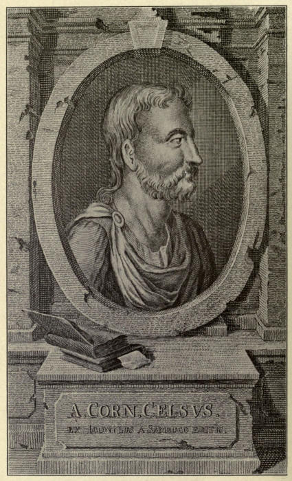
A. Cornelius Celsus (“Father of Plastic Surgery”).
PLASTIC AND COSMETIC SURGERY — CHAPTER I HISTORICAL
It seems almost incredible that at this late day so
little is generally known to the surgical profession of the
beautiful and practical, not to say grateful, art of plastic
or restorative surgery, successfully practiced even by
the ancients.
The progress of the art has been much interrupted.
It is only the later methods of antisepsis, which have so
greatly added to general surgery, that have placed it
firmly upon the basis of a distinct and separate art in
surgical science.
To Aulus Cornelius Celsus, a Latin physician and
philosopher, supposed to have lived in the time of Augustus,
we owe the first authentic principles of the science.
He was a most prolific writer and an urgent worker.
After having introduced the Hippocratic system to the
Romans he became known as the Roman Hippocrates.
His best-known work handed down to us is the “De Medicina,”
the first edition of which, divided into eight books,
appeared in Florence in 1478. The seventh and eighth
volumes, designated the “Surgical Bible,” contain much
valuable data in reference to opinions and observations
of the Alexandrian School of Medicine.
In considering plastic operations about the face
(Curta in auribus, labrisque ac naribus) he writes,
“Ratio curationis ejus modi est; id quod curtatum est,
in quadratum redigere; ab interoribus ejus angulis lineas
transversas incidere, quæ citeriorem partem ab ulteriore
ex toto diducant; deinda ea quæ resolvimus, in unum adducere.
Si non satis junguntur, ultra lineas, quas ante
fecimus, alias dua lunatas et ad plagam conversas immittere,
quibus summa tantum cutis diducatur, sic enim fit, ut
facilius quod adducitur, segui possit, quod non vi cogendum
est, sed ita adducendum ut ex facili subsequatur; et
dimissum non multum recedat.”
Centuries elapsed before a clear understanding of the
above was deduced. Several analyses have been advanced,
those of O. Weber and Malgaigne being the most
generally accepted.
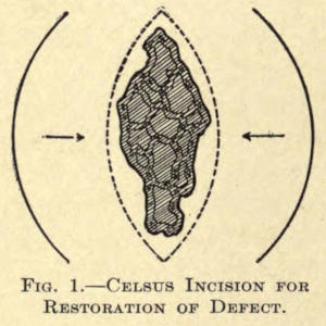
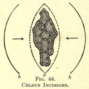
As shown in Fig. 1 the method advanced is one for
the restoration or repair of an irregular defect about the
face in which two transverse incisions forming angular
skin flaps, dissected from the underlying tissue, are advanced,
joining the denuded free ends.
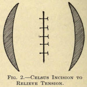
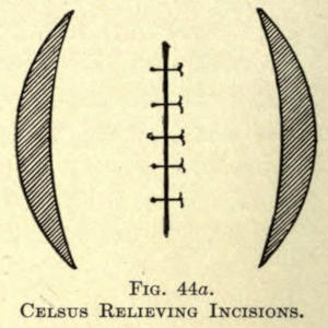
Should there be a lack of tissue to accomplish perfect
coaptation a semilunar incision beyond either outer border
is added, as shown in Fig. 2, which permits of greater
traction, leaving two small quatrespheral areas to heal
over by granulation:
Fig. 1.—Celsus Incision for Restoration of Defect.
Fig. 2.—Celsus Incision to Relieve Tension.
This is the oldest known reference to plastic surgery of
times remote.
From the Orient, however, Susrata in his Ayur-Veda,
the exact period of which is unknown, discloses the use
of rhinoplastic methods.
For centuries following, and throughout the middle
ages, the art seems to have waned and remained practically
unknown, as far as is shown in the literature of
that period.
A revivalist first appeared about the middle of the
fifteenth century in the person of Branca, of Catania, a
Sicilian surgeon, who about 1442 established a reputation
of building up noses from the skin of the face (exore).
His son Antonius enlarged upon his methods and is said
to have utilized the integument of the arm to accomplish
the same result, thus overcoming the extensive scarring
of the face following the elder’s mode. He seems to have
been the first authority employing the so-called Italian
rhinoplastic method. He is also known to have ventured,
more or less, successfully in operations about the
lips and ears.
Balthazar Pavoni and Mongitore repeated these methods
of operative procedure with more or less success and
the brothers Bojanis acquired great celebrity at Naples
in the art of remodeling noses.
Vincent Vianeo followed the work of the above.
But, somehow, the heroic efforts of these men
dropped so much into oblivion that Fabricius ab Aquapende,
in writing of the rhinoplastic work of the brothers
Bojani, of Calabria, says: “Primi qui modum reparandi
nasum coluere, fuerunt calabri; deinde devenit ad
medicos Bononienses.”
That Germany was interested at an early date is
shown in the admirable work of a chevalier of the Teutonic
Order, Brother Heinrich Von Pfohlspundt, who
wrote a book on the subject entitled “Buch der Brundth
Ertznei,” with a subtitle, “Eynem eine nawe nasse zu
mache.” His volume appeared in 1460, about the time of
Antonio Branca, of whose methods he was ignorant,
claiming to have learned the art from an Italian who
succored many by his skill.
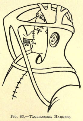
Between the years of 1546 and 1599 Kaspar Tagliacozzi,
Professor at Bologna, followed the art of rhinoplasty.
His pupils published a book at Venice, describing
his work in 1597, entitled “De Corturum chirurgia
per insitionem,” which established the first authentic
volume in restorative surgery. His operation for restoring
the entire nose from a double pedicle flap taken from
the arm was declared famous and the operation he then
advocated still bears his name.
The great Ambroise Pare knew little of rhinoplasty
except what he learned from hearsay. As an instance,
he relates in 1575 that “A gentleman named Cadet de
Saint-Thoan, who had lost his nose, for a long time wore
a nose made of silver and while being much hurt by the
criticisms and taunts of his acquaintances heard of a
master in Italy who restored noses. He went there and
had his facial organ restored, and returned to the great
surprise of his friends, who marveled at the change in
their formerly silver-nosed friend.”
Now again came a century of forgetfulness, the scientific
world taking no cognizance of the work done until,
suddenly, in 1794, a message came from Poonah, India,
to the effect that an East Indian peasant named Cowasjee,
a cowherd following the English army, was captured
by Tippo Sahib, who ordered the prisoner’s nose to be
amputated. His wounds were dressed and healed by
English surgeons. Shortly after this the victim of this
odd mode of punishment was befriended by the Koomas,
a colony of potters, or, as others claim, a religious sect,
who knew how to restore the nose by means of a flap
taken from the forehead. They operated on him and restored
his nose much to the surprise of Pennant, who
reported the case in England.
Shortly following this, and in the same year, cases of
similar nature are described in the Gentlemen’s Magazine
(England), and Pennant’s “Views of Hindoostan.”
In 1811 Lynn successfully accomplished the operation
in a case in England, and in 1814 Carpue published his
results in two cases successfully operated by him by the
so-called Hindoo method.
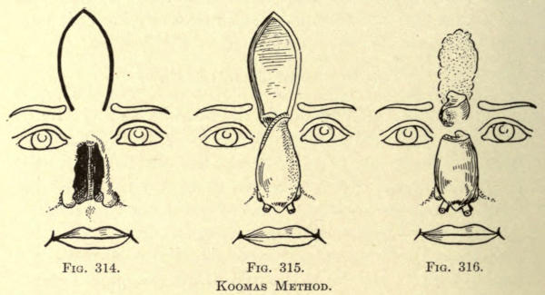
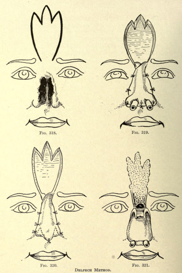
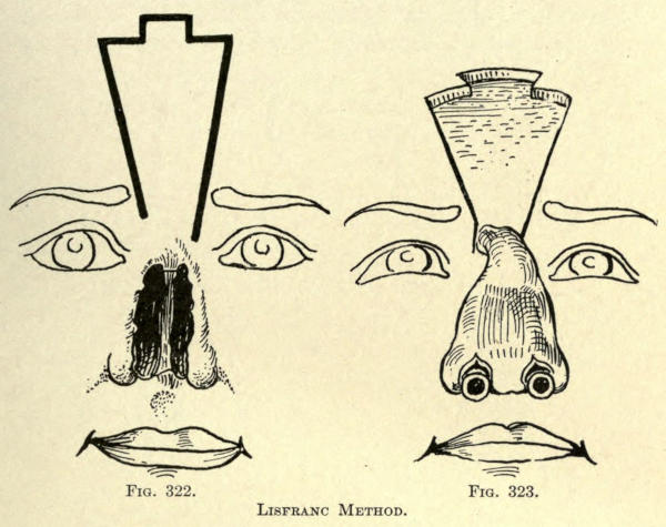
France now took up the art of rhinoplasty. Delpech
introduced a modification of the method of the Koomas
in 1820, while Lisfranc performed the first operation of
this nature in Paris in 1826.
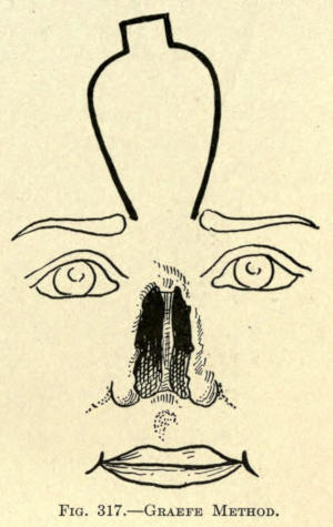
In 1816 Graefe, of Germany, took up the work of Tagliacozzi
but modified his method by diminishing the number
of operations.
Bünger, of Marburg, thereupon, in 1823, successfully
made a man’s nose by taking the necessary tegument
from the patient’s thigh.
A still later modification in the art of rhinoplasty
was that of Larrey, who in 1830 overcame a large loss
about the lobule of the nose by taking the flaps to restore
the same from the cheeks.
Among the better advocates of reparative chirurgery
were Dieffenbach, v. Langenbeck, Ricard, v. Graefe
(1816), Alliot, Blandin, Zeis, Serre, and Joberi, while
Thomas D. Mutter, in 1831, published the results he obtained
in America—his co-workers being Warren and
Pancoast.
Although Le Monier, a French dentist, as early as 1764
originally proposed closure of the cleft in the soft palate,
no one attempted to carry out his suggestion until in
1819 the elder Roux, of Paris, performed the operation.
The following year Warren, of Boston, independently decided
upon and successfully did an improved operation
to the same end.
During the years 1865-70 Joseph Lister distinguished
himself in the discovery and meritorious employment of
carbolic acid as a means of destroying, or at least arresting,
infectious germ life, the principle of which, now so
fully developed, has advanced the obtainable surgical possibilities
inestimably.
The credit of first collecting data of plastic operations
belongs to Szymanowski, of Russia. In his magnificent
volume of surgery (1867), he embodies a somewhat thorough
treatise on restorative surgery, leaving the subject
to be treated more fully and independently, as it should
be, to some other enthusiastic surgeon specialist. His
work is the result of careful study of such operations on
the cadaver, a method much to be recommended to the
prospective or operating plastic surgeon.
Several years later, 1871, Reverdin added a valuable
method to the still incomplete art, by introducing the
now well-known circular epidermal skin grafts for covering
granulating surfaces. Thiersch improved this
method in 1886 by showing that comparatively large
pieces of skin could be transplanted. Wolfe, of Glasgow,
had also been successful in utilizing fairly large skin
grafts.
Krause, however, improved upon all of these methods
by transplanting large flaps of skin without detaching
the subcutaneous tissue, a procedure which causes more
or less injury to the graft in other methods, and by his
method overcoming the subsequent contraction, heretofore
a bad feature when the skin-grafted area had
healed.
“The results of most plastic operations have been as
satisfactory as the most sanguine could hope for or the
most critical expect,” says John Eric Erichsen.
Many important additions have been made in the past
few years—the outcome of untiring attempt and skill.
Czerny replaces part of an amputated breast with a fatty
tumor taken from the region of the thigh. Glück successfully
repairs a defect in the carotid artery with the
aid of a piece of the jugular vein. Glück, Helferich, and
others have advocated implanting muscular tissue taken
from the dog into muscular deficiencies in the human, due
to whatever cause.
The transplantation of a zoöneural section into a defect
of a nerve in the human was successfully accomplished
by Phillippeaux and Vulpian.
Glück, who later restored a sciatic nerve in a rabbit
by the transplantation of the same nerve taken from a
hen, went so far as to restore a 5-cm. defect of the
radial nerve of a patient by the employment of a bundle
of catgut fibers, fully establishing the function of the
nerve within a year’s time.
Guthrie has successfully replaced the organs and
limbs of animals and has actually transplanted the heads
of two dogs.
The transplantation of a toe, to make up a part
of a lost finger, is proposed by Nicoladoni. Van Lair
hints at the possibility of removing a part or a whole
organ immediately before death to repair other living
organs.
Von Hippel has successfully implanted a zoöcorneal
graft from a rabbit upon the human eye, and Copeland
has taken the corneal graft from one human and transplanted
it upon the cornea of another to overcome
opacity.
The transplantation of pieces of bone to overcome a
defect of like tissue has been fully investigated by Ollier,
v. Bergman, J. Wolff, MacEwen, Jakimowitsch, Riedinger,
and others. They discovered that a graft of bone,
with or without its periosteum, can be made to heal into
a defect when strict antisepsis is maintained.
Von Nussbaum was the first to introduce the closing
of an osseous defect by the use of a pedunculated flap of
periosteum.
Poncet and Ollier employed small tubular sections of
bone, while Senn has obtained excellent results from the
use of chips of aseptic decalcified bone.
Hahn succeeded in implanting the fibula into a defect
of the tibia.
On the other hand, cavities in the bones have been
successfully filled by Dreesmann and Heydenreich with
a paste of plaster made with a five-per-cent carbolic-acid
solution, and at a later period by the employment of
paraffin (Gersuny) and iodoform wax, as advocated by
Mosetig-Moorhof.
The thyroid glands taken from the sheep, it is
claimed, have been successfully implanted in the abdomen
of individuals whose thyroid glands had been lost
by disease or otherwise.
Protheses of celluloid compound or gutta-percha and
painted to resemble the nose or ear have been introduced
with grateful result. Metal and glass forms have been
used to replace extirpated testicles or to take the place
of the vitreous humor of the eye (Mule).
Sunken noses have been raised with metal wire, metal
plates, amber, and caoutchouc. Metal plates have been
skillfully fitted into the broken bony vault of the cranium.
Lastly comes Gersuny’s most valuable method of injecting
paraffin compounds subcutaneously for the restoration
of the contour of facial surfaces and limbs,
which is rapidly taking the place of extensive plastic
transplantory and the much-objected-to metal and bone-plate
operations for building up depressed noses and
other abnormal cavities.
And the end of possibilities is not yet reached. The
successful plastic surgeon has become an imitator of
nature’s beauty to-day.
His skill permits of many almost unbelievable corrections
of defects that would otherwise evoke the pity and
too often the aversion of the onlooker, especially if these
occur in the faces of those that have become marred
in birth or age, by accident or disease. Withal, it is a
noble, generous art, worthy of far more extensive use
than it now enjoys.
The above fragmentary references include a number
of plastic possibilities. They are introduced only in the
sense of general interest to the cosmetic surgeon, the
special and detailed subject matter herein given under
the various divisions have to do only with plastic and
cosmetic operations about the face.
CHAPTER II REQUIREMENTS FOR OPERATING — THE OPERATING ROOM — CARE OF INSTRUMENTS — PREPARATION OF THE SURGEON AND ASSISTANTS — Care of the Hands — Gowns — PREPARING THE PATIENT — General Preparation — Preparation of the Operative Field
The ideal operating room for the plastic surgeon need
not necessarily be large, since it requires less work to
render it aseptic. Furniture and possibly amphitheater
accommodation are always a means of infection unless
scrupulously cleansed, a task of time, difficult at best.
The room should be provided with large windows,
with facilities for the introduction of the air from without.
Two doors, and those well fitted, are all the room
should have—but one being used, if possible.
The Walls.—The walls should be of plaster, smoothly
laid and well painted, so that they may be readily washed
down with antiseptic solutions—a daily morning rule.
Glass or tiled walls are much used now and add considerably
to the appearance and safety of the room, as plaster
in time will crack, while the paint, owing to the heat of
sterilizers or steam, often creeps and blisters, exposing
an absorbing surface which readily wears down, exposing
parts inaccessible for even acute cleanliness.
The Floors.—The floors of these rooms are now usually
laid with tile mosaic or marble or a composition resembling
linoleum. The base should be curved and all corners
sloped off to improve drainage and to keep off dust
and dirt.
Skylight.—A skylight of metal and glass is a valuable
accessory. It should be fixed or never permitted to be
opened during an operation.
Disinfection.—Spraying the room with an antiseptic is
hardly necessary, since all germ life descends to the floor
and can best be removed by washing with a 1-1000 bichlorid
solution.
Should it be necessary to perform an unusually extensive
operation in a private house, the room must be
cleared of all furniture, pictures, drapery, and carpet.
After plugging up the crevices in the windows and
doors it should be well fumigated either with sulphur
candles, as now commonly furnished, or, better, with formaldehyd.
The superiority of formaldehyd as a disinfecting
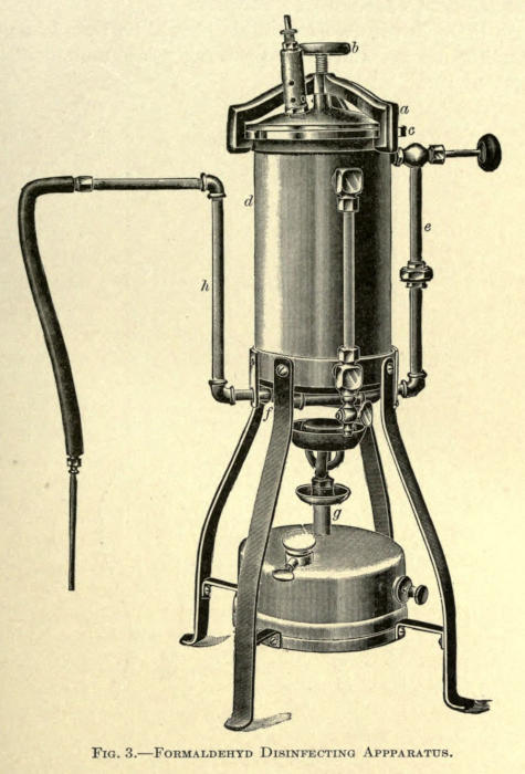
agent is now well established. An illustration of an apparatus,
largely doing away with the difficulties and
dangers encountered in the use of the older and ordinary
styles of the pressure or nonpressure type, is shown in
Fig. 3. The main difficulty with these has always been
their almost inaccessibility for cleansing purposes, and in
such where this is not the case, the size of the aperture
has been made so small that the inside could not be
reached. In the pressure apparatus the tops are bolted
on, making them exceedingly difficult to remove, with the
result that the necessary cleaning was not properly attended
to. The corrosive action of formaldehyd gas is
such that under these conditions any apparatus would
soon become useless.
In the type shown a single clamp arrangement is
used (a). By the turning of the hand screw (b) two
planed metal faces (the upper surface of the boiler and
under surface of the cover) are brought together and
sealed. When the cover (c) is removed the entire inside
of the boiler is in sight and can be thoroughly cleansed,
which should be done each time the apparatus is used.
The pipes through which the formaldehyd gas passes
after generation are arranged so that they can be taken
off and cleaned.
Fig. 3.—Formaldehyd Disinfecting Apparatus.
The gas is generated in the boiler (d) and passes out
from the top, down through the pipe (e), and from thence
through a series of pipes (f) underneath the boiler, which
are subjected to direct heat from the lamp (g). By this
means the gas becomes superheated, the polymerization
of the formaldehyd is almost entirely prevented, and a
dry gas is insured and given off at the pipe (h).
The room should be left closed overnight and thoroughly
aired thereafter. The bare floor must then be
scrubbed with hot water and soda and flushed with a
three-per-cent carbolic-acid solution.
As little furniture as possible should be found in an
operating room, and this preferably of undecorated
enameled iron.
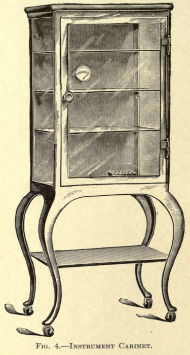
Instrument Cabinet.—For the instruments and dressings
there should be a dust-proof cabinet of iron and glass,
such as is shown in Fig. 4.
Fig. 4.—Instrument Cabinet.
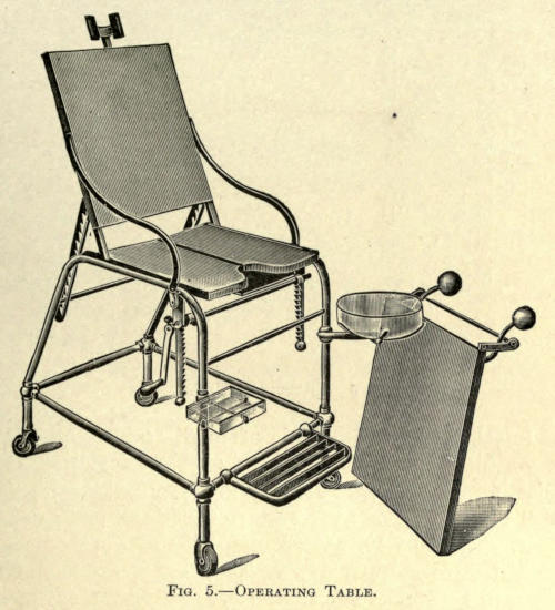
Operating Table.—The operating table should be of like
construction and as plain as possible. Its top can be
padded with sterilized felt, protected from moisture by
rubber sheets. A surgical chair of plain construction
might suffice, inasmuch as most plastic operations cover
but a small area and are usually about the head and often
performed under local anesthesia. A chair with head rest
is much more comfortable, adding much to the moral and
physical comfort of the then conscious patient. A very
desirable chair is shown in Fig. 5.
Fig. 5.—Operating Table.
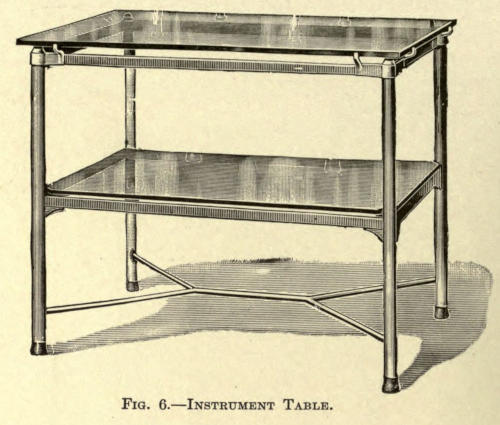
Instrument Table.—An instrument table, such as is
shown in the next illustration, is quite necessary, upon
which dressings and instruments are laid during operation.
In this the frame is of white enameled iron and the
top and shelf of plate glass.
Fig. 6.—Instrument Table.
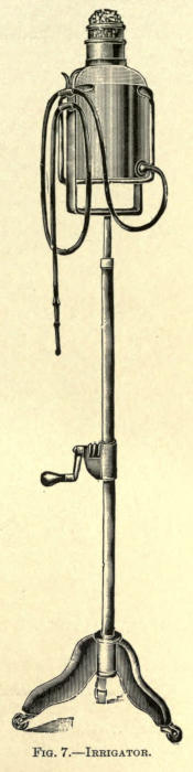
Irrigator.—An irrigator is often of service, especially
in washing out the fine pieces of bone resulting from
chiseling or drilling. In skin-grafting it may be used
with sterilized three-per-cent salt solution as described
later. The best irrigators are those of germ-proof or
ground-glass stopper type. They are suspended from
the wall by means of an iron bracket or pulley service
or placed upon a movable enameled stand as shown in
Fig. 7.
Irritating antiseptic solutions are to be avoided, their
especial indication will be found under antiseptic care of
wounds.
Fig. 7.—Irrigator.
All instruments should be of modern make, devoid of
clefts or grooves, and having separating locks when possible.
Wooden or ivory handles should be entirely discarded.
They should first be rendered free of dirt or
dried blood by scrubbing briskly with a stiff nailbrush
and hot water; then dried and
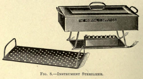
placed in the sterilizer. The immersed
instruments are boiled for
five or ten minutes. There are
many of such sterilizing apparatuses
to be obtained, all made
on the same plan, however, and
consist of a copper or brass
box and cover well nickel plated.
Folding legs are placed beneath.
A perforated tray is placed
within for the immersion of instruments.
An alcohol lamp
with asbestos wick furnishes the
heat.
One per cent of carbonate of
soda added to the water prevents
them from rusting. The simple
subjection of instruments to carbolic-acid
solutions or antiseptics
of like nature is useless. (Gärtner,
Kümmel, Gutch, Redard, and
Davidsohn.)
From the sterilizer the instruments
are placed in a glass tray
containing a one-per-cent lysol
solution. Knives, needles, and
scissors should be immersed in a
tray with alcohol, as a great number
of antiseptics destroy their
cutting edges. Glass or porcelain
trays are best for this purpose.
A sterilized towel being placed
in the bottom of each for the better
placing of instruments.
After operation all instruments should again be
scrubbed with soap and hot water, immersed a moment in
boiling water or a jet of live steam, dried with an aseptic
cloth, and returned to the case.
A very effectual means of rendering instruments
sterile is to place them in a metal box and bake them in
the ordinary oven (200° F.) for one hour.
To preserve needles Dawbarn advises keeping them
in a saturated solution of washing soda. Albolene has
an unpleasant oiliness, but is otherwise good. Calcium
chlorid in absolute alcohol is efficacious, but expensive.
All rust accumulating on instruments must be carefully
removed with fine emery cloth; this, however, is unnecessary
if the soda solution is used as previously mentioned.
It is well to occasionally dip the instruments (holding
them with an artery forceps) into boiling water as they
are used during operation.
Fig. 8.—Instrument Sterilizer.
The hands of the surgeon and his assistants must
always be thoroughly prepared before operation or
dressing a wound. The mere immersion of the hands
into an antiseptic solution is not sufficient to remove
germ life. The oily secretions of the skin and its folds,
as well as the cleft about the nails and the nails themselves,
are common carriers of infection and are cleansed
only by the vigorous method of scrubbing with soap and
water and then rendered aseptic by the use of proper
media.
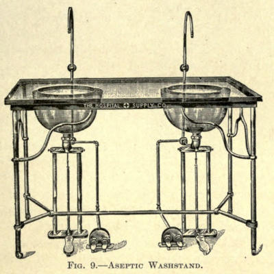
The aseptic hospital washstand, as shown in Fig. 9,
will be found an ideal piece of furniture; it has a frame
constructed of wrought iron, white enameled. The top
is of one-inch polished plate glass, with two twelve-inch
holes.
Fig. 9.—Aseptic Washstand.
The entire stand can be moved away from the wall, to
permit of thorough cleaning of basins, supply pipes, etc.
The basins are the best annealed glass, and are supported
by nickel-plated traps, with connections for vent pipes.
The water supply is controlled by foot valves, which enable
the operator to draw either cold, medium, or hot
water at will. The waste is also controlled by a foot
valve, as shown.
The systematic law of cleansing the hands should be
insisted upon at all times. Rules for the method followed
might be displayed in abbreviated form in the operating
room by glass or enameled signs hung on the wall over
the basin and reading as follows:
YOUR HANDS
I.
Clean nails.
II.
Scrub with very hot water and soap for five minutes.
III.
Wipe in sterile towel.
IV.
Brush with eighty per cent alcohol.
V.
Dip into antiseptic solution.
Green soap is commonly used and is to be preferred to
powdered or cake soap. The powder cakes and clogs the
container in damp weather, while the latter collects impurities
from the air. Synol soap, also liquid, is perhaps
the most ideal, a two per cent solution of which forms an
excellent lavage for cleaning instruments, as well as
washing down furniture in the operating room.
The brushes to be used are of the common wooden-back,
hard-bristle make, which can be boiled without injury.
There should be several of these, marked on their
backs as desired, so that one brush can be used for the
one purpose only. In cleansing the hands, the forearms,
and even the elbows, should be similarly treated. After
scrubbing with soap, as directed, they are to be rinsed,
dried with a sterilized towel, again scrubbed with alcohol,
and then dipped or flushed with a bichlorid solution.
No woolen garments should be allowed to come in contact
with the site of the operation, nor is it well to allow
such material in the operating room while working.
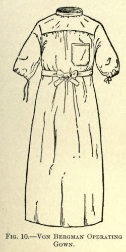
Freshly laundered linen gowns of Von Bergman’s
pattern, reaching to the shoes, should be worn. They
should contain half sleeves and be buttoned on the back.
See Fig. 10. These may be sterilized in the steam sterilizer
or washed in one-per-cent soda solution. When
soiled or blood-stained they should be relaundered.
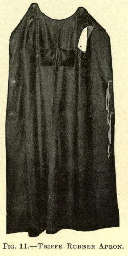
The operator may substitute the gown with a rubber
apron of the Triffe pattern, reaching as high as the collar,
but continuous washing quickly ruins them. See
Fig. 11.
Fig. 10.—Von Bergman Operating Gown.
Fig. 11.—Triffe Rubber Apron.
The patient for all plastic operations should be carefully
examined as to general health and past history.
His healing powers should be at their best, as much depends
on primary union. If he presents a syphilitic history,
it is well to place him under treatment, for a time,
at least, before an operation is undertaken. The bowels
should be regular. Sulphate of magnesium should be
given each morning, before breakfast, for at least two
days prior to operating, while his general condition may
be improved by the employment of bitter and alterative
tonics. Nux vomica with tinct. cinchonæ com., associated
with essence of pepsin aromat., or lactopeptone,
are very useful. This treatment is also carried on for
several days, post operatio.
The success of an operation depends, first, upon the
selection of the case; second, the selection of the method
employed, and, third, upon the hygiene under which the
patient undergoes convalescence. The patient must be
given to understand, in many cases, that it is often necessary
to reoperate, even to the extent of seven or eight
operations, to bring about the desired result. The first
result obtained with many cosmetic operations is not at
all gratifying to the patient, and unless this is explained
to him beforehand he may become discouraged awaiting
the next operation and disappear, thus losing the
opportunity of being pleased finally, while the surgeon
is misunderstood and underestimated by narrow-minded
judges and the ever-willing friendly advisers and critics—a
consummation much to be avoided.
The part to be operated upon should first be closely
shaven. The oily secretions of the area are next rubbed
off with an absorbent cotton sponge saturated with alcohol
or ether. Next, the skin is washed with hot water
and soap or three-per-cent synol suds, then rinsed, and
finally rendered aseptic with a bichlorid solution.
If the operation is to be done about the face a rubber
cap is so adjusted as to cover the hair. If this is not
obtainable sterilized bandages can be employed.
In operations about mucous membranes, as in the
nose and mouth, the parts must be cleaned at short intervals
with a solution of permanganate of potash or boric
acid. The teeth must be cleansed with antiseptic soap,
tartar is scraped off, and the mouth rinsed with a proper
disinfectant. The corrosive sublimate, or carbolated
solutions, owing to their toxic qualities, cannot be used.
The preparation of wounds for reoperation, or where an
operation is secondary to injury, is referred to later.
All clothing about the site of operation should be removed
and rubber cloth placed to surround the field and
cover the clothing. This should be covered again with
sterilized towels. Everything that touches the patient
after this has been done should be aseptic; indeed, hands
employed during operation must be immersed from time
to time in 1-500 bichlorid solution, and allowed to remain
wet.
CHAPTER III REQUIREMENTS DURING OPERATION — SPONGES AND SPONGING — STERILIZATION OF DRESSINGS — SUTURES AND STERILIZATION
Natural or sea sponges are now little used in surgery,
owing to their peculiar cellular construction. They
invite and readily retain spores and germs, are difficult
to clean, and require almost constant attention to be
at all safe.
Many methods for rendering these sponges aseptic
have been proposed, but at best the life of such a sponge
is short and hardly pays for the labor and time expended.
The absorbing power of a sponge is, of course, its essential
quality. For plastic operations sterilized absorbent
cotton made into small balls answers every purpose.
These puffs of cotton are covered with gauze to prevent
the fraying out of the fibers. To further improve them,
their centers may be made up of cellulose or wood fiber.
When an absorbent cotton sponge is moistened and
squeezed out it does not answer as well, since its absorbing
qualities are much reduced; the addition of the other
material overcomes this.
A much-used and inexpensive sponge having great
absorbing power is made in the form of a small compress
of sterilized gauze held together with one or two stitches
of thread. All of the above sponges are sterilized with
the needed dressings and are burned after use. When
removed from the sterilizer they are placed in a suitable
basin containing six per cent sterilized salt water. It is
well to place the receptacle close by the assistant who is
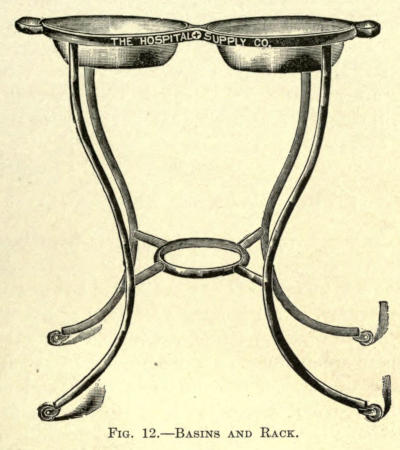
to sponge. An enameled iron basin rack, as shown in
Fig. 12, answers the purpose best.
Fig. 12.—Basins and Rack.
The soiled sponges are thrown into a lower empty
basin or one placed at the operator’s feet. As they are
removed from the solution they are squeezed as dry as
possible and pressed upon, rather than wiped across, the
operative field. It must be remembered that the surgeon’s
work must not be hampered by slow or inefficient
sponging, and that this procedure must be quick and
timely. It is well for the assistant to become accustomed
to the habit of the operator.
The best assistant is one who has acquired a methodical
and regular manipulation, a result dependent upon
constant individual association; such a one is practically
invaluable for the skillful performance of plastic surgery.
He becomes not only familiar with the one thing,
but cultivates a ready knowledge of the arrest of hemorrhage
by digital compression when hemostatic forceps
would hinder the ease of work, besides cultivating a
happy manner of holding retractors or spreading the
edges of the incisions with the free hand. As in most
of these operations hemorrhage cannot be controlled by
the so-called bloodless method. The assistant must control
the constant oozing by the gentle pressure of the
sponge quickly applied at short intervals. When the
sponges are squeezed out in salt solution, as hot as the
hand will bear comfortably, capillary oozing is more
readily overcome.
All dressings to be used in covering wounds, post
operatio, or otherwise, must be as scrupulously clean
and free from infection as the hands and the instruments
of the operator. This is done by means of sterilization
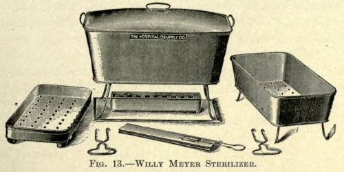
by dry heat or steam under pressure. For all minor
cases, small apparatuses only are needed. They are usually
made of copper, often nickel-plated, and so constructed
as to contain a lower perforated instrument
tray and another, placed above it, for dressings. The
two are fitted into an outer copper receptacle with snugly
fitting cover. A folding stand is furnished upon which
this arrangement is placed, and an alcohol lamp with asbestos
wick furnishes the heating power. The lower tray
is covered with water which, by boiling, fills the upper
compartment with steam evenly distributed and with sufficient
pressure to accomplish sterilization in from thirty
to sixty minutes. Metal hooks are provided with which
the trays can be removed. A complete and compact outfit,
as designed by Willy Meyer, is shown in Fig. 13.
In the above sterilizer, or in those of similar type,
there is naturally more or less saturation of the dressings
and the possibility, in the event of the entire conversion
of the water contained therein into steam, of
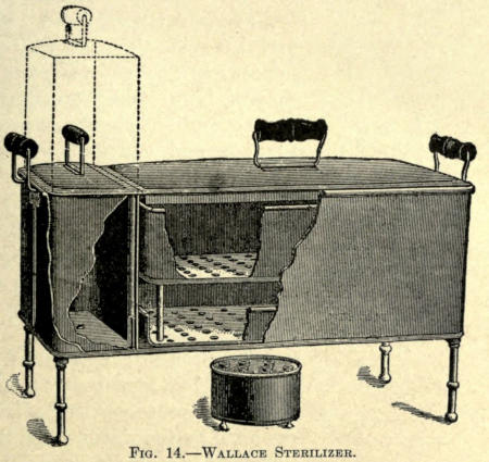
injuring the instruments by excessive heat. To overcome
this defect the Wallace sterilizer may be advantageously
employed.
Fig. 13.—Willy Meyer Sterilizer.
Fig. 14.—Wallace Sterilizer.
Wallace Sterilizer.—Its chief feature is the addition of
a reservoir fitting with the separated sterilizer into the
outer body. See Fig. 14. This reservoir automatically
regulates the water and steam supply. It is filled with
water and inserted into the compartment provided for
and adjoining the sterilizer. Through an opening in the
bottom the water is permitted to escape into the sterilizer
until the bottom of the latter is covered to a depth of
⅛ inch. As the heat is applied from the alcohol lamp
this film of water is rapidly converted into steam.
The dressings arranged in the large tray are placed
in the sterilizer and the supply of steam is maintained
through the constant and steady flow of water from the
reservoir, which compensates the evaporation in the sterilizer.
In about twenty minutes the formation of steam
in the top of the reservoir exerts sufficient pressure to
force all the boiling water from
the reservoir into the sterilizer to
the depth of about 1½ inches. The
tray of instruments is now inserted
and the process continued
for another ten minutes. Much
less heat is required with this
apparatus than with those of ordinary
type, while sterilization
can be continued uninterruptedly
for one and one half hours, if
need be.
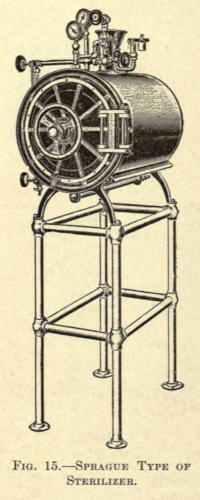
Sprague Sterilizer.—The most
perfect sterilizer is that of the
Sprague type, in which a dry
chamber is surrounded by steam
under pressure. The apparatus
is shown in Fig. 15.
Fig. 15.—Sprague Type of Sterilizer.
Its cylindrical chamber is surrounded
by two heavy copper
shells, the space between which is
occupied by the water. This compartment
is entirely shut off from
the sterilizing chamber, and as the steam is generated, the
inner, or sterilizing, chamber becomes heated to a degree
nearly equal to that of the steam in the surrounding
cylinder; this prevents any condensation of steam taking
place in the dressings. By opening the lever-handled
valve at the bottom of the sterilizer in the rear, and the
valve to the right, on top of the sterilizer, and allowing
them to remain open for a space of four or five minutes,
a vacuum is formed in the sterilizing chamber. These
two valves are then closed, the lower one first, and the
steam from the outer cylinder is allowed to enter the
chamber, by opening the left valve on top.
The contents should be allowed to sterilize for twenty
or twenty-five minutes under a pressure of fifteen pounds.
Then close the steam-supply valve; open the vacuum
valve (right) and the lever-handled valve at the bottom;
leave these open about the same time as in creating a
vacuum at the beginning of the process; close both
valves, then open the air-filter valve on the door, in
order to break the vacuum; the door can then be opened
and the dressings be taken out dry and absolutely sterile.
The steam-safety valve on this sterilizer is set at
seventeen pounds, but it can easily be regulated should a
higher or lower pressure be desired. The door used on
this apparatus has no packing of rubber or other soft
material which wears or shrinks in time, a steam-tight
joint being formed by the bringing together of two plane
metal faces on the door and sterilizer head. The door
hinge is so made that these parts are bound to come together
properly, without the use of excessive caution.
Springs on such doors are liable to get out of order or
need replacing, and are avoided in this apparatus. All
that is necessary to lock or unlock the door is to turn the
large hand wheel on the front; the locking levers then
work automatically. These sterilizers are arranged for
both gas and steam heat.
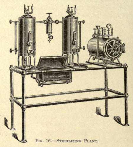
Sterilizing Plant.—For the ideal operating room the entire
sterilizing plant can be had in combined form, as
shown in Fig. 16. It consists of a dry-heat dressing apparatus,
just described, water and instrument sterilizers,
all mounted on a white enameled, tubular, wrought-iron
frame. The chamber of the dressing sterilizer is 8½ by 19
inches. The water sterilizer has a capacity of six gallons
in each tank and is fitted with natural stone filters,
thermometer, water gauge, safety valve, etc. The
size of the instrument sterilizer is 8 by 15 inches and 6
inches deep, with two trays. Each apparatus in the
above can be used independently of the other, all being
arranged for gas-heating.
Fig. 16.—Sterilizing Plant.
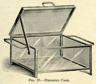
Dressing Cases.—All dressings should be sterilized immediately
before operation, and not laid away for later
use, as often done. As the aseptic material is taken from
the sterilizer it is to be placed in glass cases provided
therefor, from which they are removed, as needed, during
the operation.
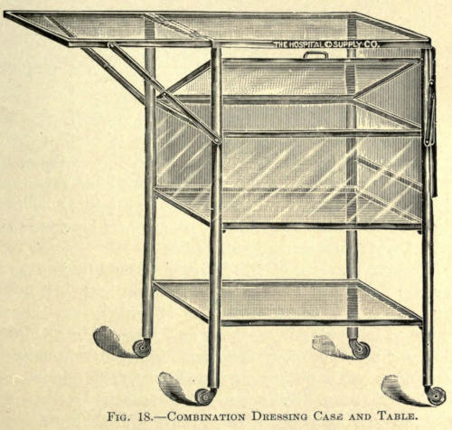
A simple glass case, as shown in Fig. 17, may be
used, or, better still, the same can be obtained in combination
with an instrument table, as shown in Fig. 18.
Fig. 17.—Dressing Case.
Fig. 18.—Combination Dressing Case and Table.
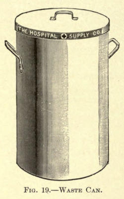
Waste Cans.—All soiled dressings and sponges should
be immediately thrown into an enameled iron pail furnished
for the purpose. At no
time must soiled dressings or
sponges be thrown upon the
floor, where they are walked
over, soiling the floor and, by
drying, contaminating the air
of the room. Cans for this
purpose are made of steel,
enameled, of the form shown
in Fig. 19.
The contents of the can must
be taken from the room after
each operation and burned.
The can should be flushed with
carbolic solution, and returned
to the operating room.
Fig. 19.—Waste Can.
(Ligatures)
Silkworm Gut and Silk.—In plastic surgery silkworm gut
and silk are used extensively. Rarely is ordinary catgut
resorted to, because it is absorbed before thorough
union takes place, besides being a source of infection,
either primarily from imperfect sterilization or by taking
it up from the secretions of the deeper layer of skin
not affected by external antiseptics.
The sterilization of silk is accomplished by boiling it
for one hour in a 1-20 carbolic solution and then keeping
it in a 1-50 similar solution (Czerny). Or it may
be boiled in water for one hour and retained in a 1-1,000
alcoholic solution of corrosive sublimate. Ordinarily it
may, however, be simply subjected to boiling and steamed
in the autocleve. Silkworm gut is treated in the same
manner. It has greater tensile strength than silk, and
for that reason the thinner varieties are to be preferred
to ordinary silk.
Catgut.—It is far more difficult to prepare catgut, but,
since it is necessary for ligation, the following methods
may be considered best:
The commercial catgut as made from the intestines
of sheep, is wound snugly upon a rod of glass and thoroughly
brushed with soft soap and hot water. It is then
rinsed free of soap, wound upon small glass spools, and
placed for forty-eight hours in a one-per-cent alcoholic
bichlorid solution, composed of bichlorid of mercury, 10
parts; alcohol, 800 parts; distilled water, 200 parts. The
turbid fluid produced by first immersion is changed. Before
using, the spools are placed in a glass vessel contain containing
a 1-2,000 sublimate alcohol (Schaffer), made up as
follows:
Bichlorid of mercury
gr. vj;
Alcohol
℥x;
Distilled water
℥iiss.
These glass cases are obtainable for the purpose and contain
a second perforated compartment for the ligatures
passing through rubber valves placed into the openings
(Haagedorn).
Catgut is generally prepared by soaking in oil of
juniper for one week and then retaining it in absolute
alcohol (Kocher), or a 1-1,000 alcoholic sublimate
solution.
Another method for strengthening catgut, as well as
to prevent its too rapid absorption, is to chromatize it.
This is done as follows:
The catgut is placed in sulphuric ether for forty-eight
hours, then treated for another forty-eight hours in a
ten-per-cent solution of carbolized glycerin, followed by
a five-hour subjection to a five-per-cent aqueous solution
of chromic acid (Lister). It is allowed to remain in the
latter forty-eight hours, then placed in an antiseptic, dry,
tightly closed receptacle, and finally soaked in 1-20 carbolic
solution before using.
The formaldehyd method of Kossman is to immerse
the gut in formaldehyd for twenty-four hours, then washing
with a solution of chlorid and carbonate of sodium
and retaining it in the same solution. The catgut in this
procedure swells and its strength is much impaired in
this way.
Any of the above methods are not above criticism,
however, rigid as they may seem, bacterial growths having
been obtained with
nearly all of them.
The dry-air method
(Boeckman, Reverdin)
is reliable, but the subjection
of catgut to dry
air at a temperature of
303° F. for two hours
results in making it
tender and less pliable.
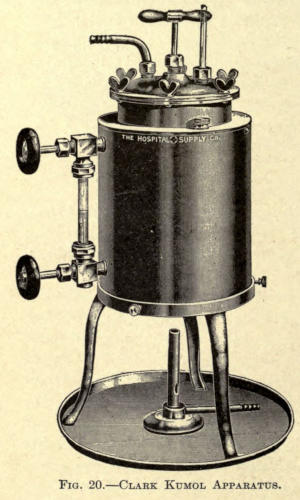
Fig. 20.—Clark Kumol Apparatus.
The Kumol method
(Kronig) is considered
the most reliable, even
under the severest
tests. This mode of
sterilization is accomplished
as follows: A
specially devised apparatus
of brass, with a
cast-bronze top, both
thoroughly nickel-plated,
is used. The apparatus
of J. G. Clark, as
shown in Fig. 20, will
be found excellent. The
kumol is retained in a seamless cylinder, 8 by 8 inches,
which is surrounded on the sides and bottom by a sand
bath; the flame, impinging on the bottom, heats the sand,
thereby insuring an even heat to the inner or sterilizing
cylinder. The catgut, in rings, is placed in a perforated
basket hanging in the cylinder, which can be raised or
lowered at will; after drying for two hours at 80° C., the
basket is dropped, and the catgut immersed in the kumol,
at 155° C., for one hour; the kumol is then drawn off
through a long rubber tube, and the catgut dried at 100°
C., for two hours; it is then transferred to sterile glass
tubes plugged with cotton.
Prepared catgut of the various sizes can now, however,
be purchased in the market, and that offered by the better
firms of chemists is quite reliable and may be safely
used for all plastic surgery about the face. It is supplied
in glass tubes, either in given lengths, as in the
Fowler type, in which the hermetically sealed tube is
U-shaped or on glass spools placed in glass tubes, not
sealed, but closed by a rubber cap, through which the
desired length of ligature is drawn and then cut off.
CHAPTER IV PREFERRED ANTISEPTICS — ANTISEPTIC SOLUTIONS — ANTISEPTIC POWDERS
These are solutions used for the destruction of and
to arrest the progress of microörganisms that have found
their way into wounds—the cause of sepsis, as exhibited
by fever, suppuration, and putrefaction. These preparations
are called antiseptics and are used to render parts
aseptic. They vary much in their destructive power,
effect on tissue, and toxic properties. The reader is referred
to a work on bacteriology for the specific knowledge
of such on germ life.
The antiseptic treatment of wounds was founded by
Joseph Lister, 1865-70, then called Listerism. His one
chemical agent to accomplish this was carbolic acid, but
many such and more effective agents have been added
since that time, all differing in their specific properties
and each having, for the same reason, its particular
use.
The following group of antiseptics has been chosen
with a view of giving the best selection, to which the
author has added a short description of each, so that the
surgeon may choose one or the other, as the occasion may
demand. As a rule, an operator cultivates the use of a
certain line of antisepsis, especially in this branch of
surgery, experience being the best guide; yet it is hoped
he may find certain aid from those referred to, their particular
use being pointed out from time to time, as the
author has had occasion to prefer one or the other.
Alcohol (absolute).—This is a well-known antiseptic,
but, because of its ready evaporation, is especially used
for the hands, as described, and to cover sharp-edged
instruments after sterilization.
Aluminum Acetate (Bürow, H. Maas).—A powerful,
nontoxic antiseptic. Is used only in two- to five-per-cent
solution. According to Primer, it arrests the development
of schizomycetes, and in twenty-four hours destroys
their propagation. It readily removes offensive
odors of wounds; its great objections are that it injures
the instruments, and, because of its astringent nature,
roughens the skin of the hands. This, however, makes
it particularly useful for sponging to arrest capillary
oozing.
Boric Acid (Lister).—Not a powerful, but nonirritating,
antiseptic. For this reason it is used extensively in
cleansing mucous membranes, and, when associated with
salicylic acid, as in the well-known Thiersch solutions,
composed of salicylic acid, 2 gms.; boric acid, 12 gms.;
water, 1,000 gms., is much used in skin-grafting operations.
It is not very soluble in cold, but readily in hot,
water and alcohol. The saturated solution is prepared
by adding ℥j to the pint of boiling water.
Benzoic Acid.—Nonirritating, moderate antiseptic
(Kraske); is prepared in 1-250 solutions. Soluble in hot
water and alcohol, but sparingly in cold water.
Carbolic Acid (Phenylic Acid).—Not a powerful, but a
much-used antiseptic. The purest acid should be used.
It appears as a colorless crystalline solid, liquefied by
the addition of five per cent water. If more water is
added the solution becomes turbid, clearing when 1-2,000
is reached.
It is readily soluble in glycerin, alcohol, ether, and
the fixed volatile oils. Solutions in alcohol and oils have
no antiseptic effect (Koch). The 1-20 aqueous solution
is recommended by Lister.
The aqueous solutions used in surgery are 1-20 and
1-40. The weaker is used for the operator’s hands, to
cover instruments, as already mentioned, and to impregnate
sponges. The stronger solution is used for the
carbolic spray, to cleanse the unbroken skin about the site
of operation, and to disinfect wounds. Either solution,
when applied to an open wound, whitens the raw surface,
coagulates the albumen, and causes considerable
irritation, which subsides quickly and is followed by
numbness.
Such solutions, by virtue of their irritant nature, increase
the serous discharge from a wound for about
twenty-four hours, for which proper drainage must be
provided, as by its collection it would add to the danger
by increasing inflammation and suppuration, and, by
absorption, even produce toxic effect generally.
When a cold solution is used it should be prepared
by vigorous stirring to separate the globules of the acid.
Hot water insures perfect distribution. After an infected
wound is washed with it, the solution should not
again be used, nor should any of the acid be permitted
to remain in the spaces about the wound. It will be found
that many patients cannot tolerate such dressings, and
that others must be substituted.
Large surfaces should never be exposed to carbolic
solutions, because the skin absorbs them readily, followed
by untoward results. Dangerous symptoms have
been known to result from the internal administration of
seven drops of the acid, and fatal termination has followed
its use as a surgical dressing (Bartley).
Mild acid poisoning is first noted in the urine, which
turns olive green. If the agent is continued, the urine
appears dark and turns almost black on standing. The
coloring is due to the presence of indican. If the absorption
is not prevented beyond this there is dull frontal
aching, tinnitus aurium, dizziness, fainting, severe and
uncontrollable vomiting. Untoward symptoms are noted
by albuminuria, total absence of sulphates in the urine,
a contracted and inactive pupil, elevation of temperature,
unconsciousness, muscular contraction, and death.
The treatment consists in immediately removing the
cause and employing another antiseptic. Support the
patient with stimulants, freely given. Cracked ice and
brandy to allay the vomiting. Small doses of sodium
sulphate, frequently repeated, as a means of converting
the acid into nonpoisonous sulphocarbolate (Bauman).
Albumen and milk internally. Magnesium sulphate, five
per cent.
Chromic Anhydrid.—Improperly called chromic acid.
Made by adding one and one half parts sulphuric acid,
c. p., to one part of concentrated solution of dichromate
of potash. Appears in saffron-colored crystals. It acts
as a caustic upon tissue, and, although a splendid antiseptic,
cannot be used for such purposes, but is well
adapted for the preparation of catgut, as mentioned.
Creolin.—Is an antiseptic prepared from coal by dry
distillation, and is used to stimulate granulations, being
much more powerful than carbolic acid. It is nonirritant
and practically nontoxic. Used in two-per-cent aqueous
solutions, in which it appears as a turbid but effective
mixture. It is well suited for cleansing the hands, a five-per-cent
solution having none of the irritating or anesthetic
effect of carbolic acid. Owing to the opacity of
the aqueous solution, it is not suitable for the immersion
of instruments for operation.
Eucalyptol (W. Schultz).—A nonpoisonous volatile oil
of considerable antiseptic power. Soluble in alcohol, and
used in three-per-cent solution. It is claimed to quickly
reduce the temperature in a wound. It was much used
by Lister on gauze dressings, the formula of which is
given elsewhere.
Glycerin.—It is said to have certain antiseptic power,
but is used principally as a staple solvent of carbolic and
boric acid. Soluble in all proportions in water and
alcohol.
Hydrargyrum Bichloratum Corrosivum (v. Bergman,
Schede, Buchholz, Billroth, R. Koch).—The most valuable
and effective, although the most toxic of all antiseptics.
It appears as a white crystalline powder. A
1-50,000 watery solution is efficacious as a germicide
(Koch; anthrax bacilli killed by 1-20,000 solution). Albumen
decomposes the bichlorid, forming a white insoluble
precipitate, albuminate of mercury. The same effect
takes place in aqueous solutions allowed to stand for a
time—the resultant being either calomel or metallic mercury.
The addition of sodium or ammonium chlorid or
a weak acid, such as tartaric, prevents this. As much
sodium as of the sublimate, weight for weight, should be
used (Koch). When tartaric acid is used for this purpose,
five times the weight of the sublimate is employed.
For all surgical purposes, except in irrigation, solutions
of 1-500 and 1-1,000 are used. For the sterilization
of wounds and during operations a 1-3,000 is employed.
For the ready preparation of such solutions sublimate
tablets can be obtained, properly mixed with one of
the above-named salts. The dyed tablets are to be preferred,
to prevent error on the part of the user. Tablets
containing 1 gm. sublimate, 1 gm. sodium chlorid,
and colored with eosin, are advocated by Angerer.
As metallic substances immediately decompose the
bichlorid in solution, instruments cannot be placed in it,
nor may it be kept in metallic vessels, glass being preferred.
Alcoholic solutions of sublimate are used to cover
catgut, silk, and rubber drainage tubes.
Since sublimate is extremely toxic, great care must be
used to prevent its absorption or retention in wounds.
A strong solution must immediately be followed by a
weaker one.
Toxic symptoms resemble arsenic poisoning very
much, and are ushered in by an acute irritation of the
wound, especially if moist sublimated gauze has been
used, vertigo, and vomiting. The mucous membrane of
the mouth becomes affected, followed by salivation and
bleeding from the gums. There may be intestinal hemorrhage
and an inflammation of the entire intestinal tract
and kidneys, increasing in severity and resulting in
death.
The early symptoms must be at once met by removal
of the cause. Albumen and milk should be given internally,
with stimulants as needed. The mouth is to be
rinsed out at frequent intervals with a saturated solution
of chlorate of potash.
Hydrogen Peroxid (Love).—A powerful nontoxic antiseptic.
It is used in five- to fifty-per-cent aqueous solutions,
and is most efficacious in suppurating wounds, in
which it destroys the microörganisms of pus. It foams
actively when brought in contact with the latter, and is
said to render a wound aseptic by one or two applications.
A standard preparation of known strength must
be obtained, however, to get good results.
Iodin.—A very powerful nonirritating antiseptic.
Used especially for washing wounds. The proper solution
is made by mixing two drams of the tincture (℥j
iodin to ℥Oj alcohol) with one pint of warm water (Bryant).
The one-per-cent solution of the trichlorid is equal
in its effectiveness to a four-per-cent carbolic solution
(Langenbuch).
Lysol.—Very similar to creolin, both in composition
and effect. Is nontoxic, and employed in two-per-cent
aqueous solution. Appears as a soapy liquid, and forms
a clear solution with water.
Potassium Permanganate.—An active disinfectant, quickly
destroying the odor of decomposition, and for that
reason is splendid for the washing out of foul wounds.
It is nonpoisonous, and has moderate antiseptic power—the
five-per-cent solution killing resting spores. Its effect
is limited to a short time only, as the secretions
from a wound decompose and precipitate it into an inactive
form. It is employed in aqueous solution, differing
in color from light ruby to dark brown; that is,
1-1,000 to 1-100. The solution, known as Condy’s Fluid,
has a strength of 1-1,000.
Salicylic Acid.—A derivative of carbolic acid, and an
effective nonirritating antiseptic. It is only slightly
soluble in cold water, 1-300. When combined with boric
acid, it becomes more soluble. This antiseptic cannot be
used for instruments, however, as it corrodes them. Its
other objections are that it evaporates quickly from
dressings and that it is expensive.
Sodium Chlorid.—Is a common agent used for the irrigation
of putrid wounds in two-per-cent solution. For
irrigation during aseptic operation and for covering sterilized
sponges it is used in eight-per-cent solution (v.
Esmarch). This corresponding to the normal salt solution.
Its use in connection with corrosive-sublimate solutions
(Maas) has been referred to.
Thymol (Rancke, Bouillon, Paquel).—The aromatic
principle of thyme. Efficient as an antiseptic in 1-1,000
aqueous solution. It has a pleasant odor, and is nonirritant
and nontoxic. Exhibited in colorless crystals. An
excellent solution is prepared as follows:
℞
Thymol
20
parts
Alcohol
10
”
Glycerin
20
”
Aquæ
1,000
”
It is used especially in washing out cavities where carbolic
acid cannot be employed, and for cleansing mucous
membranes preparatory to operation.
Zinc Chlorid (Morgan, Bardeleben, Billroth).—Extensively
used as an antiseptic, especially in the oral cavity,
where, by sealing the lymph spaces with a plastic exudate,
it hinders the absorption of septic matter. It is
only slightly antiseptic, however, in ten-per-cent aqueous
solution. Zinc chlorid represents the active agent in
Burnett’s fluid. May be effectively employed in the proportions
of from twenty to forty grains to the ounce of
water. Care must be exercised to prevent its retention
in alveolar tissue, since it may occasion serious sloughing.
As a cleansing agent for infected wounds it is of
great value, although the sulphocarbolate of zinc may be
preferred, as it is less irritating and less toxic.
Peroxoles.—Beck has introduced a group of preparations,
known as peroxoles; liquid antiseptics containing
a solution of hydrogen peroxid in combination with other
disinfectants. The preparations are composed of from
thirty-three to thirty-eight per cent alcohol, about three
per cent of hydrogen peroxid, and one per cent of thymol,
menthol, or camphor, the name given them being according
to the last ingredient—thymosol, menthosol, or camphorosol.
The association with these disinfectants
greatly increases the antiseptic power of hydrogen
peroxid. Aqueous solutions containing ten per cent of
the peroxoles are usually employed. These correspond
to a one-per-cent solution of mercuric chlorid, and possess
a more energetic action than five per cent carbolic
acid.
Aristol (Dithymol Di-iodid) (Eichhoff).—Reddish-brown
powder containing forty per cent iodin. Soluble
in ether, chloroform, and fatty oils, sparingly in alcohol.
Must be kept in dark glass bottles. Is incompatible with
corrosive solutions. Used externally as iodoform.
Iodol (Tetraido Pyrol) (Kalle).—A light grayish-brown
powder, containing eighty-nine per cent iodid.
Slightly soluble in water, soluble in alcohol and chloroform.
Its action is very similar to iodoform, and has
taken its place to a great extent, first, because it is odorless,
and secondly, because any quantity used exerts no
toxic effect (Wolfenden). It is dusted upon the wound.
Its action is due to the liberation of iodin, which acts
upon the albuminous elements, and the ozone set free
oxidizes the products of decomposition. It has a slight
escharotic effect, forming a thin crust over the surface
to which it is applied, thus effectually remaining in constant
contact with it. That it is quickly absorbed is
shown by its presence in the saliva and the urine.
Orthoform (Methyl Ester of Meta-Amido-Para-Oxybenzoic
Acid).—Nonpoisonous, white, odorless powder of
moderate antiseptic power, and well suited for wounds
involving mucous membranes. It has a decided anesthetic
effect, lasting for several hours upon painful
wound surfaces.
Iodoform (Formyl Iodid, Féréol).—A lemon-yellow
crystalline powder of penetrating, saffronlike odor.
Contains ninety-seven per cent iodin. Insoluble in water,
but forms solution with alcohol, ether, chloroform, and
the fixed volatile oils. Has a decided stimulating effect
on wounds by preventing putrefaction and deodorization
(Mikulicz). Its antiseptic value has been much discussed,
but practically it has found favor with the majority
of surgeons. According to research, iodoform is
a powerful antiseptic, from the fact that the product of
its decomposition in the presence of germ life renders
the ptomains in a wound inert, thus preventing suppuration,
or at least checking the absorption of such, which
is often a serious matter in infected wounds. It is not
sterile, and may contain ptomains which in themselves
would produce pus, but as associated with the iodoform
do not occasion it.
CHAPTER V WOUND DRESSINGS
The dressing or treatment of wounds, considered
herein, embodies particularly that practiced by the surgeon
in the performance of plastic operations.
The elasticity of the skin is especially serviceable in
bringing about desirable restorative results, but, owing
to its extreme vascularity and the infrequent supply of
venous valves, as in the face, there is considerable danger
of infection, with rapidly spreading septic inflammation.
Sutured Wounds.—Before the wound is closed all hemorrhage
must be arrested, either by catgut ligature, in
exceptional cases, and by torsion or pressure, as generally
practiced. Gauze sponges dipped into hot sterilized
solution are most suitable for the latter purpose.
The edges of the wound must be coapted perfectly by
cutaneous sutures of sterilized silk of suitable thickness.
Formaldehyd catgut is often used because of its limited
absorption. Ordinary catgut should not be employed,
as its early absorption interferes with obtaining the
proper union, and by becoming softened invites sepsis.
The wound, if small, may be powdered over with any
of the antiseptic powders, such as aristol or iodol. It
must be remembered that such powders form a hard
crust with the serous oozing of wounds, which, by reason
of pressure from the dressing applied over it, is very
liable to separate the edges of the wound, thus increasing
the width of the scar, a very important factor in facial
surgery.
Where perfect apposition has been made, the dusting
powders may be used and a covering of Lister’s protective
silk plaster placed over it. The edge of the strips
of plaster must be incised at distances of about ⅛ inch,
so as to snugly take on the curvature of the parts and at
the same time thoroughly seal over the area to prevent
subsequent contamination.
The plaster is made of taffeta silk, preferably of flesh
color, coated on one side with copal varnish and a mixture
prepared as follows:
℞
Dextrin
ʒj;
Starch
ʒij;
Carbolic acid
℥ij.
When applied, it should be moistened with an antiseptic
solution only. This can be applied only to dry surfaces,
however, and should be rarely used, since subsequent
hemorrhage or oozing will raise the plasters, inviting
sepsis.
It is better, however, in all cases to employ several
layers of an antiseptic gauze, such as fifteen-per-cent
iodoform or boric-acid gauze to cover the wound, and
back it with absorbent cotton, over which a bandage or
the silk protective is applied to retain it. The gauze absorbs
the secretions, at the same time rendering them
harmless.
At no time should cotton be placed next to the wound,
as it forms a hard mass with the secretions, the removal
of which requires enough force to injure or hazard the
union of a new wound. Nor should a plaster dressing
be pulled off without thoroughly moistening it first, withdrawing
the various layers one by one. The gauze, when
moistened, readily leaves the wound without injurious
traction. An excellent dressing for small, dry wounds,
and one that causes little tension, is collodium, or, better,
iodoform-collodium painted over the surface. The latter
may be prepared as follows;
℞
Iodoformum
5j;
Collodium
5x.
[Küster.]
To this may be added oil of turpentine or castor oil,
which permits of greater flexibility. Boric lint, applied
wet, is also good. It must be moistened thoroughly before
removal. Larger wounds should be dusted over
with one of the powders mentioned and covered with
folds of gauze and absorbent cotton, held in place with
gauze bandages.
Such dressings are allowed to remain until the sutures
are taken out, unless there is sign of soiling. As
these secretions readily decompose, it is best to remove
the cotton and upper layers of gauze and renew them
every day, or as often as is necessary. The wound, in
this way, is not disturbed whatever, and the antiseptic
properties of the lower fold of gauze is sufficient to keep
the wound surface clean.
In most superficial wounds it is best to remove the
sutures at the end of forty-eight hours, unless there are
reasons for retaining them longer, as the coapted surfaces
are then sufficiently united to permit of other dressings,
such as aseptic plaster, now extensively used. Before
these are applied the skin is washed with alcohol or
ether to assure a dry surface to facilitate adhesion.
Sutures drawn as stated leave no possibility of stitch
scars and reduce the occurrence of possible stitch abscess
to a minimum. As there is always slight oozing
following their removal, aristol or iodol may be powdered
over them before applying the plasters. This
brings us to the rather late question of sutureless coaptation
of superficial incisions.
Sutureless Coaptation.—This method, first practically
demonstrated by Bretz, may be used with considerable
advantage in wounds about the face, and overcomes the
strain of individual sutures, besides avoiding the possibilities
of stitch infection.
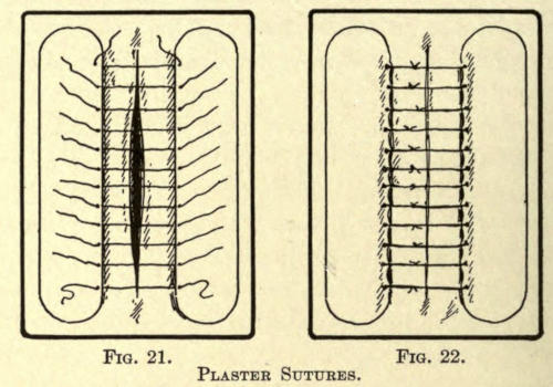
Fig. 21.Fig. 22.
Plaster Sutures.
The method involves the proper placing of strips of
plaster at either or opposite ends of the wound. The distance
between the incision and the edge of the plaster
must not be less than ¼ inch or more, according to
the length of the wound and its position. In place of
the strips of rubber adhesive plaster, the aseptic Z. O.
plaster should be substituted to overcome the objections
of the infections therefrom.
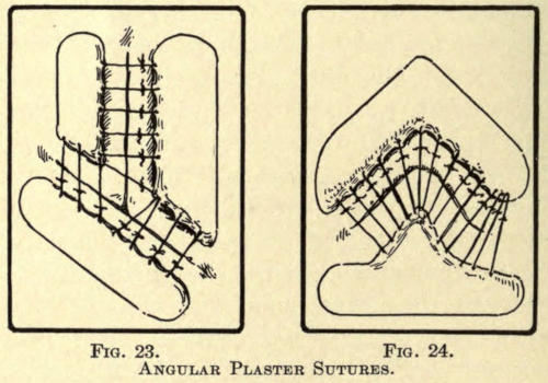
Fig. 23.Fig. 24.
Angular Plaster Sutures.
The inner edges of the plasters are raised slightly,
and interrupted sutures are inserted through them instead
of the skin (see Fig. 21). They are then tied as
shown in Fig. 22. In angular incisions the plasters are
cut as desired to insure perfect coaptation, as in Figs.
23 and 24. The advantages of this method, besides those
already mentioned, are that the wound is always open
for inspection and permits of free drainage. If thought
best, a small strip of iodoform gauze may be placed over
the threads or even under them, if there is little tension.
Since the introduction of the aseptic Z. O. (Lilienthal)
strips, the above method may be discarded as unnecessary
and requiring too much time for their application.
Strips of the antiseptic plaster are placed across the
wound at right angles, or, if the surface be a curved one,
obliquely to the wound. The plasters are furnished in
strips of the width desired, packed in two germ-proof
envelopes. They are extremely adhesive to dry surfaces.
Besides being aseptic, they are slightly antiseptic and
nonirritating. The strips are placed in position, leaving
an open space between them while the assistant brings
the edges of the wound into position.
Where there is tension of the parts this method is not
to be employed. The wound may be dusted as when sutured
and dressed in the same manner. The plasters are
removed about the sixth day by drawing the ends of the
strips toward the wound. Their second application is
unnecessary, regular dressings being substituted.
From the above it must not be inferred that all plastic
wounds are amenable to the above methods, because
many require specific treatment, as later described.
Granulation.—Wounds left open for granulation should
be dusted over with some stimulating antiseptic powder,
such as aristol or boric acid, and then covered with iodoform
or borated gauze. The granulating surface must be
gently washed with a mild solution of peroxid.
Prolific hypertrophic granulations, that jut out over
the surface, are touched with a lunar caustic point, avoiding
the epithelial edge of the wound, where it causes considerable
pain. Pale and loose granular points should
be scraped away with the sharp spoon curette to hasten
better growth.
If the skin edges are thickened and curled upon themselves,
it may be best to curette or to reduce them by
cauterization, so stimulating epitheliar spreading. Sterile
gauze is then loosely laid upon the surface, backed
with a highly absorbing material, such at charpie cotton
(Burns), wood wool, and poplar sawdust, retained in
gauze bags (Porter). The absorbing layer should be
light and pervious to the air, to facilitate not only free
absorption, but ready evaporation of the secretions.
Changing Dressings.—All dressings must be absolutely
sterile and all precautions, as primarily carried out, must
be followed in changing them.
It is rather infrequent to use permanent dressings in
plastic surgery, but where the wound appears aseptic,
with a dry serous crust over the line of healing, it should
not be disturbed except for mechanical reasons. The
latter are caused by the coagulated mixture of the wound
secretion and the antiseptic powder used, often aggravated
by the median knotting of sutures or the careless
disposition of the loose suture ends. Not too much can
be said of carefully folding the free silk suture ends at
right angles to the incisions. The ends, moistened subcutaneously,
are very liable to take on septic infection
and communicate it to the wound—crowded into the very
wound. When becoming embodied in the coagula of
serum and antiseptic powder it prevents, by pressure,
perfect union, causing a wider scar at such point, as well
as endangering the asepsis of the wound by being pulled
off accidentally, thus tearing it open and bringing on
hemorrhage.
The appearance of the resulting scar in facial surgery
is often of as much importance to the patient as the operation
itself, therefore, all care should be exercised in
bringing about the very best result.
For this reason, a patient in poor health should not
be operated upon, and any erosion of the skin about the
seat of operation should be thoroughly healed before attempting
plastic work. Aristol dusted on an abrasion
will heal it quickly.
If hemorrhage follows the dressings of a wound, the
dressing should be removed and the hemorrhage controlled
by pressure, unless severe, and be redressed.
Moist blood decomposes readily and is a source of
early infection, unless careful drainage under antisepsis
is established. At no time should any part of the wound
be unnecessarily exposed directly to the air. For small
wounds, silk protective plaster may be used to cover the
gauze dressing, while sterile gauze bandages should keep
dressings of large area in place.
Bandages, when changed, should be cut away with the
aid of the Lazarewitch angular bandage scissors and not
be unwound. It is quicker and the undue pulling of such,
when glued by secretions, is liable to disturb the healing
of wounds and even result in the tearing out of sutures.
The patient should never be intrusted to dress
wounds himself. In cases where the dressings cannot be
changed frequently proper precautions for drainage and
comfort must be observed. The temperature of the patient
should be taken twice daily; any elevation thereof
may indicate septic infection and demand immediate
attention.
When a portion of the ear, nose, or lip has been
severed by injury, the part may be put back into place and
held by sutures and aseptic Z. O. strips, powdered with
aristol and properly dressed. Union usually takes place,
even in the most unexpected cases. None but incised
wounds of such nature should be covered hermetically
with collodium or plaster, as bruised surfaces so often in
this kind of injury require perfect drainage. The retention
of secretions produces infection, generally resulting
in the entire loss of the part.
Wounds of the Mucous Membrane.—Wounds of the mucous
membrane should be carefully drained and cleansed
freely at frequent intervals, especially those about the
mouth. Wounds of the cheek, if including the mucous
membrane, should be especially cared for, as there is
here the increased danger of infections from the secretions
of the mouth.
Pedunculated Flaps.—When pedunculated flaps are left
free of other attachment, for reasons later mentioned,
they must be dressed as granulating wounds. Here it
becomes necessary to support the loose piece of skin in
such a way as to overcome circulatory obliteration. Unnecessary
handling is always to be avoided. The following
method has been used with the best results by the
author.
The flap is not dressed until all hemorrhage has
ceased. A small pad of sterilized or borated absorbent
cotton is covered lightly with ten-per-cent iodoform
gauze—cigarette fashion. The surface of this roll drain
is powdered well with aristol or iodol and it is gently
placed beneath the flap so that it rests easily upon the
same. A second and somewhat larger pad or roll of like
construction is placed next to the skin surface of the flap.
This is held in position by silk protective plaster or several
layers of gauze bandage, gently, though snugly,
applied.
The flap thus dressed should not be subjected to pressure,
often requiring considerable care on the part of the
patient, especially during the night. Undue pressure will
induce sloughing and must be avoided, even at the expense
of comfort to the patient.
This dressing may be changed the second day, when
the flap will appear anemic. Signs of discoloration indicate
gangrene, which is difficult to overcome. In a short
time the skin takes on a pale pink color, which indicates
a reëstablishment of circulation, and granulations begin
to show themselves on the reverse side, which, as they
multiply, soon thicken the flap sufficiently for the purpose
desired.
The dressings are continued, as begun, if there be no
indication for interference, although the granulations
may be stimulated if too inactive. Gently irritating the
granular surface with a 1-3,000 sublimate solution, although
rarely permitted by most surgeons, does no harm;
in fact, it is to be recommended before reapplying the dry
dressing.
In removing the dressings the edges of the flap will
be found to adhere to the gauze; this may be gently lifted
with the tenaculum, after previous softening with a weak
solution of hydrogen peroxid.
Pus (laudable) is the natural secretion from these
flaps. Whatever remains on the surface is easily removed
by an antiseptic solution, whereupon the powder
is again dusted upon the part. When the flap has thickened
sufficiently it may be covered by skin grafts, but
this is rarely done until it has been properly implanted
into the area for which it was intended and only then
when union between its sutured borders has taken place.
Ofttimes one part of a flap is left unattached, as, for instance,
the outer border of the ear, with the object of
developing a greater thickness. This must be cauterized
along the edge with the caustic pencil, keeping the granulations
within the desired limit until the opposite layer
of skin has either cicatrized with it or has been grafted
near it for the same purpose. The surface is then antiseptically
treated, as any granulating surface, except as
otherwise indicated.
Foreign Bodies.—Especial care must be exercised with
wounds into which foreign bodies have been implanted.
Under favorable conditions many are kindly received by
the tissues, but often these rebel and even with the greatest
of care in dressing such wounds will often result in
the necessity for removal of the substance. Individual
cases of such nature are fully referred to later.
CHAPTER VI SECONDARY ANTISEPSIS — SEPTICEMIA FOLLOWING WOUND INFECTION — GANGRENE — ERYSIPELATOUS INFECTION
Symptoms.—After operations performed under the
most thorough aseptic or antiseptic procedure, wound
fever, more or less marked, may be expected. It develops
a few hours after operation and subsides in twenty-four
or forty-eight hours. If, however, the wound has not
been properly rendered aseptic, or in which there is reason
for irritation or tension, more serious symptoms may
develop about the second or third day. These symptoms
increase with the amount of infection in the wound and
result in septicemia, or septic intoxication, the outcome
of the absorption of ptomains—the product of tissue
decomposition.
Inflammatory fever is marked by a sudden rise in
temperature, 100° to 103° F., with a full, strong, and
rapid pulse, headache, anorexia, coated tongue, constipation
and diminished secretion. If the infection is severe
delirium comes on.
If the symptoms are not relieved promptly the indications
of septicemia assert themselves with an increasing
temperature, between 102° and 104° F., with a
rapid compressible pulse gradually becoming weaker.
The respirations are rapid and shallow. The tongue becomes
dry and discolored and the teeth are covered with
sordes.
The restlessness disappears and apathy, somnolence,
and a low type of delirium takes its place. Vomiting occurs.
There may be a profuse diarrhea and the urine is
passed involuntarily. In other words, septicemia is but
an aggravated continuance of inflammatory fever; untoward
symptoms may come on early, and death may result
within forty-eight hours.
On inspection, the infected wound appears highly
inflamed, there is increasing swelling, with more or less
pain in the part. The edges of the wound appear pale
and everted. Serous oozing comes from the wound. If
sloughing is to occur from tension or low vitality of the
parts the area becomes discolored, assuming at first a
pale green color, which turns into bluish brown—and,
lastly, brown.
Treatment.—The treatment of such wounds is to immediately
relieve all tension by withdrawing the sutures.
Flush the wound with peroxid solution and irrigate thoroughly
with a 1-3,000 bichlorid solution, leaving the
wound open for a free drainage.
Then apply iodoform gauze over the wound and
change the dressings as often as is deemed necessary—two
or three times a day. In severe cases open the wound
thoroughly, even by further incision, clean out the contents
of the wound, such as foreign matter, bloodclots,
exudate, or perhaps pieces of bone that may have been
overlooked, using a small, sharp spoon curette for the
purpose. See that the deeper recesses of the wound,
especially in those about the nasal bones, are thoroughly
gone over.
Next irrigate the wound with a 1-1,000 bichlorid solution.
Carbolic solutions to be of use in the severer cases,
are too irritating and cause an increase of the secretions,
hence they are not to be used. Furthermore, their toxic
property is not desirable and ofttimes are not well borne
by the patient.
The wound is now loosely filled (not packed) with
iodoform gauze to permit of perfect drainage. Aseptic
absorbent cotton may be placed over this.
It is not advisable to bring the edges of the wound together;
the main object is to overcome the spread of infection
and the toxic absorption of the wound product.
When the symptoms are severe moist dressings, in
the form of compresses dipped into 1-3,000 bichlorid, are
to be preferred, changing them every hour.
Internally it becomes necessary to reduce the temperature
and to overcome the toxemia.
For the temperature quinin is the best agent. In
milder cases it can be given in tonic doses, associated
with antifebrin, with or without morphin, to quiet the
patient. In severe forms quinin must be pushed, giving
as much as twenty grains at a dose, to be repeated as
necessary. A saline purge, magnesium sulphate in full
doses, is useful to eliminate the ptomains.
The strength of the patient must be supported by the
free use of stimulants and frequent small quantities of
nutritious food. Milk with whisky is excellent. Peptonoids
and beef juice are given several times in the day.
Favorable symptoms are heralded by the lowering of
the temperature, the abatement of toxic symptoms, the
reduction of the edema, and the deep redness, as well
as the softening of the hard and painful edematous
walls of the wound, followed by the breaking down of
more or less tissue with the production of pus.
Gangrene in these cases is often due to undue bruising
or pressure on the parts during operation, and otherwise
to the tension of sutures. It is best to allow the
gangrenous mass to remain, keeping it aseptic by antiseptic
measures, as it is often found that only the superficial
layer and the edge or edges of the wound have
suffered.
As demarcation is well established the gangrenous
portions may be removed with the dressing or small seizing
forceps. Walcher’s pattern of a dressing forceps is
shown in Fig. 25, a toothed seizing forceps being represented
in Fig. 26.
Fig. 25.—Walcher Dressing Forceps.
Fig. 26.—Toothed Seizing Forceps.
The wound from now on may be at first subjected to
a rather strong aqueous solution of hydrogen peroxid,
fifty per cent, followed by the same sublimate solution
used throughout.
Iodoform gauze dressing, with or without dusting of
iodoform or iodol, is continued with the purpose of draining
the pus secretion thrown off by the granulating tissue
which soon begins to fill the wound, as well as to exert its
antiseptic and stimulating influence upon the granulations.
Lazy or glassy granulations are removed with the
curette as they appear, or a cauterant, in the form of a
nitrate-of-silver stick. Gradually the new tissue contracts,
the epidermal edges begin to fold over the surface.
Dry dressing, in the form of aristol or boric acid, may
then be used, to produce ultimate healing under an aseptic
scab, or lint moistened with two-per-cent salicylic oil
or boric vaselin is placed upon the wound. A formula
of the latter is made up as follows:
Boric acid
3
parts
Vaselin
5
”
Paraffin
10
”
A desirable boric-acid oil for the same purpose is composed
of:
Boric acid
3
parts
Cera alba
4
”
Ol. oliva
20
”
The latter must be changed daily until cicatrization has
been established.
If it is more desirable to cover the granulating area
by means of skin grafts it may be accomplished readily,
as later described. This is usually resorted to when
there has been loss of tissue from the result of sloughing,
although a sliding-flap operation may overcome the defect
to a nicety; this is especially true of wounds about
the anterior nasal border.
If the resulting cicatrice is no larger than the gaping
wound it may be excised, the skin at either side is undermined
and the edges are brought together, as was originally
intended.
It sometimes happens that a wound takes on erysipelatous
infection. It is usually of the simple variety,
although the cellulo-cutaneous variety is not rare.
Causes.—The predisposing causes are septic infection,
lowered vitality, resulting from alcoholism, poor
hygiene, and nephritis. The exciting cause has been accredited
to the erysipelo-coccus of Fehleisen, which is
found chiefly in the more superficial channels of the
corium and appearing in chain groups as seen microscopically.
Symptoms.—The symptoms locally are the peculiar
rosy rash, rapidly spreading out from the wound with
well-defined margins. The affected part appears smooth
and edematous and is slightly raised above the surface,
the patient complains of stiffness and burning pain in the
part. Often vesicles form on the affected part.
The temperature rises suddenly to 102° to 103° F.,
there is nausea and vomiting.
Treatment.—The wound in such cases must be treated
as described in inflammatory fever. Internally the usual
remedies are given. A local application of sixty-per-cent
ichthyol ointment, covered with salicylated cotton,
serves best. The skin may be incised in various places,
washed with an antiseptic (sublimate solution 1-1,000)
and the serous exudation pressed out with the sterilized
hand, after which the above ointment is applied under
absorbent cotton (Glück).
Antithermic remedies, as obtained by the application
of certain alkaloids, such as cocain, spartein, solanin,
helleborin, have been successfully used by Guimard and
Geley.
Spartein is especially claimed to exert a happy influence.
Lately a product under the name of antiphlogistin
has been used locally with excellent results and its use
is to be commended even in local wound inflammation.
If the subcutaneous tissue is affected and the surface
indicates the breaking down of tissue, hot antiseptic applications
are advisable or the skin is incised down to the
deep fascia at such places and iodoform gauze is packed
into the wound for several hours. Constant antiseptic
irrigation is then established by means of drainage tubes
inserted into the various incised places, which are connected
to an irrigating apparatus, so that the antiseptic
may reach all parts of the infected area.
Nontoxic solutions are indicated in this event; of these,
hydrogen dioxid, three per cent, and boric acid, nine per
cent, are most suitable. The solution is allowed to trickle
gently through the wound and is led off by open tubes,
that may be connected in such way as to empty into a
receptacle placed beneath the bed.
CHAPTER VII ANESTHETICS — GENERAL ANESTHESIA — Preparation for General Anesthesia — Chloroform — Ether — Combined Anesthesia — Nitrous Oxid — Ethyl Bromid — Ethyl Chlorid — LOCAL ANESTHESIA — Ethyl Chlorid — Cocain — Beta Eucain — Liquid Air — Stovain
Anesthesia of the human may be accomplished in two
ways: first, by the employment of a general anesthetic,
and, secondly, by the local use of a narcotic agent.
It is not the intention of the writer to dilate upon the
nature and use of anesthetics, as their value and indication
has been fully exploited. A concise review of these
agents, however, will meet with the approval of the special
surgeon, inasmuch as they have their particular use
in individual cases. Local anesthesia is undoubtedly the
most extensively employed, in the performance of the
average plastic operation, yet in certain cases it is contra-indicated,
and it is to further the proper selection of
such that the following may be of value.
It must be understood that general anesthesia has
its many advantages and equally its disadvantages. It
necessitates the early preparation of the patient and a
thorough physical examination as to the state of lungs,
heart, and kidneys. Patients having cardiac affections
or serious lesions within the lungs should be given the
safest anesthetic obtainable; in fact, if the operation can
possibly be done by local administration, it should be.
It is to be remembered that in a majority of these
cases the operation is undertaken to remedy a deformity,
however caused, one that is not necessarily serious to
health, and it would indeed be unwise to place such a
body in jeopardy or to take undue chances if they can be
avoided. Ofttimes several operations, a few weeks apart,
must be done and the frequent repetition of a general
anesthetic might impair the health of the patient—a condition
not associated with local anesthesia.
Neurotic subjects often insist upon the use of chloroform
and the surgeon is frequently tempted to administer
it, but little objection should be found with local
narcosis, where it can be employed.
The patient must not be allowed food at least six
hours prior to operation. In neurotic and anemic subjects
a full dose of strong wine or whisky should be given
half an hour before operation. Habitual drinkers should
be given one quarter grain morphin sulphate. All movable
artificial teeth, or other foreign bodies, must be removed
from the mouth. Observe the laws of asepsis as
heretofore described. Loosen the clothing of the patient
about the neck and chest. Only a single garment should
be worn during the time of operation—a loose, sterilized
night robe, as it may be necessary to move the patient
about, and too much or tight clothing might prove to be
dangerous in the delay occasioned by its removal.
The operating room should never be cold enough to
chill a patient so prepared. Hot-water bottles or a
warm pack can be placed between or about the limbs
to equalize the external circulation. This is especially
necessary when chloroform is administered, as this
lowers the temperature of the body. Have the bowels
and bladder emptied. Choose the early part of the day
for operations of some length, because the stomach is
then empty and vomiting with resultant gastric disturbance
will be lessened or entirely avoided. The anesthetizer
should be experienced and attend to his duty
implicitly. He must at all times watch the patient, take
note of the pulse, pupils, and respiration. Close by he
must have a mouth-gag, tongue forceps, long-handled
sponge holders, containing dry absorbent cotton sponges,
and a basin in case of emesis.
He should quietly instruct the patient how to breathe
and at first assure him, and as narcosis comes on command
him to do what is necessary. No desultory or
detracting conversation should be permitted. A small
but efficient faradic apparatus must be within call of the
administrator. Sterile vaselin should be smeared about
the nose and mouth to prevent skin irritation.
(Guthrie) The pure product must be used. It is a
colorless, mobile liquid, having an ethereal odor and
sweet to the taste. It should not affect litmus or turn
brown with sulphuric acid or give a precipitate with
nitrate of silver. If there is emphysema of the lungs,
bronchitis, or renal disease, chloroform is to be preferred
to ether, also in operations about the oral cavity. Children
bear chloroform narcosis better than adults.
Fig. 27.—Schimmelbusch Dropping Bottle.
Fig. 28.—Esmarch Dropping Bottle.
Chloroform first affects the brain, then the sensory
tract of the spinal cord, then the motor tract, followed by
an involvement of the sensory path of the medulla, paralyzing
the respiratory centers, while cardiac syncope
may come on at any time during narcosis. Death may be
either due to respiratory or cardiac failure, often from
both. To overcome this the anesthetic should not be
crowded, nor should the apparatus be held too close to
the mouth of the patient. The best method of giving it is
by means of the dropping bottle of Schimmelbusch (Fig.
27) or that of Esmarch (Fig. 28) and a simple mask or
apparatus.
Fig. 29.—Schimmelbusch Improved Folding Mask.
Fig. 30.—Esmarch Inhaler.
The wire frame affair, to be covered with a fold of
muslin, designed by Schimmelbusch, is perhaps the best
(Fig. 29). Another splendid inhaler is that of the Leiter
improvement of the Esmarch folding frame (Fig. 30).
A folded towel, crumpled or pinned into a hollow oval
form, may be substituted for the above.
Begin by pouring about thirty drops upon the inhaler,
gradually bringing it nearer from a distance of six
inches to the mouth and nostrils of the patient; then continue
by letting one drop fall upon the apparatus every
five or ten seconds until the patient is thoroughly anesthetized;
then use one drop about every ten seconds,
although it might be necessary to push this quantity at
certain moments of the operation. To obtain complete
anesthesia by this method takes about ten minutes. The
vapor should be thoroughly mixed with air in the proportion
of ninety-five per cent of air to five per cent of the
vapor. The amount administered during operation can
rarely be determined, because of the uneven respiration
of the patient, who takes more during frequent inspiration
than during ordinary breathing. By all means do
not let the chloroform trickle upon the skin or into the
eyes, as it causes considerable irritation.
The respirations are at first full and deep, soon becoming
shallow and rapid. At first the pulse is slightly
stronger and fuller than the normal, but it soon loses
its strength and volume and becomes more rapid. The
pupils are at first dilated, and as narcosis is induced, contract.
Should they contract after this, during operation,
it is a danger signal not to be neglected. Death may
come on suddenly.
If the patient struggles violently under early anesthesia,
as is often seen in alcoholics and athletes, it is not
advisable to push the chloroform nor should total muscular
relaxation be effected. The arrest of reflex movement
is all that is required.
As the reflex action of the cornea disappears last of
all, the anesthetizer can use this as a guide during further
administration to avoid all danger. This is accomplished
by gently touching the cornea with the index
finger, raising the eyelid with the third finger.
Chloroform lowers the body temperature, due undoubtedly
to its aiding in the dissipation of heat and by
reason of its effect on the nervous mechanism of heat
production. It is rapidly eliminated by both the lungs
and the kidneys, because of its high volatility, and as little
is given, the irritation to these organs is not as great,
volume for volume, as with ether.
In case of asphyxia the lower jaw must be pushed far
forward, the tongue be drawn forward with forceps, and
the head extended and lowered, by raising the feet off the
table. Cold water should be dashed over the face and
chest. Slapping the chest with a wet towel and vigorously
rubbing with hot cloths or brushing the palms and
soles. Brandy and water, one to two parts, can be introduced
into the rectum, or faradization of the nasal mucous
membrane can be tried. These means failing, artificial
respiration (Sylvester’s method) must be resorted
to. This being of no avail, tracheotomy must be done.
If the patient is induced to vomit, he should immediately
be turned on his side to prevent the indrawing of
the ejected matter into the lungs. After it has ceased,
thoroughly wipe out the mouth with a long-handled
sponge. The anesthetic must now be crowded slightly to
overcome the irritation of the mucous membrane of the
stomach. Often during the early stage of anesthesia the
patient stops breathing, which must be overcome by slapping
the chest or by two or three forceful downward
movements on the epigastrium.
If the face of the patient takes on a sudden change of
color or breathes heavily the anesthetic should be withdrawn
for a few moments, until the symptoms abate. If
the mucus collects about the glottis it is liable to cause
respiratory difficulty and must be swabbed out. If the
inversion of the patient does not relieve syncope and the
methods already mentioned fail to relieve, injections of
normal salt solution into the median basilic vein must be
employed as a last means.
(Jackson, Morton.) Sulphuric ether is used in the
pure form, free from alcohol and water. It should be a
colorless, volatile, mobile, and highly inflammable liquid,
having a peculiar penetrating odor. As its vapor is
much heavier than air, and owing to its combustible nature,
lights about the room should always be placed above
the patient. Often its vapor is ignited by the careless
use of the electro-cautery.
Ether for anesthesia should not affect blue litmus. It
should not give a blue color to ignited copper sulphate—the
test for the presence of water. Alcohol is indicated
when it turns red by adding fuchsin.
Ether is less toxic than chloroform, therefore it requires
a greater quantity to induce narcosis.
If properly administered it is by far safer than
chloroform, Ollier, of Lyons, reporting only one fatality
directly due to its employment in four thousand patients.
If the anesthetic is crowded cyanosis with jugular pulsation
is noted—the signs of inefficient oxygen and cardiac
distention. In most recorded cases of death there
were complications of a nephritic pulmonary nature.
Ether should not be used where there is bronchitis, gastritis,
or peritonitis, owing to its irritant effect on mucous
membranes, nor in nephritis, aneurysm, or advanced
atheroma. The movements of the diaphragm must be
constantly watched as it is the first to become paralyzed
when anesthesia is carried too far. The same care must
be observed with the pupil for cerebral and the pulse for
cardiac signs. Before giving this anesthetic the same
preparations as for chloroform narcosis should be observed.
The stomach should be empty, the nose and
mouth smeared with vaselin, and the eyes protected with
a towel.
At first the patient is given the ether with a considerable
mixture of air, which should be lessened gradually.
Coughing comes on quite often, which is overcome by
increasing the ether. Soon there comes a state of respiratory
forgetfulness. This is caused by the irritation
of the trigeminal and vagal nerves (Hare). This is
corrected by dashing ether upon the epigastrium or by
sudden and repeated pressure at this point. There is
also choking and struggling, the face becoming suffused
and red and there is an injection of the conjunctiva. As
the ether is pushed the patient becomes quiet, followed
by a second seizure of struggling, so intense, that force
must often be employed to hold him on the table. With
this there are the various attacks of laughing, crying,
singing, or yelling—a semiconscious exhibition of the
state of the mind of the individual.
As anesthesia progresses relaxation takes place and
the time for operation is at hand. Often the throat fills
with mucus, owing to the irritant effect of the vapor on
the mucous membranes. This must be wiped out with
the sponges.
If vomiting occurs the head of the patient is turned
to one side until relieved. The mouth should be cleansed
thoroughly thereafter to prevent the contents getting
into the lungs and causing bronchial irritation and often
broncho-pneumonia. If the patient gets too little air,
shown by laryngeal stertor, frequent and feeble pulse,
livid face or pallor, tonic spasm, thoracic breathing with
fixed diaphragm, and drawing in of the abdominal walls
with inspiration, the ether should be let up and the jaw
pushed forward by placing the fingers under the rami.
The tongue should be drawn forward, as already described,
and such methods be used as have been mentioned
in connection with asphyxia in chloroform narcosis.
The pupils fixed in dilatation is indicative of
immediate danger.
Strychnin and digitalis should be given hypodermically
or the intravenous use of ammonia may be employed.
If the stertorous breathing is due to mechanical
causes, not to too much ether, the hypodermic use of ether
will bring about reflex respiratory
movement by reason
of the local pain and
irritation thus produced.
Fig. 31.—Allis Inhaler.
Fig. 32.—Fowler Inhaler.
Fig. 33.—Juillard Mask.
The anesthetic may be administered with the aid of
various masks or inhalers. Simplicity of construction is
to be preferred to more complicated apparatuses. The
aseptic metal inhaler of Allis (Fig. 31) or the folding
form of the same modified by Fowler (Fig. 32), are much
used in the United States, while the Juillard mask—a
metal frame covered with several thicknesses of gauze—is
used abroad. (See Fig. 33.)
It is to be remembered that in operations about the
face ether anesthesia is not practicable, owing to the repeated
lifting of the mask which allows the patient too
much air. It can only be given by specially constructed
inhaling devices, which are more easily used with chloroform
or the mixed anesthetics. Their specific use and
construction will be referred to later.
It is often desirable to get the patient as quickly as
possible under anesthesia and still overcome the depleting
effects of chloroform narcosis. A common method
to accomplish this is to give chloroform to the point of
relaxation and with a change of inhaler continue with
ether. In this way anesthesia can be kept up safely a
long time with a minimum amount of ether. There are,
however, a number of mixtures used in place of this interchange
of anesthesia, all having their particular merit.
Some of the best known are:
Alcohol-Chloroform-Ether (A. C. E. Mixture, English
Formula, 1:2:3).—This induces rapid anesthesia without
the danger of syncope or the other objections to
chloroform or ether when used alone.
Chloroform-Ether-Alcohol, in the proportion of 3:1:1, and
known as Billroth’s mixture, is extensively used in the
same way.
Chloroform-Ether.—This mixture, in equal proportions,
is known as Tillman’s mixture, and has been employed by
many well-known surgeons.
Nitrous oxid, as advocated by Sir Humphrey Davy,
is a safe product, but the anesthesia produced thereby
is of too short duration to be of practical value in plastic
surgery. Its employment is resorted to only for such
operations as the opening of abscesses or the removal of
small cysts, etc.
While ethyl bromid is a product that cannot be said
to be absolutely safe, Terrier, of Paris, has used it
largely to induce anesthesia, following it up with chloroform.
It should be given freely with deep inspiration,
the sixth inhalation producing total loss of consciousness.
A moment after complete muscular relaxation is
attained, with congestion of the face and dilated pupil.
The average time necessary to accomplish this is about
one minute, in which about three to five grams are used. In
this way the stage of excitement is overcome and immediate
narcosis is obtained. As the chloroform is substituted
it must be given fairly strong, reducing it gradually.
The facial congestion slowly diminishes and the
pupillary dilation gives way to contraction. About sixteen
grams of chloroform are required to keep up anesthesia
for fifteen minutes. The after-effects of chloroform
are entirely overcome by the above method.
Hawley, in reviewing the use of ethyl chlorid as a
general anesthetic in minor operations, states that after
several years of more or less constant use of ethyl
chlorid, both in clinical and private practice, he has still
to see the first case in which it has caused him the slightest
uneasiness. The following precautions in its administration
should be observed: (1) The patient should be
prepared as for chloroform or ether; (2) whatever mask
is used, it should fit the face snugly; (3) a graduated
tube with a large aperture should be used; (4) the anesthetic
should be well supplied with air and as little given
as possible; (5) care should be taken not to present it at
first in too large a quantity; frequently a dram is quite
sufficient for short operations; (6) the patient should
rest a while after its administration, as faintness sometimes
supervenes; (7) a mask should be used which does
not receive the drug close to the patient’s face, otherwise
one is liable to either freeze the face of the patient or to
cause asphyxia by the moisture from the expired air
freezing on the gauze in the mask, and thus preventing
the free passage of air to the patient.
The use of ethyl chlorid has the following advantages:
(1) Safety in administering; (2) ease of administration;
(3) it rapidly produces surgical anesthesia;
(4) it can be used where chloroform or ether would be
contra-indicated; (5) the patient can be kept in any position
during anesthesia, upright or prone; (6) no cyanosis
need occur during administration; (7) the patient recovers
promptly without after-effects; (8) it is inexpensive;
(9) it can be used for a long or short operation with
equal success; (10) it is especially useful as a preliminary
to other anesthetics, decreasing the time required
for the production of anesthesia and avoiding shock and
discomfort to the patient.
Ethyl chlorid is a colorless, mobile liquid, which boils
at 52° F. This is furnished in thirty- and sixty-gram
glass tubes, sealed with a metal screw cap or spraying
device. As this cap is removed the liquid in the tube
begins to boil, owing to the temperature of the room, or,
better, the operator’s hand, and a fine vapor spray is
ejected from the opening.
The tube end is held from six to eight inches from the
part to be anesthetized. Immediately the skin is frosted
over and the lanugo hairs become covered with snow.
The skin turns white and becomes slightly elevated, appearing
to be thickened; at the same time the patient
feels a stinging pain in the area. This may be overcome
greatly by first smearing the part with sterile vaselin.
In a few moments the skin is frozen and rendered antalgic,
and operations of short duration can be performed.
The only disadvantage with this method is that
the part to be operated on is frozen stiff, hence the skin
cannot be neatly dissected away from the subcutaneous
tissue as under other local anesthesia, nor can the tissues
be moved about as readily, as in the case with flap operations,
owing to this stiffness.
The parts thaw out quickly with a returning sensitiveness,
and it may be necessary to apply the spray
repeatedly until the operation has been completed. If
elastic constriction can be employed, the antalgic effect
is more quickly produced and more lasting.
As the parts thaw out there is considerable prickling,
which can be mitigated by applying sponges soaked in
hot sterilized water. More or less redness of the skin
will be noted even for some time after the operation.
(Methylbenzoylecgonin)
(Gädeke, Nieman, Bennett, Koller.) Cocain is the
alkaloid derived from several varieties of Erythroxylon
coca. It should appear as a permanent white crystalline
powder in colorless prisms or flaky leaflets. The salt
used for anesthetic purposes is the hydrochlorid; it is
soluble in 0.4 part water, 2.6 parts alcohol, 18.5 parts of
chloroform, and insoluble in ether.
Locally applied on mucous membranes and open
wounds, it exerts an analgesic effect, but not of the unbroken
skin. Punctures or abrasions are necessary to
permit of absorption in this event. When locally applied
it paralyzes the peripheral sensory nerves, and at
first blanches the parts by reason of its active contraction
on the arterioles, which is soon followed by marked
congestion.
Krymoff has made extensive experiments to determine
the anesthetic effects of cocain solutions sterilized
in various ways. He claims that the best results in minor
surgery are obtained with the one-per-cent solution pasteurized
at 60° C. for three hours. The same solution
pasteurized at 80° C. for two hours or at 120° C. for fifteen
minutes gave results far less satisfactory.
While the pasteurized solutions accomplished an anesthesia
lasting from one to two hours, sterilized solutions
(boiled at 100° C.) overcame pain only for a period
between twenty and thirty minutes.
Pasteurized solutions have the advantage of being
sterile and do not decompose as the boiled solutions
would. The pasteurization is accomplished as follows:
The necessary amount of cocain is dissolved in sterilized
water. The solution is put into a sterilized glass bulb,
which is sealed hermetically and subjected to a temperature
of 60° C. for three hours.
Since cocain is a nerve poison, its systemic absorption
must be avoided. The constitutional effects of a given
amount injected about the head, face, and neck are more
marked than when injected in other parts of the body or
extremities. This is due to two causes: a more rapid
absorption and the proximity to the brain (Ricketts).
For this reason less cocain should be used and the blood
vessels be avoided.
Idiosyncrasy influences greatly these toxic effects.
In neurotic patients of irritable and impressionable type
the hypodermic use of this agent has especially induced
serious syncope. Very serious symptoms and even death
have been caused by its local use (⅛ grain hypodermically).
Untoward effects are manifested by nausea, vertigo,
emesis, syncope, followed by clonic convulsions, delirium,
and death.
Cocain first stimulates, then paralyzes, the pneumogastric
nerve; the respiration is first accelerated, and
then paralyzed, death being due to failure of respiration.
Should these symptoms occur, the patient should be
placed on his back with the head low. Amyl nitrate inhalations
act as the antidote and reduce the cerebral
anemia (Feinberg). Morphin or caffein is to be given
hypodermically, or the former is associated with potassium
bromid internally.
To overcome the toxic qualities of the anesthetic it
may be combined with morphin in solution, Schleich’s
solution being well known. It is composed as follows:
Cocain murias
0.1
Morph. sulph.
0.025
Sodium chlor.
0.2
Aquæ sterilis.
100.0
Gauthier suggests the addition of one drop of a one-per-cent
solution of nitroglycerin to the quantity injected
and repeated to prevent the unfavorable after
effects.
Solutions of cocain are to be made up fresh each day,
as they become moldy on standing. They cannot be sterilized,
for the reason that a temperature of 213° F. renders
them useless. The solution is most active at 50° F.
(Costa).
For hypodermic purposes two- to ten-per-cent solutions
are employed, the four per cent being more generally
used, not more than 0.1 gram of the agent being
introduced (Hänel, ano).
Fig. 34.—Simplex Syringe.
Fig. 35.—Kolle Improved Pravaz Syringe.
Fig. 36.—“Sub-Q” Syringe.
For the introduction of the solution the ordinary
Pravaz syringe can be used, a modification of which being
known as the “Simplex” (Fig. 34). It is a glass instrument,
without screw threads within the needle base, and
has a sterilizable fiber piston. The only disadvantage
offered by this syringe is the lack of finger rests. The
author has added a removable nickel-plated sleeve with
finger rings to slip over the glass barrel, as shown in
Fig. 35. The advantage of this modification will be appreciated
when injections are made into dense or cicatricial
tissue where considerable pressure is necessary
for the introduction of the solution.
Another excellent syringe for the purpose is the
metal-cased instrument known as the “Sub-Q” (Fig.
36); the barrel and piston in this are of glass, an asbestos
packing being wound over the piston head.
Metal needles with large thread or smooth ends are
employed. As the asbestos packing contracts in drying,
the piston should be removed from the barrel and
cleansed immediately after use, and not be introduced
into the barrel until both the asbestos windings and the
inside of the barrel have been moistened with warm
sterile water. This precaution prevents the cracking of
the instrument through undue pressure exerted on the
end of the piston rod at the time of use.
To render the primary introduction of the needle
painless the area might be sprayed for a moment with
ethyl chlorid. After carefully preparing the site of operation,
the subcutaneous injections are made in a somewhat
oval or circular manner, the first infiltration of the
cocain rendering the succeeding points analgesic.
It will be noted that the skin becomes whitened and is
raised in little tumors, with the point of puncture as a
center. The various punctures are so placed that the
borders of these tumors meet, the entire site becoming
edematous. If by constriction the part can be rendered
ischemic, the analgesic effect is prolonged, reducing the
systemic absorption to a minimum.
The subsequent nausea often following may be
promptly overcome by the use of a mixture of creosote,
four drops in limewater. For mucous surfaces the anesthetic
may be applied with absorbent cotton and allowed
to remain about five minutes. In deeper wounds
than those involving skin, deeper injections must be made.
The effect of the anesthetic as above employed is
practically immediate, and the operation can proceed at
once. Its duration is from fifteen to twenty minutes for
subcutaneous surgery, but where the deeper structures
are involved subsequent injections must be made to control
the pain.
(Benzoylvinyldiacetonalkamin)
(Merling, Vinci.) White powder, soluble in 33
parts of water. While the effects of eucain are immediate
and produce anesthesia as thoroughly as cocain,
it has the objection of producing local hyperemia and
increased edema of the parts injected. This often interferes
with the successful outcome of the first operation,
as will be later shown. The advantage over cocain,
however, is that a solution of eucain can be sterilized by
boiling without reducing its usefulness, which in itself
is an item, since both are expensive, and if we must prepare
a cocain solution fresh for each day we must discard
all that has not been used, while with eucain the same
preparation can be safely used over and over, after
proper sterilization.
The two- and three-per-cent solutions are most employed
to the extent of from 10 to 60 minims. Its
subcutaneous effect is immediate, lasting from ten to
twenty-five minutes. When applied locally to mucous
membranes, the five-per-cent solutions are used.
Principally it may be said that eucain does not exhibit
the toxic properties of cocain, the author having
employed it in over 5,000 cases with no untoward effect.
Liquid air is suggested as a means of local anesthesia
by A. C. White. He recommends its intermittent application
instead of freezing the part as with ethyl
chlorid. It is sprayed on the parts and produces immediately
anemia and insensitiveness. There is no hemorrhage
during its use, so that dressings may be applied
before the parts assume their circulatory function; an
advantage of considerable value in plastic surgery. No
untoward results follow its use.
(Fourneau.) This is the latest preparation advocated
for local anesthesia. It is a synthetic product, derived
from tertiary amyl alcohol. It is less toxic than cocain,
and has been used more or less in the past years experimentally,
but the consensus of opinion seems to be
against its use. Jennesco has used it extensively in conjunction
with strychnin in spinal anesthesia, but the surgeon
in general has not taken kindly to it. In plastic
surgery, as used locally, it has been little employed, eucain
being the most serviceable for the purpose.
CHAPTER VIII PRINCIPLES OF PLASTIC SURGERY — METHODS IN PLASTIC OPERATIONS — I. Stretching Method — II. Sliding Method — III. Twisting Method — IV. Implantation of Pedunculated Flaps by Bridging — V. Transplantation of Nonpedunculated Flaps or Skin-grafting — MUCOUS-MEMBRANE-GRAFTING — BONE-GRAFTING — HAIR-TRANSPLANTATION
Plastic surgery is resorted to in covering defects of
the skin due to congenital or traumatic malformation,
injuries, burns, the removal of neoplasms, or the ulcerative
processes of disease. Furthermore, it can be employed
cosmetically for the rebuilding of organs, whole
or in part, or for their reduction when abnormally developed.
This applies particularly to the nose, ears, and
lips, wherein it may involve either the skin alone or the
mucous membranes, or all the tissue making up the parts
operated on.
Incisions.—The incisions in plastic surgery are to be
made obliquely into the skin rather than at right angles
to the surface, the former permitting of better apposition,
and undoubtedly causing less epidermal scar. The
incisions include the skin only, except when otherwise
stated.
Sutures.—Sutures should be placed not more than ¼
of an inch apart and be made to include the skin only,
unless it is deemed advisable to employ deeper ones to
relieve undue traction, which often results in suture scars
and ofttimes tissue loss, necessitating further operation.
The latter may be obviated by placing every other stitch
at greater distance from the free edge of the skin, that is,
into the undissected border.
Intracutaneous sutures may also be employed, but
these are rarely, if ever, necessary if the apposition has
been properly accomplished. To relieve tension, harelip
pins are also used, as later described. Catgut of such
size as would be suitable because of its ready absorption
is not to be employed for skin suturing, fine twisted silk
or selected horsehair being the best material.
Formaldehyd catgut can be used if it is desirable; its
fine strands withstand absorption to a greater degree
than the ordinary.
Fig. 37.—⅝ Circle Haagedorn Needles.
Fig. 38.—Crescent Curve Haagedorn Needles.
Needles.—Very fine, flat, round-eye needles, such as
Haagedorn’s, Nos. 12 to 15, ½ or ⅝ circle, are the
most serviceable in skin work, as they incise the skin in
penetrating and leave an elongated slit, which heals readily,
rarely leaving a needle scar. The selection of needles,
however, must be left to the operator, many preferring
the one variety to the other. Split-eye needles are quite
convenient, but they break more readily, and while they
work best with fine silk, this is more readily withdrawn
from the eye at undesirable times. For very delicate
work the needles mentioned are the best.
Needle Holders.—Inasmuch as the needles used in
plastic operations are very small and fine, proper needle
holders must be used. The requisitions are that the jaw
be long, narrow, of soft copper, and that they have an
automatic lock attachment. Plain needle holders may be
used, but at times the locking device is very necessary
and saves time. The most serviceable of this class are
the Kersten modification of Mathieu, an uncomplicated
holder of merit (Fig. 39), and the small Haagedorn,
five-inch narrow-jaw holder (Fig. 40), or the holders
taking similar needles, as shown in Figs. 41 and 42,
known as Pozzi’s and Weber’s modifications, respectively.
Fig. 39.—Kersten-Mathieu Needle Holder.
Fig. 40.—Haagedorn Needle Holder.
Fig. 41.—Pozzi-Haagedorn Needle Holder, 5 in.
Fig. 42.—Weber-Haagedorn Needle Holder, 6 in.
Fig. 43.—Needle with Suture Carrier.
Holders with cup jaws serrated at different angles
are of no value, unless other needles are used with them,
as they invariably break the flat ones.
To overcome the necessity of rethreading or the use
of many needles, the very ingenious holder with ligature
carrier can be used, especially where the surgeon does
not care to sew with a long, free thread. The silk can
be sterilized on the metal spool separately and inserted
within the handle of the holder, as shown in Fig. 43.
There are five distinctive methods employed in
performing plastic operations:
I. Stretching of the margins of the skin.
II. Sliding flaps of adjacent skin.
III. Twisting pedunculated flaps.
IV. Implantation of pedunculated flaps by bridging.
V. Transplantation of nonpedunculated flaps or skin grafting.
This classification differs from that heretofore generally
given in the meager literature on the subject, but the
author believes his arrangement to be more scientifically
exact as well as simpler for recording and history
purposes.
In the stretching method the defect is neatly excised,
so as to freshen the margins to be brought together. It
may be necessary, if the defect is too large for free apposition,
to dissect the skin away from the underlying
tissue to render it more movable and to overcome tension.
The shape of the incision depends largely upon the
nature of the defect and must be made with a view of
leaving as little scar as possible. Where the defect is
somewhat linear, or elongated, an elliptical incision (A)
is made, as in Fig. 44, and, if necessary because of too
great tension, the skin is undermined sufficiently to allow
the parts to come together; if this cannot be done readily,
two semilunar incisions (b, b) must be added. This will
allow of ready coaptation. The wound is then brought
together with an interrupted suture, appearing as in Fig.
44a, the semilunar spaces being allowed to heal by granulation.
Fig. 44.
Celsus Incisions.
Fig. 44a.
Celsus Relieving Incisions.
In excisions in small rhomboidal form, the skin is
merely dissected up and around the wound, the same as
in Fig. 45, and the wound is sutured in linear form, as
shown in Fig. 46.
Fig. 45.
Rhomboid Excision.
Fig. 46.
Union of Rhomboid Excision.
If the defect is oblong in form, the angles are brought
together wholly, leaving a small surface to granulate,
as in Fig. 47, or they are drawn toward the center,
leaving the remainder of the parallel lines to be sutured,
as shown in Fig. 48.
Fig. 47.
Oblong Excision.
Fig. 48.
Coaptation of Same.
Another method of overcoming a smaller defect of
similar form is to excise a small triangular portion of
skin at either side of the oblong, as in Fig. 49, and then
with or without dissection bringing the margins together
in linear form, as in Fig. 50.
Fig. 49.
Bitriangular Excision.
Fig. 50.
Linear Coaptation.
Likewise can a triangular fault be brought together
by sewing in the greater angles and making a linear
wound of the remaining part, as in Figs. 51 and 52.
Fig. 51.
Triangular Excision.
Fig. 52.
Coaptation of Wound.
Again, a triangular defect may be remedied by adding
a smaller triangle at each end involving healthy skin,
utilizing, if need be, the relieving incisions, as in Figs.
53 and 54.
Fig. 53.
Triangular Excision with Relieving Incision.
Fig. 54.
Coaptation of Wound.
Following the principle of Celsus, as mentioned on
page 8, defects can be overcome in various ways. The
incisions may be straight or curved, and one or more
flaps of skin are raised, slid, and sutured over the part
to be covered. The simplest form is the covering of a
square, as shown in Figs. 55 and 56.
Fig. 55.
Square Excision.
Fig. 56.
Coaptation of Wound.
If the square be too large for the above method, the
incisions can be carried to the other side and above or
below the defect, as shown in Figs. 57 and 58.
Fig. 57.
Square Excision.
Fig. 58.
Coaptation of Flaps.
For triangular areas the curved incisions can be
made, as in Fig. 59, rotating the flap into place, as shown
in Fig. 60.
Fig. 59.
Triangular Excision.
Fig. 60.
Coaptation of Flap.
Or bilateral flaps may be utilized by straight incisions,
stretched and sewn, as in Figs. 61 and 62.
Fig. 61.
Triangular Excision.
Fig. 62.
Coaptation of Flaps.
Again, two curved incisions are made to obtain rotating
flaps, Fig. 63, and sewn, as shown in Fig. 64.
Fig. 63.
Triangular Excision.
Fig. 64.
Arrangement of Flaps.
Bürow introduced a method for closing over defects
by sliding flaps in which he utilized the mobility of skin
obtained by the excision of triangles of healthy skin.
The results are exceedingly good, but, unfortunately, the
sacrifice of skin affects its general use, inasmuch as patients
can afford but little loss of healthy skin; besides,
there is the objection of added scarring. The closing of
a triangular defect by this method is shown in Figs. 65
and 66, in which a is the triangular defect and b the triangle
of healthy skin excised. The skin about the incisions
is dissected up and the flaps are sutured into position,
as shown in Fig. 66.
Fig. 65.
Double Triangular Excision.
Fig. 66.
Coaptation of Wound.
Where the triangular defect has a wide base, bilateral
triangular sections of skin are removed (Fig. 67), and
the flaps are coapted, as in Fig. 68.
Fig. 67.
Tri-triangular Excision.
Fig. 68.
Coaptation of Wound.
Through the sacrifice of two triangles a large oblong
or square defect may be covered, the excisions being
shown in Fig. 69 and the suturing in Fig. 70.
Fig. 69.
Rectangular-Bitriangular Excision.
Fig. 70.
Coaptation of Wound.
Although in several of the above methods the flaps
are rotated and slightly twisted, the following are only
classified with those under this division.
Where an elliptical defect is to be obliterated the
curved incision shown in Fig. 71 can be satisfactorily
employed, leaving but a small area to granulate over.
The suturing is depicted in Fig. 72.
Fig. 71.
Weber Method.
Fig. 72.
Coaptation of Flaps.
In this the twisting of the flaps is but little, while in
the following, in which the defect is of similar shape, the
twisting is more apparent; so much so, that a fold at
the root of the flap may be induced to some extent. The
excision and incisions are shown in Fig. 73, and the
method of bringing the parts together in Fig. 74, leaving
a small area for granulation.
Fig. 73.
Elliptical Excision.
Fig. 74.
Coaptation of Flaps.
Considerable twisting of flaps is shown in covering
triangular parts in Figs. 75 and 76.
Fig. 75.
Triangular Excision.
Fig. 76.
Coaptation of Flaps.
In this only a small surface is left to granulate over,
while in the following the parts are entirely covered.
The excision and incisions are shown in Fig. 77, and the
method of approximation and suturing in Fig. 78.
Fig. 77.
Triangular Excision.
Fig. 78.
Coaptation of Flaps.
In covering a square area considerable twisting must
be resorted to, as shown in Figs. 79 and 80, leaving a portion
to granulate.
Fig. 79.
Lentenner Method.
Fig. 80.
Coaptation of Flap.
Where the area is irregular and formed somewhat as
in Fig. 81, bilateral incisions are made and the flaps
twisted into place and sewn, as in Fig. 82.
Fig. 81.
Burns Method.
Fig. 82.
Coaptation of Flaps.
In this method the flap to be utilized in covering a defect
is taken from a distant
part of the body,
as, for instance, from
the arm. The flap thus
taken at first remains
attached at its distal end
to the tissue of the arm
by a pedicle, which is
not severed until a circulation
has been established
between the flap
and the part of the human
economy to which
its free end has been
attached by suture, the
arm being held in position
in the meantime by
a suitable contrivance,
as shown in Fig. 83.
Fig. 83.—Tagliacozza Harness.
These pedunculated flaps, bridging over space, may
likewise be taken from the forearm, the hand, or the
thoracic region.
When thoracic flaps are used they may be directly
sewn at their free ends to the part to be covered, as, for
instance, in the forearm or arm, or they are stitched to the
forearm to be later transferred to another part of the
body after their circulation had become established.
The various methods of the employment of these
bridging flaps will be taken up individually in their respective
places farther on.
Where there is loss of skin due to injury or operative
procedure the parts may heal by granulation, but as this
requires much time, and the consequent cicatrice causes
considerable deformity, the granulating or freshly made
wounds are covered with so-called detached skin flaps or
grafts, when the former methods of plastic surgery cannot
be followed.
The methods employed in skin-grafting may be classified
as: 1, autodermic; 2, heterodermic; 3, zoödermic.
1. Autodermic, when the grafts are taken from the
tissue of the patient.
2. Heterodermic, when the grafts for the patient are
taken from other persons.
3. Zoödermic, when the grafts for the patient are
taken from the lower species.
The former two classes may for convenience be again
subdivided into
1. (a) Auto-epidermic.
(b) Autodermic.
2. (c) Hetero-epidermic.
(d) Heterodermic.
The third class will permit of a great many subdivisions,
too numerous to mention, each taking its name
from the source of the graft.
1. Autodermic Skin-grafting
a. Auto-epidermic Skin-grafting.—The method of covering
granulation areas with small circular pieces of detached
skin, pin grafts, was first advocated by J. Reverdin
in 1870. The Reverdin method is applicable to
healthy granulating surfaces only. The small lentil-form
skin grafts are obtained from the arm or other
suitable part of the body by raising the superficial
layer of the skin with a tenaculum hook and cutting
the conelike elevation off with delicate
scissors. The grafts thus obtained contain
the epiderm and corium and a
slight base of the Malpighian layer.
They are immediately transferred,
without handling, to the granulating
surface and fixed by the gentle pressure
of the hook point.
The skin may be transfixed with an
ordinary sewing needle and the graft
cut away with a delicate flat knife or
razor blade, or scissors especially designed
for the purpose may be used.
(See Fig. 84.)
Fig. 84.—Smith Skin Grafting Scissors.
A number of these grafts are often needed to cover a
defect, in which case they are placed side by side upon
the surface with a little space between their borders.
Several such operations may be necessary, as many of
the grafts are liable to die from malnutrition, pressure,
or defective cutting.
The granulating surface to be covered in this manner
must first be cleansed with a weak sublimate solution, followed
by a sterilized normal salt solution. When an ulcerated
or denuded surface requires skin-grafting, the best
time to begin is as soon as there is evidence of the formation
of new skin at the edges of the wound; in other
words, when reparative action is becoming established.
This does not apply to surfaces just denuded over healthy
areas for plastic purposes, which should be grafted immediately.
The grafts, having been placed, are covered with a
layer of very thin protective silk, or gutta percha, over
which a soft gauze or cotton dressing may be applied,
borated absorbent cotton being most suitable.
Thiersch recommends the use of gauze compresses
saturated in the normal salt solution, which are changed
each day.
Another method of covering the grafts is to use perforated
silk or small strips of the same material, which
permit the dressings to absorb the excretions from the
wound and also allow of the free use of either weak antiseptic
or sterile salt solutions.
The use of silk or rubber prevents the adhesion of the
grafts, which would otherwise be torn away by the removal
of dressings, although iodoform gauze answers
the purpose very well. It can be safely lifted by first
thoroughly wetting it with the normal salt solution.
Strips of tinfoil, first rendered aseptic by immersion
in a 1-1,000 sublimate solution and then dipped into sterilized
oil or two-per-cent salicylized oil, have been recommended
by Socin. Goldbeaters’ skin has also been advocated.
A method that has proved of great value in America
is that of skin-grafting in blood. In this method the
grafted site is covered with perforated protective silk or
rubber tissue, covered with a thin layer of absorbent cotton,
or, better, several folds of sterilized gauze, which
is kept wet constantly with bovinine. The latter undoubtedly
is the means of keeping life in the grafts by supplying
the necessary nutrition until the grafts have formed
vascular connection, have become firmly adherent, and
begin to spread or grow out at their edges.
The living grafts remain as pale islets of skin, which
throw out thin epidermal films that meet and grow
thicker, until finally the interjoined grafts assume all the
functions of normal skin.
It is often necessary to reduce or scarify the edges
of the healthy skin that has become thickened where the
grafts meet it. This is permissible only when the grafts
have become firm and thrive, and may be accomplished
by the careful and intelligent use of pure carbolic acid
applied with a wooden pick, or by the employment of a
stick of fused nitrate of silver, care being taken not to
come in contact or to allow the cauterant to touch directly
or in solution the new skin.
b. Autodermic Skin-grafting.—Larger pieces of skin may
be excised from selected parts of the body, preferably the
outer side of the arm, and utilized to cover the entire defect.
The piece of skin is cut about one third larger than
the size and shape of the area to be covered. This
method was first introduced by R. Wolfe in 1876, and
gives splendid results. He advises removing all subcutaneous
adipose tissue from the graft by gently cutting
it away with fine scissors or the razor, and then loosely
suturing the flap to the skin surrounding the denuded
defect.
Granulating surfaces must first be freed of their loose
superficial layers with a sharp curette and the bleeding
controlled by sponge-pressure before the flaps are placed.
The edges of the wound made by the excision of the flap
are simply sewn together, or one of the plastic methods
may be used to accomplish the same. Unfortunately
these flaps, if they thrive, contract, leaving uncovered
spaces, which must be treated separately or allowed to
granulate. The dressing in this case is the same as in the
Reverdin process.
F. Krause, of Altoona (1896), advocates the use of
freed flaps from which the subcutaneous adipose tissue
has not been removed, holding that in the healing of such
there is less contraction to follow. The success in both
of the above methods depends upon an early vascular
connection, as considerable nutrition is necessary to supply
their want. The blood dressing has aided much in
bringing about a happy result. The latter is continued
in the manner described for about ten or twelve days,
when the grafts may be allowed to depend upon their
own circulatory supply. The parts must, in the meantime,
be kept at rest and all undue pressure is to be
avoided.
These grafts, while becoming organized, change in
color more or less from a light gray to a bluish gray and
shed off their epitheliar layers, while the cutis vera remains,
rebuilding its squamous covering eventually and
leaving the surface quite normal.
At times small points of the flap, where subjected to
undue pressure or interference, will turn dark and break
down, sloughing away and leaving the granulating surface
exposed. These areas are, however, soon recovered
by skin cells being thrown out from the infral edges of
the graft. Often the use of the nitrate-of-silver stick, applied
gently at various tardy points, will hasten the
process of repair.
The most satisfactory results in skin-grafting are
those obtained by the method introduced by Ollier, of
Lyons, in 1872, and perfected by Thiersch, of Leipzig,
1874. His method is now almost entirely used for covering
large defects. The grafts can be applied over connective
tissue, periosteum, bone, and even adipose tissue.
The grafts consist of very thin strips of skin taken from
the extensor surface of the arm or the anterior region
of the thigh, after thorough antiseptic preparation. They
should be taken from the patient in preference to those
of other individuals or the new-dead or freshly amputated
parts.
Granulating surfaces are scraped clean of their superficial
or loose layer, while fresh wounds may be covered
at once or a few days after having been made,
antiseptic compresses being used in the meantime. Hemorrhage
is controlled at the time of grafting by sponge-pressure
or torsion of the small vessels.
In this, as in the former method, it is desirable that
the surface to be covered be free from loose tissue and
dry (Garre).
For the removal of the strips the Thiersch razor is to
be used. It is concave on its upper side and plane below,
the blade being bent at an angle to the handle (Fig. 85).
Folding razors of the same type can be procured; their
advantage lies in having a protecting case when not
in use.
Fig. 85.—Thiersch Razor.
Slide fixation locks are a valuable addition to the latter,
as they hold the blade in place when open. See
Fig. 86.
Fig. 86.—Folding Razor.
The site from which the graft is to be taken is first
thoroughly scrubbed and washed, then cleansed with an
antiseptic solution. The skin of the anterior surface of
the arm or upper thigh is usually chosen. The skin of
the part is made tense with the left hand, while the point
of beginning is slightly raised by the assistant with the
aid of a tenaculum hook. The razor, dipped into sterile
salt solution, is now taken in the right hand and by quick
sawing movements, the plane side being placed next to
the limb, a strip of skin is detached (Fig. 87), which, as
it is cut, glides in folds upon the concave side of the
razor.
Fig. 87.—Method of Cutting Thiersch Graft.
The uppermost layer of the skin is removed, including
epidermis, the Malpighian and papillary layers, as well
as a small portion of the stroma. Hübscher includes only
the epidermis and the upper portion of the papillary
layer, with equal success.
The length and width of the strips so removed must
be made according to the defect to be covered. Their
width may be made as much as two inches and their
length not to exceed four inches.
The collected strip of skin, still on the razor, is now
brought to the place of grafting and, with the point of a
needle placed at its farther end, is slid off upon the part
to be covered and allowed to fall in place by the gentle
backward withdrawal of the razor blade, as shown in
Fig. 88.
Fig. 88.—Method of Placing Thiersch Grafts.
The graft may be smoothed out with the needle held
flatwise or be stroked down gently, so that its fresh surface
makes contact with every portion of the part covered,
a precaution the author considers important to
obtain the best results.
If the defect is large, and where several grafts are
needed, the second flap thus obtained is made to slightly
overlap the one already placed, and so on. The free, or
distal, ends of the flaps are made to slightly overlap the
skin or that of a graft placed endwise to it. Every part
of the wound should be covered.
As soon as this has been accomplished the strips are
powdered over with iodol or aristol or protected with
some antiseptic gauze (boric or iodoform), or covered
with strips of lint smeared with borated petrolatum, over
which light, teased-out pieces of sterilized cotton are
placed. A gauze bandage may be utilized to hold all
in place.
It is quite necessary to have the part kept at rest so
as not to displace the skin-graft arrangement. If the
antiseptic powder has been used the dressings need not
be disturbed for a week or ten days, but the petrolatum
dressing must be changed every third day, care being observed
not to disturb the grafts.
Perhaps the best success is obtained by the aid of perforated
rubber tissue, covered with gauze dressing, constantly
kept wet with bovinine for ten days.
In healing, parts of the grafts may die, leaving small
areas to granulate over, but ordinarily the cicatrization
resulting therefrom is indeed slight. From the observations
of E. Fisher, it seems that the most successful results
are obtained when the grafts are taken and transplanted
under the bloodless method of Von Esmarch.
2. Heterodermic Skin-grafting
c. Hetero-epidermic Skin-grafting.—A novel method of
covering wounds with skin is advocated by Z. J. Lusk, of
Warsaw, N. Y., 1895, in which small squares of epithelium,
previously prepared, are placed upon the granulating
surface, over which a dressing of sterilized gauze is
placed, saturated with a mixture of balsam of Peru, ʒj,
and ol. Ricini, ℥j, and covered with several layers of sterilized
absorbent cotton. The dressing is allowed to remain
undisturbed until the tenth or twelfth day, unless
there is an accumulation of pus.
The advantage of this method is that the epidermal
layers can be collected at random from various patients
who present themselves with blistered surfaces—the result
of burns—or where the skin has been raised by some
blistering process for counterirritative reasons.
This loose skin is collected and spread upon a glass
plate and sterilized in warm boric-acid solution, then
allowed to dry in this position to prevent curling, and,
when dry, cut into desirable sizes and laid away for
future use.
d. Heterodermic Skin-grafting.—In this mode of skin-grafting
the pieces of skin are taken from freshly amputated
limbs of one patient or from any selected part of
the body of the newly dead, and placed upon the defects
to be covered in another patient. These grafts have
been successfully employed even after ninety-six hours
had elapsed between the time of amputation or the death
of a person and the taking of the skin-grafts.
The method employed is as follows: The site of the
amputated member or dead body from which the skin is
to be taken is thoroughly cleansed, as in the Thiersch
method. Pieces of the skin, including the subcutaneous
tissue, but no fat, are cut from the cleansed parts. These
sections are cut into smaller pieces, about one inch
square (Hartman and Weirick), and placed upon the
granulating surface to be covered, leaving one-half-inch
wide interval between each piece.
The grafts are then covered with overlapping narrow
strips of rubber tissue, over which a normal saline dressing
is applied. The outer dressing is composed of gauze
saturated with the same solution. These dressings are
changed every twenty-four hours.
The grafts will soon be found to adhere, showing a
pinkish color in about six days; those showing a tendency
to undergo gangrene or a laziness of attachment at this
time are removed.
In about two weeks the epitheliar surfaces of these
grafts are thrown off, as with other grafts already mentioned,
and shortly thereafter a new, deep-pink epithelium
is formed, the ends of the grafts throw out epitheliar
cells, which soon coalesce with those of the neighboring
grafts, eventually taking on the normal appearance and
vitality of skin.
3. Zoödermic Skin-grafting
The advantage of using zoödermic grafts is that the
patient is saved the ordeal of general anesthesia and the
secondary wound occasioned by the removal of the graft,
which necessarily leaves more or less of a scar.
The grafts for this purpose may be taken from
freshly killed animals, such as dogs, rabbits, frogs, kittens,
etc.
The best results, in the estimation of the author, are
obtained by the use of the skin taken from the abdominal
region of dogs.
The method for preparing these grafts is to kill a
healthy animal, thoroughly cleansing the skin of the abdomen,
as already described in the taking of any graft.
The entire abdominal surface is neatly shaved under
antiseptic precautions and the skin is dissected off in
one piece, leaving the subcutaneous tissue. It is then
placed in a warm boric-acid solution and cut into small
pieces, say one or two inches square, according to the
size of the defect to be covered.
These pieces are placed upon the granulating surface
and firmly pressed into place, so that they are in close
contact throughout their area. Other pieces are placed
quite near or even in contact with the edge of the first,
and so on, until the space is entirely covered. Boric-acid
dressing of any desired form is placed over them and
superimposed by loose gauze and bandage.
The dressing should be left undisturbed for at least
forty-eight hours, and then be gently removed and renewed.
The utmost care should be exercised with the
dressings, since here lies the success of the whole result.
The blood dressings have given excellent results in cases
undertaken by the author, and should be resorted to
whenever practicable. The method has already been fully
described, and does not differ in the event of employing
zoödermic grafts.
When boric-acid dressings are used, they should be
changed every day after the first dressing has been removed,
so that the behavior of the grafts can be closely
watched.
Lazy grafts and those showing signs of sloughing
should be removed at once, and granulations crowding
through the grafts should be snipped off with a fine scissors,
as they are liable to destroy the life of a graft
by pressure or by crowding it away from its bed of
nourishment.
As in dermic grafts, the upper layers of these
plaques will be thrown off, giving at times the appearance
of total sloughing, yet on interference the deeper
layers will be found to be intact and healthy. The dressings
should be continued until the grafts have not only
established their circulation, but until their edges have
firmly united and the surface has taken on a dull reddish
color, which eventually fades to a shade somewhat paler
than the normal skin. The hairs that have been carried
over with the grafts at first seem to thrive, but
eventually drop out, leaving the surface bare. Spots
of color so often observed in the skin of the bellies of
dogs also disappear from the grafts, leaving their color
uniform.
Amat, in 1895, claims that good results in skin-grafting
are obtained by substituting the epidermal pin grafts
with the film or inner-shell lining membrane of the fresh
hen’s egg. For this purpose as fresh an egg as can be
obtained is used. It is broken along the horizontal axis.
A delicate forceps is now made to grasp the free membrane
found at the air chamber of the enlarged end of
the egg. The inner lining is drawn away from the shell
in pieces four or five millimeters long; these pieces are
cut with a fine scissors into equal lengths and placed with
the point of the scissors to the granulating surface to be
covered, in the same way as the Reverdin grafts. Amat
covered the grafts with tinfoil, over which were placed
several lays of carbolized gauze. The dressings were
changed every three or four days.
The skin of the frog has successfully been implanted
upon granulating surfaces by Baratoux and Dobousquet-Laborderie.
They observed that the peculiar pigmented
mottling of the skin disappeared about the tenth day, and
that the grafts gradually took on the appearance of human
skin thereafter.
The best results in this method are obtained with the
skin taken from the back of the frog in preference to that
of the belly or legs. This skin is cut into pieces about
one fourth inch square, which are placed upon the granulating
surface in rows, each graft being separate from
its neighbor by a space of half an inch.
At the end of forty-eight hours the plaques of skin
will have adhered to the granulating surface. At the end
of five days they lose their original color and send out
cells of epithelium to each neighboring square.
The dressing to be applied over the flaps should consist
of borated vaselin, one dram to the ounce, which is
smeared upon strips of sterile gauze, over which loose
gauze is placed, held in place by a roller bandage.
The skin, once organized, is very thin, as a rule, and
requires more or less care for some time after.
General Remarks
The skin of the grafted area will always present a different
appearance from that of the healthy skin, both as
to color, which is always paler, and in texture. The
grafted portion is usually slightly elevated above the
healthy skin, giving it an edematous look.
It has been found that skin grafts taken from the negro
take more successfully than those from the white race.
White skin flaps placed upon the negro do not meet with
much success. In this event, however, the newly grafted
skin soon takes on the color peculiar to the negro and
vice versa (Thiersch).
The investigations of Karg seem to show that the
pigmentation of skin is not secreted in the rete, but is
carried to it by wandering cells arising from the deeper
layer. Von Altmann has discovered certain cell granules,
termed by him bioblasts, which he believes are responsible
for the production of the pigmentary deposits
under peculiar influences of the blood.
The grafting of mucous membrane, both from the animal
and man, has been accomplished by Wölfler. His
methods are particularly applicable to the restoration
of the conjunctiva, mucous membrane of the cheek, etc.
Under certain circumstances pedunculated skin flaps
have been folded inward to serve as the mucous membrane
by Gersuny. When mucous-membrane flaps were
taken from the animal, the conjunctiva of the rabbit has
been preferred.
Under peculiar circumstances, though rarely, mucous-membrane
flaps may be utilized to cover denuded skin
areas. The mucous membrane, in such cases, in about
ten days takes on the appearance of the skin.
Ofttimes, when it is impossible to obtain foreign mucous
membrane, grafts may be taken from the inner surface
of the lips of the patient. These grafts answer
exceedingly well for conjunctival restorations, while the
wound occasioned by their removal is closed by suture or
allowed to heal by itself, if not too large, under boric-acid
antisepsis.
Bone-grafting, as followed by MacEwen, Ollier, Poncet,
and Adamkierwicz, has been more or less successful.
Their methods have been often employed in plastic facial
surgery, as will be shown later. Their methods were later
improved by Senn, who advocated chips of decalcified
bone in place of bone taken from young or new-born animals,
from which the bones under ossification have been
utilized.
Glück’s method of introducing pieces of ivory into
bone defects may be of interest, but is applicable only to
long bone implantations. The success of his method has
yet to be practically demonstrated. Zahn and Fisher
have used various foreign substances to overcome bone
defects, but these do not interest the cosmetic surgeon to
any extent, since other methods have been proved to give
better results. These, however, belong to the subject of
subcutaneous prothesis, and must be considered separately
thereunder.
It may be of interest to know that Schweininger and
v. Nussbaum have attempted to graft hairs upon granulating
tissue by sprinkling the hairs, with their attached
roots, upon the surface to be covered. If any of these
lived and attached themselves the root sheath formed a
scar center, and the hair dropped out after several days.
CHAPTER IX BLEPHAROPLASTY ( Surgery of the Eyelids ) — ECTROPION — Partial Ectropion — Complete Ectropion — Ectropion of Both Lids — EPICANTHUS — CANTHOPLASTY — PTOSIS — ANKYLOBLEPHARON — WRINKLED EYELIDS — XANTHELASMA PALPEBRARUM — Remarks
Plastic operations about the eyelids are necessitated
by and for:
I. Direct injury causing the loss of a part, one or
both lids.
II. Loss of tissue following excision of tumor.
III. Loss of tissue, the result of gangrene or ulceration.
IV. Injuries due to burn or acid wounds.
V. The healing and cicatrization following lupus.
VI. The cicatrization following inflammatory lesions
of the orbital borders, especially those of the
supra-orbital ridge. Since the upper lid lies below
the supra-orbital ridge, the above cause is
rarely met with.
VII. For the removal of redundant tissue.
The result of the above causes leads to eversion of the lid
(ectropion). There may be cicatricial contraction of the
conjunctiva leading to ectropion, however, but its correction
is not strictly of a plastic nature and belongs
principally to the oculist surgeon, and will therefore not
be referred to herein.
Ectropion is not uncommon, and involves the lower
lid only in the great majority of cases. It may be partial
or complete, according to the extent of cicatricial
changes in the skin.
Fig. 89.—Dieffenbach Method.
For the correction of partial ectropion a V-shaped
incision is made
on the lid with the
base of the triangle,
including the
maximum eversion,
as in Fig.
89, a.
Fig. 90a.—Correction of Partial Ectropion.
(Author’s case.)
The incisions
are made downward
from the
tarsal border, just
below the lashes,
and converge to a
point. The flap
included therein
is carefully dissected
up, dividing
all the scar
adhesions, and is
pushed upward until the tarsal border at the seat of the
defect is overcorrected in this position. The incisions
are united with No. 1 twisted-silk structures to form the
letter Y, as shown
in Fig. 89, b.
As the lid has
usually become
elongated from
prolonged eversion,
a small, triangular
piece of
skin may be excised
at the outer
end of the lid,
with its base
turned upward.
In bringing the
two sides together
in linear form,
horizontal traction
is made along
the tarsal line,
which aids much
in bringing about
the desired result.
In the case shown in Fig. 90a the ectropion was the
result of the application of nitric acid or caustic potash
for the removal of a nevus. It was corrected by
the method just described, the result being shown in
Fig. 90b.
Fig. 90b.—Correction of Partial Ectropion.
(Author’s case.)
Dieffenbach Method.—In complete ectropion the entire
lid between the canthi is included in the V-shaped incision
just mentioned (Fig. 91) and the flap is sutured as
shown in Fig. 92.
In crowding up the detached flap the palpebral border
must be overcorrected, since the contractions following
union will reduce the effect even to the extent of
necessitating later minor operations.
Fig. 91.Fig. 92.
Complete Ectropion, Dieffenbach Method.
To prevent this contraction the palpebral fissure may
be united after the correction is made by fine sutures,
which are removed in several weeks (Plessing). This is
rather uncomfortable for the patient, but there is no
question as to the efficacy of the method. A shield can
be worn over the eye operated upon after the incisions
have united until the lids are separated. This relieves
the discomfort of the patient to some extent, while the
constant conscious strain to open the eye is greatly overcome
by the mere knowledge of the presence of the shield.
If the position, or the extent of the deformity, does
not permit of the Dieffenbach method, the following may
be employed:
Wolfe Method.—An incision is made parallel to the tarsal
border just below the lashes. The scar tissue is then
excised. The palpebral fissure is closed by several sutures,
as already described, thus drawing up the everted
portion and bringing the lids together and causing a
large, open wound (Fig. 93).
After the hemorrhage has been controlled a piece of
skin about one third larger than the defect is taken from
the arm or temporal region of the patient. Next its reverse
side is freed of all adipose tissue. It is then laid
upon the freshly made open wound, covering it completely,
and held in place by numerous fine silk sutures
fixing it along the wound margin, as shown in Fig. 94.
Fig. 93.Fig. 94.
Wolfe Method.
There is more or less contraction of the flap, although
primary union takes place. Less contraction of the flap
is obtained in the Wolfe method when the subcutaneous
fat is not removed, as mentioned above (Hirschberg).
Thiersch Skin-grafting Method.—To somewhat overcome
the contraction of the single-graft operation of Wolfe, the
Thiersch skin-grafting method may be resorted to as
already described. Better results have been obtained
with this method. The graft should be placed parallel
to the tarsal border. A number of Reverdin grafts can
be taken from the temporal region, just below the hair
line, and used to cover the wound. These small grafts
must be placed quite close together to obtain the best
result (Von Wecker). Immobility of the lid is, of course,
necessary, and the temporary fixation of the lid must be
accomplished as already described. Contraction in this,
as in any other skin-grafting methods, is to be looked for
and remedied later by minor plastic operations.
Fricke’s Method.—The best results in blepharoplasty,
after the extirpation of tumors, are undoubtedly obtained
by Fricke’s method. A flap is obtained from the temporal
region, with its base in line with the inferior border
of the defect to be covered. The flap must be cut to about
twice the size of the bared surface, because of the contraction
that follows in healing, and also to permit of
covering the defect in its longest diameter when twisted.
The flap should be taken from the tissues at the outer
angle of the eye and cut in the curved form depicted in
Fig. 95 to overcome its distortion as much as possible in
twisting. It is twisted upon its pedicle at an angle of 90°
and sutured into the defect, as shown in Fig. 96.
Fig. 95.Fig. 96.
Fricke Method.
The lids are temporarily sewed together, thus stretching
the defect fully into which the flap is to be sutured.
The pedicle is severed after thorough circulation in the
flap has been established.
Owing to the free movement of the skin over the temporal
fascia, the wound formed by the incision of the flap
can be entirely closed by a single line of interrupted
sutures.
Ammon and Von Langenbeck Method.—A very similar
method, especially devised for the correction of extensive
ectropion of the lower lid, is that in which the pedunculated
flap is taken from the latter aspect of the cheek.
A curved incision is made just below the tarsal border,
freely loosening the attached conjunctiva in this manner.
The cicatricial tissue or other cause of the defect is thoroughly
excised and the lids fixed together by suture.
The wound is then fully exposed. A curved incision,
as shown in Fig. 97, is now made, with its base in line
with the superior line of the raw surface. It is carefully
dissected up and twisted into position and held by suture
(Fig. 98).
Fig. 97.Fig. 98.
Ammon-Von Langenbeck Method.
The sides of the wound made by the excision of the
flap are brought together by an interrupted suture.
The skin of the cheek is liable to contract more readily
than that from the temporal region, because it is
thicker. Again, it is less suitable for grafting because
of its subcutaneous layer of adipose tissue.
Dieffenbach-Serre Method.—Where the defect is too large
to be covered with any of the preceding methods,
as is often the case following the extirpation of carcinomata,
a rhomboid flap can be utilized as shown in
Fig. 99.
The extirpation incision is made in the form of a V.
The faulty tissue or scar is removed, care being exercised
to retain as much of the conjunctiva as possible.
A rhomboid flap is then taken from the lateral aspect of
the cheek and slid over the defect and sutured into
place, as shown in Fig. 100.
Fig. 99.Fig. 100.
Dieffenbach-Serre Method.
The objection to this method is that the extensive
contraction following the healing of the wound made by
the raising of the flap causes the lid to be drawn outward.
This wound is usually allowed to heal by granulation,
but it is better to place Thiersch grafts over the area
which cannot be closed by suture, either immediately or
as soon as a good granulating surface has been obtained
and the sutured portions have become healed.
The outer or free margin of the conjunctiva is sutured
to the upper free border of the rhomboid flap or enough
of the flap should be at first provided by incision to warrant
the turning in of its superior or palpebral border
after it is slid into place.
In such case, however, it is best to provide mucous-membrane
grafts from the lip of the patient to overcome
the loss of conjunctiva (Wolfer).
Because of the splendid success obtained with temporal
flaps it is better to follow the method of Fricke in
the above operation, changing the shape of the flap to suit
the form of the defect to be covered.
Tripier Method.—For the restoration of an entire lid
the method of Tripier is to be advocated. A bridge flap
with both ends attached is taken from the healthy eyelid
(Fig. 101). It is obtained by making the curved inferior
incision in a line with the superior border of the tarsal
cartilage, and the superior incision parallel to the first
at a distance depending upon the size of the defect
to be covered. The flap thus formed should include
some of the fibers of the orbicularis muscles detached
from the tarsal cartilage, making it really musculo-cutaneous.
Fig. 101.Fig. 102.Fig. 103.
Tripier Method.
This bridge or musculo-cutaneous flap, attached at
both ends, is then gently drawn forward with a tenaculum
and slid downward over the upper lid upon the defect
of the lower lid, and there retained by interrupted
silk sutures (Fig. 102), the superior margin of the bridge
flap being sutured to the conjunctival fold freed by the
previous extirpation of the lower lid.
The retention of the fibers of the orbicularis palpebrarum
in the flap covering the defects is intended to
take the place of the part of muscle destroyed by the
incision of the faulty tissue in the lower lid, and enables
the patient to open and close the lid almost as well as
in the normal state.
The margins of the wound made by the removal of
the flap are snugly brought together and heal without
the least discomfort to the patient, inasmuch as the skin
covering the lid is quite loose and elastic (Fig. 103).
Von Artha Method.—It sometimes happens that by the
extirpation of tumors part of both eyelids has to be
removed. To restore such defect the following method
may be followed:
Two sickle-shaped flaps are raised from the skin bordering
the outer margins of the primary incision, according
to Von Artha, and sliding them about so as to
reform the canthus of the palpebral fissure (Figs. 104
and 105).
Fig. 104.Fig. 105.
Von Artha Method.
This method leaves little if any secondary effect, its
immediate success depending upon the preservance of the
conjunctiva at the time of extirpation.
As will be seen, the foregoing method only included
operations about the lower lid. The majority of these
operations are required only for the lower lid; where
defects of the upper lid are to be corrected the flaps and
incisions mentioned must be made to correspond to the
defect, while in the V-Y method the incision must be
inverted.
In the case of ectropion of both lids the palpebral
tissue may be sutured for a period of several months with
certain benefit, if no other operations can be decided upon.
Or the method may be combined with any other plastic
operation deemed serviceable for its correction. In most
defects of the upper lid, however, if they are not too extensive,
the loose skin of the lid itself can be utilized by
sliding flap methods to cover the defect (Kolle).
This condition, in which a fold of skin stretches across
from the inner end of the brow to the side of the nose
covering the inner canthus, is met with principally in
children. It usually disappears later in life. It may remain,
however, owing to nondevelopment of the nasal
bridge and is often met with in the colored races.
Bull Method.—This defect may be corrected by the excision
of an elliptical piece of skin from the anterior
aspect of the bridge of the nose, and sewing the wound
together with interrupted fine silk sutures, as shown in
Figs. 106 and 107.
Fig. 106.Fig. 107.
Bull Method.
Paraffin Injection.—As the above operation leaves a linear
scar on the anterior nasal line, the author has found
it much better to correct the defect by building up the
nasal bridge, or the entire anterior nasal line by the
subcutaneous injection of one of the paraffin compounds,
thus overcoming both the epicanthus and the nasal deformity.
In fourteen cases, two Japanese and the rest
negroes, the author has obtained excellent and permanent
results.
The process herein referred to was first suggested in
a general way by Gersuny, and has been extensively and
successfully utilized in many ways, especially in this
country. A special chapter is given to the method elsewhere.
Following the injection of the substance employed
there is slight swelling for a few days, which may or may
not involve the eyelids. This disappears about the second
or third day. The injected material becomes organized
in two or three weeks’ time and gives no further
trouble to the patient.
If the patient complains of a dull pain or soreness
in the area thus operated upon, the application of cold
extract of hamamelis is to be applied on little squares
of sterilized gauze, which usually relieves the discomfort
in a few hours.
Canthoplasty involves the lengthening of the palpebral
fissure at the external canthus. The canthus is divided
outward to the extent designed with a pair of
angular scissors, probe pointed (Fig. 108), and to the
extent as shown in Fig. 109.
Fig. 108.—Probe-pointed Angular Scissors.
The contiguous ocular conjunctiva is dissected (Fig.
110) up and attached to the newly made skin margin with
silk sutures to prevent its reunion, one suture uniting the
angle of the wound with the raised tip of conjunctiva
(Fig. 111).
The sutures are allowed to remain about five days.
Traction with the fingers should be made several times
each day to thoroughly separate the wound and to prevent
the contraction of the conjunctival triangle, which
would offset entirely the object of the operation. As
a rule the fine silk sutures heal out of the mucodermal
margins owing to the softening of the tissue through the
increased lachrymal secretion caused by the irritation
of their presence and the resultant reaction following the
operation.
Fig. 109.Fig. 110.Fig. 111.
External Canthoplasty.
A slight regional conjunctivitis usually follows this
operation, yielding readily to simple treatment, often requiring
no special care but the hygiene of secondary-wound
antisepsis.
This is a drooping of the upper eyelid, due to congenital
or paralytic causes. It may be unilateral or bilateral.
Apart from internal and proper external electrical
and other treatment the simplest surgical method to be
employed is to remove an elliptical piece of skin from
the eyelid and to suture the margins of the wound
together. Care should be taken not to take out too
much tissue, as this would involve inability to close
the lid.
A condition in which the two lid margins are united
by cicatricial adhesion. These should be removed and
the margin of the lids be rebuilt by any of the methods
suggested if possible. Mucous-membrane flaps are naturally
to be preferred.
A common condition after middle life, when not due
to other causes than normal changes in the skin and subcutaneous
tissue. Edematous pressure due to disease
is a common factor.
The wrinkling may be marked or slight.
To correct the condition is to remove the redundant
or baggy tissue by excision, as massage in any form
accomplishes little if any benefit. The shape of the incision
should be made to include the loose tissue and varied
somewhat, as shown in Figs. 112 and 113.
Fig. 112.Fig. 113.
Blepharoplasties, Author’s Method.
The superior line of incision in operations of the lower
lid should be made as close to the tarsal line as is practical,
so as to show as little of the resulting scar as possible.
The best distance is about an eighth of an inch
below the tarsal cartilage fold. Accuracy in making the
superior line of the incision is furthered by outlining the
flap to be removed with a very fine bistoury.
In operations about the upper lid a somewhat widened
elliptical piece of skin is excised with its inferior
margin about one fourth to one half inch above the tarsal
line, so as to allow the line of union to lie above it and
within the curved fold when the eye is open.
Fig. 114.—Curved Eye Scissors.
For the excision it will be found best to use a fine
pair of curved eye scissors, beginning the incision by
raising the skin at the outer canthus with a fixation forceps
or tenaculum.
Another guide to outline the necessary amount of tissue
to be removed is to mark the area, prior to operation,
with India ink or an indelible pencil. The parts can then
be snipped away readily without fear of causing ectropion.
There is usually very little bleeding, and in most
cases the tissue is exceedingly thin.
The margins of the wound are brought together with
very fine twisted silk, using the continuous suture preferably
on account of the ease with which it can be removed.
The wound is then powdered with a suitable antiseptic
powder and covered with antiseptic adhesive silk plaster
moistened with an antiseptic. The form of the plaster
should be of suitable shape, not too wide, and nicked so as
to permit of proper application.
There is more or less edema following the operation,
associated with or without discoloration, which disappears
usually without treatment in forty-eight hours. It
is advisable to administer a saline laxative each morning
following the operation for several days. Small
doses of magnesium sulphate answer the purpose very
well.
The sutures are withdrawn in from twenty-four to
forty-eight hours after having been carefully softened
with warm boric-acid solution, or a ten-per-cent peroxid-of-hydrogen
aqueous solution. The early removal
of the sutures prevents stitch cicatrices. The part is
again powdered as before and covered with the adhesive
silk plaster, which answers both purposes of protection
and splinting.
The resulting cicatrization is so surprisingly little as
to be almost invisible in the great majority of cases. In
patients of blond complexion the redness of the scar
disappears as early as three weeks, but is more prolonged
in persons of darker type. It is not advisable to do
both upper and lower eyelids in one operation to avoid
the discomfort of the edema which usually follows.
In rare instances there appears a hypertrophy of the
scar line, which is best treated with strips of thiosinamin
plaster mull, twenty per cent, applied nightly and removed
the next morning. If irritation results the plasters
should be discontinued for a day or two.
A yellow discoloration of irregular patchlike formation
in the skin of the lids, usually about the region of
the inner canthus.
The condition may involve both upper and lower lids
symmetrically. The patches are generally slightly elevated
and vary in size. They make their appearance
usually late in life, and are due to the infiltration of the
deeper layers of the skin with groups of cells overburdened
with fat.
They are best removed by excision, following the
method of the preceding operation. There may or may
not be a recurrence of the disease at an indefinite period,
when the tissue must again be removed.
All of the above operations in blepharoplasty can be
done under local anesthesia, using either the two- or
three-per-cent cocain or, preferably, Beta-eucain solutions.
About ten minutes after each operation a sharp stinging
sensation is experienced in the eyelid operated upon,
which lasts for almost an hour or more, and indicates
nothing alarming except the absorption of the anesthetic
and a return to the normal state. A sponge dipped into
cold sterile water relieves the parts considerably at the
time.
It is advisable to inform the patients of this symptom
beforehand to avoid unnecessary alarm on their part.
Patients are easily frightened when cutting operations
around the eye are undertaken, and should be apprised
of what is to be done, and what to expect, especially when
the operation has been done as a purely cosmetic one.
After the sutures are removed the patient is instructed
to allow the plasters to remain until they fall
off, which occurs usually in about two days, unless there
be reasons for dressings for discharges due to infection,
the result of carelessness in operating.
Should at any time, from carelessness or accident,
the wound be torn open, the parts need only be brought
together with adhesive silk plaster. Healing will go on,
giving practically as good a result as with the suture.
Bardeleben does not suture these wounds at all, yet the
author believes it a safeguard and a psychological necessity
in most cosmetic cases.
CHAPTER X OTOPLASTY ( Surgery of the Ear ) — RESTORATION OF THE AURICLE — AURICULAR PROTHESES — COLOBOMA — MALFORMATION OF THE LOBULE — Enlargement of the Lobule — Attachment of the Lobe — MALFORMATION OF THE AURICLE — Microtia — Macrotia — AURICULAR APPENDAGES — POLYOTIA — MALPOSITION OF THE AURICLE
This branch of surgery has to do with the corrective
and restorative operations of and about the external ear.
Traumatisms of the auricle, owing to the exposed position
of that organ, are frequently met with and are
commonly the result of stab wounds, direct blows, shot
wounds, and human bites, especially in Spaniards and
Italians, who follow this queer kind of revenge upon one
another.
Such wounds of the ear may involve only part of or
the whole of the auricle. Loss of auricular substance
may also be the results of gangrene following freezing
or the direct result of burns.
Fracture of the cartilage of the ear is exceedingly
rare (Schwartze).
Where the injury is one of incision or laceration
without loss of the part, the site should be cleansed gently
but thoroughly with a boric-acid solution, and the free
torn edges brought together with fine silk sutures. If
the cartilage projects unduly into the line of union it
should be trimmed away with a fine pair of scissors
(Roser). Wounds of such character usually heal well,
even if the pieces hang loosely by threads of skin, a linear
indent of cicatricial tissue usually marking the traumatic
separation of the cartilage.
In the negro where razor cuts about the ear are often
seen, a hypertrophic scar or keloid is liable to result,
even to the extent of involving the punctures of the suturing
needle.
It is advisable to save all that is left of the injured
member, even if entirely severed, with the hope of obtaining
union, since the rebuilding of even part of an
ear is by no means an easy matter, owing to the complicated
formation of the cartilaginous frame.
Wounds about the meatus are liable to result in stenosis,
which should be guarded against by packing of
small strips of gauze or in the case of loss of substance
immediately about the orifice by the employment of a
sliding flap taken from the skin of the vicinity or by
the transplantation of a nonpedunculated skin flap taken
from some other part of the body and sewn into place.
If a loss of substance of the auricle cannot be avoided,
the surgeon must rely upon otoplastic means to make
up the deficiency.
For the best cosmetic defects it is desirable to have
as much of the cartilage remaining as possible. The
stump of the ear is freshened at its outer margin with
the bistoury and the frontal skin carefully dissected away
from the cartilage to the extent of a quarter inch.
A flap, one third larger than the defect to be supplied,
is now outlined on the skin back of the ear in such a
way that the flap included therein will not be subjected
to too much torsion.
This flap must necessarily vary in shape and size, according
to the nature of the deformity to be corrected.
It may even extend into the hair of the scalp over the
squamoparietal region of the head or a part of the neck
laterally and below the ear.
This flap, after careful estimation as to size, should
be dissected up freely down to the periosteum, leaving
a bridge of tissue at the point where the least resistance
will be caused after its free end has been sutured to the
remains of the ear.
The free flap is stitched to the stump with several silk
sutures. After bleeding has been controlled, a few layers
of borated gauze are introduced under the flap to
prevent its reattachment and to encourage its thickening,
and the entire site of the operation dusted over with
an antiseptic powder, and covered with loose folds of
sterile gauze. A bandage can be lightly applied over the
whole to keep the parts in place.
The success of union of the flap depends upon the
immobility of the parts while healing is taking place.
The patient is to rest at night in a semirecumbent position
with the head held down firmly on the uninjured side
with the aid of a tight-fitting linen cap made for the purpose
and tied by attached tails of the same material to
the bed in such a way that the head cannot be turned
during sleep, yet allowing of more or less movement in
either direction, never enough, however, to cause tension
in the flap. During the day the patient should be on
his feet as usual, since the operation is hardly severe
enough to compel absolute rest.
As soon as the union of the flap to the ear has been
established, which is about the eighth day, the sutures
are carefully removed to avoid irritation, but the pedicle
of the flap is not to be severed until the tenth or fifteenth
day, when satisfactory circulation has been established.
The flap when severed will shrink more or less, but
will be seen to be somewhat thicker than when dissected
up in the primary operation.
Nothing should be done for a few days hereafter except
to keep the granulating surfaces of the flap and
back of the ear aseptically clean and healthy.
As soon as the flap loses its pale color and takes on
a pinkish glow it may be deemed safe to cover the granulating
or secondary wound on the head with grafts of
skin, using whatever method most suitable for the purpose.
The transplantation of a single flap of skin taken
from the anterior border of the arm is perhaps productive
of the best result.
To assure of success the graft may be healed under
the blood dressing; the methods for which have been fully
described heretofore.
Once the secondary wound is healed the surgeon’s
attention must be given to the flap attached to the stump.
By the aid of the judicious use of the nitrate-of-silver
pencil certain parts of this flap may be stimulated to
become thickened.
The upper or outer border of the flap should be taken
under operation first to form the new helix of the ear.
This can be done by making several incisions along its
free edge and gently turning backward these small flaps
so that their raw surfaces face that of the flap.
This procedure, if neatly done, will eventually give a
thickened border to the superior rim.
Should the flap have been cut large enough to permit
of lining its entire back this can be done, but care must
be exercised not to cause a too abrupt folding over of the
same, as gangrene is likely to result. The more slowly
this freshening is accomplished the better will the result
be eventually.
If, however, this flap will not permit of autolining,
and its raw surface presents a healthy granulated appearance,
recourse may be had to the transplantation of
a flap from the arm upon it and fastened to the denuded
edges of the aural flap.
As soon as healing has been established a number of
delicate, often complicated incisions are made in the
newly formed part of the ear to give it proper shape and
size.
Kuhnt has obtained excellent results in a case where
he employed a flap from the back of the ear, combined
with two pedunculated tongue-shaped flaps taken from
the cheek above and the neck below, which he twisted
about back of the flap of the newly formed ear, so that
their epidermal surfaces faced its raw surface with the
object of giving greater thickness to the ear at that
point.
At best, however, the restoration of an entire ear
may be considered impracticable, and only in such cases
where the greater part of the ear remains can cosmetic
results be looked for.
In the illustration shown the author restored the
upper third of the ear shown above the line drawn obliquely
across the ear. Seventeen delicate operations
were necessary to obtain
the result (see Fig. 115).
Fig. 115.—Partial Restoration of the Auricle. (Author’s case.)
Where the loss of
substance is not too
great and along the helix
of the ear, a flap can
be taken from the back
of the ear, leaving it attached
at its cicatrized
union with the primary
wound, and sliding this
flap upward or outward
until the defect of the
helix is overcorrected to
allow for contraction and
suturing the flap in its
new position.
The secondary wound if too large to permit of direct
union with sutures may at once be covered with a flap
taken from the anterior border of the arm, or, if preferred,
from the inner aspect of the calf of the leg. The
wound occasioned by the removal of the graft can easily
be closed by suture, leaving simply a linear scar of little
consequence. Usually such defects of the rim can
be hidden by the combing of the hair, especially in
women.
When the injury has resulted in complete loss of the
organ or so much of it that its remaining stump will not
permit of otoplasty, protheses or artificial ears or parts
of ears may be employed to render the patient less unsightly.
These protheses are usually made of aluminum,
papier maché, or rubber, and painted to match the good
ear. They are attached with a special kind of gum,
termed zinc-leim, which makers of such protheses furnish,
or are held by metal springs, which are inserted under
strips or bridges of skin surgically created for the purpose.
The esthetic effect is surprisingly good in most
cases.
A very common injury observed in women is laceration
of the lobule of the ear or ears, generally due to the
wearing of heavy earrings, which gradually cut their way
through the tissues. Coloboma may be occasioned by the
forcible tearing out of the earrings; it has also been
found to be congenital in rare cases.
The simplest method for correcting this deformity is
to cut away both cicatrized edges of the defect by the
aid of the angular scissors, exposing fully the width of the
lobular tissue on both sides (Fig. 116), as the cicatricial
edges are likely to be thinner than the lobule proper, and
if brought together would leave a depression along the
line of union. The freshened cut surfaces are brought
together with fine silk sutures, an inferior one being taken
in the outer border, so as to establish perfect coaptation
at this point (Fig. 117).
The objection to the above operation is that invariably
owing to the resultant contraction a notch is formed at
the union of the angles of the freshened wound. To
avoid this the operation shown in Fig. 118 is to be employed
(Greene).
Fig. 116.—Correction of Lobular Defect.
Fig. 117.—Coaptation of Wound.
Fig. 118.Fig. 119.
Greene Method.
The bistoury is thrust through the lobule at the point
A and an incision is made to follow at a little distance
the defect along the line D. This frees the cicatrix except
at the pedicle A. A transverse incision is now made
above the point A corresponding to the curved exsection
of the opposite side except for a thin strip of tissue B.
This delicate little flap is preserved and severed a short
distance beyond.
The raw edges when now brought in apposition will
assume the form in Fig. 119. The wound is sutured as
in the simpler operation.
Fig. 120.—Noyes’s Clamp.
These operations are best performed under local anesthesia,
the two-per-cent eucain being preferred. There
is practically little bleeding, but even this may be avoided
by applying a large Noyes’ compression clamp with its
angular arms so placed as to include the entire lobule
(Fig. 120).
There may be an enlargement of or an absence of the
lobule.
In the enlargement of the lobule the operation last described
may be resorted to, making the now supposed
coloboma the triangular amount of tissue to be removed.
It will be found that the upper curve of the incisions
must be carried much higher in cases of this kind, furthermore,
that they should define a sharper angle at this
point.
The simple exsection of a triangular piece of the
lobule and suturing is commonly practiced, with the objection
of the notch previously referred to. This operation
is very quickly done, and if care be taken in bringing
the raw surfaces together neatly a splendid result is
attained, especially if the incisions are made obliquely to
the plane of the skin.
There may be a shortening of the lobule, or, as is
more frequently seen, the attachment of the inner lateral
border of the lobe to the skin opposite.
This attachment of the lobe has been alleged by criminologists
to be a mark of the degenerate. If this be
so it can scarcely apply to the Japanese, in whom it is
found as a racial fact.
As the defect is often objected to by patients its correction
may be considered briefly.
An incision is made in the inferior auricle and in the
skin below it, as shown by the dotted lines in Fig. 121,
removing the triangular piece of tissue included therein.
Fig. 121.Fig. 122.
Correction of Attached Lobe.
The wound is then sutured with fine silk, as shown in
Fig. 122, and allowed to heal. The result is very gratifying
in most cases.
Malformations of the ear are due to the arrest of development,
termed microtia, excessive development, or
macrotia, and malposition.
The total absence of the auricular appendage is quite
rare. One or the other part of the ear is usually found,
either partially or fully developed, giving to the ear an
irregular rolled-up appearance. This defect may be unilateral
or bilateral.
It may be associated with congenital fistula (Fistula
auris congenita), varying in length from one fourth
to one inch, and secreting a serouslike fluid. These fistulæ
are usually found anteriorly and above the tragus,
the lobule, or more rarely at the
crus helix, or even behind the
ear. Sometimes these fistulæ
communicate with the middle
ear or even the esophagus.
They are due to imperfect development
in utero. In microtia
little can be done surgically,
since the malformation is usually
so pronounced as to exclude
all methods of restoration.
Szymanowski advises making
an ear from the skin immediately
back of the auditory
canal if present, making the incisions
of the shape shown in
Fig. 123.
Fig. 123.—Restoration of Auricle, Szymanowski Method.
The flap included in these incisions is dissected up and
doubled on itself posteriorly. The doubled flap thus
formed is brought forward and placed as near into the
linear position as the ear should have. The flap is then
sutured through and through to make the raw surfaces
heal together. The secondary wound and the treatment
of the flap are carried out as already referred to under
restoration of the auricle.
Several later delicate operations are done to add to
the shape of the newly made organ, but at best the effect
is far from even good.
In the case of Mr. B., illustrated in Fig. 124, an attempt
was made to enlarge the somewhat elastic roll of
tissue corresponding to the helix by several injections
of paraffin. The result proved to be anything but satisfactory;
in fact, the prominence of the malformed upper
ear was made more evident, and painful when subjected
to pressure, so that the patient was compelled to refrain
from lying on that side of the head.
Fig. 124.—Auricular Stump for Attachment of Artificial Ear.
Fig. 125.—Auricular Prothesis.
There had been also congenital atresia of the auditory
meatus, which had been operated for, leaving a hair-lined
opening, leading down to a useless middle ear, a
condition sometimes associated with microtia.
In presenting himself to the author for operation it
was decided that the otoplastic methods for the restoration
of the ear were out
of the question, as is usually
the fact in these
cases.
The hard mass of tissue
referred to and corresponding
to the helix
was reduced considerably,
so that the stump
obtained was soft and
pliable, with not only the
object of overcoming the
sensitiveness and inconvenience
of the part, but
to obtain as good a base
for the attachment of an
artificial ear as possible
(see Fig. 124).
Fig. 126.—Auricular Prothesis Applied to Stump.
Fig. 127.—Anterior View of Auricular Prothesis.
Fig. 128.—Posterior View of Auricular Prothesis.
The author advises a complete amputation of such
underdeveloped ears, since a better and firmer seat of
attachment is offered thereby to the prothesis to be worn
over it, at the same time giving the artificial organ a
better position in reference to its normal relation to the
face. An irregular stump makes this more or less difficult,
as in the case just referred to, but even these patients
are loath to part with an irregular ugly mass of
tissue they consider themselves thankful to be born with.
The auricular prothesis used in this case is shown in
Fig. 125, and its position and appearance when placed
on the stump is shown in Fig. 126.
Another, showing both anterior and posterior surfaces,
is given in Figs. 127 and 128.
The fistular conditions mentioned should be thoroughly
dissected out and healed from the bottom when
practical by antiseptic gauze packing. Those involving
the middle ear require special treatment that cannot be
included under plastic procedure.
Abnormal enlargement of the ear is often found in the
idiot, but is commonly seen as a hereditary defect in many
without having the least relation to the mental development
of the person. These conditions occur more frequently
in men than in women.
Enlargement always depends upon overdevelopment
of the cartilaginous structure of the auricle, and may
also be the result of direct violence, the result of blows
upon the organ, as in prize fighters, football players, and
other athletes.
Following violence the auricle undergoes either an
acute or chronic hypertrophy of the chondrium, resulting
in the condition known as the “cauliflower ear.”
Again, there may be hematoma occasioned by direct
violence, termed othematoma traumaticum, or a spontaneous
development of such hematoma without any
appreciable injury, as found in the insane. In the latter
form the disease appears suddenly without warning or
inflammatory manifestations, the hematoma reaching its
full size in three or four days, after which a passive resolution
in the form of absorption of the tumor takes place
associated more or less with an organization of the blood
mass, and leaving the auricular appendage unduly enlarged,
distorted, and thickened, with here and there
islands of seemingly detached or displaced cartilage
firmly adherent to the overlying skin.
Early in these cases much can be done by the application
of external medication, depletion, and pressure
bandage, and the removal of the effusion producing the
swelling and lying between the perichondrium and the
cartilage, by the introduction of a trocar cannula or by
incision, as may be required.
The union between cartilage and perichondrium is
always slow, requiring about three weeks in the traumatic
variety and often months in the noninflammatory form.
Be the enlargement due to whatever cause, the patient
not infrequently presents himself for a correction of the
deformity.
The slightest of such deformities is a tiplike enlargement
of the outer and upper angle of the helix, most commonly
unilateral. This has been termed “fox ear.”
In this condition there is more or less loss of the curl
of the helix, with flattening beginning well down in the
fossa, extending upward, and terminating in a triangular
cartilaginous tip resembling the ear of an animal, hence
the name.
The correction of this fault is quite simple. An incision
somewhat larger than the base of the cartilaginous
triangle is made under a local anesthetic about one fourth
inch below and back of the line corresponding to the
superior border of the helix. The cartilage is exposed
through this incision and excised with a fine curved scissors
without wounding the anterior skin of the helix,
and the incision neatly sutured, leaving the now redundant
skin to contract.
In this manner the fault is corrected without any
appreciable scar.
The sutures can be removed in three or four days.
In the correction of macrotia various surgical methods
may be employed, yet none can be emphasized, as
exclusively indicated, inasmuch as the enlargements may
involve one or the other part of the pinna.
The greatest fault with most of these ears lies in the
overdevelopment of the triangular antihelix or that area
lying posterior to the fossa of the antihelix and the fossa
of the helix, although in many cases the greatest malformation
is found in the concha itself.
The following methods for operation are therefore
given not so much for their individual merit, but to act
as a guide in the selection of an appropriate election or
modification for specific cases.
Schwartze Method.—Schwartze advises and has obtained
excellent results by removing a long elliptical piece of
the entire thickness of the pinna, including both skin and
cartilage, from the fossa of the helix, followed by the
excision of a triangular section with its base corresponding
to the outer border of the helix and its apex terminating
well in the concavity of the concha. The scheme of
procedure is shown in Figs. 129 and 130. The raw edges
are brought together by fine silk sutures, which are made
to pass directly through the cartilage, and tied carefully
to prevent any change of the transfixed parts, which
would mar the result of the operation more or less and
necessitate further interference. The arrangement of the
sutures and the disposition of the parts are shown in
Fig. 131.
Fig. 129.Fig. 130.Fig. 131.
Schwartze Method.
Parkhill Method.—Parkhill advises a semilunar incision
from the fossa of the helix with a rhomboidal exsection
of the helix, as shown in Fig. 132, and suturing the parts,
as shown in Fig. 133.
Fig. 132.Fig. 133.
Parkhill Method.
The tonguelike ends of the semilunar incisions must,
of course, vary in length, according to the amount of tissue
necessary to remove to facilitate accurate juxtaposition
of the newly designed flaps.
Author’s Method.—The latter operation is most successful
where the upper part of the pinna is unusually flat.
It does not correct this flatness, however, which is often
an objection, hence the author suggests excising a section
of the entire thickness of the ear from the fossa
somewhat in the form shown in Fig. 134, curving the two
deeper invading incisions, so that when the parts are
brought together a concavity will be given the antihelix,
as in the natural auricle.
Fig. 134.Fig. 135.
Author’s Method.
The rearrangement of the parts in this event is shown
in Fig. 135. The only objection to the above may be found
in the two linear scars across the antihelix entirely overcome
by the Parkhill operation, wherein the line of union
falls just below the rim of the helix and into the groove
commonly found there, yet any of these scars shows little
in well-done operations and when union takes place by
first intention.
There will always appear a notchlike depression
where the newly cut ends of the helix are brought together,
owing to the cicatrix involving the space between
the cartilaginous borders.
Inasmuch as this notch necessarily shows the most
prominent part of the ear, the author advocates the following
method in which the notch is brought anterior to
the fossa of the antihelix; in other words, near to the
point of the union of the helix with the skin of the face
about on a line with the superior border of the zygomatic
process; a point where the hair is in close proximity with
the ear and where the scar can be more easily covered.
The form of incision is somewhat sickle shaped, the
upper curvature of the incision following the inferior
border of the helix and extending well into the fossa of
the helix, as shown in Fig. 136. Where the antihelix is
particularly large a triangular section may be removed,
as shown at A, with a corresponding shortening of the
helix flap at B. The latter gives more contour to the ear
as well.
Fig. 136.Fig. 137.
Author’s Method.
The parts are brought together and sewn into position,
as shown in Fig. 137.
Small nipplelike projections of skin or elongated tumefactions
of connective tissue are sometimes found about
the tragus, the lobule, or on the neck. They are easily
removed by encompassing their bases with an elliptical
incision and amputating them a little below the level of
the skin and suturing the wound in linear form.
Auricular appendages may contain small pieces of
cartilage or resemble crudely the auricle in miniature.
This condition is termed polyotia. One or more of these
supernumerary ears may be found anterior or posterior
to the true ear or even below it on the skin of the neck.
In the case reported by Wilde there were four ears,
the two abnormal ones being situated on the neck at either
side. Langer has reported a similar case. The condition
may be unilateral or bilateral.
This congenital malformation is corrected by simple
amputation, as described under minor auricular appendages.
The most common deformity met with in ears is undue
prominence. The ears stand out from the head at an obtuse
angle, often lopping forward and downward, giving
the patient a stupid appearance. This condition is usually
inherited, but may be acquired during childhood by
the careless wearing of caps that crowd the pinnæ forward
and away from the head. The habit of ear-pulling
is also said to be a cause, also the faulty position of the
head during sleep. The deformity is usually bilateral,
but in the majority of cases one ear usually projects
more than the other.
Where the deformity is recognized during infancy the
ears should be simply bandaged to the head with a suitable
bandage or ear cap, procurable for that purpose with
the hope that the cartilages may thus be influenced during
their period of hardening and growth.
Invariably these patients are seen too late, and operative
procedures alone will restore the ears to their normal
position.
The earlier in life such an operation is performed
the more satisfactory is the result, inasmuch as the cartilage
of the ear is more pliable, and hence more susceptible
of readjustment; moreover, the operation when
done early in life necessitates only the removal of an
elliptical piece of skin from the back of the ear, according
to Monks, and suturing of the wound, as shown in
Fig. 138.
Fig. 138.—Monks’ Method.
The elliptical form of the incision must, however, be
changed according to the varied
prominence of various parts of
the ear. When the ear lops forward,
it should be broader above
and narrower below, and vice
versa in the event when the
concha is overprominent.
When the patient is less than
fourteen or fifteen years of age
a general anesthetic should be
employed, but in older patients
the operation can be easily undertaken
under local use of two-per-cent
eucain solution.
Author’s Method.—The method followed by the author is
to thoroughly anesthetize the back of the ear, the patient
lying in a recumbent position with the head to one
side, sufficient to place the ear to be operated upon in as
convenient position for operation as is possible. A rubber
cap is drawn over the head to cover the hair.
An incision is now made along the whole of the back
of the ear as far down as the sulcus, where the retro-aural
integument joins that of the neck.
The incision should involve the skin only, and vary
from three fourths to one half an inch from the outer
border.
At once the blood will ooze from the line of incision.
The operator now presses the ear backward on the bare
skin of the head, leaving an imprint of the bleeding line
on the skin there.
A second incision is made along this line, giving the
total outlining incision a heart-shaped form, as shown
in Fig. 139.
The skin within this area is now dissected up quickly.
There will be more or less bleeding from the post-auricular
vessels, which can easily be controlled by
sponge pressure, or with one or two artery forceps of
the mosquito-bill pattern. The wound should be large
enough to overcorrect the fault, as the ear springs out
more or less when healed.
Sutures are now introduced. When necessary one or
two catgut sutures are taken through the concha, not
going through the anterior skin, however, and the deeper
tissue back of the ear and tied. These hold the cartilage
in place.
Fig. 139.—Author’s method.
Fig. 140.—Cartilage to be Removed. (Author’s method.)
For the coaptation of the skin the continuous suture
is to be preferred, but when the cartilage suture is employed
it will be found impracticable, owing to the close
position of the ear to the head. In that event interrupted
sutures must be placed, as shown in the Monks operation,
and tied after the cartilage has been fixed as described.
Where it is deemed necessary to fix the cartilage in
this way, the author advises to remove an elongated elliptical
piece of the concha, as shown in Fig. 140.
This is neatly done by outlining the section with the
scalpel, and excising it with the aid of a fine pair of scissors,
half rounded; the operator holding the index finger
of the left hand in the depression of the concha anteriorly
as a guide to avoid injuring the skin.
After the elliptical exsection a linear incision with the
scissors may be made both superiorly and inferiorly to
further mobilize the springy shell of the ear, which will
then be found to fall easily into place.
The bleeding in the latter method is more severe,
since the posterior auricular arteries and the auricular
branch of the occipital have to be severed, yet ligation
is rarely necessary.
The interrupted suture may now be applied, varying
the site of puncture as below or above its fellow puncture,
as made necessary by the droop of the ear, with the object
of shifting it into a normal position; or in other words, by
raising or lowering it upon tightening the sutures.
The continuous suture is to be preferred, however,
when the cartilage has been removed as described, since
the ear has now become quite mobile and is easily placed
in position.
When the removal of these sutures, which should be
of Nos. 5 or 6 twisted silk, is considered, one can comprehend
the advisability of this form of wound closure.
The ear will now appear to lie quite close to the head,
compared with the original position, as shown in Figs.
141 and 142.
The patient is now turned so as to present the other
ear, a pad of gauze and absorbent cotton being placed
under the ear operated on for comfort’s sake.
The second ear is operated as was the first, the operator
having taken note of the form and size of the incision
of the ear just finished.
Both ears sutured, the wounds are cleansed thoroughly,
though gently, with fifty-per-cent peroxid of hydrogen
and dried and dusted over with aristol powder.
Fig. 141.Fig. 142.
Correction of Malposed Auricles. (Author’s case.)
A pad of gauze is placed over each ear and a bandage
applied around the head to protect the wounds and retain
the ears, care being taken not to tighten too tightly, as
this occasions great pain and possible pressure erosion of
the skin.
The dressing should be changed on the second day, as
there is usually some soiling of the dressings at the lower
angles of the wounds. They are again powdered, using
the pulverflator preferably, and rebandaged.
The ears will be found to lie very close to the head at
this time, if the operation has been properly done. Anteriorly
in the skin of the concha and corresponding to the
line of cartilage exsection will be found a crease more or
less discolored, according to the severity of injury occasioned
by the operation.
This should give the surgeon no concern, as the fold
will accommodate itself in a few days. There may be a
persistence of the fold for some time, however, which, if
desirable, can be corrected by a small secondary operation
at a later date. The author has never experienced
the need of such, however.
The patient at this time usually bemoans the position
of his ears, and should be assured beforehand what was
expected, and that the condition is only temporary.
The dressings after this can be repeated every second
or third day, as may be required, although these wounds
heal surprisingly well.
Moist dressings are to be avoided at all times, they
soften the edges of the wound and prevent primary union.
Fig. 143.Fig. 144.
Posterior View of Replaced Auricles.
The sutures are removed on the ninth or tenth day,
whereafter the patient may be allowed to go without the
head bandage, but is strictly instructed to replace it at
night with a band of muslin three inches wide, snugly
pinned around the head to prevent the ears from being
injured or torn away from their new attachment by sudden
movements during sleep. This bandage should be
worn at night for at least a month.
When only a part of the ear is overprominent the
operation undertaken should in the main be according to
the methods just described, the incisions being changed
in extent accordingly.
In the illustrations above, Figs. 143 and 144, are
shown the posterior view of the ears before and after
operation. At no time should the ears be placed too
closely to the head, as is often peculiarly requested by the
patient, as it gives an unnatural appearance and predisposes
toward the collection of filth in the sulcuses that is
hard to remove. The distance from the head to the outer
rim of the ear should be about half an inch at its widest
part.
CHAPTER XI CHEILOPLASTY ( Surgery of the Lips ) — HARELIP — Classification of Harelip Deformities — The Operative Correction of Harelip — Post-operative Treatment of Harelip — SUPERIOR CHEILOPLASTY — Classification of Deformities of Upper Lip — Operative Correction of Deformities of Upper Lip — INFERIOR CHEILOPLASTY — LABIAL DEFICIENCY — LABIAL ECTROPION — LABIAL ENTROPION — VERMILION DEFICIENCY
Fig. 145.
Burchardt Compression Forceps.
This branch of plastic surgery has to do with the
correction of deformities of the lips. These deformities
usually involve one lip only, and are dependent upon
direct traumatism, operative interference in the extirpation
of malignant growths, particularly carcinomata, the
correction of cicatricial disfigurement following tubercular
or syphilitic ulceration or congenital faults, commonly
met with in harelip.
Operations for the latter condition have usually been
considered under a separate heading, but since the restorative
procedures involve methods purely
plastic they are included under this their
proper classification.
Owing to the great number of blood
vessels in the lips, it is advisable to resort
to the bloodless method, where the defect
to be corrected involves more than the superficial
structure. This is accomplished:
1. By compressing the coronary arteries at both angles
of the mouth by digital pressure, suitable clamps or
compression forceps. The fenestrated oval forceps, illustrated
in Fig. 145, and designed by Burchardt, or the
harelip clamp of Beinl, Fig. 146, will be found to meet
the purpose well, the latter having a sliding lock by
which the pressure upon the tissue can be regulated to
a nicety.
Fig. 146.—Beinl Harelip Clamp.
2. By clamping off the site of operation with specially
made cutisector forceps. Its smooth parallel jaws should
be curved outward, so that the diseased area can be fully
excluded by their concavities.
3. By employing the indirect ligature of Langenbuch.
This is accomplished by including the site of operation
with several strong silk threads firmly tied in loops upon
the skin surface, each loop including a given amount of
tissue, the next encroaching upon it up to the center of
this area, and so on until the entire site is rendered
anemic. The advantage of this method is that with the
anemia a certain amount of anesthesia is produced at the
same time; a fact to be remembered when the patient is
to be operated under local anesthesia, the anemia enhancing
the efficacy of the latter.
A congenital defect of the upper lip caused by the
lack of proper union of the maxillary, globular, and
frontonasal processes in embryo. Treves states that
from the buccal aspect of the maxillary process of either
side the palatal processes arise, passing inward to combine
with each other to form the soft palate and all of
the hard palate, except the intermaxillary portion, and
that from this same source are formed the cheeks, the
outer or lateral parts of the upper lip, and the superior
maxillary bones, while the external nose, the ethmoid,
the vomer, the median portion of the upper lip, and the
intermaxillary or os incisivum are derived from the
frontonasal process.
The fact that these centers of development are concerned
in the formation of the parts involving harelip
accounts for the position of the cleft in the lip as being
unilateral or bilateral, and rarely if ever median or intermaxillary.
Six varieties of harelip deformity are recognized by
Rose, but herein only five classes of these will be considered,
one of which, the first, is so rare that its occurrence
is practically denied.
For all purposes in surgery of the face, in which cosmetic
effects are sought, the author considers the following
classification to answer fully:
1. Median or intermaxillary cleft.
2. Single and double cleft.
3. Facial cleft.
4. Buccal cleft.
5. Mandibular cleft.
Fig. 147.—Median Cleft. (Engle’s case.)
Fig. 148.—Median Cleft with Rhinophymia. (Trendelenburg’s case.)
1. Median or Intermaxillary Cleft.—As has been said, the
first variety of this form of lip deformity is very rarely
met with. It consists of a cleft in the median third of
the upper lip, more rarely associated with the absence of
the intermaxillary bone and total cleft of the hard and
soft palate. In fact, the entire median section may be
absent with or without absence of the intermaxillary and
vomer bones (Engle) (see Fig. 147). Commonly, however,
the cleft involves only a part of the filtrum of the
lip, although Witzel speaks of a case in which the lip
assumed the form of a dog’s nose, the cleft extending
upward, completely dividing the nares from one another,
or the entire nose may be divided in its median
line.
When the cleft involves the hard parts—that is, the
intermaxillary bone and the hard palate—it is said to be
total.
2. Single and Double Cleft.—The second variety in the
above classification is by far the most common, and is
often, therefore, termed ordinary. In this there exists
either a unilateral or bilateral cleft of the lip of varying
degree, depending upon the involvement of the tissue
affected. It is not unusual to find fissures in these cases
extending through the alveolar arch and the hard and
soft palate.
This fissure or cleft is always found on one side of
the median line, while in the soft palate it is median.
Most unilateral clefts of the lip will be found to be in
the left outer third. They are more common in the male
child.
Fig. 149.Fig. 150.Fig. 151.
Types of Unilateral Cleft.
The degrees of deformity of the soft parts in the
unilateral variety are shown in Figs. 149 to 151, respectively,
representing the first, second, and third degrees of
the cleft deformity, according to the involvement of the
lip tissue. In first degree are included small notches in
the prolabium only or extending upward somewhat above
its margin, but not involving the entire lip. In the second
degree both the vermilion border and the lip are
divided, while in the third degree the cleft extends into
the nose with an absence of part of the lip structure
itself.
Since the deformity in the division under discussion
is so commonly met with it will be considered fully under
its operative correction.
3. Facial Cleft.—The third class of deformity includes
either unilateral or bilateral fissure of the face.
In the unilateral variety the cleft usually begins at
the outer section of the upper lip, involving, as a rule,
only the soft parts, extending upward and irregularly
around the alæ of the nose to the inner canthus of the
eye, or going even beyond the orbit and over the forehead
as far as the hair line. An illustration of such a
case is shown in Fig. 152.
The bilateral form of this facial defect is rarely met
with. A case reported by von Guersant is shown in
Fig. 153.
Fig. 152.—Unilateral Facial Cleft. (Hasselmann.)
Fig. 153.—Bilateral Facial Cleft. (von Guersant.)
4. Buccal Cleft.—In the fourth variety the deformity involves
the cheeks, the fissures extending from the angles
of the mouth outward, causing an enlargement of this
natural opening, and hence this defect is better known as
macrostoma.
It may affect one or both cheeks. The latter is elucidated
in Fig. 154.
Fig. 154.—Buccal Fissure with Macrostoma.
On the other hand there may exist a congenital contraction
of the mouth termed microstoma.
This defect is rarely
seen, and is due to a too free
union of the maxillary and mandibular
processes. When observed
it is usually associated with improper
development of the inferior
maxillary bones.
5. Mandibular Cleft.—In the fifth
class the cleft is to be found in the
median line of the lower lip. This
fissure, though extremely rare, may
involve only the soft tissue or extend
to the inferior maxillary (Thorndike) and even to
the tongue (Wölfler).
From what has been said of the five varieties just
mentioned it can be plainly seen that the defects of the
second class are the most common. Since the correction
of such involves methods of an extensive technique that
can be followed more or less in the restoration of any
of the above, this particular subdivision will be considered
fully, but only to the extent of defects of the soft
parts, leaving the osteoplastic and periosteoplastic operations
to be studied elsewhere.
The defects that have to do with facial and buccal
clefts will be more specifically mentioned later on under
Melo- and Stomatoplasty.
The correction of a harelip should be undertaken as
early as the first two weeks after birth in the healthy
child. If, however, the infant is considered too delicate
to undergo so early an ordeal, the operation should be
deferred until the third or even the fifth month. At any
rate the operation should be undertaken as early as
deemed advisable, since the closure of the cleft has a
desirable effect upon the ofttime overprominent intermaxillary
bone, helps to approximate its lateral borders,
overcomes the later depression deformity of the upper
lip, aids its natural development, and permits of the child
suckling the breast—an important factor in the proper
nourishment, since the defect allows only of feeding with
the spoon, the child being unable to grasp the nipple of
the breast in this state. Furthermore, the act of phonation
is practically entirely perfected by an early operation,
and rarely if ever overcome when faulty phonation
has been established.
Unilateral Labial Cleft
The restoration of an unilateral cleft is to be performed
without the use of an anesthetic. The child’s
arms are fastened to its sides with several turns of a wide
roller bandage. It is then seated upon the lap of the
assistant, who holds its head in position, compressing the
coronary arteries with his fingers at the outer sections
of the upper lip at the same time. If this is impractical,
proper forceps can be employed, as already mentioned.
It is rarely necessary to employ the direct-ligature
method heretofore referred to in this class of operations.
More or less bleeding always accompanies the operation,
the child usually swallowing what enters the mouth if
not sponged up repeatedly.
To facilitate matters the child can be anesthetized,
chloroform being used. In this case the patient is to be
placed on its side, the head being fixed in a dependent
position (Rose).
This gives freer drainage of the bleeding surfaces, the
blood being sponged up with gauze sponges as required,
while the vessels that are cut can be tied off with catgut
ligatures as fast as they are divided.
The anesthetic can be given upon a small sponge held
before the nostrils. Infants should not be anesthetized,
yet in older children it is almost always necessary.
A simple freshening of the edges of the defect with the
bistoury, followed by suture, does not give a desired
cosmetic effect, hence it is advisable to resort to methods
intended to restore the lip as far as possible to its normal
state.
Nélaton Method.—The simplest operation for a cleft of
moderate extent not involving the nare is that of Nélaton.
He divides the lip above the angle parallel with the defect
with a bistoury, cutting upward, including the upper
angle which allows the prolabium surmounted by a thin
strip of skin to droop downward in a point.
The lower angle of the wound is then drawn downward
and united lengthwise with silkworm gut sutures,
giving to the prolabium a protrusion or tip, which eventually
retracts and causing the lip to assume a natural
aspect.
Fillebrown Method.—Fillebrown has devised a method
where the vermilion border of the lip is entirely preserved,
as in the preceding operation. His method can
only be employed where the cleft is not extensive. He
commences his incision at the red border at the outer
left line, cutting upward and inward toward the median
line a short distance (see Fig. 158), then downward to
the red border of the lip, then upward and outward to
the right of the median line, corresponding to the incision
just made to the left of the median line. The upper
angle of the cleft is now drawn down by its red border
and the wound sutured, as shown in Fig. 159. This operation
does not project a small triangle of the white skin
into the vermilion border and gives excellent results.
Fig. 158.Fig. 159.
Fillebrown Method.
Von Langenbeck, Wolff, and Sedillot Methods.—The methods
of von Langenbeck, Wolff, and Sedillot are somewhat
similar to that of Nélaton. An incision is made slightly
above the prolabium, following the angle of distortion
and reaching outward to either side of the median line
almost to the angle of the mouth. The raw edges corresponding
to the defect are brought together by suture
and a section of the prolabium is removed to overcome
its overprominence, but not enough to entirely flatten the
vermilion border (see Figs. 160-161). The latter is sutured
horizontally to such part of the angular defect
as has not been utilized in the median line, and also
vertically as far down as its free border, as shown in
Fig. 162.
Fig. 160.Fig. 161.Fig. 162.
Von Langenbeck-Wolff-Sedillot Method.
Malgaigne Method.—The method of Malgaigne differs
in technique in that he utilizes a semicircular incision,
which is made to include the upper angle of the defect.
Both ends of this incision are continued horizontally outward
to a required extent (see Fig. 163). The freed prolabial
flaps are drawn downward, as in Fig. 164, and
sutured vertically, as shown in Fig. 165. Two retention
sutures are shown in the latter figure to overcome the
tension of the lips post operatio.
The semicircular incision should be preferred when
the defect will permit it, since the unequal lengths of the
two lip halves may thereby be more uniformly approximated,
while the prolabium in being crowded downward
overcomes the notchlike scar so common with the vertical-incision
method.
Fig. 163.Fig. 164.Fig. 165.
Malgaigne Method.
Gräfe Method.—This method, as shown in Fig. 166, is,
therefore, to be preferred when the defect is one of the
first or second degree.
The first suture is to be placed at the margin of the
vermilion border and the skin, so that the unequal sides
are placed in normal apposition. The parts are sutured
according to the method shown in Fig. 167.
Fig. 166.Fig. 167.
Gräfe Method.
Mirault-Bruns Method.—An excellent method of this class
is that of Mirault-Bruns. Their operation is indicated in
defects of extensive degree, and usually gives excellent
results. As in the former method a semicircular incision
is made to include the superior angle, and two other incisions
are made somewhat as shown in Fig. 168. The
wound made thereby is shown in Fig. 169. The inferior
triangular flap of one side is utilized to restore the prolabium,
the whole being sutured, as shown in Fig. 170,
care being taken to make this flap of sufficient size to
give stability and volume to the lower margin of the lip.
Fig. 168.Fig. 169.Fig. 170.
Mirault Method.
Giralde Method.—This method is intended for defects of
the third degree. A vertical incision frees the vermilion
border on one side, while an angular cut on the opposite
side (see Fig. 171) allows of the bringing together the lip
flaps above it. The wound is made to appear somewhat
as in Fig. 172, and is sutured, as depicted in Fig. 173.
Fig. 171.Fig. 172.Fig. 173.
Giralde Method.
König Method.—König advocates two vertical incisions
which dispose of the cicatrized borders of the defect. A
slanting incision is added at both sides to free the prolabium
(see Fig. 174), giving a wound when drawn in
position, as shown in Fig. 175. In suturing the wound
the vermilion border flaps are turned downward as much
as possible to restore the contour of the prolabium. The
sutures are placed as shown in Fig. 176.
Fig. 174.Fig. 175.Fig. 176.
König Method.
Maas Method.—Maas has deviated from the above
method somewhat, as is shown in Fig. 177, by making
one of the prolabial flaps much larger than the other.
His operation is applicable to defects of maximum extent.
The lip wounds are thereby made to appear as in Fig.
178, and the sutures are applied as in Fig. 179, with an
advantage of leaving a smaller sutured wound to heal by
primary union.
Fig. 177.Fig. 178.Fig. 179.
Maas Method.
Haagedorn Method.—Haagedorn’s method does not differ
much from the above. The incisions are shown in Fig.
180, the appearance of the freed margins in Fig. 181, and
the sutured wound in Fig. 182. The prolabial flaps are
somewhat alike in size in this operation, in which it differs
only in the method just considered.
Fig. 180.Fig. 181.Fig. 182.
Haagedorn Method.
Geuzmer Method.—Geuzmer so incised the cicatrized defect
that a small prolabial flap is formed from the median
border and a larger one from the lateral, the very opposite
of the Haagedorn technique.
Dieffenbach Method.—To facilitate the mobility of the
lip flaps, Dieffenbach has added two additional incisions
on either side of the nose, in circular fashion, encircling
the alæ of the nose, as shown in Fig. 183. This procedure
is hardly ever necessary in harelip, and truly applies to
the restoration of a considerable loss of tissue of the
upper lip occasioned by the extirpation of cancerous
growths, although clefts of the median variety might be
corrected thereby.
The wound thus formed appears as in Fig. 184. The
sutures are placed as in Fig. 185.
Fig. 183.Fig. 184.Fig. 185.
Dieffenbach Method.
Instead of the semicircular incisions a horizontal incision
on either side of the cleft may be made just below
the nose with the same object in view, the wound being
sutured in angular form similar to the method of
Nélaton.
Congenital Bilateral Labial Cleft
The occurrence of bilateral cleft of the lip is much
rarer than the variety just described. According to
Fahrenbach, out of 210 cases he found only 59 of some
degree of the bilateral form.
The degrees of deformity have already been mentioned.
The correction of these types of fissure is very similar
to that of the single cleft variety except that the operations
for the latter are simply duplicated on the opposite
side.
Particularly is this true in cases of the first degree,
while in the severer forms, modifications of such methods
as have been described must be resorted to, according to
the nature and extent of the defect.
It must always be the object of the surgeon to save
as much of the presenting tissues as is possible, to avoid
traction on the tissues and to overcome the consequent
thinning out of the entire upper lip or the flattening so
often seen in the lips of these patients.
The correction of this flattening of the lip following
operations for the restoration of the lip will be considered
later.
The following operations for the correction of bilateral
cleft may be regarded as fundamental:
Von Esmarch Method.—Von Esmarch advocates an incision
circling the central peninsula just sufficient to remove
the bordering cicatrix. Both lateral borders are
vivified along the limit of the vermilion borders (see
Fig. 186). He advises suturing the mucous-membrane
flaps which he retroverts to form a basement membrane,
upon this he slides the skin flaps, and sutures them as
shown in Fig. 187.
The best results are obtained when the lip is sufficiently
detached from the jaw by deep incisions beginning
at the duplicature of the mucous membrane. This insures
the necessary mobility, and is considered by him
the most important step in the operation.
Fig. 186.Fig. 187.
Von Esmarch Method.
Maas and von Langenbeck Methods.—Maas and von Langenbeck
vivify the median peninsula in square fashion,
as shown in Fig. 188, and suture the fresh margins of the
flaps, as shown in Fig. 189, according to Fig. 190.
Fig. 188.Fig. 189.Fig. 190.
Maas Method.
Haagedorn Method.—Haagedorn’s method is very similar
to the above except that in cutting square the inferior
border of the median portion he fashions it into a triangular
form, with the object of giving to the prolabium
the tiplike prominence found in the normal lip, and also
avoiding the cicatricial notch obtained with the direct
suturing of the vermilion border on a line with its inferior
limitation. The various steps of his method are
shown in Figs. 191, 192, 193.
If there be considerable absence of lip tissue he advises
making two lateral incisions sufficient to overcome
the tension on the parts. These secondary wounds are
allowed to heal by granulation.
Fig. 191.Fig. 192.Fig. 193.
Haagedorn Method.
Simon Method.—Simon utilizes two curved lateral incisions
encircling the alæ of the nose. This permits of a
ready juxtaposition of the lateral flaps (see Fig. 194).
The two flaps are sewn to the median flap (see Fig. 195)
and are allowed to heal into place, the secondary wounds
healing by granulation.
When this has been accomplished, a later operation is
undertaken to correct the prolabial border, the incision
for which and the disposition of the suture are shown
in Fig. 196.
Fig. 194.Fig. 195.Fig. 196.
Simon Method.
This operation is useful only in older children, and
has the disadvantage of requiring a secondary interference.
The results are not as good as those obtained
with the operations mentioned previously, leaving, besides,
a disfiguring cicatrix at either border of the alæ,
a serious objection, especially to the cosmetic surgeon.
When the operation has been performed in the infant
the wound is simply kept clean by the local use of warm
boric-acid solutions and the mouth is cleansed from time
to time by wiping it out with a piece of gauze dipped into
the solution.
Children do not bear dressings of any kind well, although
Heath employs strips of adhesive plaster to draw
the cheeks together to relieve tension on the sutures.
To keep the child from tearing or picking at the
wound Littlewood advises fixing both elbows in the extended
position with a few turns of a plaster-of-Paris
bandage.
Everything should be done to keep the child quiet, as
crying often results in separating the wounds. This
is accomplished by giving
it milk immediately after
the operation. The mother
must ply herself closely in
soothing the child by carrying
it about, rocking, and
feeding it.
The feeding should be
done with the spoon. Dark-colored
stools containing
swallowed blood will be
passed in the first twenty-four
hours; to facilitate
this a mild laxative, such
as sirup of rhei, can be
given.
In older children a compressor can be applied to the
head. That of Hainsley, shown in Fig. 197, answers
very well, yet adhesive plaster dressings, if carefully removed
later, are most commonly used.
Fig. 197.—Hainsley Cheek Compressor.
The sutures may be removed as early as the sixth day,
but it is best to release the wound sutures about this
time, and leave the tension sutures for two or three days
later.
It often happens that the entire wound has not healed
by primary union, if this occurs and sufficient union has
taken place in part of the lip, the wound should be allowed
to heal by granulation.
Should the entire wound separate on the removal of
the sutures, the operator may attempt to secure healing
of the wound by applying a secondary suture to bring
the granulating surfaces together, although little is
gained by this procedure as a rule.
If reoperation becomes necessary, it should not be
undertaken before six weeks or more have elapsed. At
any rate not before the lip tissues have returned to their
normal state. Inflamed tissues do not retain sutures well.
It usually becomes necessary to perform small cosmetic
operations after the healing of harelip wounds.
Those should not be undertaken until the child is of such
age as to insure a perfect result.
Plastic operations for the reconstruction of the upper
lip are not met with often in surgery, except in connection
with the various forms of harelip. When the latter
is not the cause, deficiencies of the upper lip are due to
the ulcerative forms of syphilis, and are occasioned by
the ablation of epithelioma and carcinoma or the result
of burns or lupus. Rarely the surgeon will meet with
such a defect caused by dog bite or other traumatisms
due to direct violence, as in railroad or automobile accidents.
Berger has classified three degrees of this deformity,
according to its severity, to wit:
1. The skin only is destroyed and the mucosa remains.
2. The mucosa has been partially destroyed with the
skin, but a part of the free border of the lip remains and
is attached to the cicatrix.
3. All the parts which make up the lip have been destroyed,
and there remains neither skin, mucosa, muscles,
nor the prolabium.
The loss of substance of varying degree may involve
either of the outer thirds or the median position of the
lip, or its entire structure. For a more explicit classification
the author divided these defects into:
(a) Unilateral defect of the first, second, or third
degree.
(b) Bilateral defect of the first, second, or third degree.
(c) Median defect of the first, second, or third degree.
(d) Total loss of upper lip.
This same classification applies to the defects of the
lower lip.
When the deformity is either of the first or second
degree, one or the other of the operations for the restoration
of congenital cleft just considered may be employed.
When these are impracticable other methods must be
resorted to.
Bruns Method.—Bruns advocates making two lateral
flaps from the cheeks, as shown in Fig. 198. He preserves
the inferior margin of these flaps, which contain a cicatricial
border which must take the place of the prolabium.
This border can, however, be made up of the vermilion
border of the lower lip, as shown later in the performance
of stomatoplasty, to establish a better cosmetic
effect.
The rectangular cheek flaps are sutured, as in Fig.
199, leaving two small triangular wounds at either side
of the alæ to heal by granulation.
The cheek flaps referred to must be dissected up from
the bone, and be rendered as mobile as possible for a successful
issue.
Fig. 198.Fig. 199.
Bruns Method.
Dieffenbach Method.—The method of Dieffenbach is very
similar to the above. It has been described on page 157.
In this the lateral flaps are made by two curved incisions
encircling the alæ of the nose. Should these be insufficient,
two other curved incisions are added, as shown by
the dotted lines in Fig. 183.
Sedillot Method.—Sedillot also employs two rectangular
flaps, but he cuts them from the region of the chin (see
Fig. 200).
The advantage of this method lies in the fact that
these flaps are lined throughout with mucous membrane,
as the incisions are made entirely through the tissues
involved, beginning at the angle of the mouth and extending
downward to the limitation of the buccal fold interiorly.
The flaps are twisted into position and sutured, as
shown in Fig. 201. The mucous membrane of the inferior
border is dissected up to a required extent and turned
outward and stitched to the skin margin without to provide
the prolabium. This is an important matter not
only for cosmetic reasons, but especially because such
mucous-membrane lining overcomes to a great degree
the objectionable cicatricial contraction of this free
border.
In certain cases the mucous-membrane grafts of
Wölfler may be employed to cover the raw edge of these
newly made lips, or the Thiersch method of skin-grafting
might be employed with the same object.
Where the defect is unilateral, as is usually the case,
a single cheek or chin flap need only be employed, and
this lined with mucous membrane.
Fig. 200.Fig. 201.
Sedillot Method.
Buck Method.—Buck, in such unilateral defects, employs
an interolateral rectangular flap. It contains a part of
the lower lip and its vermilion
border. This flap
is twisted upward, so that
its outer and free end
comes in apposition at or
near the median line as
may be, with the remaining
half of the upper lip.
This half of the lip is
freely liberated by dividing
the buccal mucous membrane along the reflecting
fold. Should the vermilion border be contracted upward
along the median cicatricial line it is carefully cut away
from the lip proper down to its normal margin. This
strip is retained until the flap taken from the under lip
is brought into position, when it is neatly sutured to the
prolabium thus brought into apposition. If there be a
redundancy of the freed prolabium after the median
sutures have been applied it is cut away.
The secondary defect in the cheek caused by the rotation
of the flap is closed by suturing the raw surfaces
together.
The resulting mouth will be much smaller than normal,
having a puckered appearance. A secondary operation,
mentioned later, is employed to correct this.
Fig. 202.—Buck Method.
Estlander-Abbé Method.—Estlander and Abbé employed a
transplantation flap of triangular form taken from the
lower lip to restore median defects of the upper lip,
whether due to a deficiency of the latter following harelip
operation or the extirpation of a malignant growth.
Where the tissues operated upon warrant such procedure
this operation will give excellent results, leaving
the mouth almost normal in shape and size.
The lower pedunculated flap is made by cutting
directly through the entire thickness of the lip, including
the prolabium at A (Fig. 203), and downward toward
the median line to the point B, thence upward to the
margin of the vermilion border at G, leaving the latter
to form the pedicle of the flap F. The defect is freshened
by either a median incision, D, E, or the ablation is made
in triangular form.
The flap F is now rotated upward and sutured into
the upper lip, as shown in Fig. 204. The triangular defect
thus made in the lower lip is sutured along the
median line.
The prolabial pedicle of the flap F is not divided
until about the eighth day, when the vermilion borders
of both the upper and lower lips are restored by the aid
of the free stump ends, which are neatly sutured into position,
as shown in Fig. 205.
Fig. 203.Fig. 204.Fig. 205.
Estlander Method.
This operation may also be used in the unilateral
type of defect. It will be described in the operation
of the lower lip, where it is more frequently employed
than in connection with faults of the upper lip.
Apart from harelip operation, those for the separation
of the lower lip are the most common about the
mouth. This is due in a great measure to the fact that
malignant growths so frequently attack this part of the
human economy and almost exclusively in the male. Out
of sixty-one cases von Winiwarter found only one female
thus affected. It has not been determined whether the
habit of pipe smoking has been a factor in establishing
this unequal proportion, yet it is acceded to be the fact, so
much so that neoplasms of the lip in men have been commonly
termed smoker’s cancer.
The ulcerative forms of syphilis and tuberculosis
seem to be met with more in the lower than in the upper
lip; likewise is this true of burns and acute traumatisms.
Defects in the lower lip are, therefore, due principally
to the extirpation of carcinomata or other malignant
growths and less frequently to the other causes mentioned.
The classification and extent of such involvement has
already been referred to.
In operations intended to extirpate a growth of malignant
nature the incisions should be made sufficiently distant
from the neoplasm to insure of unaffected or uninvolved
tissue to avoid a recurrence of the disease.
These growths appear at first in wartlike formation,
becoming thicker in time, and bleeding readily upon interference.
They seem to develop horizontally, and invariably
in a direction toward the angle of the mouth. There
is more or less involvement of the lymphatic glands,
especially of the submaxillary, quite early in the attack.
An early extirpation of such growths is to be recommended,
and while it is true there may be a question of
primary syphilitic induration instead of the malignant
variety no harm is done if the diseased area be at once
excised.
This is especially true of patients beyond the thirtieth
year. When such indurations occur before that age
the patient may be put under a proper course of treatment
to determine the nature of the infiltration for a
period of three or four weeks; if this does not resolve it
operative measures should be resorted to. It is to be
remembered that syphilitic induration may involve the
upper as frequently as the lower lip, a fact not as likely
referable to cancer.
In sixty-seven cases reported from Billroth’s Clinic
there were sixty-five cases of carcinoma of the lower
lip and only two of the upper. Yet this proportion
hardly applies to the experience of most surgeons. The
age factor is not to be overlooked.
The author does not mean to claim that the differential
diagnosis of these diseases is at all difficult, yet
in patients beyond the admissible age early and radical
treatment should not be neglected, considering what
great amount of misery and suffering, not to mention disfigurement,
can be overcome by prompt action.
Usually these neoplasms, when superficial, are found
directly in the prolabium, are unilateral, and occupy a
place midway between the angle of the mouth and the
median line of the lip.
Richerand Method.—Very small or superficial neoplasms
may be removed by lifting up the growth with a fixation
forceps and cutting away the convexity so established as
deeply as necessary with the half-round scissors, or the
faulty area is neatly outlined in spindle form (Richerand)
with the bistoury, as in Fig. 206, and then excised
according to the method selected by the operator.
The wound is sutured horizontally, as shown in Fig.
207.
Fig. 206.Fig. 207.
Richerand Method.
If the neoplasm or defect is of a more extensive form,
involving most or all of the prolabium, the entire area,
including the necessary allowance of healthy structure,
may be raised up by a clamp, as shown in Fig. 208, and
excised. The mucous membrane from the anterior surface
of the lip is then brought forward and sutured to the
skin margin, as in Fig. 209. The disfigurement in this
operation is surprisingly little, and the mucous membrane
thus everted takes on the appearance of the vermilion
border of the lip in a short time.
Fig. 208.Fig. 209.
Extirpation of Entire Vermilion Border.
Celsus Method.—When the neoplasm has become more
than superficial, or the defect or deformity involves more
than the prolabium, it must be ablated by a wedge-shaped
incision, the base upward including the vermilion border
and the apex extending downward upon the anterior
chin.
This is best performed by piercing the tissue with a
sharp bistoury, the blade penetrating the mucosa, while
an assistant compresses the coronary vessels with his
fingers at either angle of the mouth.
The incision must be made well into the healthy tissue,
or at least 1 cm. from the boundary of the defect. The
incision is made, as outlined in Fig. 210, from below upward
while the operator draws up the triangular mass
to be removed with the fingers of his left hand. The same
method is followed on the other side. The wound margins
are then to be examined microscopically for any sign
of malignant involvement. If there be any it should at
once be removed, irrespective of the size of the wound
occasioned thereby. For this reason the area excised
may be so large as to prevent the ready apposition of the
raw edges. Should this occur, the lip halves may be made
more mobile by adding a horizontal incision continuous
from the angle of the mouth outward and over the cheek,
as shown in the line A, C.
A single incision for a unilateral defect and one on
either side for a median excision, as shown by the lines
A, C, and B, C, in the same figure.
This operation is known as the Celsus method. The
parts are brought together and the sutures placed as
in Fig. 211, beginning the first deeply and nearly to the
mucous membrane, just below the prolabial margin,
which controls the bleeding. One or two of the sutures
should be made deeply to overcome the tension of the
parts as far as possible.
A few fine stitches are taken in the vermilion part
of the lip and several in the mucous membrane to permit
of close apposition and to insure primary union.
Wounds of the lips heal very well, and the defects occasioned
by even extension operations which involve as
much as one half of the lip soon lose their acute hideous
appearance.
Fig. 210.Fig. 211.
Celsus Method with Additional Horizontal Incisions.
Estlander Method.—Estlander corrects a unilateral defect
by excising the neoplasm in triangular fashion, and
cutting out a triangular flap from the upper and outer
third of the upper lip, leaving, however, the prolabium
intact, which answers for the pedicle (see Fig. 212).
This triangular flap is rotated downward, and is sutured
into the opening in the lower lip, as shown in
Fig. 213.
Where this method can be employed it does very well,
as it overcomes the secondary defect so common with
most of these operations, while a small operation may be
undertaken later to correct the mouth formation if
necessary.
Fig. 212.Fig. 213.
Estlander Method.
Bruns Method.—Bruns removes the defect in quadrilateral
form when the disease involves one half or more of
the lower lip, as shown in Fig. 214. He encircles the
mouth by two curved incisions to aid in mobilizing the
edges of the wound, which he sutures, as shown in Fig.
215, leaving two crescentic wounds at either side of the
mouth, which are allowed to heal by granulation.
Fig. 214.Fig. 215.
Bruns Method.
Buck Method.—Buck has corrected a unilateral defect
by employing the wedge-shaped incision, as shown by B,
C, D in Fig. 216. After removing the triangular infected
area he detaches the remaining half of the lip from the
jaw as low down as its inferior border and as far back
as the last molar tooth. A division of the buccal mucous
membrane along the same line more readily permits of
sliding the remains of the lip over to meet the raw surface
opposite.
If the latter was not possible he obtained additional
tissue by making a transverse incision from the angle of
the mouth across the cheek to the point A, or within a
fingers breadth of the muscle. A second incision is made
downward from A and a little forward to the point E.
This quadrilateral flap thus formed, with its upper half
lined with mucous membrane is dissected up from the jaw
except at its lower extremity. It is glided forward edgewise
to meet the remaining half of the lip, where it is
sutured into place, as shown in Fig. 217.
To cover the triangular raw space occasioned by the
sliding forward of the flap A, B, C, E, another transverse
incision is made through the skin continuing the line A, D,
Fig. 217, to the extent of one inch. The skin is then
dissected up as far as this incision will allow and is
stretched forward until the edge meets the outer skin
margin of the quadrilateral flap, to which it is sutured.
A later operation for the restoration of the mouth has
to be made.
Fig. 216.Fig. 217.
Buck Method.
Dieffenbach Method.—Dieffenbach’s method is very similar
to the above, but is applicable only to cases where the
entire lower lip is involved and is extirpated (see Fig.
218). The wound is sutured as in Fig. 219. The secondary
wounds are either sutured as in Buck’s method or
they are covered immediately by Thiersch grafts (author’s
method).
Dieffenbach allowed these secondary wounds to heal
by granulation.
Fig. 218.Fig. 219.
Dieffenbach Method.
Jäsche Method.—Jäsche’s method is to be preferred to
that of the foregoing author. After a cuneiform excision
of the defect he adds two curved incisions extending
downward at either side to insure mobility of the parts,
as shown in Fig. 220.
In bringing the wound together, as shown in Fig. 221,
he overcomes the large secondary defects of the operation
last considered by suturing the skin margins.
Fig. 220.Fig. 221.
Jäsche Method.
Trendelenburg Method.—Trendelenburg has modified the
method of Jäsche by shortening the curve of the cheek
incisions so that their outer borders were made to
lie anterior to the facial artery (see Fig. 222), the
parts being approximated and sutured, as shown in
Fig. 223.
To obtain sufficient mucous membrane to cover the
superior margin of the two flaps when brought together
he made the cheek incision only down to the mucosa,
dissected up the latter a short distance from the upper
part of the cheek, and divided it about one half centimetre
above the line of the external incision. This flap of mucous
membrane on either side was used to line the lip
in place of the prolabium.
Fig. 222.Fig. 223.
Trendelenburg Method.
Bruns Method.—Bruns excises the defect when not involving
the whole lip in quadrilateral form, and takes up
a flap from the anterior region of the chin to cover it,
as shown in Fig. 224.
This flap is rotated upward into the wound made, and
is sutured in place, as shown in Fig. 225. The secondary
wound is brought together by suture.
Fig. 224.Fig. 225.
Bruns Method.
In cases where the entire lip is removed he cuts two
square flaps from the upper anterior region of the cheeks
extending as far upward as the alæ of the nose (see
Fig. 226).
He rotates these flaps into the open wounds and sutures
them into place, as shown in Fig. 227.
The border of the lip is lined with the mucous membrane
of the cheek flaps then brought down. If the latter
has become too stretched longitudinally, he relieves it at
its base by transverse incisions.
Fig. 226.Fig. 227.
Bruns Bilateral Method.
Buchanan Method.—Buchanan’s method consists of removing
the diseased area by an elliptical incision A, B, A.
A second oblique incision B, C, and a third of the same
obliquity B, C, is made downward and outward upon the
anterior chin. From the points C, C, two curved incisions
parallel to the upper incision A, B, A, and equal
to their lengths, are made to the points D, D, as shown
in Fig. 228.
The latter incisions provide two flaps, as shown in
Fig. 229. They are dissected off from their attachment
to the lower jaw and raised upward so that their upper
line B is raised on a level with the former margin of
the lip A, A.
The oblique margins C, B, C are thus brought together
vertically and sutured in the median line. The mucous
membrane is brought from within outward and stitched
to the skin margin.
The operation leaves two triangular wounds, which
are to be healed by granulation. The result of the rotation
and apposition of the flaps is shown in Fig. 230.
Fig. 228.Fig. 229.Fig. 230.
Buchanan Method.
Syme Method.—Syme removes the affected area in triangular
fashion, and from the apex of the wounds carries
two curved and sweeping incisions downward from the
anterior chin and beneath, terminating at the angles of
the jaw (see Fig. 231).
These two large flaps are dissected from their attachment
to the jaw and are slid upward until the sides of the
triangular wound are raised to a horizontal line corresponding
to the superior border of the lower lip, when
the flaps are sutured vertically upon the anterior chin
and to the triangular island of undisturbed tissue underneath
the chin, as shown in Fig. 232.
The advantage of this operation is that no secondary
wounds are left to granulate, the whole healing by primary
union.
Fig. 231.Fig. 232.
Syme Method.
Blasius Method.—The method of Blasius is very similar
to the foregoing, except that this author does not carry
his two curved incisions as far downward and backward
(see Fig. 233).
The two semilunar flaps are made from the tissue of
the anterior chin and slid upward, and sutured in the
median line and to the intermedian spur of undisturbed
tissue, as in Fig. 234.
Fig. 233.Fig. 234.
Blasius Method.
Bürow Method.—Bürow, who favors the excisions of two
triangles of healthy tissue in restoring an entire loss of
the lower lip, proceeds by ablating the diseased area in
triangular form. From the angles of the mouth he cuts
two transverse incisions, upon which he outlines two triangles,
as in Fig. 235.
The tissue included in these triangles is removed entirely,
an unnecessary loss and one unwarrantable, but
he saves the mucosa of these excised portions with which
he lines the upper margin of the newly formed lip.
The freed lateral chin flaps he slides forward so that
their oblique borders meet vertically in the median line,
where they are sutured.
The triangular wounds in the cheeks are by this sliding
process obliterated, and their raw edges are sutured
vertically, as shown in Fig. 236.
Fig. 235.Fig. 236.
Bürow Method.
Von Langenbeck Method.—Von Langenbeck, contrary to
the double-flap methods, uses only one flap, with a lateral
pedicle from the anterior chin.
After a semilunar excision of the diseased area, he
cuts obliquely downward upon the anterior chin, then
rounds his incision and continues it along, just above
the margin of the chin, gradually cutting upward until its
extremity is obliquely opposite to the angle of the mouth,
as in Fig. 237.
The flap thus formed will be seen to have a pedicle at
this point. It is dissected away from its mucous attachment
and is rotated upward, jumping it over the triangular
spur, which has also been mobilized by a sliding
dissection.
The flap is sutured into position, as shown in Fig.
238. Unfortunately, the flap does not permit of lining
the raw margin of the wound with mucous membrane
turned outward from within, hence it is best to take sufficient
of the mucous membrane from the cheeks to accomplish
this, or the vermilion border of the upper lip may be
carefully cut away from the lip at its outer sections just
above the prolabial line, and elongated by stretching upon
the raw surface of the under lip, to which it is sutured.
Fig. 237.Fig. 238.
Von Langenbeck Method.
Morgan Method.—For an extensive loss of the lower lip
Morgan operates in the following manner:
After a thorough elliptical extirpation of the diseased
area, he makes a curved incision in the tissue under the
chin, conforming in its curvature to the incision made
below the diseased area of the lip (see Fig. 239). The
length of this incision is about twelve centimeters.
This bridging flap is carefully dissected up from its
basement membrane. Any infected glandular tissue encountered
in the meantime is removed thoroughly.
The whole bridge of tissue is now crowded upward,
until it displaces the defect in the lip. It is sutured on
either side, as shown in Fig. 240, to hold it in position.
Several sutures are introduced along its inferior margin,
to tie it to the tissue of the anterior jaw border and
to prevent its sliding downward.
Strips of borated gauze are laid into the fold between
the raw surface of the flap and the jaw.
The secondary elliptical submental wound is drawn
together by suture as far as possible; the remaining raw
surface is either allowed to heal by granulation or is
covered immediately with Thiersch grafts (Wölfler, Regnier).
The objection experienced with the method just considered
is found in the difficulty with which the bridge
flap is carried upward over the prominence of the jawbone.
It is very essential, therefore, to give as much
freeness to this flap as possible, a fact necessitating considerable
injury to the flap by handling and cutting, although
the result of the operation, if carefully done, is
excellent; the lip, owing to its solid form and undisturbed
mucous membrane, does not contract as readily as with
the average lip operation, and consequent ectropion is
overcome to a great extent.
Fig. 239.Fig. 240.
Morgan Method.
Zeis Method.—To overcome the difficulty of sliding this
bridgelike flap, Zeis advocates ablating the diseased area
in quadrilateral form and forming the lip of unbroken
tissue by making the flap two-tailed (see Fig. 241), each
flap meeting anteriorly in a bridge of tissue sufficiently
wide to permit of the formation of the required lower lip
and extending obliquely downward and backward upon
the submental surface, having their pedicles as far back
and upon the neck as is necessary to allow the two-tailed
flap to move forward into position.
The parts are slid into position and sutured, as shown
in Fig. 242.
Unfortunately the tissue of the neck is not very thick,
nor is it well nourished, factors that do not make it very
satisfactory for cheiloplastic purposes.
Fig. 241.Fig. 242.
Zeis Method.
Delpech Method.—Delpech has utilized the skin of the
anterior neck region in the following manner: He ablates
the extensive diseased area, as shown in Fig. 243, and
dissects up an inverted triangular pedunculated flap of
skin from the hyoidean region of the neck, having its raw
surfaces brought face to face at its distal extremity sufficiently
to line the newly formed lip with skin which eventually
would take on the function of mucous membrane.
The whole flap was now rotated upward on an arc of
180° and sutured into the labial defect, as shown in
Fig. 244.
The large wound of the neck was readily drawn together
by suture, leaving only a small triangular space to
heal by granulation.
As has been mentioned, the skin of the neck is not
adaptable for this purpose, not only because of its poor
nourishment and extreme thinness, but because a flap
made therefrom is devoid of muscular structure, contracts
easily, and is devoid of a mucous-membrane prolabium,
the greatest objection being in the resultant contraction
of the lip so formed, which usually constitutes
so high a degree of ectropion of the lip as to allow the
saliva to escape from the mouth.
Apart from the ingenuity of the method it has no
practical value, for the reasons given.
Fig. 243.Fig. 244.
Delpech Method.
Larger Method.—Larger restores two thirds of the lower
lip after the ablation of an epithelioma, as follows:
1. An incision is made from the union of the left third
with the right two thirds of the upper lip, directed toward
the alæ of the nose and including the entire thickness
of the lip, the cul-de-sac, and the buccal mucous membrane.
2. A second incision is made from the upper extremity
of the first incision downward from the nasolabial fold
to a point on the cheek a little below and to the left of
the left labial commissure. The flap being turned down,
is sutured by its three edges to the lip of the quadrangular
breach, after the lower edges of the flap has been
freshened; this border being formed by the mucous membrane
of the upper lip, the membrane is destroyed in
order to permit of the edge being sutured to the horizontal
branch of the loss of substance. The upper lip
is then sutured vertically to the cheek.
Guinard Method.—Guinard modifies the above method
by making the operation bilateral and symmetrical instead
of unilateral, thus giving marked facial symmetry;
the mucous membrane forming the free edge of the upper
lip, instead of being destroyed, is dissected, turned over,
and is sutured in a groove in front of the maxillary in
such a way as to reconstitute the buccal vestibule; the
mucous membrane of the deep surface of the lip is sutured
to the skin by eversion in order to form a new
mucous border.
With the above modification of the Larger method a
considerable loss of substance can be restored, the new
lip being constructed of normal tissue of the lip lined
with mucous membrane retaining the saliva. Naturally
the secondary deformity, while great, is one that only
changes the physiognomy, leaving the face symmetrical
with slight cicatrices.
Berger Method.—Berger advocates replacing a large
loss of skin from the lower lip, the result of burns, lupus,
or syphilitic ulceration, by employing a pedunculated
flap made from the arm.
The free borders of the flap are sutured into the
defect and the arm is bandaged to the head in the proper
position. The pedicle on the arm is not divided until the
flap has become thoroughly reunited, which is at the end
of eight to twelve days.
He dissects up and divides the free border of the
mucosa until it is free from its attachments to fibers
of the orbicularis muscle. This he utilizes in lining
the flap.
The flap taken from the arm may be made large
enough to cover the entire anterior aspect of the chin.
When the mucosa has been destroyed partially he
advises releasing whatever remains of the mucous membrane,
either as it may be, and loosening it so as to
inclose the buccal orifice. He slides a flap taken from
the subhyoid region to reconstruct the lip over this, or
resorts to the Italian method just described.
Where the lip structure has become flattened and
thinned as a result of tension following the exsection
of a part of the lip, as in harelip, or the ablation of
malignant growths, operations may be undertaken to
give the tissue a better cosmetic appearance.
Estlander’s operation, described on page 171, gives,
perhaps, the best results in these cases, but the objection
to this procedure to make up the deficiency in the
other, and often necessitating a later stomatoplasty to
overcome the oval shortening occasioned by the rearrangement
of the prolabium. This, of course, is a matter
of little consequence where the primary fault is due
to the ulcerative inroads of syphilis or the cicatricial
contraction following burns. At any rate, the triangular
flap implantation method is to be preferred to any other
cutting procedure.
In simple cases where a triangular ablation has
caused the flattening the defect can be overcome to a
great extent by employing the subcutaneous method of
Gersuny.
Author’s Method.—The author recommends a subcutaneous
division of the scar line in cases permitting such
procedure prior to the injection of the tissues. This is
accomplished with a fine tenotome, which requires only
the making of a small opening in the skin through which
the filling can be introduced. A single suture may be
made through the lips of the wound, which is tied immediately
after the filling has been introduced to avoid the
displacement or pressing out of the injected mass at
this point, which is sure to result if the suture be introduced
after the injection.
A secondary filling may be found to be necessary
subsequently to obtain the desired cosmetic result. The
process of subcutaneous filling is fully considered in
Chapter XIV.
When the lower lip is extremely flattened by the tension
of cicatricial contraction of burn wounds of the
mental region with more or less ectropion of the lip.
Teale Method.—Teale advocates the following method:
Two cheek flaps are formed by making a curved outward
and upward incision upon either cheek, terminating
at the second molar tooth of the upper jaw and corresponding
to the lines A, A, in Fig. 245. These terminate
anteriorly in two vertical incisions about three quarters
of an inch long, made through the entire lip structure
down to the bone on a line with the canine teeth.
The upper extremity of the two vertical incisions are
united with a horizontal incision through the thinned-out
or everted prolabium.
The two cheek flaps are dissected off from the bone,
the mucous membrane uniting them to the alocoli being
freely divided.
A base surface is made along the alocolar border of
the median portion of the lip between the upper extremities
of the two vertical incisions first made.
The flaps A, A are then brought together so that their
vertical margins meet at the median line, where they
are sutured. A few fine sutures are taken through the
vermilion border.
A secondary wound, C, C, at either side is thus occasioned
(Fig. 246), which can at once be covered with
Thiersch grafts or is allowed to heal by granulation.
Fig. 245.Fig. 246.
Teale Method.
Where the deficiency is due to cicatricial contractions
of the submental tissue the latter must be divided horizontally
from one healthy border to the other, the parts
freed well from all subcutaneous adhesions in the cellular
structure. The head should be forcibly raised and a
flap of skin be placed into the elliptical wound thus
formed either by the rotation of a pedunculated neck
or thorax skin flap or the implantation of Wölfler or
Thiersch grafts.
Carefully keeping the head in an extended position
during the healing in of these grafts will overcome the
primary defect, unless the lip itself, too, has become
tied down, when the bridge flap method of Morgan or
Zeis can be undertaken in conjunction with the skin-grafting
method to correct the fault.
Eversion of the lip may be due to cicatricial contraction
of ulcerative wounds, burns, and traumatisms of
the skin, or it may be hereditary. In the latter case the
entire lip structure is more or less overdeveloped, as in
the negro, especially in the lower lip, so that the thickened
lip droops forward and downward. This condition
is termed macrocheila.
Ectropion of the lower lip is more common than in
the upper lip. The defect may be slight and only of
cosmetic importance or it may be so extensive as to permit
an overflow of the saliva from the mouth.
When the cause of deformity is due to a cicatrix of
the skin, as often met with in the lower lip, a flap should
be neatly raised by a V incision, as with ectropium of
the lower lid on page 104, and the wound sewed in the
Y form (Dieffenbach).
In cases of severer form the cicatrix is removed by
an elliptical incision, the lip returned to its natural position,
and a pedunculated flap of skin is taken up from
the chin or the cheek which is rotated into the wound,
or a skin graft is implanted into the area by the Wölfler
method and sutured to the free margins of the skin, or
the Thiersch method may be employed.
In hereditary cases of mild form or partial ectropion
the author advocates making two vertical incisions in
the mucous membrane, half an inch long, one half inch
distant from the median line of the lip, and suturing
them horizontally, as shown in Figs. 247 and 248.
Fig. 247.Fig. 248.
Author’s Method.
In some cases the ectropion, whether partial or more
or less general, is caused by protrusion of the teeth
either of the upper or lower jaw; more commonly of the
alveolar structure of the superior maxillary bone. In
such cases a cosmetic operation on the mucosa will do
little to restore the deformity. Such cases should be
corrected primarily by a surgeon dentist, the teeth being
forced back into place by proper metal springs or splints—a
tedious process requiring from six months to two
years’ time.
If, after the teeth have been brought back to the normal
bite, the lip still shows an abnormal contour, the
surgeon may restore this by several small incisions in
the mucosa, as above advised, at the various protruding
points of the lip.
When the simple vertical-line incisions sutured horizontally
will not accomplish the result, the excision of
small triangles or elliptical pieces of the mucosa may
be made, bringing the distal edges of the wounds together
horizontally with silk sutures, which are found
best for suturing wounds about the buccal cavity.
The same methods as above given apply to the correction
of upper-lip deformities.
Where the fault is too great to be overcome by this
method, the author advocates removing an elliptical or
diamond-shaped piece of the lip from the inner surface
or mucosa, the whole length of the lip and wide enough
to correct the fault, as shown in Fig. 249, and bringing
together the margins by an interrupted suture, as in
Fig. 250. This is the most satisfactory method to restore
either the upper or lower lip to normal position.
The resulting cicatrix of the mucous membrane offers
no objection whatever, and soon becomes obliterated.
Fig. 249.Fig. 250.
Author’s Method.
If the operator feels justified to remove a triangular
piece, with its base upward, in case of the lower lip, and
vice versa, from the whole thickness of the lip he can
do so, but the operation has the objection of leaving a
noticeable vertical scar in the skin and a notch in the
vermilion border.
The former can of course be materially hidden by
the mustache or beard in man.
While labial inversion is in most cases caused by the
removal of tissue from the inner or whole lip structure
due to disease or other causes, it may nevertheless be
met with in hereditary instances. The condition is
termed microcheila.
It is more common in the upper lip, perhaps because
of the frequency of harelip corrections undertaken with
that part of the mouth, but it may involve both lips or
be partial in one or both lips; in the latter case often
the result of the habit of talking, chewing, or laughing
with one side of the mouth, in which the active side is
the normal and the passive side the one showing a lack
of development.
In the latter case daily facial gymnastics should be
advised, and such teeth as need attention to permit of
the use of the side favored should be restored to usefulness—the
loss or uselessness of teeth in the earlier
days of puberty often causing the deformity. The correction
of such defect has in view to widen the lip structure,
and the best method to follow is the suturing of
one or more horizontal incisions in a vertical direction,
these incisions depending in number upon the extent
of the lack of tissue, whether total or partial. This, of
course, overcomes only the rolling in of the vermilion
border, and does not in cases of the extensive variety
overcome the deformity. In such cases an incision is
made through and along the entire mucosa half an inch
below the vermilion border. The incision should be made
deep enough to permit of free movement of the upper
section of the lip, which is drawn up by an assistant,
while a flap of mucosa, either pedunculated or free and
taken from the inner side of the cheek in the near vicinity
to the lip, is sutured into the opening thus made by
traction.
If a pedunculated flap is employed, it should be
cut in such a way that the twisting or rotation of its
pedicle will not be too abrupt, and thus cause gangrene.
The secondary wound is sutured with silk and heals
quite readily under proper hygienic care (see matter on
mucous-membrane grafting, page 101).
If, for traumatic reasons, a more extensive operation
involving the whole lip structure is indicated, one
of the harelip operations heretofore given will answer
the best purpose.
The cosmetic surgeon is often called upon to correct
the vermilion borders of the lips, the usual fault being
a lack of sufficient of the delicate membrane to give an
artistic appearance or form to the mouth, and in some
rare cases the absence of the so-called “Cupid’s Bow”
of the upper lip.
Surgical means are of little avail to correct or beautify
such fault, and the cosmetic operator must resort
to other means. The only practicable method at hand
is the careful tattooing of the skin with rose pigment
introduced into the skin, preferably with an electric instrument
made for that purpose. The hand-tattooing
method is slow, irregular at best, and much more painful
because of this.
The part to be tattooed is first outlined and then tattooed
in linear fashion parallel to the vermilion border
presenting, working upward to the peripheral line. The
color applied should be pale rose at the first sitting, to
be gone over after healing has taken place, and repeated
even thereafter until the desired shade has been attained.
The method and instruments involved in the above
and the tattooing of scar tissue is fully described in a
later chapter.
CHAPTER XII STOMATOPLASTY ( Surgery of the Mouth ) — THE CORRECTION OF MACROSTOMA — THE CORRECTION OF MICROSTOMA
This branch of surgery has to do with the plastic
restoration of the oral orifice. Operations of this kind
are required to enlarge a contracted mouth, termed
microstoma, whether the same be due to congenital origin
or to cicatricial contraction after operative interference
about this origin.
Stomatoplasty may also be needed to rebuild an abnormally
enlarged mouth, termed macrostoma, which
has already been described on page 149.
The operative methods to correct the latter need little
mention, since there is usually sufficient tissue present
from which the orifice can be properly formed.
The simplest method is to excise the borders of the
enlarged mouth or buccal clefts, whether unilateral or
bilateral, and to bring the raw edges together by suture.
These sutures should be made nearly through the
muscular walls of the cheek and at sufficient distance
from the edges of the wounds to avoid tearing through.
When the cleft is of sufficient length to warrant tension
sutures, they may be employed, alternating with
superficial sutures to neatly coapt the skin surfaces.
The mucous membrane should also be sutured with
fine silk to insure a perfect closure of the parts, and to
avoid, as far as possible, intra-oral infection.
When possible the vermilion borders of the lips
should be neatly brought out to the angles of the mouth,
where they should be sutured one to the other somewhat
diagonally. This will tend to give the angles a normal
appearance and shape.
Dieffenbach-Von Langenbeck Method.—It is not unusual
after the extirpation of a malignant growth that a
greater part of the prolabium has been sacrificed in
either of the lips. In this event the vermilion border
must be carefully and neatly trimmed away from the
healthy lip, leaving a median attachment (see Fig.
251).
The two strips of prolabium will be found to stretch
easily. They are utilized to line the entire denuded raw
surface and are held in position by a number of fine silk
sutures, as shown in Fig. 252.
Fig. 251.Fig. 252.
Dieffenbach-Von Langenbeck Method.
Accessory mobilizing incisions, as shown in the above
figures, may be necessary to contract the oral orifice
sufficiently to permit of such a prolabial grafting, especially
where a greater part of the vermilion border has
been destroyed. These extra incisions are not necessary
when only a small part of the latter is lost; a partial
unilateral dissection in that case would suffice to restore
the part.
This prolabial lining of the mouth gives it a puckered
and contracted appearance for a time only, because the
parts soon stretch, while oral gymnastics will help much
in restoring its size and usefulness.
The objection to the above method is the danger of
partial or total gangrene of that part of the prolabium
which has been dissected up and stitched to line the
mouth as a result of lack of nutrition or to the bruising
or rough handling of the delicate strips during the operation.
Tripier Method.—Tripier refashions the prolabium of
the mouth by means of a mucous strip taken from the
inner surface of the lip. This strip is left attached at
both ends, forming a bridge flap of mucous membrane,
the pedicles of the ends giving nourishment to the whole.
The bridging strip is slipped into place and is sutured
to the outer skin by its superior border, so as to restore
the normal appearance and thickness of the lip.
Antisepsis must be carried out scrupulously and a
strip of iodoform gauze be placed between the lips operated
upon and the gum. In forty-six cases operated
upon by this method forty-two were successful, while
in two of the unsuccessful cases there was partial gangrene
of the flap.
Macrostoma and Overdevelopment of the Lips.—In the cosmetic
correction of macrostoma there may be an overdevelopment
of one or both lips as well as the wide oral
fissure. In such cases the lip structures is to be reduced
by the methods heretofore given, before shortening of
the mouth line is undertaken, because of the greater
freedom allowed the surgeon to correct the deformity.
If possible the operation at the oral angles above
referred to should be avoided because of a certain
amount of scarring of the skin at either side of the
mouth and the resultant stiffness of the parts due to
the surgical interference; therefore, when practicable,
or when the deformity is of moderate extent, the angles
of the mouth should be advanced toward the median
vertical of the lips. In such case it is best to do such
operations before any labial corrections are undertaken.
Author’s Method.—The method advised by the author is
the employment of the Dieffenbach procedure as follows:
A V-shaped incision, its apex pointing inward and its
distal ends a half inch from the prolabial line, is made
quite deep through the mucosa and muscular tissue, as
shown in Fig. 253. The part included in the V is now
drawn toward the median line of the lip, causing the
wound to gape. The latter is then sutured with deep
and superficial silk sutures in the form of a Y, as shown
in Fig. 254. The same operation is repeated at the other
angle of the mouth.
Fig. 253.Fig. 254.
Author’s Method.
When the oral orifice has become lessened, as is frequently
the result of cicatricial contraction following
ulcerations or operative interference, but which may,
too, occur congenitally, the condition is termed microstoma.
Dieffenbach Method.—Dieffenbach advocates the following
operation for the correction of this abnormality:
Two lateral incisions are made outward from the
mouth across the cheeks and through their entire thickness,
extending in length a little beyond the intended
angle of the mouth (see Fig. 255). The mucous membrane
from within is brought forward and is sutured
superiorly and inferiorly to the skin with fine silk sutures.
If there be any difficulty experienced in accomplishing
this, owing to the presence of cicatricial thickening
of the parts, the latter must be excised in gutterlike
fashion (author), and the mucous membrane be freed
from its attachment until it comes into place readily.
Care should be especially exercised in lining the angles
of the newly formed mouth.
The subsequent contraction of the rima oris following
the above operation is prevented only by lining the
angle with mucous membrane, healing into place by first
intention.
Fig. 255.Fig. 256.
Dieffenbach Method.
Rose Method.—Rose advises sewing a small triangular
flap of mucous membrane into each angle to overcome
the contraction.
Heuter Method.—Heuter employs an artificial mouth of
hard rubber tubing of a size corresponding to the new
mouth made in the form shown in Fig.
257.
This ring is forced into the oral
opening and the patient is instructed
to wear it for some weeks after the
operation or until the tissues have become
softened and elongated and will no longer retain it.
Fig. 257.—Artificial Mouth. (Heuter.)
Nonoperative Treatment.—Smaller operations about the
mucosa alone are of no avail to correct this deformity,
but where the contraction of the oral orifice is moderate
and of recent origin, exercising the mouth and stretching
the angles forcibly may help to overcome the deformity
to a great extent. Smaller deformities due to
contraction usually subside after a time from the normal
use of the mouth.
The hypodermic injection of a solution of thiosanimin
or fibrolysin (Mendel) are of import in cases where
an operation cannot be undertaken. Their use is more
fully described in a later chapter.
CHAPTER XIII MELOPLASTY ( Surgery of the Cheeks ) — SMALL AND MEDIUM DEFECTS — LARGE DEFECTS — EMPLOYMENT OF PROTHESES
This branch of surgery has to do with the reconstruction
or restoration of the cheek following the excision
of scars or the extirpation of malignant growths.
The procedure is also recognized as genioplasty.
Where the defect occasioned by the ablation is of
small extent, the free and somewhat undermined margins
of the wound, which should be made in elliptical form, are
neatly brought together with several retention sutures
alternating with superficial sutures of fine twisted silk.
If deeper structure than the skin be involved, the diseased
area should be carefully removed even to the limitation
of the buccal mucous membrane, the soft parts
detached from the mucous membrane to render them
mobile, and the wound brought together by suture.
Care must be exercised so that the tension of the suture
does not create a new deformity, such as blepharal
ectropium, distortion of the rima oris or the alæ of the
nose. If there is enough mucous membrane after the
excision of the diseased area, Oberst advocates closing
the defect with two pedunculated flaps made each from
the mucous membrane of the cheek and of the lip.
In cases where the whole thickness of the cheek is
involved the cheek can be incised from the angle of the
mouth as far as the border of the masseter muscle down
to the adipose layer. A part of the fatty tissue is exsected
and pushed aside, the thumb of the operator being
introduced into the buccal cavity and pressed outward
against the cheek to determine the position and
extent of the pathological involvement. The diseased
area is cut out with curved scissors, going well into the
healthy tissues.
The wound is then brought together by suture while
the defect of the mucous membrane is tamponed for four
or five days, when it can be covered with Thiersch
grafts. The latter in a short time takes on the appearance
of mucous membrane and overcomes the contraction
of the ordinary sutured wound (Edward-Albert).
Serre Method.—For still larger defects Serre makes the
ablation in rectangular form, as shown at A, Fig. 258,
and forms a longer flap of rectangular form from the
tissue of the cheek and neck. This flap he dissects off
from the margin of the maxillary bone to give it the
proper mobility. The flap is drawn upward and sutured,
as in Fig. 259.
There is little retraction experienced in this method,
and answers well for defects of medium extent.
Fig. 258.Fig. 259.
Serre Method.
In larger defects the flaps to be utilized in overcoming
the deformity must be taken from the cheek above
as well as the anterior chin, as shown in Figs. 260 and
261.
Fig. 260.Fig. 261.
Correction of Angle of Mouth.
Another method is to cut the two flaps as in Fig. 262.
These flaps are made of the entire thickness of the
cheek tissue, and are slid down into position and sutured
or approximated, as in Fig. 263.
Fig. 262.Fig. 263.
Correction of Extensive Defect at Angle of Mouth.
As a rule the excised mucous membrane subsequently
prevents a free opening of the mouth added to by the
contraction of the flaps themselves.
To overcome this a flap of skin with its epidermal
surface turned inward is sutured into the defect, as will
be shown presently, or a pedunculated flap formed at the
wound surface before its transplantation into the defect
may be covered with skin grafts (Thiersch).
Bayer Method.—Bayer has successfully utilized a large
flap from the mucous membrane of the palate and covered
it externally with a flap of skin taken from the
submaxillary region.
Kraske Method.—Kraske forms the flap to be turned
into the defect of the tissue immediately surrounding it,
as shown in Fig. 264. This flap may heal into position,
even though its pedicle is made up only of subcutaneous
tissue, according to Gersuny.
The epidermal surface of such flap is made to form
the inner or mucous surface of the repaired cheek (see
Fig. 265), while its external surface and the secondary
wound are covered with Thiersch grafts, at one and the
same sitting.
The only difficulty experienced in the case with men
is that the bearded surface of this inturned flap offers
considerable discomfort to the patient, although in the
majority of cases the skin thus inverted soon takes on
the appearance of the mucosa, the objectionable hairs
falling out and the hair follicles becoming obliterated.
Fig. 264.Fig. 265.
Kraske Method.
Israel Method.—To overcome the above objection Israel
makes his flaps from the skin of the side of the neck,
the flap being elongated and attached at its upper end,
as in Fig. 266.
This flap he turns upward into the defects with its
epidermal surface facing inward and sutures it into
place, as shown in Fig. 267, leaving the outer surface
to granulate over and thicken on the sutured margins
to heal into place. This requires from fourteen to seventeen
days, when the pedicle is severed and the lower
or freed portion of the flap is brought forward. The
granulation of the entire surface is scraped off and the
free end of the flap is turned over upon itself, as it
were, and its margins sutured to the descended skin
margin, as in Fig. 268.
Fig. 266.Fig. 267.Fig. 268.
Israel Method.
Bardenheuer Method.—Bardenheuer has given this subject
a great deal of attention, and advocates the use of
the skin of the forehead for closing these buccal or oral
defects. The skin of the forehead is without hair, is
well nourished, and has little adipose tissue underlying
it, facts that make it especially useful for these operations.
The skin surface he turns into the mouth cavity,
while the outer or raw surface is covered with a pedunculated
skin flap taken from the region of the intramaxillary
region.
The secondary wounds of the forehead and below
the jaw should be covered with Thiersch grafts at the
same sitting.
The best cosmetic results are obtained by correcting
the entire defect at one sitting, as the subsequent contraction
of the flaps do not allow of it in successive operations.
The apposition of the raw surfaces of the flap from
the forehead and that of the cheek or chin greatly increases
their vitality and overcomes markedly the cicatricial
contraction.
The pedicle of the forehead flap is cut apart about
the fourteenth day and replaced.
In the case depicted in Fig. 269 a large portion of
the cheek and the whole upper and a part of the lower lip
had to be removed. A large flap taken from the forehead
that turned into the defect (see Fig. 270), and a
flap taken from the intramaxillary region, were brought
over the major and lower portion of its raw and outer
surface.
Fig. 269.Fig. 270.
Bardenheuer Method.
In Figs. 271, 272, and 273 the various steps of the
same operation are more fully shown, including the
placing of the Thiersch grafts and the replacing of the
pedicle in Fig. 274.
Fig. 271.Fig. 272.Fig. 273.Fig. 274.
Bardenheuer Method.
A still more extensive restoration of the cheek is
shown in Figs. 275, 276, and 277, and the position of the
skin grafts and replaced flap pedicles in Fig. 278.
Fig. 275.Fig. 276.Fig. 277.Fig. 278.
Bardenheuer Method.
Staffel Method.—Staffel has also utilized pedunculated
flaps taken from the forehead to correct defects of the
cheeks and mouth.
His method of procedure in an aggravated case resulting
from mercurial stomatitis with resultant cicatricial
trismus is shown in Figs. 279 and 280.
Two pedunculated forehead flaps were employed in
the above case as well as two flaps each attached by a
broad pedicle from the skin under the chin Tripier fashion,
and although the patient thus operated upon was
only five years of age, an excellent result was obtained.
Fig. 279.Fig. 280.
Staffel Method.
When the defect of the cheek due to the removal of a
greater part of its structure is so large as to frustrate
all attempts at its correction we may resort to the employment
of protheses made for the purpose.
In Fig. 281 a case of Morris is shown following the
removal of a myeloid sarcoma involving a greater part
of the upper cheek, the eye, and the palate. The operator
had a prothesis constructed by Hayman, which provided
not only an artificial cheek, but also an eye and
the palate.
Fig. 281.—Cheek Prothesis, after Removal of Sarcoma.
(British Medical Journal.)
How excellently this has been accomplished is depicted
in Fig. 282. This prothetic contrivance not only
improved the patient’s appearance, but also enabled him
to speak intelligibly, which had been impossible, owing
to the absence of a greater part of the soft palate.
Fig. 282.—Prothesis Applied to Face.
(British Medical Journal.)
Hayman describes what he did as follows:
“I obtained a model of the mouth, after which an
ordinary plate was made, then a special obturator to
correct the palatine defect. With the obturator in position
a model of the remaining hollow was taken, and
from this a silver plate was struck, which filled accurately
into the hollow and under the right ala of the
nose; a small tongue of silver was adjusted over the
bridge of the nose, and on to this the spectacles were
subsequently soldered. An artificial cheek and eye were
then modeled in wax to match the other side of the
face. A second silver plate was struck upon a metal
cast taken from the model, soldered to the inner plate
as a cover is fixed to a box. An artificial eye was then
fixed to the plate in the proper situation, and the face
portion painted flesh-color and japanned. In order to
keep the mask in position, a strong wire, fixed to the
posterior edge of the artificial cheek, passes around the
right ear, and the ear pieces of the spectacles are joined
behind the head by an elastic band.”
Fig. 283.—Circulation of the Head.
A, Supra-Orbital Vein.
B, Supra Palpebral Vein.
C, Angular Vein.
D, Nasal Vein.
E, Facial Vein.
F, Temporal Vein.
G, Ext.-Jugular Vein.
H, Post-Auricular Vein.
I, Occipital Vein.
J, Post-Ext.-Jugular Vein.
K, Sup. Labial Vein.
L, Inf. Labial Vein.
M, Transverse Facial Vein.
N, Communicating Br. Ophtal Vein.
O, Angular Artery.
P, Ant. Temporal Artery.
Q, Post Temporal Artery.
R, Sup. Coronary Artery.
CHAPTER XIV SUBCUTANEOUS HYDROCARBON PROTHESES — INDICATIONS — PRECAUTIONS — THE ADVANTAGE OF THE METHOD — UNTOWARD RESULTS — THE PROPER INSTRUMENTS FOR THE SUBCUTANEOUS INJECTION OF HYDROCARBON PROTHESES — PREPARATION OF THE SITE OF OPERATION — PREPARATION OF THE INSTRUMENTS FOR OPERATION — THE PRACTICAL TECHNIQUE — SPECIFIC CLASSIFICATION FOR THE EMPLOYMENT AND INDICATION OF HYDROCARBON PROTHESES ABOUT THE FACE — Deformities About the Forehead — Deformities of the Nose — Deformities About the Mouth — Deformities About the Cheeks — Deformities About the Orbit — Deformities About the Chin — Deformities About the Ear — SPECIFIC CLASSIFICATION FOR THE EMPLOYMENT AND INDICATION OF HYDROCARBON PROTHESES ABOUT THE SHOULDERS, ETC. — SPECIFIC TECHNIQUE FOR THE CORRECTION OF REGIONAL DEFORMITIES ABOUT THE FACE — Transverse Depressions — Deficient or Receding Forehead — Unilateral Deficiency — Interciliary Furrow — Temporal Muscular Deficiency — Deformities of the Nose — Deformities about the Mouth — Deformities about the Cheeks — Deformities about the Orbit — Deformities about the Chin — Deformities about the Ear — SPECIFIC TECHNIQUE FOR THE CORRECTION OF DEFORMITIES ABOUT THE SHOULDERS
Although the subcutaneous employment of oil and
liquefied paraffin has been known for some years, particularly
by Corning, who refers to his use of solidifying
oils in surgery in an article published in 1891, no actual
application for prothetic purposes was made until
1900, when Gersuny first advocated the method. In his
published report he says that, “if vaselin, which at the
temperature of the body has the consistency of ointment,
be liquefied by heat and by the means of a Pravaz
syringe is injected into dilatable tissue of the
human body, there is produced, at the site where the
injection is made, a tumefaction whose volume corresponds
to the quantity of vaselin injected. The reaction
which results from the procedure is insignificant
and the mass appears to rest without change where injected.”
This subcutaneous method of vaselin injection he employed
in the case of a young girl to correct a saddle or
depressed nose. The operation was purely a cosmetic
one, and was performed on May 8, 1900, with a very satisfactory
result.
From the time of the appearance of Gersuny’s paper,
“Ueber eine Subcutane Prothese,” a number of operators,
such as Halban, von Frisch, Kapsammer, Delangre,
Rohmer, Stein, and others, began to follow the
method with gratifying results.
Pfannenstiel, shortly after, claimed that the injection
of vaselin was not wholly without danger, and that pulmonary
embolism had been observed by him subsequent
to its use. Moszkowicz denied the possibilities of such
danger, although at this date it is quite evident that
there are many objections to the sole use of sterile vaselin
for all subcutaneous cosmetic purposes where such
protheses might be indicated.
Eckstein, on July 24, 1901, rehearses these objections
and advocates the use of “Hart paraffin,” or paraffin
with a melting point of 57-60° C. (140° F.). His method
was taken up by Brœckært, Baratoux, Brindel, Watson
Cheyne, Walker Downie, Leonard Hill, Lake, Scanes
Spicer, Karewski, and other prominent surgeons abroad,
and by Parker, Harmon Smith, Hamilton, Quinlan, Connell,
and others in the United States.
Drs. Lynch and Heath were the first American physicians
to place themselves on record in the employment
of the method of Gersuny for the correction of nasal
deformities.
Each of the operators employing the now so-called
Gersuny method advanced their individual ideas
and improvements in the art, and those of distinctive
merit will be considered later by the author, who
has employed both methods from the time of their incipiency.
The method of procedure in the injection of vaselin
or paraffin is practically similar, except for the various
ways in which the paraffin of different melting points is
rendered liquid.
The indications for the protheses of either method
are the same, except where the author advocates the use
of either one or the other or a combination of the two
from an experience with over five hundred personally
conducted cases.
The advantages of the Gersuny method is that the
operation is practically painless, causes no scar if properly
performed, and corrects a deformity that could not
be overcome otherwise in some cases, while in others
it would entail not only difficult surgical interferences,
but subsequently unsightly cicatrices that would render
them more objectional than the very defects which were
intended to be corrected.
This is particularly true in the cosmetic correction of
depressions about the forehead resulting from direct
violence or frontal sinus operations, for obliterating
habit furrows, or frowns, between the eyebrows; also
to restore the symmetry of the face in hollows of the
cheek due to the removal of malignant growths, the
maxillæ, or when caused by facial hemiatrophy or a
congenital or long-acquired sinking in of the cheeks;
while it may also be employed with excellent result to
prevent post-operative adhesions about the face after
mastoid operations and even to restore the form of the
breast after operation for malignant disease and the
raising of smallpox pits.
Numerous other uses may be mentioned, such as elevating
an undue depression at the root of the nose,
raising sunken furrows below the eyes, obliterating
nasolabial folds, angular droops about the chin, rebuilding
weak or pronounced oval or peaked chins, filling
hollows about the neck and shoulders, and in fact anywhere
about the body to restore the contour.
In correcting the deformities of the nose, whether
congenital or acquired, this method has met an urgent
and most useful demand, so much so that many rhinoplastic
operations of extensive delicacy have been
thrown aside for this simpler, rapid, and gratifying
means of surgery.
Not only has it been employed to restore the nasal
line in saddle noses, but also in many other deformities
of that organ which do not require the removal of superabundant
tissue.
According to the appended classification of nasal deformities,
given by Roe, it will be seen that many faults
of that organ may be overcome by the method.
Deformities of the nose
{ Bony Portion
{ Vertical
{ Concave.
{ Convex.
{ Lateral
{ Spatulated.
{ Defected.
{ Cartilaginous Portion
{ Tip
{ Excessive or Deficient Tissue.
{ Deviation from Median Line.
{ Wings
{ Collapsed.
{ Expanded.
From the above arrangement, and taking each division
separately, the author enumerates the applicability
of the subcutaneous prothesis, adding such as are not
included in the above.
1. Vertical concavity. An overmarked depression at
the site of the bony structure and about the root of the
nose.
2. Lateral deficiency of form about the root of the
nose extending downward as far as the inferior borders
of the nasal bones.
3. Median anterior vertical concavity or saddle nose
involving the middle third, otherwise the inferior and
superior sections.
4. Deviations of the cartilaginous structure about
the middle third of the nose, either unilateral or bilateral.
5. Deviation of the lobule.
6. Deficiency of the lobule.
7. Lobular cleft.
8. Subseptal cleft.
9. Collapsed alæ, unilateral or bilateral.
10. Retraction of subseptum.
In these ten subdivisions much can be done to bring
about a normal appearance of the nose.
In selecting a case for subcutaneous injection the
operator must well consider the methods to be employed,
his successes with such methods, the importance and
gravity of the operation, the condition of the patient,
the extent of the deformity, the peculiarity of the patient
and, particularly, the state of mind of the patient.
While at this date of the use of this method of beautifying
parts of the human face we may feel certain
of the happy outcome of an operation undertaken by the
operator, he must not lose sight of the hypercritical
person upon whom the work is to be done; even with an
outcome gratifying in the extreme from a surgical
standpoint, the patient will insist, and that in eighty
per cent of all cases, to still further improve them in
spite of the fact that a normal appearance has been
attained, often leading the operator into doing what he
should not do, and eventually undoing his own excellent
efforts.
The author does not mean to imply this as a weakness
on the part of the surgeon, but cannot impress too
deeply upon him the unreasonable demands of a person
insanely bent upon having the alabaster cheek ideal of
the poets, the nose of a Venus, the chin of an Apollo,
the neck of swanlike form, etc.
The patient believes it lies in the power of the cosmetic
surgeon to do with their malformations as a sculptor
would model in clay and will insist upon gaining
their ideal beyond all reason.
Let the author warn the operator against the
“beauty cranks,” especially of those who are just about
to engage in great theatrical ventures, circus performances,
or “acts,” and very desirable marriages. These
are patients who are not only difficult to deal with, but
the first to harm the hard-earned, well-deserved reputation
of the surgeon and to drag him into courts for reimbursement
for all kinds of damages, especially backed
up by events, losses, and sufferings largely imaginable
and untrue, and ofttimes entirely impossible.
In all cosmetic surgery this branch is the most dangerous
from that point of view; therefore the operator
should take his case well in hand, proceed with an unshakable
determination and give the patient to understand
his position, even to explaining what disappointments
there might be and what dangers, if any, he might
look forward to. The author believes it no unjust demand
to have an agreement made with the one to be
treated in which these matters are fully considered.
Such an arrangement will save him much worry and
will tend in the majority of cases to keep his patient satisfied.
On the other hand, the operator should not undertake
to do an operation of a cosmetic nature unless he
has a fundamental and practical experience of long
standing in this branch of surgery, and is ready at all
times to cope with such post-operative conditions as are
likely to arise, which will be described later.
The author has on various occasions been asked to
correct the most hideous malformations of parts of the
face, particularly the nose, in which surgeons of high
standing, both here and abroad, had injected paraffin
in liquid form, usually under a general anesthetic, the
most remarkable being that of a hospital orderly in the
United States service, who had been subjected to not
only one of such injections to correct a saddle nose under
chloroform anesthesia, but to three distinctive operations,
with the result of a permanent disfigurement, bettered
only by a succession of excisions at different parts
of the nose.
Apropos of such cases it may be timely to state that
a general anesthetic for the performance of a prothetic
injection operation is never justifiable and should be
considered a lack of knowledge on the part of the operator,
unless its use be advised by another surgeon in
consultation.
The greatest mistake made with this so-called “filling
method” has been a desire on the part of the patient
or the operator, or both, to complete the work too
quickly. Unscrupulous operators have restored a saddle
nose or the contour of the cheeks in a few minutes,
when it is an established fact that the work should be
done slowly, giving time for the injections to accommodate
themselves and to organize before others are attempted.
This is not only true of fillings about the
cheeks and shoulders, but also of injections about the
nose and forehead.
Eschweiler particularly emphasizes the advocacy of
oft-repeated injections, and the author recommends
such rule without reserve or deviation.
As has been said, the advantage of the Gersuny
method over other procedures is that it can be undertaken
practically without pain, that it is quick, bloodless,
leaves no scar, and is harmless except under such conditions
as will be referred to under a separate heading.
While the method entails only the pain of a pin
prick a local anesthesia may be employed to overcome
this, but never a general anesthetic. The ethyl-chloride
spray, except at very small points of the skin, is not to
be recommended because it freezes and consequently
hardens the very tissue which should be flexible, the
operation being undertaken the moment the needle is
inserted and lasting only a few seconds. The hypodermic
use of a two-per-cent solution of cocain, or better
Eucain β, can be employed, but the author sees no advantage
in it, as the hyperemic engorgement following its
use obliterates, to a certain degree, the actual extent of
the deformity.
It is desirable to obtain the best result to have the
skin above the part as free as possible. When closely
adherent it should be freed by the careful use of a delicate
tenotome, inserted at the point where the injection
is to be made, the same opening being used for the introduction
of the needle of the syringe. If this opening
has been made too large a fine suture of silk should
be employed to bring the lips of the wound together
before the injection is made; the needle point, being
knife-edged, will not disturb the apposition and will tend
to retain the filling if no undue pressure is used, as in
the case of hyperinjection.
Connell has tabulated the difficulties and dangers met
with in this work as follows:
1. Toxic absorption.
2. Marked inflammatory reaction.
3. Loss of tissue, due to infection and abscess formation.
4. Pressure necrosis, caused by hyperinjection.
5. Sloughing of tissue as a result of the heat of
paraffin.
6. Injection into very dense or inelastic structures,
or where scar tissue is firmly attached to the
underlying and adjacent parts.
7. Subinjection of too small an amount of paraffin
with an insufficient correction of the deformity.
8. Hyperinjection with overcorrection of deformity.
9. Air embolism.
10. Paraffin embolism.
11. Primary diffusion or extension of paraffin (when
first introduced) into adjacent normal structures.
12. Interference with muscular action of the nose.
13. Escape of paraffin after the withdrawal of the
needle or primary elimination.
14. Solidification of the paraffin in the needle, which
renders the injection difficult and causes injudicious
expedition on the part of the operator.
15. Absorption or disintegration of the paraffin.
16. The difficulty of procuring paraffin at the proper
melting point.
17. Hypersensitiveness of the skin over the injected
area.
18. Redness of the skin over the injected area.
To those the author would add:
19. Secondary diffusion of the injected mass.
20. Hyperplasia of the connective tissue following
the organization of the injected matter.
21. A yellow appearance and thickening of the skin
after organization of the injected mass.
22. The breaking down of tissue and the resulting
abscess due to the pressure of the injected
mass upon the adjacent tissue after the injection
has become organized.
Each of the above subdivisions may be advantageously
considered individually, to wit:
1. Intoxication.—The danger of intoxication may truly
be said to be more so due to the unclean or unsterilized
matter injected than to the absorption following its employment,
although Meyer has claimed untoward symptoms
found in his experiments from absorption of injections
of vaselin in the animal. Taddie and Delain,
Stubenrath, Straume, Sobieranski, and Dunbar have
corroborated this claim. They injected paraffin of various
melting points in the lower animals and observed
results therefrom, among which were loss of hair, a reduction
of eighteen per cent in the body weight in two
months and death.
Stein and Harmon Smith refute these conditions and
remarked neither systemic nor local untoward results
from such injections when paraffin of higher melting
points were used.
Jukuff claims that no toxic symptoms resulting from
the absorption of paraffin injected into tissues are
shown, unless the amount be equal to ten per cent of
the weight of the animal. To have this apply to the
human as much as ten to fifteen pounds would have to
be injected—an amount never required in operations of
this nature.
While it cannot be denied that the injected mass becomes
more or less absorbed in from two to three
months and is replaced by connective tissue, it may be
definitely stated that no toxic symptoms are caused
directly thereby, except by the employment of an impure
product.
2. Reaction.—The reaction following a properly made
injection is of a mild inflammatory character. Considerable
inflammation points to some fault in the technique
or impurity of the injection. More or less edema
of the site and its adjacent area may be noted, associated
with slight or marked discoloration and pain of
variable degree. The normal reaction following the injection
is temporary and does not necessitate treatment
or confinement of the patient, who can resume the duties
of life fifteen hours after the operation.
3. Infection.—The cause of infection cannot be said to
be due to anything but surgical uncleanliness, as it is
with any surgical undertaking, and can be overcome by
the same means.
The material injected should be thoroughly sterilized
by boiling before using. Brœckært suggests combining
an antiseptic with the paraffin and has used
guiaform, a combination of formic aldehyde and
guiacol in a proportion of five to ten per cent; yet
this is of little value when we consider how readily
these hydrocarbons can be rendered sterile at high
temperatures.
4. Necrosis.—Death of tissue may follow an injection
of paraffin when too much pressure has been applied, or
when too much has been injected into the tissue, cutting
off the blood supply, or when the injection has been
made into the skin instead of beneath it. Again, constitutional
disease, such as diabetes or Bright’s disease,
may superinduce the breaking down of the tissue.
Hyperinjection should and can be avoided by the use
of the proper instrument with which the required
amount is graduated to a nicety. At no time should an
injection be crowded into a dense tissue or where the
skin is closely adherent, nor carried so far as to create
a blanching of the skin. By carefully injecting the mass
this danger should be overcome.
Dense or bound-down areas of skin should be loosened
and freed, as has already been mentioned.
If care be exercised and small amounts be injected,
in preference to overcoming the defect in one sitting,
pressure effects are entirely overcome.
The circulation in the skin over the site of injection
should be normal immediately after the operation has
been performed, determined by observing the reaction
in the color of the skin after delicate digital pressure.
5. Sloughing.—That sloughing of the skin should be
occasioned by the high temperature of the paraffin injected
is a condition entirely inexcusable. Paraffin of
high melting points 58° to 65° C., or the so-called “Hart
paraffin” employed by Wolff, liquefying at from 57°
to 60° C., are to be used with caution. The author
doubts whether the temperature of the paraffin at the
time of injection, even in the latter method, is ever beyond
54° C. even if the thermometer registers 60° C.
in the liquefying, hot-water bath.
By the time it has been drawn into the syringe, which
has been heated by dipping into hot water, and the
moment it is injected it has lost several degrees in
heat.
It would not be permissible to inject a molten mass
of a temperature so high as to scar or burn the tissues,
and the best results of most operators have been obtained
with such of the paraffin group that become liquefied
at a temperature of not over 45° C. (112° F.).
The claim of Eckstein, that paraffins of low melting
points are more likely to be absorbed, has not been substantiated
in actual practice, since we now know that
any and all of these injections, irrespective of their melting
points, are absorbed in time, giving place to connective
tissue, and that rarely, if ever, is there a true
and complete encapsulation or encystment of the mass
thus introduced. Even the hard paraffins are split up in
time into minute pearllike particles which are displaced
by the growth of tissue arising from the presence of
the foreign substance. This is true even in those cases
in which the author has introduced by surgical means
solid paraffin plates in the cold state.
6. Sloughing Due to Pressure.—When an injection is
forced into a dense or firmly bound-down tissue, as into
the body of a thickened cicatrix, or about the point of
the nose or the subseptum of the nose without first dissecting
off the skin above the subcutaneous layers, an
acute anemia is at once marked, followed by inflammation
and gangrene.
By injecting sterile water into the area thus loosened
with the knife a good idea of the thoroughness of
the dissection and the possibility of building up the part
to be corrected is obtained, yet in these cases the author
has always found more or less difficulty in keeping the
injected mass in place for the reason that the divided
surfaces tend to unite at their peripheral borders,
crowding the mass upward or to one side or diffusing it in
such a way that the result has been anything but satisfactory.
To overcome this it is advisable to inject a smaller
quantity than necessary to entirely correct the defect, to
mold it out flat and to allow it to organize before more
is introduced.
7. Subinjection.—Insufficient injection leading to an
undercorrection of the defect is a far more desirable
condition than hyperinjection, and is easily corrected
by a repetition of the treatment, even to a third sitting,
until the desired result is obtained. Following this rule
will give far better results, as has been said, than to be
compelled to remove a part of the filling and some of the
connective tissue which has resulted therefrom.
8. Hyperinjection.—The injection of too much vaselin
or paraffin is one of the most common faults found with
operators. In the first instance a tumefaction of the
site results which with the production of the tissue
which takes the place of part of the filling makes the
result very unsatisfactory and requires one or more cutting
operations to reduce it. A peculiar fact with these
hyperplastic growths is that even though they may be
reduced with the knife to a normal size they seem to
redevelop again and again, giving both surgeon and patient
great concern.
This, in the opinion of the author, is due to the binding
down of the marginal borders, which, in the event
of partial extirpation, are not injured sufficiently to displace
them and that they unite again in their former
position. To overcome this it is found best to excise
the entire filling much beyond the margins and to apply
pressure over the area until perfect union has taken
place.
This is best accomplished with a disk of aluminium,
bent to conform to the shape of the part operated, lined
with sterilized lint and fixed over the site by strips of
Z. O. plaster.
While the hyperinjection of vaselin is not as objectionable
as that of paraffin, because of the more ready
accommodation and absorption of the mass, it nevertheless
leads to diffusion of the material, owing to its
softer consistency and consequent greater facility
in seeking fine avenues of escape, paraffin having the
advantage of cooling upon itself en masse, leaving little
to escape into undesirable channels after it has once
been molded and set.
Vasserman cites a case in which gangrene of the
bridge of the nose resulted after an injection of 2.05 c.c.
of vaselin.
However, when these faults occur they are errors
of technique, and should be avoided, as has been mentioned
heretofore.
The removal of such hyperinjected masses by the aid
of paraffin solvents, such as benzine, ether, chloroform,
or xycol, applied to the skin above the filling, has proved
a failure, nor will heat used externally in the same manner
remedy evil.
What is left to the operator is to open the skin and,
with a small, sharp spoon curette, remove the mass
early, before it has become organized, or to excise the
new connective tissue and the broken-down filling, as
mentioned.
When, however, the tumefaction resulting from such
hyperinjection is not extensive, as is often found about
the chin and at the root of the nose, the secondary deformity
can be materially, if not entirely, remedied by
electrolysis. A needle or brooch of certain hardness is
to be employed, connected with the negative pole of a
continuous current apparatus. From twelve to twenty
milliampères are required. The process is similar to
that used with the destruction of hair, nævi, or moles on
the face. The needle should puncture the entire tumor
or penetrate its maximum diameter and be charged with
the current for two or three minutes. Several of such
punctures should be made at each sitting, the latter being
repeated as often as is deemed necessary by the operator.
The reaction which follows this procedure is of
little moment, and these sittings can be undertaken
every three or four days.
While this method is liable to leave little punctuate
scars at the sites where the needle is introduced, it is
nevertheless more satisfactory than the linear scar
made with the knife, to the use of which the patient
may, on the other hand, object, not to speak of the
difficulty and unsatisfactory results usually obtained
therewith.
9. Air Embolism.—The fault of introducing air under
the skin with the syringe at the time of injection can
only be the result of flagrant negligence. Every physician
should know enough to hold the syringe in an erect
or vertical position, and to expel the air above the solution
in his syringe, as is done with any hypodermic injection.
Air embolisms are also occasioned by a careless filling
of the syringe with the hydrocarbon in a cold state,
as the material is now generally used, and while the
dangers of such emboli are very much exaggerated they
should not be permitted, when by the pouring in of the
liquefied material the syringe can be filled evenly.
Practically there is no harm done by the injection of
air under the skin, yet it elevates the skin at the site
of the defect and hinders the surgeon in accomplishing
the best results.
These emboli cause a bulging up of the skin for the
time being and may occasion more or less pain to the
patient, which passes away in ten or twelve hours, leaving
the parts as injected except for such reactionary
symptoms or edema, already referred to.
10. Paraffin Embolism.—The creation of an embolism is
invariably due to an injection of the foreign substance
directly into a blood vessel. This condition is one of the
most objectionable, if not the most dangerous, factor associated
with the subcutaneous injection of any foreign
matter, be it a liquid substance, as, for instance, an oil;
many cases have been placed on record where they have
been observed after the introduction of even paraffin
of high melting points, when introduced under the skin
in hot liquid state. Consequently the use of vaselin
liquefied by the aid of heat is especially liable to give
rise to such condition.
Pfannenstiel cites a case wherein he injected paraffin
in which the patient was at once attacked with violent
coughing, and for three days exhibited symptoms of
grave nature, such as pain in side, intense dyspnea, acceleration
of the pulse, hyperthermia, cyanosis of the
face, hemoptysis, violent cephalalgia, and vomiting—all
indications of pulmonary and cerebral embolism. The
injection in this case was one of 30 c.c. of paraffin, with
a melting point of 45° C. The symptoms as mentioned
continued for about one week, gradually subsiding, and
followed by recovery.
Kapsammer has also noticed such symptoms.
Leiser, after injecting vaselin to correct a saddle nose,
noted an immediate collapse of the patient, which was
obviated only by the hypodermic use of ether and the
resort to artificial respiration. When the patient returned
to consciousness, he was found to be entirely
blind in the right eye, the eye before the operation having
been known to show only a pronounced astigmatism.
Kofman cites the loss of a patient from the injection
of 10 c.c. of paraffin for vaginal prolapsis. Moskowicz
observed two cases of pulmonary embolism treated in
the same manner, stating that an alarming dyspnea continued
for several hours.
Especially have cases in which the injections of paraffin
were made submucously for the correction of
atrophic coryza shown embolic tendencies. This is especially
true when paraffins of high melting points have
been employed, as in the case of Pfannenstiel, in which
instance the condition of the mass permitted of freer
absorption or the high temperature caused a coagulation
of the blood in the veins, leading to thrombosis and
embolism, and when the amount of such an injection
is so large as to prevent cooling and hardening in the
normal space of time added to the quantity and associated
at the same time with consequent pressure, predisposing
to absorption or dissemination, especially if
the injection be made into the parenchymatous instead
of the subcutaneous tissue.
Comstock, in his experience on animals, states that
“in all cases in which paraffin was used at 102° F. the
animals died within two weeks’ time, hence the specimens
at that temperature are limited (death being by
thrombosis). In all other cases with the higher melting
point, 110° F., no unpleasant results were experienced.”
Hurd and Holden have observed a patient who had
previously undergone two injections of paraffin for the
correction of a depression in the upper part of the nose.
A third injection was advised and made under the same
conditions as the first, except that no cocain anesthesia
was employed, the paraffin being at same temperature
as before.
The moment the injection was made complete blindness
in the right eye resulted, while a small ecchymotic
spot appeared at the site of the needle insertion in the
skin. Half an hour later an examination of the eye
showed the right pupil dilated and inactive light stimulus,
the patient being unable to distinguish light from
darkness. Ophthalmoscopically the lower branch of the
central retinal artery and its subdivisions were found
to be empty and in a state of collapse, evidenced by
their pale appearance. The upper branch of the same
vessel was found to be poorly filled.
The authors endeavored to remove the embolism to
a collateral branch of the artery by the use of amyl
nitrate, digitalis, and pressure on the globe of the eye,
with no effect. Some hours later edema of the retina
appeared, followed by permanent loss of sight. The
same authors have observed several cases of pulmonary
embolism result from the injection of paraffin.
It is also a fact that injections of the nature being
considered, while not causing immediate embolism, may
do so as a result of phlebitis, caused by a direct injection
into the vein or over or upon it in such a way as
to cause irritation.
Mintz reports a third case of amaurosis following a
paraffin injection. The latter was made to correct a
saddle deformity caused by syphilis. Three minutes
after the injection the patient complained of pain in the
left eye, which was followed by total blindness, vomiting,
and a pulse of 48. Several days later there appeared
symptoms of venous congestion in the orbit,
paralysis of the ocular muscles, corneal cloudiness, and
exophthalmos, a small gangrenous spot appeared at the
site of the injection.
Brœckært observed a case of facial phlebitis, followed
by pulmonary infarction. Brindel cites a case
in which he observed a hard line of considerable extent
and painful to the touch, extending from the inner angle
of the eye to the angle of the eye, where it deviated toward
the root of the nose and terminated at the origin of
the eyebrow.
De Cazeneuve made an injection, and on the following
day noted that the right cheek had increased considerably
in size with an elevation of temperature in the
part. Two days after, under the right eye and to the
right of the nose, the whole cheek was red, hot, and much
distended, giving the skin a glazed appearance. Palpation
was extremely painful. A hard line could be
made out, extending from the inner angle of the eye
outward and downward under the lower eyelid and terminating
in the center of the edematous cheek. The
phlebitis in this case resulted without the development
of an embolism.
After a careful study of the causes of such embolisms
we come to the conclusion:
1. That the injected mass should not be heated above
a certain melting point.
2. That hyperinjection should at all times be avoided,
particularly with paraffins of high melting points.
3. That the injection should be made subcutaneously
not into parenchymatous tissues, and
4. That a puncture of a vein or the introduction of
the injected mass into a vein should be avoided.
In the consideration of the first two causes the author
advocates using injections of low melting points
only at all times; in fact, from his experience with over
two thousand subcutaneous injections, he relies entirely
upon such paraffins or hydrocarbon mixtures as are
semisolid at 70° F., appearing as a white cylindrical
thread from the needle of the syringe as pressure is
applied.
With such a preparation and a careful introduction
of the needle, as described later, and with the injection
of an amount much less than that needed to correct the
deformity and proper digital compression on the blood
vessels and about the site of the injection embolism is
practically impossible.
The avoidance in the third instance is self-evident,
and it is to the fourth fault and cause that we must pay
particular attention.
Stein says that all that is necessary to avoid puncturing
a vein is to first introduce the needle alone under
the skin and to attach the syringe only when it is found
no flow of blood results from the puncture thus made.
Freeman and the author add to this by advocating the
use of a somewhat blunt-pointed needle instead of the
extremely sharply pointed knife-edged needles usually
furnished with syringes intended for this purpose.
11. Primary Diffusion or Extension of Paraffin.—The
spreading of paraffin into normal tissues about the site
to be corrected by prothetic injection is a fault due
principally to a careless use of the syringe. The employments
of an improper syringe in which the amount
to be injected cannot be graduated or controlled will be
considered later—the result with such being hyperinjection.
In this event, when the anterior line of the
nose is to be restored, the mass is liable to find its way
into the loose areolar tissue of the infra-orbital region;
in correcting a nasolabial furrow the mass is pushed
upward or is forced into the tissue of the cheek above it,
aggravating the trouble; in obliterating a frown it travels
upward toward the margin of the scalp, giving a
median prominence to the forehead that is found to be
very difficult to correct; in injections about the mouth
the mass moves down upon the chin or accumulates at
the angle of the jaw; in correcting the creases beneath
the chin it seeks the sides of the neck, even traveling
to the superior border of the clavicle at its sternal third.
Many other forms of such diffusions can be mentioned
directly due to primary diffusion the result of hyperinjection.
Enough has been said of the danger of hyperinjection,
yet even with a proper amount of the injected mass
this distention may be observed. To avoid this the
operator, or his assistant, should compress the margins
of the site of the injection with his fingers firmly applied,
as, for instance, in the injection of the root of the
nose pressure should be made at both inner canthi and
over the tissue just above the root of the nose and beneath
the finger tips.
Downie advocates the use of celloidin in the correction
of a saddle nose as follows: He paints a band of
celloidin or collodion down each side of the nose, limited
by the line of junction with the cheeks, and another band
across the root of the nose. These painted on bands he
allows to dry and contract for fifteen minutes before
undertaking the injection.
The contraction of these bands prevents to a certain
extent the spreading or extension of the liquid paraffin
into the cellular tissue about the eyes, yet experienced
digital pressure is at all times to be preferred.
If a liquid paraffin or hydrocarbon mixture or vaselin
is used, the immediate use of ice cloths applied to
the part as digital pressure is removed, is advisable to
aid in the rapid hardening or setting of the injected
mass before the tension of the tissues over and about it
might influence it. With semisolid injection this is not
necessary, except in the subsequent treatment, as will
be considered later, because the mass, unless of too soft
a consistency, as, for instance, vaselin, will practically
remain as injected and molded.
Vaselin when injected into tissue where there is tension
would naturally be forced out of position and shape,
and should not be used except in combination with a
paraffin of a melting power high enough to give the
proper consistency to the former.
12. Interference with Muscular Action of the Wings of the
Nose.—That nasal respiration may be encroached upon
as a result of injecting paraffin about the nose has been
observed by Alter. He points out that during nasal inspiration
there is a tendency for the alæ to contract
upon themselves or to move inward, decreasing the
lumen of the orifice, and that in the normal state this
movement is counteracted by the action of dilator muscles
of the alæ—that is, the dilator naris anterioris, the
pyramidalis nasi, and the levator labii superioris alæque
nasi—and that this muscular action is interfered with
owing to the pressure of the paraffin upon these delicate
structures, and resulting in more or less permanent collapse
or indrawing of the alæ during inspiration. He
observed considerable interference with inspiration in a
case cited in which an injection of paraffin had been made.
To avoid undue pressure upon the structures referred
to, it is advised to have an assistant place a
thumb into each nostril and the index fingers without
and above the alæ in such way that the tips of the fingers
may be enabled to exert the necessary pressure over the
injected mass into these structures, and to maintain this
pressure until the mass has been properly molded and
set. Connell advises inserting the little fingers into the
nostril to prevent an encroachment on the lumen of the
nasal canal.
The above applies particularly to those cases where
injections are made into the anterior lower or lateral
third of the nose, as, for instance, in overcoming slight
depressions in the anterior line, immediately above the
lobule or in a low unilateral deviation of the nose.
13. Escape of Paraffin after Withdrawal of Needle.—When
the injected mass employed is of a semisolid consistency,
as heretofore advised, it is hardly possible for the mass
to be forced out through the opening of the skin made by
the introduction and withdrawal of the needle, unless
there be an unwarrantable immobility of the skin above
the site to be injected. The latter should be corrected
before injection.
The mass after having been molded in the shape desired
may be further hardened and set by the application
of ice cloths or spraying with ether before the
needle is withdrawn from the skin, yet this is hardly
necessary, and the author advises against the practice
for the reason that pressure of the needle prevents
proper and free molding of the mass and renders the
tissue liable to further injury by scraping its point to
and fro subcutaneously, adding to the extent of the
wound and the dangers of infection and repair.
The skin immediately around the needle hole, after
withdrawal of the needle, may be gently smoothed out
with the dull rounded metal handle end of the bistoury
to free the interdermal canal of any foreign matter.
The skin about the needle hole is then gently washed
with a fifty-per-cent solution of hydrogen peroxid, dried
with a sterile cotton sponge and the opening sealed with
a drop of collodion. Subsequent treatment of the parts
will be considered later.
14. Solidification of Paraffin in Needle.—This occurs only
when paraffins of high melting points are employed in
liquid form in the syringe, and is due to the rapid cooling
of the paraffin in the small metallic cannulæ, or needle,
wherein it sets more readily, since the volume contained
therein is very small, often not more than two or three
drops.
This cooling establishes a pluglike formation in the
distal end of the needle, which prevents a proper use
of the syringe, often breakage, and when suddenly liberated
by an extra pressure on the piston rod causes a
rapid discharge of the contents of the syringe to an extent
not desired with the result of hyperinjection.
This fault was one of the most annoying in the early
days of such injections when syringes of ordinary pattern,
such as the Pravaz, or those built like the ordinary
hypodermic, were used. It was not unusual to have the
paraffin cool in the needle so quickly between the latter
in the flame of an alcohol lamp that the syringe became
unmanageable and broke in the hands of the operator.
Since that time new and more useful syringes have been
introduced by various operators which overcome this
difficulty, yet with them, too, come the employment of
semisolid paraffins or mixtures thereof. Yet, as some
authors insist upon using paraffins of high melting
points, it may be well to rehearse their methods of overcoming
this annoying intraneedle solidification.
Eckstein surrounds the syringe and needle shaft, except
the tip of the needle, with a rubber tubing, as shown
in Fig. 284, to act as an insulator, and thus, for a time
at least, keep the preparation liquid. Before filling the
syringe he heats it by several immersions in and internal
washings of hot sterile water. To prevent the paraffin
from setting in the exposed tip of the needle he draws
into the filled syringe a few drops of hot water, which
are injected into the tissues, causing no objection to the
method.
Fig. 284.—Eckstein Method of Insulating Needle and Syringe.
Paget and Harmon Smith warm
the needle in hot sterilized or even
boiling water. Previous to this
Smith cools the contents of the
syringe drawn into it at a temperature
of 120° F. by immersing the
latter in a bath of sterilized water
at a temperature of 80° F.
From the above it will be noted
that Smith advocates using the
injections in semisolid state being
ejected in a thin, cylindrical
thread. A syringe of special construction,
as referred to later,
is, of course, required for such
work.
Quinlan has invented a so-called
paraffin heater, as shown in Fig.
285, in which the paraffin is kept
in solution by the syringe being
surrounded by a continuous flow
of hot water. A plain and very
objectionable syringe is shown in the illustration, and
while the preparation in the syringe is thus kept in
a liquid state the solidification in the needle is not
overcome.
Fig. 285.—Quinlan Paraffin Heater.
Downie winds fine platinum wire about the needle
through which he passes the current from a storage battery
to keep the needle hot, yet such an arrangement is
obviously difficult of manipulation, and when paraffins
of high melting points are employed it is quite likely
that a plug is formed in the exposed point of the needle.
Karewski has introduced a syringe having a jacket
through which hot water is allowed to circulate, while
similar instruments have been originated by Pflugh and
De Cazeneuve. None of these overcome the difficulty in
question.
Viollet went even further by inventing a syringe surrounded
with a coil of resistance wire, heated by an electrical
current, and Delangre, Ewald, and Moszkowicz
use special thermophorm sleeves over the syringe
proper; all, however, offering the same objection in the
exposure of a part of the needle in which temperature of
the liquid must necessarily be lowered, or be low enough
to cause plugging, the very fault for which all these
modifications have incidentally been urged, as the greater
amount of paraffin in the syringe itself is as a rule large
enough to retain sufficient heat to permit of its ejection,
if the injection is made as expeditiously as possible.
The objection of the setting of the paraffin in the
barrel of the syringe has never hampered any operator,
the difficulty in these instances having been entirely due
to the obstruction offered its ejection by the threadlike
plug obstructing the metal cannula before it; the barrel,
being glass, retains its temperature more readily than
the thin metal needle, hence the difficulty.
That all prothetic preparation of the nature in hand
should be placed in the barrel of the instrument in liquid
form is essential, in that the syringe is thus filled to
its required height evenly, and devoid of air spaces,
yet in the light of the best and most successful results
the mass should be allowed to cool and be ejected in
semisolid state from a specially constructed instrument,
to be described later.
With such method it is impossible to have an occlusion
of the needle at any time, and the objection of sudden
outbursts of unknown and undesirable quantities of
the mass is entirely overcome.
15. Absorption or Disintegration of the Paraffin.—The question
of the ultimate disposition of paraffin, injected subcutaneously
for any purpose, has been an extensive one
in which many operators have taken part.
Gersuny at first claimed an encapsulation for the injected
mass of vaselin, which he states was not taken up
by the lymphatics, but remained in situ as an inert, nonirritating
body. Shortly after it was shown that the
encapsulated mass soon became ramified by newly
formed, fine bands of connective tissue, which developed
more and more in the part until the entire mass had become
displaced by this tissue with an eventual consistency
of cartilage.
Eckstein claims that at first a capsule of new connective
tissue incloses the injected mass (Hart paraffin)
a few days after the latter is injected, which can be easily
stripped away from the encapsulated matter several
weeks or months after, showing a smooth inner wall,
the encysting capsule showing a decided lack of blood
vessels, proving histologically its relation to the structure
of cicatricial formation.
In this Eckstein is undoubtedly mistaken. He objects
to the ultimate replacement with connective tissue
for the vaselin process of Gersuny, when in reality we
have begun to realize that such result will follow any
hydrocarbon subcutaneous injection unless the latter be
made in small quantity into parts of the body which
are in constant motion.
The latter is shown with injections of paraffin made
into or about the nasolabial fold. The tumor is so small
as to be hardly felt by the palpating finger, but soon
takes on larger proportions, evidencing an encapsulation
of some extent or less independent of the encysted mass.
That this is true can be ascertained by incising these
little hard tumors when the contents can be readily
pressed out or evacuated, the mass appearing practically
as injected months before.
The same result is shown by Harmon Smith, who
made an injection of paraffin (110° F.) into the peritoneal
cavity of a rabbit which was killed twenty-two
days later. On examination no sign of inflammation of
the peritoneum was found—a fact that seems to prove
the nontoxic effect of paraffin—nor were there evidences
of the formation of adhesions. The mass had become
rounded, had traveled about the abdominal cavity, and
was found lodged between the liver and the diaphragm.
Comstock, with his experiences of injections of paraffins
at high melting points, found that the harder paraffins
do not become encysted, but become a part of the
new tissue, which belief is corroborated by Downie, who
introduced paraffin into a carcinomatous breast. Upon
subsequent amputation and microscopic examination
there was shown an intimate connection between the
ramified site of the injection and the surrounding tissue.
The same results have been noted by Jukuff.
Smith found that, in trying to remove an injected
mass of paraffin several months after introduction, the
greater part of the mass had become so thoroughly
imbedded in the meshes of the newly formed connective
tissue that it was practically impossible to remove it
without including a considerable portion of the connective
tissue as well.
Stein claims also that the paraffin is absorbed, little
by little, as it is replaced by the new connective tissue,
no matter what the melting point of the introduced paraffin
might have been. The mass grows smaller to a
degree, according to the amount injected; finally, at the
end of a month or more, the entire mass is replaced by
a tissue perceptibly analogous to cartilage.
Freeman, like Eckstein, claims that encystment of
the paraffin occurs soon after the injection, much like
that following a bullet or other foreign body in the tissues,
but, unlike the latter author, that a limited amount
of the connective tissue also penetrates the mass, which
is speedily converted into a solid cartilagelike body.
Wendel believes entirely in the encystment theory,
while Hertel, in specimens removed twelve to fifteen
months after injection of paraffin with a melting point
of 100° F., found a wall of round cells under various
states of inflammation surrounding the masses with
fibers of connective tissue traversing the latter. In the
various histological findings he argues that the greater
the tissue surface exposed to the injected foreign body
the greater the irritation, and the larger the smooth paraffin
mass the less the reaction; in other words, small
masses of the injected mass cause a higher rate of tissue
formation, while the larger masses have a tendency to
encystment merely. He also believes that the harder
paraffins require a greater length of time to become
absorbed, and that during such time of resorption new
connective-tissue growth is established, continuing to
the time of its complete disappearance.
Comstock, after thorough and extensive investigation
with the injection of paraffins of various melting
points made at varying times after the injection of such
procedures, concludes definitely that, “In paraffin we
have a substance that will fill in spaces of lost tissue,
and not remain entirely a capsulated foreign body, but
become a bridgework, and, in fact, a part of the new
tissue.”
Wenzel, after an unsuccessful attempt to overcome a
laparocele by the injection of paraffin, a year later performed
a radical operation of the parts. The excised
tissue at the site of the injection showed deposits of the
broken-up mass of paraffin, each being enveloped by a
capsule of connective tissue without any signs of ramifying
bands, and hence decided against the belief of the
resultant tissue formation.
Eschweiler, the latest authority on the above question,
after examining microscopically a portion of paraffin-injected
tissue that had been carried “in situ” on the
bridge of the nose for about one year, concurs absolutely
with the connective-tissue replacement belief.
From the foregoing it may be definitely accepted that
while there may be an encapsulation or encystment of
the injected mass, be it what it may so long as it belongs
to the paraffin group, there is always a ramification of the
mass by the formation of the strands of new connective
tissue which eventually in a month or more, according to
the amount of the mass, develops to a size corresponding
to the latter or even beyond the size of the latter, as
will be mentioned later, and that in all cases the paraffin
is ultimately and almost, if not completely, crowded out
of the area occupied by the injection, and that its disappearance
is accountable to absorption.
This absorption, following such an injection, is productive
of no harm to the human economy, and the new
tissue caused to be formed by such injection truly enhances
the cosmetic and surgical value of the method
inasmuch as an encapsulated mass of paraffin is liable
to displacement, spreading, and irregularities, should
it be subjected at any time to external violence.
Such violence, again, would lead to the irritation
and inflammation of such cyst wall, causing an undue
crowding upon the parts injected and possible gangrene
of that part of the wall upon which such pressure was
brought to bear, leading to unsightly attachment and
ultimate contraction of the skin where bound down by
the inflammation, or even evacuation by the absorption
of gangrenous material and resultant abscess.
That this absorption or disintegration of paraffin is
of no consequence may be proven by all the early cases
in which such injections were used. Gersuny’s first case,
having been done May, 1899, shows no diminution of the
prothetic site at the end of two years. The same may
be said of the hundreds of cases done by other operators.
The greater question in the mind of the author is
what will be the ultimate behavior of this new connective
tissue.
That the development of this new connective tissue
is gradual has been mentioned, some authors claiming
a complete replacement of the mass at the end of a
month, others from two or three months. Morton says
that four months’ time is required before the mass is,
more or less, completely removed and replaced by organized
tissue. The author believes, however, that the
length of time necessary for this replacement not only
varies, proportionately with the amount of paraffin injected,
but that it differs in each case, and markedly with
some patients in which the growth or developments of
the new tissue did not cease for months and even a year
after such injection. This corresponds truly to a hyperplasia,
and will be considered later.
Time alone will show the ultimate behavior of this
new tissue, and while it is reasonable to argue that this
newly organized tissue could cause no untoward results,
it must be determined whether this tissue will not undergo
atrophy and contract, or become susceptible to
other changes in time. It is a new tissue practically, and
as yet we know nothing of its idiosyncrasies, although
its histological nature is determined.
We do not know that irritations, such as surgical interference,
will cause it to take on new growth, as evidenced
by the attempts of extirpation of unaccountable
overcorrections obtained with injections made early in
the time of the employment of the Gersuny method, in
which the parts practically grew back to their former
size or became even larger. This may be accounted for
by the fact that most, if not all, of the connective tissue
was not removed or points to an active nucleus or several
such centers which were not destroyed.
That the growth is not limited by the size of the mass
injected is the author’s belief; in other words, the replacement
of the new tissue is not proportionate to the
injection, but that other forces, such as adjacent tissue
pressure and presence and outer influences, as, for instance,
the daily massage of the parts with the hands,
have much to do with the final amount of tissue caused
to be developed by the initial stimulus of the injection.
Nothing further or definite, however, has been written
on this supposition.
16. The Difficulty of Procuring Paraffin with the Proper Melting
Point.—This should not prove an objection to the
method, since operators can procure pure and sterilized
paraffins of the various melting points from any reliable
chemical house.
What the operator should determine first of all is the
kind of paraffin he intends to use for subcutaneous injection.
The selection of paraffin of a certain melting point
should be influenced by what he has read on the subject,
as given by authorities of wide experience.
A few cases do not suffice from which to draw conclusions;
it is only from a great number of similar operations
that a definite form or preparation of paraffin can
be decided on.
From the following authorities is shown a variance
in the melting points of the preparations used, but by
a glance it may be noted that the first division of men,
from numbers 1 to 10 inclusive, use paraffins of melting
points very near to each other; the latter group, from
11 to 13 inclusive, employ those of the higher melting
points.
The former group may, therefore, be said to utilize
the paraffins of lower melting points.
Group I
1.
Gersuny
36-40°
C.
97-104°
F.
2.
Moskowicz
36-40°
C.
97-104°
F.
3.
Parker
102°
F.
4.
Freeman
40°
C.
104°
F.
5.
A. E. Comstock
107°
F.
6.
Walker Downie
104-108°
F.
7.
A. W. Morton
109°
F.
8.
Harmon Smith
110°
F.
9.
Stephen Paget
108-115°
F.
10.
Pfannenstiel
115°
F.
Group II
11.
Brœckært
56°
C.
133°
F.
12.
Eckstein
56-58°
C.
133-136°
F.
13.
Karewski
57-60°
C.
134-140°
F.
From a glance of the first group the variance of the
temperature of melting points is not a great one, practically
lying between 102° and 115° approximately.
When we consider the actual difference in the employing
practicability and the effect upon the tissue there
is practically little, if any, difference. The only difference
between these authorities is that some employ their
preparation in liquefied form, through the application
of heat, while the others employ it in the cold or semisolid
form. The choice of such method, from what has
already been said, should unreservedly be the employment
of a paraffin in the cold or semisolid form at a
mean temperature of about 110° F.
This choice would fall upon any one of the paraffins
used by the authorities given in Group I.
The objections to the “Hart paraffins” of melting
points given in Group II have been sufficiently shown in
preceding paragraphs, although a few pointed objections
from the various surgeons may not be out of place
here to offset the claims and advocacies of those employing
the preparation in liquid form at higher temperatures
than 110° F.
Paget says: “I am absolutely sure now that Eckstein’s
paraffin is without any real advantage. It is very
difficult to handle; it sets very rapidly; it causes a great
deal of swelling and some inflammation, and may even
produce some discoloration of the skin, and it yields no
better results than does Pfannenstiel’s paraffin, which
melts at 110° F.”
Again he says: “The best paraffin is that which has a
melting point somewhere between 108° and 115° F.
When the paraffin has to stand heavy and immediate
pressure, the higher melting point is preferable.”
He had up to the date of the latter extract operated
upon forty-three cases of deformed noses and “in no
case was there embolism, sloughing of the skin, or wandering
of paraffin.”
Paget, however, employs the paraffin in liquefied
form, and allows cold water to trickle over the nose
while the injection is molded into form. Of this later.
Comstock says, “Paraffin must be used where it will
be at all time above the body temperature,” and further
that, “in selecting the melting temperature for surgical
uses, it should be that from 106° to 107° F., the
best for use in subcutaneous injections, for the reason
that it gives a substance firm enough to hold very well
its form, especially when confined by the surrounding
tissue, and at the same time with a melting point out of
the reach of the system at all times.”
From this we are given to understand that he uses
his preparation in cold form entirely when injecting, but
of the melting point mentioned.
The author can see no advantage in using any paraffins
of low temperature melting points in liquid form.
Here is the very factor of causing embolism reintroduced.
Surely a liquid of any kind injected into a blood vessel
will give cause for trouble, even if the temperature of
the setting of such a paraffin be high or low. The employment
of the paraffins of a melting point above 120°
F. in cold form is difficult, if not impossible, even with
the latest pattern of screw syringe which is quite true,
but there is no need of using such paraffin nor any liquefied
paraffin, since any such preparation of about the melting
point of 110° F. will serve every purpose overcoming
all the objections of the advocates of those using any
other.
If a vessel be injected and filled with any paraffin
preparation there is danger of phlebitis and thrombosis;
the only possible way to overcome it is not to puncture
the vessel.
While a preparation injected cold can be more easily
governed from without by digital pressure or guidance,
what can be said for a hot seething preparation introduced
under great pressure?
Furthermore, when paraffin is injected in liquid
form, especially when so rendered by a temperature
necessarily even higher than the actual melting point,
there is danger of searing the entire site intended for
injection—a condition inducive to no good and a burning
of the skin where the necessary superheated needle enters
it, causing a punctate scar, more or less painful
during the time required to heal the wound.
With the later knowledge that small amounts should
be injected, and that such injections should be repeated,
it being known that such method facilitates the production
of new connective tissue, may we not draw the conclusion
that the result obtained by the injection depends
not upon the injection per se, but the resultant of that
injection—namely, tissue production, and that this tissue
production is the outcome of a stimulus in the form of
that injection?
There has not appeared an authority who has
claimed otherwise for injections of paraffin hot or cold,
while it is true that the use of liquefied paraffins at high
temperatures have caused all sorts of untoward results,
while those of lower melting points in similar form have
not escaped objections.
The author has used the cold-injection method in
over three hundred nose cases without a single case of
sloughing, embolism, or death, and in no case was there
secondary diffusion or hyperinjection. The only fault
has been the desire on the part of the patient to be finished
too quickly, which usually leads to a result not as
satisfactory as when the injections are made sufficiently
far enough apart to allow the formation of organized
tissue at the site of injection.
Gersuny’s preparation of paraffin, particularly useful
for the cold-injection method, is made as follows: A
certain amount of cold paraffin, melting at about 120° F.,
and white cosmolin or vaselin, melting at about 100° F.,
are mixed by being heated to liquefaction. The bulb
of a clinical thermometer is then coated with the cooled
mixture of paraffin, which is then placed into a hot-water
bath, the temperature of which is gradually raised until
the paraffin melts and floats upon the surface of the
water. The water is then allowed to cool and its temperature
noted just as the oil-like liquid paraffin begins
to look opaque, which marks the melting-temperature
point of the mixture.
Should this be found to be too high more vaselin is
added, or vice versa, until the desired quantity of both
is known.
This method of preparation is, however, a tedious
and awkward one, and can be readily improved upon by
mixing certain known quantities of the one with the
other after the first experiment.
The author recommends the following formula for
the preparation of a mixed paraffin, which he has found
serviceable and satisfactory for use with cold-process
injections and employed by him for the last four
years.
℞
Paraffin (plate, sterile)
ʒij;
Vaselin alba (sterile)
℥ij.
The two are placed into a porcelain receptacle and
melted in a hot-water bath to the boiling point, then
thoroughly mixed by stirring with a glass rod and
poured into test tubes of appropriate size and allowed
to cool. Each tube is sealed properly with a close-fitting
rubber cork, which may be coated with a liquid paraffin
without, including the neck of the tube, and put away
for later use.
Since 1905 the author has used an electrothermic
heating device in which the paraffin mixture is prepared.
The apparatus is made up of a metal pot set into a resistance
coil, and is shown in Fig. 286.
This instrument overcomes the complications of the
water bath and burning or browning of the paraffin mixture,
so commonly found with ordinary methods, the
temperature of the resistance coil within the heating
chamber being controlled by a small rheostat at will.
Before using, the contents of each test tube thus prepared
are reheated to sterilization and poured into the
barrel of the syringe to two thirds of its length, the
piston introduced and screwed down into position; the
syringe being placed to one side until its contents have
been cooled, or the entire instrument is immersed in
sterilized water at about 70° F. until the paraffin mixture
has set or becomes uniform in consistency, which
takes about five minutes.
Upon screwing down the piston the mass will be
found to issue from the needle as a white, cylindrical
thread, and is ready for use in this form.
Harmon Smith has had such a paraffin prepared
which has a melting point of 110° F. This can be purchased
in the market in sterile sealed tubes ready for
use. The contents of these tubes should, however, be
resterilized at the time of employment.
The same author prepares this paraffin of 110° F.
melting point by mixing sufficient petroleum jelly (evidently
white vaselin) with the commercial paraffin melting
at about 120° F. to bring the melting point down to
110° F. He claims that making such a mixture is a
difficult matter, since a plate of paraffin will have various
melting points, one corner melting at 120° and the
opposite as high as 140° F. He advises having the mixture
accurately prepared in large quantities and dispensing
it in test tubes of one-half ounce capacity, as
now found on the market. The mixture is poured in hot
liquid form into these test tubes, which are then sealed
with wax and placed on a sand bath, whose temperature
is raised to 300° F. to insure sterilization.
The latter author has devised a neat paraffin heater,
shown in Fig. 287.
Fig. 287.—Smith Paraffin Heater.
Of this he says: “To insure still further the sterilization
of the paraffin, I have devised a tin (nickle-plated)
receptacle supported on an attached tripod,
which raises the bottom an inch from any plane surface
on which it is placed, and is closed with a detachable lid.
This arrangement prevents the paraffin from burning
or browning. Into this I pour the paraffin from the
test tube, after melting, and place this receptacle into a
sterilizer, or any ordinary boiler—surround it almost
entirely with water and then boil. After I have boiled
it for a few minutes I remove the receptacle and permit
it to cool until the paraffin therein is about 120° F.
I then draw it up into the syringe, which has been sterilized
in the same boiler with the paraffin. When sufficient
is withdrawn, I evacuate the air bubbles from the
syringe by pressing the piston upward and run my set
screw into place. Some two or three minutes are now
allowed for the paraffin to assume equal consistency
throughout and to cool down to a semisolid state.
When the paraffin reaches this consistency it may be
kept many hours ready for use, at the temperature of the
room, if only the precaution to warm the needle is taken
each time before attempting the injection.”
17. Hypersensitiveness of the Skin.—A permanent hypersensitiveness
of the skin over the site of a subcutaneous
paraffin injection has never been definitely shown. While
it is true there is some pain and feeling of stress and fullness
over and about such area, immediately after the
operation, this has subsided in about twenty-four hours
in the average case, except in those where a very hot
liquid paraffin and of large amount has been injected,
when several days are required to overcome these symptoms.
Smith claims a numbness over the site of the injected
area which soon passes away, but this is perhaps more a
feeling of fullness rather than one of anesthesia.
The author has observed, however, in several cases a
period of extreme discomfort, fullness and cephalalgia in
cases of subcutaneous injections about the root of the
nose. Peculiarly these attacks appear only after the
filling has become organized; that is, after the connective
tissue has displaced the paraffin. The secondary tumor
in such cases appears to be slightly larger superiorly
than the original size at the time of injection.
The irregularity of these attacks, with edema of the
forehead and slight puffing of the upper eyelids, points
to a disturbance of the circulation and is undoubtedly
due to pressure on the angular vessels, and the venous
arch across the root of the nose. The symptoms usually
appear in the early morning and moderate toward night,
reappearing again the next morning or not again until
the next attack, which may be expected at any time.
This condition of affairs is an unfortunate one, since
we cannot look to the avoidance of the trouble nor foresee
it at the time of operation. In one case the symptoms
did not develop until nearly two years after the injection
was made and became so troublesome that the only relief
had was by opening the skin of the nose laterally and
excising as much as seemed necessary of the newly
formed connective tissue with a fine pair of curved scissors.
None of the injected matter was discovered except
two fine scalelike disks of glistening paraffin of a diameter
of one sixteenth inch. These were evidently all that
remained of the injected mass, and were undoubtedly held
in the innermost meshes of the new tissue. Immediate
relief followed the operation, but no appreciable difference
in the size of the tumor could be noticed.
Cold applications or ice cloths relieve the temporary
pain following an injection of paraffin, but in most cases
this is rarely necessary except in extremely nervous and
expectant patients.
On the whole the author believes the secondary neuroses
and circulatory difficulties are now practically overcome
by the more conservative use of the matter to be
injected, coupled with a repetition of the injection of
smaller amounts at each sitting and not repeating the
same until the first has become organized.
18. Redness of the Skin.—Redness of the skin following
an injection of the nature under consideration was one
of the early objections made by various operators.
That redness, more or less permanent, has been found
in many cases in which these injections were made is
true, but such redness was found particularly when the
injections were those of liquid paraffin of high melting
points and in which the operator was overzealous in
bringing about an absolute correction of a deformity,
with the result that when the paraffin had been molded
and set, it was generally pinched or shaped up or outward,
thus causing a great deal of pressure upon the circulatory
vessels of the skin.
The redness in such cases did not appear until several
days after the operation, becoming worse gradually
instead of better even in spite of the efforts to reduce it
by external applications. Not unusually, in the permanent
cases, distended capillaries can be seen in the skin
resembling the condition in acne rosacea chronica, especially
when the injection had been made to correct a
saddle nose.
Smith says: “Redness is present in a good many
cases. I have seen a case in which the redness lasted over
a year, but it gradually disappeared. There seems to
be a tendency on the part of nature to take care of a foreign
body, and I think the reënforcement of connective
tissue that grows into this mass requires an increased
blood supply, and later, when the blood supply is no
longer necessary, the redness will disappear.”
The latter is true where the hyperemia is either acute
or subacute, but in chronic cases where the capillaries
have become distended and show plainly there is little to
be hoped through the effort of nature.
Eckstein, the advocate of “Hart-paraffin” method of
high melting point, states that a redness of the parts develops
a few days after the injection that disappears
after a time, but that this redness is more marked and of
longer duration when the injections are made intracutaneous
instead of subcutaneous.
These injections should be made subcutaneous in all
cases, and there is no excuse for deviating from this
method.
With the use of semisolid and cold paraffin mixtures,
as heretofore advocated, redness rarely if ever follows
the injection unless undue pressure has been made, in
which case necrosis is more liable to follow unless the
adjacent tissue will gradually allow the mass to become
relieved by a change in form and position.
Such subsequent hyperemias are not now as common
as when the injections were at first attempted, and the
author may say freely that they never occur when the
proper method and material is used.
Paget says: “In a few cases—but only in a few—some
reddening of the skin has followed the injection,
and in a few this has been very slow to fade.
“The few referred to are of a record of twenty-two
nasal cases, but no data is given whether the operator
used paraffin of high or low melting points. F. Connell
found that redness in that case continued for a year,
diminishing very little in that time. It appeared on the
second time after the operation for a correction of a
saddle nose, and remained stationary for about one
month. Twenty drops of paraffin were injected. It very
gradually increased, so gradually, in fact, that there is
still a distinct reddened area over the bridge of the nose.
On pressure this redness will disappear, but returns immediately
after the removal of the pressure. A few
dilated and tortuous capillaries course their way over
the area. The condition is still present fourteen months
after the injection.
“There has been practically no change or decrease
in the redness during the last six or seven months, it is
not as marked as it was during the first few months,
but still requires the profuse application of face powder
in order to prevent her nose from being conspicuously
red.”
The above case has been cited because it is typical
of such condition, and while the amount as stated was
quite small, one is almost nonplussed for an explanation
of the result, yet it undoubtedly must have been due
to a close attachment of the skin to the underlying structures,
necessitating pressure, which is known to cause it.
However, it is possible to have such redness develop
weeks or months after the injections are made. In such
cases it is not due to the primary pressure of the injection,
but to that of the newly developed tissue which
has taken its place, but which is slightly overdeveloped
for the same unaccountable reason already referred to.
Almost every surgeon who has used this method of
restoring the contour of parts of the face has observed
redness, more or less permanent, follow the method used,
but in most cases liquid paraffin of high melting points
had been forced into the tissues at great pressure.
In one case, that of a southern operator, the entire
tip of the nose had become injected by primary diffusion
or direct filling.
It became inflamed immediately after, and some weeks
later, when the swelling had subsided, the lobule was
found to be very hard, tense, and extremely red. Two
years after the author saw this case, and the tip of the
nose still appeared like a red cherry with numerous
capillaries showing over its area, while the rest of the
nose, although much broadened by secondary displacement
of the paraffin, was natural in color.
This proves that as the pressure was relieved by
absorption and displacement, the tissue took on a normal
appearance, whereas in the lobule of the nose, where
there was no relief from the pressure, nature could do
nothing to relieve the inevitable result.
In cases where the redness is suspected it may not be
too late, a day or two after the injection, to remold the
mass into such form as to relieve the acute tension.
If the redness develops early, cold applications of an
antiseptic nature or ice cloths can be used to advantage.
Antiphlogistin or other similar preparations applied externally
give good results.
Later ichthyol, twenty-five-per-cent solution, may be
applied; acetate of alumen in saturated solution seems
to do well. Some operators apply hydrogen peroxid,
but it gives only temporary benefit. When the capillaries
have become distended and the redness is practically
chronic the vessels should be destroyed with a fine
electric needle, using about 20 milliampères—direct current.
Sometimes when the redness is acute and seems to
persist depletion of the part does some good. This is
done by nicking the skin here and there with a fine bistoury
and allowing the part to bleed freely. Care should
be taken not to puncture the skin too deeply, so as not
to allow the injected mass to escape.
In some cases it is allowable to open the filled cavity
early and remove enough of the filling to overcome the
difficulty, injecting later, after the filling has become organized,
to make up the deficiency.
When the redness is secondary—that is, when it develops
after the connective tissue has replaced the paraffin—it
is best to open up the part and excise enough of
the tissue to overcome the pressure.
In a case where the author injected for a deep furrow
in the forehead with a cold semisolid paraffin mixture,
a secondary redness developed three months after
the injection had been made, no redness having been
noticed in the meantime. There was more or less swelling
for two or three weeks, undoubtedly due to pressure
phlebitis, which eventually subsided.
The redness in this case was only reduced by an excision
of the tissue causing the trouble. The result was
satisfactory.
19. Secondary Diffusion of the Injected Mass.—This is a
condition that no operator can foretell, although it might
be caused by a primary diffusion due to hyperinjection
of so small an extent that it escaped the surgeon’s attention
at the time.
Again, a site injected may at the time of operation
present all the indications of a satisfactory result—that
is, the tissues at the place of operation and its immediate
vicinity appear perfectly loose and elastic; the injection
being made easily and the contour of the defect being
remedied either partially or entirely as the operator may
desire; there being no mechanical anemia post-operatio,
and no decided effort on the part of the tissues to cause
primary elimination after the withdrawal of the needle;
yet it is possible that, by such an injection, sufficient
pressure may be caused upon some of the blood vessels
within the limitations of the injection as to cause a decided
reaction a few hours after the operation, as evidenced
by a swelling, too great for the disturbance occasioned,
and associated with all the signs of a fairly active
inflammation.
It is possible that such a reaction may cause a displacement
or diffusion, post-primary, of the mass injected,
especially if the mass be merely vaselin or a mixture
of vaselin and paraffin at a melting point too low
for the purpose. Nevertheless, it is practically impossible
to foresee such result and the operator can only use
the same care as with any or all such injections.
It is possible, when the reaction is too marked, to
mitigate, to a great extent, this diffusion of the injected
mass, by using such methods as reduce the inflammatory
symptoms.
As a rule, these cases exhibit considerable ecchymosis
after this active reaction has subsided, lasting from one
to two weeks.
Secondary diffusion, as the author uses the term, signifies
an extension of the injected mass beyond the intended
area. This may occur in two or three weeks or
be proportionate to the activity of the production of
fibrous connective tissue that is supplanting the mass.
Leonard Hill has reported a case in which he injected
vaselin to correct a saddle nose for æsthetic or cosmetic
reasons. The result was very satisfactory to both operator
and patient, and continued so for nearly twelve
months, when secondary diffusion of the mass began to
be noticeable. Eventually the diffusion became so great
in the upper eyelids as to close both eyes completely.
The worst case of such secondary diffusion the author
has ever heard of or seen came to his attention early this
year. The patient had been subjected to a subcutaneous
injection of oils for the cosmetic correction of an abnormal
deepening of the inner clavicular notch. The injected
mixture, as far as the author could learn, was
made up of sweet almond, peanut, and olive oils with
two others that had been forgotten. Her physician had
made two injections several days apart with a satisfactory
result. The reaction was trifling and the parts returned
to the normal in two weeks.
Five months later the part injected became tender to
the touch and began to enlarge daily. With the increase
in size a gradual inflammation involved the whole lower
region of the anterior region about the root of the neck.
Various applications were made to the part to reduce the
inflammation, but at the end of ten days a region of skin
that had indicated the pointing of an abscess burst, allowing
the escape of about eight ounces of pus. Under
the most careful surgical attention this discharge continued
for about three months, until under the influence
of gauze packing the wound was made to heal from the
bottom, leaving an ugly irregular scar at the site of the
opening. With the healing of this fistular wound, however,
the size of the tumor did not diminish whatever,
but continued to grow until, at the present time, one and
one half years after the injections had been made, the size
of this peculiar hyperplastic growth of ovate form measures
nearly five inches across its horizontal diameter and
three and one half inches through the vertical. It is
closely adherent to the overlying thickened skin, which
has undergone a yellow pigmentary change to be considered
in the next text subdivision. The tumor is hard,
painless, and freely movable beyond the limitation of its
skin attachment and rests upon the sternal thirds of the
clavicles, extending upward and forward with evidences
of traction on the whole anterior skin of the neck. Laryngoscopy
discloses nothing abnormal. The deformity is
hideous, and necessitates a mode of dress to conceal it.
The patient has not as yet been operated on for the extirpation
of the growth, owing to her present physical condition,
the result of melancholia.
Scanes-Spicer injected some vaselin to correct a saddle
nose with satisfactory immediate result, but after
several days the upper lids became slightly edematous,
and soon after a small hard lump, the size of a grain of
shot, was felt in the left upper lid.
Harmon Smith observed a secondary diffusion in two
cases in which the abnormality in one occurred on the
side of the nose and in the other at the inner canthus following
the course of the angular vein.
While in the foregoing cases the difficulty may have
been overcome by using the cold, semisolid paraffin mixture
and reducing the amount injected, it is questionable
if the diffusion could thus have been entirely overcome.
The author points to the fact that undoubtedly this
fault is observed more when the tissues at the side of the
nose, or about the alæ, are injected, and that the cause
here is one of an unequal pressure of the parts—the skin
more or less bound down above and the ungiving cartilage
below.
In such cases great care should be exercised in the
amount injected, and if, after introducing the needle,
the tissue be found to be unduly adherent and inelastic,
to withdraw the needle and with a fine tenotome divide
or dissect up the skin before the mass is injected. At no
time would an operator be justified to inject more than
ten drops of the mass, at a single operation, into the parts
referred to.
As already mentioned, there is not only danger of
diffusion of the mass in such region of the nose, including
the lobule and the subseptum, but there is a special danger
of gangrene from pressure where the tissues are less
supportative than where muscular tissue or greater mobility
of the skin is found.
After the immediate attempts to reduce a reactive inflammation,
nothing can be done to overcome secondary
diffusion except excision of the amount not wanted. This
should not be undertaken until at least three months after
the time of injection.
The mass of connective tissue must be entirely excised
as thoroughly as possible, and slightly beyond the border
of the abnormal elevation. A sharp curette is practically
of no use for this purpose, and only wounds the skin, and
by reason of retentive shreds of tissue may cause infective
inflammation.
The opening into the skin should be made with a fine
bistoury, the skin be dissected off from the elevated connective
tissue, and the latter extirpated by dipping cuts
of a fine small, sharp-pointed, half-rounded scissors. The
operation can be done neatly and painlessly under eucain
anesthesia.
The wound may be sutured with fine silk or be allowed
to unite of its own accord.
It is advisable to supply a small pressure dressing,
made of a circular gauze pad, over the site to assure of
the best union between the dissected or undersurface of
the skin and the floor of the wound.
Dry dressings are to be preferred, since moisture
would tend to soften the skin and permit it to crawl,
which would not improve the ultimate result.
20. Hyperplasia of the Connective Tissue following the Organization
of the Injected Matter.—The overproduction of connective
tissue replacing the injected mass is rarely observed,
yet a few cases have been noted.
Sebileau has reported a true case of diffuse fibromatosis
following an injection of paraffin. This not only
included the site of the injection, but extended to the
surrounding or adjacent tissue, making the secondary
defect much more disfiguring than the first.
The author has observed in one case of hyperplasia
following the correction of a saddle nose, that the area
injected presented no unusual appearance for six months,
when the nose at its middle third began to enlarge slowly
until it resembled a marked Roman shape, the enlargement
extending laterally and as far down as the nasogenian
furrows at the end of nine months.
The injection used was a cold, semisolid paraffin mixture,
and only sufficient to barely correct the defect was
injected, the skin being thoroughly flexible at the time
of operation.
No reason can, therefore, be given for this unusual
result, except, perhaps, a peculiar idiosyncrasy of the
tissues, that may be compared, somewhat, with the external
tissue changes in hypertrophic or keloidal scars, especially
noted in the wounds of negroes—a condition for
which we have, as yet, found no attributable cause.
While we cannot definitely prevent such a result, following
an injection of a hydrocarbon, we may at least
be sure that hyperinjection is not the cause.
The hyperplasia as exhibited in these cases is one
of true fibromatosis. The microscopical examination
may show the retention of paraffin in small, round, pearllike
masses lying in cells of varying size, but with specimens
of such tissue removed after a number of years’
standing does not show the paraffin in situ.
In a specimen taken from a chin five years after the
injection of paraffin the Lederle Laboratory makes the
following report accompanied by microphotographs of
sections taken from the fibromatous area as shown in
Figs. 288a and 288b:
“Anatomic Diagnosis.—The specimen consists of
several pale, tough masses of tissue removed from the
chin covered on the outside by normal skin.
“Histologic Diagnosis.—The various layers of the
epidermis—i. e., the strata corneum, lucidum, and granulosum—are
unthickened and practically normal. In the
corium the papillary and reticular layers are apparently
normal, showing no thickening nor round-cell infiltration.
“The glandular elements in this area and the hair follicles
appear normal.
“Toward the deeper layers and the subcutaneous connective
tissues appear isolated areas of round-cell infiltration
separated by masses of fibrous connective tissue,
much of which is of new formation, as indicated by
the nucleated character of the elongated cells. There
are areas of diffuse round-cell infiltration.
“In this portion of the corium there is also to be found
a large number of vacuolated areas varying very greatly
in size, and which are surrounded by membranous fibrous-tissue
elements, much of which is likewise of new formation,
as indicated by the character of its cells.
“These vacuoles doubtless represent the areas containing
the masses of paraffin which have been split up by
the new formation of fibrous tissue. Between the vacuolated
areas can be seen actual infiltration by true fat
cells.
“In many spots the fibrous-tissue formation has proceeded
to the point of thick bands containing but few
nucleated cells. Fig. 288a, which is a photomicrograph
of this portion of the section, shows these changes.
“In one of the foci of round-cell infiltration which
have been surrounded and invaded by bands of new
fibrous tissue there are numerous giant cells of the so-called
‘foreign-body’ type. This is shown in Fig. 288b.
“Summary.—Histological Diagnosis.—Diffuse fibromatories
with fatty infiltration and giant-cell formation
in a vacuolated area produced by paraffin injection.”
Once the hyperplasia is established the surgeon must
simply wait until he believes the activity of the abnormal
growth has subsided and then remove the superabundant
tissue with the knife.
With another case, in which the patient was operated
on by another surgeon, the author was called upon to
remove the growth. A part of the coarse, yellowish pale
and cartilagelike tissue was excised, sufficient to restore
the parts to a normal contour. After an uneventful recovery
the patient went away, greatly pleased, only to
return in six months, presenting a similar condition as
before the extirpation.
A second operation was done, this time more extensively,
the entire yellowish connective tissue being removed
by the aid of a long median incision on the anterior
aspect of the nose.
The wound healed readily and showed very little scar,
and the patient was discharged. One year after the last
operation the nose was still normal in appearance and
the growth had not reappeared.
From this it is deemed absolutely necessary to remove
practically all of the newly formed tissue to warrant
a nonrecurrence of the fibromatosis.
21. A Yellow Appearance and Thickening of the Skin after Organization
of the Injected Mass Has Taken Place.—This condition
of the skin is evidenced some time after the injected
mass has become organized, beginning about the sixth
month after the time of injection. It has been especially
noticed with the hard paraffin fillings of the nose, but
also with other injections, even of the lowest melting
points, about the sternoclavicular regions of the neck.
The skin becomes at first streaked with a superficial
and irregularly defined patch of red, the forerunning indication
of the size of the ultimate pathological change.
The red color subsides slowly, leaving the area pale,
which thereafter gradually thickens, taking on the appearance
of a light yellow stain in the skin.
Practically opposite to the condition in xanthalasma,
where the yellow area is slightly elevated and occurs in
the loose tissue of the eyelids.
The cause seems to be a degenerative change in the
skin dependent on pressure upon its underlying tissues.
Evidently the pressure of an overproduction of the connective
tissue which has sprung up to replace the injected
mass.
Seemingly the cause is due to an injection being
made too close to the derma where the latter is bound
down to the subcutaneous tissue, or a desire on the part
of the surgeon to prevent an injection into the deeper
areolar tissue, especially when the injection is made in
the vicinity of the larger blood vessels, for fear of causing
embolisms or phlebitis.
Excluding the use of hard paraffin for such injection,
the operator should be sufficiently experienced to use
these injections properly and without fear, and at all
times avoid injecting into the skin instead of subcutaneously.
Making the puncture first and observing if blood
flows freely or trickles from the detached needle will
assure the operator into what tissues he has thrust his
needle.
Should active bleeding follow the puncture, he should
withdraw the needle and wait to inject the site at a later
sitting, using the same precaution; at no time should he
be in doubt as to the absolute placing of the injected
mass.
When the injections are done about the lower neck
or shoulders great care must be exercised to avoid the
blood vessels, and small quantities be only injected to
prevent reactions that may cause phlebitis of these vessels;
furthermore, the injected mass must be carefully
molded to prevent the formation of uneven elevations or
lumps. Without doubt an injection into one of the blood
vessels of the neck would mean certain death.
Kofman lost a patient by pulmonary embolism
twenty-four hours after an injection of 10 c.c. of paraffin.
How many punctures he made to inject this amount
is not stated, but certain it must be that he introduced
part of the mass directly into some blood vessel.
The author advises, when injecting about the neck,
to use a stout, dull-pointed needle introduced under local
ethyl chloride anesthesia and to elevate the tissue with
the needle as the injection is made. In this way the
operator can observe the behavior or placing of the injected
mass, at the same time stretching the skin to permit
of the injection without encroaching upon the blood
vessels. The mass is immediately molded after each injection.
The further question of the practical method
of making these injections will be fully considered
later.
If, however, the pigmentation under consideration has
taken place, electrolysis with a fine needle may be resorted
to, with the object of whitening the discoloration
by producing scar tissue, in the form of punctations, in
the discolored area.
While the numerous white spots so caused are objectionable,
they are better borne by patients than the pigmented
appearance. A thorough needling of the spot
in this way eventually brings about an improvement, and
if, for æsthetic reasons, the patient objects to the unsightliness
of the result thus obtained, the white area may
be carefully tattooed with an appropriate color to match
the rest of the skin of the face or neck.
If the pigmented area is not too large, it can be excised
with the knife and the healthy skin be brought
together with a fine silk suture, thus leaving a thin linear
scar which can be dealt with as the punctate scar area,
if desired; the electrolysis being a painful procedure at
all times, since sufficient milliampères must be used to
cause scar-tissue formation, which is between 20 to 30
milliampères in such cases.
22. The Breaking Down of Tissue and Resultant Abscess Due
to the Pressure of the Injected Mass upon the Adjacent Tissue after
the Injection Has Become Organized.—The above result is particularly
noticeable when the injections have been made
into the cheek or the breast. It is understood that the
suppurative changes under consideration herein are not
attributable to imperfect sterilization of the injected matter,
although it is possible, and perhaps is the cause in
fifty per cent of the suppurative elimination of the infected
mass from the cheek, that a nucleus of infection
is carried into the tissues and is held in suspense for a
time, because of its imbedment in a neutral media that
does not readily permit of bacteriological propagation,
but eventually this nucleus must come in contact with
tissue which it can affect, and only then may its infection
be taken up.
The author believes that such secondary affections
are accountable to pressure effects upon the blood vessels
or glandular structure, as in the case of breast injections,
the new connective tissue causing a lack of nourishment
in the part or gland, and a resultant breaking down of the
tissue, directly influenced in some instances by external
violence.
Tuffier reports the elimination of paraffin injected
into the breast several weeks after the injection. If this
elimination had been caused by primary infection an
acute reaction would have taken place at least within
forty-eight hours, ending in abscess shortly after.
A case which came to the author’s attention was that
of a lady who had been operated upon for the correction
of a saddle nose three months before. The result had
been satisfactory. The day previous to consulting the
author she had injured her nose in an automobile accident.
The nose was much swollen, very painful, and red
over the entire upper and middle third. The use of external
cold did not relieve the condition much, and on the
fourth day the skin broke open at one point, allowing
pieces of the paraffin to escape. Immediate relief followed,
the wound healed with a marked sinking of the
middle third of the nose. After three weeks the nose was
again injected with no further untoward symptoms, the
result being satisfactory for two years past.
In this case undoubtedly the exciting cause was directly
due to violence, which may be the forerunner in
many of such cases, but there is a number of such eliminations
directly due to a breaking down of the tissue
from internal pressure alone.
There is no way to overcome this difficulty, except to
await the definite formation of the abscess and then to
puncture the skin directly over the soft fluctuating area
and to drain the cavity.
Once relieved, the condition quickly subsides, leaving
a certain amount of loss of contour, which can, however,
be corrected several weeks after by a secondary injection.
When the abscess occurs in the cheek it is not advisable
to open interiorly, but to make the puncture through
the skin, on account of the danger of infection from the
buccal cavity and of the imperfect evacuation thus attained.
A trocar and cannula of proper size will be found to
be the most suitable, the parts being gently manipulated
to evacuate the contents of the abscess.
Aspiration can also be resorted to, but for the breast a
small linear incision, made under local anesthesia at the
most dependent point, best answers the purpose.
A small gauze strip drain may be employed for a few
days to insure of perfect drainage in the latter case, the
wound being brought together eventually by a delicate
cosmetic operation if desirable.
Although Gersuny advocated the use of a Pravaz syringe
for injecting the liquefied vaselin mixture for prothetic
purposes, it was soon found that such an instrument
was practically useless, especially when the parts to
be injected offered more or less resistance to the introduction
of the foreign matter.
Other operators, following the advice of Eckstein,
who advised the employment of “Hart paraffin” of high
melting point liquefied by heat, raised the objection that
the metal needle became so easily obstructed by the rapid
setting of the paraffin in its distal end that the great
force necessary to eject the contents of the syringe usually
resulted in a breakage of the glass barrel in the
hands of the operator, or, as in some types of the syringe,
a separation of needle and syringe at the point where the
former was slipped upon the ground point of the latter,
with the annoyance of the paraffin squirting over the
faces of both patient and operator.
Eckstein tells us how to overcome the first difficulty
with this same style of syringe as used by him. He covers
the syringe with a rubber insulating sleeve and draws
several drops of hot, sterilized water into the needle to
overcome the plugging up of the latter; an illustration
of his syringe has been shown on page 232. Mention
has also been made of the various methods used to overcome
this difficulty by other operators.
It was presently found that such an instrument was
not only impractical, but also a detriment to procuring
desirable results, the paraffin solution shooting out suddenly,
in some instances causing hyperinjection, and at
other times emerging so slowly that it required unusual
force on the part of the operator—a painful procedure
for delicate hands, inasmuch as the fingers only can be
applied to operate the instrument.
With the object of overcoming this uncertainty of the
amount of the injection and the unnecessary exertion to
inject any given quantity, as well as to establish enough
vice à tergo to keep the needle free from plugging up
with cooling paraffin, various operators devised instruments,
all having practically similar points of mechanical
merit and usefulness. The required necessities being
to invent a syringe which would have a known capacity,
a piston under control of the operator at all times, and
metallic needles of proper lumen, to prevent the solidification
of the liquid paraffin, screwed to the syringe to
prevent loosening.
With the object of overcoming these difficulties the
author devised a syringe which was made for him by
Tiemann & Co., early in 1902. He begs to introduce the
same here, as a type similar to which most operators
have built their special instrument.
The syringe at that time consisted of a glass barrel,
of a size to hold 6 c.c. of liquefied paraffin. At either
end of the barrel tube were placed metal ends, the distal
one containing a cap with a screw thread to receive the
needle, the upper cap being threaded to receive a check
nut through its center and on its outer surface, on opposite
sides to each other, two metallic rings to accommodate
the thumb and forefinger. The center of the check
nut was double threaded to receive the piston rod, the
piston or plunger being held in place by two, upper and
lower, washer nuts, the lower being threaded to receive
a small rod passing through the bored-out center of the
piston rod, and which ended in a check nut, in the handle,
threaded upon the outer or manual end of the piston rod,
in such a way that the fiber or asbestos piston washer
could be tightened and loosened at will.
The syringe permitted of being used as an ordinary
syringe by unscrewing the cap check nut or be made into
a screw drop syringe by screwing the same nut into place.
By turning the handle end of the piston rod the contents
of the syringe were forced out smoothly and evenly in
any quantity desired.
With the later employment of the cold, semisolid
preparation of vaselin and paraffin, as heretofore considered,
it was found necessary to reënforce this syringe,
so that the greater pressure necessary to eliminate the
wormlike thread of hydrocarbon would not force off the
lower cap or break the barrel of the syringe at its needle
end.
This was done for the author by the Kny-Scheerer
Company, December 6, 1902, when metallic strips were
added to opposite sides of the glass barrel connecting
the lower with the upper cap.
At the same time the same firm made the author a
syringe entirely of metal, similar in construction, except
that the barrel was made larger in diameter and shorter
in proportion to bring the instrument near to the seat of
operation. The regulating washer rod was not needed,
since in this instrument no washers were required, the
piston head being made of solid metal throughout and
the rod being soldered to the plunger, thus overcoming
any objectionable fault in sterilization.
This type of syringe was found to be most suitable
for the cold, semisolid injections, and is of the type now
universally used except for the slight modifications of
the various operators. It is illustrated in Fig. 290.
Fig. 290.—Author’s All-Metal Drop Syringe.
Since there were no objections to making the barrel
large enough to permit of injections, such as are required
for restoring the contour of the cheek and the neck and
shoulder, it was made to contain 10 c.c. working capacity,
overcoming the necessity of constant refilling, when comparatively
large injections had to be made—a fact worth
remembering from a practical standpoint, although two
or three of these syringes, specially prepared for each
patient, might be found desirable by some operators.
Yet the simplicity and ready facility with which this
instrument can be used and refilled renders it useful and
sufficient for performing operations of this nature to
any judicious extent.
Syringes holding small quantities of the paraffin mixture
are found to be a nuisance.
The following operators employ syringes of the capacity
given:
Brœckært
3 c.c.
50 mm.
Eckstein
5 c.c.
80 mm.
Freeman
5.6 c.c.
90 mm.
Downie
10 c.c.
150 mm.
The instrument employed by Brœckært, holding less
than one dram, would be of little use except to correct
very slight deformities about the brow or nose, or dressing
up or completing the contour of parts previously
filled by larger injections.
Another syringe similar in type to the author’s, but
of a capacity of 5.6 c.c., was introduced by Harmon
Smith.
The principles of the syringe are alike, but the style
of handles, two flat metal bars at opposite sides, offers
no objection when comparatively hard mixtures of paraffin
and vaselin are used.
While operating the syringe the narrow blades are
brought in contact with the soft flexor sides of the thumb
and forefinger, indenting the flesh deeply, and with the
least unexpected move on the part of the patient permitting
it to slip out of the grasp of the surgeon. Its
incapacity for large injections also offers some objection,
but for correcting smaller defects it is both practical and
compact. It is illustrated in Fig. 291.
Fig. 291.—Smith All-Metal Drop Syringe.
It is obvious that with the screw drop type of syringe
the cold semisolid paraffin mixture contained in its barrel
is always under the full command of the operator,
nor can there be a plugging of the needle, since the great
force that can be exerted with a turn of the piston handle
would free it, even if the mixture were of a comparatively
high melting point, although the force to be applied
would naturally increase in proportion to the hardness
of the mass within the syringe.
The turning of the screw piston forces out the contents
of the syringe in the form of a white thread of a diameter
equal to the diameter of the lumen of the needle.
To facilitate this ejection, the needles should be of
ample diameter, not over one inch long and having knife-edged
points. Longer needles are not necessary, and
only add to the force required to turn the screw handle.
Curved needles, used by some operators, are never
needed, and the author does not see how they could be
applied at any time in preference to the straight.
As much of the paraffin mixture can be forced out of
the syringe as may be desired by screwing the piston
down into the barrel.
The piston rod may be graduated in five- or ten-drop
divisions, but the operator rarely ever refers to the scale.
He judges the amount required by the elevation of the
tissues brought about by the presence of the paraffin thus
forced under the tissue. Experience soon teaches him
the amounts necessary or judicious in each case, always
remembering that it is better to do a second and later
injection than to hyperinject.
The entire instrument being of metal permits it to
be sterilized as readily and in the same manner as any
other metallic instrument.
It is understood that the syringe must be taken apart
for sterilization at all times.
Lubrication, to facilitate operation, is never required,
since the nature of the mixture used in the syringe answers
this purpose in every way.
Owing to the greater amount of metal in the solid
piston itself, the latter is very likely to expand under
dry heat sterilization or boiling, so much so that for a
moment it cannot be introduced within the barrel. This
can be quickly overcome by dipping it into cold sterile
water or absolute alcohol, which brings about its contraction.
After using, the syringe should be emptied entirely,
unscrewed and sterilized, and placed in the metal case
furnished for it. A screw cap is furnished to take the
place of the needle when not in use.
The method of filling and using the syringe will be
considered later.
The same surgical precautions should be observed
when a paraffin injection is to be undertaken, as with a
minor surgical operation.
It is hardly found necessary to prepare the site of
operation the day before, nor need the patient be detained
for such time for the purpose of making him ready.
With careful observance of ordinary surgical technique,
both as to surgeon and patient, all of this class of
operations can be performed in any physician’s office,
providing that both instruments and the mass to be injected
have been rendered sterile.
Especial care should be given to the operator’s hands,
for with these he not only handles the instruments, but
must also mold the mass injected, thus frequently coming
in contact with the needle opening or openings made
in the skin.
When injections are to be made in the cheeks of the
patient, the mouth should be prepared by cleansing the
teeth thoroughly and washing out the buccal cavity with
warm boric acid or hydrogen peroxid solution, or any of
the preparations of the Listerine composition.
This rinsing should be continued every few minutes
for at least ten minutes before the operation is undertaken.
This is necessary, as the surgeon must introduce his
finger into the mouth and behind the cheek to mold out
the mass injected subcutaneously, and infection could
easily be introduced by his fingers during this procedure.
Externally a generous field of the operation is
scrubbed with a brush dipped into green soap and water.
The skin is then thoroughly washed with gauze
sponges steeped in absolute alcohol, followed with spongings
with a 1-5,000 solution of bichlorid of mercury.
The whole surface is then wiped off with a sponge dipped
in ether and covered for the time being with a pad of
sterilized gauze until the operator is ready to proceed
with the operation.
The manner of preparing the necessary mixture of
paraffin has been described on page 244. After such
preparation, the mixture, still hot, may be poured into
test tubes, which are sealed and put away for further
use, each tube holding just enough to fill the syringe two
thirds full.
When a syringe is to be filled, one of the tubes is
opened and the contents are again boiled over a spirit
flame, or simply liquefied and poured into one of the
types of heaters already described for the same purpose
of resterilization.
From the test tubes or the heater, the boiling mixture
may be drawn up into the sterilized syringe to the required
amount or it may be poured into the opened piston
screw cap end.
In the latter event the ready cooling of the mixture
as it enters the needle will permit it to be retained in the
barrel, or the needle may be immersed in sterile water as
the paraffin is poured into the syringe, yet even if a few
drops escape from the needle in the former method, no
harm is done, as such loss amounts to nothing and helps
to eventually fill the syringe evenly and free of air.
If the mixture is drawn up into the barrel to the required
height, more or less air enters, which must be removed
by turning the syringe, needle up, and screwing
up the piston rod until either the liquid or cylindrical
thread of the cooled mixture appears.
If the mixture is poured into the syringe the piston
is slowly pressed into the barrel, thus allowing the air to
escape along its sides if the mixture is set, or if warm
the syringe is turned up and the piston screwed into
place. As this is done the few drops of cooled paraffin
will be forced from the needle before the air is exhausted.
The screw is turned until the paraffin emerges evenly
from the needle.
The syringe must now be laid aside, or placed in
sterile water of the temperature of the room, to allow
the liquid within to set evenly and become uniform in
consistency.
The operator will follow what method he pleases in
filling his syringe, but at no time should he fill it with the
cooled product with a spatula, or other such means, as
he is sure to fill it unevenly in this way, incorporating a
number of air spaces. The air issues from time to time
during an operation with sudden sputtering outbursts,
that not only tend to annoy the patient, but also to
frighten him—the shock being unusual and unexpected,
while the air thus forced into the subcutaneous tissues
puffs out the parts and interferes with a perception of
the proper amount to be injected and adds to the danger
of air embolisms.
Slipshod methods are inexcusable, and should not be
tolerated. The best results possible should be given the
patient, and only from the best results obtained with the
best care can the most reliable data be attained, all helping
to fix the reliability, efficacy, and exactitude of this
branch of cosmetic surgery.
The field of operation and the instruments having
been properly prepared, as described, the modus operandi
must next be considered.
Since the various parts of the face to be injected demand
specific procedure, they will be considered somewhat
individually hereafter, whereas the general technique,
applicable in as far as the method of injection is
concerned and applying similarly in all cases, may tersely
be first taken up.
Various and noted surgeons point out that these subcutaneous
injections should be made under general anesthesia,
i. e., ether, while others consider the hypodermic
use of cocaine or Eucain β solution in one to four per
cent necessary to accomplish good results.
The author considers the method in the first case objectionable
both as to patient and operator, entailing
much discomfort to the one operated on and demanding
an unnecessary waste of time for the etherizing and recovery.
Likewise is the employment of a local anesthetic
not indicated or demanded, since the operation to be
undertaken necessitates only the pain associated with the
prick of the needle through the skin.
The objection to etherization is obvious, while the
hypodermic employment of any local anesthetic, by the
very fact of its presence of volume and its physiological
action upon the tissue, tends to interfere with the proper
injection of the parts by reason of temporary swelling
of the parts, not caused by the later injections of the prothetic
mass.
If in nervous irritable patients an anesthetic is required
to allay fear it is best to use the ethyl-chlorid
spray upon the skin sufficiently to overcome the sharp
sting of the needle insertion. For this purpose the ether
spray is used only to the point of blanching the skin, and
no longer.
This mode of procedure is especially useful when a
number of injections are to be made, as in the rounding
out of a cheek or of the shoulders, in which the contour
cannot be restored from one point of injection, as will
hereinafter be described.
The patient, being now in readiness, the skin over the
area is lifted or pinched up with the fingers of the left
hand of the operator as a guide to its mobility and to
steady the part.
The point of the needle is now forced through the
skin and into the subcutaneous tissue at a point along
the periphery of the deformity and pushed a little beyond
the center of the cavity to be filled.
The elevation of the skin is in the meantime partly
kept up with the needle itself, while the syringe is
grasped with the freed hand, the thumb and forefinger
of the right hand being placed upon the handle of the
screw or piston rod, which they must rotate to force the
semisolid mass from the instrument.
Before beginning the injection an assistant is instructed
to press with his fingers the tissue about the
margin of the defect to prevent the filling from becoming
misplaced or being forced into undesirable channels, especially
if the skin over the defect is found to be thick
and inelastic.
The screw handle is now rotated evenly and slowly,
discharging the mass to be injected, which will soon be
evidenced by the rise of the skin over the depression to
be corrected.
Only sufficient of the mass must be injected to fairly
correct, never to overcorrect, the defect.
Experience alone will assure the surgeon when this
point has been attained, since he cannot immediately
judge the necessary amount injected, as it will appear as
a round or irregular lump under the skin, until it has
been molded or worked out into shape.
Owing to the pressure exerted upon the contents of
the syringe, which will continue to emerge from the
needle for a time, the needle is left in place for a few
seconds before withdrawal, so that the needle canal
through the skin will not become filled with the semisolid
mixture.
Such blocking up of the opening causes a cystic development
or enlargement about the opening in the skin
by this backing up or exuding, ofttimes crowding itself in
between the layers of the skin and necessitating later removal
with the knife. If not this fault it tends to keep
the wound open unnecessarily after the operation, preventing
healing and permitting the escape of a certain
amount of the injected mass, if a mixture of low melting
point has been utilized.
The needle, having been allowed to remain as advised,
is now withdrawn. The tip of one finger is placed over
the opening in the skin and held there gently, but firmly,
while the mass is molded into the shape required or desired
with the fingers of the right hand.
If it now appears that the injection is insufficient the
needle may again be introduced through the same opening
and more is injected, remembering, however, that if
the correction is quite normal no more should be added
for several days, or until the injected mass has become
organized, which should take place in about three
weeks.
If it is found that the skin over the defect is inflexible
and bound down, it will be found advisable to sever or
dissect subcutaneously the adhesions that bind it down
with the use of a fine tenotome or a spear-headed paracentesis
knife.
This may be done two or three days before the parts
are injected to assure the surgeon of an absolute cleanliness
of the wound.
Mayo advocates the injection of a saline solution into
subcutaneous wounds thus made as a guide to the extent
of dissection and to further loosen the tissues.
When the parts, thus loosened, show little tendency
to bleed, the author advocates immediate injection, as the
waiting for several days permits the throwing out of new
connective-tissue cells that interfere to a certain extent
with the proper injection of the part.
It is with such wounds that secondary elimination is
most likely to take place, especially if “Hart paraffin” or
paraffin of a high melting point has been employed.
This is undoubtedly due to the healing down and contraction
of the margins of the wound, which seems to
progress more and more, encroaching eventually upon
the hard mass and ending in inflammation of the overlying
skin and ultimate elimination. With injections of
softer consistency this is less frequent and, in fact, may
be entirely overcome by limiting the amount of the injection
at the first sitting, relying upon a full correction
for later operations, when the periphery of the wound
has become more or less influenced by the presence of the
neutral mass between the wounded surfaces.
The subcutaneous dissection referred to must, of
course, be done under local anesthesia, preferably the
Schleich mixture or a one-per-cent solution of Eucain β.
The injection of the paraffin, or hydrocarbon mixture,
in semisolid form, having been made and properly
molded into shape, is set or fixed by spraying the part
with ether or by the application of sterile ice cloths.
When liquid paraffin has been injected it will be noted
that the paraffin in setting contracts upon itself considerably,
leaving less of a correction than anticipated.
The needle opening in the skin is next washed off
with a twenty-five-per-cent solution of hydrogen peroxid
and closed over with a drop of collodion.
The patient may then be discharged for the time
being, with the instruction to apply ice cloths to the part
for at least twelve hours to reduce, as far as possible,
the reactive resultant inflammation.
On the third day the collodion patch may be removed
and replaced with isinglass adhesive plaster applied
with an antiseptic solution. The latter is allowed to remain
on the skin until it falls off.
Reference has been made heretofore to the general
indications for which subcutaneous injections of paraffin
or its compounds may be employed. With the object of
systematizing such indications and to further bring out
the practicability and judicious use of the method under
consideration the author submits the following tabulated
arrangement, with the hope that it may lead to a more
concise and better knowledge of the possibilities within
the reach of the plastic or cosmetic surgeon.
The face will be considered in such grand divisions
as are easily and readily understood, the defects of each
part being shown under its distinctive regional heading.
Transverse Depressions
{ Punctate.
{ Linear.
Deficient or Receding Forehead:
(Exhibition of Undue Superciliary Ridges.)
Unilateral Deficiency
{ Surgical (Frontal Sinus).
{ Traumatic.
Interciliary Furrow
{ Single.
{ Multiple.
Temporal Muscular Deficiency
{ Unilateral.
{ Bilateral.
Anterior Nasal Deficiency
{ Superior Third.
{ Middle ”
{ Inferior ”
{ Superior Half.
{ Inferior ”
{ Total.
Lateral Insufficiency
{ Unilateral.
{ Bilateral.
Lobular Insufficiency.
Interlobular Deficiency.
Alar Deficiency
{ Unilateral.
{ Bilateral.
Subseptal Deficiency
{ Partial.
{ Complete.
Labial Deficiency
{ Upper Lip
{ Unilateral.
{ Median.
{ Bilateral.
{ Lower Lip
{ Unilateral.
{ Median.
{ Bilateral.
Nasolabial Furrow
{ Unilateral.
{ Bilateral.
Oral Angular Furrow
{ Unilateral.
{ Bilateral.
Deficiency of Cheek
{ Total
{ Unilateral.
{ Bilateral.
{ Partial
{ Unilateral.
{ Bilateral.
Deficiency of Lid Contour
{ Upper Lid
{ Unilateral.
{ Bilateral.
{ Lower Lid
{ Unilateral.
{ Bilateral.
Furrow About Canthus
{ Unilateral.
{ Bilateral.
Deficiency of Ocular Stump
{ Unilateral.
{ Bilateral.
Anterior Mental Deficiency
{ Partial.
{ Total.
Lateral Mental or Angular Deficiency
{ Unilateral.
{ Bilateral.
Pro-auricular Deficiency
{ Unilateral.
{ Bilateral.
Post-auricular Deficiency
{ Unilateral.
{ Bilateral.
Supraclavicular Deficiency
{ Unilateral.
{ Bilateral.
Infraclavicular Deficiency
{ Unilateral.
{ Bilateral.
Interclavicular (Notch) Deficiency.
Supra-acromion Deficiency
{ Unilateral.
{ Bilateral.
Infra-acromion
{ Unilateral.
{ Bilateral.
Supramammary Deficiency
{ Unilateral.
{ Bilateral.
Mammary Deficiency
{ Partial
{ Unilateral.
{ Bilateral.
{ Total
{ Unilateral.
{ Bilateral.
Supraspinous Deficiency
{ Unilateral.
{ Bilateral.
Infraspinous Deficiency
{ Unilateral.
{ Bilateral.
Interscapular Deficiency.
Punctate Form.—Such deficiencies are either of sharply
defined depressions in a part of the frontal bone due to
congenital malformation or of traumatic origin.
In the first instance they are usually unilateral or
median and rarely ever bilateral. In those of the second
class the deformity may be median, but is more often
found to be unilateral.
Linear depressions of the forehead are usually found to
be congenital, although traumatism in the form of direct
violence may be the cause, as, for instance, the kick from
a horse or a severe blow or fall.
The acquired linear form of lack of contour is found
in people of middle life given to undue use or corrugation
of the forehead, as in frowning.
The correction of this class of deformities may be
accomplished by carefully raising the depressed area by
repeated injections of small quantities, always avoiding
the frontal and supra-orbital vessels.
At no time should such a deformity be corrected in
one sitting, unless when the defect is a congenital one of
small moment.
The reaction following these injections, owing to the
close attachment of the integument to the bone, is usually
found to be more severe than where the skin is more
loosely attached.
In traumatic cases the scar attachments should be
freely liberated, under eucain anesthesia, by the aid of a
fine probe-pointed tenotome, before the cold paraffin mixture
is introduced.
In such event only one opening should be made and
just enough of the mixture be injected to raise the skin
to its normal contour, if this be possible. Generally, later
injections are required, and these may be made without
further dissection. They should not be undertaken until
the incised wound made with the tenotome has healed
thoroughly, otherwise the pressure of the injection is
liable to burst through the delicately healed wound, and
thus delay if not endanger the success of the first operation.
When the reaction following such injections be severe,
associated with considerable edema, cold pack or ice
cloths should be applied or resort may be had to hot applications
of antiphlogistin. The patient should be kept
on his feet during the day and sleep with the head high at
night. The bowels should be kept open, and general tonics
be given if indicated. The patient usually returns to
the normal, except for a little tenderness about the forehead,
in three or four days under the treatment outlined.
In this condition there is usually a transverse lack of
contour across the forehead above the superciliary ridges,
giving the patient a degenerate appearance. The defect
is congenital and is to be corrected, as described in the
foregoing division, although the injections may be at
either outer or temporal end of the forehead, gradually
being brought nearer to the median line until the contour
of the whole forehead has been raised by subsequent injections.
This defect may be traumatic—the result of direct
violence, but is more commonly due to a frontal sinus
operation.
In both events it will be found necessary to detach the
cicatrices that bind the skin down to the injured bone,
before a prothetic injection may be undertaken.
In some cases where the cause of the deformity has
been moderate and the scar is linear and of long standing
the injection may be undertaken without subcutaneous
dissection.
Several injections are necessary, as the tissue about
such parts is usually much thickened, apart from the
firmness added by the scar tissue.
A short stout needle should be employed, the puncture
being preferably made under ethyl-chlorid anesthesia, as
the pressure necessary to raise the tissue causes considerable
pain.
To further facilitate the injection the operator
should raise the skin with the needle introduced subcutaneously.
Only one injection of small amount (10 to 15 drops)
should be done at a sitting. The injected mass, unless too
easily introduced, and thus forming a tumefaction, need
not be molded out, since the pressure of the skin overlying
it will accomplish it more satisfactorily, while the
pressure required in molding tends only to press out
more or less of the mass, thus lessening the benefit of
the operation.
A second sitting must be undertaken in not less than
one week, or even later, if a subcutaneous dissection has
been done.
The secondary treatment should be followed as heretofore
described. The reaction, for even a small injection
in these cases, is usually considerable.
This deformity is usually spoken of as a frown. It
may be said to be congenital, when it appears in early
life, but is commonly acquired through the habit of
frowning.
The furrow may be a simple linear one or made up
of a number of furrows. The author has been called
upon to correct one made up of six distinct furrows.
The furrows or creases radiate upward and outward,
conelike from a point beginning at the root of the
nose.
In the correction of this common deformity the operator
is tempted to overdo the fault by hyperinjection. A
single furrow is readily corrected by a few drops of the
injection, which should be neatly smoothed out. A little
of the mass at this part of the face seems to accomplish
considerable; in fact, the part seems overcorrected for
some time after a judicious and carefully done operation,
which is undoubtedly due to the active reaction that
follows such cosmetic procedure, owing to the close proximity
of the frontal veins and those of the venous arch
at the root of the nose, which undergo more or less phlebitis
of a mild type, the resultant edema depending upon
the pressure caused by the mass on these vessels. The
intimate relation and anastomoses of the latter is clearly
shown in the carefully prepared dissection represented
in the frontispiece.
In injecting, the needle should be introduced at a
point directly at the root of the furrow or furrows—that
is, at the junction of the forehead with the nose.
A needle one inch long should be used, taking care
not to puncture any of the veins which are found to be
very differently placed in various patients. If blood
flows from the needle puncture, no injection should be
made at that point, but another be chosen which does not
give such result, preferably at a later sitting.
The needle should be introduced well upward under
the skin so that its point corresponds to the point of
greatest depression.
The injection should be made slowly and continued
until a tumor, judged to be sufficient to overcome the
major deformity when molded out has been formed.
This knowledge can only be gained by experience, and
the operator must be cautioned to underinject rather
than cause undue prominence of that part of the face.
If, however, his judgment has not been accurate
enough, the operator can immediately thereafter squeeze
out enough of the filling to give him the desired correction.
If more than a single furrow is to be corrected, he
may inject the two center ones, leaving the outer for
later operation.
In multiple furrows the injections must be made in
conelike form, to give a normal contour to the forehead.
The apex of such cone corresponding to a point at the
root of the nose, and the base to an arc with its greatest
convexity near the median hair line of the scalp, depending
upon the length of the furrows.
The injections in such cases should be made at least
three days apart, two being made at each sitting, after
the central or two inner depressions have been raised by
the first operations. These later injections should be
made to relieve the furrows lying next to the median,
gradually working out to each slant side of the cone until
the contour of the middle forehead has been made normal.
Never superimpose an injection about the median line
until the major defect in general has been overcome, and
only then when the first injections have become settled
and organized, as such untimely disturbance is liable to
set up considerable reaction, with enough induration and
resultant new connective-tissue formation to cause a decided
lumpy or protuberant appearance of the part.
The mixtures of low melting points should be preferred
to the harder variety in frown corrections. They
lend themselves to better molding, and seem to undergo
organization with less pathological change than those of
the latter class.
When the injections must be made over the inner
third or half of the eyebrows, as is often the case, they
should be made well above the hair line and molded out
in an upward direction, to avoid the dropping down of
the mass into the upper lids or to prevent the resultant
displacing connective tissue from involving them.
If the upper lids do become involved, as shown by fullness,
hardness, and partial ptosis, the connective tissue
causing the same must be carefully cut out from the lid
by a transverse semicircular incision made in the upper
lid along the line of its backward fold or hinge. If need
be, an elliptical strip of the skin of the lid may be removed
at the same time to give better scope to the extirpation
under consideration.
The author has recently corrected two such cases
where a surgeon had hyperinjected the entire forehead
with a combination of oils at one or two sittings. The
resultant involvement and later discoloration of the lids
at the end of a year’s time might have been expected.
Such wounds, when neatly sutured with No. 1 twisted
silk, leave surprisingly little scars; in fact, the cicatrices
are rarely ever detected a few days after healing has
been established.
The treatment post-injectio for all furrow protheses
should be as already laid down.
Apart from general surgical cleanliness and an antiseptic
powder, the blepharoplastic operation mentioned
required no special attention. The sutures may be removed
in forty-eight hours.
Unilateral and Bilateral
This facial defect while possibly unilateral, as in
hemiatrophy, is generally met with in the bilateral form
due to either hereditary causes or a lack of nourishment
of the parts, the latter usually involving the greater
part of the face. Chronic diseases and the cachexia
dependent upon disease may be the origin, in which the
deformity is rarely ever overcome entirely by internal
treatment and massage of the parts; if anything, massage
tends to elongate the skin about the temples, causing
a worse disfigurement in the form of numerous fine
furrows.
The correction of the defect under consideration may
be readily overcome by repeated and careful injections
of a hydrocarbon of low melting point.
The author prefers the use of sterilized vaselin injected
in its cold state. The use of paraffin of high melting
points or its compounds is not advisable, and if employed
leaves the temples uneven or lumpy, due to the
unequal organization or new tissue formation caused
thereby, at the same time causing sagging of the skin of
the adjacent parts, particularly the upper eyelids, owing
to the added weight of the new tissue growth occasioned
by such preparations.
Contrary to general expectation, this part of the face
is readily injected and corrected.
The skin should be pinched up with the thumb and
forefinger of the left hand and the needle introduced with
the right hand in such way as to exclude the puncturing
of blood vessels.
To assure the operator against such difficulty the
needle may be withdrawn after insertion, and if blood
does not trickle from the wound it may be reintroduced
without pain to the patient and the injection begun.
It is not advisable to correct the defect at one sitting.
One third or one half of the depressed area may be overcome
by one injection. The resultant tumefaction must
then be thoroughly molded out, until little seems to have
been accomplished by the injection.
The operator trusts in these particular cases more to
the development of new connective tissue than in any
other part of the face, except perhaps in the correction of
an interciliary furrow. It is surprising how much is attained
by the most conservative injections in and about
the temples.
The molding of the injected mass must be done in a
superio-posterior direction to avoid forcing it into the
upper eyelids, resulting in the same overdevelopment
previously referred to.
Both temples should be injected as advised at one sitting.
The use of the ethyl-chlorid spray makes the operation
less fearful to the patient.
Subsequent injections should not be done earlier than
three weeks or until any discoloration of the skin of the
parts has disappeared. The latter is not an unusual
occurrence, and is undoubtedly due to the pressure of the
injected mass upon the numerous blood vessels found
there.
The post-operative treatment should be followed as
heretofore advocated.
The use of hydrocarbon protheses for the correction
of nasal deformities has revolutionized, to a great extent,
the rhinoplasty of many centuries. Through their employment
many unsatisfactory cutting operations have
been entirely displaced, and it is quite right to hold that
the introduction of other subcutaneous protheses and like
apparatuses of amber, celluloid, caoutchouc, silver, gold,
aluminium, ivory, or other nature have been supplanted
by this method of operation, when these were needed to
correct a partial deformity of the nose.
When a total rhinoplasty has to be undertaken the
paraffin group of protheses of course cannot be resorted
to, owing to a lack of the necessary retentive walls of
tissue, except perhaps in such cases where the so-called
double flap, or French method, is employed, and there
only after the parts have become thoroughly organized.
A somewhat complete tabulation of nasal defects has
been given heretofore which gives an excellent idea of
the extensive use these hydrocarbon injections may be
put to.
Such nasal deformities as are amenable to this method
of correction may be due to either congenital causes,
lack of development, direct violence, ulcerative changes
following catarrh, syphilis, and tubercular disease. In
some cases, however, the defects are purely of a cosmetic
nature, and not considered as abnormalities except by
the critical eye of the patient. This is true particularly
with lobular and supra-alar deficiencies, as well as a
slight lack of contour about the anterior line.
In some instances the defect may be an acquired one,
as in the lateral deviation known as handkerchief bend.
A specific and somewhat elaborate classification has
been given to the more important and distinctive deformities
of the nose, principally to facilitate the proper citation
and recording of cases.
It may be readily understood that each one of these
classifications may be further subdivided, but such subdivision
can be only of the degree or extent of the deformity,
and must be left to the individual operator and his
thoroughness of observation and nicety of recording.
The author prefers making a plaster cast of the entire
nose which is to be corrected, and a second cast after the
operation has been completed, or at the time of his discharge.
A record sheet, or a direct photograph, can
be made before and after operation for the same purpose,
which is not so desirable, however, because it has
been found quite impossible to procure the desired accurate
pictures of a nasal deformity, the photographer not
being given to bringing out imperfections as the surgeon
wishes them, even under the most explicit instructions,
unless the surgeon accompanies the patient to the
studio to supervise the posing. This requires a waste
of valuable time; not to speak of the expense of making
pictures of a pathological nature. The better way would
be to have an apparatus in the operating room. The
surgeon can then pose his patient against a screen background
in the position and to the size of picture he may
desire. Plate cameras and time exposures are best for
this purpose. For recording and half-tone reproduction
silver prints are found best.
For all deformities of the anterior nasal line a hydrocarbon
compound of the higher melting points should be
used. This should be injected in the cold form. The
mixture given on page 39, with perhaps an added half
dram or dram of paraffin, has been found excellent, the
addition of paraffin being made to assure a suitable
fineness of contour and width. The softer mixtures are
more liable to cause a lack of contour and a consequent
widening of the part injected, even after molding, because
of the contractility of the skin overlying the injected
mass, which tends to flatten it out, giving the nose
a less artistic and delicate appearance.
Furthermore, a soft mixture will be found to be inefficient
in overcoming the tension of the skin in most cases,
especially those about the middle third of the nose.
In some cases of lateral deformity, and where otherwise
mentioned, it is advisable to use only a mixture of
the lower melting points, as in the case in the correction
of interciliary furrows and temporal muscular deficiency.
Superior Third Deficiency.—The degree of depression
about the superior third or root of the nose varies considerably.
The most extensive form may be commonly
found in the negro nose, where there is almost an absence
of a rise in that part of the nasal bones. Such
noses are also found in the Chinese and Japanese. The
condition ofttimes may be associated with epicanthus.
Epicanthus, formerly corrected by an elliptical excision
done anteriorly, can be entirely overcome by the
subcutaneous injection method, thus not only avoiding
the resultant linear cicatrix, but building up the depressed
nose to its normal contour.
The skin overlying most of the defects of the superior
third is usually found to be loose, hence injection is
readily accomplished.
Fig. 292.Fig. 293.
Anterior Superior Third Nasal Deficiency and Correction Thereof.
The needle should be introduced laterally and anterior
to the angular vessels to prevent their occlusion and
injection. The point of selection is made at about the
middle of the deformity. The needle is introduced until
its point lies in the center of the depression, or at the
median line from the anterior view.
The mass is injected slowly as the skin of the nose is
pinched up between the forefinger and thumb of an assistant.
The part is injected until a tumefaction, equal in body
to the extent of the deformity, is attained.
The needle is allowed to remain in place for a moment,
to permit of a stoppage of the threadlike mass,
usually following the pressure applied to the piston, after
the operator has stopped turning the screw. This will
prevent the mass from following into the channel made
by the needle, or the backing up of the mass, as it were.
Should this occur the paraffin mixture should be squeezed
from the skin opening to prevent the formation of an
intercutaneous encystment.
Immediately the needle is withdrawn the operator
places a finger tip over the opening and proceeds with
the thumb and forefinger of the right hand to mold the
mass into the desired shape.
The post-operative treatment should be as previously
given, and is the same with all injections about the nose,
so that it will not be referred to again under this heading.
While a fairly large defect can be corrected at one sitting,
it is advisable to rather reinject one or two weeks
later to secure the exact shape.
It is to be impressed upon the operator that there is
always a slight broadening of this part of the nose following
the development of the connective tissue which
takes the place of the injected mass, hence the injection
should not be overcrowded nor the parts overcorrected.
The mass should be molded out as narrow as possible
and be pinched between the fingers by the patient two
or three times a day after the reaction has subsided,
which is usually about the third day. This procedure
will keep the mass from being flattened during the time
tissue replacement takes place.
Middle Third Deficiency.—This defect is commonly seen
in football players and pugilists as the result of a breaking
of the inferior extremities of the nasal bones and the
displacement of the articulating cartilages, although the
defect is often seen as a result of an injury to the nose
early in life, causing a lack of development in the superior
or articulating extremities of the cartilages. Nondevelopment
from catarrh, syphilis, and intranasal disease
are other causes. This type of deformity is generally
designated as the saddle nose.
In the latter cases the skin is usually bound down to
the cartilaginous structure by cicatricial bands, and needs
to be liberated. This is accomplished subcutaneously
with a fine tenotome introduced laterally.
To assure the operator of a thorough dissection he
may inject the site with sterile water through the opening
made with the knife, squeezing it out before injecting
the nose.
If the skin has had to be freed by surgical means the
mass injected should be sufficient to overcome the defect
almost entirely, to prevent the reformation of the bands
of connective tissue which have been severed. Their re-establishment
would mean an unequal development of
the new connective tissue springing up from the injected
mass, thus defeating the object of the operation.
If no dissection has been done the defect should be
corrected about two thirds and added to by a subsequent
injection.
The mass in either case should be well molded out,
especially at both sides, to keep the nose as narrow as
possible. There will be more or less widening ultimately
following the organization of the mass.
Fig. 294.Fig. 295.
Anterior Median Third Nasal Deficiency and Correction Thereof.
It is not uncommon to find a dividing wall of subcutaneous
tissue about the articulation of the nasal bones
and cartilages, as evidenced by a rising up or down of
the injected mass above or below this line. If this be
found, rather than break down this wall with the injection,
it is deemed advisable to inject each chamber separately
and mold the two masses after injection, as in the
ordinary type of cases.
Inferior Third Deficiency.—This deformity of the nose is
due purely to a lack of development or a luxation of the
cartilage of the septum and the upper lateral cartilages.
The point or lobule of the nose is usually tilted upward
and the subseptum curved upward at its middle third.
The cause of this deformity is usually due to direct
violence at some time in life, with improper replacement
at the time of injury. Syphilis and intranasal catarrh,
lupus and ulcerative diseases, are also causes.
The skin overlying the defect may or may not be
closely adherent, but is in most cases rather thickened
and inelastic. It is therefore necessary, in most cases,
to loosen the skin by subcutaneous dissection, done as
already described before the injection is made.
To rebuild such a nasal defect without dissection, except
in such instances where the skin is quite elastic, is
not to be advised, since the injected mass would be flattened,
more or less, antero-posteriorly, giving the nose a
broad and ugly appearance after the connective-tissue
formation has been attained.
It is with cases of this kind that paraffin injections
introduced in the liquid form and of high melting points
are usually expelled in a week or ten days, or even later,
subsequent to a breaking down of the surrounding tissues
and the resultant abscess.
The best preparation to employ is the form of paraffin
mixture advocated in the preceding operation used in
its cold state and injected slowly, after the integument
has been rendered mobile enough to permit the desirable
correction.
Fig. 296.Fig. 297.
Anterior Inferior Third Nasal Deficiency and Correction Thereof.
The defect should not be corrected in one sitting, for
the very reason that some widening of the nose may take
place, owing to the contractility of the skin, post-operatio.
The mass injected should correct the major part of
the defect and be molded out carefully, especially from
both sides of the nose, and the patient be instructed to
pinch the nose laterally several times a day after the
reactive inflammation has subsided with the object of
keeping the nose as narrow as possible.
After the mass has been thoroughly replaced with
connective tissue and the anterior line is found to be too
depressed, a fine line of the mass about the thickness of
the needle may be injected over it in a vertical direction,
the point of a fairly large needle being introduced just
above the anterior aspect of the lobule and thrust upward
to the superior border of the now existing deformity,
and be slowly withdrawn as the mass is injected.
This will leave a rounded cylindrical-like mass along
the anterior nasal line, which must not be molded at all,
except to soften or shade off the superior and inferior
extremities.
The author advocates making two such injections,
at the same sitting, when the deformity has persisted.
These injections are made parallel to each other with a
distance of about one eighth inch between them.
The subseptal deficiency will also have to be corrected.
This will be referred to later under its separate
division.
The reaction in cases of this type is usually more
severe than those just mentioned. There may be considerable
swelling and discoloration, but by following the
methods of treatment laid down heretofore the symptoms
usually subside in two or three days.
Superior Half Deficiency.—In this type of deformity there
is found a nondevelopment of the bridge of the nose,
while the greater part of the cartilage of the septum and
the lower lateral cartilages seem to be quite normal in
contour. The nose has a dished appearance, with an undue
prominence of the nasal base or lower half.
Various causes may be given to this condition, but
heredity is responsible in a great majority of the cases.
The deformity in the type under consideration rarely
takes in an accurate half of the nose, there being an involvement
more or less of the lower anterior half, yet it
is sufficiently distinctive to give it specific classification.
For the correction of the defect in such cases the injection
is made laterally, the same mass being employed
as in the preceding cases.
In this type of case the mass injected should quite
correct the defect and be molded with great care to a
desired contour, keeping in mind always the condition
and elasticity of the skin overlying it.
An inflexible skin should be rendered mobile by digital
massage, practiced for a few days prior to operation,
or in tense conditions be loosened by subcutaneous dissection.
The great fault in injecting so large a quantity as
is necessary in these cases is to make the nose too wide
from the very beginning, which, added to the widening
following the replacement by new tissue, makes the shape
of the nose unsatisfactory.
For this reason it will be found of some benefit to
apply an anterior nasal splint of aluminium, covered interiorly
with a fold of white flannel or gauze and pressed
into such shape that when applied to the nose it will
keep the latter pinched up laterally to the desired width.
This splint will hardly ever be borne by a patient and
causes great discomfort until after the post-operative
reaction has subsided. It may then be bandaged or held
in place by strips of Z. O. Adhesive plaster for an hour
or two in the day and during the entire night.
After the first few days’ wearing the patient soon becomes
accustomed to the splint. It should be worn as
mentioned for about three weeks, when the patient may
be permitted to pinch the nose laterally with his fingers
two or three times a week or more.
The secondary injection may be made in the ordinary
way or as advocated by the author in the manner described
in correcting defects of the inferior third of the
nose.
Inferior Half Deficiency.—In this type of deformity the
greater point of nondevelopment or deficiency is found
at the upper extremity of the cartilage of the septum,
below its articulation with the inferior border of the
nasal bones, and involving to a greater extent the area
over the upper lateral cartilages.
This deformity, due to whatever cause, rarely affects
the base or inferior part of the nose, owing undoubtedly
to the greater protection and stability offered by the
lower lateral and sesamoid cartilages and the dense cellular
tissue making up the alæ. Except in such cases where
violence of an extreme nature has been exerted in early
life, or where ulcerative disease has broken down most
of the cartilage of the septum, the point of the nose is
usually normal in size and shape. In the latter cases
there is an upper tilt of the lobule and a shortening of
the columna upon itself with a convexity in an upward
direction.
The cause of this type of deformity is usually a direct
blow upon the point of the nose, syphilitic ulceration internally,
catarrh, or other ulcerative disease.
When due to violence the point of the nose may or
may not present a normal appearance, there may be a
normal base tilted upward (retroussé or snout nose) or
a dropping forward and downward (hook or beak nose).
The shape of the nasal base depends much upon the
time of life the injury was received—that is, before or
long after puberty, also upon the extent of injury inflicted
and where applied.
From injuries received early in life we may look to
a lack of development in the cartilage of the septum
alone, or associated with deficiency in one or both lateral
cartilages.
The deformity is usually symmetrical, but where the
nasal bones have been injured as well, particularly
where one bone is injured more than its fellow, there is a
possibility of the disfigurement being unilateral. This is
rarely the case except when due to punctured wounds;
generally in such cases the anterior nasal line assumes
a twisted form.
Some operators have included noses of undue lobular
prominence (à la Cyrano de Bergerac) under this type
of deformity, and while it is to be admitted such a nose
might be built up by subcutaneous prothesis the result
is anything but harmonious or normal. Such a nose
should be reduced by cutting operations instead of being
added to. The seeming depression above the lobule is
only comparative to the overdeveloped form of the lobule.
The face values of every patient should be studied,
and the surgeon should never attempt to break up the
harmony of facial form by simplifying an operation and
rendering the patient’s appearance even more ridiculous
than before his attempt to correct a fault.
The correction of the deficiencies of the lower half
of the nose is associated with difficulties in various directions.
Either the skin over the defect is too dense to
render injection an easy matter, or the nose is so broadened
horizontally from the original injury that the injection,
no matter how artistically done, leaves the nose
bulky and ugly in appearance.
When the nasal processes of the superior maxillary
bones have not been widened unduly by an injury and
the skin is dense, simple subcutaneous dissection before
injection will overcome the difficulty easily enough.
In that case the needle is inserted laterally in a line
with the maximum depth of the depression and the point
shoved up to the median line anteriorly.
Enough of the cold mixture of paraffin and vaselin, as
heretofore advised, is injected to reduce the deformity
nearly to the normal.
The mass is molded to give the nose as near a normal
contour as possible, always keeping in mind the later
broadening of the nose when the new connective tissue
has taken the place of the injected mass. A later injection
made, as advised heretofore, will restore the anterior
line to better form.
If the nasal processes of the superior maxillary bones
have been thrown outward considerably a surgical operation
is necessary to reduce them.
No injection should be made until the wounds from
such operation are thoroughly healed and contracted.
In all cases of this type the skin will be found to be
rather dense and likely to be tied down by past inflammations
to the anterior aspects of the lower lateral cartilages
at their juncture with the upper lateral cartilages.
If the adhesions are not too dense the harder form of
the cold mixture should be used. This will not only permit
of raising the skin more readily than with a softer
kind of mixture, but will be more likely to retain its form
under the contractile pressure brought to bear down
upon it.
When the skin is closely adherent it should be loosened
subcutaneously, as already advised. The injection
may be done at the same sitting, and be of greater quantity
than in the cases where this had not been done, for
the reasons mentioned.
Pressure splints and manual compression should be
employed as in the preceding deformity.
The reaction following the first injection is likely to
be severe. Cold applications, as previously referred to,
are indicated, and should be continued for at least two
days.
Care should be taken not to inject into the lateral vessels,
which usually lie on a line with the juncture between
the lateral and lower lateral cartilages. If this should
happen, the point of the nose at once assumes a bluish
hue, and there is more or less pain felt at once, with considerable
swelling a few hours after the injection. Later,
every symptom of gangrene of the lobule is liable to be
noticed, yet with faithful attention to furthering the
circulation of the parts by either cold or hot applications,
the active inflammatory symptoms usually subside in
ten to fourteen days, leaving the patient with a whole
nose, more or less colored at the lobule, according to the
state of the circulation and the exposure of the parts to
the various temperatures. This may be overcome in time,
yet it may persist for years, depending entirely upon the
ability of the anastomosing vessels to overcome the artificial
thrombus or occlusion offered by the mass injected.
That a reaction quite similar in character, but of
milder degree, is likely to be seen when one of these vessels
has not been injected, can be readily understood
when we consider that a hard and somewhat ungiving
mass is made to overlie the vessels themselves. The
symptoms just described in such case are apt to be noted
much later, even several hours after the injection, because
the swelling has then begun to add its pressure to
that of the mass in obstructing the flow of blood to the
lobule. Such condition may be termed pressure occlusion
in contradistinction to thrombotic obstruction.
These symptoms usually subside in a day or two, or
with the swelling caused by the reaction.
If the symptoms appear at once after the injection,
it is best to force out as much of the injected mass as is
possible through the needle hole through which it has
been introduced.
Fig. 298a. Fig. 298b.
Anterior, Superior and Inferior Third Nasal Deficiency and Correction Thereof.
The author was called to attend a case several hours
after the operator had injected a nose. The acute symptoms
pointed to a direct occlusion of the vessels, yet the
surgeon who had performed the operation assured me he
had not injected until he found that blood did not flow
from the needle after its insertion. To relieve the patient
of immediate fright and some pain, a dull pointed
needle of larger caliber than the one used in operation
was pushed through the needle wound previously made,
taking the place of a cannula, and a greater part of the
injected mass was squeezed out. Ice cloth applications
were followed through the night and the nose recovered
in three days without showing the discoloration of the
skin usually observed following such cases. The nose
was never injected again, on account of the dread of the
patient, but peculiarly the anterior line showed almost
a normal contour after four weeks had elapsed. This
only goes to prove that very much less of the mass to
be injected is required than is commonly supposed by
operators.
Total Anterior Deficiency.—In this condition there is a
scooped-out or general curved-in appearance of the entire
anterior nasal line. The lobule of the nose is usually
normal in size.
This defect should be corrected by two injections of
the paraffin compound previously referred to. The
points of injection should be lateral and anterior to the
angular vessel on the side of the nose preferred by the
operator—one about the center or major curvature and
the other about the inferior third.
Care should be taken to mold the injected mass as
narrow as possible, or as much as the skin will permit.
If the latter is bound down it should be mobilized by
subcutaneous dissection or levation. A subsequent injection
should not be undertaken until the entire mass has
become settled or fairly organized, which is about the
end of three weeks.
The mass should be injected well up to the root of
the nose to give it the appearance of the normal bridge.
If this is found impossible owing to a dividing skin attachment,
a third needle puncture should be made at a
point on a level with the internal canthus.
Fig. 299.Fig. 300.
Anterior Total Nasal Deficiency and Correction Thereof.
Care must be exercised to keep the mass from creeping
into the loose tissue about the internal canthi by having
an assistant press the sides of the nose at that point
with the thumb and forefinger.
This undesirable condition is much more liable to
occur when a hot liquid paraffin is employed, since the
operator can observe quite accurately the extent and direction
taken by the mass injected when the cold product
is used.
Some authorities have injected noses of this type
from the point of the nose, but it will be found that the
position of the puncture at this point allows a considerable
portion of the mass to work out during molding
and also to permit of the readier oozing out of the mass
during the pressure exerted by what reactive inflammation
follows the operation. This is accounted for by
the fact that the needle creates a tubelike canal in the
tightly bound down tissue overlying the lower lateral
cartilages, whereas in the lateral punctures the short
canal is easily displaced by the swelling, thus causing
its obliteration and preventing the free oozing.
On the other hand, it will be found to be more difficult
to inject from the point of the nose alone and that a
very long needle has to be used which must be withdrawn
as the parts above the point are filled. Furthermore,
it will be found necessary to thrust the point of
the needle in different directions to overcome vertical
attachments of the skin which are more readily lifted
up than thrust aside by the mass, hence necessitating a
greater amount of injury to the tissues, not to speak
of the possibility of injecting transverse blood vessels
higher up in the nose of which the operator would not
be aware at the time; showing only in the resultant phlebitis
and unexpected reactive symptoms, associated with
a discoloration more or less lasting according to the extent
of obliteration of the vessels.
The post-operative treatment should be as heretofore
advised.
Lateral Insufficiency (Unilateral and Bilateral).—Depressions
about the sides of the nose are usually due to
hereditary causes, when they are likely to be bilateral,
yet intranasal ulcerations may cause a falling-in, as it
were, of either one or both nasal walls, involving in such
instances the entire side or part of it. In the partial
cases the depression may be in any of the division of
thirds used by the author—that is, it may lie laterally
over the region of the nasal bone and such of the nasal
process of the superior maxillary bone as goes to make
up that part of the nose, or in the middle third below the
bone structure and above the superior limitation of the
lower lateral cartilages, or within the lower third over
the inferior border of the cellular tissue making up the
nasal rim.
Traumatism may be found to be the cause of such
depressions, especially in the middle third, after fracture
or luxation of the nose. In such cases the defect is usually
unilateral or at the seat of the former injury, a convexity
usually being exhibited on the opposite side.
Since the skin is rather firmly adherent at the sides
of the nose, except in the major part of the superior
third, it will be found best to raise the skin of such defect
into normal contour by a series of very small injections
instead of following the method heretofore advised
in connection with tense or adherent areas of skin, for
the reason that such dissection would render the skin too
mobile over an area usually beyond the defect itself and
inviting the surgeon to an annoying hyperinjection which
renders the part more unsightly than prior to the operation.
This is true in most cases unless the depression is
of traumatic origin and beyond the size of deformity
usually corrected.
The author advocates the employment of a hypodermic
needle attached to the syringe in place of the regular
needle and that the injection be of sterile white vaselin
without additions of any kind.
Such injections may be made very readily, one or
more at the first sitting, being introduced below the deepest
part of the defect. It is surprising how much four
or five drops of such an injection will accomplish. Furthermore,
it is to be remembered that the injections
about the side of the nose are readily replaced by new
connective tissue, equal to, if not commonly greater in
amount, than the mass injected, such growth being completed
in about two months after the time of injection.
This may be explained by a more or less active perichondritis
when the injection is made over the cartilage,
the inflammation, thus set up, being of longer duration
than where the skin and bone or areolar tissue are involved.
Any subsequent injection should not be undertaken
until at the end of two weeks or more for the reasons
above stated.
The injected mass at all times should be introduced
under normal pressure, never to the extent of rendering
the skin above it white in color. The mass should also
be molded out with the tip of the finger or the rounded,
dull handle end of a scalpel. If necessary, the small finger
may be introduced into the nostril to facilitate this
molding. Should the reactive inflammation be severe
such remedial agents as have been referred to should be
used to reduce it.
Phlebitis following injections at the side of the nose
is due entirely to the injection of a blood vessel and must
be avoided. When a fine needle is used there is less likelihood
of free bleeding from an injured vessel, therefore
a thorough knowledge of the usual position of the vessels
about the sides of the nose is absolutely essential. Bleeding
of greater extent than that which would follow the
thrust of the needle through the skin should put the surgeon
on his guard. Experience is the better teacher
and conservatism in these ofttimes delicate, subcutaneous
operations will save the surgeon much annoyance
and eventually the need of having the patient
submit to a cutting operation to reduce an overcorrected
area.
Should a hyperplasia of connective tissue result from
such an operation, a small linear incision, under four
per cent eucain anesthesia, should be made directly over
the greatest prominence, through which the offending
mass can be removed by the aid of a small hooked knife
or a fine pair of curved scissors.
The mass should be removed beyond the plane of the
skin; in fact, it should be rather removed in conelike form,
apex inward, and the peripheral attachment completely
obliterated, in order to obtain the desired result, as it is
not unusual to have the prominence reappear after imperfect
extirpation and improper dissection.
Moist pressure dressings may be applied over the
small wound thus made, for several days, or until the
inflammation following the operation has subsided. Suturing
such a wound is hardly necessary, but if the incision
be over one fourth of an inch long, two fine silk
sutures, deeply placed, may be utilized, their tension
adding to the compression needed to bring the mobilized
skin into position in reference to the base of the wound.
The author has used contractile collodion in place of
compress dressings with very good result. This should
be renewed within forty-eight hours.
After eight or ten days silk isinglass adhesive plaster
is applied over the wound until it falls off.
Lobular Insufficiency.—This defect of the nose is usually
of hereditary origin, although it may be occasioned by
the retraction of the inferior half of the organ in tubercular
or syphilitic ulceration in which the lobule falls inward
and upward by the loss of the retaining cartilages.
Owing to the close adhesion of the skin to the lower
lateral cartilages and the cellular tissue about the rim of
the alæ it is found difficult to restore the contour or elongate
the organ at that site by subcutaneous injection.
Even after thorough mobilization of the integument
the subsequent injected mass is liable to be thrown off
by an overactive inflammatory reaction, due undoubtedly
to the adhesions formed between the divided surfaces
from the periphery inward which has a tendency to
crowd the injected mass forward and downward before
a new connective tissue has had time to be formed, causing
a breaking down of the skin at some point overlying
the mass and allowing it to escape.
The author has attempted to replace the injection by
small solid paraffin plates introduced through a small
lateral incision made for the subcutaneous dissection, and
while the wound healed readily enough and the nose appeared
normal, the plates were in every case thrown off
by a later inflammatory process before the end of the
third week.
The author then attempted to replace the solid plates
with granular paraffin, gently packing the latter into
the wound until the desired elevation had been obtained
with the idea that such mass would accommodate itself
much better under the pressure caused by reactive inflammation,
but even this procedure proved unsuccessful.
The best results are obtained with sterilized white
vaselin injections when there is considerable mobility
of the skin. A single needle opening should be made,
preferably about the center of the side of the lobule, or
slightly anterior to this point, carrying the point of the
needle forward to the anterior median line and a little
above the actual point of the nose.
The injection should be made slowly, closely watching
the size of the elevation caused by the mass and the
state of the circulation about the entire lobule.
Usually ten drops of the mass suffice to give the desired
result. The mass may be molded out if found
desirable, but if the skin appears normal after the operation
and the tumefaction thus made does not make the
nose look grotesque, it may be allowed to remain as injected,
depending upon the subsequent reactive pressure
to force it into shape. In this way a greater part of the
mass is retained at the wanted site and is not crowded
to the sides of the lobule by the customary post-operative
molding.
Even with this method great care must be exercised
in not injecting too much at each sitting. A failure is
sure to result in hyperinjection about the lobule. When
it be remembered that only a very small quantity of the
mass will make a decided difference, the surgeon and
patient should be satisfied with the slightest gain.
If, however, the mass be retained and further elongation
of the lobule is desired, a subsequent injection can
be undertaken, but not until a full month after the primary
operation.
Here, as with lateral nasal injections, there seems to
be an overproduction of new connective tissue following
such an injection; a decided factor in eventually pleasing
the patient.
It is needless to say that the operator must avoid injecting
one of the blood vessels of the lobule, as this will
cause considerable inflammation from which the lobule
does not recover readily, owing to the dense tissue the
surgeon has to deal with, leaving the tip of the nose discolored
and bluish for some time after the operation.
If the injected mass causes an immediate venous
stasis of the lobule, hot applications should be applied at
once, or as soon as the operator discovers that the proper
massage and pressure to remove the offending mass does
not improve the circulation.
The author advocates the judicious use of antiphlogistin,
faithfully applied hot every six hours and continued
until the acute inflammatory symptoms subside,
when the surgeon may resort to ice cloths or cold pack
until the danger of pressure and resultant gangrene have
subsided.
Despite the very grave symptoms associated with such
inflammation, the operator may assure the patient against
permanent disfigurement, although the three or four
weeks’ duration of treatment, usually required in such
cases, is an ordeal the cosmetic surgeon and the patient
are not liable to forget.
If the injected mass causing this state of affairs has
been of liquid paraffin, the better method to pursue is
to make several small incisions into the site of the injections
and remove the little masses of solid paraffin as
far as possible with the view of relieving the pressure
or encroachment, at the same time alleviating the pain
and stasis by the resultant depletion. Moist, hot applications
should follow this procedure. The small wounds
made in the skin will heal without suture, leaving hardly
any perceptible scar.
The author, however, advises against any mixture or
liquid paraffin injections about the lobule, never having
seen a satisfactory result when either had been employed.
The post-operative treatment in uncomplicated cases
may be of aristol and adhesive isinglass plaster or collodion.
Interlobular Deficiency.—This condition is hereditary in
the great majority of cases. The defect, while quite disfiguring,
giving the appearance of a cleft nasal point, is
easily corrected by the subcutaneous injection method.
Paraffins of high melting points should, however,
never be employed for this purpose for diverse reasons:
first, the hardening of the mass after cooling causes too
much pressure upon the small blood vessels at the point
of the nose and results in more or less permanent discoloration
of the tip; second, by reason of the pressure
of a hard mass, at the end of the nose, considerable inflammation
results which usually terminates in the evacuation
of the entire mass and consequent cicatrization;
third, by virtue of the greater irritating qualities of
paraffin a greater amount of new connective tissue than
necessary is thrown out, causing a general and hyperplastic
rounding of the entire tip of the nose that requires
surgical interference to overcome. In the illustration
shown the patient’s nose was injected along the
entire anterior line and the lobule with paraffin liquefied
under heat. A marked post-operative inflammation resulted,
with permanent redness of the entire organ and
several decisive capillaries showing about the sides and
tip of the nose. This was followed in about six weeks
by a progressive hyperplasia which left the nose about
three times its natural size, and the lobule a hard, ball-like
knob of high red color. Several cosmetic operations
were required to make the nose appear anywhere near
normal, while the electrolytic needling process was resorted
to for a number of sittings to destroy the acute
redness and the individual vessels showing.
Fig. 301.—Untoward Effect of Paraffin Injection about Lobule and
Anterior Nasal Line. Scar lines on nose indicate the various attempts made to reduce the
resultant hyperplasia.
While a great many workers with paraffin deny any
beneficial results from the employment of sterile white
vaselin for subcutaneous injections, the author claims
that in this particular class of deformity it is almost exclusively
required.
The vaselin in cold state should be injected directly
under the skin overlying the deepest point of the cleft
and be slowly continued until the lobule assumes its normal
contour. The puncture may be made below the
point of the nose.
One such injection usually suffices to correct the fault.
The reactive symptoms are not severe if proper technic
has been applied, and cold compresses usually relieve it
within twenty-four hours.
Should the skin be adherent about the anterior aspect
of the lower lateral cartilages, it can be forced away with
a small, dull, round-pointed knife resembling an eye spud,
the opening for which need not necessarily be greater
than that made for the needle. The latter is inserted
through the same opening, which must be closed over in
this event with a drop of contractile collodion into which
aristol is introduced with the pulverflator, which not only
embodies an antiseptic, but at the same time hastens its
hardening.
Alar Deficiency(Unilateral and Bilateral).—The contraction
about the nasal rims may be due to hereditary
causes or the result of intranasal disease. The defect is
usually bilateral, involving the entire alæ or only their
lower half or third.
The fault should be corrected by several injections
made along the rim of the nasal wing, using a fine needle,
preferably of the hypodermic size. Vaselin only should
be used and two or three drops, according to the extent
of the deformity, be injected into the cellular tissue at
the point of each needle insertion.
Three of such punctures may be made along the rim,
one beyond the other in each wing. According to the
defect the injection may be carried higher or lower above
the margin of the rim by shoving the needle upward and
toward the inferior border of the lower lateral cartilage.
The reaction in these cases is very little, rarely necessitating
other than an antiseptic powder-plaster dressing.
Subsequent injections should be made if the first
do not give the desired contour; but never until the surgeon
is satisfied that the resultant new connective tissue
thrown out has reached its ultimate growth.
The harder paraffins, especially those injected in the
liquefied state, are not to be tolerated for the reasons
given with the preceding method of correction.
Subseptal Deficiency(Partial and Complete).—It is not
uncommon to find a marked concavity of the subseptum
in noses that have sunken in by reason of intranasal disease
or traumatism.
This concavity, when partial, is usually most marked
near the lobule, but in the complete variety the upward
curve may be greatest near its juncture with the lip.
Owing to the usual adhesions formed during the inflammatory
period causing the deformity the correction
of this defect is quite difficult. As a rule, the skin of the
entire subseptum needs to be dissected away from the
underlying structure before it will permit of correction
by the injection method.
This dissection is advocated and can be readily done
from one of the nostrils at a point just beyond the union
of skin and mucous membrane.
The dissection under such method can be made more
thoroughly than when done exteriorly, for the reason
that the entire field is laid open to a free use of the
scalpel, leaving no visible cicatrix externally. The dissection
may be followed by the immediate injection of
the mixture of paraffin and vaselin, as already referred
to, used cold, or the area is injected with normal salt
solution until the intranasal wound has healed, which
usually takes place in about five days. The mucous membrane
in such instance may be neatly but not too tightly
sutured with No. 1 silk. If the operator deems it advisable
he may inject the salt solution again on the third
day to prevent the formation of such adhesions as may
interfere with the ultimate hydrocarbon injection. This
is rarely found necessary.
If the post-operative inflammation prove mild, then
the adhesions will not be as tenacious, in which case the
surgeon may wait until even the seventh or eighth day
before injecting the paraffin compound, to be sure of not
forcing the intranasal wound apart under the pressure
of the mass injected.
Never should so large a quantity of the mass be injected
as to cause blanching of the narrow strip of skin.
This is sure to result in gangrene of some, if not all, of
the skin of the subseptum—a result much to be regretted,
since subsequent correction of the deformity increased
by the contraction of the dermal cicatrix is rendered well-nigh
impossible by reason of this very tissue.
Hard paraffin injected in its molten state is never
borne in this part of the human economy. It is usually
thrown off after a few days of very painful and highly
inflammatory symptoms, undoubtedly explained by the
fact that the circulation of the subseptum is principally
dependent upon the delicate branches of the two small
septal arteries of the superior coronary and a hard, ungiving
mass would readily cause their obliteration.
Labial Deficiency(Upper and Lower Lip).—There are a
number of causes creating deficiencies about the labial
orifice. The same causes apply naturally to both lips,
whether the defect be unilateral, bilateral, or median.
Some of these deformities are more often met with than
others, as, for instance, a median deficiency of the upper
lip following cicatricial contraction due to a harelip operation
done early in life; in elderly patients a partial
paralysis is found to affect one half the upper and sometimes
a part of the lower lip, giving to the mouth a
drooped and grinning appearance.
Other causes are dental defects, abnormalities of the
alveolar processes, traumatism, and disease.
In those conditions where loss of tissue is responsible
for the defect, as in the extirpation of neoplasms, ulcerative
disease, etc., it is quite likely that cheiloplasty is
required to rebuild the parts, but in many of these cases
splendid results may be obtained by the judicious use of
hydrocarbon protheses to overcome the usual post-operative
oral distortion. It is understood that such injections
should not be undertaken until the wounds are thoroughly
healed and the cicatricial union fully contracted. This is
true also in harelip operations undertaken later in life.
The correction of labial defects coming under this
method is not at all difficult, but artistic skill and judgment
are as necessary as the surgical technic.
The lips are plentifully supplied with blood vessels,
and therefore greater care in injecting a foreign mass
into their structure is necessary; furthermore, the lips
cannot be placed at rest for any long period of time,
so that the mass injected can never be expected to
be kept in place if of a consistency hard enough to permit
the contraction of the orbicularis muscle to move
it about.
From the very fact of this practically constant movement
of a part it is self-evident such hard mass could
not be retained or held in position for any length of time,
unless the mass is small enough not to be affected by
the movement, and under such condition the correction
of a defect as desired by the patient would require perhaps
months to accomplish, owing to the very fact that
only droplike masses may be deposited under the skin in
perhaps a half dozen places with the necessity of a long
period of rest until the injections have been replaced by
the new tissue before the next operation could be undertaken.
It is absolutely absurd to think of injecting a lip
with hard paraffin liquefied by heat and expect to obtain
a satisfactory result. While it is true the mass is moldable
immediately after its introduction, so that a desired
shape may be obtained, it does not overcome the fact,
however, that the mass must harden, as it will, and that,
while a part of it is broken away, as it were, from the
mass proper, there is a nuclear contraction as the hardening
takes place, thus overcoming partly the molded
form; furthermore, the movement of the parts here tends
to displace the mass. Unequal muscular contraction
breaks up not only the form but also the mass itself, during
all of which time it is made to act as an irritant by
virtue of the movement of the uneven edges of the paraffin
upon the adjacent tissue.
Furthermore, the presence of paraffin and the resultant
mass of new and hard connective tissue, so well recognized
by all experienced surgeons, is not desirable in
the lip structure; it makes the lip appear bulky and hard
and anything but natural.
It is in these very cases that the injections of cold
sterile white vaselin is indicated. After injection the
mass may be evenly and satisfactorily molded out, the
mass being soft and readily pressed into shape in the
various cells of areolar tissue without leaving hard and
uneven lumps.
The movement of the lip is not then a source of danger
in displacing the mass, since the acute swelling of
the lip tissue prevents its free movement for several
days, which gives the injected mass an opportunity to
establish itself and find its proper place.
Another advantage in using this preparation subcutaneously
is that it is less irritating than hard paraffin,
permits freer movement, and creates a better production
of new connective tissue.
While a part of the mass may be absorbed during the
replacement period the lip retains its normal consistency,
and if the desired contour has not been attained a subsequent
injection may be made in three weeks’ time without
interfering in any way with the former result.
The only precaution, aside from avoiding the injection
of blood vessels, is to keep the injection from the
prolabium or vermilion border. The latter tissue is very
prone to fatty degeneration or to yellowish discolorations
when such a foreign mass has been introduced into
or near its structure.
There is no objection in injecting the lip, upper or
lower, in several places, as the cellular network about
the mouth is sufficiently dense to prevent the escape of
the vaselin injected from the adjacent opening if the distance
is not less than a half inch between the punctures.
The injections may be made from above downward in
the upper lip and, vice versa, in the lower. They should
be begun at the outer angle working toward the median
line.
The reaction following such an injection is usually
more severe than in any other tissue of the face, owing
to the great number of fine blood vessels, but the swelling
is readily controlled in two or three days by cold
applications.
Aristol collodion dressings over each wound suffice to
close the punctures.
In the median variety of defect, where a cicatricial
band separates the lip into halves, it may be found necessary
to do a subcutaneous dissection before a suitable
injection can be done, but in cases of long standing the
dividing wall is exceedingly thin and the threadlike adhesions
below are quite easily broken up by the force of
the injection. The later product of new connective tissue
will tend to further improve the contour.
Nasolabial Furrows(Unilateral and Bilateral).—This
condition in the bilateral form is exceedingly common in
adults beyond middle age. It is also found in those individuals
suffering from inanition, due to whatever cause.
The unilateral form is found principally in patients suffering
from semifacial paralysis in which the tissue lacking
the proper neurotic supply droops or sags down,
causing a deep furrow to appear from the attachment of
the alæ to the angle of the mouth, associated more or less
by a flattening of the cheek contour of that side of the
face.
The method of correction advocated by the author
varies entirely from the technic advanced by other surgeons.
The usual method has been to introduce the needle
of the syringe at the outer or lower extremity of the
furrow and from one of such punctures to inject the
whole line of depression.
While this seems right theoretically the method does
not give the desired result. Owing to the free movement
of the upper lip the mass, at first neatly restoring the
contour, is crowded upward into the inferior malar region
and very often downward toward the angle of the
mouth, where it settles in a hard lump which is not only
obnoxious to the sight but interferes with the proper use
of the parts concerned in mastication and vocalization.
Invariably the operator is called upon to remove the
disfigurement.
It can be readily understood that hard paraffin itself,
in such case, would prove more objectionable than a
softer mass which, upon early discovery, could be molded
or massaged into better position, while nothing less than
excision would prove efficacious with paraffin.
As with the lip, then, the author advocates the use
of either the cold mixture of paraffin, as heretofore described,
or the cold white vaselin according to the operator’s
opinion in overcoming the extent of the fault.
For all ordinary cases white vaselin alone is necessary.
The technic of injection as used by the author is as
follows: In the ordinary case when the furrow is not too
pronounced one sitting only is required. Two needle
punctures are made above the upper line of the defect,
the first being made about one half inch from the wing
of the nose and the other about one inch outward and
downward.
The needle is pushed downward under the skin until
its opening corresponds to the median line or deepest
part of the furrow. Enough cold white vaselin is injected
to bring the depressed area slightly above the
plane of the skin of the upper lip. The second puncture
is made perpendicular to the first and the injection made
in the same manner.
With the tip of the indicis over the first needle opening
the mass is molded out evenly by a gentle rocking
or rubbing movement. The same is done with the second
mass.
It will be found, then, that the two masses are made
to meet at about the center of the furrow, leaving a
slight wall of tissue between them. This wall has the
virtue of preventing the falling down of the upper mass,
at the same time dividing the quantity of the injected
mass into two, and lessening the weight.
If the condition is bilateral both sides are operated
on at the same sitting. If subsequent injections are
needed they are done three weeks later, the punctures
being made between the former first and second punctures
and the second and outer border of the furrow.
In this way the entire site is filled with a series of injections.
If the surgeon desires he may increase the number
of these needle punctures at the first sitting, making
them nearer together in that event.
It will be found necessary in some cases to inject the
cold mixture of vaselin and paraffin into the furrow directly
below the wing of the nose, since the integument
at that point requires a mass somewhat harder than
vaselin to force and hold it up.
The rest of the furrow must, however, be injected
with vaselin alone, for the reasons already given in parts
that are movable.
The reaction is rarely very marked and subsides in
about three days.
Gentle massage may be permitted above the site of
injection to keep the mass from crawling into the cheek.
This is done by gently stroking the skin from below upward
toward the nose on a line an inch above the original
depression.
The dressings are the same as before mentioned,
although collodion painted over the needle openings is
most serviceable after having sponged off the sites with
absorbent cotton dipped into absolute alcohol to remove
the vaselin that may have exuded from the openings during
the molding-out process.
Oral-Angular Furrow.—These furrows occur at the corners
of the mouth, running downward upon the anterior
chin. Small as these defects appear, they are found difficult
of obliteration, for the reason that the tissues are
more or less under constant movement during the waking
hours. Repeated injections, each of small quantity, are
necessary. Hard paraffin is contra-indicated.
The injections are made from above the defect downward
at right angles to the defect.
It will be found difficult to keep the mass from being
expelled on account of the movement, there being more
or less oozing from the puncture, but if the openings can
be controlled for at least twenty-four hours this danger
may be overcome to a great extent.
Ethyl chlorid may be sprayed over the part immediately
the needle is withdrawn to set the mass and followed
with a drop of collodion. The patient is advised
to keep the mouth as immovable as possible for the rest
of the day.
The reaction is never severe, and is easily controlled
by cold applications. If, after one week, there is shown
a tendency to sagging of the mass, it should be gently
massaged upward with the finger several times during
the day for at least two weeks; this will keep it in place,
and allow nature to replace it with new connective tissue
when desired.
Deficiency of Cheek(Total and Partial).—A total lack
of proper contour of the cheek, generally termed flattening,
may be due to hereditary causes, but is generally
dependent upon a cachexia due to a general disease, or
fatty degeneration of the muscular structure of the
cheeks, as found in those beyond middle age.
A partial deficiency of the cheek or cheeks is usually
hereditary, but may be dependent upon digestive disorders
or other causes of malnutrition.
This class of deformity is found more often in women
than men. It is usually bilateral.
Unilateral cheek deficiency, whether partial or total,
may be congenital, but is often the result of a local paralysis
causing hemiatrophy. Traumatisms early in life
or during birth and amputation of the inferior maxillary
are other causes.
This class of deformity is quite readily corrected by
subcutaneous injection; in fact, it is the only known
method of merit, superseding the former resort to partial
correction by massage or artificial and temporary correction
by the wearing of plumpers in the buccal cavity.
The method of procedure is the same in all cases, the
number of injections and quantity varying, of course,
with the extent of the defect.
As with the rebuilding of the contour of the lips so
with the cheeks, which must of necessity be mobile and
flexible, the injection of hard paraffin is out of the question.
The author has observed a number of such cases,
and is free to say that in each case the result was not
only abnormal in appearance, but a source of great annoyance
to the patient.
What is worse is that the paraffin once injected can
never be removed except in places where an actual encystment
has taken place, in which case the hard mass
may be removed through a small incision made directly
over the mass and introducing a grooved director into
the opening then by the rotation, or to-and-fro movement
of which, combined with digital pressure, the cyst
is evacuated. Once the mass is replaced by a network
of connective tissue it could not be removed except by an
extensive dissection and extirpation, which leaves behind
it cicatrices far worse than the appearance of the parts
before operation.
The author injects cold sterile white vaselin below the
skin here and there about the cheek at the sites of deepest
deficiency.
These injections may be made under ethyl-chlorid anesthesia.
Each injection is carried to the extent of causing a
lump below the skin, the quantity being judged from a
thorough experience with similar cases.
After the injections have all been done, the thumb of
the right hand is passed into the mouth against the buccal
mucous membrane of the left cheek and the index finger
over it externally or on the skin surface. For the right
cheek the index finger instead of the thumb is placed in
the mouth. The mass or lumps are now gently pressed
into the desired shape and thickness by the aid of these
two fingers. A few drops of the mass may be forced out
of the needle holes under this procedure, but this is of no
consequence when it is considered that from one to two
ounces may have been injected into each cheek.
This gliding form of massage should be continued
until the entire cheek presents an even and rounded-out
appearance.
It will be found, in the majority of cases, that the integument
of the cheeks about the region of the inferior
border of the zygomatic process is rather firmly adherent,
and that a subsequent injection will be necessary to elevate
the cheek at that point.
Injections over the malar bone are prone to cause severe
reaction, leaving a puffed appearance just below the
eyelids. This may be more or less permanent and is very
undesirable. It should be avoided by injecting very small
quantities at that site. It is always safer to add a little
subsequently.
The reaction, generally, is not severe, and is readily
controlled by cold applications, yet the author has experienced
considerable swelling and tenderness in two cases
of total cheek deficiency corrections which lasted for several
weeks after the operation, giving excellent result
eventually, however. Such symptoms are dependent
upon circulatory interference, but resolution should take
place without untoward results with judicious treatment,
unless the operator has been negligent by injecting one
or more blood vessels, in which case the resultant thrombosis
may cause breaking down of the subcutaneous tissue,
abscess, evacuation of the mass, and possibly death
in part of the integument. The precautions referred to
in avoiding any such possibility have been fully given
heretofore.
Never should the operator hyperinject the cheeks,
even if the patient insists upon looking like a puffed ball.
He should be satisfied with a normal contour, and truthfully
assure the patient such hyperinjected contour could
not be retained owing to the weight and dropping down
of the mass before nature could properly replace it by
organized tissue.
Subsequent injections may be made about three weeks
after the first sitting.
With nervous and hypercritical patients the surgeon
may elect to give the patient a number of sittings, injecting
only small quantities at two or three places each time.
This in the majority of cases will give better results than
when an entire cheek is injected, for the reason that the
larger mass is likely to be displaced by the unconscious
act of the patient in sleeping on one or both of the rebuilt
cheeks or the willful massage to improve the handiwork
of the surgeon in their own belief.
Massage of the cheeks after the replacement period is
not to be tolerated. It tends to create hyperplasia by circulatory
stimulation.
It is not unusual to have the patient tell you that for
weeks after the replacement period the cheeks are swollen
considerably in the morning upon arising, going down
gradually during the day.
This is due to the spongy or loose character of the new
tissue caused to be formed by the foreign mass, which
gradually takes on a harder and more compact form.
The post-operative dressing will be either adhesive
isinglass plaster or collodion. With the former, moist
applications during the stage of reaction are not permissible.
Deficiency of Lid Contour(Upper and Lower Lids—Unilateral
and Bilateral).—The lack of contour in the eyelids
is not as frequently met with as redundancy of their integumentary
structure; there are cases, however, where
the eyes seem to lie deep in their sockets, owing to a sinking
in or a collapse of the surrounding lids.
This condition is often found to be hereditary, in
other cases it is the result of malnutrition, a peculiar lack
of adipose tissue about the orbit for no known reason,
or fatty degeneration in past middle life.
The fault is usually bilateral. In rare instances
trauma about the orbital borders may result in lack of
nutrition. Such cases are usually unilateral, and the
upper lid is affected in the majority of cases.
The correction of these defects is found to be rather
difficult, owing to the thickness of the tissue under consideration.
The use of hard paraffin plays havoc with eyelid tissue,
rendering it hard, immobile, and causing a hyperplasia
of the new connective tissue formed thereby, as
well as the peculiar yellowish pigmentary spots of irregular
form resembling on casual inspection xanthalasma.
This discoloration has been fully described earlier in the
work.
The author has had occasion to remove these hard
irregular masses investing the lower lid in several cases
where paraffin had been injected, also two cases in which
the pigmentary discoloration involved both upper and
lower lids associated with the same hard fibrous masses.
Excision under local anesthesia and silk suture was the
method of correction employed.
From an experience of twenty-two cases the author
believes these conditions most amenable for correction by
the injection of sterile oils in preference to any other
substance. Even white vaselin does not here seem to
answer the purpose, owing to its stimulating property of
causing the resultant growth of connective tissue.
While vaselin injected in the lids causes less of this
new tissue to be formed, such tissue is never of the consistency
required. This is especially true of the upper
lids.
The oil injected, sterilized sperm oil being employed
by the writer, is prone to absorption of more or less degree,
but the result is gratifying, and lasts from six
months to one year, leaving no untoward effect.
If the absorption has been sufficient to leave the parts
as before the operation, a subsequent injection of the
same character may be undertaken six months from the
time of the first or even later, as the patient may choose.
The tissue of the eyelid is prone to swell immediately
the oil is injected, and this swelling is entirely out of
proportion to the quantity introduced. This edema, due
to a retardation by pressure of the blood supply, is very
misleading, the operator believing the parts overinjected.
A screw-drop syringe is therefore absolutely required.
A fine hypodermic needle is used, and after a few
drops of the foreign matter have been injected, the lid
should be massaged gently with the tip of the indicis,
employing the circular movement.
The injection should be made at the outer end of the
lid about one fourth inch above or below the canthus for
upper or lower lid respectively.
The needle, slightly dulled, should be long enough to
reach the full length of the part to be injected. Its course
can be readily seen under the thin, overlying skin.
As the injection progresses slowly and evenly the
needle is withdrawn.
A second puncture or injection should not be made at
one sitting; if the parts are underinjected the operation
is repeated as soon as the swelling of the lid has subsided,
which is about the end of the fourth or fifth day.
The reaction, apart from the edema, is very little,
although there may be more or less discoloration of the
parts, as the result of the obstruction offered the blood
vessels.
This is always an alarming symptom to the patient,
but passes away completely in the usual manner in several
days.
The post-operative dressings may be collodion or silk
protective.
Cold or hot applications, as may be best borne by the
patient, can be used; they tend to reduce the puffing and
lessen the ecchymosis. The patient should be instructed
to lie with the head higher than usual for the first two
nights to retard the edema.
Furrow about Canthus(Unilateral and Bilateral).—This
condition is commonly called “Crow’s Feet,” and is, in
the majority of cases, due to advancing age, but is acquired
by habitually contracting the eyelids, as in laughing
or grimacing. It is particularly noticeable in persons
employed in the drama.
The defect is usually bilateral, but may exist at one
side only in rare cases.
The correction is easily accomplished by this method
of subcutaneous injection, although a reduction of the
furrow alone does not suffice, leaving a lump or elevation
at the site. The author shades off the injection, as it
were, making the site somewhat conelike, the apex being
at the canthus and the base outward toward the hair line
of the temporal region.
Sterile oil should be injected near the canthus, where
the overlying integument is delicate. One such injection,
covering an area of the diameter of half to three
fourths of an inch, should be made, and thus backed up
or built outward with two or three injections of the white
vaselin, as described under temporal muscular deficiency.
The hypodermic needle should be used near the canthus,
and the regular one over or about the temple.
The reaction near the canthus is similar to that with
lid injections. The same post-operative treatment as
with the lids should be employed.
Deficiency of the Ocular Stump.—It frequently happens
that by reason of extensive inflammatory disease and
adjacent adhesions of the eye, a greater part of the globe
must be excised than in the usual case, whether the operation
be an ordinary excision, the Mules’s evisceration
or the Frost modification of the latter.
In such event the granular button or the stump made
of Tenon’s capsule is too small to permit of the placing
and retention of the artificial eye. In other instances the
stump is so contracted that while the artificial eye is
retained it must of necessity be allowed to rest deep in
the socket, destroying the entire contour of the orbit.
Again in the enucleation operation so little of Tenon’s
capsule engages the artificial eye that movement is entirely
destroyed, particularly when the Mules’s glass
globe has not been introduced.
Excellent results may be obtained in some of these
cases, others are not amenable to the injection method
because of a lack of sufficient stump to inject, and the
danger of injecting through the posterior wall of the capsule,
the mass in part escaping into the orbital apex,
where it is liable to impinge sufficiently upon the remains
of the optic nerve to cause sympathetic inflammation of
the normal eye. A condition at once not easily corrected,
proving dangerous to the sight of the healthy eye, and
possibly producing a fatal termination.
It is with the use of paraffin, liquefied by heat and injected
in this state, that such fatal cases as have been
placed on record have been operated. The liquid mass
under pressure forced into a soft pultaceous mass cannot
be easily controlled, if at all, and accidents here are of
more serious import than in any other part of the
human anatomy, apart from the direct injection of a
facial artery of sufficient size to produce an alarming embolism
and death.
The author cannot speak too forcibly against such
irrational procedure. Other surgeons are beginning to
realize the danger of the use of hard paraffin injections
near the eye.
The proper and safe method of improving the stump
is to introduce into it, under local eucain or cocain anesthesia,
small masses of the mixture of vaselin and paraffin
in cold state. These injections into the stump and
mucous membrane should be done several weeks apart,
always keeping a respectful distance from the remains
of the optic nerve.
The injections should be begun as near to the surface
as possible without breaking down the tissue by necrosis,
keeping in mind that one or two of such successfully introduced
masses will do much toward supporting the artificial
eye.
If necessary the mucous membrane back of the palpebral
rim can be injected in like manner to give firmer
hold to the eye and at the same time give support to the
usually depressed and atrophied lids.
Wet dressings are applied to allay the reactive inflammation,
which should be proportionate in severity to
the amount of the mass injected.
In three cases operated upon by the author excellent
results were attained, and no untoward results had been
experienced two years after injection.
Anterior and Lateral Deficiencies.—An anterior lack of contour
of the chin is generally regarded as of the receding
type. With this is usually found a bilateral lack of form,
especially in men. With a generally well-formed face
such a chin gives it a weak and ofttimes a degenerate
appearance. In women a deficient chin is not as noticeable,
because of the smallness of the face in general and
the predomination of the oval type.
The lack of prominence about the chin may be anterior
only, the broadness being sufficient, due to a lack
of development of the mental process, or it may be deficient
laterally with a pronounced mental prominence,
giving it a sharp, protruding, or pointed appearance, or
the lack of form is combined, as is commonly the case.
Such chins may be made to appear normal, and even
ideal, by the subcutaneous injection method. The type
of chin most favored by American men is the square
angular, now so plentifully seen in pen-and-ink illustrations.
The tissue of the chin lends itself readily to the
building-up process. Almost any form may be attained
by the judicious employment of the method under consideration.
Fig. 302.Fig. 303.
Profile View, showing Correction of Antero-lateral Deficiency about Chin.
While it is true excellent results may be obtained with
hard paraffin, used in liquefied form, it can often be
shown, however, that the paraffin injected under pressure
will run down in narrow, pencil-like streams underneath
the chin and skin of the anterior aspect of the
neck, where they may be felt afterward as hard oval
cysts or of elongated form. This is not possible when
the cold mixture of vaselin and paraffin is used, since the
position of the mass can be easily followed with the eye
or felt with the fingers.
The injections should be made from either angle at
the first sitting. Enough of the mass should be introduced
to leave a ridgelike formation across the anterior
chin, varying in thickness according to the shape of the
chin previous to operation and the form desired.
It is not well in chins of very deficient type to attempt
to make the anterior contour as it should be in the
first sitting. Too much pressure would be required, and
unless the skin was freely movable considerable reactive
inflammation would result, with possible necrosis of the
skin in part and consequent expulsion of the injected
mass.
The anterior line of such chins should be rebuilt in
several sittings, always waiting for the parts to become
normal in appearance and sensitiveness.
This method helps to stretch the skin, allowing of
further injections and the introductions of a greater
quantity than could be introduced at one time only.
The author advocates making two or three sittings
of the anterior restoration of contour and two for each
angle.
The angles of the chin are injected at a point about
midway between the mental process and beginning of
external oblique line. The mass is injected as near the
inferior ridge as possible, and somewhat above the attachment
of the platysma myoides muscle.
Fig. 304.Fig. 305.
Frontal View, showing Correction of Antero-lateral Deficiency about Chin;
also Correction of Deficiency of Cheeks.
Only one needle insertion is made at each angle, and
the mass is injected until a round elevated tumor is attained,
which is pinched or squeezed with the fingers
into the desired angular form, one finger being placed
over the needle opening to avoid squeezing the mass
out.
It can be readily seen that with this puttylike mass
much better results than with the comparatively soft
vaselin could be obtained while with the liquefied paraffin
the operator would be at a loss to know just what
had been accomplished until the mass had become fairly
solidified, and then often finding the semisolid mass,
which required rapid molding to give the desired shape
before it would become hard and unmanageable, in a different
position and much more distributed than he had
expected.
For the latter reason repeated small injections have
been advised, but the author believes oft-repeated injections
of paraffin in a small area are prone to set up considerable
disturbance, and that the resultant tissue replacement
is interfered with. Furthermore, the injected
mass would eventually be in grape-bunch like form, and
in that condition not as manageable or inducive to the
establishment of contour angulation, such as is required
in the chin. The final appearance of chins thus rebuilt
is heavy and rounded, lacking the concavity above the
inferior prominence along the anterior line as well as
the angulation laterally.
With the cold mixture advised a considerable mass
may be injected at one sitting, which is easily molded
into form and which retains that form unless the reactive
inflammation is severe. This should not follow unless
actual hyperinjection has been done or an unclean
product has set up an infective cellulitis.
When the chin is uncommonly peaked, or small, it
may be found necessary to inject both sides of the chin
beyond the angle and in an upward direction slightly
below and following the external oblique line.
Such deficiency may be found decidedly unilateral as
a result of lack of development of one half of the lower
maxillary bone, a resection of either maxilla for whatever
cause, imperfect union following fracture or disease
of the bone early in life.
In such cases the lateral deficiency must be first restored,
using the same method, before the chin proper
can be built up. Ofttimes the lower cheek of the affected
side must also be injected. This should be done after
the site overlying the former body of the maxilla of the
affected side has been rebuilt. The cheek should then
be built out above this hard linear mass by the injection
of cold white vaselin, as heretofore referred to.
The following illustrations show a chin deficient anteriorly
and laterally before and the result after correction.
The post-operative treatment should be collodion
dressing, followed by cold antiseptic applications for at
least two days. The latter ameliorates the inflammation
and helps to retain the molded shape of the mass. Subsequent
sittings may be made one a week or ten days
apart.
Pro-auricular Deficiency(Unilateral and Bilateral).—A
deep furrow in front of the ear may be found unilateral
in hemiatrophy of the face, but the condition is usually
a bilateral one, due to malnutrition or the fatty degeneration
of past middle age. In the latter case the depression
is accompanied by a redundancy and wrinkling
of the skin.
Owing to the close proximity of the large temporal
vessels a hard mass should never be injected subcutaneously
for the relief of this condition. Even the mixture
of vaselin and paraffin has caused considerable reaction
when injected to overlie these vessels.
The author advises the injection of white sterile vaselin
or sperm oil for this form of correction. It should
be carefully injected, since the vessels lie close to the
skin with the anterior auricular crossing transversely
about the center of the furrow.
Every precaution should be taken, one injection only
being made from below upward at each sitting if more
than one is necessary, and then only after the needle
has been unscrewed from the syringe to make sure vessel
bleeding does not follow the puncture.
The reaction is usually severe, with considerable
edema and ecchymosis.
The resultant tissue formation likewise is active, and
hyperplasia at this site is not uncommon, especially if
the mixture or hard paraffin has been employed.
A cellulitis following such an injection is exceedingly
troublesome, the injected mass being thrown off usually
at the base of the furrow, which is followed by a low
type of inflammation with a protracted oozing of serous
exudate. Should such a case come under the care of the
surgeon, thorough cleansing of the affected site under
scrupulous antisepsis should be done at once, and wet
antiseptic dressings be applied daily until the wound is
entirely healed.
A plastic skin operation must be done in most of these
cases to overcome the ragged cicatrix formed upon healing
of the wound. This should never be undertaken until
the wound has been healed for several weeks at least.
After the injection of the parts cold antiseptic dressings
should be applied at once, and kept up until every
sign of reactive inflammation has subsided. At no time
should the subsequent injection be undertaken before a
month has elapsed from the time of the former operation.
Post-auricular Deficiency.—This defect is invariably unilateral,
and then the result of a mastoid operation.
The skin about the depressed site will be found to be
more or less firmly adherent, necessitating subcutaneous
dissection before an injection for correction can be undertaken.
In this case the cold mixture of vaselin and paraffin
is indicated, since the softer products will hardly suffice
to elevate the tense skin. If the former surgical operation
has been done some time previous to the required
injection the parts may at one or two sittings be restored
to a fairly normal contour, depending entirely upon the
amount of ungiving scar tissue at the site. If the parts are
tender and not reduced to normal, the injections should
be made frequently, about ten days apart, injecting a
small mass across and through the subcutaneous scar
attachment at each sitting.
The reactions following such injections help to tease
the scar away from the bony tissue, but should not be
sufficient to cause extensive inflammation.
The same mode of post-operative treatment as has
been given with pro-auricular corrections should be followed.
Deficiencies about the base of the neck and the shoulders
are very commonly found in women. These defects
are usually bilateral, except in rare cases. The much-desired
contour is readily restored by the subcutaneous-injection
method, and since the technic for one part is the
same as for the whole there is no need to dilate specifically
upon the treatment of each part.
The author advocates the injection of cold sterile
white vaselin only, for the restoration of the contour
about the neck, anterior and posterior shoulder, and the
mammæ, except in the unilateral correction of a flattening
of the breast following amputation for the removal of
neoplasms, when the mixture of white vaselin and paraffin
should be used, owing to the tenseness of the skin following
the excision of a large part of the integument covering
the diseased gland.
In the restoration of the contour about the neck and
shoulders it is well for the surgeon to familiarize himself
thoroughly with the superficial veins of the parts, since
the vessels here are larger, and the introduction of foreign
matter into them is liable to lead to serious and even
fatal results.
The injections should never be made until the operator
has assured himself of the fact that a vessel has not
been entered into, and then only should a small quantity
of the mass—i. e., about two or three drams—be injected
at one point.
The easiest mode of introducing the needle is to pinch
up the skin between the fingers of one hand, introducing
the needle into the fold thus raised. As the mass is injected
the skin should be raised by aid of the needle,
so as to allow all the immediate room possible for its
reception.
The mass injected is at once molded down flat with
the thumb or forefinger.
A number of such injections may be made at both
sides at the one sitting. The ethyl-chlorid spray may be
employed to render the parts less painful. At no time
should the entire shoulders be filled at one sitting, for
fear that the reaction may be severe or that for any unforeseen
cause infection results which would in such instance
be indeed difficult of treatment, eventually leaving
the parts scarred and unsightly.
Nor should the mass be injected intracutaneously, a
fault sometimes observed about the base line of the neck
anteriorly and laterally where the operator has been
timid in avoiding the exterior and anterior jugular veins.
Such injections invariably result in abscess, or when not
extensive enough to cause necrosis the skin assumes a
more or less permanent red or yellow discoloration over
the site so injected.
The treatment for the partial or total removal of such
spots has been referred to.
In the average case of contour restoration of the
shoulders about eight sittings are required, two sittings
being given each week, and as many injections made as is
deemed necessary or advisable at each.
All the precautions of technic heretofore given
should be employed. The reaction following such injections
is never severe, and little or no treatment is necessary.
The needle openings are covered with aristol collodion
or the isinglass adhesive plaster.
At the end of six months or more after the injected
matter has been quite thoroughly replaced with new connective
tissue it is often found necessary to inject small
quantities here and there about the shoulders, owing to
the contraction of the new tissue and its ultimate fixed
disposition about the parts more than to the absorption
of the mass injected.
Furthermore, a certain amount of edema or swelling
follows the injection of any foreign matter under the
skin which is not, in cases of this kind, so readily absorbed,
giving during that period of time a more pronounced
contour or fullness, which, passing away in the natural
course of events, does not imply the absorption of the
matter injected—a statement so often made by those not
in favor of using paraffins of low melting points for subcutaneous
protheses.
Such result, however extensive, as it might be in some
cases for the lack of proper injection or in the case with
oil injections is at all times correctable, while the hyperplastic
knobs, so often following the injection of paraffins
of high melting points about the shoulder, can only
be removed by surgical means, which leave the parts more
unsightly than before anything had been done for the
patient.
CHAPTER XV RHINOPLASTY ( Surgery of the Nose ) — THE CAUSES OF NASAL DESTRUCTION — CLASSIFICATION OF DEFORMITIES — SURGICAL TECHNIQUE — PROTHESES — NASAL REPLANTING — NASAL TRANSPLANTING — TOTAL RHINOPLASTY — Pedunculated Flap Method — The Combined Flap Method — Organic Support of Nasal Flaps — Periostitic Supports — Osteoperiostitic Supports — Cartilaginous Support of Flap — PARTIAL RHINOPLASTY — Restoration of Base of Nose — Restoration of Lobule and Alæ — Restoration of the Alæ — Restoration of Nasal Lobule — Restoration of Subseptum
Rhinoplastic operations serve to correct deformities
of or restore the nose. Such operations may involve only
a part of or the entire organ, hence may be termed partial
or total. Furthermore, a fine distinction may be
drawn between general rhinoplasty as applied to such
deformities when caused by traumatism, the excision of
neoplasms or destructive disease, whether such correction
be partial or total, and cosmetic rhinoplasty when
such corrections are made purely with the object of improving
the nasal form when the deformity is either
hereditary or the result of remote accident.
For some unaccountable reason the latter art has not
met with the general favor the profession should grant
it, yet the results obtained by such specialists as have
undertaken this artistic branch of surgery have been all
that could be desired, and have consequently added much
to the comfort and happiness of the patient.
Without a comparatively thorough knowledge of the
extent of cosmetic rhinoplasty it would be difficult to
draw any conclusion as to the value of this art. If it has
not met with the favor it deserves it is solely due to the
fact that the art has been limited to the few, and the literature
on the subject is so meager, indeed, that the surgeon
has been compelled in many cases to trust to his
own originality in undertaking an operation of this
nature.
The limitation to rhinoplasty is due primarily to the
artistic skill required to obtain results; secondly, to the
risks involved by loss of tissue due to gangrene, imperfect
healing or accidental interference, post-operatio;
and thirdly, to scarring about the face as a result of
the primary and secondary wounds; in fact, so much
so that many surgeons prefer to allow a small defect
to remain, to escape the risks involved in correcting
them.
The author believes such fear misplaced, because with
the methods of surgery of the present day and the proper
knowledge of the art there need be little risk involved
and the result expected should be as near perfect as
human skill can make it.
True, a surgeon cannot be expected to build an entire
nose from the skin or other tissue of the forehead or
cheeks and make it a thing of aforethought beauty and
shape, but if the result be no more than a curtain of skin
to hide the hideous deformity he has done his share, and
such result is the worst he might look forward to.
For the correction of nasal deformities the author
will consider first such operations as involve the entire
loss of the nasal organ or total rhinoplasty; thereafter
partial loss of the nose, and lastly such cases involving no
loss of tissue and dependent on malformation only under
cosmetic rhinoplasty.
It is not here intended to lay down a law for the surgeon
for the restoration of the entire or part of the nose
for the reason that each case differs more or less; that in
each case there is more or less tissue that may be utilized,
and that there are many methods advanced for such procedure,
but the author does desire to give to the operator
a concise and comprehensive treatise on rhinoplasty and
to illustrate the best of such operations as have been
placed on record as a ready guide and for immediate
reference—a matter of no small moment when this literature
can be gained only by searching through innumerable
medical journals and short references and in all
languages of the civilized world.
In the chapter on history some idea of the time in
which rhinoplasty has been practiced may be obtained.
It is not deemed necessary to go into further historical
facts here, except, perhaps, to divide the subject into the
three most important schools or countries that have
given individuality to the art.
The loss of the entire nose may be due to traumatism,
actual amputation, the bites of man or beast, duels, the
removal of neoplasms, gangrene after freezing or disease,
rhinosclerosis, syphilis, the application of caustics, tubercular
disease, lupus, cancer, and rarely congenital absence
of the organ. The loss may be total or partial.
The extent of loss of substance in each case differs,
and it is for this reason that surgeons have been compelled
to originate many methods of operation, each having
for its object to correct the deformity as neatly and
as near to the normal as possible.
To give correctly a classification of nasal deformities
would simply mean to mention each anatomical part or
division of the nose referring to the deformity involving
the same. For this reason such an arrangement would
be uselessly extensive, but for the proper recording of
such cases the author advises a systematic method of
nomenclature in which the deformity is stated, as: left,
unilateral deficiency of inferior lobule; or right, median
third deficiency of nasal dorsum of the parts destroyed
and mentioned as such.
Fig. 306.—Deficiency of Superior and Middle Third of Nose. (Saddle Nose.)
Fig. 307.—Post-ulcerative Deformity of Superior Third of Nose.
Fig. 308.—Loss of Right Ala, Lobule and Columna.
Fig. 309.—Loss of Lobule, Inferior Septum and Columna.
A fair idea of typical deformities may be obtained
from the following illustrations in which deformities
from the milder to the most extensive extent are shown.
The types here shown are all pathological with the
exception of Fig. 306, in which a saddle nose is illustrated
which may or may not be the result of disease or traumatism.
Fig. 310.—Ulcerative Loss of Right Median Lateral Skin of Nose with Involvement
of Ala.
Fig. 311.—Loss of Nasal Bones and Partial Ulcerative Destruction of Dorsum, Lobule
and Septum of Nose.
Fig. 312.—Destruction of Nasal Bones with Dorsal Integument and Lobule Intact.
Fig. 313.—Total Loss of Nose.
Many other deformities of the nose exist, of course,
such as lateral deviation, twists, etc., but as in most of
such cases cosmetic rhinoplastic operations and subcutaneous
injection are required for their correction, inasmuch
as in these cases the skin is healthy and intact, they
will be considered under that part of the chapter that has
to do with purely cosmetic rhinoplasty or under the
chapter on subcutaneous protheses.
Before going into the individual methods involved in
the correction of deformities of the nose, it is well here
to go into the special details required for the performance
of operations about the nose proper.
Anesthesia.—It may be well here to state that many of
the smaller or cosmetic operations can and should be done
under local anesthesia, and that the anterior nares should
be plugged to prevent the blood from running into the
pharynx, but in operations of greater extent the posterior
nares should be plugged by Bellocq or other method,
and that since the patient must be placed under a
general anesthetic, some special plan must be followed to
give the same.
The author has found no special apparatus on the
market for this purpose. A most practical apparatus
may be made as follows: A medium hard piece of rubber
is cut into such shape as will fit into the patient’s mouth
between the lips and the teeth. In its center a hole is
made, into which a metal tube is fixed to which a rubber
tube of three-fourth-inch diameter is securely fastened.
This tube is connected by its distal end to the anesthetic
container, which should be so constructed as to permit
the required amount of air to be given with the anesthetic
at the desired time.
Such an apparatus practically seals the oral orifice,
and prevents blood from flowing into the mouth, gives
the operator a free field to work in without the encumbrance
of large external mouthpieces, and is one that
in case of vomiting can be easily removed for the time
being, and be replaced without interference to the surgeon.
Preparation and Cutting of Nasal Flaps.—Under a division
of skin grafting some preliminary steps in the preparation
and cutting of a nasal flap has been referred to, but
the author thinks it timely to repeat here the necessity
for a systematic method of procedure.
It is well for the surgeon to have fully decided upon
the certain operative plan he is to follow several days
prior to the operation. He must, especially in total rhinoplastic
cases, prepare a paper or oiled silk model of the
flap or flaps he has decided upon to take from the forehead
or cheek, and to fold and bend this model into the
place of the deformity to be overcome, to make sure of
the result to be attained, allowing for the loss, if any, of
mass by reason of the torsion of the flap at its pedicle.
If the hair of the frontal scalp lies within the flap outline,
it should be shaven away well beyond the border
to permit of unhindered work.
Thoroughly cleanse and keep clean with a suitable
antiseptic the parts to be operated upon for at least
twenty-four hours.
Place a rubber cap over the hair of the head, or a fixed
gauze or waterproof arrangement to keep it in place.
If there be any hair adornment of the face remove it.
The surgeon should remember to get the flaps to be
utilized on forming the lost parts of the nose, at least one
third larger to overcome the consequent retraction.
Sterilized sutures, preferably silk of suitable size,
should be ready and be cut of such length as will facilitate
quick action.
Rubber tubes of proper diameter for insertion into the
nares should be at hand if required.
When all is ready the operator is to proceed quickly
and accurately, never changing his prearranged idea of
the operation. His assistants should be ready to control
by torsion or pressure the bleeding occasioned by cutting,
since it covers the field of operation and hinders
rapid work.
The surgeon in making flaps should use the greatest
gentleness in handling them to prevent pressure gangrene.
His finger tips are far better than fixation forceps.
Sharp tenaculi may be employed with gentle traction
only. Never permit the use of serrated forceps in
autoplasty.
In cutting, employ the rules laid down under the principles
of plastic surgery, and in dressing flap operations
such methods as have been heretofore described.
Dressing.—Do not be too hasty in dressing such wounds,
as early interference often results in partial if not total
loss of the flap.
The author has found that in flap operations blood
dressing under perforated rubber tissue is best. This
helps to give nutriment to the parts and permits of free
removal of the dressings. Never apply the blood treatment
on gauze, since the latter is liable to become hard
and attached to the suture lines, requiring undue force
for its removal.
Care of the Nares.—Remove all packing from the nares
before fixing the lobular section of the flap, and have all
bleeding controlled before suturing the part of the flap
intended for the columna. Blood clots tend to pressure
and infection. If nare tubes are used rather let them
remain in place for some time than to drag them forth
forcibly.
The interior nose and nares can be kept clean by gentle
irrigation through them.
Number of Operations.—Instruct the patient as to the
probable outcome of the operation, and advise him that
more than two or three operations may be necessary to
correct the deformity.
Von Esmarch has said that twenty operations about
the nose are none too many if the desired result can be
obtained. Dieffenbach has said that it is more difficult
to restore smaller nasal defects than those of greater
extent.
The latter applies particularly to cosmetic operations
in which the surgeon is compelled to work through small
openings or incisions always with the view of leaving
little if any scar, and to place such scar where it may be
least observed.
The best cosmetic surgeon is he who can accomplish
results with the least secondary disfigurement.
When for any cause there is a loss of the entire nose,
and the patient is unwilling to undergo surgical operation
for its restoration, the surgeon may resort to the
use of protheses or artificial noses.
Such noses are made of papier-maché, rubber, wood,
or light metal, and painted to imitate the color of the skin
of the individual. They should be made after a model
previously prepared by molding the new organ upon the
face of the patient or after such patterns as the surgeons
may have to choose from, fitting the skin juncture accurately
in such cases.
If the surgeon lacks such artistic ability, a sculptor
should be employed to model the proper organ suitable
for and on the face of the patient, from which a plaster
cast or mold may be made from which the maker of protheses
can work.
With the model in hand and no expert on protheses
within reach, a skillful surgeon-dentist could easily make
a vulcanized rubber nose, which may then be painted to
suit.
Some method of attachment must be provided for,
such as one or two soft rubber plugs or stems to fit into
the nasal orifice or permanent fixture to the bridge of a
pair of spectacles. Gums or pastes as advised with aural
protheses may be of service.
Celluloid protheses should never be used because of
their inflammable nature; furthermore, they are easily
damaged or cracked. Wax noses are of little use, although
resembling the normal very closely; they crack
easily, and when soiled by dust or friction soon have to
be replaced with new ones.
The following list of authorities shows the various
materials employed by them for nasal protheses:
Martin—Porcelain.
Richter—Wood.
Debout—Rubber or silver covered with colored wax.
Mathieu—Aluminum.
Charrière—Silver.
The plastic surgeon is often, especially in later years,
called upon to attend to traumatic injuries of the nose.
Sometimes there is a total severance of the nose; often
a partial loss or injury, practically involving a loss of a
part of the organ. Since the advent of the automobile
such accidents are not unusual.
The author has found that a remarkable history lies
back of the replanting of parts or all of the nose when
found detached by accident or intent.
If the part cut from the nose or face has been not too
severely bruised, it should be cleansed gently in a normal
salt solution at about 100° F., and be sutured in place as
quickly as possible. Partly separated sections should be
treated in the same way. It is remarkable how Nature
will take care of these traumatisms. So well did the
executioners in India, where nasal amputation is a criminal
sentence, know this that they destroyed the amputated
organ by fire, so that the victim could not replant it
upon himself.
Chelius successfully replanted a nose after it had been
severed about an hour.
Hoffacker has replanted a number of noses cut off in
the duels of Heidelburg students. In one case one and a
half hours intervened between the accident and the operation.
In partial separations about the nose the flap, still
hanging by a slight pedicle, should be brought in place by
suture, and because of the peculiar hypertrophy that
always follows the wounds one or two intraflap sutures
should be employed to fix the part centrally to the
deeper tissues, if any, to prevent the formation of clots
that are liable to organize and encourage such enlargement.
Such sutures are only to be made when the flap is of
sufficient size to necessitate them. If the hypertrophy
or hyperplasia cannot be prevented by this means later
cosmetic operations should be employed to make the parts
heal into normal contour.
Blood dressings should be employed after the parts
have been fixed by a number of fine silk sutures, the coaptation
being made as neatly as possible to get the best
results.
The making of a nose or part thereof from a nonpedicled
flap of skin taken from the patient has met with more
or less success in the remote past, but of later years such
methods have fallen into disuse because of the many and
better methods of modern times involving the use of flaps
with nutrient pedicles.
Branca is said to have made a nose for a patient out
of the skin of the arm of a slave.
Velpeau states that “In the land of the Pariahs the
men in power had no scruples in having the nose of one
of their subjects cut off to replace the lost organ of another.”
Van Helmont is said to have made a nose for a gentleman
from the skin of the buttocks of a street porter.
Bünger, of Marburg, in 1822 made a total nose from
the anterior thigh.
Several surgeons later than the above date have successfully
restored parts of the nose by transplanting skin
flaps from remote parts of the body, the method involved
being practically what is now accomplished by the so-called
skin-grafting methods of nonpedunculated flaps
heretofore referred to.
While for small defects such procedure has proven
quite successful, the employment of large flaps for nasal
reconstruction has been exceedingly discouraging, although
the author advises trying transplanting of such
flaps when the patient hesitates giving up sufficient facial
skin for rhinoplastic purposes for fear of disfiguring
scars, or when there are untoward reasons.
In such event there is only the secondary wound to be
considered apart from the death of the flap, and the minor
operation about the remains of the nasal organ to permit
of the fixation of the latter.
A thorough and practical knowledge of skin grafting
is of the greatest necessity to the surgeon, because he
must be ready to cope with any emergency in such cases,
and thus be able to save a flap graft from death or partial
gangrene, when he would otherwise fail.
The most practical and safe methods of rebuilding
the nasal organ have been those in which flaps having
nutrient pedicles have been employed, whether these flaps
be taken from the skin of the forehead, cheek, or both.
These procedures are autoplasties, and may be grouped
according to their peculiar differentiation into three
classes, as follows:
The Indian or Hindu Method, in which the flap is made
from the forehead.
The French Method, in which the flap is made from
the tissue about the borders of the deformity.
The Italian Method, in which the flap is taken from
some distant member or part of the body.
Furthermore, there are the combined methods of one
or the other in which inverted skin flaps are used, or those
lined with an osseous and cartilaginous support, and in
some rare and rather unsuccessful cases by metallic supports.
The Indian or Hindu Method
The method of rebuilding the nose by taking one or
two flaps from the forehead dates back to the Koomas,
from whom the art of rhinoplasty has come down to the
present time, all of the methods of to-day involving the
utilization of the pedunculated flap being a result of their
early surgical ingenuity.
Originally, their operation consisted of cutting an oval
flap, having its pedicle as the root of the nose, and extending
over the forehead, and upward vertically into the
hair line. The flap thus made was dissected away from
the bone and brought down by twisting it to the extent of
a hundred and eighty degrees on its pedicle in front of
the nasal deformity, the edges of which had been prepared
to receive it. To hold the flap in position they resorted
to some kind of clay, sutures being unknown to
them.
The pedicle was cut after the flap had thoroughly
united to the freshened borders of the deformed nose.
Naturally, many improvements in the above method
have been evolved, principally to overcome the extreme
and injurious torsion of the pedicle, and from the desire
on the part of the surgeon to bring about a better cosmetic
result. Therefore, not only the position of the
pedicle and its shape were altered, but also the size of
the flap itself, as will be shown in the specific methods of
the various authorities mentioned hereafter.
The author does not consider it necessary to go into
chronological details of the evolvement of the art, and
begs the surgeon to be content to learn of those operations
and methods that have given the best result.
Fig. 314.Fig. 315.Fig. 316.
Koomas Method.
Where one surgeon has changed his incisions in the
slightest direction and another has advised increasing the
number of sutures is of little import to the operator of
to-day; the gist of it all is the successful method for the
successful outcome.
The first to be considered will be those methods
wherein the vertical direction and the position of the
pedicle have been similar to that of the Koomas. It will
therein be noted that the principal change has been in
the formation of the distal end of the flap with the object
solely of forming a better base to the nose.
Graefe Method.—The flap was made in the shape of a
heart with a rectangular addition at its upper or scalp
border. The pedicle is made to lie between the inner
limitations of the eyebrows (see Fig. 317).
The flap is twisted into position and sutured into the
freshened remains of the nose, the pedicle being cut at
a second operation after
the flap has healed into
place, which was about
the tenth day.
Fig. 317.—Graefe Method.
Delpech Method.—The
shape of the frontal flap
was cut in the form of a
trident, as shown in Fig.
318.
The object of the arrangement
was to give a
rimlike lining to the two
nostrils, the raw surfaces
of the outer points
being brought into contact
with each other.
He also hollowed out
a groove at the root of
the nose, to better accommodate
the pedicle
when twisted. The steps are shown in Figs. 319 to
321. The pedicle was later severed when the conditions
warranted it.
Fig. 318.Fig. 319.Fig. 320.Fig. 321.
Delpech Method.
Method of Lisfranc.—Lisfranc conceived the idea that
if he carried down the one incision for the flap at
the root of the nose somewhat lower than the other
he would overcome some of the torsion at this point.
This he consequently did, making the left incision half
an inch lower than the right. The lateral incisions
ascend at an angle of forty-five degrees (see Fig. 322),
uniting in rectangular form at the scalp line, as
shown, the rectangle of skin being utilized to make the
subseptum.
Instead of sutures he dissected up the old nasal borders
and slid the flap borders into this groovelike arrangement,
holding it in place with the aid of sticking
plasters.
With the above method the pedicle was allowed to
remain intact. Fig. 323 shows the position of the flap,
and the treatment of the subseptal section.
Fig. 322.Fig. 323.
Lisfranc Method.
Labat Method.—Labat uses a frontal flap shaped as in
Fig. 324. The left bordering incision is carried down one
half inch below the point of beginning on the right and
carried downward in such manner that its lower point lies
in a line with that of the right above it.
The object of this was to overcome torsion, and, where
obtainable, the small triangle of healthy tissue at the root
of the nose, as shown in the illustration, was dissected
off from above downward, and turned downward with
the cutaneous side facing the nasal chasm and its dissected
side facing that of the flap. He avoids injury to
the angular artery, as should be done in all cases. The
pedicle was replaced at a second operation.
Fig. 324.—Labat Method.
Keegan Method.—Utilized a flap, shaped as in Fig. 325.
The pedicle occupies the internal angle of the eye, care
being taken to preserve the angular artery. The flap is
mapped out obliquely, not perpendicularly. To get the
best results he advises pasting a paper model upon the
forehead to guide the operator in making the flap, which
includes all the tissue down to the periosteum. Horsehair
sutures are employed to approximate the parts accurately.
The pedicle is divided in about twenty days,
and a wedge-shaped piece of skin is excised at the root
of the nose to prevent the tuberosity at this point of
the new nose, so commonly observed with Indian-flap
methods.
Fig. 325.—Keegan Method.
Duberwitsky Method.—The flap at its root resembles that
of Labat, but at its superior border it formed an oval with
an elongated point running into the hair line, which he
divided, as shown in Fig. 326, to form the subseptum and
nasal wings.
At the root the pedicle was about half an inch wide
made in the oblique.
The middle section of the superior pointlike projection
and intended for the subseptum was folded upon
itself or doubled, as it were, to give support to the nasal
point. The same was done with the alar or lateral sections,
so as to line the nares with epitheliar surface
to prevent contraction. The lower part of the nose
was fixed into position by a harelip pin inserted transversely
after all parts of the flap had been sutured into
place.
Fig. 326.—Duberwitsky Method.
Dieffenbach Method.—The flap is cut very much like that
advised by Lisfranc, being wider only at its upper extremity,
as shown in Fig. 327.
He advocates removing the remains of the old
nose, almost circumscribing the nose, as shown in the
illustration, except for the deep linear incision at
the base of the nose on a level with the oval fissure,
leaving a bridge of skin at either angle into which the
square or septal part of the superior frontal flap is
affixed.
The flap is made so that the right oblique line lies an
inch above that of the left, the latter incision running
into the angle formed at the root of the old nose caused
by the ablation.
Fig. 327.—Dieffenbach Method.
Von Ammon Method.—The flap is cut at its superior
border, similar to that of Keegan, but made in the perpendicular;
the point of beginning, at the end of the right
eyebrow, lies about an inch above the end of the incision
of the opposite side, but in line with it (Fig. 328). The
same method of removing the remains of the old nose
advocated by Dieffenbach is followed as well as the
lobial incision to receive the septal section.
The shape of this flap permits of bringing the secondary
wound on the forehead more readily than where
square exsections are resorted to.
Fig. 328.—Von Ammon Method.
Auvert Method.—Like the method of Keegan, the frontal
flap is made at an angle of forty-five degrees instead of
the perpendicular, the flap being cut to the left of the
median line. Its outline is shown in Fig. 329, and differs
little at its superior extremity
from that of
Labat, except that it is
made longer and narrower.
The left lateral
incision runs into the superior
border of the old
nose at the median line.
Fig. 329.—Auvert Method.
Von Langenbeck Method.—The
flap is fashioned
like that of Duberwitsky,
but the left lateral incision
enters the remains of
the old nose, as Dieffenbach
advised. The superior
border was shaped,
as shown in Fig. 330,
to form the alæ and columna.
Fig. 330.—Von Langenbeck Method.
Petrali Method.—The shape of the flap is cut in ovate
form with its rounded base near the hair line of the forehead.
Petrali likens it to the form of the mulberry leaf.
The left lateral incision dips down into the median line
of the old deformity at its upper border.
The flap, after having been cut free, is folded upon
itself along the median line, bringing the raw surfaces
together along the dorsum of the new nose, thus giving
body to the whole anterior nasal line. Presumably he
introduces several sutures through the side of the flap to
facilitate union along this line.
Forque Method.—Herein the right lateral incision of the
frontal flap is begun at a point above and corresponding
to the middle of the eyebrow. The base is fashioned as
shown in Fig. 333, and the left lateral incision is carried
down to the median line of the old nasal defect, coming
within the inner border of the eyebrow.
Fig. 333.—Forque Method.
D’Alguie Method.—This author conceived the idea of further
relieving the torsion of the pedicle by making the
frontal flap transverse along the forehead, instead of
perpendicular.
The incision at the root of the nose is on a level and
in line with the inner ends of the eyebrows. The left lateral
incision is made to lie just above the eyebrow and the
right sweeps upward and outward, as shown in Fig. 334.
The base is made with a rectangular projection to
form the columna.
Fig. 334.—D’Alguie Method.
Landreau Method.—The direction of the frontal flap is
transverse, but the root of pedicle, instead of having a
downward direction, is so cut as to have its attachment
upward, as shown in Fig. 335. This position of the
pedicle thus overcomes to a great extent the torsion at
this point. The flap must be cut somewhat longer in its
transverse axis to allow for the higher position of the
pedicle on the forehead.
The distal end of the flap is trident-shaped, as shown.
Fig. 335.—Landreau Method.
Langenbeck Method.—The flap is cut on an oblique line
along its left border, running the incision down and
across the root of the nose to the right while the right incision
begins just under the eyebrow and extends less obliquely
upward, as shown in Fig. 336. The base of the
pedicle is fashioned as shown. The bordering remains of
the old nose are removed.
Fig. 336.—Von Langenbeck Method.
In another operation by the same operator the right
incision was begun at a point above the eyebrow and carried
transversely along to the rising point of the lateral.
The left lateral incision was so made that it left an area
of skin over the root of the nose, as shown in Fig. 337,
which he dissected away, giving that part of the flap to
cover it an opportunity to adhere, at the same time furnishing
a nourishing area for its future life.
Fig. 337.—Von Langenbeck Method.
Szymanowski Method.—The flap is formed as shown in
Fig. 338, the pedicle having its upper incision just below
the end of the right eyebrow and the lower below the
inner canthus on a line with the first, giving it an oblique
position.
Just below the curvature of the basal incision two
short incisions are made on either side into the forehead
tissue with a view of rendering more flexible the skin to
be utilized in correcting the secondary wound. The margin
of the old nose is freshened.
Fig. 338.—Szymanowski Method.
Labat, Blasius, Linhart Method.—These operators performed
their operations in two sittings. In the first the
incisions were so made at the base as to permit of that
part of the flap intended for the rim of the nares to be
tucked in, as it were, where these two triangular little
folds were held in place by silk suture. When the parts
had become thoroughly united, or at the second sitting,
the entire flap was cut away and brought into place for
the new nose. The object of this procedure was to give
body to the wings of the nose and to overcome the consequent
curling and contraction of the skin so commonly
found with the single sitting operation.
This step marked the first advancement toward attaining
much more successful results in total rhinoplasty
by using skin-lined flaps, which not only added to the
better nutriment to the part, but also gave support and
firmness to the new organ.
The French Method
This method, per se, is not in itself sufficient to bring
about a satisfactory result. The fundamental principle
is that of the sliding flap of Celsus, and in which the two
flaps intended to form the new nose are taken from the
tissue of the cheek at either side of the remains of the
old nose.
The total outcome is simply to bring before the opening
a curtain of skin with a median scar running from
the root to the lobule, which in itself is sufficient upon
contraction to mar the result; furthermore, there are the
two lateral wounds which have to be covered by skin
grafts which, upon healing, have their tension of contraction,
added to that of the median scar, with the result that
the anterior nose becomes flattened and ugly, practically
amounting only to an unevenly contracted curtain of
marred skin.
The author would not advise resorting to such method,
but, owing to the fact that a step in the advancement of
the art was conceived under this particular method, space
is given to the subject. This step, first introduced by
Nélaton, consisted of allowing all of the cicatricial tissue
of the old nose to remain with which the new nose
could be built. As the possibility of this is rare in total
rhinoplastic cases, the method is more useful in partial
rhinoplastics, where it forms an important factor, as
will be shown later under that subdivision.
Nélaton Method.—Two lateral flaps of triangular form,
having their pedicles below the internal canthi, are cut
from the cheeks, each flap containing all of the remains
of the old nose. The entire inner borders of these flaps
were freshened throughout their whole thickness.
In making the flaps, dissection is made down and
through the periosteum, thus giving firmness and thickness
to the new nose. The flaps are slid forward and
sutured along the median line, leaving a triangular wound
of the cheek on either side, as shown in Fig. 339.
To keep the raw surfaces in contact with the newly
dissected area and to retain the nose in place as far as
possible, a silver pin is inserted through the base of the
new nose, going through the skin and remains of the
old nose. It should be of sufficient length to permit holding
a disk of cork at either end, beyond the skin and
for the retention of the metal ring ends of a hook bent in
inverted U-shape. The diameter of the latter bent wire is
equal to that of the pin.
He claims for his method a perfect and fixed cicatrization
of the newly placed parts.
Fig. 339.—Nélaton Method.
Heuter Method.—The cheek flaps are cut from the
cheeks, as shown in Fig. 340, leaving intact a triangular
piece of skin with the object of giving support to the
new nose. The inner and upper borders of the two flaps
were stitched to the rim of this triangle, and then along
the median line. The flaps are not made to include the
periosteum, as in Nélaton’s
method. The
results thus obtained
are not equal to the
latter’s procedure.
Fig. 340.—Heuter Method.
Bürow Method.—The
cheek flaps are made
as in Fig. 341. The projection
intended for the
subseptum is an elongated
strip at the inferior
border and inner
angle of the left flap.
The shaded triangles at either extremity of the outer
incisions show the removal of the skin at these points,
to facilitate sliding of the flaps, adding, however, to the
extent of cicatricial contraction upon final healing, with
the resultant flattening of the new nose. The lobular
prominence takes an upward position eventually, and altogether
the extensive secondary wounds and the effect
of their behavior does not warrant the use of this method.
Fig. 341.—Bürow Method.
Szymanowski Method.—His method is an improvement
on that of Bürow. The flaps, inclusive of considerable
cellular tissue, are fashioned in Fig. 342, except under
the two narrow extension
flaps, which
are to be utilized in
building up the subseptum.
Their raw
surfaces are sutured
together with silk.
The flaps are united
along the median
line.
If the tissue
from the cheeks do
not permit of free
sliding forward of the flaps, further incisions shown by
the dotted lines over each malar prominence are made.
The skin of the shaded irregular areas on either side is
removed, as in the Bürow method.
Fig. 342.—Szymanowski Method.
Serre Method.—The flaps are made to either side of the
remains of the old nose, each leaving its pedicle about
one fourth inch below the inner canthus of the eye. The
flaps were cut rather obliquely, their bases extending
somewhat below the nasal orifices. The remaining skin
of the latter was dissected downward and folded down
upon the median third of the lip. If cut in two sections
their inner borders were sutured so that their raw surfaces
faced each other. The object of the latter step
was to form the subseptum, according to Lisfranc. The
sections of skin lying with their bases on a level with
the nasal orifices were dissected downward and united
in the median line to assist in forming the end of the
nose. All along the borders of the old nose were also
dissected up where possible and folded inward, so that
their raw surfaces would adhere to the new dorsum of
the nose, and thus give it stability and form. These
pieces of skin were united at the median line when
possible.
The cheek flaps with indented bases were now brought
forward and united, as shown in Fig. 343. The skin
of the cheeks was dissected up to the extent of the dotted
line in the former illustration, and when necessary two
lower curved incisions were made to permit of free sliding.
The skin of the cheeks was retained by three sutures
at either side, as shown in Fig. 344. The subseptum
may be made at the same sitting, or at a later operation.
Fig. 343.Fig. 344.
Serre Method.
Syme Method.—The procedure is very like that of Heuter,
except that the somewhat curved line making the
inner borders of the flaps extended over the root of the
old nose. The lower ends or bases of the two cheek flaps
were stitched around and to the orifice to form the end
of the nose, rubber tubes being used to form the nostrils,
where they were retained until healing was complete.
Blasius Method.—He forms the cheek flaps in triangular
form, including all of the tissue making up the buccal
cavity. The outer or cheek incision is made through all
of the tissue and extends to a point corresponding to a
point a given distance beyond the angle of the mouth.
The inner incision is made from a point just below the
angle of the ala downward and through the thickness
of the lip. A third incision unites the angle of the mouth
with the outer incision. Both cheek flaps are made alike,
each remaining attached along all of the remains of the
old nose. They are now raised upward and inward, with
their mucosa facing outward, and united along the median
line. The raw cheek borders are now brought forward
and held in place by suturing them at either side
to the remaining rectangular flap of the upper lip. The
formation of the subseptum is left for a second sitting.
This method is not only too extensive, but too disfiguring
to make its employment practicable. The mucous
membrane would, of course, in time take on the function
and appearance of skin, but the shape of the mouth never
assumes a normal form, especially since there is quite
a loss of the vermilion border at either side which is
raised upward with the cheek flaps to assist in forming
the base of the nose.
Maisonneuve Method.—Where there is more or less occlusion
of the nares and yet an integumentary covering
corresponding to the nose, as it might rarely be in congenital
cases, Maisonneuve utilizes the sliding flap method
to overcome the abnormality. In the case presented,
the nasal orifices were hardly three sixty-fourths of an
inch in diameter and about one inch apart. The correction
was accomplished as follows, and shown in Fig. 345:
An incision was made transversely outward from each
nostril, then two converging incisions were made from
both nares downward, meeting at the vermilion border
of the lip in the form of a V, which were made to include
the whole thickness of the lip. This flap was brought
upward to form the subseptum. The skin to form the
nasal lobule was now slid forward from either end of the
incision and the subseptum sutured in place. Rubber
tubes were employed to keep the nares distended and
permit of the wings of the nose to form.
The defect in the upper lip was brought together as
in a median harelip operation, the parts appearing after
operation as illustrated in Fig. 346.
Fig. 345.Fig. 346.
Maisonneuve Method.
The Italian Method
In this classification of total rhinoplasty the skin flap
is taken from another part of the body and not from
the face. The integument of the arm is usually employed,
the pedicle remaining intact until the flap has healed into
place.
The method has been accredited to the Italian author-surgeon
Tagliacozzi, but it was practiced long before his
time; yet he was the first to fully describe the steps of
the successful operation. It has been referred to quite
fully under skin grafting.
The flap having an attached pedicle is cut from the
entire thickness of the skin of the arm. The free end
of the flap is sutured to the freshened borders of the old
nose, and the arm is held in place until union has been
established, when the pedicle is cut. There are no special
advantages in this method, since the outcome is no better
than that obtained with the Indian method; at best the
result is merely the curtain of skin covering the defect,
with the one thing in its favor—the avoidance of the
frontal scar. Against this is the great discomfort the
patient must suffer in having his arm retained in the
necessary position to prevent movement and strain on
the flap, to which may be added the danger of embolism
occasioned by freeing the arm at the time the pedicle is
cut. There is also difficulty of properly dressing the
wounds, owing to the constrained position which consequently
invite sepsis and imperfect healing. Hence, for
total rhinoplasty, this method may be termed unsatisfactory;
yet for certain partial rhinoplastic results it
supersedes all other methods, as will be hereinafter
shown.
To make the flap a pattern is laid upon the skin, from
which it is to be made; it should be one third larger than
the actual size of flap needed, to allow for contraction.
The incisions should go through the entire thickness of
the skin, leaving an attachment or pedicle, what in this
case would be the part of the flap intended for the base
of the nose, and directly opposite to those described heretofore.
The flap may be sutured in place immediately after
the cutting, or it may be allowed to remain upon the arm
until contraction has taken place in the flap, or the flap
may first be modeled into nose shape and then sutured
upon the freshened margins of the old nose.
The arm must in any of these methods be held in place
during the days required to have the flap heal or unite
with the facial tissue. The various operators have devised
means to accomplish this. There is the linen network
of bandages of Tagliacozzi, the harness of Berger,
the starched linen and book-board affair of Sedillot, the
one-piece suit of Lalenzowski, the leather sleeve and helmet
of Graefe and Delpech and many others.
Having determined upon the method to be followed
in securing the flap, the surgeon is advised to consider
such apparatus as he may be able to procure to retain
the parts, or to use his own ingenuity to construct one
of plaster-of-Paris bandages to meet the requirements of
the case at not only less expense, but with greater comfort
to the patient. At best, any apparatus employed will do
little to overcome the agony of the retained member,
which must be held in position.
Various operators give this period between six and
twenty days. The apparatus should be so constructed
that dressings can be easily made without discomfort to
the patient, and without doing damage to the parts, and
also to expose the face of the patient as much as possible.
The various operations employed to perform total rhinoplasty
by the Italian method may now be considered.
Tagliacozzi Method.—This surgeon resorted to four steps
to accomplish his operations, which were:
I. Massage of or stretching the skin of the part from
which the flap is to be made.
II. Cutting the flap, and allowing the same to cicatrize.
III. Freshening the flap and suturing in place, and
use of apparatus.
IV. Cutting the pedicle and making the subseptum.
The various details of these steps should be considered
here, since the methods are practically the same
for all other operations of this kind, except in certain
particulars as to time and mode of procedure.
I. Massaging the tissue of the arm to render it supple.
This is of some consequence, in some cases, where
the skin is tense, but requires no especial description.
II. He then compressed a fold of the skin with a large
forceps at the lower half of the biceps. Upon opening
these forceps he forced a bistoury under the skin fold
and cut down toward the elbow-joint a distance sufficient
to form a flap. This gave him a piece of raised skin,
attached at either end, double the size of that required
to make the nose. Under this he introduced linen mesh
dressings in the form of a seton, with the object of irritating
the skin to encourage the circulation, and render
it thicker by consequent suppurations and granulations.
This was continued for fifteen days, when the skin was
detached at its upper end, leaving it attached by the
lower or wider pedicle intended for the base of the nose.
The flap was now turned down and both flap and wound
were allowed to cicatrize.
III. When the flap had become dry he fitted the linen
bandage apparatus to retain the arm. Then the borders
of the old nose were freshened. Thereafter he cut a
paper pattern as a model for the new nose, upon which
the margins and shape of the flap were cut. The flap
was finally sutured in place, and the apparatus was tightened
to prevent movement of the parts.
IV. After twenty days he cut the pedicle. The latter
was then cut into, to divide it in three parts, which he
formed into the subseptum and nasal wings, which were
sutured in place, metal tubes being employed to keep the
nares open.
Dieffenbach Method.—This surgeon followed seven steps
to complete the operation, as follows:
I. The pattern of the new nose, cut one third larger,
is fixed upon the skin of the arm, with the basic pedicle
just above the fold of the elbow. Skin is now raised sufficiently
to permit of its being incised, the incisions being
made laterally, as shown in the dark lines in Fig. 347.
This gives a triangular flap, the apex lying upon the
biceps and having two adherent pedicles at apex and base.
The base is now incised at one angle, transversely and
again vertically, as shown. This incision liberates the
part of the flap intended for one of the ala of the nose.
II. Diachylon plasters are placed under the flap to
contract the arm wound immediately the bleeding has
been arrested. The free angle of the base of the flap is
now turned inward and under the attached part of the
flap, as in Fig. 348, so that its margin protrudes from
the other lateral incision, and its skin surface lying above
the plaster. The edges of the flap are now stitched together,
and the flap is allowed to lie cushionlike upon
itself while the arm wound heals. This requires about
six weeks.
Fig. 347.Fig. 348.Fig. 349.
Dieffenbach Arm-flap Method.
III. The holding of the flap cushion in place by the
use of splints of leather held in place by three needles.
The latter are moved about, as the shape of the cushion
becomes modeled, about every three weeks. The process
ends when cicatrization of the flap or the newly formed
nose has been accomplished, shown by firmness and
contour.
IV. The margins of the old nose are freshened; the
lateral incisions extend to the root of the nose, where
they are united with an upward convex incision. The
skin is well raised, gutterlike, from the deeper tissue, to
assure of the best vascularity.
V. The upper or apex pedicle of the flap on the arm
is cut (see Fig. 349), and the thickened roll of skin, or
what may now be termed the new nose, is turned down
toward the elbow. It is divided along the line where the
two margins of skin had been sutured; in other words,
it is laid open longitudinally.
VI. The nose thus prepared is brought into place before
the freshened margins of the old nose and is sutured
into place beginning at the root before the sides are
coapted.
VII. At the end of fifteen days the pedicle attaching
the nose to the arm is severed, the angle for the wing
being cut slightly larger than that of the other side, which
by this time has, of course, undergone full contraction.
The subseptum is made out of the square projection
folded upon itself, raw surfaces facing, and is brought
into place by suturing it into an incision made in the lip
at the required point.
Graefe Method.—This surgeon devotes six steps to his
operation, as follows:
I. The borders of the old nose are freshened.
II. Sutures are passed through the raised skin of the
borders of the old nose.
III. The flap is cut from the arm after a pattern made
one fourth larger than the new nose required, leaving
it attached by the small pedicle intended for the subseptum.
IV. The sutures where required are now passed
through the flap, having already been placed through
the old nasal borders and left untied. The forearm is
drawn against the forehead and the arm is fixed in place
with the retention apparatus. The sutures are now tied.
They are allowed to remain in about four or five days,
not long enough to irritate.
V. About the tenth day the head apparatus is
removed and the pedicle of the arm flap is divided.
The arm may now be carefully lowered to its normal
position.
VI. The subseptum is not formed from the free end
of the attached flap for several weeks. It is then divided
by two parallel incisions directed outward. The septal
section is folded upon itself, and inserted and sutured
in place into an incision
made into the upper lip.
Szymanowski Method.—This
author advises making
the base of the flap
sufficiently wide, and of
the form shown in Fig.
350, to permit of the
three sections of skin of
this part of the flap to be
folded upon themselves
before being sutured in
place at the base of the
nose, so as to form lined nares and a thickened and supportative
subseptum.
Fig. 350.—Szymanowski Method.
Fabrizi Method.—This author utilized the immediate
method of flap fixation, but makes his flap of triangular
form from the inner and upper skin of the forearm.
The transverse base is made to lie one half inch below
the radio-ulnar space. The flap should be about three
inches long and of about the same width. It is cut while
the forearm is relaxed; bleeding is controlled by gentle
pressure. In the meantime the cicatricial tissue of the
old nose margins has been removed and the skin freshened
to receive the flap.
To approximate the parts, the hand is laid palm down
upon the shoulder; the resultant position of the arm and
forearm are retained by bandages. The parts are now
sutured. On the thirteenth day the line of division is
traced out upon the arm with nitrate of silver, at the
same time giving the flap somewhat the form required to
give the nose its
contour.
The next day
the pedicle is cut
and the arm is
brought back into
its normal position.
With the
division of the
pedicle he advises
including a portion
of the aponeurosis
and a
few fibers of the
supinator longus
muscle.
The flap is allowed
to remain
free at its base
until contraction and cicatrization have been established,
when the subseptum and wings are made.
The position of the arm and the attached flap at the
root of the nose is shown in Fig. 351.
He advises, when possible, to dissect up a flap of the
cartilage of the old septum, letting it adhere at its lower
border and turning it from below upward with the skin
which covers it to form the subseptum. This will help to
hold up the point of the nose firmly (an important matter
because it is at this point that all noses constructed of
skin flaps alone sink down for the want of suitable prop
of tissue).
This cartilaginous flap he held in place with two pins
thrust through the latter and the skin flap proper, and
held them in place with a figure twist of silk. He removed
the needles about the sixth day.
Fig. 351.—Fabrizi Method.
Steinthal Method.—This authority made the flap for the
nose from the skin
over the sternum, proceeding
as follows:
“From the sternum
I cut a flap of
skin and periosteum
in the form of a
tongue whose lower
base was five centimeters
wide, and the
summit forming the
pedicle three centimeters
wide; its
length was twelve
centimeters.
“I could have
taken away with this
flap some of the costal
cartilage to utilize
in making the wings
of the new nose.
“I dissected up
this flap and closed
the wound over the
sternum with sutures.
The flap was
then stitched to the
forearm by its base
into an incision of
appropriate length
made near the radius.
(See Fig. 352.)
The arm was properly fastened in a plaster apparatus
and the flap enveloped in a dressing of borated vaselin.
The forearm was held in front of the breast, an attitude
easily retained. Twelve days later I cut the pedicle.
“I let a few days pass by, and then stitched the pedicle
end of the flap to the root of the nose. A new plaster
apparatus was put in a suitable position. The hand was
placed on the forehead.
“Ten days after, I detached the flap from the arm and
reformed the nose with the flap, which hung down like
an apron. It is necessary to have a flap sufficiently long
to fold in for the nostrils. I used bronze aluminum wires
for all the sutures.”
The position of the hand while the flap was healing
to the root of the old nose and the slight twist of the
flap is shown in Fig. 353.
Fig. 352.Fig. 353.
Steinthal Method.
To overcome the consequent cicatricial contraction and
falling in of the flap used to make the new nose by either
of the three grand methods given, various surgeons have
resorted to lining the flap with skin flaps, bringing their
raw surfaces together so that the nose actually received
in this way an integumentary lining.
While this had the tendency to thicken the new nose,
it did not give the support necessary to it, especially at
the lower third, and the lobule, at first quite satisfactory,
resulted only in the appearance and form of a small
tubercule of tissue, with a decided saddle effect above it.
This combined method did overcome, however, the slow
process of cicatrization, and its accompanying suppuration.
The raw surfaces of the two flaps, if properly brought
together, healed upon themselves readily, as has been
referred to in the lining or doubling in of the basal sections
to form the nostrils and subseptum.
The method of lining the nasal flap in this manner is
never sufficient to give a satisfactory result in total rhinoplastic
cases, but may be of great service in restoring
parts of the nose, as will be shown later.
The requirement is that of support, whether it be
organic or inorganic, and these methods will be considered
presently.
Volkmann Method.—This surgeon fashioned the frontal
flap as shown in Fig. 354. This resulted in leaving a triangle
of skin at the root of the nose, which he dissected
up, down, to and inclusive of the periosteum, and turned
downward so that its raw surface faced upward, as in
Fig. 355. The flap was sutured into place to retain it.
The frontal flap was brought down, so that the two
raw surfaces came together.
This method overcame the contraction of the flap over
the nasal bridge or superior third of the new nose, and
an excellent adhesion of that part of the flap to the denuded
bone and flap resulted, but the same faults about
the base were not mitigated.
Fig. 354.Fig. 355.
Volkmann Method.
Keegan Method.—The frontal flap method of Keegan has
been referred to. For the lining of the upper nose he cuts
two flaps from the skin
above the old nasal orifice,
as shown in Fig.
356, which he turns
down, raw surfaces out.
This gave a lining to
either side of the median
line; the skin remaining
intact between the two
flaps gave additional
prominence and support
to the upper third of the
new nose.
Fig. 356.—Keegan Method.
Verneuil Method.—Contrariwise
to the methods
just given, Verneuil,
after cutting out the
frontal flap, cuts the flap
from the remaining sides
of the old nose somewhat involving the skin of the cheeks,
as in Fig. 357. This done, the frontal flap is simply
turned down, raw surface out, and the cheek flaps are
slid over it, bringing the raw surfaces together. The inner
borders of the flaps were sutured in the median line,
as shown in Fig. 358. The base of the nose is made from
the frontal flap by any of the methods already given.
Fig. 357.Fig. 358.
Verneuil Method.
Thiersch Method.—The frontal flap is cut from the skin
of the forehead in the shape shown in Fig. 359. Then
two quadrilateral flaps are raised from the cheeks, as also
illustrated. These are made wide enough that, when they
were brought together, their inner borders could be made
to face each other. In this position they were sutured
along the median line, so as to give a double-gun-barrel
form to the nose, with a septal wall between.
From the lower border the nostrils were formed, giving
to the new nose a normal appearance, the continuous
septum curving downward to form the subseptum, the
whole being sutured to the remains of the old nose.
The frontal flap was now brought down over it, the
raw surfaces facing each other, and sutured in place, as
shown in Fig. 360. Later, Thiersch replanted the sides
of the nose, to give it better contour, and attained a very
satisfactory result. The frontal wound was covered with
skin grafts, but the cheek wounds were allowed to heal
by granulation. The cicatrization of the latter was not
sufficient to effect the lower eyelids nor the angles of the
mouth.
Fig. 359.Fig. 360.
Thiersch Method.
Helferich Method.—His is an ingenious application of
the French method. Both flaps are cut from the cheeks;
the lining flap was made from the left and the covering
one from the right cheek. The shape of the flaps is
shown in Fig. 361.
The lining flap is stitched along the freshened margin
of the right side of the nose. The flap should be wide
enough to give convexity to the nose, as shown in Fig. 362.
The covering or right flap, cut much larger, is now
slid over this. It should be cut amply large to cover the
flap just sutured in place. It is sutured on both sides
of the nose to hold it in place, also at the inferior margin.
The nose is lightly packed with iodoform gauze.
The pedicle of the right flap was cut after two and
a half weeks and brought into place across the root of
the nose, and sutured in place to give better contour to
the part after freshening the skin about the left side of
the nose at this point. He does not make a subseptum,
but thinks the inferior base of the nose of sufficient size
to hide the absence thereof.
The subseptum could, however, be readily made from
the upper lip, as will be shown later.
Fig. 361.Fig. 362.
Helferich Method.
Sedillot Method.—This operation is particularly efficacious
in giving a splendid subseptum and support of the
point of the nose, but does not overcome the falling-in of
the whole anterior line, so common with all Indian-flap
methods. A flap one centimeter wide and extending
downward almost to the vermilion border is cut from
the thickness of the upper lip, not including the mucous
membrane, however. It is turned upward, as shown in
Fig. 363.
The frontal flap is fashioned as shown, care being
taken to cut a subseptal rectangle of greater length than
usual, since it is intended to overlie the raw surface of
the flap taken from the lip. It is rotated downward and
sutured into place at both sides, and also to the lip flap,
to assure of accurate union.
A lateral view of the nose as formed in this manner
is shown in Fig. 364.
The free end of the septal flap is fixed into the superior
lobial wound with a harelip pin. The lobial wound
is sutured as in ordinary harelip operations. This method
is particularly valuable in total rhinoplasties involving
the columna and alæ in conjunction with flaps obtained
by the Italian method.
Fig. 363.—Anterior view. Fig. 364.—Side view.
Sedillot Method.
Küster-Israel Method.—A flap was taken from the arm by
the Italian method, which was sutured to the remains of
the old nose so that its raw surface looked upward, not
downward, as in the ordinary case.
The flap was made sufficiently large to permit of
building the wings and subseptum. After it had healed
into place the pedicle was cut, and a frontal flap was cut
from the forehead to cover it.
An unusually large flap was required to do this, since
it had to overcome the greater curvature already given
and added to by the arm flap, necessitating an extensive
secondary wound.
The reverse order of procedure would be the more
advisable for this reason, and is resorted to by the following:
Berger Method.—This surgeon makes the lining flap
from the forehead. The secondary wound is at once
closed. A flap is then made from the arm by the Italian
method, and brought into place before the one just made.
It should be of sufficient size to allow of building the base
of the nose, which is done not later than three weeks
after the pedicle of the arm flap is severed, which may be
done at any time between the eighth and the twelfth day.
All the precautions are used as already given in the
description of the Italian method. The arm is held in
the position shown in Fig. 365.
Berger sutures the arm wound before bringing the
flap into place upon the face to overcome the discomfort
of suppuration to the patient.
The apparatus is fixed definitely after the patient has
recovered from the anesthetic. Great care is exercised to
prevent coryza from
exposure. Dressings
are made twice
daily.
The pedicle is cut
under local cocain anesthesia.
To make the subseptum
and wings of
the nose, the base of
the flap is cut into
three sections. The
posterior surface is
freshened and the
parts are folded upon
themselves and sutured
into position.
Instead of employing
rubber tubes, he resorts
to a specially devised apparatus
to retain two
metal tubes in the nares,
and at the same time make
gentle pressure to the
sides of the nose to mitigate
the columna contraction.
The latter is planted
into a V-shaped incision
made into the tissue of the upper lip at the proper
place of attachment. The subseptum may be lined with
a flap of mucosa dissected up from the floor of the inner
nose.
For the wings of the nose, such tissue as may be of
service to give them stability and structure is taken from
the remains of the old nose.
Fig. 365.—Berger Method.
The apparatus just mentioned and shown in Fig. 366
is used from the very first day until total cicatrization
has taken place, and even for a longer period to aid in
shaping the entire nose and the tendency to collapse has
been overcome.
Fig. 366.—Berger Retention Apparatus.
Szymanowski Method.—A frontal flap, divided along the
median line and shaped as outlined in Fig. 367, is made
from the forehead.
Two triangular flaps are then raised from either side,
and including the angle of the nose as shown. The divided
frontal flap is now brought down in such manner
that their raw surfaces meet, thus forming a vertical
septum. The margins are united by suture, and the lower
ends are fixed into a wound made for the purpose at the
base of the nose, as shown in Fig. 368, to form the new
subseptum.
The lateral triangular flaps are dissected up so that
they can be readily slid forward toward the median line.
Their inner freshened margins are sutured to the raw
edge of the septum just made, and to themselves. The
objection here is that there is a liability of considerable
contraction of these lateral flaps, with a tendency to fall
in and drag with them the new septum; and again, in
total restorations, the upper third of the nose is only
partially covered, and necessitates later upbuilding. The
author finds difficulty in making the four margins thus
brought together unite evenly throughout, and that a
vertical contraction is caused by the cicatrization of the
median marginal wound.
Fig. 367.—First Step.
Fig. 368.—Disposition of frontal flaps.
Szymanowski Method.
Goris Method.—The operation is performed as follows,
having given very good results, according to the author:
I. The frontal flap is divided lengthwise so that its
raw surfaces face each other. The resulting fold, representing
the bridge of the nose, is held in place by catgut
suture.
II. The skin to make the wings of the nose is folded
in, as in the Langenbeck method.
III. A flap, half the thickness of the upper lip is
brought up to form the new subseptum.
IV. Dissection and turning down the triangular flap
of skin which surmounts the orifices of the old nose, and
making it serve to line the lower part of the frontal flap.
V. Suturing the frontal flap thus modeled into two
grooves made into the margins of the old nose along both
sides to its base.
It soon became evident to the rhinoplastic surgeon
that without some support to the flap or flaps used for
the construction of the new nose all of the preceding
methods, as far as æsthetic results were concerned, were
useless. Truly, the deformity lost its hideous appearance
to a great extent, but the general results obtained
hardly warranted a patient to undergo restorative operations
of the nose. In fact, many surgeons advised against
total rhinoplasty when practically all of the old nose
was lost.
Langenbeck says “that total rhinoplasty, or even
operation as to repair partial loss of the nose by the
use of soft flaps, should not be undertaken. It is better
to rely upon some prothesis.”
All that could be expected of utilizing the flap and
making it heal into place had been accomplished up to
about the year 1879. Thereafter many surgeons proceeded
to evolve and use some kind of intranasal prothesis
made of various inorganic materials. It may be
stated, however, that Rousset in 1828 wrote: “Perhaps
some day surgeons will give whatever shape they desire
to the reconstructed nose. Then a frame of gold or
silver, cleverly shaped and solidly fixed in the nose, will
give the patient, at his own option, a Roman or Carthaginian
nose, and to the ladies a choice of a roguish type,
and to our Sultans a nose a la Roxelane.”
But it was after 1878 that such prothesis came into
use, and these were at first made so that they might be
removed at night and be replaced in the morning.
The intranasal supports were made of all kinds of
material, such as gutta percha, gold plates, leaden devices,
amber, silver, porcelain, celluloid, aluminum, platinum,
etc.
With all due respect to the ingenuity of these inventions,
especially that of Martin, which was made of platinum
in the form of a St. Andrew’s cross, having at the
four ends sharp pins which were driven and fixed into
the skeleton of the nose, the use of these protheses resulted
in nothing but failure.
The movable devices were a source of irritation and
pressure, and could not overcome the consequent contraction
of the flaps whether placed below a single flap
or between two flaps, and the fixed protheses of whatever
form or material caused so much pressure that gangrene
resulted, and they had to be removed sooner or
later.
Before the discovery of Gersuny, the author had many
occasions to utilize such movable protheses in the correction
of saddle noses. These were generally made of a
silver shell, gutta percha, and later of decalcified bone,
as advised by Senn. The former remained in place from
six months to two and a half years, and then were thrown
off or had to be removed because of irritation. The bone
chips soon became absorbed, leaving the nose as before,
or a thin median strip that became broken with the least
violence, and then was absorbed.
In several cases where other surgeons had resorted
to such protheses, the author was called upon at a later
period to remove them.
While the immediate result is very gratifying, the
ultimate result is worse than useless, since in the elimination
of the foreign body the flap of the nose was
married by cicatrices that added still further to the contraction
and falling-in of the nose.
Some other method had to be devised, and organic
supports became known. These organic protheses were
made of the tissue in the near vicinity of the flap, and
at first formed a part thereof. The earlier method
included only the periosteum; later bone and periosteum
were added to the flap to give it shape and support,
and lastly cartilage was employed for the purpose.
Of the methods employing only the periosteum, it may
be said that what the surgeon expected of this membrane—namely,
the springing up of bone cells—did not
take place; at least, not to the extent desired. The very
best to be attained was a thickening of flap in the membrane,
but not sufficient to add necessary support to the
nose.
The inclusion of the periosteum-lined flap was soon
abandoned, and recourse was had to such bone additions
to the flaps as could be obtained from the vicinity of
the nose.
The bone was removed with its periosteum, adherent
or nonadherent to the flap, as will be shown by the
methods described hereafter.
Both single and combined flap methods are employed
as might be expected, following the procedures of the
Indian, French, or Italian schools. The greatest credit
for the methods herein involved belongs to the surgeons
of Germany.
The earliest operation on these lines was that of
König, who published his first successes in 1886.
König Method.—Extending upward from the root of the
old nose, a flap is outlined in vertical ending at the hair
line of the scalp, as shown in Fig. 369.
This flap was made about one centimeter wide, and
is made to include the skin and periosteum. With the
chisel a thin strip of bone is raised from the frontal bone
to nearly the full length and width of the flap, making
it an osteoperiostitic cutaneous section attached by its
pedicle at the root of the nose.
This flap is brought down with bony surface outward,
and the distal or skin end is fixed by suture into the
upper lip at the point of the intersection of the subseptum.
Any of the soft parts of the old nose remaining are
now dissected up toward the median line, and are folded
upward and inward and sutured by their freshened margins
to this median flap.
An Indian flap in oblique direction and of the form
shown is cut from the skin of the forehead and rotated
down into position before the bone-lined flap, and sutured
into place.
He advises not to include
the periosteum in
the flap making up the
subseptum, as it is likely
to interfere with respiration.
In fact, he
deems it best to make
the tegumentary flap
sufficiently long to build
the bone of the nose,
doubling the raw edges
upon themselves with a
celluloid tube apparatus
that may be removed for
cleansing, and be kept in
place long enough to
give contour to the
nares.
Fig. 369.—König Method.
Von Hacker Method.—The frontal flap was cut in the
ordinary Indian method, and of the shape shown in Fig.
355. The skin at either side of the median line was dissected
up to within four millimeters, leaving a strip eight
millimeters wide from the root of the nose to the distal
or scalp end. The two loose lips of the flap were brought
together at the anterior median line by a few sutures to
keep them in place.
This was done to give freedom to the surgeon while
he detached a strip made of the periosteum and bone
chiseled from the frontal bone. At the root of the nose
or below the pedicle the bone was not included to the
extent that it would interfere with torsion of the flap,
and yet sufficient to allow the raw bone surface to fall
upon what remained of the bony bridge of the old
nose.
Fig. 370.—Arrangement of frontal flap to allow of chiseling.
Fig. 371.—Making the osteoperiostitic support.
Fig. 372.—Bone-lined flap brought into position.
Von Hacker Method.
He utilizes pins driven into the bone to outline this
bony section, as shown in Fig. 370.
The latter is done in an oblique direction. See Fig.
371. The septal section is made to include the bone strip.
The bridge of bone holding the flap at its inferior
end was now broken, leaving, however, the periosteum
as part of the pedicle hinge.
The whole flap thus outlined was rotated downward
into position and sutured, as shown in Fig. 372.
The margins at the base intended to form the subseptum
were sutured behind the osseous structure, or,
in other words, were doubled inward and fixed by suture.
The bony strip was broken at the proper point to give
prominence to the lobule.
The margins for the nostrils were turned inward and
doubled on themselves, and sutured with silk.
Rubber tubes were left in the nares, for drainage and
to keep them distended.
Rotter Method.—The frontal flap is made in the shape
shown in Fig. 373, containing a section of the frontal
bone and its periosteum. The width of the flap is about
three and a half centimeters wide.
This flap is turned downward so that its raw surfaces
look outward.
Owing to the loose adherence of the bony section to
the skin flap, he allows the raw bone surface to granulate
over for four weeks, to fix it more solidly to the soft
parts.
The bone plate is then sawn into three sections made
by two vertical incisions, made as shown in the illustration.
The median section forms the bridge and dorsal prominence
of the nose.
The adherent skin of the lateral bony plates is dissected
up sufficiently to permit of the proper formation
of the sides and wings of the nose.
This gives a shape to the nose, as shown in Fig. 374.
The lateral margins of the integumentary flap are
now sutured to the freshened margins of the old nose,
and the remaining skin, if any, is made to cover the
granulating surface; if this is lacking or insufficient, skin
grafts are utilized to cover it completely.
Fig. 373.—First step.
Fig. 374.—Disposition of frontal flap.
Rotter Method.
Schimmelbusch Method.—The principle herein is to give
an osseous wall to the whole length of the restored nose,
covering well the skin inside and outside, and, if possible,
to fix the new nose solidly at the pyriform opening.
“I cut an osteo-cutaneous flap from the middle of the
forehead, of a size proportional to the size and shape
of the nose. Its pedicle between the eyebrows is two or
three centimeters wide; it widens out superiorly to form
seven to nine centimeters. It is triangular, and its base
lies near the hair line. In cutting it out, preferably a
little large, it goes at first to the bone, through skin and
periosteum. With a large, sharp chisel, a thin bone plate
throughout the whole extent of the cutaneous flap is detached.
It is not always possible to make this a plate
in one piece; it often breaks or gives off splinters. This
is of no consequence, if care be taken not to lose them
and to keep them adherent to the periosteum. They are
attached as well as possible to the cutaneoperiostitic flap
by passing threads crosswise from one edge of the flap
to the other over bony surface, as in Fig. 375. The whole
flap is then enveloped in iodoformed suture.
“The frontal wound I close at the same sitting by
sliding large lateral flaps whose upper border follows the
margin of the hair as far as the ears. These are freed
completely, brought down and stitched, leaving eventually
only a linear cicatrix on the forehead. The lateral
loss of substance which results is healed by granulation,
and the scars concealed by the hair.
“At first parts of the bone die; they ought to be expected
to fall out; after four, six, or eight weeks the
bone is completely covered with fleshy granulation, and
adheres solidly to the flap. The prominent granulations
are then scratched, or, better, trimmed away with the
knife, and the whole surface is covered with Thiersch
grafts.
“When the flap is thus furnished with skin within and
without, it is put into place. I saw the bony plate with
a fine-toothed saw from the grafted side; then I model
the flap and place it on the loss of substance freshened
by turning the grafted surface toward the interior of the
nose by twisting its pedicle, as in Fig. 376. The osseous
rim of the pyriform opening is uncovered at the moment
of this freshening, and the bony edges of the flap are
placed exactly on the bony edge of the aperture. The
skin of the flap is then stitched at its lower margins to
the skin of the cheeks. To preserve the height of the
nasal profile and avoid displacing the bones of the nose,
the nose is kept in place with a pin thrust through the
nose, and furnished at each end with a rubber button.
This aids to form the wings of the nose. If a subseptum
is needed, it is made by taking from the skin that covers
the circumference of the pyriform opening two small
flaps, which are dissected from without toward the median
line as far as the point where the septum is normally
found.
“These are stitched at this point, first upon themselves,
then to the end of the nose. Three weeks later
the pedicle of the frontal flap is cut; it is turned, put in
splints, and the stitching is finished.”
Fig. 375.—First step.
Fig. 376.—Disposition of frontal and skin-grafted flap.
Schimmelbusch Method.
Helferich Method.—A lining flap is made, according to
the French method, from the one cheek, which is dissected
up and turned over to bridge most of the loss of
nasal tissue, and sutured to the opposite freshened margin,
as showed in Fig. 377.
A frontal flap, as outlined in the same illustration, is
now cut from the forehead, leaving a pedicle as shown,
and containing a section of bone at its median line. This
is rotated downward and into place, and sutured along
the same margin to which the genian flap is fixed, as
shown in Fig. 378.
When the frontal and genian flaps have become well
united, the latter’s pedicle is cut when the freshened lateral
margin of the frontal flap is sutured into place.
A subseptum is now made or deemed necessary by
this surgeon.
At a later period the pedicle of the frontal flap is cut,
and fixed by suture and some cutting, to reduce the resultant
prominence thereof.
Fig. 377.Fig. 378.
Helferich Method.
Preidesberger Method.—This author cuts away the skin
surrounding the arch of the old nose, and turns this
flap downward to form the lining to the flap made
from the forehead made in the same manner as Helferich.
The bone section is made in the median line, and is
one centimeter wide and four long.
The frontal flap should be made long enough to permit
of building a subseptum and the nostrils.
Krause Method.—This frontal cutaneo-osteo-periostitic
flap is made according to the method of König.
After turning down the flap it was covered with a
nonpedunculated skin flap taken from the upper part of
the arm by transplanting after its subcutaneous fatty
tissue had been removed. (See Fig. 379.)
This method necessitates a long-continued dressing
of the forehead before the pedicle is cut, because of the
needed nutrition to make the two flaps heal upon each
other.
After union has been established the sides of the
transplanted flaps are raised by dissection, as shown in
Fig. 380, to expose the bone plate of the frontal flap.
A median strip is left intact.
With a fine saw the bony plate is cut into three sections,
making the narrowest the median.
The margins of the old nose are now freshened, and
the combined flap is sutured along the sides, preserving
what tissue the surgeon can use to add support to the
nose, which is done by dissection and turning or folding,
as heretofore described.
The lower or forehead flap is sutured to the soft parts
of the old nose, and the transplanted lateral margins to
the marginal skin of the cheeks, giving to the nose the
appearance as shown in Fig. 381.
At a later period the pedicle is cut and the wound that
cannot, at this time, be overcome by sliding of the adjacent
skin, is covered by skin grafting.
Fig. 379.—First step.
Fig. 380.—Second step.
Fig. 381.—Third step.
Krause Method.
Nélaton Method.—A lateral flap of skin is taken from the
cheeks, beginning on a line with the root of the nose and
as low as a point two thirds of its normal length. These
flaps are made wide
enough, so that when dissected
up and folded inward
they will meet on
the median line, as shown
in Fig. 382, having their
raw surface facing outward.
They are sutured
along the median line.
The frontal flap was cut
in the form of a horse-shoe
having its pedicle at the root of the nose just above
the eyebrows, and being about three centimeters wide and
six long.
The skin at the outer margins was dissected up
from the bone, leaving sufficient attachment at its center
to allow for a bony
plate.
With a fine saw, and
in the manner shown in
Fig. 383, this plate was
made from the frontal
bone, being about two
and a half centimeters
wide and four long.
Fig. 382.—First step.
Fig. 383.—Making bony support to flap.
Nélaton Method.
There is some difficulty
associated with
the making of the flap,
which ends at the superior
border of the frontal,
leaving the pedicle
composed only of skin.
The flap is now
turned down, exposing
its raw surface. The
bony plate is sawed through at the median line, as shown
in Fig. 384, and the skin of the flap is also divided along
this line, giving two partly bone-lined flaps.
The two flaps are now rotated downward before the
lost nose, so that their raw surfaces face inward, and in
this position they are sutured along the median line and
the sides, as shown in Fig. 385.
The method gives an angular dorsum of satisfactory
consistency to the new nose, but furnishes a serious drawback,
in that the cicatrization along the median line is
liable to affect the shape of the organ and leaves a prominent
scar line. The use of two small pedicles is another
objection in that the danger of gangrene is greater as the
nourishment to each flap is less.
Fig. 384.—Cutting through bony plate.
Fig. 385.—Disposition of frontal flap.
Nélaton Method.
Israel Method.—From the ulnar side of the left forearm
Israel cuts a skin flap, as shown in Fig. 386, with its
smaller end nearest to the wrist, where it is detached,
the pedicle being broad, assuring of better nourishment
to the flap.
The narrow end of the flap is cut down to the bone,
then the sides are dissected up until the borders of the
ulna are reached on both sides, reserving an adherent
strip about eight millimeters wide and six centimeters
long.
Fig. 386.—Israel Method.
The bone below this strip is now removed with the
saw from the lower end upward, and ending about one
centimeter beyond the base line of the flap, where the
strip so made is left connected to the bone proper.
The flap is now raised gently and bent upward without
breaking the bone. It is sawed half through, transversely,
at a point corresponding
to the lobule
of the nose.
The flap is then enveloped
in iodoform
gauze, and the head,
forearm, and arm are
fixed in plaster of Paris,
the forearm being bent
at a right angle to the
arm (see Fig. 387).
Fig. 387.—Israel Method. Position of forearm for placing of flap.
After nine days the
osseous connection still
remaining is severed,
and the nose is modeled
upon the forearm, as
heretofore described in
these operations, this
surgeon using silver
wire to retain the parts. The raw skin surfaces are
allowed to heal upon each other and the flap is permitted
to come in contact with the wound on the forearm temporarily,
to which it might adhere, the gauze being now
removed.
After twelve days the newly modeled nose is freed
from such adhesions and kept from healing to the parts
by using dressings between the flap and wound.
Five days after, the margins of the old nose are freshened
in the form of an inverted V. If there be sufficient
cicatricial tissue it is turned down, raw surface out, to
line the new nose.
A prolongation of the pedicle is now cut, widening out
toward the radial side of the arm, made obliquely, as
shown, so that its pedicle now corresponds to a width of
seven centimeters.
The whole flap except this newly formed pedicle is
cut free of this forearm. The arm is put into the position
shown in Fig. 387, and the freshened flap margins at
the root, the whole length of the left side, and part of the
upper right lateral. The plaster dressing to hold the
arm in the proper position until complete union is established
is used. This done, the pedicle is cut, and such
minor operations are done to fix the remaining free margin
and the base of the new nose.
The methods just described in which an osseous plate
of various size and form is included with skin flaps for
the restoration of the nose give undoubtedly the best
rhinoplastic results. The new nose is given not only
better shape, but a permanency of such form that skin
flaps of themselves could never give.
The unfortunate factors in these osteo-cutaneous
operations are the many difficulties experienced.
The cutting or making of the bony plate is no simple
task.
The skin is an uncertain agent to employ, because
of the peculiar contour of the bony surface from which
the plate is to be removed. The chisel, no matter how
dexterously used, is liable to cut through the entire
bone thickness, which has occurred in several recorded
cases.
There is also the possibility of necrosis of a part or all
of the bony plate thus obtained, and where the latter is
not lined interiorly there is the added danger of infection.
Furthermore, the secondary wound is more extensive;
the bone exposed requires about a month’s time to granulate
over before skin grafts can be successfully applied
over it.
With the employment of a cheek-flap lining there is
the added objection of cicatrization. The use of a flap
from the arm is complicated and requires considerable
time for the completion of the operation, and there is
always the added danger of infection and consequent
death of the osseous plate.
To overcome these many difficulties von Mangold advocates
the use of a section of cartilage to support the
anterior prominence of the nose.
It has been found, since the first attempt of and the
successful result obtained in 1897 by this surgeon, that
cartilage to be used for this purpose should be taken
from the costal cartilage, where a strip of the required
length and width can be obtained.
The results thus far recorded are excellent, and much
is hoped for from this method, especially in the reconstruction
of loss about the wing of the nose in partial
rhinoplasties, where the convexed contour may be reproduced
to a nicety.
The first attempt to support the flap for a total rhinoplasty
by this method was made in 1902 by Charles Nélaton.
The use of cartilaginous supports may be combined
with any of the methods given heretofore. The flap containing
the cartilage may be lined or unlined. All tissue
found about the old nose should, of course, be utilized to
give added support and to reduce as far as possible extensive
secondary cicatrization.
The combined Hindu and Italian methods give splendid
results, the frontal flap and its support being brought
down from the forehead, raw surface outward, and the
arm or forearm flap being placed immediately in front
of it.
The frontal flap with the support requires a preliminary
operation to permit of the attachment of the cartilage.
Fortunately, this step requires but little time and
shows a very slight disfigurement during this period.
The secondary wound at the site of the cartilage excision
requires little attention and heals readily, and the
cicatrix involved is very small.
Steinthal proposes taking the flap and cartilage from
the thoracic region, grafting it during the preparatory
period to the forearm, from which it is transplanted to
the face at a second sitting.
There is the objection to this method that it requires
the arm to be retained in position for a very long time.
The author advocated the use of an arm flap made
by the Italian method to line the one to be brought down
from the forehead in cases of total rhinoplasty where
little or no tissue can be obtained from the remains of
the old nose. Such procedure reduces the time required
by the Steinthal method to one half, and therefore greatly
lessens the discomfort to the patient.
The fundamental principles as laid down by Nélaton
are excellent, and may be applied to any modification of
method the surgeon may decide upon where a section of
costal cartilage is employed to support the flap, whether
this be taken from the forehead, other parts of the face,
or remote places.
The procedure of Nélaton is as follows:
Nélaton Method.—The method involved a preparatory
and a final operation.
The preparatory operation has to do with obtaining
and placing in position the section of cartilage under the
skin flap wherever located.
The final operation may or may not consist of two sittings,
the first being necessitated by the bringing upon
the remains of the nose a flap of skin to line the one
brought down in front of it and containing the support.
Preparatory Operation.—To begin properly, the frontal
flap to be utilized is marked out on the forehead with
nitrate of silver the day before the operation, so that its
outline will be plainly discernible, and act as a guide for
the placing of the cartilage. The shape of the flap is
fashioned as shown in Fig. 388.
Fig. 388.—Nélaton Method. Outlining of frontal flap.
In the illustration is also shown the incisions later
made to utilize the borders of the remaining nose to line
the frontal flap. This is done by making an inverted
V incision at a distance from the inner borders, corresponding
to the lateral line of union of the frontal flap
with the face. The resultant flap is turned down, raw
surface outward, curtainlike, and is sutured to the frontal
flap, where it falls into position.
The flap outline shows that its pedicle lies between the
outer end of the inner third and above the right eyebrow
and a little to the left
of the median line at
the root of the old nose.
This will avoid considerable
tension at this
point, the rotation as
made being ninety degrees.
Nearly horizontally,
as shown in the figure,
a line is drawn through
the center of the flap,
showing the position
the strip of cartilage is
to occupy.
This done, a pattern
of the outline is
cut from stiff paper or
oiled silk to preserve
as a guide for the making of the flap, it being understood
that the outlining has been made to the measurement of
the required nose, allowance being given for cicatricial
contraction.
This done, the surgeon having prepared the skin
about the costal prominences of the left thorax, he proceeds
as follows:
A vertical line is drawn the width of two fingers to the
right of the nipple, as shown in Fig. 389, the length of
the line being obvious.
Fig. 389.—Method of Locating Strip of Cartilage.
Where the vertical crosses the eighth costal cartilage
an incision is made downward over and not under the border
of the cartilage.
The incision extends downward for a distance of eight
centimeters, where it is turned upward at an angle, as
shown, to a distance of three centimeters.
By separating the muscular aponeurosis made visible
by this incision the lower edge of the eighth costal cartilage
is exposed. The knife is moved along the lower edge
of the cartilage, dividing the fibers of the insertion of the
transverse muscle from without inward. The cartilage
can now be grasped between the thumb and forefinger and
be forced out of its normal position after a slight anterior
dissection.
The union between cartilage and bone is exposed.
The chisel is used to divide the cartilage about one centimeter
from the rib, after the costal or inner extremity
has been made.
The position of the hands and the exposed cartilage is
shown in Fig. 390.
Fig. 390.—Excising Strip of Costal Cartilage.
This accomplished, the wound is temporarily dressed.
The cartilage is then fashioned to suit the required size
and shape.
It is thinned down on its lower surface to about three
millimeters in diameter. This thickness is maintained to
a length of two and a half centimeters, the part being
intended for the subseptum.
A notch is made on the upper surface at this distance
from the end, which marks the point at which it must be
eventually bent to form the point of the nose. This notch
is cut to two thirds of the entire thickness.
The required length, that of the nasal line and its
added septal length, is preserved.
The cartilage being prepared is now ready for the insertion
under the frontal periosteum at the site already
marked.
For this purpose a vertical incision one and a half
centimeters, extending down to the bone, is made, as
shown in Fig. 391.
Fig. 391.—Cartilage Placed under Frontal Flap.
The periosteum is peeled away
from the bone with the dull or
rounded handle of a knife.
The cartilage is now thrust
into the tunnel thus made, the
thinned-down, notched-off section
facing forward and lying toward
the vertical incision.
The skin wound is sutured
and a gentle compress is used to
keep the cartilage in contact with
the periosteum, which requires at
least two months. A longer interval of time is advocated
to give greater vitality to the cartilage.
The wound of the thorax is simply sutured and
dressed as any surgical wound.
Final Operation.—The part cut is prepared as in the
Hindu method. A lining for the frontal is made of such
tissue as remains, and its freshened borders are sutured
where possible, as shown in the last figure.
When this cannot be done, a flap may be taken from
the arm, as already suggested, or a Krause nonpedunculated
skin flap may be used, according to the methods
given heretofore.
The epidermis is made to face inward. If either of
these methods is used, the frontal lap is not brought
down until healthy granulation has been established.
The frontal flap is made to include the periosteum,
from which it is separated with a blunt instrument. The
cartilaginous strip will be found to be attached to the
periosteum.
The freed flap is now brought before the nasal defect
and fitted into place. The cartilaginous strip should
occupy the anterior median line.
The subseptal cartilage is bent inward and downward
and the skin of the flap is sutured to it with catgut to
form the subseptum, as shown in Fig. 392.
Fig. 392.—Bringing Down Frontal Flap.
The free margins of skin remaining at the septal bone
of the flap are folded inward to line the new nostrils.
Catgut sutures are used to keep these folds in position.
The nose is now ready to be sutured into place. The
subseptum is inserted first and fixed into the upper lip,
then the nose being held so that its median line occupies
the proper position, both wings are sutured to the freshened
margins, and lastly the sides (see Fig. 393).
Fig. 393.—Placing of Frontal Flap.
The frontal wound may be drawn together as near as
possible by suture.
Rubber drainage-tubes are kept in the nares for a few
days, and are thereafter replaced by rolls of gauze.
Dry dressings are preferred for the nasal wounds,
which heal in about five days.
A month after, Thiersch grafts are employed to cover
the frontal wound remaining. They require about eight
days to heal into place.
In this defect there may be a loss of the lobule and
both alæ, including the subseptum, or there may be a
lateral loss, involving more or less of the base.
There are many types of this deformity, so that to
include all would involve considerable space, and at best
most of the operations involved would be those utilizing
the methods heretofore mentioned.
The earlier operations for the correction of lesions of
large extent are founded upon the use of skin flaps, which
have been shown to be unsatisfactory because of their consequent
cicatrization. Reference is made, however, to several
of these to exhibit the disposition of the remaining
parts of the old nose.
Later will be considered the methods involving osteo-cartilaginous
supports.
Steinhausen Method.—The inferior remains of the old
nose are detached from the margins and brought downward;
a Hindu flap is fashioned as shown in Fig. 394,
and brought down to form the new nose; the size of the
flap is given as being four inches wide and eight inches
long.
The distal end of the flap is sutured to the freed flaps
obtained from the borders, as shown in Fig. 395.
The method is purely of the Hindu type, and the results
are not, therefore, very satisfactory.
Fig. 394.Fig. 395.
Steinhausen Method.
Neumann Method.—This author cuts down the remains
of both lower margins of the old nose, as in the Steinhausen
operation. A wedge-shaped section is cut from
the entire thickness of the upper lid and turned upward
to form the subseptum, and is sutured to the
lateral parts brought down by the former incisions,
to which it is sutured at the median line, as shown in
Fig. 396.
Two lateral flaps are now made from the sides of the
remaining nose retaining their cartilages, as shown in the
illustration, A, B, C, D, showing one of them. The two
flaps remain attached, anteriorly along the median line
over the bridge of the nose. These two lateral flaps A, B,
C, are turned down from the point A, which represents
the pedicle, and are sutured at the median line by their
lower borders, A, B, the borders B, C, being thus brought
down, fall before the fresh borders taken from the margins
of the old nose, to which they are sutured, as shown
in Fig. 397.
This procedure will leave two exposed areas at either
side of the nose, which are permitted to heal by granulation.
Fig. 396.Fig. 397.
Neumann Method.
Later Neumann Method.—An incision is made to circumscribe
the remains of the old nose at either side, extending
upward in rectangular form above the root of the
nose, between the inner canthi and upward, and somewhat
above the eyebrows, as shown in Fig. 398.
This flap thus outlined is freely dissected down to the
bones of the nose, leaving it attached only at the roots
of the wings, so that it can be turned downward, hanging
over the mouth, like a curtain.
A deep transverse incision is then made through the
remaining cartilaginous structure of the nose, just below
the inferior borders of the nasal bones. This gives a
cartilaginous, archlike support to this part of the flap,
which is utilized to give firmness and shape to the base
of the new nose.
The incision just mentioned is depicted in Fig. 399, in
which is also shown the turned-down flap.
After the hemorrhage has been controlled the flap is
turned upward and into such position as to form the new
nose, utilizing the cartilaginous arch, above referred to,
to the best advantage to give the proper contour. This
will lower the apex of the flap considerably. The lateral
borders are sutured to
the freshened margins
where possible, but as
a rule an opening is
left at either side,
communicating with
the inner nose, which
must be healed by
granulation.
The wound on the
forehead may be
brought together completely
by suture.
The appearance of the nose assumes at this time the
form shown in Fig. 400.
The objection to this method lies in the fact that the
cartilaginous arch brought down with the flap is usually
insufficient to give proper support to the base of the
nose, permitting the lobule to contract and sink. In
most cases there is an absence of sufficient cartilage to
employ the method at all. An osseous arch would, therefore,
preferably be incorporated with the flap, taken
from the remaining nasal bones.
Fig. 398.Fig. 399.Fig. 400.
Later Neumann Method.
Bardenheuer Method.—This author makes a transverse
incision across the root of the nose, and two lateral incisions
from either end of the first, carrying them downward
and outward, as shown in Fig. 401. These incisions
are made down to the bone. With a chisel the nasal bones
are separated from their frontal and superior maxillary
attachments, giving an arch of bone to the flap, which is
brought downward and outward, the bone being dissected
from the underlying mucosa. To facilitate the bringing
down of this flap the anterior border of the cartilaginous
septum must be divided if present.
The flap thus made is attached only at the two points
of skin at the inferior borders, the epidermal surface
looking inward. The archlike mass of bone is gently
bent backward at either side to practically reverse
its convexity. The position of the flap is shown in
Fig. 402.
The raw surface of the flap above mentioned is now
covered with a flap taken from the forehead in the form
shown in the figures.
The resultant nose is entirely lined with skin, and
contains sufficient bone to support it. The objection is
that there must necessarily be a large secondary wound
in the forehead, which must be covered with Thiersch
grafts.
Fig. 401.—Shape of flap.
Fig. 402.—Disposition of nasal flap.
Bardenheuer Method.
Ollier Method.—This author uses an inverted V incision,
beginning on the forehead at a point about three centimeters
above the superior margin of the eyebrows. The
diverging incisions are
carried down to a point
just above the base of
what remains of the
old nose, where it remains
attached.
The shape of the
flap thus made is
shown in Fig. 403.
Fig. 403.—Ollier Method. First Step.
The flap is dissected
up and made to contain
the periosteum as
far as the juncture of
the frontal nasal bones.
The skin over the
right nasal bone is now
dissected up, without,
however, including the
periosteum. The left nasal bone, still adherent to the
skin, is removed with the chisel, beginning at the median
line, then at its frontal attachment, and lastly along its
union with the superior maxillary bone.
On the right side what remained of the cartilaginous
structure was divided so as to include it in the flap.
This gave a large triangular flap, periosteo-cutaneous
above, osteo-cutaneous below that, and ending in a chondro-cutaneous
border, attached to the face by a double
pedicle, as shown in Fig. 404.
To give further support to this flap at the median
line, Ollier divided the septum with the scissors in such
a way as to form an antero-posterior cartilaginous flap
attached by its lower base.
The flap was brought downward in the same manner
as in the method of Neumann and sutured into position,
the parts involved assuming the position shown in Fig.
405, in which the lateral nasal surface is left uncovered to
show the space occasioned by the removal of the nasal
bone, and in dotted line the position that bone now occupies.
In five weeks the two nasal bones united, end to end,
and three months after the operation the space made by
the removal of the bone had become filled with hard tissue,
that eventually ossified in about seven months.
Fig. 404.—Second step.
Fig. 405.—Position nasal bone occupies.
Ollier Method.
Langenbeck Method.—A median incision is made through
the remaining skin of the old nose, dividing it into
halves. The incisions about the base and the shape of
flap to be brought down from the forehead are shown
in Fig. 406.
The skin over the nose is dissected up, moving toward
the cheek, exposing the bony frame of the nose.
From the lower border of the pyriform aperture two
elongated triangular plates of bone are made, being
attached posteriorly to superior maxillary bones. They
should be made about one sixth inch wide.
By their subsequent displacement they are made to lie
antero-posteriorly. With a saw the nasal bones are separated
from their maxillary connection from below upward,
making a median bone plate, which is raised with a
levator to the height desired for the new nasal bridge,
remaining attached to the frontal bone, as shown in
Fig. 407.
A frontal flap is taken from the forehead and sutured
to the freshened raw margins of the lateral flaps.
The bone plates are fastened to each side of the frontal
flap by suture.
The nasal base is preferably made of the tissue remaining
of the old nose, as depicted, to prevent closure
of the nostrils, the only difficulty being to keep the
poorly nourished tissue from dying. When used the
raw surface is brought in contact with that of the
frontal flap.
The objection in this case is that the median third
anterior line usually falls in rapidly, leaving the nose
dished or saddled, and unless there be sufficient tissue to
construct the base, the objections so often referred to
heretofore will occur.
Fig. 406.—First step.
Fig. 407.—Showing separation and elevation of nose flaps.
Langenbeck Method.
Ch. Nélaton Method.—This author uses an osteo-cutaneous
flap taken from the forehead. The shape of the
latter is shown in Fig. 408.
The lateral incisions are to be made the width of a
finger from the margins of the old nose, extending upward
in curved fashion through the inner edge of the
eyebrows and meeting at a point on the forehead, becoming
slightly oblique near the border of the hair.
The flap is dissected up from the borders inward, including
the periosteum, leaving a strip of bony attachment
at the median line.
The dissected sides of the flap are held up by an
assistant while the operator proceeds to chisel a thin bony
plate from the frontal. The bony plate ends just above
the root of the nose.
The dissection is now carried on downward until the
bones proper of the nose appear, and latterly, so that
the saw does not injure
the soft parts, and to
act as a guide for the
course of the latter.
The position of the
flap and the saw in position
is shown in Fig.
409.
Fig. 408.—First step.
Fig. 409.—Making lower nasal flap section.
Nélaton Method.
The saw is made to
sever the nasal bones
from the apophyses of
the superior maxillary.
The blade follows a line
starting one centimeter
anterior to the anterior
and superior nasal
spine, and is directed
downward toward the
second molar, not going
entirely through the
apophyses.
The latter are broken
with the chisel in such
way that some of the bony border lies in contact with
the nasal process of the superior maxillary.
This fracturing is made as the flap is still further
brought down, as in Fig. 410.
The flap is now so adjusted that its median bone-lined
section will form the median third of the nose, the base
being made by folding the flap upon itself, as shown in
Fig. 411.
Fig. 410.—Forming base of nose.
Fig. 411.—Ultimate disposition of entire flap.
Nélaton Method.
The raw surfaces are sutured at their point of coaptation,
laterally, and to the margins of the genian
flaps.
The frontal wound is brought together by suture as
closely as possible, and Thiersch grafts are employed to
close any wound still remaining.
The objections to this operation is that of all bone-plate
flaps. A flap containing a cartilaginous support
taken from the eighth costal cartilage, as previously described,
would undoubtedly give the best results.
The defect being at a distance from the forehead, the
employment of frontal flaps for the restoration of the
lobule and alæ are to be eliminated; furthermore, such
methods would involve the incision and dissection of the
healthy skin of the nose to no advantage but disfigurement,
and possible further loss of the organ.
The results with autoplasties about this part of the
nose are usually excellent, and particularly gratifying are
those obtained with the Italian method, in which the flap
is made from the skin of the forearm.
French methods involving large nasogenian flaps
are not to be used because of their consequent retraction
and cicatrization of the cheeks. Small lining nasogenian
flaps may be utilized where necessary, since they cause
little scarring.
If the loss of tissue is very small, the flaps to reform
the parts may be taken from the nasal skin and the septum
be made of a flap from the upper lip. Both such
secondary wounds could be drawn together by suture,
leaving slight linear scars. Operations of this nature
will be described separately later. Some of the methods
referred to might be combined for small defects of this
nature.
Defects of larger extent may be corrected as follows:
Küster Method.—A flap of considerable size is outlined
on the skin of the arm and cut laterally, leaving it attached
at both ends in bridge fashion.
Gauze dressings are inserted under the flap. Several
days later the superior pedicle is severed and the flap is
sutured to the freshened margin of the nose. An application
of borated vaselin on gauze is used as the dressing.
The arm is held in position by a proper apparatus, a plaster-of-Paris
fixture being used by the author.
Six days later the brachial plexus is divided to half
its width, and totally divided three days thereafter.
Fifteen days later the free border of the flap is divided
into three sections, the median one being made narrowest.
The outer small flaps thus made are sutured to the remaining
wings of the nose.
Five days later the septum is formed of the remaining
unattached flap, which is sutured to the stump of the old
septum. It is not folded upon itself, but allowed to heal
by cicatrization.
Eight days later minor operations are performed to
reduce the exuberant portions of the side flaps.
Berger Method.—This author makes a flap of the skin
above the border of the nose, which he turns down, raw
surface outward, upon which he immediately brings a
flap from the arm. The object of the lining is to give
stability to the base of the new nose as well as to
prevent curling and contraction of the rims of the
nostrils.
Bayer-Payr Method.—Two flaps two and a half centimeters
wide are cut from the nasolabial furrow, extending
down to the lower border of the inferior maxillary bone,
as shown in Fig. 412.
The flaps are dissected up and brought forward and
upward, their raw surfaces meeting in the median line,
where they are sutured upon one another to the extent of
three centimeters, as shown in Fig. 413.
The nasolabial wounds are brought together by suture
except for a small triangular space near each pedicle,
which are allowed to heal by granulation.
The superior borders of the flaps were then united by
suture to the freshened margins of the nose, which have
been prepared as shown in the illustration.
The septal ends of the two flaps are likewise sutured
to the stump of the old septum.
The raw or outer surfaces of the flaps are to be covered
with Thiersch grafts when ready for them, though
this may not be necessary
with small flaps.
The pedicles of the
flaps are not cut until
the end of the fourth
week, when the fresh
ends may be sutured to
freshened surfaces of
the wings made to receive
them.
The disposition of
the parts at this period
is shown in Fig. 414.
Fig. 412.—First step.
Fig. 413.—Disposition of flaps.
Fig. 414.—Ultimate placing of pedicles after division.
Bayer-Payr Method.
Ch. Nélaton Method.—This author in cases of extensive
destruction of the point of the nose advocates the lining
of an Italian flap with skin flaps made in similar manner,
as in the foregoing operation.
The lining flaps are taken from the nasogenian furrow,
placed and sutured as just described, without twisting
of their pedicles, and are sutured at the median line
and at their free ends to the freshened septal stumps.
The Italian flap is placed over those two flaps immediately,
or the Italian flap is first made to unite to the
raw margin of the defect,
and the two nasogenian flaps
are made and employed at a
later sitting by subplanting.
The Italian flap may be
taken from the arm or forearm,
this surgeon preferring
the forearm. The attached
flap and position of the hand
on the forehead where it is
retained with an apparatus
for the required time is
shown in Fig. 415.
The adherent Italian flap
and its subseptal addition
and the outlines for the lining
flaps are shown in Fig. 416.
The secondary nasogenian
wounds reduced by
suture and the flaps so obtained are shown in Fig. 417.
The subseptal section of the Italian flap is raised to
show the disposition of the flap ends to form the new
septum. The raised flap is brought down and sutured
to the raw edges of the two septal flaps covering the
median cicatrix, its own cicatrices falling within the rim
of the nostrils.
Fig. 415.—Attachment of flap from forearm.
Fig. 416.—Forearm flap in position and outline of lateral flaps.
Fig. 417.—Disposition of lateral flaps.
Ch. Nélaton Method.
This surgeon advises in less severe losses of tissue to
do without lining the Italian flap, but to make the latter
large enough to be able to fold in enough of its base sections
to line the nostrils to the extent of the inferior line
of the mucosa. The flap should be cut one fourth longer
than the nasal deformity.
This procedure also overcomes to a great extent the
shrinking of the nasal orifices.
The pedicle of the flap is cut close to the arm at the
end of two weeks. The subseptum may be made at once
if the flap shows good nutrition, as evidenced by marked
bleeding at the time of cutting away the bridge tissue.
The method of restoration of the wing or wings of
the nose depends largely upon the extent of the tissue
loss.
The use of the Hindu method is not advisable, since
the flap must be made with a long pedicle, which involves
the making of a large wound and predisposes to consequent
large cicatrices, although many surgeons have resorted
to the method. The author does not see any advantage
with this method, even if the loss of tissue about
the lobule is great.
The best results, both as to the primary and secondary
wounds, are those obtained with the Italian
method, and in extensive cases the use of a combined flap,
wherein the lining flap is taken from the nasolabial furrow
or just above it. This leaves a linear scar that does
not disfigure the face, and assures of better contour than
when a single integumentary flap is employed which, as
has been so frequently mentioned, is liable to curl inward
and contract in an upward direction, adding little to the
area of lost tissue.
The ideal operations are those which include cartilaginous
supports, which may be obtained from about
the border of the deformity or from some remote place,
as of the ear. The surgeon is hardly justified to use the
remaining healthy tissue of the nose, unless the case is
such that the secondary wound can be corrected, so as not
to add scars to the face.
Small defects can be easily corrected by sliding flaps
taken from the vicinity of the defect, whether they include
cartilage or not, and by granulation or dissection
and approximation of the skin, the secondary wound may
be entirely closed. It is remarkable how little linear
scars show about the nose when the lips of the wounds
have been neatly brought together.
The author advocates the use of the continuous silk
suture for this purpose, since it fulfills both the object
of suture and splint and overcomes the corrugating
effect, so often found with interrupted sutures; furthermore,
a continuous suture is more easily withdrawn, and
there is no danger of wounding the skin on removal, and
the discomfort to the patient is greatly reduced.
From the foregoing descriptions of procedure, the
surgeon has been sufficiently familiarized with such steps
in rhinoplasty as are
usually employed, and
it would be a matter
of constant repetition
to rehearse these same
steps for the following
operations; therefore
the author trusts the
illustrations given will
be sufficiently lucid to
work from. All special
features to be observed
are given.
Denonvillier Method.—The secondary wounds made by
the two methods here given may be allowed to heal by
granulation or be covered with skin grafts, as heretofore
described.
Fig. 418.—Making of flap. Pedicle anterior.
Fig. 419.—Disposition of flap.
Fig. 420.—Pedicle posterior.
Fig. 421.—Disposition of flap.
Denonvillier Method.
Mutter Method.—A skin flap is taken from the cheek and
slid forward into the defect as shown.
Fig. 422.Fig. 423.
Mutter Method.
Von Langenbeck Method.—The skin flap is taken from the
healthy side of the nose and brought into the defect by
sliding.
The secondary wound is allowed to heal by granulation.
Fig. 424.Fig. 425.
Von Langenbeck Method.
Busch Method.—The same method as above is employed
except that for the incision A, C, which, upon dissection
of the skin in triangle A, B, C, allows the closure of a
larger defect than could be corrected with the lateral
nasal flap alone (see Fig. 426).
Fig. 426.—Busch Method.
The following illustrations are similar to those given
and involve only the skin in the flaps made, as shown.
They are only of interest in portraying the position of
the flaps and their pedicles.
Fig. 427.—Dieffenbach Method.
Fig. 428.—Dupuytren Method.
Fig. 429.—Fritz-Reich Method.
Fig. 430.Fig. 431.Fig. 432.Fig. 433.
Sedillot Method.
Fig. 434.Fig. 435.
Nélaton Method.
In the Bonnet method the flap is taken from the entire
thickness of the upper lip and by twisting is brought into
the defect. The pedicle must be cut at a later sitting.
Fig. 436.Fig. 437.
Bonnet Method.
Weber Method.—The flap is made from half the thickness
of the upper lip, as shown in Fig. 438, and brought
into the defect, as in Fig. 439. The pedicle is cut later.
Fig. 438.Fig. 439.
Weber Method.
Thompson Method.—This author uses a lateral flap taken
from the cheek, as shown in Fig. 441, and lines it with a
flap of mucosa dissected from the septum antero-posteriorly,
as shown in Fig. 440, disposing of the latter flap
as shown. The raw surface meets the raw surface of the
skin flap, as in Fig. 442.
At a later sitting the two pedicles must be severed and
adjusted by small minor operations.
Fig. 440.—Mucosa flap.
Fig. 441.Fig. 442.
Thompson Method.
Blandin Method.—The flap is made of the whole thickness
of the lip. The pedicle is cut at a second sitting.
Fig. 443.Fig. 444.
Blandin Method.
Von Hacker Method.—This author adds a flap from the
nasolabial region to line that taken from the healthy side
of the nose, as shown in the Langenbeck method. There
is little cicatrization here, and the result is excellent for
defects of large area.
The procedure and shape of flaps as used are shown
in Figs. 445 and 446.
Fig. 445.Fig. 446.
Von Hacker Method.
Kolle Method.—The author dissects away the flap E, A,
D when part of the mucosa and cartilaginous tissue remains,
and where there is a loss, total or partial, of the
alar rima, the transverse incision E being made as long
as required to overcome the defect by sliding, as in
Fig. 447.
The latter flap is freshened at its inferior border along
the line D, and a second or bordering flap of sufficient
width to line and face the nostril is taken up from the
upper lip, skin only, as shown in area C.
The lateral or upper flap is now slid down to slightly
overcome the loss of tissue and the flap C is brought upward
by twisting slightly on its pedicle and sutured in
place, as shown in Fig. 448.
The secondary wound lying between the lines E and E’,
occasioned by the sliding downward and leaving the triangular
defect F, is allowed to heal by granulation. The
lateral flap is fixed along the line A.
Usually the pedicle of flap C need not be cut, as it
adjusts itself under primary union.
The secondary lip wound is closed at once by suture.
The author has also used the inverted V incision of Dieffenbach,
including the cartilage or part thereof that remains
above the defect, and has moved this flap downward,
suturing in Y fashion with good results.
Fig. 447.Fig. 448.
Author’s Method.
Denonvillier Method.—The operation is similar to that of
Dieffenbach and the author’s modification just mentioned.
Its advantage, as in the latter,
is that the inferior border
or nasal rim remains
intact, and contains what
cartilage remains above the
defect. The shape of the
incision is as shown in Fig.
449.
The flap A, B, C includes
the skin and such
cartilage as can be used,
while the rim below the line
B, D retains its lower cicatricial
border.
The flap is slid down
until the defect has been
overcome, and the resultant
superior triangular wound
is allowed to heal by granulation.
The dissection of
the flap is made down to
the line including the skin
or cartilage referred to. At the dotted line B, D the
whole thickness of the tissue except the overlying skin
is involved.
Fig. 449.—Denonvillier Method.
Von Hacker Method.—The flap A, F, C, as shown in Fig.
450, is cut from the entire thickness of the side of the nose
attached by its posterior pedicle C.
This flap is moved downward, and its anterior border
is sutured along the freshened line A, B, as in Fig. 451,
leaving a triangular defect, A, F, C.
Two little triangular flaps of skin are dissected up,
skin only, at D, E, C and H, G, C.
Next a rectangular flap, I, K, L, M, is dissected up
from the cheek, as in Fig. 452, including some areolar
tissue.
The flap should be made sufficiently long, so that when
folded over it will fit into the defect without tension, at the
same time allowing for contraction.
This flap is sutured into the defect made by the making
of the first flap, as shown.
The secondary wound of the cheek is brought together
by suture, except for a small triangle near the pedicle to
avoid its constriction.
Its raw surface is allowed to heal by granulation.
The pedicle is severed in about fifteen days, and may be
cut in triangular fashion to make it fit smoothly into the
slight defect in the skin just posterior to it.
Fig. 450.Fig. 451.Fig. 452.
Von Hacker Method.
König Method.—In this novel method a flap somewhat
of the form of the defect is taken from about the
entire thickness of the rim of the ear, as shown in
Fig. 453.
This flap should be made slightly larger than the
defect, since it contracts somewhat immediately after
excision.
It is sutured rim down
to the freshened wound in
the wing.
The secondary deformity
of the ear is brought
together by suture. The
author has found that this
cannot be readily done
without puckering the rim
when the line of excision
is made convexly, and advises
making it triangular
instead. The defect of the
nose should be freshened
to the same form. The
flap from the ear now becomes ideal, fits better, is more
readily sutured in place. No sutures should, however,
be made through the apex of this triangular flap to
avoid gangrene at this frail point. Silk isinglass at this
point acts as a splint. Dry aristol dressings are used.
Fig. 453.—König Method.
Kolle Method.—When the defect of the ala is elongated
and involves only part of the rim, the author has taken
a cutaneo-cartilaginous flap from the back of the ear.
The flap is cut vertically, and is made to include a
strip of cartilage of about the size and form of the defect.
The flap is immediately sutured to the freshened defect
and folded upon itself with the cartilage facing the
inferior margin of the defect.
The flap thus employed exhibits an epidermal face,
both inside and outside as well as at the rim of the
wing.
A case in which this method was used is shown in the
illustrations 454 and 455, in which the defect is shown in
the former figure, and the result after the sutures were
withdrawn on the sixth day in the latter.
The secondary wound is easily brought together by
suture, as the skin is quite flexible at this point.
Fig. 454.Fig. 455.
Author’s Case.
This defect of the nose has been restored by the use
of skin flaps taken from the forehead, the nose itself, or
from half or the whole thickness of the upper lip. The
author does not advocate the use of such flaps except
those taken from the skin of the inner side of the forearm,
just below the wrist, made according to the Italian
plan, as heretofore described.
The pedicle of such a flap is cut about the twelfth day,
and at a later period, when the inferior or free margin
has cicatrized, the subseptum is formed and sutured to
the remaining stump or into a wound in the upper lip
made to receive it.
The skin of the forearm is nearer to the thickness of
the skin of the nose; hence a flap from it is preferable to
that taken from the arm.
The method of obtaining the flap has been fully described
heretofore.
The results obtained are excellent in most cases. The
resulting cicatrix is barely visible, and may be later improved
by scar-reducing methods, later described under
that heading.
The appearance of the flap after the pedicle has been
severed and the subseptal section has been put into place
may be observed in Fig. 456, and the final appearance
after total contraction, in Fig. 457.
Fig. 456.—Flap detached.
Fig. 457.—Final appearance.
Author’s Case.
For very small losses of tissue about the lobule nonpedunculated
skin grafts are to be employed. The author
advises including some of the areolar tissue with them to
avoid contraction.
These are to be dressed with the blood method
referred to under skin grafting. Perforated rubber
tissue is to be used next to the epidermal surface to prevent
the dressings from tearing away the graft when
changed.
Fine twisted silk is most suitable for suturing purposes.
The loops must not be drawn too tightly and the
knot be made so that it rests upon the healthy skin of
the nose.
For the correction of this defect various methods are
given, and all of these must be modified more or less,
to meet the requirements or extent of lost tissue. In
some cases the entire subseptum is absent, while in others
there is more or less of a stump remaining. Again in
some, the subseptum required is unusually wide and in
others quite narrow.
While a number of surgeons prefer making the flap
to restore it from part or the whole thickness of the
upper lip, as will be shown, the author believes the best
results are to be obtained with the Italian flap method,
if there be great loss of tissue, or to attempt to restore
smaller defects with cartilage-supported nonpedunculated
flaps taken from back of the ear, as heretofore described,
or the cartilage to be used as a support may
be taken from the nasal septum itself, having its pedicle
posteriorly.
This strip of cartilage is brought downward, freed
at either side from its mucosal attachment, and the skin
flap to be used is then made wide enough to be sutured
to the inferior mucosa margins as well as to the skin of
the lobule.
The method of taking a sliding flap from the healthy
skin of the nose is not advisable, because of the resultant
disfigurement.
The tissue of the lip, on the other hand, can be used,
since the secondary wound can be readily drawn together,
leaving only a linear scar. In men, this may be
hidden by the mustache.
When the Italian method is used, the method referred
to in restoration of the lobule is to be followed.
Blandin Method.—The flap is taken vertically from the
entire thickness of the upper lip, as shown in Fig. 458,
having its pedicle at the base of the nose.
This strip of tissue is turned upward, mucosa outward,
and its freshened free end is sutured to the raw
surface of the lobule.
The secondary wound of the lip is sutured as in ordinary
harelip, as shown in Fig. 459.
The mucosa soon takes on the appearance of skin, but
in most cases remains pink in color.
The flap taken in this way should not be made too
wide.
Fig. 458.Fig. 459.
Blandin Method.
Dupuytren Method.—The flap is taken vertically from the
skin of the upper lip, reaching down at its free end to the
vermilion border, as shown in Fig.
460.
The flap is twisted upon its
pedicle and sutured to the skin of
the lobule; to facilitate this the left
incision is made higher than that on
the right.
The pedicle may be cut as with
all such flaps, and it may be allowed
to remain, if not too disfiguring.
The secondary wound of the upper
lip is drawn together by suture,
as shown in Fig. 461.
The mucosa of the nose is to be
sutured to the raw edge of the flap
when that is possible.
Fig. 460.Fig. 461.
Dupuytren Method.
Serre Method.—This author advises dissecting up a flap
from the upper lip, including the skin only, leaving it
attached just above the vermilion border, as in Fig. 462.
The free and upper end is sutured to the lobule.
When union has taken place, the pedicle is divided and is
brought upward and sutured into place. The secondary
wound repaired by suturing finally. There is some difficulty
in dressing the wound during the time required to
have it unite to the skin of the lobule, because of the danger
of pressure and consequent gangrene.
Fig. 462.—Serre Method.
Dieffenbach Method.—This author took up the skin flap
transversely or obliquely, as shown in Fig. 463, and
twisted it into position, as shown in Fig. 464.
The objection to the direction of making the flap in
this manner is that the consequent cicatrization has a
tendency to draw the mouth out of its normal position on
the wounded side.
Fig. 463.Fig. 464.
Dieffenbach Method.
The following methods show the taking of the flap
from the skin of the nose itself. Unless the defect be
very small such methods are objectionable.
Fig. 465.—Heuter Method.
Fig. 466.—Szymanowski Method.
Szymanowski Method.—In the latter method of Szymanowski
the flap must be stretched considerably, to close
over a lengthy deformity, encouraging gangrene. The
deformity is not so great, however, as with the two preceding
methods.
Fig. 467.Fig. 468.
Szymanowski Method.
The author believes a nonpedunculated flap with or
without a cartilaginous support should be tried before
other methods are resorted to, in all cases, with the hope
of healing the graft in place. The fact that the mucosa
can in some cases be sutured to the margins of the flap
adds much to the possibility of its subsequent life by
adding its nutriment to the graft.
CHAPTER XVI COSMETIC RHINOPLASTY — ANGULAR NASAL DEFORMITY — CORRECTION OF ELEVATED LOBULE — CORRECTION OF BULBOUS LOBULE — ANGULAR EXCISION TO CORRECT LOBULE — CORRECTION OF MALFORMATIONS ABOUT THE NASAL LOBULE — DEFICIENCY OF NASAL LOBULE — CORRECTION OF WIDENED BASE OF NOSE — REDUCTION OF THICKNESS OF ALÆ — CORRECTION OF NASAL DEVIATION — UNDUE PROMINENCE OF NASAL PROCESS OF THE SUPERIOR MAXILLARY
The operations herein considered have to do with
overcoming deformities, congenital or acquired, as a result
of traumatism, and in which there is no loss of tissue,
the sole object being to give to the nasal organ a more
desirable size and contour.
There are many types of abnormalities involving
both the size and shape of the nose. Some of these deformities
may be readily corrected by subcutaneous or
submucous operations, while others involve more or less
cutting of the skin.
The object of the surgeon at all times is to accomplish
the best results with as little disfigurement as possible.
Anesthesia.—All the cosmetic operations of the nose
should be done under local anesthesia, unless there be
serious objections to its employment.
The author advocates the use of a four-per-cent solution
of β Eucain, in preference to all others. It is
less toxic than cocain and harmless to the patient; no
untoward symptoms are exhibited from its use post-operatio.
Various patients complain of slight uneasiness
about the epigastrium, and many speak of a peculiar
weakness about the knees, but these symptoms pass away
quickly.
More or less stinging is felt in the wounds made in
this manner, immediately after the operations, especially
about the lobule of the nose, as with blepharoplasties, but
this usually subsides in less than an hour. It may persist,
however, in some cases, for several hours. It is well,
therefore, to acquaint the patient with this fact to avoid
worry or fear.
Where severe, hot applications, dry or moist, may be
used to overcome it.
More or less edema follows the employment of local
anesthetics, which passes away in various lengths of time,
from one to four, or even five days, according to the
amount used and the site and circulation of the part
operated upon.
In over ten thousand hypodermic injections of eucain
the author has observed only two cases of collapse, which
responded readily to the usual treatment employed in
such event, and has never met with a single fatality.
Sutures.—Twisted silk sutures are to be preferred, as
they do not invite sepsis, as softening catgut does, and
retain the parts during the entire time required for healing,
while the latter is liable to become separated by uneven
absorption, allowing the wound to gape at that point
and causing more or less of a cicatrix, so intolerant to
patients of this class.
Dressings.—Bulky bandages are not required; they heat
the parts, and look unsightly. The author employs antiseptic
adhesive silk plaster for covering all external
wounds, except where the hair prevents its use. Moist
dressings are never indicated, except in the later treatment
of infected wounds.
This is, perhaps, the most common of all nose deformities.
The nose is overprominent about the osseous
bridge, extending outward and downward, hook or hump
fashion. It may be congenital or the result of external
violence.
There are various methods of reducing the redundant
bones and cartilage; those involving submucous excision
are difficult to perform for the inexperienced operators,
and the external means of reduction are advised to be followed.
The resultant scar, if the skin has been properly
incised and not damaged by retracting pressure, and,
lastly, properly and neatly sutured, should be barely visible.
Monk’s Method.—This author made a small incision
through the skin just posterior to the inferior edge of
the lobule, as in Fig. 469a. Then with a dull instrument,
introduced through the
opening, he detached the
connecting tissue that
binds the skin along the
anterior dorsum as far
as the root of the nose,
giving more or less width
to this freed area about
the nasal bones.
A dull-pointed scissors
is introduced through the
sublobular opening, and
the bones and cartilage
are reduced until the desired
nasal line has been
attained.
The wound is cleansed
of all spiculæ of bone or
bits of cartilage and the skin opening is closed by suture.
Healing takes place with more or less ecchymosis in about
six days.
The difficulty the author finds with this method is that
it is practically impossible to do good work with the scissors
in this position.
The use of an electric drill has been advocated to do
away with the scissors, but it is a dangerous instrument
and requires great skill for its manipulation and reduces
the bone particles to such fine fragments that much of it
is left in the wound, which may induce sepsis or cause
unevenness of the skin surface until later absorbed or
removed. The same fault is observed with cartilage,
which it grinds into pulpy pieces and for which it should
never be used.
Anterior Median Incision.—This, perhaps the oldest
method, has been extensively employed. The incision is
made down the median line of the dorsum of the nose,
beginning above the deformity, and ending slightly below
the inferior bone line, as shown in Fig. 470. The skin is
incised obliquely.
An assistant separates the wounds with hook tenaculi,
exposing the osseous bridge, as in Fig. 471.
Fig. 470.Fig. 471.Fig. 472.
Median Nasal Incision.
The author advocates dividing the periosteum and
dissecting it well back to either side of the bony elevation.
By bringing it back over the denuded surface after
the chiseling has been done, it aids materially in establishing
a smooth surface and hastens the bone repair.
The periosteum being held aside, the straight chisel
and mallet are used to reduce the bone. The operator
may proceed to do this from above down or vice versa,
according to the formation and position of the protuberance.
The redundant cartilage is removed with the knife
from above downward, cutting from side to side.
Usually the operator does not remove enough of the
cartilage and a new angulation of the nose appears after
the swelling has disappeared, necessitating a second
operation.
Lateral Incision.—The incision in this case is made
slightly posterior to the beginning of the lateral border,
as shown in Fig. 473.
The skin is held back, as shown in Fig. 474, and the
same mode of procedure is followed as that just given.
Fig. 473.Fig. 474.
Lateral Nasal Incision (Author’s method).
The operator will have some difficulty to reach the
opposite anterior border of bone elevation, especially if
the incision has not been made long enough. This should
be done. At no time should the assistant employ too
much force in retracting the anterior flap to better expose
the field of operation; it is certain to cause gangrene of
the skin.
To overcome a long scar line, and to facilitate the cutting
away of the bone, the author had a special set of
chisels made with curved cutting blades, one angular
and the other straight-edged. There are two each for
working from the right and left sides. The striking
point lies midway between the blade and the end of the
handle.
To the set the author has added a suitable metal
mallet, an instrument very hard to obtain for osteoplastic
operating. All the mallets obtainable are too large and
heavy for delicate work (see Fig. 477).
Fig. 477.—Author’s Metal Mallet.
After the bone is reduced to the proper level, the
cartilage is cut away as before described, and the wound
is sutured.
The resultant scar is much better than when made in
the median line, and is not so noticeable in this position.
This operation gives the best results of all external
methods employed for this purpose.
(Retroussé Nose)
This condition is frequently a deformity. The base
of the nose is tilted upward, unduly exposing the nares.
The author prefers to bring the lobule down by excision
of the anterior third of the subseptum in preference
to submucous dissection of the cartilaginous tissue, causing
the deformity.
Fig. 478.Fig. 479.
Author’s Method.
With an angular scissors introduced through the
nares, a triangular section of the septum is removed, as
shown in Fig. 478. The apex of the triangle should be
placed well up into the septum to break the elasticity of
its structure, the base of the triangle being sufficiently
wide to somewhat overcorrect the deformity.
Such noses are usually narrow at the lobule, and
no interference with the lower lateral cartilages is
called for.
The septal mucoid and the subseptal skin wounds are
brought together by suture, as shown in Fig. 479, leaving
only a slight transverse linear scar on the subseptum.
Roe corrects this deformity by making an incision,
either vertically or horizontally, in the mucosa in one or
both nares, through which he introduces the blades of
a fine curved scissors, with which sufficient redundant
tissue is removed to bring the lobule down into the desired
contour.
The mucosa should be sutured to facilitate rapid cicatrization.
The operation should be overdone to get the
desired result.
Not infrequently the extreme convexity of the lower
lateral cartilage must be overcome by either removal submucously
or by excision of the cartilage itself, employing
an elliptical incision in the mucosa for the purpose.
The alæ are kept in position after such ablation by
compress dressings or by a suture made transversely
through both wings of the nose and the septum, and tied
over a quill or cork support placed externally upon the
skin at either side of the nose. This is removed about
the sixth day.
Sheet lead or a splint of aluminum of proper thickness
and covered with gauze may also be used to retain the
parts during cicatrization.
When the lobule is unduly broad at its base and is
more or less concave above the rim of the alæ, it can be
reduced by removing a diamond-shaped piece of tissue
at either side of the subseptum.
The bases of the two triangles making up the diamond
at its widest area meet at the anterior rim of the nostrils,
extending with their apices upward and backward, as
shown in Fig. 480.
If there be a prominence of the cartilaginous structure
of the lobule, this may be removed subcutaneously after
the two ablations have been made.
Fig. 480.Fig. 481.
Author’s Method.
Before suturing the wounds, it is advisable to free the
skin of the inferior lobule to overcome tension.
The sutures are applied as in Fig. 481. None are used
to unite the mucosa unless the interior wounds are large
enough to permit of their use.
The operations herein described apply particularly to
the correction or reduction of an overprominent nasal
tip due to an excessive growth of congenital malformation
of that part of the nose, giving the organ undue
prominence and a hooklike appearance, usually associated
with a narrow, sharply upward inclined upper lip.
Pozzi Method.—The same operation, on a larger scale,
can be readily employed for the correction of hyperplasia
nasi and rhinophyma.
In the operation of Pozzi (Bulletin et mémoire de
Société de chirurgie, 1897, p. 729) an elliptical section of
skin and cartilage are removed from the lobule with its
widest part corresponding to the point of the nose; the
cicatrices occasioned thereby are practically as bad, if
not worse, than the unscarred overprominent nose, while
the submucous procedure of Roe (Medical Record, July
18, 1891) is not only insufficient in these cases, but, according
to my experience, practically useless.
Roe Method.—Roe’s method requires a submucous extirpation
of the redundant cartilage at the tip through a
necessarily small opening within the nasal orifice, also the
division in several places of the anterior fold of the
lower lateral cartilage with the object of reducing the
undue convexity of the alæ. The latter is, we might say,
impossible, since the cartilages will be reduced by such
a method, even under pressure dressings, which are likely
to cause gangrene of the skin of the wings; or if this be
avoided, the cicatrix resulting from such division usually
restores the very fault that it is expected to overcome,
while the mucous lining of the alæ becomes thickened
and more firmly tied down than previous to the
operation.
One is tempted to exsect the major curvature of the
lower lateral cartilage, but this leads to a flattening of
the wings of the nose, partial atresia of the nasal orifice,
and a decided lack in its symmetry.
Secondly, in Roe’s operation there is always a lack
of knowing how much or how little to remove of the
cartilage of the tip, a second cosmetic operation being
made necessary after the parts have contracted and
healed, a common fault with most cosmetic plastic operations
performed under local anesthesia, owing to the
immediate edematous enlargement following its hypodermic
use.
Operation as Commonly Practiced.—The operation heretofore
most commonly practiced is one in which an elliptical
piece of skin is cut from the tip of the nose, followed
by the extirpation of the anterior prominences of the
lateral cartilages, and amputation of the septal cartilages.
Unfortunately, the result, at first quite satisfactory
to the eye, culminates in the pulling apart of the
cicatrix formed by bringing the sides of the wound together
along the median line with a later depression of
the tip in this median line, occasioned by the outward
traction of the lower lateral cartilages. Even a second
or third operation does not overcome this result entirely,
and at best leaves an ugly irregular gash in the median
line of the tip and the columna.
In one of the cases here cited this same operation had
been unsuccessfully tried twice by another surgeon, with
very unsatisfactory and unsightly result. (Case II.)
The ideal operation for all of this type of cases from
the view of the surgeon is to leave as little disfigurement
as possible, and the method to be here considered, when
properly followed, leaves no scar whatever, except for
a slight white line across the columna of the nose, where
it is out of view, and when contracted offers no objection
on the part of the hypercritical patient.
Author’s Method.—The method of the author is as follows:
Given a nose, typified by the illustration in Fig.
482, the skin above the site of the operation is thoroughly
cleansed with soap and hot water, then rinsed with alcohol,
ninety-five per cent, and vigorously scrubbed with
gauze sponges, dipped into hot bichlorid solution,
1 to 2,000, followed with a thorough lavage with sterilized
water. Both nostrils are now cleansed with warm boric-acid
solution by the aid of small tufts of absorbent cotton
wound over a dressing forceps. The patient is then instructed
to breathe through the mouth during the operation.
A number of small round gauze sponges dipped
into sterilized water and squeezed dry are placed within
reach of the assistant. About one drachm of two-per-cent
Beta Eucain solution is now injected about the tip
of the nose, the columna, and the alæ, as far back as their
posterior fold.
Fig. 482.Fig. 483.
Author’s Method.
A thin bistoury is then thrust into the nose from right
to left, entering at the point E (Fig. 483), and brought
down parallel to the anterior line of the nose, and emerging
below the tip in a line with the anterior border of the
nasal orifices. This procedure leaves a strip (A) about
one quarter inch wide, laterally, rounded at its inferior
extremity, and attached superiorly to the nose. Next the
round inferior tip (B) is cut away obliquely, sloping inward
toward the nose by the aid of a small angular
scissors. Each blade of the angular scissors is now
placed into each nostril, the tips of the blades inclined
forward, and the columna or subseptum is divided at C,
also the septum along the line D up to a point a little
above the first incision made externally at E. The two
arterioles of the columna are controlled by the use of
mosquito-bill forceps. The two projecting folds of the
lower lateral cartilage in the columna are next severed
as deeply as possible to give mobility to the stump, a step
necessary to overcome the changed position, otherwise
resulting in a droop, which would have to be corrected
at a later sitting.
The next step is to give the needed shape to both
wings. This is accomplished with a specially designed
scissors, so curved on the flat that its convexity facing
upward corresponds to the normal curvature of the orificial
rim. A clean cut with these scissors, beginning at
G and ending at the point E, is made, leaving the base of
the nose, as shown in Fig. 484. The anterior flap A is
now bent backward to meet the stump of the columna
at C. If it does not fall readily into place a little more of
the septal cartilage is removed
along the line D
until this is accomplished.
It may be necessary to
shorten the flap A in cases
where a very prominent
hook is to be corrected.
Fig. 484.Fig. 485.
Base of Nose After Excisions.
The free end of the flap A is now sutured with No. 4
sterilized twisted silk to the stump of the columna at B.
Two stitches usually suffice (see Fig. 485). One or two
sutures may also be taken across the angles of union of
the alæ and the flap A. The inferior raw surface of
each wing may be found to be too wide, owing to the
presence of the thickened cartilage at this point of the
wing. The skin and the mucous membrane are then
carefully peeled away from the cartilage, and the latter
cut away as high as possible, or a gutterlike incision
is made along its edges as shown in C (Fig.
485), excising the elongated elliptical piece of tissue
which includes the cartilage. The raw mucocutaneous
edges of the wings are now brought together with a
No. 1 twisted silk continuous suture, completing the
operation.
An antiseptic powder is dusted over the parts operated
on, and small gauze dressings are applied with the
aid of strips of silk isinglass plaster. A small tampon
of cotton, well dusted over with an antiseptic powder, is
placed into each nostril.
The dressings are changed the second day, when the
resultant swelling will have practically subsided. The
sutures in the columna are removed the fourth day preferably,
and those of the wings about the sixth day. Complete
cicatrization follows in about ten days, when the
patient can be discharged.
The following cases are given to show the types of
cases thus far operated upon and to illustrate the results
obtained:
Fig. 486.Fig. 487.
Author’s Case.
Case I.—Mr. R., aged thirty-two; foreman mechanic.
Had been operated upon for angular nose, also at point
of nose by Dr. S. Presented himself for operation October
19, 1904, when cast was made (see Fig. 486). Bromides
given during recovery. Patient had been subject
to fits of depression on account of his nose for over a
year. Wounds healed in ten days, when second cast was
made (Fig. 487). Complete recovery.
Case II.—Miss B. P., aged twenty-two; actress. Patient
presented herself for operation March 22, 1905. A
long, irregular depressed cicatrix showing at point of
nose, the result of an attempt to reduce tip of nose
by an elliptical extirpation of the lobule (Dr. N.). No
cast was made of the case at the time, so that a second
cast showing the result would be of no use. Recovery
complete in twelve days. Patient returned to her
profession three weeks later much pleased with the
result.
Fig. 488.Fig. 489.
Author’s Case.
Case III.—Mr. L. L., aged twenty-eight; broker. Presented
himself, at the advice of Dr. T., for operation
May 2, 1905. Cast of cast made and shown in Fig. 488.
Uneventful recovery in twelve days, when case Fig. 489
was made.
Case IV.—Mr. M. B., aged twenty-eight; operatic
baritone. Presented himself for operation June 4, 1906.
Photograph shown in Fig. 490. Uneventful recovery in
fifteen days, when photograph in Fig. 491 was made;
angular nose operated upon (at this time discharged; recovery
complete).
Fig. 490.Fig. 491.
Author’s Case.
Case V.—Miss L. W., aged twenty-seven. Presented
herself for operation and cast (Fig. 492) made August 4,
1906. Uneventful recovery in ten days. Cast of result
made August 18, 1906 (see Fig. 493).
In each of these cases the patient was discharged
highly satisfied and well pleased with the result of the
operation, although in Case V the patient was requested
to return in about one month for an operation to reduce
the width of the wings of the nose, which was not attempted
at the first sitting, but could have been with little
difficulty by beginning the primary incision at E, Fig. 483,
higher up, and cutting out a triangular section on either
side of the flap A, the apex of each triangle being at
point E, and the base along the line D. The wounds are
sutured along the dorsum of the nose with No. 1 twisted
silk, after exsecting much of the lower lateral cartilages
of the wings, as can easily be reached in the triangular
point formed by the raw dorsal border and the inferior
edge (F). The latter method, however, would be likely
to leave a slight cicatricial line on either side of the nose.
This could be much overcome by making the incision from
point E to B obliquely to the plane of the skin, likewise
the posterior sides of the triangles mentioned, just as
the incisions at B, and across the columna at C, are made.
Recovery should be complete in five days.
Fig. 492.Fig. 493.
Author’s Case.
Where there is a lack of lobular prominence it may
be enlarged and brought forward by a subcutaneous prothesis
if the skin is flexible enough to permit of injection,
as has heretofore been described. If this cannot be done,
the following operation may be employed to advance the
point of the nose, and reduce the width at its base so
commonly observed with these cases.
Gensoul Method.—A deep incision is made from the floor
of each nostril downward and backward, meeting at a
point just below the union of the subseptum with the
upper lip, as in Fig. 494.
The deeper tissues are loosened from their attachments
to the bone until the subseptum at its base, including
the triangular appendage thus made, is freely
movable.
The lobule is now drawn forward to its required
prominence and the parts are sutured Y fashion, as in
Fig. 495.
If the subseptum be too wide, an elliptical section is
removed, including the cartilage, sufficient to give it the
desired thickness when brought together, as illustrated.
The lips of the wound are brought together as shown.
Fig. 494.Fig. 495.
Gensoul Method.
When the base of the nose at its juncture with the
lip is too broad, the reversed procedure mentioned under
correction of a broad lobule is to be employed.
The diamond-shaped section is removed from the posterior
rim of the nares as shown in Fig. 496.
The tissues at either side are freed from their subcutaneous
attachments so as to render them mobile.
The mucosa and skin wounds are sutured as in Fig.
497.
A retention splint or suture is to be employed to retain
the parts as with the anterior lobule operation just
described until healing has taken place.
Fig. 496.Fig. 497.
Author’s Method.
When the alæ are thickened they add to the width
of the nasal bone and cause more or less atresia of the
nostrils. The cause may be due to superabundant connective
tissue or a congenital enlargement of the lower
lateral cartilage.
To overcome this deformity the following operations
may be followed:
Linhardt Method.—This author excises an elliptical section
of tissue from the inferior base of both nasal wings,
as shown in Fig. 498.
A similar procedure has heretofore been described in
Fig. 485 in connection with correction of the lobule.
The section removed includes as much of the cartilage
as is necessary to thin out the wing of the nose and to
overcome the atresia.
Dieffenbach Method.—In this method cone-shaped section
of skin and cartilage are removed from the wings of
the nose, as shown in Fig. 500.
If the septum is too wide, two or three of the same
shaped sections are removed from it.
The skin wounds are drawn together by suture, as
shown in Fig. 501.
Fig. 500.Fig. 501.
Dieffenbach Method.
In this deformity the nose is bent or twisted to one
side. The cause is usually traumatism, but may be congenital.
The interior cartilaginous septum is usually found
malformed on one or both sides.
To correct the deviation, the redundant cartilaginous
septum is cut or sawed away to clear both nares and
the anterior nasal vestibule. After this has been done
the nasal attachments are freed subcutaneously, until the
nasal organ is freely movable from its attachment to
the superior maxillary bones.
The nose is now placed in the position desired, somewhat
overdoing the correction, and is held in place by
gauze packs in the nares or by Roberts’ spear-pointed
pins thrust through the lateral skin of the nose at either
side and through the septum, as shown in Fig. 339, p. 365.
The use of the pins placed as shown allows of free
drainage to the nares and gives little inconvenience to
the patient.
Plugs of gauze contract and harden, thus overcoming
the object of their use and cause a disturbance of the
wounds and pain when reapplied.
The pins should not be withdrawn until the nose has
healed into its new position, or begin to cause irritation
of the parts punctured.
Where the deviation is unilateral it should be corrected
by subcutaneous injection, as previously described.
The protuberance of bone lies external to the middle
meatus, involving an abnormal convexity of the nasal
process of the superior maxillary. Its external removal
or reduction involves considerable tissue and would leave
a conspicuous linear scar, therefore the surgeon must
attempt its reduction from the inner nose.
The author prefers to make a horizontal incision
below the inferior border of the process, beginning anteriorly
just before the articulation with the nasal bone
and extending backward as far as the view from the
nare will allow.
Through this opening, the skin overlying the bone is
raised by dull dissection. A fine nasal saw is next
introduced through the submucous wound and several
vertical incisions are made into or even through the
bone about three sixteenths of an inch apart, dividing
the convexed osseous tissue into several sections adherent
at their superior extremity which lies inferior to
the insertion of the levator labii superioris alæque nasi
muscle.
A forceps, such as Adams’s, is now introduced and
each section of bone thus made is fractured from below
upwards inwardly to produce a concavity of the osseous
tissue.
The operation requires considerable dexterity. The
amplitude of the sawing movement is very much restricted,
because of the palpebral muscular attachment
just above.
A frail bone cutting forceps may be employed and
the lower half of the process be removed to avoid encroachment
upon the middle meatus, but this is rarely
necessary, as that chamber is found unusually wide in
this case. If the bone is removed, the remaining bone
may be cut into sections, as described, or by the cutting
forceps, and fractured backwards as described.
Retention dressings must be resorted to, to keep the
fragments of the bone in their new position until cicatrization
has been sufficiently established to keep them in
place.
When possible Roe advises sawing off the convexity
submucously and, after loosening the skin over the dorsum
of the nose, to move the bony plate thus made over
to the opposite side of the nose and into the concavity
usually found there in these cases. If there be no deviation
at the latter site the bone plate can be entirely
removed through the inferior wound in the mucosa.
CHAPTER XVII ELECTROLYSIS IN DERMATOLOGY — REMOVAL OF SUPERFLUOUS HAIR — REMOVAL OF MOLES OR OTHER FACIAL GROWTHS — TELANGIECTASIS — REMOVAL OF NÆVI — REMOVAL OF TATTOO MARKS — THE TREATMENT OF SCARS
Several references have been made in the preceding
chapter to the specific use of electricity without a description,
however, of its source or application. The author
does not deem it necessary in this volume to go into the
principles of electricity, and takes it for granted that the
practitioner is sufficiently familiar with a knowledge of
the rudiments of the subject and that he understands
the meaning of an electric cell commonly known as a battery.
The Electric Battery.—An electric cell or battery is made
up of two poles which are named positive, designated by
the + (plus) sign, and negative
by the - (minus) sign.
In the usual form of cell
used the parts are made up
of a carbon and zinc cylinder
placed into a glass jar
containing the electrolyte or
actuating fluid. The latter
is either an aqueous solution
of potassium bichromate or
salammoniac contained in a
glass jar.
For continuous use or
open circuit work the Le
Clanche type of cell is most
practicable.
Fig. 502.—Electric Wet Cell.
In Fig. 502 a cell of this type is shown in which the
positive pole or element is composed of a solid piece
of carbon forming a cover to the glass jar as well, and
the negative element is of zinc. The covering over of
the jar prevents evaporation of the solution and adds
much to its life.
The Voltage or Electromotive Force.—The voltage or
electromotive force from such a cell averages about 1.5
volts. Voltage represents the force or propelling power
of current known scientifically as the electromotive force
and designated EMF. Owing to the great resistance
of the body to the electric current, a proportionate force
is required to attain therapeutic results.
The unit measure of the quantity of current is known
as the ampère. As this is too great for therapeutic use,
the thousandth part, or milliampère, is employed, and
for the purpose of measuring the amount of current
given the patient the milliampèremeter is included in
the circuit or flow of current.
The unit of resistance is termed the Ohm, and to
simplify the method of electrotherapeutic administration
the practitioner may refer to Ohm’s law as a guide. He
must remember the average resistance to the current
of the parts to be operated on by this process. The
law is as follows:
EMF or Voltage
C or Current in Ampères
=
—————————
R or Resistance,
or commonly written
R
C
=
———
EMF
The Rheostat.—When we consider that the resistance
between electrodes placed on the palm of the left hand
and the back of the neck is about 4,000 Ohms, it may be
readily understood that considerable voltage is required
to overcome this resistance before the proper amount of
current can be employed. Since each cell, for quick reference,
may be said to represent one volt, at least twelve
and not more than sixteen cells would be required for
electrolysis. Not all of the current given off by a battery
of such number of cells should be used on a patient for
electrolytic purpose. Some method must be employed to
reduce this voltage and to control it at will. This is necessary
since the life of a cell varies and its current capacity
is limited according to the use the cell is put to. An instrument
of this nature is called a rheostat and is usually
made of graphite or metal wire. Water resistances are
also used, but they do not permit of a constant current because
of the consequent heating and decomposition of
the water into its elements at the two metal poles exposed
to the water. The proper instrument will be referred
to later.
The electric cell represents a certain voltage; to add
to this more cells are needed and connected with each
other so that each adds its voltage to other or the circuit.
The method of connecting cells in this manner is called
series connection, in which the carbon element of one cell
is connected with the zinc of the next, and so forth,
until the last cell, leaving two free poles, one carbon and
a zinc to which the wires to hold the electrodes for the
patient are connected. As has been said, the carbon is
the positive pole and the zinc the negative. The method
of connection is shown in Fig. 503.
Fig. 503.—Series Connection.
These two poles when brought in contact with human
tissue exhibit different action and effect. Without going
into electro-chemistry it may be said the current of the
positive pole is sedative and that of the negative irritant
or destructive. That oxygen and acids are freed at the
positive pole and hydrogen and alkalies at the negative
pole.
It is due to these properties of the current that it is
employed therapeutically, but to properly employ it the
current must be controlled so that the exact amount given
or used can be estimated. This is accomplished first of
all by the interposition of resistance within the circuit.
This resistance should be such that the current can be
increased or decreased at will. It has been referred to
and is called a rheostat. Its position in the circuit is
shown in Fig. 504.
Fig. 504.—Shunt Rheostat Connection.
Cell Selector.—The physician may do without such a
rheostat and use a cell selector with the object of adding
one or more cells to the circuit at will. Such instrument
is composed of a marble or wooden base with a number
of disks upon it, each disk representing a cell of the
battery. A metal arm is made to slide over these disks,
and as it advances over each disk the current from that
cell is added to the circuit. It may have a second arm
which is used to cut out the current from the cell or cells
at the beginning of the circuit—in fact, will permit of the
selection of any cell in the circuit by proper manipulation.
Such a selector is shown in Fig. 505.
Fig. 505.—Cell Selector.
The connection of the cells of the battery when a
selector is used varies from that just mentioned. The
proper wiring with the disks of the Selector is shown in
Fig. 506.
Fig. 506.—Cell Selector and Battery Arrangement.
Milliampèremeter.—The fact that a proper resistance
has been forced in circuit is not alone sufficient to permit
of the proper use of current for electrolysis. A measuring
device should
be included, as has
been referred to and
called the Milliampèremeter
or Milliammeter.
It is shown
in Fig. 507.
Fig. 507.—Milliampèremeter.
The method of connecting
this instrument
in series with the
current from the rheostat
has been shown
in Fig. 509.
The Electric Current.—Where
the operating
room of the physician
is provided with street
current it will be found
more economical and cleaner to use that current for
this purpose.
Usually the direct current is furnished of a voltage
varying from 100 to 125 volts. To utilize such a current
a wall plate is employed and connected to the circuit,
as shown in Fig. 508. The resistance of an electric lamp
is added to guard against injuring the patient if by any
accident or negligence the circuit has been improperly
closed.
Fig. 508.—Direct Current Switch Board or Wall Plate.
Whether the street or battery current is used with
such a plate makes no difference except that with a
battery circuit the lamp is not used. The connections
are given in Fig. 509.
Fig. 509.—Wall-Plate Connections.
It will be observed that a current changing switch
has been added to the wall plate. This is included in
the circuit to permit of changing the poles to the patient
without interfering or disconnecting the electrodes if
desired at any time during treatment.
Portable Batteries.—The above instruments and circuits
refer to those to be used in the operating room and are
stationary. The physician may be called upon to treat
a patient at a distance and for this purpose must have
a portable battery.
There are many such instruments on the market of
both dry and moist cell type. The moist cells usually
require a bichromate of soda or potash solution and are
so constructed that the carbon and zinc poles are taken
out of the electrolyte or solution and placed into water-tight
compartments provided for them. Such an apparatus
is shown in Fig. 510a.
Fig. 510a.—Portable Wet Cell Direct Current Apparatus.
The best cell for this purpose is the silver chloride
battery. It is compact, light in weight, and gives a steady
current. The only objection is the high cost.
Portable batteries should be furnished with a milliampèremeter.
A type of a compact dry cell direct current
apparatus is shown in Fig. 510b. In the end the best
apparatus proves the most economical.
Fig. 510b.—Direct Current Dry Cell Apparatus with Rheostat
and Interrupted Current Attachment.
Electrodes.—Having the circuit or current under control,
it now becomes necessary to attach electrodes to the
free poles to be able to properly apply it to the patient.
These electrodes vary considerably according to their
use. The author will refer to only those that are of
service in electrolysis.
Sponge Electrode.—The one electrode held by the
patient is usually made of a metal disk covered with felt
or sponge attached to a wooden handle and is shown in
Fig. 511.
Fig. 511.—Sponge Electrode.
This electrode represents the positive; the negative
pole is held by the operator. When used, the felt or
sponge is moistened with warm water to which a little
salt has been added and is placed into the palm of the
hand, sponge inward.
The author prefers to use a plain metal disk with
the sponge and places a piece of absorbent cotton or
gauze over it when in use
for hygienic reason.
When the operator prefers
he may resort to arm
or wrist electrodes which
can be clamped upon the
limb and be held in position
and shown in Fig. 512.
Fig. 512.—Arm Electrode.
The hand electrode is of
greater service since the patient
can regulate or make
and break the current at
will, a matter of no small consequence when fairly large
currents are being used to destroy a growth upon the
skin of the face.
Needles and Needle Holders.—For the negative electrode
the operator uses a needle holder with a needle
of proper form and material.
When the operator desires he may employ an interrupting
needle holder with which he can make and break
the current at will during the operation. It is shown
in Fig. 515.
Fig. 515.—Interrupting Current Needle Holder.
Such a device is not advocated, since the patient is
liable to jump as the current is made suddenly, because
of the sharp stinging pain felt at the point when the
needle has entered the tissue or hair follicle, often resulting
in the breaking of the needle and possible injury to
the patient.
Other operators employ a small magnifying glass
which may be attached to the holder, as in Fig. 516, and
by a sliding arrangement be moved up or down the
handle to adjust the lens to the proper focus. This
arrangement is indeed novel and may be of service in
removing fine superfluous hairs, but the author has never
resorted to the method.
Fig. 516.—Needle Holder with Magnifying Glass.
The proper kind of needle to be used for electrolysis
varies with the device of the operator. The ordinary
cambric needle usually advocated is too stiff and thick.
Jeweler’s broaches are better, but are very brittle and
easily broken. The ideal needle should be very thin and
made of platinum or irido-platinum. The author prefers
the sharp to the bulbous-pointed. For the removal
of other blemishes than hair from the face the sharp
needle only can be used.
The moistened sponge electrode connected to the (+)
positive pole of the circuit is placed into the hand of
the patient, who lies in a chair with her head on a level
with the physician’s chin when operating. The light
should be southern, or such that the shafts of the hairs
show plainly.
The operator turns on the current, holding the needle
holder in the right hand which is connected by a flexible
cord to the (-) negative pole. The rheostat handle is
brought back so that just the least current is flowing.
The needle is now thrust down into the follicle containing
the hair. This must be done very gently so as to
feel when the papilla has been reached by the needle.
The depth to which the needle goes varies very much
according to the size and place of the hair. It may be
less than one eighth and more than one fourth inch.
The patient holding the sponge will at once feel a
stinging sensation when the needle enters the skin, which
is later not as objectionable. The current is now increased
by advancing the handle of the rheostat until
about eight milliampères are shown by the index on
the dial.
Within a few seconds a white froth will issue from
the follicle, showing that decomposition of tissue is taking
place. The operator must familiarize himself with
the time and amount of current required to destroy superfluous
hairs. Coarse hairs may require as much as fourteen
milliampères, but it is advisable to use a moderate
amount of current and to leave the needle a little longer
in the follicle to avoid scarring of the skin.
The papilla having presumably been destroyed, the
patient loosens her grip on the sponge and the needle
is withdrawn.
The operator now takes up an epilating forceps, such
as shown in Fig. 517, and removes the hair. If the hair
does not come out of the follicle readily it shows that
it has not been destroyed, and the same treatment, just
described, must be repeated, but for a shorter duration.
Fig. 517.—Epilating Forceps.
When the hair is removed it will show more or less
bulb according to its size and nourishment.
The physician now proceeds to remove the coarse
hairs first. Hairs should not be removed too closely
placed, as the current will destroy the tissue between the
follicle and cause scarring. It is better to remove the
hairs some distance apart, leaving the remaining hairs
for later sittings.
About forty or fifty hairs may be removed at one sitting.
This will require from half to an hour and a half
of time, but the operator will soon accomplish considerable
work in a minimum of time.
Some of the hairs removed will return, showing as
black or dark specks in the skin, in from five to ten days.
The number returning depends on the operator’s skill.
At first he should not be surprised to see fifty per cent
come back, but this ratio is reduced so that only three
or four hairs out of fifty may return, and perhaps these
stunted in growth.
The electrolytic removal of hair does not stimulate the
growth of the finer hairs of the skin; that general belief
has been erroneous.
Where there is considerable hair to be removed, as
with a beard on a woman’s face, several sittings may be
given a week and at different parts of the face, but with
the average patient only one sitting should be given each
week.
More or less edema follows the removal of hair, which
may remain for a day or more. Warm applications will
help to remove it.
The operator should at no time state a definite fee to
remove the hair on the face, unless he is certain of the
number present. Such judgment is, indeed, very misleading.
Moles, warts, fibromata, fungoids, and other excrescences
are best removed with this method, especially
where they are of the nonpedunculated type. It is hardly
necessary to state that very light currents should be used
for the light flat growths, such as a dark freckle or a
small yellow mole. The amount of current required
varies from 6 to 24 milliampères, according to the size of
the body to be removed.
The same procedure as with the removal of hairs is
followed. Positive electrode in the hand of the patient,
negative pole to the needle holder. The needle is thrust
through the growth on a plane with the skin and slightly
above it. The current will at once produce a pale color
in the mass and white froth will issue about the shaft
of the needle. A comparatively greater amount of current
is needed for this purpose than with the destruction
of hairs. The operator must judge the amount and time
required from experience.
The mass is punctured in stellate fashion to assure an
even necrosis, as shown in Fig. 518.
Fig. 518.—Electrolysis Method for Destroying Growths.
The mass will appear much softer after this treatment,
is in some cases, as with flat moles, quite friable,
but this disappears in a few hours and the mass begins to
shrivel and dry up, forming a scab, which is between
brown and almost black in color. This scab falls off in
several days, according to its size, leaving a pink eschar,
which gradually turns white and shows very little, if the
growth has not been too large and the electrolysis carefully
done. If little tumefactions, or tips of tissue, still
appear, they are removed as soon after the scab falls off
as deemed advisable by the same method. Warts show
more or less recurrence.
In this condition there appear in the skin one or many
dilated capillaries. It is quite common about the sides
and lobule of the nose and just inferior to the malar
prominence of the cheeks. To destroy these the fine platinum
needle is thrust through the skin and directly
through the canal of the vessel. The same disposition
of the electrode is used as heretofore described.
Immediately the current is made, a series of bubbles
of hydrogen will run through the vessel which presently
becomes pale and empty, as a result of the electro-chemical
action.
The needle should be allowed to remain in the vessel
from five to ten seconds, according to the size of the
latter.
The object is to set up sufficient irritation in and of
the walls of the vessel so as to occlude it when cicatrization
has been established. Some edema follows such a
treatment, subsiding in a day or more. Several vessels
may be treated in the same sitting, and at either side of
the face. The operator should guard against too strong
a current, to avoid scarring of the skin. The final result
in this treatment shows fine punctate scars, as after the
removal of coarse hairs, and sometimes pale linear scars,
but these are observable only on close inspection.
Birthmarks, port-wine marks, and other pigmentary
conditions may be entirely or partly removed from the
skin of the face, according
to the size of the
area treated and the nature
of the case. For
this purpose the single
needle attached to the
negative pole is hardly
sufficient, unless the spot is exceedingly small, therefore
a bunch needle electrode is used. This electrode
has a number of fine steel needles set into it, as shown
in Fig. 519.
Fig. 519.—Multiple Needle Electrode.
In this treatment the needles are made to puncture
the skin at right angles to them to a depth corresponding
to the papillary layer. These pigments lie above that,
so that it is not necessary to include the derma. At each
point of puncture a white spot will appear which soon
turns red. In a day’s time a number of fine scabs, or a
single scab, will form over the parts treated, which fall
away in about five days eventually, leaving the parts
paler than before, owing to a number of minute punctate
scars.
The amount of treatment given in each case varies
with the extent of the lesion. If the result from the first
sitting has not accomplished as much as desired, it can be
repeated over and over until the parts assume a normal
tint. There may be more or less bleeding following the
treatment; this is easily checked by pressure. If the part
worked on is quite large, dry aristol dressing should be
used to avoid infection. The scab should not be picked
off by the patient, but allowed to fall off.
The best method of removing such pigmentations of
the skin is to remove them with the knife when possible,
and to cover the wound by sliding flaps made by subcutaneous
dissection at either side of the wound, as in
the Celsus method. Some authorities advocate their re-tattooing
with papoid solution, while others prefer caustic
agents, with the object of destroying the pigmented
area. These methods are not to be preferred, since they
leave unsightly burn scars.
Electrolytic needling may be tried and is quite successful
when the marks are very small, but, as with gun-powder
stains, they are best removed by punching, or
cutting out, a little cone of skin containing the pigment.
The secondary wounds thus made leave only very small
punctate scars that are hardly noticeable. Of course a
number of such removals would not be advisable.
Where the pigmentation is very pale, recourse may
be had to the peeling method, as will be later described.
Not infrequently the cosmetic surgeon is called upon
to remove or improve unsightly scars about the face,
the result of injuries or burns and after the careless coaptation
of such wounds. The scars vary in extent and
degree, from a mere pit due to varicella or variola to
the broad areas following the cicatrization of lupus and
burns. Surgical scars vary also from a mere line to
areas of greater or less extent, dependent upon the ablation
of neoplasms or the granulation of wounds due to
any cause.
The treatment of scars depends upon their size and
location. A mere linear scar may be reduced by electrolysis,
the needle, negative pole, being introduced equidistantly,
from one sixteenth to a quarter inch apart,
with the hope of causing a breaking down electro-chemically
of the scar itself and waiting for secondary
cicatrization. In other words, making a scar within
a scar.
This mode of treatment may be repeated in two or
three weeks and has the tendency of breaking up the
shiny line of light that makes the scar stand out prominently
from the skin.
Such scars, where nonadherent, or flat with the plane
of the skin, may also be tattooed to reduce their white
color.
For this purpose, the red or carmine pigment used for
tattooing is diluted and pricked into the scar tissue with
a fine cambric needle by hand or electric process.
When the scar is small the line is punctured here and
there and the aqueous solution of the pigment is painted
over the area, which is again worked over to make it
take.
For larger scar surfaces multiple needles are used.
These are composed of from four to ten needles soldered
together at their eye ends, leaving the points at an even
level.
The electric method is the most serviceable for tattooing
large scars.
These instruments are electro-magnetic devices made
to accommodate single or multiple needle points and can
be obtained from instrument makers.
The author has had a special electric synchronous
reciprocal apparatus made, as here shown in Fig. 520,
which is much more compact than the ordinary electric
apparatus found on the market. It works on the principle
of the sewing machine needle.
Fig. 520.—Author’s Electric Apparatus for Tattooing Scars.
In using the electric apparatus the needle ends are
dipped into the pigment paste, to which a little glycerin
is added to bind it, and this is tattooed or pricked into
the scar.
If, after the parts are healed, the color is too
light, the scar may again be gone over until the tint
matches somewhat the tint of the skin. Other pigments
may be used, according to the complexion of the
patient.
Some scars, the resultant of negligent coaptation, are
to be excised according to the Celsus method and are
brought together with a number of fine silk sutures.
If the skin is found to be attached too closely to the
subcutaneous structure, it must be dissected up to render
it mobile.
When the scar cannot be removed by excision the hypodermic
use of thiosinamin may be tried.
Thiosinamin or rhodallin is only slightly soluble
in water, but the addition of antipyrin according to
Michel renders it useful for hypodermic use. The formula
preferred by the author is made as follows:
℞
Thiosinamin
grs. ij
Antipyrin
grs. j
Aqua dest.
gtts. xx.
The above solution makes up a single injection, which
is to be made directly under the scar or into the muscular
tissue below it. Two injections are given each week.
The treatment is to be continued until the texture of
the cicatrix is equal to that of the skin.
These injections are more or less painful and may
be supplanted to advantage with the hypodermic use
of fibrolysin (Mendel), in which each 2.3 c.c. correspond
to three grains of thiosinamin.
For very small scars, as those occasioned by blepharoplastic
operation, the author employs the twenty-per-cent
thiosinamin plaster mull made by Unna. These are to
be applied every day or night, according to the convenience
of the patient, and allowed to remain on for several
hours each day.
At first these plaster mulls are inclined to cause erythema
and exfoliation of the epithelium, therefore they
might be used on alternate days to keep the parts more
sightly.
For scars of large extent the above method will answer
best. If there is considerable contraction, the
parts should be massaged daily to soften and stretch
them. Eventually the depression of contour may be
corrected by hydrocarbon protheses introduced subcutaneously
following subcutaneous dissection, if deemed
necessary.
Small pits, where discrete, are best removed with a
fine knife and brought together by a fine suture which is
to be removed on the fifth day.
Confluent pittings, as after variola, must be removed
by decortication or peeling methods.
The pits, if spread about the face promiscuously, may
be treated separately by the peeling method, but when
they lie less than one inch apart, it is best to treat the
skin of the whole face.
This is done by applying pure liquid carbolic acid to
the skin with a cotton swab. The skin at once assumes a
white color. If the pittings are not very deep, one application
of the acid is sufficient. If deep, one or two
more applications are made as the preceding one dries.
In very deep pits, the surgeon should apply the acid to
the pit proper several times, blending off the application
at the periphery.
When the surface thus treated has become dry, adhesive
plaster, cut in half-inch strips of desirable length,
are put on the face, one above the other, slightly overlapping,
until the whole treated surface is well covered,
mask-like.
The author uses Unna’s zinc oxide plaster mull for
this purpose, as it is backed with gutta-percha, which
readily adapts itself to the curvatures of contour.
The adhesive plaster mask is not removed until
about the fourth or fifth day, when it will be practically
forced away from the skin by the excretions
thrown out from the derma. In some cases there is
considerable pus.
After removal of the mask the skin, now very red
and tender, is cleansed with a solution of bichloride, 1 in
10,000.
After the cleansing a mild soothing ointment, such as
zinc oxide in vaselin, is used for several days until the
skin takes on its normal epitheliar layer and appears
normal in color.
No water or soaps are to be allowed during the latter
period. In the later days of the treatment the skin
may be cleansed with a little borated vaselin or even
olive oil used with absorbent cotton.
If there is a pigmentation of the new skin this
should cause no alarm, as it will fade out in from six to
eight weeks.
Tincture of iodine has been used for the same purpose,
as well as its mixture with carbolic acid.
Resublimed resorcin is also advocated, but the resultant
peeling will not prove thick enough to give a satisfactory
result.
If, for any reason, the effect obtained is not as desired,
the patient should wait for several weeks and have
the treatment repeated.
It is hardly necessary to say that the application used
should not get into the eyes. The upper eyelids should
not be treated, since no benefit arises from it. If there is
a redundancy of tissue, it should be removed surgically,
as heretofore described.
CHAPTER XVIII CASE RECORDING METHODS
Every case, whether of little consequence or of important
nature, should be properly and fully recorded in
a thorough and systematic manner. Apart from the
value of such a record, to the operating surgeon it often
proves of the greatest importance in cases where operations
of a purely cosmetic nature are undertaken.
Patients who beg us to make them more beautiful,
or less unsightly in the eyes of the ever-critical observer,
are the most difficult to please, and often complain, after
a few days of constant mirror study, of the parts changed
by methods that are the result of years of hard-earned
experience, that the nose or the eyes or the ears have not
been changed as much as they desired—in fact, so little
that their closest friends have failed to evoke ecstatic
remarks about the improvement.
This is not unusual with the most intelligent patients
and is due to the fact that cosmetic operations performed
on an ugly though otherwise normal organ have not
yet become very frequent, and while friends are inclined
to remark a change in lesser defects, they fail to credit
this to the cause, owing to a lack of the knowledge of cosmetic
surgery, or their ignorance of the art entirely.
Photographs.—Where a pathological defect, wound, or
scar or traumatic deformity is to be corrected, the patient
is usually kind enough to permit of photographs
being made of the parts to be operated on, but where the
defect is hereditary, or the result of age, objections are
invariably raised by all concerned, for fear their pictures
will be used in some outlandish way.
The objection to photographs is obvious, since it
usually requires visits to a studio, and the necessary loss
of time to the surgeon, whose presence is nearly always
necessary to secure the proper negative.
This is especially true of the nose. Very few photographers
will make a satisfactory sharp profile picture.
It is less artistic, but most desired by the surgeon, and
when the patient is presented for a second negative after
the operation has been performed, the picture varies
more or less in pose from the first taken.
It would be well for physicians to have a camera for
use in the operating room, and those who can manipulate
one will find that taking a 5 × 7 negative the most suitable.
Stencil Record.—For those who cannot provide themselves
or bother with a photographic apparatus, the stencil
record is recommended.
For this purpose a picture of a normal eye and its
lids, a nose, lip or ear, is drawn upon a piece of oiled or
stencil paper, or upon any thick, stiff book board.
The paper is laid down upon a plate of glass and the
outlines of the picture are cut out, wide enough to allow
the sharpened point of a pencil to pass. Where the lines
are long it is advisable to allow connecting links to remain
at various intervals as desired to keep the stencil
stiff and to prevent cut margins from slipping or rolling
up. (See Fig. 521.)
Fig. 521.—Nose Stencil.
The stencil thus made is laid upon the record card
and a tracing is made upon the latter by passing the
lead-pencil point along the cut outline.
The stencil is now lifted and the defect sketched into
the picture of the normal organ.
If this should be the anterior nasal line, a perfect
sketch can be made of the defect by placing a card alongside
of that organ and drawing the outline upon it as the
pencil is made to glide over the nose, the point facing the
card in such a way that a true profile outline is obtained.
The card is then cut along the pencil line.
The nasal section of the card is now placed upon the
stenciled nose and its outer border traced into or over it,
as the case may be, by drawing the pencil point along the
outer margin.
The same method may be followed post-operatio.
This method can be employed for the other parts of the
face as well, as, for instance, the mouth, ears, base of
nose, etc.
Distances in measurements should be put into the
record drawing to make it more exact.
The Rubber Stamp.—Another method is to make outline
sketches of normal parts of the face with India ink upon
drawing board and have those reproduced in rubber
stamps, using the stamp in place of the stencil and marking
in the defect in the manner before mentioned.
The Plaster Cast.—The best method by far, however, and
the one found most accurate, is the plaster cast. It is not
a difficult thing to make a cast of a nose, eyelid, lip, or
ear, and the latter is much more preferable to any other
method of record.
For this purpose some modeling clay is required,
which is molded into a strip and laid around the part to
be reproduced.
This forms a sort of raised ring or border and prevents
the overflow of the semiliquid plaster, and avoids
the annoyance of trickling
the liquid upon
other parts of the face
about the site of the
part worked on; at the
same time it permits of
neatness and uniformity
in the size and shape
of the casts to be filed
away as records. (See
Fig. 522.)
Fig. 522.—Method of Making Nasal Plaster Cast.
The skin surface,
and hair, if any, within
this ring area, before
using this plaster of
Paris, is now thoroughly
coated with clean oil,
or petrolatum, applied with a soft sable brush. The
inner and upper part of the wax ring is also coated.
If there are openings in the parts of the face, such
as the nostrils or the auricular orifice, they should be
plugged lightly with dry absorbent cotton, care being
taken, however, to avoid distending the alæ.
The plaster is now prepared in a small porcelain or
soft rubber bowl by adding warm water to it until the
powder, upon stirring, forms an even semiliquid paste.
This is poured first upon the area to be reproduced to
fill all the finer crevices and to avoid air holes, and is
then put on with a spatula, or wooden slab, until the
space within the clay boundary is properly filled, covering
the organ all over with a layer ¼ to ½ inch in thickness
on all sides. Over the eyelids a thin coating of
plaster should be used, whereas over other parts of the
face a thickness of half an inch can be allowed without
discomfort to the patient.
It is well at first to make the plaster thick, as the
mold is liable to be broken upon removal or in drying.
After a little experience splendid results are obtained
with very thin walls of plaster.
The plaster is allowed to dry and harden, while the
patient is instructed to remain still and silent. If a
cast of the nose is made, the patient should refrain from
talking and breathe gently through the mouth.
Tapping on the plaster now and then with a lead pencil
will show when it has hardened sufficiently to be removed.
A firm, quick pull relieves the mold.
In molds of the ear an anterior and posterior impression
should be made, if a cast of the entire organ
is desired. This can be done by first applying a layer of
plaster to the posterior surface up to the outer rim, allowing
this to harden and painting the anterior ear and
the exposed plaster border with petrolatum before putting
the plaster over it. Upon traction, when set, the
plaster will separate readily at the point of the separation.
The removed piece of set plaster is called the mold.
It is allowed to dry thoroughly and then preferably
coated inside with a thin coat of liquid petrolatum,
which is found to be much better than oil.
A thinly prepared paste of plaster is poured into it
at the outer brim and allowed to harden. The best results
are obtained by setting the mold into a small
pasteboard box in which it is held in proper position and
prevents the thin plaster from running over the depressed
edges.
By gently tapping the mold when the cast has set, it
is made to separate from the latter sufficient to permit
of separating or cutting away of the mold inside
of it.
The cast, when removed and dry, is coated with white
shellac varnish. Upon its reverse side a note is scratched
into it, giving the case number, or such information as
the surgeon may desire.
The author advises the addition of a small quantity
of Armenian bole to the plaster used for the cast, as it
gives a less ghastly tint and aids much by its color in
the cutting away of the white mold from the cast. Several
of these casts, taken before and after operation by
the author, have been shown in the preceding chapters.
After operation and healing of the parts a second
cast is made.
Hooks can be inserted into the casts, when still soft,
to hang them up by, or loops of string or wire are stuck
into them, while setting for the same purpose.
Such a collection is not only of great value to the operator,
but is a means of constant and absolute record,
even to the extent of reproduction by photography.
The necessary data in respect to the method employed
in operating, dressing, etc., is to be added to the record
as generally done with medical or other surgical causes.
Preparation of Photographs.—There are some cases of
which no other permanent record can be made, except
by photograph. If these can be obtained, the negatives
are to be printed without retouching, the prints being
made on silver printing paper of the glossy type to permit
of reproduction in half-tone when desired at some
future time.
In printing such pictures, the eyes, or other part of
the face not operated on, may be obliterated by laying
strips of paper next to the negative, the part thus covered
coming out white in the positive.
The photographs made of parts to be operated on
should be made as near as the normal size, for obvious
reasons of accuracy and measurement.
This can be done by comparing the size of the part to
the picture found on the ground glass.
Cameras that do not permit of ground-glass focusing
are useless as well as uncertain. Time exposures are
necessary for the best results.
Dark backgrounds should be used to get the sharp
outlines by contrast. Too much light on the parts, such
as direct sunshine, is undesirable, as it makes the parts
appear flat and lifeless; therefore a muslin screen is of
great value to graduate the intensity of the light, and if
this is not at hand, a sheet of paper will answer the same
purpose.
In printing make note of the depth of color of the
parts most desired to be shown, varying with the different
parts of the face. Look to contrast, and in pathological
cases have the diseased area printed so that it will
stand out forcibly as compared to the fellow organ in
health or the normal tissue beyond its border.
To protect photographic records, they should be properly
bound in book fashion to avoid scratching, rubbing,
or breaking. This not only implies neatness and thoroughness
on the part of the surgeon, but also permits of
ready reference at all times.
An index to the contents of such a book is a desirable
adjunct.