26. You have already learnt to recognize the blood-vessels of the
rabbit, and to distinguish two kinds of blood-vessels—the arteries,
which in a dead animal generally contain little or no blood, and have
rather firm stout walls; and the veins, which are generally full of
blood, and have thinner and flabby walls. The arteries when you cut them
generally gape and remain open; the veins fall together and collapse.
The larger the arteries, the stouter and firmer they are, and the
greater the difference between them and the veins.
You have also studied the capillaries in the frog’s foot; you have seen
that they are minute channels, with the thinnest and tenderest walls,
forming a close network in which the smallest arteries end, and from
which the smallest veins begin.
You have moreover been told that all over your own body, in every part,
there are, though you cannot see them, networks of capillaries like
those in the frog’s foot which you can see; that all the arteries of
your body end in capillaries, and all the veins begin in capillaries.
Let me repeat that, one or two structures excepted, there is no part of
your body in which, could you put it under a microscope, you would not
see a small artery branching out and losing itself in a network of
capillaries, out of which, as out of so many roots, a small vein gathers
itself together again.
In some places the network is very close, the capillaries lying closer
together than even in the frog’s foot; in others the network is more
open, and the capillaries wider apart; but everywhere, with a few
exceptions which you will learn by and by, there are capillaries,
arteries, and veins.
Suppose you were a little lone red corpuscle, all by yourself in the
quite empty blood-vessels of a dead body, squeezed in the narrow pathway
of a capillary, say of the biceps muscle of the arm, able to walk
about, and anxious to explore the country in which you found yourself.
There would be two ways in which you might go. Let us first imagine that
you set out in the way which we will call backwards. Squeezing your way
along the narrow passage of the capillary in which you had hardly room
to move, you would at every few steps pass, on your right hand and on
your left, the openings into other capillary channels as small as the
one in which you were. Passing by these you would presently find the
passage widening, you would have more room to move, and the more
openings you passed, the wider and higher would grow the tunnel in which
you were groping your way. The walls of the tunnel would grow thicker at
every step, and their thickness and stoutness would tell you that you
were already in an artery, but the inside would be delightfully smooth.
As you went on you would keep passing the openings into similar tunnels,
but the further you went on, the fewer they would be. Sometimes the
tunnels into which these openings led would be smaller, sometimes
bigger, sometimes of the same size as the one in which you were.
Sometimes one would be so much bigger, that it would seem absurd to say
that it opened into your tunnel. On the contrary, it would appear to you
that you were passing out of a narrow side passage into a great wide
thoroughfare. I dare say you would notice that every time one passage
opened into another the way suddenly grew wider, and then kept about the
same size until it joined the next. Travelling onwards in this way, you
would after a while find yourself in a great wide tunnel, so big that
you, poor little corpuscle, would seem quite lost in it. Had you anyone
to ask, they would tell you it was the main artery of the arm. Toiling
onwards through this, and passing a few but for the most part large
openings, you would suddenly tumble into a space so vast that at first
you would hardly be able to realize that it was the tunnel of an artery
like those in which you had been journeying. This you would learn to be
the aorta, the great artery of all; and a little further on you would be
in the heart.
Suppose now you retraced your steps, suppose you returned from the aorta
to the main artery of the arm, and thus back through narrower and
narrower tunnels till you came again to the spot from which you started,
and then tried the other end of the capillary. You would find that that
led you also, in a very similar way, into wider and wider passages. Only
you could not help noticing that though the inside of all the passages
was as smooth as before, the walls were not nearly so thick and stout.
You would learn from this that you were in the veins, and not in the
arteries. You would meet too with something, the like of which you did
not see in the arteries (except perhaps just close to the heart). Every
now and then you would come upon what for all the world looked like one
of those watch-pockets that sometimes are hung at the head of a
bedstead, a watch-pocket with its opening turned the way you were going.
This you would find was called a valve, and was made of thin but strong
membrane or skin. Sometimes in the smaller veins you would meet with one
watch-pocket by itself, sometimes with two or even three abreast, and I
dare say you would notice that very frequently, directly you had passed
one of these valves, you came to a spot where one vein joined another.
Well, but for these differences, your journey along the veins would be
very like your journey along the arteries, and at last you would find
yourself in a great vein, whose name you would learn to be the vena
cava, or hollow vein (and because, though there is but one aorta, there
are two great “hollow veins,” the superior vena cava or upper hollow
vein), and from thence your next step would be into the heart again. So
you see, starting from the capillary (you started from a capillary in
the arm, but you might have started from any capillary anywhere),
whether you go along the arteries or whether you go along the veins, you
at last come to the heart.
Before we go on any further we must learn something about the heart.
27. Go and ask the butcher for a sheep’s pluck. There will most probably
be one hanging up in his shop. Look at it before he takes it down. The
hook on which it is hanging has been thrust through the windpipe. You
will see that the sheep’s windpipe is, like the rabbit’s, all banded
with rings of cartilage, only very much larger and coarser. Below the
windpipe come the spongy lungs, and between them lies the heart, which
perhaps is covered up with a skin and so not easily seen. Hanging to the
heart and lungs is the great mass of the liver. When you have got the
pluck home, cut away the liver, cut away the skin (pericardium, it is
called) which is covering the heart, if it has not been cut away
already, and lay the lungs out on a table with the heart between them.
You will then have something very much like what
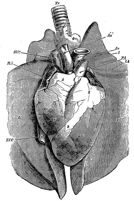 Fig. 5.—Heart of Sheep, as seen after Removal from the Body,
lying upon the Two Lungs. The Pericardium has been cut away, but no
other Dissection made.
R.A. Auricular appendage of right auricle; L.A. auricular appendage
of left auricle; R.V. right ventricle; L.V. left ventricle; S.V.C.
superior vena cava; I.V.C. inferior vena cava; P.A. pulmonary
artery; Ao, aorta; Áó, innominate branch from aorta dividing into
subclavian and carotid arteries; L. lung; Tr. trachea. 1, solid cord
often present, the remnant of a once open communication between the
pulmonary artery and aorta. 2, masses of fat at the bases of the
ventricle hiding from view the greater part of the auricles. 3, line of
fat marking the division between the two ventricles. 4, mass of fat
covering the trachea.
is represented in Fig. 5. If you could look through the front of your
own chest, and see your own heart and lungs in place, you would see
something not so very very different.
If now you handle the heart—and if you want to learn physiology you
must handle things—you will have no great difficulty in finding the
great yellowish tubes marked Ao and Áó in the figure. Your butcher
perhaps may not have cut them across exactly where mine has done, but
that will not prevent your recognizing them. You will notice what thick
stout walls they have, and how they gape where they are cut. Ao is the
aorta, and Áó is a great branch of the aorta, going to the head and
neck of one side, perhaps the branch along which we imagined just now
that you, a poor little red blood-corpuscle, were travelling. If you
were to put a wire through Áó you would be able to bring it out
through Ao, or vice versâ. But what is P.A. which looks so much
like the aorta, though you will find that it has no connection with it?
You cannot pass a wire from the aorta into it. It also is an artery, the
pulmonary[2] artery. We shall have more to say about it directly.
Now try and find what are marked in the figure as S.V.C. and I.V.C.
You will perhaps have a little difficulty in this; and when you have
found them you will understand why. They are the great veins of the
body. S.V.C. is the superior vena cava, to form which all the veins
from the head and neck and arms join, the vein in which you were
journeying a little while ago. I.V.C. is the inferior vena cava, made
out of all the veins from the trunk and the legs. Being veins, they have
thin flabby walls; and their sides fall flat together, so that they seem
nothing more than little folds of skin, and it becomes very hard to find
the passage inside them. But when you have found the opening into them,
you will see that you can stretch them out into quite wide tubes, and
that their walls, though very much thinner than those of the aorta, so
thin indeed that they are almost transparent, are still after a fashion
strong. If you put a penholder or thin rod through either you will find
that they both seem to lead right into the middle of the heart. With a
little care you can pass a rod up I.V.C. and bring the end of it out
at the top of S.V.C. Of course you will understand that both of these
veins have been cut off short.
28. Before we go on any further with the sheep’s heart, let me tell you
something about it, by help of the diagram in Fig. 6, which is meant to
represent the whole circulation. You must remember that this figure is a
diagram, and not a picture; it does not represent the way the
blood-vessels are really arranged in your own body. If you had no arms
and no legs, and if you only had a few capillaries at the top of your
head and at the bottom of your body, it might be more like than it is.
In the centre of the figure is the heart. This you will see is
completely divided by an upright partition into two halves, a right half
and a left half. Each half is further marked off, but not completely
divided, into
 Fig. 6.—Diagram of the Heart and Vessels, with the Course of the
Circulation, viewed from behind so that the proper left of the
Observer corresponds with the left side of the Heart in the
Diagram.
L.A. left auricle; L.V. left ventricle; Ao. aorta; A1.
arteries to the upper part of the body; A2. arteries to the lower
part of the body; H.A. hepatic artery, which supplies the liver with
part of its blood; V2. veins of the upper part of the body;
V2. veins of the lower part of the body; V.P. vena portæ; H.V.
hepatic vein; V.C.I. inferior vena cava; V.C.S. superior vena cava;
R.A. right auricle; R.V. right ventricle; P.A. pulmonary artery;
Lg. lung; P.V. pulmonary vein; Lct. lacteals; Ly. lymphatics;
Th.D. thoracic duct; Al. alimentary canal; Lr. liver. The arrows
indicate the course of the blood, lymph, and chyle. The vessels which
contain arterial blood have dark contours, while those which carry
venous blood have light contours.
two chambers, an upper chamber and a lower chamber; so that altogether
we have four chambers,—two upper chambers, one on each side, marked
R.A. and L.A., these are called the right and left auricles; and two
lower chambers, one on each side, marked R.V. and L.V., these are
called the right and left ventricles. The right auricle, R.A., opens
in the direction of the arrow into the right ventricle, R.V., the
opening being guarded, as we shall see, by a valve. The left auricle,
L.A., opens into the left ventricle, L.V., the opening being
likewise guarded by a valve; but you have to go quite a roundabout way
to get from either the right auricle or ventricle to the left auricle or
ventricle. Let us see how we can get round the figure. Suppose we begin
with the two tubes marked V.C.S. and V.C.I., the walls of which are
drawn with thin lines. These both open into the right auricle. They are
the vena cava superior and inferior, which you have just made out in the
sheep’s heart. From the right auricle you pass easily into the right
ventricle; thence, following the arrow, the way is straight into the
tube marked P.A. This is the pulmonary artery, the outside of which
you saw in the sheep’s heart (Fig. 5, P.A.) Travelling along this
pulmonary artery, you come to the lungs, and after passing through
branches not represented in the figure, picking your way through
arteries which continually get smaller and smaller, you find yourself
at last in the capillaries of the lungs. Squeezing your way through
these, you come out into veins, and gradually advancing through larger
and larger veins, you, still following the arrow, find yourself in one
of four large veins (only one of them is represented in the diagram)
which land you in the left auricle. From the left auricle it is but a
jump into the left ventricle. From the left ventricle the way is open,
as indicated by the arrow, into the tube marked Ao. This is intended
to represent the aorta, which you have already seen in the sheep’s heart
(Fig. 5, Ao). It is here drawn for simplicity’s sake as dividing into
two branches, but you have already been told, and must bear in mind,
that it does not in reality divide in this way, but gives off a good
many branches of various sizes. However, taking the figure as it stands,
suppose we travel along A2. Following the arrow, and shooting
through arteries which continually get smaller and smaller, we come at
last to capillaries somewhere, in the skin or in some muscle, or in a
bone, or in the brain, or almost anywhere, in fact, in the upper part of
the body. Out of the capillaries we pass into veins, which, joining
together and so forming larger and larger trunks, bring us at last to
the point from which we started, the superior vena cava, V.C.S. If we
had taken the other road, A2, we should have passed through
capillaries somewhere in the lower part of the body instead of the
upper, and come back by the vena cava inferior, V.C.I., instead of the
vena cava superior. Starting from the right auricle, whichever way we
took we should always come back to the right auricle again, and in our
journey should always pass through the following things in the following
order: right auricle, right ventricle, pulmonary artery, arteries,
capillaries, and veins of the lungs, pulmonary vein, left auricle, left
ventricle, aorta, arteries, capillaries, and veins somewhere in the
body, and either superior or inferior vena cava. That is the course of
the circulation. But there is something still to be added. Among the
many large branches, not drawn in the diagram, given off by the aorta to
the lower part of the body, there are two branches which are drawn and
which deserve special notice.
One is a large branch carrying blood to the tube A.L., which is meant
in the diagram to stand for the stomach, intestines, and some other
organs. This branch, like all other branches of the aorta, divides into
small arteries, and these into capillaries, which again are gathered up
into veins, forming at last a large vein marked in the diagram V.P.
and called the vena portæ or portal vein. Now the remarkable thing is
that this vein does not, like all the other veins, go straight to join
the vena cava, but makes for the liver, where it divides into smaller
and smaller veins, until at last it breaks completely up in the liver
into a set of capillaries again. These capillaries gather once more into
veins, forming at last the large trunk, called the hepatic[3] vein,
H.V., which does what the portal vein ought to have done but did not;
it opens straight into the vena cava.
The other branch of the aorta of which we are speaking goes straight to
the liver, and is called the hepatic artery, H.A.: there it breaks up
in the liver into small arteries, and then into capillaries, which
mingle with the capillaries of the portal vein, and form one system, out
of which the hepatic veins spring. So you see it makes a great
difference to a red corpuscle which is travelling along the lower part
of the aorta A2, whether it takes a turn into the branch going to
the alimentary canal, or whether it goes straight on into, for instance,
a branch going to some part of the leg. In the latter case, having got
through a set of capillaries, it is soon back into the vena cava and on
its road to the heart. But if it takes the turn to the alimentary canal,
it finds after it has passed through the capillaries and got into the
portal vein, that it has still to go through another set of capillaries
in the liver before it can pass through the hepatic vein into the vena
cava.
This then is the course of the circulation. Right side of the heart,
pulmonary artery, capillaries of the lungs, pulmonary vein, left side of
the heart, aorta, capillaries somewhere, sometimes two sets, sometimes
one, vena cava, right side of the heart again. A little corpuscle cannot
get from the right to the left side of the heart without going through
the capillaries of the lungs. It cannot get from the left side of the
heart to the right without going through some capillaries somewhere in
the body, and if it should happen to take the turn to the stomach, it
has to go through two sets of capillaries instead of one.
You see, you really have two circulations, and you have two hearts
joined together into one. If you were very skilful you might split the
heart in half and pull the two sides asunder, and then you would have
one heart receiving all the veins from the body and sending its arteries
(branches of the pulmonary artery) all to the lungs, and another heart
receiving all the veins from the lungs and sending its arteries
(branches of the aorta) all over the body. And you would have two
circulations, one through the lungs, and another through the rest of the
body, both joining each other. Very often two circulations are spoken
of, and because the lungs are so much smaller than the rest of the body,
the circulation through the lungs is called the lesser circulation, that
through the rest of the body the greater circulation.
29. I have described the circulation as if the blood always went in one
direction from the right side of the heart to the left, from arteries to
veins, the way the arrows point in the diagram. And so it does. It
cannot go the other way round. Why does it go that way? Why cannot it go
the other way round?
The reasons are to be found partly in the heart, partly in the veins.
In the veins the blood will only pass from the capillaries to the heart.
Why not from the heart to the capillaries? You remember the little
watch-pocket-like valves, here and there, sometimes singly, sometimes
two or three abreast. You remember that the mouths of the watch-pockets
were always turned towards the heart. Now suppose a crowd of little
corpuscles hurrying along a vein towards the heart. When they came to
one of these watch-pocket valves they would simply trample it down flat,
and so pass over it without hardly knowing it was there, and go on
their way as if nothing had happened. But suppose they were journeying
the other way, from the heart to the capillaries. When they came to the
open mouth of a watch-pocket valve, some of them would be sure to run
into the pocket, and then the pocket would bulge out, and the more it
bulged out the more blood would run into it, until at last it would be
so full of blood that it would press close against the top of the vein,
as is shown in Fig. 7 (or, if there were two or three, they would all
meet together), and so quite block the vein up. If you doubt this, make
a watch-pocket out of a piece of silk or cotton, fasten it on to a piece
of brown paper, and roll the paper up into a tube, so that the valve is
nicely inside the tube. If you pour some peas down the tube with the
mouth of the valve looking away from you, they will run through at once;
but if you try to pour them the other way, your tube will soon be
choked, and if you carefully unroll the tube you will find the
watch-pocket crammed full of peas.
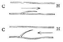 Fig. 7.—Diagrammatic Sections of Veins with Valves.
In the upper, the blood is supposed to be flowing in the direction
of the arrow, towards the heart; in the lower, the reverse way. C,
capillary side; H, heart side.
The valves in the veins, then, let the blood pass easily from the
capillaries to the heart, but won’t let it go the other way. If you bare
your arm you may see some of the veins in the skin, in which the blood
is running up from the hand towards the shoulder. If with your finger
you press one of these veins back towards the hand it will swell up, and
if you look carefully you may see little knots here and there caused by
the bulging out of the watch-pocket valves. If you press it the other
way, towards the elbow, you will empty it easily, and if with another
finger you prevent the blood getting into it from behind, that is from
the hand, the vein will remain empty a very long time.
The presence of valves in the veins, then, is one reason why the blood
moves in one direction, but other reasons, and these the chief ones, are
to be found in the heart.
Let us now go back to the sheep’s heart.
30. You know from the diagram that the two great veins, the superior and
inferior vena cava, open into the right auricle. If you slit up these
two veins in the sheep’s heart, you will find that they end by separate
openings in a small cavity, the inside of which is for the most part
smooth, and the walls of which, made, as you will at once see, of
muscle, are not very thick. This small cavity is the right auricle,
shown in Fig. 8, R.A., where the great veins have not been slit up,
but the front of the auricle has been cut away. In this auricle, beside
the openings into the two great veins and another one which belongs to a
vein coming from the heart itself (Fig. 8, b) there is quite a large
one, leading straight downwards, into which you
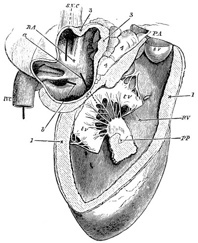 Fig. 8.—Right Side of the Heart of a Sheep.
R.A. cavity of right auricle; S.V.C. superior vena cava;
I.V.C. inferior vena cava; (a piece of whalebone has been passed
through each of these;) a, a piece of whalebone passed from the
auricle to the ventricle through the auriculo-ventricular orifice;
b, a piece of whalebone passed into the coronary vein.
R.V. cavity of right ventricle; tv, tv, two flaps of the
tricuspid valve: the third is dimly seen behind them, the a,
piece of whalebone, passing between the three. Between the two
flaps, and attached to them by chordæ tendineæ, is seen a
papillary muscle, PP, cut away from its attachment to that
portion of the wall of the ventricle which has been removed. Above,
the ventricle terminates somewhat like a funnel in the pulmonary
artery, P.A. One of the pockets of the semilunar valve, sv, is
seen in its entirety, another partially.
1, the wall of the ventricle cut across; 2, the position of the
auriculo-ventricular ring; 3, the wall of the auricle; 4, masses of
fat lodged between the auricle and pulmonary artery.
can put your three fingers. This is the opening into the right
ventricle; and you will have no difficulty in putting your fingers from
the auricle into the ventricle and bringing them out again.
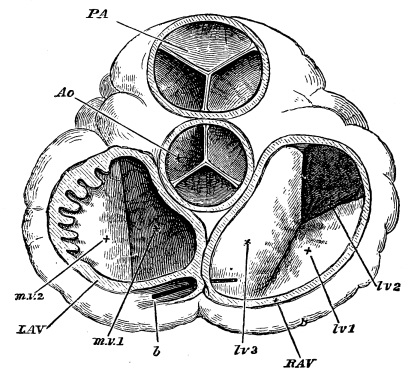 Fig. 9.—The Orifices of the Heart seen from above, the
Auricles and Great Vessels being cut away.
P.A. pulmonary artery, with its semilunar valves; Ao. aorta,
do.
R.A.V. right auriculo-ventricular orifice with the three flaps
(lv. 1, 2, 3) of tricuspid valve.
L.A.V. left auriculo-ventricular orifice, with m.v. 1 and 2,
flaps of mitral valve; b, piece of whalebone passed into coronary
vein. On the left part of L.A.V. the section of the auricle is
carried through the auricular appendage; hence the toothed
appearance due to the portions in relief cut across.
But hold the heart in one hand with the auricle upwards, and try to pour
some water into the ventricle. The first few spoonfuls will go in all
right, and then you will see some thin white skin or membrane come
floating up into the opening and quite block up the entrance from the
auricle into the ventricle; the
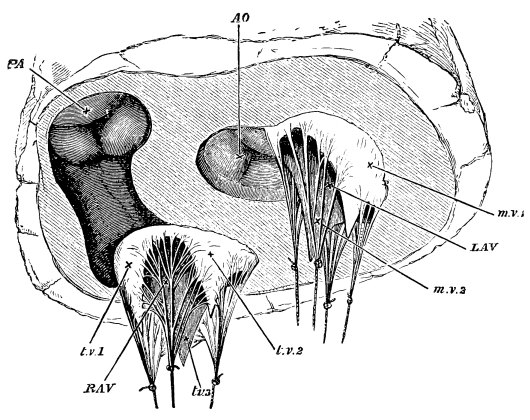 Fig. 10.—View of the Orifices of the Heart from below,
the whole of the Ventricles having been cut away.
R.A.V. right auriculo-ventricular orifice surrounded by the three
flaps, t.v. 1, t.v. 2, t.v. 3, of the tricuspid valve; these
are stretched by weights attached to the chordæ tendineæ.
L.A.V. left auriculo-ventricular orifice surrounded in same way
by the two flaps, m.v. 1, m.v. 2, of mitral valve; P.A. the
orifice of pulmonary artery, the semilunar valves having met and
closed together; Ao. the orifice of the aorta with its semilunar
valves. The shaded portion, leading from R.A.V. to P.A.,
represents the funnel seen in Fig. 8.
water will immediately fill the auricle and run over. If you look at the
membrane carefully as it comes bulging up, you will notice that it is
made up of three pieces joined together as is shown in Fig. 9 (lv. 1,
lv. 2, lv. 3). These three pieces form the valve between the right
auricle and ventricle, called the tricuspid, or three-peaked valve. Why
it is so called you will understand if you lay open the right ventricle
by cutting with a pair of scissors from the auricle into the ventricle
along the side of the heart, or by cutting away the front of the
ventricle as has been done in Fig. 8. You will then see that the valve
is made up of three little triangular flaps, which grow together round
the opening with their points hanging down into the cavity of the
ventricle (Fig. 10, t.v.) They do not, however, hang quite loosely.
You will notice fastened to the sides of the flaps, thin delicate
threads, the other ends of which are fastened to the sides of the
ventricle, and often to little fleshy projections called papillary
muscles (Fig. 8, P.P.)
How do these valves act? In this way. When the ventricle is empty, and
blood or water or any other fluid is poured into it from the auricle,
the valves are pushed on one side against the walls of the ventricle,
and thus there is a great wide opening from the auricle into the
ventricle. But as the ventricle fills, the blood or water gets behind
the flaps and floats them up towards the auricle. The more fluid in the
ventricle the higher they float, until when the ventricle is quite full
they all meet together in the middle of the opening between the auricle
and ventricle and completely block it up. But why do they not turn right
over into the auricle, and so open up again the wrong way? Because of
those little threads (the chordæ tendineæ, as they are called) which
fasten them to the walls of the ventricle. The flaps float back until
these threads are stretched quite tight, and the threads are just long
enough to let the flaps reach to the middle of the opening, but no
further. The tighter the threads are stretched the closer the flaps fit
together, and the more completely do they block the way from the
ventricle back into the auricle.
The tricuspid valve, then, lets blood flow easily from the right auricle
into the right ventricle, but prevents it flowing from the ventricle
into the auricle.
31. Now look at the cavity of the ventricle. Its walls are fleshy, that
is muscular, and you will notice that they are much stouter and thicker
than those of the auricle. Besides the opening from the auricle there is
but one other, which is at the top of the ventricle, side by side with
the former. If you put a penholder or your finger through this second
opening, you will find that it leads into the large vessel which you
have already learnt to recognize as the pulmonary artery (Fig. 5,
P.A.)
Slit up the pulmonary artery from the ventricle with a pair of scissors,
as has been done in Fig. 8, P.A. You will notice at once the line
where the red soft flesh of the muscular ventricle leaves off, and the
yellow firmer material of which the artery is made begins. Just at that
line you will see a row of three (perhaps you may have cut one of the
three with your scissors) most beautiful, watch-pocket valves, made on
just the same principle as those in the veins, only larger, and more
exquisitely finished. These are called semilunar valves, because each
pocket is of the shape of a half-moon. Lift them up carefully and see
how tender and yet how strong they are. There is no need to tell you the
use of these. You know it at once. They are to let the blood flow from
the ventricle into the pulmonary artery, and to prevent the blood going
back from the artery into the ventricle.
On the right side of the heart we have, then, two great valves, the
tricuspid valve between the auricle and the ventricle, and the semilunar
valve between the ventricle and the pulmonary artery. These let the
blood flow easily one way, but not the other. If you doubt this, try it.
Put a tube into either the superior or inferior vena cava of a fresh
heart, tying the other vena cava and another tube into the pulmonary
artery. If with a funnel you pour water into the tube in the vein, it
will run through auricle and ventricle and out through the tube of the
pulmonary artery as easily as possible; but if you try to pour water the
other way down the pulmonary artery, you will find you cannot do it; the
tube gets blocked directly, and only a few drops come back through the
heart into the vein.
Now slit up the pulmonary artery as far as you can, and note when you
cut it how stout and firm are its walls. You will find that it soon
divides into two branches, one for the right lung, one for the left.
Each of these, when it gets to the lung, divides into branches, and
these again into others, as far as you can follow them. You know from
what you have learnt already that these branches end in capillaries all
over the lungs.
32. Not far from the two main branches of the pulmonary artery you will
find, covered up perhaps with fat and other matters, some tubes which
you will at once recognize as veins, and if you open any one of these
you will find that you can put a thin rod into it, and that it leads in
one direction to the lungs, and in the other into the left side of the
heart. These are the pulmonary veins, and if you slit them right up
you will find they open (by four openings) into a cavity on the left
side of the heart, almost exactly like that cavity on the right side
which we called the right auricle (Fig. 11). This cavity is, in fact,
the left auricle; out of it there is an opening into the left ventricle,
very like the opening from the right auricle into the right ventricle.
It too is guarded by flap valves, exactly like the tricuspid valve, only
there are but two flaps instead of three (Fig. 9, m.v. 1, m.v. 2).
Hence this valve is called the bicuspid, or more frequently the mitral
valve. Its flaps have little threads by which they are fastened to the
walls of the ventricle, and in fact, except for there being two flaps
instead of three, the mitral valve is exactly like the tricuspid valve,
and acts exactly the same way.
If you cut with a pair of scissors from the auricle into the ventricle,
you will find the left ventricle (Fig. 11) very much like the right
ventricle, only its walls are very much thicker, so much thicker that
the left ventricle takes up the greater part of the heart. You will see
this if you now look at the outside of a fresh heart.
The auricles are so small and so covered up by fat that from the outside
you can hardly see them at all. What you chiefly see are two little
fleshy corners, one of each auricle (Fig. 5, R.A. L.A.), often
called “the auricular appendages.” By far the greater part is taken up
by the ventricles—and if you look you will see a band of fat slanting
across the heart (Fig. 5, 3). This marks the line of the fleshy
division, or septum as it is called, between the two ventricles. You
will notice that the point or apex of the heart belongs altogether to
the left ventricle.
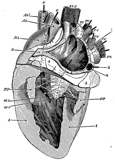 Fig. 11.—Left Side of the Heart of a Sheep (laid
open).
P.V. pulmonary veins opening into the left auricle by four
openings, as shown by the styles or pieces of whalebone placed in
them: a, a style passed from auricle into ventricle through the
auriculo-ventricular orifice; b, a style passed into the coronary
vein, which, though it has no connection with the left auricle, is,
from its position, necessarily cut across in thus laying open the
auricle.
M.V. the two flaps of the mitral valve (drawn somewhat
diagrammatically): pp, papillary muscles, belonging as before to
the part of the ventricle cut away; c, a style passed from
ventricle in Ao. aorta; Ao2. branch of aorta (see Fig. 5,
Áó); P.A. pulmonary artery; S.V.C. superior vena cava.
1, wall of ventricle cut across; 2, wall of auricle cut away around
auriculo-ventricular orifice; 3, other portions of auricular wall
cut across; 4, mass of fat around base of ventricle (see Fig. 5,
2).
To return to the inside of the left ventricle. Up at the top of the
ventricle, close to the opening from the auricle, there is one other
opening, and only one. If you put your finger into this, you will find
that it leads into a tube which first of all dips under or behind the
pulmonary artery and then comes up and to the front again. This tube is
what you already know as the aorta. If you slit it up from the ventricle
(and to do this you must cut through the pulmonary artery), you will
find that on the left side, as on the right, the red fleshy wall of the
ventricle suddenly changes into the yellow firm wall of the artery, and
that just at this line there are three semilunar valves exactly like
those in the pulmonary artery.
On the left side of the heart, then, we have also two valves, the mitral
between the auricle and the ventricle, and the semilunar between the
ventricle and the aorta. These let the blood pass one way and not the
other. You can easily drive fluid from the pulmonary veins through
auricle and ventricle into the aorta, but you cannot send it back the
other way from the aorta.
These then are the reasons why the blood will only pass one way, the way
I said it did. There are sets of valves opening one way and shutting the
other. These valves are the tricuspid between the right auricle and
right ventricle, the pulmonary semilunar valves between the right
ventricle and the pulmonary artery, the mitral valve between the left
auricle and the left ventricle, the aortic semilunar valves between the
left ventricle and the aorta, and the valves which are scattered among
the veins of the body. Of these by far the most important are the
valves in the heart: they do the chief work; those in the veins do
little more than help.
33. Well, then, we understand now, do we not? why the blood, if it moves
at all, moves in the one way only. There still remains the question, Why
does the blood move at all?
You know that during life it does keep moving. You have seen it moving
in the web of a frog’s foot—and whenever any part of the body can be
brought under the microscope, the same rush of red corpuscles through
narrow channels may be seen. You know it moves because when you cut a
blood-vessel the blood runs out. If you cut an artery across, the blood
gushes out from the end which is nearest the heart; if you cut a vein
across, the blood comes most from the end nearest the capillaries. If
you want to stop an artery bleeding, you tie it between the cut and the
heart; if you want to stop a vein bleeding, you tie it between the cut
and the capillaries. You understand now why there is this difference
between a cut artery and a cut vein. And you see that this is by itself
a proof that the blood moves in the arteries from the heart to the
capillaries, and in the veins from the capillaries to the heart.
The blood is not only always moving, but moves very fast. It flies along
the great arteries at perhaps ten inches in a second. Through the little
bit of capillaries along which it has to pass it creeps slowly, but
manages sometimes to go all the way round from vein to vein again in
about half a minute.
It is always moving at this rapid rate, and when it ceases to move, you
die.
What makes it move?
Suppose you had a long thin muscle, fastened at one end to something
firm, and with a weight hanging at the other end. You know that every
time the muscle contracted it would pull on the weight and draw it up.
But suppose, instead of hanging a weight on to the muscle, you wrapped
the muscle round a bladder full of water. What would happen then each
time the muscle contracted? Why, evidently it would squeeze the bladder,
and if there were a hole in the bladder some of the water would be
squeezed out. That is just what takes place in the heart. You have
already learnt that the heart is muscular. Each cavity of the heart,
each auricle, and each ventricle is, so to speak, a thin bag with a
number of muscles wrapped round it. In an ordinary muscle of the body,
the bundles of fibres of which the muscle is made up are placed
carefully and regularly side by side. You can see this very well in a
round of boiled beef, which is little more than a mass of great muscles
running in different directions. You know that if you try to cut a thin
slice right across the round, at one part your carving-knife will go
“with the grain” of the meat, i.e. you will cut the fibres lengthways;
at another part it will go “against the grain,” i.e. you will cut the
fibres crossways. In both parts, the bundles of fibres will run very
regularly. But in the heart the bundles are interlaced with each other
in a very wonderful fashion, so that it is very difficult to make out
the grain. They are so arranged in order that the muscular fibres may
squeeze all parts of each bag at the same time.
Each cavity of the heart, then, auricle or ventricle, is a thin bag
with a network of muscles wrapped round it, and each time the muscles
contract they squeeze the bag and try to drive out whatever is in it.
There are more muscles in the ventricles than in the auricles, and more
in the left ventricle than in the right, for we have already seen how
much thicker the ventricles are than the auricles, and the left
ventricle than the right; and the thickness is all muscle.
And now comes the wonderful fact. These muscles of the auricles and
ventricles are always at work contracting and relaxing, shortening and
lengthening, of their own accord, as long as the heart is alive. The
biceps in your arm contracts only when you make it contract. If you keep
quiet, your arm keeps quiet and your biceps keeps quiet. But your heart
never keeps quiet. Whether you are awake or whether you are asleep,
whether you are running about or lying down quite still, whatever you
are doing or not doing, as long as you are alive your heart keeps on
steadily at work. Every second, or rather oftener, there comes a short
sharp squeeze from the auricles, from both exactly at the same time, and
just as the auricles have finished their squeeze, there comes a great
hug from the ventricles, from both at the same time, but a much stronger
hug from the left than from the right; and then for a brief space there
is perfect quiet. But before the second has quite passed away, the
auricles have begun again, and after them the ventricles once more, and
thus the contracting and relaxing of the walls of the heart’s cavities,
this beat of the heart as it is called, this short snap of the two
auricles, this longer, steadier pull of the two ventricles, have gone on
in your own body since before you were born, and will go on until the
moment comes when friends gathering round your bedside will say that you
are “gone.”
34. But how does this beat of the heart make the blood move? Let us see.
Remember that you have, or when you are grown up will have, bottled up
in the closed blood-vessels of your body about 12 lbs. of blood. You
have seen that the heart and the blood-vessels form a system of closed
tubes; the walls are in some places, in the capillaries for instance,
very thin, but they are sound and whole—and though the road is quite
open from the capillaries through the veins, heart, and arteries to the
capillaries again, there is no way out of the tubes except by making a
breach somewhere in the walls.
This closed system of heart and tubes is pretty well filled by the 12
lbs. of blood.
What then must happen each time the heart contracts?
Let us begin with the right ventricle. Suppose it is full of blood. It
contracts. The blood in it, squeezed on all sides, tries to go back into
the right auricle, but the tricuspid flaps have been driven back and
block the way. The more the blood presses on them, the tighter they
become, and the more completely they shut out all possibility of getting
into the auricle.
The way into the pulmonary artery is open, the blood can go there. But
stay, the artery is already full of blood, and so are the capillaries
and veins in the lung. Yes, but the artery will stretch ever so much.
Take a piece of pulmonary artery, and having tied one end, pump or pour
water into the other; you will see how much it will stretch. Into the
pulmonary artery, then, goes the blood, stretching it in order to find
room. As the ventricle squeezes and squeezes, until its walls meet in
the middle, all the blood that was in it finds its way out into the
artery. But the beat of the ventricle soon ceases, the squeeze is over
and gone, and back tumbles the blood into the ventricle, or would
tumble, only the first few drops that shoot backwards are caught by the
watch-pocket semilunar valves. Back fly these valves with a sharp click
(for the things of which we are speaking happen in a fraction of a
second), and all further return is cut off. The blood has been squeezed
out of the ventricle, and is safely lodged in the pulmonary artery.
But the pulmonary artery is ever so much on the stretch. It was fairly
full before it received this fresh lot of blood; now it is over-full—at
least that part of it which is nearest to the heart is over-full. What
happens next? What happens when you stretch a piece of india-rubber and
then let it go? It returns to its former size. The ventricle has
stretched the piece of pulmonary artery near it, beyond the natural
size, and then (when it ceased to contract) has let it go. Accordingly
the piece of pulmonary artery tries to return to its former size, and
since it cannot send the blood back to the ventricle, squeezes it on to
the next piece of the artery nearer the capillaries, stretching that in
turn.
This again in turn sends it on the next piece—and so on right to the
capillaries. The over-full pulmonary artery, stretched to hold more than
it fairly can, empties itself through the capillaries into the pulmonary
veins until it is not more than comfortably full. But the pulmonary
veins also are already full,—what are they to do? To empty the surplus
into the left auricle. Oftener than every second there will come a time
when they can do so.
For at the same time that the right ventricle pumped a quantity of blood
into the pulmonary artery and safely lodged it there, the left ventricle
pumped a like quantity into the aorta, safely lodged it there, and was
left empty itself. But just at that moment the left auricle began to
contract and to squeeze the blood that was in it.
Where could that blood go? It could not go back into the pulmonary
veins, for they were already full, and the blood in them was being
pressed behind by the over-full pulmonary arteries. But it could pass
easily into the empty ventricle—and in it tumbled, the mitral flaps
readily flying back and opening up a wide way. And so the auricle
emptied itself into the ventricle. But now the auricle ceases to
contract—its walls no longer squeeze—it is empty and wants filling,
and so comes the moment when the pulmonary veins can pour into it the
blood which has been driven into them by the over-full pulmonary artery.
Thus the right ventricle drives the blood into the over-full pulmonary
artery, the pulmonary artery overflows into the pulmonary veins, the
pulmonary veins carry the surplus to the empty left auricle, the left
auricle presses it into the empty left ventricle, the left ventricle
pumps it into the aorta—(the stretching of the aorta and of its
branches is what we call the pulse)—the over-full aorta overflows just
as did the pulmonary artery, through the capillaries of the body into
the great venæ cavæ—through these the blood falls into the empty right
auricle, the right auricle drives it into the empty right ventricle,
and the full right ventricle is the point at which we began.
Thus the alternate contractions of auricles and ventricles, thanks to
the valves in the heart and in the veins, pump the blood, stroke by
stroke, through the wide system of tubes; and thus in every capillary
all over the body we find blood pressed upon behind by over-full
arteries, with a way open to it in front, thanks to the auricles, which
are, once a second or oftener, empty and ready to take up a fresh supply
from the veins. Thus it comes to pass that every little fragment of your
body is bathed by blood, which a few moments ago was in your heart, and
a few moments before that was in some other part of your frame. Thus it
is that no part of your body can keep itself to itself; the blood makes
all things common as it flies from spot to spot. The red corpuscle that
a minute ago was in your brain, is now perhaps in your liver, and in
another minute may be in a muscle of your arm or in a bone of your leg:
wherever it goes it has something to bring, and something to fetch. A
restless heart is for ever driving a busy blood, which wherever it goes
buys and sells, making perhaps an occasional bargain as it shoots along
the great arteries and great veins, but busiest of all as it lingers in
the narrow pathways of the capillaries.
35. When you look down upon a great city from a high place, as upon
London from St. Paul’s, you see stretching below you a network of
streets, the meshes of which are filled with blocks of houses. You can
watch the crowds of men and carts jostling through the streets, but the
work within the houses is hidden from your view. Yet you know that, busy
as seems the street, the turmoil and press which you see there are but
tokens of the real business which is being carried on in the house.
So is it with any piece of the body upon which you look through the
microscope. You can watch the red blood jostling through the network of
capillary streets. But each mesh bounded by red lines is filled with
living flesh, is a block of tiny houses, built of muscle, or of skin, or
of brain, as the case may be. You cannot see much going on there,
however strong your microscope; yet that is where the chief work goes
on. In the city the raw material is carried through the street to the
factory, and the manufactured article may be brought out again into the
street, but the din of the labour is within the factory gates. In the
body the blood within the capillary is a stream of raw material about to
be made muscle, or bone, or brain, and of stuff which, having been
muscle, or bone, or brain, is no longer of any use, and is on its way to
be cast out. The actual making of muscle, or of bone, or of brain, is
carried on, and the work of each is done, outside the blood, in the
little plots of tissue into which no red corpuscle comes.
The capillaries are closed tubes; they keep the red corpuscles in their
place. But their walls are so thin and delicate that they let the watery
plasma of the blood, the colourless fluid in which the corpuscles float,
soak through them into the parts inside the mesh. You probably know that
many things will pass through thin skins and membranes in which no holes
can be found even after the most careful search. If you put peas into a
bladder and tie the neck, the peas will not get out until the bladder is
untied or torn. But if you were to put a solution of sugar or of salt
into the bladder, and place the bladder with its neck tied ever so
tightly in a basin of pure water, you would find that very soon the
water in the basin would begin to taste of sugar or salt—and that
without your being able to discover any hole, however small, in the
bladder. By putting various substances in the bladder, you will find
that solid particles and things which will not dissolve in water keep
inside the bladder, whereas sugar and salt, and many other things which
dissolve in water, will make their way through the bladder into the
water outside, and will keep on passing until the water in the basin is
as strong of sugar or salt as the water in the bladder. This property
which membranes such as a bladder have of letting certain substances
pass through them is called osmosis. You will at once see how important
a part it plays in your own body. It is by osmosis chiefly that the raw
nourishing material in the blood gets into the little islets of flesh
lying, as we have seen, in the meshwork of the capillaries. It is by
osmosis chiefly that the worn-out stuff from the same islets gets back
into the blood. It is by osmosis chiefly that food gets out of the
stomach into the blood. It is by osmosis chiefly that the waste,
worn-out matters are drained away from the blood, and so cast out of the
body altogether. By osmosis the blood nourishes and purifies the flesh.
By osmosis the blood is itself nourished and kept pure.
There are two chief things by which the blood, and through the blood
the body, is nourished. These are food and air. The air we have always
with us, we have no need to buy it or toil for it; hence we take it as
we want it, a little at a time, and often. We gather up no store of it;
and cannot bear the lack of it for more than a few moments.
For our food we have to labour; we store it up in our bodies from time
to time, at intervals of hours, in what we call meals, and can go hours
or even days without a fresh supply.
Let us first of all see how the blood, and, through the blood, the body,
is nourished by air.