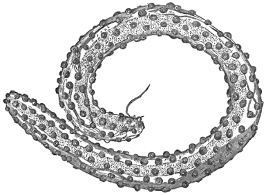
Whatever notions people may entertain respecting the
dignity of the human race, there is no gainsaying the fact that
we share with the lower animals the rather humiliating privilege
and prerogative of entertaining a great variety of parasites.
These are for the most part entozoal in habit. As the parasites
are apt to cause suffering to the bearer, a superstitious age
sought to interpret their presence as having some connection
with human wrong-doing. We can now afford to smile at such
erroneous ideas. The intimate relation subsisting between
parasitic forms dwelling in man and animals, and their interdependence
upon one another, alone suffices to preclude the
idea that parasites have been arbitrarily placed within the
human bearer. It would seem, indeed, that our existence
is essential to the welfare and propagation of certain species
of parasites. Possibly it is only by accepting the hypothesis
of “Natural Selection” that we can escape the somewhat
undignified conclusion that the entozoa were expressly created
to dwell in us, and also that we were in part designed and
destined to entertain them. View the matter as we may, the
internal parasites of man and animals strictly conform to a few
well-known types of structure, but these types branch out into
infinitely varied specific forms. The vulgar mind sees nothing
attractive in the morphology and organisation of a parasitic
worm, and common-place conceptions of the beautiful cannot
be expected to embrace within their narrow grasp the marvelous
harmony and order that pervade the structure and economy
of the individual members of this remarkable class of beings.
Fasciola hepatica, Linneus.—The first form I have to consider
is the common liver fluke. The part this entozoon plays
in the production of disease will be fully stated when treating
of the parasites of the sheep and other ruminants. About
twenty instances of its occurrence in the human body have been
recorded. It has been found beneath the skin in the sole of
the foot (Giesker), and also under the scalp (Harris), and
behind the ear (Fox). Its more frequent seat is in the liver
and gall-ducts (Pallas, Brera, Bidloo, Malpighi) and gall-bladder
(Partridge). The alleged cases by Bauhin, Wepfer, and Chabert
are spurious, as is probably also that given by Mehlis. Duval’s
case appears to be genuine, but the occurrence of the worm in
the portal vein was accidental. Dr Murchison has recorded a
case, occurring at St Thomas’s Hospital, where a solitary
specimen was found in the liver. Dr H. V. Carter also met
with the worm in a young Hindoo.
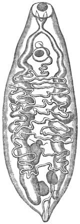
In the second half of the present work I shall reproduce
Blanchard’s admirable figure of the sexually mature worm
(Fig. 61), accompanied by a categorical statement respecting
the known facts of development. In this place, however, I
may observe that the cases recorded by Giesker, Harris, and
Fox had clearly pointed to the circumstance that the higher larvæ
of this fluke must be armed cercariæ, otherwise they could not
have bored their way through the human skin. As we shall see,
Dr Willemoes-Suhm’s investigations have furnished evidence
as to the truth of this supposition. For anatomical details I refer
to my introductory treatise. In the adult state the liver fluke
has been known from the earliest times. We have clear
evidences that it was described by Gabucinus in the year 1547,
and also subsequently by Cornelius Gemma, who, in a work
published some thirty years later, refers to an epizootic disease
prevalent in Holland during the year 1552, and which was very
justly attributed to the parasite in question. After this date
many writers described the liver fluke more or less accurately,
and entire volumes were devoted to the consideration of the
formidable disease which it occasions. The nomenclature of
the parasite has been a subject of controversy. Amongst
naturalists in general the common liver fluke is often described
under the combined generic and specific name of
Distoma hepaticum; but the title is both incorrect and inappropriate.
The proper generic appellation of this parasite is
Fasciola, as first proposed by the illustrious Linneus (1767) and
subsequently adopted by F. Müller (1787), Brera (1811), Ramdohr
(1814), and others. Unfortunately Retzius (1786) and
Zeder (1800) changed the generic title without good cause,
and the majority of writers, following their authority, refused
to employ the original name, although a consideration of the
distinctive types of structure severally displayed by the genera
Distoma and Fasciola fairly demanded the retention of the
Linnean title. In later times M. Blanchard (1847) strongly
advocated the original nomenclature, and I have myself continually
urged its adoption. On somewhat different grounds
Professor Moquin-Tandon followed the same course.
In the sexually mature state the liver fluke commonly
measures three fourths of an inch in length, occasionally reaching
an entire inch or even sixteen lines; its greatest breadth
also varying from half an inch to seven or eight lines transversely;
body very flat, presenting distinct dorsal and ventral
surfaces, frequently curled toward the latter during life; upper
or anterior end suddenly constricted, produced and pointed
in the centre, forming the so-called head and neck; posterior
extremity less acuminated, sometimes rounded, or even slightly
truncated; margins smooth, occasionally a little undulated,
especially towards the upper part; oral sucker terminal, oval,
rather smaller than the ventral acetabulum, which is placed
immediately below the root of the neck; reproductive orifices
in the middle line, a little below the oral sucker; intromittent
organ usually protruded and spirally curved; a central,
light-coloured space, covering two thirds of the body from
above downwards, marks the region of the internal male reproductive
organs, being bordered on either side and below by
a continuous dark band, indicating the position of the so-called
yolk-forming organs; a small, brown-coloured, rosette-like
body situated directly below the ventral acetabulum, marks
the limits of the uterine duct; a series of dark lines, branching
downwards and outwards on either side, indicate the position of
the digestive organs; general color of the body pale brownish
yellow, with a slight rose tint. The surface of the body, though
smooth to the naked eye, is clothed throughout with small
epidermal spines which diminish in size towards the tail.
If any argument were necessary to show how desirable it is
to furnish full descriptions of the commoner kinds of parasite,
I could adduce numerous instances that have been brought
under my notice where professional men and others have been
entirely mistaken as to the essential nature of their parasitic
finds. Thus, I have known an instance where a great authority
on the diseases of dogs has persisted in asserting for the free
proglottides of a tapeworm a nematode origin; and, in like
manner, human tapeworm-segments have frequently been
mistaken for independent fluke parasites. One of the most
remarkable instances of this kind is that which I have elsewhere
described as an error on the part of Dr Chabert. My
reasons for so regarding his interpretation of the facts observed
by him stand as follows:
In the ‘Boston Medical and Surgical Journal’ for the years
1852–53–54, Dr J. X. Chabert described several cases of Tænia,
and he averred that the tapeworms were associated with
numerous specimens of Distoma hepaticum. The passage of distomes
by patients during life was even regarded by Dr Chabert
as indicative of the presence of Tænia within the intestines.
Surely, I remarked, Dr Chabert was mistaken. Are not these
so-called distomes the well-known proglottides? Not willingly
doubting Dr Chabert’s statements, but desirous, if possible, of
verifying the accuracy of his conclusions, I wrote to him (March
22nd, 1864) requesting the loan of a specimen, but I was
not fortunate enough to receive a reply. In the “Case of
Tænia” in a boy four and a half years old, given in the 49th
vol. of the journal, Dr Chabert writes as follows:—“In consequence
of his passing the Distoma hepaticum, I concluded he
must be afflicted with Tænia.” Further on it is added, that
the administration of an astringent injection “caused the discharge
of innumerable small worms (Distoma hepaticum).” I
think this is quite decisive. The idea of “innumerable” flukes
being expelled in this way is altogether out of the question.
The only genuine case in which any considerable number of
Distomata, of this species, have been observed in the human
subject is the one recently recorded by Dr Prunac. In this
instance two flukes were vomited along with blood immediately
after the administration of salines (sel de Seignette), and about
thirty were passed per anum. On the following day, some
tapeworm proglottides having been evacuated, both salts and
male-fern extract were administered. This caused the expulsion
of an entire tapeworm, and also about twenty more flukes. Notwithstanding
this successful treatment the hæmatemesis returned
in about a month, when, finally, three more flukes were vomited
and the bleeding ceased. Had not the parasites been submitted
for identification to a competent observer (Prof.
Martins, of Montpellier), some doubt might have been entertained
as to the genuineness of this remarkable case. In reference
to Dr Prunac’s comments on the facts of fluke-parasitism
in man, I will only remark that Dr Kerr’s Chinese cases, to which
he refers, were probably due to Distoma crassum and not to D.
hepaticum. The Chinese flukes will be noticed below.
Bibliography (No. 3).—Full references to details of the cases
by Partridge, Fox, and Harris are given in Appendix B. to
Lankester’s Edit. of Küchenmeister’s Manual. See also the
works of Davaine and Leuckart (l. c. Bibl. No. 1).—Carter,
H. V., “Note on Distoma hepaticum” (from a patient under the
care of Mr Pandoorung), ‘Bombay Med. and Physical Soc.
Trans.’ (Appendix), 1862.—Chabert, J. X. (quoted above).
Murchison, C., ‘Clinical Lectures on Diseases of the Liver,’ (2nd
Edit., Appendix), London, 1877.—Prunac, De la Douve ou
Distome hépatique chez l’homme; in ‘Gazette des Hôpitaux’
for December, 1878 (p. 1147). For further references in this
work, see Bibliog. No. 49.
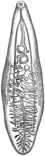
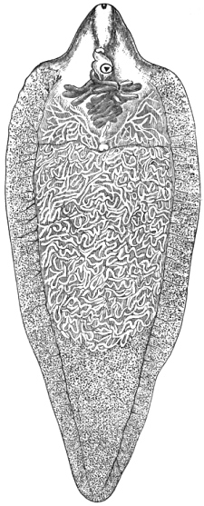
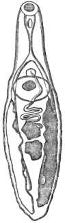
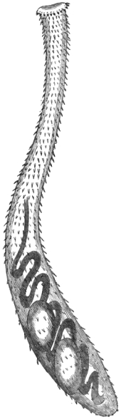
Fig. 1.—The lancet-shaped fluke
(Distoma lanceolatum), showing
the disposition of the digestive
and reproductive organs internally.
Viewed from behind; mag.
about 12 diameters. After Blanchard.
Distoma lanceolatum, Mehlis.—At least three instances of the
occurrence of this small fluke in the human body have been observed.
The authority for these cases rests, severally, with Bucholz, who
found them in the gall bladder in considerable numbers at Weimar; with
Chabert, who expelled a large number from the intestines of a girl in
France; and with Küchner, who obtained forty-seven specimens from a girl
in Bohemia. Probably many similar instances have been overlooked, and
Küchenmeister hints that Duval’s parasites (above mentioned) may have
been this species. Although this worm will again be incidentally noticed
in connection with bovine parasites (and its ciliated larvæ will also be
referred to when discussing the characters of the embryo of Bilharzia),
I here subjoin a diagnosis of the characters of the adult parasite. The
lancet-shaped liver fluke is a small flat helminth, measuring rather
more than the third of an inch in length, and about one line and a half
in breadth, being also especially characterised by its lanceolate form;
the widest part of the body corresponds with a transverse line drawn
across the spot where the vitellaria terminate below, and from this
point, on
either side, the width of the animal becomes gradually narrowed towards
the extremities; both ends are pointed, but the inferior or caudal one
more obtusely than the anterior or oral end; the general surface is
smooth throughout, and unarmed; the reproductive orifices are placed
in the central line immediately in front of the ventral sucker, and
below the point at which the intestine bifurcates; the oral sucker is
nearly terminal, and 1/50″
in breadth, the ventral acetabulum being about the same diameter; the
testes form two lobed organs placed one in front of the other in the
middle line of the body and directly below the ventral sucker; the
uterine canal is remarkably long, forming a series of tolerably regular
folds, which occupy the central and hinder parts of the body, reaching
almost to the caudal extremity. The vitelligene glands cover a limited
space, on either side of the centre of the body near the margin. The
foramen caudale communicates with a contractile vesicle, which passes
upwards in the form of a central trunk-vessel, early dividing into two
main branches; these latter reach as far forwards as the œsophageal
bulb, opposite which organ they suddenly curve upon themselves,
retracing their course for a considerable distance backwards; the
digestive canals are slightly widened towards their lower ends, which
occupy a line nearly corresponding with the commencement of the lower
fifth of the body; the ova are conspicuous within the uterine folds,
which present a dark brownish color in front, passing to a pale yellow
color below.
In reference to Kichner’s remarkable case I reproduce an abstract
of it from Leuckart’s account (‘Die menschlichen Parasiten,’ Bd. i, s.
608), the original particulars of which were communicated to Leuckart by
Dr Kichner himself:—
“Dr Kichner’s patient was a young girl, the daughter of the
parish shepherd at Kaplitz, having been accustomed to look
after the sheep ever since she was nine years old. The
pasture where the animals fed was enclosed by woods, being
traversed by two water dykes, and being, moreover, also supplied
by ten little stagnant pools. These reservoirs harboured
numerous amphibia and mollusks (such as Lymnæus and Paludina),
and the child often quenched her thirst from the half
putrid water. Probably she also partook of the watercresses
growing in the ditches. At length her abdomen became much
distended, the limbs much emaciated, and her strength declined.
Half a year before death she was confined to her bed,
being all the while shamefully maltreated by her step-mother.
Dr Kichner only saw her three days before her death, and
ascertained that she had complained of pain (for several years)
over the region of the liver. A sectio cadaveris was ordered
by the Government, when (in addition to the external evidences
of the cruel violence to which the poor creature had been subjected)
it was found that she had an enormously enlarged liver,
weighing eleven pounds. The gall-bladder which was very
much contracted and nearly empty, contained eight calculi and
forty-seven specimens of the Distoma lanceolatum, all of which
were sexually mature.”
As I have remarked in a former comment on this singular
case, one can have no difficulty in arriving at the conclusion
that these parasites were obtained from the girl’s swallowing
trematode larvæ, either in their free or in their encysted
condition. Leuckart says it was not possible to ascertain
whether the parasites had any connection with the gall-stones,
or whether the two maladies, so to speak, were independent
of each other; yet this question might possibly have been
solved if the calculi had been broken up in order to ascertain
their structure. It is just possible that dead distomes may
have formed their nuclei, and if so, the circumstance would,
of course, point to the worms as the original source of the
malady.
So far as I am aware, the actual transformations undergone
by the larvæ of Distoma lanceolatum have not been observed.
The Planorbis marginatus has been confidently referred to as
the intermediate bearer of the cercariæ of the common fluke,
and Leuckart supposes that the same mollusk harbours the
larvæ of this species. The ciliated embryos carry a boring
spine or tooth, and it is most probable that the higher larvæ
are similarly armed.
Bibliography (No.
4).—Kichner (see Leuckart), quoted above.—Cobbold,
‘Entozoa’ (p. 187).—The case by Bucholz (reported as one of
Fasciola hepatica) is given by Jördens in his work (quoted by
Diesing and Leuckart) ‘Entomologie und Helminthologie des menschlichen
Körpers,’ (s. 64, tab. vii, fig. 14), 1802.—Chabert’s French
case is quoted by Rudolphi in his ‘Entozoorum sive vermium,’ &c.
(loc. cit., Bibl. No. 1), p. 326, 1808.
Distoma crassum, Busk.—This large species was originally
discovered by Prof. Busk in the duodenum of a Lascar who
died at the Seamen’s Hospital, 1843. It, however, remained
undescribed until 1859, when, with the discoverer’s approval, I
gave some account of it to the Linnean Society.
Of the fourteen original specimens found by Mr Busk,
several have been lost. The one that he himself gave me
I handed over to Prof. Leuckart, and it is figured in his
work (‘Die mensch. Par.,’ s. 586). A second is preserved in the
museum attached to the Middlesex Hospital, and a third is contained
in the Museum of the Royal College of Surgeons. This
last-named specimen is the best of the original set. It supplied
me with the few details of structure figured in outline in my
‘Introductory Treatise’ (fig. 42, p. 123), published in 1864;
and it also in part formed the basis of the description of the
species communicated to the Linnean Society in June, 1859
(“Synopsis of the Distomidæ,” p. 5, ‘Proceedings,’ vol. v).
The late Dr Lankester, it is true, was the first to give a distinctive
title to this entozoon (Distoma Buskii); but as the
discoverer objected to this nomenclature, and as Dr Lankester’s
proposed terms were unaccompanied by any original description,
I requested Mr Busk to suggest a new name for the worm,
which he accordingly did. As I subsequently pointed out, Von
Siebold had already employed the compound title Distoma crassum
to designate a small fluke infesting the house-martin
(Hirundo urbica); but for reasons similar to those which contributed
to set aside Dr Lankester’s nomenclature, the title
adopted in my synopsis at length came to be recognised by
Leuckart and by other well-known helminthologists. Before
this recognition took place, Dr Weinland, of Frankfort, had so
far accepted Lankester’s nomenclature as to call the species
Dicrocœlium Buskii. In my judgment there are no sufficient
grounds for retaining Dujardin’s genus. Further, I may
observe that, in addition to the above-mentioned specimens,
two others are preserved in the Museum at King’s College. Thus,
only five out of the fourteen specimens are still in existence.
No well-authenticated second instance of the occurrence of
this worm took place until the year 1873, when a missionary
and his wife from China consulted Dr George Johnson respecting
parasites from which they were suffering. After a
brief interval, both of Dr Johnson’s patients were by an act of
courtesy on the part of this eminent physician placed under my
professional care. I need hardly add that Dr Johnson had
from the very first recognised the trematode character of the
parasites. From the patients themselves I ascertained that
they had been resident in China for about four years. During
that period they had together freely partaken of fresh vegetables
in the form of salad, and also occasionally of oysters, but
more particularly of fish, which, in common with the oysters,
abound in the neighbourhood of Ningpo. From their statements
it appeared to me that to one or other of these sources
we must look for an explanation of the fact of their concurrent
infection. Fluke larvæ, as we know, abound in mollusks and
fish; but whether any of the forms hitherto found in oysters or
in fish have any genetic relation to the flukes of man, is a question
that cannot very well be settled in the absence of direct
experimental proof. I should add that it was not until after
their visit to the interior of the country, some 130 miles distant
from Ningpo, that the symptoms (which Dr Johnson in the first
instance, and myself subsequently, considered to have been due
to the presence of the parasites) made their appearance. Whilst
in the country the missionary and his wife freely partook of
freshwater fish, and on one occasion they received a quantity of
oysters that had been sent up from Ningpo. The husband
assured me that the fish were always thoroughly well cooked.
If it be asked what were the symptoms produced, I can only
furnish such few and hitherto unpublished particulars as the
missionary himself supplied. I need hardly say that he was a
highly cultured and intelligent gentleman, since only such
persons are chosen for missionary work in China.
From inquiries made by me on the 29th of January, 1875, I
learnt that they left Ningpo in November, 1872, and travelled
thence 130 miles into the interior of the country. In the
following September, or about ten months subsequently, the
missionary was attacked with diarrhœa, which persisted until
expulsion of some of the parasites had occurred. According
to the patient’s statements this result, so far, was entirely
due to his having been placed on a milk diet; this course
of treatment having been recommended by Dr Henderson,
of Shanghae. The patient himself always suspected the presence
of intestinal worms of some sort or other, although a
Japanese doctor laughed at the idea of such a thing. Some
other doctor treated this missionary for parasites, administering
both male-fern and santonine without effect.
It was not until several months had elapsed that his wife
was attacked with diarrhœa. In both cases there was more or
less flatus. The motions were white, and there were other indications
implying that the liver was affected. Later on, symptoms
of indigestion, with heartburn, set in and became very severe.
Streaks of blood appeared in the fæces, but there was no
dysentery. For the most part these symptoms were attributed
to the effects of climate.
When, in the month of February, 1875, I saw the missionary
a second time, professionally, I found that all the old symptoms
had returned. He had a foul tongue, the surface of the
body was cold, he felt chills, and the pulse, though regular,
registered ninety-six to the minute. Indigestion, nausea, headache,
and diarrhœa had reappeared. Notwithstanding these
febrile symptoms, so satisfied was the patient himself that all
his ailments were entirely due to the presence of parasites, that
I felt inclined to take the same view of his case. Accordingly
my attention was principally directed to an effort for their expulsion;
and in this view I ordered an aloetic pill followed by a
castor-oil emulsion. This having no effect, I subsequently
prescribed aloes and assafœtida pills, followed by scammony
mixture. The action of the latter drug did not occasion griping,
but, although efficient, led only to negative results. I should
mention that in the patient’s judgment none of the vermifuges
administered to him at any time had exerted any influence in
the expulsion of the flukes. He was still thoroughly impressed
with the notion that the milk diet, ordered by Dr Henderson,
was the sole cause of their expulsion.
As even a missionary could not live by milk alone I insisted
upon a more substantial diet. The milk, indeed, had occasionally
been supplemented by Liebig’s extract of meat and by light
farinaceous food. When I last saw him neither he nor his wife
had passed any more flukes, but they did not feel satisfied that no
more guests remained. Somewhat improved in general health,
the missionary resolved to go back to his duties in China. I
expressed my fears, however, that his strength would prove
unequal to the work.
From the size and almost leathery texture of the two flukes
which were in the first instance submitted to my notice, I at
once recognised the species; but as they were spirit-specimens,
I requested that if any more examples were obtained they
should be sent to me in the fresh state. Fortunately others
were brought in a few days, when, from an examination
conducted whilst they were still fresh, I was able to make out
several details of structure which had hitherto escaped notice.
Altogether I secured seven specimens, three of them being in a
mutilated condition. In what way these mutilations (as shown
by my dried specimens) occurred I have not been able to make
out, either by personal observation or by questioning the
bearers. Two of the parasites look as though portions had
been carefully excised near the centre. The new facts I have
gleaned were derived from the examination of two comparatively
small specimens, one of which, dried, has, by Prof.
Rolleston’s desire, been deposited in the anatomical department
of the University Museum at Oxford. When I took occasion
to bring some of the new specimens under Mr Busk’s attention,
he at once recognised them as referable to the species he had
long ago discovered.
The earliest literary notice of Distoma crassum appeared in
Dr Budd’s classical treatise ‘On Diseases of the Liver;’ and in
it the author correctly stated, from data supplied by Mr Busk,
that these human flukes were “much thicker and larger than
those of the sheep,” being, it is added, from “an inch and a
half to near three inches in length.” The longest of my recent
specimens, however, scarcely exceeds two inches, whilst the
smallest and most perfect (the one at Oxford) measures less
than an inch from head to tail. The greatest width of my
broadest specimen is little more than half an inch, or 9/16″.
None of the twelve examples that I have examined approach
the length of three inches; but Mr Busk assured me that,
judging from his recollection, some of his specimens were even
longer than that. I fear, nevertheless, that the estimate given
in my Synopsis is somewhat exaggerated; at all events it is so
for average specimens.
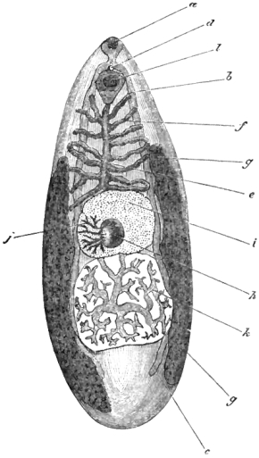
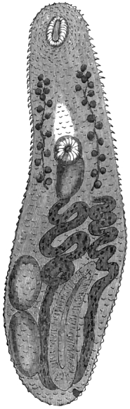
Fig. 2.—The large human fluke (Distoma crassum) a, Oral sucker;
b, intestine; c, cæcal end of same; d, reproductive papilla;
e, uterine rosette (the folds of which are not branched); f, one of
the folds (in profile); g, vitellarium; h, hernial protrusion (the
result of an injury to the specimen); i, upper testis; j, streaks
or layers of seminal fluid which have escaped by rupture
and assumed a branched appearance; k, lower testis uninjured
(but slightly altered in outline from flattening); l, ventral sucker.
Magnified 2 diameters. Original.
The new anatomical facts made out by me bear reference
principally to the reproductive apparatus. What else I have observed
is for the most part confirmatory of the statements made by Mr Busk.
In particular, his brief account of the position and character of the
digestive organs was not only confirmed by my earlier examinations, but
is now re-verified. In the representation given in my ‘Introduction’ I
showed in dotted outline two large organs which I supposed to be the
testes. I distinctly observed radiating lines proceeding from the centre
in each; but I could not discover the slightest trace of any limiting
border to either organ. I now found in the same position two nearly
circular flattened masses with clearly defined limits (i, k). No
doubt could be entertained as to the testicular character of the lower
organ (k). In the original drawing I further indicated the presence
of a third and much smaller globular mass, which I termed the ovary;
but what I supposed to represent this organ in the particular specimen
from which the accompanying illustration was drawn turns out to be
merely a hernial protrusion resulting from injury (h). The radiating,
broad, and branching seminal ducts are beautifully distinct in one of my
specimens, forming the most attractive
feature of the parasite’s organisation (k). In consequence of injury
to the specimen which is here drawn, the upper testis (i) displays
no seminal tubes. I made out the female reproductive organs with more
completeness. In the outline drawing given in my introductory treatise
I had indicated the probable position of the uterine folds; reducing
the organ to the simplest expression of what I concluded must obtain in
the normal condition. My conjecture was perfectly correct. The uterus
consists of irregularly folded tubes, which, though here and there
apparently branching from a central tube, are in reality folded evenly
upon themselves. The oviduct can be distinctly traced to its outlet in
the reproductive papilla, which, as usual in true Distomes, is placed in
the middle line, immediately above the ventral sucker. In my examination
of Mr. Busk’s original specimens I could not find the slightest trace of
vitelligene organs; but in my fresh examples I not only obtained proof
that these organs were largely developed, but that their limitations
could be fixed with accuracy (g g). They consisted of two large
elongated masses, one on either side of the body, occupying about two
thirds of the entire length of the parasite. Their yolk-vesicles were
distinctly seen; but the main efferent canals were only here and there
traceable. Clearly, the position and character of the yolk-forming
glands of this large human fluke are quite unlike those of any of its
congeners. This fluke is a remarkably fine species, and, when viewed in
the fresh state with a powerful pocket-lens, presents a most striking
appearance. I did not observe any cutaneous spines. I found the eggs to
present an average long diameter of about 1/200″, by 1/330″
in breadth. They are therefore somewhat smaller than those of the
common fluke. In the specimen preserved in the Hunterian Museum there
was complete evidence of the presence of an excretory outlet at the
caudal extremity; but I did not succeed in finding any trace of the
water-vascular system higher up. I have no doubt, however, that it
exists.
As regards the affinities of Distoma crassum, it is clear that
this Trematode has little in common either with the liver-fluke
of cattle and sheep (Fasciola hepatica), or the still larger species
obtained by me from the giraffe (Fasciola gigantea). The
simple character of the digestive tubes obviously connects it
more closely with the lancet-shaped fluke (Distoma lanceolatum),
the last-named parasite being, as already shown, an occasional
resident in the human liver, where its presence, moreover,
undoubtedly contributed towards the production of the fatal
result.
In my remarks on the missionary’s diet it is hinted that
the Ningpo oysters may have played the rôle of intermediary
bearers to the parasite in question; and as tending in some
measure to strengthen this notion, it should be borne in mind
that Mr. Busk’s original fluke-bearer came from the east. It
is not improbable that the Lascar host may have partaken of
the same particular species of fish or shell-fish that the
missionary and his wife partook of. Be that as it may, the
frequency of the occurrence of Trematodes and their larvæ in
marine mollusks is well known. According to Woodward,
several species of oyster are sold in the Indian and Chinese
markets. Thus, it would require the skill of a malacologist to
determine the particular species of Ostrea to which the Ningpo
oysters should be referred.
Mons. Giard is of opinion that the singular larvæ known as
Bucephali attain sexual maturity in sharks and dog-fishes;
therefore it is extremely unlikely that the Bucephali should have
been in any way concerned in the infection of our missionary
and his wife; nevertheless there remains the probability that
these human bearers swallowed other kinds of Trematode
larvæ when they consumed the Ningpo oysters. Moreover, if
it should happen that none of the other larvæ occurring in
oysters are capable of developing into flukes in the human
territory, it yet remains highly probable that some one or other
of the various encysted (and therefore sexually immature)
Trematodes known to infest marine fishes will turn out to be
the representative of our Distoma crassum. In this connection
we must not forget that the flesh of the Salmonidæ forms the
probable source of human Bothriocephali; and there is some
likelihood that salt-water fishes, if not actually the primary,
may become (after the manner explained by M. Giard) the
secondary intermediary bearers of fluke-larvæ. At all events,
I am inclined to look to the Ningpo oysters, or to some
other of the various species of marine shell-fish sold in eastern
markets, as the direct source of Distoma crassum; for, in
addition to the bucephaloid cercarians, we have abundant
evidence of the existence of other and more highly developed
fluke-larvæ in marine bivalve mollusks.
In this connection I will only further observe that we possess
very little knowledge of the parasites which take up their abode
in the viscera of savages. This ignorance results partly from
the fact that these untutored races, as proved by the statements
of Kaschin and others, actually, in the matter of severe symptoms,
suffer much less from the presence of intestinal worms
than their civilised fellow-men do. The subject is worthy of
further attention, but no one, so far as I am aware, has cared
to institute the necessary inquiries in a methodical way. I
strongly suspect that several of the human parasites which we
now consider to be rare would be found to be abundant if by
means of post-mortem examinations and other methods of investigation
we could be made acquainted with the facts of helminthism
as they occur amongst the raw-flesh and fish-eating
savage tribes. Of course any person, notwithstanding the
utmost care and cleanliness, as in the cases before us, may
contract a noxious parasite; nevertheless, speaking generally,
it may be said that the measure of internal parasitism affecting
any given class of people bears a strict relation to the degree
of barbarism shown by such persons in their choice of food and
drink, and in their manner of eating and drinking. This
statement, if true, is not destitute of sanitary importance;
moreover, it applies not alone to ourselves, but also to all the
domesticated animals that serve our wants. Cleanliness is just
as necessary for their welfare as for our own.
In the spring of 1878 my patients returned from China.
They had experienced fresh attacks from the parasite; moreover,
one of their children, a little girl, was also victimised by
the same species of fluke. Thus, in one family I have encountered
three cases of fluke-helminthiasis due to Distoma
crassum! One of the worms passed by the little girl per anum
is now in my possession. It not only shows the upper testis
perfectly, but also the many times transversely folded, simple,
uterine rosette which is certainly not branched. There are
also traces of an organ which I take to be the cirrhus-pouch;
but I have never seen the penis protruded externally.
For the purposes of diagnosis I subjoin the following characters.
The Distoma crassum is a large, flat helminth varying
from an inch and a half to two and a half inches in length,
and having an average breadth of five eighths of an inch; it is
especially also characterised by its uniform and considerable
thickness, combined with the presence of a double alimentary
canal which is not branched; the body is pointed in front, and
obtusely rounded posteriorly; the integument being smooth and
unarmed; the reproductive orifices placed immediately above
the ventral sucker; the testes form two large rounded organs,
situated below the uterine rosette, and disposed in the middle
line, one in front of the other; the uterine folds occupy the
front part of the body; near the lateral margins there are two
large vitelligene glands, one on either side of the intestinal
tube; the excretory organ probably consists of a central trunk
with diverging branches, opening below.
Bibliography (No.
5).—Budd, original notice in his ‘Diseases of the Liver,’ 2nd
edition, quoted by Lankester in Appendix B to Küchenmeister’s ‘Manual
of Parasites,’ p. 437, 1857.—Cobbold, T. S., “Synopsis of the
Distomidæ,” in ‘Journ. of the Proceed. of the Linnean Soc.,’ vol. v,
Zool. Div., 1860 (original description p. 5).—Idem, ‘Entozoa,’
p. 193, 1864.—Idem, “Remarks on the Human Fluke Fauna, with
especial reference to recent additions from India and the East,” the
‘Veterinarian,’ April, 1876.—Idem, “On the supposed Rarity,
Nomenclature, Structure, Affinities, and Source of the large Human Fluke
(D. crassum),” ‘Linn. Soc. Journ.,’ vol. xii, Zool. Div., 1876, p. 285
et seq.—Idem, “Observations on the large Human Fluke, with
notes of two cases in which a missionary and his wife were the victims,”
the ‘Veterinarian,’ Feb., 1876.—Idem, “The new Human Fluke,”
in a letter published in the ‘Lancet,’ Sept., 1875.—Leidy, in
‘Proceed. Acad. Nat. Sciences of Philadelphia;’ see also Dr McConnell’s
paper quoted below (Bibl. No. 6).—Leuckart, l. c., Bd. I, s.
560.—Weinland, l. c. (Bibl. No. 2), Appendix, p. 87.
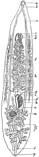
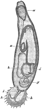
Fig. 3.—The Chinese fluke (Distoma
Sinense). a, Oral sucker; b,
œsophageal bulb; c, intestine;
c′, cæcal end; d, ventral sucker;
e, genital pore; f, uterine
folds; g, ovary; h, vitellarium;
i, vitelligene duct; k, upper
seminal reservoir; l, testes;
m, lower seminal pouch; o,
vas deferens; p, pulsatile vesicle;
p′, water vessel. After
McConnell.
Distoma Sinense, Cobbold.—The discovery of this species is
due to Prof. J. F. P. McConnell, who “on the 9th of Sept., 1874,
found a large number of flukes in the liver of a Chinese, obstructing
the bile ducts.” The species measures 7/10″
in length, by 1/7″
in breadth, the eggs being 1/833″
by 1/1666″.
Dr McConnell showed in his original memoir that the worm cannot well
be confounded with Fasciola hepatica, with Distoma lanceolatum, or
with D. conjunctum. In this conclusion he was supported by Dr T. R.
Lewis, who examined the specimens with him. In a letter communicated to
the ‘Lancet,’ quoted above, I proposed the nomenclature here given; but
Prof. Leuckart, unaware of this step, afterwards suggested the terms
Distomum spatulatum. Later on I received numerous specimens from
Calcutta, the examination of which enabled me to confirm the
accuracy of the original description. As regards the male organs in
the subjoined figure, it will be seen, by comparing the lettering and
references, that I have interpreted the facts of structure somewhat
differently from Prof. McConnell.
In the month of December, 1874, a Chinese died in the Civil Hospital
at Port Louis, Mauritius, whilst he was under the care of Dr William
Macgregor, chief medical officer of the Colony of Fiji. The post
mortem revealed the presence of a very great number of flukes in the
bile-ducts. Dr Macgregor described these parasites with great care, and
having favored me with a copy of his manuscript I at once recognised the
worms to be identical with the species discovered by McConnell. I also
received through Dr Henry Clark, of Glasgow, two Mauritius specimens,
which when compared with the Calcutta examples proved to be specifically
identical. Dr Macgregor’s paper, communicated to the Glasgow
Medico-Chirurgical Society, gives full particulars of the helminthiasis
associated with this parasite, whilst both his and Prof. McConnell’s
account of the structure of the worm are remarkably complete in details,
and well illustrated. It is not a little curious to notice that although
these parasites were obtained in countries far removed from China, they
were in both instances taken from Chinese; moreover, from the statements
of Macgregor, it appears very probable that the parasites in question
are a common source of liver disease. Without doubt oriental habits are
eminently favorable to fluke infection, for we are now acquainted with
four species of flukes whose geographical range is limited to eastern
parts.
Bibliography (No.
6).—McConnell, J. F. P., “Remarks on the Anatomy and
Pathological relations of a new species of
Liver-fluke,” ‘Lancet,’ Aug. 1875; repr. in the ‘Veterinarian,’ Oct.,
1875; also in the ‘Lancet,’ March 16th, 1878, p. 406.—Macgregor,
W., “A new form of Paralytic Disease, associated with the presence of
a new species of Liver Parasite (Distoma Sinense),” ‘Glasgow Med.
Journ.’ for Jan., 1877; also in the ‘Lancet’ for May 26th, 1877, p.
775.—Cobbold, T. S., in a note to the ‘Lancet,’ Sept., 1875, and
in the Appendix to Macgregor’s paper, p. 15, 1877.—Leuckart, R.,
l. c., Bd. ii, s. 871, 1876.
Distoma conjunctum, Cobbold.—The little fluke which I first
discovered in the gall-ducts of an American fox (Canis fulvus) was
fourteen years afterwards obtained from pariah dogs in India by Dr T.
R. Lewis (1872); but it remained for Prof. McConnell to show that this
entozoon also invades the human subject (1874). A second instance of its
occurrence in man was recorded in 1876. We all figured the worm, and in
respect of general details our descriptions for the most part agreed
(fig. 56). The worms from the dog and fox gave an
average of 1/4″
in length, but the majority of those found by McConnell in man were fully 3/8″
from head to tail.
Writing in the spring of 1876 Dr McConnell says:—“In
the ‘Lancet’ for the 21st of August, 1875, I published the
description of a new species of liver-fluke found in the bile-ducts
of a Chinaman (sic) who died in this hospital. Dr Spencer
Cobbold has very kindly interested himself in this discovery,
and proposed the name of Distoma Sinense for the new fluke.
This discovery (in September, 1874) has stimulated me to pay
still greater attention to the morbid conditions of the biliary
canals in our post-mortem examinations; but, although more
than 500 autopsies have been conducted since that date, I have
not met with another instance of distomata in the liver until
within the last fortnight. On the 9th of January, 1876, in examining
the liver of a native patient who had died in the hospital,
I again found a large number of flukes in the bile-ducts, and
having carefully examined many specimens, I recognise the
species as the D. conjunctum of Cobbold. Dr Cobbold discovered
this fluke in 1858; but, as far as I am aware, the human
liver has never hitherto been found infested by these parasites,
and this will give general interest and importance to the following
case.”
“Jamalli Khan, a Mahommedan, aged twenty-four, admitted
into the hospital on the 25th of December, 1875. He is a
resident of Calcutta, and an ordinary labourer (coolie). He
states that he had been suffering from ‘fever’ for the last two
months, at first intermittent in character, but for the last seven
days more or less continued. He is much emaciated and reduced
in strength. Complains of pain on pressure over the liver and
spleen; the latter can be felt much enlarged, reaching downwards
to nearly the level of the umbilicus; the lower border of
the liver, however, can only just be felt below the ribs. Temperature
on evening of admission 101° F. Conjunctivæ are
anæmic, but not jaundiced. Has also a little bronchitis. The
fever continued with slight remissions for ten days (January 4th,
1876), the highest diurnal temperature (in the afternoon) varying
from 103° to 104° F.; it then abated, but dysentery set in.
He began to pass six or eight stools in the twenty-four hours,
attended with much griping, and containing varying quantities
of blood-tinged, gelatinous mucus. These became more frequent,
in spite of treatment, during the next three days, and on the
8th of January he was manifestly sinking; passed his evacuations
into the bedclothes, became cold and collapsed, and died
in this state that same evening.
“A post-mortem examination was made on the following
morning, thirteen hours after death. All the organs of the
body were found more or less anæmic, but exhibited nothing
remarkable with the following exceptions. The lungs towards
their posterior margins and bases were dark, but still spongy
and crepitant. The spleen was found greatly enlarged, heavy;
capsule tense and stretched; substance soft, reddish brown,
irregularly pigmented; weight 1 lb. 13 oz. The liver was of
about normal size; its surfaces smooth, the capsule slightly
hazy looking. Hepatic substance firm, but abnormally dark,
and the bile-ducts particularly prominent and thickened.
Numbers of small distomata escaped from the incisions made
into the organ, and could be seen protruding from the dilated
biliary canals. The gall-bladder was filled with thick greenish-yellow
bile, measuring about an ounce and a half, but containing
no parasites, and no ova even could be detected on microscopical
examination of this bile and of scrapings from the
lining membrane of the gall-bladder. The cystic duct was
free from obstruction. The condition of the common choledic
duct could not so well be ascertained, as the liver had been
removed from the abdominal cavity before anything extraordinary
had been detected in its condition, but, so far as it
could be examined, it was found patent; the duodenal mucous
membrane was well bile-stained, and there was evidence of
biliary colouring matter in the fæcal contents of the bowels.
On carefully dissecting out, and then laying open, the biliary
ducts in a portion of the right lobe of the liver (the rest being
preserved entire), numbers of distomata were found within them,
lying singly, flattened, and generally with the anterior extremity,
or “oral sucker,” directed towards the periphery of the organ,
the posterior extremity towards its centre; or in twos, threes,
or even little groups of fours, variously coiled upon themselves
or upon each other. The lining membrane of the biliary canals
was found abnormally vascular, its epithelial contents abundant
(catarrh?), and, among these, ova could be detected under the
microscope. Sections of the liver, hardened and then examined
in glycerine, showed fatty infiltration of the lobular structure,
but not to any advanced degree; the bile ducts considerably
dilated, their walls thick and hypertrophied, but nothing else
abnormal, or in any way remarkable. The weight of the liver
was 3 lbs. In the transverse and descending colon numerous
indolent-looking, shallow, pigmented ulcers were found, and in
the rectum others evidently more recent and highly injected.
The submucous tissues throughout were abnormally thickened.
The intestinal contents consisted of only about three ounces of
thin yellowish (bilious) fæcal fluid, with small bits of opaque
mucus. This was carefully washed and examined, but no flukes
were discovered. About a dozen distomata escaped from the
liver on making the primary incisions, and quite twice this
number was found subsequently within the biliary canals. Only
a portion of the right lobe has, as I have said, been dissected,
so that it may be confidently stated that probably not less than
a hundred of these flukes must have infested this liver. All
were found dead, but it must be remembered that the autopsy
was performed thirteen hours after the death of the patient.
It is remarkable that in this case, as in the one before described
by me, no distomata were found in the gall-bladder. The
presence of these parasites in the bile-ducts seems to have led to
catarrhal inflammation of their lining membrane and abnormal
thickening and dilatation of their walls, but there is no evidence
of their having caused sufficient obstruction to produce cholæmia,
as in the case just referred to, and no marked pathological
change could be detected in the lobular structure of the liver.”
After referring to the anatomical descriptions of the worm,
as recorded by myself (in ‘Entozoa’) and by Lewis (in the
memoir quoted below), Professor McConnell further observes
that the addition of a few more particulars seems necessary for
the determination of the identity of the species. He then gives
the following characters:
“Body lanceolate, anterior and posterior extremities pointed,
the latter obtusely. Surface covered with minute spines or hairs.
Average length 3/8″ (three eighths of an inch);
average breadth 1/10″.
‘Ventral’ sucker slightly smaller than ‘oral.’ Reproductive papilla or
genital orifice placed a little above and to one side of the former.
Alimentary canal double and unbranched. Uterine folds and ovary placed
in the median line, and above the male generative organs, the latter
consisting of two very distinct globular bodies or testes. Ova of
the usual type, i.e. oval in outline, having a double contour, and
granular contents; average length, 1/750″; average breadth, 1/1333″.
The only point of note is that the average length of these flukes
is greater than that of the same species found by the authors above
referred to. The D. conjunctum in the American fox, and in the pariah
dog, has an average length of 1/4″;
only two or three specimens of this size were found in this liver, and
these showed evidences of immaturity; a few were found 1/2″ in length; but the great majority exactly 3/8″.
The anatomical characters are otherwise precisely identical.”
Professor McConnell concludes his communication by a remark in
reference to the common source of infection shared by mankind and dogs
in India. The occurrence, however, of this entozoon in an American red
fox points to a very wide geographical distribution of the species. It
is hardly likely that the fox, though dying in the London Zoological
Society’s Menagerie, should have contracted the parasite in England.
In the second half of this work I shall reproduce my original drawing
(fig. 56) from the ‘Linnean Transactions;’ but I
may refer to my Manual (quoted below) for a reproduction of McConnell’s
figure. In my original specimens the integumentary spines had fallen,
probably as a result of post-mortem decomposition.
Bibliography (No.
7).—Cobbold, T. S., “Synopsis of the Distomidæ,” (l. c.), 1859;
and in “Further Observations on Entozoa, with experiments,” ‘Linn.
Trans.,’ vol. xxiii (tab. 33, p. 349), 1860.—Idem, “List of
Entozoa, including Pentastomes, from animals dying at the Zool. Soc.
Menagerie between the years 1857–60,” ‘Proceed. Zool. Soc.,’
1861.—Idem, ‘Entozoa,’ p. 20, pl. ii, 1864; and in
“Manual of the Internal Parasites of our Domesticated Animals,” p. 81,
1873.—Lewis, T. R., and Cunningham, D. D., in a footnote to
their ‘Microscopical and Physiological Researches,’ Appendix C., ‘Eighth
Ann. Rep. of the San. Comm. with the Govt. of India,’ p. 168, Calcutta,
1872.—McConnell, J. F. P., “On the Distoma conjunctum,”
in the ‘Lancet’ for 1875–76, quoted above; reprinted in the
‘Veterinarian,’ 1876; also (a second case) in the ‘Lancet’ for March
30th, 1878, p. 476.
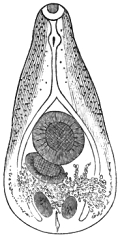
Fig. 4.—The small Egyptian
fluke (Distoma heterophyes),
viewed from behind. The
large ventral sucker, supplementary
disk, uterus, testes,
simple divided intestine,
vitellarium, and pulsatile
vesicle are conspicuous.
Original.
Distoma heterophyes, Von Siebold.—This minute parasite,
measuring only 3/4
of a line in length, was discovered by Dr Bilharz, of Cairo, in the
intestines of a lad, post-mortem, in the year 1851. A second similar
instance occurred, when several hundred examples were collected and
afterwards distributed amongst the helminthologists of Europe. Through
the kindness of Leuckart two of the worms eventually reached myself.
From one of these the accompanying figure was drawn. For the purpose
of supplying a full diagnosis I have elsewhere described this worm
as presenting an oblong, pyriform outline, attenuated in front, and
obtusely rounded behind; body compressed throughout, the surface being
armed with numerous minute spines, which are particularly conspicuous
(under the microscope) towards the head; oral and ventral suckers
largely developed, the latter being near the centre of the body, and
about twice the diameter of the former; pharyngeal bulb distinct and
separate from the oral sucker, and continued into a long œsophagus,
which divides immediately above the ventral acetabulum; intestinal tubes
simple, gradually widening below and terminating near the posterior
end of the body; reproductive orifices inconspicuous, but evidently
placed below and a little to the right of the ventral sucker, at which
point they are surrounded by a special accessory organ, resembling a
supernumerary sucker; uterine folds numerous and communicating with
small but conspicuously developed vitelligene glands; testes spherical
and placed on the same level in the lower part of the body; ovary
distinct; aquiferous system terminating inferiorly in a large oval
contractile vesicle, the latter opening externally by a central foramen
caudale.
Apart from its minuteness, moreover, this trematode is especially
characterised by the possession of a very remarkable apparatus
surrounding the reproductive orifices. It consists of an irregularly
circular disk, measuring 1/125″
in diameter, and having a thick-lipped margin, which supports seventy fish-
basket-like horny ribs comparable to the claw-formations seen in the
genus Octobothrium. According to Bilharz these ribs give off five
little branches from their sides, but Leuckart could not see them in his
specimens. Leuckart estimated the length of these horny filaments
to be 1/1250″,
whilst their breadth was 1/3570″.
On the whole we may regard this organ as a complicated form of
“holdfast” designed to facilitate or give efficiency to the sexual act.
I may here also state that this structure is by no means unique; for,
if I mistake not, it exists in an equally developed degree in the young
trematode which Dr Leared found infesting the heart of a turtle. Leared
believed that he had found an ordinary distome; an opinion to which I
could not give my assent, seeing that the organ described by him as a
“folded, ventral sucker” presented a very different aspect to the oral
sucker displayed by the same animal. Without doubt, however, the organ
in his so-called Distoma constrictum is analogous to the supplementary
“holdfast” existing in Distoma heterophyes. The views which I
originally advanced as to the source and condition of the parasite are
probably correct.
As regards the structure of Distoma heterophyes, I have only to
add that a special set of glandular organs is situated on either side
of the elongated œsophagus, but the connection between these organs
and the digestive apparatus has not been clearly made out. Leuckart
compares them to the so-called salivary glands found in Distoma
lanceolatum, and says, “The presence of such a glandular apparatus is
also indicated by the more ventral position of the oral sucker, and
the development of the cephalic margin.” The conspicuous contractile
vesicle terminating the excretory system is developed to an unusually
large extent, exhibiting in its interior multitudes of the well-known
active molecular particles. Lastly, I have only to add that the eggs of
Distoma heterophyes measure 1/990″
in length by 1/666″
transversely.
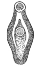
Fig. 5.—The eye fluke
(Distoma ophthalmobium).
Showing the
suckers and intestinal
tubes. After Von
Ammon.
Bibliography (No.
8).—Bilharz, “Beitrag zur Helminth. humana,” ‘Zeitsch. für
wissenschaftl. Zool.,’ s. 62, 1851.—Cobbold,
‘Entozoa,’ p. 195, 1864.—Küchenmeister, F., ‘Parasiten,’
1855, s. 210, Eng. edit., p. 276, 1857.—Leared, “Description
of Distoma constrictum,” ‘Quarterly Journal of Micros. Science,’
new series, vol. ii, 1862.—Leuckart, R., l. c., s. 613,
1863.—Moquin-Tandon, on the Genus Fasciola, l. c.,
1861.—Weinland, on Dicrocœlium, l. c., p. 86, 1858.
Distoma ophthalmobium, Diesing.—There is every reason to
believe that the small flukes found by Gescheid and Von Ammon in the
human eye were sexually immature worms, but since it cannot be decided
as to what adult species they are referable I prefer to notice them
under the usual title. Possibly these eye-worms may be referred to D.
lanceolatum, as suggested by Leuckart. However that may be, I deem
it unnecessary to repeat the details recorded in the treatises quoted
below. The largest examples measured only half a line or about one
millimètre in length.
Bibliography (No.
9).—Cobbold, ‘Entozoa,’ p. 191.—Gescheid (D. oculi
humani), in Von Ammon’s ‘Zeitsch. f. Ophth.,’ iii, and also in
Ammon’s ‘Klin. Darstell. d. Krankheit d. Menschl. Auges.,’ vols. i and
iii.—Küchenmeister, Eng. edit., p. 287.—Leuckart, l. c.,
s. 610.—Nordmann (Monostoma lentis), “Mikr. Beitr.,” Heft. ii,
‘Vorwort,’ s. ix, 1832.
Tetrastoma renale, Chiaje; Hexathyridium pinguicola, Treutler;
and H. venarum, Treutler.—Whether these forms are good species
or not, the fact that they were genuine parasites cannot, I think, be
disputed. The first-mentioned measured five lines in length, and was
found by Lucarelli in the urine. The second, eight lines long, was found
by Treutler in a small tumour connected with the ovary. The third,
measuring three lines in length, was twice found in venous blood, and
twice in the sputum of patients suffering from hæmoptysis.
Bibliography (No.
10).—Delle-Chiaje, ‘Elmintografia Umana,’ 1833.—Bremser
(l. c., Bibl. No. 2), s. 265, 1819.—Cobbold, ‘Entozoa’
(p. 204, et seq.).—Dujardin (l. c., Bibl. No. 2), s. 265,
1819.—Treutler, ‘Obs. Path. Anat. ad Helm. Corp. Humani,’ p. 19,
1793.—Zeder, ‘Anleitung zur Naturg. der Eingeweidewürmer,’ s.
230, 1803.
Amphistoma hominis, Lewis, and McConnell.—The original
account of this species is based upon two finds. The first
series of specimens was procured from Dr J. O’Brien, of
Gowhatty, and the second set from the Pathological Museum of
the Calcutta Medical College. Dr O’Brien and Dr Curran
together procured their specimens, post-mortem, from an Assamese.
There were hundreds of worms present in the vicinity of
the ileo-colic valve. The museum specimens were procured from
a patient who died at the Tirhoot gaol hospital in 1857. They
were (say the authors) presented to the museum by Dr Simpson,
and in the catalogue their history was briefly recorded as
follows:
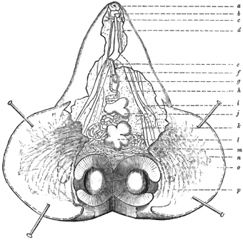
Fig. 6.—The human amphistome (Amphistoma hominis). Longitudinal section. a, Oral sucker;
b, pharyngeal bulb; c, nerve ganglia; d, œsophagus; e, genital pore; f, vagina; g, ductus
ejaculatorius; h, ventral nerve cords; i, intestinal canal; j, upper testis; k, water vessel;
l, lower testis (ovary according to Lewis); m, principal ducts of the vitellarium; n, branches
of the vitellary ducts; o, ventral pouch or bursa; p, caudal sucker. Magnified 12 diameters.
After Lewis.
“The cæcum of a native prisoner who died from cholera in
the Tirhoot gaol hospital, with a number of peculiar and,
probably, hitherto unrecognised parasites, found alive in that
part of the intestinal canal.” (Presented by Dr Simpson through
Professor E. Goodeve.)
In continuation of their narrative, Drs Lewis and McConnell
go on to say that, “with reference to this preparation, the following
very interesting particulars from the ‘Annual Jail Report of
Tirhoot’ for 1857 have been very kindly placed at our disposal by
the Surgeon-General, Indian Medical Department. The prisoner,
Singhesur Doradh, aged 30, was attacked with cholera on the
13th, and died on the 14th of July, 1857. Had not been in
hospital previously, and was employed in cleaning the jail.”
The post-mortem examination was made three hours after death:—“Colon externally livid, contracted;
contains a little serous fluid with flakes of mucus. Mucous membrane
healthy except venous injection. In the cæcum and ascending colon
numerous parasites like tadpoles, alive, adhering to the mucous membrane
by their mouths. The mucous membrane marked with numerous red spots like
leech-bites from these parasites. The parasites found only in the cæcum
and ascending colon, none in the small intestines.” This description
is by Dr Simpson, who adds, “I have never seen such parasites, and
apparently they are unknown to the natives. They are of a red colour,
size of a tadpole, some young, others apparently full grown, alive,
adhering to mucous membrane,—head round, with circular open mouth,
which they had the power of dilating and contracting. Body short and
tapering to a blunt point.”
Drs Lewis and McConnell’s description of the worm is too long to be
quoted in full. The parasites measure 1/5″ to 1/3″
in length, by 1/8″ to 1/6″
in breadth. Science is much indebted to these eminent observers for
having unearthed the museum specimens and for recording the facts they
could gather. From a zoological point of view the most interesting fact
connected with Lewis’s amphistome is the existence of a gastric pouch.
This structure brings these human Masuri into close relation with the
equine parasite which I have named Gastrodiscus Sonsinoii, and which
will be found illustrated in this work (fig. 62).
In short, Lewis’s worm appears like a transition form; the absence of
gastric supplementary suckerlets separating it from the new generic
type.
Bibliography (No. 11).—Lewis, T. R., and McConnell, T. F. P.,
“Amph. hominis; a new parasite affecting Man,” ‘Proceedings
of the Asiatic Society of Bengal,’ Aug., 1876.
Bilharzia hæmatobia, Cobbold.—This remarkable parasite was
discovered by Bilharz in 1851. It was subsequently found by myself
in an ape (1857); other species of the same genus having since been
detected by Sonsino in the ox and sheep (1876). The human examples were
originally obtained from the portal system of blood-vessels. Afterwards
they were obtained by Bilharz, Griesinger, and others, from the veins of
the mesentery and bladder. It was shown that they were not
only associated with, but actually gave rise to a formidable and
very common disease in Egypt.
In 1864 Dr John Harley made the interesting announcement that he
had discovered specimens of this singular genus in a patient from the
Cape of Good Hope. He also showed that the entozoon was the cause
of the hæmaturia known to be endemic at the Cape. Harley believed
his parasites to represent a new species (Distoma capense), but in
this view I showed that he was mistaken. His admirable contribution,
nevertheless, served not only to establish the wide range of this
parasite on the African continent, but also to throw much light upon
the subject of endemic helminthiasis. As this worm forms an almost
altogether exceptional type of fluke-structure, it became necessary to
supersede the original nomenclature proposed by Bilharz and Von Siebold
(Distoma hæmatobium). Accordingly I proposed the term Bilharzia,
whilst other helminthologists subsequently proposed various titles
(Gynæcophorus, Diesing; Schistosoma, Weinland; Thecosoma,
Moquin-Tandon). On various grounds, and chiefly on account of priority,
most writers have at length definitely accepted the nomenclature which
employed the discoverer’s name for generic recognition.
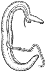
Fig. 7.—The blood fluke (Bilharzia
hæmatobia). The lower end of the
female is withdrawn from the gynæcophoric
canal of the male. After
Küchenmeister.
The Bilharzia hæmatobia may be described as a trematode helminth
in which the male and female reproductive organs occur in separate
individuals; the male being a cylindrical vermiform worm, measuring only
half an inch or rather more in length, whilst the female is filiform,
longer, and much narrower than the male, being about four fifths of
an inch from head to tail; in both, the oral and ventral suckers are
placed near each other at the front of the body; in the male the suckers
measuring 1/100″,
in the female 1/314″
in diameter; in either, the reproductive orifice occurs immediately
below the ventral acetabulum. The comparatively short, thick, and
flattened body of the male is tuberculated and furnished with a
gynæcophoric canal, extending from a point a little below the ventral
sucker to the extremity of the tail; this slit-like cavity being
formed by the narrowing and bending inwards of the lateral borders of
the animal, the right side being more or less completely overlapped by
the left margin of the body; caudal extremity pointed; intestine in
the form of two simple blind canals. Female with a cylindrical body
measuring only 1/312″
of an inch in thickness in front of the oral sucker; lodged in the
gynæcophoric canal of the male during the copulatory act; thickness of
the body below the ventral acetabulum being about 1/357″,
and at the lower part 1/96″;
surface almost smooth throughout; intestinal canals reunited after
a short separation to form a broad, central, spirally twisted tube
extending down the middle of the body; vitelligene and germigene canals
combining to form a simple oviducal canal, which is continued into
a simple uterine tube, finally opening near the lower margin of the
ventral sucker; eggs pointed at one end, or furnished with a projecting
spine near the hinder pole.
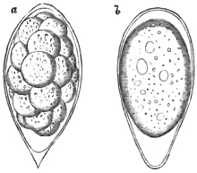
Fig. 8.—Two eggs of Bilharzia. a, With the yolk
coarsely segmented; b, with the yolk granulated and
the spine wanting. Original.
The study of the structure and formation of the contents of the
ova possesses great interest. When fully developed the eggs are oval,
measuring from 1/180″
to 1/160″
in length, with an average transverse diameter of 1/325″.
Some are a trifle larger, others smaller. Occasionally one encounters
narrow specimens, and also aberrant forms presenting a pear-shaped
outline. I have met with eggs not exceeding 1/250″
in their long diameter, and 1/500″
transversely, whose yolk-contents had already arrived at an advanced
stage of segmentation.
The shell is transparent, of a brown colour, and free from any
markings, lines, or sculpturing. One pole of the shell is invariably
narrower than the other, and usually presents a more or less pointed
extremity (fig. 8). This narrow end commonly displays a sharp,
projecting, beak-like spine, which, at its base, constantly rests
upon the centre of the pole of the shell, but occasionally it is
eccentrically placed (fig. 8a). In some few examples the spine is
removed to a little distance from the actual extremity of the shell; but
even in these instances, so far as my observations go,
its apex always projects beyond the level of the curved end of the
pole. Now and then the spine is altogether absent (fig. 8b); and when
present it is, as already hinted, very unequally developed. In size the
spine ranges from a mere point, having an extreme length of only 1/8000″,
up to the comparatively large magnitude of 1/2500″
lengthways.
According to the best evidence there is no good ground for asserting
the existence of any specific differentiation between the parasites
coming from the Cape and Egypt respectively.
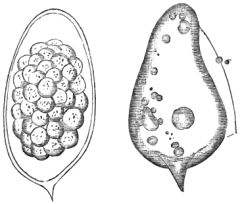
Fig. 9.—Two eggs of Bilharzia, with eccentrically placed
spines. That to the left shows mulberry cleavage of
the yolk; the other having lost its embryonal contents
by rupture. Original.
Taking a more extended view of the significance of these singular
chorional spines, I think we may here recognise the early efforts of
Nature, so to speak, to form or evolve a special organ, which, in
the eggs of certain other parasites, becomes capable of attaining
a relatively prodigious degree of development. To me it seems that
the little process in question is a kind of rudimentary holdfast;
and, as such, it may be reckoned as the homologue of a variety of
egg-appendages. Eleven years ago Mr Edwin Canton discovered some curious
ova attached to the conjunctiva of a turtle’s eye. I had no hesitation
in pronouncing them to be referable to some ectozoon or entozoon
belonging to one or other of the allied genera Polystoma, Tristoma,
Octobothrium, and Dactylogyrus. Now, whilst the Bilharzia ova
display only a solitary and imperfectly developed holdfast, placed at
one end of the shell, the singular eggs described by Mr Canton develop
organs of anchorage at both extremities. Parasitic ova exhibiting
analogous processes, spines, and filamentary appendages at both poles,
have been observed in various species of parasite—as, for example,
in Monostoma verrucosum infesting the fox, in Tænia cyathiformis
infesting the swallow, in Tænia variabilis of the gambet, in
Octobothrium lanceolatum attached to the gills of the common herring;
and in Polystoma appendiculata, from the branchiæ of various marine
fishes. Eggs of parasites which,
like Bilharzia, are furnished with a single appendage, may likewise
be seen in the ova of different species of Dactylogyrus infesting
the gills of the pike. In the more strongly pronounced developments
it is easy to perceive how admirably these outgrowths are adapted to
the necessities of the different species of parasite to which they are
severally referable; and, even in the case of Bilharzia, the trifling
amount of anchorage furnished by a projecting point is not absolutely
thrown away. The resistance will also be greater where the spine is
situated a little on one side of the pole of the egg, which seems to
need steadying during the violent struggles of the embryo to escape from
its temporary abode.
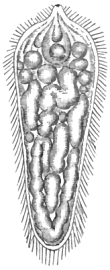
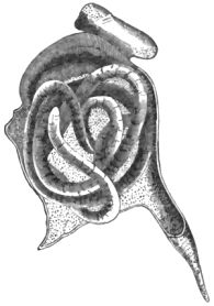
Fig. 10.—Free ciliated embryo
of Bilharzia, with pear-shaped
rudimentary organs below the
head. Original.
|
|
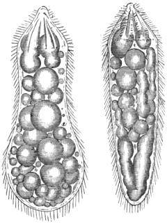
Fig. 11.—Two ciliated embryos of Bilharzia; showing sarcode
spherules in their interior. That to the left has
recently escaped the shell. Original.
|
When any number of ova are removed from the urine and examined, it
will be found that a large proportion of them contain embryos in an
advanced stage of larval growth. The structural appearances presented
by the embryos whilst still in the eggs are remarkably uniform; since,
in all, the yolk appears to have resolved itself into a mass of rounded
sarcode-globules, one or two of these particles being conspicuously
larger than the rest (fig. 12). At this stage,
except towards the cephalic division of the larva, no tendency to
differentiation is perceptible; but
some time after the embryo has escaped, one may notice elongated masses
of sarcode formed by the coalescence of the globules. Whilst still in
the egg, one end of the primitive embryonal mass becomes gradually
narrowed, cilia at the same time appearing. This part becomes the
future head, eventually acquiring the form of a cowl. Whatever form
the body of the embryo may display after extrusion from the shell,
the head retains its conical shape, the cone itself being narrowed or
widened only when the larva is subjected to abnormal conditions (fig. 14). Whilst the head is undergoing development within
the shell, one, two, or sometimes three, pyriform masses make their
appearance within the cone; and after the embryo has escaped, these
structures become more marked (fig. 10). The sarcode-globules refract
light strongly; and, when the larva is not compressed in any way, they
move freely within the somatic cavity. In well-developed embryos,
whilst still in the egg, the cilia are observed to clothe every part of
the larva except the oral papilla. This minute nipple-like projection
measures about the 1/3000
of an inch transversely, forming a very simple kind of unarmed
proboscis. When the head of the free embryo is viewed from above,
the proboscis looks like a central ring surrounded by a series of
regular folds, which radiate outwards like the spokes of a wheel. The
ridges thus formed support numerous cilia, these latter projecting at
the circumferential margin of the cephalic cone in such a way as to
present the figure of a star. Dr Harley has admirably represented this
character, which is shared by many other parasitic larvæ. Throughout the
greater part of the time, whilst the embryo is still resident within the
egg, the broad neck or base of the cephalic cone forms a fixed point of
resistance by its firm attachment to the inner wall of the shell; and
this structural union, so long as it remains intact, enables the embryo
to move not only its head and body from side to side synchronously, but
also each part independently. When the time for final escape is drawing
near, the vigorous movements of the cone-shaped head seem chiefly
concerned in loosening the membranous
connection just referred to; and when, at length, the ciliated
animalcule has succeeded in overcoming this first difficulty, it is
ludicrous to witness its frantic efforts to find an opening in the
shell. While thus partially liberated, it will rush to and fro from
one pole of the egg to the other, performing a series of summersaults,
and at the same time occasionally rolling itself over laterally. This
activity becomes gradually more and more violent, until at length
its excitement is worked up into a sort of frenzy. I have many times
watched these performances, which, however, are only to be seen within
those ova whose shells, for some reason or other, refuse to yield to
the earlier and ordinary efforts of the prisoner. In all cases where
these phenomena are witnessed the eye readily detects a number of
small free globules between the embryo and the inner wall of the shell
(fig. 13). These minute particles are likewise
tossed about tumultuously during the rapid rotatory movements of the
imprisoned larva. Except as regards their size, these globules do not
differ in character from the sarcodic contents of the animalcule. They
are probably superfluous detachments from the primitive yolk-mass, but
it is possible that they may afford some aid in the final breaking
up of the shell. Whilst the embryo remains fixed its tail is usually
directed towards the narrower or spine-bearing pole of the egg, but
in a few instances I have seen this position reversed. As regards the
precise mode of emerging from the shell, and the time occupied by the
larva in freeing itself, there are several points of interest. Speaking
generally, the purer the medium into which the ova are transferred, the
more rapid will be the movements of the larvæ. To give an example of
observed facts in relation to the rapidity of development, I cite the
following:—“On the 20th of August,
1870, I placed twelve eggs of Bilharzia under the microscope. The
medium in which they were immersed consisted of eight parts of ordinary
drinking water to one of urine. At the expiration of seventeen minutes
the first-born made its escape. In the course of
another minute two more emerged. In twenty-six minutes the fourth, in
twenty-eight the fifth, in thirty-two the sixth, in thirty-four the
seventh, in thirty-seven the eighth, in thirty-eight the ninth, in forty
the tenth, in forty-three the eleventh, and in forty-six minutes the
twelfth, respectively made their appearance.”
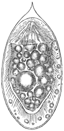
Fig. 12.—Egg of Bilharzia,
with contained embryo and
free sarcode globules. Original.
|
|
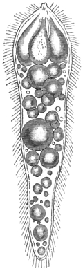
Fig. 13.—Free ciliated embryo
of Bilharzia, slightly
deformed, and having the
pear-shaped organs largely
developed. Original.
|
|
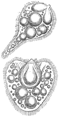 Fig. 14.
—Ciliated embryos of Bilharzia, deformed
by the application of reagents. Original. |
Now, this rapid mode of birth and emergence from the shell is very
much more striking in the case of eggs which are placed in perfectly
pure water; for, whilst the eggs are still in the urine, there appears
to be neither the power nor the inclination on the part of the embryo to
escape; but, on isolating and placing them in suitable conditions, their
behaviour is even more remarkable. In a space of less than two minutes
I have repeatedly seen the hitherto motionless embryo alter its shape
by contractions, become violently agitated, and burst out of its shell
in the condition of a free-swimming animalcule. Moreover, it is worthy
of remark that the eggs and larvæ of Bilharzia soon perish in stale
urine. “On the 16th of August, 1870, I placed about a thousand eggs in a
quart of fountain-water, to which only a drachm or rather less of urine
had been added. At the expiration of forty-eight hours not a single
living embryo could be found. I subsequently ascertained that I could
not keep the embryos alive for twenty-four hours in any water in which
I had introduced the smallest trace of mucus, blood-corpuscles, urinary
crystals, or decomposing matters of any kind. All sorts of reagents
speedily killed the larvæ. Mere discoloration by carmine solution, or
by the addition of a drop of the solution of permanganate of potash,
instantly caused them to assume grotesque and unnatural shapes (figs. 13
and 14), death sooner or later following as a result
of the disintegration and resolution of their delicate bodies into mere
sarcode-masses. Still more rapidly poisonous effects were produced by
the addition of a little sherry or alcohol. In solutions where the
amount of spirit did not exceed one part of spirit, proof strength, to
fifty parts of water the effect was the same.”
The development of the larva is equally well accomplished in
distilled water, in well-water, and in brackish water. In pure sea-water
the process goes on less satisfactorily. It was found, indeed, that the
addition of slightly saline water to ciliated embryos, which were on the
point of expiring in fresh water, had the effect of reviving them for a
time. These facts have an important practical bearing.
I have thus shown that the escape of the embryo is by no means the
slow process that Bilharz has described. Almost invariably the shell
bursts by a longitudinal slit extending over fully two thirds of its
long diameter, the first point of rupture being commonly situated midway
between the spine and the centre of the shell. In normal births, so
to speak, the head of the animalcule emerges first; but occasionally
the animal escapes sideways, and I have even seen the embryo extricate
itself tail foremost. Not unfrequently it has a difficulty in detaching
itself from the shell, in which case the egg is whirled round and round
by the half-freed prisoner (fig. 15). The lodgment
of the spine, however, against any foreign substance affords the
necessary leverage for ensuring escape.
The larva never displays its proper elongated, spindle-shaped, or
cylindro-conical figure, until some short time after its escape from
the shell; and, as a consequence of this, its powers of locomotion
are less marked at first than they are subsequently. At the time
of extrusion the larvæ are commonly more or less hour-glass shaped
(fig. 11); this particular form being sometimes
retained for many minutes or even for an hour. Usually the larvæ have
a tendency to acquire their normal shape immediately after quitting
the shell; the oval, pear-shaped, and variously contracted forms
gradually merging into the characteristic cone-shaped animalcule (fig.
10). In their fully developed condition, they
exhibit the most lively movements; and to witness several hundreds of
them rushing about with unceasing activity is a curious sight. The
phenomenon, moreover, loses none of its interest from the consideration
that only a few hours, or it may have been minutes, previously, these
now actively
gyrating animalcules were lodged in ovo within the blood-vessels of
their human host. From persons who are infested, myriads of these eggs
of Bilharzia daily make their escape during the act of micturition;
and, when this act is accomplished by the host out-of-doors, it is
easy to perceive how readily the ova may be subjected to conditions
favorable to the development of larvæ. The direct passage of the urine
into any considerable receptacle of natural or fresh water would in a
few minutes ensure the hatching of all the eggs; and in the absence
of any such direct aid to development, the accidental occurrence of a
shower of rain would, in all localities where the Bilharzia disease is
endemic, readily transfer the ova into ditches, ponds, rivers, lakes,
and ultimately, perhaps, even into the sea itself.
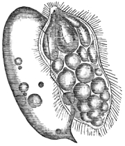
Fig. 15.—Egg of Bilharzia, with the shell adhering
to the escaped ciliated embryo. Original.
|
|
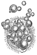
Fig. 16.—Ciliated embryo of Bilharzia in
the act of dying from the escape of the
sarcodic contents. Original.
|
The behaviour of the embryo under the action of reagents of various
kinds is remarkable. Thus, when on the 5th of Sept., 1870, I placed some
ova in brackish water, of the strength of two parts of fresh water to
one of pure sea-water, their contents were readily developed, though
the escaping embryos did not swim vigorously. When again I placed some
other eggs in pure sea-water, their contained embryos became instantly
transfixed, the vibratile cilia of the head being rigid and motionless.
At first I naturally concluded that the embryos were killed outright;
but, to my great surprise, the shock passed away in about half an hour,
when they revived and were soon afterwards hatched. One of the larvæ
thus set free carried off several of the loose intra-chorional globules
which had, during the period of transfixion, become firmly adherent
to the ends of the caudal cilia. Here I may remark upon a decided
difference observable
between the cilia of the head and body respectively. The former are at
all times vibratile, active, and conspicuous, whilst the latter are more
delicate, capable of comparatively little motion, and partaking more of
the character of fine setæ. In length their general measurement varies
from 1/2500″
to 1/2000″.
The action of pure sea-water on the free animalcules, previously
immersed in fresh or brackish water, was equally striking. All,
without exception, immediately became paralysed and almost motionless;
nevertheless, on again adding fresh water, several entirely recovered.
It is worthy of notice that in these cases the cephalic cilia furnished
the first indications of returning viability. I was particularly struck
with the behaviour of one embryo, which, under the stimulus of the
sudden shock, retracted its cone-shaped head almost entirely within
the general cavity of the body (fig. 14, lower
specimen). In their moribund condition, whatever shape the embryo
retained, the sarcodic contents gradually faded away; the outline of the
creature, however, becoming more marked (fig. 16).
Usually the body of the animalcule became elongated whilst expiring in
sea-water. Under other circumstances the embryo frequently bursts; the
sarcodic contents escaping in the form of amœba-like bodies and the
cilia retaining their powers of movement long after all traces of the
sarcode have disappeared.
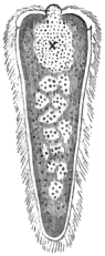
Fig. 17.—Ciliated embryo
of Fasciola hepatica,
showing the
so-called eye-spot.
After Leuckart.
The larvæ of Bilharzia closely resemble those of Fasciola
hepatica, which latter may be appropriately noticed in this place. The
ciliated embryo of the common liver fluke has the form of a long cone
inverted; the anterior end or head being flatly convex. In the centre is
a short proboscis-like papilla destitute of cilia (fig. 17). The general
covering of cilia rests on a well-defined granular epidermis; this
latter being succeeded by a dense peripheral layer of large nucleated
cells, each of them measuring about 1/2500″
in diameter. The epidermis measures 1/6250″
in thickness. In the central mass of parenchyma no internal organs
are recognisable, but Leuckart observed indications of a canal which
he thought might open at the tail, though the opening itself was not
actually visible.
As long as the ciliated covering remains intact the embryo, like
other animalcules, displays great activity, whirling round
and round on its own axis, and also describing gyrations and circles
of different degrees of range in the water, the latter movements being
accomplished by bending the body upon itself to a greater or lesser
curvature. The embryos of Bilharzia and other infusoria exhibit the
same behaviour, and, as Leuckart observes, when these embryos knock
against any obstruction, they pause after the blow, as if to consider
the nature of the substance they have touched. As in the case of fluke
embryos generally, the ciliated covering eventually falls off and the
embryo reassumes a more or less oval figure, at the same time changing
its swimming mode of progression for the less dignified method of
creeping. In the free ciliated condition the embryo of the common
liver-fluke measures, according to Leuckart, 1/190″
in length, the anterior broad end being 1/500″.
The cilia have a longitudinal measurement of 1/1388″.
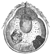
Fig. 18.—Ciliated embryo
of Distoma lanceolatum. After
Leuckart.
According to the observations of Dr Willemoes-Suhm, the cilia of the
embryos of the Distoma megastoma are limited to the anterior pole of
the body. This is also the arrangement, as Leuckart first pointed out,
in Distoma lanceolatum (fig. 18). On the other hand, Pagenstecher
has shown that the embryos of Distoma cygnoides and Amphistoma
(Diplodiscus) subclavatum are ciliated all over, an observation
which, as regards the latter species, has been confirmed by Wagener
and others. Dr Pagenstecher’s original statement to the effect that
“intrachorional germs of trematodes offer no distinctive characters,”
must, therefore, in the present state of our knowledge, be accepted as
a general conclusion admitting of many exceptions. In the early stages
of development the embryo of Distoma lanceolatum occupies the centre
of the egg, and according to Leuckart has its conical head invariably
directed towards the upper pole of the shell, or, in other words, to
that end of the egg which is furnished with a lid-like operculum.
Leuckart describes the embryo itself as “finely granular and armed
at the tip with a dagger-like spine, which, with the simultaneous
displacement of the adjacent granular mass, can be pushed forward and
drawn back again.” Besides this so-called cephalic granular mass, there
are within the embryonic body two other granular masses widely separated
from each other, but occupying the posterior half of the embryo. These
Leuckart supposes to be the rudiments of a future brood, to be developed
at the time when the free embryo shall have lost its ciliated swimming
apparatus, shall have bored its way by means of the cephalic
spine into the tissues of a mollusk, and shall have become metamorphosed
into a sac-like larva (Nurse, Sporocyst, or Redia, as the case may be).
Whatever be the full significance of these internal developments, we
have at least satisfactory evidence that the complete and free embryo is
a globe-shaped animalcule, having the anterior third or cephalic end of
the body covered with cilia, and armed with a central boring spine. In
consequence of this limitation of the ciliated covering, its swimming
movements are less vivacious than those of the embryo of Fasciola
hepatica; it will, therefore, probably take up its residence in a less
active host than that chosen by the embryo of Fasciola, selecting one
of those mollusks which either move slowly or are prone to keep at the
bottom of the water. The mature eggs have a length of 1/625 to 1/555
of an inch, and a breadth of 1/833″.
The long diameter of the free embryo varies from 1/990″ to 1/833″,
the transverse diameter being 1/1562″.
Whilst the embryos were still in the egg Leuckart could see no ciliary
motion. With most observers, both the ciliary apparatus and the boring
spine appear at this stage to have altogether escaped observation.
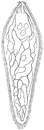
Fig. 19.—Outline representation of
a ciliated embryo of Bilharzia,
showing the arrangement of the
water vessels and the vacuoles.
Original.
As regards the intimate structure of the ciliated embryo of
Bilharzia hæmatobia, I have further to observe that, shortly after
its extrusion from the shell, the hitherto loose, globular sarcode
particles coalesce. This is apparently a preliminary step towards the
subsequent differentiation process. Respecting the pedunculated blind
sacs formed within the head, I think that we must regard the largest one
as representing the stomach of the larva in its future cercarian stage.
Under the 1/12″
objective I distinctly recognised, in the cavity of the central blind
sac, numerous highly refracting granules, the diameter of which averaged
not more than 1/12000″.
The rudimentary stomach is often traceable whilst the larva is still
within the egg. It measures about 1/500″
in length, including the peduncle, and 1/14000″
in breadth. The width of the narrow stalk does not exceed 1/9000″.
The other two-stalked bodies appeared to have the character of
lemnisci. They were occasionally well seen whilst the embryo was still
within the egg. As regards the integument, it is easy to recognise
two layers. In careful adjustments of the focus the inner wall of the
transparent dermis presents a beaded appearance. These minute and
regular markings do not undergo alteration during the contractions
of the body of the larva.
A highly developed water-vascular system exists in these little
animalcules. On many occasions I saw traces of this set of vessels,
and in several instances I obtained a most satisfactory view of the
entire series of branches. Anxious to receive confirmation of my
discovery, I demonstrated the existence of these vessels to a skilled
microscopist—the late Mr J. G. Pilcher, of H. M. Army. In the
briefest terms it may be said that the water-vascular system of
Bilharzia, in the larval condition, consists of two main stems, which
pursue a tortuous passage from head to tail, and which, in the course
of their windings, give off several anastomosing branches (fig. 19). As
also obtains in the corresponding larvæ of Diplodiscus subclavatus,
there is no excretory outlet visible at the tail.
Encouraged by the experiences and determinations of Pagenstecher,
Filippi, Wagener, Leuckart, and others, I sought for the intermediate
hosts amongst fresh-water mollusks and small crustacea. Failing of
success in these, it occurred to me that the larvæ of Bilharzia might
normally reside in fluviatile or even in marine fishes. This latter idea
seems also to have struck Dr Aitken. In an appendix to his ‘Report to
the Army Medical Department for 1868,’ dated from Netley, Nov., 1869, he
gives a figure of a nurse-form, which he terms a cercaria, from the tail
of a haddock—suggesting for Bilharzia some genetic relation. Dr
Aitken also extends his views in reference to certain larval trematodes
alleged to have been found in the so-called Delhi boils and Lahore
sores. These parasitic forms have, however, been shown by Dr Joseph
Fleming to be nothing more than altered hair-bulbs (‘Army Med. Reports,’
1868–69).
In regard to the flukes from the haddock, I have satisfied
myself that these immature trematodes from the nerves of the
cod-tribe can have no genetic relation with Bilharzia; and I
think it due to Dr Maddox to say that I accept his conclusion
respecting them. In his paper (‘Micros. Trans.,’ vol. xv, 1867,
p. 87) he offers strong proof that the so-called Distoma neuronaii
Monroii of the haddock (Morrhua æglefinus) is the juvenile
condition of Gasterostoma gracilescens of the angler (Lophius
piscatorius).
I am sorry to have to state that all my experiments proved negative.
I tried to induce the ciliated embryos to enter the bodies of a variety
of animals, such as Gammari, Dipterous larvæ, Entomostraca, Lymnæi,
Paludinæ, different species of Planorbis, and other mollusks; but
neither in these, nor in Sticklebacks, Roach, Gudgeon, or Carp, did they
seem inclined to take up their abode.
The very peculiar and formidable helminthiasis produced by this
parasite has been thoroughly investigated by Griesinger and Bilharz,
and it has been fully described in the standard works of Küchenmeister
and Leuckart. My own case from Natal also supplied many interesting
clinical facts which were published in my ‘Lectures on Helminthology,’
quoted below. The comparative prevalence of this disorder in Egypt is
well established. Symptomatically, its principal feature consists in a
general disturbance of the uropoietic functions. Diarrhœa and hæmaturia
occur in advanced stages of the complaint, being also frequently
associated with the so-called Egyptian chlorosis, colicky pains, anæmia,
and great prostration of the vital powers. The true source of the
disorder, however, is easily overlooked unless a careful microscopic
examination be made of the urine and other evacuations. If blood be
mixed with these, and there also be a large escape of mucus, a minute
inspection of the excreta will scarcely fail to reveal the presence of
the characteristic ova of Bilharzia. Besides the increase of mucus
secretion, there may even be an escape of purulent matter, showing that
the disorder has far advanced. The patient’s constitution eventually
becomes undermined; pneumonia often sets in, and death finally ensues.
On making post-mortem examinations the following pathological facts come
to light. In cases where the disease has not advanced very far, minute
patches of blood-extravasation present themselves at the mucous surface
of the bladder, but in more strongly pronounced cases the patches are
larger or even
confluent. In some instances there are villous or fungus-like
thickenings, ulceration and separation of portions of the mucous
membrane, with varying degrees of coloration, according to the amount
of the extravasation, which becomes converted into grey, rusty-brown,
or black pigment deposits. A gritty or sandy deposit is often
superimposed, consisting of ordinary lithic-acid grains mixed with eggs
and egg-shells. Eggs are readily detected in the urine, these having
escaped from the ruptured vesical vessels. The lining membranes of the
ureters and renal cavities are also more or less affected; the kidneys
being frequently enlarged and congested. It must, however, be borne
in mind that in all these organs the true seat of the disorder is the
blood, which forms the proper habitat of the Bilharzia; and this being
the case, the worms as well as their escaped eggs may be found in any of
the vessels supplying the diseased organs. In one instance, quoted by
Leuckart, Griesinger found a number of empty eggs in the left ventricle
of the heart, and from this circumstance it was supposed that they might
be carried into various important organs, or even plug up the larger
vessels. As before stated, however, the parasites are more particularly
prevalent in the vessels of the bladder, mesentery, and portal system.
The effects upon the intestinal mucous membrane are, in most respects,
similar to those occurring in the urinary organs. Blood extravasations,
with thickening, exudation, ulceration, and fungoid projections,
appear in and upon the intestinal mucous and submucous tissues; these
appearances, of course, being more or less strongly marked according to
the degree of infection.
In regard to the treatment of the helminthiasis, I am precluded from
entering into details here; nevertheless, I am glad to perceive that
the principles which I long ago enunciated have received approval both
at home and abroad. As stated in my ‘Lectures’ our object should be not
to interfere with, but to promote nature’s curative efforts. If I read
the pathological facts correctly, she seeks to bring about this result
by erecting artificial barriers which serve to moderate the bleeding.
In this way, under ordinary circumstances, the life of the bearer is
sustained, or held in the balance until the parasites either perish or
cease to be capable of causing active disease. Depend upon it, this is
the principle which should guide physicians in their treatment of the
Bilharzia disorder. If the adult parasite were merely attached to the
lining membrane of
the bladder, then powerful diuretics and medicated injections would
probably prove serviceable; but since the entozoa reside in the
blood we must be careful not to increase the patient’s troubles. In
the case of intestinal worms the most powerful parasiticides may be
prescribed without let or hindrance; but that drug must be a truly
subtle worm-poison which, when taken into the system, shall kill the
blood-flukes without exerting any injurious effects upon the parasite
bearer.
When, in 1872, I published my lectures on helminthology, I remarked
that it was not improbable that, ere long, many more cases of Bilharzia
disease would be brought to light. What has been added in this respect
is chiefly due to the researches of Sonsino, but a case of some interest
has been recorded comparatively recently by Dr W. K. Hatch, stationed
at Bombay. From the particulars furnished it seems evident that the
victim, an English gentleman, contracted the disease by drinking water,
either in Arabia or in Egypt, in which latter country, however, he had
only sojourned fifteen days. From the patient’s statements it appears
that, hæmaturia is frequent amongst the Arabs. Incidentally, Dr Hatch
mentions that Dr Vandyke Carter had informed him that, so early as
the year 1862, he (Dr Carter) had detected the embryos of Bilharzia
in the urine of an African boy admitted to the Jamsetjee Jejeebhoy
Hospital. The treatment employed by Dr Hatch was that recommended
by Dr Harley in his well-known memoir. Having myself energetically
opposed Dr Harley’s views on pathological grounds, I am not surprised
to see it stated that Dr Harley’s method of treatment effected “no
diminution in the number of the parasites.” As I said in my lectures
(now out of print) it is evident that “nature” in view of moderating the
hæmaturia—by the formation of plugs at the ulcerated points of the
mucous surface—sets up the artificial barriers above referred to;
therefore if you catheterise and employ medicated injections you do more
harm than good. As to the administration of belladonna internally, in
view of retarding development, or of destroying the parasite, no good
can be expected from this source. I certainly obtained better results
with buchu and bearberry (Arctostaphylos).
In the matter of sanitation it is quite evident, from the foregoing
data, that the danger of infection cannot arise from the drinking of
impure water, as ordinarily understood. The embryonal larvæ would be
killed by an admixture of sewage. It is obvious that infection can
only occur from swallowing free cercariæ or freshwater mollusks which
contain the higher larval forms in their encysted or pupa condition.
Slow running streams or stagnant pools with sedgy banks are eminently
favorable to the existence and multiplication of intermediary bearers,
and consequently their waters are dangerous if employed for drinking
purposes.
Bibliography (No.
12).—Bilharz, in Siebold and Köll., ‘Zeitsch. für wissensch.
Zool.,’ iv, 1851.—Idem, ‘Wiener medic. Wochenschrift,’
1856.—Cobbold, T. S., “On some new forms of Entozoa (Bilharzia
magna),” ‘Linn. Trans.,’ vol. xxii, p. 364, 1859.—Idem,
“Synopsis of the Distomidæ,” in ‘Proceed. Linn. Soc.,’ vol. v, Zool.
Div., p. 31, 1860.—Idem, “Remarks on Dr J. Harley’s Distoma
capense,” in ‘Lancet,’ also in the ‘Veterinarian,’ and in ‘Intell.
Observer’ for Feb. and March, 1864.—Idem, “Entozoa,” l.
c., p. 197, 1864.—Idem, “On Blood Worms,” Lecture xx in
‘Worms,’ l. c., p. 145 et seq., 1872; Tommasi’s edit., Vermi, p. 141,
1873.—Idem, “On the Embryos of Bilharzia,” ‘Brit. Assoc.
Rep.,’ 1864.—Idem, “On the Development of Bilharzia hæmatobia,
together with Remarks on the Ova of another Urinary Parasite occurring
in a case of Hæmaturia from Natal,” ‘Brit. Med. Journ.,’ July, 1872;
repr. in the ‘Veterinarian,’ 1872.—Idem, ‘New Entozootic
Malady, &c.’ (brochure), London, 1865.—Idem, “Helminthes,”
in Gunther’s ‘Record of Zool. Literature,’ p. 617, 1865.—Idem,
“Entozoa in relation to Public Health and the Sewage Question,” Rep.
of the Proceed. of the Metrop. Assoc. of Officers of Health, in ‘Med.
Times and Gazette,’ Jan., 1871, repr. in the ‘Veterinarian,’ p. 359,
1871.—Idem, “Verification of recent Hæmatozoal Discoveries in
Australia and Egypt,” ‘Brit. Med. Journ.,’ June, 1876.—Idem,
“On Sewage and Parasites, especially in relation to the Dispersion
and Vitality of the Germs of Entozoa,” rep. in ‘Med. Times and Gaz.’
for Feb., and the ‘Veterinarian’ for May, 1871.—Davaine, C.,
l. c., ‘Synops,’ and p. 312, 1860.—Diesing, C. M., ‘Revis.
d. Myzelmith,’ Vienna, 1858.—Griesinger, “Klin. und Anat.
Beobachtungen über die Krankheiten von Egypten,” in ‘Arch. für physiol.
Heilkunde,’ 1856.—Idem, ‘Gesammelte Abhandlungen,’ Berlin,
1872.—Idem, ‘Arch. d. Heilk.,’ 1866.—Harley, J., ‘On
the Hæmaturia of the Cape of Good Hope, produced by a Distoma,’ rep.
in ‘Lancet,’ and ‘Med. Times and Gaz.,’ Feb., 1864; also in Ranking’s
‘Abstract,’ p. 173, 1864, and fully in
‘Medico-Chirurg. Trans.,’ 1865.—Idem, “On the Endemic Hæmaturia
of the South Eastern Coast of Africa,” ‘Med.-Chir. Trans.,’ vol. liv,
1871.—Idem, in Hooper’s ‘Vade Mecum,’ 1869.—Hatch, W.
K., “Case of Bilharzia hæmatobia,” in ‘British Medical Journal,’
Dec. 14, 1878, p. 875.—Küchenmeister, F., ‘Parasiten,’ 1855;
Eng. edit., p. 277, 1857.—Leuckart, R., l. c., s. 617,
1863.—Sonsino, P., “Richerche intorno alla Bilharzia hæmatobia
in relazione colla Ematuria Endemica dell’ Egitto e nota intorno un
Nematoideo trovato nel Sangue Umano,” ‘Estr. dal Rend., del. R. Accad.,’
1874.—Idem, ‘Della Bilharzia hæmatobia e delle alterazione
Anatomo-patologiche che induce nell’ Organismo Umano, loro importanza
come Fattori della Morbilità e Mortalità in Egitto, con cenno sopra
una Larva d’Insetto Parassita dell’ Uomo. Estratto dall’ Imparziale,’
Firenze, 1876.—Idem, ‘Sugli ematozoi come contributo alla Fauna
Entozooca Egiziana,’ Cairo, 1877.—Idem, “La Bilharzia hæmatobia,
et son rôle Pathologique en Egypte,” ‘Arch. Gén. de Médicine,’ for
June, p. 650, 1876.—Idem, “Intorno ad un nuovo Parassita del
bue (Bilharzia bovis),” ‘Estr. dal Rend. del. R. Accad. di Napoli,’
1876.—Weinland, D. F., l. c., p. 67, 1858.
Tænia mediocanellata, Küchenmeister.—This cestode is
frequently spoken of as the unarmed or beef tapeworm. In general
appearance it is very similar to the armed form. It is, however, a
larger and broader animal, being at the same time rather stouter.
It varies usually from fifteen to twenty-three feet in length, but
specimens have been described as attaining thirty feet. It is called
the unarmed tapeworm in consequence of the absence of any coronet of
hooks on the head; and consequently, also, from there being no prominent
rostellum or proboscis. The place of the last-named structure, however,
is supplied by a small rudimentary disk, which I have seen protruded
on pressure (fig. 20). Usually this disk forms a
more or less conspicuous cup-shaped circular depression, which has been
compared to and described as a fifth sucker. That it is not, in any
structural sense, comparable to the true suckers, I have had abundant
opportunity of ascertaining; nevertheless, I do not doubt that it is to
a slight extent capable of being used by the parasite as a supernumerary
holdfast. The anchorage thus secured, however, is by no means equal to
that obtained by the armed species.
This explains the comparative difficulty we find in procuring a
specimen of the armed tapeworm with the head attached.
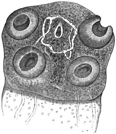
Fig. 20.—Head of Tænia mediocanellata. Showing the calcareous corpuscles, suckers, rudimentary
proboscis, and water vessels. Highly magnified. Original.
The establishment of this species as distinct from T. solium is
due to Küchenmeister; but it is curious to observe how accurately this
determination was foreshadowed by the shrewd naturalist and theologian,
J. A. E. Goeze, who clearly indicated two forms of the common tapeworm,
remarking (l. c., Bibl. No. 1, s. 278):—“Die erste ist die
bekannte grosse, mit langen dicken und gemästeten Gliedern, die ich
Tænia cucurbitina, grandis, saginata, nennen will.” The same author
(s. 245) pointed out the resemblance subsisting between the tapeworm
of the cat (T. crassicollis) and the vesicles (“Krystallblasen”) and
their contained “erbsförmige Blasen” (Cysticercus fasciolaris) of the
mouse. Thus the celebrated pastor of St Blasius, in Quedlinberg, almost
contemporaneously with Pallas, early
arrived at the conclusion that the hydatid-measle was a kind of
tapeworm.
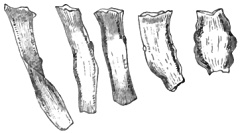
Fig. 21.—Free proglottides of
Tænia mediocanellata. After Leuckart.
Respecting the organisation of this worm I may observe that the
mature joints have a more complicated uterine organ than obtains in
Tænia solium, presenting nearly double the number of lateral branches.
They are more closely packed, running outwardly in an almost parallel
manner. The first sexually mature proglottis occurs at about the 450th
joint, but whereas, in the pork tapeworm, only some 200 subsequent
segments share this perfect character in the beef tapeworm, according
to Leuckart, as many as 360 or even 400 mature joint may be present.
The joints are very liable to form monstrosities; these abnormalities
sometimes affecting the reproductive organs, which become doubled
or even trebled. In the Hunterian collection there is a proglottid
showing twenty-two sexual orifices. Dr Cullingworth, of Manchester, has
described a specimen in which the joints are curiously tripartite.
As already hinted the true source of this parasite has been proved by
experiment; the first successful worm-feeding having been accomplished
by Leuckart. Mosler’s, and subsequently my own feeding experiments,
immediately followed. Other successful experiments with this species
have been conducted by Zurn, Probstmayer, St Cyr, Perroncito, Masse and
Pourquier, and Zenker. As will be again mentioned below Dr Oliver, R.A.,
whilst stationed at Jullundur, successfully reared the adult tapeworm in
a Mohammedan groom and in a Hindoo boy. It will also be seen that Prof.
Perroncito reared the worm in a student in fifty-four days. In my own
experiments on animals I was assisted by Professor Simonds. The feeding
materials were tapeworms expelled from my own patients. We obtained the
following interesting results:
Exp. 1.—A calf. First feeding, Dec. 21st, 1864. Marked
symptoms. Slaughtered April 3rd, 1865. Result positive.
Exp. 2.—A calf. First feeding, April 13th, 1865. Second,
third, and fourth feedings in May and June. No symptoms. Died on Sept.
3rd, 1865, after thirty-six hours’ illness with
“cattle plague.” Result stated to have been negative as far as the
muscles were concerned. Viscera not examined.
Exp. 3.—A Dutch heifer. First feeding, March 3rd, 1865. Three
subsequent feedings. Symptoms only slight. Slaughtered April 4th, 1866.
Result positive. Measles especially numerous in the diaphragm, but all
had undergone calcareous degeneration.
Exp. 4.—A calf. Fed May 27th, 1872, with ripe proglottides.
Marked symptoms set in on June 7th, which began to abate on the 12th,
and had nearly disappeared by the 20th of the same month. The record of
the post-mortem result has been lost; but the animal was infected.
Exp. 5.—A calf, which had been made the subject of a
“glanders experiment.” First fed on Oct. 17th, 1872, and thrice in the
following year, Jan. 1st and 11th, and March 8th. No symptoms having
appeared the animal was kept for six or eight months after the last
feeding. Seeming to be free from disease of any kind, it was sold as a
sound heifer.
Exp. 6.—A young heifer calf, of six months. Fed Oct. 18th,
1873, with the mature proglottides of a large beef tapeworm. No
symptoms. Slaughtered several months afterwards. Result stated to have
been negative. Unfortunately I was not present at the autopsy.
Exp. 7.—A young heifer. First fed May 19th, 1874, with
the joints of a tapeworm, and again on June 12th. No apparent ill
effects resulted, but the animal died in October. At the post-mortem
examination, made by Prof. Simonds, no parasites were observed.
Subsequently I found calcareous specks in the liver which proved to be
degenerated measles.
Exp. 8.—A calf. Fed on or about March 24th, 1875, with
sexually mature joints. The calf was put to and remained with a foster
mother until it died from disease of the larynx on the 15th of the
following July. The animal was ill-treated by its foster parent, and
at the post-mortem I observed a large intercostal cicatrix, evidently
the result of injury. In this case I devoted several hours to the
exploration of the muscles and viscera. Not a trace of the Cysticercus
bovis could be found in the muscles or connective tissues, but the
liver contained scores of perfectly developed measles, besides hundreds
of others in various stages of calcareous degeneration. On comparing
some of the latter with those I had obtained from the preceding
experiment the pathological appearances were at once seen to be
identical. It was easy to find and pick out the measles in their cysts
from the naturally friable liver. I also detected four Cysticerci in the
lungs, two of which had degenerated. Microscopic examination confirmed
my interpretation of the naked-eye appearances.
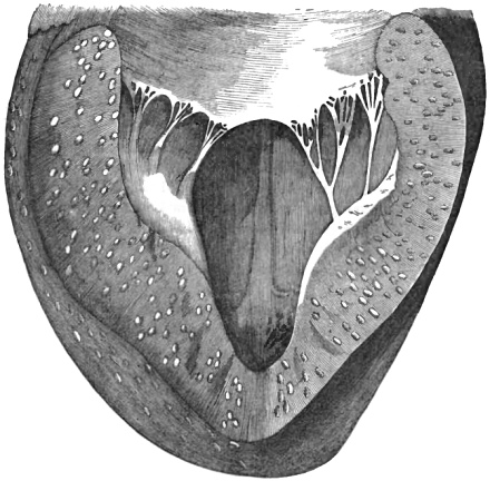
Fig. 22.—Section of the heart of a calf
infested by cestode larvæ. After Mosler.
Fragmentary as the above data are, they serve to show that we have
hitherto been too hasty in concluding that beef and veal measles reside
only in the voluntary and striated muscles of their hosts. The facts
here recorded prove that the liver of a calf may be extensively invaded
by cysticerci, and yet the animal will exhibit no sign of constitutional
disturbance. The cestode tuberculosis may come and go without any
diagnostic symptom, whilst a few months suffice for the natural death
and decay of the parasite by calcareous degeneration. Thus it becomes
extremely probable that many experiments hitherto regarded as negative
in their results have really been positive; the pathological evidences
having been either misinterpreted or
altogether overlooked. Every pathologist is familiar with gritty
particles in the various viscera of man and animals, but few are
probably aware how constantly these are dead and degenerated Cysticerci.
The gritty particle itself may be reduced to the merest point, no
larger than the receptaculum capitis of the Cysticercus itself,
and in course of time it will disappear entirely. Practically it is
satisfactory to have experimental evidence of the fact that cattle, as
well as other animals, however extensively measled they may have been,
can become thoroughly cleansed of the disorder by nature herself. It is
only necessary that the diseased animals be separated from infectious
influences.
Although the beef measle has never yet been found in man, I have
for convenience sake introduced the facts of larval parasitism in this
place. The sanitary bearings of this subject are far too important to
be dismissed in a summary manner. I have shown that the prevalence or
rarity of the beef tapeworm in man is strictly dependent upon the habits
of the people; this same cause operating to produce healthy or diseased
meat-food, according to the degree of civilisation. In this connection
the oft-quoted statements of Kaschin respecting the prevalence of
tapeworms among the Burätes, and the well-known frequency of this
entozoon in Abyssinia, need only be alluded to.
When discussing the food question in my ‘Manual,’ I freely availed
myself of facts privately communicated by Dr Joseph Fleming, and I
especially referred to the published labours of Lewis, Hewlett, Veale,
and other observers stationed in India. Beef measles are extremely
common in the cattle of the north-west provinces of India, so much
so that severe restrictions have been imposed upon the consumption
of ration beef. The presence of a few measles in the flesh of cattle
has been deemed a sufficient excuse for condemning and burying entire
carcases. The measle is easily distinguished from that of mutton and
pork by the fact that its head is not furnished with hooks, whilst in
the place of a rostellum there is a small, centrally placed, retractile
disk, which assumes the appearance of a supplementary sucker as in the
adult worm. The four true suckers are also comparatively large. The
measle usually varies in size from the fourth to the half of an inch in
length, but my cabinet contains a specimen nearly an inch long. This was
contributed by Dr J. Fleming, who mentions having seen
a measle which, when unrolled, measured nearly an inch and a half
in length. Although thousands of these bladder worms must exist in
the cattle of England, up to the present time not a single instance
has been recorded of the occurrence of these cystic parasites in the
United Kingdom, except in our experimental animals. Notwithstanding my
inquiries, I have not yet found a butcher, flesher, meat-inspector,
or veterinarian, who has encountered this parasite in any animal
slaughtered for the market. Several butchers have denied their
occurrence in meat sold by themselves. Even so late as June, 1874, the
presence of measles in the flesh of cattle was denied before an assembly
of French savans; yet for many years past I have constantly exhibited
measly beef and veal in the lecture room of the Royal Veterinary
College. (See the discussion of the Société de Thérapeutique, recorded
in the ‘Bullétin Gén. de Thér.’ for June 30th, 1874, and also the ‘Jour.
de Thér.,’ No. 14, for July, p. 556, where, however, special remarks on
this head have been omitted; see also the ‘Lond. Med. Record’ for July
29th, 1874, p. 472, and the ‘Lancet’ for Dec., 1874, p. 794.) Quite in
contrast with the statements referred to are those of recent Italian
observers.
Some few years back Professor G. Pellizzari communicated to the
Medico-Physical Academy, at Florence, the results of a series of
experiments conducted by himself, with the assistance of Dr Tommasi, in
regard to the temperature necessary for the destruction of cysticerci in
measled meat. An account of these experiments is published in Tommasi’s
edition of my ‘Manual.’ The researches were made in relation to certain
sanitary measures effected by the Municipal Commission of Florence,
the express object of these measures being to prevent the injurious
distribution of measly meat, especially that of swine. Signor Bosi, the
superintendent of the public slaughterhouses, granted every facility in
his power. In a previously published memoir by Professor E. Perroncito
it was stated that measly meat (panicatura degli animali) required a
higher temperature than that of boiling point for the destruction of the
bladder worms in question. In this opinion Signor Bosi shared. According
to the original memoir of Perroncito we are told that “about twenty
specimens of Cysticerci were collected by the author, and placed in
boiling water. After twenty minutes’ boiling, not one of the parasites
appeared to suffer. The head continued to be drawn into the body, and
when the Cysticerci had their heads drawn out one by one they still
appeared to possess all the elasticity of living bladder worms,
displaying those movements of extension which are proper to parasites
not yet dead. The hooks were observed regularly disposed on the
proboscis, where they formed a double crown, the suckers remaining
intact.” Perroncito remarked, however, that the Cysticerci showed a
coloring tendency towards brown, and he added that “with the aid of
two needles it became easy to lacerate the body of the Cysticercus,
which appeared to be swollen, and possessed of diminished cohesion
of its parts.” It was evident to all eyes, observed Professor
Pellizzari, that these statements involved clear contradictions.
Yet again, at page 28 of the memoir, Professor Perroncito wrote:—“During the past winter I introduced
some little slices (fettuccie) of muscle-flesh (8 to 10 millimètres
in thickness), infested with Cysticerci into a vessel (cassolina)
containing fat at the temperature of 190 to 200° Cent. (374 to 400°
Fahr.). At the expiration of ten or fifteen minutes the slices of meat
were fried, and the Cysticerci lying at the surface had acquired a light
brownish colour, as if they were roasted. By breaking up the slices
one could still see the small reddish muscular bundles, whilst the
Cysticerci in the middle remained entire and well preserved. Their heads
displayed the hooks and suckers regularly distributed.” It is certainly
singular, as Pellizzari observes, that these Cysticerci, having been
thoroughly fried and roasted, should still remain alive and in their
normal state; but the ultimate conclusion at which Perroncito arrived
was still more startling, and one which, if it were true, would not fail
to create a considerable stir among our officers of health. On reviewing
the whole matter Perroncito says:—“It appears
to me that the melted fat alone of hogs (maiali grandinosi) should be
utilised, and I am pleased to reckon the illustrious Gerlach and all
other distinguished practitioners to be of the same opinion. Permit me,
therefore, being well satisfied also with the results of many other
experiments, once more to advance the conclusion that, if it is not
certain that the Cysticerci die at from 80 to 100° Centigrade (176 to
212° Fahr.), we are quite sure that they dry up and become completely
mummified at 125, 130, and 150° Cent. (257, 268, and 302° Fahr.),
temperatures which we could easily produce by means of a properly
constructed apparatus.”
After remarking upon the serious nature of the conclusion
which Perroncito sought to establish, Professor Pellizzari makes further
use of quotations which bear upon the question as to whether the quality
of the vessels in which the fat of diseased hogs is melted down may not
largely affect the degree of high temperature sought to be obtained (in
view of a perfect destruction of the Cysticerci). Perroncito repeatedly
witnessed the operations of pork-butchers; and when portions of meat
were introduced, with water, into the cauldrons, he always saw that the
temperature “was maintained between 97° and 98° Centigrade.” However,
this part of the question may be dismissed in a very few words, since
Perroncito himself finally allows that “the different composition of the
vessels cannot elevate the temperature of the fat by many degrees.”
With the praiseworthy intention of either verifying or refuting
these conclusions, Pellizzari, with the approval of Bosi and with the
assistance of Tommasi, instituted a fresh series of experiments at a
private laboratory. The details of these experiments are exceedingly
interesting; but as their record occupies several pages of Tommasi’s
appendix already referred to, I must content myself with a general
statement of the results obtained. Professor Pellizzari found that
Cysticerci, so far from requiring a temperature of upwards of 100°
Centigrade for their destruction, die at a temperature of 60° Centigrade
(140° Fahr.). He had, it appears, previously taken the initiative in
recommending certain measures to the Florentine municipality, in view of
protecting the public health, and he had now the satisfaction of more
than confirming the wisdom of these sanitary precautions. In excessively
measled animals the fat is removed and boiled in suitable cauldrons, and
has potash mixed with it to render it useful for industrial purposes.
By the various measures adopted the entire animal is utilised, and with
proper precaution there seems little chance for the measles to arrive at
the tænioid or sexually mature condition.
In the next part of his communication Pellizzari touches upon the
question of measles in beef, referring especially to the experimental
labours of Leuckart and myself. Finding additional support from our
views Pellizzari declared the propositions of Dr Perroncito as of no
value whatever. “But how is it,” he adds, “that notwithstanding that so
low a temperature suffices to kill these cysticerci, yet cases of Tænia
are continually occurring?” The answer to this question will appear in
the sequel; but meanwhile it will be as well to refer to the
recent brochure by Dr Giacomini. This author appears to have had no
opportunity of perusing Pellizzari’s communication already cited, and
consequently it is not surprising that he should, in common with others,
have accepted the original conclusions of Perroncito. Dr Giacomini
clearly perceives that, whatever precautions of a hygienic character are
suitable for the prevention of disease arising out of the consumption
of measly pork, the same, or at all events similar, measures ought
to be adopted with the view of checking tapeworm affections arising
from the ingestion of other kinds of meat, especially veal and beef.
Like Pellizzari, he is satisfied as to the human origin of the small
bladder worms found in cattle, and establishes this position not only
from the oft-quoted experiments of Leuckart and Mosler, but also from
those conducted by myself and Simonds in England, and by Professor
F. Saint-Cyr in France. From a careful review and consideration of
all the facts of the case, he recommended a more complete supervision
over the flesh of oxen before it is employed commercially, and greater
precaution when employing veal as food, by causing it to be subjected
to a high temperature, in order that the parasites may be killed before
it is ingested. It is evident that Giacomini thinks that a temperature
exceeding that of boiling-point is necessary for the destruction of the
beef and veal measles, since he immediately adds, “Though experiments
have not been made with the object of ascertaining the amount of
resistance of heat which the unarmed cysticercus can bear, yet, judging
by those conducted by Professor Perroncito on the measle of the hog, we
are in a position to say that a temperature of 135° Cent. (275° Fahr.)
is necessary for the destruction of an isolated Cysticercus, whilst the
heat should be raised from 150° to 200° Cent. (302° to 392° Fahr.) for
ten or fifteen minutes, in order to ensure the complete destruction of
the Cysticerci encapsuled in the interior of a piece of meat.” I have
abridged this portion of Giacomini’s text, because his statements are
pretty much the same as those already quoted from Perroncito (as cited
by Tommasi). But, in the next place, Dr Giacomini is in error when he
states that experiments had not been performed on the Cysticerci of
the ox. So far from this being the case, similar experiments had long
previously been conducted by Dr Lewis in India; and these researches
had quite as much to do with the measles or Cysticerci of beef as they
had with those of the hog, if not more. Naturally but few foreign
investigators can
have had access to the work in which Lewis’s experiments were originally
recorded, and to which, therefore, I must call their attention. Thus, Dr
Tommasi has fallen into the error of supposing that the investigations
of Lewis were made in England. It is of very little moment where the
experiments were carried on, but Tommasi’s statement (appendix, loc.
cit., p. 161), wherein he says that Pellizzari’s experiments, in which
he himself took part (ai quali io stesso ho assistito), are even more
complete than those made in England by Dr Lewis, and in Germany by
Dr Küchenmeister, cannot be allowed to pass unchallenged. If Tommasi
had enjoyed the opportunity of consulting Lewis’s original memoir, he
would not have underestimated our countryman’s labors. The memoir by
Lewis is singularly complete, and well-nigh exhausts all the facts that
can have any interest in relation to the question of public health.
Towards the close of his essay he expressly states, as the result of
investigation—“(1) That exposure to a temperature of 120° Fahr.
for five minutes will not destroy life in Cysticerci, but that they may
continue to manifest indications of life for at least two or three days
after such exposure; (2) that exposure to a temperature of 125° Fahr.
for five minutes does not kill them; but (3) after being subjected to
a temperature of 130° Fahr. for five minutes, they may be considered
to have perished. After exposure to this and higher temperatures, in
no instance have I been able (he adds) to satisfy myself that the
slightest movements took place in their substance when examined even
under a high power. At least, it may be confidently asserted that, after
exposure for five minutes to a temperature of 135° to 140° Fahr., life
in these parasites may be considered as absolutely extinct” (p. 139).
Thus the statements of Lewis and Pellizzari were in perfect accord; and
seeing that their conclusions were alike the result of very careful and
independent inquiry, it seemed as if the question at issue was finally
solved. These investigations made it perfectly clear that Cysticerci of
all kinds, whether found in veal, beef, or pork, could not retain their
vitality when exposed to a temperature of 60° Centigrade, or, in other
words, 140° Fahr.
The rather severe strictures made on Perroncito’s earlier experiments
induced the Turin professor to go over the subject more carefully, when
he obtained excellent results. He finally ascertained that Cysticerci
perished at a temperature below
50° C. (122° Fahr.). In May, 1877, Dr Perroncito furnished me
with an account of his researches. With the exception of a few verbal
alterations, for which I am responsible, Perroncito wrote as follows:
“In order to resolve the highly important question of the tenacity of
life of the Helminths and corresponding larval forms, I made since 1871
a very long series of experiments on the Cysticercus cellulosæ, which
were published almost at the same time with others of the same kind,
made by Dr Lewis in Calcutta. Towards the end of 1874 Mr Pellizzari, of
Florence, disputed the results of the investigations which I had made
known two years before, i.e. in 1872, and agreed with Dr Lewis, who
had stated already that the Cysticercus exposed to a temperature of
55° C. can be held for dead after five minutes, and also with Dr
Cobbold, who thought the temperature of 60° C. quite enough to
kill it. But the characters he (Mr Pellizzari) relied upon, needing the
exactness and precision required to enlighten and persuade in the most
important scientific questions, gave rise to a mistrust in the most
scrupulous amongst the men devoted to biological pursuits and to several
hygienic measures on the part of the sanitary inspectors with regard to
infected pork. Therefore, my conclusions, argued from the experiments
made in 1871–72, were still those followed by the most important
Italian cities, and approved in principle by the superior Board of
Health in 1873. I expressed doubt then about the Cysticercus dying
at a temperature lower than 100° C., and some person misconstrued
these doubts, saying that I had contradicted myself in my work. However,
as I could not assert they died at 80°–100° C., I only
noticed the alteration of color and cohesion which happened in the
Cysticercus exposed to various degrees of temperature, to the end that
I might contribute usefully to the solution of the difficult question,
and concluded that ‘if we could not be sure of the Cysticercus
dying at 80°–100° C., it was certain at all events that
they perished at 125° or 130° C.’ Not wishing to prejudice the
question, I never said that they did not die at 80°–100° C., but
simply stated that at this temperature we could not be certain of their
death.
“Now, after a large number of experiments, I have been able to
ascertain with exactness the lowest degree of temperature required to
kill infallibly the Cysticercus and other parasites of animals. The
means I made use of for this kind of investigation
were Mr Schulze’s heating table, the neutral tincture of carmine, the
tincture of hæmatoxylon, and breeding experiments.
“My method is founded essentially—
“(a) On the fact that the Cysticercus when it is fresh and is
stretched and conveniently prepared in pure water, or in chloride of
soda very much diluted, and afterwards brought gradually from the
temperature of the ambient air to that of the body of higher animals and
to degrees of heat still more elevated, until life is extinct, keeps
moving to and fro with more or less energy throughout its body, using
especially its suckers and proboscis.
“(b) On the greater imbibing power of the dead tissue generally,
which is undoubtedly far more apparent in insects and plathelminths.
“(c) On the experiments made to ascertain the value of the two
above-stated facts.
“If, after having prepared a Cysticercus, newly extracted from a
pig in the way we have pointed out, we examine it with a microscope
on M. Schulze’s heating table, we find that usually it begins to move
after 30° or 35° C., and each moment with greater activity,
especially after 38°, 40°, 42°, 44°, 45° C. The temperature being
raised progressively, we see that the Cysticercus cellulosæ puts a
stop to its movements occasionally at 45–46° C., seldom at
47° C., more frequently at 48° C., sometimes at 49° C.;
and, in fifty and more experiments, only one Cysticercus was able to
live on beyond 49° C., standing still at 50° C.
“As soon as it stands still the parasite is dead. In fact, if we
lower again the temperature gradually to that of the ambient air,
and if afterwards we raise it a second time, we pass through all the
intermediate temperatures without the Cysticercus showing the least
signs of life.
“But a more convincing proof of the death of the parasite is got from
the greater imbibing power of the tissue when life is extinct, the same
over the whole body of the plathelminths, and their larval forms. If
we dip the Cysticercus alive with its head stretched in the neutral
tincture of carmine or hæmatoxylon we can leave it there even two, four,
eight, ten, or twelve hours and more, without the head coloring or a
real imbibition taking place; this begins only after the Cysticercus
is dead, so that if the Cysticercus is brought first to a temperature
hot enough to kill it (with M. Schulze’s tables to one of 48°, 49°, 50° C.)
and
dipped afterwards in the above-mentioned tinctures, it colors intensely
in less than 45°, beginning from the head, and onwards to the extremity
of the cyst of the tail. The head colors more intensely and rapidly than
the neck, as it is covered with very numerous calcareous corpuscles,
which are not met with so frequently in the remaining part of the
body.
“Cysticercus cellulosæ of the pig, and that of the Tænia
mediocanellata of the calf, brought gradually to a final temperature,
the first of 50° C., and the second of 44°, 45°, and 47° C., and
then swallowed alone, or with a piece of butter or crumb of bread, never
produced the Tænia in the valiant students who voluntarily undertook
to make the experiment of swallowing them.
“My investigations were extended to other kinds and forms
of Helminths, and the results were always the same, so that,
abiding by the same principles, I was able to ascertain that—
“1st. The Cysticercus cellulosæ of the pig dies sometimes at
45° C., more frequently at 47° C., ordinarily at 48° C.,
very seldom reaches alive 49° C., and is quite an exception when it
resists for a few moments the temperature of 50° C., so that we can
say that the Cysticercus brought gradually up to this temperature most
assuredly dies if it is kept there longer than one minute.
“2nd. A Cysticercus cellulosæ, extracted by Professor Raymond from
the conjunctiva of a child’s eye, died between 45° and 46° C.
“3rd. The Cysticercus of the Tænia mediocanellata dies sometimes
at 44° C., very often at 45° C., and does not resist a
temperature superior to 46° C.
“4th. The Cysticercus pisiformis of the rabbit, like the
cellulosæ, dies sometimes at 45° and 46° C., but generally stands
still and perishes at 47° and 48° C.
“5th. A Cysticercus tenuicollis died at 49° C.
“6th. The scolici of the Cœnurus cerebralis of a sheep died at
42° C.
“7th. The scolices of the cysts of Echinococcus polymorphus die
generally between 47° and 48° C., and in no case amongst those I
have experimented on did it reach 50° C. alive.
“8th. The Tænia cucumerina died, one at 43° C., and a second
parasite at 45° C.
“9th. A few individuals of Tænia serrata of the dog died at
50° C.
“10th. Two individuals of Tænia perfoliata of the horse died, the
first at 45° C., the second at 50° C.
“11th. The embryos of the Filaria microstoma of the horse began to
stand still at 46–47°, and all died at 48° C.
“12th. The embryos of the Filaria megastoma of the horse’s stomach
died at 47° C.
“13th. The Trichina spiralis, both free and in a cyst, in several
experiments always died at 48° C.
“14th. The embryos of the Strongylus filaria of the sheep stood
still at 50° C.
“15th. Probstmayer’s viviparous oxyurids, the infusoria of the colon
and cæcum of the solipeds, and the psorosperms of the liver of the
rabbit did not stir at all.
“Each experiment lasted about ten minutes, and the temperature
rose from 8–10° C. to 45–46° C. in six to eight
minutes; and from 46° to 50° in one minute. These experiments have a
great value, both scientific and practical, as they show, on one side,
which is the lowest intensity of heat sufficient to kill always the
Cysticercus, the Trichina, and other parasites, reducing thus by
far the tenacity of life generally attributed to a large number of
Helminths and corresponding larval forms. They assure us, moreover, of
the harmlessness of the flesh infected by the above-mentioned parasites,
when it is cooked in such a manner as to reach the temperature of
50° C. over all points of the pieces, even though it be kept at
such a degree of heat not longer than five minutes.
“In a piece of leg of pork the Cysticerci were found alive in
all places not yet putrefied twenty-nine days after the animal had
been slaughtered. On the other hand, in the dry muscles of a calf the
Cysticerci of the Tænia mediocanellata were all found dead fourteen
days after the slaughtering of the animal. I have ascertained that
putrefaction of the flesh is fatal for the two larval forms of these
different kinds of helminths.”
In a subsequent communication received from Professor Perroncito
towards the close of the year 1877 he writes:
“At the last meeting, held on April 23rd, I made a statement
to the Medical and Surgical Society of Turin, of the results of
other experiments tried by heating at M. Schulze’s table and by the
imbibitions with the neutral tincture of carmine, through which I came
to the conclusion that the Cysticerci of the Tænia mediocanellata
die sometimes at 44° C., now and then at 45° C., and always at
46° C. I therefore concluded
that they could in no case survive at 47° C. and 48° C. when
they were maintained at this temperature at least five minutes. But to
the end of more fully corroborating the facts I had thus communicated,
I, contemporaneously with these, made some breeding experiments with the
same Cysticerci on bold and courageous students who generously offered
themselves for the benefit of science.
“Consequently I am now enabled to state that neither Mr Gemelli
nor Dr Ragni contracted the Tænia, though each of them had eaten a
Cysticercus of the Tænia mediocanellata previously, and respectively
subjected to a temperature of 45° C. and 47° C. The larvæ
were properly prepared and submitted to gradual heating on the
above-mentioned table, and swallowed when they no longer gave signs of
life. In like manner no generation of the Tænia took place in the
body of Mr Martini, who ate the Cysticercus brought to a temperature
of 44° C. It was maintained at this degree of heat during a period
of about three minutes, and swallowed whilst a very slight movement was
still visible in a portion of its neck.
“In another student, on the contrary, who ate a living Cysticercus
of the Tænia mediocanellata, the tapeworm reached its maturation in
fifty-four days and eliminated the two first proglottides. It threw
off two more on the fifty-eighth day, and thirty on the sixtieth.
Sixty-seven days after swallowing the Cysticercus this courageous
young man, having, like his three companions, taken some kousso and
castor oil, emitted the strobila. It was furnished with 866 rings, but
destitute of the neck and head. Its measurement afforded a total length
of 4·274 mètres.
“Adding now to the 866 proglottides the thirty-four already
eliminated, 900 would be the number of the segments; and reckoning the
length of each of the latter to be fourteen millimètres, we should have
had the strobila (deprived of the head and neck) reaching a length
of 4·75 mètres. Further, calculating the head and neck to be eight
millimètres long, a total length of 4·83 mètres would be the result.
“From all these facts we may conclude that the Tænia has, in our
instance, reached an approximative length of seventy-two
millimètres a day, affording a daily production of 13·43 proglottides.”
In relation to requirements of state medicine I have thought
Perroncito’s researches sufficiently valuable to be quoted at some
length; but their chief interest culminates in the worm-
feeding experiments. Excellent in all respects as was the conduct of the
medical students who, with Professor Perroncito’s approval, swallowed
living specimens of the Cysticercus bovis, the intentional ingestion
of beef measles is by no means a novelty. Eight or ten years back Dr
Oliver (after explaining to one of the selected victims the possible
consequences of the experiment) induced a Mahommedan syce or groom and a
Hindoo boy to swallow perfectly fresh and living beef measles. In this
way Dr Oliver successfully reared the Tænia mediocanellata in India,
and he was thus enabled to fix the amount of time necessary for the
full growth of the strobila. Many other persons have displayed an equal
amount of zeal in the cause of helminthology, by partaking of the larvæ
or germs of other parasites. Thus, at the risk of repetition, I may
state that Möller many years ago swallowed the slender-necked hydatid
(Cysticercus tenuicollis) in the hope of infesting himself with Tænia
marginata. Several persons have defiantly swallowed trichinised flesh.
Professor Leuckart and some of his pupils also courageously swallowed
the eggs of Oxyurides, and they had the infinite satisfaction of
noticing the young worms in their fæcal discharges some fifteen days
afterwards. Dr Crisp ate part of the cooked flesh of an animal that had
died of cattle plague, and I myself partook of moderately cooked meat
which I knew to be swarming with psorosperms. These obscure organisms
were by some persons considered to be either a cause or product of
the rinderpest. They will be noticed in my account of the Protozoal
parasites.
For the purpose of advancing science and the welfare of the people,
there are scores of persons always to be found ready to make personal
sacrifices of the kind undertaken by Drs Ragni, Martini, and Gemelli.
Unfortunately for English science there are not wanting people in
this country who are prepared to threaten with fines and imprisonment
any savant who may think it desirable to perform a similar set of
feeding experiments on animals. Invaluable for good as our experimental
investigations have already been, it would seem as if it were the
deliberate aim of these sentimental obstructives to put a stop to the
acquisition of all useful knowledge in the future.
In reference to the rate of growth of tapeworms, Professor
Perroncito’s determinations are useful, inasmuch as they verify certain
ascertained facts with precision and confirm the general
conclusion that had been drawn by practical helminthologists from
various sources of information. In regard to the number of proglottides
proper to a sexually mature tapeworm, the circumstance that Perroncito’s
calculation was made without the head and a portion of the neck of
the worm being present shows that it cannot be relied on absolutely;
nevertheless, as far as it goes, it tends to confirm what Leuckart had
long previously stated. I have possessed myself of upwards of thirty
perfect beef tapeworms expelled from my patients, and in some of the
specimens it was noticed that the segmentation-rings in the region of
the neck were far more crowded together than they were in others. I also
possess a perfect Tænia mediocanellata, removed post mortem. Though
the rate of growth may be the same from day to day, yet experience
has shown that the number of proglottides actually cast off varies
exceedingly. Küchenmeister’s estimate of the average number agrees in
the main with what we have ourselves observed (five to twenty daily);
and here again Perroncito’s investigations serve to verify the general
correctness of our previous determinations.
To return to Pellizzari’s researches, one of the most important
questions is that which relates to the prevalence of tapeworm. In this
connection he first brings forward some very interesting and instructive
data that had been previously communicated to the Medico-Physical
Academy of Florence by Professor Marchi. On the occasion referred to
Marchi had stated that, out of thirty-five Tæniæ which he had examined,
only one belonged to the species known as Tænia solium; all the other
thirty-four being of the unarmed type, or Tænia mediocanellata.
Reflecting on this striking fact, and also on the circumstance that
he had in vain begged his colleagues to send him specimens of Tænia
solium, Marchi seems to have missed the very palpable explanation of
this otherwise strange phenomenon. “How does it happen,” exclaimed
Marchi, “that, notwithstanding the occurrence of 13,000 kilogrammes of
the flesh of measled hogs in the public butcheries, I have seen but one
specimen of Tænia solium, whilst thirty-four cannot have originated
from the pig?” “The wherefore is obvious enough,” replies Pellizzari,
“because our hygienic regulations demand that the flesh of the hogs be
raised to a temperature of 60° Cent. (140° Fahr.);” and he then himself
immediately proceeds to ask another question, namely, as to how it
happens that the Tænia solium is so frequently seen in other places.
To his own question Pellizzari responds by remarking—(1)
that there are not so many precautions (of a sanitary kind) taken in
other places; and (2) that the people elsewhere consume more slightly
salted or uncooked meat, as sausages and so forth (come salame giovane,
salciccia e via dicendo). Pellizzari, having explained that Marchi’s
thirty-four tapeworms must all have arisen from the consumption of
the Cysticercus of the ox, then goes on to speak of the prevalence of
tapeworm in Florence, even in little children. This last-named feature,
he says, is due to the circumstance that raw meat is frequently employed
as a restorative (come cura ricostituente). “Thirty years ago,”
remarks Professor Pellizzari, “it was just as difficult to find a single
Tænia mediocanellata as it is now easy to find a great number of these
worms; and all because it is nowadays customary to eat the flesh of
the ox either insufficiently cooked or raw. This absolute inversion of
the facts of the case affords proof of the correctness of the position
sustained by me, to the effect that the cooking of meat up to the degree
of temperature necessary for ebullition ensures the destruction of the
Cysticerci.” Notwithstanding this statement of his own, Pellizzari
thinks that the interference of inspectors may be pushed too far, and
thus serve to bring about the very disasters which it should be their
supreme object to prevent. Thus, he argues against the suggestions of
those who would entirely prevent the sale of measly meat, and who would
only permit, as obtains in the province of Modena, the melting down of
the fat of hogs. Very strict measures of this sort would, as he says,
constitute a radical means of entirely stamping out Tænia, but he also
very judiciously reminds the sanitarian (igienista) that “such a step
would be a serious thing for the tradesman, bringing injury not only
to the municipal administration, but also proving an encouragement to
smuggling. In this way the public health would sustain worse injury by
the inducement held out to the owners of infected animals to slaughter
them in secret butcheries, thus little by little withdrawing the meat
from the superintendence of the public officials. By the adoption of
fraudulent measures there would be a daily consumption of diseased
meat; and thus also, while the public administration would suffer loss,
the public health, on the other hand, would gain nothing.” In effect
Pellizzari says, if we advise the employment of more severe and radical
measures than those already in vogue in Florence, we should overburden
the tradesman, almost compel him to
defraud the exchequer by smuggling, and greatly injure the public
health.
The facts and explanations advanced by Italian writers regarding the
causes of the endemic prevalence of tapeworm, are in perfect harmony
with those previously obtained from other sources. Respecting these
causes there is much that is both new and interesting. The eighth
annual report of the sanitary commissioner of the Government of India
had already made us acquainted with the fact that during the year 1869,
out of 13,818 head of cattle slaughtered in the stations of the Upper
Punjab, 768 beasts were found to be infected with measle-cysts. This,
as I have remarked (Tommasi’s edit., p. 54), “affords a rate of 5·55
per cent., being a considerable diminution of the proportion observed
in 1868, when the percentage gave a total of 6·12. The reduction was,
without doubt, due to the vigilance and enlightenment of the army meat
inspectors. The prevalence, however, of tapeworm does not bear relation
to the number of animals infested with Cysticerci so much as to the
actual number of Cysticerci developed in infected animals. I have
frequently pointed out the inadvisability of condemning and burying
the carcases of measly oxen, whether there be few or many Cysticerci
present, and I have stated, on trustworthy evidence, that even the
presence of a few Cysticerci is deemed by some inspectors a sufficient
reason for rejecting the entire animal. Such a waste should never be
allowed. In regard to the numbers of ox-measles present in particular
instances, I have elsewhere adduced some remarkable facts communicated
to me by Dr Joseph Fleming, of the Indian Army Medical Staff. None of
my experimental animals, though fed with scores of ripe proglottides,
yielded such an abundance of Cysticerci as Dr Fleming encountered
in Punjab cattle. In one pound weight of the psoas muscles Fleming
counted no less than 300 Cysticerci.” From this it follows that the
flesh of a largely infested animal is capable, under the circumstances
of ration distribution and imperfect cooking, of originating numerous
tapeworms.
Not many years back the leading medical journal of this country
challenged me to produce evidence as to the injuriousness of beef and
mutton from Cysticerci. The writer stated in his article that I had
“failed to produce a single specimen of beef or mutton measles” which
had not resulted from experiments conducted “at the Royal Veterinary
College;” and he
said, further, “that butchers, fleshers, and veterinarians were
practically right in refusing to adopt the opinion of Dr Cobbold,
that measled beef or mutton is produced to any great extent”
independently. How palpably I endured a species of unjust
reproach for being somewhat in advance of the knowledge
current at the time may be gathered from the voluminous
evidence which has since cropped up from various parts of the
world. It was, indeed, mainly through experiments conducted
at the Royal Veterinary College, and reported in the ‘Lancet,’
that professional men in India first became acquainted with the
possibility of finding Cysticerci in beef.
The statements of Dr Joseph Fleming, who was one of the
foremost in discovering cystic disease in cattle, have since
received abundant confirmation. The Indian Government
Reports given in the February issue of the ‘Madras Monthly
Journal of Medical Science’ for 1873 are especially instructive.
Referring to the prevalence of Cysticercus in the ration beef at
Jullundur, in the Punjab, the Inspector General (India Medical
Department) reports as follows:
“Cysticercus was first noticed here in the beef tendered at
the Royal Artillery ration stand in May, 1868. For some two
years previous to this date condemnations of cyst-infected meat
had been frequent at Peshawur, Rawul Pindee, Meean Meer
and several other stations in the upper part of the Punjab, and
here I had often detected the parasite in meat exposed for sale
in the bazaars, but no trace of it had been observed in the
Commissariat beef, either by myself or any other medical officer
who had preceded me.
“From May, 1868, to November, 1869, ‘cyst’ was more or
less frequently found both at the Artillery and 92nd Highlanders’
ration stands; but since the latter date it has almost
entirely disappeared.
“The following table shows the quantity of meat destroyed
on this account during 1868 and 1869:
| Years. | Months. | Number of cattle
infected. | Weight of meat
destroyed. |
|---|
| 1868 | | May | 114 | 412 | lbs. |
| June | 111 | 77 | " |
| September | 111 | 130 | " |
| October | 110 | 1,763 | " |
| November | 114 | 2,010 | " |
| December | 112 | 1,785 | " |
|
| 1869 | | January | 121 | 4,062 | lbs. |
| February | 116 | 2,341 | " |
| March | 114 | 2,209 | " |
| April | 115 | 856 | " |
| May | 112 | 220 | " |
| June | 111 | 122 | " |
| July | 111 | 194 | " |
| August | 113 | 464 | " |
| September | 112 | 218 | " |
| October | 114 | 615 | " |
| Total | 111 | 17,478 | lbs. |
“The whole of this meat was otherwise well fed and of
excellent quality. The waste of so much good food led me to
make inquiries; 1st, as to the sources from which the cattle
obtained the Tænia ova, and the best means for preventing their
infection; and 2ndly, as to whether or not any evil results followed
the consumption of this meat when properly cooked.
“From information obtained from the Commissariat Officer
I found—1st. That the infected cattle had been purchased by
native dealers from various parts of the district, not from any
particular locality. 2ndly. That when brought in they were
lean, and on an average required from two to three months’
feeding at the Commissariat cattle yards before they were fit
for the shambles. 3rdly. That their food consisted of the grass
they could pick up on the grazing grounds of cantonments,
supplemented by such an allowance of grain and bhoosâ as their
condition required.
“They were supposed to be watered at a trough with water drawn
from a well, but on closely inquiring as to this, it transpired that
they very frequently were taken to a large dirty tank near the yard
for their water. The question which occurred to me was, were the
cattle infected before their purchase by the Commissariat, or was
there anything in their feeding to account for it after purchase?
I am inclined to the latter opinion for several reasons, thus:—In the large number of the diseased
cattle, the Cysticerci were of remarkably small size; many of them
having no capsules, except such as were formed by the surrounding
structures, and not being more than 1/8
to 1/4
of an inch in diameter. Although the dry food given to the cattle
was doubtless good, still much of the water they got during 1868 was
probably filthy. The tank previously
referred to was situated close to the huts of the camel drivers. These
men are all Mussulmans from Cabul, Peshawur, or thereabouts, and many of
them are infected with Tænia mediocanellata. Human filth was often to
be seen on the banks of the tank, and microscopic examination of mud and
stagnant water taken from the margin exhibited Tænia ova.
“The conditions above shown must have been eminently
favorable to keeping up a constant supply of ova, and the fact
that Cysticercus entirely disappeared from amongst the cattle a
few months after means had been taken to secure them a good
supply of well water, seems to confirm the view that this tank
must have been the source of a large amount of, if not all, the
infection.
“It has been suggested that Cysticercus can be detected
before the animal is killed by an examination of the tongue.
In exceptionably severe instances this is probably correct, but
then it would be equally observable in some other parts of the
body. Major Biggs, Commissariat Officer here, tells me of an
animal he saw at Rawul Pindee, in which immense clusters of
cysts could be felt at the root of the tongue and under the
skin in several parts. After examining a very large number of
tongues of ‘cysted’ animals, my experience is that it is found in
the soft muscles and cellular tissues at the root of the tongue,
perhaps more frequently than anywhere else; but I have never
seen a case in which there was a chance of detecting it before
death.
“The most common situations in which it has occurred in
the ration meat have been the gluteal, psoas, and lumbar
regions. In many instances only from one to ten cysts have
been found on cutting the carcase into small pieces, and I have
no doubt that it often passed without detection.
“During 1868 and 1869 I from time to time obtained pieces
of beef badly infected with Cysticercus, and made some experiments
as to the results of its consumption under different conditions.
“After explaining to them the possible consequences of
eating it a buttock of beef studded with Cysticercus was given
to three natives of low caste. They all declared that they were
free from Tænia, or, to use their own term, “Kadhu dana.”
The meat they cooked in their own way. These men were
under my observation for some six months. Two of them had
no symptom of Tænia, but the third, who was a low-class
Mahommedan syce, and had probably eaten the meat in a very
raw state, developed a Tænia mediocanellata in about three
months.
“My own sweeper ate this cyst-infected beef regularly two
or three times a week for some months. He cooked it well
generally as an ordinary stew, and has never shown a sign of
having tapeworm.
“Into the food of a boy of low Hindoo caste, but who had
never eaten beef, two scolices of Cysticercus were surreptitiously
introduced, the result being that, between three or four months
afterwards, he applied for some tapeworm medicine.”
[The two successful experiments here reported are evidently
the same as those that I have referred to (p. 72) as having
been performed by Dr Oliver, of the Royal Artillery, stationed
at Jullundur. The report continues as follows:]
“Tænia mediocanellata is very common amongst the Mussulman
population of the Punjab, and from reliable sources I
am informed that the lower classes amongst them are in the
regular habit of eating half-cooked beef; indeed, prefer it so,
and it is amongst these people that tapeworm is so prevalent.
“But it is not only thorough cooking that is required to
guard soldiers in India from the ill effects of eating measly
meat; there is want of cleanliness in the general arrangements
of the kitchens and serving of meals, which must offer
great facilities for the introduction into the food of Cysticercus.
“Barrack cooks, unless constantly looked after, are utterly
careless as to the washing of chopping blocks, tables, dishes,
&c. The dish or pot cover on which the meat is placed when
raw is often used without washing for serving the piece up
for dinner, and I have myself picked up a Cysticercus from the
table on which a cook was preparing food. The dangers too
of the parasite being conveyed by the cook’s unwashed hands
to the plates in which meals are served, and the common
practice of using the same knife for cutting up meat, and
afterwards, without washing it, for other culinary purposes,
must not be overlooked. With good selection and careful
feeding there seems to be every probability that Cysticercus
would soon almost or completely disappear from our Commissariat
cattle. If they were entirely stall-fed and watered from
wells there could scarcely be a possibility of infection after
their purchase.
“Perhaps with the trench system of conservancy, which
will necessitate the growing up crops, a sufficient quantity of
root and other green produce may be obtained from cantonment
lands set apart for this purpose, to supply green fodder
for the cattle.”
The important question as to whether the presence of cysts
detected at the root of the tongue could be made available for
the purposes of diagnosis was made the subject of special
report through the agency of executive Commissariat officers,
and they testified to its practical valuelessness in the following
terms:
“Jullundur.—No appearance of cyst has been found at the
root of the tongues of any of the cattle. A medical officer was
asked for assistance in making search for the cysts, but he also
found none.
“Rawul Pindee.—It is utterly impossible to discriminate
before slaughter, from any outward symptoms, cattle that are
cyst infected.
“Every endeavour has been made to discover by close and
careful scrutiny before slaughter the cyst-infected cattle, but
the result has been in no way satisfactory.
“Sealkote.—All endeavours to discover any symptoms of the
infection by examination of their tongues, while the animals
were living, have been unsuccessful.
“Mooltan.—The mouth and tongue of a large number of
living cattle have been examined before slaughter, but in no
single instance has the infection been so detected.
“Dr Ross’s plan of examining the tongues of all animals at
time of purchase is not feasible, as they are usually very wild
and frightened, and often dangerous to approach.
“Peshawur.—In probably 99 cases out of 100 it is utterly
impossible to discover cyst infection in cattle previous to
slaughter by examination of their tongues. In only one
instance has it been so discovered, and that was from the
animal’s having a number of small lumps over the body which
were also apparent on the back part of the tongue. When the
tongue is infected the ‘cyst’ lies so far at the very root of it
that it cannot be seen in the live animal.”
From Mooltan a specially interesting report was made by Dr
Alexander Neill, who says:—
“I have carefully examined the mouth and tongue of a large
number of living cattle, and of those slaughtered for issue as
rations, and in no single instance did I find such cysts. These
cattle were healthy.
“In a case that died, and in which cysts existed, I could discover
nothing abnormal in or under the tongue.
“If such ‘cysts’ exist, or if such enlargements of the sublingual
glands are found, I argue that they are not a diagnostic sign of what is
termed ‘cyst infection,’ or more correctly ‘Cysticercus bovis,’ for
in the recent outbreak of cattle disease in England, one most prominent
symptom of that disease was a bunch of grape-like swelling under the
tongue, which in advanced cases suppurated, and to a casual observer
would have been called cysts or ‘bags of matter.’
“If such swellings are found in a bullock that is sick, it is merely
symptomatic of an inflamed condition of the whole mucous surface of the
intestinal canal, and not of any localised disease, such as Cysticercus,
the above-mentioned swellings being merely inflamed sublingual
glands.
“In the pig the diagnostic sign of swellings of the glands or ‘cyst’
under the tongue is not found in ‘Cysticercus,’ and the disease called
‘measles’ is not ‘Cysticercus,’ but a mere superficial inflammation of
the skin and a symptom of fever. ‘Cysticercus cellulosus,’ as its
name shows, infects the cellular tissue only of the pig, and cannot be
discovered in life by any abnormal condition of skin.
“In ‘measles’ these swellings are found, because intestinal mucous
membrane sympathises with eruption on the skin and are then merely
inflamed glands, not cysts.”
Dr Neill concludes his report by remarking that the larvæ of the
beef tapeworm can “only arrive at maturity in the mucous membrane of
horned cattle,” and not in the cellular tissue. This is an error on
Dr Neill’s part; but in adducing these instructive extracts from the
Government Reports my chief object has been to show the prevalence of
Cysticercus in the North-West Provinces of the Indian Peninsula. I may
say that a large proportion of my tapeworm-infected patients have been
officers from the Punjab, and one of these victims told me that when he
superintended the serving out of rations to the troops, “he (and those
who acted with him) sent the meat away to be burnt, even when they only
detected a single cyst in any given carcase.” It is needless to remark
that such a waste of valuable food is altogether reprehensible.
Some people, including not a few of the profession, make
light of the occurrence of tapeworm, and I have seen many patients who
had been told by their usual medical advisers that the presence of the
worms was of little consequence. To account for this wide-spread error
there is some basis in the fact that by far the majority of infested
persons suffer only the trifling inconvenience arising from the passage
per anum of the proglottides; moreover, the less civilised the
tapeworm-bearers happen to be, the less are they likely to suffer. The
recorded experience of Kaschin, before referred to, where 500 hospital
patients, in the Baikal district, had tapeworm, although all of them
were being treated for other disorders, affords another argument
tending to the same conclusion. On the other hand, amongst Europeans
only a small percentage of tapeworm-patients suffer severely. But
without trenching upon the symptomatology and prognosis of tapeworm
disease, I may remark that I have (in my Manual) summarised the whole
facts of cysticercal prevalence within the compass of two brief propositions:—1. The prevalence or the
rarity of Cysticerci in cattle in any given country must be determined
primarily by the habits of the people; for since the beef measle can
only result from the ingestion by the ox of the eggs of the Tænia
mediocanellata, it is clear that the degree of infection of cattle will
correspond with the facilities offered by egg-dispersion. 2. It may
be affirmed that the frequency of this particular species of tapeworm
amongst the people occupying any given area will bear a strict relation
to the amount of underdone measly beef consumed by the inhabitants.
Another question, and one of great interest to sanitary science,
is that which I have raised in reference to the period that nature
requires for the destruction of the Cysticerci, or, in other words,
for the performance of a natural cure by calcareous degeneration of
the parasites. I have shown that all kinds of tapeworm larvæ (measles,
bladder-worms, cœnuri, and so forth) have a natural life-epoch assigned
to them, and in one of my experiments on a Dutch heifer or young cow I
demonstrated that a period of ten months was more than sufficient to
ensure the perfect destruction of the Cysticerci of cattle. Moreover,
this law or process of natural cure is not limited to cestode parasites,
but affects all other kinds of internal parasites in one or other of
their juvenile stages of growth. In the flesh of my experimental animal
I estimated that there were not less than 12,000 of these degenerated
Cysticerci. This positive contribution
to our knowledge of the limits assigned by nature to the epoch of
larval activity is not merely one of abstract scientific interest, but
it has important practical bearings, inasmuch as it points out in what
way an entire herd of cattle (known to be measled by the post-mortem
examination of one animal previously selected for the purpose, or for
that matter, by the rather barbarous act of excising and examining a
fragment of the muscle of a living one) may be freed of its parasitic
guests; and it also shows how all risk of propagating tapeworm, apart
from the question of subjecting the flesh to a certain temperature,
may be effectually prevented. The stockowner has but to remove his
animals for six or eight months to localities where no fresh infection
can occur, when, at the expiration of the time mentioned, all those
Cysticerci that existed in the beasts at the time of the transfer will
have perished. The flesh of the animals may then be eaten with impunity,
whether well cooked or raw. This is an important teaching deducible from
experimental inquiry, and I am rather surprised that it has hitherto
escaped the notice of persons who, though they affect to ignore the
value of scientific researches, are particularly anxious to parade their
practical knowledge, which, unhappily, too often proves a mere cloak for
ignorance.
The memoir by Giacomini already quoted (p. 65)
affords interesting details respecting a case in which there was a
most unusual degree of infection of the human body by Cysticerci. Dr
Giacomini instituted a searching comparison between the human measles
procured by himself and those of the pig sent to him by Professor
Perroncito. In the human Cysticerci he noticed a greater adherence
of the capsule to the enclosed measle, and he also observed that
while the human measle-heads either displayed thirty-two, or in some
few cases thirty-four hooks, in two differently sized circles of
fifteen or sixteen each, the pig-measles, on the other hand, carried
only twenty-four hooks to the double circle of equal circumference;
consequently the hooks appeared to be more crowded together in the human
parasite. This fact, Giacomini remarks, does not of itself constitute
an essential specific difference, since variations of the kind not
unfrequently occur in Cysticerci occupying one and the same host. Even
the beef-measle is not necessarily confined to one species of host,
since Zenker has succeeded in rearing it in a goat.
Although the substance of the above-recorded conclusions
was originally communicated by me, anonymously, to a professional
periodical, I have considered this work a suitable medium for a fuller
discussion of the subject. Its importance in relation to the public
health and the supply of meat-food has not received the attention it
deserves.
Bibliography (No.
13).—Balert, B., ‘Die Bandwürmer,’ &c. (pamphlet),
1877.—Bertolus, G., ‘Diss. sur les metamorph. des cestoïdes,’
Montpellier, 1856.—Cobbold, T. S., “On the Production of the
so-called ‘Acute Cestode Tuberculosis’ by the Administration of the
Proglottides of Tænia mediocanellata” (with Mr Simonds), in ‘Proc.
of the Royal Society’ for May 4th, 1865; repr. in the ‘Veterinarian’
for 1865, p. 513.—Idem, “Experimental Investigations with
Cestoid Entozoa,” in ‘Linn. Soc. Journ.,’ vol. ix, p. 170; also for
July, 1865, p. 141.—Idem, “On Beef, Pork, and Mutton, in
relation to Tapeworms,” in ‘Brit. Assoc. Rep.’ for 1865, p. 102, and
in ‘Appendix to Treatise on Tapeworms and Threadworms,’ 1st Edit.,
1866, p. 73; also in ‘Med. Times and Gaz.’ for Sept. 23rd, 1865, p.
343.—Idem, “Remarks on Entozoa,” in ‘Brit. Assoc. Rep.’ for
1865, p. 102; also on “Cystic Entozoa from Veal and Mutton,” in the
‘Path. Soc. Trans.’ for 1866, vol. xvii, p. 462.—Idem, “Entozoa
found in a Westphalian Ham;” report in ‘Athenæum’ for March 27th, 1869,
p. 442; also in ‘Brit. Med. Journ.’ for March 20th, 1869.—Idem,
“Note on Beef Measles from a Cow,” in ‘Path. Soc. Trans.,’ vol. xvii,
p. 463, 1866; also in the ‘Lancet’ for Feb. and August, 1865, p.
249.—Idem, ‘Entozoa,’ &c., p. 235 et seq., 1864; and in
‘Supp.,’ sections iii, iv, v, 1869.—Idem, ‘Tapeworms,’ 3rd Edit.
(with 100 cases), 1875, p. 11.—Idem, ‘Manual of the Internal
Par. of Domesticated Animals,’ chap. iii to vi, 1874.—Idem,
‘Worms,’ Lectures i to xi, 1872.—Idem, “On the Parasites of
our Food-producing Ruminants (Cantor Lectures),” in the ‘Journ. of
the Soc. of Arts,’ 1871.—Idem, “On the Entozoa of Abyssinia”
(Lecture), in ‘Lancet,’ 1867.—Idem, “Remarks on Eighty Cases
of Tapeworm,” ‘Lancet,’ June, 1874.—Idem, “Revised List of
Entozoa, with notes and references (the beef tapeworm, No. 15, and the
beef measle, No. 25),” in the ‘Veterinarian,’ Dec., 1874, and Feb.,
1875.—Idem, (anonymously), “Cysticerci, being a review of the
writings of Pellizzari, Tommasi, Perroncito, Lewis, Giacomini, &c.,”
contributed to the ‘Lond. Med. Record,’ 1874, p. 642 et seq.; repr. in
the ‘Veterinarian,’ Jan., 1875.—Idem,
“Notice of a Discussion by Paul, Martineau, Créquy, Delioux de
Savignac, Trasbot, and others, respecting the Source and Treatment
of Tapeworm,” ‘Lond. Med. Rec.,’ July, 1874, p. 472.—Idem,
“Review of the Writings of Oliver, Fleming, Hewlett, Lewis, and others,
on the Cystic Disease of Animals,” ‘Lond. Med. Rec.,’ June, 1873, p.
339.—Idem, “Further Experimental Researches with the Eggs of
the Beef Tapeworm,” the ‘Veterinarian,’ Aug., 1875.—Idem,
“Remarks on Perroncito’s Researches,” the ‘Veterinarian,’ Dec.,
1877.—Dardel, A., “Sulla frequenza della Tenia in Savoia,”
‘Giorn. d’Accad. di Med.,’ 1868.—Davaine, C., ‘Traité’ (1.
c. Bibl. No. 1), 1860.—Idem, “Les Cestoides,” in ‘Dict.
Encyclopédique des Sci. Med.,’ 1875.—Fleming, J., ‘Indian
Med. Gaz.,’ 1869.—Fock, H. C. A. L., ‘De Lintworm en het
middel om hem mit te drijven,’ Utrecht, 1878.—Fritsch, G.,
“Zur differentiellen Diagnose von T. solium and T. mediocanellata,”
‘Berliner Klinische Wochenschrift,’ 1874.—Gamgee, J., “Entozoa
in Veal and Beef” (Letter on), ‘Lancet,’ 1865.—Giacomini, C.,
‘Sul Cyst. cell. hominis e sull Tænia med, contrib. alla studio
dei Cestoidi Parrassiti dell’ Uomo,’ Torino, 1874.—Heller,
A., “Darmschmarotzer,” in von Ziemssen’s ‘Handbuch der speciellen
Pathol. und Therapie,’ s. 598 et seq., 1876.—Hewlett, ‘Health
Officer’s Report,’ Bombay, 1870.—Krabbe, H., ‘Beretning om 100
Tilfælde af Bœndellorm hos Menesket iagttagne her i Landet (Aftryk
af Ugeskrift for Læger),’ 1869.—Küchenmeister, F., ‘Ueber
Cestoden im Allgemeinen und die des Menschen insbesondere, hauptsählich
mit Berücksichtigung ihrer Entwickelungsgeschichte, geographischen
Verbreitung, Prophylaxe und Abtreibung; specieller Theil. Zittau,’
1853.—Idem, ‘Parasiten’ (1. c. Bibl. No. 1), 1855, Eng. Edit.,
London, 1857.—Laboulbéne, A., “Sur les Tænias,” ‘Mém. de la
Soc. Méd. des Hôpit.,’ 1876.—Idem, ‘Anat. Pathologique,’ 1879,
p. 962.—Letheby, “On Diseased Meat,” ‘Med. Times and Gaz.,’
1867.—Leuckart, R., ‘Die Menschl. Par.,’ Bd. i, s. 285 and s.
747, 1864.—Levi, “Della freq. della tenia,” &c., ‘Giorn.
Veneto di Scienz. Med.,’ 1874.—Lewis, T. K., “A Report on the
Bladder Worms found in Beef and Pork” (‘App. B. to 8th Ann. Rep. of the
Sanit. Commiss. with the Gov. of India’), Calcutta, 1872.—Masse,
E. et Pourquier, P., “Le Tænia inerme et la lardrerie du Bœuf,
Nouvelles Expériences,” &c., in ‘Montpellier Med. Journ. Mens.
de Méd.,’ p. 220, 1876.—Mosler, ‘Helminthogische studien und
Beobachtungen,’ Berlin, 1864. Neill, A., “Letter, forming the fifth
of a series of important articles on Cyst-infected Cattle, and on the
prevalence of Cysticercus
in Beef,” reported by the Inspector General (I. M. D.), in the
‘Madras Monthly Journ. of Med. Sci.,’ Feb., 1873; repr. in the
‘Veterinarian,’ July, 1873.—Nitsche, H., “Untersuchungen
ueber den Bau der Tænien,” ‘Sieb. und Köll. Zeitschrift,’
1873.—Oliver, “Rejections of Ration Beef on account of Cystic
Disease” (l. c. supra), ‘7th Rep. of the Commiss.,’ p. 82, Calcutta,
1871.—Perroncito E., “Della panicatura negli animali,” ‘Annali
della R. Accad. d’Agricolt. di Torino,’ vol. xv, 1872.—Idem,
“Sulla morte del Cyst. cell. delle carni del majale;” ibid.,
1872.—Idem, “Ueber die Lebenszähigkeit des Cyst. cell. und
anderer Eingeweidewürmer,” ‘Zeitsch. f. prakt. Veter.-Wissenschaften,’
Bern, 1876.—Idem, ‘Della Grandine o Panicatura nell’ Uomo
e negli animali,’ Torino, 1877.—Idem, “Esperimenti sulla
produzione del cisticerco nelli carni del bovini, coll’ amministrazione
di anelli della tænia med. dell’ uomo,” ‘Lo Studente Vet.,’ Parma,
1876, p. 146.—Idem, “Sulla tenacita,” &c., ibid., 1877,
p. 194.—Idem, “Esperimenti sulla prod. del Cyst. della T.
med. nelle carni dei Vitelli,” ‘Estr. della Annali d. R. Accad.
d’Agric. di Torino,’ vol. xx, 1877.—Idem, “On the Tenacity of
Life of the Helminths, and their corresponding Larval Forms in Man and
Animals,” the ‘Veterinarian,’ July, 1877, p. 457.—Idem (with
similar title, including notice of experiments), the ‘Veterinarian,’
Dec., 1877; partly from ‘Osservatore Gaz. d. Cliniche di Torino,’ and
from ‘Archivvo per le Sci. Med.,’ vol. i, 1877.—Idem, “On the
Tenacity of Life of the Cysticercus in the flesh of Oxen, and on the
rapid development of the corresponding T. mediocanellata in the Human
Body,” the ‘Veterinarian,’ Dec., 1877, p. 817.—Probstmayr,
‘Jahrb. der Münchener Thierarzneischule,’ 1869.—Rochard,
“Note sur la fréquence du Tænia mediocanellata en Syrie, et sur
la présence du cysticerque qui lui donne naissance, dans la chaire
musculaire des bœufs de ce pays,” in ‘Bulletin de l’Acad. de Méd.,’
1877, tom. vi, p. 998.—Thudichum, J. W. L., “On the Parasitic
Diseases of Quadrupeds used as Food,” ‘Privy Council Med. Officer’s
Rep.’ 1865.—Sommer, F., “Ueber den Bau und die Entwickelung
der Geschlechtsorgane, von Tænia mediocanellata und T. solium,” in
‘Siebold and Köll. Zeitschrift,’ Bd. xxiv, s. 499, 1874.—St Cyr,
“Deux Experiences,” &c., ‘Journ. de l’Anatomie, de Robin,’ p. 504;
and in ‘Lond. Med. Rec.,’ by Higgs, vol. i, 582, 1873.—Tommasi,
T., ‘Appendice (to Cobbold’s) Parasiti Interni degli Animali
Domestice,’ p. 161, Firenze, 1874.—Van Beneden, P. J.,
“Iconographie des Helminthes ou des vers parasites de l’homme” (Vers
Cestoïdes, pl. ii), Louvain, 1860.—Welch, F. H., “Observations
on the Anatomy of Tænia mediocanellata,” ‘Quart. Journ. of Microsc.
Science,’ vol. xv, 1875.—Zenker, in ‘S. B. Soc.,’ Erlang. iv,
s. 71.—Zurn, ‘Zoopathologische und physiol. Untersuchungen,’
1872.
Tænia solium, Linneus.—This cestode was formerly known as the
common tapeworm, but in England it is of far less frequent occurrence
than the beef tapeworm. In contradistinction it is best to speak of it
as the pork tapeworm. Though only one specimen is usually present, the
bearer may entertain several worms of this species at one and the same
time. The parasite has been known to science from the earliest times,
though possibly not earlier than the measles, or Cysticerci, from which
it originates. Hippocrates, Pliny, and Aristotle describe the full-grown
worm; and, in regard to the larvæ, some have gone so far as to express
their belief that the prohibition of swine’s flesh as food amongst the
Jews and other Oriental people, was dictated by sanitary considerations.
Weinland has suggested that the Mosaic commandment not to eat pork may
have originated in an old popular notion “of the fact that tapeworm
sometimes comes from this food.” Weinland’s hypothesis is probably
correct, for if one supposes Moses to have been supernaturally informed
that pork would produce tapeworm disease, one naturally asks why veal
and beef should not also have been prohibited, seeing that these meats
also frequently harbour tapeworm larvæ.
A perfect pork tapeworm presents itself to the eye of the observer as
a long, soft, white, jointed strobile, which, when alive, elongates and
contracts itself with facility. Though commonly spoken of as a single
creature, it is a compound of many individuals. These are variously
called “cucurbitini,” “zooids,” “proglottides,” “segments,” “links,”
or “joints.” When fully grown the segments are capable of detaching
themselves and of enjoying a free and independent existence. Very
annoying it is to the human bearer to be continually reminded of his
unwelcome “guests” as they seek to quit his interior.
The head of Tænia solium is seldom seen in anatomical museums,
although the evacuation of pork tapeworms is not of rare occurrence.
Placed under the microscope, the head displays a quantity of dark,
almost black, pigment granules, which are abundant at the base of the
rostellum and in the
neighbourhood of the hook-fangs. They are equally present and abundant
in the pork measle proper, and in measles derived from the human
subject. The cephalic hooks of this cestode are comparatively large,
those of the greater circle individually measuring 1/156″,
whilst the smaller hooks have a length of about 1/220″.
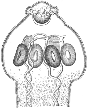
Fig. 23.—Head of Tænia solium.
Highly magnified. After Van Beneden.
The male reproductive organ consists of a number of small
vesicles or sacs, in which filiform spermatozoa have been
detected, these latter, when ripe, being conducted by a vas
deferens into a seminal pouch, from which a canal passes
laterally into the penis; the latter organ, in its retracted condition,
being lodged within a flask-shaped sheath or cirrhus-pouch.
The female organs are somewhat more complicated.
They consist of two masses of vitelligene glands occupying a
limited space, a small ovarium, a centrally-placed and largely-developed
branched uterus, canals of outlet leading from all
these organs, and enlargements of the main passages to form
internal seminal reservoirs; also, a vaginal canal, which is
widened at its termination to form a receptaculum for the
curved penis.
In addition to the above-named structures, the entire series
of joints from the head downwards are traversed by a set of
vascular canals, which are doubled in the region of the head.
These form the so-called aquiferous system. There are two
main channels, one passing down on either side of the worm,
both being connected by transverse vessels, which occur singly
at one end of every joint.
The eggs in their mature condition are globular, and contain
a six-hooked embryo. They present an average diameter of 1/694
of an inch, the shell itself measuring about 1/4000″
in thickness. In 1856 I observed that many of the eggs, whilst still
within the uterine branches, displayed an outer envelope, very delicate
in structure and totally dissimilar from the egg-shell proper. This
has since been more accurately described by Weinland, Van Beneden, and
Leuckart. The outer membrane, according to the last-named authority,
constitutes the primitive yolk-membrane, within which a part of the
yolk-contents separates to form the true egg and embryo by a process of
daughter-cell formation. The remaining part of the yolk forms a granular
mass, being probably concerned in the formation of the true chitinous
shell. The true shell displays a series of radiating and circular lines;
the former, however, are more conspicuous than the latter, being due,
according to Leuckart, to the presence of a series of fine rod-like
chitinous elements, which are formed on the external surface of the
original true shell-membrane. The enclosed embryo is furnished with six
boring spines, arranged in three pairs, its granular body being invested
by an extremely delicate skin-membrane, which is separated from the
inner surface of the shell by a clear transparent fluid. The embryo
measures 1/1250″
in diameter.
The scolex or higher larval stage of growth forms the well-known pork
measle or Cysticercus (telæ) cellulosæ of authors. The smallest
measles found by Leuckart measured 1/25″
in length. They were obtained from the brain, liver, and intermuscular
substance of a pig fed with proglottides about thirty days previously.
Only those specimens, however, occurring in the liver at this early
period displayed an outer membrane proper to the worm itself, the others
being simply invested with capsules formed out of the connective tissues
of the host. Many measle-masses in the same host were much larger,
presenting an average diameter of 1/6″.
The smallest already displayed a smooth, transparent, homogeneous, outer, cuticular
membrane, overlying a double, finely-granular corium, the latter being
traversed by a branched system of aquiferous vessels. These vessels
proceed from a central spot, which marks the position of the so-called
head-cone, or receptaculum capitis. It is, in fact, the first
well-marked indication of that flask-shaped capsule within which the
head, neck, and body of the Cysticercus is formed, and which Goeze long
ago very aptly compared to a lantern. As growth proceeds, a central
granular mass forms the true foundation of the head, its upper or
stalk-like extension becoming the future neck and body. Further changes
result in the evolution of the internal water-vascular system, the
calcareous corpuscles, the marginal transverse foldings of the body, the
four suckers, the rostellum, and, in particular, the double coronet of
hooks. All these metamorphoses were minutely followed and described by
Leuckart, who found the development of the larva to be completed within
the space of ten weeks.
As regards the injurious effects of this parasite upon man, it may
be said to act prejudicially in three separate ways. I have remarked in
my ‘Entozoa,’ that this parasite may cause disease and death both by
its action in the larval and adult states. It may likewise injure us by
rendering the flesh of swine unwholesome.
When one or more sexually-mature tapeworms have developed themselves
within the human intestine, they are apt to give rise to a variety
of unpleasant symptoms, more or less marked according to the habit
or irritability of the patient. According to Davaine (p. 103 of his
‘Traité’) the principal features are “vertigo, noises in the ears,
impairment of sight, itching of the nose and anus, salivation, dyspepsia
and loss of appetite, colic, pains over the epigastrium and in different
parts of the abdomen, palpitation, syncope, the sensation of weight
in the abdomen, pains and lassitude in the limbs, and emaciation.” In
ordinary cases there is always more or less anxiety and restlessness;
but in severe cases the sympathetic symptoms are very strongly
marked, showing themselves in hysterical fits, chorea, epilepsy, and
epileptiform seizures, attended by more or less alarming convulsions.
Amongst some of the more interesting and remarkable cases
recorded in our English journals, I may instance that of Mr
Hutchings, where a complete cure followed the evacuation of
the worm which had produced convulsions. Mr Tuffnell records
a case where irritability of the bladder and stricture of the
urethra were entirely dependent on tapeworm, as proved by
the subsequent recovery. At a meeting of the Pathological
Society, in 1853, Dr Winslow mentioned his experience of
three or four cases of mania arising from tapeworm; whilst on
the same occasion Drs Ryan and Davey each recorded a similar
instance. A case has also been previously published by Mr W.
Wood. At a meeting of the London Medical Society, held on
the 10th of April, 1837, Dr Theophilus Thomson (during an
interesting discussion on this subject) stated the facts of a
case where the presence of tapeworm had given rise to a
tumultuous action of the heart, this symptom entirely disappearing
after evacuation of the worm. Our journals likewise (anonymously)
record a considerable number of cases from foreign
sources. Thus, in the ‘London Medical Gazette’ for 1840, there
is the case of a lady, aged thirty-seven, who had convulsions
attended with a complete loss of consciousness, the separate fits
lasting an hour at a time. The passage of the worms effected
a complete cure. In the same journal for 1838, there is also
the case of a younger lady (aged twenty-seven) suffering from
epilepsy, in whom a complete cure had been similarly brought
about; here, however, in addition to a single specimen of the
Tænia solium, there were two lumbrici present. This journal
also gives Ettmüller’s case, where eighteen tapeworms were
the cause of hysteria; and likewise the case published by
Steinbeck, where the symptoms presented an altogether peculiar
character. More precise references to some of the above cases
will be found in the ‘Bibliography’ below; and I may also
refer to my published lectures on Helminthology and especially
to my separate work on Tapeworms, where particulars of one
hundred cases are briefly recorded. These were all average
cases occurring to me whilst in private practice. Davaine’s
book also abounds with remarkable cases.
Whilst the adult worm is capable of producing serious
and even fatal mischief to the bearer, the larvæ or measles
much more frequently prove fatal. The Cysticerci may
develop themselves in almost any situation in the human body,
but they occur most commonly in the subcutaneous, areolar,
and intermuscular connective tissue; next, most commonly in
the brain and eye, and lastly, in the substance of the heart and
other viscera of the trunk.
In my ‘Entozoa’ I have stated that probably not less than
one hundred cases have been observed where death had resulted
from Cysticerci in the brain. Griesinger alone collected
between fifty and sixty such cases. Mental disturbance occasioned
by the presence of measles in the brain may occur with
or without epilepsy. When Griesinger states that “the epilepsy
from Cysticercus is in all respects like cerebral epilepsy
and the psychical disturbances have nothing characteristic
about them,” he tacitly admits the impossibility of correct
diagnosis during life.
Since the publication of Griesinger’s well-known memoir on
Cysticerci of the brain, many similar cases have appeared, and
amongst the more recent of these is one by Dr Frédet in which
the victim was a young man twenty-two years of age. Though
apparently in good health he fell dead in the street; the
fatal result being due to the presence of a Cysticercus within
the pons Varolii.
Many other cases of earlier date are especially noteworthy. Thus Mr
Toynbee recorded a case where an hydatid (which I take to have been the
Cysticercus cellulosæ) situated in the middle cerebral fossa beneath
the dura mater, but in this instance death ensued from other causes.
Mr Ottley gives the case of a woman aged forty, where an undoubted
Cysticercus in the brain gave rise to distressing fits, convulsions,
and death. Then, again, there was Dr Burton’s workhouse patient, only
twenty years of age, who was found dead in bed, but who at the time
of admission merely complained of pain in the head. After death, four
hydatids (Cysticerci) were found in the tuber ancillare at the
summit of the spinal marrow. M. Bouvier’s similar case is also reported
in our periodicals. Of instances where Cysticerci occupied the cavity
of the eye, we have one or two cases by Mackenzie of Glasgow, one by
Mr Rose of Swaffham, and others by Windsor, Logan, and Estlin. Amongst
the more peculiar cases, I may mention that described by Dr Greenhalgh
in the ‘Lancet’ (1848), where the Cysticercus was lodged within the
substance of the lip. Five similar cases are likewise recorded by Heller
of Stuttgard. Then there is Dupuytren’s case of a Cysticercus ensconced
within the great peroneus muscle; and also Fournier’s, where several of
these scolices were said to have been found in a boil. The so-called
Trachelocampylus, discovered by Frédault in the human brain, was
neither more nor less than a common Cysticercus cellulosæ.
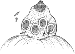
Fig. 24.—Head of a Cysticercus removed from the
brain. Magn. 5 diam. with detached hooks. Original.
It is worthy of remark, as Griesinger has also observed, that
in cases where the Cysticerci have taken up their temporary residence in
the brain, they are usually found, post mortem, in the grey cortical or
peripheral substance of the cerebrum. The particulars of such a case are
given in my ‘Entozoa’ where the victim suffered from epileptic fits due
to the presence of numerous Cysticerci (fig. 24). The patient was under
Mr Hulke’s care.
As regards infection by the adult worm it is not alone sufficient
that we avoid underdone meat, as brought to the dinner-table, but
we must be especially careful to have our sausages well cooked.
Under ordinary circumstances, we are safe for the following reasons:—No respectable butcher will
knowingly supply us with pork or with sausages which are measled. Even
in the case of underdone meats, in whatever way prepared, it is usually
only a small portion which is unaffected by cooking. As we have seen a
temperature of 140° Fahr. is sufficient to kill the Cysticerci.
The successful rearing of pork measles by experimentation with the
eggs of T. solium has been accomplished by many helminthologists,
amongst whom may be particularised Van Beneden, Leuckart, Küchenmeister,
Haubner, Gerlach, and Baillet. The converse experiment of rearing the
adult worm from the Cysticercus was first successfully undertaken by
Küchenmeister on a condemned criminal; Leuckart, Humbert, and others
having repeated this method with more or less success.
The dangers arising from infection by swallowing the larval worms or
six-hooked embryos are not easily avoided. Our flesh, like pork, thus
becomes measled, although certainly not to the spawn-like extent so
often seen in the lower animals. A single measle is sufficient to prove
fatal; and this humiliating contingency, moreover, is one which we can
never be absolutely certain of avoiding. We become the “host” or bearer
of the measle by swallowing the fully-developed eggs of the Tænia
solium. This we may do directly by handling fresh tapeworms, whose
eggs, being concealed under our
nails or in our clothing, may subsequently be swallowed, and develop
within us accordingly. Even a thorough washing of the hands will not
ensure absolute security. In like manner, those who partake of choice
salads, prepared from the stores of the market-gardener, run a certain
amount of risk. The vegetables may have been manured with night-soil
containing myriads of tapeworm eggs, or they may have been watered
with fluid filth into which the eggs were accidentally cast. In such
cases, one or more tapeworm ova will be transferred to the digestive
organs, unless the vegetables have been very carefully cleansed. In
the same way, one perceives how fallen fruits, all sorts of edible
plants, as well as pond, canal, and even river water procured from
the neighbourhood of human habitations, are liable to harbour embryos
capable of gaining entrance to the human body. One individual suffering
from tapeworm may infect a whole neighbourhood by rendering the swine
measly, these animals, in their turn, spreading the disease far and
wide. As already remarked, measles sometimes occur in great numbers in
different parts of the body. Among the more remarkable cases of the
multiple Cysticerci are those recorded by Delore (1864) and Giacomini
(1874). In M. Delore’s case, about 2000 were obtained post mortem. Of
these, 111 occurred in connection with the nervous centres, eighty-four
being in the cerebrum, twenty-two in the membranes of the brain, four in
the cerebellum, and one within the substance of the medulla oblongata.
Dr Knox published a less notable instance in the ‘Lancet’ (1838); and in
the year 1857, Dr Hodges, of Boston, U.S., published a case where the
cysts, which in size he compared to rice grains and coffee beans, were
felt subcutaneously. The coexistence of Tænia and Cysticerci in the same
individual has also recently been observed in France (‘Lond. Med. Rec.,’
1875). Besides these, several remarkable instances have lately been
reported by Davy, Tartivel, and others.
To the literature already quoted in connection with the beef tapeworm
the following may be added:
Bibliography (No.
14).—Aran, in ‘Archives Gén. de Médecine,’
1841.—Baillet, “Helminthes,” art. in ‘Bouley and Reynal’s
Dict. Vétérin.,’ tom. viii, 1869.—Bécoulet and Giraud, “On
Cysticercus in the Brain,” ‘Bullet. de la Soc. Méd. de Gand,’ 1872; and
in ‘Lond. Med. Rec.,’ Feb., 1873.—Birkett, J., Cases, ‘Guy’s
Hosp. Rep.,’ 1860.—Bouchut,
“Cyst. in the Brain,” ‘Gaz. des Hôp.,’ 1857, and ‘Journ. für
Kinderkrankheit.,’ 1859.—Bouvier, ‘Bullet. de l’Acad.,’
1840.—Burton, in ‘Med. Times and Gaz.’ (supposed hydatids),
1862.—Cobbold, “On Measly Meat and Measles in Man,” the
‘Veterinarian,’ 1876.—Czermack, “Cysticerci causing Insanity,”
Corresp.—Blatt, 1838.—Dalton, J. C., “Cyst in the
Scrotum,” ‘New York Journ. of Med.,’ 1857.—Davaine (see his
‘Traité’ for many additional references; p. 676).—Davy, R.,
“Cysticerci in the Muscles,” ‘Rep. of Lond. Med. Soc.,’ ‘Lancet’ for
Nov., 1876.—Estling, “Cases of Cysticercus,” ‘Lond. Med. Gaz.,’
1838–39.—Frédet, “Cysticercus in the pons Varolii,”
in the ‘Lancet’ for June 23rd, 1877 (p. 925), from ‘Giornale Veneto
de Scienze.’—Fournier, ‘Journ. des Connois. Med. Chir.,’
1840.—Griesinger, “On Cysticerci of the Brain,” from ‘Med.
Jahrb.’ in ‘Med.-Chir. Review,’ 1863.—Harley, J., “Cyst. in
the Brain,” ‘Lancet,’ 1867.—Hodges, R. M., “Specimens of
Cyst. cell., felt as small tumours just beneath the skin, varying in
size from that of a grain of rice to that of a coffee bean,” ‘Rep. of
Boston Soc. for Med. Improvement,’ in ‘Brit. Med. and Surg. Journ.,’
1857.—Hogg, J., “Obs. on Cysticercus,” in his ‘Manual of
Ophth. Surgery,’ 3rd edit., 1863.—Holler, A., “Cyst. cell.,
im Gehirne einer Geisteskranken,” ‘Allgem. Wiener Med. Zeitung,’
1878.—Logan, R., “Probable Cases of Cyst. cell.,” removed
by Robertson, ‘Ed. Med. and Surg. Journ.,’ 1833.—Mackenzie,
W., “Cyst in the Eye,” ‘Lancet,’ 1848, ‘Lond. Med. Gaz.,’
1839.—Mazotti, L., “Caso di numerosi cisticerchi del cervello
e delle meningi,” ‘Rivista Clin. di Bologna,’ 1876.—Mégnin,
P., “La Ladrerie du porc et le Tænia solium,” ‘La France Médicale,’
1876.—Putz, H., “Ueber die Lebenszähigkeit des Cysticercus
cellulosæ,” &c., ‘Zeitsch. f. pr. Vet.-Wissenschaften,’
1876.—Rainey, G., “On the Structure, &c., of Cyst. cell.,”
‘Phil. Trans.,’ 1857.—Rizzetti, G., “Rendiconto Statistico
dell’ufficio d’igiene di Torino per l’Anno 1873.”—Rudall, J. T.,
“Cyst. in the Brain,” ‘Australian Med. Journ.,’ 1859.—Tartivel,
De A., “Cysticerques multiples dans le tissu cellulaire sous-cutané et
dans certain viscères,” ‘Rec. de Méd. Vet.,’ 1876.—Von Gräfe,
A., in ‘Arch. für Ophthal.,’ 1857.—Wells, S., Bourman’s Case,
‘Ophth. Hosp. Rep.,’ 1860.—Windsor, J., “Cyst. in the Eye,”
‘Brit. Med. Journ.,’ 1861.
Tænia tenella, Cobbold.—I have long been acquainted with the
fact that there is a comparatively small human tapeworm which cannot be
referred to either of the foregoing species. In
the absence of experimental proof, I incline to the belief that the worm
in question owes its existence to measly mutton. The sheep harbours an
armed Cysticercus (C. ovis), which I regard as the scolex of Tænia
tenella. The specific name (tenella) was originally applied by
Pruner to a cestode six feet in length, which he found associated with
a larger tapeworm. This latter he called Tænia lata. Whilst Diesing
has pronounced Pruner’s Tænia lata to have been a T. mediocanellata,
I, on the other hand, consider Pruner’s T. tenella to have been
a T. solium. Mr J. C. Mayrhofer has suggested its identity with
Bothriocephalus tropicus. When, some years back, I applied the term
T. tenella to a new tapeworm (of which I possess several strobiles)
I was quite unaware than any similar nomenclature had been adopted by
Pruner. From the few facts supplied by Pruner and Diesing, I cannot
suppose that our cestodes are identical. Unfortunately my specimens are
imperfect, wanting the so-called head. It is not possible to estimate
the length of the worm accurately, but the perfect strobile must measure
several feet.
On one slide I have mounted nine mature proglottides of a worm which
I procured on the 15th Dec., 1875. The segments measure, on the average,
exactly 1/10″
in length, and only 1/20″
in breadth. The uterine rosettes are all full of eggs, and their
branches so crowded together that I am unable to ascertain their
average number. The segments are perfectly uniform in character, their
reproductive papillæ alternating irregularly at the margin.
In the autumn of 1872 I caused a lamb to be fed with the
proglottides of a tapeworm which I referred to this species.
The animal was slaughtered on the 22nd of January, 1873,
when the result was stated to have been negative. As I had
no opportunity of examining the carcase, I cannot feel quite
sure that there actually were no Cysticerci present. On several
occasions I have detected measles in the flesh of animals, when
none were supposed to be present by those who either assisted
me or were professional on-lookers. Assuming my Tænia tenella
to be derived from the sheep’s Cysticercus, I think it fitting to
describe the mutton measle in this place. Even if T. tenella
be not actually the adult representative of the mutton measle
(Cyst. ovis), it is quite certain that the scolex in question
gives rise to an armed tapeworm, and it is almost equally certain
that the adult armed cestode resides in man. In Pruner’s case,
which is by no means unique, we have seen that two distinct
species of cestode may coexist in the human bearer. It is
quite possible that some one may yet have the good fortune to
detect the beef tapeworm, the pork tapeworm, and the mutton
tapeworm, all together in one and the same host.
On five separate occasions I have detected measles in “joints”
of otherwise excellent and healthy mutton brought to my own
table, and supplied by the family butcher. On several other
occasions I have had these parasites brought under my notice;
nevertheless, many persons are either unaware of, or actually
deny, the existence of these ovine parasites. Thus, MM.
Masse and Pourquier, in the ‘Montpellier Med. Journ.’ for
Sept., 1876, make the following statement: “The sheep, not
being subject to measles, it seems to us natural to employ the
raw meat of that animal whenever it is required for nourishment
in the treatment of diarrhœa, in weaning children, in phthisis,
and for anæmics.” Clearly, if MM. Masse and Pourquier
could have brought themselves to believe that English literature
is worth consulting on such matters, they would not have
made this statement. Incidentally they also observe, when
speaking of beef measles:—“Un fait que nous avons remarqué
et que nous tenons à signaler, c’est que nous avons trouvé des
cysticerques nageant librement dans l’eau où nous avions plongé
de la viande infestée de ladrerie.” Certainly this is a novel
experience. That measles should not only get out of their
cysts, but should have the power of “swimming freely” in the
water is a phenomenon which requires explanation. There must
have been some error of observation.
It was in the year 1865 that I discovered the mutton
measle (C. ovis, mihi); but I am not prepared to say that the
parasite had never been seen before, since it is alleged that a
two-headed Cysticercus was obtained by Fromage from the liver
of a sheep (as cited by Davaine). Be that as it may, my discovery
was announced in a communication made at the Birmingham
meeting of the British Association in the autumn of
1865, and subsequently at a meeting of the Pathological Society
of London, on the 3rd of April, 1866 (‘Path. Trans.,’ vol. xviii,
p. 463). After these dates further announcements and verifications
appeared, amongst which I can only refer to my
remarks “On Beef, Pork, and Mutton, in relation to Tapeworms,”
forming an appendix to the first edition of my work
on Tapeworms, 1866; to the “Remarks on Cysticerci from
Mutton,” contained in the fourth chapter of the Supplement to my
introductory treatise on Entozoa, where a figure of the parasite
is given, 1869, p. 27; to Dr Maddox’s paper “On an Entozoon
with Ova, found encysted in the Muscles of a Sheep,” recorded
in ‘Nature,’ May 15th, 1873, p. 59; to the ‘Monthly Microscopical
Journal,’ June, 1873, p. 245; to my further communications
in the ‘Lond. Med. Record,’ Aug. 6th, 1873; to my
‘Manual,’ 1874, pp. 74 and 105, Ital. edit. ‘Nota Dell’ Autore,’
p. 133; and especially to the article headed “The Mutton
Tapeworm,” contained in the 3rd edit. of my little volume on
‘Tapeworms,’ p. 12, et seq., 1875.
In regard to the measle itself, I spoke of it as smaller than
the common pork measle. The head is 1/30″
in breadth, and is armed with a double crown of hooks, twenty-six in
all, the larger hooks each measuring 1/160″
in length. The suckers are four in number, each having a breadth
of 1/100″.
The neck and head are abundantly supplied with calcareous corpuscles,
being at the same time marked by transverse rugæ. The data on which I
founded my brief description of the scolex were chiefly based on the
examination of a specimen which had been procured by Prof. Heisch from
the interior of a mutton chop. Subsequently much fuller details of the
structure of the scolex were supplied by the illustrated memoir of Dr
Maddox (above quoted). This excellent microscopist, however, announced
the presence of immature ova within the Cysticerci themselves. As the
notion of the existence of eggs in larval cestodes was altogether
at variance with what we know of the phenomena of tapeworm life, I
suggested that the author might have mistaken the egg-shaped calcareous
corpuscles (which I found so abundant in my own specimens) for the ova.
In the interests of truth I felt bound to characterise certain of the
conclusions arrived at by Dr Maddox as simply incredible, but I regarded
his memoir as forming “an important contribution to our knowledge of
the structure of the mutton measle.” I had no idea that in pointing to
errors of interpretation I should offend the excellent author. However,
a long letter appeared in the ‘London Medical Record,’ in which Dr
Maddox showed that he was much vexed that I should have “impugned” the
“accuracy of his conclusions.” He defended his position with the support
of no less an authority than Dr Macdonald, F.R.S., the distinguished
Assistant Professor of Naval Hygiène at the Victoria Hospital, Netley.
Dr Maddox says:—
“We were quite alive to the anomalous position. Hence the
exceptionability of the case rests on more than my own evidence.” In
regard to this unfortunate dispute I will only add the expression of
my conviction that Drs Maddox and Macdonald will eventually become
satisfied that no cestode scolex is capable of displaying either mature
or immature ova in its interior.
Bibliography (No. 15).
Cobbold (l. c., supra), 1865–75.—Idem, “On Measly
Meat, &c.,” the ‘Veterinarian,’ Dec., 1876.—Idem, “The
Mutton Tapeworm (T. tenella),” No. 16 in my revised list of Entozoa,
the ‘Veterinarian,’ Dec., 1874.—Diesing, C. M. (Tænia tenella,
Pruner nec Pallas), in “Revis der Cephalocotyleen,” ‘Sitzungsb. der
Math.-Mat. Class d. k. Akad. der Wissenschaften,’ Bd. xlix, s. 369,
1864.—Maddox (l. c., supra), 1873.—Mayrhofer, J.
C., ‘Die helminth. des Menschen,’ Erlangen, 1854.—Pruner,
‘Krankheiten des Orients,’ s. 245, 1847.
Tænia lophosoma, Cobbold.—This is a good species notwithstanding
the doubts that have been expressed by Heller
and others regarding it. I have called it the ridged tapeworm
in consequence of the presence of an elevated line coursing the
whole length of the body, which measures about eight feet.
The reproductive papillæ are remarkably prominent and uniserially
disposed throughout the entire chain of proglottides.
It is quite an error to suppose that this species is a malformed
cestode, or that it has any resemblance to Küchenmeister’s
variety of tapeworm from the Cape of Good Hope. Neither
does it in the slightest degree resemble the remarkably malformed
T. mediocanellata described by Mr Cullingworth. Of
the distinctiveness of this parasite as a species, any one may
satisfy himself by an inspection of the nearly complete strobile
preserved in the Pathological Museum attached to the Middlesex
Hospital Medical College. From the examination of several
mature proglottides detached from this specimen, I find their
average breadth to be one fifth of an inch, by three quarters
of an inch in length. Their greatest thickness does not exceed
the 1/13th
of an inch. The eggs resemble those of other tapeworms,
and offer a diameter of about 1/850″
from pole to pole.
Bibliography (No. 16).—Cobbold, “Parasites of Man,” in
the ‘Midland Naturalist,’ April, 1878, p. 98.—Idem, ‘Tapeworms,’
1st edit., p. 52, 1866; 3rd edit., p. 27, 1875.—Cullingworth
(see Bibl. No. 18).—Davaine, ‘Les Cestoïdes,’ l. c., p. 573.—Heller,
l. c., s. 594.
Tænia nana, Siebold.—As regards the dwarf tapeworm, unless
Spooner’s case be genuine, there is but one solitary instance on record
of its occurrence in the human body; moreover, we have no evidence of
its having existed in any other host. It was discovered by Dr Bilharz,
of Cairo, at the post-mortem examination of a boy who died from
inflammation of the cerebral membranes. Prodigious numbers existed. The
largest specimen measured only one inch in length. To the naked eye
these worms resemble short threads, and consequently they might very
readily be overlooked. The head is broad and furnished with a formidable
rostellum armed with a crown of hooks. These hooks have large anterior
root-processes, which, extending unusually forward, impart to the
individual hooks a bifid character. By far the best account of this worm
is furnished by Leuckart, to whom I am indebted for a specimen.
Bibliography (No. 17).—Cobbold, ‘Entozoa,’ p. 244.—Davaine
(l. c., Bibl. No. 2), p. 574.—Heller, l. c., s. 606.—Küchenmeister,
l. c., Eng. edit., p. 141.—Leuckart, l. c., Bd. i,
s. 393.—Von Siebold and Bilharz, in Von Sieb. and Köll.
Zeitschr., Bd. iv.—Spooner, ‘Amer. Journ. Med. Sci.,’ 1873.—Van
Beneden, ‘Iconographie,’ l. c., pl. iii, fig. 17.—Weinland,
‘Diplacanthus nanus,’ l. c., p. 85.
Tænia Madagascariensis, Davaine.—This appears to be a
well-defined species although the head has not yet been seen. It
probably forms the type of a distinct genus. Dr Grenet, stationed at
Mayotte (Comores), twice encountered single specimens passed by two
young children, eighteen and twenty-four months of age respectively. The
proglottides have their genital pores uniserially arranged, and they
show, in their interior, remarkable egg-capsules, from 120 to 150 in
number in all, each containing from 300 to 400 eggs. These give a long
diameter of 1/625″ for the outer envelope and 1/1250″
for the inner, or shell proper. The embryo measures only the 1/2500
of an inch.
A full account of this parasite, with figures, is given by
Davaine (‘Les Cestoïdes,’ l. c., Bibl. No. 2, p. 577 et seq.).
Tænia marginata, Batsch.—Although I possess no certain
evidence of the occurrence of this parasite in its adult condition
in the human bearer, yet there is a tapeworm in the Edinburgh
Anatomical Museum referable to this species, which was said
to have been obtained from the human body. This worm is
very common in the dog.
The principal evidence demonstrating the occurrence of the
larval representative of this species (Cysticercus tenuicollis) in
man, rests upon the two cases recorded in Schleissner’s ‘Nosography’
of Iceland. One of the alleged instances, however, has
been proved by Küchenmeister and Krabbe to be that of an
echinococcus; so that, after all, there only remains the solitary
case observed by Schleissner himself, in which the parasite can
fairly be considered as the “slender-necked hydatid.”
To the above, however, may probably be added a specimen
preserved in the Anatomical Collection at King’s College, London.
It was found connected with an ovarian cyst.
Tænia elliptica, Batsch.—This parasite is readily recognised
not merely by its delicate form and small size, but also by the
circumstance of its supporting two sets of reproductive organs
in each mature joint. Their outlets are situated at the centre
of the margin of each segment, one on either side. Ordinarily
infesting the cat, this worm is a mere variety of the common
Tænia cucumerina of the dog. At all events, from the evidence
put forth by Eschricht, seconded by Leuckart, there is every
reason for believing that one or other of these closely-allied
varieties is liable to infest the human body. It was originally
stated by Eschricht that he had received a Tænia canina which
had been passed by a negro slave at St Thomas, Antilles. This
is a synonym of T. elliptica, which must therefore be very rare
in the human body, possibly only occurring in the negro race.
In regard to the source of this parasite, it has been shown
by Melnikow that the scolex of Tænia cucumerina resides in
the louse of the dog (Trichodectes latus), and thus it is
exceedingly probable that the scolex of Tænia elliptica resides
in the louse of the cat (Trich. subrostratus). How man becomes
infested is not so clear. Melnikow’s paper on the juvenile state
of this cestode is contained in the ‘Archiv für Naturgeschichte’
for 1869, and is illustrated by a figure of the measle.
Tænia flavopuncta, Weinland.—Regarded as a new species,
the discovery of this little tapeworm is due to the investigations
of Weinland. In Dr Jackson’s ‘Catalogue of the Boston
Medical Improvement Society’ an account of the contents of a
phial is recorded as follows:—“Specimen of Bothriocephalus,
three feet in length, and from half a line to one line and a
quarter in width, from an infant. The joints are very regular,
except at one extremity, where they approach the triangular
form, are very delicate, and but slightly connected, as shown in
a drawing by Dr Wyman.” It is further stated that the infant
was nineteen months old, and that the worm was discharged
without medicine, its presence having never been suspected. It
was presented by Dr Ezra Palmer in the year 1842. On
examining the fragments, Dr Weinland found, instead of a
solitary specimen, at least six different tapeworms, all of them
being referable to a hitherto undescribed species. There were
no heads; nevertheless, it was ascertained that the worms varied
from eight to twelve inches in length, the joints or segments
being very broad, and at the same time narrowed from above
downwards. The parasite was named “the spotted tapeworm,”
in consequence of the presence of yellow spots near the middle
of the joint. They represent the male organs of reproduction,
the outlets of which, as in my T. lophosoma, occur all along one
side of the body or strobile. In Weinland’s estimation this
parasite forms the type of a new genus which he calls Hymenolepis.
A full account of the worm is given in his well-known
essay (l. c., Bibl. No. 2).
Tænia abietina and other varieties. I can only notice very
briefly certain cestodes which either present malformations or
which may be regarded as mere varieties. First in this series is
Weinland’s T. abietina. No one who has studied his ‘Beschreibung
zweier neuer Tænioiden aus dem Menschen,’ Jena, 1861, can
doubt that it is a mere variety of T. mediocanellata. The
monstrosity described by him as referable to T. solium must also
be referred to the beef tapeworm. The variations in the character
of cestode proglottides is practically infinite. A museum
might be filled with them. Most common with T. mediocanellata,
these varieties more or less prevail with other species.
Thus I have seen them in Tæniæ and Bothriocephali alike. I
have obtained segments of T. mediocanellata having sexual
outlets on both sides of the proglottis, so regularly disposed in
a few segments as to suggest the notion of a new species. The
coalescence of several segments into one compound segment is
frequent, but the most remarkable specimen that I have seen is
one contained in the museum of the Royal College of Surgeons.
In the old Hunterian catalogue the specimen is described as
“two joints of the Tænia solium, with a number of orifices in
unequal series on either side.” As stated in the new catalogue
of the series, prepared by myself, the “lower segment is furnished
with twenty-two sexual orifices, one of which is situated in the
central line” on the ventral surface (as in Bothriocephali).
References to this and other specimens in the Hunterian
Collection will be found below (see Pittard). In regard to
Weinland’s conjectural Tænia acanthotrias, based on the
circumstance of his having found a Cysticercus that presented
three rows of hooks on its rostellum, I need only say that if
such a Tænia were found it would only turn out to be a
malformed T. solium. The specimens, however, are none the
less interesting. Very remarkable and altogether exceptional
characters are presented by the strobile of the cestode described
by Mr Cullingworth, of Manchester, and of which I possess
specimens. Here, apparently, at least two tapeworms are joined
together throughout the entire chain of proglottides without
intermission. The three margins of each compound segment
project at equi-distant angles. Could we have secured the head
we should certainly have found six or eight suckers present,
since the finest neck-segments showed that the malformation
pervaded the entire colony of zooids, sexually mature and
otherwise. Mr Cullingworth’s specimen is so remarkable that
I subscribe full particulars of the case in his own words. He
says:—“A respectable married woman, named Ann H—, forty
years of age, residing in Salford, brought to my out-patient
room at St Mary’s Hospital, Manchester, on September 3rd,
1873, a few segments of tapeworm as a sample of what she had
been passing per anum for about two years. Although never
in the habit of taking meat absolutely raw, she told me, on
inquiry, that she was particularly fond of tasting it when only
partially cooked. The segments were unlike anything I had
seen before, and I took them home for examination, ordering the
patient meanwhile a draught containing a drachm of the oil of
male fern, and giving her strict injunctions to bring to me every
fragment that passed away as a result.
“On September 17th she brought me portions of a tapeworm
corresponding throughout to the segments I had already seen,
and measuring altogether nine feet in length. Unfortunately,
the head was not to be found. Along the middle line of every
segment in the body a crest or ridge runs longitudinally, and in
the centre of the margin of this crest the genital pore is situated.
[In 304 segments examined, only four had the genital
opening placed laterally. One segment had two openings,
viz. one at the lateral margin and the other in the crest.]
Underneath the segment there is a longitudinal groove, and
the lateral portions are folded together by the apposition of their
under surfaces. When hardened in spirit the section of a
segment presents a three-branched appearance, the branches
being of unequal length, but placed at equal angles. The
uterus sends vessels into the crest as well as into the sides of the
segment; and the contained ova are exactly like the ova of an
ordinary Tænia mediocanellata. Wedged in between, or
attached to, the segments here and there, is a stunted and
ill-shaped joint, with irregular and unequal sides. A mature
joint measures from five eighths of an inch to three quarters of
an inch in length, and about half an inch in breadth, and
the breadth or depth of the crest is usually one eighth of
an inch.
“There are only two specimens that I can find on record at
all similar to the one here described, and both of these differ
from it in several important particulars. Küchenmeister mentions,
as a variety of Tænia mediocanellata, a tapeworm sent
to him from the Cape of Good Hope by Dr Rose. This worm
possessed a longitudinal ridge, but he describes its mature
segments as ‘extremely massive’—more than an inch in length
and 3/5″ in breadth. The genital pores, too, were irregularly
alternate, and not situated on the crest. On March 20th, 1866,
Dr Cobbold exhibited to the Pathological Society of London a
specimen of crested tapeworm which was discovered in the
museum of Middlesex Hospital, and to which he proposed to
give the name Tænia lophosoma (λόφος, crest; σῶμα, body). The
reproductive papillæ were all on one side of the chain of segments,
a peculiarity which entirely distinguished it from the
Cape of Good Hope variety of Küchenmeister. The head of
the creature was wanting. It will thus be seen that my
specimen does not correspond with either of these in the situation
of the genital aperture. Here it is placed in the crest
itself, and not unilaterally, as in Dr Cobbold’s specimen, or
alternately, as in Küchenmeister’s. It further differs from the
Cape variety in the more moderate dimensions of its proglottides.
I have adopted, however, the name suggested by
Dr Cobbold in the communication referred to, inasmuch as it
sufficiently indicates the principal distinguishing feature of the
specimen. I may mention that Dr Cobbold saw the specimen
during his visit to Manchester, and that he regarded it as a
most remarkable and unique abnormality.”
Further, in connection with abnormal cestodes, I may observe
that Weinland’s case of a triple-crowned Cysticercus does not
stand alone, since a similar specimen is, I believe, in the
possession of the Rev. W. Dallinger. This was removed from
the human brain. Curious as this subject is, I cannot dwell
upon it. Not only are the mature tapeworms and their Cysticerci
liable to present monstrosities, but even also their proscolices
or six-hooked embryos. Thus, twelve hooks were observed
by Salzmann in the embryo of T. elliptica, and Heller also
figures two embryos of T. mediocanellata (T. saginata, Gœze)
with numerous hooklets. Dujardin saw seven in a Bothriocephalus
embryo. Occasionally there have been errors of interpretation
made by observers. Thus, Diesing has given beautiful
figures of Dibothrium hians in such a way as to suggest different
degrees of monstrosity affecting the tail end of the strobile; but
this splitting has clearly resulted from injury. Thus also, when
I removed five specimens of a new cestode (Diphyllobothrium
stemmacephalum) from the intestines of a porpoise, one of them
was cleft nearly half way up the strobile. This had been done
by the scissors employed in slitting up the gut; but owing to
perfect contraction of the incised edges, it was some time
before I discovered that the apparent monstrosity had been
artificially produced. Lastly, I may add that many of the
older writers were well acquainted with larval and other
anomalies. Thus Rudolphi described a two-headed Cysticercus
from a Lemur, and also a double-headed Tænia crassicollis. This
worm had a tripartite body; as had likewise a Tænia crassicollis
of which he did not possess the head (corpore prismatico).
Other monstrosities were described and figured by Bremser
and Creplin. Pallas mentions a two-headed Tricuspidaria
(Triænophori nodulosi bicipites), and, as already stated at p. 97,
a double-headed Cysticercus has been obtained from the liver
of a sheep.
Before quitting the Tæniæ proper, I may observe that several
other species have been indicated, based on ovular and other
insufficient characters. To these belong Ransom’s supposed
tapeworm, and also Weinland’s Tænia megaloön.
Bibliography (No. 18).—Bonnet, C., ‘Œuv. Compl.,’ tom vi,
p. 191, 1791.—Bremser, Atlas, by Leblond, Pl. iv.—Chaussat,
‘Comptes Rendus,’ p. 20, 1850.—Cobbold, ‘Catalogue of the
specimens of Entozoa in the Museum of the Royal College of
Surgeons of England,’ Nos. 118–121, London, 1866.—Idem,
‘Worms,’ l. c., p. 78.—Idem, “On a Cysticercus from the
Human Brain,” ‘Brit. Assoc. Rep.,’ 1870.—Creplin, ‘Tænia
Monstrum, &c.,’ Berlin, 1839.—Cullingworth, C. J., “Notes on
a remarkable specimen of Tapeworm (Tænia lophosoma, Cobbold),”
‘Med. Times and Gaz.,’ Dec., 1873.—Davaine, ‘Les
Cestoïdes,’ l. c., p. 570.—Diesing, ‘Zwanzig Arten von
Cephalocotyleen,’ figs. 1 and 2, taf. ii (aus dem xii, Bd. d.
denkschr. d. Math.-nat. Cl. d. k. Akad.), Wien, 1856.—Dujardin,
l. c., p. 619.—Heller, l. c., s. 600.—Küchenmeister, l. c., Eng.
edit., p. 139.—Leuckart, l. c., s. 303 and 465.—Levacher, ‘Journ.
l’Institut,’ p. 329, 1841.—Pittard, S. R., Remarks in his article
“Symmetry,” Todd’s ‘Cyclop.,’ vol. iv, p. 848, 1849–52, in
which he refers to a monstrous Bothriocephalus (T. lata) in the
Hunterian Museum, old ‘Catalogue of Nat. Hist.,’ pl. iv, p. 50,
No. 205; see also my ‘Catalogue,’ l. c., supra, No. 167.—Ransom,
in Reynolds’ ‘System of Medicine.’—Rudolphi,
‘Synops.,’ p. 545 and 598–9, with fig. showing the heads of
Cystic. Simiæ (biceps), widely apart, 1819.—Weinland (T.
megaloön), in Zoolog. Garten, Frankf., 1861, s. 118.—Idem,
‘Essay,’ l. c., p. 11.
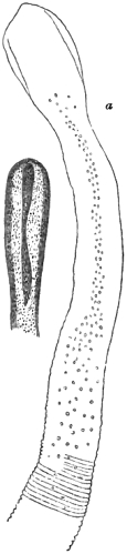
Fig. 25.—Head and neck
of Bothriocephalus latus.
a, Front view. The
smaller figure represents
the head as seen from the
side. After Knoch.
Bothriocephalus latus, Bremser.—This species, though seldom
seen in England, is sometimes brought hither by persons who
have been residing for a time in foreign countries. It is indigenous
in Ireland, and, though by no means common there, has
been called the Irish Tapeworm. As regards its distribution
in Europe it is much more prevalent in some districts than in
others. On this point Leuckart remarks that “foremost amongst
these are the cantons of West Switzerland, with the adjacent
French districts. In Geneva, according to Odier, almost a
fourth part of all the inhabitants suffer from Bothriocephalus.
It is also common in the north-western and northern provinces
of Russia, in Sweden, and in Poland. In Holland and Belgium it
is likewise found, but, on the whole, not so frequently as in the
first-named countries. Our German fatherland also harbours
them in some districts, especially in eastern Prussia and Pomerania,
and there have appeared cases in other places, as in
Rhenish Hesse, Hamburg, and even in Berlin; these being
apparently spontaneous instances.”
Unlike the ordinary tapeworms, the segments of the broad tapeworm
do not individually separate so as to become independent organisms, a
circumstance which is highly favorable to the bearer. Its remarkable
breadth, and the extremely numerous and closely-packed proglottides,
impart a sufficiently distinctive character; but this parasite may be
more fully characterised as
the largest human cestode at present known, attaining a length of
more than twenty-five feet, and sometimes measuring nearly an inch in
breadth; the so-called head 1/25″
in width, bluntly pointed at the tip, much elongated or club-shaped,
slightly flattened from behind forwards, and furnished with two
laterally disposed slit-like fossæ or grooves, but destitute of any
armature: anterior or sexually-immature segments of the body extremely
narrow, enlarging in a very gradual manner from above downwards; joints
of the lower half of the body gradually decreasing in width, but
enlarging in depth; sexually-mature segments usually about 1/8
of an inch in depth, but those near the caudal extremity frequently 1/4″,
and quadrate in form; body flattened, but not so uniformly as obtains
in the ordinary tapeworms, being rather thicker near the central
line; total number of joints estimated at nearly 4000, the first
sexually-mature ones being somewhere about the six hundredth from the
head; reproductive orifices at the central line, towards the upper
part of the segment at the ventral aspect, the vaginal aperture being
immediately below the male outlet, and both openings surrounded by
papillæform eminences; uterus consisting of a single tube, often
seen regularly folded upon itself, forming an opaque, conspicuous,
centrally-situated rosette; eggs oval, measuring 1/350″
in length by 1/550″ in breadth,
having three shell-coverings, and a lid-like operculum at one end, as
occurs in the fluke-worms. Owing to the dark color of the egg shells,
the uterine rosette is readily seen by the naked eye as a conspicuous
deep brown spot at the centre of each successive segment.
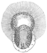
Fig. 26.—Proscolex, or six- hooked embryo
of Bothriocephalus, escaping from
its ciliated covering. After Leuckart.
The source and development of this parasite are points of
considerable interest. The eggs are of comparatively large size,
and after expulsion and immersion in water they give passage
to beautifully ciliated embryos, which latter produce larvæ
furnished with a boring apparatus. These larvæ resemble the
six-hooked embryos of other tapeworms. In what animals the
larvæ subsequently develop themselves is not ascertained with certainty,
but it is probable that persons become infested by eating imperfectly
cooked fresh-water fish. Leuckart has suggested that the intermediary
bearers are species of the salmon and trout family. Dr Knoch, of
Petersburg, thought that there was no need of the intermediate host. He
believed that he had succeeded in rearing young broad tapeworms in the
intestines of dogs. It was Leuckart who first explained the source of
Knoch’s errors of interpretation. Although Knoch administered eggs of
Bothriocephalus latus to dogs, and afterwards found young tapeworms
of the species in question in the intestines of the dogs, it did not
logically follow that any genetic relation (as between the egg-contents
and the adult worms) had been thereby established. The circumstance that
ripe ova of the Bothriocephalus always contain six-hooked embryos, must
alone imply that an intermediate host is necessary for the formation of
Cysticerci or measles. If the broad tapeworm could be reared in a direct
manner by the administration of Bothriocephalus eggs, there would be no
need for the presence of boring hooklets in the proscolex. These are
necessary for invading the flesh of some intermediate host.
Dr Fock, of Utrecht, has sent me particulars of an interesting
case, and he suggests that infection comes from the little river
bleak (Leuciscus alburnus). Writing from Utrecht in December,
1877, Dr Fock, after referring to a former case, goes on to say:—“Permettez moi, cher confrère,
que je rappelle à votre souvenir que vous avez eu l’obligeance de
communiquer au public une observation, de ma main, sur un cas très rare
de ver rubanaire, d’un Bothriocephale, chez une petite fille juive.
Malheureusement je n’ai pu en donner de plus amples détails, parce que
cette enfant n’a plus, depuis ce temps-là, rendu la plus petite parcelle
de ver. Il y a maintenant quinze mois, et voilà que de nouveau un cas
pareil se présente. Une femme mariée, frisonne, et, cette fois-ci
encore, juive, s’est adressée à moi pour la débarasser de son ver. Elle
me disait avoir rendu, il y a quelque temps, des fragments, ou plutôt un
fragment de la
longueur d’un mêtre, d’un ver solitaire, pour lequel elle avait été
traitée, sans succès, par son médecin ordinaire. A cause de cela elle
s’adressa à moi, et je lui ai repondu qu’elle devrait revenir la
première fois qu’elle rendrait de nouveau, spontanément, un nouveau
fragment. Après un mois d’intervalle elle est revenue en me montrant
un fragment de la longueur d’un demi-mêtre qu’elle venait de rendre
spontanément, après avoir jeûni par précepte réligieuse, et deux
jours après cela, traitée par l’écorce de grenadier, elle a rendu un
Bothriocephale parfaitement conditionné en entier.
“Ce cas me semble assez intéressant pour être communiqué
de nouveau, d’abord parce que jusqu’ici personne n’a pu dire par
quel chemin a pu s’introduire un tel helminthe, et ensuite parce
que ce chemin doit se présenter bien rarement dans nos
contrées (ou en Angleterre) puisque dans le courant d’une
trentaine d’années ayant rencontré des centaines de tænias, ce
cas-ci est seulement le second dont je suis gratifié. Il me
semble digne de réflexion que ce cas-ci se présente cette fois-ci
de nouveau chez une juive. Est ce cas-ci fortuit, ou bien y-a-t’il
un lien de causalité entre ce ver rare et le genre de nourriture
ou de boisson de ces bonnes gens? La dame me recontait
que, en Frise, il y a un poisson très recherché qui s’appelle en
Hollandais blèck, en Anglais blay ou bleak, et dont ils sont très
friands, dans lequel, ils rencontrent très souvent un très grand
ver rubanaire. Une autre personne me disait avoir été à
table chez un ami, qui ne sachant probablement ce qu’il
mangeait, savoura avec beaucoup de délice cette friandise dégoutante.”
After describing the specimen, Dr Fock concludes his remarks
with a suggestion as to the possibility of introducing tapeworm
into the human body by potable water, into which Cysticerci
have accidentally found their way. Dr Fock remarks:—“J’ajoute
une réflexion par rapport à la provenance des autres tænias, qui
jusqu’ici sont introduits par l’usage de la viande non assez cuite
ou rôtie, ou saignante; mais, ne se pourrait-il pas que des débris
de la chair d’un animal ladre fussent introduits fortuitement
dans l’eau, par example, d’un fossé, et que celle-ci employée
comme boisson contint des Cysticerques et par ainsi aussi une
cause de Tænia? Ce n’est qu’une conjecture que je propose en
terminant cet article.”
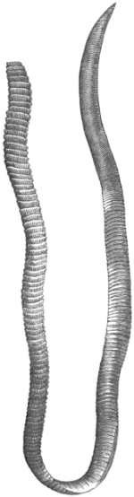
Fig. 27.—Strobile of Bothriocephalus
cordatus. After Leuckart.
Although I cannot at all agree with Dr Fock in regarding
water as a source of infection in the manner he indicates, yet
the still more recently expressed opinions of MM. Bertolus and Duchamp,
based on experimental researches, render it tolerably certain that
Leuckart’s original surmise was correct, and that we must look to
freshwater fishes for the larvæ of the broad tapeworm. In the section
of this work devoted to the parasites of fishes I shall make particular
allusion to the experiences of Dr Bertolus; but as confirming the view
of Leuckart I may here observe, that Bertolus has almost proved that
the so-called Ligula nodosa infesting the common trout is merely
a sexually incomplete example of Bothriocephalus latus. The bleak
(Leuciscus alburnus) shares with other freshwater fishes the privilege
of harbouring a species of Ligula (L. digramma); but whether this
form bears any genetic relation to our human Bothriocephalus latus
can only be determined by actual experiment. If, as Duchamp and others
have either indicated or implied, Ligula alburni is a synonym of the
bleak’s cestode in question, then it is evident that the sexually mature
form of the Ligula of the bleak is the well-known L. simplicissima
of many water birds and of a few other avian species. Probably the
bleak-eaters of Holland consume many kinds of freshwater fishes,
including various species of the salmon and trout family.
The symptoms occasioned by Bothriocephalus latus do not differ
materially from those produced by other tapeworms. According to Odier,
as quoted by Davaine, there is not unfrequently a tumid condition of
the abdomen, with sickness, giddiness, and various hysterical phenomena
occurring at night. Pain in the region of the heart, palpitations, and
faintness are also mentioned.
As already hinted, this cestode is very liable to present
abnormalities of structure, the proglottides frequently displaying
double sexual orifices, with corresponding duplication of the
reproductive organs internally. For details respecting the anatomy
of Bothriocephalus I must refer to the works of Küchenmeister and
Leuckart; and more particularly to the memoir of Drs F. Sömmer and L.
Landois, who have supplemented the previous researches of von Siebold,
Leuckart, Böttcher, Stieda and others by beautiful investigations of
their own. In the pages of ‘Nature,’ for 1872, I gave a résumé of
Sömmer’s memoir, which will be found quoted below.
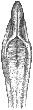
Fig. 28.—Head of Bothriocephalus
cristatus, viewed
from the front. After
Davaine.
Bothriocephalus cordatus, Leuckart.—This species is identical
with a worm long ago described by Pallas and Linneus. At present it
is only known to infest the residents of North Greenland, but it is
probably distributed throughout the north generally. It attains the
length of about one foot, and has a small heart-shaped head, whose apex
is directed forwards. The neck is so obscure that it may be said to
be altogether wanting, the segmentation of the body being well marked
immediately below the head. Though so small a species, Leuckart, who
first described it, counted between six and seven hundred joints. As
in the broad tapeworm, the reproductive orifices are serially disposed
along the centre of the ventral line, but a close inspection shows that
the folds of the egg-bearing organ are comparatively more numerous. This
worm does not appear to be a frequent resident in the human body, though
it is by no means uncommon in the dog. Possibly it may yet be found in
the inhabitants of some of our northern and western isles.
Bothriocephalus cristatus, Davaine.—This cestode measures
between nine and ten feet in length, and is characterised by the
presence of two remarkable prominences, together forming a sort of
rostellum or crest which is covered by numerous minute papillæ. The
full-grown segments are less than half an inch in breadth; the body of
the parasite being narrower than that of the broad species. The original
description of the parasite by Davaine
is based on two specimens, one of which, quite perfect, was obtained
from a child five years old, under Dr Féréol’s care at Paris. The
other was passed spontaneously by an adult residing at Haute-Saône. I
have here copied one of Davaine’s original figures of the head of the
worm.
Bibliography (No.
19).—Bertolus, “Mém. sur le development du Dibothrium latum”
(in Appendix to Duchamp’s work, see Bibliog. No. 59).—Blanchard,
“Recherches, &c.,” ‘Ann. des Sci. Nat.,’ ser. 3, Zool., Pl. 11,
12, 1848.—Böttcher, “Studien ueber den Bau des Both. latus,”
‘Virchow’s Archiv,’ s. 97 et seq, 1864.—Bremser, l. c.,
Bibl. No. 1, s. 88, 1824.—Chiaje, ‘Compendio, &c.,’
Tab. iii, figs. 1–5, 1833.—Cobbold, ‘Entoz.,’ p. 289,
1864.—Idem, “Remarks on the Broad Tapeworm” (with a letter from
Dr Fock), the ‘Veterinarian,’ July, 1878.—Creplin, in Ersch and
Gruber’s ‘Encyclop.,’ 1839, p. 296.—Davaine, ‘Traité,’ l. c.,
1860; 2nd edit. (passim), 1877.—Idem, art. ‘Les Cestoïdes,’
l. c., Bibl. No. 2, p. 580–591, 1876.—Dujardin,
l. c., Bibl. No. 1, p. 612, 1845.—Eschricht, D. F.,
‘Anat-physiol. Untersuchungen ueber die Bothriocephalen,’ Breslau,
1840.—Fock (see Cobbold).—Heller, ‘Darmschmarotzer,’
l. c., s. 606, 1876.—Knoch, ‘Petersburger Med. Zeitschrift,’
1861.—Idem, ‘Die Naturgeschichte des breiten Bandwurms (B.
latus, auct.),’ St Petersburg, 1862.—Küchenmeister, ‘Ueber
cestoden,’ l. c., 1853.—Leuckart, ‘Die Blasen Bandwürmer,’
1856.—Idem, ‘Die mensch. Par.,’ Bd. i, s. 414–448, und
757, 1863; and Bd. ii, s. 866, 1876.—Owen, Todd’s ‘Cyclop.,’
1837.—Sömmer und Landois, aus Sieb. und Köll. Zeitschr.,
‘Beiträge zur Anatomie der Plattwürmer,’ Leipsig, 1872; see also the
résumé in ‘Nature’ for Aug., 1872, p. 278.—Wawruch, ‘Pract.
Monograph. d. Bandwürm-Krankheit,’ 1844, s. 33.

Fig. 29.—Tænia echinococcus Strobile.
Mag. 30 diam. Original.
Echinococcus hominis (the common hydatid).—This larval
entozoon has acquired various names according to the kind of bearer
in which it happens to have been found; but all the true hydatids or
acephalocysts, whether infesting man or animals, are referable to one
and the same species of parasite. They have been termed Echinococcus
hominis, E. veterinorum, E. polymorphus, E. exogena, E.
endogena, E. multilocularis, according to circumstances. All
of them represent a juvenile stage of the Tænia echinococcus or
hydatid-forming tapeworm which infests the dog and wolf. Experimental
proof of this fact has been furnished by Von Siebold (1852), Haubner,
Leuckart, Küchenmeister, Van Beneden, Naunyn, Nettleship, Krabbe, and
others.
The first successful rearing of Tæniæ with human hydatids
was accomplished by Naunyn (1864), his results being subsequently
verified by Krabbe and Finsen (1865). Zenker, Ercolani, and several
others, including myself, also conducted feeding experiments with
human hydatids which were attended with negative results. In the
case of one of my experimental dogs the animal was liberated by an
ill-disposed person before I had opportunity to destroy it. As the
experiment was carefully conducted, the animal may have proved a
source of fresh echinococcus-infection. Mr E. Nettleship’s eminently
successful experiment was made with hydatids obtained from a sheep. The
converse experiment, namely, that of rearing hydatids with the mature
proglottides of Tænia echinococcus administered to animals, has been
performed most successfully by Leuckart, and by Krabbe and Finsen; by
the former in the pig, by the latter in a lamb, with tapeworms that had
also been reared by experiment. Zenker, later on, reared the Tænia
from hydatids obtained from an ox.
The sexually mature Tænia echinococcus may, for the purposes of
diagnosis, be characterised as a remarkably small cestode, seldom
reaching the fourth of an inch in length and developing only four
segments, including that of the head; cephalic extremity capped by
a pointed rostellum, armed with a double crown of comparatively
large-rooted hooks, from thirty to forty in number; the four suckers
prominent, and succeeded by an elongation of the segment forming the
so-called neck; final segment, when sexually mature, equalling in length
the three anterior ones; reproductive papilla at the
margin of the proglottis rather below the central line; proscolex
or embryo giving rise to the formation of large proliferous
vesicles, within which the scolices or echinococcus-heads are
developed by gemmation.
When an animal is fed with the mature proglottides of Tænia
echinococcus the earliest changes that take place are the same
as obtain in other cestodes. The segments are digested; the
shells of the ova are dissolved; the six-hooked embryos
escape. The embryos bore their way into the organs of
circulation, and thence they transfer themselves to the different
organs of the host; being especially liable to take up their
abode in the lungs and liver. Having arrived at this, their
resting stage, the embryos are next metamorphosed into
hydatids. According to Leuckart’s investigations the juvenile
hydatid is spherical at the earliest stages; being surrounded by
a capsule of connective tissue formed from the organs of the
host. After removal from its capsular covering, the vesicle
consists of a thick laminated membrane, forming the so-called
cuticular layer, and a central granular mass, which subsequently
becomes enveloped by a delicate granular membrane. At the
fourth week the echinococcus capsule measures about 1/25″ in
diameter, its contained hydatid being little more than half this
size. Its future growth is by no means rapid, seeing that at
the eighth week the hydatid has attained only the 1/15″ in diameter.
At this period the central granular mass develops a number of
nucleated cells on the inner surface of the so-called cuticle.
These cells, which at first are rounded or oval, become angular
or elongated in various directions, and even distinctly stellate;
and in this way a new membrane is formed, constituting the
so-called inner membrane or granular layer. The intermediate
stages between this condition and that of the fully-formed
echinococcus hydatid have not been satisfactorily traced in
detail; nevertheless, Krabbe and Finsen’s experiment on a lamb
showed that within a period of little more than three months
well-developed echinococcus-heads may be formed in the interior
of the vesicles. It is thus clear that the production of scolices
immediately follows the formation of the granular layer, and
this is succeeded, though not invariably, by the formation of
daughter- and grand-daughter-vesicles, which are sometimes
termed “nurses.” These latter may be developed exogenously
or endogenously.
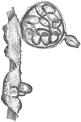
Fig. 30.—Ectocyst, endocyst, and brood capsule of Echinococcus.
From a Zebra. After Huxley.
The appearance of hydatids varies very much according to
their mode of formation, to the kind of host in which they are present,
and to the character of the organs in which they happen to take up their
residence. The so-called exogenous type occurs sparingly in man, whilst
the endogenous type is very abundant. The peculiar form known as the
multilocular echinococcus is probably a mere variety of the exogenous
type. The exogenous and endogenous hydatids may coexist in the same
bearer. In the lower animals we commonly find the organs of the body
occupied by numerous lobulated cysts, varying in size from a walnut to
a goose’s egg, but sometimes rather larger. They are rarely solitary,
being particularly liable to occupy both the liver and lungs in the same
animal. The viscera are sometimes crowded with cysts. The hydatids do
not usually protrude much beyond the surface of the infested organ, but
lie imbedded within its parenchymatous substance.
The multilocular variety was first described by Virchow. In
reference to it Leuckart writes as follows:
“Hitherto we know this growth only from the liver, in which it forms
a firm, solid, and tolerably rounded mass of the size of the fist or
even of a child’s head. At first sight it looks more like a pseudoplasm
than a living animal parasite. If you cut through the tumour, you
recognise in its interior numerous small caverns, mostly of irregular
shape, and separated from one another by bundles of connective tissue,
more or less thick, and including a tolerably transparent jelly-like
substance. In the intervening stroma a blood-vessel or a collapsed
bile-duct runs here and there; but there is nowhere any trace of
true liver substance. The outer boundaries of the tumour are in most
cases pretty well defined, so that the attempt to cut these growths
out is not difficult. In particular spots, especially at the surface,
one sometimes sees white, moniliform, jointed lines passing off from
the tumour, and even thicker terminations which, perhaps, expand in
the neighbouring liver-parenchyme into new (multilocular) groups of
different size. In one case, recorded by Virchow, the growth extended,
together with Glisson’s capsule, a long way towards the intestine.” To
this description it may be added, that the growth on section presents
an appearance not altogether unlike alveolar colloid, having, in point
of fact, been confounded with that pathological product, with which,
however, as stated by Virchow, it has nothing in common. This is proved
not only by the occurrence of the pathological features above mentioned,
but also, more
particularly, by the well-ascertained presence of echinococcus-heads in
most of the so-called alveoli. Several hypotheses have been broached
with the view of explaining the mode in which these multilocular hydatid
growths are formed. Virchow thought that the echinococcus vesicles were
primarily formed in the lymphatic vessels, whilst Schröder van der Kolk
supposed that they originally took up their abode in the biliary ducts.
Although, thanks to the courtesy of Professor Arnold Heller in giving me
a specimen, I have been enabled to confirm much that has been written
in respect of the morbid appearances, I can add nothing towards the
solution of the difficulty in question. Until lately it was supposed
that the multilocular variety of hydatids only existed in man, but
Professor Böllinger has encountered it in the liver of a calf.
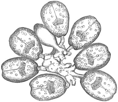
Fig. 31.—Group of Echinococcus-heads, from an hydatid found in the liver of a sheep.
Magnified about 25 diameters. From a drawing by Professor Busk.
|
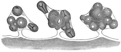
Fig. 32.—Three brood-capsules, containing Echinococcus-heads. Magnified 76 diameters.
After Professor Erasmus Wilson.
|
Selecting any ordinary fresh example of the exogenous kind,
and laying the tumour open with a scalpel, we notice in the first
instance an escape of a clear transparent, amber-coloured fluid. This
previously caused the distension of the sac. If the tumour is large,
this escape will probably be followed by a falling in, as it were, of
the gelatiniform hydatid membrane, in which case the inner wall of the
external adventitious investment or true fibrous cyst will be laid
bare. If the hydatid be next withdrawn from the cyst, it will be seen
to display a peculiar tremulous motion, at the same time coiling upon
itself wherever there is a free-cut margin. Further examination of
portions of the hydatid will show that we have two distinct
membranes; an outer, thick, laminated, homogeneous elastic
layer (the ectocyst of Huxley), and an internal, thin, soft, granulated,
comparatively inelastic layer—the endocyst of the same
author. The terms are convenient. The ectocyst is structureless,
consisting of a substance closely allied to chitine. For this
and other reasons it has been called the cuticular layer, but
the endocyst is the essential vital part of the animal, representing
a huge compound caudal vesicle. In an hydatid from
the zebra, Huxley found that the endocyst was “not more than
1/2000th of an inch in thickness, being composed of very delicate
cells of 1/2000″ to 1/5000″ in diameter, without obvious nuclei; but
often containing clear, strongly refracting corpuscles, generally
a single one only in a cell.” Prof. Huxley adds: “These
corpuscles appear to be solid, but by the action of dilute acetic
acid the interior generally clears up very rapidly, and a hollow
vesicle is left of the same size as the original corpuscle. No
gas is developed during this process, and sometimes the
corpuscles are not acted upon at all by the acid, appearing then
to be of a fatty nature. A strong solution of caustic ammonia
produces a concentrically laminated or fissured appearance in
them. Under pressure and with commencing putrefaction a
number of them sometimes flow together into an irregular or
rounded mass.”
The precise mode of development of the echinococcus-heads
or scolices has been a subject of lengthened discussion between
Leuckart and Naunyn. According to Leuckart the earliest
indication of the scolex consists of a slight papillary eminence on
the inner surface of the granular endocyst. After a short
period this prominence displays in its interior a vacuole-like
cavity, the latter being occupied, however, with a clear limpid
fluid. Its margins become more and more clearly defined, until
the cavity is by and by seen to be lined with a distinct cuticular
membrane. The papilla increasing in size, becomes at first
elongated or oval, eventually scoleciform, or even, perhaps, a
true echinococcus-head. Thus far the description bears out, in
a measure, the theoretical notions entertained by the older
authors; but the developmental process does not stop here. The
scolex-development has now to sacrifice itself by developing in
its interior a brood of scolices or echinococcus-heads. In other
words, it becomes transformed into the so-called brood-capsules
of Leuckart and other authors. These structures were previously
well known to Professors Erasmus Wilson and George
Busk. Mr Wilson spoke of the capsule as “a delicately thin
proper membrane, by which the Echinococci are connected with
the internal membrane of the acephalocyst” (‘Med.-Chir.
Trans.,’ 1845, vol. xxviii, p. 21). Mr Busk described the
echinococcus-heads as “attached to a common central mass by
short pedicles, which appear to be composed of a substance
more coarsely granular, by far, than that of which the laminæ
of the cyst are formed. This granular matter is prolonged
beyond the mass of Echinococci into a short pedicle common
to the whole, and by which the granulation is attached to the
interior of the hydatid cyst.” What Mr Busk here describes
as a granulation can only be equivalent to the brood-capsule
and its entire contents, but he elsewhere speaks of the capsule
itself as a “delicate membranous envelope.” It should be
borne in mind that Busk’s paper was communicated to the
Microscopical Society so early as the 13th Nov., 1844; being
published in the ‘Transactions’ for that year.
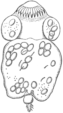
Fig. 33.—Separate scolex, or echinococcus-head.
Magnified 500 diameters. After Huxley.
In the completely developed state the echinococcus-heads exhibit
somewhat variable characters as to size and form, the latter differences
being, for the most part, dependent upon their degree of contraction and
vitality. In the perfect condition they vary from the 1/60″
to the 1/100″
in diameter, being usually about the 1/80″.
They are solid, and when stretched out exhibit an hour-glass-like
constriction at the centre of the body, which divides the scolex into an
anterior part supporting the rostellum and suckers, and a posterior part
which has been compared to the caudal vesicle of ordinary Cysticerci.
The rostellum supports a double crown of hooks, but the disparity of the
two series is scarcely sufficiently marked to render their distinction
obvious. The hooks of the smaller row vary in size from 1/1040″
to 1/830″
of an inch, whilst those of the larger series are from 1/830
to 1/555″.
In all instances the root-processes are incompletely developed, and
consequently vary in thickness. They are, as Leuckart also has stated,
apt to exhibit abnormalities.
In regard to the development of the echinococcus-heads it further
remains for me to observe that a distinct water-vascular system is
recognisable in the scolices. By the intervention of the pedicle of the
scolex this system is connected with the brood-capsule, and also with
the vessels of
the maternal endocyst. In the scolex there exists a circular channel
immediately below the rostellum, and this ring, on either side, gives
off two vessels which pass downwards in a tortuous manner, internally,
until they arrive at the pedicle where they unite to form two channels,
which latter are continued into the vascular system of the maternal
endocyst. In the retracted condition their position, of course, becomes
very much altered, and they form loops on either side of the central
line which marks the space leading down to the inverted head. Neither
Prof. Huxley nor myself have seen these vessels, which Leuckart observed
in the scolex itself, but Huxley discerned some apparently loose cilia
in the granular parenchyma of the body; their longitudinal measurement
being about the 1/3500
of an inch.
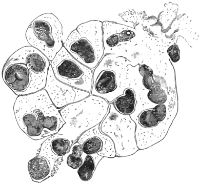
Fig. 34.—An Echinococcus brood-capsule (flattened by pressure). Magnified about 120 diameters.
From a drawing by Professor Busk.
As regards the production of “nurses” by the phenomenon
of proliferation, I can only remark that the endocyst is
primarily concerned. The secondary and tertiary vesicles must
be regarded as so many special bud-developments which, instead
of becoming brood-capsules, become daughter-vesicles and
grand-daughter vesicles, constantly developing in their interior
secondary and tertiary brood-capsules and scolices, but sometimes,
it would appear, developing neither the one nor the other.
This is the view of Naunyn, which is somewhat opposed by
Leuckart, who holds that the vesicles ordinarily arise from within
the layers of the ectocyst. Speaking of these daughter-hydatids
Leuckart remarks that “Naunyn denies that they take their
origin between the lamellæ of the mother bladder—a fact, however,
which, in agreement with Kuhl and Davaine, I have seen
more than once and have followed out step by step.” For my
own part I incline to the belief that the process as observed by
Leuckart is exceptional, and that under ordinary circumstances
it occurs as Naunyn has described it. Thus the long and short
of the whole matter appears to be that the endocyst is capable
of forming solitary scolices. Some of the scolices become
differentiated to form brood-capsules, a portion of whose individual
echinococcus-heads may, in their turn, become secondary
brood-capsules, whilst others fail to become either scolices or
brood-capsules. It accords with our knowledge of the general
plan of development to believe that the daughter and grand-daughter
hydatids are likewise peculiarly modified scolices.
They are, in short, buds of the endocyst.
The distribution of hydatids throughout the organs of the
bearer, and their prevalence in particular countries, has especially
engaged my attention. I have personally examined
upwards of a thousand preparations of entozoa in our public
collections; and of these, 788 are preserved in the anatomical and
pathological museums of the metropolis. By this inspection I
have obtained a tolerably accurate knowledge of the pathology,
localisation and effects produced by the presence of bladder-worms
in at least 200 unpublished cases of hydatid disease.
Most of our museums exhibit one or more specimens that are
unique. After making certain necessary deductions, I find that
I have 192 new cases to add to the 135 cases of hydatid
disease that I had previously recorded, affording a total of 327
cases available for statistical purposes. If an analysis of these
cases be made and compared with the statistics furnished by
Davaine, and if the whole be reduced to the lowest number of
practically available terms, we at length obtain a result which,
although it may be only approximatively correct, is nevertheless
of much practical value and significance. The statistics in
question stand as follows:
| Organs affected. | Davaine. | Cobbold. | Total. |
|---|
| Liver | 165 | 161 | 326 |
| Abdomen, including spleen | 26 | 45 | 71 |
| Lungs | 40 | 22 | 62 |
| Kidney and bladder | 30 | 23 | 53 |
| Brain | 20 | 22 | 42 |
| Bones | 17 | 16 | 33 |
| Heart and pulmonary vessels | 12 | 13 | 25 |
| Miscellaneous | 63 | 25 | 88 |
| Total | 373 | 327 | 700 |
In the main Davaine’s table and my own show a remarkable
correspondency, as is seen in the numbers referring to hydatids
of the liver, heart, and bones respectively. Where our results
do not correspond the explanation of the discrepancy is sufficiently
simple. The abdominal cases here credited as such in
Davaine’s table are placed by him under pelvis, whilst the
abdominal cases in my own table not only include the pelvic
hydatids, but also two spleen cases, and nineteen others from
the peritoneum and intestines.
As the facts here stand, the liver cases comprise nearly
461/2 per cent. In a large number of cases the entozoon has
taken up its abode in organs of vital importance. If statisticians
and officers of health would obtain an adequate conception
of the fatal capabilities of parasites, they should consider
these data. In 6 per cent. of all these cases the bladder worm
has found its way into the brain, and of course proved fatal to
the bearers; in about 31/2 per cent. more they took up their
residence in the heart, also proving fatal; whilst of all the
other cases put together I reckon that not less than 15 per cent.
were concerned in bringing about the death of their hosts. I
probably underrate the fatal capabilities of echinococcus disease
when I express the conviction that hydatids prove fatal to 25
per cent. of all their human victims.
The recently published analysis of 983 cases by Dr Albert
Neisser affords similar results. Of these, 451 were referable
to the liver, or 45·765 per cent. The other cases, reduced as
above, show in the main a similar correspondency.
It may be asked if these facts afford us any assistance in
determining the amount of injury that we, as a people, sustain
either directly or indirectly from hydatids. On carefully
reviewing all the data before me, I may say that it is difficult
to draw very precise conclusions; albeit it is not mere guess-work
when I assert that in the United Kingdom several hundred
human deaths occur annually from this cause. In some
other countries the proportion is far greater; the oft-quoted
case of Iceland, where the disorder is fatally endemic, still
standing at the head of the afflicted territories.
Our Australian colonies are probably entitled to the next
place of distinction in this respect. We have strong and
recent evidence of the truth of this statement. Thus a writer
in the ‘Australian Med. and Surg. Review’ says: “This disease
is becoming unpleasantly frequent, and at present we have
no reliable mode of treatment, either theoretical or empirical.”
Another writer observes (‘Melbourne Argus,’ May 18th, 1874),
“Hydatid disease is endemic in this colony; and, though not
so constantly met with as in Iceland, we may probably claim
the doubtful honor of holding the second place in the list of
countries so affected.” In the ‘Argus’ for June 20th of the
same year, another writer refers to the frequent notices of
cases of hydatids published in the various local newspapers. A
retired medical man, the late Mr J. P. Rowe, writing in the ‘Melbourne
Leader’ (Sept. 7th, 1872), incidentally remarked on the
“notable increase of hydatid disease in the human subject.”
Again, still more satisfactory evidence is afforded by a reviewer
in the ‘Leader’ of the 31st January, 1874. Commenting on
my manual, he not only takes occasion to speak of the prevalence
of hydatids generally, but also supplies that kind of
accurate statistical evidence of which we so much stand in need.
He gives the following table, showing the number of deaths
from hydatids in Victoria for eleven years. It is instructive in
many ways.
| Years. | Males. | Females. | Total. |
|---|
| 1862 | 3 | 2 | 5 |
| 1863 | 3 | 2 | 5 |
| 1864 | 6 | 3 | 9 |
| 1865 | 9 | 6 | 15 |
| 1866 | 18 | 7 | 25 |
| 1867 | 13 | 12 | 25 |
| 1868 | 21 | 12 | 33 |
| 1869 | 12 | 10 | 22 |
| 1870 | 10 | 7 | 17 |
| 1871 | 6 | 9 | 15 |
| 1872 | 24 | 5 | 29 |
| Total deaths in eleven years | 125 | 75 | 200 |
To employ the writer’s own words, “this mortality gives only
a faint notion of the extreme prevalence of hydatids in Victoria,
since numbers of cases are cured by tapping, and otherwise by
medical treatment, or by spontaneous bursting of the cysts.”
Hydatids are often found post mortem where their presence has
never been suspected during life. “To meet with hydatids as a
cause of deranged health is now a matter of daily expectation
with every medical practitioner.” Lastly, Dr Dougan Bird, in
his able brochure on ‘Hydatids of the Lung,’ fully confirms
these statements, remarking that the rich and poor of the Australian
metropolis suffer just as much from hydatids as do either
the shepherds of the western plains, or the miners of Ballarat
and Sandhurst.
Such are the facts from Australia. As regards home evidence,
so far as I am aware, little or nothing has been done
towards securing an accurate estimate of the mortality in
England from echinococcus disease. The reports of the Registrar
General give no sufficient sign. The explanation is not far
to seek, since for the most part hydatids are either classed
with diseases of the liver, or with those of the other organs in
which they happen to have been present.
One of the most valuable contributions to our knowledge of
the prevalence of hydatid disease affecting animals is that
supplied by Dr Cleghorn, from a statistical table constructed
by the executive commissariat officers stationed at Mooltan.
The record in question shows that out of 2109 slaughtered
animals, no fewer than 899 were affected with hydatid
disease. This is equal to more than forty-two per cent. In
the majority of cases, both the lungs and liver were affected,
cysts were found 829 times in the liver and 726 times in the
lungs. In a few instances they were present in the kidneys,
and also occasionally in the spleen. The inference from all
this is that in India, if not elsewhere, the echinococcus disease
is much less common in man than it is in animals. The
explanation is simple enough, since cattle have more ready access
to, and less scruple in partaking of filthy water and food in
or upon which the eggs of the Tænia echinococcus abound.
Into purely professional questions connected with the treatment
of the echinococcus malady I do not here enter; nevertheless,
in connection with hygiene I may observe that the
prevalence of hydatids in any country is strictly dependent
upon the habits of the people. The close intimacy subsisting
between the peasantry and their canine companions is the
primary source of the endemic; and where dogs are not kept,
it is well nigh impossible that the disease should be contracted.
The fact that every Icelandic peasant possesses, on an average,
six dogs, and that these dogs share the same dwelling (eating
off the same plates and enjoying many other privileges of
intimate relationship) sufficiently explains the frequency of
hydatids in that country. According to Krabbe, the sexually
mature Tæniæ occur in 28 p. c. of Icelandic dogs, whereas in
Copenhagen he found it twice only in 500 dogs examined. In
his work (quoted below, p. 58, or Fr. Edit., p. 60) Krabbe
comments on a sensational passage which, in my introductory
treatise (p. 283), I had quoted from a popular memoir by
Leuckart (‘Unsere Zeit,’ s. 654, 1862). The practitioners
whom we had spoken of as “quacks” are mostly homœopaths;
and it appears that even those who are not in any legal sense
professional men “treat their patients much in the same way
as ordinary medical men.” It simply comes to this, that,
instead of dog’s excrement forming with the aforesaid “quacks”
a conspicuous or common remedy (as Leuckart’s description had
led me to infer), this nasty drug is now rarely administered,
and by the grossly ignorant only.
Up to the present time no person has seen the Tænia echinococcus
in any English dog which has not been previously made
the subject of experiment, but considering the prevalence of
hydatid disease amongst us, there can be no doubt that
English dogs are quite as much if not more infested than continental
ones. Probably, at least one per cent. of our dogs harbour
the mature tapeworm. Certainly a great deal of good might
accrue from the acquisition of more extended evidence respecting
the prevalence of this and other forms of entozoa infesting
man and animals in this country.
From Schleissner’s table it appears that hydatids are more
frequent in women than in men. Apparently, it is not so in
Australia. As regards Iceland the explanation must be sought
for in the different habits of life. No doubt, water used as drink
by women is constantly obtained from supplies in the immediate
neighbourhood of dwellings, and in localities to which dogs
have continual access. The comparative rarity of the echinococcus
disease amongst sailors is not so much dependent upon
the circumstance that seamen’s diet usually consists of salted
provisions, as upon the fact that these men can seldom have
opportunities of procuring water from localities where dogs
abound. In regard to water drinking, there is ground for
believing that the addition of a very little alcohol is sufficient
to destroy the six-hooked embryos of Tænia echinococcus whilst
still in ovo; and there is no doubt that water raised to a temperature
of 212° Fahr. will always ensure the destruction of the
larvæ. Boiled water by itself is by no means palatable. The
reason why the upper classes comparatively seldom suffer from
hydatids may be attributed to the circumstance that those few
who drink water take the very proper precaution to see that it
is either “pump” or fresh spring water in which no living
six-hooked embryos are likely to exist. So far as hydatids are
concerned, wine and beer drinking is preferable to water-drinking;
yet if water is carefully filtered no evil of the parasitic
kind can possibly result from its imbibition. An ordinary
charcoal filter will effectually prevent the passage of the ova,
since their diameter is nearly 1/370 of an inch.
From what has been stated it follows that personal and
general cleanliness are eminently serviceable as preventions
against infection, but to ensure perfect success other precautions
must be exercised, especially in relation to our contact with
and management of dogs. Leuckart puts this very clearly when
he says:—“In order to escape the dangers of infection, the
dog must be watched, not only within the house, but whilst he
is outside of it. He must not be allowed to visit either
slaughter-houses or knackeries, and care must be taken that
neither the offals nor hydatids found in such places are accessible
to him. In this matter the sanitary inspector has many
important duties to perform. The carelessness with which
these offals have hitherto been disposed of, or even purposely
given to the dog, must no longer be permitted if the welfare
of the digestive organs of mankind is to be considered.
What blessed results may follow from these precautions may
be readily gathered from the consideration of the fact that,
at the present time, almost the sixth part of all the inhabitants
annually dying in Iceland fall victims to the echinococcus
epidemic” (l. c., s. 654). Similar measures had previously
been recommended in less explicit terms by Küchenmeister,
who in effect remarked that the principal thing was to ensure
the destruction of the echinococcus vesicles. He also recommended
the expulsion and annihilation of the Tænia echinococcus.
In order to carry out this idea, it was suggested by Dr Leared
that every dog should be periodically physicked, and that all
the excreta, tapeworms included, should be buried at a considerable
depth in the soil. I advised, however, that in place
of burying the excreta, they should, in all cases, be burnt. I
had, indeed, long previously urged this measure (in a paper
“on the Sclerostoma causing the gape-disease of fowls,”
published in 1861), with the view of lessening the prevalence of
entozoa in general, whether of man or animals. The rule I
suggested stood as follows:—All entozoa which are not preserved
for scientific investigation or experiment should be thoroughly
destroyed by fire, when practicable, and under no circumstances
whatever should they be thrown aside as harmless refuse. As
an additional security I recommended that boiling hot water
be occasionally thrown over the floor of all kennels where dogs
are kept. In this way not only would the escaped tapeworms
be effectually destroyed, but also their eggs and egg-contents,
including the six-hooked embryos. These measures were again
advocated at the Cambridge Meeting of the British Association
in 1862, and also more fully in a paper communicated to the
Zoological Society, during the autumn of the same year (‘Proceedings,’
vol. xxx, pt. 3, pp. 288, 315).
As the scope and tendency of this work preclude the textual
admission of clinical details, I must limit my remaining observations
to the pathology of hydatid disease. At very great
labor, pursued at distant intervals during a period of ten years,
I sought to ascertain the probable extent and fatality of this
form of parasitism in England, by going over such evidence as
our pathological museums might supply. Although, from a statistical
point of view, the investigation could hardly be expected
to yield any very striking results; yet clinically viewed the
study was most instructive. The evidence which I thus procured
of numerous slow and painful deaths from echinococcus
disease, further stimulated me to place a summary of the facts
on record. Physicians, surgeons, scientific pathologists, and
veterinary practitioners are alike interested in the study of
hydatid disease; and I had not proceeded far in my careful
investigation before it became evident to me that very great
practical results would ensue if, in this kind of effort, the principle
of division of labor had full play. At all events, within
these museums lie concealed a mass of pathological data which,
although well within reach, have not been utilised to the extent
they ought to have been.
As a student of parasites for some thirty years, I must
without offence be permitted to protest against the too
frequent omission of parasites in statistical evidence as a
cause of mortality. From facts within my own knowledge I
can confidently assert that parasites in general, and hydatids in
particular, play a far more important part in the production of
disease and death than is commonly supposed. In saying thus
much, however, I am not insensible to the fact that, in recent
times, new methods of treatment combined with higher surgical
skill, have greatly tended to lessen the fatality of this affection.
In this connection I would especially refer to the recorded
experiences of an able colonial surgeon, Dr MacGillivray, as
made known in the pages of the ‘Australian Medical Journal.’
The able surgeon to the Bendigo Hospital, treated as in-patients,
from 1862 to 1872, inclusive, no fewer than seventy-four cases
of hydatid disease. He operated on fifty-eight of them. Two
patients were tapped for temporary relief (as they were dying
of other diseases); and of the remaining fifty-six only eleven
died. No fewer than forty-five were discharged cured—a fact
redounding largely, I should think, to the credit of Australian
surgery.
In reference to museum evidence I have no hesitation in
saying that the pathological collections in the metropolis
abound in rare and remarkable illustrations of hydatid disease;
most of the preparations being practically known only to such
few members of the medical profession as have been at some
time or other officially connected with the museums. Not
without justice, curators often complain that their work and
catalogues are turned to little account. As a former conservator
of the Edinburgh University Anatomical Museum (1851–56), and
subsequently as museum-curator at the Middlesex Hospital
Medical College, I am in a position to sympathise with them.
Valuable, however, as the catalogues are, it is often necessary
to make a close inspection of the preparations in order to arrive
at a correct interpretation of the facts presented.
Although the entozoal preparations in the museum attached
to St Bartholomew’s Hospital are, comparatively speaking,
few in number, there are some choice specimens of hydatid
disease. There is a remarkable case in which hydatids invaded
the right half of the bones of the pelvis; death resulting from
suppurative inflammation of the cysts. This patient, a woman,
had also another hydatid cyst which was connected with the
ovary. Amongst the series contributed by Dr Farre, there is
a case represented where a large cyst containing numerous
hydatids “occupied the pelvis of an infant and produced retention
of urine,” which ultimately proved fatal. There are also
several fine examples of hydatids from the omentum (Dr Farre’s
case), besides a good specimen of acephalocysts connected with
the vesiculæ seminales. There are two other cases in which
these larval entozoa were passed with the urine. At the time
when I made my inspection, the entire series represented
twenty-five separate cases, of which only one appears to have
been published in detail (Mr Evans’s case, ‘Medico-Chirurgical
Transactions,’ 1832). In addition to the above, I must not
omit to particularise two instructive preparations illustrative
of a case in which an hydatid was lodged in the right
half of the cerebrum. This was from a girl in whom head
symptoms showed themselves a year before death, and in whom
there was partial hemiplegia of the left side. I may add that
there is also in the series a doubtfully genuine example of
hydatids of the breast.
The collection in connection with the Westminster Hospital
contains several highly interesting specimens of entozoa (one
of which I believe to be altogether unique), but it is by no
means rich in the matter of hydatids. Out of a score of
preparations of parasites of various kinds, only four (apparently
representing the same number of cases) are hydatids, all of
which appear to have been connected with the liver. Two are
certainly so, one of the latter (Mr Holthouse’s case) showing
calcareous degeneration.
The museum connected with St Mary’s Hospital Medical
School, in addition to several liver cases, contains one interesting
example of hydatids of the lung (Dr Chambers’s case), and
also three valuable preparations illustrating Mr Coulson’s
remarkable case of hydatids affecting the tibia. One of the
preparations shows the bone itself, which was eventually removed
at the joint, the operation having been performed by Mr
Spencer Wells.
Here, perhaps, it will not be out of place to mention as a
fact of special clinical interest that I have encountered records
of no fewer than nine other similar cases where hydatids
have taken up their abode in the tibia, generally selecting
the head or upper part of the bone. Some of my notes have
been mislaid, but, speaking from recollection, one of the
choicest specimens which I have examined is that contained
in the pathological museum of the Nottingham Hospital.
When I first went over the collection of the Middlesex
Hospital Museum, I found it to contain fifty-four preparations
of entozoa, of which some fourteen only were true hydatids,
representing as many separate cases. There are now upwards
of a score of preparations of hydatids, several of the cases
having already had ample justice done to them by Dr Murchison
in his well-known memoir (‘Edinb. Med. Journ.,’ Dec., 1865).
Amongst the most interesting preparations I would especially
call attention to two fine and genuine specimens from the
kidney, another very large example of an hydatid situated
between the bladder and rectum, a simple acephalocyst removed
from the orbit (Mr Hulke’s case), and the hydatid removed
from the axilla by the late Mr Charles Moore. There is a jar
containing hundreds of hydatids that were taken from the
thoracic cavity of a dissecting-room subject, who was reported
to have died of phthisis; and there is another preparation of an
hydatid of the heart, which also proved fatal, without there
having been the slightest suspicion entertained as to the true
nature of the disease. For this fine preparation the museum
stands indebted to Dr Moxon, of Guy’s Hospital. Several of
the liver cases are particularly instructive; but amongst the
specimens presented by Mr Mitchell Henry is a small bottle full
of minute hydatid vesicles, all of which were removed from the
interior of the tibia. The history of this case has been lost;
and, unfortunately, the bone from which the parasites were
taken does not appear to have been preserved.
The museum connected with King’s College contains at
least a dozen good specimens of liver hydatids, several of the
cases being of special interest from a pathological point of
view. There are two remarkably fine examples of hydatids
contributed by Dr Hooper, the parasites in one case affecting
the spleen, and in the other involving the ovary and uterus.
The spleen contained numerous encysted hydatids, whilst the
uterine organs exhibited “an immense collection” of the same
growths. In this place, also, I may refer to an hydatid-like
entozoon, taken from a cyst in the ovary of a female who had
been under the care of Dr Johnson (1860). It is, apparently, a
genuine example of the slender-necked hydatid (Cysticercus
tenuicollis); and if so (as might be determined by dissection),
is, so far as I aware, the only specimen of the kind in existence
from the human bearer. There is a renal hydatid (presented
by Dr Pass, of Warwick) which was obtained from a lunatic,
its presence being “quite unsuspected during life.” Amongst
the liver cases (the majority of which are from Dr Hooper’s
collection), there is one enormous hydatid that was obtained
from a young woman who had died during a fit of laughter.
The tumour had pushed the diaphragm up to a level with the
fourth rib; and it is stated that, on puncturing the cyst, the
fluid contents were ejected “in a jet nearly two feet high.”
There is one case represented where numerous hydatids were
expectorated after hepatitis, whence it was concluded that they
were originally connected with the liver. There is a large
solitary hydatid that was removed from a young female who
died of phthisis, and in whom the consequent swelling had
formed in the neighbourhood of the navel. Especially instructive,
also, from a clinical point of view, is a case of peritoneal
hydatids where the tumours had been diagnosed to represent a
case of extra-uterine fœtation. It appears that there were two
cysts, one of them being connected with the uterus. Two of
the enormous hydatids taken from these cysts are preserved in
the collection of the Anatomy School of Oxford. Several of
the preparations show to perfection the stages of natural cure
produced by calcareous degeneration; and there is one liver
showing three of these so-called ossified cysts. The disease in
this case proved fatal.
Most of the entozoa displayed in the Charing Cross Hospital
Museum have been contributed by Dr Wiltshire, the series
being particularly strong in tapeworms. There are four characteristic
examples of hydatids of the liver, representing as
many separate cases. Two were from abscesses of this organ.
In one of these, Mr Canton’s case, the hydatid was, I believe,
expelled after operation; but in the other example (presented
by Mr Rose, of Swaffham) the parasite was evacuated from an
abscess, which burst of itself, externally.
In the museum at University College, I examined sixteen
preparations of hydatid disease, representing almost as many
distinct cases. One is a wax model. Eight of the specimens
were from the liver, five from the abdomen (including those of
the omentum and mesentery), two from the lungs, and one
from the heart. The model displayed ordinary hydatids of the
liver bursting into the lungs. The mesenteric example is particularly
fine, whilst that from the omentum is undergoing calcareous
degeneration. Probably the most interesting of all is
the example showing an hydatid lodged in the septum of the
heart. This was from a middle-aged female, who died suddenly
whilst pursuing her ordinary domestic avocations.
The museum of the Royal College of Surgeons contains a
fine collection of parasites, its chief strength in this respect
being due to the special series of entozoa. Were visitors to
judge by the contents of the catalogue of this series (which I
prepared some years ago at the instance of the Council of the
College), they might be led to suppose that the hydatids were
only feebly represented. Out of nine preparations of hydatids
in this section, only six have come from the human body.
However, scattered throughout the collection, I found that
there were no fewer than thirty-five preparations of hydatids
belonging, apparently, to as many as thirty separate cases.
Omitting, for the present, all mention of these derived from
animals, I ascertained that, of the thirty human cases, thirteen
were referable to the liver, four to the abdomen, three to the
lungs (one of which was originally connected with the liver),
and two to the brain. Five were of uncertain seat. With the
abdominal cases we may also include one case of hydatids of
the spleen, and another where these organisms were found in
the region of the bladder. There is a characteristic breast
case. One of the original Hunterian cases (in which “a prodigious
number of hydatids were found in the sac of the liver
and dispersed throughout the cavity of the abdomen”) appears,
though it is not expressly so stated in the catalogue, to have
been regarded as an ordinary example of abdominal dropsy.
In one of the three lung cases two small hydatids were separately
expectorated at an interval of about a month. This
occurred in a female.
I may here incidentally remark that many cases are on record
where abdominal hydatids have been overlooked, the patient
being supposed to be suffering from ascites. One such instance
took place a few years ago at the Middlesex Hospital.
I well remember a similar case of supposed hydrothorax, where
the post-mortem examination revealed the presence of immense
numbers of these formations occupying the right side of the
chest. This case occurred at the Norfolk and Norwich
Hospital, at the time when I was a student there, some
thirty-five years ago.
The pathological collection connected with St George’s
Hospital displays several good hydatid preparations, the entire
series representing at least twenty-two separate cases. Of
these, fifteen are referable to the liver, that is, if we include Dr
Dickinson’s case, already published, where hydatids were found
within the hepatic duct. There are two renal cases; also one
from the brain (Dr Dickinson’s case), and another where an
hydatid was expectorated. Besides these, there are three other
highly characteristic examples of echinococcus disease affecting
the region of the neck, breast, and axilla respectively.
The museum of the London Hospital Medical School contains
a large collection of parasites. Out of fifty-seven preparations
of entozoa, I found twenty-two referable to hydatids; and, so
far as I could gather, all of them belonged to different cases.
Only one case seems to have been published in detail. This,
though a very old preparation, is a fine example of an hydatid,
nearly three inches in length, occupying one of the cerebral
hemispheres (‘Edinb. Med. Journ.,’ vol. xv). There is a second
brain case, where the vesicles were of small size, but very
numerous. Of the other twenty cases, fourteen belong to the
liver, two to the spleen, one to the lung, one to the uterus;
one being a very large hydatid of doubtful seat, and another
being referable to the lumbar region, where it formed a tumour
containing “a large number of small hydatids.” Amongst the
more remarkable specimens is that described in the MS. catalogue
as “a true hydatid cyst developed in connection with the
broad ligament.” This preparation, unique of its kind, shows
no trace of the ovary, which, indeed, seems to have disappeared
altogether. One of the liver cases should rather be classed as
abdominal, since the large cyst is situated between the diaphragm
and liver, pressing upon the latter organ below and also
upon the lung above, but apparently not involving either of
these viscera structurally. Another very striking case is that
in which there is an external opening communicating with the
cyst in the liver, and an internal opening through the diaphragm
communicating with the lungs and bronchial tubes.
The patient had actually coughed up liver hydatids by the
mouth, and had passed others through the right wall of his
abdomen. There is another liver case in which the hydatids,
in place of escaping externally, had gained access to the inferior
cava; and if I understand the MS. record rightly, in the same
patient a second hydatid communicated with the portal vein,
and a third with the hepatic vein. Lastly, I must add that
there is yet another fine preparation of liver hydatids, occurring
in a lad, nineteen years of age. He had, it seems, met with
“a slight accident, and died with obscure head symptoms;” but
the odd part of the case is that at the post-mortem examination
there was positively nothing found that could explain the
patient’s death. He was under the care of Mr Luke (1834).
Comparatively recently I inspected the collection at St
Thomas’s Hospital, which I found to be particularly rich
in entozoa of various kinds, especially tapeworms and hydatids.
I encountered seventy-six preparations of internal parasites;
and of these, forty-two were of the hydatid kind, representing
at least thirty-three different cases. I say “at least,” because
it is often impossible to decide in instances where no history of
the specimens can be obtained. Thus, there are three similar
preparations of hydatids passed by the urethra, and, from their
appearance, I judge them to have come from one and the same
patient; yet there is no statement in the catalogue to that
effect.
Of the thirty-three cases of hydatids represented in this
museum, I reckoned eighteen as referable to the liver, two to
the brain, two to the bones, two to the urinary organs, and one
to the lung, spleen, uterus, and soft parts of the thigh respectively.
There are also three that may be classed as peritoneal.
There is another choice example in which the disease cannot be
referred to any particular organ. I allude to Dr Peacock’s case,
already published (‘Pathological Transactions,’ vol. xv), where
the lungs, liver, heart, spleen, and some other organs, were all
occupied by hydatid formations. As an instance of extensive
visceral infection by Echinococci in the human subject, I believe
this case to be unique. The brain hydatids are particularly
fine. In the specimen presented by Mr Boot, of Lincoln, the
hydatid, two inches in diameter, is lodged in the anterior horn
of the left ventricle. One of the peritoneal cases is remarkable
for the amount of forward displacement of the pelvic viscera,
caused by four or more hydatids, each of them nearly as large
as a cricket-ball. Amongst the abdominal cases I have included
a recent preparation, to which Mr Stewart has called my
attention. The hydatid in question, of the size of a large
lemon, existed near the fundus of the bladder, its walls being
one third of an inch in thickness, and forming an unusually
firm tumour. Of all the fine specimens of hydatids in the collection,
however, none have struck me so much as those affecting
the bones. There is a humerus, taken from a man thirty-four
years of age, in which the shaft is occupied throughout by
small hydatids that have destroyed almost all the cancellous
structure; in some places, also, the absorption of the cortical
layer has gone on to such an extent as to have left little more
than the periosteum. Of course, the bone was at last fractured
easily. It is a beautiful specimen; and the existence of Echinococcus-heads
was proved by microscopic evidence. Scarcely
less interesting are two preparations illustrative of Mr Traver’s
case of a man, thirty-eight years of age, in whom numerous
small hydatids occupied both the head of the tibia and the lower
end of the femur. Each set of parasites freely communicated
with the knee-joint, necessitating amputation of the limb.
The very large museum connected with Guy’s Hospital is
rich in hydatids. When, some time ago, I spent several days
in going over the collection, I examined seventy-six preparations,
representing apparently seventy separate cases of this
affection. Amongst the noteworthy specimens one lung
hydatid was intimately associated with a thoracic aneurism,
two others being connected with the pleura; and of seven
abdominal cases, five were connected with the peritoneum, one
with the mesocolon, and one with the aorta. This last-mentioned
instance occurred in a woman of sixty years, who, until
her death, was treated for dropsy. She complained of incessant
pain, which was only relieved when she rested on her hands
and knees. Of the three cases affecting the heart one has
been published (Mr Henderson’s), where the patient, a girl of
nineteen years, died suddenly whilst in the apparent enjoyment
of perfect health. In one of the other two cases (Mr May’s, of
Tottenham), the left lung was also involved. One case of
hydatid disease affecting the spinal column appears to have been
originally an ordinary liver case. In Mr Cock’s example of
genuine mammary hydatids, the hooklets and echinococcus
heads were detected; but I am not sure that a similar
result of microscopic examination was obtained in the equally
interesting example of hydatids of the thyroid gland (also
removed by Mr Cock). There are five bladder cases, all
apparently genuine (of which one has been published); and
there are also five other cases referred in the catalogue to the
kidneys, of which I regard two as doubtfully parasitic in
character. Of three cases of hydatid growths occupying the
soft parts of the thigh, two were under Mr Bryant’s care. The
museum likewise contains an old preparation of hydatids of the
tibia, but its history has been lost. There are also two brain
cases, besides upwards of a score of more or less characteristic
and instructive cases of hydatids affecting the liver.
Scattered amongst the museums connected with the larger
provincial schools and recognised hospitals there must be a
great many valuable preparations of hydatid disease; at all
events, I judge so from the inspection I have incidentally made
of a few of the collections.
Of eleven preparations of human hydatids which I observed
in the Cambridge Anatomical Museum, apparently representing
the same number of cases, seven were connected with the
liver and one with the lungs. Those hydatids displayed in the
“special series” of entozoa were of uncertain seat. From the
recently published and valuable ‘Notes’ by Dr Bradbury, I
have no doubt that considerable additions have been made to
the Cambridge Collection since my last visit.
The museum at Oxford contains some choice specimens of
hydatids, but I have only personally inspected a few of them.
In the absence of original notes, however, I am indebted to
the kindness of Mr W. Hatchett Jackson for supplying me with
several interesting particulars. The anatomical department of
the Oxford Collection shows from one particular case two
hydatids that were found “under the dura mater.” In the
pathological department we find one hydatid from the liver of
a male subject, and also a preparation showing a number of
small hydatids that were “coughed up from the lungs of a
female.” There are also in this department (Dr Acland’s)
two examples of hydatids from the diaphragm, apparently
belonging to two separate cases. One is described as a large
“hydatid in the diaphragm covered by the pleura,” whilst the
other is spoken of as “springing from the diaphragm and
projecting into the sac of the pericardium.” There is likewise
a preparation showing a number of small specimens of hydatids
that were passed per anum by a female. It is conjectured
that they came from the liver.
The small pathological museum attached to the Brighton and
Sussex Hospital is particularly rich in hydatids. Amongst
others, it contains preparations illustrative of the remarkable
case of hydatids in the region of the prostate, communicated
by Mr Lowdell, in the ‘Lancet,’ in 1846.
The comparatively large museum adjoining the Norfolk and
Norwich Hospital displays a choice series of hydatids, chiefly
from the collection of the late Mr Crosse. That eminent
surgeon prepared a special set of specimens to illustrate the
process of natural cure by calcareous degeneration; and I may
here, perhaps, be pardoned for mentioning that it was the study
of these and other entozoa in Mr Crosse’s Collection, some
thirty or more years ago, that first drew my attention to the
phenomena of parasitic life. Illustrations of the helminths in
question are still in my possession. In one case (which is
instructive as indicating the possibility of death from the
simplest form and commonest habitat of an hydatid) a lad,
twelve years old, received a slight blow from a playmate. Something
gave way, and death speedily followed. It was found by
post-mortem examination that a solitary liver hydatid, rather
larger than a cricket-ball, had been ruptured. Although the
case is almost unique, it is nevertheless by no means pleasant
to reflect upon the fact that under similar circumstances a slight
blow might prove fatal to any one, no matter in what internal
organ the bladder worm happened to be situated.
Before concluding my summary notice of the human
hydatids contained in the metropolitan and certain other
museums, there is an interesting literary contribution that I
cannot pass unnoticed. In the November number of the
‘Indian Medical Gazette’ for 1870 an article occurs in which it
is stated that the Calcutta Medical College Museum contains
eighteen specimens of hydatid cysts of liver. This fact was, it
seems, originally adduced to show, not the frequency, but rather
the rarity, of the occurrence of hydatids in India. However,
from a valuable communication by Dr James Cleghorn, which
was published in the same periodical for the following March, it
appears that hydatids of the liver are much more common in
India than is generally supposed. This, he says, is owing to
the circumstance that many of the so-called cases of tropical
abscess are neither more nor less than examples of hydatid cysts
that have suppurated. Besides Cleghorn’s evidence, we have
the previous testimony of the Inspector General I. M. D.,
whose Report for 1868–69 I have already referred to in connection
with Cysticercus in beef. He says: “During some
three months’ regular observation of the animals killed at the
Commissariat slaughter-house here, at least 70 per cent. of the
beef livers may be calculated as thus affected. Cobbold,
writing of the Tænia echinococcus, says that ‘this little tapeworm
infests only the dog and the wolf.’ Therefore, considering the
immense number of pariah dogs fed on the refuse of animals
infected with hydatids, it seems more than probable that the
parasite must attain its strobila condition in their intestines, and
through them be eventually disseminated over the pastures on
which the cattle graze.”
I now turn to a neglected phase of the subject from which
much practical instruction may be gathered. The consideration
of the pathological phenomena of hydatid disease as it affects the
lower animals is of high interest, and no prejudice should induce
any medical man from accepting such useful data as may be
gathered from this source. The facts of hydatid parasitism in
animals, though often peculiar, are, for the most part, of an
order similar to those presented in the human subject. If any
medical practitioner thinks it beneath his dignity to study the
pathology of the lower animals, the conduct of John Hunter in
this respect is a standing protest against such narrowness.
The museum of the Royal College of Surgeons of England
contains some of the finest specimens of hydatids from the lower
animals that are to be seen anywhere, the very choicest of them
having been selected by Hunter himself. That distinguished
man sought information from every available source, and
hydatids were for him of almost equal interest, whether found
in the body of a human being or in the carcase of an ox or an
ass. Now, at all events, neither pathologists nor sanitarians can
well afford to neglect comparative pathology; and, for myself, I
am free to say that the yearly exposition to the students of the
Royal Veterinary College of the phenomena of parasitic life
amongst animals has brought with it an ever-increasing knowledge
of the most curious and often unlooked-for information.
Some of the data thus supplied are quite remarkable. Let me
also add that my studies of the entozoa of wild animals have put
me in possession of particulars of high value in regard to
the larger question of the origin of epidemics. Beasts, birds,
reptiles and fishes, of every description, are liable to succumb
to internal parasites, and there is practically no end to the
variety of useful information to be obtained from this source.
I have collected materials almost sufficient for a separate treatise
on this department of the subject, but I fear I shall never have
either the time or opportunity to give the facts due publicity.
Here, for obvious reasons, I must for the most part restrict
myself to the hydatids properly so called.
Referring, in the first instance, to the hydatids of animals
that have the same mode of origin and exhibit the same general
characteristics as those found in man, I notice that four of
the metropolitan museums exhibit nine examples of liver Echinococci.
The Hunterian Collection shows specimens of this
kind from the pig, monkey, zebra, and lion. The museum at
St Bartholomew’s Hospital contains two examples from the pig
and one from a cow; whilst the animal liver-hydatids preserved
in the King’s College and Guy’s Museums, respectively, are from
the pig and sheep. That from the latter is partly calcified.
Respecting animal hydatids affecting the lungs, the Cambridge
Museum exhibits a simple acephalocyst from a monkey, and
the Guy’s Hospital Museum shows a pulmonary hydatid from
the kangaroo. In the museum at Oxford, Dr Acland’s
(pathological) department shows a preparation of “one large
echinococcus cyst from the abdomen of a baboon,” whilst Dr
Rolleston’s department (anatomical) displays the echinococcus
itself from the “cavity of the abdomen of the same
animal.” The collection also contains a variety of other
bladder worms from different animals. The Hunterian Museum,
Lincoln’s Inn, exhibits four or five alleged examples of hydatids
from the kidney of the sheep, besides another from the spleen.
Some of these are of very doubtful character. A cystic kidney
from the sheep, preserved in the London Hospital Museum, and
originally supposed to have been due to hydatids, is (as hinted
in the MS. catalogue) certainly not of parasitic origin. In
regard to the occurrence of hydatids in the heart of animals the
Hunterian series shows two good examples from cattle, whilst
the collection at University College exhibits one taken from the
wall of the left ventricle of a sow. This was presented by Dr
Elliotson.
In the museum of the Royal Veterinary College there are a
number of excellent preparations of true hydatids taken from
various animals, especially from cattle, swine, and sheep; and
there are also many kinds of bladder worms which, though
often called “hydatid” by veterinarians, have a totally different
origin from that of the true Echinococci. The so-called gid-hydatids
(Cœnuri) and slender-necked hydatids (Cysticercus
tenuicollis) are of this description. Specimens of the polycephalous
brain hydatid, or Cœnurus, also exist in the museums
connected with St Bartholomew’s, Guy’s, and St Thomas’s
Hospital Medical Colleges, as well as in both the anatomical
and pathological departments of the Oxford Museum. Specimens
of large Cœnuri occurring in the soft parts of rabbits may
be seen in the Guy’s Museum (presented by Mr Carpenter).
Similar characteristic specimens exist in the Oxford Collection,
labelled C. cuniculi, obtained from the “masseter and infraspinatus”
muscles of a rabbit. My private collection also
contains a recent addition of this remarkable hydatid, sent to
me by Mr Alston from Ayrshire. It is the only one I have
seen from Scotland. In the second half of this work these
Cœnuri will again come under notice. Three examples of the
slender-necked hydatid (from a monkey and two sheep respectively)
may be seen in the Guy’s and University College Collections,
and there are several in the museum of the Royal
Veterinary College.
I cannot go out of my way to speak of other bladder worms,
except so far as to call attention to the heart of a bear preserved
in the museum at Guy’s, the walls of which are crowded with
Cysticerci. That unique preparation ought to be carefully
examined and described. The Hunterian Museum contains two
magnificent specimens of hydatids affecting the bones of cattle.
In the one case a solitary vesicle occupies the shaft of the
humerus; whilst in the other several “acephalocysts” have
taken up their residence within the cancellous structure of the
ilium.
In the matter of human mortality from hydatids I have
already supplied statistical evidence of the unenviable distinction
which our Australian colonies exhibit, and in addition to the
facts brought forward I may add that Dr Lewellin has
mentioned to me a fatal case in which an hydatid occupied the
whole length of the vertebral canal. The patient was under
Dr Annand’s care. There could be no doubt as to the genuineness
of the case, as the spinal cyst was tapped during life,
when echinococcus hooklets were found.
Through Dr Lewellin I am also indebted to Dr H. B.
Allen, pathologist at the Melbourne Hospital, for the particulars
of a case of hydatids of the cerebrum, which are given as
follows:
“J. Q—, aged 15, was admitted into the Melbourne Hospital
on the 13th November, 1877, suffering from partial left
hemiplegia. He rapidly became insensible and died next day.
His mother furnished the following history.
“He had been woodcarting in the bush for a considerable
time, and while thus engaged eight weeks before admission
began to lose power in his left arm and leg; gradually the
paralysis increased, and he was taken home, where he remained
for six weeks. During this time he had every week an attack
of severe headache, and once he lost all sight for over half an
hour. Gradually the symptoms increased, and he was taken to
the hospital, but even then was able to walk with assistance
part of the way.
“At the autopsy, when the calvarium was removed, a large
cyst about four inches in diameter was found on the mid-convexity
of the right hemisphere of the cerebrum, slightly
towards its anterior part. It formed a marked prominence on
the anterior surface of the brain, and was bounded superficially
by the pia mater and arachnoid, which were neither noticeably
thickened nor adherent to the dura mater. On opening the
cyst it was seen to extend inwards and abut on the wall of the
lateral ventricle, and consisted of the ordinary gelatinous
membrane, studded internally with little granular eminences,
some pellucid, some opaque white. The contents were thin
limpid fluid. The brain tissues around presented scarcely any
induration. All other organs structurally healthy, congestion
being the only morbid condition present.
“The specimen is preserved in the hospital museum, which
contains two other preparations of hydatids in the brain, and
also an hydatid cyst of large size growing from the interior of
the frontal bone.”
In concluding this account of hydatids I may remark that,
by the employment of sanitary measures, the disorder might,
in course of time, be thoroughly stamped out. What these
measures are I have already stated.
I need hardly say that the following bibliography by no
means exhausts the records of echinococcus disease. In Dr
Albert Neisser’s recent monograph nearly a thousand separate
cases are quoted and classified. The monograph of Dr Hearn,
which is not mentioned in Neisser’s work, also contains a
valuable bibliography.
English literature. Hydatids in general (Bibliography No.
20 a).—Ballard, E. (review of Henoch), ‘Med.-Chir. Rev.,’
1854.—Bird, S. D., ‘On Hydatids of the Lung; their diagnosis,
prognosis, and treatment,’ 2nd edit., Melbourne, 1877.—Busk,
“On the Nat. Hist. of the Echinococcus,” ‘Micr. Soc. Trans.,’
orig. series, vol. ii, 1849.—Budd, G., ‘Diseases of the Liver,’
Lond., 1845.—Carmichael, R. (lecture), ‘Dub. Med. Press,’ 1840,
p. 91.—Cobbold, T. S., ‘Entozoa,’ chap. vii and viii, 1864.—Idem,
“On Hydatid Disease” (lecture), ‘Lancet,’ June, 1875,
p. 850.—Idem, “On Hydatid Diseases of Man and Animals”
(museum specimens), in a series of articles contributed to
‘Brit. Med. Journ.,’ Oct., 1875, to Jan., 1876; fully reprinted
in the ‘Veterinarian,’ Feb., 1876.—Copland, J. (Art. “Hydatids”)
in his ‘Dictionary,’ 1848.—Davies, T., ‘Lond. Med.
Gaz.,’ 1835.—Gairdner, J., and Lee, ‘Edinb. Med. and Surg.
Journ.,’ 1844.—Goodsir, H. S. D. (same ref., Lee’s case), 1844.—Goodsir,
J. (same ref.), 1844.—Gross, S. D., ‘Elements of
Path. Anat.,’ chap. xv, Boston, U.S., 1839.—Hawkins, C.,
‘Lancet,’ 1833.—Hjaltelin, ‘Edinb. Med. Journ.,’ 1867; see
also Dobell’s ‘Report on the Progress of Practical and Scientific
Medicine,’ London, 1870.—Hodgkin, T., in his ‘Lectures on
the Serous and Mucous Membrane,’ 1838.—Kerr, W. (art.
“Hydatids”) ‘Cyclop. of Pract. Med.,’ 1833.—Leared, A.
(prevention), ‘Med. Times and Gaz.,’ 1863.—MacGillivray, P.
H. (see below, miscell. cases).—Murchison C., in his ‘Clinical
Lectures,’ Lond., 1868, p. 54; 2nd edit., 1877.—Idem,
“Hydatid Tumours of the Liver; their danger, their diagnosis,
and their treatment,” ‘Edinb. Med. Journ.,’ 1865.—Nettleship,
E., “Notes on the Rearing of Tænia echinococcus in the Dog
from Hydatids, &c.,” ‘Proc. Royal Soc.,’ 1866, p. 224.—Rose,
C. B., “On the Vesicular Entozoa, and particularly Hydatids,”
‘Lond. Med. Gaz.,’ 1833–4, p. 204.—Stephens, ‘Lancet,’ 1833;
the ‘Veterinarian,’ 1831, p. 284.—Thompson, T. (remarks),
‘Lancet,’ 1851.—Wilson, E., “On the Structure, Classification,
and Development of the Echinococcus hominis,” ‘Med.-Chir.
Trans.,’ 1845.—Yates, G., “On Hydatid Disease,” ‘Assoc.
Med. Journ.,’ vol. iii, 1855.
Hydatids of the liver (Bibliography No. 20 b).—Abercrombie,
T., ‘Lond. Med. Journ.,’ vol. ii, p. 276, 1829.—Alison, S. S.,
‘Lond. Med. Gaz.,’ 1844.—Barclay, ‘Brit. Med. Journ.,’ Nov.,
1868, p. 494.—Barker, T. A. ‘Lancet,’ and ‘Path. Soc. Trans.’
1855.—Barlow, ‘Lond. Med. Gaz.,’ 1857.—Beith, ‘Path. Soc.
Trans.,’ 1852.—Bradbury, J. B. (six cases), ‘Brit. Med. Journ.,’
Oct., 1874, pp. 526–558.—Idem, ‘Brit. Med. Journ.,’ 1876,
vol. ii, p. 646.—Brinton, ‘Lancet,’ 1854.—Idem, ‘Lancet,’
1858.—Bristowe, T. S., ‘Path. Soc. Trans.,’ 1851.—Idem,
‘Path. Soc. Trans.,’ 1858.—Broadbent, W. H., “Hydatids of
the Liver; Paracentesis followed by free Incision;” ‘British
Med. Journ.,’ Nov. 30th, 1878.—Brodie, B. C. (supposed),
‘Lond. Med. Gaz.,’ 1828.—Brook, C., ‘Lancet.,’ Feb., 1868,
p. 162.—Buchanan, ‘Surg. Med. Gaz.,’ 1861.—Budd, W.,
‘Brit. Med. Journ.,’ 1859.—Chambers, T. K., ‘Lond. Med.
Gaz.,’ 1846.—Cox, T., ‘Lancet,’ and ‘Med.-Chir. Trans.,’ 1838.—Crosse,
J. G., ‘Lancet,’ 1837.—Curling T. B., ‘Med.-Chir.
Trans.,’ 1840.—Daly, O. (supposed), ‘Brit. Med. Journ.,’ 1859.—Davies
H., ‘Path. Soc. Trans.,’ 1848.—Dickenson, ‘Lond. Med.
Gaz.,’ 1861; ‘Path. Soc. Trans.,’ 1862.—Duncan, A. (near the
portal vein), ‘Edin. Med. and Surg. Journ.,’ 1808.—Duncan,
P. M. (several cases), ‘Prov. Med. and Surg. Journ.,’ 1850–52.—Elliotson,
J., ‘Lancet,’ 1832.—Fearn, S. W. (immense cyst),
‘Brit. Med. Journ.,’ Nov., 1868, p. 496.—Fletcher, T. B. E.,
‘Prov. Med. and Surg. Journ.,’ 1846.—Freer, W. G., ‘Lancet,’
and ‘Prov. Med. and Surg. Journ.,’ 1845.—Gaitskell, W. (1000
discharged) ‘Lond. Med. Repository,’ 1815.—Gulland, ‘Edin.
Med. Journ.,’ 1860.—Harley, J., ‘Lancet,’ May, 1866, p. 538,
and ‘Med. Chir. Trans.,’ 1866.—Idem, in ‘St Thomas’s Hospital
Reports,’ 1877, p. 291.—Hastings, C., ‘Brit. Med. Journ.,’
1858.—Heaton, ‘Brit. Med. Journ.,’ Oct. 31st, 1874, p. 557.—Heckford,
N., ‘Brit. Med. Journ.,’ Sept., 1868, p. 332.—Hillier,
‘Lancet,’ and ‘Path. Soc. Trans.,’ 1855.—Hutchinson, J.,
‘Lancet,’ Oct., 1862.—Inglis, A., ‘Brit. Med. Journ.,’ 1859.—Leared
A. (Hjaltelm’s case), ‘Path. Soc. Trans.,’ 1863.—Logan
(thousands present), ‘Path. Soc. Trans.,’ and ‘Med. Times
and Gaz.,’ March, 1865, p. 243.—Lyon, E. (several cases),
‘Prov. Med. and Surg. Journ.,’ 1850.—Murchison, C. (rupture
through pleura), ‘Path. Soc. Trans.,’ 1861.—Idem, ‘Lancet,’
July, p. 75, 1868.—Page, ‘Brit. Med. Journ.,’ and ‘Lancet,’ Nov.,
1864.—Pavy, F. W. (expectorated), ‘Med. Gaz.,’ 1851.—Idem,
‘Med.-Chir. Trans.,’ and ‘Lancet,’ Sept., 1866, p. 234.—Peacock
(two cases, expectorated), ‘Lond. Med. Gaz.,’ and
‘Lancet,’ 1850.—Pemberton, O. A. (rupturing diaphragm),
‘Prov. Med. Journ.,’ 1848.—Philipson, ‘Brit. Med. Journ.,’
Oct. 31st, 1874, p. 557.—Pollock, J. (fatal), ‘Path. Soc. Trans.,’
1854.—Idem (opening into lung), ‘Lancet,’ Jan., 1865, p. 63.—Rees,
G. O., ‘Guy’s Hosp. Rep.,’ 1848.—Idem (lecture), ‘Lond.
Med. Gaz.,’ 1849.—Richards, C. C., ‘Lancet,’ Jan., 1865,
p. 261.—Roberts, ‘Lancet,’ 1833.—Russell, J., ‘Prov. Med. and
Surg. Journ.,’ 1851.—Sadler, M. T. (Cæsarean section), ‘Med.
Times and Gaz.,’ Aug., 1864, p. 141.—Salter; H., ‘Path. Soc.
Trans.,’ 1860.—Savory, W. S. (letter), ‘Lancet,’ May, 1866,
p. 410.—Sherwin, H. C. (fatal), ‘Edin. Med. and Surg. Journ.,’
1823.—Sibson, F., ‘Lancet,’ July, 1868, p. 76.—Sloane, J.
(puncture), ‘Brit. Med. Journ.,’ 1858.—Thompson H., ‘Path.
Soc. Trans.,’ and ‘Lancet,’ 1858.—Trimnell, G. C., ‘Lond.
Med. Repos.,’ 1821.—Ward, S. H., ‘Lancet,’ 1868, vol. ii,
pp. 141, 305, and 474.—Wearne, V. (perforating diaphragm),
‘Brit. Med. Journ.,’ July, 1864, p. 31.—Wilks (escaping by
gall-ducts), ‘Path. Soc. Trans.,’ 1860.—Young, J., ‘Edin.
Med. and Surg. Journ.,’ 1829.
Hydatids of the liver and other organs together (Bibliography
No. 20 c).—Beale, L. (kidney), ‘Arch. of Med.,’ vol. i, p. 31,
1857; see also same case by Bristowe, ‘Path. Soc. Trans.,’
1853.—Billing (lungs), ‘Lond. M. and S. Journ.,’ 1831, p. 58.—Griffith,
J. W. (abdomen), ‘Lond. Med. Gaz.,’ 1844.—Heslop,
T. P. (kidney), ‘Month. Journ. of Med. Sci.,’ 1850.—Richardson
(kidney, Dr Mackinder’s case), ‘Lancet,’ 1855.
Liver cases occurring in America (Bibliography No. 20 d).—Alexander,
E. (200 present), ‘Boston Med. and Surg. Journ.,’
1838.—Finnell, ‘New York Med. Journ.,’ 1856, p. 216.—Minot,
T. (expectorated), Bost. Soc. for Med. Improv., 1859, and
‘Brit. Med. and Surg. Journ.,’ 1860, p. 297.—Webber, J. E.,
‘New York Med. Times,’ 1853, and ‘Bost. Med. and Surg.
Journ.,’ 1853, p. 126.
Hydatids of the lungs and pleura (Bibliography No. 20 e).—Cholmeley,
‘Guy’s Hosp. Rep.,’ 1837.—Dowling, F., ‘Australian
Med. Journ.,’ 1864.—Duffin, A. B., ‘Beale’s Archives,’ 1857,
vol. i, p. 253.—Hare, ‘Path. Soc. Trans.,’ 1857–8.—Hill, J.,
‘Med. and Philos. Comm.,’ 1784, vol. ii, p. 303.—Hutchinson,
J., ‘Path. Soc. Trans.,’ 1854.—Kirkes, W. S., ‘Med. Times
and Gaz.,’ 1851.—Leared, A., ‘Path. Soc. Trans.,’ 1857.—Peacock,
‘Lancet,’ 1850.—Ridge, J., ‘Guy’s Hosp. Rep.,’ 1836,
p. 507.—Rigden, G., ‘Prov. Med. and Surg. Journ.,’ 1852.—Smith,
F. G., ‘North Amer. Med.-Chir. Rev.,’ 1858, p. 333.—Todd,
R. B., ‘Med. Times and Gaz.,’ 1852.
Hydatids of the Kidney (Bibliography No. 20 f).—Adams,
A. L., ‘Lancet,’ 1864, p. 375.—Barker, T. H., ‘Glasg. Med. Journ.,’
1855–6, p. 439.—Duncan, ‘Liverpool Med. Journ.,’ 1834.—Dunn,
J., ‘Lond. Med. Repos.,’ 1817.—Fussell, E. F., ‘Lancet,’ 1851.—Lettsom
(two cases), ‘Trans. Med. Soc. of Lond.,’ 1789, p. 33.—Ward,
W., ‘Lancet,’ 1846.—Wilson, J. (lecture) ‘Lond. Med.
Repos.,’ 1822.
Hydatids of the spleen, omentum, and abdominal cavity
(Bibliography No. 20 g).—(Anonymous), ‘Edin. Med. and Surg.
Journ.,’ 1819, p. 50.—Bailey, F., ‘Lond. Med. Repos.,’ 1826.—Bright,
R. (remarks on cases) ‘Guy’s Hosp. Rep.,’ 1838.—Bryant,
T. (simulating ovarian disease), ‘Guy’s Hosp. Rep.,’ 1868,
p. 235.—Budd, G. (omentum), ‘Med. Times,’ 1838.—Idem (rep.
by Parsons), ‘Brit. Med. Journ.,’ 1859.—Burman, ‘Prov. Med.
Journ.,’ 1847.—Crowther, C., ‘Edin. Med. and Surg. Journ.,’
1826, p. 49.—Greenhow, E. H., ‘Lancet,’ 1862.—Little, W. I.
(simulating ovarian disease), ‘Brit. Med. Journ.,’ 1857.—Macleay,
K., ‘Edin. Med. and Surg. Journ.,’ 1806.—Morley, J. (partly
pelvic), ‘Lancet,’ 1845.—Newman, W. (simulating ovarian
disease), ‘Obstetr. Soc. Trans.,’ vol. iv, 1862.—Obre (peritoneal),
‘Path. Soc. Trans.,’ 1854.—Ogle, J. (omentum), ‘Path.
Soc. Trans.,’ 1860.—Simpson, A. R. (peritoneal), ‘Edin. Med.
Journ.,’ 1861–62.—Simpson, J. Y., ‘Assoc. Med. Journ.,’ 1854,
p. 137.—Thompson, T., ‘Lancet,’ 1843.—Thompson, A. T.
(simulating ovarian disease), ‘Lancet,’ 1833.
Hydatids within the pelvic cavity (Bibliography No. 20 h).—Birkett,
J. (voided), ‘Guy’s Hosp. Rep.,’ 1851, p. 300.—Bryant,
T., ‘Lancet,’ 1865, pp. 566 and 589.—Corrigan (ovarian), ‘Dub.
Quart. Journ.,’ vol. i, 1846.—Crampton (ovarian), ‘Dub. Quart.
Journ.,’ vol. ii, 1846.—Curling, T. B. (bladder), ‘Med. Times
and Gaz.,’ 1863.—Farre, A., ‘Lancet,’ 1862.—Habershon,
‘Path. Soc. Trans.,’ 1860.—Hughes, ‘Lond. Med. Gaz.,’ 1861.—Hunter,
T., ‘Trans. of Soc. for Improv. of Med. and Chir.
Knowledge,’ 1793, p. 34.—Jennings (simulating pregnancy),
‘Dublin Quart. Journ.,’ 1855.—Lowdell, ‘Lancet,’ 1846.—Maunder,
‘Lancet,’ Sept., 1864, p. 351.—Sadler, M. T. (voided),
‘Med. Times and Gaz.’ 1865.—Simon, J. (voided), ‘Lancet,’
1853.—Wakley, ‘Lancet,’ 1863.—White, ‘Med. Gaz.,’ 1842.
Hydatids of the heart and blood-vessels (Bibliography No.
20 i).—Bigger, ‘Dub. Path. Soc.’ Rep. in ‘Lancet,’ 1830.—Budd,
G., ‘Path. Soc. Trans.,’ 1839.—Coote, H., ‘Lond. Med.
Gaz.,’ 1854.—Goodhart, ‘Brit. Med. Journ.,’ Nov. 27, 1875.—Price,
D., ‘Lond. Med. Repos.,’ 1822.—Smith, R., ‘Lancet,’
1838.—Trotter, ‘Chem. and Med. Essays,’ 1736.—Wilks (Henderson’s
case), ‘Path. Soc. Trans.,’ 1860.
Hydatids of the brain and cranial cavity (Bibliography, No. 20
k).—(Anonymous) ‘Lancet,’ April, 1864, p. 444.—Bailey, F.,
‘Lancet,’ 1825; ‘Lond. Med. Repos.,’ 1826.—Barker, T. A.,
‘Path. Soc. Trans.,’ 1858.—Bennett, J. R., ‘Med. Times and Gaz.,’
Jan., 1862.—Berncastle, J., ‘Lancet,’ 1846.—Bree, C. R., ‘Lancet,’
1837.—Brittan, F., ‘Brit. Med. Journ.,’ 1859.—Burton, ‘Med.
Times and Gaz.,’ 1862.—Dagleish, G., ‘Lancet,’ 1832.—Fletcher,
T. B. E., ‘Assoc. Med. Journ.,’ vol. iii, p. 161, 1855.—Headington,
‘Edin. Med. and Surg. Journ.,’ vol. xv, 1819, p. 504.—Helsham,
‘Med. Comment.,’ vol. xiii, 1788, p. 289.—Macnamara,
W. H., ‘Brit. Med. Journ.,’ vol. ii, p. 616, 1876.—Rigden, G.,
‘Prov. Med. and Surg. Journ.,’ 1852.—Stewart, J., ‘Lancet,’
1848.—Sturton, ‘Lancet,’ 1840.—Wilson, E., ‘Lancet,’ 1848.
Hydatids of the bones (Bibliography No. 20 l).—Cobbold, T. S.,
“Notice of Specimens of Tibial Hydatids in Nottingham,” ‘Brit.
Med. Journ.,’ 1865, and in the ‘Veterinarian,’ Feb., 1866.—Idem,
“Notice of Specimens from the Tibia in the Mid. Hosp.
Museum,” ibid.—Cooper, A., “Foster and Lucas’s case affecting
the Tibia,” ‘Surg. Essays,’ Lond., 1818.—Coulson, W. (tibia),
‘Med.-Chir. Trans.,’ 1858; see also Daubeny, H., ‘Path. Soc.
Trans.,’ 1858.—Erichsen, J. E., in his ‘Surgery,’ 4th edit., pp.
728, 823, and 948, Lond., 1864.—Hunter, W. (tibial, Mus. Spec.
at Glasgow), quoted in ‘L’Expérience,’ 1838, p. 531.—Keate,
R. (os frontis), ‘Med.-Chir. Trans.,’ 1819.—Lambert, J. (tibia),
‘Lancet,’ 1826.—Thompson, H. (Hearne’s tibial case), ‘Path.
Soc. Trans.,’ 1859.—Webster, F. W. (tibia), ‘New Eng. Med.
Journ. of Med. and Surg.,’ 1819.—Wickham, W. J. (tibia), ‘Lond.
Med. and Phys. Journ.,’ 1827.
Hydatids of the breast, muscles, and soft parts (Bibliography
No. 20 m).—Adams, J. (abdominal parietes), ‘Lancet,’ 1851.—(Anonymous),
“Hyd. in the Eye of a Girl,” ‘Boston Med. and
Surg. Journ.,’ 1849, p. 28.—Baird, J. (muscles), ‘Edin. Med. and
Surg. Journ.,’ 1821.—Birkett, J. (mammary), ‘Lancet,’ March,
1867, p. 263.—Brodie, B. C. (near scapula), ‘Lancet,’ 1818.—Bryant,
T. (thigh), ‘Path. Soc. Trans.,’ 1859.—Idem (thigh),
‘Lancet,’ 1862.—Idem (breast), ‘Path. Soc. Trans.,’ and
‘Lancet,’ Nov., 1865, p. 565.—Cholmeley (from right side),
‘Lancet,’ 1826.—Cooper, B. B. (neck and breast, two cases),
‘Guy’s Hosp. Rep.’ 1851.—Idem, in Birkett’s work on the
‘Breast,’ p. 183; the ‘Institute,’ vol. i. p. 119, 1850.—Dixon,
J. (neck), ‘Lancet,’ 1851.—Henry, M. (breast), ‘Lancet,’ Nov.,
1861, p. 497.—Hewndon, A. (neck), by Tyson, in ‘Phil. Trans.,’
1706–7, vol. xxv, p. 2344.—Jones, S. (subperitoneal), ‘Path.
Soc. Trans.,’ 1854.—Rankine, J., “Supposed Hyd. in Synovial
Sheaths,” ‘Edin. M. and S. Journ.,’ 1830.—Sands (neck),
‘Amer. Med. Times,’ 1861, vol. ii, p. 376.—White (breast and
arm), ‘Lancet,’ 1839.
Hydatids of uncertain seat, or miscellaneous cases and observations
(Bibliography No. 20 n).—Barrett, ‘Lond. Med. Gaz.,’
1838.—Durrant, C. M. (Ipswich Hosp.), ‘Prov. Med. and Surg.
Journ.,’ 1851.—Fagge, H., ‘Lancet,’ July, 1868, p. 76.—Greenhow,
J. M. (intestinal), ‘Lancet,’ 1823.—Howship, J.
(case, with speculative remarks), ‘Edin. M. and S. Journ.,’
1835.—MacGillivray, P. H. (orbit, &c.), ‘Austral. Med. Journ.,’
Aug., 1865.—Idem, ibid., March, 1867.—Idem (3rd series of
cases), ibid., July, 1872.—Idem (treatment with kamala), ibid.,
July, 1872.—Markham, W. O., “On the ‘son hydatique,’”
‘Assoc. Med. Journ.,’ 1856, p. 1072.—Musgrave (letter to Sir H.
Sloane), ‘Phil. Trans.,’ vol. xxiv, 1704–5.—Phillips, ‘Lancet,’
July, 1868, p. 77.—Russell, J. J., ‘Dub. Journ.,’ 1838.—Salter,
H., ‘Path. Soc. Trans.,’ 1854.—Ward, T. O., ‘Lond.
Med. Gaz.,’ 1837.
Hydatids of animals (acephalocysts) (Bibliography No. 20 o).—Böllinger
(see Bibl. No. 49).—Cobbold, ‘Manual,’ l. c. (Bibl.
No. 2), 1874.—Crisp, E. (in a turkey and in hogs), ‘Path.
Soc. Trans.,’ 1863.—East, J. (see Steel).—Findeisen, “Echin.
in der Lunge,” ‘Repertorium für Thierheilkund.,’ 1875,
s. 48.—Gross, S. D. (in swine), ‘Elements of Path. Anat.,’
1845, p. 118.—Hunter, J., “A Cyst (hydatid) which was
filled with water, formed in and filling up the Bone (humerus)
of an Ox (from Hunterian MS.),” more fully described in the
‘Catalogue of the Mus. Lond. Coll. Surg.,’ “Path.,” vol. ii,
prep. No. 864, p. 201, 1847.—Idem, “On Hydatids in Sheep”
(supp. to Trans. of a Soc., l. c., supra), 1793.—Hutchinson, J.,
“Hydatid in the Eye of a Horse,” ‘Path. Soc. Trans.,’ and
‘Lancet,’ 1857.—Huxley, T. H., “On the Anatomy and Development
of Echinoc. veterinorum (from a Zebra),” ‘Proc. Zool.
Soc.,’ 1852.—Kirkman, J., “Chronic Disease of the Bones of
the Cranium of a Horse, associated with the existence of Hydatids
within a Cyst at the inferior part of the Orbit,” the ‘Veterinarian,’
vol. xxxvi, p. 77, 1863.—Lepper, “Hydatids in the
Kidney of a Lamb,” the ‘Veterinarian,’ 1863, p. 524.—Martin,
J. (in the liver of a sow), ‘Vet. Assoc. Trans.,’ 1842–3, pp.
330 and 364.—Moorcroft, W. (in the brain of a cow), ‘Med.
Facts and Observ.,’ vol. iii, 1792.—Morgan, A. (in the brain of
a mare), the ‘Veterinarian,’ 1855, p. 396.—Peech, S. (in the
muscles of a horse), the ‘Veterinarian,’ 1854, pp. 80 and 209.—Siedamagrotzky
(see Bibliog. No. 49).—Simonds, J. B., “Remarks
on Mr. Scruby’s case of Hydatids in the Liver of a
Sheep,” ‘Trans. of Vet. Assoc.,’ 1842–3, p. 331.—Steel, J. H.
(in liver of a cow; Mr East’s case), the ‘Veterinarian,’ 1878,
p. 441.—Stoddart (in liver of a cow), the ‘Veterinarian,’ 1838,
p. 637.—Thudichum, J. L. W. (in sheep), ‘Assoc. Med. Journ.,’
1856, p. 195.—Vincent, J. P. (in horse, causing lameness), the
‘Veterinarian,’ 1848, p. 674.—Walker, A. (in the liver of a sow),
‘Vet. Record,’ 1846, p. 185.—Woodger (in the brain of a horse),
the ‘Veterinarian,’ 1863, p. 75.
Foreign Literature. Human Hydatids (Bibliography No. 20
p).—Böcker, ‘Zur statistik der Echinoc.,’ Berlin, 1868.—Davaine,
C., ‘Traité’ (l. c., Bibl. No. 1), p. 350, 1860; 2nd edit. p. 369,
1877.—Idem, “Recherches sur les hydatides, les échinoc., &c.,”
‘Gaz. Méd. de Paris,’ 1855.—Idem, “Recherch. sur le frémissement
hydatique,” ‘Gaz. Med.,’ 1862.—Eschricht, ‘Danske videnskab.
selsk. forhandl.,’ 1853.—Finsen, ‘Ugeskrift for Läger,’
Bd. iii, 1867; see also ‘Brit. and For. Med.-Chir. Rev.,’ 1868,
p. 324; also ‘Schmidt’s Jahrb. für Med.,’ 1867, s. 181.—Guérault,
“Sur la maladie hydatique, &c.,” ‘Gaz. des Hôp.,’ 1857.—Hearn,
A. W., ‘Kystes hydatiques du poumon et de la plèvre,’
Paris, 1875.—Heller, A., “Die Schmarotzer der Leber,” von
Ziemssen’s ‘Handbuch d. Spec. Pathol. und Therapie,’ Bd. viii,
s. 559.—Krabbe, H., “Maladie causée en Island par les Échinocoques,”
in his ‘Recherches Helminthologiques,’ p. 41, Paris,
1866; ‘Helm. Undersøgelser,’ Copenhagen, 1865, p. 40.—Idem,
“Die echinoc. der Islander,” ‘Archiv für Naturg.,’ 1865, and in
‘Den med. Skole i Reykjavik,’ 1868.—Idem (see T. R. Jones,
Bibl. No. 2).—Leuckart, R. (l. c., Bibl. No. 1), Bd. i, s. 335,
1863; Bd. ii, s. 859, 1876.—Linder, ‘Echinococcen der Leber,’
Leipsic, 1869.—Naunyn, ‘Archiv für Anat., Physiol.,’ &c.,
1862–3.—Neisser, A., ‘Die echinococcen Krankheit.,’ Berlin,
1877.—Rassmussen, ‘Bidrag til Kundskab om Echinoc.,’ &c.,
1865; see also ‘Brit. and For. Med.-Chir. Rev.,’ 1866, p. 285,
and 1867, p. 424.—Schmalfuss, ‘Ueber Leberechinococcus,’
Breslau, 1868.—Tommasi, T., ‘Storia di un caso di Echinoc.,’
&c., in an appendix (Nota) to his edition of my ‘Lectures’
(Vermi, &c.), Milan, 1873, p. 153.
Note.—As Leuckart, Davaine, and especially Neisser offer
exhaustive analyses of the French and German literature of
human hydatids, I will only give the authors’ names attached
to such additional foreign memoirs and cases as have been
published in England. These are quoted in my ‘Introductory
Treatise on the Entozoa.’ Full references will be found in the
“Bibliography” of that work under the following heads:—Andral
(pulmonary veins), Angeli, Auglagnier (bladder), Baillarger
(brain), Boinet (liver), Chaubasse (abdominal), Cruveilhier (liver
and spleen), Dupuy (hydatids in animals), Demarquay (liver),
Dupuytren (muscles and viscera, &c.), Fouquier (lungs), Gayet
(liver), Goyrand (liver), Guérard, Guillot, Hedinger (brain),
Heintz (liver), Held (thigh), Heller (lip), Klencke (blood, &c.),
Kuhn, Lafforgue (liver), Legroux, Livois, Luschka (liver), Martinet
(brain, liver), Maug (hand), Meissner, Micheá (brain), Moissenet
(liver), Montault (brain), Morrisseau, Nicolai (liver), Oerstelen
(kidney), Pohl (abdominal), Quinquirez (bladder), Récamier
(abdominal), Richard (liver), Roget (lungs), Roux (pelvic),
Rüttel (brain), Schleissner, Sichel, Sömmering (eye), Skoda,
Tomowitz (bladder), Zeder (brain).
Additional references to the echinococcus disease as it occurs
in animals will be found at the close of the section devoted to
the parasites of Ruminants (Bibliography No. 49), and I shall
recur to the subject of mortality from “worms” further on.
Trichina spiralis, Owen.—The progressive triumphs of
biological science are well epitomised in the history of the
discovery, and in the record of the gradual manner in which we
have obtained our present complete knowledge of the structure
and development of this small entozoon.
Although the facts connected with the original discovery are
clear and indisputable, much error still pervades foreign literature
on this head. Without a doubt Mr Hilton was the first to
suggest the parasitic nature of the capsules first spoken of as
“gritty particles.” With Sir James Paget, however, rests the
true discovery and determination of the nematoid character of
the worm itself. With Professor Owen remains the honor of
having first scientifically verified, described, and named the
entozoon. Some have sought, without good reason, to alter
Owen’s nomenclature; yet not only the generic title, but nearly
all else that he wrote concerning the parasite, must be allowed
to stand.
In relation to the capsules, it is true that prior claims of
discovery have been put forward; but whilst Peacock’s preparation
of the “little bodies” testifies to the fact of his having
seen the capsules before other English observers, including
Wormald, it was Hilton who first surmised their parasitic
character. As for the claims of Klencke and Tiedemann, they
are practically of no value, even if it be admitted that the
former may have at an early period seen something resembling
this nematode, and that the “stony concretions” encountered
by the latter were degenerated capsules.
On no subject have I desired to write with more accuracy
and precision than on this, and lest the above remarks should
appear to be somewhat partial, I now purposely re-state the
facts as they have presented themselves to me during a full and
prolonged study of the entire literature of the subject. If it be
asked with whom rests the discovery of Trichina, the reply
must be framed with a due regard to precise issue at stake.
The first recognition of the capsules as parasitic products is
fairly claimed by Hilton; the worm by Paget; the zoological
allocation and nomenclature by Owen; the adult worm by
Virchow; the developmental phenomena by Leuckart; the
rearing of the larvæ by Herbst; and to crown all, the clinical
importance of the parasite by Zenker. Due regard being had
to these relative claims, I think the following more extended
statement will be found to be true and just in all its bearings.

Fig. 35.—Sexually
mature Trichina
spiralis; male.
After Leuckart.
In the year 1834 Sir James Paget, then a student, first
actually determined the existence of the nematode entozoon,
which was subsequently more completely described by Professor
Owen. The discoverer was assisted by the celebrated
botanist, Robert Brown, who lent his microscope for the
purposes of examination. In the following year Professor
Owen first scientifically described and named the flesh-worm
(Trichina spiralis) in the published transactions of a learned
society. He first fully interpreted the true zoological position
of the parasite. Sir J. Paget’s colleague, Mr. Wormald, had
“more than once” previously noticed the characteristic specks
“in subjects dissected at St Bartholomew’s Hospital.” He
transmitted the individual specimens which enabled Owen to
draw up his valuable paper. It is clear, however, that Mr
Hilton was the first to suggest the parasitic and animal nature
of the specks observed in human muscle. As the “find” was
made in 1832, he anticipated Wormald in his observation of
the “gritty” particles in dissecting-room subjects, describing
the bodies as “probably depending upon the formation of very
small Cysticerci.” Nevertheless, according to Dr Hodgkin,
“the first observation of these little bodies was made in 1828”
by Mr H. Peacock. The latter made a dry preparation of the
sterno-hyoideus muscle to display the specks. That preparation
is the oldest in existence, and may be seen in Guy’s Museum.
It may further be remarked that Henle, Küchenmeister,
Davaine, myself, and others, have pointed to a notice by
Tiedemann as probably, or possibly, indicating a prior observation
of the specks. Leuckart rejects the evidence. Dr Pagenstecher
appears to be in doubt as to the nature of the bodies in
question. As the passage in question possibly gave a rough
and imperfect description of the now familiarly known calcified
Trichina capsules, I give a translation of it (Froriep’s ‘Notizen,’
1822, Bd. i, s. 64):—“At a post-mortem examination of a man
who had been a great brandy-drinker, and who died from
thoracic dropsy after several severe attacks of gout, Tiedemann
found white stony concretions in most of the muscles,
especially at the extremities. They lay in the cellular tissue
between the fibre-bundles, frequently also attached to (or near)
the walls of the arteries, being from two to four lines long,
and roundish. The chemical examination conducted by Gmelin
yielded seventy-three parts phosphate of lime, seven parts carbonate
of lime and twenty parts animal matter, resembling albumen
or fibrin.” In regard to this notice Dr Pagenstecher (‘Die
Trichinen,’ s. 4) has remarked that Tiedemann’s “communication
was also referred by Henle to such a parasitic development
when he subsequently found Trichina; and in this sense it was
afterwards received by Diesing, Küchenmeister, and Davaine.
But it has been rejected by Leuckart on account of the size (from
two to four lines) and seat of the concretions. True, it has
never yet been observed that the capsuled Trichina (not measuring
a tenth part of that diameter) subsequently constituted centres
of gouty deposit exceeding their own bulk, nor is it likely that
they should. Seeing, however, as we often do, that errors
respecting size have crept into works on Trichina, we shall not
need to lay much stress upon these statements; still less so
since the notice is very superficial, and its character is essentially
of a physiologico-chemical nature. But this, at least,
seems to us decisive, that when Bischoff, at Heidelberg, wrote
on a case which occurred in Heidelberg, not one single word
was mentioned respecting a former case, if such should have
happened, although Tiedemann and himself were on terms of
close intimacy.” So much for Tiedemann. In regard to
Klencke’s claims, the same observer writes:—“Klencke has
asserted that he had already drawn Trichinæ in the year 1829,
and that he had seen them again in 1831. This subsequent
statement has no kind of confirmation. The unreliableness,
mistakes, and self-deceptions in the helminthological writings of
Klencke have been repeatedly exposed some twenty years ago.”
Prior to this criticism by Pagenstecher, Professor von Siebold
and several other well-known helminthologists had already commented
on Klencke’s assertions in the same destructive manner.
In regard to the experimentation and the valuable instruction
thus acquired, it appears that Herbst was the first
to rear muscle-flesh-worms, or encapsuled Trichinæ,
in animals (1850); whilst Virchow was probably the
first to rear and recognise sexually-mature intestinal
Trichinæ in a dog (‘Deutsche Klinik,’ 1859, s. 430);
yet, without doing injustice to others, it must be
added that it remained for Prof. Leuckart to offer a
full, complete, and correct solution of the principal
questions relating to the source and mode of genesis
of the flesh-worm (1860). Leuckart likewise did
good service by disproving the erroneous views
that had been put forth by Küchenmeister. Lastly,
all these brilliant results culminated in the clinical
observations of Zenker, who opened out a new
epoch in the history of trichinal discovery. Professor
Zenker was the first to detect the young in
the act of migration, and he likewise primarily
demonstrated the fact that the larval parasites
were capable of producing a violent disease in the
human body.
Never in the history of biological science have
more valuable issues followed the method of experiment
upon animals. Not only has human life
been thus saved, but animal life also. State-medicine
and sanitation have received an immense
impulse. The good that has already resulted is
simply incalculable; nevertheless, in the eyes of a
set of ignorant fanatics who infest this country,
all experiments “involving cruelty to animals”
ought to be prevented at any cost. The further
progress of biological science in England has
hereby sustained a severe check.
The Trichina spiralis in its sexually-mature state is an extremely
minute nematode helminth, the adult male measuring
only the 1/18th of an inch, whilst the perfectly developed female
reaches a length of about 1/8″. The body is rounded and filiform,
usually slightly bent upon itself,
and rather thicker behind
than in front, especially in the
males. The head is narrow,
finely pointed, unarmed, with a
simple, central, minute oral aperture.
The posterior extremity
of the male is furnished with
a bilobed caudal appendage, its
cloacal or anal aperture being
situated between these divergent
appendages. The penis consists
of a single spicule, cleft above,
so as to assume a V-shaped
outline. The female is stouter
than the male, bluntly rounded
posteriorly, having the genital
outlet placed far forward, at
about the end of the first fifth
of the long diameter of the
body. The eggs measure 1/1270″
from pole to pole. The mode
of reproduction is viviparous.
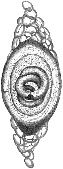
Fig. 36.—Larval Trichina coiled within its
capsule. After Bristowe and Rainey.
As commonly observed in the
human body our young Trichinæ
appear as spirally-coiled worms
in the interior of small, globular,
oval, or lemon-shaped cysts,
which latter appear as minute
specks scarcely visible to the
naked eye. These specks resemble
little particles of lime, being
more or less calcareous according
to the degree of degeneration
which their walls have undergone. In shape and general
aspect they are not altogether unlike the eggs of certain nematoid
worms, but their size alone sufficiently distinguishes them.
They measure on an average 1/78″ in length by 1/130″ in breadth.
The organised capsules are not essential to the further development
of the parasite, and are rather to be regarded as abnormal
formations, or rather, perhaps, as products resulting from an
effort of nature to protect and thus prolong the life of the
occupant. They are frequently altogether wanting. The capsuled
Trichinæ measure 1/23″ in length by about 1/630″ in breadth.
When fully formed they not only exhibit a well-marked digestive
apparatus, but also reproductive organs, which are often, indeed,
sufficiently developed to determine the sex.
Notwithstanding the large number of experiments that have
been more or less recently made by investigators, little or
nothing has been discovered calculated to disturb the conclusions
set forth by Leuckart, who writes as follows:—“(1) Trichina
spiralis is the juvenile state of a little round worm, previously
unknown, to which the generic title of Trichina must
remain attached. (2) The sexually mature Trichina inhabits
the intestinal canal of numerous warm-blooded animals, especially
mammalia (also of man), and constantly in great numbers.
The duration of its life extends from four to five
weeks. (3) At the second day after their introduction the
intestinal Trichinæ attain their full sexual maturity. (4) The
eggs of the female Trichinæ are developed within the uterus of
the mother, into minute filaria-like embryos, which, from the
sixth day, are born without their egg-shells. The number of
young in each mother-worm is at least from ten to fifteen
thousand. (5) The new-born young soon after commence
their wandering. They penetrate the walls of the intestine and
pass directly through the abdominal cavity into the muscles of
their bearers, where, if the conditions are otherwise favorable,
they are developed into the form hitherto known. (6) The
directions in which they proceed are in the course of the intermuscular
connective tissues. (7) Only the striped muscle
(that of the heart excepted) contains Trichinæ. The majority
of the wandering embryos remain in those sheathed muscular
groups which are nearest to the cavity of the body, especially
in those which are smaller and most supplied with connective
tissue. Speaking generally, their number decreases with the
distance from the abdomen, being, however, more numerous in
the anterior half of the body. (8) The embryos penetrate
into the interior of the separate muscular bundles, and here
already, after fourteen days, acquire the size and organisation
of the well-known Trichina spiralis. (9) Soon after the intrusion
of the parasite the infested muscular fibre loses its
original structure, the fibrillæ collapse into a finely granular
substance, whilst the muscular corpuscles change into oval
nucleated cells. (10) The infected muscular bundle retains
its original sheathing up to the time of the complete development
of the young Trichinæ, but afterwards its sarcolemma
thickens, and begins to shrivel at the extremities. (11) The
spot inhabited by the rolled-up parasites is converted into a
spindle-shaped widening, and within this space, under the
thickened sarcolemma, the formation of the well-known lemon-shaped
or globular cysts commences by a peripheric hardening
and calcification. This degeneration commences several months
after the wandering. Immature muscle-Trichinæ are not
capable of producing
infection. (12) The
migration and development
of the embryos
also take place
after the transportation
of impregnated
Trichinæ into the intestines
of a new
host. (13) The further
development of
the muscle-Trichinæ
into adult animals is
altogether independent
of the formation
of the calcareous
shell, and occurs as soon as the former have reached their
completion. (14) Males and females are already recognisable
in their larval state. (15) The immigration of the
Trichina-brood in masses produces very grave or even fatal
consequences, such as peritonitis (from the embryos perforating
the intestinal walls), pain, and paralysis (resulting from the
destruction of the infected muscular fibres). (16) The infection
of man occurs especially through swine. (17) The muscle-Trichinæ
are so capable of resistance that they are by no means
in all cases destroyed by the ordinary methods of roasting,
cooking, pickling and smoking. (18) As a rule, swine obtain
Trichinæ from rats, to which latter we also as the natural
bearers have to convey them. Microscopic examination of
flesh is, therefore, urgently recommended as a public preventive
against all danger from Trichinæ.”
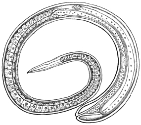
Fig. 37.—Immature female Trichina from muscle.
After Leuckart.
As a summary the above conclusions are well nigh exhaustive;
but whilst I purposely avoid entering into mere clinical details,
there are points of hygienic interest to which I must allude.
Thus, as regards the number of larval Trichinæ in any one
“bearer” at a time, this, of course, must be extremely variable,
but it may amount to many millions. In one of the cats on
which Leuckart experimented, he estimated a single ounce of
its muscle-flesh to harbour no less than 325,000 Trichinæ. I
find that a relatively similar degree of infection in an ordinary
human “bearer” would yield thirty millions. In the case of one
of my own experimental animals, a pig, I reckoned that there
were at least sixteen millions of Trichinæ. The larvæ were about
ten months old and enclosed within perfectly formed capsules;
nevertheless, the animal had never displayed any symptom of
irritation. In a trichinised human subject, examined by Dr
Thudichum, it was estimated that 40,000,000 parasites were
present. My own estimate, calculated from specimens of
muscle obtained from the same case, gave 100,000,000 as the
approximate number of worms present. In the only outbreak
of Trichinosis occurring in England, details of which will be
given further on, I found that the flesh of the hog that had
caused the local endemic contained upwards of 80,000 Trichinæ
to the ounce. The consumption of a pound of such flesh
would be capable of producing a collective progeny of something
like 400,000,000 within the human “bearer.”
In the year 1865 I conducted a series of experiments upon
upwards of a score of animals, including seven birds, the latter
all yielding only negative results. So far as muscle-Trichinæ
were concerned my experiences accorded with those of Professors
H. A. Pagenstecher and C. J. Fuchs, at the Zoological Institute
in Heidelberg. These experimenters found that the ingested
muscle-Trichinæ acquired sexual maturity within the intestinal
canal of their avian “hosts;” but they never found young
Trichinæ in the muscles of the birds, nor did they perceive
any evidences of an attempt on the part of the escaped embryos
to effect a wandering or active migration on their own account.
Clearly, if the bird’s intestinal canal were a proper territory
for the residence of sexually mature Trichinæ, we should have
found abundance of wandering non-encapsuled flesh-worms and
also sexually-immature muscle-Trichinæ enclosed in well-formed
capsules. Not a few persons still entertain the notion that
Trichinæ are liable to infest all kinds of warm-blooded, and
even also many kinds of cold-blooded animals, such as reptiles
and fishes. Certain nematodes found in earth-worms have been
described as Trichinæ; and consequently, pigs and hedgehogs
were said to become trichinous through eating these annelids.
The minute flesh-worms described by Bowman from the muscle
of the eel are not true Trichinæ, any more than the somewhat
similar parasites which Eberth found to infest the muscles of
the frog. The same may also be said of Dr Salisbury’s
urinary Trichinæ, which are the larvæ of Filaria Bancrofti.
Deducting the seven birds, and also six other animals where
no examination after death was possible, I ascertained the
result of my worm-feedings in sixteen instances. Nine of the
experiments were entirely successful, the infected animals
comprising four dogs, two cats, one pig, one guinea-pig, and a
hedgehog.
Carnivorous mammals, especially those subsisting on a
mixed diet, are the most liable to entertain Trichinæ, but it is
quite possible to rear them in herbivora. Thus, Pagenstecher
and Fuchs succeeded in rearing muscle-Trichinæ in a calf, and
they found three female intestinal Trichinæ in a goat, but
apparently no muscle-flesh-worms, although twenty-seven days
had elapsed since the first feeding with trichinised rabbit’s flesh.
In three sheep on which I experimented no trace of Trichinæ
could be found. There is no practical need for any further experiments
on herbivora, for it is quite clear that, in their natural
state, herbivorous mammals can seldom have an opportunity of
infesting themselves, whilst the reverse is the case with
swine, carnivorous mammals, and ourselves. Because many
quadrupeds become trichinous, it does not follow that all
mammals are liable to be infested. In the case of most
parasites we find the species limited to a larger or smaller
number of hosts. On the other hand, in not a few cases, the
range of the entozoon is limited to a single territory or host.
In conducting the experiments above mentioned I was
assisted by Professors Simonds and Pritchard, of the Royal
Veterinary College. As they were the only researches conducted
on any considerable scale in England, I subjoin a few
details of them. Dr Thudichum’s experiments were, I believe,
confined to rabbits.
Exps. 1 and 2.—On the 15th of March, 1865, an ounce of
flesh containing Trichinæ was administered by myself to a black
bitch. The dog being destroyed five days subsequently,
neither intestinal nor muscle-Trichinæ were discovered. It was
thought that the dog had thrown up the bolus, which was
strongly saturated with chloride-of-zinc solution. The bolus
consisted of a portion of the pectoralis major of a subject
brought to the dissecting-room at the Middlesex Hospital. The
cysts were highly calcified, but the majority contained living
embryos, which were quite unaffected by the zinc solution
injected into the body to prevent decomposition. At the same
date a small white puppy was experimented on and examined
with precisely the same results. In either case it was too early
to expect muscle-flesh-worms to have become developed.
Exp. 3.—Half an ounce of the same trichinous human flesh
was given (at the same date) to a black-and-tan puppy reared
at the Royal Veterinary College, a second “feeding” being
administered on the 21st of March, or six days after the first.
In this case Mr Pritchard, who fed the animal, took the precaution
to chop the muscle into small pieces, and to mix it with
other food, in order that the flesh might be the more readily
retained in the stomach. The puppy was not destroyed until
the 15th of the following June, when, on examination, numerous
encysted but non-calcified muscle-Trichinæ were found in all the
voluntary muscles subjected to microscopic scrutiny.
Exp. 4.—An ounce of the same flesh was given to a dark-colored
pig on the 15th of March, and again on the 20th,
several other “feedings” being also administered during the
month of April, 1865. It was destroyed on the 16th of May, but
no Trichinæ were detected.
Exp. 5.—An ounce of the same human muscle-flesh administered
to a small sheep (which was subsequently killed on the
29th of June) also produced negative results.
Exps. 6 and 7.—“Feedings” were at the same time administered
to a rat and mouse. The mouse died on the 2nd April,
when I examined its muscles without success. On the following
day the rat unfortunately made its escape, but whether
trichinised or not cannot be said.
Exp. 8.—An ounce of trichinous human flesh was given to a
donkey, in the form of “balls,” on the 20th of March; and
during the month of June four other separate “feedings” with
trichinous dog’s flesh were also administered. The animal was
removed from the College without the result being ascertained.
Exp. 9.—From the 15th to the 20th March, 1865, inclusive,
three small Trichinæ “feedings” were likewise administered to
a guinea-pig. This little animal was not destroyed until the
15th of the following June, when a positive result was obtained.
The pectoralis transversus and other muscles were found to
harbour a considerable number of encysted Trichinæ.
Exp. 10.—On the 20th March, and again on the 21st (1865),
“feedings” from the same human subject were administered to
a hedgehog. On the 26th of April the animal seemed to be
attacked with symptoms of Trichinosis. It refused food, kept
its head extended, and the eyelids closed. On the 27th it
appeared much worse, and on the morning of the 28th it was
found dead. On the 29th I examined the flesh, and found abundance
of living Trichinæ in the muscles. The capsules were
very thin and transparent. A few days later Mr Simonds also
examined the flesh, and confirmed this result.
Exps. 11 and 12.—Two chickens were fed, on the 21st of
March, with the same material. One of the birds died on the
24th, when I examined the intestines and detected one or two
very minute nematodes, which, at the time, I believed to be
imperfectly developed Trichinæ, but subsequently saw reason
to alter my opinion. The other bird died on the 3rd of April,
and certainly contained no muscle-Trichinæ.
Exp. 13.—On the 22nd and 23rd of March “feedings,”
amounting to an ounce of flesh in all, were given to a mole.
This animal was returned to the care of Mr Charles Land, who
had previously sent it to the Veterinary College. He subsequently
reported that, after observing the mole to be “working”
for two or three days, he lost all trace of it, and concluded that
it had either escaped or was dead.
Exp. 14.—On the 1st and 2nd of May portions of the left
fore extremity of the hedgehog (in which we had successfully
reared Trichina from the Middlesex-Hospital subject) were
offered by Mr Simonds to a cat. It ate the flesh very readily,
consuming the entire limb. On the 15th of the following June
the cat was killed, when living Trichinæ were found within all
the muscles which we examined.
Exp. 15.—At the same dates a young terrier dog was similarly
treated, but did not take the “feeding” so readily. In
this case the left hind extremity of the hedgehog was employed,
and what was not eaten voluntarily was forcibly introduced. On
the 1st of June the dog was attacked with “distemper,” and
died on the 8th of the same month. On examination we found
several living Trichinæ in the sterno-maxillaris and other
muscles. Some of the parasites were encysted.
Exp. 16.—From the 9th to the 12th of June inclusive four
separate worm-feedings with the flesh of the trichinised
terrier-dog were administered to a crow. The bird was killed
some months afterwards and sent to me for examination. Its
muscles were entirely free from Trichinæ.
Exp. 17.—From the 9th to the 17th of June inclusive seven
separate worm-feedings were administered to a pig. One of
the “feedings” was with the trichinised guinea-pig’s flesh, the
others from the dog. This animal was not destroyed until the
4th of April, 1866, when all the muscles which I examined were
found extensively infested with Trichinæ. There were probably
not less than 16,000,000 present, all being alive and enclosed
within perfectly-formed capsules, none of which latter exhibited
any traces of calcareous deposition.
Exp. 18.—Four separate feedings with trichinous dog’s flesh
were likewise, at the same dates as the foregoing, administered
to a rat. This experimental animal, however, like the one
previously mentioned, contrived to make its escape. I fear it
was well trichinised.
Exp. 19.—About the same date trichinous “feedings” were
given to a black puppy (bred at the Veterinary College). The
dog was killed on the 18th of August, 1866, having also been
made the subject of an echinococcus-feeding, when I found
abundance of encysted Trichinæ within the voluntary muscles.
Exp. 20.—Four separate worm-feedings with the flesh of the
trichinised guinea-pig were given to a sheep on the 15th, 16th,
17th, and 19th days of June, 1865. The experimental animal
was destroyed on the 29th of the same month, but the result
was negative.
Exps. 21 and 22.—“Feedings” with the guinea-pig’s flesh—four
in the one case and three in the other—were also administered
by Mr Simonds (from the 15th to the 19th of June,
inclusive) to a chicken and goose respectively. These birds were
destroyed some months afterwards and sent to me for examination,
but the most careful scrutiny failed to detect any
Trichinæ within their muscles. The goose was cooked and
eaten without the slightest hesitation. The chicken I found
too tough for consumption.
Exp. 23.—On the 28th of March, 1866, I obtained a small
quantity of muscle from a highly trichinised German subject,
who died from the effects of an accident at the London
Hospital the day previous. The case was fully reported by
Dr Thudichum in a new journal, called ‘Scientific Opinion’
(No. 4, April 25th 1866, p. 55). During the same day (at
2.30 p.m.) I fed a dog with part of this human flesh. On the
morning of the 31st I killed the dog, and examined the
intestinal canal (at 11.30 a.m.), which revealed the presence of
sexually-mature living Trichinæ. The males (of one of which
I retain an accurate figure) displayed the characteristic bilobed
caudal appendage, leaving no doubt as to their source and
nature. I have mentioned the precise time of the experiment,
in order to show that a period of sixty-nine hours proved amply
sufficient for the development of the young muscle-flesh-worms
of the human subject into the sexually-mature adult Trichinæ
of the dog.
Exp. 24.—With another portion of this human flesh (taken
from the muscles of the tongue) in which the Trichinæ were
extraordinarily abundant, I fed a cat. In about ten days the
animal showed the most marked symptoms of trichinosis. It
refused to eat; the eye lost its lustre; the body became very
thin, and I thought the animal would die. By very great care,
keeping it warm before the fire, and subsequently inducing it
to take a little milk, the creature improved, gained flesh, and
eventually recovered. About three months afterwards I destroyed
this cat, when on examining the panniculus carnosus,
latissimus dorsi, and other superficial muscles, I found great
quantities of well-developed, capsuled Trichinæ. Although the
animal had swallowed scarcely a quarter of an ounce by weight
of the infested flesh, yet thousands of parasites had been propagated
and dispersed throughout its muscular system. In
this way the helminthiasis nearly proved fatal to my cat. As
has been already stated, Dr Thudichum, who I believe had
an opportunity of examining the corpse of this trichinised
German, estimated the number of parasites in his body at
40,000,000. I do not think this estimate likely to be exaggerated,
for if all the flesh had been infested to the extent I
found to obtain in respect of the muscles of the tongue, I
believe 100,000,000 would have been nearer the mark. In
places the point of a needle could scarcely be thrust between
the capsules, so closely were they agglomerated.
Exp. 25.—From the 19th to the 25th of April, 1866, inclusive,
daily administrations of trichinous pork, in the form of bolus,
were made to a sheep by Mr Pritchard. The Trichinæ were
obtained from one of our experimental animals at the Veterinary
College, about two ounces of the flesh being given at each
feeding. The flesh of this sheep (destroyed in the following
November) failed to give any indication of the presence of
parasites.
Exps. 26 and 27.—About the same time, and occasionally at
intervals extending over a period of five weeks, Mr Pritchard
also fed two young fowls with the same trichinous pork.
Towards the close of October, 1866, both birds died, when Mr
Pritchard carefully examined the flesh of them, but failed to find
any trace of Trichinæ.
Exps. 28 and 29.—From April 2nd to the 9th of the same
month, 1866, inclusive, feedings with trichinous pork were
likewise given to two dogs. These animals were destroyed and
examined by Mr Pritchard in November, 1866, but the result
appears to have been negative.
It is perfectly certain that the infection of man by Trichina
is invariably due to the ingestion of verminiferously
diseased meat, and as remarked in my ‘Lectures,’ whenever the
parasites are taken in large numbers unpleasant symptoms soon
show themselves in the infested person. There is, first of all,
restlessness, loss of appetite, and more or less prostration.
This is succeeded by rheumatoid pains in the limbs, with the
frequent accompaniment of considerable swelling. The pain is
not situated in the joints, but in the intermediate soft parts.
In severe cases the limbs are drawn up and half bent, as in
instances of severe and continued cramp. Sometimes the
suffering is excruciating and unbearable, patients having been
known to request the surgeon to put an end to their lives. In
the worst forms of the malady death rapidly ensues from
diarrhœa and exhaustion. If the parasites have gained admission
to the muscles all hope of destroying them is at an end;
but if a person suspects himself to have eaten diseased or
trichinised meat he should lose no time in seeking professional
assistance, seeing that the administration of suitable anthelmintics
might be the means of saving his life, whereas a few
days’ delay would probably prove fatal. So long as the worms
remain in the stomach or intestinal canal they can be got rid
of, but when once the trichinal brood have invaded the flesh
then they cannot be expelled. As remarked in my ‘Entozoa,’
it is easy to perceive that although, in the majority of instances,
Trichiniasis does not cause death, yet the percentage of fatal
cases is by no means insignificant.
The notion that particular breeds of swine are more liable to
be infested than others is absurd, since infection must be due
to the facilities offered for swallowing garbage, especially dead
rats. According to Drs Belfield and Atwood 8 per cent. of
slaughtered American swine contain Trichinæ. In infested
hogs they found from 35 to 13,000 parasites in a cubic inch of
muscle, and by repeated feedings they succeeded in rearing
about 100,000 Trichinæ in the body of a rat.
In regard to the disease in man let us glance at the
phenomena that presented themselves in Plauen, a town of
Central Saxony. Drs Böhler and Königsdörffer, who first
saw this disease and treated it, state, according to Leuckart,
that “the affection began with a sense of prostration,
attended with extreme painfulness of the limbs, and, after
these symptoms had lasted several days, an enormous swelling
of the face very suddenly supervened. The pain occasioned by
this swelling and the fever troubled the patients night and day.
In serious cases the patients could not voluntarily extend their
limbs, nor at any time without pain. They lay mostly with
their arms and legs half bent—heavily, as it were, and almost
motionless, like a log. Afterwards, in the more serious cases,
during the second and third week, an extremely painful and
general swelling of the body took place; yet, although the fifth
part of all the patients were numbered amongst the serious
cases, only one died.”
Satisfactory as it may be to note the numerous recoveries
which take place, this circumstance is very much marred by
the fact that a large proportion of the patients suffer the most
excruciating agony. In the main it will be observed that
Böhler’s and Königsdörffer’s experience, as recorded by
Leuckart, corresponds very closely with that given by other
observers. The symptoms, moreover, are very similar to those
produced in the original case published by Zenker. In this case,
which occurred in the Dresden Hospital (1860), the patient was
a servant girl, aged twenty, and the principal symptoms were
loss of appetite, prostration, violent pains, contraction of the
limbs, and finally œdema, which, in association, perhaps, with a
certain amount of pneumonia, terminated her career within a
period of thirty days. The post-mortem appearances showed
that the larval Trichinæ were the cause of death. The intestinal
canal contained numerous sexually-mature worms.
The effects produced by Trichinæ on animals are similar to
those occasioned in man. The phenomena were summarised
by Davaine (in the journals quoted below) in 1863 as follows:
“The first phase is characterised by intestinal disorder, produced
by the development of the larvæ in large numbers, and
their adhesion to the mucous membrane of the intestine. In
this stage M. Davaine has seen rabbits die with intense diarrhœa;
one of two cats which he fed with trichinised meat had
diarrhœa for at least a fortnight, but survived. Of five or six
rats fed on a similar diet, one only, which was pregnant, died
of diarrhœa, after abortion, on the eighth day. According to
M. Leuckart, the passage of the embryos of Trichinæ through
the intestinal walls sometimes produces peritonitis. This intestinal
phase often becomes blended with the next; it may be
relieved by the expulsion of the worms by means of the diarrhœa,
or may cease with the natural death of the worms.
“The second stage presents general symptoms—muscular
pains, &c. These phenomena are dependent on the introduction
of the Trichinæ into the muscles; they rapidly acquire
their maximum intensity, and have not a long duration. The
appearance and duration of this stage are in complete relation
with the development and length of sojourn of the Trichinæ in
the intestines; in fact, in this entozoon, oviposition is not slow
and of long duration, as in many nematoid worms; the genital
tube is rapidly formed, and the ova, in its whole length, are
developed almost simultaneously, so that the embryos, arriving
soon at maturity, are at once thrown out in large numbers into
the intestine, and the mother Trichina dies exhausted. If it
be remembered that the embryos do not escape before the
eighth day, that a certain number of days are required for their
arrival in the muscles, and that new ones are not produced
after six or seven weeks, it will be understood that the first
symptoms of this stage can scarcely appear until the end of a
fortnight after ingestion of the diseased food, that they must
continue four or five weeks, and that after this they may disappear.
This course of events is observed in animals; and in
man the symptoms of this stage have shown themselves and
become aggravated from the third to the sixth week after
infection. Most animals die during this stage; rabbits rarely
survive; rats, on the contrary, generally resist it.
“If the animals do not die of the general symptoms or local
disturbances proper to these two stages, the inflammatory symptoms
cease, respiration becomes natural, and order is re-established.
But, in some cases, the number of cysts formed in
the muscles are sufficiently great to impede them in the proper
exercise of their functions, and hence arises general debility, a
kind of consumption which persists or becomes aggravated, and
the animal dies of marasmus. M. Davaine has noticed this in
rabbits, but especially in a rat.
“Recovery from these phases of trichinal infection may be
apparently perfect. A rabbit which M. Davaine kept during
five months became large and fat, although it had a large
number of Trichinæ in its muscles; a rat which had had these
entozoa in considerable numbers during six months was, to all
appearance, in good health. Hence he concludes that the
Trichinæ produce symptoms only when they are in the intestinal
canal, and when they are entering the muscles. Having
become lodged in their cysts among the muscular fibres, they
may remain harmless for an indefinite time. In every case
except one, down to 1859, Trichinæ have been found in the
bodies of persons who have died of disease (generally chronic)
or by accident; or in the dissecting-room, in bodies regarding
which the previous history could not be obtained. In most cases
the cysts contained a cretaceous or fatty deposit, showing that
they had probably existed several years.
“The observations which have been made on the human
subject, in regard to the symptoms caused by Trichinæ, show
that they belong, as in animals, to the initial period of infection.
They consist in intestinal and in muscular lesions; the
latter coincide with the entrance of the parasite into the
muscles, and are truly traumatic. In Zenker’s case the intestinal
symptoms were swelling and pain; in a case described by
Friedreich diarrhœa was present. In all cases the most
remarkable symptoms were violent rheumatoid pains in the
muscles, not in the joints, which were considerably aggravated
by attempts to extend the half-bent limbs. The other symptoms
have been variable, but have had a strong resemblance
to those of typhoid fever. In several cases there has been
abundant sweating; and in one there was a very remarkable
miliary and furuncular eruption. The animal heat was diminished
in Friedreich’s case; and in those observed in Voigtland
by Freytag the temperature never exceeded 102° Fahr.
“The progress, duration, and severity of the disease in man
are in relation to the number of Trichinæ taken into the
digestive canal. Of sixteen patients observed at Plauen by
Drs Böhler and Königsdörffer, eight, who were moderately
affected, recovered in a month; four, more severely diseased,
were ill two months; of four others, one died with ascites and
colliquative diarrhœa at the end of two months, and three
recovered slowly at the end of three or four months. Recovery
does not imply the death of the Trichinæ, it follows their
enclosure in cysts.
“The diagnosis of trichinal infection has several times been
made in the living human subject by removing a portion of
muscle. M. Davaine thinks it probable that, during the first
six or eight weeks of the disease, the diagnosis may be confirmed
by searching for adult Trichinæ in the alvine evacuations,
produced naturally or by means of a purgative.
“The prophylactic treatment consists simply in the avoidance
of uncooked meat. The medicinal treatment must vary with
the stage of the disease. At first, attempts must be made to
expel the parasites from the intestines by purgatives and
anthelmintics. Which amongst the latter is the most energetic
is not yet determined. Calomel is, perhaps, M. Davaine
thinks, the best. After six or eight weeks all treatment
directed towards the intestines is superfluous. It is scarcely
probable that any substance will act on the larvæ disseminated
through the muscles. Friedreich has recommended picronitrate
of potash; but, in the case in which he used it, live Trichinæ
were found in the muscular tissue after the patient was considered
to be cured.”
In regard to the possibility of curing trichiniasis by the
administration of drugs which should act as trichinacides upon
the parasites in the condition of flesh-worms, the absurdity of
the proposal only equals that which was made in reference to
the destruction of hydatids by the administration of kamala.
As has been shown in the record of my first experiment the
flesh of a trichinised corpse may be thoroughly saturated with
a strong solution of chloride of zinc, and yet the worms will
remain quite unaffected.
In reference to the dangers arising from the consumption of
diseased meat, Professor Gamgee has very cogently put two
questions:—“Did Moses know more about pigs than we do?”
“Was it a knowledge of the parasitic diseases of swine and man
that led Moses to condemn pork as human food?” Mr
Gamgee answered both questions negatively, thus:—“The
wisdom of the Mosaic law can only be justly estimated with a
knowledge of the accidents arising in warm countries from
eating pork throughout long and hot periods of the year; and
there is no doubt that the direct evil results, as manifested by
human sickness, led to the exclusion of pork from the list of
Israelitish viands. The masses of measly pork which may be
seen hanging from the butchers’ stalls in Southern Europe
prove that the long-legged swine which hunt the forests for
acorns, and rove about to pick up all kinds of offal, are often
unfit for human food, and that they were so to no less extent
in the land of Israel is probable.” As supplementing Professor
Gamgee’s argument, I may remark that, if Moses had
been furnished with special knowledge beyond that of his contemporaries,
he would not, in the matter of meat-parasitism,
have confined his restrictions to pork. Had he possessed any
knowledge of measly beef, he would not have spared the ox on
the ground that although “it divideth the hoof, yet it cheweth
the cud.” As regards home-reared animals, Professor Gamgee
cogently remarked: “It is interesting to observe that parasitic
maladies in the pig specially abound in that section of the
United Kingdom where swine live most amongst human beings.
The Yorkshire and Berkshire pigs, in their native counties
enclosed in the farmyards of their breeders, are free from worms
which are likely to live in the body of man. The Irish pig
is the one most commonly injured by entozoa, and the reason
for this is evident when we know how much the cottager relies
on rearing a porker which is permitted the free range of house
and road, where every description of filth is devoured, charged
with the ova of parasites expelled by man or some of the lower
animals.” He also adds: “The conditions under which we live
in the British isles are certainly much less favorable to the
propagation of worms; but we disregard, in our ignorance, the
most common precautions to protect ourselves from loathsome
diseases, and not only permit dogs to eat any kind of offal
in and around slaughterhouses, but sanction the existence of
piggeries where all kinds of garbage, charged with worms or
their eggs, are daily devoured by swine. The majority of
germs calculated to engender parasites are to be found in
abundance in the contents of the alimentary canal of human
beings and domestic quadrupeds. If pigs are permitted to eat
these, as in Ireland or in many British piggeries, we must
expect hams, bacon, and pork sausages to be charged with the
embryonic forms of human entozoa. Whereas in Iceland the
dog is the victim of human negligence, and en revanche the
cause of human disease, in the British isles the pig holds this
unenviable position. We have good reason to believe, with
Moses, that the pig is an unclean beast; but without discarding
him from the scanty list of animals to be eaten, it is evident
that we can purify the race of swine, and thus prevent human
as well as porcine maladies.”
On the authority of Rupprecht, as quoted by Davaine, I
append a list of the principal epidemics observed in Germany
during the first six years immediately following the discovery
of trichinosis:
1. Two slight epidemics in 1860 in the Island of Rügen;
10 to 20 patients (Dr Landois).
2. An epidemic at Stolberg, 1860 (Dr Fricinus). The
number of trichinised persons was not stated with certainty.
3. Five epidemics during five summers, 1858 to 1862, at
Magdebourg. The number of patients was 300, two only died
(Dr Sendler).
4. An epidemic at Plauen in 1862, 20 patients (Böhler).
5. Gusten, 1861, 40 cases, all cured (Fränkel).
6. Epidemic in the Province of Armsted (Mansfeld), 1861,
8 patients.
7. Hettstädt, January and March, 1862, 8 to 10 patients.
8. Blankenburg, 1862, 278 cases, 2 deaths.
9. Calbe (Prussia), 1862, 38 cases (9 men, 25 women, 4
children), 8 deaths (Dr Simon and Dr Herbst).
10. Burg, in Magdebourg, 1863, 50 patients, 10 deaths
(Dr Klusemann).
11. Quedlinburg, 1863, 9 patients, 1 death (Dr Behrens).
12. Plauen, 1863, 21 patients (Königsdörffer).
13. Falkenstein, 1863, 4 patients (Drs Bascher and Pinter).
14. Posen, August and September, 1863, 37 patients (Dr
Samter).
15. Hamburg, 1863, 2 patients (Dr Tüngel).
16. Blankenburg, 1863, 32 patients, 2 deaths; new cases in
1864 (Dr Scholz).
17. Hettstädt (Prussian-Saxony), October, 1863, 158 patients,
27 deaths (Rupprecht).
18. Eisleben, December, 1863, and January, 1864, 18 cases,
no deaths. This result was attributed to the employment of
phosphoric acid (Rupprecht).
19. Hettstädt, February and March, 1864, 8 patients, no
deaths. Two cats were also attacked, one of which died.
Nearly 50,000 Trichina were counted in an infected leg of pork
(Rupprecht).
20. Quedlinburg, 1864, 120 patients, 2 deaths; benzine was
employed (Dr Wolf).
21. Hettstädt, January, 1865, 15 patients (Rupprecht).
22. Berlin, 1864, 3 cases (Dr Cronfeld). Several butcher
boys (Frischer).
23. Leipzig, 1864, 14 patients, 2 deaths; 4 were infested
after having eaten raw beef hashed on a block which had
previously received the flesh of a trichinised hog (Dr E. Wagner).
24. Potsdam, 1864, 5 slight cases (Dr Mollendorf).
25. Celle (Hanover), 1864, 8 patients (Dr Scheller and Dr
Baring); Trichina proven in the pork by Gerlach. In 1855, 12
Trichina (?) patients were treated by Schuchart.
26. Hedersleben, 25th October, 1865, a pig was killed and
sold; on the 28th the malady appeared amongst the workmen;
350 patients, 100 deaths. Of 100 children infected, none died.
Trichina found in the autopsies (Dr F. Kratz).
Dr Davaine also adds the following outbreaks:
In Massachusetts, 1867, 6 patients from having eaten raw
ham, 1 death (‘Medical Times,’ 20th April, 1867, p. 431).
Ravecchia (Bellinzona), 1868, 5 patients, 4 deaths (Dr Zangger
in Landbote of Winterthur).
Up to a comparatively recent date no case of trichinosis had
been recognised in England during the life of the victim. As
regards diagnosis, what was happening every day on the Continent
was utterly unknown here. Whilst, however, not a
single instance of Trichina-disease had been observed by British
physicians in actual practice, as many as thirty or forty instances
had occurred where the parasites in question had been found
post mortem. I had myself examined the trichinised flesh
taken from a dozen of these corpses, but in no instance had
the faintest suspicion of trichinosis been entertained during
life. The circumstances attending the only outbreak of trichiniasis
that has been witnessed in England are very interesting.
In the month of April, 1871, I received from Dr
W. L. Dickinson, of Workington, Cumberland, specimens of
pork which he requested me to examine; and in complying with
his request I confirmed his opinion that the diseased meat was
infested with Trichinæ. A few days afterwards I announced
the discovery in the pages of the ‘British Medical Journal’
for April 22, p. 435. It happened, also, that at the time
I was delivering a course of lectures before the Society
of Arts; consequently, in my second discourse (which was
devoted to the parasites of cattle) I gave full details of the facts
that were obtained. Taking a small portion of the flesh which
I judged to be affected to an average extent I addressed the
audience as follows:
“If you calculate from one portion only, you might, if
you had stumbled upon a part which was more infested with
parasites than another, be led to over-estimate the degree
of trichinisation. Taking proper precautions I have calculated
that one scruple of this trichinous flesh would give us
4320 parasites, and two scruples would therefore yield 8640.
Without speaking so precisely to numbers, I do not hesitate
to aver my belief that there are at least 5000 of these
parasites inside this small piece of ham. The number is probably
close upon 8640. In one drachm that would give us 12,000,
and in an ounce 103,000, according to the old apothecaries’
weight. If, however, we calculate according to the ordinary
weight used by butchers, we should say that one ounce contains
4371/2 grains of meat, and therefore the number of parasites in one
ounce would be 85,000. Thus, in one ounce of meat from this
particular pig you have 85,000 Trichinæ, calculated at the rate
of 200 in the grain, for I have purposely cut off the odd numbers.
You may say, if a person can survive 18, 20, 30, or 40 millions,
he would not take much harm from eating a piece of flesh
containing only 8640 parasites. Such a portion, however,
would be quite sufficient to make any one of us extremely
uncomfortable were we to eat it, for supposing its contained
parasites to be alive, it might prove dangerous to life. Why?
The explanation is this:—Half of those 85,000 parasites, at the
very least, will, in forty-eight hours after ingestion, have become
fully-developed females; and from each of these 42,000 there will
proceed at least 1000 as a brood, so that the entire progeny
(and it is they that do the mischief by their independent
migrations through our tissues) will eventually yield about
42,000,000 entozoa. If we should be so voracious as to eat a
pound of such trichinised flesh, then there would be 400,000,000
as the result of a single meal.
“Having detailed these facts and inferences, I now wish to
bring to your notice some other particulars connected with the
Cumberland outbreak.
“Dr Dickinson, of Workington, tells me that he was at first
suspicious that his patients were suffering from fever, but was
not quite able to make out what the disorder was. At length
certain symptoms occurred, which suggested that it might
possibly be the German flesh-worm epidemic making its appearance
in this country for the first time, and, therefore, in view of
verifying the facts of the case, he sent me portions of the flesh of
the pig. He describes the symptoms, which in their character
corresponded with those previously recorded as experienced by
persons similarly attacked. Dr Dickinson remarks, towards the
close of his communication, that the victims form a small family
who have carefully reared their own swine. The British farmer
is thus here introduced to us at his own table playing the part
of ‘host’—at her own table, I should say, for, to be more precise,
it is a widow, her daughter, and a man-servant who are
suffering. Dr Dickinson informs me that for two or three weeks
before he was called to see them they had been eating sausages
and boiled pork from one of their own home-fed pigs, which
pig, by the way, turns out to have been an old sow. He
brought away some sections of the leaner portions of the flesh
for microscopic examination. You will observe that there can
be no mistake about the source of the food on this occasion.
Hitherto, Trichina has not been observed in our British-fed
swine in more than one or two, or possibly three instances.
Therefore it would be very interesting to ascertain how it happened
that this poor pig became trichinised. In my communication
addressed to the ‘British Medical Journal,’ I wrote as
follows:—“Dr Dickinson has at the present time under his care
a family suffering from the so-called flesh-worm disease,
resulting from the consumption of ham prepared from pigs
reared by the family themselves. A portion of ham sent to me
swarmed with recently encapsuled Trichinæ. Dr Dickinson being
thus the first person who has diagnosed trichiniasis in the living
subject in England, I hope he may be induced to give us further
particulars.” The editor, in commenting upon this letter,
added a practical point, which I wish especially to bring to your
notice. He says:—‘The subject of parasitic diseases of
domestic animals is one of widespread and increasing interest.
It is immediately related to the irrigation of fields with
sewage.’ The editor, of course, made this statement on
independent grounds, and on his own responsibility. If
he had said the subject bears an indirect relation to the
sewage question, he would have said no more than is absolutely
true, for, as I shall take occasion to explain, there is
every reason to suppose that certain forms of parasitic disease
may be propagated by means of sewage. In this connection
some of you may be disposed to ask the question:—‘Are there
any sources of comfort to be gathered from the facts?’ Or
you may say, supposing that in future our British swine are not
as free from Trichinæ as they have been hitherto, can we possibly
avoid the contingency of playing the part of host to those creatures?
Certainly, I reply, it is simply a question of properly cooking
the food. If these farmers have not cooked their food at all,
or scarcely at all, that will at once account for their being laid up.
I should tell you that the lady and the daughter are recovering,
and that they are convalescent, but the man-servant is very ill.
If, during cooking, the flesh consumed by these persons had
been raised to a persistent temperature of 170° Fahr., then,
doubtless, the ingestion of trichinised pork would have done
no harm. You observe that Dr Dickinson says in his letter
that they partook of it roasted and boiled. Now, few of us are
in the habit of eating underdone pork, although there are other
meats that we devour very readily in an imperfectly cooked
state. It must be remembered, also, that although the exterior
may have been subjected to a temperature of 212 degrees, it by
no means follows that the whole of the joint throughout must
have been submitted to that temperature. Under rapid cooking,
the centre of a large joint may remain much below even 140
degrees. If the man-servant ate only one ounce of the flesh
with living Trichinæ in it, he will probably have at this present
moment at least 42,000,000 of these guests in his muscles.
You will ask, ‘Will he recover?’ ‘Yes; if he ate no more than
that.’ If he has eaten 2 oz. thoroughly underdone, depend
upon it he has 80,000,000, and if he has eaten 3 oz. he will have
over 100,000,000 of Trichinæ in his muscles. Could he survive
if he had eaten over 3 oz., and thus have 100,000,000 and
upwards of these inhabitants? I think he could. We have
evidence on this point from the case in which I estimated that
there were upwards of 100,000,000 of Trichinæ present, and yet
the man survived the attack.
“Incidentally I may remark that in the course of the last
twenty years, although millions of parasites and their eggs have
passed through my hands, I have almost entirely escaped infection.
It is something to know what you are either handling
or looking at, because there are many parasites besides Trichina
which are dangerous. There are gregariniform entozoa
residing in meat which we eat every day without any bad
consequences. They are as harmless as cheese-mites. There
is no need to be in the slightest degree nervous about flesh-food,
provided it is properly cooked. I believe there will be
no fatal issue in the case of any of the three individuals just
alluded to, but the chief practical point before us arises out of
the fact that we have here, for the first time in England, an
epidemic of trichiniasis. By calling attention to the subject, it
will, to say the least, suggest precautions by which future
epidemics may be avoided.”
The above remarks form the substance of a lecture given
on the 24th of April, 1871. A week later I delivered the third
of the Cantor lectures for that year, when I took occasion to
add the following particulars:
“It has been asked whether the so-called muscle-Trichinæ,
after they have arrived at their destination within the flesh of
man, are capable of producing any more unfavorable consequences?
The answer is, Certainly not. In the case of man
it would be necessary that his muscles should be eaten in
order for the Trichinæ to become sexually-mature worms;
and in those countries where cannibalism exists, the man-eater
would himself become trichinised, and would certainly
deserve his fate. I was very desirous to follow up the
account of this outbreak by inquiries respecting the particular
animal which had been the cause of the outbreak. I may
therefore mention that my informant, Dr Dickinson, states
that the family, including the man-servant, all fed together, and
that they had for upwards of a fortnight eaten daily, and sometimes
twice a day, sausages made from the flesh of the trichinised
animal. And he adds: The meat cut from the ham and
flitches, and what is called the spare-rib, was roasted before
the fire or in the frying-pan. Occasionally it was cooked in
the oven. Dr Dickinson ascertained from the mother that she
liked her meat to be underdone, and thus, therefore, there is
very little doubt that the meat was generally undercooked.
The man, a strong labourer, had a good appetite, and would
therefore get a large share. He is improving slowly. Dr
Dickinson adds in a postscript, what is still more to the point,
that the sausages would be most likely undercooked; they
would be cooked in the frying-pan, and if only brown on the
outside would be eaten. It is probable that the outbreak was
due therefore to eating underdone meat from this pig, cooked
in various ways, and not alone from the ham itself.”
If the facts connected with this outbreak be honestly faced,
it must be rendered clear to any unprejudiced observer that
Dr W. Lindow Dickinson was the first person to observe,
recognise, and treat the Trichina disorder in this country. No
other English, Scotch, or Irish physician has encountered any
similar case. If I lay stress upon this fact it is because I
have learned from Dr Dickinson that another person has
asserted priority in this relation. Sir Dominic Corrigan is
stated to have told a gentleman in the House of Commons,
“that he had often met with trichiniasis in his practice in
Dublin,” further averring that the disease “was quite common
in many parts of Ireland.” If Sir D. Corrigan merely desired
it to be understood that he had repeatedly encountered the
Trichina at post-mortem examinations, then there is nothing
surprising in his statement, but if, on the other hand, the disease
itself has been frequently recognised in the living Irish
human subject, one can only express astonishment that hitherto
no single instance of the kind appears to have been recorded
either in the public or professional journals.
Bibliography (No. 21).—English Literature.—Allman, G. J.,
“Exhib. of Specimens,” ‘Micr. Jrn. and Structural Record,’
1842, p. 94.—Althaus, J., ‘Essay on Trichinosis,’ London, 1864.—Idem,
“On a Suspected Case,” ‘Med. Times and Gaz.,’ 1864,
p. 161; see also pp. 362 and 390.—Atwood, see Belfield.—Ballard,
E., “On Diseased Meat,” ‘Med. Times and Gaz.,’ 1864.—Belfield
(with Atwood), “Trichinæ in Pork;” ‘New York Med. Rec.,’
Dec. 28, 1878; ‘Med. Times and Gaz.,’ Feb. 15, and ‘Lancet,’
Feb. 22, 1879.—Bellingham, O. B., ‘Dublin Med. Press,’ 1852.—Bowditch,
H. J., “Cases of Trichina,” ‘Boston Med. and Surg.
Journ.,’ 1842–43–44.—Bristowe, J. S. (and Rainey), ‘Trans.
Path. Soc.,’ 1854.—Chevers, N., “Sanitary Efforts in regard to
Trichiniasis,” ‘Lancet,’ 1864, vol. ii, p. 733.—Cobbold, T. S., “On
the Discovery of Trichina, in relation to the question of Priority,”
‘Lancet’ for March 3, 1866, p. 244.—Idem, ‘Parasites, and the
part they play in the Economy of Nature’ (lecture), Manchester,
1873, p. 46; also in the ‘Veterinarian,’ March, 1874.—Idem,
Remarks in the ‘Journ. Soc. of Arts,’ 1866, p. 399; also in
‘Med. Times. and Gaz.,’ 1867, p. 24; also in ‘Lancet,’ Feb.,
1864 and 1866, p. 538.—Idem, ‘Our Food-producing Ruminants
and the Parasites which reside in them’ (Cantor Lectures),
1871.—Idem, “Experiments with Trichina,” ‘Proceed. Linn.
Soc.,’ vol. ix, p. 205, 1867; see ‘Lancet’ for Jan. 13, 1866,
p. 52; ‘Brit. Med. Journ.’ for Dec. 22, 1866, p. 713; also
‘Lancet’ for Jan. 9, 1867, p. 91.—Idem, “Worms” (l. c.,
Bibliog. No. 2, Lecture xviii), 1872.—Idem, “Outbreak of
Trichinosis in England,” ‘Brit. Med. Journ.,’ 1871, p. 435.—Idem,
“Trichinæ in Fish” (alleged to have been found in a pike
by Dr Elendenen at Ostend), in the ‘Lancet’ (anonymous
annotation) for Nov. 16, 1878.—Curling, T. B., two cases,
‘Lond. Med. Gaz.,’ 1836.—Davaine, C., quoted in ‘Brit. Med.
Journ.,’ see foreign lit., below.—Delpech, abstr. of his Report,
‘Brit. Med. Journ.,’ 1866, p. 375.—Dickinson, W. L., ‘Brit.
Med. Journ.,’ 1871.—Elendenen, “Trichinæ in Fish” (newspaper
report respecting his “find”); see Cobbold, above.—Farre, A.,
“Observations,” ‘Lond. Med. Gaz.,’ 1835–36.—Friedreich, N.
(trans. by Ogle), ‘Med.-Chir. Rev.,’ 1863, repr. in ‘Edin.
Vet. Rev.,’ 1863.—Furstenburg, ‘Edin. Vet. Rev.,’ 1864, p. 513.—Gordon
(see Chevers).—Gamgee, J., “On Diseased Meat,”
‘Pop. Sci. Rev.,’ 1864.—Goodsir, J., ‘Month. Journ. Med. Sci.,’
1842.—Harr, ‘Bost. Med. and Surg. Journ.,’ 1866, p. 532.—Harrison,
“On a peculiar Species of Entozoon occasionally found
in the Voluntary Muscles of the Human Subject,” ‘Rep. of Brit.
Assoc.,’ Aug. 12, 1835; in ‘Dub. Journ.,’ vol. viii, 1835–36;
in ‘Lond. and Edin. Phil. Mag.,’ and in ‘Amer. Journ. Med.
Sci.,’ vol. xviii, p. 187, 1836.—Herbst, “Trichinæ in the
Badger,” ‘Assoc. Med. Journ.,’ 1853, p. 491.—Hilton, J., “Notes
on a peculiar appearance observed in Human Muscle, probably
depending upon the formation of very small Cysticerci,” ‘Lond.
Med. Gaz.,’ vol. xi, p. 605, 1833.—Jackson, J. D., “Trichiniasis,”
in ‘Hay’s Amer. Journ.,’ 1867, p. 82.—Kiefer, H., cases,
‘Bost. Med. and Surg. Journ.,’ 1866, p. 208.—Kobelt, ‘Micr.
Journ. and Struct. Rec.,’ 1842, p. 147.—Kratz, “On the Hedersleben
Epidemic,” ‘Brit. Med. Journ.,’ 1866, p. 76.—Krombein,
“Trichiniasis in New York,” ‘Amer. Journ. Med. Sci.,’ 1864,
and ‘Med. Times and Gaz.,’ 1864, p. 292.—Küchenmeister, F.,
Symptoms, &c., ‘Lond. Med. Rev.,’ 1860, p. 457.—Langenbeck,
case, ‘Edin. Vet. Rev.,’ Feb., 1864.—Leidy, J., “Trichina in
the Pig,” ‘Annals of Nat. Hist.,’ and ‘Pr. Ac. N. S. Philad.,’
1847.—Leuckart, R. (translations from), in ‘Ann. Nat. Hist.,’
1860; in ‘Qrt. Journ. Micr. Sci.,’ 1860; and in ‘Bost. Med.
and Surg. Journ.,’ vol. liii, p. 198, 1860–61.—Liveing, R., “Path.
Soc. Rep.” in ‘Med. Times and Gaz.,’ 1865, p. 374.—Mosler,
“On Trichinous Flesh,” ‘Brit. Med. Journ.,’ 1864, p. 554.—Idem,
“On Benzine in Trichinosis,” ‘Med. Times and Gaz.,’ Oct.,
1864, p. 444.—Nunneley, T., ‘Brit. Med. Journ.,’ 1866, p. 252.—Owen,
R., “Description of a Microscopic Entozoon infesting
the Muscles of the Human Body,” ‘Proc. Zool. Soc.,’ and ‘Lond.
Med. Gaz.,’ 1835; ‘Trans. Zool. Soc.,’ vol. i, 1835.—Idem,
Remarks in ‘Journ. Soc. of Arts,’ 1866, p. 399.—Paget, J.,
“Letter relating to the Discovery of Trichina,” ‘Lancet,’ March,
1866, p. 269.—Rorie, J., Letter, ‘Lancet,’ Feb., 1864.—Salisbury,
J. H., On a supposed Species of Trichina (T. cystica) from
the Human Bladder; in his paper on “Parasitic Forms,” in
‘Hay’s Amer. Journ. Med. Sci.,’ 1868, p. 376.—Sanders, R.,
‘Edin. Month. Journ.,’ 1853.—Sawer, A., ‘Bost. Med. and
Surg. Journ.,’ 1865, p. 16.—Sutton, G., Report on Trichinosis;
Indiana, U.S., 1874.—Thudichum, J. W. S., ‘Brit. Med.
Journ.,’ Jan., 1864, repr. in ‘Glasgow Med. Journ.,’ April, 1864,
p. 116; also letter in ‘Edin. Med. Journ.,’ Feb., 1864.—Idem,
“Rep. on the Parasitic Diseases of Quadrupeds used
as Food,” pub. by the Med. Officer of the Privy Council,
London, 1865.—Idem, “The Diseases of Meat as affecting
the Health of the People,” ‘Journ. Soc. of Arts,’ April 20,
1866.—Idem, “German Sausages and the Trichina Disease,”
‘Scientific Opinion’ for April 25, 1866.—Idem, ‘Lancet’ for
Jan. 6, 1866, p. 16.—Turner, W., “On the Trichina spiralis,”
‘Edin. Med. Journ.,’ Sept., 1860; in the ‘Year-Book,’
p. 109, for 1860; in ‘Med.-Chir. Rev.,’ 1862; and in ‘Bost.
M. and S. Journ.,’ vol. lxiii, p. 294.—Ude, “Rep. on the
Inspection of Pigs,” ‘Med. Times and Gaz.,’ Aug., 1868, p. 141.—Valentin,
‘Micr. Journ. and Struct. Rec.,’ 1842, p. 87.—Virchow,
R., Extr. from his brochure on ‘Trichina’ (by myself),
in ‘Gunther’s Record’ for 1864, p. 611.—Idem, “On the Cure
of Trichinosis,” ‘Brit. Med. Journ.,’ April, 1866, p. 368.—Idem,
from “Comptes Rendus,” in ‘Qrt. Journ. Micr. Sci.,’ 1861.—Idem,
from his ‘Archiv,’ 1860, Bd. xviii, Heft. 4, p. 330; in
‘Brit. and For. Med.-Chir. Rev.,’ vol. xxvi, p. 515, 1860.—Wedl,
Report, ‘Brit. Med. Journ.,’ Dec., 1866, p. 618.—Wilks,
S., “Letter respecting the Discovery of Trichina,” ‘Lancet,’
March 10, 1866, p. 269; see also the ‘Times,’ Feb. 13, 1866.—Windsor,
J., ‘Brit. Med. Journ.,’ March 4, 1866, p. 319.—Wood,
H., case, ‘Lond. Med. Gaz.,’ 1835.
Foreign Literature:—Ardenghi, E., “Sulla Trichina spiralis,”
‘Lo Studente Veterinario,’ 1876, p. 115.—Behrens, “Ein Fall
von Trichiniasis,” ‘Deutsche Klinik,’ No. 30, 1863 (quoted by
Davaine).—Bette, F., ibid., 1876.—Bischoff, ‘Path. Anat. des
menschl. Körp.,’ 1845.—Idem, ‘Med. Annalen,’ 1840.—Böhler,
‘Die Trichinenkrankheit in Planen,’ 1863.—Boudin, “Des
épidémies de Trichina spiralis observées en Allemagne dans ces
dernières années,” ‘Journ. de Méd. Vét. Milit.,’ August and
September, 1864 (quoted by Davaine).—Claus, ‘Wurzb. nat.
Zeitschr.,’ 1860.—Idem, ‘Ueber die Trichine’ (a discourse),
1877.—Colberg, ‘Deutsche Klinik,’ 1864.—Davaine, C., ‘Traité’
(l. c., Bibl. No. 1), 1860, p. 672, 2nd edit., p 732–768.—Idem,
“Faits et Considerations sur la Trichine,” ‘Mémoires de la
Société de Biologie’ for the year 1862, tom. iv, ser. 3, 1863;
in ‘Gazette Médicale de Paris,’ 1863; in ‘British Medical
Journal’ for April 25, 1863; and in my ‘Entozoa,’ p. 349.—Idem,
“La Trichine” (popular exposition), in ‘Revue des
Deux Mondes’ for May, 1865.—Dujardin (l. c., Bibl.
No. 1), p. 24.—Fiedler, ‘Virchow’s Archiv,’ 1864.—Fleckles,
F., ‘Die Trichinen und die Trichinenkrankheit’ (popular
exposition), Prag., 1866 (quoted by Davaine).—Friedrich,
N., ‘Virchow’s Archiv,’ 1862.—Fürstenberg, “Wochenblatt
d. Ann. der Landwirthsch., in d. Königl. Preuss. Staaten,”
1865.—Gerlach, C., ‘Die Trichinen,’ 1866.—Idem, ‘Hannöversche
Zeitschrift,’ 1864.—Hagen, in ‘Pharmaceutische
Centralhalle,’ 1862.—Henle, ‘Muller’s Archiv,’ 1835, s. 526.—Herbst,
‘Nachrichten v. d. Georg-Aug. Univ. zu Göttingen,’
1852; ‘L’Institut,’ 1852, p. 135.—Heschl, R. L., ‘Ueber
Trichinen, die Trichinenkrankheit und die Schützmassregeln
dagegen,’ Gratz, 1866 (quoted by Davaine).—Kestner, “Etude
sur le Trichina,” ‘Gaz. Méd. de Paris,’ 1864.—Klusemann,
“Die Erkrankung durch den Genuss von Nahrungsmittel aus
dem Thierreiche,” ‘Deutsche Klinik,’ 1864.—Kobelt, ‘Valentin’s
Repertorium,’ 1841.—Krabbe, “Husdyrenes Indvoldsorme,”
‘Tiddsskrift for Vet.,’ 1872.—Kratz, ‘Die Trichinenepidemie
zu Hedersleben,’ 1866.—Küchenmeister, ‘Parasiten,’ 1855.—Leuckart,
‘Untersuchungen ueber Trichina spiralis,’ 1866.—Idem,
‘Die mensch. Par.,’ Bd. ii, s. 409.—Idem, “Die
neuesten Entdeckungen ueber menschliche Eingeweidewürmer
und deren Bedeutung für die Gesundheitspflege,” ‘Unsere
Zeit.,’ 1862.—Lion, ‘Zur Geschichte, Therapie, Prophylaxis,
und Sanitätspolizei der Trichinen’ (quoted by Pagenstecher).—Luschka,
“Zur Naturgeschichte der Trichina spiralis,” ‘Zeitschr.
für wissenschaftl. Zool.,’ 1851.—Meissner, ‘Zeitschr. f. rat.
Med.,’ 1855.—Idem, “Ueber Trichinenkrankheit,” ‘Schmidt’s
Jahrbücher,’ 1863.—Ordonez, E. L., ‘Note sur la Distinction
des Sexes et le Développement de la Trichina spiralis des
Muscles,’ Paris, 1863; and ‘Compt. Rend. Soc. Biologie,’ p. 61,
1863 (quoted by Davaine).—Pagenstecher, ‘Verhandl. d. Naturhist.-Med.
Vereins zu Heidelberg,’ 1864.—Idem (und Fuchs),
‘Die Trichinen,’ 1865.—Perroncito, “La Trichina spiralis” in
‘Italia. Estr. degli Annali R. Accad. d’Agric. di Torino,’ vol. xx,
1877.—Reyher, O., ‘Die Trichinenkrankheit,’ Leipzig, 1862.—Rodet,
H., ‘De la Trichine et de la Trichinose,’ Paris, 1865
(quoted by Davaine).—Rupprecht, B., ‘Die Trichinenkrankheit
im Spiegel der Hettstedter Endemie betrachtet,’ Hettstedt,
1864.—Seidel, ‘Jenaische Zeitschr. f. Med. u. Nat.’ 1864.—Siebert,
‘Ueber die Trichinenkrankheit und ihre Vermeidung,’
Jena, 1863.—Siebold, art. “Parasiten,” ‘Wagner’s Handwörterbuch,’
1844.—Simon, G., “Eine Trichinen-epidemic in
Calbe,” ‘Preussische Medicinal Zeitung,’ 1862.—Tommasi,
‘La Trichina spiralis e la Malattia prodotta da esso,’ Torino,
1863.—Tüngel, ‘Archiv von Virchow,’ xxvii, 3, 421, 1863
(quoted by Davaine).—Virchow, ‘Deutsche Klinik,’ 1859;
‘Comptes Rendus de l’Acad. des Sci.,’ tom. xlix.—Idem,
‘Archiv f. Path. Anat. und Physiol.,’ Bd. xviii.—Idem, ‘Darstellung
der Lehre von den Trichinen’ (fur Laien und
Aerzte), 1864.—Vogel, ‘Die Trichinenkrankheit,’ 1864.—Wagner,
“Eine Trichinenepidemie in Leipzig,” ‘Arch. der Heilkunde,’
1864.—Wunderlich, C. A., “Sur la diagnose probable
de l’affection trichinale,” ‘Gaz. Méd. de Paris,’ p. 311, 1863;
from ‘Wagner’s Archiv der Heilkunde,’ ii, 3, p. 269,
Leipzig, 1861 (quoted by Davaine).—Zenker, “Zur Lehre
von der Trichinenkrankheit,” ‘Deutsches Archiv. für Klin.
Med.,’ Bd. viii, s. 387.—Idem, ‘Virchow’s Archiv,’ 1855 and
1860.
Trichocephalus dispar, Rudolphi.—This well-known worm
possesses a long filiform neck, occupying about two thirds of the
entire length of the body. The surface of the skin though
smooth to the naked eye is furnished on one side with a longitudinal
band of minute wart-like papillæ. The tail of the male
is curved, and emits at the extremity a short, tubular penis-sheath,
armed with minute retroverted spines. The tail of the
female is straight and bluntly pointed. The eggs measure 1/480″
to 1/447″ in their long diameter. The whipworm infests the
cæcum, and also the upper part of the colon. Upwards of one
thousand were found by Rudolphi in a woman.
The original name of Trichuris, given to this worm by
Buttner, could not, of course, be allowed to stand when it
became evident that the so-called tail was in reality the head
and neck. The Trichocephalus is not uncommon in England
and Ireland. It is less frequent in Scotland. On the
continent, however, it is so abundant that M. Davaine calculates
that not less than one half of the inhabitants of Paris are
infested by it. From what Dujardin has said it can be
scarcely less abundant in Northern France, for M. Duval, the
distinguished director of the Rennes School of Medicine,
supplied that helminthologist with numerous specimens on
various occasions. The worm abounds in Italy and Egypt;
being scarcely less prevalent in the United States. The
lamented Mr Noel, one of my old pupils at the Middlesex
Hospital College, brought me specimens which he found post-mortem
on three or four occasions. Dr Haldane, of Edinburgh,
once or twice obtained large numbers (post-mortem). In
Ireland, Bellingham found the worm in eighty-one out of ninety
post-mortem examinations. Mr Cooper, of Greenwich, met
with it, post-mortem, in eleven out of sixteen instances. When
treating patients for tapeworm I have repeatedly expelled the
whipworm.
The organisation of Trichocephalus dispar has been investigated
by Dujardin, Mayer, Von Siebold, Eberth, Bastian, and
others. Prof. Erasmus Wilson and myself have carefully
studied the anatomy of the closely-allied whipworm of ruminants
(T. affinis) which is discussed in my ‘Entozoa.’
The statement of Küchenmeister that there are no external
appendages in the female Trichocephalus comparable to those
known to exist in the allied Trichosomata, is incorrect. Leuckart’s,
and especially Virchow’s, researches disproved Küchenmeister’s
and Meissner’s notion that Trichinæ were the young
of Trichocephalus. The experiments of Davaine render it
probable that the young get into the human body in a direct
manner. He finds that the eggs undergo no development
whilst yet lodged within the host’s intestines. The eggs are
expelled per anum in the immature condition in which they first
escape from the body of the parent worm. It further appears
that, after their expulsion, a period of six months must elapse
before embryonic formation commences. The fully-developed
embryo measures 1/333″ in length, and resembles the parent to a
certain extent.
Whipworms rarely put their bearers to inconvenience; nevertheless,
both human and animal hosts occasionally suffer from
their presence. Thus, Felix Pascal quotes a remarkable and
fatal instance of cerebral symptoms from this cause in a girl of
four years of age; and Mr Gibson has recorded an instance in
which these worms produced paralysis and loss of speech.
According to Professor Axe, sheep suffer severely from the
allied species.
Bibliography (No. 22).—Bastian, H. C., “On the Anatomy
of the Nematoids,” ‘Phil. Trans.,’ 1866, p. 545.—Bellingham,
O. B., “On the frequency of Trichocephalus dispar in the
Human Intestines,” ‘Rep. of Brit. Assoc., in Dubl. Journ.,’
1838, and in ‘Med. Chir. Rev.,’ 1838; see also Bibliog. No. 33
(and the biography of Bellingham by Dr Mapother, in ‘Dubl.
Jrn. Med. Sci.,’ 1877, p. 471).—Busk, G., “Anat. of T. dispar,”
‘Ann. Nat. Hist.,’ vol. vii, 1841.—Chiaje, sul Tricocephalo
disparo, &c., 1836.—Cobbold, ‘Entozoa,’ pp. 69 and 329.—Idem,
‘Worms,’ pp. 31 and 67.—Davaine, l. c., p. 205.—Idem,
‘Compt. Rend.,’ 1858, p. 1217, and ‘Journ. de Physiol.,’ 1859,
p. 296.—Dubini, ‘Entozoografia umana,’ p. 83.—Dujardin, l. c.,
p. 32.—Eberth, “Die Generationsorgane von T. dispar,”
‘Sieb. und Köll. Zeitschr.,’ 1860, s. 384.—Gibson, D., “On a
Case of Paralysis, with loss of speech, from intestinal irritation
(produced by T. dispar),” ‘Lancet,’ Aug. 9th, 1862, p.
139.—Goeze, ‘Naturg.,’ s. 112.—Gurlt, ‘Path. Anat.,’ p. 350.—Küchenmeister,
l. c., s. 235; Eng. edit., p. 321.—Leidy, ‘Proc.
Acad. Phil.,’ viii, p. 53.—Leuckart, l. c., s. 465.—Mayer, Sieb.
und Köll. ‘Zeitsch. f. wiss. Zool.,’ Bd. ix, s. 367; Bd. x, s. 233,
and s. 383, 1858–60.—Mérat, ‘Dict. Sc. Méd.,’ p. 560.—Von
Siebold, ‘Wiegm. Arch.,’ 1845.—Wilson, E., ‘The Veterinary
Record and Trans.,’ vol. ii, p. 47, 1846.
Filaria Bancrofti, Cobbold.—The history of the discovery of
this entozoon is second only in interest to that of Trichina
spiralis. Step by step the facts have been evolved by a slow
process of observation, and from the data thus afforded a tolerably
connected narrative of the probable life-cycle of this
entozoon may now be offered. To place matters beyond all doubt
much remains to be done; yet that which has been accomplished
is, or ought to be, of surpassing interest alike to the physician,
the scientific pathologist, the epidemiologist, and the philosophic
naturalist. In the case of Trichina, Owen’s nomenclature
was most properly allowed to stand; but for reasons stated
below I have not hesitated to employ for this worm, in its
adult state, a name differing from that originally given to the
hæmatozoon which turns out to be its representative larval
state. Although the male parasite is at present unknown, the
following characters will in the meantime suffice for a diagnosis
of the species:—Body capillary, smooth, uniform in thickness.
Head with a simple circular mouth, destitute of papillæ. Neck
narrow, about one third of the width of the body. Tail of
female simple, bluntly pointed; reproductive outlet close to the
head; anus immediately above the tip of the tail. Length of
largest females, 31/2 in.; breadth, 1/90″; embryos, 1/200″ to 1/125″ in
length, by 1/3000″ to 1/2250″ in breadth; eggs, averaging 1/1000″ by 1/1650″
from pole to pole.
The first discovery of this entozoon, in its embryo state, was
made by Wucherer on the 4th of August, 1866. To use Dr
Da Silva Lima’s words:—“At the moment when Wucherer
was seeking for the Bilharzia hæmatobia, he found instead of it
an unknown worm. Our illustrious collaborator,” adds Dr
Lima, “has made his important discovery known under the
modest title of ‘Preliminary Notice on a species of Worm at
present not described;’ and still more modestly Wucherer formulated
in the following manner his judicious and prudent
conclusions:—It would be rash on my part to put forth a conjecture
on the coexistence of these worms of the hæmatochyluria,
and on the etiological signification which they might
have. I shall therefore abstain until I have been able to make
more ample investigations, and until I have been permitted
to examine the corpse of a hæmaturic, which has not yet
been possible.” (‘Gazeta Medica da Bahia,’ Dec., 1868,
p. 99.)
In the year 1868 Dr J. H. Salisbury referred certain ova
which he found in the urine to a new and distinct species of
nematode. Although he had no acquaintance with the adult
parasite, Dr Salisbury at once placed the “species” in the
genus Trichina. Here is what he says:—“Trichina cystica
(Salisbury).—This is a small species which I have found in the
human bladder. In all my examinations I have met with this
little entozoon in three cases only. In two of these it was
only occasionally met with in the urine. In the other it
occurred in great numbers. Frequently from ten to fifteen ova
were found in a single drop of urine.”
It is important to remark, that there was no hæmaturia in
the last-named case, which Dr Salisbury describes as one of
“cystinic rheumatism,” or “severe cystinæmia associated with
rheumatism and paralysis.” The patient “had been insane for
several years. Her urine was passed milky, with granular
cystine, and was dense and scanty.” It is likewise added:
“No examination was made of the muscles after death to
determine whether this species burrowed in the tissue, like the
(Trichina) spiralis.”
So much for the principal facts recorded by Dr Salisbury.
His paper is accompanied by two woodcut figures of the ova
(× 300 diam.), and one representation of the embryo (× 1000
diam.). If these figures give the size correctly, the ova measure
only about 1/800″ in length, by 1/1560″ in breadth, whilst the
embryo would be about 1/500″ from head to tail.
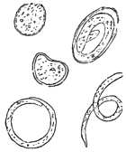
Fig. 38.—Group of eggs and
embryos in a case of endemic
hæmaturia (1870). Original.
On the 17th of May, 1872, I communicated to the Metropolitan
Counties Branch of the British Medical Association a
paper on ‘Bilharzia,’ and in an Appendix to it I wrote as
follows:—“A most interesting circumstance connected with
this case of ‘Bilharzia’ from Natal lies in the fact that I
obtained from the patient some other urinary parasites in the
egg-condition (fig. 38). On five separate occasions I obtained
one or more specimens of the eggs or embryos of a minute
nematode. In one instance there were about fifty of these ova
in the urine, their contained embryos being well developed and
in a state of activity. Usually they were all in this advanced
condition; but on the 25th of July, 1870, several were observed
in much earlier stages of development. One of these was of a
triangular form; its shape, granular contents, and clearly
defined limiting membrane, indicating separation from the
rachis within the ovarian tube. Another early form was perfectly
spherical, with a well marked chorional envelope and
double contour. These forms measured about 1/750″ in diameter.
The fully grown eggs observed at the same time gave a
longitudinal measurement of 1/500″ by 1/1000″ in breadth. On
adding any stimulus, such as diluted sulphuric acid, the embryos
moved themselves freely within the egg. After allowing the
urine to stand for forty-eight hours, I found, on the 27th of
July, that the shells of the ripe ova had
dissolved, leaving the embryos dead, but
still coiled within a fine transparent envelope.
In this state they were easily
separated and examined, when they gave
a measurement of 1/300″ in length, by 1/3500″
in breadth. On two occasions, whilst
engaged in rearing the larvæ of Bilharzia
in water, I noticed single specimens
of these embryos lying dead;
and one of the examples thus observed
gave a length of 1/150″, by 1/3000″ in
breadth.”
Knowing what errors of interpretation have often crept into
helminthological literature I was more than usually cautious in
pronouncing upon the source of these urinary parasites. Accordingly,
I remarked that “future discoveries might enable us
to identify the species of nematode to which these ova are
referable.” I also added:—“Notwithstanding discrepancies as
to size, I am inclined to think that Dr Salisbury and myself
have been made acquainted with nematode eggs and embryos
referable to one and the same species of parasite. I do not
care to speculate as to the origin of these ova. Long ago I
gave in my adhesion to the determinations of Schneider in
respect of the so-called Spiroptera hominis, but I am by no
means certain that his position may not be disturbed by fresh
discoveries. It is not a little remarkable that the parents of my
patient should have averred that she passed three small vermiform
entozoa by the urethra, corresponding, to judge from their
verbal statements, very closely with the ordinary appearances of
Filaria piscium.”
Having written thus much seven years back, it is with natural
pleasure that I find my anticipations already verified. Knowing
that I was dealing with parasites in their earliest larval stages,
it never occurred to me to give a specific name to them, and I
could not possibly approve of Dr Salisbury’s nomenclature, for
which there was no good ground.
In the original discovery Dr O. Wucherer procured the worms
from the chylous urine of a female in the Misericordia Hospital
at Bahia; and on the 9th of the following October, 1866, he
obtained similar worms from another female suffering from
hæmaturia. He also afterwards found them in a man whose urine
was slightly chylous, but not hæmatic. In all cases these
sexually-immature nematodes were alive. In September, 1872,
Dr A. Corre furnished a careful description of similar worms
found by Dr Crévaux in a hæmato-chylurous patient at Guadeloupe.
Dr Crévaux frequently examined the blood of this
patient but found no hæmatozoa. In like manner in Brazil,
Dr J. Silva Lima sought in vain for worms in the blood of no
less than five patients, all of whom suffered from hæmaturia,
and whose urine contained numerous nematoid worms.
Towards the close of the year 1872 the biological world was
startled by the announcement of the discovery of minute Filariæ
in human blood. Dr T. R. Lewis had found microscopic worms
in the blood, and also in the urine, of persons suffering from
chyluria. The worms could be obtained from day to day by
simply pricking any portion of the body with a finely pointed
needle. To this hæmatozoon Lewis gave the trinomial term
Filaria sanguinis hominis, which thus fitly distinguished it from
the Filaria papillosa hæmatica canis domestici described by
Grube and Delafond. Dr Lewis found the average size of the
parasite to be 1/75″ in length by 1/3500″ in breadth. He observed
that while it exists in the blood the body is enclosed in a
delicate transparent tunic or cyst. The worm was never absent
from urine in chyluria. In a case in which there was a milky
discharge from the eyes the worms were also detected. In one
case Lewis calculated that 140,000 Filariæ were present in the
blood—a number certainly not relatively large seeing that MM.
Grube and Delafond estimated the verminiferous blood of their
several dogs to contain numbers varying from 11,000 to 224,000.
Lewis also found Filariæ in the kidneys and supra-renal
capsules of a woman who died of chyluria. It did not appear
probable that the worms underwent further development in the
human body. On this point Lewis remarks:—“Not only may
those hæmatozoa found in man live for a period of more than
three years, but there is no evidence that they have any
tendency to develop beyond a certain stage as long as they
remain in the circulation.” Dr Lewis judged that the form of
chyluria associated with this condition of the blood was local
and intimately related with a tropical climate. The milky
condition of the urine comes on suddenly, not only at first, but
on succeeding occasions also. It is frequently accompanied by
more or less distinctly marked symptoms of various other
obscure diseases, including temporary swellings in the face or
extremities. From certain appearances of intestinal ulceration
Lewis thought that the parasites might gain access to the
system by the alimentary canal, possibly from the tank-water
or the fish inhabiting it. He considered the state of the urine
to be due to the mechanical interruption offered to the flow of
the nutritive fluids of the body. The accidental aggregation of
the Hæmatozoa might give rise to obstruction of the currents
within the various channels, or occasion rupture of their
extremely delicate walls, and thus cause the contents of the
lacteals, lymphatics, or capillaries, to escape into the most
conveniently placed excretory channel.
Compressed into a small compass, I think the above is a
fair statement of the leading facts and phenomena discovered
by Lewis. The whole subject of hæmatozoology immediately
received additional impulse, the consequences of which have not
yet terminated. In this country Welch was stimulated to
investigate the structure of Filaria immitis in the dog, whilst
others sought diligently for nematoid hæmatozoa abroad.
On the 20th of April, 1874, Dr Prospero Sonsino communicated
to the Neapolitan Royal Academy his memoir entitled
“Researches concerning Bilharzia hæmatobia in relation to the
endemic hæmaturia of Egypt, with a notice concerning a nematoid
found in the human blood.” In this brochure he made
known the fact of his having discovered microscopic Filariæ in a
young Egyptian Jew, in the following words:—“On the 1st of
February last, having well washed the finger of the boy, I
placed one drop of blood under the microscope, when with
astonishment I discovered a living organism of the form of a
nematode, resembling Anguillula, in the midst of the hæmatic
corpuscles. The worms glided amongst the globules, which were
tossed about by their lively movements, showing various appearances
according as they presented themselves either from the
sides, the edges, or the front of the disk” (‘Ricerche,’ &c., pp. 11,
12). Dr Sonsino took every precaution to prevent error, subsequently
verifying his “find” from the same patient. Dr Sonsino
directs attention to two of his own characteristic figures of the
worm, and subsequently states not only that he found examples
of the Filariæ in the urine of this same youth, but also “in the
urine of another patient.” The parasites from these two sources
being figured side by side, it was clear, from their resemblance,
that they referred to one and the same species of entozoon. Dr
Sonsino having compared the facts supplied by these cases, was
satisfied that the nematodes in question were specifically identical
with those that I had previously obtained from my little African
patient. However, Dr Sonsino was of opinion that his Filariæ
were not precisely the same as those that had been described
by Lewis.
On the 8th of April, 1876, I received from Dr William
Roberts, of Manchester, some capillary tubes, charged with
blood, obtained from a patient suffering from chyluria. The
tubes had been transmitted by Dr Bancroft, of Brisbane,
Queensland, Australia; and in fulfilment of the donor’s request,
Dr Roberts afforded me an opportunity of examining their
contents, he having himself verified Bancroft’s statement that
they contained Filariæ. It was not until May 22nd that I
found opportunity to confirm the observations of Drs Bancroft
and Roberts. The contents of some of the tubes had by this
time completely dried up; but in others, to which diluted
glycerine had been added, the blood appeared tolerably fresh.
In what might be reckoned as the sixth part of the contents of
one of the tubes, spread on a glass slide, I detected about
twenty Filariæ, three of which I sketched in sitû, in order to
compare them with the figures of Lewis, and also with others
that I had procured from my Bilharzia-patient in the year 1870.
There could not, I thought, be any doubt as to the identity of
all these sexually-immature nematoids. One novelty, however,
presented itself in the presence of a solitary and empty egg
envelope, measuring about 1/500 of an inch in its long diameter,
and thus corresponding precisely with the ova that I obtained
from the urine in my Bilharzia case.
According to Bancroft, chyluria is somewhat common in
Brisbane; and the case here brought forward was not the only
one of the kind which had already furnished Filariæ in the
blood. The patient was a little girl ten years of age.
Thus stood the facts in the spring of 1876. Having
informed Dr Bancroft that a nematoid egg had been detected
in the Australian blood transmitted to England, he was induced
to make further investigations. These happily resulted in the
discovery of the adult worm; the circumstances attending the
“find” being recorded by Dr Bancroft in a letter written to
myself and dated from Brisbane, Queensland, April 20th, 1877.
He wrote as follows:—“I have labored very hard to find the
parental form of the parasite, and am glad to tell you that I
have now obtained five specimens of the worm, which are waiting
to be forwarded by a trustworthy messenger.
“I have on record about twenty cases of this parasitic
disease, and believe it will be the solution of chyluria, one form
of hæmaturia, one form of spontaneous lymphatic abscess, a
peculiar soft varix of the groin, a hydrocele containing chylous
fluid, together with some forms of varicocele and orchitis. These
I have verified. In the colony there are no cases that I can
find of elephantine leg, scrotal elephantiasis, or lymph scrotum;
but from the description of these diseases in the volume on skin
and other diseases of India by Fox, Farquhar, and Carter, and
from Wm. Roberts’ article on the latter in his volume on
urinary diseases, I am of opinion that the parasitic nature of the
same will be established.
“The worm is about the thickness of a human hair, and is
from three to four inches long. By two loops from the centre
of its body it emits the Filariæ described by Carter in immense
numbers.
“My first specimen I got on December 21st, 1876, in a
lymphatic abscess of the arm; this was dead. Four others I
obtained alive from a hydrocele of the spermatic cord, having
caught them in the eye of a peculiar trochar I use for tapping.
These I kept alive for a day and separated them from each
other with great difficulty. The worm when immersed in pure
water stretches itself out and lies quite passive. In this condition
it could be easily washed out of hydroceles through a
large-sized trochar from patients known to suffer from Filariæ.”
In July, 1877, I announced Bancroft’s discovery in the
‘Lancet,’ naming the parasite Filaria Bancrofti, and in the
following September I sent the editor an account of the
results of my study of the adult worms received from Brisbane
in the interval. These examinations supplied me with the
diagnosis already given (p. 181).
On the 29th of September, 1877, Dr Lewis published a paper in
the ‘Lancet,’ wherein, after alluding to my previous announcement
respecting the discovery of Filaria Bancrofti, he describes
under the name of Filaria sanguinis hominis a mature worm,
which was evidently the same parasite. Not unnaturally Dr
Lewis put aside the nomenclature I had employed, on the ground
that the name originally given by himself to the embryonal
form ought to be retained, and that “a new name, if not
necessary on anatomical grounds, would only lead to confusion.”
Personally I have no objection to Lewis’s specific name, but if
the question of priority is to determine the nomenclature, then I
fear we ought to call the species Filaria Salisburyii. Obviously
the retention of Dr Salisbury’s nomenclature (Trichina cystica)
would be unsuitable and misleading.
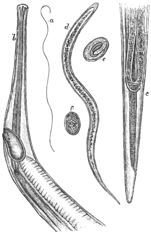
Fig. 39.—Filaria Bancrofti. a, Female (nat. size); b, head and neck (× 55 diam.);
c, tail; d, free embryo (× 400 diam.); e, egg containing an embryo; f, egg, with
mulberry cleavage of the yolk (× 360 diam.). Original.
When (prior to Lewis’s discovery of the hæmatozoa) I had
myself encountered larval nematodes of the same character as
those described by Salisbury, I, like Wucherer, was careful not
to employ a special name for an immature form, which might
or might not represent a worm hitherto known to science.
The paper in which I described the adult worm from specimens
supplied by Bancroft appeared in the ‘Lancet,’ Oct. 6th, 1877,
the facts being stated as follows:—
On the 28th of August, 1877, I received a small collection of
entozoa. The box contained the promised Filariæ, and also
eight bottles filled with various intestinal worms taken from
animals. The Filariæ were enclosed in four small tubes and
preserved in glycerine. Three of the tubes (marked 1, 2, 3)
contained sexually-mature worms, the fourth being labelled
“Sediment from adult Fil. sang.—young and ova.” I described
their contents in succession. Thus, on the 6th of September, 1877,
I examined the Filaria in tube No. 3. The specimen was injured
and in four portions, these collectively measuring three inches
in length. Although, to the naked eye, the worm had appeared
to Dr Bancroft to be of the thickness of an ordinary human hair,
yet I found it about 1/90″ at the thickest part. It was a female.
At the same time I examined the specimen in tube No. 1. This
was also a female. Towards the centre of the body a hernial
protrusion of the uterine horns and intestine had taken place.
In a lithograph sent by Dr Bancroft this specimen was figured
and described as the “parent worm of the Filaria sanguinis,
emitting young Filaria from two loops.” Later on I examined
the contents of tube No. 2. In it I found one tolerably perfect
female Filaria, and also a delicate shred forming part of one of
the uterine horns of another worm. This filament measured
one inch and a half in length, and was coiled round the complete
worm. On transferring it to a watch-glass containing
water, hundreds of embryos made their escape. Owing to the
transparency of the tissues I had much difficulty in finding the
reproductive outlet, and the effort to find it was all the greater
because Bancroft’s figure had misled me. At length I found
the vagina and its orifice close to the head (about 1/20″ from it),
the anal orifice being placed within the 1/90″ from the extremity
of the tail. The vaginal pouch, 1/100″ long, was crowded with
embryos, and a constriction marked its junction with the uterus
proper, which appeared to divide lower down at a distance
of 1/10″ from the head. Towards the tail a fold of the tuba
Fallopii was seen to extend to within 1/20″ of the extremity. All
sections of the uterine system were crowded with germs, eggs,
and embryos in their usual relative situations.
My examinations of the ova and embryos were chiefly made
from the “sediment” sent in a special glass tube. The fully
formed embryos were 1/125″ in length by 1/2500″ in breadth. They
each showed a double skin, the outer envelope in the more
advanced specimens leaving clear spaces at either end of the
body, resulting from commencing ecdysis. I saw no trace of
intestinal tube, but a central line of condensation marked an
early differentiation of the somatic granular contents. The less
advanced embryos were mostly enclosed in a chorional envelope,
the smallest free embryos measuring only 1/200″ in length by 1/3000″
in breadth. These had no double contour. The ova, whose
yolk-contents were still in various stages of cleavage, gave an
average long diameter of 1/900 to 1/1000 of an inch.
Such are the facts I made out, and they enabled me to
amend the characters of the species.
As regards nomenclature, I associated Dr Bancroft’s name
with the sexually-mature worm as being in harmony with the
binomial method and little calculated to mislead; moreover, it
helped to fix both the source and date of the discovery (Brisbane,
Dec. 21st, 1876). The use of this nomenclature detracts
nothing from the high merits of Lewis, who first named the
immature worm Filaria sanguinis hominis. As it now turns
out, both Dr Salisbury and myself had previously been made
acquainted with the young of Filaria Bancrofti; but it was
reserved for Lewis to discover the hæmatozoal character of the
embryos of this worm, and actually to take them from the
blood. It was a singular circumstance, that when I was
engaged in treating my little African patient for trematode
hæmatozoa, it never once occurred to me that the numerous
nematoid embryos mixed with the Bilharzia ova were hæmatozoal.
As before remarked, it was alleged that my patient
had passed worms two or three inches long by the urethra. I
therefore concluded that these were the parents of the eggs
and embryos, and that all of them were urinary. The inference
was wrong, but it has instructively shown how near one
may go towards a great discovery without really making it.
As regards the larvæ, notwithstanding some slight differences
in regard to size and so forth, I have little hesitation in saying
that all the embryo forms severally described by Salisbury, by
myself, by Lewis, Sonsino, Wucherer, Crévaux and Corre,
Silva Lima, Bancroft, Manson, and others, are referable to one
and the same species.
Into the clinical bearings of this subject it is impossible for
me to enter at any length, but I may remark that these parasites
appear to be associated with, if not actually the cause of,
several distinct morbid conditions. To one of these Bancroft
has given a separate name (Helminthoma elastica). This is a
highly elastic form of growth to which I have already alluded
under the title of “lymphatic abscess of the arm.” In the
first valuable report on Hæmatozoa, by Dr Patrick Manson, of
Amoy, China, this careful observer gives interesting particulars
of no less than fifteen cases in which hæmatozoa were found.
Two of these patients had Elephantiasis scroti, two had lymph-scrotum,
two were lepers (one having scrotal disease), two had
enlarged inguinal glands, one had anasarca; and of the remaining
six, spoken of as having no concomitant disease, one had
enlarged glands and abscesses, and another suffered from
marked debility. It would thus appear that what is ordinarily
termed “good health” is rarely associated with a hæmatozoal
condition of the blood in the human subject. The
cases given by Lewis and Manson, where absolutely no recognisable
disease existed, must be regarded as exceptional. Disease,
moreover, may exist without any palpable symptoms being
exhibited by the “bearer,” and thus perhaps it was with the
hæmatozoal dogs of Gruby and Delafond to which I shall again
have occasion to allude. Even those animals that carried
upwards of two hundred thousand microscopic Filariæ in their
blood appeared to suffer no inconvenience whatever.
In the autumn of 1877 Dr Da Silva Lima published an
article in the ‘Gazeta Medica da Bahia,’ in which he dwelt
upon the labors and merits of Wucherer, and, judging from an
omission in one of my memoirs, he supposed that I had insufficiently
acknowledged Wucherer’s claims. A translation of
this article appeared in the ‘Archives de Médicine Navale,’
with an important appendix by Dr le Roy de Méricourt. In
this addendum the French savant showed that the omission on
my part was unintentional, and had been corrected by me in a
later memoir. Not only had I been amongst the earliest in
England to enforce Wucherer’s claims in respect of the micro-Filariæ,
but I had first announced his discoveries in connection
with Anchylostoma duodenale. In my translation of Wucherer’s
memoir (‘Ueber die Anchylostomum Krankheit’) I spoke of the
melancholy satisfaction I had in knowing that the memoir in
question was “among the last that appeared from the pen of
that gifted and amiable physician.” Some notice of Dr Lima’s
paper and its appendix by Dr A. le Roy de Méricourt appeared
in the ‘Lancet’ for Jan. 5th, 1878, and I also published a full
translation of it, with explanatory notes, in the ‘Veterinarian’
for Feb., 1878. Later on, in the ‘Lancet’ (March 23rd, 1878),
Dr Da Silva Lima published an interesting letter correcting a
misconception that had incidentally arisen in the mind of a
commentator (on the Helminthological work of 1877), and at
the same time he pointed to the original facts connected with
the discovery of Wucherer’s Filaria. As my views are in perfect
accord with those of Dr Da Silva Lima, I can only regret that
errors of interpretation should have crept into the discussion.
Dr Lima honorably recognises the nomenclature (Filaria Bancrofti)
which I proposed for the adult worm, and only claims
for Wucherer that which is fairly due.
On the 4th of January, 1878, I received from Dr Patrick
Manson a manuscript in which he announced the discovery of
the larvæ of Filaria sanguinis hominis in the stomach of
mosquitoes. Already, in April, 1877, Dr Bancroft had informed
me of his expectation of finding that these insects
sucked up the larvæ of the Filaria whilst engaged in their
attacks on man. Dr Bancroft’s supposition was a very natural
one, but it remained for Manson to make the actual discovery
of the existence of human hæmatozoa, or parasites that had
been such, within the stomach of Culex mosquito. I lost no
time in making the principal facts public (‘Lancet,’ Jan. 12th,
1878). Dr Manson at the same time forwarded for publication
a record of thirty-five additional cases of hæmatozoa occurring
in Chinese subjects, together with additional particulars of one
of the cases already published in the ‘Customs Gazette.’
These were afterwards published as separate contributions in
the ‘Medical Times and Gazette.’ Dr Manson likewise forwarded
materials for a paper entitled “Further Observations
on Filaria sanguinis hominis.” In this communication he
gave an analysis of the cases (sixty-two in all) in which he had
observed the hæmatozoa, and he added valuable statistical
evidence as to the prevalence of Filariæ in the Amoy district,
dwelling especially on the influence of age, sex, and occupation
in determining the presence of the parasite. He also described
the morbid states with which these entozoa were commonly
associated.
On the 7th of March, 1878, I formally communicated to the
Linnean Society a detailed account of Manson’s investigations
relating to the metamorphoses undergone by the Filariæ within
the body of the mosquito. In this paper Manson pointed out
that the female mosquito, after gorging itself with human
blood, repairs to stagnant water for the purpose of digesting
the blood, and also for the purpose of depositing its eggs.
During this period, which lasts four or five days, the Filariæ
undergo remarkable changes. Subsequently, in a more perfect
state, they escape into the water, and in this advanced stage
they are conveyed to the human body along with the water as
drink. Dr Manson persuaded a Chinese, whose blood was
previously ascertained to abound with Filariæ, to sleep in a
“mosquito house.” In the morning the gorged insects were
captured and examined under the microscope. A drop of
blood from the mosquito was found to contain 120 Filariæ, but
a drop taken from the man’s hand yielded only some thirty
specimens. Further stages of development are accomplished
within the human host, ending in the sexual maturity of the
parasite. After fecundation successive swarms of embryos
are discharged by the female worm, a part of whose progeny
eventually gains access to the blood.
Before I proceed to summarise the whole body of facts I
must in the next place state that Manson and myself contributed
a joint communication to the Medical Society of
London on the 25th of March, 1878. In this memoir I especially
dealt with the question of priority in connection with the discovery
of the adult worm. I then restated that the adult
parasite was discovered by Dr Bancroft on December 21st,
1876. The discovery was verified by Dr Lewis on August 7th,
1877, by Dr Silva Araujo October 16th, 1877, and by Dr F.
dos Santos November 12th, 1877. I gave these dates unhesitatingly,
without, however, in any way prejudicing the question
already raised in respect of the identity of the worms found in
each case. My own mind was fully made up on that point,
and affirmatively so. Dr dos Santos’ find was made in conjunction
with Dr J. de Moura in a case of lymphatic abscess of the
arm. Clinically viewed, the case published by Dr Araujo must
be regarded as unique. Not only were adult and embryonic
Filariæ found in the same patient, but, what was far more surprising
and interesting, the patient displayed in his own person
several of the disorders hitherto found apart; and he was more
than once attacked by one or two of the diseases. He experienced
a first attack of chyluria three years ago, then attacks
of craw-craw commencing a year ago, the latter being attributed
to bathing in a particular lagoon. He had a second
attack of chyluria six months back, at which time lymph-scrotum
appeared, and also scrotal elephantiasis. Dr Bourel-Roncière
pronounced this case to be unique, and attributed
nearly all the disorders to the presence of Wucherer’s embryonic
Filariæ. In a very elaborate analysis of and commentary
on Dr da Silva Lima’s second memoir, Dr Bourel-Roncière
warmly claims for Wucherer the supreme honor in
all these discoveries. A number of affections hitherto regarded
as distinct, and all of which appear to be due to the action of
Filariæ, are regarded by Dr Bourel-Roncière as mere phases of
one and the same disorder. This affection he terms Wucherer’s
helminthiasis. Dr Manson had indeed arrived independently
at a similar conclusion, and I am confident that Wucherer,
were he alive, would in this particular aspect of the question
be the last to claim priority either to Lewis, to Bancroft, or to
Manson.
In this place I may observe that Dr Pedro S. de Magalhães,
of Rio de Janeiro, detected free microscopic nematodes in the
potable waters of Rio (agua da Carioca), which from their
similarity he supposes may have some genetic relation with
Filaria Bancrofti. In this opinion I cannot share.
As regards the metamorphoses of the embryo, Manson states
that for a little while after gaining access to the stomach of the
mosquito the embryo undergoes no change (Fig. 40, a). In a
very few hours changes commence, resulting in wider separation
of the outer skin and an appearance of transverse markings on the
body within (b). In the next stage oral movements occur;
the striation becomes more marked, and the outer envelope is
cast off (c). Then the striated lines disappear and a dotted
appearance is substituted (d). From this condition the embryo
passes to what Manson calls the chrysalis stage, in which nearly
all movement is suspended and the large spots gradually disappear
(e, f, g, h, i, j, k). The tail continues to be flexed and
extended at intervals and the oral motions cease. By the
close of the third day the embryo becomes much shorter and
broader; but the finely pointed tail retains its original dimensions,
projecting abruptly from the sausage-shaped body (m, n).
Large cells next appear in the interior of the body, and by a
little pressure one may detect indications of a mouth (o, p, q, r).
At this period the embryo begins to elongate, and at the same
time to diminish in width; but the growth takes place chiefly
at the oral end of the body. The mouth becomes four-lipped,
open, and funnel-shaped, and from it a delicate line can be
distinctly traced passing to an opening near the caudal extremity,
the tail itself gradually disappearing (s, t). Speaking of
the most advanced stage Manson says:—“A vessel of some
sort is seen in the centre running nearly the whole length of
the body and opening close to one extremity. This end is
slightly tapered down and is crowned with three or perhaps
four papillæ, but whether this is the head or tail, and whether
the vessel opening near it is the alimentary canal or the vagina,
I cannot say.” Now it is quite evident, I think, from Manson’s
figures that he has here faithfully represented the head and
tail, the former (u) to the left, the latter (v) to the right. In
his manuscript (from which I am now quoting) there is no
special reference to these two figures; but it is easy to see that
these terminal sections of the body of the advanced embryo
closely correspond with the head and tail of the adult worm
(Filaria Bancrofti). The curved line passing to the left (u)
evidently indicates the commencement of the partially-formed
vagina.
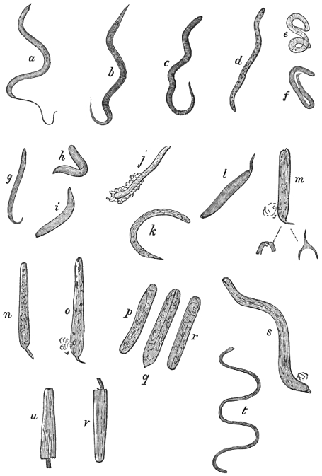
Fig. 40.—Larval Filariæ in various stage of growth from the mosquito; a to d, representing
the first stage of metamorphosis during the first 36 hours, e to o, the changes
occurring during the second stage, to the close of the third day; p to t, forms seen
during the third stage of metamorphosis from the fourth day onwards. The figs. u, v,
represent the head and tail only, whilst t shows the young Filaria in an advanced
stage, and drawn to a much smaller scale, than the others which are here magnified
about 125 diameters. Much reduced from Manson’s original figures.
How completely Manson took the initiative in this part of the
work is evident even from Lewis’s own later observations. In
a paper published in March, 1878, Dr Lewis, writing from
Calcutta and speaking of the rôle of the mosquitoes, says:—“I
had repeatedly examined, in a cursory fashion, these and other
suctorial insects, but had not observed any parasites suggestive
of these embryo-hæmatozoa, hence, when, on receipt of a communication
from Dr Manson a couple of months ago, a renewed
search was made, I was surprised to find that four out of eight
mosquitoes, captured at random in one of the servants’ houses,
harboured specimens of hæmatozoa to all appearances identical
with those found in man in this country. After this, however,
several days elapsed before any mosquitoes could be obtained
which contained these embryo-nematoids, and the specimens
obtained on the next occasion were devoid of the enveloping
sheath, which appears to characterise the kind found in man
out here, and apparently, according to Dr Manson, in China
also.” Further on Lewis also remarks, “When the insect is
caught shortly after feeding and the contents of its stomach
examined microscopically, the hæmatozoa, if present, will be
observed to manifest very active movements, which may possibly
continue for several hours on the slide. If the insect be
kept for twenty-four hours before examination it is probable
that the movements of the parasites will be more sluggish, and
their form probably altered owing to irregular contractions and
dilatations of their substance—changes which may also occasionally
be observed when embryo-hæmatozoa are preserved on a
glass slide, and they may sometimes be kept alive thus, if in
suitable media, for two or three days. When the insect is not
examined till the third day, the contained parasites will probably
manifest marked signs of disintegration—and possibly
every indication of life will have disappeared from many of the
specimens. After the third or fourth day I have not seen any
active specimens of these entozoa in the stomach or in any part
of the alimentary canal of the mosquito; those which remain
have undergone more or less fatty degeneration, and are readily
stained with eosin, which, as far as my experience goes, is not
the case so long as they are alive and active. After the fourth
or fifth day it is very rare that traces of any hæmatozoa-like
objects can be detected at all, so that it must be inferred either
that they have succumbed to the digestive action of the insect’s
stomach or been disposed of along with the excreta.” An
important addendum by Lewis records a fortunate incident
as follows:—“It was observed that nearly all the mosquitoes
captured in one of the servants’ houses contained hæmatozoa,
so that the supply of suitable insects in all the stages of their
growth became amply sufficient for all requirements. The
result of the examinations under these favorable conditions has
shown that although the stomach digests a great number of the
ingested hæmatozoa, as mentioned above, nevertheless others
actually perforate the walls of the insect’s stomach, pass out,
and then undergo developmental stages in its thoracic and
abdominal tissues.”
I may here observe that Sonsino has instituted a comparison
between the embryos of this Filaria and those of Anchylostoma,
by which it appears that the former measure 0·218 to 0·330 mm.
in length, and those of Anchylostoma 0·430 mm. The hæmatozoa
are about forty times longer than broad, and the larval
anchylostomes only fourteen times longer. The tail of Filaria
is conspicuously longer.
In the ‘Lancet’ for June 22nd, 1878, an announcement
appeared from the pen of Mr D. H. Gabb, of Hastings, stating
that a patient under his care formed the habitat of Filaria
sanguinis hominis; and in the autumn of the same year a
paper which I read to the Linnean Society in the spring
was published. In that paper the following summary was
offered:
1. Filaria Bancrofti is the sexually-mature state of certain
microscopic worms hitherto obtained either directly or indirectly
from human blood.
2. The minute hæmatozoa in question—hitherto described as
Wucherer’s Filariæ, Filaria sanguinis hominis, Trichina cystica,
Filariose dermathemaca, and so forth—are frequently associated
with the presence of certain more or less well-marked diseases
of warm climates.
3. The diseases referred to include chyluria, intertropical
endemic hæmaturia, varix, elephantiasis, lymph scrotum, and
lymphoid affections generally, a growth called helminthoma
elastica, a cutaneous disorder called craw-craw, and also leprosy.
4. It is extremely probable that a large proportion, or at
least that certain varieties of these affections are due to morbid
changes exclusively resulting from the presence of Filaria
Bancrofti or its progeny within the human body.
5. It is certain that the microscopic hæmatozoa may be
readily transferred to the stomach of blood-sucking insects,
and it has been further demonstrated that the digestive organs
of the mosquito form a suitable territory for the further growth
and metamorphosis of the larval Filariæ.
6. The character of the changes undergone by the microscopic
Filariæ, and the ultimate form assumed by the larvæ
whilst still within the body of the intermediate host (Culex
mosquito), are amply sufficient to establish the genetic relationship
as between the embryonal Filaria sanguinis hominis, the
stomachal Filariæ of the mosquito, and the sexually-mature
Filaria Bancrofti.
In the month of September, 1878, I received a letter from
Dr da Silva Lima announcing the fact that Dr Araujo had
verified the existence of the embryos of Filaria Bancrofti in
mosquitoes, at Bahia. These mosquitoes had, I understood,
attacked a French priest in whose blood Dr Araujo also
detected Filariæ. Thus, it fell to the lot of Araujo, through
his untiring zeal, to verify in Brazil all the separate discoveries
of Bancroft, Manson, and Lewis.
In the October issue of the ‘Pathological Society’s Transactions’
for 1878 Dr Bancroft records numerous cases of filarious
disease, and he gives a succinct account of the circumstances
connected with his original discovery.
In a clinical lecture published October 12th, 1878, Dr
Tilbury Fox seeks to diminish the value of these discoveries,
characterising helminthological investigators as merely “recent
writers.” Dr Fox denies that Filariæ are a cause of true
elephantiasis, but admits the occurrence of “elephantoid inflammation
and inflammations due to Filariæ.” Dr Fox’s statement
that “Filariæ have not been found in uncomplicated
elephantiasis, that is, in disease without chylous exudation,”
seems to me to be directly at variance with Manson’s recorded
experiences. I hold that Manson has confirmed the truth of
Lewis’s views, and that he has thoroughly proved that (to use
his own words) “varicose groin glands, lymph scrotum,
elephantiasis, and chyluria are pathologically the same disease.”
In the first instance I was myself led to conclude that some of
the forms of elephantiasis might be due to other causes than
obstruction of the lymphatics caused by the presence of Filariæ;
but the explanations of Lewis, of Bancroft, and of Manson
more especially, have almost entirely removed this doubt.
Those who seek to explain away the connection between
genuine elephantiasis and Filariæ will do well to study Manson’s
last important memoir. He shows that “elephantiasis and
allied diseases are much more frequently associated with the
parasite than are other morbid conditions.” This fact is
brought out very clearly in his table of 670 cases, from which
it appears that 58 per cent. of cases of Filaria are associated
with elephantoid disease.
When this opposition to Manson’s views is likely to cease
(on the part of those who do not happen to have been in any
way instrumental to the discoveries in question) it is not easy
to say. In a brief communication which appeared in the last
number of the ‘Medical Times and Gazette’ for 1878, Dr
Manson successfully combats the doubts that have been entertained
respecting the rôle of the mosquito. Because Lewis
found that canine hæmatozoa were digested, and thus perished
in the stomach of mosquitoes, it had been argued that human
hæmatozoa must necessarily undergo similar processes, and
consequently die. Those who oppose the views of helminthologists
in respect of the intermediary host-function of insects on
such grounds can have very little general, and still less special
knowledge of the phenomena of parasitism. It is the old
story. When any new discovery is made, it must always pass
through the ordeals of denial and doubt before it can be generally
accepted as true; and, as in the case of Jenner’s immortal
discovery, there will always remain a certain number of peculiar
people who show themselves hostile to every advance in science.
Dr Manson may take comfort from this consideration, and rest
assured that the value of his discovery is quite unaffected by
the opposition referred to.
Since I communicated the results obtained by Manson,
Lewis, myself, and others to the Linnean Society, an even
more exhaustive summary of the facts has been published
by Dr Bourel-Roncière, in the ‘Archives de Médecine Navale.’
The distinguished author does full justice to the writings
of English helminthologists, and dwells, with emphasis, upon
the finds and interpretations of Lewis, Manson, and Bancroft.
Incidentally, also, he comments upon Sir Joseph Fayrer’s early
recognition of the etiological identity of hæmato-chyluria and
elephantiasis, on other than helminthic grounds. The frequent
concurrence of the two affections had especially struck Sir J.
Fayrer as pointing to a probable common origin. He had also
surmised that the disorders might be due to parasites.
Dr Bourel-Roncière, alike with the caution, precision, and
logical reasoning of a cultured savant, concludes his elaborate
review in the following terms:—“There are the facts.
Certainly, many points remain obscure, many problems await a
solution, and the last word has not been said on the actual part
which the parasite plays in the pathogenesis of the affections
above enumerated—its mode of action, the importance of its
rôle, the extent of its pathological domain, the habitat of its
progenitors, their identity, and so forth. All these questions
will only be elucidated by necroscopic researches, which at
present remain absolutely wanting.”
“However, notwithstanding the doubts which hover over the
future value of these curious discoveries, it is difficult not to
recognise their importance in the study of certain tropical
diseases—which up to the present time have been attributed to
vague and undetermined causes—hæmato-chyluria and elephantoid
affections principally. Apart from the interest which
attaches to the natural history of the nematoids, they raise, in
effect, etiological and prophylactic questions, the extreme importance
of which we believe it would be needless to demonstrate.
It is greatly to be desired that the researches should
be taken up in other parts of the globe, where endemicity and
perhaps greater facilities for necroscopic investigation would
render them fruitful—Cochin-China, Tahiti, &c. Fresh observations
are necessary to confirm the first and to fill up
notable gaps. The way has been brilliantly opened by the
English and Brazilian physicians. Let our colleagues in the
French colonies put their shoulders to the wheel; they have
before them a vast field of study to explore.”
Since the above remarks were written I have received several
communications from Dr Bancroft, and also others from Drs
da Silva Lima, Araujo, Assis Sousa, Paterson, Hall, of Bahia—the
two last named being English physicians in practice there.
I regret that I can do little more than refer to the writings of
these authors in the Bibliography below; but I may observe that
Drs Paterson and Hall have ascertained that the proportion of
the population of Bahia affected by Filaria is 81/2 per cent. Out
of 309 persons examined, 26 had hæmatozoa, which is, roughly,
one in twelve, or more strictly, 8·666 per cent.
Amongst recent memoirs that by Sir J. Fayrer, read to the
Epidemiological Society on the 5th of February, 1879, deserves
especial attention. In regard to its significance, I have only
space to remark that, much as we may regret the little interest
shown by our hospital physicians and surgeons in this subject,
it is particularly gratifying to see experienced Indian officers
like Sir J. Fayrer, Mr Macnamara, and Dr John Murray, coming
forward both to aid and render homage to their junior
colleagues in Eastern parts, who are successfully labouring to
advance the cause of helminthology and scientific medicine.
In concluding this subject I may observe, that one of the
greatest hindrances to the due recognition of the remarkable part
played by parasites in the production of human endemics and
animal epizoötics arises from the circumstance that no inconsiderable
number of minute worms may infest a host without
obvious injury. This immunity proves nothing. If, for example,
we take the case of Trichina we find that several millions of
entozoa may exist in the human, or, at all events, in the animal
bearer, without producing any symptom of discomfort. In such
cases it is not possible to determine the strict limits of health
and disease; nevertheless, were we to double the amount of
infection, the imaginary line of demarcation is at once bridged
over and the parasites become acknowledged as directly
responsible for grave symptoms which may even prove fatal to
the bearer. Again, the relative strength and size of the infected
host constitute factors that materially limit the power of the
parasite for injury. Where the entozoa are of minute size,
and where their injurious action is primarily due to the
mechanical obstructions they set up, it is clear that the
virulence of the helminthiases, or resulting diseased conditions,
will mainly depend upon the number of intruders.
Another consideration of the highest value in relation to
epidemiology generally, and more especially in regard to the
practical question as to the best methods of stamping out parasitic
plagues, is that which refers to the life-history of the
entozoon itself. It must be obvious that in all cases where the
intermediate host can be captured and destroyed, the life-cycle
of the parasite can be broken and interrupted, and if thus
broken, there is an end to the further propagation of the
species. The knowledge that we have acquired by experimental
research in this connection has already enabled us to
set a limit upon the prevalence of certain well-known disorders,
such as Trichinosis, Cestode-tuberculosis, and so forth. In
the case of epizoötics, however, which are indirectly due to the
action of intermediary hosts that cannot be readily captured or
destroyed, then our power of arresting the disease is comparatively
limited. In the present case it is probably not
necessary either that a dead or living mosquito should be
swallowed to insure infection; but it is necessary that the
parasitic larvæ should have dwelt within the mosquito in
order to arrive at the highest stage of larval growth prior to
their re-entrance within the human territory. Undoubtedly,
the larvæ are swallowed with potable waters. Perfect filtration
before use would certainly check, if in course of time it
did not totally extinguish several of the many virulent diseases
that now afflict the inhabitants of warm climates.
It is with reluctance that I terminate this article, but in
the closing pages of this work (Book II, Section V) I hope
to add a few more particulars in reference to Lewis’s latest
researches.
Bibliography (No. 23).—Araujo, A. J. P. da Silva, “Memoria
sobre a Filariose,” &c., Bahia, 1875; see also ‘Arch. de
Méd. Nav.,’ 1875 and 1878.—Bancroft, J., “Cases of Filarious
Disease,” in ‘Pathological Soc. Trans.’ for 1878, vol. xxix,
p. 407.—Bourel-Roncière, “Résumé of and Commentary upon
the writings of Silva Lima, Silva Araujo, and others,” in
‘Arch. de Méd. Nav.’ for March, 1878.—Idem, “Pathologie
exotique. De l’hématozoaire nématoïde de l’homme et de son
importance pathogénique, d’après les travaux Anglais et
Bréziliens des dernières années;” ibid., for August and Sept.,
p. 113–134 and p. 192–214, 1878.—Cobbold, T. S., “Discovery
of the Adult Representative of Microscopic Filariæ,” ‘Lancet,’
July, 1877, p. 70.—Idem, ‘On Filaria Bancrofti,’ ibid. Oct., 1877,
p. 495.—Idem, “Verification of Hæmatozoal Discoveries in
Australia and Egypt,” ‘Brit. Med. Journ.,’ June, 1876.—Idem,
“Obs. on Hæmatozoa,” ‘Veterinarian,’ October, 1873.—Idem,
“Remarks on the Ova of another Urinary Parasite (in the paper
on ‘Bilharzia’) from Natal,” ‘Brit. Med. Journ.,’ July 27th,
1872, p. 89; see also Bibl. No. 12.—Idem, “Entozoa in Relation
to the Public Health” (various papers), ‘Med. Times and Gaz.,’
Jan. and Feb., 1871.—Idem, ‘Worms’ (l. c., p. 151), 1872.—Idem,
“Hæmatozoa; Fresh Discoveries by Lewis,” ‘Lancet’
for Feb. 6, 1875.—Idem (brief notice), the ‘Veterinarian,’
p. 209, March, 1875.—Idem, “On the Discovery of the Intermediary
Host of Filaria sanguinis hominis,” ‘Lancet,’ Jan. 12,
1878, p. 69.—Idem, “On the question of Priority of Discovery,”
Rep. of Med. Soc. of Lond., in ‘Lancet,’ March 30, 1878,
p. 465.—Idem, ‘Mosquitoes and Filariæ’ (explanatory note), in
‘Brit. Med. Journ.,’ March 16, 1878, p. 366.—Idem, “On the
Life-history of Filaria Bancrofti, as explained by the discoveries
of Wucherer, Lewis, Bancroft, Manson, Sonsino, myself, and
others,” “Report of the Proceed. of the Linnean Soc.” for
March 7, 1878, in ‘Pop. Science Rev.,’ April, 1878; and
afterwards published in extenso in ‘Journal Linn. Soc.,’ Oct. 31,
1878.—Idem, “On Filaria Bancrofti,” in Part iv of a series of
papers on the Parasites of Man, in the ‘Midland Naturalist,’
August, 1878.—Idem, “On Filaria sanguinis hominis,” in a
letter to the ‘Lancet,’ July 13, 1878, p. 64.—Idem, “Filariæ
and Leprosy” (case from Bancroft); ‘Lancet,’ Feb. 1, 1879.—Corré,
A., “Note sur l’helminthe rencontré dans les urines
hémato-chyleuses,” ‘Rev. des Sci. Nat.,’ 1872.—Cossé, “Sur
l’helminthe rencontré par Wucherer et Crevaux,” &c., ‘Rev.
Montpellier,’ tom. i, p. 190.—Couto, A., “These de concourso,”
Bahia, 1872.—Crevaux, J., “De l’hématurie chyleuse, &c.,”
1872; also in ‘L’Union Médicale,’ 1872 (abs. in ‘Brit. Med.
Journ.,’ July, 1872, p. 100); also in ‘Arch. de Méd. Nav.,’
1874; and in ‘Journ. de l’Anat. et de la Physiol.,’ 1875 (see
also Silva Lima).—Davaine, C., ‘Traité,’ 2nd edit., p. 944;
‘Hæmatozoaires,’ supp., 1877.—Fayrer, Sir J., “Filaria sang.
hom.,” ‘Lancet,’ March 16, 1878, p. 376.—Idem, “Elephantiasis
Arabum,” ‘Med. Times and Gaz.,’ Dec. 1, 1877, p. 588;
“On the Relation of Filaria sanguinis hominis to the Endemic
Diseases of India,” in the ‘Lancet,’ Feb. 8 and 15, and reprinted
from the ‘Med. Times and Gazette’ (same date), 1879.—Gabb,
D. H., letter in ‘Lancet,’ June 22, 1878.—Leuckart,
l. c., s. 638, 1876.—Lewis, T. K., “On a Hæmatozoon in Human
Blood,” ‘San. Comm. 8th Rep.,’ Calcutta, 1872; ‘Med. Press,’
1873, p. 234; ‘Indian Ann. Med. Sci.,’ 1874; ‘Lond. Med.
Rec.’ (abs. by myself in vol. i, p. 5), 1873.—Idem, “Pathological
Significance of Nematode Hæmatozoa,” ‘Tenth Ann.
Rep.,’ 1873, Calcutta (reprint), 1874; ‘Ind. Ann.,’ 1875.—Idem,
“Remarks regarding the Hæmatozoa found in the
Stomach of Culex mosquito,” ‘Proc. Asiatic Soc. of Bengal,’
March, 1878, p. 89.—Idem, “Flagellated Organisms in the
Blood of Rats” (being portion of a paper on “The Microscopic
Organisms found in the Blood of Man and Animals,” in ‘14th
Annual Report of the San. Comm. with the Govt. of India’), in
the ‘Quart. Journ. of Micr. Science,’ Jan., 1879.—Idem (published
since the present article was written), “The Nematoid
Hæmatozoa of Man,” ibid., April, 1879.—Lima, J. F. da Silva
(with Crevaux), ‘Memoria sobre hematuria chylosa ou gordurosa
des paizes quentes;’ extrahida da ‘Gazeta Medica da Bahia,’
1876; repr. in ‘Arch. de Méd. Nav.,’ Dec., 1878 (see also
Le Roy de Méricourt).—Magalhães, Pedro S. de, “Filarias em
estado Embryonario, encontradas n’agua tida como potavel (agua
da Carioca),” ‘O Progresso Medico,’ Dezembro, 1877, p. 57.—Idem,
“Nota sobre os nematoides encontrados no sedimento
deposito pela agua (potavel) da Carioca,” ‘O Prog. Med.,’ 1 de
Setemb., 1878, p. 577.—Idem, “Caso de filariose de Wucherer;”
ibid., 15 de Setemb., 1878, p. 589.—Makina, M.D., “Filaria in
Chyluria,” letter in ‘Lancet,’ Feb. 22, 1879, p. 286.—Manson, P.,
“Rep. on Hæmatozoa,” ‘Customs Gazette,’ No. 33, Jan.–March,
1877; see also ‘Med. Times and Gaz.’ for Nov. 10, p. 513, Nov. 17,
p. 538, and Nov. 24, p. 563; Dec. 1, p. 589, 1877; also Jan.,
1878.—Idem, “Additional Cases;” ibid., March 2, 9, 23, 1878.—Idem,
“On Filaria sanguinis hominis, and on the Mosquito
considered as a Nurse,” ‘Proc. Linn. Soc.,’ March 7, 1878;
see also report in ‘Nature,’ March 28, 1878, p. 439.—Idem,
“On Filaria sanguinis hominis, clinically considered in reference
to Elephantiasis, Chyluria, and allied Diseases,” ‘Rep. of
Med. Soc. of Lond.,’ in ‘Lancet,’ March 30, 1878.—Idem,
“Further Observations on Filaria sanguinis hominis,” “Med.
Rep.” for April–Sept., 1877, in ‘Customs Gazette,’ Shanghae,
1878.—Idem, “The Development of the Filaria sanguinis hominis,”
‘Med. Times and Gaz.’ for Dec. 28, 1878, p. 731.—Méricourt,
A. Le Roy de, in Appendix to an art. entitled “Nouvelle
phase de la question relative à la nature parasitaire de la
chylurie. Découverte du représentant adulte de la ‘Filaire de
Wucherer,’” par le Dr da Silva Lima, from the ‘Gaz. Med. da
Bahia,’ Sept., 1877; see also the ‘Lancet,’ Jan., 1878, p. 22
(editorial notice).—Moura, J. de, ‘These de Concourso,’ 1877.—O’Neill,
“On Craw-craw,” ‘Lancet,’ Feb., 1875.—Pareira, A. P.,
“On Bilharzia and Chyluria,” ‘Gazeta Med. da Bahia,’ No. 9,
1877 (noticed in ‘Lancet,’ Feb. 2, 1878).—Salisbury, J. H.,
“On the Parasitic forms developed in Parent Epithelial Cells
of the Urinary and Genital Organs,” ‘Hay’s American Journ.,’
vol. iv, 1868, p. 376.—Santos, F. dos, in ‘Gaz. Med. da Bahia,’
March, 1877.—Sonsino, P., ‘Richerche,’ &c., 1874; ‘Della
Bilharzia,’ &c., 1876; ‘Sugla Ematozoi,’ &c., 1876 (see Bibl.
No. 12).—Idem, “On the Diagnosis of Embryos of Filaria,” in
his paper ‘Sull’ Anchylostoma duodenale;’ ‘Estr. dall Imparziale,’
1878.—Sousa, M. de A., ‘Memoria sobre a Elephantiasis
do escroto,’ Bahia, 1878.—Wucherer, O., “Noticia Preliminar,”
&c., ‘Gaz. Med. da Bahia,’ Dec., 1868.—Idem, ‘Sobre Hematuria
no Brazil,’ ibid., Sept., 1869; see also “Méricourt’s trans.
(De l’hématurie intertropicale observée au Brézil),” ‘Arch. de
Méd. Nav.,’ p. 141, 1870, and the fuller references quoted in
my memoir; ‘Linn. Soc. Journ., Zool.,’ vol. xiv, p. 368.
Filaria Loa, Guyot.—Although further examinations of this
worm will probably result in placing it in some other genus
than Filaria, yet it is by no means clear that Diesing was right
in placing it with the genus Dracunculus. I therefore abandon
the nomenclature adopted in my previous treatise. According
to the surgeon, Guyot, who made seven separate voyages to the
coast of Angola, these worms cannot be confounded with the
Dracunculus. They are quite white, and relatively much
thicker than guinea-worms. Under the title of Filaria oculi
Moquin-Tandon has spoken of certain small nematodes as “not
uncommon in the negroes of the Angola coast;” and he gives
other localities where it occurs. The worms are identical with
those described by Guyot as dwelling beneath the conjunctivæ of
negroes at Congo and in the Gaboon region generally. The
parasite is rather more than an inch and a quarter in length,
being pointed at one end and blunt at the other. It is termed
Loa by the natives, who state that after a period of several
years the worm voluntarily quits the organ. The disease is
thus naturally cured. This parasite enjoys a tolerably wide
geographical distribution, as it has been observed by Clot Bey
in a negress who had come from the town of Monpox, situated
on the banks of the River Magdalena; by Sigaud, who saw one in
the eye of a negress in Brazil; by Blot, at Martinique, who
saw two in a negress originally from Guinea; by Bajon, who
met with one in a little negro girl who had come from Guadeloupe;
by Mongin, who found one in a negress who had been
living in the Island of San Domingo; and by Lestrille, who
removed one from beneath the conjunctiva of a negro who
came from Gaboon.
Bibliography (No. 24). Davaine, l. c., p. 839.—Guyon,
‘Gaz. Méd. de Paris,’ p. 106, 1841, and in ‘Micr. Journ. and
Struct. Record,’ p. 40, 1842, and in ‘Dublin Journ.,’ vol. xxv,
p. 455, 1839.—Idem, ‘Compt. Rendus,’ tom. lix, p. 743, 1865.—Guyot,
in ‘Mém. par Arrachait,’ p. 228, 1805.—Küchenmeister,
l. c., s. 322.—Lestrille, in Gervais and Van Beneden’s ‘Zool.
Med.,’ 1859, also quoted by Davaine, l. c., 2nd edit., p. 840.—Leuckart,
l. c., s. 619.—Moquin-Tandon, A., ‘Zool. Med.,’ Hulme’s
edit., p. 363, 1861.
Filaria lentis, Diesing.—This is a doubtful species. The
worm was first discovered by Nordmann, in a case of lenticular
cataract under the care of Von Gräfe, and it was afterwards
found by Jüngken in a similar case, as recorded by Sichel.
There is also the instance described by Gescheidt, in which Von
Ammon operated, and from which brief descriptions of the
worm have generally been taken. In this case there were three
worms, two measuring about 1/6″ and the third 1/15″ in length. In
Jüngken’s case (exhibited by Quadri, of Naples, at Brussels) the
worm was more than 3/4″ long. In another case, reported by M.
Fano, the worm was somewhat less than 1/4″ long. There is no
certain evidence that any of these various worms had developed
sexual organs in their interior. It is true that the reproductive
organs were described in two of the worms observed by
Gescheidt; but after a due consideration of all the facts I
fear we must conclude that all the worms in question were
sexually-immature and wandering nematodes, possibly referable
to Gurlt’s Filaria lacrymalis, as Küchenmeister long ago
suggested.
Bibliography (No. 25).—Cobbold, ‘Entozoa,’ p. 332.—Davaine,
l. c., p. 821 et seq.—Diesing, ‘Syst. Helm.,’ p. 625.—Fano,
‘Traité des Malad. des Yeux,’ tom. ii, p. 498; and in
‘Rec. de Méd. Vét.,’ p. 140, 1869; quoted by Davaine, p. 831.—Gescheidt,
Ammon’s ‘Zeitsch.,’ 1833, s. 435.—Leuckart, l. c.,
Bd. ii, s. 622.—Nordmann, l. c., Bibl. No. 2, s. 7, 1832.—Sichel,
‘Iconogr. Ophth.,’ p. 707, 1859.
Filaria labialis, Pane.—This is a filiform cylindrical worm
measuring an inch and a quarter in length. The mouth is
armed with four papillæ arranged in the form of a cross. The
tail of the female is blunt, the vaginal outlet being placed at a
very short distance from its extremity, and a little above or in
front of the anus. This parasite was found by a medical
student at Naples. It occupied the cavity of a pustule in the
upper lip, giving rise to considerable irritation. Only the male
worm is at present known.
Bibliography (No. 26).—Davaine, l. c., edit. ii, Synopsis,
p. 107.—Leuckart, l. c. (with a fig.), Bd. ii, s. 616.—Pane,
“Nota di un elminte nematoide,” in ‘Annali dell’ Acad. degli
aspiranti Naturalisti,’ Napoli, ser. 3, vol. iv, 1864.
Filaria hominis oris, Leidy.—In the fifth volume of the
‘Proceedings of the Philadelphia Academy of Natural Sciences’
(1850, p. 117) Dr Leidy furnishes the following description of
this worm as gathered from the examination of a simple specimen
preserved in alcohol, and labelled as having been
“obtained from the mouth of a child.” Body white, opaque,
thread-like; mouth round, simple; posterior extremity obtuse,
furnished with a short, curved, epidermal hooklet, 1/500″ in length,
by 1/2000″ in diameter at base. Dr Leidy offers some speculations
as to its origin, but from whatever source the worm was
obtained by the bearer, it seems to be an immature form. Its
length is five inches and seven lines.
Filaria (Nematoideum) trachealis, Bristowe and Rainey.—This
is another very doubtful worm. It was originally described
in the ‘Pathological Society’s Transactions’ for 1855.
It evidently represents only a juvenile stage of growth of some
species of round worm. Rainey discovered a considerable
number of these worms in the trachea and larynx of a person
who died from a disease affecting the lower extremities. Individually
the parasites measured about the 1/50″ in length.
Strongylus (Filaria) bronchialis, Rudolphi.—This is a small
nematode. The male measures rather more than half an inch,
whilst the female is upwards of an inch in length. The caudal
appendage of the male is furnished with a bilobed, membranous,
half-bell-shaped bursa. This surrounds the cloacal
outlet, the latter concealing a double spiculum. The tail of
the female is sharply pointed, the anal orifice being placed a
little in front or above. The body is filiform, of a pale yellow
color. It is about 1/50″ broad in the male, and 1/35″ in the female.
The mode of reproduction is viviparous.
The original specimens were discovered by Treutler in
Germany, during the winter of 1791, in the bronchial glands of
an emaciated subject, whilst those sent to Diesing for description
were discovered by Dr Fortsitz at Klausenberg, in Transylvania,
in the lungs of a boy six years old. Diesing and
Weinland suggested the identity of Filaria bronchialis and
Strongylus longevaginatus, whilst Küchenmeister went further,
and pronounced them to be one and the same species.
Bibliography (No. 27).—Cobbold, ‘Entoz.,’ p. 357.—Davaine,
‘Synops.,’ l. c., ‘Synopsis’ cix.—Küchenmeister, l. c., Eng. edit.,
p. 381.—Leuckart, l. c., s. 618.—Treutler, F. A., “De vermibus
filiformibus (Hamularia lymphatica) in glandulis conglobatis
bronchiorum repertis,” in ‘Obs. Pathol. Anat.,’ 1793.—Wedl.,
‘Die im Menschen vorkommenden Helminthen’ (quoted by
Leuckart), Wien, 1862, s. 22.
Eustrongylus gigas, Diesing.—This is by far the largest
nematode known to science, the male sometimes measuring a
foot in length and the female more than three feet, whilst the
breadth of the body reaches half an inch at the thickest part.
Though fortunately very rare in man, this worm is known to
occur in a great variety of animals, especially in weasels.
According to Weinland and Jackson, it is particularly abundant
in the kidney of the North American mink (Mustela
vison), destroying the substance of the organ, the walls of
which become the seat of calcareous deposit. It has been
found in the dog, wolf, puma, glutton, raccoon, coati, otter,
seal, ox, and horse.
The body of the adult worm is cylindrical, more or less red
in color, and somewhat thicker behind than in front. The
head is broadly obtuse, the mouth being supplied with six
small, wart-like papillæ, two of which correspond with the
commencement of the two lateral lines of the body. These
lines are also distinguishable from other six longitudinal lines
traversing the body from end to end by the presence of very
minute papillæ which are less closely arranged towards the
centre (Leuckart). The tail of the male shows a simple,
thick, cup-shaped bursa, which is destitute of rays, and
partly conceals the simple spiculum. The tail of the female
is blunt and pierced by the centrally placed anal opening.
The vulva is situated near the head in the ventral line. The
eggs are stout and oval, measuring 1/300″ in length by about 1/550″
in breadth.
As regards development the recent researches of Schneider
have shown that certain kinds of fish play the part of intermediary
bearer. Balbiani preserved the ova in water for more
than a year without their hatching, and all his attempts to rear
the larvæ in the intestines of the dog by direct experiment
failed. Similar feeding experiments upon fishes and reptiles
also failed. The embryo, when removed from the egg, measures
1/104″ in length. It is vermiform, having a pointed head and
simple mouth. Balbiani describes the buccal cavity as containing
a protractile stylet. Notwithstanding the negative
results obtained by Balbiani’s experiments on fishes, Schneider
(from anatomical data, which Leuckart confirms) has placed it
almost beyond question that the worm hitherto known as
Filaria cystica is the sexually-immature Eustrongylus gigas.
This worm is found encysted beneath the peritoneal membrane
in Galaxias scriba and Synbranchus laticaudatus. It is worthy
of remark that the genus Galaxias comes nearer to the Salmonidæ
than to the pike family, whilst the Synbranchi are
tropical oceanic fishes. Probably the sexually-immature worm
occurs in other fishes, especially the Salmonidæ.
Remarkably fine examples of the adult worm may be seen
in the Hunterian Collection, Lincoln’s Inn, and in the Museum
of the Royal Veterinary College. The human example is
undoubtedly genuine. The dissections in the Hunterian Collection
of specimens were made by me in 1865. Objection has
been taken to my description of the œsophagus as “spiral.”
In Sheldon’s specimen it is certainly twisted upon itself, precisely
in the manner in which Davaine has also figured it (‘Traité,’ fig.
68); but I cannot here give further anatomical particulars.
Drelincourt found two worms sexually united in the kidney.
When once the parasites have gained access to this organ,
rapid destruction of the glandular substance follows. Ultimately
the kidney is reduced to the condition of a mere cyst or
bag, which, besides the worms, contains a quantity of sanguineo-purulent
matter. Frequently only one worm is present, but
oftener two or three. In the kidney of a puma D’Azara’s
friend, Noseda, found no less than six worms, whilst Klein
obtained eight from the kidney of a wolf.
Bibliography (No. 28).—Azara, F. de, ‘The Natural History
of the Quadrupeds of Paraguay,’ trans. from the Spanish by
W. P. Hunter; Valpy’s edit., p. 43, 1837; Black’s, 1838;
French edit., p. 313, 1801.—Albers, ‘Beitr. z. Anat. &c.,’ Bd. i,
s. 115.—Aubinais, ‘Revue Méd.,’ 1846, p. 284.—Balbiani,
“Recherches,” &c., ‘Compt. Rend.,’ 1869, p. 1091; ‘Rec. de
Méd Vét.,’ 1870, p. 5.—Bickford, “Spec. of Str. gigas found in
the Kidney of a Dog,” the ‘Veterinarian,’ 1859, p. 312.—Blainville,
‘Dict. des Sci. Nat.,’ tab. 29.—Blanchard, ‘Ann.
des Sci. Nat.,’ 1849, p. 186.—Idem, in ‘Cuvier’s Règne Animal’
(Masson’s edit.), ‘Les Intestinaux,’ p. 57, pl. 27.—Blasius, ‘Obs.,
&c.’ (with fig. of Lumbricus in renibus hominis), 1674, p. 125.—Bobe-Moreau,
in ‘Journ. de Méd.,’ tom. xlvii.—Boerhaave,
‘Aphorism.,’ 1728.—Bremser (l. c., Bibl. 2), s. 223.—Chabert,
‘Traité des maladies verm. dans les Animaux,’ 1782.—Chiaje,
‘Comp. d. Elmintogr. umana,’ p. 106.—Clamorgan, J. de, ‘La
Chasse de Loup,’ 1583 (quoted by Davaine, the worms being
described as “serpents et bêtes fort venemeuses”).—Cobbold,
‘Entoz.,’ p. 358.—Idem, ‘Catalogue of Entozoa in the Museum
of the Roy. Coll. of Surg.,’ “Descr. of preps. Nos. 19–25,” p. 3,
1866.—Idem, “Parasites of Man,” ‘Midland Naturalist,’ Dec.,
1878.—Collet-Meygret, “Mém. sur un ver trouvé dans le rein
d’un Chien,” in ‘Journ. de Physique,’ &c., 1802.—Cuvier, see
Blanchard (supra).—Idem, ‘Voyage en Sicile,’ and in ‘Ann.
des Sci. Nat.,’ tom. xi.—Davaine, C., ‘Traité,’ l. c., deuxième
edit., p. 271 et seq. (with full lit. refs. at p. 290).—Diesing, l. c.,
vol. ii, p. 325.—Dujardin, l. c., p. 113.—Frank, F., “Ein Spulwürm
in der Urinblase eines Hundes,” ‘Hufeland’s Journ.,’
Bd. xviii, s. 112.—Jackson, ‘Catalogue of the Boston Museum,’
1847, p. 317.—Klein, T. K., “Anatomical Description of Worms
found in the Kidneys of Wolves,” ‘Phil. Trans.,’ 1729–30,
p. 269.—Küchenmeister, l. c., Eng. edit., p. 376.—Leblanc (rep.
by Rayer and Bouley), in ‘Bull. de l’Acad. de Méd.,’ 1850,
p. 640; in ‘Rec. de Méd. Vét.,’ 1862, p. 800; and quoted by
Davaine.—Leuckart, l. c., Bd. ii, s. 353–401, 1876.—Moublet,
“Mém. sur les vers sortis des reins et de l’urethre d’un enfant,”
‘Journ. de Méd-Chir. et Pharm.,’ 1758, pp. 244 and 337.—Otto
(Anat.), in ‘Mag. d. Gesellsch. naturf.,’ 1814.—Owen, art.
“Entozoa,” in Todd’s ‘Cyclop.’—Rayer, ‘Traité des maladies
des reins,’ 1841.—Rayger, ‘Sur un serpent qui sortit du
corps d’un homme après sa mort’ (quoted by Davaine, l. c.,
p. 272), 1675.—Schneider, ‘Monographie der Nematoden,’
1866, s. 50.—Idem (mit Peters), quoted by Leuckart, l. c.,
s. 382.—Stratton, in ‘Edin. Med. and Surg. Journ.,’ p. 261,
1843.
Dochmius duodenalis, Leuckart.—Much time might be occupied
and wasted over the nomenclature of this parasite. In
my previous treatise, and for reasons there stated, I placed it
under the genus Sclerostoma. On rather slender grounds
Dubini formed the genus Anchylostoma for its reception, but
Von Siebold thought that, on account of the absence of symmetry
in the arrangement of the so-called dental organs,
Dubini’s genus might very well be allowed to remain. Bilharz,
Diesing, Küchenmeister, Wucherer, and others have retained
the genus as either Anchylostoma or Anchylostomum. Schneider
keeps it amongst the Strongyli; but after all that has been said
and written there can, I think, be no doubt that if Dujardin’s
genus Dochmius is to be retained at all, Dubini’s worm
must be placed in it. The comparisons instituted by Leuckart
afford sufficient proof of the intimate alliance as between
Anchylostoma and Dochmius. Professor Molin thought to
meet the difficulty by calling the worm Dochmius anchylostomum,
but the specific term, duodenale, should certainly be
retained.
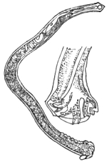
Fig. 41.—Male Dochmius duodenalis,
with bursa separately enlarged.
After Küchenmeister.
This worm was discovered by Dubini at Milan, and though
at first thought rare, it is now known to be tolerably common
throughout Northern Italy. The worm has also been recently
found by Dr Kundrata at Vienna, in an Austrian subject.
According to Pruner, Bilharz, and Griesinger, it is abundant in
Egypt. Griesinger believed that about one fourth of the
people of that country suffered from anæmic chlorosis, solely in
consequence of the presence of this worm in the small intestines.
From Wucherer’s observations especially, we know that
Dubini’s worm is not limited to the localities above mentioned,
for it occurs in the western tropics, in Brazil, and even in the
Comoro Islands.
The worm may be described as a small nematode, the males
measuring 3/8″ or rather more, whilst the females extend to very
nearly 1/2″ (12 mm.). The head is pointed and tapering,
and bent forward, having the mouth directed towards the
ventral aspect. The oral opening is armed with four asymmetrically
disposed, unequally-sized, horny, conical, converging
teeth. The neck is continuous with
the cylindrical body, which is 1/80″ in
thickness. The body terminates in a
straight cone-shaped, or rather sharply-pointed
tail in the female, the caudal
extremity of the male ending in a
partially inflexed, blunt point. In the
male there is a cup-shaped, bilobed
bursa, the membranes of which are
supported by eleven chitinous rays, ten
being simple, whilst the median, or odd
one, is bifurcated at the summit. The
mode of reproduction is viviparous.
Adult males and females occur in the
proportion of one of the former to
three of the latter.
As above mentioned, it was Griesinger who first pointed out
the clinical importance of this entozoon. He first explained
the manner in which the worm produces anæmia, the persons
attacked losing blood as if they were being bitten by innumerable
small leeches. Like the rest of their kindred, these
worms are veritable blood-suckers. In the first instance the
views of Griesinger met with opposition, but they have since
received abundant confirmation. Whilst Küchenmeister’s
‘Manual’ furnishes an excellent account of the disorder as
known in Europe, we are chiefly indebted to Wucherer for
what is known of the disorder in Brazil. The experiences
recorded in the ‘Deutsches Archiv für Klinische Medicin’ for
Sept. 27th, 1872 (s. 379–400), were amongst the last that
appeared from the pen of that gifted and amiable physician.
As little or no notice of his writings appears to have been
taken by professional men in this country, I depart somewhat
from the design of this work when I venture to abstract a few
of the clinical particulars which he has supplied. Their importance
in relation to sanitary science is obvious, inasmuch as
these parasites are introduced into the human body by drinking
impure water, or, at least, water which either contains the
free larvæ of the worm, or the intermediary bearers that harbor
the larvæ.
It should be borne in mind that Dubini’s original discovery
was made at Milan in 1838, whilst Griesinger’s recognition
of the worm as a cause of the Egyptian chlorosis resulted
from a post-mortem examination made on the 17th of April,
1851.
In the journal above mentioned, Wucherer records his own
discoveries as follows (‘Ueber die Anchylostomunkrankheit,’
&c.):—“Although Griesinger with well-founded confidence
gave an account of his ‘find’ and its significance, yet it remained
for a long time unnoticed and unutilised, till at length a case
led me to corroborate it. During my many years’ residence in
Brazil, especially during the first year, I had very frequent
opportunities for witnessing the tropical chlorosis, but seldom to
treat it, as it is one of those diseases for which Brazilians seek
no medical assistance. Its treatment falls to the lot of the
curiosos, curadeiros (quacks), who employ the fresh pulp of a
species of fig as a remedial agent with the best results. On
the 13th of December, 1865, I was called to the Benedictine
monastery in Bahia to see a slave of the order suffering from
hypoæmia. The patient was about thirty years of age, married,
a strongly built mulatto. He was a field laborer on the Ingua
plantation of the order, who exhibited in a conspicuous degree
all the symptoms that occur in hypoæmia except the diarrhœa.
He was well nourished, but strikingly pale, his whole face, but
especially the eyelids, being œdematously swollen, as also were
the feet, legs, and hands. The hands and feet were very cold.
His appearance betrayed the most horrible anguish or low despondency.
With difficulty only could he raise himself, being
obliged to lie down again immediately on account of his weakness.
Auscultation revealed a diminished respiratory murmur,
and bronchial expiration in both lungs. The pulse was very
rapid and small, the patient complaining of pain in the region
of the heart. He had frequent palpitation when he moved, and
he complained of pain in other parts of the body. His abdomen
was much distended by gases, but not sensitive to pressure from
without, except in the region of the stomach. The urine was
clear, its specific gravity 1007 to 10231/2°. Under great difficulties
he resided for several months after his marriage at
Inhatâ. Earlier he had been on the estates of the order at
Rio de S. Francisco. He there suffered for a long time from
intermittent fever, but at Inhatâ he entirely recovered. At
Inhatâ the slaves frequently suffered from hypoæmia, but in
S. Francisco not at all. He appears not to have made any
misuse of brandy. The slaves of the order were well cared for,
and supplied with good and wholesome nourishing food. The
patient had already, for a long period, treated himself with
steel wine, yet was continually getting worse and worse. He
had not taken the pulp of the fig. As I was unaware he had
suddenly become so ill, they hastily despatched a message to
the town. There was no good to be expected from the further
employment of iron, and the patient was in such a condition that
from the very first I despaired of his recovery. I immediately
prescribed the pulp of the Gammeleira (Ficus doliaria), but
it could not be easily obtained. Considering that the Gammeleira
would have a drastic effect, I therefore prescribed two
grammes of elaterium, to be divided into eight doses, of which
he should take one every three hours.” Dissatisfied with this
advice, however, Dr Wucherer goes on to say that on reaching
home he carefully looked up the literature of the subject. “In
a ‘Geologico-Medical Report’ by Professor Hirch, recorded in
the ninety-sixth volume of ‘Schmidt’s Jahrbucher,’ I found
how Griesinger had recognised the Anchylostoma as the cause
of the Egyptian chlorosis, which was clearly identical with our
hypoæmia. He had employed this commended anthelmintic. I
resolved the more to prescribe the pulp of the Gammeleira
when I found it described as a worm-expelling remedy in
Martin’s ‘Systema Materiæ Vegetabilis Braziliensis.’ The next
morning, however, when I arrived at the monastery I learnt
that my patient died about two hours after a slight evacuation.
Only after much resistance would they permit the sectio cadaveris.
I merely opened the abdomen, and was surprised to find
everything as Griesinger had described. During the next
season, through the courtesy of my colleagues attached to the
General Infirmary at Bahia, especially of Drs Silva Lima,
Faria, and Caldos, I was enabled to open more than twenty
bodies of anæmically deceased individuals. All were selected
as miserably poor in condition, but only five were bodies of
persons in whom hypoæmia was diagnosed, and in these there
were a great number of Anchylostomes in the small intestine.
The intestines of the other bodies contained either none, one, or a
few.” Dr Wucherer next states that he compared the characters
presented by his entozoa with those given by Dubini, Diesing,
and Von Siebold, and found a perfect agreement throughout.
He sent several examples to Griesinger, who also established
their identity, and communicated the results of his investigations
accordingly (‘Archiv für Heilkunde,’ 1866, s. 387.
See also Leuckart, ‘Die Mensch. Par.,’ Bd ii, s. 411). Dr
Wucherer also forwarded a number of specimens to Dr Weber,
who published a brief account of them with excellent figures
(‘Path. Soc. Trans.,’ vol. xviii, 1867, p. 274). As mentioned
in the text of his memoir (s. 394), Dr Wucherer also transmitted
some strongyloids to myself. “The publication of my observations,”
adds Dr Wucherer (‘Gazeta Medica da Bahia,’ 1866,
p. 27 et seq.), “had a result in that Dr J. R. de Moura, of
Thersepolis, in the province of Rio de Janeiro, sought for Anchylostomes
in the bodies of tropical anæmics (Hypöæmikern). He
at once found these parasites, as stated in the same journal (for
1866, p. 132). As occurred to myself, he saw no enduring
results from the application of the remedies which appeared
to be called for, whilst he well knew that unprofessional persons
(Nichtärzte) succeeded in obtaining marked results by the
exhibition of the pulp of the Gammeleira (Ficus doliaria). The
anthelmintic action of this remedy was also unknown to him.”
Dr Wucherer then records how his discovery of these entozoa
was announced by Dr Jobini to the Rio academy, and how Dr
Moura’s observations were subsequently communicated, adding
remarks upon the interesting discussion that followed. The
general opinion was that the Anchylostomata were not the
primary and necessary cause of this tropical anæmia, but rather
a co-operating agent in its production. Against this view Dr
Wucherer afterwards very properly protested (‘Gazeta,’ Jan. 15th,
1868). In the mean time, says our author, “Dr le Roy de
Méricourt, prompted by my first communication, had invited
the physicians of the French colony to seek for Anchylostomes.
Drs Monestier and Grenet, at Mayotta (one of the Comoro Isles,
which lies about 12° S. lat. to the north-east of Madagascar),
ascertained the presence of entozoa in hypoæmics. Dr Grenet
sent the duodenum and a portion of the jejunum of an hypoæmic
corpse to Le Roy de Méricourt, who compared the Anchylostomes
with Davaine’s description, and recognised them as
examples of A. duodenale.”
“In the year 1868 Dr Rion Kérangel found Anchylostomes in
the bodies of hypoæmics in Cayenne. Thus, the occurrence of
Anchylostomes in hypoæmics has been authenticated by Pruner,
Bilharz, and Griesinger, in Egypt; by myself, Dr Moura,
Dr Tourinho, and other physicians, in Brazil; by Monestier and
Grenet, in the Comoros; and by Rion Kérangel in Cayenne.
It thus also appears, from the wide separation of these several
localities, that the Anchylostomes, if duly sought for, will be
found in many other countries.”
These details given by Wucherer are so precise and instructive
that I could not have further abridged them without injustice
to his record. The bearing of the foregoing facts in relation
to the question as to how we may hope to arrest the fatal action
of many of these nematodes is sufficiently obvious. That
strongyles and their allies prove highly destructive to man and
beast is as well established as any other recognised conclusion
in medical science; nevertheless, there are those who still doubt
the power of these nematodes in relation to the production of
fatal epidemics. I shall deal with the sanitary bearings of the
subject hereafter. In conclusion, I may mention that Dr da
Silva Lima has forwarded specimens of Anchylostomum to the
Hunterian Museum, where they may be seen.
Bibliography (No. 29).—Bilharz, ‘Zeitschr. f. wiss. Zool.,’
Bd. iv, s. 55.—Cobbold, ‘Entozoa,’ p. 361.—Idem “Remarks on
Recent Contributions to our Knowledge of the Parasitic Nematoids,
especially in reference to the Wasting Diseases they produce in
Man and Animals,” the ‘Veterinarian,’ Jan., 1876, p. 1.—Davaine,
l. c., pp. 118 and 931.—Diesing “Revis. der Nematoden,”
‘Sitzb. d. m.-naturw. cl. d. k. Akad.,’ 1860, s. 716.—Dubini,
‘Entozoografia,’ &c., 1849.—Griesinger (quoted above),
see also ‘Arch. f. Phys. Heilk.,’ 1854.—Küchenmeister, l. c.,
Eng. edit., p. 383.—Leuckart, l. c., ss. 410–455.—Molin,
‘Il sottordine degli Acroffali,’ p. 61 (quoted by Leuckart).—Siebold,
‘Zeitsch. f. wiss. Zool.,’ 1852, s. 55.—Sonsino, P.,
L’Anchilostoma duodenale in ‘relazione coll’ Anemia progressiva
perniciosa,’ Egitto, 1877.—Idem, ‘Sull.’ Anch. duod., 1878 (see
also Bibliog. No. 27, both reprinted from ‘Imparziale.’)—Weber,
H., l. c., 1867.—Wucherer (quoted above), 1872.

Fig. 42.—Outline of a female
Dracunculus medinensis.
Nat. size. Original.
Dracunculus medinensis, Cobbold.—This parasite is popularly
known as the guinea-worm, or Medina-worm. Probably
Lister was the first writer who distinctly spoke of it
as the Dracunculus, 1690, the same title being applied to it by
Kaempfer, 1694. Be that as it may, Gmelin, long afterwards,
placed the parasite in the genus Filaria, at the same time
adopting the specific title medinensis. This had been previously
employed by Linneus, who, however, regarded the worm
as belonging to the genus Gordius. It being clear from the
distinctive characters of the entozoon that it was desirable to
separate it from the Filariæ, and that no better generic name
could be devised than Dracunculus, I thought it right to
combine Lister’s and Gmelin’s nomenclature as above, 1864.
Leuckart pursued a similar course, crediting Linneus with the
titles.
The guinea-worm having been known from the earliest times,
it is not surprising that its true nature long remained a
mystery. Any one who has read Küchenmeister’s elaborate
narrative of the historical significance of the Dracunculus will
hardly have failed to arrive at the conclusion that Moses was
probably the earliest writer on the endemic disorder which is
occasioned by this parasite. There can be no doubt that the
“fiery serpents” which afflicted the children of Israel during
their stay in the neighbourhood of the Red Sea were neither
more nor less than examples of our Dracunculus. It is further
evident that Plutarch spoke of Dracunculi, when in the eighth
book of his ‘Symposiacon,’ he quotes Agatharchidas as stating
that the people taken ill on the Red Sea suffered from many
strange and unheard-of attacks, amongst other worms, from
“little snakes, which came out upon them, gnawed away their
legs and arms, and when touched retracted, coiled themselves
up in the muscles, and there gave rise to the most insupportable
pains.” In order to render the passage more readable, it will
be seen that I have slightly altered the original version (‘Parasites,’
s. 305).
The guinea-worm may be described as a nematode measuring
from one to six feet in length, having a thickness
of 1/10th of an inch. The body is uniformly cylindrical, terminating
below in a more or less curved and mucronately pointed
tail. The head is flatly convex or truncate, having a central,
simple mouth, which is surrounded by four equi-distantly and
cruciately disposed papillæ. The mode of reproduction is
viviparous, the body enclosing a prodigious number of hatched
embryos, which, by distension of the uterine ducts, almost
entirely obliterate the somatic cavity. Notwithstanding the
statements of Owen to the contrary, the male Dracunculus is at
present altogether unknown.
The guinea-worm possesses a comparatively limited geographical
range, for not only is it proper to the tropical regions,
but within intertropical limits it is almost exclusively confined
to certain districts in Asia and Africa. Thus, according to
Künsenmuller, as quoted by Busk, it occurs endemically in Arabia
Petræa, on the borders of the Persian Gulf and Caspian Sea, on the
banks of the Ganges, in Upper Egypt, Abyssinia,
and the coast of Guinea. “In America
the guinea-worm is unknown, except in persons
who have had communication with Africa
or other parts where it is indigenous. The
island of Curaçoa is the only locality in the
New World which offers an apparent exception
to this fact, and it would be highly
desirable to ascertain the real state of the case
in this instance.” The observations of Chisholm
showed that the Dracunculus is really
prevalent in several of the West Indian islands,
especially in Grenada, and the still later investigations
of Dr Da Silva Lima point to its
former prevalence in Brazil. Now, the worm is
rarely seen at Bahia. Mr Busk said:—“Though
endemic only in the above-mentioned parts of
the world, it would yet appear that all races
of mankind are obnoxious to the attacks of
the Filaria when exposed to what may be
called the contagion; that is, when placed in
circumstances under which it might be supposed
a contagious seminium could be conveyed
to them.” Mr Busk also added:—“I have
known many instances tending to prove that,
in order that a European should become infected
with the guinea-worm on the coast of
Africa, it is not necessary that he should have
been on shore at all. It has been quite sufficient
for him to have exposed the bare
surface of some parts of his person to the
water in the native canoes alongside, or, it
may be, to the discharge from the sores of
those laboring under the disease. This mode
of its introduction accounts for the frequency
with which the legs and feet are attacked
by the parasite, in preference to other parts
of the body, as it will always, I believe, be
found that the men who have become so affected
have been in the habit of going about with bare
feet, as is common among sailors in warm
latitudes. That the contagious material is
conveyed in water is also further indicated by the well-known
fact that in India, where it is the custom of the natives to carry
water in skins on their backs, the worm makes its appearance
on the back and shoulders and upper part of the body.”
These views were published by Busk in 1846, and I am free to
confess that—confirmed as they appeared to be by subsequent
and independent testimony—they completely dominated my conceptions
as to the mode of ingress of the young parasites within
the human bearer. Thus, those of our Indian troops which
were most exposed during the rainy season, subsequently
exhibited evidence of having been invaded by the Dracunculus.
As, moreover, the period of incubation of the entozoon commonly
extends from twelve to fifteen months, it necessarily
happened that the disease often showed itself in localities far
distant from the spot where the troops originally contracted the
disorder. The statement that the period of incubation of the
worm is not less than a year, is probably incorrect, since Carter
mentions that in a school of fifty boys bathing in a certain pond
at Bombay—the sediment of which swarmed with microscopic
tank-worms (Urobales palustris, Carter)—twenty-one were
attacked with Dracunculus during the year, whilst the boys of
other schools, bathing elsewhere, remained, with few exceptions,
uninfected. This is a remarkable occurrence, and it points to
the possibility of the young Dracunculi being confined to
particular pools. That they should, whether occupying the
bodies of intermediary bearers or not, be more abundant in some
waters than others, is just what might be expected, since such
a distribution is in harmony with a recognised law affecting the
abundance or limitation of species in particular localities.
Much, indeed, has been written respecting the nature of the soil
and geological formations occurring in the Indian worm-districts,
but the speculative views enunciated on this point are
little worthy of credit. Those who desire information on this
head should at all events consult the valuable writings of
Smyttan, Greenhow, Bird, Forbes, Chisholm, and Aitken,
who, apart from the question at issue, supply abundance of
practical information.
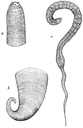
Fig. 43.—a, b, Head and tail of the adult guinea-worm (magnified 10 and 18 diameters
respectively); c, embryo (magnified 500 diameters). Original.
Into the anatomy of the adult Dracunculus I do not enter,
but I may remark in passing, that the structure of the worm
has been exhaustively treated of by Busk and Bastian. A
résumé of their views is given in my introductory treatise. Carter
and Leuckart have also added important details. As regards
the structure and development of the young worms, I have to
observe that the discovery of the viviparous mode of reproduction
in Dracunculus is due to Jacobson. Nearly a quarter of a
century ago I recognised the fact that the uterine organs of the
adult worm almost completely filled up the perivisceral cavity,
and that they were crowded with microscopic worms. Referring
to this “find,” the late Sir George Ballingall, of Edinburgh,
in his well-known work on ‘Military Surgery,’ recorded the
circumstance in the following terms:—“The Assistant Conservator
of the Anatomical Museum in our University has
detected in the oviduct of an adult specimen from my collection
myriads of minute and perfectly-developed (embryonic) Dracunculi.
They can be very well seen with an half-inch object-glass,
but their structure is best exhibited if the magnifying
power be increased to two hundred and fifty diameters linear.”
As already stated in my introductory treatise, these observations
were made during the winter of 1853–54. In July, 1854,
M. Robin made a similar statement after examining a fresh
Dracunculus which had been extracted from the leg of a man
by M. Malgaigne. Robin, not unsuitably, compared the worm
to a double tube, one tubular sheath, as it were, enclosing the
other. “The second tube,” he distinctly affirms, “is the
oviduct, or, rather, that part which represents the uterus. The
young still remaining in the uterus were nearly all coiled, sometimes
with the tail sallying outwards, at others rolled like the
rest of the body.” I have thought it only due to Robin and
myself to show that from the first we were perfectly well
acquainted with the fact of the “great development of the
genital tube and of its close adherence to the parietes of the
body.” To be sure, many discrepancies occurred in our
writings, and in those of Busk and Carter. It was Bastian’s
skill and good fortune to correct these errors. Thus, most of
us agreed in recognising a slightly trilobed or tripapillated
mouth; but Carter failed to demonstrate the existence of these
tubercles, and spoke of the oral aperture as being simple and
“punctiform.” The body throughout its three upper fourths
appeared to me to be cylindrical, but Robin found that it was
flattened. It is finely striated transversely, except at the part
where it contracts to form the slender, pointed tail. According
to Carter, Robin, and Davaine, the young attain a length of
about 1/33 of an inch, but Bastian gives it as about 1/42″. In
thickness, Carter gives the approximative diameter as 1/633″,
Robin makes it 1/990″ to 1/1320″, whilst Bastian gives their breadth
at 1/1428″, and Davaine at 1/2500″. I estimated their greatest length
and breadth to be 1/30″ by 1/1000″. Robin and myself thought we
recognised a distinct, rounded, anal orifice; and whilst Busk,
on the one hand, saw nothing which in the slightest degree
indicated the presence of an anal opening, Carter, on the other
hand, described the structure which we called the anus as a
gland, at the same time placing the alimentary outlet on one
side and a little above it. According to Bastian, “the intestinal
tube is about 1/87″ in length, and appears to consist of a
simple canal of varying calibre, pursuing a nearly straight
course, and terminating exactly at about the middle, in length,
of the worm.” Like Robin, Bastian recognised œsophageal and
stomachal divisions, and in a few examples he observed the
cæcal or terminal portion of the intestine to be partially reflected
upon itself. In regard to the circular
opening which Robin and myself described
as the anus, Bastian says there
is a rounded body, “about 1/2200″ in diameter,
with a dark or light spot in the
centre, according to the varying focal
distance, and which seems to represent
a central aperture. Sometimes, above
this, traces of two or three large cells
may be recognised, whilst behind nothing
definite can be made out, save that
the cavity of the body is visible for
about 1/400″. In other specimens of the
young worm the central body and spot
are wanting, but, in its stead, two lateral
sacculi are met with, about 1/3300″ in diameter,
that communicate with the exterior
by a minute channel through the
integuments, which can sometimes be
distinctly recognised. At other times
the channel is obscured by protrusion,
which appears to have taken place
through it, of a minute bilobed papilla,
projecting 1/10,000″ from the side of the
body. When the projections are seen,
the sacculi are indistinct.”
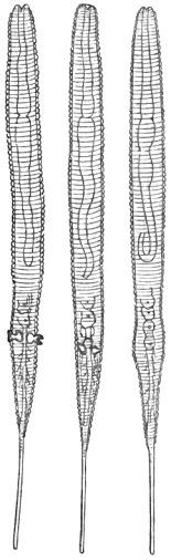
Fig. 44.—Embryos of Dracunculus.
Magnified 500 diameters. After
Bastian.
As Bastian found the young in all
stages of development from the germ
condition 1/5000″ in diameter up to the perfect
embryo, and as, moreover, he, like the
rest of us, could detect no sexual orifice
in the adult Dracunculus, he was led to
express his belief that the young were produced agamogenetically.
He went so far as to call the germs pseudova. It was with
great reluctance that I dissented from the views of so gifted an
observer as Bastian; nevertheless, later researches have shown
that I was justified in not hastily concurring in the theory of a
non-sexual mode of reproduction for Dracunculus.
Among the many advances of modern helminthology, the
discovery of the true source of the guinea-worm is not the least
important. To the late M. Fedschenko (the lamented and
accomplished Russian traveller, who lost his life in a snowstorm
on the Alps), science stands indebted for this memorable
advance. Fedschenko showed that the embryos of Dracunculi,
after quitting the human host, succeed in effecting an entry
into the bodies of entomostracous crustaceans belonging to the
genus Cyclops. Within these intermediary bearers, after
twelve hours’ sojourn, the embryos undergo a change of skin,
attended with subsequent growth. Here they remain to complete
their larval development, which takes place within a
period of five weeks, or, as Fedschenko himself told me, one
month and six days. At length, as perfected larvæ, they are,
together with their crustacean hosts, transmitted to the stomach
of the ultimate or human bearer. It is probable that sexual
maturity is next acquired within the human stomach, copulation
following. After this, the females migrate to the situations
in which they are found beneath the skin of the human
bearer, whilst the males perish and pass out with the fæces.
Thus much I gathered from M. Fedschenko himself when he
visited this country, and I possess a sketch of the larvæ made
by him at the time (October 23rd, 1873). One of the figures
represents a larva which has undergone ecdysis, the long and
narrow embryonic tail being supplanted by one which is blunt
and forked at the tip. The somatic contents of the embryo
have at the same time differentiated into a complete intestinal
tube, and a constriction marks the junction of the œsophagus
with the stomach. There is also internally an oval-shaped
mass of cells near the centre of the body. These represent
the commencement of the reproductive organs.
What I had gathered from Fedschenko in conversation thus
epitomises that which has since been much more fully stated
by Leuckart; and it is only fair to add that the Russian
traveller was led up to his discovery by the previous investigations
of Leuckart respecting the young of Cucullanus. The
Leipsic helminthologist had, indeed, specially instructed Fedschenko
as to the probable source of Dracunculus.
It is often thus that science makes its clear advances, since
a master-mind is needed to set others on the right track. The
embryos of Cucullanus and Dracunculus bear a close resemblance
to each other, and the similarity of the types is
continued on, though not in the same degree, in the next stage
of larval growth, after ecdysis. The higher larvæ of both have
their tails trifurcate at the tip, the head of the Dracunculus-larva
being distinguished by the presence of a pair of papillæ.
In the case of Cucullanus the embryos are, according to
Leuckart, passively transferred to the stomach of Cyclops by
the mouth; but in the case of Dracunculus, Fedschenko saw the
embryo in the act of perforating the bodies of the little crustacea
at the ventral surface, where the segments are bound together
by a thin and easily penetrated connecting membrane. The
larvæ then proceed to coil themselves within the limbs, as many
as six or even a dozen of the parasites being occasionally
found within the body of a single crustacean host. When they
have reached full larval growth they measure about 1/25″ in length.
Of course, after attaining this stage, it is a matter of conjecture
as to the precise way in which their final destiny is accomplished.
Fedschenko fed dogs and cats with the infected crustacea, but
failed to rear Dracunculi in these animals. Clearly, these
carnivora were unsuitable hosts. Could Fedschenko have
experimented on man the result would probably have been very
different. Arguing from what happens in the case of Cucullanus
amongst fishes, and Trichina in man, there can be little
doubt that all the further and final changes undergone by the
larvæ are accomplished within the human host. These changes
are usually, if not invariably, consequent upon a direct transference
of the infested entomostraca along with water used as
drink. Thus, it must at once be evident that the simple
sanitary precaution of filtering water before use is amply sufficient
to ensure the prevention of attacks of dracontiasis or the
guinea-worm disease. The theosophical remedy of Moses
against this invasion by fiery serpents, as the worms were called
in his time, and the modern prophylactic measures dictated
alike by science and common sense, thus stand in striking
contrast the one to the other. In the nature of things it must
ever remain that unreason and reason will select diametrically
opposite methods of action, equally, no doubt, with the good
intention of bringing about beneficial results.
From what has now been advanced, it will be seen that as
regards the mode of infection the views categorically expressed
in my previous work (‘Entozoa,’ p. 387) cannot be maintained.
What, however, is there stated in respect of treatment still holds
good in the main, even as regards prophylaxis.
Bibliography (No. 30).—Adam, ‘Trans. Med. and Surg.
Soc.,’ Calcutta, 1824.—Aitken, W., ‘The Science and Practice
of Medicine,’ 6th edit., vol. i, 1872.—(Anonymous), “Review of
the writings and opinions of Duncan, Johnson, Bird, Mylne,
Kennedy, Chisholm, H. Scott, A. J. Robertson, Smyttan,
Macgregor, Thomas, Mosely, Morehead, Twining, and others,
on the Dracunculus or Guinea-worm,” in ‘Corbyn’s India Journ.
of Med. and Phys. Sci.,’ vol. ii, p. 118, 1836.—(Anon.), “The
Guinea-worm very Prevalent at Bokhara,” ‘Boston Med. and
Surg. Journ.,’ 1843, p. 387.—Balfour, J., ‘Ind. Ann. Med.
Sci.,’ 1859, p. 175.—Ballingall, G. (l. c., supra), 1854.—Bastian,
H. C., “On the Structure and Nature of the Dracunculus or
Guinea-worm,” ‘Linn. Soc. Trans.,’ vol. xxiv, p. 101, 1863.—Berncastle,
J., in the ‘Lancet,’ 1851.—Bird, J., ‘Calcutta Med.
and Phys. Trans.,’ 1825, p. 151.—Bremser (l. c., Bibl. No. 2),
s. 194.—Brett, ‘Surgical Diseases of India,’ 1840; see also
‘Med.-Chir. Rev.,’ 1841.—Bruce, N., ‘Edin. Med. and Surg.
Journ.,’ 1806, vol. ii, p. 145.—Busk, G., ‘Micr. Soc. Trans.’
(original series), 1846.—Carter, H. J., “Note on Dracunculus in
the Island of Bombay,” ‘Bombay Med. and Phys. Soc. Trans.’
(new series), No. 2, p. 45, 1853–54; see also postscript, p. 252.—Idem,
“Further Observ. on Dracunculus,” ‘Bomb. Med. and
Phys. Soc. Trans.’ (new series), No. 4, p. 215, 1857–58.—Idem,
“On Dracunculus and Microscopic Filaridæ,” ‘Ann. of Nat.
Hist.,’ vol. iv (third series), 1859.—Idem, “Notes on Dracunculus,”
&c., ‘Ann. of Nat. Hist.,’ vol. ix (third series), 1862.—Chapotin,
‘Bull. des Sci. Med.,’ 1810.—Charvet, ‘Ann. des Sci.
Nat.,’ 1834.—Chiaje (l. c., Bibl. No. 2), p. 99.—Chisholm, C.,
“On the Malis Dracunculus or Guinea-worm (in Grenada),”
‘Edin. Med. and Surg. Journ.,’ vol. xi, 1815; see also the
‘Veterinarian,’ vol. ix, p. 508, 1836.—Clark, ‘Med.-Chir. Rev.,’
1840.—Clarkson, N. F., “Alleged Case in the Horse,” the ‘Veterinary
Record,’ 1845, p. 73.—Clot-Bey, ‘Aperçu sur le ver
dragonneau observé en Egypte,’ 1830.—Cobbold, ‘Entozoa,’
p. 373.—Cuvier, ‘Règne animal,’ Orr’s Eng. edit., 1849, p. 644.—Davaine,
‘Traité,’ l. c., edit. ii, p. 783 (full lit. refs.), 1878.—Dickson,
‘Path. Soc. Trans.,’ 1851.—Drummond, ‘Med. Commentaries,’
1793, p. 294.—Dubois, ‘Edin. Med. and Surg.
Journ.,’ vol. ii, 1806.—Duncan, ‘Calcutta Med. and Phys. Soc.
Trans.,’ 1835.—Ewart, J., “Questions relating to Dracunculus,”
in a review of his memoir on the “Vital Statistics of the Meywar
Bheel Corps,” in the ‘Madras Quart. Journ. of Med. Sci.,’ vol. i,
1860, p. 462.—Fedschenko, ‘Protocol of the Promoters (Freunde)
of the Natural and Physical Sciences at Moscow’ (in the
Russian language), 1869 and 1874 (quoted by Leuckart).—
Forbes, D., “Observ. on Dracunculus” (extr. from the ‘Half-yearly
Reports of the diseases prevailing at Dharwar in the 1st Grenadier
Regiment, in the year 1836’), ‘Bombay Med. and Phys. Soc.
Trans.,’ vol. i, 1838, p. 215.—Gibson, A., “Note on the Prevalence
of Dracunculus,” in his remarks on the “Diseases of the
Deckan,” in ‘Bomb. Med. and Phys. Soc. Trans.,’ vol. ii, 1839,
p. 209.—Gramberg, ‘Geneeskundige tijdschrift voor nederl.
Indie,’ 1861, p. 632 (quoted by Leuckart).—Greenhow, H. M.,
‘Indian Ann. of Med. Sci.,’ vol. vii, 1861, p. 31.—Grierson, D.,
“Observ. on the Dracunculus, as it prevailed in the 22nd
Regiment, N.I., from April till September, 1841,” ‘Bomb.
Med. and Phys. Soc. Trans.,’ No. 4, 1841, p. 90.—Grundler, in
‘Commerc. Litt. Nov.,’ 1740, p. 239.—Henderson, J., “Note
respecting Four Cases of Dracunculus in the 48th Regiment,”
‘Madras Quart. Journ.,’ vol. iii, 1841, p. 353.—Horton, J. A. B.,
‘Army Med. Reports,’ 1868, p. 335.—Kennedy, R. H., ‘Calcutta
Med. and Phys. Soc. Trans.,’ 1825, p. 165.—Küchenmeister (l. c.,
Eng. edit.), p. 389.—Leuckart (l. c., Bibl. No. 1), s. 644–725.—
Lewis, T. R., in ‘On a Hæmatozoon,’ &c. (l. c., Bibl. No. 23),
p. 30 et seq.—Lima, Da S., “Remarks on the Filaria medinensis,
or Guinea-Worm; on the occurrence of this Parasite endemically
in the Province of Bahia; on its entrance into the human
body by drinking water,” in the ‘Veterinarian,’ Feb., March,
et seq., 1879.—Lister, ‘Phil. Trans.,’ 1690, p. 417.—M’Clelland,
J., ‘Calcutta Journ. of Nat. Hist.,’ vol. i, 1841, p. 366.—M’Grigor,
J., “On the Guinea-worm” (in his “Account of the
Diseases of the 88th Regiment in Bombay”), ‘Edin. Med. and
Surg. Journ.,’ vol. i, 1805, p. 284.—Morehead, C., ‘Calcutta
Med. and Phys. Soc. Trans.,’ vol. vi, 1833, p. 418; also
noticed in ‘Edin. Med. and Surg. Journ.,’ vol. xliv, 1835.—Idem,
part ii, ‘Calcutta Med. and Phys. Soc. Trans.,’ vol. viii,
1836–42.—Murray, J., “Guinea-worm a very Common Disease
at Sattara” (in his Official Report on the Hospital, &c.),
‘Bombay Med. and Phys. Soc. Trans.,’ No. 9, art. vi, p. 198,
1847.—Oke, W. S., “Case of Guinea-worm,” ‘Prov. Med. and
Surg. Journ.,’ vol. vi, 1843.—Oldfield, “Case of Dracunculus”
(from Laird and Oldfield’s “Narrative of an Expedition into
the Interior of Africa”), ‘Dublin Journ.,’ vol. xii, 1838.—Paton,
“Cases of Guinea-worm,” ‘Edin. Med. and Surg. Journ.,’
vol. ii, 1806.—Raddock, “A Case of Guinea-worm,” ‘Indian
Med. Gaz.,’ Oct., 1877, p. 265.—Scott, W., “Remarks on the
Dracunculus,” in a letter to the Medical Board, Madras, ‘Edin.
Med. and Surg. Journ.,’ vol. xvii, 1821.—Leverance, C. E.,
“History of a Case of Guinea-worm,” from ‘Amer. Med. Times,’
in the ‘Glasgow Med. Journ.,’ vol. ix, 1861–62, p. 377.—Smyttan,
G., “On Dracunculus,” ‘Calcutta Med. and Phys.
Soc. Trans.,’ vol. i, 1825, p. 179.—Stewart, L. W., ‘Indian Ann.
of Med. Sci.,’ vol. vi, 1858, p. 88.—Twining, W., “Cases of
Dracunculus,” ‘Calcutta Med. and Phys. Soc. Trans.,’ vol.
vii, 1835.
Fig. 45.—Head of Oxyuris vermicularis.
Highly magnified. After Busk.
Oxyuris vermicularis, Bremser.—Of all the parasites infesting
the human body this is the one concerning which the medical
practitioner is most frequently consulted, partly on account of
its remarkable frequency in children, and more particularly on
account of the difficulty often experienced in getting permanently
rid of it. The Oxyuris vermicularis is by no means confined
to young persons, seeing that adults are infested even to
old age. It is familiarly known as the threadworm or seatworm.
The male measures about 1/6″, and the female from 1/3″
to 1/2″ in length. The female possesses a long capillary tail,
which terminates in a three-pointed end. The extremity is
said to act as a kind of holdfast. The tail of the male is obtusely
pointed. In both sexes the body presents a more or less fusiform
shape, the anterior end being narrowed to form a somewhat
abruptly-truncated head, which is often rendered very
conspicuous by a bulging of the transparent integument surrounding
the mouth. This presents in profile the aspect of
winged appendages (fig. 45). The oral opening is tripapillated,
leading into a triangular œsophagus. The integument is transversely
striated, and of a silvery-white appearance. The
spicule is simple, single, and very minute. The eggs are
oblong and unsymmetrical. They measure about 1/900″ from pole
to pole, and 1/1400″ transversely.
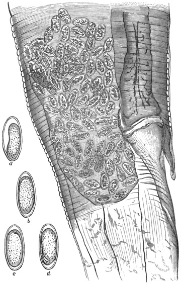
Fig. 46.—Section of a female Oxyuris vermicularis, magnified 220 diameters (after Busk);
and also several free eggs (original). a, With an imperfectly formed embryo; b, c, d, with
three tadpole-shaped embryos, magnified 450 diameters.
Many years back (1863) I pointed out that the most advanced
eggs whilst still within the body of the pregnant female
contained tadpole-shaped embryos, and about the same time
the fact was noticed by Claparède. In his beautiful and
scholarly memoir, ‘De la formation et de la fécondation des
œufs chez les vers Nématodes,’ he wrote concerning the ova as
follows:—“The egg, which exhibits the form of a very narrow
disk in the ovary, acquires the shape of an elongated ellipsoid
in the oviduct, and at the surface differentiates itself into a very
thick vitelline membrane. Then it forms a strong and resisting
chorion, which imparts to the egg an outline similar to that
of a bridge’s span. It has an oval figure flattened at one of its
sides. This chorion is very fragile; it frequently gives way
under slight pressure from the thin plate of glass which covers
the object. It extends itself considerably under the action of
acetic acid, acquiring a size three or four times greater than
that of the egg. The constitution of this chorion is perfectly
identical in the eggs both before and after impregnation. It
is, nevertheless, easy at first sight to know whether or not we
have to deal with a fecundated egg. In the impregnated females
the uteri are filled with thousands of ova, each one of which
encloses an embryo already well formed. The ventral surface
of the embryo and the tail are, without exception, applied to
the flattened side of the egg. The embryo is very broad in the
body, and occupies all the interior space. An embryo such as
Küchenmeister has represented under the form of a small filiform
worm folded on itself, and only occupying a very small
part of the cavity of the egg, is never to be seen. In the non-fecundated
females, on the other hand, the uteri are filled with
eggs, which, instead of the embryo, enclose a non-segmented
yolk furnished with a large germinal vesicle. This vesicle is
not visible so long as the eggs have the form of thin disks; it
only shows itself when the eggs begin to acquire an elliptical
form in the oviduct. It is, however, probable that this vesicle
is the same which was originally visible in the ovary.” The
chorion itself is homogeneous, but in an allied species (Oxyuris
spirotheca) Gyoery and Claparède found that this egg-covering
consists of spirally-coiled bands resembling the tracheal spiral
fibre of an insect. Under suitable conditions the tadpole-shaped
embryos rapidly assume a vermiform character. The
investigations of Leuckart have shown that “one only needs to
expose the eggs to the action of the sun’s rays in a moistened
paper envelope when, at the expiration of five or six hours, the
tadpole-shaped embryos will have already become slender
elongated worms.” According to Heller, the simplest way to
rear the vermiform stage of Oxyuris is to put a number of the
eggs in a glass tube filled up with saliva. The tube should
then be placed in the arm-pit, in which situation it can be
carried about with little inconvenience. In a few hours the
transformations will commence and go on continuously until the
vermiform condition is attained. If, as remarked in my
‘Lectures,’ it be asked whether the embryos which have
escaped into the bowel are capable of arriving at the vermiform
stage, the answer is in the affirmative; for, as Leuckart says,
“the elongated embryos are to be found not only in the fæces
but also in the mucus of the rectum above and around the anus.”
Vix has also asserted that free vermiform embryos are occasionally
to be detected in the intestine of the human bearer
along with the eggs; this hatching within the lower bowel,
however, must, in my opinion, be regarded as exceptional.
Heller is of the same opinion. According to Leuckart, the
escape of the embryos from the eggs “ordinarily takes place
under the action of the gastric juice, also primarily in that
condition when they have by some means or other gained
access to a new bearer.” Prof. Leuckart and three of his
pupils courageously infected themselves by swallowing the eggs,
and had the satisfaction of observing young Oxyurides in their
stools fifteen days afterwards.
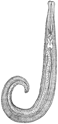
Fig. 47.—Adult male Oxyuris vermicularis.
Magnified. After Küchenmeister.
From the united labors of Professors Zenker and Heller it is
now rendered certain that all the further changes necessary to
bring the larvæ to sexual maturity are accomplished within the
small intestines of the human bearer; and it is not necessary
that a change of hosts should occur at any time during the life of
the parasite. Infection ordinarily takes place by the accidental
and direct conveyance of the eggs that are lodged in the neighbourhood
of the victim’s anus to the mouth. Since the victim
may accomplish this during sleep, it is not in all cases fair to
charge infected persons with uncleanliness. On the other hand,
it too often happens that due care in this respect has not been
exercised, and from such persons you may remove the eggs of
Oxyurides from the margins of the finger nails. One aristocratic
person, who was infested by myriads of these entozoa,
confessed to me that in his extreme distress, and consequent
rage, he had freely bitten the live worms in halves between his
teeth. He had thus exposed himself to a terrible revenge,
since multitudes of the ova entering his mouth subsequently
found their way into the stomach and intestines. By whatever
mode the eggs are conveyed to the mouth their subsequent
passage to the stomach ensures their being hatched. In the
duodenum and other divisions of the small intestines, as Zenker
and Heller have shown, the embryos undergo transformation,
casting their skins, and growing with great rapidity. Probably
not more than three weeks or a month is necessary to complete
their growth. Heller obtained mature worms from an infant
only five weeks old. Finally the worms are transferred to the
cæcum, which constitutes, so to speak, their headquarters. It
is an error to suppose that the lower bowel or rectum forms
their especial habitat, nevertheless the most approved manuals,
vade mecums, and general treatises have for a long time supported
this erroneous view. The error had been pointed out by
Stricker in 1861.
The symptoms produced by Oxyurides are occasionally very
serious. In the mildest cases they have a tendency to undermine
the health. As remarked
in my ‘Entozoa,’ the unpleasant
sensations chiefly develop themselves
in the evening and at night,
consisting for the most part of
feelings of heat and irritation within
and around the margin of the anus.
The symptoms may become extremely
distressing and almost intolerable,
especially when the itching
extends to the genito-urinary
passages, in consequence of the
escape and migration of the parasites
about these parts. By-and-by
various sympathetic phenomena,
such as restlessness, general nervousness,
itchings at the nose, involuntary
twitchings, grinding of
the teeth during sleep, chorea, convulsions,
and even epileptiform
seizures, may supervene. At the
age of puberty special local disorders
arise, the nature of which will
be readily understood when merely
spoken of as the morbid phenomena
of sexual irritation. In the female the occurrence of
pruritus and leucorrhœa is not uncommon, accompanied or
not, as the case may be, with hysteria in various forms. There
is usually general asthenia, with more or less emaciation. The
anæmia is sometimes remarkable, but in place of anorexia,
which is, however, an occasional symptom, one frequently finds
a most voracious appetite, especially in young people. Sometimes
there are obscure symptoms simulating those of local
organic disease.
About the treatment of the disorder I have nothing to say
here, further than to urge the benefits of the preventive
measure of cleanliness. Like Zenker and Heller, I have obtained
the eggs of oxyurides from beneath the finger-nails of
young people. In one lad all the nails had been carefully
bitten down to their roots, but from beneath a minute projecting
portion that was left on the right fourth-finger I procured
two eggs. Their demonstration under the microscope convinced
both parent and child of the necessity of frequently
employing local and general ablutions. Personal cleanliness is
essential. In this connection an able biologist has ventured to
hazard a statement to the effect that “probably any infected
person who adopted the requisite precautions against reinfection
from himself or others would get well in a few weeks without
treatment by drugs.” Dr Ransom bases his belief on the
known facts of the life-history of this entozoon, as recorded
more especially by Leuckart. I regret that I cannot fully
share Dr Ransom’s views, and still less should I think it right
by my silence to seem to endorse his statement to the effect
“that every person who is shown to be infested with those very
common entozoa, Oxyuris vermicularis and Trichocephalus dispar,
is thereby demonstrated to have swallowed minute portions of
his own or another person’s fæces.” This is putting the case
too strongly. No doubt the eggs of oxyurides swallowed by
ourselves must have previously passed through some person’s
rectum; as such, either separately or mayhap collectively, in
the body of the maternal parasite. That does not, however,
justify the statement, that we “have swallowed” part of our
own or of some other person’s excrement. The eggs ought not
to be regarded as constituent portions of the fæcal matter. Perhaps
Dr Ransom will say that the surfaces of these eggs, being
in contact with fæcal matter, must carry infinitesimal particles
on their surfaces, and it is to such that he refers. As, however,
a large proportion of the ova escape with their parents, whilst
they are still lodged within the maternal worm, it cannot be held
that these intra-uterine ova carry fæcal matter on their shells.
Commonly the eggs are swallowed in the separate, free, and dry
state. In water they perish quickly. The act of eating with
unwashed hands is a fertile source of infection, more especially
if the meal be taken either in bed or in the bedroom.
Bibliography (No. 31).—Alexander, J., “On Vermination,”
‘Lancet,’ 1833.—Anderson, W., “On Santonine, with especial
reference to its use in Roundworm and Threadworm,” ‘Brit.
Med. Journ.,’ April, 1864, p. 443; also in Braithwaite’s ‘Retrospect
of Medicine,’ vol. xlix (synopsis, p. 20), 1864.—Barry,
J. M., “On the Origin of Intestinal Worms, particularly the
Ascaris vermicularis,” ‘Trans. Assoc. of Fell. and Licent. of
King’s and Queen’s Coll. of Phys. in Ireland,’ vol. ii, 1878, p.
383.—Bremser, l. c., s. 79.—Buckingham, “Ascarides causing
Erotomania,” from ‘Bost. Journ., U.S.,’ in ‘Med. Gaz.,’ 1857.—Claparède,
E., “On the Formation of the Egg and Fertilisation
in the Nematoidea,” from the ‘Zeitsch. f. w. Zool.,’ translated
by Dallas in ‘Ann. Nat. Hist.,’ vol. i (third series), 1858.—Idem
(memoir quoted in the text above), Genève, 1859.—Cobbold,
T. S., ‘Worms,’ Lect. xii-xv, 1872.—Idem, ‘Entozoa,’
p. 362.—Idem, ‘Brit. Med. Journ.,’ Aug., 1873.—Idem, ‘Tapeworms
and Threadworms,’ 2nd edit., 1872.—Idem, ‘Lancet,’
1866.—Idem, “On the Development and Migrations of the
Entozoa,” ‘Brit. Assoc. Rep.,’ 1864, p. 116.—Date, W.,
‘Lancet’ for Feb., 1872, p. 185.—Davaine, ‘Traité,’ l. c., 2nd
edit., p. 211, and ‘Synops.,’ p. 95.—Dickinson, “Case of Epilepsy
in Children relieved by the expulsion of Worms,” ‘Med.
Times and Gaz.,’ Jan., 1863.—Dickson, R., art. “Anthelmintics,”
rep. from the ‘Penny Cyclopædia,’ in Knight’s ‘Eng. Cyclop.
Arts and Sci. Div.,’ vol. i (column 365), London, 1859.—Dreyfus,
“Irritation of the Bladder from Ascarides,” from
‘Journ. de Med.,’ in ‘Lond. Med. Gaz.,’ 1847.—Elliotson, J.,
“A Lecture on Worms,” ‘Lond. Med. Gaz.,’ 1833.—Idem, “On
Worms in the Intestinal Canal,” ‘Lancet,’ 1831.—Idem, “On
a Case of Threadworms,” ‘Lancet,’ 1831.—Idem, “On Intestinal
Worms,” ‘Lancet,’ 1830.—Heller, A., “Darmschmarotzer,”
in von Ziemssen’s ‘Handbuch,’ Bd. vii, s. 632 (see also Anglo-American
edit.), 1876.—Küchenmeister, l. c., Eng. edit., p. 356.—Ransom,
in Reynolds’ ‘Dictionary of Medicine.’—Smith, A.
(and others), ‘Lancet,’ April 29th, 1865, p. 468.—Stricker, W.,
in ‘Virchow’s Archiv,’ xxi, 1861, s. 360.—Tatham, ‘Lancet,’
April, 1867, p. 457; see also p. 519.—Vix, E., ‘Ueber Entozoen,’
&c., Berlin, 1860; see also “On the occurrence of Entozoa
in the Insane, particularly with respect to the Oxyuris
vermicularis;” brief notice (‘Allg. Zeitsch. f. Psychiatrie’)
in Winslow’s ‘Journ. of Psycholog. Med.,’ vol. i, 2nd series,
1861, p. 158.—Zenker, ‘Verhandl. d. phys. med. Soc.,’ H. ii,
Erlangen, 1870, s. 20; and in ‘Tageblatt der deutschen
Naturforscherversammlung zu Dresden,’ 1868, s. 140 (also
quoted freely by Leuckart, Davaine, and Heller).
Leptodera (Anguillula) stercoralis, Bavay.—In the summer of
1876 Dr Normand, of the French Marine, discovered this little
entozoon in the fæcal discharges of soldiers who had been sent
home invalided from Cochin-China. The patients in question
were the victims of the so-called Cochin-China diarrhœa or
dysentery. This disorder is endemic in character, and it had
hitherto been regarded as consequent upon a variety of causes
other than parasitic. Dr Normand’s discovery, as such, therefore
takes equal rank with the analogous revelations made by
Bilharz, Harley, Leuckart, Zenker, Weber, Lewis, and Bancroft,
in respect of the particular helminthiases in man with which
their names are severally associated (Bilharzia disease, Endemic
hæmaturia, Cestode tuberculosis, Olulaniasis, Inter-tropical
anæmia, Trichinosis, Lymphoid affections, Helminthoma, and so
forth), and also, if I may be permitted to say so, with my own
determinations in respect of a variety of endemics affecting
animals (cestode and nematode epizoöty in the horse, the so-called
grouse-disease, the pigeon-endemic due to lumbricoids,
&c.).
The Leptodera stercoralis is a minute, smooth-bodied, simple,
rhabditiform nematode, measuring when full grown 1/25″ in
length, with an average breadth of 1/625 of an inch. The
embryos at the time of their extrusion measure only 1/250″ in
length, but by the time at which a rudimentary vesicle representing
the uterus begins to form, the females have already
attained a length of about 1/83″. The males and females are of
nearly equal size. The transition from the embryonal state to
the higher larval conditions is accompanied by a change of
skin, after which the digestive and reproductive organs are
gradually but rapidly formed and completed. These changes
have been minutely traced and recorded by Professor Bavay,
who also compares the entozoon with the genera Rhabditis and
Leptodera, in either of which genera the worm might be placed.
I have accordingly adopted the nomenclature suggested by
Bavay.
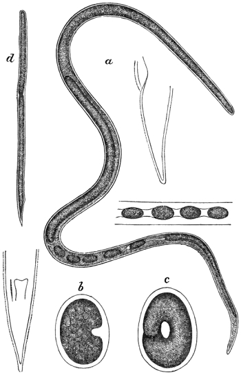
Fig. 48.—Leptodera intestinalis. a, Adult female, and separate figure showing a portion of
the body with the ova in sitû. The two outlined figures represent profile and front views
of the tail, respectively. b, c, Eggs with imperfectly formed embryos. d, Larva. Highly
magnified. After Bavay.
As happens in all the kindred helminthiases that are known
to be dependent upon the presence of small worms, large
numbers of Anguillules are necessary to produce injurious
effects upon the bearer. Thus, the evacuations of the Cochin-China
patients were found to contain such multitudes of the
worms that their numbers could only be adequately estimated
at so many hundreds of thousands passed in twenty-four hours.
Of course they varied in quantity, not only in different patients,
but in the same bearer, from day to day. They are to be found
in every stage of growth and development, from that of the
intra-ovular embryo and free embryonic state up to sexual
maturity. They occupy all parts of the intestinal canal, from
the stomach downwards, being also found in the pancreatic and
biliary ducts, and likewise within the gall-bladder. According
to Bavay, five days suffice under favorable circumstances for the
complete maturation of the worm. This readily accounts for
their occasional extreme abundance.
I am indebted to the courtesy of Dr le Roy de Méricourt for
the original memoirs from which these brief abstracts are taken.
Leptodera intestinalis, Bavay.—This is a larger species, now
and then found associated with the above, and, according to
Bavay, “in infinitely less abundance.” This species was also
discovered by Dr Normand, and has been carefully described
by Bavay. Possibly the worm may afford us another curious
instance of dimorphism. Be that as it may, it must be provisionally
regarded as a distinct form. As its occurrence is by
no means invariable, its rôle in relation to the Cochin-China
diarrhœa must, as Davaine has likewise remarked, be regarded
as of secondary importance. It is readily distinguished from
A. stercoralis both in the adult and larval conditions. The full
grown worm, although comparatively narrow, is more than
twice as long as its congener; moreover, the larvæ, in place of
possessing finely-pointed tails, have blunt or truncated caudal
extremities. Converting M. Bavay’s millimetric measurements
into fractions of the English inch, the average length of the
mature worms will be about 1/11″, whilst their breadth does not
exceed 1/757″ in diameter.
Bibliography (No. 32).—Bavay, “Sur l’Anguillule stercorale,”
‘Comptes Rendus,’ Oct., 1876, p. 694, also in ‘Ann.
Nat. Hist.,’ vol. xviii, 4th series, p. 507, 1876, also noticed in
the ‘Veterinarian,’ Jan., 1877, p. 19.—Idem, “Note sur
l’Anguille intestinale,” ‘Archiv. de Méd. Nav.,’ July, 1877, p.
64, and in ‘Ann. Nat. Hist.,’ 1877, vol. xix, 4th series, p. 350.—Cobbold,
T. S., “Parasites of Man,” in the ‘Midland Naturalist’
for January 1st, 1879.—Davaine, ‘Traité,’ l. c., 2nd
edit., Supp., pp. 966–976, 1877.—Laveran, in ‘Gaz. Hebd. de
Med.,’ Jan., 1877, p. 42.—Layet and Le Roy de Méricourt, in
‘Dict. Encycl. des Sci. Med.,’ 1875.—Libermann, in ‘Gaz. des
Hôp.,’ March, 1877, p. 237, and in ‘La France Méd.,’ 1877, p.
165 (quoted by Davaine).—Méricourt (see Layet).—Normand,
A., in ‘Comptes Rendus’ for July, 1876, p. 316, and Aug.,
1876, p. 386.—Idem, in ‘Arch. de Méd. Navale,’ 1877, p. 35,
and separately as ‘Mémoire sur la diarrhée dite de Cochinchine,’
Paris, 1877.—Idem, “Du rôle étiologique de l’Anguillule
dans la diarrhée de Cochinchine,” in ‘Archives de Médecine
Navale’ for September, 1878, pp. 214–224.
Ascaris mystax, Rudolphi.—This well-known helminth possesses
aliform appendages, one on either side of the head. It
is of a medium size, the male measuring 21/2″ and the female
usually 31/2″ to 4″ in length. Both as regards the size of the
alæ and the length of the body it varies in different hosts.
Thus the variety infesting the dog has long been regarded as a
distinct species (A. marginata), partly from the circumstance
that the alæ are less conspicuous, and partly because the individuals
are often longer and thicker. I possess one specimen
from the dog measuring more than six inches in length. From
like causes the Ascaris leptoptera and other varieties infesting
the carnivora have been regarded as distinct species, but the
worm also varies in one and the same host.
As remarked in my elementary treatise, the late Dr
Bellingham, of St Vincent’s Hospital, Dublin, published in the
13th vol. of the ‘Annals of Natural History,’ an extended
catalogue of Irish entozoa, and in this list he recorded the
existence of a new round worm in man. He says of it:—“From
the distinctness of the lateral membranes of the head I have
given it the name of Ascaris alata.” The catalogue was constantly
referred to by Dujardin, Diesing, and other systematists;
but some of the continental helminthologists do not appear to
have had access to Dr Bellingham’s more extended account of
this parasite as given in the first volume of the ‘Dublin Medical
Press,’ No. 7, Feb. 20th, 1839. I am led to this inference from
the doubt which some have cast upon the very existence of the
worm, although others, with more candour, supposed that Bellingham
had only mistaken the species. Thus, Küchenmeister (‘Parasiten,’
s. 464, and in Lancaster’s edit., vol. ii, p. 100) says:—“The
Ascaris alata, found in the small intestines of a man, is
probably only a young individual of one of the long-known
nematoda, if, indeed, it be a worm at all!” (The italics are
mine.) This statement was reproduced by Hulme in his
English edition of Moquin-Tandon’s ‘Elements of Medical
Zoology,’ p. 341; and the French author himself evidently
shared the doubt expressed by other people. Dujardin (‘Helminthes,’
p. 156) admitted the species, as also did Diesing
(‘Systema Helminthum,’ p. 175), but the latter unluckily added
the following very significant suggestion:—“An Ascaris lumbricoides
capitis epidermide emphysematice inflata?”
Dr Leidy, of Philadelphia, admitted A. alata among his Entozoa
hominis without comment (‘Smithsonian Contrib.’ for April,
1853), but Weinland, of Frankfort, in his list, prefixed a note
of interrogation, observing also that it had been “once” found
in Ireland (‘Essay on Tapeworms,’ p. 88). It is quite clear,
therefore, that these authors did not believe that the Ascaris
mystax was a human parasite. Those who doubtfully accepted
Bellingham’s A. alata did so under the impression that whatever
it was, it could not be regarded as the common Ascaris of
the cat. In the new edition of Davaine’s ‘Traité,’ A. alata is,
to my surprise, still retained as a separate species, and there is
no mention of the occurrence of A. mystax in man. From what
has recently been written by several continental helminthologists
(Leuckart, Heller, and others), I rejoice to think that it is not
necessary for me again to advance the really superabounding
proofs that Bellingham’s A. alata was nothing more than A.
mystax. It has at length been admitted by almost all who are
competent to form an opinion, that the memoir originally communicated
to the ‘Lancet,’ in 1863, and subsequently introduced
into the text of my introductory work, finally settled the question
of identity. It was through the donation of Dr Edwin
Lankester and Mr Scattergood that I was enabled at the time
to announce the third instance of the occurrence of this parasite
in man, and since that date several other instances have been
brought under public notice. Not less than seven cases have
now been noticed in which this little lumbricoid of the cat
and dog has been found in man. For one good human specimen
I am indebted to Dr Morton. In the above list I include
Heller’s specimen, and the one from Greenland sent by Steenstrup
to Leuckart. According to Hering’s observations this
worm grows with remarkable rapidity. Worms obtained from
a puppy only six days old measured from 1/12″ to 1/6″ in length. In
a twelve-day-old puppy they reached nearly an inch in length,
and in a month the growth was up to four inches. Females
only 11/2″ in length already contained eggs, and males only 3/4″
long had acquired their spicules. Three weeks therefore,
would be amply sufficient for the completion of sexual maturity
within the feline or canine host. We do not know, however,
whether or not a temporary host is necessary for the larvæ prior
to their introduction into the cat or dog. Hering thinks that
a direct infection by the ova is sufficient; but he gives no proof
of the truth of this hypothesis. “Leuckart (as quoted by Heller,
l. c., s. 615) found numerous embryonal round worms in the stomach
of a cat, 1/62″ in length, and in addition all the intermediate stages
of growth up to the larger examples found in the small intestine.
They remain in the stomach until they have attained a length
of from 1/18″ to 1/12″ and then pass into the small intestine. When
they have attained a length of nearly 1/8″ they cast their skins
and change the tooth-like boring apparatus for the three characteristic
semicircular lips. These observations on Ascaris
mystax (adds Heller) render it probable that A. lumbricoides is
also introduced into the human alimentary canal while still in the
embryonal state or somewhat further advanced (und wohl auch
grösse).” The subject will be found more fully discussed in
my account of the large species further on. The cat’s worm
possesses an historical interest, not only in connection with
Bellingham’s original discovery, but also in respect of Nelson’s
subsequent determinations as to the precise mode of impregnation
in nematodes. The subject is too extended and too special
to be dealt with here at any great length.
For several years after Nelson left the shores of England to
spend a too short life in New Zealand, the points discussed in
his ‘Edinburgh Thesis’ (and subsequently published in the ‘Philosophical
Transactions’) formed the subject-matter of numerous
memoirs contributed to the leading German scientific journals.
Stated with brevity, it may be said that, according to Nelson,
the essential act of impregnation occurs when the thimble-shaped
spermatozoa of the male penetrate the unimpregnated or
ovarian ovum. This, he maintained, could and did take place
at any part of the surface of the unfertilised ovum, since the
granular mass of which it was composed, though well defined,
did not, at this period, possess a limiting—or true yolk—membrane.
Professor Allen Thomson, in a series of papers (some
contributed in the German language), supported Nelson’s views
generally.
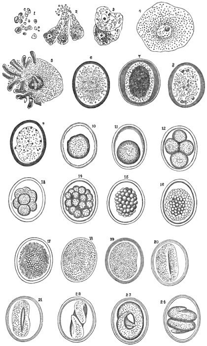
Fig. 49.—Germs and ova of Ascaris mystax. Nos. 1 to 3 magnified 330 diameters and
Nos. 4 to 24 magnified 220 diameters. After Nelson.
Amongst Nelson’s chief opponents was Meissner, who demonstrated
that the unimpregnated ova really possessed a delicate
limiting membrane, and that consequently the action of the
spermatozoa was restricted to that portion of the ovarian ovum
which became exposed by rupture or separation from the rachis.
This opening he termed the micropyle. The union of the
sexual elements is quickly followed by a condensation of the
yolk-granules, and by the disappearance of the hitherto centrally
placed germinal vesicle. The ovum next assumes a distinctly
oval shape, the true yolk-membrane and the external
chorional envelope now becoming more and more differentiated,
until the latter acquires a regularly tuberculated surface. Co-ordinating
with these changes the granular yolk is seen transforming
itself into a single large embryonal cell; after a time
this cell divides and subdivides by the ordinary process of
yolk-segmentation, until it is finally resolved into the condition
of a short, stout, vermiform embryo. The egg having assumed
its definitive oval shape, the intrachorional embryo remains
coiled within the shell, and does not make its escape until the
egg has passed from the body of the parent worm.
Into the question of the mode of formation of the ovarian
ova, and also into that of the development of the spermatozoa,
I do not enter. However unwillingly, I must, in this matter,
be contented to refer to Professor Allen Thomson’s classical
article ovum (quoted below), to Leuckart’s elaborate analysis
(l. c., Bd. ii, s. 76–92), and also, especially, to the exhaustive
memoir of Claparède, whose brilliant labors, like those of Henry
Nelson, were too early terminated by death. Shortly after
graduation Nelson suffered a virtually enforced banishment from
his native land.
Bibliography (No. 33).—Bellingham, O. B., “On the Genus
to which the Worms known as Ascarides belong,” ‘Dublin
Journ.,’ vol. xiv, 1839.—Idem, “Catalogue of Irish Entozoa,”
‘Ann. of Nat. Hist.,’ vols. xiii and xiv, 1843–44; and in the
first part of Charlesworth’s ‘Mag. of Nat. Hist.,’ vol. iv, 1840.
See also the address by Dr E. D. Mapother on the “Lives and
Writings of O’Ferrall and Bellingham,” in the ‘Dubl. Journ.
of Med. Sci.,’ Nov., 1877, p. 471 et seq.—Bischoff, ‘Widerlegung
(u. s. w.),’ Giessen, 1853; quoted by Claparède, l. c.
infra, p. 9.—Idem, ‘Bestätigung (u. s. w.),’ Giessen, 1864.—Idem,
“Ueber Ei-und Samenbildung und Befruchtung bei
Ascaris mystax,” Sieb. and Köll. ‘Zeitsch.,’ 1855, s. 377; also
in S. and K. ‘Zeitsch.,’ 1856.—Bremser, ‘Icones helminth.,’
p. 23, tab. iv.—Claparède, E., “Ueber Eibildung und Befruchtung
bei den Nematoden,” S. and K. ‘Zeitsch.,’ 1857, s. 106.—Idem,
‘De la formation et de la fécondation des œufs chez
les vers Nématodes,’ Genève, 1859. See also ‘Ann. of Nat.
Hist.,’ vol. i, 3rd series, 1858.—Cobbold, in ‘Proceed. of the
Zoological Soc. of London,’ Nov., 1862.—Idem, ‘Brit. Assoc.
Rep.,’ 1862.—Idem, “On the occurrence of Ascaris mystax in
the Human Body,” with figures, ‘Lancet,’ Jan., 1863; and in
the ‘Dublin Med. Press,’ Feb., 1863.—Idem, ‘Entozoa,’ chap.
xi, p. 316, 1864.—Idem, ‘Worms,’ pp. 72 and 112, 1872.—Idem,
in “Obituary Notice of Dr Henry Nelson,” ‘Med. Times and
Gaz.,’ 1865 (?).—Davaine, ‘Traité,’ l. c., 1877.—Diesing, C. M.,
‘Syst. Helm.,’ vol. ii, p. 180, 1850.—Dujardin (l. c., Bibl.
No. 2), p. 162.—Frœlich, in ‘Naturf.,’ xxiv, s. 141 (Asc. felis).—Funke,
O., ‘Lehrbuch (u. s. w.),’ 1857, s. 1299.—Gmelin,
‘Syst. Nat.,’ p. 3031.—Golze, ‘Naturg.,’ l. c., s. 79.—Gurlt,
‘Path. Anat.,’ s. 366.—Heller, A., “Darmschmarotzer,” in
Von Ziemssen’s ‘Handbuch,’ Bd. vii, s. 361.—Idem, ‘Sitzungsb.
d. Erlanger phys.-med. Soc.,’ 1872, s. 73.—Hering, “Ueber das
Vorkommen und die Entwicklung der Ascaris mystax bei jungen
Hunden,” quoted by Leuckart from ‘Würtemb. Naturw.
Jahreshefte,’ 1873, s. 305–337.—Kölliker, in ‘Müller’s Archiv,’
1843, s. 68 et seq.—Leidy, ‘Proc. Acad. Phil.,’ viii, p. 50.—Leuckart,
l. c., Bd. ii, s. 258.—Meissner, G., “Beobachtungen
über das Eindringen der Samenelemente in den Dotter,” S.
and K. ‘Zeitsch.,’ 1854, s. 208.—Morton, T., “Another Example
of the Occurrence of A. mystax, from a Child of fourteen
months old,” in a letter to the ‘Lancet,’ March 11th, 1865,
p. 278.—Nelson, H., “On the Reproduction of Ascaris mystax,”
‘Proc. of the Royal Soc.,’ in ‘Philosoph. Trans.,’ and in ‘Med.-Chir.
Rev.,’ 1051–52; also in ‘Froriep’s Tagsbericht.,’ 1852,
s. 205–207.—Rudolphi, ‘Synops.,’ p. 42, 1819.—Schneider,
“Ueber Bewegung an dem Samenkörperchen der Nematoden,”
in ‘Monatsb. d. Berliner Akad.,’ 1856, s. 192.—Idem, ‘Monographie
der Nematoden,’ Erste Abth., s. 38, und Dritte Abth.,
s. 263 (“Entwicklungsgeschichte”), 1866.—Siebold, ‘Vergleichende
Anatomie,’ 1848, s. 153, and in Burnett’s edit.,
p. 125 et seq., 1854.—Thomson, A., art. “Ovum,” in ‘Todd’s
Cyclop. of Anat. and Phys.,’ supp., 1859.—Idem, “Ueber die
Samenkörperchen, die Eier und die Befruchtung der Ascaris
mystax,” S. and K. ‘Zeitsch.,’ 1856, s. 425.—Idem, “Report
of Glasgow Meeting” (‘Brit. Assoc. Rep.’), 1855, p. 158.
Ascaris maritima, Leuckart.—This is a well-marked species.
Judging from the characters presented by the solitary, sexually-immature
female which supplied Leuckart with his only means
of diagnosis, this worm may be briefly described as a filariform
nematode about 3/4″ in length and about 1/25″ in breadth. Although
there are no cephalic aliform membranes, the cuticle immediately
below the lips forms small and distinct projections,
one on either side of the head (‘Die Mensch. Par.,’ Bd. ii,
s. 877).
This entozoon was discovered by Dr Pfaff at Jacobshavn, near
Godhavn, West Greenland, in April, 1865. Two years later
he sent the specimen to Krabbe, who afterwards transmitted it
to Leuckart. In the original communication addressed to the
Copenhagen helminthologist, Dr Pfaff states that he procured
the worm from amongst matters vomited by a child, and he
incidentally observes that he had hitherto encountered only
Bothriocephalus cordatus and Oxyuris vermicularis amongst
Greenlanders. As to the source of infection, Prof. Leuckart
not unnaturally refers to the similar conditions of existence
shared by the human and carnivorous inhabitants of that
country. It is well known that bears, polar-bears, seals, and
walruses are largely infested by nematodes (Asc. transfuga,
A. osculata, Ophiostoma dispar, &c.), but these various species
are quite distinct from Dr Pfaff’s little “spulwurm.”
Ascaris lumbricoides, Linneus.—This common parasite was
for a long while regarded as identical with the great lumbricoid
of the horse, but the question has been finally settled by
Schneider, who has shown that the human worm, although
identical with Dujardin’s Ascaris suilla of the hog, is nevertheless
quite distinct from the Ascaris megalocephala of solipeds.
The large lumbricoid occasionally found in the ox belongs to
the human worm. Our large human helminth resembles the
common earth-worm in general appearance only. The males
usually measure from four to six inches in length, and the
females from ten to fourteen inches. Some have been reported
up to seventeen or eighteen inches in length. The body is
smooth, fusiform, and elastic, and marked by numerous fine
transverse rings. It is attenuated towards either extremity, the
anterior end terminating in a prominently three-lobed mouth
The tail is bluntly pointed. The female is much shorter than
the male, having a diameter of nearly a quarter of an inch. The
male is supplied with a double spiculum, its tail being always
more or less curved towards the central surface. The female
reproductive orifice is situated above the centre of the body.
According to Schneider, the tail supports from 138 to 150
caudal papillæ, that is, from 69 to 75 on either side of the
median line. Below the anus the papillæ are regularly
arranged in pairs, seven in number, the two uppermost pairs
being double.
Notwithstanding the advantage which the size of this entozoon
affords us in the matter of observation and experiment, we are yet
ignorant as to the precise mode in which the young gain access to
the human body. From what has been said respecting the quick
growth of Ascaris mystax in the dog, and from what has been
observed respecting the rapid growth of the so-called A. suilla
in the hog, we know that the worm requires but a short time to
pass from the larval to the sexual state. The view of Hering,
Mosler, Davaine, and others, who suppose that these worms are
reared in a direct manner by swallowing the ova, is, as Leuckart
observed, not yet proved. We are not in full possession of
the facts of larval development. It is true that Professor
Heller’s interesting “find” has shown that when these worms
first gain access to the human body their size is quite insignificant.
At the post mortem of an imbecile, Heller discovered
eighteen young worms, varying in size from about 1/9″ to 1/2″ in
length (2·75 to 13 mm.). The sexes were indistinguishable.
As a set-off against this, Leuckart’s repeated attempts to rear
Ascaris lumbricoides and A. mystax by means of direct feeding-experiments
with the eggs all failed. Thus, we are yet left in
doubt as to the destiny of the larvæ during the period which
elapses between the time of their escape from the egg and the
time of their entry into the human body. So important is the
question as to the mode of origination, growth, and subsequent
development of the larvæ, that it may be well to trace, however
briefly, what steps have been taken to clear up the matter.
Leuckart obtained his negative results by the administration of
ripe ova to dogs, rabbits, swine, and mice. The eggs of
Ascaris lumbricoides have been kept alive by Dr Davaine for a
period of more than five years. I have myself watched the
development of their contents in fresh water through all the
stages of yolk segmentation up to the stage of an imperfectly-organised,
coiled, intra-chorional embryo, and have kept them
in the latter condition for a period of three months. According
to Davaine (‘Comptes Rendus,’ 1858, p. 1217), the fully-developed
embryo is cylindrical, its length being, 1/100th of an
inch. The mouth is not furnished with the three characteristic
papillæ of the genus, and the tail terminates suddenly in a
point. Davaine administered some of his five-year-old embryos
to rats, and had the satisfaction of finding a few of these eggs
in the rodent’s fæces, with their embryos still living, but
striving to emerge. He also gave eggs to a cow, and introduced
others into the stomachs of dogs in small linen-covered
flasks. As a general result it may be said that the embryos
escaped from their shells. Those eggs, however, in which the
yolk-segmentation had not arrived at the early embryonal stage
remained unaffected. According to Heller, the embryo of
A. lumbricoides casts its first skin while still within the egg,
and “a subsequent ecdysis probably completes its definitive
form” (l. c., s. 615). So far back as 1853 Verloren reared
coiled intra-chorional embryos in the eggs of Ascaris marginata
within a period of fifteen days in distilled water. I also reared
the embryos of this species in fresh water, and kept them alive
for a period of nearly a year and a half, at the expiration of
which time, and during the warm weather, some few of them
succeeded in making their escape. According to Davaine, the
eggs of many nematode species will readily retain their vitality
though long exposed to dryness, but their yolk-contents will
not go on developing during this period of exposure. As
regards A. mystax, however, Heller remarks that whilst “the
eggs have a great power of resisting external influences, their
development is not arrested in spirits of wine, chromic acid, or
oil of turpentine” (l. c., s. 631). In the case of Ascaris
tetraptera of the mouse, embryonic formation goes on in spite
of the absence of external moisture. Davaine has noticed the
same thing in the oxyurides of rodents. Dryness does not
even destroy the eggs of A. lumbricoides and Trichocephalus
dispar. It would seem, in short, that the eggs of nematodes
which normally take up their residence in cats, dogs, and in
the carnivora which reside in arid regions, will develop
embryos in the egg without external moisture. As before
remarked, Davaine thinks it is not necessary that these nematode
embryos should pass through any intermediary bearer, and
he believes that they are often directly transferred to the
stomach of their “hosts” whilst adhering in the form of an
impalpable dust to the coats of their bearers, whence they are
detached by the animal’s frequent habit of licking the fur.
Davaine’s view has received some support from the observations
and experiments of Unterberger with the eggs of Ascaris
maculosa. This observer administered eggs of the worm to
doves (whose fæces were free of eggs), and seventeen days
after found ova in the fæces.
With the eggs of the Ascaris megalocephala of the horse I
performed numerous experiments. I reared the embryos in
simple fresh water, and found them during warm weather
escaping before the expiration of five months. I also succeeded
in rearing these larvæ in pond mud, noticing, at the same
time, that after their escape from the shell they grew more or
less rapidly up to a certain point, after which they ceased
growing. The addition of horses’ dung to soft wet mud in one
case, and of cows’ dung in another, neither appeared to advance
nor retard the process of embryonal formation, so long as the
embryos were enclosed in their shells. On the other hand,
when I reared embryos in simple horse-dung purposely kept
moist, they attained a higher degree of organisation than did
those in wet mud or water. Having watched hundreds of
these larvæ under varying conditions, I came to the conclusion
that, after escape from the egg, their activity, growth, and
strength was most marked when they occupied media which
happened to be impure. Davaine experimented on cows, and
Leuckart also experimented on horses, with the eggs of this
worm without success. Leuckart also failed to rear the larvæ
in intermediary hosts. Some eggs passed through the water-palmer
unaltered.
These results, so far as they go, seem to be borne out by
facts of a professional order. Thus, an instance has been
brought under my notice where a considerable number of
peasants and their children, dwelling in a parish in Yorkshire,
were infested with this worm. There was, in short, a local
endemic helminthiasis. Through the parish runs a stream
which supplies the cottagers with all the water they employ for
domestic purposes (washing, drinking, and so forth). Some of
the peasants living by the side of the stream keep pigs, and
the sewage from this source has been allowed to pass into the
stream itself. Now, if Schneider’s determination as to the
identity of the lumbricoid of man and the pig is correct (which
I do not doubt), the explanation of the cause of the endemic
becomes a very simple matter. But it does not explain all
that we desire to know about the young worms. Either the
freed embryos before they enter the human bearer accomplish
further changes of form and growth in the sewage or impure
water; or, what is far less probable, they pass into the bodies
of intermediary hosts (such as insect-larvæ, Gammari, Entomostraca,
&c.) to undergo the necessary changes. Practically,
no doubt, it comes to the same thing in the end. Even if we
suppose that the Ascaris suilla and A. lumbricoides are not
identical species, still it is evident that any person discharging
the eggs of lumbricoids in the vicinity of open waters becomes,
by that fact, a source and centre of infection. To ensure an
endemic it is probably only further necessary that the human
inhabitants should employ the contaminated water for domestic
purposes. But time and an increase of temperature must be
allowed for the bringing about of those known and unknown
larval changes that alike form the necessary antecedents of
infection. In this connection I will only add, that if the
present position of the question be such as I have here represented
it to be, we see that Mosler was not far wrong when he
suggested that “contamination of the drinking water with the
eggs out of privies is to be blamed” as a source of infection.
According to Heller, from whom I quote, Mosler actually
demonstrated the presence of the eggs in water thus exposed.
In like manner it becomes obvious that Davaine’s practical
remark (although it was based on the assumption of a direct
infection by the eggs), that filtration will probably be sufficient
to prevent infection, loses nothing of its hygienic value.
The foregoing observations naturally lead one to the question
of frequency and distribution. Davaine holds that the comparative
infrequency of this parasite in Paris is due to the free
use of the filter. In London, though not uncommon, the worm
rarely occurs in great numbers in one bearer. Those cases in
our hospitals, where considerable numbers have been present,
have usually come up from suburban or country places. Heller
states that these worms were found in 9·1 per cent. of post
mortems conducted at Dresden, in 12 per cent. at Erlangen,
and in 17 per cent. at Kiel. He quotes Huss as stating that
no one is free from this worm in Finland. The prevalence of
large round worms in warm countries generally is well known.
Throughout India and the East they are extremely abundant,
and the same may be said of the West Indies, Brazil, and the
adjacent territories. Professor Dyce and others have remarked
on the extreme prevalence of lumbrici in the Mauritius, but
they are comparatively rare along the sea border. In all
situations where there is an abundant fresh-water supply these
parasites are particularly common, as in the lowlands of
Holland and the lake districts of Sweden. The abundance of
water is certainly not alone sufficient to explain the frequency
of the parasite, seeing that the most important factor is that
which rests upon the uncivilised habits of the rural population.
What, therefore, it may be asked, can be the cause of immunity
enjoyed by Icelanders in this respect? The answer is not
apparent; nevertheless Krabbe and Finsen have testified to the
fact that Iceland is the only country that is entirely free from
Ascaris lumbricoides.
As remarked in my previous work the number of worms
present in any human bearer is usually small, varying commonly
from one to six or eight. Cases in which scores or hundreds
have existed are comparatively rare. Küchenmeister mentions
the case of one child who passed 103 examples, and of another
child that harbored from 300 to 400 worms. Dr Gilli, of
Turin, gives a case where 510 were passed by a child, and
Cruveilhier estimated that over 1000 existed in an idiot girl,
whose intestines he found crammed with them. A remarkable
case has also been communicated to me by Dr Mackeith, of Sandhurst,
Kent, who, by means of santonine, expelled from a little
girl, five and a half years of age, 300 lumbrici; and I am likewise
indebted to Dr Cooper Rose for notes of a case in which
about thirty lumbrici were expelled, chiefly in consequence of
the employment of this drug. The most interesting fact, however,
in this case was that the child was only fifteen months old.
In this case the symptoms were severe.
The proper habitat of the lumbricus is the upper and middle
part of the small intestine. From this situation it often wanders
into the stomach, and frequently gains access to the outer world,
not only by the natural passages of the mouth, nostrils, and
anus, but also, occasionally, in a more direct way, by perforating
the intestinal and abdominal walls. Many cases are on record
where lumbrici have passed into the abdominal cavity. In other
instances they have lodged themselves within the abdominal
viscera and pulmonary organs. When they find their way into
the parietes of the abdomen and adjacent parts, they usually give
rise to the formation of abscesses requiring surgical interference.
As regards the symptoms produced by lumbrici, these vary
according to the situation they happen to occupy. The symptoms
are also modified by age and temperament. In the
stomach and intestines they give rise to colic and shooting pains
about the abdomen, followed generally by dyspepsia, nasal itching,
nausea, vomiting, and even diarrhœa. Occasionally death
supervenes suddenly. A singular case of this kind (the particulars
of which I only gathered from a local newspaper) occurred
in a boy, thirteen years of age, at the County Gaol at Hertford,
in 1873. From Dr Evans’s statement, made at the coroner’s
inquest, the sole cause of death appeared to be due to pressure
on the windpipe by a worm lodged in the gullet. Sometimes
there is cerebral disturbance, attended with general restlessness
and convulsive twitchings during sleep. Thus, Dr Woodman
has recorded a serious case of convulsions arising from lumbricoid
worms, in which, however, a cure was effected by expulsion
of the worms. An anonymous writer in the ‘Medical
Gazette’ records a case of epilepsy from this cause, whilst
another writer in the same journal (1839) mentions an instance
where two lumbrici and one tapeworm were associated in the
production of similar phenomena. But a much more striking
case is also given (anonymously) in the ‘Gazette’ for 1874 (p.
415), where a single lumbricus caused the bearer to be a lunatic
for eight years. The victim suffered from cataleptic fits, which
lasted for two or three weeks at a time. M. Petrequin, in his
‘Traité Pratique,’ records two cases of amaurosis in young girls
produced by lumbrici. A fatal case is recorded by Petrenz,
where 200 worms produced enteritis, and another fatal case is
given by Roger from perforation (1848). Cases of perforation
are also given by Young, by Blair (1861), by Mondière (1839),
by Buchner (1851), by Sheppard (1861), and by Luschka
(1854), the worms in this last-mentioned case occupying the
cavity of the pleura. Cases of severe irritation affecting the
genito-urinary organs are given by Dreyfus, Buckingham, and
others; and one or two instances are reported where these
worms have been discharged from several parts of the body
(Neilson, 1833). I may add that the third fasciculus of a work
illustrating the collection of morbid anatomy in the Army
Medical Museum at Chatham gives a case of lumbrici occupying
the biliary ducts and gall-bladder. I find, moreover, two
additional cases of perforation of the small intestine, one of
which appeared in the ‘London Medical Gazette’ (1827) and
the other in the ‘Lancet’ (1836).
During the Franco-German war Dr Reginald Pierson, as he
afterwards informed me, removed a lumbricus from an abscess
formed in the abdominal parietes of a soldier. But amongst
the most curious cases (illustrating the wandering habits of these
parasites) are those severally described by Barwell (1857),
Williams, Prichard, and the Messrs Stockbridge. In Barwell’s
case an Ascaris was expelled from a child who had swallowed
the brass “eye” of a lady’s dress. Through the circular loop of
this eye, used as a toy, the Ascaris had partly thrust its body, and
becoming thus strangulated, it probably perished before it was
evacuated. In Prichard’s case (1859) one or two lumbrici had
similarly trapped themselves in the eyes of buttons swallowed
by the patient, and one worm, not contented with a single
strangulation, had succeeded in passing its body through two
buttons. In 1842 Mr T. G. Stockbridge gave a similar case,
in which he, not inaptly, spoke of these “hooks and eyes” as
constituting a new remedy or “worm-trap” for lumbricus,
and singularly enough, a namesake (W. Stockbridge), in the
succeeding year, also recorded a like instance of the “mechanical
expulsion of worms” by metallic buttons. Again, a third
correspondent in the ‘Boston Journal,’ under the initials A. M.,
spoke of an open-topped thimble as constituting another new
“worm-trap,” whilst he gave a case of lumbrici penetrating
“metallic suspensor buttons.” There is also the case reported by
Williams, who, at a meeting of the Boston Society for Medical
Improvement, exhibited “a lumbricus with a dress-hook
attached” (1857). Lastly, another lumbricus, trapped in the
same way, may be seen in the Museum of the Royal College of
Surgeons at Edinburgh.
Owing to the presence of a peculiar irritating vapour which
is given out by these lumbricoids, particularly when fresh,
several observers have experienced curious symptoms. Thus,
Miram on two occasions, when examining A. megalocephala, was
attacked with sneezing, excessive secretion of tears, with swelling
of the puncta lacrymalia, and Huber also experienced a troublesome
itching of the hands and neck after examining specimens
of A. lumbricoides. In like manner I have myself had watery
suffusion of the eyes (when collecting the perivisceral fluid for
Marcet’s analyses: see Bibliog.), and Bastian has given a detailed
account of the serious effects which the poison produced upon
him. In Bastian’s case even spirit specimens produced irritation.
The attacks of catarrh and asthma were so persistent
and severe that they lasted for six weeks at a time. So sensitive
was Bastian to the lumbricoid-miasm that he could not
even put on a coat that he had worn during his investigations
without experiencing fresh attacks of sneezing and other
catarrhal symptoms. The attacks became periodical, occurring
between five and six in the morning, being accompanied by
dyspnœa and a distressing spasmodic cough. Bastian, in short,
was quite a martyr in the cause of nematode anatomy.
Bibliography (No. 34).—Abousson, L., “On the Presence of
Worms (lumbrici) in the Air-passages,” from ‘Arch. Gén. de
Méd.,’ in ‘Med.-Chir. Rev.,’ 1836.—(Anonymous), A. M.,
“Another New Worm-trap—an open-topped Thimble in the
Nostril (also notice of metallic suspender buttons penetrated by
Lumbrici),” ‘Bost. Med. and Surg. Journ.,’ vol. xxvii, p. 121,
1842–43; see also T. G. and W. Stockbridge.—(Anon.), “Lumbrici
expelled by Bismuth,” ‘Bost. M. and S. Journ.’ (from
‘Gaz. des Hôp.,’ ‘Journ. des Connaiss. Méd.,’ and ‘Boletin del
Inst.-Med.-Valenc.’), 1859.—(Anon.), “Case of one Tapeworm
and two Lumbrici causing Epilepsy,” from ‘Bull. du Midi’
and ‘Gaz. Méd.,’ 1839.—(Anon.), “Case of Perforation of
the Ileum by Ascarides,” from ‘Hufl. and Ossan’s Journ.,’ in the
‘Lancet,’ 1836.—(Anon.), “Case of Lumbrici in the Biliary the
Ducts and Gall-bladder,” note and fig. in third fasc., illust. the
Coll. of Morb. Anat. in the Army Med. Mus. at Chatham, 1838.—(Anon.),
“A Lumbricus causing Catalepsy, with Fits lasting
two or three weeks; Cure by Vomiting,” ‘Lond. Med. Gaz.,’
1847, p. 415.—Archer, E., “On a Case of A. lumbricoides producing
alarming symptoms,” ‘Lancet,’ 1857.—Barwell, “Case
of Ascaris expelled by the swallowing of a foreign body,”
‘Lancet,’ 1857.—Bastian, H. C., “On the Anatomy and Physiology
of the Nematoids, Parasitic and Free,” ‘Phil. Trans.,’
1866, p. 545; for the account of his poison-symptoms, see footnote,
p. 583.—Batterbury, R. L., “Jaundice due to the presence
of Lumbrici,” ‘British Med. Journ.,’ Nov., 1878, p. 721.—Bigelow,
H., “Worm in an Abscess,” ‘Bost. Med. and Surg.
Journ.,’ vol. xxxiii, p. 486, 1836.—Blatchley, C. C., “Two Cases
of A. lumbricoides, attended with Abscesses, followed by large
purulent discharges, and Worms therein,” ‘New York Med.
and Phys. Journ.,’ vol. i, new series, p. 209, 1829.—Bonfils, E.,
“Lesions and Path. Phenomena caused by Lumbrici in the
Biliary Ducts,” from ‘Arch. Gén.,’ in ‘Brit. and For. Med.-Chir.
Rev.,’ 1858, and in ‘Amer. Journ. of Med. Sci.,’ vol.
xxxvii, 1859.—Bradford, J. T., “Singular Case of Worms
(Lumbrici),” ‘Bost. Med. and Surg. Journ.,’ vol. xxviii, 1843.—Brigham,
A., “Worms in the Bladder simulating ‘Stone,’”
‘Amer. Journ. Med. Sci.,’ 1837; ‘Med.-Chir. Rev.,’ 1837;
‘Quart. Journ. Calcutta Med. and Phys. Soc.,’ vol. ii, p. 132,
1838.—Buchner, “On the Perforation of the Intestinal Canal by
Worms (with ref. to two cases),” from ‘Med. Zeitung,’ 1850,
in ‘Med.-Chir. Rev.,’ 1851.—Calderwood, “Treatment,” ‘Brit.
Med. Journ.,’ Jan. 30, 1875.—Chapman, N., “Case of 68
Ascarides causing Pulmonary Disease,” in his ‘Dis. of the
Thoracic and Abd. Viscera,’ p. 263, and in ‘Med.-Chir. Rev.,’
1845.—Chiaje, Delle-, in ‘Rend. dell’ Accad. di Napoli,’ 1846
(“Anat.,” p. 403).—Church, J., “On A. lumbricoides,” ‘Mem.
Med. Soc. Lond.,’ vol. ii, 1789.—Claparède (l. c., Bibl. No. 33,
for development).—Clark, P., “Discharge of a Lumbricus
through the Male Urethra,” ‘New York Journ. Med.,’ 1844,
rep. in ‘Lancet,’ 1844, and in ‘Edin. M. and S. Journ.,’ vol.
lxiv, 1845.—Cloquet, ‘Anat. des vers Intest.,’ 1824.—Cobbold,
“On Sewage and Parasites, especially in relation to the
Dispersion and Vitality of the Germs of Entozoa,” ‘Med. Times
and Gaz.,’ Feb. 25, 1871, p. 215.—Idem, ‘Entoz.,’ p. 302–315.—Idem,
‘Worms,’ lect. xvi, p. 3.—Idem, art. “Ascaridæ,” in
‘Maunder’s Treasury,’ 1862.—Colvan, J., “Case in which Eleven
Round Worms of the species A. lumbricoides were removed
by Anthelmintics,” ‘Dubl. Med. Press,’ vol. xxvi, p. 211, 1851.—Cutler,
J. H., “Death by Worms (a large Lumbricus being
found in the Wind-pipe),” ‘Bost. Med. and Surg. Journ.,’
vol. lxvi, p. 392, 1862.—Czermak, in ‘Sitz. d. k. Akad. d.
Wissensch.,’ 1852 (“Anat.,” s. 755).—Davaine, in his ‘Traité,’
l. c., 2nd edit., syn. xcvii, and p. 122–235 (with details of forty-five
cases); see also his memoir “On the Development and
Propagation of the Trichoceph. dispar and A. lumbricoides,”
from ‘Comptes Rendus,’ in ‘Ann. Nat. Hist.,’ vol. ii, 3rd series,
1858; also in the ‘Journ. of Pract. Med. and Surg.,’ Eng. edit.,
vol. i, 1858, and in the ‘Veterinarian,’ vol. xxxii, p. 700, 1859,
from ‘Proc. of Acad. des Sci.,’ in ‘Bost. M. and S. Journ.,’
vol. lix, p. 157, 1858–59.—Idem, art. “Entozoaires,” in ‘Dict.
de Méd. et Chir. prat.’—David, J. B., “Cases of Perforation of
the Intestines by Worms,” from ‘Gaz. Méd. de Paris,’ in ‘Dubl.
Med. Press,’ 1840, p. 223.—Diesing, ‘Syst. Helm.,’ ii, p. 166;
and in ‘Revis der Nemat.,’ l. c., s. 660.—Douglas, J., “Worms
(Lumbrici) evacuated at an Ulcer of the Groin,” ‘Med. Ess.
and Obs.,’ vol. i, 2nd edit. (vol. i, 5th edit., p. 179), p. 222,
1737.—Dowler, B., “Case of Worms in the Urinary Bladder,”
from ‘New Orl. M. and S. Journ.,’ in ‘New York Journ. Med.,’
new series, vol. xiv, 1855.—Dubini, ‘Entozoografia umana’
(“Anat.,” p. 148).—Dupuytren, “Lumbricus passed by the
Urethra,” from “Clin. Lect.,” in ‘Lond. Med. and Surg. Journ.,’
1846, p. 14.—Dyce, R., “On Lumbrici and the Causes of their
Prevalence in the Mauritius,” ‘Lond. Med. Gaz.,’ 1834.—Evans,
T., “Lumbricus causing Death,” rep. of coroner’s inquest in
the ‘Herts Advertiser and St Alban’s Times’ for Feb. 8, 1873.—Gervais
(and Van Beneden), ‘Zool. Med.,’ ii, p. 118.—Gilli,
“Account of a Case in which 510 Worms (Lumbrici) were
voided by a Child,” from ‘Giorn. d. Scienze Med. di Torino,’
in ‘Med.-Chir. Rev.,’ 1843.—Goopta, G. D. D., “On Suicide
and Lumbrici,” ‘Ind. Med. Gaz.,’ July, 1874, and ‘Lond. Med.
Rec.,’ Aug., 1874, p. 502.—Heller, A., “Darmschmarotzer,” in
Von Ziemssen’s ‘Handb.,’ s. 612–631.—Holland, G. C., “A
peculiar Case of Nervous Disease or Derangement of the Nervous
System (associated with A. lumbricoides),” ‘Edin. M. and S.
Journ.,’ vol. lxiii, 1845.—Howall, “Abscess of the Groin, with
discharge of Lumbrici,” ‘Lond. Med. Gaz.,’ 1845, and ‘Edin.
M. and S. Journ.,’ 1846, p. 241.—Johnson, W. G., “Case of
forty Lumbrici in a Boy who died with Traumatic Tetanus,”
“Rep. of South Mid. Br. of Brit. Med. Assoc.,” in ‘Brit. Med.
Journ.,’ 1858.—Kell, “Perforation of the Intestines by a
Worm,” ‘Lond. Med. Gaz.,’ 1828.—Kilgour, T., “Case in
which Worms in the Nose, productive of alarming Symptoms,
were removed by the Use of Tobacco,” ‘Med. Comment.,’
vol. viii, 1783.—Kirkland, “Case of Lumbricus in an Abscess
of the Liver,” rep. in his book, entitled ‘An Enquiry,’ vol. ii,
p. 186 (quoted by Richter and Davaine), London, 1786.—Küchenmeister,
‘Manual,’ Eng. edit., p. 410–427.—Leidy, J.,
‘Proc. Acad. Phil.,’ 1856, p. 50.—Lente, F. D., “Lumbricus in
the Stomach causing Dyspnœa,” in his “Rep. of Cases occurring
in the New York Hosp.,” in ‘New York Journ. of Med.,’ vol. v,
new series, p. 167, 1850.—Lettsom, “Case of Lumbricus evacuated
from an Abdominal Abscess,” ‘Trans. Med. Soc. Lond.,’
and ‘Lond. Med. Repos.,’ 1817.—Leuckart, l. c., s. 152–258.—Lieberkühn,
in ‘Miller’s Arch.’ (“Anat. of A. suilla”), 1855, s. 331.—Luschka,
“Case of Lumbrici within the Pleura,” from ‘Virch.
Arch.,’ in ‘Med.-Chir. Rev.,’ 1854.—Lowne, B. T., “The
Anatomy of the Round Worm,” ‘Trans. Roy. Micr. Soc.,’ 1871,
p. 55.—Maesson, “On a Worm found in the Bubo of a Woman
36 years of age;” see ‘Entozoa and Parasites, being a ref.
to numerous papers;’ from “Valentin’s Repertorium,” in
‘Month. Journ. of Med. Sci.,’ vol. ii, p. 559, 1842; also in
‘Micr. Journ. and Struct. Rec.,’ p. 85, 1842.—Marcet, W.,
“Chemical Exam. of the Fluid from the Peritoneal Cavity of
A. megalocephala,” ‘Proc. Roy. Soc.,’ 1862, No. 72, p. 69.—Martin,
D. T., “Large number of Worms (140 examples of
A. lumbricoides) discharged from a Child five years old,” rep.
from the “Stethoscope,” in ‘Bost. Med. and Surg. Journ.,’
vol. xliv, p. 301, 1851.—Mattei, R., “On a Case of two Lumbricoid
Worms, which had penetrated during life into the liver,
and were demonstrated by Prof. G. Pelizzari to his pupils in
the Sch. of Path. Anat. of Florence,” from ‘Gaz. Med. Ital.
Toscana,’ in ‘Dubl. Quart. Journ.,’ vol. xxiv, 1857.—Michel,
“Case of Epilepsy in a Girl ten years of age, caused by Lumbrici,”
from ‘Journ. des Connaiss. Méd.,’ in ‘Amer. Journ. of
Med. Sci.,’ vol. vi, p. 451, 1843.—M’Laggan, J., “Gangrenous
Sore from a large Worm in the Parietes of the Abdomen,”
‘Med. Comment.,’ vol. ii, 1774, p. 80.—Molin, in ‘Sitzungsb.
d. k. Akad.,’ 1859, s. 23.—Mondière, “On Perforation by Worms
(three cases),” from ‘L’Expérience,’ in ‘Med.-Chir. Rev.,’ 1839.—Moore,
E. D., “Example of A. lumbr. ejected by the Mouth,”
‘Prov. Med. and Surg. Journ.,’ 1852.—Morgan, J., “Case of
Perforation of the Stomach, probably by a (Lumbricus) Worm,”
‘Lancet,’ 1836.—Morland, W. W., “Ejection of numerous
Lumbrici from the Mouth, impaction of the small intestine
with Lumbrici, (of which 365 were removed post mortem),” ‘Bost.
M. and S. Journ.,’ vol. lvi, 1857.—Idem., an “A. lumbr. of unusual
size (over 17 inches long),” ‘Rep. of Bost. Soc. for Improv.,’
in ‘Bost. M. and S. Journ.,’ vol. lviii, p. 62, 1858.—Neilson,
“Discharge of Worms from various parts of the Body,” ‘Med.-Chir.
Rev.,’ and ‘Lond. Med. Gaz.,’ 1833.—Omond, R., “Case
of Lumbricus attended with Hæmoptysis,” ‘Edin. Med. Journ.,’
1856.—Owen, R., art. “Entozoa,” l. c.—Padley, G., “Jaundice
and Lumbrici,” ‘British Med. Journ.,’ Dec. 14, 1878, p. 877.—Petrenz,
“Case of fatal Enteritis produced by (200) Lumbrici,”
from ‘Clarus and Radius’ Beitr. zur Pract. Heilk.,’ in ‘Dubl.
Journ.,’ vol. xi, 1837; also in ‘Lond. Med. Gaz.,’ 1837.—Playfair,
“Case of Lumbricus (69 specimens) cured by the Mudar,”
‘Calc. Med. and Phys. Soc. Trans.,’ vol. ii, p. 407, 1826.—Pomeroy,
C. G., “Escape of Worms (17 Lumbrici) from the
Navel of a Child,” ‘Bost. M. and S. Journ.,’ vol. xxi, 1840.—Prichard,
A., “Case of Lumbricus,” ‘Rep. of East York and
North Lincoln Br. of Brit. Med. Assoc.,’ in ‘Brit. Med. Journ.,’
1859.—Royer, “Case of Intestinal Perforation by a Lumbricus,”
report in ‘Lancet,’ 1856.—Rumsey, N., “Cases of Lumbricus
and Tænia associated with Hæmoptysis,” ‘Med.-Chir. Trans.,’
1818.—Sandwith, H., “Remarks on Worms in the Peritoneal
Cavity, with a case,” ‘Brit. Med. Journ.,’ 1861.—Schleifer,
“Case of a Deaf and Dumb Child restored after the discharge
of Worms (87 Lumbrici and innumerable Oxyurides),” from
‘Œsterr. Med. Wochensch.,’ in ‘Amer. Journ. of Med. Sci.,’
vol. viii, p. 473, 1844.—Schneider, A., ‘Monog. der Nemat.,’
s. 36.—Idem, “On the Nervous System of Nematoda,” from
the German by Busk, ‘Quart. Journ. Micr. Sci.,’ 1863.—Schultze,
“Case of Stuttering occasioned by Worms,” from
‘Med. Zeit.,’ in ‘Med.-Chir. Rev.,’ 1837.—Sheppard, “Case of
A. lumbr. extracted from an Abdominal Abscess,” ‘Brit. Med.
Journ.,’ 1861.—Smith, J. N., “Thirty-nine Specimens of Ascaris
lumbricoides in a Child,” ‘Bost. M. and S. Journ.,’ 1856.—Spalding,
P., “Case of Worms (100 Lumbrici),” ibid., 1839.—Stockbridge,
T. G., “Worm-trap (hooks and eyes), a New
Remedy,” ibid., vol. xxvii, p. 73, 1842–43; see also Anon.,
A. M.—Stockbridge, W., “Mechanical Expulsion of Worms (by
metallic buttons),” ibid., vol. xxviii, p. 419, 1843.—Van Beneden,
‘Animal Parasites,’ l. c., p. 95; see also Gervais.—Villemin,
“Case of Death from Worms (about 18 specimens of A. lumbr.),”
from ‘L’Union Méd.,’ in the ‘Lancet,’ and rep. in ‘Dubl. Med.
Press,’ vol. xxxv, p. 327, 1856.—Weinland, in his ‘Essay,’ l. c.,
p. 88, and in ‘Troschel’s Arch.,’ 1859, s. 283.—Welsh, T.,
“Curious Facts respecting (symptoms produced by) Worms
(A. lumbricoides),” art. ix in the ‘Med. Papers communicated
to the Massachus. Med. Soc.,’ vol. i, p. 87, 1790.—Wendelboe,
“Case of discharge of Worms (Ascarides?) through the Skin,”
from ‘Rep. of Roy. Soc. Copenhagen,’ in ‘Lancet,’ 1836; see
also Neilson.—Williams, H. W., “Exhibition of a Lumbricus
with a dress-hook attached,” ‘Rep. of Bost. Soc. for Med.
Improv.,’ in ‘Bost. M. and S. Journ.,’ vol. lvi, p. 163, 1857.—Wilson,
J., “On the prevalence of Lumbrici in China,” in his
‘Med. Notes on China,’ London, 1846.—Woodman, W. B.,
“Case of Convulsions, &c., arising from Lumbricoid Worms,”
‘Med. Times and Gaz.,’ 1863.—Young, W., “Cases in which
Lumbrici were evacuated by Ulceration through the Parietes of
the Abdomen,” ‘Lond. Med. Gaz.,’ from ‘Glasgow Med. Journ.,’
1828; rep. in ‘Lond. Med. and Surg. Journ.,’ vol. i, p. 564,
1828.
Echinorhynchus gigas, Goeze.—There is but one recorded
instance of the occurrence of this entozoon in the human body.
This is the oft-quoted case by Lambl, given in the ‘Prager
Vierteljahrschrift’ for 1859. Lambl, indeed, described it as a
separate species (E. hominis), but as the worm was a sexually-immature
female, its identification with E. gigas, notwithstanding
Schneider’s great authority, can hardly be regarded
as absolutely certain. The worm was found in the small
intestine of a boy of nine years, and measured only rather more
than the fifth of an inch in length. As Leuckart hints, the
worm may be Echinorhynchus angustatus, or possibly the E.
spirula, a species found in various South American apes (Cebus
and Jacchus), and also in the Barbary ape (Inuus).
In 1872, Welch, unaware of Lambl’s case, announced the
discovery of “the presence of an encysted Echinorhynchus in
man.” The minute parasite found by him occurred in a
soldier, thirty-four years of age, who died at Netley, but who had
contracted the worm in India. “It was situated in the
jejunum, immediately beneath the mucous coat, and formed an
oval prominence in the interior of the gut.” Speaking with
great confidence, this able microscopist further remarks:—“The
character and arrangement of the hooklets unequivocally
shadowed forth a species of Echinorhynchus for the first time
discovered as a representative of the Acanthocephala in the
human body.” Along with his elaborate description Welch
gives several figures; but these, so far from producing conviction
as to the accuracy of his inferences, have unfortunately
led me to believe that the parasite in question would be more
properly referred to the Pentastomidæ. But for Heller’s
acquiescence I might have more fittingly noticed this worm
elsewhere. Davaine falls into the same view, and moreover
accepts Lewis’s “Echinorhynque du Chien,” which I have
shown to be a nematode (Cheiracanthus robustus). It is thus
that serious errors creep into the literature of parasitism.
In the adult state the female Echinorhynchus gigas is a
huge species, occasionally reaching two feet in length, with a
breadth of one third of an inch. The male rarely exceeds
three inches. This worm is common in swine, both wild and
domesticated. According to Schneider the embryos take up
their residence in the grubs or larvæ of the cockchafer (Melolontha
vulgaris), a discovery which very readily explains the
manner in which hogs become infested. Whether E. gigas be
a human parasite or not, it is certainly very injurious, not to
say destructive, to swine. Although this parasite must be
quite common in England I have experienced great difficulty
in procuring specimens. In the second book of this work I
shall give some interesting particulars furnished by the memoir
of Prof. Verrill and privately by Mr George Wilkins. (See
‘Parasites of the Pachydermata’.)
Bibliography (No. 35).—Blanchard, in ‘Cuvier’s Règne
Animal,’ tab. 35 (good fig.), and in ‘Ann. d. Sci. Nat.,’ ser. xii.—Bremser,
‘Icones,’ tab. vi.—Cobbold, “Parasites of the Hog,”
the ‘Veterinarian,’ 1875.—Idem, ‘Manual,’ l. c., p. 123.—Davaine,
l. c., ‘Syn.,’ p. 83.—Diesing, l. c., ii, p. 2.—Dujardin,
l. c., p. 503.—Goeze, l. c., s. 143 (good figs.).—Gurlt, l. c.,
s. 367.—Heller, ‘Darmschmarotzer,’ l. c., s. 663.—Lambl, l. c.,
supra, Feb., 1859.—Leuckart, l. c., Bd. ii, s. 729; also in ‘Bibl.
Univ.’ for March, 1863, and in ‘Ann. Nat. Hist.,’ vol. xii, 1863.—Owen,
l. c., in ‘Todd’s Cyclop.’ (figs. after Cloquet).—Rudolphi,
‘Synops.,’ pp. 63 and 310.—Schneider, in ‘Arch. f.
Anat. und Phys.,’ 1868.—Idem, in ‘Sitzungsb. der Oberhess.
Gesellsch. f. Nat.,’ &c., 1874 (quoted by Leuckart); see also
‘Ann. Nat. Hist.,’ 4th series, vol. vii, p. 441, 1871.—Verrill,
‘The external and internal Parasites,’ &c., l. c., p. 109.—Welch,
“The presence of an Encysted Echinorhynchus in Man,”
‘Lancet,’ Nov. 16, p. 703, 1872.—Westrumb, ‘De Helm. Acanth.’
(good figs.), 1821.
As explained in the Introduction we must regard the Leeches
and many allied forms of Suctorial Annelids as creatures possessed
of semi-parasitic habits. They are, perhaps, something
more than what Van Beneden styles “free parasites”—an expression
which almost looks like a contradiction of terms. I cannot
here, however, stop to discuss questions which lie, as it were, on
the border-land of parasitology. Three species of leech are more
or less commonly employed in medicine. These are the grey leech
(Sanguisuga medicinalis, Savigny), the green-leech (S. officinalis,
Sav.), and the dragon-leech (S. interrupta, Moq.-Tandon).
The two former abound in Central and Southern Europe, being
also present in North Africa, the last named inhabiting Barbary
and Algeria. So abundant are leeches in the country bordering
the Mediterranean that during the invasion of Egypt by Napoleon
the French soldiers suffered seriously from their attacks.
When the men lay down to drink, the leeches (Hæmopis sanguisorba,
Sav.) affixed themselves to their mouths and nostrils,
producing serious distress. They also attacked horses, camels,
and cattle. In like manner the Ceylon and Philippine Island
leeches (S. ceylonica, Moq.-Tand., or S. tagalla, Meyen), of which
there are several varieties, prove exceedingly troublesome to
Europeans. These leeches, not being aquatic forms, occupy
woods and damp places. Unless the limbs of travellers are
well protected, the presence of the blood-suckers is soon discovered
by the trickling of blood from the limbs and lower part
of the body. The leeches even sometimes creep up to the neck
and other adjacent parts. These “free parasites” also attack
horses, causing much loss of blood. Terrestrial leeches
abound more or less in all warm countries. Sir J. Hooker
encountered them in the Himalayas, and they are common in
China, Japan, Java (S. Javonica, Wahlberg), and other eastern
parts. They likewise abound in Brazil and Chili. The American
leeches for the most part belong to the genus Hæmenteria
(H. Mexicana, H. officinalis, and H. Ghiliani, Filippi). The
last named is common in Brazil, the other two being Mexican
forms. Another species, which is blind, has been found in
Brazil by F. Müller (Cyclobdella lumbricoides). Not only the
above-named species, but also many other kinds of leeches are
in the habit of attacking man and the domestic animals, but
the subject is too extended and special to be fully dealt with
in this work. Almost a legion of species are known as externally
parasitic upon Fishes, Chelonian and Batrachian reptiles,
Crustaceans, and Echinoderms.
Bibliography (No. 36).—Blainville, ‘Dict. des Sci. Nat.,’
tom. xlvii, p. 257.—Brandt (und Ratzeburg), ‘Medicin. Zoologie,’
Bd. ii.—Brightwell, ‘Ann. and Mag. Nat. Hist.,’ ix, 1842.—Diesing,
‘Syst.,’ vol. i, p. 465, and “Revis. der Myzelminth.
(Abth. Bdellideen),” in ‘Sitzungsb. der math.-nat. Cl. d. k.
Akad. der Wissensch.,’ Bd. xxxiii, s. 473.—Ebrard, ‘Compt.
Rend.,’ 1856, p. 1012.—Idem, ‘Monogr. des sangues Méd.,’
1857.—Filippi, De, ‘Mem. Accad., &c., Torino,’ and in ‘S. und
K. Zeitsch.,’ 1829.—Idem, “Nuovo genere,” &c., in ‘Gaz.
Med. Lombard,’ 1849.—Grube, ‘Fam. d. Annelid.,’ s. 109.—Hofmeister,
in ‘Burmeister’s Zeitung für Zool.,’ 1848.—Johnson,
‘Treatise on the Medicinal Leech.’—Leuckart, l. c., Bd. ii,
s. 634–739 (with many refs.)—Leydig (“Anat.”), ‘S. und K.
Zeitsch.,’ Bd. i.—Moquin-Tandon, ‘Monogr. de la fam. des
Hirudinées,’ 1846.—Idem, in his ‘Medical Zoology’ (Hulme’s
edit.), 1861, p. 137.—Müller, F., in ‘Archiv f. Naturg.,’ 1846.—Pereira,
in his ‘Materia Med.,’ vol. ii, p. 2197, 1853.—Savigny,
‘Descript. de l’Egypte,’ 2nd edit.—Idem, ‘Syst. des
Annélides,’ 1820.—Schmarda, ‘Neue wirbell. Thiere,’ Bd. i
(quoted by Leuckart).—Virey (and Serullas), in ‘Journ. Pharm.,’
1829, p. 614.—Wagener, in ‘Troschel’s Archiv,’ 1858, Bd. i,
s. 244 et seq.—Wahlberg, in ‘Œfvers. Kongl. Vetensk. Akad.
Forhand.,’ Stockholm, 1855.
The Trachearian division of the Arachnida comprises a few
internal parasites that attack man, and many ectozoa which are
parasitic upon man and animals. The species can only be
noticed very briefly.
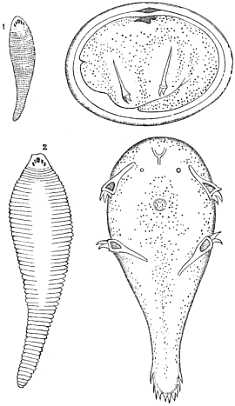
Fig. 50.—Pentastoma tænioides. (1) Male and (2) female, of
the natural size. The egg and embryo highly magnified.
After Leuckart.
Pentastoma tænioides, Rudolphi.—In the system of classification
adopted by Diesing, this entozoon and its allies are placed
in the division Cephalocotyleen and therefore, in association
with the Cestodes, with which, however, it has no structural
affinity. It was long ago pointed out by Van Beneden, T. D.
Schubart, Leuckart, and others, that the pentastomes were
Acarine and Lernæan Arthropods; the genus being osculant
between the Acaridæ and Lernæidæ. The whole subject is
discussed in Leuckart’s profound memoir quoted below.
The adult Pentastoma tænioides is characterised by the
possession of a vermiform, lancet-shaped body, flattened at the
ventral surface, attenuated posteriorly, and marked transversely
by about ninety rings (fig. 50, 1 and 2). The cephalo-thoracic
segments are continuous with the body, each supporting a pair
of strong retractile chitinous claws; four in all. The head is
truncated, furnished with an oval mouth, armed with a horny
lip. The integument of the body is perforated with numerous
respiratory openings or stigmata. These are wanting in the
cephalic segment. In the larval state (══ Pent. denticulatum)
the body is armed with numerous rows of small, sharply pointed
spines. The adult female
measures from three to
four inches in length, but
the male is only about
an inch long. The genital
aperture of the female is
situated at the extremity
of the tail, that of the
male being placed at the
front part of the abdomen
in the middle line. The
mode of reproduction is
oviparous, accompanied
by a subsequent and complete
metamorphosis.
In the mature condition
this parasite infests
the nostrils, and frontal
sinuses of the dog and
wolf, and also, though
more rarely, the nasal
cavities of the horse and
sheep. In the pupal and
larval states it sometimes
occurs in the abdominal
and thoracic cavities of
the human body, but it is
more frequently found in herbivorous mammals, such as the
sheep, deer, antelope, peccary, porcupine, guinea-pig, hare, and
rat. According to Creplin, it infests the domestic cat. In
these animals and in man the young worms occupy little cysts
within or upon the peripheral parts of the liver and lungs.
I have occasionally found them free in the cavities of the
abdomen and pleura.
In the course of the development of this entozoon, Leuckart
recognises four well-marked stages. The first is that of the
embryo with a boring apparatus. In the second stage, the
embryo has become transformed into a motionless pupa. The
third is the ordinary larval condition characterised by numerous
rows of small spines in addition to two pairs of double claws.
The fourth is the sexually-developed stage, furnished with a
simple hook-apparatus, and without integumentary denticles.
“Our Pentastomes, therefore,”
says Leuckart, “exhibit
two kinds of larval
forms, an earlier and later
one, such as takes place
in other animals; this also
occurs even in insects
(Strepsiptera and Meloidæ),
only that, in our case (i.e.
in Pentastoma), both do
not immediately follow one
another, but are separated
by a resting condition,
which I have designated
as the pupa stage. In
choosing this name I do
not mean to express a complete identity of this intermediate
state with the pupal sleep of insects.”
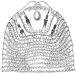
Fig. 51.—Upper third of the body of Pentastoma
denticulatum. Original.
So far as my own observations extend, the pupa, in its later
stages, closely resembles the free larva; but, as Leuckart points
out, the earlier stages are very different. The embryo, after
encystation, repeatedly casts its skin, and during the intervals
of these several successive moultings, the young animal makes
rapid growth, accompanied by a series of structural changes.
Passing through these it at length acquires the perfected larval
state (P. denticulatum).
As regards the occurrence of this entozoon in the human
body, the best account is that given by Frerichs. As quoted
in my previous work from Murchison’s edition of Frerichs’ well-known
clinical treatise, the German savant remarks:—“The
Pentastoma is a parasite which has only recently been discovered
in the human subject, but it is, nevertheless, far more
common in the human liver than the echinococcus. It is
devoid of clinical importance, because it does not give rise to
any functional derangements. Pruner (‘Krankheit des Orients,’
1847, s. 245) was the first who pointed out the existence of the
Pentastoma in the human liver. On two occasions he found an
encysted parasite in the liver of negroes at Cairo, the nature of
which, however, he did not accurately determine. Bilharz and
Von Siebold (‘Zeitschr. für Wissench. Zoologie,’ Bd. iv, s. 63)
recognised in it a new variety of Pentastoma, to which he gave
the name of P. constrictum. In Germany the Pentastoma was
found in the human liver by Zenker (‘Zeitschr. f. ration. Med.,’
1854, Bd. v, s. 224); it occurs, however, not only in this gland,
but also in the kidneys, and in the submucous tissue of the
small intestine (Wagner). The parasite is by no means rare
with us. Zenker, at Dresden, succeeded in finding it nine
times out of 168 autopsies; Heschl, at Vienna, met with it five
times out of twenty autopsies; Wagner, at Leipsig, once in ten.
According to Virchow, it is more common in Berlin than in
Central Germany. During six months at Breslau I met with it
in five out of forty-seven dead bodies. The Pentastoma-endemic
in Germany is not identical with that which occurs in Egypt;
the former is the P. denticulatum of Rudolphi.” This clear
statement of Frerichs is valuable; but, as Murchison has also
pointed out, there is some discrepancy between Frerichs and
Küchenmeister’s record of Zenker’s experience. According
to Küchenmeister, Zenker met with the Pentastoma thirty times
in 200 autopsies.
Although from a purely clinical point of view, and speaking
generally, this worm, as Frerichs says, can claim little attention,
yet, as we shall see (when treating of the parasites of the dog),
it occasionally proves fatal to the canine bearer. Not only so,
it may even occasion severe inconvenience to the human bearer.
Quite recently a remarkable instance of this kind occurred in
Germany, some notice of which appeared in the ‘Medical Times
and Gazette,’ Jan. 4th, 1879, as follows:
“Dr Landon of Elbing (‘Berl. Klin. Wochenschrift,’ No. 49,
1878) relates the case of a workman, aged forty-two, who soon
after the Franco-German campaign of 1870 was laid up with pain
in the hepatic region, jaundice, and gastric disturbance, which
symptoms persisted more or less until 1874, when he came
under Dr Landon’s care with an attack apparently of perihepatitis.
It then appeared that since 1871 he had also suffered from severe
attacks of epistaxis, which occurred often twice in the same day.
The patient complained of a feeling of painful pressure in the
left nasal cavity, but with the speculum nothing but a moderate
degree of inflammatory swelling could be detected. Suddenly,
at Easter, 1878, a parasite was dislodged from the left side of
the nose by a violent sneeze, and from that moment the epistaxis
has not occurred. Its cause proved to be the Pentastoma
tænioides.”
As the full-grown parasite occupies the nasal chambers of the
dog, it is clear that the act of sneezing will be liable to transport
the eggs and their contained embryos to the face and other exposed
parts of persons who fondle dogs. In this way the germs
will readily gain access to the human mouth. Ordinarily, the
germs are introduced into the human stomach with uncooked
vegetable food and fruits, to which they adhere after expulsion
from the animal’s nostrils. The slimy nasal mucus secures
this attachment, especially when it has become dry by exposure
to the air. On reaching the stomach the embryos escape the
egg-coverings and bore their way directly to the liver and
other viscera, in which organs they become encysted and
undergo the pupal transformation. Eventually they acquire a
length of 2 to 21/2 lines (P. denticulatum). After a while the
capsules enclosing the larvæ undergo calcareous degeneration,
the parasite perishing.
In the case of dogs it is easy to perceive that when the
animals are engaged in devouring the flesh of herbivora, the
liberated larvæ will often come in contact with their noses. In
this way contraction of the body, aided by the integumentary
denticles, will secure their entrance into the nasal cavities. For
our own security, therefore, we should avoid contact with dogs
which frequent butchers’ shops and knackeries, and be sure that
our market-garden fruits and vegetables are carefully washed
before they are brought to table.
Fig. 52.—Pentastoma
constrictum. Magnified four diameters.
After Bilharz.
Pentastoma constrictum, Von Siebold.—This parasite is at
present only known to us in the immature condition; unless,
indeed, as is by no means improbable, the adult worm has been
described under some other name. It was first discovered by
Pruner on two occasions in negroes, and he also subsequently
found two specimens of the worm preserved in the Pathological
Museum at Bologna, which had been removed from the human
liver. Pruner also found it in the giraffe. Bilharz afterwards
frequently detected it in the livers of negroes at Cairo. It
differs from the larval form of P. tænioides in not possessing
integumentary spines; moreover, it is a much larger parasite.
The cephalothorax is furnished with four foot-claws, and the
elongated abdomen displays twenty-three rings placed at tolerably
regular intervals. The anterior part of the animal is
obtusely rounded off, the caudal end being conical. The worm
usually attains a length of rather more than half an inch, whilst
the breadth scarcely exceeds a line.
An extremely interesting account of this worm has been
published by Prof. Aitken, accompanied with illustrations by
Dr H. C. Gillespie, taken from specimens in the Pathological
Museum at Netley. Two cases are recorded. In one of these the
encysted worms were found in the liver and lungs,
and in the other in the liver only. In Dr Crawford’s
account of the post mortem in the last-mentioned case,
Prof. Aitken quotes him as saying:
“These worms varied in length from an inch to an
inch and a half, and were found coiled up like a
watch-spring, in small sacs scattered throughout
the whole organ.” The patient was a private
of the 1st West India Regiment, and died at
Bathurst, Gambia, in 1854. In the other case,
where the lungs and liver were infested, the patient
was an African, about twenty-one years old, who
had enlisted into the 5th West India Regiment
at Up Park Camp, Jamaica. He had, a few months
previously, come from the slave depôt at Rupert’s
Valley, St Helena. According to the post-mortem
report, furnished by Mr Kearney (staff surgeon),
the lower lobe of the right lung contained one or
two yellow specks. “When cut into, worms were seen regularly
encysted in its substance.” The surface of the liver was
dotted over “with about twenty or thirty yellow specks, similar
to those seen in the lung.” The longest of these specimens
was a trifle less than three quarters of an inch.
Whether Pent. denticulatum be or be not devoid of clinical
interest, it is quite clear from Aitken’s account that P. constrictum
is a formidable parasite and one that occasionally proves
fatal to the bearer. As his remarks suggest, a parasite that
can produce both pneumonia and peritonitis is not a creature
that either the physician or the sanitarian can afford to ignore.
Lastly, I must again express my belief that the so-called
Echinorhynchus, described by Welch, if it be not the Pentastoma
denticulatum, must either be referred to P. constrictum (in an
early larval condition), or to some other hitherto undescribed
pentastomatoid larva.
Bibliography (No. 37).—Aitken, W., “On the occurrence of
Pentastoma constrictum in the Human Body as a cause of painful
Disease and Death,” repr. from the ‘Science and Practice of
Medicine,’ 4th edit., 1865.—Bellingham, in ‘Ann. Nat. Hist.,’
vol. xiv, p. 162.—Blanchard, in ‘Ann. des Sci. Nat.,’ ser. 3,
t. viii, and in ‘Règn. Anim.’ (with figs.).—Cobbold, ‘Entoz.,’
p. 393 et seq.—Idem, in ‘Quart. Journ. Med. Sci.,’ 1859, p. 205.—Idem
(“P. cephalophi”), in ‘Linn. Trans.,’ xxii, p. 357, and
xxiii, p. 350.—Idem, in ‘Zool. Soc. Proc.,’ 1861, p. 124.—Diesing,
‘Syst.,’ i, p. 609.—Idem, ‘Revis. der Cephalocot.,’
s. 327.—Frerichs (l. c., in text), vol. ii, p. 276.—Klob (und
Schroff), in ‘Gesellsch. d. Aerzte,’ Wien, 1860.—Küchenmeister,
l. c., i, s. 370, Eng. edit., tab. viii.—Idem (with Van Beneden),
in ‘Bullet. Acad. Belg.,’ xxii (with figs.), 1855.—Landon
(quoted in text).—Leuckart, in ‘Zeitsch. f. rat. Med.,’ 1857;
see also “Obs. on the development and early condition of the
Pent. tænioides,” in ‘Ann. Nat. Hist.,’ vol. iii, 3rd series, 1859;
also my translation of his “Further Observations on the
development of P. tænioides,” from ‘Henle and Pfeufer’s
Zeitsch.,’ in the ‘Quart. Journ. of Micr. Sci.’ for 1859.—Idem,
‘Bau und Entwicklungsgeschichte der Pentastomen, nach Untersuchungen
besonders von P. tænioides und P. denticulatum,’
Leipzig, 1860.—Moquin-Tandon, ‘Med. Zool.’ (Hulme’s edit.),
“The Linguatula,” p. 329.—Pruner (“Nematoideum”) in
‘Krankh. d. Orient.,’ 1847.—Schubart, ‘S. und K. Zeitschr.,’
Bd. iv.—Welch, see Bibl. No. 36.—Zenker, in ‘H. und Pf.
Zeitschr. f. rat. Med.,’ 1854, s. 212 (with figs.).
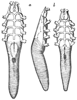
Fig. 53.—Demodex folliculorum, var. caninus. a, Female; b, male. Viewed from below
and in profile. Magnified 300 diameters. After Mégnin.
The ectoparasitic arachnidans comprise a great variety of
mites and ticks (Acaridæ and Ixodidæ) more or less proper to
man, and also a number of creatures which, though hardly to
be reckoned as human parasites, are apt to transfer themselves
from animals to man. Little more than an enumeration of the
forms is possible here. The Common Scab or Itch insect
(Sarcoptes scabiei) forms the type of a great variety of arachnids,
generally spoken of as different species according to the
host they dwell upon. Mégnin, however, in his beautiful
memoir, quoted below, regards most of the forms of this genus
(found on the horse, hog, sheep, dog, wolf, and other animals)
as mere varieties. In man the female Acarus burrows beneath
the skin, forming galleries or curved channels, in which she
deposits her eggs. The irritation produced is not alone due to
these excavations, but to the presence also of a poison which
the mite discharges when feeding. The Sarcoptes crustosæ of
Fürstenberg, producing the Norway itch, is a variety, if,
indeed, it can be called as much. Under the frightful name of
Dermatophagoides Schérémétewsky two parasites found on an
herpetic patient have been described as new to science by M.
Bogdanoff, but Mégnin points out that these Acari are only
female and young male representatives of his Chorioptes setiferus
(var. bovis) respectively. In Newfoundland, Dr Le Roy de
Méricourt discovered a singular species upon an officer who
had come from Havannah (Tyroglyphus Méricourti, Laboulbène).
It possesses enormous palpi, as in the genus Chyletus to which
Robin refers it. Another ectozoon, placed by Mégnin and
others amongst the lowest types of Arachnida, is the well-known
Demodex folliculorum. It is a gregarious species, a
dozen or more examples often being present in a single dilated
hair follicle. Though disfiguring to the human face it produces
little harm. M. Gruby made it out to be a very common
parasite, infesting forty out of sixty persons; but Mégnin, in
his brochure (l. c. infra, p. 119), shows this statement to be an
exaggeration. It infests on the average not more than one in
ten persons. According to Gruby, moreover, a single follicle
in the dog may contain 200 of these mites, another statement
which Mégnin deems unreliable. The Demodex of the dog is only
a variety (fig. 53). Many other human Arachnids have been found,
some of which appear to be genuine species, whilst others are
accidental, so to speak. Of the former kind, perhaps we may
reckon the two species discovered by Hessling (Cœlognathus
morsitans and Entarsus cancriformis). Of the latter sort, those
found by Busk, Simon, and Bory de St Vincent may be cited.
The mite found in Simon’s case was the Dermanyssus avium,
which infests cage-birds. Probably it was the same species
which Bory found on a lady; but in Busk’s negro sailor the
mite may have been D. gallinæ of the common fowl. Differing
from the mites, proper, and also from the true ticks, are
some bug-like forms called Argades. The two best known are
the Miana bug of Persia (Argas persicus) and the Chinche of
Columbia (A. chinche). Like their congener infesting pigeons
(A. reflexus) these parasites are terrible blood-suckers. The
bite of the Persian bug is so venomous as to have occasioned
death. Various species of tick have been known to attack man,
but the species have not been well determined. Although a
human form has been described (Ixodes hominis, Koch), yet it
is more probable that the species usually attacking man are the
same as those known to infest the domesticated animals. In
this list we may, therefore, reckon Ixodes nigra, Ix. bovis, Ix.
ricinus, and Ix. reduvius. Cases in which one or other of
these ticks occasioned much pain and distress are recorded by
Hussem, Raspail, and Dr Cosson. Besides these there is a
formidable tick well known at Angola (Ix. monbata). Its
habits are like those of the common bed-bug. Severe pain
comes on two hours after the person is bitten. It likewise
attacks animals. The Ix. carapato is similarly troublesome in
Brazil. Another very disgusting arachnid liable to attack man
is the Galeodes araneoides. This large spider-like creature, two
inches in length, commonly attacks camels and has an extremely
venomous bite. One or more species of the dung-beetle mites
(Gamasidæ) have also been known to fasten themselves on
man. According to Latreille, they first get attached to the
clothes of travellers, whence they pass to his body, and there
shift about, producing great torment. Another disagreeable
arachnid is the little harvest bug (Leptus autumnalis), which
not only excites irritation during its crawling motion on the
human skin, but even succeeds in burying itself near the hairs.
The irritation thus produced is almost unbearable. This mite
attacks various animals, especially dogs and cats. I myself
once suffered severely from this species in consequence of fondling
a young wild rabbit which, as I afterwards discovered, was
much infested. When the parasites had reached my left arm-pit
they occasioned extreme torture. I have known these autumnal
spiders to produce small suppurating boils on the abdomen.
I may add that Dr Tilbury Fox has brought under my notice
an instance where the hexapod larva of another species (probably
Trombidium cinereum) was found to have occasioned severe
irritation in a child.
Bibliography (No. 38).—Alibert, ‘Maladies de la Peau,’ Paris,
1833.—Audouin, V., art. “Arachnida,” in ‘Todd’s Cyclop.,’
vol. i, 1836.—Beneden, Van (et Gervais), ‘Zool. Med.,’ 1859.—Bourguignon
(et Delafond), in ‘Rec. Vét.,’ 1856.—Idem, in
‘Mém. de l’Institut.,’ 1862.—Cobbold, “Case of Leptus producing
Boils,” in ‘Worms,’ p. 140, London, 1872.—Gamgee, ‘Our
Domestic Animals in Health and Disease,’ Edin., 1861.—Gerlach,
‘Kraetze und Räude,’ 1857.—Hebra, in ‘Oester. Jahrb.,’
1864.—Hering, ‘Die Kraetzmilben,’ Stuttgard, 1845.—Krabbe,
“Husdyrenes paras. Mider.,” ‘Tidssk. f. Vet.,’ Rœk. 2, Bd. iii.—Küchenmeister,
l. c., 1855, s. 412 (good figs.).—Mégnin,
‘Monographie de la tribu des Sarcoptides psoriques.’ (This work
contains a full bibliography and numerous beautiful plates; see
also Review in the ‘Veterinarian,’ Aug., 1877, p. 563).—Idem,
“Mémoire sur un nouveau Symbiote (Chorioptes, Gerv.),”
‘Journ. de l’Anat. et de la Physiol.,’ 1872.—Idem, “Mém. sur
un nouvel Acarien,” ibid., 1873.—Idem, “Mém. sur les Hypopes,”
ibid., 1874.—Idem, “Mém. sur l’organisation et la
distribution zoologique des Acariens de la famille des Gamasidés,”
ibid., 1876.—Idem, “Mém. sur les métamorphoses des Acariens
en général, et en particulier sur celles des Trombidions,” ‘Ann.
des Sci. Nat.,’ 1876.—Idem, “Des conditions de la contagion
de la gale des animaux à l’homme,” ‘Arch. générales de Méd.,’
1876.—Idem, “Mém. sur le Demodex folliculorum (Owen),”
‘Journ. de l’Anat. et de la Physiol.,’ 1877.—Moquin-Tandon,
‘Elém. de Zool. méd.’ (Hulme’s edit., p. 302–328), 1861.—Williams,
in his ‘Veterinary Surgery’ (good figs., reproduced
from Gamgee’s translation of ‘Gerlach,’ &c.), 1872.
Although multitudes of small Crustaceans are parasitic upon
fishes, and some few of them adhere to oceanic mammals
(Cetacea), I am sceptical as to the parasitism of Crustacea either
in or upon man. Many of the Amphipodous Gammari lead a
sort of free parasitic existence, and they are themselves very
liable to harbor larval parasites. As regards human parasitism
from this source the only records known to me are those quoted
below.
Bibliography (No. 39).—Banon, “Freshwater Shrimp, or
Gamarus pulex (said to have been passed per anum),” ‘Rep. of
Path. Soc. of Dublin,’ in ‘Dub. Med. Press,’ April 6, p. 351,
1864.—Bartels, “Gamarus pulex in the Human Subject, with a
postscript by Troschel,” trans. by Dr E. P. Wright, from ‘Verhandl.
des Naturhist. verein. der Preuss. Rheinl. und Westph.,’
in ‘Dubl. Med. Press,’ 2nd ser., vol. ix, p. 407, 1864.—Wright,
E. P., ‘Remarks on Dr Banon’s Case’ (see Bartels).
Whilst very many flies, bugs, lice, and fleas persecute animals,
not a few of them also attack man. Several of the species are
genuine parasites, others are semi-parasitic, and others, again, are
altogether outside the border-land of parasitism in the ordinary
sense of the term. In fact, it becomes difficult to say where
the line of parasitism should be drawn. I cannot, however,
ignore all notice of the insect tormentors, whether strictly
parasitic or not.
At least fifty different species have been regarded as playing
the rôle of parasitism in man. Amongst the Coleopterous
parasites none is more authentic than Blaps mortisaga. At
least half a dozen such cases have occurred. Mr Hope’s
catalogue of insects producing parasitism in man gives three
examples of scolechiasis from this source. Sir J. R. Cormack
published a fourth case, and I have recorded a fifth. In this
instance I received the living larva from Dr Horne, of Barnsley,
who procured it from an infant eleven weeks old. In my ‘Introductory
Treatise’ I have alluded to the case of the girl Riordan,
who not only passed per anum upwards of 1200 larvæ, but also
several perfect insects. The case was first reported by Pickells,
Thomson, and Bellingham. One of the other authentic cases,
in which only a few larvæ were present, was recorded by
Patterson, of Belfast, and the third case by Bateman. Mr Hope’s
‘Catalogue’ originally appeared in the ‘Transactions of the
Entomological Society,’ being afterwards published in the pages
of the ‘London Medical Gazette,’ 1837. Patterson’s case was
also, I believe, first communicated to the Entomological Society.
As regards the mode in which the maggot gained access to
the child in Horne’s case, it was not easy to decide; but in the
case of the girl Riordan the mode of ingress was sufficiently
explained. The Blapsidæ, as a family, are closely allied to the
meal-worms, and, like most of the Tenebrionidæ, are black and
foul-smelling beetles, frequenting dark and damp situations,
from which they escape only at night. The family comprises
numerous species, of which probably not more than three are
found in this country. They are abundant in Africa, especially
in Egypt, where (according to Fabricius, as quoted by Westwood,
Figuer, and others) the women eat Blaps sulcata cooked
with butter in order to make themselves grow fat. The insects
are also employed as specifics against ear-ache and the bite of the
scorpion. The superstitious notion of a “charm” is generally at
the bottom of these domestic remedies. In the girl Riordan’s case,
as Westwood observes (when epitomising Pickell’s account), the
parasites, as such, “probably originated in an absurd and superstitious
practice, which she had for some time followed, of drinking
daily for a certain time a quantity of water mixed with clay,
taken from the graves of two Catholic priests, and eating large
pieces of chalk. One of these beetles was immersed repeatedly
in spirits of wine, but revived after remaining therein all night,
and afterwards lived three years.” The intolerance of light
shown by the perfect insect seems to be equally shared by the
larva. Of this fact I had repeated evidence by observing the
behaviour of the living specimen sent to me by Dr Horne. Thus,
when, on February 5th, 1877, I placed the maggot on the
surface of some moist mould, scarcely half a minute elapsed
before it commenced to bore its way downwards, and in less
than a minute all but the tip of the tail had disappeared. In
like manner, when, on the 7th, I raised the lid of the box, and
found the maggot on the surface of the soil, it almost instantly
proceeded to bury itself. Hope’s list records no less than nine
instances of parasitism in man from the larvæ of Tenebrio
molitor, and he gives a score of other Coleopterous insects
which he regarded as human “intestinal worms.” Undoubtedly
a large number of insect larvæ do get introduced into,
and actually live within the human intestines.
Numerous cases of this sort have come under my observation,
but it often requires a profound knowledge of entomology to
determine the species. Several examples of œstridean larvæ
occur amongst them. For one example of Œstrus hominis I am
indebted to Mr Higginson, of Liverpool, who obligingly supplied
me with notes of the case. Dr Kirk presented me with a
small bot which he removed from Livingstone’s leg. I afterwards
deposited the African bot, in his name, in the Hunterian
collection. Bates speaks of an Œstrus in Brazil producing boils
in human flesh. Westwood quotes similar instances. Of these,
one was extracted from the thigh by Dr Brick. Mr Doubleday,
the entomologist, extracted one from his own leg, and M. Goudot,
another entomologist, was also victimised in the same way.
Both of these savans were travelling in America at the time.
Two cases from South America were also recorded by Howship.
In one the larva lodged in the back; in the other in the
scrotum. Humboldt noticed that the Indians were much
infested by Œstridæ. Three cases are severally recorded by
Roulin, Guérin, and Audouin. Mr Stroop also mentions a case
in which an Œstrus was removed from an ulcer on the shoulder
of a boy in Texas. Another kind of bot known as the Macaco
worm (Cuterebra noxialis) occasionally attacks man, but more
frequently cattle and dogs. For one example, taken from the
leg of a negro, at Belize, British Honduras, I am indebted to
Dr Dobson (A. M. D., Netley). Hope’s list records five cases of
bots of Œstrus hominus, one of Œ. Guildingii, one of Œ. bovis,
and thirteen others (belonging to the same genus) as having
been noticed in man. Since his time many additional cases
have been recorded by J. M. Duncan and others. In like manner
a legion of cases in which the maggots of various Muscidæ have
been noticed, either in, upon, or from the human body. At
a meeting of the American Academy in April, 1859, Dr Leidy
showed some larvæ of the bluebottle fly vomited by a child;
five larvæ of the flower-fly (Anthomyia) from a physician’s
own person (which had produced choleraic symptoms); and
nine examples of Cuterebra noxialis. I have myself encountered
numerous insect larvæ in medical practice. Amongst others
I have obtained the larvæ of Anthomyia canalicularis in six
or eight separate instances. One set of specimens, identified by
Wunderlich, was sent to me by Dr Brandt, of Oporto. Drs
Duffin, W. Fox, and Leared have supplied me with others.
The larvæ described in Farre’s case, not being setose, must be
referred to one or other of the Muscidæ proper. Mr Hope gave
nearly forty cases of this kind, referable to eight different
species of fly, and, as already implied, I have myself collected
a great variety of the maggots of Muscidæ passed by the bowel,
besides others obtained either from beneath the skin or from
open ulcerations.
The flies hitherto noticed as supplying parasitic maggots in
man are Musca domestica, M. carnaria, M. larvarum, M. nigra,
and perhaps M. Cibaria, M. stabulans, and M. Cæsar. This last,
a handsome fly, is the species which proves so troublesome to
sheep. The habit which flies have of depositing their eggs in
open wounds, when the victims are asleep, is a fertile source of
this kind of parasitism. Some of the instances recorded by
Kirby and Spence are revolting in the extreme. They quote
the cases which came under Mr Sell’s notice in Jamaica. In
one instance the flies were hatched in a neglected blister on the
chest; in another from the gums and inside of the cheek; and
in a third, from the nostrils of a negro, from whom 235 larvæ
were expelled. The case of the Lincolnshire pauper, Page, who
was literally eaten up by maggots, is almost incredible. An
equally horrible instance, however, is recorded by Cloquet. It
is said that the Jamaica cases were all due to the larvæ of the
bluebottle fly (M. vomitoria). An instance of the same kind has
been recorded by Mr Knox (A. M. D.). Sufficiently revolting as
these cases are, the horrors attending them are eclipsed by the
habits of the larvæ of Lucilia hominivora. The best accounts of
its habits are those by M. Coquerel, M. Saint-Pair, and M.
Vercammer. The insects lay their eggs in the mouth and nostrils,
and when the larvæ escape they devour the tissues surrounding
the buccal cavity, the pharynx, glottis, frontal and nasal sinuses,
even reaching the sockets of the eye. Several Cayenne convicts
have perished from the maggots of this fly, which is also prevalent
in Mexico. These are not, however, the only instances of
maggots gaining access to the nasal chambers. In a case
recorded by Dr Astros, of Aix, 113 were discharged from the
nose of a woman; and M. Legrand du Saulle records an instance
where a number of larvæ occupied the frontal sinuses of a girl,
nine years of age. The larvæ produced persistent headache and
convulsions. In the case recorded by Wohlfart, 18 larvæ were
discharged from the nose of an old man, and in the example
given by Latham several larvæ were obtained from the frontal
sinuses of a woman. Bracey Clark also gives an instance in
which a bot was taken from a woman’s jaw. Not improbably
the well-known Indian disease, termed peenash, or worm in the
nose, is due to the presence of Œstridean larvæ. Cases by
Rustomjee and Lahory are quoted below. Possibly Stockett’s is
another of the same order. The case by Kilgour (Bibliog. No.
34) may be another. I may add that Moquin-Tandon gives an
instance of the discharge of seventy-two bots, resembling those
of the sheep, from a woman’s nose (‘Journ. de Vandermonde’).
The rat-tail maggots or larvæ of Helophilus are parasitic. Two
or three instances have been recorded from the horse. I possess
one from the human intestine. Kirby also quotes an instance
in which Heloph. pendulus was obtained from the stomach of
a woman (‘Philos. Mag.,’ vol. ix, p. 366).
A vast number of non-parasitic insects are injurious to man
and beast. Inasmuch as they subsist at the expense of their
victims and also adhere to his person during their attacks for a
shorter or longer time, they, like the leeches, may be spoken of
as free parasites. The leg-sticker (Stomoxys calcitrans) penetrates
through thick stockings, causing blood to flow freely.
The clegg of the West Highlands (Hæmatopota pluvialis) also
violently attacks man and beast, especially horses. The mosquito
(Culex anxifer), the gnats (C. pipiens, C. annulatus, and C.
pulicaris), and the midge (Chironomus plumosus) need only be
mentioned. The creeping gnat (Simulium reptans) is also very
annoying in Sweden. The rôle of the mosquito, as itself constituting
a parasite-bearer, will be again referred to in the
closing pages of this work (Book II, Section V). The bites of
the tsetse (Glossina morsitans), though so destructive to the
horse, ox, sheep, and dog, are not dangerous to man himself.
According to Sir S. Baker, the seroot-fly, or zimb of Bruce,
which is a species of Pangonia, is excessively annoying to
travellers in Abyssinia. Amongst the hemipterous insects the
common bug (Acanthia lectularia) is sufficiently blood-thirsty;
but there is a far more sanguinary species of this kind in
South America. This is the pampas benchucha (Conorhinus
nigrovarius). Our distinguished countryman, Darwin, in his
‘Voyage,’ speaking of these wingless insects, says:—“Before
sucking they are quite thin, but afterwards become round
and bloated with blood. In less than ten minutes the insect
is changed from being as flat as a wafer to a globular form.”
This insect somewhat resembles our water-scorpion (Nepa
cinerea), a non-parasitic species whose bite causes severe pain,
as does also the wound inflicted by the water-boatman
(Notonecta glauca). There are other species of bug, such as
the Acanthia rotundata of Réunion Island and A. ciliata of
Kasan, the bites of which are worse than that of the common
species. The fly-bugs also, such as the Reduvius personatus, so
common in France, and the R. amænus of Borneo and Java,
attack man, although their especial habit is to attack and destroy
other insects, including bugs themselves. Other species or
varieties of Reduvius (R. cruentus, and R. serratus) attack man.
The last named is an Indian form, capable, it is said, of producing
an electric shock.
Passing to the fleas, the most important and truly parasitic
form is the chigoe or gigger (Pulex penetrans). This abounds
in tropical America and the West Indies. The female insects
only attack man, and this they do for the purpose of securing
a lodgment for their offspring. They attack especially the
soles of the feet, between the toes and near the nails. In bad
cases the whole of the foot becomes affected. After the insect
has penetrated the skin its body swells enormously and becomes
a mere bag of eggs. This swelling causes active inflammation,
which terminates in suppuration and the formation of open
ulcers. The chigoe also attacks various animals. In addition
to the common flea (P. irritans) other species infesting
animals are said to attack man occasionally. As regards those
degraded types of insects known as lice I can only afford space
to remark that five species have been recognised as human.
These are the head-louse (Pediculus capitis); the louse of the
eyelids (P. palpebrarum); the clothes-louse (P. vestimenti);
the distemper louse (P. tabescentium); and the pubic louse (P.
inguinalis). The distress these creatures occasion is only fully
known to physicians who deal with the obstinate cutaneous
affections caused by their presence (Phthiriasis). Some of the
lice found on negroes and Greenlanders are regarded as distinct
varieties. Lice are abundant on animals, and some of the
species are apt to transfer themselves from one host to another.
Thus the Ornithomyia avicularis of cage birds has been found
on man, whilst one or more of the numerous species infesting
the common fowl are, by transfer, apt to produce a severe
phthiriasis in the horse. The lice of the fowl belong to the
genera Leipurus, Liotheum, Menopon, Philopterus, Goniodes, and
Goniocotes. The unsuitableness of man’s person as a habitation
for bird-lice should, however, check the fear which many persons
have of handling fresh-killed poultry and game birds. Poultry
lousiness in man is probably impossible from this source.
Bibliography (No. 40).—Arture, “Obs. sur le ver nommé
Macaque,” in ‘Mém. Acad. des Sci.,’ Paris, 1753.—Bates,
“Œstrus in Man,” ‘The Naturalist on the Amazons.’—Beneden,
Van, ‘Animal Parasites and Messmates,’ 1876.—Idem (with
Gervais), ‘Traité de Zool. Méd.’—Blood, M., “Case of Larvæ
(Musca sarcophaga) expelled alive in the Fæces,” ‘Beale’s Arch.
of Med.,’ vol. iii, p. 134, 1862.—Brinton (similar case), ‘Arch. of
Med.,’ vol. iii, p. 133, 1862.—Bouyer (quoted by Figuer),
‘Tour du Monde,’ p. 318, 1866.—Clark, Bracey, in ‘Linn.
Trans.,’ vol. iii, 1797 (the jaw-case at p. 323), and vol. xv, 1827.—Idem,
‘Essay on Bots,’ 1815.—Cloquet, J., “Case of Blindness
from Worms (larvæ of Musca) in the Eyes,” from ‘Arch.
Gén. de Méd.,’ in ‘Lancet,’ 1828.—Cobbold, “On Blaps mortisaga
as a Human Parasite,” ‘Brit. Med. Journ.,’ 1877, p. 420.—Idem,
“Entoz.” (‘Hope’s List.’), p. 416.—Idem, in ‘Worms’
(“Leptus,” &c.), p. 140, 1872.—Coquerel (quoted by Figuer).—Cormack,
J. R., “Exhibition of a Larva (B. mortisaga) passed
by a Child,” ‘Month. Journ. Med. Sci.,’ vol. i, 1841.—Crumpe,
S., “History of a Case in which Worms (larvæ of a beetle)
were discharged from the Stomach,” from ‘Trans. of the Roy.
Irish Acad.,’ vol. vi, in ‘Med. Facts and Observ.,’ vol. viii,
p. 229, 1800.—Denny, ‘Monog. Anoplurorum Brittaniæ,’ 1842.—Duncan,
J. M., “On the Occurrence of Bots in the Human
Subject,” ‘Edin. Vet. Rev.,’ vol. i, p. 275, 1858–59.—Idem,
“The Larva of Œstrus bovis in the Human Subject,” ‘Rep. of
Edin. Med.-Chir. Soc.,’ in ‘Month. Journ. of Med. Sci.,’ July,
1854.—Farre, A., “On the Larva of Anthomyia canalicularis,”
‘Micr. Journ. and Struct. Rec.,’ 1841, p. 129, and in ‘Trans.
of Micr. Soc. of Lond.,’ orig. ser., p. 51, 1844.—Figuer, in
‘The Insect World’ (good figs.), Janson’s edit., 1869.—Furlonge,
“Anat. of Pulex,” in the ‘Journ. of the Queckett Club,’
vol. iii.—Geer, De, ‘Mémoires pour servir à l’Histoire des
Insectes,’ 1773.—Gervais (see Van Beneden).—Hill, “Account
of the Larva of a supposed Œstrus hominis, or Gad-fly, which
deposits its Eggs in the bodies of the human species, with
particulars of a Case,” ‘Edin. New. Phil. Journ.,’ vol. xxii,
p. 284, 1830.—Hilaire (see St Hilaire).—Hope, “Tables of
Cases of (spurious) Intestinal Worms,” ‘Lond. Med. Gaz.,’
1837–38.—Hoppe, “Case of Larvæ of Insects (Musca stabulans)
passed by Stool,” from ‘Bibl. für Läger,’ in ‘Med.-Chir. Rev.,’
1842.—Hopper, R. S., “Insects (Stratiomis) voided with Urine,”
edit. note in ‘Micr. Journ. and Struct. Rev.,’ p. 160, 1841.—Joly,
‘Recherches sur les Œstrides en général,’ &c., Lyons,
1846.—Keferstein, ‘Sur l’Oistros,’ Isis, 1827.—Kirby (and
Spence), ‘Introd. to Entomology,’ 7th edit., 1856.—Knox, M.,
“Maggots, the larvæ of the Bluebottle Fly, in Syphilitic Ulceration
of the Throat,” ‘Lancet,’ Oct. 6, 1877, p. 514.—Lahory,
B. T. C., “On Peenash, or Worms in the Nose,” ‘Ind. Ann. of
Med. Sci.,’ 1855, and ‘Edin. Med. Journ.,’ 1857.—Leach,
“Œstridæ,” in ‘Wernerian Trans.,’ 1817.—Leidy, in ‘Proc.
Phil. Acad. Nat. Sci.,’ 1859, p. 7.—Maclean, “On the Oistros,”
‘Linn. Trans.,’ vol. xiv, 1824, and in ‘Zool. Journ.,’ vol. i and
iv.—Metaxa, “Vom Œstrus (u. s. w.),” in ‘Neuen Nord.
Beitr.,’ Bd. i, and in ‘Mém. de Zool. Med.,’ Rome, 1835 (quoted
by Westwood).—Moquin-Tandon, ‘Med. Zool.’ (l. c., Bibl.
No. 38).—Newport, art. “Insecta,” ‘Todd’s Cyclop.,’ 1839.—Rustomjee,
B., “Case of Worms in the Nose, or ‘Peenash,’” in
‘App. to Bomb. Med. and Phys. Soc. Trans.,’ No. vii, new ser.,
p. 21, 1861; see also Lahory, on ‘Peenash.’—Saint-Hilaire,
“Sur l’Œstre chez l’homme,” in ‘Ann. Soc. Ent. de France,’
1833.—Say, “Brick’s Case,” in ‘Trans. Acad. Nat. Sci. Phil.,’
vol. ii.—Sells, in ‘Trans. Entom. Soc. Lond.;’ see also Lemprière’s
‘Diseases of the Army in Jamaica,’ vol. ii.—Stockett,
T. H., “An account of a Headache cured by the discharge of a
Worm (?) from the Nose,” ‘Med. Com.,’ vol. xix, p. 157, 1794,
and in ‘Trans. Coll. of Phys. Phil.,’ vol. i, part i, p. 181, 1793.—Stroop,
St J., “Œstrus,” in ‘Amer. Naturalist,’ vol. vii,
p. 437.—Tanner, “On Lice,” in his ‘Pract. of Med.,’ vol. ii,
p. 429, 6th edit., 1869.—Westwood, in his ‘Classification of
Insects,’ vol. ii, 1840.—Wohlfart (quoted by Moquin-Tandon).—Yule,
“Case of Larvæ of Insects in the Human Stomach,”
‘Edin. Phil. Journ.,’ and ‘Lond. Med. Repos.,’ 1825.
The scope of this work does not demand that I should comprise
within its limits any vegetable parasites; nevertheless, I
must needs refer, however briefly, to certain confervoid and
sarcodic organisms, which, for the most part, lie on the borderland
of the animal and vegetable kingdoms. Professor Cohn
regards the bacteria as allied to the Oscillitoriaceæ. He puts
them in his order Schizosporeæ. It is of little moment,
practically, where these protista forms are placed. Unquestionably
many of them are parasitic, as they live in the tissues,
fluids, and secretions of animal bodies, including man. Their
presence in cattle is associated with an anthracoid disease
(charbon), whilst in the human body they have been detected in
connection with zymotic affections. They have been found by
Cohn, Sanderson, Klebs, Chauveau, and others, either in the
lymph of vaccine pustules, or in the miliary eruptions of typhus
fever. Professor Beale, who was one of the first to observe
these special organic particles in vaccine lymph, denies that
they are true Bacteria; and, indeed, he warmly disputes the
inferences that have generally been drawn from the fact of the
presence of such particles in lymph, blood, and other nutrient
fluids. The best known and defined forms are Bacterium
termo and Bact. lineola, which are concerned in the production
of putrefaction, Bacillus anthracis, found in the blood of animals
suffering from carbuncular disease, Micrococcus septicus, found
in typhus and pyæmia, M. vaccinæ of cow pox lymph, and M.
diphthericus, in diphtheria. As regards their prevalence in
certain forms of relapsing fever, Sanderson states that Dr H. V.
Carter, of Bombay, examined the blood of 250 fever patients
and found spirilla in nearly every instance. From the independent
observations of Pasteur, Sanderson, Lister, Tyndall,
Bastian, Eberth, Roberts, Davaine, and many others, it seems
clear that the Bacteria and their allies play an important part in
association with certain morbid states. However, as regards
the etiology of the maladies in which these organisms are found,
it is perhaps too early to speak with absolute confidence. The
subject cannot be dealt with here; moreover, it is outside the
range of my personal investigations.
Passing to those protozoa which, although retaining some
vegetable affinities, are more or less distinctively animal, I
notice the obscure organisms termed psorosperms. In dealing
with these I shall treat of the forms that infest both man and
animals, confining my remarks to such as happen to have come
under my own observation.
In the year 1865 the public were thoroughly roused to a
sense of danger arising from the consumption of meat. The
panic originated with the outbreaks of trichiniasis in Germany.
During the excitement which subsequently prevailed at the time
of the rinderpest, all sorts of erroneous notions took possession
of the popular mind, and the errors were stimulated by writers
ignorant of helminthology. In January, 1866, I published a
few observations, the purport of which was to show that certain
microscopic organisms found in animals dying from cattle
plague were harmless “parasitic Protozoa,” possessing more or
less striking vegetable affinities. About a week previously
some interesting researches on these so-called cattle-plague
bodies had been published by Dr Beale. Those who first saw
these bodies thought they had stumbled upon organisms new to
science. I showed that similar or analogous organisms were
to be met with in a great variety of animals, and likewise in
the human body. They had been called worm-nodules, worm-nests,
egg-sacs, eggs of the common fluke, young “measles,”
corpuscles produced by muscular degeneration, psorospermiæ,
stages of growth of gregarinæ, amœboid bodies, and so forth.
In so far as the higher animals were concerned, Dujardin was
the first to describe them. He found these organisms in a
mole. This animal, however, having been fed upon earth-worms
known to harbour such parasites, there was no difficulty
in accounting for the source of the psorosperms.
In 1853 Hessling discovered psorospermial sacs in the muscular
substance of the heart, not only of the ox, but also of the
sheep and roe. By him they were regarded as evidences of
muscular degeneration. About ten years previously Miescher
found similar bodies in the muscles of the mouse.
In 1857 Rainey described similar structures taken from the
flesh of swine; and, in his memoir, he went so far as to maintain
that these bodies were early stages of development of the
common pork-measle. In the year 1858 Gubler wrote an
important paper on this subject, in which he related a case
where twenty cysts existed in the human liver. The cysts
were of great size, mostly as large as a hen’s egg, one of them
being some six inches in diameter. Naturally, the largest had
been diagnosed as an ordinary hydatid. However, on evacuating
their contents (post mortem), they were found to harbor
enormous quantities of minute corpuscles strictly analogous to
those usually obtained from psorospermial sacs. Gubler believed
he had stumbled upon masses of eggs of Distoma hepaticum, but
in this he erred. Shortly after Gubler’s discovery similar
bodies from the human liver were described by Virchow; and
in 1862, the subject was followed up by Dr Dressler, of Prague.
Dressler found in the human liver a number of pea-shaped
bodies, the milky contents (breisubstanz) of which displayed a
multitude of the characteristic corpuscular elements referred
to. These particles, already considered as equivalent to, if not
identical with, the so-called pseudo-navicellæ of gregarinæ, were
soon encountered by a variety of independent observers. Thus,
Leuckart noticed these bodies in various animals; but with
caution remarked:—“Concerning the nature of these formations
I will not decide. To be candid, however, it appears to
me to be in no way made out whether the psorospermiæ are to
be considered as the result of a special animal development,
whether they, like pseudo-navicellæ, are the nuclei of gregariniform
productions, or whether they are the final products of
pathological metamorphosis.” Leuckart found these organisms
in the intestines of a trichinised dog, also in a sheep and pig
fed with Trichinæ. He also found them in the muscles of
another pig fed with psorosperms, and likewise in the liver of
various rabbits. He remarks that in swine these parasites are
more abundant than measles. They were present in five of
eighteen pigs, and also in two out of four sheep, whose flesh
was especially examined. The observations of Lindemann at
Nischney-Novgorod are particularly interesting. This medical
officer discovered psorospermial sacs attached to the hair of a
girl who was being treated in hospital for chlorosis. The sacs
in question bore close resemblance to the bodies which we
found in abundance in diseased and healthy cattle. It would
further appear, from Lindemann’s observations, that the affection
is not very uncommon amongst the Russian peasants.
In connection with and attached to the same parasitically
affected hairs Lindemann also noticed several movable gregarinæ;
and partly from this circumstance he was led to believe
in the existence of a genetic relation subsisting between the two
kinds of bodies. He further expressed his conviction that the
people contracted the disease by washing themselves with water
in which gregarinæ abounded. Lindemann moreover refers to
Lebert as having noticed similar parasites in a case of favus,
and concludes that these organisms are of a vegetable nature.
His opinion, though not shared by the majority of parasitologists,
is nevertheless supported by the views of Robin, Leydig, and
others. Of still higher interest are the observations of Lindemann
respecting the occurrence of psorospermiæ in the capsule
of the kidney of a hospital patient who died with Bright’s
disease. The sacs in this case were remarkably small; nevertheless
their corpuscular contents indicated their true nature.
The pseudo-navicellæ measured only 1/5000″ in diameter. Amongst
other contributions of interest I may refer to those of Dufour,
J. Müller, Creplin, Kölliker, Keferstein, Stein, Drummond,
Lieberkühn, and E. Ray Lankester. I doubt if the vegetable
organisms described by Prof. W. T. Gairdner can be referred
to this group of parasites. At all events, by whatever name
these spurious entozoa are called, they were first discovered by
Dufour in insects, by Müller in fishes, by Miescher in the mouse,
by Dujardin in the mole, by Hessling in the larger quadrupeds,
and by Gubler in man. The results of my own examinations
may be briefly re-stated. In the flesh of cattle I found
psorospermial sacs varying from 1/120″ to 1/12″ in length, and in
that of sheep from 1/220″ to 1/80″. The bodies were enclosed in
well-defined transparent envelopes, and their contents exhibited
indications of segmentation. In some specimens the segments
displayed themselves as a complete cell-formation, the contents
of each cell being uniformly granular. Under the 1/4″ objective
the contained granules were clearly visible, and on rupturing
the sac their peculiar characters were at once manifest, each
granule or corpuscle represented a pseudo-navicel, all displaying
a tolerably uniform size, averaging 1/2000″ in diameter. Some of
the corpuscles were round, others oval, several bluntly pointed
at one end, many curved and fusiform, not a few being almost
reniform. Highly refracting points or nucleoli were visible in
their anterior.
Turning to the practical aspect of the subject, I remarked
that these bodies had nothing to do with the cattle plague. No
one who carefully examined the flesh of animals that had died
of rinderpest had failed to discover them; yet, in one or two
instances they appear to have escaped notice. When it is considered
how long it takes us to examine a few grains weight of
muscle carefully, it is obvious that the body of a large beast
might contain many hundreds of these organisms without our
being able to detect their presence, except by a prolonged
investigation. In the few rinderpest beasts, portions of whose
flesh I submitted to the microscope, I should say there were
not more than 100 of these bodies in one ounce of meat; but in
the heart of a healthy sheep (which I afterwards ate) I calculated
there were about 1000 parasites to the ounce, and in the
heart of a healthy bullock (which likewise served me for a
meal) their numbers were rather in excess of those in the sheep.
Altogether, at two meals, I could not have swallowed less than
18,000 of these psorosperms. Consumers of beef, mutton, and
pork eat these bodies every day, but they take no harm because
the parasites in question are not true helminths. Fine healthy
beef has been returned to the butcher when it was as good as
any other meat in the market. I have examined various kinds
of meat, such as veal, pork, and mutton, but in none have I
found so great an abundance of psorosperms as in beef, which
was, notwithstanding, perfectly healthy and sound. I calculated
that in one instance a single ounce of the flesh contained upwards
of 2000 parasites. There is practically no limit to the extent
of this kind of parasitism, and there is no organ of the body in
which psorosperms may not be found. Moreover, the forms
they display are exceedingly various. Psorosperms have been
found by Siedamagrotzky in the muscles of the horse, and not
very long ago, through the help of Professors Simonds and Axe,
I had the opportunity to examine some peculiar worm-like
structures which occupied the mitral valve of a horse. To the
naked eye they looked like coiled nematodes, but I was soon convinced
that they formed a peculiar type of psorosperm. A
complete view of these bodies was a matter of great difficulty
owing to the delicate nature of their limiting membrane and to
the confusion of markings produced by the interlacing of the
fibres of the chordæ tendinea. At length, by spreading a portion
of the membrane of the valve over a large glass slide, and
by allowing it to dry slowly, I found that the vermiform body
presented neither beginning nor end. The appearances were
curious and puzzling. The organism formed a flattened tube
or sac, almost uniform in width and variously twisted upon
itself. From the main tube there projected several hernia-like
secondary loops or branches, most of them presenting less
than half of the thickness of the former. These peculiarities,
however, can hardly be understood without reference to the
original illustrations. That these secondary coils were not of
the nature of hernial protrusions was evident, not alone from
the nature of their contents, but also from the fact that they
showed distinct anastomoses. In fact, the parasite was a simple
sac or bag with branches.
On puncturing the main tube with a fine needle a small
quantity of tenacious creamy fluid made its escape. This,
under Ross’s 1/4-inch lens, resolved itself into a few excessively
delicate sarcode globules surrounded by fine granules. The
granular matter displayed a tendency to collect itself in the
form of oval masses without showing any trace of a limiting
border. One of these masses, measuring 1/250″ in length, I
examined under a Wasserlein-objective, when I further ascertained
that the elementary particles or granules were uniformly
oval in shape, rather highly refractive, their size scarcely
exceeding 1/8000″ in diameter. The sarcode corpuscles, on the
other hand, were of different sizes, ranging between 1/3000″ and
1/1600″ in diameter.
From the facts thus elicited, negative as they were in
respect of helminthic structure, I could see no escape from the
conclusion that we had to deal with a new form of psorospermial
bag, whose granular contents consisted of excessively
minute pseudo-navicellæ. In the centre of the largest hernia-like
loop there was a clear oval disk, which at first brought to
my mind the nucleus of Monocystis infesting the earth-worm,
but it was merely a vacuole.
The case recorded by Gubler reminds me of another remarkable
instance of psorospermial cysts, in this case associated
with true hydatids. In 1873 Dr Whittell sent me particulars
of a case in which the contents of an hydatid of the liver
(drawn off, during life) consisted of shreds of a true hydatid,
a few echinococcus-hooklets, together with multitudes of
spindle-shaped amœboid particles of excessive minuteness and
delicacy. The bodies, floating in a transparent fluid, formed a
thick milky or creamy fluid, resembling pus in appearance; but
there was no trace of pyæmia. Judging from Dr Whittell’s
figures, he must also have found a solitary microscopic nematoid
hæmatozoon, the nature of which was not clear to him. I
believe it to have been a specimen of Filaria sanguinis hominis.
Be that as it may, the case is altogether unique and deserves
further elucidation.
As regards the higher forms of protozoa it must suffice to
allude to the Cercomonas hominis of Davaine, found in the
dejections of cholera patients, to the Cerc. urinarius of Hassal
and C. saltans of Ehrenberg, to the Trichomonas vaginalis of
Donné, detected in the vaginal mucus, and to the Balantidium
coli of Claparède and Lachmann, originally found by Malmsten
in the human colon. The Balantidium, or Paramæcium coli,
has frequently been observed in the evacuations of fever patients,
and it has also been found by Dr Treille in patients suffering
from the Cochin-China diarrhœa. Monads have also been
found in the stomach and intestines of the hog and various
other animals. Infusorial parasites are particularly abundant
in batrachians, the Bursariæ of frogs and toads being familiar
to every helminthologist.
Bibliography (No. 41).—Arloing (and Tripier), in ‘Gaz.-hebd.,’
1873, p. 574 (quoted by Davaine).—Balbiani, ‘Compt.
Rend. Soc. Biol.,’ 1867, p. 103 (quoted by Davaine and Bastian).—Bastian,
“On the Nature of the so-called Sarcina ventriculi,”
‘Brit. Med. Journ.,’ Feb. 3, 1872.—Idem, “On Heterogenesis
in its relation to certain Parasitic Diseases,” ‘Brit. Med. Journ.,’
Feb. 24 and April 20, 1872 (see part iv, p. 417, with figs. from
Balbiani).—Beale, L., “Entozoon-like bodies in Muscles,” in the
‘Microscope in Medicine,’ 4th edit., p. 485, 1878.—Idem,
“Bacterium Hypothesis of Contagium,” ibid., pp. 313–321.—Burnett,
W. T., “On Psorospermia, Mermithes, &c.,” in a paper
entitled ‘Reviews and Records in Anat. and Physiol.,’ in ‘Amer.
Journ. of Sci. and Arts,’ vol. xviii, 2nd ser., p. 104, 1854.—Carter,
H. V., “On Spirilla,” quoted by Sanderson in ‘Brit. Med.
Journ.,’ Nov. 17, 1877, p. 700.—Cobbold, “Remarks on Spurious
Entozoa found in Diseased and Healthy Cattle,” ‘Path. Soc.
Trans.,’ vol. xvii, p. 452, 1866, and ‘Lancet,’ Jan. 27, 1866,
p. 88; see also Prof. J. Gamgee’s work on the ‘Cattle Plague.’—Idem,
“On Worm-like Organisms in the Mitral Valve of a
Horse,” ‘Veterinarian,’ Sept., 1877.—Idem, “On Psorospermiæ
in the Eye of the Cod (Morrhua),” ‘Linnean Society’s Proc.,’
May, 1862, and in ‘Intellectual Observer,’ 1862, p. 199.—Cohn,
‘Nova Acta,’ xxiv, s. 103 (quoted by Leuckart), Bd. i, s. 139.—Creplin,
‘Wiegmann’s Archiv,’ 1842, s. 61.—Davaine, l. c.,
2nd edit., “Synops. xxi” (with bibliog. refs.), 1878.—Donné,
‘Cours de Microscopie,’ Paris, 1847, p. 157.—Dressler,
quoted by Leuckart, Bd. i, s. 141.—Drummond, ‘Edin. Phys.
Rep.,’ 1852,—Dufour, ‘Ann. des Sci. Nat.,’ 1837.—Dujardin,
‘Traité’ (l. c., see Bibl. No. 1).—Eberth, ‘Zur Kentniss Bacteritischer
Mykosen,’ 1872.—Eimer, ‘Ueber Psorospermien,’
1870.—Gairdner, ‘Edin. Phys. Soc. Rep.,’ 1853.—Gluge, “Cysts
in Sticklebacks,” ‘Bullet. Acad. Roy. des Sci. de Bruxelles,’
1838.—Gubler, ‘Mem. Soc. Biol.,’ 1859, p. 657, and in ‘Gaz.
Méd.,’ 1858, p. 61.—Harz, C. O., “Eine neue Mikrococcusform
im lebenden Thierkörper,” ‘Deutsche Zeitschrift für Thier-Medicin
und vergleichende Pathologie,’ f. Novemb., 1878.—Hessling,
‘Sieb. u. Köll. Zeitsch.,’ 1853, p. 196.—Henle, ‘Müller’s
Archiv,’ 1845.—Hollis, W. A., “What is a Bacterium?” repr.
in the ‘Veterinarian,’ p. 205, 1875.—Keferstein, ‘Götling. gelehrte
Anzeigen,’ 1862.—Kloss, ‘Ueber Parasiten (u. s. w.)’ (quoted
by Davaine).—Knoch, ‘Journ. de Russ. Kriegs. dep.,’ Bd. xcv,
1866 (quoted by Leuckart and by Davaine).—Kölliker, in
‘Zeitsch.’ (by Sieb. and Köll.), 1848–49.—Lambl, ‘Prager
Vierteljahrschrift,’ 1859.—Lankester, E. R., “Recent Researches
on Bacteria (with copious references),” ‘Quart. Journ. Micr.
Science,’ Oct., 1878.—Lebert, ‘Phys. Pathologique’ (quoted by
Leuckart).—Leidy, “Gregarina,” ‘Amer. Phil. Trans.,’ 1851.—Leisering,
“Bericht (u. s. w.),” in ‘Sachsen,’ 1865.—Leuckart,
l. c., Bd. i, s. 135 and 740, and Bd. ii, s. 842 et seq.—Leydig,
‘Müller’s Archiv,’ 1851, s. 221, in ‘Micr. Journ.,’ 1853, p. 206,
and in ‘Arch. f. Anat. und Phys.,’ 1863, s. 191.—Lieberkühn,
‘Müller’s Arch.,’ 1854.—Lindemann, ‘Bullet. Soc. imp. des
Naturalistes de Moscow,’ 1863, and in ‘Gaz. Méd. de Paris,’
1870, p. 86.—Lister, J., “Natural History of Bacteria,” ‘Micr.
Journ.,’ Oct, 1873.—Malmsten, “Paramæcium coli” (quoted
by Davaine, l. c., 2nd edit., p. 67).—Miescher (quoted by
Leuckart and Siebold).—Müller, ‘Archiv,’ 1841, s. 477.—Rainey,
‘Phil. Trans.,’ 1857.—Rayer, “Singulière éruption sur
un véron (Cyprinus),” ‘Arch. de Méd. Comparée,’ Paris, 1842
(quoted by Davaine).—Rivolta, “Psorospermi, &c.,” trans. in
‘Journ. des Vét. du Midi,’ 1869, pp. 445 and 521.—Robin, ‘Les
Végét. Paras.,’ 2nd edit., p. 291.—Sanderson, in ‘Privy Council
Reports,’ 1874.—Siedamagrotzky, in ‘Recueil de Méd. Vét.,’
1872, p. 460.—Stein, in ‘Müller’s Arch.,’ 1848, and ‘Ann. Nat.
Hist.,’ 1850.—Idem, “Abhandl. d. k. Böhmischen Gesellsch.,”
x, s. 69, oder Lotos, 1859, s. 57 (quoted by Leuckart, Bd. i, s.
151).—Steinberg, ‘Walter’s Zeitschr. f. die moderne Medicin,’
1862, and in Leuckart, Bd. ii, s. 844.—Stieda, ‘Arch. f. pathol.
anat.,’ Bd. xxxv, and in Leuckart, Bd. ii, s. 846.—Suriray, “Sur
quelques parasites du lombric,” ‘Ann. des. Sci. Nat.,’ 1836.—Virchow,
“Zur Keutniss der Wurmknoten,” ‘Arch. f. Anat. u.
Phys.,’ xviii, s. 523.—Vogel, ‘Path. Anat.,’ i, s. 404.—Waldenburg,
“Psorospermien,” in ‘Arch. f. Path. Anat.,’ s. 435, 1867.—Windbladh,
also Wising, ‘On Balantidium coli’ (quoted by
Leuckart, Bd. ii, s. 846–847).—Winkler (see Leisering).
Appendix.—On various occasions I have dwelt upon the
necessity of acquiring accurate information respecting the degree
of mortality due to parasites, and in the present volume (p. 124)
I have referred to the defective evidences supplied by the returns
of the Registrar-General in respect of the echinococcus disease.
My object is not to cast blame upon those whose duty it is to
publish the returns, but rather to call attention to the advantages
that would follow if the Registrar-General were supplied
with full and accurate information on this head.
Through the courtesy of Mr Noel A. Humphreys I have been
furnished with the following official statement of the number of
Deaths from Worms in England and Wales, as recorded in the
Annual Reports of the Registrar-General throughout a decade
of years:
| 1868 | 1869 | 1870 | 1871 | 1872 | 1873 | 1874 | 1875 | 1876 | 1877 |
| Total | 172 | 148 | 151 | 160 | 154 | 183 | 188 | 227 | 204 | 225 |
| Including— | | | | | | | | | | |
| Porrigo | 15 | 13 | 19 | 19 | 19 | 14 | 15 | 16 | 17 | 13 |
| Scabies | 16 | 12 | 17 | 11 | 14 | 12 | — | 15 | 12 | 13 |
| Tapeworm | 18 | 13 | 16 | 13 | 15 | 13 | 15 | 15 | 12 | 16 |
| Hydatids | 20 | 20 | 33 | 37 | 41 | 34 | 29 | 43 | 31 | 51 |
Considering the prodigious advances in helminthology during
the last half century, it is certainly remarkable that under the
category of “worms,” as a cause of death, only two kinds of
true helminths should be mentioned in the Registrar’s record.
It will also strike the experienced hospital and dispensary
physician as somewhat remarkable that of the two death-producing
parasites above named one of them should be the “tapeworm.”
Now death from Tænia is certainly a very rare occurrence,
although grave nervous symptoms are not unfrequently
due to its presence in man. Thus, I am inclined to regard the
46 reported instances of death from this cause as a redundant
estimate. On the other hand, I am surprised to see no specified
instances of death from lumbricoid Ascarides, from Oxyurides,
or even from Cysticerci, which now and then take up their
residence in the human brain.
As regards hydatids I believe the returns to be excessively
deficient. In place of an average of 34 deaths annually from
this cause in the United Kingdom I am of opinion that at least
400 deaths are due to hydatids. This opinion and the data on
which it was founded were communicated by me twelve years
ago to the Linnean Society, and I have since become acquainted
with facts which lead me to conclude that my original estimate
was very much below the mark. The post-mortem registrar
of one of our large hospitals has told me that of late years as
many as ten deaths might be reckoned as annually due to
hydatids in their institution alone. At a smaller hospital I
ascertained that the average was about four. Obviously, if these
estimates are correct, the Registrar-General’s returns for the
United Kingdom do not record a tithe of the annual mortality
due to hydatids. Perhaps another half century will elapse
before the truth of my deductions be confirmed by the official
returns. For me, it must suffice to have pointed to the
desirability of securing more accurate records.
By a curious coincidence I had only just sent to press the
sheet of this work recording the statistics of hydatid disease in
Australia (p. 123), when a paper dealing with the same subject
appeared in the ‘Lancet.’ I refer to the brief memoir of Dr
David Thomas, of Adelaide, South Australia, which was published
on the 1st of March, 1879. Dr Thomas writes as follows:
“It is well known that Australia presents an extraordinary
prevalence of hydatid disease, but, as far as I know, no definite
statistics have been published to illustrate the fact. Consequently,
some months ago, with the kind assistance of the Hon.
W. Morgan, the present Chief Secretary of South Australia, I
endeavoured to procure reports from the Governments of
Victoria, New South Wales, Queensland, New Zealand, Tasmania,
and Western Australia, upon this question. Unfortunately, the
mode of registration of the causes of death in most of these
colonies was such as prevented the necessary replies being
supplied. However, it appears that in Tasmania no deaths
were returned from this disease in the ten years 1867–77.
During the greater part of the same period no separate classification
of hydatid disease had been adopted in New South
Wales; but in 1875 four deaths were attributed to hydatids; in
1876, eleven were so returned. In Victoria, however, the
record of deaths from this cause is far more complete, and I
append a table based upon the returns from that colony.
“One case, in which hydatid of the kidney was present,
accompanied by malignant disease of bladder, with stricture,
was not returned as a case of hydatid causing death.
“During the decade 1867–77, 2·5 per 1000 deaths were due
to hydatid disease. In 183 out of the 307 cases the liver was
either solely or conjointly with other organs the seat of disease.
Holding the second place in frequency come the lungs in 71
cases, i.e. 53 simple and 18 complicated.”
Table of Deaths returned as being due to Hydatid Disease in the
Province of Victoria during the ten years 1867–77.
| Year. | Liver. | Lungs.
and
pleura. | Brain and
spinal
cord. | Heart and
peri-
cardium. | Kidney. | Spleen. | Pan-
creas. | Omen-
tum. | Ovary. | Womb
(?) | Abdom-
inal
cavity. | Situation
not
recorded. | More
than one
organ
invaded. | Total
annual
deaths. |
| 1868 | 117 | 15 | | 1 | | | | | | | 12 | 17 | 1 | (a) | 33 |
| 1869 | 116 | 18 | 1 | | | | 1 | | | | 12 | 11 | 3 | (b) | 22 |
| 1870 | 110 | 12 | 1 | | | | | | | | | 14 | | | 17 |
| 1871 | 114 | 16 | 1 | | | 1 | | | | 1 | | 11 | 1 | (c) | 15 |
| 1872 | 119 | 13 | 1 | 1 | | | | | | | 12 | 12 | 1 | (d) | 29 |
| 1873 | 117 | 13 | 2 | | | | | | | | | 12 | 5 | (e) | 29 |
| 1874 | 121 | 10 | 1 | | | | 1 | | | | | 16 | 2 | (f) | 41 |
| 1875 | 129 | 17 | | 2 | | 1 | | | 1 | | 13 | 12 | 2 | (g) | 47 |
| 1876 | 123 | 11 | | | 2 | | | | | | 11 | 14 | 5 | (h) | 36 |
| 1877 | 120 | 18 | | 1 | | 1 | | 1 | | | 13 | 12 | 2 | (i) | 38 |
| Totals | 166 | 53 | 7 | 5 | 2 | 3 | 2 | 1 | 1 | 1? | 13 | 31 | 22 | | 307 in 10 yrs. |
| (a) | Lungs and kidneys. |
| (b) | In two instances liver and lungs; in the third case liver and brain. |
| (c) | Lungs and liver. |
| (d) | Lungs and abdominal cavity. |
| (e) | In three cases liver and lungs; in one kidney and abdomen; in another liver, lungs, omentum. |
| (f) | Liver and lungs. |
| (g) | One liver and lungs; the second case lungs and heart. |
| (h) | Three cases liver and lungs; one liver and kidney. |
| (i) | Liver and lungs. |
Such are the returns as recorded by Dr Thomas. If a comparison
be instituted between the data supplied by his decade-report
and those supplied by the decade-report which I have
previously adduced (p. 123), it will be seen that as regards the
returns for the years 1868–72, inclusive, both reports are in
perfect agreement. Of still more interest also is the circumstance
that whilst, on the one hand, out of the total of 307
deaths given in Dr Thomas’s table, 116 occurred during the
first semi-decade (i.e. from 1868 to 1872, inclusive), on the other
hand, no less than 181 deaths occurred during the second semi-decade
(i.e. from 1873 to 1877, inclusive). This increase of 45
deaths during the later semi-decade is very significant. It
points either to the fact of more careful returns having been
made, or to an actual increase in the fatality of the disorder.
Possibly both the causes alluded to operated to affect the
returns. Be that as it may, Dr Thomas’s record is highly
instructive, and should stimulate the profession in England to
supply our Registrar-General with more precise data wherewith
to construct his annual reports.