ON INFLAMMATION OF THE VEINS:
WITH EXPERIMENTS ILLUSTRATING THE EFFECTS OF A VITIATED
CONDITION OF THE BLOOD.
I. John Hunter expressed his belief that the blood has "the power
of action within itself",[1] and that when it coagulates, it does so in consequence
of an "impression" which it receives. Such an impression
may be communicated by separation from the living vessels, or from
"cessation of natural action"[2] in them. In certain circumstances also
the living vessels themselves may be the means of exciting coagulation.[3]
In others, the admixture of extraneous substances may either retard or
hasten this operation.[4] The experiments made to determine the last
point, Mr. Hunter informs us, "were rather imagined than fully carried
out; and the subject rather broached and touched upon, than prosecuted".[5]
In these experiments, different articles used in medicine
were mixed with portions of blood taken from the body; and it was
found that, in some cases, they altered both "the time," and "the firmness
of the coagulation".[6] The circumstance of medicines being used
in such experiments, conveys the idea, that, in instituting these researches,
Hunter conceived that substances which would tend to produce
such actions out of the body, might likewise produce some effect
upon the blood in living animals. In endeavouring to prosecute the
idea thus thrown out, I have been led to try the effect of different substances
upon the blood, and to consider the changes which may be
produced in that fluid, by the admixture of animal secretions. The
experiments which will be hereafter detailed, not only confirm Mr.
Hunter's notion, that foreign substances may induce actions in the blood
when withdrawn from the body, but also show that some of these effects
may be produced still more rapidly in the living vessels.
In these experiments, pus was used in preference to any other fluid;
first, because the power of coagulating the blood which it was found
to possess, enables its influence to be traced within the body; and
secondly, because, being an animal secretion, the results obtained are
likely to be analogous to those produced by the admixture of other secretions
with the blood.
When pus is mixed with blood, fresh-drawn from a healthy animal, it
is found in a marked manner to favour coagulation. This effect does not
take place immediately, as in the case of the mixture of an acid with the
blood; and I have reason to believe, that where the blood has lost its
natural power of coagulation, no visible change is produced in it by the
addition of pus. It appears, therefore, that this effect depends rather
upon a vital than a chemical influence. In some cases, the coagulation
takes place in less than two minutes; in others, after a longer period;
but in all the experiments made, the influence of pus, when added to
blood, in promoting its coagulation, was sufficiently evident. Putrid
pus was found to act more rapidly than healthy pus (Exp. 1, b), but
the admixture of water was found to retard the operation; the result,
in this respect, differing in some degree from the conclusion drawn from
a similar experiment performed by Hunter.[7] The causes which usually
favour coagulation out of the body, are rest, and separation of the blood
into small quantities. These conditions are, in some degree, brought
into play during the circulation of the blood through the capillaries;
and when the influence of the admixture of pus with the blood is not
sufficient to produce coagulation at once, we should naturally expect the
effect to be more readily induced, where these two additional causes
concur in favouring such an action. When the pus introduced is in
any large quantity, the coagulation of the blood is at once determined,
and the entrance of pus into the circulation thereby prevented. The
experiments vi, vii, and viii, appear to furnish evidence of the correctness
of this opinion, and to show that the result may be produced more
quickly in the vessels than elsewhere. In these cases, so sudden was
the effect, that the mixture of blood and pus coagulated before it could
traverse the jugular vein, as indicated by the induration and cord-like
feeling of the vessel.
In Experiment viii, the obstruction formed was sufficient to resist
even firm pressure, and in a great measure, if not altogether, to prevent
the pus injected from finding its way along the vein. The coagulum
was felt in the vessel during the operation, and was there found after
death. One effect of the coagulation of the blood thus immediately
produced, is necessarily to retain the vitiated blood in the part, and to
prevent its being carried in the course of the circulation. This intention
may be interfered with, either by accident or design. The coagulum,
as in Experiment vi, may be broken up during the process of
its formation, or after it has formed, and the parts of which it was
composed carried forward with the circulating blood. In such a case,
the vein in which the coagulum first formed, is found in its natural condition
(except at the part where it may have been mechanically injured),
and dark patches of congestion may be found in distant systems of
capillaries. If the coagulum be allowed to remain, the vein in which
it is formed soon becomes thickened; but, as the experiments cited
prove, this thickening is the effect and not the cause of the stagnation of
the vitiated blood in the vessel.
II. When blood coagulates in a serous cavity, a thin pellicle forms
upon its surface, and, becoming thickened by deposition from the fibrin
of the blood, forms a cyst, which completely circumscribes the effusion.
This point has not probably received the attention which it deserves;
and as it is believed to be of primary importance in the investigation of
the present subject, a short space will be devoted to the purpose of fully
establishing it, and tracing its connexion with other and subsequent
changes. Every layer of lymph observed upon dissection, has perhaps
too generally been considered as the result of inflammation; and hence
there has arisen a confusion in the terms employed. That lymph may
be derived from the blood directly, and deposited in the form of a membrane,
without being secreted by any vessel, has been fully shown by a
paper in the Medico-Chirurgical Transactions.[8] Such layers of lymph
assume so much the appearance of others, derived by secretion from inflamed
capillaries, that they have been described as identical. But the
mode of their formation in the two instances is altogether different. In
the one case, the process is a local one, confined to the blood itself, and
subsequently to the membrane with which it happens to be in contact. In
the other case, it is an effort of the constitution, accompanied by constitutional
symptoms. The former of these processes was clearly described
by Hunter. In describing the process of union by the first intention,
"Coagulation", he says, "I imagine to proceed upon exactly the same
principle as the union by the first intention. It is particle uniting
with particle by the attraction of cohesion, which, in the blood, forms a
solid; and it is this coagulum uniting to the surrounding parts which
forms the union by the first intention: for union by the first intention
is no more than the living parts when separated, whether naturally, or
by art, forming a reciprocal attraction of cohesion with the intermediate
coagulum, which immediately admits of mutual intercourse, and, as it
were, one interest."[9] "When the blood has coagulated, so as to adhere
to both surfaces and to keep them together, it may be said that union
has begun."[10] "The uniting medium becomes immediately a part of
ourselves, and the parts not being offended at it, no irritation is produced."
"If the quantity of blood extravasated be large, the whole will
not become vascular, but the surface only, which is in contact with the
surrounding parts."[11] The process thus described in general terms may
take place in serous cavities. In the third plate at the end of Mr.
Hunter's work, is represented a coagulum of blood adhering to the
tunica vaginalis. "The adhesion was firm, though it admitted of a
separation at one end; when separated, fibres were seen running between
it and the testis."
It might seem unnecessary to dwell upon this process further, had
not some of the highest authorities in surgery, both here and on the
continent, described it as identical with adhesive inflammation. Thus
Bichat[12] says, "The cicatrization of wounds in veins after bleeding is a
result of inflammation." Now, it is submitted, that when the blood
coagulates, either in serous cavities or in veins, the process of union is
not usually one of inflammation, or one in which the powers of the constitution
are called into increased activity. It is true, that in both cases,
inflammation may take place, and lymph, as the result of such inflammation,
may be secreted; but this is only when, to use Mr. Hunter's
language, the "primary intention" has not been fulfilled.[13]
When a membranous layer of lymph is deposited from effused blood,
it adheres with some firmness to the surface with which it is in contact;
but, as there is at first no vascular connexion established between them,
it may be separated, leaving the part to which it adhered in its natural
condition. Lymph derived from adhesive inflammation, on the other
hand, when separated, leaves the surface upon which it was formed
rough and uneven. Coagulated fibrin, when recently deposited, may
thus be distinguished from effused lymph.
The changes which blood undergoes when effused in serous cavities,
may likewise take place when it is detained in injured or exposed veins.
The coagulation of the blood in such cases (Exp. vii and viii) serves as a
bond of union between the sides of the veins (which may be either temporary
or permanent), so as to prevent the entrance of any foreign matter
into the circulation. When the blood thus coagulates in veins, changes
may be produced analogous to those mentioned as occurring in serous
cavities. If the quantity of blood be large, a thin pellicle is at first formed
upon its surface (see Preparations 1523-25 and 1525-64, in the Museum
of Guy's Hospital). This membrane becomes thickened and adheres to
the internal surface of the vein (see plate No. 13, Cooper and Travers'
Surgical Essays, Part i, and Prep. No. 1736, in the Pathological Museum
of the College of Surgeons). It then becomes vascular, and finally
so firmly united to a part of the circumference of the vessel as to be
inseparable from it, without lacerating its lining membrane.
If the wounded vessel be small, or if the animal be strong and robust,
the whole of the blood in the vein may at once coagulate and become
united to its sides. The usual economy of nature, however, is here
exercised, with a precision proportionate to the strength of the patient.
A simple wound in a vein, in healing by the first intention, will not
obstruct the circulation through the vessel under ordinary circumstances.
A coagulum will form, sufficient to unite together the divided edges,
and the circulation of blood through the vessel will be uninterrupted;
but if the wound does not readily heal, coagula may form, which encroach
more or less upon the cavity of the vein. There are then three
ways in which a coagulum may obstruct the circulation through a vein.
1. By the outer layer of the coagulum forming a membrane, which contains
the more fluid parts of the blood. 2. By the whole of the blood
contained in the vessel forming a solid coagulum. 3. By a coagulum
adhering to the injured side only of the vessel.
In whichever of these ways the process of repair is commenced, it
may be interfered with, and the union dissolved. This is practically
known to farriers; who, when they want to bleed a second time from
the same orifice, break down the "union by the first intention" by a
blow upon the vein. During the time that the parts are united only
by the fibrin from the blood, any violence must tend to produce the
same effect. If the constitution is good, and the coagulating power of
the blood unimpaired, the union may be frequently interrupted, and yet
be as frequently re-established in the same way. When from any local
cause, or from any constitutional peculiarity, the union by the first intention
fails at the seat of the injury, it may yet be attempted at some
distance up the vein; and then we have coagula formed at different
distances along the vessel. If these coagula fill the vein, are firm, and
remain undisturbed by violence, the union may be complete, and the
vessel sealed at those parts, even although the original wound should
suppurate. But it sometimes happens, that the same peculiarity of
constitution, or the same local cause, which prevented the union at the
original wound, may prevent complete union by the first intention at
any other point of the vein; and then its canal is open to any secretion
that may be introduced into it. Foreign matter may thus find its way
along a vein; but still there is a provision against its being carried the
round of the circulation. It has been already shown that the blood,
when in a natural condition, has a tendency to coagulate around pus,
and, probably, many other fluids, even out of the body (Experiments i, v),
and that this property is exercised in a still more remarkable manner
in the living vessels (see Experiments vii, viii). Foreign matter, even
after it has got into the veins, may then, by the same means, be prevented
from proceeding farther towards the centre of circulation. The
process that takes place under such circumstances, is strictly analogous
to union by the first intention. The blood may coagulate and adhere to
the sides of any part of the vascular system. The union thus formed
may be permanent, or the coagulum may be again broken up and carried
with the blood in the course of the circulation, as shown in Experiment
vi. When this occurs, as is shown in the same experiment, other
changes supervene in remote parts of the body. This tendency to coagulate
around the foreign matter once impressed upon the blood, cannot
be destroyed by the coagulum being mechanically broken up, as indeed
is proved by the fact already mentioned, that after one attempt at union
in a vein (in consequence of the introduction of foreign matter) has
failed, another attempt is made immediately farther up the vessel. Under
these last circumstances, we may find a vein partially obliterated at different
points, leaving intervals where lymph or pus are secreted. If
the purulent matter introduced is allowed to remain a short time only
in the vein, no inflammation is produced (Experiment vi). But when
any irritating fluid is detained there in consequence of the blood coagulating
around it, adhesive, ulcerative, or suppurative inflammation, will
be excited (Experiments vii and viii).
The slowness with which veins inflame when cut, tied, or bruised, has
been made a subject of comment by different authors; and Mr. Travers,
in particular, has endeavoured to reconcile "the infrequency of its occurrence"
with the rapid and violent character of the inflammation in
certain cases. Although, under ordinary circumstances, a wounded vein
does not inflame, yet the annexed experiments show, that pus introduced
into its cavity will produce inflammation, in which the system
will sympathize. Other fluids besides pus will no doubt produce similar
effects; but those of pus are here particularly noticed, as affording a
good illustration of the series of changes produced by the introduction
of foreign matter into the blood.
What the symptoms are which characterize the presence of pus, as
distinguished from other secretions in the blood, it would probably be
difficult to determine in cases as they occur in practice. The examination
of the blood in these instances affords no very satisfactory information;
for the characters of pus, when the blood has once coagulated
round it, are so altered, that I know of no means by which a small
quantity can be recognized, when it has once entered the circulation.
The conclusions drawn from the different facts now stated are,—first,
that inflammation of a vein, or phlebitis, is no essential part of the primary
affection which precedes constitutional symptoms, even when
morbid matter has found its way into the circulation through a vein.
Secondly, that when inflammation of a vein does occur, in some instances
at least, it is not the cause, but the consequence of the introduction of
diseased or foreign matter into the blood. Thirdly, that although veins
are with difficulty inflamed by any mechanical injury, they are susceptible
of rapid inflammation, accompanied with constitutional disorder,
whenever any irritating fluids are introduced into their cavities.
III. When the principal veins in a part become obstructed, it is
natural to suppose that changes should be produced in the smaller veins
which supply them. These changes may be expected in a more marked
degree, when the obstruction depends upon coagulation of the blood,
than when it arises from other causes, inasmuch as the coagulum usually
extends to several veins at the same time.
In the experiments that have been made upon animals, it has been a
matter of surprise that, while extreme pain was evinced upon the injection
of irritating fluids into the veins, comparatively little or no suffering
was produced, when similar experiments were performed upon the arteries.
The foreign matter introduced in these cases would probably
have the effect of coagulating the blood, as in the instances already
mentioned. If this occurred in an artery, the supply of blood below
the obstruction would be diminished; but if in a vein, the return of
blood would be prevented: in the latter case, the continued influx of
blood to the part would necessarily distend the capillaries.
In M. Cruveilhier's[14] experiment, of injecting ink into the veins of
dogs, he found, that in thirty-six hours the legs swelled, and a number
of bloody patches (foyers apoplectiques) were found in the substance of
the muscles and the cellular tissues of the limb. The large veins were
distended with adherent coagula of blood, and the smaller veins around
the livid patches were also filled with coagulated blood. If the animal
were allowed to live, the congested spots suppurated. The appearances
thus produced in the muscles and cellular tissue of the limb were evidently
not those of inflammatory action propagated along the coats of
the veins, for the affection in the capillaries was circumscribed, and terminated
in many places abruptly, leaving the veins in the immediate
neighbourhood perfectly healthy; still less could the appearance produced
depend upon the injected fluid finding its way through the veins
(contrary to the course of the circulation) to the capillary system; nor,
lastly, could it depend upon the ink finding its way into the general
circulation, and producing its effects in its course a second time through
the limb; for, not to mention that the capillaries of the lungs and other
parts would be equally liable to be affected, one essential condition of the
success of the experiment is mentioned to have been, that the fluid
injected should not find its way along the vein in the usual course of
the blood. We therefore conclude, that it was the coagulation of
the blood in the large veins which caused the congestion of the capillaries,
those veins remaining unaffected which could discharge their contents
by some collateral channel.
In cases of phlegmasia dolens after child-birth, the same principle can
sometimes be traced; thus, in a dissection performed by Mr. Lawrence,[15]
the external and common iliac veins were filled with a substance like
the laminated coagulum of an aneurism. "The tube was completely
obstructed by this matter, adhering as firmly as the coagulum does in
any part of an old aneurismal sac. In its centre was a cavity containing
about a teaspoonful of thick fluid of the consistence of pus, of a light
brownish red tint, and pultaceous appearance." The femoral vein was
in this case also filled with a coagulum; but, as is observed in the
account of the dissection, the red colour of that vein might have been
caused by the clot everywhere in contact with it, and therefore cannot
be deemed a proof of inflammation.
Mr. Guthrie[16] has published a case of inflammation of the veins after
amputation, resembling phlegmasia dolens, in which the veins of the
opposite limb, even down to the foot, had become affected. In this case,
on the fourteenth and fifteenth days after amputation of the right thigh,
the left leg began to swell, and became intolerably painful. "The
swelling was elastic, yielding to the pressure of the finger, but not in
any manner like an œdematous limb. Upon a careful examination, no
pain was felt in the course of the iliac vessels upon that side; the stump
looked well, save at one small point, corresponding to the termination
of the femoral vein." On examination after death, the termination of
the vein on the surface of the stump was open, and in a sloughy condition.
At the left groin, the iliac vein was greatly distended with pus.
Sir Henry Halford[17] has also mentioned three cases of what he has
termed phlegmasia dolens, occurring in the male, in one of which the
iliac vein was found obliterated after death. In this case, the patient
had suffered, for several years before his death, from swelling of the left
leg and thigh. In the interior of the obliterated vessel there is a coagulum,
which has lost its colour, and become firm and completely adherent
to the inner surface of the vein. (See Prep. No. 1732, Path. Mus. Coll.
of Surgeons.) The rapid swelling and general pain of the limb in
such cases, indicate a sudden obstruction to the circulation, while the
absence of tenderness in the course of the vessels during the first stages
of the disease, tends to show that the contents of the vessels, and not
the vessels themselves, are primarily interested in its production.
The foregoing remarks have appeared necessary, in order to explain a
circumstance mentioned by Hunter, upon which considerable stress has
been laid by subsequent writers. Mr. Hunter observed that the whole
side of the head in horses that had been bled would frequently become
swollen and inflamed. The explanation of this fact appears very simple,
when viewed in relation to the general principle illustrated by the above
cases. The horse has only one jugular vein upon each side; and, although
in the usual operation of bleeding, its channel is not obstructed, yet if
the wound do not readily heal, its contents will coagulate. The circulation
will then be obstructed in all the distant branches, and the blood, if long retained,
will coagulate in them also. It will then part with its serum, and
give rise to all the symptoms of inflammation in the distant vessels; a
pulpy elastic swelling, accompanied with great pain, will then be the principal
symptom, while the turgescence on the surface will be less than where
the superficial veins have been mechanically compressed. It will, however,
very frequently happen, that a vein in a part may be felt distended
without any symptom of inflammation being present; and, in other cases,
the pain and swelling will appear and disappear too rapidly to allow the
idea that they depend upon inflammation of the coats of the vein. It
has occurred to the author, to feel a vein in the arm and hand distended
during life, and after death, to find it empty, and its coats of their
natural colour and thickness; in such a case, the coagulum gives way,
becomes broken up, and mixed with the circulating blood.
IV. When pus, or other diseased fluid, is confined to the cavity of a
vein, the constitutional symptoms produced are comparatively mild, as
long as it remains limited and circumscribed by adherent coagula; that
is to say, so as to be excluded from the rest of the circulating system.
(Compare the frequency of the respiration in Experiments vi and vii.)
But the tendency of a clot of blood is to contract; and a time comes
when the coagulum is either broken up, or shrinks, so that if no further
changes are produced, the current of blood through the vein is re-established.[18]
Meanwhile, however, the coats of the veins have undergone
changes corresponding to the degree of irritation produced by the contained
fluids, and the intention or result to which the inflammation
tends. If the coagula have long remained, the coats of the veins are
always found thickened, sometimes to three or four times their natural
thickness, and sometimes so as to completely obliterate the vessels.
The contents of the veins are occasionally found to consist, as far as can
be seen, simply of coagulated blood; at other times, they are found
filled with soft yellowish coagula, deprived, more or less perfectly, of
their colouring matter; more rarely, the cavity of a vein will be found
filled with dark-coloured membranous layers, leaving still a channel
through the vessel; and occasionally it will be found completely obstructed
by "dense, dark-coloured, bluish membranes."
As the coagulum contracts in a vein, if the intention is to obliterate
the vessel, its sides are gradually approximated. In the smaller veins,
and in the divided extremities of large veins, the sides are soon completely
drawn together. But the latter, if not wounded, may for a long
time (see Prep. 1732, Path. Mus., Coll. of Surg.) retain coagulated blood
in their contracted, but not completely closed, cavities. In both cases, the
coagula which close the veins are liable to be displaced by accident, or
to have their adhesions loosened by the changes which they undergo.
The position of a vein, and the structure of the organ through which it
passes, may be unfavourable to its healthy reparation. The process of
repair goes on frequently during a continued flow of blood over the part,
and sometimes during the constant action of the muscles in the neighbourhood:
at other times, an injured vein will be situated immediately
in the bend of a joint, and will be subject to be continually bent and extended
with the motions of the limb. In the structure of the bones,
the veins lie in unyielding channels, and are consequently deprived of
the assistance derived from the approximation of their sides, as in soft
parts, during the process of reparation. As the coagula contract in such
a case, there is danger lest the union by the first intention should be
disturbed, and that the cavities of the injured veins should be left
exposed.
Again, in the uncontracted uterus after child-birth, the veins which
open upon the placental surface, pass through the firm texture of the
organ, and are incapable of contraction independently of the muscular
structure which surrounds them. The coagula which close their
extremities secure them against the entrance of any foreign matter; but
should these coagula be removed before the vessels are otherwise protected,
their open mouths are exposed to any secretions that the uterus
may happen to contain. In these cases, if a coagulum is not firmly
formed, or if it is displaced by violence, it may be broken up, and portions
of it mixed with the fluid blood. Subsequent coagula may form
in the veins and offer fresh obstructions to the admission of any foreign
matter, but these may, as in the first instance, be disturbed, and carried,
together with any admixture of the secretions of the part, in the course
of the circulation. The period at which the union of a coagulum in a
vein is dissolved, is sometimes marked with great precision. In a case
recorded by Dr. Davis,[19] a patient was convalescent from an attack of
phlegmasia dolens, when death took place instantaneously, while the
patient was in the act of changing the sitting for the recumbent posture;
the left external iliac vein was thickened, and its internal tunic was
studded in several places with deposits of adherent lymph. The portion
most remarkable for this incrustation, as well as for other disease,
was immediately beneath Poupart's ligament; the vein, although contracted,
was manifestly pervious.
V. It has been shown in the previous sections, that secretions mixed
with the blood will alter its properties, and influence the period of its
coagulation: that when the blood is thus altered, it may pass through
a vessel without leaving any trace of its passage; but that if it coagulates
and remains in a vein, the coats of the vessel will then take on
increased action. The exciting cause of the inflammation in such cases
appears to be conveyed by means of the contents of the vessels to the
vessels themselves. But, as in post-mortem examinations, the changes
produced in the vessels are much more easily recognized than the alterations
in their contents, the former have of late years almost exclusively
occupied the attention of pathologists. The cases in which constitutional
symptoms follow inflammation of the veins, will be found to divide themselves
principally into three large classes. 1. Those in which one of the
larger veins has been opened. 2. Those in which some portion of bone
has been involved in the original lesion. 3. Those that occur after
child-birth.
In each of these three classes of cases, a free communication will be
found to exist between the injured part and the general circulation.
The natural mode of sealing this communication, when it is no longer
proper, is the coagulation of the blood in the veins of the injured part.
When, from some constitutional affection, or from some local peculiarity
of structure, this intention is not fulfilled, a ready passage remains open,
through which the blood may become infected. When pus has been
injected into the veins, it has frequently happened, that no great constitutional
disturbance, and no signs of secondary inflammation, have
been produced; but this is believed to have depended upon the coagula
in the veins having prevented (as probably occurred in Experiments vii
and viii) the foreign matter from finding its way along the vessels.
But if this obstruction be not offered, or be overcome, then the appearance
of secondary inflammation, accompanied by corresponding constitutional
symptoms, will be produced.
If water be injected into the cancellous structure of bone, it will find
its way out in drops through the apertures of the nutritious vessels.
The ready communication which is thus shown to exist between the
interior of bones and the veins, has been but too often exemplified by
M. Cruveilhier's experiments of introducing mercury into their cancellous
structure, and finding it subsequently in the vascular system.
This fact assumes peculiar significance, when taken in conjunction
with the very large proportion of cases, in which some portions of bone
will be found to have been involved in the primary lesion, in those
who have died of secondary inflammations. Of fifty-two consecutive
cases, occurring in surgical hospital practice, of which I have preserved
notes, in no less than forty-one was some portion of the osseous
system implicated.
Again, in the third class of cases above-mentioned, if the vena
cava be injected after parturition, the injection will very speedily
find its way into the uterus.[20] The ready communication which is thus
shown to exist between the vascular system and the local affection, in
each of the three large classes of cases which usually give rise to subsequent
disease, would of itself afford at least a very remarkable coincidence.
But more direct evidence presents itself of the way in which
the system becomes contaminated in these affections: thus, after an
operation for hæmorrhoidal tumours, an effusion of lymph and pus has
been found in the hæmorrhoidal veins,[21] from thence the same appearances
have been traced to the inferior mesenteric vein, and the severity
of the secondary affection, indicated both by the symptoms and the
post-mortem appearances, has fallen upon the liver. These circumstances
all tend to point to the venous system as the means by which
morbid matter in such instances is introduced: and the still more conclusive
facts afforded in the production of secondary disease, by injecting
fluids into the veins,[22] allow scarcely a doubt to remain upon the mind,
that the unprotected veins are the channels, in a very large proportion
of cases, through which the blood becomes infected.
VI. The cancellous structure of bone may be compared to the cellular
tissue in soft parts. When inflamed, its intervals become filled
up by effusion from the vessels, and an abscess may be as accurately
circumscribed in the hard as in the soft structures of the body. In a
healthy constitution, the adhesive inflammation will, in this way, always
precede the suppurative; but where the inflammation is not circumscribed
by adhesion, the secretions may permeate from cell to cell in
unadhering parts. In soft structures, a remedy is at hand for allowing
the escape of the matter, by a free division of the parts; but in bone,
where the same thing takes place, the hard unyielding sides offer an
effectual obstruction to the escape of any effused fluid. The cells of the
bone then may become infiltrated, and, unless the veins of the part have
been closed, there is nothing to prevent the diseased secretions from
finding their way into the circulation.
M. Cruveilhier assures us, that a single drop of mercury introduced
into the cancellous structure of living bone, may subsequently be detected
in the capillaries of the lung, where it becomes the centre of one or
more patches of livid congestion. This experiment appears to afford a
perfect illustration of the way in which diseased secretion may be conveyed
into the circulation, when the natural processes of repair in bone
are abortive. These processes are the same in bone as in the other
structures of the body; viz., union by the first intention, and adhesive
inflammation. In soft parts, as the fibrin, which forms the bond of
union in the first of these, is absorbed, the divided veins collapse, and
thus continue closed; but in bony structures, where the injured vessels
are held open, as the fibrin which at first closed their extremities becomes
removed, their channels may be left as much open to the diseased
secretions of the part, as to the globule of mercury in M. Cruveilhier's
experiment.
The low degree of organization in bone, and the comparative slowness
with which actions are there carried on, render it, in a peculiar degree,
liable to interruptions in the process of repair; especially when, as not
unfrequently happens, there is reason to believe that the vitality of
some portion of the bone has been threatened. The offensive smell of
the bone, as well as the appearance of its cancellous structure infiltrated
with puriform matter, will frequently show in such cases, that the processes
above-named have not followed their natural course.
VII. As a necessary deduction from the accompanying experiments,
and those of M. Cruveilhier, alluded to in the previous section, we
arrive at the conclusion, that a vitiated condition of the blood may give
rise to inflammation of the veins in different parts of the body. The
circumstances which occasionally attend reparation of the uterine veins
after child-birth, will be found to lead to the same inference; and the
same general proposition will derive fresh support from the consideration
of this class of cases.
The veins which terminate upon the placental surface of the uterus
are necessarily open when this organ is distended, and become more or
less perfectly closed when it contracts. In cases when the contraction
is incomplete, innumerable open-mouthed orifices are left bathed in
secretions, which are often offensive and undergoing decomposition; the
natural protection to the vessels then, is the coagulation of the blood in
them. If examined, the uterine veins will be found filled with coagula
for some distance. But in cases where this power is impaired, all the
uterine veins and arteries recently separated from the placenta may be
found bathed in the secretions of the part, under circumstances most
favourable for their absorption. The passage of diseased secretions
through the vessels cannot always be traced in this, any more than in
the other forms of the disease. Many of the substances introduced
artificially into the circulation by M. Gaspard, produced no action upon
the coats of the veins through which they passed, and yet the general
symptoms were precisely similar to those originating from genuine phlebitis.
In accordance with this, it may be observed that the uterine veins
are often found perfectly healthy when the spermatic, or renal, or still
more distant veins are thoroughly disorganized. In either case, the
healthy condition of the veins near the original lesion forbids the idea
of inflammation having been propagated along the coats of the vessels,
while all analogy appears in favour of the disease being transmitted
through their contents.
In a certain number of cases no lesion will be found in any of the
veins of the body, but the uterine veins will be found to contain some
unnatural fluid; at other times coagula of blood, which have lost their
elasticity, gritty to the feel, and greyish or light brown in appearance,
will be found filling the veins or leaving intervals in them, where lymph
or pus may be recognized. It matters little whether the unnatural
fluids, thus found in the uterine vessels, have been absorbed from the
cavity of the uterus, or are the product of venous inflammation. The
effect upon the blood in either case would be the same.
When obstructions form in the spermatic veins, they are not indicated
by any external symptoms; but when the veins opening into the
internal iliac are similarly affected, the coagula are liable to extend into
its cavity, and even beyond it to the external and common iliac vessels.
The free return of the blood from the inferior extremity, will then be
prevented. The effects of this have already been described (sec. iii.)
The connection of this form of disease with affections in distant parts
of the body, has been noticed by several eminent writers. Legallois
has expressed his conviction, that phlegmasia dolens, puerperal fever,
and many other puerperal ailments, are solely dependent upon the absorption
of pus from the uterine surface. This opinion appears to have
been formed upon too hasty a generalization, inasmuch as other fluids
besides pus, as evinced by some of the annexed experiments, may produce
similar effects upon the blood. But that pus, when absorbed, will
determine the coagulation of the blood in the iliac as well as in other
veins, must be allowed; and that the symptoms of obstructed venous
circulation arising from this cause, will exactly resemble those of
phlegmasia dolens, will scarcely be denied.
"Besides depositions of pus in certain portions of the frame," observes
Dr. Ferguson, "I have seen two other states of the limb, which
are connected with and traceable to the cause originating puerperal
fever. In one of these the malady looks like erysipelas...; in the
other, the leg is attacked with a disease so exactly resembling phlegmasia
dolens, as to leave no doubt in my mind that they are one and the
same malady. In this, as in other forms of the disease, there may be
a tendency to gangrene of the skin."
The period of the occurrence of what has been described under the
name of uterine phlebitis is marked with much precision, and the affection
of the system is often general and sudden. It may be stated as
the result of all the observations hitherto made, that it occurs most
frequently from the 10th to the 20th day after parturition.[23] If the inflammation
in such cases were propagated along the vessel only, it would
be difficult to account for such an apparently capricious selection of time
for its development. This difficulty, however, disappears when the
period is observed to be so strictly in accordance with the time at which
the same symptoms occur after other local complaints, and to be, moreover,
the time at which the coagula formed in the veins, may naturally
be expected to shrink.
It has been observed, that inflammation after child-birth usually
attacks the spermatic veins alone, and for the most part the one only on
that side of the uterus to which the placenta has been attached. The
hypogastric veins are comparatively rarely affected. The appearances
observed upon dissection in the spermatic vein, usually terminate
abruptly at its opening into the vena cava on the right side, or into the
renal on the left. This fact is in perfect accordance with that observed
by Mr. Arnott, that the coagulum in veins extends usually only to the
nearest collateral branch; the explanation appears to be the same in
both cases, as illustrated by Experiment vi. If the coagulating blood
be left undisturbed, it will form adhesions to the sides of the vessel and
produce increased action in its coats; but if mechanically disturbed, it
will be carried forward before the process of coagulation is completed,
and leave the vein in its natural condition. When any portion of a
vein is obstructed, the blood is kept at rest between the obstruction and
the next collateral branch; and, if disposed to coagulate, there is nothing
to interfere with such an action. But the case is different, as soon as
one vein opens into another. A fresh current of blood is then continually
sweeping the orifice of the obstructed vessel; and, even although
the blood at this point should have a tendency to coagulate, it is carried
on in the course of the circulation, before it can adhere to the sides of
the unobstructed vein. The sudden termination of the diseased appearances
in these cases, affords an additional proof that the blood is the
medium by means of which this affection is transmitted. It is true, in
such instances the diseased fluid cannot be always, or even generally,
traced in the veins, and very many cases occur where a retained and
putrid placenta, or decomposing coagula, remain in contact with the
mouths of the uterine veins, without any of the symptoms of local phlebitis
being produced; but this is only in accordance with what is observed
in cases where purulent or other fluids have been directly injected
into the blood. The examination of the blood, or of the vessels, in such
cases, will by no means invariably indicate the presence of foreign
matter after it has once become thoroughly mixed with the blood, nor
will inflammation of the vein through which the fluid passes, be by any
means invariably produced.
When a foreign substance is introduced into an artery, any immediate
effects upon the blood may naturally be looked for in the system of
capillaries which it supplies. If the blood then coagulates, local symptoms
alone, will, in the first instance, be produced, and the constitution
will remain unaffected. M. Magendie,[24] indeed, asserts that fluids injected
into the arteries of animals, return quickly through the corresponding
veins, and that this takes place even more rapidly in the
living than in the dead body. If this were universally true, it would
matter little whether foreign matter were introduced into the arterial
or venous system. The effect upon the constitution would be the same
in either case. But if, as is now maintained, extraneous matter introduced
into the blood may, under certain circumstances, produce its coagulation,
then the effects will be confined, more or less completely, to
the first system of capillaries which the blood meets with in the natural
course of its circulation, and the constitution will be affected only in
consequence of the changes which then take place. M. Gaspard has
shown that greasy fluids, and such as contain sediments, do not find
their way readily from the small arteries into the veins. They become
entangled in the intermediate capillaries, and there produce, first patches
of local congestion, and subsequently serous effusion and abscesses.
Some clear fluids, on the other hand, such as solutions of tartar emetic,
of opium, and of nux vomica, when introduced into an artery, pass
readily in the course of the circulation, and produce their full effect
upon the constitution; and in such cases no irritation is manifested in
the capillaries through which they pass. The first of these poisons
produces vomiting and purging, the second stupor, and the third tetanic
rigidity, exactly in the same manner as if they had been introduced
into the stomach, or injected into a vein.
There are yet another class of substances differing in their effects
from both of the former; and under this head are classed infusion of tobacco,
solution of acetate of lead, putrid fluids, etc. These are distinguished
from the first class above mentioned, as not offering in themselves
any mechanical impediment to the circulation of the blood, and
from the second, as not producing the same constitutional symptoms
when injected into an artery as when thrown into a vein. M. Gaspard
found that, when introduced into an artery, the infusion of tobacco neither
produced vomiting nor stupor, the solution of acetate of lead did not act
upon the intestines, and the putrid fluids did not produce the evacuations
usually observed after their introduction into the system by other
means. All these substances, however, were found to produce violent
local irritation in the parts to which the branches of the injected
artery were distributed, and the constitutional symptoms were those
produced in consequence of the local irritation, and not those which
would arise directly from the action of those poisons upon the
system.
In Experiment xx, seven or eight cubic inches of common air were
gradually injected into the carotid artery of a dog, and half an hour
afterwards an ounce of water, to which seventy drops of medicinal
prussic acid had been added, was thrown into the same vessel; none of
the peculiar effects of the poison followed this operation. At the expiration
of another quarter of an hour, an ounce of a saturated solution
of nux vomica was likewise injected, still without producing any constitutional
symptoms. It is very remarkable in this experiment, that
M. Gaspard[25] should have considered that the elasticity of the air contained
in the vessels was sufficient to counteract the impetus of the
blood, and thus to prevent the progress of the poison along the vessels,
especially when we find him stating that, on a post-mortem examination,
the smaller vessels appeared to have been obstructed by very hard clots
of blood.
EXPERIMENT I.
(a). On the 25th of September, 1848, having procured four small
vessels of equal sizes, I placed in the first some dilute sulphuric acid,
in the second some offensive pus, and in the third some water. The
fourth vessel was left empty. They were then all equally warmed, and
some blood from the jugular vein of a healthy horse was received into
each of them so as to fill them to the same level. They were now
stirred with separate pieces of wood. At the expiration of two minutes
(noted by a watch), the contents of the second vessel had become coagulated
into one uniform mass. The contents of the first vessel (containing
the acid) were thickened and of a dark brown colour; in the third
and fourth cups the blood was of its natural fluidity, but darker coloured
in the cup containing water than in the other. At the expiration of ten
minutes, the blood contained in the fourth cup had begun to coagulate;
the blood and water still remained fluid. At the expiration of a quarter
of an hour, the blood had completely coagulated in the fourth cup, containing
blood alone; and had very partially coagulated in the third cup
containing the blood and water.
(b). Four vessels were taken, each capable of holding three fluid
ounces. In the first was placed half an ounce of cold water, in the
second half an ounce of dilute sulphuric acid, and in the third half a
drachm of pus, which was quite fresh and sweet. All the vessels were
then quickly filled with blood, from the jugular vein of a horse. The
contents of each vessel were stirred. The blood and dilute sulphuric
acid became thick, and changed in colour almost immediately, as in the
first experiment, but did not coagulate. The pus and blood coagulated
in six minutes, and the mass was firm in seven. The pure blood coagulated
in twelve minutes and was firm in sixteen. The blood and water
coagulated in about the same time, but took nineteen minutes to become
firm.
The above and the following experiments were made at the suggestion
of the author, in conjunction with Mr. T. W. Mayer,[26] veterinary surgeon.
EXPERIMENT II.
An abscess was opened in the groin and a quantity of pus received
into a gallipot; some blood from the divided vessels was also
received into the same vessel; they were then stirred together, and in
two minutes the mass coagulated. Some blood taken from the same
patient in the same manner, but not mixed with pus, coagulated in
eleven and a half minutes.
EXPERIMENT III.
On the 20th of January, 1849, an inflamed and suppurating
abscess was opened, and the blood and pus which flowed from it were
mixed together. They coagulated in two minutes and twenty seconds.
This experiment was repeated several times, with nearly similar
results.
EXPERIMENT IV.
In June 1849, a tense inflamed swelling was opened in the
perinæum of a patient, who had for years laboured under a very obstinate
stricture. A quantity of matter first escaped, and subsequently
serum, mixed with shreds of lymph and small quantities of pus and
blood, continued to flow for some time. Portions of this mixed fluid
were received into separate vessels; they coagulated on an average in
about two minutes.
EXPERIMENT V.
Two ounces and six drachms of blood were taken from a healthy
horse, and two drachms of pus were mixed with it. The mass coagulated
in three minutes and three-quarters.
EXPERIMENT VI.
A healthy male ass, three years old, was procured, and, with
the assistance of Mr. Mayer, was made the subject of the following
experiment, on the 23rd of September, 1848. Three drachms of pus
were collected from an issue in the chest of a horse, which laboured
under inflammation of the lungs. The pus thus obtained was quite
pure and sweet, and having been warmed, was injected, by means of a
syringe, into the left brachial vein of the ass. The animal lay quiet,
till nearly the whole of the pus was injected; it then struggled, and a
small quantity of the pus may have been lost. When the operation
was completed, the sides of the vein were brought together with a pin,
and the animal was allowed to get up. The vein above the opening
could now be felt as a hard, unyielding cord, as high as it could be
traced with the hand; but upon gentle pressure being made, so as to
propel the blood in the course of the circulation, the hardness completely
disappeared. The vein which, immediately after the operation,
was hard and prominent, no longer presented anything remarkable to
the touch. The animal now moved from side to side, as if inclined to
lie down.
Two hours and a half after the operation, the pulse, which naturally
was 36, had risen to 60; and the respiration from 12 per minute had increased
to 26.
September 24th. Pulse 52; respiration 20; mouth hot; ears cold.
In the evening the pulse became 48 and the respiration 16; he coughed
occasionally.
25th. Pulse 48; respiration 12; some dullness of countenance, but
he is lively and occasionally playful. The left fore-leg is swollen; the
ears are very cold. In the afternoon he was killed, and the blood was
allowed to flow from the body.
Post-mortem appearances. The wound in the left leg opened directly
into the brachial vein, which was filled with lymph and a thin pus for
a very short distance, both above and below the external opening; immediately
above this, the vein was healthy, nor was there any appearance
of disease in any of the other veins of the limb, nor in the veins leading
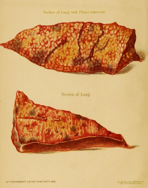
to the heart. The glands in the axilla were swollen. The lungs were
found studded irregularly in different parts, with circumscribed spots of
livid congestion: these existed both upon the surface and in the substance
of the lungs; they were generally about the size of a filbert,
but in some places they occupied a single lobule, and were accurately
circumscribed by its outline.
EXPERIMENT VII.
On the 23rd of November, 1848, about an ounce of perfectly pure
pus (previously warmed) was injected into the right jugular vein of
an aged ass; the vein immediately became "corded", and the blood
appeared to have coagulated in the vessel. The operation did not much
excite the breathing; but the pulse, which naturally was 35 in the
minute, rose to 60, and subsequently fell to 55.
24th. The animal dejected; appetite indifferent. The vein can be
traced as a thickened cord as far as the sternum. Respiration 12 (the
natural standard); pulse 50.
25th. The parts around the vein much infiltrated with serum: pulse
55; respiration 12.
26th. The wound in the neck began to suppurate, and an abscess subsequently
formed in the course of the vein, about midway between the
opening and the sternum. The general symptoms continued, with very
slight variation, until the 4th of December, when the animal was destroyed.
Post-mortem appearances. The jugular vein was found to have become
inflamed only in the course of the circulation, and to be obliterated a
short distance below the external opening. The surrounding parts
were greatly infiltrated with serum and lymph, and several abscesses
had formed in the immediate neighbourhood. The lungs did not present
any well-defined patches of congestion, as in the last mentioned experiment.
EXPERIMENT VIII.
A healthy ass, six years old, was operated on upon the 16th of
November, 1848. The respiration was naturally 14 in the minute, and
the pulse 38. About two ounces of highly offensive pus, obtained from
the frontal sinus of a horse, were injected into the left jugular vein; the
pus had unintentionally been mixed with water previous to its being injected.
The vein became full during the operation, as though the blood
in it were in a semi-coagulated state. The pulse now became 60, and
the respiration 20 in a minute; slight rigors occurred in two hours.
November 17th. The animal is tranquil; appetite good; pulse 48, small
and wiry; respiration 16. In the evening he was rather more excited;
the vein was becoming inflamed downwards towards the heart; pulse
60; respiration 20.
November 18th. The vein was more inflamed, and slight suppuration
was visible at the orifice of the wound. Respiration 16; pulse 55. From
this period to the 23rd, the pulse continued from 55 to 60, and the respiration
varied from 12 to 18.
November 26th. The swelling in the situation of the vein is rapidly
subsiding; pulse 55; respiration 12.
The animal gradually recovered, and on the 26th of February, 1849,
was made the subject of another experiment. The right jugular vein
having been opened, two fluid ounces of pure healthy pus were injected,
and propelled in the course of the circulation, by pressure upon the vein
externally. The vein became tense during the operation, and sensibly
resisted the attempts that were made to propel its contents towards the
heart. Even forcible pressure was not sufficient to overcome the resistance
offered to the return of blood. Soon after the operation, the animal had
a rigor; the breathing became laborious, but not accelerated; pulse 57.
After the lapse of seven hours, the animal appeared dejected; he refused
to eat or drink; the extremities were cold; breathing 16 in the
minute; pulse 60, small and irregular.
February 27th. The vein can be felt thickened as far as the sternum.
The general symptoms are the same as on the previous evening.
28th. There appears less constitutional irritation; pulse 60; respiration
14.
March 2nd. Appetite still indifferent; pulse 60; respiration 16.
From this date to the 7th, when the animal was destroyed, the general
symptoms continued much the same, but the induration and swelling
around the jugular vein, from the opening to the sternum, became
greater.
Post-mortem appearances. The left jugular vein was found completely
obliterated. The remains of a firm coagulum obstructed its
canal for some distance below the opening which had been made into it,
and terminated, below, in an elongated conical portion, which adhered
to one side only of the vessel. On the right side, an abscess had
formed in the course of the vein; and for two inches, the whole of the
parts were imbedded in a confused mass of pus and lymph, in which it
was impossible to distinguish the structure of the vein. Both above
and below this, for several inches, the vein was filled with coagula,
which effectually obliterated it. These coagula extended for several
inches in the course of the circulation; but beyond them, in both directions,
the vessel was pervious. The lungs presented some slight spots
of congestion, but not of the same characteristic kind observed in Experiment
vi. The other organs were healthy.
EXPERIMENT IX.[27]
Two drachms of pus, somewhat fetid, derived from a large common
ulcer, and diluted with a little water, were injected into the jugular
vein of a middling-sized dog. The animal immediately made several
convulsive efforts to swallow, and soon became faint. It showed indications
of pain, and vomited more than six times in the course of the
day. At the expiration of an hour, it appeared slightly relieved by an
evacuation, and by passing turbid urine. In the evening, it was very ill;
it lay upon its side with its legs extended; had a very feeble pulse and
scarcely perceptible respiration. Ten hours after the experiment, it
passed black, liquid, and extremely offensive motions; these were accompanied
by immediate relief. The animal regained its appetite, eat
and drank freely, and went to sleep. The day following, it appeared
nearly well. On the third day, three drachms of the same pus were
injected into the opposite vein; after the lapse of a certain time, there
occurred, as in the first instance, faintness, vomiting, and frequent desire
to pass urine; twelve hours after the injection, frequent liquid, white,
and very fetid motions were passed, and the animal died at the expiration
of twenty-four hours. On opening the body, no alteration was found
either in the intestines or other organs.
EXPERIMENT X.
The last experiment was repeated on a greyhound with the same
results: faintness, fever, vomiting, and repeated evacuations succeeded
each other, with recovery after the first experiment, but not after the
second. On opening the body, no lesion was observed, except that the
inferior lobes of the lungs were gorged and almost hepatized.
EXPERIMENT XI.
Three drachms of recent pus, derived from the same patient as
in the last experiments, were injected into the jugular vein of a small
emaciated unhealthy dog. After the expiration of three minutes, there
was an abundant evacuation of urine, followed by continued vomiting,
and repeated ineffectual efforts to pass fæces. For nearly a quarter
of an hour, there was a kind of emprosthotonos, rigidity of the limbs,
and a death-like condition. Subsequently, fresh vomiting ensued, with
very fetid liquid evacuations, which were followed by apparent relief;
soon after, however, long continued tenesmus made its appearance,
and terminated in death, five hours after the injection of the pus. On
opening the body, the mucous membrane of the intestines was found
red, swollen, and inflamed, especially in the colon and rectum.
EXPERIMENT XII.
Half an ounce of pus, similar to that used in the preceding instances,
but more putrid, in consequence of having been longer kept,
was introduced into the veins of a middling sized dog. The animal, as
in the other cases, was seized with vomiting, accompanied by violent
straining. Subsequently, strongly marked nervous symptoms made
their appearance. The eyes wandered; there was extreme sensibility,
and involuntary convulsive twitching over the whole body, accompanied
by faintness, hiccough, and short piteous cries. The walk was unsteady,
staggering, and without apparent object. There was furious delirium,
ardent thirst, dyspnœa, palpitation of the heart, etc. This state lasted
for nearly two hours, and the animal died in frightful convulsions, without
having experienced any critical evacuations, as in the former cases.
Post-mortem appearances. On opening the body, while still warm,
the venous blood was found very firmly coagulated, not parting with any
of its serum when left at rest; the left ventricle of the heart showed,
on its external surface, some stains of the colour of lees of wine, formed
by a kind of concrete pellicle, which disappeared only after long rubbing
and maceration. The other organs appeared healthy.
EXPERIMENT XIII.
Some beef was allowed to decompose in some dog's blood; half
an ounce of the fluid resulting from the decomposition, was injected into
the jugular vein of a little bitch. Immediately, the animal made several
convulsive efforts to swallow, and soon became oppressed, uneasy, and
faint. At the expiration of an hour, there was great prostration, accompanied
by repeated gelatinous and bloody evacuations, and vomiting of
bilious matter. The strength became gradually less, and the animal
died three hours after the injection.
Post-mortem appearances. The lungs were found inflamed in a very
peculiar manner. They were gorged with blood, of a violet or black
colour, and presented many petechial spots, like small ecchymoses.
These spots existed also on the left ventricle of the heart, in the spleen,
in the mesenteric glands, in the gall-bladder, and even in the subcutaneous
cellular tissue. The peritoneum contained some spoonsful of a
reddish serum; but the mucous membrane of the digestive organs was
found to have been principally affected. In the stomach it was slightly
inflamed. In the intestines, but especially in the duodenum and rectum,
it was of a livid colour, presenting many black spots, and covered by a
gelatinous and bloody secretion, resembling lees of wine. The tissues
in these parts were slightly thickened.
EXPERIMENT XIV.
The preceding experiment was repeated, by injecting into the
jugular vein of a moderately large dog, an ounce of fluid, derived from
the maceration of putrid beef in water. The animal very soon passed
extremely offensive, liquid evacuations, with much urine. The breathing
became quick and deep, the pulse small and quick. Repeated efforts
were made to empty the bowels. There was great depression and want
of strength. At the expiration of an hour, a kind of diarrhœa or dysentery
made its appearance. Liquid, bloody, and fetid evacuations, continued
for an hour and a half, when the animal died.
Post-mortem appearances. Livid, brown, and black patches were
found scattered over the lungs. The intestinal canal was filled with a
bloody mucous secretion, resembling the matter that had been voided;
its mucous membrane was of a livid colour, as in the preceding case.
EXPERIMENT XV.
Two ounces and a half of thick fetid fluid, derived from the maceration
of cabbage leaves in an equal quantity of water, for two days,
at a temperature of 77 Fah., were injected into the right jugular vein
of a moderate sized dog. During the operation, the animal made several
efforts to swallow, and soon became faint, and vomited several times.
Some hours afterwards, there was great uneasiness and oppression, with
recurrence of the vomiting, and continued faintness during the day.
After nine hours, a most copious and very fetid evacuation took place.
The discharge was as black as soot, and composed of mucus, with a little
fæcal matter, and a large quantity of what appeared to be corrupted
blood. Some time afterwards, there was a second evacuation of bloody
mucus, exactly resembling the first. On the following day, there was much
loss of strength: the animal lay upon its side, or staggered as it walked.
There was great and insatiable thirst, with a small feverish pulse. But
the most remarkable symptom was the occurrence, at intervals, of palpitation
of the heart, accompanied by extraordinary force and sound,
resembling that produced by long continued hypertrophy of that organ,
in consequence of aneurism[28] of one of the large arteries. On the third and
fourth days, the animal was better, but there were still great thirst, fever,
and occasional rejection of fluids from the stomach. On the fifth day,
the symptoms became aggravated; there was extreme weakness, a tottering
gait, excessive thirst, the eyes red and filled with gum; the nostrils
were stuffed, swollen, and obstructed with mucus; and the lining membrane
of the mouth was tumid, and of a violet red colour. In the
middle of the day, there was a liquid greyish white evacuation, resembling
pus in its odour, consistence, and appearance, mixed with some
clots of putrified blood. Death occurred during the following night.
Post-mortem appearances. The mucous membrane of the eyes, nose,
and mouth, was red or violet, and covered by a very abundant thick mucus.
The lungs were of a dark colour, with some black patches, but still crepitant.
The left ventricle of the heart presented several brown stains,
resembling ecchymoses, which penetrated into its tissue. Its internal
surface was of the colour of lees of wine, offering a singular contrast
to that of the right side, which, however, contained a hard fibrinous
concretion, two drachms and a half in weight, of a light yellow colour,
and resembling grease in appearance. This was of the same consistence
throughout, everywhere free, with the exception of a portion of the size
of a finger nail, which adhered to an irregular and apparently inflamed
spot on the inner surface of the ventricle; no appearance of the injected
fluid could be recognized in this clot. It was continued of the same
colour and consistence into the pulmonary artery, and into the vena
cava, the vena azygos, the axillary, and even the right jugular vein.
The intestinal mucous membrane, especially in the rectum, the
duodenum, and a small portion of the small intestines, was of a violet
red colour. It was inflamed in longitudinal stripes and in patches,
which gave a mottled appearance, even to the outer surface of the intestines,
before they were opened. This discolouration was not accompanied
by any thickening of the tissues, nor by ulceration, and appeared
rather the result of ecchymosis or hæmorrhage. The lining membrane
of the rectum was principally affected, and its mucous glands were
swollen and very prominent. This intestine contained puriform fluid,
resembling the matter evacuated before death. The other intestines
contained a very thick greyish white mucus. The mesenteric glands
were inflamed, and appeared as if infiltrated with blood. The gall
bladder was mottled on its surface by brown and violet patches, and
contained black, thick, ropy bile, resembling melted tar.
EXPERIMENTS XVI AND XVII.
Shewing the effects of the introduction of Mercury into an artery.
An ounce and a half of mercury, mixed with water, was injected into the
left carotid artery of a sheep. The animal immediately evinced pain,
and stood immoveable upon its feet. The head was held down, there
was stupor and heaviness, and the eyes were protruded and widely open.
The fore legs subsequently became bent, and the head inclined over
the right shoulder with a kind of convulsive rigidity, which continued
till death. Two hours afterwards, the animal became comatose, with
some convulsive motions of the limbs, and the left eye became red and
inflamed. Death took place fifty hours after the operation.
Post-mortem appearances. The left eye was found in a state of suppuration,
and contained mercury. Many of the branches of the left carotid
artery also contained some mercury, which had not penetrated to the
capillary system. All the organs supplied with these vessels were red,
swollen, and inflamed, in consequence of the presence of the foreign
matter. The thyroid gland, the tongue, the cheeks, and the lips, were,
however, only affected as far as the median line, leaving the opposite
halves pale and in their natural condition.
A drachm and a half of mercury, mixed with some warm water,
was injected into the crural artery of a large dog. The animal
evinced no pain, and walked resting slightly on the affected limb, which
became sensibly colder. After the expiration of an hour, the animal
refused its food, became restless, and indicated severe pain in the limb,
which was now very hot. On the following day, the leg was swollen and
œdematous. On the third day, there was extreme thirst, increased œdema,
and great suffering. The animal was killed sixty hours after the operation.
Post-mortem appearances. No disease was found in any organ, excepting
the affected limb. This was swollen and œdematous in every
part; abscesses of different sizes had formed, which contained sanious
fluid, mercury, and pus; some parts were in an incipient state of mortification,
and gave out a considerable quantity of air. Globules of
mercury were found in different parts, occupying usually the centre of
the abscesses, and ran out upon the scalpel when incisions were made
into the limb.
EXPERIMENT XVIII.
Shewing the effect of the injection of Oil into an artery.
Three drachms of olive oil were thrown into the crural artery of a
large dog. Slight pain was experienced, and the limb became evidently
cold, and the pulse under the tendo-Achillis could no longer be felt.
Two hours afterwards, a like quantity of oil was again injected. The
leg now began to inflame, and became tender. The following day, the
whole limb was œdematous, much swollen, and very painful. Twenty-nine
hours after the first experiment, the muscles of the thigh and leg,
as well as the cellular tissue, were found in some places gorged with
blood, and inflamed in livid patches; in others, infiltrated with yellow
serum and gelatinous exudations. No oil could be detected in the
affected parts.
EXPERIMENT XIX.
An ounce of putrid water, in which some beef had been macerated,
was injected into the crural artery of a middling-sized dog. The
artery having been tied, the pulse ceased below the tendo-Achillis; the
limb, however, preserved its usual degree of heat, offering a contrast in
this respect to the last experiment. A considerable degree of fever and
restlessness followed the operation; this continued the whole day and
the following night, without any vomiting or evacuations, which so constantly
followed similar operations upon the veins. The next day the
limb was very painful, but not swollen; there was thirst, with the ordinary
secretion of fæces and urine. On the third day, the animal was
evidently better; the appetite had become almost natural, and he could
walk more easily, although the limb was still very painful. In the night,
there were some soft, almost liquid, evacuations. The fourth day, the
animal was evidently recovering, when an ounce and a half of very fetid
and very concentrated fluid (derived from the maceration of beef), was
injected into the crural artery of the opposite limb. The animal immediately
evinced pain, accompanied by very violent and remarkable palpitation
of the heart. It walked lame, keeping the leg raised, and soon
became feverish and uneasy. The symptoms were exactly the same as
after the first experiment. The leg became gradually more and more
painful, extremely sensitive, but not infiltrated with serum. During
the night, there was much expression of pain, and the animal was in
continual motion. Death occurred nineteen hours after the second injection.
The limb had become swollen only within five or six hours previous
to death.
Post-mortem appearances. The limb presented a very large quantity of
bloody fluid infiltrated in all the tissues. The superficial muscles were
black, and presented more or less the appearances of gangrene. The deep
muscles existed as such no longer, but were entirely disorganized, and
converted into a putrid pulp, resembling masses of the red lees of wine,
extremely fetid, and disengaging a quantity of gas. The limb first injected
was still swollen, and presented, in the interior of the adductor
muscles, two or three cavities filled with a putrid bloody serum. In
the chest, the lungs were healthy, as were also the right cavities of the
heart; but the left cavities presented several reddish-black spots, scattered
over their external surface. In the left auricle was a firm yellowish-white
coagulum, adhering to an inflamed spot on its inner surface.
The intestinal canal was filled with a brownish red fluid, resembling
altered blood, which, in the stomach and duodenum, was of the colour
of soot. The mucous membrane of these organs, as well as of the jejunum
and rectum, were gorged with blood, of the colour of the lees of
red wine, but without any inflammatory thickening of their coats.
EXPERIMENT XX.
Shewing the effect of the introduction of Air into an artery.
Seven or eight cubic inches of common air were injected gradually into
the crural artery of a large dog. A peculiar rustling noise, depending upon
the admixture of the air with the blood, accompanied the operation. No
particular symptoms followed; but after some minutes the corresponding
vein became distended with frothy blood, which moved with difficulty,
and became stagnant in the vessel. The whole limb crepitated upon
pressure, but no untoward symptom presented itself for more than half
an hour. An ounce of water, to which seventy drops of medicinal
prussic acid had been added, was now injected into the same artery.
This produced no apparent effect upon the constitution.
A quarter of an hour after, an ounce of saturated solution of nux
vomica was injected into the same vessel. This also was followed by
no particular symptoms. An hour after the first injection, half an
ounce of a weak infusion of tobacco was introduced into the same
artery. Excessive pain immediately followed, accompanied by great
rapidity of breathing. The animal now appeared as if he were going
to die; however, he slowly recovered, appeared giddy and inclined to
vomit, and kept himself in a continual state of restlessness. This
condition was succeeded by fever, accompanied by extreme sensibility
of the limb, and irregularity of the pulse. At the expiration of some
hours, he appeared better; the pulse became more regular and less
feverish, but the leg continued swollen, and extremely painful upon
pressure. During the night, the pain returned, indicated by howling
and restlessness. There were several evacuations of fæces and urine.
The following morning, there was great prostration with much fever, and
apparent suffering. The limb was slightly emphysematous, swollen,
inflamed, and infiltrated with serum.
On the following day, two ounces and a half of water, in which some
nux vomica had been boiled, were injected into the crural artery of the
opposite limb. The dog expressed no pain; but, at the expiration of ten
or twelve minutes, slight convulsive motions became evident, which were
gradually converted into violent tetanic spasms. The animal threw
himself backwards with his limbs extended, and died, after repeated
convulsive attacks, an hour and a half after the last injection.
Post-mortem appearances. On opening the body, no unusual appearances
were observed in the limb upon which the last experiment had been
tried, but the opposite one was tumid and emphysematous, infiltrated
with a greyish red frothy serum of a fetid odour. The small vessels were
obstructed by firm clots of blood. The gall-bladder was greatly distended;
and the intestinal canal contained a quantity of yellowish mucus.