BY WILLIAM ROSE, M.B., B.S.Lond., F.R.C.S., PROFESSOR OF SURGERY IN KING’S COLLEGE, LONDON, AND SURGEON TO
KING’S COLLEGE HOSPITAL, ETC.
LONDON:
H. K. LEWIS, 136 GOWER STREET, W.C.
1891.
To the Memory
OF
SIR WILLIAM FERGUSSON, Bart., F.R.S., LL.D., SERGEANT-SURGEON TO THE QUEEN; PROFESSOR OF CLINICAL SURGERY IN KING’S
COLLEGE, LONDON, AND SENIOR SURGEON TO KING’S COLLEGE HOSPITAL, ETC.
In Grateful Remembrance OF HIS
EXCEPTIONAL KINDNESS FOR MANY YEARS
BOTH AS A TEACHER AND PERSONAL FRIEND, AND In Humble Recognition OF HIS
MASTERLY SKILL AS A SURGEON, THIS SMALL VOLUME IS
AFFECTIONATELY DEDICATED.
PREFACE.
In bringing this book before the notice of the profession I
have given the results of my experience, which has now
been considerable, in a department of surgery always of
special interest to me, and to which I have devoted particular
attention. The privilege enjoyed for many years of assisting
the late Sir William Fergusson gave me an early insight
into these operations, and an unusual opportunity of learning
their details at the hands of so distinguished a surgeon.
I have endeavoured to make the work as complete as
possible, and to bring its contents up to date, and in this
am greatly indebted to the assistance of my colleague
Mr. Albert Carless, who has not only revised the whole work,
indexed it, and seen it through the press, but has also written
the chapter on Development. I gratefully acknowledge the
kindness I have received from various sources in permitting
the use of blocks for illustrations, notably from Mrs. Mason,
the widow of the late Mr. Francis Mason, whose work on the
subject is well known, from Mr. Oakley Coles, and many
others, whose names will be found in the list of illustrations.
17, Harley Street, W.; 1891.
CONTENTS.
PAGE
CHAPTER I. General Introduction.
Harelip—Cleft palate—Frequency—Occurrence in animals—Associated
deformities—Median harelip—Facial clefts—Macrostoma—Mandibular
clefts—Causes of these deformities
Normal development of mouth, face, nose, and teeth—Ossification—Development
of intermaxilla; old ideas (Goethe’s, &c.);
Albrecht’s theory—Harelip; position of cleft in alveolus,
and in lip—Dentition; accessory teeth—Development of
other deformities
CHAPTER IV. The Anatomy and
Physiology of Harelip and Cleft Palate.
Harelip—Effect of labial muscles on deformity—Structure of os
incisivum and labial segments.
Cleft palate—Arrangement and action of muscles—Shape of
bony segments—Associated irregularity in shape of skull—Physiological
effects in nutrition, articulation, &c.
Period of operation—Preparation of patient—Anæsthesia—Duties
of the assistant—Instruments—Description of uranoplasty;
of staphyloraphy—After-treatment—Complications—Modifications
of the operation
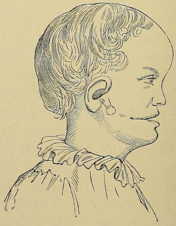
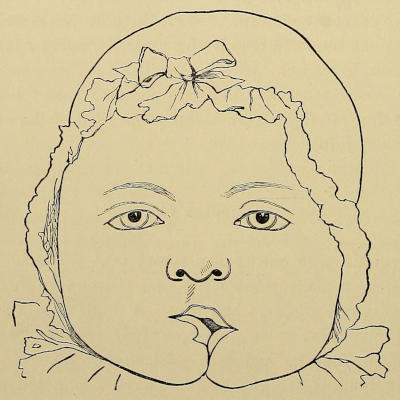
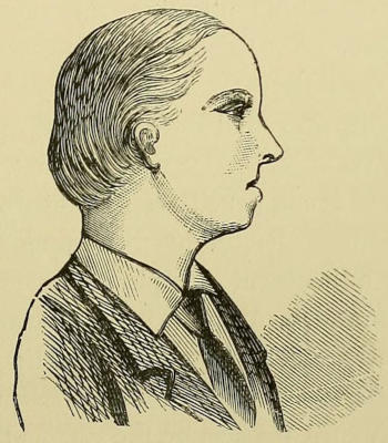
Appearance in profile after operation for double harelip, including removal of os incisivum (Coles).
ON HARELIP AND CLEFT PALATE.
CHAPTER I. GENERAL INTRODUCTION. — Harelip. — Cleft Palate. — Occurrence in Animals. — Association with other Deformities. — Median Harelip. — Facial Clefts (German, “ Schräge Gesichtsspalte ”). — Macrostoma — Mandibular Cleft.
Harelip—Cleft palate—Frequency—Occurrence in animals—Associated
deformities—Median harelip—Facial clefts—Macrostoma—Mandibular
clefts—Causes of these deformities.
The congenital fissures and deformities of the mouth and
lips form a group which is considerably larger than might be
imagined from the scanty notice given them in ordinary text-books;
and although many are extremely rare, yet possibly
if more attention were drawn to them, fresh cases would be
noted and recorded, and the somewhat scanty materials from
which we have to work out their development and characteristics
would be increased. In order to facilitate subsequent
description, I append a classified list of the deformities
which we shall pass under notice, premising that the more
practical part of this work will be occupied exclusively with
two of them.
Six different classes may be described:—
(1) Median harelip (inter-intermaxillary).
(2) Ordinary harelip (intermaxillary).
(3) Facial cleft (maxillo-intermaxillary).
(4) Buccal cleft, or macrostoma (maxillo-mandibular).
(5) Mandibular cleft, or median fissure of the lower lip.
(6) Cleft palate.
Inasmuch as ordinary harelip and cleft palate are the conditions
most commonly met with, it will be convenient to describe
them first, alluding subsequently to the others.
French, bec-de-lièvre. German, Hasenscharte—or if with
complete cleft palate, Wolfsrachen (wolf-jaw).
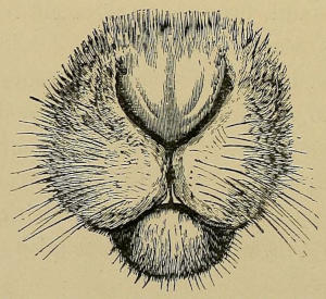
Fig. 1.—Hare’s lip to show the median cleft in the lower part prolonged
upwards into either nostril. (Sutton.)
Harelip is a congenital deformity of the upper lip, characterised
by a cleft extending for a variable depth, either
through the soft tissues of the lip only, or implicating in
addition the alveolus, floor of the nose, and palate. No mention
of this condition is made by Hippocrates, Galen, or any
of the fathers of medicine; and so far as I can discover the
name is first used by Ambrose Paré, who probably initiated
the treatment by pin and figure-of-8 suture. The name is
really a misnomer, in that the condition (as has been many
times pointed out, but notably by Fergusson) does not simulate
a hare’s lip except in the fact of being cleft, for the natural
cleft in the animal’s lip is always in the median line below,
bifurcating above to reach either nostril (Fig. 1), whereas
in the abnormal human lip the cleft lies to one or the other
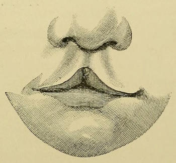
side. Instances of median defect are known, but they
are extremely uncommon, and consist often of more than a
simple fissure.
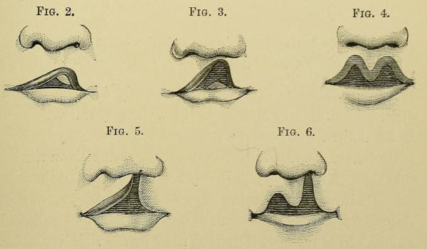
Fig. 2.Fig. 3.Fig. 4.
Fig. 5.Fig. 6.
The deformity may exist as a simple notch in the soft
tissues of the lip, unilateral (Figs. 2 and 3) or bilateral
(Fig. 4); when more decided, it may implicate one or both
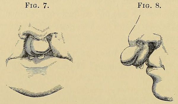
nostrils (Figs. 5, 6, and 7). In mild cases the alveolus is intact;
in others, cleft, constituting the variety known as alveolar
harelip, and the line of fissure may, or may not, extend backwards
into the palate. In all cases of double alveolar cleft,
the palate is also involved, and the central parts of the lip and
intermaxilla tend to project forwards; in the severest forms
these portions are completely isolated from the maxillæ, and,
supported by the vomer and septum nasi, form a proboscis-like
appendage to the end of the nose, which is excessively
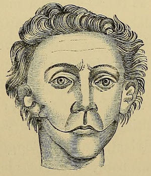
disfiguring. (Figs. 7 and 8 illustrate this deformity as seen
from the front and in profile.)
The shape of the nose in unilateral harelip is very characteristic,
being broad and flattened out from the deficiency of
the floor and posterior wall of the anterior nares.
Figs. 7 and 8.—Double harelip with projection of the os incisivum,
as seen from the front and in profile. (Fergusson.)
Harelip seems to occur more commonly in boys than in
girls. According to Müller, out of 270 cases, 170 were boys,
and 100 girls.
Unilateral harelip is more commonly met with on the left
side than on the right; probably 60-70 per cent. of the cases
are left-sided. Thus Müller reports 142 left-sided against 62
right-sided clefts; Mason, out of 65 cases, found 54 to be unilateral,
and of these 35 left-sided to 19 on the right; Kölliker
mentions that in 165 unilateral clefts, 113 were on the left
side, and 62 on the right. My own experience quite coincides
with these figures. At present, no satisfactory explanation of
this preponderance of left-sided clefts has been given. One
solution suggests itself, but we have no facts of importance
to support it, viz. that, inasmuch as the majority of people
are from heredity or education right-handed, Nature devotes
more energy to completing her developmental processes on
that side than on the left, and any check to this would
be more likely to happen on the left side. It would be
valuable and interesting to know in what proportions other
unilateral deformities occur on the left and right sides
respectively.
Occasionally one sees in the upper lips of children a congenital
red line apparently cicatricial, occupying the position
of the normal harelip fissure, and which has been supposed to
indicate a natural cure of a temporary defect of development.
My colleague, Mr. Carless, has recently shown me a case
of this character under his care in a child a few weeks old.
There was a well-marked red line extending from the lip
margin to the nostril; but there was no irregularity in the
red border, and no evidence of cicatricial contraction; the
tissue of the lip, moreover, seemed quite soft and normal, not
fibrous or hard. These points seem to bear out fully Trendelenburg’s
opinion[1] that the name “intra-uterine cicatrisation
or cure of a harelip” is incorrect, and that such cases are
simply due to the raphe of union remaining evident instead
of disappearing as usual; and he quotes the normal appearance
of the raphes in the scrotum and perinæum as
similar conditions. In this child there was no evidence of any
groove or depression in the alveolus; but other deformities
were present, viz. a very definite post-anal dimple, the cicatrix
being adherent to the tip of the coccyx, a slight condition of
hypospadias, and a congenital hydrocele. There was no history
of deformity in the family, nor of maternal impression.
This is a congenital deformity due to non-closure of
the horizontal palatine outgrowths extending inwards from
the maxillary processes. The name must not be applied to
acquired fissures or defects of the palate due to injury or to
disease of the bones later in life. (See Chap. IX.)
As with harelip, so with cleft palate, the extent of the defect
varies greatly in different cases. Thus in the most
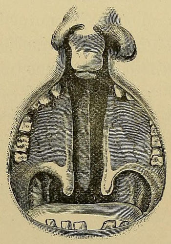
severe forms, there is a total mesial longitudinal cleft,
extending forwards from the tip of the uvula to the level of
the anterior palatine canal, thence bifurcating to communicate
anteriorly with a double alveolar harelip, the os incisivum or
central portions of the intermaxilla being usually displaced
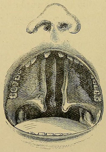
forward (Fig. 9). Such a condition is known by German
authors as “Wolfsrachen,” or wolf-jaw. The vomer descends
in the median line usually into close quarters with, but
separate from the margins of the cleft, and the os incisivum
is attached to its anterior extremity. When the vomer
comes far down and is well developed and prominent, and
the palatal outgrowths small, the cleft appears to be double,
but is not so in reality (Figs. 9 and 10).
Fig. 9.
Fig. 9.—Complete cleft palate and double alveolar harelip; simulating a
double lateral cleft, due to the vomer being seen free between the palatal
segments. (Mason.)
Fig. 10.
Fig. 10.—Complete cleft palate without alveolar or labial deformity; the
vomer is separate from the lateral segments. (Mason.)
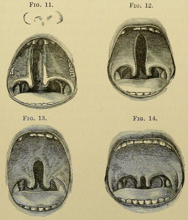
Fig. 11.—Complete unilateral cleft palate
without alveolar deficiency;
the vomer is attached to the left palatal segment. (Mason.)
Figs. 12, 13, 14.—Various degrees of simple fissure of the palate.
(Mason.)
Not unfrequently the vomer is attached to one of the
margins of the cleft, this condition being usually associated
with unilateral alveolar harelip. Such attachment
always occurs on the side opposite to the fissure in the
alveolus; that is to say, since unilateral harelip is more
common on the left, the vomer is usually attached to the right
side of the cleft. Fig. 11 indicates the less common condition
of attachment of the vomer to the left palatal segment.
Rouge[2] and Oakley Coles[3] fully confirm this statement.
The cleft may, however, merely implicate the soft and hard
palate, leaving the alveolus and lip perfect, and does not then
extend further forward than the site of the anterior palatine
canal, and is strictly median (Fig. 12); or it may be still
more limited, involving more or less of the velum, perhaps only
the uvula, or extending a variable distance into the hard
palate (Figs. 13 and 14).
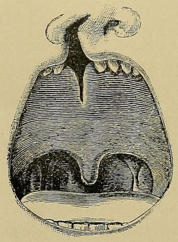
Fig. 15.—Unusual form of cleft involving the alveolar arch, and
the anterior portion of the palate only. (Mason.)
Other less common congenital deformities have been recorded,
and amongst them may be noted a case lately seen by
myself in a girl of four years, in whom there existed an oval
opening at the junction of the hard and soft palate, separated
by a narrow bridge of normal palatal tissue from a cleft of
the posterior half of the velum and uvula, showing intermissions
of development; a congenital aperture in the soft
palate at its junction with the hard, or in any part of the
velum, but with no defect of either uvula or palate bones
(Dieffenbach[4]); a defective development of the palate bones
alone, the mucous membrane remaining intact from side to
side, and hence no cleft resulting (Trélat, Notta, Langenbeck);
or again, as in Fig. 15, a cleft only of the anterior
portion of the palate, extending through the alveolus, and
for a short distance behind it (Mason[5]). Inasmuch as the
union of the two halves of the velum occurs subsequently
to that of the alveolar arch, it appears that this last rare
defect must have been due to an intermission of development,
which was felt only at the anterior portion, whilst that
of the posterior part proceeded normally at a later date.
Mason[6] records a curious case worth mentioning of a girl
under his care in 1877, who had a fissure extending through
the velum, and for a short distance into the hard palate, but
there was no trace of uvula on either side, and the soft palate
was continuous on both sides with the pharyngeal wall.
The width of the cleft varies as much as the extent,
and is a matter of great importance prognostically, as the
broader clefts are much more difficult to close. The direction
or slope of the segments of the bony palate also differs
considerably, in some instances being more or less horizontal
and following the normal curve; in others one or both of the
segments is much more nearly vertical, a condition which is
not at all unsatisfactory, for, as will be explained hereafter,
the more horizontal the palatal processes, the more difficult is
it to gain satisfactory closure by operation (p. 65).
The frequency of the occurrence of harelip and cleft palate
cannot accurately be ascertained, inasmuch as statistics are not
readily to be found. In the ‘St. Thomas’s Hospital Reports’
the number of malformations of the children born is noted in
some of the years. Thus the aggregate number of living
children born in their maternity department in the years
1875, 1877-1880, and 1883 was 10,653, and of this number
there was only one case of harelip, with two cases of cleft
palate, and three of the combined deformity, i. e. about one
case in every 1800 infants born; but if the silence of the
reports for subsequent years means absence of deformity,
then this proportion may be much too great.
On the Continent some old records are obtainable. Thus,
according to Grenser, of 14,466 infants born living at the
Maternity at Dresden from 1816 to 1864 there were sixteen
cases of simple harelip, and nine with fissures of the palate.
Credé states that amongst 2044 infants examined at birth,
only one case of simple harelip was observed, and one of complete
division of the hard and soft palate.
These conditions obtain not only in the human subject, but
also in animals, though not so commonly.
Thus Sutton figures a right-sided harelip in a slink calf,
and mentions a specimen of a harelip in a lamb in the museum
of the Odontological Society; and in our museum at King’s
College there is a specimen of a right-sided harelip in a kitten
with a cleft alveolus, but the palate is intact.
Cleft palate occurs more frequently in animals, particularly
in those born in a state of captivity. Thus it appears that
from statistics taken ten years ago 99 per cent. of the lion
cubs born in the London Zoological Gardens had cleft palates,
indicating that either the food-supply of these animals was
not all that was requisite for perfect development, or that
enforced confinement has a deleterious effect upon the multiplication
of the species. It is a curious fact that in the
Dublin Zoological Gardens the deformity was rarely noticed
amongst the lion cubs, and the reason for this was supposed
to be the supply of such food that the mother could eat both
flesh and bone. Since the same practice has been followed
in London, viz. giving the lions twice a week a young goat
which they can eat, bones and all, the proportion of cleft
palates in the young subsequently born has become considerably
diminished.
Occasionally, besides the fissured palate or lip, other deformities
are noted in the same patient, but not so often as
one might be led to expect. Mason records two or three
cases as having come under his notice, the coincident deformities
being respectively fistulous openings of buccal glands
in an everted lower lip, congenital fissure of the lobe of
the right ear, congenital talipes calcaneus and hypospadias.
Dr. F. Warner records in the ‘Medical Times and Gazette,’
January, 1882, some cases of cleft palate associated with congenital
defects of the heart and smallness of head, and also
notes in his more recent report[7] that in 117 cases of malformations
of the palate, other than cleft, only 42 were not in
combination with other defects. Thus in 55 cases there were
abnormalities in the shape of the cranium, in 16 cases
defective development of the ear, in 12 the existence of an
epicanthic fold, and in 15 cases other defects not tabulated.
Clutton[8] records and pictures a curious development of a
flap of mucous membrane on the lower lip of a woman with a
cleft palate; it was triangular in shape, and with overhanging
projecting angles. The teeth in this case were likewise badly
developed, and were all extracted at the age of nineteen.
Binet[9] reports a case of an old cured right-sided harelip in a
man dead from apoplexy (æt. 53 years) with infantile genital
organs.
Broca[10] describes a much deformed fœtus, stillborn at seven
months, which he dissected, showing a double harelip and
cleft palate, but the os incisivum retained its usual position,
thanks to its mucous covering. The dentition,[11] as noticed
elsewhere, was also interesting, and the buccal deformity was
associated with a congenital diaphragmatic hernia, and an
abnormal condition of the heart and great vessels.
Other associated malformations are on record, e. g. an extra
thumb on each hand; and Sir Morell Mackenzie has reported
a case in which there was a congenital fissure between the
arytænoid cartilages with a trilobate epiglottis, occurring in
conjunction with harelip and cleft palate.[12]
The remaining deformities to be noticed here are much less
common, but demand attention by their rarity and interest,
and on account of the light they throw on the embryology of
the lip and mouth.
This is an exceedingly rare phenomenon, and for long the
possibility of the existence of such a condition was doubted.
Supposed cases were explained by imagining that from some
unknown cause a lateral fissure had been drawn over to the
median line. But at the present time there are records of
several, mainly, however, in German works; in our own
literature there are but few references to the subject. Two
varieties of median defect have been described; and the
distinction between these has been carefully and thoroughly
made by Trendelenburg.[13]
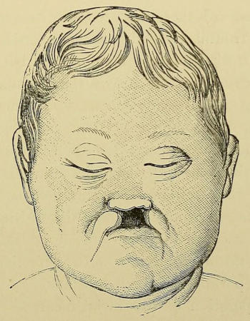
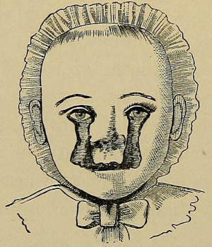
1. Double cleft of the upper lip with failure of development
of the intermaxilla. Some half-dozen cases of this
are indicated in his work; but perhaps one of the best
descriptions is that recently given by Bland Sutton,[14] and
from whose paper the accompanying picture is obtained
(Fig. 16). It occurred in the practice of Mr. Treves,
and died within a few weeks of birth. There was a broad
median defect, flanked laterally by the curved convex
borders of the maxillary processes; the intermaxillæ were
entirely absent, and the nose quite flat. In addition to
this the eyes were affected with coloboma, the right eye
presenting other serious defects. “On examining the child
before its death,” the author states, “I felt convinced that
there was no ethmo-vomerine plate, and this conviction was
strengthened by the peculiar shape of its forehead. When
the child died, this opinion was fully confirmed; there was no
ethmo-vomerine plate, consequently no nasal septum, and
what is more important, the premaxillary bones were absent.”
Fig. 16.—Median harelip showing total absence of the central portion
of the upper lip and of the intermaxillæ, and flattening of the
nose from absence of the ethmo-vomerine plate. (Bland Sutton.)
But according to the cases reported by Trendelenburg the
defects do not stop here. There is usually in addition a broad
median palatal cleft, and an absence of nasal bones and muscles;
but Kundrat records two cases where the palatal processes
of the superior maxillæ and palate bones were well developed,
and united in the middle line. The skull itself has been
found defective occasionally, the whole cranial portion being
small, and the lamina cribrosa and crista galli of the ethmoid
absent; in place of these was a fossa between the orbital
plates of the frontal bone with no bony basis, but only dura
mater covered with mucous membrane. No openings for
the passage of the olfactory nerves were found.
Hadlich has also described changes in the brain in two
cases occurring in Langenbeck’s clinique, consisting mainly
in the amalgamation, more or less, of the two hemispheres;
the corpora striata and optic thalami were united in the middle
line, and the third ventricle, fornix, corpus callosum and
olfactory nerves were absent. It is interesting to note the
association of such an abnormal fusion of the lateral parts
of the brain in the median line with the defective development
of the median parts in the skull and face.
But the separation of the facial elements is not always
maintained; sometimes they fall or are drawn together
by the united palate, resulting in the so-called congenital
atresia of the nose (“angeborene Atresia der Choanen”),
cases of which have been recorded by Luschka, Bitot and
Engel. The latter states that in an infant’s skull examined,
only 2 or 3 mm. of space existed between the orbits, and
4 mm. between the optic foramina.
2. True median cleft of the upper lip with development
of the intermaxilla is an excessively rare occurrence, but a
few cases have been now recorded.
The simplest type consists of a cleft in the soft portions
of the upper lip with no other deformity, but a more complete
variety of the defect includes a median division of the nose.
Mr. Pitts, in the Medical Society’s ‘Proceedings’ (vol. xii,
p. 304), reported a case in a boy aged five months (Fig.
17). The cleft was median, extending halfway up to the
columna. The premaxilla was centrally grooved but otherwise
perfect. The palate was normal.
Fig. 17.—Median harelip, showing a mesial cleft in the soft
structures of the upper lip. (Pitts.)
A more aggravated condition has been dissected by Witzel
(in the Rostock Collection). Behind the cleft in the upper lip
was found a median division of the premaxilla, each half of
which was firmly united to the adjacent superior maxilla.
The vomer was single, but broader than usual, and the palate
cleft throughout; the two halves of the nose were bounded
internally by separated plates of the divided cartilaginous
nasal septum. There was also a defect of the frontal bone
giving rise to a meningocele. This flattening of the nose,
combined with separation of the anterior nares, gave such an
appearance to the face as seemed to warrant the term “dog’s
nose” (Doggennase) which has been applied to it.[15]
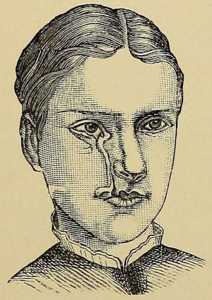
Fig. 18.—Oblique facial cleft, or rather cicatricial deformity of face
along the line usually traversed by such a cleft. (Tillmanns, after Kraske.)
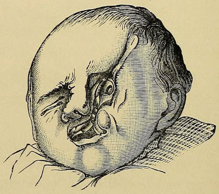
Fig. 19.—Facial cleft in a child, implicating the lower lid and eye, and
with a development of accessory teeth along the cleft margins. (Tillmanns,
after Hasellmann.)
Fig. 20.—Double facial cleft with macrostoma. (Tillmanns, after Guersant.)
These are seldom seen, but a sufficient number are now
recorded and figured to enable us to study the nature of the
defect. Sir W. Fergusson seems the only English surgeon
who has observed this rare condition, the majority of
recorded cases hailing from Germany or France. As we shall
see hereafter, this defect is due to the non-closure of the
cleft between the outermost part of the intermaxilla and the
maxilla itself, and occupies the position which was claimed
up to recent years as that of an ordinary harelip.
In several of the cases noted red cicatrices (Fig. 18) rather
than actual clefts (Figs. 19 and 20) were present. The defect
begins at the free margin of the upper lip, and usually at the
spot whence starts the ordinary harelip cleft; but occasionally
from the angle of the mouth. It then trends upwards and
outwards, leaving the nose entire, and skirts round the ala
nasi to reach its upper limit at the middle of the lower eyelid
which is cleft, or at the inner canthus. The eye itself may
show a coloboma iridis, usually downwards and inwards.
The facial skeleton may be divided or not; sometimes a large
opening into the antrum exists (Hasellmann,[16] Kraske[17]). No
incisor teeth are developed on the outer side of the cleft, the
first tooth seen being the canine. On the inner border of
the cleft lip there is usually a marked frænulum, often
smaller, however, than the normal median frænum.
This deformity may be unilateral or bilateral (Guersant,
Meckel), more frequently the former; and is not uncommonly
associated with macrostoma of the same or opposite side of
the face (Guersant, Pelvet[18]), as seen in Fig. 20.
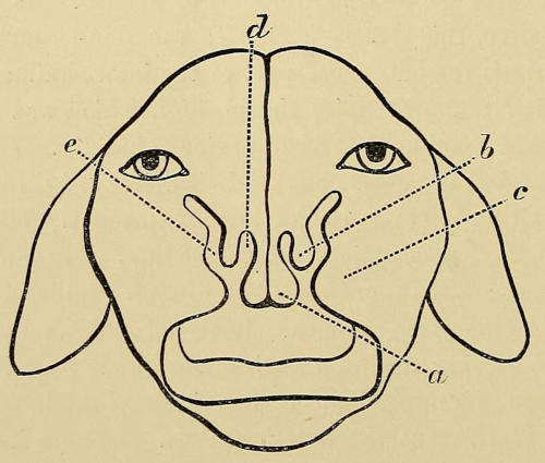
Albrecht[19] records a most interesting case in a newly born
pup (Fig. 21) of double clefts extending from the lip margin
upwards not only into the nostril, but also towards the eye
on either side, i. e. a double associated harelip and facial
cleft. The specimen is taken from the Royal Veterinary
College of Brussels.
Fig. 21.—Front view of a young puppy’s head from a preparation
in the Veterinary College of Brussels, showing double harelip with
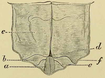
double partial facial cleft. (After Albrecht.)
a. Central portion of upper lip, corresponding to internal nasal process.
b. Ala nasi, corresponding to external nasal process. c. Outer
portion of upper lip, from superior maxillary process. d. Harelip
cleft. e. Facial cleft.
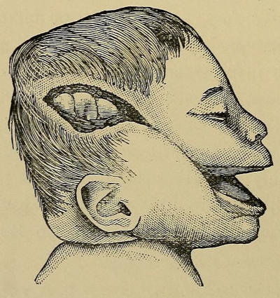
Fig. 22.—Macrostoma, showing the cleft in the cheek prolonged
upwards and backwards by a reddish cicatrix to a lateral loss of substance
of the cranial wall. (Sutton.)
Or commissural harelip (French, bec-de-lièvre genien; German,
Quere Gesichtsspalte, Wangenspalte, or Grossmaul) is a less
uncommon condition, evidenced by an increased transverse
diameter of the mouth. The oral aperture extends into one or
both cheeks, and, if unilateral, is more frequently on the right
side. The cleft extends upwards and backwards towards the
auditory meatus, and sometimes towards the temple to a
variable extent. It may merely be manifested by a slight
increase in the breadth of the mouth, or may extend to
a considerable distance, as in a case reported by Rynd,[20]
where the mouth-opening extended as far as the first molar
on the right side, and to the last molar on the left. Sutton[21]
has published drawings of a child (Fig. 22) in which a very
large cleft existed, the angles of which gradually passed into
a red cicatrix. This scar ended in a gaping recent wound
over the temporal region, extending to the dura mater, and
through this, after death, the convolutions of the brain were
visible. The condition was symmetrical, and he suggests
that the wound in the skull was probably brought about
during parturition. The same author records a condition the
very opposite of this, where the defective closure of maxillary
and mandibular processes was reduced to a minimum, the
deformity amounting to nothing more than a fistulous opening
through the cheek, with a small tumour representing an accessory
auricle just in front of the tragus.
Roulland[22] has recently reported an instructive case in which
double macrostoma existed with accessory auricular appendages,
but this was also complicated with an entire absence
of the middle ear and of the Eustachian tube, with defective
development and absence of the temporo-maxillary joint on
the left side. Such a deformity is probably to be explained
by an excessive obliteration or partial development of the
maxillo-mandibular cleft at its posterior extremity, and a
defective obliteration of the same anteriorly.
Fig. 23.—Double macrostoma, showing the presence of auricular appendages.
(Tillmanns.)
Fig. 24.—Macrostoma with auricular appendages. (Fergusson.)
Associated with macrostoma is often to be noticed some
abnormal condition of the external ear, either defective development
or the production of accessory auricles (Figs. 23 and
24). In a case of bilateral macrostoma recently under my
own care, there was a well-marked accessory auricle. This
complication was first pointed out by M. Debout.
One or two observers (Morgan, Colson[23]) have noticed a
small papillary projection on the red margin of the cleft,
indicating the position where the true mouth ended, and due
to the insertion thereat of the divided orbicularis oris.
For long the very existence of this macrostomatous deformity
was doubted, but cases have been recognised more or
less since 1715, when Muralt pictured it for the first time.
A résumé of all the earlier cases has been made by M. Debout,[24]
whilst Roulland[25] and Pilz[26] have gathered together some of
the later.
Macrostoma is not only attended by great disfigurement,
but is also troublesome from the impossibility of the child
retaining its saliva, and the food escaping during mastication.
Suckling can be performed if the nurse’s nipple be long, but
is difficult otherwise. This deformity is, perhaps, more frequently
associated with defective cerebral power than any
other of the facial clefts, a large proportion of the subjects
having been idiots.
This condition is one of the rarest that we have had to
describe, so much so that Roux and Cruveilhier denied its
existence, and Fergusson had seen but one case. Bouisson[27]
in 1840 mentions some three or four earlier cases, and
records one that he had seen post mortem himself. Since
that date some six or eight instances have been noted, and the
latest, with some excellent pictures, is described by Wölfler[28]
(Fig. 25).
The cleft extends in different cases to a variable extent.
Thus Nicati, Couronue, F. Petit, and Ammon saw clefts
implicating the lower lip alone. Ribell[29] operated on a cleft
extending to the chin, through which the saliva was continuously
dribbling. Faucon (1868) and Lannelongue (1879)
recorded clefts of the lip and mandible conjoined, and in both
cystic swellings (presumably of the dermoid type) were found
between the segments. Parisé’s (1862)[30] and Wölfler’s cases
were also associated with cleft of the tongue, through its whole
thickness in the former, and only at its tip in the latter.
Fig. 25.—Mandibular cleft, showing the divided lower lip, the segments
being held together by cicatricial bands. (Wölfler.)
In Parisé’s case the child was fourteen days old. The
lower lip was cleft through its whole thickness in the median
line. The free edges were rounded as in harelip, and the
cleft was continued below as a cicatricial band in the middle
line of the neck as far as the sternal notch. The mandible
was in two portions, which were separated from one another
by a distance of two or three millimetres, bridged across by
connective tissue. The tongue was entirely divided, the cleft
extending back to the glosso-epiglottic ligament, and downwards
between the genio-hyo-glossi muscles; each half was
covered throughout with mucous membrane, and was bound
to the corresponding side of the jaw by a mucous ligament or
frænulum.
As to the ætiology of these defects, but little is known.
Heredity is an undoubted factor in their production, and
an investigation of the family history will in many cases elicit
a confirmation of such an idea. Thus in two instances in my
own practice I have been able to determine that the father,
grandmother, and great-grandfather had all suffered from
harelip to a greater or less extent. Mason in his book mentions
several other illustrations of this fact. Liston operated on
four members of one family for harelip. M. Demarquay[31]
related a case in the Surgical Society of Paris, in which, from
the grandparents downwards, eleven children had been born
with harelip. In the ‘British Medical Journal’[32] a correspondent
related his own family history, stating that it had
occurred in some branch or other for the past hundred
years.
An examination of the parents’ mouths should always be
made when possible, and very commonly it will be found
that one or both possess a short upper lip, and a high arched
narrow palate. In others there is a slight groove in the
alveolar process between the central and lateral incisors. I
have also observed a small symmetrical crease on either side
of the median line in the upper lip, indicating a tendency to,
if not a natural intra-uterine cure of, a double harelip.
In some instances the deformity dies out of families,
possibly from the fact that the defective condition in one
parent is remedied by a more perfect development in the
other; whilst in others the tendency distinctly increases, and
a father or a mother with harelip will beget a family where
three out of the four or five children will be similarly affected.
By a proper selection of mates this deformity could probably
be bred out, as well as bred up to.
The so-called Maternal Impression is looked on, especially
by the laity, as another common cause of these deformities.
Medical men will usually receive histories of such with a smile
of incredulity, and rightly so; but some recorded cases, if true,
are so definite that to condemn such an explanation too dogmatically
seems scarcely to indicate a scientific spirit. The
usual type of history given is that after the mother has seen
the defect in the newly-born infant, she looks back over
the preceding nine months to see if there were any apparent
cause for the trouble, and seeking out particularly some shock
or fright produced by seeing something resembling the defect
in her infant often selects something trivial and irrelevant.
The following authentic case is worthy of mention:[33]
A child was born deformed by a left unilateral harelip.
The mother immediately asked to see the infant, declaring
she was afraid it was marked, and on seeing it manifested no
surprise at the appearance of its lip, stating that when about
four months pregnant she received a fright, from the shock of
which she had not yet fully recovered. Startled by a boy
running almost into her arms, from whose face blood was
streaming, she had seen a cut in the left side of the upper
lip, extending through its substance into the nostril, laying
bare the gums and teeth. She turned faint with fright, and
could not banish the thought even after reaching home.
The lad was subsequently examined, and the scar of a cut was
found in that position.
In spite of such facts, however, one hesitates somewhat in
accepting the antecedent alarm and the subsequent deformity
in the relationship of cause and effect. The imaginary
“maternal impression” probably in nine cases out of ten has
nothing to do with the defect; whilst a real “maternal shock”
which possibly led to the production of the deformity passes
unnoticed. Mr. Carless tells me of a case recently seen by him
of a cleft of the soft palate in a child, whose mother, without
asking any leading questions, gave a history of a sharp attack
of febrile disturbance keeping her in bed two or three weeks
at a period when the fœtus could not have been more than
two months old. This is the type of maternal shock we
should possibly look for, rather than the more out-of-the-way
maternal impressions commonly suggested.
The union of the parts entering into the formation of the
palate, alveolus, and lip is normally completed by the eighth to
the tenth week, and when once this has occurred in these
parts no maternal impression (such as seeing a gashed lip)
could, as far as we know, bring about a retrogressive change.
Should some shock occur to the mother prior to that period,
we can fully appreciate the possibility of its interfering with
the typical growth of the parts then being produced; and the
fact that the due adjustment and union of so many component
parts is requisite for the normal development of the mouth
and face explains why these defects are relatively so common.
That a severe shock to an infant may produce coincidently a
lamellar cataract and defective development of dentine is well
recognised; that a similar type of shock acting on the mother
should result in defective union of parts developing at that
period in the fœtus is not strange; but that the real shock and
the so-called “Maternal Impression” are one and the same is
more than doubtful.
CHAPTER II. ANATOMY AND PHYSIOLOGY OF THE NORMAL PALATE.
The hard palate—The velum and its muscles—The mucous
membrane—The blood supply—The shape and size of the
hard palate—Functions.
The palate is a more or less horizontal partition dividing the
month from the nasal cavity, and consists of a firm bony plate
in front (the hard palate) with a freely moveable membrano-muscular
velum behind (the soft palate), which under varying
conditions of muscular action can either open or close the
communication between the nose and pharynx.
The bony palate forms the vaulted roof of the mouth,
the central and posterior parts of which are nearly horizontal;
and on all sides, except at the back, it is bounded by
the alveolar ridge. Into its formation several bones enter; in
the adult skull one usually sees posteriorly a cruciform suture
indicating the limits of the superior maxillæ and palate bones;
but even in the adult, evidence is forthcoming in the existence
of traces of sutures to indicate that the anterior part of the
palate is formed independently of the part immediately behind
it. Thus Mr. Carless tells me that a cursory examination
by him of a few dozen adult skulls picked up at random in
the Museum of the College of Surgeons revealed the fact that
in quite one half of them traces of sutures could be seen extending
outwards from the posterior part of the anterior palatine
canal; and a similar examination by him of 40 skulls from
the Museum of King’s College of many nations and various
ages showed a similar result. In almost all there was distinct
evidence of the suture in the median line; in 21, the
maxillo-intermaxillary suture was indicated; whilst in 10
skulls, representing the period from infancy to young adult
life, both the above were seen in all, and 7 showed traces
in addition of a suture placed between them on either side,
and which we shall describe hereafter as the endo-mesognathic.
Kölliker[34] similarly records that out of 325 adult
skulls examined, 96 of them showed definite traces of the
maxillo-intermaxillary suture. Albrecht[35] declares that nine
tenths of the skulls in the Königsberg and Kiel Museums
from children under five years of age reveal the existence
of five intermaxillary sutures, proving that there are four
separate portions to the so-called intermaxilla. Each portion
carries an incisor tooth, and the canine is developed
immediately at the junction between the outer portion and
the maxilla. Occasionally there are three incisors on each
side, the jaw being then called hexaprodontous; the extra
tooth is developed from the inner segment of the intermaxilla
(or endognathion), the outer segment (or mesognathion)
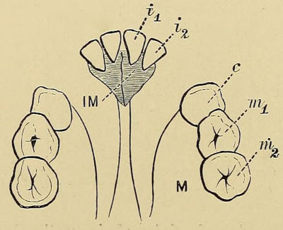
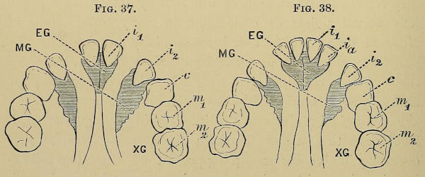
carrying as usual only the lateral incisor. The accompanying
illustrations well indicate this arrangement of sutures and
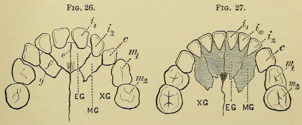
teeth (Figs. 26 and 27); the importance of these facts will be
emphasized later. All traces of the facial aspect of these
sutures disappear quite early in life.
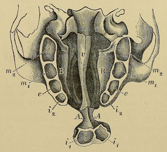
Figs. 26 and 27.—Diagrams to represent the normal human upper jaw of a
child, with four and six incisors respectively, and also indicating the five intermaxillary
sutures. (After Albrecht.)
EG, MG, XG. Endo-, meso-, and exo-gnathion. e. Inter-endognathic suture.
f. Endo-mesognathic suture. g. Exo-mesognathic suture. i₁. Central incisor.
i₂. Lateral incisor. iₐ. Accessory incisor. c. Canine. m₁. First temporary
molar. m₂. Second temporary molar.
The bony surface of the roof of the mouth is perforated by
numerous small foramina for the transmission of the nutrient
vessels to the body of the bone, pitted for the lodgment of
mucous glands, and grooved longitudinally for the transit of
vessels. At the postero-external corners the posterior and
accessory palatine canals give entrance to the posterior
palatine vessels, and nerves; and anteriorly in the median
line is the anterior palatine canal transmitting the naso-palatine
vessels and nerves.
The soft palate is a moveable curtain, consisting of a membranous
expansion or aponeurosis attached to the posterior extremity
of the hard palate by firm fibrous tissue. Incorporated
with it are five pairs of muscles, controlling its movements; it
is covered by a smooth thin mucous membrane, and terminates
posteriorly in the uvula. The arrangement of these muscles is
important, not only from their normal physiological functions,
but also from their irregular action and effects in cases of cleft
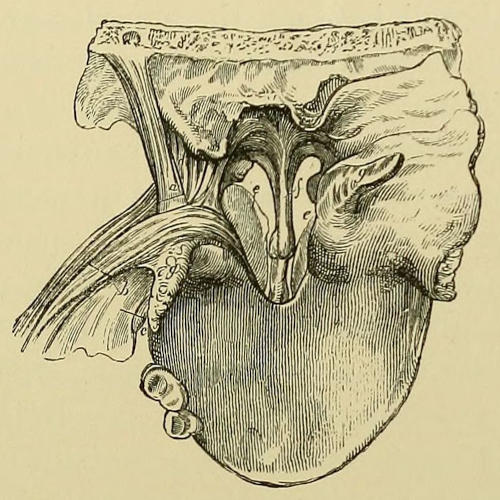
palate (Fig. 28). They may be arranged in groups: two, the
levator and tensor palati, form a superior group; the azygos
uvulæ is intermediate; and the palato-glossus and palato-pharyngeus
form an inferior set. Arising from the extremity of
the petrous bone, the levator passes downwards, and spreading
out below unites with its fellow in the whole length of the
median raphe. The tensor arises from the navicular fossa of
the internal pterygoid plate, and after being reflected around
the hamular process, its action there being assisted by the interposition
of a bursa, is attached to the anterior portion of the
aponeurosis and to the hinder part of the bony palate. The
combined action of these muscles raises and makes tense the
velum, and in addition influences the Eustachian tube; but
the levator is by far the more important. The azygoi uvulæ
muscles arising from the median raphe and spine of the hard
palate descend to the tip of that process, and are thus able
to regulate its length.
Fig. 28.—Muscles of palate dissected. The cut represents the posterior
nares and upper surface of the soft palate.
a. The levator palati. b. The inner bundle of fibres of the palato-pharyngeus,
forming the posterior pillar of the fauces, c. The palato-glossus.
d. The tensor palati; the cartilaginous extremity of the
Eustachian tube is seen in front of this latter. e. The posterior extremity
of the inferior turbinated bone. f. The septum. g g. The uvula
on each side stretched apart. (Fergusson.)
The two descending muscles are placed in the pillars
of the fauces, forming the lateral prolongations of the
velum, and the tonsils lie in a recess between them. The
palato-glossi arising from the tongue ascend in the anterior
pillars of the fauces, and spreading out on the anterior
surface of the velum unite in the median raphe. The
palato-pharyngei start from the median raphe in two lamellæ
enclosing the termination of the levator muscle; they descend
in the posterior pillars of the fauces, and being attached to
the pharyngeal wall between the superior and middle constrictors,
by their contraction assist in raising the pharynx
during deglutition.
The nervous supply of these muscles requires little notice
here; suffice it that the superior set and the azygos are supplied
by the facial nerve, the inferior set from the pharyngeal plexus.
The mucous membrane of the hard palate is of the usual
oral type, and only differs from that of the rest of the mouth
in its close attachment to the periosteum, from which in fact
it is almost impossible to separate it. It is thick, dense,
rather pale and much corrugated, especially in front and at
the sides, whilst behind over the velum it is smoother and
thinner. In it are many small glands (palatine glands) which
extend down to the periosteum. In the median line is a well-marked
raphe, extending anteriorly to a prominence indicating
the position of the anterior palatine canal. The rugose condition
of the membrane over the hard palate is not seen in
young children; it supervenes later in life.
The vascular supply of the palate is free and abundant, a
circumstance which is of the greatest surgical importance in
that it permits of the free detachment of the soft structures
from the hard by long lateral incisions, and the necessary
manipulation of these in uranoplastic operations without any
fear of loss of vitality, provided that the patient’s health and
constitution are tolerably sound, and that sufficient pedicle is
left in front and behind.
The mucous membrane of the hard palate derives its blood
supply from two of the terminal branches of the internal
maxillary artery. The naso-palatine descend through the
anterior palatine canal, and entering the palate at the incisive
foramen (foramina of Stenson) assist in supplying the anterior
portion, anastomosing with the terminations of the more important
posterior or descending palatine, which find their way
to the palate from the spheno-maxillary fossæ through the
posterior palatine canals. Each of the latter arteries on
reaching the palate sends branches to the velum and tonsils, and
its main twig runs onwards in a groove of the bone to supply
the mucous membrane and glands of the hard palate and gums.
Its usual position is parallel to the alveolar border, and about
three quarters of an inch from it; but this varies considerably.
The artery can often be felt pulsating as it emerges from the
bone, and is very likely to be divided in the lateral incisions
made during the operation of uranoplasty; but the knife
should be carried external to it, if possible, so that the trunk
of the vessel may be preserved in the flap. The bony palate
derives its blood supply not only from its lower surface but
also from its upper, and hence detachment of the inferior
periosteal covering does not lead to death of the bone. The
soft palate derives its blood from three sources, viz. the
ascending palatine of the facial, the ascending pharyngeal,
and the posterior palatine of the internal maxillary. The two
former reach it through the sinus of Morgagni, i. e. over the
upper border of the superior constrictor muscle, forming loops
of anastomosis on its posterior aspect with similar branches on
the opposite side; the last supplies the anterior palatal surface.
The normal shape of the palate is a regular arch, bounded
laterally by the gums and alveoli into which the teeth are
implanted so as to describe a parabolic curve, being normally
uninterrupted at any spot by spaces or diastemata. The
height and curvature of the palate vary considerably in
different individuals, not only from inherited peculiarities,
but also from acquired conditions dependent on the teeth.
A person with a good set of sound teeth will probably
own a regular well-formed palate; whilst if sundry of the
upper permanent teeth are lost during the stage of adolescence,
the palate is likely to become high and narrow from the
falling in of the jaw. This is especially the case if the incisor
teeth are lost.
The shape of the palate in a child of two years does not
differ so markedly as one would at first expect from that of
an adult except in length, and the reason for this is plainly
the existence in the latter of three additional teeth on each
side. Its increase in length is from 20 to 30 millimetres,
whilst its breadth is only augmented by 10 to 15 mm., and
this mainly posteriorly. When once the permanent incisors,
canines, and premolars are developed, the anterior portion of
the palate alters but little in shape, unless any of these teeth
be lost, and the gaps not artificially maintained.
Dr. Ehrmann[36] states that the alveolar border in front of
the canine teeth forms a nearly regular semicircle, with a
posterior transverse diameter of 22-26 mm.; thence the
alveoli diverge regularly, adding to the diameter about 2-4
mm. for each tooth. He gives the following measurements
as the mean of many observations:
From 2-6 yrs.
From 7-10 yrs.
From 11 yrs.
Interval between canines
22-25 mm.
23-27 mm.
25-28 mm.
” 1st premolars
24-29 ”
25-30 ”
27-30 ”
” 2nd ”
26-31 ”
28-32 ”
31-34 ”
” 1st molars
—
—
32-37 ”
Oakley Coles[37] has carefully investigated the size of the
palate in several series of skulls in the Museum of the College
of Surgeons, and gives the results as follows:
Of 34 adult skulls of European origin, the average length
was 49 mm., the average width at the second bicuspid was
35 mm., and the average height from the margins of the
alveoli 9 mm.
Of 32 adult skulls of mixed races, the average length was
54 mm., the width 35 mm., and the height 12 mm.
The frequent association of inherited mental and nervous
weakness with a high arched palate is now a well-established
clinical fact. Thus Savage states that in “Genetous Idiocy”
(i. e. idiocy which starts in fœtal life, and cannot be traced to
any specific disease) the palate is usually keel-shaped, the
molar teeth being closely approximated; they are also late in
appearing and deficient in number. “Although this kind of
palate may be present in healthy individuals or in those suffering
from ordinary insanity, if it be associated with weak-mindedness
or moral peculiarities in youth I believe one is justified
in saying that the tendency to moral or intellectual deficiency
is congenital.”[38] Only recently Dr. F. Warner has reported[39] to
the Psychological Section of the British Medical Association the
results of an investigation as to the occurrence of deformities
amongst school children, and their relationship to defective
vital and mental conditions. Out of 5344 children examined,
physical deformity was noted in 399 cases, and of these 274
were boys and 125 girls, i. e. in the proportion 9·8 per cent. and
5 per cent. respectively. It was found that of these 25 per
cent. exhibited evidences of low nutrition, 36 per cent. evidences
of nervous weakness, and 31 per cent. of mental dulness. 117
cases were noted of deformity of the palate, 77 boys and 40 girls;
and of these 35 per cent. gave signs of low nutrition, 39 per
cent. of nerve weakness, and 35 per cent. of mental dulness.
These defects were more marked and more frequent in the
pauper than in the elementary public schools, in the proportion
of 4·2 to 2·2. As to the character of the malformations, the
following are the numerical statistics: In 105 cases, the
palate was arched, narrow, high or vaulted; in 8, it was
𝖵-shaped; in 4 it was of the flat type.
Dr. Langdon Down[40] had previously noticed and pointed out
this frequent relationship, remarking that as the result of a
large number of careful measurements of the mouths of the
congenitally feeble-minded and of intelligent persons of the
same age, he found with few exceptions a marked diminution
in the transverse measurement between the posterior bicuspids,
resulting in an inordinate vaulting of the palate. There was
often noticed an actual deficiency in the bony structures of
the posterior part of the hard palate, causing the velum
to hang down abnormally, interfering with phonation.
The function of the hard palate is mainly mechanical.
Acting as a partition between the nasal and buccal cavities,
it prevents nasal mucus from falling into the mouth, and, by
presenting an opposing surface to the tongue, allows of the
production by the latter of the vacuum necessary for suction,
and enables the tongue to direct the food towards the alveoli,
and to disintegrate soft particles, thus assisting mastication.
It is also an accessory to the development of taste by enabling
particles to be evenly spread over the back of the tongue. For
the production of articulate speech the hard palate is an indispensable
factor, and the quality of the voice is much influenced
by its contour.
The functions of the soft palate are mainly related to the
acts of respiration, deglutition, phonation, and articulation.
1. In respiration.—If the mouth is closed, and the respiration
purely nasal, the velum hangs loosely, and allows free
passage of air through the posterior nares. If the mouth is
open, the velum is raised, and air passes freely through the
fauces to or from the larynx. When air passes simultaneously
through nose and mouth, the velum hangs in a more
or less flaccid condition midway between the two extremes,
and sometimes, when absolutely relaxed, vibrates, giving rise
to snoring or stertor.
2. In deglutition.—The passage of food into the nose is
prevented by the closure of the posterior nares. This is
effected by elevation and tension of the velum, the levator
and tensor muscles acting in unison, so that its position
becomes almost horizontal. The raised velum meets the posterior
wall of the pharynx, which advances as the result of
the action of the upper horizontal fibres of the superior constrictor,
and the closure is completed on either side by the
approximation towards the median line of the posterior pillars
of the fauces from the action of the palato-pharyngei muscles
contained therein. These, acting from the soft palate as a
fixed point, and raising the pharynx to grasp the bolus of
food, straighten the walls of the sphincter-like isthmus
faucium, and so guide the food as down an inclined plane.
The tension of the velum also assists in this guidance. That
the above is the action of the palatal structures is proved by
the results of their imperfect development or paralysis, e. g.
in post-diphtheritic paralysis, where the naso-pharyngeal
cavities remaining unclosed, food (especially if fluid) regurgitates
into the nose.
3. In phonation and articulation.—The soft palate is here
of considerable importance, inasmuch as it is needed to cut
off the naso-pharynx and nasal cavities from the oral pharynx.
When defective or paralysed, a certain amount of nasal resonance
is imparted to the voice, which, however, is less noticeable
during vocalisation than in articulation. For the production
of clear normal voice-sounds it is essential that the
separation between nose and mouth should be absolute, except
for the sounds m, n, and ng. The American twang is
probably due to a slight relaxation of the soft palate, permitting
a small percentage of voice-sounds to pass through the
nose. Dr. N. W. Kingsley[41] has recently published some excellent
diagrams illustrating the position of these parts during
the production of definite sounds, and for all, except those
mentioned above, the velum is horizontal, and in contact with
the posterior pharyngeal wall.
CHAPTER III. DEVELOPMENT. [42]
Normal development of mouth, face, nose, and teeth—Ossification—Development
of intermaxilla; old ideas (Goethe’s,
&c.); Albrecht’s theory—Harelip; position of cleft in
alveolus, and in lip—Dentition; accessory teeth—Development
of other deformities.
Before discussing from an embryological standpoint the
various deformities which we have already described, it is
essential for us to consider the normal process of development
of the parts entering into their formation.
About the end of the third week of intra-uterine life, the
anterior cerebral vesicle becomes acutely bent over the end of
the notochord, and a marked depression is seen on the ventral
aspect of this, constituting what is known as the Stomodæum,
or primary buccal cavity. This, however, is formed rather by
the outgrowth of surrounding processes entering into the
formation of the facial elements than by any definite or
distinct involution of epiblast. The cavity is bounded posteriorly
and superiorly by the cephalic flexure of the cerebral
vesicles, and inferiorly is separated from the cephalic portion
of the intestine by a septum. This becomes perforated at a
slightly later date (eighth or ninth week), and communication
is thus established between the stomodæum and intestine.
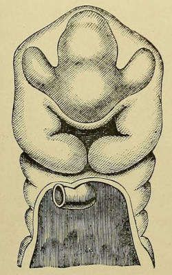
Anteriorly, the opening is at first stellate in shape (Fig. 29),
but soon assumes the form of a transverse cleft by the union
in the middle line of the first pair of post-oral branchial arches,
in which are developed the primary cartilaginous bars on either
side, known as Meckel’s cartilage, the anterior part of which
goes to form the inferior maxilla, and the posterior part the
malleus. The soft parts around develop into the lower portion
of the cheek, the lower lip and chin (Fig. 30).
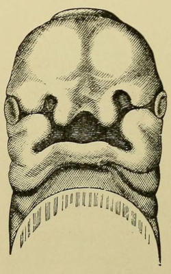
Fig. 29.—Head of fœtus, of about 5 weeks, from ventral aspect (after
His), showing the primitive stomodæum bounded above by the undivided
fronto-nasal process, laterally by the maxillary, and below by the still
separate mandibular processes. The quinqueradiate appearance is well
represented. (Sutton.)
Fig. 30.—Head of fœtus from ventral aspect of a little later date (6-7
weeks). The mandibular processes have now united; the orbito-nasal fissure
has come in contact with the ocular vesicle, and the fronto-nasal process has
developed into external and internal nasal (or globular) processes around
the nasal fossæ (after His). (Sutton.)
At the same time that this mandibular arch is being developed,
other changes are occurring around the upper part of the
stomodæum, viz. the shutting off of sacs lined with epiblast to
assist in the formation of the organs of special sense, and the
outgrowth between them of fleshy processes which by their
later amalgamation form the facial skeleton and coverings.
Three of these involutions of epiblast occur, two communicating
more or less with the stomodæum, viz. the nasal and
ocular; whilst the third, or auditory, is separate. Expansions
from the cerebral vesicles meet them, and by further changes,
unnecessary to particularize here, the organs of special sense
are elaborated.
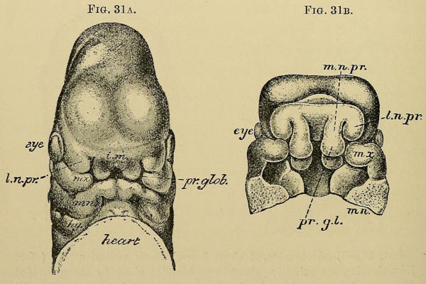
Fig. 31A.—Head of fœtus at a somewhat later date (8 weeks) as seen from
the front, showing the nasal and maxillary processes in close apposition, and the
clefts between them diminishing in size.
Fig. 31B.—The same, seen from below, the mandibular process having been
removed.
i.m. Central portion of fronto-nasal process, m.n.pr. Internal nasal process.
pr.gl. Globular process, or the lower rounded extremity of the former.
l.n.pr. External nasal process. m.x. Maxillary process. m.n. Mandibular
process.
The most anterior of these depressions are the primary
Olfactory pits which appear on either side on the lower surface
of the wall of the anterior cerebral vesicle at a very early
date. They are at first merely depressions surrounded by a
raised margin; but subsequently they become pyriform by the
extension of the lower end as a groove into the stomodæum.
Each pit and groove is bounded laterally by thickened rounded
outgrowths developed from a broad median fleshy protrusion
from the same part of the cerebral vesicle, the fronto-nasal
process; these are termed respectively the external and
internal nasal processes, the latter being also named by His
the globular processes (Fig. 30).
These Globular processes are separated in the median line by
a groove which is subsequently obliterated by their amalgamation
to form the central portion of the upper lip (“Philtrum”
of German authors), and from their deep aspect the
inner segments of the intermaxilla (endognathia). Above
this groove is a central flattened median portion of the fronto-nasal
process, from which subsequently the prominence of the
nose is developed (Figs. 31A, 32, and 33), a result of the continued
growth from its deeper aspect of the ethmo-vomerine
plate.
The external nasal process forms the ala nasi and the soft
parts in its immediate neighbourhood, extending downwards a
little distance, but probably not so far as the red margin of the
lip. From its deeper aspect the outer portion of the intermaxilla
(mesognathion) is developed, and in it appears the
germ of the lateral incisor. It also serves to separate the nasal
pit from the second epiblastic intrusion which assists in the
formation of the eye. The lower boundary of the naso-orbital
fissure which passes from the primary ocular involution to the
stomodæum is formed by the maxillary process, which is usually
described as an upward extension of the mandibular process,
but which probably arises separately as a pre-oral branchial
outgrowth. From this is developed the whole of the superior
maxilla, except that portion which is intermaxillary, and also
the greater part of the cheek.
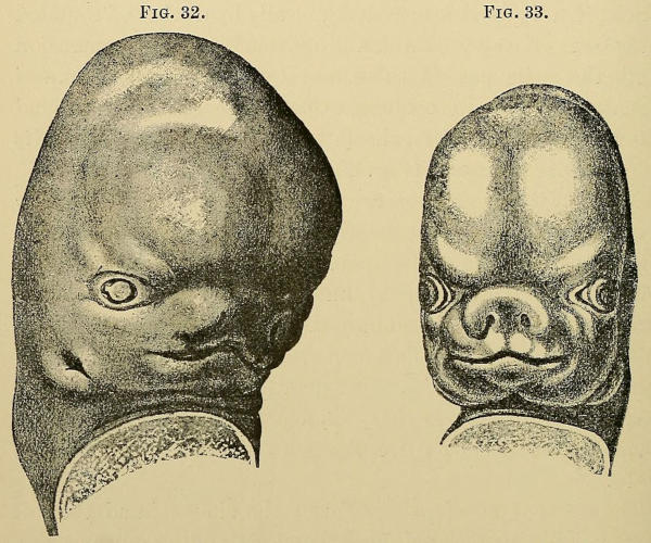
Figs. 32 and 33.—Later stages of development of fœtal head.
About the sixth week the stomodæum shows signs of division
into upper and lower segments by the outgrowth from
the deep aspect of each maxillary process of horizontal palatal
plates, which by their junction in the median line form the
rudiments of the hard and soft palate, separating thus the nasal
and buccal cavities. The anterior portion of these unite
with the lateral aspects of the deeper parts of the fronto-nasal
process, leaving a space of greater or less dimensions in
the median line, known as the anterior palatine canal, which
serves subsequently for the passage of nerves and vessels, and
for the lodgment of the “organ of Jacobson” in animals in
which it occurs.
The upper or nasal cavity is again subdivided into lateral
halves by the growth downwards from the under surface of
the fronto-nasal process of a central vertical septum, to become
in time the cartilaginous septum nasi and the bony
ethmo-vomerine plate, uniting at its lower border with the
primary fleshy palatine processes (Fig. 34).
The tongue grows as a fleshy protuberance from the floor
of the stomodæum; antero-lateral segments on either side
from the conjoined second and third branchial arches unite
with a central posterior growth from the tuberculum impar in
an inverted 𝖸-shaped manner. At the point of junction of
the segments is a depression from which the thyroid gland
develops, indicated in later life by the foramen cæcum.
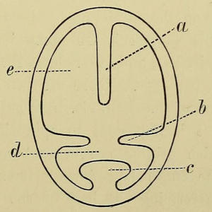
Fig. 34.—Diagrammatic representation of the development of the
palatal processes and of the ethmo-vomerine plate, seen in vertical
section. (After Gegenbaur.)
a. Ethmo-vomerine plate. b. Palate processes. c. Tongue.
d. Buccal cavity. e. Nasal cavity.
Each of these primary epiblastic pits is at a later date
almost entirely cut off from its connection with the buccal
cavity. The external and internal nasal processes of the
fronto-nasal outgrowth unite below the anterior olfactory pits,
and thus surround the anterior nasal apertures, and separate
the nares from the mouth. The external nasal and superior
maxillary processes are also freely amalgamated except along
one small deep track, which remains patent to form the nasal
duct and lachrymal passages; and probably the internal nasal
and maxillary processes unite below the external nasal process
to establish the continuity of the red margin of the upper lip.
The union of all these various parts has been completed by
the sixth to the tenth week of normal fœtal life; the external
nasal and superior maxillary processes unite first, and by the
sixth week are becoming closely approximated to the central
portion of the fronto-nasal process, a time when the palatal
processes are only indicated as slight ridges. By the ninth
week the alveolus and upper lip are complete, and union of
the palate is commencing from before backwards, being
usually completed even to an indication of the uvula by the
tenth week.
Whilst the later stages of these developmental processes
are in progress, points of ossification have been appearing in
many places to form the cranial and facial skeleton. A full
knowledge of this subject is still unattained, but the researches
of Goodsir, His, and others have thrown much light
on hitherto dark passages. There are two main sources of
origin of the bones of the skull, viz. from cartilage and from
membrane, and it is important to appreciate the portions
of the skull which originate from each of these sources
respectively. The bones laid down primarily in cartilage
are mainly those forming the base of the skull and their
anterior prolongations. Thus about the fourth week of
intra-uterine life the basis cranii consists of a cartilaginous
mass surrounding the upper end of the notochord, and prolonged
anteriorly around the pituitary fossa as two cartilaginous
bars, the trabeculæ cranii, into the fronto-nasal process.
From the anterior extremity of this the nasal bones and
cartilages are developed, and from the under surface the
ethmo-vomerine plate. The transformation of the primordial
mesoblastic undifferentiated tissue into recognisable cartilage
is occurring from about the fourth or fifth week until the
eighth, when ossification at different centres is apparent. To
the development of the intermaxilla we shall refer in detail
later; suffice it to say here that the ossifying centres appear
about the eighth week, and by the twelfth to the fourteenth
the whole process is ossified, and the space between the
maxillæ closed except posteriorly, where the anterior palatine
canal remains permanently patent; the component parts of
the bone, however, are not united until a later date. There
are two other cartilaginous foci from which ossification ensues,
viz. the pterygo-palatine cartilage in the superior maxillary
process, a delicate bar from which arise in part the pterygoid
and palatine plates; and Meckel’s cartilage in the mandibular
process for the production of the mandible and malleus. All
the other facial bones are developed from membrane, more or
less in connection with these bars. The vomer is ossified from
a single nucleus appearing in the upper part of the ethmo-vomerine
plate, about the ninth week; from this two laminæ
are developed, which, passing downwards and forwards on
either side of the middle line, embrace the septal cartilage.
The amount of the osseous material increases from behind
forwards, until at maturity a median osseous lamina remains
which is grooved only anteriorly.
The palate bone develops from a single centre appearing
about the eighth week at the junction of the horizontal and
perpendicular portions. The superior maxilla is supposed to
arise from four separate foci of ossification, viz. for the
alveolar arch, for the palate, for the orbito-malar portion, and
for the naso-facial segment. All these are united together by
the third month.
It is unnecessary here to discuss the development of the
teeth beyond stating that the thickening of the epiblast
covering the gums, which occurs as the earliest sign of the
production of the milk teeth, is to be seen about the forty-fifth
day, when as yet there are no signs of ossification of the
maxilla, and by two and a half months a distinct involution
filled with cells is evident. Calcification commences about the
eighteenth week of intra-uterine life, and extending from
crown to fang is usually not completed until from twelve to
twenty months after birth.
By the fifteenth week of embryonic life preparation is being
made for the development of the first four permanent molars,
and soon afterwards in the sixteenth week occur the inflections
of the mucous membrane giving rise to the enamel organs for
the twenty anterior permanent teeth; and from this period
until the birth of the infant the germs of the twenty-four permanent
teeth are passing through the various stages preparatory
to calcification, so that at birth the child has not only
twenty milk teeth with calcification nearly complete, but also
the germs of twenty-four permanent teeth. Calcification commences
in twelve of these latter during the first year of life,
viz. in the first molars and the incisors, and spreads from the
crown in which it starts to the fang. In the case of the
incisors this process is not completed until the tenth year.[43]
The question as to the ossification of the intermaxilla has
been purposely omitted hitherto, that the subject and its
morphological relationship to congenital deformities might
be more fully discussed.
In the time of Galen[44] the presence of the intermaxilla as a
separate bone had been demonstrated in apes and lower
animals, and its existence in man inferred, although probably
not actually seen. This opinion held its ground till the sixteenth
century, when Vesalius attacked it, maintaining that
no such bone existed in man, and its absence was even
claimed as a distinguishing feature from the lower animals.
The first to actually discover and notify the separate existence
of the bone in the human skull was Dr. Robert
Nesbitt,[45] who, in a lecture before the Royal College of
Surgeons in 1736, described and figured the suture crossing
the anterior part of the palate at all times of life, and maintained
that during intra-uterine life each superior maxilla
“is generally divided into two distinct parts, the sutural line
running from between the dentes canini and incisivi up to
the bottom of the nose.” But the merit of appreciating the
importance of this fact belongs to Goethe[46] and Vicq d’Azyr,[47]
the former of whom, in the year 1779, demonstrated the
existence of the intermaxilla in the human fœtus, and, as
the outcome of this discovery, promulgated in 1819 the
theory that in alveolar harelip the cleft in the alveolus occurs
at the maxillo-intermaxillary suture, i. e. between the lateral
incisor tooth and the canine. This opinion has been believed
and handed down through successive generations of surgeons
until the present day, but more recent and exact research has
so increased our knowledge that it cannot now be considered
correct. Many painstaking embryologists have, during the
last ten years, been investigating this subject; but the
honour of raising the question as to the morphological
position of the cleft in harelip lies with Professor Albrecht
of Brussels, who in a masterly series of papers has fully established
the fact, observed also by others, that the intermaxilla
is not developed en masse, but is formed by the coalescence
of four segments, two on either side; and he maintains that
the cleft in alveolar harelip lies not between the maxilla and
intermaxilla, but between the inner and outer intermaxillary
segments. These have been named respectively the endo-
and meso-gnathion, whilst the maxilla proper is called the
exognathion. According to Albrecht, therefore, the cleft is
not situated along the exo-mesognathic, but along the endo-mesognathic
suture.
Much controversy has been lighted up by this pronouncement,
but here only a few of the points of interest and
importance will be discussed.
The development of the intermaxilla from two centres on
each side may be accepted as a proven fact. It was first
maintained by the late Mr. Callender,[48] who stated that these
bones have a lateral wedge-shaped sutural surface, fitting
into a groove in either superior maxilla, and that the alveolar
processes of the latter extend forwards, forming the
anterior walls of the sockets of the central incisors, and so
fix the bones in position. A confirmation of the idea that
the anterior alveolar walls of the incisor teeth are formed in
this way was sought in the well-established fact that these
particular parts are very imperfectly developed in those cases
of alveolar harelip in which the os incisivum is isolated from
the superior maxillæ; but such is probably due to the abnormal
condition and position in which the bone is developed, rather
than to the loss of the maxillary “clip.” And certainly the
most recent researches tend to prove that the maxillæ have
no share in the formation of the alveoli of the incisor teeth.
Sir William Turner and other anatomists have fully confirmed
this method of development from four ossific centres,
and important additions to our knowledge of the subject have
been made recently. Thus M. Gilis[49] describes and figures
the condition of the palate as seen in a six-months fœtus,
where it was clearly demonstrated that the intermaxillary
portion formed a sort of lozenge-shaped prism fitting in
between the two maxillæ, and consisting of four portions of
bone, the sutures between these being perfectly clear and distinct
(Fig. 35). The posterior extremity of the short axis of
the intermaxillary segment corresponds to the anterior palatine
canal, and the anterior surface forms the median anterior
alveolar border, no process of the superior maxilla closing in
the alveoli in front. The upper border of the bone forms the
floor of the nasal apertures.
Fig. 35.—Bony palate of a fœtus of six months, showing the development
of the intermaxilla in four portions.
a. Endognathion. b. Mesognathion. c. Exognathion. d. Anterior
palatine canal. e. Endo-mesognathic suture.[50]f. Exo-mesognathic
suture. (After Gilis.)
Biondi, of Breslau,[51] has completed the observations necessary
for the establishment of this fact by demonstrating the
four actual points of ossification in many fœtal skulls of
different dates, which had been specially prepared for the
purpose. Moreover, as mentioned before (p. 27), traces of
the five intermaxillary sutures, when looked for, may be
found in many adult and in the majority of young skulls.
The fourfold division of the intermaxilla being granted, it
is obvious that a cleft through the alveolus such as that
occurring in alveolar harelip happens at one of the two following
situations, either along the endo-mesognathic, or through
the exo-mesognathic suture, i. e. between the component elements
of the intermaxilla, as Albrecht declares, or between
the maxilla and intermaxilla, as Goethe suggested—a claim
which has been vigorously defended by Kölliker against its
newer rival.
The relative position of the clefts is indicated in the
diagrams appended (Figs. 36, 37, and 38).
Fig. 36.—Diagram representing the old or “Goethe” theory of
alveolar harelip, indicating both central and lateral incisors as developed
from one intermaxilla on either side, and the alveolar cleft
between the maxilla and intermaxilla.
M. Maxilla. IM. Intermaxilla. i₁. Central incisor. i₂. Lateral
incisor. c. Canine. m₁. First molar. m₂. Second molar.
Figs. 37 and 38.—Diagrams to represent the “Albrecht” theory of harelip,
in conditions where the alveolus contained four or six incisors respectively.
EG. Endognathion. MG. Mesognathion. XG. Exognathion. i₁. Central incisor.
iₐ. Accessory incisor. i₂. Lateral incisor. c. Canine. m₁, m₂. 1st and
2nd molars.
Albrecht’s papers on the subject are numerous, and contain
a large amount of interesting material which space forbids
us to introduce here; and one must refer readers, desirous of
knowing more, to the appended list of his chief works.[52] But
the arguments in favour of his theory may be briefly stated to
be drawn from the following facts:
Fig. 39.—Drawing of an adult upper jaw with right-sided alveolar
harelip and cleft palate, and the vomer attached to the left palatal
margin. A rudimentary mesognathion bearing the stunted lateral
incisor is shown on the outer side of the cleft.
i₁, i₂. Central and lateral incisors, x. Inter-endognathic suture.
y. Exo-mesognathic suture. (After Albrecht.)
1. That in cases of alveolar harelip, a small portion of bone
has been found in many instances on the outer side of
the cleft, quite distinct from the maxilla. This is claimed to
be the mesognathion, separated by the cleft from the endognathion,
and by a distinct suture from the exognathion. A
picture (Fig. 39) is appended of an adult skull taken from
the museum of the Royal Anatomical Institute of Kiel, which
clearly shows a small distinct flake of bone in the required
position, extending back as far as the ordinary site of
the anterior palatine canal. Such has been also found in
children’s skulls, and very distinctly in a series of horses’
skulls with alveolar harelip. (For figures of such, v. ‘Langenbeck’s
Archiv,’ xxxi, 2.) But this condition, indicating the
distinct entity of the mesognathion, is not very commonly
to be demonstrated in human pathology, inasmuch as the
suture is early obliterated. But its existence is indicated by
the dentition, to which we must now turn.
Fig. 40.—Drawing of a case of double alveolar and palatine cleft
with projecting os incisivum, to show the lateral incisor on outer side
of the cleft.
i₁, i₂, c, m₁, m₂, represent the alveoli of the teeth as in Figs. 26 and 27.
v. Vomer. B. Palatal process of superior maxilla united to the meso- and
exo-gnathion. A. Os incisivum, consisting of the two endognathia,
and bearing the alveoli of the central incisors. (After Albrecht.)
2. That in alveolar harelip there is in a large number
of cases a distinct precanine or incisor on the outer side
of the cleft. This is well shown in Figs. 39 and 40.
In the former, an adult skull, the mesognathion is distinctly
seen bearing the alveolus of a precanine tooth,
the lateral incisor; whilst the central bony portion (the endognathion)
bears but the socket for one tooth, the central
incisor. The latter is a picture of a child’s skull with double
alveolar harelip and cleft palate; the os incisivum is seen
separate and projecting forwards at the end of the nasal
septum; it bears the sockets of the two central incisors.
Outside the cleft the alveolus bears four teeth on either
side, viz. two molars, one canine, and one precanine, which
we cannot but recognise as the normal lateral incisor. So
that the dental formula of the upper jaw might be represented
thus:
In normal jaw—CI₂I₁I₁I₂C;
in double alveolar harelip—
CI₂=I₁I₁=I₂C,
where C represents the canine, I₁ and I₂ the central and
lateral incisors, and the double lines indicate the position of
the clefts. Careful examination of a considerable number of
skulls has brought much confirmatory evidence to light,
indicating the truth of the above proposition. Thus, to pick
out a few facts from the mass of material available:—Sabourand[53]
records two cases of unilateral harelip with cleft
palate, one of which died at the age of thirty-three days. In
each the dentition was typically that described by Albrecht,
viz. four teeth on the side of the cleft (two molar, one canine,
and one precanine), and six on the opposite (two median incisors,
one lateral incisor, one canine, and two molars).
Broca[54] has reported a case of a much deformed fœtus stillborn
at seven months. In this there was cleft palate and
double harelip with the os incisivum freely moveable, but not
displaced. The bone was found to consist of two little masses,
mobile on each other, and each containing two incisor germs;
and on each side external to the cleft there was a precanine
similar in shape to the incisors. The middle one of these
three incisors was distinctly the least developed.
Again, Sir William Turner[55] has carefully investigated the
dentition, as seen in casts obtained from various hospitals, of
fifteen specimens of alveolar harelip, eight of which were
single left-sided, four single right-sided, and three double
clefts. To these he adds the records of forty-nine preparations
examined and reported on by Kölliker;[56] we can here,
therefore, discuss the dentition of sixty-four cases. They
may be divided into two groups:
(α) In which no precanine intervened between the cleft and
the canine—thirteen cases.
(β) In which a precanine existed between the cleft and
the canine—fifty-one cases.
In not a few instances the os incisivum contained four
teeth, and yet a precanine existed external to the cleft, i. e.
in hexaprodontous jaws the cleft passed between the middle
and outer precanines.
A similar condition is described by Albrecht[57] as occurring
in an adult human skull in the museum of the University of
Kiel. In this a cleft palate exists, with the fissure extending
through the alveolus of the right side, i. e. a right-sided
alveolar harelip with cleft palate. The mesognathion is
plainly seen on the outer side of the cleft, bearing an
incisor tooth. On the inner side of the cleft (i. e. on the left
side) are the alveoli for five incisors before reaching the
canine socket of the left side, so that here is a skull with
six incisor teeth, and with a cleft between the alveoli of the
right middle and outer precanines. And not a few similar
preparations are indicated by Biondi[58] as occurring in the
Berlin and Breslau collections. The condition of parts is
represented diagrammatically in Fig. 38. Albrecht’s explanation
is that the middle of the three precanines, i. e. the outer
tooth in the os incisivum, is an accessory development; whilst
that on the outer side of the cleft is the normal lateral incisor
springing from the mesognathion.
With such facts one necessarily collates the accredited
teaching respecting the number and character of the incisor
teeth in man.
Normally one finds two incisors on each side, occupying
the space between the canines, but it is a fact perfectly well
recognised by dentists that occasionally an extra precanine
or incisor is present (Fig. 27); and very rarely are there more
than three incisors on either side. My colleague, Professor
Underwood, tells me that once he has seen the cast of a jaw
with at least five precanine teeth on one side only, but that
was an absolute exception, and only to be explained as a
vagary of nature. The more common existence of three incisors
can scarcely be placed in the same category, especially
when one considers that although not constant by any means
throughout the series, yet amongst the mammalia one does
find three incisors as an oft-repeated formula; and certainly
the typical mammalian dental formula would indicate the
occurrence of three incisors on each side. Hence it is possible
that the occasional occurrence of three incisors in man
is an illustration of the so-called “recurrence to type,” and
that, under ordinary circumstances, one incisor has been suppressed;
and the majority of anatomists fully concur in the
belief that it is the middle one of the three which has disappeared.
The occasional failure of the wisdom teeth to erupt,
an occurrence which dentists tell us is increasing in frequency,
is additional evidence as to the possibility of the disappearance
of an incisor.
The accessory tooth in the os incisivum met with in some
cases of alveolar harelip is maintained by Albrecht to be a
reappearance of this lost middle incisor; and his explanation
of such an occurrence seems very feasible, viz. that the
existence of the alveolar cleft prevents the naso-palatine
artery from anastomosing with the posterior palatine, and
thus the vascular supply to the os incisivum is greater
than it should be under normal circumstances; hence, there
being a superabundance of nutrient material, nature uses
such in the restoration of a structural unit ordinarily
suppressed. The same fact (viz. the absence of the usual
anastomosis) may explain why the mesognathion is (even
when demonstrably present) always small and the lateral
incisor not infrequently stunted or absent, and so answers
the objection to this theory which has been raised on the
ground that in cases of alveolar harelip an incisor external
to the cleft is not invariably present.
Hence the dental formula in cases of alveolar harelip will
vary as follows:
In tetraprodontous jaws—
CI₂=I₁I₁=I₂C,
or
C=I₁I₁=C;
in hexaprodontous jaws—
CI₂=IₐI₁I₁Iₐ=I₂C,
or
C=IₐI₁I₁Iₐ=C,
where Iₐ represents the accessory incisor, the other letters as
on p. 51. The former of each of these pairs of formulæ
represent the mesognathion and lateral incisor as present,
the latter as absent.[59]
Notwithstanding the mass of positive evidence which is
steadily accumulating in favour of Albrecht’s theory, there
are still some careful observers who will not admit its truth.
Some attack it on the ground that the intermaxilla does not
consist of four portions (Kölliker, &c.); with such we have
already dealt. Others object to it on the ground that any
teeth existing external to the cleft are merely accessory teeth,
or due to a bifidity of the canine. The arguments with which
they support their opinion are derived from the following
considerations:
(i) That in normal development accessory teeth do certainly
occur, as in the case mentioned above (p. 53), whilst
in a few instances of abnormality the same condition has been
noted (e. g. a few cases recorded by Kölliker;[60] see also
Fig. 19, showing a facial cleft in which several accessory teeth
are present along the inner margin). And the explanation
given of such facts is that the involution of mucous membrane
from which the teeth are developed is continuous along the
alveolar ridge, and not localised to the definite spots from
which the teeth subsequently erupt. This, however, is merely
an opinion still sub judice, and not absolutely proven.
(ii) That in other defects accessory structures are sometimes
produced in the neighbourhood, as if Nature, being
baulked in her efforts of development at one spot, expends
her energies in some less useful addition elsewhere. The
accessory auricles and auricular appendages seen in macrostoma
are cited as illustrations of this; and any precanine
external to the cleft is maintained to be of a similar character.
Whilst fully admitting the plausibility of such teaching, I
cannot see that it explains such osseous development as occurs
in Albrecht’s Kiel skulls, or such a regular appearance of an
incisor external to the cleft as that indicated by the figures
quoted above. The less frequent existence of an accessory
tooth in the os incisivum seems much more readily explicable
on such a ground.
To summarize the principal points as to the development of
ordinary harelip:
1. That the intermaxilla is derived from the union of four
ossific portions, two on either side of the median line, and that
these are to be known respectively as the endo- and meso-gnathion,
whilst the superior maxilla is termed the exognathion.
2. That these ossific portions are developed from the internal
and external nasal projections of the fronto-nasal process
respectively, and that ordinarily the central and lateral
incisors are developed one from each segment. Occasionally
an accessory incisor is developed between the other two
from the endognathion.
3. That the external nasal process does not enter into the
formation of the upper lip, but terminates superficially in the
depression immediately below the ala nasi (see p. 57).
4. That simple harelip, where the cleft is limited to the soft
parts, is due to the non-union of the superficial portions
entering into the formation of the lip.
5. That in alveolar harelip the cleft passes between the
endo- and meso-gnathion.
6. That the os incisivum consists merely of the two united
endognathia, and normally carries only the two central incisors.
Any additional tooth is not the normal lateral incisor, but an
accessory one, probably due to the reappearance of an old
suppressed member.
7. That any precanine existing on the outer side of the cleft
is the normal lateral incisor, springing from the mesognathion;
but that the latter portion of bone is rarely seen as a separate
entity in human skulls possessing such deformity, from early
obliteration of the suture between it and the maxilla, and that
it is often badly developed and the lateral incisor stunted or
undeveloped from defective vascular supply.
Having entered thus fully upon the question as to the
situation of the cleft in alveolar harelip, we must now turn to
the consideration of the morphological position of the oblique
facial clefts, and their relations to the above.
It has been already pointed out that they commence at
about the same spot in the lip margin as ordinary harelip,
and thence run upwards and outwards clear of the ala nasi
towards the centre of lower eyelid, following along the line
of the naso-orbital fissure. Such a condition, coupled with
the developmental facts already mentioned, viz. the existence
of four segments in the intermaxilla and their relations to the
internal and external nasal processes, suggests the following
conclusions:
1. That the situation of the cleft in the lip margin is in all
probability between the internal nasal and maxillary processes.
The truth of this proposition depends on whether or
not the external nasal process has any share in the formation
of the lip. His, in his diagrams (Fig. 30), seems to indicate
that it does not, whilst Biondi[61] claims that it does.
The fact that these facial clefts commence at about the same
spot in the lip margin as do the clefts in harelip seems distinctly
to point to the conclusion that the superficial portion
of the external nasal process is limited to the development of
the ala nasi, and of the soft parts immediately around it. And
this opinion goes far to explain the dimpling of the skin around
and the consequent definition of the ala nasi.
2. That the situation of the cleft in the alveolus is between
the meso- and exo-gnathion, so that the first tooth on the
outer side, if developed, will be the canine.
3. That the upper extremity of the cleft should typically
be located at either the inner canthus or about the middle of
the lower eyelid; but this is not always the case. The association
with coloboma iridis is readily explained by an imperfect
closure of the choroidal cleft which normally occurs at
the lower and inner segment of the globe.
Median harelip in its two forms is readily explained. The
true median cleft is due to the non-union of the two globular
processes developed from the median portion of the fronto-nasal
outgrowth. If prolonged between the bones, it occupies
the position of the median inter-intermaxillary suture,
and so passes between the two endognathia.
The more serious and complete form is due to the non-development
of the globular processes, and hence absence of
the central portion of the lip, the endognathia, and of the
ethmo-vomerine plate. The contour and size of the alæ nasi
in pictures of this deformity suggest strongly that the external
nasal processes are developed, but no record of the dentition
of these rare cases is to be found.
Macrostoma is due to the non-union of the maxillary and
mandibular processes, or possibly in some instances to the
imperfect development of the former.
Mandibular clefts are due to the non-union of the separately
developed lateral segments of the mandibular process—a deformity
which must result from an earlier interference with
the normal conditions of development than any of the others.
Prof. Wölfler has pointed out that at the period when the
branchial arches are being formed, the aortic bulb lies between
their ventral extremities, reaching up even as high as the
mandibular processes. If from any cause the retrogression of
the heart and aortic bulb into the thorax is interfered with,
then non-union of the visceral arches may result, and even a
cleft mandible may thus be caused. This ingenious theory is
stated only to apply to the more severe cases.[62]
Cleft palate is due to non-union of the palatal outgrowths
of the maxillary processes. When the cleft extends beyond
the anterior palatine canal it may pass along any of the
intermaxillary sutures, but usually between the endo- and
meso-gnathion on one or both sides. Inasmuch as the
palate closes normally from before backwards, it is most
common to find the deficiencies at the posterior rather than
at the anterior end.
CHAPTER IV. THE ANATOMY AND PHYSIOLOGY OF HARELIP AND CLEFT PALATE.
Harelip—Effect of labial muscles on deformity—Structure of
os incisivum and labial segments.
Cleft palate—Arrangement and action of muscles—Shape of
bony segments—Associated irregularity in shape of skull—Physiological
effects in nutrition, articulation, &c.
The short description of these congenital conditions given
in Chapter I must be now supplemented by a little more
exact account from an anatomico-physiological point of view.
With regard to harelip, if unilateral, but little remains to
be said, except to emphasize the fact that the deformity is
not altogether due to loss of substance, but to a considerable
extent to the unbalanced action of muscles, the equilibrium
of which has been disturbed by the fissure. Thus the
orbicularis oris, which should have a sphincter-like action,
has its continuity interrupted, so that when contraction
occurs, the effect will be to widen and evert the edges of the
cleft. The muscles acting upon the corners of the mouth,
moreover, will tend to exaggerate the deformity, and thus all
such actions as laughing and crying will have a similar result.
The margins of the cleft are rounded, and the red mucous
border of the lip passes up for a variable distance on either
side, but does not extend to the apex except in very slight
fissures. The upper part of the cleft in the more serious
forms has its margin formed of skin, a fact which must not
be overlooked in planning an operation for its cure, and which
will be again alluded to in the next chapter.
The space between the segments of the lip is usually
triangular in shape, and like an inverted V; it may or may
not communicate with the nostril. In alveolar harelip the
alveolus is cleft, as has been already described, along the
endo-mesognathic suture; but the floor of the nose is not
necessarily implicated.
The line of fissure in many instances passes through the
maxillary attachment of the depressor alæ nasi, and the
absence of the controlling influence of this muscle is an
important element in the production of the broad flattened
condition of nostril such a common accompaniment of this
deformity, thus affording an explanation of the nasal distortion
in cases where the alveolus is intact. If that structure
be also implicated, then the floor of the nose will be deficient
to a greater or less extent, and the tendency of the nostril
to fall away increased.
On raising or making tense either segment of the cleft lip,
the existence of strong reflections of the mucous membrane
or frænula will become evident, in addition to the normal
mesial frænum; these are sufficiently firm to limit the range
but not to antagonise completely the action of the muscular
contractions already alluded to. Moreover, unless freely
divided by undercutting they will effectually prevent by their
tension the parts being brought into a state of easy apposition,
so necessary in order to gain primary union.
In bilateral or double harelip the maxillary segments on
either side correspond in every particular with the outer
segment in a unilateral cleft; but the central portion which
is continuous with the columna nasi deserves special notice.
It is usually ovoid in shape and stunted, appearing as if
shrunken upwards from the absence of lateral support; its
breadth and length are nearly equal, and there is a small
portion of the red labial margin at the lower part. It is
attached on its deep aspect to the os incisivum by firm muco-fibrous
frænula, and in aggravated cases it appears to project
amalgamated with the columna from the tip of the nose, forming
the proboscis-like appendage already illustrated (Fig. 8).
Fig. 41.—Os incisivum, consisting of two lateral bony segments,
each bearing an incisor. (Fergusson.)
The os incisivum has usually a larger superficial area than
this “philtrum” of the upper lip, and hence protrudes beyond
it in all directions. It forms a projecting tubercle, covered
by smooth mucous membrane on its under side, with the
central portion of the upper lip attached anteriorly. In a
young child it consists of two little portions of bone, imperfectly
united together, which in the fœtus are represented
by two cartilaginous nodules, mobile on each other, and
within each a separate ossific centre; in other words, it is
formed by the two endognathia. Inside are found the
rudiments of a variable number of teeth; ordinarily in a
child’s os incisivum, operated on at the usual age (viz. one to
three months), one finds on laying it open the rudiments of
four teeth, the temporary and permanent central incisors,
arranged in pairs, one above the other. Occasionally, as
has been already mentioned (p. 54), one finds evidence of
the development of another incisor on one or both sides of
the projecting tubercle, and directed towards the cleft; but
such is usually imperfectly developed and stunted. In fact,
amongst all the ossa incisiva removed by Sir William Fergusson
and now preserved in King’s College Museum, but few
show any traces of the additional incisor, whilst the common
arrangement is to find only the two central teeth (Fig. 41).
In no case is there any evidence of the existence of more than
two bony segments.
The anterior wall of the bone is always badly developed,
and most commonly when displaced the growth of the whole
projection is somewhat impeded, so that it is smaller than in
the normal condition.
Its position may vary, being occasionally but little displaced
anteriorly, though in consequence of its slight basis of support,
viz. the antero-inferior extremity of the vomer, it is generally
mobile; bands of muco-fibrous tissue are occasionally seen
passing from it to the maxilla under such circumstances.
Every variety of anterior displacement is met with, until the
severest forms alluded to above are reached. If operative
interference be delayed until late in life, the vomer becomes
dense and hypertrophied, and the junction with the os incisivum
much firmer, increasing the subsequent difficulties and
dangers of treatment. More exact details as to the dentition in
cases of alveolar harelip have been already given and discussed
in a former chapter (p. 51). It is interesting to note here,
however, that the temporary incisors, both in the intermaxilla
and lower jaw, have a tendency to appear earlier than usual;
I have many times seen incisors in such cases erupted at birth.
Amongst the many contributions to surgery which we owe
to the late Sir William Fergusson, not the least is that interesting
account given of the anatomy of cleft palate, derived
from a minute dissection of a case which came under his observation
in the dissecting room. The specimen was obtained
from the mouth of an aged female.[63] The fissure in this case
was one of medium severity, implicating the velum and the posterior
portion of the hard palate. The upper horizontal fibres of
the superior constrictor were more fully developed than under
ordinary circumstances, and would appear to have assisted in
shutting off the posterior nares during deglutition and speech.
The tensor and levator palati muscles were normally situated
and developed, and it appeared from this dissection that the
latter muscle was the main factor in drawing the velum upwards
and outwards. Consequently the division of this muscle
in some way or other is absolutely essential where any plastic
operation is undertaken for the closure of the cleft. The
palato-glossi and palato-pharyngei evidently possess the
power of drawing the posterior part of the velum outwards
and downwards, but they are by no means so powerful as the
levator.
During muscular repose the edges of the cleft are considerably
approximated to one another; indeed, the posterior halves
of the velum may even touch, and the same condition to a
limited degree obtains during deglutition. Fergusson rightly
ascribed the latter effect to the contraction of the upper
portion of the superior constrictor, which we have already
mentioned is hypertrophied, the levator and tensor muscles
being at the same time presumably relaxed. If the edges of
the cleft be irritated, the lateral segment is instantly drawn
upwards and outwards, and disappears as it were, an observation
which emphasizes the necessity for the complete division
of the levatores at some period of the operation.
Where the cleft extends into the bony palate, it is not
uncommon to find the pitch of the palatal segments vary considerably.
If the cleft be incomplete, the sides will be
regularly sloped, although the vault may be higher than usual;
whilst in cases of complete cleft, it is not uncommon to see an
excessive upward slope of the bones like a Gothic arch, but
not always symmetrical. Pollock states that “the more complete
the cleft, the nearer the perpendicular are the sides of
the palate;” and consequently when the soft tissues are
detached from the bone in uranoplasty the flaps will fall into
position more readily, and in many cases meet without difficulty
in the median line. The following diagrams (Figs. 42 and 43)
indicate how much more advantageous such a condition is
than when the palatal segments approach more nearly the
horizontal.
Figs. 42 and 43.—Diagrams representing the greater facility for
bringing the muco-periosteal flaps together when the palatal segments
are vertical rather than horizontal. (Mason.)
A B. Bony palatal segments.
A C. Muco-periosteal flaps.
My friend Mr. Oakley Coles has in his book on
‘Deformities of the Mouth’[64] gone very fully into the question
of the association of abnormalities in the shape of the cranium
with deformities of the palate, endeavouring to prove that the
palatal defect is concurrent with, if not dependent upon, a
non-development of the left lower parietal region of the
cranium, i. e. of the portion of the skull overlying Broca’s
convolution, which governs the function of articulate speech.
Into this question space forbids me to enter here, and I
would refer my readers to his excellent book, merely quoting
some of the conclusions at which he arrives:
“1. There seems a definite relation between palate and
cranium; certainly as to length and breadth, probably as to
outlines.
“2. In palatal deformity or interference with the mechanism
of speech, there seems to be in a large number of cases
asymmetry of the brain-case.
“3. In strongly marked cases of malformation of the upper
or lower jaws, there is equally well-marked asymmetry of
the skull.
“4. In a notable number of cases this flattening of the
cranium is on the left side.
“5. It is generally admitted that the language, speech, and
sound centres are chiefly on the left side of the brain.
“6. Evidence is obtainable that structural defects, mechanical
injuries, or pathological changes involving these
parts produce defects of language and speech.
“7. In so far as functional activity and capacity may be
taken as measures of organic perfection or otherwise, it may
be assumed that certain cases of cleft palate, or the subjects
of some other deformities in the maxillary region, who have
also a deficiency in the articulate sound function are also
deficient in the articulate sound nerve-centre.
“8. And as it has been shown that congenital structural
defect of the brain is frequently associated with physical deformity
of the skull, so it may be useful to regard the conformation
of the skull as part of the evidence by which we
may estimate the development of the brain.
“9. If it be possible to avail ourselves of the facts that are
stated, and the inferences that are indicated, we may be able
to prognose with a greater degree of certainty the future
language and speech capacity of sufferers from palatal and
maxillary deformities of congenital origin.”[65]
We must now turn to the functional results of these deformities,
and trace out some of the effects which they produce
on the economy.
Fig. 44.—Soft metal cover to teat of feeding bottle, which can be
moulded to the infant’s mouth, and act as an obturator during suction.
(Mason.)
We necessarily place in the front rank the serious difficulties
met with in the administration of nutrition. Where
the lip alone is involved, and that only to a slight degree,
but little difficulty arises, as the child is usually able to
suck; but in the severer cases of cleft lip, involving also the
alveolus and palate, the child’s life may be endangered from
the inability to take or to swallow sufficient food. For as the
cleft alveolus and lip seriously impair the power of suction, so
the cleft palate allows the fluid which has found its way into
the mouth to regurgitate through the nose. In many such
cases spoon-feeding is the only chance for the child. To
carry this out successfully the head must be thrown well back,
so that the fluid may pass directly into the pharynx; in fact,
the child is often obliged to drink like a bird, in which, as is
well known, the communication between the mouth and nose
through the non-union in the median line of the palate bones
necessitates a similar manœuvre. But even when this precaution
is taken, there is no doubt that many infants with
fissured palate die of sheer starvation. Mr. Mason suggested
the use of an apparatus such as that figured below (Fig. 44).
It consists of an ordinary india-rubber teat attached to a
feeding bottle. Over the teat is a very thin plate of soft
metal, which can be readily moulded to fit the infant’s mouth,
and so act as a temporary obturator. It may be used with
advantage in some cases, but I have not employed it largely,
preferring to trust to careful spoon-feeding. A similar
contrivance in india-rubber which can be fitted to a Maw’s
feeding bottle has been successfully used. A covered spoon
with apertures left at either end is, in the hands of an
intelligent nurse, an efficient contrivance.
Coles has devised an artificial palate attached to a shield
to go over the mother’s breast (Fig. 45) in order to enable
the infant to take its natural nourishment. It is made of thin
elastic rubber, is not uncomfortable, can be kept perfectly
clean, and from the shape in which it is made can be used for
either breast. In exceedingly delicate children the employment
of this device may be advisable.
Fig. 45.—Nipple shield suggested by O. Coles for use in cases of
cleft palate. A is the apparatus fitted to the breast, and prolonged
anteriorly so as to form a shield, which projects over the nipple.
When in the child’s mouth it acts as an obturator, partly shutting
off the nasal cavity. (Coles.)
In the severer cases it must not be forgotten that it is
quite possible that some other factor is engaged in the production
of the rapid wasting which in spite of every precaution
may ensue, such as mesenteric tuberculosis, or some congenital
intestinal or vascular defect. Whether such exist or not, the
child quickly emaciates, the face becomes pinched and old-looking;
the skin has an earthy appearance and hangs in
wrinkles, lax and inelastic; and death, practically from
starvation, soon ends the chapter.
When our patient survives the dangers of infancy and
arrives at boyhood or girlhood, the regurgitation of food
through the nostrils ceases, except under occasional circumstances
or in very severe cases; and although the difficulties
of infant nutrition are often manifested in a much retarded
growth, yet there is no reason why the physical constitution
of the patient should ultimately suffer. Again, the knowledge
of such visible and audible defects before operative treatment
has been undertaken has a decidedly depressing influence
upon the mind. The subjects of this deformity, from
evident consciousness of their repulsive aspect, shun the
observance of others as they grow older; and so strong is the
sentiment which prevails as to maternal impressions that
they are studiously avoided by women in the earlier stages
of pregnancy. I have often noticed a distressed or hunted
expression on the face of those who had attained to adult
life before surgical treatment had been undertaken; and its
disappearance after a successful operation has been equally
marked.
Defective articulation is another serious accompaniment
of these deformities. Although this may be present to a
slight degree as a result of a simple cleft in the lip, yet it is
only when the palate is imperfect that the trouble is obviously
manifest. The defect consists in the inability to articulate
distinctly any but the open vowel sounds, and those few consonants
which do not require the nasal cavity to be entirely
shut off from the buccal. For the production of the labials,
dentals, and gutturals, it is essential that there be a complete
closure of the posterior nares; and as the mechanism for
effecting this is imperfect, the production of the sounds must
be similarly defective. In spite of these difficulties, it is
extraordinary how adults suffering in this way can by practice
make themselves understood. This inability to completely
shut off the nose from the mouth is undoubtedly the primary
cause of the nasal twang imparted to the voice. Even a small
aperture is sufficient to give rise to a marked defect in
speech; whilst cases are recorded where without any actual
cleft the velum from deficient antero-posterior length could
not be approximated to the posterior pharyngeal wall, and
a similar condition of speech has resulted. Indeed, in many
instances where a scanty palate has been successfully sutured,
the cacophonic sounds may for a time persist, though to a
less degree than before the operation, a result either of
inability to make this contact through an uncorrected faulty
habit or tension of the velum, or due to the presence of
some small opening. The peculiarity of the twang imparted
to the voice varies according to the amount of communication
between the mouth and nose, the size of the nasal cavities,
and the shape of the nostril aperture. Where the tonsils are
enlarged, and adenoid vegetations exist in considerable numbers
on the pharyngeal wall, the size of the communication
may be reduced, and articulation thus rendered more distinct.
It is a question, therefore, whether these growths should be
removed unless for some pressing reason.
Singing is interfered with, though to a much less degree
than is ordinary speech, although the words sung will be indistinct.
Whispering, moreover, is impossible; but most of
these conditions will not be very manifest if the velum alone
be fissured. Mason notes that it is very difficult, and in
some cases impossible, for the patients to blow out a candle;
and similarly they cannot perform on wind instruments.
The passage of air into the mouth and over the tongue
tends to produce dryness of the latter organ, and consequently
excessive thirst. The abnormal exposure of the parts
to the unwarmed air produces a tendency to nasal catarrh
which is very decided; and, in fact, it is very common to find a
condition of chronic granular pharyngitis present, associated
with adenoid vegetations and chronic enlargement of the
tonsils. Patches or crusts of dried mucus may be observed
clinging to the mucous membrane, and these have occasionally
been mistaken for sloughs. From these arises a peculiar
odour, which, however, cannot be quite accurately described
as fœtor. The falling of mucus into the mouth is another
unpleasant result, and the loss of the faculty of smell is in
some cases most distinct.
The sense of taste is very defective in all severe cases, from
the fact that the tongue cannot be applied to the palatal
surface in such a way as to bring the food successively in
contact with the organs of taste; moreover, as is well known,
the senses of smell and taste are closely correlated, and where
smell is absent, taste is deficient. This was very well illustrated
in one of my cases, where the operation for closure of
a complete cleft was not undertaken until the girl was twenty-five
years of age; it was entirely successful, and she told me
subsequently that the ability to appreciate the tastes of different
foods in a way of which she had no idea previously was
not one of the least of the advantages derived from the
operation.
CHAPTER V. OPERATIVE TREATMENT OF HARELIP. — Operation for Single Harelip. — Stage I. — Detachment of the Lip from the Maxillæ. — Stage II. — Preparation of the Edges of the Cleft. — Stage III. — Union by Sutures and Application of Dressing. — Operative Treatment of Double Harelip.
Period of operation—Statistics—Precautions to be adopted.
Operation for single harelip: incisions; sutures; dressing;
after-treatment—Various plans adopted.
Operation for double harelip: treatment of os incisivum—extirpation
or reposition; treatment of soft parts.
In discussing the period in the infant’s life when a harelip
should be operated on, it may be laid down as a general rule
that the sooner an operation is performed for the repair
of the abnormal condition per se the better; but other coexistent
conditions have to be taken into consideration, such
as the amount of vitality, the degree of deformity, and its
association or not with cleft palate; and these may lead us
to postpone the operation.
A low state of vitality may be due either to a general
inherited weakness, or possibly to some associated deformity
in another part of the body interfering with nutrition; or,
again, simply to difficulties attending the administration of
nourishment owing to the cleft lip and palate; for, as has
been already pointed out, suction, and therefore breast-feeding,
are impossible (p. 67). The problem that the surgeon has to
solve lies in deciding to which of these causes the asthenic
condition is mainly due, and whether the infant has sufficient
strength to withstand the shock of the operation, and is in a
state favorable for the occurrence of primary union. If due
to some inherited weakness, or associated deformity elsewhere,
immediate operation would be rash in the extreme,
for the child is very likely to succumb. In any such case, careful
hand-feeding is alone practicable; if a steady improvement
is manifested, the operation may be undertaken later. But
if, on the other hand, the asthenia is evidently due to the inability
to take nourishment, the child gradually getting thinner
and looking half starved (as I have seen in many cases), then
the first opportunity should be taken of closing the lip, as such
treatment holds out the only prospect of saving the child’s life.
The greater the deformity, the more difficult will the question
be to decide, for with the higher degrees of malformation
the operation necessarily increases in severity. If associated
with cleft palate this should be performed as early as possible,
as the closure of the lip enables nourishment to be
taken when administered in the way indicated above (p. 67).
It would be well here to call attention to the fact that
the early closure of the lip by the insensible and yet constant
pressure brought to bear on the separated maxillæ has
a most beneficial effect in narrowing the alveolar cleft. In
my own experience I can testify to the decided diminution
which has occurred in the width of many clefts when
the lip had been closed by me some years previously, the
patients having subsequently returned for operative treatment
on the palate. Passavant[66] relates a case of a child whose
harelip was closed at the age of nine weeks, and a year later
the palate was found to be approximated without further
operation, so that it merely presented a fissure. Some surgeons
have attempted to gain a similar result by prolonged compression
of the maxillæ. Trendelenburg,[67] on the other hand, casts
doubt on this explanation of the narrowing of the palatal
cleft, the existence of which he fully admits, stating he has
seen the same occur in children who have not been operated
on, and suggesting that it is due to the inward growth of the
bones.
Three different periods have been suggested for the operation,
viz.:
(a) The immediate operation—within two or three weeks of
birth.
(b) The early operation—from three weeks to six months.
(c) The deferred operation—from six months to two
years.
Statistics do not favour the immediate operation, for
although some surgeons have obtained good results, the mortality
with others has been considerable. Thus König,[68] on the
one hand, records seventy cases operated on in the first month
with but one death; whilst Hermann[69] gives 52·4 per cent. as
the mortality of the operation during the first three months
of life, and Gotthelf[70] 50 per cent. for a similar period. The
latter cannot but be considered as an extraordinarily high
death-rate, and possibly antiseptic precautions were not carefully
observed. Trendelenburg[71] reports 44 cases treated in
the course of three years with seven deaths; the infants
were between three and six months old. Fifteen were simple
cases, with one death; twenty-one were complicated, with
two deaths; and eight most complex forms, with four deaths.
Only one died within a fortnight of the operation; the
remainder from intercurrent maladies. Still, however, he
reckons the death-rate during the first year of life of children
operated on as 41·6 per cent., explaining it by malnutrition
and the want of intelligent artificial feeding. Fritzsche
reckons the mortality during the first two weeks after operation
as about 5 per cent., but even this is higher than
I should consider consistent with the results of British
surgery.
My own personal experience has been much more satisfactory,
and the above figures are much too high to represent
my results. Out of between 300 and 400 cases treated
between the fourth and eighth weeks, i. e. by the early operation,
I have had no death as an immediate result, but several
have died subsequently from intercurrent maladies or defective
nutrition. I attribute this success largely to the fact
that I never operate upon out-patients, but always take the
precaution of carefully preparing and watching them for a
few days prior to operating. In the practice of the late Sir
W. Fergusson the one or two fatal cases which I recollect
occurred in children who were taken home immediately after
the operation.
It has been claimed for the deferred operation that convulsions
are liable to ensue when an infant under six months
is operated on, and also that the interdiction of nursing
impairs nutrition; but this has not been my experience.
From a consideration of the foregoing facts, it would
appear that from the fourth week to the third month is the
most favorable period for interference, and that at which the
greatest proportion of success has been obtained.
In conclusion, whilst fully admitting that it is impossible
to lay down rules which will meet every case, and that each
must be dealt with on its own merits, I would venture to
suggest the following propositions which may be helpful as a
guide to practice:
1. That, cæteris paribus, it is important to close the cleft in
the lip as early as possible.
2. That, under ordinary circumstances, the immediate
operation is dangerous to life, and should only be undertaken
in desperate cases as a means of saving it,[72]i. e. in double
cleft of the lip and palate, where suction is impossible and
swallowing difficult.
3. That experience shows that the sixth week may be taken
as an average at which operations can be safely performed;
but that if the child be very weakly, it is better to defer such
treatment for a few days, until careful spoon-feeding has
improved our little patient’s condition.
4. That association with cleft palate in no way invalidates
the previous propositions.
In many cases of slight cleft without alveolar complication
the child is able to take the breast, and as it is desirable
to maintain this after the lip has healed, care must
be taken that the lacteal secretion is not checked. The
child is often able to suck five to seven days after
operation; during that period the mother’s milk must be
drawn off by a breast-pump when necessary, and should be
given to the child by spoon. Any mammary inflammation
is thus avoided, and the child’s diet is not changed. In
many cases of severe deformity, where the child is unable
to suck from the first, an early disappearance of the
milk has of necessity entailed spoon-feeding. When such
an infant is taken from home into hospital it is well to
wait for a few days before operating until acclimatised to
the change of surroundings and of diet. The general state
of health should be as satisfactory as possible, and every
effort must be made to ensure this; it is often politic to
defer operation on this account for a short period. Any
aphthous condition of the mouth should be treated by
swabbing with a weak boracic solution (1-40) or by the
application of mel boracis.
Anæsthesia is now-a-days invariably employed, chloroform
being the agent used. Care must be taken by the anæsthetist
to prevent any drop coming in contact with the wound, such
an occurrence being liable to interfere with primary union.
With regard to the position of the patient, some difference
of opinion appears to exist. The practice adopted years
ago and described by the late Sir W. Fergusson in his
manual[73] consisted in the surgeon and nurse sitting opposite
one another, the latter holding the child with its head
on the surgeon’s knee. To quote his own words: “A cloth
should be wrapped round the chest so as to confine the
arms; a pillow-case answers the purpose well, as the legs
can then be secured by slipping the patient into it. Then the
child should be held by an assistant with its head resting face
uppermost between the surgeon’s knees; if he puts on an
apron of waterproof cloth, it will answer the double purpose
of keeping his trousers free of blood, and preventing the
child’s head falling too low; a little pressure with the thighs
will enable him to keep the head more steady.”
The majority of surgeons at the present time employ the
recumbent posture on a table, a plan which I always follow,
the surgeon standing behind the child’s head, and the anæsthetist
and assistant one on either side. Some prefer to
stand at the side of the infant, with the assistant behind its
head.
For convenience of description, the operation may be divided
into three stages:
1. Detachment of the lip from the maxillæ.
2. Preparation of the edges of the cleft.
3. Union by sutures, and application of dressing.
The importance of thoroughly loosening the attachment of
the lip to the maxillæ and alveoli cannot be too strongly insisted
on; and although emphasized in monographs on the
subject by several authors, yet in our ordinary surgical text-books
it is but scantily noticed or not alluded to at all.
Unless this proceeding is carried out efficiently, the tension
upon the stitches subsequently inserted will be so great as to
hazard successful union, and will prevent the surgeon from
obtaining a symmetrical adaptation of the parts. In severe
cases it may be necessary to carry the knife as far as the
infra-orbital foramen, and I have often had occasion to go
close up to the orbital margin to gain as much freedom as was
needful. The maxillary attachment of the ala nasi must also
be completely divided, so that the flattened and distorted
nostril may be made to correspond in shape and form to that
on the opposite side. This dissection in single harelip is
mainly needed to the outer side of the cleft, but rarely to such
an extent as described above unless the cleft be very wide.
The knife must be kept close to the bone in order to minimise
bleeding, and not unnecessarily to lacerate muscular and
other structures. Sponge pressure will readily control any
hæmorrhage. Afterwards the plastic exudation that results
is useful in steadying the mask of the face, and the temporary
division of facial muscles has a like effect.
Many different methods have been suggested and practised
for the preparation of the margins of the cleft, some of which
will be noticed in detail hereafter. It is necessary to keep
clearly in view the points to be aimed at in the operation.
The mere union of the two segments of the divided lip is not
sufficient; we also require to obtain symmetry of the nostrils,
to avoid an unsightly flattening of the tip of the nose, to
have a scar almost invisible, and no notch in the lip margin;
the muco-cutaneous line or red margin, moreover, should be so
united as to be continuous.
Many surgeons have endeavoured to utilise almost, if not
every particle of tissue bounding the cleft, notably Malgaigne,
Nélaton, Henri, and Giraldés; but the principal objections to
this are that it leaves the nostril wide and depressed, and the
expression anything but agreeable, whilst in some of the plans
suggested the muco-cutaneous line will be irregular. From
my own experience of operations I am convinced that better
results may be obtained by a free removal of tissue, principally
from the outer or buccal half of the cleft; and in so doing
the knife should always encroach upon the affected nostril, and
thus the necessary diminution in the size of its aperture can be
obtained.
Bearing in mind the tendency of scar tissue to contract
in all directions, it is obvious that the surgeon must so plan
his incisions that the united lip shall be at first slightly
longer vertically than is ultimately desired. The incisions,
instead of being made parallel to the edges of the
cleft, should be curved, with their concavities facing each
other, so that when in apposition a vertical elongation
may be obtained. To avoid the formation of a 𝖵-shaped
notch, a result so liable to occur, a variety of methods of
forming a prolabium have been suggested and practised.
Most of these aim at the formation of a protrusion which,
exaggerated at first, will ultimately be reduced to normal
dimensions by subsequent cicatrisation. Some surgeons (e. g.
Mirault and Giraldés) are content with using the mucous
membrane of one side only, and planting it on a prepared
surface on the other margin of the cleft; whilst Malgaigne,
Henri, and Stokes make use of labial tissue from both
sides. My usual plan of procedure is a modification of that
described by Dr. Stokes, though I have had recourse to
other methods.
Great care must be taken to make the incisions clean and
at right angles to the skin. By some, however, the edges
are bevelled, and when for any reason such is thought desirable
it is important to remember that each side will need
bevelling to a proportionate extent. The use of scissors for
this purpose is sometimes preferred to that of the knife, but
the difficulty of cutting cleanly appears to me much greater
with scissors, however sharp, than with a scalpel.
Various kinds of lip compressors have been suggested for
controlling the hæmorrhage from the coronary arteries during
this stage of the operation; but I agree with the majority
of surgeons in considering that these are cumbersome, and
quite unnecessary when one has intelligent assistants. The
constant presence of such an instrument distorts the parts,
and prevents the operator from seeing clearly how to plan his
incisions. Nothing can be so well adapted for this purpose
as the thumb and index finger.
The usual method that I am accustomed to adopt for
cases of simple unilateral harelip is as follows:—Standing
behind the patient’s head, and my assistant holding the
right side of the lip between the finger and thumb of
his right hand, so that the index finger is in the mouth,
and so holding the lip forward and inward at a sufficient
distance from the margin to enable me to remove the requisite
amount of tissue without difficulty, I enter the knife
with its edge downwards either at the apex of the cleft,
or in a complete case at the margin of the nostril as high as
desirable, and cut in a curved direction downwards until the
muco-cutaneous junction is reached. The edge of the knife
is then turned so as to cut through the mucous membrane of
the lip in a direction practically at an angle of 60° to the
former incision. Then grasping the left side with my own
left thumb and forefinger, and thus making it tense, I make
an exactly corresponding incision, dealing with the muco-cutaneous
margin and mucous membrane in a similar manner
(Fig. 46 A). Having approximated the edges and fitted them
together, we are now ready to undertake—
Fig. 46 A.—Author’s method of preparing edges of cleft, showing
semilunar incision as far as red margin of lip, and oblique upward
cut on either side to form the prolabium.
Fig. 46 B.—Shows flaps in position, and the nostrils symmetrical.
The wide stitch lines represent the position of the wire sutures, the
narrow those of the catgut.
Many surgeons still retain the plan first introduced and
figured by Ambrose Paré[74] of uniting the edges by means of
harelip pins and figure-of-8 sutures; but this has been
largely superseded by the use of silver wire and intermediate
fine sutures.
Good results undoubtedly followed the old plan of treatment,
and it had the advantage in pre-anæsthetic days of
being more rapidly accomplished. But success could not be
depended on for the following reasons: it was more difficult to
adjust the edges with exactness, and the muscular movements
of the lip were liable to cause them to slip, and
being hidden by the coils of superjacent suture the displacement
was undetected until the removal of the pins. Moreover
the track of the pins, especially if they were retained
beyond the fourth day, was liable to become the seat of
suppuration, and unsightly cicatrices resulted. In some
instances the pins cut their way out of the lip, leading to still
more evident cicatricial deformity, and the liability to septic
infection of the wound was of course much greater. At the
same time I have no desire to detract from the one great and
acknowledged advantage of pin-transfixion and figure-of-8
suture, viz. the steadying and accurate approximation of the
deeper parts, when efficiently inserted; but I maintain that
the same advantages can be secured by the use of silver wire
as detailed below.
When harelip pins are used, the method of introduction is
as follows:—The first pin should be inserted close to the
muco-cutaneous margin, and about one centimetre from the
edge of the right side of the lip, and its point should emerge
on the deep aspect of the raw surface close to the mucous
membrane. It should then be passed on through the opposite
side of the lip, entering at an exactly corresponding point on
the raw surface, and passing out through the skin of the
left side at the same distance from the edge as on the other.
One or two more pins should be similarly passed at equal distances
through the other portions of the cleft. Moderately
thick unwaxed silk is now used as a figure-of-8 suture, whilst
during this the assistant presses the cheeks, and holds the
lip in situ. The parts should not be dragged together by
this means, but merely retained in the position to which
they have been easily brought by the pressure of the assistant’s
fingers, as a result of the previous undercutting. A
separate silken thread is advisable for each pin. The pins
are now cut short by wire-nippers, and collodion painted
over all.
The plan I now adopt, in common with many others,
of suturing the prepared lip is as follows:—Purified silver
wire of No. 27 gauge is carefully threaded on special wire
needles. I introduce two or three sutures by entering the
needle at rather more than half a centimetre from the
margin, and bringing out the point on the raw surface
close to the mucous membrane as with the pins, taking care to
pass the needle in on the opposite side at an exactly corresponding
point. The three situations I select for these
sutures are, one at the root of the nose or upper part of the
cleft; one a little above the muco-cutaneous junction; and
the third, if necessary, between the other two. In very
young infants and simple cases, only two wires are needed.
Having passed the wires and tested the accuracy of their
position, the ends are left long and unfastened lying on
the cheeks, whilst the fine catgut sutures are being adjusted.
By means of small semicircular needles, about two centimetres
in diameter, held in a needle-holder, these sutures are
inserted, as near to the margin of the cleft as is possible, consistent
with their holding. The first two should be placed one
at the muco-cutaneous junction, and the other at the nostril
aperture as high as is necessary in order to bring about the
approximation of the ala nasi to the median line, and thus
secure the diminution in the size of the opening, and a symmetrical
disposition of the features.[75]
As many other fine sutures as are necessary are now inserted
between these two. In regard to the mucous membrane
of the lip and the formation of the prolabium, care must be
taken that the exact edges are stitched together, as they are
very liable to curl in. It will be found of great assistance if
the catgut of the first suture in the mucous membrane be not
cut short, but used as a holder to lift the lip during the
passage of the next stitch, which will fulfil the same office for
the succeeding one, and so on, until, in this way, the mucous
membrane can be thoroughly everted, and fine sutures carried
through the edges on the buccal aspect. The effect of this is
most satisfactory in maintaining exact coaptation of this part
of the lip, which is so liable to be displaced when the child is
fed or cries, permitting the entrance of food or saliva which
will interfere with the progress of union. The wire stitches
(sutures of relaxation) are now fastened, and in doing so there
is no necessity to tighten them unduly; experience alone can
teach the requisite amount of tension. This completed, all
traces of blood are removed from the face, and the sutured lip
carefully cleansed with a purified sponge dipped in boracic
acid lotion.
A collodion dressing is then applied in the following
manner: a piece of antiseptic gauze folded double is cut
butterfly fashion, so that one wing is fixed upon each cheek,
and the uniting portion, just the width of the lip, passes
over the wound. Collodion is carried close up to, but not over,
the wound itself, which is merely covered by the bridge of
gauze. During the adjustment of the dressing, the assistant
should hold the cheeks forward, and this position must be
maintained until the collodion is firm. The contractile nature
of this dressing is especially useful in limiting to some extent
the movements of the cheek.
In former days the use of Hainsby’s truss or cheek compressor
was much in vogue, with the object of relaxing, as far
as possible, all tension on the flaps; but the apparatus has
now been discarded by most surgeons. The pressure of the
spring was occasionally so severe as to cause sloughing of the
cheek (as I have seen in one or two cases many years ago); or
else there was a great liability for the pads to slip out of position
during any sudden movement of the child’s head, leading
to injurious pressure on or near the wound itself. In fact, if
the truss was acting efficiently, pain and irritation to the child
resulted; if it was comfortable, it was generally useless.
One of the principal points to be attended to in the after-treatment
is to instruct the nurse to depress the lower lip with
the index finger for some hours after the child has recovered
from the anæsthetic, and to repeat it occasionally until it
becomes accustomed to the diminished oral aperture; otherwise
the efforts to draw air through the mouth (now closed
for the first time) will tend very considerably to disturb
the wounded surfaces.[76]
Spoon food must be so administered as to allow it to touch
the upper lip as little as possible. The arms should be fixed
to the side to prevent them touching the face. In young
infants constant attention day and night is necessary, for
they are very liable to roll the head from side to side, and
so bring the sutured lip in contact with the bedclothes,
which causes pain and makes the child cry, a most undesirable
occurrence. The state of the bowels should be attended to,
and if constipation exist, a small dose of grey powder with
magnesia may be advisable. The silver wire sutures should
usually be taken out on the fourth day; the catgut stitches may
remain a week, or some of them until absorbed, the collodion
dressing being re-applied when necessary, and maintained
for a few days after the catgut has disappeared or been removed.
Occasionally saliva and milk soak into and under the
gauze, producing a moist condition of the skin around the
freshly united wound, which may lead to eczema. The
gauze should then be left off, and the parts gently washed
with warm boracic lotion and dusted over with a mixture of
equal parts of powdered oxide of zinc and starch. In
mild cases without alveolar complication the child may be
put to the breast on the fifth or sixth day, if the condition of
the wound is satisfactory. But in the severe forms, or where
the union is weak and threatens to give way, most careful
spoon-feeding and general watchfulness must be continued.
In spite, however, of every precaution, the depression of the
nostril will sometimes persist or reappear as cicatricial contraction
takes place, and a slight notch in the lip cannot be
always prevented.
It will be convenient to append here a description of some
of the better known methods of operating on unilateral harelip,
with a few words of criticism on each.
1. Graefe’s method is applicable only to incomplete clefts in
the soft tissues. He prepares the edges by an arch-like
incision (Figs. 47 A and B), and brings them together with the
muco-cutaneous margin even. It will be seen that a notch
must necessarily result (in spite of the successful appearance
in the picture which I have borrowed) from cutting the red
margin of the lip in this manner.
Fig. 47.—Graefe’s operation. The completed lip is an impossible
diagram of the result of such a section.
2. Nélaton’s method (Figs. 48 A and B).—In this no tissue
is removed, but the margin is freed by a semicircular incision
skirting the cleft and extending through the whole thickness
of the lip. The centre of the fissure is then drawn down, and
the opening thus created is brought together laterally so as
to cause the lower portion to protrude as a prolabium. It is
only suitable for very mild cases of harelip where the nostril
is not involved, and has been adopted in the secondary treatment
of the 𝖵-shaped notches, the results of previous operations.
It will be noted, however, that the prolabium in this
case consists mainly of cutaneous tissue, and that there must
necessarily be an unsightly break in the red margin of the lip,
which makes it a most undesirable proceeding.
Fig. 48.—Nélaton’s operation. No tissue removed, but the loosened
margin pulled down and sutured.
3. Malgaigne’s operation (Figs. 49 A and B) was suggested
for unilateral harelip where the fissure does not extend into
the nostril. No tissue is removed, but flaps are turned down
from the apex of the cleft on either side, the incision stopping
at the red margin of the lip. Knife or scissors may be used.
The flaps are drawn down and united to form a prolabium,
whilst the raw surfaces, necessarily left above, are united from
side to side. The same objection applies to this as to Nélaton’s
operation, viz. the break in the red margin of the lip caused
by the interposition of integument.
Fig. 49.—Malgaigne’s operation. No tissue removed; cleft margins
turned down to form a prolabium.
4. Giraldés’[77]or the mortise operation (Figs. 50 A and B) is a
somewhat complicated proceeding. Taking a left-sided unilateral
cleft for illustration, a flap (a) is cut on the right side
from below upwards, starting from the muco-cutaneous junction,
and remaining attached by its base to the root of the
nose. The portion of red lip margin below this is removed by
an oblique incision (c), and so prepared for receiving a flap
from the other side. On the left side of the cleft, a flap (b) is
made by cutting from the ala nasi downwards to the muco-cutaneous
junction, leaving it attached below; and in addition
a transverse incision outwards is made from the same starting-point,
skirting the nostril if necessary. The right-hand flap (a)
is turned up and implanted along the opening made by the
transverse incision, whilst the left-hand flap (b) is turned
down and implanted on the oblique raw surface (c). It will
then be easy to approximate the surfaces d and e together
as indicated in the figured diagrams. I have not practised
this identical operation as described above, because of the
objection there is to the left-hand flap, which contains skin
at its upper part, being introduced into the red margin of
the lip.
Fig. 50.—Giraldés’ or the mortise operation.
5. Mirault’s operation (Figs. 51 A and B) consists in entirely
removing the inner margin of the cleft, whilst on the outer
side a flap is turned down by cutting from above downwards,
commencing at or near the apex and extending to the junction
of the middle and lower thirds where it remains attached.
Care must be taken to make this flap sufficiently thick. It
is then carried horizontally across the cleft and applied to
the opposite margin, and the raw surfaces sutured together.
The same objection may be raised to this as to some of the
above-mentioned operations, viz. the implantation of integumental
tissue in the continuity of the mucous membrane of the
lip, resulting probably in an irregularity of the red margin.
Fig. 51.—Mirault’s operation. Outer side of cleft margin implanted
on prepared surface of inner side.
6. König’s operation is more satisfactory, and not unlike
the one I usually employ (Fig. 46). It consists in paring both
margins of the cleft, and in then forming two small prolabial
flaps by horizontal incisions parallel to the lip margin.
7. Stokes’s operation.—In this a prolabium is formed by
tissue from both sides of the cleft by means of incisions
skirting the red margin of the lip, as seen in the drawing
(Fig. 52, ab, a′b′). The upper part of the cleft is not completely
pared on either side, but the knife is only carried
three quarters of the way through the thickness of the lip,
the mucous membrane remaining intact. These partially
dissected flaps are turned back, and the edges of the skin
brought into apposition, whilst the prolabial flaps are drawn
downward and outward. As regards the latter part of this
proceeding, it will be seen that my own plan is much the
same, but the necessity for leaving the tissue at the back of
the lip does not appear to possess any advantage commensurate
with the greater difficulty that its presence entails in the
accurate adaptation of the flaps.
Fig. 52.—Stokes’s operation. Prolabium formed by flaps ab, a′b′
from each side; margins of cleft partially detached, and flaps K Bab,
K Ba′b′ turned backwards to increase breadth of raw surface.
(Mason.)
8. Collis’s operation[78] (Figs. 53 A and B).—This proceeding
is somewhat similar to Stokes’s as regards the utilisation of
every portion of the soft tissues. On the inner side the knife
is carried along the margin of the cleft (a b), but stops short at
the mucous membrane, allowing this portion to be turned, as
on a hinge, backwards to increase the thickness of the raw
surface. On the outer side a prolabial flap (e f) is made from
above downwards, starting at the centre of the margin, whilst
the rest is turned upwards to form a flap attached above (c d).
This latter is then drawn across and adapted to the upper
part of the inner margin with its apex upwards, whilst the
lower flap is drawn across and implanted on the lower portion
with its apex downwards. In actual practice this is complicated
and tedious, but the principal objection to it as well as
to Stokes’s operation lies in the fact that there is no provision
for restoring the shape of a distorted nostril.
Fig. 53.—Collis’s operation. No tissue removed. Inner margin is
pared by incision a b, but left attached by mucous membrane, and
hinged backwards. Outer margin is transfixed, and flaps c d and e f
are cut; c d is turned up and attached to a g; e f is turned down and
attached to b h. (Mason.)
In the severer forms of harelip, where either the cleft is
broad or the nostril much flattened, other modifications may
be necessary; such, for instance, as that practised by Dieffenbach,
the essential principle of which consists in making
additional incisions horizontally below, and even skirting
around the ala nasi, with the object of so loosening the tissues
as to bring them more readily into apposition. I have never
practised this, and cannot help thinking that the difficulty
often experienced in bringing a flattened nostril into position
would be rather increased than otherwise. Free undercutting
of the cheek tissue will probably be found much more
efficacious.
In alveolar harelip with projection of either segment of
the alveolus it may be necessary to excise the projecting
portion, or to reduce its bulk in order to prevent undue
tension on the flaps. In many it is sufficient merely to
excise the milk tooth, whilst in others a part of the bony
margin may need removal with cutting pliers. Any such
step, when obviously necessary, should be carried out as a
preliminary operation.
This subject naturally resolves itself into the discussion of
two points, viz. the method of treatment of the os incisivum,
and that of the soft parts.
The treatment of the os incisivum has given rise to considerable
discussion, and the practice of various surgeons
differs greatly. Whilst some, especially on the Continent,
have advocated its retention, others, particularly of the
English school, have just as strongly urged its extirpation.
One thing is plain; if the bone is to be retained steps must
be taken to restore it to a normal position. It will be well
to describe seriatim the different plans of treatment which
have been suggested, and subsequently to discuss their
relative value.
The oldest and simplest method consists in the complete
removal of the bone, or, as it is sometimes called, the operation
of Franco.[79]
This should be always undertaken as a preliminary step
a week or two prior to dealing with the soft parts, and is
effected in the following manner:—The central portion of the
upper lip, together with all the available tissue which can be
turned up, is first dissected away from the bone and left
attached to the columna nasi. The mucous membrane behind
the projection is then incised transversely to allow of the
introduction of a pair of cutting pliers, by which the separation
of the bone from the vomer is effected. Smart bleeding
from the anterior palatine vessels frequently occurs, and may
require a touch of the cautery to stay it. No after-treatment
is necessary, as the stump rapidly cicatrises. The child
should be well fed up in view of the subsequent operation
upon the soft parts.
Where the os incisivum is retained, the following methods
for its treatment have been adopted:
1. Gradual and continuous backward pressure by means of
a bandage (Desault). In this plan the bandage requires constant
attention to keep it sufficiently tight; and it is very
doubtful whether much effect can be thus produced, especially
when only applied, as in Desault’s cases, for from ten to eighteen
days. The use of elastic tension by means of india-rubber
has been also recommended (Thiersch). The effect of such
treatment will be to bend the vomer in proportion to the
amount of repression; but much pain must always be produced
by this process, and the vitality of the central part of
the upper lip may be seriously impaired.
It would appear from Desault’s writings that he only advised
this proceeding in cases where the projection of the bone was
slight, and where there was a certain amount of mobility
owing to the median septum being soft and cartilaginous,
conditions which do not often obtain; and certainly statistics
do not show any large number of cases treated.
Where, however, the projection is but slight, and the vomer
not too strong and hypertrophied, this plan deserves a trial
prior to undertaking more serious steps.
2. Forcible repression of the incisive bone by seizing the
projecting tubercle at its extremity and violently forcing it
back, fracturing the bony processes which support it. This
proceeding, which was introduced by Gensoul of Lyons, rests
on the theoretical hope of simply fracturing its pedicle
at its narrowest part without giving rise to much hæmorrhage,
or to laceration of the mucous membrane. But anatomical
facts are opposed to such a probability. The vomer,
we know, is usually thick and hypertrophied in these cases,
and the line of fracture will probably be far back, and
may very possibly extend to the cribriform plate of the
ethmoid and base of the skull. The mucous membrane,
moreover, is liable to be severely lacerated and the hæmorrhage
considerable; Sédillot[80] sums up the proceeding as
“peu sûre, difficile toujours, et impossible souvent.” But
few cases of success are recorded, and from its uncertainty
one may dismiss it as unscientific and unjustifiable.
3. Repression after excision of a wedge-shaped piece of
the vomerine plate immediately behind the os incisivum
(Blandin’s method).[81] This only applies to cases of complete
double cleft where the vomer is unattached to either
palatal segment. Using strong scissors, M. Blandin cut out
a 𝖵-shaped portion of the vomer, the anterior incision being
vertical and the posterior oblique. The median tubercle
could then be easily replaced. The great objection to this
method, however, is the severe hæmorrhage which is liable
to ensue from the divided anterior palatine arteries, and, in
fact, M. Richet reported three cases in the ‘Société de Chirurgerie,’
in 1856, in which he had performed this operation,
and all with fatal results.
A much better plan is that which was suggested, in order
to avoid such mishaps, by Bardeleben.[82] He incises the
mucous membrane along the lower border of the nasal
septum behind the os incisivum, and then strips up the
mucous membrane and periosteum by means of a narrow-bladed
raspatory. The septum may be either divided with
cutting pliers and the projection thus reduced, or being
grasped by a pair of sequestrum forceps, the blades of
which are protected by gutta percha, may be diminished
in length by being forcibly twisted upon itself. The effect
of either of these proceedings will be to cause the two
portions of the vomer to overlap, a matter of little consequence,
whilst the operation being subperiosteal, but slight
hæmorrhage occurs. The results of this method of treatment
seem to have been fairly satisfactory.
The late Mr. Butcher[83] designed certain ingenious instruments
for “cutting through the projecting pieces in complicated
harelip without dividing the soft parts,” or interfering
with the vascular supply from behind previous to bending
them back.
By whatever method the median tubercle has been replaced,
it is always advisable to operate at the same time on the soft
parts, as the united lip is the best splint for steadying the
bone in its new position and giving it as good a chance as
possible for becoming fixed. To assist this fixation various
plans have been adopted, but with very partial success, e. g.
the lateral aspects of the cleft and the os incisivum have
been freely pared in order to obtain firm adhesions of the
raw surfaces, and even silver wires have been passed, a
proceeding somewhat detrimental to the developing teeth.
Langenbeck,[84] after paring the edges of the prominent
tubercle and of the maxilla, transfixed the parts with a harelip
pin after replacement into position, and I have myself
tried the same plan, but with indifferent success.
In discussing the relative merits of these two forms of
treatment, extirpation or reposition, it must be remembered
that the latter is practically impossible in adults or in patients
rather older than the usual infants operated on, for the os
incisivum will in such be larger and more bulky than usual,
and the palatine cleft having become narrower, the space into
which the bone has to be repressed is much smaller than usual.
The advantages claimed for reposition are the following:
1. The profile view of the face is much improved by
retaining the normal shape of the alveolar border, and the
appearance, especially when the mouth is open, as in laughing
or yawning, is more pleasant.
2. The normal contour and size of the upper jaw is maintained,
preventing the patient from becoming so obviously
“underhung” as is commonly the case after extirpation.
3. The patient retains his own teeth, and is able to use
them better than any artificial appliances.
But such advantages are more theoretical than practical,
as the following facts will show.
The os incisivum in its new position is admittedly never
very firm, and usually has considerable mobility, and hence
its use in bearing the incisors is considerably discounted.
Moreover the position of these teeth is such that they
are both useless and unornamental; for from the rotary
movement by means of which reposition is effected, the
teeth will generally erupt obliquely backwards; they are
in addition often small and decayed. Although it may
be desirable to maintain the normal contour of the jaw,
we must assert that the presence of the incisive bone
between the anterior portions of the maxillæ is by no means
an unmixed good, as its wedge-like action interferes materially
with the subsequent narrowing of the palatine cleft,
and so renders the later operation for the cure of this defect
more serious and difficult.
Again, it has been already pointed out that it is desirable
to complete the operation on the lip simultaneously
with the reposition of the median projection; the effect
of this more serious step is manifestly to increase the
shock to the little patient, who is probably not in the most
vigorous condition of health from its inability to take
nutriment in the usual way, and at any rate renders the
occurrence of primary union less likely. This fact may
perhaps explain the much higher death-rate after operation
amongst German surgeons than in this country. The prominent
condition of the under lip (Fig. 75, p. 147) can be
remedied later on by excising a 𝖵-shaped portion from its
centre, resulting in marked improvement to the facial expression,
especially in patients operated on after infancy.
To my own mind the disadvantages of the retention of the
incisive bone so clearly outweigh the prima-facie advantages,
that in my practice I have followed the usual course adopted
by the majority of British surgeons in removing the bone at
the earliest opportunity. By this removal the operation on
the lip can be more successfully accomplished, and as regards
the profile effect the later introduction of a dental plate with
artificial incisors will greatly improve the appearance, and
enable the patient to bite in a satisfactory manner, far more
so, in fact, than with the mobile os incisivum.
After removal of the bone and union of the lip, the approximation
of the maxillæ to one another has been repeatedly
observed and accurately noted. Whether this is due to the
insensible pressure of the united lip or to increased osseous
development is a matter of but slight importance; probably
both agents contribute to this desirable effect. If, however,
the maxillæ are considerably drawn together, the “bite” or
dental adjustment between the upper and lower jaws becomes
uneven, i. e. the upper teeth fall within the lower so that
during mastication, side-to-side movements of the mandible,
as seen in horses and cows, become needful.
After the child has recovered from this preliminary operation
of extirpation of the incisive bone, and the raw surface
left by its removal has cicatrised, the soft parts of the lip are
then dealt with. This cannot be well undertaken before the
tenth to the fourteenth day. The operation, so far as the
lateral segments are concerned, should be carried out according
to the principles enunciated for the single harelip operation.
A free detachment of the lip from the maxillæ by
undercutting should be the first step, and this must be accomplished
thoroughly in these bilateral cases. The edges will
then require preparation by curved incisions made from above
downwards as far as the muco-cutaneous junction, and then
prolabial flaps are formed by cutting upwards and inwards
at an angle of 60° to the preceding (Fig. 54 A).
Fig. 54.—Author’s incisions for double harelip. The central tubercle
is pared in a 𝖵-shaped manner, and the lateral segments by curved
incisions from above down to the muco-cutaneous junction, and then
obliquely upwards and inwards. Only the apex of the central portion
is included in the completed lip. The long cross lines represent the
position of the wire stitches, and the short ones of the catgut sutures.[85]
The treatment of the central part of the upper lip demands
special notice. In the first place it is quite evident that to
attempt to draw it down to any extent between the flaps
would have the effect of depressing the point of the nose and
producing an unsightly lateral dilatation of the nostrils, for
it must be remembered that this stunted portion of tissue
represents in most cases not only the central part of the
lip, but also the columna nasi. Very commonly there is but
little more tissue than will suffice to form a columna. Though
thus deficient in length it is often broader than is necessary,
and may subsequently require further operative treatment to
reduce it to a shapely size (p. 148); otherwise it encroaches too
much upon the nostrils, and is very unsightly. Consequently
it is only the extremity of this philtrum which needs preparation,
and this is effected by cutting it into a 𝖵-shape, the
raw margins thus exposed being carefully implanted between
the edges of the lateral flaps at the upper part (Fig. 54 A).
Wire stitches are now passed; the upper one should traverse
the apex of the 𝖵, and other fine catgut sutures should be
used for accurately adjusting this central portion. The outer
segments can then be brought together in the median line
in the manner previously described in the operation for unilateral
harelip (Fig. 54 B).
Several other operations have been suggested, and notable
amongst them are those of Sédillot and Mr. Thomas Smith.
The former devised a cheiloplastic method of remedying this
double deformity, the incisions for which are shown in the
accompanying engraving (Fig. 55). Flaps aa consisting of
the outer margins of the clefts are turned down to form the
red border of the completed lip, and united in the middle
line, whilst oblique incisions are made upwards and outwards
to free the outer segments. The central tubercle is pared,
leaving raw surfaces (b′b′), to which are applied by suture
the surfaces (bb) made by the oblique incisions. I cannot but
think that the objections stated above to a similar plan
suggested for single harelip are equally valid as regards
this method (p. 91), viz. that the nasal distortion is less
easily remedied by this plan than by the free under-cutting
of the segments which I invariably practise.
Fig. 55.—Sédillot’s operation for double harelip.
a a. Prolabial flaps to form red margin of lip by union in middle
line. b b. Incisions below alæ nasi to permit approximation of the
above. b′ b′. Incisions in sides of central tubercle. (Mason.)
The latter operation (T. Smith’s, Fig. 56) is only adapted
to those rare cases where the soft tissues of the central
tubercle are abundant. He turns down marginal flaps from
this central part and implants them on prepared surfaces of
the outer segments. The apex of the philtrum thus forms
the central part of the united lip; hence there must not
only be a tendency to depression of the point of the nose,
but also great probability of a decided notch subsequently
manifesting itself in the median line, when cicatrisation is
complete.
Fig. 56.—T. Smith’s operation for double harelip. The outer
segments are pared and the parings removed. Prolabial flaps are
turned down from the sides of the central tubercle. Evidently it can
only be of use where the soft tissues are abundant. (Mason.)
CHAPTER VI. OPERATIVE TREATMENT OF CLEFT PALATE. — Preparation of Patient. — Anæsthesia. [87] — Duties of the Assistant. — Instruments. — The Operation. — Stage I. — Incision and Detachment of Muco-periosteal Flaps. — Stage II. — Paring the Edges of the Cleft. — Stage III. — Passage and Tightening of Sutures. — Stage IV. — Relief of Lateral Tension. — Management of the Patient after Operation. — After-Complications. — Modifications of the Operation.
Period of operation.—Preparation of patient.—Anæsthesia.—Duties
of the assistant.—Instruments.—Description of
uranoplasty; of staphyloraphy.—After-treatment.—Complications.—Modifications
of operation.
The period of life at which an operation can be safely
undertaken for Cleft Palate is a matter which demands careful
consideration. Before the introduction of anæsthesia the
assent of the patient was required, and therefore the operation
was seldom performed before the age of puberty. With
the aid of chloroform this is obviated, and we can now
operate at an earlier period; undoubtedly as regards the
subsequent power of articulation the earlier the operation is
performed the better. On the other hand, the palatal tissue
in infant life is so delicate, and the cavity of the mouth so
small that a plastic operation is attended with more than
usual difficulty. Further it is almost impossible to keep an
infant sufficiently quiet to allow of primary union, as it is
constantly interfering with the stitches by pushing the
tongue against the wound, and sucking the edges apart.
Statistics of results, moreover, tend to prove that such operations
conducted on young infants are not only directly
dangerous to life, but also indirectly, by depressing the general
vitality and increasing the liability to subsequent disease.
Thus Ehrmann[86] records ten cases operated on under two
years of age with two deaths, two failures, and six cures,
which latter he considers due to the fact that the children
were fed after the operation by œsophageal tubes passed
through a protective plate of hardened rubber so as to
prevent interference with the sutures. Of these six cases
cured, only one was living after four years had elapsed, and
in this the soft palate only had been closed. He considers that
the loss of blood, and the shock of the prolonged operation
or operations interfered in a serious manner with the vitality
of the patients. These are, perhaps, somewhat scanty
facts to argue from, but they tend to show that there is a
greater risk associated with operations performed at an early
period of life, although we have the authority of many well
known surgeons for attempting them. Thus Billroth has
operated at the age of four weeks, Roye of Lausanne at eight
days; but my own experience is certainly in favour of deferring
operation until the child is at least three years old, or as soon
after that period as possible if it is at all of a tractable
disposition; the moral control at this age is usually sufficient
for our purpose.
As to whether the whole cleft should be dealt with at
one operation or not, the practice of surgeons differs considerably;
and indeed each case needs to be decided upon its
own merits. Where the cleft merely involves the soft
palate, or possibly extends but for a short distance into
the hard, one operation will usually suffice; but in extreme
cases of complete cleft of the hard and soft palate with wide
separation of the edges, it may be advisable to deal at different
times with the hard and soft, some preferring to close the hard
at the first operation, and others the soft. This must depend
upon the surgeon’s confidence in himself and in his patient.
Personally I always prefer, if practicable, to obtain union in
the hard palate at the first operation; then if after taking the
necessary steps for loosening the muco-periosteal flaps the
parts appear to come easily together, the edges of the whole
cleft can be pared, and the whole process completed at one
sitting. I cannot too strongly insist on the paramount
importance of obtaining firm union in the anterior part of
the palate, for if the smallest opening be left in that situation,
distinctness of speech in after-life will be seriously
impaired.
The state of the health and the local conditions of the
mouth, nose, and pharynx must be carefully examined before
the operation is decided on.
The little patient’s general condition must be as satisfactory
as possible, and a course of tonic preparatory treatment
(including possibly a change to the seaside) is often
advisable. Sources of infection from measles, &c., should be
carefully avoided, and for a few days prior to the operation
they should be kept under observation and at rest, to prevent
any likelihood of catarrhal developments.
The local conditions, too, must be satisfactory. There
should be no excessive secretion from the naso-pharyngeal
mucous membrane, as such is usually associated with an
œdematous infiltration of that structure, most unfavourable
to the attainment of primary union; and, moreover, this
excess of mucus tends to insinuate itself between the edges
of the flaps. If present, it should be treated by rest in
a warm mean temperature, bland diet, and the application
locally of gargles of boracic acid and chlorate of potash,
combined with the careful use of astringents such as tannic
acid and alum; the tongue also should be clean. The state
of the tonsils should be looked to, and when greatly enlarged
they ought to be previously removed, for they may materially
interfere with the union of the palate, either from their size,
or from the possible supervention of inflammation; when only
moderately enlarged there is, I believe, no necessity for their
removal; on the contrary they subsequently assist in closing
the aperture between the nose and mouth during speech.
Similarly post-nasal adenoid growths should not be interfered
with, unless absolutely necessary (p. 70).
For the immediate preparation of the patient it is advisable
that the bowels should be moved the day before operation,
that no food be administered for at least six hours previously,
and fluids only for some hours prior to this.
The patient should be placed on a suitable narrow table in
such a position that the light falls well into the mouth.
Where practicable, a graduated head piece capable of being
raised and lowered, in order at one time to throw the light
on the soft, and at another on the hard palate, is desirable;
but in private houses this is usually attained by a due
adjustment of pillows. To prevent any sudden movement on
the part of the patient the hands should be fixed to the side,
and my usual method of accomplishing this is to pass a
leather strap around the thighs immediately below the trochanters,
and to this the wrists are attached by means of
leather bracelets locking on to the circular strap by spring
hooks. This plan of fixing the arms enables the patient to
be turned from side to side to allow blood to pass out of
the mouth when respiration becomes embarrassed by an
accumulation in the pharynx. If the patient is strapped
down to the table, this cannot be accomplished, and the plan
just indicated will be found of great practical value. For it
must not be forgotten that the anæsthesia is not always so
deep as to prevent sudden reflex movements of the hands, which
might jerk the operator’s knife and cause serious mischief.
To obviate the dangers arising from the flow of blood into
the pharynx and larynx, it has been recommended by Prof.
E. Rose of Berlin to operate with the head hanging over the
end of the table, thereby causing the blood to gravitate into
the nose. I have only adopted this suggestion in one or two
instances, but in those in which it was tried considerable
congestion of the vessels of the head was produced, and
the administration of the anæsthetic interfered with. A
skilled assistant should to my mind render such inversion
unnecessary.
The importance of efficient anæsthesia during this operation
is so obvious that a few suggestions as to the best means
of obtaining and maintaining it will not be out of place.
First, as to the choice of an anæsthetic, the conditions of
the operation are such that chloroform seems the only agent
which is conveniently applicable; our patients moreover are
generally children, and with such at any rate it may be safely
used. It has been recommended and practised by some to
produce initial unconsciousness by the administration of the
A. C. E. mixture, ether, or nitrous oxide gas, and then to
maintain it with chloroform. This plan is quicker, and
supposed to be safer, but on either plea the gain, if any, is
so slight as to render the extra complication undesirable.
As to the method of administering chloroform, the old
plan of soaking a piece of lint or towel with the drug and
applying it closely to the air passages until anæsthesia is
produced ought by no means to be followed in these days
of advanced knowledge. It is well known that more than
4 per cent. of the vapour of the drug is dangerous, and hence
a safer method must be employed. All plans requiring the
introduction of nasal or buccal tubes are undesirable. The
best method is that recommended and always followed in the
practice of my colleague, Sir Joseph Lister. The corner of a
well-starched towel fixed into a hollow oval by a safety pin,
sufficiently long to extend from the glabella to the point of
the chin, is kept continually moist by chloroform from a drop-bottle.
It is held close to the face without actually touching it;
and when complete anæsthesia has been induced can be held
out of the way of the operator, and yet sufficiently near for
the patient to be still affected by the drug. Any opportunity
of inserting this adaptable mask into the region of the mouth
must be always taken advantage of by the anæsthetist, so
that as little delay and inconvenience as possible may be
experienced.
During the operation a strict watch must be maintained
upon the respiratory functions, so that any laryngeal obstruction
may be readily noticed and treated. The colour of the
pinna of the ear, the mobility of the tongue, if unrestrained
by a gag, and the condition of the conjunctival and pupillary
reflexes assist in giving useful indications for an increased
or decreased administration when the patient’s face is obscured
by hands, instruments, or congealed blood.
When the conjunctival reflex is absent, and the pupil dilated
and unaffected by light, the anæsthetic should be temporarily
suspended.
When the conjunctival reflex is absent, the pupil contracted,
and the colour good, the patient is in the best condition of
anæsthesia, and this state should be maintained, if possible,
throughout.
When the conjunctival reflex is present together with dilatation
of the pupil, movements of the tongue, and other “reflex”
efforts, the amount of chloroform should be increased.
During the first stage of the operation, when hæmorrhage
is profuse, deep anæsthesia is undesirable for fear of blood
passing into the larynx. To prevent this, after the incisions
have been made, sponge pressure should be applied, and the
head turned on one side. Any gurgling in the throat, or dusky
colour of the face, indicating threatening laryngeal obstruction
by blood-clot, needs an efficient application of a sponge on
forceps behind the tongue, the effect of this being not only
to remove blood, but also to stimulate closure of the glottis.
The bleeding having been arrested, and the later stages of
the operation reached, a deeper anæsthesia is necessary.
Sudden increase or decrease of the amount of the anæsthetic
will readily induce vomiting, and if much blood has been
swallowed in the earlier stages, this contretemps may be
inevitable in spite of all precautions.
For the efficient performance of the various steps of the
operation a skilled assistant is absolutely essential. His
duties will consist, first and foremost, in keeping the pharynx
clear of blood; secondly, in such dextrous use of sponges as
will allow the parts to be clearly seen by the operator;
thirdly, in the judicious use of a tongue depressor, when
necessary; and fourthly, in exercising a careful supervision
over the stitches before they are finally fastened.
For the purpose of clearing the pharynx, he must be provided
with a pair of long smooth-nosed forceps (Fig. 61 B),
and some small loose pieces of purified sponge; or the
sponges may be fastened on pieces of stick about six inches
long in such a manner and so securely that the necessary
manipulations shall not detach them. The former of these
methods is, I think, preferable.
The great art in clearing the pharynx consists in letting
the sponge slide over the dorsum of the tongue, and then by
a rotary movement of the wrist the clots are entangled upon
its surface and easily removed. In this manipulation the
sponge should not touch the palatal structures more than is
absolutely necessary, as any friction or bruising of the edges
when pared is highly injurious. Vomiting, moreover, is
readily excited by too frequent sponging, especially about
the uvula and soft palate.
Fig. 57.—T. Smith’s gag with tongue plate (Arnold).
Fig. 58.—Mason’s gag.
Fig. 59.—Rose’s gag, double ended. Large end for adults; small end
for children. Sliding ring-catch fixing instrument in position.
An efficient gag is one of the most important requisites
for a rapid and successful performance of this operation. In
the selection of such appliances the choice will lie between
those which merely separate the jaws and those which, in
addition, command the tongue. The latter are represented by
such as T. Smith’s (Fig. 57) or Whitehead’s gags; but with
either the tongue is apt to curl up at the back of the plate
which is intended to repress it, and severely embarrass, if not
altogether interfere with respiration, necessitating a hurried
readjustment. Any gag with a tongue plate is not only more
difficult to adjust, but also to remove in an emergency. I am
inclined on the whole to think that it is better to leave the
tongue free, the assistant depressing it, when necessary, with
an ordinary spatula. The apparatus should be as simple
as possible, unilateral, and easily moved from one side of
the mouth to the other, and constructed with a minimum
amount of metal and projections which might obscure the
field of operation, or cause delay by entanglement of the
stitches. These conditions are, I believe, fulfilled as nearly as
possible in my own adaptation of the late Mr. Francis Mason’s
gag, generally used by Sir Wm. Fergusson (Figs. 58 and 59).
As will be seen from the drawing, the gag is unilateral,
provided with a sliding ring-catch easily thrown in and out
of position, and so made that by reversing ends it can be
used either for an adult or a child. The portions inserted
between the teeth are covered with rubber tubing or fine twine,
thus protecting them from injury, and in some measure preventing
the gag from slipping. I admit that the supervision
of an assistant is needed to maintain its position, but contend
that this is rather an advantage than otherwise, and
the breathing is less likely to be interfered with. It is
inserted closed between the lateral incisors, and is gently
pushed back until between the molar teeth, when it is opened
to a sufficient extent, and fixed in that position by the sliding
catch.
Fig. 60.—Various forms of raspatories employed in detaching the
muco-periosteal flaps in uranoplasty. The three in the left-hand lower
corner are used for detaching the flaps anteriorly (After Durham).
Fig. 61 A, B, C.—Fine hook
forceps. Long smooth-nosed forceps. Knife for
paring the edges of the cleft (Mason).
A small scalpel, raspatories of various shapes, right and left-handed
(Fig. 60), long smooth-nosed, and fine hooked forceps,
and a long-handled, narrow-bladed, very sharp paring-knife
(Fig. 61) are necessary. For seizing the edge of the cleft in
order to remove the mucous membrane therefrom, the surgeon
will find the forceps depicted in Fig. 62 extremely useful;
they are an adaptation of a pair of German trachelorraphy
forceps, and possess the following advantages: first, by
their angular prehension they can seize the exact edge of
the palate, and then when seized, the hold is maintained
by means of a spring catch in the handle. It is obvious
that a pair of straight hooked forceps (Fig. 61 A) introduced
into the mouth cannot so certainly
seize the edge, whilst the slightest
relaxation of the fingers causes it to
loose its hold.
Fig. 62.—Angular long-handled catch forceps
(the teeth are a little coarser than in the
original).
Fig. 63.—Various forms of needles employed
in palate operations. The left-hand figures
show the double-curved needles used in suturing
the uvula.
The needles best adapted for this
work deserve a somewhat detailed
description, inasmuch as the clumsy
forms generally in use twenty years
ago have been superseded by much
more satisfactory and delicate instruments,
which inflict less injury in
passing through the palatal structures.
Those most commonly employed
are a special modification of
the Hagedorn type of needle (Fig.
63), long, narrow, measuring with
the handles about eight inches, fine,
curved, and flattened laterally, with
a small eye near the point, and so
ground and set that there is only a
short cutting edge on the convex
side close to the extremity. The
advantage of this is that when introduced quite close to the
edge of the palate, its blunt concave border directed
towards it has no tendency to cut its way out, whilst the
convex cutting edge makes a track for the needle and suture
to follow. It is manifest that the incision thus made is at
right angles to the margin of the cleft, and consequently when
the suture is drawn tight, the tendency is rather to close than
to open the needle track. With the old needles making as they
did in their passage an incision parallel with the edge of the
cleft, the tightening of the suture caused the opening to
gape, and this occasionally resulted in the establishment of a
fistulous aperture leading to subsequent trouble. (Compare
Figs. 64 and 65.) Mr. T. Smith emphasised this point as far
back as 1868. (Vide an interesting paper of his in ‘Med.-Chir.
Trans.,’ vol. 51.)
Fig. 64.—Shows effect of drawing stitches together when needles
cutting parallel to the edge of the cleft have been used, resulting in
an oval opening at the site of each needle puncture.
Fig. 65.—Contrasts the effect produced when needles cutting at
right angles to the cleft margin are used. There is no tendency to
opening up of the needle tracks, but rather to close them.
With reference to the sutures, many different materials have
been employed, such as silk, silkworm gut, catgut, horsehair,
and fine silver wire. For many reasons the silver wire is to
be preferred; it can be more easily and accurately adjusted
to the required degree of tension, and has no tendency to
slip; catgut and horsehair are often so springy that
the knots are liable to come unfastened. Silver wire
is less irritating, and therefore can be left for an
almost indefinite period in situ; it is incapable of
absorbing septic material, and is insoluble in the
tissues. The method of introducing the wire stitches
is described later (p. 119); the wire twister (Fig. 66)
will be found useful for the purpose of regulating their
tension.
Fig. 66. Wire twister (Maw).
The thickness of the wire used must vary directly
with the delicacy or otherwise of the palatal tissues;
the thinner the palate, the finer the wire, and vice
versâ. In different portions of the same palate, wires
of varying thicknesses have often to be used. The
principal varieties that I make use of are Nos. 30 and
32 on the ordinary wire gauge. Whichever is used,
it should be uniformly and well annealed, otherwise
it is liable to break whilst being twisted, and does
not straighten out on removal. In the region of the
uvula it is better to employ some softer material,
such as fine silk or catgut, as the projection of the
ends of the wire has a tendency to irritate the back
of the tongue and cause coughing and nausea.
A narrow straight probe-pointed bistoury may be
needed to extend the lateral incisions into the soft
palate, in order to relieve lateral tension.
It will be convenient first to describe in detail
the technique of the operation in a typical case of
combined cleft of the hard and soft palate, i. e. the
operations of uranoplasty and staphyloraphy combined, and
subsequently indicate the modifications necessary under
special circumstances.
The method which is now almost universally employed is
that known as Langenbeck’s, effecting complete closure by
dissection of muco-periosteal flaps obtained from either side
of the cleft, and sutured in the middle line. Although
called after the great German surgeon, and rightly so, inasmuch
as he first clearly enunciated the principles underlying
the operation, it is certain that similar plans had been previously
employed by others. The late Mr. Avery, of Charing
Cross Hospital, seems to have been the first in this country to
completely close a cleft in the hard palate, and he employed
and described[88] a method very similar to Langenbeck’s. This
was undertaken in 1848, and in 1853 Messrs. Weiss made
improved and special raspatories for the operation. Langenbeck’s
paper, on the other hand, did not appear until 1862.
Previous to this various plans of surgical treatment had been
employed. Operations upon the soft palate were undertaken
much earlier than upon the hard, and although priority has
been claimed both for Prof. Graefe[89] (1816) and M. Roux[90]
(1819), who performed staphyloraphy independently, yet it
is certain that a similar proceeding had been adopted by others
in the latter half of last century. The first reference to a
successful case that we possess is in 1760, when a dentist
named Lemonnier[91] united the borders of a cleft in a child.
Desault and others record similar cases in the first decade of
this century. As regards the hard palate, M. Krimer[92] seems
to be the first who attempted operative treatment (1824); he
dissected up small muco-periosteal flaps on either side of the
cleft, reversed them from without inwards, and united them
in the middle line by sutures. M. Beaufils made use of a
single flap twisted on itself so as to fill the aperture. Dr. Mason
Warren in 1843 published a method of operating which seems
in his hands to have been moderately successful, although
only after repeated operations. He dissected up the mucous
membrane, and freed the soft palate by dividing the posterior
pillars with strong curved scissors, and then sutured in the
median line. Several methods of “bony suture” have also
at different times been suggested. Dieffenbach[93] led the way
in 1826, and was followed by many other surgeons, Fergusson
and Mason being prominent amongst them in this country. But
the results were never satisfactory, and the method has now
been entirely superseded by Langenbeck’s operation, which
is applicable in almost all cases.
It may be divided into four stages:—
Stage 1. Incision, and detachment of muco-periosteal flaps.
Stage 2. Paring the edges of the cleft.
Stage 3. Passage and tightening of sutures.
Stage 4. Relief of lateral tension.
The patient being thoroughly anæsthetised, and the mouth
held open by the gag, the surgeon, standing on the right side
of the patient, commences by making a lateral incision,
preferably on that side of the cleft which is opposite to the
gag; it facilitates matters to shift the gag to the opposite side
of the mouth when the second incision is made. These incisions
should commence a little internal and opposite to the last
molar tooth (Fig. 67 A), and should be carried forward parallel
to the alveolar margin to a point immediately behind the
lateral incisor, terminating a little anterior to the apex of
the cleft, if the alveolus be intact. The knife should be
held so that the incision is always perpendicular to the
varying planes of the mucous membrane, in order to prevent
the edge from being bevelled, which may seriously impair
its nutrition. All the structures should be cleanly divided
down to the bone.
Fig. 67a.—Diagram to indicate the extent of the incisions in
Langenbeck’s operation. The thick black lines show the primary
incision; the thick dotted lines the extension backwards of the same
to relieve any lateral tension (made after the insertion of the stitches);
the thin dotted lines indicate approximately the position of the free
posterior border of the bony palate.
Fig. 67b.—Shows the position of the sutures and the condition of
the parts at the close of the operation.
Hæmorrhage, even to a considerable amount, naturally
follows, and this should be checked by pressure with purified
sponges; it will be much more serious should the palatine
arteries be included in the line of incision. The distribution
of the anterior and posterior palatine arteries is so
variable, and their pulsation so rarely to be felt beforehand,
that it is not always possible to avoid wounding one or
other of them. Should this occur, it is important that the
vessel be completely divided, as a buttonhole in it will cause
severe and protracted hæmorrhage. During the bleeding
the patient’s head should be turned on one side and lowered,
so as to allow the blood to run freely out of the mouth and
not into the throat.
Whilst the assistant is staunching the hæmorrhage, the
operator can introduce the raspatories through the openings
thus made, and working them from without inwards, separate
the whole of the muco-periosteal tissue. To effect this,
different shapes of instruments will be required in order to
follow the curves of the palatal segments, and those devised by
Mr. Durham will be found most useful (Fig. 60). In loosening
the flaps anteriorly, the advantage of the double-curved
raspatory (Fig. 68) will be obvious. As the point of the
raspatory reaches the inner free margin of the palatal segment,
the separation of the muco-periosteal flap should be
completed by the protrusion of the instrument into the cleft
at the junction of the buccal and nasal mucous membranes.
This is more readily accomplished if the edges have been
previously pared; but it is better to postpone this step until
the flaps have been detached, as the raw edges are less liable
to be bruised by the sponging, and with the flaps loosened the
margin can be pared with greater accuracy. In cases where
the vomer is attached to one free edge of the palate (Fig. 11)
the junction of the nasal and buccal mucous membrane should
be incised to prevent its being lacerated by the raspatory.
The attachment of the soft structures to the hamular process
and back of the hard palate must be freely and fully
divided. This is a most important and delicate part of the
operation, and as the structures are here extremely thin, great
care must be exercised. Should this separation be incomplete,
the lateral incision cannot be carried down into the soft
palate, and the flaps will not come into proper apposition. It
may be attained by the use of a sharp cutting raspatory
kept close to the bone, and as regards the hamular process, by
a narrow probe-pointed bistoury, or a pair of curved scissors.
The introduction of the left forefinger into the incision is of
great assistance in effecting this with precision and thoroughness.
Fig. 68.—Double-curved raspatories for detaching the anterior portion of the
muco-periosteal flaps in uranoplasty.
After detachment the muco-periosteal flaps will often
appear blanched or of a bluish-white colour as a result of the
interference with the circulation, a fact which has been commented
on by M. Trélat. The circulation, however, is soon
re-established, and the normal colour returns in a few hours.
When this proceeding has been satisfactorily accomplished
on both sides, a temporary delay generally occurs for the assistant
to arrest the hæmorrhage, and for the anæsthetist to get
the patient more fully under control, so that the second most
important stage may be conducted without any struggling.
The extreme inner edge of the cleft velum should be seized
near the base of the half uvula with the catch forceps (Fig. 62).
The narrow-bladed knife (Fig. 61C) is entered with the back
towards the tongue, just in front of the forceps, and made to
cut the merest shaving from the margin as far as the apex of
the cleft. Before relaxing the grasp of the forceps, the same
process is continued backwards to the apex of the half uvula.
The other side of the cleft is similarly treated, and, if possible,
the strip of marginal tissue removed should be continuous
throughout, thus satisfactorily proving that the whole of the
cleft has been pared. This strip should be cut square with
the palate, for if bevelled, the edges cannot afterwards be
brought into such accurate apposition.
Care should be taken in this proceeding not to contravene
the important canon of plastic surgery, that no unnecessary
amount of tissue should be removed; for it is most important
to remember that in these cases, there is no excess of
material, and that a too free removal of marginal tissue will
lead to increased tension in the united palate, and subsequently
to a less satisfactory functional result from defective
closure of the posterior nares.
The quickest method and the one calculated to disturb the
parts the least is a modification of that introduced and
practised by the late Sir William Fergusson, the so-called
“loop-method.” It consists in the passage of a loop of fine
silk through both sides of the cleft, to act as a carrier for
the silver wire which is to be the permanent suture. One
of the needles already described, previously threaded with a
piece of fine silk about sixteen inches in length, so that its
ends are equal, is passed from the buccal aspect through the
loose flap close to the margin of the cleft (i. e. about 2 or 3 mm.
from it), and as near as possible to its anterior extremity.
To accomplish this it is unnecessary to hold the flap with
forceps, as its margin may be seriously damaged. The needle
track should be perpendicular to the palate surface, and
therefore parallel to the pared margin of the cleft. The silk
is then seized close to the eye of the needle with the smooth-nosed
forceps introduced within the cleft, the needle withdrawn,
and the loop pulled forwards sufficiently to be laid
temporarily on the side of the cheek. The same process is
repeated at an exactly corresponding point on the opposite
side, so that now there are two loops emerging from behind
forwards through the cleft. By loosely threading the right
loop through the left and gently withdrawing the latter,
the former is carried through the flap on the left side
(Fig. 69); in this way we have a double thread, with its loop
on the left side and its free ends on the right, passing through
the flaps on either side. This process is repeated at intervals
of about 5 to 6 mm. throughout the length of the cleft from
before backwards, until the uvula is reached, the anæsthetist
and assistant guarding the loops and ends of the silk by
placing their hands on them at the sides of the face. This is
especially needed if much sponging is called for, or if
vomiting occur. The uvula need not be dealt with until the
silver sutures have been tightened.
Fig. 69.—Loop method of passing sutures in palate operations. (Mason.)
The silver wire must next be substituted for these loops,
and this is effected by taking a six-inch length of the
former and doubling half an inch of one end into a hook
over the loop; gentle traction on the free ends of the silk
will easily draw the wire through into its place. A small
piece of sponge lightly dabbed on the edges of the cleft at the
point of suture removes any adherent blood-clot or mucus.
The ends of the wire are crossed and the wire-twister (Fig.
66) applied, and in this way the suture is tightened until
the margins of the cleft lie accurately in apposition, without
undue mutual pressure or folding in of the edges; experience
and practice can alone decide the requisite amount of
tension. When this has been accomplished, the twisted ends
are cut off with scissors, leaving a sufficient length visible to
allow of easy removal when necessary. It is better to deal
in this way with each wire separately, in order to prevent
entanglement or confusion.
To stitch the uvula, a double-curved semicircular needle
(Fig. 63) may be advantageously employed, and passed
through both sides before withdrawing it; as previously
stated, no substitution of wire is advisable (p. 114), but the
silk is drawn tight by means of a slip-knot made fast in the
usual way (Fig. 70). Two of these silk sutures may often
with advantage be inserted in the uvula, but this should be
accomplished with the greatest possible delicacy of manipulation,
as any rough handling with the forceps may result in
bruising, œdema, and subsequent non-union. The sutures,
moreover, must be so placed that the circulation in the uvula
is not interfered with when they are drawn tight, or strangulation
and sloughing may follow.
Fig. 70.—Method of tying slip-knot for uvula stitch; formerly used
in each suture. (Fergusson.)
Fig. 71.—T. Smith’s palate needle (Arnold).
Although the above detailed process appears very elaborate,
it certainly seems to me the best. Other methods are
used by many, and amongst these perhaps the most frequently
employed is the “direct” method of Mr. T. Smith.
In this the needle (Fig. 71) is double-curved and hollow,
and the wire which is wound on a drum in the handle of the
instrument can be projected at will from the aperture at the
point by a movement of the thumb. Different shapes are
used for different parts of the palate. The needle is passed
from below upwards through one side of the cleft, and without
withdrawing it through the other from above downwards;
the wire is now protruded from the point of the needle,
grasped by forceps, paid out from the drum, and the needle
withdrawn as it entered. Mr. Smith’s usual practice is to
stitch from the uvula upwards, tying each stitch as it is inserted,
and making use of the ends of one stitch to steady the palate
whilst introducing the next. The advantage claimed for
this method, viz. the saving of time, is more than counterbalanced
in my opinion by the following drawbacks; first,
the strain exercised upon one of the palatal flaps in order
to pass the needle through the other; second, the occasional
and not infrequent hindrances to the smooth working of the
wire by its kinking; and, thirdly, the difficulty often experienced
in seizing the end of the wire.
The palate having been thus satisfactorily sutured, the
relief of lateral tension and the division of the levator palati
have now to be undertaken; for however well the parts may
appear to lie, it is never safe to omit this. A narrow-bladed
probe-pointed bistoury is introduced through the lateral aperture
on either side, and carried directly backwards through the
soft palate. It is useful to introduce the left index finger
into the lateral opening to ascertain if any fibres of the muscle
still remain undivided. This plan was first introduced and
practised by Mr. Pollock in mild cases of cleft palate, where
the fissure extended through the velum only.
It may not be out of place to notice the method adopted
by Sir W. Fergusson for dividing the levator. A triangular-bladed
knife set at right angles to a long stem was introduced
behind the velum, and the two edges of the angular
point made to cut their way between the pterygoid plates
down to the bone, so as to divide the muscle close to its
origin. From a theoretical point of view this appears all
that can be desired; but practically the results following
this procedure were not always satisfactory, inasmuch as
the tension upon the stitches often appeared to be but little
relieved, and one could never tell with certainty whether
the muscles were effectually divided or not; in addition to
which, unless the surgeon were very skilled in the use of the
instrument and the anatomy of the region in which he was
cutting, serious mischief might and did sometimes ensue. A
knife such as the one to be employed, cutting at right angles
to the handle, can never be used with absolute precision,
particularly when the part to be dealt with is out of sight.
The ease and certainty with which the structures can be
divided by the former method of prolonging the lateral
incision backwards have rendered this plan of Fergusson’s
obsolete, although in his hands it was often very successful.
Should the hæmorrhage from these final incisions made
in the soft palate be excessive, steps should be taken to
ascertain whether the trunk or any large branch of the
posterior palatine artery has been partially divided, as if so
the bleeding is liable to recur at intervals, and may become
serious. Under such circumstances, complete division of
the vessel has almost always the immediate effect of staying
the hæmorrhage. Sponge pressure and syringing with iced
boracic lotion may be useful adjuncts in arresting the general
oozing; but long continuance of the latter is detrimental to
the vitality of the flaps and may endanger primary union.
For a similar reason, plugging the lateral apertures, or recourse
to powerful styptics, such as perchloride of iron,
should if possible be scrupulously avoided.
All lateral tension being now relieved, and no serious
hæmorrhage continuing, the sutured palate should present
a solid, if somewhat blanched appearance in the middle line;
the gag can be removed and the operation is complete.
Thus far we have been describing the operative treatment
in the severer forms of cleft, in which both hard and soft
palate are involved. When, however, the velum alone is cleft,
merely the operation of staphyloraphy is required. In such
cases the lateral incisions need not be of such an extensive
character, and are usually made after the edges have been
pared, and the stitches passed. It was for this type of case
that Mr. Pollock introduced his method of dividing the
levator palati by entering the knife through the mucous
membrane of the velum a little in front and to the inner side
of the hamular process, which can be felt in the mouth just
behind the last molar tooth. The knife is pushed through the
substance of the palate, and then by raising the handle and
depressing the blade the muscle can be fully divided without
making too extensive an incision in front. I should strongly
recommend, however, a sufficient incision being made to admit
the tip of the index finger, in order to ascertain with certainty
that no tense fibres of the muscle remain undivided.
When the uvula alone is cleft no lateral incisions are
necessary.
When the cleft extends for a short distance into the hard
palate, lateral incisions must be made in the first stage of the
operation, extending to a point a little anterior to the apex of
the cleft, for the purpose of introducing raspatories to loosen
the soft tissues around this point.
In some cases, after the soft palate has been brought
together, a certain amount of tension is observed to be exercised
upon the flaps by the traction of the muscles in the
pillars of the fauces. If this be so, they should be divided
by snipping them across with a pair of blunt-pointed scissors
curved on the flat. By this means lateral tension is diminished,
and the velum can be subsequently more easily
approximated to the posterior pharyngeal wall.
The patient should be placed in bed with the head low
and no pillow, so that any oozing or accumulation of mucus,
whether from the upper or lower surface, may gravitate into
the pharynx; otherwise it may insinuate itself between, and
tend to separate the lips of the wound.
A certain amount of shock is frequently observed during
this period, and the circulation in the extremities should be
promoted by warmth. A shivering fit, scarcely amounting
to a rigor, is often observed, but is of no prognostic importance.
During reaction, the blood which has been swallowed
during the operation is usually vomited; when this occurs
early the danger to the palate is not very great; but any
vomiting at a later period has a serious disturbing effect,
and the greatest care must be exercised in the supervision of
the diet and general hygienic surroundings in order to
prevent such accidents.
Diet.—It is best to give no nourishment for the first three
or four hours, and but very sparingly for the first twenty-four.
Iced milk and water, or milk and soda-water, given in small
spoonfuls, should be the first food supplied; but after twenty
four hours, when the tendency to vomiting has disappeared,
the food should be slightly warm. Milk and water given by
spoon or from a feeder at frequent intervals will form the
staple article of diet; but if the patient be rather older,
strong broths and clear soup may be added. By the fifth
day they may often safely take soaked bread, custard pudding
or some soft farinaceous food; but no hard substance,
liable to damage the newly-formed adhesions, should be
allowed for fully a fortnight.
If the patient be sufficiently intelligent, it is advisable that
the mouth should be gently washed out, especially after food,
with a tepid weak boracic solution. This is best effected by
using it as an ordinary gargle; syringing the mouth I
consider to be unadvisable, because the jet if forcible will
tend to find its way between the margins of the wound, and
hinder union. Some surgeons recommend that prior to
fastening the stitches during the operation, the edges of the
wound should be touched with a solution of chloride of zinc
in order to assist in keeping them aseptic; this is really
unnecessary if careful washing of the mouth after the operation
be enjoined.
Of course absolute quiet is essential, and all attempts at
talking must be strictly forbidden. The child should be
closely watched, and any attempt to meddle with the palate
should be prevented by tying down the hands, which may be
done as a matter of precaution throughout the whole duration
of the case with young children, and as a routine during
sleep with all patients. If the child is noticed to suck at the
palate, or curl the tongue up against it, an effort should be
made to divert its attention.
The palate should not be examined too often. The blanched
appearance observed at the close of the operation generally
disappears during the first few hours, and a moderately
injected condition of the mucous membrane with slight
swelling of the palatal tissues is a sign that all is doing well.
The lateral incisions usually fill with granulations rapidly,
and these subsequently cicatrise; the rate of their healing
depends on the width of the aperture and on the vitality of
the patient.
It is impossible to lay down any hard and fast rule as
to the period when the stitches should be removed. In the
majority of cases where the course has been satisfactory
the stitches in the velum may be safely removed on the sixth
or seventh day; those in the hard palate, if causing no irritation,
had better remain a little longer. If there is any
doubt as to the firmness of the union, the sutures should not
be touched till later, as they seldom of themselves give rise to
any trouble. It occasionally happens that a child refuses to
open its mouth, and renders removal of the stitches without
the chance of damaging the palate impossible; an anæsthetic
must then be administered.
In spite of every precaution taken both at the time of the
operation and subsequently, it occasionally happens that the
process of repair does not proceed satisfactorily.
This is mainly due either to a low state of vitality on the
part of the patient, or to the development of some febrile or
catarrhal condition, or to a septic contamination of the wound.
Cases have occurred in which diphtheria, measles, or scarlet
fever have shown themselves a few days after the operation,
and, under such circumstances, there is a great probability
of complete failure of union, the stitches ulcerating
through, and even a portion of the soft palate being destroyed
by a necrotic process. Such a result, however, does not
necessarily ensue, for in one of my cases good union was
obtained throughout the greater part of the palate in spite of
an attack of measles.
In the majority of instances where defective union occurs
there has been some neglect in the observance of the precautions
upon which stress has been laid above. The most
common errors are as follow:
a. Inefficient relief of lateral tension. Of late years I have
become more than ever convinced of the paramount importance
of the use of free incisions, and also that the vascular supply
of the palate is amply sufficient to allow of these being made
without any danger of sloughing, or of hindering primary
union. That sloughing has occurred in the practice of others
is undoubted; but this is more likely to have been due to a
septic contamination and bruising of the tissues than to the
extent of the incisions. I would again refer my readers to
what has been already written (p. 118) as to the separation of
the palatal tissues from the hamular process, and the complete
detachment of the muco-periosteal flaps from the point
of junction of the hard palate with the soft, where the tissue
is thinner than elsewhere.
b. Defective paring of the edges of the cleft. This probably
occurs from want of skill on the part of the surgeon,
who fails to remove in one strip the mucous membrane from
the margins.
c. Bruising of the edges from careless sponging, or rough
manipulation with clumsy instruments. This is particularly
liable to occur if the edges are pared prior to the detachment
of the muco-periosteal flaps, in accordance with the mistaken
directions given in many text-books.
d. Inaccurate coaptation of the edges of the wound, caused
either by the stitches not being inserted at exactly opposite
points on either side of the cleft, or by bringing the edges
together too loosely, or so tightly that they are curled in.
e. Incomplete division of the levator palati will possibly
explain some cases of non-union of the soft palate.
f. Want of careful supervision after the operation, and
unsuitable food.
g. The occasional occurrence of uncontrollable vomiting or
excessive hæmorrhage.
The most frequent situation of defective union is at the
point of junction of the hard and soft palate; the tissue here
is extremely thin, and laceration is liable to occur during the
detaching process.
The apex of the cleft is another likely spot where union
may fail; here from rigidity of the tissues accurate apposition
is rendered difficult and sometimes impossible, particularly
when the deformity is associated with alveolar cleft.
When apertures have resulted from any of the above detailed
causes, it is useless to attempt to close them immediately, and
moreover subsequent cicatrisation may much diminish their
size or even close them entirely. M. Trélat[94] has seen one 9 mm.
in diameter thus disappear, and my experience fully confirms
such an observation. When, however, the contraction has
come to a standstill, the margins may be pared, the lateral
apertures reopened, the tissues loosened again from the bone,
and the opening closed by as many sutures as may be
necessary. Small fistulæ are often cured by the application
of lunar caustic or fuming nitric acid.
Occasionally some trouble is experienced in the closure of
the lateral apertures, one or both of them remaining patent
and threatening to become fistulous. As a rule no anxiety
need be entertained on this score. The only case in which I
have had trouble was in a severe complete cleft in a young
woman of twenty-seven; one of the openings was only closed
twelve months after operation by applying nitric acid.
The occurrence of secondary hæmorrhage may be of so severe
a character as to give rise to great anxiety, and it, as well
as the treatment adopted for its arrest, may seriously interfere
with the process of repair. Both intermediary and secondary
hæmorrhage are met with; the former generally ceases after
the application of cold, and seldom requires more active treatment.
If, however, it arises from a large vessel such as the
posterior palatine, which may have been incompletely divided,
the re-introduction of the bistoury to complete the division and
allow the artery to retract and subsequent sponge pressure
will be necessary. In cases of true secondary hæmorrhage
the palate wounds have probably progressed satisfactorily up
to the fifth or seventh day, when suddenly there is an alarming
gush of blood from one of the lateral apertures, and the
patient becomes blanched and faint. The lateral apertures
should be at once carefully syringed out, and the source of
the bleeding discovered, if possible; the patient should lie
with the mouth open and the head supported on a pillow.
The use of styptics, such as perchloride of iron, should be
studiously avoided, and, if absolutely necessary, I infinitely
prefer to use the galvanic or Paquelin’s cautery. Some (e. g.
Howard Marsh) have recommended and practised searching
for the posterior palatine canal with a probe, and plugging it
with a piece of wood, but of this I have had no experience.
Although the bleeding may cease for a time it is liable to
recur; under such circumstances it is best to enlarge the
lateral apertures in order to expose the source of the hæmorrhage,
which can then be dealt with as needful. Plugging
of the lateral wounds should be reserved as a dernier ressort
for fear of pressing injuriously upon the new vessels in the
recent median cicatrix. These plugs, whether of lint, gauze,
or sponge, soon become septic and sources of danger, and
cannot therefore be long retained, whilst removal is liable to
be attended with fresh bleeding.
The operation of osteoplasty demands a brief notice under
this heading. It was first practised by Dieffenbach in 1826,
and subsequently revived in 1874 by the late Sir William
Fergusson, whom I had the privilege of assisting in some
eighty cases.
The principle of the operation consisted in carrying each
lateral incision through the bony palate by means of a
chisel, and prising the detached portions towards the middle
line. Prior to this, however, the edges of the cleft were
pared, and sutures were passed through holes previously
drilled in the bony margins. The intention was to secure
the union of flaps containing bone in the median line.
Unfortunately, the results were anything but satisfactory, for
in many instances the detached portions became necrosed and
set up active inflammation and suppuration, leading to non-union.
The bone, moreover, did not always cleave in the
desired direction, and although the late Mr. Mason endeavoured
to obviate this by punching holes, as a preliminary
step, along the line the chisel was subsequently to take, on
the postage-stamp principle, the results were not improved.
One great objection to this lies in the difference of level
which often exists between the two sides of the palate,
especially when the vomer is attached to one margin. It
is then excessively difficult to get the detached segments
accurately together, whereas in Langenbeck’s operation the
muco-periosteal flaps drop readily into position. Consequently,
this method of osteoplasty has long since fallen into disuse.
In cases where the vomer is attached to either side of the
cleft with a wide gap and scanty tissues, Mr. T. Smith has
suggested the utilisation of the mucous membrane covering
the vomer as a means of bridging the cleft. He incises it in
a direction parallel to the edge, and at such a distance above
the palate margin as is thought advisable; detaches it from
above downwards by a hooked raspatory, and stitches it to
the pared margin of the opposite side. Owing to the extreme
delicacy of the membrane in this situation and the tendency
it has to curl up, the success of this manœuvre is not always
to be assured.
Mr. Davies-Colley has recently published[95] an account of an
operation for which, indeed, he does not claim superiority over
the usual method of closing ordinary clefts in the hard and
soft palate, but which, he urges, should be adopted in the following
contingencies—(1) for infants, (2) when the ordinary
operation has failed, and (3) when the cleft in the hard palate
is very wide. It consists in dissecting up a triangular muco-periosteal
flap from one side of the cleft and entirely detaching
it anteriorly, its base being at the junction of the hard and
soft palate. On the other side a raw surface is prepared for its
reception by reflecting a longitudinal flap of muco-periosteum
in such a way that it can be turned as on a hinge into the cleft,
and maintained in position there. The loose flap is then planted
on it, and fixed by sutures. A bridge is thus formed across
the hard palate consisting of a double muco-periosteal flap.
The advantages claimed for this operation are less hæmorrhage,
double thickness of flap, no loss of tissue, absence of tension,
and that upward pressure of the tongue is more likely to do
good than harm, whereas in Langenbeck’s the reverse is the
case. There are obvious disadvantages, in particular that the
hard palate is alone united, and that a foramen at the front
part of the cleft usually remains; and although in Mr. Colley’s
hands it may be occasionally successful, it scarcely appears to
be one adapted for general use. As to its applicability in the
case of infants, it is probably a procedure not devoid of risk,
inasmuch as no operation can be safely undertaken in the
majority of cases before the age of three years. The reader is
referred to p. 101 for my reasons for this. When an operation
has failed, it is surely more advisable to attempt closure of
the whole cleft by repeating Langenbeck’s method rather than
by a proceeding admittedly incomplete at first and requiring
further treatment. When the cleft is wide anteriorly, it must
be conceded that ordinary uranoplasty is often not sufficient
to effect at one operation complete closure, and an anterior
opening is not unlikely to persist, a condition, however, which
Mr. Colley’s operation in no way prevents. My own practice,
under such circumstances, is to obtain union as far forward
as possible at the first operation, and to deal subsequently
with the fistula by a modification of the same proceeding.
When a triangular opening has been left in front, owing
to absence or previous removal of the os incisivum with
the maxillæ more or less widely separated by a gap which
extends anteriorly to the mucous membrane of the lip, it is
often impossible to bring the edges of the cleft together however
freely the raspatory is used, and many plans have been
devised to meet this very definite difficulty.
Some surgeons have detached one muco-periosteal flap
anteriorly, and so been able to bring it across the cleft and
stitch it to the opposite side. But the interference with the
vascular supply to the apex of the flap, and the rapid
shrinkage which is apt to take place, frequently make matters
worse than before. My experience of this plan has not been
satisfactory.
Another method that I have recently employed with partial
success consisted in reflecting a flap of mucous membrane
from the back of the upper lip, and turning it down into the
gap, fixing it laterally to the refreshed margins by fine wire
sutures. Even if complete union does not take place, the
portion thus reflected forms a point d’appui for later plastic
interference.
It has also occurred to me to try the effect of cutting
through the alveolar process immediately external to the
canine tooth; that is, instead of detaching the palatal flap
anteriorly to continue the lateral incision forward through
the bony alveolus and after partially detaching this to prise
it towards the median line. This proceeding is practically
a modification of Fergusson’s osteoplasty, but differs from it
inasmuch as there is little fear of necrosis on account of
the spongy and vascular state of the alveolus. In the performance
of it, after the palatal flaps have been detached by
the raspatory, I incise the gum vertically along the line
indicated, that is, continuing the lateral incision forward
external to the canine tooth; a notch is then made with a small
saw, and a chisel inserted cuts through and sufficiently detaches
the portion of the alveolar process contiguous to the palate.
The edges are now carefully freshened, and, if necessary, on
the bevel, so as to allow for the slight rotation which occurs
in drawing them together. Sutures are passed through the
soft tissues deep enough to gain a firm hold of the flaps,
so that when twisted they do not cut their way out in spite of
the traction which is exercised. Care must be taken to pass
the stitches in such a manner as to prevent undue rotation of
the detached portions, otherwise the raw edges will not come
into proper contact. Having at present given this plan but
a limited trial, I do not wish to speak too confidently in its
favour. Should such operative proceedings fail, an obturator
should be fitted to the aperture.
Many other methods have been from time to time suggested
as accessories to the ordinary operations of uranoplasty and
staphyloraphy, and some of these need a cursory notice.
Passavant stitched the halves of the velum to the posterior
pharyngeal wall by an operation, known as “staphylo-pharyngoraphy.”
Schönbein and Trendelenburg suggested “staphyloplasty”
as an improvement, i. e. taking a flap of mucous
membrane from the posterior pharyngeal wall and stitching
this to the hinder wall of the velum. Both these operations
aimed at totally shutting off the nose from the mouth; but
in practice this was found to be not only uncomfortable,
but also injurious. Smell and hearing were both interfered
with, and breathing could only be carried on through the
mouth; actual inflammatory troubles followed, which necessitated
the communication being reopened.
Von Mosetig Moorhof attempted to improve upon these
operations by allowing a fistula to remain at the position of
the anterior palatine canal, which could be filled with an
obturator by day to prevent the objectional nasal twang, and
at night could be left open for breathing purposes.
Still more heroic are the operations which have been undertaken
for the closure of palatal clefts by tissue taken from the
face. Only three such cases, are, I believe, on record and of
these two were for acquired deformities, and but one was for
a congenital deficiency.
Blasius operated in a case where both the nose and the
palate had been destroyed, by dissecting up a flap from the
forehead attached to a long pedicle. This he easily twisted
down into the mouth owing to the absence of the nose, and
stitched into the gap. Success, however, did not follow
from the drying effect of the double current of air. The
same method was tried on the cadaver by Nussbaum, who
demonstrated the possibility of drawing the flap through a slit
in the nostril into the mouth and fixing it there; but he never
had the opportunity of operating upon the living subject.
Professor Thiersch in 1868 successfully closed a hole in the
hard palate, the result of a gunshot injury. He chiselled
away the alveolar process, and turned in through this a flap
consisting of the whole thickness of the cheek, its base being
close to the nose.
Rotter records a third instance.[96] It was in a case of very
wide right-sided harelip with cleft palate, in which Langenbeck’s
operation had been successfully performed in so far as
union in the middle line was concerned but the left palatine
process was so nearly vertical that a lateral cleft half an inch
in breadth resulted. This was repaired by a modification of
Blasius’ operation. A long cutaneo-periosteal flap was taken
from the forehead; the raw under-surface was grafted and
allowed to heal entirely before being placed in situ. To
accomplish this it was merely necessary to draw it through
the still unclosed harelip to pare the edges of the flap and of
the cleft, and to fix with sutures the former within the latter.
When united firmly, the pedicle was divided, and the harelip
closed. A good result followed, and was maintained two years
later.
Such procedures can only be necessary in exceptional cases.
Permanent scarring of the face is always to be regretted, and
Langenbeck’s method or some slight modification of it, carefully
and skillfully carried out, should meet nearly all contingencies.
There is an instance recorded by Wolff[97] where the
whole of the right-sided flap in a case of uranoplasty became
gangrenous, leaving a wide opening which, however, was
successfully closed subsequently by a repetition of the same
process.
CHAPTER VII. ON OBTURATORS AND ARTIFICIAL VELA.
Before the year 1830, when operative treatment for the
closure of cleft palate first attracted attention, and began to
be recognised as a legitimate surgical procedure, the only
means of alleviating the troublesome symptoms resulting
therefrom was by the use of artificial mechanical appliances;
and in spite of the increased safety and certainty, the outcome
of increased knowledge, with which such operations are
now performed, the use of these has not been entirely superseded,
and in America they are still much in vogue. These
appliances are called obturators or artificial vela according to
their position and function in the mouth.
“An obturator is a stopper, plug, or cover, stationary, and
fitting to an opening, with a well-defined border or outline,
and closing the passage.
“An artificial velum is an elastic moveable valve, under the
control of surrounding or adjacent muscles, closing or opening
the posterior nares at will, and applicable to cases of
congenital cleft, occasionally when the soft palate has been
destroyed by ulceration, but never merely to perforations of
the hard, or soft palate.”[98] Such are the Utopian definitions
given by American dentists.
It may be interesting to pass in review some of the ingenious
appliances which have been from time to time suggested,
and to indicate some of the various steps in the progress of
their production.
It is evident that the ancient Greeks were acquainted
with some means of closing or remedying acquired or congenital
defects of the palate; but nothing is known definitely
of the method adopted. In the year 1565 Petronius, in his
work ‘De Margo Gallico,’ proposed to close the opening
by wax, cotton, or with a gold plate adapted to the curve
of the palate; but in all probability this was no new suggestion.
Ambrose Paré, in his book on Surgery, published
in Paris in 1579, translated into English in 1649, suggests
that the cavity should be covered over by a gold or silver
plate, “made like unto a dish in figure, and on the upper
side which shall be towards the brain, a little sponge must be
fastened, which when it is moistened with the moisture
distilling from the brain will become swollen and puffed, so
that it will fill the concavity of the palate, that the artificial
palate cannot fall down, but stand fast and firm, as if it stood
of itself.” A modification of this was suggested shortly
after by Isaac Guillemeau, who, to increase the accurate
adaptation of the obturator, proposed a “packing” of sponge
or lint around the edges of the apparatus. At the beginning
of the eighteenth century, Garangeot, in his ‘Treatise on
Instruments,’ proposed to fix the sponge, which was placed
above the obturator in the nose, by passing through it a
screw stem, arising from the upper surface of the plate, and
screwing a nut down upon it; evidently trouble had arisen in
some cases from the nasal sponge becoming liberated, and
retained in the nose.
In this country, Wiseman, Sergeant-Surgeon to King
Charles II, suggested the accurate filling of the cleft with
a paste composed of myrrh, sandarac, and a number of
other ingredients; but as to the means by which this was to
be maintained in position we are left in ignorance.
The discontinuance of the nasal sponge seems to have first
occurred to Astruc, who, in his ‘Treatise on Syphilis’ (1754),
replaces it by a silver button attached to the upper surface
of the obturator in order to avoid the unpleasantness arising
from the absorption of mucus. This was soon followed by
another suggestion emanating from M. Pierre Fouchard
(1786), who describes a silver obturator with an arrangement
of metallic wings, worked into position after introduction
through the opening by means of a hollow stem and
nut, which, when screwed down, kept the wings covered
with soft sponge across the aperture. The introduction of
“elastic gum” as a suitable substance to be used in the
restoration of the velum and uvula was the next step in advance;
this was utilised in 1820 by M. De la Barre, who
devised some very clever, but extremely complicated pieces
of mechanism. Thus far it appears that no particular precautions
had been taken to secure the accurate fitting of the
apparatus; but in 1828 Snell drew attention to the necessity
of obtaining an accurate model of the mouth, and his results,
in consequence, were much more satisfactory. Since that
period various instruments have been devised and used with
more or less success; but in this work it is unnecessary to do
more than mention the names of Stearns, Kingsley, Sercombe,
Ramsay, Oakley Coles, and Wolff as being authorities on the
subject, and to indicate some of the plans adopted.
The obturators employed in recent days have been much
simplified, and practically have been reduced to a simple
plate fixed in the roof of the mouth by an arrangement similar
to that employed for ordinary dental plates, i. e. attached to
one or more of the teeth. This is a great improvement on
the old form of “plug” obturator, which by its constant
pressure had the effect of increasing the size of the opening.
Artificial vela are always somewhat complicated, and that
success will attend their use cannot be assured. They consist
of a vulcanite or gold palate plate fastened to some of the
teeth, and of a moveable flap attached to it by a hinge and
spring of suitable strength (Figs. 72 and 73), or simply of
a rubber flap sewn to the posterior margin of the plate
(Fig. 74). These vela either rest above the palatal segments,
or their sides can be grooved to allow the palatal segments
to fit into them. It is very difficult to obtain an artificial
velum sufficiently strong to retain its position, and yet light
enough to allow of its being easily moved by the displaced
and probably weakened muscles.
Figs. 72 and 73.—Figures of artificial velum as seen from below
and above, consisting of a metal palate plate with a velum hinged to
it, and supported above by a spring of suitable strength. (Coles.)
Fig. 74.—Another form of artificial velum. (Coles.)
In 1864 Dr. N. W. Kingsley, of America, suggested for
this purpose the use of soft india rubber of such delicacy as
to resemble the normal velum as nearly as possible. The
rubber was arranged in two layers, one of which rested above
and behind the cleft, and the other overlapped for about half
an inch all the margins of the cleft seen from the front.
This amount of overlapping was found sufficient to prevent
the apparatus from becoming displaced during muscular contraction,
and at the same time by its means allowed the palate
muscles to effect closure of the posterior nares.
Mr. Baker, in the ‘Boston Medical and Surgical Journal,’[99]
describes a velum consisting of rubber distended with water,
which was fixed with a hinge to the back of the palatal
plate, and under the control of the muscles by being
inserted above them on either side. A stop prevents it
falling too low, and the posterior extremity is almost semicircular
to allow of perfect apposition with the pharyngeal
wall, which is drawn forward by the superior constrictor.
He claims to have met with much success.
Wolff and Schiltsky have devised a similar apparatus, but
use air instead of water for distending the hollow rubber
velum.
The main arguments that have been educed in favour of the
use of artificial substitutes for the palate rest upon the fact
that until recently the results of operative interference in
severe cases of fissured palate were often very unsatisfactory;
in most, if not all, an aperture was left anteriorly, which
caused the speech of the patient to remain indistinct. But
with the greater success which has followed increased experience
and practice, this cause can be eliminated; and, moreover,
secondary operations for the attainment of this object
can always be undertaken with every prospect of success.
Another objection raised to operation is that no immediate
improvement takes place in the power of clear articulation;
and although this is perfectly true, the patient is in the same
condition in this respect as when first provided with an
obturator, and will require the same educational process for
the improvement of speech. Again, the mental effect on
patients operated on is much more satisfactory than that
following the application of artificial assistance; whilst the
presence of a foreign body in the mouth is a source of continual
danger and irritation; for there is always the possibility
of the obturator slipping out of position and becoming
impacted in the pharynx or œsophagus. Irritation of the
sides of the cleft not uncommonly results from their use, and
may end in ulceration and even necrosis. When obturators
and vela are removed from the mouth, a spongy granulating
surface is often seen, bleeding on the slightest touch, and
giving rise to a peculiar fœtor of the breath. Under these
circumstances a temporary discontinuance of the apparatus
becomes necessary, a most undesirable and unpleasant contingency.
Again these appliances cannot be fitted to a patient much
before the age of fifteen, and the habit of defective articulation
has been fully formed by that time. They also need constant
renewal, and are thus a source of continual expense, putting
them beyond the reach of hospital patients.
In spite, therefore, of the optimistic arguments so boldly
maintained by our American dental confrères, and of the
successes they claim to have attained by the use of these
artificial means, I am driven to the conclusion that in the
majority of cases of cleft palate operative interference, followed
by a suitable educational course, will give results incomparably
superior to these, and unattended by the above-mentioned
disadvantages.
But whilst strongly maintaining the superiority of the
treatment by operative rather than by mechanical means, I
will readily grant the greater applicability of the latter in
certain conditions; viz. in acquired defects of the palate, the
results of syphilis, traumatism, or surgical operations involving
extensive loss of tissue—as, for instance, after excision of the
superior maxilla: obturators are almost invariably the only
means by which these apertures can be closed. In cases of
congenital cleft where the os incisivum has needed removal,
leaving a broad anterior opening the closure of which by
operation is often impossible (p. 135), the application of an
obturator is similarly advisable; and one suggests this method
of treatment the more readily from the ease with which it
can be effected, inasmuch as it merely necessitates an extension
backwards of the plate which carries the artificial incisors.
The communication between nose and mouth is thus effectually
closed, and the functional success of previous plastic
work and subsequent educational efforts ensured.
In cases of hopeless deformity, where the palatal tissue is so
attenuated that operative interference is impracticable, the
recourse to artificial assistance is inevitable; but such cases
are fortunately rare.
CHAPTER VIII. RESULTS OF TREATMENT—AFTER-TREATMENT.
The typical result which we desire to gain after an operation
for harelip is a symmetrical appearance of lip and nose,
and a normal contour and projection of the parts as seen from
the front and in profile. The cicatrix should be practically
invisible, and the red margin of the lip continuous throughout.
Unfortunately, however, in many cases these results are not
easily attainable. The tip of the nose tends to become drawn
down and depressed, especially when in double harelip the
philtrum is poorly developed, or when a mistaken attempt is
made to incorporate it between the segments of the lip. This
stunted but thickened columna encroaches on and obstructs
the anterior nares, whilst in unilateral cases the aperture on
the affected side is apt to become dilated and distended from
the absence in some instances of the osseous floor, but also
from subsequent cicatricial contraction of the previously
divided bands between the cheek and maxilla. The behaviour
of young cicatricial tissue, moreover, is not always the same.
In some young and feeble children it remains vascular for a
long time, and at first tends to stretch and become more
evident;[100] subsequently contracting, it may leave an indurated
cord-like ridge. In addition to this, a longitudinal contraction
takes place in direct proportion to the thickness of the
cicatrix, reducing the length of the scar and the depth of the
lip, thus bringing about the 𝖵-shaped notch in the lip margin,
and assisting in the dilatation of the nostril.
In double harelip, where the os incisivum has been removed,
it has already been mentioned that the upper lip sinks back,
the lower lip projects forwards, and the profile resulting
therefrom becomes very unsightly (Fig. 75).
Fig. 75.—Profile of a case of double harelip after operation with
removal of the os incisivum, showing the falling in of the upper lip
and the prominent projection of the lower. (Coles.)
Many of these defects may be remedied by subsequent
treatment. I am frequently in the habit of advising and
practising secondary operations for the improvement of the
facial expression in young children and adults presenting
the unsatisfactory cicatrices detailed above. The operation
comprises not only the removal of scar tissue, but also the
obliteration of the 𝖵-shaped notch, elevation of the depressed
nose, and the diminution in size, if necessary, of the nasal
aperture. For a simple 𝖵-notch without other complications,
I have sometimes made use of Nélaton’s operation (Fig. 48)
with most satisfactory results.
The narrowing or partial obliteration of the nostrils in
double harelip from the large size of the columna has sometimes
to be remedied subsequently. One plan which I have
practised several times for reducing the breadth of the columna
is by excising a central lenticular-shaped portion, extending
nearly the whole length through its entire thickness, and
closing the gap with sutures. But a simpler method consists
in paring the edges of the columna on either side to the
required dimensions, and allowing the raw surfaces thus
formed to cicatrise. The redundant tissue should be removed
on the inner or nasal aspect, so that when cicatrisation takes
place the skin is drawn round into the nares, and the resulting
scar is unobtrusive.
The falling in of the upper lip after the operation for double
harelip, when the os incisivum has been removed, can best be
remedied by the adjustment of a plate carrying the required
artificial incisors, and furnished with a central cheek-plate to
restore the natural profile. Where the lower lip projects unduly
in spite of the above-mentioned artificial adjustment, it may
be requisite to reduce its size by the removal of a 𝖵-shaped
portion from its centre. This is so easily accomplished as to
need no detailed description; suffice it to say that the greatest
care is needed in the accurate application of the sutures.
I cannot conclude this portion of my subject without mentioning
the manual and mechanical aids which may be beneficially
employed for the improvement of the mobility and
appearance of the face and nose after such operations. The
under-cutting of the integuments of the lip and cheek, and
the subsequent cicatrisation involved, necessarily lead to a
certain amount of rigidity of the parts. This can be remedied
in a great measure by persistent gentle manipulation of the
lip and cheek, care being taken always to press the parts
towards the median line. This should be carried out by the
mother or nurse daily after due instruction. A slight depression
or collapse of the nostril on one side can be improved
by the use of an apparatus supplied by Messrs. Hawksley,
consisting of a head-band across the forehead, to which are
attached vertical stems ending in smooth bulbs, which, by
rack and pinion movement, can be so adjusted as to press the
nostril into the desired position. The apparatus can be worn
at night and for a certain time during the day.
The conditions which are essential to a complete success
after plastic operations for cleft palate are as follows:
complete closure of the cleft with no fistulous communication
in any part of the line of union, and a velum capable, when
necessary, of being approximated to the posterior pharyngeal
wall so as to shut off the nasal cavity during speech and
deglutition. That this is not invariably attained is an
undoubted fact, and in spite of the merest shaving being
removed when paring the edges of the cleft, the velum
when united is frequently so scanty as to be unable to
fulfil this condition. To remedy this, it was proposed some
years ago by the late Mr. Mason to loosen the soft palate by
lateral incisions, passing downwards and backwards through
the free border, thus relaxing the tension, and allowing it to be
drawn upwards, when requisite, into relation with the pharyngeal
wall. I have adopted this plan in several cases, but with
only transient success, inasmuch as the subsequent cicatrisation
of the incisions neutralises the temporary benefit derived.
The division of the pillars of the fauces, however, is occasionally
needful and more satisfactory. This has been already
alluded to (p. 127), and may either be performed at the time of
the first operation, or subsequently if found to be necessary.
Even in cases where the tissue is abundant, and the united
velum loose and moveable, the immediate effect on the speech
is not always satisfactory. The other advantages of the operation
(e. g. exclusion of nasal mucus from the mouth, prevention
of regurgitation of fluids through the nose, improvement in
taste and smell, and the psychical effects) are immediately
apparent; but speech is a more complicated proceeding, and
the first result of the operation is often to dislocate the
mechanism which the patient had formerly made use of in its
production, and hence, as has been often noticed, speech and
voice may be temporarily deteriorated, even after a successful
operation. This is a disappointment both to the patient and
friends unless they have been previously warned. A subsequent
thorough educational course at the hands of a professional
voice-trainer, if possible, is therefore most important.
In some instances tension of the velum is no doubt the cause
of the persistent nasal twang, but in many others habit is the
principal factor, and this can alone be got rid of by a suitable
education. The reluctance to breathe through the mouth, and
the unwillingness to open it sufficiently during articulation,
are conditions very liable to persist after operation. Such
patients also speak too rapidly and run the words into one
another, the velum evidently not being under control. The
most difficult letters to pronounce are t, b, d, k, g, s, z, and l.
The best means of dealing with the defective breathing is to
make the patient undergo a course of “respiratory gymnastics.”
Thus he should be made to practise deep abdominal
breathing with the mouth wide open; he should
stand in front of a looking-glass, and breathe with his
mouth open and his tongue voluntarily depressed. He
should next repeatedly exercise the movements of his tongue
and lower jaw; this is often productive of great improvement
in the facial expression. These exercises should be
followed up by others directed to the improvement of speech.
The distinct production of the various vowels and consonants
and of all the sound combinations must be a matter of daily
practice. He should be made to speak and read aloud
according to the recognised laws of elocution, and by so
doing obtain proper modulation of the voice and fluency of
speech. Compression of the nasal apertures during these
exercises is also advantageous.
The physical condition of the hard palate after the operation
of uranoplasty is a subject of considerable interest.
Langenbeck[101] claimed that a new formation of bone really
occurred about three or four weeks after the operation, and
attained in time considerable solidity. From experiments,
however, by M. Marmy on the palates of dogs, doubt was
thrown on the correctness of this assertion; and M. Ollier, so
well known as an authority on subperiosteal work, declares that
“if there may be doubt as to the ossification, all must admit
that it forms a very resisting surface, which has the strength
and takes the place of bone.” Opportunities for post-mortem
investigation do not seem to have been taken advantage of
for deciding this question; but clinical experience seems to
indicate that no new bone is actually formed, the central
portion of the palate consisting merely of dense fibro-cicatricial
tissue covered with mucous membrane. In operating after a
lapse of several years for the closure of oval apertures in the
hard palate in patients in whom a previous operation had
been but partially successful, I have never found osseous
tissue, either when paring the margins or when detaching
the flaps through lateral incisions.
The shape and size of the alveolar arch are sometimes considerably
affected as an after result of uranoplasty. It would
appear that in the young the contraction of the cicatrix between
the palatal segments and of the new tissue in the lateral
openings exercises a narrowing influence on the transverse
diameter. The alveolar borders approach one another, and
this approximation is most marked at the level of the first or
second bicuspids, and indeed is so great occasionally as to produce
an obvious incurvation of the alveolar ridge. M. Ehrmann
of Paris, has investigated many instances of this change, and
from his work[102] the figures mentioned below are obtained.
In one case a child was operated on for total cleft at three
and a half years. Six months later the following measurements
were taken:
Transverse interval between
canines
13 mms.
””
1st molars
18 ”
””
2nd ”
26 ”
At twenty-three years of age the following were the
measurements:
Transverse interval between
canines
7 mms.
””
1st premolars
13 ”
””
2nd ”
19 ”
””
1st molars
23 ”
””
2nd ”
32 ”
The alveoli here formed a reversed 𝖵, and when the patient
spoke, the tongue was more or less protruded. In another
case, operated on at five years of age for the palate defect, a
double harelip having been treated at an earlier date, the
measurements were—
At 5 years.
At 6 years.
At 11 years.
Intervals between
canines
23 mms.
19 mms.
12 mms.
”
1st premolars
27 ”
24 ”
14 ”
”
2nd ”
32 ”
27 ”
19 ”
”
1st molars
—
—
23 ”
This result is more frequently seen in the severer forms
of cleft palate associated with double harelip, especially
where the os incisivum has been removed. Extreme youth
increases the tendency to the production of these deformities,
which may become troublesome, not only by interfering with
the size of the buccal cavity, and so causing protrusion of the
tongue during speech, but also by interfering with the
“bite,” necessitating lateral movement of the jaw during
mastication.
In one of Ehrmann’s cases an actual increase of the interdental
diameters was found; this was one in which
Fergusson’s osteoplasty had been performed with complete
success; possibly the formation of new bone from the callus
produced led to this, or it may have been merely an evidence
of normal growth. The measurements were as follows:
At 3 years.
At 11 years.
Intervals between
canines
24 mms.
26 mms.
”
1st premolars
26 ”
29 ”
”
2nd premolars
29 ”
32 ”
”
1st molars
—
34 ”
CHAPTER IX. SYPHILITIC AFFECTIONS OF THE PALATE.
This small work will not be complete without some allusion
to the destructive effects of syphilis upon the hard and soft
structures of the palate, resulting either in loss of substance
of the velum, or in the production of apertures which of
necessity impair its functions in the same way as do
congenital deformities. They occur at different stages of the
disease, but mainly in cases which have been neglected, and
of which the treatment has been unsatisfactory.
In the secondary period the most common manifestation of
this disease in the palate is, in its mildest form, simply a
moderate injection of the mucous membrane, similar to the
roseola seen on the skin. It is situated mainly on the velum
and anterior pillars of the fauces, and under efficient treatment
soon disappears. Severer manifestations are, however,
met with, from the mucous plaque, with its resulting “snail-track”
ulcer, to the most serious forms of destructive change.
Such severe forms occur usually towards the close of the
secondary period, and in persons of vitiated constitution. The
process starts in the neighbourhood of the uvula, and involves
the velum and pillars of the fauces; the mucous membrane
and submucous tissue become hyperæmic and infiltrated with
the products of inflammation, and the hyperplasia may be such
as even to suggest the presence of epithelioma. Ulceration
soon follows, and if the disease be extensive the patient’s
condition may become serious from the difficulties experienced
in deglutition and respiration. The loss of substance may
extend to a variable depth, and subsequent cicatrisation tends
more often to produce pharyngeal stenosis than to leave permanent
apertures in the velum; as a result, speech becomes
indistinct, and the act of swallowing is much interfered with.
In many of these cases the primary sore has been intentionally
or accidentally overlooked, or no treatment adopted.
In the tertiary stage the disease usually commences as a
gummatous infiltration of the periosteum of the hard palate,
resulting in an inflammatory swelling which softens and breaks
down, the mucous membrane over it giving way; portions
of the bony palate are thrown off at a later date with the
discharge, in the form usually of “crumbly” spongy sequestra
of variable size. This process often extends beyond the
palate to the bones of the nose, to the walls of the antrum,
and to the alveolar border of the superior maxilla. After
cicatrisation has taken place, apertures of varying extent are
left bounded by dense fibro-cicatricial tissue, which in some
measure tends by its contraction to diminish the size of the
opening. Clear articulation is impossible under these circumstances.
Similar conditions occur in inherited syphilis, leading to
destruction of the bony palate, but in these cases the disease
usually extends downwards from the nose.
The treatment of these affections need not here be discussed
in detail so far as regards the general means to be adopted.
What we are chiefly concerned with is the question as to the
possibility of surgical interference with a view to closing the
apertures, so as to improve the speech and increase the patient’s
comfort by preventing the regurgitation of fluids from mouth
to nose, and the descent of nasal mucus on to the tongue. The
result, however, of the experience of all surgeons tends to prove
that in the majority of cases any operative interference is
worse than useless, and is likely to increase existing mischief.
The chief reasons for the want of success are (1) that so much
loss of substance has already occurred; (2) that the tissue
dealt with is cicatricial, and consequently of low vitality;
and (3) that the constitutional condition of such patients is extremely
unfavorable for plastic work. Although I have myself
repeatedly attempted the closure of apertures in the hard
palate, I cannot recall a single case in which complete success
was attained when the operation was performed on middle-aged
individuals. On the other hand, small holes in the soft
palate can in many instances be successfully dealt with, and
I should not hesitate to attempt the closure of a small opening
in the hard, provided that there was a reasonable prospect of
gaining sufficient tissue to be brought together without tension
after paring the edges, and that no external manifestation of
local or general disease was present. When any such operation
is decided on, the only hope of success consists in an absolute
freedom from all tension, gained by extensive lateral incisions.
The application of lunar caustic or nitric acid for the
purpose of closing small foramina is of doubtful utility on
account of the feebleness of the tissues dealt with.
In most instances, therefore, we are compelled to have
recourse to the use of obturators, and these are now made to
accurately fit the opening without undue pressure on the
sides. Two discs of india-rubber united by a central stem
generally answer the purpose satisfactorily, and a plate may
be worn fixed to one or more of the teeth. In hospital patients
a piece of sheet india-rubber, which they fit for themselves, and
maintain in situ by suction, is a cheap and efficacious contrivance.
ADDENDUM. RECTAL ANÆSTHESIA.
The plan of inducing anæsthesia per rectum, which has
recently been brought before the profession in this country
by Dr. Dudley Buxton, was originally suggested by Pirogoff
in 1847, ether being the agent employed; but the introduction
of chloroform in 1848 led to the disuse of ether in any
way for many years. More recently Pirogoff’s suggestion
has been resuscitated, and made use of by Bull, Weir, and
others in America, Ollivier and Molière in France, by Iversen
and Wancher in Copenhagen, and by Dudley Buxton in this
country. The last-named anæsthetist recommends an apparatus
(supplied by Mayer and Meltzer) consisting of a
receiver for the ether, which is placed in water at about
120° F. The vapour thus given off is conducted by a ¾-inch
rubber tube, about four feet long, through a specially constructed
intercepter to prevent any liquid ether bubbling into
the rectum, and enters it by an anal tube. A special device
maintains sufficient pressure upon the perineal pad to prevent
the escape of flatus or ether from the bowel. Anæsthesia may
be induced from the first in this manner; or, as a preliminary
step, chloroform or ether may be given by inhalation in the
usual way, and the rectal administration subsequently relied
on. The disadvantage of this combined method is the difficulty
of judging when the absorption by the rectum is sufficient to
be trusted alone; otherwise the patient may regain partial
consciousness, and struggle. When the rectal plan only is
used, the patient is often twenty or thirty minutes becoming
unconscious, although ether may be smelt in the breath within
five of its commencement. There is no excitement or struggling,
and fewer after-effects. Care must be taken to regulate
the amount of ether used, or abdominal distension and
rectal catarrh may result; particularly is this the case if the
operation be protracted. The method may prove of value,
when properly employed, in operations involving the tongue,
lips, pharynx, larynx, palate, jaws, &c. There are, however,
obvious dangers in connection with its use, and unfortunately
these fears have been realised in America by the combustion
of the vapour leading to rupture of the bowel and other
disastrous consequences.
FOOTNOTES
[1] Trendelenburg, ‘Deutsche Chirurg.,’ Lief. xxxiii, Hälfte 1.
[2] Rouge, ‘L’Uranoplastie et les divisions congénitales du palais.’
[3] Oakley Coles, ‘Deformities of the Mouth’ (Churchill).
[49] Gilis, ‘Bull. de Soc. Anat. de Paris,’ 1888, p. 372.
[50] The dotted line from e is erroneously prolonged a little beyond the suture.
[51] ‘Virchow’s Archiv,’ Bd. cxi, i, p. 125; ‘Anat. Anzeiger’ (Breslau), 1888,
p. 577.
[52] Albrecht: (1) “Die Morph. Bedeutung der seitliche Kieferspalte,” ‘Zool.
Anzeig.,’ 1879, p. 207. (2) ‘Sur les 4 Intermaxillaires, &c.,’ Soc. d’Anthropol.
de Brux., 1883. (3) “Über die morph. Bedeutung der Kiefer-, Lippen-, und
Gesichts-spalten,” ‘Lang. Archiv,’ xxxi, 2; ‘Centr. für Chirurg.,’ 1884, 4.
(4) “Zur Zwischenkieferfrage,” ‘Fortschritt d. Med.,’ 1885, iii, 14. (5) “Über
sechs-schneidezähnige Gebisse beim normalen Menschen,” ‘Centr. für Chir.,’
1885, No. 24. (6) “Über den morph. Sitz der Hasenscharten Kieferspalten,”
‘Biolog, Central.,’ 1886, vi, 3, pp. 80 and 122.
[53] Sabourand, ‘Bull. de Soc. Anat. de Paris,’ 1890, No. 13, p. 270.
[54] ‘Bull. de Soc. Anat. de Paris,’ 1886, p. 350.
[56] “Über das Os intermax. des Menschen und die Anat. des Hasenscharte und
des Wolfsrachen,” ‘Nova Acta der Leopold Carol. Akad. der Naturforscher,’ Halle,
1882, Bd. xliii, No. 5, p. 369.
[57] “Über den morphol. Sitz der Hasenscharten Kieferspalten,” ‘Biolog.
Central.,’ Bd. v, 13, pp. 80 and 122.
[58] “Lippen und deren Complicationen,” ‘Virchow’s Archiv,’ cxi, p. 138.
[59] Occasionally the lateral or accessory incisor may be developed or suppressed
on one side only, a fact explaining the occasional occurrence of cases with three
or five incisors present.
[72] Trendelenburg objects to the harelip operation being called a life-saving
one, on the ground that the inability to gain sufficient nutriment depends rather
on the associated cleft palate than on the cleft lip. But if the lip be united
efficiently the method of feeding by bottle suggested at p. 66 enables the
child to suck and swallow satisfactorily in spite of the palatal defect.
[73] Fergusson, ‘A System of Practical Surgery’ (Churchill), 1865, p. 497.
[74] ‘The Works of the famous Chirurgeon, Ambrose Paré’ (1579), translated in
1678 by Th. Johnson.
[75] In view of the dilatation of the nasal aperture, which often takes place at
a later date, it is advisable to make it at first actually smaller than on the
opposite side.
[76] The lower lip may also be kept drawn down and everted by the use of
collodion applied longitudinally between it and the chin, thus obviating in part
the need of the constant application of the nurse’s finger. This ingenious plan
has been suggested and practised by one of the sisters in my wards at King’s
College Hospital.
[77] ‘Dict. de Médicine,’ p. 703; M. Coste, ‘Lancet,’ 1851, ii, 203.
[78] ‘Dublin Quart. Journal,’ 1868, vol. xlv, p. 269.
[79] It may be interesting to quote Franco’s own words on this subject:—“Pour
l’extirpation de telle turpitude, nous y deuous en premier lieu procéder de la
manière que dessus (Cure des leures fendues, ch. 119), hormis que quand les
dents et mandibules passent dehors, et que ne peuuent estre couuertes de la
bouche, il n’y a point de danger de copper le superflu et ce qui ne sert à rien
avec tenuailles incisiues ou auec scie ou autre instrument propre à cest essait, en
laissant la chair qui est dessus icelles dents s’il y en a, affin qu’elle serue en
cousant les deux autres parties en icelles de chaque costé, et s’il y auoit telle
distance entre lesdites leures qu’on ne peut les assembler, il faudrait user de
semblables dissections en la bouche qu’au cas précédent, et procéder au reste
ainsi qu’auons montré” (Franco, Traité des hernies, &c., cure de dents de
lieure, chap. cxxii, Lyon, 1561).
[85] The central portion requires catgut stitches in addition to being transfixed
by the upper wire.
[86] Ehrmann, “Des operations plastiques sur la palais chez l’infant,” Cong.
Franc. de Chir. 1888, p. 462.
[87] Many of the points alluded to under this heading are obtained from an
excellent paper furnished me by Mr. G. L. Cheatle, late Surgical Registrar to
King’s College Hospital, who has had considerable experience in such work.