A PRACTICAL MONOGRAPH ON THE
DISORDERS AND DISEASES
OF THE RECTAL VALVE
BY
THOMAS CHARLES MARTIN, Ph. D., M.D.
of Cleveland, Ohio
Fellow of the American Proctologic Society, Professor of Proctology
in the Cleveland College of Physicians and Surgeons,
Proctologist to the Cleveland General Hospital, etc.
THE PHILADELPHIA MEDICAL PUBLISHING CO
1899
TO MY
GENEROUS COLLEAGUES I GRATEFULLY DEDICATE THIS LITTLE BOOK.
T. C. M.
“I deny their existence, and if they did exist I would deny
that their use was to support the fecal mass. For many years
I have searched for these folds and I have yet to encounter
them. In my opinion, they existed only in the author’s mind’s
eye.”—Joseph M. Mathews, M.D., Diseases of the Rectum,
p. 37.
That man is guilty of a most reprehensible foolhardiness
who undertakes any of the operations
herein recommended without first familiarizing himself
with the anatomy of the rectum under the conditions
specified.
The benefit which accrues to the obstipated patient
is measured by the judgment and skill which the
operator exercises in the selection of his subject, in
the application of the proposed methods, and in the
subsequent treatment of his patient.
Obstipation may be defined as that condition of
obstructed defecation which is due to the presence in
the rectum of an organic obstacle to the descent of
the feces through it. Constipation, on the other
hand, is that condition of delayed defecation which
results from a prolonged retention of the feces in
higher portions of the gut. Constipation may exist
independently of any obstruction and may be due to
any of several causes, such, for instance, as faulty
habits and diet, imperfect innervation or modified
secretion—the consequence of disease or accident.
Obstipation and constipation may coexist.
Introduction.
The great diversity of opinion which characterizes
the literature on the anatomy of the rectum, our discouraging
record of compromise with the obstipated,
paucity of definite knowledge of the etiology and
pathology of stricture of this organ, the lack of safe
and sure measures for acquiring this knowledge, the
inefficacy of the methods generally employed in
the treatment of rectal stricture, and the too heroic
colostomy occasionally applied by the surgeon on the
one hand, or else the supine inertia of the physician
on the other hand,—all compel the search for a
demonstrable conclusion to the matter.
That the rectal valve is the chief anatomic feature
of the rectum is capable of demonstration. That in
certain diseases of this organ it is to be reckoned
with as the most important etiologic factor, or, in
other words, that the rectal valve provides a ready
foundation on which strictures may be quickly built,
it shall be the attempt of this treatise to prove. If it
is proved that the rectal valve exists, it will then be imperative
that a method of diagnosis almost universally
practised be abandoned, and it will also be necessary
that the methods of treatment of certain obstructive
lesions be modified; possibly, too, a new point of
view may be afforded from which to study congenital
malformation of the rectum.
A quarrel which is so archaic, so involved, and in
which there is such multiplicity of contradiction concerning
a matter of scientific interest, can not with
proper understanding and with perfect fairness be
referred to without the free and exact quotation of
the expressed opinion of the distinguished workers
in this field.
I ask no consideration for my ipse dixit, but on the
contrary undertake to present such evidence in support
of my claims as reasonably may be considered
documentary, such as photographs of specimens, certified
drawings of historic specimens and of microscopic
appearances of valve-sections, together with
the detailed descriptions of the simple procedures by
which my own findings may be readily verified. Anticipating,
however, that some may complain that the
technic for the anatomic research is too elaborate
and the opportunity for its practice by the ordinary
student too rare, I would bespeak a general interest
in the subject by offering to the physician—after
quoting sufficient of the literature to exhibit the precise
status of the subject—a certain simple and practical
method of inspection of the rectum, the employment
of which will discover a basis for the prosecution
of this investigation while at the same time it provides
him with a ready resource for the discovery of many
of the diseases of the rectum.[1]
A Chronologic Review of the Literature of the Rectal Valve. [2]
On page 10 observes that he found valves in two
subjects situated about a finger’s-breadth above the
anus. “The form of the valves in one,” he says,
“was circular, and in the other transverse.”
1778, Cheselden, Anatomy of the Human Body; London:
On page 159 says: “As the gut approaches the
anus, they (the valves) become less remarkable and
fewer in number.”
1803, Portal, Cours d’Anatomie Médicale; Paris:
“One notices at the inferior extremity near the
anus, certain folds of the internal membrane which
form a sort of valves, arranged more or less circularly.
Glisson, who recognized them, called them the semilunar
valves. The inner membrane of which these
folds are constituted sometimes relaxes and prolongs
itself to the extent of forming an impediment to the
passage of the feces.”
1810, Thomas Copeland, in Diseases of the Rectum and Anus;
Printed for J. Callow, No. 10, Crown Court, Princes Street,
Soho, London:
Omits mention of the rectal valve.
1815, M. Boyer, Traité d’Anatomie, tome IV; Paris:
The writer seems to verify the description of Portal.
He says on page 377: “Sometimes, though
rarely, in place of the semilunar folds of which we
have just spoken, veritable valves are found which in
a manner control the inferior extremity of the rectum.”
1821, John Howship, in Diseases of the Lower Intestines and
Anus; Printed for Longman, Hurst, Rees, Orme, and
Brown, Paternoster-Row, London:
Omits mention of the rectal valve.
1824, Thomas Copeland, in Diseases of the Rectum and
Anus; Printed for Callow & Wilson, Medical Booksellers,
Princes Street, Soho, London:
Omits mention of the rectal valve.
1828, Frederick Salmon, Strictures of the Rectum; G. B.
Whittaker, Ave-Maria Lane, London:
Speaking of the rectum, says: “When empty, its
mucous coat is thrown into undulating folds, varying
in number and size; near to the anus these folds are
larger than in the upper part of the bowel, and are 4
or 5 in number; to these Morganni has given the appellation
of the columns of the rectum. Between
these, other processes are found denominated semilunar
folds.” Obviously, a reference to the anal
pockets and not to the semilunar valves.
On page 158 writes: “In the natural state the
tube of the gut does not form, as is usually conceived,
one smooth, uninterrupted passage, devoid of any
obstacles that might impede the entrance of bougies;
it is, on the contrary, made uneven in several places
by certain valvular projections of its internal membrane,
which, standing across the passage, must frequently
render the introduction of such instruments
a matter of considerable difficulty. Cloquet and some
other anatomic writers have made a cursory allusion
to this condition of the membrane; but all the authors
who have treated of diseases of the rectum appear to
have wholly overlooked it.
“The valves exist equally in the young and in the
aged, in the male and in the female; but in different
individuals there will be found some varieties as to
their number and position. Three is the average
number, though sometimes four, and sometimes only
two are present in a marked degree. The fold of next
most frequent existence is placed at the upper end of
the rectum. The third in order occupies a position
midway between these, and the fourth, or that most
rarely present, is attached to the side of the gut, about
one inch above the anus.
“The form of the valves is semilunar; their convex
borders are fixed at the sides of the rectum, occupying
in their attachments from one-third to one-half of
the circumference of the gut. Their surfaces are
sometimes horizontal, but more usually they have a
slightly oblique aspect, and their concave, floating
margins, which are defined and sharp, are generally
directed a little upward. The breadth of the valves
about their middle varies from a half to three-quarters
of an inch and upward in the distended state of the
gut. Their angles become narrow, and disappear
gradually in the neighboring membrane. Their structure
consists in a duplicature of the mucous membrane,
inclosing between its laminae some cellular tissue, with
a few circular muscular fibers. The only method by
which the condition of these valves in the distended
state of the rectum can be displayed, is that of filling
and hardening the gut with spirit previous to being disturbed
from its lateral connections. By the ordinary
procedure of distending it after removal from the
body the valves are made to disappear. Their presence
may likewise be ascertained in the empty state,
if looked for soon after death, and before the tonic
contraction of the gut has subsided.
“They will be found to overlap each other so effectually
as to require considerable maneuver in conducting
a bougie or the finger along the cavity of the intestine.”
1830, Abraham Colles, A.B., M.D., P. of S. in the R.C. of
S. in Ireland, in Practical Observations upon Certain
Diseases of the Anus and Rectum; Dublin Hospital
Reports, Vol. V:
Omits mention of the rectal valve.
1837, George Bushe, Treatise of the Malformations, Injuries
and Diseases of the Rectum; French & Allard,
New York:
Regard for brevity justifies quotation under Kelsey,
1893.
1840, Andrew Paul, A.B., M.B. Trin. Coll., L.R.C.S., in Diseases
of the Rectum; John Churchill, 16 Princes Street,
Soho, London:
Omits mention of the rectal valve.
1844, Wilson, The Dissector, page 52; P. B. Goddard, M.D.,
Philadelphia:
“In the cecum and colon the mucous membrane is
smooth, but in the rectum it forms three valvular
folds, one of which is situated near the commencement
of the intestine; the second, extending from the
side of the tube, is placed opposite the middle of the
sacrum; and the third, proceeding from the front of
the cylinder, is situated opposite the prostate gland.”
1845, Nelaton:
Regard for brevity justifies quotation under Kelsey,
1893.
1846, James Syme, F.R.S.E., in Diseases of the Rectum, Second
Edition; Adam & Charles Black, Edinburgh; Longman,
Brown, Green & Longmans, London:
Omits mention of the rectal valve.
1848, E. D. Silver, M.D., Reg. Coll., in Diseases of the Rectum
and Anus; Simpkin, Marshall & Co, London:
Omits mention of the rectal valve.
1851, Horner, Special Anatomy and Histology, Vol. II, p. 47;
Philadelphia:
“At a corresponding part on each side of the gut,
in its interior, exists a transverse doubling of the
mucous membrane, forming the valvula conniventes
alluded to. The result of this arrangement is a semicircular
valve on each side, one above the other, the
margins and diameters of which pass each other in
the empty and contracted state of the rectum, but
touching at the same time, and they present an additional
barrier to the involuntary evacuation of feces.”
1853, Hyrtl, Topographic Anatomy:
Regard for brevity justifies quotation under Chadwick,
1878.
1855, Richard Quain, F.R.S., in Diseases of the Rectum; Walton
& Maberly, London:
Omits mention of the rectal valve.
1860, T. J. Ashton, in Rectum and Anus; Blanchard & Lea,
Philadelphia:
Omits mention of the rectal valve.
1865, Henry Smith, F.R.C.S., in Surgery of the Rectum,
Fourth and Fifth Editions; J. & A. Churchill, New Burlington
Street, London:
Omits mention of the rectal valve.
1866, Henry Lee, F.R.C.S., in Affections of the Rectum; John
Churchill & Sons, New Burlington Street, London:
Omits mention of the rectal valve.
1870, Wm. Bodenhamer, Physical Exploration of the Rectum;
William Wood & Company, New York:
“The idea of calling these small folds valves, and
then of their becoming relaxed and prolonged, except
in a diseased state, so as to form a barrier or an obstruction
to the passage of the feces, is, to say the
least of it, hypothetic. If ever such cases occur, they
must be rare indeed.
“The first anatomist, however, who called especial
attention to a valvular arrangement of the rectum,
was Mr. John Houston, of Dublin, Curator of the
Museum, and one of the administrators in the School
of the College of Surgery in Ireland. This he did in
a very able practical paper entitled, Observations on
the Mucous Membrane of the Rectum, inserted in the
fifth volume (1830) of the Dublin Hospital Reports.”
After quoting from Houston the passages already
quoted by me, Bodenhamer continues:
“I have quoted quite sufficient from this ingenious
author to present him fairly, and I hesitate not to say
at once that, in my opinion, he has entirely failed to
establish the verity of his statements, that the folds
or projections of the rectum are genuine valves; that
they are sufficiently strong to bear the whole weight
of the fecal mass, and to retard its downward movement
and cause it to take a winding direction; and
that they exert great opposition to the introduction
of the finger, the bougie, or any other instrument not
in the shape of a corkscrew.
“The anatomic evidence against the existence of
veritable valves in the rectum is corroborated by
numerous facts, a few of which I will now adduce:
“I maintain that the irregular folds of the mucous
membrane of the rectum, supposed to be valves by
the several authors I have named, are not permanent
but purely accidental, and are caused by the partial
contraction of the intestine. This can be verified by
any one by carefully examining this membrane in the
same subject on different days, at such time when the
rectum is not distended; and these folds will be found
each time to be more or less changed in appearance, and
to occupy different situations. Not so with veritable
valves anywhere in the body.
“I further maintain that valves, such as described
by Mr. Houston, capable of supporting the whole
weight of the fecal matter collected in the rectum,
and of resisting the introduction of the bougie or the
finger, would most certainly be easily distinguishable
and demonstrable in the living body; and in the dead
body the removal of the organ ought not to obliterate
them, but, on the contrary, that they should be capable
of being demonstrated easily, and at any period previous
to decomposition.
“I deny most positively that these plicae, except
in an indurated or diseased state, are ever firm and
unyielding; on the contrary, they are soft, pliable and
unresisting, being easily displaced by a proper-sized
bougie, or, if in reach, by the extremity of the index-finger,
either being well lubricated, and gradually introduced
into the rectum. Should there be resistance,
it will be found not to be occasioned by valves, but
either by fecal accumulation, by the promontory of
the sacrum, by contraction of the rectum, by one or
more tumors, by chronic irritation or inflammation of
the mucous lining, by spasm in nervous and irritable
subjects, etc. I have often found that a small-sized
rectal bougie—say a No. 2 English—will be apt to
become hooked or entangled in these folds or superabundant
membrane, while one of a much larger size
will so dispose of them as to pass readily. A small
sound, as a general rule, the organ being in a normal
and healthy state, will often encounter much more
resistance than a larger one, as any one must have
experienced who has frequently sounded the rectum
or urethra.
“Veritable valves contain muscular fibers, and are
capable of firmly constricting the bowel, and can
never be entirely effaced by distention, I care not
how far it is carried in length and in width; not so
these irregular folds, for they may be completely defaced”
(effaced) “by this process.
“Veritable valves sufficiently large and strong to obstruct
or dam up this inferior extremity of the rectum
is simply ridiculous; such never have, and, in my
opinion, never can be demonstrated, the able authorities
I have quoted to the contrary notwithstanding. I
admit that these accidental folds of the rectum resemble
the valvulae conniventes of the small intestines;
that they look like valves; yet they lack the
essential attributes, and, consequently, are not valves.
“The foundation of Mr. Houston’s error in relation
to these folds of the mucous membrane of the
rectum, was his peculiar method of investigation. He
did not examine this membrane in its natural state, indeed,
his procedure was anything but natural, although
he intimates that it is the only method by which the
condition of these valves, as he calls them, can be
displayed.”
1876, T. B. Curling, F.R.S., in Diseases of the Rectum; J. &
A. Churchill, New Burlington Street, London:
Omits mention of the rectal valve.
1877, Daniel Mollière, in Maladies du Rectum et de
l’Anus; G. Masson, Éditeur, Libraire de l’Académie de
Médecine, Paris:
Omits mention of the rectal valve.
1878, Chadwick, Transactions of the American Gynecological
Society, Vol. II, page 43; Houghton, Osgood & Company,
Cambridge:
“Hyrtl (1853), in his treatise on Topographical
Anatomy, devotes three pages to the consideration of
what he designates as the sphincter ani tertius.
From his description the only inference is that Hyrtl
has generally found a bundle of muscular fibers so
encircling the rectum as to exercise the function of a
sphincter, at least when the other sphincters are for
some reason inoperative. On inflating rectums, however,
in accordance with the directions given by him,
it is rather surprising to discover that no such annular
constrictions appear. At the point of the rectum
designated by him is, nevertheless, observable a semicircular
constriction of the rectum confined to the
anterior wall; corresponding to this, but an inch or
more higher up, is always seen a second semicircular
constriction affecting the posterior wall only. If,
now, the rectum be cut open, and its mucous membrane
dissected off, as directed by Hyrtl, each of
these two constrictions may be demonstrated to consist,
as he says the ‘third sphincter’ does, of an
agglomeration of the circular muscular fibers of the
rectum. I am able to show you seven rectums taken
from dissecting-room subjects, from which we dissected
off the mucous membrane after cutting them
open longitudinally. In all of these you cannot fail
to find corroboration of my statements in the presence
of two distinct masses of circular fibers, each encircling
about half the circumference of the canal.
“If, now, a mass of feces be supposed to advance
through the rectum, following the sinuosities, it is
evident that these bundles of fibers, when not in active
contraction, would present scarcely any obstacle to its
progress. It is further noticeable these partial constrictions
of the canal differ only in degree from the
constrictions visible in the higher segments.
“At about 2½ inches from the anus the finger encounters
a confused mass of folds through which the
continuance of the canal can only be discovered by
considerable burrowing. Here an annular constriction,
diminishing the lumen by about one-half, seems
to be felt.
“If, now, the rectum be distended with water,
the finger will almost invariably detect, in place of
the lax folds, what still seems to be an annular constriction,
but which a more careful examination will
show to be composed of two distinct semicircular
bands slightly overlapping each other, the posterior
being somewhat higher than the anterior.”
Chadwick continues: “Being familiar with the
views of Nelaton, Hyrtl, and others, I at first sought
to assign to this apparent constriction of the rectum
sphincteric functions, but soon had to relinquish that
idea, for the exploration of very many rectums in the living
failed to reveal a single one in which the lumen of
the supposed sphincter, when quiescent, had a smaller
diameter than three-quarters of an inch, while in the
majority it was over an inch.
“These anatomical and clinical observations all tend
to indicate that the term ‘third sphincter ani,’ applied
by Hyrtl to these constricting bands, is a misnomer, and
to show that they are simply a part of the general circular
layer of muscles, whose function is to dilate before
and contract behind the scybala, thereby propelling
them on their way and not retarding them.”
Chadwick concludes, saying: “Having seemingly
elucidated the true function of the ‘third sphincter
ani,’ and proved by the above observations that it
should more properly be termed a detrusor faecium,
if deserving of any special appellation, my attention
was next directed to the action of the internal
sphincter.”
1879, W. H. Van Buren, M.D., on Phantom Stricture and
other Obscure Forms of Rectal Disease; The American
Journal of the Medical Sciences, October, 1879:
“The walls of the rectal pouch tend to fall into lose
folds when empty, and they present also certain slight
permanent partial constrictions or narrowings. The
uppermost one of these corresponds with the level
at which the rectum gets its complete peritoneal investment.
A normal narrowing at this point has been
already frequently recognized, and the fact has been
confirmed, by good observers, in the experiments in
manual exploration recently practised upon the rectum.
“Other writers have described slighter and more or
less constant permanent narrowings at and below this
point of the rectum as a ‘third sphincter,’ and, in
fact, have cumbered the archives of surgery with a
good deal of fruitless speculation concerning an organ
to which anatomy and physiology have been equally
unsuccessful in assigning either certainty of location or
certainty of function.”
1881, Georg Koehler, prakt. Arzt, Darmresection bei Carcinom
des Dickdarms; A. Neumannische Buchdruckerei
(C. Ducius) in Breslau, Altbüsserstrasse 42:
Omits mention of the rectal valve.
1882, Dr. Victor Patzelt, Ueber die Entwicklung der Dickdarmschleimhaut;
in Commission bei Carl Gerold’s
Sohn, Buchhändler der kaiserlichen Akademie der
Wissenschaften:
Omits mention of the rectal valve.
1884, Oscar Juliusberger, prakt. Arzt, Beiträge zur Kenntniss
von den Geschwüren und Stricturen des Mastdarms;
Druck von Grass, Barth u. Comp. (W. Friedrich),
Breslau:
Omits mention of the rectal valve.
1886, Hermann Kummell, Ueber hochgelegene Mastdarmstricturen;
Druck und Verlag von Breitkopf und Härtel,
Leipsic:
On page 2634 briefly refers to Hyrtl’s Sphincter
ani tertius and to the Falten des Rectums.
1886, Samuel Benton, L.R.C.P. (London), M.R.C.S. and L.H.
(England), in Diseases of the Rectum; Henry Renshaw,
356 Strand, London:
Omits mention of the rectal valve.
1887, Dr. Friedrich Esmarch, Die Krankheiten des Mastdarmes
und des Afters; Verlag von Ferdinand Enke,
Stuttgart:
On pages 7 and 8 says: “Only one large transverse
fold does not usually disappear under complete dilatation
because the longitudinal muscular layer passes
outside of this; this fold is situated about .6 to .8 cm.
above the anus at the junction of the middle and upper
division of the rectum, and does not include the
whole circumference, but arises for the most part
somewhat obliquely as a sickle-formed fold of mucous
membrane projecting, at the most, 15 mm. from the
right and anterior walls of the rectum. Kohlrausch
has called this fold the plica transversalis recti. As
it not seldom contains distinct circular muscular
fibers it has been described by several authors [Houston
(?), Hyrtl] as the sphincter ani tertius....
“Inflammatory and cancerous strictures often occur
in this situation. In addition, a similar fold is often
found also in the region of the sigmoid flexure.
“In rarer cases, also, transverse folds, which do
not disappear on inflation” (of the gut) “are found
in other situations; these folds are probably due to a
coalescence of the opposing walls of short lateral curvatures.”
1887, Alfred Cooper, F.R.C.S., in Diseases of the Rectum;
H. K. Lewis, 136 Gower Street, W. C., London:
Omits mention of the rectal valve.
1887, Walter J. Otis, M.D., Anatomische Untersuchungen
am menschlichen Rectum; Veit & Company, Leipsic:
Elaborate investigation on the cadaver led him to
say: “The rectum consists of large sacular dilatations
marked off from each other by intermediate partitions
or folds, projecting alternately from left to right,
one beyond the other.” And agreeing with Houston,
he says: “These partitions or folds are semilunar in
shape, involve rather more than one-half of the circumference
of the internal surface, extend a little
farther on the anterior than on the posterior wall and
project at the center, where they are deepest, from
one to two and a half centimeters into the lumen of
the bowel.” The number of visible folds of this kind
found by him was always two or three, two of which
were constant, the other variable. He locates these
valves as did Houston, and continues: “The folds
described within the bowel are composed of mucous
membrane and bands of circular muscular fiber in
greater or less proportions. The longitudinal fibers
do not enter into the construction of the folds....
“The physiologic action of this arrangement of the
circular fibers I believe to be as Chadwick has described,
viz., that it is a part of the expulsory apparatus
of the intestine to propel the feces toward the anal outlet,
rather than to offer an obstacle to their descent.”
Continuing, Otis suggests: “That the divisions between
the sacculi which Houston described as the
‘Valves of the Rectum,’ Kohlrausch as ‘Plica Transversalis
Recti,’ and Bauer as ‘Die Falten des Mastdarms,’
be called plicae recti, and as they are placed on
the sides of the rectum, that they be designated as
right and left plicae. That the lowest plica on the
right, which is the plica transversalis of Kohlrausch,
be always known as such.”
1888, Wm. Allingham, revised by Herbert Wm. Allingham, in
Diseases of the Rectum, Fifth Edition; J. & A. Churchill,
11 New Burlington Street, London:
Omits mention of the rectal valve.
1889, Martin Sihle, Ein Beitrag zur Statistik der Rectumcarcinome;
Schnakenburg’s Buchdruckerei, Dorpat:
Omits mention of the rectal valve.
1890, W. H. Van Buren, M.D., LL.D. (Yale), in Diseases of
the Rectum; D. Appleton & Company, New York:
Omits mention of the rectal valve.
1890, Harrison Cripps, F.R.C.S., in Diseases of the Rectum
and Anus; J. & A. Churchill, London:
Omits mention of the rectal valve.
1890, Le D’Andre Jacquinot, in Rétrécissement Vénérien
du Rectum; G. Steinhall, Éditeur, 2, Rue Casimer Delavigne,
Paris:
Omits mention of the rectal valve.
1891, Dr. Joseph Schaffer, Beiträge zur Histologie menschlicher
Organe; in Commission bei Carl Gerold’s Sohn,
Buchhändler der kaiserlichen Akademie der Wissenschaften:
Omits mention of the rectal valve.
1892, Dr. Rudolf Frank, Ueber die angeborene Verschliessung
des Mastdarmes und die begleitenden inneren und äusseren
angeborenen Fistelbildungen; Verlag von Josef
Safar, Wien:
Omits mention of the rectal valve.
1892, Edmund Andrews, M.D., LL.D., and Edward Willis
Andrews, A.M., M.D., in Rectal and Anal Surgery;
W. T. Keener, 96 Washington Street, Chicago, Ill.:
Omits mention of the rectal valve.
1893, Kelsey, Diseases of the Rectum and Anus, page 26;
William Wood & Company, New York:
“It is now about half a century since Nelaton,
(1845) first described the third sphincter muscle, and
in spite of all that had been written concerning it
since that time, it is only a few years since Van Buren
(1878) characterized it as an organ to which anatomy
and physiology had been equally unsuccessful in assigning
either certainty of location or certainty of
function. For the original description of the muscle
by Nelaton we are indebted to Valpeau, who writes
that he has verified the existence of a sort of sphincter
of the rectum, lately discovered by Nelaton, and
goes on to say that it is a muscular ring situated
about four inches above the anus, just in the place
where retractions of the rectum are most often found.
If, after turning the rectum so that its mucous surface
is external, it is moderately distended by inflation, the
muscles will be seen to be made up of fibers collected
into bundles.
“Sappey admits its frequent existence, and locates
it at the level of the base of the prostate, in the middle
portion of the rectum, six, seven, eight or sometimes
nine centimeters from the anus. It never
completely surrounds the rectum, but only one-half
or two-thirds its circumference; and it appears to him
to be caused by a grouping of the circular muscular
fibers. Its breadth is one centimeter, and its thickness
two or three millimeters. Situated sometimes
in front, sometimes behind, and again laterally or
antero-laterally, it is constant in nothing except its
direction, perpendicular to the axis of the bowel. In
place of one he has sometimes found two bands at
opposite points and different levels, and in one
specimen there were three. Henle adopts Sappey’s
description in the main. Petrequin found the muscle
irregularly oblique, less marked in the front wall
than in the back, and consisting of a weak band of
fibers.”
After these references Kelsey comments as follows:
“The third sphincter muscle and the valves of
mucous membrane in the rectum are not, as might be
supposed, one and the same thing, though it is true
that they have become almost hopelessly confounded in
surgical and anatomical literature, and are often
spoken of as identical. The valves of the rectum,
we use the word simply as expressing the folds of the
mucous membrane, were first described by Houston
at about the same time that Nelaton described the
superior sphincter; and it is worth remembering that
the two authors were writing about two entirely different
things, and two things which stood in no necessary
relation to each other, so far as we may judge
from their descriptions.
“According to this first and clearest of all descriptions—for
the whole article (Houston’s) is written
with a force and clearness of style which have perhaps
had an undue weight in disarming criticism as
to the facts—the valves exist in all persons, but
vary much in different individuals as to location
and number.”
Kelsey quotes Houston’s description, which has
already been quoted by me, and in contravention
says: “The palpably weak points in Houston’s article
were very soon pointed out by O’Bierne (1833)
in a work of marked and almost amusing originality.
O’Bierne seems rather to regret that he is unable
to accept Houston’s statements as to an anatomic
condition which would account so fully and so easily
for the physiologic emptiness of the rectum and fulness
of the sigmoid flexure on which his (O’Bierne’s)
own views depend; but nevertheless he sets himself
to the task of demolishing them with great vigor and
considerable success. Although he believed the
rectum to be normally empty, except just at the time
of defecation, he believes that condition to depend
upon the anatomic arrangement of the sigmoid
flexure joined with the narrowing of the upper end of
the rectum, which is entirely independent of any folds
of mucous membrane. He not only denies the existence
of any such folds, but stated flatly that Houston
is altogether incorrect in his statement that Cloquet
(1828) or any other anatomist before his (Houston’s)
time (1830) makes even the slightest allusion to
them. He (O’Bierne) believes the folds to have been
produced by the method of making the preparations,
distending and hardening all the parts with spirit
before making the incision, and asserts that this
method is anything but natural, and nothing more
nor less than an attempt to exhibit natural appearances
by placing the parts in an unnatural situation—such
a situation, indeed, as is not known to be
necessary for the exhibition of the valvulae conniventes
or any other valve of the body. He (O’Bierne)
meets the statement that by the ordinary procedure of
distending the rectum after removal from the body the
valves are made to disappear, by the question, why, if
such valves really exist, and if muscular fibers enter
into their structure, they should not be discoverable at
any time after death, or in any state of the intestine—a
question very difficult of solution.”
Kelsey, continuing, says: “Four years later (1837)
the voice of a New York surgeon was raised against
these folds, and in almost the same language as
O’Bierne’s, though from an entirely different standpoint.
Bushe[3] (1837) declares that he has never
in the living body, been able to detect any valve
of such firmness and capable of exerting any
such influence upon the descent of the feces as
Houston describes, though he has frequently met
with accidental folds produced by the partial contraction
of the bowel. He (Bushe) points out that,
by the method of hardening the rectum after distending
it with spirit, the accidental folds are rendered
permanent by the induration resulting from the action
of the alcohol; and that, by the method of inflating
and drying, the projections resembling valves are produced
by the angles formed by the setting of the intestine
during the process of desiccation.”
Referring to Otis’s investigations Kelsey says:
“Except this description of the arrangement of the
muscular fibers and folds of mucous membrane is
more exact and definite than any previously given,
and as to this constancy of location my own observation
does not lead me to entirely agree, the author’s
conclusions from his dissections are not different
from those of other writers.”
Kelsey, in his edition of 1898, repeats the substance
of his discussion just quoted.
Kohlrausch locates one important fold, the plica
transversalis recti, at the same point that Houston
locates the most constant of the valves, projecting
well from the right side of the bowel, forming a little
more than a semicircle and running farther on the
anterior than on the posterior wall. Kohlrausch says
that this fold is known as the sphincter ani tertius,
though he does not think that the anatomic conditions
justify the title, as the circular muscular fibers do not
enter into the structure and are not developed more
here than elsewhere.
Sappey describes the bowel in its empty state as
presenting various folds of mucous membrane, having
no determinate direction, and but slightly marked.
Of 30 rectums examined, he found but three that answer
at all to Houston’s chief valve or Kohlrausch’s
plica transversalis recti. He says that there is no
proof that these folds persist when the rectum is full,
but that they probably are effaced by distention, and that
it is an abuse of language to apply the name valve to
them.
Henle says that there is but one permanent valve,
the “plica transversalis recti,” which is present only
in a minority of subjects.
Rosswinkler describes and locates two folds, but
locates them differently from several of the other
authorities.
1893, Mathews, Diseases of the Rectum, Anus and Sigmoid
Flexure, page 37; D. Appleton & Company, New York:
Declares he has not been able to find the valve, and
discussing this subject, asks: “Is there a third sphincter muscle?”
and answers that Kelsey, in his work on
the Diseases of the Rectum and Anus, page 39, says:
“From a study of the literature of this question, and
from the results of dissections and experiments which
we have been able to make, we are led to the following
conclusions:
“1. What has been so often and so differently described
as a third or superior sphincter ani muscle is
in reality nothing more than a band of areolar muscular
fibers of the rectum.
“2. This band is not constant in its situation or size,
and may be found anywhere over an area of 3 inches
in the upper part of the rectum.
“3. The folds of mucous membrane, which have been
associated with these bands of muscular tissue stand in
no necessary relation to them, being inconstant and
varying much in size and position in different persons.
“4. There is nothing in the physiology of the act of
defecation as at present understood, or in the fact of
a certain amount of continence of feces after extirpation
of the anus, which necessitates the idea of the
existence of a superior sphincter.
“5. When a fold of mucous membrane is found,
which contains muscular tissue, and is firm enough to
act as a barrier to the descent of the feces, the arrangement
may fairly be considered an abnormality, and is
very apt to produce the usual signs of stricture.”
Mathews then adds: “The only exception I would
make to any of these is to note 2, which says, ‘This
band is not constant in its situation or size.’ I would
beg to amend by saying that the band in many instances
is entirely absent. I quite agree with all
these conclusions of Kelsey, but would relegate the
third or superior sphincter ani muscle to the company
of ‘Houston’s valves,’ and to the ‘pockets and
papillae.’”
Mathews, in his edition of 1897, repeats these statements,
and adds that he believes “the rectal valve
exists only in the author’s mind’s eye.”
1894, Charles B. Ball, M. Ch. (Univ. Dub.), F.R.C.S.I., in
The Rectum and Anus, their Diseases and Treatment;
Lea Brothers & Company, Philadelphia:
Omits mention of the rectal valve.
1896, A. Ernest Maylard, M.B., B.S. (Lond.), in Surgery
of the Alimentary Canal; P. Blakiston, Son & Company,
1012 Walnut Street, Philadelphia:
Accepts Houston’s views without qualification.
1896, Bert B. Stroud, Annals of Surgery, July:
“Anal Pockets. The pectineal dentations are not
usually equally developed. But in rare instances two
large ones adjoin, and the depression between them
is a large foliated sac or pocket. The walls of this
pocket contain numerous sacculi Horneri. The outer
side of the pocket is formed by a substantial fold of
epithelium which unites the two dentations. In the
cases examined each terminated in a well-marked
papilla. The fold has the appearance of a valve, which,
if it were sufficiently developed, might be of service in
helping to retain the feces under unfavorable conditions.
This, so far as I have been able to determine, is a
human peculiarity and not constant. It also, like the
papillae, has been described as pathologic.
“Considering these facts, a question naturally arises,
Is not Nature in the process of evolving for man additional
organs for his convenience and safeguard? A
careful compilation of statistics at intervals, of say each
generation, would throw light on this question.”
This interesting speculation was, however, preceded
by the fact itself, which is shown by the existence
of a rectal valve, as will subsequently be incontrovertably
demonstrated.
1896, Gant, Diseases of the Rectum and Anus, page 10; The
F. A. Davis Company, Philadelphia:
“Internally the rectum presents three or four
transverse folds. According to Houston the largest
one is situated three inches above the anus,” etc.,
quoting Houston; and in conclusion Gant says: “The
folds become almost obliterated when the bowel is distended.”
This literature makes it obvious that there is an
imperfectly understood anatomic feature in the rectum.
It is not improper to assume that, if the judgment
of trained observers be equal, their description of the
thing considered will vary in the main, only as does
the medium through which the view of each is obtained.
Our critic review of the literature on this
subject has revealed two important facts: that observers
employing like means of investigation adduce
almost identic evidence, and that the more nearly the
method of one approaches that of the other the more
in accord are the conclusions reached. By the employment
on both living and dead subjects of the
methods used by the various observers, I have secured
results similar to theirs, which, when considered
collectively and in comparison with the results of my
recent researches, are practically and logically as
harmonious as they have heretofore appeared contradictory,
which proves that for about one and three-quarter
centuries these gentlemen have been discussing
the same anatomic feature, but have observed it
from quite different points of view.
Houston distended and hardened the rectum in
situ with spirit. On mesial section of the subject the
gut presented valve-like folds with unvarying constancy
but in varying number, and in different location
in different subjects. He declared their structure
to be a duplicature of mucous membrane and bundles
of circular muscular fibers only. Others recognizing
that in moderate distention the mucous membrane is
loosely adherent in the lower rectum, insist that under
the conditions employed by Houston the membrane
would assume the same appearance as that described
by him, and therefore conclude that these features
are accidental folds and not valves; and, as Houston
did not support his statement by attributing to these
valves the histologic element which histologists recognize
as the essential feature of a valve, the opinion of
his opponents is seemingly reasonable, but is nevertheless
a mistake.
Hyrtl employed atmospheric distention after removal
of the gut, and observed an appreciable thickening
of the wall of the rectum beneath the mucous
membrane, and with apparent reason assumed this
thickening to be muscle only. Under the same manipulations
a valve may be made to lose its valvular
form and seem to support this view.
Velpeau supported Nelaton’s claim for the superior
sphincter by removing the rectum and turning it
inside out, so that its mucous membrane was external,
and then by inflation demonstrated a marked constriction
on the now external surface, which was distinctly
claimed to be nothing other than a muscular
band. It is not difficult to understand how the true
valve within the normally situated gut would appear
as a constricting band when the rectum is removed
and turned in the manner described.
Horner’s observations are put to the question.
Chadwick discovered by digital exploration the
lowermost of the valves, which he declared to be a
detrusor fecium muscle only instead of a valve. I
find that these valves, when not the seat of disease,
though often discoverable, frequently elude the finger
of average length, or if high up, are inaccessible to it.
As the uppermost valve is seldom less than nine
inches (22.86 cm.) from the anus, this means of determining
their presence is not usually satisfactory.
This was proved by an instance: a subject was examined
in which the lowermost semilunar valve was
malformed into a congenital annular or diaphragmatic
stricture with a circular aperture, which, although
at times within three inches (7.62 cm.) of the anal
verge, escaped my digital perception and that of a
dozen other medical men in attendance at my clinic
and was not discovered until subsequently revealed
by proctoscopy.
Otis’s methods of inquiry were direct and to him
must be accredited the achievement of making the
first positive ocular demonstration of the existence of
the valves. He, however, like Houston, attributes to
this band no especial structural element other than
is found at any and all parts of the intestinal tube.
He agrees with Kohlrausch, and calls the largest band
the plica transversalis recti of Kohlrausch. And he
agrees with Chadwick that “its function is expulsory
only.”
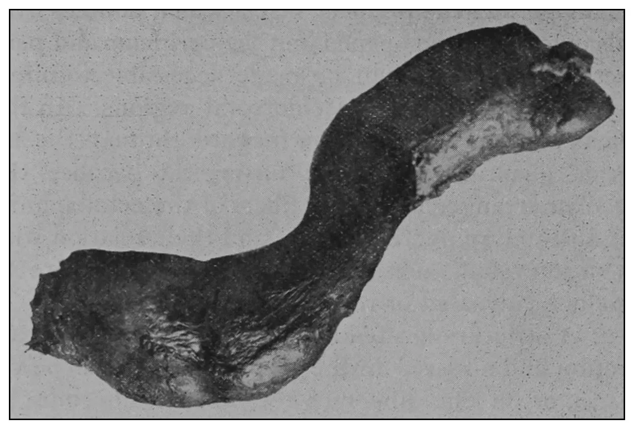
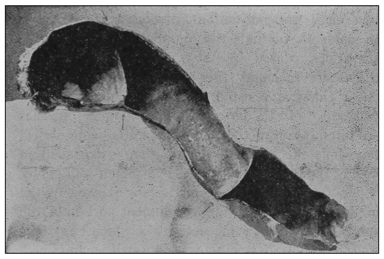
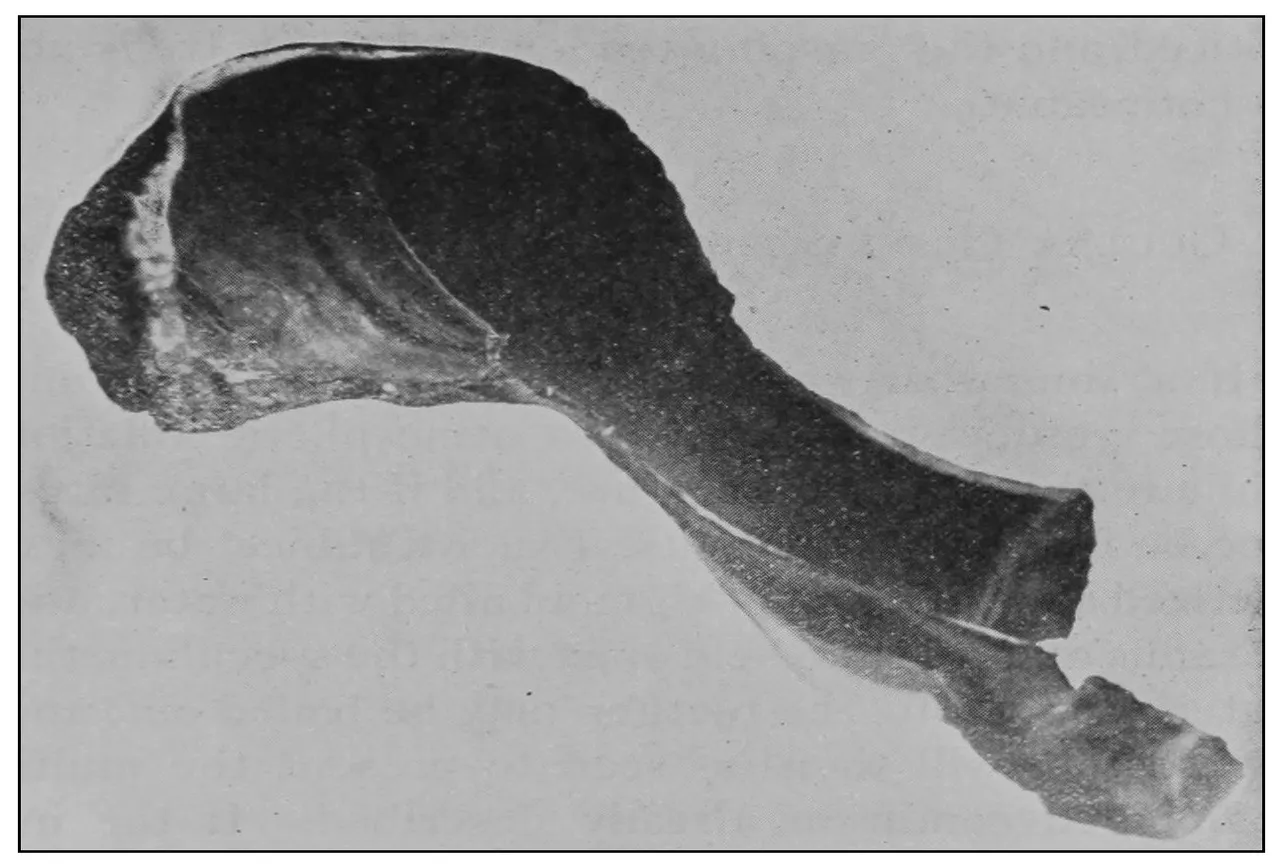
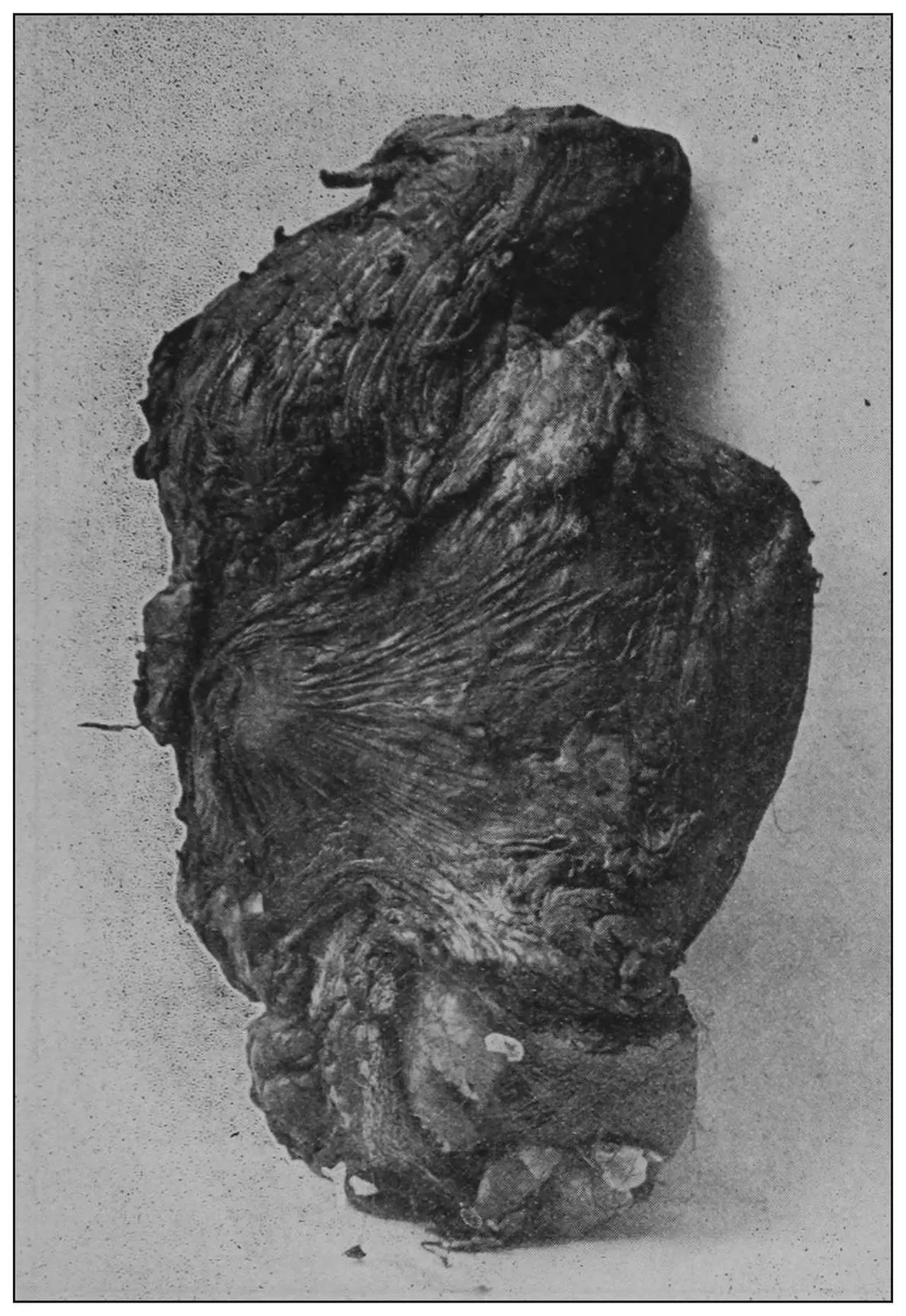
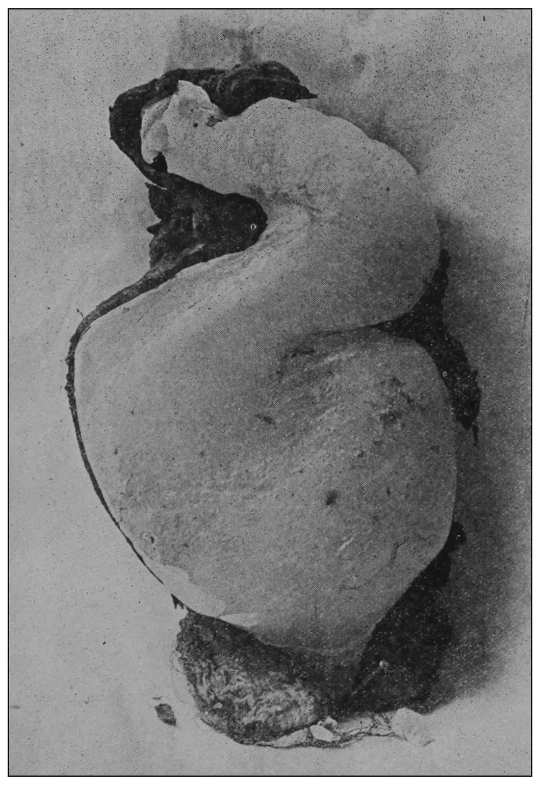
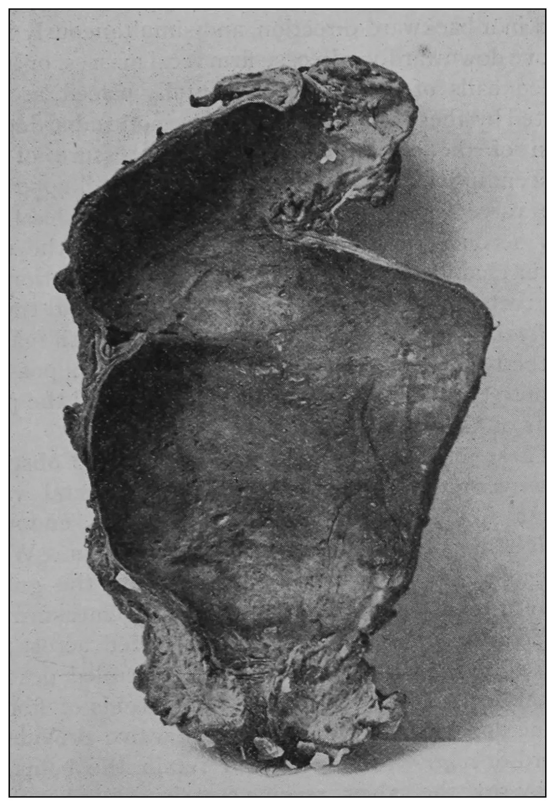
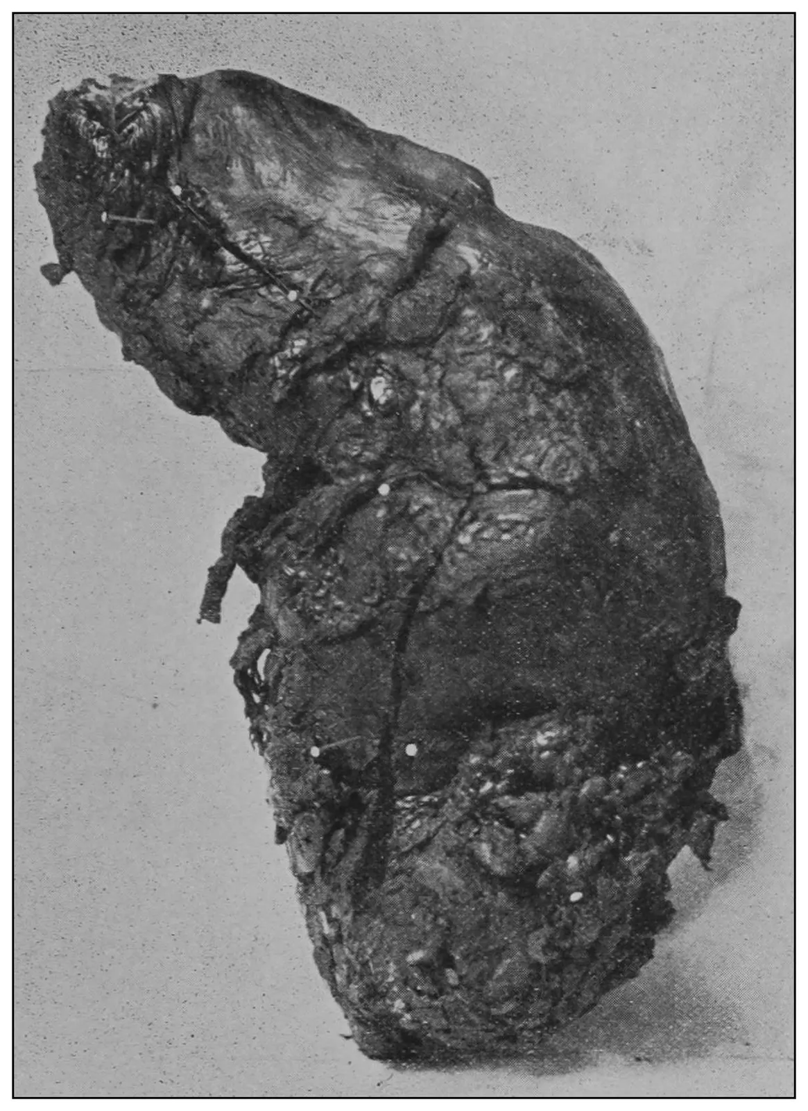
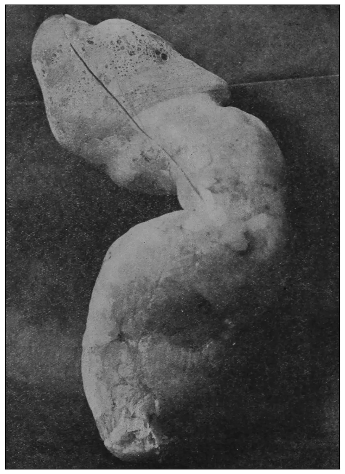
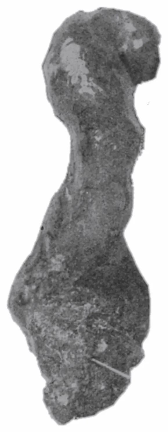
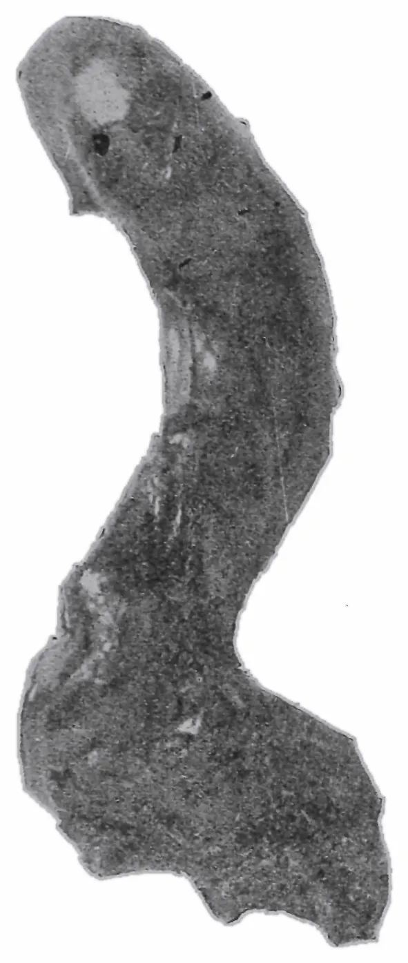
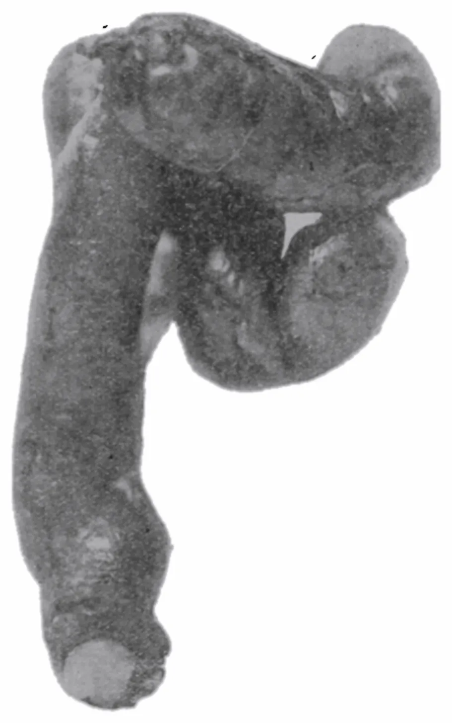
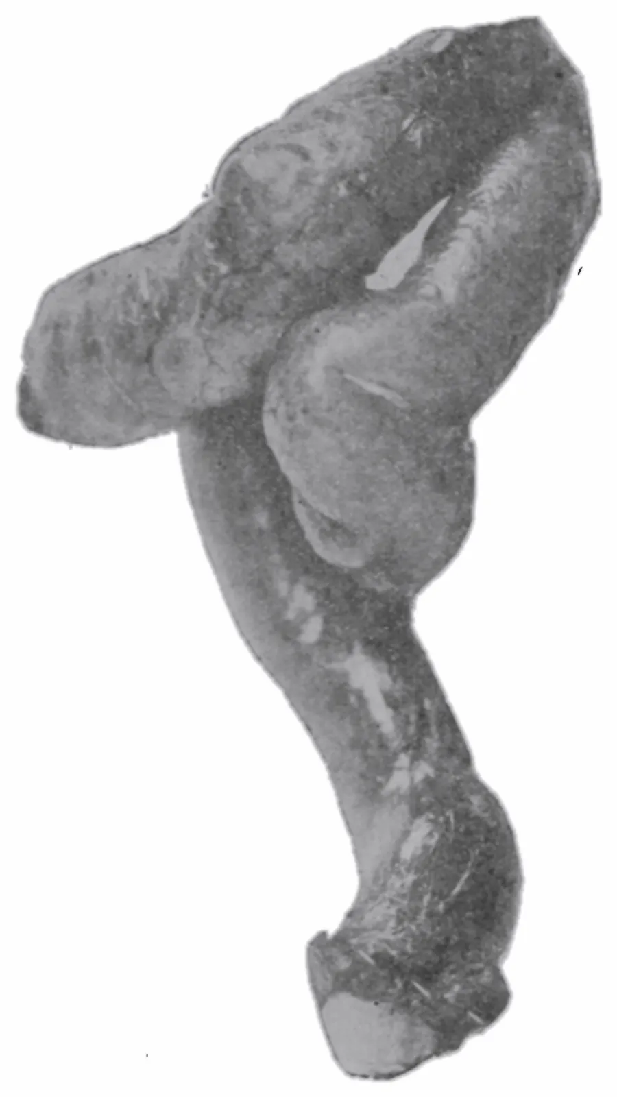
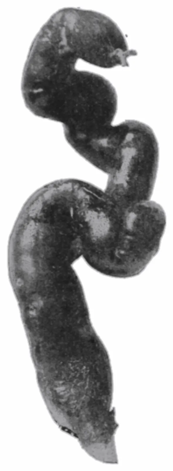
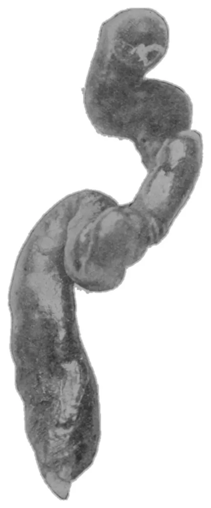
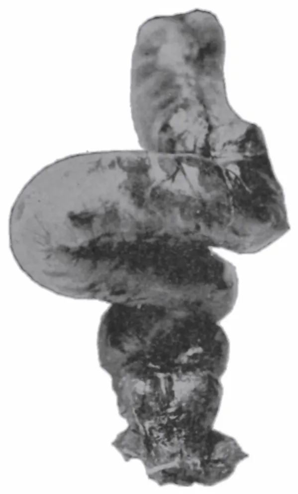
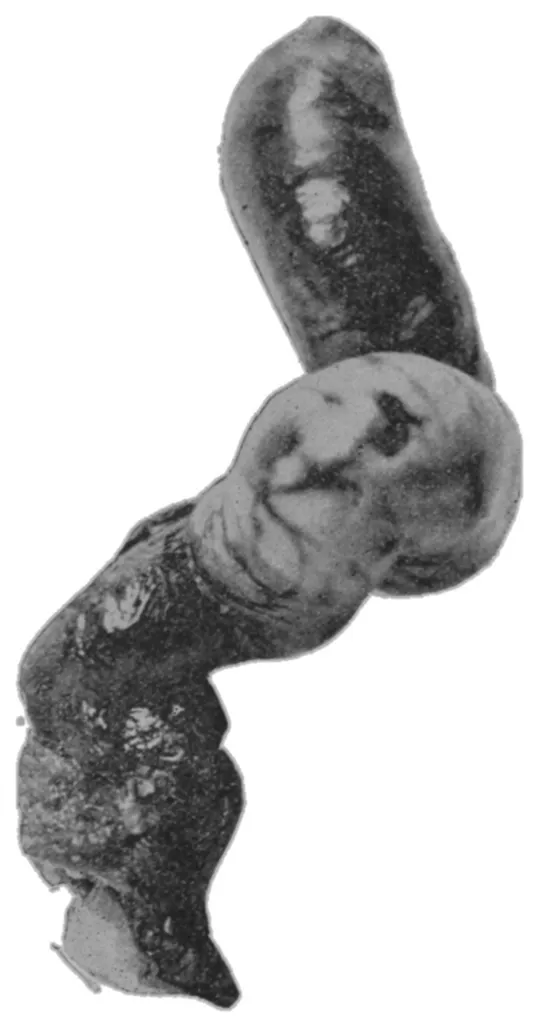
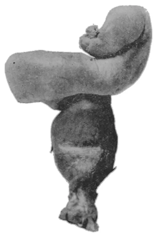
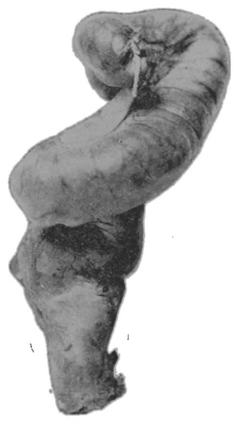
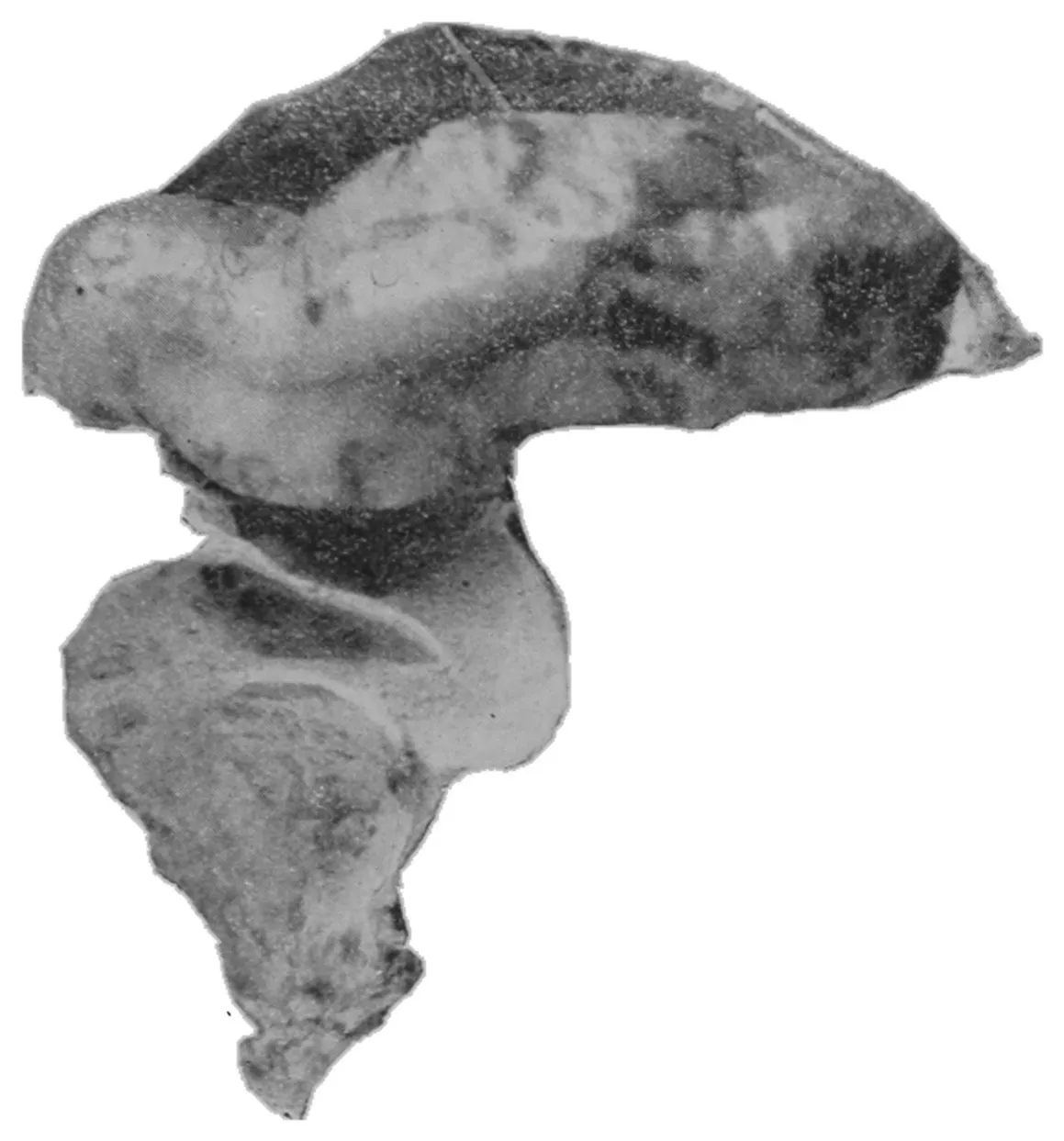
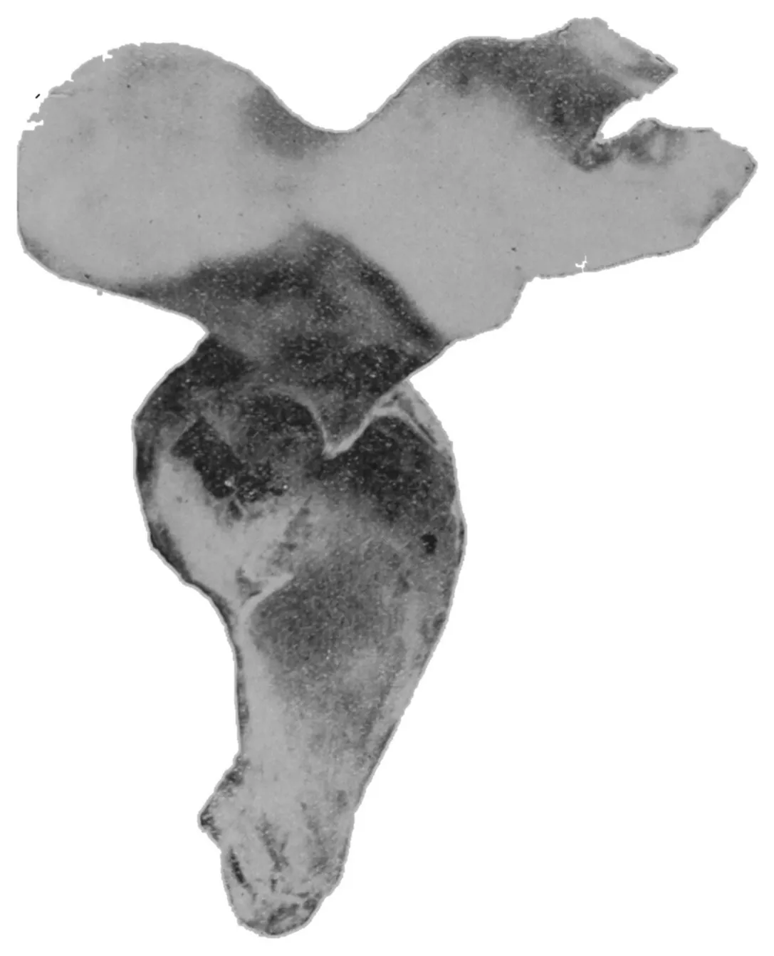
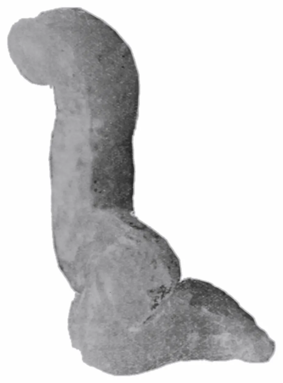
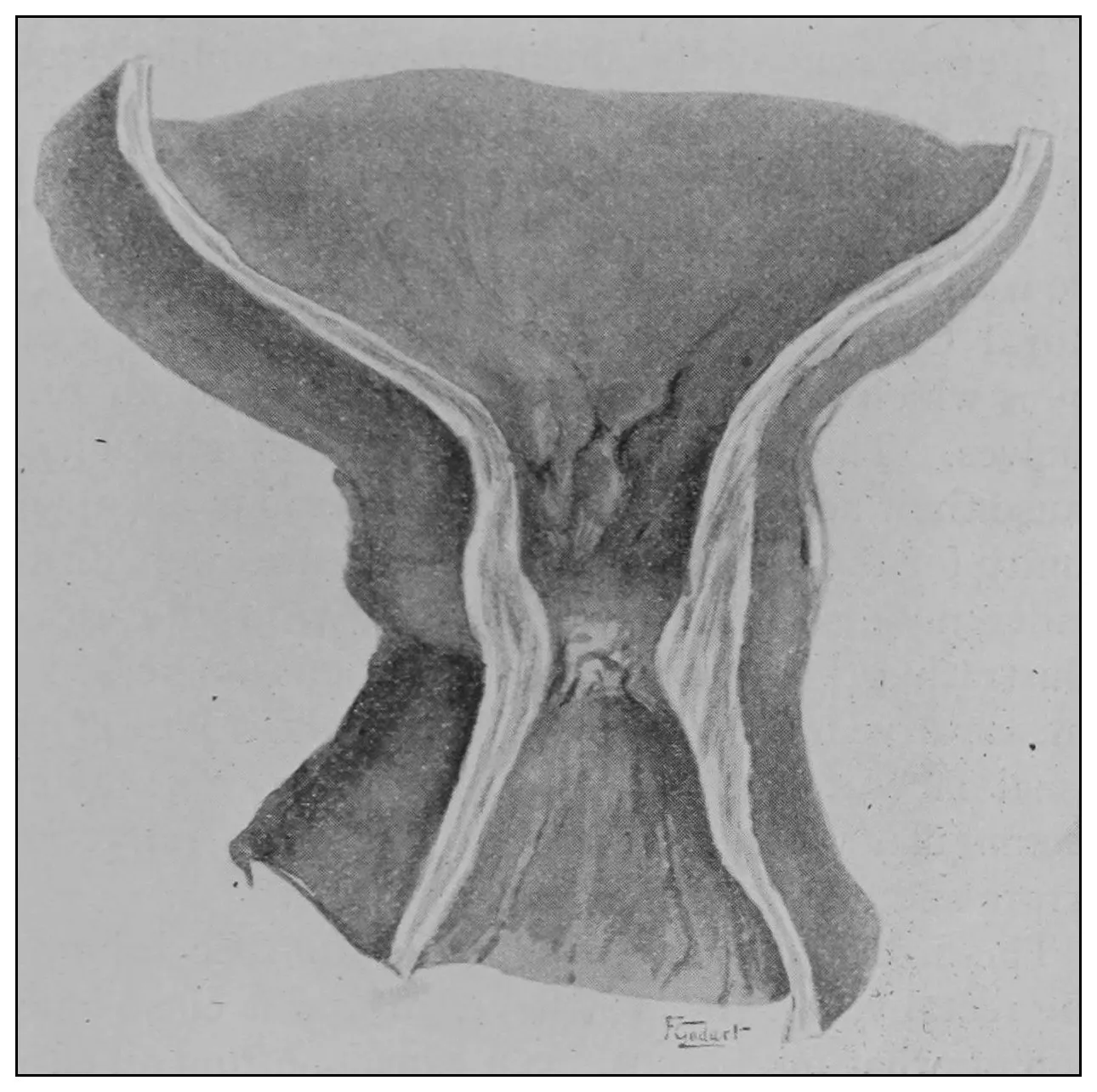
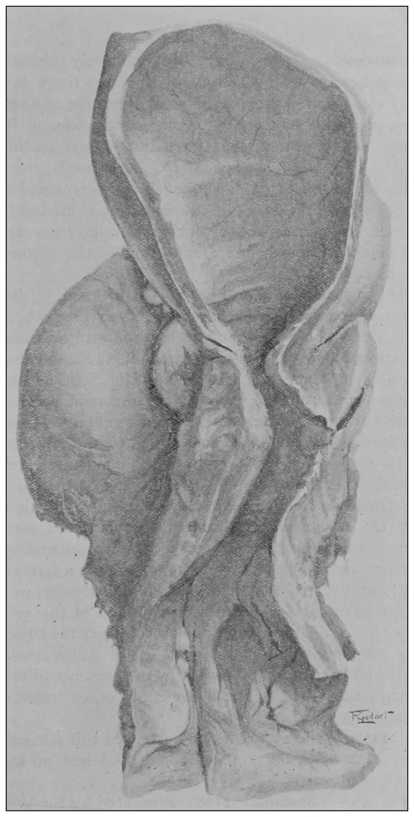
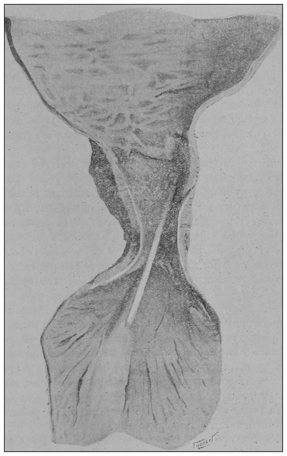
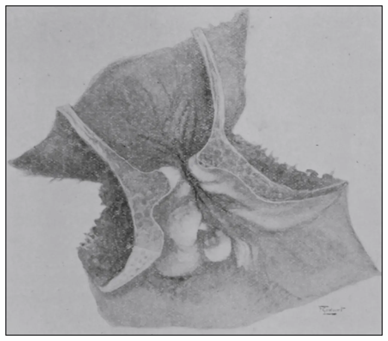
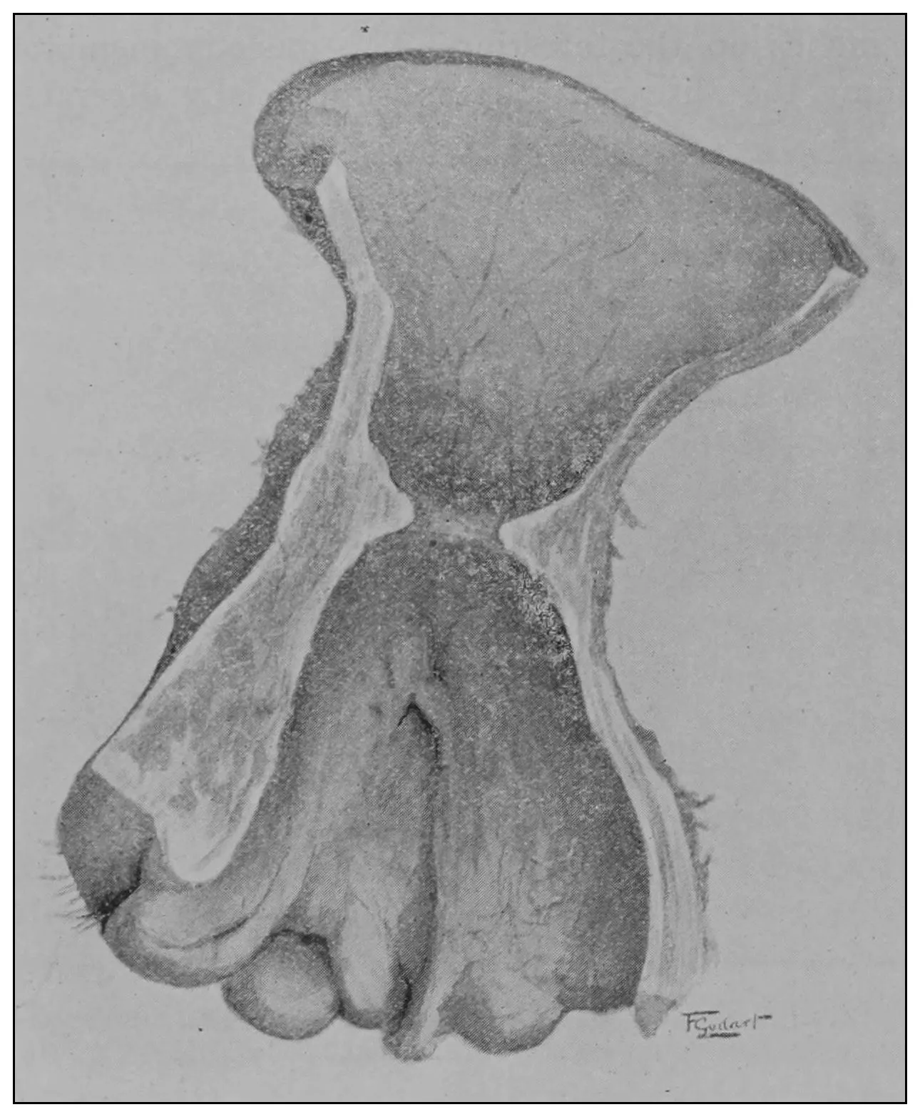
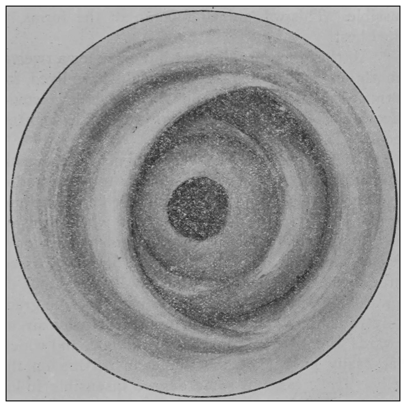
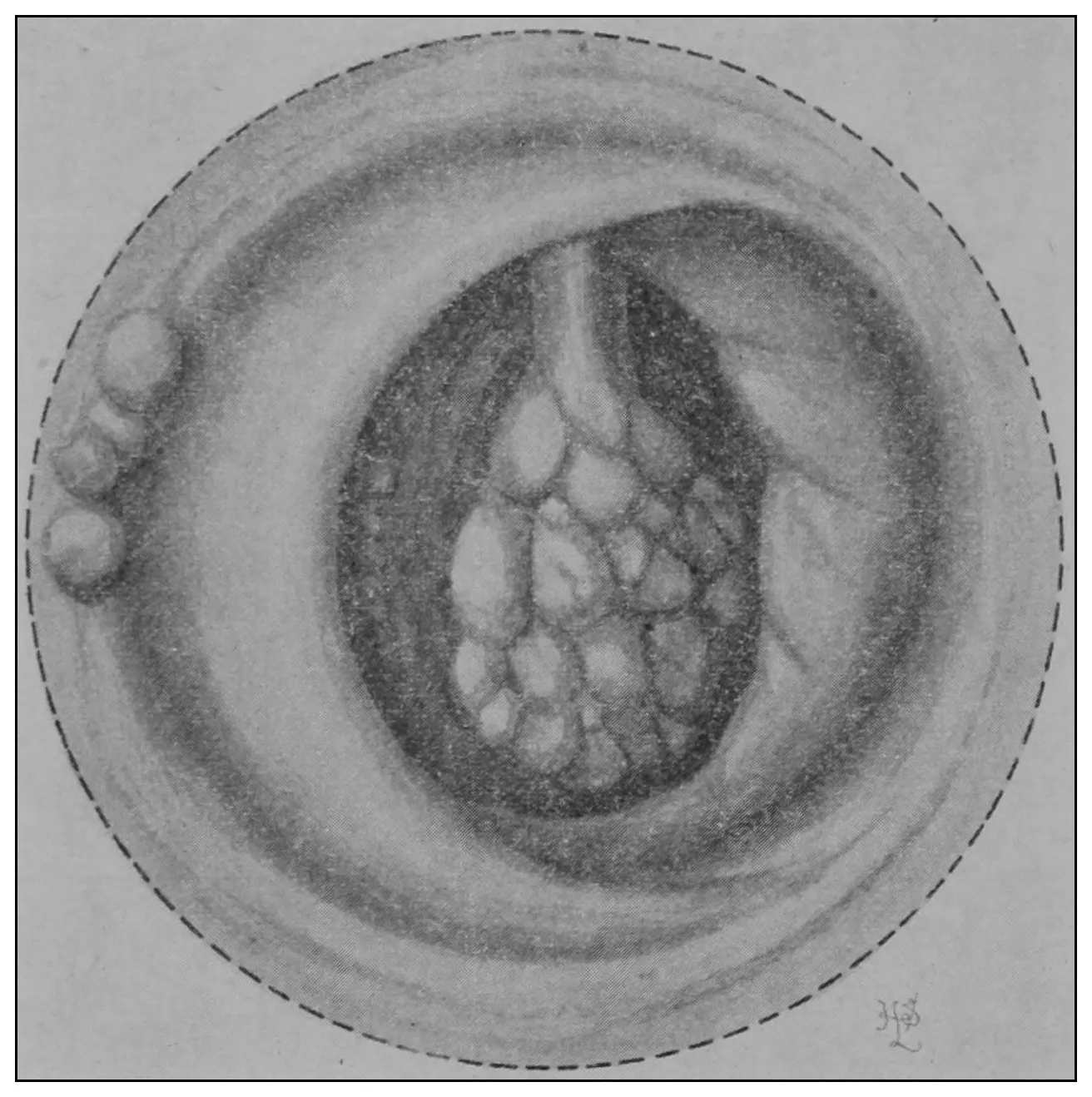
The photographic reproductions here published are
documentary evidence of the existence of the obstructions
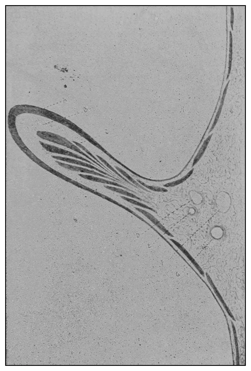
under discussion. The sketch, Fig. 33, which
was drawn from the valve while under the microscopic
lens, exhibits the character of these obstructions and
proves it that of a typic anatomic valve, and the
absence of permanent bands of any other character
in this organ is evidence that the semilunar valves
and the so-called plica transversalis recti, Falten des
Rectums, sphincter ani tertius, superior sphincter,
and detrusor fecium muscles are one and the same
thing and this thing is essentially a valve. It is most
prominent when the gut is most distended.
The Chronology of Atmospheric Inflation of the Rectum for its Inspection.
Proctoscopy has proved an open sesame to a newer
proctology.[4] As there seems to be some confusion
of opinion concerning the time of its origin and the
chronology of its evolution, the present time is opportune
for a brief historic review of the subject.
In 1845 Dr. J. Marion Sims[5] discovered by a chance
that a hollow or tubular pelvic viscus would inflate
provided the orifice were opened at a time when the
patient’s hips were higher than the chest. He elaborated
the manner of this discovery to a method of
procedure. He first used the knee-chest posture, and
subsequently the semiprone-semiflexed position with
elevated hips. This posture became known as Sims’
posture, and the instrument which he designed as
Sims’ speculum. The first published account of his
method appeared in 1852, in the January number of
the American Journal of the Medical Sciences.
In 1871 Dr. Wm. H. Van Buren,[6] of New York,
was the first to publish an account of the use of the
identical postures and Sims’ speculum for atmospheric
inflation and inspection of the rectum and sigmoid
flexure.
In 1882 Dr. Wm. Allingham[7] employed elevation
of the patient’s hips and a tubular speculum, and
achieved the same results. In subsequent editions
of his book in 1888 and 1896 he repeats a description
of his rather crude operation for inspection of the
rectum through a cylindric tube.
In 1887 Dr. Alfred Cooper[8] described a similar
posture, and suggested the use of two retractors for
the purpose of opening the anus.
In 1887 Dr. Walter J. Otis,[9] of Boston, published
in Leipsic a monograph on the subject of rectal inspection,
and described the use of the knee-chest
posture and of two retractors.
In 1887 Prof. Esmarch[10] described a method similar
to that of Dr. Otis.
In 1895 Dr. Howard A. Kelly[11] described a method
of proctoscopy by means of tubular speculums which
are very similar in construction to those of Dr.
Edmund Andrews, which Dr. Andrews first described
in 1887.
Dr. Kelly’s article, however, was the first to catch
the attention of the general profession, and to him
is due the credit of pointing out to a multitude of
physicians the possibility of rectal inflation for inspection
by such means. Kelly’s technic and tubular
speculums are far superior to those of Mr. Allingham,
who first employed a similar method in 1882.
In 1896 I published in the July number of Mathews’
Quarterly Journal of Rectal and Gastro-Intestinal
Diseases, under the title of “Proctocolonoscopy and
Its Possibilities,” a description of a technic and new
instruments which increased the areas exposed to
view, and which facilitated access to the part for the
treatment of disease.
In 1896 A. Ernest Maylard[12] briefly referred to the
various methods.
Review of the literature on rectal inflation for rectal
inspection establishes the fact that Van Buren is entitled
to the credit for priority; that Marion Sims
was the discoverer of the possibility of atmospheric
inflation of the hollow pelvic viscera; that there is
much similarity in the methods of the various operators
quoted, some using similar instruments and dissimilar
technic, and vice versa; and it is made obvious,
also, that he who would most insist upon a credit for
originality must sometimes discount with the erudite
his reputation for literary research.
The time has arrived when the profession must
recognize that the rectum need no longer be regarded
a darkest continent. There remains, however, something
further to be desired in the way of an easier
and more convenient method of manipulation to secure
inspection, but I am confident that ere long the profession
will accept the newer mechanic means and
contrivances which will render a proctoscopy of as
practical simplicity as is laryngoscopy. But it behooves
us to remember the words of Dr. Edmund
Andrews: “The false method is that of the bungler
and amateur, who is only right by haphazard; the true
one is that of the professional expert, who can not be
balked by petty obstacles, but who will reach success
when others have failed, not less by his dogged persistence
and thoroughness than by his superior
knowledge.”
The Examination of the Rectum. — Noninstrumental Inspection of the Rectum. — Instrumental Inspection. — The Technic.
It has been complained that the best methods proposed
for the inspection of the rectum require so expensive
an armamentarium and such painstaking practice
on the part of him who would see, that the general
practician can not hope to invade with his keen glance
this field which is generally regarded a terra incognita.
On the contrary, as shall be seen, no artificial
means whatsoever are required for a complete ocular
inspection of the rectum.
The elevation of the hips which sets in operation
that principle of physics which governs the methods
of Marion Sims’ vaginal inspection (1845), Van Buren’s
rectal inspection (1871), and the methods of the
senior Allingham (1882), of Walter J. Otis (1887) and
of Howard Kelly (1895), which controls my own proctocolonoscopy
(1896), and which suggested Trendelenburg’s
posture, is, also, the chief feature of the
simplest proctoscopy.
The essentials to this simplest method are a patient,
an assistant and an operator having at least one finger
on each hand. The patient is to be put into the
knee-chest posture, the assistant is to put and to hold
the patient, and the physician’s fingers are to be used
to open the anus, all in the following manner, to wit:
1. The patient is to be completely anesthetized as
he lies on his back, and then turned toward the assistant
and into Sims’ posture.
2. The assistant is to station himself at the patient’s
knees. In his left hand he is to grasp the patient’s
feet. He is to lean himself against the patient’s knees.
He is to pass his right arm under the patient’s hips.
Now steadying the feet and bearing himself firmly
against the subject’s knees, with his right arm he is
to lift the hips and pull the patient into the knee-chest
posture, where he is to be balanced on his perpendicular
right thigh throughout the whole time of the
physician’s manipulations.
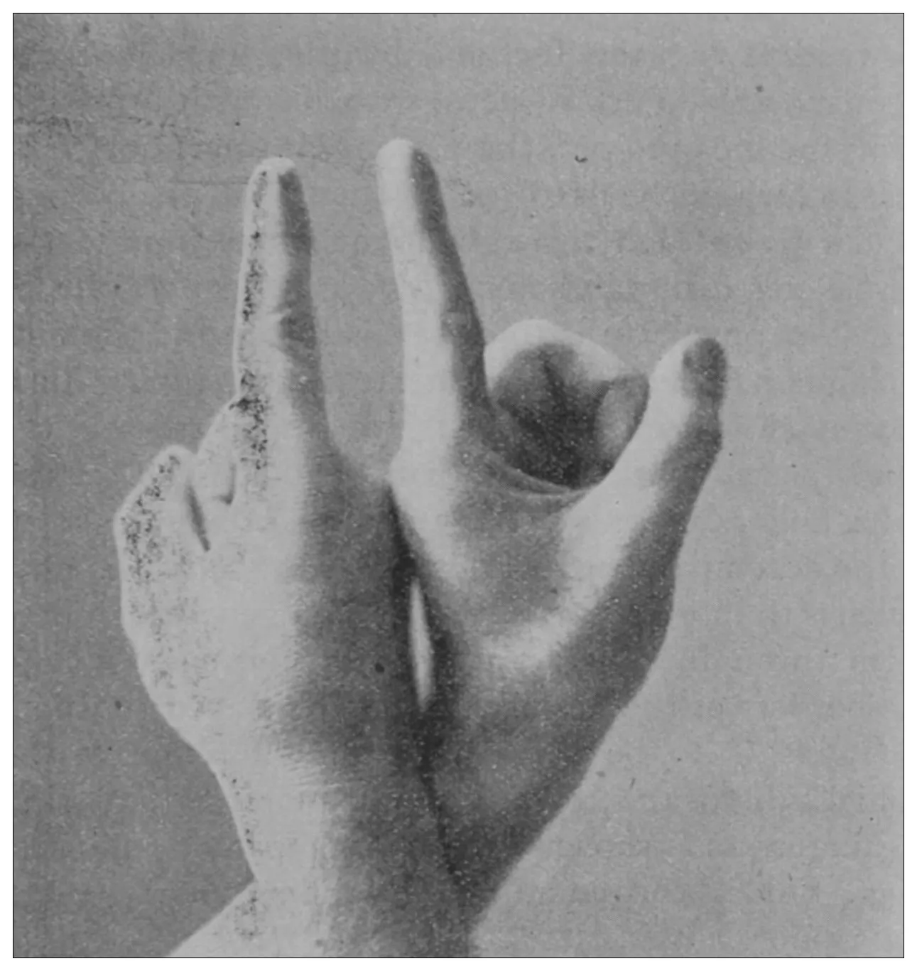
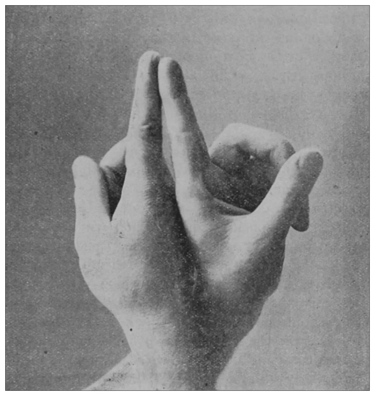
Fig. 1.—Positions of the hands for the practice of the simplest method of
proctoscopy.Fig. 2.—Positions of the fingers for the practice of the simplest method of
proctoscopy.
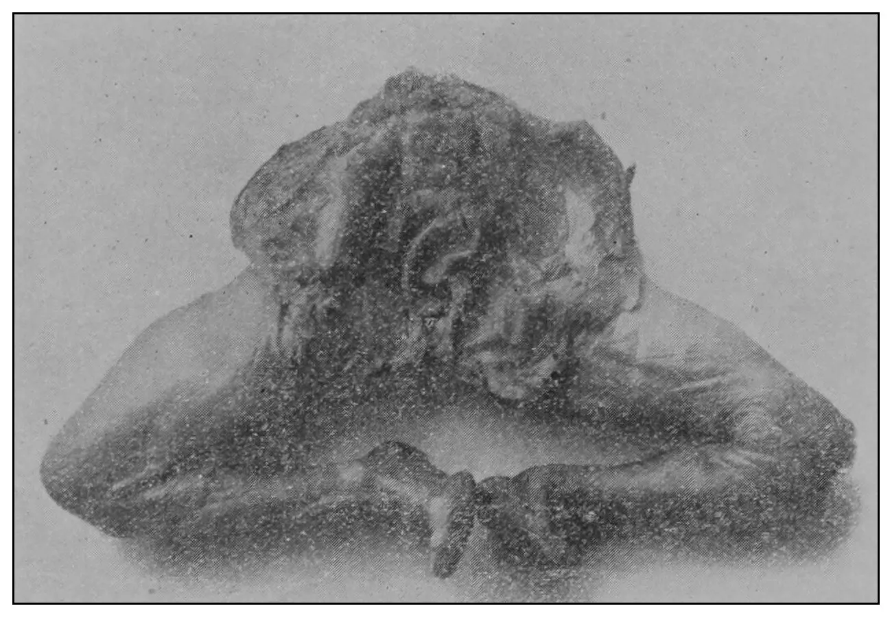
3. The physician is to close his hands and to point
each index-finger as shown in the accompanying illustration
(Fig. 1). The wrists are to be crossed, the
hands placed back against back, and the nails of the
index-fingers placed one against the other, as shown
in the accompanying illustration (Fig. 2). The physician
is to lubricate these fingers and gently insinuate
them through the anus and place their ends beyond
the borders of the levatores ani. This accomplished,
the anus is to be divulsed in the direction of the
ischial tuberosities, by the physician forcibly parting
his fingers as is shown in the accompanying illustration.
Under this manipulation the rectum becomes
atmospherically inflated.
Now, provided the physician lowers his head to the
level of his fingers and then rises again, or stoops, or
moves a little from side to side, he may command
under his eye a view of the interior of the atmospherically
inflated rectum to the depth of six or eight
inches (15.24 or 20.32 cm.), and in some instances he
may behold even a part of the sigmoid flexure.
It is possible for the operator to manipulate his
patient and to finish his inspection within two and a
half or three minutes, provided the patient be in a
state of complete anesthesia.
If this method is practised, as I am persuaded it
may be with facility by the general practician, I am
convinced that the greater number of rectal diseases
may be instantaneously diagnosed. But I must declare
that here at diagnosis, the achievement of the
simplest proctoscopy ends, for the reason that the
operator’s hands are so full of his patient he can do
nothing at all for the disease which he may have
discovered.
Under some conditions and amid some circumstances
the rectum will not inflate. If the bladder is
much distended; if there is an inordinate hypertrophic
rectitis; if there is a close tubular stricture of the
rectum; if there is malignant growth or other disease
of the rectum by means of which the gut’s coats
have become extensively filled and fixed with an
organized plastic exudate; if for some reason the
extraabdominal pressure is abnormally increased, as
it may be by the bearing down of the patient, or by
enormous flatus, or by ascites; or if there is an
impinging uterus, adrectal growth or extensive
infiltrating disease of the contiguous textures, rectal
inflation by this method or by any other which is
governed by the same principle may be a physical
impossibility—but this need not baffle the man bent
on seeing by instrumental aid.
Practised as described, when not embarrassed by
the exceptions specified, this method will achieve its
purpose and reveal to the physician that the transverse
diameter of the rectum is variable; that in
some places it is not more than an inch (2.54 cm.), in
others it is more than four times this diameter.
The rectum may present to the eye of the imaginative
observer the appearance of a chain of urinary
bladders, communicating one with another by means
of irregularly elliptic openings set at varying axes,
and bounded by the nonparallel borders of the rectal
valves. In the normal rectum the air-pressure
smooths the mucous membrane evenly over the
entire surface of the gut, as may be observed in the
photographic illustrations. The normal mucous
membrane of the so-called ampulla appears at first
wet and of a shining bluish gray. As it dries, under
the influence of gravitation the blue venous tint fades
out of the gray and the wall assumes a pink tint.
Presently it acquires the appearance of parchment,
and sometimes it appears painted at rare intervals
with ramifying little arteries which may be crowded
and overlapped by the larger companion veins; the
latter are less arborescent and more suddenly dive
and disappear in the bowel-wall. In time, over all
there comes a sheen and the vascular pictures may
fade away. These phenomena appear exactly as
described only in the healthy rectum. In the diseased
organ the color varies much.
Should the operator deviate from the described
directions for the manipulation of his fingers and so
twist his hands as to divulse the anus in the anteroposterior
direction instead of laterally, he invites
defeat upon himself, for in the male the fixation of
the perineum and the immobility of the coccyx interfere
with the requisite dilatation; while, in the
female, the extreme mobility of the perineum and
particularly the backward displaceability of the coccyx
will allow such traction to be made upon the
levatores ani as to pull their inner fibers parallel and
almost together, and, in consequence, the wider the
female’s anus be opened anteroposteriorly the closer
is it made to contract laterally to rob one of his
view.
Certain paraphernalia and much practice in their
use are necessary for rapid, complete and painless
inspection of the rectum.
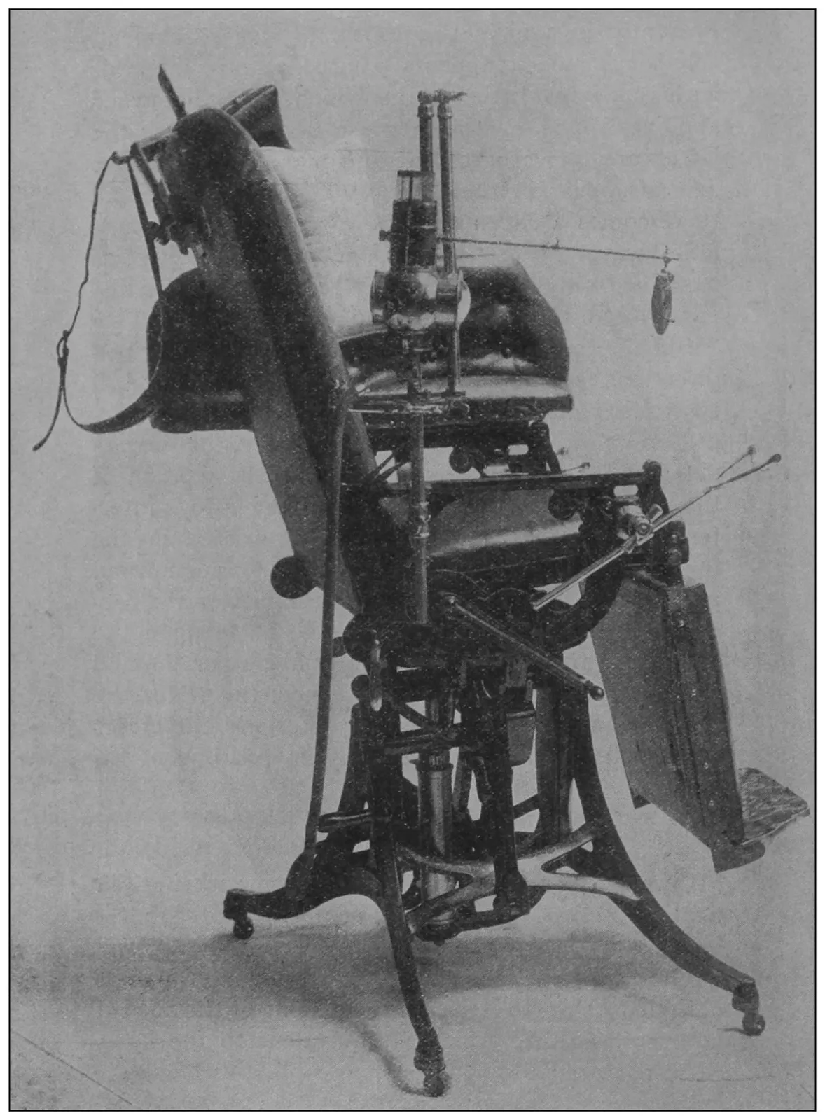
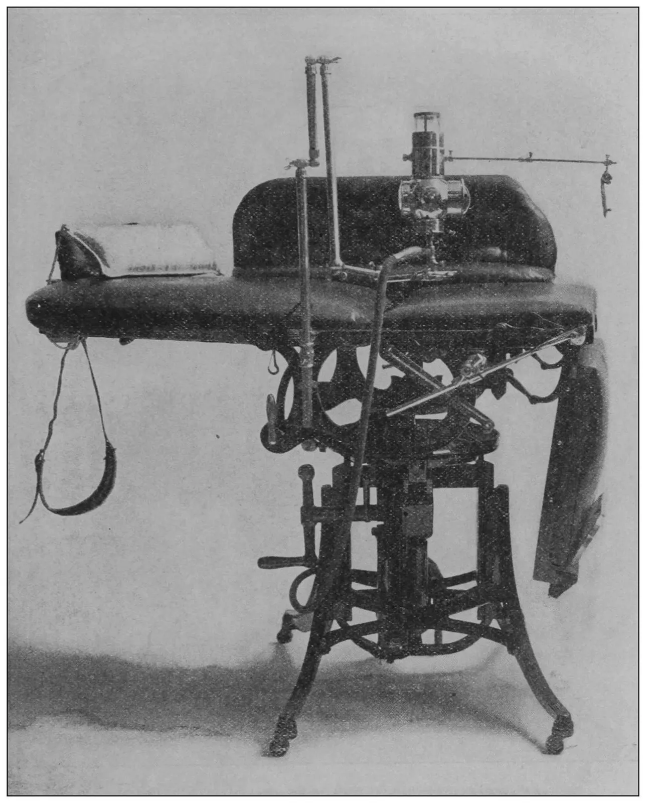
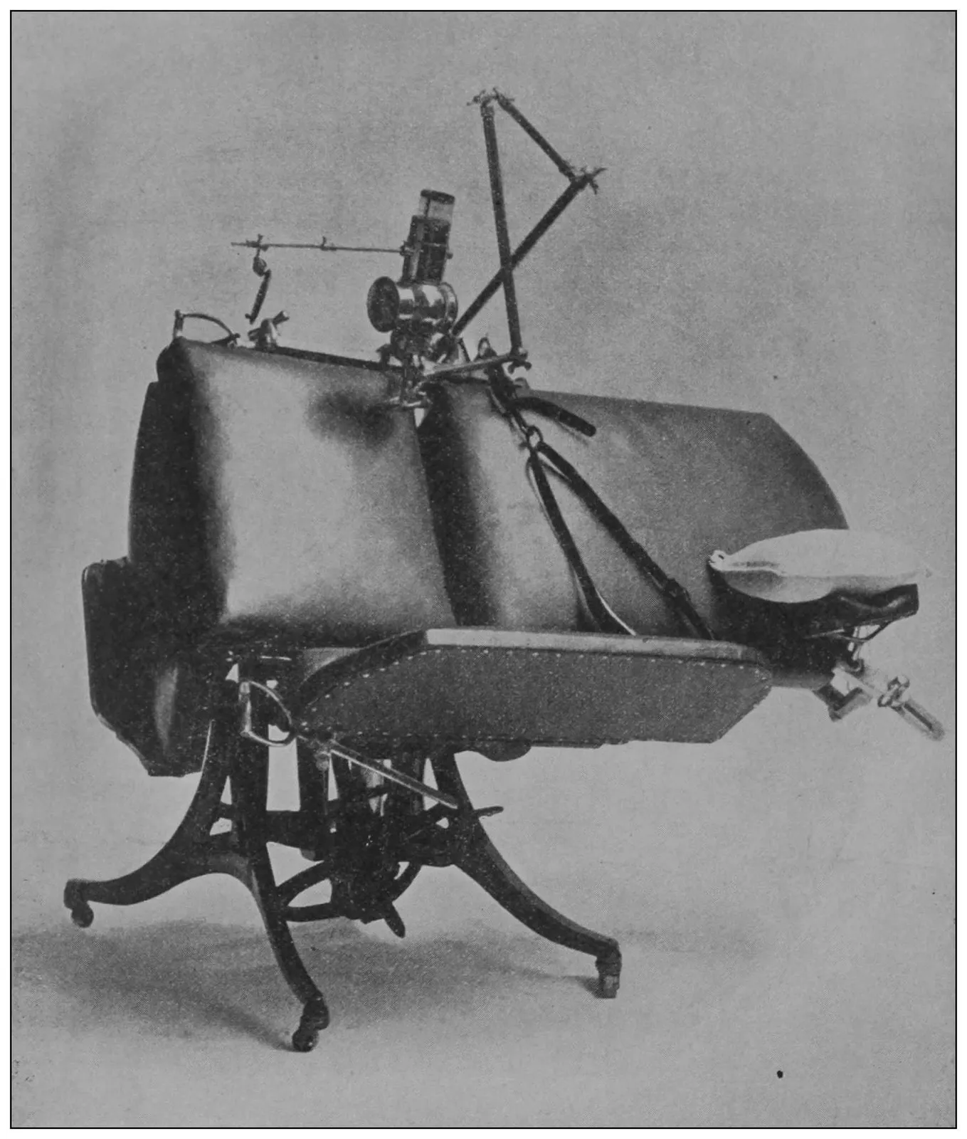
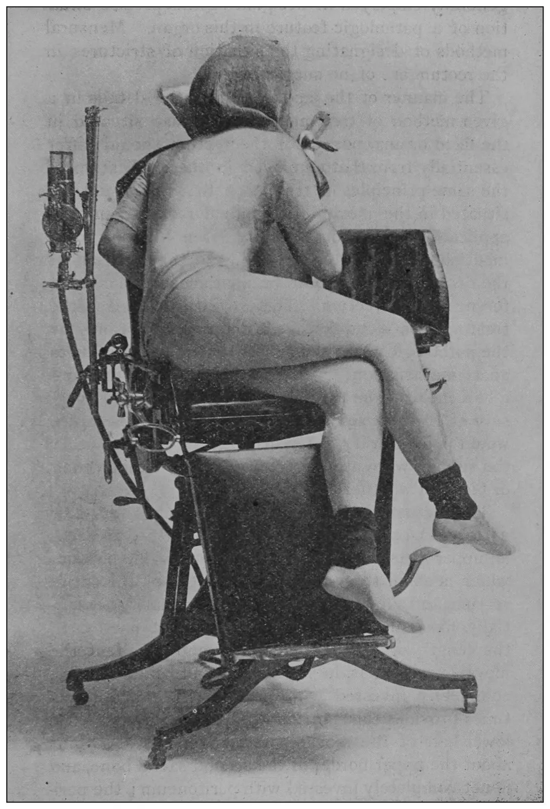
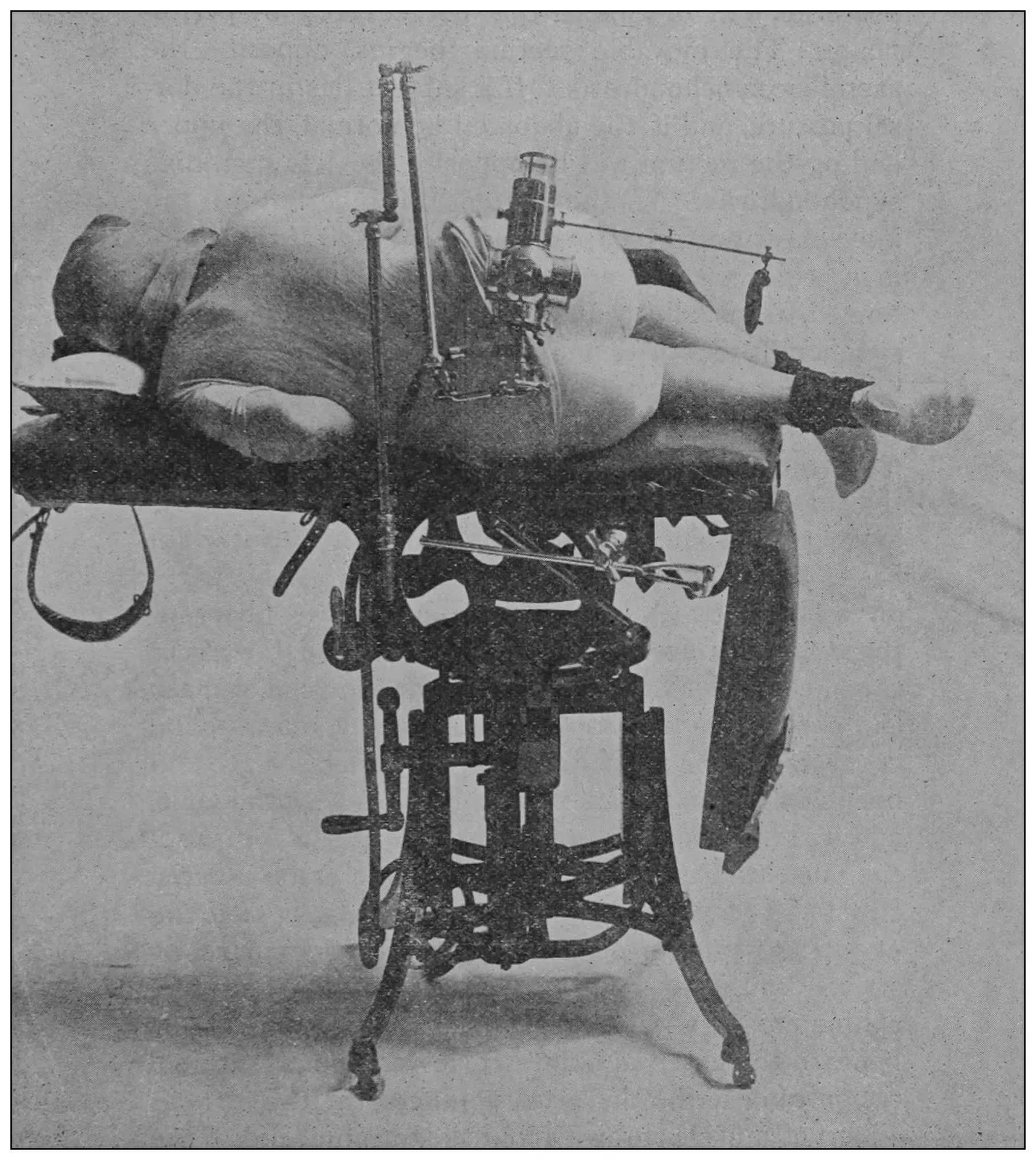
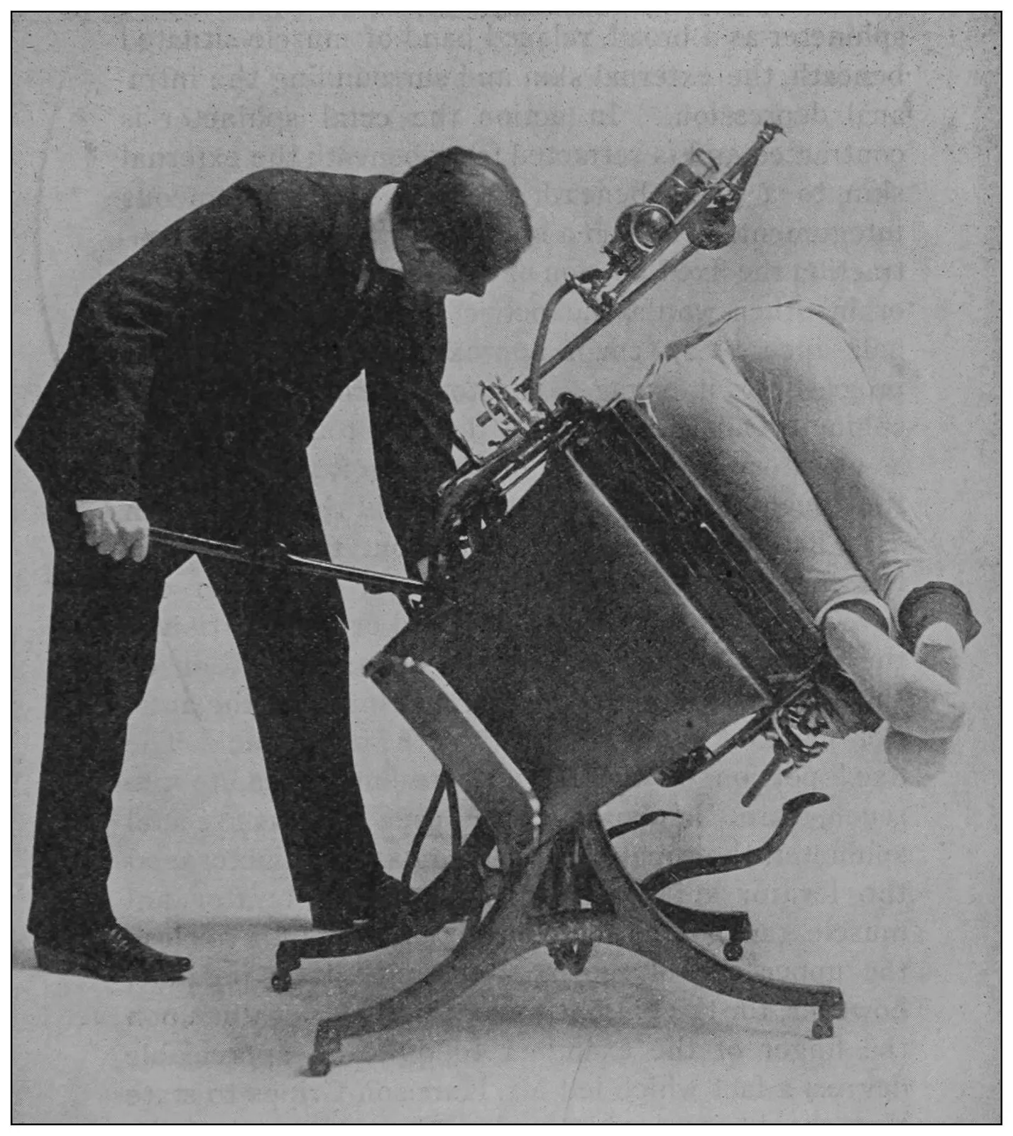
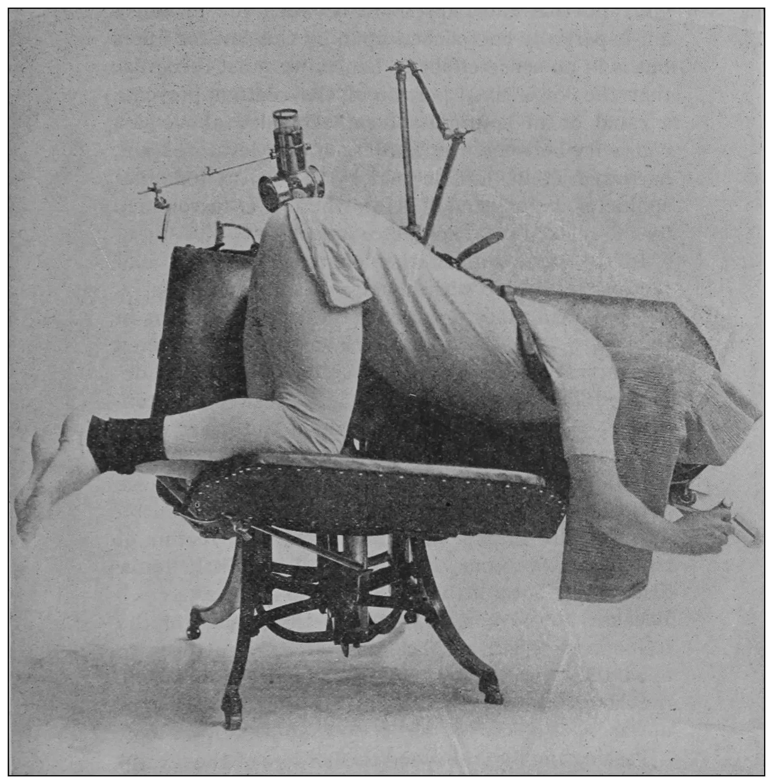
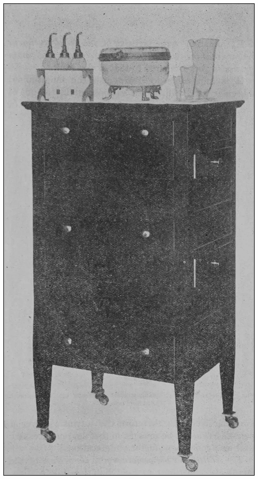
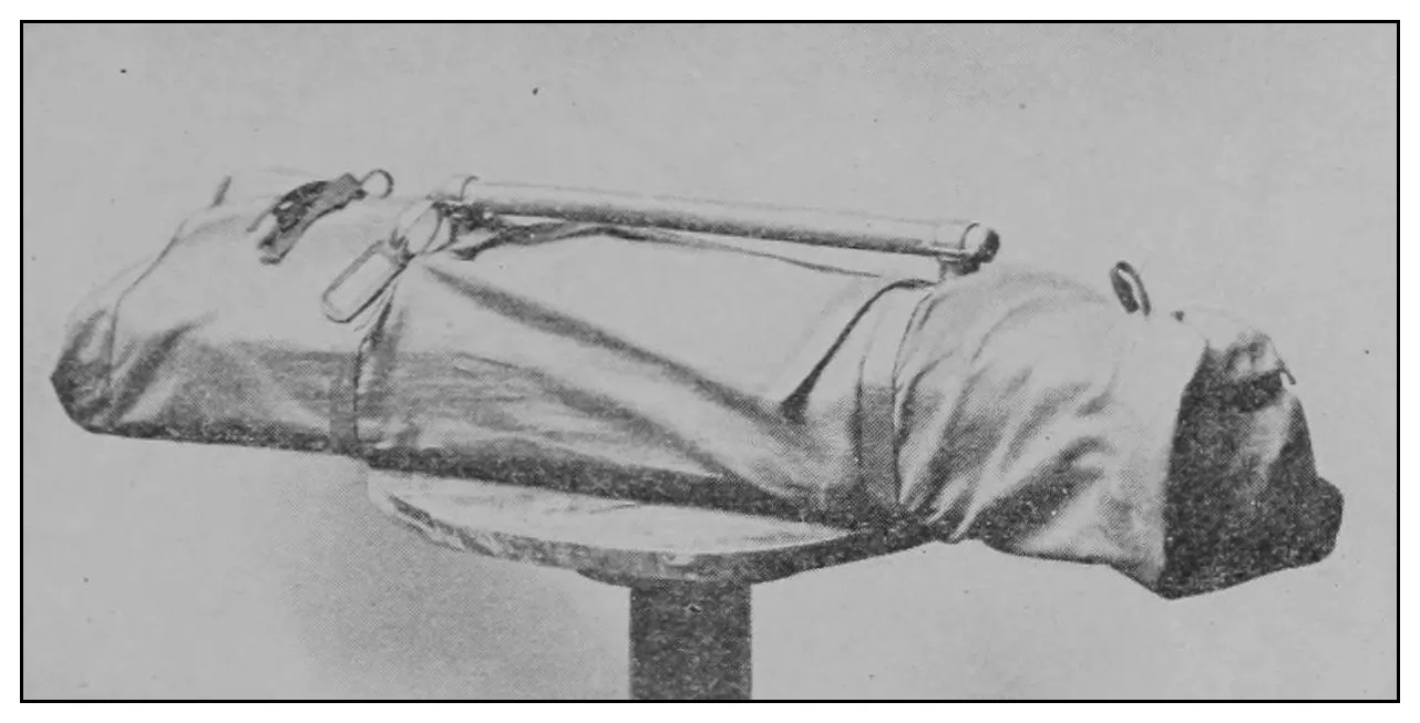
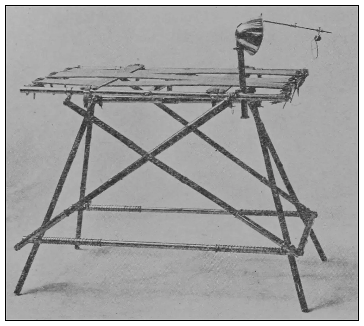
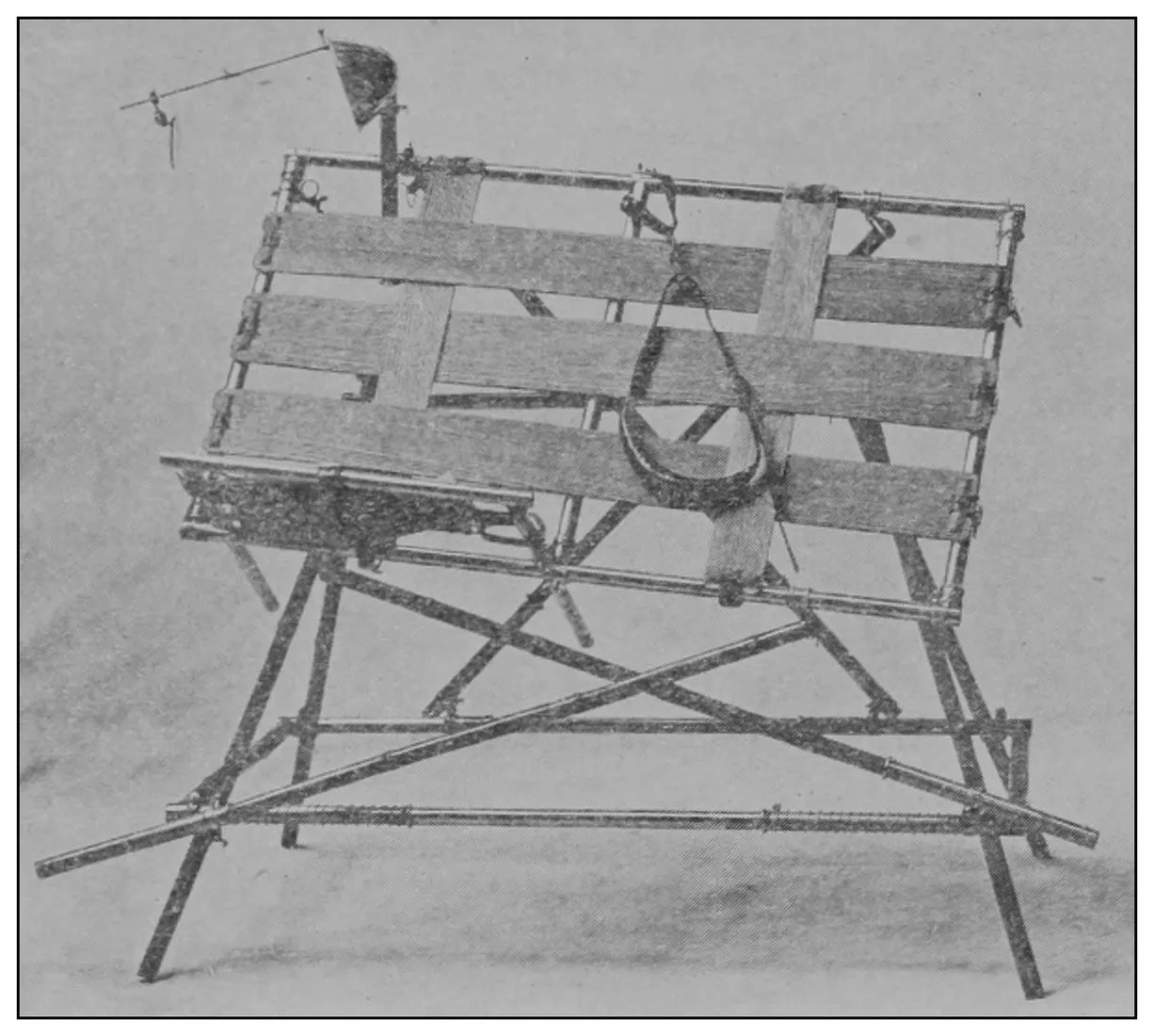
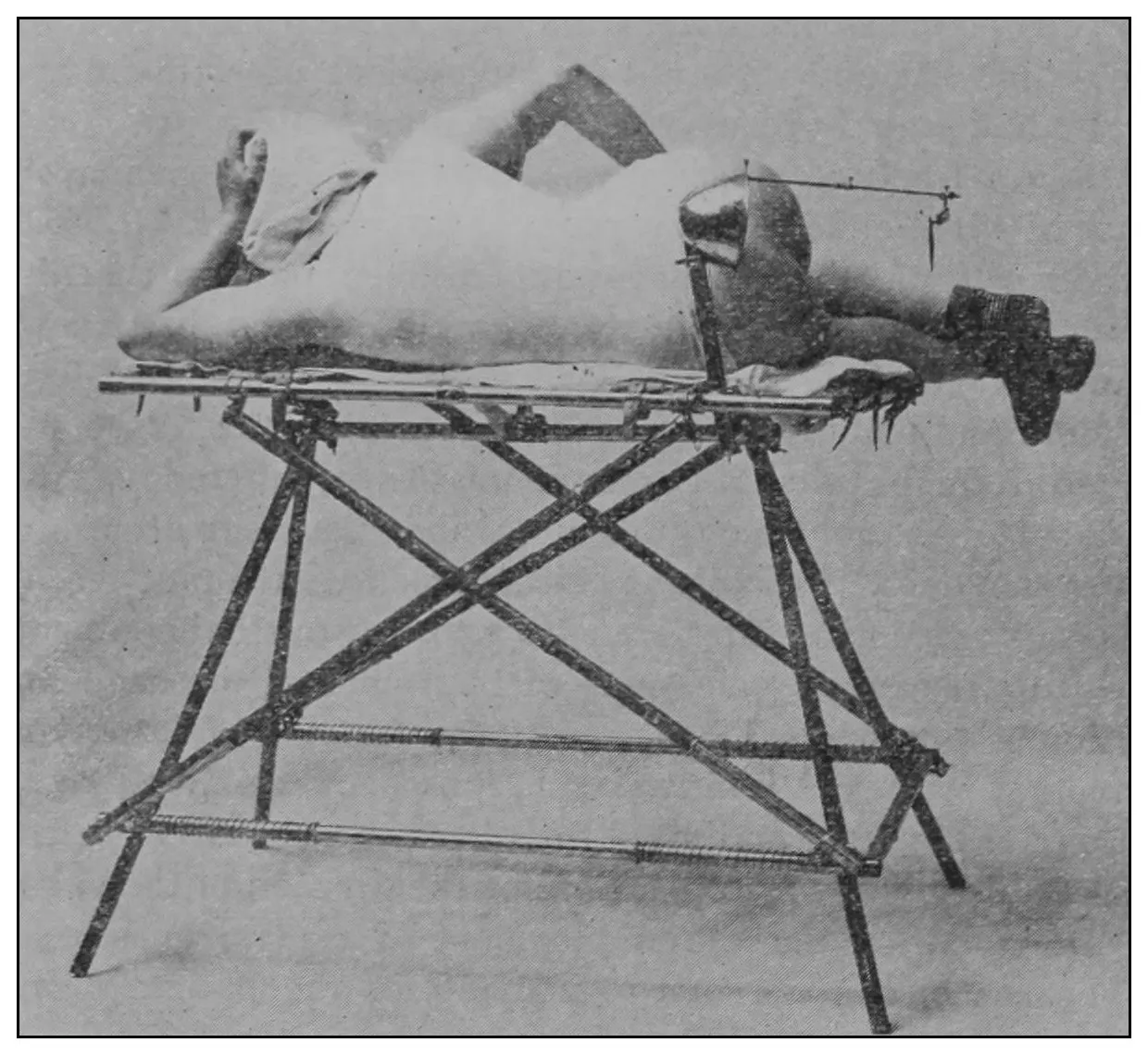
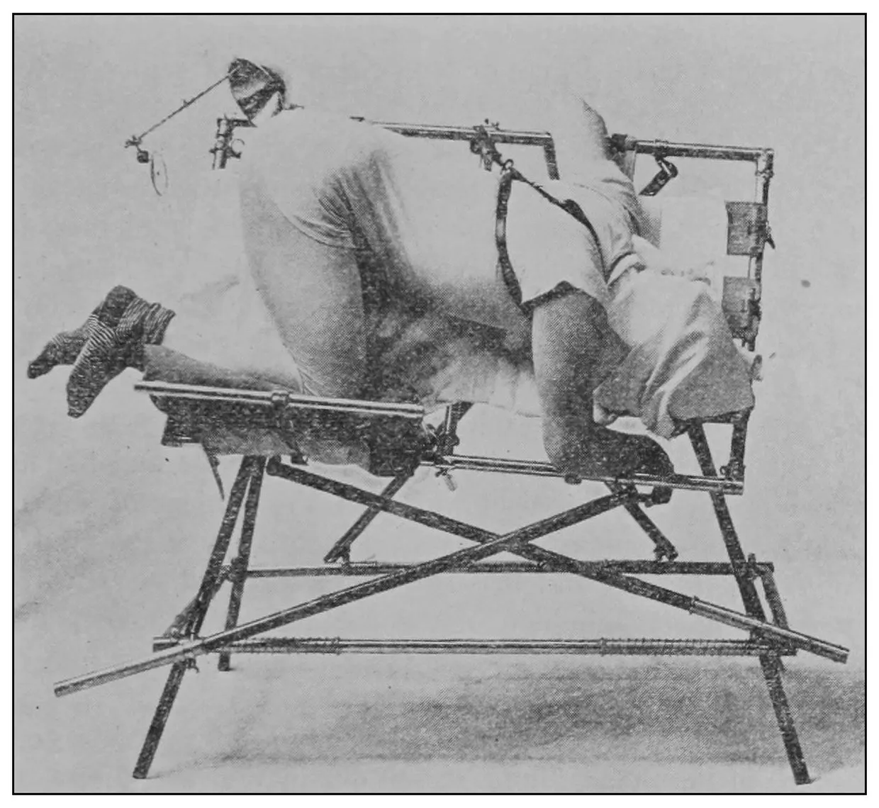
The chair which is shown in the illustrations was
designed by me to facilitate the placing of the patient
in a new posture, which is equivalent to the knee-chest
posture.[13]
Fig. 3.—The chair, illumination-apparatus, shoulder-suspender, and small pillow.
Fig. 4.—The chair in the horizontal posture for anoscopy.
Fig. 5.—The position of the chair for the new posture of the patient.
Fig. 3 exhibits the chair and the attached illumination-apparatus
in the first position to receive the
patient. Fig. 4 shows the chair and illumination
attachment in the second position, and Fig. 5 shows
the chair and illumination-apparatus in the position
for the third step in the procedure. Fig. 3 shows,
also, hanging from the head of the chair, a small pillow
and the shoulder-suspender.
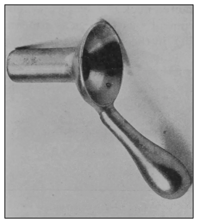
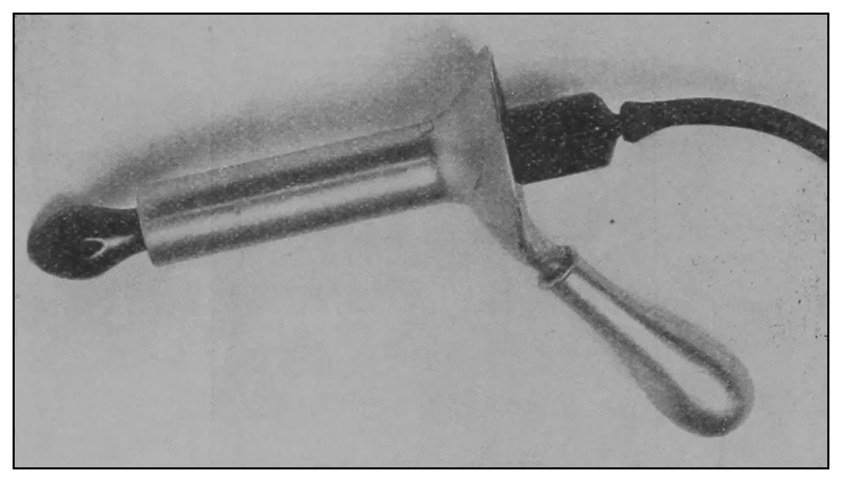
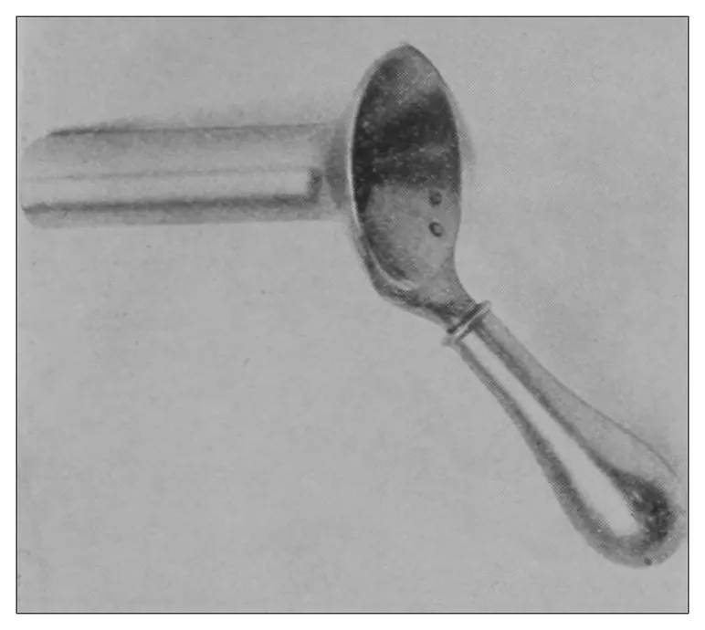
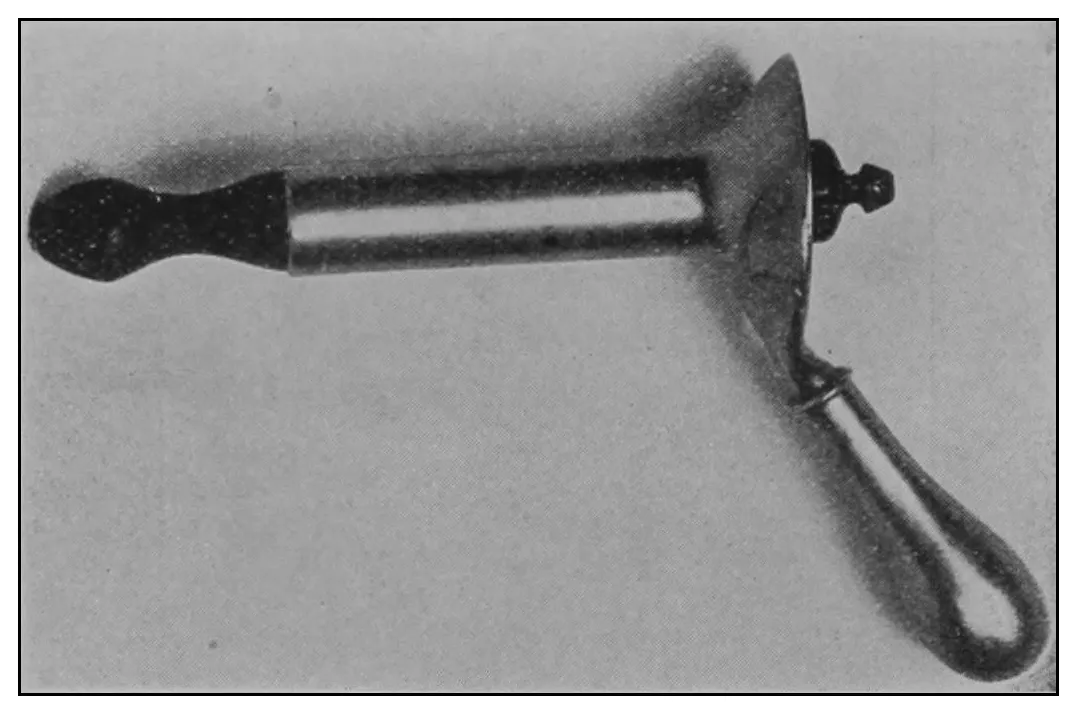
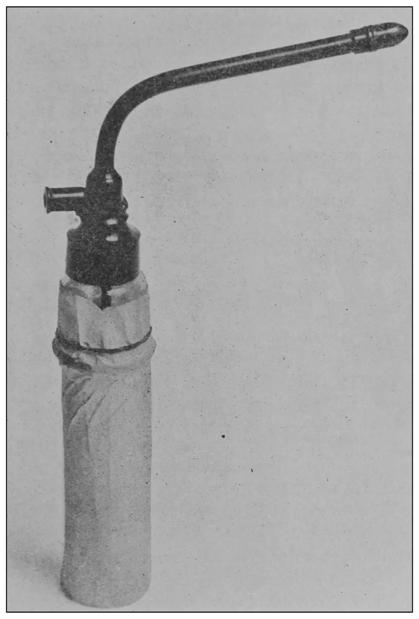
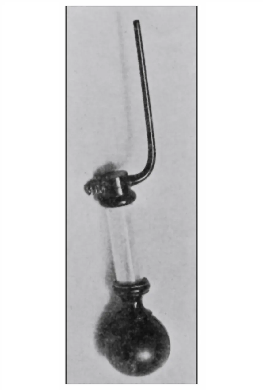
Fig. 6.—The anoscope.
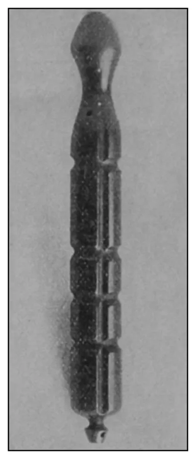
Fig. 7.—The obturator.
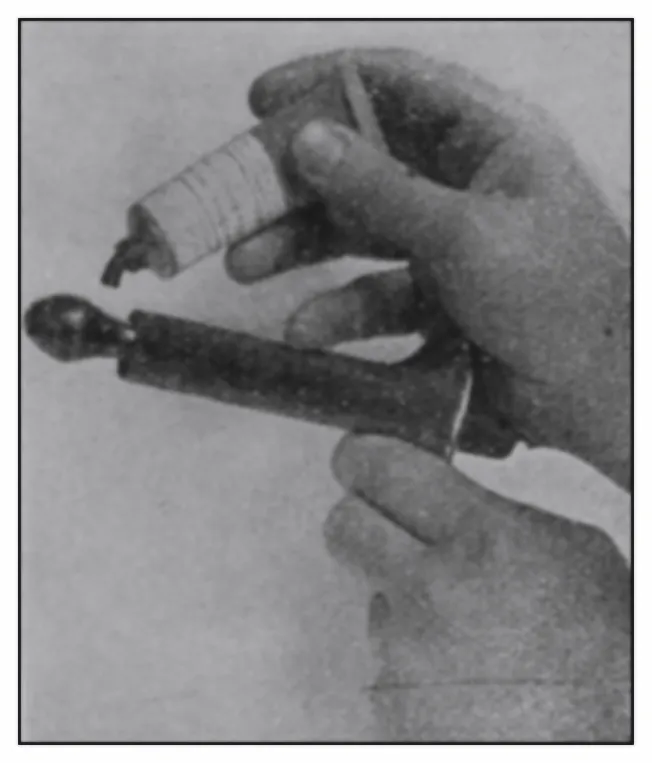
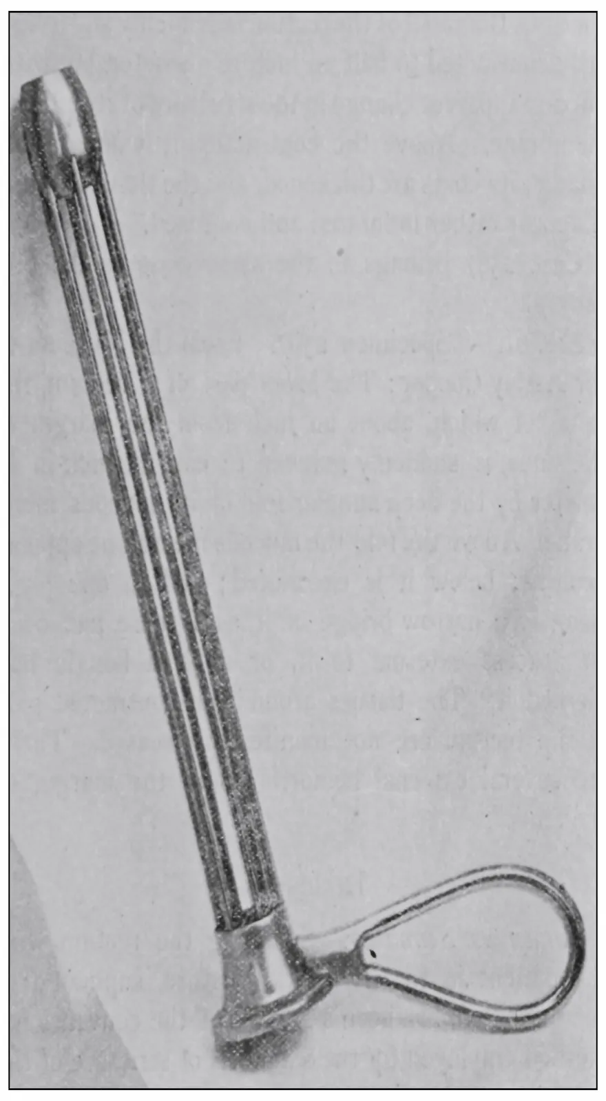
Fig. 8.—The ointment applicator.
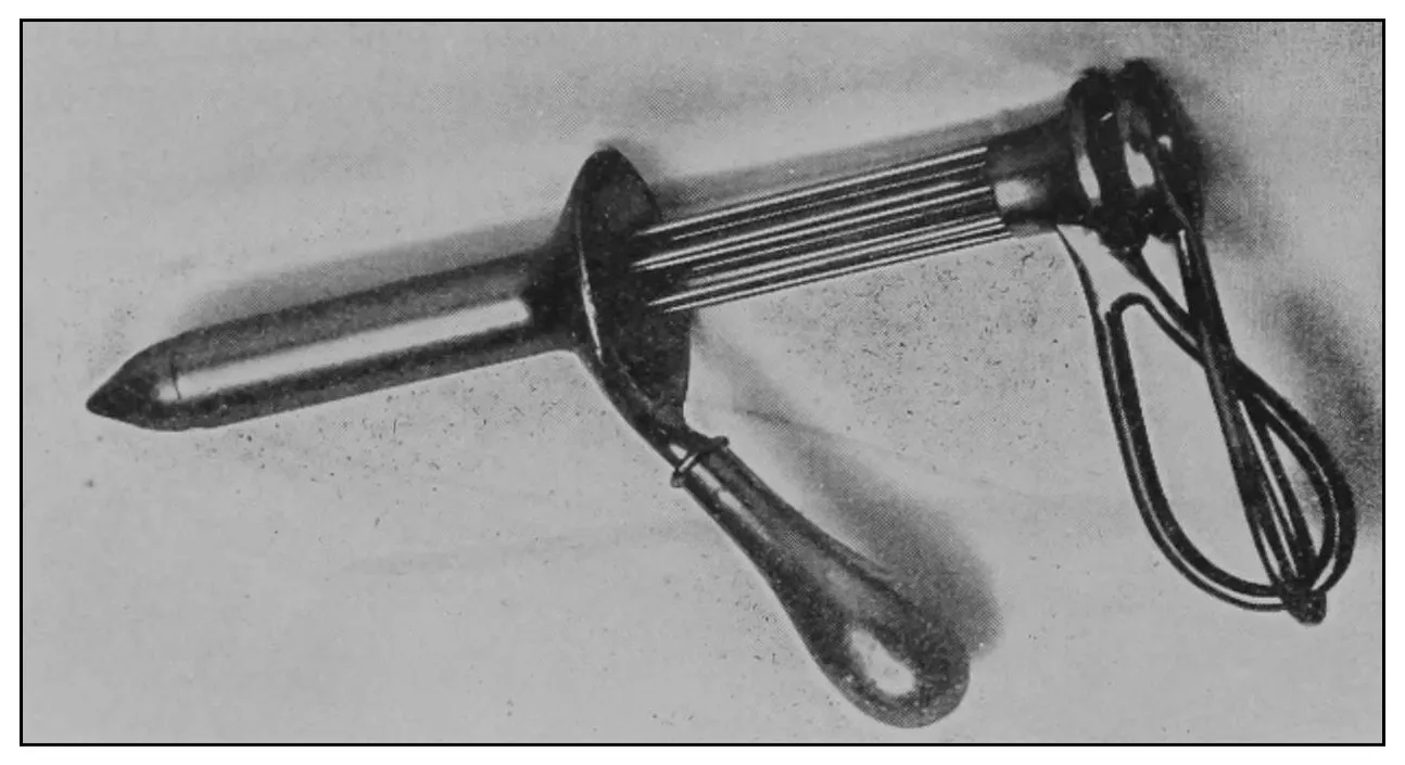
The anoscope (Fig. 6) consists of a short cylindric
tube open at the ends. It is two inches (5.08 cm.) in
length and seven-eighths of an inch (2.22 cm.) in
diameter. The proximal end is provided with a
trumpet-shaped expansion and a strong handle. The
distinctive feature of the anoscope is the peculiar
form of its obturator (Fig. 7), which has a capacity
for a multiplicity of uses.
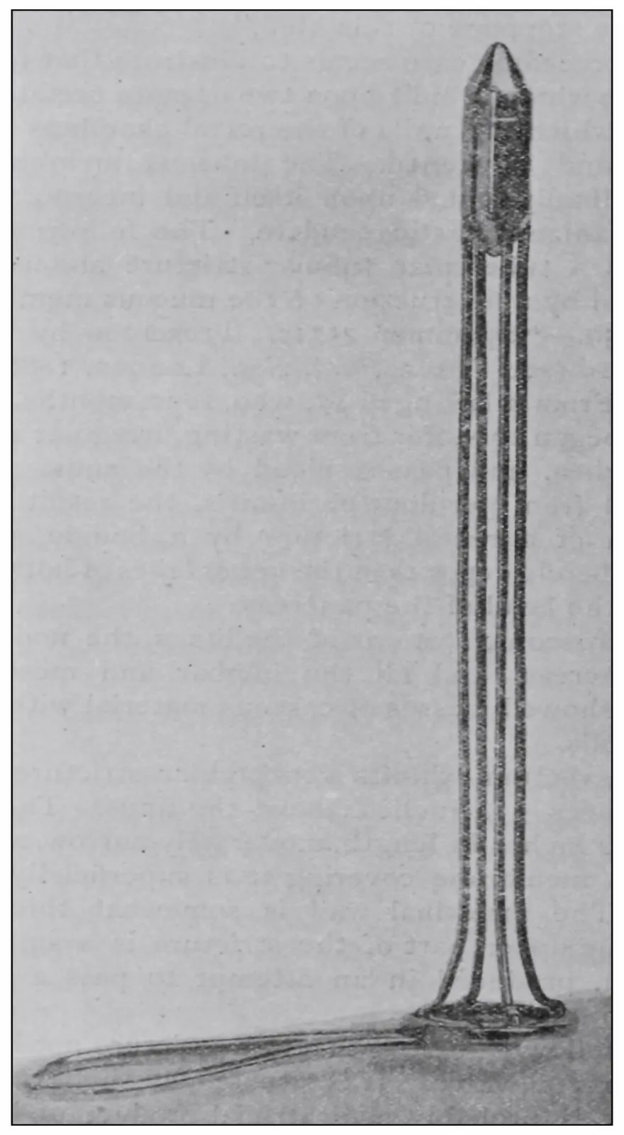
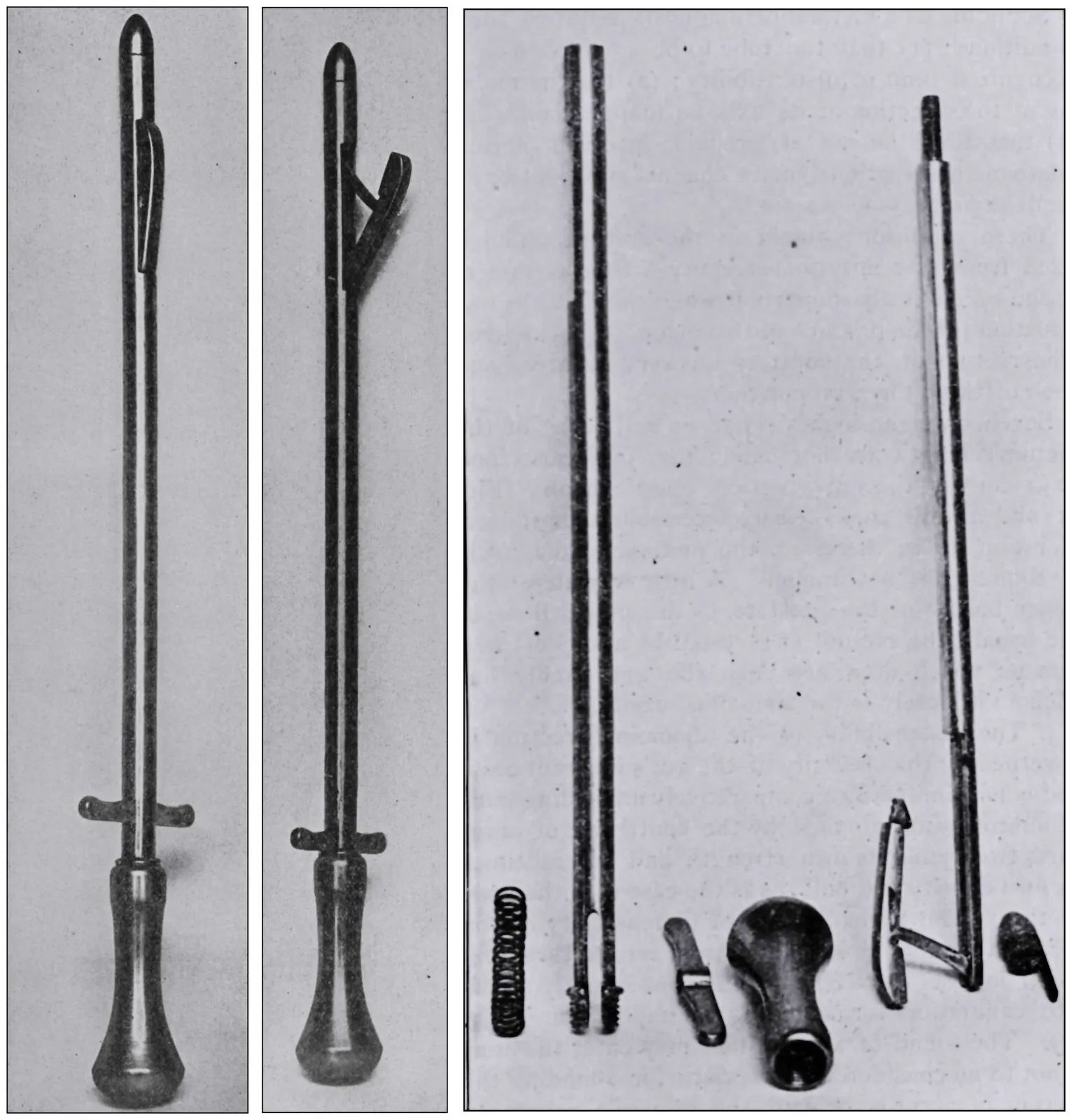
The obturator consists of a hard-rubber cylinder, in
the middle of which is fixed a brass tube for the purposes
of irrigation. Its surface is fluted in such a
manner that it may be made to lock in any of several
positions upon a tubercle within the cylinder. These
flutes also provide for escape from the rectum of
fluids and gases under certain conditions. The contracted
neck near the distal end of the obturator provides
a cup to facilitate the application of ointments
to certain rectal areas.
Fig. 9.—The two-way irrigator.
The contracted neck is a feature which contributes
to the instrument’s usefulness as a means for irrigation,
providing in the one case a self-retaining direct-flow
irrigator, and in the other case when locked in
the position shown in Fig. 9, an unobstructed two-way
irrigator. Platinum pins connect the centrally-placed
brass tube with the surface of the neck of the
obturator, which makes the instrument an anal electrode.
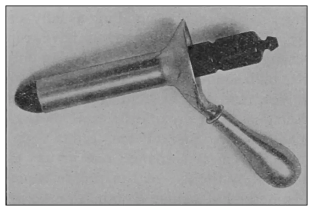
Fig. 10.—The proctoscope.
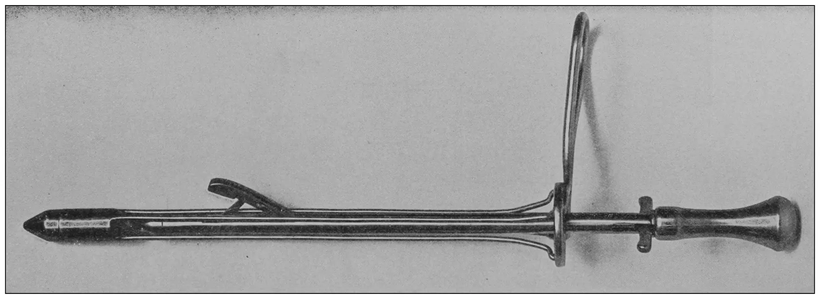
The proctoscope (Fig. 10) is of the same diameter as
the anoscope, and is four inches (10.16 cm.) in length,
which, because of the displaceability of the pelvic
floor is usually of sufficient length to reach as high as
the promontory of the sacrum, except in some especial
instances, the management of which exceptions
will be treated of in another place.
Special preliminary preparation of the patient is
ordinarily not required, as the usual condition of the
rectum is that of emptiness. In some cases, however,
it facilitates the inspection if the patient employs
rectal lavage an hour before the examination. This
injection should not consist of more than one pint of
fluid. The excess of this might be accidentally dejected
from the sigmoid and obscure the field during
examination. The bladder should be emptied, since
its distention would necessarily interfere with the
accuracy of the examination.
Fig. 11.—The ointment applicator at the time of the placing of the ointment
in contact with a diseased area.
Fig. 12.—The proctoscope ready for introduction.
Step 1.—The patient should be required to sit on
the operating-chair with his body turned to the left,
facing the knee-board. The right knee should be
crossed over the left knee, the left arm should embrace
the right border of the chair-back, or it may be
folded at the side as for Sims’ posture. The small
pillow should be held in the patient’s right hand and
against and upon his left shoulder (Fig. 13).
Fig. 13.—Sitting posture of the patient, the first step toward proctoscopy.Fig. 14.—Horizontal posture of the patient for anoscopy.
Step 2 consists in shifting the chair to the horizontal
position shown in Figs. 14 and 4, and in adjusting
the light-fixture. This movement brings the patient
into Sims’ semiprone-semiflexed posture, without requiring
any movement whatever on the part of the
patient after he is properly seated. In this posture,
the external anus and fixed rectum are to be examined.
(a) Digital examination and ocular inspection
should now be made of the anal verge, the external
anus, and superficial ischiorectal space, at a moment
when the patient is relaxed, and, again, when he is
bearing down.
(b) Digital examination of the fixed or anal rectum,
also, should be made preliminary to the introduction
of the anoscope.
(c) The anoscope should be gently pressed into the
anus in the direction of its axis till the sphincters relax
to receive it. The introduction of an instrument into
the rectum may be much facilitated by placing its
lubricated end against the ectal sphincter and requiring
the patient to bear down; bearing down expands
the ectal sphincters, relaxes the levator ani, thins the
pelvic floor, or shortens the fixed rectum, and presses
the ental sphincter over the instrument—in other
words, the patient’s anus is made to climb down upon
the speculum. After the introduction of the anoscope,
its obturator should be removed, and the inspection
made. These observations should be made coincident
with the withdrawal of the anoscope. In cases of extremely
sensitive ani, a skillfully put hypodermic injection
into the sphincters of 10 or 20 minims of ⅒
of 1% solution of cocain may render anoscopy painless.
A desire for precision requires that lesions of the
fixed or anal rectum should be noted as occupying a
given quadrant, and as situated at a given zone, e.g.,
a circumscribed disease may be described as situated
at the ental sphincter zone, and in the left lateral
quadrant.
Fig. 15.—Putting the patient into the new posture.Fig. 16.—The new posture.
Step 3 (a) requires that the shoulder-suspender
should be placed and fixed to the chair, as shown in
Figs. 5 and 16, that the knees be drawn up, so that the
thighs are at a right angle to the length of the chair-top,
and that the chair should be tilted to put the patient
in the new posture shown in Fig. 16. The leg
foot-board should now be lowered, and the operator’s
stool placed in convenient position.
The illumination-apparatus should now be adjusted,
as shown in the illustrations. In this new posture,
which is equivalent to the knee-chest posture, the abdominal
rectum is to be examined.
(b) Introduction of the proctoscope requires supported
eversion of the buttocks and steady gentle
pressure of the well-lubricated instrument upon the
anus and in the direction of the umbilicus, until the
sphincters are felt to yield, or the patient may be
required to bear down to take the speculum; as the
instrument enters the inflatable movable rectum, it
should be pointed toward the promontory of the
sacrum, and subsequently into the sacral hollow.
The withdrawal of the obturator is followed by atmospheric
inflation of the rectum.
(c) The operator should observe the degree of rectal
distention, the situation and number of the rectal
valves, their propinquity to one another when passive,
and the relation of one valve to another at the time
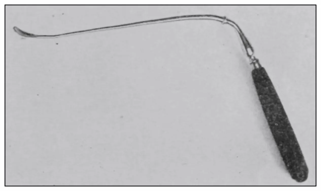
of the patient’s bearing down. Under pressure of
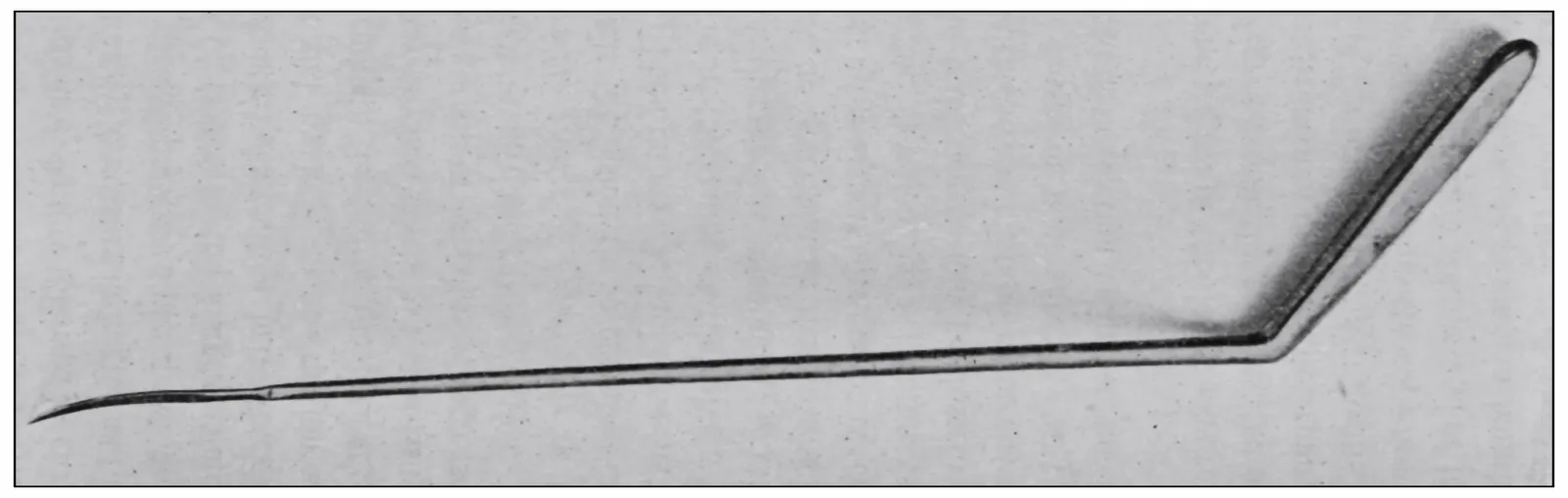
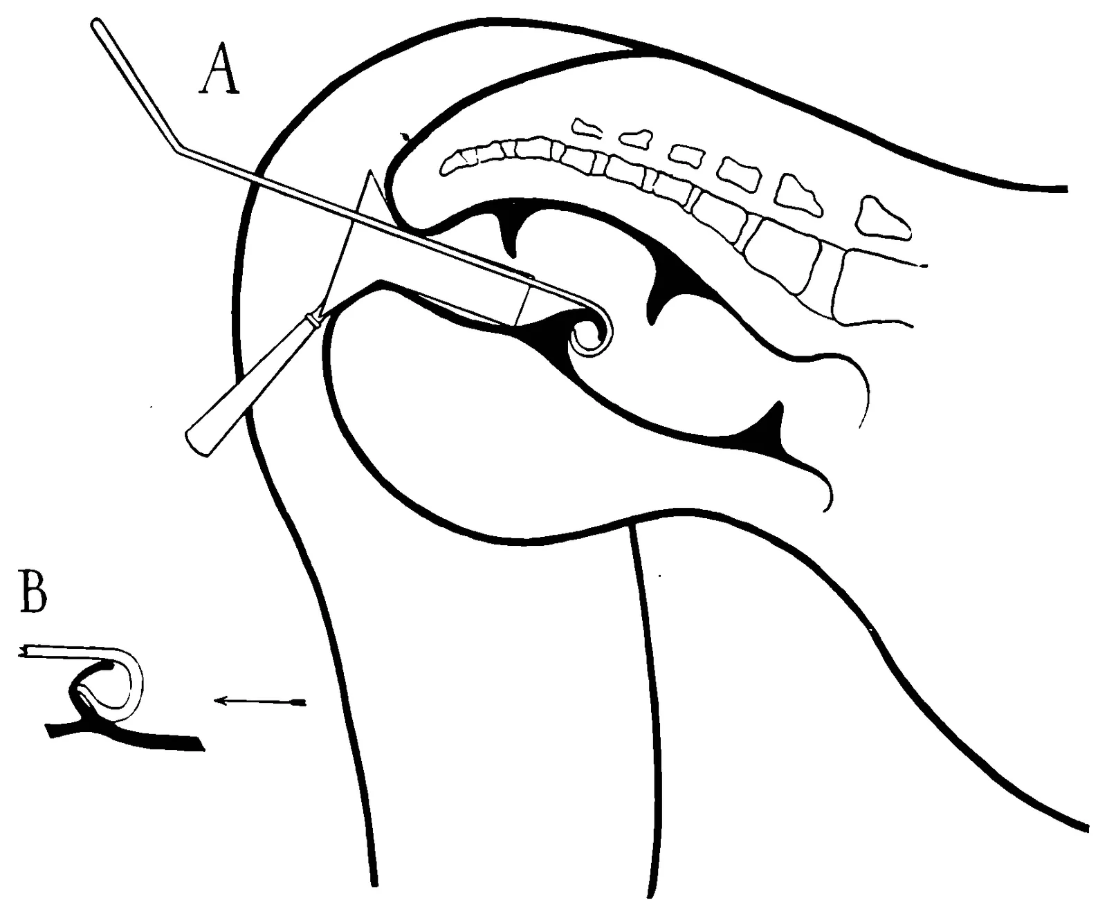
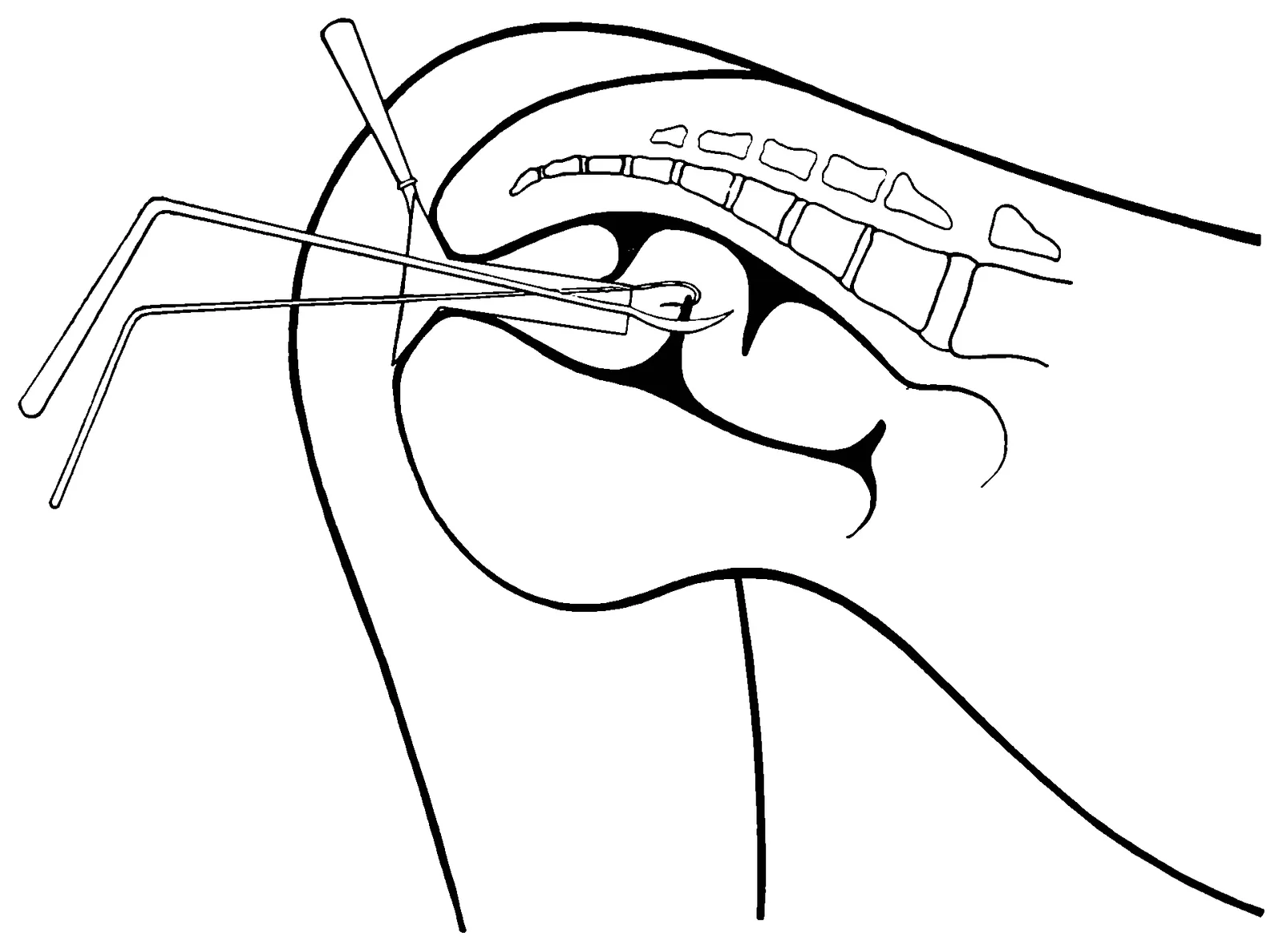
the proctoscope if possible, or the hook if necessary,
each valve should be effaced or displaced, and in regular
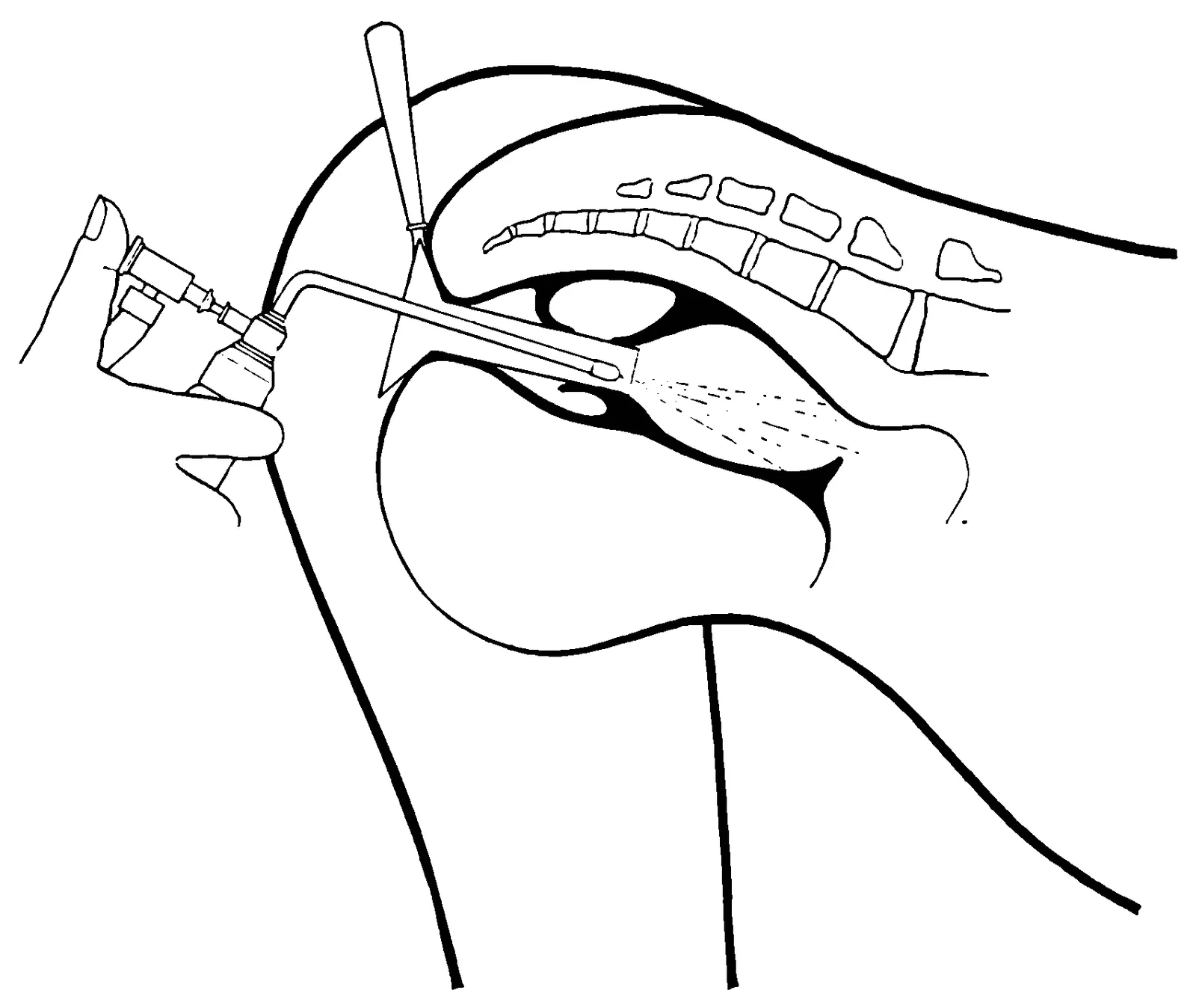
order each of the rectal chambers should be carefully
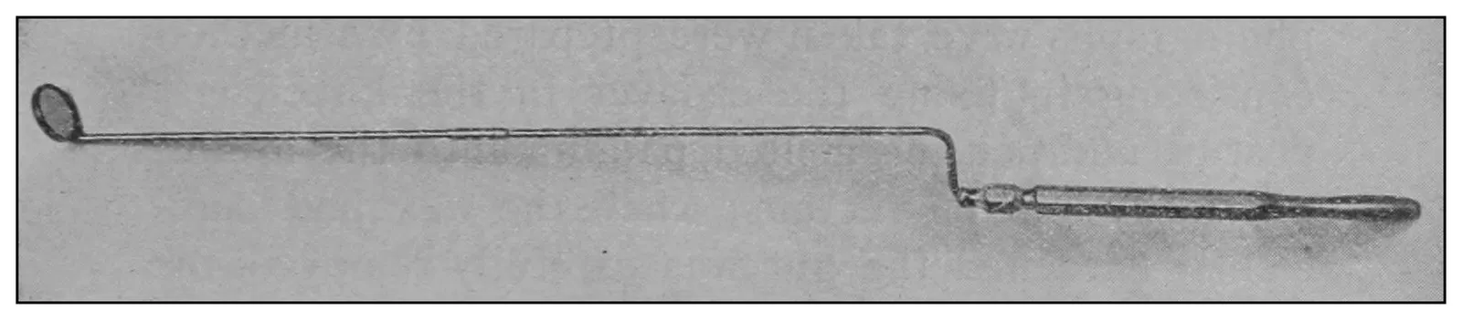
inspected. A proctoscopic mirror may be necessary
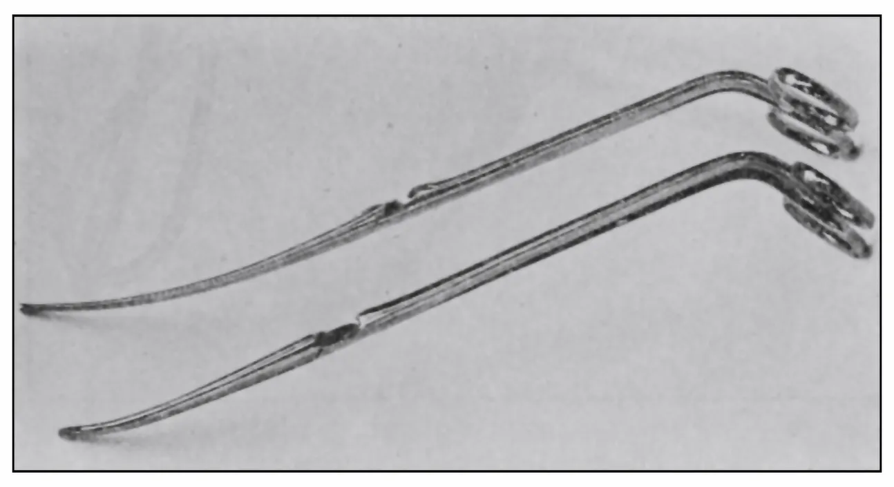
for viewing the supravalvular surfaces (Fig. 18).
The examination being finished:
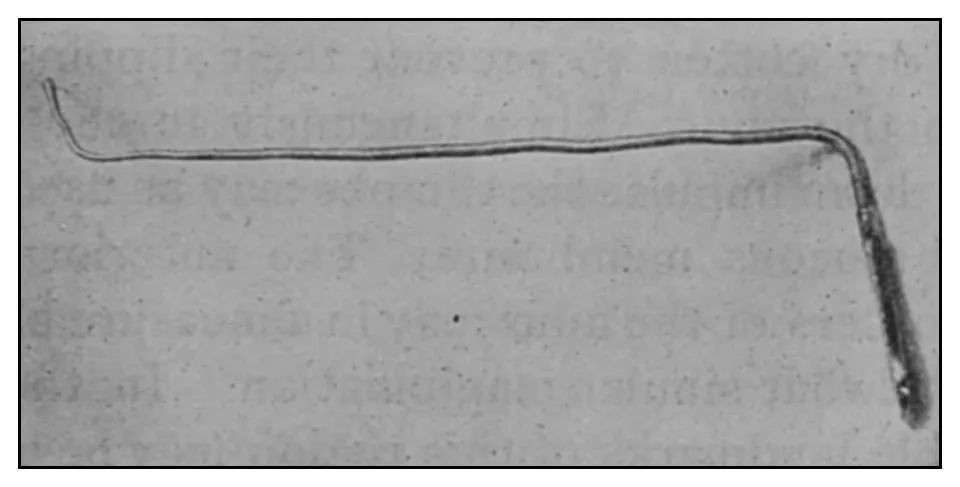
Fig. 17.—The hook for testing the valves.Fig. 18.—The proctoscopic mirror.
Step 4.—The proctoscope should be withdrawn, the
illumination-apparatus fixed in the first position, the
leg foot-board lifted to its place, the lever extended,
the crank turned and the chair carried back to the
horizontal and upright positions, and thus the passive
patient may be returned to his feet by the execution
in the reverse order of the several steps described.
This method of inspection does not subject the
patient to struggle, strain or embarrassment.
Observation by this method has taught me that in
nearly all cases of disease at the anus there is congestion
or inflammation of the rectal mucous membrane.
Those cases in which there is no apparent lesion at
the anus, and which are in a perfunctory way sometimes
declared catarrh of the rectum, will at once
have the real condition, such as a high up rectal polypus,
congenital or organic stricture or ulceration,
positively diagnosed, and will be made accessible for
intelligent treatment.
New growths and ulcerations may be seen and by
means of a long-handled curet scrapings made in order
that the microscopist may determine their exact
character.
Stricture of the rectum need no longer be regarded
as of only doubtful presence, and this method proves
positively, even to the casual observer, how fallacious
is the method of rectal sounding usually employed
for the diagnosis of stricture. I have repeatedly
proved to visitors how easy it is for an entering or
returning bulb-sound to be caught and held by the
rectal valves,[14] and thereby yielding signs generally considered
diagnostic of organic stricture of the rectum.
Vesicorectal, vaginorectal and other deep fistulas
are often apparent at a glance, but in any case may
be discovered by the use of the proctoscopic mirror.
If this method of ocular examination be practised,
I am convinced there need be no longer any excuse
for calling an undiagnosed disease of the rectum obscure
disease, and whatever the disease present this
method makes it susceptible of demonstration by the
proctologist to the attending physician. There is no
necessity that a diagnosis be taken on faith.
Topographic Anatomy.
A close study of the lesions of the rectum and
their manifestations and some experience in discussion
of these subjects have convinced me of the
necessity of methods of greater accuracy than those
generally employed in designating the precise situation
of a pathologic feature in this organ. Mensural
methods of designating the situation of strictures in
the rectum are of no surgical value.
The manner of the application of the details in a
given method of treatment for a disease situated in
the fixed or anal portion of the rectum should differ
essentially from that employed in the application of
the same principles of treatment to a similar disease
situated in the movable abdominal rectum; and the
application of the details of a given kind of treatment
should differ, too, according to the situation of
the disease at one point or another in the circumference
of the rectum. The prognosis as well as the
treatment of rectal disease is determined not only by
the pathologic character of the lesion, but also by its
anatomic situation. “Two inches up,” or “one and
a half inches from the anus,”[15] if one were positively
sure of the precise location referred to as the anus,
would point to very different anatomic parts, whether
the subject were male or female, and whether young
or old, and whether thin or stout.
The rectum, the terminal 8 or 10 inches (20.32 or
25.40 cm.) approximately of the intestine, presents
an upper abdominal and essentially movable portion,
which is about three-fourths the length of the entire
rectum, and a lower coccygeoischioperineal and essentially
fixed anal portion, which is about one-fourth
the length of the rectum. The upper half of the
movable rectum is, in the majority of adult persons,
completely invested with peritoneum, which sometimes
provides this portion with a mesentery. The
lower half of the movable rectum begins at a point
about the upper border of the second sacral bone, and
is not completely invested with peritoneum; the peritoneum
is reflected from the sides of the rectum toward
the lateral masses of the sacrum, so that the
posterior wall of this part is not covered by peritoneum.
The movable rectum begins opposite the
sacroiliac synchondrosis. If a subject lies in the dorsal
posture, and if the abdomen be normal, the upper
end of the rectum will lie opposite the left sacroiliac
synchondrosis. If the patient is inverted to the
knee-chest posture or its equivalent, and if the abdomen
is normal, the upper end of the rectum, if distended,
is usually found nearer the right sacroiliac
synchondrosis. The movable abdominal rectum terminates
at the levator ani muscle.
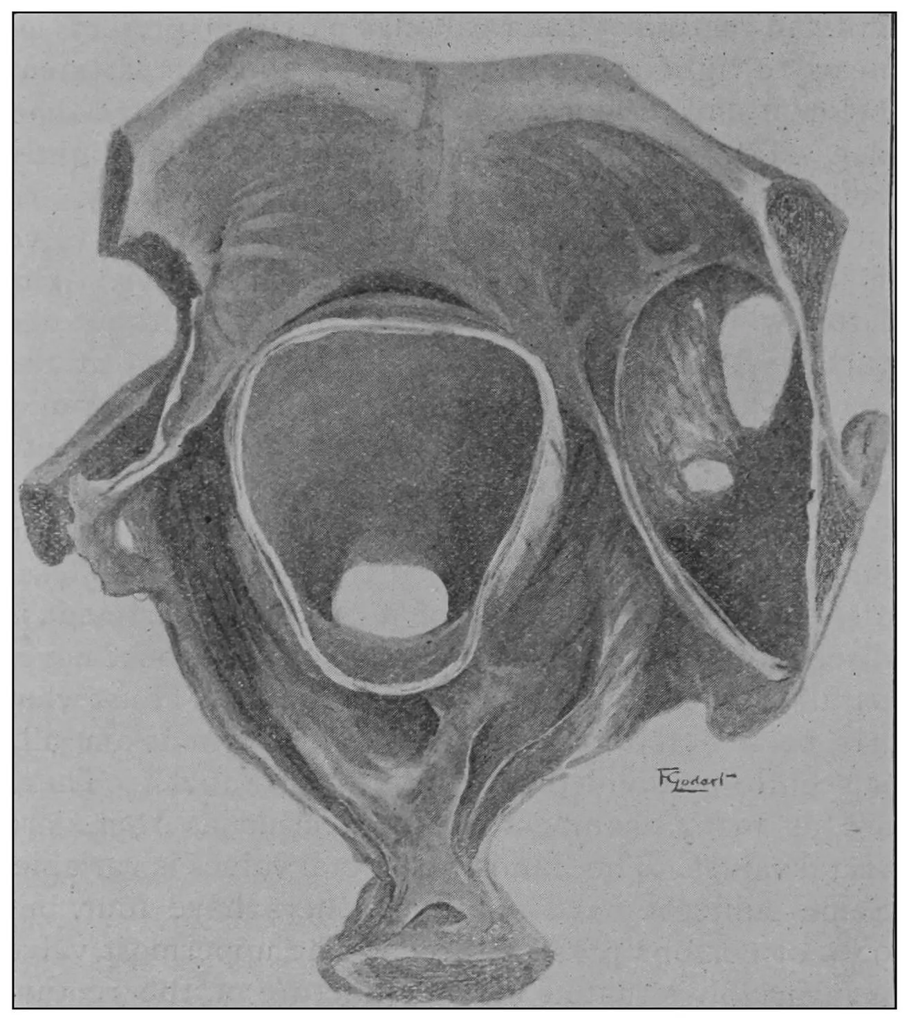
Fig. 19.—Drawing of specimen No. 281 in the Anatomical Museum of the
Royal College of Surgeons, London. The dissection was made by Mr.
William Pearson. The drawing shows in the lower part of the field the
coccyx, in the upper part of the field the pubes.
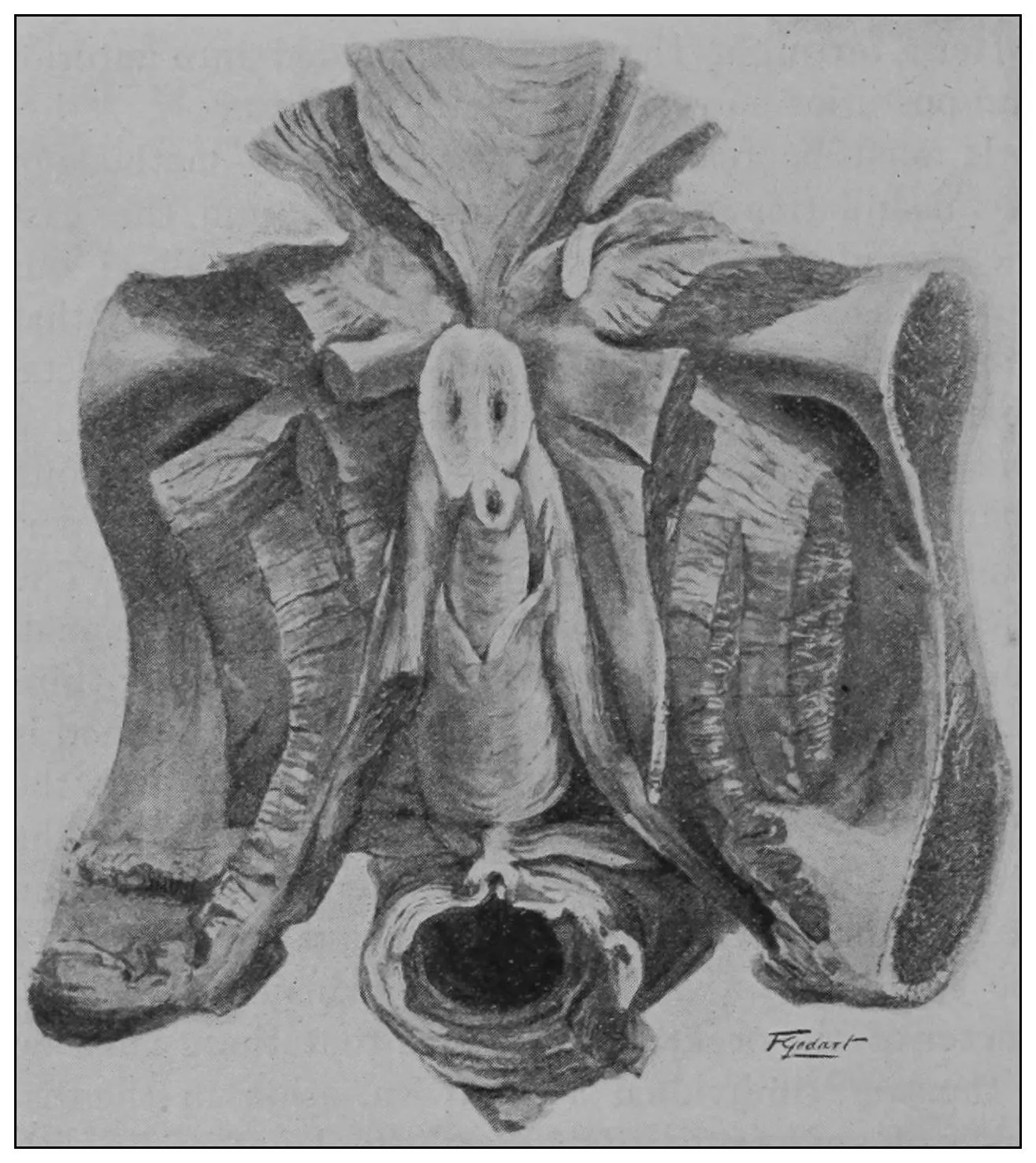
Fig. 20.—Drawing of specimen No. 284 in the Anatomical Museum of the
Royal College of Surgeons, London. The dissection was made by Mr.
William Pearson. The drawing shows in the lower part of the field behind
the rectum the coccyx, in the upper part of the field the pubes.
The fixed anal rectum begins at the levator ani and
coccygeus muscles. The levator ani has its origin at
the sides of the bodies of the pubic bones, the coccygeus
at the spines of the ischii, and the levator ani
has additional origin from the fascia and bony parts
on a line between these two points. The fibers of
these muscles are directed downward and inward to
the fixed anal rectum; many fibers are blended into
the contiguous structures of the pelvic floor. The
coccygeolevator muscles may be compared to an
opened slat-fan, the apex having its place at the anus,
and its long border representing the line of origin of
the muscles, Fig. 19. The ental sphincter ani muscle,
situated a few lines below the levator ani, is made up
of an aggregation of the fibers of the circular non-striated
muscular intestinal coat, Fig. 20. The ectal
sphincter ani striated muscle is situated immediately
beneath the external skin. It serves the present occasion
to describe the ectal sphincter of the male as
a loop of muscle thrown about the terminal end of the
rectum and hitched to the terminal bone of the coccyx,
and in the female as a longer loop of muscle
twisted upon itself so as to make a tandem-loop,
which, in the form of the figure eight (8), is thrown
about the vagina and terminal end of the rectum.
In the passive subject the finger discovers the ectal
sphincter as a broad, relaxed band of muscle situated
beneath the external skin and surrounding the infra-anal
depression. In action the ectal sphincter is
contracted and is retracted from beneath the external
skin to a point beneath the mixed mucocutaneous
integument. In such a state of ectal sphincter contraction
the fixed portion of the rectum is lengthened,
or in other words, the pelvic floor is deepened one-half
inch (1.27 cm.), approximately. The finger
progressing, it engages the tonicly contracted ental
sphincter muscle which the delicate touch discovers
as vibratory in its grasp of the finger, presenting to
the touch the sense of a sharp rigid ring, and again
as a flat band snugly applied about the finger. A
few lines above the ental sphincter the finger discovers
the upper limits of the fixed or anal portion of
the rectum surrounded by the somewhat V- or U-shaped
borders of the levator ani muscle; the anterior
quadrant is not encircled by the levator ani. The
fixed portion of the rectum presents, then, to the
touch, three landmarks: the usually relaxed ectal
sphincter, the usually contracted ental sphincter, and
the levator ani muscles. The normal levator ani
muscle can not by the contraction of its fibers close
the upper end of the fixed rectum. It is possible,
however, for the patient to contract his levator upon
the finger of the examiner to quite an appreciable
degree, a fact which led Mr. Harrison Cripps to state
that the levator ani can close the upper end of the
fixed rectum. If a tubular speculum of seven-eighths
of an inch (2.22 cm.) diameter be introduced through
the fixed rectum and then withdrawn through the
levator ani to a point above the ental sphincter and
the patient be directed to contract the muscles of
the pelvic floor, it may be observed that the axis of
the fixed rectum is directed more forward at its upper
end, and that the depression between the levatores
ani is partially encroached upon by the levator fibers
but is in no sense effaced; hence, we must recognize
that the lower fixed portion of the rectum presents
a canal of an hour-glass form, expanded above as a
concavity between the borders of the levatores ani,
narrowed at its middle part by the contracted ental
sphincter, and expanded below where it is surrounded
by the relaxed ectal sphincter muscle.
In the same subject the length of the fixed anal
rectum is variable with a state of activity or passivity,
and in a state of activity there are variations in
its length of at least one inch (2.54 cm.) between a
contracted, uplifted pelvic floor and that of a depressed
floor with anal eversion; both of which conditions
may rapidly follow one upon the other while
the examiner’s finger is engaged in diagnosis. Again,
variations in depth of the fixed anal rectum are quite
noticeably regulated by the size of the finger introduced.
The thumb may find a fixed anal rectum of
two inches (5.08 cm.) in depth, while the little finger
discovers it but a little more than an inch (2.54 cm.).
Because of the bony confines to the tissues of the
ischiorectal space, displacement of its structures to
open the anus must occur in the vertical direction,
the larger the finger the greater the displacement
upward.
Passing the finger beyond the borders of the levator
ani, the distal phalanx enters the movable or abdominal
rectum, where it may be hooked over the pelvic
floor. In some instances, if the finger be directed
backward and crowded with a boring maneuver
through the loose folds of the movable rectum, and
provided the folded knuckles displace upward the
pelvic floor, the finger may be made to engage the
lowermost of the rectal valves, which will contract
about the finger with a rhythmic action and mislead
the uninformed, inexperienced and undiscriminating
explorer to think that he is but now encountering the
ental sphincter muscle, or, perhaps, that he has discovered
a stricture—the phantom stricture? In the
passive rectum, this valve is usually about three
inches (7.62 cm.) above the lower border of the ental
sphincter muscle. The great range of mobility of
the pelvic diaphragm permits a finger of two inches
(5.08 cm.) length to be hooked over a valve which
under some circumstances may be an inch and a half
(3.81 cm.) beyond its reach.
The pelvic floor in the infant is often less than one-half
inch (1.27 cm.) in depth. The depth of the pelvic
floor in the adult, from the lower border of the relaxed
ectal sphincter ani muscle to the levator ani muscle,
is extremely variable. In the aged male, because of
senile enlargement of the prostate, the fixed rectum
may seem to be three inches (7.62 cm.) in depth. In
the aged female, because of senile atrophy of the
generative organs and contiguous structures, the pelvic
floor may be much less than an inch (2.54 cm.) in
depth. In the adipose and in emaciated subjects
because of the character of the tissues occupying the
ischiorectal space, there are the greatest variations in
the depth of the pelvic floor. Hence, it is obvious
that the palpable muscular landmarks of the fixed
rectum are situated at variable positions in the different
sexes, and that the length of the fixed rectum is
changed in the same person at different periods of
life and in differing conditions of flesh.
The visible topographic features of the fixed rectum
are several, and under the influence of disease may
become somewhat changed in appearance and situation.
In the male they are readily discoverable by
putting the subject in the dorsal posture, separating
the nates with the hands and placing the thumbs on
bits of dry cotton to prevent their slipping just exterior
to the anus. Simultaneously to the patient’s
bearing-down impulse the thumbs may be used to evert
the anal mucous membrane. The anterior and posterior
borders of the anus may in a measure be everted
by a somewhat similar manipulation. In the female
the visible landmarks of this region may be inspected
by entering the finger into the vagina and placing the
thumb over the perineum; the former is to push the
rectum down while the thumb draws the external skin
forward over the perineum. The posterior segment
may be exposed by pushing the post anal skin toward
the coccyx; the lateral segments may be exposed with
the finger and thumb of the left hand while the right
hand keeps up the initial pressure. Marked pigmentation
of the anal skin is observable in a circumscribed
area about the anus; beneath this area of darkened
skin, intimately attached to the skin, lies the surgically
unappreciated corrugator cutis ani. Within the
borders of the everted anus the complexion fades to a
light gray, within which zone is sometimes noticeable
the distinctly lighter zone known as Hilton’s
white line. Above, is noticeable an undulating zone
of deeply red mucous membrane the lower border of
which has been given the name of linea dentata
(Stroud). Between the linea dentata and the white
line is a zone occupied by several pyramidal elevations
about half an inch (1.27 cm.) in length, to which
has been given the names: columns of Morganni and
pecten of Stroud. Their bases are of a somewhat
purple reddish color. Toward their apices they may
pale and terminate in projecting white eminences.
The bases of these pyramids, which, I believe, may
without impropriety be called anal pilasters, are extremely
vascular and their structure partakes somewhat
of the character of erectile tissue. The apex
contains a nerve end-bulb. Under the influence of
disease the color of these bodies is changed. Their
antemortem and postmortem appearance is quite different.
There are usually present in each anus from
four to eight of these bodies. Between these projections
and at their lower borders, sometimes, there is
discoverable a thin fold of membrane. The saccule
which it, together with the anal wall and pilasters on
either side, forms, has been known as the saccule
Horneri, pocket of Physic, and anal pocket, sometimes
inaptly called rectal pocket. Its cavity is about
the size of a split pea. The three typic visible topographic
features of the fixed rectum, then, are the
white line of Hilton, the pecten of Stroud, or anal
pilasters, and the linea dentata.
The mucocutaneous membrane of which these visible
landmarks are a part, rests upon a quantity of
loose connective tissue, which permits of a great
range of mobility of these features independent of
movement of the structures constituting the palpable
landmarks of the fixed or anal rectum.
Fig. 21.
Fig. 22.
Fig. 23.
Fig. 21.—From a photograph of an external view of the paraffin cast-filled
rectum. The rough surface shows the place of attachment of the peritoneum.
Fig. 22.—Interior view of the left half of the rectum of an adult. Filled with
melted paraffin, the subject being in the proctocolonoscopic posture. The
rectum is distended only to the degree of normal atmospheric inflation.
When the wax was hardened the rectum was removed, varnished, dried
and cut open longitudinally. The picture is a reproduction of a photograph
of the interior of the left half of the rectum. The anus is at the
picture’s left. The valves are respectively two and one-half and seven
inches (6.35 and 17.78 cm.) from the anus.
Fig. 23.—Knee-chest posture. Left lateral half-interior view. Upper rectum
and sigmoid out of focus.
Fig. 24.—Posterior view of a specimen carefully dissected to show the muscular
supply to the valve-bases.
Fig. 25.—Posterior half occupied by its cast.
Fig. 26.—Anterior half of specimen shown in Fig. 25.
Fig. 27.—Posterior view of a cast-filled rectum.Fig. 28.—Paraffin cast from a rectum.
The Rectal Valve.
That the rectal valve constitutes the most hypsometric
of the topographic features of the movable
abdominal rectum, is conclusively proven by the photographic
pictures. The specimens from which the
photographs were taken were prepared by a method
consisting in fixing the cadaver in the knee-chest
posture and pouring melted paraffin into the atmospherically
inflated rectum; when the wax had sufficiently
hardened the gut was carefully removed, for
a few weeks immersed in alcohol and subsequently
dried, varnished, and finally dried and cut into longitudinal,
shell-like halves. A comparative study of the
photographs of the rectal interiors discovers the fact
that the prominence of the rectal valve is increased with
the degree of distention of the rectum.
The accompanying drawing was made from a microscopic
section taken from the middle of the lower
valve of an adult specimen. In the illustration, Fig.
33, it is shown magnified 5 diameters. Beneath the
mucosa is noticeable the heavy layer of fibrous tissue
which gradually diminishes till it is lost at the valve
base. Bundles of circular muscular fibers are seen in
the middle of the valve. At its base are seen arteries
and veins for its special nutrition. This structural
arrangement makes this organ the typic anatomic
valve. The evidence of the fibrous tissue in the valve
is an original contribution to our knowledge of this
subject.
The attached border of each valve spans a little
more than half the circumference of the rectum, and
its free border projects half across the diameter of the
inflated rectum. Each valvular partition projects at
nearly a right angle to the wall of the compartment
below it and terminates in a sharply defined free border.
The free margin of the structure is slightly
concave in form and is directed a little obliquely. In
life it is noticeable that the free border of the valve
is less elastic than the main wall of the valve. The
latter will yield to a modicum of traction without disturbing
its free border, while traction applied at the
margin may at once disturb the entire circumference
of the gut at that point. The rectal wall opposite
the valve is usually concave. The aperture thus
formed between two rectal compartments is irregularly
elliptic; it may be called the valve-strait. Usually
the greatest diameter of a rectal compartment is
directed laterally or else obliquely; it is almost never
parallel to the direction of the sacrum. Thus, what
has been heretofore considered a cavernous ampulla
is seen to be divided into several chambers. There
are as many chambers in the rectum as there are
rectal valves. The number of rectal valves is variable.
Some subjects have but two, others have four, but
90% of persons possess three. The uppermost valve
is invariably situated at the juncture of the rectum
and the sigmoid flexure, which valve is invariably
situated on the left, the next lower is on the right
wall, and the lowermost is on the left. The positions
of the lower two valves are sometimes anterior and
posterior.
The specimen shown in Fig. 30 exhibits the rectum
naturally distended with feces. It was found in a
deceased member of the demimonde who had been
habituated to opium indulgence. She was forty
years of age and of robust stature. On opening the
abdomen the colon was discovered loaded with a collection
of scybalous feces, and the upper chamber of
the rectum was partially occupied with a similar deposit.
The abdomen was then opened through the
pubes and perineum down to the coccyx. The genitourinary
organs and contiguous structures were
removed, the symphisis forcibly separated and the
exposed sigmoid flexure and rectum were photographed
as shown in Fig. 29. The sigmoid and rectum
were carefully removed and immersed in alcohol.
After a fortnight the gut was divided into anterior
and posterior halves, as shown in Fig. 30.
Fig. 29.—Photograph of a female cadaver showing, after laparo-symphysiotomy
and removal of bladder, uterus and adnexa, the upper rectum and
sigmoid packed with scybalums.
Fig. 30.—The rectum, the same as is shown in Fig. 29, divided into anterior
and posterior halves.
It must be readily seen that the new methods of
rectal inflation for rectal inspection and the cast
method of dissection, which have determined our
newer ideas of the topography of this part, justify that
the lowermost chamber be considered the first rectal
chamber; the cavernous area beyond the first valve
and below the second should be called the second
chamber; and the upper chamber the third or perhaps
fourth, according to the number of valves. The
ancient arbitrary division of the rectum by the anatomists
into upper first, middle second and lower third
parts should be abandoned because such a method is
inaccurate and has no surgical significance. As the
arrangement of the fibers of the muscular coats of the
abdominal rectum and the attachments of the abdominal
rectum provide for extension and contraction of
the gut on its axis, as well as expansion of the diameter
of the organ, it is obvious that there must be
in the same individual a great variation in the distance
of any given valve from the levator ani with
the variable normal states of the organ. The normal
range of movement upward and downward of a
given valve is from two to three inches (5.08 to
7.62 cm.).
A discriminating study of the diseases of the rectum
requires an arbitrary division of the organ into several
quadrants.
Regard for the conveniences of scientific description
and considerations of technic and of anatomy
justify that the fixed anal rectum be called the anus,
and that the movable abdominal rectum be called
the rectum. The former is bounded above by the
levator ani and below by the ectal sphincter muscle.
From this survey of the topography of the anus and
rectum and consideration of the arrangement of the
contiguous structures the following salient aphorisms
may be deduced:
1. In treating lesions on a level with the sphincter
muscles the operator should beware of dividing these
structures through the anterior quadrant. In the
male the ectal sphincter terminates in the tendinous
raphe in common with the transversus perinei. Contraction
of the transversus perinei will separate the
cut fibers of the ectal sphincter and defeat the desired
subsequent union of this muscle. If an incision be
carried forward or forward and laterally into the transversus
perinei the perineal fascia which doubles over
this muscle will be opened and the perineum and peri-urethral
structures will be made accessible to infection
from the anus and ischiorectal regions. In the
female, to carry an incision forward through the anterior
quadrant would be unsurgical because the
peculiar arrangement of the fibers of the ectal sphincter
ani and sphincter vaginae and their relation with
the transversus perinei would perhaps conspire to
produce vulvoanal or rectovaginal fistulas.
2. A stricture located at the upper end of the fixed
rectum and situated in the anal fascia, in the pelvic
fascia, or in the fibers of the levator ani muscle,
should not be cut in the anterior quadrant nor in the
posterior quadrant, but in one or the other, or both,
lateral quadrants. An incision through the anterior
quadrant on the plane of the levator ani muscle
would divide none of its fibers because there are none
there and would endanger the urinary organs and
vagina in the male and female respectively. An incision
made into the posterior quadrant on this plane
would fail to increase the diameter of the rectum for
the reason that contraction of the fibers of the levator
ani would hold in coaptation the lips of the wound in
such a manner as to early reestablish the stricture.
A skilfully made incision in the lateral quadrants in
this region will not endanger the peritoneum. A
possible hemorrhage may be readily avoided by digitally
ascertaining the situation of the middle hemorrhoidal
arteries. And because of the direction of the
fibers of the levator muscle a short lateral incision
will effectually increase the diameter of the part.
3. The rectal valve must be reckoned with in
studying the strictures of the movable abdominal rectum.
Linear posterior proctotomy, because of the
relation of the peritoneum to the posterior wall of
the lower part of the movable rectum, is eminently
safe, but will be efficacious only in some special instances—those
instances in which the rectal valve involved
is posteriorly situated. Semilunar (annular)
strictures may be situated at any point in the circumference
of the movable rectum, for they are
built on the foundations supplied by the rectal
valves, and may be safely cut through to the depth
of a quarter of an inch (.63 cm.), provided the surgeon
be equipped with the proper instruments and provided
the rectum be maintained in a state of atmospheric
inflation at the time of the operation.
The studies of the topography of the human rectum
made in this research employed more than 50
autopsies, on subjects of all ages, and physical examinations
of many hundred living persons, and the
facts which are set forth above justify the inference,
I believe, that none but the topographic designation
of the precise situation of the rectal lesion is of
reliable surgical significance.
Dissection of many rectums of rabbits, cats, dogs
and monkeys proved that these animals are not provided
with rectal valves.
Defecation. — Digital Observation of the Nonanesthetized Subject. — Ocular Observation of the Anesthetized Subject. — Ocular Observation of the Nonanesthetized Subject.
“Defecation is partly a voluntary and partly a reflex
act. But in the infant the voluntary control has not
yet been developed; in the adult it may be lost by
disease; in an animal it may be abolished by operation;
in each case the action becomes wholly reflex.
In the normal course of events, the rectum which is
empty and quiescent in the intervals of defecation,
is excited to contraction as soon as feces begin
to enter it through the sigmoid flexure, and the
sensations caused by their presence give rise to
the desire to empty the bowels. This desire may
for a time be resisted by the will, or it may be
yielded to. In the latter case the abdominal muscles
are forcibly contracted, and the glottis being closed,
the whole effect of their contraction is expended in
raising the pressure within the abdomen and pelvis....
The sphincter ani is now relaxed by the
inhibition of a center in the lumbar portion of the
spinal cord, through the activity of which the tonic
contraction of the sphincter is normally maintained.
This relaxation is partially voluntary, the impulses
that come from the brain acting probably through
the medium of the lumbar center; but in the dog,
after section of the cord in the dorsal region, the
whole act of defecation, including contraction of the
abdominal muscles and relaxation of the sphincter,
still takes place, and here the process must be purely
reflex. The contraction of the levatores ani helps to
resist overdistention of the pelvic floor and to pull
the anus up over the feces as they escape.”[16]
The above paragraph concisely represents the accepted
interpretation of the act of defecation. The
rectal valve, an individual anatomic organ in itself,
must, however, be reckoned with in studying the
operations of the integral mechanism of defecation.
In the normal rectum, when the first rectal valve
can be reached by the finger, provided it be immediately
discovered on the introduction of the finger, a
definite but not rigid band of tissue may be felt buried
behind the masses of lax mucosa. This band presents
at one side that which seems to be a circular aperture
of variable diameter; a few seconds’ application of
the passive finger detects a gradual relaxation of what
first had seemed a ring-like constriction till the finger
can make out but an indefinite thickening on a side
of the gut-wall. If the finger be held passive, this
constriction may again surround the finger; but if so,
it does so with a somewhat weaker grasp. Often the
presence of the finger within the rectum excites a
peristalsis-like contraction, during which the valve
usually cannot be detected. When the rectum is filled
with water, the valve is sometimes more readily discoverable
by the finger, and when discovered, the diameter
of the valve-strait is greater than that before
noticed, and the grasping tendency of the valve is not
so noticeable.
If a somewhat emaciated male be selected, one
whose rectum is susceptible to atmospheric inflation
while in the lithotomy posture, and if the large intestine
be freed of its contents, if such a subject be anesthetized
and his colon or sigmoid filled with water, and
the anus divulsed and held open with the speculum, the
water confined to the rectum may be bailed out, and
the rectum will then be seen to present the multichambered
condition already described. If the inquirer
will now place a hand on the abdomen over the
sigmoid flexure, and give the part a quick, firm pressure,
his watchful eye may observe a downward rush
of waters into the rectum, first striking the concave
wall formed by the juncture of the upper surface of
the uppermost visible valve and the adjoining rectal
wall, bounding from this surface to the opposite next
lower valve, from which the stream is deflected to a
lower point on the opposite wall, by which time all
may be obscured by a wave of contracting peristalsis,
rhythmically repeated, which may project small quantities
of water through the anus. In the intervals
of the contractions, the rectal chambers are observed
to be either submerged or else atmospherically
inflated.
If a subject under proctoscopy be directed to bear
down, it is observable that the empty and atmospherically
inflated rectum closes under the compression of
the voluntary forces by the concave anterior wall becoming
convex, and being carried backward toward
the fixed posterior wall, the transverse diameters remaining
apparently but little shortened. The compression
may be noticed as being applied from before
and in a backward direction, and simultaneously from
above downwardly. In case firm fecal masses, or lubricated
balls of cotton experimentally placed, are dejected
by these efforts, there will be noticeable depression
of the valve-floor under the pressure of the
descending mass. Presently the mass will appear at
the valve-edge, where it may seem suspended for a
few seconds before it is plunged on into the next
lower chamber. Occasional rhythmic contractions of
the rectal muscular coats occur, which fill and transitorily
obscure the field under observation with masses
of the reduplicated mucosa, and it is always possible
to note that such contractions have changed the positions
of the gut’s contents.
These anatomic findings and physiologic observations
warrant the conclusion that the rectal valve
has a function. Its histologic elements endow it
with essentially passive and active properties. When
the muscular elements are relaxed and the gut is
either greatly dilated or else in a lesser measure distended,
the valve is passively projected across the
channel to resist the hurried or uncontrolled descent
of the feces. The presence of the bands of fibrous
tissue under the free margin of the valve provides a
guard or control to receive and retain the bolus, or,
I may say, the valves receive a series of boluses, till
a sufficient pressure is made to stimulate the complex
involuntary mechanism of defecation to an expulsion
of the feces or to a reversed peristalsis. The presence
of the feces or the involuntary movements incident
to their presence, signals the consciousness to
cooperative voluntary expulsory effort, or gives warning
of the necessity of voluntary resistance. In the
event of the exercise of these forces in the direction
of resistance there seems to be some reason to believe
that an antiperistalsis returns the feces to the
sigmoid flexure. The same arrangement which sets
in operation the involuntary factors of defecation
also provides for the minimum expenditure of energy
on the part of the voluntary forces for the reason that
the gut’s contents are collected on the surface of the
unyielding sacrum and steadied there to receive the
pressure of the really expulsory voluntary effort. Such
an arrangement of the feces as is mentioned above
further facilitates defecation for the reason that
the entire contents of the rectum are not rushed
upon the anus at once. The surgeon remembers
that it is usually impossible to reduce a hernia en
masse.
The bundles of circular muscular fibers which constitute
the muscular element of the valve evidently
belong to the same mechanism and have the same
function as those which form the ental sphincter.
Their usual state may be that of tonic contraction; they
are relaxed by inhibition; when the muscle is in a state
of inaction the fibrous bands are projected and support
the valves across the channel of the rectum. In
health the valve is susceptible, under pressure, of a
temporary effacement.
If it be the function of the normal rectal valve
to beneficently retard the descent of the feces it
is obviously true that it may be the especial property
of the valve, in certain other than normal conditions,
to maliciously obstruct the descent of the
feces.
My experience convinces me that a perfect knowledge
of the rectal valve constitutes the key to an
understanding of obstipation, rectal stricture, and
their sequels.
Fig. 31.—From a photograph of an anal end-view of a cast-filled rectum; the
average transverse diameter was 3½ inches (8.89 cm.), the vertical diameter
was 3 inches (7.62 cm.).
Fig. 32.—From a photograph of an external view of a cast-filled rectum and
its mesenteric attachment to the sacrum; taken from an 18-months-old
infant.
Obstipation in Infants. — Treatment.
The infant strains at stool because of the imperfect
development of the anatomic features concerned
in the mechanism of defecation. These are
as follows:
1. The infant’s lower gut is muscularly deficient.
2. Its great length and its mobility within the
abdomen are obstructive to defecation.
3. The rectal valves are obstructive.
4. The infant’s anus not being sufficiently expansible
is also obstructive to the descent of feces.
The specimens of infant rectums and sigmoids here
pictured are all fairly illustrative of the facts upon
which the foregoing declarations are based.
The dried specimens shown in the illustrations
were prepared by flushing the intestine and then
fixing the subject in the genuacromial posture; the
anus was then fixed open and melted paraffin was injected
under about twelve ounces’ pressure. When
the cast had hardened, that portion of the gut which
it occupied, was removed. Specimens shown in Figs.
43 and 44 were prepared by placing the subject upon
its back and by opening the descending colon; the
intestine below was then washed out and the colon
perforation fixed at the abdominal wound, which,
save for this point, was sewed up, the anus was tied
up and as much melted paraffin as would enter under
two-pounds’ pressure was forced into the gut; subsequently
the sigmoid and rectum were removed as in
the other instances. After immersion in alcohol the
specimens were varnished.
Fig. 33.—A semilunar valve drawn as seen under a glass magnifying five diameters.
A, mucous membrane; B, fibrous tissue; C, bundles of circular
muscular fibres; D, vein; E, artery; F, vein; G, artery; H, areolar and
adipose tissue.
Fig. 34.—The instrument case; the drawers opening on three sides afford
access to any drawer without disturbing the position of the others.
Following are the memoranda of a few of the
autopsies made in this study:
Case 1.—Female, stillborn, height 16 inches (40.64 cm.); circumference
at anterior superior spinous process 7½ inches
(19.05 cm.); anterior superior spinous process diameter 3 inches
(7.62 cm.); ensiform to pubes 4 inches (10.16 cm.); transverse
diameter pelvic outlet ¾ inch (1.90 cm.); peritoneum at last vertebra
of coccyx. (Fig. 35).
Fig. 35.—The rectum of an infant, stillborn; muscular coat undeveloped.
Filled from below.
Fig. 36.—Side view of the specimen shown in Fig. 35. Antero-posterior angulation
at third sacral vertebra.
Case 2.—Female, aged 1 hour, height 17 inches (43.18 cm.);
circumference at anterior superior spinous process 7½ inches
(19.05 cm.); anterior superior spinous process diameter 2½ inches
(6.35 cm.); ensiform to pubes 4 inches (10.16 cm.); transverse
diameter pelvic outlet ½ inch (1.27 cm.); peritoneum at last
vertebra of coccyx. (Fig. 37).
Fig. 37.—Front view of the rectum of an infant aged one hour; autopsy
while in a state of rigor mortis. The rectum being nearly perpendicular
and the sigmoid almost tied in a knot. Filled from below.
Fig. 38.—Side view of the specimen shown in Fig. 37.
Case 3.—Male, aged 1 month, height 23 inches (58.42 cm.);
circumference at anterior superior spinous process 9 inches (22.86
cm.); anterior superior spinous process diameter 3½ inches (8.89
cm.); ensiform to pubes 4 inches (10.16 cm.); transverse diameter
pelvic outlet ⅝ inch (1.59 cm.); peritoneum at last vertebra
of coccyx.
Fig. 39.—Front view of the rectum of an infant aged one month; autopsy
while in the state of rigor mortis. Filled from below. Two-third life-size.
Fig. 40.—Side view of the specimen shown in Fig. 39.
Case 4.—Female, aged 6 weeks, height 24 inches (60.96 cm.);
circumference at anterior superior spinous process 10½ inches
(26.67 cm.); anterior superior spinous process diameter 3 inches
(7.62 cm.); ensiform to pubes 5½ inches (13.97 cm.); transverse
diameter pelvic outlet ⅝ inch (1.59 cm.); peritoneum at last
vertebra of coccyx. (Fig. 41).
Fig. 41.—Front view of the rectum of an infant aged six weeks. Filled from
below.
Fig. 42.—Side view of the specimen shown in Fig. 41.
Case 5.—Female, aged 2 months, height 20 inches (50.80 cm.);
circumference at anterior superior spinous process 8 inches (20.32
cm.); anterior superior spinous process diameter 3 inches (7.62
cm.); ensiform to pubes 3½ inches (8.89 cm.); transverse diameter
pelvic outlet ¾ inch (1.90 cm.); peritoneum at last vertebra of
coccyx. (Fig. 49).
Case 6.—Male, aged 6 months, height 24 inches (60.96 cm.);
circumference at anterior superior spinous process 10 inches (25.40
cm.); anterior superior spinous process diameter 4 inches (10.16
cm.); ensiform to pubes 5 inches (12.70 cm.); transverse diameter
pelvic outlet ¾ inch (1.90 cm.); peritoneum at last sacral vertebra.
(Fig. 43).
Fig. 43.—Front view of the rectum of an infant aged six months. Filled from
below after disappearance of rigor mortis. Two-thirds life-size.
Case 7.—Male, aged 6 months, height 24 inches (60.96 cm.);
circumference at anterior superior spinous process 12 inches (30.48
cm.); anterior superior spinous process diameter 4 inches (10.16
cm.); ensiform to pubes 5 inches (12.70 cm.); transverse diameter
pelvic outlet ¾ inch (1.90 cm); peritoneum at first bone of
coccyx. (Fig. 44).
Fig. 44.—Front view of the rectum of an infant aged six months. Filled from
above after disappearance of rigor mortis.
Fig. 45.—Side view of the specimen shown in Fig. 44. The mesentery from
sacrum and coccyx to the rectum is fairly well shown, but is shown shorter
than in the fresh state, alcohol immersion, varnishing and drying having
shrunken it. Observe also how nearly the peritoneum approaches to the
anus.
Case 8.—Male, aged 17 months, height 25 inches (63.50 cm.);
circumference at anterior superior spinous process 12 inches
(30.48 cm.); anterior superior spinous process diameter 4 inches
(10.16 cm.); ensiform to pubes 6 inches (15.24 cm.); transverse
diameter pelvic outlet 1 inch (2.54 cm.); peritoneum at last sacral
vertebra.
Examination of the pictures of the dried specimens
reveals that the wall of the infant rectum and sigmoid
flexure is thin compared to that of the adult.
It is impossible to distinguish the longitudinal
muscular bands which are so apparent in the gut of
the adult. The infant gut being very deficient in
muscular elements, therefore the intrinsic power of
peristalsis cannot be present in that degree necessary
to it as a component factor of defecation.
The relations of the peritoneum to the rectum of the
infant also contribute to the difficulties of defecation,
as also does the relatively great length of the descending
colon and sigmoid flexure. In young children the
length of sigmoidal mesentery from its attachment to
the parietes to its invagination of the lower loop of
the sigmoid is often greater than the distance from
the promontory of the sacrum to the distal bone of
the coccyx. (See Figs. 44 and 45.) From the sigmoidorectal
juncture to the beginning of the middle
third of the rectum the mesentery rapidly shortens
but apparently completely invests the upper third of
the rectum. The middle and lower thirds are not so
completely invested, and present upon their posterior
parts a vertical lane bare of peritoneum, from the
borders of which the peritoneum is reflected in lateral
directions. This uncovered part of the gut is not
applied directly to the sacrum and coccyx. There is
a space between which is occupied by loose connective
tissue. The distance from the dorsal parietes to
the gut is variable here, being from one-half to one-fourth
of an inch (1.27 to .63 cm.). At the beginning
of the middle third there is usually a gradual decrease
in the length of the peritoneal band as it descends,
till it is one-eighth or possibly but one-sixteenth of an
inch (.32 or .16 cm.) in length at the last bone of the
coccyx; it rapidly shortens from this point to its
termination. The parietal peritoneum descends over
the ischial tuberosities and approaches nearly to the
ental sphincter muscle. In the newly-born the peritoneum
is situated within one-fourth inch (.63 cm.)
of the anal skin.
The disproportionately great length of the descending
colon and mesentery of the infant obviously
contributes to the possibility of angulation of the gut.
We may see in the presence in the child of lax and
long, or relatively long, peritoneal ligaments, and in
the great length of the descending colon and the
consequent probability of numerous acute angulations
in the infant sigmoid and rectum, and in the
mobility of these parts within the abdomen, the possibility
of development of a perfect adult mechanism
for defecation. The essential features of this development
are two: (1) the growth of prostate or uterus
and their supports which relatively fix the lower
rectum; and (2) the downgrowth and outgrowth of
the pelvic bones and the consequent conversion of
mesenteric peritoneum to parietal peritoneum, which
shortens the adult mesentery and in some measure
fixes the upper rectum; thus the entire rectum is
steadied to facilitate discharge when the mechanism
of defecation is set in operation.
Fig. 46.—Diagrammatic of this gut in the empty state, therefore its flexions are
not the same as those shown in the photographs. In the partly filled state,
which is the condition about the time of defecation, the flexions and positions
of these parts would be intermediate to those of the filled and empty
conditions. (The dotted lines indicate positions of valves.)
The diagrammatic figures do not exaggerate these
obstructive elements. The collateral muscles which
assist in the performance of the act of defecation
force the feces in the direction of the lower angle
in each flexion, and in that flexion whose onward, or,
to be paradoxic, whose downward direction for the
time points upward (Fig. 46), the auxiliary pressure
is in the direction opposite to that of peristalsis.
Fig. 47.—Diagrammatic, showing direction of forces and resistance in infant,
in defecation.
Fig. 48.—Diagrammatic, showing direction of forces and resistance in adult,
in defecation.
Inspection of the pictures of the dried specimens
reveals the angulations referred to, which may be
expected to be more numerous the more segmentary
the gut’s contents.
The third feature obstructive to defecation in infants
is the rectal valve. It is a feature and factor which not
only is not recognized, but is one whose very anatomic
existence has been persistently disputed by Matthews,
Kelsey, and others. It was imperfectly described as
an anatomic feature of the rectum by Mr. Houston
in the Dublin Hospital Reports in 1830, and in 1887
Dr. Walter A. Otis more practically demonstrated its
presence; neither of these gentlemen, however,
attributed to it the characteristic element of an
anatomic valve, although happily and by chance bestowing
upon it its proper name. A number of
investigators have discovered this organ and have
miscalled it by various names and given it widely
varying descriptions; but a greater number still
deny its existence and critically strive to dissolve
away the imperfect evidence recorded in medical
literature.[17]
The pictures here shown of the dried specimens
prove that in the infant the valve is such a matter of
fact that its existence is no longer a question for
debate.
Fig. 49.—The rectum and sigmoid of an infant aged two months. The photograph
shows the interior of the anterior half. Portions of two valves are
noticed at the middle part.
Fig. 50.—The interior view of the opposite posterior half of the rectum shown
in Fig. 49, the two valves shown in Fig. 49 being continued toward each
extremity of the ampulla. The sigmoid is out of focus.
Fig. 51.—The paraffin cast removed from the gut shown in Figs. 49 and 50.
The fetal specimens pictured indicate that the
valves are particularly well developed early in embryonic
life. The infant specimen shown in Fig. 49
is marked by two valves, situated so close together
that the middle portion of the rectum presents its
longest diameter at right angles to the main direction
of the gut. Another valve may be seen at the juncture
of the upper portion and the sigmoid flexure.
It must be readily seen that the presence in the
rectum of such a structure as an anatomic valve
would be essentially obstructive to the passage of
feces.
In studying the physics of the rectum it is important
that we recognize that the posterior wall from
which the mesentery is reflected is less movable than
other parts in the circumference, hence distention of
the rectum not only tends to carry a given point of
its wall away from the point opposite to it, but also
carries it away from a given point cephalad or caudad.
So it may be assumed that if the two valves situated
at the middle of the cast-distended dried specimen
(Fig. 49) are three-eighths of an inch (.95 cm.) separated,
and that the two opposite walls of the rectum
in the portion bounded by these valves are one inch
(2.54 cm.) apart, that in a state of the gut’s collapse
or systole the valves would be in contact, and thus
afford a very definite obstruction to the descent of
semisolid feces.
The bony pelvic outlet in the infant is so contracted
that the limits of anal expansion are such as to almost
defeat the passage through it of other than fluid feces.
It should be remembered that the normal average
measurement from ischial tuberosity to ischial tuberosity
in the adult is about four inches (10.16 cm.), and
it is a fact proven by our observation that the average
transverse diameter of the newly-born infant’s
pelvic outlet is but a little more than one-half inch
(1.27 cm.), the pubococcygeal measurement is even
less. Those who are familiar with instrumental divulsion
of the adult anus may have observed that two
and a quarter inches (5.71 cm.) is the average limit of
lateral separation of the anus, the remaining portion
of the pelvic outlet being filled with the compressed
soft tissues of the ischiorectal space. Therefore it
may be estimated that nine-sixteenths at most, of the
diameter of the pelvic outlet is the reasonable limit
of expansibility of the anus for the passage of feces.
Applying this calculation to the infant we find that if
the ischial tuberosities are one-half inch (1.27 cm.)
separated, that the anal expansibility is but five-sixteenth
of an inch (.79 cm.), which is, as we know,
the diameter of a No. 22 sound (French scale).
Reference to the paraffin cast of the infant gut (Fig.
51) indicates that the average distensibility of the
sigmoid flexure and rectal chambers, in which the
feces when firm are formed, is four or five times that
of the anal expansibility; thus it is readily perceived
that compared to the adult the juxtaposition of the
ischial tuberosities in the infant supplies a most
obstinate obstructive factor in defecation.
To collate in brief:
1. The muscular development of the adult rectum
and lower sigmoid is plainly apparent in the plates
here exhibited of the fresh specimens. A deficient
muscularity is observed in the infant specimens.
2. The peritoneal band of these parts in the adult
is observed to be, relatively, very considerably shorter
than that in the infant, and the sigmoid flexure is
relatively shorter.
3. The rectal valves appear to bear the same
proportion to the gut in both adult and infant, but
when we remember the difference in muscular development
in the two the disproportionately greater
resistance of the valve in the infant rectum is obvious.
4. The anal expansibility is remembered as adequate
in the adult, and is seen to be deficient in the
infant.
Correlative to the facts just stated we must recognize
that the adult rectum has resident within its
own wall a powerful expulsive muscular mechanism;
that the shortening of the mesentery holds the upper
rectum steady under the applied auxiliary forces;
that the angulations of the normal sigmoid are not
necessarily obstructive, though in a desirable measure
retardative; that the forward incline of the lower
sacrum and coccyx behind, and the development of
the uterus and prostate and their inherent supports
in front, provide the lower adult rectum with a firm
funnel-like arrangement which guides the feces directly
upon the os internum of the anus; that the
valves divide the feces into portions to facilitate their
separate successive discharge, and finally in sequence,
that the physiologic descent of the structures of the
ischiorectal space reduces this last resistance to the
minimum in adult defecation.
Straining, the ruptures and prolapses, obstipation,
retention of feces, and the multitudinous consequent
ills demand our consideration, though this study forces
the conclusion, I believe, that the individual’s escape
is ultimately assured by process of development, and
that for the normally formed infant, the physician
will find the solution of the problem of difficult defecation
in the solution of the stool.
Diet, hygroscopic suppositories, and fluid injections
which may render more fluid the intestinal contents
will favor their descent through the convoluted gut,
the valvulated rectum, and the contracted anus.
Massage of the abdomen over the region of the
colon aids in (1) the development of the auxiliary
abdominal muscles of defecation and of the intrinsic
expulsory muscles in the intestinal wall; and further,
such manipulations (2) directly propel the gut’s contents
along the tortuous course of the bowel and
hence (3) reduce, inasmuch as they may overcome
the obstructive features of the valve.
If there be an overgrowth of the rectal valve and
if it form an almost impassable barrier to the descent
of the feces it may be in some measure overcome
in infants by the dilatation which may be effected
through the means of the gently introduced trained
finger. In case the defective valves are present in
the form of diaphragmatic strictures or membranous
septa with circular aperture, the valve may be safely
cut by a method described in another place.
As at birth the pelvic bones are not yet united and
as the rami of the ischium and pubes are still quite
cartilaginous it is obviously possible that a little
energy intelligently directed upon the anus from
without will spread the pelvic outlet, whereas the
infant labors ineffectually with his own forces applied
as they are from within. The nurse, therefore, may
be directed to pare away her nail-ends, lubricate her
fingers and gently introduce the smallest finger
through the anus. Daily gradual dilatation of the
infant’s pelvic outlet should be practised, graduating
from finger to finger until the required degree of
dilatation be reached. The introduction of the finger
independent of its dilating uses incites the mechanism
of defecation to action.
Rachitic subjects with abnormal contraction of the
pelvic outlet demand forcible divulsion of the ischial
tuberosities.
Fig. 52.—A three-and-a-half-months fetus. A photograph showing the posterior
half of the rectum prepared by the paraffin-cast process. The rectal
valves are noticeably prominent at this stage of fetal development.
Fig. 53.—A five-months fetus. A photograph showing a paraffin cast-filled
rectum in situ; the other organs having been dissected away.
Obstipation in the Adult. — The Symptoms. — Pathogenesis. — Diagnosis. — Diagnostic Observations. — The Positive Diagnosis. — Preliminary Treatment. — Operative Treatment. — Subsequent Treatment. — Treatment of Simple Tubular Stricture of the Rectum. — Acute Rectitis. — Chronic Hypertrophic Rectitis. — Chronic Membranous Rectitis.
If it be the function of the normal rectal valve to
beneficently retard the descent of the feces it is
obviously true that it may be the especial property
of the valve in certain other than normal conditions
to maliciously obstruct the descent of the feces.
There are three forms of valvular obstruction:
1. Anatomic coarctation of the valves may afford an
exaggerated physiologic resistance to the descent of
the feces (Fig. 54).
2. Congenital hyperplasia of the rectal valve is a
condition classically described as diaphragmatic
stricture or membranous septum in the abdominal
rectum.
3. Hypertrophy of the rectal valve constitutes the
classic annular stricture of the abdominal rectum.
Fig. 54.—Anatomic coarctation of valves.
The patient is the subject of more or less chronic
obstipation, he sometimes makes frequent partially
successful attempts daily at defecation, but may experience
an unrequited desire for stool. The patient
acquires the reprehensible physic-habit. In time the
periods of obstipation are interrupted by diarrhea.
There is an ineffectual straining at stool except for
fluid feces. Later the diarrhea occurs with greater
frequency, and ultimately long periods of diarrhea
may ensue which are interrupted by a transitory constipation
and obstipation. All these symptoms may
be accompanied by increasing degrees of flatulence
and borborygmus, and from time to time the patient
is subjected to attacks of intestinal autointoxication,
and finally he becomes neurasthenic. On account
of the especial nonsensitiveness of the rectal valve
the patient’s sufferings are not uniformly referred to
this region by himself, but in many instances, however,
the intelligent patient is prepared to present
his physician with a ready-made diagnosis of rectal
obstruction. Finally, the symptoms of intestinal
obstruction become pronounced, and if the patient be
unrelieved the disease proceeds to a fatal termination.
Symptoms of pain, aching in the sacral and iliac
regions, hemorrhage, proctorrhea, prolapse, hemorrhoids,
fistulas, etc., are usually the signs of concomitant
sequels of the hypertrophied valve, and these
may embrace the entire proctica. For a classic
graphic description of the symptoms of rectal stricture,
which has never been excelled, the reader is
referred to the paper by Dr. Sherwin published in
the Transactions of the London County Medical
Society in 1787.
1. Obstructive anatomic coarctation of the normal
rectal valves is a fortuitous embryonic affair, and admits
of no amplification in this section.
2. Prenatal hyperplasia: The fibrous and muscular
laminas of the rectal valve are developed from the
mesoblastic layer of the blastoderm and the mucous
membrane is derived from the hypoblast; this latter
layer consists of columnar epithelium, and epithelium
of this character may be found covering both superior
and inferior surfaces of congenital diaphragmatic
strictures or membranous septums. When there exists
an aperture in this character of stricture it is seldom
or never situated centrally. Another distinguishing
feature of this obstruction is that it constitutes a
cephalad boundary to a rectal chamber. If the lowermost
valve be deformed, and if it be situated unusually
low down, the lower rectal chamber may be noninflatable.
In such an instance the anal or fixed
rectum may seem to be of an extraordinary length,
but that such is not really the fact may be determined
by digitally ascertaining the situation of the anal
borders of the levatores ani muscles. Such a stricture
has its origin, probably, in the embryonic hyperplasia
of the rectal valve. Another form of diaphragmatic
stricture is sometimes observed at the anus. It has
its origin in an imperfect anorectal coalescence. The
salient structures of this septum are hypoblastic
enteron and epiblastic proctodeum, and a microscopic
inspection of a portion of membrane taken from its
inferior surface reveals its epiblastic origin. This
anorectal septum is of variable thickness, if there be
a perforation it is located centrally. This character
of stricture is always situated caudal to the rectal
chamber. It is due to an arrest of fetal development.
3. Postnatal hypertrophy: Inflammation of the
normally formed rectal valve, infiltration of lymph
through its structures and organization of the plastic
exudate contract and fix in a state of contraction this
normal projection across the lumen of the rectum and
constitute the nonmalignant annular stricture of this
organ. Abnormal increase of fibrous tissue from any
cause may, without contracting the valve-strait, render
the valve sufficiently inelastic as to constitute it
an obstruction. Extension of the inflammatory processes
to an adjacent valve or a general hypertrophic
rectitis involving an area occupied by several valves,
consequent contraction of the longitudinal muscular
bands in the area involved, together with contraction
of the circular muscular bands, the infolding of masses
of the lax mucous membrane, and the infiltration of
plastic lymph into all these tissues between the valves
as well as into the valve structure itself, organization
of the exudate, degeneration of the muscular elements
and increase of fibrous tissue, is the probable sequence
of morbid processes which establish the nonmalignant
tubular stricture of the rectum. The great thickness
of the gut-wall at the situation of the tubular stricture
is ordinarily not a new product of the disease. It is
the consequence of the infolding of the mucous membrane,
fixed in longitudinal corrugations by the organization
of the plastic exudate.
The causation of hypertrophy of the rectal valve
may be direct infection; a rectitis, dysentery, chancre,
chancroid, gonorrhea; or traumatism, irritation or injury
by a foreign body; the irritation of scybalums;
or, it may be the local expression of some constitutional
dyscrasia; syphilis or tuberculosis. The influence
of age or of emaciation, through atrophy of the
cellular tissues and the hypertrophy of the fibrous,
may contribute a relative increase to the tendinous
feature of the rectal valve.
Because of the absence of the rectal valve in the rabbit,
cat, dog and monkey it was found impossible to do
vivisection work for the production of an experimental
stricture.
There are many specimens of hypertrophied rectal
valves to be seen in the metropolitan museums in
Europe, none of which, of course, are classified as
such. The cases quoted below were copied by me
from the catalog of the Pathological Museum of the
Royal College of Surgeons, London, and the specimens
which are pictured here may be found on their
shelves. These cases are chosen for illustration because
their accessibility affords the reader an opportunity
for their verification. The pictures were drawn
under my direction by Mr. Godart with the aim of
illustrating this essay. The specimens immersed in
preservative are sealed in cylindric glass jars of the
usual laboratory type. Each of these we set into
rectangular vessels of water to overcome refraction-errors
and afford accurate appearances.
The first case quoted is one of traumatic injury to
the rectal valve, and, I believe, affords a close imitation
of what might have been expected from an effort
at the production of an experimental stricture by
simple inflammation of the rectal valve.
The method used for the preparation and preservation
of these specimens accounts for the nonappearance
of the normal valves. From the time of the preparation
of Hunter’s first specimen to the present time
more than a century and a quarter has about elapsed.
Fig. 55.—Drawing of specimen No. 2569 in the Pathological Museum of the
Royal College of Surgeons, London.
Fig. 55. “Specimen 2569. Presented by William
Coulson, Esq. A rectum and part of a colon, the
blood-vessels of which have been minutely injected.
Six inches above the margin of the anus there is a
very close and narrow annular stricture of the rectum,
produced by thickening and contraction of its
coats and of the tissues immediately surrounding
them. The inner surface of the stricture is ulcerated,
and a small thin piece of fish bone is sticking
in it. Above the stricture the intestine is dilated to
a diameter of nearly four inches, but its coats are
not much thickened.” The following is the history
of the case:
“‘I was requested to see a woman, aged 34, between
four and five months advanced in pregnancy,
who, three days before, had been seized with sickness,
constipation, pain, and distention of the abdomen.
These symptoms increased in severity, fecal matter
was rejected from the stomach, the abdomen became
more distended, no evacuation could be obtained from
the bowels, and the injections which were attempted
to be thrown up the rectum were immediately expelled.
Her powers gradually sank, and on the third
day from the commencement of the attack she died.
“‘On examination after death, the colon was seen
to be exceedingly distended, especially its descending
portion, and about six inches from the anus a foreign
body, believed to be a small portion of fish bone, was
found adherent to the lining membrane of the rectum....
Immediately below this body the bowel
was completely closed, to the extent of half an inch,
by the effusion of lymph caused by the presence of the
foreign substance. There was no other morbid
appearance.
“‘Prior to the attack which destroyed this patient,
she was in her usual state of health and had no ailment
whatever.’”
Fig. 56.—Drawing of specimen No. 2568 in the Pathological Museum of the
Royal College of Surgeons, London.
Fig. 56.—“Specimen 2568. Presented by Sir William
Blizard: Portion of a rectum, of which the canal
is at one part suddenly reduced to less than a quarter
of an inch in diameter by the thickening, induration,
and uniform contraction of its walls. The stricture is
half an inch in length, and terminates as suddenly as
it commences. The intestine above the stricture is
very much distended, and its muscular coat is hypertrophied;
the part below it is small and atrophied.”
Fig. 57.—Drawing of specimen No. 2571a in the Pathological Museum of the
Royal College of Surgeons, London.
Fig. 57.—“Specimen 2571a. Presented by Dr. David
Lawson (see Lancet, Vol. I, p. 512, 1879): A portion
of a rectum, the seat of a stricture, which was excised.
The bowel is much narrowed, and its walls are thickened.
“From a woman, aged 34, who had suffered from
symptoms of stricture of the rectum for 8 years. Dilatation
by bougies proved of no permanent benefit.
The stricture was hard, annular, admitted the tip of
the finger, and was situated 2 inches above the anus.
It was excised through an incision between the anus
and coccyx, and the divided edges of the bowel above
and below were united by sutures. The patient recovered
from and was much relieved by the operation.”
The cases just quoted illustrate the typic advanced
hypertrophy limited to the rectal valve. The 2 cases
following next are cases of tubular stricture of the
rectum. The definite boundary between the diseased
and normal tissues suggests the idea that the rectal
valves are both starting and limiting boundaries, and
the tortuous course of the stricture’s canal in Case
2571 seems also to support this idea, as will be pointed
out in the section on diagnosis.
Fig. 58.—Drawing of specimen No. 2571 in the Pathological Museum of the
Royal College of Surgeons, London.
Fig. 58.—“Specimen 2571. Presented by John
Hunter, Hunterian MS., Cases and Dissections, No.
59. A rectum, with the urinary bladder and other
adjacent parts. About 2 inches above the anus the
canal of the rectum is gradually reduced to less than
half its usual size by extensive thickening, induration,
and contraction of the walls and of the tissues around
them. They are all converted into a uniform pale,
brawny, hard substance, like that of a cicatrix. This
change, and the stricture due to it, extend for about
3 inches up the intestine. The mucous membrane
lining the diseased part is superficially ulcerated;
above it the intestine is greatly dilated, and its coats
are thickened; below it is deeply wrinkled, but apparently
not of unhealthy texture. The following is
most probably the history of the case:
“‘About the spring, 1785, General G. consulted me.
He complained of a sensation in the rectum, attended
with a kind of difficulty in going to stool when costive,
and often a desire to go when there was nothing to
pass. I examined the rectum, and found, so far as I
could reach with my finger, a hard contracted ring
surrounding the gut. I then pronounced what the
case was, and what would be the event.
“‘This hardness and thickening of the gut gradually
increased, so as to make it difficult at times to pass
the feces, especially when costive. At last, it occasionally
became so difficult as to require the passing
of bougies and hollow catheters, which one could
always pass the lower stricture, but with difficulty
passed the upper, which appeared to be 3 or 4 inches
further up the gut.
“‘Clysters, purgatives, sedative and diluting, were
occasionally thrown up, which sometimes had their
intended uses. In this way he went on—sometimes
better, other times worse—but upon the whole becoming
worse. At last, it became difficult to pass a
bougie, catheter, or even to throw up an injection,
and which was attended with very disagreeable symptoms
for the time, as acidity in the stomach, fulness,
oppression, kind of hiccough, a vast rumbling in his
bowels, and want of rest; but he got occasionally a
passage which gave him relief for a time.
“‘He was, of course, put on a very low diet, and
such as was thought best to answer the purposes of
diet, while producing the least quantity of excrements,
as also such as tended as little to acidity as
possible. This was animal food in all the forms he
liked best.
“‘All this art probably kept him alive for a twelve-month
longer than he otherwise could have lived, for
without this attention one or two costive days would
have almost killed him, which I think I have often
seen.
“‘What appeared to be very singular, the constitution
did not, till the very last, seem to feel the disease
or its consequences, for his pulse kept slow and
regular, never in the least hard; and when signs of
dissolution had taken place, the pulse was only
weaker, but not irritable. At last nothing passed
through the strictures, either downwards by stool, or
upwards by way of clyster. The belly became gradually
fuller and fuller, which was principally air, as
towards the last he took but little food, and which
was easily known by the sound in patting on the
belly. He became in some degree insensible to his
own situation, and in some degree less sensible of
pain, which increasing, he died in that kind of easy
and insensible manner.
“‘On opening the body the colon was found very
much distended with air through its whole length;
its transverse arch made a quick turn down to near
the pelvis, then up upon itself to the left side, and
then down the left, forming the sigmoid flexion;
from all which turns, viz., making four, and being
considerably distended, it appeared to fill almost the
whole belly.
“‘There was a good deal of feces in the colon, but
not in the least distending it.
“‘On putting the hand into the bottom of the pelvis
was found a considerable tumor, which, with the
bladder and rectum, was removed; but in this operation
it was found that the tumor adhered closely to
the hollow of the lower part of the sacrum, so as to
be obliged to lay that bone bare in the removal of it.
“‘On slitting down the rectum, which was very
large, it was found to be very much thickened in its
coats, and of a hardish, gristly texture, a good deal
like the turtle’s intestines. This increase of thickness
was to give it power to expel its contents.
“‘At the tumor the intestine contracted almost at
once; and at its entrance into the tumor its inner
coats were thrown into loose folds, so as to obliterate
almost any appearance of a passage there; however,
I could readily pass the end of my finger into it, those
folds easily giving way. The tumor was next slit
through, which showed a firm increase of the gut,
near an inch thick all round, and for three inches in
length. At the lower part it terminated all at once into
the sound gut, which we had often felt when alive.
The inner surface had lost entirely its natural appearance;
was slightly rugged so as to appear like
villi.
“‘On introducing the pipe by the anus it was
found to come butt against the side of the upper part
of the cavity of the tumor, where there was a bend in
the passage; but why a crooked pipe did not pass
when attempted to be passed by turning it to all
sides, I cannot conceive. Or why a bougie which
was slightly bent, did not hit the hole, is not easily
accounted for; but what is more extraordinary than
either, why a clyster did not pass freely up; or why
did not the wind or soft excrements, that did yet lay,
pass pretty readily down, while I could pretty readily
pass the end of my finger down from the gut above
into the tumor. The folds of the contracted part
did not appear after death to have been sufficient for
an entire stoppage of this kind.”
The preceding case seems to illustrate that form of
stricture which is built upon two or more rectal valves
and in which the walls of the rectal chambers are involved
and thickened. The mucous membrane is
longitudinally folded upon itself and incorporated in
the organized plastic exudate. The following case
exhibits a tubercular tubular stricture and is characterized
by a destruction of the mucous membrane:
Fig. 59.—Drawing of specimen No. 2571c in the Pathological Museum of the
Royal College of Surgeons, London.
Fig. 59.—“Specimen 2571c. Presented by Dr. H.
Handford (see Trans. Path. Soc., London, 1888, page
117). From a lad, aged 17, who, four months before
death, began to suffer from wasting, irregular attacks
of diarrhea, and passed blood by the anus. Death
resulted from purulent peritonitis, the result of perforation
of a rectal stricture by a bougie. There
were tubercle deposits in the upper lobes of both lungs
and in the head of the pancreas.
“Microscopic sections of the lungs, the nodules in
the pancreas, and all the lumbar and mesenteric
glands showed masses of caseous material with a few
giant cells.
“The rectum exhibits a tubercular stricture which
commences 3½ inches above the anus. The stricture
is 2 inches in length, moderately narrow, and the
mucous membrane covering it is superficially ulcerated.
The intestinal wall is somewhat thickened.
Near the upper part of the stricture is a small perforation,
produced in an attempt to pass a bougie
through it.”
The following case is that of a stricture not builded
on the rectal valve. It is situated at the levator ani
level, it is probably a cicatricial product of disease
which is not uncommon at this situation, but it may
have had its origin in an imperfect anorectal coalescence.
Fig. 60.—Drawing of specimen No. 2570 in the Pathological Museum of the
Royal College of Surgeons, London.
Fig. 60.—“Specimen 2570. Presented by John
Hunter: The lower part of a rectum, with the anus.
On the margin of the anus are several large hemorrhoids,
and the skin for a considerable distance around
it is excoriated. At the right side of the anus is an
appearance of a narrow granulating wound, as if a
fistula had been there operated on. Immediately above
the anus the canal of the rectum is suddenly and irregularly
contracted to half an inch in diameter, but without
any apparent change in the structure of its mucous
membrane. Above the contraction it is unnaturally
dilated, its coats are thickened, and the tissues around
it appear rather indurated and confused.”
Case 2567 belongs to the same class as the preceding.
Fig. 61.—Drawing of specimen No. 2567 in the Pathological Museum of the
Royal College of Surgeons, London.
Fig. 61.—“Specimen 2567. From the Museum of
Sir Astley Cooper: The lower part of a rectum, the
canal of which, about an inch from the margin of
the anus, is suddenly reduced to half an inch in diameter
by the deep annular fold of its mucous membrane.
Above the fold the mucous membrane appears
healthy; below it is excoriated; and in one place
there is a narrow bridge of it, as if there had been
an abscess external to it, or as if a bougie had
pierced it. The tissues around the contracted part
of the rectum are not manifestly diseased. There
are several external hemorrhoids at the margin of
the anus.”
Fig. 62.—Congenital diaphragmatic stricture of the rectum dependent on
faulty development of third rectal valve. Composite view.
Fallacious Sounding.—Sounding the rectum with
the patient in the horizontal posture, supinated or
semipronated, has been a feature of the conventional
method employed for the diagnosis of stricture of the
rectum for more than a century, and as a great array
of fatalities has not yet persuaded the profession to
abandon the practice I feel that at this moment an
analytic study of the procedure would not be unprofitable.
Sounding as a method of diagnosis requires three
conditions: (1) that the tube to be sounded have a
recognized limit of distensibility; (2) that its mobility
in the direction of its axis be inappreciable, and
(3) that there be not at irregular intervals normal
anatomic obstructions in its channel sufficient to arrest
the progress of a sound.
These conditions obtain in the urethra which is
fixed from extremity to extremity within a mass of
tissue which firmly supports it when the organ is in a
situation for the practice of this diagnostic maneuver.
The rectum on the contrary answers negatively to
each of these three propositions.
Seven or eight inches (17.78 or 20.32 cm.) of the
rectum’s length are not fixed; the lowermost inch
(2.54 cm.) is the only portion muscle bound (Fig.
21) and as this part is easily accessible to digital exploration,
to it, therefore, the method of diagnosis
by sounding is not applied. A little way above the
upper border of the prostate, or the pelvic floor in
the female, the rectum is invested by a loop of peritoneum
which does not yoke the gut fixedly but
anchors it loosely in the abdominal cavity.
1. The distensibility of the abdominal rectum is
governed by the elasticity of the gut’s inherent coats
and is not limited by a comparatively unyielding musculofibrous
wall supplied by the contiguity of other
parts twentyfold its own strength and several times
its own density and bulk, as is the case with the male
urethra. The normal range of distensibility of the
rectum then may be said to be from zero to three and
a half inches (0 to 8.89 cm.) and consequently a definite
calibration for sounding is impossible. (Fig.
21). The sound of a size which may enter the anus
is not to be considered appropriate for sounding the
rectum in accordance with the principle governing
urethral catheterization which may be formulated in
the aphorism; the sound which fits the mouth should
discover contractions in the tube.
The average diameter of rectal sounds is about one
inch (2.54 cm.). Two-thirds or three-fourths of the
rectum’s expansibility, which may be greater than
three inches (7.62 cm.), must then of necessity be
sacrificed before rectal sounding will uniformly produce
any definite evidence of stricture, provided all
other things are equal and comparable to conditions
obtaining in urethral catheterization.
2. Let us suppose, now, that there exist a considerable
constriction of this gut. In such a case the
element of mobility of a part of the rectum in the
direction of its axis enters into the problem. The
range of such movement of that part of the gut constricted
is determined by the length of its peritoneal
attachment at that point, and of neighboring portions
of the gut, possibly by adhesions of the rectum to
other organs, and, also, depends upon whether the
contraction be on the side next the mesentery or
opposite it. To discuss these special points in detail
would be to dwell upon the degrees of a fallacy. The
perplexing fact is this, and it is one that in itself
should dethrone the practice of sounding the rectum
by the customary method for the diagnosis of stricture.
A bulb-tipped sound entering the rectum and
coming in contact with a contraction presenting an
aperture of lesser diameter than the sound’s end, will
carry that part of the gut above its normal situation
to a point where the limits of length and elasticity of
its attachments arrest the movement, at which time
the sound will be stopped or else will enter, dilate and
pass the stricture, or, perhaps, puncture the gut.
When the sound encounters an obstruction it is the
conventional practice to observe how far the proximal
border of the supposed stricture is from the anus,
which, let us say for purposes of illustration, is in a
given case exactly five inches (12.70 cm.). This
measurement having been determined it is now desired
that knowledge of the exact location of the
stricture’s distal border be obtained, that the length
of the gut affected by the contraction may be estimated.
Having passed beyond the stricture the sound
is tentatively withdrawn, the shoulder of the bulb
presently engaging the upper border of the constriction
will carry it downwards until arrested by the
gut’s limit of displaceability downwards; the exposed
length of the shaft of the sound is now measured and
it is discovered that the most distant border of the contraction
instead of being more than five inches (12.70
cm.) is but three inches (7.62 cm.) from the anus, or,
paradoxically, it is discovered that the farther border of
the stricture is two inches (5.08 cm.) nearer the anus
than the nearer border was!
3. There is, however, one other factor, which when
fully recognized will effectually discountenance the
practice of sounding according to customary rules.
The rectal valves, which I have demonstrated to be
typic anatomic valves and possessed, therefore, of a
structure which qualifies them to offer both active
and passive resistance, and which span one-half, two-thirds,
and sometimes three-fourths the circumference
of the rectum, and which have a depth from free
border to that attached to the wall of the gut, varying
from a quarter of an inch (.63) to an inch or more
(2.54 cm.) according to the degree of distention of the
rectum. These valves afford in many instances an
effectual obstacle to the passage of the bougie; they
supply evidence which simulates that of stricture
when the sound is used, and a valve may constitute
a very ready pocket to trip up and deflect the sound’s
point out of the channel of the gut through its wall
and into the peritoneal cavity.
Analytic survey of the anatomy of this part and
study of the mechanics of surgical sounding compel
the conclusion that (1) the enormous normal distensibility
of the rectum (Fig. 31); (2) its great susceptibility
to upward and downward displacement (Fig.
21), and (3) its normal valvular partitions (Fig. 22)
are significant that the customary method of sounding
the rectum for the diagnosis of stricture is unscientific,
is unprofitable as a diagnostic measure, and is extremely
hazardous to the life of the patient.
The rectal obstructions under discussion may be
readily diagnosed by ocular inspection. In the more
exaggerated forms of the disease the method of
visual examination, already mentioned in the first
part of this monograph, is to be reinforced by certain
instrumental means presently to be described. For
a description of proctoscopy the reader is referred to
the section on Instrumental Inspection.
Anatomic coarctation of the rectal valves is visibly
apparent, and the degree of obstruction which the
coarcted valves afford may be estimated by requiring
the patient to bear down, when it may be observed
how the valves may crowd and overlap one another
and erect at one point an almost insurmountable
barrier to the descent of solid feces. The symptoms
of such a condition will be found in a history of
labored defecation and chronic obstipation, with frequent
and unsuccessful attempts at evacuation of the
rectum.
The condition may be accompanied by a chronic
catarrhal rectitis.
Congenital hyperplasia of the rectal valve in the
form of a diaphragmatic stricture does not restrain
the atmospheric inflation of the rectum and the
stricture may be observed as a membranous septum
with a laterally placed aperture surrounded by the
thin margin of the valve. The structure is not very
elastic and tears readily on divulsion. The symptoms
are those of chronic obstipation with straining at
stool; defecation may occur only at rare intervals
and is accompanied by violent straining, much pain
and consequent transitory prostration. There may
be daily repeated unsuccessful attempts at evacuation
of the rectum.
There may result rectitis, ulceration of the rectum,
hemorrhoids, pruritus, prolapse, fissure, abscess, and
fistulas, and this form of stricture may be the foundation
and initial feature of any of the more formidable
diseases of the rectum. Hypertrophy may ensue
upon chronic inflammation provoked by the irritation
or traumatism incident to the efforts at defecation.
The appearance of the stricture may consequently
become much changed and the adjacent rectum may
become involved in a tubular stricture.
Hypertrophy of the rectal valve when present as an
individual lesion and if of minor degree, presents on
proctoscopy the appearance of a much thickened state
of the valve, which may be more marked near its free
border and in the area occupied by the fibrous or
tendinous structure. There may or may not be a
noticeable narrowing of the valve-strait. The valve
is not readily effaced under the pressure of the proctoscope.
It offers great resistance to the hook shown
in Fig. 17, and if the disease be long continued, and
there be much hypertrophy of the fibrous tissue and
infiltration of the muscular elements of the valve, the
typic appearances of the classic annular stricture are
presented. The valve-strait becomes circular in form
and is contracted to greater or lesser degree according
to the extent of the lesion. If the walls of the rectal
chamber are involved to a degree which somewhat
limits their expansion under atmospheric pressure, the
annular stricture instead of presenting a smooth margin
may be covered by corrugations of the mucous
membrane. The mucous membrane will appear not
smooth and close fitting as is the normal relation,
but will appear loose fitting and in elevated folds,
somewhat like the palmar skin when the hand is
slightly flexed.
In those cases in which the pathologic processes
are far advanced, the rectal inflation may be much
compromised or entirely sacrificed.
The symptoms of this lesion are usually those of an
initial rectitis or dysentery followed by chronic obstipation,
gradually increasing in degree as time elapses
until the patient presents a picture of many of the
symptoms described in the historic clinical reports
quoted. Acute inflammation may attack the strictured
part and produce an obstruction which may
quickly terminate the life of the patient; ordinarily
the case may proceed slowly to a fatal issue.
The complications of this disease increase and multiply
and may involve the entire proctica.
The variable grades of hypertrophy of the rectal
valves permit the lesion to be classified in three degrees:
(1) The first may be said to be that in which
there is evident thickening of the valve without corrugation
of the mucous membrane; (2) the second
degree may be described as that in which there is
more or less intravalvular corrugation of the mucous
membrane and in which rectal inflation is possible,
and (3) the third degree may be described as that
which constitutes a noninflatable rectal chamber. The
extraneous causes of noninflatability of the rectum
are described in the section on Inspection of the Rectum.
The second degree of this lesion may require, and
the third degree essentially requires, the application
of further instrumental means to determine the precise
extent of the lesion. The instruments additional
to those required for the proctoscopy described, are
a set of curved cylindric sounds and of fenestrated
speculums ten inches (25.40 cm.) in length and of a
diameter which allows their ready passage through
the proctoscope. These instruments are shown in
Figs. 63, 64, 65.
Fig. 63.—Curved sounds.
Fig. 64.—The fenestrated speculum No. 1.
Fig. 65.—The fenestrated speculum No. 2.
Proctoscopy is required to determine the presence
of the obstructive lesions under discussion. It is
also sometimes necessary to reenforce the proctoscopy
by the use of additional instruments and by
the exercise of a more elaborate technic. The proper
use of these instruments requires that absolute familiarity
with the anatomy of the part which is only to
be acquired by numerous dissections of the human
subject performed in some such manner as that described
in the section on Topographic Anatomy, and
it may be superfluous to add that the manipulations
prove useful and safe in that degree which
the skilled operator considers them difficult of execution
and possibly dangerous to the continuity of
the gut.
Anatomic coarctation of the rectal valve is made
apparent by a discriminating handling of the proctoscope,
and the degree of their physiologic juxtaposition
may be determined by requiring the patient to
bear down while a view of the valvular area is kept
under the command of the eye.
The congenital errors in development of the valve
are in most instances readily perceived. However,
in some special cases, particularly those in which the
lowermost rectal valve projects from the anterior wall,
it may require some considerable degree of skill in
the operator to find the valve-strait. Such skill may
be acquired by much practice in the management of
the patient, the proctoscope and the illuminating rays.
In the instances under contemplation this valve-strait
is usually found well back toward the hollow of the
sacrum. The proctoscope being made to pass this
valve it must be made to search for the next valve-strait
in a direction somewhat behind the first valve,
and so on, first on one side and then on the other
till the whole of the rectum has been ocularly
inspected.
In cases of hypertrophied valve of (1) the first degree
the amount of valve resistance may be determined
by the use of the hook shown in Fig. 17. The
normal and elastic valve may be effaced under its
pressure. (2) Hypertrophied valve of the second
degree, being that form which is usually called annular
stricture of the rectum, presents on proctoscopy
a valve-strait of an irregularly elliptic or circular form
and a smooth-lying or corrugated folding of the mucous
membrane according to the degree of contraction.
It is sometimes impassable by the proctoscope
and obstructs the inspection of the rectal chamber
beyond and of the more distant valves and renders it
impossible to determine without further means of inspection
whether it is the only lesion of this character
present or whether there is a multiple valvular
hypertrophy. In such a case it is necessary that the
fenestrated proctoscopes, which are shown in Figs.
64 and 65, be used in the following manner: The distal
end of the cylindric proctoscope should be placed
about the contracted valve-strait in such a way that
the smaller fenestrated instrument may under the
guidance of the eye be directed through the stricture.
This having been accomplished, the instrument should
be carried firmly but cautiously against the valve side
of the contraction, that a tentative search may be
made for the next valve-strait above. The instrument
should never be pushed forward; it should be
directed from side to side in search of the next valve-strait,
through which, if it be safe for the instrument to
pass, it may glide under the influence of gravity, for
it is remembered that the patient is in a posture
equivalent to the knee-chest posture. Never, under
any circumstances, should greater pressure be given
a proctoscopic instrument than that which may be
given by the unaided flexor profundus digitorum. If
there be encountered any difficulty in entering the
third rectal chamber the first hypertrophied valve
should be divulsed or cut after a method presently to
be described, and a visual search made for the upper
passage. Expansion of the lower valve-strait should
render the one next above readily visible. (3) In
rectal obstruction of the third degree, which consists
in the tubular stricture already described and which
essentially compromises or prevents atmospheric inflation
of the rectal chamber involved, a search may
be made for the channel of the stricture by means of
the conjoint use of the proctoscopes and sounds.
The largest sound which may enter should be tentatively
introduced under the guidance of the eye, and
its distal end directed in one direction or another for
the strictured channel in accordance with our present
knowledge of the natural deviations of the rectal
course from side to side. If it be wise and safe that
the sound should enter it will usually require little
or no forward impulse from the proctologist’s fingers.
Steadily and patiently the instrument should be held
in the various positions given it till the muscular resistance
yields to the sound’s gentle pressure. Systematically
the field should thus be felt over. If the
channel be found the traction of gravity will probably
carry the instrument forward. Because of the
nature of the anatomic features of the rectum, which
have been already pointed out, tubular strictures, being
those which involve the walls of the rectal chamber
and the longitudinal area occupied by two or more
rectal valves, are from 1 to several inches (2.54 +
cm.) in length. The introduction through the cylindric
proctoscope of the special instruments for channel
searching is a procedure which places all resistance,
if there be any, upon the pathologic obstruction
itself and does not divide the responsibility for resistance
with the sphincters or other muscles, which
in the prevailing method of sounding is a decidedly
confusing circumstance.
Preparatory for radical operative treatment it is
often necessary to care for the one or more complications
which are involved. If there be ulceration,
hemorrhoids, abscess, fistulas, general rectitis or
other disease, it is a matter for the consideration of
the proctologist whether their treatment shall precede
or follow the operation designed to remove the
obstruction.
The following operation does not require general
anesthesia. It may be painlessly performed without
resort to local artificial anesthesia.
Divulsion.—Frequently-repeated massage of the
hypertrophied valve by means of the coactor (Fig.
66) is often sufficient for the cure of minor valvular
hypertrophy.
Figs. 66, 67, 68.—The coactor.Fig. 69.—Illustrating a method of seizing the free margin of the valve by
means of the volsellum.
Valvotomy.—The patient should be placed in the
proper posture and the proctoscope introduced and
given into the hand of an assistant. The valve to be
divided should first be seized by the volsellum (Fig.
70) or by the long tenaculum, and steadied. The
exercise of delicate judgment is required to determine
how deep to grasp the valve without going into
the circular muscular fibers at its middle part. The
reader’s attention is directed to Fig. 33, which
shows the arrangement of the structures of the valve.
The hook should be made to transfix the mucous
membrane and fibrous portions of the valve only.
Fig. 70.—A volsellum.
Fig. 71.—A knife for valvotomy.
Before transfixing the valve with the hook or volsellum,
the depth to which the valve may safely be
divided may be determined by the following procedure:
A flexible uterine sound should be bent near
its handle in a manner similar to that shown in the
hook for valve-testing. At its distal extremity it
should be bent in the form of a curved hook, which
should complete three-quarters of a circle. This hook
should now be introduced to a point above the valve
and drawn toward the operator till the pressure of its
end depresses the valve-floor which presents toward
the operator in the form of a blanched eminence.
Thus it may be estimated that the rectal wall behind
and above the valve is at a safe distance from this
point. The distance from the eminence to the free
margin of the valve should be carefully noted, for in
the subsequent operation of division the valve should
be transfixed by means of the bistoury at a point
considerably nearer the free margin than the estimated
position of the eminence.
Fig. 72.—Illustrating a stage preliminary to the division of the valve. The
most convex portion of the valve, which is projected toward the operator’s
eye by means of the three-quarter circle-bent hook, indicates a point midway
between the free and attached borders of the valve, to which point it
is perfectly safe to cut the valve. By this means the invisible wall of the
rectal chamber immediately above the valve is fortified against accidental
injury.
Fig. 73.—The method of making the initial incision for valvotomy.
The valve should now be seized by tenaculums on
either side of the point selected for section. The
knife, shown in Fig. 71, should be made to transfix
the fibrous border of the valve and to divide a few
fibers of this tissue and the mucous membrane covering
it, by cutting its way through the valve’s free border
(Fig. 72). This should be transfixed with the bistoury
at a moment when the valve is situated at a right angle
to the gut-wall. Caution: If the valve be pulled downwards
by means of the tenaculums so that it presents an
inclined plane toward the operator at the moment when
the bistoury is made to transfix the conjoined tendon,
the superior dense fibrous lamina will have a tendency
to force the knife outward and through the gut-wall;
hence the necessity of a proctoscope of different length
for each valve, that the proctoscope’s end may be carried
to the valve instead of the valve being pulled
down to the proctoscope and probably to disaster.
But a few fibers of the conjoined tendon are to be
divided by the bistoury. After the incision is thus
started, a scalpel-like knife, provided with a similarly
bent handle, should be used to deepen the incision.
In two places the valve should be cut. The instant
the conjoined tendon is divided, a gaping wound will
be presented to the eye. This wound is irregularly
pyramidal and open at its apex; the two walls running
away from the apex consist of the fibrous laminas
of the valve; the base is made of the circular
muscular fibers; external to the circular muscular
fibers are the longitudinal muscular and the peritoneal
coats of the rectum. Should hemorrhage occur
it may be readily stopped by the temporary application
of clamps (Fig. 74).
Fig. 74.—Clamps.
Fig. 75.—Telescoped proctoscope.
Fig. 76.—Coactor within a proctoscope.
Hypertrophy of the rectal valve in the second degree,
and which constitutes annular stricture of the
rectum, usually requires the introduction of the
smaller fenestrated proctoscope, according to the
manner already described; or if this instrument does
not fill the stricture and draw its border taut about
the spokes, the larger fenestrated proctoscope should
be placed on the smaller, that the smaller may serve as
guide, and the two introduced through the cylindric
proctoscope and carried into the stricture according
to the directions given in the section on diagnosis.
The smaller fenestrated proctoscope may now be
withdrawn. On looking down through the vista the
stricture or strictures may be discovered binding close
about the instrument. The walls of the rectal chambers
between the valves will be lifted away from the
instrument by the atmospheric pressure, and may be
seen only through the medium of the proctoscopic
mirror. The valves may now be cut by transfixing
and cutting through the free border as it is held taut
about the fenestrated proctoscope. On the removal
of the valvotome the coactor should be introduced and
the stricture divulsed in several directions by opening
the coactor, as shown in Fig. 76. The cervix divulsor
shown in the illustrations 77 and 78 may be likewise
used.
Fig. 77.—A divulsor.
Fig. 78.—A divulsor within the proctoscope.
Hemorrhage is seldom of any consequence
after operations on this
variety of stricture. However, should
it require treatment, the fenestrated
proctoscope should be removed and
the clamps temporarily applied.
Should the operator fear secondary
hemorrhage he should fix a serrefine
on the bleeding point and leave it in
place for twenty-four hours.
Should there be any sign, constitutional
or local, of hemorrhage, the
patient should at once be subjected
to a proctoscopic inspection and the
bleeding point surgically cared for.
Each day the wound may be inspected
and dressed according to the nature
of its requirements, and after the first
two or three days the valve should be
occasionally subjected to divulsion or
massage by means of the coactor.
Should there ensue a rectitis or a
granulating wound, it may be treated
by means of the atomizer, by the use
of topic applications otherwise administered,
or by lavage.
The radical treatment of this stricture
may possibly require a resection
of that portion of the gut which it
contracts, or in case of acute obstruction
the establishment of an artificial
anus may be imperative. The individual use of, or
the alternate use of, the methods of gradual dilatation
and immediate divulsion described in previous
paragraphs may be efficacious. A continued course
of treatment by instrumental massage has in my
hands relieved such patients of their symptoms and
restored contracted rectal chambers to normal inflatability
and healthful mucous surfaces.
Salient Symptoms.—There is usually steady aching,
or sensation of heat and weight in the sacral region
and lumbar spine; the disease is initiated with a
short period of obstipation or constipation which is
sometimes followed by a somewhat longer period of
diarrhea; finally there are discharges of mucus.
Diagnosis.—Proctoscopy reveals the fact that the
mucous membrane lining the rectal chambers is deeply
infected. The arborescent arterioles may appear in
clusters of bright red twigs. The club-shaped venous
radicals, which are of a purple color, may be observed
somewhat elevated above the surface of the mucous
membrane at various points throughout the chambers,
and there is a generally diffused redness throughout
the entire area involved. Extensive rectitis sometimes
prevents inflation of the rectum. This may be
overcome by the use of the coactor.
Treatment.—Acute inflammation of the rectal mucous
membrane may be rapidly reduced by spraying
the part with any of the familiar antiphlogistic solutions;
silver nitrate solutions, 3 or 4 grains to the
ounce, are also effective.
Fig. 79.—A method of spraying the rectum.
Technic.—With the patient under proctoscopy, the
operator should take in his left hand the proctoscope,
and in his right hand the atomizer (Fig. 79), which
should be attached to a compressed-air reservoir. By
coordinate movement of the hands, each of the chambers
involved in the disease may be rapidly and systematically
sprayed with the solution. If the hand-bulb
spray be used, an assistant will be required to
hold and to direct the proctoscope from chamber to
chamber. The method proposed is neat and susceptible
to a rapid execution; the other is awkward
and fatigues the patient, while it but imperfectly
achieves its purpose. Autolavage of glycerin solutions
are also helpful.
Fig. 80.—A hooked
probe.
Fig. 81.—An atomizer.
Fig. 82.—An insufflator.
Salient Symptoms.—There are usually lumbar
and sacral backache, and obstipation if there be
valvular hypertrophy. There may be diarrhea, in
some instances, if there be increased secretion of
mucus. The patient becomes much debilitated and
suffers from recurring attacks of flatulence and dyspepsia.
The symptoms are not of reliable diagnostic
significance.
Fig. 83.—A curet.Fig. 84.—A composite proctoscopic view of a rectal polypus, papillomas, and
of a hypertrophied rectal valve of the second degree.
Diagnosis.—Proctoscopy may reveal a somewhat
magenta-colored mucous membrane, the opacity of
which often obscures the arterioles and renders the
engorged veins less clearly defined than in the acuter
forms of this disease. At various points small areas
of the mucous membrane will be observed superficially
eroded; and here and there will be seen
inspissated masses of mucus burdened with exfoliated
epithelial cells, while elsewhere in many places
about the chambers may be seen larger collections
of viscid mucus.
Treatment.—This disease requires the application,
by methods described in a preceding paragraph, of
sprayed solutions which are essentially stimulating in
their character. It is necessary that the treatment
be repeated after an interval of several days.
Salient Symptoms.—Constipation, obstipation or
diarrhea may alternate. Discharge of shred-, cord-like
or tubular casts, which are usually of a light gray
color, is a common symptom. The patients are the
subjects of repeated attacks of intestinal autointoxication
and are usually neurasthenic.
Diagnosis.—Proctoscopy reveals the mucous membrane
of the rectal chambers of much the same appearance
as in the most aggravated forms of chronic
hypertrophic rectitis. The discharge will be characterized
by shred, rope, cord-like or tubular formations
of mucus and epithelium, and occasionally
fibrin also may be detected incorporated in the casts.
If these casts are not observed at the time of the
proctoscopic inspection they will be reported in the
patient’s anamnesis.
The Treatment is essentially the same as described
in the preceding paragraph on treatment.
Cases of Obstipation Radically Treated.
Case 1.—June 3, 1898, Miss R. T., 32 years of age, consulted
me for the relief of long-continued obstipation. She reported
that for many years defecation was possible and easy only
when the feces were rendered fluid by means of cathartics or
enemas, that when the feces were formed their evacuation
was accomplished only with the greatest of straining and by
manual assistance. The young woman was profoundly neurasthenic
and suffered from repeated attacks of intestinal autointoxication.
Proctoscopy discovered a general hypertrophic
rectitis and such a degree of hypertrophy of the rectal valves
and contraction of the valve-straits as is equivalent to multiple
annular stricture. Without the employment of general anesthesia
the fibrous bands beneath the valve-margins were divided
by means of the knives especially designed for the purpose. The
operation was painless and unaccompanied by hemorrhage.
Within three days the patient was able to take a journey of
several miles to visit me at my office. During the first two or
three days, without the aid of enemas or cathartics, there was on
each day a normal evacuation. On the third day, fearing that
there would be contraction of the valve at the seat of the wound
I practised instrumental massage. There ensued a mild degree
of rectitis, and for two weeks the defecation was attended with
some difficulty, but in lesser degree than formerly. The rectitis
presently subsided. Normal defecation was restored.
Case 2.—Mr. J. C., aged 24 years, was referred to me by Dr.
H. L. S. in August, 1898. The patient presented a history of persistent
obstipation, which began after an attack of fever from
which he suffered some six years ago. The patient reported
that except when the feces were rendered fluid by means of
cathartics or enemas it was impossible for him to procure
evacuation of the bowels. He reported that there was progressive
increasing difficulty in getting injected fluids into the colon.
He complained of tenderness and pain throughout the region of
the sigmoid, and reported that for several years rectal irrigations
had brought cord-like and membranous deposits on their return.
The patient was neurasthenic. Proctoscopy discovered a general
hypertrophic rectitis with no apparent contraction of the
valve-straits and hypertrophy of the rectal valves, their borders
being twice their normal thickness, which on the employment
of the hook were discovered to be rigid and inelastic. The sigmoid
was observed to be enormously dilated and here and there
were observable deposits of gelatinous mucus. Without the
employment of artificial anesthesia the fibrous band beneath
each valve-border was painlessly divided. Subsequently the
valves were subjected to instrumental massage by means of the
coactor, the rectal and sigmoidal mucous membrane was sprayed
daily with a weak solution of silver nitrate. At the end of
ten days defecation was being daily normally performed and
the pain and tenderness had entirely subsided in the sigmoid.
Case 3.—In October, 1898, Mrs. C., 34 years of age, the
mother of two children, consulted me for the relief of persistent
obstipation and annoying borborygmus. She was
emaciated, neurotic, and irascible to the last degree. She reported
that since childhood she had been the subject of difficult
defecation. The rectum was evacuated only occasionally, and,
within the last few years she had suffered recurrent attacks of
diarrhea. Proctoscopy discovered the two lowermost rectal
valves slightly hypertrophied and the presence of two much
hypertrophied rectal valves at the juncture of the rectum and
sigmoid flexure. The sigmoid was observed to be much dilated.
Because of her nervous symptoms and of her inability to properly
control herself she was placed in a condition of general
anesthesia and all the valves divided. During the next four
weeks, without the employment of cathartics or enemas, defecation
was normally performed at irregular intervals. At the end
of four weeks, because the patient would not submit to instrumental
massage of the valves, which I deemed necessary to
prevent their contraction and to make a perfect cure, the woman
discontinued her relation to me as a patient. Some two months
subsequently she called at my office and reported that during
the preceding six weeks defecation had been performed daily,
without difficulty and without the employment of artificial aid;
also, she had entirely recovered from neurasthenia.
Case 4.—In June, 1898, Mrs. R. B., aged 36 years, the
mother of three children, was referred to me by Dr. G. W. C.
for the relief of obstipation and continued pain in the iliac
fossas. She reported that from childhood she had never
had an evacuation of the bowels except when the feces
were fluid and had been rendered so by cathartics. She was
addicted to the physic-habit, was neurasthenic, suffered repeated
attacks of intestinal autointoxication and recurrent attacks of
proctosigmoiditis. Proctoscopy discovered hypertrophic rectitis
of such a degree as to interfere with spontaneous ballooning of
the rectum, several applications of instrumental massage and
divulsion of the rectum by means of the coactor and spraying
the rectum with silver nitrate solutions soon rendered rectal
inflation possible and permitted the discovery of four hypertrophied
rectal valves. An operation for the division of the valves
was attempted without the employment of general anesthesia,
but because of the nervous movements of the patient it was
found necessary to completely anesthetize her for the sake of
continuing the focus of light upon the field of operation. Without
the aid of enemas or cathartics normal defecation was
almost immediately instituted and firmly formed feces were
evacuated with little or no straining once in two or three days
and finally daily with only an occasional intermission. However,
the patient was many months recovering from the intestinal
autointoxication incident to the dilated sigmoid.
Case 5.—Mr. F. D. N., of Red Lake Falls, Minn., aged 46
years, was for many years a subject of chronic obstipation. His
anamnesis detailed symptoms of backache, pain extending
down the thighs, tenderness throughout the sigmoid flexure and
colon, and straining at stool except when the feces were rendered
fluid by cathartics. He was neurasthenic. Examination
revealed internal varicose hemorrhoids, hypertrophy of the
rectal valves and a dilated sigmoid flexure. The hemorrhoids
were removed under cocain-infiltration anesthesia on March 6,
1898. A valvotomy was done on January 15, 1899. The patient
recovered normal defecation, was relieved of the symptoms referred
to and has gained 12 or 15 pounds in weight.
Case 6.—Miss G. H., of Marion, Ohio, aged 19 years, was
referred by Dr. C. F. H. From infancy she had suffered
more or less chronic irregularity in defecation. This difficulty
increased to such a degree during the last two years that
cathartics and enemas were indispensable. She reported the
classic symptoms of intestinal autointoxication. An examination
by means of the proctoscope revealed the presence of four
rectal valves and the fact that the first two were anatomically
coarcted. On April 29 valvotomy of the two valves was done.
She was immediately relieved of the obstipation, the function
of defecation has since been perfectly normal, and her neurasthenic
symptoms have entirely subsided.
Case 7.—Mrs. E. B. W., of Los Angeles, Cal., aged 48 years.
Her symptoms were chronic obstipation with straining at stool
except for the evacuation of fluid feces. Proctoscopy revealed
hypertrophic rectitis and much thickening of the rectal valves.
On May 6 valvotomy was performed and normal function restored.
Case 8.—Mrs. A. B. P., of Conneaut, aged 44, was referred
by Dr. B. M. T. with a history of chronic obstipation, and of
an abscess in the right ovaroappendicular region at a time prior
to Dr. T.’s acquaintance with her. Her symptoms were those
of chronic obstipation, tenderness in the region of the sigmoid
and in the right iliac fossa. The pain was aggravated by the
presence of water or feces in the rectum. Even small enemas
caused such pain that cathartics had been relied upon. Examination
discovered a dilated sigmoid which was extremely
tender, the tenderness being greater in the right iliac fossa. It
also discovered the presence of hypertrophic rectitis with hypertrophy
and coarctation of the two lowermost rectal valves and
such a considerable degree of hypertrophic rectitis and edema
at the rectosigmoidal juncture as to prevent the entrance into
the sigmoid of even the smallest sound. On June 28 the lower
two valves were divided, a 4% solution of cocain was sprayed
upon the swollen rectosigmoidal mucosa, which becoming
ischemic permitted of an easy introduction of the coactor for
the divulsion of the highest stricture. During the ensuing three
weeks the lower intestine was daily irrigated with two or three
quarts of hydrastis solution, and the irrigation was unaccompanied
by pain or distress and normal defecation was instituted
and has continued. All of the symptoms have subsided
except a small degree of tenderness in the region of the
appendix.
Case 9.—Mr. R. T. G., of Rochester, aged 24 years, was referred
by Dr. W. E. L. The patient was neurasthenic and subject
to chronic obstipation, backache, and extreme tenderness in
the sigmoid flexure, which was enormously dilated. He suffered
much from accumulation of gas, which seemed to lodge, according
to his own report, at a point just below the navel. The
dorsal posture was unendurable to him because it seemed to
increase the obstruction to the escape of the gas. For this reason
he had to cease frequenting the barber’s chair and had to
shave himself. This patient was also the subject of excruciatingly
painful sphincter spasm. Proctoscopy discovered a
small fissure in ano and hypertrophy of the third rectal valve
with reducible invagination of the sigmoid. Valvotomy, and
silver nitrate application to the fissure, soon relieved him of
all his symptoms.
Case 10.—Mr. F. C. S., of Cleveland, aged 52 years, consulted
me in July, 1898, for persistent obstipation and intestinal
autointoxication. Proctoscopy revealed a hypertrophic rectitis
with hypertrophy of the rectal valves. Instrumental massage of
the valves by means of the coactor was practised. The treatment
was administered half a dozen times with intervals of five
or ten days between the treatments. The patient made a perfect
recovery.
Case 11.—A gentleman of Cleveland, aged 36, had suffered
for years from pyloric stenosis in an extreme degree, and from
chronic impairment of defecation. He had been operated for
gastroenterostomy some months previous to my seeing him and
had been completely restored to health in every way excepting
in that of defecation and tenderness in the lower abdominal
region. Proctoscopy revealed a general hypertrophic rectitis
with hypertrophy of the rectal valves. I twice performed valve
section upon this gentleman without improving his defecation.
He now finds it necessary to use a laxative to secure evacuation
of the bowels. This is undoubtedly a case of obstipation and
constipation, illustrating the fact that constipation and obstipation
may coexist in one individual, and that the division of the
rectal valves, though removing the strictured condition of the
rectum and relieving him from the dire consequences of such
disease, will not cure the constipation.
The subjects of hypertrophied rectal valves may
present the symptom of diarrhea; in such a case
valvotomy may be performed at once or, on the other
hand, may be delayed until the catarrhal proctocolitis
has been brought under control by means of sprayed
astringent solutions. The few cases reported are
typic. Up to the present time I have operated upon
forty-six patients whose cure has been established
for sufficient time to justify report. A few have been
relieved by means of instrumental massage of the
valve and without resort to its section. The eleven
cases presented represent the average in severity of
disease and in the beneficence of the results achieved.
Of all the operations performed but three have been
done under artificial anesthesia. The operation is
painless and if swiftly performed, as it may be by
the skilled, need not fatigue the patient. It is wise,
however, to narcotize the extremely neurotic. The
operation should be performed in the hospital or at
the patient’s home.
The commoner complications of the hypertrophied
valve, which may be dilated sigmoid and colitis in
varying degree, may contribute to the establishment
of constipation. In such a case, in addition to local
treatment by application of sprayed solutions, lavage
and massage, such measures as will improve the
general condition of the patient must be employed.
In conclusion, the reader is referred to the prefatory
note.
Fig. 85.—Portable operating table made of bamboo and light weight steel tubing,
strapped in a package 42 inches in length and 7 inches in diameter,
and weighing about 29 pounds. The table and its covering are susceptible
of being rendered aseptic. This table is capable of being made 6
feet in length. When set in shorter length it affords a surface 42 inches
in length for the lithotomy or other dorsal, or Sims’ posture. It may be
readily converted into the Trendelenburg position without elevating the
patient above the reach of the standing operator; the table holds the
patient securely in this position without the aid of straps; the table may
be turned to support the patient in the new posture for operations high in
the rectum; its top may be made to serve as a stretcher by withdrawing
the telescoped end-pieces; and, finally, it may be used as a top piece for
the hospital wagon. The light-apparatus is susceptible of the adjustment
to an infinite number of positions and when not required for illumination
may be turned under the tabletop. This table has sustained a test of over
500 pounds.
Fig. 86.—The table and light-apparatus in position for examination and operations
upon the anus.
Fig. 87.—The table and light-apparatus in position for examination and operations
within the rectum.
Fig. 88.—The patient in the Sims’ posture with the table and illumination-apparatus
adjusted for examination and operations upon the anus.
Fig. 89.—The patient in the new posture with table and illumination-apparatus
adjusted for examination and operations within the rectum. The passive
patient is supported by the shoulder-suspender and knee-piece.
Photograph of a female cadaver showing, after laparo-symphysiotomy and removal of bladder, uterus and adnexa, the upper rectum and sigmoid packed with scybalums
[2] Italics in the quotations to follow are mine and are used to point out statements
to which I will make particular exceptions.
[3] “Treatise on the Malformations, Injuries and Diseases of the Rectum,”
French and Allard, New York.
[4] “The Chronology of the Methods of Atmospheric Inflation for Inspection
of the Rectum and Sigmoid Flexure;” Thos. Chas. Martin, The Louisville
Journal of Surgery and Medicine, December, 1898.
[5] “Clinical Notes on Uterine Surgery;” William Wood & Company, New
York, 1866.
[6] “Diseases of the Rectum;” D. Appleton & Co., New York, page 394.
[7] “Diseases of the Rectum;” P. Blakiston, Son & Company, Philadelphia,
page 12.
[8] “Diseases of the Rectum;” H. K. Lewis, London, 1887, page 16.
[9] “Anatomische Untersuchungen am menschlichen Rectum;” Veit and
Company, Leipsic, 1887.
[10] “Die Krankheiten des Mastdarmes und des Afters;” Ferdinand Enke,
Stuttgart, 1887.
[11] “A New Method of Examination and Treatment of Diseases of the Rectum
and Sigmoid Flexure;” Annals of Surgery, April, 1895.
[12] “Surgery of the Alimentary Canal;” P. Blakiston, Son & Company,
Philadelphia, 1896, page 566.
[13] My attachment consists of a superstructure and an additional mechanism
upon the Yale chair which adds the new movements without interfering in
any way with the other postures which the chair makes possible.
[15] Allingham, Diseases of the Rectum, page 261, Churchills, London.
[16] “Manual of Physiology,” 1895, G. M. Stewart, M.A., D.Sc., M.D., Edin.,
D.P.H. Camb., Professor of Physiology in the Western Reserve University,
Cleveland.