The text of this e-book has been preserved as in the original,
including inconsistent capitalisation and hyphenation. Archaic and
inconsistent spellings have also been preserved except where obviously
misspelled in the original. A list of corrections and inconsistencies
is appended at the end.
Archaic phraseology has been retained. Incorrect paragraph numbering
on p. 17 has been corrected, and several missing full stops have been
inserted. Cross references to a page or illustration have been
hyperlinked.
The cover image of the book was
created by the transcriber and is placed in the public domain.
VETERINARY SCIENCE ASSOCIATION OF AMERICA
BY
CHARLES J. KORINEK, V. S.
Graduate of the Ontario Veterinary College, in affiliation with
the University of Toronto, Canada. Hon. Member of the Ontario
Veterinary Medical Society. Ex. State Veterinarian for
Oregon. Ex. President of the Oregon State Veterinary Medical
Board of Examiners. Author of “The Veterinarian”. Principal
of the Veterinary Science Association of America. Sixteen
years of Practical Experience as a Veterinary Surgeon.
PUBLISHED BY THE
VETERINARY SCIENCE ASSOCIATION
OF AMERICA
COPYRIGHT 1917 by the VETERINARY SCIENCE ASSOCIATION
OF AMERICA
There are a number of excellent works on Veterinary
Anatomy, and many of them will amply repay the student
for the time taken to master them, but for quick
reference none seem to contain the wants of the veterinary
practitioner and student for which this humble little
work is primarily intended.
It has been my endeavor to briefly describe each
organ as found in the healthy animal; its functions, etc.,
in a condensed yet complete form. I am positive that the
student or veterinary practitioner will find its pages
highly instructive as well as profitable and interesting.
In compiling this work a few authorities have been
consulted and quoted, while it has not been practical to
give individual credit for the use of ideas and language,
a general acknowledgement is here made:
Veterinary Science, Hodgins and Haskett.
Veterinary Anatomy, Strangeways.
Charles J. Korinek, V. S.
DESCRIPTIVE ANATOMY
Osteology is a term applied to that section of descriptive
anatomy which treats of the bones. Arthrology,
to the consideration of the joints or modes of union between
the bones, while by Myology is meant the doctrine
of the muscular system. Splanchnology treats of the
viscera, Angiology of the circulatory and absorbent systems.
Neurology deals with the nervous system. Aesthesiology
with the organs of sense; while Embryology,
as before stated is the consideration of the animal frame
at periods preceding its birth.
In this work the various departments are discussed
in the order here given. The structures which are the
subject of the first three divisions are sometimes classed
together as the Organs of Locomotion; for bones form
the frame work of the body and often act as levers; the
joints connect the bones, permitting more or less motion
between them; while the muscles move the bones, and so
produce motion of a part of the body—or it may be
locomotion, or change of situation in the entire frame.
In the study of comparative Anatomy the terms
analogy and homology are frequently met with. Although
these words are unfrequently used indiscriminately,
the following differences should be noted.
Organs are said to be analogous when, though differing
in structure, they perform the same function; but
when their functions are different, which, in the broad
sense, they correspond in structure or form, they are
said to be homologous. Thus the middle finger of the
human hand is the homologue of the anterior (front)
digit of a horse, because they have the same general
structure, and relation to the rest of the limb; but as
the functions they perform are quite dissimilar, they
cannot be termed analogous. Again, the lungs of a
mammal are analogous to the gills of a fish, for, though
they differ widely in structure, position and form, and
are therefore not homologous, their ultimate use is the
same—each of them being an apparatus in which is carried
on the process of purifying the blood.
DISSECTION.
Students must dissect as many animals as possible,
so as to familiarize themselves with the frame work or
structure, and the location of the digestive, nervous and
blood systems, as it will aid materially in the art or process
of determining the nature of various diseases.
During cool weather an animal for dissecting purposes
can be kept for a considerable length of time without
preservatives
It is well to have a copy of Anatomy at hand when
dissecting for it will show the location of the various
organs and explain their functions.
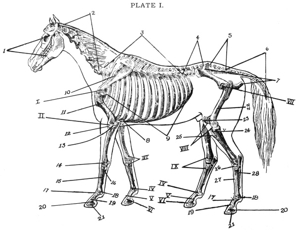
Proximal Phalanx, os suffraginis, or large pastern bone.
4.
Lumbar vertebræ or loin bones.
18.
Great Sesamoid Bones.
5.
Sacral vertebræ or rump bones.
19.
Medium Phalanx, os coronæ, or small pastern bone.
6.
Coccygeal vertebræ or tail bones.
20.
Distal Phalanx, os pedis, or coffin bone.
7.
Pelvic or hip bones.
21.
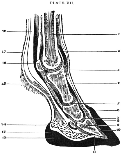
Os Naviculare, small sesamoid, or shuttle bone. (This bone can be plainly seen Plate VII).
8.
Sternum or breast bone.
22.
Femur, or thigh bone.
9.
Ribs.
23.
Patella, or stifle bone.
10.
Scapula or shoulder blade.
24.
Tibia, or leg bone.
11.
Humerus or shoulder bone.
25.
Fibula. (This bone is little developed in the horse.)
12.
Radius or bone of the fore-arm.
26.
Tarsus or hock bones.
13.
Ulna or bone of the fore-arm.
27.
Metatarsus, or os metatarsi magnus.
14.
Carpus or bones of the knee.
28.
Ossa Metatarsi Parva, or splint bones of the hind leg.
Names of joints placed according to numbers.
I.
Shoulder Joint.
VI.
Coffin Joint.
II.
Elbow Joint.
VII.
Hip Joint.
III.
Carpus or knee joint.
VIII.
Stifle Joint.
IV.
Fetlock Joint.
IX.
Tarsus or hock joint.
V.
Pastern Joint.
Structure of the Bones—Bones are hard, yellow-white,
insensitive objects, which form the skeleton and
give attachment to soft structures (muscles, tendons
and ligaments); they are of various sizes, forms and densities.
In the limbs the bones are ordinarily more or less
long, circular bodies, with expanded ends, effectually
supporting the body, supplying leverage and attachment
for soft structures, and forming the basis of all joints.
Where cavities, such as the cranium, chest, and pelvic,
enclosing the organs requiring protection and support,
the bones tend to assume a flat, expanded form.
Living bone is bluish pink, insensitive, and elastic;
on exposure to air it becomes diseased and blackened,
very sensitive and painful; (the teeth excepted) which
are harder and of a higher specific gravity than any other
bone formation.
Bones are composed of two kinds of substance—animal,
which makes the bone tough and flexible; earthy,
which makes it hard and fragile. In young animals the
animal matter forms about one-half of the bone substance;
in the adult, it diminishes to about a third, while
in old animals it is still less; hence the bones of very old
animals are brittle, more liable to fracture and harder to
mend.
Bones in a six-year-old horse contained, Phosphate
of Lime, 54.37 per cent; Carbonate of Lime, 12.00 per
cent; Phosphate of Magnesia, 1.83 per cent; Soluble
Salts, 0.70 per cent, or mineral matter, 68.90 per cent.
While they contain Cartilage, 27.99 per cent; Fat,
etc., 3.11 per cent, or animal matter 31.10 per cent.
In bone tissue there are two modifications of texture,
the compact and the cancellated. The former—hard,
dense, and ivory-like, is always situated externally; the
latter porous and spongy lies within.
Although the compact tissue appears uniformly
dense, and destitute of porosity, yet, if we transversely
sectate the shaft of a long bone, and examine it under
the microscope, by transmitted light, it is found to contain
numerous round openings. These are called Haversian
canals. They transmit bloodvessels, and run in a
longitudinal or slightly oblique direction, opening on
either the outer or inner surface of the bone.
The external (outer) surface of every bone is covered
by a tough, fibrous, inelastic membrane called periosteum,
which can be seen by examining the bone of an
animal which has recently died. The only exception to
this is at the joints where one bone articulates with
another, and where a tendon or muscle plays over a bone;
here we find its place taken by articular cartilage. By
its strength it sometimes retains bones in contact after
an oblique fracture; in the young, it is thicker and more
vascular than in the adult. Blood-vessels which penetrate
the periosteum pass directly to the bone; the outer
surface of the bone is always studded with numerous
foramina through which these enter.
The periosteum owing to its inelasticity, is, when
inflamed, the seat of intense pain; and should any part
of it be stripped off, there is every probability of the
denuded bone dying and separating.
Contents of Bone.—Red marrow is found in the
extremities or near the ends of bones, white marrow is
found in the shaft.
Classes of Bones.—Bones are classed as long, flat,
and irregular. Long or cylindrical bones are found in
the limbs or extremities, and serve as levers and pillars
for traveling and to support the body. Descriptively, a
long bone is divisible into a center or shaft and two ends
or extremities. Flat bones are found where visceral
organs need protection. As the shoulder or scapula and
ribs, to protect the heart and lungs; pelvic or hip bones,
to protect the rectum and urinary and genital organs;
also the cranial bones, to protect the vital organ called
the brain. Irregular bones are found in the spinal column
and in the joints, such as the knee or carpus, hock
or tarsus, where great strength is required. They usually
possess many angles and indentations, with surfaces
for articulation and tendonous attachment, and consist
of a thin, dense, external (outer) case of compact bone
enclosing cancellated tissue. In proportion to their size
they present a much larger extent of articular surface
and greater mechanical strength than any other class.
1. Skull, or skeleton of the head, the most anterior
(forward) part of the horse’s skeleton, articulates with
the first cervical vertebra (or atlas), from which it is
suspended by its posterior (or back part) extremity, its
anterior (forward) extremity being free. Its position
varies with the attitude of the animal; but in our descriptions
we shall always suppose it to be placed in a
horizontal position.
In the young animal the skull is composed of a number
of bones, all of which, with the exception of the lower
jaw, the teeth, the bones of the tongue, and ossicles of
the ear, become united by ossification (growing
together) in the adult.
In speaking of the different points of importance in
connection with the head bones of the horse, and other
animals, suppose a cross or longitudinal section of the
head is made. It will be noticed that it is full of cavities
or sinuses. The uses of these are to lighten the
head and also to warm the air as it passes into the nostrils,
on its way down to the lungs. It will also be found full
of foramen or small holes through which the nerves from
the brain and various blood vessels pass to the organs
situated in the head, such as the tongue, lips and the
various glands in and around the head.
Then there are the cavities in which the eyes are situated,
one on each side of the head, called the orbital
fossa. In examining this fossa you will find a small
opening or foramen, through which the optic nerve
passes in coming from the brain to the eye. This is the
nerve of sight. Then the most important part of all to
consider is the cranial cavity in which that very important
organ is situated called the brain, which controls all
the various functions and movements of the body.
Another important point is the situation of the ear
drum. It is situated in the hardest bone found in the
whole skeleton, called the petrosal. The nerve that gives
the function called hearing comes from the brain down to
the petrosal bone and enters by a small foramen or hole
into the drum of the ear to give hearing. This nerve is
called the auditory nerve.
2. Cervical Vertebrae or Neck Bones.—These
bones are seven in number. The first and second bones
proceeding from the head receive special names. The
first one is called the atlas, from which the head is suspended
and to which it is attached; it somewhat resembles
the body of a bird with wings extended. The second
bone receives the name of dentata. This is the bone
which allows the head to turn in any direction, hence it
is sometimes called the axis or pivot of the neck. Between
these two bones, on the upper surface, is the only
place where the spinal cord is not covered with bone, a
spot about three-fourths of an inch in diameter. The
next four bones receive no name, and are about the same
in size and length. The last, or the seventh bone, is
only about one half the length of the preceding ones and
receives no special name.
3. Dorsal Vertebrae or Back-Bone.—Dorsal
bones are eighteen in number. The chief point of interest
about them are the height of the spines on the
upper surface of the bones. These large spines form the
withers of the horse, as will be noticed in the skeleton.
On either side of these bones the ribs are attached, 18
pairs corresponding with the number of bones in this
region.
4. Lumbar Vertebrae (or the bones which form
the skeleton of the loins). These bones are six in number,
and they are situated immediately above the kidneys.
5. Sacral Vertebrae or Rump Bones.—There
are five of these bones in the young horse, but in the
adult they unite as a single bone, somewhat triangular-shape.
These are situated between the upper hip bones
and help to form the rump. Beneath these bones the
bladder is situated.
6. Coccygeal Vertebrae or Tail Bones.—These
are 18 to 20 in number. There is no complete canal, like
in the previous vertebraes, for enclosure of the spinal
cord.
7. Pelvic or Hip Bones.—The pelvic bones are
flat, but somewhat irregularly-shaped, and they form
the sides, floor, and part of the roof of the pelvic cavity.
Above they are connected with the sacrum, and below
united to each other, in the adult, by ossification (union
of bone). In the young animal, as above stated, and
especially in the foetus, each side consists of three parts,
which retain their names of ilium, ischium, and pubis,
even after union by ossification. They all three meet
in the acetabulum, or articular cavity for the femur or
hip bone.
8. Sternum or Breast Bone.—This bone is small
and short in the horse and is situated on the lower surface
of the chest cavity. The principle points to be noted in
this bone are that of its softness, and that the first eight
pairs of ribs are attached to it on either side. This bone,
in the horse resembles the keel and cut-water of a boat.
9. Ribs.—In the horse the ribs usually number
eighteen on each side. They extend in a series of arches
of varying curvature from the dorsal vertebrae above,
towards the sternum and sides of the abdomen below.
Their shape, in a great measure, determines the conformation
or shape of the thorax or chest cavity; they
protect its contents, and materially aid in its contraction
and expansion. They are continued downward and forward
by a small piece of cartilage or gristle, and are
just slightly attached to the breast bone or sternum; these
are called the false ribs. Note that, starting with the
first rib, they get longer until the ninth rib is reached;
they then get shorter, the last rib being only a few
inches long.
10. Scapula or Shoulder Blade.—The scapula
is a flat bone situated on the antero-lateral (front side)
surface of the thorax or chest cavity, it is triangular in
shape, the base being turned upwards. The inferior or
lower extremity articulates with the humerus or shoulder
bone.
11. Humerus or Shoulder Bone.—The humerus
is a long bone extending from the scapula to the radius
and ulna in an oblique direction downwards and backwards.
Like all long bones it possesses a shaft and two
extremities and two articular surfaces.
12. Radius, or Bone of the Fore-arm.—The radius
is a long bone, and occupies a vertical position between
the humerus and the carpus.
13. Ulna, or Bone of the Fore-arm.—The ulna
is an irregular bone, in form triangular, with the base
uppermost, and is placed on the supero-posterior or
(upper and back) part of the radius.
14. Carpus, or Knee.—The carpus, knee or wrist,
as it is incorrectly termed in quadrupeds, is composed of
seven and often of eight small, irregular bones arranged
in two rows of three each, one above the other, the seventh
being at the back of the three in the upper row, and the
eight, when present, in a similar position with respect
to the lower row. Their names are as follows: scaphoid,
lunar bone, cuneiform bone, trapezium, trapezoid, os
magnus, unciform, pisiform, which is not always present.
15. Os Metacarpi Magnus, Metacarpal or Cannon
Bone.—The large metacarpal, or cannon bone, is a
long, straight bone, placed in a vertical direction. Its
superior or upper extremity articulates with the carpus
and its inferior or lower extremity articulates with the
os suffraginis and the two sesamoids.
16. Ossa Metacarpi Parva, or Splint Bones.—The
two small metacarpal, or splint bones, tuberous in
form (marked or covered with projections) at the carpus
and tapering distally (lower portion), are attached to
the large bone, one on each side of its posterior (back)
surface, by ligaments in the young animal, and by ossification
(union of bone) in most grown up, and in all old
animals.
17. Proximal Phalanx, Os Suffraginis, or
Large Pastern Bone.—The os suffraginis, or large
pastern bone, passes obliquely downwards and forwards,
and articulates with the cannon bone above, and the
median phalanx or os coronae below. It belongs to the
class of long bones.
18. Great Sesamoid Bones.—These bones, two in
number, are placed side by side at the postero-inferior
(back and lower) part of the metacarpus and postero-superior
(back and upper) part of the os suffraginis;
they are irregular in shape, their back parts are covered
with cartilage, for the passage of the flexor tendons of
the digit or the last four bones of the limb. Their superior
or upper surface is roughened, and their sides,
which are grooved for the reception of the suspensory
ligament. (This is called the fetlock joint.)
19. Median Phalanx, Os Coronae, or Small
Pastern Bone.—The second phalanx, os coronae, or
small pastern bone, has no marrow canal, and belongs to
the class of irregular bones. It is inclined, like the os
suffraginis, obliquely downwards and forwards, and is
partly covered by the hoof. The upper portion of this
bone articulates with the os suffraginis and the two sesamoids,
the lower part articulates with the os pedis.
20. Distal Phalanx, Os Pedis, or Coffin Bone.—The
third, or ungual phalanx, os pedis, or coffin bone,
is an irregular bone situated within the hoof, and, when
in a healthy state, corresponding somewhat to it in shape,
being semilunar in form, with the convexity to the front.
This bone is very hard and porous, having many openings
for the transmission of arteries and veins. In this
bone we notice the wall, the sole, the tendonous surface,
the articular surface, and the alae or wings. This bone
articulates with os coronae superiorly (upper) and posteriorly
it articulates with the os navicular bone.
21. Os Naviculare.—The naviculare, third or
small sesamoid, or shuttle bone, is an irregular bone, situated
with its long axis transversely, behind and below
the os coronae, and behind the os pedis, with both of
which it articulates, the articulation of the three forming
the so-called coffin joint. The lower surface of this
bone is important as it is covered with cartilage, and
together form a kind of a pully over which plays the
great flexor perforans muscle. The remaining portion
of the anterior limb or front leg, will be considered under
anatomy of the foot. The navicular bone can be plainly
seen on Plate VII, anatomy of the foot.
22. Femur or Thigh Bone.—The os femoris, femur
or thigh bone, the largest, thickest, and strongest bone in
the body, belongs to the class of long bones, and is placed
in a direction obliquely downwards and forwards, articulating
with the cup-shaped cavity in the pelvic or hip
bones superiorly (or upper), and with the tibia and
patella inferiorly (or below). This bone is roughened
for the attachment of the powerful muscles of the hip.
23. Patella, or Stifle Bone.—This, the kneepan
or stifle bone, is placed in front of the pully-shaped
groove of the femur. It is very compact, its front surface
being irregular, round and very much roughened, for
ligamentous attachment, and its posterior (or back) surface
very smooth to articulate with the groove in the
femur, presenting two depressions divided by a ridge,
the inner being the larger, and in the fresh state enlarged
still more by projecting cartilaginous lip, or elevation.
The patella increases the power of the hind leg, and it
is this bone that causes stifle joint lameness when it
slips out of the groove in which it glides normally.
24. Tibia, or Legbone.—The tibia or leg bone, is a
long bone, larger at its upper than its lower end, situated
between the femur and the astragalus, slanting downwards
and backwards. This bone is three-sided, possessing
outer, inner and back surfaces, all of which are wider
above than below. This bone gives attachment to the
flexor muscles of the hip.
25. Fibula.—This is a long slender bone, little developed
in the horse, and is an appendage to the tibia,
being attached to the outer side of that bone, and extending
from its head to its lower third, to which it is affixed
by a ligament; the space between the two bones is called
the tibial arch. The fibula gives attachment to the peroneus
muscle, the muscle that is supposed to be severely
contracted when an animal is affected with string-halt.
26. Tarsus, or Hock Bones.—The tarsus, or hock,
corresponding to the ankle-joint of a man, is composed
of six irregular compact bones, situated between the
lower end of the tibia and the superior or upper extremity
of the metatarsus; they are arranged in two series; one
consisting of the cuboid and three cuneiform bones, the
magnum, medium, and parvum, corresponds to the lower
row or carpal bones; the other upper series consists of the
astragalus and calcaneum; the first, forming with the
bone above the mobile portion of the joint, may be said
to correspond to the upper row of carpal bones, while
the latter, being the lever bone, corresponds to the trapezium.
These bones, like those of the carpus, are thickly
covered with cartilage on their articular surfaces, which
acts as a protection against concussion. It is these bones
that become diseased and united when an animal is affected
with bone spavin. The calcaneum bone which
forms the prominent part, termed the point of the hock,
and corresponds to the heel-bone of man. This bone
gives attachment to the calcaneo-cuboid ligament, and
it is this ligament that is sprained or ruptured in curb of
the hock.
28. Metatarsus, or Os Metatarsi Magnum.—This
bone presents the same general appearance as the
large metacarpal or cannon bone, from which it differs
principally in being about one-sixth longer and flattened
from side to side. It is rounded and more prominent in
front. This bone articulates above with the tarsus bones,
and its lower portion articulates with the os suffraginis.
29. Ossa Metatarsi Parva, or Splint Bones of
the Hind Leg.—These also present the same general
form as the small metacarpal bones of the front leg; the
outer is the longest and largest, and has the largest head,
with two surfaces which articulates with the cuboid bone;
the inner one has also three articular surfaces, two for
small, and one for the middle cuniform bones. The two
surfaces on each head articulate with corresponding ones
on the large metatarsal bone. The remaining bones of
the hind extremity, viz.: the three phalanges, with their
three accessory bones, so closely resemble the corresponding
bones of the fore extremity, that it seems at first
sight difficult to distinguish one from the other; the
chief differences being, that the first phalanx of the
hind extremity is longer; its upper end larger, and its
lower end smaller, than in the fore extremity.
In this section we shall endeavor to point out where
the skeleton of the domesticated animals, other than the
horse, differ from the typical skeleton of the latter in
any important particular. The following descriptions
are therefore in all cases comparisons, where comparison
is possible, between the typical skeleton and the skeleton
in question.
(Cud Chewing Animal.)
In this class we take the ox as the animal which
represents the best for our purpose, the family of ruminating
or cud-chewing animals, as cattle, sheep and
goats.
The Skull or Cranium.—In the skull of the ox an
important feature is the development of the frontal or
forehead bone, which extends from below the eyes to the
back of the skull, forming the entire forehead and crest
or top, in the middle of which is the forehead tuberosity
or knob-like elevation, which is very large in hornless
animals. Springing from the sides of the top are two
processes, varying in size and shape, but corresponding
to the shape of the horns, which they support.
Vertebrae.—The true vertebral column is made up
of 26 bones divided into seven cervical or neck-bones, 13
dorsal or back-bones, and six lumbar or loin bones.
Cervical or Neck-Bones.—The bodies of these
bones are shorter than those of the horse, but same in
number.
Dorsal or Back-Bones.—These bones are longer
than in the horse, but have the same general form. It
gives attachment to 13 ribs on each side in the same
manner as those of the horse.
Lumbar or Loin Bones.—The lumbar vertebrae
are longer and thicker than in the horse, their bodies
being more round on the sides and lower surfaces.
Sacrum, or Rump Bones.—The sacrum is larger
and more arched, and the upper surface more round than
in the horse.
Coccygeal or Tail Bones.—The tail bones are from
15 to 20 in number, are strong and rougher than those
of the horse.
Pelvis or Hip Bones.—The pelvis is larger, but
presents the same general appearance as in the horse.
In studying the bones of the limbs in ruminants, the
only point of difference is in bones below the knee. The
large metacarpal bone presents a vertical groove down
its front, which marks the original division of the bone
into two bones. The lower extremity is divided by a
deep groove into two articulations, each resembling the
single one of the horse. The outer one being always the
smaller, a rudimentary metacarpus is placed on the back
and outer surface. The pasterns and sesamoids in either
limb are double, one set forming each digit; they are
small and narrow, the coffin bone resembles half of that
of the horse, equally divided.
In the aged ruminant, two bones are commonly
found in the heart, and may be termed the cardiac bones.
They are found related with auriculo-ventricular rings.
In shape they present three angles, three borders, and
two surfaces. The left bone is somewhat smaller
than the right.
Animals eating both vegetable and animal food.
The omnivora are represented in veterinary anatomy
by the hog.
An order, suborder, or family of mammals, especially
wild animals, including the dog and cat, etc. Animals
that eat flesh.
In this order our description alludes mostly to the
dog.
The scope of the present work will allow only very
brief treatment of this part of the subject.
Of or pertaining to Birds, Poultry, Etc.
The bone tissue or substance of birds are exceedingly
compact and hard, white in color; and some of the bones
are pneumatic, or contain air instead of marrow, notably
these are bones of the skull, the sternum or breast bone,
and the upper bones of the limbs. There are a great
variety in the amount of pneumaticity or air possessed
by the skeleton of different species, but it is not necessarily
determined by the flying power of the animal.
CHAPTER II. — ARTHROLOGY. — Joints of the Front Leg. — Joints of the Hind Leg.
What is known regarding the articulations of bones
of joints. The several bones which form the skeleton
are united by means of certain soft structures, forming
a number of articulations or joints, the study of which is
termed arthrology.
Before considering the different forms of joints, it
will be advisable to describe briefly the various tissue,
other than bone, which enter into and contributes towards
their formation. These are chiefly cartilage, connective
and elastic substance or tissues, and fat.
In health, one bone never comes directly in contact
with another, cartilage or fibrous tissue being always
interposed; an exception to this exists in the adult skull,
most of the bones of which become firmly united by ossification
of the interposed soft material.
Cartilage.—Cartilage, known also by the familiar
name of gristle, is a firm, bluish-white elastic animal
substance, somewhat transparent, resilient, and flexible,
possessing great cohesive power. That which forms the
original basis of the bony framework is termed temporary,
and that which persists in the adult, permanent
cartilage; the former disappears as it is replaced by bone,
but the latter, of which alone we have to treat here, never
under normal circumstances become ossified. Cartilage
consists of corpuscles or cells, usually embedded in an
intercellular substance.
The articular cartilage is important as it encrusts
the articular surfaces of bones, helping to form joints by
supplying smooth, elastic cushions, which diminishes both
concussion and friction.
Connective Tissue (White fibrous tissue.)—In one
form or another this tissue is found in all parts of the
body. The chief varieties are the areolar and the fibrous;
the former serving as a connecting medium, and support
to the various organs, and to the structures of which
they are formed. It appears as a loose, transparent
mesh, its interwoven bundles forming spaces termed the
areolar or cells.
Connective tissue contains nerves and blood-vessels,
for the supply of neighboring structures as well as for
its own nourishment. When healthy it is little sensitive
to pain.
Yellow Elastic Tissue.—This differs from the
white or connective tissue in being yellow, elastic, and
not so tough or strong. Its fibers are usually large but
when mixed with the white tissue in tendons the size
diminishes.
Yellow elastic tissue is found nearly pure in the ligamentum
nuchae, and tunica abdominis, the coats of the
largest arteries and elsewhere. The lungs contain a
large quantity of this tissue.
When white fibrous tissue is boiled gelatine is obtained,
which is not the result of boiling the yellow elastic
tissue.
Adipose Tissue.—Fat or adipose tissue consists of
cells containing an oily material, and arranged in isolated
groups, or slightly separated by meshes of areolar tissue.
It is found in many parts of the body, and varies greatly
in quantity; in joints it occurs between the ligaments,
and serves the purpose of a packing material, while in
the form of medulla or marrow it occupies the cavities
of bones. In fat cell a nucleus is very rarely visible.
Ligaments.—Ligaments are dense, fibrous, connecting
structures. They exist in most articulations, and
are made up principally of white fibrous tissue. There
are two kinds—capsular or bursal, and funicular or binding
ligaments.
Capsular ligaments are membranous structures enclosing
true joints. They consist of a dense interlacement
of fibers attached to bones, round the edges of the
articular cartilages; some regard them as a continuation
of the periosteum or the covering of bone. They are
never closely applied, their use being to form cavities
round the joints, enclosing and protecting the synovial
or lubricating apparatus inside.
Funicular or binding ligaments consist of rounded or
flattened cords, or bands of fibrous tissue, passing from
one bone to another, firmly attached to roughened portions
of their surfaces. They hold the bones in their
places, at the same time allowing the requisite amount
of motion in the joints. Ligaments which are situated
between bones are often termed interosseous. Annular
ligaments are those which bind down and protect the
tendons of muscles in certain joints, converting grooves
in the bones into channels or tubes which are lined with
synovial membrane, and through which the tendons play.
Some ligaments are composed almost entirely of yellow
elastic tissue, such as the ligamentum nuchae (the
elastic ligament of the neck) and the ligaments connecting
the vertebral arches (arches between the bones of
the spinal column).
Synovial Membranes.—These are thin membranes
lining the capsular ligaments of joints, or they are interposed
elsewhere between structures which move one upon
another, and which would otherwise be injured by the
friction. Near the borders of articular cartilages the
membrane is generally found as a projecting fold, the
projection being due to a small pad of fat, interposed
between the membrane and the capsular ligament. These
projections were once erroneously termed synovial
glands, but their use is probably to assist in forcing the
synovia between the opposing surfaces of cartilage.
Synovia.—Synovia or joint oil, is a viscid, transparent
fluid, colorless, or pale yellow, physically resembling
oil, but it contains very little fatty material, consisting
chiefly of albumen, salts and water; it is secreted by the
cells of the inner surface of the synovial membrane.
When an animal is in active exertion, there is a greater
demand for joint oil than when at rest, consequently
there is an increased secretion of it.
Classes of Joints.—Joints may be divided into three
classes—Immovable, movable and mixed.
Motion in Joints.—The following terms express
the various movements allowed by joints—Extension
tends to bring two bones as nearly into a straight line as
the structure of the joint will permit; flexion is the reverse
of this, and diminishes the angle that extension
increases; abduction expresses the outward movement
of a limb or bone from the central line of the body; adduction
is the reverse action; rotation signifies the partial
revolution of a bone or number of bones, as it were, on
their own axis; circumduction implies the movement of
the lower end of a bone or limb, where it describes a
curve, as the bow of a circle, ellipse, etc. The term gliding
explains itself, and is peculiar to movable and other
joints having no frictional surfaces.
Important points of joints to be considered will be
described on Plate I by Roman figures to correspond
with those given below.
I. Shoulder Joint.—This joint is formed by the
lower end of the scapula, or shoulder blade, and the upper
end of humerus or shoulder bone. This is a ball-and-socket
joint, and is held in its place by ligaments and
muscles. This belongs to the class of rotation joints,
its action is outward, inward, backward and forward.
II. Elbow Joint.—This joint is formed by the
lower part of the humerus and the upper portion of the
radius and ulna. It is covered with ligaments and muscles
similar to the above mentioned joint. This is a
hinge joint the action of which is only forward and
backward, or flexion and extension. It has no lateral
movement.
III. Carpus or Knee Joint.—This joint has been
explained very thoroughly in the previous chapter. There
are three separate articular surfaces in this joint. This
joint has a large capsular ligament, and has binding ligaments
one above the other, the seventh being at the back
of the three in the upper row, and the eight, when present,
in a similar position with respect to the lower row.
Thus there are three separate articular surfaces in this
joint. The upper surface of the carpus articulates with
the radius; this forms the hinge joint of the knee and is
where flexion and extension takes place. Another articulation
between the upper and lower carpus bones give
slight motion to the knee, but not so much as the upper
articular surface. Between the lower surface of the
lower row of carpus bones and the upper extremity of
the metacarpal, or cannon bones, is another articular
surface which gives motion to the knee. This joint has
a large capsular ligament, and has binding ligaments
inside and outside. In addition to the flexion and extension
movement the knee joint possesses, it also serves as a
cushion and relieves concussion of the fore limb.
IV. Fetlock Joint.—This joint is formed above
by the lower portion of the metacarpal, or cannon bone,
and below by the part of the os suffraginis or the large
pastern bone. At the back of this joint there are two
small bones called the sesamoid bones. This joint has a
capsular and two lateral binding ligaments. The motion
of this joint is a backward and forward or flexion
and extension, same as that of the knee.
V. Pastern Joint.—This joint is situated just
above the hoof, and is formed by the lower end of the
os suffraginis or large pastern bone and the upper end
of the os coronae or small pastern bone. It has a capsular
and lateral binding ligaments, same as the fetlock
joint. It also has the same motion as the fetlock joint,
and is often the seat of what is termed a high ringbone.
VI. Coffin Joint.—This joint is situated within
the hoof. It is formed above by the lower end of the os
coronae or small pastern bone, and below by the upper
surface of the os pedis or coffin bone. Immediately behind
this joint, and articulating with the two mentioned
bones, is the navicular, or shuttle bone—it gets its name
from its likeness to the shuttle of a sewing machine. This
bone when diseased is the seat of navicular disease, or
coffin-joint lameness. The action of this joint is very
slight forward and backward.
VII. Hip Joint. The hip joint is formed by the
pelvis and femur bone. This is a true ball-and-socket
joint similar to the shoulder joint. It has a capsular,
and is called the round ligament, in the joint, holding
the head or ball on the femur in the socket of the pelvis.
This can be seen plainly by examining the joint. This
round ligament is important, as it often becomes strained,
being the seat of hip joint lameness. It is also held
together by the strong muscles of the hip. Its motion is
rotation similar to that of the shoulder joint.
VIII. Stifle Joint.—This joint is formed above
by the lower end of the femur, or hip bone, and the upper
end of the tibia or thigh bone. These two bones in front
form a pulley-like surface on which the patella, or stifle
bone, is situated. This bone, when the joint is in motion,
glides up and down over the pulley-like surface. It sometimes
becomes displaced, and this is termed dislocation of
the patella or stifle bone. This is an important point to
notice about this joint. It has a capsular and lateral, or
binding ligament, which hold the stifle bone to its place
as it plays upon the pulley-like process of this joint. The
action of this joint is only forward and backward or
flexion and extension. It has no lateral or side motion.
IX. Tarsus or Hock Joint.—This joint contains
six bones. The two upper bones, one of which is a pulley-like
bone placed in front, and the other placed behind,
forms that portion of the hock which is called the point
of the hock to which the muscles of the gambe are attached.
It can be easily seen or felt. The upper surface
of these two bones articulate with the lower portion of
the tibia or thigh bone, and forms a true articulation of
the hock joint. This part is what gives most of the motion
to the joint. Below these are three other small, irregular
bones, placed one upon the other, having an
articular surface between them. Immediately behind
these three small bones is what is called the cuboid bone.
This bone also articulates with the three small irregular
bones, helping to form the articular surface of the hock.
The lower articular surface helps to give a small amount
of motion to the joint. This joint is the seat of bone or
bog spavin due to severe sprains, or poorly conformed
joints.
Fetlock, pastern and coffin joints of the hind leg are
so closely allied to those of the fore leg that it is not
worth while discriminating between them.
CHAPTER III. — MYOLOGY.
The branch of anatomy which treats of the muscular
system is called Myology.
Muscles are the active organs of motion, or locomotion,
each being separated from the other by a thin delicate
membrane made up of connective tissue, which
forms a sheath for the muscle. A muscle is divided into
two parts, viz.: muscular and tendinous. The muscular
part is the larger of the two. It is sometimes called the
belly of the muscle or flesh. This part is known as
muscular tissue, and has a reddish, meaty color. At
both ends of the muscle there is a tendinous part, or the
hard, white portion of the muscle which becomes attached
to the bone. All muscles are attached to two or more
places of different bones, and when contracted, the joints
of the body are moved. They are well supplied with
nerves, which give strength and feeling, and also well
supplied with small blood vessels, from which the muscle
is fed. Muscles are found in separate groups, all of
which have different actions to perform. There are two
kinds of muscles—voluntary and involuntary. The
voluntary muscles are under the control of the will of
the animal; example—the muscles of the head, neck,
back, hip and legs. The involuntary muscles are beyond
control of the animal, and will act even though the animal
were asleep—such as those of the heart, the large
muscular curtain which separates the chest cavity from
the abdominal cavity, which is one of the great muscles
of breathing; also the muscles around the chest which
assist in breathing.
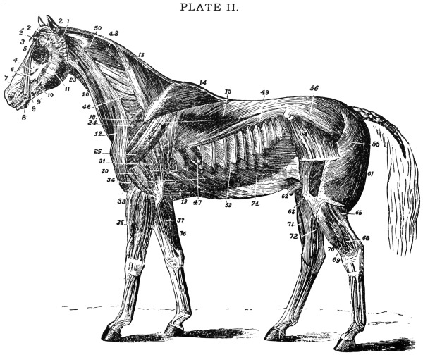
MUSCLES OF THE HORSE—SUPERFICIAL LAYER—AFTER MEGNIN.
EXPLANATION OF PLATE II.
Muscles of the Horse.
This illustration shows the superficial muscles of the
body after the skin and panniculus carnosis muscle has
been carefully removed. This muscle is spread over the
greater part of the body, which is related externally with
the skin; internally with the superficial layer of muscles.
Its action corrugates the skin, and thus enables the animal
to expel or shake off insects and irritating bodies, its use
being thus protective to some extent; it also supports and
binds down the superficial muscles.
SUPERFICIAL LAYER.
The panniculus and tunica abdominalis are removed.
1.
Abducens.
31.
Caput magnum extensor brachii.
2.
Retrahentes muscles.
33.
Extensor metacarpi magnus.
2′.
Attollens maximus.
34.
Humeralis obliquus.
2″.
Attollens anticus.
35.
Extensor pedis.
3.
Temporalis.
36.
Flexor metacarpi externus and medius.
4.
Nasalis longus.
37.
Flexor metacarpi internus.
5.
Orbicularis palpebrarum.
46.
Cervical
6.
Levator labii superioris alæque nasi.
47.
Dorsal serratus magnus.
7.
Dilatator naris lateralis.
48.
Rhomboideus longus.
8.
Orbicularis oris.
49.
Superficialis costarum.
9.
Zygomaticus.
50.
Splenius.
9′.
Buccinator.
52.
Intercostales.
10.
Depressor labii inferioris.
54.
Tensor fasciæ latæ.
11.
Masseter.
55.
Triceps abductor femoris.
12.
Levator humeri.
56.
Gluteus externus.
13.
Trapezius cervicalis.
61.
Biceps rotator tibialis.
14.
Trapezius dorsalis.
62.
Rectus femoris.
15.
Latissimus dorsi.
63.
Vastus externus.
18.
Pectoralis parvus.
65.
Gastrocnemius externus.
19.
Pectoralis magnus.
68.
Flexor pedis perforans.
20.
Sterno-maxillaris.
69.
Peroneus.
23.
Subscapulo-hyoideus.
70.
Extensor pedis.
24.
Antea-spinatus.
71.
Flexor metatarsi.
25.
Teres externus.
72.
Flexor pedis accessorius.
26.
Postea-spinatus.
74.
Obliquus abdominis externus.
30.
Caput medium of the triceps.
Voluntary Muscles.—The voluntary muscles are
in groups. The first muscle we will call the student’s
attention to, after removing the skin, is the panniculus
carnosus (not shown in Plate I), which is a thin muscle,
and almost entirely covering the body, which is sometimes
accidentally removed by a careless person in skinning
the animal. The action of this muscle is to shake
the skin when flies or other objects bother the horse.
This muscle is not shown in Plate I it having been removed
in order to show the more important ones.
Head Muscles.—At the head there is a group of
muscles which assist in chewing, or masticating, the food.
Gullet or Pharynx Muscles.—Around the throat
is another set of muscles, sometimes called the muscles
of the gullet, or pharynx, which assist in swallowing.
Neck Muscles.—The neck muscles are divided into
two groups, one on each side. The action of these is
to raise and lower the head, also to turn the neck and
head from side to side.
Muscles of the Back.—The muscles of the back
are generally divided into two groups, one above the
spinal column and the other below. The muscles above
the spine assist the animal in running, jumping and rearing.
The muscles below the spine are sometimes called
the psoae, or lumbar, muscles, situated below the lumbar
bones, or the bones of the small of the back. The
action of these muscles is to assist the animal in getting
up. These muscles are important, for when paralyzed
the horse cannot use his hindquarters. Below these
muscles are the kidneys.
Hip or Gluteal Muscles.—The muscles of the
hip are very large, filling in around the hip bones. The
action of these is much the same as those of the back, as
they assist in jumping, running, rearing and in flexing
and extending the hind leg.
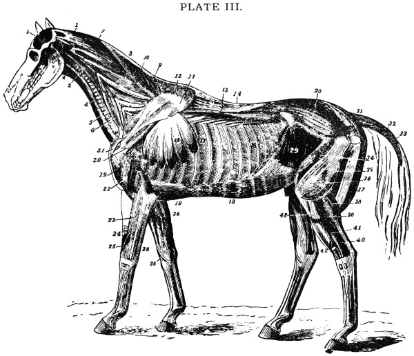
MUSCLES OF THE HORSE—DEEP LAYER—AFTER MEGNIN.
EXPLANATION OF PLATE III.
Muscles of the Horse.
Deep Layer.
1.
Temporalis.
22.
Humeralis obliquus.
1.
Stylo-maxillaris.
22´.
Caput parvum (of triceps extensor brachii).
2.
Rectus capitis anticus major.
23.
Extensor suffraginis.
3.
Sterno-thyro-hyoideus.
24.
Extensor metacarpi magnus divided.
4.
Sterno-maxillaris.
25.
Extensor metacarpi obliquus.
5.
The Trachea.
25´.
Its tendon.
6.
Scalenus.
26, 28.
Flexor pedis perforans and perforatus.
7.
Splenius.
29.
Obliquus abdominis internus.
8.
Funicular part of ligamentum nuchæ.
30.
Gluteus maximus.
9.
Rhomboideus longus.
31.
Erector coccygis.
10.
Cervical.
32.
Curvator coccygis.
15.
Costal serratus magnus.
33.
Depressor coccygis.
11.
Cartilage of prolongation.
34.
Rectus femoris.
12.
Rhomboideus brevis.
35.
Vastus externus.
13.
Transversalis costarum.
36.
Part covered by triceps abductor.
14.
Longissimus dorsi.
37.
Biceps rotator tibialis.
15.
Serratus Magnus.
38.
Gastrocnemius externus.
16.
External intercostals.
39.
Plantaris.
17.
Internal intercostals.
40.
Flexor pedis perforans.
18.
Rectus abdominis.
41.
Peroneus.
19 19.
Pectoralis magnus.
42.
Flexor metatarsi.
20.
Postea-spinatus minor.
43.
Extensor pedis (cut across).
21.
Flexor brachii.
Tail or Coccygeal Muscles.—Here there are four
that are important, one situated on the upper side of the
tail when straight out, the action of which is to raise
the tail; two, one on each side of the tail, have the
power of drawing the tail to either side; the fourth is
situated under the tail and is the smallest one of the four.
Its action is to draw the tail down.
Abdominal Muscles.—The abdominal or belly
muscles, are four large, flat muscles on each side of
the abdomen. The outer edge of these muscles is attached
to the outer ends of the false ribs, also to the
processes of the lumbar bones and the outer angles of
the pelvic or hip bones. They unite below to what is
called the linea alba, a hard, white fibrous cord. They
pass back in the center of the belly and are attached
to the front of the pelvic bones, called the lower bones of
the pelvic cavity. About ten inches from where it is
attached here, passing forward, is a small slit or hole,
which is called the navel, or umbilical opening. Here
the navel vessels pass in and out during the foetus life,
or before the colt is foaled. This is a point of importance
to note, for sometimes at the time of birth this
opening does not close and allows the bowels to come
down and form what is known as umbilical or navel
rupture.
Before finishing the description of this group of
muscles a very large, important ligament should be
noted, which is found spread all over the abdomen of
the horse. It is of a yellowish color and about one-eighth
of an inch thick, attached in front to the back
of the breast bone and to the pelvic bones behind. This
is the first structure seen after removing the skin from
the abdomen. This ligament gives great support to the
organs contained in the abdominal cavity. The action,
or uses, of the abdominal muscles are to support the organs
contained in the abdominal cavity, to flex the back-bone
and assist in passing of the feces. In the mare these
muscles assist in parturition, or foaling.
Shoulder Muscles.—The shoulder muscles are
very large and powerful. There are only three of great
importance. Two situated on the outside of the scapula
or shoulder blade are important, as they are muscles
affected in the disease called shoulder sweeny. The
other important one is that which passes down over the
shoulder joint through the groove or pulley-like surface
on the humerus, or shoulder bone. This is a long, powerful
muscle, attached above to the lower end of the scapula,
or shoulder blade, passing down through the groove mentioned,
and is attached to the upper and front part of the
radius or fore arm bone. Its chief point of importance
rests in its action in raising the front leg, where it passes
over the pulley-like surface mentioned, when it becomes
injured or diseased; it is the seat of shoulder joint lameness.
Muscles of the Front Leg.—Muscles of the front
leg, from the shoulder down, are divided into two separate
kinds, the extensor and flexor muscles. The extensor
muscles are those which bring the leg forward. These
muscles above are attached to the bones around the elbow
joint, passing down in front of the arm bones.
About three inches above the knee they become changed
into the tendinous part of the muscles, or what is called
the cords of the leg. Some of them are attached to the
bones about the knee joint, while others pass over the
front of the joint and are held down to their place by a
band or ligament, forming a loop, as it were, for the
tendinous portion of the muscle to glide into when the
leg is in action. Each one of these loops through which
the muscles pass are supplied with a synovial membrane
to secrete the synovia, or oil, which lubricate it during
action, the same as in the joint. This is a point of importance,
as sometimes, on account of injury or strain
of this part of the joint through which the muscles play
there may be found a small, puffy enlargement containing
oil secreted by the synovial membrane. This disease
is called bursal enlargement.
The flexor muscles are situated at the back part of
the leg, attached above to the back part of the elbow
joint, passing downwards at the back part of the leg.
About two or three inches above the back part of the
knee joint they become tendinous, and from there down
to the back part of the coffin bone, where two of the principle
muscles are attached; these form what is known as
the back tendons, or cords, of the leg. Some of them
become attached to the back part of the knee, same as
the muscles on the front part of the leg, while the other
two principal tendons pass through a loop formed by ligaments,
the same as those mentioned in the front part of
the knee. In tracing these tendons down from the knee
to the fetlock, notice that they pass through another
larger loop or sheath formed at the back of the fetlock,
where some of the fibers are attached, while others continue
down at the back part of the pastern bones, and
are attached to the os pedis or coffin bone. These tendons
are important, because when they are strained the
fact is spoken of as the strain of the back tendons. The
action of these muscles is to flex the leg, bend the knee,
pastern joints and fetlock.
Muscles of the Hind Leg.—These are also divided
into two groups, extensor and flexor. The extensor muscles
are situated in front of the hind leg. They are attached
above, around the stifle joint, and pass downward
in front of the tibia, or thigh bone, one being attached
to the front part of the hock. The other passes
through sheaths, or loops, supplied by a synovial membrane,
formed by ligaments, to hold the muscles firm in
front while the leg is in action. In tracing them down,
in front of the shin bone to the fetlock, note that they
pass through loops, or sheaths, and continue down in
front of the pastern bones to where they are attached.
The action of these is to bring the leg forward.
The flexor muscles of the hind legs are attached,
above, around the back part of the stifle joint. In tracing
them down it will be found that they become tendonous.
Two of the principal ones pass down to that part
of the hock joint, which forms the point known as the
cap. These form what is called the gambe of the leg, and
are partly attached at the point of the hock, and other
part passing down to the fetlock joint through a loop,
or sheath, along the back part of the pastern bones, and
are attached to the os pedis or coffin bone of the foot.
This muscle, from the hock down, forms one of the back
tendons of the hind leg. Another important muscle is
found passing down underneath those already mentioned,
through a loop, or sheath, at the back part of the
hock, where it is supplied with a synovia sac. This is a
point of importance, because when it becomes sprained it
is the seat of what is called thoroughpin. It then passes
down the back part of the shin bone beneath the other
tendons already mentioned, through the loop at the fetlock
to the back part of the os pedis or coffin bone, where
it is attached. The action of these muscles are to flex or
bend the fetlock and raise the hock joint in traveling.
Involuntary Muscles.—Involuntary muscles, or
muscles not under the control of the will. The first to
notice are those of breathing or respiration. They form
a group situated about the chest in such a way as to be
the means of increasing or decreasing the size of the chest
cavity. When these muscles expand the chest cavity is
enlarged, causing the air to rush into the lungs, known
an inspiration. On the other hand, when these muscles
contract the air is expelled from the lungs, known as
expiration.
The diaphragm is a muscular curtain which separates
the chest from the abdominal cavity. It also assists
greatly in drawing the air in, when it contracts. This
muscle also assists in passing faeces, and in the mare
foaling. It separates the heart and lungs from the bowels,
liver and stomach. This muscle should be carefully
examined by the students. It can be seen by opening
any dead animal.
There is one muscle which is both voluntary and involuntary.
It is situated in the penis, surrounding the
urethra, or the tube, which carries the urine from the
bladder to the penis in the male animal. Its action is
voluntary while the animal is passing urine. It is involuntary
during sexual intercourse, forcing the semen
down through the penis.
CHAPTER IV. — SPLANCHNOLOGY. — RUMINANTIA. — TEETH. — RESPIRATORY SYSTEM OF THE HORSE. — RESPIRATORY SYSTEM OF THE OX. — URINARY SYSTEM OF THE HORSE. — URINARY ORGANS OF THE OX. — GENERATIVE SYSTEM OF THE HORSE. — FEMALE GENITAL ORGANS. — GENITAL ORGANS OF THE BULL. — GENITAL ORGANS OF THE COW.
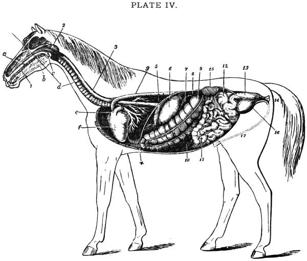
DIGESTIVE AND MALE URINARY APPARATUS OF A HORSE—AFTER MEGNIN.
EXPLANATION OF PLATE IV
Digestive Apparatus of the Horse
1.
Mouth.
13.
Rectum.
2.
Pharynx.
14.
Anus.
3.
Œsophagus.
15.
Left kidney and ureter.
4.
Diaphragm.
16.
Bladder.
5.
Spleen.
17.
Urethra.
6.
Stomach (left sac).
a.
Hard palate.
7.
Duodenum.
b.
Tongue.
8.
Liver (upper extremity).
c.
Soft palate.
9.
Great colon.
d.
Trachea.
10.
Cæcum.
e.
Pulmonary artery (divided).
11.
Small intestine.
f.
Heart.
12.
Floating colon.
g.
Posterior aorta.
Study of large interior organs in any of the four
great bodily cavities especially those in the abdomen.
Digestive Organs of the Horse.—The digestive
organs comprise the alimentary canal and the accessories
by which the alimentary matter is received and subjected
to specific actions, which adapt it for purposes of nutrition.
Digestion, therefore, embraces the collective operations
and changes which the food undergoes in the alimentary
canal.
The whole digestive track from the mouth to the anus
which is situated just below the tail, is sometimes called
the alimentary canal.
The mouth is an oval cavity at the commencement of
the alimentary or digestive canal. In front of the mouth
are the lips, one above and one below; at the sides are the
cheeks. The mouth is lined with what is known as the
mucous membrane, in which are several small openings,
from the glands, which are situated about the mouth.
Through these the saliva is poured. On the upper part
of the mouth the mucous membrane is thrown into ridges,
or folds, from 18 to 20 in number. This is a point of
importance in connection with bleeding a horse with
lampas. It is never safe to bleed back of the third bar
because there is a large artery which runs down through
the roof of the mouth and enters the hole in the bone
just before it reaches this bar. The tongue, which has
the chief nerves of the sense of taste, is situated in the
mouth; this organ also has a very important part to
perform in masticating the food and mixing it with
saliva. The teeth, which also take a very active part in
the masticating of food, are dealt with under the heading
of “Teeth.”
Salivary Glands.—These glands secrete the saliva
that is poured into the mouth while the animal is eating.
There are only three pairs of much importance. One
large pair, one on each side of the throat below the ears,
known as the parotid glands, fill up the space between
the jaw bone and the neck. This pair has tubes passing
around and under the lower jaw and up into the cheek
muscles entering the mouth opposite the fourth molar
tooth. These tubes, known as steno’s ducts are about
an eighth of an inch in diameter and convey the saliva
from the glands into the mouth. The next pair are situated
under the pair just mentioned. Their tubes enter
into the bottom part of the mouth. The third pair are
situated under the tongue, one on each side. They pour
their secretion into the mouth by several small openings
near the front under the tongue. This can be seen by
examining the under surface of the tongue closely. This
is very important fluid in connection with the digesting
of the food.
The gullet is a cavity situated just back of the mouth.
It is chiefly made up of muscles which perform the act of
swallowing the food. It is lined with the continuation
of the mucous membrane of the mouth.
The oesophagus, or the continuation of the gullet, is
a tube extending from the gullet to the stomach, and is
used to convey the food to that organ. It is made up of
two coats, the muscular and the mucous. The former
contains fibers which, when once the food enters the tube,
contract behind it, forcing it down to the stomach. Its
lining is a continuation of the mucous membrane of the
mouth and the gullet. In tracing the oesophagus or tube
down the neck from the gullet, note that it passes down
the left side of the neck, entering the thoracic, or chest
cavity, between the lungs over the heart through the
large muscular curtain known as the diaphragm, then
enters the stomach an inch or two after passing the
diaphragm.
The Stomach.—This organ is very small in the
horse in comparison with that of the ox. It holds only
about four gallons, and is situated just back of the curtain
which separates it from the lungs. It lies mostly to
the left side. The walls of the stomach being composed
of three coats. That on the outside is called the serous
membrane, a name applied to membranes which line
closed cavities, such as the abdominal cavity. The inside
lining is a continuation of the mucous membrane lining
the organs before mentioned. The lining in the left part
of the stomach or the part where the food is prepared
for digestion is the same color as that of the mouth. The
lining of the right part of the stomach, which is the true
digestive part, is of a deep red color resembling velvet,
and when placed under a microscope has the appearance
of a honey-comb. When the stomach is empty this membrane
is thrown into loose folds. Several small openings
may be noticed through which the gastric juice and pepsin
from the glands, situated in the walls of the stomach,
enters. These are very important fluids as they assist
greatly in digesting the food. The third coat is known as
the muscular coat already mentioned. Its action is to
give the stomach a churning motion, rolling the food
around and mixing it with the juices. The opening to
the stomach is guarded by a valve which prevents the
food from passing back through the gullet. There is
also a valve at the opening of the bowels, preventing any
coarse, undigested food from entering them. The stomach
is held in its place by five large ligaments, and is
well supplied by blood-vessels and nerves. Digestion of
the food takes place very quickly in the horse in comparison
to other animals. Frequently a change of food
or working too soon after eating will interfere with the
digestion, thus setting up what is known as indigestion.
This is a very painful disease in the horse. After the
food is acted upon by the juices in the stomach it changes
into what is known as chyme, which passes into the
bowels.
The Bowels.—They are divided into two parts—the
large and small.
The small bowels are 72 feet in length, about one
inch in diameter, and are made up of three coats, same
as the stomach. The serous coat on the outside contains
small glands which secrete an oily material to lubricate
the outside of the bowels, which comes in contact with
the inner wall of the abdominal cavity. The muscular
coat, made up of muscular fibers, is situated between the
other two coats, the same as in the stomach; its action
is to contract the bowels, giving them motion to convey
the food along through them. The mucous coat is a
continuation of the mucous coat of the stomach. Along
this coat are found small glands known as villi lacteal;
these absorb the nourishment from the food as it passes
along through the bowels and pours it into the blood.
The small intestines or bowels are attached on the upper
side to what is known as the mesentery, which is attached
above to the roof of the abdominal cavity. It can be seen
in any of the smaller animals upon examination. About
six inches from the stomach, in the bowels, are found
two openings. One of these receive the hepatic duct,
a tube for the purpose of carrying the bile from the liver
to be poured in on the food as it passes through the
bowels. The other opening is for the duct of the gland
known as the pancreas. It secretes a clear fluid known
as the pancreatic juice. These juices act on the food in
the first part of the small intestines, changing it into
chyle. After this, the action of the rest of the intestines
is to absorb the nourishment out of the food as it is
passing back. The small intestines and stomach, when in
a healthy condition, should be found empty one hour after
food has been eaten. The small bowels or intestines are
situated mostly on the left side just behind the stomach.
The large bowels have three coats, the same as the
small ones. The first part of the large bowels is known
as the blind bowel or caecum, and is about three feet in
length; this is generally the first thing to protrude when
opening a horse’s abdominal cavity. Its use is to act as a
reservoir to hold the water and fluid of the food; from
this organ the water and fluid parts are mostly taken up
into the system. The next part of the large bowels is
known as the large colon; it lays along the floor of the
abdominal cavity, is about nine feet in length and is
doubled on itself three times. In this bowel the solid
part of the food is found. Here digestion is brought
about by the contraction and expansion of the muscles
of the bowel and the nourishment taken from it, after
which it is worked back out of this bowel and enters
what is known as the floating colon. This is about ten
feet in length and about two inches in diameter, or double
the size of the small bowel. It is thrown into folds or
pleats, and as that portion of the food containing no
nourishment passes through it is worked into balls which
pass back and are emptied into the rectum or back bowel.
This is situated at the back part of the abdominal and
pelvic cavity back of the small bowels or intestines and
like them, is suspended by a fold paritoneum.
The rectum or back bowel is sometimes known as
the straight bowel. It is about 18 inches long and forms
the last part of the bowels or intestines. Its coats are
a continuation of those of the large bowel, but each is
thicker and heavier. Above this bowel are the bones
of the sacrum, below it, in the horse the bladder and
other small glands. Below the rectum of the mare are
situated the womb and the vagina, the latter being the
passage into the womb from the outside. The bones
which help to form the pelvic cavity are situated at the
sides, and at the back immediately under the tail is what
is known as the annus. The use of the rectum is to hold
the balls as they pass back from the floating colon. When
the rectum becomes so full that there is pressure on the
sides of the wall thus stimulating the nerves the muscular
coat contracts and forces the contents back towards the
annus. At the same time the muscles of the annus dilate,
causing the faeces to pass out.
Accessory Organs of Digestion.—In the abdominal
region these organs are the Liver, the Pancreas, and
the Spleen.
The Liver is the largest gland in the body and is situated
between the stomach and the diaphragm. The liver
of the horse weighs from ten to twelve pounds. It is of a
dark brown color, well supplied with blood and nerves,
and is held in place by several strong ligaments. A bitter,
greenish colored fluid called the bile is secreted from
the liver and emptied into the digestive system, where it
plays an important part in its action on the food. There
is no gall bladder in the horse, but simply a tube passing
from the liver to the small bowel into which it empties the
bile about six inches back of the stomach. It is important
to note that it is in this tube that gall stones sometimes
collect.
The Pancreas is another very important gland. It is
of a grayish, fatty color, and may be found near the roof
of the abdominal cavity in front of the kidneys. The
Pancreas secretes a clear, colorless fluid called the pancreatic
juice. This fluid, like the bile from the liver, also
plays an important part in the digestion of food. It is
carried down from the Pancreas by a duct or tube emptying
into the small bowel just back of that of the liver.
The Spleen is a long, flat gland about fifteen inches
in length, situated along the left side of the stomach and
to which it is closely attached. It is of a grayish red color
and feels quite soft. It is ductless, there being no secretion
passing from it. The function of this gland is not
clearly understood, but by many it is supposed to regulate
the temperature of the stomach during the process
of digestion and to act as a reservoir for the blood. Some
speak of it as the burying ground of the red corpuscles.
It is well supplied with blood vessels and nerves and
weighs about two pounds in an average sized horse.
Digestive System.—The lips of an ox are thick and
hard. The upper has no hair on it and varies in color
with the color of the animal. When cattle are in good
health this space is always moist.
The cheeks on the inside are covered by many small
rough processes, which give them a very rough appearance.
The Tongue of the ox is stronger than that of the
horse and is more movable. It is very thick and heavy
at the back, pointed at the front end and the upper part
of it is very rough. It is by means of the tongue that the
ox takes most of the food into the mouth.
The Salivary glands are similar to those of the horse.
The Teeth differ very much from those of the horse.
The ox has no front teeth in the upper part of his
mouth, their place being taken by a pad of cartilage or
gristle. This pad takes the place of the upper row of
front teeth. The lower row of teeth press against it when
the animal is cropping grass. This accounts for the fact
that cattle do not do as well on short grass as horses.
The front teeth in the lower jaw also differ from
those of the horse. They are eight in number, chisel-shaped,
and are loosely set in the gum.
The molars, or back teeth, are similar to those of the
horse, only they are smaller and not so smooth on their
upper surface. The ox has twenty-four molars or back
teeth, and eight incisors or front teeth, making thirty-two
in all.
The Gullet of the ox is much larger than that of the
horse.
The Oesophagus or tube, which carries the food down
from the mouth to the stomach is well developed, the
fibers in it being very strong and possessing a double
action. When the animal is eating they carry the food
from the mouth to the stomach, and when chewing the
cud they act the very opposite, carrying the food from
the stomach back into the mouth.
The Stomach.—The student will do well to give
some time to the study of this important organ of the
ox, as it is very frequently the seat of disease.
The stomach has a capacity of fifty-two gallons and
is divided into four separate and distinct compartments:
the Rumen or Paunch, the Reticulum or Honey Comb,
the Omasum or Many-plies, and the Abomasum or the
true Stomach. In the first three of these the food undergoes
a sort of preparatory process, while in the fourth the
process of digestion is complete.
The Rumen or paunch is very large, and in an aged
animal fills three-quarters of the abdominal cavity. It
lies up against the left side of the wall of the abdomen,
where it is attached and held to its place by the ligaments.
Its situation being an important matter, as many
diseases of the rumen, or paunch, are first noticed on the
left side. Tapping for bloating is always done on the
left side because of this fact. The walls of the paunch
of an ox resemble those of the stomach of the horse, but
are not so sensitive, and stand a great deal of abuse before
inflammation sets in. The paunch has two openings,
both of which are at the front; through one the food
enters, while through the other it passes out into the
next division.
The Reticulum, or honey comb, is the smallest division
and resembles a honey comb in appearance. This
part has little to do with preparing the food. It is provided
with two openings, one in front, where the food
enters, the other at the back, where it passes through into
the third division. In the reticulum, or honey comb,
the food is softened further by the water that the animal
drinks which passes directly into the second division.
The food is here pressed into balls and prepared to be
forced back into the mouth to be further masticated.
The Omasum, or many-plies, is the second largest
division of the stomach. When full it is ovoid in shape.
It is placed just behind the second division and at the
right side of the paunch. The inside is full of folds, or
layers of membrane, into which all the coarse parts of
the food pass and roll about until it is fine and well prepared
to pass into the last division. When this part of
the stomach becomes deranged and the food becomes
dry and hard between the folds, the disease called impaction
of the many-plies, or dry murrain, is the result.
The Abomasum, or fourth stomach, is the true digestive
part. In it the food is completely digested. The
walls are redder in color than those of the three first divisions
and contain the glands which secrete the acids and
gastric juices. This stomach has two openings, one
through which the food enters and the other through
which it passes into the small bowels.
The Bowels, or intestines, of the ox are divided into
large and small bowels. This, together with their structure
and action resembles that of the horse. The small
bowels are only half the size of the horse, being about
one-half inch in diameter, and about one hundred and
fifty feet in length. The large bowels are not nearly
so long as those of the horse and are thirty-five feet in
length.
The Liver of the ox resembles that of the horse, except
that it is provided with a gall bladder which resembles
a pear in shape. This acts as a sac in which to
store the gall during the time it is not required in digestion.
When digestion is going on the wall of the gall
sac contracts and forces the gall down to the food. The
other glands, the pancreas and spleen, resemble those
of the horse. The juices from these glands have the same
action in cattle as they have in the horse.
Rumination, or Chewing the Cud.—Food when
first taken into the mouth of a ruminant is but lightly
masticated and mixed with the saliva from the salivary
glands, after which it is swallowed, passing through the
oesophagus into the rumen or paunch (first stomach).
This division acts as a reservoir or storehouse for food
thus eaten quickly. When the animal has time, so to
speak, he lies down or stands quietly and completes the
process of mastication of his food by chewing the cud.
This peculiar act is performed as follows: After being
softened and moistened by warmth, the food passes from
the rumen or paunch into the second division—the reticulum,
honey comb or second stomach. In this small
globe-like compartment the food is moistened and compressed
into pellets—the cud. By a peculiar reverse
action of the oesophagus or gullet these pellets are taken
back into the mouth for further mastication or chewing.
When re-mastication is completed it is again swallowed,
but this time it passes directly into the omasum, many-plies
or third stomach, and thence to the fourth or true
stomach.
Process of Digestion After Rumination.—Following
rumination which, strictly speaking, is the first
step in the digesting process, the food passes into the
third stomach. The fine parts pass right along to the
fourth stomach while the coarser parts are drawn between
the folds of the membrane in this division and
worked about until it is fine and ready to pass into the
fourth stomach, where it becomes fully digested by the
action of acids and gastric juices which are secreted in
this part. It then passes into the small bowels, and is
acted upon by the bile from the liver and the pancreatic
juice from the pancreas. These juices are emptied into
the first part of the small bowels through little ducts or
tubes, which lead from the glands down to the bowels,
just on the same principle as that of the horse. After
this, throughout the rest of the bowels, the nourishment
of the food is taken into the system by means of little
glands which are situated in the coating of the bowels.
The nourishment when once in the blood goes to supply
the different parts of the body, while the part containing
no nourishment or undigested passes off through the
back bowels in the form of feces.
Teeth are objects situated in the upper and lower
jaws. They are made of the same tissues as bone
but contain 101⁄2 per cent. more of earthy salts. This
fact accounts for their extreme hardness. Unlike bone
they can stand exposure to air and friction without becoming
diseased. Teeth are used to masticate or chew
the food, and because of the constant change in their formation
and appearance they serve as a guide in telling
the age of the horse.
There are three hard structures that enter into the
formation of the teeth—Dentine or Ivory, Enamel, and
Cementum, or Crusta Petrosa.
The Dentine, or ivory, is situated in the upper part
around the pulp or nerve cavity. It is of yellowish
color and largely supplied with nerves which pass
through it from the pulp cavity.
The Enamel is the hardest substance of the tooth, and
covers the outside of all the exposed part. This substance
is characterized by its whiteness and, unlike the
dentine, contains no blood vessels or nerves. If part of
the enamel is broken off it is never replaced, and the
tooth below the broken part generally becomes decayed.
The Cementum, or crusta petrosa, is found in the
fang or root and the parts situated below the gum. It
is the softest part of the tooth.
Teeth may be simple or compound. Simple as in
the dog, where the entire exposed surface is covered by
a solid cap of enamel, which alone is in wear; compound
or complex, as in the horse, where various tissues are
in wear. A tooth consists of the following anatomical
parts: The body, or crown, that part above the gum;
the table, the part that comes into wear on the top; the
neck, the part to which the gums are attached; and the
fangs or roots, the parts situated down in the bone.
There are three kinds of teeth found in the horse,
the incisors, the canine and the molars.
The Incisors, or front teeth, situated in the front
part of the mouth just inside the lips, are twelve in number,
six above and six below.
The Canine, or bridle teeth, are often absent in the
mare. They are four in number, two in the upper and
two in the lower jaw, one on each side about two inches
back from the incisor teeth. They are from a quarter to
three-quarters of an inch above the gum, are round and
pointed and of no particular use. They resemble the
eye teeth of other animals.
The Molars, or back teeth, are twenty-four in number,
six on each side in the upper and six on each side in
the lower jaw. With these the food is ground and masticated.
Wolf Teeth are two small, round, pointed temporary
teeth which vary in size in different animals, situated
one on each side in front of the molars or back teeth
in the upper jaw.
Temporary and Permanent Teeth.—The horse
has two sets of teeth. The milk are temporary and are
those that the colt sheds; while those that come in and
remain without being shed are called the permanent
teeth. The cutting of the teeth of the foal varies, but
at or within nine days after birth he has four front teeth,
two in the center above and two below, and in the back
part of the mouth twelve molars. At from seven to nine
weeks four more incisors or front teeth appear, one at
each side of the two center teeth in each jaw. At nine
months he gets the last of his milk or temporary teeth,
these being the corner teeth, two in the upper side and
two in the lower side of the jaw. At this time he has
his full set of milk temporary teeth, consisting of twelve
molars or grinders and twelve incisors or front teeth,
six above and six below, making twenty-four in all. As
the colt advances in age he sheds all these teeth. He then
commences to get permanent teeth. When the age of one
year is reached, four permanent molars appear, two in
each jaw, one on each side, behind the three temporary
teeth. At two years of age he gets four more back
molars, one on each side of each jaw. When the age
of two years and nine months is reached the two middle
teeth of the temporary incisors, or front teeth of each
jaw fall out, and are replaced by two permanent incisors
in each jaw; thus at the age of three years these four
permanent incisors are up and in wear. At this age, the
first eight molars, two on each side of each jaw, are shed
and replaced by eight permanent molars. At four years
of age he sheds four more front or incisor teeth next to
those shed at three years, and these are replaced by four
more permanent incisors or front teeth. At this age,
too, he sheds the four remaining temporary molars, or
grinders, which are replaced by four more permanent
molars. He also gets four more permanent molars at
the back of the mouth. Thus at the age of four years
the colt has a full set of permanent molars, consisting of
six on each side of each jaw, making twenty-four in all.
At five years of age he sheds the four remaining temporary
incisors or front teeth, which are replaced by four
permanent incisors, known as the corner teeth. It is
important to become familiar with the time at which the
colt sheds his different teeth, for sometimes the caps or
shells of the teeth do not fall off when they should. These
should be watched, for they greatly interfere with feeding
and should be removed with forceps. At five years
of age the canine or bridle teeth make their appearance;
thus at the age of five years the colt has all his teeth or
what is known as a full mouth.
The following table shows the various changes taking
place in the mouth of the horse from the time of
birth up to the age of five years:
Hence the horse has—
—Incisors—
Canine
—Molars—
Age
Temporary
Permanent
Temporary
Permanent
At or soon after birth
4
0
0
12
0
9 weeks
8
0
0
12
0
1 year
12
0
0
12
4
2 years
12
0
0
12
8
3 years
8
4
0
4
16
4 years
4
8
0
0
24
5 years
0
12
4
0
24=40
The table given below indicates the various changes which occur
in the mouths of ruminants, and more particularly in the mouth of
the ox:
RUMINANTS
—Incisors—
—Molars—
Age
Temporary
Permanent
Age
Temporary
Permanent
At or soon after birth
4
0
12
0
2 weeks
6
0
1 year
12
4
3 weeks
8
0
2 years
8
12
2 years
6
2
3 years
4
16
3 years
4
4
4 years
2
6
4 and 5 years
0
24=32
5 years
0
8
A table giving the number and variety of teeth as they occur in
the domestic animals and in man may be serviceable as one of handy
reference, and is herewith appended:
Incisors
Molars
Canine
Bicuspid
Total
Man
4 — 4
6 — 6
2 — 2
4 — 4
= 32
Horse
6 — 6
12 — 12
2 — 2
0 — 0
= 40
Ox
0 — 8
12 — 12
0 — 0
0 — 0
= 32
Dog
6 — 6
12 — 14
2 — 2
0 — 0
= 42
Pig
6 — 6
14 — 14
2 — 2
0 — 0
= 44
Cat
6 — 6
8 — 6
2 — 2
0 — 0
= 30
The dental formula of the ox is the same as that of all ruminants.
Organs of Respiration.—By the action of these
organs certain chemical and physical changes take place
in the blood, the chief of these consisting of absorption
of oxygen from, and giving off carbonic acid to the atmospheric
air, the former change being necessary for
the elaboration of the fluid, the latter for the elimination
of a substance which, if retained, would prove injurious.
The organs of respiration are invariably adapted to the
wants of the animal and the medium in which it lives.
Thus insects breathe by air-tubes, opening on the surface
of the body; in the oyster breathing is performed
by fringes; in fishes by gills; in the mammalia by means
of elastic air-receptacles, called lungs, which are enclosed
in special cavities, and communicate with the atmosphere
by means of an air-tube.
In the horse, who breathes only through his nose,
the organs of respiration are the nostrils, nasal-chambers,
larynx, trachea, and in the thoracic cavity, the
bronchi, bronchial tubes, and the lungs.
Nostrils.—The nostrils are two openings, one on
each side of the nose. They are held open by the aid of
cartilage and muscles. About one and one-half inches
up the nostril on the under side is a small opening about
the size of a grain of shot. Through this opening the
duct or tube which carries the tears down from the eyes
empties into the nose. The nostril is lined with a thin,
delicate skin which changes into mucous membrane as
it passes up into the chambers of the head.
Nasal Chambers.—These give passage to the air
from the nostril into the larynx. There are two of these
chambers, divided in the center by a thin partition or
cartilage called the septum nasi. These communicate
with the sinuses of the head. The horse cannot breathe
through the mouth on account of the formation of the
throat, and this compels him to always breathe through
the nostrils. This is a point which should be remembered.
Larynx.—This is a cavity made of cartilage. It
gives passage to the air and also the organ of voice. It
is situated in the floor of the gullet. This cavity has an
opening on its upper side, guarded by a valve, which is
always opened except when the animal is swallowing
food or water. When the food is being swallowed it
passes over the valve which closes the opening while the
food passes over it. This is important, for if the valve
does not close properly, thus allowing either food or
water to drop into the windpipe, the animal will have a fit
of coughing. This is sometimes referred to as “the food
going down the wrong way.” On the outside of this
cavity of cartilage are found several small muscles which
help to hold it in its place. It is lined inside by a continuation
of the same membrane as that of the chambers
of the head. The vocal cords which come into play when
the animal is whinnying are found along the inside of
this cavity. These cords are not nearly so well marked
as in the human being, and if they or the cartilage of
the larynx become affected it generally gives rise to
the disease called wind-broken or roaring.
Trachea (Windpipe).—This is a tube which conveys
the air down from the larynx to the bronchial tubes
in the lungs. It is made up of forty or fifty rings of
cartilage which are united to each other by strong elastic
ligaments. They give to the windpipe its flexibility,
that is, the power to bend in any direction almost like a
piece of elastic. From the larynx the windpipe enters
the chest where it terminates into two small tubes, one
going to the right lung and one to the left. These are
called the bronchial tubes.
Bronchial Tubes and Air Cells.—These are
made up of the same material as that of the windpipe,
but are only about half the size. After passing into the
substance of the lungs they break up into small tubes
which pass all through the lungs and terminates into
what is known as the air-cells. These small tubes and
air cells are lined inside by a very thin mucous membrane,
a continuation of the membrane lining the other
organs already mentioned. Just inside this thin mucous
membrane is found the capillary network of the lungs,
and while the blood is slowly passing through this network
of vessels it gives off to the air in the air cells carbonic
acid gas and takes in the oxygen from the pure air
while it is in the lungs.
Lungs.—Lungs are the most important organs of
respiration. They are spongy, yellowish organs, two
in number, one situated on the right side, and the other
on the left. The right lung is the largest because of the
left one having a hollow in its side for the heart. The
lungs are separated by a partition known as the mediastinum,
by the heart which is in the folds of this partition,
and also the large blood vessels and oesophagus. They
are made up of light elastic tissue and are full of air cells
and tubes. While the animal is alive they are very large
and fill up nearly the whole chest cavity, but after death
they collapse and are not nearly so large. Between the
lungs and the ribs is found a serous membrane called
the pleura or the lining membrane of the chest. It is
made up of two folds, one being attached around the
outer surface of the lungs, while the other is attached to
the ends of the ribs at the side and at the back to the
large curtain which separates the lungs from the bowels.
The little glands situated in this membrane secrete an
oily fluid which serves to lubricate these parts while the
lungs are working in the chest so as not to cause friction.
When this membrane becomes inflamed from a chill
or injury it sets up the disease called pleurisy.
Respiration.—The number of respirations per minute
varies with the different classes of animals; as a rule,
the larger the animal the slower the respiration.
The horse
8 to 10
Cattle
12 to 15
Sheep and goats
12 to 20
The dog
15 to 20
Swine
10 to 15
The rate of breathing is increased from the process
of digestion immediately after eating, or may increase
from exercise.
The nostrils are narrow and capable of little dilation
compared to those of the horse. The nasal chambers
differ chiefly in there being an additional turbinated
bone. The nasal chambers communicate with the mouth,
therefore cattle can breathe through the mouth to a certain
extent. The larynx is simpler in construction, the
true vocal cords being only slightly developed. The
trachea or windpipe presents no important variation.
We may note the presence of a third bronchus, which
passes to the right lung to supply a lobe which is wanting
in the horse.
The Thoracic Cavity is relatively smaller in the ruminants,
and the pleurae present a very important deviation
from the arrangement found in the solipede—viz.,
the back or posterior mediastinum is imperforate and
strong, completely separating one pleural sac from the
other. This arrangement exists in all the domesticated
mammals but the solipede. The left lung is divided into
two lobes, the right into four, the front one recurving
over and almost covering the front of the
heart. The interlobular or cellular tissue is exceedingly
thick, the separation between the lobules being distinctly
visible. This arrangement explains perfectly the special
nature of pneumonic lesions in the large ruminants.
The organs of this system secrete the urine from the
blood, and excrete or expel it from the body. These
organs are chiefly the kidneys, ureters, bladder and
urethra. The urine, which is a watery fluid, is secreted
by the kidneys, and carried off by their ducts, the ureters,
to a special reservoir, the bladder, where it accumulates
and from which it is finally expelled at intervals through
the urethra.
The kidneys are two compound tubular glands, one
on the right side and one on the left side, and are situated
just below the small of the back (sublumbar
region), the right one being the fartherest ahead. In
shape they are long and narrow and resemble the liver
in color. In cutting one of the kidneys open, it is found
to be full of glands and tubes, which secrete the urine
from the blood while it is passing through the kidneys.
These tubes pass to the center of the kidneys, where
they empty the urine into what is called the pelvis. The
glands are largely supplied with blood vessels and
nerves. The use of the kidneys are to secrete the urine
from the blood, which contains a large amount of what
is known as ureaic acid, and if not taken out of the
blood by these glands, acts as a poison to the system.
The Ureters are tubes which carry the urine down
from the pelvis of the kidney to the bladder. They are
two in number, one situated on the right side of the
pelvic cavity and the other on the left side, close to the
walls—they enter on each side at the upper surface of
the bladder. They are only about one-sixteenth of an
inch in diameter.
The Bladder is situated in the pelvic cavity. When
it is full it sometimes stretches out into the abdominal
cavity. It consists of a body and neck. The body is
the large part, and is placed in front; the neck being at
the back part of the bladder. This is where the urine
passes out of the bladder. The bladder is made up of
three coats, somewhat similar to that of the bowels. The
serous coat is a continuation of the serous coat found
in the abdominal cavity lining the bowels. The inside is
lined with mucous membrane which is thrown into folds
when the bladder is empty. Another coat is found between
the two membranes above mentioned, called the
muscular coat, the action of which is to contract the
bladder when the animal wants to urinate. The bladder
is held in by ligaments. The rectum lies above the
bladder, which in the horse rests on the floor of the pelvic
cavity. Its position in the mare differs from that of the
horse. Instead of the rectum or back bowel being immediately
above it, as in the horse, the womb is just
above the bladder or between it and the rectum. The
bladder acts as a reservoir in which to store the urine
until it is full; it then presses on the walls and nerves,
giving a peculiar sensation to these parts, causing the
walls to contract, forcing the urine into a tube which
carries it from the body. This is called the urethra.
The neck of the bladder is simply an opening at the
back part, and is guarded by a valve which prevents the
urine from dripping out except when the animal is passing
its urine or water.
The Urethra is the tube which carries the urine from
the bladder out of the body. It is situated much differently
in the mare than in the horse. In the mare it is
very short, passing from the neck of the bladder along
below the womb and vagina, which is the passage from
the outside into the neck of the womb. It opens up into
the underside of this passage about four inches in from
the outside. This opening is guarded by a small, thin
valve, and can be felt by passing the finger along the
under side of the passage which leads into the womb.
In the horse this tube is a great deal longer than in the
mare. It commences at the bladder, passes along below
the rectum or back bowel to just below the anus. Here
this tube bends downward and forward and passes into
the penis, continuing down to the end where it terminates.
Its purpose is to carry the urine from the bladder
out of the body and to perform certain actions in connection
with the genital organs. Its lining is a continuation
of the membrane of the bladder.
The chief difference in these organs occurs in the
kidneys, which in the ox are larger, and in place of being
smooth, like those of the horse, are rough, resembling a
bunch of grapes. The bladder and the urinary organs
resemble those of the horse.
Animals possess the faculty of reproducing or propagating
their species, and this function may be non-sexual
or sexual, the former being confined to certain
lowly-organized classes of animals.
In all the higher animals the generation of a new being
is dependent upon two individuals, a male and a female,
the female furnishing a germ, or ovum, the male
a fecundating fluid, or sperm, which animates the germ
and renders it fit for development.
Both the ovum of the female and the sperm of the
male are secretions of glands, which are termed the genital
glands, male and female; and in either sex the generative
system may be said to consist of these glands,
with certain accessory organs. The act of coition brings
the two secretions into contact.
We have two systems of genital organs to consider—the
male and the female.
Male.—The genital organs of the horse are as follows:
The scrotum or bag, the testicles, the spermatic
cord, the vesiculae seminales or pouches which hold the
semen or sperm, the urethra, the penis and the sheath.
The Scrotum is a sac or bag which contains the testicles.
It is situated between the hind legs, and is covered
on the outside by a very fine, soft skin. Passing
up in the center under the sheath the scrotum is a well
marked line in the skin called median raphe. This can
be plainly seen when the horse is on his back. It continues
up, gradually getting fainter until it reaches the
anus. Under the skin are layers of white fascia or tissue
which can be seen by cutting through the scrotum.
There is a partition in the scrotum separating the two
testicles. The size of the scrotum is affected very much
by the weather. In cold weather its fibres contract,
causing it to get very much smaller, while in warm
weather the fibres relax, causing it to become very much
larger. The scrotum contains, supports and protects
the testicles.
The Testicles are the glands which secrete the semen
or sperm. They are two in number, one situated on the
left side and the other on the right. They are oval in
shape, and are attached above to the spermatic cord.
Before the animal is born the testicles are situated in
the abdominal cavity and attached to the serous membrane
which has already been spoken of in connection
with that cavity. At or about the time of birth, there
takes place what is known as the descent of the testicles
into the scrotum. In their downward course they pass
through a slit or small opening at the back part of the
muscles of the abdomen, where they are attached to the
under part of the hip bone. These slits or openings are
known as the inguinal rings. They can be felt in the
horse by pressing the fingers well up into the groins.
The descent of the testicles is an important point to remember.
If the testicle does not descend into the scrotum
the horse is known as a ridgling. In this case the
testicle is not found in the scrotum. At the front part
of the testicle there is a small ridge called the globus
major and at the back of it is another smaller ridge
called the globus minor. Passing between these two
ridges is another well marked ridge called the epididymis.
These can be easily seen by examining the testicle
after the animal is altered or castrated. The substance
of the testicle is made up of small glands and fine tubes.
These tubes, as they pass towards the back of the
testicle, form into larger tubes and finally unite to form
one called the vas deferens.
The Spermatic Cords, or the cords of the testicles,
are attached above to the inguinal rings or openings
mentioned before. They are about five or six inches
long and have the testicles attached to them below. In
each cord is found a small muscle which goes by the
name of the spermatic muscle, the rest of the cord being
made up of the spermatic artery, veins and nerves.
Running up at the back of these cords is found a tube
called the vas deferens. Around the spermatic cords
and testicles is a serous membrane, one layer being attached
to the testicle and cord, while the other is closely
attached around the inside of the scrotum. In this membrane
are small glands which secrete an oily fluid to lubricate
the parts, preventing friction when they are jolted
about in the scrotum. This fluid flies out as soon as
the scrotum is cut. This is important, because
sometimes from a slight injury the glands will secrete
a large amount of this fluid, thus causing the scrotum
to look large and swollen. This disease is known as
hydrocele or water in the scrotum.
Vas Deferens.—These tubes are two in number
and are situated just behind the spermatic cords. They
are about an eighth of an inch in diameter and quite hard.
They carry the semen up the back part of the spermatic
cord through the inguinal rings, before mentioned.
They pass backward and upwards, one on each side, to
the upper part of the bladder, where they empty into
two small pouches or sacs, called the vesiculae seminales.
These store up the semen as it is secreted by the testicles,
and when full present the appearance of a pear.
Vesiculae Seminales.—These sacs or pouches are
situated at the upper side, over the neck of the bladder,
one on each side. They have the vas deferens emptying
into them at the front end, while at the back end of each
is a small opening that leads out into another small tube
which passes backward and empties into the urethra,
mentioned before as carrying the urine out from the
bladder. These sacs or pouches store up the semen or
sperm of the horse. During sexual intercourse, these
pouches contract and force the semen through the little
tubes mentioned out into the urethra, leading down
through the penis.
The Penis is the main organ of sexual intercourse.
Its substance is formed of what is known as erectile tissue,
which under certain circumstances becomes enormously
distended with blood. Passing up the under
side there is, what has already been mentioned, the
urethra, or the tube, which carries the urine out of the
body, and also in the act of intercourse carries the semen.
This is used for two purposes, as we have already
mentioned.
The Sheath is a loose process of skin which passes
downward from the scrotum, generally from about five
to six inches, according to the size of the animal. It is
attached to each side, leaving a hole or opening in the
center through which the penis passes. The outside of
the sheath is covered by a thin, delicate skin similar to
that of the scrotum. It is lined inside by a membrane
containing many small glands, which secrete a thick,
dark fluid to lubricate this passage. Sometimes this
fluid collects in here and has the appearance of tar. This
is important, for when it collects to a large extent the
sheath should be washed.
The Semen or sperm of the horse is a light fluid,
which, when examined under a microscope is found to
contain small objects called spermatozoa. These move
about, and when in the womb meet the ovum of the female,
which is secreted by a gland called the ovary.
When these two small objects unite, they form the
foetus, or what may be called the animal in its first stage
of development.
The female genital organs, or the organs of the mare,
are very much different from those of the horse. They
are known as follows: The ovaries, the fallopian tubes,
or tubes which carry the ovum from the ovaries to the
uterus or womb, the uterus or womb, the vagina, and the
vulva.
The Ovaries in the mare correspond to the testicles
in the horse. Each is about the size of a pigeon’s egg,
and resembles it much in shape. They are held in place
by ligaments, and at the back part are provided with
tubes leading from them called the fallopian tubes. The
ovaries secrete the ovum or germ. This is a very minute
body, which, when examined under the microscope, is
found to be only 1-150 of an inch in diameter.
The Fallopian Tubes are two canals, one on each
side. They pass backward and upward, and enter the
front part of the uterus or womb. These small tubes
are simply used to carry the germ or ovum up from the
ovaries and empty into the uterus or womb.
The Uterus or Womb is a muscular sac situated in
the pelvic cavity, bounded above by the rectum, below
by the bladder, and on either side by the walls of the
pelvic cavity. It is divided into what is known as a body
and a neck. The body of the womb is very small, being
only about four to six inches in length and a couple of
inches in diameter when the animal is not pregnant.
Near the front end, at the upper side there are openings
by which the ovum enters. When the animal becomes
pregnant, the body of the womb becomes enlarged and
passes forward and to the left side of the abdominal
cavity. It continues to enlarge as the time of pregnancy
passes on, until the foetus, or young, has attained its
full size. After the mare has had her young, the womb
begins to get smaller until it attains its natural size
again. The womb is very largely supplied with blood
vessels and nerves. This is especially so when the animal
is pregnant, as it takes a large amount of blood to
nourish the foetus, or the young animal, before birth. It
is made up of three coats. The inner is called mucous
membrane, and in the mare, while pregnant, is covered
with numerous processes about the size of peas to which
the placenta or after-birth of the foal is attached. The
muscular coat is next to that of the mucous coat, and lies
between the outer and inner coats of the womb. It is
made up of muscular fibres, and is strong and thick in
the womb, much thicker than it is in the bowels or other
organs, already mentioned. This coat supports and protects
the foetus, or young, while being carried in the
womb, and at the time of parturition, or what is commonly
known as foaling, this coat also comes in use.
It contracts the womb very forcibly on the foal, while
the neck of the womb lies open, thus helping to force
the foal out of the womb. This is important as the contraction
of this coat produces what is known as labor
pains. Lying outside, and covering around the womb,
is a serous coat, a continuation of the serous coat of the
bowels. The womb is held in place by strong ligaments
attached to the sides, and from there to the hip bones.
These are called broad ligaments. At the back part of
the womb is the neck. It consists of an opening, formed
by a projection about the size of an egg. This has a
hard, gritty feeling when the animal is not in season.
The neck at this time is closed. The neck of the womb
is under control of the muscle around it, and this muscle
is under control of the nerves of the womb. When a
mare comes in season this muscle is relaxed to a certain
extent, thus allowing the neck to open wide enough for
the passage of a couple of fingers. By working around
it with the fingers at this period it can be forced wide
enough to admit a man’s hand. If the mare is put to the
horse at this time and becomes pregnant or with foal,
the muscles in the neck of the womb contract, firmly
closing it. It remains closed until the time of foaling.
When, at the time of foaling, the labor pains come on,
the muscle in the neck dilates, allowing the neck of the
womb to open large enough for the foal to pass out. The
neck of the womb can be felt easily by oiling the hand
and passing it into the passage of the womb. It will be
noticed, too, that the neck spoken of projects into the
passage.
The Vagina and the Vulva.—These two organs
together make up the passage which leads into the womb
from the outside. In the young mare they are separated
by a thin curtain, or partition, made up of mucous membrane.
This curtain is found about four inches from the
outside, and is known as the hymen. It is destroyed, or
should be, when the mare is first put to the horse, although
it is broken down other ways, and in some cases
it will disappear of its own accord. The part of the passage
in front of the hymen is called the vagina. This
passage, in structures, resembles the womb, but is not
so strong. There are numerous glands situated along
the inner lining which secretes a fluid to lubricate it. The
principle use of this organ is to guide the penis during
sexual intercourse, and at the time of foaling serves as
a passage for the foal. That part of the passage behind
the hymen is known as the vulva. It is about four
inches long and about two or three inches high, varying
according to the size of the mare. In front, it is separated
from the vagina by the hymen membrane. It
resembles the vagina in structure and is also provided
with little glands in its inner membrane to secrete fluid
to lubricate the passage. At the back part of the vulva
or around the outside is what is known as the lips of the
vulva, one on each side of the opening. The outside of
the lips is covered by a very fine skin. Just below the
skin, they consist of erectile tissue, which is the same
kind of tissue as that of the penis of the horse. This
tissue is found more abundantly in the lips of the vulva
of the young mare than in those of an old mare. The
opening between these lips is situated just below the
anus, or the opening where the back bowel ends. At
the back part of the vulva, on the under side, is an opening,
or hole, about large enough for the passage of a
man’s finger. Through this hole the tube leading from
the bladder enters into the passage and allows the urine
to pass into the vulva, through which it runs out of the
body. The clitoris is situated on the upper side of this
passage, just inside the lips. It can be seen in the mare
when she works the vulva after passing urine. Just below
the clitoris are found two or three small glands
which secrete the fluid that passes away when the mare
is horsing.
Mammary Glands, or what is known as the mare’s
udder or bag, are two in number, situated between the
thighs. In the young mare they are very small, but
after the mare is pregnant a few months these glands
enlarge, until at foaling time they attain their largest
size. They are covered outside by a thin, smooth skin.
The substance of them consists of small glands and
tubes retain or hold milk until it is drawn away from the
bag either by milking or by the young animals sucking.
During the time of suckling the young, the glands are
largely supplied with blood, from which the milk is secreted.
On the under side of each gland is found the
teat, or that part taken hold of by the young when sucking.
The end of the teat is pierced by several small
holes, through which the milk passes.
The testicles are ovoid in shape and well developed,
its long axis being nearly vertical; the membrane which
separate the two testicles is very strong.
The spermatic cord and artery are small compared
with those of the horse.
The penis is long and pointed, and has an S-shaped
curve in it just below the pelvic bones; this curve can be
felt by feeling just behind the bag.
The sheath is long and runs further forward on the
belly. It has a tuft of hair on the point of it. During
the time the bull is serving the S-shaped part of the penis
is straightened out by the action of the protracter muscles,
and drawn back into the S-shaped curve by retractor
muscles.
The urethra is completely enveloped by the fibrous
sheath.
The ovaries of the cow are comparatively smaller
than those of the mare, but resemble them in structure.
The uterus or womb of the cow somewhat resembles
that of the mare, but the inner membrane is different,
being covered with sixty or eighty mushroom-like bodies
about the size of a pigeon’s egg, more flattened out.
These bodies receive the name of cotyledons; to these the
placenta or afterbirth is attached—a very important
point with which every person interested should be familiar.
These may be felt by examining a cow soon after
calving. The passage from the womb of the cow is
shorter than that of the mare, but is formed on the same
principles.
The mammary glands constitute an organ termed
the udder, which is composed of two symmetrical halves,
placed one against the other. Each half is again divided
into two distinct glands, each with its own teat, so that
the udder consists of four mammae and four teats; behind
this there may be two small rudimentary teats. In
the center of each quarter, just at the base of the teat, is
a large cavity, the general receptacle of all the milk
ducts. From this cavity, which is sometimes large
enough to contain a quart, proceeds down the center of
the teat one defined canal from which the milk is drawn.
In the small ruminants as the sheep and goat there
are two mammae and two teats, constructed like those of
the cow.
CHAPTER V. — ANGIOLOGY.
Blood-Vascular System of the Horse.
Under this heading we describe the organs of circulation,
by the action of which certain fluids are propelled
through the body. It is customary to divide this branch
of the subject into two sections, considering respectively
the blood-vascular and lymphatic systems.
Blood-Vascular System.—This involves the consideration
of the blood, a fluid which supplies nutriment
to the tissues and receives effete material from them; the
heart, a muscular organ which, by its contraction, initiates
the motion of the blood; the arteries, a series of tubes
which convey the blood from the heart to all parts of the
body; the veins, tubes which return that fluid to the
heart; and the capillaries, minute tubes joining the small
arteries and veins.
Blood.—Blood is a fluid tissue, which nourishes all
living structures, being the medium by which nutritive
material is conveyed to, and effete or waste material conveyed
away from the solid tissues. It is an opaque, thickish,
clammy liquid, with a peculiar odor, sickly saline
taste, and alkaline reaction. Its color varies in different
parts of the same animal, that in the arteries being bright
red or scarlet, while the blood in the veins is of a dark
purplish hue.
When examined microscopically, the blood is found
to consist of minute corpuscles, and a clear, transparent,
yellow fluid, the liquor sanguinis, or plasma, in which
the corpuscles float. The corpuscles are of two kinds,
the red and the white or colorless; the former, by far
more numerous, vary in proportion. Red corpuscles vary
in shape, but in all mammals (animals that suckle their
young) are more or less flat, the families excepted where
they are oval, as in birds, reptiles, and fish, which
are also nucleated. Their average diameter in the
horse, ox or sheep is about 1/4000th part of an inch, their
average thickness being about one-fourth of this. Each
surface is depressed towards its center, hence the corpuscle
is appropriately described as a bi-concaved disc.
The white corpuscles are larger than the red, round
in shape, and nucleated.
The liquor sanguinis is pale and clear, and consists
of water, fibrin, albumen, fatty compounds, extracts,
odoriferous and saline matters. The serum is a thin,
transparent liquid, of a pale-straw or yellow color, consisting
of the liquor sanguinis deprived of fibrin. It contains
nearly 90 per cent of water, is always slightly alkaline,
and coagulates when heated, owing to the large
quantity of albumen it contains. Fibrin is a white,
stringy elastic substance, which, when the blood is in circulation,
is in solution, and cannot be distinguished from
the other constituents of the plasma.
Heart.—The heart is the principal organ of circulation;
it weighs about six and one-half pounds in the average
horse and acts as a force pump to force the blood
through the arteries. It is composed of strong muscular
tissue, which acts involuntarily, and is situated between
the lungs, which are divided by what is known as the
mediastinum. This is a division between the lungs made
up of two folds, the heart being between them. The bottom
or apex, of the heart is downward and rests just
above the breast-bone; the upper part, or base is directed
upward and to the left side, the left lung having a depression
on its inner surface for the heart to work in.
There is a covering or sack around the heart which helps
to protect and support it in its place. It is attached
above to the back-bone, and below to the bones of the
breast. This sack is made up of fibrous tissue and is of a
whitish appearance; inner surface is smooth, and supplied
with numerous small glands which secrete an oily
substance called serous fluid. This lubricates the outer
surface of the heart and the inner surface of the sack so
that in action it does not irritate the walls or surfaces.
The cavity of the heart is divided into two parts, the
right and left sides; each of these parts is again subdivided.
The upper cavity is called auricle and the lower
cavity ventricle; thus there are the right and left ventricle
and right and left auricle. The right auricle communicates
with the right ventricle by an opening in the
septum or partition on the right side of the heart. This
opening is guarded by a valve to keep the blood from
flowing back into the auricle. The left auricle communicates
with the left ventricle, same as on the right side.
The right side of the heart is sometimes called the venous
side and contains only venous or impure blood. The
left side is sometimes called the arterial side. It contains
pure blood only. This side of the heart is very much
stronger and thicker than the right side.
THE HEART AND THE CHIEF BLOOD VESSELS OF THE HORSE—AFTER MEGNIN.
EXPLANATION OF PLATE V
Blood-Vascular System of the Horse
1.
Heart, right ventricle.
18.
Coeliac axis.
2.
Heart, left ventricle.
19.
Mesenteric arteries.
3.
Heart, left auricle.
20.
Renal artery (left).
4.
Pulmonary artery.
21.
Small testicular artery.
5.
Pulmonary veins.
22.
Posterior vena cava.
6.
Anterior aorta.
23.
Portal vein.
7.
Carotid artery.
23´.
Hepatic circulation.
8.
Glosso-facial artery.
24.
External iliac artery.
9.
Left brachial artery.
25.
Internal iliac artery.
10.
Dorsal artery.
26.
Lateral sacral artery.
11.
Superior cervical artery.
27.
Femoral artery.
12.
Vertebral artery.
28.
Posterior tibial artery.
12′.
Internal thoracic artery.
28′.
Anterior tibial artery.
13.
Humeral artery.
28″.
Femoro-popliteal artery.
14.
Radial artery.
29.
Metatarsal vessels.
14′.
Cubital artery.
30.
Venous plexus of the foot.
15.
Great metacarpal artery.
31.
Internal saphenic vein.
16.
Ungual branches.
32.
Cephalic vein.
17.
Posterior aorta.
33.
Jugular vein.
Arteries.—Arteries are tubes the purpose of which
is to convey the blood from the heart. For this reason it
is apparent that all arteries carry pure arterial blood with
but one exception. The pulmonary artery carries the
blood from the right ventricle to the lungs, and consequently
carries impure or venous blood. Each time the
left ventricle contracts it causes a wave, as it were, to pass
all through the arteries. This contraction takes place
when in a healthy condition about 36 to 42 times every
minute and gives rise to what is known as the pulse. This
wave, or beating, may be detected at any point where the
artery is situated so closely to the surface as to affect the
outside of the body sufficient to be felt by placing the
finger on the point; consequently the pulse may be
counted at any of these points. Place your forefinger on
the lower edge of your own lower jaw directly under the
corner of your mouth. At this point an artery passes
out over the jaw bone and therefore runs very close to
the surface, making it quite possible to feel the wave
caused by the contraction of your own heart, quite distinctly.
Near this point on the jaw of the horse the pulse
is most conveniently felt and counted.
The walls of the arteries are composed of elastic tissue
and after death are always lying open. Blood is
never found in them after death because they continue
to contract sufficiently long enough to force all the blood
through them.
Veins.—Veins are tubes in construction not so
strong as the arteries—the purpose of which is to convey
the blood from all parts of the body to the heart. The
heart wave does not affect the veins, and consequently
the pulse cannot be detected by placing the finger on an
exposed portion of one of them. It is also apparent that
all veins carry impure or venous blood with but one exception,
viz., the pulmonary vein, the purpose of which
is to conduct the purified blood from the lungs to the
heart.
Capillaries.—The small arteries terminate in a system
of minute vessels—the capillaries—which are interposed
between the termination of the arteries and the
commencement of the veins, forming plexuses (network)
which vary much in arrangement. Their average diameter
is about 2/1000ths of an inch, varying in different
construction of the organs, smallest in the brain and mucous
membrane of the intestines, larger in the skin, in
glands, and the interior of bones. All arteries do not
terminate in capillaries, an exception being in erectile
tissue of the penis, where arteries end in cells or cavities
placed at the origin of the veins. As the blood passes
slowly through these capillaries, the nourishment is absorbed
from it through their very thin walls to supply the
tissues of the body. When the blood passes through this
capillary network it again enters into large vessels called
the veins, which carry it on its way back to the heart.
Course of the Blood.—We have seen that the
heart is divided into a right or venous, and a left or arterial
portion. The blood is pumped by the heart to all
parts of the body, through the arteries, passing through
the capillary system, where it parts with its nourishment,
is collected and returned to the heart by the veins, is again
pumped by the heart to the lungs, where it is purified
and returned to the heart to again commence the circuit
as before.
Lymphatic System.—The lymphatic or absorbent
system is closely connected with the blood-vascular system,
and is made up of very fine minute tubes and glands.
These convey from the tissues of the body a clear fluid
known as lymph, and pours it into the blood of the veins
as it is on its way back to the heart. These glands are
found all through the body; for instance, there is a large
group inside the thigh or stifle joint of the horse, and another
large group inside the shoulder. It is important
to note these, as they sometimes become inflamed and the
leg is swollen. They are then the seat of the disease
called weed in the leg, or lymphangitis.
This illustration shows where the brain, spinal canal
and the principal nerves of the horse are located.
Study of the Nerves.—The nervous system is a
very important set of organs controlling the motion of
the various members of the body and supplying the different
senses of feeling, seeing, hearing, smelling and
tasting. The two principal organs of the nervous system
are the brain and spinal cord.
The brain is the center of the whole nervous system,
and is situated in the cranial cavity, surrounded by three
delicate membranes, the outer one being attached to the
inner wall of the bones forming the brain cavity. The
brain contains several important nerves called cranial
nerves, which are given off from the brain and pass down
through the various foramen or openings in the head to
supply the different organs situated there. The optic
nerve passes down to the eye, giving the sense of sight.
The auditory nerve passes down to the drum of the ear
to give the sense of hearing. The olfactory nerves, which
give the sense of smell, are situated in the mucous membrane
lining the nose. The nerves passing down to the
tongue give the sense of taste. Other nerves pass down
to the lips, teeth, mouth and face, giving motion and feeling
to the parts mentioned. Others pass down to the
pharynx or gullet, giving it the power of swallowing.
The spinal cord passes from the brain through the
openings in the bones of the back, which gives off numerous
small nerves that supply the muscles of the back
with motion and feeling. Nearly opposite the shoulder
blade the spinal cord gives off a large trunk of nerves,
portion of which supplies the heart and lungs with nervous
power. This is a point of importance, for if the spinal
cord becomes injured in front of these nerves immediate
death is the result. The other portions of this trunk of
nerves supply the shoulder, chest and muscles of the front
legs. Passing backward along the spinal cord is found
the sympathetic system of nerves, which go to supply
the bowels, stomach, liver, kidneys, and other organs situated
in the abdominal cavity. Coming backwards along
the spinal cord to about opposite the hip bones, is another
set of nerves, one of which goes to supply the rectum,
or back bowels. Others go to the generative and urinary
organs where they assist in performing their functions.
Other nerves pass to the small organs situated in the pelvic
cavity; some of these nerves pass down to the hind
legs, supplying them with nervous power. The remainder
of the nerves go to supply the tail.
The difference between the nervous system of a horse
and other animals is not worth mentioning.
CHAPTER VII. — AESTHESIOLOGY. — THE EAR. — THE EYE. — THE SKIN. — THE HAIR.
The study of organs of special sense, the ear, eye and
organs of special sensation, skin, hair, foot, etc.
The apparatus of hearing is composed of three parts
the outer, middle, and inner ear; the two first being accessory
for the collection and transmission of sounds, and
the latter the essential organ which receives the impressions
thus conveyed.
The inner part, or drum, of the ear, is situated in the
hardest bone of the body, called the petrosal. The nerve
which passes into the drum of the ear and gives the sense
of hearing, is called the auditory nerve. From the drum
a small opening passes out into the outer part of the
ear; this is the portion which is seen on top of the head.
It is made up of a membrane known as the cartilage,
which gives the ear its stiffness. This cartilage is covered
by a fine, delicate skin, covered on the outside by fine,
short hair. Situated on the inner side of the outer ear
are numerous long hairs projecting outward, the use of
which is to keep foreign bodies from dropping into the
ear. The ear is moved backward and forward by small
muscles which are attached around it.
The apparatus of vision comprises the essential
organ, the globe of the eye or eyeball, and its accessory
parts or appendages. The eyeball is situated
in the orbital fossa, mentioned in chapter on the bones of
the head. It is chiefly made up of several coats around
the outside, and in the center by the humours of the eye.
On the inner side of these coats is a thin membrane called
the retina, which contains the branches of the optic nerve.
This receives the reflections of objects as they pass
through the humours of the eye and from which the sensation
passes along the optic nerve to the brain. The oblong
opening seen in the middle of the eye is known as
the pupil. If a horse be led from a dark stall into the
light and the pupils of the eyes watched closely, it will
be noticed that they get smaller, but on returning it to
the stall the pupils will be noticed to dilate or get larger;
thus it is seen that the pupils do not always remain the
same size. The chief use of the pupil is to gauge the
sight. At the back part of the eye are several muscles attached
from around the eye to the bones in the fossa.
These muscles move the eye and assist in holding it to its
place. Around the front part are two movable curtains,
one above and the other below, called eyelids, the use of
which is to open and close the eye, and also to protect it
from injuries. Around the free border of the eyelids are
what is known as the eyelashes, which keep foreign substances
from falling into the eye. Situated in the inner
angle is what is known as the haw of the eye; this membrane
also helps to protect it. In the corner of this
angle is a small duct or opening, through which a fluid
called the tears passes down into the nasal tubes, from
whence it is carried down through the bones of the head
and emptied into the under part of the nostril or nose.
A small gland is situated on the upper part of the eye.
This gland secretes the tears which lubricates the eyes.
The color of the eye is generally brown, but in some cases
it is white. It is then called a moon eye.
The skin is a membrane or external casing of the
body. The skin itself consists of two layers covered
with hair, fine or coarse, long or short, according to its
position or purpose which nature intended it to serve.
The outer layer is called the epidermis, the inner the
dermis.
The Epidermis.—The epidermis is the outer layer.
It is not supplied with nerves and blood vessels, its purpose
being to protect the inner layer. This layer undergoes
a continual process of being made up and passing
away in dandruff.
The Dermis.—The dermis or true skin lies under
the epidermis. It is well supplied with nerves and blood
vessels, part of the nerves being the nerves of touch.
This fact accounts for its becoming so very sensitive and
painful when through injury of any kind the outer layer
is scraped off. It is attached to the body by a layer of
white tissue known as the areolor tissue, this being that
which is cut through when the animal is being skinned.
The thickness of the skin varies in different parts of the
body, being thinnest in the under parts. The sweat
glands are situated in the dermis.
There are three kinds of hair on the horse—the common,
the finest of the three, covers most of the body; that
of the mane and tail, coarse and long; and that growing
on the muzzle or nose and lips, long and usually black,
known as tactile or cat hairs.
On the inside of the front legs, just above the knee,
and on the inside of the hind legs, above the hock, are
rough, horny spots. These are called chestnuts.
CROSS SECTION OF THE FOOT OF THE HORSE.
EXPLANATION OF PLATE VII
Anatomy of the Foot.—This illustration represents
the foot of a horse sawed from above the fetlock
down through the center of the foot. It shows the structure
of the foot, the name of each part being given according
to number.
1. Lower end of large metacarpal, or cannon bone.
2. Bursa, which secretes the joint oil that lubricates
the place where the tendon, or cord, on the front of the
leg passes down over the front of the fetlock joint. This
is important as it sometimes gets injured and becomes
enlarged. It is then called a bursal enlargement, and is
of the same nature as a wind gall.
3. Fetlock joint.
4. Os suffraginis, or large pastern bone.
5. Pastern joint. This joint is important; when
diseased it is the seat of a high ringbone.
6. Os coronae or small pastern bone.
7. Coffin joint. This joint is important, for when
it is diseased it is known as a low ringbone.
8. Wall of the hoof.
9. Os pedis, or coffin bone.
10. Sensitive wall, or quick of the foot.
11. Sensitive sole, or quick of the foot.
12. Frog of the foot, or horney frog.
13. Plantar cushion, or fatty frog.
14. Navicular bone. This is also important, for
when diseased it is the seat of navicular, or coffin joint
lameness.
15. Back tendons below the fetlock.
16. Sesamoid, or fetlock bones.
17. Skin.
18. Back tendons above the fetlock.
Foot.—In equine anatomy the word “foot” implies
the hoof, together with the bones and soft structures contained
therein. Many of these objects have already been
described, so that our description here will be confined
almost to the hoof and the structures with which it comes
into immediate contact internally.
Wall of the Foot.—The wall is that part of the
hoof seen when the foot is resting flat on the ground.
It is divided into the toe, the quarters, the heels and the
bars. The toe forms the front, and is the thickest and
strongest part of the wall. The quarters are situated
at the side. The walls are not nearly so thick here as at
the toe, but are almost straight up and down. The heels
are situated at the back part of the foot. From the heel
is a process of hoof, which looks like a bar, passing forward
between the frog and the sole of the foot; this can
be seen plainly by raising up the foot. There is one of
these at each side of the frog. They act as braces to
the heel and the quarters of the wall; these are called the
bars. Covering the outside of the wall is a fine membrane
called the periople, which gives the hoof its polished
appearance. This can be seen best when the hoof
is well washed off, as it is after traveling through wet
grass. This membrane keeps the moisture in the hoof
and protects it from water. This is a point of importance
in shoeing horses, as it is very injurious to file the
wall too much. Around the top part of the wall, where
it unites with the skin, is a groove which contains a white
band, called the coronary substance, or band. This
nourishes the wall of the hoof, or, in other words, it is
from this that the wall of the hoof grows. The under
part of the wall, or that which rests on the ground in the
unshod animal, is called the spread of the foot. On the
inside of the wall, attaching it to the bone of the foot
called the os pedis, is the part called the quick, or sensitive
laminae. It is important to note this when driving nails
in shoeing. The nail should not be driven into this membrane,
nor should it be even pressed upon, for it is very
sensitive. When a nail has been driven so as to injure
the membrane it is a common expression to say, “You
have pricked that horse’s foot.”
Sole of the Foot.—The sole is a thick plate of horn
which helps to form the under part of the hoof. It is situated
between the inner border of the under part of the wall
already mentioned and the front of the frog. The under
part of the sole is concave, or hollowed out. The upper
part of the sole is attached to the under surface of the
os pedis bone, or bone of the foot, by a membrane called
the quick, or sensitive sole—this membrane is merely a
continuation of the sensitive laminae. The outer part
of the sole is attached to the inner part of the wall. When
pared down a white ring is seen where the sole and the
wall unite. At the back part of the sole there is a notch
the shape of the letter V; in this notch the frog is situated.
It is important to remember when shoeing never
to let the shoe rest on any part of the sole; neither is it
well to pare off too much of the barky-looking substance
of the sole, as this helps to keep the moisture in the foot.
When this is taken off it allows the moisture to escape
and the hoof becomes dry and contracted.
Frog of the Foot.—The frog is the important
spongy horn found in the V-shaped notch in the back of
the sole. It is wide at the back and helps to form the heel
of the foot; the pointed part in front is called the apex
of the frog. The under part of the frog is triangular in
shape and has a hollow in it called the cleft of the frog.
There is a hollow at each side of the frog, between it and
the bars, called the commissures of the frog. On the upper
part is a membrane, known as the sensitive frog,
which attaches it to the under part of the os pedis, or foot
bone. This membrane is simply a continuation of the
sensitive sole spoken of in connection with the sole. The
back part of the frog is the widest part and spreads out
to form the heel.
The study of the foot of the horse is of the greatest
practical importance, owing to the many diseases and
injuries to which it is liable. It resolves itself here into
the consideration of the hoof or horny case, and the parts
contained within it.
CHAPTER VIII. — EMBRYOLOGY. — INDEX
The Development of the Young.—We must here
first speak of the ovum or germ, which is secreted by
the ovary of the mare. Every time she comes in season
(which occurs every three weeks during the hot weather)
this ovum passes down the tubes before the womb as before
mentioned, where it remains a few days and then
dies if she is not put to the horse; but if, during the time
this ovum is in the womb she is put to the horse and one
of the spermatozoa from the semen of the horse comes
in contact with it (the ovum) and a union of these takes
place, then the rest of the semen dies and passes away,
and the neck of the womb contracts gradually until it is
perfectly tight. These two little bodies begin to grow
when united and form the foetus, or foal. The foetus
may be for convenience divided into three parts, viz.:
the foetus proper, the navel string, and the placenta.
The placenta is the part which is found covering the foal
and is attached to the little pea-like elevations on the inside
of the womb. This covering is found to be full of
small blood vessels which finally unite to form two larger
vessels, known as the navel veins. These carry the blood
up through the navel opening of the foal and then to its
heart. By the action of the heart it is forced all through
the body of the foal and returned again to the heart. It
is then forced down another artery to the navel opening,
along the navel cord, into the placenta again, where it is
distributed through the small blood vessels. When the
blood comes down this cord from the foal it is in its
impure state, and while it is passing through
these small vessels in the placenta it comes very
close to the small blood vessels in the womb. The
blood is cleansed and nourished from the blood of its
mother by a process similar to that which was spoken of
when describing the lungs. The foetus, or foal, does
not grow so fast the first month as it does later on. At
the age of seventeen weeks the first hair appears on the
lips and the tip of the tail. Between the thirty-fifth and
the fortieth week the foal begins to show signs of life,
and is completely covered with hair. After this time it
grows very rapidly and may be seen moving around by
watching the flank closely. The mare carries her foal
eleven months, but in some cases an aged mare has been
known to carry her foal over twelve months. In rare
cases young mares may lack a few days of eleven months.
The Average Periods of Gestation of Domestic
Animals.
Mare
11
months
Goat
5
months
Ass
12
months
Sow
31⁄2
months
Cow
9
months
Bitch
9
weeks
Sheep
5
months
Cat
8
weeks
Note—A mare having been served by a stallion may
occasionally in the course of four or five weeks, manifest
a desire for a second visit from the male; is again served
and conceives both times. This is known as superfoetation.
Such a case has been recorded by a veterinarian;
the animal in question, a mare, giving birth to a horse
colt and a mule colt, both dead. The mare had been
covered by a jack and subsequently by a horse.
Signs of Pregnancy.—The veterinarian is occasionally
called upon to give an opinion as to the pregnancy
or non-pregnancy, of an animal and consequently should
familiarize himself with the various indications which
tend to prove the absence, or presence, of this condition.
As a rule, when the mare conceives, heat, or the desire
for the male, is no longer observable, and, on being led
to the horse, she not only refuses to receive his caresses,
but assumes the offensive, viciously striking and biting
at him until led away. Soon the hair becomes more
glossy, and the mare becomes quieter in disposition. This
change is usually well marked in mares that are of a
vicious disposition. The abdomen gradually enlarges as
pregnancy advances, the right side being a little larger
than the left. This enlargement is especially well marked
in the cow. In some cases the beating of the foetal heart
may be heard with the assistance of the stethoscope. Such
an examination is, however, very likely to give rise to
mistakes. After the eighth month well-marked symptoms
of pregnancy are manifested, the belly at this time
being considerably distended, the back sinking, etc. Before
this time it is, however, impossible to make a positive
statement as to the condition of the animal except by
making a very close and thorough examination per rectum.
The rectum should be cleared out by means of an
enema (injection) of tepid water; the hand and arm
should be well oiled and passed into the rectum. The
region of the uterus being reached, an examination may
be made of its condition. As the time for parturition approaches,
the ligaments relax to a greater or less degree,
and a well-marked depression or sinking in the lumbosacral
region may be observed, the udder or bag enlarges,
and milk is secreted. The secretion of milk sometimes
appears long before the time of parturition, and has frequently
been noticed to take place in animals that have
never been bred. As a rule the animal shows slight uneasiness
for a day or two before parturition, slight abdominal
pain, etc., being manifested. About this time
the vulva becomes larger, and presents more or less tumefaction.
There may also be observed a flow of mucous
taking place from the vulva for a day or two before parturition.
Spelling inconsistencies:
after-birth/afterbirth
anus/annus
areolor/areolar
blood vessels/blood-vessels/bloodvessels
Œsophagus/oesophagus
pully/pulley
sweeney/sweeny
tendinous/tendonous
vertebræ/vertebrae/vertebraes
principle/principal not used strictly in accordance with present day usage