Asst. Phys. for Dermatology, Edinburgh Royal Infirmary.
WITH NUMEROUS PHOTOGRAPHS AND COLOURED PLATES.
BRISTOL: JOHN WRIGHT & CO.
LONDON: SIMPKIN, MARSHALL, HAMILTON, KENT & CO., Ltd.
HIRSCHFELD BROS., 82, HIGH HOLBORN.
1895.
JOHN WRIGHT AND CO.,
PRINTERS AND PUBLISHERS, BRISTOL.
AUTHORS’ PREFACE.
As it is evident in the numerous recent publications on
Leprosy that our first work on the disease, which was
published in Norwegian, is unknown to many investigators,
and as there appear in their publications many
statements, to our thinking, premature and founded on
insufficient knowledge, we think it desirable to present a
comprehensive statement of the result of our studies of
this disease, so interesting in itself and so instructive in
other directions.
Since one of us has been for more than twenty years
occupied in dealing with the disease, we hope to be
able to lay before experts a thorough, complete and
instructive demonstration, the more, as we do not base
our views, as has been frequently and unfortunately
done, on any single or scattered observations.
TRANSLATOR’S PREFACE.
In the translation of this little work I have, of course,
made absolute accuracy of meaning my first aim. It
presents the views of one who in his knowledge and
experience of the disease is probably second to none.
There are, of course, certain points on which there is
much difference of opinion. For example, Hansen’s
view, which is very widely held, of the position of the
bacilli in the cells, is very strongly opposed by Unna.
Then as to the occurrence of nodules on the palms and
soles which Hansen denies, Unna remarks that it is
quite exceptional, while Hillis seems to consider it by
no means rare. The chapter on Treatment has been
wholly re-written for this edition, and is practically a
summary of the late Dr. Danielssen’s views. The
photographs are a further addition to the original
German edition. It has been pointed out to me that
Dr. Hansen does not refer to the recent Indian Commission.
His views on it may be found in the Lancet,
of October, 1893.
In conclusion, I have to thank my friends, Dr.
Colcott Fox for a general reading of the proofs, and
Drs. George Mackay and Stockman for their revision of
those parts relating to the affections of the eye and to
the drugs used in treatment.
Seat, 5;
Localisation, 5;
Affection of the Eyes, 8;
Affection of the Extremities, 10;
Affection of the Mucous Membrane, 11;
Affection of the Lymphatic Glands, 13;
Subcutaneous Nodules, 13;
Affection of Nerves in this Form, 14;
Fate of the Nodules, 15;
Commencement of the Disease, 16;
Duration of the Eruptions, 19;
Fate of the Patient, 19;
Combination with Tuberculosis, 22;
Comparison with that Disease, 23;
Affection of the Lungs, 26;
Cultivation of the Bacilli, 27, Miliary Leprosy, 28;
Mental Symptoms, 29.
Movement of the Bacilli, 31;
Nature of the Cells, 32;
Nature of the Blood Vessels, 32;
Softening of the Nodule, 34;
“Brown Elements,” 34;
“Globi,” 35;
Position of the Bacilli in the Cells, 37;
Position of the Bacilli in the Blood Vessels, 38;
Effect of Measles, 40;
Digestion of the Bacilli by the Cells, 41;
Structure of the Bacilli, 41;
Bacilli in the Sweat Glands, 43;
Affection of the Testicle, 45;
Affection of the Liver, 47;
Affection of the Spleen, 47;
Affection of the Glands, 48;
Affection of the Nerves, 50.
Prodromal Stage, 53;
Development of Bullæ, 53;
The Macular Eruption, 54;
Symmetry, 55;
Neuritis, 56;
Trophic Disturbances, 57;
Affections of the Joints, 59;
Motor Weakness, 60;
Atrophy of the Interossei, 61;
Paralysis of the Orbicularis oris et palpebrarum, 61;
Trophic Affections of the Bones and Skin, 62;
Sensation, 64;
Loss of Smell and Taste, 66;
Duration, 66;
Cause of Death, 66;
Recovery, 67.
Comparison of Recent and Old Macuoles, 68;
Alterations in the Nerves, 70;
Bacilli in the Nerves, 70;
The Spinal Cord, 71;
Lymphatic Glands, 72;
Muscular Affections, 72;
Association with Tuberculosis, 73;
Necrosis of Bones, 75;
Joint Affection, 76;
Difference between the two Forms of the Disease, 77;
Influence of Climate, 79.
Diagnosis from Psoriasis, 82;
Diagnosis from Syphilis, 83;
Diagnosis from Syringo-myelia, 83;
Leprosy in France, 83;
Morven’s Disease, 83;
Prognosis, 84.
Humoral Pathology, 86;
Heredity, 87;
Miasma, 87;
Contagion, 88;
Baumgarten’s Dormant Heredity, 89;
Nature of Heredity, 90;
Father Damien’s Case, 93;
Leprosy among Norwegians in North America, 94;
Inoculation, 95;
Hutchinson’s Fish Theory, 96;
Direct Proofs of Contagion, 97;
Hospital Experience in Norway, 99;
Communication of the Disease, 102;
Greater Danger of Nodular Form, 102;
Leprosy and Vaccination, 103.
The Bacillus Lepræ has now been recognised
in all leprous products, and although the
fact has not yet been experimentally demonstrated,
we may practically say with confidence that
Leprosy is a chronic disease caused by the Lepra
bacillus.
Leprosy appears in two forms, which are
clinically pretty sharply distinguishable, and
were named by Danielssen and Boeck
“nodular” and “anæsthetic.” This nomenclature,
introduced by these authors in their
pioneer work, Om Spedalskhed, Christiania, 1847,
Traité de la Spedalskhed, Paris, 1848, is so far
important as it characterizes the common and
most prominent symptoms of the two forms,
viz., in the one, the nodular eruption on the skin,
in the other, the anæsthesia which results from
the widespread affection of the nerves. On the
other hand, regarded from a strictly scientific
standpoint, the nomenclature is scarcely the
happiest: firstly, because the one form is named
from the eruption on the skin, and the other from
the results of the affection of another organ, the
nerves; and secondly, because the nerves are
affected in the nodular as well as in the other form,
though the result of the affection, the anæsthesia,
does not so dominate the appearance of the disease.
As Danielssen and Boeck recognised, skin
eruptions are present in both forms, and since, as
we shall later point out, the eruptions differ both
clinically, and also somewhat in their anatomical
formation, it would perhaps be more strictly
correct to describe the two forms as Lepra
tuberosa (tuberculosa) and Lepra maculosa or
lævis. But, in the first place, it is of questionable
advantage to change a universally accepted
nomenclature; and secondly, it is very frequently
the case that one sees the patient for the first
time after the disappearance of the macular
eruption with anæsthesia only, so that the name
Lepra anæsthetica exactly describes the case.
But in order to give to the skin eruption in the
anæsthetic form its proper place in the nomenclature
we will describe the two forms of the
disease as Lepra tuberosa (tuberculosa), and
Lepra maculo-anæsthetica. And thus we hope to
have done justice both to the founders of the
scientific study of Leprosy, and to the clinical
appearances of the disease.
Danielssen and Boeck have also described a
mixed form of the disease, in which nodular
Leprosy is combined with anæsthetic. Sometimes
the skin eruption disappears and the nodular form
passes into the anæsthetic, and sometimes, though
much more rarely, the anæsthetic into the
nodular; and since, further, the two forms are so
different in their clinical appearances that they
look almost like different diseases, the recognition
of a mixed form might appear to be justified.
But since every case of nodular Leprosy is
accompanied by affection of the nerves and
anæsthesia; and the natural termination of every
case of nodular Leprosy is to pass into the
anæsthetic form, if only, as occasionally happens,
the patient live long enough; and since the skin
eruptions of the maculo-anæsthetic form are
characterized, just as those of the nodular form,
by the presence of the leprosy bacillus, we regard
the transformation of a case of maculo-anæsthetic
into nodular Leprosy only as a sign of the
unity of the two forms, and we delete altogether
the name of mixed Leprosy. Otherwise every
case of nodular Leprosy must, at all events after
some years of existence, properly be called
“mixed,” for in such cases anæsthesia is never
absent.
It has been attempted to indicate as a special
form of the disease a Nervous Leprosy, in which
no characteristic skin affection is present.[1] In
view of what we have noted above, that nerve
Leprosy is present in both forms of the disease,
and that an eruption may be noted at some period
in all carefully observed cases of the disease,
this attempt to indicate a special nerve Leprosy
is evidently wrong.
We will first of all discuss separately the two
forms of the disease, the nodular and the maculo-anæsthetic,
and then proceed to demonstrate the
unity of the disease in spite of the differences in
form.
Chapter II. NODULAR LEPROSY.
Nodular Leprosy (Lepra tuberosa, L.
tuberculosa) is usually easily diagnosed by
its characteristic skin affection.
The leprous nodes or nodular Lepromata are
of different size and colour; their consistence is at
first firm and hard; they are but slightly compressible,
and show little elasticity. Their form
is usually semi-spherical, but they are often
oblong. The smallest nodule that we have seen
was not more than 1-2 mm. in diameter, and its
appearance was so little characteristic, that we
had to confirm the diagnosis by excision and
microscopical examination. The larger the
nodules, the more characteristic is their appearance.
As they are almost always seated in the
cutis, the epidermis over them is stretched and
shiny; it is occasionally normal in colour, but
usually at first reddish, later becoming yellow.
The localisation of the nodules is usually characteristic.
They are generally first evident on
the face, on the backs of the hands, and on the
dorsal surfaces of the wrists, and next on the
extensor surfaces of the limbs. They are more
rarely seen on the back and nates; on the flexor
surfaces of the extremities, on the breast,
abdomen, scrotum, and penis, they are quite exceptional;
and we have never seen them on the
glans penis, the palms or soles, or on the hairy
scalp. Leloir has described a leprous affection
of the palms of the hands, of which he himself
says that it closely resembled a syphilide in that
situation. Since Danielssen, with his enormous
experience, never saw a leprous affection of the
palm of the hand, we incline to believe that
the affection which Leloir observed, not only
resembled, but actually was, a syphilide.
Here in Norway where the people often go
barefoot, wading in streams, marshes and rivers,
the backs of the feet and the under part of the
calves are frequently the seat of the first leprous
eruption, not so often in the form of nodules, as of
a dense, regular infiltration. Now since, as we
have noted above, the face and back of the
hands are the usual seats of predilection for the
earliest appearance of the eruption, it appears
not improbable that this has its explanation
in the climatic influences on these parts, possibly
influenced by the structure of the skin, especially
the cutaneous vessels. That there are peculiarities
in the structure of the tissues, which determine
the localisation of the poison, one may
conclude; for certain organs are never affected
with leprosy, in spite of the fact that the poison
has evidently at some time circulated in the
blood.
The face is usually especially characteristic,
as the eyebrows are almost always the seat of
nodules. The nodules are sometimes isolated
though close together, sometimes there are only
one or two, though usually several, and sometimes
there are no distinct nodules, but the
eyebrow is infiltrated both in length and breadth,
and of a reddish colour. Even if the infiltration
is not so great that the brow appears thickened,
the reddish colour and the shadow over the eyes
give to the face a characteristic expression, and
one can feel the infiltration, if the brow is gripped
between the thumb and forefinger. In these
cases the hairs persist; in more severe infiltration,
and where nodules are formed, they drop out.
The forehead and cheeks usually present a
diffuse or spotty redness and burnish, and with
the finger one recognises the infiltration as an
increased resistance. This discolouration is most
evident on changes of temperature, as when a
patient comes from the outside cold into a warm
room. Not infrequently the suspicion of Leprosy
is aroused by this change of colour, and by the
shadows over the eyebrows, even years before
more definite symptoms appear. But in most
cases one finds distinct nodules in the eyebrows
and over the countenance generally. When
the nodules are numerous and large, so that the
eyebrows project far over the eyes; when the
cheeks and chin are beset with large rounded or
flat nodules pressing on each other, so as to cause
deep furrows between them; when the point of
the nose, the alæ nasi, and the lips are infiltrated
throughout their whole thickness; the countenance
is frightfully deformed, and there is
developed the so-called Facies Leonina. The
lobules of the ears are almost always infiltrated,
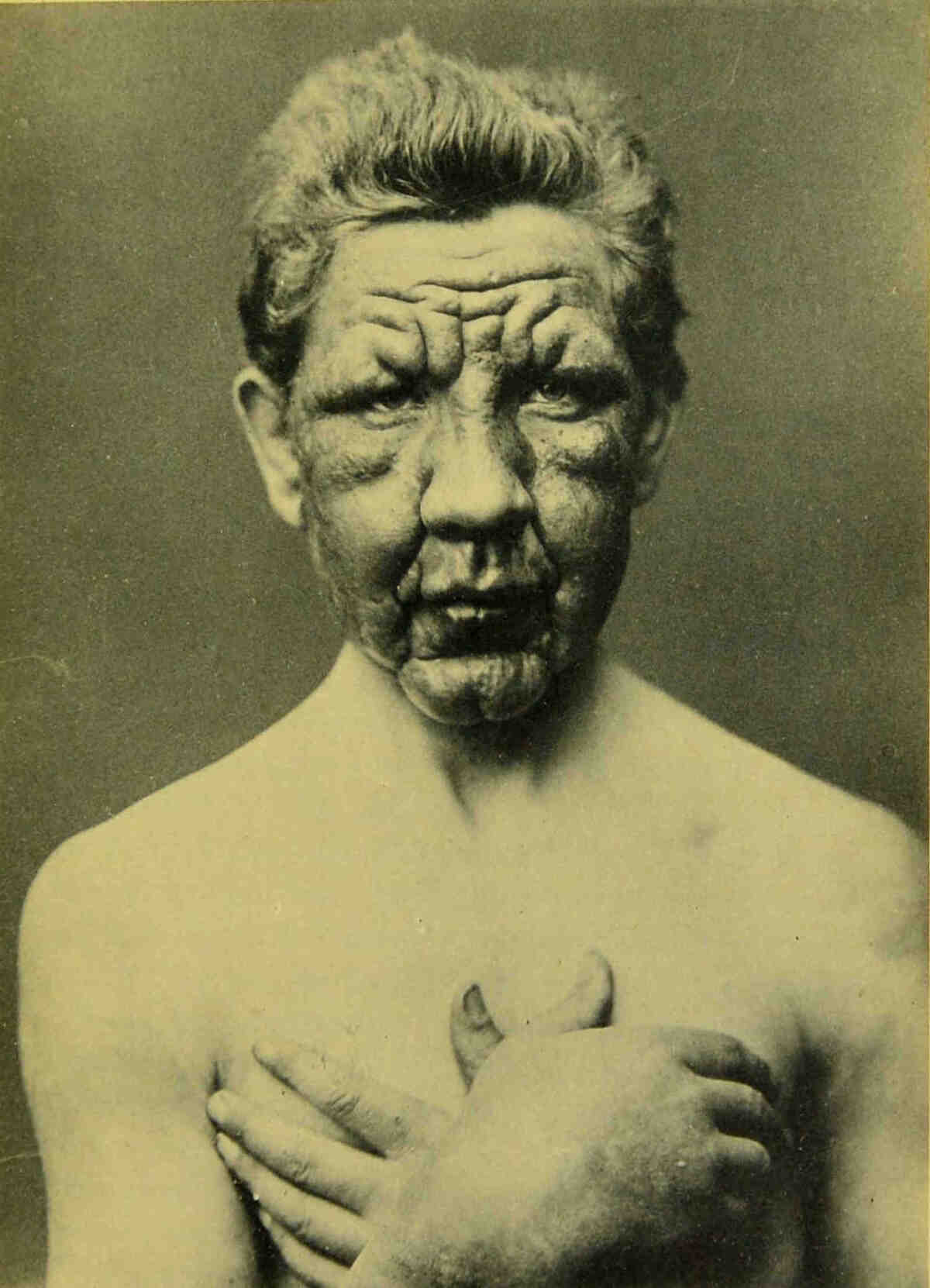
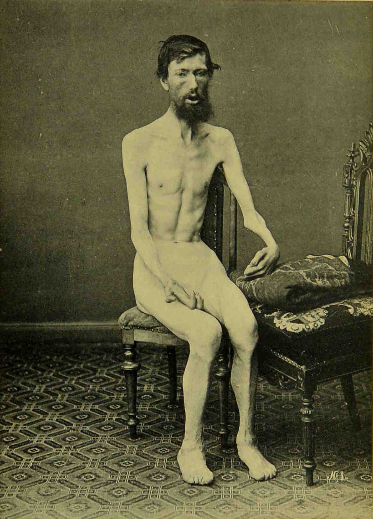
and become red, thick and elongated. Plate I is
a case of tuberous leprosy of two years’ duration,
the hands being swollen with leprous infiltration.
PLATE I.
The eyes are, in the nodular form, almost always
affected; nodules are frequently present in the
eyelids, the upper as well as the lower, and are
usually situated close to their margin. The
earliest affection of the eye itself, which we have
observed, is a faint clouding of the upper part
of the cornea, which often appears as a
very fine dotting of the corneal surface, only
noticeable when one can compare the upper part
of the cornea with the black pupil, and often
requiring for its recognition the use of a lens. A
slight infiltration of the limbus conjunctivæ is
always combined with this clouding of the cornea,
but it is at this early stage so slight that it
cannot be noted clinically. Later on it increases,
and gradually attacks the whole of the outer
margin of the cornea. When this infiltration
becomes greater it appears yellow, as seen through
the conjunctiva running intact over it, and this
gives to the eye a peculiar woe-begone aspect.
It is quite exceptional for this infiltration to
extend completely round the cornea, for that part
of the limbus directed towards the nose is almost
always free. As time goes on the infiltration
increases, and a low rampart is formed around
the cornea. Sooner or later the infiltration
and nodule formation attack the cornea itself,
in one of three different ways: first, quite
superficially, immediately under the epithelium.
The nodule in this case is always elevated, usually
grows very rapidly till it finally covers the whole
cornea, and may by its height prevent the closure
of the lids. That part of the cornea lying below
or behind the nodule is quite clear. Secondly,
the infiltration may attack the cornea in the form
of a wedge, and form a node which is not so
much elevated as in the previous instance; and
thirdly, the infiltration may penetrate the cornea
close in front of Descemet’s membrane. The
result, complete blindness, is the same in all cases
when the nodule covers the pupil. A frequent
accompaniment of this form of the disease is iritis,
or, as anatomical investigation shows, irido-cyclitis.
These forms of iritis run a chronic or sub-acute
course, sometimes so stealthy and painless that it
is observed by neither doctor nor patient, until
adhesions have formed between the pupillary
border and the capsule of the lens. Blindness
may sometimes be caused by exudation into the
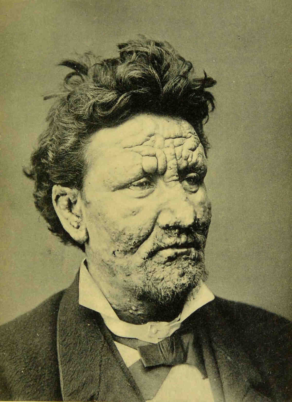
pupil. Plate II shows a typical case of tuberous
leprosy of six years’ duration. The hairs have
completely disappeared from the eyebrows; on
the chin a few can still be seen between the
tuberosities. In the right eye is a nodule, growing
from the Limbus conjunctivæ into the cornea.
Nodules may also be present in the iris, and
usually arise in the outer and under margin, in the
angle between the cornea and the iris; they may
completely fill the corresponding part of the anterior
chamber, are of a yellow colour, and sometimes
look exactly like an obliquely-placed hypopion,
as they have an inner or upper straight, or
slightly concave margin. We once did an iridectomy
directly through a small early nodule, and
put a stop to its further growth. On anatomical
examination we also find a leprous affection of
the anterior part of the retina over the ciliary
body, which appears as a fine white spotting of
the retina; the ciliary nerves are always for a
considerable distance backwards infiltrated with
leprosy, as are the membrana supra choroidea and
the choroid itself.
PLATE II.
On the extremities the nodules always appear
singly, but when closely set may run together to
form large plaques. On the backs of the hands
and fingers nodules are very frequently, and on
the extensor surfaces of the thighs and the front
of the legs almost always, found. The calves
are often also infiltrated as a whole, especially
on the fibular side close above the ankle, and
this infiltration reaches as high as the middle
of the leg; the skin is tense and shiny, reddish
blue in colour, and in this infiltrated part ulcers
resembling varicose ulcers readily appear, which
are as difficult, if not more so, to heal. They are
surrounded by thick elevated walls, may last for
years, and occasionally completely surround the
leg.
Of the mucous membranes, those of the nose,
mouth, larynx and pharynx are affected. The
nasal mucous membrane is affected only in its
anterior part along with the alæ nasi and the
anterior part of the septum. If a general infiltration
takes place in this situation, the softening and
ulceration which may ensue lead eventually to the
disappearance of all the soft parts of the nose;
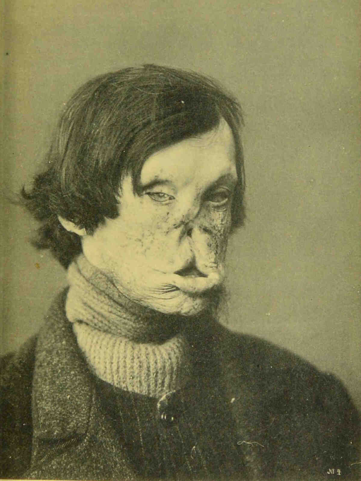
the bones are never affected. (See Plate III, a
case in which the leprosy developed in 1848, and
was of the tuberous variety. The tubers disappeared
partly by suppuration. In 1857 he
entered an asylum and then presented the same
appearances as in the photograph. He was then
anæsthetic. There were cicatrices in the face due
to the disappearance of the nodules, the point of
the nose was gone, but the nasal bones were
intact, thus differing from syphilis. He died in
1885.) In the mouth, the mucous membrane of
the lips, of course, shares in the process when
these are completely infiltrated, and even on
the mucous membrane of the cheeks one
occasionally sees and feels thickening and infiltration.
The tongue is often the seat of
nodules, which in all respects correspond to those
of the skin. The gums, the velum, and the
uvula may be either infiltrated or dotted with
nodules. The rest of the mucous membrane of
the pharynx is more frequently infiltrated than
beset with nodules, and the same is true of the
epiglottis, which sometimes becomes quite stiff
and almost immoveable. In the larynx, the true
and false cords are more frequently the seat of
infiltration than of nodules; the voice is rough and
hoarse, the rima glottidis is often so narrowed
that respiration is rendered difficult; excessive
narrowing of the rima is only present in the late
stages, and is proportionately rare. When the
mucous membrane ulcerates the cords grow
together, both anteriorly and posteriorly, and
when the infiltration disappears there remains a
scar tissue, which, by its contraction, reduces the
rima to a small slit, a few millimètres wide. In
such cases a very little mucus is sufficient almost
or completely to close the opening, and the
patient may perish from suffocation. Usually an
emetic suffices to open up the hole at once; but
tracheotomy is often necessary to supply air to
the patient, the attacks of suffocation are so
frequent, and since he is already voiceless, he
loses nothing by the operation.
PLATE III.
The lymphatic glands (cervical, axillary and
inguinal) in relation to the affected skin and
mucous membrane are always swollen: this
leprous swelling is always indolent, and never
goes on to suppuration. Sometimes the glandular
swelling may aid in the diagnosis, if the skin
affection is not absolutely characteristic, though
this is most rarely the case.
The nodules are almost always seated in the
cutis, but they may, though rarely, be placed
deeper in the subcutaneous connective tissue;
they form then no projections, but the skin over
them is almost always somewhat hyperæmic and
bluish-red, and, if the finger is passed over the
place, the thickening or the nodule may be felt in
the deeper parts. It is in our experience that a
patient who had only this form of nodules was
regarded by a colleague, well acquainted with
the disease, as free from Leprosy, probably
because he did not use his fingers.
From the symptoms described above, the
diagnosis is almost always very easy, and we
ourselves know of no disease of the skin which
can be confounded with nodular leprosy. If
necessary a piece of skin may be removed and
examined for the presence of bacilli, which, at
least in the nodular form of the disease, are never
absent. This, we have once had occasion to do.
In addition to the skin the nerves are also
affected, not always at the commencement, but
always in the later stages. Whether all peripheral
nerves are affected we cannot say—certainly
the facial, radial, ulnar, median and peroneal
are always diseased. According to our
investigation the nerves of the extremities are
affected throughout their whole length, but the
affection is severe only at certain places, viz.,
where the nerves run superficially over bones or
joints, as the median at the wrist, the ulnar at
the elbow, and the peroneal where it crosses the
fibula. As a result of this nerve affection we
have pain followed by anæsthesia. The pains in
the arms, hands, feet and calves are sometimes
very severe and persistent. The affection at
first causes pain, by pressure on the nerves, and
later—when the pressure has led to atrophy—anæsthesia.
Now since, as we shall later clearly
demonstrate, leprous affections tend to heal, it is
not infrequently the case that nerve affections,
when slight, pass off without having specially
injured the nerves, and these nerves may be the
seat of fresh infections, and thus the patient suffers
from repeated painful attacks through a course of
years. This is particularly the case where there is
general infiltration of the legs, and is either the
result of repeated attacks of the same, or of the
implication of different nerve branches. The
nodules are often painful when first developed,
but later on sensation is deadened.
Of internal organs, the testicle, liver, and
spleen, are always affected in this form, but we
shall consider them later in the description of
the pathological anatomy.
Before we more closely describe the course
of the disease, we shall first briefly discuss the fate
of the nodules. These usually remain for years
unchanged, growing very little or not at all. The
skin over and around them, or rather its vascular
supply, is very sensitive to changes of temperature,
so that the skin, as we have already indicated,
changes its colour with change of temperature
from dilatation of the blood vessels. The vessels
evidently suffer from the invasion of the leprous
poison. New outbreaks have often the appearance
of an “erythema nodosum,” with great
hyperæmia. We had once the opportunity of
examining a piece cut out of such an erythema-like
eruption, and found dilated vessels and round
cells, and only after long search a few bacilli.
One must conjecture that there is deposited with
the bacilli a chemical poison which affects the
vessels, or that the bacilli produce the poison, and
that this poison has its action only in its immediate
neighbourhood.
But occasionally the nodules grow so luxuriantly
that the epidermis develops furrows and clefts
which may reach down to the nodules, and then a
bloody fluid comes out of them which dries up on
the surface to a reddish brown scab. Or it may
happen that the upper horny layers of epithelium
disappear, and that only a few rows of cells of the
rete Malpighii remain. In this case the exuded
fluid less often dries, the surface is usually blood-red
and moist, and appears like an ulceration,
though it is not really one. When this takes
place on the face, particularly on the lips, or on
the backs of the fingers, the sufferings of the
patient are very much increased. After several
years the nodules usually soften about the middle
of their base, and the nodule may sink in over
the softened part; or they burst, the softened
part is thrown off, and now is developed the true
ulceration by which the nodule may be completely
eliminated and sunken stellate scars alone
remain.
The determination of the commencement of
the disease is exceptionally difficult, indeed impossible,
for it must always be founded on the
statements of the patient, and the patients either
observe themselves insufficiently, as may frequently
be noted, or they conceal many facts. As
a matter of fact we do not know the earliest
symptoms of the disease. According to Danielssen
and Boeck, the patients often suffer long and
repeatedly, before the outbreak of the disease, from
weakness, with rheumatoid pains and fever. This
the patients frequently corroborate. But we are
inclined to regard these attacks of fever as
indications of the already existing disease. It
appears to us more probable that the disease
begins with some form of local affection which is
so indistinct that the patient himself does not
notice it, or at least lays no weight upon it, and
that these local affections are analogous to others
with which we are familiar, namely, the nodules
which may last for years before new and such
definite eruptions appear, that the disease cannot
any longer be ignored or kept secret. We believe
therefore, that the patients do not really know
when they commence to be ill, and that they date
the beginning of the disease from a later eruption.
If at the commencement only the extremities are
affected the patients may conceal their condition
for years, and through this concealment become so
accustomed to lie, that later it is impossible to
receive from them correct information.
The cases are very frequent in which the
patients have for several years only scattered
nodules, and then suddenly a fresh outbreak of
numerous nodules. The disease always advances
by outbreaks of eruptions which repeat themselves
at longer or shorter intervals. It is very often the
case that the older nodules soften during a fresh
outbreak, and completely or partly disappear;
and these outbreaks are always accompanied by
fever, the temperature rising to 39° or 40° Cent.
Now we know that the nodules, if the patient is
affected by another febrile disease, may disappear.
It is, therefore, difficult to decide whether the disappearance
of the nodules is the cause or the
result of the eruptive fever. But we possess
certain observations in which the disappearance of
the nodules has begun before the onset of the
fever, and in which, therefore, the fever and the
later eruption appear to be caused by the softening
of the nodules. Supported by these observations
we regard the eruptions as auto-infections, in
which bacilli (or poison) from the older lepromata
pass into the blood, and thus new areas of the
skin or other organs are affected. We have often
observed that an irido-cyclitis, or an affection of
the throat, arises during an eruption, and also that
the nerves beneath the nodules become swollen
and painful, and once we have seen the testicle
become swollen and painful during an eruption.
As to the affections of the liver and spleen we
have no clinical observations; they appear to cause
no clinical symptoms, or at all events, such indefinite
ones that, although our attention has been
directed to them, they have escaped observation.
The eruptions are of the most varied duration.
Some last only a few days and cause so little
fever that the patients experience no particular
disturbance to health; they only know that a few
new nodules appear, or that they are sore about
the throat. Others last for weeks, indeed,
months, with remittent fever, the temperature
rising to 40°. Quinine has no effect. During
such eruptions the strength of the patients
is of course distinctly diminished, but when the
eruption is over they recover rapidly and feel
themselves all right again; only they are more
leprous than before, or at least have more
leprous manifestations. The number of eruptions
varies greatly in different cases. Sometimes the
patient has, for several years, several eruptions
every year; in other cases the eruptions appear
only very rarely—one or two in the whole
course of the disease, and they may be very
slight. It appears as if the bacilli in different
cases were of varying virulence, or possibly the
structural conditions are different in different
individuals, so that in some the bacilli (or the
poison) reach the blood more easily than in others.
Therefore the fate of the patient is very
different. If the eruptions are frequent the
growth of the nodules is usually luxuriant, and
those nodules with diminished epithelial covering,
and therefore moist, are very frequent. If then,
the whole countenance and part of the extremities
are covered with such nodules; if the eyes are
blinded by the formation of nodules in the cornea;
if the tongue and the pharynx are, through formation
of nodules and infiltration, half ulcerated and
sore; if respiration is made more difficult by the
narrowing of the rima, and the voice inaudible;
then the condition is as miserable as is possible to
conceive.
If there is added to this, amyloid degeneration
of the kidneys, liver, spleen and intestine, with
diarrhœa, it can only be desired that death shall
put an end to such a condition, and that usually
takes place soon, although occasionally the patient
may linger for months. Whether leprosy alone
is responsible for the end is, we think, doubtful;
as remarked above, the affections of the liver and
spleen appear to be without much significance.
The biliary secretion is never influenced, and
there is according to our observations no special
leprous anæmia. The patients are indeed often
anæmic, or become so during the disease, but we
have not been able to convince ourselves that this
is a direct result of the leprosy. In many examinations
of the blood, which were indeed undertaken
for other purposes, we have never noticed
anything remarkable in relation to the number or
form of the blood corpuscles.
As in almost all cases of nodular leprosy
nephritis is present, we are inclined to regard this
nephritis as a frequent cause of the marasmus
which ensues. Tuberculosis was formerly a frequent
occurrence in our hospitals, where our
observations have chiefly been made. The relationship
between this and leprosy we will discuss
later.
The prognosis in the case of patients in whom
the eruptions appear less frequently is more
favourable, and they may live many years.
Either they die from an intercurrent disease
or as a result of their nephritis, or they become
in time anæsthetic, that is, according
to our view, they recover. When the nodules
become stationary they ultimately soften, as
described above, and may be absorbed without
opening, though this is rare and usually
occurs only with single nodules; or they burst
and ulcerate; in either case they leave scars.
If this takes place in all the nodules and the
patient is attacked by no fresh eruptions, then
anæsthesia gradually develops as the result of the
affection of the nerves; in the nerves, too, the
specific leprous affection disappears, and there
remains only scar tissue, which by compression
destroys the nerve fibres and thus causes anæsthesia.
Then gradually all specific leprous
affections disappear, and the patient is healed from
his leprosy, and may live many years in perfect
health, having lost practically nothing of his
power of work. Such cases are unfortunately not
very frequent; but we have had the opportunity
of examining some after death and have not been
able to discover in them any specific leprous
affection.
Thus one is struck with the fact, how little
leprosy of itself influences the health of the
patient, and if nodular leprosy usually shortens
life, that takes place probably because in this
form the frequent ulceration leads to amyloid
degeneration of the internal organs, or that
the nephritis is a sequel of the leprosy. The
nephritis appears either as the so-called parenchymatous
or as the interstitial; according to our
examinations it is never bacillary. Further, as
nephritis is very much rarer in the maculo-anæsthetic
form of the disease, it must be assumed
that nodular leprosy in some way causes nephritis.
The same is true of amyloid degeneration. The
duration of life of a patient with the nodular form
of leprosy is in general eight to nine years after
the definite outbreak of the disease.
The most frequent complication which we have
seen in our institutions is tuberculosis, particularly
some years ago, for then the institutions were
over-crowded, and consequently the sanitary
conditions were in many respects unsatisfactory.
In order to give an idea of the frequency of this
complication, we have placed in tabular form at
the end of this work the results of eighty-nine
autopsies (see Table I, page 128).
From the statistics there given it is evident
that we have had ample opportunity of examining
the combination of tuberculosis and leprosy.
Most of these examinations were indeed made in
the pre-bacillary era; but we are satisfied that the
differentiation of tuberculosis and leprosy without
an examination for bacilli is by no means
difficult.
As Danielssen and Boeck had described a
leprous affection of the intestine, we gave great
attention to this point, and as we were at the
same time engaged in an investigation on the
pathological anatomy of the lymphatic glands,
we lost no opportunity of carefully examining
these organs. It was during this investigation
that we discovered the characteristic leprous
affection of lymph glands, and had our attention
first directed to the leprous affection of the liver
and spleen, which affections are, macroscopically,
so little evident, that we at first overlooked them.
The leprous and tuberculous affections of the
lymph glands are macroscopically so very different,
that it is impossible to confuse them, and
the microscopical differences are still more evident.
Both fresh and hardened preparations
were always examined. And since the lymph
glands are always affected with leprosy, if the
organs which drain into them are affected, even
if this affection is very slightly developed, we
conclude from the fact that we have never seen a
leprous bronchial or mesenteric gland, that there
is no leprous affection of the lungs or of the
intestine, and later examination of certain special
preparations have only confirmed us in this view.
But more of this later, and we will first treat of
the differences between tuberculosis and leprosy.
In organs affected with tubercle one always
finds, as is well known, giant cells and caseous
degeneration; in the many, we can truthfully say,
thousands of preparations of leprous affections,
which we have had under the microscope, we
have never seen either a typical giant cell with
marginal nuclei or caseous degeneration. There
are indeed multinuclear cells in the lepromata,
but never giant cells like those of tubercle.
What may be the reason for this striking difference
in the action of the very similar bacilli of
tubercle and leprosy, we have no idea; we
simply state the fact and assert that, if one finds
giant cells, he is dealing with tuberculosis and
not with leprosy. This alone would be sufficient
to cause us to separate the two neoplasms, but
there are many other distinctions. Tubercle is
avascular; the leproma is rich in vessels; tubercle
undergoes caseous degeneration, the leproma
never. Anatomically therefore, we are justified
in maintaining a sharp distinction between the
two diseases.
So far as concerns the resemblance between
the tubercle and lepra bacilli, we must not omit
to mention that one almost always finds among
tubercle bacilli some which are pretty long and
somewhat bent; this is never the case among
lepra bacilli. Baumgarten has indicated as a
distinction between them, the fact that the latter
is more easily stained; according to our experience
this distinction can scarcely be regarded
as sufficient. But the distribution of the bacilli
in the tuberculous and leprous tissue is usually
so very different, the tubercle bacilli being usually
arranged singly, the lepra bacilli always in large
quantities in masses and clumps, that a confusion
of the two diseases anatomically can only be possible
in exceptional cases. Danielssen has repeatedly
stated in his triennial report of the
Lungegaards Hospital that tuberculosis and
leprosy are such nearly allied diseases that the
one (leprosy) may pass into the other (tuberculosis)
by a modification of the bacilli, and that
thus the frequent combination of the two diseases
is to be explained. This view we cannot, in view
of the above demonstration, support. If an organ
is attacked at the same time by tuberculosis and
leprosy, one can anatomically very readily separate
the two diseases. We would rather seek the explanation
of the frequent combination of the two
diseases in our institutions in the great over-crowding
and consequently insanitary conditions
to which they were formerly subjected. Tuberculosis
once introduced, we find a ready explanation
in the bad habits of the patients in regard to
expectoration, why it was difficult or impossible to
root it out. In later years, when the institutions
are no longer full and the sanitary conditions
consequently much improved, tuberculosis has
much decreased. Whether tuberculosis was as
frequent a combination in the country as in our
institutions we do not know. The duration of life
of patients in the country is about a year longer
than in our institutions, and possibly this depends
on the absence of tuberculosis.
Doutrelepont has recently described in the
transactions of the German Dermatological
Society, On the Pathology and Therapeutics of
Leprosy, a lung affection, probably developed
by tuberculine treatment, which he diagnosed from
the examination of the sputum. But the patient
had a leprous affection of the larynx, and his
account by no means excludes the possibility that
the bacilli in the sputum came from a ruptured
nodule in the larynx, possibly softened by the
tuberculine treatment. We cannot, therefore, recognise
this observation as infallible evidence of
the presence of a leprous lung affection, any more
than that case of leprosy of the lungs described by
Bonome in Virchow’s Archiv, Bd. C. That
author himself draws attention to the great resemblance
of the affection to tuberculosis, and as
it is quite evident from his description of the
bronchial lymphatic glands that they were not
leprous, and he notes the presence of giant cells
in the pathological products, we cannot doubt
that it was really a case of tuberculosis.
In the same case there was found an affection
of the spinal cord, from which Bordoni-Uffreduzzi
cultivated on glycerine agar an
organism which he recognised as the lepra
bacillus. Here we may remark in the first place
that we have never seen a leprous affection of
the spinal cord, and have never found bacilli in
it. We must indeed admit that we have only examined
the spinal cord in a few cases, because
there appeared to be no indication for such an examination,
since clinical symptoms do not point to
an affection of that organ, and as in the profusely
nodular cases, affections easily recognised appear
everywhere, except in the liver and spleen, it was
to be expected that an affection of the spinal
cord would have been recognisable. In necrotic
bones, which we have often examined, nothing
leprous is found. This necrosis is therefore no
specific leprous affection, but a secondary one.
Secondly, we must remark that in spite of many
attempts we have never succeeded in cultivating
lepra bacilli on glycerine agar. We therefore
believe that Bordoni-Uffreduzzi has cultivated
tubercle bacilli instead of lepra bacilli. The only
thing which speaks for the leprous nature of the
lung affection and the cultivated bacilli, is the circumstance
that the author did not succeed in inoculating
guinea pigs and rabbits with tuberculosis.
But, according to all investigations on
tuberculosis, it appears to us not incredible that
the tubercle bacilli may, under circumstances,
become so weakened that they are no longer
pathogenic.
Arning has described a miliary leprosy, and
found in the products of this disease giant cells,
and also leprous ulcers in the intestines. Dr.
Arning has kindly sent us some preparations of
this miliary leprosy, in which we find evidence
everywhere that the case is one of tuberculosis,
both because giant cells are found everywhere, and
the bacilli are only present singly, and scattered.
They are never present in the excessive numbers,
and have not the arrangement, which they usually
have in leprous products. In connection with the
presence of giant cells in leprous products, we may
note that we have received from two foreign colleagues
preparations in which they believed giant
cells to be present. But we have found, on careful
examination of the preparations, that they were
cross and oblique sections of blood vessels, which
with their endothelial nuclei gave the impression of
giant cells. Without the use of a homogeneous
immersion lens it was not possible to make a
definite distinction.
According to our observation there exists a
sharp anatomical distinction between leprosy and
tuberculosis, and there is no such thing as leprosy
of the lungs and intestines, the bones and the
kidneys. In order to establish a differential diagnosis
in doubtful cases, we recommend in the case
of the lungs and the intestines a thorough examination
of the bronchial and mesenteric glands.
We ourselves have never sought in vain, in cases
of these affections, for tuberculous or caseous degeneration
in the glands, and we have seen in no
single case anything resembling leprous affection
of the glands.
So far as concerns the central nervous system,
Danielssen noted that he had several times seen
acute hydrocephalus in leprosy. We once saw
severe cerebral symptoms with maniacal attacks.
The patient, who was taken into a lunatic asylum,
left this later, cured. Other indications of an
affection of the central nervous system in leprosy
are unknown to us. Anatomically, we have not
been able to recognise in the nervous system any
traces of leprosy. In connection with this, we
may note that we have several times seen pain and
swelling of the knee joints during eruptions,
which at their conclusion disappear. In these
cases there is nothing to be made out anatomically.
When we reflect that, as indicated in
describing the eruptions, the bacilli and (or) their
toxines most probably circulate for some time in
the blood, it is remarkable that the organs above
referred to are not affected by leprosy. We can
give no reason for this; connective tissue, which
is especially liable to be affected, is present everywhere.
Chapter III. STRUCTURE OF THE LEPROMA.
The leprous nodes have on section a smooth,
white, glistening surface, if they are still
sufficiently young. If one examines, microscopically,
sections or teased preparations of fresh
nodules, one sees little else but cells, with distinct
nuclei, usually of the size of a white blood corpuscle,
or rather larger. There are also a few larger
so-called epithelioid cells, with larger nuclei, and
among the cells, fragments of connective tissue
and of blood vessels. With a higher power, one
sees in the fluid of the preparation small straight
rods, which are not destroyed by addition of
potash. These are the lepra bacilli, and thus
were they first discovered in the year 1871.
If one teases out preparations in osmic acid
solution, or soaks a nodule in the solution some
hours before teasing, the rods are coloured faint
brown, and one finds them lying mostly in the
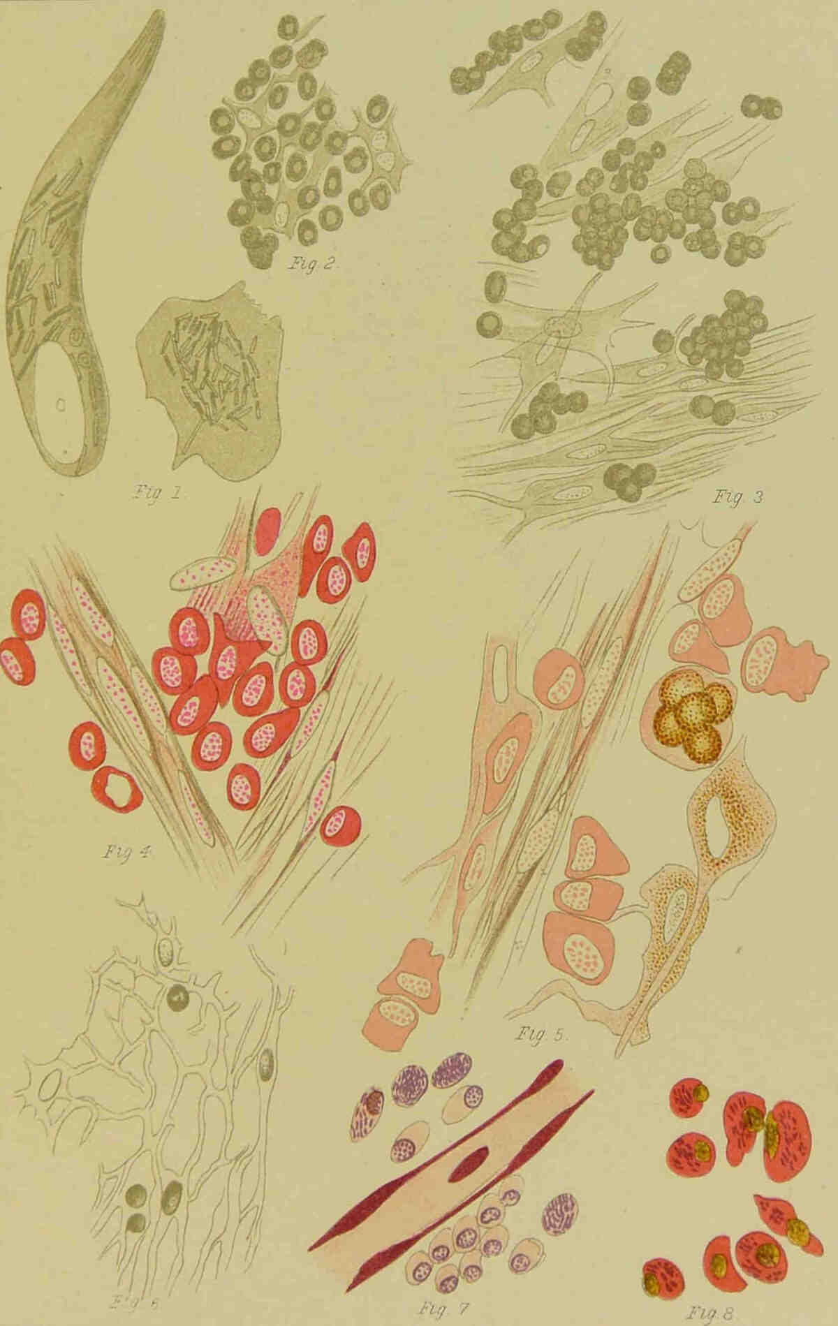
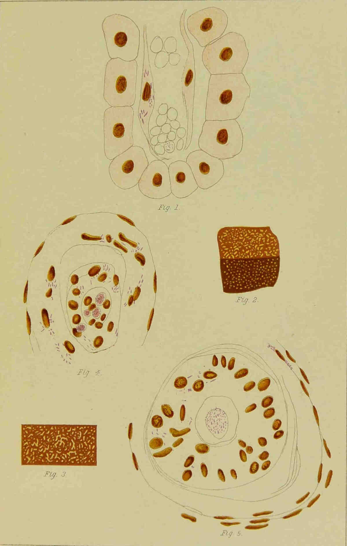
cells (Plate VI, Fig. 1). If one adds water to a
fresh preparation, the bacilli move actively; even
in the cells swollen up with water, one sees the
bacilli moving; and this led us to regard them as
movable, although we at the same time indicated
a doubt whether the movement was not simply a
molecular one; for the movements were equally
vigorous in strong osmic acid solution as in water,
and on the addition of glycerine or strong solution
of albumen to the preparations, the movements
ceased. All later observers, with the exception of
Unna, regard the bacilli as motionless. We have
no ambition to decide this question, because we
know no absolutely trustworthy distinction between
molecular movement and independent
movement of the bacilli.
The older the nodules become, the more large
multinuclear cells are found, and in nodules of the
skin and cornea one always finds small flat cells
with processes, and with oblong nuclei, which we
recognise as the connective tissue and corneal
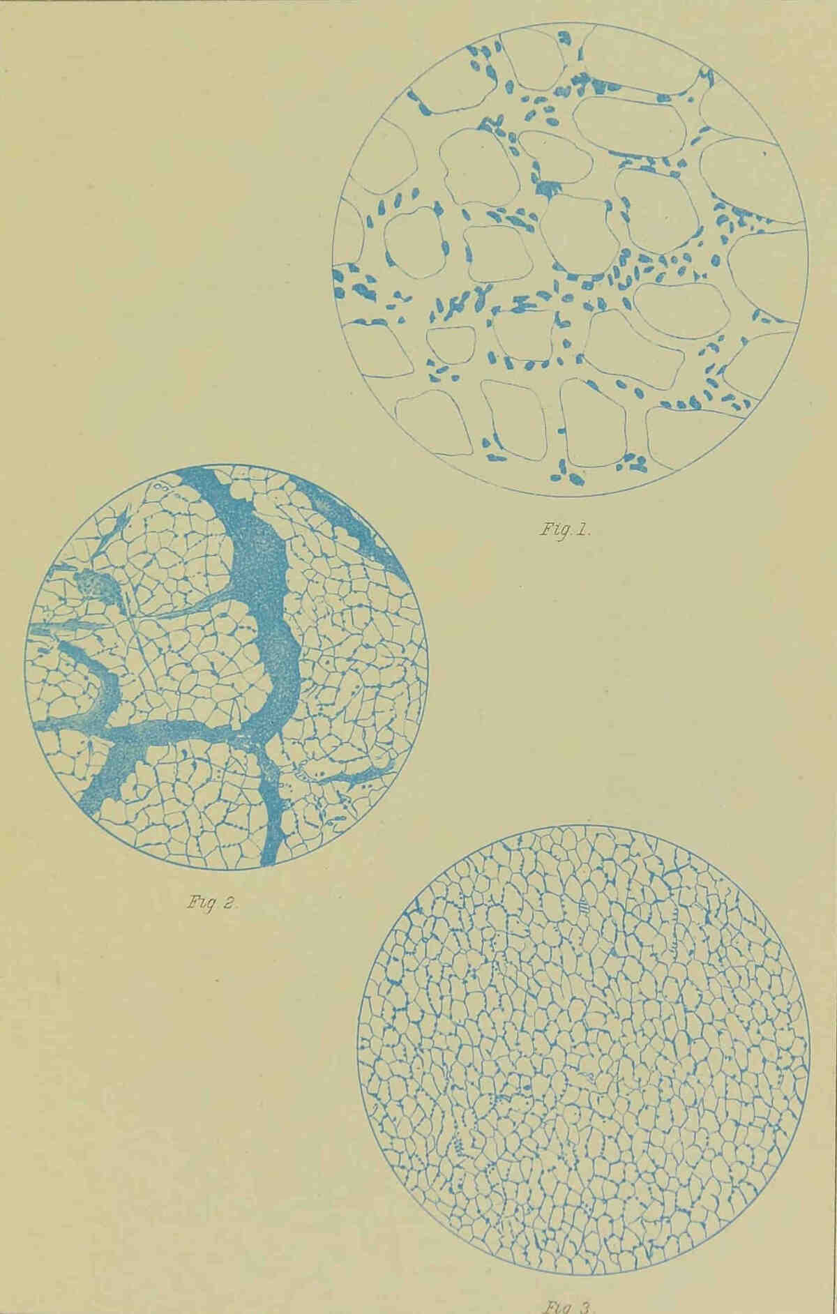
cells (Plate VI, Figs. 2 and 3).
The protoplasm of these cells is clear, while
that of the round cells is more or less granular.
The nuclei of the latter are also round, and
usually very granular; the flat cells are much less
stained by carmine than the round cells, and one
sees the flat cells lying on connective tissue
bundles. The nodules are richly supplied with
blood vessels; in the nodules of the skin it cannot
be determined whether the vessels are newly
formed, or only those already present in the cutis.
They are, however, always of embryonic type.
One sees very plainly their formation from cells,
and in the cornea, indeed, the vessels must be
newly formed. Here the vessels penetrate into
the cornea before the nodule forms, and round
these vessels, penetrating into the cornea, there
are always visible collections of cells which are
apparently migrated white blood corpuscles (Plate
VI, Fig. 4).
One meets also in old nodules, among apparently
only unproductive elements, blood vessels surrounded
by young cells (Plate VI, Fig. 5). This
appears definitely to favour the view that the
tumour cells are, at least for the most part,
migrated blood corpuscles. These results have
been obtained by examination of fresh nodules,
and of those hardened in Müller’s fluid. As we
have recently, while hardening the nodules in
Fleming’s chromic and osmic acid mixture, and
staining the sections with hæmatoxylin, been
unable as yet to find any mitoses, and further,
have found, by bacillary staining of an old section
from a corneal node, an appearance like Plate VI,
Fig. 7, we must maintain provisionally that in
the lepromata the new formations are, at least
chiefly, formed by the immigration of white blood
corpuscles. As the round cells infiltrate the connective
tissue, the fibres are pressed asunder,
and form a network closely resembling that of a
lymph gland with nuclei in the angles (Plate VI,
Fig. 6).
When the nodule softens, which takes place
almost always exactly in the middle, it attains
a brownish tint, which is due to the transparent
softened part. If one divides the softened
nodule, the central part has a distinct brown
colour, and the constituents of this part readily
fall out. If this is examined under a microscope,
one sees, almost exclusively, larger or
smaller clumps of a brownish colour, and very
granular. They are partly elastic, but partly
brittle and fragile, so that by pressure on the cover
glass they are easily broken up. One often sees
clearly that the clumps lie in the cells, the nucleus
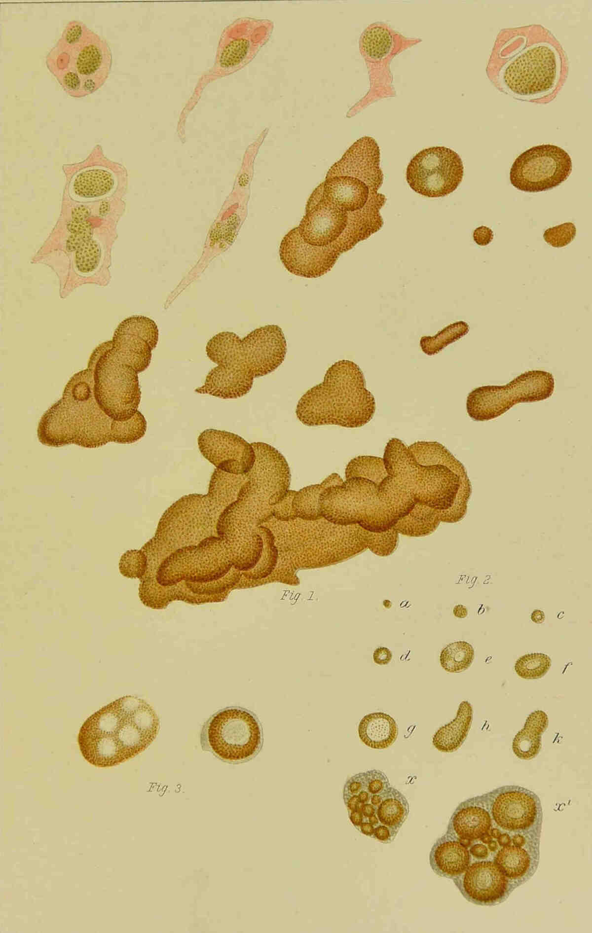
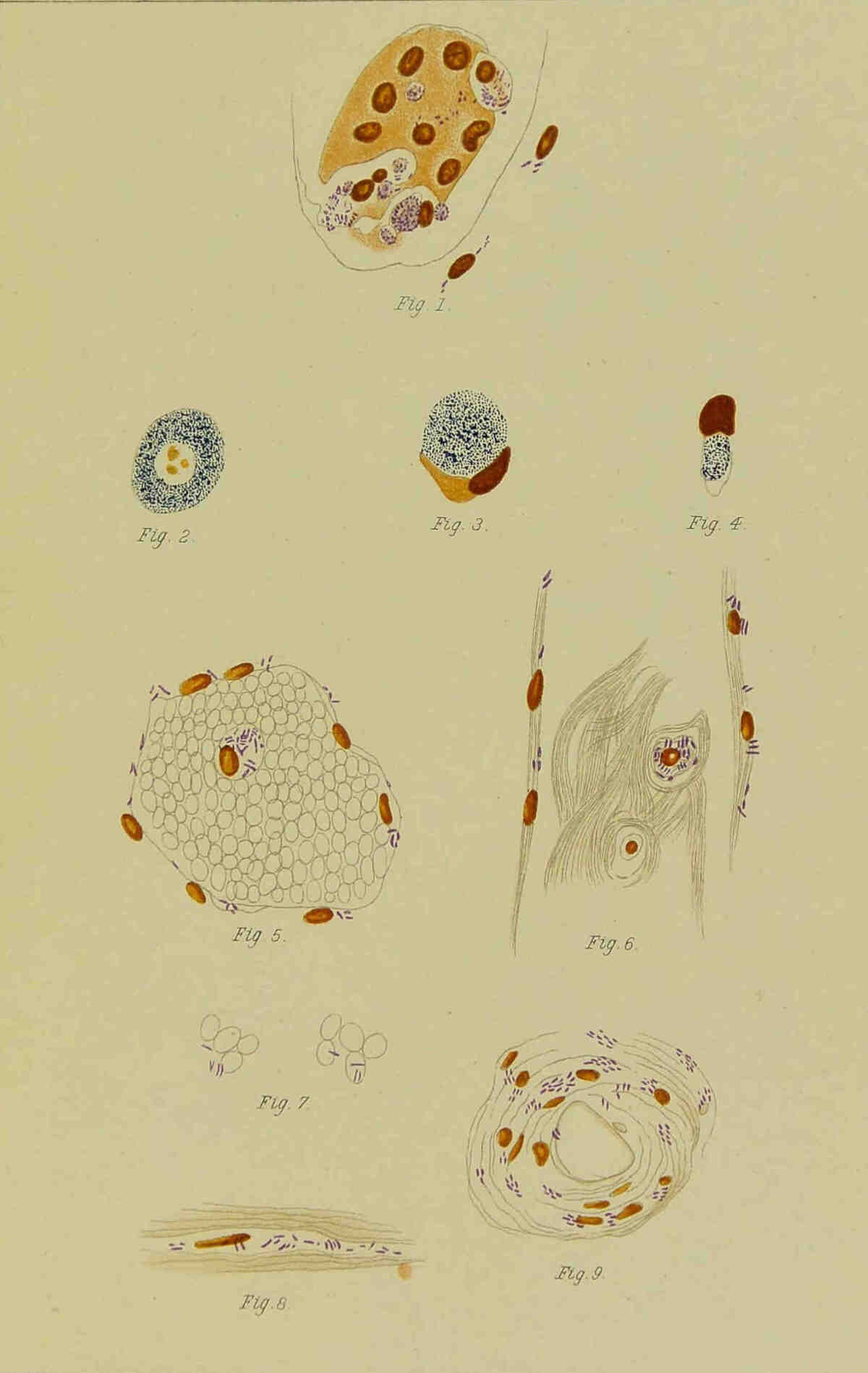
and the cell substance being still evident (Plate
VII, Fig. 1). Sometimes only one lies in a cell,
which then has the appearance of a signet ring, in
whose circle the clump lies, but sometimes several
lie in one cell. One finds similar clumps in
all other organs affected with leprosy, as in the
liver, the spleen (Plate VII, Fig. 2), the nerves,
lymphatic glands, the testicle and the eyes (Plate
VII, Fig. 3, which shows two clumps from the
retina). In the nodes of the skin, but particularly
in the testicles, one may find them so large that
they may even be seen by the naked eye. Both
in the small and the larger clumps there are usually
vacuoles, not infrequently several. From the
cornea we have often got preparations in which
the corneal corpuscles are more or less completely
filled with brown granules (Plate VIII, Fig. 1).
One sees here definitely the cell nucleus in the
middle of the brown granular material.
We have described these elements as we first
observed them in fresh and carmine-stained preparations,
in which they stand out very definitely,
as the brown masses do not take up the carmine
stain. These brown elements, if one knows their
characteristic appearance, may very well serve
as diagnostic indications for leprous affections,
for, according to our experience, one never fails
to find them except in very young nodules.
Since the discovery and easy recognition of the
lepra bacillus, they have indeed lost their value as
diagnostic signs, unless one is examining perfectly
fresh preparations. Later investigation has proved
that these brown clumps are nothing else but collections
of lepra bacilli broken down into granules,
and they have received from Neisser the well-chosen
name of “globi,” as they usually appear in
spherical form. Unna declares, in accordance with
his view of the position of the lepra bacilli outside
the tumour cells in the lymph spaces, that these
“globi” are collections of bacilli in the lymph
vessels, and that the vacuoles have arisen from
the falling out of bacilli in the middle. In particular,
Neisser, Touton, and we, ourselves, have
opposed this view, in that we have all seen the
bacilli definitely in the cells, and have figured them
so. In view of the above description of the discovery
of bacilli in fresh preparations and of these
brown clumps, there can scarcely be any doubt of
the position of the bacilli, and of the cellular nature
of the brown clumps. Further, it may be noted
that in the testicles the “globi” are chiefly in the
lumen of the seminal canals, where no lymph
vessels exist, and if Unna says of the “globi”[2]
that “no one has certainly determined their
cellular character,” he must have said this in
ignorance of our earlier publications in Norwegian.
The above description and the drawings were
published by us in 1869 and 1870, in the Nordiskt
Medicinsk Arkiv.
In connection with the vacuole, we have found
in a testicle a “globus” with vacuoles, and in the
vacuoles small granules which were recognised as
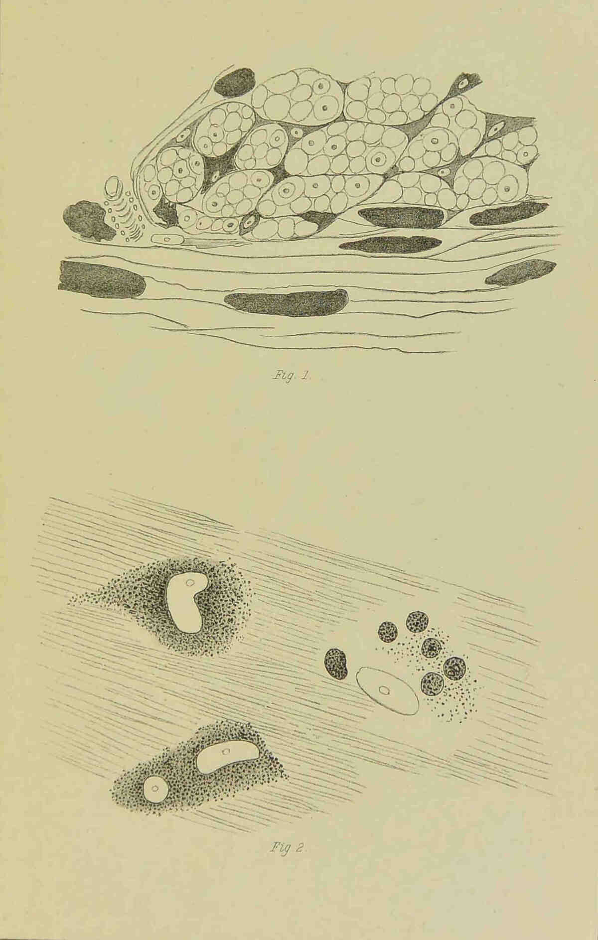
remains of the nuclei (Plate X, Fig. 2). We add
to this the picture of a “globus,” or rather a
developing “globus,” with two nuclei, from a skin
nodule (Plate VIII, Fig. 4). We have seen earlier,
however, that the brown clump may lie in the cell
without enclosing the nucleus, and also that the
nucleus may lie in the middle of the brown mass.
Now, if the vacuoles are transformed nuclei, as we
believe, then it would be comprehensible that the
vacuoles would be absent in many “globi,” and
that in others which have developed from multinuclear
cells, several vacuoles would be found.
That appears, at least, the simplest explanation of
their presence. But there are certain very small
“globi” with vacuoles, such as are represented
in Plate VII, Fig. 2, c, and those small vacuoles
can scarcely represent nuclei; small “globi”
may however arise, as is evident at x and x1
of the same figure, from clumps of bacilli in the
cells. The vacuoles of the larger “globi” of x
and x1 may indeed possibly represent nuclei, but
not the vacuoles in Plate VII, Fig. 2, a-k,
nor the vacuole in one of the small “globi” in x1.
Possibly the vacuoles are also the result of a
specific degeneration, either of the bacilli themselves,
or of the cell protoplasm lying in the
middle of the group of bacilli, but on this we
would rather not express an opinion.
We have repeatedly demonstrated the position
of the bacilli in the cells, and explained them
in diagrams, but in many preparations it is
impossible to distinguish where the bacilli lie. The
best method of definitely noting their position,
which we know, appears to be the fixing of small
nodes or small pieces of organs in Fleming’s or
Müller’s fluid, with subsequent dehydration and
hardening in alcohol; sometimes one gets excellent
preparations by simply hardening in absolute
alcohol. If the preparations are stained with
fuchsin, and counter-stained with methyl blue, or
still better, stained with gentian violet, decolourised
by Gram’s method, and counter-stained with Bismarck
brown or with Bismarck brown and eosin,
one will never fail to see the bacilli lying definitely
in the cells (Plate VI, Fig. 8). Even in such
preparations there are many places where one
cannot definitely distinguish the position of the
bacilli; but as one always finds bacilli in the cells
where the preparations are sufficiently clear, we
may safely conclude that the bacilli lie everywhere
in the cells. In the connective tissue spaces one
often sees bacilli in and round the nuclei of the
connective tissue cells (Plate X, Fig. 8), and
although the body of the cell is not visible, we may
conclude that the bacilli lie in the cell body, and
not free in the lymph spaces. We have found in
many sections of the blood vessels in a testicle
(Plate X, Figs. 5 and 6), and in a liver (Plate VIII,
Fig. 6, and Plate IX, Fig. 1), white blood corpuscles
filled with bacilli, and in both cases only slight
affection of the organ, and we also observed many
bacilli in the endothelium of the vessels, as Touton
and Unna have observed in skin nodules where
we, we may remark in passing, have never seen
them. From these observations we draw the conclusion
that these two organs have been infected
through the blood. As we do not know the
manner and method of the primary infection of
the organ, we must devote our attention to the
search for discoveries like those described above,
and to the localisation of the bacilli in general, in
order to form an idea of the method of action of
the bacilli.
As we have already noted, we found in the
examination of an excised piece of a recent
eruption, in a nodular case of leprosy, chiefly,
round cells surrounding dilated vessels, and
only after long search, a few bacilli. It thus
appears not improbable that during the eruption a
toxin (which is circulating in the blood), and only
a few bacilli, escape from the vessels at various
places, or, it may be, only the bacilli, which produce
the toxin locally; that further, the toxin causes
the emigration of white blood corpuscles, and that
the escaped bacilli only after some time slowly increase
in number and gradually fill the cells.
From the presence of the bacilli in the endothelium
of the vessels and in the connective tissue cells,
one may speculate that the bacilli are passively
forced into them by the blood or lymph pressure;
we have certainly found, in the testicle referred to,
bacilli free between the red blood corpuscles in the
vessels (Plate X, Fig. 7). Such a fresh eruption
may remain stationary or slowly develop into
a nodule, or it may apparently completely disappear,
and only after several years again become
apparent, it may be during a fresh eruption. We
fancy that in these cases a few bacilli are deposited
at the time of the first eruption, and that they have
needed years to become so much increased that a
permanent nodule has finally formed. That the
vessels receive lasting injuries from the leprous
infection, appears to us to be proved by the following
observations. In an epidemic of measles in
one of our institutions, we saw in anæsthetic
patients the previous leprous spots, which had long
disappeared from view, definitely reappear, the
hyperæmia and turgescence being on these places
general, so that the earlier spots stood out as well
defined, red, and somewhat swollen areas. All
this appears to point to the fact that the bacilli increase
very, very slowly, and that possibly they
also produce a toxin, usually only in small quantities,
which causes no particular injury to the
organism, since the patients, in spite of numerous
nodules, with millions or milliards of bacilli, may
remain in pretty good health for years. We may
also conjecture that the toxin which is produced,
usually only acts immediately around the bacilli,
leading to dilatation of the vessels and favouring
the migration of white blood corpuscles. Only
occasionally does the production of toxin or the
multiplication of the bacilli appear to become so
vigorous that toxin and bacilli get into the blood
and cause an eruption; possibly this is favoured by
peculiar anatomical conditions, for it is very striking
with what varied frequency eruptions appear in
different patients. That the bacilli in the nodules
are all of them dead, as has been assumed, we
cannot admit, so long as the nodules still grow. It
appears to us preferable to ascribe the character
of the disease to the relative benignancy and slight
viability of the bacilli, as Unna has already suggested.
As we believe, as explained above, that
the bacilli lie almost exclusively in the cells, the
question arises whether the cells digest the bacilli,
or not. As we often find cells with only one or
two bacilli, and as we find in most cells balls or
clumps of bacilli, we must admit that the bacilli
multiply in the cells. In some cells the bacilli
remain lying in separate collections, in others they
fill the entire cell body, but they never penetrate
into the nucleus. Finally, the bacilli break down
into small granules, and this breaking down corresponds,
according to our view, to a degeneration
of the bacilli. Unna and Lütz have indeed stated
that this granular appearance of the bacilli is constant,
and is a mark of their structure, that they
really consist of small rows of cocci, and Unna has
therefore described them as coccothrix. This
actual (!) structure of the bacilli, however, only
becomes evident under the action of free iodine.
But we have seen in our preparations, however
they were treated, smooth and granular bacilli
lying close to each other, and we cannot, therefore,
corroborate the view of Unna and Lütz.
Neisser first drew attention to clear spaces
in the bacilli; these Neisser regards as spores;
we regard them as the first sign of breaking down
of the bacilli into granules, and for the following
reasons. We have made numerous attempts to
cultivate the bacilli, and have attained in all our
investigations only (the breaking down of the
bacilli into) granules, and in examining a piece of
a nodule which lay eight days on broth peptone
agar, found all the bacilli beset with clear spaces.
And as the result has always been the breaking
down into granules, we believe we are right in regarding
the appearance of these holes as the commencement
of degeneration, and that we are not
as yet familiar with spores of the lepra bacillus.
It appears as if all bacilli in time break down into
granules, particularly in the internal organs, where
it occurs much earlier than in the skin nodules;
whether this is the result of digestion on the part
of the cell, we cannot say; but as the bacilli at
first multiply in the cells, and the breaking
down appears most definitely and freely when the
cells are crammed full of bacilli, it is equally
possible that it is the result of diminished nutrition,
and as they break down more rapidly in
the internal organs, it is also possible, indeed
probable, that the higher temperature in these
organs favours this disintegration. As we have
unfortunately not been able to cultivate the bacilli,
it is at present impossible to form a conclusion.
At all events, we regard the transformation into
granules as a degeneration, and believe that the
bacilli thus altered are dead.
In the skin nodules we have only once found
bacilli in the epidermis; this was in a nodule
with many fissures in the epithelium, and partly
covered with exudation. We have not been able
to decide from our preparations whether the bacilli
lie in the epithelial cells or only between them,
possibly enclosed in wandering cells.[3] Touton
found bacilli in the epithelium of the sweat glands,
and he and Unna in the hair sheaths also; this
situation of the bacilli we have never observed
with certainty, and it can only be exceptional, and
can scarcely give origin to a “constantly flowing
bacterial spring,” as Unna suggests. As a rule, no
bacilli are found in the epithelium.
Of the presence of bacilli in affections of the eye,
it may be said in general that everywhere, where
infiltration is present, bacilli are found. In the
clouding of the upper part of the cornea described
above, which we recognise as keratitis punctata,
there are found groups of granularly degenerated
bacilli close under the epithelium. This we have
only once been able to determine on the living by
excision of a lamella of the cornea; in this case the
affection was, according to our own view, disappearing,
because the bacilli were granularly degenerated.
This corresponds with the fact that
this characteristic affection of the cornea always
ultimately disappears; the granules are probably
absorbed. We have already stated that blood
vessels precede the nodule into the cornea, and
that they are surrounded by round cells. Here, as
in the middle of the nodule, the corneal corpuscles
are found apparently intact or filled with brown
granules (Plate VI, Fig. 3, and Plate VIII, Fig.
1). The same is the case in nodules in the iris,
in which one finds the stellate cells intact (Plate
VIII, Fig. 2). Round cells are also found in the
spaces in the cornea near the nuclei of the corneal
corpuscles (Plate VIII, Fig. 3). All this
appears to us definitely to indicate that at least
most of the cells of the growth are migrated
white blood corpuscles.
Dr. Boeckmann has introduced, in the treatment
of the nodules growing into the cornea, the division
of the cornea in front of the nodules; through the
scar formed by the healing of the wound, they
hardly ever grow. We have seen such a case in
which, on the one side of the scar, no actual
nodules, but only a clouding appeared, and we
were later able to examine the eye anatomically.
The nodule reached like a rampart close up to the
scar, and all its cells were full of bacilli. On that
side of the scar in the clouded part of the cornea,
there were found only a few scattered cells containing
bacilli; no vessels had penetrated the scar,
and only a very few cells had succeeded in making
their way through. The treatment is therefore a
very desirable one in order to preserve the pupil
free. We have been able to prepare no bacillary
preparations from the retina, as we have not seen
the affection since the discovery of the bacilli; the
two brown clumps which are figured in Plate VII,
Fig. 3, lay in the outer granular layer of the retina.
As already indicated, the testicles are affected
with leprosy in all nodular cases. The affection
is both inter-tubular and intra-tubular. In
a testicle only slightly affected, we found bacilli
everywhere in the endothelium of the vessels, and
in several dilated vessels white blood corpuscles
filled with bacilli (Plate X, Figs. 5 and 6);
and in some places also bacilli lying free between
the red blood corpuscles (Plate X, Fig. 7).
At the same time, and especially where the affection
is more marked, the bacilli penetrate into the
seminal canals, and lie grouped in their walls
around the nuclei (Plate IX, Figs. 4 and 5, Plate
X, Fig. 1), and their epithelial cells are more or
less filled with them (Figs. 1, 2 and 4).
The bacilli appear rapidly to break down into
granules, and one finds, especially in the seminal
canals, globi, sometimes enormously large, as if
they were formed by the running together of
several epithelial cells. We have found here
globi where a nucleus and a little protoplasm were
evident (Plate X, Fig. 3), and a globus where
there lay in the vacuole small fragments stained
with Bismarck brown (Plate X, Fig. 2). As it
has been proved that a man affected with nodular
leprosy may beget children, and as the globi lie in
the seminal canals, it is not altogether impossible
that these may be thrown off with the spermatic
fluid, and that in this way the ovum is infected.
But as, according to our view, these globi
contain only broken down and degenerated bacilli,
it must be regarded as very doubtful whether they
are still infective. In examining the contents of
the seminal vesicles, we have found in them neither
bacilli, nor globi, nor any spermatozoa. It is an
old conception that lepers suffer from satyriasis.
This is, according to our experience, certainly not
the case. The leprous testicle is finally completely
destroyed by the scar-like contraction of the connective
tissue, and one finds only here and there
traces of the seminal canals around the globi which
they enclose.
When the liver is severely affected with
leprosy, there are evident macroscopically, fine
white, or yellow, streaks or points, which shine
through the capsule and are more evident on
the cut surface (Plate IX, Fig. 2); they evidently
lie in the acini. One also finds round cells with
bacilli along the portal vessels and in the capsule
of Glisson. Here and there we find also scattered
bacilli in the acini, and as is evident from a specimen
hardened in Fleming’s solution, the bacilli lie
in the endothelium of the blood vessels (Plate IX,
Fig. 1). In the liver cells we have never seen
bacilli, but we have found here also, in the vessels,
white blood corpuscles containing bacilli (Plate
VIII, Fig. 6, and Plate IX, Fig. 1).
The affection of the spleen may also be recognised
macroscopically by the yellow streaks and
points in its substance (Plate IX, Fig. 3), but the
affection must be pretty severe to be recognised
macroscopically; the cut surface is then somewhat
dry. The affection has its seat in the
arterial sheaths and the Malpighian bodies; and
in this organ also one can in good preparations
definitely recognise the position of the bacilli in
the cells (Plate VIII, Fig. 5).
The glands in the hilum of the liver are, when
that organ is affected with leprosy, definitely
leprous, and the affection of the glands is often
more evident than that of the liver itself. In the
hilum of the spleen we have once or twice found
leprous lymph glands.
This leprous affection of the glands is macroscopically
very readily recognisable. The glands
are swollen as a whole, without any alteration in
their form. On section, one sees the ampullæ and
the medullary cords of a yellow or yellowish
brown colour; this colour gives to the glands
such a characteristic aspect that they can hardly
be mistaken. The affection is best studied on the
inguinal glands, and the retro-peritoneal ones in
connection with them. The lowest inguinal gland
is always most swollen, reaching sometimes to the
size of a pigeon’s egg; the ampullæ and trabeculæ
are coloured throughout a deep yellow; but
the somewhat thickened capsule and the connective
tissue framework have retained their
greyish semi-transparent appearance, so that the
structure of the gland stands out very clearly, especially
if the lymph sinuses are injected with
blood pigment, which is sometimes the case when
there have been peripheral hæmorrhages in the
nodules. As we advance upwards the glands are
gradually less swollen, and the yellow colouring of
the ampullæ and trabeculæ less intense, and one
can further follow in the retro-peritoneal glands a
gradual diminution of the affection until, about
the level of the kidney or rather higher, normal
glands are once more met with. The glands are
permeable, but penetration is evidently more difficult,
for the lymph vessels leading to them are
dilated, especially those of the lowest and most
swollen glands.
With great patience and moderate pressure one
can succeed in artificially injecting the lymphatics
without causing extravasation. It may even be
the case that only one, or at most one or
two ampullæ are affected. Microscopically the
ampullæ and trabeculæ are found more or less
filled with brown bodies or globi. These are
evidently lymph cells which have become filled
with bacilli and their degenerative products—granules.
One could hardly have a better demonstration
of the functions of the lymphatic glands, as filters,
than in these leprous glands. The circulation
through them is not arrested; nevertheless, the
glands retain the infectious product, and if it pass
one gland it is arrested and retained in the next.
Sometimes the quantity of this infection is so
small that one or two ampullæ are sufficient to
retain the whole of it. This indicates that the
circulation in the gland does not take place exclusively
through the lymph sinuses, but that the
lymph reaching the gland must at once enter the
ampullæ. A similar process is seen in tubercular
lymphatic glands, in which one often finds only one
or one or two ampullæ infiltrated with tubercle.
In the nerves the bacilli are found partly
in round cells, which lie around the vessels
and between the nerve fibres, and partly in
the cells of Schwann’s sheath; here also they
break down into granules, and in time completely
disappear. The finer details of the
affection of the nerves are best studied on the
ciliary nerves when the eye is affected, because
there one can examine the finest nerves without
cutting sections or putting them through any manipulation
which might injure them. One often
sees the myelin sheath pressed in by bacilli or cells
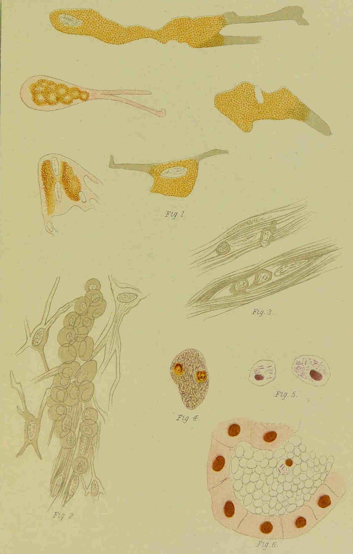
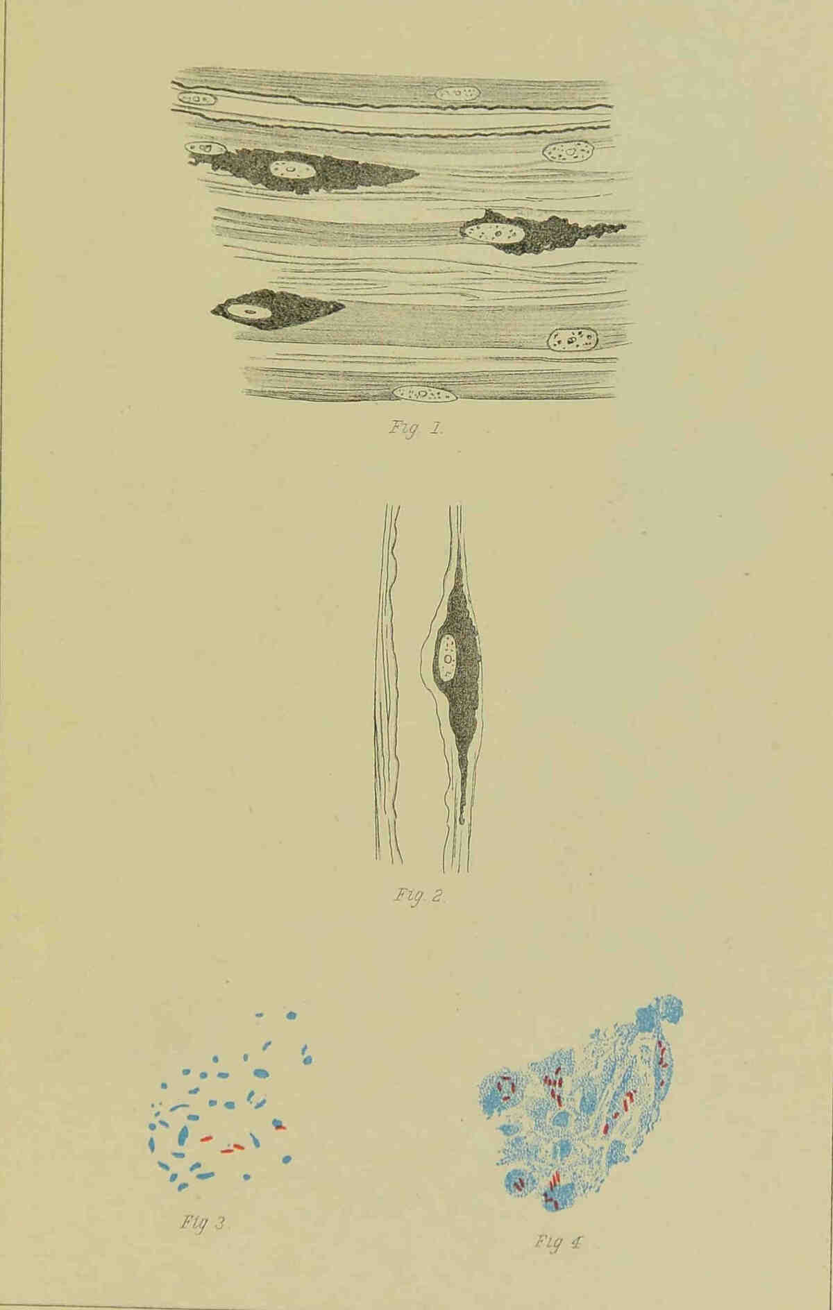
filled with granules (Plate XII, Fig. 2), and one
finds nerve fibres without a myelin sheath and with
a more or less atrophic axial cylinder (Plate
XII, Fig. 1). These drawings clearly explain
how the pressure on the axial cylinder at first
causes pain, and later, when atrophy has set in,
anæsthesia. And one can also understand that
when the leprous affection passes off without complications,
the axial cylinders are again restored
and become functionally active.
But on those places above referred to, where
the nerves run superficially over bones or joints,
and are exposed to pressure and stretching, secondary
inflammation is added to the process.
While the primary leprous affection hardly
appreciably thickens the nerves, the secondary
inflammation causes a very marked thickening.
The ulnar nerve at the elbow may attain a
diameter of 7 to 8 mm. or more, and when the
secondary inflammation disappears the connective
tissue contracts, and the previously thickened
nerve becomes thinner than normal. This whole
process usually advances very deliberately, and
years are required before anæsthesia is developed
to its full extent. While the section
of the thickened nerve is quite smooth and of a
pale brown colour from the numerous globi present,
the section of the atrophic nerve, though also
smooth, is as pale as the section of a tendon. It
consists almost exclusively of connective tissue;
every trace of bacilli has disappeared, and one
sees hardly a suggestion of nerve fibres. The
leprous affection is healed, but only a completely
functionless rudiment of the nerve remains.[4]
Chapter IV. LEPRA MACULO-ANÆSTHETICA.
This form of leprosy was first distinctly and
well described by Danielssen, who called it L.
anæsthetica; but since the macules, as Danielssen
recognised, play an important and constant rôle in
the course of the disease, we prefer the name
maculo-anæsthetica, as it includes the two most
striking symptoms; the name L. nervorum used
by many investigators, we do not consider satisfactory.
Certainly the nerves suffer most, and the
neuritis is the most prominent feature in the
disease, but the skin affection is a bacillary one,
which precedes or accompanies the neuritis;
it is not, as is often believed, a tropho-neurosis,
as we have determined by the demonstration
of bacilli in both young and very old leprous
patches.
Thus there disappears the sharp distinction between
the two forms of the disease—the tuberous
and the maculo-anæsthetic. We must regard them
as the same disease, only with varied intensity in
the action of the bacilli.
One can distinguish in the maculo-anæsthetic
form, different stages in the course of the disease,
but in general they cannot be very sharply defined
from one another.
In the prodromal stage, which is of very varied
duration, lasting for months or even years, the
patients state that they suffer from exhaustion,
general debility, rheumatoid pains of the joints or
muscles, hyperæsthesia of the skin, neuralgic pain
of certain nerve regions, sopor and mental depression.
Ephemeral eruptions of spots are
admitted; and pigmentary anomalies, sometimes
atrophic, sometimes hypertrophic, were noted by
Bidenkap.
Danielssen states that he has seen, at the very
beginning of the disease, a slight vaso-motor
disturbance, which is indicated by a bluish-red
reticular appearance, which is evident most clearly
on changes of temperature. These vaso-motor
disturbances, which appear as slight patches which
can be induced by friction, are chiefly characteristic
of the maculo-anæsthetic, though they
may appear during the earliest stage of the
tuberous, form.
In fact, our view is that the so-called prodromal
symptoms are nothing more than the
earliest, indefinite, undiagnosable phenomena of
infection.
One or more eruptions of pemphigoid bullæ
may occur in the commencing stage, but we have
more often seen them later, both accompanying the
patches and in the stage of anæsthesia and mutilation.
After a longer or shorter period the typical
picture of the maculo-anæsthetic form develops;
the spots either appear stealthily or they may
appear all at once with marked fever. Several
forms of the eruption are described by investigators;
in our patients usually only the erythematous
and the yellowish or brownish pigmented
ones have been noted. Usually both forms are
seen on the same patient, for the simple erythematous
spots become in time more and more
pigmented—usually only at the periphery, where
a bluish-red play of colours is often seen. Those
eruptions which are all along pigmented and which
develop very gradually, we have particularly noted
in the intercostal spaces. Various forms—round,
oval, gyrate—have been observed. The patches
may be perfectly flat, or slightly elevated. The
size varies from that of a pea up to that of the
palm of the hand, and they may be even larger.
At the commencement, we have usually found
these patches hyperæsthetic; anæsthesia is only
found in the older patches. They do not always
at once attain their full size; we can often observe
their growth; one may run into another, and then
the initial form is lost. The number and extent
of these patches are very varied; some patients
present great map-like eruptions on the face, back
and extremities; in others the patches are few and
scattered. The seat of these patches corresponds
in general to that of the nodules, but the back and
the intercostal spaces are frequently the seat of
patches, while on these areas nodules are only
rarely, if ever, present. Plate IV gives a good
picture of the patches in the maculo-anæsthetic
form—duration, two years. The white centres
and the slightly elevated reddish edges are very
well shown. A symmetrical distribution of
the patches strikes one, and has been regarded
as indicative of a central localisation of
the leprous poisons of which the patches are a
tropho-neurotic vaso-motor symptom; but in
many cases there is absolutely no symmetry, and
the discovery of bacilli in the patches themselves
proves them to be the direct result of the action
of the lepra poison. The lymphatic glands corresponding
to the position of the patches are always
swollen, and the swelling may persist long after
the disappearance of the patches. The duration
is very varied; some are gone in a few days or even
less, others may last for years. Pigmentation of
the periphery and pallor of the centre indicate
that the patch is already old, and the pallid centre
is always anæsthetic, the anæsthesia affecting all,
or only some perceptions. The signs which Hillis
indicated, as diagnostic of the patches of the
anæsthetic from those of the tuberous form, we
cannot recognise. The patches of the tuberous
form are certainly usually thicker, indicating a
greater degree of infiltration, than the anæsthetic;
but as both are caused by the lepra bacillus it is
evident that they may be absolutely similar.
PLATE IV.
The longer the disease lasts the more does the
neuritis predominate; the ulnar and peroneal
trunks may be felt to be thickened, they are sensitive
to pressure, and, if carefully felt for, the
peripheral finer branches may be felt as delicate
thickened cords. In one case we were able to feel
the cutaneous nerve branches in a patch, growing
daily more and more thickened, in contrast to the
large not especially affected nerve trunk. The
large palpable nerve trunks are not equally
thickened in their whole length, but the thickening
appears about the joints where the nerves run
across bone.
The symptoms of this neuritis are various; at
first neuralgia, and later, widespread anæsthesia,
with trophic disturbances, such as the formation of
pemphigoid bullæ; we have often seen hydrarthrus
and pains in the joints. Motor pareses and
paralyses are never absent, but they are not, as
Neisser argues, due to a leprous affection of the
muscles, but are a secondary neuritic symptom, as
we have discovered from anatomical examination
of the muscles. As the neuritis especially affects
the peripheral nerve trunks, we find the secondary
symptoms in the peripheral regions, usually only
in the extremities and on the face. We will now
more closely consider the various nervous symptoms.
Neuralgia is usually present in the extremities,
in the ulnar and peroneal regions. The anæsthesia
relates to the different qualities of sensation,
and is not only present in the patches, but progresses
gradually from the periphery toward
the centre, so that at last the whole extremity,
and often also parts of the trunk, become anæsthetic;
the face is always more or less anæsthetic.
We have often found thermal anæsthesia present
alone, or accompanied by anæsthesia or analgesia.
The anæsthesia may become more and more
extensive, or it may very gradually disappear,
indicating that the neuritis of the affected nerve
has disappeared.
Trophic and vaso-motor disturbances are never
absent if the neuritis is pronounced and lasts for
any time; the skin may become œdematous, or it
becomes thin, shiny, and slightly scaly (glossy
skin). We have often seen, especially if the
neuritis has lasted long, and the later symptoms
such as mutilation have commenced, dark-coloured
hyperkeratoses, usually symmetrical on the front
of the leg, or on the dorsal surface of the hands.
In one patient we observed on the toes horny,
thick (1.5 cm.) symmetrically placed formations,
which when thrown off left a new-formed rosy-red
skin, with intact sensibility; the patient had on
the front of the leg the appearances of ichthyosis.
The changes in the nails are a part of the
trophic disturbance; they become thickened, brittle
and cleft, and sometimes thin and diminished in
size, as one finds them, as we shall see later, in
the terminal stages. The secretion of sweat is
affected, being diminished over the anæsthetic
areas, and the hairs there are altered and fall out.
We regard the pemphigoid bullæ as a trophic
symptom; they may appear at the very commencement
of the disease, along with the macular eruption;
but they usually appear late when the anæsthesia
has become extensive; in this we agree with
Danielssen. Neisser and Leloir give prominence
to the early appearance of pemphigoid bullæ,
Neisser believing that the irritation of the commencing
interstitial neuritis causes this trophic
symptom. Our reason for regarding them as
trophic symptoms is that we have never been able
to find bacilli in the bullæ we have examined, not
even in those which appeared at the same time as
the maculæ, and their marked symmetry is also in
favour of their nervous origin. The bullous
eruptions usually appear suddenly. The patients
will discover on awaking, one or more blisters
which may be already burst; some have pain and
fever for hours or days before their appearance;
(Leloir). They vary in size—they may be
small, from the size of a pea to that of a bean,
or as large as the palm of the hand.
Their contents are serous, but if the bullæ
persist, they become purulent. They usually rupture
early and heal, leaving behind them violet-coloured
scars, which after some time become pale.
If irritant factors are added, if the bullæ last long
and become purulent, there develops after rupture
deep ulceration, most frequent on the hands and
feet.
Bullous eruptions of the mucous membranes,
which Leloir has noted, we have never been
fortunate enough to see.
We regard also as tropho-neurotic vaso-motor
symptoms the acute rheumatoid affections of the
joints, which are not infrequent in our hospital.
The joints, especially the small ones of the fingers
and hands, and also those of the knee and ankle,
become painful and tender, and on palpation, a collection
of fluid can usually be recognised. The
affection is always symmetrical.
These affections of the joints, which belong to
the earlier stages of the disease, usually appear
simultaneously with the macular eruption, and
disappear with it, but they may appear later; and
after one, or it may be only after several attacks,
thickening of the ends of the bones and ligaments,
with stiffening of the limbs, is developed. Leprous
affections of the tendon sheaths, which Wolff
refers to, we have never seen. The contracture of
the fingers and toes is not tendogenous, so far as
we can decide from clinical and anatomical investigation;
it is myogenous, the leprous paralyses,
which we shall immediately describe, being the
cause.
Hillis claims to have recognised a motor
weakness in the prodromal stage of the disease,
and considers that the nerves are already
affected with the leprous virus. Such a weakness
referable to a neuritis of the motor
nerves we have not noted previous to the
macular eruption and the onset of the anæsthesia.
If there are, in the prodromal stage, muscular weaknesses,
we are inclined to regard these as symptoms
of the general weakness; according to our view,
the skin affection is the first definite symptom of
the disease. The muscular affection may always
be definitely recognised as a secondary symptom
by the atrophy, and the altered electrical reactions.
Different degrees and varied extent of the muscular
affection may be noted; trivial paresis with only
very slight atrophy, which, along with the anæsthesia,
may completely disappear, if the neuritis
passes off without destroying the nerve fibres;
and almost complete paralysis with great atrophy
of the muscles.
Paralysis with atrophy is most marked on the
hands and forearms, feet and calves, and on the
face.
PLATE V.
The interosseous muscles, of the hands and feet
atrophy, so that the interosseous spaces appear as
furrows. The thenar and hypothenar muscles
also disappear, and the first interosseous space
becomes a depression. The muscles of the forearms
and calves also atrophy and lose their power,
but complete paralysis is never developed; the
patients can always move their hands and walk.
In the face, all the muscles atrophy and lose their
power, and thus all expression is lost. The
masseter muscles occasionally, though very rarely,
become so atrophic, that the lower jaw drops, and
must be retained in position by a bandage.
Paralysis of the Orbicularis Oris leads to dropping
of the under lip, and to extreme difficulty in closing
the mouth, and consequent escape of saliva. (See
Plate V—a case of maculo-anæsthetic form completely
cured and anæsthetic. The maculæ were
developed in 1857, and the patient is still alive.)
Paralysis of the Orbicularis Palpebrarum renders
the closure of the eyes impossible, and paralytic ectropion
of the lower lid is developed. The results
of this on the eye we will refer to later. The
muscular atrophy is by no means always symmetrical;
one hand may present marked atrophy,
while the other is tolerably useful. This indicates
the peripheral development of the paralysis, as
does the fact that the muscular sense is preserved,
and in particular that no ataxic symptoms appear.
The patients can do fine knitting and needle-work
with their paretic hands. Their gait has a peculiar
character, from the difficulty with which the
weakened muscles raise the feet, but they can both
stand and walk quite well with closed or bandaged
eyes. As we shall see later, this clinical observation
corresponds with the results of the anatomical
examination of the cord.
Along with the paralysis and atrophy of the
muscles, there appear also, as the result of the
destruction of the nerve trunks, trophic affections
of the bones and of the skin. The changes in
the skin have been already described; when
the anæsthesia is advanced, there always appear
ulcerations under the heel and the ball of the
foot. Most of these owe their origin simply to
pressure; the weight of the body is too much for
the atrophic skin. They are always callous,
heal with great difficulty, and so long as the
patient goes about, not at all. The ulcers are not
usually deep, but it may happen that they penetrate
as far as the bone, and lead to necrosis. But
usually necrosis of the bones is preceded by periostitis,
and when the pus makes its way out, an
ulceration leading down to necrotic bone is formed.
We have sometimes seen the formation of a blister
precede the ulceration, but it is rare, and it is certainly
not true, as stated by many authors, that ulceration
of the sole is always preceded by a bulla.
Not only on the feet but also on the hands,
necrosis with exfoliation of bone takes place;
the phalanges are especially attacked. When
the atrophy of the muscles is advanced, the fingers
are always claw-like, with extension of the first
phalanx. The joints consequently often appear
swollen, although no joint affection is present. The
phalanges and the metacarpals undergo simple
atrophy, becoming very thin at the middle, and
since the joints retain almost their normal thickness,
they appear swollen by contrast. As already
noted, this clawing of the fingers is accompanied
by no disease of the tendon sheaths. The bones
in the feet undergo the same form of atrophy.
If, in addition to this atrophy of the bones,
which was first noted and demonstrated by Prof.
Hjalmar Heiberg, there ensues necrosis with
exfoliation of whole phalanges or metacarpals (the
carpal bones very rarely necrose), there is great
mutilation of the hands and feet; all the fingers of
the hand may go, and there remain on the
diminished carpus only small, soft processes, each
supplied with a nail—the remnants of the fingers.
The toes disappear from the feet, the metatarsal
bones atrophy, and several of the tarsal bones may
exfoliate; so that ultimately there remains of the
foot only a pyramidal cushion at the lower end of
the leg. In most cases we must regard external
injury as the cause of these necroses. The
patients feel nothing when they injure themselves;
they may burn their hands at a stove without
noticing it. Under such circumstances it is
not to be wondered at, that inflammation is readily
excited, especially as the vitality of the parts
evidently diminished. But it is remarkable how
well operation wounds heal. One may do pretty
extensive necrotomies, and the wounds heal well
and quickly, either by granulation or by first
intention. In such operations it is a frequent
experience that the bone is reached before the
patient feels anything, but he immediately feels
pain when the periosteum is scraped or the bone
attacked with forceps or saw. We believe, however,
that it is only nervous individuals who complain
of pain; though it is certain that when the
bone is meddled with, something is felt. Probably
in this connection may be explained the statement
of the patients, that when walking they feel the
ground. It is easy to demonstrate that a patient
who is quite unconscious of any irritation on his
skin, can perceive deep pressure fairly well.
These necroses and trophic disturbances, together
with the muscular contraction, cause the
mutilation characteristic of the last stage of the
disease, which was at one time described as a
special form, Lepra mutilans.
We said above that the finger joints appear
enlarged on account of the atrophy of the shaft of
the bone. In some cases, however, the joints,
especially the ankle, show changes which must
either be regarded as the remains of the rheumatoid
affections of the eruptive period, or as trophic
articular changes, corresponding to those seen in
Tabes dorsalis. We have seen ankles and knees,
but especially ankles, presenting such an appearance.
In some cases post mortem examination
shows widespread tuberculosis of the synovial
membrane and of the ends of the bones, which
we shall refer to more particularly under the
pathological anatomy.
As we have already mentioned, the eyelids can
no longer be closed on account of the paralysis of
the orbicularis palpebrarum, and consequently the
under part of the cornea remains uncovered during
sleep. This leads to a punctiform drying of the
epithelium of the cornea, and further, to an injection
of the conjunctivæ at the under margin of the
cornea; then the vessels gradually attack the
cornea, which becomes opaque, at first around
the xerotic spots, and later in its whole under
part. It may go on to ulceration with rupture
of the cornea and prolapse of the iris, and
finally to complete atrophy of the globe. As a
result of the paralysis, the lower lid is always
ectropic, at first at its inner end, and later, completely.
As the lower punctum is thus drawn
away from the bulb, the tears run down over the
cheeks, and the paralysed countenance looks still
more woe-begone.
In the later stages, when the facial paralysis is
very pronounced, the senses of smell and taste may
be very much diminished, or completely lost.
We often see symptoms which are not proper to
the disease itself developing during its course,
such as obstinate cardialgia, acid pyrosis, and
vomiting of a slimy nature indicating gastric
catarrh. Diarrhœa or chronic obstruction is by no
means rare, nor is albuminuria dependant on
parenchymatous, interstitial or amyloid nephritis.
The course of maculo-anæsthetic leprosy is
essentially chronic. Cases usually last between
ten and twenty years; some may even exceed
forty.
The patients often die cachectic, without one
being able to find on the post mortem table any
definite cause of death, or they may—though in
our aseptic and antiseptic times more and more
rarely—perish from septicæmia or pyæmia. Pulmonary
or general tuberculosis was formerly a
frequent cause of death, which, however, usually
takes place from some intercurrent disease.
In the ordinary course of the disease the
macular eruptions disappear, and the neuritic
symptoms—anæsthesia, muscular paralysis and
atrophy, and necrosis of bone—appear. Sometimes,
though rarely, there are several macular
eruptions after the disappearance of the earlier
maculæ, or there may be an eruption of nodules.
If, then, these eruptions are auto-infections, they
are evidence in favour of the unity of the disease,
in spite of the difference in form.
Where the bacilli come from, in these, so to
speak, later eruptions, when there is no skin
affection, it is difficult to say with certainty.
In a case of maculo-anæsthetic leprosy we
found the inguinal glands leprous, although no
skin eruption was present. Possibly, then, fresh
eruptions develop from the glands when the
original skin eruption has disappeared. Thus
the specific leprous affections gradually disappear,
and only their results remain—in other
words, the leprosy is healed. Most maculo-anæsthetic
patients become in time purely anæsthetic;
they no longer suffer from leprosy, but only from its
results. The late eruptions show, however, how
difficult it is to define the fact of recovery, for
when all externally diagnosable signs of actual
leprosy are gone, bacilli may still remain somewhere
in the body, in the lymphatics, or possibly
in the liver and spleen.
Chapter V. PATHOLOGICAL ANATOMY OF THE MACULO-ANÆSTHETIC FORM.
The maculæ exhibit generally the same
anatomical characters as do the nodular
lepromata, viz., infiltrations with round cells,
epithelioid and spindle cells. The difference
between them is quantitative; in the macular
infiltration the number of bacilli are less. We
will describe the microscopical appearance of
macules of different duration.
In a recent (three weeks old) macule, microscopical
examination showed cord-like and rounded
infiltrations of the cutis, with round and epithelioid
cells, mostly arranged around the vessels. There
were pretty numerous bacilli lying between and
also definitely in, the cells; here and there were
found little collections of bacilli or bacillary
granules, with one or more nuclei—small globi—in
their neighbourhood, which in size could
not be compared with those found in the tuberous
form. Specimens hardened in perosmic acid
showed unaffected nerve fibres, which the leprous
infiltrations did not implicate. Gold preparations
showed a marked increase in the number
of Langerhans’ cells in the epidermis, which,
indeed, we expected, since we agree with Ranvier
in considering them as wandering cells, and not as
nervous elements. Definite changes in the cutaneous
nerves were not evident.
Microscopical sections from an older spot
(perhaps two years old) show here and there
infiltrations in the cutis—especially around the
vessels, consisting of epithelioid, round and spindle
cells. Bacilli were found here and there—one or
two in each infiltration. In the lumen of one small
vessel we found a collection of round cells, with
a bacillus among them; and on the inner side of
the wall of another vessel we saw a similar
appearance. Gold preparations of this macule,
which was definitely anæsthetic, showed slight
changes in the small nerve twigs in the cutis, less
marked precipitation of the gold salt in the nerve
fibres, and a number of Langerhans’ cells.
Sections from a very old anæsthetic macule
showed only very slight infiltration along the vessels;
the cells were mostly spindle-shaped; only a
few were round or epithelioid. In most of the
sections no bacilli were found; in some, one or
two distinct bacilli and some granules, taking the
same stain.
The maculæ are therefore like the nodules,
leprous infiltrations of the cutis, consisting of
round epithelioid and spindle cells, the latter being
more numerous the greater the age of the macule.
These infiltrations appear to proceed from the
vessels. Lepra bacilli are always present, but
are most numerous in the younger macules. In
young, not as yet anæsthetic, macules, the nerve
twigs appear unchanged; in the older ones they
are usually affected. Langerhans’ cells are, according
to our view, wandering cells, and their
number is probably dependant on the inflammation.
We have only had the opportunity of examining
the nerves in this form of the disease in old cases,
and have not found bacilli in them, but merely
great increase of the interstitial connective tissue
and disappearance of the nerve fibres. Dr. Arning
has, however, as is well known, found bacilli in a
piece of ulnar nerve which he removed during
life from an anæsthetic patient. Our results,
therefore, correspond with the proposition put
forward above, that the leprous affection disappears,
and that the anæsthesia is a result of the
atrophy of the nerves caused by secondary shrinking.
We found on a mutilated finger the collateral
nerve almost completely transformed into fibrous
tissue, not a single medullary fibre being evident.
In a case where on account of joint disease (which
proved to be tubercular) we amputated, the small
periosteal nerve twigs were much degenerated;
in another, where the foot was amputated for
gangrene, the small peri-articular nerves also
showed degeneration.
Danielssen found, in some cases, changes in the
spinal cord which, using the methods of investigation
then at his disposal, he regarded as degenerations
and inflammations of leprous origin.
Armauer Hansen, Neisser and Leloir have
not been able to find any leprous affection of the
cord. The cases with changes in the cord, described
by Langerhans and Steudener, were, in
our view, not leprosy. Tschiriew’s case, Lepra
tubero-anæsthetica, presented no marked changes.
Looft has found, in two cases of maculo-anæsthetic
leprosy, degeneration of the posterior
columns, atrophy of the posterior roots and
fibrous degeneration of the spinal ganglia, with
disappearance of the medullary fibres, and changes
in the nerve cells. In these two cases the affection
appeared to be primary in the ganglia, and
secondary in the cord. Lepra bacilli were not
found in either case, but Chariotti found them
once in the cord, and Suderkowitsch in the
spinal ganglia.
From all this we can only conclude that the
cord is affected in some cases, in others not;
definite clinical symptoms are absent, and where
they suggest a central cause, they may be equally
well ascribed to a peripheral neuritis.
The lymphatic glands related to the affected
skin are swollen, but not nearly so much so as
in nodular leprosy. After death one usually finds
the glands but little swollen, and their appearance
presents nothing characteristic. In only one case
of maculo-anæsthetic leprosy have we found the
inguinal glands distinctly leprous, and that two
years after the disappearance of the macules. This
indicates in the first place that the affection of the
glands is due to the same cause in this as in the
nodular form, and secondly, that the leprous
affection of the glands may last longer than that
of the skin. In the same case we found indefinite
traces of leprous affection of the liver and spleen,
unfortunately so indefinite that we cannot say
with certainty whether the liver and spleen are
affected with leprosy in this form of the disease.
The case occurred in the pre-bacillary era.
The muscular affections, paralysis and atrophy,
play a prominent rôle in the maculo-anæsthetic
form of the disease, and the anatomical examination
of the muscles is of great interest since
some (Neisser)[5] regard it as a specific leprous
process, while G. and E. Hoggan[6] had previously
described it as secondary, and due to the
neuritis. In our[7] examination of various stages of
muscular atrophy, we have found that the changes
begin with a multiplication of the nuclei of the
Perimysium intern., which becomes thicker and
thicker; at the same time the muscle fibres
become thinner, they retain their transverse
striation, and some break up into discs. The
greater the thickening of the perimysium the
thinner become the muscle fibres, so that one
must regard the process as an atrophy due to
pressure. The intra-muscular nerves showed
interstitial neuritis.
Where the atrophy was very pronounced, as in
the small muscles of the hands and feet, the
muscular fibres had completely disappeared, and
only fat and connective tissue remained. We
found no bacilli, not even in sections of very early
stages of the process, where the larger nerve
branches, relating to the part, contained numerous
ones, either in so-called mixed or in true tuberous
cases. We must therefore with Hoggan regard
the muscular affection in leprosy as a secondary
one, caused by neuritis. We have tabulated at
the end of this work the results of thirty-six post-mortems
on maculo-anæsthetic lepers (see Table
II, page 138).
In those thirty-six cases we find simple meningitis
twice, tubercular meningitis once, solitary tubercle
in the cerebellum once, and hydrocephalus internus
twice. The protocol notes nothing further.
The spinal cord twice showed macroscopic
changes (thin and atrophic once, thickening and
hyperæmia of the lumbar cord once).
The most of these brains and cords, which are
entered as normal, were unfortunately only
examined macroscopically.
Two cords thoroughly examined by Weigert’s
method showed degeneration of the posterior
columns.
The peripheral nerves were probably examined
in all cases, but only the results of a few of the
examinations are noted.
The lungs were found tubercular thirteen times,
the intestine four times, once without the presence
of pulmonary tuberculosis being noted, this making
fourteen cases of tuberculosis out of thirty-six—almost
exactly 39 per cent. In the nodular
form we had thirty-six undoubted tubercular
cases out of eighty-nine—rather over 40 per
cent. Possibly, and indeed probably, some insufficiently
described cases in this form were
also tubercular, but the frequency of tuberculosis
is so nearly equal in both forms, that there is no
ground for supposing that the particular form of
leprosy has any relation to the development
of tuberculosis. Under precisely similar conditions,
in our institution, the nodular and
maculo-anæsthetic cases were pretty equally
attacked by tubercle. If Leloir means to say that
tuberculosis is much rarer in the maculo-anæsthetic
form, than in the nodular, it is certainly
not the case in our leper hospitals.
So far as concerns the necrosis of the bones, the
panaritii and periostitides, we have found in
them nothing specific; we have often sought for
bacilli, but always in vain. Pyogenic cocci,
usually the staphylococcus aureus, we have found
both microscopically and in cultivations.
The mutilation of the bones may occur from
concentric atrophy alone, as demonstrated by
Prof. Hjalmar Heiberg (Klinisk arbog, III).
The phalanges of the hands and feet, the bones of
the metacarpus and metatarsus, and even the
carpal and tarsal bones, diminish in all their
dimensions, evidently from trophic changes, the
result of the neuritis.
We have been able to examine anatomically a
few of the cases of articular affection referred to
above, but we have not seen instances of the
acute affections on the post mortem table. R.
Thoma has described clinically and anatomically
a case of lepra tuberosa (Virchow’s Archiv, Bd. 57),
in which first the elbow, and later the knee joint,
was affected. Examination of the knee joint
showed an inflammatory irritation of the nature of
hæmorrhagic gonitis. Where we have noted joint
affections in the maculo-anæsthetic form we have
only noted a condition of hydrops. The chronic
affections we may indicate as tubercular and
tropho-anatomical. Tuberculosis attacks the
joints of lepers by no means infrequently, as is
easily explainable in the case of those already
tubercular, by the frequent traumata to which the
anæsthetic and paretic lepers are exposed. The
carpal and tarsal joints are most frequently affected;
but we have also seen the knee and elbow tubercular.
The process is exactly the same, as in
those who are not lepers. Chronic trophic
joint affections are by no means so rare as
was formerly believed. Prof. H. Heiberg has
(loc. cit.) drawn attention to this, and has described
a foot which resembles that described by Charcot
as pied tabétique. According to Heiberg a
characteristic of these leprous tropho-neurotic joint
affections is swelling and laxness of the capsule of
the joint, a wearing away and atrophy of the ends
of the bones, or periostitis ossificans, and hypertrophy
of the ends of the bones, which is especially
seen in the tarsal and metatarsal joints. We have
been able to confirm these results of Heiberg’s,
and have also seen in such an ankle, growth of
the synovial membrane with villous projections;
the capsule of the ankle joint was loose and lax,
the talus smooth and oblique, the cartilage worn
away; and marked outward subluxation of the
foot was present. Other older spirit preparations
showed changes in the joint capsule, which was
flabby and loose or thickened, and further, slight
irregular thickening of the synovial membrane,
and thickening of the ends of the bones.
We have also in some cases examined the
tendon sheaths; but never, even when contraction
had existed for a long time did we find
any anatomical changes, which pointed to a
tendo-vaginitis. Leloir and Wolff have noted this
clinically, but we have been unable to confirm their
observation.
The pemphigoid bullæ we have repeatedly
examined, and never found lepra bacilli in their
contents; both microscopically and on culture we
have found only pyogenic cocci, usually the
staphylococcus aureus. Müller found (according
to Neisser, Virchow’s Archiv, Bd. 103) lepra bacilli
in the contents of a pemphigoid bulla. This was
probably a mere accident. For other morbid
changes found in maculo-anæsthetic leprosy the
reader is referred to Table I, page 128.
Maculo-anæsthetic leprosy is therefore characterized
by an infection of the skin and nerves directly
caused by the lepra bacillus, and by secondary
tropho-neurotic affections of the muscles, bones, and
joints, the skin and organa sensus.
As is clear from the description of the two
forms, the leprosy bacillus is found in the
leprous products in both, though in much greater
quantity in the nodular than in the maculo-anæsthetic
form. The course of the two differs;
in that in the nodular form eruptions constantly
recur, and thus the affected areas
are much more numerous than in the maculo-anæsthetic.
Whether the liver, spleen, and
testicle are attacked in the latter form we cannot
as yet say with certainty. In one case of maculo-anæsthetic
leprosy, we noted an affection of the
throat exactly corresponding to that constantly
present in the nodular form, and in another we
found the inguinal glands affected with leprosy in
a manner similar to that in the nodular form. In
the maculo-anæsthetic form eruptions are also
present, but they are by no means so frequent or
so severe as in the other. Both forms may
recover. As we have frequently noted in the
description of nodular leprosy, the bacilli in the
leprous products break up into granules which
finally disappear, and there remains of the
leprous products only a scar in which nothing
leprous can be recognised. Occasionally this
takes place in all the affected parts, and there
remains only a widespread anæsthesia, the result
of the nerve affections; and in the maculo-anæsthetic
form this is the regular termination of the
disease. In both cases the leprosy is completely
healed.
We are thus enabled to see how complete is the
parallel between the two forms. The course of the
disease, especially the eruption, gives to it evidently
the character of an infectious disease. In the
nodular form, where the bacilli are present in
millions or milliards, the eruptions or auto-infections
are frequent; in the maculo-anæsthetic
form, where their number is comparatively insignificant
the eruptions are rare.
Does this difference between the two forms
depend on a difference in the virulence of the
bacilli? This readily suggests itself. But if
so, this virulence is capable of very rapid
changes. We have seen a case of maculo-anæsthetic
leprosy, which probably arose by
inoculation from a very severe case of nodular
leprosy, since the patient some years before the
outbreak of the disease had for a long time shared
the bed of a nodular leper. In this case the
virulence of the bacilli must have been at once
diminished on their inoculation on another
organism. And since it also happens that a
maculo-anæsthetic case may on a fresh eruption
become nodular, the bacilli must be able by
cultivation in the organism to re-acquire their
power. Both are possible, but the virulence
of the bacilli seems to depend, not so much on
any constant character of their own, as on the soil
in which they live.
Now it is a remarkable fact that in certain
regions in Norway the nodular form predominates;
in others the maculo-anæsthetic does not indeed
predominate, but is present almost as frequently
as the nodular. The maculo-anæsthetic cases are
more numerous in the eastern districts, where the
climate is dry; the nodular in the western, along
the coasts where the climate is moist. And in
this western division there is a region where the
climate is not nearly so moist as in the division
generally, and here the proportion of maculo-anæsthetic
cases is distinctly higher, as may be
seen by referring to Table III, page 144.
Sogn lies in Nordre Bergenhus, and is an
inland fjord with a rather dry climate. Söndfjord
and Nordfjord lie nearer the coast, and have,
especially the former, very damp climates.
We have already noted that the leprous nodes
are most frequently found on the exposed parts of
the skin, and it is quite possible that the form is
determined by climatic influences.
As the Table shows, males are more affected
than the opposite sex, and this too may depend on
climatic influences.
It is also possible that the bacilli always possess
the same virulence, and that it is solely dependant
on the soil in which they live, whether they
multiply freely or no. But it is impossible to say
anything definite on this, so long as we are unable
to cultivate the bacillus, and so long as we can
only refer to the conditions in Norway; and
nowhere else have we such definite statistics of
the disease and its form as to justify us in drawing
any conclusions.
We must, therefore, for the present leave in
suspense this most important and interesting
question of the virulence of the Lepra bacillus,
since we possess no experimental proof of any
attenuation.
Chapter VI. DIAGNOSIS AND PROGNOSIS.
DIAGNOSIS.—In view of the description
which we have given of the two forms of
the disease, the diagnosis is usually accompanied
by little difficulty. We have, however, noted
above how a case of tubercular Leprosy,
with subcutaneous nodules only, was overlooked
by a doctor well acquainted with the
disease; and we have occasionally seen, in the
country, people described by the doctors as leprous
who were not so. And, on the other hand, we
ourselves were once in doubt as to the diagnosis
in a tuberous case, since all the nodules were
exceptionally small, and presented no characteristic
appearance. But the doubt was readily
dispelled by the excision of a nodule and the
recognition of the bacillus. In the diagnosis of
the maculo-anæsthetic cases, one is more frequently
in doubt, since the maculæ have not
always a characteristic appearance; sometimes
they closely resemble psoriasis, and in such cases
the excision and microscopical examination of a
portion might clear away doubt. This we have
never needed to do, since the swelling of the
lymphatic glands, or a thorough investigation of
the sense of touch, have always been sufficient to
establish a diagnosis. Even in comparatively,
recent cases there may almost always be detected
some loss of sensation in the fingers and
toes; sometimes it is first evident on the wrists
or back of the feet. For this investigation one
must either use callipers, or very slight stroking,
since deeper pressure can be at once perceived.
As a rule, the maculæ themselves are somewhat
anæsthetic. We recollect once seeing a syphilitic
eruption exactly resembling the leprous maculæ,
but here the history cleared up the diagnosis.
Maculo-anæsthetic Leprosy may in its later
stages be confounded with syringo-myelia, as
Charcot has already noted in giving the points of
distinction between the two conditions. If the
maculæ are no longer present, careful investigation
will often enable one to recognise their
previous presence by finding areas of skin, especially
on the upper arms, the back, the thighs and
calves, which are somewhat paler than the surrounding
skin, and in which sensation is somewhat
blunted. We have thus frequently recognised the
previous presence of maculæ in patients who
themselves knew nothing of them. Zambaco
Pasha has stated that certainly many of the cases
described in France as syringo-myelia and
Morvan’s disease are cases of Leprosy; that
Leprosy in this form still exists in Brittany; and
further, that he has there found some cases of
nodular Leprosy. It is remarkable that in these
last cases, where the proof would have been so
easy, he has not demonstrated the Lepra bacillus.
From the drawings which he gives in the Annales
de Dermatol. et de Syphil. (T. III., Nr. 12), some
of the cases can scarcely be regarded as Leprosy,
since on the hands with mutilated fingers, no
muscular atrophy can be noted; but in others
there is distinct atrophy, and these may very well
be leprous, the more as Pitres has published in
the Gaz. des hôp. 1892, a case diagnosed as syringo-myelia
in which Lepra bacilli were demonstrated
in an excised portion of the ulnar nerve. It is
unfortunate that Zambaco Pasha did not demonstrate
the remains of previous maculæ, which
would probably have been possible in some cases,
were they really cases of Leprosy. According to
the rich experience of Dr. Danielssen and our
own, it must be admitted that a skin eruption is
never absent in true cases of Leprosy. With
multiple neuritis from some other cause, it is, with
a good history and careful examination, not possible
to confuse Leprosy; and the same is true of
progressive spinal muscular atrophy, where there
is no disturbance of sensation.
PROGNOSIS.—The prognosis is very different
in the two forms. As we have already
stated, both forms may recover, since all leprous
products may disappear without any fresh ones
appearing. In nodular cases this is a very rare
exception, while it is the rule in the maculo-anæsthetic.
Recurrent outbreaks are almost invariable
in nodular cases, and in them, too, nephritis is an
almost constant occurrence. Patients rarely live
more than eight or nine years after the definite
outbreak of the disease. As already remarked, we
cannot state that Leprosy of itself is responsible
for the end; we are rather inclined to regard the
nephritis and other complications as the direct
cause of death. The patients usually die long before
the disease has run its course. But in the
maculo-anæsthetic form the cure of the Leprosy is
almost invariably the result. What remains, however,
after the cure of the leprosy, is very different.
We have occasionally a complete subject with
vigour and good health, but usually only a miserable
rudiment of a human being, with more or less
paralysed and deformed hands and feet, with unclosable
eyes, of which the lower part of the cornea
is opaque, and from which the tears run down over
the cheeks, and with paralysed facial muscles unable
to close the mouth, so that the saliva constantly
dribbles from it. Such cases may, however,
live long and reach great ages, if under such circumstances
this can be looked upon as any advantage.
They die usually from some intercurrent disease.
Chapter VII. ETIOLOGY.
There is hardly anything on earth, or between
it and heaven, which has not been regarded
as the cause of Leprosy; and this is but natural
since the less one knows, the more actively does his
imagination work. And since all that was known
of Leprosy was that it was a loathsome disease,
search was made everywhere for a cause. We
will not linger over the older literature of Leprosy.
That may be found fully dealt with in Danielssen
and Boeck’s Traité de la Spedalskhed and in
Hirsch’s Geographical Pathology.
Only after the work of Danielssen and Boeck
can one say that Leprosy entered the ranks of the
scientifically investigated diseases. At that time,
in 1840, when they commenced their investigations,
Humoral Pathology held the field.
Most diseases were ascribed to changes in the
blood, and they therefore endeavoured to establish
that there was in lepers a change in the blood
which they regarded as the cause of the disease
symptoms, especially the node formation. These
changes in the blood they believed were caused
by unfavourable conditions of living, and as they
were not able to find any convincing evidence of
the power of infection of the disease but several
of its limitations to certain families, they drew the
conclusion that Lepra, as they called it, might
appear spontaneously, that is to say, that the sanguineous
dyscrasia which led to leprosy could be
developed under unfavourable conditions of life,
but that it was in most cases hereditary. It must,
however, be noted that Danielssen always regarded
Leprosy as a specific disease, described it as such,
and sought for a specific cause, and the fact that
he did not find it must be ascribed to the circumstance
that microscopical technique and microscopical
aids, especially the immersion lens, were
at that time either insufficiently developed, or not
yet discovered. The teaching of Danielssen and
Boeck was everywhere adopted, especially their
view of the heredity of the disease. The fish diet
and damp cold theories are only attempts to explain
the so-called spontaneous development of
the disease, and they are founded on the fact that
Leprosy is chiefly present in littoral districts and
on islands.
Of other Norwegian investigators, the late Dr.
Hjorth held the view that Leprosy could not be
ascribed to a specific cause, and that it was certainly
not hereditary. Dr. Holmsen regarded it
as a specific miasmatic and non-hereditary disease,
and finally Prof. Lochmann stated that it was
specific, contagious, and hereditary. While Danielssen
and Boeck always required a leprous ancestor
in order to recognise a case as hereditary, and
when this was not forthcoming, found in the
presence of the disease in other branches of the
family, proof of its heredity, Biedenkap, as he was
unable in many of his cases to determine the
existence of leprous ancestors, widened the
definition of heredity by assuming that unfavourable
conditions of life might produce in the
organism conditions which became hereditary, and
showed themselves in later generations as leprosy.
In the year 1869, Dr. Drognat-Landré published
a book with the title, De la contagion seule cause
de la propagation de la lépre, in which he sought
to prove that heredity had nothing to do with
the spread of Leprosy. That is, according to
our view, the right standpoint, as we shall
endeavour to demonstrate.
As is seen from the above short summary of
the views of Norwegian investigators, some maintain
the non-specific origin and heredity of the
disease; one only, the non-specific origin; one a
specific cause and no heredity; and finally, one a
specific cause, contagiousness and heredity.
It is rather remarkable that supporters both of
the specific and the non-specific origin of the
disease should regard it as hereditary. It apparently
struck none of them that possibly the
specificity of a disease, that is, its development
through the action of a poison, might be incompatible
with heredity. Since the discovery of the
Lepra bacillus and its recognition in all leprous
products, it is now everywhere admitted that it is
the cause of the disease, and it would therefore be
superfluous to indicate which of all the symptoms
of the disease point to its specific nature. All
this was shown in a communication made by
one of us to the Copenhagen Congress in 1884.
We start then from the assumption that Leprosy
is a disease caused by the Lepra bacillus, although
it is as yet not strictly scientifically proved, since
inoculation on man and animals has not been
definitely successful.
The question as to heredity now is, Can the
lepra bacillus be conveyed by heredity. This is
Baumgarten’s view; he holds that both Tuberculosis
and Leprosy are thus spread, that the bacilli
of both diseases may be transferred to the children
and there remain dormant, but that they can
thence be conveyed to another generation, and
from it to a fourth, fifth, etc. generation, and then
in the third, fourth, etc. generation become once
more active and cause the disease. We see at
once that this is only a modification of Biedenkap’s
heredity, a peculiarity of the organism which
finally becomes evident as Leprosy. In place of
his undiscovered peculiarity, we have undiscovered
latent bacilli. Baumgarten’s view is therefore only
a hypothesis for the explanation of the origin of
the two diseases. The hypothesis may be tested
from two points of view: first, we may sift it theoretically;
and secondly, we may investigate whether
it can explain the distribution of the disease.
We must first endeavour to make clear what
heredity is. As a matter of fact we do not know;
we are familiar only with a series of phenomena
which we call heredity, just as we have a series of
phenomena which we ascribe to the force of
gravity, without knowing what gravity or its force
actually is. If we consider only the phenomena
of heredity, which are nowhere so completely
and clearly put together as in Darwin’s works,
we find that he presents so-called laws of heredity,
that is, he has noted the most frequent phenomena
of heredity, and thence deduced a law. If the
phenomena in the conveyance of a disease to
one’s descendants are to be called heredity, they
must correspond with the rest of the phenomena
of heredity. And looked at thus, we know of
no specific disease which can be called hereditary.
The conditions which are hereditary are all anatomical
and physiological peculiarities of the
organism. A bacillus which is living in the
organism cannot be regarded as one of its
anatomical or physiological peculiarities; it is a
parasite. Now it is beyond doubt that parasites
may be conveyed from parents to children. Such
are the Achorion Schönleini, and the Acarus
Scabiei. But this is no hereditary communication,
it is simply a present from the parent to his child.
But, it is objected, if the parasite is conveyed
through the organs of generation during copulation,
then it is no longer a present or an
inoculation, it becomes hereditary. This appears
to us a remarkable argument, that conveyance by
means of the organs of generation can change the
nature of the parasite and convert it into an
anatomical constituent of the organism of parent
or child. A parasite remains always a parasite,
and since its conveyance from one adult to another
is called infection, we cannot comprehend why
its conveyance to an ovum or a fœtus should be
indicated by another name. Were Baumgarten’s
hypothesis correct, it should certainly be called a
hypothesis of latent infection, and not a hypothesis
of heredity. We said above that we knew of no
specific disease which was hereditary. To this it
will naturally be objected that syphilis is a
typically hereditary disease. But we reply that
syphilis is a disease communicable to children,
but that it is not hereditary, and in order to
maintain this statement we only need to place
in parallel the phenomena of the conveyance
of hereditary peculiarities and those of the
communication of syphilis to children, in order
to make it perfectly clear how different the two
methods are.
Conveyance of Hereditary Peculiarities.
Conveyance of Syphilis to Children.
1.—The hereditary peculiarity is usually limited to one sex, so that the male communicates his peculiarities to his male descendants, and the female hers to the female.
Not the case in syphilitic children.
2.—Very often, possibly always, the appearance of the hereditary peculiarity is limited to a definite age.
In the communication of syphilis to children this is unknown.
3.—Atavismus, or the overleaping of one or several generations of the heredity is very frequent.
Never known.
Anyone who will maintain that these two forms
of conveyance are the same, and both hereditary,
must evidently sacrifice facts.
We hold then, that a specific disease may be
conveyed to the children, but that it is never
hereditary; the communication is by infection.
And since we regard leprosy as a specific
disease we hold that it cannot be hereditary.
There remains, however, the further question
whether it can be conveyed to descendants by
latent germinative infection. This cannot a priori
be denied, although we have no right to assume
it from our knowledge of the lepra bacillus and of
the occurrence of the disease. We have indeed
sought above to point out the possibility that
the lepra bacillus is attenuated in the maculo-anæsthetic
form of the disease; but we know of
no phenomenon which would allow us to assume
that the bacillus could occasionally become quite
innocuous, and call forth no symptoms of disease.
All this, however, proves nothing against the
hypothesis; and it is always a dangerous thing to
use a simple absence of knowledge either to contradict
or to found a hypothesis.
But we think we can supply an almost
incontrovertible proof that Baumgarten’s hypothesis
is wrong. It is well known that the
Belgian Father Damien became a leper in the
Sandwich islands. If the Father was of pure
Belgian ancestry, and his disease was caused by
latent hereditary bacilli, then these bacilli must
have been at least several hundred years old,
unless one assumes that one of his nearer ancestors
had had connection with a leper, and that in this
way the Father had acquired his bacilli. Against
this is the explanation that the Father who tended
the lepers on Molokai, with self-sacrificing love,
was, through some want of care or caution, infected
as he went in and out among the lepers. The
choice between the two explanations does not
appear to us a difficult one.
The view of the non-communication of leprosy
by latent bacilli is further strengthened by the
fact that there are places in Norway where many
descendants of lepers live without one single one
of them becoming leprous, as for example, in the
town of Bergen where the descendants of lepers
may certainly be numbered by thousands; and
further, we have demonstrated by our investigations
in North America, that of the numerous
descendants of Norwegian lepers there, not one
has developed the disease. But since about
one hundred and seventy leprous Norwegians
have emigrated to America, it may possibly
be that the disease is spread by infection.
There are indeed cases in America which
have possibly arisen from infection, but they
are not sufficiently definite to serve as arguments
for its contagion. But even if leprosy
be a contagious disease, one can easily understand
how it should spread little, or not at all,
in North America, when one compares the
social condition and especially the cleanliness there
with that in Norway, and probably especially
with that of the districts in which leprosy is
most prevalent. In North America the dwelling
houses are roomy, so that the lepers whom we
saw there had usually their own room, or at least
their own bed; and everywhere, even among the
Norwegians, great cleanliness is observed. And
this is, according to our view, sufficient isolation
in order, in most cases, to prevent the spread of
the disease.
That leprosy is really contagious is primarily
evident from its nature as a bacillary disease.
No one has been able to demonstrate the
presence of the bacillus outside the human body,
so that we may abandon the idea of a miasmatic
origin.
Unfortunately, all attempts to inoculate animals
have failed. The now old experiments of Neisser
and Damsch, as well as our own, we may pass over.
We dealt with them in our Copenhagen communication.
Since then Melcher and Ortmann believe
that they have communicated the disease to
rabbits. Dr. Ortmann has kindly sent us preparations
from the infected rabbits, and on close
examination of them we regret to say that we
lost our faith in the leprous nature of this affection
of rabbits. We found both caseous degeneration
and myelo-plaques in the preparation, which, as
we have noted, we have never found in leprosy.
At the same time the affection does not look
exactly like tubercle, and it is possible that in
rabbits leprosy appears otherwise than in man.
But we may note that Veterinary Surgeon Nielsen
has, here in Bergen, observed a disease in mice,
which shows a close resemblance to Ortmann’s
rabbit leprosy. Inoculated on a rabbit, the disease
appeared with new growths in the cœcum, just
like Ortmann’s rabbit leprosy. These new
growths had certainly more resemblance to
tuberculosis; but there were in them, at many
places around the vessels, cells, crammed full of
bacilli. Nielsen’s investigations are not yet concluded,
but it is possible that the disease is one of
animals as yet unknown, which Melcher and
Ortmann have by chance conveyed to their rabbits.
All the inoculations of leprosy on rabbits which
Dr. P. F. Holst made in the laboratory of the
Lungegaards Hospital were unsuccessful; not
one of the infected animals became leprous.
Although, as we have stated above, the lepra
bacillus has never been found outside the human
body, this might possibly be dependent on insufficient
search, and it might be possible that the
old view at present maintained by Hutchinson
is the right one, viz., that leprosy is caused by the
eating of putrifying fish, or that the contention
of Holmsen that leprosy is a miasmatic disease,
is correct.
Against Hutchinson’s hypothesis there is in
the first place the fact that we have never succeeded
in cultivating the bacillus, which, if the bacillus
lived as a saprophyte on decaying fish, would be
a very simple matter. And there are, secondly,
places where the inhabitants certainly and frequently
enjoy decaying fish without the disease
appearing. And thirdly, there are many places
authoritatively indicated where leprosy is present,
and where no fish is ever eaten. For his hypothesis
of the miasmatic origin, Holmsen can only
bring forward the fact that the disease is often
limited to certain districts. This is certainly correct,
but this endemic appearance of leprosy may
be as readily explained by infection, while the
localities affected do not give the slightest support
to the assumption of a miasma. Such areas are
found here in Norway, both on the bare cliffs, on
the coast, in the valleys, and on the mountains.
There is, therefore, no other course open to us
but to assume the infectiousness of the disease,
and thus the spread of leprosy is readily understood;
while by the assumption of heredity, or
Baumgarten’s latent germinative infection, it
remains absolutely inexplicable. These two last
causes are to our thinking absolutely proved to be
non-existent by the case of Father Damien; by
the results of our investigations in North America;
and by the diminution of the disease in the
descendants of lepers in the Norwegian towns.
But direct proofs of its contagiousness may
also be obtained. Such are given by Drognat-Landré
in his book, and also by many other
observers. Norway can supply many proofs; but
against these it can always be urged that it is
impossible to exclude the possibility of inheritance
on account of the widespread nature of the
disease. But we think we can supply from
Norway a still better proof in the shape of the
gradual diminution of the disease during the last
thirty-five to forty years.
Up to 1856 leprosy probably increased in
Norway; we cannot speak more definitely, as
previous to that year there was no exact or sufficient
enumeration of the lepers. But we have
grounds for believing that we have obtained, by
means of the yearly census begun in 1856, a
pretty exact knowledge of the number of new
cases in the years 1851-55, and this shows that
the number is considerable, and almost exactly
corresponds with the number of new cases in the
next five years, 1856-60. This, we think, indicates
that the total number of lepers in the two quinquenniums
was pretty much the same. Now we know
that there were in Norway in 1856 over 2800 lepers.
The number was estimated in 1836 at 659, and in
1845 at 1122. According to this estimate a
considerable increase had taken place. We can,
however, with certainty say that the numbers for
1836 and 1845 are too low. In the first place,
the enumeration was not undertaken by medical
men, and secondly, even now, there are many
lepers overlooked at every enumeration, or, to
speak more correctly, they are not discovered
because the patients conceal their condition.
But that the number of those overlooked in the
years 1836 and 1845 can have been so great as
the difference between the numbers for those years
and that for 1856 is scarcely credible, and it
therefore appears probable that the number of
lepers in Norway increased during the first half of
the century.
In the year 1856 the first Leper Asylum was
opened in Bergen. Previously to this we had
in Bergen the St. Georg and Lungegaard’s
Hospitals, which together served for 200
patients. In Molde there was also a small
hospital, for lepers, so that altogether in the year
1856 there were about 235 lepers in hospital. In
1861 were opened two new asylums, one in Molde,
the other in Trondhjem (Drontheim), and as is
seen in Table IV (p. 145), since that time a large
number of lepers have been admitted to these
institutions.
We may regard the numbers in this Table up to
1885 as accurate; for the later years corrections
will have to be made from the later enumerations,
which may detect older, concealed cases. But
these can only affect the figures to a limited degree.
If we consider the Table closely, we see that in
the first quinquennium (1856-60) the total number
of lepers is 76 less, while those in their own homes
are 380 less; during this period 585 lepers were
admitted to the asylums. The number of lepers
as a whole, then, was not much reduced, but at
the end of the period there were 380 lepers fewer
at home, or, in other words, there were among the
people 380 fewer sources of infection, and to this
we ascribe it that while the number of new cases
was 1,148 in the years 1856-60, it fell in the years
1861-65 to 1,028. The mortality of the year 1856
we do not know; in the four following years 981
died—668 at home, in the asylums 313. If we
assume that there died at home as many in 1856 as
in 1857, viz., 230, then the number of deaths
at home during the quinquennium would have
amounted to 898, and the whole mortality, adding
the 313 who died in the asylums, would have been
1,211—only 109 more than the new cases. But one
must not so reckon. There died, especially in the
early days, a much larger proportion in the asylums
than outside; and it is evident, secondly, when
one observes the figures in a district from which at
the commencement only very few cases were sent
to the asylum, that the new cases are much more
numerous than the deaths. In Nordmoere, for
example, the number of new cases from 1856-60,
was 81; that of deaths, 46; only 14 were sent to
the asylums, and the disease increased during the
quinquennium, the cases rising from 105 to 119.
One finds the same thing in Soendmoere—new
cases 104, deaths 42, sent to asylums 28;
number of cases in 1856, 178; in 1860, 195. In
these two districts, then, the disease was evidently
on the increase, and this would probably have
been the case throughout the country, had many
cases not been removed from their homes to the
asylums; as, for example, in Soendfjord. Here
there were, in 1856-60, 214 new cases, 116 deaths,
211 sent to the asylums; number of cases 1856,
431; 1860, 306. If we examine in this way the
numbers in the different districts, we find everywhere
that decrease of the disease depends on the
numbers isolated in the asylums. Where isolation
was insufficient or absent, there was no decrease,
but either the numbers increased or remained
stationary; where, on the contrary, isolation was
thorough, the decrease was invariable. This can
only be explained in one way, viz., that isolation
is the cause of the decrease, and isolation can
only have effected improvement by removing
from the homes of the people the sources of infection.
Further, during the next five years, the
number of new cases in those districts where
isolation was good continued to sink; where there
was none, or it was insufficient, the numbers either
rose or remained almost stationary. We will
once more compare Nordmoere with Soendfjord.
In Nordmoere, in 1856-60, 81 new cases, 14
sent to the asylum; 1861-65, 88 new cases. In
Soendfjord, 1856-60, 214 new cases, 211 sent
into asylums; 1861-65, 146 new cases.
We consider it superfluous to point out further
how the isolation of the patients has caused the
decrease of Leprosy in Norway. It is not possible
to explain the action of isolation by the
elimination of heredity; the time is too short for
that. The only one possible solution is that
which we have given, and therefore we regard
this decrease of Leprosy in Norway following on
isolation as the best proof of the contagiousness of
Leprosy. Leprosy is, then, according to our view,
a contagious disease, and only contagious, not
hereditary.
How Leprosy is “caught,” we do not know, but
we think it is probably by inoculation; and the
nodular form must be more dangerous than the
maculo-anæsthetic. This last statement seems to
be confirmed by the fact, that in Sogn, where
56.6 per cent. of the cases are nodular and 43.4
per cent. maculo-anæsthetic, the increase varies
between 8. and 10.8 per cent., while in Soendfjord,
with 72.6 per cent. nodular, and 27.4 per cent.
maculo-anæsthetic, it is between 14.4 per cent. and
19.5 per cent. In the nodular form there are incomparably more
bacilli than in the maculo-anæsthetic,
and in the latter there is no discharge containing
bacilli, which in the former is almost always
present. It is not improbable that Leprosy may
be conveyed by the clothes. We know of one
case in which a young man became affected one
year after he had worn a pair of old drawers
given him by a leper. The same thing happened
to another young man who wore several pairs of
his leprous father’s stockings.
Although we are not acquainted with the spores
of the Lepra bacillus, it is quite conceivable that
the bacilli are spore bearing. Unfortunately, we
know of no method of determining whether the
bacilli are alive or dead, and therefore Arning’s
observation of the bacilli in the fæces does not
decide the question as to whether the bacilli can
live for any time outside the body, even admitting
that the bacilli which Arning found were actually
Lepra bacilli.
After completing this work I received “The
Recrudescence of Leprosy and its Causation,” by
William Tebb. The author seeks to prove that
leprosy is everywhere on the increase as the result
of the introduction of vaccination. The book as
a whole is directed against vaccination as dangerous.
Distinct proofs for his contention that
leprosy may be conveyed by vaccination from arm
to arm, the author, to our thinking, does not
supply. Since Dr. Arning found lepra-bacilli in
the contents of vaccine vesicles in lepers, the possibility
of the communication in this way can
scarcely be denied. But that it can be frequent,
I cannot possibly believe. In Norway, vaccination
has been compulsory during all those years in
which leprosy has steadily diminished. In 1891 I
put the question to all those doctors in Norway
who had anything to do with leprosy, whether
they had ever met in their practice with a case
which could be ascribed to vaccination. Not a
single one had observed such a case. And yet,
here in Norway, lymph must often be taken from
the children of leprous families. But since leprosy
is very rare in children, it is evident that leprosy
cannot be conveyed in this way. That vaccine
vesicles in the non-leprous members of leprous
families contain lepra-bacilli is incredible.
Chapter VIII. TREATMENT.
Since many cases of Leprosy terminate in
cure by the disintegration and elimination of
the bacilli, one might imagine that it would be a
tolerably easy task to find a suitable treatment for
the disease. But this is far from being the case.
Treatment of Leprosy has been carried out from
time immemorial. In the Bible there is little
concerning any treatment which was regularly
applied in Leprosy; the disease being apparently
regarded as beyond human power. Later, all
possible and, we may say, impossible remedies,
such as the teeth of elephants, the flesh of crocodiles
and serpents, the fat of panthers, lions and
bears, and so on, were applied. In mediæval
times, the same class of remedies was used along
with religious incantations.
In the eighteenth century, Schilling tells us he
treated leprosy with success. For the first three
months he ordered a rather sparing diet, consisting
of bread, vegetables and soup. The real treatment
began with purgatives, “not mineral ones, because
these are dangerous for lepers, and often produce
dangerous diarrhœa,” and was followed by warm
baths, “with circumspection, when the disease
is far advanced, because they produce palpitation
of the heart, convulsions and fainting fits.” In
addition to advising frequent exercise in the open
air, Schilling regarded it as important to dilute the
diseased humours by large quantities of purifying
fluids, of which he used first emollient decoctions,
and later, powerful sudorific ones. As mild ones
he used barley water, infusions of herbs, Agrimony,
Hedera terrestris, Fumaria, Veronica, etc., to which
were sometimes added other softening and purgative
remedies, such as Malva, Parietaria, Senna
and Rhubarb.
Of these the patients took, for six weeks, 3 to 4
litres daily. He then gave powerful resolvents
and sudorifics, as Saponaria, Zedoaria, Sassafras,
Juniper, Fol. Scolopendrii, herb. Cardui benedicti,
Pareira brava, etc. The more the patient could
take, the more rapid and complete, according to
Schilling, was the cure. Rich food and good wine
might be given. During the treatment the
patient had to guard himself from cold air. After
three months the patient was bled, as much blood
being taken as his strength would allow. The external
remedies which were used when there were
putrid ulcerations, and when fingers and toes
commenced to fall off, were tincture of aloes, myrrh,
and succini, which were dropped on lint and
applied twice a day. Both the pharmaceutical
and the dietetic remedies were to be persevered
with until definite signs of cure were manifest, and
the treatment was to be continued for many
months after apparent recovery. Many years ago
this method of treatment was carried out at the
Lungegaard’s hospital, but without success.
We shall next mention all the specific remedies
which have been recommended and have acquired
any reputation as effective.
Madar (Mudar) is one of the oldest. It is got
from the Indian plant, Caloptris gigantea (Asclepias
gigantea). Only the powdered bark of the
root was used. Some of it was sent from India to
the Lungegaard’s hospital, and was given in large
doses to many lepers. The effect was absolutely
nil; one might just as well have given them
flour.
Dr. George Watt enumerates the following
plants of which the oils have been used in India in
leprosy:—(1) Albizzea Libbek, (2) Anacardium
occidentale, (3) Gynometra ramiflora, (4) Dipterocarpus
turbinatus, (5) Gynocardia odorata,
(6) Hydnocarpus Wightoni, (7) Hydnocarpus
venenata, (8) Pongania glabra, (9) Psoralea corylifolia,
(10) Sunocarpus anacardium, (11) Arachis
hypogæa. Of these, only a few merit more particular
mention; the others have been mostly
merely popular remedies.
Cashew-nut Oil, from Anacardium occidentale,
Linn., Cassuvium pomiferum, Lamk, a large tree
very common in the West Indies. The oil is
found in the pericarp, and is extracted by ether,
which, after being evaporated in open vessels,
leaves a thick, brownish black oil—the Cashew-nut
oil. This is the oil with which the French
physician, Dr. Beauperthuy, in Cumana, Venezuela,
claimed to have cured leprosy. An English
physician, Dr. Bakewell, who had investigated the
treatment of Dr. Beauperthuy, sent a report to both
houses of parliament on the beneficial effects of
the treatment, and it thus attracted much notice.
Dr. Beauperthuy’s method of treatment was the
following: good nourishing diet, good hygienic
surroundings, frequent warm baths followed by
the inunction of olive oil, and internally 1/15 to 1/20 of
a grain of bichloride of mercury twice daily, or,
when this was contra-indicated, 10 to 20 grs. of
carbonate of soda. As external remedies, which
Beauperthuy regarded as the most important, he
applied different liniments, such as tincture of
iodine, to which were added potash lye,
olive oil, and balsam of copaiba mixed with yolk
of eggs. These liniments he principally applied
where herpetic or other eruptions complicated the
leprosy, and to the specific eruptions he applied
solutions of nitrate of silver or sulphate of
copper, but especially Cashew-nut oil. In all this
there is nothing new except the oil; all the other
remedies have been long, and still are occasionally,
used. In the Lungegaard’s hospital, the oil was
tried on five patients exactly according to the
directions of Dr. Beauperthuy, and after a trial of
several months, the results were anything but
good. The oil induced irritation, redness, swelling
and vesiculation; the leprous tubers and patches
remained unaltered, and in one case a leprous
eruption developed, probably produced by the
irritation of the oil. In Trinidad, too, the same
results of the application of the oil were seen, and
Dr. Beauperthuy’s treatment has been given up.
Even before Dr. Beauperthuy’s remedy had
lost its reputation, a new specific remedy appeared
in the East Indies, viz., Gurjun oil, introduced by
Surgeon-Major Dougall, of the Andaman Islands.
The oil is procured from several species of Dipterocarpus,
principally D. lævis, D. tuberatus, and
D. trinervus. Dr. Dougall’s method of treatment
was the following: good nourishment, fresh air, and
a mixture of gurjun oil and lime water internally
and externally. For internal use he gave a mixture
of equal parts of oil and lime water, which
forms a tolerably thick emulsion, 15 grains morning
and evening. Externally, he used an ointment of
oil 1 part, and lime water 3 parts. With this
the patients rubbed the whole body for two hours
both forenoon and afternoon. The body was thus
covered with a sticky layer, to which dust and dirt
adhered. To remove this, the patients rubbed
themselves every morning with dry earth, and
afterwards took a bath in running water, before
again applying the oil. The prolonged rubbing
he considered not only beneficial to the skin, but
useful as a gymnastic exercise. Of twenty-four
lepers whom he treated in this way for six months,
all, without exception, improved; all ulcerations
healed without again breaking down; and, what is
most remarkable, the anæsthesia almost or completely
disappeared.
According to his description of the patients,
some of them evidently had syphilis, some chronic
eczema, and some psoriasis. Their ulcerations
had been so neglected that flies laid eggs in
them, and it is little wonder that they healed
under the cleanliness which Dr. Dougall induced.
That old anæsthesia should disappear within
six months, is, to any one who knows leprosy,
absolutely incredible.
In the summer of 1887 the Lungegaard’s hospital
got a sample of the oil, and nine male lepers
underwent Dr. Dougall’s treatment, the only
difference being that they took a warm bath instead
of a bath in running water, which Dr.
Dougall probably ordered because he had no
bath-rooms at his disposal. Every morning and
evening the patients took 15 grains of the mixture
of equal parts of oil and lime water. In the forenoon,
from 9 till 11, and in the evening from 5 till 7,
they rubbed each other in a room at a temperature of
26 to 28° R., with oil 1 part and lime water 3 parts.
In the morning they rubbed themselves with dry
earth, and took a warm bath before re-applying the
oil. The treatment led to no results. In some of
the patients the disease advanced very little, but,
nevertheless, the gurjun oil had no effect. As
leprosy, so far as we know, is the same disease in
the East Indies as in Norway, it was surprising
that the gurjun oil should cure it in the one place
and not in the other. Later trials of the treatment
in India have had the same negative results
as in Norway.
Chaulmoogra, which the medical department of
Madras has used with success, is the oil pressed
from the seeds of Gynocardia odorata, Lindl.
Hydrocarpus odoratus, Lindl. The oil is given internally
(2 grs. in an ounce of milk) twice daily,
and externally, there is rubbed in a mixture of oil
1 part, and olive oil 16 parts, followed some hours
after by a bath. We are told that the progress of
the disease is stopped by a persevering use of
the oil. The skin becomes smoother and more
elastic, the patient becomes more energetic, the
discolorations disappear, and so too does the anæsthesia,
either partly or completely. Ulcerations
heal, but promptly break down again. Besides
the treatment, a generous diet is given, especially
vegetables, milk and meat; spices and spirits are
not allowed. Dr. Arjoon says that the tuberous
form heals more easily than the mixed leprosy,
and that anæsthetic leprosy is the most obstinate.
When the disease is inherited there is no hope
of a cure, and it is only in early cases that a
cure can be expected. Further, he thinks that
the permanence of the cure is doubtful.
In the Lungegaard’s hospital three tuberous
and two anæsthetic patients were treated with
chaulmoogra oil. The treatment was continued
from eight months to a year, but the results were,
as with the other remedies, nil.
Father Etienne sent to the Lungegaard’s Hospital
from Port of Spain, Trinidad, a quantity
of pills containing a vegetable, Hoang-nan, which
he had received from some missionaries in Ting-King.
He had used the pills for three years in
leprosy with surprisingly good results. In several
patients all the external symptoms of the disease
disappeared, and he had good hopes of their complete
cure. His report seeming to guarantee the
remedy, it was given a trial in the Lungegaard’s
Hospital. After prolonged use it proved to be
absolutely useless, and, since no more has been
heard from him, Father Etienne has probably
been disappointed with his later results.
Father Damien also received from Ting-King
a supply of pills for the cure of leprosy, which
were, no doubt, of the same nature. He found
them to consist of alum. sulph., pts. 1.5, Realgar
2.5, and Hoang-nan 2.5.
Hoang-nan belongs to the Euphorbiaceæ; the
cortex contains a powerful poison which is the
active remedy. At first Father Damien believed
the remedy to be of some benefit “to us poor
lepers”—he was then a leper—but further experience
showed him that the remedy was, like so
many others, unsatisfactory.
The last remedy to be mentioned is one
which once attracted much notice, and on which
the French academy allowed Dr. Gibert to
write a report. The remedy is Assacou or
Ussacou, Brazilian names of the tree Hura
Braziliensis, Martin, of the family Euphorbiaceæ.
It is considered very poisonous.
Several physicians in the West Indies have
tried it, and amongst them Dr. Maldur, who
treated four lepers in Santa-Caza da Misericordia,
with, he believed, great benefit; other trials,
however, failed, and the remedy has been laid
aside.
The foregoing reports of the results of the
treatment of leprosy with different specific
remedies, are taken from the experience of the
late Dr. Danielssen, who spent half a century
trying to cure leprosy, and we shall now give
some of the various methods of treatment he used.
Early in the development of bacteriological research,
Dr. Danielssen already suspected the
bacterial nature of the disease, and began to use
germicides.
Carbolic acid was given in solution, 8 grains to
8 ounces, 1 tablespoonful four times daily, and he
went so far as to give 20 grains to 8 ounces in the
same way. Externally, he applied carbolic acid
as an ointment or a lotion. It was used by fifty-three
lepers from three to eighteen months, but
had no effect on the disease.
Creasote was used in 1838 by the late Dr.
Hjort, and later by Danielssen, without any good
effect. Prof. Langerhans of Madeira, told Dr.
Danielssen that he had seen several lepers on
Teneriffe, whom Dr. Perez, at his suggestion,
treated with creasote in increasing doses, with
good effect, and he therefore asked Dr. Danielssen
to try it once more. Consequently, creasote in
pills (0.025 gr. in each pill) was administered to five
tuberous lepers, in an early stage of the disease.
They took the pills for six months without any
effect on their leprosy. Two of them took the
pills for fifteen months, and as many as 20
pills a day. The pills did neither good nor
harm; their influence on leprosy was nil.
In the early years of the Lungegaard’s
hospital (1849-50) glacial phosphoric acid was
largely used without benefit.
Phosphorus was given in doses of 2 to 3
milligrammes daily. After some months it was
given up because it caused indisposition and
destroyed the appetite, while the leprosy remained
unaltered.
Arsenic has been used by many physicians in
the treatment of leprosy. Dr. Danielssen tried
it in the form of Fowler’s solution, and the so-called
Asiatic pills, in gradually increasing doses,
but the prolonged use of arsenic, instead of doing
good, made the patients worse, causing gastro-enteritis
and emaciation. The emaciation caused
diminution in the size of the tubers, which has
been regarded by many physicians as an
improvement; but this is a fallacy, for, when
the patient regains his former good condition, the
nodules regain their former size.
Ichthyol was tried internally in the form of
pills, 10 centigrammes thrice daily, increased up
to 2 grammes daily; but no benefit was observed,
although it was continued for almost a year.
From Dr. Englemann, in Kreuznach, Dr.
Danielssen got a sample of Kreuznach salt, and
directions how to use it. The bath was prepared
thus: 3 lbs. of common salt, 4 lbs. of Kreuznach
salt, 264 litres of water. Temperature 35°C.
After the lapse of a week the Kreuznach salt
was increased to 6 lbs., after a fortnight to 12
lbs., and after three weeks to 16 lbs. The
patient remained from one-and-a-half to two hours
in the bath, and no other medicine was taken.
Forty-six baths were used by five lepers, four
tuberous, and one anæsthetic. At the same
time, a young man with a recent eruption of
leprosy was sent to Kreuznach to take the baths
under Dr. Englemann’s directions. After a year
Dr. Danielssen saw him again, when no improvement
was to be observed, and the same was
the result in the cases treated here.
Mercury in its different combinations has been
tried internally and externally, but instead of producing
any good effect it has rather made the
condition of the patient worse.
Iodine has been used as iodide of potassium,
and externally as tincture of iodine and iodine
ointment. At the beginning of his studies in
leprosy Dr. Danielssen had great confidence
in iodine, but he soon learned what a very
dangerous remedy it was in this disease. Even
small doses of iodine produce new eruptions of
leprous tubers or patches, and Dr. Danielssen
therefore ultimately used it as a test in cases of
apparent cure. When a patient was considered
cured, he gave him iodide of potassium, and if
no new eruption developed, the cure was considered
complete.
Some years ago Dr. Unna, of Hamburg,
claimed to have cured three lepers, and Dr.
Danielssen, using Unna’s method, cured one.
The treatment is the following: Internally, pills
of ichthyol in increasing doses; externally, the
rubbing of arms and legs morning and evening
with an ointment of 10 per cent. pyrogallic acid
in lanolin, and the breast, back, neck, and cheeks,
with 10 per cent. chrysarobin in lanolin; the
covering of the forehead and chin with a plaster
consisting of chrysarobin, salicylic acid, and
creasote, which is changed every second or third
day. This treatment was continued from
December 16th, till January 11th; then followed
a few days of plain baths, and then the same treatment
again. In the Lungegaard’s hospital Dr.
Unna’s treatment was tried by thirteen lepers,
five nodular, four anæsthetic, and four mixed.
The results of the treatment were not so satisfactory
as in Dr. Unnas hands, and there was
certainly no question of a cure.
Koch’s tuberculin was administered to five
anæsthetic, three tuberous, and three mixed
cases, as a rule, daily, from January 8th to May
8th. The dose at the commencement was 1
milligramme, and in several cases as much as 320
milligrammes were finally injected. The injections
were made, sometimes daily, sometimes at intervals
of a few days, according to the degree of reaction
which followed the injections. In some cases the
injections had to be stopped very soon, because
they produced fresh leprous eruptions; and in
one case bacilli were found in the blood. It
seemed as if the tuberculin had set the bacilli
afloat. We came to the following general conclusions:—(1)
Tuberculin administered to lepers
produces a general and local reaction, usually
forty-six hours after the injection, seldom after
twelve hours, and very seldom after two to three
days. The local reaction becomes evident later.
(2) These reactions do not beneficially influence
the leprosy, they rather aggravate the disease by
causing fresh eruptions just as iodide does. (3)
Tuberculin does not kill the lepra bacillus. (4)
Immunity against tuberculin can be attained, but
this immunity does not influence the progress of
leprosy.
Dr. Carreau, in Guadaloupe, treated a leper
with chlorate of potassium, he believed with great
benefit; he gave the remedy in enormous doses.
Dr. Hjort had, already, in 1838-39 tried this
remedy without effect. Dr. Beaven-Rake has
also tried the remedy according to Dr. Carreau’s
directions, but also without result. Dr. Danielssen,
too, tried it, but without any benefit.
During the last few years the following
remedies have been tried in the Lungegaard’s
hospital: Hydroxylamin, Europhen, Naphthol,
Salol, Methylene blue, Aristol.
Hydroxylamin forms colourless crystals, easily
soluble in alcohol and glycerine. It is decidedly
poisonous, and is, according to some authors, a
more powerful reducing agent than chrysarobin
or pyrogallic acid; 2 to 5 hydroxylamin to 20
glycerine and 80 alcohol, was painted on the
patches of four maculo-anæsthetic lepers. In
two of them there developed, after the painting,
an erythema, during the persistence of which the
painting was discontinued. The painting was
continued for two months, and then a 2 per
cent. hydroxylamin ointment was applied, but as
no amelioration could be noted after four or five
months, the remedy was laid aside. In four
tuberous patients the painting could only be
continued for two or three days, because the
spots re-acted severely, grew red and painful, and
vesicles formed. The tubers somewhat diminished,
but otherwise the condition remained
unaltered.
Europhen has some resemblance to iodoform,
but gives up its iodine less readily. It is a fine
yellow powder, insoluble in water, but soluble in
alcohol, ether, chloroform, and oil. A solution
in oil was used for hypodermic injections: at
first 0.015 europhen was injected, and after a
month 0.025. At the end of another month an
eruption developed in one of the patients; in the
others no effects were evident, and thereupon
0.030 was injected. After three weeks an iodine
eczema developed in three of the patients, and a
leprous eruption in another, and consequently no
further trials were made. Dr. Julius Goldschmidt
of Madeira, has also used this remedy, and
considers one of his patients as almost cured,
while four others remain unchanged.
Aristol was tried by three patients, partly
internally, dissolved in ether, and partly externally,
in the form of ointment; the effect was the same
as after the use of iodide of potassium, and it
was stopped after three weeks’ trial.
Naphthol and Salol were tried for a long time,
but the effect was almost nil. Salol has also been
used by Dr. Lütz in the Sandwich Islands, and
by Surgeon Major Cook in Madras, but though
some amelioration in the condition of the patients
has been noted, no case has been cured.
Methylene Blue was given to one tuberous
patient, both internally and hypodermically. The
skin, especially the tubers, became deeply blue,
but a microscopical examination showed the
bacilli unaltered. The treatment was continued
from May 20, 1891 till January 30, 1892. Some
tubers diminished a little, but most of them
became larger, so that the disease as a whole
grew worse.
Nerve stretching was first tried by Dr. Gerald
Bamfert, who stated that he had done the
operation with success on an anæsthetic patient,
in whom both hands were atrophic and sensibility
much diminished. The ulnar nerve was stretched
and incised longitudinally. The sensibility in
the right hand re-appeared immediately after
the operation; and after some days the muscular
power was almost completely restored. In three
anæsthetic patients in the Lungegaard’s hospital
the operation was performed; the ulnar nerve was
stretched and incised along a length of three to
four inches. All went well, only the anæsthesia
remained unchanged; neither sensibility nor
muscular power was restored. Dr. Beaven-Rake,
who has done numerous nerve-stretchings in
Trinidad, says, “on the whole the results of
nerve-stretching for anæsthesia cannot be considered
satisfactory.”
Dr. Beaven-Rake has also done the operation
for pains in the limbs, for which hypodermic
injections of morphine have been successfully
used in the Lungegaard’s hospital.
Electricity, both faradic and galvanic, has
been used for anæsthetic leprosy, and electric
baths, but no good effect has been attained.
Surgical measures are often needed. Section
of the cornea, as introduced by Dr. Boeckmann,
in the case of tubers growing into it, has already
been mentioned, as has the operation of tarsoraphia
interna in ectropion of the lower eyelid.
Iridectomy has often to be performed, when the
pupil has been obliterated by adhesions of the
iris or by exudation.
Tracheotomy is necessary when the larynx
is occluded by leprous-growths or by cicatrices.
Necrotomies should always be performed when
there is necrosis of the bones of the hands and
feet. It is astonishing how well the wounds heal
in the anæsthetic parts, and patients are spared
from long-standing suppuration by the removal
of the necrosed bones.
The best remedy for leprosy Dr. Danielssen
found in his experience to be Salicylate of soda.
If the patients were badly nourished, he first
administered quinine, iron, cod-liver oil, and
nutritious food, and when the patient’s condition
was satisfactory he gave 1.0 gramme salicylate of
soda four times daily, and for six months or a
year the dose was gradually increased. Its good
effects were seen in both forms of the disease.
In the maculo-anæsthetic form the patches and
the less extensive anæsthesias slowly disappeared.
In the tuberous form, when not of too long
duration and severity, the rapidity of progress
was diminished, and fresh eruptions were prevented.
A complete cure has, however, not
been attained, unless, at the same time, there
have been applied regularly “cucurbita cruenta,”
steam baths, alternating with warm water and
sea-water baths, exercise in the fresh air, good
hygienic surroundings, and good diet. From
time to time irritants were applied, such as,
carbolic and salicylic acids, in the form of
fomentations and ointments. It is only in cases
in the first six to twenty-four months that a
favourable issue can be hoped for. The results
of the treatment in the Lungegaard’s hospital
are nothing to boast of, but they show, according
to Dr. Danielssen, that leprosy at its
commencement can be cured. In our opinion
this is true, but with the reservation that the
cure is not due to the treatment, but is the natural
development of the disease. We have seen
cases of leprosy, in the country, both tuberous and
maculo-anæsthetic, completely recover without any
treatment whatever. So far as we know, in most
of the patients discharged cured from the Lungegaard’s
hospital the anæsthesia has increased,
which is in conformity with the general progress
of the disease, the nerve fibres continuing
to undergo atrophy from the pressure of the
contracting inflammatory tissue in the nerve
trunks, as described above.
Treatment ought theoretically to be directed to
the destruction of the bacilli, and this is what Dr.
Unna sought to attain by his reducing remedies,
pyrogallic acid, chrysarobin, etc.; but while Dr.
Unna and Dr. Deichmann succeeded in
Hamburg, Dr. Danielssen had no success when
using the same remedies. Dr. Danielssen
believed that the bacilli were destroyed by
salicylate of soda; but we fear that others will
not succeed with this same remedy.
As we are then, in our opinion, unable to
destroy the bacilli with remedies, either internal
or external, it only remains to us to prevent
infection, and that can only be attained by isolation
of those affected. For this isolation no very
costly measures are required. From what we saw
in North America in 1888, all that is wanted is
cleanliness, both personal, and in the household.
But amongst the people where leprosy prevails, it
is almost impossible to get sufficient cleanliness
thoroughly enforced. We think, therefore, that
the best measures are those which have been
taken in Norway, where the lepers are isolated
at their own request, and where the communities
can get rid of the disease, if they will, since the
sanitary authorities have the power to order the
leper to live sufficiently isolated from his family,
and, if he cannot or will not assent to this, can
compel him to enter an asylum. At the same
time, the doctrine of cleanliness and isolation and
the necessity of their observance in order to
prevent the spread of the disease, is constantly
preached.
Since the state pays all the expenses of the
lepers in the asylums, their families are generally
relieved by getting rid of the lepers, who are
almost invariably bad workers and unable to
earn their living.
These measures are quite adequate in a
democratic country like Norway, where the communities
have governed themselves since 1836,
and the results are most satisfactory, seeing that
we had in 1856, 2833 lepers, and at the end of
1890 only about 950, which number, when
corrected, will probably amount to about 1100.
Whether the same measures would be adequate
in other countries where leprosy prevails, we
cannot of course say; it must depend on the social
condition of the community. But we are firmly
convinced that isolation must be carried out in
some appropriate fashion.
TABLES. — TABLE I. — TABLE II. — TABLE III. —THE PROPORTIONS OF THE TWO FORMS OF LEPROSY. — TABLE IV.
SHOWING THE FREQUENT COMPLICATION OF NODULAR LEPROSY WITH TUBERCULOSIS.
No.
Duration of Disease in Years.
Lungs and Pleura.
Liver.
Spleen.
Kidneys.
Intestinal Canal.
1
11
Normal.
Amyloid and leprous.
Amyloid and leprous.
Fatty degeneration.
Amyloid.
2
6
Left-sided tubercular pleurisy; in lower lobes, lobular pneumonia.
Leprous.
Tubercular.
Fatty degeneration.
Normal.
3
10
Hydro-thorax.
Amyloid and leprous.
Leprous.
?
Normal.
4
5
Cavities in both apices; tubercular peri-bronchitis in the upper lobes.
Leprous.
Amyloid.
Amyloid.
Amyloid and tubercular ulceration.
5
12
Normal.
Amyloid.
Amyloid.
Amyloid.
Normal.
6
8
Lobular pneumonia in the right lung.
Leprous.
Leprous.
Fatty degeneration.
Normal.
7
13
Lungs œdematous.
Leprous.
Leprous.
Fatty degeneration.
Normal.
8
7
In both lower lobes pneumonia, with commencing cavity formation.
Leprous.
Leprous.
Normal.
Normal.
9
5
In the left lung, cavities in the apex, and widespread lobular pneumonia. The same without cavity formation in the R.
Peyer’s patches and solitary glands tubercular with commencing ulceration.
19
6
Numerous cavities and gelatinous pneumonia.
Leprous.
Leprous.
Interstitial nephritis.
Ulcerating plaques and solitary glands.
20
15
Normal.
Fatty.
Amyloid.
Amyloid.
Amyloid.
21
2
Normal.
Leprous.
Leprous.
Normal.
Normal.
22
14
A small pneumonia in R.
Leprous.
Leprous.
Amyloid and fatty degeneration.
Amyloid.
23
7
Cavities, tubercular peri-bronchitis, and lobular pneumonia in left lung.
Leprous.
Leprous and amyloid.
Amyloid and fatty degeneration.
Tubercular ulceration.
24
13
Bronchiectasis and lobular pneumonia.
Leprous.
Leprous.
Amyloid and fatty degeneration.
Normal.
25
8
Bronchiectasis and lobular pneumonia.
Leprous.
Leprous.
Slight fatty degeneration.
Normal.
26
5
Normal.
Leprous.
Leprous.
Slight fatty degeneration.
Normal.
27
?
Small cavities and lobular pneumonia.
Leprous and amyloid.
Leprous and amyloid.
Amyloid.
Normal.
28
3
Small cavities and lobular pneumonia.
Leprous and tubercular.
Leprous and tubercular.
Fatty degeneration and amyloid.
Amyloid ulcerated follicles and plaques.
29
1½
Tuberculosis of lungs and pleura.
Leprous and tubercular.
Leprous and tubercular.
Acute nephritis, tuberculosis in R. kidney.
Normal.
30
4
Caseous areas in both lungs.
Leprous.
Leprous.
R. tubercular, L. hydronephrosis, tubercular cystitis.
Normal.
31
6
Œdema, bronchitis.
Amyloid.
Amyloid.
Amyloid.
Amyloid.
32
24
Lobular pneumonia.
Amyloid.
Amyloid.
Amyloid and fatty degeneration.
Amyloid.
33
6
Cavities and lobular pneumonia.
Leprous and amyloid.
Leprous and amyloid.
Normal.
Amyloid.
34
5
Normal.
Leprous.
Leprous and amyloid.
Amyloid and interstitial nephritis.
Amyloid.
35
4
Pleuritic adhesions.
Leprous.
Leprous.
Normal.
Peritonitis, ulceration of the vermiform process.
36
14
Œdema.
Amyloid.
Normal.
Amyloid and fatty degeneration.
Amyloid.
37
4
L. Tubercular pleurisy; R. tubercular peri-bronchitis.
Amyloid.
Amyloid and tubercular.
Tubercular.
Amyloid, tubercular peritonitis.
38
6
Tubercular peri-bronchitis and lobular pneumonia; tubercular pleurisy in left lung.
Leprous.
Leprous.
Normal.
Amyloid, localised tubercular peritonitis around the spleen.
39
6
Tubercular peri-bronchitis, lobular pneumonia and small cavities.
Leprous and amyloid.
Leprous and amyloid.
Tubercular and amyloid.
Amyloid.
40
7
Normal.
Leprous and amyloid.
Leprous and amyloid.
Amyloid.
Normal.
41
8
Lobular pneumonia.
Leprous.
Leprous and amyloid.
Amyloid.
Normal.
42
2
Tubercular.
Leprous and tubercular.
Leprous and tubercular.
Normal.
Tubercular ulceration.
43
12
Lobular pneumonia.
Leprous.
Leprous.
Amyloid and fatty degeneration.
Normal.
44
8
Lobular pneumonia, pleurisy.
Leprous and amyloid.
Leprous and amyloid.
Acute nephritis.
Normal.
45
4?
Cavities and lobular caseous pneumonia.
Leprous and amyloid.
Leprous and amyloid.
Normal.
Amyloid ulcerations.
46
4
Tuberculosis, double pneumonia.
Leprous.
Leprous.
Normal.
Normal.
47
10
Œdema.
Amyloid.
Amyloid.
Amyloid.
Amyloid.
48
4
Tuberculosis.
Tubercular and amyloid.
Amyloid.
Tubercular and amyloid.
Amyloid, tubercular ulcerations.
49
?
Normal.
Leprous and amyloid.
Leprous and amyloid.
Amyloid.
Amyloid.
50
4
Tuberculosis, numerous large cavities, tubercular pleurisy.
Leprous.
Leprous.
Normal.
Tubercular ulceration.
51
7
Bronchiectasis, lobular pneumonia.
Leprous.
Leprous.
Normal.
Normal.
52
?
R. lobular pneumonia.
Leprous.
Leprous.
Normal.
Normal.
53
?
Pleuritic adhesions, lungs normal.
Amyloid.
Amyloid.
Amyloid.
Amyloid.
54
5
Tuberculosis of lungs and pleura.
Leprous, tubercular, and amyloid.
Leprous and tubercular.
Amyloid.
Amyloid, tubercular ulceration.
55
12
Pleuro-pneumonia on right side, hydro-thorax sinistra, hydro-pericardium, ascites.
Leprous (?).
Leprous (?) commencing amyloid degeneration.
Nephritis.
Amyloid.
56
3
Tuberculosis.
Cirrhosis.
Leprous (?).
Normal.
Normal.
57
13
Normal.
Normal.
Normal.
Amyloid.
Normal.
58
7
Apical tuberculosis.
Leprous.
Leprous.
Interstitial nephritis.
Normal.
59
21
Œdema.
Leprous (?).
Leprous (?).
Parenchymatous nephritis.
Normal.
60
2
Tuberculosis, cavities.
Amyloid.
Amyloid.
Amyloid and tuberculosis.
Tubercular.
61
10
Normal.
Fatty.
Normal.
Parenchymatous nephritis.
Tubercular (?).
62
9
Croupous pneumonia.
Amyloid.
Amyloid.
Amyloid.
Normal.
63
7
Œdema.
Leprous.
Leprous.
Normal.
Hyperæmia.
64
7
Lobular pneumonia.
Normal.
Leprous.
Normal.
Normal.
65
9
Normal.
Leprous.
Leprous.
Parenchymatous nephritis.
Normal.
66
3
Tuberculosis.
Leprous.
Leprous.
Congested, otherwise normal.
Normal.
67
?
Double pleurisy, caseous node of the size of a walnut in the left lung.
Fatty.
Tubercular.
?
Normal.
68
?
Marked stenosis of larynx. Both apices infiltrated.
Leprous.
Leprous.
Normal.
Normal.
69
?
Tuberculosis.
Normal.
Normal.
Interstitial nephritis.
Normal.
70
?
Normal.
Leprous and amyloid.
Leprous and amyloid.
Parenchymatous nephritis.
Normal.
71
?
Normal.
Leprous.
Leprous.
Interstitial nephritis.
Normal.
72
17
Normal.
Cancerous.
Leprous.
Normal.
Normal.
73
7
Tuberculosis.
Leprous and amyloid.
Leprous and amyloid.
Amyloid.
Normal.
74
?
Tuberculosis of lungs and pleura.
Tubercular.
Tubercular.
Tubercular.
Tubercular.
75
?
Œdema.
Amyloid.
Amyloid.
Amyloid.
Amyloid.
76
2-3
Tuberculosis.
Tubercular & amyloid.
Leprous and amyloid.
Amyloid.
Tubercular, tubercular peritonitis.
77
?
Tuberculosis.
Leprous.
Leprous and tubercular.
Normal.
Tubercular.
78
10
Tuberculosis.
Leprous and amyloid.
Leprous.
Amyloid.
Amyloid.
79
7
Cavities.
Leprous and amyloid.
Amyloid.
Parenchymatous nephritis.
Normal.
80
?
Cavities.
Leprous & tubercular.
Leprous and tubercular.
Normal.
Tubercular.
81
7
Normal.
Leprous and amyloid.
Leprous and amyloid.
Amyloid.
Amyloid.
82
?
Double pneumonia.
Amyloid.
Amyloid.
Parenchymatous nephritis and amyloid.
Amyloid.
83
?
Normal.
Leprous.
Leprous.
Normal.
Amyloid.
84
?
Œdema.
Leprous and cancerous.
Leprous.
Interstitial nephritis.
Normal.
85
?
Œdema.
Leprous and amyloid.
Leprous and amyloid.
Interstitial nephritis and amyloid.
Normal.
86
?
Tuberculosis.
Leprous.
Leprous and tubercular.
Normal.
Tubercular.
87
1½
Tuberculosis.
Leprous and tubercular.
Leprous and tubercular.
Normal.
Tubercular.
88
?
Normal.
Leprous and amyloid.
Leprous.
Parenchymatous nephritis
?
89
?
Tuberculosis.
Leprous and amyloid.
Leprous.
Interstitial nephritis and amyloid.
Normal.
THE PROPORTION OF TUBERCULOSIS IN MACULO-ANÆSTHETIC LEPROSY.
No.
Brain.
Spinal Cord.
Lungs.
Liver.
Spleen.
Kidneys.
Nerves.
1
Nothing.
Nothing.
Tubercular.
Enlargement and hyperæmia.
Nothing.
Hyperæmia.
Ulnar nerve thickened in the under third of the upper arm.
2
Nothing.
Nothing.
Tubercular.
Nothing.
Nothing.
Enlargement and fatty degeneration.
—
3
Hydroceph. internus. Thickening of the pia mater. Gelatinous exudation between pia and arachnoid.
Nothing.
Tubercular.
Fatty.
Nothing.
Nothing.
Ulnar nerve thickened throughout a length of about 10-15 cm. about the elbows. Median nerve thickened at the wrist.
4
Nothing.
Not examined.
Tubercular.
Fatty.
Normal.
Fatty degeneration.
—
5
Nothing.
Not examined.
Normal.
Fatty.
Normal.
Fatty degeneration.
—
6
Nothing.
Not examined.
Normal.
Normal.
Normal.
Granular, atrophic. Fatty degeneration.
—
7
Solitary tubercle in the cerebellum. Hydroceph. internus.
—
—
—
—
Tuberculosis of the left kidney.
—
8
—
Not examined.
Normal.
Fatty.
—
Fatty degeneration.
Ulnar nerve as thick as a little finger.
9
—
Not examined.
Tubercular.
Tuberculosis of peritoneum. Liver amyloid.
Amyloid.
Hyperæmia. Fatty degeneration.
Ulnar nerve contracted.
10
Hydroceph. internus.
Not examined.
Tubercular.
Tubercular.
Normal.
Normal.
—
11
Normal.
Not examined.
—
Amyloid.
Amyloid.
Amyloid.
—
12
Normal.
Not examined.
Tubercular.
Hypertrophic.
Abscess.
Abscess.
—
13
Tubercular meningitis.
Not examined.
Miliary tuberculosis.
Miliary tuberculosis.
Miliary tuberculosis.
—
—
14
—
Degeneration of the posterior columns.
—
Fatty and amyloid.
Amyloid.
Fatty degeneration and amyloid.
Ulnar nerve sclerotic.
15
—
—
—
Fatty and amyloid.
Amyloid.
Fatty degeneration and amyloid.
Ulnar nerve sclerotic.
16
Hydroceph. internus.
Normal.
Normal.
Hypertrophy (?).
—
—
—
17
Normal.
Lumbar cord thickened, the membranes thickened and hyperæmic.
Normal.
Normal.
Normal.
Normal.
—
18
Normal.
Thin, atrophic (?).
Normal.
Large.
Normal.
Normal.
Axillary plexus atrophic. Ulnar and radial nerves thickened.
19
—
—
—
Amyloid. Intestine also amyloid.
Amyloid.
Interstitial nephritis.
—
20
—
—
—
—
Amyloid.
Amyloid.
—
21
—
—
—
—
—
Interstitial nephritis.
Ulnar nerve sclerosed.
22
—
—
Tubercular.
Amyloid.
—
—
—
23
Normal.
Normal.
—
—
—
—
—
24
Normal.
Normal.
Double pleurisy.
—
—
—
—
25
Sero-purulent meningitis.
Normal.
—
Fatty degeneration.
—
—
—
26
Normal.
Normal.
—
—
—
—
—
27
Normal.
Normal.
—
—
—
—
—
28
—
—
Tubercular.
Fatty degeneration. (Intestine also tubercular.)
Tubercular.
—
—
29
Normal.
Normal.
—
Amyloid.
Amyloid.
Fatty degeneration.
—
30
Normal.
Normal.
—
Amyloid.
Amyloid.
Amyloid.
Ulnar nerve contracted.
31
—
—
Tubercular.
Normal. (Intestine also tubercular.)
Normal.
—
—
32
—
—
Tubercular.
Amyloid.
Amyloid.
Nephritis parenchymatous.
—
33
—
—
—
Fatty. (Intestine also tubercular.)
Normal.
Tuberculosis of the left kidney.
—
34
—
—
Croupous pneumonia.
—
—
Cirrhotic kidney.
—
35
—
—
Tubercular.
Tuberculous.
Tuberculous.
Fatty degeneration. Amyloid.
—
36
—
Degeneration of the posterior columns. Atrophy of the posterior roots. Sclerosis of the spinal ganglia.
—
—
—
—
—
Of every Hundred Lepers from 1856 to 1890 there belonged to the
Nodular Form:
Maculo-anæsthetic Form:
Altogether.
Males.
Females.
Altogether.
Males.
Females.
In the Eastern part of Norway
47.7
26.6
21.1
52.3
27.5
24.8
Littoral Districts. {
Lister and Mandal
58.8
41.2
17.6
41.1
29.4
11.7
Stavanger
60.6
34.7
25.9
39.3
21.0
18.3
Söndre Bergenhus
68.0
34.2
33.8
31.9
15.3
16.6
Nordre Bergenhus
66.8
34.9
31.9
33.0
17.2
15.8
Romsdal
73.3
44.4
28.9
27.2
15.8
11.4
Söndre Trondhjem
74.3
48.5
25.8
25.7
16.4
9.3
Nordre Trondhjem
70.6
46.8
23.8
29.5
17.4
12.1
Nordland
71.5
44.1
27.4
28.5
17.1
11.4
Tromsö
79.1
52.9
26.2
20.9
13.1
7.8
Finmarken
82.5
65.0
17.5
17.5
12.5
5.0
Sogn
56.6
31.8
24.8
43.4
23.0
20.4
Söndfjord
72.6
37.4
35.2
27.4
14.3
13.1
Nordfjord
68.5
33.9
34.6
31.3
16.9
14.4
THE RESULTS OF ISOLATION IN NORWAY
Total at commencement of Year.
Result.
Number at end of Year.
Total at end of Year.
Year.
New Cases.
Died.
Cured.
Emigrated.
At Home.
In Asylums.
1856
—
238
?
?
?
2598
235
2833
1857
2833
242
293
3
15
2339
427
2766
1858
2766
210
224
3
3
2294
475
2769
1859
2769
239
213
8
7
2267
523
2790
1860
2790
219
251
1
6
2218
539
2757
1861
2757
219
239
6
14
2028
711
2739
1862
2739
211
215
5
11
2009
698
2707
1863
2707
196
192
5
4
1947
749
2696
1864
2696
201
202
—
8
1914
781
2695
1865
2695
201
205
5
8
1910
772
2682
1866
5682
203
214
3
10
1879
795
2674
1867
2674
200
191
8
4
1876
787
2663
1868
2663
206
210
6
7
1865
788
2653
1869
2653
183
199
10
13
1820
787
2607
1870
2607
187
203
4
13
1762
764
2526
1871
2526
170
238
2
16
1681
747
2428
1872
2428
131
205
5
10
1627
708
2335
1873
2335
129
177
9
17
1592
672
2264
1874
2264
137
183
6
9
1566
643
2209
1875
2209
134
203
5
14
1499
623
2122
1876
2122
115
187
3
6
1440
613
2053
1877
2053
110
163
3
7
1372
629
2001
1878
2001
105
149
10
8
1341
618
1959
1879
1959
88
162
5
10
1277
602
1879
1880
1879
72
150
7
7
1178
617
1795
1881
1795
60
164
5
8
1092
608
1692
1882
1692
66
137
11
7
1061
553
1614
1883
1614
87
127
9
5
1022
535
1557
1884
1557
55
140
10
2
944
519
1463
1885
1463
71
146
9
12
855
522
1377
1886
1377
48
135
16
9
748
522
1270
1887
1270
47
111
2
3
704
514
1218
1888
1218
27
99
8
1
631
524
1156
1889
1156
27
86
9
12
551
530
1081
1890
1081
10
122
6
2
447
507
954
PLATE VI.
PLATE VI.
Fig. 1.—Two cells from a fresh nodule in 1 per cent.
osmic acid (Gundlach, No. viii).
Fig. 2.—Part of a section of a cutaneous leproma. Round
cells with clear stellate cells between them (Hartnack, No. ix).
Fig. 3.—From a corneal leproma. Round cells with
corneal corpuscles between them (Hartnack, No. ix).
Fig. 4.—From the border of a corneal leproma. Capillary
surrounded by round cells. Müller’s Fluid (Hartnack, No. ix).
Fig. 5.—From the inside of a corneal leproma. A
capillary surrounded by round cells in among globi and
corneal corpuscles unchanged or filled with brown granules.
Müller’s Fluid (Hartnack, No. ix).
Fig. 6.—The brushed-out network of a cutaneous leproma
(Hartnack, No. ix).
Fig. 7.—From a corneal leproma. Capillary surrounded
by round cells free from bacilli, and outside these, cells filled
with bacilli. Seibert homogen. immersion 1/16.
Fig. 8.—Cells from a cutaneous leproma. Eosin, Bismarck
brown, and Gentian-violet. Hardening in Fleming’s Solution.
Seibert homogen. immersion 1/16.
Plate VI.
PLATE VII.
PLATE VII.
Fig. 1.—Cells and globi from cutaneous lepromata.
Müller’s Fluid (Hartnack, No. ix).
Fig. 2.—Globi from a leprous spleen.
Fig. 3.—Two globi from the retina.
Plate VII.
PLATE VIII.
PLATE VIII.
Fig. 1.—Corneal corpuscles filled with brown granules
(Hartnack, No. ix).
Fig. 2.—From a leproma of the iris. Round cells with
stellate cells (Hartnack, No. ix).
Fig. 3.—Corneal space with round cells near the corneal
corpuscles (Hartnack, No. ix).
Fig. 4.—Cells with two nuclei full of bacilli from a
cutaneous leproma. Seibert homog. immersion 1/16.
Fig. 5.—Two cells with bacilli from a leprous spleen.
Fuchsin, methyl blue. Seibert homog. immersion 1/16.
Fig. 6.—From a leprous liver. White blood corpuscles
with bacilli in a capillary. Seibert homog. immersion 1/16.
Plate VIII.
PLATE IX.
PLATE IX.
Fig. 1.—From a leprous liver. Bacilli in an endothelial
cell of a capillary. Seibert homog. immersion 1/16.
Fig. 2.—Piece of a very leprous liver. Capsule below,
cut surface above.
Fig. 3.—Cut surface of a very leprous spleen.
Figs. 4 and 5 and Plate X, Fig. 1.—Cross section of the
seminal canals with bacilli around the nuclei of the walls and
in the epithelium; in Plate X, Fig. 1, is shown an
epithelial cell filled with bacilli.
Plate IX.
PLATE X.
PLATE X.
Fig. 1.—Epithelial cell from a seminal canal, filled with
bacilli.
Fig. 2.—A globus with a vacuole in which are fragments
stained with Bismarck brown, probably the remains of nuclei.
Fig. 3.—A globus lying in a cell; the nucleus and a part
of the cell-protoplasm preserved.
Fig. 4.—An epithelial cell from a seminal canal, filled with
bacilli broken down into granules.
Fig. 5.—Cross section of a blood-vessel with bacilli in the
endothelium and a white blood corpuscle filled with
bacilli.
Fig. 6.—Longitudinal section of a blood-vessel showing
bacilli in the endothelium, and a fibrinous coagulum enclosing
two white blood corpuscles, one of them filled with bacilli.
Fig. 7.—Bacilli free between the red blood corpuscles.
Fig. 8.—A connective tissue space from the tunica albuginea
filled with bacilli.
Fig. 9.—Cross section of a blood-vessel with bacilli
around the nuclei of the surrounding connective tissue.
All the preparations from the testicle were hardened in
Fleming’s or Müller’s solutions, and the drawings made
with a Seibert homog. immersion lens 1/16. Plate IX, Fig. 5
with Seibert 1/8.
Plate X.
PLATE XI.
PLATE XI.
Fig. 1.—Bundle of nerve fibres from a ciliary nerve, the
cells of Schwann’s sheath filled with brown granules.
Fig. 2.—The myelin sheath pressed in by a cell filled with
granules.
Fig. 3.—Section from an old leprous macule.
Fig. 4.—Section from a recent leprous macule.
Both sections stained with Fuchsin and Methyl-green.
Seibert homogen. immersion 1/16.
Plate XI.
PLATE XII.
PLATE XII.
Fig. 1.—Cross section of a leprous ulnar nerve. The
darkly hatched cells represent those filled with brown granules.
It will be noted that the axial cylinder is wanting in many of
the nerve fibres (Hartnack, No. ix).
Fig. 2.—A piece of neurilemma with flat connective
tissue cells lying on it, containing brown granules darkened
by osmic acid (Gundlach, No. viii).
Plate XII.
PLATE XIII.
PLATE XIII.
Fig. 1.—Cross section of an atrophic muscle, growth of
nuclei in the perimysium. Low power.
Fig. 2.—Cross section of an atrophic muscle with great
development of connective tissue. Low power.
Fig. 3.—Proliferation of the nuclei of the perimysium.
High power.
[2] Unna, zur Histologie der Leprösenhaut; in Leprastudien,
Monatshefte für practische Dermatologie, Ergänzhungsheft,
1885.
[3] In a nodule, with exudation, which we have recently examined,
we have found bacilli in the epithelium, and there
are in several places distinct leucocytic nuclei in the bacillary
groups, thus showing emigrated cells with bacilli in the
epithelium.
[4] Just as the manuscript of this treatise was completed, a year
and a half ago, we obtained at an autopsy a lung in which
there was tuberculosis, but at the same time probably leprosy
also. Most unfortunately the bronchial glands were not preserved
for microscopical examination. Dr. Lie also permits me
to state that he has found leprosy bacilli in two kidneys and in
one lung. He had diagnosed tuberculosis of the lung, but at
the autopsy he found only an indurating pneumonia, containing
lepra bacilli, and no tuberculosis. In the kidneys, of the
same case, he found lepra bacilli in the glomeruli, and in the
interstices between the cortical tubules. He will describe the
case more fully later. This is a mere preliminary note.