For supporting weight, whether the subject is at rest or in motion, the
bony column of the leg, together with attached ligaments, tendons and
muscles, is wonderfully well adapted by nature for the function which
they perform. The several bones which go to make up the supportive
portion of the leg, are so joined at their points of articulation, that
a minimum degree of strain is put upon each attachment.
The upper third of the scapula, with its cartilage of prolongation, is
sufficiently broad and flattened that it fits snugly against the thorax
without necessity for a complicated method of attachment—the clavicle
being absent, attachment is muscular.
Smith[5] has very aptly stated that:
"It seems quite legitimate to regard the muscular union between the
thorax and forelimb as a joint. There are no bones resting on each
other, no synovia; but where the scapula has its largest range of
movement there is a remarkable amount of areolar tissue, which renders
movement easy. The whole central area beneath the scapula and humerus
not occupied by muscular attachment, is filled with this easy-moving,
apparently gaseously distended, crepitant, areolar tissue over which the
fore legs glide on the chest wall as freely as if the parts were a
large, well lubricated joint."
The scapulohumeral articulation (shoulder joint) is an enarthrodial
(ball and socket) joint but because of its being held more or less
firmly against the thoracic wall by muscular and tendinous attachment,
and because a part of this attachment affords a means of support for the
body itself, there is no need for binding ligaments and movement is
possible in all directions even though restricted as to extent.
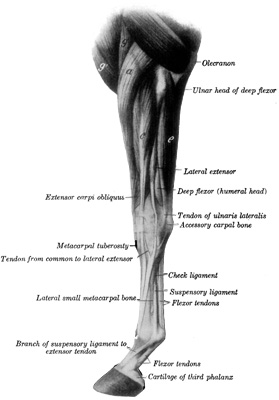
Fig. 2—Muscles of Left Thoracic Limb from Elbow Downward; Lateral (External) View.
a, Extensor carpi radialis; g, brachialis; g', anterior superficial
pectoral; c, common digital extensor; e, ulnaris lateralis. (After
Ellenberger-Baum, Anat. für Künstler.) (From Sisson's "Anatomy of the
Domestic Animals").
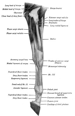
Fig. 3—Muscles of Left Thoracic Limb from Elbow Downward; Medial (Internal) View.
The fascia and the ulnar head of the flexor carpi ulnaris have been
removed. 1, Distal end of humerus; 2, median vessels and nerve. (From
Sisson's "Anatomy of the Domestic Animals").
Undue extension, (by extension is meant such movement as will cause the
long axis of two articulating bones to assume a position which
approaches or forms a straight line—opposite to flexion), of the
scapulohumeral joint is impossible while weight is borne, because of the
normally flexed position of the humerus on the scapula; whereas flexion,
beyond desirable limits, is inhibited by the biceps brachii (flexor
brachii or coracoradialis) muscle.
The distal end of the humerus, however, articulating with the radius and
ulna in a fashion that no support is lent by any sort of contact with
the body, is a ginglymus (hinge) joint and lateral motion, because of
the long transverse diameter of its articular portions, is easily
prevented by the medial and lateral ligaments (internal and external
ligaments). Flexion of this, the humeroradioulnar joint (elbow), is
restrained by the triceps brachii and extension is checked by the biceps
brachii (flexor brachii).
The carpal joint (erroneously called the knee joint), is composed of the
several carpal bones which interarticulate and, when taken as a group,
serve as a means of attachment and articulation for the radius and
metacarpal bones.
The transverse diameter of this joint is long, thus giving it contacting
surfaces that are sufficiently extensive to minimize the strain upon the
mesial and lateral ligaments (internal and external lateral common
ligaments). Motion is that of flexion and extension; slight rotation is
possible when the position is that of flexion. While supporting weight
the carpus is fixed in position by a slight dorsal flexion, but undue
dorsal flexion is prevented by the flexor muscles and tendons and
volar-carpal or annular ligament, together with the superior check
ligament.
The metacarpophalangeal articulation (fetlock joint), is a hinge joint
and its articular surfaces contact one another, with respect to their
having a long bearing surface from side to side, as do all ginglymus
(hinge) joints. Two common lateral ligaments bind the bones together.
While bearing weight, there is assumed a position of slight dorsal
flexion, undue flexion being checked by the inhibitory apparatus of the
joint—check ligaments, and their tendons and the suspensory ligament.
The inhibitory apparatus of the fetlock joint is materially reinforced
by the proximal sesamoid bones. Situated as they are, between the
bifurcating portions of the suspensory ligament and the posterior part
of the distal end of the metacarpus—with which they articulate—the
sesamoid bones serve to change the course of the branches of the
suspensory ligament in a manner that they give firm support to this
joint. Volar flexion is limited by the extensors of the phalanges.
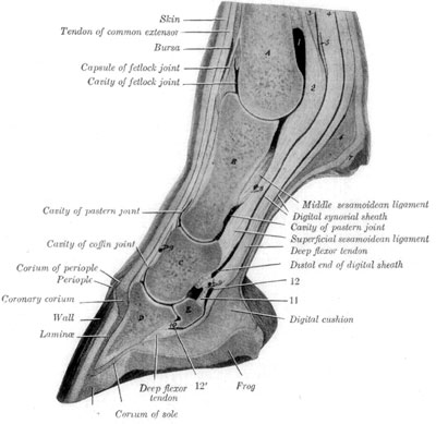
Fig. 4—Sagital Section of Digit and Distal Part of Metacarpus.
A, Metacarpal bone; B, first phalanx; C, second phalanx, D, third
phalanx; E, distal sesamoid bone; 1, volar pouch of capsule of fetlock
joint; 2, inter-sesamoidean ligament; 3, 4, proximal end of digital
synovial sheath; 5, ring formed by superficial flexor tendon; 6, fibrous
tissue underlying ergot; 7, ergot; 8, 9, 9', branches of digital
vessels; 10, distal ligament of distal sesamoid bone; 11, suspensory
ligament of distal sesamoid bone; 12, 12', proximal and distal ends of
bursa podotrochlearis. (From Sisson's "Anatomy of the Domestic
Animals").
The first phalanx (os suffraginis) normally sets at an angle of about
50 to 55 degrees from a horizontal plane while weight is being
supported. Its distal end articulates with the second or median phalanx
(os corona) and forms the proximal interphalangeal (pastern or
suffraginocoronary) joint. This also, is a ginglymus joint, having but
slight lateral motion, and that only when it is in a state of flexion. A
rather broad articular surface—from side to side—exists here,
lessening the strain on the collateral ligaments somewhat. Dorsal
flexion is checked by the flexor tendons and dorsal ligaments. Volar
flexion is restrained by the extensor tendons.
The distal end of the second phalanx (os corona) has but slight lateral
motion and this is manifested principally when it is in a state of volar
flexion. Undue dorsal flexion is prevented by the deep flexor tendon
(perforans) and volar flexion is inhibited by the extensor of the digit
(extensor pedis). Thus it is seen, that when the leg is a weight-bearing
member, weight is supported by the bony framework whose constituent
parts are joined together by ligaments and tendons and each one of the
several bones articulates in such manner that the joint is locked. The
articular parts of bones rest upon or against an inhibitory apparatus,
and are slightly flexed, as in the carpus, or considerably flexed such
as in the fetlock joint when weight is being supported. In the first
instance, for example, the flexors of the carpus and the superior check
ligament assisted by the flexors of the phalanges constitute the
inhibitory apparatus.
It will be noted that provision for weight bearing is so arranged that
muscular energy is not required except in the matter of suspension of
the body between the scapulae and here tonic impulses only are necessary
to maintain an equilibrium[6], yet in every instance where weight is not
supported by bones, inelastic ligaments or tendinous structures relieve
the musculature of this constant strain. This explains the fact that
some horses do not lie in the stall, yet in spite of their constant
standing position, they are able to rest and sleep.
The student of lameness is interested in the function of the legs in the
rôle of supporting weight and as propelling parts, and not particularly
in the capacity of these members for inflicting offense or as weapons
of defense. Yet, in the exercise of their functions other than that of
locomotive appliances, injury often results, but usually it is the
recipient of a blow that suffers the injury, such as an animal may
receive upon being kicked. Therefore, we do not often concern ourselves
with strains or other injuries that the subject experiences as the
result of efforts put forth in kicking or striking. Where such injuries
occur, however, a diagnosis is established by making use of the
principles heretofore discussed.
As propelling members the front legs bear weight and are advanced
alternately when the horse is walking or trotting—in cantering this is
not so. When the normal subject travels in a straight line, at a walk or
a trot, the length of the stride is the same with the right and left
members. The stride of the right foot then, for example, is equally
divided by the imprint of the left foot, in the normal horse, when
traveling at a walk and in a straight line.
This enigmatical term is frequently employed by the diagnostician when
he is baffled in the matter of definitely locating the cause of
lameness; when he has by exclusion and otherwise arrived at a decision
that lameness is "high up." Shoulder lameness may be caused by any one
or several of a number of conditions, e.g., fractures of the scapula or
humerus; arthritis of the shoulder or elbow joint; luxation of the
shoulder or elbow joint (rarely); injuries of muscles and tendons of the
region due to strains, contusions or penetrant wounds; paralysis of the
brachial plexus or of the prescapular nerve; involvement of lymph
glands; arterial thrombosis; metastatic infections; rheumatic
disturbances; and as the result of inflammation, infectious or
non-infectious occasioned by collar bruises. In some instances such
inflammation is due to the manner of treatment of collar injuries.
Therefore, when one considers the numerous and dissimilar possible
causes of shoulder lameness, it behooves the practitioner to become
proficient in diagnostic principles.
A principle which is elemental in the diagnosis of locomotory
impediment, is that lameness of the shoulder or hip is usually
manifested by more or less difficulty in swinging the affected member.
Swinging-leg-lameness, then, is usually present in shoulder affections.
In some instances lameness is mixed as in joint ailments, involvement of
the bicipital bursa (bursa intertubercularis), etc. In affections of the
extremity there exists supporting leg lameness. Consequently, we employ
this elemental principle, and, by a visual examination of the subject,
which is being made to travel suitably, one may decide that lameness is
either "high up"—shoulder lameness or, "low down"—of the extremity.
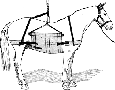
Fig. 5—Ordinary type of heavy sling.
To make practical use of this principle, the examiner must be thoroughly
familiar with the anatomy of the various structures concerned in
advancing the leg—those which support weight as well as those concerned
both in weight bearing and swinging the member.
Etiology and Occurrence.—Fractures of the body of the scapula are of
infrequent occurrence in horses for the reason that protection is
afforded this bone because of its position. Its function, too, is such
that very unusual conditions are necessary to subject it to fracture.
The spine is occasionally broken due to blows such as kicks, etc., and
here frequently a compound fracture exists.
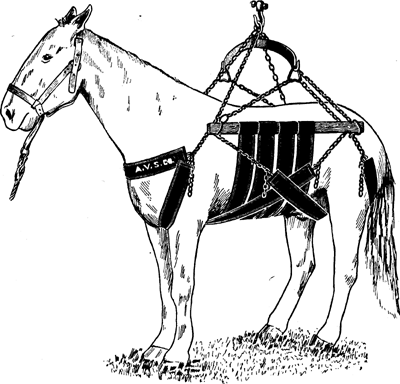
Fig. 6—A sling made in two parts so that horses may be
supported without use of central part or bodice. This sling is more
comfortable than is the ordinary style and is particularly useful in
cases that require a long period of this manner of confinement.
Where fractures of the body of the scapula occur, heavy contusions have
been the cause as a rule, and serious injury is done the subject;
consequently, treatment of fracture of the body of the scapula is seldom
successfully practised. Fractures of the body of this bone resulting
from accidents not involving internal injury or other disturbances and
which would not seriously interfere with the vitality of the subject,
are not necessarily serious unless compound.
Fractures of the neck of the scapula are serious because of the fact
that there occurs displacement of the broken parts and perfect
apposition of the fractured ends is difficult, if not impossible.
Fractures that extend to the articular surface are very serious, and
complete recovery in such instances is practically impossible. The
cartilage of prolongation of the scapula is sometimes seriously involved
in certain cases of fistulous withers, and in some instances it has been
separated from its attachment to the rhomboidea muscles, and lameness
has resulted. In such instances, the upper portion of the scapula is
disjoined from all attachment, and with every movement the animal makes,
the scapula is moved back and forth. Complete recovery in such cases
does not occur.
Symptomatology.—Fractures of the scapular spine are ordinarily
readily recognized because there is usually visible displacement of the
broken part. Crepitation is also detected without difficulty.
In fractures of the body of the scapula where an examination may be made
before much swelling has taken place, and in subjects that are not
heavily muscled, one should have no difficulty in recognizing the
crepitation.
Fractures of the neck of the scapula are recognized by crepitation, by
passively moving the leg, but it is necessary to exclude fractures of
the humerus when one depends upon the finding of crepitation by this
means. However, unless undue swelling exists, the exact location of the
crepitation is recognized without serious difficulty.
Treatment.—The treatment of compound fractures of the scapular spine
consists in the removal of the broken piece of bone by way of a
cutaneous incision so situated that good drainage of the wound will
follow.
Simple fractures of the body of the scapula are best treated by placing
the subject in a sling, if the animal is halter broken, and enforcing
absolute quiet for a period of from three to six weeks. Splints or
similar appliances are not of practical value in scapular fractures.
Compound fractures of the scapula usually result from violence, which at
the same time does serious injury to adjacent structures, and it then
becomes necessary to administer an expectant treatment, observing
general surgical principles and providing in so far as possible for the
comfort of the patient.
Anatomy.—The scapulohumeral joint is an enarthrodial (ball and
socket) joint wherein the ball or humeral articulating head greatly
exceeds in size the socket or glenoid cavity of the scapula. The
capsular ligament surrounding this joint is very large and admits of
free and extensive movement of the articulation. There exist no lateral
or common ligaments jointing the scapula and humerus as in other joints,
but instead the tendinous portions of muscles perform this function. The
principal ones which are attached to the scapula and humerus that act as
ligaments are the supraspinatus (antea-spinatus), infraspinatus
(postea-spinatus) biceps-brachii (flexor brachii) and subscapularis
muscles.
Etiology and Occurrence.—Inflammation of the scapulohumeral
articulation results from injuries of various kinds, including punctures
which perforate the joint capsule, bruises from collars, metastatic
infections and involvement as a result of direct extension of infectious
conditions situated near the joint.
Classification.—Acute arthritis may be septic or aseptic, and there
seems to be a remarkable tendency for recovery in cases of septic
arthritis involving this joint in the horse.
Chronic arthritis with destruction of articular surfaces and ankylosis,
is seldom observed. It is only in cases of severe injury, where the
articular portions of the bones are damaged at the time of infliction of
the injury, and where the articulation remains exposed for weeks at a
time, together with immobility of the parts because of attending pain,
that permanent ankylosis results.
Scapulohumeral arthritis may result then from infections, local or
metastatic; from injuries, such as contusions of various kinds; from
wounds, which break the surface structure or perforate the joint
capsule; or from luxations.
Infectious arthritis of the scapulohumeral joint the result of local
causes other than produced by septic wounds, seldom causes serious
inconvenience to the subject. Where such occurs, however, there is
manifested mixed lameness and complete extension of the extremity is
impossible. Local swelling is present and manifestations of pain are
evident upon palpation of the affected area.
Treatment.—During the first stage of the infection, local
applications, hot or cold, are indicated. A hot poultice of bran or
other suitable material contained within a muslin sack, may be supported
by means of cords or tapes which are passed over the withers and tied
around the opposite fore leg. Such an appliance may be held in position
more securely by attaching it to the affected member. Following the
acute stage of such an infection, any local counter-irritating
application or even a vesicant is in order.
Where abatement of the infectious process does not take place, and
suppuration of the structures in the vicinity of the joint occurs, it is
necessary to provide drainage for pus. In some cases of strangles, for
instance, large pus cavities are formed and drainage is imperative.
However, metastatic inflammation of this joint is seldom observed except
in cases of strangles. The animal should be kept perfectly quiet until
recovery has taken place.
Injuries to the scapulohumeral joint may be the result of kicks, runaway
accidents or bruises from the collar, and there may result, because of
such injuries, reactionary inflammation which will vary in intensity
from the mildest synovitis to the most severe arthritis, causing more or
less lameness.
Treatment.—The general plan of treatment in this form of arthritis is
the same as has been outlined under the head of infectious arthritis,
with the exception that there is seldom occasion to provide for drainage
of pus.
Wounds which cause a break of the skin and fascia overlying the
scapulohumeral joint are usually of little consequence, unless the blow
is of sufficient force to directly injure the articulation, and in such
cases, the treatment of the injury along general surgical principles,
such as cleansing the area, providing drainage for wound secretion, and
the administration of suitable dressing materials such as antiseptic
dusting powder, is all that is required for the wound. The symptoms
manifested by the subject in such cases are the same as have been
discussed heretofore and merit no special consideration.
Prognosis.—Unless very serious injury be done the articular portions
of the scapula or the humerus, resulting in the destruction of the
capsular ligament, prognosis is entirely favorable.
Open Joint.—Where the capsular ligament is perforated and the
condition becomes one of open joint, then a special wound treatment
becomes necessary. The surface of the skin is first freed from all hair
and filth in the vicinity of the wound. The wound proper is cleared of
all foreign material either by clipping with the scissors, curetting or
mopping with cotton or gauze pledgets. The whole exposed wound surface
as well as the interior of the joint cavity, if much exposed, is
moistened with tincture of iodin. Subsequent treatment consists in a
local application of a desiccant dusting powder, which should be applied
five or six times daily. The composition of the powder should be such as
to permit of its liberal use, thereby affording mechanical protection to
the wound as well as exerting a desiccative effect. Equal parts of boric
acid and exsiccated alum serve very well in such cases.
Animals suffering from open joints of this kind should be confined in a
standing position, preferably in slings, and kept so confined for three
or four weeks. Since they usually bear weight upon the affected member,
there is no danger of laminitis resulting.
Because of the large humeral head articulating as it does with a
glenoid cavity, scapulohumeral luxations are very rare in the horse.
According to Moller[7], luxation is generally due to excessive flexion
of the scapulohumeral joint. In such cases the head of the humerus is
displaced anterior to the articular portion of the scapula and remains
so fixed.
Symptoms.—Complete luxation of the scapula is recognized because of
immobility of the scapulohumeral joint and of the abnormal position of
the head of the humerus, which can be recognized by palpation, unless
the swelling be excessive. Immobility of the scapulohumeral joint is
noticeable when one attempts to passively move the parts.
Treatment.—Reduction of the luxation is effected by making use of the
same general principles that are employed in the reduction of all
luxations, and they are—the control of the animal so that the
manipulations of the operator are not antagonized by muscular
contraction, which is best accomplished by anesthesia; placing the
luxated bones in the position which they have taken to become unjointed;
and then making use of force which is directed in a manner opposite to
that which has effected the luxation.
In a forward luxation of this kind, the operator should further flex the
humerus, and while it is in this flexed position, force is exerted upon
the articular head of this bone, and it is pushed downward and backward
into its normal position.
After-care consists in restriction of exercise and, if necessary,
confining the subject in a sling and the application of a vesicant over
the scapulohumeral region.
Anatomy.—There is interposed between the tendon of the biceps brachii
(flexor brachii) and the intertubercular or bicipital groove a heavy
cartilaginous pad, which is a part of the bursa of the biceps brachii.
This synovial bursa forms a smooth groove through which the biceps
brachii glides in the anterior scapulohumeral region. Great strain is
put upon these parts because the biceps brachii is the chief inhibiting
structure of the scapulohumeral articulation—the one which prevents
further flexion of the humerus during weight bearing. Passing, as it
does, over two articulations, the biceps brachii has a somewhat
complicated function, being a flexor of the radius and an extensor of
the humerus. Thus it is seen, the biceps brachii is a weight bearing
structure, as well as one that has to do with swinging the leg.
Etiology and Occurrence.—Because of the exposed position of the
bicipital bursa (bursa-intertubercularis) it is occasionally injured.
Blows and injuries received in runaway accidents do serious injury to
the bursa and because of the peculiar and important part it plays during
locomotion, serious injuries are not likely to resolve, and too often
chronic lameness results. It is to be noted that the tendon of the
biceps brachii (flexor brachii) is always involved in cases of
inflammation of the bicipital bursa, and according to the late Dr.
Bell[8] strain of the biceps brachii is a frequent cause of lameness in
city horses, more frequent than is generally supposed.
Pathological Anatomy.—More or less destruction of the cartilaginous
portion of the bursa, sometimes involving the tendinous portion of the
biceps, takes place and, according to Moller, in some instances there
occurs ossification of the tendon. Autopsies in some old horses reveal
the presence of erosions of cartilage and hyperthrophy of the inflamed
parts.
Symptoms.—In acute inflammations, there is always marked lameness.
This is manifested to a greater degree when the subject advances the
affected leg. There is incomplete advancement of the member; the toe is
dragged when the horse is made to walk and the foot kept in a position
posterior to the opposite or weight bearing foot while the subject is at
rest. Lameness is disproportionate to the amount of local manifestation
in the way of heat, swelling and pain that is to be recognized on
palpation. In fact, in some cases so much pain attends the condition
that no weight is borne by the affected member, and when compelled to
walk, the subject hops on the sound leg.
Chronic inflammation of the bicipital bursa is occasionally met with
wherein both members are affected. Because of the nature of the
structures involved, when inflamed, chronic inflammation is a more
frequent termination than is complete recovery. Bilateral affections are
seen in horses that are driven for years, regularly at a fast pace on
paved streets. In such cases, the gait is stilted, that is, there is
incomplete advancement of both members and, of course, the period of
weight bearing is correspondingly shortened; hence the short strides.
In chronic cases, little if any evidence of inflammation is to be
detected by digital manipulation of the parts. If flinching occurs, one
is often unable to interpret the manifestation as to whether it is due
to inflammation or not.
There is no marked "warming out" in this condition, and animals are
nearly as lame after having been driven a considerable distance as when
started, although the lameness is not as a rule very great.
Treatment.—In very painful cases acute inflammation is treated by
employing cold applications during the initial stage. Cracked ice when
contained in a suitable sack may be held in contact with the affected
part and the pack is supported by means of cords or tapes as suggested
in the discussion on treatment of scapulohumeral arthritis on page 66.
Later, hot applications may be employed to good advantage.
In the course of ten days or two weeks, if the acute painful condition
has entirely subsided, vesication is indicated. The ordinary mercury and
cantharides combination does very well. Depending upon the course taken
in any given case, one is guided in the treatment employed. If prompt
resolution comes to pass, the subject may be given free run at pasture
after three or four weeks confinement in a box stall. If, however, the
case does not progress in a prompt and satisfactory manner, absolute
quiet must be enforced for six weeks or more. Repeated blistering is
beneficial, although it is doubtful if firing is of sufficient benefit
in the average chronic case of intertubercular bursitis to justify the
punishment which this form of treatment inflicts, unless infliction of
pain is the thing sought, to enforce repose in restless subjects.
Patients are best given a long rest at pasture and returned to work for
two or three months after an acute attack of inflammation of the bursa,
lest the condition become chronic. When due consideration is given the
pathology of such cases, the frequent unsatisfactory termination under
the most careful treatment, is readily understood.
Anatomy.—The triceps brachii is the principal structure which fills
the space between the posterior border of the scapula and the humerus.
The several heads originate for the most part on the border of the
scapula, the deltoid tuberosity of the humerus and the shaft of the
humerus. Insertion of this large muscular mass is effected by means of
several tendons to the olecranon. A synovial bursa is situated
underneath the tendinous attachment of the posterior portion of the
triceps brachii—the long head or caput magnum.
The function of the triceps as a whole is to flex the shoulder joint and
extend the forearm. The triceps brachii is the chief antagonist of the
biceps brachii.
Etiology and Occurrence.—Owing to the exposed position of this
structure, it is not infrequently contused, the result of falls, kicks
and other injuries. The function of the triceps is such that it becomes
strained upon rare occasions when a horse resists confinement of
restraint in such manner that the parts are unduly tensed in
contraction. This sort of resistance may stretch the radial nerve or its
branches in a way that paralysis results. A condition known as "dropped
elbow" is described by Henry Taylor, F.R.C.V.S., in the Veterinary
Record[9], wherein a two-year-old colt while resisting confinement was
so injured.
The triceps group because of its convenient location, constitutes the
site for hypodermic injection of drugs and biologic agents, with some
practitioners; and as a result, more or less inflammation may occur. The
author has observed and treated some twenty cases where an intensely
painful infectious inflammation of the triceps brachii was caused by
the intramuscular injection of a caustic solution by a cruel and
unscrupulous empiric, whose object was to increase his practice.
Symptomatology.—As the triceps brachii is not particularly taxed
during weight bearing in the subject at rest, there may be no unnatural
position assumed during inflammation of the triceps. More or less
swelling and supersensitiveness is always present, however, and great
care and discrimination must be exercised in digital manipulation of the
triceps region because many animals are normally sensitive to palpation
of these parts. It is sometimes difficult to correctly interpret the
true state of conditions because of this peculiarity.
There is always swinging-leg-lameness, which is accentuated when the
subject is urged to trot. Where symptoms are pronounced, it is
unnecessary to cause the subject to move at a faster pace than at a walk
to recognize the condition. The forward stride is shortened and in
extremley painful conditions, no attempt is made to extend the leg. It
is simply carried en une piéce—flexion of the shoulder and elbow
joints is carefully avoided.
Treatment.—During the early stage of inflammation, hot or cold
applications are beneficial. Long continued use of moist
heat—fomentations—allays pain and stimulates resolution. Keeping in
contact with the painfully swollen parts a suitable bag filled with
bran, which can be moistened at intervals with warm water, constitutes a
practical and easy means of treatment. By employing this method, one is
more likely to succeed in having his patient properly cared for, in that
less work is entailed than if hot fomentations are prescribed.
After the acute and painful stage has subsided, a stimulating liniment
is of benefit. The subject should be kept within a comfortable and roomy
box stall for a sufficient length of time to favor prompt resolution.
Wild and nervous subjects, if not so confined, will probably overexert
the affected parts if allowed the freedom of a paddock or pasture.
Where the inflammation becomes infective, surgical interference is
necessary. The prompt evacuation of pus, with adequate provision for
wound discharge, should be attended to before extensive destruction of
tissue takes place. Resolution is prompt as a rule in such cases because
of the vascularity of the structures and the ease with which proper
drainage may be effected. No special after-care is necessary if drainage
is perfect, except that one should avoid injecting the wound cavity with
aqueous solutions unless it be absolutely necessary to cleanse such
cavity, and then it is best to swab the wound rather than to irrigate it
freely.
No satisfactory consideration of the pathogeny of this condition is
recorded, but practitioners have long distinguished between muscular
atrophies which are apparently caused without doing serious injury to
nerves and muscular atrophy which seems to be due to nerve affection. In
the first instance, recovery when proper attention is given, is prompt;
whereas, in the latter, regeneration of the wasted tissues requires
months in spite of the best sort of treatment.
The parts more frequently affected are the supra- and infrascapularis
(antea- and posteaspinatus) muscles. But in some cases the triceps group
is involved; however, this occurs in unusual and chronic affections. No
doubt, these chronic cases are due to suspended innervation and are not
to be classed with the ordinary case of atrophy of the abductor muscles
of the humerus (supra- and infraspinatus) as in the usual case of
"sweeny."
Occurrence.—Shoulder atrophy such as the general practitioner
commonly meets with, is an affection, more often seen in young animals
and it seems to be due to injuries of various kinds which contuse the
muscles of the shoulder. Ill-fitting collars and pulling in a manner
that there occurs side draft with unusual strain on the muscles of one
side of the neck and shoulder, seem to be the more frequent causes of
this trouble. Blows such as are occasioned by kicks and falls frequently
result in atrophy of shoulder muscles.
Course.—In some cases a rapidly progressive atrophy characterizes the
case and lameness and atrophy appear at about the same time. The
affection in such instances does not recover spontaneously but
constitutes a condition which requires prompt and rational treatment so
that function may be fully restored to the parts involved.
Occasionally one may observe cases where there is but slight atrophy;
where the disease progresses slowly and atrophy is not extensive or
marked. In vigorous young animals that are left to run at pasture when
so mildly affected, spontaneous recovery occurs.
Symptomatology.—Lameness is the first manifestation of shoulder
atrophy, and in many cases where lameness is slight, the veterinarian
may fail to discover the exact nature of the trouble if he is not very
proficient as a diagnostician of lameness or if he is careless in taking
into consideration obtainable history, age of the subject, etc. Because
of the fact that the average layman believes that practically every case
of fore-leg lameness wherein it is not obvious that the cause is
elsewhere, is due to a shoulder affection of some kind, we may be too
hasty in giving the client assurance that no "sweeny" exists. In some of
these cases where a diagnosis of "shoulder lameness" has been made and
the client has been assured that no sweeny exists, the patient is
returned in about a week and there is then marked atrophy of one or both
of the spinatus muscles.
A mixed type of lameness characterizes this affection, and in the
average case there exists little evidence of local pain. The salient
points in recognizing the condition are a consideration of history if
obtainable; age of the subject; finding slight local soreness, by
carefully manipulating the muscles which are usually involved; noting
the character of the lameness if any is present; and where atrophy is
evident, of course, the true condition is obvious.
Treatment.—Subcutaneous injections of equal parts of refined oil of
turpentine and alcohol, with a suitable hypodermic syringe, is a
practical and ordinarily effective treatment. From five to fifteen cubic
centimeters (the quantity varies with the size of the animal), of this
mixture is injected into the atrophied parts at different points, taking
care to introduce only about one to two cubic centimeters at each point
of injection. The syringe should be sterile and, needless to say, the
site of injections must be surgically clean.
Other agents, such as tincture of iodin, solutions of silver nitrate,
saline solutions and various more or less irritating preparations have
been employed; but in the use of these preparations one may either fail
to stimulate sufficient inflammation to cause regeneration to take
place, or infection is apt to occur. Where suppuration results, surgical
evacuation of pus must be promptly effected else large suppurating
cavities form.
The employment of setons constitutes a dependable method of treatment of
shoulder atrophy, but because of the attendant suppurative process which
inevitably results, this method is not popular with modern surgeons and
is a last resort procedure.
After-care.—Regular exercise such as the horse usually takes when at
pasture, is very helpful in treating atrophy, and in some cases it has
been found that no reasonable amount of irritation would stimulate
muscular regeneration; but by later allowing patients to exercise at
will, recovery took place in a satisfactory manner. No special attention
is ordinarily necessary.
Anatomy.—The suprascapular (anterior scapular) nerve, a small branch
of the brachial plexus, is given off from the anterior portion of this
plexus. The nerve rounds the anterior border of the neck of the scapula,
passing upward and backward under the supraspinatus (antea-spinatus)
muscle and terminating in the infraspinatus (postea-spinatus) muscle.
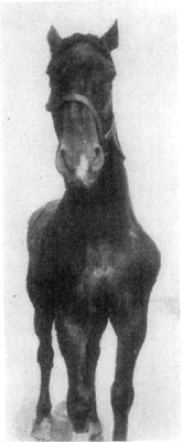
Fig. 7—Paralysis of the
suprascapular nerve
of the left shoulder
Etiology and Occurrence.—As the result of direct injury to this nerve
by contusion such as may be received in runaway accidents, collar
bruises, especially collar bruises in young horses that are not
accustomed to pulling and that walk in a manner to cause side draft,
injury to the nerve occurs, and partial or complete paralysis
supervenes. Some writers state that it may be produced by confining an
animal in recumbency, with the casting harness. The common cause of
paralysis or paresis of this nerve in cases such as one observes in
country practice, is bruises from the collar in colts that are put to
heavy farm work or where ill fitting collars are used.
Symptomatology.—With partial or complete suspension of function of
the suprascapular nerve there results enervation of the supraspinatus
and infraspinatus muscles. Since these muscles act as external lateral
ligaments of the scapulohumeral joint, when they are incapacitated,
there naturally follows more or less abduction of the shoulder when
weight is borne.
In extreme cases, as soon as the ailing animal is caused to support
weight with the affected member, the joint is suddenly thrown outward in
a manner that the average layman at once concludes that there must be
scapulohumeral luxation, and the veterinarian receives a call to see a
case wherein the "shoulder is out of place." There exists, however, no
luxation in such cases.
If serious injury is done the nerve so that it undergoes degenerative
changes, there will result atrophy of the muscles that derive their
nerve supply from the suprascapular nerve.
Treatment.—During the first few days following injuries which result
in this form of paralysis, it is well to keep the subject inactive, and
if much inflammation of the injured structures contiguous to the nerve
exists, the application of cold packs is beneficial. Later, as soon as
acute inflammation has subsided, vesication of a liberal area around the
anteroexternal part of the scapulohumeral joint and over the course of
the suprascapular nerve, will stimulate recovery in favorable cases. As
a rule, in mild cases, the subject is in a condition to return to work
in two or three weeks.
Described under the titles of "Radial Paralysis" and "Brachial
Paralysis," there is to be found in veterinary literature a discussion
of conditions which vary in character from the almost insignificant form
of paresis to the incurably affected conditions wherein the whole
shoulder is completely paralyzed.
When one considers the anatomy of the brachial nerve plexus and the
distribution of its various branches, the location of this plexus and
its proximity to the first rib, and the inevitable injury it must suffer
in fracture of this bone, together with the inaccessibility of the
plexus, it is not strange that a correct diagnosis of the various
affections of the brachial plexus and the radial nerve is often
impossible until several days or weeks have passed. And, in some
instances, diagnosis is not established until an autopsy has been
performed. Here, too, we fail to find cause for paralysis in some rare
instances.
Anatomy.—The radial nerve is a large branch of the brachial plexus
and is chiefly derived from the first thoracic root of the plexus and is
here situated posterior to the deep brachial artery. It is directed
downward and backward under the subscapularis and teres major muscles,
rounding the posterior part of the humerus, and passing to the anterior
and distal end of the humerus, it finally terminates in the anterior
carpal region. The radial nerve supplies branches to the three heads of
the triceps brachii, to the common and lateral extensors of the digit
and also to the skin covering the forearm.
Etiology and Occurrence.—Nothing definite is known about the cause of
some forms of radial paralysis. However, radial paralysis is encountered
following injury to the nerve occasioned by its being stretched, as in
cases where the triceps brachii is unduly extended in restraining
subjects by means of a casting harness. Berns[10] states that in
confining horses on an old operating table where it was necessary to
draw the affected foot forward twenty-four to thirty-six inches in
advance of its fellow, which was secured in a natural vertical
position, radial paralysis of a mild form was of frequent occurrence.
Country practitioners, in restraining colts by casting with harness or
ropes, occasionally observe a form of paresis wherein the radial nerve
suffers sufficient injury that there is caused a temporary loss of
function of the triceps brachii. Such cases recover within three or four
days and are not a true paralysis, but nevertheless constitute
conditions wherein normal nerve function is temporarily suspended.
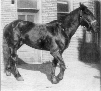
Fig. 8—Radial paralysis.
Symptoms.—Immediately subsequent to injuries which involve the radial
nerve, there is manifested more or less impairment of function.
Remembering the structures supplied by the radial nerve and its
branches, one can readily understand that there should occur as
Cadiot[11] has stated:
In complete paralysis, the joints of the affected limb with the
exception of the shoulder are usually flexed when the horse is
resting. In consequence of loss of power in the triceps and
anterior brachial muscles, the arm is extended and straightened on
the shoulder, the scapulohumeral angle is open, and the elbow
depressed. The forearm is flexed on the arm by the contraction of
the coracoradialis (biceps brachii), while the metacarpus and
phalanges are bent by the action of the posterior antibrachial
muscles. The knee is carried in advance, level with, or in front
of, a vertical line dropped from the point of the shoulder. The
hoof is usually rested on the toe, but when advanced beyond the
above mentioned vertical line, it may be placed flat on the ground,
the joints then being less markedly bent. When the limb as a whole
is flexed, it may be brought into normal position by thrusting back
the knee with sufficient force to counteract the action of the
flexor muscles.
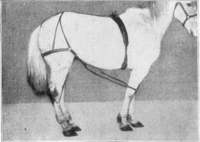
Fig. 9—Merillat's method of fixing carpus in radial paralysis. Courtesy, Alex. Eger.
When made to walk, the animal being unable to exert muscular action with
the paralyzed structures, limply carries the member as a whole, and
there is shortening of the anterior portion of the stride. There being
loss of function of the triceps brachii, it is impossible for the
subject to straighten the leg in the normal position for supporting
weight; therefore, any attempt to bear weight results in further
flexion of the affected member and the animal will fall if the body is
not suddenly caught up with the sound leg.
Differential Diagnosis.—In making examination of these cases, one can
exclude fracture by absence of crepitation and usually, also, swelling
is absent in radial paralysis. In a typical case of radial paralysis,
the affected leg can sustain its normal share of weight if placed in
position, that is, if the carpal joint is extended in such manner that
the leg is positioned as in its normal weight-bearing attitude. In
brachial paralysis, whether due to fracture of the first rib or to other
serious injury, it is impossible for the subject to support weight with
the affected member even when it is passively placed in position.
No difficulty is ordinarily experienced in differentiating radial
paralysis from muscular injuries to the triceps; yet, in some cases of
"dropped elbow," it is necessary to observe the progress of the case for
ten days or two weeks before one can positively establish a diagnosis.
Quoting Merillat[12]: "When, after four weeks, there is no
amelioration of the paralysis, the muscles have atrophied, and the
patient has become emaciated from pain and discomfort, the
diagnosis of brachial paralysis with fracture of the first rib may
then be announced."
Prognosis.—When no complete paralysis of the brachial plexus or no
fracture of the first rib exists, the majority of cases recover
completely in from ten days to six weeks. Some writers claim that
recoveries occur in ninety per cent of cases when conditions are
favorable.
Treatment.—When incomplete radial paralysis exists, little needs be
done except to allow the subject moderate exercise and to provide for
its comfort. Local applications, stimulative in character, are
beneficial, and the internal administration of strychnin is indicated.
In the cases where weight is not supported without the affected leg
being passively placed in position, it is necessary to provide for the
subject's comfort in several ways.
Mechanical appliances such as braces of some kind in order to keep the
affected leg in a position of carpal extension, constitute the essential
part of treatment. The leg is supported in such a manner that flexion of
the carpus is impossible. Due regard is given to prevent chafing or
pressure necrosis by contact of the skin with the braces—this may be
done by bandaging with cotton. The supportive appliance is kept in
position for ten days or two weeks. At the end of this time the brace
may be removed and the subject given a chance to walk, and improvement,
if any exists, will be evident. When there is manifested an amelioration
of the condition, moderate daily exercise and massage of the affected
parts are helpful.
Should the subject be seriously inconvenienced by the application of a
brace or other supportive appliances, it is necessary to employ slings.
Further, if weight is supported entirely by the unaffected member,
laminitis may supervene if a sling is not used.
Thrombosis of the brachial artery or of its principal branches is of
very rare occurrence in horses.
Etiology.—Partial or complete obstruction of arteries (brachial or
others) occurs as the result of direct injury to the vessel wall from
compression and tension of muscles and resultant arteritis; lodging of
emboli; and parasitic invasion of vessel walls causing internal
arteritis.
Symptomatology.—If sufficient collateral circulation exists to supply
the parts with blood, no inconvenience is manifested while the subject
is at rest. Where the lumen of the affected vessel is not completely
occluded, there may be no manifestation of lameness when the ailing
animal is moderately exercised. Consequently, the degree of lameness
depends upon the extent of the obstruction to circulation; and,
likewise, the course and prognosis depend upon the character and extent
of such obstruction.
In severe cases, lameness is markedly increased by causing the animal to
travel at a fast pace for only a short distance. There are evinced
symptoms of pain, muscular tremors and sudation, but the affected member
remains dry and there is a marked difference of temperature between the
normal areas and the cool anemic parts. When the subject is allowed to
rest, circulation is not taxed, and there is a return to the original
and apparently normal condition, only to recur again with exertion. This
condition characterizes thrombosis.
Treatment.—In these cases, little if any good directly results from
any sort of treatment in the way of medication. Absolute rest is thought
to be helpful. Potassium iodid, alkaline agents such as ammonium
carbonate and potassium carbonate, have been administered. Circulatory
stimulants also have been given, but it is doubtful if any good has come
from medication.
The shaft of the humerus, protected as it is by heavy muscles, is not
frequently fractured; and fractures of its less protected parts, as for
example, the head, are complicated in such manner that resultant
arthritis soon constitutes the more serious condition.
As a result of falls on frozen ground, kicks or any other form of heavy
contusion, the humerus is occasionally broken. It is rarely fractured
otherwise. Because of the force of contusions usually required to effect
humeral fracture, the manner in which the bone is broken, with respect
to direction, is variable. Often oblique fractures exist and
occasionally there occurs multiple fracture. In addition to the
ordinarily serious nature of the fracture itself, there is always much
injury done the adjoining structures.
Symptomatology.—Mixed lameness and manifestation of severe pain
characterize this affection. Considerable swelling which increases, in
some cases for a week or more, is to be observed. Crepitation is readily
detected, if pain and swelling is not too great to prevent passive
movement of the member. Where intense pain is not manifested, because of
manipulation, one may abduct the extremity and thereby occasion distinct
crepitation; but when it is possible to recognize crepitation by holding
the hand in contact with the olecranon while the animal is made to walk,
this method is to be preferred, if the subject can move without serious
difficulty. The pathognomonic symptom here is recognition of
crepitation, but this may be very difficult to recognize in fracture of
condyles, and in such instances, a careful examination is necessary.
Gentle manipulation in a manner that pain is not aggravated will tend to
inspire confidence on the part of the subject and relaxation of muscles
will enable the operator to detect crepitation.
Course and Prognosis.—Because of the direction of the long axis of
the humerus, with relation to the bony column of the extremity, it is
obvious that any lateral movement of the leg tends to rotate the shaft
of this bone. In fractures of the shaft of the humerus, then, it is
apparent that immobilization is very difficult if at all possible.
The proximity to the axillary lymph glands makes for easy dissemination
of infection when the contused musculature becomes infected. The
adjacent brachial nerve plexus is so very apt to become involved, if not
actually injured at the time fracture occurs, that paralysis is a
probable complication. Consequently, it is logical to reason that
because of the many possible serious complications, such as shock,
occasioned by the injury and the distress and pain which this accident
produces, recovery must be the exception in fracture of the humerus.
However, recoveries do take place and in addition to the reported
recoveries by Liautard, Moller, Stockfleth, Lafosse, Frohner and others,
we have instances cited by American practitioners where cases resulted
in recovery. Thompson[13] reports a good recovery in a 1600-pound mare
where there existed an oblique fracture of the humerus. This mare was
kept in slings for eight weeks. Walters[14] reports complete recovery in
humeral fracture in a foal three days old. The only treatment given was
the application of a pitch plaster from the top of the scapula to the
radius. The colt was kept in a comfortable box stall and in about four
weeks regained use of the leg. Complete recovery eventually resulted. In
the experience of the author, recovery has not occurred in humeral
fractures.
Treatment.—When animals are not aged and of sufficient value to
justify treatment, they are best supported in a sling, if halter broken.
If subjects are nervous, wild and unbroken, it is possible to employ the
sling, if care is given to train the animal to this manner of restraint.
The presence of an attendant for a day or two will reassure such
subjects so that even in these cases it may be practicable to employ the
sling.
Braces and other mechanical appliances intended to immobilize the parts
are not of practical benefit in the horse. Unlike the dog, the horse as
yet has not been successfully subjected to tolerating rigid braces for
the shoulder and hip.
Everything possible must be done that will make for the patient's
comfort. If the subject turns out to be a good self nurse, and the
nature of the fracture is such that practical apposition of the broken
ends of bone may be maintained, recovery will occur in some cases.
Affections of this articulation other than those which are produced by
traumatism are rare. This joint has wide articular surfaces, and
securely joined as they are by the heavy medial and lateral ligaments
(internal and external lateral ligaments), luxation is practically
impossible. When luxation does occur, irreparable injury is usually
done. Castagné as quoted by Liautard[15], reports a case of true
luxation of the elbow joint in a horse where reduction was effected and
complete recovery took place at the end of twenty-five days. This is an
unusual case. The average practitioner does not meet with such
instances.
Anatomy.—The condyles of the humerus articulate with the glenoid
cavities of the radius and a portion of the ulna. Two strong collateral
ligaments pass from the distal end of the humerus to the head of the
radius. The capsular ligament is a large, loose membrane which encloses
the articular portion of the humerus with the radius and ulna and also
the radioulnar articulation. It is attached anteriorly to the tendon of
the biceps brachii (flexor brachii). The capsule extends downward
beneath the origin of these digital flexors. This fact should be
remembered in dealing with puncture wounds in the region, lest an error
be made in estimating their extent and an open joint be overlooked at
the initial examination.
Etiology and Occurrence.—Exclusive of specific or metastatic
arthritis, which is seldom observed except in young animals,
inflammation of the elbow joint is usually caused by injury. This
articulation is not subject to pathologic changes due to concussion or
sprains as occasioned by ordinary service, but is frequently injured by
contusion from falls, blows from the wagon-pole and kicks. Wounds which
affect the elbow joint, then, may be thought of in most cases, as
resultant from external violence. They may be contused wounds or
penetrant wounds. Sharp shoe-calks afford a means of infliction of
penetrant wounds which may occasion open joint and infectious arthritis.
Classification.—A practical manner of classifying inflammation of the
elbow is on an etiological basis. Eliminating the forms of elbow
inflammation, such as are caused by metastatic infection and other
conditions which properly belong to the domain of theory of practice, we
may consider this affection under the classification of contusive
wounds and penetrative wounds.
Symptomatology.—Any injury which is of sufficient violence to
occasion inflammation of the elbow causes marked lameness and
manifestation of pain. The degree of lameness and distress manifested by
the subject, depends upon the nature and extent of the involvement. A
contusion suffered as the result of a fall, which occasions a
circumscribed inflammation of the structures covering this joint and
where little inflammation of the articulating parts exists, marked
evidence of pain and lameness might be absent. On the other hand, if a
true arthritis is incited, there will be evident distress manifested,
such as hurried respiration, accelerated pulse, inappetence, mixed
lameness, local evidence of inflammation and particularly marked
supersensitiveness of the affected parts. Considering these two extremes
of manifested distress and injury, one may readily conclude that in the
frequently seen case, wherein contusion has occasioned a moderate
degree of injury, prognosis is favorable and recovery ordinarily
follows in the course of a few weeks' treatment.
In cases of arthritis due to penetrative wounds (because of the
important function of this joint and its large capsule, which when
inflamed discharges synovia in a manner that closure of such an open
joint is seldom possible) a very grave condition results.
Treatment.—Inflammation of the elbow, such as is frequently seen in
general practice where horses are turned out together and exposed to
kicks and other injuries, yields to treatment readily, if an open joint
does not exist.
Hot packs supported in contact with the elbow and kept around the
inflamed articulation for a few days, materially decrease pain and tend
to reduce inflammation. The subject must be kept quiet in a comfortable
stall and, if necessary, a sling used. Where it is impossible for the
animal to support much weight with the injured member the sling should
be employed.
As inflammation abates, which it does in the course of from one to three
weeks in uncomplicated cases, the subject may be allowed the freedom of
a comfortable box stall. Vesication of the parts is in order, and this
may be repeated in the course of two weeks, if it is deemed necessary.
Penetrative wounds resulting in open joint are not treated with success
as a rule, and because of the handicap under which veterinarians labor,
methods of handling such cases, where large, important articulations are
affected, are not being rapidly improved. Prognosis is usually
unfavorable, and for humane and economic reasons, animals so affected
should be destroyed.
Ordinary wounds of the region of the elbow are treated along general
lines usually employed. They merit no special consideration, except that
it may be mentioned that with such injuries concomitant contusion of the
parts occasions injury that does not recover quickly.
Etiology and Occurrence.—Fractures of the ulna in the horse are not
common in spite of the exposed position of the olecranon. This bone when
broken, is usually fractured by heavy blows and any form of ulnar
fracture is serious because of its function and position in relation to
the joint capsule. Transverse fractures do not readily unite because of
the tension of the triceps muscles, which prevent close approximation of
the broken ends of the bone.
Thompson[16], however, reports a case of transverse simple fracture of
the ulna in a mare, the result of a kick, in which complete recovery
took place. He kept the subject in a sling for six weeks and then
allowed six months rest.
Symptomatology.—The position assumed by a horse suffering from a
transverse fracture of the ulna, is similar to that in radial paralysis.
Crepitation may be detected by manipulating the parts, and in some
instances of fracture of the olecranon, there occurs marked displacement
of the broken portions of the bone. Lameness is intense and the parts
are swollen and supersensitive. The capsular ligament of the elbow joint
is usually involved in the injury because fracture of the ulna may
directly extend within the capsular ligament. In such cases, there is
synovitis, and later arthritis causes a fatal termination.
Treatment.—The impossibility of applying a bandage in any way to
practically immobilize these parts in fracture of the ulna, prevents our
employing bandages and splints. Therefore, one can do little else than
to put the patient in a sling and try to keep it quiet and as nearly
comfortable as circumstances allow.
Etiology and Occurrence.—From heavy blows received such as kicks,
collision with trees or in falls in runaway accidents, the radius is
occasionally fractured. In very young foals, fracture of the radius, as
well as of the tibia and other bones, results from their being trampled
upon by the mother.
Symptomatology.—Excepting in some cases of radial fracture of foals
where considerable swelling has taken place, there is no difficulty in
readily recognizing this condition. The heavy brachial fascia materially
contributes to the support of the radius, and in cases where swelling
is marked, crepitation may not be readily detected. In fact, a
sub-periosteal fracture may exist for several days or a week or more and
then, with subsequent fracture of the periosteum, crepitation and
abnormal mobility of the member are to be recognized. In such cases, the
subject will bear some weight upon the affected member, but this causes
much distress. In one instance the author observed a transverse fracture
of the lower third of the radius which was not positively diagnosed
until about ten days after injury was inflicted. In this case, without
doubt, the subject originally suffered a sub-periosteal fracture of the
bone and because the animal was a good self nurse, the brachial fascia
supported the radius until the periosteum gave way and the leg dangled.
In this instance infection took place and suppuration resulted. It was
deemed advisable to destroy this animal.
Prognosis.—In adult animals, radial fracture constitutes a grave
condition; generally speaking, prognosis, in such cases, is unfavorable.
Because of the leverage afforded by the extremity, immobilization of the
radius is difficult. Any sort of mechanical appliance, which will
immobilize these parts, is likely to produce pressure-necrosis of the
soft structures so contacted. There is occasioned thereby much pain and
the subject becomes restive, unmanageable and sometimes the splints are
completely deranged because of the animal's struggles, and much
additional injury to the leg is done. Occasionally, an otherwise
favorable case is thus rendered hopelessly impossible to handle, and the
subject must be destroyed several days after treatment has been
instituted.
Consequently, unless all conditions are good, and the affected animal a
favorable subject, young, of good disposition, and the fracture a simple
transverse one, complete recovery is not likely to result from any
practical means of handling.
Treatment.—Mature subjects ought to be put in slings and kept so
restrained throughout the entire time of treatment. Immobilization of
the broken parts of the bone is the object sought. This is attempted by
practitioners who employ various methods, and each method has its
advocates.
Casts are used by some and serve very well in many cases; but because of
their bulk and unyielding and rigid nature, they are not well adapted to
use on fractures of bones proximal to the carpus and tarsus. This is in
reference to plaster-of-paris casts or those of any similar material.
Appliances which depend on glue or other adhesive substances combined
with leather, wood or fiber for their support, are efficacious but not
comfortable.
The use of heavy leather when the member has been suitably padded with
cotton and bandages, constitutes a very good manner of reducing fracture
of the radius or of the tibia. Leather when cut to fit both the medial
and lateral sides of a leg, and firmly held with bandages, will form a
firm support that yields slightly to changes of position, thus making
for comfort of the subject.
Such a splint or support should extend from the fetlock region to the
elbow, but the cotton and bandages are to reach to the foot. When one
considers that, with the supportive appliance placed on each side of the
affected member, rigidity is accomplished as much from tensile strain
put upon the leather as from its own stiffness, it is seen that the
leather need not be of the heaviest—sole leather is unnecessary.
Because of the more comfortable immobilizing appliance, the subject is
less restive, and chances for a successful outcome are materially
increased thereby.
In the mature subject, six or eight weeks' time is required for union of
the parts to occur sufficiently so that splints may be dispensed with.
Rearrangement of the supportive apparatus, however, is possible and
usually necessary during the first few weeks of treatment. By employing
care in handling the parts, the subject will be unlikely to do itself
injury at the time readjustment of splints is being effected.
In foals, it is best to give them the run of a box stall with the
mother. Being agile, they get up and lie at will without doing injury to
the fractured member. The splints (leather is preferable in these cases
also) are looked after and readjusted as necessity demands.
Three or four weeks time is all that is required for the average young
colt to be kept in splints when suffering from simple transverse
fracture of the radius.
Compound fractures are necessarily more difficult to treat than are the
simple variety, but even in such cases recovery results sometimes, and
the practitioner is justified in attempting treatment after having
explained the situation to his client.
Oblique fractures, even when simple, do not completely recover. Muscular
and tendinous contraction, together with the natural tendency for the
beveled contacting parts of the broken bone to pass one another in
oblique fracture, results in shortening of the leg and, if union
results, a large callus usually forms. Where shortening of bones occur,
necessarily, permanent lameness follows.
Etiology and Occurrence.—Contusions and lacerations of the forearm
are of frequent occurrence in horses and are troublesome cases to
handle; particularly is this noticeable where extensive laceration of
the parts occurs. These injuries are caused by animals being kicked; by
striking the forearm against bars in jumping; and in sections of the
country where barbed wire is used to enclose pastures, extensive
lacerated wounds are met with when horses jump into such fences.
Symptomatology.—Any wound which causes inflammation of the structures
of the anterior half of the forearm, is characterized by
swinging-leg-lameness. Depending upon the nature and extent of the
injury, manifestation varies. In cases where laceration has practically
divided all of the substance of the extensor tendons, it is, of course,
impossible for the subject to advance the leg; but where lacerated
wounds involve only a part of the extensor apparatus of the foreleg, not
so much inconvenience is evident, unless the wound is seriously infected
and inflammation involves contiguous structures. Therefore, in many
instances, lameness is more pronounced in contusions of the anterior
brachial region than where tissues have been divided more or less
keenly.
In every instance diagnosis is easily established. The injury is quite
evident, and the manner of locomotion is not in itself an essential
feature to be considered in a discussion of symptoms. Where a contusion
of the anterior brachial structures occurs, there is, in addition to
lameness, swelling which is painful because of the pressure occasioned
by the heavy non-yielding brachial fascia. And where suppuration occurs,
there is then an intensely painful condition which is not relieved until
pus has been evacuated. Rather frequently, drainage for wound secretions
is a difficult problem, and approximation of the divided ends of muscles
is always difficult to maintain.
Treatment.—Contused wounds of the anterior brachial region are
treated along usual lines; that is, attempt is made to stimulate prompt
resolution. Hot or cold applications are employed throughout the acute
stage of the affection. Complete rest is provided for until all pain has
subsided. Later, stimulating liniments are beneficial.
Where no injury is done the periosteum or bone, complete resorption of
all products of inflammation usually occurs, though in many instances,
this is tardy—six weeks or more are sometimes required for recovery to
take place.
If suppuration occurs, it is necessary to provide for drainage as soon
as it is possible to distinguish the presence of pus. Due regard is
given the manner of establishing drainage because of the usual existence
of sub-fascial fistulae. In these cases, one avoids injecting solutions
of aqueous antiseptics. By gently compressing the parts, pus is caused
to drain out and in enforcing a moderate amount of exercise at a walk,
when lameness is not intense, drainage is maintained. Cotton packs,
moistened with hot antiseptic solutions, and kept around the forearm for
several hours daily, are helpful because drainage is facilitated, and
resolution is stimulated by the increase of blood thus attracted to the
parts, and pain materially diminishes.
In lacerated wounds of the anterior brachial region, after having
controlled hemorrhage, an area around the wound margin is freed of hair
by clipping or shaving. The wound is carefully examined, and the best
site for drainage is selected and a suitable opening for wound discharge
is provided for. Where the extensor carpiradialis (metacarpi magnus)
with other structures, is divided and the distal portion is torn
downward, as frequently is the case in barbed wire cuts, it is
necessary to make careful provision for drainage. The wound is
thoroughly cleansed by means of ablutions if necessary; but preferably
by swabbing with pledgets of cotton or gauze which are moistened in
antiseptic solutions. All shreds of macerated tissue are clipped with
scissors and finally the whole wound surface is painted with tincture of
iodin.
If drainage is made by cutting through the tissues in the median portion
of the structures that have been displaced, the opening should be packed
with gauze so that it may remain patent after swelling has occurred.
Such packing is left in situ for twenty-four hours.
The pendant muscular portions of tissues are sutured up by means of
tapes and, while perfect apposition is not ordinarily possible, it is
very essential to train the pendant tissues in their normal position
even if they require resuturing within a week. This minimizes
granulation of tissue, and there results less scar if the detached
portions are kept near, even if not in contact with the proximal wound
margins. The skin together with subcutaneous fascia is sutured on either
side unless drainage is to be provided for on one side, and the
lowermost part of that side is left unsutured.
After-care.—Where extensive suturing of tissues has been necessary,
subjects must be kept quiet. They are best confined in box stalls and
not taken out for several weeks. Particularly is this true where
transverse division of extensors has taken place. Sutures are removed at
the end of from ten days to three weeks as cases permit. Drainage of
wound secretions, which usually become infected, is necessary, because
with obstructed drainage in an infected wound of this kind, there will
result an early destruction of tissue at some point sutured. Daily
irrigation done in a manner that practical asepsis is carried out, is
necessary for about a week. All irrigation is done by way of the
drainage opening, and this with warm aqueous solutions of suitable
antiseptics. After a week or ten days' time, the wound should not be
dressed more frequently than twice weekly.
If it is necessary to leave a portion of the wound uncovered, as in
cases where skin is destroyed, the frequent (three or four daily)
application of a suitable antiseptic powder is necessary to check
exuberant granulation. This may be directly effected by the use of an
astringent or desiccant preparation, and such dressing serves as a
mechanical protection as well.
When such wounds are kept clean, where drainage is properly maintained,
and the subject kept quiet, no particular attention other than the local
application of an astringent lotion (such as the zinc and lead lotion)
is necessary after the first three or four weeks. Usually, if the animal
gnaws at the parts or otherwise manifests evidence of discomfort, it is
an indication that new areas of infection are being established because
of obstructed drainage or retained eschars. A thorough cleansing of the
wound with a two per cent solution of Liquor Cresolis Compositus and
this followed by moistening every part of the wound with tincture of
iodin, will check all such disturbance if done promptly.
Where practically all of the anterior surface of the radius has been
denuded, recovery is tardy and there is in some cases imperfect
extension of the leg for months after the wound has healed. But in such
instances, animals gradually regain complete use of the affected member
and in the course of a year function is fully restored.
Anatomy.—The structures which are usually considered as true flexors
of the carpus are a group of three muscles, which have separate heads of
origin and different points of tendinous insertion.
The flexor carpiradialis (flexor metacarpi internus) originates from
the medial epicondyle of the humerus. It is inserted to the proximal end
of the medial metacarpal (inner metacarpal or splint) bone. This muscle
is the smaller of the three and is not usually divided in doing carpal
tenotomy.
The flexor carpiulnaris (flexor metacarpi medius) has two heads of
origin; one, the larger, originates from the epicondyle of the humerus
and the other from the posterior surface of the olecranon. The two
heads unite at the upper third of the radius and the muscle, becoming
tendinous, as is the case with the other carpal flexors, is attached by
one point of insertion to the accessory carpal bone (trapezum). The
other blends with the posterior annular ligament of the carpus.
The ulnaris lateralis (flexor metacarpi externus) has its origin from
the lateral epicondyle of the humerus and inserts to the proximal
extremity of the fourth metacarpal (outer splint) bone and by another
attachment to the accessory carpal bone (trapezium) with the tendon of
the flexor carpiulnaris (flexor metacarpi medius).
Acting together, these muscles flex the carpus or extend the elbow and
this action is antagonized by the biceps brachii (flexor brachii) and
extensors of the carpus and phalanges.
Etiology and Occurrence.—Inflammation of the muscular or tendinous
parts of the carpal flexors, does not occur as frequently as does
inflammation of the flexors of the extremity. They are subject to injury
such as is occasioned by hard work and concussion and contract as a
result; but, more frequently a congenital malformation of the leg is
responsible for undue strain upon these parts. Horses that are "knee
sprung" or that have a congenital condition where in the anterior line,
as formed by the radius, carpal and metacarpal bones, is bent forward at
the carpus, are subject to inflammation and contraction of the carpal
flexors. When these flexors are contracted, the condition is commonly
known among horsemen as "buck knee." In itself, inflammation of the
carpal flexors is not a condition which is likely to prove troublesome,
but because of carpal involvement (which is often present) the cause of
the trouble remains, and inflammation of the carpal flexors recurs or
becomes chronic and contraction of tendons results.
Symptomatology.—Inflammation of the carpal flexors, when acute and
uncomplicated, is characterized by a painfully swollen condition of the
affected tendons. No weight is borne upon the affected leg and the
carpal joint is flexed. Mixed lameness is present. There is no
difficulty encountered in arriving at a diagnosis because of the very
noticeably inflamed parts.
Many fully developed cases of contraction of the tendons of the carpal
flexors are observed where the condition has become established
gradually and no lameness has resulted from tendinitis or carpitis. In
some of these cases, subjects are stumblers and when they are carelessly
handled or kept at fast work over irregular or hard roads, chronic
carpitis with hyperplasia of the structures of the anterior carpal
region results, owing to frequent bruising from falls.
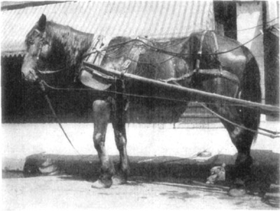
Fig. 10—Contraction of carpal flexors, "knee sprung."
Where inflammation is caused by a puncture wound and subfascial
infection occurs, there is evident manifestation of pain. No weight is
supported by the affected member and because of the pressure, occasioned
by the swollen muscles confined within the non-yielding brachial fascia,
there exists marked supersensitiveness of the affected parts. Flexion of
the elbow is avoided because contraction of the biceps brachii (flexor
brachii) or the extensors, which are antagonists of the flexors of the
carpus, tenses the carpal flexors and pain is thereby increased.
However, in most instances, the practitioner's attention is not directed
to typical and uncomplicated cases, but to subacute or chronic
inflammations which are often attended with contraction of the tendinous
parts of the carpal flexors, and in such cases carpitis is present.
Animals so affected have lost the rigidity which characterizes the
normal carpal joint when the leg is a weight bearing member, and because
of its sprung condition, the leg trembles when supporting weight.
Treatment.—Acute inflammation is treated by means of local
application of cold or hot packs until the pain and acute stage of
inflammation has subsided and later stimulating liniments are indicated.
Absolute quiet must be enforced. Especially where the carpus is involved
must the subject be kept quiet until all evidence of inflammation has
subsided.
The application of vesicants or line-firing is beneficial in subacute
inflammation of the tendons of the carpal flexors. Where contraction of
tendons exists and no osseous or ligamentous change prevents correction
of the condition, tenotomy is necessary. The reader is referred to
Merillat's "Veterinary Surgery"[17] for a good description of the
technic of this operation.
In all serious cases of inflammation of the carpal flexors, whether
tenotomy has been performed or not, the subject needs a long period of
rest subsequent to treatment. In fact, three or four months at pasture
is necessary to permit of recovery and this where no congenital
deformity has predisposed the subject to such affection of the flexors.
Return to work must be gradual and the character of the work such as to
enable the animal to become inured to service without a recurrence of
the trouble if possible.
It follows then, that tenotomy, here as in other cases, is not practical
from an economic viewpoint, unless the animal be of sufficient value to
justify the long period of rest for recovery. Tenotomy is not of
practical benefit unless ample time is allowed for regeneration of
divided tendinous tissue.
Etiology and Occurrence.—Fracture of the carpal bones is of
infrequent occurrence in horses and, when it does occur, it is usually
due to injuries, and because of their nature (resulting as they
generally do from heavy falls or in being run over by street cars or
wagons), a comminuted fracture of one or more bones exists. The
accessory carpal bone (trapezium) is said to be fractured at times
without being subjected to blows or like injuries, but this is
exceptional.
Luxations of the carpal joint are of rare occurrence, and very few cases
of this kind are on record. Walters[18] reports a case of
carpometacarpal luxation in a pony wherein reduction was spontaneous and
an uneventful recovery followed. His reason for reporting the case, as
he states, is its rarity.
Symptomatology.—Fractures of the carpal bones as they usually take
place are diagnosed without difficulty. Because of their usually being
comminuted, abnormal movement of the joint is possible. Such movement is
not restricted and flexion of the leg at the carpus in any direction is
possible. Crepitation is readily detected and frequently these fractures
are of the compound-comminuted variety.
In fracture of the accessory carpal bone (trapezium) or in fracture of
any other single bone when such exists, there is no increase in the
movement of the joint. The accessory carpal bone may be readily
manipulated and when fractured, its parts are more or less displaced.
Recognition of fracture of any other single carpal bone must be done by
detecting crepitation unless it be a compound fracture, whereupon
probing is of aid in establishing a diagnosis.
Carpal luxation when present is to be recognized by finding the apposing
carpal bones joined in an abnormal manner—that is, out of position.
There is restricted or suspended function of the joint, and in the cases
recorded, no difficulty has been experienced in making a diagnosis. The
carpometacarpal portion of the articulation is the part which is usually
affected.
Prognosis and Treatment.—There is no chance for complete recovery in
the usual case of carpal fracture because of the fact that there results
sufficient arthritis to destroy articular cartilage beyond repair. In
the average instance, because of arthritis which persists for a
considerable length of time, more or less ankylosis results. At best,
one can only hope for partial recovery, that is to say, the member may
regain its usefulness as a weight-supporting part, but because of
restricted or abolished joint function, locomotion is more or less
difficult. Exostoses, articular and periarticular, occur and the carpus
usually becomes a large immobile articulation. There is danger of
infection resulting in simple carpal fractures and, needless to say, in
a compound-comminuted fracture of the carpus, infection usually occurs
and a fatal outcome is probable.
When treatment is instituted, antiseptic precautions are taken in
handling the compound fractures, and in any case immobilization of the
parts is sought. Here, as has been previously pointed out, it is best to
employ leather splints, so that a maximum degree of rigidity with a
minimum of distress and inconvenience to the patient will result. The
leg must be bandaged from the hoof upward, making use of a sufficient
amount of cotton to ensure against pressure-necrosis. The leather
splints are placed mesially and laterally and, of course, need to extend
as high as the proximal end of the radius. Subjects must be kept in
slings until union of bones has become established, and as a rule there
will then exist marked ankylosis.
There is no particular difference in the handling of carpal luxation and
dislocation of other bones. Where ligaments have not been destroyed to
the extent that reduction is of no practical use, the parts are kept
immobilized, if thought necessary. Later, vesication of the whole
pericarpal region is done and the subject allowed exercise at will.
Etiology and Occurrence.—Inflammation of the carpus is caused by
contusions, such as are occasioned in falling, by kicks by striking the
carpus against objects in jumping and sometimes by striking it against
the manger in pawing. The condition is of rather frequent occurrence.
Symptomatology.—Evident symptoms of inflammation in carpitis are
always present—hyperthermia, supersensitiveness and swelling. Also,
there exists lameness which is characterized by an apparent inability to
flex the leg, and there is circumduction of the leg as it is advanced
because in this way little if any flexion of the carpus (which increases
pain) is necessary.
Depending upon the nature of the cause, there occurs a marked difference
in the character and amount of swelling.
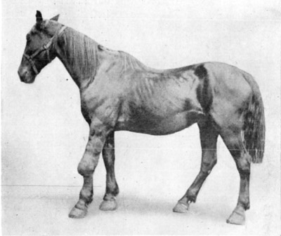
Fig. 11—Pericarpal inflammation and enlargement due to injury.
Naturally, when much extravasation of serum and blood takes place, there
is occasioned a fluctuating swelling which is usually less painful to
the subject upon manipulation than is a dense inflammatory change
without marked extravasation.
In acute carpitis, there is present, then, a very painful condition
which involves the articulation, causing marked lameness, disturbance of
appetite and some elevation of temperature.
Chronic cases do not occasion serious pain or constitutional
disturbances, but do interfere with locomotion in direct proportion to
the existing articular inflammation and periarticular hypertrophy of
ligamentous and tendinous structures.
Treatment.—If possible, keep the subject absolutely quiet, employing
the sling if necessary. During the first stages of inflammation, the
application of ice packs to the affected parts, is of marked benefit.
At the end of forty-eight hours, hot applications may be used and this
treatment continued throughout several days. Anodyne liniments are of
service and should be employed throughout the acute stage of
inflammation during intervals between the hydrotherapeutic treatments.
As inflammation subsides, a counterirritating application such as a
suitable liniment and later blistering or line-firing is helpful in
stimulating resolution.
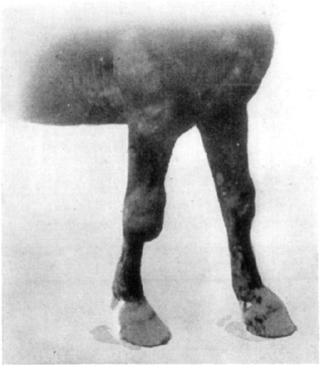
Fig. 12—Hygromatous condition of the right carpus, also
distension of sheaths of extensor tendons of both fore legs.
Anatomy.—The carpal bones as they articulate with one another and
with the radius and metacarpal bones, as classed by anatomists, form
three distinct articular parts of the joint as a whole and are known as
radiocarpal, intercarpal and carpometacarpal. These three pairs of
articulating surfaces are all enclosed within one capsular ligament. On
the anterior face of the bones, the capsular ligament is attached to the
carpal bones in such manner that an imperfect partitioning of the three
joint compartments is formed. Posteriorly, the capsule is very heavy and
forms a sort of padding over the irregular surfaces of the bones, and
also its reflexions constitute the sheaths of the flexor tendons. The
anterior portion of the capsular ligament forms sheaths for the extensor
tendons, and both portions of the joint have an attachment around the
distal end of the radius and another at the proximal end of the
metacarpal bones.
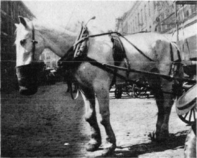
Fig. 13—Carpal exostosis in aged horse.
Etiology and Occurrence.—Puncture wounds of any kind may serve to
perforate the joint capsule and such traumatisms are occasioned by
falls, kicks and in various ways in runaway accidents, and open carpal
joint may follow.
Symptomatology.—The pathognomonic symptoms of the existence of an
open joint is the exposure to view of articular surfaces of bones or
noting the escape of synovia from the joint capsule. As has been
previously referred to, there always exists a peculiar suspension of
carpal flexion in all cases of carpitis.
Non-infective wounds which may cause open joint are not necessarily
productive of an active carpitis—a synovitis may be the extent of the
disturbance. Unlike synovitis, which may characterize a non-infectious
penetrative wound of the capsular ligament, septic arthritis which may
supervene is a very painful inflammatory disturbance. It is
characterized by all of the symptoms which attend the case of open joint
and synovitis plus the obvious manifestation of great pain. There is an
elevation of temperature of from two to five degrees above normal;
circulation is accelerated; the pulse is bounding; respiration is
hurried; there is an expression of pain as indicated by the physiognomy;
and because of rapid erosive changes of cartilages which take place,
there is soon so much of the articulation destroyed that death is
inevitable. Death is usually due to generalization of the arthritic
infection.
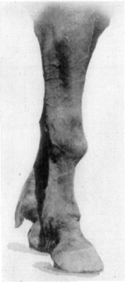
|
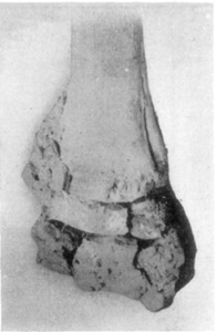
|
Fig. 14—Exostosis of carpus
resultant from carpitis. |
Fig. 15—Distal end of radius.
Illustrating the effects of
chronic carpitis. |
In the meanwhile, if the character of the infectious material is not so
virulent, the disease will take on a slower course and the subject may
experience laminitis from supporting weight upon the sound member, or
because of continued recumbency, decubital gangrene and emaciation
sometimes cause death. If the subject does not soon succumb, it is
compelled to undergo days or even weeks of unnecessary suffering, and
too often in such cases, it is later deemed advisable to destroy the
animal because of the cost of continuing treatment until the horse is
serviceable. Therefore, it is evident that when such joints as the
carpus or tarsus are open and infection exists, if they are not promptly
treated and the infectious process checked, it is neither humane nor
practical to prolong treatment.
Distinction must be made between the different joints when infected as
the condition is much more serious in some cases than in others. All
things considered, perhaps open joints rank, with respect to being
serious cases as follows: elbow, navicular, stifle, tarsus, carpus,
fetlock and pastern. This, of course, is restricted to articulations of
the locomotory apparatus.
Treatment.—Preliminary care in the treatment of an open carpal joint,
is the same as has been described in this condition as it affects the
scapulohumeral articulation described on page 65. Likewise the further
treatment of such cases is along the same lines except that where it is
possible, the parts are kept covered with cotton and bandages. However,
in some cases, animals have been successfully treated without bandaging
and by keeping the patient in a standing position and on pillar reins
until recovery resulted. Such cases were of the non-infectious type and
recovery was possible within three or four weeks. Further, the condition
is not sufficiently painful in such instances as to prevent the subjects
bearing weight with the affected member; hence, no danger of resulting
laminitis is incurred. And finally, where bandages are not employed, the
frequent use of antiseptic dusting powders is substituted for cotton as
a protector.
When bandaged, such wounds need dressing more or less frequently, as
individual instances demand. The purulent infective inflammation of a
carpal joint will require daily dressing; whereas, in other cases
(non-infective), semi-weekly change of bandages is sufficient. Equal
parts of boric acid and exsiccated alum constitute a suitable
combination for the treatment of these cases, and this powder should be
liberally employed. Tincture of iodin may be injected into the joint
capsule where there is provision for its ready evacuation, as
conditions seem to require. Daily injections for three, four or five
days, are not harmful and will control infection in many instances.
Etiology and Occurrence.—The thecae and bursae of the leg are several
in number. In the carpal region, the flexors of the phalanges are
contained together in the carpal sheath, and this is the principal theca
in the carpal region. Each of the tendons is provided with synovial
sheaths which are subject to inflammation and occasionally synovitis and
distension of these synovial sheaths occur.
Because of faulty conformation, some animals are subject to inflammation
of these sheaths, and all forms of strenuous work which taxes the
tendons greatly is apt to result in synovitis. Direct injury such as
blows may be the cause of synovial distension of thecae and the
affection is to be seen in all horses that have done much fast work on
hard road surfaces or pavements.
The usual case as it occurs in practice is a non-infective synovitis,
but where puncture wounds cause the trouble, an infectious inflammation
obtains.
Symptomatology.—No trouble is experienced in diagnosing distension of
tendon sheaths, for the affection is very palpable. During acute
inflammatory stages of this affection, some lameness is present—in
infectious inflammation lameness is intense. Local heat and pain upon
manipulation are readily discernible in all acute cases. And finally,
where there is reason for doubt, an aseptic exploratory puncture of the
wall of the fluctuating enlargement may be made with a suitable trocar,
and the discharging synovia will be proof of the existence of synovial
distension.
After the affection becomes subacute or chronic, no lameness or
inconvenience is manifested, and the condition is undesirable only
because of its being a blemish.
Treatment.—Acute non-infectious synovial distension of tendon sheaths
is treated by aspirating as much synovia as possible from the affected
theca (this is, of course, done under strict asepsis) and by means of
bandages, a uniform degree of pressure is kept over the parts for ten
days or two weeks. The patient is kept quiet and in the course of two
weeks an active blistering agent is employed over the region affected.
Usually, at the end of a month's time, complete recovery has taken place
and the subject may be gradually returned to work.
When synovial distensions are of long standing, it is necessary to take
special precautions to check excessive secretion of synovial fluid, and,
also because of the atonic condition of the tissues affected, resolution
is tardy. In addition to aspirating synovia, the introduction of equal
parts of alcohol and tincture of iodin into the theca is necessary. The
quantity of this combination injected, depends upon the size of the
sheath affected and the amount of synovia retained at the time injection
is made. Experience is necessary to judge as to this part of the work,
but one may consider that a quantity between three and ten cubic
centimeters of equal parts of tincture of iodin and alcohol constitutes
the proper amount to employ. Where much synovia is contained within the
sheath at the time of injection, there occurs great dilution of the
agent injected and consequently less irritation results.
The object of such injections is to check synovial secretion, and this
is sought by the local effect of iodin in contact with the secreting
cells together with the reactionary swelling which occasions pressure.
An increase in the local blood supply also follows. In all cases where
it is possible to employ suitable bandages, this should be done. The
ordinary derby bandages serve well and if their use is continued for a
sufficient length of time, good results follow.
There are other methods of treating these affections, and each has its
advantages and disadvantages. Line-firing, instead of the vesicant is
made use of by some, but the object desired is the same and results
obtained are similar.
Sheaths may be opened surgically by means of a knife, and the removal of
a portion of the wall of distended and atonic tendon sheaths is
possible. These operations belong to the realm of surgery and are not
properly a part of this treatise. However, in passing, it may be said
that if a perfect technic is possible in doing the last named
operation, a permanent recovery is the outcome.
Etiology and Occurrence.—As the result of all sorts of violence, such
as falls and injuries in accidents of various kinds wherein the
metacarpals are subjected to contusions, fractures may result. In the
horse it is unusual for fracture of one of the small metacarpal bones to
take place without there being at the same time a fracture of the third
(large) metacarpal bone.
Classification.—Fractures of the metacarpal bones as they occur, are
as likely to be compound as simple, and the multiple and comminuted
varieties are occasionally observed. The manner in which the third
(large) metacarpus is fractured, largely determines the outcome in any
given case.
Symptomatology.—Abnormal mobility of the broken parts of bone and
crepitation mark fracture of the metacarpus, and the condition is easily
diagnosed. In many instances, when compound fracture exists, broken ends
of bone are protruding through the skin. No weight is borne upon the
fractured member ordinarily, although during the excitement occasioned
by runaways, horses are sometimes seen to support weight with a broken
leg even when the protruding bone is sunk into the ground in so doing.
Prognosis.—Generally speaking, fractures other than the
simple-transverse in young animals, are considered unfavorable cases.
With the metacarpus, however, there are instances where compound
fracture occurs in colts that justify treatment. But in all cases of
compound fracture, the element of infection in addition to the increased
difficulty in maintaining immobility of the broken bone, creates almost
insuperable difficulties in the average instance. And unless the
practitioner distinctly explains to his client the various reasons which
make treatment an economic impracticability, dissatisfaction is likely
to follow if treatment is instituted without such an understanding.
Treatment.—Perfect apposition of the broken ends of bone is easily
effected and less difficulty is encountered in maintaining such
relations in metacarpal fractures than in fractures of the radius.
However, reduction and immobilization of this as in all fractures, must
be done without delay. In simple fracture, the metacarpus is covered
with enough cotton to pad the parts, and this is retained in position by
bandages. Splints of heavy leather or of thin pieces of tough flexible
wood are placed on each side of the leg and firmly held in position with
bandages. Bandages may be put on in layers and a coating of glue applied
over each layer if this is thought necessary. The advantage gained in
using glue or other adhesive materials is that the cast thus formed is
more rigid than where such material is not employed. On the other hand,
all elasticity is lost as soon as the cast adapts itself to the contour
of the extremity, and because of this rigidity, it is doubtful if
anything is gained by the incorporation of glue, except in the way of
added strength of the cast. Since the animal does not walk upon the
broken leg, it is possible to employ splints of suitable materials which
are retained in position without glue and frequent readjustment of a
part of the immobilizing apparatus is possible. This is impossible with
casts.
In compound fractures, provision ought to be made for dressing the wound
of the soft structures. This entails adjusting the splints in such
manner that one splint may be retained and others removed for dressing
the wound and readjusted as often as wound dressing is necessary.
By this term is meant a condition where there exists an exostosis which
involves usually the second (inner small) and third (large) metacarpal
bones. While an exostosis involving any one of the splint bones, even
when directly caused by an injury, is called a "splint," the term is
employed here, in reference to exostoses not due to direct injury such
as in contusions.
Etiology and Occurrence.—This condition is one wherein there is
osseous formation following a periostitis and the region of the upper
portion of the second (inner small) metacarpal bone is the usual site of
the exostosis. There is incited an inflammation of the periosteum at
the site of the interosseous ligament which attaches the small to the
large metacarpal bone. This ligament is involved in the inflammatory
process, and according to Havemann, whose view is supported by Moller,
this inflammation is the origin of the trouble.
Various theories attempting an explanation of the frequent affection of
this one certain part so regularly involved have been offered, but no
proof of the correctness of any exists. It follows, however, that
splints occur in young animals; that the affection seldom starts in
subjects that are ten years of age or older, and that when the exostosis
has formed, lameness usually subsides. Anything which will cause undue
strain or irritation of the metacarpal bones in young animals, is quite
apt to result in a splint being formed. Concussion such as is caused by
fast work on hard roads, or work on rough or irregular road surfaces
which cause unequal distribution of weight, will cause splint lameness
and exostosis follows.
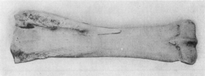
Fig. 16—Posterior view of radius (right) illustrative of
effects of splint. Note the extent of exostosis.
Course.—Because of the peculiar manner in which the second and third
metacarpal bones articulate in young animals, until the bones become
ossified and permanently joined, the inflammation which attends the
acute stage of this affection, causes lameness. Later, unless an
unusually large exostosis is formed, which may cause a constant
irritation due to its size and juxtaposition to the carpus, lameness is
discontinued.
Symptomatology.—Lameness is usually the first manifestation of this
disorder, and the thing which characterizes splint lameness is its
peculiar intermittence. There is a mixed form of lameness which may not
be in evidence when an affected animal is started on a drive, but which
is marked after the subject has gone some distance. The animal may,
however, go lame throughout the whole of a drive and continue to be lame
for several days or weeks in some cases. It is noticeable that lameness
is augmented or produced when the subject travels on rough road surfaces
and that little or no difficulty is encountered when roads are smooth.
The heavy brachial fascia is inserted in part to the head of the second
metacarpal (inner small) bone together with the oblique digital extensor
(extensor metacarpi obliquus) and this explains the reason for pain
being manifested during extension of the member.
Before there is a visible exostosis, supersensitiveness is readily
recognized upon palpation of the parts, if careful comparison is made
between the sound and unsound members. However, frequently splints occur
on both forelegs at the same time and in some instances exostoses are
several in number upon each member affected. In some instances, the
affection involves the outer splint bone and no evident involvement of
the inner one exists.
Treatment.—At the onset complete rest should be provided and the
local application of some good cataplasm is in order. A stimulating
liniment is beneficial when employed several times daily and massage is
also quite helpful. Later, the application of a blistering ointment is
good treatment. The use of the actual cautery stimulates prompt
resolution, but there is seldom any resorption of products of
inflammation following firing. Whereas, in cases where other treatment
is begun early, there usually follows considerable diminution in the
size of the exostosis. A rest of four or five weeks is necessary and
very young animals should not be put to work too soon, if the character
of the work is such as to induce a recurrence of the trouble.
Many cases are treated successfully in draft types of animals (where the
subjects are not kept at work that occasions serious irritation to the
affected parts) by blistering the exostosis repeatedly and allowing the
animals to continue in service. In such cases, it is unreasonable to
expect to check the size of the exostosis and, of course, such methods
are not employed where lameness causes distress to the subject.
Firing usually causes prompt recovery from lameness and is a dependable
manner of treating such cases but there remains more blemish following
cauterization than where vesication is done.
This condition, because of the frequency with which it occurs may be
taken as typal, from the standpoint of treatment and results obtained
therefrom. While it serves to constitute a basis from which other
joints, when open, are to be considered, due allowance must be made for
the fact that, as has been previously mentioned, some articulations when
open constitute cause for grave consequences; while with others an open
capsule, even when infected, does not cause disturbance enough to be
classed as difficult to handle. Moreover, the fetlock joint is admirably
suited, anatomically, to bandaging; and when wounded, is easily kept
protected by means of surgical dressings. This fact is of great
importance in influencing the course and termination in any given case
of open fetlock joint and should not be forgotten.
There is no logical reason for comparing the pedal joint with the
pastern on the basis that it may also be completely and securely
bandaged. Open navicular joint does not occur, as a rule, except by way
of the solar surface of the foot, and the introduction of active and
virulent contagium is certain to happen; consequently, an acute
synovitis quickly resulting in an intensely septic and progressively
destructive arthritis soon follows in perforation of the capsule of the
distal interphalangeal articulation.
Etiology and Occurrence.—Wounds of the fetlock region resulting in
perforation or destruction of a part of the capsular ligament are caused
by all sorts of accidents, such as wire cuts, incised wounds occasioned
by plowshares, disc harrows, stalk cutters and other farming implements.
In runaways the joint capsule is sometimes punctured by sharp pieces of
wood or other objects. In horses driven on unpaved country roads the
fetlock is occasionally wounded by being struck against the sharp end of
some object, the other end of which is firmly embedded in the ground. In
one instance the author treated a case wherein the fetlock joint was
perforated by the sickle-guard of a self-binder. In this case there
occurred complete perforation causing two openings through the
cul-de-sac of the joint. Such wounds are produced by implements which
are, to say the least, non-sterile, and this perforation of the
uncleansed skin conveys infectious material into the joint capsule. Yet
in many instances, especially in country practice, no infectious
arthritis results where cases are promptly cared for.
Symptomatology.—A difference in the character of symptoms is
evidenced when dissimilar causes exist. Small penetrant wounds which
infect the synovial membranes cause infectious arthritis in some cases,
whereas a wound of sufficient size to produce evacuation of all synovia
will, in many instances, cause no serious distress to the subject, even
when not treated for several days. If it is not evident that an open
joint exists and the articular cavity is not exposed to view a positive
diagnosis may be early established by carefully probing the wound. In
some cases where a small wound has perforated the joint capsule,
swelling and slight change of relation of the overlying tissues may
preclude all successful exploratory probing. In such instances it is
necessary to await development of symptoms. Twenty-four hours after
injury has been inflicted, there is noticeable discharge of synovia
which coagulates about the margin of the orifice, where synovial
discharge is possible. Particularly evident is this accumulation of
coagulated synovia where wounds have been bandaged—there is no
mistaking the characteristic straw-colored coagulum which, in such
cases, is somewhat tenacious.
No difference exists between other symptoms in infectious arthritis
caused by punctures, and non-infectious arthritis, excepting the
intensity of the pain occasioned, the rise in temperature, circulatory
disturbances, etc.; all of which have been previously mentioned.
Treatment.—Just as has been stated in discussions on the subject of
open joint, probing or other instrumentation is to be avoided until the
exterior of the wound and a liberal area surrounding has been thoroughly
cleansed—too much importance can not be placed on this preliminary
measure. In cases of open joint where ragged wound margins exist and the
interior of the joint capsule is contaminated, much time is required to
thoroughly cleanse all soiled parts. In some instances an hour's time is
required for this cleansing process after the subject has been
restrained and prepared. In order to thoroughly cleanse these delicate
structures without doing them serious injury, one ought to be skillful
and careful in all manipulations of the exposed parts of the joint
capsule.
The general plan of treatment, after preliminary cleansing has been
accomplished, has been outlined on page 66 in the consideration of
scapulohumeral joint affections. The injection of undiluted tincture of
iodin in ounce quantities, it must be remembered, is not to be done
unless there is provision for its free exit. Where good drainage from
the joint cavity exists all infected wounds should be thus treated, and
this treatment may be repeated as conditions seem to require—until
infection is checked.
If daily injections are necessary, dilution of the tincture of iodin
with an equal amount of alcohol is advisable in order to avoid doing
irreparable damage to the articular cartilages and synovial membranes.
An antiseptic powder composed of equal parts of boric acid and
exsiccated alum is employed to protect the wound surfaces and the
margins, and the parts are then bandaged. In bandaging wounds of this
kind a liberal amount of cotton should be employed, and after a large
surface surrounding the wound has been thoroughly cleansed, it must be
so kept thereafter. This is impossible, if one uses a small amount of
cotton, particularly if such meager quantity of dressing material is
carelessly wrapped in position with an insufficient amount of bandage
material. Mention, without description of the elemental problem of
applying cotton and bandages to a wound, would be sufficient, were it
not that this is a very important part of the handling of such cases,
and many practitioners are not only thoughtless in this part of their
work, but also apparently careless. What does it profit to prepare a
part and cleanse a wound with painstaking care and then neglect to take
every possible precaution to prevent its subsequent contamination?
In the handling of open joint capsules where the perforation of the
capsular ligament is small and discharge of synovia does not immediately
follow, there is presented a problem which is difficult to decide upon
and that is the manner in which such wounds are to be handled. One
hesitates to enlarge such openings to drain or irrigate the capsule when
there is no proof that serious trouble will follow because of infectious
material which has probably been introduced at the time the wound was
inflicted. It is especially difficult to decide upon the manner of
handling such cases where the tarsal joint is wounded, although one
hesitates to invade any joint to the extent of incising its capsule,
unless there is urgent need of so doing.
Frost[19] offers the following suggestion in such instances:
The treatment recommended by us for open joints, in which we wish
to prevent ankylosis, is, first, to shave all hair from the area
surrounding the wound, following with a thorough cleansing of the
skin and disinfection of the wound, and then to inject a twenty per
cent Lugol's solution in glycerin into the wound. This should be
repeated two or three times a day, each time enough of the solution
being injected to fill the joint capsule, thereby securing the
flushing effect. As this solution does not cause irritation to the
tissue and yet is a strong antiseptic, it serves to shorten the
period of congestion and inflammation and to overcome the infection
without causing a destruction of the secreting membrane until the
external wound has had time to heal. The injection of this solution
seems to retard the excessive secretion of synovia. The larger the
joint capsule and the smaller the external wound, the longer our
antiseptic will remain in contact with the inflamed tissues as the
glycerin, being thick, does not flow through a small opening.
After-care.—Following the initial cleansing and treatment of open
joint, subsequent dressing is necessary as frequently as conditions
demand. If the parts are badly infected and profuse discharge of pus
exists a daily change of dressings is necessary. In the average
instance, however, semi-weekly treatments are sufficient. And in many
instances where one is obliged to travel a considerable distance to
handle the affected animal one weekly dressing of the wound will suffice
after the second treatment.
The same general plan of treatment concerning the subject's comfort that
has been previously mentioned in arthritis, is carried out here. A
further and detailed consideration of the subject of handling of open
joints follows.[20]
* * * Such wounds may be classified in two general groups as
follows: First, wounds in which the trauma has exposed the
articulation to view, and second, those the result of punctures, in
which the external wound is small and free drainage is lacking.
Wounds in which the articulation is exposed to view have drainage
either all ready provided for, or it is established without
hesitancy surgically. With free drainage thus established there is
little or no chance for the adjacent tissues to become infiltrated
with infected wound discharge. This prevents an extension of the
injury and the establishment of a good field for the growth of
anaerobic bacteria.
Open joints caused by punctures, unless the puncture is aseptic,
produce a swelling which is more painful than is the open wound
which exposes the joint to view. Especially is this true if the
puncture is of small diameter, allowing the tissues to partially
close the opening immediately after the wound has been made. Where
drainage is lacking there follows an exudation which congests the
tissues surrounding the injury and all factors favoring germ growth
are present. It is perhaps advisable to establish good drainage in
such cases as soon as a diagnosis is made.
It is not always an easy matter to recognize an open-joint, when
first made, but twelve to twenty-four hours later there is no cause
for doubt. The condition is then a very painful one; lameness is
excessive; there is rise in temperature; acceleration of the pulse
and manipulation or palpation of the region affected, occasions
great pain.
The treatment of open joints must be varied to suit the disposition
of the animal, the nature and location of the injury, the length of
time intervening between the infliction of the wound and the first
attention given, and the surroundings in which the patient is kept.
In each and every case in which there exists an open wound the
surface surrounding the wound is cleansed thoroughly, the hair is
shaved if possible, and the margin of the wound is curretted and
cleansed thoroughly with antiseptic solutions.
If there is evidence that the articulation contains infective
material, it is washed out with copious quantities of peroxide of
hydrogen—usually as much as six or eight ounces. This is followed
by injection of an ounce or two of tincture of iodin. Even though
the joint appears to be clean some tincture of iodin is used, as it
checks the secretion of synovia and is, in every way, beneficial.
Care is taken to apply the iodin also to the surface immediately
surrounding the wound. The entire wound is then covered with a
dusting powder composed of zinc oxide, boric acid, exsiccated alum,
phenol and camphor.
This powder is used in abundance and the wound is then covered with
a heavy layer of absorbent cotton and well bandaged. This bandage
is not disturbed for at least three days and may be left in place
for a week. In cases in which it is necessary to keep the dressing
on for a week, or in cases where the patient is, through necessity,
kept in quarters that are wet or unclean, the first bandage is
covered with a layer of oakum which has been saturated in oil of
tar and this in turn is held in place by means of several layers of
bandages. The bandages are also saturated with oil of tar.
In from one to two months wounds so treated, unless they are
foot-wounds, will be ready to dress without being bandaged. It is
ordinarily unnecessary to dress foot-wounds oftener than every
second week after the discharge of synovia has ceased. When the
wound has filled with granulation, a protective dressing is applied
which is rendered water proof by the use of bandages covered with
oil of tar. The patient can now be turned out for a month or six
weeks without disturbing the dressing. After the removal of the
bandages, the only treatment necessary is an occasional application
of some mildly antiseptic ointment.
Except in nail pricks of the foot, occasioned by punctures, a five
per cent tincture of iodin is injected into open joints, if the
wound remains sufficiently open, and this treatment is continued so
long as there is a discharge of synovia. Surgical drainage is
established if it is considered practicable and the remainder of
the treatment is about the same as for wounds which are open.
Open joints occur in horses at pasture and are sometimes not
discovered until several days or a week after the injury, and in
some instances the wounds are filled with maggots. The only
difference in the treatment of these cases is that more time and
care is taken in cleansing the wound, more curetting is necessary,
and after cleansing the wound with peroxide of hydrogen, the joint
is thoroughly washed out with equal parts of tincture of iodin and
chloroform. This is followed by the injection of a quantity of
seventy-five percent alcohol and the wound is dressed and bandaged
as already described. At each subsequent dressing of infected
wounds so treated less suppuration is noticed and the synovial
discharge usually ceases in from one to two months.
About ninety percent of all cases of open joint make complete
recoveries, about four per cent partially recover and six per cent
are fatal. Among the fatal cases are the open joints with
complications as severed tendons, those occasioned by calk wounds
in horses that are stabled, and nail punctures of the feet. The
following report of twelve favorable cases is taken from a record
of sixty-two cases. The favorable ones are reported, chiefly
because there are now enough reports on record of such cases which
have terminated fatally.
Case 1.—A gray gelding used as a saddle pony received a
horizontal wire cut laying completely bare the scapulohumeral
articulation. The margins of the wound were cleansed as heretofore
described, a drainage was provided surgically, tincture of iodin
was injected and the wound was covered with equal parts of boric
acid and exsiccated alum. The horse was kept tied and a diluted
tincture of iodin was injected into the wound once daily and the
powder applied often enough to keep the wound covered. The case
made a complete recovery and the pony was again in service within
sixty days.
Case 2.—A twelve-hundred-pound bay mare with an open carpal
joint. The wound was an open one about two and one-half inches in
length, and made transversely and when the member was flexed the
articular surface of the carpal bones were presented to view. An
ounce of tincture of iodin was injected into this joint after
having cleansed the margin of the wound and the mare was cross-tied
in a single stall to keep her from lying down. The owner was
instructed to keep the outside of the wound powdered with air
slaked lime and a very unfavorable prognosis was given.
I heard nothing further from this case until fifty-nine days from
the date of the injury, when I met the owner driving this mare to a
buggy. The wound had healed by first intention and at that time so
little cicatrix remained that it was difficult to find it.
Case 3.—A brown mare with an open fetlock joint due to a
spike-nail puncture. Lameness was excessive, and joint greatly
swollen. Tincture of iodin was injected into the wound and towels
dipped in hot antiseptic solutions were applied for several hours
daily until the acute stage had passed. Later the mare was turned
out to pasture and a vesicant was applied once or twice a month
until recovery was complete which was in about six months.
Case 4.—A four-year-old bay mare having a wire-cut which opened
the tarsus joint was treated as heretofore described. The wound was
kept bandaged for about two weeks and later it was dressed without
being bandaged. In ninety days she had completely recovered.
Case 5.—A twelve-year-old mare with an open fetlock joint due to
a puncture wound. The margins of the wound were cleansed and the
external wound enlarged to facilitate drainage. Tincture of iodin
was injected; the wound was bandaged and dressed for a month in the
manner heretofore described, when all discharge had stopped. A
vesicant was applied; the mare was put to pasture and within sixty
days from the date of the injury she was being driven on short
trips.
Case 6.—A two-year-old brown gelding with a wire-cut on the left
front foot. The wound extended down through the sole and opened the
navicular joint. This colt was very wild and it was necessary to
tie it down each time the wound was dressed. The wound was dressed
weekly for a month and less frequently thereafter. It was handled
eight times; the last dressing was left in place until worn out.
Six months later the colt was practically well, a very little
lameness being shown when walking on frozen ground.
Case 7.—A seven-year-old saddle-horse weighing eleven hundred
and fifty pounds received a wound of the tarsus, laying bare the
articular surfaces of a part of the joint. It was impossible to
keep this wound bandaged because of the restless disposition of the
subject. Injections of a dilute tincture of iodin were employed
every second or third day for a month and the wound was kept
covered with the antiseptic dusting powder referred to heretofore.
In five months complete recovery had taken place, with the
exception of a stubborn skin disturbance which was successfully
treated six months after the wound was inflicted. The horse is
still in use and is absolutely free from lameness.
Case 8.—A two-year-old brown gelding with a wire-wound opening
the scapulohumeral joint. This wound was large enough to expose to
view the articular portion of the humerus. The same treatment as
that given case No. one was instituted and in ninety days the colt
was practically well.
Case 9.—A three-year-old bay filly was found at pasture with one
fore foot badly injured. The owner intended to destroy her, but a
neighbor prevailed upon him to have her treated. Apparently the
wound was of about a week's standing and in a very bad condition,
filled with maggots and dirt. Both the navicular and coronary
articulations were open. This wound was cleansed in the usual
manner and the owner cared for the case the balance of the time
because the distance from my office was too great to give her
personal attention. She made an almost complete recovery in five
months.
Case 10.—At two-year-old mule with an open navicular joint due
to a barbed wire wound. Usual care was given this case and in five
months recovery was complete and little scar is to be seen. This
case received seven treatments.
Case 11.—An eighteen-months-old colt at pasture was found down
and unable to rise without help. In addition to several wounds of
lesser importance there was a large wound on the inner side of the
elbow, the joint was open and the entire leg was greatly swollen
and in a state of acute infectious inflammation. The colt could not
walk, its temperature was 105°, pulse was rapid and respiration was
a little hurried. After advising the owner to put the poor animal
out of its misery I left the place. Four days later the owner came
to my office and asked if he could borrow some old shears to "trim
off some loose hide from that colt." He left the colt in the
pasture and all the care it received was the regular application of
a proprietary dusting powder. It made a complete recovery.
Case 12.—A family mare, heavy in foal, received a vertical wound
of the fetlock joint inflicted by a disc-harrow. The cul-de-sac
of the ligament of this joint was opened freely. The wound was
dressed in the usual manner and again three days later when no
suppuration had taken place. Four days later the patient gave birth
to a colt and suckled it right along through her convalescence.
This wound healed by first intention and seventy-nine days from the
date of the injury the mare was driven to town, two and one-half
miles distant, and showed but little lameness.
This term is applied to exostoses involving the first and second
phalanges (suffraginis and corona), regardless of their size, extent or
location. It is a misnomer, in a sense, and the veterinarian is
frequently obliged to spend considerable time with his clients in order
to convince them that a spherodial exostosis of the proximal phalanx, in
certain cases, is in reality "ringbone," even though there exists no
exostosis which completely encircles the affected bone.
Etiology and Occurrence.—Exostosis of the first and second phalanges
is usually due to some form of injury, whether it be a contusion, a
lacerated wound which damages the periosteum, or periostititis and
osteitis incited by concussions of locomotion, or ligamentous strain.
Practically the only exception is in the rachitic form of ringbone which
affects young animals.
There are predisposing causes that merit consideration, chief among
which is the normal conformation of the coronet joint. This proclivity
is constant; the normal interphalangeal articulation is an incomplete
ginglymoid joint and while its dorso-volar diameter is great, this in no
wise compensates for its disproportionately narrow transverse diameter.
The pivotal strain which is sometimes thrown upon this articulation when
an animal turns on one foot, as well as the tension which is put on the
collateral ligaments when the inner or the outer quarter of the foot
rests in a depression of the road surface, tends to detach the insertion
of these ligaments or to cause fibrillary fractures of their substance.
Short, upright, pasterns receive greater concussion during fast travel
on hard roads than do the longer more sloping and well formed
extremities. Those who are advocates of the theory that this type of
osteitis with its complications has its origin in the articular portion
of the joint, claim that the upright pastern constitutes an important
tendency toward ringbone. Howbeit, ringbone is an active, serious and
frequent cause of lameness and it affects animals of all ages and occurs
under various conditions. Horses having good conformation and kept at
work wherein no great amount of strain is put upon these parts, are
occasionally victims of this affection.
Classification.—The arrangement employed by Moller[21] is intensely
practical and logical. He considers ringbone as articular,
periarticular, rachitic and traumatic. A mode of classification
that is common and in a practical way, good, is, high and low ringbone.
When prognosis is considered, for instance, it is very convenient to
state that the chances for recovery are much better in high ringbone
than in low ringbone. The classification of Möller will be followed
here.
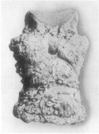
Fig. 17—Phalangeal exostoses.
Symptomatology.—In all forms of incipient ringbone except rachitic,
the first manifestation of its existence, or of injury to the ligaments
in the region of the pastern joint which causes periostitis, or
affections of the articular portions of the proximal inter-phalangeal
joint, is lameness. Lameness which typifies ringbone is of the
supporting-leg variety and by compelling the subject to step from side
to side, marked flinching is observed, especially in periarticular
ringbone; causing the affected animal to turn abruptly on the diseased
member, using it as a pivot, likewise accentuates the manifestation. In
fact, many subjects that exhibit no evidence of locomotory impediment
while walking or trotting in a straight line on a smooth road surface,
will manifest the characteristic form of lameness from ringbone when the
aforementioned side to side movement is performed.
When the manner in which pain is occasioned is considered, it will be
understood why lameness is intermittent in the early stages of this
affection and may even be unnoticed by the driver. An animal may travel
on a smooth road without giving evidence of any inconvenience, but as
soon as a rough and irregular pavement or road surface is reached, will
limp. As the subject is driven farther on level streets the lameness may
disappear. This intermittent type of lameness may continue until there
is developed a large exostosis, or until articular involvement causes so
much distress during locomotion that lameness is constant. On the other
hand, resolution may occur during the stage of periosteal inflammation,
or, an exostosis forms which causes no interference with function.
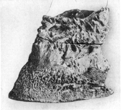
Fig. 18—Rarefying osteitis in chronic ringbone and
ossification of lateral cartilages.
Before there is evidence of an exostosis, diagnosis of ringbone is not
easy, for it is then a problem of detecting the presence of a
ligamentous sprain, periostitis, or osteitis. The diagnostician should
take note of local manifestations of hypersensitiveness, or heat if such
exist, and, in addition, other conditions must be excluded before
definite conclusions are possible.
In articular ringbone as soon as there is developed an exostosis, it
occupies a position on the dorsal (anterior) part of the articulation
and extends around the sides of the joint.
Periarticular ringbone is characterized by exostoses which are
situated on the sides of the phalanges and not extending around to the
anterior part of the joint. This type of ringbone as well as the
articular may occur "high" or "low."
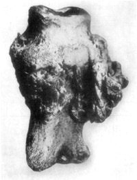
Fig. 19—Phalangeal exostoses in chronic ringbone. Museum
specimen of the Kansas City Veterinary College.
With the traumatic form of ringbone, all consequences, as to the size
and form the exostosis is to assume, depend upon the nature and extent
of the injury.
Rachitic ringbone is frequently observed in some sections of the
country and does not ordinarily cause much if any lameness. It is a
disease of colts and may affect one or all of the phalanges at the same
time. As the subject advances in age there is more or less diminution in
the size of the enlargements.
Treatment.—Rest is essential in the treatment of ringbone. If
diagnosed during its incipiency, remedial measures such as are usually
employed to treat sprains, are indicated and later the parts should be
blistered. When an exostosis has developed puncture firing is the remedy
par excellence. Not that this method of treatment is infallible, for
to any thinking one who takes into consideration the pathological
anatomy of this condition, it is evident that no manner of treatment is
beneficial in some cases. If the exostosis is so situated that it does
not mechanically interfere with function, and is not so large that it
may inhibit flexion and extension, and where the articular portions of
the joint are not eroded, good results attend the use of the actual
cautery.
In firing, after having anesthetized the extremity, and prepared the
surgical area, the cautery is deeply inserted in numerous places, taking
care, however, not to open the joint. The parts are immediately covered
with aseptic absorbent cotton and this dressing is left in position for
forty-eight hours and if perchance there is evidence of synovial
discharge, the parts are again aseptically dressed in order to prevent
infection of the articulation. If, as is the case usually, no
perforation of the joint capsule exists, the openings made by the
cautery have been closed by the coagulation of serum and there is then
little chance of infection causing trouble, even though the member is
left unbandaged.
In several instances, the author has treated ringbone by this method
where the periarticular type existed and lameness was marked, and in
three weeks the subjects were in service and not lame—this, in one
instance in a valuable polo pony where the subject continued in service
for more than a year without any evidence of recurrence of the lameness.
The production of a deep-seated and acute inflammation with the actual
cautery is preferable to any sort of counter-irritation which may be
produced by vesicants.
There is no occasion for any difference in the treatment of either of
the first three classes of ringbone, but in the rachitic type where
treatment is given, the application of a vesicant is all that is
required. In most instances treatment is not necessary.
The affected animals require a month to three months' time for recovery
to take place in the average favorable cases of ringbone.
Median neurectomy is of service in many instances where lameness is not
completely relieved by the use of the actual cautery and no bad results
attend the performance of this operation even though no benefit is
derived thereby. Plantar neurectomy is contraindicated in all cases
where there exists much lameness. If lameness is due to acute
inflammation bad results such as sloughing and loss of the hoof may
follow; and if large exostoses mechanically interfere with function of
the joint, or where articular erosions exist, no possible good can come
from neurectomy. Careful discrimination should be employed in selecting
cases for neurectomy for this operation; otherwise, it is very likely to
prove disappointing.
This condition does not differ from a like affection involving other
tendons except that the function of these tendons is such that large
synovial sheaths are necessary, and when synovitis exists, the condition
then becomes more serious.
Infectious synovitis involving these tendons in the fetlock region is of
more frequent occurrence than a like affection of carpal or tarsal
sheaths. With the exception of the extent of the involvement and
distress occasioned thereby, synovitis the result of open tendon
sheaths, is similar wherever it occurs.
Etiology.—The same conditions which are responsible for open fetlock
joint and other wounds of the pastern region, cause open tendon sheaths
of the flexor tendons.
Symptomatology.—Because of the size and extent of this sheath and the
different manner in which it is opened, there is manifested dissimilar
symptoms in different cases. A nail puncture which perforates the sheath
in the pastern region and at the same time produces an infectious
synovitis, will cause a markedly different manifestation than will a
wound which freely opens the sheath above the fetlock. In the first
instance, the condition is much more painful; swelling is intense in
some cases; and if the subject does not possess sufficient resistance so
that spontaneous resolution promptly occurs, surgical evacuation of pus
is usually necessary. When these tendon sheaths are opened, there
follows a reaction which is quite analogous to that which exists in
arthritic synovitis, but instead of ankylosis, adhesions with thecal
obliteration occur. Rarely there result cartilaginous and osseous
formations.
The constitutional disturbances which characterize this condition vary
with the degree of distress occasioned. As the infection is virulent and
causes serious destruction of the affected parts, so does evidence of
malaise and finally distress appear. Detailed discussions of
symptomatology in similar conditions have heretofore been given, and
further repetition is unnecessary.
Treatment.—The same general plan of treatment which is employed for
handling open joint is put in practice in these cases. Following the
preoperative cleansing of the external wound and adjacent surfaces,
where liberal drainage exists, tincture of iodin is injected into the
sheath, the parts covered with a suitable dressing powder, and the
entire member is carefully dressed with cotton and bandages.
Subsequent treatment is the same as has been outlined in the discussion
of open fetlock joint on page 112. The same general plan of after-care
is necessary. Recovery, however, does not require so much time
ordinarily, yet punctures of the sheath occasioned by nails or other
small implements make for long drawn out cases of infective synovitis.
Etiology and Occurrence.—The manner of construction of the fetlock
joint is such that disarticulation without irreparable injury resulting,
is practically impossible. Logically, this joint in the fore legs (not
so in the pelvic limbs) should disarticulate in such manner that either
all of the inhibitory apparatus (flexor tendons and suspensory ligament)
must rupture or a lateral luxation is necessary. Lateral disarticulation
must necessarily sever the attachment of one of the common collateral
ligaments. Because of the width (transverse diameter) of the
articulating surfaces of this joint, lateral luxation requires a great
strain; and a force that is sufficient to occasion this trauma usually
causes serious additional injury. Therefore, the condition is considered
one wherein prognosis is always unfavorable in so far as practical
methods of treatment are concerned.
Mr. A. Barbier[22] reports a case of bilateral luxation of the fetlock
joints of the hind legs in a horse. This was done in jumping, and the
extensor tendon of each leg was ruptured and the anterior portion of the
metatarsus was protruding through the skin. Profuse hemorrhage had taken
place due to tearing of the blood vessels.
Symptomatology.—Entire luxation of this joint when present is so
evident that one cannot fail to recognize the condition. Complete
disarrangement of normal relation occurs and there is either a breaking
down of the inhibitory apparatus, or if a lateral disarticulation
exists, the normally straight line formed by the bones of the front leg,
as viewed from the front or rear, is broken at the fetlock.
Often fracture of bones are concomitant and then, of course, mobility is
increased and not decreased as is the case in uncomplicated luxation.
Such violence occurs at times, when this joint is disarticulated, that
the joint capsule is also completely ruptured and the articular portion
of the bones is exposed to view.
Treatment.—The condition being practically a hopeless one,
destruction of the subject is the thing which should be promptly done.
In valuable breeding animals, owners may prefer that treatment be
attempted when a lateral luxation and detachment of but one common
ligament have permitted luxation without complete disarticulation and
rupture of the joint capsule. In such cases, by immobilizing the
affected parts as in fracture, and confining the subject in a sling for
about sixty days, partial recovery may occur in some instances.
Experience has shown that where luxation with detachment of a
collateral ligament occurs, recovery is slow and incomplete—there
always results considerable exostosis at the site of injury.
Etiology and Occurrence.—Inflammation of the proximal sesamoid bones
is caused by any kind of irritation which may involve this part of the
inhibitory apparatus. Positioned as they are, between the bifurcations
of the suspensory ligament and the pastern joint, they serve as fulcra
and effectively assist in minimizing concussion which is received by the
suspensory ligament. The flexor tendons also, in contracting, exert
strain upon the inter-sesamoidean ligament, which has a similar effect
upon the sesamoid bones as that which is produced by the suspensory
ligament.
The condition occurs quite frequently, and because of the important
function performed by these bones, active inflammation of the sesamoids
constitutes a serious affection. Because of the fact that these bones
have proportionately large articular surfaces, when they are inflamed to
the extent that degenerative changes affect the articular cartilage,
complete recovery seldom results.
The same pathological changes occur here that are to be seen in any case
of arthritis. No special pathological condition characterizes
sesamoiditis but this condition causes incurable lameness when the
sesamoid bones are much inflamed.
Symptomatology.—In acute inflammation, there exist all the symptoms
which portray any arthritic inflammation of like character. The parts
are readily palpable and are found to be hot, supersensitive, and more
or less infiltration of the tissues contiguous to the joint causes
swelling. There is volar flexion of the phalanges when the subject is at
rest. Lameness is intense; in some acute inflammatory disturbances the
subject is unable to bear weight on the affected member.
In chronic sesamoiditis, constant lameness is the one salient feature
which marks the condition. While it is possible for one sesamoid bone to
become involved without its fellow being affected, this is not usual.
Considerable organization of tissue surrounding the joint is present and
no particular evidence of supersensitiveness exists. However,
supporting weight brings sufficient pressure to bear upon the inflamed
and more or less eroded bones so that pain is occasioned and lameness
results.
Treatment.—During acute inflammation, absolute quiet is, of course,
of first consideration. Cold packs are to be kept in contact with the
parts until acute inflammatory symptoms subside. The fetlock region is
then enveloped with a poultice or an iodin and glycerin combination
(iodin one part to seven parts of glycerin) is applied and a dressing of
cotton is kept in contact with the inflamed region. Following this, a
vesicant is employed and the subject is allowed a month's rest.
In sub-acute cases, the entire region surrounding the pastern is
blistered or the actual cautery is used. Line-firing is preferable. The
subject is given a month or six weeks rest and one may be guided by the
presence or absence of lameness as to whether improvement or recovery is
taking place.
Old chronic cases, and particularly those where there are considerable
induration and fibrous organization of tissue surrounding the joint, are
not to be benefited by treatment.
The chief consideration in handling sesamoiditis is checking
inflammation as early as possible and preventing, if this can be done,
the erosion of articular surfaces. If destruction of any part of the
articular surfaces can be prevented and the patient allowed ample time
for complete resolution of the affected parts to occur, permanent relief
is possible.
Etiology and Occurrence.—Fracture of the proximal sesamoid bones is
caused by violent strain when there exists fragilitas osseum, or by
contusions. The author treated a case where fracture of one sesamoid was
occasioned by a horse receiving a puncture wound wherein the sharp end
of a steel bar was protruding from the ground where it was firmly
embedded. The subject in this case was injured while being driven along
a country road. Frost[23] reports simultaneous fracture of all of the
proximal sesamoids occurring in a sixteen-year-old pony. The condition
is of rather common occurrence in some countries because of the fragile
condition of horses' bones.
Symptomatology.—If the parts can be examined before extravasation of
blood and swelling mask the condition, crepitation may be detected. In
other instances, it is possible to note a displacement of parts of the
sesamoid bones—this in horizontal fracture. There occurs more or less
descent of the fetlock which must not be attributed to rupture of the
superficial flexor tendon (perforatus). By outlining the course of this
tendon with the fingers, when it is passively tensed sufficiently to
follow its course, one may exclude rupture of the superficial flexor.
Finding the suspensory ligament intact from its origin to the sesamoid
attachments, one may also eliminate rupture of this structure as a cause
of the trouble. Needless to say, marked lameness and swelling of the
fetlock soon take place. The condition is painful, and ordinarily,
recovery is impossible.
Treatment.—Where treatment is attempted, immobilization as in
luxation is in order. The patient's comfort is sought, and if the
fractured parts can be kept in close proximity, their union may occur in
time. However, chances for partial recovery (which is the best to be
hoped for) are so remote that early destruction of the subject is the
humane and economical thing to do.
Where treatment is instituted, it is found that there is required a long
time for union of the fractured bones to occur (where union does take
place) and the cost of treatment together with the uncertainty of even
partial recovery, makes for an unfavorable outcome. When the best
possible results succeed treatment, a large callosity is formed and
movement of the pastern joint is restricted. Lameness, though not
intense, in the case referred to, where one bone was broken, was
permanent and the subject was out of service for nearly a year.
Anatomy.—The ligaments here involved are the four volar ligaments
described by Sisson[24] as follows: "The volar ligaments (Ligg
Volaria) consist of a central pair and a lateral and medial bands which
are attached below to the posterior margin of the proximal end of the
second phalanx and its complementary fibro-cartilage. The lateral and
medial ligaments are attached above to the middle of the borders of the
first phalanx, the central pair lower down and on the margin of the
triangular rough area."
This portion of the inhibitory apparatus is described by Strangeways'
Anatomy as two posterior ligaments which run each from three points on
the sides of the os suffraginis to a piece of fibro cartilage, described
as the glenoid cartilage, and attached to the postero-superior edge of
the os coronae; between them is the insertion of the inferior
sesamoidean ligament.
Etiology and Occurrence.—Everything tending to increase strain upon
these ligaments is contributory to possible fibrillary fracture of these
structures. Excessive leverage as furnished by long toes, long toe-calks
and low heels increases the normal tension on the posterior ligaments of
the pastern joint. Faulty conformation, which throws an abnormal strain
on these ligaments, is a predisposing cause of inflammation of these
structures. Hard pulling upon slippery and rough or frozen roads is a
common exciting cause of this injury. The condition is of comparatively
frequent occurrence and is seen affecting draft horses frequently, in
the hind legs.
Symptomatology.—Lameness is the first manifestation of this affection
and weight bearing is painful in direct proportion to the extent of
injury present. Volar flexion of the phalanges relieves tension on the
parts; therefore, this position is assumed while the subject is at rest.
When considerable tissue has been ruptured, and the condition is very
painful, the foot is held off the ground as in all painful affections of
the extremity.
By palpation evidence of pain is discernible, though very little
swelling occurs. Pain is increased by manual tension of the parts which
is done by grasping the toe of the foot and exerting traction on the
flexor apparatus. Care must be taken in executing such manipulations,
and it is only by comparison of the affected member with the sound one
and noting the difference in the manifestations of discomfort that we
may arrive at the proper conclusion.
Some hyperthermia is to be recognized in acute inflammation, by
comparing the extremities. In the fore legs, navicular disease is
differentiated by noting absence of contraction at the heel. By use of
the hoof testers one may recognize evidence of inflammation of the
navicular apparatus. In inflammation of the posterior ligaments of the
pastern joint, there is also absence of the characteristic stumbling
which is seen in navicular disease.
Treatment.—Rest is the first requisite, and in addition every
mechanical means possible to change the center of gravity in the
phalangeal region, is to be employed. This is best accomplished by
shortening the toe and paring the sole at the toe as much as conditions
will permit. The heel is raised by means of a shoe with moderately high
heel calks.
The iodin-glycerin combination heretofore mentioned may be applied and
the parts covered with cotton and bandage. Subjects require from three
weeks to several months' rest and must be returned to work carefully,
lest the incompletely regenerated tissues suffer injury.
Regeneration of tissue in such cases, as has been pointed out, is slow
and sufficient time for complete recovery must be allowed or relapses
will occur.
Etiology and Occurrence.—Fractures of the first phalanx (suffraginis)
occur with respect to frequency, second to pelvic fractures. Often,
almost insignificant injuries cause phalangeal fractures. On city
streets, horses shod with shoes having long calks get caught in frogs of
street railways or by slipping on rails, and phalangeal bones are often
broken. The author observed a case of comminuted fracture of both the
first and second phalanges (suffraginis and corona) in a polo pony
caused by making a sudden turn while in action in a contest on the turf.
Symptomatology.—Fracture of the phalanges is nearly always signalized
by lameness, and this is marked during the period of weight bearing.
Lameness is usually intense and where the pathognomonic symptom
(crepitation) is not recognized, the intensity of the claudication, when
other causes are absent, is indicative of fracture. The subject does not
bear weight upon the affected member and where pain is intense, the foot
is held in an elevated position and swung back and forth. In hind legs
the member is often flexed in abduction and held in this position for
several minutes, being rested on the ground only during short intervals.
When compelled to walk, if pain is excruciating, the animal hops with
the sound leg, no weight being supported by the fractured member.
When an examination of the subject is possible before the extremity is
swollen, crepitation is usually found without great difficulty, except
in a subperiosteal break or in some cases of vertical or oblique
fracture. Great care is necessary in handling the injured extremity in
these cases, and particularly in nervous subjects or in excited animals
that have been recently injured in runaways, is it necessary to be
gentle in manipulating the extremity, if definite deductions are to be
made. As has been mentioned in the chapter on diagnostic principles, if
the condition is so painful that the subject does not relax the parts
and crepitation is masked, local anesthesia is necessary. An anesthetic
solution of cocain or novocain may be applied to the metacarpal or
metatarsal nerves and an entirely satisfactory examination is then
possible.
Passive movement of the phalanges in all directions is practised in
order to produce crepitation. When rotation of the parts does not
occasion crepitation, gentle flexion and extension may do so. And in
many instances, considerable manipulation of the phalanges is necessary
before the pathognomonic symptom is to be recognized.
In cases where crepitation is not found and lameness is pronounced, out
of proportion with other possible existing causes, one may by exclusion
of other causes establish a diagnosis of fracture in the course of
forty-eight hours. In the meanwhile, support is given the affected
member by applying an effective leather splint, so that pain may be
diminished. To combat inflammation, a suitable cataplasm may be applied
directly to the skin, the extremity bandaged, and the temporary
immobilizing appliance may be secured over all. In this manner one may
make repeated examinations of the subject, and if slings are used and
every other necessary precaution taken to promote comfort for the
subject, no harm will result in delaying for several days the
application of permanent immobilization—bandages and splints or casts.
In fact, where much swelling exists at the time one is called to treat
such cases, it is advisable to delay the application of a permanent
dressing or cast until inflammation has somewhat subsided.
Course and Prognosis.—Where conditions are favorable, the nature of
the fracture one that will yield to treatment, the subject not aged, and
facilities for giving good attention to the affected animal are ample,
fractures of the first and second phalanges recover completely in from
six weeks to four months. Only simple fractures are considered curable
from a practical and economical point of view, excepting in foals, where
compound, and even comminuted, fractures may be so handled that animals
may eventually become serviceable though blemished.
Age retards the process of osseous regeneration, but in one instance at
the Kansas City Veterinary College, a very aged mare suffering from a
multiple fracture of the first phalanx was treated and at the end of
sixty days was able to walk into an ambulance. Large exostoses had
developed and the subject remained lame, but union of the broken bone
took place in a surprisingly prompt and effective manner, when age of
the subject and nature of the fracture are considered.
As a rule, one is loath to recommend treatment, even in a simple
transverse fracture of the first phalanx, in animals ten years of age or
older. The conditions which exist in any given locality that regulate
the expense of caring for an animal during the period of treatment,
especially influence the course to be pursued in treating fractures.
Treatment.—For permanent immobilization of the phalanges in fracture,
materials which might adapt themselves to the irregular contour of the
member and at the same time contribute sufficient rigidity to the parts
without doing injury to the soft structures, would constitute ideal
means of treatment; but no such materials have yet been devised, and
opinions are various as to the most efficient and practical method to
employ.
After the fetlock has been shorn of hair and the ergot trimmed, the skin
is thoroughly cleansed and allowed to dry. Several thin layers of long
fiber cotton are then wrapped around the extremity—enough to pad well
the member—and this is retained in position with a wide bandage. Gauze
bandages are preferable to heavier bandages of cotton fabric because
they are somewhat more elastic and yield to the irregular contour of the
parts to a better advantage. Layers of three inch gauze bandages, which
are soaked with a cold starch paste are wound about the extremity.
Strips of leather that are flexible and not more than an inch in width
are placed in a vertical position around the leg and these are also
covered with the starch and securely held in position with the bandages.
In this way, one is able to provide a sufficient degree of rigidity and
at the same time, where the cast is carefully applied, little if any
injury is done the skin. Such a cast is not difficult to remove and is
so inexpensive that it may be removed and reapplied at any time it
should be thought preferable to do so. Of course, this does not
constitute an effective means of support if the parts are to be
frequently and thoroughly soaked with water, but animals undergoing this
sort of treatment are usually kept sheltered.
The same after-care is necessary in such cases as is given in fractures
of other bones. Two months after the injury has been done, the
application of a blistering ointment to the entire region is of benefit.
Results.—Much depends on the nature of fractures as to the success
one may attain in approximating the parts of a broken bone, and in some
cases of oblique fracture for instance, complete recovery is impossible,
despite the most skillful and painstaking attention given. On the other
hand, cases of simple transverse fractures make perfect recoveries in
some instances. All fractures are serious, and in every instance the
practitioner would best be careful to impress his client with the many
difficulties which usually attend the treatment of fracture in horses.
One of the most common causes of lameness in light harness and saddle
horses is tendinitis, and because of the character of the structure of
tendons and because of their function, an active inflammation of these
parts is always serious.
Being almost inelastic and not well supplied with blood, tendinous
tissue is slowly regenerated, and so much time is required for complete
recovery to take place in tendinitis, that affected animals seldom fully
recover before they are in service or vigorously exercising at will. As
a result, complete recovery is delayed or prevented.
The extensor tendons, because of the nature of their function, are very
seldom strained; they are often bruised and occasionally divided, but
unlike this condition in the flexors, tendinitis of the extensors is of
rare occurrence.
For a concise discussion of this subject the most practical
classification is one made on a chronological basis and we may then
consider tendinitis as acute and chronic.
ACUTE TENDINITIS.
Etiology and Occurrence.—Causes of tendinitis, as in almost all
diseases, may be considered under the heads of predisposing and
exciting. Among the predisposing causes of tendinitis may be mentioned,
faulty conformation. Everything which has to do with increasing the
strain upon tendons adds to the probability of their being over-taxed.
Long, sloping, pastern bones; disproportionate development of parts,
such as a heavy body and small, weak tendons and long hoofs, are the
principal factors which usually predispose to tendinous sprains.
Degenerative changes which take place in tendons following
constitutional diseases such as influenza may also be classed as a
predisposing cause.
Excessive strain when put upon tendons in any possible manner, such as
is occasioned in running and jumping; making missteps and catching up
the weight of the body with one foot, when the force thus thrown upon
the supporting structure is great because of momentum gained at a rapid
pace, are exciting causes of tendinitis.
Symptomatology.—In all cases of acute tendinitis there is presented a
characteristic attitude by the subject. Volar flexion in a sufficient
degree to relax the inflamed structures is always evident. The foot may
be rested on the toe or placed slightly in advance of the one supporting
weight, but the fetlock is always thrown forward. More or less swelling
of the inflamed tendons is present. Where the deep flexor (perforans) is
involved swelling is marked and with swelling there is present the other
symptoms of inflammation—heat and supersensitiveness.
In manipulating tendons for the purpose of detecting supersensitiveness,
care must be taken so that no false conclusion be drawn, because of the
aversion many horses have to submitting to palpation of the tendons even
when they are in a normal condition.
Supporting-leg-lameness is present and varies in degree with the
intensity of the pain caused by weight bearing. In many instances, as
soon as the subject has traveled a considerable distance, lameness
diminishes or discontinues. As soon as the affected animal is permitted
to stand long enough to "cool out" there is a return of the lameness,
which is then marked.
No difficulty is encountered in making a practical diagnosis in
tendinitis; that is, one may fail to readily recognize the extent of the
involvement as it affects the superficial flexor tendon, for instance,
but this has no practical bearing on the prognosis and treatment, when
existing inflammation of the deep flexor is recognized.
The course of each tendon is readily outlined by palpation; all parts
are easily manipulated; and with experience one may readily recognize
the extent and degree of the inflammation.
Treatment.—In some cases of acute tendinitis, pain is intense and the
application of cold packs during this stage is very beneficial in that
pain is controlled and inflammation subsides. The extremity may be
bandaged with a liberal quantity of absorbent cotton or with woolen
material. Ice water is then poured around the bandaged member every
fifteen minutes and this should be continued for about forty-eight
hours. In some cases this treatment is not necessary for more than
twelve hours; at the end of this length of time, pain has subsided and
the acute stage of inflammation has passed or its intensity has been
diminished.
Following the application of cold packs, the use of a poultice such as
some of the sterile, medicated muds, is of marked benefit. The author
has made use of tincture of iodin and glycerin in the proportion of one
part of iodin to seven parts glycerin, with very satisfactory results.
This combination is hygroscopic, anodyne and antiseptic and is easily
applied. A liberal quantity is directly applied all around the affected
tendons and the leg covered with a heavy layer of cotton, and this is
snugly held in position with bandages. The application may be used once
or twice daily, or if it is thought necessary, an attendant may pour a
quantity of the iodized-glycerin around the leg and under the bandage
once daily without removing the cotton and bandage. Needless to say,
absolute rest is imperative.
When all evidence of acute inflammation has subsided vesication is
indicated. At this stage walking exercise is beneficial and the subject
may be allowed the freedom of a paddock.
Some practitioners are partial to the use of the actual cautery in these
cases, but it is doubtful if it is necessary to produce such a great
degree of counter-irritation in cases where the subject is suffering the
first attack of tendinitis.
As has been indicated, ample time should be allowed for recovery and
depending upon conditions, it takes from three weeks to six months for
complete recovery to become established.
Etiology and Occurrence.—Acute inflammation of the flexor tendons may
result in chronic tendinitis. Recurrent attacks in cases where
insufficient time is allowed for complete recovery to result, is
followed by chronic inflammation and hypertrophy of the tendons. Again,
in subjects where conformation is faulty, no amount of care will be
sufficient to prevent a recurrence of the inflammation and the condition
must become chronic.
Symptomatology.—On visual examination of the subject at rest, one may
note the hypertrophied condition of the affected tendons. Their
transverse diameter is usually perceptibly increased and in many cases,
there is an increase in the antero-posterior diameter. The latter
condition causes a bulging of the tendon that is so noticeable, because
of the convexity thus formed, it is commonly known as "bowed tendon."
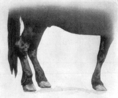
Fig. 20—Contraction of the superficial digital flexor
tendon (perforatus) of the right hind leg, due to tendinitis.
In chronic tendinitis there occurs repeated attacks of inflammation
wherein lameness is pronounced and there exists in reality, at such
times, acute inflammation of a hypertrophic structure, where at no time
does inflammation completely subside. Therefore, in chronic tendinitis
there is to be found at times the same conditions which characterize
acute inflammation, except that there is usually a variance of symptoms
because of the difference in the degree of inflammation and pain.
The diagnosis of contraction of tendons is an easy matter because of the
fact that relations between the phalanges are constantly changed with
tendinous contraction. If one bears in mind the attachments and function
of the digital flexors, no difficulty is encountered in recognizing
contraction of either tendon.
Contraction of the superficial digital flexor (perforatus), when
uncomplicated, is characterized by volar flexion of the pastern joint.
The foot is flat on the ground and the heel is not raised because the
superficial flexor tendon does not have its insertion to the distal
phalanx (os pedis) and therefore can not affect the position of the
foot.
By causing the subject to stand on the affected member, one may outline
the course of the flexor tendons by palpation, and in this way recognize
any lack of tenseness or contraction of tendons or of the suspensory
ligament.
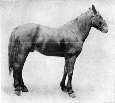
Fig. 21—Contraction of the deep flexor tendon
(perforans) of the right hind leg, due to tendinitis.
Contraction of the suspensory ligament would cause the pastern joint to
assume the same position as is occasioned by contraction of the
superficial digital flexor (perforatus) tendon, but when the subject is
bearing weight on the affected member, it is easy to determine that no
contraction of the suspensory ligament exists, by noting an absence of
abnormal tenseness of this structure. And finally, contraction of the
suspensory ligament is of rare occurrence.
Contraction of the deep flexor tendon (perforans) causes an elevation of
the heel. The foot can not set flat because the insertion of the deep
flexor tendon to the solar surface of the distal phalanx (os pedis)
causes when the tendon is contracted—a rotation of the distal phalanx
on its transverse axis—hence the raised heel. No other tendon has this
same effect on the distal phalanx and the condition is correctly
diagnosed without difficulty.
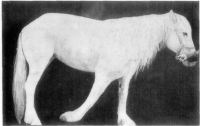
Fig. 22—A chronic case of contraction of both flexor
tendons of the phalanges. In this case (presented at a clinic of the
Kansas City Veterinary College) because of long continued contraction of
the flexors, which prevented weight being supported with any degree of
comfort, there resulted a partial paralysis of the extensors, and
consequently the extremity was dragged on the ground.
Course and Complications.—This condition may exist for years without
causing the subject any serious inconvenience, if the affected animal is
kept at suitable work. In other instances recurrent attacks of lameness
are of such frequent occurrence that the subject is not fit for service.
Many affected animals that are kept in service in spite of lameness (and
in some instances where no lameness is present), soon become
unserviceable because of contraction of the inflamed tendon. This, in
fact, is the condition which eventually becomes established in most
instances.
Treatment.—Where conformation is not too faulty so that recovery may
be expected, good results are obtained by line-firing the tendons and
allowing the subject a few months' rest. In some cases median neurectomy
is advisable. This is recommended by Breton[25] as being productive of
good results even where contraction of tendons exists and tenotomy is
done.
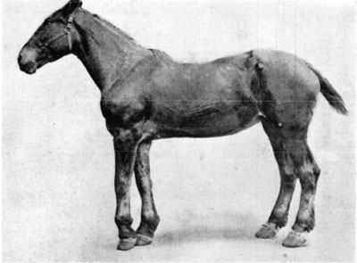
Fig. 23—Contraction of the superficial and deep flexor
tendons (perforatus and perforans) of the left fore leg.
By shoeing with high heel-calks considerable strain is taken from the
inflamed tendons because of the changed position of the foot which
alters the distribution of weight on different parts of the leg. Rubber
pads materially diminish concussion and should be made use of when the
subject is returned to work, if the character of the work is such as to
occasion much concussion.
It is to be remembered, however, that in sprains there occurs fibrillary
fracture of soft structures and time is required for regeneration of
tissue which has been injured or destroyed. Absolute rest is necessary
where inflammation is acute and in sub-acute or chronic tendinitis
avoidance of all work which causes irritation to the affected tendons is
imperative.
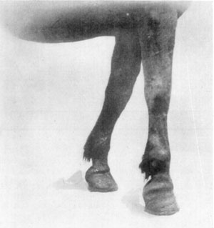
Fig. 24—Contraction of superficial digital flexor and
slight contraction of deep flexor tendon.
Where contraction of tendons exists surgical treatment is necessary. No
good comes from appliances which are calculated to stretch the affected
tendons; in fact, they aggravate the inflamed condition and hasten
complete loss of function of the affected member. Where there exists no
articular or ligamentous diseases which would defeat the purpose,
tenotomy is the only remedy for contracted tendons.
Etiology and Occurrence.—This condition is occasionally observed and
no positive explanation of the reason for its existence can be given.
That mal-position en utero causes the metacarpal bones to develop in
length so rapidly that the tendons are too short, is an explanation that
is offered. Be that as it may, in breeding sections of the country the
general practitioner is obliged to handle these cases and successful
methods of treatment are essential even though cause is not removable.
Symptomatology.—The superficial flexor tendon (perforatus) alone, is
the one usually contracted, and while both flexors are at times
involved, this rarely occurs. The condition is usually bilateral.
The degree of contraction varies greatly in different cases. In some,
contraction exists to such extent that it is impossible for the colt to
stand, and because of continual decubitus where no relief is given, the
subject is lost because of gangrenous infection occasioned by bed sores.
Otherwise the same symptoms are to be observed in this condition, that
exist in contraction of tendons of the mature animal.
Treatment.—Wherever contraction is not too marked and weight is borne
with the affected members, and where the feet can be kept on the ground
in a nearly normal position, it is possible to correct the condition
without doing tenotomy. That is, in cases where the subject is simply
"cock-ankled", where volar flexion of the pastern joint exists but the
foot is kept flat on the ground, correction is possible without
tenotomy.
In such instances the foal must be treated early—before the skin on the
anterior pastern region has been badly damaged by knuckling over. It is
possible in many cases to stretch the flexor tendons by grasping the
colt's foot with one hand, and with the other hand one may push the
pastern in the direction of dorsal flexion. This may be tried and when a
reasonable amount of force is employed, no harm is done, even though no
material benefit results. Some veterinarians claim good results from
this treatment alone and direct their clients to repeat the stretching
process several times daily.
Whether the tendons are manually stretched or not, splints should be
adjusted to the affected members. The legs are padded with cotton and
bandages and a suitable splint is applied on either side of the members
and securely fixed in position by bandaging.
The splints are kept in position for four or five days and then removed
for inspection of the affected parts. If necessary, they are reapplied
and left in position for a week; however, this is unnecessary in the
average case that is treated in this manner.
Where contraction exists to the extent that the subject can not stand
and where no weight is borne by the feet, it is necessary to divide the
affected tendons surgically. The same technic is put into practice that
is employed in the mature subject but there is much greater chance for a
favorable outcome in the foal. Further, if necessary, one may divide
with impunity, both tendons on each leg, at the same time. In all cases
this operation is done by observing strict aseptic precautions and the
legs are, of course, bandaged. If both tendons are divided, splints
should be employed and kept in position for ten days or two weeks.
Primary union of the small surgical wound of the skin and fascia occurs
in forty-eight hours.
The reader is referred to William's "Veterinary Surgical and Obstetrical
Operations," for a complete description of this operation.
In veterinary literature there is occasionally described a
condition which affects young foals wherein symptoms similar to
those of contraction of the flexors are manifested, but upon
examination it is found that rupture of the extensor of the digit
(extensor pedis) exists. This affection is briefly described by
Cadiot but no complete treatise on this condition has been
published.
In parts of Canada foals of from one to three days of age are found
affected in such manner that more or less interference with the
gait is to be seen in those moderately affected. There is, in some
subjects, only a slight impediment in locomotion which is
occasioned by inability to properly extend the digit. In other
subjects, while able to stand and walk, great difficulty is
experienced because of volar flexion of the phalanges. The more
seriously affected animals are unable to stand and, in most
instances, perish because of the effects of prolonged decubitus.
A local enlargement occurs at the anterior carpal region and the
mass is somewhat fluctuating, extravasated fluids becoming infected
in many instances, and necrosis of the skin and fascia provide
means for spontaneous discharge of the contents of the enlargement
if it is not opened. The infection when it becomes generalized
causes a fatal termination in most cases that are not treated.
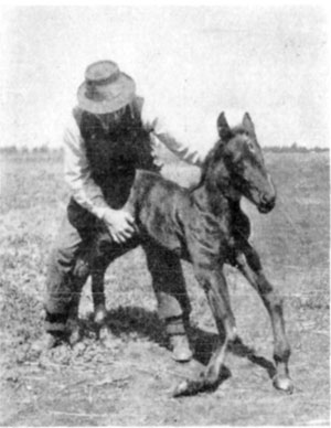
Fig. 25—"Fish knees."—Photo by Thos. Millar,
M.R.C.V.S.
Native stock owners of some parts of Canada know this condition as
"fish knees" because of the presence of the ruptured end of the
extensor tendon which is found coiled in the cavity of the
enlargements caused by the ruptured tendon.
Local practitioners have treated the condition by incising the
swollen mass and removing the part of tendon contained within such
cavities. Treatment has not proved entirely satisfactory in the
majority of instances, perhaps because of tardy interference.
In a colt's leg sent the author by Mr. Thomas Millar, M.R.C.V.S.,
of Asquith, Saskatchewan, a careful dissection of the carpal region
revealed the fact that in this case the ruptured extensor tendon
was due to injury. The colt may have been trampled upon by its dam
in such manner that the tendon was divided. No noticeable evidence
of injury to the skin was to be seen on its outer surface, but on
the fascial side a cyanotic congested area, which was situated
immediately over the site of the ruptured tendon, was very
evident.
With the execution of a good surgical technic, the ruptured tendon
might be sutured; the wound of the tendon sheath as well as that of
the skin carefully united by means of gut sutures, the leg bandaged
and immobilized with leather splints and recovery follow in a
reasonable percentage of cases so treated. These cases afford an
opportunity for the perfection of practical means of treatment by
those who frequently meet with this affection.
Etiology and Occurrence.—Rupture of the flexor tendons or of the
suspensory ligament is of rare occurrence. Frequently, these structures
are divided as the result of wounds; but rupture, due to strain, is not
frequent.
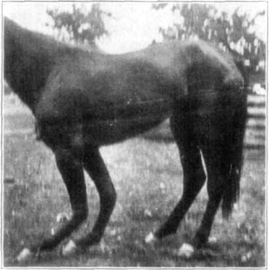
Fig. 26—Extreme dorsal flexion said to have resulted
from an attack of distemper. From Amer. J'n'l. Vet. Med., Vol. XI, No. 4.
In some cases in running horses, or in animals that are put to strenuous
performances, such as are jumpers, rupture of tendons or of the
suspensory ligament takes place. However, more frequently this follows
certain debilitating diseases such as influenza or local infectious
inflammation of the parts which results in degenerative changes and
rupture follows.
The non-elastic suspensory ligament receives some heavy strains during
certain attitudes which are taken by horses in hurdle jumping as is
explained in detail by Montané and Bourdelle[26] under the description
of this ligament. But in spite of the frequent and unusually heavy
strains, which these structures receive, complete rupture is not
frequently seen.
Symptomatology.—When the anatomy and function of the flexor tendons
and suspensory ligament is thoroughly understood, recognition of rupture
of either of these structures is easily recognized. When one considers
that in rupture, a position directly opposite to that which is seen in
contraction in either one of these structures, is assumed, a detailed
description of each separate condition is needless repetition.
However, it is pertinent to suggest that rupture of the deep flexor
tendon (perforans) allows a turning up of the toe. Whether it be torn
loose from its point of attachment or ruptured at some point proximal
thereto, the position is the same—heel flat on the ground, toe slightly
raised and this raising of the toe varies in degree as the subject moves
about.
When the superficial flexor (perforatus) is ruptured there is no change
in the position of the foot but the fetlock joint is slightly lowered.
The pathognomonic symptom is the lax tendon during weight bearing, which
may be felt by palpation of the tendon along its course in the
metacarpal region.
With complete rupture of the suspensory ligament there occurs a marked
dropping of the fetlock joint and an abnormal amount of weight is then
thrown upon the superficial flexor tendon (perforatus), causing it to be
markedly tensed. This is readily recognized by palpation. By palpating
the suspensory ligament from its proximal portion down to and beyond its
bifurcation, while the affected member is supporting weight, it is
possible to diagnose rupture of one of its branches.
Prognosis and Treatment.—In rupture of the superficial flexor tendon
(perforatus) because of its comparatively less important function,
prognosis is favorable and recovery takes place when proper treatment is
put into practice.
With rupture of the deep flexor tendon (perforans), especially when it
occurs at or near its point of insertion and sometimes following
disease, prognosis is unfavorable.
Rupture of the suspensory ligament constitutes a condition which is, as
a rule, hopeless, because of the impracticability of treating such
cases.
The salient feature which characterizes any practical attempt at
treatment of ruptured tendons or other portions of the inhibitory
apparatus of the fetlock region, is to retain the phalanges in their
normal position for a sufficient length of time that the approximated
ends of ruptured tendons or ligaments may unite. The length of time
required for this to occur, together with the difficulties encountered
in confining the affected extremities in suitable braces or supportive
appliances, precludes all possibility of this condition's being
practically amenable to treatment when the deep flexor tendon
(perforans) and suspensory ligament are simultaneously ruptured. It does
not follow, even so, that recovery does not succeed treatment in some of
these unfavorable cases.
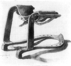
Fig. 27—A good style of shoe for bracing the fetlock
where tenotomy has been performed, or in case of traumatic division of
the flexor tendons. An invention of Dr. G.H. Roberts.
Affected subjects are kept in slings as long as it seems
necessary—until they learn to get up without deranging the braces worn.
Several styles of braces are in use and each has its objections;
nevertheless some sort of support to the affected member is necessary
and steel braces which are connected with shoes are usually employed.
The principal difficulty which attends the use of braces is
pressure-necrosis of the skin which is caused by the constant and firm
contact of the metal support. The practitioner's ingenuity is taxed in
every case to contrive practical means of padding the exposed parts in
order to prevent or minimize necrosis from pressure. This is
attempted—with more or less success—by frequent changing of bandages
and the local application of such agents as alcohol or witch hazel.
Needless to say, the skin must be kept perfectly clean and the dressings
free from all irritating substances.
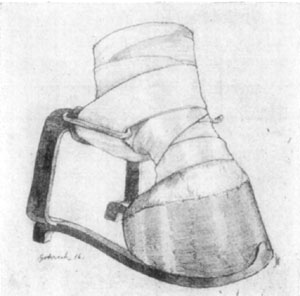
Fig. 28—Showing the Roberts brace in operation.
The fact that tendons or ligaments which are ruptured, do not regenerate
as readily as in cases where traumatic or surgical division occurs, must
not be lost sight of, and prognosis is given in accordance.
Etiology and Occurrence.—Synovial distension of tendon sheaths and
bursae in the region of the fetlock are caused by the same active
agencies which produce this condition in other parts. The fetlock region
is exposed to more frequent injury than is the carpus and as a
consequence is more often affected. The same proportionate amount of
irritation affects this part of the leg, owing to strains, as affect the
carpus from a similar cause; and synovitis from this cause, is as
frequent in one case as in the other. Therefore, it is a natural
sequence that the tendon sheaths of the metacarpophalangeal region are
frequently distended because of chronic synovitis and thecitis. These
inflammations are usually non-infective in character.
The cul-de-sac of the capsular ligament of the fetlock joint which
extends upward between the bifurcation of the suspensory ligament is the
most frequently affected structure in this region. When distended, two
spheroidal masses bulge laterally and anterior to the flexor tendons in
a characteristic manner. This condition is known among horsemen as
"wind-gall" or "fetlock-gall."
The sheath of the flexor tendons, which begins about the middle portion
of the lower third of the metacarpus, and continues downward below the
pastern joint is often distended.
Excepting in cases of acute inflammation attending synovitis of these
parts, no lameness marks its existence and in chronic cases of synovial
distension the service of affected animals is not interfered with. These
distensions constitute unsightly blemishes and they are treated chiefly
for this reason.
No difficulty is encountered in recognizing these conditions even where
considerable organization of tissue overlying distended thecae occurs.
In such cases there may be only slight fluctuation of the enlargement,
but if necessary, an aseptic exploratory puncture may be made with a
suitable needle or trocar.
Treatment.—Complete rest and the local application of cold packs are
in order in acute synovitis when there is distension of tendon sheaths.
In the fetlock region, because of the ease with which pressure may be
employed, the parts should be kept snugly wrapped with cotton, and derby
bandages are used to exert the desired amount of pressure over the
affected region. The pressure-bandages should be employed as soon as all
acute and painful inflammation has subsided; and then they should be
continued, day and night, for ten days or two weeks. The bandages should
be removed morning and night. After the skin of the leg has thoroughly
dried off, an infriction of alcohol or distilled extract of hamamelis is
given the parts and the cotton and bandages are readjusted. A good, even
and firm pressure in such cases is productive of satisfactory results.
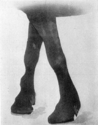
Fig. 29—Distension of theca of the extensor of the digit
(extensor pedis).
In chronic distensions of tendon sheaths synovia may be aspirated and
about five cubic centimeters of equal parts of tincture of iodin and
alcohol is injected into the cavity. This is not done, however, without
usual aseptic precautions. If no marked swelling results within
forty-eight hours the entire fetlock region is thoroughly vesicated and,
as soon as the skin has recovered from the effects of the vesicant,
pressure bandages may be employed. In these cases, subjects may be put
into service after all swelling which the injection or the vesicant has
produced has subsided. The pressure bandages are used at night or during
the time that the horse is in its stall and they are not worn by the
subject while at work.
Where no marked swelling occurs within ten days, as the result of the
injection of iodin, the injection may be repeated and, if thought
necessary, the quantity may be materially increased. If swelling does
not occur it is indicative that no particular irritation has been
caused.
Some swelling is desirable and much swelling sometimes results and
persists for weeks. This is not in any way likely to cause permanent
trouble; and if the technic of injection is skilfully executed no
infection will follow.
By persistent and careful use of suitable elastic bandages, the support
thus given the parts, together with the absorption of products of
inflammation which constant pressure occasions, some chronic cases of
synovial distension of tendon sheaths recover in two or three months and
this without other treatment. Such good results are not to be expected
in aged subjects, nor in horses having at the same time, chronic
lymphangitis.
Where bandages of pure rubber are employed great care is necessary, if
one is not experienced in their use, lest necrosis result. Where
bandages are uncomfortably tight the subject will manifest discomfort,
and an attendant should observe the animal at intervals for a few hours
(where there may be some doubt as to the degree of pressure which is
exerted by elastic bandages) and readjustment made before any harm is
done.
Anatomy.—The anatomy of the metacarpophalangeal articulation is
briefly reviewed on page 58 under the heading of "Anatomo-Physiological
Review of Parts of the Foreleg."
Etiology and Occurrence.—The chief causes of non-infective arthritis
of the fetlock joint are irritations from concussion and contusions due
to interfering. The condition occurs in young animals that are
over-driven in livery service or other similar exhausting work, where
they become so weary that serious injury is done these parts by striking
the pasterns with the feet—interfering. In these "leg-weary" animals,
that are always kept shod with fairly heavy shoes, much direct injury is
done at times by concussion due to self-inflicted blows. In older
animals, where there exists similar conditions, with respect to their
being worn from fatigue and, in addition, periarticular inflammatory
organizations, arthritis is not of uncommon occurrence.
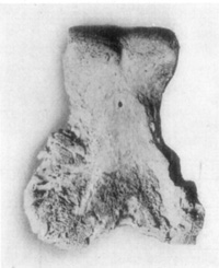
Fig. 30—Rarefying osteitis wherein articular cartilage
was destroyed in a case of arthritis of fetlock joint.
Symptomatology.—In true arthritis there exists a very painful
affection which is characterized by manifestations of distress. The
subject may keep the extremity moving about—where pain is
great—suspended and swinging. There is swelling which is more or less
hot to the touch and compression of the parts with the fingers increases
pain. Lameness is always pronounced and no weight is supported with the
affected member in very acute and generalized arthritic inflammations.
There occurs the usual facial manifestations of pain—the tense
condition of the facial muscles and the fixed eye and nostril are in
evidence.
In cases where there exists a synovitis or where a very limited portion
of the articulation is involved, a somewhat different clinical picture
is presented. Then, the disturbance causes less distress; local swelling
and evidence of supersensitiveness are not so pronounced and lameness is
not intense, though weight-bearing is painful.
Prognosis.—There is a constant difference in the degree of pain
manifested, as well as the other symptoms of inflammation, between true
arthritis, which involves much of the joint, and synovitis; or synovitis
plus a small circumscribed area of joint involvement. This difference is
present in all joint affections of the extremities and, in passing, it
is well to say that infection usually increases every manifestation of
pain. Infection occasions more pronounced local symptoms of inflammation
and, because of the rapid progress of necrotic destruction of cartilage,
the course of the affection is usually rapid; ankylosis is a frequent
result and loss of the subject is often inevitable. However, in
non-infective arthritis of the fetlock joint, prognosis is favorable.
Treatment.—The same general principles which are employed in
arthritis of other joints are used here. Rest and comfort for the
patient is sought in every available manner. If the subject remains
standing too long, the sling should be used and a well-bedded box-stall
will contribute much to the comfort of the patient.
Pain and acute inflammation is diminished or controlled, if possible, by
using ice-cold packs. In nervous, well-bred animals analgesic agents may
be employed; or small doses of morphin sulphate—one to two
grains—given at intervals of three hours during the first stages of the
affection is very beneficial. This is especially indicated in infectious
arthritis.
As inflammation subsides, hot applications are used and finally counter
irritants are employed. Their selection is a matter of choice with the
practitioner. The object sought is the same with every practitioner and
while methods employed vary, results are not markedly different except
in so far as the degree of counter irritation which is produced varies
in given cases. Where a great degree of counter irritation is thought
necessary, line-firing with the actual cautery is the remedy par
excellence.
After-care.—In the course of three or four weeks subjects may be
allowed the run of a paddock and, after a complete rest of six weeks at
pasture, they may be returned to work with care, if the work is not of a
nature to occasion concussion or other manner of irritation to the
articulation.
Neurectomy is not indicated even though there is a recurrence of
lameness, unless the lameness is not pronounced and inflammation is
periarticular and no osseous enlargements mechanically interfere with
function of the joint. There are few cases then, where neurectomy is
materially helpful.
Anatomy and Function of the Cartilages.—Surmounting each wing of the
distal phalanx (os pedis) is the irregularly-quadrangular cartilage. The
superior border of this cartilage is thin, generally convex, and
perforated for vessels to pass to the frog; the inferior border is
attached to the wing of the third phalanx and posteriorly, it is
reflected inward and is continuous with the inferior surface of the
sensitive frog. The anterior border which is directed obliquely downward
and backward becomes blended with the anterior lateral ligament of the
coffin joint. The fibrous expansion of the anterior digital extensor
(extensor pedis) is united to the anterior borders of the lateral
cartilages.
According to Smith[27]: These structures form an elastic wall to
the sensitive foot, and attachment to the vascular laminae; they
also admit of increase in width occurring at the posterior part of
the foot without destroying the union of the two set of leaves.
Further, by their connection with the vascular system of the foot,
their elastic movements materially assist the circulation. The
primary use of the lateral cartilages is to render the internal
foot elastic, and admit of its change in shape which occurs under
the influence of the weight of the body. The alteration in the
shape of the foot is brought about by pressure on the pad, which
widens and in consequence presses on the bars. The pressure
received by the pad is also transmitted to the plantar cushion,
which likewise flattens and spreads under pressure. Both of these
factors force the cartilages slightly outwards. When the posterior
wall recoils the cartilages are carried back to their original
position. Should the elastic cartilage under pathological
conditions become converted into bone, its functions are destroyed,
and lameness may occur.
Etiology and Occurrence.—The causes of ossification of these
cartilages are several. No doubt there exists a predisposition to this
condition for it is of such frequent occurrence in heavy draft types of
horses. Concussion plays an important rôle and, according to
Möller's[28] theory, which is sound, high heel calks prevent the frog
from contacting the ground, and as weight is placed upon the foot "the
lateral cartilages are subjected to a continuous inward and downward
dragging strain."
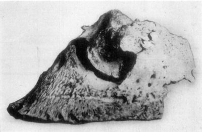
Fig. 31—Ringbone and sidebone.
The condition affects the cartilages of the fore feet more frequently
than those of the hind and the outer cartilage is more often ossified
than is the inner. This fact may be accounted for by its more exposed
position; it is also frequently injured by being trampled upon and
otherwise contused or cut, as in lacerated wounds of the quarter.
Symptomatology.—Ossification of the cartilages is known by grasping
the free borders with the fingers and attempting their flexion; the
rigid inflexible ossified cartilage is thus easily recognized.
Lameness during weight-bearing occurs in the majority of cases at some
time. Much depends on the conformation of the foot and whether the
involvement affects one or both cartilages as to the degree and duration
of lameness which marks this affection. In narrow and contracted heels
it is reasonable to expect more lameness than in well formed feet. Where
only one cartilage is ossified, the other being flexible, there is less
inconvenience experienced by the subject during weight-bearing, because
of the expansion of the heel which the one normal cartilage allows.
Treatment.—There is little if anything to be done in case the
cartilage has become ossified except to shoe without high calks but
preferably with rubber pads. The hoof should be kept moist; the wall at
the quarter may be rasped thin and kept anointed. Firing is of no
practical benefit in these cases, and it is doubtful if vesication is
helpful excepting where only a part of the cartilage is ossified.
Subjects which continue somewhat lame, because of complete ossification
of both cartilages, are best put to slow work on soft ground and not
driven on pavements.
This more or less ambiguous term has been applied to various diseases
affecting the structures which make up the coffin joint. We consider
this name to be applicable to inflammatory involvement of the third
sesamoid (navicular bone), the deep flexor tendon (perforans) and the
bursa podotrochlearis or navicular bursa.
Etiology and Occurrence.—In 1864 Thomas Greaves[29] wrote on the
subject of navicular disease as follows: "The opinion I entertain upon
the subject of navicular disease is, that in by far the greater majority
(if not all) of these cases there exists in the animal affected a
congenital tendency or predisposition, that, generally speaking, it is
the high stepper, the good goer, that becomes the victim to this
disease; and it is a fact well attested, that it as frequently develops
itself in the feet with wide frogs, bulbous heels, shallow heels, spread
flattish feet, as in the narrow upright feet.... I have known foals,
born from defective parents, in which this condition was so strongly
developed, that all men would at once pronounce them affected with
navicular disease, and such lameness was permanent."
Often both fore feet are affected and this would point toward its being
a disease wherein either conformation or congenital tendencies exists.
It is rare that hind feet are involved.
There are many theories regarding the possible exciting causes of
navicular disease and, when one has carefully considered the
explanations as offered by Peters, Möller, Branell, Schrader and others,
he may conclude that navicular disease is a non-infectuous inflammatory
affection of the third sesamoid (navicular) bone, deep flexor tendon
(perforans) and adjoining structures. Whether it originates in the
flexor tendon or whether the bone is the original part affected, the
disease is frequently met, and of all possible causes, jars and
irritation incident to concussion of travel, are probably the principal
causative agents.
Symptomatology.—Lameness is the primary indicator and a constant
symptom which attends navicular disease wherever much structural change
affects the infirm parts. As the degree of intensity or extent varies,
so is there a dissimilarity in the character of the impediment.
Incipient cases of bilateral involvement are more difficult to diagnose
than are unilateral affections, particularly when lameness is not
marked. There is manifested a supporting-leg-lameness which varies as to
degree in the same subject at different times. This may be noticed
during the same trip in an animal that is being driven. There is a
tendency for the subject to stumble and, of course, where the affection
is bilateral, there is a stilted gait owing to shortened strides.
At rest the lame animal usually points with the affected member. Because
of the fact that the distance is lessened between the origin and
insertion of the deep flexor tendon (perforans) by this attitude, one
may readily understand the reason for the position assumed by the
subject. Pressure on the navicular bone is diminished and tension on the
flexor tendon is relieved by even slight volar flexion.
In acute inflammatory affections abnormal heat may be detected in the
region of the heel. By exerting tension on the flexor tendon, by means
of passive dorsal flexion of the member, evidence of hyperesthesia may
be detected. With the hoof testers one may determine supersensitivenss
in most instances. There occurs more or less contraction of the hoof in
navicular disease, but this is not to be taken as a cause of the
affection, but rather a sequence.
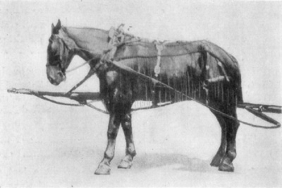
Fig. 32—"Pointing"—the position assumed by horse having
unilateral navicular disease.
In some cases of unilateral navicular disease there is a marked contrast
in size between the sound and unsound foot. However, one must not be
misguided in this particular, for in some pairs of sound feet there
exists considerable difference in size. Finally, by a change from the
normal position of the foot to one in which the heel is somewhat
elevated (as may be obtained by shoeing with high heel calks), relief is
evident, and in the opposite position, the condition is aggravated.
This experiment may be used for diagnostic purposes.
Treatment.—When the anatomy of the diseased parts is taken into
consideration, and an analysis of the lesions which occur in cases where
considerable structural change is occasioned by this affection, it is
obvious that recovery is impossible. Only in cases where the
inflammation is promptly checked before damage has been done the
navicular bone or the flexor tendon, is permanent recovery possible. The
disease is not frequently treated during this stage, however, and in the
majority of instances the condition becomes chronic.
As soon as a diagnosis is made the shoes must be removed, the toe
shortened with the hoof pincers and rasp and the subject is put in a
well bedded box-stall. If the animal is very lame and the inflammation
is acute, ice-cold packs should be applied to the feet. As soon as acute
inflammation has subsided the foot may be so pared that all excess of
sole and frog is removed without lowering the heels, and the animal may
be blistered about the coronet region. The subject may be shod later,
with heel calks that raise the heel moderately and a protracted period
of rest should be enforced.
In cases where no acute inflammatory condition exists, neurectomy is
beneficial. One must discriminate, however, between favorable and
unfavorable subjects. This is not a last resort expedient to be employed
in cases where extensive lesions of the navicular structures exists.
With proper shoeing, and by putting the subject at suitable work, where
concussion of fast travel on hard roads is not necessary, the best
results are obtainable.
This disease is primarily a non-infective inflammation of the sensitive
laminae which very frequently affects the front feet. Often all four
feet are affected, less frequently one foot (when its fellow is unable
to sustain weight) and rarely the hind feet alone.
Occurrence.—Probably a greater number of cases of laminitis occur in
localities where horses that are worked on heavy transfer wagons are,
when in a state of perspiration, allowed to stand exposed to sudden
lowering of temperature and to stand in a cool or cold shower of rain
such as occurs near the coast of the Great Lakes or the ocean in some
parts of this country.
This disease occurs in connection with digestive disorders of various
kinds and, because of the frequent association of the two conditions,
the common term "founder" has long been employed to designate laminitis.
In cases of "over-loading," particularly when a large quantity of wheat
has been eaten by animals that are unaccustomed to this diet, laminitis
almost constantly results.
Large draughts of cold water, when drunk by animals that are overheated
is often followed by laminitis. Concussion, such as attends hard
driving, especially in unshod horses or on rough and hard roads, is
often succeeded by this affection. Likewise, as has been stated, injury
such as is occasioned by long continued standing on the same foot is
followed by laminitis. Some horses that are frequently shod, suffer from
this affection a few hours after shoes have been reset. Dr. Chas. R.
Treadway of Kansas City reports the rather frequent occurrence of such
conditions in horses that are in the fire department service in his
city.
Age in no way influences the occurrence of laminitis and the general
condition of an animal with regard to its vigor or state of flesh has no
apparent influence toward predisposing horses to this ailment.
Etiology and Classification.—As it is with some other diseases, one
may unprofitably theorize on cause and readily enumerate many conditions
which are apparently contributory toward producing the affection. Causes
may well be grouped, however, and a more definite understanding of
laminitis is possible as a result. Such collocation would include
conditions which directly or indirectly affect the digestion, such as
puerperal laminitis, drinking of large quantities of cold water and
exposure to cold and rain when the body is warm. All of these various
conditions might be said to affect the vaso-constrictor nerves in such
manner that the natural tendency (because of the peculiar structure of
the sensitive laminae and their mode of attachment to the non-sensitive
wall) which solipeds have for this affection is indirectly due to this
one cause—vaso-constriction. According to Dr. D.M. Campbell, the effect
of toxic materials, which may be absorbed from the digestive tract or
the uterus in parturient females, upon the vaso-constrictor nerves, is
such that a passive congestion of the sensitive laminae occurs and
laminitis is the result. He believes that even the chilling of the
surface of the body when very warm, by a cold rain, constitutes a
condition wherein the effect upon the vaso-constrictors is the same.
This grouping does not include the effect of direct injuries of any and
all kinds to which the feet are subjected such as: Concussion in fast
road work, injuries occasioned by tight or ill fitting shoes, contusions
of any kind resulting in non-infectious inflammation of the sensitive
laminae, as well as the causes which produce laminitis where weight is
borne by one foot when its fellow is out of function.
A classification which is practical is that of acute and chronic
laminitis. To the practicing veterinarian it is this manner of
consideration that is essential in the handling of these cases.
Symptomatology.—In the acute attack the condition is so well
described by Dr. R.C. Moore[30] that we quote him in part as follows:
The acute form is generally ushered in very suddenly. Often a horse
that is perfectly free from symptoms of the disease is found a few
hours later so stiff and sore that he will scarcely move. They
stand like they were riveted to the ground. If forced to move the
evidence of pain subsides to some extent after they have gone a
short distance, to return more severe than ever after they have
been allowed to stand for a short time. If the disease is confined
to the two front feet, the hind feet are placed well under the
center of the body to support the weight and the front ones are
advanced in front of a perpendicular line so as to lessen the
weight they must bear. If they are made to move, the same position
of the feet is maintained. If made to turn in a small circle, they
do so by using the hind feet as a pivot, bringing the front parts
around by placing as little weight on them as possible.
Placing the hind feet so far under the body, arches the back and
often leads to errors in diagnosis, the condition sometimes being
taken for diseases of the loins or kidneys.
If all four feet are involved, the animal stands in the usual
position assumed in health, but if urged to move, the least effort
to do so usually brings on chronic spasms of the entire body. In
very severe cases, a slight touch of the hand will develop the
spasms. At times they are so severe, and have such short
intermissions, that the disease has been mistaken for tetanus.
However, the clonic nature of the spasm should prevent such an
error. If they are lying down, it is difficult to get them to
arise, and if they do so, they show marked symptoms of pain for
some time after rising.
If the disease is confined to the hind feet, they are placed well
forward to relieve the strain on the toe caused by the downward
pull of the perforans (deep flexor) tendon, but in place of the
front feet being kept in front of a perpendicular line, as they are
when the disease is confined to the front ones, they are placed far
back under the body, so they will carry the maximum share of the
body weight of which they are capable. The position of the feet is
of great importance and offers symptoms that should not be
overlooked.
When the subject is caused to walk, symptoms of excruciating pain are
manifested in all acute cases of laminitis. In some cases where all four
feet are affected, no reasonable amount of persuasion will cause the
suffering animal to move from its tracks.
There is acceleration of the rate of heart action; the pulse is full and
in some cases, bounding. As the affection progresses the pulse becomes
rather weak and irregular. The character of the pulse in the region of
the extremity is a reliable indicator; but one has to learn to make
necessary discrimination because of the condition of the parts, as in
some cases of lymphangitis or where the skin is abnormally thick. The
characteristic throbbing pulse is, however, easily recognized in most
cases. Temperature is variable, though usually elevated from one to four
degrees above normal. This symptom varies with the type and stage of the
affection. In a subject that has been down, unable to rise for several
days, where there is a suppurative and sloughing condition of the
laminae, the temperature is high. Whereas, in some other and less
destructive cases there may be little thermic disturbance after the
first few hours have lapsed.
A constant symptom in bilateral affections of acute laminitis is the
difficulty with which the subject supports weight with one foot. It is
this which causes the victim to stand as if "rooted to the ground" when
all four feet are involved. If one attempts to take up one foot, thus
causing the subject to stand on the other, there is much resistance and
in many cases the animal refuses to give the foot.
When we consider that the sensitive parts of the foot are encased by a
horny, unyielding box and that, when the laminae are congested, a great
pressure is brought to bear upon the sensitive structures, it is easy to
understand why the condition is so painful.
Chronic laminitis is a sequel of acute inflammation of the sensitive
laminae. It varies as to intensity and the exact manner of its
manifestation depends upon preëxisting disturbances.
In some mild cases of laminitis there are recurrent attacks wherein no
particular structural change exists, and diagnosis is established
chiefly by noting the character of the pulse at the bifurcation of the
large metacarpal (or metatarsal) artery just above the fetlock. The same
manifestation of pain is present when weight is supported by one foot,
though in a lesser degree. There is less local heat to be detected by
palpation than in the acute cases.
Chronic laminitis as it occurs following acute attacks which have
resulted in structural changes of the foot, present the same symptoms
just described and, in addition, the peculiar alterations in structure
exist. When, owing to acute inflammation of the sensitive laminae, there
has resulted necrosis of this sensitive tissue together with
infiltration between the anterior surface of the distal phalanx (os
pedis) and the contacting hoof, the lower portion of the distal phalanx
is turned downward and backward (rotated upon its transverse axis).
Because of the traction which is exerted by the deep flexor tendon
(perforans), as it attaches to the solar surface of the distal phalanx,
this rotation is facilitated. With hyperplasia of lamina, at the
anterior portion of the distal phalanx, there results a thick "white
line." Rotation of the distal phalanx necessitates a descent of its
apical portion and there occurs a "dropped sole."
In time, partly because of excessive wear of hoof at the heel, owing to
an altered condition in the normal antagonistic relation between the
flexor and extensor tendons, the toe makes an excessive growth, and the
concavity of the anterior line is accentuated owing to this abnormal
length of hoof. The hoof, because of recurrent inflammatory attacks, is
corrugated—elevations of horn in parallel rings are usually present.
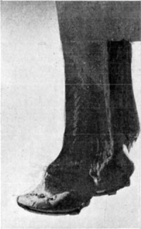
Fig. 33—The hoof in chronic laminitis. Note the
concavity. This animal was serviceable for any work that could be
performed at a walk.
Animals that are so affected in traveling strike the heel first and the
toe is later contacted with the ground surface. Rotation of the distal
phalanx upon its transverse axis produces a condition, with respect to
this peculiar impediment, that is equivalent to added and excessive
length of the deep flexor tendon.
Where there occurs suppuration, by careful inspection of the coronary
region, one may early recognize detachment of hoof. In such cases
animals remain recumbent and, while the condition is not so painful at
this stage, the practitioner must not overlook the real state of
affairs. History, if obtainable, will be a helpful guide in such cases.
Separation of hoof occurs as a rule in from four to ten days after the
initial attack of acute laminitis. Needless to say these cases are
hopeless, when the economic phase of handling subjects is considered.
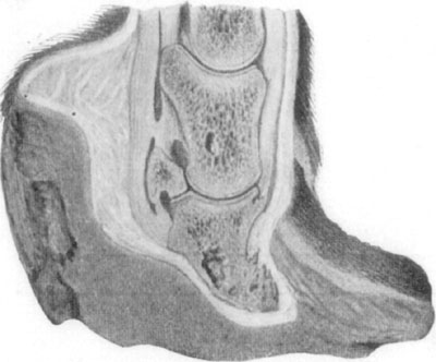
Fig. 34—Showing the effects of laminitis. By permission,
from Merillat's "Veterinary Surgical Operations."
Treatment.—Much depends upon the concomitant disturbances (or causes
if one is justified in referring to them as such) as to the manner in
which laminitis is to be treated. In all cases where digestive
disturbances exist, the prompt unloading of the contents of the
alimentary canal is certainly indicated. D.M. Campbell[31] in a
discussion of laminitis has the following to say regarding the treatment
of such cases:
Because superpurgation may be followed by laminitis, the
advisability of using the active hypodermic cathartics is
questioned. Neither arecolin nor eserin can cause superpurgation.
The action of the former does not continue longer than an hour
after administration and of the latter not more than eight hours.
The action of either is mild after the first few minutes.
I do not think that anyone has recommended either arecolin or
eserin where there is severe purgation. Where the intestinal canal
is fairly well emptied and its contents fluid, I should be inclined
to rely upon intestinal antiseptics to hold in check harmful
bacterial growth.
The use of alum in the treatment of laminitis is held to be without
reason other than the empirical one that it is beneficial. If
laminitis is due chiefly to an autointoxication, good and
sufficient reason for the administration of alum can be shown based
upon its known physiological action. It is the most powerful
intestinal astringent that I know of and has the fewest
disadvantages. I have not noted constipation following its use nor
diarrhea, nor a stopping of peristalsis, nor indigestion, and in
any case its action lasts at most only a few hours, and if it did
all these, it could not much matter. Quitman says, that it
constricts the capillaries. If this is true, a thing of which I am
not certain, is it not reasonable to suppose that as with other
vaso-constrictors, e.g., digitalis, there is a selective action on
the part of the capillaries (not of the drug) and those that need
it most, i.e., those of the affected feet in laminitis, are
constricted most? All body cells exert this selective action in the
assimilation of food, the tissue needing most any particular kind
of food circulating in the blood, gets it.
Our first consideration in laminitis should be to remove the
cause—to stop the absorption of the toxin in the intestinal tract
that is producing the condition. This we accomplish by partially
unloading it by the use of the active hypodermic cathartics and
stopping absorption by the surest and most harmless of intestinal
astringents. Whether the astonishingly prompt and certain action of
alum in this case is due wholly to its astringent action or whether
alum combines with the harmful bacterial products chemically and
forms an innocuous combination, I can only surmise, and it is
unimportant. At any rate, when alum is administered, the onslaught
of the disease is promptly stopped. Irreparable damage may already
have been done if the case is a neglected one, but whether
administered early or late in acute attacks, the progress of the
disease is stopped immediately.
The same authority may be profitably quoted in the matter of handling
all cases wherein the revulsive effect of agents which diminish vascular
tension are chiefly indicated or necessary as adjuvants. In this
connection, Campbell says:
The early and vigorous administration of aconitin in laminitis to
its full physiological effect, is more logical. Assuming that
laminitis is due to absorption of harmful products from the
intestinal tract permitted through the deranged functioning of the
organs of digestion, or assuming that it is due to an extension of
the inflammation from the mucosa to the sensitive lamina, or that
it is a reflex from a sudden chilling of the skin, we have in any
of these conditions a disturbed circulation, and aconitin is the
first and foremost of circulation "equalizers." Furthermore, in
laminitis there is an elevation of the temperature, an almost
invariable indication for aconitin. A speedy return of the
temperature to normal, a very marked diminution of the pain and
improved conditions generally, appear coincident with the symptoms
of full physiological effect of aconitin when given in cases of
laminitis, which constitutes assuredly an important part of its
treatment.
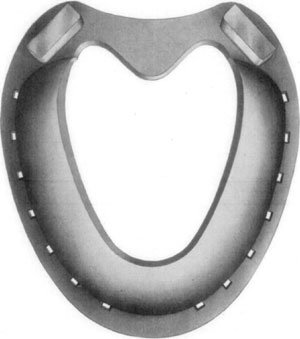
Fig. 35—Inferior (convex) surface of Cochran shoe.
Where lameness is not great as in cases wherein no marked structural
change of the foot has occurred, proper shoeing is very beneficial. By
keeping the heels as low as possible and shoeing without heel calks a
more comfortable position is made possible. Thin rubber pads which do
not elevate the heel are of service in diminishing concussion.
Dr. David W. Cochran of New York City has attained unusual success in
cases of chronic laminitis with dropped sole by the use of a specially
designed shoe.
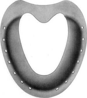
Fig. 36—Superior surface, showing concavity or bowl, as
formed by the toe and branches of the shoe, as designed by Dr. David W. Cochran.
Cochran claims that, not only are horses with dropped soles that would
otherwise have to be put off the streets enabled to do a fair amount of
work by means of this shoe, but that continually wearing it, meanwhile
keeping the convexity of the front of the hoof rasped thin, in time
brings about a marked improvement, and that after some months or years
of use the animals are able to work with ordinary rubber-pad shoes,
provided they are arranged to facilitate breaking over.
From having been successfully used on some race horses of high value,
the Cochran shoe has attained considerable notoriety and is being used
by a number of practitioners. A disadvantage, however, arises from the
fact that few horseshoers other than Doctor Cochran seem able to make
the shoe, the peculiar shape of which offers considerable difficulty in
forging. Concerning the application of the shoe Cochran[32] says:
"The most important primary procedure is the preparation of the
foot to receive the shoe. All excess of growth must be removed from
the anterior face of the hoof. The outer face must be reduced at
the toe (not shortened), but rasped down thin for the lighter the
top of the foot is, the more chance the sole and coffin bone will
have of resuming their former normal position. The pressure of the
wall at the toe upon the exudate between wall and coffin bone,
tends to force the coffin bone and sole out of their normal
position. Leave the sole alone. You can lower the excess of growth
at the heels.
"There are many designs of shoes to relieve this condition. A great
deal depends on the judgment of the shoer to meet the conditions
presented, depending on the degree of the convexity and strength of
the sole. In some cases we use a shoe that admits of a large amount
of sole room. Again, we shoe with a shoe of wide cover. In other
cases a shoe with even pressure over the whole sole. In some cases
a high, narrow shoe, resting only on the wall, or the ordinary
plain shoe with side calks welded close to the outside edge and the
shoe dished well from these as a foundation. Then we have the air
cushion pad designed after the model of the bowl shoe."
In cases when slight and persistent lameness interferes sufficiently to
prevent using an animal at any sort of work on hard roads, median
neurectomy will relieve all lameness in most instances. This is a safe
operation, moreover, in that no bad after effects are to be feared, even
though lameness were to continue.
Etiology and Occurrence.—Injuries of various kinds are inflicted upon
the coronary region but usually they are due to the foot being trampled
upon. When the foot that inflicts the injury happens to be unshod, a
contusion of the injured member is occasioned, but in the majority of
instances, wounds that demand attention are the result of shoe calks
which have penetrated the tissues in the region of the coronary band.
Often calk wounds are self-inflicted. When animals are excited and in
turning crowd one another, they often perform dancing movements which
frequently result in deep calk wounds of the coronet. Some horses have a
habit of resting the heel of one hind foot upon the anterior coronary
region of the other. While sleeping in this position, if they are
suddenly awakened, the weight is abruptly shifted to the uppermost foot
and the one underneath is (because of the pain attending its being
wounded) quickly drawn out from under its fellow. In this way deep cuts
may divide the coronary band and inflict extensive injury to the
sensitive lamina as well.
An infectious type of coronary inflammation occurs in some localities
during the winter months, wherein the condition is enzootic.
Symptomatology.—Depending upon the manner in which the injury has
been produced, the appearance of the wound varies and likewise lameness
is more or less pronounced. If the tissues are not divided and the wound
is chiefly of the subsurface structures, there will not immediately
occur pronounced local evidence of the existence of injury; but as soon
as the lame animal is made to move, the peculiar character of the
impediment (supporting-leg lameness with the affected foot kept well in
advance of its normal position) directs attention to the extremity and
all of the symptoms of acute inflammation are discovered.
Where a wound is inflicted which divides, in some manner, the surface
structures (skin, coronary band, or the hoof wall) one's attention is at
once called to the existence of the wound.
Because of the fact that there is every facility for the production of a
sub-coronary and podophylous infection, these wounds should receive
prompt attention. In some instances, the pastern joint is opened by calk
wounds and then, of course, an infectious arthritis succeeds the injury.
Treatment.—In all contused wounds of the coronary region the parts
need thorough cleansing; the hair, if long is clipped and a cataplasm is
applied. Or preferably, an iodin-glycerin combination of one part of
iodin to four parts of glycerin is poured on a layer of cotton, and
this is confined in contact with the inflamed parts by means of a
bandage.
Where normal resistance to infection obtains, the subject usually
suffers no suppurative disturbance when the surface structures are not
broken; and daily applications of the antiseptic lotion above referred
to stimulates complete resolution. This may be expected in from four to
ten days depending upon the extent of the injury.
If a calk wound has been inflicted, the adjoining surface structures are
freed of hair and the parts cleansed in the usual manner, (which in
wounds recently inflicted, should be done without employing quantities
of water) and after painting the wound surface with tincture of iodin
and saturating its depths with the same agent, the wound is cleansed, if
it contains filth, by means of a small curette. By using a small and
sharp curette, one is enabled to cleanse the average wound quickly and
almost painlessly.
In such cases, equal parts of tincture of iodin and glycerin are
employed. The wound is filled with this preparation and a quantity of it
is poured upon a suitable piece of aseptic gauze or cotton and this is
contacted with the wound. The extremity is carefully bandaged and this
dressing is left in position for forty-eight hours unless there occurs,
in the meanwhile, evidence of profuse suppuration—which is unusual.
One is to be guided as to the progress made by the degree of lameness
present. If little or no lameness develops, it is reasonable to expect
that infection has been checked; that the wound is dry and redressing
every second day is sufficiently frequent.
Where cases progress favorably, recovery (unless infectious arthritis
results) should occur in from ten days to three weeks. Where extensive
sub-coronary fistulae result, either from lack of prompt or proper
attention, the condition is then one requiring a radical operation to
establish drainage and to disinfect if possible, the suppurating
tissues.
Etiology and Occurrence.—In horses, because of a tendency toward
contraction of the heel in some subjects, together with work on hard
roads and pavements, where the feet become dry and brittle, and because
of neglect of the matter of shoeing, this affection is of frequent
occurrence. Unshod horses are rarely affected. If conformation is faulty
and too much weight is borne on the inner or the outer quarter, and the
hoof wall at the quarter tends to turn inward, corns are usually
present. They occur more frequently on the inner quarters of the front
feet, though the outer quarters are occasionally also affected and in
rare instances corns are found at the toes. They do not often affect the
hind feet.
As soon as injury by pressure, such as is supposed to cause the
formation of corns, is brought to bear on the sensitive sole, an
extravasation of blood occurs. In time when the cause remains active,
this discoloration is evident in the substance of the insensitive sole
and consists in a red or yellowish spot which varies in size—this is
ordinarily termed dry corn.
In some cases where infection of this extravasation of blood and serum
occurs, instead of desiccation and discoloration of the insensitive
parts, there is, in time, manifested a circumscribed area of destruction
of the insensitive sole and the abscess may, where no provision for
drainage exists, burrow between sensitive and insensitive laminae and
perforate the tissues at the coronet. If the suppurative material
discharges readily by way of the sole, no disturbance of the heel or
quarters occurs above the hoof.
Symptomatology.—A supporting-leg-lameness characterizes this
condition; and this lameness in most instances varies in degree with the
amount of distress which is occasioned by pressure upon the inflamed
parts. By an examination of the sole after having removed all dirt, and
exposed the horny sole to view, no difficulty is encountered in locating
the cause of the trouble.
Treatment.—Before suppuration has taken place and in the cases where
suppuration does not occur, the horse-shoer's method of paring out the
diseased tissue affords a means of temporary relief; but unless
frequently done, in many cases, lameness results within about three
weeks after such treatment has been given. In other instances temporary
relief is not to be gotten in this manner for any great length of time
or until a more rational mode of treatment becomes necessary so that the
subject may experience a cessation of the inconvenience or distress.
The general plan which meets with the approval of most practitioners
consists in careful leveling of the foot and removing enough of the wall
and sole at the quarters to make possible frog pressure by means of a
bar shoe. With frog pressure, expansion of the heel follows in time, and
permanent relief is obtainable in this manner. Thinning the wall of the
quarter is advocated by many practitioners and is undoubtedly beneficial
in chronic cases where marked contraction has taken place. The wall must
be thinned with a rasp until it is readily flexible by compressing with
the thumbs.
There are instances, however, where corns and contraction of the heel
have existed so long that they do not yield to treatment. Such cases are
found in old light-harness or saddle-horses that have been more or less
lame for years and where there exists marked contraction of the heels,
rough hoof walls, and hard and atrophied frogs.
Suppurating corns require surgical attention in the way of removal of
the purulent necrotic mass and making provision for drainage. Dry
dressings, such as equal parts of zinc sulphate and boric acid, may be
employed to pack the cavity. After the infectious condition has been
controlled, and the wound is dry, the same plan of treatment is
indicated that is employed in the non-suppurating corn. Ample time is
allowed, however, for the surgically invaded tissues to granulate and,
if the subject is to be put in service, a leather pad, under which there
has been packed oakum and tar, affords good protection.
This name is employed to designate an infectious inflammation of the
lateral cartilage and adjoining structures. The disease is characterized
by a slowly progressive necrosis and by a destruction of more or less of
the cartilage and by the presence of fistulous tracts.
Etiology and Occurrence.—The disease is due to the introduction of
pus producing organisms into the subcoronary region of the foot under
conditions which favor the retention of such contagium and extension of
infection into contiguous tissues.
Morbific material is introduced into the region of the lateral cartilage
by means of calk wounds and other penetrant injuries of the foot. A
sub-coronary abscess which, because of lack of proper care or because of
virulency of the contagium or low vitality of the subject, is quite apt
to result in cartilaginous affection and its perforation by necrosis
follows.
Symptomatology.—Quittor is readily diagnosed on sight in many
instances. Where there is dependable history or other evidence of the
chronicity of an infectious inflammation of the kind, quittor is easily
identified. If no positive evidence of the disease exists, by means of
careful exploration of sinuses with the probe, one may distinguish
between true cartilaginous quittor and superficial abscess formation
that is often accompanied by hyperplasia.
Lameness depends upon the extent of the involvement as it affects the
structures contiguous to the cartilage. A variable degree of lameness is
manifested in different cases.
Treatment.—Two general plans of handling this disease are in vogue.
One, the more popular method, consists in the injection of caustic
solutions of various kinds into the fistulous openings with the object
of causing sloughing of necrotic tissue and the stimulation of healthy
granulation of such wounds. The other mode consists in either complete
surgical removal of the cartilage or its remaining portions, or removal
of the diseased parts of curettage.
When quittor has not extensively damaged the foot and the lateral
cartilage is not partly ossified as it is in some old chronic cases, the
complete removal of the lateral cartilage by means of the Bayer
operation or a modification thereof is indicated. A complete description
of the Bayer operation as well as Merillat's operation for this disease
(the latter consisting in part, in the removal of diseased cartilage
with the curette) are given in Volume three of Merillat's "Veterinary
Surgical Operations."
Treatment by injection of caustic solutions has many advocates and
because of the fact that, in many instances the condition is such that
they are not desirable surgical cases and also because some animals may
be put in service before treatment is completed, the injection method is
popular.
The mode of treatment advocated by Joseph Hughes, M.R.C.V.S.,
constitutes a very successful manner of handling quittor and we can do
no better than quote Dr. J.T. Seeley [33] on his manner of using this
particular treatment.
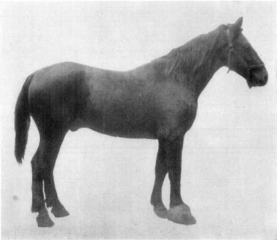
Fig. 37—Hyperplasia of right fore foot, due to chronic
quittor.
Preparation.—First remove the shoe, have the foot pared very
thin and balanced as nicely as possible. Moreover, all loose
fragments of horn must be detached and all crevices cleaned
thoroughly.
Next, have the leg brushed and hair clipped from the knee or hock
to the foot and scrubbed with ethereal soap and warm water, after
which the foot must be scrubbed in like manner. The foot is then
placed in a bichlorid bath several hours daily, for from two to
five days, depending upon whether or not soreness is shown. The
bichlorid solution is 1 to 1,000 strength.
On removing the horse from the bath a liberal layer of gauze is
soaked in 1 to 1,000 bichlorid solution and placed so as to cover
the entire foot. On discontinuing the bath, cover the foot with
gauze saturated with a 1 to 1,000 bichlorid solution. This is to be
covered with absorbent cotton and a gauze bandage, and over all is
placed an oil cloth or silk covering. This pack is kept moist with
bichloride solution for forty-eight hours. The foot is then ready
for injection.
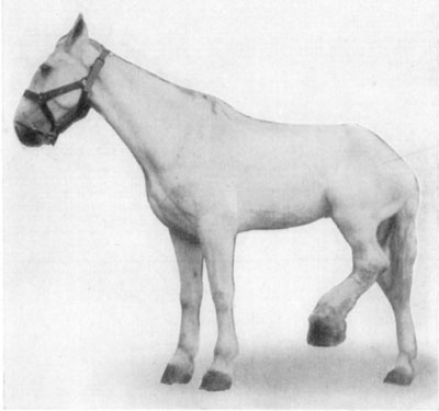
Fig. 38—Chronic quittor, left hind foot. Showing
position assumed because of painfulness of the affection.
Preparation of the Injection Fluids.—Have on hand a pint of a
one per cent aqueous solution of formaldehyd made under cleanly
conditions, even to a clean bottle and cork, and a clean container
when ready to use the liquid. Prepare also a bichlorid of mercury
solution as follows: Hydrarg. Chlor. Corros. 3IV; Acid Hydrochlor.
3Iss.; Aqua Bulliens, Oij. This should be thoroughly triturated,
and then filtered into a clean bottle, when it is ready for use.
Injection.—The patient should be laid on a table, if one is
available, or cast, and the foot securely fixed. Then, with an
ordinary one-ounce hard rubber syringe, with a good plunger (tried
first to note whether or not any fluid works around between the
barrel and the plunger), introduce one syringe full of the
formaldehyd solution, then thoroughly probe the quittor to
determine the number of sinuses. This done, inject each sinus. If
two sinuses open on the surface, close one with cotton while
filling the other so that if there is a connection the solution
will come in contact with all tissues involved. Irrigate with the
full pint of formaldehyd solution first, then follow with six or
eight ounces of the bichlorid solution. Never probe the foot nor
allow it to be tampered with except in the manner prescribed.
After-Treatment.—Put on a pack saturated with a solution of
bichlorid of mercury 1 to 1,000 and let it remain two days. Remove
pack, and once daily afterwards wipe off with cotton the secretion
which accumulates on the outside, and apply a dry dressing or
healing oil composed of phenol, camphor gum and olive oil.
When Dangerous to Inject.—Never inject a quittor in the acute
stage. Never inject a quittor if considerable lameness is present.
On injecting a solution of formalin, hold cotton tightly around the
nozzle of the syringe, when the plunger is down, then withdraw the
syringe gently and note particularly if the fluid returns through
the opening; if none returns cease operations at once, as it is
dangerous to proceed farther, it indicates that the sinus is not
well defined and the fluid retained will cause much trouble and
often the death of the patient.
Experience has taught that, if extensive destructive changes of the foot
exist, the Bayer operation is not indicated. In the country, where
quittors are not so frequently met as in urban practice, the Merillat
operation is preferable in all cases. However, the cost of the
protracted period of idleness, which convalescent surgical patients
require, renders the Hughes method more satisfactory in the hands of the
general practitioner, especially in the city.
Nail punctures, as herein considered, embrace all penetrant wounds of
the solar surface of the horse's foot due to trampling upon street
nails. This does not include accidental nail pricks occasioned in
shoeing. In city practice, in some stables, these cases are of frequent
occurrence; and, generally speaking, nail punctures are observed more
frequently in urban horses than in animals that are kept in the country.
Occurrence and Method of Examination.—This condition, then, is a
rather common cause of lameness and in no case, where cause of the
claudication is not obvious, is the practitioner warranted in concluding
his examination without careful search for the possible existence of
nail puncture of the solar surface of the foot.
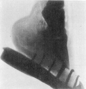
Fig. 39—Skiagraph of foot. The X-ray offers very limited
possibilities in the diagnosis of lameness. The location of a "gravel"
or a nail that had worked its way some distance from the surface, or of
an abscess of some proportion, deep in the tissues, might be facilitated
under some circumstances by the aid of the X-ray. Its use in the
detention of fractures is very limited, owing to the difficulty
encountered in getting a view from the right position—many trials being
necessary in most cases. The case shown above was diagnosed clinically
as incipient ringbone. The X-ray revealed no lesions. (Photo by L.
Griessmann.)
In occasional instances there co-exists an obvious cause for
supporting-leg-lameness and an occult cause—a nail puncture. Where such
complications are met, the practitioner is not necessarily guilty of
neglect or carelessness when the nail puncture is not discovered at
once, nevertheless, an examination is not complete until practically
every possible cause of lameness has been located or excluded in any
given case.
In a search for nail puncture it is necessary to expose to view every
portion of the sole and frog in such manner that the existence of the
smallest possible wound will be revealed. This necessitates removal of
the shoe, if, after a preliminary examination, a puncture is not found,
when there is good reason to suspect its presence. However, where it is
readily possible to locate and care for a wound without removal of the
shoe, allowing the shoe to remain materially facilitates retaining
dressings in position and relieves the solar surface of contact with the
ground. If extensive injury or infection exists, it is of course
necessary to remove the shoe and leave it off. By removing a superficial
portion of all of the sole and frog, thus carefully and completely
exposing to view all parts of the solar surface of the foot, and with
the aid of hoof-testers one is enabled to positively determine the
existence of nail punctures. Because of the tendency of puncture wounds
of the foot to close, and since the superficial portion of the solar
structures are usually soiled, it is absolutely necessary to conduct
examinations of this kind in a thorough manner.
Symtomatology.—Not all cases of nail puncture cause lameness during
the course of the disturbance and in many instances no lameness is
manifested for some time after the injury has been inflicted—not until
infection has been the means of causing considerable inflammation of
sensitive structures. Nevertheless, this lack of manifestation occurs
only in cases where serious injury has not taken place and the degree of
lameness is a constant and reliable indicator of the character and
extent of nail punctures within twenty-four hours after injury has been
inflicted.
The position assumed by the affected animal inconstantly varies with the
location and nature of the injury and is not of particular importance in
establishing a diagnosis. The subject may support some weight with the
affected member and stand "base-wide" or "base-narrow," or no weight may
be borne with the foot or the animal may point or keep the extremity in
a state of volar flexion. In cases where extensive injury has been
inflicted, and great pain exists, the foot is kept off the ground much
of the time and it may be swung back and forth as in all painful
affections of the extremity.
Nail punctures cause typical supporting-leg-lameness and in some cases
certain peculiarities of locomotory impediment are worthy of notice.
Punctures of the region of the heel, which directly affect or involve
the deep tendon sheath, cause a type of lameness wherein pain is
augmented, when dorsal flexion of the extremity occurs as well as when
weight is borne. Wounds in the region of the toe of the hind feet
sometimes cause the subject to carry the extremity considerably in
advance of the point where it is planted and, just before placing the
foot on the ground, it is carried backward a little way—ten or twelve
inches.
However, diagnosis of nail puncture is based on the finding of the
characteristic wound or resultant local changes.
Course and Prognosis.—The nature of the progress and the manner of
termination of these cases are variable. If the coffin joint has been
invaded, and a septic arthritis exists, the condition is at once grave.
An open and infected tendon sheath, while not so serious, constitutes a
condition which is distressing, and recovery is slow even under the most
favorable conditions. Where a heavy, rigid and sharp nail enters the
foot, in such manner that fracture of the third phalanx (os pedis)
occurs, this complication makes for a protraction of the condition.
Experience teaches that the natural course and termination in these
cases are modified by the location and depth of the injury, virulency of
the contagium and resistance of the subject to such infection.
Prevention.—In all horses which are kept at such work that exposure
to nail punctures is frequent, a practical means of prevention of such
injuries consists in the employment of heavy sole leather or suitable
sheet metal to cover the sole of the foot and, at the same time, confine
oakum and tar in contact with the solar surface to prevent the
introduction of foreign material between the foot and such protecting
appliances. Further, if drivers and owners could be impressed with the
serious complications which so frequently attend wounds of this kind,
undoubtedly many cases which are now lost, because of ignorance or
neglect on the part of the teamsters or proprietors of horses, would be
saved by prompt and rational treatment.
Treatment.—The treatment of this condition falls so largely within
the dominion of surgery that we can give little more than an outline
here.
In cases where there exists no evidence of open joint or open tendon
sheath as judged by the site of the puncture and degree of lameness
present (after having thoroughly cleansed the solar surface of the foot
and enlarged the opening in the nonsensitive sole) a little phenol is
introduced into the wound. In such cases, where it is possible for the
antiseptic to contact every part of wound surface to the extreme depths
of the puncture, infection is prevented when such treatment is promptly
administered. This may be considered as first aid, or emergency care,
and is indicated in all wounds of the foot whether the injury be serious
or almost insignificant.
Subsequently one of two general courses may be pursued in the treatment
of cases of nail puncture. One, by the employment of means to keep the
wound patent and injection of suitable antiseptics, or agents that are
more or less caustic in conjunction with strict observance of asepsis
and wound protection. The other method consists in prompt establishment
of drainage by surgical means and includes exploration and curettage.
The first method is better adapted to the use of the average general
practitioner and he would do well to keep the opening in the
nonsensitive structures patent. By introducing equal parts of tincture
of iodin and glycerin daily, good results will follow in most instances.
The wound is protected in unshod horses, either by completely bandaging
the foot and retaining, in contact with the wound, cotton that is
saturated with iodin and glycerin, or, if a minor injury exists, the
moderately enlarged opening in the nonsensitive sole or frog, which has
been moistened with the antiseptic, is packed with a very small quantity
of cotton. A little practice in this mode of closing benign puncture
wounds will enable the practitioner to successfully protect the
sensitive parts in the treatment of such cases in unshod country
horses.
When the condition progresses favorably the wound may be dressed every
second day or twice weekly, and in the course of from two to six weeks
recovery should be complete.
If the practitioner is somewhat proficient as a surgeon, and has at his
command facilities for doing surgery, the second method is preferable in
many cases. By using a local anesthetic on the plantar nerves and
confining the subject on an operating table, restraint should be
perfect. The solar surface of the foot is first thoroughly cleansed, the
puncture wound is enlarged in the nonsensitive structures and the parts
are then moistened with phenol or other suitable antiseptics. By means
of a small probe the puncture is explored and, depending on the
character of the wound and the structures involved, surgical
intervention is varied to suit the case. If necessary, all of the
insensitive frog is removed, and in wounds affecting the region of the
heel the tissues may be incised from the puncture outward dividing all
of the tissues outward and backward to the surface. A suitable surgical
dressing is then applied.
If, on the other hand, the puncture extends into the navicular bursa,
the radical operation is perhaps indicated, though not until one is sure
that infection of the bursa and serious consequences are to follow if
this operation is not performed. Detailed description of the technic of
this operation belongs to the realm of surgery and a good discussion of
it is to be found in William's work on veterinary surgical and
obstetrical operations.
One may summarize the discussion of treatment of nail puncture by saying
that emergency care as herein described is of first consideration. In
every case an immunizing dose of anti-tetanic serum should be given.
Subsequently, the method employed must suit the character of the wound,
existing facilities for handling the subject and the skill and aptitude
of the practitioner.


