We shall now proceed to consider in the organization
of the Echinodermata a type of nervous system
which is more highly developed than that of the
Medusæ. In conducting this research, I was joined
by my friend Professor J. Cossar Ewart, to whose
unusual skill and untiring patience the anatomical
part of the inquiry is due. But here, as formerly,
I shall devote myself to the physiology of the subject,
as it is not possible within the limits assigned
to this volume to travel further into morphology
than is necessary for the purpose of rendering the
experiments intelligible. I shall therefore begin by
seeking to give merely such a general idea of the
structure of the Echinodermata as is necessary for
this purpose.
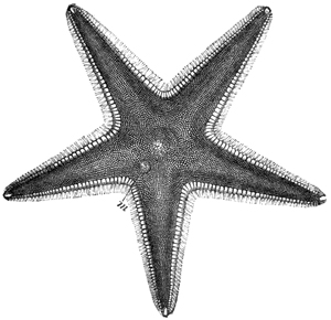
Fig. 32
Upper surface of a Star-fish (Astropecten). (From Cassell's "Nat.
Hist.")
As we all know, a Star-fish consists of a central
disc and five radiating arms (Fig. 32). Upon the
whole of the upper surface there occur numerous
calcareous nodules embedded in the soft flesh, and
supporting short spines. One of these nodules is
much larger than any of the others, is constant in
position, and is called the madreporic tubercle
(Fig. 32, m). Continuing our examination of the
upper surface, we may observe, when we use a lens,
a number of small pincer-like organs scattered about
between the calcareous nodules, or attached to the
spines; these are known as the pedicellariæ. Each
consists of a stalk serving to support a pair of
forceps or pincers, and the whole being provided
with muscles, the stalk is able to sway about and
the pincers to open and shut (Fig. 33). The entire
mechanism is therefore clearly adapted to seizing
and holding on to something; but what it is that
these curious organs are thus adapted to seize, and
therefore of what use they are in the economy of
the animal, has long been a standing puzzle to
naturalists. I hope presently to be able to show
that we have succeeded in doing something towards
the solution of this puzzle.
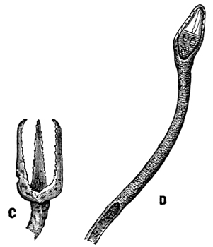
Fig. 33.—Pedicellariæ (magnified). (From Cassell's "Nat. Hist.")
Turning now to the under surface of our Star-fish
(Fig. 34), we observe that the mouth is situated in
the centre of the disc, and that from this mouth as
a centre there radiate five grooves or furrows, which
severally extend to the tips of each of the five rays.
On each side of these grooves there are numerous
actively moving membraneous tubes, which may be
protruded or retracted by being filled or emptied
with fluid. These are used for crawling, and I
shall therefore call them the feet, or pedicels.
So much, then, for the external surface of a Star-fish.
If, now, we examine the internal structure, we
find that the central mouth leads by a short œsophagus
into a central stomach, and that this in turn
communicates with the intestine, which terminates
in an orifice on the dorsal surface. Springing
from the intestine at its origin, there are five tubes,
each of which divides into two, and the five pairs
of tubes thus formed extend into the five rays;
numerous blind processes grow out from these tubes,
and give rise to glandular structures, which probably
perform the functions of a liver.
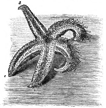
Fig. 34.—Lower surface of common Star-fish.
When a section is made across the base of one of
the arms, the furrows or grooves before mentioned
are seen to be formed of two rows of plates connected
together so as to compose a series of structures
not unlike the couples of an ordinary roof.
These so-called ambulacral plates rest on horizontal
spine-bearing plates, from which other larger plates
extend upwards to form the sides of the arms.
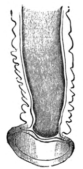
Fig. 35.—The terminal portion of a tube-foot (magnified).
In a living Star-fish the tube-feet or pedicels
already mentioned are seen projecting from each
side of the ambulacral groove; and, with the exception
of a few at the tip of each arm, all the tube-feet
terminate in a well-formed sucker, by means of
which they can be firmly fixed to a flat surface
(Fig. 35).
If we wish to understand the structure and
mechanism of this locomotor or ambulacral system—which,
I may observe in passing, is of special
interest from the fact that as a mechanism it is
unique in the animal kingdom—we must resort to
dissection. We then find that each of the tube-feet
is provided in its membraneous walls with a number
of annular or ring-shaped muscular fibres; when
these fibres contract, the fluid contained in the tube
is forced back, while, conversely, when these fibres
relax, the fluid runs into the tube. If the contraction
of these fibres is strong, the tube shrinks up
entirely, i.e. is retracted within the body of the
animal; but if the contraction of the fibres is not
so strong, the tube is only shortened. If, before its
shortening, its terminal expansion, or sucker, has
been applied to any flat surface, the effect of the
shortening is to cause the sucker to adhere to the
flat surface, in consequence of the pressure of
the surrounding sea-water being greater than that
of the fluid within the shortened tube. In this
way, by alternately contracting and relaxing the
muscular fibres in the walls of a tube-foot, a Star-fish
is able alternately to cause the terminal sucker
to fasten upon and to leave go of any flat surface
upon which the animal may be crawling. In other
words, when the tube-foot is about to form its
attachment to a flat surface, it is fully distended
with fluid; but when the terminal sucker touches
the flat surface, this fluid is partly withdrawn, so
causing the sucker to adhere.
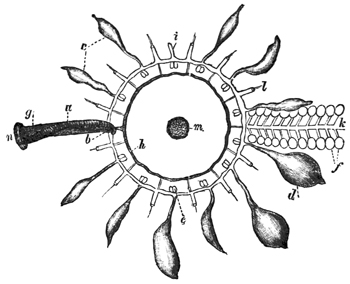
Fig. 36.—Diagram of ambulacral system of a Star-fish: a, madreporic canal; b,
inner end; g, outer end of sinus leading to circular neural vessel; h, from which
radial neural vessels, l, arise; c d, Polian vesicles; f, ampullæ; m, oral aperture;
n, madreporic plate.
When we dissect out one of these tube-feet, we
find that at its base, within the body of the animal,
it bifurcates into two branches. One of these branches
passes immediately into a closed sac (Fig. 36, f),
while the other passes into a large tube (Fig. 36, k),
which runs all the way from base to tip of the ray,
receiving in its course similar branches from all the
tube-feet in the ray. This common or radial tube
itself opens into a circular tube (Fig. 36, e) surrounding
the mouth of the animal (Fig. 36, m). This
circular tube therefore receives five radial tubes—one
from each of the five rays—and is likewise in
communication with a number of membraneous sacs
(Fig. 36, c, d), resembling in their structure (though
larger in size) those which occur at the base of each
of the tube-feet. The function both of these sacs
and of those at the base of each tube-foot is the
same, namely, that of acting as reservoirs of the
fluid when this is expelled from the tube-feet.
Moreover, all these membraneous sacs are provided
with ring-shaped muscular fibres in their membraneous
walls, which therefore serve as antagonists
to the ring-shaped muscles which occur in the
membraneous walls of the tube-feet; that is to
say, when the muscles of the reservoirs contract
(Fig. 36, c, d, f), the pressure in the tube-feet is increased,
and when these muscles relax, that pressure
is diminished. The animal is thus furnished with
the means of varying the head of pressure in its
tube-feet, either locally or universally.
The circular tube surrounding the mouth communicates
at one point with a calcareous tube
(Fig. 36, a), which runs straight to the dorsal surface
of the animal, and there terminates in the madreporic
tubercle, to which I have already directed
attention (Fig. 32, m, and Fig. 36, m). Thus it will
be seen that all the pedicels of all the rays are in
communication, by means of a closed system of
tubes, with this madreporic tubercle. It has therefore
been surmised that the function of this tubercle
is that of acting as a filter to the sea-water which
in large part constitutes the fluid that fills the
ambulacral system. We have been able to prove
that this surmise is correct; for we found that if
we injected any part of the ambulacral system with
coloured fluid—maintaining the injection for several
hours at as great a pressure as the tubes would
stand without rupturing—the coloured fluid found
its way up the calcareous tube to the madreporic tubercle,
on arriving at which it slowly oozed through
the porous substance of which that tubercle consists.
Such, then, is the so-called ambulacral system of
the Star-fish. Passing over another system of vessels
which I need not wait to describe (Fig. 36, g, h, l),
we come next to the nervous system. This is disposed
on a very simple plan. It consists of a pentagonal
ring surrounding the mouth, from which a
nerve-trunk passes into each of the five rays, to
run along the ambulacral groove as far as the extreme
tip of the ray, where it ends in a small red
pigmented spot, about which I shall have more to
say presently. Each of these five radial nerves
gives off in its course a number of delicate branches
to the tube-feet.
So much, then, for the structure of the common
Star-fish. I must next say a few words on the remarkable
modifications which this structure undergoes
in different members of the Star-fish group.
In some species the size of the central disc is
increased so as to fill up the interspaces between
the rays, the whole animal being thus converted
into the form of a pentagon. In other species,
again, the reverse process has taken place, the rays
having become relatively longer, and being at the
same time very active; they look like five little
snakes joined together by a circular disc (Fig. 37).
Again, in another species the rays have begun to
branch, these branches again to branch, and so on
till the whole animal looks like a mat. But the
most extreme modifications are attained in the
sea-cucumbers and lily-stars (Fig. 38). Without,
however, waiting to consider these, I shall go a
little more particularly into the modification of
Star-fish structure which is presented by the
sea-urchin, or Echinus (Fig. 39).
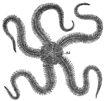
Fig. 37.—A Brittle-star. (From Cassell's "Nat. Hist.")
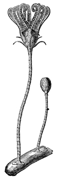
Fig. 38.—A Lily-star. (From Cassell's "Nat. Hist.")]
Externally, the animal presents the form of an
orange, and is completely covered with a large
number of hard calcareous spines, on which account
it derives its scientific name of Echinus, or hedgehog
(the spines have been removed from the larger
portion of the specimen represented in Fig. 39). In
the living animal these spines are fully movable
in all directions, each being mounted on a ball-and-socket
joint, and provided with muscles at its base.
On the external surface, besides the spines, we
meet with pedicellariæ (Fig. 33 magnified), and also
with the madreporic tubercle (Fig. 39, m). The
pedicellariæ in their main features resemble those
which occur in the Star-fish, though considerably
larger, and the ambulacral system is constructed
upon the same plan. If we shave off the spines
and pedicellariæ (Fig. 39), we find that we come to
a hard shell, which, if we break, we find to be
hollow and filled with fluid (Fig. 40). The fluid
closely resembles sea-water, but is, nevertheless,
richly corpusculated; it coagulates when exposed
to the air, and otherwise shows that it is something
more than mere sea-water. If we look closely into
the shell which has been deprived of its spines, we
find that it is composed of a great number of small
hexagonal plates (Fig. 41), the edges of which fit so
closely together that the whole shell is converted
into a box, which, when the animal is alive, is
water-tight, as we have proved by submitting the
contained fluid to hydrostatic pressure, under which
circumstances there is no leakage until the pressure
is sufficient to burst the shell. Nevertheless, if we
look closely at the dried shell of an Echinus, we
shall see that it is not an absolutely closed box; for
we shall see that the hexagonal plates are so arranged
as to give rise to five double rows of holes or pores
(Fig. 41), which extend symmetrically from pole to
pole of the animal (Fig. 39). It is through these
holes that the tube-feet are protruded; so that if
we imagine a pentagonal species of Star-fish to be
curved into the shape of a hollow spheroid, and
then converted into a calcareous box with holes
left for its feet to come through, we should have a
mental picture of an Echinus. It would only be
necessary to add the curious apparatus of teeth
(Figs. 40 and 42), which occurs in the Echinus, to
increase the size of the spines and pedicellariæ, and
to make a few other such minor alterations; but in
all its main features an Echinus is merely a Star-fish
with its five rays calcified and soldered together
so as to constitute a rigid box.
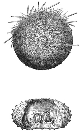
Fig. 39.—An Echinus, partly denuded of its spines. (From Cassell's "Nat.
Hist.")
Fig. 40.—Showing interior of Echinus shell. (From Cassell's "Nat. Hist.")
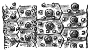
Fig. 41.—A portion of the external shell of an Echinus denuded of spines and
slightly magnified, showing the arrangement of the plates, the balls in the ball-and-socket
joints of the spines, and the holes through which the ambulacral feet
are protruded. (From Cassell's "Nat. Hist.")
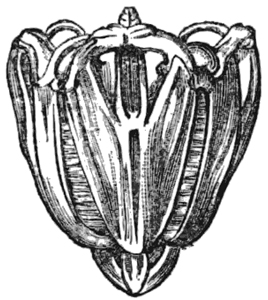
Fig. 42.—Teeth of Echinus (from Cassell's "Nat. Hist.")
This echinoid type itself varies considerably
among its numerous constituent species as to size,
shape, length and thickness of the spines, etc.; but
I need not wait to go into these details. Again,
merely inviting momentary attention to the developmental
history of these animals, I may remark that
the phases of development through which an individual
Echinoderm passes are not less varied and
remarkable than are the permanent forms eventually
assumed by the sundry species.
Turning now to the physiology of the Star-fish
group, I shall begin by describing the natural
movements of the animals.
Taking the common Star-fish as our starting-point,
I have already explained the mechanism
of its ambulacral system. The animals usually
crawl in a determinate direction, and when in the
course of their advance the terminal feet of the
advancing ray—which are used, not as suckers, but
as feelers, protruded forwards—happen to come into
contact with a solid body, the Star-fish may either
continue its direction of advance unchanged, or
may turn towards the body which it has touched.
Thus, for instance, while crawling along the floor of
a tank, if the terminal feet at the end of a ray
happen to touch a perpendicular side of the tank,
the animal may either at once proceed to ascend
this perpendicular side, or it may continue its
progress along the floor, feeling the perpendicular
side with the end of its rays perhaps the whole way
round the tank, and yet not choosing, as it were, to
ascend. In the cases where it does ascend and
reaches the surface of the water, a Star-fish very
often performs a number of peculiar movements,
which we may call acrobatic (Fig. 43). On reaching
the surface, the animal does not wish to leave its
native element—in fact, cannot do so, because its
sucking feet can only act under water—and neither
does it wish again to descend into the levels from
which it has just ascended. It, therefore, begins to
feel about for rocks or sea-weeds at the surface, by
crawling along the side of the tank, and every now
and then throwing back its uppermost ray or rays
along the surface of the water to feel for any solid
support that may be within reach. If it finds one,
it may very likely attach its uppermost rays to it,
and then, letting go its other attachments, swing
from the one support to the other. The activity
and co-ordination manifested in these acrobatic
movements are surprising, and give to the animal
an almost intelligent appearance.
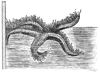
Fig. 43.—Natural movements of a Star-fish on reaching the surface of water.
In Astropecten the ambulacral feet have become
partly rudimentary, inasmuch as they have lost
their terminal suckers (Fig. 44). These Star-fish,
therefore, assist themselves in locomotion by the
muscular movements of their rays, while they use
their suckerless feet to run along the ground somewhat
after the manner of centipedes. It is to be
noticed, however, that although the feet have lost
their suckers, the Star-fish is still able to make them
adhere to solid surfaces in a comparatively inefficient
manner, by constricting the tube on one side after
it has brought this side into opposition with the
solid surface (Fig. 45).
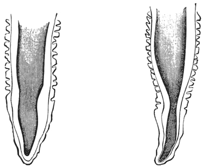
Fig. 44.—A pedicel of Astropecten
(magnified), showing the absence
of any terminal sucker.
Fig. 45.—The same, showing the
method of extemporizing a
sucker.
In the Brittle-stars the ambulacral feet have been
still more reduced to rudiments, and are of no use
at all, either as suckers or for assisting in locomotion.
These Star-fish have, therefore, adopted another
method of locomotion, and one which is a great
improvement upon the slow crawling of other
members of the Star-fish group. The rays of the
Brittle-stars are very long, flexible, and muscular,
and by their combined action the animal is able
to shuffle along flat horizontal surfaces. When it
desires to move rapidly, it uses two of its opposite
arms upon the horizontal floor with a motion like
swimming (Fig. 46); at each stroke the animal
advances with a leap or bound about the distance
of two inches, and as the strokes follow one another
rapidly, the Star-fish is able to travel at the rate of
six feet per minute. A common Star-fish, on the
other hand, with its slow crawling method of
progression, can only go two inches per minute.
Some of the Comatulæ, in which the muscularity
of the rays has proceeded still further, are able
actually to swim in the water by the co-ordinated
movements of their rays.[39]
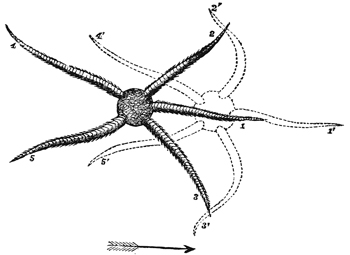
Fig. 46.—Natural movements of a Brittle-star when proceeding along a solid
horizontal surface.
The Echinus crawls in the same way as the
common Star-fish; but besides its long suckers it
also uses its spines, which by their co-ordinated
action push the animal along. The suckers, moreover,
in being protruded from all sides of a globe
instead of from the under side of a flat organism,
are of much more use as feelers than they are in the
Star-fish. Therefore, while advancing, the feet
facing the direction of advance are always kept
extended to their fullest length, in order to feel for
any object which the animal may possibly be
approaching. When a perpendicular surface is
reached, the Echinus may either ascend it or not, as
in the case of the Star-fish. While walking, the
animal keeps pretty persistently in one direction
of advance. If it be partly rotated by the hand,
it does not continue in the same direction, but
continues its own movements as before; so that,
for instance, if it is turned half round, it will
proceed in a direction opposite to that in which
it had previously been going. When at rest, some
of the feet are used as anchors, and others protruded
as feelers.
Regarded from the standpoint of the evolutionist,
we have here an interesting series of gradations.
At one end of the series we have the Echinus with
its rays all united in a box-like rigid shell. At
the other end of the series we have the Brittle-stars
and Comatulæ with their highly muscular and
mobile rays. Midway in the series we have Astropecten
and the common Star-fish, where the rays
are flexible and mobile, though not nearly so much
so as in the Brittle-stars. Now, the point to observe
is, that in correlation with this graduated difference
in the mobility of the rays, there is a correspondingly
graduated difference in the development of
the ambulacral system of suckers. For in Echinus
this system is seen in its most elaborate and efficient
form; in the common Star-fish the suckers are still
the most important organs of locomotion, though
the muscularity of the rays has begun to tell upon the
development of the specially ambulacral system,
the suckers not being so long or so powerful as
they are in Echinus. Lastly, the Brittle-stars and
Comatulæ have altogether discarded the use of
their sucking feet in favour of the much more
efficient organs of locomotion supplied by their
muscular rays; and, as a consequence, their feet
have dwindled into useless rudiments, while the
rays have become limb-like in their activity.
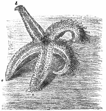
Fig. 47.—Natural righting movements of common Star-fish.
There is only one other point in connection with
the natural movements of the Echinodermata which
it is necessary for me to touch upon. All the
species when turned upon their backs are able again
to right themselves; but seeing, as I have just
observed, that the organs of locomotion in the
different species are not the same, the methods to
which these species have to resort in executing the
righting manœuvre are correspondingly diverse.
Thus, the Brittle-stars can easily perform the needful
manœuvre by wriggling some of their snakelike
arms under the inverted disc, and heaving the
whole body over by the mere muscularity of these
organs. The common Star-fish, however, experiences
more difficulty, and executes the manœuvre mainly
by means of its suckers. That is to say, it twists
round the tip of one or more of its rays (Fig. 47)
until the ambulacral feet there situated are able to
get a firm hold of the floor of the tank (a); then, by
a successive and similar action of the ambulacral
feet further back in the series, the whole ray is
twisted round (b), so that the ambulacral surface
of the end is applied flat against the floor of the
tank (c). The manœuvre continuing, the semi-turn
or spiral travels progressing all the way down the
ray. Usually two or three adjacent rays perform
this manœuvre simultaneously; but if, as sometimes
happens, two opposite rays should begin to
do so, one of them soon ceases to continue the
manœuvre, and one or both of the rays adjacent to
the other takes it up instead, so assisting and
not thwarting the action. The spirals of the co-operating
rays being invariably turned in the same
direction (Fig. 47, a, b, and c), the result is, when
they have proceeded sufficiently far down the rays,
to drag over the remaining rays, which then
abandon their hold of the bottom of the tank, so as
not to offer any resistance to the lifting action of
the active rays. The whole movement does not
occupy more than half a minute. As a general
rule, the rays are from the first co-ordinated to
effect the righting movement in the direction in
which it is finally to take place—the rays which
are to be the active ones alone twisting over, and
so twisting that all their spirals turn in the same
direction.
A Star-fish (Astropecten) which is intermediate
between the Brittle-star and the common Star-fish, in
that its ambulacral feet are partly aborted (having
lost their suckers, as shown in Fig. 44) and its
rays more mobile than those of the common Star-fish,
rights itself after the manner shown in Fig. 48,
where the animal is represented as standing on the
tips of four of its rays, while the fifth one is just
about to be thrown upwards and over the others, in
order to carry with it the two adjacent rays, and so
eventually to overbalance the system round the
fulcrum supplied by the tips of the other two rays,
and thus bring the animal down upon its ventral
surface.
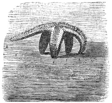
Fig. 48.—Righting movements of Astropecten.
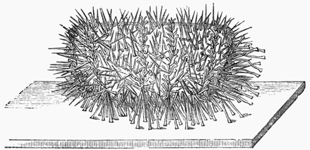
But it is in the case of Echinus that these righting
movements become most interesting, from the
fact that they are so much more difficult to accomplish
than they are in the case of the Star-fishes.
For while a Star-fish is provided with flat, flexible,
and muscular rays, comprising a small and light
mass in relation to the motive power, an Echinus
is a rigid, non-muscular, and globular mass, whose
only motive power available for conducting the
manœuvre is that which is supplied by its relatively
feeble ambulacral feet. It is, therefore,
scarcely surprising that unless the specimens chosen
for these observations are perfectly fresh and
vigorous, they are unable to right themselves at
all; they remain permanently inverted till they
die. But if the specimens are fresh and vigorous,
they are sooner or later sure to succeed in righting
themselves, and their method of doing so is
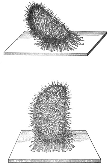
always the same. Two, or perhaps three, adjacent
rows of suckers are chosen out of the five, as the
rows which are to accomplish the task (Fig. 49). As
many feet upon the rows as can reach the floor of
the tank are protruded downwards and fastened
firmly to the floor; their combined action then
serves to tilt the globe slightly over in their own
direction, the anchoring feet on the other or
opposite rows meanwhile releasing their hold of the
tank to admit of this tilting (Fig. 50). The effect
of this tilting is to enable the next feet in the
active ambulacral rows to touch the floor of the
tank, and, when they have established their hold,
they assist in increasing the tilt; then the next feet
in the series lay hold, and so on, till the globe
slowly but steadily rises upon its equator (Fig. 51).
The difficulty of raising such a heavy mass into
this position by means of the slender motive power
available can be at once appreciated on witnessing
the performance, so that one is surprised, notwithstanding
the co-ordination displayed by all the
suckers, that they are able to accomplish the work
assigned to them. That the process is in truth a
very laborious one is manifest, not only from the
extreme slowness with which it takes place, but
also because, as already observed, in the case of not
perfectly strong specimens complete failure may
attend the efforts to reach the position of resting on
the equator—the Echinus, after rearing up a certain
height, becoming exhausted and again falling back
upon its ab-oral pole. Moreover, in some cases it
is interesting to observe that when the equator
position has been reached with difficulty, the
Echinus, as it were, gives itself a breathing space
before beginning the movement of descent—drawing
in all its pedicels save those which hold it securely
in the position to which it has attained, and
remaining in a state of absolute quiescence for a
prolonged time. It then suddenly begins to protrude
all its feet again, and to continue its manœuvre.
At any time during such a period of rest, a stimulus
of any kind will immediately determine a recommencement
of the manœuvre.
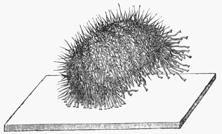
It will be perceived that as soon as the position
just described has been attained, gravity, which had
hitherto been acting in opposition to the righting
movement, now begins to favour that movement.
It might, therefore, be anticipated that the Echinus
would now simply let go all its attachments and
allow itself to roll over into its natural position
But an Echinus will never let go its attachments
without some urgent reason, seeming to be above
all things afraid of being rolled about at the mercy
of currents; and therefore in this case it lets itself
down almost as slowly as it raised itself up. So
gently, indeed, is the downward movement effected,
that an observer can scarcely tell the precise
moment at which the righting is concluded. Therefore,
in the downward movement, the feet, which at
the earlier part of the manœuvre were employed
successfully in rearing the globe upon its equator,
are now employed successfully in preventing its too
rapid descent (Fig. 52).
Several interesting questions arise with reference
to these righting movements of Echinus. First of
all we are inclined to ask what it is that determines
the choice of the rows of feet which are delegated
to effect the movements. As the animal has a
geometrical form of perfect symmetry, we might
suppose that when it is placed upon its pole, all the
five rows of feet would act in antagonism to one
another; for there seems nothing more to determine
either the action or the inaction of one row rather
than another. Indeed, if there were any moral
philosophers among the Echinoderms, they might
point with triumph to the fact of their being able
to right themselves as an irrefutable argument in
favour of the freedom of the Echinoderm will. "We
are in form," they might say, "perfectly geometrical,
and our feet-rows are all arranged with perfect
symmetry; therefore there is no reason, apart from
the sovereign freedom of our choice, why we should
ever use one set of feet rather than another in executing
this important movement." And indeed, I
do not see how these Echinoderm philosophers could
be answered by any of the human philosophers, who,
with less mathematical data and with less physiological
reason, employ analogous arguments to prove
the freedom of the human will. Physiologists,
however, would give these Echinoderm philosophers
the same answer that they are in the habit of giving
to the human philosophers, viz. that although the
physiological conditions are very nicely balanced,
they are never so nicely balanced as to leave
positively nothing to determine which rows of feet—that
is to say, which sets of nerves—shall be
used. And in this connection I may observe that
on making a number of trials it becomes apparent
in the case of certain individual specimens that
they manifested a marked tendency to rotate
always in the same direction, or to use the same set
of foot-rows for the purpose of righting themselves.
In these individual specimens, therefore, we must
conclude that the foot-rows thus employed are
selected because of some slight accidental prepotency
or superiority over the others; the animal has, as it
were, thus much individual character as the result
of a slight prepotency of some of its nerve-centres
over the others.
Another question of still more interest arises out
of these righting movements, namely, that as to
their prompting cause. This question, however, I
shall defer till later on, since it cannot be answered
without the aid of experiments as distinguished
from observation.
In now quitting our observations on the natural
movements of the Echinodermata, and beginning an
account of the various experiments which we have
tried upon these animals, I shall first take the
experiments in stimulation.
All the Echinodermata seek to escape from
injury. Thus, for instance, if a Star-fish or an
Echinus is advancing continuously in one direction,
and if it be pricked or otherwise irritated
on any part of an excitable surface facing the
direction of advance, the animal immediately
reverses that direction. There is one point of
special interest concerning these movements of
response to stimulation. The form of the animals
and the distribution of the nervous system being,
as I have before said, of geometrical regularity, it
follows that by applying two stimuli simultaneously
on two different aspects of the animal, the combined
result of these two stimuli is that of furnishing a
very pretty instance in physiology of the physical
principle of the parallelogram of forces. Thus, for
instance, if two stimuli of equal intensity be applied
simultaneously at the opposite sides of a globular
Echinus, the animal begins to walk in a direction
at right angles to an imaginary line joining these
two points. And, generally, wherever the two
points of simultaneous stimulation may be situated,
the direction of the animal's advance is the
diagonal between them. As showing in more detail
how very delicate is the physiological balancing of
stimuli which may be produced in these organisms,
and consequently the manner in which we are able
to play, as it were, upon their geometrically
disposed nervous systems in illustration of the
mechanical principle of the composition of forces,
I shall quote a series of observations.
"1. Scraped with a scalpel the equator of an
Echinus at two points opposite to each other—animal
crawled at right angles to the line of injury.
"2. Similarly scraped at the ab-oral pole—no
effect. There was no reason why injury here should
determine escape in one direction rather than in
another.
"3. Scraped similarly near the oral pole, and
half-way between pole and equator—little or no
effect.
"4. Scraped in rapid succession five equatorial and
equidistant injuries—Echinus crawled actively in
one determinate direction; the equal and equidistant
injuries all round the globe neutralized one
another.
"5. Scraped a band of uniform width all the way
round the equator—same result as in 4.
"6. Band of injury in same specimen was then
widened in the side facing the direction of crawling—no
effect. Still further widened—slight change
of direction, and, after a time, persistent crawling
away from the widest part of the injured zone.
Repeated this experiment on other specimens by
scraping round the whole equator, and simultaneously
making one part of the zone of injury wider
than the rest—same result; the animal crawled
away from the greatest amount of injury.
"7. Scraped on one side of the equator, and, after
the animal had been crawling in a direct line from
the source of irritation for a few minutes, similarly
scraped equator on the opposite side—animal reversed
its direction of crawling; it crawled away
from the stimulus supplied latest.
"8. Scraped a number of places on all aspects
of the animal indiscriminately—direction of advance
uncertain and discontinuous, with a strong
tendency to rotation upon vertical axis."
These observations show conclusively that the
whole external surface, not only of the soft and
fleshy Star-fish, but even of the hard and rigid
Echinus, is everywhere sensitive to stimulation.
Closer observation shows that this sensitiveness,
besides being so general, is highly delicate. For if
any part of the external surface of an Echinus is
lightly touched with the point of a needle, all the feet,
spines, and pedicellariæ within reach of that part, and
even beyond it, immediately converge and close in
upon the needle, grasp it, and hold it fast. This simultaneous
movement of such a little forest of prehensile
organs is a very beautiful spectacle to witness. In
executing it the pedicellariæ are the most active,
the spines somewhat slower, and the feet very
much slower. The area affected is usually about
half a square inch, although the pedicellariæ even
far beyond this area may bend over towards the
seat of stimulation, which, however, from their small
size they are not able to reach.
And here we have proof of the function of the
pedicellariæ—proof which we consider to be important,
because, as I have before said, the use of
these organs has so long been a puzzle to naturalists.
In climbing perpendicular or inclined surfaces of
rock, covered with waving sea-weeds, it must be of
no small advantage to an Echinus to be provided on
all sides with a multitude of forceps, all mounted
on movable stalks, which instantaneously bring
their grasping forceps to bear upon and to seize a
passing frond. The frond being thus arrested, the
spines come to the assistance of the pedicellariæ,
and both together hold the Echinus to the support
furnished by the sea-weed. Moreover the sea-weed
is thus held steady till the ambulacral feet have
time also to establish their hold upon it with their
sucking discs. That the grasping and arresting of
fronds of sea-weed in this way for the purposes of
locomotion constitute an important function of the
pedicellariæ, may at once he rendered evident
experimentally by drawing a piece of sea-weed over
the surface of a healthy Echinus in the water. The
moment the sea-weed touches the surface of the
animal, it is seen and felt to be seized by a number
of these little grasping organs, and—unless torn
away by a greater force than is likely to occur in
currents below the surface of the sea—it is held
steady till the ambulacral suckers have time to
establish their attachments upon it. Thus there is
no doubt that the pedicellariæ are able efficiently
to perform the function which we regard as
their chief function. We so regard this function,
not merely because it is the one that we observe
these organs chiefly to perform, but also because
we find that their whole physiology is adapted
to its performance. Thus their multitudinous
number and ubiquitous situation all over the
external surface of the animal is suggestive of
their being adapted to catch something which may
come upon them from any side, and which may
have strings and edges so fine as to admit of being
enclosed by the forceps. Again, the instantaneous
activity with which they all close round and seize
a moving body of a size that admits of their seizing
it, is suggestive of the objects which they are
adapted to seize being objects which rapidly brush
over the surface of the shell, and therefore objects
which, if they are to be seized at all, must be seized
instantaneously. Lastly, we find, on experimenting
upon pedicellariæ, whether in situ or when
separated from the Echinus, that the clasping action
of the forceps is precisely adapted to the function
which we are considering; for not only is the force
exerted by the forceps during their contraction of
an astonishing amount for the size of the organ
(the serrated mandibles of the trident pedicellariæ
holding on with a tenacity that can only have
reference to some objects liable to be dragged away
from their grasp), but it is very suggestive that this
wonderfully tenacious hold is spontaneously relaxed
after a minute or two. This is to say, the pedicellariæ
tightly fix the object which they have
caught for a time sufficient to enable the ambulacral
suckers to establish their connections with it, and
then they spontaneously leave go; their grasp is not
only so exceedingly powerful while it lasts, but it is
as a rule timed to suit the requirements of the
pedicels.[40]
Concerning the physiology of the pedicellariæ
little further remains to be said. It may be stated,
however, that the mandibles, which are constantly
swaying about upon their contractile stalks as if
in search for something to catch, will snap at an
object only if it touches the inner surface of one
or more of the expanded mandibles. Moreover, in
the larger pedicellariæ, a certain part of the inner
surface of the mandibles is much more sensitive to
contact than is the rest of that surface; this part is
a little pad about one-third of the way down the
mandible: a delicate touch with a hair upon this
part of any of the three mandibles is certain to
determine an immediate closure of all the three. It
is obvious that there is an advantage in the sensitive
area, or zone, being placed thus low enough
down in the length of the mandibles to ensure that
the whole apparatus will not close upon an object
till the latter is far enough within the grasp of the
mechanism to give this mechanism the best possible
hold. If, for instance, the tips of the mandibles
were the most sensitive parts, or even if their whole
inner surfaces were uniformly sensitive, the apparatus
would be constantly closing upon objects
when these merely brushed past their tips, and
therefore closing prematurely for the purpose of
grasping. But, as it is, the apparatus is admirably
adapted to waiting for the best possible chance of
getting a secure hold, and then snapping upon the
object with all the quickness and tenacity of a
spring-trap.
Another point worth mentioning is that if, after
closure, any one or more of the mandibles be gently
stroked on its outer surface near the base, all the
mandibles are by this stimulation usually, though
not invariably, induced again to expand. This is
the only part of the whole organ the stimulation of
which thus exerts an inhibitory influence on the
contractile mechanism. If there is any functional
purpose served by such relaxing influence of stimulating
this particular part of the apparatus, we
think it can only be as follows. When a portion of
sea-weed brushes this particular part, it must be
well below the tips of the mandibles, and therefore
in a position where it, or some over-lying portion,
may soon pass between the mandibles, if the latter
are open; hence when touched in this place the
mandibles, if closed, open to receive the sea-weed,
should any part of it come within their cavity.
Turning next to experiments in stimulation with
reference to the spines, I may observe that we have
found these organs to be, physiologically considered,
highly remarkable and interesting, from the fact
that they display co-ordinated action in a degree
which entitles them to be regarded as a vast multitude
of limbs. Thus, for instance, if an Echinus be
taken out of the water and placed upon a table, it
is no longer able to use its feet for the purpose of
locomotion, as their suckers are only adapted to be
used under water. Yet the animal is able to progress
slowly by means of the co-ordinated action of
its spines, which are used to prop and push the
globe-like shell along in some continuous direction.
If, while the animal is thus slowly progressing, a
lighted match be held near it, facing the direction of
advance, as soon as the animal comes close enough
to feel the heat, all the spines begin to make the
animal move away in the opposite direction. Moreover,
as showing the high degree in which the action
of the spines is co-ordinated, I may mention that
there is an urchin-like form of Echinoderm, which is
called Spatangus, and which differs from the Echinus
in having shorter feet and longer spines. When,
therefore, a Spatangus is inverted, it is unable to
right itself by means of its feet, as these are too short
to admit of being used for this purpose; but, nevertheless,
the animal is able to right itself by means
of the co-ordinated action of its long spines, these
being used successively and laboriously to prop
and push the animal over in some one definite
direction. The process takes a very long time
to accomplish, and there are generally numerous
failures, but the creature perseveres until it eventually
succeeds.
Coming now to stimulation with reference to the
feet, we find that when a drop of acid, or other
severe stimulation, is applied to any part of a row
of protruded pedicels, the entire row is immediately
retracted, the pedicels retracting successively from
the seat of irritation—so that if the latter be in the
middle point of the series, two series of retractions
are started, proceeding in opposite directions simultaneously;
the rate at which they travel is rather
slow. This process of retraction, however, although
so complete within the ray irritated, does not extend
to the other rays. But if the stimulus be applied
to the centre of the disc, upon the oral surface of
the animal, all the feet in all the rays are more or
less retracted—the process of retraction radiating
serially from the centre of stimulation. The influence
of the stimulus, however, diminishes perceptibly
with the distance from the centre. Thus, if weak
acid be used as the irritant, it is only the feet near
the bases of the rays that are retracted; and even
if very strong acid be so used, it is only the feet as
far as one-half or two-thirds of the way up the rays
that are fully retracted—the remainder only having
their activity impaired, while those near the tip may
not be affected at all. If the drop of acid be placed
on the dorsal, instead of the ventral surface of the
disc, the effect on the feet is found to be just the
converse; that is, the stimulus here applied greatly
increases the activity of the feet. Further experiments
show that this effect is produced by a stimulus
applied anywhere over the dorsal aspect of the
animal; so that, for instance, if a drop of acid be
placed on the skin at the edge of a ray, and therefore
just external to the row of ambulacral feet, the
latter will be stimulated into increased activity;
whereas, if the drop of acid had been placed a very
small distance past the edge of the ray, so as to
touch some of the feet themselves, then the whole
row would have been drawn in. We have here
rather an interesting case of antagonism, which is
particularly well marked in Astropecten, on account
of the active writhing movements which the feet
exhibit when stimulated by an irritant placed on
the dorsal surface of the animal. It may be added
that in this antagonism the inhibitory function is
the stronger; for when the feet are in active motion,
owing to an irritant acting on the dorsal surface,
they may be reduced to immediate quiescence—i.e.
retracted—by placing another irritant on the ventral
surface of the disc. Similarly, if retraction has
been produced by placing the irritant on the ventral
surface of the disc, activity cannot be again induced
by placing another drop of the irritant on the dorsal
surface.
Now, if we regard all these facts of stimulation
taken together, it becomes evident that the external
organs of an Echinoderm—feet, spines, and pedicellariæ—are
all highly co-ordinated in their action;
and therefore the probability arises that they are
all held in communication with one another by
means of an external nervous plexus. Accordingly
we set to work on the external surface of the
Echinus to see whether we could obtain any evidence
of such a plexus microscopically. This we
succeeded in doing, and afterwards found that Professor
Lovèn had already briefly mentioned such
a plexus as having been observed by him. The
plexus consists of cells and fibres, closely distributed
all over the surface of the shell, immediately
under the epidermal layer of cells (Figs. 53, 54, 55),
and it sends fibres all the way up the feet, spines,
and pedicellariæ. As it seemed to us important
to investigate the physiological properties of this
plexus, Professor Ewart and I made a number of
further experiments, an account of which will now
lead us on to the next division of our subject, or
that of section.
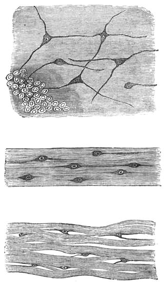
Fig. 53. External nerve-plexus of Echinus.
Fig. 54. Structure of a nerve-trunk of Echinus.
Fig. 55. Nerve-cells lying among the muscular fibres at the base of a spine in
Echinus.
1. Star-fish.—Single rays detached from the
organism crawl as fast and in as determinate a
direction as do the entire animals. They also
crawl up perpendicular surfaces, and sometimes
away from injuries; but they do not invariably, or
even generally, seek to escape from the latter, as is
so certain to be the case with entire animals.
Lastly, when inverted, separated rays right themselves
as quickly as do the unmutilated organisms.
Dividing the nerve in any part of its length has
the effect, whether or not the ray is detached from
the animal, of completely destroying all physiological
continuity between the pedicels on either
side of the line of division. Thus, for instance, if
the nerve be cut across half-way up its length, the
row of pedicels is at once physiologically bisected,
one-half of the row becoming as independent of the
other half as it would were the whole ray divided
into two parts: that is to say, the distal half of the
row may crawl while the proximal half is retracted,
or vice versâ; and if a drop of acid be placed on
either half, the serial contraction of the pedicels
in that half stops abruptly at the line of nerve-division.
As a result of this complete physiological
severance, when a detached ray so mutilated is
inverted, it experiences much greater difficulty in
righting itself than it does before the nerve is
divided. The line of nerve injury lies flat upon
the floor of the tank, while the central and distal
portions of the ray, i.e. the portions on either side
of that line, assume various movements and
shapes. The central portion is particularly apt to
take on the form of an arch, in which the central
end of the severed ray and the line of nerve-section
constitute the points of support (tetanus?) (Fig. 56),
or the central end may from the first show paralysis,
from which it never recovers. The distal end, on
the other hand, usually continues active, twisting
about in various directions, and eventually fastening
its tip upon the floor of the tank to begin the
spiral movement of righting itself. This movement
then continues as far as the line of nerve-injury,
where it invariably stops (Fig. 56). The central
portion may then be dragged over into the normal
position, or may remain permanently inverted,
according to the strength of pull exerted by the
distal portion; as a rule, it does not itself assist in
the righting movement, although its feet usually
continue protruded and mobile. Thus, the effect of
a transverse section of the nerve in a ray is that
of completely destroying physiological continuity
between the pedicels on either side of the section.
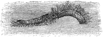
Fig. 56. Movements performed by a detached ray of a Star-fish, in which the
central nerve-trunk is divided.
The only other experiments in nerve-section to
which the simple anatomy of a Star-fish exposes
itself is that of dividing the nerve-ring in the disc;
or, which is virtually the same thing, while leaving
this intact, dividing all the nerves where they pass
from it into the rays. In specimens mutilated by
severing the nerves at the base of each of the five
rays, or by dividing the nerve-ring between all the
rays, the animal loses all power of co-ordination
among its rays. When a common Star-fish is so
mutilated it does not crawl in the same determinate
manner as an unmutilated animal, but, if it moves
at all, it moves slowly and in various directions.
When inverted, the power of effecting the righting
manœuvre is seen to be gravely impaired, although
eventually success is always achieved. There is a
marked tendency, as compared with unmutilated
specimens, to a promiscuous distribution of spirals
and doublings, so that instead of a definite plan of
the manœuvre being formed from the first, as is
usually the case with unmutilated specimens, such
a plan is never formed at all; among the five rays
there is a continual change of un-coördinated movements,
so that the righting seems to be eventually
effected by a mere accidental prepotency of some of
the righting movements over others. Appended is
a sketch of such un-coördinated movement, taken
from a specimen which for more than an hour had
been twisting its rays in various directions (Fig. 57).
Another sketch is appended to show a form of
bending which specimens mutilated as described
are very apt to manifest, especially just after the
operation. When placed upon their dorsal surface,
they turn up all their rays with a peculiar and
exactly similar curve in each, which gives to the
animal a somewhat tulip-like form (Fig. 58). This
form is never assumed by unmutilated specimens,
and in mutilated ones, although it may last for a
long time, it is never permanent. In detached rays
this peculiar curve is also frequently exhibited; but
if the nerve of such a ray is divided at any point in
its length, the curve is restricted to the distal
portion of the ray, and it stops abruptly at the line
of nerve-section. When entire Star-fish are mutilated
by a section of each nerve-trunk half-way up each
ray, and the animal is then placed upon its back,
the tetanic contraction of the muscles in the rays
before mentioned as occurring under this form of
section in detached rays, has the effect, when now
occurring in all the rays, of elevating the disc from
the floor of the tank. This opisthotonous-like
spasm is not, however, permanent; and the distal
ends of the rays forming adhesions to the floor of
the tank, thy animal eventually rights itself, though
much more slowly than unmutilated specimens.
After it has righted itself, although it twists about
the distal portions of the rays, it does not begin to
crawl for a long time, and when it does so, it crawls
in a slow and indeterminate manner. Star-fish
so mutilated, however, can ascend perpendicular
surfaces.
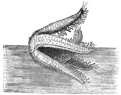
Fig. 57. Un-coördinated movements of a Star-fish, in which the nerves of all the
rays have been divided.
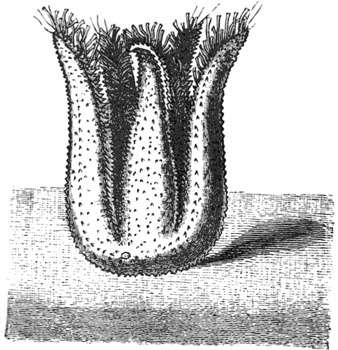
Fig. 58. Form frequently assumed by Star-fish under similar circumstances.
The loss of co-ordination between the rays caused
by division of the nerve-ring in the disc is rendered
most conspicuous in Brittle-stars, from the circumstance
that in locomotion and in righting so much
here depends upon co-ordinated muscular contraction
of the rays. Thus, for instance, when a Brittle-star
has its nerve-ring severed between each ray,
an interesting series of events follows. First, there
is a long period of profound shock—spontaneity,
and even irritability, being almost suspended, and
the rays appearing to be rigid, as if in tetanic
spasm. After a time, feeble spontaneity returns—the
animal, however, not moving in any determinate
direction. Irritability also returns, but only for
the rays immediately irritated, stimulation of one
ray causing active writhing movements in that
ray, but not affecting, or only feebly affecting, the
other rays. The animal, therefore, is quite unable
to escape from the source of irritation, the aimless
movements of the rays now forming a very marked
contrast to the instantaneous and vigorous leaping
movements of escape which are manifested by
unmutilated specimens. Moreover, unmutilated
specimens will vigorously leap away, not only from
stimulation of the rays, but also from that of the
disc; but those with their nerve-ring cut make no
attempts to escape, even from the most violent
stimulation of the disc. In other words, the disc is
entirely severed from all physiological connection
with the rays.
If the nerve-ring be divided at two points, one
on either side of a ray, that ray becomes physiologically
separated from the rest of the organism.
If the two nerve-divisions are so placed as to include
two adjacent rays—i.e. if one cut is on one
side of a ray and the other on the further side of
an adjacent ray—then these two rays remain in
physiological continuity with one another, although
they suffer physiological separation from the other
three. When a Brittle-star is completely divided
into two portions, one portion having two arms and
the other three, both portions begin actively to turn
over on their backs, again upon their faces, again
upon their backs, and so on alternately for an indefinite
number of times. These movements arise
from the rays, under the influence of stimulation
caused by the section, seeking to perform their
natural movements of leaping, which however end,
on account of the weight of the other rays being
absent, in turning themselves over. An entire
Brittle-star when placed on its back after division
of its nerve-ring is not able to right itself, owing to
the destruction of co-ordination among its rays.
Astropecten, under similar circumstances, at first
bends its rays about in various ways, with a preponderant
disposition to the tulip form, and keeps
its ambulacral feet in active movement. But after
half an hour, or an hour, the feet generally become
retracted and the rays nearly motionless—the
animal, like a Brittle-star, remaining permanently
on its back. In this, as in other species, the effect
of dividing the nerve-ring on either side of a ray
is that of destroying its physiological connection
with the rest of the animal, the feet in that ray,
although still remaining feebly active, no longer
taking part in any co-ordinated movement—that
ray, therefore, being merely dragged along by the
others.
Under this division it only remains further to be
said, that section of the nerve-ring in the disc, or
the nerve-trunks of the rays, although, as we have
seen, so completely destroying physiological continuity
in the rows of ambulacral feet and muscular
system of the animal, does not destroy physiological
continuity in the external nerve-plexus; for however
much the nerve-ring and nerve-trunks may
be injured, stimulation of the dorsal surface of the
animal throws all the ambulacral feet and all the
muscular system of the rays into active movement.
This fact proves that the ambulacral feet and the
muscles are all held in nervous connection with one
another by the external plexus, without reference
to the integrity of the main nerve-trunks.
2. Echini.—Section of external surface of shell.—If
a cork-borer be applied to the external surface
of the shell of an Echinus, and rotated there till
the calcareous substance of the shell is reached,
and therefore a continuous circular section of the
over-lying tissues effected, it is invariably found
that the spines and pedicellariæ within the circular
area are physiologically separated from the contiguous
spines and pedicellariæ, as regards local
reflex excitability. That is to say, if any part of
this circular area be stimulated, all the spines and
pedicellariæ within that area immediately respond
to the stimulation in the ordinary way; while none
of the spines or pedicellariæ surrounding the area
are affected. Similarly, if any part of the shell
external to the circumscribed area be stimulated,
the spines and pedicellariæ within that area are not
affected. These facts prove that the function which
is manifested by these appendages of localizing
and gathering round a seat of stimulation, is exclusively
dependent upon the external nerve-plexus.
It is needless to add that in this experiment it does
not signify of what size or shape or by what means
the physiological island is made, so long as the
destruction of the nervous plexus by a closed curve
of injury is rendered complete. In order to ascertain
whether, in the case of an unclosed curve of
injury, any irradiation of a stimulus would take
place round the ends of the curve, we made sundry
kinds of section. It is, however, needless to describe
these, for they all showed that, after injury of a
part of the plexus, there is no irradiation of the
stimulus round the ends of the injury. Thus, for
instance, if a short straight line of injury be made,
by drawing the point of a scalpel over the shell, say
along the equator of the animal, and if a stimulus
be afterwards applied on either side of that line,
even quite close to one of its ends, no effect will be
exerted on the spines or pedicellariæ on the other
side of the line. This complete inability of a
stimulus to escape round the ends of an injury,
forms a marked contrast to the almost unlimited
degree in which such escape takes place in the more
primitive nervous plexus of the Medusæ.
Although the nervous connections on which the
spines and pedicellariæ depend for their function of
localizing and closing round a seat of stimulation
are thus shown to be completely destroyed by
injury of the external plexus, other nervous connections,
upon which another function of the spines
depends, are not in the smallest degree impaired by
such injury. The other function to which I allude
is that which brings about the general co-ordinated
action of all the spines for the purposes of locomotion.
That this function is not impaired by injury
of the external plexus is proved by the fact that
if the area within a closed line of injury on the
surface of the shell be strongly irritated, all the
spines over the whole surface begin to manifest
their peculiar bristling movements, and by this co-ordinated
action rapidly move the animal in a
straight line of escape from the source of irritation;
the injury to the external plexus, although completely
separating the spines enclosed by it from
their neighbouring spines as regards what may be
called their local function of seizing the instrument
of stimulation, nevertheless leaves them in undisturbed
connection with all the other spines in the
organism as regards what may be called their
universal function of locomotion.
Evidently, therefore, this more universal function
must depend upon some other set of nervous connections;
and experiment shows that these are distributed
over all the internal surface of the shell.
Our mode of experimenting was to divide the
animal into two hemispheres, remove all the internal
organs of both hemispheres (these operations producing
no impairment of any of the functions of
the pedicels, spines, or pedicellariæ), and then to
paint with strong acid the inside of the shell—completely
washing out the acid after about a quarter
of a minute's exposure. The results of a number
of experiments conducted on this method may be
thus epitomized:—
The effect of painting the back or inside of the
shell with strong acid (e.g. pure HCl) is that of at
first strongly stimulating the spines into bristling
movements, and soon afterwards reducing them to
a state of quiescence, in which they lie more or less
flat, and in a peculiarly confused manner that
closely resembles the appearance of corn when
"laid" by the wind. The spines have now entirely
lost both their spontaneity and their power of
responding to a stimulus applied on the external
surface of the shell—i.e. their local reflex excitability,
or power of closing in upon a source of irritation.
These effects may be produced over the
whole external surface of the shell, by painting
the whole of the internal surface; but if any part
of the internal surface be left unpainted, the corresponding
part of the external surface remains
uninjured. Conversely, if all the internal surface
be left unpainted except in certain lines or patches,
it will only be corresponding lines and patches on
the external surface that suffer injury. It makes
no difference whether these lines or patches be
painted in the course of the ambulacral feet, or
anywhere in the inter-ambulacral spaces.
The above remarks, which have reference to the
spines, apply equally to the pedicellariæ, except
that their spontaneity and reflex irritability are not
destroyed, but only impaired.
Some hours after the operation it usually happens
that the spontaneity and reflex irritability of the
spines return, though in a feeble degree, and also
those of the pedicellariæ, in a more marked degree.
This applies especially to the reflex irritability of
the pedicellariæ; for while their spontaneity does
not return in full degree, their reflex irritability
does—or almost in full degree.
These experiments, therefore, seem to point to
the conclusions—1st, that the general co-ordination
of the spines is dependent on the integrity of an
internal nerve-plexus; 2nd, that the internal plexus
is everywhere in intimate connection with the external;
and 3rd, that complete destruction of the
former, while profoundly influencing the functions
of the latter, nevertheless does not wholly destroy
them.
Professor Ewart therefore undertook carefully to
examine the internal surface of the shell, to see
whether any evidence of this internal nervous
plexus could be found microscopically, and, after a
great deal of trouble, he has succeeded in doing so.
But as he has not yet published his results, I shall
not forestall them further than to say that this
internal plexus spreads all over the inside of the
shell, and is everywhere in communication with the
external plexus by means of fibres which pass between
the sides of the hexagonal plates of which
the shell of the animal is composed. Thus we can
understand how it is that when a portion of the
external plexus is isolated from the rest of that
plexus as a result of the cork-borer experiment, the
island still remains in communication with the
nerve-centres which preside over the co-ordination
of the spines, as proved by the fact of the Echinus
using its spines to escape from irritation applied to
the area included within the circle of injury to the
external plexus produced by the cork-borer.
Now, where are these nerve-centres situated?
We have just seen that we have evidence of the
presence of such centres somewhere in an Echinus,
seeing that all the spines exhibit such perfect
co-ordination in their movements. Where, then,
are these centres?
Seeing that in a Star-fish the rays are co-ordinated
in their action by means of the pentagonal ring in
the disc, analogy pointed to the nervous ring round
the mouth of an Echinus as the part of the nervous
system which most probably presides over the
co-ordinated action of the spines. Accordingly, we
tried the effect of removing this nervous ring, and
immediately obtained conclusive proof that this was
the centre of which we were in search; for as
soon as the nervous ring was removed, the Echinus
lost, completely and permanently, all power of co-ordination
among its spines. That is to say, after
this operation these organs were never again used
by the animal for the purposes of locomotion, and
no matter how severe an injury we applied, the
Echinus, when placed on a table, did not seek to
escape. But the spines were not wholly paralyzed,
or motionless. On the contrary, their power of
spontaneous movement continued unimpaired, as
did also their power of closing round a seat of
irritation on the external surface of the shell. The
same remark applies to the pedicellariæ, and the
explanation is simple. It is the external nervous
plexus which holds all the spines and pedicellariæ in
communication with one another as by a network;
so that when any part of this network is irritated,
all the spines and pedicellariæ in the neighbourhood
move over to the seat of irritation. On the other
hand, it is the internal plexus which serves to unite
all the spines to the nerve-centre which surrounds
the mouth, and which alone is competent to co-ordinate
the action of all the spines for the purposes
of locomotion.
It remains to consider whether the ambulacral
feet exhibit any general co-ordinated action, and, if
so, whether this likewise depends upon the same
nerve-centre.
The fact already mentioned, that during progression
an Echinus uses some of its feet for crawling
and others for feeling its way, is enough to
suggest that all the feet are co-ordinated by a
nerve-centre. But in order to be quite sure about
the fact of there being a general co-ordination among
all the feet, we tried the following experiments.
I have already described the righting movements
which are performed by an Echinus when the
animal is inverted, and it will be remembered that
in this animal the manœuvre is effected by means
of the feet alone. At first sight this might almost
seem sufficient to prove the fact of a general co-ordination
among the feet; but further reflection
will show that it is not so. For the feet being all
arranged in regular series, when one row begins to
effect the rotation of the globe, it may very well be
that its further rotation in the same direction is due
only to the fact that the slight tilt produced by the
pulling of the first feet in the series A, B, C gives
the next feet in the series D, E, F an opportunity
of reaching the floor of the tank; their adhesions
being established, they would tend by their pulling
to increase still further the tilt of the globe, thus
giving the next feet in the series an opportunity of
fastening to the floor of the tank, and so on. In
order, therefore, to see whether these righting
movements were due to nervous co-ordination
among the feet, or merely to the accident of the
serial arrangement of the feet, we tried the experiments
which I shall now detail.
First of all we took an Echinus, and by means of
a thread suspended it upside-down in a tank of
water half-way up the side of the tank, and in such
a way that only the feet on one side of the ab-oral
pole were able to reach the perpendicular wall of
the tank. These feet as quickly as possible established
their adhesions to the perpendicular wall,
and, the thread being then removed, the Echinus
was left sticking to the side of the tank in an inverted
position by means of the ab-oral ends of two
adjacent feet-rows (Fig. 59). Under these circumstances,
as we should expect from the previous experiments,
the animal sets about righting itself as
quickly as possible. Now, if the righting action of
the feet were entirely and only of a serial character,
the righting would require to be performed by rearing
the animal upwards; the effect of foot after
foot in the same rows being applied in succession to
the side of the tank, would require to be that of
rotating the globular shell against the side of the
tank towards the surface of the water, and therefore
against the action of gravity. This is sometimes
done, which proves that the energy required to perform
the feat is not more than a healthy Echinus
can expend. But much more frequently the
Echinus adopts another device, and the only one by
which it is possible for him to attain his purpose
without the labour of rotating upwards: he rotates
laterally and downwards in the form of a spiral.
Thus, let us call the five feet-rows, 1, 2, 3, 4, and 5
(Figs. 59, 60, 61), and suppose that 1 and 2 are in
use near their ab-oral ends in holding the animal
inverted against the perpendicular side of a tank.
The downward spiral rotation would then be
effected by gradually releasing the outer feet in row
1, and simultaneously attaching the outer feet in
row 2 (i.e. those nearest to row 3, and furthest from
row 1), as far as possible to the outer side of that
row. The effect of this is to make the globe roll far
enough to that side to enable the inner feet of row
3 (i.e. those nearest to row 2), when fully protruded,
to touch the side of the tank. They establish their
adhesions, and the residue of feet in row 1, now
leaving go their hold, these new adhesions serve to
roll the globe still further round in the same
direction of lateral rotation, and so the process proceeds
from row to row; but the globe does not
merely roll along in a horizontal direction, or at the
same level in the water, for each new row that
comes into action takes care, so to speak, that the
feet which it employs shall be those which are as
far below the level of the feet in the row last employed
as their length when fully protruded (i.e.
their power of touching the tank) renders possible.
The rotation of the globe thus becomes a double
one, lateral and downwards, till the animal assumes
its normal position with its oral pole against the
perpendicular tank wall. So considerable is the
rotation in the downward direction, that the normal
position is generally attained before one complete
lateral, or equatorial, rotation is completed.
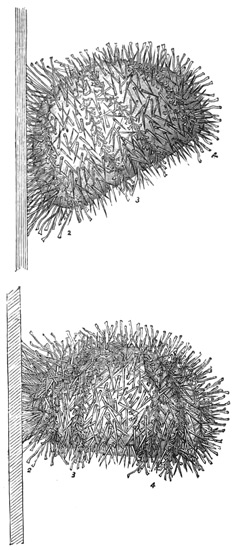
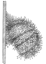
Fig. 61
Figs. 50, 60, and 61 are righting movements of Echinus on a perpendicular surface.
The result of this experiment, therefore, implies
that the righting movements are due to something
more than the merely successive action of the series
of feet to which the work of righting the animal
may happen to be given. The same conclusion is
pointed to by the results of the following experiment.
A number of vigorous Echini were thoroughly
shaved with a scalpel over the whole half of one
hemisphere, i.e. the half from the equator to the
oral pole. They were then inverted on their ab-oral
poles. The object of the experiment was to see
what the Echini which were thus deprived of the
lower half of three feet-rows would do when, in
executing their righting manœuvres, they attained
to the equatorial position and then found no feet
wherewith to continue the manœuvre. The result
of this experiment was first of all to show us that
the Echini invariably chose the unmutilated feet-rows
wherewith to right themselves. Probably this
is to be explained, either by the general principle
to which the escape from injury is due—viz. that
injury inflicted on one side of an Echinoderm
stimulates into increased activity the locomotor
organs of the opposite side,—or by the consideration
that destruction of the lower half of a row very
probably induces some degree of shock in the
remaining half, and so leaves the corresponding
parts of the unmutilated rows prepotent over the
mutilated one. Be this as it may, however, we
found that the difficulty was easily overcome by
tilting the animal over upon its mutilated feet-rows
sufficiently far to prevent the unmutilated rows
from reaching the floor of the tank. When held
steadily in this position for a short time, the mutilated
rows established their adhesions, and the
Echinus was then left to itself. Under these circumstances
an Echinus will always continue the
manœuvre along the mutilated feet-rows with
which it was begun, till the globe reaches the position
of resting upon its equator, and therefore
arrives at the line where the shaved area commences.
The animal then remains for hours in this
position, with a gradual but continuous motion
backwards, which appears to be due to the successive
slipping of the spines—these organs in the
righting movements being always used as props for
the ambulacral feet to pull against while rearing
the globe to its equatorial position, and in performing
this function on a slate floor the spines are
liable often to slip. The only other motion exhibited
by Echini thus situated is that of a slow
rolling movement, now to one side and now to
another, according to the prepotency of the pull
exerted by this or that row of ambulacral feet.
Things continue in this way until the slow backward
movement happens to bring the animal
against some side of the tank, when the uninjured
rows of ambulacral feet immediately adhere to the
surface and rotate the animal upwards or horizontally,
until it attains the normal position. But if
care be taken to prevent contact with any side of
the tank, the mutilated Echinus will remain propped
on its equator for days; it never adopts the simple
expedient of reversing the action of its mutilated
feet-rows, so as to bring the globe again upon its
ab-oral pole and get its unmutilated feet-rows into
action.
From this we may conclude that the righting
movements of the pedicels are due, not to the merely
serial action of the pedicels, but to their co-ordination
by a nerve-centre acting under a stimulus
supplied by a sense of gravity; for if the movements
of the pedicels were merely of a serial
character, we should not expect that the equatorial
position, having been attained under these circumstances,
should be permanently maintained. We
should not expect this, because after a while the
pedicels, which are engaged in maintaining the
globe in its equatorial position, must become exhausted
and relax their hold, when those next
behind in the series would lay hold of the bottom
of the tank, and so on, the rotation of the globe
thus proceeding in the opposite direction to that
in which it had previously taken place. On the
other hand, if the righting movements of the pedicels
are due to co-ordination proceeding from a
nerve-centre acting under a sense of gravity, we
should expect the animal under the circumstances
mentioned to remain permanently reared upon its
equator; for this would allow that the nerve-centre
was always persistently, though fruitlessly, endeavouring
to co-ordinate the action of the absent feet.
Further, as proof that the ambulacral feet of
Echinus are under the control of some centralizing
apparatus when executing the righting manœuvre,
we may state one other fact. When the righting
manœuvre. is nearly completed by the rows engaged
in executing it, the lower feet in the other rows
become strongly protruded and curved downwards,
in anticipation of shortly coming into contact with
the floor of the tank when the righting manœuvre
shall have been completed (see Fig. 52, p. 280).
This fact tends to show that all the ambulacral feet
of the animal are, like all the spines, held in mutual
communication with one another by some centralizing
mechanism.
But the best proof of all that the feet in executing
the righting manœuvre are under the influence of
a co-ordinating centre, is one that arose from an
experiment suggested to me by Mr. Francis Darwin,
and which I shall now describe. Mr. Darwin
having kindly sent the apparatus which his father
and himself had used in their experiments on
the geotropism of plants, it was employed thus.
A healthy Echinus was placed in a large bottle
filled to the brim with sea-water, and having been
inverted on the bottom of the bottle, it was allowed
in that position to establish its adhesions. The
bottle was then corked and mounted on an upright
wheel of the apparatus whereby, by means of clockwork,
it could be kept in continual slow rotation
in a vertical plane. The object of this was to
ascertain whether the continuous rotation in a
vertical plane would prevent the animal from righting
itself (because confusing the nerve-centres
which, under ordinary circumstances, could feel by
their sense of gravity which was up and which
was down), or would still allow the animal to right
itself (because not interfering with the serial action
of the feet). Well, it was found that this rotation
of the whole animal in a vertical plane entirely
prevented the righting movements during any
length of time that it might be continued, and that
these movements were immediately resumed as
soon as the rotation was allowed to cease. This,
moreover, was the case, no matter what phase of
the righting manœuvre the Echinus might have
reached at the moment when the rotation began.
Thus, for instance, if the globe were allowed to
have reached the position of resting on its equator
before the rotation was commenced, the Echinus
would remain motionless, holding on with its equatorial
feet, so long as the rotation was kept up.
Therefore, there can be no question that the ambulacral
feet are all under the influence of a co-ordinating
nerve-centre, quite as much as are the
spines. But, on the other hand, experiments show
that the centre in this case is not of so localized a
character as it is in the case of the spines; for
when the nerve-ring is cut out, the co-ordination
of the feet, although impaired, is not wholly
destroyed. Take, for instance, the case of the
righting manœuvre. The effect of cutting out the
nerve-ring is that of entirely destroying the ability
to perform this manœuvre in the case of the majority
of specimens; nevertheless about one in ten continue
able to perform it. Again, if an Echinus is divided
into two hemispheres by an incision carried from pole
to pole through any meridian, the two hemispheres
will live for days, crawling about in the same
manner as entire animals; if their ocular plates
are not injured, they seek the light, and when
inverted they right themselves. The same observations
apply to smaller segments, and even to single
detached rows of ambulacral feet. The latter are,
of course, analogous to the single detached rays of
a Star-fish, so far as the system of ambulacral feet
is concerned; but, looking to the more complicated
apparatus of locomotion (spines and pedicellariæ),
as well as to the rigid consistence and awkward
shape of the segment—standing erect, instead of
lying flat—the appearance presented by such a segment
in locomotion is much more curious, if not
surprising, than that presented by the analogous
part of a Star-fish under similar circumstances. It
is still more surprising that such a fifth-part segment
of an Echinus will, when propped up on its
ab-oral pole (Fig. 62), right itself (Fig. 63) after
the manner of larger segments or entire animals.
They, however, experience more difficulty in doing
so, and very often, or indeed generally, fail to
complete the manœuvre.
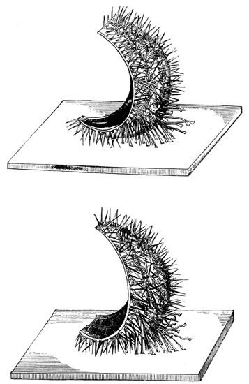
Figs. 62 and 63.—Righting and ambulacral movements of severed segments
of Echinus.
On the whole, then, we may conclude that the
nervous system of an Echinus consists (1) of an
external plexus which serves to unite all the feet,
spines, and pedicellariæ together, so that they all
approximate a point of irritation situated anywhere
in that plexus; (2) of an internal nervous plexus
which is everywhere in communication through the
thickness of the shell with the external, and the
function of which is that of bringing the feet, spines,
and probably also the pedicellariæ into relation
with the great co-ordinating nerve-centre situated
round the mouth; (3) of central nervous matter
which is mainly gathered round the mouth, and
there presides exclusively over the co-ordinated
action of the spines, and in large part also over
the co-ordinated action of the feet, but which is
further in part distributed along the courses of the
main nerve-trunks, and so secures co-ordination of
feet even in separated segments of the animal.
Before concluding, I must say a few words on
the experiments whereby we sought to test for the
presence in Echinoderms of the special senses of
sight and smell.
We have found unequivocal evidence of the Star-fish
(with the exception of the Brittle-stars) and
the Echini manifesting a strong disposition to crawl
towards, and remain in, the light. Thus, if a large
tank be completely darkened, except at one end
where a narrow slit of light is admitted, and if a
number of Star-fish and Echini be scattered over
the floor of the tank, in a few hours the whole
number, with the exception of perhaps a few per
cent., will be found congregated in the narrow slit
of light. The source we used was diffused daylight,
which was admitted through two sheets of
glass, so that the thermal rays might be considered
practically excluded. The intensity of the light
which the Echinoderms are able to perceive may be
very feeble indeed; for in our first experiments we
boarded up the face of the tank with ordinary pinewood,
in order to exclude the light over all parts
of the tank except at one narrow slit between two
of the boards. On taking down the boards we
found, indeed, the majority of the specimens in or
near the slit of light; but we also found a number
of other specimens gathering all the way along the
glass face of the tank that was immediately behind
the pine-boards. On repeating the experiment
with blackened boards, this was never found to be
the case; so there can be no doubt that in the first
experiments the animals were attracted by the
faint glimmer of the white boards, as illuminated
by the very small amount of light scattered from
the narrow slit through a tank, all the other sides
of which were black slate. Indeed, towards the
end of the tank, where some of the specimens were
found, so feeble must have been the intensity of
this glimmer, that we doubt whether even human
eyes could have discerned it very distinctly. Owing
to the prisms at our command not having sufficient
dispersive power for the experiments, and not wishing
to rely on the uncertain method of employing
coloured glass, we were unable to ascertain how the
Echinoderms might be affected by different rays.
On removing with a pointed scalpel the eye-spots
from a number of Star-fish and Echini, without
otherwise injuring the animals, the latter no longer
crawled towards the light, even though this were
admitted to the tank in abundance; but they
crawled promiscuously in all directions. On the
other hand, if only one out of the five eye-spots were
left intact, the animals crawled towards the light
as before. It may be added that single detached
rays of Star-fish and fifth-part segments of Echini
crawl towards the light in the same manner as
entire animals, provided, of course, that the eye-spot
is not injured.
The presence of a sense of smell in Star-fish was
proved by keeping some of these animals for several
days in a tank without food, and then presenting
them with small pieces of shell-fish. The Star-fish
immediately perceived the proximity of food, as
shown by their immediately crawling towards it.
Moreover, if a small piece of the food were held in
a pair of forceps and gently withdrawn as the Star-fish
approached it, the animal could be led about
the floor of the tank in any direction, just as a
hungry dog could be led about by continually
withdrawing from his nose a piece of meat as he
continually follows it up. This experiment, however,
was only successful with Star-fish which had
been kept fasting for several days; freshly caught
Star-fish were not nearly so keen in their manifestations,
and indeed in many cases did not notice the
food at all.
Desiring to ascertain whether the sense of smell
were localized in any particular organs, as we had
found to be the case with the sense of sight, I first
tried the effect of removing the five ocelli. This
produced no difference in the result of the above
experiment with hungry Star-fish, and therefore I
next tried the effect of cutting off the tips of the
rays. The Star-fish behaving as before, I then progressively
truncated the rays, and thus eventually
found that the olfactory sense was equally distributed
throughout their length. The question,
however, still remained whether it was equally distributed
over both the upper and the lower surfaces.
I therefore tried the effect of varnishing the upper
surface. The Star-fish continued to find its food as
before, which showed that the sense of smell was
distributed along the lower surface. I could not try
the converse experiment of varnishing this surface,
because I should thereby have hindered the action
of the ambulacral feet. But by another method
I was able nearly as well to show that the upper
surface does not participate in smelling. This
method consisted in placing a piece of shell-fish
upon the upper surface and allowing it to rest there.
When this was done, the Star-fish made no attempt
to remove the morsel of food by brushing it off with
the tips of its rays, as is the habit of the animal
when any irritating substance is applied to this
surface. Therefore I conclude that the upper or
dorsal surface of a Star-fish takes no part in ministering
to the sense of smell, which by the experiment
of varnishing this surface, and also by that
of progressively truncating the rays, is proved to
be distributed over the whole of the ventral or
lower surface of the animal. For I must add that
severed rays behave in all these respects like the
entire organisms, although they are disconnected
from the mouth and disc.
As this chapter has already extended to so great
a length, I omit from it any account of some further
experiments which I tried concerning the effects of
nerve-poisons upon the Echinodermata. A full
record of these experiments may be found in the
publications of the Linnean Society.
FINIS.
THE
INTERNATIONAL SCIENTIFIC SERIES.
Each book complete in One Volume, 12mo, and bound in Cloth.
| 1. |
THE FORMS OF WATER IN CLOUDS AND RIVERS, ICE AND GLACIERS.
By J. Tyndall, LL.D., F.R.S. With 35 Illustrations. $1.50. |
| 2. |
PHYSICS AND POLITICS; or, Thoughts on the Application of the Principles
of "Natural Selection" and "Inheritance" to Political Society.
By Walter Bagehot. $1.50. |
| 3. |
FOODS. By Edward Smith, M.D., LL.B., F.R.S. With numerous Illustrations.
$1.75. |
| 4. |
MIND AND BODY: The Theories of their Relation. By Alexander Bain,
LL.D. With 4 Illustrations. $1.50. |
| 5. |
THE STUDY OF SOCIOLOGY. By Herbert Spencer. $1.50. |
| 6. |
THE NEW CHEMISTRY. By Professor J. P. Cooke, Harvard University.
With 31 Illustrations. $2.00. |
| 7. |
THE CONSERVATION OF ENERGY. By Balfour Stewart, M.A.,
LL.D., F.R.S. With 14 Illustrations. $1.50. |
| 8. |
ANIMAL LOCOMOTION; or, Walking, Swimming, and Flying. By J. B.
Pettigrew, M.D., F.R.S., etc. With 130 Illustrations. $1.75. |
| 9. |
RESPONSIBILITY IN MENTAL DISEASE. By Henry Maudsley, M.D.,
$1.50. |
| 10. |
THE SCIENCE OF LAW. By Professor Sheldon Amos. $1.75. |
| 11. |
ANIMAL MECHANISM: A Treatise on Terrestrial and Aërial Locomotion.
By Professor E. J. Marey, College of France. With 117 Illustrations. $1.75. |
| 12. |
THE HISTORY OF THE CONFLICT BETWEEN RELIGION AND SCIENCE.
By J. W. Draper, M.D., LL.D. $1.75. |
| 13. |
THE DOCTRINE OF DESCENT AND DARWINISM. By Professor Oscar
Schmidt, Strasburg University. With 26 Illustrations. $1.50. |
| 14. |
THE CHEMISTRY OF LIGHT AND PHOTOGRAPHY IN THEIR APPLICATION
TO ART, SCIENCE, AND INDUSTRY. By Dr. Hermann
Vogel, Royal Industrial Academy of Berlin. With 100 Illustrations. $2.00. |
| 15. |
FUNGI: Their Nature and Uses. By M. C. Cooke, M.A., LL.D. Edited by
the Rev. M. J. Berkeley, M.A., F.L.S. With 109 Illustrations. $1.50. |
| 16. |
THE LIFE AND GROWTH OF LANGUAGE. By Professor William
Dwight Whitney, Yale College. $1.50. |
| 17. |
MONEY AND THE MECHANISM OF EXCHANGE. By W. Stanley
Jevons, M.A., F.R.S. $1.75. |
| 18. |
THE NATURE OF LIGHT, with a General Account of Physical Optics. By
Dr. Eugene Lommel. With 188 Illustrations and a Table of Spectra in
Colors. $2.00. |
| 19. |
ANIMAL PARASITES AND MESSMATES. By Professor P. J. Van Beneden,
University of Louvain. With 83 Illustrations. $1.50. |
| 20. |
FERMENTATION. By Professor P. Schützenberger. With 28 Illustrations.
$1.50. |
| 21. |
THE FIVE SENSES OF MAN. By Professor Julius Bernstein, University
of Halle. With 91 Illustrations. $1.75. |
| 22. |
THE THEORY OF SOUND IN ITS RELATION TO MUSIC. By Professor
Pietro Blaserna, Royal University of Rome. With numerous
Illustrations. $1.50. |
| 23. |
STUDIES IN SPECTRUM ANALYSIS. By J. Norman Lockyer, F.R.S.
With 7 Photographic Illustrations of Spectra, and 52 other Illustrations.
$2.50. |
| 24. |
A HISTORY OF THE GROWTH OF THE STEAM-ENGINE. By Professor
R. H. Thurston, Cornell University. With 163 Illustrations. $2.50. |
| 25. |
EDUCATION AS A SCIENCE. By Alexander Bain, LL.D. $1.75. |
| 26. |
STUDENTS' TEXT-BOOK OF COLOR; or, Modern Chromatics. With
Applications to Art and Industry. By Professor Ogden N. Rood, Columbia
College. With 130 Illustrations. $2.00. |
| 27. |
THE HUMAN SPECIES. By Professor A. de Quatrefages, Museum of
Natural History, Paris. $2.00. |
| 28. |
THE CRAYFISH: An Introduction to the Study of Zoölogy. By T. H.
Huxley, F.R.S. With 82 Illustrations. $1.75. |
| 29. |
THE ATOMIC THEORY. By Professor A. Wurtz. Translated by E.
Cleminshaw, F.C.S. With Illustrative Chart. $1.50. |
| 30. |
ANIMAL LIFE AS AFFECTED BY THE NATURAL CONDITIONS OF
EXISTENCE. By Professor Karl Semper, University of Würzburg.
With 106 Illustrations and 2 Maps. $2.00. |
| 31. |
SIGHT: An Exposition of the Principles of Monocular and Binocular Vision.
By Professor Joseph Le Conte, LL.D., University of California. With
132 Illustrations. $1.50. |
| 32. |
GENERAL PHYSIOLOGY OF MUSCLES AND NERVES. By Professor
I. Rosenthal, University of Erlangen. With 75 Illustrations. $1.50. |
| 33. |
ILLUSIONS: A Psychological Study. By James Sully. $1.50. |
| 34. |
THE SUN. By Professor C. A. Young, College of New Jersey. With 83
Illustrations. $2.00. |
| 35. |
VOLCANOES; What they Are and What they Teach. By Professor John
W. Judd, F.R.S., Royal School of Mines. With 96 Illustrations. $2.00. |
| 36. |
SUICIDE: An Essay in Comparative Moral Statistics. By Professor Henry
Morselli, M.D., Royal University, Turin. With 4 Statistical Maps.
$1.75. |
| 37. |
THE FORMATION OF VEGETABLE MOULD, THROUGH THE ACTION
OF WORMS. With Observations on their Habits. By Charles
Darwin, LL.D., F.R.S. With 16 Illustrations. $1.50. |
| 38. |
THE CONCEPTS AND THEORIES OF MODERN PHYSICS. By J. B.
Stallo. $1.75. |
| 39. |
THE BRAIN AND ITS FUNCTIONS. By J. Luys, Hospice Salpêtrière,
Paris. With 6 Illustrations. $1.50. |
| 40. |
MYTH AND SCIENCE. By Tito Vignoli. $1.50. |
| 41. |
DISEASES OF MEMORY: An Essay in the Positive Psychology. By Th.
Ribot, author of "Heredity." $1.50. |
| 42. |
ANTS, BEES. AND WASPS. A Record of Observations of the Habits of
the Social Hymonoptera. By Sir John Lubbock, Bart., F.R.S., etc. $2.00. |
| 43. |
THE SCIENCE OF POLITICS. By Professor Sheldon Amos, $1.75. |
| 44. |
ANIMAL INTELLIGENCE. By George J. Romanes, M.D., F.R.S. $1.75. |
| 45. |
MAN BEFORE METALS. By Professor N. Joly, Science Faculty of Toulouse.
With 148 Illustrations. $1.75. |
| 46. |
THE ORGANS OF SPEECH AND THEIR APPLICATION IN THE
FORMATION OF ARTICULATE SOUNDS. By Professor G. H. von
Meyer, University of Zürich. With 47 Illustrations. $1.75. |
| 47. |
FALLACIES: A View of Logic from the Practical Side. By Alfred
Sidgwick, B.A., Oxon. $1.75. |
| 48. |
ORIGIN OF CULTIVATED PLANTS. By Alphonse de Candolle. $2.00. |
| 49. |
JELLY-FISH, STAR-FISH, AND SEA-URCHINS. A Research on Primitive
Nervous Systems. By George J. Romanes, M.D., F.R.S. With 63
Illustrations. $1.75. |
| 50. |
THE COMMON SENSE OF THE EXACT SCIENCES. By William Kingdon
Clifford. With 100 Figures. $1.50. |
| 51. |
PHYSICAL EXPRESSION: Its Modes and Principles. By Francis Warner,
M.D., Assistant Physician, London Hospital. With 51 Illustrations.
$1.75. |
| 52. |
ANTHROPOID APES. By Professor Robert Hartmann, University of
Berlin. With 63 Illustrations. $1.75. |
| 53. |
THE MAMMALIA IN THEIR RELATION TO PRIMEVAL TIMES. By
Professor Oscar Schmidt, University of Strasburg. With 51 Illustrations.
$1.50. |
| 54. |
COMPARATIVE LITERATURE. By Professor H. M. Posnett, M.A., University
College, Auckland. $1.75. |
| 55. |
EARTHQUAKES AND OTHER EARTH MOVEMENTS. By Professor John
Milne, Imperial College of Engineering, Tokio. With 38 Figures. $1.75. |
| 56. |
MICROBES, FERMENTS, AND MOULDS. By E. L. Trouessart. With
107 Illustrations. $1.50. |
| 57. |
THE GEOGRAPHICAL AND GEOLOGICAL DISTRIBUTION OF ANIMALS.
By Professor Angelo Heilprin, Academy of Natural Sciences,
Philadelphia. $2.00. |
| 58. |
WEATHER. A Popular Exposition of the Nature of Weather Changes from Day to Day.
With 96 Diagrams. By Hon. Ralph Abercromby. $1.75. |
| 59. |
ANIMAL MAGNETISM. By Alfred Binet and Charles Féré. Assistant
Physician, Hospice Salpêtrière, Paris. With 15 Figures. $1.50. |
| 60. |
INTERNATIONAL LAW, with Materials for a Code of International Law.
By Professor Leone Levi, King's College. London. $1.50. |
| 61. |
THE GEOLOGICAL HISTORY OF PLANTS. With 79 Illustrations. By
Sir J. William Dawson, LL.D., F.R.S. $1.75. |
| 62. |
ANTHROPOLOGY. An Introduction to the Study of Man and Civilization.
By Edward B. Tylor, D.C.L., F.R.S. With 78 Illustrations. $2.00. |
| 63. |
THE ORIGIN OF FLORAL STRUCTURES, THROUGH INSECT AND
OTHER AGENCIES. By the Rev. George Henslow, M.A., etc. With
88 Illustrations. $1.75. |
| 64. |
THE SENSES, INSTINCTS, AND INTELLIGENCE OF ANIMALS, WITH
SPECIAL REFERENCE TO INSECTS. By Sir John Lubbock, Bart.,
F.R.S., etc. With 118 Illustrations. $1.75. |
| 65. |
THE PRIMITIVE FAMILY IN ITS ORIGIN AND DEVELOPMENT.
By Dr. C. N. Starcke, University of Copenhagen. $1.75. |
| 66. |
PHYSIOLOGY OF BODILY EXERCISE. By F. Lagrange, M.D. $1.75. |
| 67. |
THE COLORS OF ANIMALS: Their Meaning and Use. By Edward
Bagnall Poulton, F.R.S. With 36 Illustrations and 1 Colored Plate.
$1.75. |
| 68. |
SOCIALISM: New and Old. By Professor William Graham, M.A., Queen's
College, Belfast. $1.75. |
| 69. |
MAN AND THE GLACIAL PERIOD. By Professor G. Frederick Wright,
D.D., Oberlin Theological Seminary. With 108 Illustrations and 3 Maps.
$1.75. |
| 70. |
HANDBOOK OF GREEK AND LATIN PALÆOGRAPHY. By Edward
Maunde Thompson, D.C.L., etc. $2.00. |
| 71. |
A HISTORY OF CRUSTACEA. Recent Malacostraca. By the Rev.
Thomas R. R. Stebbing, M.A. With 51 Illustrations. $2.00. |
| 72. |
RACE AND LANGUAGE. By Professor André Lefèvre, Anthropological
School, Paris. $1.50. |
| 73. |
MOVEMENT, By E. J. Marey. Translated by Eric Pritchard, M.A.,
M.B., B.Ch. (Oxon.). With 200 Illustrations. $1.75. |
| 74. |
ICE-WORK, PRESENT AND PAST. By T. G. Bonney, D.Sc., F.R.S.,
F.S.A., etc., Professor of Geology at University College, London. $1.50. |
| 75. |
WHAT IS ELECTRICITY? By John Trowbridge, S.D., Rumford Professor
and Lecturer on the Applications of Science to the Useful Arts, Harvard
University. Illustrated. $1.50. |
| 76. |
THE EVOLUTION OF THE ART OF MUSIC. By C. Hubert H. Parry.
D.C.L., M.A., etc. $1.75. |
New York: D. APPLETON & CO., 72 Fifth Avenue.