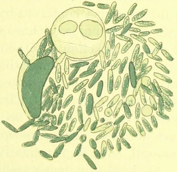
New original cover art included with this eBook is granted to the public domain.
IMMUNITY
IN
INFECTIVE DISEASES
CAMBRIDGE UNIVERSITY PRESS WAREHOUSE,
C. F. CLAY, Manager.
London: FETTER LANE, E.C.
Glasgow: 50, WELLINGTON STREET.
Leipzig: F. A. BROCKHAUS.
New York: THE MACMILLAN COMPANY.
Bombay and Calcutta: MACMILLAN AND CO., Ltd.
[All Rights reserved.]
BY
ÉLIE METCHNIKOFF
FOREIGN MEMBER OF THE ROYAL SOCIETY OF LONDON
PROFESSOR AT THE PASTEUR INSTITUTE, PARIS
TRANSLATED FROM THE FRENCH
BY
FRANCIS G. BINNIE
OF THE PATHOLOGICAL DEPARTMENT, UNIVERSITY OF CAMBRIDGE
With 45 figures in the text
CAMBRIDGE
AT THE UNIVERSITY PRESS
1905
Cambridge
PRINTED BY JOHN CLAY, M.A
AT THE UNIVERSITY PRESS.
PREFACE TO THE ENGLISH EDITION.
v
In preparing for the English-reading student this version of
M. Metchnikoff’s latest work, wherein he “sums up the labours
of twenty-five years,” it has been my aim to give a faithful rendering
of the ideas and argument of the original, even at the risk of an
occasional crude expression, rather than to attempt to reproduce
the brilliancy of the original by any wide verbal departure from
the text.
The Table of Contents forms an admirable analytical summary of
the main subject-matters treated, but an alphabetical Index has been
added to the present edition, and, though not at all exhaustive, this
may serve as a key to the many authors cited and to the maze of
detail discussed in the work.
The marginal reference to the pages of the original work will,
I hope, commend itself to those readers who may wish to refer to
the ipsissima verba of the author. It is, I believe, a novelty in
scientific works, though familiar in works in other departments of
literature.
I am under deep obligations to Professor Woodhead (who has
read the whole of the proofs) and to Mr A. E. Shipley, and
Mr G. H. F. Nuttall (who have read portions) for much valuable
criticism and advice.
THE TRANSLATOR.
August, 1905.
ERRATA.
Page 93, line 2, after itself insert alone; line 3, delete only.
Page 98, line 3 from bottom, for actively read vigorously.
Permit me to dedicate to you this work, which sums up
the labours of twenty-five years; a very great part of it has been
carried out by your side, you who have done so much to lighten
my task.
When, nearly fourteen years ago, you allowed me to share your
work alongside the venerated Master who founded the House where
we have laboured together, you were anything but partisans of
my theories; they seemed to you too vitalistic, and not sufficiently
physico-chemical. In course of time you became convinced that
my ideas were not without foundation, and since then you have
given me warm encouragement to pursue my researches in the
field that I had marked out for myself.
Working by your side and drawing largely from your vast
and varied stores of knowledge, I felt myself safe from those divagations
into which a zoologist, who had wandered into the domain
of biological chemistry and of medical science, is likely to stray.
I thank you with all my heart, and I beg you to accept the homage
of this work as a testimony of my deepest gratitude and of my
warmest friendship.
ÉLIE METCHNIKOFF.
Institut Pasteur,
3 October, 1901.
PREFACE.
ix
When, ten years ago, I was preparing my Lessons on the Comparative
Pathology of Inflammation for the press, I hoped that
the other sections of the phagocytic theory—Immunity, Atrophies,
and Healing—would soon follow this first work. This hope has not
been realised, and it has needed prolonged work ere I could publish
the volume I have just completed.
During this long period I sent out several ballons d’essai under
the form of summaries of the question of Immunity, published in the
Semaine médicale (1892), the Ergebnisse of Lubarsch and Ostertag
(1886), and the Handbuch der Hygiene by Weyl (1897). I there
attempted, as far as possible, to give a general picture of the
phenomena of Immunity in the infective diseases, and it was my
desire to excite criticism and opposition, in order to determine the
fate of the theory of phagocytes in its application to the problem of
Immunity.
The most recent attempt in this direction was made at the International
Congress at Paris, in the past year (1900), when I presented
my report on Immunity before an audience which included, amongst
others, my principal opponents. It was the result of this Congress
which at length decided me to bring together my views on Immunity
in a volume which I now present to the reader.
Convinced that many of the objections raised against the phagocytic
theory of Immunity proceeded solely from an insufficient
knowledge of the theory, I thought that a work condensed into one
volume might render some service to those who are interested in
the problem of Immunity. I do not know whether I shall convert my
opponents, but I am convinced that a perusal of this book will clear
away certain misunderstandings. A very competent observer recently
confessed in one of his publications that for many years he had
been unaware of the experiments of M. J. Bordet and myself on
Immunity against the cholera vibrio, experiments which he now
xregards as of fundamental importance for the comprehension of the
whole question of Immunity. I hope that after the appearance of
this treatise such oversights will not be so likely to occur.
Should I not succeed in convincing my opponents of the justice
of the cause which I defend, T shall at least have informed my critics
and shall have given them an opportunity of discussing it with a
thorough knowledge of the material on which it is based. This
result alone would justify me in having undertaken this work.
At first I intended to add to my explanation of Immunity a
theory of the phenomena of healing in infective diseases, but I soon
had to renounce this project, for its execution would have increased
too greatly the bulk of the book which, without it, has already assumed
considerable proportions. It seemed to me preferable to set forth
the present state of the question without paying too much attention
to the historical sequence of the discoveries, and to reserve for a
special chapter, at the end of the work, a sketch of the history of our
knowledge on Immunity.
Before I ask the reader to glance through this work, I should
mention that I have been heartily seconded in its preparation by many
of my friends and collaborators. I offer my most sincere thanks to
MM. Roux, Nocard, Massart, and J. Bordet, who kindly undertook
to read my manuscript throughout, or such parts of it as related to
their special subjects. For example, M. Nocard rendered me a
very great service by correcting the paragraphs of Chapter xv,
which treat of the vaccinations against epizootic diseases, and
M. Massart, by giving me his advice on the subject of immunity
in plants.
I owe very special thanks to M. Mesnil, who has been good
enough to give me very effective help in the dry task of correcting
the manuscript and proofs.
I beg MM. E. Rémy and L. Barnéoud to accept my thanks for the
care they have bestowed on the execution of the illustrations in this
work.
Importance of the study of immunity from a general point of view.—Part played by parasites in infective diseases.—Intoxications by the products of micro-organisms.—Resistance of the organism to the invasion of micro-organisms.
Natural immunity and acquired immunity.
Immunity to micro-organisms and immunity to toxins.
Infective diseases of the unicellular organisms.—Intracellular digestion in the Protozoa.—Amoebo-diastase.—Part played by digestion in the defence of the Protozoa against infective parasites.—Defences of the Paramaecia against micro-organisms.—Part played by irritability in defence in the lower organisms.
Immunity of unicellular organisms to toxins.—Acclimatisation of bacteria to toxic substances.—Protective secretion of membranes by bacteria.
Adaptation of the Protozoa to saline solutions—of yeasts to poisons—of yeasts to milk-sugar.
Irritability of unicellular organisms and Weber-Fechner’s psycho-physical law.
Infective diseases of plants.—Plasmodia of the Myxomycetes and their chemiotaxis.—Adaptation of the plasmodia to poisons.—Pathogenic action of Sclerotinia upon Phanerogams.—The cicatrisation of plants.—Defence in plants against Bacteria.—Sensitiveness of vegetable cells to osmotic pressure.—Adaptation of plants to modifications of osmotic pressure.—Dependence of the chemical phenomena upon the irritability of the vegetable cells.—The law of Weber-Fechner.
CHAPTER III
Preliminary remarks on immunity in the animal kingdom
xiiExamples of natural immunity among the Invertebrates.—Immunity against micro-organisms and insusceptibility to microbial poisons are two distinct properties.—The refractory organism does not eliminate micro-organisms by the excretory channels.—It destroys them by a process of resorption.—The fate of foreign bodies in the organism.—The resorption of cells.—Intracellular digestion.—This digestion effected by the aid of soluble ferments.—Digestion in Planarians and Actinians.—Actinodiastase.—Transition from intracellular digestion to digestion by secreted juices.—Digestion in the higher animals.—Enterokynase and the part it plays in digestion.—The psychical and nervous elements in digestion.—Adaptation of the pancreatic secretion to the kind of food.—Excretion of pepsin in the blood and in the urine.
Digestion in the tissues.—Resorption of cells in the Invertebrata.—Resorption of red corpuscles by the phagocytes of the Vertebrata.—Phagocytes.—Various categories of these cells.—Macrophages and microphages.—Part played by macrophages in the resorption of the formed elements.—Digestive property of the macrophagic organs.—Solution of the red blood corpuscles by the blood serums.—The two substances which operate in haemolysis.—Macrocytase and fixative.—Analogy of the latter with enterokynase.—Escape of the macrocytase during phagolysis.—Suppression of phagolysis.—Resorption of the spermatozoa.—Presence of fixatives in plasmas.—Origin of fixatives.
Resorption of albuminoid substances.—The precipitins of blood serum which appear as a result of the absorption of serums and of milk.—Absorption of gelatine.—Leucocytic origin of the ferment which digests gelatine.—Anti-enzymes.—Antirennet.—The anticytotoxins.—Antihaemotoxic serums.—Their two constituent parts: anticytase and antifixative.—Action of anticytase.—The antispermotoxins.—Origin of anticytotoxins.—Ehrlich’s theory on this question.—Origin of antihaemotoxin.—Origin of antispermotoxin.—Production of this antibody by castrated males.—The antispermofixative produced when the spermatozoa are excluded.—Distribution of spermotoxin and antispermotoxin in the organism.
CHAPTER VI
Natural immunity against pathogenic micro-organisms
Natural immunity and the composition of the body fluids.—Cultivation of the bacteria of influenza and pleuro-pneumonia in the fluids of refractory animals.—Resistance of Daphniae to the Blastomycetes.—Examples of natural immunity in Insects and Mollusca.—Immunity of Fishes against the anthrax bacillus.—Immunity of frogs against anthrax, Ernst’s bacillus, the bacillus of mouse septicaemia, and the cholera vibrio.—Natural immunity in the cayman.—Immunity of the fowl and pigeon against anthrax and human tuberculosis.—Immunity of the dog and rat against the anthrax bacillus.—Immunity of Mammals against anthrax vaccines.—Immunity of the guinea-pig against spirilla, vibrios, and streptococci.—Natural immunity against anaerobic bacilli.—Fate of Blastomycetes and Trypanosomata in the refractory organism.
xiii
CHAPTER VII
The mechanism of natural immunity against micro-organisms
The destruction of micro-organisms in natural immunity is an act of resorption.—Part played by inflammation in natural immunity.—Importance of microphages in immunity against micro-organisms.—Chemiotaxis of leucocytes and ingestion of micro-organisms.—Phagocytes are capable of ingesting living and virulent micro-organisms.—The digestion of micro-organisms in phagocytes is most often effected in a feebly acid medium.—Bactericidal property of serums.—Phagocytic origin of the bactericidal substance.—Theory of the secretion of the bactericidal substance by leucocytes.—Comparison of the bactericidal power of serums and of blood plasmas.—The bactericidal substance of blood serums must not be considered a secretion-product of leucocytes; it remains within the phagocytes, so long as they are intact.—The cytases.—Two kinds of cytases: macrocytase and microcytase.—Cytases are endo-enzymes, allied to trypsins.—Changes in the staining properties and in the form of micro-organisms in the phagocytes.—Absence or rarity of fixatives in the serums of animals endowed with natural immunity.—The agglutination of micro-organisms does not play any important part in the mechanism of natural immunity.—Absence of antitoxic property of the body fluids in natural immunity.—The phagocytes destroy the micro-organisms without their ingestion being preceded by neutralisation of the toxins.
CHAPTER VIII
Survey of the facts bearing on acquired immunity against micro-organisms
The discovery of attenuated viruses and its application to vaccination against infective diseases.—Vaccination by microbial products.—Vaccination with serums.—The acquired immunity of the frog against pyocyanic disease.—The acquired immunity against vibrios.—Extracellular destruction of the cholera vibrio.—Part played by two substances in Pfeiffer’s phenomenon.—Specificity of fixatives.—Phagolysis and its relation to the extracellular destruction of vibrios.—Part played by phagocytosis in the acquired immunity against vibrios.—Fate of the spirilla of recurrent fever in the organism of immunised guinea-pigs.—Acquired immunity against the bacteria of typhoid fever and pyocyanic disease.—Acquired immunity against swine erysipelas and anthrax.—Acquired immunity against the streptococcus.—The acquired immunity of rats against Trypanosoma.
CHAPTER IX
The mechanism of acquired immunity against micro-organisms
xivCytases and fixatives.—Only the latter are augmented in the immunised organism.—Properties of the fixatives.—Difference between them and the agglutinative substances.—The part played by the latter in acquired immunity.—Protective property of the fluids of the immunised organism.—Stimulant action of the body fluids.—The protective power of serum cannot serve as a measure of acquired immunity.—Examples of acquired immunity in which the serums exhibit no protective power.—Phagocytosis in acquired immunity.—Negative chemiotaxis of leucocytes.—Theory of attenuation of micro-organisms by the fluids of immunised animals.—Refutation of this theory.—Phagocytosis acts without requiring any previous neutralisation of the toxins.—The origin of the fixative and protective properties of the body fluids.—The relation between these properties and phagocytosis.—The side-chain theory of Ehrlich and the theory of phagocytes.
CHAPTER X
Rapid and temporary immunity against micro-organisms, conferred by specific and normal serums, or by other substances, or by micro-organisms other than those against which it is desired to protect an animal
Immunity conferred by specific serums.—Analogy of the mechanism of this immunity with that observed in immunity obtained with pathogenic micro-organisms and their products.—Part played by phagocytosis in the immunity conferred by specific serums.—Influence of opium on the course of immunisation by these serums.—Stimulant action of specific serums.—Protective and stimulant action of normal serums.—Influence of fluids, other than serums: broth, urine, physiological saline solution, etc.
Examples of natural immunity against toxins.—Immunity of spiders and scorpions against tetanus toxin.—Immunity of the scorpion against its own poison.—Antivenomous property of the blood of the scorpion.—Immunity against tetanus toxin in the larvae of Oryctes and in the cricket.—Immunity and susceptibility of frogs against this toxin.—Natural immunity of reptiles against tetanus toxin.—Antitetanic property of the blood of alligators.—Immunity of snakes against snake venom.—Immunity of the fowl against tetanus toxin.—Immunity of the hedgehog against poisons and venoms.—Immunity of the rat against diphtheria toxin.
xvAdaptation to poisons.—Artificial immunity against bacterial and vegetable toxins and against snake venom.—Principal methods of immunisation.—Immunisation by toxins and toxoids.—Inoculation against diphtheria toxin.—Phenomena produced in the course of vaccination against toxins.—Rise of temperature.—Leucocytosis.—Development of antitoxic power.—Properties of antitoxins.—Mode of action of antitoxins.—Action of antitoxins in vitro.—Their action in the organism.—Influence of living elements on the combination of antitoxin with toxin.—Antitoxic action of non-specific serums, of normal serums, and of broth.—Immunity against toxins is not in direct ratio to the amount of antitoxins in the body fluids.—Hypersensitiveness of an animal treated with toxin.—Diminution of the susceptibility of the organism immunised against toxins.
Hypotheses as to the nature and origin of antitoxins.—Hypothesis of the transformation of toxins into antitoxins.—Hypothesis of receptors detached from cells as the source of antitoxins.—Hypothesis of the nervous origin of tetanus antitoxin.—Fixation of tetanus toxin by the substance of the nerve centres.—The relations between saponin and cholesterin.—Anti-arsenic serum.—Part played by phagocytes in the struggle of the animal against poisons.—Probable part played by phagocytes in the production of antitoxins.
Protective function of the skin.—Exfoliation of the epidermis as a means of ridding the animal of micro-organisms.—Localisation and arrest of micro-organisms in the dermis.—Intervention of phagocytes in the defence of the skin.
Elimination of micro-organisms by the conjunctiva.—Microbicidal function of the tears.—Absorption of toxins by the conjunctiva.—Protection of the cornea.—Elimination of micro-organisms by the nasal mucosa.—Protection by the respiratory channels.—Dust cells.—Absorption of poisons by the respiratory channels.
Alleged microbicidal property of the saliva.—Part played by microbial products in the protection of the buccal cavity.—Antitoxic function of the saliva.
Antiseptic action of the gastric juice.—Antitoxic function of pepsin.
Protective function of the alimentary canal.—Absence of microbicidal power from the intestinal ferments.—Protective function of the bile.—Antitoxic rôle of the digestive ferments.—Favouring and retarding functions of the intestinal micro-organisms.—Destruction of toxins by these micro-organisms.
Defensive rôle of the liver. Protective function of the lymphoid organs of the alimentary canal.
Protective function of the mucous membrane of the genital organs.—Autopurification of the vagina.
Immunity acquired after recovery from infective diseases.—Immunity acquired in malaria.—Humoral properties of convalescents from typhoid fever.—Preventive power of the blood of persons who have recovered from Asiatic cholera.—Antitoxic power of the blood of persons who have recovered from diphtheria.
Immunity acquired by heredity.—Absence of hereditary immunity properly so called.—Immunity conferred by the maternal blood and by the yolk.
Methods used by savage races for vaccination against snake venom and against bovine pleuropneumonia.—Variolisation and vaccination against small-pox.—Discovery of the attenuation of viruses and of vaccinations with attenuated micro-organisms.—Theory of the exhaustion of the medium as a cause of acquired immunity.—Theory of substances which prevent the multiplication of the micro-organisms in the refractory body.—Local theory of immunity.—Theory of the adaptation of the cells of the immunised organism.
Observations on the presence of micro-organisms in the white corpuscles.—History of phagocytosis and of the theory of phagocytes.—Numerous attacks upon this theory.—Theory of the bactericidal property of the body fluids.—Theory of the antitoxic power of the body fluids.—Extracellular destruction of micro-organisms.—Analogy between bacteriolysis and haemolysis.—Theory of side-chains.
Progress of the theory of phagocytes.—Attempts to reconcile it with the humoral theory.—Present phase of the question of immunity.
Means of defence of the animal against infective agents.—Absorption of micro-organisms.—Phagocytes, and their function in inflammation.—The action of phagocytes in the absorption of micro-organisms.—The cytases, phagocytic ferments.—The cytases are closely bound up with the phagocytes.—The fixatives and their function in acquired immunity.—The fixatives are excreted by the phagocytes and pass readily into the fluids of the body.—Essential mechanism of the action of the fixatives.—Adaptation of phagocytes to destroy micro-organisms in acquired immunity.—Difference between the fixatives and the agglutinins.—Antitoxins and their analogy with the fixatives.—Hypotheses as to the origin of antitoxins.—Cellular immunity is a fact of general import.—Susceptibility and its rôle in immunity.—Applications of the theory of immunity to medical practice.
Importance of the study of immunity from a general point of view.—Part played
by parasites in infective diseases.—Intoxications by the products of microorganisms.—Resistance
of the organism to the invasion of micro-organisms.
Natural immunity and acquired immunity.
Immunity to micro-organisms and immunity to toxins.
[1]
The problem of immunity in relation to infective diseases is one
that not merely concerns general pathology but has a very important
bearing on all branches of practical medicine, such as hygiene, surgery
and the veterinary art. The prevention of disease by the production
of an acquired immunity is daily assuming greater importance. With
the object of arresting the multiplication and dissemination of
morbific germs, we are seeking, by artificial means, to render individuals,
who may come in contact with them, refractory to their
pathogenic action. Patients who have just undergone a surgical
operation and women in child-bed are frequently in danger of
acquiring a post-operation disease or a puerperal affection; we are,
therefore, striving to protect them by conferring upon them an
artificial immunity.
The immunisation of animals useful to man is likewise a question
of such great importance to agriculture and to industry as to have
now become the object of legislation.
[2]
This question of immunity is, however, apart from its practical
aspect, intimately connected with problems of pure theory. There
can be no question that the marked pessimism developed during
the century just closed was in a large measure prompted by
the dread of disease and premature death, scourges against which
humanity is as yet powerless. It is recognised that Byron and
Leopardi, the great poets of pessimism, both suffered from congenital
anomaly and from incurable disease and that these maladies
cast a gloom over their poetry. Schopenhauer, the founder of the
2pessimistic school in modern philosophy, was noted for his exaggerated
fear of disease.
During the greater part of the nineteenth century our knowledge
as to immunity has been limited to certain practical methods, often
efficacious it is true, but purely empirical, such as those employed in
immunising man against small-pox and certain domestic animals
against sheep-pox or pleuro-pneumonia.
So long as the nature of the viruses was unknown no really
scientific study of their action or of immunity from them could be
made. The revelation of the organised nature of the infective viruses
opened up the way for these researches. This discovery, the outcome
of the demonstration by Pasteur of the organised nature of
the ferments, has enabled us to establish the part played by living
agents in a great number of infective diseases, and, linked with the
names of Davaine, Obermeyer, and above all with that of Robert
Koch, it has very greatly advanced the study of susceptibility and of
natural immunity in certain infections.
A considerable forward step was made with the discovery, by
Pasteur and his collaborators Chamberland and Roux, that it was
possible, in certain infective diseases, to confer immunity by means of
micro-organisms which had had their virulence attenuated. Thanks
to this discovery, science was now in a position to take up the
thorough study of acquired immunity. The field of research was still
further enlarged by the demonstration of the immunising power of
the culture products of pathogenic micro-organisms and above all
by the discovery that the blood of immunised animals is capable of
conferring immunity upon susceptible animals.
Before taking up in detail the problem of immunity as it is
revealed to us as a consequence of these discoveries, it is essential to
cast a glance at infective and allied diseases as a whole and to indicate
in what light we look upon them in view of the present state of our
knowledge.
[3]
It has been definitely established that many infective diseases
of man and animals are due to the invasion of small parasitic
organisms, sometimes of animal nature (as in itch, trichinosis, malaria,
Texas fever, nagana, or surra and the allied condition “dourine” in
horses), sometimes belonging to the vegetable kingdom like the
Moulds (aspergillosis), the Hyphomycetes (actinomycosis, Madura foot
disease, bovine farcy) and the Yeasts (disease of the Daphniae, some
pseudomyxomas and septicaemias, pseudolupus). But by far the
3greater number of infective diseases are due to the development in
the organism of plants of the simplest structure, Bacteria. These
Bacteria produce the gravest and most destructive infections, such
as tuberculosis, bubonic plague, diphtheria, cholera, anthrax, the
pneumonias, suppuration, erysipelas, tetanus, glanders, leprosy, &c.
Among these bacteria some are too small to be resolved individually
under the highest magnifying powers and can only be made out en
masse. Such is the micro-organism of the contagious pleuro-pneumonia
of cattle. To this minuteness of certain pathogenic Bacteria is very
probably due the fact that in a considerable number of infections,
amongst which are scarlatina, measles, rabies, syphilis, aphthous fever
and small-pox, it has been impossible, up to the present, to recognise
any specific micro-organisms.
It is probable that we shall succeed in discovering parasites, not
only in the diseases I have just cited, which present the characters of
infective and virulent diseases, but also in diseases of entirely different
types. In spite of the failure of various attempts to demonstrate the
parasite of malignant tumours, it may be hoped that, with improvement
in scientific methods, such a parasite will be unequivocally demonstrated.
In many other conditions which are at present considered
as not dependent on micro-organisms, an intimate connection with
such organisms will probably be established. Such are the atrophic
diseases and certain diseases of nutrition in which the parasites, without
playing a direct or immediate rôle, act by means of their products,
or by the changes which they set up in the affected organism. To
give an idea of this possibility it will be useful to cast a glance at the
various modes of action of the numerous etiological agents in infective
diseases. The parasites which produce them have, as a common
feature, their small dimensions; they can only be recognised with
precision by the employment of high powers of the microscope.
They are likewise distinguished by a great variability, which is not
astonishing, since among infective agents are found on the one hand
animals of high structure (such as the Acari of itch) and on the other
plants of the simplest character such as the Gonococci or the various
Cocco-bacilli.
[4]
The Acari are capable of perforating the epidermis by the mechanical
action of their feet and mouth-parts. They excavate channels
in the skin and thus provoke the irritation so characteristic of itch.
The larvae of the Trichinae in like manner produce marked lesions by
the mere mechanical act of penetration and migration in the striped
4fibre of muscular tissue. In human trichinosis, however, the disease
picture is more complicated than in itch and leads us to assume that
there is some additional action of the excreta of the larvae in the
production both of the febrile state and of certain general phenomena.
In the nagana disease (transmitted by the Tsetse fly) there is equal
reason to admit the preponderating rôle of the mechanical action of
the flagellated parasite (Trypanosoma) which obstructs the vessels
of the nervous centres.
In the diseases which are set up by Fungi, such as ringworm and
aspergillosis, the purely mechanical element still appears to play the
more important part. Even certain of the bacterial infections manifest
this same character. Thus, there is no doubt that in chronic
tuberculosis in the guinea-pig, Koch’s bacillus brings about a substitution
of tuberculous elements for the normal tissues, and this to
such a degree that, at the termination of the disease, there may
remain merely traces of the liver and of the lungs, and the animal
dies for want of these organs, whose normal action is no longer
possible. In the tuberculous guinea-pig the phenomenon of intoxication
by the bacillary poisons plays but a secondary rôle; yet there
are examples of tuberculosis (as in acute miliary tuberculosis in man
or experimental tuberculosis in cattle, obtained by Nocard’s method
of inoculation into the milk ducts), where the poisoning assumes
much greater importance.
Among the bacterial diseases of man, leprosy may be cited as one
in which the intoxication is relegated to a subsidiary position, yielding
place to the mechanical substitution of the specific granuloma for the
normal tissues. It is only in the acute leprous exacerbations that we
perceive any signs of intoxication by the products of the leprosy
bacillus.
[5]
All the instances cited, however, constitute but a feeble minority
which is completely thrown into the shade in the presence of very
numerous infections in which the toxic element dominates the situation.
Even in carbuncular diseases an exact analysis of their morbid phenomena
has compelled us to recognise the marked influence of the
poison produced by the bacterium. The majority of the micro-organisms
act as poisoners which introduce themselves into the
organism where they can secrete toxins capable of provoking general
disorders of very diverse natures. Indeed in infective diseases a whole
gamut of very remarkable variations is produced. Thus many of the
micro-organisms capable of setting up septicaemias must multiply
5abundantly in the organism and be distributed in the blood, before
they can produce a general morbid condition. The spirillum of
human recurrent fever is an example of this. It multiplies for some
days and produces several generations without provoking the least
malaise; then, however, their appearance in the blood suddenly
produces intense fever and constitutional phenomena of the most
pronounced character.
On the other hand there are certain bacteria which are distinguished
by a very much feebler reproductive power, but a more
marked toxic activity. Incapable of spreading through the organism,
these bacteria remain localised at the point of entrance, where they
secrete their poisons and thus frequently set up a fatal intoxication.
Some of these bacteria, such as the bacilli of tetanus and of diphtheria,
penetrate more or less deeply into the living tissues of the affected
animal. Others can manifest their toxic action so to speak at a
distance or by simple contact with the living elements. Into this
category comes the organism of Asiatic cholera. Koch’s vibrio, once
established in the intestine, there secretes its poison; this, absorbed
by the apparently intact intestinal mucous membrane, sets up a fell
disease, purely toxic in character. It is probable that in the case of
those intestinal diseases whose etiology is still unknown, such as
infantile choleras, the poisoning by the products of micro-organisms
constitutes the essential phenomenon. The micro-organisms do not
make their way into the blood or tissues; they remain in the contents
of the intestine and thence set up their deadly intoxication.
[6]
Instances do exist in which the pathogenic micro-organism disappears
from the body, leaving there a toxin which, alone, is responsible
for death. Thus in the spirillar septicaemia of geese, the birds die
at a stage when not a single living spirillum can be found in the
body. The poisoners have been destroyed before the toxin produced
by them had completed its work. In other instances, e.g. typhoid fever
of the horse, the specific micro-organism likewise disappears before
the death of the animal; but at the period when the poison of this
bacterium finishes its fatal work, there is a secondary invasion of
other micro-organisms which have nothing to do with the typhoid
fever proper of the horse.
This great variability in the action of the different pathogenic
agents is still further increased through the differing relations between
the parasites and the affected organism. Certain micro-organisms
are capable of producing a typical disease, whatever may be the
6mode and seat of invasion of the organism. But these are comparatively
few in number. The bacillus of tuberculosis belongs to this
minority. Whether it enters subcutaneously, by the eye or by the
respiratory, digestive or genito-urinary passages, it invariably produces
tubercular lesions more or less grave and more or less capable
of generalisation. On the other hand, a very large number of micro-organisms
only exert their pathogenic action when they invade the
organism at definite points. The anthrax bacillus, when introduced
through the slightest lesion of the skin or of the mucous membranes,
produces in man, and in a large number of mammals, a very grave
and usually fatal disease; when absorbed in the vegetative state with
food, it is almost always innocuous. With the cholera vibrio we
have an exactly opposite condition of affairs. When inoculated, even
in large numbers, below the skin in the human subject, it rapidly
disappears, producing merely insignificant disturbances; but when
the same vibrio is introduced into the digestive canal it develops and
produces Asiatic cholera, a disease so often terminating in death.
All these variations and peculiarities associated with the nature of
infective agents are of great importance from the point of view of
immunity.
[7]
Do diseases come from without or do their causes arise within the
organism? is a pressing question, long discussed by pathologists.
Those who have discovered most of the pathogenic micro-organisms
have ranged themselves on the side of the former hypothesis. For
the majority of them the essential etiological factor in the causation
of infective diseases consists in the invasion of the patient by the
pathogenic micro-organism from the outer world. This theory is in
perfect harmony with many of the admitted facts of epidemiology,
according to which the viruses of the most deadly epidemic diseases,
such as Asiatic cholera, yellow fever, and bubonic plague, must be
imported into a country previously free from the disease before an
epidemic can be developed. In anthrax and trichinosis it is recognised
that the parasites must come from without. Hence, in the study of
pathogenic micro-organisms one always follows the rule that it is
essential to find the specific micro-organism in all cases of the disease
in question and to prove its absence in healthy individuals or in
those who are affected with other diseases. Thus, Koch[1], in his classical
researches on Asiatic cholera, insisted on the fact that the cholera
vibrio was always found in cases of this disease but never in healthy
7persons. Almost simultaneously Loeffler[2], in the course of his work
on the etiology of diphtheria, demonstrated the presence of a specific
bacillus not only in a large number of cases of this disease but also in
the throat of a healthy child; and this fact at first prevented him
from accepting this bacillus as the real cause of diphtheria.
This view accepted by two such eminent bacteriologists cannot
however be maintained. It is impossible to assume that each time
that a pathogenic micro-organism makes its way into a susceptible
species its presence must inevitably be followed by the production of
the specific disease. Although the discovery by Loeffler of the
diphtheria bacillus in the throat of healthy individuals has repeatedly
been confirmed, it is impossible to doubt the etiological rôle of
this organism in diphtheria. Moreover, it has been established that
Koch’s vibrio, although undoubtedly the etiological factor in the
production of Asiatic cholera, has nevertheless been recognised in the
digestive canal of perfectly healthy persons.
[8]
As soon as he is born, man becomes the habitat of a very rich
microbial flora. The skin, the mucous membranes, and the gastrointestinal
contents become stocked with such a flora, but a very small
number of these micro-organisms have up to the present been recognised
or described. The buccal cavity, the stomach, the intestines
and the genital organs offer a feeding ground for Bacteria and
inferior Fungi of various kinds. For long it was thought that in
healthy individuals all these micro-organisms were inoffensive and
sometimes even useful. It was supposed that when an infective
malady was set up a specific pathogenic micro-organism was added to
this benign flora. Exact bacteriological researches have, however,
clearly demonstrated that as a matter of fact the varied vegetation in
healthy persons often includes representatives of noxious species of
bacteria. Besides the diphtheria bacillus and the cholera vibrio, which
have repeatedly been found in a virulent form in perfectly healthy
individuals, it has been demonstrated that certain pathogenic micro-organisms,
e.g. the Pneumococcus, staphylococci, streptococci and the
Bacillus coli, are always, or almost constantly, found among the
microbial flora of healthy persons.
This observation has necessarily led to the conclusion that in
addition to the micro-organism there exists a secondary cause of
infective diseases—a predisposition, or absence of immunity. An individual
in whom one of the above-mentioned pathogenic species is
8present, manifests a permanent or transitory refractory state as regards
this specific organism. As soon however as the cause of this immunity
ceases to act, the micro-organism gets the upper hand and sets up the
specific disease. It is thus in diabetic persons that boils make their appearance
as the result of the development of Staphylococcus pyogenes,
a micro-organism that is almost always found in abundance on the
skin and mucous membranes of the human subject. The diabetes is,
in these cases, the cause of the suspension of the immunity which
exists in the healthy individual.
People who carry the Pneumococcus on their mucous membranes
may remain for long without being attacked by fibrinous pneumonia
or any of the other maladies due to this micro-organism. But often,
in consequence of some special circumstance, a cold for example, the
refractory state gives way to a more or less marked susceptibility.
It is unnecessary to multiply the number of such examples; they
demonstrate in the clearest fashion that, in addition to the causes
of disease which come from the outer world and which are represented
by the micro-organisms, there are yet other causes which lie within
the organism itself. When these internal factors are powerless to
prevent the development of the morbific germs, a disease is set up;
when, on the other hand, they resist the invasion of the micro-organisms
properly, the organism is in a refractory condition and
exhibits immunity.
[9]
Diseases in general and infective diseases in particular were
developed on the earth at a very remote epoch. Far from being
peculiar to man, animals and the higher plants, they attack inferior
forms and are widely distributed among unicellular organisms, Infusoria
and Algae. Diseases undoubtedly play an important rôle in
the history of life on our planet, and it is very probable that they
have contributed in a marked degree to the extinction of certain
species. When we observe the ravages produced by parasitic Fungi
among the young fish which we are trying to rear, or the destruction
of crayfish in certain countries in consequence of the rapid increase
of epizootic germs, we are involuntarily led to the conclusion that
pathogenic micro-organisms must have brought about the disappearance
of certain animal and vegetable species.
Darwin[3], in the chapter on the extinction of species in his book
On the Origin of Species, states upon the authority of several
observers that insects so annoy elephants that these large mammals
9become incapable of reproducing themselves in sufficient numbers.
Now it is proved that many Insects inoculate pathogenic micro-organisms
and thus transmit destructive diseases. A most formidable
epizootic disease, provoked by a flagellated Infusorium, the
Trypanosoma brucei, is inoculated into large mammals in South
Africa by a fly, the Tsetse fly; in certain districts this disease is so
widespread and so destructive that the rearing of domestic animals
becomes impossible.
Parasites strike then with great intensity, bringing about the
destruction of numerous human beings, animals and plants. Nevertheless,
in spite of the disappearance of a large number of species,
the world continues well populated. This fact proves that, by the
special means at the disposal of the organism, without any aid of the
medical art or special human intervention, many living species have
held their own throughout the ages. Everybody has seen how dogs
lick their wounds, moistening them with a saliva full of micro-organisms.
These wounds heal well and quickly without dressings
or antiseptics.
In all these examples the resistance of the organism depends
on immunity, a condition very general in nature. This immunity
against infective diseases is very complex and its thorough study
could only be undertaken after we had acquired an extended knowledge
of these diseases, and after adequate methods of research had been
devised.
[10]
By immunity against infective diseases we understand the resistance
of the organism against the micro-organisms which cause
these diseases. We have here to do with an organic property of
living beings and not with the immunity which belongs to certain
countries or localities. For this reason information on the causes
of the immunity in Europe and in mountainous regions from yellow
fever will not be found in this book, nor why the majority of
Europeans do not take recurrent fever. The inhabitants of our
continent do not possess organic immunity against either the virus
of yellow fever or Obermeyer’s spirillum of recurrent fever. Indeed
they are very susceptible to these diseases. It is solely the conditions
of life, in the majority of European countries, that prevent
the invasion by the specific germs and the creation of epidemic
foci. The same point of view ought also to be applied to animals.
Our small laboratory rodents, mice and guinea-pigs, are much more
susceptible to anthrax, whether inoculated beneath the skin or
10in any other part of the body, than are the large domestic mammals
such as the ox and the horse. And yet these latter are very
liable to epizootic anthrax, whilst the rodents mentioned are seldom,
if ever, attacked by spontaneous anthrax. This apparent immunity
in no way depends on the existence of a true immunity of the
organism, but solely on the conditions under which mice and guinea-pigs
live.
We shall therefore in this volume treat only of the phenomena of
organic immunity in living beings, and the problem, even restricted
within these limits, still appears sufficiently complex. With the
object of rendering its study as easy as possible, it will be useful to
commence by giving an account of the phenomena of immunity in the
lowest organisms.
Immunity against infective diseases should be understood as the
group of phenomena in virtue of which an organism is able to resist
the attack of the micro-organisms that produce these diseases. It
is impossible, at present, to give a more precise definition, and
useless to insist upon it. Some have thought it necessary to distinguish
between immunity properly so called, that is to say a
permanent refractory state, and “resistance,” or a very transient
property of opposing the invasion of certain infective micro-organisms.
It is not possible to maintain this distinction, for in reality the limits
between these two groups of phenomena are far from being constant.
[11]
Immunity may be inborn or acquired. The former is always
natural, that is to say, independent of the direct intervention of
human art. Acquired immunity is also often natural, from the fact
that it is established as the result of the spontaneous cure of an
infective disease. But in a great number of cases acquired immunity
may be the result of direct human intervention as in the practice
of vaccination.
For a long time all the phenomena of immunity against infective
diseases were collected into a single group. Later, it was recognised,
as the result of the demonstrations summarised at the beginning of
this chapter, that it is necessary to distinguish sharply between
immunity against the pathogenic micro-organisms themselves and
that against microbial poisons. Hence the idea of antimicrobial
and antitoxic immunities. In the course of this work this essential
distinction must always be borne carefully in mind.
CHAPTER I IMMUNITY IN UNICELLULAR ORGANISMS
11
[13]
Infective diseases of unicellular organisms.—Intracellular digestion in the Protozoa.—Amoebo-diastase.—Part
played by digestion in the defence of the Protozoa
against infective parasites.—Defences of the Paramaecia against micro-organisms.—Part
played by irritability in defence in the lower organisms.
Immunity of unicellular organisms to toxins.—Acclimatisation of Bacteria to toxic
substances.—Protective secretion of membranes by Bacteria.
Adaptation of the Protozoa to saline solutions—of yeasts to poisons—of yeasts
to milk-sugar.
Irritability of unicellular organisms and Weber-Fechner’s psycho-physical law.
[14]
The immunity of unicellular organisms against infective diseases and
against toxic agents is as yet very imperfectly understood. Nevertheless,
it will be very useful for us to begin our study of the problem
of immunity on these lower organisms, because of their greater
general simplicity. It may be affirmed that if the line of comparative
pathology had been followed in our study of the etiology of diseases
of man and the higher animals, the parasitic nature of these infections
would have been established considerably earlier than was the case.
Thus, at a period when medical men and veterinary surgeons were
content to record the presence of Bacteria in the blood of their
patients, without attributing to them the slightest etiological rôle,
botanists and zoologists had already proved most definitely that
many plants and lower animals were subject to epidemic diseases
undoubtedly set up by the parasitism of various exceedingly simple
organisms. In the same year, 1855, that Pollender[4] published his first
observations on the bacterium found in the blood of animals affected
by anthrax though he could not trace the slightest relation between
the presence of this organism and the etiology of the disease, the
12celebrated botanist Alexander Braun[5] issued his work on the genus
Chytridium, in which he demonstrated the fact that certain plants
and flagellated Infusoria suffer from the invasion of a small mobile
parasite which, attaching itself to their body wall, absorbs the
contents and so destroys its hosts, causing a very great mortality
among them. The cycle of development in the Chytridia, established
by Braun, left no doubt as to the accuracy of his view and even
renders it possible for us to interpret more accurately the earlier
observations of Stein, on the supposed evolution of certain Infusoria,
by showing that the changes observed in these organisms were in
reality due to an invasion by Chytridia.
Since these observations were made it has been clearly demonstrated
that among the unicellular organisms, certain Flagellata and
ciliated Infusoria are subject to infective maladies the result of
parasitism of the Chytridiaceae, a group of the lower Fungi. Small,
mobile, colourless cells attach themselves to the surface of the
Protozoa, penetrate into their interior and absorb the greater part
of their living content. Sometimes these parasites multiply in a most
extraordinary fashion and destroy enormous numbers of the Infusoria.
Thus, Nowakowski,[6] who has given a very detailed description of
Polyphagus euglenae, the Chytridium of the common green fresh-water
Euglena, records the disappearance of the Euglenae from
his aquaria glasses: the parasites “were reproduced in such great
abundance that ultimately they had completely replaced the Euglenae.”
[15]
[16]
The Flagellata, subject to infection by Chytridia, are found almost
exclusively amongst those genera (Cryptomonas, Chlamydomonas,
Haematococcus, Phacus, Volvox, etc.) which are nourished after the
fashion of vegetables, that is by the absorption of substances dissolved
in the surrounding fluids. It is very remarkable that in the group of
ciliated Infusoria this parasitism of the Chytridia is observed almost
solely in the encysted forms, that is to say, at a stage when the
animalcules, surrounded by their envelope, do not take any nourishment.
The invasion by the Chytridia has been demonstrated in the
case of the cysts of the Vorticellina, Oxytrichinina, Nassula, etc.[7]
These facts indicate that the absence of the digestion of solid
aliments, such as occurs in almost all the ciliated Infusoria, constitutes
13a condition favourable to infection by the Chytridia. Whilst
the growth of Volvocina, Euglenae and their allies is almost always
interfered with by very destructive parasitic epidemics, the ciliated
Infusoria, capable of seizing and digesting lower organisms, may be
cultivated and flourish for a very long period. Thus Balbiani[8] has
watched one of his cultures of Paramaecium aurelia multiply and
thrive in splendid condition for 14 years in succession. Now these
Infusoria readily adapt themselves to ordinary water untreated to
render it more hygienic. Such water swarms with all sorts of lower
organisms, among which are the Chytridia and numerous Bacteria, but
the Paramaecia and Infusoria in general feed upon these organisms
and contribute largely to the purification of the water. Almost the
whole body-contents in a ciliated Infusorian is made up of a digestive
protoplasm into which the captured Bacteria and other lower
organisms are conveyed; the nutrient particles becoming surrounded
by transparent vacuoles, in which the ingested organisms
are killed and digested. The food contained in the vacuoles circulates
in the endoplasm of the Infusoria by means of the streaming
movements of this layer. The digestive vacuoles become filled with
a fluid having a distinctly acid reaction. Formerly, in order to
demonstrate this reaction, Infusoria were allowed to ingest small
granules of blue litmus which after a certain time became more
or less intensely red; but the use of aniline colours has much
simplified the study of digestion in microscopic organisms. By
introducing a solution of alizarin sulpho-acid into a liquid containing
Infusoria, the yellow staining (characteristic of the acid
reaction) of the digestive vacuoles can be readily made out. When
the Infusoria ingest small clumps of alkaline substances, stained
violet by this reagent, the vacuoles take on a red tint, indicating
the acidity of their contents[9]. Another aniline colour, neutral red
(Neutralroth), introduced into microscopical technique by Ehrlich[10],
enables us to demonstrate the acid reaction in the digestive vacuoles
even within a few minutes. Thus, in Paramaecia treated with a dilute
solution of this reagent, the digestive vacuoles at once assume the
deep rose tint, characteristic of an acid reaction. This coloration is
observed during the life of the Infusorian, but immediately after death
14the vacuoles become brownish and then completely lose their colour.
This reaction, easily demonstrated, indicates that neutralisation of the
acid of the vacuoles by the protoplasm and the surrounding water,
both of which are alkaline in reaction, has taken place.
In a medium distinctly acid the Infusoria digest their prey which,
in a very great number of cases, consists of Bacteria. These micro-organisms
are swallowed and carried into the digestive endoplasm
in the living condition; we have evidence of this in the active
movements of a certain number of the bacteria; at first they are
found isolated in the interior of the vacuoles, but later they collect
into more or less compact clumps. These masses of micro-organisms
undergoing digestion, when treated with neutral red assume a very
deep rose tint, preserving their bacillary form to the end, that is to
say up to the extrusion of the effete or waste material. There is,
indeed, only very imperfect dissolution not only of the bacilli as a
whole but also of their contents. Paramaecia placed amongst
cholera vibrios swallow them greedily and in great numbers,
digesting them as they would any other micro-organism. I have
never been able to see any conversion of vibrios into granules going
on within the digestive vacuoles.
All the attempts that have been made in my laboratory to extract
a digestive fluid from Paramaecia have failed entirely. Very
large quantities of these Infusoria, obtained by filtration of rich
cultures, and macerated by different methods, have proved inactive
even in the case of those Bacteria which constitute their normal food.
Intracellular digestion in the Infusoria unquestionably takes
place as the result of the action of some diastase; but from the
impossibility of observing the action in vitro the properties of this
diastase, except that it can act in a distinctly acid medium, cannot be
determined.
[17]
Even less is known concerning the digestion of Rhizopods than
concerning that of Infusoria. It has long been recognised that,
in the majority of cases, Amoeba, Actinophrys and Rhizopods in
general, absorb a nourishment composed of lower plants and
animals, which are taken into the protoplasmic body by means of
the movements of amoeboid processes, pseudopodia or lobopodia.
Once within the Rhizopod the nutritive particles are surrounded
by a digestive fluid, in which the presence of acid may be recognised
by means of colour reactions. The addition of a drop of
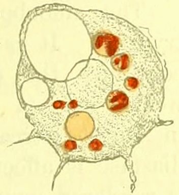
Ehrlich’s neutral red to Amoebae in the act of digesting Bacteria
15at once gives the acid colour reaction (Fig. 1 ). Rhumbler[11] has described
very precisely and with much detail the way in which
the Amoebae behave when they are incorporating
filaments of Oscillaria very much longer than
their own bodies. He has also described the
digestion that these Algae undergo; a process
most characteristic in those cases where a portion
only of the filament has been taken into the
interior of the Amoeba and there subjected to
the digestive action. Whilst the free part of
the Oscillaria retains its normal properties and
appears of a bluish green colour, the ingested portion progressively
changes colour, assuming first a deep green tint, then becoming
light yellow, orange yellow, brown and finally reddish brown.
Simultaneously the cellulose wall of the Alga begins to soften,
and the cells break up into minute fragments which are soon
extruded. The food is seldom completely digested and there is
always an abundant residual material which is thrown out in the
form of solid excreta.
Fig. 1. An Amoeba treated with neutral red, 1%.
[18]
Although it is fully recognised that, in the Rhizopods, digestion
goes on in a medium distinctly but feebly acid, and that the
intervention of some soluble ferment is essential, our ideas on this
subject were very vague until the publication of the researches of
Mouton[12], carried out with great care in the Pasteur Institute. In
order that he might obtain exact results Mouton made use of
cultures of Amoebae grown on agar, in association with the Bacillus
coli which served them as food. The bacilli were ingested in large
numbers, became enclosed in vacuoles and were digested by a
ferment which Mouton was able to obtain in vitro. To that end
he collected large numbers of Amoebae, and, after centrifugalising
them in water, treated the deposit with glycerine. On adding alcohol
he obtained a precipitate readily soluble in water.
The fluid thus obtained exerted an undoubted digestive action
upon albuminoid substances. It readily liquefied gelatine and even
attacked, though feebly, albumen coagulated by heat; flakes of fibrin
heated to 58° C. remained unaltered. There was present then, in
this fluid derived from Amoebae, a proteolytic diastase of feeble
activity. On the other hand, this extract contained neither sucrase,
16capable of inverting cane sugar, nor lipase, capable of digesting fatty
matters.
The amoebo-diastase of Mouton must be classified with the
trypsins. It is very active in a distinctly alkaline medium and
continues the diastatic action even when the medium becomes
weakly acid (a feature that corresponds to the reaction observed
in Amoebae treated with appropriate staining agents). The amoebo-diastase
is affected at as low a temperature as 54° C. and at 60° C.
is rendered completely inactive.
A question of especial importance is that concerning the action
of the amoebo-diastase upon Bacteria. The numerous experiments
of Mouton directed to the solution of this point, and made with
living Bacillus coli communis, gave negative results. If, however,
these bacilli were previously killed by heat or by chloroform,
they were at once attacked by the soluble amoebo-ferment. Opalescent
emulsions of these dead bacilli, incapable of undergoing self-digestion
of any kind, became transparent after remaining for some
time in contact with the extract of Amoebae. The amoebo-diastase,
then, undoubtedly digests dead bacilli in vitro, whereas in the body
of the Amoebae the ingested bacteria are attacked whilst still living.
As a result of these observations it must be concluded that only a
fractional part of the diastase is extracted in the solution prepared
by Mouton.
[19]
This intracellular digestion in the Protozoa serves not merely for
the nutrition of these organisms, but also as a protection against
infective parasites. The protoplasm of the Infusoria, with its vacuolar
secretions, has a general digestive action on everything that comes
within its reach. If the internal structures, such as the nuclei and
the pulsatile vacuoles, resist this process, it is undoubtedly because
they possess a power of defending themselves against the attack of
the digestive secretions. Thus, as brought out in the beautiful
researches of Maupas[13], the macronucleus of the Paramaecia is, at
a certain stage in the life of the Infusorian, completely digested by
the protoplasm just as is any other nutrient substance introduced
from outside. It must be admitted that in this case the nucleus has
ceased to produce the protective substance which, under ordinary
conditions, interferes with its being digested.
A struggle similar to that observed between the nucleus and the
digestive content of the Protozoa goes on between these latter
17organisms and infective microbes. All organisms which, in any way
whatever, penetrate into the body of an Infusorian or Rhizopod, are
brought into contact with the digestive endoplasm of these Protozoa.
If the intruders are killed and partially digested by the digestive
secretions, or are expelled as excrementitious matter, the Protozoon
remains uninjured and continues its normal and routine existence.
Here, then, we have an example of natural immunity, due to intracellular
digestion. On the other hand, when the foreign parasitic
organism resists this digestive action, it instals itself permanently in
the body of the Protozoon, and should it reproduce itself in small
numbers merely, excrete no poison and, in general, exercise no
injurious influence upon its host, the parasite may readily become
a commensal. Thus, it is not rare to find in the contents of Infusoria
and Radiolaria small vegetable organisms of the genera Zoochlorella
or Zooxanthella which not only set up no disease but, owing to their
assimilation of carbonic acid, may even be useful to their hosts.
There are cases, however, where the parasites act in a manner more
or less injurious to the Protozoa containing them; in such cases
a true and sometimes fatal infection results.
[20]
Among the infective diseases of the Protozoa the one that has been
most thoroughly studied is that set up by several representatives of
a particular genus of micro-organisms discovered by Johannes Müller
in 1856 and made the subject of an investigation carried out in my
laboratory by Hafkine[14]. I have already discussed these researches
in my work on the comparative pathology of inflammation[15] and need
here recapitulate only very briefly. Paramaecia are sometimes affected
by needle-shaped or spirillar parasites which penetrate, sometimes
into the macronucleus, sometimes into the micronucleus, reproducing
prolifically, giving rise to a marked hypertrophy of the affected organs.
The Infusorian, in spite of this invasion, may continue to exist and
carry on its reproductive processes; it is, thus, enabled in many cases
to recover from the disease. On the other hand the Paramaecium
into whose body the spores of the parasite are introduced treats
them as it would any other ingested foreign body. Not being able to
digest them, owing to the resistance offered by the membrane of the
spore, the Paramaecium expels them just as it would any other
18excrementitious matter. The Infusorian behaves in the same way in
regard to bacterial endospores.
Hay bacilli, which occur so commonly in the infusions in which
the Paramaecia live, are digested in the endoplasmic vacuoles of the
latter, but the spores of these bacilli, after a more or less prolonged
sojourn in the vacuoles, are expelled with the excrement.
As by far the greater part of the body of a Protozoon is made up
of digestive protoplasm, it is natural that infective epidemics should
be very rare among these animalcules. The Infusoria and Rhizopods,
organisms specially well adapted to live upon the lower Algae and
Bacteria, are, practically, never subject to bacterial diseases. The
infections observed in the Protozoa are due in most cases to the
invasion of the lower Fungi, such as the Chytridia, the Microspheres,
the Saprolegniae or the special organisms mentioned as occurring in
the nuclei of Paramaecia. Further, these infections are met with
most frequently in Protozoa which are incapable of carrying on true
intracellular digestion or which are in the encysted stage, at which
period the Infusoria, leading a passive existence, neither absorb nor
digest nutriment. As an exception to the above general statement
I ought to mention the epidemic in Amoebae caused by the Microsphaera[16]
and the disease in Actinophrys observed by K. Brandt[17] and
attributed to Fungi allied to the genus Pythium. In these two
instances we have to do with parasites which live and develop in the
interior of the active protoplasm of these Protozoa. Certainly a
proportion of the parasites are expelled with the excrementa; but
there remain others which instal themselves in the protoplasm,
multiply there and cause the death of their hosts. In these cases the
digestive action of the protoplasm must be neutralised or paralysed
by the secretions of the parasite. This aspect of the question,
however, has so far not been considered.
[21]
In addition to intracellular digestion and the expulsion of parasites
by the excretory function, the resistance offered by Protozoa to
infective diseases should, in part, be ascribed to their great irritability.
Anyone who will watch the manœuvres of Amoebae or of
certain Infusoria in the midst of a rich microscopic flora and fauna,
will at once be struck by the preferences which these Protozoa
exhibit in the choice of their food. Amoebae are often seen
making search for Diatoms only, disdaining all other Algae, or again
19they may single out one species of Palmellaceae from a very varied
flora. The Infusoria also have their likes and dislikes in the matter
of food. Many of the ciliated Infusoria choose Bacteria to the exclusion
of almost everything else; others, as Nassula, have a special
partiality for the Oscillariae. A most striking example of this
is afforded in Amphileptus claparedei, a voracious Ciliate, which
chooses Vorticellae to the exclusion of all other animalcules; these
it devours, and then becomes transformed into a cyst upon the
peduncle of the Vorticellae it has devoured. This irritability clearly
must control and guide the Protozoa in their relations with other
organisms and enable them to escape the invasion of parasites.
In this connection I must mention a very interesting observation
made by Salomonsen[18] and communicated to the Paris International
Medical Congress in 1900. He was able to demonstrate the fact that
almost all the ciliated Infusoria, on becoming aware of the proximity
of dead bodies of kindred organisms, rapidly draw away, thus
manifesting a very marked negative chemiotaxis. This property
must, it is evident, protect them from any contamination by the
parasites contained in the bodies of Infusoria that have succumbed
to infective diseases.
We have, then, quite a number of facts which throw light on the
natural immunity of the Protozoa against the action of pathogenic
micro-organisms. Up to the present, however, we know nothing
concerning the existence or the possibility of an acquired immunity
among the lower animalculae against infective diseases. We are
better informed as to the resistance of unicellular organisms to the
action of soluble poisons, which is, in general, much more easily
studied than is immunity against the micro-organisms themselves.
[22]
As a very large number of the higher animals are sensitive to the
toxic action of poisons of bacterial origin, the question has been put,
“May not the Infusoria also be poisoned by these micro-organismal
products?” With the object of answering this question Gengou[19]
has studied the influence of the toxins of tetanus and diphtheria on
the ciliated Infusoria. He was unable, however, to bring forward
proof that these substances exert any special toxic action on the
Paramaecia. These Infusoria withstand, perfectly well, doses of
20cultures of the diphtheria and the tetanus bacillus grown in broth
and deprived of the bacilli by filtration as large as those of ordinary
broth alone in which no bacilli have been cultivated. Gengou
argues from this that the Paramaecia possess a natural and absolute
immunity against these two toxins. When we take into consideration
the fact that these poisons act but feebly at ordinary temperatures
and are often innocuous to “cold-blooded” animals we may perhaps
be tempted to attribute the immunity of the Infusoria to the temperature
that was maintained in the incubator whilst Gengou’s
experiments were being carried on. Led by this train of thought
Mme Metchnikoff tried the action of the blood-serum of eels, which
is very toxic, not only for warm-blooded Vertebrates but also for cold-blooded
Vertebrates and the Invertebrates, on the Paramaecia, and this
at a low or medium temperature. This eel’s serum, however, exerted
no greater toxic action than did the blood-serum of other animals.
The microbial toxins are innocuous not only to the ciliated
Infusoria but also to many other unicellular organisms. It is now
well recognised that these toxins, exposed to the air, are soon
inhabited by quite a rich flora of micro-organisms, amongst which
Bacteria and Yeasts predominate. I have been able to prove[20] that
these organisms are not only unaffected in their normal life by the
presence of the toxins of diphtheria or tetanus but that they rapidly
bring about the more or less complete destruction of these poisons.
Gengou, also, observed that yeasts thrive luxuriantly in these bacterial
toxins. The rapid increase of micro-organisms and the destruction
of these poisons take place at temperatures varying from 15° to 37° C.
[23]
Whilst the lower organisms are refractory to bacterial toxins
which in quite small doses are capable of killing man and the
higher animals, many micro-organisms manifest a special sensitiveness
to certain fluids of animal origin. In a succeeding chapter
we shall treat at greater length of this microbicidal property of
the humours. Here it is merely necessary to indicate certain facts
concerning this property, regarding them solely from the point of
view of the immunity of the lower organisms. The most striking
example of the bactericidal power of an animal fluid is certainly
that afforded in the action of the blood-serum of the rat on the
anthrax bacillus. This fact, discovered in 1888 by von Behring[21], led
21to the conclusion that the blood of the rat contains an organic base
capable of killing and dissolving a considerable number of anthrax
bacilli. Several observers have confirmed von Behring’s observation
and have supplemented it by the fact that the bacillus can be readily
accustomed to the toxic action of this serum. Thus Sawtchenko[22], in
an investigation carried out in my laboratory, was able, by successive
cultures, to accustom the anthrax bacillus to an existence in the pure
serum of the rat. In this case, therefore, there has been produced a
real acquired immunity of a lower plant against a toxic substance of
animal origin. More recently Danysz has demonstrated the same thing
and has added several other facts which seem to throw light upon the
means by which the bacterium becomes adapted to the poison. He
has shown, in a work carried out in the Pasteur Institute[23], that the
anthrax bacillus protects itself against the toxic action of the serum
by surrounding itself with a thick sheath composed of a kind of
mucus which fixes the toxin of the rat’s blood and renders it
harmless. This same mucus, but in smaller quantity, is likewise
produced in a culture of the bacillus grown in ordinary broth. When
such a culture is freed from the contained bacilli by filtration through
porcelain and a little of this fluid is added to the rat’s serum, this
latter becomes less bactericidal than is a mixture of the same serum
with ordinary broth. Danysz suggests that this is to be explained
by the presence in the filtrate of a certain quantity of the mucous
substance produced by the bacillus, which fixes and neutralises a
portion of the “rat toxin.” If, in place of sowing the ordinary
bacillus, sensitive to this toxin, we inoculate the broth with an
anthrax bacillus which has previously been accustomed to the rat’s
serum, we find that the liquid of this culture when filtered neutralises
a larger proportion of the toxin. Danysz concludes from this that the
acclimatised bacillus has acquired the property of producing more
mucus than does the ordinary bacillus and that, for this reason, a
greater quantity of this protective substance passes into the fluid of
the culture.
[24]
The formation of a transparent sheath has several times been
observed in the anthrax bacillus, notably in cases where this organism
happens to be in “a state of defence” against various noxious
influences. For example, this sheath is well developed in the
anthrax bacillus which invades the blood of lizards, animals which
22are in general very resistant to anthrax[24]. Under analogous conditions
the streptococci which, as a rule, do not produce a mucous sheath,
will develop one of exceptional size. The guinea-pig is in general
very resistant to the streptococcus against which it exhibits a very
effective reaction. Sometimes, however, this immunity gives way;
in such instances, as demonstrated by J. Bordet[25], the streptococcus, in
order to overcome the natural resistance of the guinea-pig, is found
to have surrounded itself with a sheath of a thickness such as is
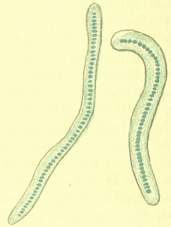
seldom to be met with in the world of bacteria (Fig. 2).
Fig. 2. Streptococcus surrounded by a protective envelope.
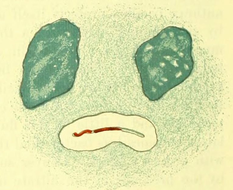
Fig. 3. Tubercle bacillus surrounded by a transparent envelope and enclosed in the giant cell of a gerbil.
[25]
Analogous facts are also observed in cases where the micro-organism
is defending itself against the action of substances enclosed
in animal cells. I may cite as an example the tubercle bacillus in
the interior of the giant cells of a gerbil (Meriones shawii), where,
under the influence of noxious substances contained in these cells, the
tubercle bacillus (Fig. 3) envelops itself in a transparent sheath
similar to that of the bacillus or of the streptococcus. As the action
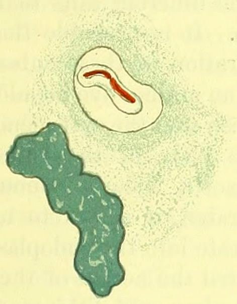
of the giant cell still does not cease, the tubercle bacillus secretes
a second sheath (Fig. 4) and continues to surround itself with
23quite a series of such envelopes (Fig. 5), thus coming to resemble
a palmellaceous Alga surrounded by successive layers of membranes
or certain other vegetable cells whose principal means of defence
against all kinds of injurious influences consists in the production
of these protective membranes.
Fig. 4. Another tubercle bacillus surrounded by two membranes.
Fig. 5. Tubercle bacillus surrounded by a series of concentric layers.
[26]
Quite recently Trommsdorf[26], in Buchner’s laboratory in Munich,
has carried out a series of experiments on the adaptation of
the cholera vibrio and of the typhoid bacillus to the bactericidal
substance found in the blood of the rabbit. He has been able to
confirm the results of his predecessors and by various experiments
has convinced himself that these two micro-organisms are capable
of adapting themselves to existence in the defibrinated blood and in
the blood-serum of the rabbit.
The immunity, or acclimatisation of injurious organisms to different
toxins, presents an undoubted analogy to the phenomena of adaptation
shown by these organisms to mineral or organic poisons. It has long
been known that the same species of Protozoa are met with in both
fresh and salt water and that it is possible to gradually accustom
Infusoria and Amoebae to tolerate an amount of sea salt which at first
is absolutely fatal to them. This toleration is not acquired unless care
be taken to increase the amount of salt very gradually: too abrupt a
rise inevitably causing death. By this means Cohn[27] accustomed the
24fresh-water Euplotes to a life in artificial sea water containing 4% of
sodium chloride. In Balbiani’s experiments[28] the fresh-water Monads
(Menoidium incurvum and Chilomonas paramaecium) died very
quickly on the addition of ½% of this salt; but when it was added in
small successive doses (0·05 per day), they readily became accustomed
to a concentration of 1%. In the encysted state the Protozoa are even
more resistant than in the active state to the different salts that may
be added to their normal culture medium. It is probable that the
wall of the cyst interferes with the penetration of these substances
into the endoplasm. If a small quantity of an aniline dye be added to
a fluid containing encysted Infusoria, it is seen that the cyst-membrane
becomes very intensely coloured but the body of the Infusorian
remains unstained. The membrane absorbs a large amount of
colouring matter, after which, being saturated, it ceases to take it
up; but it does not allow the dye to penetrate into the endoplasm.
[27]
Balbiani (loc. cit. p. 580), having compared the action of the salts
of sodium with that of the salts of potassium and lithium on Infusoria,
comes to the conclusion that the injurious influence of these
substances can only be partially explained by osmotic phenomena.
In addition to these a purely chemical action must be invoked. He
bases his opinion on the fact that the isotonic solutions of the three
salts acting on Infusoria of the same species and same origin exert
a different influence. The salts of potassium and of lithium act in a
much more energetic fashion than do the sodium salts. Consequently,
the Protozoa are able to adapt themselves progressively not only to
noxious influences of a physiological character but also to those of a
chemical nature. Thus Infusoria and Rhizopods can be accustomed to
the action of high temperatures, to an intense light, etc. On the other
hand they can also be habituated to the toxic actions of true poisons.
Davenport and Neal[29] have established the fact that Stentors kept for
two days in a weak solution of corrosive sublimate (0·00005%) acquire
a tolerance to a dose of this poison four times as great as the lethal
dose for individuals previously kept in pure water. The same thing
has been observed in connection with the toxic action of quinine.
This immunity cannot be attributed to the selection and persistence
of those Infusoria which possess a natural resistance to the sublimate.
25It is really acquired as the result of a direct and gradual chemical
influence on the protoplasm of the Stentors which, once adapted,
all survive doses which are lethal for the unacclimatised control
organisms.
[28]
The vegetable micro-organisms, which are much more easily
cultivated than are the Protozoa, frequently manifest most characteristic
phenomena of acclimatisation. The first systematic researches
in this direction were carried out by Kossiakoff[30] in the laboratory of
Duclaux. He studied the antiseptic action of borax, of boracic acid,
and of corrosive sublimate on the anthrax microbe and several other
bacilli (Bacillus subtilis, Thyrotrix scaber and T. tenuis). He found
that all these micro-organisms can be gradually accustomed to doses
which are absolutely bactericidal to the same species when not so
acclimatised. The acclimatised Thyrotrix tenuis withstands almost
double the amount of bichloride of mercury that the non-acclimatised
bacillus will resist. The ordinary anthrax bacillus will not develop at all
if the culture medium contains more than 0·005 of boracic acid whilst
the same organism, when accustomed by passage through successive
cultures in which this substance is present in gradually increasing
proportions, grows well in spite of the presence of 0·007 of the same
antiseptic. Since these observations were made similar facts have
been demonstrated by several other observers, and the ready acclimatisation
of Bacteria to poisons is now generally admitted. Danysz
(loc. cit.), with the object of elucidating the mechanism of this adaptation,
has studied the action of arsenic acid on the Bacillus anthracis.
He has demonstrated that this bacillus will gradually accustom
itself to grow in broth containing a quantity of arsenic acid which at
first inhibited all development. During this phenomenon of adaptation,
which is acquired after a series of passages through media more
and more highly arsenicated, the bacillus secretes a coating of mucous
substance which protects the sensitive parts of the microbial cell.
Here, therefore, is formed something exactly corresponding to what
the same observer has demonstrated in anthrax bacilli that have
acquired a tolerance for rat’s serum. This analogy extends even
to the throwing out of the protective substance into the culture
fluid. When one sows an ordinary unadapted bacillus in arsenicated
broth to which has been added some of the fluid from a culture of
the adapted bacillus, development takes place in a marked fashion.
On the contrary when the same material is “seeded” into arsenicated
26broth of the same composition but to which has been added the
filtrate from an unadapted culture, the bacillus does not develop
nearly so well. The difference is explained by the presence, in the
fluid in which the adapted bacillus had grown, of a certain quantity
of the mucous substance which fixes the arsenic and prevents it from
acting on the protoplasm of the micro-organisms.
[29]
The Yeasts, also, adapt themselves very readily to antiseptics.
This property has even had a practical application. We know that
small doses of hydrofluoric acid are capable of preventing the
proliferation of the yeast of beer, and Effront[31] has accustomed this
plant to live in media containing an amount of hydrofluoric acid
which is absolutely inhibitory to the unadapted yeast. Under these
conditions the adapted cells undergo a stimulation which causes the
production of a greater quantity of alcohol. The yeast, in adapting
itself to antiseptic doses (300 mm. of hydrofluoric acid per 100 c.c. of
beer wort), acquires a kind of immunity which it did not possess in
the first instance. Moreover this acquired property can be hereditarily
transmitted to new generations developed in ordinary beer wort to
which hydrofluoric acid has not been added. The stimulating action
of this substance on the fermentative property does not depend upon
the acid reaction of the hydrofluoric acid, for other acids which are
non-antiseptic, such as tartaric acid, are incapable of inducing it.
The acquired immunity against hydrofluoric acid is strictly specific,
the yeasts that have been adapted to this substance becoming even
more susceptible to the action of other poisons.
Duclaux[32] has already insisted on the relations which exist between
antiseptics and foods. Formic aldehyde which has a very powerful
coagulative and therefore strongly antiseptic action on protoplasm may
actually serve as a food for micro-organisms. The Thyrotrix tenuis,
studied in this connection by Péré[33], adapts itself to the presence of this
aldehyde and utilises it for its nutrition. Here is produced something
that recalls the case of the Protozoa that digest parasitic organisms.
It is now a current idea in microbiology that Bacteria and
Yeasts which primarily do not make use of certain substances, adapt
themselves to use them as nutrient substances. Dienert[34] has published
a detailed work on the adaptation of the yeasts to milk-sugar. This
27sugar is usually disdained by the yeasts that set up the fermentation
of glucose; it is not difficult, however, to adapt them to galactose
which they then attack and transform into alcohol and carbonic acid.
The Protozoa can be progressively accustomed not only to poisons
but also to altered physical conditions. Thus, Dallinger[35] succeeded
in raising the temperature of the water in which flagellated Infusoria
were growing from 15°·5 to 23° C. without causing their death. By
prolonging the experiment over several months, he was even able to
habituate them to an existence at a temperature of 70° C. In the
opinion of Davenport[36], a view which is shared by many other
observers, this resistance to high temperatures was dependent on
the abstraction of water from the protoplasm. Dallinger has also
observed that in Infusoria that are accustomed to life in hot water,
the vacuoles become smaller and smaller and may even actually
disappear.
[30]
This adaptation, then, is a property that is very general and widespread
in the microcosm of the unicellular organisms. It is connected
with the intracellular digestion of solid food and with the absorption
and transformation of soluble substances. These phenomena, chemical
in character, are intimately linked with the irritability of microscopic
organisms, which represents one of the fundamental properties of
living organisms.
A Protozoon, which is refractory to a parasite, may protect itself
by flight or it may devour and digest the parasite; another, which
acquires a tolerance in regard to a toxin or to a mineral poison,
absorbs, fixes and transforms this substance. Consequently, in all
these instances of immunity there is a reaction of the living elements
of the organism, this being a direct consequence of the irritability of
the protoplasm.
Before an Infusorian retreats from the dead body of an allied
organism, before a Protozoon secretes a digestive fluid around the
prey it has ingested, before a Bacterium secretes a glairy layer for
its defence, etc., these unicellular organisms must receive sensations
which provoke the above-mentioned reactions. It is to a celebrated
botanist, Pfeffer, that we owe the most important researches on this
irritability of unicellular organisms, researches which may be summed
up in the general statement that this property is subject to the psycho-physical
law of Weber-Fechner. Pfeffer, by the observation of the
28movements of Bacteria under the influence of increasing stimulations,
has established the fact that, conformably to this law, when the
stimulus increases in geometrical ratio, the irritability increases in
arithmetical ratio, that is to say, the reaction is proportional to
the logarithm of the stimulation. In order that a motile bacterium
(Bacterium termo), grown in a peptonised solution, may perceive a
difference of medium, it is necessary to place it in a peptone solution
of five times the original concentration; weaker solutions, in which the
concentration is but three or four times greater than the original
fluid, do not attract the bacteria at all; consequently these differences
are below their chemiotactic sensibility.
The different reactions that are exhibited in the immunity of
unicellular organisms, reactions which are dependent on the irritability
of their protoplasm, therefore, come undeniably under the
category of purely cellular phenomena.
CHAPTER II IMMUNITY IN MULTICELLULAR PLANTS
29
Infective diseases of plants.—Plasmodia of the Myxomycetes and their chemiotaxis.—Adaptation
of the plasmodia to poisons.—Pathogenic action of Sclerotinia upon
Phanerogams.—The cicatrisation of plants.—Defence in plants against Bacteria.—Sensitiveness
of vegetable cells to osmotic pressure.—Adaptation of plants to
modifications of osmotic pressure.—Dependence of the chemical phenomena
upon the irritability of the vegetable cells.—The law of Weber-Fechner.
[31]
For several reasons this immunity in the vegetable kingdom cannot
be treated in a satisfactory fashion. Much attention has been devoted
to the pathology of plants and the etiology of a number of vegetable
diseases was well established at a period when we were still groping
in the dark for the causes of infective diseases in man and the higher
animals. In spite of this, the botanist has relegated the study of the
phenomena of immunity to a secondary position, and up to the present
no work specially devoted to this subject has appeared. It is only
incidentally that the question of the resistance of certain plants to
morbific factors capable of infecting or intoxicating them has been
touched upon. We should require, therefore, to carry out special
researches in this direction and to make a very complete study of
botanical literature, before we should be able to present to our
readers a résumé of the question of immunity in the vegetable
kingdom. Such a programme being impossible we must content
ourselves with borrowing from the botanists certain facts which
throw light on some aspects of the general problem in which we
are interested.
[32]
Many of the higher plants are subject to infective diseases set
up by the lower plants, of which the most important are the Fungi.
Whereas in the animal kingdom the majority of the infections
are due to Bacteria, these micro-organisms rarely occur in plants;
moreover when they are present the part they play is nearly always
a secondary one. This difference is due mainly to the chemical
30composition of the “humours” in the two kingdoms, the cell-juice of
plants being generally acid; under this condition the Fungi develop
much better than do the Bacteria.
The various modes of defence against infective diseases that have
been met with in unicellular organisms are also found in the multicellular
plants. Whereas in almost all plants the cells are rigid,
owing to the presence of a well-developed membrane, some of the
lower plants have preserved a condition in which the protoplasm is
completely naked and capable of movement. Myxomycetes are
specially distinguished by an amoeboid stage of existence and by the
formation of large plasmodia which protrude protoplasmic processes
and exhibit a kind of locomotion similar to that met with in the
Rhizopods and the Sporozoa.
[33]
Infective diseases among the Myxomycetes must be very rare
since, up to the present, they have not been noted by a single observer.
It is very probable that the plasmodia get rid of the infective germs,
as do the Protozoa, both by expulsion of the parasites and by
means of intracellular digestion. This latter takes place in a medium
which is distinctly acid and by means of a soluble ferment described
by Krukenberg[37] as a kind of pepsin. I need not here enter into
further detail as I have already treated this subject in my Lectures
on the comparative pathology of inflammation. The fact that the
Myxomycetes can ingest living organisms has been demonstrated by
Celakovsky, jun.[38], who has observed that the spores of the various
Fungi can germinate in the interior of the plasmodium. Whilst our
conceptions concerning the resistance of the plasmodia in regard to
micro-organisms are merely based upon analogies and hypotheses, our
ideas as to their immunity against soluble substances rest on well-established
experimental facts. We owe to Stahl[39] our first information
as to the mode by which the plasmodia resist poisons. When
they are placed in contact with solutions of salts, of acids or of sugar
in a sufficiently concentrated form to bring about an injurious action,
the plasmodia make use of their amoeboid power of motion to escape
from these fluids. Hence they exhibit a negative chemiotaxis, exactly
parallel to that so often observed in the case of the unicellular
organisms. Consequently there is in the Myxomycetes a natural
immunity due to the activity of their movements. Further, a kind
31of acquired immunity in these plants has also been demonstrated
by Stahl. The following is the passage in his paper referring to
this subject, a passage very important from a general point of view[40]:
“If we replace the water in a vessel by a 1 or 2% solution of
glucose, we observe either the death of the plasmodia, if the action
is too rapid, or merely their retreat from the glucose solution.
Even solutions of ½ or ¼% are at first avoided by the plasmodia
and, should the action be too rapid, may cause their death. Usually
the plasmodia emigrate into those portions of the substratum remote
from the solution, to return after some time, often only after several
days, and immerse themselves in the solution of glucose as they do
in an infusion of tan, although with more hesitancy. Consequently
the Myxomycetes accommodate themselves slowly[41] to a more concentrated
solution, probably by giving up a certain proportion of their
water. I was able to observe the same phenomena with even much
more concentrated solutions (2%). A plasmodium which at the end
of several days had adapted itself to a 2% solution of glucose and had
sent out numerous processes into it, found itself injuriously affected
when the sugar solution was suddenly replaced by pure water. Those
that remained alive had retired to a great distance from the upper
layer of the fluid and did not descend again until the end of the second
day. After a fresh change of fluid we were able to observe first the
repulsion and later the attraction of the plasmodia, but a certain time
always elapses before the plasmodia become accustomed to the change
in concentration. We obtain the same result when we replace a 2%
solution, not by pure water, but by a ½ or a 1% solution” (p. 166).
[34]
De Bary[42] had already interpreted these facts as being due to an
immunity acquired by the plasmodia, the result of an adaptation of
these organisms to solutions which they had, at first, carefully avoided.
He threw out the suggestion that a similar adaptation might take
place in regard to solid substances ingested by the Myxomycetes.
As these phenomena of acquired immunity, in organisms so
primitive and of so simple a structure, are of immense importance
from the point of view of Immunity in general I felt bound to submit
them to a personal investigation. I found it an easy matter to
32accustom the plasmodia of Physarum to solutions of arsenious acid
which at first repelled them in a very striking manner. This adaptation
manifests itself by movements of the plasmodia and by the
change from negative chemiotaxis (repulsion) to positive chemiotaxis
(attraction).
It is impossible in the present state of our knowledge to state
precisely the modifications that the plasmodia undergo during this
process of adaptation. Stahl supposes that they depend “on some
special properties of the plasmodia (probably in a greater or less
richness in water)”; and that it is a case “not of simple phenomena,
easy of explanation, but of extremely complicated phenomena of
irritability.”
It is evident that, in this case of acquired immunity, we have
not to do with a question of physical or chemical modification of the
solutions employed but solely with reactive phenomena on the part
of the living plasmodia.
After a phase of active life, during which the Myxomycetes move,
feed, digest and expel waste products as do the lower animals,
there comes a stage when they become immobile and transform themselves
into a number of sporangia filled with rounded spores. Before
leaving their animal aspect for that of true plants, the plasmodia
exhibit entirely new attributes. They reject all nourishment and
no longer ingest foreign bodies; they avoid the moisture which
previously attracted them and cease to shrink from the light.
[35]
Having come to maturity, the Myxomycetes declare themselves
true plants and lead a passive life until the new generation comes
forth. Most plants are restricted to this passive phase of the
Myxomycetes. In these latter it persists only for a short period,
whereas in almost all plants it is the permanent condition. It
is at this stage that these organisms are liable to the attack of
parasites against which it is necessary for them to oppose all their
means of defence. Our knowledge of these means of defence is as
yet, as I have already stated, very imperfect, and the example of
Sclerotinia libertiana (or Peziza sclerotiorum) which has been the
subject of the researches of de Bary[43] remains up to the present the
one that has been most thoroughly studied.
This Fungus, belonging to the group of the Discomycetes, invades
many species of plants and often produces great ravages amongst
the cultivated plants of our fields and gardens, such as colza, hemp
33petunias, dahlias, etc. The mycelium of this Sclerotinia develops in
the stems of herbaceous plants and produces sclerotia inside them,
forms of resistance, which in this instance are black and resemble
small particles of mouse excrement.
The spores of the Sclerotinia germinate and form mycelial threads
on the surface of the plants. In order that they may penetrate into
the tissues these threads must attack the cell-membrane and for this
purpose they secrete a fluid, which contains both a digestive ferment
and oxalic acid, the latter being essential for the action of the ferment.
The presence of this “toxin” has been demonstrated by de Bary
by macerating the mycelium of the Sclerotinia. The resultant extract
has a well-marked action on the tissues of many plants (carrot,
Jerusalem artichoke, chicory, etc.). Under its influence the protoplasm
of the cells contracts, a genuine plasmolysis is set up, the cell-membrane
swells and its layers between the cells are dissolved. As
the result of this digestive action, the cells become separated and
the tissue softens. This extract, when heated to 52° C., loses its
digestive action on the cellulose membrane, but still retains its power
of setting up plasmolysis. This reaction to temperature confirms the
view that the juice of the Fungus contains a soluble ferment. The
results of de Bary’s researches have been confirmed and in part
supplemented by the experiments of Laurent[44].
[36]
It is a fact of common observation that the Sclerotinia libertiana
invades for the most part young plants. It may therefore be asserted
that the disease produced by this Fungus is, like scarlatina or measles
in the human subject, an “infantile” disease. De Bary suggested
that the immunity of adult plants must depend on the greater
resistance which their cell-membranes offer to the fluid secreted by
the mycelial filaments. Direct experiments have shown the accuracy
of his suggestion. Whilst the fluid extracted from the Sclerotinia
readily digests the tissue of young plants it leaves intact that of adult
plants of the same species.
In the course of this disease we have a struggle going on between
two plants. The parasite brings into play toxic and digestive secretions
with which it seeks to impregnate its host. The attacked plant
defends itself by the secretion of membranes capable of resisting the
action of the secretions of the Fungus. This struggle by means of
chemical substances is, however, directed by the activity of the living
34cells of the two belligerent plants, an activity dependent upon the
irritability of their protoplasm.
The example we have just studied may serve as a type for our
examination of the phenomena of immunity in the vegetable kingdom.
The crux is above all to prevent the access of the parasites to the
vital parts of the plant by opposing to them membranes as resistant
as possible. Consequently the majority of plants, directly the smallest
lesion is produced, react by an abundant cell-proliferation and by the
suberisation of the outer layers. The cell-membranes of the latter
thicken, the cellulose is transformed into suberin; a layer of cork
not very permeable to fluids and gases being thus formed. By
suberisation the plant reacts against grosser lesions, incisions or
burns, as well as against the decay set up by micro-organisms.
Massart[45], in an extremely interesting memoir, has brought
together the known data concerning cicatrisation in plants and has
demonstrated the fact that it is a very variable process. In many
leaves after being damaged there is no attempt to react by forming
cicatricial tissue. Many aquatic and marsh plants react but feebly.
Their tissues die and turn brown, the plants failing to defend themselves
by cicatrices, probably owing to the ease with which the lost
parts can be replaced. When, however, in the same plants, there is
produced a lesion of parts which are of great importance for the
preservation of the integrity of the individual or a lesion of the organs
which enable the plant to continue its existence through the winter,
cicatrisation of the wounds takes place rapidly.
[37]
The old or adult parts in most cases react differently from the
young parts. Thus, the young leaves of Clisia (the example selected
by Massart) react to traumatism very promptly and form a genuine
callus which makes good the injury, but the adult leaves merely produce
a layer of cork in the immediate neighbourhood of the lesion.
The essential mechanism of cicatrisation has not yet been satisfactorily
analysed, but it is evident, when all is said and done, that it is
directed by the irritability of the living protoplasm of the vegetable
cells.
Many plants protect their wounds with a kind of dressing, using
for that purpose juices which harden on exposure to the air. Sometimes
these juices, e.g. latex, are preformed in the plant and are as it
were always ready for use; at other times they may be formed only
35as the result of an injury. In this latter case the resins and gums
which serve to close the wound and to protect the living parts receive
the name of “cicatricial secretions” (Wundsecrete). According to the
view first formulated by de Vries, those juices which harden under
the action of air prove of great service both as natural “dressings”
and as safeguards against the attacks of plants and animals. Indeed
many of these secretions contain essences whose antiseptic and toxic
action is now generally recognised[46].
[38]
The suberisation, the formation of a callus, and the secretion of
juices which close the wounds, are all means readily utilised and very
potent in ensuring the resistance of plants against all sorts of injurious
influences which may be set up by a morbid condition. But these
processes are not the only means which plants have at their disposal.
The living elements of plants usually secrete a cell-juice of acid reaction
which plays a very important part in the defence of plants
against pathogenic agents. Laurent[47] has studied this phase of the
immunity of plants against bacterial decay. A variety of the Bacillus
coli communis, according to this observer, attacks the potato by
means of its secretions in a fashion analogous to that already described
when discussing Sclerotinia. This bacillus produces a soluble
ferment which has the power of digesting the cellulose membrane in
the tuber of the potato, and at the same time secretes an alkaline
juice without which this digestion cannot go on. Heating to 62° C.
destroys the soluble ferment and the fluid thus heated is no longer
able to digest the layers of the cell-membrane between the cells. In
spite of exposure to this temperature, however, it still retains intact
one or even several substances which may continue to cause contraction
of the protoplasm and ultimately kill it.
When Laurent placed cut halves of tubers coming from races of
potato which were most resistant to bacterial decay in the fluid produced
by the Bacillus coli and afterwards inoculated them with the
bacillus itself, he invariably found that the vegetable cells were profoundly
affected.
The alkaline secretions of the bacillus studied by Laurent may be
neutralised by the acid juice of the potato, and when certain races of
tubers prove immune from decay, it is, according to this observer,
because of the production of sufficiently acid cell-juices. Moreover he
36actually succeeded in communicating an artificial immunity to varieties
of the potato which were most susceptible to decay by immersing
them for several hours in solutions of certain organic acids. On the
other hand, when he treated varieties endowed with a well-marked
natural immunity with alkaline solutions, the tubers became very
susceptible to the decay set up by the bacillus.
The struggle between the potato and the Bacillus coli reduces
itself, then, to the chemical reaction between the alkaline cell-secretions
of the micro-organism and the acid secretions of the potato.
This general fact, according to Laurent, explains the part played by
certain manures in determining the susceptibility or the resistance
manifested by the potato and many other plants against infective
diseases.
We know that the addition of phosphates to the soil increases the
immunity of certain cultivated plants. These substances are greedily
absorbed by the roots and produce acid salts which are dissolved in
the cell-juice. The nitrogenous manures, on the other hand, both
potassic and lime, diminish the resistance of the same plants,
probably from the fact that they bring about a diminution of the
acidity of the cell-juice.
But these manures can act differently on different plants. Thus
the same phosphates which confer immunity on the potato against
bacterial decay render the Jerusalem artichoke more susceptible to
the attacks of the Sclerotinia.
[39]
Laurent explains this fact as due to the difference in the reaction
of the medium, which favours the action of one or the other of the
soluble ferments of the two parasites. The ferment of the bacillus
digests the cell-membrane in an alkaline or feebly acid medium,
whereas the hyperacidity which results from the absorption of the
phosphates prevents this digestion and consequently aids the plant in
its struggle. On the other hand, the ferment of Sclerotinia, as is seen
from the researches of de Bary, will digest cellulose even in a distinctly
acid medium. The hyperacidity, induced by the phosphated
manure, in this case favours the parasite and enables it to gain the
upper hand in the struggle with the tissues of the artichoke.
In addition to neutralising the microbial products the acids of
the cell-juice also act injuriously on most bacteria, which will only
develop in neutral or alkaline media; it is for this reason that
bacterial diseases are so much rarer in plants than in animals.
The secretion of cell-juices is consequently a very important
37element in the defence of plants; it will be useful, therefore, to
ascertain as definitely as possible the essential mode of its action.
Vegetable cells are as a rule very sensitive to the influences to which
they are exposed; they distinguish with great precision the changes
which take place in their surroundings. They are, indeed, capable of
discerning not only the physical properties but also the chemical composition
of the medium in which they live.
Vegetable cells estimate very accurately the osmotic pressure of the
fluid which bathes them, and they react towards this solution by increasing
or diminishing their own internal pressure. Van Rysselberghe[48],
in an investigation very carefully carried out, demonstrated that
when vegetable cells (especially the epidermic cells of certain species
of Tradescantia) are placed in a solution of greater density than that
to which the cells are accustomed, the intracellular pressure increases;
in a solution of less density the pressure diminishes. These changes
in osmotic pressure are due to variations in density of the cell-juice,
whilst these variations are in turn determined by chemical transformations.
Thus, if the cell be treated with a too concentrated solution it
produces oxalic acid, which dissolving in the cell-juice, is, owing to the
smallness of its molecule, very osmotic.
[40]
With the purpose of confirming this by exact facts van Rysselberghe
has studied the acids of the cell-juice of Tradescantia. In the normal
juice he found that malic acid was constantly present and, in rare cases
only, traces of oxalic acid. He then determined the acids present in
the leaves of the same plant after they had been several days in contact
with fairly concentrated solutions of cane sugar. In each analysis
he found oxalic acid in quite appreciable quantity. There is then, in
the plant which adapts itself to more concentrated solutions of the
medium, a production of oxalic acid which serves the purpose of
increasing the pressure of the cell-juice.
The origin of this oxalic acid could not be accurately demonstrated,
but van Rysselberghe considers that it is probably formed at the
expense of the glucose.
According to the researches of Giessler oxalic acid is localised
specially in the epidermis and generally in the peripheral tissues of
plants; it is very probable, therefore, that it fulfils a protective rôle
against all kinds of injurious influences. Botanists hold indeed that
oxalic acid keeps herbivorous animals, especially slugs and plant lice,
38from attacking plants that are rich in this substance. It is of use,
also, in retaining the moisture in the superficial cells. It is very
probable that it also plays an important part as a factor in the maintenance
in plants of immunity against bacterial diseases.
The vegetable protoplasm, which is capable of increasing the
production of acids in order to raise the osmotic pressure, can also, in
case of need, cause a diminution.
When the cells of Tradescantia are transferred from a concentrated
solution into one much more dilute there may often be
observed a precipitation, in the cell-juice, of crystals of oxalate of
lime; this brings about a diminution in the osmotic pressure. When
the density of the medium is altered, and the vegetable tissue is again
transferred to a stronger solution, the oxalate crystals are seen to
dissolve, as a result of a new production of acid.
These chemical processes, so important to the life of plants in
general and for ensuring to them immunity against infective agents
in particular, are dependent upon the irritability of the protoplasm.
Imprisoned in its resistant and more or less thick membrane, the
living part of the vegetable cell estimates with nice discrimination
every change that takes place around it.
[41]
Massart[49] has shown that the stimulation produced by traumatism
is often propagated a considerable distance and may excite a reaction
in very remote cells. If the mid-rib of a leaf of Impatiens sultani
be cut near the base of the limb the wound does not cicatrise but, a
few days later, the leaf becomes detached from the stem.
Irritability is a fundamental property of all living beings. The
plant may react by rapid movements, as in the case of the Mimosa
pudica, or more slowly—by chemical reactions—as in the case of
adaptation to density of medium. These reactions are produced as
the result of various irritabilities which exhibit a specific character.
It is this specificity that determines whether the reaction that is
manifested by the movements shall be produced in this direction or
in that. The stem, owing to the specific irritability of its living parts,
turns to the light; whilst the root, guided by a different irritability,
grows down into the soil.
The irritability of plants, like that of unicellular organisms, is
subject to the psycho-physical law of Weber-Fechner. Pfeffer[50] first
39demonstrated this for the motile spermatozoids of the Cryptogams.
Massart[51], by a series of ingenious experiments on the irritability
of a Mould (Phycomyces nitens) to light, has shown that the same law
regulates the movements of this plant towards the source of light.
This irritability of the Fungus to light is much more delicate than is
the chemiotaxis of the spermatozoids of the Mosses and the Ferns
and than that of the Bacteria.
Errera concluded from a consideration of the experiments of
van Rysselberghe that the osmotic reaction of plants must also come
under this psycho-physical law. His pupil at his request made
systematic researches on the subject and the results have entirely
confirmed his prevision. According to the data obtained by van
Rysselberghe[52], the cellular osmotic reaction increases in arithmetical
progression as the osmotic stimulation increases in geometrical progression.
The osmotic reaction is therefore proportional to the
logarithm of the stimulation.
[42]
To sum up, the phenomena of adaptation and of immunity in plants
are, as in the unicellular organisms, very widely distributed. Plants
defend themselves by means of their resistant membranes and by
secretions whose physical and chemical properties they are able to
modify. These phenomena are dependent on the living parts of the
cell which regulate them according to their greatly developed
irritabilities. Thanks to this power, plants can gradually adapt themselves
to concentration of the medium and to the presence of poisons
which, at first, set up serious disturbances. Plants therefore, alongside
a natural immunity, possess an acquired immunity against many
pathogenic agents.
CHAPTER III PRELIMINARY REMARKS ON IMMUNITY IN THE ANIMAL KINGDOM
40
Examples of natural immunity among the Invertebrates.—Immunity against micro-organisms
and insusceptibility to microbial poisons are two distinct properties.—The
refractory organism does not eliminate micro-organisms by the excretory
channels.—It destroys them by a process of resorption.—The fate of foreign
bodies in the organism.—The resorption of cells.—Intracellular digestion.—This
digestion effected by the aid of soluble ferments.—Digestion in Planarians
and Actinians.—Actinodiastase.—Transition from intracellular digestion to
digestion by secreted juices.—Digestion in the higher animals.—Enterokynase
and the part it plays in digestion.—The psychical and nervous elements
in digestion.—Adaptation of the pancreatic secretion to the kind of food.—Excretion
of pepsin in the blood and in the urine.
[43]
As shown in the two preceding chapters unicellular organisms
and plants afford evidence of numerous phenomena of immunity.
Alongside natural immunity we find in them undoubted evidence of
an adaptation to the presence of morbific agents, evidence which
warrants us in inferring that cases of acquired immunity are frequent.
This being the case it is quite natural that the animal kingdom should
be no exception to the general rule. Indeed, immunity against pathogenic
agents is widely distributed in animals, and we continually see
manifestations of natural immunity not only against parasites and
their toxins, but against poisons in general. Just as frequently we
find cases of acquired immunity against these morbific agents.
[44]
As yet we know but little concerning the phenomena of immunity
in the lower animals belonging to the great group of the Invertebrata.
But it may be affirmed with certainty that these also are often
endowed with a natural immunity against micro-organisms and
bacterial toxins. As an example I may cite the case of the large
white larvae of the Rhinoceros beetle (Oryctes nasicornis) frequently
met with in tanner’s bark. Very susceptible to the cholera vibrio—¹⁄₈₀₀₀
of a culture[53] of this organism being sufficient to set up a fatal
41septicaemia—these larvae exhibit a very remarkable natural immunity
against the bacilli of anthrax and diphtheria. A large dose of bacteria
of the second anthrax vaccine, fatal to rabbits, guinea-pigs and mice,
is borne without any inconvenience by the larvae of the Rhinoceros
beetle. They are equally refractory to large doses of the diphtheria
bacillus. And yet, there are not wanting species of insects which
are susceptible to these same micro-organisms. Thus, according
to A. Kovalevsky[54], crickets contract anthrax very readily even at
moderate temperatures (22°–23° C.). On the other hand they are,
according to the same author, refractory to the bacillus of avian
tuberculosis. Many of the Invertebrata, studied from this point of
view, present analogous facts, with which, however, we need not at
present occupy ourselves.
In the Vertebrata in general and in Man in particular, natural
immunity against many infective diseases and soluble poisons is
so widespread that we are at no loss to find examples for citation.
We have a whole series of human infections whose study is rendered
particularly difficult simply because of the natural immunity of all
other species of animals from these infections. Such are syphilis,
scarlatina, leprosy, exanthematous typhus, etc. On the other hand,
a large number of diseases, very infective for domestic animals, are
quite innocuous to man. In this group we have cattle plague,
strangles, contagious pleuro-pneumonia, fowl cholera, pneumo-enteritis
of pigs, and a number of other diseases.
As in a very large majority of instances pathogenic organisms act
through the agency of their toxic products, one might believe—and
this has been assumed repeatedly—that natural immunity against
infective diseases is dependent on the insusceptibility of the refractory
organism to the specific poisons.
[45]
Such a supposition cannot survive criticism. We have undoubted
instances of a species of animal being resistant both to
a micro-organism and to its toxin. Such instances, however, are
rare and usually an organism that is refractory or only slightly
susceptible to the micro-organism itself is very susceptible to its
toxic products. Even those micro-organisms which come almost
constantly in contact with the human organism without becoming
pathogenic, may produce toxins capable of gravely affecting health.
42Let us take as an example the bacillus of blue pus. This organism is
most widely diffused in human surroundings. According to Schimmelbusch[55]
it is met with on the skin of the arm-pits and of the inguinal
region of one-half of mankind. From the skin it very often passes
into the dressings of wounds which then assume the characteristic
and so long recognised blue colour. The same bacillus is also found
in the intestines of both sick and healthy persons. Jakowski[56] has
met with it in the faeces coming from intestinal fistulae in two women
who had undergone operations. Now, in spite of these specially
favourable conditions for the production of infection, the Bacillus
pyocyaneus has remained harmless. It is only in children, and even
then rarely, that it can be convicted of exciting disease. Man, then,
usually enjoys a true natural immunity against the Bacillus pyocyaneus.
And yet it is not to his insusceptibility to the pyocyanic
toxin that he is indebted for this immunity. Schaffer[57], having injected
himself in the shoulder with half a c.c. of a sterilised culture
of B. pyocyaneus, developed fever and an erysipelatous swelling.
Bouchard and Charrin[58] injected pyocyanic toxin into patients who
reacted with more or less fever and by other toxic symptoms.
[46]
Another extremely common saprophyte, the Micrococcus prodigiosus,
is incapable of setting up an infective disease, but this
does not prevent its products from exercising a toxic action, often
very grave, in man. The frog, which is refractory to the cholera
vibrio, undergoes a fatal intoxication when cholera toxin is injected.
One of the most striking examples is furnished in the case of the
human tubercle bacillus and tuberculin. Man is much more resistant
than is the guinea-pig to the pathogenic action of this organism,
yet he is incomparably more susceptible to its toxin (tuberculin).
According to the researches of Behring and Kitashima[59], the sheep, of
all species of mammals, is most susceptible to the tubercular poison;
the Bovidae and the guinea-pig occupy an inferior rank in the scale
of susceptibility. On the other hand, the guinea-pig is very susceptible
to the tubercle bacillus; the Bovidae are less so and the
sheep is still more resistant to tuberculosis. It is unnecessary to
multiply instances. Immunity against microbial infection and against
43intoxication are two distinct properties, so that it is impossible to
reduce the former to an insusceptibility to toxins. We must therefore
consider these two kinds of immunity separately and we will first
consider the resistance of the animal organism against living infective
micro-organisms.
Refractory human beings and animals may be inoculated with
a large number of micro-organisms without being affected. Thus
Opitz[60] injected 10,000,000 organisms into the blood of a dog.
Twenty minutes later he could find no more than 9000. It is then
quite natural to ask, What becomes of these micro-organisms after
they have made their way into the interior of the refractory organism?
It has been suggested that the animal gets rid of the pathogenic
germs much as it does of all kinds of soluble poisons. Certain of
these poisons, such as iodine and alcohol, are in great part eliminated
by the kidneys; others, such as iron, by the alimentary canal. Why,
it is asked, should not micro-organisms also be eliminated by the
same channels? Flügge has adopted this view and has expounded
it in his work on ferments and micro-organisms[61]. Moreover he
suggested to Wyssokowitch[62] that he should carry out a large series
of experiments with the object of verifying this theory. But numerous
very careful researches have given a result quite at variance with the
forecast made by Flügge. Micro-organisms of various species, injected
into the blood vessels of rabbits and dogs, were, in those cases where
these animals are refractory, never eliminated, either by the kidneys
or by any other of the excretory channels which were studied. When
bacteria pass into the secretions, lesions of the tissues, more or less
grave, are invariably present.
[47]
This result has been repeatedly confirmed and has been accepted
as a general experience. The elimination of micro-organisms by the
urine indicates not merely the absence of immunity, but implies,
also, a susceptibility of the organism. In many septicaemias, such
as those produced by the anthrax bacillus, the streptococcus and
other bacteria, or in less generalised diseases, such as typhoid fever,
bacteria are found in the urine, often in large numbers. In these
cases it is a question of anything but a refractory condition even of
the slightest degree.
44In recent years, however, several works have been published
the aim of which was to demonstrate the inaccuracy of this
apparently well-established thesis. Biedl and Kraus[63] in Vienna
took the initiative and announced in a detailed work that micro-organisms
can readily pass intact into the kidney and that this organ
in virtue of its physiological function eliminates them. The organisms
were said to leave the blood capillaries by the normal process of
diapedesis and were then eliminated with the urine. The liver in
a physiological condition, according to the researches of these
authors, is equally capable of allowing of the passage of micro-organisms;
indeed it aids in discharging them from the system. On
the other hand, the pancreas and the salivary glands were incapable
of fulfilling this function. Von Klecki[64] obtained similar results.
He also holds that the kidney is the principal organ of elimination
for micro-organisms which have penetrated into a refractory organism.
With these contradictions before him, Opitz[65] set himself to study
this question in Flügge’s laboratory at Breslau. Having critically
reviewed the technical methods of his predecessors and carried out
a series of new experiments, he declared categorically “that a
physiological excretion, by the kidneys, of the micro-organisms which
circulate in the blood, does not exist.” For Opitz “the frequent
appearance of micro-organisms in the urine of animals into whose
blood, a short time previously, living bacteria have been injected, is
due to mechanical and chemical lesions of the vessel wall and of the
renal epithelia.”
[48]
This question might be looked upon as definitely settled in
favour of the first results obtained by Wyssokowitch were it not
that other voices had been raised in favour of a physiological
excretion of the micro-organisms by the renal channels. Pawlowsky[66]
has recently published a long work on this subject in which he
attempts to demonstrate that certain micro-organisms, even when
introduced into the subcutaneous tissue of animals, pass very rapidly
(at the end of a quarter of an hour) into the uropoietic organs and
are eliminated with the urine.
It was necessary to put an end to these controversies and Métin[67]
undertook a series of researches at the Pasteur Institute with the
45object of clearing up this question. He guarded himself against the
objections justly made against his predecessors and conducted his
experiments under unexceptionable conditions. He injected several
species of micro-organisms into the veins of rabbits and into the
subcutaneous tissue of guinea-pigs. At various intervals he performed
laparotomy on these animals, pulled out the bladder and
drew off the urine in such a fashion that no trace of blood could
get into it. The results were most conclusive. Never, when the
experiment was conducted under the rigorous conditions just mentioned,
did the micro-organisms traverse the kidneys of resistant
animals nor were they ever met with in their urine.
Métin’s researches on the passage of micro-organisms through the
liver in refractory animals gave the same results. In no case was he
able to find in the bile any of the organisms that had been injected
into the blood or under the skin. At the end of his memoir
Métin sums up his results as follows: “(1) The kidneys and the
liver are impermeable to bacteria introduced into the organism,
subcutaneously or intravenously; (2) when the culture tubes contain
colonies of the injected micro-organism, it is because there has been
a certain amount of blood in the fluid inoculated, this being an
indication of a vascular or epithelial lesion, either mechanical or
chemical.” We were present at M. Métin’s experiments and can bear
witness to their exactitude.
[49]
There can no longer be any doubt then on this point. The
elimination of the micro-organisms from the refractory animal
takes place, as indicated in Wyssokowitch’s first investigation, neither
by the kidneys nor by the liver. Some observers have asserted that
this elimination may take place by the sudoriparous glands. Thus,
Brunner[68] made experiments with young pigs and cats into which he
had previously injected micro-organisms, for the most part pathogenic.
Then producing a transpiration by means of pilocarpin, he
“cultivated” the sweat and noted the development of the same
bacteria as he had introduced into the blood. In a single experiment
with a saprophyte (Coccobacillus prodigiosus) he obtained a positive
result, from which he concludes that the refractory animal gets rid of
bacteria which circulate in its blood by way of the sudoriparous
glands. It is scarcely allowable to draw any conclusion from this
experiment from the fact that the snout of the pig, the seat of the
transpiration, is very liable to small vascular lesions which might
46furnish the bacteria that developed on Brunner’s plates. Nevertheless,
even in the case of pathogenic organisms, which swarm in the
blood, the sweat is usually free from them. This has been shown by
Krikliwy[69] in the case of cats inoculated with anthrax whose sweat,
in spite of the passage of numerous bacteria into the circulation,
contained none.
[50]
Micro-organisms, then, after their entrance into the refractory
animal, are not eliminated by any of the excretory channels which
serve for the elimination of many of the soluble poisons. It was
necessary therefore to seek some other process capable of affording
an explanation of the disappearance of the micro-organisms which so
often and by such varied means make their way into the interior of
a resistant organism. For it is a well-established fact that in these
cases the micro-organisms do disappear completely. This has been
observed so often that it is unnecessary to offer any demonstration of
the fact. Perhaps in the refractory organism the micro-organisms
undergo the fate of the foreign bodies which penetrate, or which are
introduced, into the circulation. It has long been known, thanks
especially to the work of Hoffmann and Recklinghausen[70], and of
Ponfick[71], that particles of carmine or vermilion when injected into
the blood are deposited in several organs. They are found in the
spleen, the lymphatic glands and the bone-marrow. A certain number
of these foreign particles may even be fixed in the liver and kidneys,
but, instead of passing into the bile and the urine, they remain
lodged in the interstitial tissue of the organs. The observers just
cited noted that the coloured granules do not remain long in either
the blood or the lymph but will be found in the interior of the
cellular elements. These granules persist for weeks without any
appreciable modification, differing in this from the micro-organisms
which, as a rule, after several days or even after a few hours, disappear
from the refractory organism. This disappearance might be more
justly compared to the resorption of corpuscular elements which
results in a more or less complete atrophy. The facts concerning
the resorption of pus, of extravasated blood, of the mucosa of the
uterus in pregnancy, etc., have long been known, and it is among
these that one should seek analogies with the disappearance of the
micro-organisms. When bacteria of various species are injected into
47refractory or not very susceptible animals, we always observe a local
reaction in the form of inflammation, accompanied by the appearance
of white corpuscles. Gradually the organisms disappear from the
point at which they are introduced; the exudation becomes sterile
and ultimately is completely absorbed. Numerous researches, which
will be set forth in the succeeding chapters, have, indeed, demonstrated
the remarkable analogy that exists between the disappearance
of the micro-organisms from the refractory animal and the resorption
of corpuscular elements or of animal cells.
The analysis of the phenomena of this resorption will help us
considerably in our study of immunity against micro-organisms.
When in any part of the animal organism a collection of pus, an
effusion of blood, or any other organic lesion is produced, these lesions
are usually repaired after the lapse of a longer or shorter interval.
In those cases where the cells retain their integrity, they are taken
into the lymphatic vessels and then pass into the circulating blood.
In the course of his researches on the transfusion of blood, Hayem[72]
observed “that blood injected into the peritoneum is absorbed unaltered
and passes with its anatomical elements into the general
circulation.” He was able to demonstrate “that the lymphatic
channels play an important part in this absorption.” Lesage of
Alfort[73] confirmed this result. He found that in the dog “one hour
after an abundant haemorrhage into the peritoneum, induced experimentally,
the red corpuscles commenced to pass freely, without
alteration and in very large numbers, into the thoracic duct.” I have
observed a similar resorption of the red blood corpuscles of the
guinea-pig when injected into the peritoneal cavity of other individuals
of the same species. The white corpuscles can also be taken
up by the lymphatic vessels without being modified in any way. At
the end of an inflammatory reaction of feeble intensity, set up in
cold-blooded animals, especially in the tadpole, the direct passage
of leucocytes from the exudation into the lymphatic system may be
observed.
[51]
The examples I have just cited are, however, quite exceptional.
In the great majority of cases the cellular elements that are undergoing
resorption are seized by the amoeboid cells and are taken into
their substance. Even in the resorption of the red corpuscles, lying
free in the peritoneal cavity of the same species of animal, a certain
48number of the globules do not pass directly into the circulation but
are first ingested by the amoeboid elements. This fact is insisted upon
by Lesage. In inflammatory exudations the leucocytes also become
the prey of their fellows. The ingested white corpuscles may be
recognised for some time lying in the interior of other leucocytes;
they are soon broken up, however, and finally disappear completely.
When, instead of isolated cells such as leucocytes, we introduce
fragments of tissues or of organs into any part of the organism, the
same mode of resorption may always be observed. The introduced
fragments are first surrounded and infiltrated by amoeboid cells and
are then taken up into their interior.
[52]
The mode of absorption just described is very general. It applies
to all kinds of cells and is observed in the absolutely normal organism,
as well as in a large number of pathological conditions. For more
than fifty years, the existence of cells which contain red blood corpuscles
(“blutkörperchenhaltige Zellen” of German writers) has
been recognised; they were met with in the spleen, the lymphatic
glands and in many pathological products. For long we could not
explain how the red corpuscles come to be inside other cells.
Virchow[74] thought that they got there as the result of a mechanical
pressure. Later histologists succeeded in determining the true
nature of cells containing red blood corpuscles and in recognising
that the leucocytes had really ingested the corpuscles. There has
been much discussion, also, on the presence of leucocytes in the
interior of large cells in exudations. It was thought that these were
mother-cells which contained a new generation of small cells. Writers
even described a fusion between the large cell and those found inside
it; but Bizzozero[75] first recognised that the former was an amoeboid
cell which had ingested pus corpuscles. Since this observation was
made numerous cases have been described in which different cell
elements have been found in the large cells. There could no longer
be any hesitation in interpreting these cases as instances of ingestion
by leucocytes or similar cells.
The changes that the ingested elements undergo within amoeboid
cells may be compared with those that take place in intracellular
digestion. If the modifications of the particles ingested by the Amoebae
be studied side by side with those which take place in ingested
49cells in the process of resorption, a striking analogy may be observed.
To establish this satisfactorily it is essential to begin with a study
of intracellular digestion properly so called, especially as in this
phenomenon we have the fundamental basis of the whole of the theory
developed in this work.
In our first two chapters we have already cited examples of this
intracellular digestion in the Protozoa (Amoebae, Infusoria, etc.) and
in the plasmodium stage of the Myxomycetes. In all these cases it
goes on in the organism, in a distinctly acid medium, by the aid of
ferments which could be demonstrated in the Amoebae and Myxomycetes,
and which are analogous sometimes with trypsin, sometimes
with pepsin.
In the lower Invertebrata we find the principal source of our
knowledge of intracellular digestion in the digestive organs. This
form of digestion is met with in Sponges, in the whole of the Coelenterates
(Medusae, Siphonophora, Ctenophora, etc.), in the great
majority of the Turbellaria (Planarians, Rhabdocoela), and in certain
of the Mollusca (the lower Gasteropods). In the Invertebrata higher
in the animal scale, intracellular digestion in the digestive organs
becomes more and more rare, and sometimes it manifests itself only
in the larval condition (Phoronis); ultimately it gives place permanently
to digestion by juices secreted into the gastro-intestinal canal.
[53]
In his sketch of the comparative physiology of digestion,
Krukenberg[76] sought to establish two types: protoplasmic or cellular
digestion and secretory digestion. The former is effected, according
to this observer, by a vital action independently of any production of
soluble ferments. Secretory digestion alone, characteristic of the
Vertebrates and of almost all the higher Invertebrates, is effected by
means of these ferments (diastases or enzymes). Many observers,
adopting this view, maintain that intracellular digestion presents
a purely vital phenomenon essentially different from that of chemical
digestion due to juices containing soluble ferments secreted in the
gastro-intestinal canal. That this theory is absolutely erroneous the
succeeding pages of this work will furnish ample proof.
[54]
The Protozoa, from their small size, are unsuitable for researches
on the essential phenomena of intracellular digestion. Amongst animals
higher in the scale the Planarians lend themselves most readily to the
observation of this process. These flat worms are very common in
both fresh and sea water and are easily fed in captivity. They are
50very voracious animals and, among other things, devour the blood of
man or animals with avidity. One has merely to allow them to fast
for a few days, and then to give them a drop of blood in order to see
their digestive canal fill itself with this fluid
(fig. 6). The white Planarian, Dendrocoelum
lacteum, is well adapted for these researches.
In a worm that has sucked blood from a
Vertebrate, owing to its great transparency,
the whole length of its intestine with its
numerous ramifications may be seen. For
some time this organ remains of a bright
red colour, but gradually the tinge becomes
brownish or faintly violet. These changes
of colour recall those observed in effusions
of blood in or under the human skin resulting
from contusions. A microscopical examination
of Planarians that have been fed
with blood shows that the coloration of their
digestive canal is due to red blood corpuscles
in different stages of digestion. Immediately
after the taking in of the blood by
the Planarian all the red blood corpuscles
are ingested by the epithelial cells of the intestine.
Connected with the wall by slender
stalks, these elements appear as large amoeboid cells whose free end
projecting into the lumen of the intestine sends out protoplasmic processes
which seize the red blood corpuscles and convey them into the
interior of the cell. This goes on very rapidly, and in a very short
time all the red corpuscles are found within the epithelial cells.
As a result of the increase in volume of these cellular elements
the intestinal cavity is completely occluded.
Fig. 6. Young Planarian some time after having sucked goose’s blood.
[55]
Fig. 7. Intestinal cell of a Planarian, filled with red blood corpuscles, undergoing digestion, of the goose.
Once inside the cells of the intestine the red blood corpuscles
exhibit changes which are readily followed under the microscope. It
is better still to feed the Planarians with the blood of those lower
Vertebrates whose red corpuscles are nucleated. In my researches
I have used the blood of the goose. The red blood corpuscles
of this bird, when ingested by the epithelial cells of the intestine
of Planarians, are usually collected into compact groups (fig. 7),
only a few remaining isolated. The majority of these red corpuscles
soon lose their normal appearance and contour; they become
51rounded and fused together, but the nucleus and the haemoglobin
enable us to recognise them without any difficulty. Later
the red colouring matter begins to diffuse into the digestive vacuoles
which form around the corpuscles. These corpuscles empty themselves,
retaining their nuclei and capsules, which shrivel more and
more. The nucleus also undergoes almost complete digestion, its
membranous layer alone persisting (fig. 8). Even several days after
the digestion of the blood has begun one can still find debris of
perfectly recognisable red corpuscles, but the red colour has been
replaced by a more or less pronounced brown tint. In the last
stage of the digestive process, as the red corpuscles disappear, the
52protoplasm of the intestinal cells becomes filled with round vacuoles,
containing brown irregular concretions—excreta—which are expelled
into the intestinal cavity.
Fig. 8. Digestion of red blood corpuscles of the goose within an intestinal cell of a Planarian.
This slow digestion of a substance usually so easily assimilable as
blood takes place entirely within the epithelial cells of the intestine.
Continuous microscopical observation demonstrates most clearly the
complete absence of any extracellular digestion of the blood corpuscles
in the intestinal content.
[56]
When goose’s blood mixed with blue litmus powder is given to
Planarians, the coloured grains may be
found some hours afterwards inside the
epithelial cells of the intestine, but only
a few of the blue litmus granules change
colour, taking on a light violet tinge; the
great majority retain their blue coloration.
It might be concluded from this
that in Planarians intracellular digestion
is effected in a neutral or nearly neutral
medium. If, however, the preparations
of intestinal cells gorged with goose’s
blood are treated with a 1% solution
of neutral red, we at once notice that
the red corpuscles and the vacuoles
which contain them are stained bright
red, assuming a tint similar to that given
with picrocarmine staining (fig. 9). This
colour reaction indicates, according to
our researches on neutral red, an acid reaction, more feeble, however,
than that met with in Paramaecium and many other Protozoa.
Fig. 9. Portion of an intestinal cell of a Planarian, treated with 1% neutral red.
Macerations of Planarians in normal saline solution to which has
been added a small quantity of the red corpuscles of the goose’s
blood exhibit in vitro a very distinct solvent action on these corpuscles,
which become rounded and lose their haemoglobin, this latter
diffusing into the surrounding fluid, and at the close of the experiment
there remain simply the membranes and the nuclei of the corpuscles.
[57]
The study of these Planarians shows us, then, that the food of these
animals undergoes exclusively intracellular digestion in a feebly acid
medium and by means of a soluble ferment, and it furnishes us with
proof that typical intracellular digestion is essentially a chemical process
due to the intervention of enzymes. Now there can be no question, here,
53of a protoplasmic action proper, but the branched digestive canal,
so intimately associated with the parenchyma, cannot be completely
isolated from the rest of the Planarian, and it is impossible to study
in vitro its digestive action apart from other tissues. To attain this
end we must turn to animals of larger size and those in which the
digestive organs can be isolated more easily. In the Coelenterata
intracellular digestion is general. Many of them are so transparent
that they can be examined in vivo. It is easy to observe that the
particles of food are seized by amoeboid processes of the entodermic
cells and that they pass into the substance of these elements there to
be digested. For the systematic study of the digestive phenomena,
however, it is not sufficient merely to examine all that takes place in
the living animal. Experiment in vitro is also necessary. For this
purpose the Actinians or sea-anemones offer us really excellent
material. As these animals are very common in all our seas and
are easily kept alive for long periods in aquaria, they have been used
for various researches, among others for the study of the process of
digestion.
[58]
The Actinians are easily fed in captivity; they devour morsels
of flesh, of shrimps, of mollusca and other marine animals with
avidity. The ingenious English observers Couch and G. H. Lewes[77]
long ago demonstrated that morsels of food when introduced enclosed
in perforated quills or wrapped in test paper or gutta percha silk and
swallowed by the anemones were afterwards ejected surrounded by
mucus but with no trace of digestion. Having failed in their search
for digestive juices in the large gastric or coelenteric cavity of the
Actinians, Lewes concluded that digestion in these animals is effected
in a purely mechanical fashion. The greatly developed muscles of the
Actinians were supposed to squeeze the food and extract its fluid which
is then absorbed by the walls of the general cavity. It was not until
very much later that the problem of digestion in the Actinians could
be resolved in any accurate and definitive fashion. More than twenty
years ago I demonstrated[78] that the digestion in these polyps is intracellular.
In order that a clear conception of this phenomenon may be
obtained it may be useful to recall in a few words the fundamental
features of the organisation of Actinians. They are cylindrical bodies,
sometimes as large as the fist, attached by their base to stones,
shells, or other submarine objects, and furnished at their free
extremity with one or more series of tentacles. In the middle
54of this extremity is an elongated opening, the mouth, which
leads into a spacious sac, often spoken of as the stomach. It is,
however, only a kind of oesophagus, through which the food passes
into the large coelenteric cavity which is divided by septa into
numerous compartments lined by the entodermic epithelium. These
septa give origin to many very long and tortuous filaments, spoken of
as mesenterial filaments from their resemblance, a purely superficial
one, to the mesentery of higher animals (fig. 10). When the Actinian
is hungry it protrudes its tentacles in order to seize marine animals,
which it conducts to its mouth. The lips and the oesophagus are
used to estimate the quality of the capture, and if it is found
unsuitable the anemone rejects it, first surrounding it with a layer of
mucus. If however the food is found to be suitable, the Actinian
retains it in its large cavity and throws around it a multitude of its
mesenterial filaments. These penetrate it in all directions, and as
their epithelial cells are capable of sending out amoeboid processes
they seize and ingest the particles, which immediately enter the protoplasmic
content. This work is done with such precision and nicety
that the sea-anemone is able to extract the contents of a shrimp from
the carapace, which latter alone it rejects.
Fig. 10. Longitudinal section of an Actinian (after Hollard).
Fig. 11. An Actinian in which carmine after absorption has passed into the mesenterial filaments.
[59]
55The epithelium of the mesenterial filaments is therefore the organ
of digestion in the Actinians. The nutritive parts of their prey pass
into the amoeboid epithelial cells and there undergo a purely intracellular
digestion. If we add to the shrimp-muscle or other food
a little carmine or blue litmus powder, the mesenterial filaments
ingest it also and become pigmented. After eating carmine they
assume a very brilliant rose colour (fig. 11); blue litmus colours them
rose violet. This change of colour in the interior of the cells of the
filaments indicates a decidedly acid reaction of their contents[79]. When
one adds to the mesenterial filaments which are carrying on the process
of digestion a drop of a 1% solution of neutral red they assume various
shades of red (fig. 12).
Fig. 12. Portion of mesenterial filament of an Actinian, stained with 1% neutral red.
This intracellular digestion in the Actinians has been confirmed
by several observers, amongst whom may
be cited Chapeaux[80] and Bjelooussoff[81].
It has often been asserted, however,
that, along with a digestion in the interior
of the cells of the mesenterial
filaments, there is, in the Actinians, a
secretion in the coelenteric cavity of
their body of fluids which digest nutritive
matter by means of a soluble
ferment. A ferment similar to trypsin
has been extracted from Actinians by
Léon Frédéricq and Krukenberg. But, in presence of contradictory
assertions, it remained undecided whether, in the enzymatic digestion,
this ferment does its work in the fluid of the coelenteric cavity or
whether it represents the active factor in intracellular digestion.
With the object of definitely elucidating a problem of such general
importance, Mesnil, the superintendent of my laboratory, has been
good enough to carry out a fresh series of experiments on the digestion
of the Actinians and has studied this process not only in animals kept
in captivity in aquaria but also in Actinians living under natural conditions
in the sea[82].
[60]
As intracellular digestion is of interest to us specially in connection
with the resorption of formed elements in the tissues and cavities of
56animals, Mesnil directed his attention to the digestion of the red
corpuscles of the blood. He made use of the red corpuscles of
several species of Vertebrata, but he made a special study of the
digestion of nucleated red blood corpuscles. These corpuscles are
very delicate, and may even undergo a certain degree of maceration
in ordinary sea water. In spite of this these red corpuscles are not
digested in the coelenteric cavity of the Actinians but, once ingested
by the entodermic cells of the mesenterial filaments, they are completely
dissolved by the intracellular digestion. Mesnil also observed
that fibrin is not digested except in the cells of the filaments. The
facts cited by Chapeaux in favour of an extracellular digestion in the
fluid of the coelenteric cavity in no way support his hypothesis, and
reduce themselves, according to Mesnil, to a digestion by the diastase
of blood itself fixed by the fibrin, after the bleeding, at the moment
of the formation of the clot.
For a certain period the red corpuscles may be met with inside the
cells of the mesenterial filaments. They are ingested in their normal
state—oval red corpuscles with a nucleus. As several hours are
required for the ingestion, it is evident that the fluid of the coelenteric
cavity has been incapable of attacking the red corpuscles. In the
protoplasm of the entodermic cells the red corpuscles become rounded,
their walls become permeable, and the haemoglobin begins to diffuse
from them. It passes first into the vacuoles of the digestive cells and
is then, in part, ejected into the general body cavity. The haemoglobin
is transformed into a green substance which reminds one of
biliary pigment. The membranes and nuclei of the red corpuscles
are also digested and ultimately disappear completely.
The digestive cells of the entoderm ingest not only blood corpuscles
or fibrin, but also fragments of muscular fibre and particles of
carmine and litmus. These latter, as already stated, indicate a marked
acid reaction.
[61]
In the Actinians, then, the mesenterial filaments, or rather their
entodermic portion, represent the real organ of intracellular digestion.
There are indeed other regions of the entoderm which also carry on
this function, but in an insignificant degree as compared with the
mesenterial filaments which are capable, however, not only of
ingesting and digesting solid substances, but also of absorbing
solutions. Mesnil has demonstrated this by injecting soluble
colouring matters, such as eosin, carminate of ammonia, etc., into
Actinians. These solutions, although in great part absorbed by the
57digestive cells of the mesenterial filaments, can, however, also be
retained by other elements, amongst others, the cells of the ectoderm.
As the digestion of the food-particles goes on within the entodermic
cells of the mesenterial filaments and as these organs can
easily be isolated from the rest of the Actinian, Mesnil was able to
study with great precision and care the phenomena of digestion
outside the organism. With this object he prepared extracts of the
filaments in sea water and studied their action on various nutritive
substances. He confirmed the discovery of a soluble ferment made
by Léon Frédéricq and demonstrated that it is capable of digesting
albuminoid substances (fibrin, coagulated albumen) in media which
are neutral, slightly alkaline or weakly acid. In this respect the
actino-diastase (the name given by Mesnil to the soluble ferment
of the Actinians) approaches most nearly to papain. On the other
hand, it is distinguished by its greater sensitiveness to an excess
of acid and also by its more powerful action on coagulated albumen.
The actino-diastase acts vigorously at any temperature between
15° and 20° C., but the optimum temperature for its digestive action
is between 36° and 45° C. Higher temperatures weaken the diastatic
power, and heating to 55–60° C. inhibits it completely. Among the
products of the digestion of albuminoids by actino-diastase, Mesnil,
like his predecessors, found not only a notable quantity of peptone
but also products of the disintegration of the albuminoid molecule,
such as tyrosin and proteino-chromogen. Consequently actino-diastase
resembles Mouton’s amoebo-diastase in certain respects.
[62]
The nucleated red blood corpuscles of the lower Vertebrata are
very convenient objects on which to observe the process of intracellular
digestion within the cells of the mesenterial filaments. Mesnil has also
studied them in vitro under the influence of actino-diastase. Under
these conditions the phenomena of digestion recall very clearly those
that have been observed within the digestive cells. The oval red
corpuscles of the fowl and goose become spherical as a result of the
solvent action on their membrane, and the haemoglobin diffuses into
the fluid. The membranes and the nuclei of the corpuscles are, however,
little altered and may be recognised under the microscope. The
difference between this and digestion within the cells reduces itself
to a more feeble digestive action of the aqueous extract. It is evident
that the preparation of this extract is only capable of bringing into
prominence a certain proportion of the actino-diastase contained in
the entodermic cells of the filaments.
58Mesnil has fed the same Actinians with repeated doses of blood
with a view to make out whether the cells, under these conditions,
acquire any special aptitude for the production of the actino-diastase.
Notwithstanding numerous attempts, he could never assure himself
that this takes place; the rapidity with which the red corpuscles were
dissolved by the extract of the mesenterial filaments was the same
whether this was prepared from Actinians that had been several times
fed on blood or from those that had received none at all.
From what I have just described no doubt can exist that intracellular
digestion is not a “protoplasmic” process essentially different
from that which is brought about by the digestive juices secreted in
the intestinal canal. In both cases we have a diastatic action, due
to soluble ferments, produced by living elements. In intracellular
digestion, however, the diastases carry on digestion in the interior
of the cells, principally in the vacuoles, whilst in extracellular
digestion this process goes on outside the cells, in the lumen of
the gastro-intestinal canal.
It cannot be doubted that, in the animal scale, intracellular
digestion represents an earlier and primitive condition for the
solution of the food substances. This follows from the fact that
it is widely distributed amongst the lowest animals, such as the
Protozoa, Sponges, Coelenterata and Turbellaria. Intracellular
digestion only gives way step by step to digestion by secreted
juices. The higher Invertebrata furnish us with conclusive testimony
on this point. Thus, among the gasteropod Mollusca, there are
some which exhibit the two modes of digestion in the same animal.
In Phyllirhoë, a beautiful mollusk, without a shell and quite transparent,
which floats on the surface of the sea, the food can be seen
passing into the cavity of the digestive canal, where it undergoes
a preliminary digestion by secreted juices; the result is a magma of
small solid particles which are at once seized by the amoeboid
epithelium of the coecal appendages, two on each side of the body.
Intracellular digestion then completes the process and ends by
dissolving the nutritive substances and reducing them to their
final stage previous to absorption. On adding to the food some
particles of carmine these may be found along with the digestible
particles in the interior of the epithelial cells of the coeca.
[63]
This example furnishes us with a real link between primitive
intracellular digestion and the perfected and derivative extracellular
digestion. In the same group of Gasteropods may be followed out
59several stages of this evolution so that in the higher representatives of
the group, such as the slugs and the snails, we meet with digestion
carried on only by secreted juices in the gastro-intestinal contents.
In these Mollusca a voluminous glandular organ, the liver, which is
certainly derived from coecal appendices similar to those of Phyllirhoë,
is now met with. Regarded from this point of view the liver is, as
Claude Bernard has stated, an organ of second digestion. I think
that a detailed study of the liver of the Mollusca, guided by this idea,
will give results of considerable importance.
In the Vertebrata intracellular digestion in the gastro-intestinal
canal almost disappears and is replaced by digestion carried on
by means of ferments contained in secreted juices. We cannot, of
course, offer to the reader anything like a complete account of
this extracellular digestion in the higher animals. It is necessary,
however, to draw attention to several aspects of this function which
have been established, thanks to the progress made during recent
years, in obtaining digestive juices and in the study of their action.
[64]
For the study of intracellular digestion the sea-anemone is the
most suitable animal for our purpose; for that of extracellular
digestion the dog. In this latter animal, an omnivorous flesh-eater,
the food substances are treated by digestive juices of great activity
which contain a whole series of soluble ferments. The stomach
secretes two of these: rennet and pepsin. The pancreas elaborates
three: trypsin, amylase and saponase, which act on the three main
groups of food substances. To these the small intestine adds a special
ferment, described by Pawloff[83] under the name of enterokynase.
Every one recognises the proteolytic function of pepsin and trypsin
and the analogies and differences between these two diastases. Nor
need I dwell on amylase or on the ferment which saponifies fats. But
enterokynase merits special attention in connection with the study of
immunity. Pawloff entrusted to his pupil Chépowalnikoff the study
of the digestive rôle of the intestinal juice concerning which, up to
this, very little was known. It was known indeed that this juice
contained weak saccharifying and inverting ferments, but it was
generally regarded as a secretion of little importance. Chépowalnikoff[84]
has demonstrated that this view is absolutely erroneous. The
intestinal juice fulfils the very important function of accelerating the
60action of the three pancreatic ferments. The duodenal juice of the
dog, especially, contains enterokynase. When this juice is mixed with
a pancreatic juice that by itself actively digests fibrin and albumen,
digestion takes place still more rapidly, the action being from three
to four times as great. The part played by the intestinal juice
becomes even more evident when it is mixed with a pancreatic juice
that has little or almost no activity, as is the case of that from dogs
that have recently been operated upon. Thus pancreatic juice, which
has no action upon albumen, digests it promptly when a certain
quantity of duodenal juice is added. When Chépowalnikoff took
500 c.c. of inactive pancreatic juice diluted with 500 c.c. of water
or soda solution and added to it but a single drop of intestinal juice,
the mixture exerted a manifest digestive action on coagulated
albumen.
If, in place of pancreatic juice, we take the aqueous or glycerinated
extract of the pancreas, which by itself exerts a very insignificant
digestive action on albumen, and add to it intestinal juice, digestion
takes place immediately. If it be admitted, as several physiologists
maintain, that the inactivity of the pancreas is due to the fact that we
have zymogen present in place of trypsin, one might conclude with
Chépowalnikoff that “the intestinal juice possesses the power of
transforming the zymogen into trypsin, and that this transformation
takes place in a much more marked degree than in the presence
of acids or the oxygen of the air” (p. 137).
The intestinal juice, from whatever region of the small intestine it
be derived, exercises an undoubtedly favourable influence on the
digestion of starch by the pancreatic juice, but this action is much
more feeble than that on trypsin digestion. The action of the
intestinal juice on the saponification of fats is even less marked.
But here it is to the bile that the more important rôle is transferred.
This fluid also augments the activity of the pancreatic juice, but
in a manner different from the intestinal juice, for it acts especially
by accelerating the digestion of fatty substances.
[65]
The action on the pancreatic digestion is not in any way interfered
with when the bile is heated to boiling point. On the other hand
the intestinal juice, under these conditions, completely loses its
accelerating rôle. It follows from this, as has been formulated by
Pawloff, that, in the intestinal juice, the existence of a soluble ferment
which is destroyed by heat must be admitted; to this ferment he
proposes to give the name of enterokynase. Without exercising
61a digestive power on any of the alimentary substances, it may act as
a ferment of the pancreatic ferments.
Delezenne, at the Pasteur Institute, has repeated Chépowalnikoff’s
experiments. He has confirmed the accuracy of his results and
has added new data of great importance, not only as regards the
physiology of digestion but also in relation to the study of immunity.
Enterokynase appears from Delezenne’s experiments to be a true
ferment; carried down by the same precipitants (collodion, phosphate
of lime, alcohol) which enable us to obtain the greater number of the
known ferments; it is sensitive to high temperatures, and even that of
65° C. is sufficient to do away with the greater part of its activity.
Yet another property of enterokynase, which it possesses in common
with the soluble ferments and which has for us a very special interest,
is the facility with which it attaches itself to fibrin. By means of flakes
of this substance we can at any time remove from a fluid the whole
of the enterokynase contained therein. This fixative property is very
important in connection with the part which enterokynase plays in
digestion. The fibrin to which it has become attached absorbs trypsin
with great avidity. If we introduce flakes of fibrin impregnated with
enterokynase along with other flakes which have not been in contact
with this ferment into a solution of trypsin, the former are digested
with great rapidity, whilst the latter do not undergo any change. The
fibrin that has fixed enterokynase is capable of clearing a fluid of its
trypsin. On the other hand, that which has not been acted upon by
the intestinal juice leaves it there almost unaltered.
[66]
It is of the utmost importance that we should inform ourselves as
to the origin of the enterokynase of the intestinal fluid. This fluid,
when obtained from a fistulous opening, for example, contains mucus
and a considerable amount of débris of various kinds of cells. What
are the elements which furnish such a remarkable ferment? Delezenne
has obtained a very precise answer to this question. The
enterokynase is not contained in the mucus and is not secreted by the
intestinal glands; it comes from the lymphoid organs.
If the small intestine of a fasting dog be washed carefully with
water all the pre-existing enterokynase is removed from it. The
Peyer’s patches are then removed and treated with chloroform
water. The other parts of the small intestine are similarly treated.
This fluid dissolves the enterokynase, as it does the other soluble
ferments. We find that the Peyer’s patches furnish enterokynase,
but that the rest of the intestine, including Lieberkühn’s glands,
give none.
62We know that the Peyer’s patches are lymphoid organs in which
are a large number of amoeboid mononucleated cells, and that these
elements are even capable of ingesting foreign bodies and of submitting
them to intracellular digestion. It is therefore not at all
astonishing that Delezenne should have succeeded in finding enterokynase
in the mesenteric glands of several Mammals (dog, pig, rabbit).
These glands, when treated by the method just mentioned, yield a
substance which assists the action of trypsin just as does the intestinal
juice. Having reached this point, Delezenne asked himself whether
the mononucleated white corpuscles, so closely allied to the mononucleated
cells of the lymphoid organs, may not also contain
enterokynase. With the object of settling this point he collected
exudates that were rich in mononucleated leucocytes; in these also
he found this same soluble ferment. Moreover, the leucocytic layer
of the blood showed itself equally capable of increasing, very
energetically, the action of trypsin.
The results of the old experiments carried out by Schiff and by
Herzen on the adjuvant rôle of the extract of the spleen in pancreatic
digestion, must without doubt be ranged alongside those we have just
indicated. In fact the mononucleated cells of the spleen, like those of
Peyer’s patches and of the mesenteric glands, contain a substance
which acts like enterokynase. Delezenne has given us a definite
demonstration of its presence and action.
[67]
In intracellular digestion it is the chemical side which has been
most difficult of demonstration. The purely physiological functioning,
the sensitiveness of the digestive cells and the amoeboid movements
of their protoplasmic processes are, on the other hand, so manifest
that it has even been suggested that intracellular digestion should be
looked upon as a protoplasmic phenomenon purely vital in character.
In extracellular digestion through the agency of secreted juices
we have a very different condition. Here the chemical side is the
striking feature, the physiological factor being veiled more or less
completely. Nevertheless, thanks to recent advances and above all
to the labours of Pawloff’s disciples in St Petersburg, this problem
has been elucidated in a very remarkable fashion.
The secretion of digestive fluids follows definite laws, the most
potent factor being the reflex action of the nervous system. To use the
expression of Pawloff, the study of the process of salivary secretion
has revealed a real psychology of these organs. You may fill the
mouth of a dog with small polished pebbles or with snow; you may
pour into it very cold water—the saliva will not flow. But merely
63allow the animal to see sand in the distance—the glands at once
begin to secrete fluid saliva. Tempt the dog with flesh—and
immediately a thick saliva appears; show him dry bread—saliva is
secreted in abundance, even if the dog has no great desire to eat.
The same phenomena may be observed in the stomach. Mechanical
stimulation by inert bodies, such as stones, provokes no secretion;
but the suggestion of a meal or the sight of food is sufficient to call
forth a large quantity of gastric juice. The quantity and quality of
the gastric juice are regulated by the quantity and quality of the food.
Bread given to a dog provokes the secretion of a gastric juice endowed
with the greatest digestive power. That which flows after the ingestion
of milk contains only one-fourth as much pepsin.
[68]
In spite of these differences in the gastric secretion in relation
to food, Pawloff and his pupils have never been able to assure
themselves that there was any prolonged and chronic adaptation of
the gastric function. They were struck by the uniformity of the
digestive power of a great number of their dogs. Samoïloff[85] had
under observation three dogs placed on different diets. In spite of
the very long periods during which these diets were given, the gastric
juice, in all the dogs, presented the same properties and manifested
no appreciable difference. This result harmonises with that indicated
above as obtained in the Actinians fed with blood by Mesnil. In spite
of repeated feedings on blood from the same species of animal, the
extract from the mesenterial filaments was in no way different from
that of the fasting Actinians used for control.
The pancreatic secretion is, in many respects, a more perfect type.
We have here to do with the principal agent in the digestive function,
without which the organism could not continue to exist. The advances
made in surgery have enabled us to remove the stomach, first in the
dog and then in man, and there are already several persons[86] from
whom the stomach has been removed and who, in spite of this
operation, have continued to live. A portion of the small intestine
may also be removed, but, in order that life may not be endangered,
a considerable portion of it must be left intact. It is evident then
that the pancreatic digestion is an admirably organised function both
in animals and in man. One of the main regulators of this process of
digestion consists in the great sensitiveness of the intestinal mucous
membrane. Just as the organs of the buccal cavity possess in the
64specific sense of taste an excellent means of discrimination in the
choice of foods, so the mucous membrane of the small intestine is
endowed with a special sensitiveness, comparable to the chemiotaxis
of unicellular organisms and of the cells of more highly developed
organisms. Hirsch and Mehring have satisfied themselves that the
passage of the contents of the stomach through the pyloric orifice
depends on a reflex mechanism which proceeds from the upper
reaches of the small intestine. To the researches of the school of
Pawloff, however, we owe what light has been thrown on this question.
The duodenal mucous membrane is endowed with a well-developed
chemiotaxis for acid substances. The passage of the acid content
of the stomach into the duodenum determines this chemiotaxis and
brings about a secretion of alkaline juice which neutralises the acid.
This contest between acid and alkali forcibly calls to our mind the
analogous phenomena in those plants that defend themselves against
the alkaline secretions of parasites by the production of an acid (see
Chapter II). As in these lower organisms, this battle of the chemical
secretions is regulated by the action of living and sensitive parts.
When the acidity of the mass which passes through the pylorus is
too marked, the reflex contraction starting from the duodenal mucosa
arrests its passage. Then takes place a neutralisation of the acid,
thanks to the alkaline secretion, and the pylorus is again allowed to
open. This mechanism thus regulates the passage of the contents
of the stomach into the duodenum, the passage taking place in
instalments.
[69]
The sensitive intestinal mucous membrane can estimate not only
the degree of acidity, but also the other chemical characters of the
aliments which pass into the duodenum. This chemiotaxis is, as it
were, the starting-point of the reflex action which excites the pancreatic
secretion with its contained three ferments. The passage of
bread through the pylorus excites the secretion of a juice very rich in
amylase and very poor in saponase. The passage of milk into the
duodenum brings forth, on the other hand, a juice very much richer
in saponase but poorer in amylase and in trypsin. Flesh-meat
provokes the secretion of a pancreatic juice which is less rich in
amylase than the juice poured on bread, but richer in saponase. Fat
causes the secretion of a juice still richer in saponase than is the juice
poured out in the presence of bread or milk. These facts now
carefully established—especially by Walter[87]—demonstrate that the
65pancreatic function is carefully regulated as regards its adaptation to
the characters of the food substances on which it is to act. Such
adaptation may even become permanent.
Whilst, as already stated, the stomach, under the influence of a fixed
diet, is incapable of effecting any lasting modification in the composition
of its secreted juice, the pancreas may reach this degree of perfection.
When a dog is fed for several weeks on bread or on milk and is then
placed on flesh diet its pancreatic juice is found to become progressively
richer in trypsin. Whilst this augmentation of the proteolytic
power is being brought about, the juice becomes poorer and poorer in
amylase. Wassilieff[88] has carried out a large number of experiments
on this point and has demonstrated a very remarkable adaptation
of the pancreatic juice to the wants of nutrition, an adaptation that
may become permanent. A dog which has been accustomed to digest
bread and milk adapts itself to this nourishment: its pancreatic juice
contains less and less trypsin, but, on the other hand, becomes richer
in amylase. Pawloff observed that in dogs great variations in the
composition of the pancreatic juice are often present; this he attributes
to the diet to which these animals had been previously subjected.
[70]
Not only does the quality of the digestive juices accommodate itself
to the wants of digestion; their quantity also undergoes variations
according to the part that these juices have to play. Thus, Pawloff
has observed that his dogs secreted a saliva which was very fluid and
very abundant when he gave them acids, bitter substances or other substances
they did not like. On the other hand, the presence of food in
the mouth, or even the sight of it, excited the secretion of a thick saliva
containing a large quantity of mucin. In the first case the part played
by the saliva was that of diluting the injurious substances as much as
possible, in the second that of facilitating the deglutition of the food.
In general the organism manifests a tendency to produce more
digestive ferments than it actually needs for digestion. It is for this
reason probably that they are often found outside the digestive canal.
Among these ferments pepsin and amylase, especially, have been
definitely proved to be present in the urine of man and of some
mammals, notably the dog. The data as to rennet and trypsin are
not so well established. But, as several of these ferments, such as
amylase and trypsin, may be derived from several sources in the
organism, their elimination by the urine is less important for the
thesis I have just formulated than is that of pepsin.
66Pepsin was found in the urine by Brücke exactly forty years ago.
It is more frequently found in the morning urine, but is absent from
that passed immediately after the principal meal. Leo and Senator[89]
found only traces of pepsin during the prolonged fast of the Italian
Cetti; but the day he broke his fast they were able to demonstrate
the presence of a considerable quantity of this ferment in his urine.
[71]
Delezenne and Froin, with the object of seeking the source of the
urinary pepsin, extirpated the stomach of a dog. After the animal
had recovered, they fed it well and examined its urine at different
periods of the day. By the methods which had shown the presence of
pepsin in all the normal dogs taken as controls they could never
discover the faintest trace of this diastase in the urine of the dog that
had been operated upon. On the other hand, the urine of a dog
whose stomach had simply been isolated, contained very much the same
quantity of pepsin as that of normal dogs. This experiment proved
among other things that the pepsin, before it could be eliminated by
the kidneys, must have been re-absorbed by the wall of the stomach.
From these data, combined, it must therefore be admitted that the
pepsin found in the blood and which passes thence into the urine can
only be of gastric origin. As it serves no useful purpose in the organism
we must conclude that a portion of the pepsin, secreted by the
stomach and not used for digestion, has been rejected as superfluous.
The study of the digestive function of animals gives us information
on a large number of points of the highest importance for the comprehension
of immunity. Intracellular digestion, a function so widely
distributed in the lower animals, is very intimately connected with the
phenomena which are observed when micro-organisms are destroyed in
the animal organism. Extracellular digestion furnishes us with information
concerning many of the features of progressive adaptation, similar
to those which are observed in connection with acquired immunity.
When we examine the phenomena of intracellular digestion and
those of secretory digestion as a whole, we see that, in both, the
chemical processes are subjected to the influence of the living parts
of the organism. In the lower animals, it is the protoplasm of the
amoeboid cells which regulates the chemical processes in digestion;
in the higher animals, this rôle is taken by a very complicated
apparatus, in which the nervous system plays a predominant part.
CHAPTER IV RESORPTION OF THE FORMED ELEMENTS
67
Digestion in the tissues.—Resorption of cells in the Invertebrata.—Resorption of red
corpuscles by the phagocytes of the Vertebrata.—Phagocytes.—Various categories
of these cells.—Macrophages and microphages.—Part played by macrophages
in the resorption of the formed elements.—Digestive property of the
macrophagic organs.—Solution of the red blood corpuscles by the blood
serums.—The two substances which operate in haemolysis.—Macrocytase and
fixative.—Analogy of the latter with enterokynase.—Escape of the macrocytase
during phagolysis.—Suppression of phagolysis.—Resorption of the spermatozoa.—Presence
of fixatives in plasmas.—Origin of fixatives.
[72]
It is usually understood that nutritive substances must necessarily
be subjected to the influence of the digestive juices in the gastrointestinal
canal before they can be utilised for the nutrition of
the organism. This is a very old idea. It was based on a well-known
experiment by Schiff who injected several animals intravenously
with solutions of cane sugar and egg albumen and others
with the same substances after they had been artificially digested. In
the first case the food substances passed into the urine, in the second
they only appeared there when injected in large quantities.
[73]
At the recent International Congress of Medicine held in Paris in
1900, the question of extra-buccal nutrition was much discussed[90]. It
has been accepted that fats, when injected into the subcutaneous
tissues, are, at least in part, absorbed by the organism, but that
carbo-hydrates and albuminoids are never absorbed. This is perhaps
true from the point of view of clinical medicine. But, in principle, it
must be admitted that food substances of very diverse natures, when
introduced into the organism by channels other than the gastrointestinal
canal, still undergo profound changes.
68When we inject milk, blood serum, or white of egg, that is to say,
materials very rich in albuminoid substances, under the skin or into
the peritoneal cavity of laboratory animals, we find that after a time
they disappear. At the same time they give rise to modifications of
the organism which indicate that these injected substances have there
undergone profound changes.
After injecting eel’s serum into rabbits, Th. Tchistovitch[91] found
a substance in the blood of the injected animals which gave a
precipitate with eel’s serum. Shortly afterwards Bordet[92] observed
that the blood of animals into which he had injected cow’s milk
acquired a new property: it gave a precipitate with this milk, a condition
never observed in the serum of untreated animals.
The injection of white of egg into rabbits, carried out by Myers[93]
and Uhlenhuth[94], brought about the same changes in the blood serum.
The researches of the latter of these two observers have for our
present purpose a special interest. He demonstrated first that the injection
of white of egg into the peritoneal cavity of rabbits was followed
by the appearance in the blood serum of these animals of a substance
which precipitates egg albumen in vitro. Uhlenhuth then obtained
this same acquired property of the blood in rabbits which had been
made to swallow a considerable quantity of the white of hens’ eggs.
Twenty-four days after the commencement of this regimen the serum
of the rabbits precipitated white of egg in the test-tube. This example
affords a marked analogy between the results of digestion in the
alimentary canal and those of resorption into the tissues. Uhlenhuth
points out, indeed, that his rabbits which received the injections of
white of egg into the peritoneal cavity flourished under this treatment.
[74]
A certain number of similar examples are now recognised. They
all indicate that various nutritive substances, when introduced into
the peritoneal cavity or under the skin of animals, are retained there
for a longer or shorter time and are subjected to certain modifying
influences on the part of the organism. The proof that these
substances are not eliminated intact by the kidneys has been
furnished by a large number of experiments. Recently Lindemann[95]
and Néfédieff[96], working in my laboratory, have established the fact
69that normal blood serum, when injected under the skin of animals,
does not provoke albuminuria at all, or at least produces it in a very
insignificant and transitory degree.
The mechanism by which the organism modifies these nutritive
substances, introduced by a channel other than the digestive canal, is
not as yet sufficiently known; and is therefore not easy to define.
But we know, very definitely, that each injection of serum, whether
of white of egg, milk or fatty matter, is followed by a rather considerable
aseptic inflammation at the point at which these substances are
introduced. We might conclude from this that the organism digests
the food substances outside the gastro-intestinal canal, by means of
an inflammatory reaction. In order to determine more exactly the
phenomena that appear under these conditions, it may be useful to
consider first, not the fluid substances but the solid elements that are
introduced into the tissues and cavities.
Let us begin with the lower animals in which the anatomical
organisation and all the functions are of a much more simple
character than they are in the Vertebrata. In my Comparative
Pathology of Inflammation (Lecture IV) I have directed some
attention to the digestion of the Sponges.
The nutritive substances—small organisms—whether they may
have entered by the small openings, so numerous on the surface of
Sponges, or have been introduced through a rent in the body wall,
undergo the same fate. They are seized by vibratile or amoeboid
cells which ingest the food and digest it by an intracellular digestion.
These two kinds of cells, which come under the category of Phagocytes,
have a great resemblance to one another, and we may say that
digestion and resorption are two very closely related phenomena.
[75]
When we examine somewhat higher Invertebrata, such as the
Medusae or certain other Coelenterates, we can still trace a close
analogy between the true digestion of the food that goes on within
the epithelial cells of the entoderm and the resorption of certain
foreign bodies which make their way by an extra-buccal channel into
the intermediary tissue. Here these bodies are surrounded by
amoeboid cells which fulfil their function as phagocytes by ingesting
and digesting the substances that have come from outside.
It is, here, unnecessary to go over the whole gamut of the
perfecting of the organisation of the Invertebrata, in its relation
to the resorption of foreign bodies, especially as it has already
been treated in my Lectures on Inflammation. Let us choose
70merely some of the more common and better-known representatives
of the Invertebrata and dwell for a few moments on the phenomena
manifested in their organism, into the midst of which have been
introduced a few nucleated red blood corpuscles[97].
If a small drop of defibrinated blood from a goose be injected
beneath the skin of a snail and another under the skin of a cockchafer
larva, the red corpuscles are disseminated in the blood fluid which, of
itself, is incapable of modifying them, but at the end of a few hours
the leucocytes of the two invertebrates that we have chosen for the
experiment will have ingested a certain number of the injected
red blood corpuscles. The next day red blood corpuscles are still to
be found intact in the blood plasma, but the great majority have been
devoured by the leucocytes (Fig. 13). Inside these cells the red
corpuscles undergo constant and marked changes. In the snail they
become round and their walls permeable. In the vacuoles that are
produced around the ingested red corpuscles dissolved haemoglobin
is found (Fig. 14); a portion of this colouring matter passes into the
nucleus of the red corpuscles, so that it also has undergone a profound
change (Fig. 14). Many of the nuclei become emptied, only
the peripheral layer remaining. This layer and the membrane of the
red corpuscle are the parts that resist the action of the leucocytes
longest and they are found for some time after their ingestion. The
white corpuscles of the snail, having devoured one or more red
corpuscles, may themselves become the prey of their fellows.
[76]
[77]
In the “ver blanc” (French popular name for the larva of the
cockchafer) the phenomena of resorption of the red corpuscles of the
goose resemble those just described. The blood plasma leaves intact
the red corpuscles which undergo no change until they have been
ingested by the leucocytes. The haemoglobin diffuses into the
leucocyte, whilst the nucleus and the membrane persist for a very
considerable period (Fig. 15), though they lose their normal aspect,
shrivel, and become transformed into an irregular mass of brown
pigment which may remain in the substance of the leucocyte
(Fig. 15, p) for weeks.
Fig. 13. Leucocytes of a cockchafer larva containing red blood corpuscles of a goose.
Fig. 14. Red blood corpuscles of a goose, free, and ingested by leucocytes of a snail (Helix pomatia), 24 hours after their injection.
Fig. 15. Leucocyte of a cockchafer larva, 7 days after last injection of goose’s blood.
Fig. 16. Leucocyte from peritoneal cavity of a gold-fish after ingesting red blood corpuscles of a guinea-pig.
Having once injected goose’s blood into snails and “vers blancs,”
if we repeat the injection several times, the phenomena observed are
71invariably the same. The red corpuscles are unacted upon by the
plasma and undergo the same changes within the leucocytes. These
changes are in fact comparable to those described in the preceding
chapter in discussing the intracellular digestion of the red corpuscles
by the intestinal cells of the Planarians. In both cases the red corpuscles
are seized by amoeboid cells and subjected to the influence of
72their contents. In the intestinal phagocytes of the Planarian, as in
the phagocytes of the blood (leucocytes) of the snail and “ver blanc,”
the haemoglobin diffuses through the wall of the red corpuscle,
whose most resistant parts are the nucleus and the membrane. These
resistant residual fragments, impregnated with haemoglobin, become
brown in the Planarian, in the “ver blanc,” and also, but in a less
degree, in the snail. The most appreciable difference consists in
the formation of excretory vacuoles, containing concretions, in the
Planarian, and the absence of these vacuoles in the blood phagocytes
of the other Invertebrata. We have, however, less right to attribute
a fundamental importance to this difference, in that the phenomena in
the Actinians, which ingest the red blood corpuscles by the amoeboid
cells of their entoderm, are in all respects (with the exception of the
presence of these special excretory vacuoles) comparable to the
phenomena observed in the Planarians. From the fact that in these
two examples we have to do with a true intracellular digestion, it
must be admitted that the modifications of the red blood corpuscles
within the phagocytes of the blood in the snail and in the larva of the
cockchafer, must also be placed in the same category of phenomena.
[78]
In order to make a more thorough study of this intracellular
digestion in the phagocytes of the blood, we must direct our attention
to larger and more highly organised animals than the snail and the
“ver blanc.” Let us take, first, an example among the inferior
cold-blooded Vertebrata. The red blood corpuscles of a few drops
(0·25 c.c.) of the blood of a guinea-pig injected into the peritoneal cavity
of a gold-fish (Cyprinus auratus) are not appreciably changed by the
peritoneal fluid itself; but the numerous leucocytes that are found in
the peritoneal fluid seize them and ingest them, just as do the phagocytes
of the blood of Invertebrata, or the intestinal phagocytes in the
Planarians and Actinians in the case of the red blood corpuscles of
the goose. Each leucocyte of the Cyprinus ingests several red blood
corpuscles and subjects them to intracellular digestion. The stroma
of the red corpuscles becomes permeable; the haemoglobin diffuses
into the nutritive vacuoles and at the end of a shorter or longer period
the whole is dissolved and decolorised (Fig. 16). Here no brown
pigment is produced and the red corpuscles are completely digested,
leaving no “remains”; in this respect differing from the process in the
Invertebrata mentioned.
This result depends, probably, partly upon the more feeble resistance
offered by the non-nucleated red corpuscles of Mammals, and
73partly upon the more active digestive power of the leucocytes of
Fishes.
As the result of several injections of guinea-pig’s blood into the
peritoneal cavity of Cyprinus, the peritoneal fluid acquires new
properties[98]. If, a fortnight after the first injection, a little of the
peritoneal exudation in the gold-fish be withdrawn, it is found that
a drop of the serum which floats on the surface produces, almost
immediately, well-marked agglutination of the red corpuscles of the
guinea-pig, this being soon followed by the rapid solution of these
red blood corpuscles in the fluid. This new property, which does not
exist in the untreated fish, also makes its appearance in the blood
serum of Cyprini treated with guinea-pig’s blood. The experiment
is very successful at a temperature of 18°–19° C.
As the solution or lysis of the red blood corpuscles in the serum
is exactly like that which takes place within the leucocytes of
Cyprinus, we are justified in assuming that, in both cases, it is
produced by the same substance. And, since the solvent or haemolytic
power of the serum is only acquired as the result of the
intracellular digestion of the red blood corpuscles by the leucocytes,
it is probable that the solvent substance represents the intracellular
ferment derived from the leucocytes.
[79]
The subject we have just broached is of fundamental importance
in connection with the study of resorption and of the phenomena of
immunity dependent upon it. It is necessary, therefore, that we
should go more fully into its analysis. With this object we must first
review the processes that go on during resorption in the higher
animals and continue our examination of the changes that injected
or extravasated blood undergoes in various positions of the organism.
This study is rendered comparatively easy for us by the numerous
researches that have been carried out by pathological anatomists for
the purpose of ascertaining the fate of effusions or extravasations of
blood so frequently met with in disease. It has long been known that
in subcutaneous, cerebral and other haemorrhages, or in hepatised
lungs, there are found in the escaped blood a great number of cells
containing red corpuscles. As was mentioned in the preceding chapter,
these cells were evidently amoeboid cells that had ingested red blood
corpuscles. To Langhans[99] especially we owe a detailed study of the
74phenomena that follow extravasation of blood produced artificially in
the subcutaneous tissue of the pigeon, rabbit and guinea-pig. In all
these animals the haemorrhage is early followed by exudative inflammation,
during which the leucocytes come up in great numbers and
ingest the red blood corpuscles which are modified in the interior of
the leucocytes. There is a formation or deposition of pigment and
finally all traces of the red corpuscles disappear. In Mammals the
pigment is brown or brownish, just as it is in the Planarians and
in the “ver blanc”; in the pigeon it is green and resembles that
found in the Actinians. In short there is a great analogy between
the resorption of red corpuscles and the true intracellular digestion of
the red blood corpuscles that goes on in the intestinal cells of the
Invertebrata.
But what is the nature of these amoeboid elements that intervene
in the resorption of the extravasated blood? At the period when
Langhans carried out his investigation, we were unable to differentiate
the cells at all satisfactorily. It is only since the publication of
Ehrlich’s classic researches on the white corpuscles that we have
been able to bring more order into this question. Thanks to the use
of various aniline stains, Ehrlich was able to arrange the leucocytes
found in the Vertebrata into several definite groups.
The question has already been touched upon in our eighth lecture
on inflammation; it is therefore unnecessary to treat it here at length.
We must, however, before entering on the analysis of the essential
phenomena in the resorption of cells, as we now understand them,
give a rapid survey of the different varieties of amoeboid cells that are
found in the Vertebrata.
[80]
Beside mobile amoeboid cells, represented by several forms of
white corpuscles, we must distinguish fixed amoeboid cells. These
are permanently fixed in certain situations in the body; this, however,
in no way prevents them from throwing out amoeboid processes in
various directions and seizing foreign bodies or certain elements
of the same organism. The nerve cells, the large cells of the splenic
pulp and of the lymphatic glands, certain endothelial cells, the cells
of the neuroglia, and perhaps some connective tissue cells, belong to
the category of fixed amoeboid cells. All these elements, under
certain conditions, are able to ingest solid bodies; consequently, they
act as phagocytes. With the exception of the cells of the nerve
centres, all these fixed phagocytes are of mesoblastic origin. It has
been much discussed whether certain processes of the nerve cells may
75not really serve to seize foreign bodies and carry them into the cell
contents. It appears to us that sometimes they undoubtedly do fulfil
this function. For example, it is only by means of such amoeboid
movements that leprosy bacilli can be introduced into the interior of
ganglion cells and cells of the spinal cord[100]. We must not dwell
on this question, as the phagocytic property of the nerve elements
plays no part in the resorption of cells. On the other hand, the
neuroglia cells contribute largely to this process and their phagocytic
function is now admitted by many observers[101].
For long the large “dust” cells of the respiratory channels were
looked upon as being epithelial cells which were capable of ingesting
carbon particles, micro-organisms and other foreign bodies. The researches
of N. Tchistovitch, carried out in my laboratory more than
twelve years ago, made it evident that these elements are nothing
more than white corpuscles that have immigrated into the alveoli
and bronchi.
[81]
It is probable that the same is the case as regards the stellate cells
of the liver, known as Kupffer’s cells. First described by Kupffer
as cells of a nervous type, having long processes, they were later
recognised by several observers as belonging to the endothelial
tissue of the blood vessels of the liver. Kupffer[102] himself has accepted
this view and in his recently published monograph on these
stellate cells, he describes them as endothelial cells that have
retained their independence. Some researches on the resorption
of blood, of which I shall speak shortly, have led me to think
that these cells are nothing but white corpuscles that have been
arrested in the hepatic capillaries. I have asked Mesnil, head of
my laboratory, to study this question for me. His investigation is not
yet concluded, but the demonstration already made that the livers
of guinea-pig embryos and new-born rabbits do not possess any
Kupffer’s cells is an argument in favour of my hypothesis.
Certain white corpuscles have undoubtedly been often mistaken
for epithelial or connective tissue cells. We must not conclude from
this, however, that these elements are never capable of sending out
amoeboid processes and of ingesting foreign bodies. It would, however,
be useful to collect new and incontestable proofs of the
76accuracy of this thesis. In spite of this uncertainty, it may be
accepted as fully demonstrated, that certain fixed amoeboid cells,
such as the large elements of the splenic pulp, of the lymphatic
glands, and of the omentum, play an important part in the resorption
of cells. It is there that elements filled with red corpuscles and
white corpuscles in process of being destroyed are so often found.
[82]
Just as certain fixed cells do not function as true phagocytes,
so also in some leucocytes this function is undoubtedly absent. The
suggestion has been made several times that any cell element,
provided it be young, is capable of ingesting foreign bodies. The
examination of white corpuscles proves exactly the contrary. The
smaller white corpuscles found in fairly large numbers in the blood
and the lymph, and which are commonly known as lymphocytes or
small lymphocytes, are simply leucocytes with very little protoplasm
which in this state never fulfil phagocytic functions. It is only when it
becomes older, when its nucleus, single and rich in chromatin, becomes
surrounded by an ample layer of protoplasm, that the lymphocyte
becomes capable of ingesting and resorbing foreign bodies. Several
authors, with Ehrlich at their head, still assign to these larger cells
the same name—lymphocytes. Others, however, give them the name
of large mononuclear cells. Confusion is thus possible, especially as
Ehrlich includes under the same term the large mononucleated leucocyte,
a very rare form of cell in human blood, which is distinguished
by the greater staining capacity of its nucleus. To avoid this inconvenience
I propose to designate the large lymphocytes by the name
of blood macrophages and lymph macrophages (haemomacrophages,
lymphomacrophages). This term is preferable to that of mononuclear
leucocytes, especially as in exudations we frequently meet with macrophages
with two and even several sharply separated nuclei. Giant cells,
moreover, are nothing but polynucleated macrophages. On the other
hand, the leucocytes so often designated by the name of polynuclear in
reality contain but a single nucleus. Even Ehrlich, who introduced this
term, acknowledged its imperfection but he retained it for some time
because it was already very extensively used and could, he thought,
give rise to no misunderstanding. In his excellent work on anaemia,
published jointly with Lazarus[103], he now agrees that the name of “cells
with polymorphous nuclei” would be more exact.
77These polymorpho-nuclear leucocytes are very numerous in the
blood and in many exudations and are distinguished by the greater
selective affinity of their nucleus for basic aniline dyes and by
a certain tendency of the protoplasm to become stained by acid
aniline colours, such as eosin. The true macrophages are without
granulations, but the “polymorpho-nuclear” leucocytes contain
many. These granulations are sometimes “eosinophile,” “pseudo-eosinophile”
(or “amphophile”) or even “neutrophile” (as in man
and the horse).
[83]
These two main groups of leucocytes are generally distributed in
the Vertebrata; and we already meet with them in one of the lowest
vertebrate forms—the Ammocoetes (the larva of the lamprey). The
macrophages of this fish present all the principal characters of the
group to which they belong (protoplasm without granules, easily
stained with methylene blue, large nucleus rich in nuclear juice). In
the “polynuclear” forms in this lower vertebrate the protoplasm does
not stain with methylene blue, but assumes a faint rosy tint with
eosin; the single nucleus is divided into several lobes. In Vertebrates
which are much higher in the scale these characters change. Thus in
the cayman (Alligator mississipiensis), according to the researches of
Madame Podwyssotsky, carried out in my laboratory, the two great
varieties of leucocytes are readily found in the blood, lymph and exudations.
The macrophages, however, especially in the exudations, are
very often furnished with two or several nuclei, whilst the small leucocytes
possess only a single nucleus, which is not divided into lobes. In
spite of this peculiarity the two groups are readily distinguished. The
staining reactions of the macrophages are identical with those of the
corresponding corpuscles in all the other Vertebrata; whilst the small
leucocytes, in spite of the absence of a polymorphous nucleus, are easily
recognised by their eosinophile granulations and by the special affinity
of the nucleus for basic aniline dyes. Under these circumstances it
would be quite inappropriate to designate those leucocytes, which are
really polynuclear, that is to say, possessing two or several nuclei, by
the name of “mononuclear,” and to reserve the name of “polynuclear”
for the small corpuscles which possess only a single nucleus
undivided into lobes. For this reason it is much more rational to
retain for these so-called polynuclear cells my proposed name of
microphages. Moreover, the microphages are true phagocytes. It
was formerly thought that the eosinophile leucocytes, such as the
“‘overfed’ cells (Mastzellen)” of Ehrlich, which are identical with
78the clasmatocytes of Ranvier, never ingested foreign bodies. But,
(especially after the researches of Mesnil[104]), we have been compelled
to change our opinion on this point. The true eosinophile cells are
able to devour foreign bodies, especially micro-organisms, and must
therefore be regarded as phagocytes belonging to the group of
microphages.
[84]
It is the peculiar merit of Ehrlich and of his school that they have
thoroughly established the fact that, in Mammals at any rate, the two
principal groups of white cells are distinguished, amongst other
characters, by the diversity of their origin. The lymphocytes and the
mononuclear cells are developed in the spleen and lymphatic glands,
whilst the “polynuclear” cells arise from the granular mononucleated
myelocytes of the bone marrow. This is now generally accepted as
applicable in the great majority of cases. In Ammocoetes, however,
the two chief varieties of leucocytes arise from one and the same
organ, regarded by several observers as a kind of primitive spleen,
which runs along and in part surrounds the intestine. Mesnil has
been good enough to make sections of this primitive organ in which
it may be demonstrated that the macrophages and the microphages
in the larva of the lamprey have the same seat of origin. Frog
tadpoles and Cartilaginous Fishes also possess microphages which do
not arise from the bone marrow, since in them this tissue is completely
absent. But even in Mammals, at least in certain pathological conditions,
Dominici[105], in a research executed with much care and a
perfect technique, has demonstrated the myelogenous transformation
going on in the spleen. Thus in the adult rabbit affected with
septicaemia by the typhoid bacillus, he found in the spleen developmental
centres of amoeboid elements which, normally, appear
to develop in the bone marrow only, i.e. the megacaryocytes, or large
cells with budding nuclei, the neutrophile myelocytes (amphophiles),
basophiles and eosinophiles.
The mesoblastic phagocytes of the Vertebrata are divided, then,
into fixed phagocytes—the macrophages of the spleen, endothelia,
connective tissue, neuroglia, and muscle fibres—and free phagocytes.
These latter are sometimes haemo- or lympho-macrophages, sometimes
microphages. The fixed macrophages and the free macrophages resemble
one another so greatly that it is very often extremely difficult,
if not impossible, to differentiate them. For this reason it is often
79very useful, when the exact origin of a large phagocyte is not
known, simply to name it “macrophage.”
[85]
The two principal groups of phagocytes—(1) fixed and free
macrophages, (2) microphages—are distinguished not only by their
morphological characters; they also give evidence of very marked
physiological differences. All phagocytes are endowed with amoeboid
movement which allows them either to move about freely or merely
to put out protoplasmic processes. These movements are regulated by
a very great sensitiveness, often different in the two groups. Besides
a tactile sense, the phagocytes possess a kind of sense of taste or
chemiotaxis which enables them to distinguish the chemical composition
of the substances with which they come in contact. The
existence of this chemiotaxis could be anticipated from the moment
that an important part in the life of the organism began to be
ascribed to the amoeboid cells. Leber[106], Massart and Charles Bordet[107]
have, however, demonstrated it by rigorous experiment. Following
the method used by Pfeffer to demonstrate the chemiotaxis of the
vegetable spermatozoids and of Bacteria, these investigators introduced
into the bodies of higher (rabbits and guinea-pigs) and lower
(frogs) Vertebrates small glass tubes filled with different solutions
(peptone, broth, salts, bacterial products, etc.). The leucocytes,
guided by their positive chemiotaxis, made their way into the
tubes and there formed plugs which were often very voluminous;
when, on the other hand, the chemical composition of the solutions
excited their negative chemiotaxis, the leucocytes avoided the tubes.
[86]
Having acquired information as to the chief characters of the
leucocytes, we may ask, To which group do those amoeboid cells,
which, according to the observations of Langhans and many other
investigators, bring about the resorption of the red corpuscles of the
blood, belong? This resorption goes on more rapidly and is observed
much better if, instead of introducing blood of the same species into
any part, we inject defibrinated blood, or red blood corpuscles from
which the serum has been removed by washing, from another species
of Vertebrate. It will be found best to inject the nucleated red
corpuscles of lower Vertebrates into Mammals, or (as already described
above) to introduce the non-nucleated red blood corpuscles
of Mammals into lower Vertebrates. In all these cases the injection
80of such blood or corpuscles sets up an aseptic inflammation which
attracts a large number of free phagocytes to the seat of injection.
In subcutaneous, peritoneal or intraocular exudations produced
under these conditions, we find, in addition to a number of microphages,
many macrophages. Whilst the former ingest the injected
red corpuscles merely in isolated cases, the positive chemiotaxis
of the macrophages manifests itself much more actively. In the
resorption of the red blood corpuscles the more important part is
played by the macrophage. To get a clear idea of the phenomena
that accompany this resorption, let us take a concrete example.
Inject defibrinated goose’s blood into the peritoneal cavity of guinea-pigs[108].
During the first few hours after injection the oval nucleated
red corpuscles are found intact in the fluid of the peritoneal lymph.
The plasma, by itself, exercises no destructive or solvent action on
the red corpuscles of the goose.
Immediately after the injection the lymph of the peritoneal cavity
begins to show important changes. The white corpuscles which, in
the normal condition, are fairly abundant, disappear almost completely;
some small lymphocytes presenting their ordinary aspect may
indeed be found, but the few macrophages and the microphages
that remain show signs of very grave lesions. They lose their
mobility, run together into clumps and become incapable of ingesting
foreign bodies. At this moment the phagocytes undergo a critical
change which we have designated by the name of phagolysis. This
condition lasts for about an hour, sometimes it continues longer,
according to case and circumstance, but after this the peritoneal
fluid becomes filled with leucocytes that have newly come on to the
scene. These cells make their way, by diapedesis, through the
walls of the congested vessels of the peritoneum. A true aseptic
inflammation is produced which induces an exudation of a large
number of white corpuscles, amongst which are found microphages
and still more numerous macrophages. The latter show a very
pronounced positive chemiotaxis towards the injected red corpuscles
of the goose. Soon after their appearance, that is to say two or three
hours after the injection of the blood, the macrophages send out very
small protoplasmic processes and affix them to the surface of the red
corpuscles. There follows an aggregation of the macrophages of the
guinea-pig with the red corpuscles of the goose and characteristic
masses, in which can be recognised both kinds of cells, are produced.
81This union with the very small pseudopodia is the first stage in the
ingestion of the red corpuscles by the macrophages (Fig. 17). The
red corpuscle, seized by amoeboid processes, passes into the interior
of the macrophage. This macrophage seldom rests contented with
ingesting a single red corpuscle. Usually it devours a large number
and sometimes enormous macrophages may be seen filled with a score
of red corpuscles.
[87]
If the quantity of goose’s blood injected into a guinea-pig is large
(5–7 c.c.), the ingestion of red corpuscles by the macrophages continues
for a considerable period—often for three to four days.
During the whole of this time a certain number of the red corpuscles
remain free in the peritoneal plasma, but, in spite of this prolonged
stay, none of them undergo extracellular solution.
Fig. 17. Macrophage of guinea-pig in process of devouring and digesting red blood corpuscles of goose.
Fig. 18. Macrophage of guinea-pig in the act of ingesting and digesting red corpuscles of goose. Intra vitam staining with neutral red.
[88]
The red blood corpuscles, anchored by the amoeboid processes of
the macrophages, at first present a normal appearance. Later their
membrane begins to wrinkle, but as soon as they have passed within
the phagocytes the wrinkles disappear and the corpuscles regain their
normal aspect. If a little neutral red solution be added to a drop of peritoneal
exudation (Fig. 18) we observe that the nucleus of the ingested
red corpuscle and even its contents are stained red, whilst the red corpuscles
adherent to the surface of the phagocytes retain their normal
yellow colour. This reaction enables us to see that the red corpuscles
are seized by the macrophages whilst still in their normal condition,
but that they undergo a change immediately after they have been
82ingested. Little by little the devoured corpuscles are digested within
the phagocytes. The haemoglobin diffuses into the contents of the
macrophage through the stroma, which has become permeable; the
nucleus of the ingested red corpuscle also becomes stained by
the haemoglobin. Part of this colouring matter is excreted by the
phagocyte. The body of the red corpuscle is pretty soon digested,
but the nucleus, impregnated with haemoglobin, persists for a much
longer period. It divides into several fragments, recognisable by their
yellow colour, and in certain cases these remnants of red corpuscles
may be met with for weeks in the interior of the macrophages. These
macrophages do not remain permanently in the peritoneal fluid.
Some (3–4) days after injection the lymph of the peritoneum
contains only leucocytes that have newly come up and which
contain neither red corpuscles nor their remains. We must open
the guinea-pig to find any macrophages that have devoured red
corpuscles. They are to be met with in large numbers in the
glandular portion of the omentum, in the mesenteric glands, in the
liver and in the spleen. They are fairly easily recognised by the
characteristic aspect of the débris of the red blood corpuscles.
Having devoured the red corpuscles the macrophages leave the
peritoneal fluid and the digestion is completed in the positions
just mentioned. In the liver they are seen as large mononuclear
cells often with highly developed processes. In this condition they
remind one of Kupffer’s stellate cells—a fact that suggested to me
the idea that these elements are nothing but white corpuscles which
have immigrated into the vessels of the liver.
Following up the fate of the macrophages that have resorbed
the red blood corpuscles, we find them in the large hepatic vessels,
in the vena cava and even in the blood of the heart. But in
these latter situations they contain merely a few scarcely recognisable
traces of their prey. These phagocytes, which left the blood during
the inflammation that followed the injection of red corpuscles of
the goose, re-enter it, having fulfilled their function, during the final
period of the resorption. This resorption must undoubtedly be
regarded as an intracellular digestion. When we compare the
essential phenomena taking place inside the macrophages containing
red blood corpuscles with those we have described in the intestinal
phagocytes of the Planarians or Actinians after a meal, the analogy
between the two becomes very apparent. In both cases the red blood
corpuscles undergo a marked change which results in a diffusion of
83the haemoglobin. The membrane and nucleus of the red blood
corpuscles persist longer but they also are ultimately digested. The
excretion of haemoglobin from the phagocytes, just mentioned in the
case of the macrophages of the guinea-pig, is also observed in the
Actinians, whose coelenteric cavity is tinted by a rose-coloured solution.
[89]
We have seen that in the Actinians intracellular digestion takes
place in a distinctly acid medium, whilst in the intestinal cells of the
Planarians it takes place in one that is only weakly acid. The
macrophages of the guinea-pig, during the resorption of red blood
corpuscles of the goose, carry on the digestive process in a medium
which shows a still weaker acidity. When made to ingest granules of
blue litmus there is no change of colour. Nor does alizarin sulpho-acid
give any reaction, probably owing to the fact that it exerts a
toxic action on the protoplasm of the macrophages. If, however, we
add to a drop of the peritoneal exudation of a guinea-pig, containing
macrophages filled with red blood corpuscles of the goose, a little of
Ehrlich’s 1% solution of neutral red, the red brick tint at once makes
its appearance in the content of these phagocytes. This coloration
is identical with that described in the Amoebae which digest Bacteria
or in the intestinal phagocytes of the Planarians. It may, then, be
regarded as an indication of weak acidity. This coloration is maintained
for some hours, after which it gives place to complete decoloration,
a phenomenon that must be attributed, as in many other cases,
to the neutralisation of the acid by the alkaline protoplasm that has
been macerated in the fluid after the death of the macrophages.
The example we have chosen—the destruction of red blood
corpuscles of the goose by the macrophages of the guinea-pig—may
serve as a prototype of the resorption of formed elements in
general. If, instead of red blood corpuscles of the goose, we inject
into the guinea-pig’s peritoneal cavity pigeon’s or fowl’s blood, the
essential phenomena will be the same. The red blood corpuscles will
always induce positive chemiotaxis, especially of the macrophages,
which in turn will ingest the nucleated red corpuscles. It may be
that in certain cases, when fowl’s blood containing red corpuscles
that are not very resistant is injected, a certain number of the corpuscles
immediately undergo a partial solution in the peritoneal fluid[109].
84Here also the stromas and the nuclei of all the red blood corpuscles,
as well as many of the corpuscles unacted upon by the plasma of
the phagolysed exudation, undergo digestion inside the macrophages.
[90]
When, instead of blood, we inject white corpuscles from the bone
marrow, spleen or lymphatic glands of animals into the peritoneal
cavity, we may still observe their final disappearance in the macrophages.
The spermatozoa of man or of various mammals (bull, rabbit,
guinea-pig, etc.), when injected into the peritoneal cavity of the
guinea-pig or rabbit, are well adapted for this line of investigation.
Here again the immediate result of injection is the very marked
phagolysis of the leucocytes. This phenomenon gives place to an
exudative inflammation which brings into the peritoneal cavity
a number of phagocytes. These, especially the macrophages and in
a much smaller degree the microphages, devour the spermatozoa
which in no case are dissolved, even partially, in the plasma of the
exudation. The macrophage seizes the spermatozoa which sometimes,
by the active movements of their flagella, exhibit great vitality. At
the end of several hours all the spermatozoa are found inside phagocytes
where they are completely destroyed. The flagellum is digested
first, but the head and medial portion soon suffer the same fate.
Neutral red reveals the feebly acid reaction, perhaps with even more
distinctness than in the case of the red blood corpuscles.
The résumé of Langhans’ investigation given in this chapter would
lead us to expect that resorption in the subcutaneous tissue will
follow the same rules as that going on in the peritoneal cavity. As
a matter of fact, blood injected at this position sets up a diapedesis
of phagocytes which ingest the red blood corpuscles. In some cases
only is there a partial solution of these corpuscles in the fluid of
the subcutaneous exudation. It is for this reason that goose’s blood,
injected under the skin of a guinea-pig, gives rise to a fluid exudation
coloured a bright rose red by the dissolved haemoglobin. This
haemoglobin is derived from red blood corpuscles which are damaged
by the goose’s blood serum that was added to the plasma of the
exudation. The stroma and nuclei of the red blood corpuscles cannot,
however, be dissolved in this fluid. They undergo the same fate as
the red corpuscles that have remained intact, that is to say they are
ingested by the macrophages which immigrate into the subcutaneous
tissue and which finally digest all these elements. The cells, less fragile
than certain red corpuscles, are, in the subcutaneous tissue, as in the
peritoneal cavity, destroyed solely in the interior of the phagocytes.
[91]
85The analogy between the modifications undergone by the red
blood corpuscles and other cells inside the macrophages and the
changes that take place in the intestinal cells of Planarians and
Actinians, suggests that the resorption of formed elements must
undoubtedly be regarded as a true intracellular digestion. It would,
however, be a very important matter to be able to support this conclusion
by even more convincing proofs. The study of the artificial
digestion that is observed in vitro in the case of the macerated mesenterial
filaments of Actinians has furnished a very valuable argument in
favour of the enzymatic nature of intracellular digestion. Animal
exudations are not well adapted for this special line of study. We can
only obtain them as the result of the injection of different substances,
solid or fluid, which are greedily absorbed by phagocytes. If we collect
the exudations at a moment when the number of these cells is still
considerable we must withdraw along with them many digestive substances
which interfere with our observation. We may therefore with
advantage turn our attention to masses of phagocytes collected in
organs. As it is mainly the macrophages which effect the resorption
of cells, it is evident that we must choose the centres where they are
formed in order to investigate the digestive ferments. Let us take,
then, the lymphatic glands of the mesentery, the glandular portion of
the omentum and the spleen, the three pre-eminently macrophagic
organs, and let us see if, with an extract of them, prepared with
physiological salt solution (0·75% of sodium chloride), any digestive
effect is to be obtained.
Macerate the three organs mentioned of a guinea-pig and mix
the extracts thus obtained with red blood corpuscles of the goose,
corpuscles that have already given us information in connection with
the phenomena of resorption in the living organism. In almost all
the guinea-pigs a solution of the red blood corpuscles of the goose
by the extract of the glandular portion of the omentum may be
observed. The mesenteric glands likewise give an extract which
in most cases has a solvent action. The extract from the spleen is
only active in a limited number of cases. In all these examples the
extracts from macrophagic organs bring about the solution of the
haemoglobin, but leave intact the membrane and nucleus of the
corpuscles. In this respect there exists, then, a certain difference
between this and the digestion of red corpuscles in the macrophages
of exudations, where the membrane and even the nucleus are in the
end completely dissolved. This difference may be explained by the
86fact that in the preparation of the extract in physiological salt solution,
one part only of the soluble digestive ferment may be set at liberty.
[92]
The solvent action of extracts of macrophagic organs must in fact
be attributed to the presence of a soluble ferment in the cells of
which these organs are made up. As the diastases are distinguished,
in general, by their great sensitiveness to heat, we tried the action of
our extracts after a preliminary heating, when it was found that a
temperature of 56° C., applied for three quarters of an hour, completely
abolished the solvent action of the extracts upon the red blood
corpuscles of the goose. The soluble ferment of macrophagic organs,
to which we propose to give the name of macrocytase[110] or macrophage
ferment, is in many respects analogous to the actino-diastase of
Mesnil, described in the preceding chapter.
With a view to obtain more complete information on the cytases
I suggested to Tarassewitch that he should make a detailed study
of them; this he has carried out in my laboratory. He has demonstrated
that the macrophagic organs of other mammals than the
guinea-pig, especially those of the rabbit and dog, exert the same
solvent action on the red blood corpuscles. He has also established
the fact that this action applies not only to the red corpuscles of the
goose but extends also to those of several other birds and mammals.
Tarassewitch succeeded in confirming the injurious action of heat on
macrocytase. Extracts of macrophagic organs which contain much
debris in suspension, when heated for an hour at 55°·5 C. in certain
cases lose their solvent property for red blood corpuscles; sometimes
this temperature brings about merely a weakening of the macrocytase.
In order to destroy it surely and completely, the suspensions must be
heated at 58°·5–62° C. for an hour. If, however, instead of heating
the entire suspension, we first pass it through filter paper, the clear
fluid filtrate is deprived of its diastatic action even after it has been
heated at 55°·5 C. for three quarters of an hour.
[93]
Of all the other organs of which extracts have been kept in prolonged
contact with the red blood corpuscles of birds, the pancreas
alone has shown a very well-marked digestive action. Extracts of the
87salivary glands exerted a feeble solvent action on a certain quantity of
the red corpuscles. The other organs, such as the liver, kidneys,
brain, spinal cord, ovary, testicles, suprarenal capsules and placenta,
exercised no such action. Even bone marrow, in agreement with my
results published some years ago, showed itself quite inactive.
The blood serum of guinea-pigs which I employed in my researches,
as well as that of the animals examined by Tarassewitch, has not
shown itself capable of dissolving the red blood corpuscles of the
goose, although the macrophagic organs dissolve them easily. It has
long been known, however, that the serum of the blood of many
animals will destroy the red corpuscles of a different species. This
demonstration was afforded during the period when attempts were
being made to transfuse the defibrinated blood of mammals, especially
of the sheep, into man. This practice had to be abandoned,
in consequence of the difficulties resulting from the solution of the
human red corpuscles. Later, Daremberg[111] and Buchner[112] set themselves
to study this haemolytic action of serums systematically. They
found that it was due to a particular substance to which Buchner
gave the name of alexine or protective substance. Of indeterminate
chemical composition, this substance is allied to albuminoid substances.
It is destroyed when heated to 55°–56° C. and only acts in
the presence of certain salts. When these salts are removed from the
serum by dialysis, it loses its haemolytic power; but as soon as the
salts are replaced in proper proportion this power reappears. Later,
Buchner[113] compared the action of alexine to that of soluble ferments
and referred it to the category of the digestive diastases. According
to him the same alexine is capable of dissolving the red blood
corpuscles of several species of Vertebrates. Bordet[114], in a series of
researches made in the Pasteur Institute, confirmed this view. He
came to the conclusion that the alexines of the various species of
animals differ from one another. Thus, the alexine of the blood
serum of the rabbit is not the same as that found in the serum
of the guinea-pig or dog. Nevertheless each of these alexines is
capable of exerting a solvent action on the red blood corpuscles of
several species.
[94]
[95]
88Ehrlich and Morgenroth[115], in a series of memoirs on the solution of
red blood corpuscles, have combated the idea that there is only a single
alexine in one and the same serum. Moreover, they state that alexine
always requires for its action the aid of another substance, and that
matters are much more complicated than at first sight appears. They
maintain that in each normal serum a number of different substances
are found, each one of which only attacks a single species of red blood
corpuscle. They point out that the solution of the red corpuscles by
the normal serum takes place through the combined action of two
different substances and cite several cases where a normal serum, after
being heated to 55° C. and so deprived of its haemolytic power, again
becomes capable of dissolving the red corpuscles when some normal
serum from another species, which of itself is destitute of the solvent
property, is added to it. Let us quote an example from Ehrlich
and Morgenroth. The normal serum of the goat readily dissolves
the red blood corpuscles of the rabbit and guinea-pig, but if heated
for half-an-hour at 55° C., it loses this power. On the other hand,
the normal serum of many horses shows itself powerless to dissolve
the red corpuscles of these rodents. Here, then, are two serums,
equally incapable of effecting the solution of the red corpuscles of the
rabbit and guinea-pig. Yet, when they are mixed together and to
them a few drops of blood from one of the rodents cited is added,
haemolysis takes place readily. The heated goat’s serum then, has,
retained in it something that resists a temperature of 55° C., a substance
which, by itself, leaves the red blood corpuscles intact; but
which, when combined with a second substance present in the horse’s
serum, causes their solution. Ehrlich gives to the first substance,
that is to say that found in the heated goat’s serum, the name of
intermediary body (“Zwischenkörper”). The second substance, present
in the unheated horse’s serum, is designated by him the complement.
In order that a normal serum may dissolve the red corpuscles,
it is not sufficient that it should possess a single substance, the alexine
of Buchner. It must, to exert this action, contain two distinct substances
which are very often found together in the same normal serum.
Unheated goat’s serum was only capable of dissolving the red blood
corpuscles of the rabbit because a particular complement and
intermediary substance were both present. Deprived of its complement
at 55° C., the serum is solvent only when we add to it another
substance that is contained in the normal serum of a different species
89(horse). Continuing their researches in this direction, Ehrlich and
Morgenroth have come to the conclusion that the normal serum of a
single species may contain several intermediary substances, each one
acting on a single species of red blood corpuscles. Further, that
normal serum must contain several or even many different complements.
[96]
Ehrlich and Morgenroth carried on researches on the intermediary
substances in normal serums and found several in addition to that
already mentioned. The serum of the normal dog readily dissolves
the red blood corpuscles of the guinea-pig. When heated to 57° C. it
loses this property; but with the addition of normal guinea-pig’s serum
the property is regained. In the serum of the normal dog there exists,
then, besides the complement, at least one intermediary substance.
The same result can be obtained with several combinations of serums
of normal mammals, heated or unaltered[116]. Yet it often happens, as
Ehrlich and Morgenroth themselves point out, that the demonstration
of the presence of the intermediary substance in normal serums is
accompanied with marked difficulties. Bordet, also, who has studied
this question very thoroughly, often failed completely in his attempts
to make normal serums, that were incapable of producing haemolysis,
active by the addition of heated serums of other species of animals.
Thus he observed that normal fowl’s serum readily dissolves the red
corpuscles of the rabbit. When heated to 55°–56° C. this serum
loses its haemolytic power which cannot be restored by the addition
of any normal serum. He thinks therefore that, in this example,
haemolysis is produced solely by the alexine, without the assistance
of any intermediary substance in the serum of the normal fowl.
P. Müller[117], whilst confirming Bordet’s experimental results, considers
that, in this case also, there is the intervention of an intermediary
substance. When he mixed heated fowl’s serum with a small quantity
of unaltered fowl’s serum the solution of the red corpuscles of the
rabbit is not brought about. When, however, instead of adding a
little unheated normal fowl’s serum, he added the same quantity of
90serum from a fowl previously treated with physiological salt solution,
the red corpuscles of the rabbit were dissolved without any difficulty.
Müller explains this difference as due to the fact that the
serum of the treated fowl contains more complementary substance
than does that of the normal fowl.
We see, then, from this example that the analysis of the phenomena
taking place in the solution of the red corpuscles by normal
serums is beset with very great difficulties. For this reason it is
much more profitable to make researches in this direction, using more
active serums, where the demonstration of the two substances can
be made simply and exactly. This desideratum has been supplied by
J. Bordet, when preparateur in our laboratory; he described an easy
method of increasing the haemolytic power of serums.
As stated above, guinea-pigs that have received an intraperitoneal
injection of goose’s blood digest the corpuscles, although the peritoneal
fluid exerts no haemolytic action. In vitro, the extract of their
macrophagic organs certainly dissolves the red corpuscles, whilst the
blood serum usually fails to do so. Now, if a second or a third
injection of goose’s blood be made into the peritoneal cavities of the
same guinea-pigs, partial solution of the corpuscles takes place in the
peritoneal plasma and the serum of the blood acquires new properties:
it becomes capable of clumping the red corpuscles, that is to say of
agglutinating them; afterwards it dissolves them in vitro.
[97]
J. Bordet[118] has shown that the injection of the blood of one species
of Vertebrate (mammal or bird) into the peritoneal cavity or under the
skin of an animal of a different species, always produces in the blood
serum of the latter the haemolysing substance. This haemolysing
substance is specific or nearly so, that is to say it dissolves the red
corpuscles of the species which has furnished the injected blood and
also, but more feebly, the red corpuscles of allied species. Consequently,
with guinea-pig’s serum, treated with goose’s blood, we
obtain the greatest solvent action on the red corpuscles of the goose,
though there is a certain haemolysis of the red corpuscles of some
other birds. This rule, thoroughly established by Bordet, has been
the starting-point for a large number of researches on haemolysis
and amongst others of those which bear on the intermediary substance
of normal bloods.
Bordet demonstrated very definitely a fact of fundamental importance—that
in the blood serums of animals treated with blood from a
91different species, there exist two distinct substances which only dissolve
the red blood corpuscles when they are combined. Here the
duality of the haemolytic agent cannot be doubted, as it may in
certain examples of normal serums. Each time that we deprive the
serum of a treated animal of its solvent action by heating at 55°–56° C.,
this property can be restored to it with certainty by the addition
of a little normal serum which, by itself, is incapable of bringing
about haemolysis. The heated serum of these injected animals loses
the power of dissolving the corresponding red corpuscles, but it retains
its other acquired property—the agglutination of the corpuscles.
The red corpuscles, aggregated into voluminous masses quite visible
to the naked eye, remain intact indefinitely, if left in the prepared
and heated serum. But as soon as we add to them a trace of normal
blood (taken from one of a number of species of Vertebrates), the
solution of the red corpuscles is not long in taking place. Under
these conditions an action of two substances is set up; one of these
substances is found in the heated serum of the injected animal, and
the other in unheated normal serum. The first of these substances
which not only resists a temperature of 55°–56° C., but stands, without
alteration, heating to 60°–65° C., corresponds to the intermediary
substance of Ehrlich. By Bordet it has been termed “substance
sensibilisatrice[119].” The second substance, a common one, found in
normal serums and destroyed at 55°–56° C., is the alexine of Buchner
and of Bordet, or the complement of Ehrlich.
[98]
The ease with which one can demonstrate the co-operation of two
substances in the haemolysis by the serums of animals treated with
the blood of a different species, is due to the fact, that during the
course of this treatment the animal organism produces a quantity
of an intermediary or sensibilising substance. In fresh animals
which have not been treated, it is often very difficult to demonstrate
the presence of this substance. Bordet has established the fact that
the serum of animals which have been injected several times with the
blood of a different species, contains almost the same amount of
alexine as does untreated serum. On the other hand, the sensibilising
substance makes its appearance in large quantity as the result of
these injections. Von Dungern[120] has confirmed this result and contributes
92the interesting fact that the sensibilising substance is found
even in great excess in the serum of treated animals. When he adds
to this serum blood that has not been heated, he produces a haemolysis
that is more than thirty times as active as when the serum of
the prepared animal alone is used. From the quantitative point of
view, then, there is no relation between the amount of the two substances
in the serum of prepared animals.
It may be suggested that the sensibilising or intermediary substance
is the same as that which produces the agglutination of the
red corpuscles. But careful researches have thoroughly demonstrated
the difference between the two substances that have this character in
common, both resist heating to 55°–60° C. and even beyond this point.
[99]
Having established this co-operation of two substances in haemolysis
the intimate mechanism of their action was next studied. Here
I must give pride of place to the discovery by Ehrlich and Morgenroth
that the intermediary (or sensibilising) substance links itself to its
corresponding red corpuscles. A serum, capable of dissolving the
red corpuscles of a different species, is heated to 56° C. which causes
it to lose this solvent property. When a certain number of these
corpuscles are added to it, such corpuscles remain intact although
they are agglutinated. It is sufficient, after some hours of contact, to
centrifugalise the mixture in order to separate a limpid serum from
the mass of red corpuscles, the former being now entirely deprived of
its intermediary substance, that is to say it has become incapable of
dissolving the red corpuscles even with the addition of a large quantity
of the “complement” (normal serum, unheated). On the other hand,
the red corpuscles, having fixed (linked) all the intermediary substance,
dissolve very rapidly when placed in contact with normal
serum which contains the necessary quantity of the complement (or
alexine). This fundamental experiment has been confirmed and
repeated by many observers and has now become classic. The idea
that the intermediary (or sensibilising) substance links itself to the
red corpuscle, without dissolving it, is generally accepted and may be
regarded as permanently settled. We should do well, then, instead
of designating by all sorts of synonyms the substance in serums which
resists the action of a temperature of 55°–65° C., to apply to it, once
for all, the name of fixative substance or simply that of fixative.
This name is short, expresses the essential character of the substance
and gives rise to no misunderstanding, as do the other names proposed
up to the present (amongst them that of philocytase employed by
myself in some of my earlier publications).
93Another of Ehrlich and Morgenroth’s experiments has furnished
the proof that the complement does not fix itself to the red corpuscles
only. A normal serum, unheated, which, by itself, is quite as
incapable of dissolving the red corpuscles as the fixative alone, is
mixed with some defibrinated blood. After the centrifugalisation of
this mixture, it is easy to demonstrate that the supernatant fluid
has lost none of its complement (alexine), whilst the red corpuscles
have fixed none.
If, instead of an inactive serum, we take a serum which is capable
of dissolving the red corpuscles and which consequently contains the
two haemolysing substances, and if we place it in contact with the
corresponding red corpuscles, at a temperature between 0° and 3° C.,
the solution will not take place (Ehrlich and Morgenroth). Under
these conditions the fixative certainly attaches itself to the red
corpuscles, but the alexine remains in solution, unused. It is only
necessary, however, to heat the mixture up to 30° C. to bring about
rapid haemolysis.
[100]
From their very ingenious experiments, as a whole, Ehrlich and
Morgenroth conclude that the fixative possesses two different affinities:
one for the red corpuscle and another for
the complement. Of these two affinities the
stronger is that which links it to the red corpuscle,
for this is manifested at a very low
temperature. In order that the fixative may
combine with the complement a much higher
temperature is requisite. Ehrlich comes to the
conclusion that the molecule of the fixative
possesses two haptophore groups, or groups
capable of chemical combination. The first of
these links it to a corresponding molecule of the
red corpuscle to which he gives the name of
receptor; the second combines the fixative with
the molecule of the complement and in this way
introduces the latter into the red corpuscle.
These investigators give a diagram which greatly
facilitates the understanding of their hypothesis
(Fig. 19). They seek to prove that the combinations of the fixative
with the red blood corpuscle and with the complement follow the law
of definite multiples and that these phenomena must, in consequence,
be looked upon as being of a purely chemical character.
Fig. 19. Schema of Ehrlich’s theory.
c, complement (alexine, cytase)—am, amboceptor (fixative)—r, receptor of the red corpuscle.
(After Levaditi in the Presse médicale.)
94The hypothesis advanced by J. Bordet does not accord very well
with the theory we have just set forth. He could never convince
himself that the fixative combines with the complement. He was of
opinion rather that the fixative, retained by the corpuscle, exercises
upon it a mordant action which enables it to absorb the alexine. The
alexine is supposed to attach itself to the sensibilised red blood corpuscle
as a dye attaches itself to a mordanted element. Bordet rests
his interpretation mainly on the fact that the absorption of alexine
by the sensibilised corpuscles does not follow the elementary laws of
chemical combination, especially those of definite multiples.
Nolf[121] has sought to define more accurately the part played by
these two substances in the solution of the red blood corpuscles.
He agrees with Bordet, that in this phenomenon the fixative plays
the same part that the mordants do in dyeing. Linked to the red
corpuscle the fixative renders it more greedy for alexine, exactly as
the mordant facilitates the fixation of the dye on the fibre of the
textile fabric. Under these conditions the alexine (complement),
finding itself in large quantity inside the red corpuscle, exercises
upon it its hydrating action, thus bringing about the diffusion of
the haemoglobin and often even the solution of the corpuscular
stroma.
Nolf compares the solvent action of alexine upon the red corpuscle
to that of certain mineral salts, such as ammonium chloride. He
passes in review the various properties of alexines and finds them
very similar to the solvent action of certain salts. Even the peculiarity
of alexine, of remaining inactive at a temperature of 0°–3° C.,
is shared by ammonium chloride which, alone of all the salts studied
by Nolf, exercises no solvent action under these conditions. But Nolf
found it impossible to push these analogies further, and especially to
sensibilise, by the fixative, the red corpuscles to the action of quantities,
which were of themselves inactive, either of ammonium chloride
or of any other salt.
[101]
London[122] hoped by fresh experiments to solve the problem of the
mode of action of the two substances which act in haemolysis. He
pronounced in favour of the theory that they entered into chemical
combination with the red corpuscles. But the facts accumulated
up to the present do not enable us to make a positive statement
as to the exact nature of the reaction which is set up during the
95solution of the red blood corpuscles; this is not astonishing in view of
the fact that it is impossible to isolate the haemolysing substances in
a pure state.
It may, however, be admitted that the action of alexine (complement)
comes under the category of phenomena that are produced by
soluble ferments. Buchner[123] maintains that there is an analogy between
this substance and the diastases (or enzymes); Bordet[124], from
the appearance of his first publications on haemolysis, has expressed
himself in favour of this view. Ehrlich and Morgenroth[125], in their
two first memoirs, very distinctly put forward the same idea. “We
shall not deceive ourselves”—they say—“if we attribute to the
addiment (syn. complement, or alexine) the character of a digestive
ferment.” In one of their last memoirs[126] they no longer express themselves
in so decided a fashion. Nevertheless we are still quite justified
in maintaining this proposition. The substance which dissolves the
red blood corpuscles of Mammals or a portion only of those of Birds,
undoubtedly presents very great analogies to the digestive ferments.
As has been mentioned repeatedly, it is very sensitive to the action
of heat and is completely destroyed by heating for one hour at 55° C.
In this respect it closely resembles the macrocytase of macrophagic
organs which also dissolves red corpuscles. As it is the macrophages
which ingest and digest the red blood corpuscles in the organism, it
is evident that alexine is nothing but the macrocytase which has
escaped from the phagocytes during the preparation of the serums.
[102]
We know that the leucocytes contain quite a series of soluble
ferments of which some are set at liberty after the blood has been
drawn from the vessels. It is thus that plasmase, or fibrin-ferment,
is set free from the leucocytes to combine with fibrinogen to produce
the clot. This is not the only soluble ferment of leucocytic origin.
It has been known for some time that in addition to this coagulating
ferment the leucocytes contain ferments which are especially digestive
or decoagulating. Thus Rossbach[127] has demonstrated the presence of
amylase in the leucocytes of different organs, especially the tonsils.
Arthus has confirmed this discovery and Zabolotny[128] has completed it
by his observations on the phenomena which appear in the peritoneal
96cavity of animals into which wheat flour or starch were injected.
He observed that the small granules are quickly ingested by isolated
leucocytes, whilst the large granules are surrounded by quite a layer
of phagocytes. He agrees with several other writers, that the amylase
found in defibrinated blood has its origin in leucocytes.
[103]
Leber[129], in the course of his researches on inflammation, made the
observation that the pus of a hypopyon that was absolutely aseptic
digests coagulated fibrin at a temperature of 25° C. and liquefies
gelatine very readily. Achalme[130] has confirmed this and has added
several other interesting data. He investigated the soluble ferments
of pus and directed his attention amongst others to experimental pus,
set up by the injection of spirit of turpentine. In addition to amylase
and a ferment which liquefies gelatine, Achalme has discovered in
pus, saponase (lipase), casease, and a ferment closely allied to trypsin.
This last readily digests fibrin and also attacks coagulated white of
egg; in the products of this digestion Achalme found peptone but
could not always obtain leucin and tyrosin. He never succeeded in
demonstrating the presence of sucrase, inulase, emulsin or lactase
in pus. On the other hand he found large quantities of oxydase,
thus confirming the discovery of Portier[131] who was the first to demonstrate
that these ferments met with in the blood are, in the living
animal, found inside leucocytes. By a large number of experiments,
carried out on most diverse representatives of the animal kingdom,
Portier was able to establish the important fact that the oxydases
which are found in many organs or in the fluid of blood withdrawn
from the organism really originate in leucocytes as they deteriorate
and break up. In this respect, then, they resemble fibrin-ferment
very closely.
To complete the list, already considerable, of leucocytic ferments,
I must further cite the anticoagulating soluble ferment whose
existence in Mammals has been so well demonstrated by Delezenne.
All this evidence encourages us, then, to support the thesis that
alexine is one of the numerous intraleucocytic soluble ferments and
that it only passes into the fluids as the result of rupture or of
damage to the phagocytes. Nolf (l.c.) has recently pronounced
against this view; we must therefore examine his arguments closely.
In the first place he takes his stand on the analogies between the
97solution of the red blood corpuscles by the serums and by certain
salts. It must not be forgotten, in connection with his theory, that
haemolysis is but one example, out of many, of the action of alexines.
Of all the formed elements the red corpuscles are the most delicate;
they are readily broken up by all sorts of agents (moderate heat,
water, salts, etc.). Further, there are numerous other cells (white
corpuscles, spermatozoa, and inferior organisms) which resist the
action of salts much better, which, nevertheless, are very injuriously
affected by the action of the alexines.
Nolf lays special stress on the experiments in which, after keeping
red blood corpuscles in prolonged contact with active serums, he
has looked in vain for the peptone reaction. He prepared his
mixtures in sealed tubes or flasks, and kept them in an incubator at
37° C. for 24–48 hours, or even for weeks. Under these conditions
the haemoglobin is transformed into metahaemoglobin, but peptones
never appear. Nolf concludes therefrom “with confidence, that the
alexines do not exert the slightest peptonising effect on the albuminoids
of the corpuscle” (l.c. p. 672).
[104]
To this conclusion it must be objected that peptone is not the only
product of the digestion of albuminoids by soluble ferments. Under
certain conditions the disintegration is carried much further, in
others it is arrested at an earlier stage. Thus human urine which
contains pepsin, never gives the peptone reaction with fibrin; the
digestion of the latter only goes on up to the stage of protalbumose.
When, however, the urinary pepsin is fixed on flakes of heated fibrin
which are submitted to digestion in acidulated water the digestion
proceeds further and gives as final products deuteroalbumose and
peptone[132]. Now, under the conditions in Nolf’s experiments the
digestion would be very quickly stopped, because, at the temperature
of 37° C., alexine very soon loses its strength. Investigators who
have experimented with haemolytic serums know well that, even
when kept at a low temperature, alexine may lose its activity within
24 hours.
It has been mentioned above that Nolf sought in vain for
a parallel between haemolysis by salts and that by serums, in what
relates to the action of the fixative. He was unable to find anything
comparable to this action amongst salts, although digestion by soluble
98ferments offers undoubted analogies. I need only recall further the
discovery of enterokynase, the soluble ferment of the digestive juice
of the dog, which actively stimulates the action of pancreatic ferments,
and especially that of trypsin. The recent researches of Delezenne
(communicated to the International Congress of Physiology held at
Turin in September 1901) support this conclusion in a very important
fashion. As already pointed out in Chapter III the enterokynase
of the intestinal juice exerts an action comparable with that
of the fixatives of haemolytic serums. Alone, it does not act as a
solvent ferment, but when it attaches itself to the fibrin it aids the action
of the trypsin in a marked degree. In pancreatic digestion enterokynase
plays the part of the fixatives in the solution of red corpuscles.
[105]
The analogy between the resorption of formed elements and
intestinal digestion extends even beyond this. When we inject, into
the peritoneal cavity or under the skin of various animals, blood from
a different species, the blood serum of the former becomes haemolytic
for the red corpuscles of the latter. The solution of these red
corpuscles is effected by the alexine of the serum, whose activity is
rendered very great owing to the presence of a quantity of specific
fixative. This same fixative appears also in the fluids of animals
to whom, instead of injecting blood, we simply give it by the mouth.
This fact has been established by Metalnikoff[133].
Another fact in favour of the close relationship between the
fixatives and enterokynase consists in the presence of both in the
lymphatic (lymphopoietic) organs. The fixatives which aid the solution
of red corpuscles are found specially in the mesenteric glands.
Enterokynase, as demonstrated by Delezenne, is found not only in the
intestinal juice, but also in Peyer’s patches, the solitary glands, the
mesenteric glands, and the leucocytes of exudations and of the blood.
Supported by these various facts we are quite justified in regarding
the haemolysing substance of serum as containing two soluble
ferments, of which one, alexine, corresponds to trypsin, the other,
the fixative, resembling enterokynase. The alexine, whose nature is
gradually disclosing itself with more precision, should bear the name
of cytase or cell-ferment. The cytase of the macrophagic organs, or
macrocytase, comes under this category. According to the researches
of Tarassewitch it also acts more actively when there is added to it
some of the fixative found in the serum (heated to 56° C.) of prepared
animals.
[106]
99We have said that in the living animal the macrocytase is localised
in the phagocytes of the organs and of the blood. Thus, when goose’s
blood is injected into the peritoneal cavity of the guinea-pig the red blood
corpuscles are digested within the macrophage and not in the fluid of
the peritoneal exudation. When, however, the same kind of blood is
injected a second or a third time, it is found that a certain number of
the red corpuscles become permeable and lose their haemoglobin, which
they give up to the fluid of the exudation, and only the membrane and
the nucleus remain. These are at once ingested by the macrophages
which under these conditions manifest a real excess of activity. Instead
of sending out small processes, as they do after the first injection
of blood, these phagocytes move about like true Amoebae, sending
out broad pseudopodia, and ingest not only the remains of the red
corpuscles but also those still intact[134] (Fig. 20). Under these conditions
macrocytase must undoubtedly
be found in the peritoneal plasma.
It is, however, easily demonstrable that
this ferment was not preformed in the
fluid but has escaped from the leucocytes
that have undergone phagolysis.
After the rapid injection of alien blood
the phagocytes of the peritoneal lymph
gather into clumps, become immobile,
and for a time lose their phagocytic
power. It is only after the lapse of a
longer or shorter period that the leucocytes
recover from the phagolysis, arrive
in great numbers in the peritoneal cavity
and display their phagocytic energy.
Fig. 20.—Rapid ingestion of red corpuscles of the goose by macrophages.
If the damage to the phagocytes—the phagolysis—is the actual
cause of the setting free of the intraleucocytic ferment, we have
only to prevent this phagolysis in order to inhibit the solution of
red blood corpuscles in the fluid of the exudation. For this purpose
it is sufficient to prepare guinea-pigs (which have already received
several injections of goose’s blood) by means of an injection of fresh
broth, of physiological salt solution, or of carbonic acid into the
100peritoneal cavity on the eve of the decisive experiment. Such injection
at once provokes phagolysis, which is then followed by an abundant
exudation of leucocytes. When, next day, a dose of red blood corpuscles
of the goose (deprived of serum by centrifugalising) is
introduced into the peritoneal cavity thus prepared phagolysis is no
longer produced, or very feebly, and is of very short duration.
Under these conditions the solution of the red corpuscles by the
peritoneal fluid is reduced to a minimum, and in its place an extremely
rapid and considerable ingestion of red corpuscles by the
macrophages may be observed. In order that the experiment may
be completely successful it is advisable to use goose’s blood heated
to 37° C. or thereabouts for the injection.
[107]
Even when the red corpuscles of the goose are introduced, not into
the peritoneal cavity but into the subcutaneous tissue of guinea-pigs
that have received several injections of goose’s blood, we can easily
prevent the extracellular solution of the red corpuscles which takes
place, as already indicated, in the normal guinea-pig. As in this
case the goose’s serum which is mixed with the corpuscles contributes
to the haemolysis, it must be suppressed by centrifugalising the
defibrinated goose’s blood and by washing the corpuscles with normal
saline solution.
Collectively, the facts I have just described clearly indicate that
the phagocytes must be regarded as the source of the haemolytic
ferment. The macrocytase remains in the body of these cells so
long as they are in a normal condition; but immediately they are
injured, in consequence of the sudden introduction of foreign substances
into the peritoneal cavity, a portion of the macrocytase escapes
and acts on the red corpuscles as if it had been employed in vitro.
As the conclusion I have just formulated is of fundamental
importance in the study of resorption and immunity it is necessary
to support it by as many arguments as possible. For this reason,
therefore, I feel obliged to draw the attention of the reader to another
example of the resorption of formed elements.
We have already spoken of the resorption of spermatozoa in
the peritoneal cavity, and of the part played by the macrophages
in this phenomenon. As a result of this resorption, just as after
that of red blood corpuscles, the organism acquires new properties
of the same character. Landsteiner[135] and the writer[136] have shown
101that the blood serum and the peritoneal fluid of animals that have
been injected with the spermatic fluid of bull, rabbit, or man, become
spermotoxic, that is to say, they render the corresponding
spermatozoa motionless and kill them. These fluids, however, never
acquire the power of dissolving, even partially, these elements.
The disappearance and final solution of the spermatozoa is only
effected within phagocytes, and almost exclusively in the macrophages.
[108]
Moxter[137] has demonstrated that the spermotoxin which appears in
the serum of prepared animals consists of two substances, corresponding
to those present in the haemolytic serums. These are the
macrocytase (alexine, complement) and the fixative (intermediary or
sensibilising substance). For him they are identical with those which
dissolve the red corpuscles. Without dwelling on the subject we
may say that the macrocytase which dissolves the red corpuscles
and that which arrests the motion of the spermatozoa are really
identical in the same species of animal, as is accepted and developed
by Bordet. On the other hand, it is impossible to accept Moxter’s
theory of the identity of the two fixatives. They must be regarded
as different; this we have attempted to prove in one of our memoirs[138]
and is in accordance with the law of the specificity of fixatives in
general.
The question which interests us more especially at this moment
is where are these two constituent substances of the spermotoxin
to be found and how do they behave in the living organism? This
question has been very thoroughly studied by Metalnikoff[139] in my
laboratory. His experiments have been closely followed by me, and
in presenting their principal results I can bear witness to their
correctness.
The spermotoxin obtained by Metalnikoff is distinguished from
the haemotoxins we have discussed up to the present in that they
develop, not as a result of the injection of cell elements from a different
species, but as a result of the introduction into the organism of
spermatozoa from the same species, the guinea-pig. We have here,
then, to deal with what has been termed autospermotoxin.
The serum of the normal guinea-pig acts but feebly on the spermatozoa
of this species, which, under its influence, remain motile for
102several hours. When, however, guinea-pigs have received one or
several injections of the spermatozoa of their own species, their serum
and peritoneal lymph become distinctly toxic and render the spermatozoa
motionless in a few minutes. In male guinea-pigs so prepared
the serum acquires this toxic property not only for the spermatozoa of
other male guinea-pigs, but likewise for those of the individual itself
which furnishes the serum. This latter, then, becomes distinctly
autospermotoxic.
[109]
If the spermotoxin were diffused in the plasma and other fluids of
the guinea-pig which furnishes it, it ought to render motionless the
spermatozoa contained in the genital organs. Experiment demonstrates,
however, that this is not the case. If the male organs be removed
from a guinea-pig whose serum is very autospermotoxic in vitro, we
find, especially in the epididymis, a mass of very virile spermatozoa
which for a long time retain their motility in physiological salt
solution. The macrocytase, then, has not reached the spermatozoa
in the living animal; this is because it is not found in the plasmas.
Let us inject into a guinea-pig, whose serum is strongly autospermotoxic,
one portion of sperm into the subcutaneous tissue and another
portion into the peritoneal cavity. In the first site a soft oedema,
filled with transuded fluid, in which the very active spermatozoa
retain their motility for a couple of hours, is produced. In the peritoneal
fluid the same spermatozoa become motionless in a few
minutes. This great difference is explained by the fact that, under the
skin, there are no, or almost no pre-existing leucocytes, whilst in the
peritoneal fluid they are abundant. The phagocytes injured by the
introduction of sperm into the peritoneal cavity, abandon a portion of
their macrocytase, sufficient to render the spermatozoa motionless.
But when Metalnikoff injected physiological salt solution into the
peritoneal cavity of his autospermotoxic guinea-pigs, and then, on the
following day, a quantity of sperm, the spermatozoa continued very
active for more than an hour. In this case phagolysis is very transitory
and insignificant; it is soon followed by a great afflux of
leucocytes which bring about a rapid ingestion of the spermatozoa.
Many of these elements are devoured in a living state; for even when
their body is enclosed in the macrophage, their tail, left outside,
continues to move very actively.
[110]
All these experiments demonstrate that in the normal state the
macrocytase remains within the phagocytes and only escapes during
phagolysis, or at the moment when the blood, after it has been withdrawn
103from the organism, coagulates. Is it the same for the fixative?
It is easy to prove that this soluble ferment circulates in the plasmas
of the living organism. We have already said that the spermatozoa
of a guinea-pig whose serum is very autospermotoxic, remain alive for
some time in the physiological salt solution. But if we introduce
them, in vitro, into the serum of a normal guinea-pig they remain
motile but a short time (some 10–20 minutes), whilst the spermatozoa
of a normal guinea-pig will live in the same serum for several hours.
This difference is explained by the fact that the spermatozoa of the
autospermotoxic guinea-pig, although very active, have absorbed
the fixative during the life of the animal. This fixative is, as we
have stated, found in the body fluids and has been able to penetrate
to the male organs. Here the spermatozoa become charged with
the fixative and, once transported into the serum of the normal
guinea-pig, rich in macrocytase, they lose their movements very
quickly. At the same time the spermatozoa used for control, not
having absorbed any fixative, are able to live for a long time in the
same serum.
As the macrocytase remains fixed to the phagocytes there can be
no doubt as to its origin; it is elaborated by these cells. Whence
however comes the fixative which is free in the body fluids and which
is precisely the substance that is developed in so large a quantity in
the treated animals? The exact solution of this question is not easy;
nevertheless there are many facts which indicate that this fixative is
also of phagocytic origin. We know already that the serums of normal
animals contain only small quantities or sometimes, perhaps, none of
the fixative. This fixative only appears abundantly as the result of
the resorption of the corresponding elements, red corpuscles or spermatozoa.
This resorption, as we have said, is almost exclusively the
work of the macrophages. It is just in those cases where the red
corpuscles, injected into the peritoneal cavity of an animal of the same
species, pass directly into the lymph, without being injured or, save
exceptionally, ingested by the phagocytes, that the fixative is not
formed. When the red blood corpuscles of the goose, introduced with
defibrinated blood below the skin of a guinea-pig, undergo there a
partial solution in the fluid of the exudation, and where the phagocytosis
is more limited than in the peritoneal cavity, the production of
fixative is small. When the injection of the same goose’s blood is made
into the peritoneal cavity of a guinea-pig and is followed by complete
phagocytosis, the fixative is produced in greater abundance. There
104exists, then, in all these cases a constant relation between the degree
of phagocytosis and the amount of the fixative produced. As this
fixative facilitates the access of the cytase to the cells and as the
resorption of these elements takes place specially in the macrophages,
we are bound to come to the conclusion that the fixative is a second
phagocytic ferment which is produced in abundance during the process
of intracellular digestion. Only, instead of remaining in the substance
of the phagocytes, this fixative is in part thrown out from these
elements. It passes into the plasma of the blood and into the other
fluids and ends by disappearing from the organism, probably being
eliminated by the excretory channels.
[111]
In the Invertebrata, where, as we have seen, the alien red blood
corpuscles are also digested within the phagocytes, we have never
been able to demonstrate any haemolytic property of the blood fluid,
even after repeated injections of blood. We must conclude from this
that in these animals the quantity of fixative is merely sufficient to
bring about the solution of the red corpuscles which are within the
phagocytes. In the case of fishes and higher animals (we may recall
the example of the red corpuscles of the guinea-pig when resorbed
into the organism of the gold-fish) the production of the fixative is
much more abundant, and this ferment can be easily demonstrated by
its action in vitro.
This over-production of a ferment which acts in the phagocytic
resorption, finds its analogue in the passage of certain digestive
ferments, such as amylase and pepsin in man and the dog, into the
blood and urine, as mentioned in the preceding chapter.
One of the best arguments in favour of the thesis here developed,
has been furnished to us by the analysis of the phenomena observed
in connection with the autospermotoxic serums of the guinea-pig.
This idea of autotoxins was originally put forward by Ehrlich in his
memoirs, published in conjunction with Morgenroth and already repeatedly
cited. Ehrlich asked himself whether the organism which
resorbs, not red corpuscles of an alien species, but red corpuscles of
its own species, would also be capable of developing haemolytic
substances. With this object he injected blood obtained from goats
into these same goats or into other individuals of the same species.
He and Morgenroth[140] were, under these conditions, able to obtain
isotoxic serums, that is to say serums which dissolve the red corpuscles
of the goat, coming from other individuals than those which had been
105treated by the blood and which furnished the serum. In order to
obtain this result, however, they had to inject, not unaltered blood but
blood mixed with water. The red corpuscles of the unaltered blood
pass readily into the circulation of the animal of the same species,
without being attacked by the phagocytes. Now, we know from the
experiments of Bordet that the stromas of the red corpuscles suffice
for the production of the fixative, whilst the haemoglobin does not
incite to the development of this ferment by the organism. As the
stromas, injected with a mixture of blood and water, must be devoured
by the macrophages, we can readily understand that these phagocytes
may serve for the elaboration of the fixative.
[112]
The resorption of the red corpuscles and that of spermatozoa which
we have presented as examples, may serve as types for the resorption
phenomena of formed elements in general. When other species of
cells are introduced into the organism, the resulting process always
reveals the same character: inflammatory reaction with preponderant
intervention of the macrophages; intraphagocytic digestion of the
introduced elements; excessive production and excretion of the
fixatives. Whilst the macrocytase is always the same in the same
species of animal, the fixatives are different and specific. In addition
to the haemofixatives and spermofixatives already described, we may
obtain, as the result of the injection of the corresponding cells, leucofixatives,
nephrofixatives, hepatofixatives, trichofixatives, etc. It
does not enter into our programme to treat the subject here[141]. We
wish simply to insist on those aspects of the resorption of cells
which are closely connected with the problem of Immunity. In the
next chapter we must, however, recur to certain features of the
phenomena of resorption.
CHAPTER V RESORPTION OF ALBUMINOID FLUIDS.
106
Resorption of albuminoid substances.—The precipitins of blood serum which appear
as a result of the absorption of serums and of milk.—Absorption of gelatine.—Leucocytic
origin of the ferment which digests gelatine.—Anti-enzymes.—Antirennet.—The
anticytotoxins.—Antihaemotoxic serums.—Their two constituent
parts: anticytase and antifixative.—Action of anticytase.—The antispermotoxins.—Origin
of anticytotoxins.—Ehrlich’s theory on this question.—Origin of
antihaemotoxin.—Origin of antispermotoxin.—Production of this antibody by
castrated males.—The antispermofixative produced when the spermatozoa are
excluded.—Distribution of spermotoxin and antispermotoxin in the organism.
[113]
We stated at the beginning of the last chapter that various fluid
substances of very complicated chemical composition may be absorbed
by the tissues and utilised by the organism without requiring to be
modified by the digestive juices of the intestinal canal. We must
now describe, exactly, the phenomena observed in these cases and
endeavour to establish the mechanism of the absorption of fluids in
the living organism.
[114]
We have already cited the examples of blood serum, milk, and
white of egg, all of which are readily utilised by the organism which
receives them directly into the peritoneal cavity or below the skin.
The proof that these substances are modified—digested by the tissues,
is furnished by the observation that their injection necessarily
brings about appreciable changes in the properties of the blood.
Th. Tchistovitch[142], in a research carried out in the Pasteur Institute,
was the first to demonstrate that the resorption of the blood serums
of the eel and horse by the organism of the rabbit, excites in the
blood of the latter animal the production of specific precipitates.
The blood serum of rabbits that have been vaccinated against the
toxic eel’s serum gives a precipitate with eel’s serum; the serum of
rabbits treated with horse’s blood gives a similar precipitate with
horse’s serum, etc. This property has since been confirmed and
107studied by several observers, who have made use of it for the recognition
of human blood in medico-legal investigations[143].
Bordet[144] has made the discovery that intraperitoneal injections of
the milk of cows into rabbits provokes in the blood serum of the
latter the property of giving a specific precipitate with cow’s milk
only. This precipitation bears a great resemblance to the coagulation
of casein; which, however, does not justify us in identifying the
precipitating substance with rennet. This fact has been confirmed for
several other species of milk, and Schütze[145], in an investigation carried
on in the Berlin Institute, essayed to apply it to the differentiation of
the various kinds of milk. In the same order of ideas, researches
have been made on the artificial precipitins that develop in the blood
as the result of injection of white of egg and other albuminoids[146].
Leclainche and Vallée[147] have prepared animals in such a fashion that
their serum produces a precipitate with urinary albumen. The biological
precipitin reactions are more sensitive than any of the chemical
reagents properly so called. These specific substances in the serums
must be looked upon as belonging to the group of soluble ferments,
approximating to the fixatives rather than to the cytases, since they
are unaltered by being heated to 56° C. Their action gradually
declines after passing 60° C. but is only destroyed at a temperature
beyond 70° C.
[115]
An analogous soluble ferment has been discovered in the blood
serum of animals treated with injections of gelatine. We owe to
Delezenne, who has studied this question in his laboratory at the
Pasteur Institute, the most important and most complete data on the
resorption of gelatine. The blood serum of normal animals possesses
only a very feeble power, sometimes even none, of liquefying gelatine.
When however this substance is injected several times, the serum, as
is the rule for the formed elements and quite a series of fluid substances,
acquires a much more pronounced activity. The gelatine,
without giving any precipitate, is simply dissolved and will no longer
108solidify when it is cooled. The ferment of the serum that produces
this effect resembles the precipitins in that it withstands the action of
a temperature of 56° C. and is only destroyed beyond 60° C. Like the
trypsins it acts in a weakly alkaline, neutral, or weakly acid medium;
but digestion takes place best in a slightly alkaline medium.
The question of especial interest to us is that of the origin of this
ferment which digests gelatine. If several c.c. of a 10% solution of
this substance be injected into the peritoneal cavity of a laboratory
animal, there is provoked with certainty, within a few hours, a marked
leucocytosis of the peritoneal fluid. A considerable afflux of leucocytes,
amongst which the microphages are even more numerous than
the macrophages, takes place. When to a hanging drop of such an
exudation a trace of Ehrlich’s neutral red solution is added, there
appears almost at once an intense coloration of the numerous droplets
inside the two kinds of leucocytes. It is, therefore, manifest that the
gelatine excites a powerful positive chemiotaxis of the mobile phagocytes
and that it is absorbed by these cells. This experiment demonstrates
that the phagocytes can not only ingest solid bodies, such as
the various formed elements, coloured granules, etc., but that they are
also capable of absorbing fluid substances introduced into the tissues
or cavities of the organism.
The data brought forward by Delezenne demonstrate very clearly
the part played by the mobile phagocytes in the digestion of gelatine.
He obtained his best results in the dog. We know that it is easy in
this animal to provoke an aseptic exudation, very rich in leucocytes.
This exudation when deprived of its serum and washed with physiological
salt solution gives a solution which exerts a feeble digestive
action on gelatine. If the exudation be produced in a dog that has
previously received several injections of this substance, we obtain
leucocytes whose extract, obtained by the same method, will digest
gelatine much more actively. The digestive power of the leucocytes
of the treated dog is sometimes five times greater than that of the
leucocytes of the normal dog. Here, then, we undoubtedly have an
acquired digestive power which reveals a great reinforcement of the
phagocytic activity.
[116]
In the prepared dogs the leucocytes have a much greater digestive
action on gelatine than has the blood serum of the same animals, a
fact which indicates that the source of the soluble ferment must be
sought for in the phagocytes themselves. The results of these
researches are of great service to us in the study of immunity
properly so called.
109For some time past attempts have been made to show that the
soluble ferments, diastases, or enzymes, are closely allied to albuminoid
substances. Nencki and Mme Sieber[148] support this view by
their recent researches on the chemical composition of pepsin. In all
the above cases there is this in common between the two categories of
substances, their absorption by the organism is followed by the appearance
in the blood of antagonistic ferments. Just as after the injection
of milk, white of egg, serums, etc. into the cavities or tissues, specific
precipitins are produced, so the injection of certain enzymes provokes
the formation in the organism of antienzymes or antidiastases.
[117]
It has been known for some time that the blood serum of many
animals prevents the action of certain enzymes. Thus Röden has
shown that normal horse’s serum retards or even completely prevents
the coagulation of milk by rennet. It has often been observed, too,
that normal serums hinder, more or less, the digestion of albuminoids
by trypsin. It is only quite recently, however, that we have begun to
prepare antienzymes by the injection into animals of corresponding
enzymes. Thus, Hildebrand[149] has succeeded in obtaining an antiemulsin
in the serum of rabbits, into which he had injected several
separate doses of emulsin. Fermi and Pernossi[150] have prepared an
antitrypsin, and von Dungern[151] has obtained an antidiastase against the
proteolytic enzymes of some bacteria. But of all the antienzymes,
the one that has been best studied up to the present is indisputably
antirennet, obtained independently by Morgenroth[152] and Briot[153]. The
former of these investigators treated goats with increasing quantities
of rennet and was able to assure himself, by comparative detailed
researches, of the appearance and increase in quantity of antirennet
in the blood serum. The goat which gave the best result ceasing to
develop antirennet it was impossible to make the antirennet potency
go beyond a certain point.
Briot also obtained antirennet in rabbits into which he had injected
fluid rennet on several occasions. He was able to convince
himself that the antirennet of horse’s serum is a non-dialysable
110substance which is precipitated by alcohol and certain salts. Like the
precipitins and the diastase which digests gelatine, antirennet resists a
temperature of 55°–56° C.; even heating to 58° C. has no effect on
the antirennet serum. At 60° C. however, the heat begins to exert an
injurious effect, and after three hours at 62° C. the serum has lost
all power to prevent the coagulation of the casein by antirennet.
Morgenroth and Briot both state that the antirennet neutralises the
rennet by a direct action.
The cell poisons, or cytotoxins, of animal origin which were
treated in the preceding chapter, likewise set up the production of
special antibodies, or anticytotoxins. The consideration of these latter
has a very special interest for those who study the question of immunity
from a general point of view. The first discovery of these anticytotoxins
was made in connection with the study of the toxic power of the
blood serum of eels. Camus and Gley[154] and, independently of them,
H. Kossel[155] demonstrated that animals when treated with increasing
doses of eel’s serum acquire an antitoxic property which protects
their corpuscles against the haemolytic action of ichthyotoxin, or the
toxic substance of the blood of eels. Th. Tchistovitch[156] has not only
confirmed this discovery, but has added to it new and interesting data.
[118]
When antitoxic serum is mixed in vitro with red blood corpuscles
of the species which furnished the serum and there is added to it
some haemolytic eel’s serum, it will be found that the red corpuscles
remain quite unaltered. In the control tubes, however, in which the
antitoxic serum is replaced by normal serum of the same species, the
red corpuscles are very readily dissolved under the toxic influence of
the eel’s serum. In animals (rabbits) that are treated with this latter
fluid, there is established not only an antitoxic power of the blood,
but the red corpuscles acquire a resisting power more or less pronounced
against the ichthyotoxin of eel’s serum. When the red
corpuscles are separated from the serum of rabbits (treated with eel’s
serum) and some ichthyotoxin is added to them, solution very often
does not take place at all. According to the experiments of
Tchistovitch there is no direct relation between this acquired resistance
and the antitoxic power of the blood. Sometimes even a
kind of antagonism is observed between the two properties; that is to
say, the red corpuscles of a rabbit whose serum is very antitoxic may
111be extremely sensitive to the poison of the eel whilst the converse
may also hold good [cf. infra p. 120].
The toxic action of the eel’s serum upon the red corpuscles of a
great number of Vertebrates is a natural property which demands no
previous treatment of the eel. It is the antitoxic power, directed
against the ichthyotoxin, which is developed only as a result of the
preparation of the animals by the administration of increasing doses
of eel’s serum. Nevertheless we also find natural antitoxins present
in the blood of man or animals that have not been treated and which
act against the cell poisons, cytotoxins, so widely distributed in the
blood of a large number of species of animals.
Besredka[157] has demonstrated that the blood serum of Man and
many Vertebrates contains a substance which prevents the solution of
red corpuscles under the influence of blood serums of a different
species. To reveal the presence of these antitoxins it is useful to heat
the serums to 56° C. and then to add to them red corpuscles of the
same species and some haemolytic serum of a different species.
Under these conditions the solution of the red corpuscles does not
take place, whilst their mixture with haemolytic serum alone, inevitably
provokes haemolysis.
[119]
Along with these natural antihaemolysins there exist a number of
artificial antihaemolysins or antihaemotoxins. Jules Bordet[158] was the
first to draw attention to this important subject. He first obtained
these antihaemolysins by injecting blood serum of the fowl, which
possesses a very great haemolytic power on the red corpuscles of the
rabbit, into individuals of this latter species. After some injections,
the serum of these treated rabbits was found to be antihaemotoxic
against the fowl’s serum. Later[159], Bordet obtained a serum against an
artificial haemotoxin. The serum of the guinea-pig is innocuous to the
red corpuscles of the rabbit. But when rabbit’s blood was injected
several times into guinea-pigs the serum of the latter became very
solvent for the red corpuscles of the rabbit. To prevent this action it
is sufficient to inject the haemotoxin of treated guinea-pigs several
times into rabbits. The serum of these rabbits becomes antihaemotoxic
and protects the red corpuscles of the rabbit against the solvent
action of guinea-pig’s serum.
In the normal haemolytic serums, such as the serums of the eel and
112fowl, the presence of two substances which act by combining could
not be demonstrated. On the other hand, in the serums that were
obtained as a result of the treatment of animals by the injection of
blood from a different species, it was easy to demonstrate, as we have
shown in the preceding chapter, the presence of two constituent
substances which are: the macrocytase (alexine, complement) and
the fixative (amboceptor of Ehrlich, sensibilising substance of Bordet).
For this reason the study of the antihaemotoxins obtained against
artificial haemotoxins is endowed with special interest. As the solution
of the red corpuscles, in this case, can be prevented either by an
antitoxic action directed against the cytase, or by a neutralisation of
the fixative (for the concurrence of these two substances is indispensable
in order that the solution may take place), Bordet asked
whether the antitoxic serum, obtained by him in rabbits, is anticytatic
or antifixative, or whether it contains both properties. Before resolving
this problem it was necessary to establish some of the
essential characters of artificial antihaemotoxic serums. The principal
one amongst them is the resistance of these antihaemotoxins to a
temperature of 55–60° C.; even when heated to 70° C. the antihaemotoxins
retain, at least in part, their fundamental property. In this
respect these substances differ from the cytases and approach the
precipitins, fixatives and agglutinins.
[120]
[121]
The very exact experiments carried out by Bordet have demonstrated
that in the serum of rabbits, treated with the specific
haemotoxic serum of guinea-pigs, two substances, an anticytase and
an antifixative, are found in combination. The former of these
antitoxins is found in abundance, but the amount of antifixative is
very small. Bordet was led to this result in the following way. To
prevent the solution of the red corpuscles of the rabbit in the haemotoxic
serum of the guinea-pig, it was necessary for him to add a
considerable dose (10 to 20 times) of the antitoxic serum. When,
however, he heated the latter to 55° C. the quantity of this serum
necessary to prevent haemolysis could be reduced very considerably.
In place of its being necessary to add to the haemotoxic serum 10 or
20 volumes of antitoxic serum, it was sufficient to add three or
sometimes only two volumes of this heated serum. As we know
already, heating to 55° C. destroys the macrocytase which should be
found in the antitoxic blood of the rabbit. This cytase by itself is
incapable of dissolving the red corpuscles of the same species; but
when it is added to the fixative of the haemotoxic serum of the
113guinea-pig the macrocytase of the rabbit’s serum dissolves them very
readily. Hence the conclusion that in the haemotoxic serum of the
guinea-pig there must be present a quantity of fixative sufficient to
allow of the solution of the red corpuscles by the macrocytase of the
rabbit’s serum. This antitoxic serum, therefore, which only prevents
the haemolysis on the condition of being added in comparatively
large quantity, contains very little antifixative. When, by heating
this serum to 55° C. we destroy the rabbit’s macrocytase, the mixture
of antitoxic serum of the rabbit and haemotoxic serum of the guinea-pig,
which ordinarily dissolves the red corpuscles of the rabbit, now
leaves them intact. The reason is that the free fixative contained
in this mixture does not find any available macrocytase: that of the
rabbit being destroyed by the heating, and that of the guinea-pig
neutralised by the antitoxic serum. The experiment I have just
described proves that this antitoxic serum contains specific anticytase.
This anticytase is capable of neutralising the guinea-pig’s macrocytase,
but is altogether powerless against that of the rabbit. This last circumstance
allows us to investigate whether the antitoxic serum of
the rabbit contains, in addition to anticytase, a specific antifixative.
Bordet prepared a mixture of antitoxic serum of the rabbit, heated
to 55° C., with haemotoxic serum of the guinea-pig, also heated to
55° C. In this mixture the two macrocytases (that of rabbit and that
of guinea-pig) have been destroyed by heat, but the antitoxins of the
rabbit’s serum and the fixative of the haemotoxic serum have remained
intact. This mixture owing to its want of macrocytases was
incapable of dissolving the red corpuscles of the rabbit. By adding
to it some fresh unheated serum from a normal rabbit the rabbit’s
macrocytase was introduced. As the latter could not be neutralised
by the anticytase of the antitoxic serum and was incapable, by itself,
of dissolving the red corpuscles of the rabbit, it was unable to produce
haemolysis except on the condition that there is in the mixture a
sufficient quantity of unneutralised free specific fixative. As a
matter of fact, the red corpuscles of the rabbit are not dissolved in
the mixture described; this proves that the fixative had become
inactive in consequence of the presence of an antifixative in the
antitoxic serum of the rabbit. I need not enter into further details
of Bordet’s experiments, which have fully demonstrated the fact that
in the antitoxic serum of his rabbits there were really two antitoxins;
an anticytase abundant in quantity, and an antifixative present in
much smaller amount.
114Ehrlich and Morgenroth[160] quite independently of Bordet have
shown that an antihaemotoxic serum is very rich in anticytase. After
making a number of injections of normal horse’s serum (very rich in
cytase) into a goat, they obtained in the blood serum of the latter an
anticytase very active against the cytase of the horse. This antitoxic
serum of the goat, as might be anticipated, contains no antifixative,
the horse’s serum that served for the injections coming from normal
horses which contained no, or very little, fixative. Even in another
case, where these investigators[161] injected a dog with sheep’s serum
very rich in fixative specific for the red corpuscles of the dog, they
did not succeed in obtaining any antifixative. These observations in
no way diminish the value of the discovery of the antifixative by
Bordet, though they demonstrate that this antitoxin cannot, in
certain cases, be found in the serum. Ehrlich and Morgenroth themselves
throw out, in this connection, the suggestion that in these
cases the antifixative remains linked to the cell which produces it,
without being thrown off into the blood.
[122]
The very precise data that we have just summarised do not seem
to agree with the statements of certain other investigators. Thus
Schütze[162], from his researches on the antihaemotoxic serum of guinea-pigs,
directed against the rabbit’s haemotoxin, has arrived at the
conclusion that in the former an antifixative only is produced. As
he merely injected into his guinea-pigs haemotoxic rabbit’s serum
that had been heated to 60° C. and consequently deprived of the
macrocytase, he concluded that in this serum there remained only
the specific fixative capable of provoking the formation of an antitoxin.
This must consequently be an antifixative. Paul Müller[163] came
to a similar conclusion, after injecting rabbits with the heated haemotoxic
serum of fowls. These injections caused the formation in the
rabbit’s serum of an antitoxin that Müller regarded as an antifixative.
Ehrlich and Morgenroth[164] objected to this interpretation, taking
their stand on experiments made with the serums of normal animals.
They were able to show that these serums, when injected in the fresh
state or after being heated to 60° C., caused the production of a corresponding
antihaemotoxin which is nothing but an anticytase. When
115Schütze and Paul Müller concluded that by heating the serums they
had entirely deprived them of cytase elements they did not take into
account the possibility of the cytases being transformed, under the
influence of heat, into other bodies unable to produce haemolysis, but
quite capable of provoking the formation of anticytases. Ehrlich and
Morgenroth give to these new bodies, derived from cytases under
the influence of temperatures between 55°–60° C., the name of
complementoids; and these complementoids appear in the experiments
of Schütze and Müller to have caused the production of antitoxins—anticytases.
In all the investigations just summarised the anticytases have been
obtained by the injection into animals of various blood serums, fresh or
heated. Wassermann[165] has discovered another method of arriving at
the same result. He injected into guinea-pigs the leucocytes of
rabbits, carefully deprived of all traces of serum. After some time
the blood serum of guinea-pigs thus treated became weakly but
distinctly anticytatic. From this experiment Wassermann draws the
conclusion that, as has been often affirmed by several observers,
the leucocytes really contain cytases.
[123]
How do the anticytases act upon the cytases? On this point all
observers who have studied this question have but one answer, the
action of the anticytases is direct. Bordet thinks that the two substances
combine so intimately that they cannot be again separated by
heat. We know that the cytases are very sensitive to heat and that
their haemolytic property is destroyed at 55° C. The anticytases, on
the other hand, as already noted, are much more resistant to the action
of heat. Bordet has prepared mixtures of haemolytic cytase serum
and of antihaemolytic serum, neutral mixtures, that is to say, inactive
for red corpuscles or with a very feeble action upon red corpuscles
that have been sensibilised by the specific fixative. These mixtures no
longer exhibit antihaemotoxic properties or they exercise this power
in a very feeble degree. If in these mixtures the cytases remain
uncombined alongside the anticytases, it is to be expected that heating
them to 55° C. will restore the antihaemotoxic function of the
anticytases; the cytases being destroyed at 55° C. there will remain
in the mixtures only active anticytase. The experiments made on
this point have demonstrated that the heating of these mixtures does
not restore the antihaemotoxic action, that is to say, the anticytase is
definitely combined with the cytase.
116Ehrlich and Morgenroth have satisfied themselves that their antihaemotoxin
exerts no influence, either upon the red corpuscles or
upon the fixative, and is only capable of preventing the action of the
cytase. They introduced red corpuscles of the rabbit into a mixture
of goat’s serum, heated to 56° C. and thus only retaining its fixative,
and anticytase serum. The fluid bathing the red corpuscles was then
removed by centrifugalisation and the corpuscles were mixed with
normal haemolytic horse’s serum. Solution of the red corpuscles
took place at once as the anticytase had been completely removed
during centrifugalisation, being combined with neither the red
corpuscles nor the fixative.
These investigators have obtained various anticytases by injecting
serum of various species of animals into other mammals. They observed,
however, that injections of the serum of an allied species did
not bring about the formation of anticytases. Thus the injection of
goat’s serum into sheep, or of that of sheep into goats, never produced
anticytase serum.
[124]
In addition to antihaemotoxic serums several other analogous
anticytotoxic serums have now been obtained. Thus Delezenne has
prepared serums which prevent the action of neurotoxin and of the
cell poison which destroys the liver cells. We[166] have been able to
obtain a rabbit’s serum which prevents the spermatozoa of this rodent
being rendered motionless by the specific spermotoxin of the guinea-pig.
More recently Metalnikoff[167], working in my laboratory, has
prepared another antispermotoxic serum which prevents the specific
spermotoxin of the rabbit from arresting the movement of the
guinea-pig’s spermatozoa.
[125]
As the history of these antispermotoxins presents certain interesting
general features we may with advantage, perhaps, dwell on some
of their characters. The two antispermotoxins mentioned above are
distinguished by certain peculiarities. When Metalnikoff set to work
to inject rabbit’s spermotoxin into guinea-pigs, he thought that he had
an easy task before him and that after a few injections the guinea-pig’s
serum would become antispermotoxic. This, however, was not
the case. The serum from these animals when mixed with spermotoxic
serum was powerless to prevent the immobilisation of the spermatozoa
of the guinea-pig. It was only when he heated the serum of his treated
guinea-pigs to 56° C. that the antispermotoxic power appeared with
117the greatest distinctness. The inefficacy of the unheated serum must
therefore depend on the toxic action of the guinea-pig’s macrocytase,
because it is this substance alone that can have been destroyed by the
heating process. Now, in order that this macrocytase may act, the
presence of the fixative is necessary, which leads us to the conclusion
that the serum of the guinea-pigs injected by Metalnikoff contained
no antifixative. This hypothesis was fully confirmed by experiment.
Metalnikoff introduced a drop of guinea-pig’s serum into a mixture of
antispermotoxic serum, heated to 56° C., with spermotoxic serum.
The spermatozoa continued their movements in normal fashion. But
when afterwards he added a few drops of unheated serum from a
normal guinea-pig the motions of the spermatozoa were arrested
almost instantaneously. Consequently there was present in the mixture
rabbit’s macrocytase which had been neutralised by the anticytase
of the prepared guinea-pig’s serum and for that reason the spermatozoa
remained motile. But in the same mixture we had also the specific
fixative, coming from the rabbit’s spermotoxic serum, which remained
free and not neutralised. The motile spermatozoa had become impregnated
with this fixative and a little guinea-pig’s macrocytase
(against which the anticytase was powerless) was sufficient to make
them suddenly cease their movements.
There is no doubt, then, that the serum of guinea-pigs that have
been treated with spermotoxin contains anticytase only and no,
or almost no, antifixative. Such is not the case with the antispermotoxin
obtained by us in rabbits that were treated with spermotoxic
toxin of guinea-pigs. Several consecutive injections were sufficient to
render the serum of the rabbits so treated capable of preventing the
action of the spermotoxic serum of the guinea-pig on the motility of
the rabbit’s spermatozoa. In the mixture of antispermotoxic serum
and spermotoxic serum these spermatozoa continue to move for a
considerable time, whilst in the control mixture prepared with normal
rabbit’s serum and spermotoxic serum they become motionless at the
end of a few minutes. To obtain this marked effect it was not
necessary to heat the antispermotoxic serum as in Metalnikoff’s case.
Indeed I have performed almost all my experiments with fresh serums,
unheated. As the rabbit’s serum contains macrocytase capable of
rendering the spermatozoa, sensibilised by the fixative, motionless
and as this macrocytase cannot be neutralised by the anticytase that
is active against the guinea-pig’s macrocytase, the fact I have just
pointed out indicates that the antispermotoxic serum of my rabbits
118contains antifixative. The difference between the antispermotoxic
serum obtained by Metalnikoff and that prepared by me is similar
to that observed between the antihaemotoxic serums. Some contain
only anticytase but others undoubtedly contain antifixative also.
[126]
As this result appeared to me to be of far-reaching importance I
felt bound to verify it by another method. I injected certain rabbits
with spermotoxic serum of the guinea-pig and others with normal
guinea-pig’s serum. The amount of cytases being about the same in
both, the strength of the serums obtained as the result of injections
of normal serum and of specific serum should be the same if the
antispermotoxic serums contain anticytase only. Experiment demonstrates
just the contrary. The antispermotoxic serum of rabbits
treated with normal guinea-pig’s serum was on every occasion much
less active than the serum of rabbits injected with the spermotoxic
serum of prepared guinea-pigs. The former contains anticytase only,
whilst the latter contains in addition antifixative. Weichhardt’s[168]
experiments carried out in my laboratory corroborated the conclusion
I have just formulated.
Having made ourselves acquainted with the constitution of the
anticytotoxins we may now pass to the question of the origin of these
bodies and of analogous ferments which act in the resorption of
albuminoid substances in the blood and in the tissues.
We have already mentioned that the leucocytes are charged with
a soluble ferment which digests gelatine, and that in animals treated
with injections of gelatine these cells elaborate a much larger amount
of the ferment. Here we have evidence of a kind of education of the
leucocytes to produce a greater amount of digestive ferment, in a
manner quite analogous to that which has been described in Chapter
III in connection with the augmentation of the pancreatic ferments
in intestinal digestion. It is, then, quite permissible to look upon
leucocytes, and probably phagocytes in general, as the source of the
soluble ferment that digests gelatine.
[127]
Is this the case with the other substances which take an active
part in the resorption of albuminoid substances in the fluids and
tissues of the organism? Up to the present the origin of precipitins
and antiferments, such as antirennet, has not been studied. The
problem being very complex and difficult, it appears to be impossible
at present to solve it. It is known indeed that the introduction of
these substances into the organism provokes a reaction similar to the
119one we have described in the case of the injection of gelatine into the
peritoneal cavity of guinea-pigs. Thus Morgenroth[169] observed that in
his goats the subcutaneous injection of sterile rennet caused the
formation of extensive infiltration at the seat of inoculation, this
being accompanied by fever; we are justified in concluding from this
that rennet provokes a marked leucocytic reaction. Hildebrandt[170] has
demonstrated by direct experiment that rennet, when enclosed in
capillary glass tubes and introduced below the skin of rabbits, induces
a marked positive chemiotaxis. This led to the formation of a leucocytic
plug several millimetres long. Now we know from Briot that
the rabbit is capable of producing antirennet. Hildebrandt has further
shown that several other diastases, or hydrolytic ferments, such as
sucrase and emulsin, give rise to a similar chemiotactic phenomenon.
The leucocytic reaction is consequently a general phenomenon following
the introduction into the tissues of substances of complex chemical
composition capable of provoking the formation of antibodies. We
are tempted from this fact to accept it as a law that the leucocytes are
capable of producing these latter substances. Although this hypothesis
may be very probable, the number of facts at our disposal is not
yet sufficient to justify the statement that its truth is demonstrated.
Since it is the red corpuscles which are affected by the haemotoxins
it might be asked whether it may not be that these elements defend
themselves by the production of antihaemotoxins the overplus of
which is thrown into the blood and fluids in general? The researches
that have been made on this point relate especially to the antihaemotoxin
of the blood serum of rabbits in relation to the ichthyotoxin of
eel’s serum.
We must therefore examine the collected evidence bearing on
anticytotoxins and analogous bodies and endeavour to form some idea
as to their probable origin. A large accumulation of exact data bearing
on the antihaemotoxins does not afford us sufficient information as
to the source of these substances.
Let us first examine the question, is it possible to attribute to the
red corpuscles the function of producing the antihaemotoxins? If
these elements are really the source of the antihaemotoxins it is
probable that the red corpuscles of animals whose serum is antihaemotoxic
will exhibit marked resistance to the toxins; thus we
know that the white corpuscles which produce gelatinase digest
120gelatine much better than does the serum of the same animals. From
the experiments of Tchistovitch (l. c. supra p. 110) on rabbits that
have been immunised against eel’s ichthyotoxin, it must be accepted
that the red corpuscles of these animals are often very sensitive to
the action of the poison at a period when the blood serum of the
same rabbits exhibits a marked antihaemotoxic power. It is not
until later in the process of immunisation, when the serum loses a
great part of this power, that the red corpuscles become resistant to
the ichthyotoxin.
[128]
But before we abandon the hypothesis of the production of antihaemotoxins
by the red corpuscles we must see if it cannot be
reconciled with the facts, by the application of Ehrlich’s side-chain
theory[171]. This theory was evolved with the object of explaining the
production of antitoxins and their action on bacterial and vegetable
toxins. Later, Ehrlich has extended it to the cytotoxins, anticytotoxins
and bactericidal substances.
[129]
According to Ehrlich the complex molecule of albuminoid substances
contains, besides the central stable nucleus, a number of
side-chains, or “receptors,” which fulfil various accessory functions
and serve especially for the nutrition of the cell. These receptors
have a great affinity for the various substances necessary for the maintenance
of the life of the cell. Under normal conditions these receptors
seize nutritive molecules, as a leaf of Dionaea seizes the fly that
serves it as food. Under special conditions these receptors lay hold
of complex molecules of albuminoid substances, such as the various
toxins. In this case the receptor, instead of combining with a molecule
which supports life, fixes a molecule which poisons the cell. According
to Ehrlich’s theory on the constitution of toxins their molecules
contain an atomic group which poisons—the toxophore, and another
group which combines with the receptor—the haptophore. The toxic
group of a complex poison, such as ichthyotoxin, cannot penetrate into
a red corpuscle except by the help of the haptophore group and of the
corresponding receptor. When a red corpuscle has absorbed a large
number of molecules of ichthyotoxin, the united action of the toxophore
groups renders life impossible and the corpuscle is dissolved.
But when a red corpuscle has been touched by only a few toxic
molecules, too few to compromise life, there is merely immobilisation
121of the receptors which are combined with the haptophore groups of
the ichthyotoxin. As these receptors fulfil an important function in
the nutrition of the red corpuscles, the latter reproduce them in larger
numbers than were originally present. We know that in the phenomena
of repair an over-production of the new-formed parts often
takes place and, according to Ehrlich, to this over-production the presence
of antitoxins in the fluids of the body is due. The receptors,
developed in excess by the red corpuscles, fill these cells, and no
longer finding room therein are extruded from them and overflow
into the blood and other fluids of the organism. When a fresh injection
of toxin makes its way to the blood it there meets with a number
of free receptors, endowed with an affinity for the haptophore group
of the molecule of the toxic substance. The chemical combination
between the two substances takes place at once in the plasmas, a fact
which prevents the haptophore group of the toxin from uniting with
the receptor of the red corpuscles and so injuring these cells by introducing
the toxophore group into them. According to this theory
the same receptors which, in the free state in the fluids, fulfil the
antitoxic function become in the interior of the red corpuscles the
vehicles of intoxication and consequently fulfil a philotoxic function.
This opposite rôle of the receptors has often been compared to a
lightning-conductor; so long as the receptors are attached to the
molecule of the living protoplasm they attract the toxin just as a
lightning-conductor attracts the lightning when it is badly insulated.
[130]
So interpreted, it is easy to conceive that the red corpuscles of
animals whose fluids are antihaemotoxic may be sensitive to the
toxic action of eel’s serum, as has been observed by Tchistovitch. As
soon as the protective fluids have been removed from the red corpuscles
of the immunised organism, the corpuscles when placed in
contact with ichthyotoxin (eel’s serum) attract the haptophore groups
of the poison by means of their numerous receptors. These haptophores
in their turn introduce the toxophore groups which dissolve
the red corpuscles without the slightest difficulty. This theory does
not explain the cases, which are numerous, in which the red corpuscles
of rabbits that are vaccinated against eel’s poison resist this poison.
Camus, Gley, and Kossel, working independently, have arrived at the
result that the red corpuscles of immunised rabbits, from which the
serum has been carefully removed, are not dissolved when submitted
to the action of ichthyotoxin, whilst the red corpuscles of untreated
rabbits placed under the same conditions, undergo a rapid solution.
122Tchistovitch confirming this fact has added to it the observation that
the resistance of the red corpuscles of the rabbit is most often found
when the serum loses its antitoxic power. If the receptors of the
red corpuscles of immunised rabbits, owing to their great affinity
for the haptophore group of the ichthyotoxin molecule, only attract
the toxophore group of this poison, as the lightning-conductor when
badly insulated attracts the lightning, the red corpuscles should
never manifest resistance. To explain this contradiction we must not
suppose that the red blood corpuscles which have become resistant
have got rid of their receptors. In fact, if these receptors are so
necessary to the nutrition of the cell that their absence has set up
this extraordinary over-production which has inundated the fluids, it
is evident that one cannot admit the existence of red corpuscles
entirely deprived of corresponding receptors.
When examined from different points of view the hypothesis of
the production of antihaemotoxin by the red corpuscles is surrounded
with very great difficulties. It appears to be probable, therefore, that
the source of this antitoxin must be sought for in other cell elements,
and we may be allowed to recall to mind those cells which manifest a
general and local reaction of the most constant kind after each injection
of ichthyotoxin. Tchistovitch has observed that eel’s serum
when introduced into rabbits in non-fatal but immunising doses
excites a marked hyperleucocytosis.
[131]
The question of the origin of anticytotoxins being so complicated,
it has been necessary for its elucidation to seek an experimental
method of excluding the organ in which the antibody is supposed to
have its origin. As we cannot think of eliminating the red or white
corpuscles, nor the greater part of the tissues and organs, there
remains only one way of bringing about this result. It is the suppression
of the male genital organs. We know already that the
injection of semen readily excites the production of a spermotoxin,
and that this spermotoxin gives rise to the development of a corresponding
antispermotoxin. If it is the spermatozoa, that is to say
the elements having a particular affinity for the spermotoxin, which
elaborate the antitoxin we must conclude that castrated males would
be incapable of producing it. With this in view we have carried out a
great number of experiments which have amply proved to us that male
rabbits when deprived of their sexual organs are fully as capable of
developing antispermotoxin in their fluids as are control rabbits
in which the male genital apparatus remains intact. Doe-rabbits,
123and young, sexually immature rabbits of both sexes, also react to
injections of spermotoxin by producing the corresponding antispermotoxin.
The specific elements which are sensitive to the action of a
cytotoxin undoubtedly are not indispensable for the development of
the corresponding anticytotoxin. This result is in complete harmony
with the hypothesis above put forward, that the red corpuscles cannot
be regarded as the source of the antihaemotoxin. In the case of antispermotoxin
this fact can be rigorously established by experiment.
Here arises the following question. We have seen that the anticytotoxins
are composed of two different substances: an anticytase
and an antifixative. The former is an antitoxin capable of neutralising
macrocytase, the soluble ferment which will attack indifferently all
kinds of cell elements. It is not to be wondered at, then, that the
exclusion of the spermatozoa in no way prevents the production of
anticytase by an organism which receives injections of cytotoxins.
These latter, as we have already said, contain cytase along with the
specific fixative; the macrocytase can attack any kind of animal cell
provided that it can find some fixative or any other means to penetrate
into the interior of these formed elements. We have seen that the
antispermotoxin, obtained by Metalnikoff in guinea-pigs, does not
contain any anticytase. Amongst his animals treated with spermotoxin
was a castrated male guinea-pig which also produced anticytase.
There is nothing astonishing in this fact, the injected cytase must have
linked itself to many other cells which were able to develop anticytase.
[132]
But the example of the antispermotoxin of the rabbits in my own
experiments is very different. In order that it might manifest its action
the serum of these rabbits did not need to be heated to 56° C.; it was
not necessary to rid it of its own macrocytase which could have acted
under the influence of the fixative, if this latter for want of antifixative
had remained free in the added spermotoxin. This antifixative,
then, is undoubtedly found in the serum of castrated males which have
shown themselves capable of producing not only anticytase, but also
antifixative. This result has been further verified by comparative
experiments on castrated male rabbits, some of which received
spermotoxic guinea-pig’s serum whilst the others received only
normal guinea-pig’s serum. It has been demonstrated that the
amount of cytases remains almost constant in both normal and
vaccinated animals[172]. If, then, the antispermotoxins contain only
124anticytase, the injection of specific guinea-pig’s serum and that of
normal guinea-pig’s serum should produce the same result, that is
to say the serums of castrated rabbits, when treated by these two
kinds of guinea-pig’s serum, should exhibit the same antispermotoxic
power. Experiments have, however, proved that this is not the case.
The serum of castrated rabbits that have been injected several times
with normal guinea-pig’s serum becomes distinctly antispermotoxic,
but its power to protect the spermatozoa of the rabbit against
being deprived of motility by the guinea-pig’s spermotoxin is greatly
inferior to that which is developed in the serum of other castrated
rabbits that I injected with spermotoxic guinea-pig’s serum. Of
course all the other conditions of the experiment were the same
for the two groups of rabbits.
[133]
Several series of facts, then, focus to this fundamental point, that
the organism of an animal that has been deprived of its male sexual
organs is in a condition to produce antispermofixative. Against the
argument that we have drawn from the fact that the antispermotoxic
serum of castrated rabbits that have been treated with spermotoxic
serum acts without being heated, might be cited certain experiments
made by Ehrlich and Morgenroth. The antispermotoxic action in this
case, as already stated, demonstrates that the serum of prepared
rabbits contains antifixative. Otherwise, had the fixative not been
neutralised, it would have allowed the macrocytase of the rabbit’s
serum to arrest the movements of the spermatozoa. Now the two
above-named observers have demonstrated[173] that the injection of
different serums into animals is capable of exciting in their blood the
development of anticytases. The macrocytase of castrated rabbits
which, before treatment with the spermotoxin, was capable of arresting
the movements of rabbits’ spermatozoa acted upon by a fixative,
might become inert after the injections of spermotoxic serum of
guinea-pigs. To clear up this point I asked M. Weichardt[174], who has
carried out work on this subject in my laboratory, to try by means
of unheated serums of normal animals, to restore the activity of
spermotoxin that had been mixed with antispermotoxic serum. Spermatozoa
of rabbits were put into a definite mixture of spermotoxic
guinea-pig’s serum, heated to 56° C., and antispermotoxic serum, also
heated to 56° C., obtained from castrated rabbits that had been treated
with spermotoxin. The spermatozoa remained very active in this
125mixture which contains specific fixative (in the spermotoxic guinea-pig’s
serum) and antispermotoxin. To this mixture is added a little
normal rabbit’s or horse’s serum, unheated. These serums contain
cytases and would be quite capable of arresting the movements of the
spermatozoa if there was found in the mixture any free fixative that
would enable the macrocytase to be linked to the spermatozoa. Under
these conditions the spermatozoa remain motile for a long time. The
fixative, then, was no longer active; it was neutralised by the antifixative
of the antispermotoxic serum of castrated rabbits. A control
experiment was made with the same substances; but the castrated
rabbits’ serum that had been treated with spermotoxic serum was
replaced by the serum of other castrated rabbits treated with normal
guinea-pig’s serum. In these latter mixtures the spermatozoa became
motionless at the end of a very short time; the fixative, not being
neutralised, readily allowed the rabbit’s and horse’s cytases to affect
the spermatozoa.
It follows from all this that the antispermotoxic serum of castrated
male rabbits, when treated with normal guinea-pig’s serum, contains
anticytase only; whilst the serum of castrated male rabbits, treated
with specific and spermotoxic guinea-pig’s serum, contains anticytase
and antifixative. The latter, then, has been produced independently
of the sensitive elements,—the spermatozoa.
[134]
Having established the fact that antispermotoxin does not come
from the male organs, it was necessary to try to ascertain its true
source. With this object in view we injected spermotoxic serum into
young rabbits (quite capable of producing antispermotoxin) and tried
to follow the fate of the spermotoxin in the organism. When spermotoxic
guinea-pig’s serum is injected into the peritoneal cavity of the
rabbit a notable amount of spermotoxin is found in the thickened
portion of the omentum made up of lymphoid tissue. But the greater
portion of the poison passes into the circulation whence it goes to fix
itself in various organs, especially the spleen. At the moment when
the spermotoxin is found in the blood a certain quantity of this fluid
was drawn off into tubes containing some drops of extract of leeches’
heads. After the blood thus treated had been centrifugalised the
plasma was decanted and its power of arresting the movements of
spermatozoa was compared with that of serum of the same blood
prepared in the usual way. From these researches it results that the
plasma is always richer in spermotoxin than is the corresponding
serum. Sometimes the difference in favour of the plasma is very great.
126A part of the spermotoxin passes into the kidneys and the suprarenal
capsules. It is probable that, as is the case with so many soluble
poisons, a certain proportion of the spermotoxin may be eliminated
by the uropoietic organs. A small quantity of this poison is found
also in the male and female sexual glands of young non-castrated
rabbits.
The search for some main centre of origin for the production of
antispermotoxin has as yet led to no positive result. The power of
arresting the movements of spermatozoa first appears in the blood
plasma, and it is this same fluid which, later, is more antispermotoxic
than is any organ. Amongst the tissues which fix spermotoxin the
genital organs play not the slightest part in the production of antispermotoxin.
The experiments with castrated rabbits afford sufficient
proof of this. On the other hand it becomes more and more probable
that the phagocytic system, disseminated in many organs, and especially
the leucocytes, furnish the antispermotoxic substance. The fixation
of the spermotoxin by the leucocytes of the blood, such as the cells of
the omentum and of the spleen, already offers us a valuable indication.
The absence of any particular organ that might have the monopoly of
fixing the spermotoxin and which should later be found charged with
a predominant amount of antispermotoxin also speaks in favour of the
phagocytic origin of this antitoxin.
[135]
After a single intraperitoneal injection of spermotoxic guinea-pig’s
serum into young rabbits, the blood of the latter is distinctly spermotoxic
for several days; later it becomes indifferent, but eight or ten
days after the commencement of the experiment the blood begins to
exhibit an antispermotoxic power. In these cases the plasma shows
itself more active than the serum. When the rabbits are killed at this
stage of commencing antitoxic production, it is found that an extract
of the organs is not antispermotoxic or only feebly so. In all cases this
power, when it exists, is more feeble than that of the blood fluid. The
results obtained with extracts of organs are not constant. Sometimes
the spleen possesses more antitoxic activity, whilst the liver, thymus,
omentum, lymphatic glands and genital glands exhibit none of this
property. In other cases the survival of the spermatozoa that are
influenced by the spermotoxin has been longest in the extract of the
suprarenal capsules. Sometimes the extract of the omentum exhibits
the greatest antispermotoxic power. This great variability in the
development of the property of protecting the spermatozoa accords
well with the idea that the elements which produce antispermotoxin
127are wandering cells which, under diverse influences, may be localised
in very diverse points of the organism.
We must not deceive ourselves. The facts which have been
collected up to the present do not allow us as yet to form a final
opinion on the origin of anticytotoxins, but we are quite justified in
regarding as very probable the hypothesis that the phagocytes play a
most important part in the process. It is in all cases beyond doubt
that the amoeboid cells which resorb the formed elements play a very
important part in the resorption of fluids of very complex molecular
composition.
CHAPTER VI NATURAL IMMUNITY AGAINST PATHOGENIC MICRO-ORGANISMS
128
Natural immunity and the composition of the body fluids.—Cultivation of the
bacteria of influenza and pleuro-pneumonia in the fluids of refractory animals.—Resistance
of Daphniae to the Blastomycetes.—Examples of natural immunity
in Insects and Mollusca.—Immunity of Fishes against the anthrax bacillus.—Immunity
of frogs against anthrax, Ernst’s bacillus, the bacillus of mouse
septicaemia and the cholera vibrio.—Natural immunity in the cayman.—Immunity
of the fowl and pigeon against anthrax and human tuberculosis.—Immunity
of the dog and rat against the anthrax bacillus.—Immunity of
Mammals against anthrax vaccines.—Immunity of the guinea-pig against
spirilla, vibrios, and streptococci.—Natural immunity against anaerobic bacilli.—Fate
of Blastomycetes and Trypanosomae in the refractory organism.
[136]
In the third chapter reference has been made to the frequency of
cases of natural immunity against infective diseases. Examples of this
immunity occur in the lower animals—the Invertebrata—and are
widely met with among the Vertebrata. We have already mentioned
that this natural immunity can be attributed neither to insusceptibility
to microbial toxins nor to the elimination of the micro-organisms
by the excretory channels. Nevertheless the pathogenic
agents which have penetrated into the tissues of the refractory
organism disappear, without being eliminated. To facilitate the
study of their disappearance it has been necessary to pass in review
the phenomena that follow the introduction of foreign bodies into
the organism and to present a brief analysis of the process of resorption
of cell elements in its relations to digestion. We have tried
to demonstrate that resorption is nothing more than a process of
digestion which, instead of going on in the intestinal canal, takes place
in the tissues; that it is, indeed, an intracellular digestion exactly
comparable to that which serves for the nutrition of certain of the
lower animals.
[137]
129A knowledge of all these facts is necessary before we can deal
with the subject to which the present chapter must be devoted—the
innate natural immunity of animals and man against pathogenic
micro-organisms. As, under natural conditions, it is the micro-organism
and not its toxic products which invades the organism, it
is clear that we must give the first place to the study of immunity
against the micro-organism. The more so because this form of immunity
is much more frequently met with than is an insusceptibility
to toxins.
Since the animal organism has a very variable composition it
might be concluded that the micro-organisms find in the refractory
species simply a chemical medium in which they cannot live. We
cannot go far in the discussion of this supposition without seeing that
it may be rejected. Among the pathogenic micro-organisms some are
distinguished by a great fastidiousness and sensitiveness as regards
the medium in which they are placed. Such, for example, are the
parasites of malaria and their allies. They live inside the red blood
corpuscles of Vertebrata and appear to be extremely discriminating
in regard to their requirements. All animals, even monkeys, are
refractory to human malarial fevers. It might be concluded from
this that here at least the immunity may be due to the fact that the
chemical composition of the contents of the red corpuscles in the
immune animals is different from that of the red corpuscles of man.
But when we see, as was first demonstrated by Ross[175], that the malaria
parasite of Laveran, having made its way into the digestive canal
of certain mosquitos (Anopheles), there develops abundantly, it is
difficult to maintain this thesis.
Among other micro-organisms of animal origin we have the Trypanosoma,
the parasite of the terrible disease propagated by the
Tsetse fly which commits such ravages amongst mammals. Man alone
escapes it, exhibiting a natural immunity that nothing apparently can
overcome. Are we to affirm that it is the difference in the chemical
composition of the human body which assures to man his immunity
against a parasite that attacks indifferently an herbivorous animal,
such as the ox or rabbit, or a carnivorous animal, such as the dog?
In these examples I have chosen merely those micro-organisms which
it has never been possible to cultivate on any artificial nutrient
130medium and which are kept alive with very great difficulty outside
the living organism.
[138]
What is to be said then of the vegetable micro-organisms which, in
this respect, are much less exacting? The most important of these
and the most numerous of all pathogenic micro-organisms, the
Bacteria, can as a rule be cultivated without difficulty not only in
the blood and fluids of animals that are susceptible or refractory
to their morbific action, but also on all kinds of vegetables and
artificial media: broths, fluids composed of mineral salts and of
certain organic substances. It is really not possible to attribute the
natural immunity of the dog and the fowl against the anthrax
bacillus—so fatal to a great number of mammals, man included,—to
its incapacity to feed on the fluids of these animals, when we see
that this same bacillus is capable of killing lower animals, such as
the cricket, and can thrive on carrots, potatoes and other vegetables.
Even when, among the bacteria, we take those that are most
exacting in the choice of their food, we still find it impossible to
explain natural immunity as being due to the want of power on the
part of these organisms to obtain their nutriment from the juices
of refractory species. The bacillus discovered by R. Pfeiffer[176] in
influenza does not develop on media that are ordinarily employed in
bacteriology in the cultivation of a great number of micro-organisms.
It needs a special food, which is prepared for it by spreading a drop
of fresh blood on the surface of agar. Pfeiffer has established the
fact—confirmed by many observers—that the best species of blood
to use for this purpose is that of the pigeon. We should have to
believe, then, did the immunity really depend on the composition
of the fluids, that the pigeon is the least refractory of all animals.
Experiment has demonstrated the erroneousness of such a supposition:
the pigeon is quite as refractory to Pfeiffer’s bacillus as are
most other species of animals.
[139]
As a second example the bacterium of bovine pleuro-pneumonia
may be cited. It is the smallest of all known bacteria. The difficulties
surrounding the discovery and identification of this organism
were very great, and the ingenuity of Nocard and Roux[177] was required
for the demonstration of its existence. Very exacting in its choice of
nutritive material, it was first cultivated in the fluids of the rabbit,
a species endowed with an absolute immunity against bovine pleuropneumonia.
131It is unnecessary to multiply examples to obtain a
general proof that natural immunity against micro-organisms cannot
be explained by the incapacity of these pathogenic agents to live in
the fluids of the refractory organism.
We must, however, ascertain what takes place in resistant animals
inoculated with micro-organisms. Here, again, it is preferable to
begin with the lower animals of simple organisation. We have already
seen that examples of natural immunity are not rare in the Invertebrata.
When engaged in the study of the disease found in
Daphniae, small crustacea so common in fresh water, I was able
to show that the special Blastomycetes which cause it meet with
a vigorous resistance on the part of the organism. As the Daphniae
are small, transparent, and consequently easily observed under the
microscope, I was able without difficulty to establish the main
phenomena observable in these organisms. I can be the more brief
in describing these phenomena of resistance as, in addition to devoting
a special memoir to the Daphnia disease[178], I have, in my
Lectures on Inflammation (pp. 97–103)[179], described at some length
the reaction of their organism to the Monospora. It is nevertheless
necessary that I should recall, very briefly, the mechanism by which
these small crustaceans secure immunity.
The spores of the parasite—very delicate and rigid needles—are
swallowed with the food. By means of their sharp points they
perforate the intestine and penetrate into the body cavity, full of
blood, where they find themselves exposed to the attacks of leucocytes.
These leucocytes, guided by their tactile sense, gather around
the foreign body, ingest it completely and destroy it. It is remarkable
that the spore, which is furnished with a very resistant
membrane, once in the interior of the mass of leucocytes, undergoes
modifications which afford evidence of the presence in these cells
of an extraordinary digestive power. The surface of the spore, from
being smooth and regular, becomes pitted and sinuous, the spore
breaks up into fragments and is reduced to a mass of débris which,
in the form of brown granules, remains indefinitely in the contents
of the leucocytes. From this it is evident that these phagocytes
must produce a ferment which is capable of digesting the cellulose
or analogous substance which forms the membrane of the spore.
Unfortunately, the small size of the Daphniae, so useful for the
132direct observation of the phenomena of immunity, presents an insurmountable
obstacle to the study of its leucocyte ferments, especially
in vitro.
[140]
The destruction of the spores of the parasite by the leucocytes
secures to the Daphnia a real immunity. Of a hundred Daphniae
taken in my aquarium and carefully examined under the microscope,
fourteen only were found to be infected by the budding conidia of
the parasite, whilst fifty-nine of the others contained the remains of
spores that had been destroyed by the phagocytes. When transferred
to pure water containing no new source of contagion, these Daphniae
flourished and lived a normal life, giving birth to a numerous progeny.
The immunity of the Daphnia, due to the intervention of
phagocytes, is an example of natural, individual immunity. It is
not the specific or racial possession of these crustacea, for when
the leucocytes do not seize the spore, at once, on its penetration into
the body cavity, it commences to germinate and gives rise to a whole
generation of budding cells. These cells, then, secrete a poison which
not only repels the leucocytes, but kills and completely dissolves
them. Under these conditions the Daphnia is disarmed; the
parasites grow in the organism, deprived of its arm of defence, as
in a culture tube, and the animal rapidly succumbs.
Since I first observed this struggle between the Daphnia and its
parasite, some eighteen years ago, no other example has been found
that is so easily observed and so demonstrative of the protective
action of phagocytes in an animal that can be kept under observation,
alive, under the microscope. Cases, however, are not wanting in the
Invertebrata in which the different phases of this struggle may be
observed with sufficient accuracy to warrant the conclusion that in
these cases also the phenomena are analogous to those observed in
the case of the Daphniae.
[141]
It has already been stated in Chapter III. that the larvae of the
rhinoceros beetle (Oryctes nasicornis), although very sensitive to the
cholera vibrio, are very refractory to anthrax and diphtheria. In
order that we may obtain some idea of the mechanism of this immunity
let us inject into the body cavity of these large white grubs
a trace of anthrax culture. In the blood, drawn off the following
morning, the injected bacilli are found, not in the plasma, but inside
many of the leucocytes. Here there has occurred, as in the Daphnia,
an ingestion of the parasites which have then been destroyed by the
intracellular digestion of phagocytes. The process is the same, then,
133as that by which the resorption of the red corpuscles of the goose
takes place when they are injected into the blood of cockchafer larvae.
In both cases the foreign bodies are ingested and destroyed by the
leucocytes of the blood; this act of resorption, however, taking a very
long time.
Although the leucocytes of the larvae of the rhinoceros beetle
exhibit a positive chemiotaxis for the bacillus, these same cells
behave in a very different fashion in presence of the cholera vibrio.
Very small quantities of this vibrio, when injected into the blood of the
larvae, give them a fatal disease: the vibrios excite in the leucocytes
a negative chemiotaxis and flourish without hindrance in the blood
plasma. The larva is soon transformed into a culture vessel and the
numerous vibrios that develop in it cause the death of the animal.
The difference in action of the two bacteria cannot be explained
by any corresponding difference in their mode of life in the blood.
Removed from the organism the blood plasma of the white larvae of
the rhinoceros beetle is a culture medium just as favourable to the
growth of the anthrax bacillus as to that of the cholera vibrio.
Moreover, the former of these micro-organisms is quite capable of
setting up a fatal disease in other representatives of the class of
Insects. Kovalevsky[180] has discovered in the house cricket four phagocytic
organs, with a great appetite for all kinds of foreign particles
that may penetrate into its body. The blood of mammals, when
injected below the skin of the cricket, is rapidly absorbed by the
cells of the four “spleens” (for so Kovalevsky designates these
phagocytic organs). The resorption of the red blood corpuscles goes
on within these phagocytes owing to their power of intracellular
digestion. When Kovalevsky kept crickets at a temperature of
22°–23° C. and injected them with anthrax bacilli he noted that
these bacilli also were ingested by the cells of the spleens. There
was, thus, no manifestation of negative chemiotaxis of these elements
towards the bacillus. The ingestion of the bacilli by the phagocytes
was not sufficient, however, to protect the animal. The bacilli reproduced
themselves rapidly in the blood fluid; the intracellular
lacunae of the spleens were full of them and the crickets quickly
succumbed to the infection.
[142]
Nevertheless these crickets are quite capable of resisting certain
other bacteria. Balbiani[181] has shown that they are refractory to
134a great number of bacilli belonging to the group of Bacillus subtilis.
He observed that when injected into the body of the cricket these
bacilli are devoured and destroyed by the leucocytes of the blood
and by the large cells of the pericardial tissue corresponding to the
elements of the spleens of Kovalevsky. Whilst the crickets and other
Orthoptera, which are rich in phagocytes, exhibit a real immunity
against these bacilli, insects which have very few leucocytes such
as butterflies, flies and Hymenoptera are found to be much more
susceptible to infection by the same bacilli. In this case the direct
relation between immunity and phagocytosis is very marked.
[143]
The Mollusca also furnish some interesting examples of natural
immunity. Karlinsky[182] has observed that anthrax bacilli, when injected
into the blood of slugs and snails, soon disappear from their
bodies; these pulmonate Gasteropods are absolutely unaffected by
this bacillus so formidable for many species of animals. From the
rapidity of this disappearance of the bacilli it has even been concluded
that it was impossible for this bacillus to live in the fluids
of Mollusca. Kovalevsky (l.c. p. 443) has studied this question with
the carefulness that characterises all his work. He confirms the
fact that snails (Helix pomatia) resist the introduction of a large
quantity of anthrax bacilli into their bodies; he notes also that
these bacteria disappear from the blood. But he finds them again
in the tissues of the foot, and especially in the cells which surround
the pulmonary vessels. “The greater number of the bacteria are
found in the cells of that part of the pulmonary region in Helix
which adjoins the heart and kidney. All the bacteria were ingested
by the cells and I easily succeeded in demonstrating this not only in
sections but also in bulk” (p. 444). The snails remained in good
health in spite of the presence in their phagocytes of numerous
bacteria which maintained themselves there for some time. At the
end of ten or twelve days and more these bacteria still presented
their usual aspect; this accords well with the slowness with which
intracellular digestion goes on in the majority of the Invertebrata.
These bacteria were, however, no longer living, although still undigested.
Morsels of the pulmonary tissue of the snails that were
injected with anthrax bacilli still gave cultures 48 hours after injection
and contained bacilli capable of giving fatal anthrax to mice.
Later, media seeded with similar particles remained sterile, and mice
inoculated therewith continued to live. From these experiments it may
135be accepted that bacteria, living in the blood plasma, become the
prey of phagocytes which render them inoffensive and kill them.
This example demonstrates once again that the organism gets rid of
bacteria by the same mechanism as that which serves for the resorption
of any of the formed elements. The snail reacts to bacteria
as it does to the red corpuscles of the goose.
It is unnecessary to insist further on the natural immunity of the
Invertebrata, and it is useless to multiply examples which always
point in the same direction: to the importance of phagocytic reaction
and of intracellular digestion in resorption and immunity. We must
pass on to the examination of the reaction phenomena of the
vertebrate organism towards pathogenic micro-organisms, following,
as hitherto, the comparative method. We will commence with the
study of the natural immunity of fishes as lower representatives of
the great group of the Vertebrata.
[144]
It is well known that fishes are liable to infective diseases and
pisciculture has often to deplore considerable losses brought about
sometimes by certain of the lower Fungi (e.g. Saprolegniae), sometimes
by Bacteria. The pathogenic microbes which produce epidemics
in fishes are still little understood; but among the bacteria
which kill many of the higher animals are some which cause fatal
maladies in certain fishes. Thus the anthrax bacillus so virulent for
many mammals is capable also, as we have seen, of producing an
infection in the cricket, and may cause the death of small marine
osseous fishes, the Hippocampi. Sabrazès and Colombot[183], who have
studied this question, have demonstrated that the anthrax bacillus,
which is virulent for the rabbit, when inoculated into these fishes
first produces swellings at the seat of inoculation and ultimately
becomes generalised throughout the body, producing a fatal septicaemia.
As these experiments have given this result at a temperature
of 14°–16° C., it is quite evident that the bacillus, in order to
manifest its pathogenic effect, in no way needs the high temperature
of the mammalian body for its action.
Now among fishes there are not wanting species which resist the
anthrax bacillus. Mesnil[184] has, in our laboratory, thoroughly studied
the mechanism of this immunity. He has shown that several fresh-water
fishes, e.g. the perch (Perca fluviatilis), the gudgeon (Gobio
fluviatilis), and the gold-fish (Carassius auratus), will resist an
136injection of a considerable number of bacilli into the abdomen.
When kept at temperatures of 15°–20° C. or even 23° C., a temperature
at which the bacilli are able to develop very abundantly,
these fishes destroy a large number of the bacteria in their bodies.
Soon after the introduction of the bacilli into the peritoneal cavity, the
numerous leucocytes accumulate around them and ingest them by the
same mechanism that is observed in the Invertebrata or in the same
fishes when absorbing the red blood corpuscles of alien species. In
the gudgeon, at as early as six and a half hours, a very marked, nay,
an almost complete phagocytosis is set up.
It is impossible to doubt the fundamental fact that the bacilli,
at the moment of their ingestion, are in a perfect condition of
vitality and virulence. The fluid of the peritoneal exudation, when
withdrawn from the animal, is of itself incapable of preventing the
development of the anthrax bacilli. The peritoneal lymph of the
above-mentioned fishes is, in vitro, even a good culture medium for
these bacilli.
[145]
When, long after the completion of the phagocytosis by the
leucocytes of the peritoneal exudation, a drop of the exudation is
withdrawn and kept outside the organism under suitable conditions
of temperature and moisture, a number of the ingested bacilli begin to
multiply and give an abundant culture. This experiment proves, indisputably,
that the bacilli are devoured in the living state. If a little
of the peritoneal exudation, withdrawn several (up to nine) days after
the injection of the bacilli, be injected below the skin of guinea-pigs
these animals die from generalised anthrax, a fact which demonstrates
that the bacilli, which have been ingested alive, have retained their
virulence a long time after they have been devoured by the leucocytes.
But, if the peritoneal exudations that have been withdrawn at
still longer periods after injection be examined, it is found that they
no longer contain bacilli capable of developing in culture media or
of setting up the disease in the most susceptible animal. Hence it
follows that in the organism of the refractory fish, the bacteria are
not destroyed by the fluids but by the phagocytes, which take a long
time to bring about the complete intracellular digestion of ingested
micro-organisms.
The phagocytes which assure immunity to the osseous fishes that
were studied by Mesnil belong principally to the group of haemomacrophages.
These are leucocytes with abundant protoplasm
which stain readily by basic aniline dyes, mononuclear cells whose
137nucleus, however, is sometimes divided into lobes. It is to be noted
that in the perch these are the sole representatives of the motile
phagocytes, and that in this fish not only the eosinophile but every
other variety of granular leucocyte is completely wanting. In the
gudgeon, in addition to haemomacrophages, some microphages whose
protoplasm stains faintly with acid aniline colours are met with.
These facts will be useful to us when we come to study the part
played by phagocytes in immunity from a general point of view.
Another class of cold-blooded animal, the Amphibia, has been
much more frequently studied from the point of view of infection
and immunity. The frog, an animal so convenient for many physiological
and pathological researches, has been much employed for
the study of immunity against pathogenic micro-organisms. Quite
a literature, which has been excellently summarised in the memoir of
Mesnil already cited, and to which we shall have occasion to return
more than once, has been accumulated on the subject.
[146]
The immunity of frogs against the anthrax bacillus was early
demonstrated and studied in Robert Koch’s celebrated memoir[185]
on anthrax. This observer, after injecting an emulsion of anthrax
spleen into the lymph sac of the frog, recovered the bacilli from
the interior of round cells which burst readily when transported
into water. Koch, accepting the view then generally held, thought
that the bacilli found a favourable culture medium in the contents
of certain cells, but that, in spite of this, the frog was capable of
manifesting a real immunity against anthrax. Gibier[186] made the
interesting discovery that frogs when subjected to the influence
of high temperature (about 37° C.) lose their natural immunity and
readily contract fatal anthrax.
Since that time the mechanism by which the organism of the frog
secures immunity against the anthrax bacillus has repeatedly been
studied. In a memoir which appeared in 1884[187] I insisted that the
principal part played in this immunity belonged to the phagocytes
which devour the injected bacteria and subject them to intracellular
digestion. The round cells described by Koch are nothing
but the leucocytes of the lymph sac which have seized upon the
anthrax bacilli. These bacilli instead of thriving in the cell contents
find there a very unfavourable medium, and perish at the end of
138a longer or shorter period. When the activity of the phagocytes is
impeded by unfavourable influences, e.g. high temperature, they
exhibit a very feeble reaction, incapable of assuring to the frog that
immunity which, under normal conditions, it possesses. The conclusions
I have just summarised have raised very lively opposition
from a large number of observers. Baumgarten[188], with his pupils
Petruschky[189] and Fahrenholtz[190], have endeavoured to demonstrate that
phagocytosis plays no part in immunity and that the frogs resist
anthrax simply because the bacilli are incapable of maintaining
themselves alive in the fluids of this Batrachian. Nuttall[191], of
Flügge’s school, also maintained that frogs resist anthrax owing to
the bactericidal power of their fluids. This view has been defended
by several other observers and appeared for some time to become
quite dominant.
[147]
Nevertheless, it is possible to demonstrate that the plasmas of
the frog not only are not inimical to the life of the bacillus, but
serve as a good culture medium for it[192]. All that is necessary for the
demonstration of this fact is to introduce below the skin of frogs
anthrax spores enclosed in a sac of reed pith, or simply enveloped in
a small piece of filter paper. The plasma of the lymph sac at once
permeates the spores and allows them to germinate and produce
quite a generation of bacilli. But, as soon as the leucocytes pass
through the paper, they seize upon the young bacilli, digest them
in their substance and prevent their pathogenic action. The germination
of the spores may take place even where they have been
introduced below the frog’s skin without being protected in any way
whatever. But, under these conditions, only a certain number of the
spores germinate, the majority not having time to do so before the
arrival of the leucocytes. The small, very short bacilli which proceed
from the germinated spores, are, along with the spores that have
not germinated, soon ingested by the phagocytes. But, whilst the
rods are in the end digested within these cells, the ingested spores
remain intact for a very long time: they do not germinate, but they
are not destroyed and retain their vitality indefinitely, in spite of
139the influence of the phagocytes. It is sufficient to withdraw from
a frog, that has been inoculated with anthrax spores some time
before and kept at a moderate temperature (15°–25° C.), a little
lymph and sow it in any nutrient medium (of those employed in
the culture of bacteria), in order to see the spores germinate and
produce a whole generation of absolutely normal filamentous bacilli.
All these phenomena have been carefully studied by Trapeznikoff[193]
in a work executed in my laboratory. It is obvious from his experiments
that the phagocytes of the frog are quite capable of protecting
the organism against the anthrax bacillus by ingesting and digesting
the bacilli in the vegetative state and by preventing the germination
of the ingested spores. This phagocytic action is very important in
presence of the fact that the plasmas of the frog allow the spores to
germinate and the bacilli to develop and produce abundant cultures.
[148]
The immunity of frogs against the anthrax bacillus that we have
just described and which is guaranteed by the activity of the
phagocytes, is constant under the conditions of temperature above
mentioned (15°–25° C.), conditions which are sufficient, however,
to ensure the death of susceptible cold-blooded animals, such as
the cricket or Hippocampus, from anthrax. The edible frog, a
species that readily accommodates itself to a temperature of 35° C.,
resists, even under these conditions, infection by the bacillus, as
pointed out by Mesnil in a work already cited when treating of the
immunity of fishes. The green frog (Rana esculenta) when kept for
a long time at this high temperature, so suitable for the development
of the anthrax bacillus, reacts by the same phagocytic mechanism.
The leucocytes of the lymph and blood, the cells of the splenic pulp
and Kupffer’s stellate cells of the liver, seize the introduced bacilli
and digest them as in any other case of phagocytosis. The brown
frog (Rana temporaria) adapts itself but slightly and with great
difficulty to the high temperature and dies whether it has been
inoculated with anthrax or not. Under these conditions the bacteria
develop in the body of the dead or dying frogs, but Mesnil insists
on the fact that a true anthrax infection is not produced, as has
been maintained by Gibier as the outcome of his researches.
[149]
Dieudonné[194], however, has found a method of removing the
natural immunity of the frog against the anthrax bacillus, by inoculating
it with an artificial bacterial race which he had adapted to
140develop fairly luxuriantly at the low temperature of 12° C. Under
these conditions all the inoculated frogs, even those which had
resisted the inoculation with ordinary bacteria (grown at 37°·5 C.),
died within a period of 48 to 56 hours, containing many bacilli in
the blood and organs. Dieudonné has not studied the essential
mechanism that accompanies this loss of immunity; but it is very
probable that, for one thing, we have here to do with a reinforcement,
special for the frog, of the bacillus that has become accustomed
to develop at a low temperature. This bacillus must multiply, in
frogs that have been maintained at a low temperature, much more
rapidly and profusely than would the ordinary bacillus. On the other
hand, the susceptibility of Dieudonné’s frogs must depend on a less
resistance of the organism under the conditions of his experiments.
Unfortunately, we cannot find in his memoir sufficient data on these
points; he does not even state the temperature at which the frogs
that had been inoculated with bacteria adapted to cold lived.
Dieudonné invokes the analogy of his results with those obtained
in the case of the immunity and susceptibility of frogs as regards
a septicaemic bacillus.
This bacillus (Bacillus ranicida) has been made the subject of
an interesting study by Ernst[195]. It is a small, very slender bacillus,
which, in frogs, produces a fatal malady epidemic in spring, but
ceasing completely during summer. Taking this fact as a basis,
Ernst has succeeded in conferring immunity upon frogs in autumn
by placing them in an incubator at 25° C. In spite of the injection
of a considerable dose of the small bacillus, the frogs living at this
temperature remained in good health, whilst control animals exposed
to a low temperature died of septicaemia. The counter-test was
made in summer. Inoculated frogs that were kept in the laboratory
were unaffected, whilst those that had been kept in a refrigerating
apparatus at 6°–10° C. invariably died. It may be asked, Is this
evident influence of temperature on immunity and receptivity exercised
on the organism of the frog or upon the pathogenic bacillus?
In the case where a bacillus can only develop at low temperatures
its harmlessness at the higher temperature may be readily understood.
The experiments of Ernst have demonstrated, however, that
this small bacillus develops much better at 22° C., and even at 30° C.,
than at lower temperatures. It must be concluded, therefore,
that the high temperature which confers immunity acts not by
141weakening the bacillus, but rather by reinforcing the resisting power
of the organism. The low temperatures (6°–10° C.) that are favourable
to a fatal infection have a different action; that is to say,
they weaken the reaction of the inoculated frogs.
[150]
Although Ernst has not studied the mechanism of this resistance
fully, it is evident, from the data he has supplied, that it consists in
a phagocytic reaction. He was able to demonstrate the ingestion of
the bacilli by the phagocytes in the susceptible refrigerated frogs, as
well as in the refractory frogs, kept at a higher temperature; but in
the former case the phagocytosis was so feeble that 24 hours after
inoculation a considerable number of free bacilli were still found
in the lymph of the dorsal sac, whilst in the refractory frogs the
much more active phagocytosis brought about the disappearance of
the free bacilli during the first day. If, as is very probable, the
analogy of this septicaemia with anthrax in frogs, upon which Ernst
insists, really exists, it must be concluded that the susceptibility of
these Batrachians to the modified race of the bacillus depends on
their weak phagocytic resistance.
Since, in these two examples of natural immunity in the frog, we
have seen that the phagocytic activity exhibits itself in an active
form against bacteria which readily develop in the fluids of the
same animal, we might conclude that the reaction of the phagocytes
constitutes a general mode of defence in cold-blooded animals.
But Lubarsch[196], a very cautious observer, has expressed an opposite
view, based on his studies on the bacillus of mouse septicaemia.
He convinced himself that frogs will resist injections of even considerable
quantities of this bacillus, without any co-operation on
the part of the phagocytes. As we have, here, to do with a
matter of fact, Mesnil (l.c.) set himself to verify these observations,
with the object of establishing whether it was a case of a real
exception or of a simple misunderstanding. He was able to demonstrate,
by irrefutable observations and experiments, that the
bacilli of mouse septicaemia when inoculated into frogs, set up a
very pronounced positive chemiotaxis on the part of the phagocytes,
which seized and digested the bacilli just as they do the anthrax
bacillus. This apparent exception, therefore, becomes transformed
142into an additional argument in favour of phagocytic reaction being
a general factor in immunity. In support of this hypothesis I may
adduce a further example, already mentioned in a preceding chapter
when discussing another question. The frog is very refractory against
the cholera vibrio. When these vibrios are inoculated into the dorsal
lymphatic sac or into any other part of the body the animal retains
its health unimpaired. An examination of the exudation at the point
of inoculation demonstrates that the vibrios meet with a vigorous
opposition on the part of the phagocytes, which ingest and completely
digest them. This is of special interest from the fact that
the frog is very sensitive to the toxin of the cholera vibrio. When
injected in a weak dose it kills the frog very quickly. Two small
frogs died in less than an hour from the effect of 0·5 c.c. of cholera
toxin.
[151]
The natural immunity of the frog against the cholera vibrio affords,
then, an example in which the organism, destroying the vibrio by
phagocytosis, prevents the production of the poison, which, otherwise,
would infallibly kill it.
Having demonstrated that phagocytic reaction manifests itself in
the frog in all cases of natural immunity that have been sufficiently
studied, we must dwell for an instant on the question of the condition
of the bacteria at the moment of their ingestion by the
phagocytes. It is very evident that this phagocytic defence is only
efficient on condition that it is exercised against bacteria which, in
its absence, might injure the organism by their multiplication and
their virulence. For this reason the question as to whether the
micro-organisms, before being ingested, were living and capable of
producing their pathogenic action has been widely discussed. It
has even been suggested that the phagocytes are only capable of
ingesting the dead bodies of micro-organisms that have been killed
by other agents. Frogs are very suitable for a study of this question.
When a drop of the exudation is removed some time after inoculation
with a motile organism, such as the Bacillus pyocyaneus or
the cholera vibrio, the organism was often found moving rapidly
within the vacuoles inside leucocytes. The experiment will succeed
even more completely if a drop of frog’s lymph be mixed, on a slide,
with a trace of a culture of these motile micro-organisms, the latter
being soon found in the clear vacuoles included in leucocytes and
executing extremely rapid movements.
Besides this direct proof we can assure ourselves of the living
143condition of the micro-organisms in another way. Withdraw a drop
of the exudation at an advanced stage of the process when there
are no longer any free micro-organisms; inside the phagocytes a few
scattered bacteria, more or less well preserved, can still be seen. It
is sufficient to keep a hanging drop of such an exudation at a temperature
of about 30° C., care being taken to keep it from drying, but
without adding to it any nutrient medium. Under these conditions
the leucocytes die more or less rapidly, but the bacteria regain vigour:
they begin to multiply, and at the end of a short time produce a
generation of bacteria within the dead leucocyte. The multiplication
of the bacteria goes on progressively and the hanging drop is transformed
into a real pure culture. Mesnil was able to confirm these
data with the exudations of frogs that had been inoculated with either
the bacilli of anthrax or of mouse septicaemia.
[152]
The bacteria, ingested in the living state by phagocytes, retain
their original virulence. Some authors think, and I was formerly of
this opinion, that at the end of a more or less prolonged sojourn
within the leucocytes, anthrax bacilli undergo an attenuation in
their virulence. Later, numerous researches have, however, demonstrated
that this opinion is incorrect, and that the virulence is
maintained in the bacteria included in the phagocytes of frogs the
whole time that these bacteria remain alive. Dieudonné has insisted
on this fact as regards the anthrax bacillus. Mesnil has
confirmed it for this same species and for the bacillus of mouse
septicaemia. It is impossible, therefore, to doubt this general result,
that frogs which are refractory against certain bacteria resist because
of the phagocytosis which is exercised against living and virulent
micro-organisms.
We have insisted sufficiently on the analysis of the natural
immunity of the frog, and need not tarry over the facts relating to
other amphibia which, moreover, have been much less studied. The
reptiles, those higher representatives of the Vertebrata called cold-blooded,
often present examples of really remarkable immunity. Thus
alligators will resist enormous doses of various bacteria, such as
the anthrax bacillus, that of human tuberculosis or the cocco-bacillus
of typhoid fever. When, some time after an injection is made, the
exudation at the point of inoculation is withdrawn there is found
a large number of leucocytes, amongst which may be recognised
many eosinophile microphages, though the majority are macrophages
with one, two or more nuclei. Really giant cells are found in the
144exudation. It is the macrophages which specially manifest phagocytosis
and they are often found crammed with the injected bacteria,
as I was able to assure myself after injections of typhoid cocco-bacilli.
The natural immunity of alligators (Alligator mississipiensis) persists
not only at the temperature of the incubator (37° C.), but also at room
temperature (20°–22° C.).
[153]
Passing in review the animal kingdom we must pause for a
moment to consider the natural immunity of birds or lower warm-blooded
Vertebrates. The classic example of this immunity is that
of the fowl against anthrax. It has long been known that birds resist
inoculation with anthrax or only exhibit a feeble receptivity; though
smaller birds are for the most part susceptible to anthrax, the pigeon
is much less so and the fowl presents a case of the most pronounced
immunity. It was believed to be absolutely refractory until the
experiments of Pasteur and Joubert[197], who found a sure method of
suppressing this immunity. Fowls that had been inoculated with
the bacillus were immersed up to the thighs in cold water in order to
bring down their temperature. It was found that, under these conditions,
the anthrax bacillus developed at the seat of inoculation and
later became generalised in the blood, and invariably caused death.
It was concluded from this that the natural immunity of the fowl
was dependent on its very high normal temperature (41°–42°) which
interfered with the pathogenic functions of the anthrax bacillus.
Hess[198] studied the mechanism of this immunity of the fowl and
pointed out the important part that phagocytosis plays in the destruction
of the inoculated bacteria.
These researches were resumed in my laboratory by Wagner[199].
Having established that the anthrax bacillus develops readily in the
blood and the blood serum of fowls, outside the organism, at high
temperatures (42°–43° C.), he came to the conclusion that the
lowering of the temperature of the body of the fowls by immersing
them in water produced, not a reinforcement of the bacillus, but
a weakening of the resisting power of the animal. He was able
to convince himself that this resistance exhibits itself in the activity
of the phagocytes which ingest and destroy the anthrax bacillus in
its vegetative state. In the normal fowl the phagocytosis is rapid
and very pronounced, whilst in a fowl that has been refrigerated this
145reaction is very slight or absent. To corroborate this general conclusion,
Wagner, instead of lowering the temperature by means of
cold water, made use of antipyrin and chloral. The application of
this treatment likewise caused enfeeblement of the natural defence
of the organism and suppressed the immunity of the fowl against
anthrax.
[154]
Trapeznikoff[200] has studied carefully the fate of anthrax spores
when injected into fowls. He observed that most of them are
devoured by the leucocytes. Some of the spores were first transformed
into small rods, sometimes growing into real bacilli, but
finally they all became the prey of phagocytes and perished in
their interior. Those in the vegetative condition are soon digested,
the spores, however, persist for some time inside the phagocytes,
but ultimately disappear. The phagocytosis in fowls inoculated
with spores is very marked, and preparations, stained by Ziehl’s
method, demonstrate most clearly the reality of this reaction phenomenon.
These preparations have for long been used in the course
in bacteriology at the Pasteur Institute for the demonstration of
phagocytosis.
[155]
In the face of these facts, well established and confirmed many
times, it is impossible to accept Thiltges’[201] denial of the ingestion of
these bacteria by the phagocytes of the fowl. Some fault of technique,
which I am not at the moment in a position to indicate exactly,
has evidently slipped into this author’s work. The positive data,
however, on phagocytosis in the fowl, obtained by Hess, Wagner, and
Trapeznikoff, data confirmed by myself, render unnecessary any fresh
researches for the purpose of explaining the negative results obtained
by Thiltges. As regards his experiments on the bactericidal action
of defibrinated blood and of blood serum of fowls against the bacillus
and its spores, experiments whose results are opposed by those of
Wagner, the contradiction may be explained pretty easily, at least in
part. Thiltges mentions several times that the bacilli, when sown
in the blood serum of the fowl, were aggregated in clumps. Nevertheless,
he has failed to guard against this source of error and has
attributed the diminution in number of the colonies on plates to
the destruction and not to the agglutination of the bacilli. Thiltges
gives so few particulars of the conditions under which his experiments
were performed that we do not even know at what temperature
146he kept his tubes containing blood and serum sown with bacilli. As
Wagner kept his at 42°–43° C., a temperature which corresponds to
that of the body of the fowl, I asked M. Gengou to make a series of
experiments on the bactericidal power of the plasma and blood
serum of fowls on the anthrax bacillus, keeping his tubes at 37° C.
The result of his experiments was in complete accord with those of
Wagner. Under the conditions that I have just stated the fluids
of the fowl are no more bactericidal than they are under the conditions
maintained in Wagner’s experiments.
In summing up these data on the natural immunity of fowls
against anthrax, we are certainly justified in concluding that it is
due to the phagocytosis and not to any bactericidal property of the
“humours.”
The pigeon is more susceptible than the fowl to the action of
the anthrax bacillus, still it manifests a certain degree of resistance
against the microbe. After what we have said on the subject of
the fowl we need make but few remarks on the pigeon, in spite of
the very animated discussions that have taken place on the mechanism
of its immunity. When Baumgarten was offering a systematic
opposition to the part played by phagocytic reaction in immunity,
he set his pupil Czaplewski[202] to investigate the resistance of pigeons
against anthrax. The results of this investigation were absolutely
negative as regards phagocytosis. The latter was said to have no
importance in the defence of the organism, which resisted simply
because it was impossible for the bacillus to live in the body of
the pigeon. I then set myself to study this question[203], and I was
able to demonstrate that the anthrax bacillus is quite capable of
keeping alive in the pigeon, that it can develop in its fluids, but
that it is unable to defend itself against the aggression of the phagocytes
which ingest it and completely digest it. By isolating the
phagocytes that had ingested the bacilli injected into the body of the
pigeon, I was able to prove that a number of these bacilli were still
alive. The enfeeblement and death of the phagocytes when outside
the body allowed the anthrax bacilli again to get the upper hand
in this struggle, to develop and to give virulent cultures. The part
played by phagocytes in this example of natural immunity was thus
placed beyond doubt.
[156]
147Later, Czaplewski[204] himself became convinced that his previous
negative results would not stand criticism, and Thiltges, in his work
already mentioned, when discussing the fowl, was able to confirm the
importance of phagocytosis in the defence of the organism of the
pigeon against anthrax. He was struck by the difference between
these two species of birds. In the pigeon it was easy for him to
prove that in the individuals that succumb to anthrax the phagocytic
reaction is very feeble, whilst in those which ultimately resist the
bacillus it is very pronounced. Thiltges likewise observed that the
blood and blood serum of pigeons when sown in vitro with the
anthrax bacillus, manifest only an insignificant bactericidal power,
a fact that further warrants him in attributing great importance to
phagocytosis in the maintenance of the natural immunity of the
pigeon. It is remarkable that, in presence of these facts, it did not
occur to the author to ask whether this fundamental difference in the
mechanism of the resistance, which he thought possible in two birds
so closely allied as are the pigeon and the fowl, really did exist in
nature. I infer that his experiments on the fowl were made before
those on the pigeon, and that the difference in his results depended
specially on the fact that he had acquired greater skill in executing
his later experiments.
Having observed that frogs die readily when inoculated with an
anthrax bacillus that was adapted to develop at a low temperature,
Dieudonné (l.c.) endeavoured to suppress the immunity of the pigeon
by using bacilli adapted to a high temperature. But the inoculation
of a second generation of the anthrax bacillus, cultivated at 42° C.,
was borne by five pigeons without inconvenience. Even bacilli
that were rendered capable, by cultivation through sixteen generations,
of developing at this temperature were not in a condition
to kill more than five pigeons out of thirteen inoculated. These
attempts to explain immunity as due to the properties of the bacilli
rather than to those of the organism of the pigeon, have therefore
led to a result very different from that anticipated by Dieudonné.
[157]
The pigeon is further of interest to us because of its natural
immunity against the bacillus of human tuberculosis. It resists
considerable doses of this bacillus, so virulent for man and for the
majority of mammals, and even for some birds (canaries and parrots).
Dembinski[205], studying the mechanism of this immunity, was able to
148prove that the bacilli of human tuberculosis encounter in the organism
of the pigeon a very great resistance from the phagocytes,
especially from the macrophages. These cells fuse together around
masses of bacilli and imprison them within real giant cells or polynucleated
macrophages (Fig. 21). The microphages in this struggle
play only a secondary part,
but the resistance offered by
the macrophages is a most
effective one. Incapable of
completely destroying the
bacilli, these phagocytes
exercise over them an unfavourable
influence and
prevent them from multiplying
and exhibiting their
noxious action. The importance
of the defence by
the macrophages comes out
still more clearly when compared
with what takes place
if, instead of the bacillus
of human tuberculosis, we
inoculate into pigeons the
bacillus of avian tuberculosis.
In the latter case
the microphages certainly
promptly seize the bacilli,
but being powerless against them they perish, whilst the macrophages
only intervene later on and in small numbers. The result is that
in the pigeon the avian bacillus becomes generalised in the organism
and sets up a fatal tuberculosis.
Fig. 21. Reaction of the phagocytes of the pigeon against the bacilli of human tuberculosis.
It must be admitted, then, that the immunity of the pigeon against
the bacillus of human tuberculosis is due to the defence by the
macrophages. This conclusion is corroborated by the fact that in
the fowl—equally refractory against the same bacillus—there is also
observed a very strong macrophagic reaction.
[158]
Nocard[206], who for several years has been carrying on studies on
the relations between the bacilli of human and avian tuberculosis,
conceived the idea of adapting the former to the organism of the
149fowl. With this object he enclosed a culture of the bacillus of
human tuberculosis in a sac of collodion which he then introduced
into the peritoneal cavity of fowls. Under these conditions the
bacillus, protected against the aggression of phagocytes, continued
to live inside the sac through whose walls the fluid part of the
peritoneal lymph could diffuse. After several passages from sac to
sac the human bacillus becomes acclimatised to the body of the fowl
and is transformed into a variety quite comparable to the bacillus
of avian tuberculosis. This experiment has definitely settled the
question so long under discussion of the specific difference between
the two tubercle bacilli. It has resolved it in the sense of affirming
their unity; the avian bacillus is only a modified race of the same
bacillus which sets up tuberculosis in man and other mammals.
In spite of the great difference between the anthrax bacillus and
that of human tuberculosis, the immunity against these two bacteria,
which is shown in birds, depends in every case upon the reaction of
the phagocytic system.
Having rapidly glanced at natural immunity as we ascend the
scale of the animal series we now come to it as it presents itself in
the highest class, Mammals, a question on which it is necessary to
dwell at greater length because of its great importance, and also
because of the fuller study that has been given to it.
[159]
As the immunity of the Invertebrata and of the lower Vertebrata
against the anthrax bacillus has furnished us with several important
indications we will first endeavour to throw light on the mechanism
of the resistance offered to anthrax by certain mammals. The representatives
of this class being, however, for the most part extremely
susceptible to this disease, examples of true natural immunity are
very rare. The first place among resistant mammals is occupied by
the dog. Although young dogs, as demonstrated by Strauss[207], readily
take fatal anthrax, the canine species may nevertheless be regarded
as possessing a real immunity, as adult dogs withstand, without
inconvenience, the inoculation of large quantities of bacilli. When
introduced beneath the skin these bacilli excite a local inflammation,
accompanied by a very marked diapedesis of white corpuscles which at
once begin to devour the bacilli. This phagocytosis has already been
observed by Hess[208], Malm[209], myself, and several other investigators,
150so that its existence cannot be doubted. Recently, Martel[210] has
demonstrated a very distinct phagocytic reaction in all those cases
where he has had to deal with dogs that were refractory or not very
susceptible. This reaction is shown by the ingestion of the bacteria
and by the large accumulation of leucocytes at the seat of inoculation.
His researches are of special interest by reason of the
counter-test that he was able to make upon dogs that were
susceptible to anthrax. It was demonstrated some years ago that
the natural immunity of the dog against the bacillus, although very
real, is, nevertheless, relative and limited. Thus Bardach[211] established
the fact that dogs from whom the spleen, an organ full of phagocytes,
had been removed, became susceptible to anthrax. Even dogs into
whose veins he injected fine wood-charcoal powder suspended in
water, with the purpose of “diverting” the phagocytosis, readily
succumbed to anthrax.
Martel endeavoured to suspend the natural immunity of dogs by
injecting into them phloridzin or pyrogallic acid. But he obtained
much more constant results by inoculating the bacillus into rabid
dogs. The organism, weakened by this terrible disease, became
very susceptible to anthrax, and the rabid animal succumbed to
anthrax before the rabies had completed its evolution. By its
passage through the rabid dog the anthrax virus is so augmented
in virulence that it becomes fatal for normal dogs. Martel succeeded
also in reinforcing the bacillus isolated from a cow affected with
anthrax. In all these cases where the reinforced bacilli set up a
severe and rapidly fatal infection, Martel could demonstrate only
a feeble phagocytic reaction.
Researches on the phagocytosis of dogs, inoculated with the
anthrax bacillus, have always demonstrated a regular and constant
relation between this reaction and the resistance of the organism.
On the other hand, experiments undertaken for the purpose of
establishing the part played by the body fluids in this immunity,
have always given negative results.
[160]
As the dog, of all mammals, exhibits the greatest natural immunity
from anthrax, it is very natural that in the bactericidal
property of its blood the key to the enigma has been sought. Thus
Nuttall[212] concludes from his experiments that the anthrax bacillus
151is readily destroyed by defibrinated dog’s blood. But, as this result
was not in accord with my observations[213] that the bacillus is easily
cultivated in dog’s blood, and as several observers, especially
Lubarsch[214], had arrived at conclusions opposed to those of Nuttall,
systematic researches were made for the purpose of solving this
complicated problem. Denys and Kaisin[215] sought to remove the
objections formulated against the explanation of the immunity of
the dog as due to the bactericidal property of its blood by affirming
that this power, which is absent in the inoculated dog, develops
whilst the animal is under the influence of the bacillus. Immunity
is reduced, then, in this case to the establishment of a new
property in the fluids during the course of the struggle of the
organism against the inoculated bacillus. None of the observers,
however, who have repeated these experiments, e.g. Lubarsch[216] and
Bail[217] were able to confirm the results of the Belgian observers.
Denys himself, indeed, having resumed this study with Havet[218],
had to reject the conclusions of his former work executed in
collaboration with Kaisin. He is persuaded that their error was
due to the fact that in their experiments in vitro, the living leucocytes
ingested the bacilli and prevented their development. As the
result of these new researches Denys and Havet have come to the
conclusion “that the main, the predominating part of the bactericidal
power of the dog’s blood must be ascribed to the leucocytes acting as
phagocytic elements” (loc. cit. p. 15).
[161]
As a result of the investigations I have summarised the conclusion
is forced upon us that the natural immunity of the dog from anthrax
is a function of the phagocytes. In presence of this uniformity of the
experimental results it becomes very important to make a more profound
study of the phenomena that manifest themselves during the
destruction of the bacilli by the phagocytes of the dog. What are the
phagocytic elements which play the principal part in this struggle,
and by what means do they attain this result? Gengou[219] undertook a
152detailed investigation in my laboratory to answer these questions.
He was able to convince himself, in agreement with the statements
of his predecessors, that not only was the serum of dog’s blood not
bactericidal for the anthrax bacillus, but that the plasma of the blood
is no more so. The fluid of the aseptic pleural exudation obtained
after injection of gluten-casein, was likewise incapable of killing the
anthrax bacillus. When Gengou, by means of centrifugalisation,
isolated the leucocytes from these exudations, washed them in
physiological salt solution, froze them, and then macerated them in
broth, he obtained suspensions of white corpuscles, to which he
added bacilli. He was able to demonstrate that when the exudations
contained macrophages principally, as is observed in exudations
taken at the end of two or three days, the bactericidal power of
the suspensions was nil or insignificant. When, on the other hand,
the leucocytes came from exudations only twenty-four hours old and
were composed almost exclusively of microphages, the destructive
action on the bacilli of the extract of the microphages in broth
was most marked. Now it is fully demonstrated that in the
exudation set up in the refractory dog by the injection of anthrax
bacilli, it is especially the microphages which exhibit the phagocytic
reaction against this bacillus.
This is how the question of the immunity of the dog from
anthrax stands at present. The natural immunity of this species,
which although not unlimited, is very real, depends on the activity
of phagocytes. These elements, under the stimulus of the bacillus
and its products, exhibit a positive chemiotaxis of the most marked
character, they approach the bacilli, ingest them by a physiological
act, and destroy them by means of a substance which is not found in
either the plasma or the blood serum, but which can be demonstrated
in an extract of the microphages.
[162]
In spite of the uniformity and precision of these data, it is
impossible to rest satisfied with describing, as an example of natural
immunity from anthrax, the single case of the dog. If the resistance
of the rat against this disease was merely of historical interest because
of the large number of works devoted to this question, we might
relegate it to the chapter reserved for the history of our knowledge
on immunity. But it is not so. The anthrax of rats is a subject
full of very valuable instruction, and von Behring was quite justified
in saying that whoever wished to get a true conception of natural
immunity from a virus should pay special attention to this example.
153As a matter of fact, it may be stated that the grey rat (Mus
decumanus), the black rat (Mus rattus), and white rats are far from
enjoying a true immunity from anthrax. They, nevertheless, exhibit
a more or less marked resistance against this disease and are always
less susceptible than are the other laboratory rodents: mice, guinea-pigs
and rabbits. Rats resist attenuated bacilli (anthrax vaccines)
better than do these three species, and in order to induce in them
fatal anthrax it is necessary to inoculate a much larger number of
virulent bacilli. On the other hand, rats are distinguished by a great
irregularity in the resistance they offer to the bacillus. At times
they resist very virulent bacilli; at others they contract a fatal
disease after an injection of very attenuated bacilli (Pasteur’s first
vaccine).
[163]
In my first memoir on anthrax[220] I noted the fact that in rats the
phagocytosis against the bacillus when injected subcutaneously was
more marked than after the same inoculation into the rabbit and
guinea-pig. Later, this fact was disputed by several observers, who
refused to accept the extent and importance of the phagocytic reaction
in the rat. This opposition was strengthened by a very interesting
discovery made by von Behring[221], namely, that the blood serum of
the rat possessed a remarkably destructive power for the anthrax
bacillus. When this observer added a certain quantity of anthrax
bacilli to some blood serum of the rat, instead of elongating into
filaments and dividing they underwent a change, lost their normal
refraction and took on staining reagents very imperfectly. The
membranes alone remained of the bacilli thus treated. Von Behring
considered that this bactericidal action of the serum depends on
the presence of an organic base dissolved in the blood fluid. He
had merely to neutralise the serum by means of an acid, and there
was at once a very abundant development of the bacillus. From
these researches von Behring came to the conclusion that the
natural immunity of the rat from anthrax can be reduced to terms
of the chemical action of the blood on the bacillus.
In one of his most recent publications this author[222] returns to the
question of anthrax in rats and sums up his present point of view
as follows. He regards the immunity of these rodents as being
154relative, not absolute. “The anthrax bacilli”—he says—“die in rat’s
serum in vitro; and in the cases where the inoculation of these
animals with the anthrax virus is not fatal, it is at least reasonable to
assume that the blood fluid likewise produces this protection in
the organism of the living rat. Now, an immunity that manifests
itself without the aid of any activity of the cell must undoubtedly
be regarded as being of a humoral character” (loc. cit. p. 202).
[164]
Let us begin by analysing the facts as presented in rats into
whose subcutaneous tissue we have injected anthrax virus. A certain
number of them resist, without exhibiting any lesion other than
a certain exudative inflammation at the seat of inoculation. The
exudation is, in this case, very rich in leucocytes which quickly
exert their phagocytic function and destroy the ingested bacilli.
In this reaction it is the microphages that play the chief part, the
macrophages intervening later and in a much less pronounced fashion.
Usually, however, the inoculated rats exhibit a more serious illness:
the bacilli multiply at the point of inoculation and excite the
formation of an extensive oedema, rich in serous fluid, transparent,
and very poor in leucocytes. It is only later that these cells intervene
in any considerable number. The exudation becomes thicker
and turbid, the numerous white corpuscles devour the bacilli and
cause their disappearance. Under the influence of this marked
reaction the animals in most cases recover, as has already been
established by Frank[223]. But even in those individuals which succumb
to anthrax death occurs more or less tardily, an examination of the
internal organs then revealing a considerable phagocytic reaction.
The spleen, often of enormous size, contains numerous macrophages
which are filled with normal or more or less altered bacilli. In the
liver these macrophages, which have devoured several microphages
and some bacteria, are also found (Figs. 22 and 23).
When instead of bacteria in the condition of rods, anthrax spores
are inoculated subcutaneously or into the anterior chamber of the
eye, we can observe their germination. There is developed a whole
generation of bacilli which behave like those we have already
described, that is to say, they excite an exudation and are ultimately
digested within the phagocytes (Figs. 24 and 25). All these phenomena
of phagocytosis I described in detail more than ten years ago
in my memoir on the anthrax of rats[224]. Since then not a single
155fact has been brought forward to invalidate the results there set
forth.
Fig. 22. Macrophage from the liver of a rat affected with anthrax.
Fig. 23. Macrophage containing bacilli, from the liver of a rat affected with anthrax.
Fig. 24. Microphage of rat filled with bacilli.
Fig. 25. Two microphages of rat that have ingested bacilli.
[165]
How is this paradoxical fact to be explained, that anthrax which
grows in the body of the rat, there setting up a disease more or less
grave and sometimes fatal, is so readily destroyed by the serum and
156blood when removed from the organism? From numerous experiments,
carried out by Hankin[225] and by Roux and myself[226], it has been
demonstrated that the bactericidal power of the fluids of the rat
cannot be invoked as the cause of the animal’s resistance to anthrax.
Those rats which show themselves very susceptible to this disease and
die from anthrax infection, furnish, nevertheless, a serum that will
prevent anthrax in other rats, and which will protect even mice into
which the bacilli have been injected. Rats into which we inoculate
on one side of the body a little anthrax culture, and on the other
side the same quantity of bacilli mixed with blood serum from the
same animal, manifest oedema at the former place only. It is from
this latter point that the general infection takes place, the side where
the anthrax bacilli mixed with serum was introduced remaining
unaffected. Sawtchenko[227], who has investigated the immunity of the
rat in my laboratory, has to the facts just mentioned added the
observation that when the injection of bacilli causes haemorrhage
the rat survives. When, on the contrary, the injection is made with
a fine needle and without effusion of blood, the rat contracts a
fatal anthrax.
[166]
[167]
It follows from these facts that the blood, immediately it has
escaped from the vessels, undergoes a change in its composition and
becomes bactericidal for the anthrax bacillus, whilst, when it is circulating
in the organism, it exhibits no such power. Sawtchenko has
studied the substance in the serum which kills the bacilli and has
demonstrated that it will resist heating to 56° C.; even when heated
to 61° C. the serum still exercises a certain amount of bactericidal
power for very attenuated bacilli (Pasteur’s first vaccine). Researches
on the distribution of this bactericidal power in the living rat have
convinced Sawtchenko that none of it passes into the fluid of the
passive oedema set up by the slowing of the circulation, nor into that
of the active oedema developed as the result of the inoculation of
anthrax bacilli. He observed that even the bacillus of Pasteur’s first
vaccine grows abundantly in the oedematous fluid produced by the
injection of virulent bacilli. The peritoneal lymph, however, exerts
a very marked bactericidal action on the bacilli. Having demonstrated
this fact Sawtchenko put to himself the question: May not
the great difference between the action of these fluids depend on the
157fact that the lymph is rich in leucocytes, whilst in the fluid of the
oedema they are almost absent? Pursuing this question, Sawtchenko
made a comparative study of the bactericidal power of the serum,
prepared outside the body, and of the blood plasma obtained by
means of an extract of the heads of leeches, and he concluded
from his researches that the bactericidal substance circulates in the
plasma of the living rat and that it is not derived from the microphages,
but must be looked upon rather as a secretion of the macrophages
in the blood and of endothelial cells. This result was not
confirmed by Gengou[228], who also took up the study of this important
question in my laboratory. Instead of preparing the plasma by
means of the addition of an extract of leeches he made use of
a method much more perfect and free from sources of error. He
introduced no foreign substance capable of affecting the results of his
experiments. Collecting the rat’s blood in paraffined tubes, and
centrifugalising it in similar tubes, he obtained a fluid which approaches
much more closely the plasma of circulating blood than does
serum. This fluid, however, will coagulate at the end of a fairly long
interval, which proves that it cannot be looked upon as blood plasma.
Gengou examined the bactericidal power of the fluid portion of the
“plasma,” obtained by the process just described, on the anthrax
bacillus, and also that of serum prepared in tubes in the ordinary
way. The difference between the two fluids is very marked; whilst
the serum destroys the bacilli sown in it very rapidly and dissolves
their contents, the fluid of the “plasma” has no similar action.
These results, confirmed several times, demonstrate very definitely
that the plasma of the circulating blood does not contain any bactericidal
substance. This, during the life of the animal, is found
inside leucocytes and only escapes from them when the cells burst
or undergo profound lesions, this taking place when the clot is
formed and when the serum is prepared outside the organism, or in
the effused and coagulated blood, or again in the peritoneal lymph
during phagolysis. This phagolysis is inevitably produced as a result
of rapid injection of foreign fluids into the peritoneal cavity, e.g.
of broth or of physiological salt solution, containing bacteria in
suspension.
[168]
The facts we have brought together on the subject of anthrax in
rats form a whole whose several parts are in complete harmony. The
phagocytes of this species of rodent contain a bactericidal ferment,
158a kind of cytase, which resists temperatures approaching 60° C. This
cytase is very active against the bacilli, but in the living animal it
can only act within the phagocytes, or, in a transitory and incomplete
fashion, outside these cells, when phagolysis is taking place in the
peritoneal cavity. The resistance offered by the rat to anthrax
depends, then, on this phagocytic activity. For its manifestation it is
necessary, first of all, that the phagocytes should manifest a positive
chemiotaxis for the bacilli, and then that they should seize and ingest
these organisms. These are the vital acts that decide the result of
the struggle. When the phagocytes show themselves inactive the
bacilli multiply in the oedematous fluid which contains no bactericidal
cytase, and pass into the plasmas of the lymph and of the blood,
which also are incapable of killing these bacteria. The animal may,
then, die of anthrax, in spite of the presence in its body of a large
quantity of bactericidal cytase which is to be found in situations to
which the bacilli have not penetrated. In those cases, on the other
hand, where the phagocytes accomplish their function, where they
rush up to the menaced point and devour the inoculated bacteria,
these bacilli are placed in contact with the intracellular cytase and
undergo complete digestion. The organism in this case gets rid of its
enemies and victoriously resists infection.
[169]
Anthrax in rats, then, presents one of the most instructive
examples of natural immunity. But the detailed analysis of the
mechanism of this resistance demonstrates very clearly the great
part played by the phagocytes in this process. In this respect the
organism of the rat presents, in a general fashion, a great analogy
to the natural immunity of the dog, of birds, and of other representatives
of the animal kingdom that we have examined. Under
these conditions it is useless to insist at any length on other examples
of resistance against anthrax which, moreover, have relation much
more often to a natural immunity against attenuated bacilli than to
one against true anthrax virus. Rabbits and guinea-pigs, so sensitive
to this virus, often resist the inoculation of Pasteur’s vaccines. The
rabbit is, in general, refractory to the first anthrax vaccine; it may
even resist the second vaccine. The guinea-pig, a more sensitive
animal, does not exhibit any natural immunity except against the
first vaccine. In all these cases the mechanism is similar to that
which the rat and the dog oppose to virulent anthrax. The bacilli,
into whatever part of the body they are injected, set up an exudative
inflammation which brings up a large number of leucocytes to the
159point menaced. These cells readily exert their phagocytic function
and rid the organism of the introduced bacteria. In order to obtain
a complete grasp of the part played by this reaction it will be found
useful to inject beneath the skin of one ear of a rabbit a little anthrax
vaccine and beneath the skin of the other the same quantity of virulent
bacilli. The difference between the reaction in the two cases is very
striking. The ear inoculated with vaccine soon becomes the seat of a
circumscribed inflammation with a purulent exudation, all the bacilli
in which have been devoured by the leucocytes. The other ear, on
the contrary, presents, around the injected virus, only a serous or
blood-tinged exudation containing no, or few, leucocytes; the bacilli
are found free in the liquid and multiply without let or hindrance.
Meeting with no opposition the virus becomes generalised throughout
the organism and brings on death by anthrax septicaemia.
Rabbits, into which anthrax vaccines only are introduced, oppose to
the invasion of the bacilli a leucocytic barrier which arrests their
extension. The natural immunity of the sheep, rabbit and guinea-pig
is also a phagocytic immunity, but it is only capable of being
exercised against bacilli previously attenuated in virulence. The
researches of Mme Metchnikoff[229] on the reaction of the phagocytes
of these animals to the bacilli of Pasteur’s two anthrax vaccines have
demonstrated the importance of the destruction of these bacilli by
the leucocytes. All the other examples of natural immunity against
anthrax are also merely relative. The fowl that resists an anthrax
virus strong enough to kill an ox or a horse, succumbs to a special
variety of anthrax cultivated by Levin[230]. The dog, as we have seen,
in spite of its pronounced natural immunity against anthrax, is killed
by the special anthrax bacillus prepared by Martel.
In this immunity against anthrax we have to deal with a bacillus
capable of living and reproducing itself in extremely varied media.
Hence the reason, it may be said, that the bactericidal influence
of the fluids is so little pronounced in this case. To bring it into
relief we must, therefore, choose a bacterium less capable of adapting
itself to the chemical composition of various culture media. In this
matter we cannot do better than select pathogenic spirilla of extremely
delicate nature and analyse the mechanism of the natural
immunity of certain species of animals with respect to them. It
must not be forgotten, however, that here we are making use of
160representatives of an infinitely small minority of pathogenic bacteria,
the majority resembling the anthrax bacillus in the facility with
which they can be cultivated in all sorts of nutritive media.
[170]
[171]
Fig. 26.—Leucocyte of guinea-pig in the act of ingesting two spirilla.
Fig. 27.—The same leucocyte, half-an-hour later.
Fig. 28.—The same leucocyte, ten minutes later than Fig. 27.
Fig. 29.—Leucocyte of guinea-pig in the act of ingesting a very active spirillum.
Fig. 30.—The same leucocyte, forty minutes later.
Fig. 31.—The same leucocyte, half an hour later than Fig. 30.
The spirillum of recurrent fever of man (Spirochaete obermeyeri)
was the first pathogenic microbe found in an infective disease
distinctly human. Discovered a third of a century ago, it has passed
through the hands of the most skilful bacteriologists, who have tried
all possible methods of cultivating it outside the body. Koch himself
tried to solve the problem, but, in spite of his incomparable
skill, did not succeed. Later, Sakharoff[231], at Tiflis, discovered a
spirillum very similar in appearance which produced a fatal septicaemia
in the goose. He, also, tried to cultivate it, but in vain. His
successors have not been more fortunate in this respect. Here, then,
are two micro-organisms, against which natural immunity should be
easily obtainable and in a fashion quite other than that against
anthrax. Nothing, indeed, is more frequent than examples of very
stable natural immunity against the spirilla of Obermeyer and of
Sakharoff. As I wished to obtain a clear idea of the mechanism by
which the guinea-pig resists injections of the spirillum of goose
septicaemia (Spirochaete anserina) I made injections of goose’s
blood, containing a quantity of these organisms, into the peritoneal
cavity of guinea-pigs. This injection, as usual, causes the disappearance
of most of the leucocytes, as the result of a very marked
phagolysis. We know that, under these conditions, the damaged
leucocytes allow a certain quantity of the bactericidal cytase to
escape. In spite of this the spirilla remain intact and exhibit very
active movements in the peritoneal exudation. This exudation, after
a period of phagolysis, which lasts for two or three hours, begins to
be stocked again with leucocytes which come up in increasing
numbers, a fact that does not prevent the spirilla moving about with
great rapidity. Even seven hours after the injection of goose’s blood
we still find many extremely active spirilla among a large number of
recently migrated leucocytes, some of which even at this stage contain
red corpuscles of the blood of the goose. It is not until later that
the ingestion of these spirilla by the leucocytes commences, the
leucocytes at last damaging and completely destroying them. This
act of phagocytosis may be readily observed in hanging drops of the
peritoneal exudation of inoculated guinea-pigs. The attention of the
161observer is drawn to certain macrophage leucocytes which throw out
one or two conical-looking processes (Figs. 26–28). These pseudopodia
attach themselves to spirilla which exhibit very violent movements
as though wishing to extricate themselves from the grasp of the
leucocyte. Sometimes the spirillum succeeds in escaping, but usually
it becomes surrounded by the protoplasm and sinks more and more
deeply into the substance of the leucocyte. Even when almost
surrounded the free part of the spirillum still continues to move
(Figs. 29–31). These movements cease only after the complete
162ingestion of the spirillum. Once inside the phagocyte the spirillum
is digested and soon becomes unrecognisable.
Fig. 32.—Macrophage of guinea-pig filled with spirilla of recurrent fever (after Sawtchenko).
Fig. 33.—Macrophage of guinea-pig containing three Spirochaete obermeyeri (after Sawtchenko).
[172]
Recently, Sawtchenko[232] took advantage of an epidemic of recurrent
fever at Kazan to make similar investigations on the natural immunity
of the guinea-pig against Obermeyer’s spirillum. He observed
that these organisms, when injected into the peritoneal cavity,
remained there, alive, for 24 and even 30 hours, whilst these same
spirilla, when kept at 37° C. outside the organism in their natural
medium, died at the end of some (4–7) hours. The injection of
human serum containing spirilla into the peritoneal cavity of guinea-pigs
set up a phagolysis succeeded by a considerable afflux of
leucocytes. In spite, however, of the arrival of quite an army of
these cells, the spirilla continued to move rapidly; for a long time
they evaded the phagocytes which, however, in the end always
ingested them. But it is only the macrophages which fulfil their
phagocytic function (Figs. 32 and 33); the microphages obstinately
exhibit an absolutely negative chemiotaxis. Now, as the macrophages
do not make their way into the peritoneal cavity until after
the microphages have appeared, it is easy to understand that phagocytosis
can only take place at a late period. Sawtchenko came to
the conclusion that “in the peritoneal cavity of animals naturally
refractory, the spirochaetes perish as the result of a slow phagocytosis
and not from the action of the bactericidal substances of the fluids.”
163In conformity with this result this observer has often noted the
ingestion of living spirilla by the macrophages, in hanging drops of
the peritoneal exudation of inoculated guinea-pigs. The phenomenon
corresponds exactly to that described in connection with the spirillum
of the goose.
In spite of the great difference between the spirillum and the
anthrax bacillus from the point of view of their adaptation to
surrounding media, the general result is the same with both these
microbes: animals endowed with natural immunity get rid of them
through the agency of their phagocytes.
[173]
[174]
It would be impossible and even useless here to pass in review
all the cases of natural immunity against infective micro-organisms.
We must consequently limit ourselves to several examples which
may have an interesting bearing on the study of the problem as
a whole. The spirilla, whose history we have just recorded, remain
in the peritoneal fluid, without change of form, up to the moment
when they are captured by the macrophages. Let us see by what
mechanism the natural immunity against micro-organisms, characterised
by a very special sensitiveness to external influences and by a
considerable change of shape, is produced. The cholera vibrio and its
allies best satisfy this postulate. When they find themselves placed
under unfavourable conditions, these vibrios immediately become
transformed into small spherical bodies which are much more like
cocci than vibrios. The cholera vibrio is pathogenic for the laboratory
rodents, especially for the guinea-pig, when a fairly large
quantity of a culture is injected into the peritoneal cavity. Against
smaller doses, however, the natural immunity is a most marked one.
If we take a race of the cholera vibrio of medium virulence, and
inject into the peritoneal cavity of guinea-pigs a sublethal dose of a
culture, the following phenomena may be observed[233]. The inoculated
vibrios move actively in the peritoneal fluid, from which almost all
the leucocytes have disappeared. There remain only a few lymphocytes
which appear to be indifferent to the influences that set up a
real phagolysis. But, little by little, fresh leucocytes come into the
exudation and engage in a struggle with the vibrios which, so long as
they are free, retain their curved form and complete motility. The
microphages, especially, swarm into the peritoneal cavity. Some of
them begin to ingest vibrios, but this phagocytosis is at first slight.
Later it becomes much more active. The microphages and macrophages
164seize vibrios that are evidently living and uninjured, which,
sometimes, may be observed inside the vacuoles of the leucocytic
contents exhibiting very lively movements. Once ingested, however,
many of the vibrios become transformed into round granules. This
change of shape is constant when inside microphages, but is completely
absent when inside macrophages (Figs. 34 and 35). Finally,
the phagocytosis becomes complete, and the organism gets rid of the
vibrios solely by means of this reaction. Even seven hours after
injection of the vibrios, when the peritoneal fluid, crammed with
leucocytes, has become thick and turbid, there still remain a few
scattered vibrios which always retain their shape and their normal
activity. A drop of this exudation, maintained at 38° C. outside the
organism, gives, in a few hours, an abundant culture of very active
vibrios. It must, therefore, be concluded that the fluid part of the
exudation was powerless to destroy the vibrios or even to render
them motionless, whilst the living leucocytes have shown themselves
capable of ingesting and digesting them. The peritoneal exudation,
withdrawn at a period when it no longer contains any free vibrios,
still gives cultures of the organism for some time. Soon, however,
there comes a period when the inoculated exudation remains sterile,
this proving that the vibrios, ingested in a living state by the
phagocytes, have at length been killed by the microphages and
macrophages.
Fig. 34.—Microphage of guinea-pig filled with cholera vibrios, the majority of which are transformed into granules.
Fig. 35.—Macrophage of guinea-pig filled with cholera vibrios not transformed into granules.
[175]
165When, instead of cholera vibrios of medium virulence, we take
a variety completely deprived of pathogenic activity, it is sometimes
observed that certain of these organisms, when injected into the
peritoneal cavity of the normal guinea-pig, become transformed into
spherical granules in the fluid of the exudation without any direct
co-operation of the phagocytes. This transformation into granules
was first studied by R. Pfeiffer[234] and hence has been termed Pfeiffer’s
phenomenon. It is of limited occurrence in natural immunity and is
produced, as I have been able to demonstrate, only under certain
well defined conditions. Pfeiffer’s phenomenon is observed in the
peritoneal fluid. It commences soon after the injection of the vibrios
and takes place during the period of phagolysis. In other parts of the
body of the guinea-pig, notably in the subcutaneous tissue and in the
anterior chamber of the eye, Pfeiffer’s phenomenon does not manifest
itself; the animal, none the less, resists the inoculation of the vibrios.
Even in the peritoneal cavity, moreover, it is easy to check the
granular transformation of the vibrios by means which prevent the
production of phagolysis. When we inject into the peritoneal cavity
of a guinea-pig a foreign fluid, capable of exciting the phagocytic
action, e.g. veal broth, physiological salt solution, urine, etc., we first
excite a transitory phagolysis. To this stage succeeds another in
which the leucocytes become very numerous and much more resistant
than before. If we take advantage of this period of leucocytic
stimulation to inject vibrios which have been attenuated as much as
possible, we shall observe that they soon become the prey of the
peritoneal phagocytes, without manifesting any sign whatever of
Pfeiffer’s phenomenon.
It is evident, then, that this extracellular destruction of the
vibrios, sometimes observed in the peritoneal cavity, is really the work
of the microcytase that has escaped from the phagocytes during their
period of transient injury.
[176]
[177]
Fig. 36.—Peritoneal exudation from guinea-pig showing free streptococci and microphages that have ingested Proteus bacilli.
Having analysed the mechanism of natural immunity against
certain bacilli, spirilla and vibrios, it will be interesting to determine
whether the same rules are to be applied in the case of the cocci.
Choice is not difficult since we may equally well fix upon the staphylococci,
the pneumococci, streptococci or gonococci. Should we decide
upon the streptococcus it is solely because the natural immunity
against this micro-organism has attracted the special attention of
166several observers. A second advantage of the streptococcus, however,
is the high degree of natural immunity manifested against it by
a laboratory animal so convenient as the guinea-pig. Dr Jules
Bordet[235] studied this subject in my laboratory. He observed that the
injection of streptococci into the peritoneal cavity sets up a marked
leucocytosis which ends in a complete destruction of the micro-organisms.
The leucocytes rapidly ingest the great majority of the
streptococci and destroy them; there remain only a few isolated and
free individuals which are protected by a clear zone (aureola) which
develops around them, but in the end they also become the victims
of the voracity of the phagocytes. When we increase the dose of
streptococci injected, phagocytosis still goes on, but some of the
streptococci succeed in escaping, and we see a new generation
produced which is distinguished by the thickness of the protective
aureola. In spite of the afflux of a large number of leucocytes,
they no longer ingest the streptococci and generalisation of the
infection results, followed by the death of the animal. Natural
immunity, then, can be suppressed under certain definite conditions.
Dr Jules Bordet[236] wished to satisfy himself whether the leucocytes
failed to fulfil their phagocytic function because of the paralysis of
their movements, or as the result of some other weakness. With
this object he injected into the peritoneal cavity of guinea-pigs, at
the moment when the
streptococci begin to get
the upper hand of the
leucocytes, a definite quantity
of a culture of Proteus
vulgaris. These small
bacilli in a short time
become the prey of phagocytes
which, however, still
refuse to ingest streptococci
(fig. 36). There is thus
in the peritoneal cavity a
kind of selective process
as regards the ingestion
of these microbes. The Proteus disappears as the result of phagocytosis,
whilst the streptococci thrive in the fluid of the exudation
167and continue to multiply. This experiment, which readily succeeds,
demonstrates very clearly the difference between the positive susceptibility
of the leucocytes (with respect to the Proteus) and the
negative (with respect to the streptococcus). Bordet, in accordance
with the view now generally accepted, regards this sensitiveness as a
chemiotaxis, that is to say a perception of the chemical composition
of the surrounding medium. It must be admitted that the substance
which excites the chemiotaxis of the leucocytes does not readily
diffuse and may not, therefore, be found in a state of solution in the
plasma of the peritoneal exudation. Otherwise the leucocytes would
refuse to ingest, not only the streptococci, but also the small Proteus
bacilli, bathed in the same repellent fluid. It is more probable that
the substance which excites the negative chemiotaxis is contained in
the aureola that surrounds the streptococci, from which it only
escapes with difficulty and for a short distance.
[178]
Marchand[237] continued the investigation of the same subject in
Denys’ laboratory at Louvain. He studied the natural resistance of
the guinea-pig, rabbit and dog against the streptococcus. He, also,
came to the conclusion that phagocytosis constitutes the principal
means of defence of these mammals in their struggle against one of
the most formidable of the pathogenic micro-organisms. Starting
from a single colony, Marchand obtained two distinct races, one very
virulent for the rabbit, the other encountering a most effective
natural resistance. This resistance is due to the activity of the
phagocytes which destroy the streptococci in the ordinary fashion.
He states as the general result of his investigation that “an attenuated
streptococcus is a streptococcus readily devoured by phagocytes”
whilst “a very virulent streptococcus is a microbe that is not
attacked by the leucocytes,” and he adds that “a streptococcus is
virulent because it is not devoured by phagocytes” (l.c. p. 270). Up
to this point the views of Marchand are in accord with those of
Bordet; but here they diverge, in fact as soon as it becomes a
question of the explanation of the origin of the difference in the
behaviour of the leucocytes. Marchand refuses to apply the theory
of chemiotaxis and asserts “that the phagocytosis depends on some
physical property of the streptococcus and is consequently dependent
on the tactile functions of the leucocytes” (p. 292). The
experiments upon which he founds his conclusion cannot, however,
168be regarded as absolutely demonstrative. Thus, Marchand observed
that the attenuated streptococci, when conveyed in the culture-fluid
of the virulent variety, are as readily devoured by the phagocytes
as when they were injected alone. According to him, therefore,
there was in the culture-fluid of the virulent streptococcus
no soluble substance capable of exciting the negative chemiotaxis
of the leucocytes. But is it quite proved that this substance must
necessarily pass into the filtrate of a virulent culture? If it adheres
closely to the glairy aureola, as we have suggested, may it not remain
behind with the bodies of the streptococci, without passing through
the filter in any appreciable amount? The question cannot be regarded
as definitely settled, but probability appears to be on the side
of the theory of chemiotaxis.
Marchand also investigated whether the immunity against the
attenuated streptococcus might not be explained by the bactericidal
activity of the fluids of refractory animals. His results were unvarying
and definite. The blood serum of his animals never exhibited
any bactericidal power against the streptococcus, and the
attenuated race, like the virulent one, grew well in the serums of
the rabbit, dog and guinea-pig.
More recently, Wallgren[238] has taken up the study of the immunity
and susceptibility of rabbits with respect to the streptococcus.
His conclusions are, on the whole, in accord with those of his predecessors.
He found that if the injected streptococci were not very
virulent phagocytosis began immediately after the injection into the
peritoneal cavity and continued as long as there were any streptococci
to be attacked. In those cases, on the other hand, where the
streptococcus was endowed with a greater virulence, a transitory
phagocytosis took place at the beginning of the infection; but the
streptococci soon succeeded in adapting themselves to the struggle
with the leucocytes and kept them at a distance. The multiplication
of the streptococci could then go on without restraint and
the animal soon succumbed to a generalised infection. Wallgren
considers that, in the defence of the organism against the streptococcus,
the products of the destroyed leucocytes may, sometimes,
play a part.
[179]
As the mechanism of natural immunity against the groups of
bacteria—bacilli, spirilla (and vibrios) and cocci—presents a very great
analogy in all three, it might be considered superfluous to continue
169our analysis of this phenomenon. Our review, however, would be
incomplete if we omitted to take note of the natural immunity of the
animal organism against micro-organisms which are distinguished
by an exceptional toxicity. The first place in this group must
undoubtedly be assigned to the bacillus of tetanus.
Fig. 37.—Leucocytes of rabbits filled with tetanus spores.
[180]
It may appear very inconsequent to be told that animals very
susceptible to tetanus, such as the guinea-pig and rabbit, are endowed
with a natural immunity against the tetanus bacillus. And yet this
fact, paradoxical as it may seem, has been demonstrated beyond
doubt by Vaillard and his collaborators Vincent and Rouget[239]. When
a small quantity of a culture of the tetanus bacillus was injected
into one of the animals just mentioned, tetanus was not long in
declaring itself. After a period of incubation, certain muscles became
stiff and a tetanus, local at first, soon became general and had
a fatal issue. Now, when much larger quantities of bacilli are
inoculated, but care is taken to rid them of the tetanus poison elaborated
in the culture-fluid, the animals resist without exhibiting any
trace of tetanus. This experiment, repeated many times, always
with the same result, demonstrates that the tetanus bacillus, when
deprived of the co-operation of
the toxin, encounters, in these
animals so susceptible to the
latter, a most effective opposition.
Why is this? It was
supposed that, in diseases like
tetanus so markedly toxic in
character, the resistance was
in no way dependent on the
phagocytic function. Thus
Vaillard and Vincent were
quite prepared to attribute no
share to the phagocytes in the
example of natural immunity
which they had discovered. A
detailed analysis of the facts
convinced them, however, that
in this they were in error.
Guinea-pigs and rabbits do not contract tetanus, after the inoculation
of a quantity of spores and bacilli of tetanus deprived of their toxin,
170solely because of the occurrence of very pronounced phagocytosis.
Such an injection is soon followed by a very marked invasion of
leucocytes which cram themselves with spores and bacilli without
being in any way inconvenienced thereby (Fig. 37). Once the phagocytes
have devoured all these organisms, the latter become incapable
of producing their morbific effect. The spores cannot germinate
within the phagocytes, but there undergo a marked degeneration
and finally, after a longer or shorter interval, disappear.
When, on the other hand, the tetanus bacilli or their spores are
accompanied by the pre-formed toxin, the latter, according to Vaillard,
excites a negative chemiotaxis of the leucocytes which keep away
from the organisms and which are thus allowed to multiply and
to secrete fresh quantities of toxin. The natural immunity of the
animal’s organism against the tetanus bacillus can be suppressed
whenever the phagocytic defence is hampered in any way. Under
natural conditions it is usually the adjuvant micro-organisms that aid
the tetanus infection by hindering the phagocytes from seizing the
spores with sufficient rapidity to prevent their germination. This
fundamental result, established by Vaillard and Vincent, has often
been gainsaid on the evidence of insufficient experiments (Sanchez-Toledo,
Klipstein, Roncali), but, ultimately, its accuracy has been
completely confirmed. Cases have been cited in which the tetanus
spores, deprived of their toxin, still set up a fatal tetanus. When a
small fragment of an agar culture of tetanus, previously heated to
85° C. for the purpose of destroying the toxin, is inoculated, we
produce tetanus. Vaillard and Rouget have demonstrated that, under
these conditions, the leucocytes penetrate merely into the superficial
layer of the agar, the spores germinating and the bacilli multiplying
in the deeper part. We can also set up a fatal tetanus in animals
by inoculating, along with sterilised earth, spores deprived of their
toxin by means of heat. The particles of soil protect the spores
against the aggression of the phagocytes, allow them to germinate
and then to poison the organism. Lactic acid produces an analogous
effect, by destroying or weakening the phagocytes. Micro-organisms,
most often inoffensive in themselves, also prevent the phagocytosis
of the tetanus spores and thus aid the intoxication.
[181]
The facts above summarised have been demonstrated to be the
rule for several species of anaerobic pathogenic bacteria. Thus,
Besson[240] showed that the septic vibrio is, by itself, incapable of setting
171up septicaemia; in order to do this it needs the co-operation of other
micro-organisms. Leclainche and Vallée[241] have extended the same
rule to the bacillus of symptomatic anthrax (Bacillus chauvaei), so
important as being the cause of an epizootic disease of the Bovidae.
The spores of this bacillus when heated to 80°–85° C. lose the preformed
toxin and at once become incapable of producing infection.
In this case also, these spores soon after injection become the prey
of phagocytes, which seize them, prevent their germination and check
their pathogenic action. If to these heated spores, however, we add
a small quantity of toxin, they are enabled to germinate in the
tissues and set up a typical infection. If heated spores are mixed
with sterile sand, and the mixture introduced into guinea-pigs, these
animals almost invariably acquire a fatal symptomatic anthrax. The
spores in the superficial part of the sandy mass are readily devoured
by the phagocytes; but those which are included within the central
part of the mass, being protected for some time against these cells,
germinate as soon as they become permeated with the fluids of
the animal organism. If we envelope the sand in a paper sac the
protection against the phagocytes is still more complete and allows
almost all the spores to germinate and in every case to set up a fatal
infection. Leclainche and Vallée conclude from their experiments
“that we only require to protect the spore mechanically in order to
see an infection produced; here we cannot allege an increase of its
virulence, as when we associate a chemical substance with the virus,
and the exclusive part played by the phagocytosis in the protective
process stands out clearly” (p. 221).
The history of these three anaerobic organisms clearly proves that
the natural immunity against them cannot be made dependent on
either the bactericidal power of the fluids, or on any antitoxic property,
or on the incapacity of the micro-organism to secrete its toxin in the
fluids of the refractory animal. The cause of this immunity resolves
itself into the reaction of the phagocytes which prevent the micro-organisms
from producing their poisons.
[182]
All that has been said on the subject of the natural immunity of
the Vertebrates has had reference to cases of resistance against
Bacteria. But may not the immunity against micro-organisms belonging
to other groups depend on other factors with which the
reader has not yet been made sufficiently acquainted? Amongst the
lower plants there are Blastomycetes (Torulae and Yeasts) which are
172capable of producing infections, e.g. the disease amongst the
Daphniae.
[183]
Some observers, no doubt, have come to the conclusion that the
various Blastomycetes, when introduced into a refractory organism,
undergo complete destruction within a few hours without any intervention
of phagocytosis. Thus Jona[242] explains the disappearance of
yeast-cells injected into the veins or peritoneal cavity of the rabbit as
due to the sole influence of the microbicidal property of the bloodfluid.
Gilkinet[243] looks at it from the same point of view. He injected
beer yeast (Saccharomyces cerevisiae) into a rabbit and observed
that it had disappeared shortly afterwards. The destruction of the
yeast-cells, according to this observer, “is effected by means of
plasmatic juices” and “is due to a specific property of the organic
fluids” whose nature is “quite unknown as regards its essential
principle.” Phagocytosis is said to play no part in this phenomenon.
Let us hasten to say that before the publication of the two works just
cited, a memoir by Schattenfroh[244] had appeared on the same subject.
This observer, who carried out his experiments in Buchner’s laboratory
at Munich, accurately observed and described the destruction of injected
yeasts by phagocytes, whilst his experiments on the microbicidal
power of the blood and serum failed. This testimony is the more
important that it emanates from a school by whom the microbicidal
power of the “humours” is regarded as the principal factor in the
defence of the animal organism. The facts described by Schattenfroh
are perfectly accurate and have been confirmed in my laboratory by
Skchiwan[245], who did not restrict himself to injecting ordinary yeasts
(pink yeast, Saccharomyces pastorianus) but inoculated guinea-pigs
with pathogenic yeast-cells, isolated by Curtis[246] from a case of
myxomatous tumour in man. The guinea-pig is refractory to small
doses of this yeast but succumbs to injections of larger quantities:
Skchiwan convinced himself that the ingestion of the non-pathogenic
yeast-cells takes place with great rapidity. Thus the Saccharomyces
pastorianus, in the peritoneal cavity of the guinea-pig, is ingested
almost exclusively by microphages at the end of two hours. Some
(3–4) hours after injection, “sowings” of the peritoneal exudation
173no longer yield growths. On the other hand Curtis’ pathogenic
yeast-cells resist the action of the phagocytes for a much longer time.
After a period of phagolysis in the peritoneal cavity, the leucocytes
that have just arrived in large numbers begin to seize the yeast-cells.
Usually several macrophages fuse around the same yeast globule forming
a very characteristic kind of rosette. Sometimes the macrophages
run together to produce a giant cell, whose centre contains the
yeast-cell. This latter defends itself against phagocytosis by secreting
a fairly thick membrane. The struggle between the two living
elements is a fairly prolonged one; 24 to 48 hours after inoculation all
the yeasts are surrounded by phagocytes, amongst which microphages
are exceptional. But the parasites remain alive for 4–6 days after
their injection into the peritoneal cavity, as proved by the cultures that
are obtained from the exudation when the fluid is “seeded” out. It
must be concluded, therefore, that the yeast-cells were surrounded by
the phagocytes whilst still presenting all the signs of life. Skchiwan
was no more successful than Schattenfroh in demonstrating any kind
of microbicidal action of the fluids on the Blastomycetes.
There is, consequently, no doubt whatever that the resistance of
the animal organism against yeasts follows the same rules that hold
in the defence against bacteria.
[184]
The animal micro-organisms are much rarer in infective diseases
than are the microphytes; moreover the impossibility of obtaining
cultures of them renders their investigation much more difficult.
Yet there exist facts that are capable of affording us information as
to the means made use of by the refractory organism against certain
parasitic Protozoa. Amongst these latter the Trypanosomae play a
most important part. One species of this genus (T. lewisi) produces
an infective disease in rats, especially in the grey rat (Mus decumanus),
the blood of these rodents often containing a very large
number of them, whilst the small flagellated organisms flourish well in
the serum prepared from the blood of affected animals. Laveran and
Mesnil[247], in their studies on the Trypanosomae, injected defibrinated
blood containing numerous Trypanosomae into the peritoneal cavity
of guinea-pigs, which exhibit a natural immunity against this parasite.
The parasites remained alive for some days and then disappeared
completely. Here again it is the phagocytes of the peritoneal exudation
which rid the animal of the Trypanosomae by ingesting them.
Laveran and Mesnil were able, by the examination of hanging drops
174of the peritoneal exudation of their guinea-pigs, to detect leucocytes
in the act of devouring Trypanosomae which showed, by their active
movements, that they were still alive. Once the parasites were completely
enclosed within the macrophages, their final disappearance
took place with extraordinary rapidity.
In this chapter we have attempted to place before the reader a
complete series of the phenomena observed in natural immunity
in animals. We have passed in review the resistance of the animal
organism against the principal groups of bacteria, and we have dwelt
on certain of them which are most capable of adapting themselves
to various media, and on others which present examples of microorganisms
more exacting and more delicate. We have examined the
immunity against Blastomycetes and parasitic animalcules. Above
all, in the lower animals, just as in the Vertebrata of all classes, we
have always observed this general phenomenon: phagocytic resistance
as the principal and constant factor in natural immunity.
CHAPTER VII THE MECHANISM OF NATURAL IMMUNITY AGAINST MICRO-ORGANISMS
175
The destruction of micro-organisms in natural immunity is an act of resorption.—Part
played by inflammation in natural immunity.—Importance of microphages
in immunity against micro-organisms.—Chemiotaxis of leucocytes and ingestion
of micro-organisms.—Phagocytes are capable of ingesting living and virulent
micro-organisms.—The digestion of micro-organisms in phagocytes is most
often effected in a feebly acid medium.—Bactericidal property of serums.—Phagocytic
origin of the bactericidal substance.—Theory of the secretion of the
bactericidal substance by leucocytes.—Comparison of the bactericidal power of
serums and of blood plasmas.—The bactericidal substance of blood serums must
not be considered a secretion-product of leucocytes; it remains within the
phagocytes, so long as they are intact.—The cytases.—Two kinds of cytases:
macrocytase and microcytase.—Cytases are endo-enzymes, allied to trypsins.—Changes
in the staining properties and in the form of micro-organisms in the
phagocytes.—Absence or rarity of fixatives in the serums of animals endowed
with natural immunity.—The agglutination of micro-organisms does not play
any important part in the mechanism of natural immunity.—Absence of antitoxic
property of the body fluids in natural immunity.—The phagocytes destroy
the micro-organisms without their ingestion being preceded by neutralisation
of the toxins.
[185]
The facts we have set forth in the preceding chapter clearly justify
us in concluding that the destruction of the micro-organisms in
natural immunity is reduced to their resorption by the phagocytes.
We have now, therefore, returned to the point arrived at and
already studied in Chapter IV, where we attempted to establish certain
fundamental laws. It remains to be seen up to what point these
laws apply to the phenomena of natural immunity against infective
micro-organisms.
[186]
The introduction into the animal organism of foreign blood, of
spermatozoa belonging to the same or a different species, or of any
other cells, as in the case of the penetration of micro-organisms into
176the tissues or cavities of the body of a refractory animal, determines,
primarily, a localised inflammation, associated with which is a diapedesis
of many white corpuscles. Instead of aseptic inflammation, as
in the case of the resorption of cells, there is produced, in antimicrobial
immunity, a septic inflammation at the point of invasion of the micro-organisms.
In this inflammation the redness and heat are slight, the
fluid part of the exudation is insignificant, but what is especially
characteristic is the large number of leucocytes which come up
towards the point menaced. This constancy of the inflammatory
reaction in natural immunity is one of the best proofs of the accuracy
of the view that inflammation is a phenomenon useful to the animal
organism, especially in its struggle against microbial invasion. As we
have devoted a whole volume to the discussion of the comparative
pathology of inflammation it is here unnecessary to discuss it further.
Since the publication of this book numerous articles on inflammation
have appeared, but none of them have undermined, in the least
degree, the fundamental bases of the phagocytic theory of inflammation.
The view that this phenomenon really constitutes a healing
reaction of the organism is at present accepted by many investigators
in all countries. It is therefore needless to re-defend it.
Although there still remain a certain number of points that are
not sufficiently cleared up in the essential mechanism of inflammation,
it is now proved beyond doubt that the sensitiveness of the cell
elements which here play a part, is one of the essential factors in the
process. The nerve cells which govern the vascular dilatation, the
endothelial cells which allow of the passage of leucocytes, and the
leucocytes themselves which escape from the vessels in order to reach
the point of entrance of the micro-organisms, all must be influenced
in a special fashion. In natural immunity the phagocytes exhibit a
positive chemiotaxis and this form of sensitiveness is a condition
indispensable to a state of immunity and to the disappearance of
the micro-organisms.
In my eighth lecture on inflammation I have already set forth the
fundamental facts upon which rests the doctrine of the chemiotaxis
of leucocytes. During the last ten years numerous data corroborating
these results, obtained first by Leber, Massart, and Charles
Bordet, and since confirmed by numerous other observers, have been
accumulated.
[187]
In the resorption of blood corpuscles and of animal cells in
general, it is especially the macrophages which intervene, but in
177natural immunity against micro-organisms positive chemiotaxis is
exhibited by the microphages more than by the macrophages.
When we examine an inflammatory exudation and find a preponderance
of microphages we are satisfied that there has been an
intervention of micro-organisms. Even in the examples where it
is, at first, principally the macrophages which destroy the micro-organisms
(as in the case of the resistance of the animal organism
against the tubercle bacillus), there is also a great afflux of microphages.
The sensitiveness of the two chief categories of phagocytes
often exhibits a marked difference. We need merely recall to the reader
the example of the spirilla, ingested and destroyed exclusively by the
macrophages of the guinea-pig, which alone exhibit the necessary positive
chemiotaxis. In many other examples of natural immunity the
part played by the macrophages is masked by that of the microphages.
In natural immunity the motile phagocytes, having come up to the
invaders, perform a second physiological function; they ingest the
micro-organisms. Sometimes the leucocytes devour at one swoop
whole masses of these organisms, and carry out their work in a very
short time. In other cases, especially when actively motile micro-organisms,
such as the spirilla of Obermeyer or of Sacharoff, have
to be dealt with, the ingestion takes place with more difficulty and
requires special conditions. Thus, in order to ingest a spirillum,
the macrophages of the guinea-pig throw out long conical processes.
Never in the ingestion of micro-organisms have I observed methods
comparable to that by which the macrophages seize upon the red
corpuscles of birds or upon other animal cells.
[188]
Some observers have expressed the opinion that micro-organisms
make their way into the cells spontaneously and do not need to be
drawn in by means of protoplasmic processes thrown out by the
phagocytes. It is of course indisputable that certain micro-organisms
may pass into the interior of the cell independently of any act of
phagocytosis. Such is the case with the malaria parasite and allied
species which make their way into the red blood corpuscles. But
here we are dealing with amoeboid organisms, quite capable of
perforating the wall of the red blood corpuscle by means of their
own pseudopodia. Bacteria, which do not possess amoeboid movements,
are deprived of this power of invasion. There are, however,
very rare cases in which such penetration does take place. For
example, Bizzozero[248] has described spirilla in the stomach of the
178dog; these may be found inside epithelial cells. But here these
actively motile bacteria make their way into the interior of vacuoles
which open on the free surface. Attracted, probably, by the epithelial
secretions the spirilla first draw near to the cells and then
take advantage of small openings through which they pass into the
secretory vacuole. In almost all cases, however, living and even
actively motile bacteria are incapable of penetrating into cells. Thus,
when we observe the spirilla of recurrent fever or of goose septicaemia
in the neighbourhood of leucocytes, we often see them exhibit very
brisk corkscrew movements on the surface of these cells without ever
being able to invade them. On the other hand, when the leucocyte
sends out a process towards the spirillum ingestion rapidly takes
place. In anthrax exudations, or in the spleen of animals that have
succumbed to anthrax, large numbers of bacilli may often be observed
in the immediate neighbourhood of the leucocytes or of the
cells of the splenic pulp, without a single bacillus being found within
these cells. Nor do we ever see any bacteria (which develop
abundantly in a drop of exudation withdrawn from the organism)
invade the dead leucocytes, lying alongside them. Whilst on the
other hand we see the micro-organisms swarming outside the neighbouring
leucocytes and occupying the free spaces between these
cells.
[189]
Almquist[249] has recently described a method by means of which
micro-organisms can be taken into the substance of dead leucocytes.
He collects leucocytes from mammalian blood, mixes them
with bacteria, and centrifugalises the mixture for some time. He
convinced himself that after a not very prolonged contact the bacteria
are found within leucocytes. Here Almquist excluded phagocytosis,
properly so-called, that is to say, the ingestion of the bacteria by the
active movements of the leucocytes; but he does not give sufficient
proof that the cells, in his experiments, were actually dead. He thinks
that the relatively low temperature (below 15° C.) excluded the
possibility of amoeboid movement in the leucocytes of warm-blooded
animals. This argument, however, does not accord with actual fact,
for it is indisputable—and we have often convinced ourselves of this—that
the leucocytes of man and warm-blooded vertebrates maintained
at even a lower temperature than 15° C. are quite capable of motion
and of ingesting foreign bodies. In all cases, the data as a whole, some
179of which we have cited above, leave no doubt that the ingestion of
micro-organisms unprovided with amoeboid powers takes place by
means of active movements of the living protoplasm of the leucocytes.
To dissipate any remaining doubt on the part of the reader I need
only recall Bordet’s investigations, cited in the preceding chapter, of the
behaviour of leucocytes in the peritoneal cavity of guinea-pigs inoculated
with streptococci and Proteus bacilli. The leucocytes of the
peritoneal cavity allow the virulent streptococci to develop freely, not
ingesting a single one, whilst the Proteus bacilli, injected later, are
quickly devoured and at the end of a very short time are all found
in the substance of these same phagocytes. This example, so demonstrative,
of the chemiotaxis (positive as regards Bacillus proteus and
negative as regards the streptococcus), is at the same time the best
proof of the fact that the ingestion of the micro-organisms is a vital,
physiological act and not merely a simple phenomenon of mechanical
penetration of micro-organisms into the soft protoplasm of the
leucocytes.
It was formerly thought that leucocytes, charged with micro-organisms,
provide the latter with a good culture medium and serve
also as vehicles of transport for them from one place to another in
the living organism. This view has often been affirmed without any
proof whatever being given of it. It has now been demonstrated
to be erroneous. The micro-organisms, with some rare exceptions,
find within the leucocytes a very unfavourable medium. Usually
they perish there, or, in the case of very resistant micro-organisms,
such as the tubercle bacilli in refractory animals or the endospores of
certain bacteria, without being actually destroyed, they are prevented
from germinating and multiplying.
[190]
Later, another view has been advanced that phagocytes are
capable of ingesting only those micro-organisms that have been
previously killed by some substance which is found outside the
defensive cells. This view is quite as erroneous as the one we have
just analysed. The phagocytes are perfectly capable of seizing and
devouring living micro-organisms. We have only to recall on this point
the facts cited in the preceding chapter on the subject of living
bacteria ingested by the leucocytes of various animals, or the history
of the very active spirilla which retain their motility up to the moment
when they become completely enclosed by the protoplasmic processes
of the leucocytes of the guinea-pig. Observations in vitro have, as
already described in the same chapter, afforded a demonstration of the
180ingestion of living flagellated Infusoria by the leucocytes of refractory
animals.
These facts, fairly numerous in themselves, are not, however, the
only ones that might be cited in favour of the fundamental thesis that
phagocytes possess all the means for incorporating living micro-organisms.
In my first works on phagocytosis I cited the example of
amoeboid cells, in the Invertebrata, containing motile bacteria[250], and
that of leucocytes of the frog charged with motile bacilli[251] of an
artificial septicaemia. Since then the number of similar cases has
increased considerably. Nothing is easier than to observe the phagocytosis
of living micro-organisms in vitro. Take a drop of frog’s lymph
and add to it a few of the Bacilli pyocyanei, we soon observe the
struggle between the leucocytes and the very motile bacteria, and
inside the digestive vacuoles bacilli executing very pronounced and
active movements.
[191]
The same result may be obtained by another method, by which at
the same time we gather information as to the virulence of the micro-organisms
ingested by the phagocytes. The view has often been
expressed that phagocytes seize only those bacteria that have been
deprived of their virulence by a previous action of the fluids of
the animal organism; consequently search has been made for some
attenuating property of these fluids. We have already answered this
objection in the previous chapter by the citation of cases in which
the exudations of refractory animals, containing only micro-organisms
ingested by the phagocytes, were, nevertheless, very virulent for susceptible
animals. This question has been especially discussed in
relation to the anthrax of frogs, on which subject several investigations
have been carried out, the result of which is completely convincing.
Bacilli ingested by the leucocytes of these Batrachians retain their
full virulence for a long time. Exudations which contain only intraphagocytic
bacilli, the majority of which have already lost their normal
staining by aniline dyes, produce fatal anthrax in susceptible animals,
such as the mouse and the guinea-pig. Mesnil has demonstrated
the same fact by using the exudations of fresh-water fishes that are
refractory to anthrax. The same rule applies equally to the exudations
of dogs and fowls that have been inoculated with the bacillus.
Long before these experiments on anthrax were made, Pasteur[252]181had shown that the virus of fowl cholera, which in the guinea-pig
sets up a mild affection and gives rise to the formation of abscesses,
retains its virulence for a considerable time in the pus of these abscesses.
When he injected rabbits with a small quantity of guinea-pig’s
pus developed at the point of inoculation of the cocco-bacillus
of fowl cholera, the animals succumbed to a generalised and rapid
infection. The conviction has since been arrived at that, in the
guinea-pig, these micro-organisms readily become the prey of the
leucocytes that are present in the exudations.
The rule, therefore, is general that in animals endowed with
natural immunity the phagocytes seize and ingest even living micro-organisms
that have retained their initial virulence.
[192]
[193]
Once within the phagocytes, the micro-organisms are surrounded
by a clear fluid, which accumulates in vacuoles, or they are lodged
directly in the protoplasm. In both cases the micro-organisms are
subjected to a digestive action which usually dissolves them completely.
It is not always easy to form an idea of the conditions under
which the intracellular digestion takes place. At first[253] I used a weak
solution of vesuvin for the purpose of gaining some idea as to the
condition of the micro-organisms that have been ingested by the leucocytes
and demonstrated that the living bacteria remain unstained
in this solution, whilst the dead bacteria take on a somewhat deep
brown stain. Thanks to this reaction I was able to furnish one of the
proofs of the fact that in immunised animals ingested bacteria are
killed inside the phagocytes. The use of Ehrlich’s neutral red (Neutralroth)
gives us further valuable indications. This colour, quite
innocuous for living elements, is an excellent indicator of acid or
alkaline reaction. Plato[254], in Breslau, has carried out numerous
researches on the staining of micro-organisms by a weak aqueous
solution (1%) of this substance. He has shown that “free” micro-organisms
remain alive in this solution without taking on any tinge
of colour. On the other hand, the same micro-organisms, when
ingested by the phagocytes, are stained brownish-red. Most of these
stained organisms no longer exhibit any sign of vitality; but amongst
those within the phagocytes are some which, in spite of being deeply
stained, are certainly alive. Plato insists on the fact that ingested
micro-organisms remain stained as long as the phagocytes are
alive, for, shortly after the death of these cells, decoloration of the
182micro-organisms and of the intracellular granules takes place. When
neutral red is added to an exudation in which the leucocytes are
dead, the staining of the ingested micro-organisms—dead or living—does
not take place. I have myself verified
these observations, and Himmel[255], who has
carried out an elaborate investigation on
this subject in my laboratory, has confirmed
them in numerous cases. In the
third and fourth chapters of this work I
have already brought forward arguments
in favour of the view that the staining of
the ingested elements indicates a feebly
acid reaction inside the phagocytes. Sometimes
this reaction manifests itself in the
digestive vacuoles; in other cases it is exhibited
only in the micro-organisms directly
lodged in the protoplasm (Fig. 38). Whilst
the phagocyte is still living the acid juice
which fills the vacuoles or permeates the
ingested organisms does not mix with the
protoplasm which is always alkaline. But
shortly after the death of the phagocytes
this mixture is effected without difficulty,
and the alkalinity of the protoplasm is then
amply sufficient to neutralise or even render
alkaline the feebly acid juices. This interpretation
of the facts is in complete
harmony with all the data, collected up to the present, on the staining
by neutral red of phagocytised micro-organisms.
Fig. 38.—Peritoneal macrophage of guinea-pig that has ingested a number of Bacilli coli. Stained intra vitam with neutral red.
All ingested bacteria do not, however, stain in the way we have
indicated. Tubercle bacilli, even in cases of natural immunity,
remain unstained inside the phagocytes or take on only a very slight
straw-yellow tint. Himmel made this observation on the bacilli of
avian tuberculosis that had been ingested by the peritoneal leucocytes
of the guinea-pig, a species resistant to this micro-organism.
It might be thought that such a resistant membrane as that of the
tubercle bacillus, with its waxy layer, would prevent the penetration
of the acid leucocytic juice; but several bacilli which resist decoloration
by acids, as do the tubercle bacilli, notably the bacilli
183of Moeller and their allies, are stained a bright red by neutral red
as soon as they are ingested by the phagocytes. It is, therefore,
more probable that, in the case of true tubercle bacilli, the reaction
in the cells is no longer acid, but alkaline. This conclusion is confirmed
by what is observed in the giant cells of the Algerian gerbil
(Meriones shawii), a species of rodent which exhibits a great natural
resistance against the bacillus of human tuberculosis[256]. The bacilli,
ingested by these phagocytes, secrete a series of concentric membranes
which become impregnated with phosphate of lime (Fig. 5). The
process causes the death of the bacilli, of which there remain only
the calcified membranes. The precipitation of the lime salt around
bacillary membranes itself indicates the alkaline reaction of the
medium. The use of certain staining substances fully confirms this
conclusion. Thus, with alizarin sulpho-acid the giant cells stain deep
violet, this affords clear proof of a very distinct alkaline reaction.
[194]
We arrive then at the general conclusion that phagocytic digestion
usually takes place in a medium weakly acid, but that it can also go
on in an alkaline medium. It is impossible, in the present state of
our knowledge, to define the nature of the acid secreted by the
phagocytes. H. Kossel[257] has expressed the view that the intracellular
digestion of micro-organisms is effected by the nucleic acid, secreted
by the cell nucleus and accumulated in the vacuoles of the contents
of the phagocytes. He has brought forward in support of this view
the fact that nucleic acid is distinctly bactericidal, killing certain
pathogenic micro-organisms, and giving a precipitate composed of
albumen and nucleic acid. Later H. Kossel pointed out the presence
in these formed elements of albuminoid substances which have
an alkaline reaction but which also destroy bacteria. Thus he
has isolated from the spermatic fluid of the sturgeon a protamine,
“Sturin,” which, even in very weak solutions, exhibits a strong
bactericidal action on the typhoid bacillus, staphylococcus, etc. It
is possible that these substances play a part in intracellular digestion.
On the other hand, however, we must regard it as well established
that in phagocytes there is a soluble ferment which kills and
digests micro-organisms. We have already seen, in connection with
the resorption of animal cells, that it is the ferment alexine, or cytase,
which plays the principal part in the digestive function. We must
184now ask ourselves whether the same substance acts also on micro-organisms.
For more than fifteen years a study of the bactericidal power of
the blood and other fluids drawn from the animal organism has
been carried on. Based on the not very definite results of Traube
and Gscheidlen[258], Fodor[259] drew attention to the property of the defibrinated
blood of the rabbit to destroy the bacteria sown in it.
Under the inspiration of Flügge[260], Nuttall[261] carried out a whole series
of experiments on this bactericidal property of defibrinated rabbit’s
blood, of the aqueous humour, and of some other fluids. After
confirming Fodor’s general result, Nuttall went further and showed
that the bactericidal power of the fluids is due to a substance of
undetermined nature which is destroyed by heating to 55° C. for
one hour. This discovery was confirmed by a large number of
observers, and soon became an accepted fact.
[195]
Flügge now considered that he could base a theory of immunity
on the presence of the bactericidal substance of the body fluids.
Bouchard[262] and his school adopted and developed this view, especially
with reference to researches on the microbicidal power of blood
serum. Buchner[263] soon came forward as the chief advocate of this
theory, and enriched it by numerous investigations carried out by
himself or along with collaborators in his school at Munich. It is to
him that we owe the suggestion of the term alexine (protective
substance) to designate the bactericidal substance of blood serum
and other fluids of the animal organism which are capable of
killing micro-organisms. Buchner determined the conditions under
which alexine acts best as a bacterial poison and developed the
humoral theory of natural immunity, according to which the latter
is reduced to the bactericidal property of the body fluids.
[196]
As the postulates of this theory are often not in accord with the
real facts, as Lubarsch[264], especially, has demonstrated in many of his
185papers, we[265] expressed the opinion that a portion at least of the bactericidal
power might come from substances that had escaped from the
leucocytes during the preparation of the defibrinated blood and of
the blood serum. This hypothesis remained for several years unnoticed,
but later several observers have, quite independently, arrived
at the conclusion that alexine is nothing but a leucocytic product.
Denys and Havet[266] were the first to show that exudations rich in
white corpuscles exhibited a bactericidal power much higher than that
of the corresponding blood serums. Shortly afterwards H. Buchner[267]
showed the same thing on comparing the bactericidal power of
exudations rich in leucocytes with the blood serum of the same
animals. As this property disappeared from both fluids after they
had been heated to 55° C., Buchner concluded that the bactericidal
substance of the exudations must be identical with the alexine
of the blood serum. Several other observers, amongst whom Bail,
Schattenfroh, Jacob and Löwit, may be cited, obtained results more
or less in accord with the above, though obtained by different
methods, so that it has now for some time come to be recognised
that the leucocytic origin of the alexines is generally accepted,
especially since Jules Bordet[268], in an investigation carried out in my
laboratory, arrived at the same result from various very demonstrative
experiments.
Nevertheless several authoritative voices have been raised against
this interpretation of the facts. R. Pfeiffer especially, with his school,
has pronounced against the leucocytic origin of the bactericidal substance
found in the blood serum. Pfeiffer and Marx[269] and Moxter[270]
have insisted on the fact that the fluids of exudations rich in leucocytes
are often much less bactericidal than is the serum of the blood
of the same animals.
For some years, struck by the marked difference between the
phagocytic function of the macrophages and that of the microphages,
I have thought that the contradictory results of the observers cited
might be explained by some difference in the nature of the leucocytes
of the various exudations and of the blood which served for the
186preparation of the serums. I therefore asked Gengou to devote his
attention to this particular point and to compare the bactericidal
power of exudations, rich in microphages, with that of others containing
many macrophages and also with the blood serum of the
same animals. Gengou[271] has carried out his experiments with remarkable
exactness and care, and as I have followed them closely I am in
a position to speak as to their extreme accuracy.
[197]
In order to obtain exudations very rich in microphages Gengou
injected gluten-casein by Buchner’s method into the pleural cavity
of dogs and rabbits. Usually at the end of 24 hours he was able to
collect a large quantity of fluid containing numerous leucocytes, almost
exclusively microphages. To obtain macrophagic exudations Gengou
injected washed red blood corpuscles of the guinea-pig into the pleural
cavity of his animals; two days afterwards he withdrew from the
pleural cavity a very viscid fluid, containing, as regards formed
elements, macrophages almost exclusively. After isolation of the
leucocytes by centrifugalisation of the exudations, Gengou washed
the cells with physiological salt solution and then added to them
an equal volume of broth. This mixture was frozen by Buchner’s
method, and was then submitted to a temperature of 37° C. Under
these conditions the leucocytes, killed by cold, gave up to the fluid
their bactericidal substance.
Studied in this way, the bactericidal power of the extract of microphages
showed itself always superior to that of the corresponding
blood serum. The greatest difference was observed in the dog,
where, as already mentioned in the preceding chapter, the serum
of the blood has no bactericidal property as regards the anthrax
bacillus, whilst the extract of microphages manifests this property
very strongly. The microphagic extract of the exudations of rabbits
was more active in the destruction of the bacilli of anthrax and
typhoid, Bacillus coli and the cholera vibrio, than was the blood serum.
The result of these experiments leaves no room for doubt. The
microphages, collected in the aseptic exudations of the dog and
rabbit, contain more bactericidal substance than does the blood
serum of the same animals. Nor can there be a doubt that this
bactericidal substance is the same whether it appears in the microphages
or in the blood serum: in both cases it is destroyed by heating
to 55° C. and, in all other respects, it behaves in the same manner.
187The experiments of Gengou with the extracts of macrophages
have demonstrated, on the other hand, that this fluid exerts no
bactericidal power. Let it be understood at the outset that this
fact is in no way an indication of the absence of the bactericidal
ferment in the macrophages. Direct examination of the phenomena
which are manifested inside these cells demonstrates most clearly that
the macrophages kill and digest micro-organisms. But this process
usually goes on much more slowly in the macrophages than in the
microphages, owing probably in the former to the presence of a
smaller quantity of the bactericidal substance. Under these conditions
we can readily understand that this substance does not pass, or passes
only in small amount, into the extracts. There is nothing remarkable
in the fact that, with so imperfect a method of preparing the
extracts, the greater part of the bactericidal substance should remain
in the bodies of the cells.
The facts just set forth afford a sufficient explanation of the marked
difference in the results obtained by various observers as to the
bactericidal power of the exudations. When the latter are rich in
microphages, the bactericidal property is very marked: when, on
the other hand, the exudations contain a large number of macrophages,
the bactericidal power may be very weak or even nil.
[198]
The experiments above summarised confirm the conclusion that
the microphages must be regarded as the source of the bactericidal
substance of the body fluids. But here arises the question: Do the
microphages secrete the substance during life, giving it up to the
blood plasma, or does this substance escape only after the death of the
leucocytes and the damaging of the cells, due to various external
causes? We here touch on a problem which has been the subject
of much discussion and one of very great importance in connection
with the question of Immunity in general.
After the discovery of the bactericidal power of serums, several
investigators set to work in search of the source of the bactericidal
substance. Hankin[272], and shortly afterwards Kanthack and Hardy[273], expressed
the view that this substance is the secretion-product of the
eosinophile leucocytes which would thus appear to be a kind of motile
unicellular glands. This theory could not be supported by solid
188arguments and must be regarded as generally abandoned, because
it is now completely out of accord with well-established facts. Thus,
various osseous fishes, in spite of the total absence of eosinophile
or pseudo-eosinophile granules are none the less capable, thanks to
their leucocytes, of destroying a large number of pathogenic micro-organisms
(Mesnil, l. c.).
[199]
A similar theory was enunciated by H. Buchner[274], though he holds
that it is not the eosinophile leucocytes only that secrete the bactericidal
substance, but the leucocytes in general. Being attracted to
the point menaced by the micro-organisms, these cells secrete
their bactericidal product, which diffuses into and along with the
plasma of the exudations and of the blood. In these fluids the
micro-organisms undergo a more or less complete destruction, or
at least severe injury which renders them more susceptible to the
attack of the phagocytes. At the International Congress of Hygiene,
held at Budapest in 1894, Buchner proclaimed the thesis that “the
leucocytes fulfil an important function in the natural defence of the
organism ... by means of soluble substances which they secrete.”
Later, his pupils, Hahn[275] and Schattenfroh[276], endeavoured to support
this theory by exact experiments, but they found it impossible to do
this at all satisfactorily. Later, another of Buchner’s pupils, Laschtschenko[277],
published a paper in which he maintains that he has found a
convincing argument. It is as follows. A blood serum, by itself void
of bactericidal property, some minutes after white corpuscles from
another species of mammal have been added to it acquires this property.
Thus the rabbit’s leucocytes added to dog’s serum immediately
give to it the bactericidal power, so long as a large number
of cells remain alive and motile. But when the leucocytes of the
same species are added to rabbits’ serum the fluid becomes no more
bactericidal than before. The same result may be obtained by mixing
rabbits’ leucocytes with the blood serum of the horse, pig and other
species. Laschtschenko concludes from these observations that the
vital secretion of the bactericidal substance by the leucocytes of the
rabbit takes place when they are irritated by the serum of a different
species. As an analogous effect has been observed with mixtures of
189rabbits’ leucocytes with the serum of a different species heated to
60° C., Laschtschenko believes himself safe from the objection that the
giving up of the bactericidal substance results from the death or
injury of the white corpuscles. According to him this injurious
effect on the white corpuscles can only be produced by an unstable
substance which is destroyed by heating to 60° C. Laschtschenko
forgets that the leucocytes are in general delicate cells, capable of
being affected even by fluids which do not actually kill them. Now
we know that serums, when heated to 60° C., still retain their power
of agglutinating the leucocytes, a power which must hamper these
cells in their normal function.
[200]
Trommsdorff[278], in an investigation carried out in Buchner’s laboratory,
endeavoured to supplement Laschtschenko’s results and to
support them by new and more convincing experiments. But he
only succeeded in a few cases in obtaining a bactericidal serum after
adding rabbits’ leucocytes to the blood serum of other animals.
“In a great number of my experiments,” says Trommsdorff, “I
very often did not succeed in extracting the alexines from the
rabbit’s leucocytes by the use of Laschtschenko’s method” (p. 385).
On the other hand, Trommsdorff, wishing to establish the living
condition of the leucocytes mixed with a foreign serum, arrived at
the following result: “In the majority of the cases, as in fresh
exudations, the number of living leucocytes after their treatment
with active horse’s serum, as well as with inactive serum
(heated to 60° C.) of dog, ox and horse, varied between 60 and
80%” (p. 391). In spite of these verifications, Trommsdorff comes
to the conclusion that the presence of alexine in those serums to
which leucocytes had been added, must “in all probability” be
attributed to its secretion by the living leucocytes. We regard it
as much more probable that the alexine, in those cases where it
passed into the serum, was due to the breaking up of the dead
leucocytes, whose numbers rose to 40 %, that is to say, almost
half their total number. Our conclusion is, in any case, much
more in accord with the more constant and more exact results
obtained by other methods.
[201]
In spite of the insufficiency of proofs in favour of the theory of
bactericidal secretions by the leucocytes it has been very favourably
received by many investigators. As, however, it came into collision
with the general fact that, in the refractory animal, the microorganisms
190remain alive in the plasmas of the exudations and are, in
this condition, ingested by the phagocytes, it was therefore very important
that this fundamental contradiction should be settled by
decisive experiments. The attempt has often been made to obtain
blood plasma and to compare its bactericidal action with that of
serum from the same animal. In the preceding chapter we have already
mentioned an attempt in this direction made by Sawtchenko. Hahn[279]
had previously attempted to prepare plasma by adding histon to blood.
As this “plasma” was found to be just as bactericidal as the blood
serum Hahn concluded that the bactericidal substance, secreted by
the living leucocytes, circulates in the living blood. In all the experiments
carried out by this method it was impossible to avoid certain
sources of error, and in my laboratory Gengou[280] undertook a new
series of researches, endeavouring to obtain from blood a fluid resembling
normal plasma as closely as possible. The method he
employed has been described in detail in a memoir, on an anticoagulating
serum, which he published along with Bordet[281]. The
blood was drawn into paraffined tubes and centrifugalised at once
in other tubes whose walls were likewise covered with a layer of
paraffin. The fluid thus prepared is certainly more allied to circulating
plasma than is the blood serum obtained after the coagulation
of the blood. Nevertheless, it is still far from being identical with
true normal plasma; it still coagulates, though tardily. Gengou
compared, in their bactericidal action, the blood serum and the serum,
decanted after the tardy coagulation of the fluid analogous to
plasma. He carried out a great number of experiments with the
two fluids, obtained from dogs, rabbits and rats, making a comparative
study of their bactericidal power as regards the anthrax bacillus, the
typhoid bacillus, and the cholera vibrio. I have closely followed all
these experiments and can confirm the results described by Gengou,
namely, that the fluid, in this plasma serum, possesses an insignificant
bactericidal power or none at all, whilst the blood serum almost
always exhibits this property to a marked degree.
As a result of the researches just summarised it is no longer
possible to maintain the theory of bactericidal secretions by leucocytes
or by any other category of cells. The bactericidal substance
191does not circulate in the blood plasma nor in that of the exudations,
and this is a sufficient reason for refusing to it the title of a secretion-product.
Its presence in the blood serum is due, like that of the
fibrin-ferment, to the destruction or more or less grave injury of
the phagocytes.
[202]
This fact, upon which we must insist most strongly, is in flat contradiction
to the view recently formulated by Wassermann[282]. In a work
devoted to natural immunity against micro-organisms, this author
describes how he submits his animals (guinea-pigs) to the action of an
anticytase (or anti-alexine) serum whose preparation, described in the
fifth chapter of this work, offers no difficulties. Under the influence of
this serum, the guinea-pigs, into the peritoneal cavity of which a strong
dose of typhoid cocco-bacilli is inoculated, die from infection, whilst the
control animals, inoculated in a similar manner, but which have received
in addition some normal rabbit’s serum, heated to 60° C., entirely
resist the infection. Wassermann concludes that the first series of
guinea-pigs succumbed because of the impossibility of fighting against
the typhoid bacillus by means of the free cytase, this being neutralised
by the anticytase serum. The fact pointed out by Wassermann is
perfectly accurately stated and has been confirmed by Besredka[283], in
an investigation carried out in my laboratory. Nevertheless, it is
impossible to accept Wassermann’s view as to the part played by
anticytase in his experiment. As clearly demonstrated by Besredka,
the anticytase serum acts not merely by neutralising the bactericidal
ferment, but also by its other properties, especially by one which
prevents the stimulation of the phagocytes.
In the struggle of the guinea-pig’s organism against a strong
dose of typhoid cocco-bacilli (in Wassermann’s experiments 40 times
the lethal dose), the free cytase plays a part so infinitely small that
even the injection into a guinea-pig of a large quantity of serum (3 c.c.)
from a normal guinea-pig (containing much cytase) does not prevent
the death of the animal. It is only the blood serum of other species
(rabbit or ox) that is capable of protecting a guinea-pig against such
a large quantity of typhoid bacilli.
Wassermann was in error in supposing that his experiment was a
case of natural immunity. It comes entirely within the range of the
phenomena of acquired immunity. In fact, the natural immunity
of the guinea-pig is only exhibited against a dose 40 times less than
192that employed by Wassermann. Consequently the control guinea-pigs
which received such a huge quantity of the typhoid cocco-bacilli,
going beyond 40 times the limit of their natural immunity, require
to be preserved from death by the injection of a large quantity of
blood serum heated to 60° C. from the normal rabbit. This serum,
deprived of its cytase, retains its other properties, by which the
organism of the guinea-pig profits, especially exercising a stimulating
action on the phagocytes of the guinea-pig. The immunity of Wassermann’s
control animals was, then, really an acquired immunity, the
result of the introduction into their organism of the stimulating
serum of the rabbit. For this reason an analysis of the work of this
observer must be postponed until we treat of the phenomena of
acquired immunity under the influence of normal serums.
We must, then, persist in the opinion that the plasmas of the
normal animal, containing no cytases, cannot play a bactericidal part
in natural immunity, a part which devolves upon the cytase contained
within the phagocytes.
[203]
This result accords well, also, with the whole of the facts bearing
on the destruction of micro-organisms in the animal body. The
transformation into granules of the attenuated cholera vibrios that
is sometimes observed in the peritoneal cavity during the period of
phagolysis, and the absence of this transformation under conditions
where the peritoneal leucocytes are protected against this injury, is
clearly explained. In the first case, Pfeiffer’s phenomenon is set up
by the bactericidal substance which has escaped from the leucocytes
that have been altered by the foreign substances injected into the
peritoneal cavity; in the second case, this phenomenon is not produced
because the leucocytes remain intact. The absence of this
granular transformation in the anterior chamber of the eye and in
the subcutaneous tissue is also readily explained by the fact that the
bactericidal substance, not being present in the blood plasma, cannot
pass into the exudations of the eye and subcutaneous tissue[284].
193The bactericidal substance, then, is essentially some substance
which remains inside the uninjured phagocytes in the living animal
but which escapes from these cells when they are injured, either in
the body of the animal or outside in the blood withdrawn from the
organism. Buchner has given to this substance the name of alexine
and we must now determine whether this substance is the same
cytase which digests the formed elements on their resorption.
[204]
Since his first researches on the power of one normal blood serum
to dissolve the red corpuscles of another species, Buchner[285] has maintained
the identity of the haemolytic substance with the bactericidal
substance of the same serum. In both cases we have to do, according
to him, with one and the same substance of an albuminoid nature,
with the same “alexine.” In his later work, Buchner attempted
to confirm and develop this thesis. Bordet[286] has, on several occasions,
brought forward arguments in favour of the same view; but
against this Ehrlich and Morgenroth[287] have declared themselves. According
to these observers a single serum may contain several
alexines or “complements.” The same serum may even contain
two complements, one of which is destroyed by heating to 55° C.,
whilst the other, much more stable as to the action of heat, resists
this temperature. In one of their most recent memoirs, Ehrlich
194and Morgenroth lay special stress on the importance of an experiment
which has enabled them, by means of filtration, to separate
two complements from the normal serum of the goat, one of them
attacking the red corpuscles of the guinea-pig, the other those of the
rabbit.
Max Neisser[288] has adopted this view of the plurality of alexines.
According to Ehrlich and Morgenroth, the same serum may possess
several complements which attack the red blood corpuscles of various
species and other complements which attack micro-organisms. In
favour of this thesis Neisser gives a summary of his experiments on
the absorption of complements which, in his opinion, prove the
plurality of alexines. By centrifugalising rabbit’s blood serum to
which he had previously added a certain number of anthrax bacilli,
he obtained a fluid which no longer destroyed this bacillus but
which still dissolved the red corpuscles of goat and sheep. There
are then, according to Neisser, in the normal serum of the rabbit, at
least two different complements; one for the bacilli and one for the
red corpuscles.
[205]
With the object of explaining the discrepancy between these
results and those of his previous experiments, Bordet[289] undertook
a new series of researches on the absorption of cytases. He first
made it clear that the normal red corpuscles, when plunged into a
normal haemolytic serum, are incapable of fixing all the cytase.
When such a serum is centrifugalised, after a prolonged contact
with red corpuscles of a different species, the fluid no longer dissolves
normal red corpuscles. But if these latter be sensibilised by means
of a specific fixative, the red corpuscles are dissolved in large
numbers. It must be admitted that in this experiment we have
to do with a single cytase because, before centrifugalisation, as after
it, the red corpuscles of the same species are added. In the first
case, however, these corpuscles were normal, whilst in the second
they were sensibilised by the fixative.
When, after the first part of this experiment, that is to say, after
the fixation of a certain quantity of cytase by the red corpuscles, we
centrifugalise the mixture and add, not the sensibilised red corpuscles
of the same species but the normal red corpuscles of a different
species, we find that the latter still dissolve and fix a certain quantity
of cytase. As the first experiment (with sensibilised red corpuscles)
195has shown that the whole of the cytase has not been absorbed by
the red corpuscles, we readily understand that the portion remaining
in the fluid will act on the normal red corpuscles of another
species.
[206]
But when we fix the cytase to the sensibilised red corpuscles the
absorption becomes complete and the addition of other species of
red corpuscles no longer produces any solution. It is easy, therefore,
by means of sensibilised red corpuscles, to take out the whole of the
cytase from a serum. When to such a serum, thus deprived of the
whole of its haemolytic cytase, we add bacteria, these latter show no
sign of disintegration; whilst previously, that is before the absorption
of the cytase by the sensibilised red corpuscles, the same serum was
highly bactericidal. Let us take a concrete example so that the reader
may form some definite idea of the phenomena observed. Take a
normal rat’s serum which, in a very short time, transforms cholera
vibrios into granules or deforms and dissolves anthrax bacilli. The
same serum dissolves the red corpuscles of a different species. We
will first leave this serum in contact with these red corpuscles sensibilised
by the specific fixative. After the solution of a quantity of
these red corpuscles, let us add to the serum a few cholera vibrios
or anthrax bacilli. The vibrios, in this serum, are no longer transformed
into granules and the anthrax bacilli undergo no change at
all; they stain in the normal fashion by basic aniline dyes, they
present neither deformations nor solution of their contents. In
other words, no bactericidal action takes place in a serum that is
deprived of its cytase by sensibilised red corpuscles.
Is it necessary to conclude from this and other analogous experiments
that the cytase, fixed by the sensibilised formed elements (red
blood corpuscles or micro-organisms), is always one and the same
cytase? May it not be that these elements, impregnated with specific
fixatives, become so greedy for cytases that it is easy for them to
absorb not only one variety but several species of cytases?
The facts we have summarised in Chapter IV concerning the
cytases, indicate that very probably there exist two kinds of cytases,
connected with the two great groups of phagocytes. Extracts of
the mesenteric glands, of the omentum and of the exudations, which
are composed for the most part of microphages, do not dissolve the
red corpuscles, but are, on the other hand, specially bactericidal.
Sarassewitch has carried out numerous experiments on this point
in my laboratory and has brought forward a large number of data
196in favour of this theory of two phagocytic cytases. He found that,
even when specific fixative is added to the extract of microphagic
exudations (of rabbit), the sensibilised red corpuscles are not dissolved.
It must then be accepted that microcytase, so active against
bacteria, is entirely powerless against animal cells.
As the microphages seize, though rarely, and digest red blood
corpuscles, spermatozoa and other cells of animal origin, it must be
admitted that they also contain a small quantity of macrocytase, or
that the microcytase, given time, is capable of dissolving these elements.
On the other hand, the macrophages, in spite of their marked
predilection for animal cells, also ingest and digest certain bacteria.
This is due perhaps to the presence of a little microcytase or to the
power that the macrocytase has of attacking micro-organisms. These
questions are too subtle to be definitely resolved at present.
[207]
The duality of the cytases does not clash with the experiments of
Bordet summarised above. We have only to admit that the formed
elements, once they are impregnated with specific fixatives, become
capable of absorbing not only the cytase which digests them, but also
another which, without dissolving them, is simply fixed to them. Here
we should have a phenomenon analogous to the fixation by fibrin
of diastases, other than trypsin and pepsin, or to the fixation by silk
threads of all kinds of soluble ferments.
It may be accepted, then, that the phagocytes elaborate two
cytases: macrocytase, active for animal cells, and microcytase, which
digests bacteria. This result up to a certain point has been anticipated
by Schattenfroh’s[290] experiments and foreseen by Max Neisser
(l.c.).
It has already been noted that the reaction inside the phagocytes
is usually feebly or very feebly acid, and only rarely distinctly alkaline.
On the other hand, it is well known that cytases, in serums,
act in an alkaline medium. It is certain therefore that these soluble
ferments can carry on the process of digestion under varied conditions.
Hegeler[291], working in Buchner’s laboratory, has studied
the influence of the alkalinity and acidity of the medium on the
bactericidal action of serum. He comes to the conclusion that the
destruction of micro-organisms can take place in a serum to which
has been added small quantities of alkali (carbonate of soda) and
also in a weakly acid serum (from the addition of small quantities
197of sulphuric acid). Once the serum becomes distinctly acid the
bactericidal power disappears at once.
Our knowledge of the cytases, as a whole, leads us to approximate
these diastases to the group of trypsins, papain, amoebodiastase and
actinodiastase. The cytases are elaborated by the phagocytes, but
are not secreted into the plasmas and they remain inside the cells so
long as these cells remain uninjured.
[208]
In this respect the cytases must be placed in the group of the
“Endo-enzymes,” according to the nomenclature of Hahn and Geret[292].
These observers have carefully studied the proteolytic diastase of the
yeast of beer which likewise acts inside the cells without ever being
excreted. This diastase, to which they give the name of “yeast endotrypsin”
(Hefeendotrypsin), presents in general an undeniable relationship
with the phagocytic cytases, from which it is distinguished
however by a greater sensitiveness to alkalis. Kutscher[293] in his
researches on autodigestion in yeast has established analogous facts.
The cytases and endotrypsin are consequently endo-enzymes, as
are also amoebodiastase, actinodiastase, plasmase (fibrin ferment)
and the zymase of E. Buchner. All remain confined within the cells
which have manufactured them and are not secreted or excreted, as
are the sucrase and invertin produced by yeasts or Mucedinae.
Our present knowledge on the cytases is as yet far from perfect,
which is not astonishing, seeing how recently the question has been
brought forward. The cytases found in the serum of the same animal
are the same, for we have seen that the macrocytase which dissolves
red blood corpuscles is the same which digests spermatozoa; whilst
the same microcytase digests bacilli, spirilla, and cocci. But in the
serums of different species, the cytases differ. Thus the cytases of
the dog are not the same as are those found in the serums of the
rabbit or horse. Whilst the majority of the cytases are very sensitive
to heat and are destroyed at a temperature of 55°–56° C., some,
e.g. the microcytase of rat’s serum, resist this temperature and are
only destroyed at 65° C., presenting, consequently, an example of
cytase stable to heat similar to that discovered by Ehrlich and
Morgenroth.
[209]
It is as yet very difficult to establish whether, besides the cytases,
there exist other endo-enzymes within phagocytes, that is to say,
soluble ferments which do not pass into the serums on the destruction
198of the phagocytes, but continue within these cells. Our present
methods of investigation do not enable us to come to any conclusion
on this point. We know only that the digestion of the formed
elements is more complete inside the phagocytes than in the serums.
Thus, as we have seen in Chapter IV, the best spermotoxic and
haemolytic serums never digest either spermatozoa or the nuclei of
the red corpuscles of birds. And yet these elements are completely
dissolved in the phagocytic contents. Does this difference depend
on the fact that, in the serums, we get only a very small part of the
macrocytase, or upon the injurious influence of the alkalinity of the
serums on the macrocytase which acts better in weakly acid media,
or on the presence in the phagocytes of other endo-enzymes still unknown?
These are questions to which at present no definite answer
can be given.
Just as animal cells, when ingested by phagocytes during resorption
(see Chap. IV), immediately become permeable to stains, so in
natural immunity do micro-organisms taken into phagocytes acquire
the same property. Very often, under the influence of the phagocytic
action, the ingested micro-organisms become stainable by eosin (fig.
36). This eosinophile transformation has been observed in the cholera
vibrio, the anthrax bacillus and in Proteus vulgaris. It is probably
widely diffused among the phagocytised bacteria. This fact demonstrates
clearly that at least some of the eosinophile granules are
derived from foreign bodies ingested by the phagocytes. Others of
these granules are probably the result of the transformation of soluble
substances absorbed by the phagocytes. In fact, during inflammation,
many microphages which contain no foreign solid body, may often be
seen charged with a quantity of small pseudo-eosinophile granules.
Certain vibrios and bacilli, when ingested by microphages, become
transformed, almost immediately, into spherical granules. The cholera
vibrio undergoes the same transformation in the peritoneal exudation
at the moment of phagolysis, as also in blood serum. The Bacillus
coli, the typhoid bacillus, and certain other cocco-bacilli do not change
in the least, or change very slightly in serum, but exhibit the transformation
into granules when inside microphages. The macrophages,
on the other hand, digest the same bacteria (vibrios and cocco-bacilli)
without these bacteria presenting any signs of this change of form.
The bacterial membrane resists the influence of the phagocytic digestion
longer than do the contents, but is in the long run also completely
digested. After the ingestion and destruction of micro-organisms
199by the phagocytes, débris of indeterminate form may, for long, be
found in the cells, but I have never been able to demonstrate any
solid excreta from them. We must suppose, then, that the undigested
portions are not thrown out from the phagocytes.
[210]
When describing the solution of red blood corpuscles by normal
serums, we have mentioned Ehrlich and Morgenroth’s view that
the cytases are incapable of fixing themselves to these cells without
the help of fixatives. They cite in support of their opinion
several examples of fixatives (intermediary substances or “Zwischenkörper”)
discovered by them in the serums of various species of
mammals. Is this so with microcytase in respect to micro-organisms?
If this soluble ferment is incapable alone of fixing itself upon the
bodies of these parasites, the help of fixatives would be indispensable
to it. The bactericidal property of the microcytase, then, would
depend on the existence of another body (fixative) which, perhaps,
might not owe its origin to phagocytes. The problem, then, has a
wide general range.
In one of his memoirs, Bordet[294] had already raised the question of
the existence of this sensibilising (or fixative) property in normal
serums. By mixing two normal serums coming from different species,
he was sometimes able to demonstrate the existence of such fixatives.
Thus the cholera vibrios, which do not undergo granular transformation
in either the normal serum of the horse (which is capable only
of arresting their movements and agglutinating them into a mass)
or in that of the normal guinea-pig, readily become transformed into
granules when placed in contact with a mixture of the two serums.
Bordet, however, refrains from any hasty generalisation on this
observation and proposes to make fresh researches on this subject.
Independently, Moxter[295] has attempted to demonstrate the presence
of fixative in the normal serum of the guinea-pig. When deprived
of cytases by heat, this serum is incapable of transforming the cholera
vibrios into granules; but when fluid from the peritoneal exudation
of the same guinea-pig is added, the transformation takes place
very rapidly. Nevertheless, as this exudation was already, by itself,
capable of producing Pfeiffer’s phenomenon, Moxter’s conclusions on
the presence of the fixative in the normal guinea-pig’s serum cannot
be accepted without a fuller analysis of the facts, and this demands
fresh researches.
[211]
200A recent investigation, carried out by Bordet[296] in collaboration
with Gengou, devoted to the study of the absorption of cytases
by micro-organisms that have been sensibilised by means of fixatives,
also gives us information on the question which now occupies
us. It was easy to demonstrate the presence of fixative in the
serums in the case of the cholera vibrio and its allies, by reason
of their transformation into granules, appreciable on microscopical
examination. When a serum, which of itself is incapable of setting
up this transformation, produces it directly we add another
serum heated to 55° C., we must conclude that the latter fluid
contains the cholera fixative, whilst the former contains only cytases.
But, as the majority of bacteria do not undergo any analogous transformation
in serums, we are, in these cases, without any criterion as
to the presence of fixative. Bordet and Gengou have eliminated this
inconvenience in determining the fixation of alexine by bacteria
which undergo neither granular transformation nor any other visible
change. A normal unheated serum, which always contains a sufficient
quantity of cytases, is mixed with any micro-organism, e.g. with the
anthrax bacillus or the cocco-bacillus of plague. The serum, decanted
after a prolonged contact with these bacteria, remains quite as capable
of dissolving the red corpuscles of a determined foreign species as it
was originally. This proves that cytases remain in the serum and
that they have not been absorbed by the bacteria. Repeat the same
experiment with this difference, that instead of normal anthrax bacilli
or plague cocco-bacilli we introduce into the unheated normal serum
these bacteria after they have been sensibilised by the corresponding
fixatives (that is to say, previously submitted to the influence of specific
serums heated to 55° C.). After contact for a certain length of time
with these bacteria the serum is no longer capable of dissolving the
red corpuscles of a determined foreign species, thus demonstrating
that the cytases have, thanks to the help of the fixatives, been linked
to the bacteria. We see, therefore, that it is easy to determine
whether a serum, whose properties are unknown, contains fixatives
or not. It is heated to 55° C. and mixed with normal unheated serum
to which bacteria are added. If, after contact with these latter
the normal serum has lost the power of dissolving the red corpuscles
(which it was capable of dissolving previously), it is because its
cytases, thanks to the fixative which must be present in the heated
201serum, have been absorbed by the bacteria. In the other case, we
conclude the non-existence of the fixative.
[212]
In their researches, Bordet and Gengou often employed normal
unheated serums to which they added several species of bacteria.
They demonstrated that in these mixtures the cytases remained
intact or nearly so. These soluble ferments were scarcely, if at
all, absorbed by the bacteria, which proves that in the normal
serums there are no fixatives in any appreciable quantity. Of all
their experiments the one that interests us most was carried out
with Proteus vulgaris. This organism placed in prolonged contact
with normal guinea-pig’s serum showed itself incapable of absorbing
or fixing anything beyond the most minute quantities of the cytases.
There is consequently no fixative for Proteus in normal guinea-pig’s
serum, or, if any exists, it is only in negligible quantity. And yet
this same Proteus vulgaris, when injected into guinea-pigs, was in
a short time ingested and destroyed by the phagocytes which assure
to the animal a natural immunity of the most stable character. The
facility with which the leucocytes of the guinea-pig devour the
Proteus follows, among others, from an experiment by Bordet[297]
carried out with quite another object. A guinea-pig, very ill as the
result of the injection into its peritoneal cavity of a very virulent
streptococcus, contained in the peritoneal exudation a quantity of
empty microphages incapable of ingesting these streptococci. At
this critical moment there was injected into the same position a
mass of Proteus vulgaris. “At the end of a very short time, it is
seen that the leucocytes which energetically refuse to ingest streptococci
greedily seize upon the new organism offered to them; and at
the end of half-an-hour the whole of these organisms are found inside
phagocytes.”
Here, then, we have an actual proof of the fact that the phagocytes,
in order to rid the animal organism of a microbe and assure
to it a natural immunity, have no need of any previous help from an
extraphagocytic fixative. The phagocytes act, so to speak, motu
proprio, and themselves bring about the resorption of the intruders.
The question of fixatives in normal serums, then, loses its importance
for us and their origin no longer presents any essential interest for
the problem with which we are at present occupied.
[213]
Can we conclude, from the data just summarised, that the cytases,
202which in several respects approximate to the trypsins, have this
further feature in common with them that they can act without the
help of any fixative? It is known, as mentioned in Chapter III, that
trypsin can digest alone, or in collaboration with enterokynase, that
ferment of the intestinal juice which acts as such a powerful adjuvant
to the pancreatic ferments. Is this also the case with the
cytases? The fact that when Proteus vulgaris is placed in contact
with normal unheated guinea-pig’s serum, it is incapable of absorbing
cytases, although it is so readily digested by phagocytes, indicates
rather that, for the fixation of cytases, the help of the fixative is
indispensable. But, as this fixative is absent from the serum, and
since, nevertheless, it must exist for the needs of digestion, it must
clearly be concluded that it is found inside the phagocytes. Its
quantity is perhaps so small that when it has passed into the serum
its action is entirely lost or nearly so. Fresh researches are necessary
to elucidate this delicate point.
But perhaps the phagocytes which, as we have just seen, can
engage in a struggle with and ingest the micro-organisms without the
latter being previously modified by the fixative, may be incapable
of fulfilling their functions without the help of some other substance
circulating in the blood plasma? Amongst these substances is one
which manifestly acts upon the micro-organisms by rendering them
motionless and agglomerating them into masses. This agglutinative
property is met with in the normal fluids of many species of
animals and is exercised upon many bacteria. It may be demonstrated
not only in the blood serum, but also in the fluids of
transudations and exudations and in certain secretions such as
milk, tears, and urine. Little is known as yet of the mechanism of
this agglutinative action, and we can the more readily refrain from
entering into details concerning it as it is of no great importance
from the point of view of natural immunity.
In the preceding chapter we have already spoken of the ingestion
of cholera vibrios in the peritoneal cavity of guinea-pigs. In those
cases in which the animals exhibit an effective resistance, the
phagocytes devour the vibrios whilst they still exhibit very active
movements. Even when a large majority are already seized by the
leucocytes and only a few isolated free vibrios remain, these latter
still continue to exhibit normal movements. These facts, repeatedly
observed, clearly demonstrate that phagocytosis may take place
without any previous agglutinative action; this does not, however,
203prevent the micro-organisms, when united into motionless masses,
from being ingested by the leucocytes with greater ease.
[214]
In the case of the typhoid bacillus, one of the most active of bacteria,
the same facts may be observed as in the case of the cholera vibrio.
In animals that remain unaffected we often see the last free bacilli
moving about actively between the leucocytes filled with microbes.
In many other examples of natural immunity we constantly meet
with phagocytes containing but a single or a small number of micro-organisms
(streptococci, yeasts, etc.).
The presence of motile micro-organisms inside phagocytes proves
also that it is possible for these cells to do without the help of agglutinative
substance in carrying on their protective work. The most
carefully studied case of the relations between natural immunity and
agglutination is that met with in the anthrax bacillus. We owe it to
Gengou[298], who at the Liège Bacteriological Institute carried out a very
detailed investigation on this question. He showed that the bacillus
of Pasteur’s first anthrax vaccine is agglutinated by the blood serum of
a great number of animals. But he also showed that the serums which
have the greatest agglutinative action on this bacillus do not come from
the most refractory species. Human serum agglutinates most strongly
the bacillus of the first vaccine (in the proportion of one part of
serum to 500 parts of culture) but man is far from being exempt
from anthrax. Pigeon’s serum, on the other hand, is completely
without any agglutinative power, although this species resists not only
the first vaccine but very often even virulent anthrax. The serum
of the ox, a species susceptible to anthrax, is more agglutinative
(1 : 120) than that of the refractory dog (1 : 100). There are, however,
exceptional cases in which the agglutinative property corresponds
to the degree of susceptibility. Thus the serum of the
mouse has not the slightest agglutinative action on the bacillus of
the first vaccine. But alongside this example is that of the rat, a
species of moderate susceptibility to anthrax, whose serum possesses
the least agglutinating power of all, acting only in the proportion
of 1 : 10. All these facts fully justify the conclusion formulated by
Gengou that “we cannot establish any relation between the agglutinating
power and the refractory state of the animals to anthrax”
(p. 319). This conclusion may be extended to the phenomena of the
204agglutination of micro-organisms and to those of natural immunity
in general.
[215]
Amongst the properties of humours, there exists one which might
play a part in natural immunity against micro-organisms. I mean the
power possessed by the blood and certain other fluids of the animal
body to neutralise the action of microbial poisons. Perhaps, it may
be suggested, the phagocytes are not capable of commencing to do
their work except after a previous action of antitoxins. After
the neutralisation of the principal means possessed by the micro-organisms
to injure the organism, these parasites, having been rendered
innocuous, might be readily destroyed by the phagocytic cells.
We have already had occasion to treat this fundamental question.
Thus, we have insisted in the preceding chapters on the absence of
any parallelism between immunity against micro-organisms and that
against their toxins, taking as our examples anaerobic bacteria (tetanus
bacillus, septic vibrio, bacillus of symptomatic anthrax) in connection
with which phagocytosis takes place without any help from
the antitoxic function.
We must now pass directly to the examination of the question
of antitoxins in the fluids of animals naturally refractory to the
micro-organisms and of the ultimate part played by them in this
immunity.
[216]
Examples of the presence of antitoxic serum in normal animals
are very rare. It might be supposed that animals endowed with
natural immunity against micro-organisms and at the same time
against their toxins, present an appreciable natural antitoxic power.
Let us examine some of the more typical examples. The fowl enjoys
a very marked immunity against the tetanus bacillus and its toxin;
its blood and its serum, however, as demonstrated by Vaillard[299],
exhibit no antitoxic power; this observation has been confirmed
by several other workers. The rat is very refractory to diphtheria;
it resists subcutaneous inoculation of a large quantity of diphtheria
bacilli and vigorously withstands diphtheria toxin when injected anywhere
but into the brain. It has been demonstrated by Kuprianow[300],
in an investigation carried out under Loeffler’s direction, that the
blood serum and the emulsion of the organs of the grey rat (Mus
decumanus) possess no antitoxic property. This fact has been confirmed
205by other observers. Von Behring[301], in a review of the phenomena
of immunity in general, sums up this question as follows: “we
find no antitoxin in the blood of individuals that are naturally
refractory.” There are, however, certain exceptions, perhaps only
apparent, to this rule. Thus Wassermann[302] has shown that blood
serum from healthy human beings is sometimes antitoxic to the
diphtheria poison. The individuals who furnished this antitoxin
maintained that they had never suffered from diphtheria. We know,
however, that this disease is sometimes present in so benign a form
that it may pass unnoticed. More conclusive appears the example
of normal horses whose blood serum, as demonstrated by Meade
Bolton[303] and Cobbett[304], is very often antitoxic for the diphtheria
toxin. This property, however, is not found in every horse; in
certain individuals it is entirely absent. This last fact affords an
indication that the antitoxic property in the blood of horses has
been acquired as the result of some affection produced by a bacillus
allied to the diphtheria bacillus. This view has not yet been sufficiently
examined and consequently cannot claim to be accepted as settled.
Recently, Max Neisser and Wechsberg[305] have discovered an antitoxin
in human blood which is capable of preventing the solution of the
red corpuscles by the toxin of staphylococci. This antitoxic power
varies considerably in different individuals and is probably to be
accounted for by the fact that the staphylococcus is one of the most
widely diffused organisms among the bacterial flora of the human
body. The small lesions produced by these micro-organisms (acne,
boils, etc.) are so frequent in man that they may readily bring about
the production of an antitoxin. In this case, however, we have again
an example of acquired antitoxic power.
The examples just summarised can in no way affect the general
thesis that the phagocytes, in order to fulfil their microbicidal
function in an animal endowed with natural immunity, have no need
of any previous action of the body fluids to neutralise the corresponding
toxins.
[217]
206The facts and views analysed in these two chapters afford us a
general picture of the phenomena exhibited in natural immunity
against micro-organisms. The dominant feature is represented by
the phagocytic reaction that is observed throughout the animal
series and that is exercised against parasites belonging to all the
microbial groups. Phagocytosis is exhibited not only by the macrophages
but also, in a high degree, by the microphages which stand
out as the defensive cells par excellence against micro-organisms.
Their action is divided into a series of vital physiological acts, such
as sensitiveness to the micro-organisms and their products, amoeboid
movements which serve to ingest the micro-organisms, and into
chemical and physico-chemical processes, such as the destruction and
digestion of the devoured organisms.
The phagocytes enter into a struggle against the micro-organisms
and rid the animal organism of them without requiring any previous
help on the part of the body fluids. Phagocytosis, exercised against
living and virulent micro-organisms, is sufficient to ensure natural
immunity. The bactericidal power of the serum, which for a long
time served as the basis for a humoral theory of immunity, represents
merely an artificial property, developed in consequence of the setting
free of the microcytase of the leucocytes that have become disintegrated
after the blood has been drawn. The agglutinative power of
the normal fluids of the body plays no important part in natural
immunity.
The phagocytes, in order to fulfil their function, can attack
micro-organisms that are capable of producing toxins. Any antitoxic
action against these bacterial poisons is in no way necessary
to allow of phagocytosis coming into action.
Taken as a whole, the data collected on natural immunity against
micro-organisms clearly demonstrate that the destruction of these
parasites in the refractory animal organism represents merely a special
phase of the resorption of formed elements.
CHAPTER VIII SURVEY OF THE FACTS BEARING ON ACQUIRED IMMUNITY AGAINST MICRO-ORGANISMS
207
The discovery of attenuated viruses and its application to vaccination against
infective diseases.—Vaccination by microbial products.—Vaccination with
serums.—The acquired immunity of the frog against pyocyanic disease.—The
acquired immunity against vibrios.—Extracellular destruction of the cholera
vibrio.—Part played by two substances in Pfeiffer’s phenomenon.—Specificity of
fixatives.—Phagolysis and its relation to the extracellular destruction of vibrios.—Part
played by phagocytosis in the acquired immunity against vibrios.—Fate
of the spirilla of recurrent fever in the organism of immunised guinea-pigs.—Acquired
immunity against the bacteria of typhoid fever and pyocyanic disease.—Acquired
immunity against swine erysipelas and anthrax.—Acquired immunity
against the streptococcus.—The acquired immunity of rats against Trypanosoma.
[218]
Certain of the hypotheses on acquired immunity are of as ancient
origin as are those on natural immunity. For example, it has for long
been known that man is constitutionally refractory to certain diseases
which are very fatal to cattle. It has also been recognised that after
a first attack of a contagious disease, such as small-pox, measles,
scarlatina, typhoid fever, etc., man acquires a lasting immunity; and
that the same rule applies to domestic animals, for example, cattle
that have recovered from cattle plague or sheep that have got better
from sheep-pox, become refractory against these diseases.
[219]
The discoveries of variolisation and vaccination, as methods of
conferring on man a resistance to small-pox, have notably advanced
our knowledge upon acquired immunity. The researches on the
properties of vaccine had already led to some important results.
But it is only since the publication of Pasteur’s investigation, carried
out with his collaborators Chamberland and Roux, in the first place,
and with Thuillier later, that we have been able to take up the study
of acquired immunity in a really scientific manner. The first in this
208series of discoveries, which have opened up a path so fruitful to
science and medical art, was the discovery of the attenuation of
micro-organisms. The small cocco-bacillus of fowl cholera after
several weeks’ culture in broth was found to have become markedly
attenuated in virulence. To Pasteur the idea occurred of testing
whether fowls that had resisted the inoculation of these attenuated
organisms had acquired any real immunity against virulent fowl cholera.
Experiment confirmed his expectation and led to the
discovery of the vaccine against this disease. The method was at
once applied to other infective epizootic diseases and shortly afterwards
Pasteur, Chamberland and Roux found a method of preserving
sheep and cattle from anthrax. To attain this end it was found
necessary to prevent the bacillus from producing spores (in this they
succeeded by cultivating it in broth at a temperature of 42°·5 C.),
because these spores fix the virulence and prevent attenuation. Having
overcome this main obstacle, Pasteur and his collaborators found
that their cultures, thus deprived of spores, become attenuated on
exposure to the air and so become transformed into vaccines. They
were thus able to prepare their two anthrax vaccines which soon
found such wide application in practice. A few years later, Pasteur
and Thuillier discovered the vaccines against swine erysipelas and,
in collaboration with Roux and Grancher, Pasteur made the first
application of his discoveries to the vaccination of man against
rabies.
[220]
The path thus opened up was traversed by many other investigators
and led to many very remarkable discoveries. Vaccination
with micro-organisms became a recognised method and in the hands
of Arloing, Cornevin and Thomas, soon found its application to
symptomatic anthrax. The next step in this onward progress of
science was taken when Salmon and Smith, working at hog-cholera,
demonstrated the possibility of vaccinating not only with hog-cholera
bacilli, but also with culture fluids in which these organisms
had developed. These fluids, when completely deprived of micro-organisms
by filtration, protected the experimental animals from
virulent hog-cholera. This discovery, at first sceptically received,
was soon confirmed and extended by the work of other investigators.
Beumer and Peiper extended it to the experimental disease set up
by the typhoid bacillus in small laboratory animals; Charrin applied
it to the disease that he produced by means of the bacillus of blue
pus; and Chamberland and Roux prepared vaccines from the soluble
209products of the septic vibrio and of the bacillus of symptomatic
anthrax. And now, as the result of these investigations, vaccinations
by microbial products are in everyday use in all research laboratories.
The vaccinations now used (anthrax, symptomatic anthrax, swine
erysipelas and rabies) are still being carried out by means of the
inoculation of living viruses.
The comparative history of acquired immunity is still very incomplete.
The facts known concerning the adaptation of unicellular
organisms to all kinds of injurious influences of a physical or chemical
nature enable us to perceive that acquired immunity is just as general
in living beings as is natural immunity; but it is impossible, in the
present state of our knowledge, to confirm this hypothesis by exact
and experimental data. The reason for this lies in the great difficulty
we have in carrying out experiments on the lower animals. The
majority of the Invertebrata in captivity do not remain alive long
enough and can not be sufficiently often inoculated for us to obtain
in them a well marked acquired immunity against micro-organisms.
Kowalevsky[306], the celebrated Russian zoologist, has tried to overcome
these various difficulties by making use of Myriapods. He found first
that Scolopendrae, when inoculated with anthrax bacilli, die therefrom
during the heats of summer, the blood containing a number
of anthrax bacilli. But when the temperature does not exceed 17°–18°
C., a fairly large number of these myriapods survive. The same
survival was observed when Pasteur’s first vaccine was injected. Kowalevsky
utilised the Scolopendrae that had resisted the first injection
of anthrax bacilli to ascertain whether they had contracted an acquired
immunity. The results were not absolutely demonstrative and Kowalevsky
sums up his results in the following words: “I cannot say,
therefore, that I have succeeded in solving this question of vaccination,
but it appears to me very probable” (p. 607).
[221]
In view of this doubt, I asked Mesnil to make a fresh attempt,
making use of Scolopendrae and inoculating them with anthrax
bacilli. These creatures, however, were so delicate and so little
capable of remaining alive under the artificial conditions of their
captivity, that the attempt soon had to be abandoned. I tried to
obtain better results with the larvae of Oryctes nasicornis; here
again the difficulties were too great. These insects exhibit a
perfect natural immunity against certain micro-organisms, but for
others they showed an insurmountable susceptibility. It is very
210evident, then, that it is not an easy matter to set up an acquired
immunity in the Invertebrata.
It was necessary, therefore, to go higher up the animal scale and
have recourse to cold-blooded vertebrates. The choice naturally
fell on the frog. I asked Dr Gheorghiewski[307], who was working
in my laboratory, to try to vaccinate the Batrachians against pyocyanic
disease. I ought first to state that the bacillus of blue pus
is pathogenic for the frog, which it kills both at the ordinary laboratory
temperature, and at that of the incubator, 30°–37° C. In the first
case the fatal dose is much greater than in the second, but it is
always easy to induce a fatal infection. In this respect, therefore, the
Bacillus pyocyaneus is much better adapted for study than the
anthrax bacillus or many other micro-organisms. Gheorghiewski
vaccinated green frogs (Rana esculenta), which had been accustomed
to the incubator temperature, 30° C., by injecting every 4 to 7 days
considerable doses of cultures of Bacillus pyocyaneus heated to 80° C.
in order to kill all the micro-organisms. Some (3–4) weeks afterwards
the prepared frogs became more resistant to the Bacillus
pyocyaneus than were the controls placed under the same conditions.
The frogs, inoculated with a fatal dose of the bacilli, clearly exhibited
a certain, though slight, degree of acquired immunity. They withstood
a dose that was always fatal to the controls or even a dose and a half,
but died when injected with double the fatal dose. The lymphatic
fluid of the vaccinated frogs feebly agglutinated (1 : 20–1 : 30) the
Bacillus pyocyaneus although it still formed an excellent culture
medium for this organism. Gheorghiewski satisfied himself that the
agglutination was insufficient to assure immunity to the frog. The
bacilli agglutinated into clumps were very virulent.
[222]
A detailed examination of the phenomena observed in the immunised
frogs revealed the following facts. During the earliest
stage the bacilli, injected into the dorsal lymphatic sac, were found
free in the fluid, retained their form and were not transformed into
granules. The bacilli carried by the lymphatic current spread rapidly
throughout the body. Very shortly after inoculation, however, some
of the leucocytes began to ingest the bacilli which became transformed
into spherules within these cells. Later, the phagocytic
reaction increased and at the end of 15 to 20 hours all the bacilli
were found inside leucocytes. It was easy to demonstrate that these
211bacilli had been ingested in a living condition. Forty-eight hours
after inoculation, no bacilli were to be found in the lymph of the
dorsal sac, either inside or outside the cells. But this fluid when
sown on nutrient media gave colonies of the Bacillus pyocyaneus
up to 15 and even 18 days after inoculation.
We may conclude from these facts that the cold-blooded vertebrata
are capable of acquiring immunity to a slight degree and
that, in this acquired immunity, a marked phagocytosis may be observed,
but no bactericidal action of the fluids.
In order to gain a more complete idea of the mechanism of acquired
immunity, it is necessary to observe it in higher vertebrates
in which a well developed immunity of this type is readily obtained.
Here we must have recourse to mammals and pass in review an ample
number of examples, before we attempt to give to our readers a
general summary of the question.
For long, researches on acquired immunity were confined almost
exclusively to the analysis of the facts observed in animals submitted
to anti-anthrax vaccinations by means of the two vaccines
of Pasteur. A large number of important facts were thus collected,
the more weighty of which must be presented to the reader. But,
before entering on the subject, a general orientation on acquired
immunity in laboratory animals against vibrios is indispensable as
this example dominates, so to speak, the whole of the chapter on
acquired immunity against micro-organisms.
[223]
Von Behring and Nissen[308], in their researches on the bactericidal
power of serums, examined, amongst others, several specimens of
serums coming from animals that had been vaccinated against various
micro-organisms. In the majority of the examples given by them the
acquired immunity produced no increase in this power, but the blood
serum of guinea-pigs that had been immunised against Gamaleia’s vibrio
(Vibrio metchnikovi) was found to be much more bactericidal as regards
this micro-organism than the serum of normal susceptible guinea-pigs.
These authors came to the conclusion that in acquired immunity, at
least as regards the vibrio mentioned, the chief part is played by a
bactericidal substance which is developed in the fluids of the vaccinated
animals. They were content with the mere demonstration of this
fact without making any attempt to follow the course of events in the
destruction of the vibrios as it occurs in the organism of the vaccinated
212guinea-pig. R. Pfeiffer[309] in collaboration with Issaeff sought to fill
this gap. But, instead of taking Gamaleia’s vibrio, these observers
concentrated their attention on the study of the acquired immunity
of guinea-pigs against the cholera vibrio. As this vibrio is as a rule
less virulent than Gamaleia’s vibrio, it was necessary, in order to
obtain a fatal infection, to inject it, not into the subcutaneous tissue
but into the peritoneal cavity. We have already seen (Chapter VI)
that the cholera vibrio when inoculated into the peritoneal cavity of
the guinea-pig, there meets with a vigorous resistance on the part
of the leucocytes which seize the living and virulent vibrios and
digesting them rid the animal of their presence. But when the dose
of the vibrios is increased, they multiply in spite of the phagocytic
reaction; they are found swarming in the peritoneal cavity, whence
they invade the lymphatic and blood vessels and cause the death
of the animal. It is easy, then, to induce a fatal infection of the
guinea-pig with the cholera vibrio. But it is also easy to vaccinate
these animals against this experimental disease. We have only to
inoculate them with a non-fatal quantity of living cholera vibrios,
or to inject into them a culture in which the vibrios have been killed
by heat, or some of the culture fluid from which the vibrios have been
removed by filtration. All these methods soon produce an acquired
immunity in guinea-pigs. If, when this has been brought about, a
little blood is withdrawn and to the serum a small quantity of
cholera vibrios is added, in vitro, we can readily demonstrate their
disappearance, under the influence of the bactericidal substance dissolved
in the fluid. In this respect there is, then, a marked analogy
with the fact established by v. Behring and Nissen as regards
Gamaleia’s vibrio.
[224]
When into the peritoneal cavity of vaccinated guinea-pigs a
certain quantity of cholera culture containing virulent and very
motile vibrios is injected, we find that in the peritoneal fluid drawn
off by means of a fine pipette, the vibrios have undergone profound
changes in the refractory organism. Even a few minutes after
the injection of the vibrios, the leucocytes disappear almost completely
from the peritoneal fluid; and only a few small lymphocytes
and a large number of vibrios, the majority of which are already
transformed into granules, are found (fig. 39); and there is presented
a most typical case of Pfeiffer’s phenomenon. Alongside
213the round granules may be seen swollen vibrios, and others which
have kept their normal form, but all are absolutely motionless. Some
of these granules are gathered into small
clumps, others remain isolated in the fluid.
When to the hanging drop containing these
transformed vibrios a small quantity of
a dilute aqueous solution of methylene blue
is added, we observe that certain granules
stain very deeply, whilst others take on
merely a very pale tint, scarcely visible.
Many of these granules are still alive, because
it is easy to watch them develop outside
the animal and elongate into new vibrios.
A large number of the granules, however,
no longer exhibit any sign of life and are
evidently dead. R. Pfeiffer and certain other
observers affirm that the granules may be
completely dissolved in the peritoneal fluid just as a piece of sugar
dissolves in water. We have repeatedly sought for this disappearance
of the granules in hanging drops of the peritoneal fluid, without
being able to find any diminution in the number of these transformed
vibrios, even after several days; nor have we been able to
observe the phenomenon of the solution of the granules. It is at any
rate indisputable that this granular transformation is a manifestation
of very profound lesions undergone by the cholera vibrios under the
influence of the peritoneal fluid of the immunised animal.
Fig. 39. Cholera vibrios in the peritoneal cavity showing Pfeiffer’s phenomenon.
[225]
An attempt has been made to define the mechanism of Pfeiffer’s
phenomenon more exactly, and Fischer[310] has sought to refer it to
osmotic action, exercised by the salts of the fluids in which the
vibrios are suspended. These vibrios, under the action of media
richer or poorer in salts than is the fluid in which they had developed,
are said to present an increase of their internal pressure, in consequence
of which the vibrios swell up or allow a spherical droplet
of protoplasm to escape at one of their poles. This explanation was
inadequately supported by its author and cannot be regarded as
proved. On the other hand, one is compelled to the conclusion that
the granular transformation is due, as we shall see later, to a fermentative
action of the peritoneal exudation.
Whilst the vibrios are undergoing this transformation in the
214peritoneal cavity of an immunised guinea-pig, the animal recovers
from a malaise that is quite transitory and continues to live, whilst
normal unvaccinated guinea-pigs die, an enormous quantity of vibrios
swarming in the peritoneal exudation. The difference between the two
animals is most striking, and we can readily understand that Pfeiffer
was so impressed by it that he was led to attribute the acquired
immunity of his guinea-pigs solely to the granular transformation
set up by a bactericidal substance contained in the fluids of the
immunised animals.
[226]
The ease with which we can gain an idea of the change of form
in the vibrios under the influence of the fluids of the body, greatly
aids the study of the bactericidal substance. Before passing to the
question of the part played by this substance in acquired immunity
we must consider for a moment the principal properties of this acquired
immunity. Very manifest in the peritoneal fluid, the power of causing
Pfeiffer’s phenomenon is equally evident in the blood serum of immunised
guinea-pigs, as has been demonstrated by Bordet. A drop
of this serum, when quite fresh, readily and rapidly transforms a
number of vibrios into granules. When the serum is kept for several
days or has been heated to 55° C. for an hour, the total disappearance
of the substance which produces Pfeiffer’s phenomenon is brought
about. This at once betrays the presence of microcytase in the
fluids of guinea-pigs that have acquired immunity against the cholera
vibrio. Yet the blood serum and the peritoneal fluid of these
animals, having been deprived of their microcytase by heating
to 55° or 56° C., still retain a remarkable power over the vibrios.
These organisms no longer undergo granular transformation, under
the influence of the heated body fluids, but they are deprived of all
power of motion, agglutinate into clumps and acquire a special
susceptibility to the action of cytase. Soon after the discovery of
Pfeiffer’s phenomenon, I[311] was able to demonstrate that this granular
transformation can be obtained in vitro under the following conditions.
Prepare a hanging drop with the blood serum of a guinea-pig
vaccinated against the cholera vibrio, a serum which has lost the
power of transforming, by itself, the vibrios into granules. Add to
it a drop of the peritoneal lymph of a normal unvaccinated guinea-pig;
this lymph contains dead or living leucocytes and is, by itself,
also incapable of producing Pfeiffer’s phenomenon. When, to the
mixture of these two fluids, which are inactive when they are employed
215separately, a few cholera vibrios are added, they are quickly transformed
into granules. This transformation, obtained in vitro, is
remarkably like that produced in the peritoneal cavity of the vaccinated
animal.
Jules Bordet[312], working in my laboratory, made a very complete
investigation of Pfeiffer’s phenomenon outside the animal body
and found that, in my experiment, the peritoneal lymph can be
replaced by the blood serum of the vaccinated guinea-pig without
thereby in the least hindering the granular transformation. After
making a specially thorough study of the question he has come
to the conclusion that Pfeiffer’s phenomenon is the result of the
action of two substances. One of these is found in the blood serum
and in the peritoneal fluid of guinea-pigs vaccinated against cholera,
heated to 55°–56° C. or deprived by some other means of their individual
power of transforming vibrios into granules. This substance
resists this temperature and only loses its activity on being heated
to 68°–70° C. The second of the two substances, that found in the
peritoneal lymph or in the blood serum of the normal guinea-pig,
is, on the other hand, destroyed at 55°–56° C. and is nothing but the
ordinary cytase (or alexine) present in the fluids of normal animals.
[227]
The facts we have described with regard to Pfeiffer’s phenomenon
in the body fluids of immunised animals must, then, be interpreted
as follows. The fresh peritoneal exudation or blood serum of these
animals readily produces the granular transformation, because in
these fluids both the two necessary substances are found. But as
microcytase is a very unstable substance which, under the influence
of time or heating to 55°–56° C., is destroyed, the fluids of immunised
animals are very readily deprived of it. The blood serum
then, after being some time outside the body, becomes incapable of
transforming vibrios into granules; but when to it is added a small
quantity of the cytase, found in the blood serum or in the peritoneal
lymph of the normal guinea-pig, the transformation takes place with
great rapidity. To the serum of the immunised animal, which has
become inactive, is restored its property of setting up Pfeiffer’s
phenomenon. This interpretation, formulated by Bordet, corresponds
to the whole of the known data and is now generally accepted.
[228]
As the fluids of immunised animals, that have become incapable
of transforming vibrios into granules, still retain their power of
rendering these organisms motionless and of uniting them into
216clumps, it might be asked whether this agglutinative substance might
not be the substance, stable under heat, which is necessary for the
production of Pfeiffer’s phenomenon. For some time, indeed, it was
believed that this phenomenon is due to the microcytase acting on
vibrios which have first been modified by the agglutinative substance.
This latter substance resists heating to 55°–56° C., is only
destroyed at higher temperatures, and is retained in the blood serum
long after the cytase has entirely disappeared. The analogy between
the agglutinative substance of the fluids of animals that have acquired
immunity and the substance in the same fluids that is stable under
heat is undeniable, and yet these two substances are not identical.
A whole series of observations, which we shall presently describe,
demonstrate this thesis clearly. A serum may be highly agglutinative
without being capable of bringing about the transformation of vibrios
into granules; the converse also holds good. The substance which sets
up Pfeiffer’s phenomenon, and which is found in the fluids of immunised
guinea-pigs, is a “fixative substance” analogous to those we have
already met with in the serums of animals so adapted that they are
able to resorb the various cell elements. As in the resorption of cells,
so also in the destruction of micro-organisms, the fixatives are specific.
The substance which aids the transformation into granules is not only
distinct from the fixatives which sensibilise red blood corpuscles or
spermatozoa, but also from the fixatives which sensibilise bacteria.
This specificity has been demonstrated and carefully studied by
Pfeiffer, who has shown that it may even serve to distinguish species
of bacteria. The serum of a guinea-pig which has been immunised
against the cholera vibrio, will render sensitive these vibrios, and
these only, to the action of the microcytase. Even allied vibrios,
such as various water vibrios, for example, are not sensitive to the
fixative of anticholera serum. On the other hand, the serums obtained
after the inoculation of these aquatic vibrios are incapable of
producing granular transformation in the cholera vibrio.
When we inject into one and the same animal several species
of vibrios we obtain a serum or a peritoneal fluid which produces
Pfeiffer’s phenomenon with the vibrios of all the species which have
been used to make the inoculations. This antivibrio serum contains
only a single cytase for the vibrios, but contains as many different
fixatives as there were species inoculated.
[229]
The transformation of vibrios into granules, when produced in a
high degree against virulent vibrios, under the influence of the body
217fluids of immunised animals, affords a very valuable indication of the
simultaneous presence of cytase and of specific fixative. As we have
already stated, at the commencement of this account of the acquired
immunity of guinea-pigs against the cholera vibrio, Pfeiffer’s phenomenon
is manifested in the peritoneal fluid of these animals in a
very short time (5 to 20 minutes) after the inoculation of the vibrios.
This proves that in this fluid the two substances really occur together,
and that the fixative and the cytase are in solution in the plasma of
the exudation. Is it the same in every part of the body of immunised
guinea-pigs? If, instead of introducing the cholera vibrio into the
peritoneal cavity, we inject it into the subcutaneous tissue or into the
anterior chamber of the eye of these animals, Pfeiffer’s phenomenon
does not make its appearance. The vibrios, isolated or collected into
small clumps, do not undergo granular transformation; they keep
their normal vibrio form and remain alive much longer than in the
peritoneal cavity. Some of them may be found still living 24 hours
after subcutaneous injection and several (4–6) days in the anterior
chamber of the eye. Nor can Pfeiffer’s phenomenon be observed when
the cholera vibrio is introduced into the oedema of the foot, produced
in consequence of the slowing of the circulation, the vibrios
remaining alive for a fairly long time. These facts clearly indicate
that in the fluid thrown out in passive oedema, just as in the aqueous
humour of the eye or in the subcutaneous tissue, the two substances
necessary to set up the granular transformation are not present. Are
both of them absent or only one? This question is easily answered
on adding to the fluids mentioned normal guinea-pig’s serum, a
serum which, by itself, is incapable of producing Pfeiffer’s phenomenon.
Bordet[313] has made these experiments and found that when
to the fluid of the passive oedema of the immunised guinea-pig
normal serum is added, this fluid transforms the cholera vibrio into
granules, but does so in less degree than does the serum of the same
immunised guinea-pig, when heated to 55°–56° C., to which normal
serum has likewise been added. There is, then, reason to conclude
that the fluid of the oedema does not contain any cytase, but contains
a certain quantity of cholera fixative, less, however, than that which
is found in the blood serum. As to the aqueous humour of the eye
of immunised animals, analogous experiments have demonstrated
that it contains neither of the two substances necessary for the
production of Pfeiffer’s phenomenon.
218With the help of the facts I have here summarised, we arrive at
the following conclusion. In the animal that is immunised against
the cholera vibrio, microcytase is found in the peritoneal exudation;
it does not pass, however, either into the fluid of the passive oedema
or into the aqueous humour of the eye; the cholera fixative is
found in the peritoneal fluid and passes into the oedema, but
does not penetrate into the fluid of the eye. This indicates that
microcytase is found in fluids rich in leucocytes, but is absent from
those which contain very few or none of these cells.
The introduction of vibrios into the peritoneal cavity of immunised
guinea-pigs at once produces Pfeiffer’s phenomenon, and at the same
time causes the disappearance of the majority of the leucocytes from
the peritoneal lymph. We have already had occasion, several times,
to speak of this phagolysis, because it is produced as a sequel to the
injection into the peritoneal cavity of blood, spermatic fluid, and all
kinds of fluids. The greater the quantity of fluid injected and the
greater the difference of the temperature between it and the contents
of the normal peritoneum the more vigorous is phagolysis.
[230]
Pierallini[314], working in my laboratory, studying phagolysis in the
peritoneal cavity of the guinea-pig, has obtained several results
worthy of attention. Of all the fluids used by him, such as water,
broth, filtered cultures of micro-organisms and physiological saline
solution, the last of these caused the least intense phagolysis, yet
one sufficiently well marked. Immediately after the injection of any
of the above fluids the number of leucocytes in the peritoneal lymph
diminishes very considerably, the cells being found collected in
clumps on the omentum. Many of them exhibit signs of enfeeblement
and of partial destruction. Alongside the leucocytes are found
fibrinous masses, this affording evidence that some of the leucocytes
have been greatly damaged and have given up the fibrin-ferment
which induces coagulation of the fibrin. When Pierallini injected
fluids containing coloured powders in suspension, such as Indian ink
and vermilion, he observed that these substances accumulated on the
greater omentum, which became stained black or red. Microscopical
examination revealed the existence of a not very intense phagocytosis
and a number of free coloured granules in the midst of filaments of
fibrin.
[231]
The leucocytes which, during this phagolysis, allowed the fibrin-ferment
to escape might also give up a certain amount of their
microcytase. This microcytase would pass into the peritoneal fluid
219and give rise to Pfeiffer’s phenomenon. If this hypothesis be correct,
the suppression of phagolysis would result in the absence of the transformation
of the vibrios into granules. It is not a difficult matter
to verify this hypothesis as we have a means of preventing phagolysis
or at least of reducing it very considerably. Issaeff[315], in an
investigation carried out in Pfeiffer’s laboratory, demonstrated that
an intraperitoneal injection of physiological salt solution, broth,
urine, etc., reinforces the leucocytes and brings them up in large
numbers into the peritoneal cavity. It is easy to foresee that such
an injection would serve to diminish the intensity of the phagolysis.
In fact, if we first inject a few cubic centimetres of physiological salt
solution or of fresh broth into the peritoneal cavity of a guinea-pig,
and if, on the following day, we repeat the same operation, we shall
find that after the second injection phagolysis is much less powerful
than after the first. Pierallini, who repeated these experiments,
observed that the phagocytosis of the coloured granules is much
more complete in the guinea-pigs that were treated by a preliminary
injection into the peritoneal cavity. The amount of fibrin on the
omentum is in this case much less, and the phenomena as a whole
show that in these guinea-pigs the damage to the leucocytes is very
considerably attenuated.
We have been able to demonstrate that in the case where phagolysis
is thus diminished, Pfeiffer’s phenomenon is not produced or is
manifested in a very feeble degree. If the experiment succeeds, the
fluid taken from the peritoneal cavity of a guinea-pig prepared the
day before and then injected with a culture of cholera, is opaque
and thick, like pus. It contains a mass of leucocytes in good condition,
a large number of which gorge themselves in a few minutes
with a number of vibrios. The plasma of this exudation contains
few vibrios, and these retain their normal form and do not exhibit,
save exceptionally, a granular change. A little later there remain
no free vibrios; they are all contained within leucocytes. Pfeiffer[316]
declared himself against the facts I have just summarised, because he
was never able to prevent the granular transformation of the vibrios,
in spite of the preparatory injection of sodium chloride. Abel[317], who
repeated the experiments, expressed an intermediate view: in guinea-pigs
prepared by injections the day before, he observed that one
220portion of the vibrios became transformed into granules, whilst
another became the prey of the leucocytes. The fact is, the suppression
of phagolysis demands special conditions: the broth that is
injected must be freshly prepared, and before its introduction into
the peritoneal cavity it must be heated to 37°–39° C. Even when
these precautions are taken it sometimes happens that the experiment
is not very successful. In making the experiment we must be
guided by the appearance of the peritoneal fluid withdrawn into
the small glass pipettes. If the fluid which enters the tube is clear
or scarcely clouded, it indicates that phagolysis has taken place, in
spite of the preparatory injection. The experiment is successful
in those cases where the peritoneal exudation is very cloudy and
resembles pus.
[232]
As the demonstration of the suppression of Pfeiffer’s phenomenon
as well as that of phagolysis is of fundamental importance, I asked
M. Garnier[318] to carry out further experiments with the object of
setting the question at rest. Using a whole series of fluids for
the preparatory injection he found that fresh broth gives the best
results. In guinea-pigs in which the phagolysis had been reduced
to a minimum, phagocytosis commenced immediately after the injection
of the vibrios. In from two to five minutes many vibrios
are found inside the leucocytes, the free vibrios now being few
in number and not exhibiting Pfeiffer’s phenomenon. Garnier in
his memoir gives photographic reproductions of leucocytes crammed
with vibrios; these should convince the veriest sceptic. Since the
publication of this paper no objection has been advanced, and this
question of the suppression of the granular transformation of vibrios
may now be regarded as definitely settled. I have since demonstrated
this feature to many observers, all of whom have assured themselves
of its accuracy. It must, then, be accepted that Pfeiffer’s phenomenon
is not produced in the peritoneal cavity except when there is phagolysis.
As this fact renders it very probable that the microcytase,
which is necessary for the transformation of the vibrios, escapes from
the injured leucocytes, it becomes necessary to verify this hypothesis
by a series of other experiments. If this hypothesis be well founded,
Pfeiffer’s phenomenon should not be observed in those situations in
the body where there are no, or almost no, leucocytes already present.
These conditions can be realised by injecting cholera vibrios into the
subcutaneous tissue or into the anterior chamber of the eye of guinea-pigs
221that are well vaccinated against the cholera vibrio. Under these
conditions, as I have demonstrated in my work on the extracellular
destruction of cholera vibrios, the vibrios retain their normal form
and are never transformed into granules. Pfeiffer has questioned
this result, stating that beneath the skin of vaccinated guinea-pigs
the granular transformation is always produced, though in a more
feeble fashion and after more delay than in the peritoneal cavity.
The contradiction between Pfeiffer’s experiments and my own can,
however, be explained. When inoculating the vibrios into the subcutaneous
tissue, or during the withdrawal of the exudation formed
at the point of infection, small haemorrhages are sometimes produced
and a certain amount of microcytase is set free from the leucocytes
found in the effusion of blood; these cells also give up to the extravasated
blood a portion of their fibrin-ferment. When the experiment
is successful, that is to say when no haemorrhage is produced
during the operations involved, the subcutaneous exudation contains
normal vibrios only, without the appearance of any trace of Pfeiffer’s
phenomenon in the fluid.
[233]
If the extracellular transformation of the vibrios into granules
were the real cause of the acquired immunity, the absence of this
phenomenon in the subcutaneous tissue of the vaccinated guinea-pig
should lead to the death of the animal. As a matter of fact
this does not take place and the animal resists the inoculation of the
vibrios. This conclusion is open to one serious objection. As the
cholera vibrio in the great majority of cases is incapable of producing
a fatal infection when inoculated subcutaneously, even in normal
unvaccinated guinea-pigs, this example of immunity must be placed
in the category of natural immunity, a kind of immunity which may depend
on causes other than those on which acquired immunity depends.
To answer this objection it was necessary to select a race of vibrios
capable, when injected subcutaneously, of causing death. Mesnil[319], chief
of my laboratory staff, undertook to carry out experiments with the
Massowah vibrio, which is regarded by some authors as belonging
to the true cholera species. When inoculated subcutaneously into
unprotected guinea-pigs, it induces local oedema, in which the vibrios
swarm; the infection rapidly becomes generalised and causes the death
of the animal in 24 hours. Yet this vibrio, when injected into the
subcutaneous tissue of well vaccinated guinea-pigs, is completely
resisted by these animals and not the least trace of Pfeiffer’s phenomenon
222is produced. Under these conditions, a certain number of
the vibrios are at first united into masses, but a considerable number
remain isolated and motile. Some hours after inoculation the number
of clumps diminishes and the isolated vibrios become more numerous,
a fact which indicates a certain amount of adaptation of the vibrio
to the medium in which it now finds itself. But never, so long as the
vibrios remain free in the subcutaneous exudation, do they become
transformed into granules.
[234]
Salimbeni[320], in an investigation carried out in my laboratory,
endeavoured to satisfy himself whether or no Pfeiffer’s phenomenon
is produced in the subcutaneous tissue of a horse that had been
hyperimmunised against the cholera vibrio. This animal had, during
a period of 14 months, received considerable quantities of these microorganisms,
and the serum of its blood transformed the vibrios into
granules with great rapidity and intensity. In spite of such favourable
conditions for the manifestation of Pfeiffer’s phenomenon, it
was never produced beneath the skin of this horse. The vibrios
when injected in this position became completely motionless in a
very short time, but they kept their vibrio form and remained alive
for a number of hours. The exudation drawn off up to 24 hours
after inoculation still gave growths of the cholera vibrio.
As it is more easy to introduce, without effusion of blood, the
cholera vibrio into the anterior chamber of the eye than beneath
the skin, and as the aqueous humour contains no fixative, the absence
of the granular transformation in the first of these two situations has
been observed even by Pfeiffer himself. The demonstration of this
fact presents no difficulty, and for a considerable period we may
observe free and perfectly motile vibrios moving about in the aqueous
humour. The exudation from the eye contains many of these living
organisms, which when sown on culture media made their appearance
as colonies even when the fluid has been withdrawn from the eye
several days after inoculation.
[235]
These carefully established facts show very clearly that the microcytase
is only met with in the fluids of the living animal in those
situations in which there are many pre-existing leucocytes and under
conditions in which the cells undergo a more or less marked phagolysis.
This may be corroborated by a decisive experiment. When
we inject a suspension of the cholera vibrio directly into the veins of
a guinea-pig, well vaccinated against these organisms, and whose
223serum produces in vitro Pfeiffer’s phenomenon with great rapidity,
this phenomenon is not manifested. This experiment has been
performed and described by Bordet[321]. Having injected a suspension
of this vibrio into the jugular vein of a guinea-pig vaccinated
against the cholera vibrio, he killed the animal an hour later and
found, in the blood of the heart, vibrios that had kept intact their
form and their property of staining with methylene blue. Cultivation
of the blood of the heart, liver and spleen gave growths of
vibrios. In another guinea-pig, hypervaccinated against the same
organism and inoculated by the same method, the blood drawn off
shortly (4–15 minutes) afterwards showed, in preparations treated
with methylene blue, well-stained vibrios, of normal form and quite
intact. This is the most direct proof of the absence of Pfeiffer’s
phenomenon in the blood fluid of a living animal that enjoys a very
marked acquired immunity. The intact vibrios were lodged inside
the leucocytes.
Levaditi[322] repeated these experiments in my laboratory and varied
the conditions under which the vibrios were injected into the blood
vessels. He was sometimes able to observe phagolysis of the leucocytes
of the blood and their almost complete disappearance from the peripheral circulation. In these cases the injured leucocytes
accumulated in the pulmonary capillaries and masses of them were
seen surrounding groups of vibrios that were transformed into
granules. It was, however, easy to exclude phagolysis by preparing
the animals by means of injections of physiological saline solution or
broth. Under these conditions the leucocytes remained in the blood
current and very soon ingested the vibrios. Whilst the vibrios that
were still free in the blood plasma retained their form and staining
power intact, those found inside microphages were already, in great
part, transformed into granules. The rapidity with which these
phagocytes ingest the vibrios and set up the changes in them is really
extraordinary.
In this case, which affords a typical example of the reaction of the
animal organism in acquired immunity, we see a very marked and
immediate phagocytosis. It is this same process that has already
been described as occurring in the peritoneal cavity of vaccinated
guinea-pigs in which phagolysis was absent as the result of preparatory
injection. In the subcutaneous tissue and in the anterior
224chamber of the eye, where Pfeiffer’s phenomenon is regularly absent,
the phagocytosis follows its ordinary course and causes the destruction
of the vibrios. This result has been confirmed repeatedly—see works
by Bordet, Mesnil and Salimbeni already quoted.
[236]
We need only compare the extension of Pfeiffer’s phenomenon
and that of phagocytosis in animals that are immunised against the
cholera vibrio, to satisfy ourselves that the former phenomenon is a
limited one whilst the latter is general. There might be advanced
against the latter conclusion the fact of the absence of any ingestion
of the vibrios in the peritoneal fluid of guinea-pigs that are immunised
but are not preserved against phagolysis. When a little of
the peritoneal fluid is drawn off with small tubes shortly after the
injection of vibrios into the peritoneal cavity, as a matter of fact, only
a very intense Pfeiffer’s phenomenon is seen, phagocytosis being completely
or almost entirely absent. But this procedure is insufficient.
If we are to get an idea of what really takes place in the abdominal
cavity, the animal must be killed and the peritoneum and especially
the omentum very carefully examined. As first demonstrated by
Max Gruber[323] and later by Cantacuzène[324], the greater omentum is, in
these cases, covered with a thick layer which contains a large number
of leucocytes, of which some are filled with vibrios; further, this
layer contains a mass of vibrios, in part transformed into granules,
in part agglutinated or isolated and retaining their vibrionic form
intact. As time goes on, the phagocytosis becomes more and more
marked, and it is impossible to deny its existence or to attribute to
it merely a secondary part.
We have seen that the suppression of Pfeiffer’s phenomenon in
the peritoneal cavity and in the blood, or its total absence in the
anterior chamber of the eye, does not in the least deprive the vaccinated
guinea-pig of its acquired immunity. The animal resists
the vibrios perfectly, without these requiring to be transformed into
granules in the body fluids. This transformation does take place
undoubtedly, but only inside the phagocytes. As already stated in
the discussion on natural immunity (Chaps. VI, VII) the vibrios,
after being ingested by the microphages, almost immediately undergo
within these cells a change in form, very similar to that observed in
the real Pfeiffer’s phenomenon. The microphages are often full of
a quantity of granules, derived from the ingested vibrios, which in
225a short time are completely digested. This fact, of such constant
occurrence in the phagocytosis of the vibrios, furnishes us with still
another proof of the microphagic origin of microcytase.
[237]
If Pfeiffer’s phenomenon is merely a particular instance of the transformation
of vibrios into granules in fluids containing microcytase,
it is quite natural that its suppression should not involve a fatal
infection of the vaccinated animals. On the other hand, if the phagocytic
reaction, so widely different, really plays an important part
in acquired immunity, everything that interferes with phagocytosis
must at the same time compromise the refractory condition. With
the object of solving this question Cantacuzène[325], working in my
laboratory, undertook a detailed investigation of this point. He
showed that the injection of opium, in a non-fatal dose, narcotised
the guinea-pig and at the same time prevented the movements of
the leucocytes. Small glass tubes containing cholera vibrios and
introduced beneath the skin of vaccinated guinea-pigs, became filled
with numbers of leucocytes in the non-narcotised animal; in the
guinea-pig that had received tincture of opium, the tubes left for
several hours contained no leucocytes and later only did they begin
to enter the tubes. When a strong dose of cholera vibrios was
injected into the peritoneal cavity of thoroughly vaccinated guinea-pigs,
the animals easily resisted the inoculation. When, however,
similarly vaccinated guinea-pigs were submitted to the influence of
tincture of opium, the same dose of vibrios caused their death. In
these narcotised animals, in spite of the considerable dilatation and
hyperaemia of the vessels and in spite of the marked hyperleucocytosis
of the blood, diapedesis was not produced during the first few hours
after the injection of the opium, and it was not till later (5 to 6 hours
after injection) that the leucocytes began to appear in the peritoneal
cavity. The vibrios profit by the period of inactivity of the phagocytes
to multiply, retaining their motility and also the property of
staining with basic aniline dyes. When the retarded leucocytes make
their appearance in the peritoneal cavity, they find it already invaded
by a multitude of vibrios. In spite of this the leucocytes, especially
the microphages, ingest an enormous number of the organisms; this
does not prevent the death of the guinea-pigs, though it takes place
some hours later than in the unvaccinated control animals. At the
moment of death, free vibrios are no longer found in the exudation;
they have all been ingested by the microphages, inside
226which they have undergone granular transformation. On making a
post-mortem examination of the animal a large number of small
heaps of vibrios, such as are never met with in animals that have
not been submitted to the action of opium, are found on the omentum.
[238]
All that is necessary, then, is to retard the phagocytic reaction
for a few hours in order to cause well-vaccinated guinea-pigs to
succumb to the action of the vibrios. One can readily understand
that, with this result before us, there can be no hesitation in attributing
to phagocytosis a much more important part in assuring
acquired immunity than to Pfeiffer’s phenomenon.
The study of other diseases produced by vibrios only serves to
corroborate the general conclusions that follow from the detailed
study of the essential processes in acquired immunity against the
cholera vibrio. It is here necessary to recall the discovery by
v. Behring and Nissen of the very marked bactericidal power of
the blood serum of guinea-pigs that have been vaccinated against
Gamaleia’s vibrio. When this fact was first demonstrated we were
justified in thinking that the vibriocidal property of the blood
might by itself explain this acquired immunity; but a comparative
study of the phenomena which take place in vitro with those which
take place in the living animal, soon demonstrated how slight was
the foundation for this hypothesis. Whilst the vibrios, when sown
in the blood serum of hypervaccinated guinea-pigs, there perished
in large quantities or even the whole of them, these same organisms,
when inoculated into the subcutaneous tissue of the same animals, remained
alive for several days. Gamaleia’s vibrio is much less capable
of being transformed into granules than is the cholera vibrio, and we
find it retaining its normal form even inside the leucocytes. There is
no occasion in this case, therefore, to look for Pfeiffer’s phenomenon.
[239]
The rapid and marked destruction of Gamaleia’s vibrio, in vitro,
in the blood serum of vaccinated guinea-pigs, and the prolonged
survival of these organisms in the living animal, afford additional
evidence that the two groups of phenomena cannot be identical.
On the other hand, it furnishes a further proof that, during the
preparation of the serum, there is produced, parallel with the coagulation,
another process which confers bactericidal power on the
serum. It is quite evident that, as in the case of the cholera vibrio,
we have here to do with the liberation of microcytase at the
expense of the destroyed or injured leucocytes. Acting along with
the specific fixative of the body fluids, this cytase causes the death
227of the vibrios introduced into the serum. In the living organism,
the microcytase not being free, these vibrios, although influenced
by the fixative, resist until they have become the prey of the
phagocytes. In an investigation which was the subject of a communication
to the International Congress of Hygiene in London in
1891[326], I demonstrated that the phagocytic reaction is produced with
great intensity in guinea-pigs that have been vaccinated against
Gamaleia’s vibrio. The inoculation of this organism into the subcutaneous
tissue, an inoculation which sets up a rapidly fatal infection
in untreated guinea-pigs, gives rise in immunised animals to the
formation of an abundant exudation, in which the numerous vibrios
soon meet with resistance from the phagocytes. These phagocytes
ingest the living vibrios, retaining them for some considerable time
in their interior, but in the long run always digesting them completely.
During the last phase of this struggle, we sometimes find,
inside the leucocytes, vibrios that are transformed into spherical
granules. It was with these cells, filled with ingested vibrios, that
I was able first to carry out an experiment that has since been
repeated again and again, always with the same result. When from
a well-vaccinated guinea-pig a drop of subcutaneous exudation is
withdrawn, at a stage when all the vibrios have for some time
been ingested by the leucocytes, and transferred, in the form of
a hanging drop, to the incubator at 35°–37° C., it is found that the
ingested vibrios develop inside the phagocytes which have died outside
the animal. The vibrios first fill the leucocyte and, continuing
to multiply, cause the cell to burst when they distribute themselves
in the fluid of the hanging drop (figs. 40 and 41). This experiment
proves, in the first place, that the vibrios have been ingested alive,
and, secondly, that the plasma of the exudation was incapable of
preventing their later development.
Fig. 40. Vibrios (V. metchnikovi) developed inside a microphage from a vaccinated guinea-pig.
Fig. 41. Vibrios (V. metchnikovi) developed in a drop of exudation from a vaccinated guinea-pig. The vibrios have ruptured the microphage and scattered themselves in the fluid.
[240]
[241]
Having summarised the principal phenomena exhibited by vibrios
in an animal possessing acquired immunity, we must now enquire
whether the mode of destruction and disappearance taking place in
these vibrios is of general application. Naturally, we commence this
study with the spirilla, which in many respects present a great
analogy to the vibrios. The task is an easy one, thanks to a very
careful work recently published by Sawtchenko[327], on the Spirochaete
228obermeyeri of recurrent fever. We know, from what has been said in
Chapter VI, that the spirochaetes found in the serum of persons
attacked by this organism, are, in the peritoneal cavity of guinea-pigs,
destroyed by the intervention of the macrophages. These
phagocytes guarantee the natural immunity of the guinea-pig against
229the parasite of recurrent fever. In guinea-pigs, into which blood
or serum containing spirilla has been injected on several occasions,
the destruction of these micro-organisms is effected in a different
way. When Sawtchenko introduced a number of Spirochaete obermeyeri
into the peritoneal cavity of guinea-pigs so prepared, he
noted that they underwent a transformation resembling that observed
in Pfeiffer’s phenomenon. In a short time the majority of
these micro-organisms assumed the form of very delicate spirilla to
which were attached round granules. There was not a complete
transformation of the spirilla into granules, but a portion of their
contents exuded in the form of spherical drops. The spirilla that
exhibited these changes lost their motility and collected into clumps.
There was undoubtedly an extracellular transformation of the spirilla,
but this took place only in the peritoneal cavity. When injected into
the subcutaneous tissue of a prepared guinea-pig the spirilla brought
about the formation of a firm but scanty exudation in this situation.
In this exudation were found leucocytes containing spirochaetes which
retained their normal form. These micro-organisms were found exclusively
in macrophages and gave no evidence of the occurrence
of Pfeiffer’s phenomenon. A like absence of this phenomenon was
observed in normal guinea-pigs which had been injected subcutaneously
with the same quantity of spirilla. In these animals, however,
the oedema that appeared at the seat of inoculation was abundant
and soft, and the disappearance of the spirilla, that is to say their
ingestion by the macrophages, took place at a very much later period
than in the prepared guinea-pigs. We have, therefore, in this respect
a complete analogy with the vibrios: in both cases there is an absence
of granular transformation below the skin and an ingestion by the
leucocytes of the exudation; on the other hand, we have Pfeiffer’s
phenomenon appearing in the peritoneal fluid. This analogy extends
even further. Thus, in guinea-pigs prepared by repeated injections
of human serum rich in spirilla, Sawtchenko could suppress Pfeiffer’s
phenomenon in the peritoneal cavity just as easily as in the case of
the vibrios. All he had to do was to inject a certain quantity
of broth into the peritoneal cavity of his immunised guinea-pigs.
Twenty-four hours later, on introducing spirilla into the animals at
the same site, they retained their motility for hours, did not exhibit
any granular transformation and were ultimately completely ingested
by the macrophages.
[242]
These facts lead us to conclude that the fate of the spirochaetes
230of recurrent fever in the organism of guinea-pigs prepared by previous
injections is governed by laws the same as those established
for acquired immunity against vibrios. The spirilla are ingested and
destroyed by the phagocytes, except where phagolysis occurs, in
which case the cytase, being set free, attacks the micro-organisms
outside the leucocytes.
[243]
After his discovery of the granular transformation of vibrios,
R. Pfeiffer, in collaboration with several of his pupils, set himself to
discover how far this phenomenon was general in acquired immunity.
He directed his attention to the typhoid cocco-bacillus, upon which
he had already published[328] a very detailed account of work carried
out in conjunction with Kolle. These observers availed themselves
of the discovery made by Beumer and Peiper[329], and Chantemesse and
Widal[330] and confirmed by other observers, that laboratory animals,
especially mice and guinea-pigs, could be easily vaccinated against
the fatal disease set up by the micro-organism of typhoid fever. As
in the experimental infection of the guinea-pig by the cholera vibrio,
the vaccination of the animals against the typhoid bacillus could be
carried out very easily, either by using sterilised cultures or the fluids
of cultures deprived of their organisms by filtration. In the small
laboratory animals a most marked acquired immunity may thus be
obtained, and the study of the phenomena which appear in the
vaccinated organism afforded evidence of a general analogy with those
which have been observed when vibrios are used. In the peritoneal
cavity of the immunised guinea-pigs, Pfeiffer’s phenomenon proper
does not appear, that is to say, only a few of the bacilli are transformed
into granules, the large majority retaining their bacillary
form; still they are evidently greatly damaged: they become motionless
and agglutinate more or less completely into clumps. If, however,
a few of these micro-organisms are sown on nutritive media, they
multiply freely and give abundant growths. The peritoneal fluid,
then, acts most unmistakably upon the typhoid bacillus, but in
a much less degree than does the peritoneal exudation of guinea-pigs
upon the cholera vibrio when immunised against that organism.
In both cases we have a pronounced phagolysis which sets free the
microcytase, whose action on the vibrio is more marked than on
the bacillus of typhoid fever. This extracellular action on the
231typhoid bacillus in the peritoneal cavity can be easily prevented by
a previous injection, twenty-four hours before, of broth, physiological
salt solution, or normal serum. The suppression of phagolysis is, as
in the case of vibrios and spirilla, followed by the suppression of
extracellular action on the typhoid bacilli.
The same analogy is observed in the phenomena which appear beneath
the skin. The bacillus of typhoid fever, when introduced into
the subcutaneous tissue of vaccinated guinea-pigs, although not appreciably
injured by the fluid of the exudation, undergoes some agglutination.
The injurious action of the fluids of the body is here still
less effective than in the peritoneal cavity. But, as in the peritoneal
cavity of vaccinated guinea-pigs previously treated with broth, so in the
subcutaneous exudation it is the phagocytes which destroy the micro-organisms.
In both cases there is a very great afflux of leucocytes,
mainly microphages. These cells ingest and digest the bacilli, which
ultimately disappear. The micro-organisms ingested by the microphages,
once inside these phagocytes are transformed into granules
very like those observed in the cholera vibrio similarly treated. In this
respect the analogy between the two micro-organisms is complete.
[244]
Oppel, working in my laboratory, has repeated Cantacuzène’s
work on the retarding action of opium upon the phagocytic process.
He obtained the same results: under the influence of the narcotic,
the leucocytes intervened only at a late stage, with the result that
the vaccinated guinea-pigs succumbed to the typhoid infection. The
same conclusion must be drawn from the experiments made by
A. Wassermann[331]. Guinea-pigs that had been immunised against the
bacillus of typhoid fever were completely resistant to a dose that was
always fatal to the control animals. When, however, along with this
dose of bacilli, a certain quantity (3 c.c.) of a serum which hinders
the phagocytic reaction is injected, the guinea-pigs lose their immunity
and die from typhoid infection. The serum employed by
Wassermann was obtained from rabbits that had been treated with
the blood serum of guinea-pigs. Rabbit’s serum, thus prepared,
neutralises the action of the guinea-pig’s cytase, but, as demonstrated
by Besredka[332], it also exercises several other functions, one especially,
that of preventing phagocytosis. In Wassermann’s experiments it
was the antiphagocytic function, then, that was the important factor
232in the suppression of the acquired immunity of the guinea-pigs.
These experiments supply a fresh proof of the great importance of
the phagocytic reaction in this kind of immunity, and afford further
confirmation of the analogy between the mechanism of resistance of
the animal’s organism against the typhoid bacillus and that against
the cholera vibrio.
In presence of this striking analogy, it is unnecessary to insist
further on the details of the acquired immunity of animals against
the experimental disease set up by the micro-organism of typhoid
fever. It will be better to select another example from the group
of bacilli. Let us first take the acquired immunity against the
bacillus of blue pus (Bacillus pyocyaneus) which for many years has
been regarded as the best example in which to study this kind of
immunity. Charrin, who was the first to obtain disease with this
bacillus experimentally, published several notes[333] on the acquired
immunity of the rabbit against it. He demonstrated the possibility
of vaccinating this animal not only with living bacilli, but also with
the products of their culture; he studied the blood serum of vaccinated
animals, comparing it with the serum of normal rabbits,
especially as to its action on the development of the Bacillus pyocyaneus.
Although unable to find any bactericidal power properly
so called in the serum of immunised rabbits, Charrin was the first
to draw attention to certain modifications undergone by the bacilli
when grown in this medium. He noted that under these conditions
no pyocyanin was produced, and, in collaboration with Roger, he
demonstrated that, in the serum of the vaccinated rabbit, the Bacillus
pyocyaneus forms packets composed of little chains of greater or
less length, whilst in the serum of the normal, susceptible rabbit,
it develops in the form of normal rods, the rods for the most part
being isolated.
[245]
From his experiments in vitro Charrin concluded that there was
marked enfeeblement of the functions of the Bacillus pyocyaneus
when submitted to the action of the vaccinated animal organism.
Bouchard[334] has gone so far as to develop a theory of acquired
immunity, in which the principal part is attributed to the impossibility
of the micro-organism, after it has invaded the refractory
animal, secreting its fluid products; there is no vascular dilatation
and diapedesis does not take place. A comparative observation of the
233phenomena observed in rabbits that are susceptible to the pyocyanic
disease and of those met with in vaccinated rabbits, most clearly, however,
demonstrates the impossibility of accepting Bouchard’s interpretation.
The inoculation of the bacillus of blue pus below the skin of
the ear of the normal (unvaccinated) rabbit sets up extensive inflammatory
reaction with marked hyperaemia; the diapedesis of the white
corpuscles takes place at a comparatively late stage of the process
and phagocytosis is neither set up nor completed until very late.
On the other hand, in vaccinated rabbits, infected in the same way,
the hyperaemia of the ear is insignificant, but diapedesis occurs very
early and phagocytosis commences at once. It is not, therefore, the
impossibility for the leucocytes to traverse the vessel wall, owing to
the absence of the dilatation of the veins, which prevents them from
making their way rapidly to the field of battle; it is their imperfect
positive sensitiveness that is accountable for the tardy and incomplete
phagocytosis. This interpretation is confirmed in other cases
of acquired immunity.
[246]
More recently, Paul Müller[335] has laid special stress on the part
played by the bactericidal action of the serum of animals that have
been vaccinated against the pyocyanic disease. For him the negative
results obtained by his predecessors lose their significance, since all
their experiments were carried out under conditions of aërobiosis,
whilst it is only in the absence of free oxygen that this bactericidal
power can be exerted at all freely. Müller, therefore, set himself
to compare under anaerobic conditions the bactericidal action on the
Bacillus pyocyaneus of serums coming from normal and from vaccinated
animals. He succeeded in demonstrating that the blood serum
of vaccinated animals is more bactericidal than that of normal
rabbits. Before, however, drawing any conclusion from this observation,
the following question must be answered: Are the phenomena
observed in vitro comparable with those seen in the living animal?
In preceding chapters it has been shown so often that the blood
serum obtained after the separation of the extravascular clot, can
in no way be identified with the plasma of the circulating blood,
that it is unnecessary to argue this matter further. If we wish
to gain a clear idea of the mechanism of immunity in the living
animal we must observe the course of events in the vaccinated
animal and not draw conclusions from observations in vitro except
after strict examination. All the works on pyocyanic immunity
234above summarised lie under the reproach that in them this rule
has not been adhered to.
Since the discovery of Pfeiffer’s phenomenon in animals that have
been vaccinated against the cholera vibrio, much greater care has
been taken to attend to the changes that occur in the animal that
enjoys acquired immunity. Wassermann[336] was the first to attempt
to apply Pfeiffer’s discovery to the Bacillus pyocyaneus. With a race
of this bacillus rendered more virulent he succeeded in producing a
fatal experimental malady in the guinea-pig against which he was
able by various methods to vaccinate these animals.
[247]
He thus describes the phenomena observed in the peritoneal
cavity of immunised guinea-pigs. Soon after injection the bacilli
of blue pus become motionless, then “the rods swell up and melt,
like wax in hot water. The formation of granules, such as occur in
the cholera vibrio, has been observed but rarely. The process recalls
rather that which takes place in experimental typhoid fever, as
described by R. Pfeiffer. In all cases the phenomenon of solution takes
place entirely in the fluid of the exudation, without any co-operation
on the part of the leucocytes” (p. 284). We see that we have
still to do with a kind of attenuated Pfeiffer’s phenomenon, without
any granular change, but with an immobilisation of the bacilli. As
Wassermann has remained satisfied with the examination of the
peritoneal content which, as we know, gives but an imperfect picture
of acquired immunity, Gheorghiewsky[337] set himself to study the
question more thoroughly under my direction. With this object he
vaccinated a series of guinea-pigs with living bacilli of blue pus,
a sure method of obtaining acquired immunity. On examining the
peritoneal fluid (withdrawn shortly after the injection of the bacilli)
of the vaccinated guinea-pigs, he found that the bacilli were motionless
and had undergone a certain degree of agglutination. They were
not transformed into granules but became thicker and somewhat more
dumpy. These changes are observed during the period of phagolysis,
when only a few scattered leucocytes are to be found in the
fluid of the peritoneal cavity. About two hours after the injection
of the bacilli the leucocytes begin to reappear in the peritoneal
exudation, more especially the microphages, which lose no time in
seizing the bacilli, some of which become transformed into granules.
A few hours later the exudation, containing a multitude of leucocytes,
235no longer contains any free bacilli: all are found inside the microphages.
Nevertheless, if a drop of the exudation now be withdrawn
and kept for some time at a temperature of 37° C., it will be found
that the bacilli multiply inside the dead phagocytes outside the
animal. We thus obtain colonies of bacilli, a fact which clearly
proves that these bacilli whilst still alive have been ingested by the
leucocytes. This experiment is, therefore, very similar to the one
we have described in connection with Gamaleia’s vibrio.
[248]
Even at a later period, 24 or 30 hours after the injection of the
bacilli, that is to say at a period when an examination of the exudation
no longer reveals the presence of bacilli, the sowing of a drop
of this exudation on a nutrient medium still gives isolated colonies of
the Bacillus pyocyaneus capable of producing the characteristic
pigments. At a still later period, when the peritoneal exudation
remains sterile, a post-mortem examination of the animals enables
one to recognise, beneath the peritoneal surface, small white points
made up of leucocytes. The sowing of these masses almost invariably
gives colonies of the Bacillus pyocyaneus which form blue pigments.
We see from this account that, even in the peritoneal cavity of
vaccinated animals, matters by no means go on in a uniform fashion,
as would appear from Wassermann’s statements. Some bactericidal
action in the peritoneal fluid there certainly is, but it is quite
transient, and is limited to the period of phagolysis. The majority
of the bacilli resist this attack of the body fluids to continue their
struggle with the phagocytes, which, however, ultimately get the
upper hand. In the subcutaneous tissue the part played by this
phagocytic reaction is still more general. Gheorghiewsky has studied
it not only in vaccinated guinea-pigs but also in a goat which had
received several large injections of the Bacillus pyocyaneus. He
observed that shortly after the subcutaneous injection of these bacilli,
the fluid which accumulates at the seat of inoculation renders them
motionless and in part agglutinates them. This fluid is clear and
contains a few leucocytes and a number of bacilli which still retain
their usual form. Some time later the leucocytes begin to come
up to the seat of inoculation and to ingest the bacilli. At the end
of 10 to 15 hours all the bacteria have been seized by the microphages
and we no longer find any of them free. A hanging drop
of this exudation, transported to the incubator, soon swarms with
bacilli which have sprung from the organisms ingested by the leucocytes.
236The exudation becomes more and more abundant at the seat of
inoculation and ends in the formation of an abscess, from the contents
of which cultures of the Bacillus pyocyaneus may be obtained for
a fortnight. The bacilli, however, finally disappear, this being due
to the destructive action of the phagocytes and not to that of the
fluid of the exudation.
This fundamental part played by phagocytosis in acquired immunity
against the Bacillus pyocyaneus has been confirmed by
Gheorghiewsky by experiments on guinea-pigs vaccinated and then
submitted to the action of opium. As in the analogous experiments
of Cantacuzène on the cholera vibrio, the opium narcosis retards diapedesis
and this, for some time, increases the chances of the bacilli.
A tardy diapedesis and phagocytosis, no doubt, is produced which ends
in the ingestion of the bacilli, but the animal loses its acquired
immunity and finally succumbs in spite of the fact that the dose of
Bacillus pyocyaneus was insufficient to kill a control guinea-pig vaccinated
to the same degree, but not submitted to the action of opium.
[249]
The example we have just analysed relates, then, to a micro-organism
which is more resistant than are the vibrios, Obermeyer’s
spirilla or even the typhoid bacillus, to the action of the microcytase
which has escaped from the cells during phagolysis. The Bacillus
pyocyaneus undergoes, in the fluids of the vaccinated animal, the
action of the specific fixative and can thus be rendered motionless
and become agglutinated. But this action is insufficient to ensure
immunity and should phagocytosis not take place in time to ingest
the bacilli, the vaccinated animal succumbs. The reaction of the
phagocytes is, therefore, indispensable if the acquired immunity is
to be effective. In this respect the analogy is very great between
the resistance of the vaccinated animal against the various bacteria
(vibrios, spirochaetes, typhoid cocco-bacilli, bacilli of blue pus) that
we have so far studied in this chapter. These bacteria have, however,
this in common;—they are all endowed with a considerable power
of motion. Pursuing our examination of the principal data on
acquired immunity against micro-organisms, we must now choose
examples from the group of non-motile bacilli; amongst these we
assign the first place to the micro-organism of swine erysipelas. This
small bacillus has been the subject of several important researches
on acquired immunity, one of which at a certain period caused quite
a sensation in the bacteriological world. Emmerich[338], in an investigation
237carried out in collaboration with di Mattei, made an unexpected
announcement. He said he believed that he was justified in affirming
that the acquired immunity of rabbits against the bacillus of
swine erysipelas is due to the formation, in the fluids of the body, of
an antiseptic substance which very quickly destroys this organism.
This substance, secreted by the cells of the vaccinated animal, was
supposed to act after the fashion of a solution of bichloride of mercury
and to kill a large number of bacilli, introduced subcutaneously, in from
15 to 25 minutes. This discovery was not confirmed. In a series
of experiments that I carried out[339] with the object of clearing up
this question, and made under conditions as favourable as possible
for the demonstration of the supposed bactericidal secretion, this
action was never manifested. Not only did the virulent bacilli of
swine erysipelas, when injected subcutaneously into well vaccinated
rabbits, remain alive in the subcutaneous exudation for hours and
even days, but the attenuated bacilli of Pasteur’s vaccines likewise
remained intact. These bacilli when introduced into the anterior
chamber of the eye survived for even a longer period. Here, as
beneath the skin, the injection of the bacilli induced an exudation
rich in leucocytes, amongst which microphages predominated. These
phagocytes at once began to seize the bacilli which were destroyed
not in the fluid of the exudation but within the leucocytes. Long
after all the bacilli had been ingested, 24 hours and more after
inoculation, the sowing of the exudation frequently gave growths
in appropriate media.
[250]
Emmerich[340] sought by new experiments to remove these objections,
but he found that the bacilli of swine erysipelas did not disappear
from the vaccinated animal until some 8 or 10 hours after they
had been introduced. There is, therefore, no longer any question
of a rapid bactericidal action at all comparable to that of corrosive
sublimate, which would destroy the introduced bacilli in less than an
hour. The limit of 8 to 10 hours, accepted by Emmerich, is still too
short and is not in accordance with my experiments; but even this was
quite sufficient for the appearance of a free phagocytosis, a condition
that really occurs. Emmerich has not directed his researches in this
direction, and his theoretical conclusions did not in the least weaken
the value of my arguments drawn from the demonstration of the
ingestion and intracellular destruction of the bacilli by phagocytes.
[251]
238The researches on immunity against swine erysipelas then languished
for some time, until the discovery of Pfeiffer’s phenomenon
gave a fresh stimulus to the study of this problem. One of Pfeiffer’s
pupils, Voges[341], sought to apply the results obtained in the case of
the cholera vibrio to the acquired immunity against the bacillus
of swine erysipelas. He studied the blood serum of animals vaccinated
against this bacillus and believed himself justified in affirming
the existence of an acquired bactericidal power. Under no condition,
however, did he observe anything comparable to Pfeiffer’s
phenomenon, and he was compelled to admit that the bactericidal
action of the serum is very feeble and only takes effect on young
bacilli whose membranes are as yet very delicate and not very
resistant. Mesnil[342] repeated these researches in my laboratory, but
his results were very different from those obtained by Voges. The
blood serum of rabbits, fully vaccinated against the bacillus of swine
erysipelas, proved to be a good culture medium for this bacillus, and
Mesnil affirms, as the result of numerous well-established observations,
that “in vitro, the serum of rabbits immunised against the
erysipelas has no bactericidal power or a very insignificant one.” On
the other hand, the same fluid had a very marked agglutinative
power. The bacillus of swine erysipelas, being non-motile, does not
present the abrupt change that is observed in vibrios or in the
typhoid bacillus when submitted to the influence of specific serums—under
which conditions these organisms at once lose their motility.
But the bacilli of swine erysipelas, when introduced into the specific
serum of vaccinated animals, run together into masses which become
more and more voluminous and fall to the bottom of the vessel,
leaving a limpid supernatant fluid. When this bacillus is sown in
the serum of vaccinated animals, it is seen to develop in the form
of chains, composed of a large number of segments, which fall to
the bottom of the tube. These bacilli, however, whether agglutinated
or developed in chains, never show any attenuation in virulence.
When the serum which bathes them is got rid of by washing, they
are just as virulent as are the bacilli developed in the serum of
normal unvaccinated rabbits. It is important to show that this
virulence is kept up in spite of the fact that the bacilli, when placed
in contact with the serum of immunised animals, become permeated
239with the specific fixative, as shown by the experiments of Bordet
and Gengou[343]. These observers, indeed, have demonstrated that the
bacilli of swine erysipelas, when kept for 24 hours in the specific
serum heated to 55° C., acquire the property of absorbing the cytases
contained in the unheated serum of normal animals.
The study of acquired immunity against the bacillus of swine
erysipelas teaches us that this immunity is not due to any extracellular
destruction comparable with Pfeiffer’s phenomenon; and that
this immunity causes the production of a specific fixative and of a
specific agglutinative substance, whose action on the resistance of the
animal, to judge from the complete virulence of the bacilli when
agglutinated and impregnated by fixative, is feeble or nil. It is
the phagocytic reaction which is dominant in the immunised animals
and which brings about the intracellular destruction of the bacilli.
The history of the anthrax bacillus, another representative of the
group of non-motile bacilli, is particularly interesting, the more so
that for some time the researches on acquired immunity have been
concentrated almost entirely on the analysis of the facts observed
in animals that have been vaccinated with the two Pasteur vaccines.
In this way a large number of valuable facts have been collected;
of these the more important may be presented to the reader.
[252]
In my first work on this subject[344] I called attention to the fact
that in the rabbit vaccinated against anthrax, the bacilli, when
inoculated subcutaneously, soon become the prey of leucocytes which
accumulate at the spot menaced. In the unvaccinated control
rabbits, however, the anthrax bacilli remain in a free state in the
fluid of the subcutaneous exudation, only a few isolated rods being
found inside phagocytes. I have since been able to confirm this
fact[345], which must now be regarded as fully established. In the
vaccinated rabbits the leucocytes exhibit a very marked positive
chemiotaxis against the anthrax bacilli, whilst in normal unvaccinated
rabbits the chemiotaxis of the leucocytes in the anthrax of
the subcutaneous tissue is distinctly negative. When a small quantity
of anthrax culture is inoculated subcutaneously into vaccinated and
into unvaccinated rabbits there may be observed, even within a few
hours, a very great difference. In the former there is found at the
seat of inoculation an infiltration which swarms with leucocytes
240in the act of devouring bacilli. In the normal, susceptible rabbit,
on the other hand, the exudation produced is soft, rich in fluid, and
very poor in leucocytes. The vessels in the vicinity are distended with
blood, and the fact that the leucocytes do not come up to the seat of
inoculation is in no way due to the absence of vascular dilatation
which might prevent diapedesis. The vessels are much more dilated
than in the vaccinated rabbit, and yet in the latter the emigration is
incomparably greater. This essential difference must be attributed
to the sensitiveness of the leucocytes, which exhibit a negative
chemiotaxis in the normal rabbit but a very marked positive chemiotaxis
in the vaccinated rabbit.
It has been shown repeatedly that the subcutaneous exudation,
very rich in leucocytes which have had time to ingest all the bacilli,
when inoculated into guinea-pigs, ensures the appearance in them
of a generalised and fatal anthrax; this affords evidence that the
phagocytosis is exercised against virulent and therefore living bacilli.
Marchoux[346], in Roux’s laboratory, has carried out numerous experiments
on the vaccination of rabbits and has observed that the
inoculated anthrax bacilli cause an exudation very rich in leucocytes,
and that these cells ingest and destroy the bacilli. The phagocytes
easily rid the refractory animal of the bacilli in the vegetative state,
but the spores are much more resistant. After being devoured by the
leucocytes they may remain inside them for months without germinating.
Marchoux obtained cultures of anthrax from the subcutaneous
exudation taken from vaccinated rabbits 70 days after inoculation.
[253]
The fact that the bactericidal action of the blood serum on
anthrax bacilli is specially well marked in the rat, suggested the
idea of trying to obtain, in this rodent, an augmentation of this
property as a result of vaccination. Sawtchenko[347] attempted to do
this in an investigation already cited in Chapter VI, carried out in
my laboratory. He succeeded in thoroughly vaccinating white rats
against virulent anthrax and in showing that the blood serum of these
animals rendered refractory “is bactericidal in the same degree as that
of non-immunised rats.” In the vaccinated rats “the subcutaneous
exudation was as free from bactericidal substances as was the lymph
of the control animals.” Sawtchenko was unable to demonstrate any
increase of bactericidal power except in the peritoneal exudation of
rats vaccinated by injection of cultures into the peritoneal cavity.
241In spite, however, of the absence of any increase in the bactericidal
property of the blood serum and of the subcutaneous exudation
in vaccinated rats, the cell reaction obtained in them is very
different from that met with in normal, susceptible rats. In a very
short time (3 to 5 hours) after the subcutaneous injection of anthrax
bacilli into the control rats (susceptible), an evident oedema is
produced; in the vaccinated rat there is none. The exudation,
not very abundant in the latter, already contains a number of leucocytes
which are actively phagocytic, whilst in the control animal,
examined simultaneously, “leucocytes are rarely met with, and few
of them contain bacilli.” Later, the difference becomes still more
marked. Pronounced oedema occurs in the control animal, it is poor
in leucocytes but rich in bacilli, which continue to multiply; but
“in the immunised rat, we find not a clear exudation but a thick and
purulent fluid, full of leucocytes.” These cells devour all the bacilli;
not a single one remains free. “Even after 14 hours bacilli ingested
by the leucocytes are present and a culture of anthrax bacilli may be
obtained from fluid taken from the seat of inoculation. Further,
guinea-pigs or rats, when inoculated with a drop of this exudation
(which contains no anthrax spores), succumb to anthrax.”
[254]
Even before these researches on the immunity of rats had been
carried out, an attempt had been made to gain some idea of the
differences presented by the vaccinated fluids of animals as compared
with those presented by the fluids of control animals susceptible to
anthrax. In 1886 I was able to demonstrate[348] that the anthrax
bacillus develops abundantly in the defibrinated blood of sheep that
had acquired immunity as the result of vaccination by Pasteur’s
method. When these bacilli contain spores and are inoculated into
rabbits they rapidly produce a fatal anthrax; but when no spores
are present the injection of bacilli does not produce a fatal disease,
and such infection is well supported by the rabbits. From this I
concluded at that time that the anthrax bacillus must, in the blood
of the vaccinated sheep, undergo a real attenuation in virulence, an
interpretation which, as will be seen in the next chapter, was found
to be erroneous.
Nuttall[349] showed that the defibrinated blood of refractory sheep
acted as a nutrient medium for the anthrax bacillus. Making comparative
investigations, by the plate method, on the bactericidal
242power of the blood of vaccinated and normal sheep, he observed that,
in both cases, there was, at first, a certain decrease in the number of
bacilli sown, more marked in the blood of the vaccinated than in
that of the control animals. Nevertheless, 8 hours after the commencement
of the experiment the anthrax bacteria had produced
innumerable bacilli in the blood of the refractory sheep. Nuttall
satisfied himself that this feeble bactericidal power was not to be
compared with the very much greater power of the blood of the
rabbit, an animal specially susceptible to anthrax.
More recently the properties of the serum of sheep which have
been vaccinated against anthrax have been studied very carefully by
Sobernheim[350]. He also was able to show that this serum allows of an
abundant development of the bacillus, and that, outside the animal,
it does not exercise any more appreciable bactericidal power than
does the serum of the normal sheep. The serum of the best vaccinated
sheep was found to be incapable of destroying even very small
quantities of anthrax bacilli. The only change that Sobernheim could
make out was with regard to the thickening of the bacterial
membrane. This modification, however, was not constant and could
not be seen in the serum of certain vaccinated sheep.
[255]
The serum of the sheep vaccinated by Sobernheim exhibited no
increase of agglutinative power as regards virulent bacilli. Gengou[351],
however, made it clear that repeated injections of cultures of the
first vaccine of Pasteur into dogs produced a marked augmentation
of this agglutinative power; but it was only produced when the
attenuated bacillus was used. The virulent anthrax bacillus, developed
as isolated rods, was not affected in the least by serum
that was highly agglutinative for the bacillus of the first vaccine.
Gengou also made the converse experiment with the serum of a
dog into which he had previously injected a number of virulent
anthrax bacilli. The dog, naturally refractory to anthrax, resisted
the inoculation perfectly, but its serum did not acquire any
agglutinative power against the first vaccine. He concluded
therefrom that “the part played by agglutinins in the defence of
the animal must be regarded as extremely problematical” (p. 339).
On the other hand the phagocytic reaction in the vaccinated sheep
is always very pronounced and constant. Von Behring[352], in one of his
243most recent publications, expresses the opinion that this example
of acquired immunity must be placed in the category of phagocytic
immunity.
In the group of bacilli, several examples of which we have
studied, the typhoid bacillus approaches still more closely to the
vibrios and spirilla in its relation to humoral properties. Here
may be observed a kind of attenuated Pfeiffer’s phenomenon and
somewhat profound modifications taking place under the influence
of the serum of vaccinated animals. The Bacillus pyocyaneus is
more resistant to the injurious influence of fluids taken from immunised
animals. This resistance is still more marked in the bacillus
of swine erysipelas and again still greater in the anthrax bacillus.
Whilst, however, these properties of the fluids of the body are found
to be very variable and of unequal power, the phagocytic reaction
is constantly manifested and always very actively. The leucocytes
which, in susceptible animals, exhibit a very marked negative chemiotaxis
or only a tardy and incomplete positive chemiotaxis, have,
in the vaccinated animal, this positive susceptibility developed in
a very high degree.
[256]
Before quitting the group of bacteria we must cast a glance at
the mechanism of acquired immunity against representatives of the
group of spherical micro-organisms. Amongst the cocci the streptococci
have been especially studied as regards this immunity. For
long great difficulties were encountered in vaccinating animals
against these chain cocci, but Roger[353], Marmorek[354], Denys and Leclef[355]
overcame these obstacles and succeeded in immunising the rabbit,
one of the most susceptible species, to their pathogenic action.
More recently the larger mammals, notably the horse, have been
successfully immunised. A certain number of important facts, the
knowledge of which is useful to complete the survey of the phenomena
of acquired immunity, have thus been collected.
[257]
Roger set himself to study the properties of the blood serum
of rabbits vaccinated against the streptococcus, and established
the fact that this fluid had not the slightest appreciable bactericidal
action; the streptococcus grew in it just as well as in the serum
244of fresh unvaccinated rabbits. When, however, he injected cultures
grown in the serum of immunised animals into rabbits, these rabbits
did not die and presented only transient and insignificant lesions.
From this fact Roger concluded that there must be an attenuation
of the streptococcus by the immune serum, a view which was shared
by several other observers. In formulating this view, however, he
had not taken into account the possibility that this serum acted not
upon the coccus that had developed in it but upon the organism
of the animal into which it was injected. Bordet[356], indeed, was able
to show that the streptococcus which grows in the serum of immunised
animals is in no way weakened in virulence. When he
took a race very virulent for the rabbit (Marmorek’s streptococcus)
and injected a minimal dose of a culture grown in the serum of
immunised animals, the rabbits died just as did the control animals,
because the amount of serum introduced was too small to exert
any influence. So also, when he filtered this culture and got rid
of the serum bathing the streptococci, it was found to be just as
virulent as that grown in the serum of susceptible unvaccinated
animals.
In confirmation of the discovery made by Roger with the serum
of vaccinated rabbits, Bordet showed that the blood serum of horses
highly immunised against the streptococcus did not exhibit any
bactericidal action. Moreover, he found that this serum caused the
development of somewhat agglutinated streptococci and that it was
capable of throwing streptococci grown on the ordinary media into
clumps. Summing up his researches on the properties of this serum
Bordet concludes that it “causes no profound change in the streptococcus.
The vegetative character of the coccus is not appreciably
diminished, and its morphology remains the same save for certain
variations in the length of the chains. Even the agglutinative
power, recognised in numerous serums by recent researches, is, in
the antistreptococcic serum, developed but slightly” (p. 196).
More recently von Lingelsheim[357] has studied the properties of
the serum of animals which he had thoroughly vaccinated against
the streptococcus. He observed a certain slowing of the development
of the coccus in this serum as compared with the growth in cultures
made in the serum of normal, susceptible animals. But this retardation
245was slight and transient, and exhibited itself especially
in serums to which von Lingelsheim, following Denys, had added
leucocytes.
[258]
Von Lingelsheim also noted a certain degree of agglutination
of the streptococcus by the serum of vaccinated animals, although
this was much more feeble than in the case of the cholera vibrio
or the typhoid bacillus, when agglutinated by their corresponding
serums. Speaking generally, he regarded the direct action of the
body fluids as insufficient to bring about the rapid destruction of
the streptococci in the vaccinated organism. “Since the action
of the bactericidal substances is limited in time, the streptococci
are able to adapt themselves to these substances and recover their
former energy. As the phenomena of extracellular solution, of such
a form as those observed under the influence of the cholera antibodies,
are absent in the case of the streptococcus and as, on the
other hand, a considerable ingestion of these organisms by the leucocytes
is observed ... we must seek in the activity of these cells
a second important element of the defence of the animal organism”
(p. 78).
[259]
To Salimbeni[358], who has carried out in my laboratory an investigation
on this subject, we are indebted for the most reliable
information on the phagocytic reaction in acquired immunity against
the streptococcus. He studied specially the phenomena in the subcutaneous
tissue of a horse, hypervaccinated against Marmorek’s
streptococcus; this animal received in all, at several injections, about
five litres of living culture. In spite of this refractory condition,
an oedema at the point of inoculation was soon produced; in this
the micro-organisms remained free and the leucocytes were sparse.
But the cellular reaction, at first insignificant, developed with
great rapidity and many leucocytes, amongst which the macrophages
were much the more numerous, were attracted. The phagocytosis
was still delayed for some time, but it continued to increase
and 20 to 24 hours after the inoculation it was complete. As
soon as the phagocytosis was well established the oedema began
to disappear. In the thick exudation, containing a mass of leucocytes,
the macrophages are filled with a very large number of
streptococci packed together. These cocci develop inside the cells,
cause them to burst and again become free. A fresh arrival of
leucocytes, however, takes place, this time mainly microphages.
246These microphages seize the free streptococci that have struggled
so victoriously against the macrophages; this second phagocytic
phase is final. The streptococci still remain alive inside the microphages
for some days, but ultimately are killed and digested by
the phagocytes. At a period when, 5 or 6 days after injection,
insignificant or isolated traces of streptococci are to be found in
the microphages, the exudation when sown in nutritive media still
gives abundant cultures. The incidents of this struggle between the
streptococcus and the animal organism demonstrate the important
part played by the phagocytes. The fact that the macrophages
perish and allow the cocci to escape, clearly proves that these cocci
have been ingested alive and virulent, and consequently that the
fluid of the exudation was incapable of destroying or even of
attenuating them. The macrophages, also, were powerless to bring
about this result and the intervention of the microphages was
necessary to cause the disappearance of the cocci. It is, however,
always the phagocytes which ensure the final resistance of the
animal.
In presence of these very precise results obtained from the work
of Salimbeni, a work which I followed very closely, the previous
researches by Denys and Leclef (l.c.) made under less favourable
conditions on vaccinated rabbits are deprived of their importance.
These observers wished to get an idea of the difference between the
reactions of the animal organism (a) after the injection of streptococci
into the pleural cavity of immunised rabbits, and (b) after injection
into that of normal susceptible rabbits. They killed the inoculated
animals and found a very marked diminution of micro-organisms in
the pleuritic exudation of the former. This diminution could not be
attributed to a lysis of the streptococci by the body fluids, because
there were never any signs of such destruction. Nor could the
phagocytosis, very feeble at first, be considered as the cause of the
disappearance of a large number of the streptococci. Denys and
Leclef put forward a third hypothesis, which attributed this disappearance
to the rapid resorption by the lymph stream of the
injected fluid containing the organisms. Going over the record of
their experiments it will be seen that in vaccinated rabbits the
quantity of pleuritic exudation was always very much less than in
normal rabbits. In presence of this feature there is reason to ask
whether, in the case of the streptococci, a large number of these
organisms were not fixed, along with the leucocytes, on the walls of
247the pleura, as in guinea-pigs that are inoculated intraperitoneally?
Instead of being satisfied with merely examining the fluid exudation,
the surface of the pleura should have been scraped in order to ascertain
whether the phagocytic reaction was localised in this region.
In any case such incomplete results on the active immunity of
rabbits in no way weaken the positive results obtained in the subcutaneous
tissue of the horse, in which the phagocytic reaction plays
a really preponderant part.
This example of the streptococci completes our series of bacteria
in which we have studied their relations with the properties of the
animal organism that has acquired immunity. We have still to see
whether the acquired immunity against micro-organisms of animal
origin is subject to the same law as that against bacteria.
[260]
For some years past a zealous study of the infectious diseases produced
by animal micro-organisms has been carried out. Besides
malaria, which occupies a most important position, attention has been
directed to certain diseases in domestic animals that are set up by
endoglobular haematozoa and by flagellata, and a fairly large number
of accurate data have been collected with regard to Texas fever and
its parasite the Piroplasma bigeminum, as well as upon the epizootic
diseases due to Trypanosomata (Tsetse fly disease or Nagana,
“Dourine,” etc.).
We are indebted to Smith and Kilborne[359] for the earliest information
concerning the acquired immunity of Bovidae against Texas fever.
R. Koch[360] then added some very precise observations on the immunity
of calves which had been inoculated with parasites attenuated in
the body of the tick (Boophilus bovis). Lignières[361], who devoted
much attention to this question in the Argentine Republic, has discovered
a sure method of vaccinating the Bovidae against the
“Tristeza,” the local name for Texas fever. He brought to Alfort
specimens of attenuated haematozoa, and in Nocard’s presence
performed successful vaccination experiments. Lignières is now
engaged in devising a practical method of ensuring immunity under
the special conditions found in the home of the “Tristeza.” Up to the
present, however, there are no sufficient data as to the mechanism of
248the acquired immunity in this case. We have fuller information as to
the essential phenomena observed in the organism of the rat vaccinated
against Trypanosoma lewisi. We owe to Mme. L. Rabinowitsch
and Dr Kempner[362] the first important data as to the possibility of
immunising white or piebald rats against the disease produced by the
flagellated infusorian. They noted that these animals when inoculated
with the blood of grey rats containing Trypanosomata acquire
a very transitory disease which, however, confers an immunity against
any subsequent infection. The flagellated organisms disappear from
the blood within a few weeks, after which fresh injections of these
parasites have no pathogenic effect.
[261]
Laveran and Mesnil[363] confirmed these observations, and in addition
made careful observations on the mechanism of this acquired
immunity. After making several inoculations with blood containing
Trypanosomata into white rats, they made a study of the properties
of the blood serum of these immunised animals. First they established
the fact that this serum exerts no microbicidal action on
the Trypanosomata, but it agglutinates them without, however,
rendering them motionless:—“The masses may be resolved into
rosettes in which the Trypanosomata, united merely by their posterior
extremities, have their flagella free and motile at the periphery.”
Laveran and Mesnil then studied the phenomena evolved in the
refractory organism. When injected into the peritoneal cavity of
immunised rats the Trypanosomata are not acted upon injuriously by
the body fluids. They are, however, devoured by the leucocytes.
Laveran and Mesnil thus express themselves on this subject: “... we
have demonstrated clearly and repeatedly that the Trypanosomata
are ingested alive, perfectly isolated and very motile, by phagocytes,
and we have followed the details of this process of ingestion which
recalls that of the ingestion of spirilla by the leucocytes of the
guinea-pig. We consider, therefore, that the immunity is phagocytic
in character.”
[262]
The main facts on acquired immunity established in connection with
the most diverse micro-organisms, facts just described, may already be
said to lead to certain general conclusions. They indicate in the
first place that acquired immunity is accompanied by phenomena
more complicated than those observed in natural immunity. In the
two categories of processes observed in acquired immunity the phagocytic
249reaction is the only one that can be said to be constant. We
find it in those examples in which the influence of the fluids of the
body is most manifest, as in the experimental cholera peritonitis of
the guinea-pig, as well as in those cases where the humoral action is
most feeble, as in anthrax or in the Trypanosoma disease of rats.
We have, however, still to establish the relations that exist between
phagocytosis and the part played by the fluids of the immunised
animal, in order that we may, as far as possible, present a general
picture of the inner mechanism of acquired immunity against micro-organisms.
To attain this result we must place the reader in possession
of further well-established facts, and we must postpone its
discussion to the following chapter, which will be entirely devoted
to the above-mentioned problem.
CHAPTER IX THE MECHANISM OF ACQUIRED IMMUNITY AGAINST MICRO-ORGANISMS
250
Cytases and fixatives.—Only the latter are augmented in the immunised organism.—Properties
of the fixatives.—Difference between them and the agglutinative
substances.—The part played by the latter in acquired immunity.—Protective
property of the fluids of the immunised organism.—Stimulant action of the body
fluids.—The protective power of serum cannot serve as a measure of acquired
immunity.—Examples of acquired immunity in which the serums exhibit no
protective power.—Phagocytosis in acquired immunity.—Negative chemiotaxis
of leucocytes.—Theory of attenuation of micro-organisms by the fluids of immunised
animals.—Refutation of this theory.—Phagocytosis acts without requiring
any previous neutralisation of the toxins.—The origin of the fixative and protective
properties of the body fluids.—The relation between these properties and
phagocytosis.—The side-chain theory of Ehrlich and the theory of phagocytes.
[263]
Whilst, in natural immunity against micro-organisms, humoral
phenomena play no prominent part, in acquired immunity these
phenomena assume a much greater importance. The bactericidal
power of the fluids of the body is, in natural immunity, reduced to a
mere trace, for it has been demonstrated that the power of normal
serums to destroy bacteria corresponds to no natural phenomenon of
the living organism, but is dependent upon the presence of cytases
which have escaped from the phagocytes at the time of the formation
of the clot in vitro and separation of the serum. The presence of the
fixative, that other important element in immunity, has been demonstrated
in the normal fluids only in rare cases and in small quantity.
The agglutinative property of these fluids has likewise shown itself
to be little developed and without any importance in natural
immunity.
[264]
[265]
In acquired immunity against micro-organisms, on the other
hand, we find that the bactericidal and agglutinative powers of the
251fluids of the body are very greatly increased. With the discovery that
the bactericidal property was so highly developed in the serums of
animals that had been vaccinated against vibrios arose the belief in
the acquisition of a new and purely humoral property. R. Pfeiffer,
especially, insisted on the fundamental difference between the power
of the serum of immunised animals to transform the cholera vibrios
into granules and the corresponding property of normal serums. In
the first case Pfeiffer’s phenomenon exhibited marked specificity;
in the second, it was much more general. A normal serum transforms
into granules, indifferently, vibrios that are very distinct from
one another; whilst the serum of an animal vaccinated against
a particular species or race of vibrios gives Pfeiffer’s phenomenon
with this species or race only. Bordet’s[364] researches have definitely
settled this question. This investigator has shown that
Pfeiffer’s phenomenon is produced, with all the usual serums, by
means of the same substances, the cytases (alexine, or complement of
Ehrlich). But in the serum of vaccinated animals there is added
to these cytases the fixative (sensibilising substance of Bordet,
immunising body or amboceptor of Ehrlich) which exhibits specific
properties. Having thus carefully distinguished the two substances
that set up the granular change in vibrios, Bordet shows that in
vaccinated animals it is the fixative which increases in quantity,
whilst the cytase remains pretty much in the same proportions as in
the normal animal. He demonstrated, in fact, that when we take
a very small dose of the serum of a vaccinated animal which by itself
is incapable of transforming the vibrios into granules, about the same
quantity of immunised serum or of normal serum must be added
to it in order that Pfeiffer’s phenomenon may appear. The quantity
of cytase, that soluble ferment which is necessary for the production
of the phenomenon, is, therefore, about the same in the serum of
a normal animal as in that of a well-vaccinated animal. Whilst the
cytase does not increase as a result of vaccinal injections, the fixative,
on the other hand, becomes more and more abundant. Consequently
it is this second soluble ferment that impresses its characters on the
blood serum and on some of the other fluids of the vaccinated animal.
It has been pointed out in the preceding chapter that the fixative is
found in the fluid of the oedema of vaccinated animals, although in
less quantity than in their blood serum. It has also been mentioned
that no fixative is found in the aqueous humour of well-vaccinated
252animals. It must be admitted that this ferment is not inseparably
bound to the cells which produce it, as is the case with the cytases.
I have already developed, at some length, the thesis that the cytases
remain, in the normal animal, within the phagocytes, and only escape
from them when these cells are destroyed, whether in the living
animal (during phagolysis) or outside the animal (during the preparation
of the serum). Gengou’s experiments with the plasma and
the blood serum of normal animals have completely confirmed the
fundamental observations that the cytases are not found free in the
circulating blood. It is evident that the same law applies also to
an animal that has acquired immunity. For this reason neither
Pfeiffer’s phenomenon nor any analogous process that demands the
action of cytases is ever produced in the anterior chamber of the
eye, or in the subcutaneous tissue, or in oedema either active or
passive. Further, it is in virtue of this same law that Pfeiffer’s
phenomenon does not manifest itself even in the peritoneal cavity or
in the blood vessels of vaccinated animals in which the phagocytes
have been protected from phagolysis by previous injections of
various fluids (physiological saline solution, broth, etc.). It would
be very interesting to be able to demonstrate the absence of cytases
in the fluids of immunised animals by experiments of the same order
as those carried out by Gengou with the fluids of normal animals,
but the obstacles to the realisation of this postulate are too great.
We saw when discussing Gengou’s experiments that it is impossible
to obtain in vitro a fluid identical with the plasma of living blood.
The greatest precautions in collecting the blood and in its after
treatment are insufficient to prevent coagulation taking place sooner
or later. It follows that, as there is always a considerable quantity
of free fixative in the plasma of immunised animals, an infinitesimal
quantity of microcytase, set free from the leucocytes, is sufficient
for the production of Pfeiffer’s or any other analogous phenomenon.
There must be a great improvement in the methods of preparation
of plasmas outside the body before it will be possible to undertake
successful researches on the above problem. For the present we
must rest satisfied with other proofs, already numerous and very
demonstrative, of the absence of free cytases in the normal plasmas of
vaccinated animals.
[266]
The cytases being found in about the same quantity and presenting
the same properties in all animals that enjoy immunity
whether natural or acquired, it must be the fixative which specially
253distinguishes these two categories of immunity. Now, the fixative
is found in the serum of perhaps all cases of acquired immunity.
Bordet and Gengou have studied it by the method already mentioned
(Chap. VII.). A certain quantity of micro-organisms of various species
is introduced into the serum. If the cytases, present in the serum
when the experiment was commenced, ultimately disappear from it,
it indicates that this ferment has been absorbed by the bacteria,
thanks to the fixative, which consequently should be found in the
serum under observation. The presence or absence of the cytases
can be demonstrated by the production or absence of Pfeiffer’s phenomenon
with vibrios.
The application of this method enabled Bordet and Gengou[365]
to satisfy themselves that the serum of animals immunised against
several species of bacteria (plague bacillus, typhoid bacillus, bacillus
of swine erysipelas, first anthrax vaccine, and Proteus vulgaris), really
contains an appreciable quantity of fixative. It may, then, be accepted
that the production of this substance is fairly constant in
acquired immunity against bacteria, and that it constitutes one of the
most important factors in such immunity.
The question has been raised: What is the nature of the substance
to which the name of fixative is given? Pfeiffer and Proskauer[366]
have attempted to solve this question by making use of a serum which
acts against the cholera vibrio and which they obtained by vaccinating
animals with this vibrio. They carried out a long series of experiments
which led them to the conclusion that this substance, which
they term “cholera antibody,” cannot be identified with any of the
albuminoid substances of the serum. Further, the fixative is not
represented by any of the salts or extractive substances of the serum,
because these substances dialyse easily, whereas the cholera antibody
does not pass through the dialysing membrane. The fixative is wholly
precipitated by alcohol, and is regarded by Pfeiffer and Proskauer as
belonging to the category of soluble ferments, an opinion which is
certainly shared by many other investigators.
[267]
[268]
What is it that communicates to this ferment its remarkably
specific character? Without being able to give a precise answer
to this question, the authors just cited point out the analogy that
exists between the cholera antibody and the soluble ferments of
yeasts which have been studied by Emil Fischer. Some of these
254act only upon certain special sugars in a manner equally specific.
From a logical point of view it might be permissible to attribute
the specificity of fixatives to something borrowed from the species
of micro-organism that has played a part in their production. It
has long been recognised that in old cultures of the cholera vibrio
these micro-organisms are transformed into spherical granules, the
arthrospores of Hueppe, which closely resemble the granules produced
in Pfeiffer’s phenomenon. There are, then, undoubtedly, vibrionic
products which act much as do the microcytases, and it would be very
interesting if we could find them in the bactericidal ferments of the
animal body. An attempt of this kind was undertaken by Emmerich
and Löw[367], who attribute the acquired immunity to a particular substance
which they term “Nuclease-Immunproteïdin.” According to
their hypothesis the microbial products which are produced in the
animal during the period of vaccination—the nucleases—combine with
proteid substances of the blood and organs to furnish the substance
to which these authors have given such an elaborate name. In their
most recent publication Emmerich and Löw even describe a method
of producing this substance outside the animal body, by the action
of ox blood, or better still pounded spleen, on the nuclease produced
by the bacteria found in old cultures. To it they attribute the property
of dissolving the various bacteria, of conferring immunity
against and even of curing several infective diseases. But these
authors do not say whether this remarkable substance is identical
with, or analogous to, the antimicrobial ferments composed, as we
have seen, of microcytase and fixative. It must be concluded that
they look upon it as being similar to the alexine of Buchner, which
is nothing more than a mixture of the two substances just named.
Unfortunately the whole account given by Emmerich and Löw will
do anything but gain over the reader, and in their publications no
proof of their assertions can be found. Several of the facts advanced
by them do not fall in with well-established data. Thus they speak
of the complete lysis of the bacilli of swine erysipelas by their
soluble “Erysipelase-Immunproteïdin” in vaccinated animals, a process
that has never been demonstrated by them and which in no
way accords with conscientious and carefully carried out observations.
On the other hand, they cite facts which contradict one another. The
“Pyocyanase-Immunproteïdin” is a substance which possesses an
extraordinary bactericidal power, not only against the Bacillus pyocyaneus255but also against several other bacteria, e.g. the bacilli of
anthrax, diphtheria, typhoid, and plague. This substance rapidly
breaks up these bacteria, and cures diphtheria and experimental
anthrax. But it is, at the same time, so affected by the invasion
of the most common bacteria, such as Bacillus subtilis, that it is
necessary to add antiseptics in order to preserve it. To these contradictions,
inaccuracies, and uncertainties must be added further
the advice, given by Emmerich and Löw to bacteriologists, not to
attempt to reproduce their experiments, because they may easily
fail, and I think that, in spite of the seductiveness of the attempt
to attribute to bacterial products a share in the elaboration of antimicrobial
substances, we must conclude not to follow these authors
further. It is better to confess our ignorance of the chemical
composition of these substances in general and of the fixatives in
particular.
[269]
As the fixatives resist temperatures much higher than those which
destroy the cytases, in this respect resembling the agglutinative substances
so frequently found in the fluids of vaccinated animals, there
has long been a tendency to identify them with these latter. It is indisputable
that between the fixatives and the agglutinative substances
the analogies are fairly numerous. Both are produced in quantity
during the process of immunisation, and are found not only in the
blood serum but also in the fluids of the living animal, especially
in the fluids of exudations and transudations. Both dialyse through
parchment more readily than do the cytases. Buchner[368] has demonstrated
that his alexines (bactericidal substances of normal serum)
will dialyse only where the lower fluid is pure water; dialysis is nil
when the distilled water is replaced by physiological saline solution.
The fixatives and agglutinins, as demonstrated by Gengou[369] for the
latter, pass almost completely through the dialyser in the case of pure
water, and one-half still passes when the lower fluid approaches as
nearly as possible to normal serum.
In spite of these analogies, however, the agglutinative property
must be sharply distinguished from the fixative power of serums. In
this fluid, derived from normal animals, the agglutinative property is
often very marked when the power of fixing the cytases is totally, or
in great part absent. Bordet and Gengou[370] have demonstrated also
256that feebly agglutinative serums of persons convalescent from typhoid
fever may exhibit a great capacity for fixing the cytases. Other
facts, to be mentioned later, confirm the real difference between the
fixative and the agglutinative properties.
[270]
The agglutination of bacteria was noted during the course of a
series of researches on the acquired properties of the blood serum of
vaccinated animals. Charrin and Roger[371], seeking to obtain a clear
idea of the difference between the serum of normal animals and that
of animals vaccinated against the Bacillus pyocyaneus, observed that
this bacillus developed in the normal fashion in the former, but in
the latter gave rise to special forms of growth. Instead of growing
in the form of rods, it elongates into segmented filaments which
become entangled and fall to the bottom of the tubes, leaving a
supernatant limpid serum. I was able not only to confirm the
accuracy of this observation for the Bacillus pyocyaneus, but to
extend it to Gamaleia’s vibrio and to the pneumococcus[372]. In all
these instances we have a modification of the bacteria developed
in specific serums coming from vaccinated animals. Later, Bordet[373],
during his researches on the bacteriolysis of vibrios in vitro, observed
that these vibrios, when introduced into the blood serum of vaccinated
animals, lose their movements and soon unite into more or
less voluminous masses. This observation was confirmed by Gruber
and Durham[374], who were the first to apply it in the specific diagnosis
of bacteria. They showed that the agglutinating power of vaccinated
animals, although not rigorously specific, might, nevertheless,
be utilised for the differentiation of certain bacteria, especially the
cholera vibrio and the typhoid bacillus. But, independently of
this result, Gruber[375] essayed to formulate a theory of acquired immunity
based on the agglutinative property of the serum. He
accepted, in connection with the phenomenon of the destruction of
the bacteria, Bordet’s hypothesis of the concurrent action of two
substances, of which one, the bactericidal substance proper, is nothing
but the alexine of Buchner, the second being that which agglutinates
the bacteria. This agglutination, according to Gruber, results from
257the swelling of the bacterial membrane which becomes viscous and so
leads to the cohesion of the bacteria and the formation of clumps.
Thus transformed and rendered motionless, the bacteria succumb more
readily to the destructive action of the alexine. It is supposed that
the phagocytes do not intervene at all in these cases of acquired
immunity, except in a purely secondary fashion when they ingest
the bacteria already greatly weakened by the united action of the
agglutinin and the alexine. The principal rôle in this theory of
immunity is thus given to the agglutinative substance, which is regarded
as being a microbial product, modified by the macrophages
and thrown into the blood.
[271]
The discovery of this agglutination of bacteria has acquired great
importance, especially in connection with its application to the diagnosis
of typhoid fever. Widal[376] succeeded in showing that typhoid
bacilli agglutinate readily under the influence of blood serum and
other fluids (milk, transudations, tears, etc.) derived from patients
suffering from typhoid fever. As this phenomenon could be utilised
for the early recognition of the disease, it began to be studied with
great care and many interesting data concerning it have been collected.
The general outcome of these researches accords with the
conclusions drawn by Widal, and the serum-diagnosis of typhoid
fever has taken an important place among the methods used for
the recognition of this disease. This aspect of the question, however,
does not interest us from the point of view of the problem of immunity
which we now have under consideration, and we cannot here
enter upon the study of the serum-diagnosis of typhoid fever and
certain other diseases (cholera, tuberculosis, pneumonia). Moreover,
we must refrain from any analysis of the hypotheses advanced to
explain the mechanism of agglutination. A lively discussion has been
carried on between the partisans of the chemical theory—according
to whom the agglutinin acts directly on the agglutinable substance
of the bacteria—and the advocates of the physical theory, led by
Bordet[377], who attribute the agglutination to modifications in the
molecular attractions which unite the agglutinable elements, be it
between each other or with the surrounding fluid. At one time
it was thought that Roger’s[378] observation that the cell membranes
of Oïdium albicans, when cultivated in the specific serum of
258immunised animals, increased in volume and became greatly swollen,
settled the question in favour of Gruber’s theory. But the objection
formulated by Kraus and Seng[379], on the one hand, and by Bordet,
on the other, dealt a severe blow to this view. As the serum employed
by Roger was not deprived of its cytases (alexine), the
viscosity of the membrane of the fungus could not be attributed
to the agglutinin. When Bordet[380] demonstrated that the red blood
corpuscles, under the influence of the serums, undergo an agglutination
as marked as that seen in bacteria, it enabled us to study this
phenomenon in the large red corpuscles of birds, in which no one
has ever been able to demonstrate any viscosity of the corpuscular
stroma. In a mixture of red corpuscles of bird and mammal, submitted
to the action of a serum which agglutinates the former only,
the red corpuscles of the mammal never unite with those of the bird,
although this should undoubtedly take place if the membrane of the
agglutinated corpuscles had really become viscous. All the facts
collected up to the present are, therefore, in favour of Bordet’s
physical theory in which an analogy between the phenomena of
agglutination and of coagulation is traced.
[272]
The point that interests us more particularly in regard to agglutination
is the relation of this phenomenon to immunity. We have
already given (Chapter VII) the arguments which render it impossible
for us to attribute to the agglutinative property of the fluids of the
body any rôle, however unimportant, in natural immunity against
micro-organisms. We must now study the importance of this
property in the condition of acquired immunity, in which the agglutination
of micro-organisms by the fluids of the body is much more
frequent and active than in natural immunity.
[273]
The first question to be settled is the following: Is the agglutinative
property really constantly present in the fluids of vaccinated
animals? The blood serum of animals that have acquired immunity
is unquestionably usually agglutinative as regards the corresponding
micro-organism. This agglutination may be more or less pronounced,
but it certainly exists in the great majority of cases. Nevertheless,
examples can be cited in which, in spite of the refractory condition
acquired as the result of immunisation, the serum exhibits not
a trace of agglutinative power. Having demonstrated that several
bacteria (Bacillus pyocyaneus, Diplococcus pneumoniae, Vibrio
259metchnikovi) develop in the serum of vaccinated animals in the
form of elongated filaments more or less interlaced, I was quite
prepared to allow that this fact might be of general import. But
the study of a cocco-bacillus which produces the pneumo-enteritis of
swine and which was isolated by Chantemesse during an epizootic
at Gentilly, led me to believe that this was not the case. As this
bacillus is characterised by great motility, I concluded[381] that it
was identical with that of the hog cholera of American writers.
Theobald Smith[382], to whom I sent a specimen and who is a competent
authority on this question, refers it, however, to the species which
produces swine plague. Knowing that the question of these two
bacteria is not finally settled, it is impossible to come to an absolute
decision in the matter. Fortunately, from the point of view of
immunity, this is of no great importance. The point upon which
I must lay stress is that the serum of rabbits vaccinated against
the Gentilly bacillus, when sown with this cocco-bacillus, gave very
abundant and uniformly turbid growths. In my researches, undertaken
at a period when the rapid agglutination of micro-organisms
added directly to the specific serum had not yet been recognised,
I noted merely that the cocco-bacilli which grew in the blood serum
of vaccinated rabbits presented their normal form and gave rise to
a general turbidity of the fluid. Since then, however, it has often
been observed that the mode of development of a micro-organism
in a serum gives an even more delicate indication than does the
agglutination properly so called, produced by the serum to which
has been added an organism cultivated on its usual medium. Thus
Pfaundler[383] saw that Bacillus coli and Proteus vulgaris, which were
not agglutinated by certain serums, developed in them in an unusual
fashion and produced very long and interlacing filaments. When
a serum is incapable of revealing its properties by agglutinative
reaction properly so called, it is sown with the corresponding micro-organism
and the development is then compared with that observed
in a normal serum. Frequently a very marked difference is noted,
the same organism growing into filaments in the specific serum and
forming rods only in the normal serum. The first mode of development
is sometimes designated “Pfaundler’s reaction.”
260In the serum of rabbits vaccinated against the Gentilly cocco-bacillus,
no filaments corresponding to those met with in the agglutinative
reaction are formed, but bacilli are produced. In spite of
this the animals that furnish the serum show a distinct resistance
to infection. More recently, Karlinski[384] has studied the properties of
the serums of animals treated with the cocco-bacilli of hog cholera
and swine plague. He was able to demonstrate that blood serum
from oxen that had received repeated injections of cultures or toxin
of hog cholera, was not only incapable of killing the cocco-bacilli of
the two swine diseases but it even “gave rise to no agglutination” of
the two bacilli and did not arrest the motions of those of hog cholera.
On the other hand, serums have been obtained from other species
of animals (dog, pig) which brought about the typical agglutination
of the cocco-bacillus of hog cholera[385].
[274]
In the preceding chapter, Gengou’s experiment on the serum of
a dog that had been treated with a virulent culture of anthrax has
already been cited. This serum did not agglutinate the bacillus,
even of the first vaccine of Pasteur. Nevertheless, a second dog
treated with an attenuated culture of this bacillus furnished an
agglutinative serum. The immunisation of the first dog was carried
very much further than that of the second, but the agglutinative
properties were in inverse order. Sawtchenko, in his study of immunity
against anthrax, demonstrated that the subcutaneous exudation
from vaccinated rats does not agglutinate the bacillus which
usually exhibits such a great tendency to collect into clumps.
Agglutination has been studied particularly carefully in typhoid
fever. We know that after an attack of this disease, an acquired
refractory condition is produced which lasts for a considerable period.
In most cases the agglutinative power of the blood diminishes very
rapidly, and disappears a few weeks after the commencement of
convalescence. It is only in rare cases that it persists for years[386].
On the other hand, during the period of apyrexia which precedes
the relapse in typhoid fever and during the period of relapse,
the agglutinative power may manifest itself in a very marked
degree. In an observation made on a case reported by Widal and
261Sicard[387], the agglutinative power was raised, two days before the
relapse, to a ratio (1 : 150) it had never attained during the first
attack. “The appearance of the relapse, two days after this observation”—these
authors add—“renders it evident that the agglutinating
reaction is independent of the state of immunisation.”
Analogous cases have been pointed out repeatedly by several
observers.
[275]
The examples cited show, on the one hand, that the serum of
individuals endowed with acquired immunity may be without any
agglutinative property, but, on the other, that this power may be
highly developed in the serum of susceptible individuals. The argument
based on these data may be corroborated by several other series of
facts. Thus, Salimbeni[388] has pointed out that the cholera vibrio is not
agglutinated in the fluids of immunised animals. The subcutaneous
exudation of a horse treated with a large quantity of these vibrios
does not agglutinate Koch’s vibrio except outside the body. When
this exudation is drawn off shortly after the injection of the vibrios,
the organisms render the fluid uniformly turbid. But a short exposure
to the air is sufficient to bring about the agglutination of
the vibrios in the same exudation. Guided by this observation,
Salimbeni carried out comparative experiments on the action of the
serum of vaccinated animals outside the body, in tubes deprived
of oxygen and in others exposed to the air. In the former agglutination
did not take place or was very incomplete, in the latter it
soon came on. This fact accords perfectly with the observation of
Pfeiffer’s phenomenon in the peritoneal cavity of guinea-pigs from
which we withdraw a fluid containing granules that have resulted
from perfectly isolated vibrios. In other micro-organisms a difference
has been noted in this respect. Thus Gheorghiewsky has seen the
agglutination of the Bacillus pyocyaneus produced under the influence
of the serum of vaccinated animals, even in tubes deprived
of oxygen. Durham has made a similar observation in the case of
the typhoid bacillus. When, however, Trumpp[389] wished to satisfy
himself as to the agglutination of the same organism in the body
of well-vaccinated guinea-pigs, he obtained only imperfect results.
He concluded from his experiments “that the formation of typhoid
clumps may precede the breaking down of the bacteria in the
262animal body itself, but only under certain conditions—when the
degree of immunity of the animal is sufficiently high and when
the bacilli introduced are not too numerous” (p. 130). In the case of
the typhoid bacillus, a certain degree of agglutination is produced
inside the animal body, but it is markedly increased in the fluids
that have been withdrawn and exposed to the action of the air.
[276]
It has been demonstrated, repeatedly, that the agglutination of
micro-organisms by their specific serums does not prevent their
growth and multiplication. These agglutinated organisms lose none
of their virulence. Issaeff[390], working in my laboratory, carried out
an investigation on this point in the case of the pneumococcus.
He vaccinated rabbits against this organism and satisfied himself
that the organism still grows well in the blood serum of such
rabbits; but, instead of presenting the typical form of lanceolate
diplococci, the pneumococcus, under these conditions, forms very
long chains of true streptococci. Having filtered the cultures in
order to get rid of the serum, he injected them into rabbits and
mice and demonstrated that the pneumococci had retained to the
full their initial virulence. Sanarelli[391] carried out corresponding
experiments with Gamaleia’s vibrio, which, as we know, also forms
chains in the serum of vaccinated animals. When filtered on a paper
filter and washed with physiological saline solution, the vibrios were
found to be just as virulent as were the control vibrios grown in
the serum of susceptible animals. More recently, Mesnil[392] demonstrated
the same point in connection with the bacillus of swine
erysipelas. He experimented on cultures that were agglutinated
after their formation and also on others agglutinated as they were
growing. The fluid of the culture was decanted and replaced by
fresh broth until the elimination of the serum was complete. Mice,
inoculated with the washed clumps, died in the normal period, thus
affording proof that “agglutination in no way alters the vitality
and virulence of the bacillus of swine erysipelas” (p. 492).
We can readily understand, after the demonstration of these
various facts, that it is impossible to maintain Max Gruber’s theory
that the agglutinative power constitutes the fundamental basis of
acquired immunity. Hence this writer, after publishing several preliminary
notes in 1896, has not yet decided to give to his hypothesis
263a more extended development. Nor has any one else attempted to
defend it.
[277]
It is probable that in certain special cases the immobilisation of very
motile bacteria and their agglutination into clumps may facilitate the
reaction of the animal organism, especially the rapidity of phagocytosis.
Thus, Besredka[393] observed that guinea-pigs when inoculated
with typhoid bacilli that had previously been mixed with the blood
serum of normal animals survived. The most active amongst these
serums was ox serum heated to 60° C. Guinea-pigs furnished a
serum which was much less active. The resistance of guinea-pigs,
inoculated into the peritoneal cavity, was in direct ratio to the
agglutinated condition of the bacilli. Besredka lays stress on the
facility with which the bacilli, when agglomerated into large clumps,
were ingested by the phagocytes, and suggests that there is a certain
stimulating action of the serums on the leucocytes. When he injected
into guinea-pigs a mixture of typhoid bacilli and guinea-pig’s
serum, made immediately before injection, his animals died from
infection. But when he left the bacilli for some time in contact
with the guinea-pig’s serum outside the body, and did not inject
the mixture until after agglutination was complete, the inoculated
animals usually survived. This experiment indicates the part played
by agglutination in the resistance offered by the animal, and at the
same time proves that in the body of the guinea-pig the agglomeration
of the micro-organisms into clumps does not take place to the
same degree as in the serum prepared in, and left in contact with,
the air.
In any case, the data collected by Besredka cannot be put
forward as an argument in favour of the essential part played by
agglutination in acquired immunity, nor can they weaken the facts
indicated as to the absence of agglutinative power in examples of
acquired immunity and as to the virulence of the agglutinated micro-organisms.
The part played by agglutination in this immunity is
merely accidental and subordinate.
[278]
Special researches have been carried out with the object of defining,
exactly, the origin of agglutinins in the body of an animal
that has acquired immunity. Observers are unanimous in recognising
that, of all parts of the organism, the blood is richest in agglutinin.
This substance is found in the blood serum as well as in the plasma.
From this (corroborated by the agglutinative property of other
264fluids, such as the pericardial fluid, oedemas very poor in formed
elements, etc.) it follows that the agglutinin circulates in the
blood and lymph of the living animal. Several observers, amongst
whom I may cite Achard and Bensaude[394], Arloing[395], and Widal and
Sicard[396], put to themselves the question whether, before passing
into the blood, the agglutinin is not formed in the exudation developed
at the seat of inoculation of the micro-organisms. Their
conclusions were invariably negative; they were never able to find
more agglutinins in these exudations than in the blood. Pfeiffer
and Marx[397] had occasionally observed that their animals, inoculated
with the cholera vibrio, early exhibited an agglutinative power in
the spleen; but this result was not met with sufficiently constantly
to enable them to draw a positive conclusion. A little later, van
Emden[398] studied in detail the distribution of the agglutinative property
in the body of an animal inoculated with Bacillus aërogenes.
His researches led him to the conclusion that the spleen and the
lymphoid organs must be regarded as the source of the agglutinins.
Shortly after the inoculation of the bacilli, an extract of the spleen
was more agglutinative than the blood or any of the other organs.
In rabbits from which the spleen had been removed, the same rôle
was filled by the bone marrow and probably also by the lymphatic
nodules. But this preponderance of the haematopoietic organs did
not continue long, the blood soon becoming the most important
seat of the agglutinative power.
The proof that this question of the origin of the agglutinins is
a very delicate and difficult one is afforded by an investigation very
carefully carried out by Gengou[399] on the agglutination of the
attenuated anthrax bacillus (Pasteur’s first vaccine) by the fluids and
organs of normal and prepared guinea-pigs. This observer was never
able to obtain any confirmation of the results obtained by van Emden
with another micro-organism. In Gengou’s guinea-pigs it was always
the blood fluid which showed itself most agglutinative, the organs
exhibiting merely a feeble and inconstant agglutinative power. As the
265extracts of leucocytes were always found to be markedly less active
than the blood and the fluids of the exudations, Gengou was obliged
to come to the conclusion that the agglutinins cannot be regarded
as products of the cells of the animal body; this he sums up by
saying that “in the increase of the agglutinative power of its blood
the organism of the animal plays only a relatively passive part”
(p. 337).
[279]
I think that, in spite of the facts established by Gengou, his
conclusion can scarcely be regarded as final. The agglutinative
property, developing in the animal body, must be attributed to
some cellular influence, because we know that the prolonged sojourn
of micro-organisms in the animal fluids is incapable of conferring
on them this power. As Gengou’s experiments did not permit him
to attribute the formation of agglutinin to any formed element, it
must be concluded that, although perfectly exact, they were insufficient
to solve the problem. Gengou killed his animals at a stage when
their blood was already pretty strongly agglutinative. At this stage
the organs only possessed it to a much more feeble degree. Perhaps,
if he had examined his animals at an earlier stage, when the blood
possessed a much less marked agglutinative power, he might have
obtained a more powerful agglutination with an extract of the
organs. In my researches on the resorption of cells, I observed, on
several occasions, that the abdominal fluid of guinea-pigs which had
received an injection of goose’s blood became agglutinative before
the blood serum. Later, however, the blood exhibited a greater
agglutinative power than did the peritoneal fluid. If to this fact
we add the results of van Emden’s experiments, we shall be tempted
to assign to the cells found in the peritoneal exudation and in the
lymphoid organs a share in the production of the agglutinin. This
question of the origin of the agglutinative power is, however, a very
difficult one, and it is impossible, in the imperfect state of our
knowledge, to express oneself in a more positive fashion. Fortunately,
according to the whole of our data on this phenomenon,
the part played by agglutination in immunity can only be very inconsiderable,
and we may be allowed to consider our general problem
without concerning ourselves over much about the origin of the
agglutinative property.
Among the definite results obtained from the study of the agglutinins,
it may be specially pointed out that these substances can
in no way be identified with the fixatives. These latter were, for
266long, spoken of as preventive substances. They are so termed in
the early papers of Jules Bordet treating upon this question. The
explanation of this designation is that, for a series of years, the
presence of the fixatives was revealed chiefly by the preventive or
protective property of the media which contained them.
[280]
To gain a clear conception of this protective property, which
occupies so important a place in the study of acquired immunity,
we must go back to an epoch in our science when it was sought
to prove that the fluids of the body played a part in the production
of immunity. Shortly after the earliest researches on the bactericidal
power of the blood had been made, the idea of applying the
results obtained in this direction to the production of immunity in
animals by means of injections of blood occurred. The first step in
this direction was taken by Richet and Héricourt[400], who succeeded
in vaccinating rabbits against a variety of staphylococcus by means
of defibrinated dog’s blood. The dog is naturally refractory against
this organism, and the blood of a normal dog exercised a certain
vaccinal or protective influence on rabbits inoculated with the staphylococcus.
But this action was much more marked when Richet and
Héricourt employed the defibrinated blood of dogs which had previously
received inoculations of the staphylococcus. Shortly after
this observation, von Behring[401] made his discovery of antitoxins
in the blood serum of animals immunised against tetanus and
diphtheria toxins. In collaboration with Kitasato he demonstrated
that the serum of these animals, when injected into normal animals,
protected them against intoxication by the poisons of diphtheria
and tetanus. This great discovery, which has been confirmed on
all sides and extended to other poisons, gave rise to the view
that a serum exerting any protective power depends solely on its
property of impairing the action of the toxins. A more careful
study of the phenomena which appear under the influence of the
serums has, however, demonstrated the inaccuracy of this view.
I was able to furnish the proof[402] that the blood serum of rabbits
vaccinated against the micro-organism of the Gentilly pneumo-enteritis
prevented normal rabbits from contracting a fatal infection.
Nevertheless, the serum exerted no influence on the toxin of this
267micro-organism; the rabbits that received the minimal lethal dose
of this toxin, mixed with serum from vaccinated rabbits, died, as
did the control animals, from rapid poisoning. It was evident then
that this serum, which prevented infection without in any way
hindering intoxication, could not be classed in the category of antitoxic
serums. We find ourselves, therefore, in the presence of
a new property of the fluids of the body to which we have given
the name of protective or anti-infective power. We are driven to
this conclusion the more as the serum in question was neither
bactericidal nor agglutinative.
[281]
This discovery was soon confirmed by R. Pfeiffer[403] for the cholera
vibrio. Animals vaccinated against this organism furnished Pfeiffer
with a serum which, whilst not at all antitoxic, was distinctly protective
when injected into normal guinea-pigs. It protected these
animals from a fatal infection by the vibrio and, when injected into
the peritoneal cavity, it set up the granular transformation of the
cholera vibrios,—Pfeiffer’s phenomenon. Pfeiffer, for this reason,
gave to the protective antivibrio serum the name of bactericidal
serum. As the granular transformation was produced, under the
influence of this serum, with cholera vibrios only and never with
other species of vibrio, Pfeiffer gave to the active substance in
the serum the name of specific cholera antibody. This substance,
according to his theory, was formed in the animal body at the
expense of an inactive antibody which became transformed into an
active substance under the influence of the peritoneal endothelium.
The possibility of thus vaccinating susceptible animals by means
of the serums of immunised animals, quite apart from any antitoxic
power, was easily confirmed and extended to several other
infective diseases. Pfeiffer and Kolle[404], Funck[405], Chantemesse and
Widal[406] demonstrated it in connection with the experimental disease
produced in animals by the typhoid bacillus; Loeffler and Abel[407] for
the Bacillus coli, etc. The protective or anti-infective power of the
serum and other fluids of immunised animals was soon recognised
as a general property.
[282]
268Pfeiffer and his collaborators, as well as many other investigators,
laid special stress on the bactericidal character of these
protective fluids. It was seen that the serums of immunised animals
were often almost or completely incapable of killing the
corresponding micro-organisms, but they were still regarded as
bactericidal, because, when injected into the peritoneal cavity of
normal animals, they set up the transformation of vibrios into
granules, or, in the case of other bacteria, determined certain phenomena
of extracellular destruction. Whilst carrying on researches
in this direction, Fränkel and Sobernheim[408] discovered a fact of great
importance. They found that the protective substance of the serum
of animals vaccinated against the vibrios resisted heating to 70° C.
When submitted to the influence of this temperature, the serum lost
its bactericidal power completely, but remained quite as protective
as the unheated serum, when injected into susceptible animals. This
experiment, which has since been confirmed repeatedly, furnished
us with a means of separating the bactericidal power from the
protective power in cases where both were present in the same
serum. Later, in the hands of Bordet, it proved to be of great
service in connection with his researches on the concurrence of two
substances in acquired immunity.
The possibility of obtaining Pfeiffer’s phenomenon outside the body
by “reactivating” the protective serum with peritoneal fluid or blood
serum of normal unvaccinated animals has still further facilitated
the study of the action of the two substances in acquired immunity.
It was with the help of this method that Bordet was able to furnish
so much valuable information on the subject of anticholera serums
and, later, on that of haemolytic serums. The discovery by Ehrlich
and Morgenroth[409] of the fixation by the sensitive elements of the
heat-resisting (thermostabile) substance (that which resists a temperature
of 65°–70° C.) constitutes a new and important acquisition
to the study of acquired immunity. The discovery has been applied
by Bordet to micro-organisms, and since then it has been found
possible to study much more precisely the mode of action of
specific protective serums.
[283]
Even before this last scientific advance had been made it was possible
to determine the relations between the protective power and the
agglutinative power of the fluids of animals that had acquired
269immunity. Both resist about the same temperatures; both are
found in the blood plasma and pass into the fluids of exudations
and transudations. But it may be affirmed with certainty, as already
stated, that the two properties are quite distinct. Pfeiffer has laid
great stress on the fact that highly protective serums often exhibit
only a feeble agglutinative power and vice versa. During an investigation[410]
into an epidemic of typhoid fever, he had occasion to
study the serum of patients convalescent from this disease. The
exact dosage of the two properties demonstrated that a slightly
marked agglutinative property might be associated with a very
powerful protective property. Gheorghiewsky[411] made similar observations
on animals vaccinated against the Bacillus pyocyaneus.
The serum of a goat, although more agglutinative, invariably proved
to be less protective than that of a rabbit. A similar result
was obtained with the serum of immunised guinea-pigs. “This
shows distinctly”—concludes Gheorghiewsky—“that the property
possessed by serums of agglutinating the Bacillus pyocyaneus does
not march parallel with the protective property” (p. 304). Analogous
examples are sufficiently numerous to justify us in accepting the
distinctiveness of the two properties of specific serums.
The protective or anti-infective substance is, therefore, not the
same as the agglutinin. But are we justified in regarding it as
identical with the fixative substance, or fixative (sensibilising substance,
immunising or intermediary substance, or amboceptor)? From
the fact that the fixative was at first rightly designated by Bordet
as protective substance we should conclude in the affirmative. The
question is an important one and merits close examination. The
discovery of an exact method of determining the presence of
fixatives has rendered it possible to ascertain whether these substances
are always found in the protective fluids and also whether
the presence of fixatives necessarily implies the protective power
of the serums.
The first of these questions has been answered in the affirmative.
All the protective serums studied from this point of view, by Bordet
and Gengou, were found to be endowed with very distinct fixative
properties. They also found the specific fixative in the serum of
guinea-pigs immunised with the attenuated bacilli of the first vaccine
of Pasteur. Now this serum is powerless to prevent the production
270of fatal infection in mice into which is simultaneously injected the
bacillus of the first vaccine. Consequently a fixative fluid is not
necessarily protective. This is in accordance with the fact that the
micro-organisms that have absorbed the fixative may, nevertheless,
retain their virulence. We have already cited the experiment of
Mesnil that the bacilli of swine erysipelas, mixed with the specific
serum and then deprived of this fluid, produce a fatal infection in
mice. We have also drawn attention to the fact, demonstrated by
Sawtchenko, that anthrax bacilli, obtained from the exudation of
immunised rats, give rise to a fatal anthrax in normal guinea-pigs
and rats. The experiments of Bordet and Gengou proved that there
is absorption of the fixative substance by the bacilli of swine
erysipelas and of anthrax when placed in contact with the specific
serums of the immunised animals. In order that the protective
power may manifest itself adequately, therefore, besides the fixative
substance, some other factor capable of acting is also necessary.
[284]
In connection with my work on immunity against the micro-organism
of swine pneumo-enteritis I was able to demonstrate that
the serum of vaccinated rabbits, incapable of preventing the multiplication
of the specific cocco-bacillus, is also powerless to deprive it
of its virulence; it is without the power of causing its agglutination
or of neutralising its toxin. In short, this serum appears to exercise
no direct action on the micro-organism, yet, in spite of that, it
prevents its pathogenic action. With these results before me,
I was led to assume a certain stimulating action of the serum on
the defensive elements of the animal organism and especially on the
phagocytic system. The discovery of the fixative property of serums
would lead us to believe that this stimulation was entirely useless,
and that the permeation of micro-organisms by the fixative was
amply sufficient to bring about their destruction and removal from
the animal. A living micro-organism in its normal form, endowed with
full virulence and provided with its fighting weapon, the toxin, but
at the same time permeated by the fixative substance, might behave
in the animal in some special way. It might excite a strong positive
chemiotaxis of the leucocytes and be ingested and destroyed by
these cells with greater facility. A priori, there would be nothing
to object to in this view, but certain facts are opposed to it. Thus,
in the case of micro-organisms just cited, we see bacteria, permeated
not only with the fixative but also with cytases, capable of producing
a fatal infection. We are thus compelled to accept the theory of an
271influence of protective serums not only on the micro-organisms but
also on the organism of the animal into which they are introduced.
As this influence manifests itself in the form of a strong phagocytosis,
it is only natural that we should attribute it to the existence of
a stimulating action of the serums of vaccinated animals on the
phagocytes of the normal animals. The detailed analysis of the
mechanism of the immunity acquired as the result of the injection
of these serums, as we shall attempt to prove in the following
chapter, in many cases confirms this view.
[285]
The important part played by the stimulation of the phagocytic
reaction in acquired immunity is supported by yet another series of
facts and from a different side. It has been clearly established that
not only the serum of immunised animals but also that of normal
man and normal animals, themselves susceptible to the pathogenic
action of the micro-organisms, protects the animal organism against
infection. This fact was first demonstrated in connection with researches
on the vaccination of guinea-pigs against the experimental
peritonitis produced by the cholera vibrio.
G. Klemperer[412] was the first to observe that the blood of several
individuals who had never had cholera was, nevertheless, in the
case of guinea-pigs, protective against peritoneal infection by the
cholera vibrio. He concluded therefrom that the individuals who
had furnished this protective blood possessed immunity against
cholera. Soon afterwards I[413] was able to extend analogous researches
over a large number of persons and to show that the
protective power of the blood is of very wide distribution in human
beings. But, instead of assuming that all these individuals, whose
fluids protect the guinea-pig from peritoneal infection, possess a
natural immunity against cholera, I came to the conclusion that
the protective power of the blood cannot be taken as a measure
of the immunity of the individual from whom the blood was drawn.
Here again I assumed a stimulant action of the human blood on
the phagocytic reaction of the guinea-pig, looking upon it as quite
natural that the blood, capable of exciting the reaction in an alien
animal, might remain inactive in the body of the animal which
furnished it.
[286]
R. Pfeiffer[414] has given much attention to the protective action
272of serums; he has laid special stress on the essential difference
between the influence of normal serums and of those obtained from
animals that have acquired immunity. Whilst, in order to obtain
a protective effect with the normal blood or serum of man and
animals, it is necessary to inject a considerable quantity (from
0·5 c.c. upwards), the specific serum, i.e. serum obtained from
persons recovered from cholera or from animals vaccinated against
the cholera vibrio, is active in a very minute dose. Sometimes the
cholera peritonitis of the guinea-pig is prevented by a fraction of
a milligramme of such serum[415]. Based on these facts, Pfeiffer has
expressed the view that the normal serum acts by stimulating
the natural powers of defence of the animal, whilst the specific
serum exercises its influence in virtue of the property of causing
the formation of a special secretion which acts only against the
micro-organism which served for the production of the immunity.
Pfeiffer and his collaborators have demonstrated that normal serums
are protective, not only against the cholera vibrio, but also against
several other micro-organisms, e.g. the typhoid bacillus. One of his
pupils, Voges[416], believed that, in certain infections, the protective
power of normal blood might be greatly exaggerated, and that,
in these cases, the limit between the activity of normal and of
specific serums might be almost completely effaced. He affirmed,
especially, that very small doses (0·1 c.c.) of blood serum from
a normal guinea-pig was quite sufficient to prevent, in other guinea-pigs,
a fatal infection by the micro-organism of hog cholera and
its allies. As this fact might be of general application I asked
M. Saltykoff[417], who was working in my laboratory, to verify the
statements of Voges. Several series of experiments demonstrated
the incorrectness of the contention. The small doses of normal
serum of guinea-pigs, indicated by Voges, were found to be absolutely
incapable of protecting against the virus used by him in
his experiments.
The fact that normal serums, injected in sufficiently large doses,
exhibited an undoubted protective property, affords additional
proof that this property cannot be identified with the fixative
power. The latter was present in serums which were not protective;
here, then, we have the inverse phenomenon and we see
273normal serums exercise their protective action although they contain
no fixative. This follows from Bordet and Gengou’s experiments
already described, according to which the cytases, placed in contact
with micro-organisms in normal serums, remain free, simply because
of the absence of fixatives.
We are led, then, from these demonstrations to recognise the
presence of stimulins not only in specific serums, but also in normal
serums. Between the two there is this difference that, when applied
with the normal fluids, the stimulins alone act, whilst when injected
with the serum of the animal enjoying acquired immunity the action
of the stimulins is facilitated and reinforced by the fixatives or
sometimes, perhaps, by the agglutinins.
[287]
[288]
The stimulating influence of certain normal serums may be so
considerable that it may prevent infection by the micro-organism,
injected at the same time in a dose many times more than lethal.
Wassermann[418] protected guinea-pigs by injecting into the peritoneal
cavity a quantity as great as 40 times the lethal dose of typhoid
bacilli, by introducing at the same time and at the same place 3 c.c.
of normal rabbit’s serum, heated to 60° C. Besredka[419], who confirmed
this observation, has analysed its special mechanism. He showed
that the serum exercises a very marked stimulating influence on
the guinea-pig’s leucocytes, which then exhibit a truly extraordinary
phagocytic activity. They are seen to act in the peritoneal fluid,
but they are much more active in the region of the omentum, where
the leucocytes gorge themselves with micro-organisms, devouring
them by dozens. The stimulating action of the heated rabbit’s serum
is exercised in a similar fashion if, instead of micro-organisms, grains
of carmine be injected. Very shortly after the commencement of
the experiment very little carmine is found outside the cells; it is
all either ingested by individual leucocytes, if the grains are small,
or surrounded by numerous leucocytes when the grains are massed
together; this phagocytosis is most developed in the region of the
omentum, exactly as in the case of typhoid bacilli.
These facts, which so clearly demonstrate the stimulating action
of the normal rabbit’s serum, prove in another way that the stimulin
resists heating to 60° C., and that, in this respect, it resembles the
agglutinins and fixatives. This may afford us an indication as to the
nature of the stimulating substance. The possibility of obtaining an
274antistimulin gives us another valuable indication. Wassermann,
in the work we have just cited, showed that the serum of a rabbit,
previously treated with guinea-pig’s serum and injected under the
same conditions as in the experiment with normal rabbit’s serum, has
completely lost its protective power. The typhoid bacilli multiply
freely in the peritoneal cavity and the organism of the guinea-pig
is incapable of opposing a sufficient resistance. Wassermann thinks
that, in this case, the disease becomes grave because of the anticytase
found in the serum of rabbits treated with guinea-pig’s blood. There
is no doubt that this serum is really anticytasic. But as the free
cytases found in the peritoneal cavity of a guinea-pig inoculated at
the moment of phagolysis, become inactive under the influence of
the anticytase and play merely a minor part, it is impossible to accept
the German investigator’s interpretation. Indeed, Besredka has
proved that, in this case, it is the antiphagocytic or antistimulant
action of the rabbit’s serum which brings about the fatal issue in
the case of the typhoid inoculation.
We have laid stress on the point that an animal, whose serum is
protective when introduced into another animal, may itself not be
refractory against the specific micro-organism. As regards the serum
of normal unvaccinated animals this has been so fully demonstrated
that nowadays no one doubts it. The question is more complicated
in the case of animals that have acquired immunity. As in the
great majority of cases the serum of these animals is found to be
endowed with a very great protective power, it has been accepted
as proved that the animal which furnishes it must itself possess
great immunity. The degree of protective power has even been taken
as the measure of the acquired immunity. Thus, the numerous
attempts to vaccinate the human subject against typhoid fever,
undertaken in consequence of the researches of Pfeiffer and Kolle[420],
were based on the fact that in these cases the serum of vaccinated
individuals acquires a great protective power. It was argued that
if this power is present it can only be due to the acquired immunity
of the individuals who furnish such a serum. Undoubtedly the protective
property of the fluids and the resistance are often equal; but
it is none the less true that there are cases where, in spite of this
property being markedly developed, the animal that furnishes the
protective serum is susceptible to the action of the micro-organism
and may even succumb to infection therewith.
[289]
275As the hypothesis just mentioned is of importance from a
general point of view it must be supported by adequate proof. It
was during the course of the vaccination of rabbits against the
micro-organism of the pneumo-enteritis epidemic at Gentilly that
I was first able[421] to assure myself of its accuracy. I noticed that
some of these rabbits, although vaccinated, ultimately succumbed
to pyaemia, set up solely by this micro-organism. They were consequently
not refractory against the disease, and yet their blood
serum, when injected into normal rabbits along with an absolutely
fatal dose of micro-organisms, was found to be highly protective.
This observation drove me to the conclusion that the protective
power is not a function of immunity and cannot be received as a
measure of this immunity. Analogous facts have since been demonstrated
in certain other cases. Thus, Pfeiffer[422] on several occasions has
found that guinea-pigs, highly immunised against the cholera vibrio,
have succumbed after the injection of a moderate quantity of these
organisms. “On post-mortem examination of these cases living
vibrios were found in the peritoneal cavity, sometimes in considerable
numbers; and yet minimal doses of the heart blood given to normal
guinea-pigs caused in these animals a very marked breaking down of
the vibrios.” Alongside these facts may be placed others, described
in the preceding chapter, of well immunised animals dying from infection,
after they had been weakened by opium, cold, or other lowering
agent. It is clearly seen, then, that for the manifestation of acquired
immunity it is necessary that the reaction of the living cell elements
should take place without let or hindrance. When this reaction fails,
the possession of even great protective power is insufficient to prevent
the immunised animal from contracting a fatal infection.
[290]
[291]
If, in acquired immunity against micro-organisms, it is really the
cell defence which plays the most important part, we can readily
imagine cases where it by itself can confer immunity without calling
in the co-operation of the protective power of the fluids. When in
this connection we study the resistance of an animal against various
pathogenic organisms, we note, first of all, the very great variability
that exists in the production of the acquired humoral properties. In
certain cases, as in vaccination against vibrios or typhoid bacilli, the
serum very readily becomes not only protective, but agglutinative
and fixative. In other cases these properties develop with difficulty
276and are only manifested after a long period of vaccination. Such is
the case with anthrax. After the discovery of protective serums,
numerous attempts were made to obtain a serum protective against
the anthrax bacillus. Several observers failed in their attempts,
others were more fortunate. Sclavo[423] and Marchoux[424] were the first
to succeed in obtaining a protective serum from animals hyperimmunised
against anthrax. They were able to show that the
serum of sheep, treated first with vaccines and then repeatedly
with anthrax virus, would protect rabbits against a fatal dose
of the bacillus. Marchoux even obtained, with hyperimmunised
rabbits, a serum which prevented normal rabbits from contracting
fatal anthrax. Sobernheim[425] was less fortunate in his first
experiments. He satisfied himself that the blood serum of cattle
that had recovered spontaneously from anthrax or that had been
vaccinated according to Pasteur’s method, was absolutely unable to
protect small animals against the anthrax bacillus, and his hypervaccinated
rabbits furnished serums of doubtful activity. It was
only later that he succeeded[426] in obtaining better results; especially
when he used sheep. Even then he found that in the production
of the anti-infective property the individuality of the immunised
animals had a dominant influence. Thus, in two sheep, treated in
exactly the same way, the serum of one was found to be incapable of
protecting a rabbit, whilst that of the other exhibited an undoubted,
although feeble, protective power.
But what is of greater interest to us, from our point of view, is
that guinea-pigs which have been vaccinated against anthrax and
which enjoy a considerable immunity against this disease, exhibit no
protective power. In a letter from Behring I learnt that this fact
had for the first time been demonstrated by Wernicke in experiments
carried out in the Hygienic Institute at Marburg. After repeated
and painstaking attempts this observer succeeded in vaccinating
guinea-pigs against enormous doses of virulent anthrax bacilli. The
serum from the animals so immunised was, however, quite incapable
of protecting normal guinea-pigs against a fatal infection. This result
was the more extraordinary since Wernicke’s pigeons, likewise vaccinated
277against anthrax, gave a serum whose protective power was
quite distinct. Realising the great importance of these facts I asked
M. de Nittis[427] to repeat these experiments in my laboratory. The
vaccination of pigeons is an easy matter, but that of guinea-pigs
presents great difficulties. He succeeded, nevertheless, in vaccinating
some of these rodents very highly, and this enabled him to compare
the protective power of the blood serum in the two species. That of
the vaccinated pigeon was found to be endowed with this power and
protected guinea-pigs and mice against virulent anthrax. The serum
of the immunised guinea-pigs, on the contrary, exhibited no protective
property, just as in Wernicke’s experiments. The guinea-pigs
and mice, into which this serum was injected at the same time as
the anthrax bacilli, died even when attenuated anthrax was used.
We have, then, in this case, an example of acquired immunity, independent
of any protective power of the fluids of the body.
In the course of their researches on the bacillus isolated by
R. Pfeiffer from persons attacked by influenza, Delius and Kolle[428]
tried to vaccinate susceptible animals (guinea-pigs) against this
minute organism and to immunise animals naturally refractory (dog,
sheep, goat) against fairly large doses of cultures. They succeeded
in vaccinating guinea-pigs against ten times the lethal dose, but
never obtained any protective serum. Nor did the other animals
that were treated furnish a protective serum. “From the whole of
our experiments carried on for several years”—conclude Delius and
Kolle—“it is quite evident that we were unable to produce any
appreciable change in the blood by the use of those methods which
have produced specific immunising serums against other bacteria
such as the bacilli of diphtheria, cholera, typhoid fever, and ‘blue
pus’” (p. 345). Slatineano undertook a detailed study of Pfeiffer’s
bacillus in my laboratory, but he found it impossible to demonstrate
any unquestionable protective effect exerted by the blood serum of
vaccinated guinea-pigs upon normal guinea-pigs inoculated with a
fatal dose of this organism. We are not justified, therefore, in classing
this bacillus with the anthrax bacillus; we may, however, cite it as
an argument illustrating the difficulty that is met with, in certain
examples of acquired immunity, of discovering the protective power,
when feeble and masked.
The inoculation with micro-organisms of animal nature causes the
278development of acquired immunity, but in this case the properties
of the fluids of the body are but little in evidence or they may be
even nil. Let us return to the example of the Trypanosoma of the
rat which excites in vaccinated animals a protective and weakly
agglutinative power of the serum. This fluid, however, is usually
found to be incapable even of rendering the flagellated parasites
motionless.
[292]
The question of immunity against malaria has been much discussed.
It is well known that a first attack of this disease, so far
from conferring any immunity of the slightest durability, leaves a
certain predisposition to another attack. In spite of this the study
of malaria in various countries and in individuals belonging to
different races has demonstrated that there does indeed exist a
certain degree of acquired immunity against this disease. During
recent years Koch[429] has paid special attention to this subject and has
furnished us with very valuable data, based especially on a comparative
study of the blood of children and adults. The frequency
of Laveran’s parasite in the former and its rarity in the latter, have
led him to the conclusion that infantile malaria sets up an immunity
which persists in the adult. Moreover, it has been established that
in malarial countries the indigenous inhabitants exhibit an attenuated
form of the disease, unaccompanied by acute attacks, but with phenomena
that are chronic and very slow in development.
In spite of the existence of a certain degree of acquired immunity
against malaria, all attempts to demonstrate any protective
action of the serum have been fruitless. Celli[430], indeed, injected, as a
preventive, the blood serum of individuals who had recovered from
malaria or of others who were bled during the period of defervescence
following an acute crisis of this disease, but in every
instance these injections were found to be useless in preventing an
attack of malaria.
We can readily understand that in a disease which is exclusively
human, such as malaria, it has not been possible to perform a
sufficient number of experiments to decide the question of the
protective property of the blood. In this respect we shall have
279greater chance of obtaining satisfactory data if we direct our attention
to some analogous disease attacking one of the lower animals.
Such a disease we have in Texas fever, occurring in the Bovidae,
as the result of the action of an animal parasite, Piroplasma
bigeminum, which invades the red blood corpuscles much as Laveran’s
parasite invades those of the human subject.
[293]
[294]
As mentioned in the preceding chapter, Smith and Kilborne
and Koch have demonstrated that the Bovidae may acquire a
real immunity against Texas fever. Nicolle and Adil Bey[431] at
Constantinople found indigenous races that exhibited a remarkable
immunity against the Piroplasma. Having demonstrated this fact
the idea occurred to them to inoculate these refractory cattle with
very large quantities of virulent blood and to make use of the serum
from animals so treated for the prevention of infection in susceptible
races of Bovidae. This experiment gave negative results. Lignières[432]
elaborated a special method of vaccinating susceptible Bovidae and
was successful in obtaining very encouraging results. A commission
of veterinary surgeons from Alfort[433] appointed to verify these observations
came to the conclusion that “the vaccination as carried out
by Lignières was absolutely effective.”
Lignières also carried out researches on the protective power of
the blood serum of his immunised cattle. In a communication to
the International Congress of Medicine, held in Paris in 1900, he
stated that the injection of several hundred cubic centimetres of this
fluid did not protect normal animals against infection. We must
conclude, therefore, that, here also, we have another example of
acquired immunity unaccompanied by the presence of any protective
property of the blood fluid.
These results have received confirmation from a most authoritative
source. Nocard has kindly communicated to me the fact that he has
tried in vain to confer immunity on normal dogs into which he has
injected blood serum coming from dogs that had recovered from the
disease produced by a haematozoon closely allied to that of Texas
fever or serum from sheep immunised with blood from the affected
dogs.
Looking at the data we have just summarised as a whole, we are
280compelled to recognise that, on the one hand, the protective power of
the body fluids may coincide with a susceptibility to the corresponding
micro-organism, and that, on the other, real acquired
immunity may exist without any manifestation of this humoral
property, especially as, even in immunised animals, the acquired
immunity often persists longer than does this property. It must
be accepted then, that, in this immunity, there exists something
other than the powers of the fluids of the body, that is to say, the
factor which plays the predominant part is to be sought for in the
cellular elements. We need only recall the many facts collected in
the preceding chapter to be convinced that in acquired immunity
phagocytosis is the most constant and most general phenomenon.
We find it in cases where the humoral properties are the most
marked, as well as in those in which they are only slightly developed
or are entirely absent. We need not again discuss Pfeiffer’s phenomenon
analysed in the preceding chapter. It is sufficient to
mention that this example of the extracellular destruction of micro-organisms
only occurs under limited and special conditions. It is
observed only in cases where the injection is made into a situation
rich in leucocytes which undergo phagolysis as a result of the sudden
change brought about in their conditions of existence. Further, this
phenomenon is observed only in connection with micro-organisms
that are slightly resistant to the influence of the microcytases. In
those cases in which we meet with Pfeiffer’s phenomenon, we also
meet with a widely extended phagocytic reaction.
[295]
This reaction is most pronounced where the properties of the
body fluids are only slightly developed or are absent. The study
of acquired immunity against anthrax provides us with a very
convincing proof of this. We have already cited the example of
vaccinated rabbits and rats in which phagolysis is incomparably
greater than in the susceptible control animals which contract a fatal
anthrax. This rule is general. It is confirmed in the vaccinated
sheep and guinea-pig. The absence, or feeble development, of the
protective power of the blood or of the other humoral properties in
no way, then, prevents the considerable change which is set up in the
phagocytes of animals that have acquired immunity against anthrax.
The negative chemiotaxis of the leucocytes, so marked in susceptible
animals, is modified into positive chemiotaxis as the result of
vaccination. This fact, one of fundamental importance, was first
demonstrated for the immunity against anthrax, later being extended
281to other micro-organisms. Massart[434] studied the general
subject and collected a series of data which led him to say that
“vaccination effects an education of the leucocytes; these latter
become so adapted that they can approach the virulent micro-organisms.”
The best method of forming an estimate of the change
which the leucocytes undergo is by injecting subcutaneously very
virulent micro-organisms capable of setting up a generalised infection.
The anthrax bacillus, Gamaleia’s vibrio, the streptococci and
the cocco-bacilli of swine and fowl cholera are very suitable for such
study. These micro-organisms, when inoculated subcutaneously into
susceptible animals, set up a very slight local reaction or none at all,
in the form of an exudation of transparent fluid almost entirely
without leucocytes. The micro-organisms grow freely in these exudations
and soon invade the animal. In vaccinated animals the
local reaction is more marked and the exudation, very rich in
leucocytes, is poor in fluid; the micro-organisms remain free for a
very short time, being soon ingested by the leucocytes. Their destruction,
inside these cells, takes a longer or shorter time according
to circumstances; but in the end it is always complete.
[296]
The difference as regards phagocytic reaction between susceptible
and vaccinated animals, such as I have just described, has been
generally recognised by many observers. A few opponents are still
found, however, who consider that they are justified in affirming that
the negative chemiotaxis of the susceptible animal does not exist and
that, consequently, vaccination can in no way change it into positive
chemiotaxis. Werigo made himself the spokesman of this view,
which he has maintained in several papers[435]. Instead, however, of
introducing the virulent micro-organisms into the subcutaneous
tissue of susceptible animals he injected them directly into the veins.
Using cultures of the anthrax bacillus and of the cocco-bacillus of
fowl cholera he injects these into the venous system of normal
rabbits. The animals soon die from general infection. If, however,
these animals are killed shortly after inoculation, it is found on
examination of sections that many of the micro-organisms have
been ingested by the leucocytes. Werigo concludes from these facts
that in the higher animals the chemiotaxis is always positive; but
that it ends in the destruction of the micro-organisms in the vaccinated
282animals, never bringing about this result in susceptible animals.
Taking all the data on this question into consideration, it is easy to
convince oneself that this view cannot be accepted as correct, for not
only the definite phenomena observed below the skin but also the no
less demonstrative process appearing in the peritoneal cavity prove
most clearly the existence of this negative chemiotaxis of the
leucocytes. I need only recall Bordet’s experiment on the fate of
streptococci and Proteus vulgaris when injected together into the
peritoneal cavity of guinea-pigs. Whilst the Proteus bacilli at the
end of a very short time are all ingested by the leucocytes, the
streptococci remain free in the peritoneal fluid up to the death of
the animal. The leucocytes which exhibit a positive chemiotaxis as
regards the former, manifest a negative chemiotaxis as regards the
streptococci.
[297]
In spite of the great force of these arguments, the discovery of a
means of reconciling the results obtained from the inoculation of
micro-organisms subcutaneously or into the peritoneal cavity, with
those observed after they had been injected into the blood vessels
would be of great interest, and Zilberberg and Zeliony[436] have undertaken
a series of experiments with this object. Following Werigo
they made use of the cocco-bacilli of fowl cholera, and found, in
accordance with his observations, that the intravenous injection of
these organisms, obtained from cultures in nutrient media, causes a
very marked phagocytosis of the cocco-bacilli. When, however, they
injected into the veins of rabbits cocco-bacilli that had been grown in
the peritoneal fluid of other rabbits, they found the micro-organisms
free in the blood plasma and observed only a very restricted phagocytosis
in the microphages of the liver. It follows from these experiments
that the ingestion of the cocco-bacilli, in Werigo’s experiments, was
dependent on the presence of a large number of attenuated micro-organisms
which were present in the cultures that he employed for
his injections. Alongside these organisms, slightly or not virulent,
were others, endowed with their normal pathogenic activity and
quite numerous enough to set up a fatal infection. When Zilberberg
and Zeliony replaced cultures on agar by the peritoneal exudation
which contained virulent cocco-bacilli almost exclusively, the phagocytosis
in rabbits, injected into the veins, was found to be almost
suppressed. With the object of establishing whether the absence of
the phagocytic reaction, in this case, really depended on negative
283chemiotaxis on the part of the leucocytes, the above cited observers
performed the following experiment. They injected into the vein of
a rabbit, already affected with a generalised infection by the cocco-bacillus
of fowl cholera, an innocuous culture of a saprophytic
staphylococcus. Post-mortem examination showed that these cocci
were almost entirely ingested by the same phagocytes which refused
so energetically to seize the cocco-bacilli. This experiment, analogous
to that of Bordet on streptococcus and Proteus, compels us to reject
Werigo’s conclusions as to the absence of negative chemiotaxis in the
phagocytes of the higher animals. I ought to add that the work of
Zilberberg and Zeliony was in part executed in my laboratory so that
I was able to convince myself by ocular demonstration of the complete
accuracy of their statements.
Independently of these observers and even before their work appeared,
Th. Tchistovitch[437] published an interesting study on the same
question. He injected very virulent streptococci into the ear vein of
rabbits. These micro-organisms set up a generalised and fatal infection
in which phagocytosis was completely absent or nearly so. Here
again was manifested a negative chemiotaxis of the phagocytes, which,
henceforth, could no longer be questioned.
[298]
In certain infective diseases terminating fatally a very marked
phagocytosis is observed even in susceptible animals. The most
typical example of this is furnished by swine erysipelas and mouse
septicaemia. We know from the researches of Koch[438], followed by
those of Loeffler[439], Schütz[440] and others, that in animals which have
died from these two diseases the leucocytes are gorged with small
specific bacilli. A method of vaccinating animals against the micro-organism
of swine erysipelas was worked out by Pasteur and
Thuillier[441] and was afterwards studied by many observers. Thanks to
this method it has been possible to demonstrate the phenomena which
may be observed in vaccinated animals (especially rabbits). Here
also a phagocytosis takes place, even more rapid and more complete
than in susceptible animals. What is more important, the intracellular
digestion of the ingested bacilli is followed by the total
284destruction of the micro-organisms in the vaccinated animals, though
in the normal animals this digestion is very imperfect.
[299]
The acquisition of immunity against micro-organisms is, therefore,
due not only to the change from negative to positive chemiotaxis, but
also to the perfecting of the phagocytic and digestive powers of the
leucocytes—a general superactivity and adaptation of the phagocytic
reaction of the immunised animal is produced. This conclusion,
based upon a large number of well-established facts and in complete
harmony with the whole of the data at our disposal concerning
acquired immunity, has been attacked by Denys and Leclef[442] in their
work on the streptococcus. They base their opposition upon experiments
made in vitro on the action of serums and leucocytes on this
micro-organism. They have compared the bactericidal power of
mixtures of the serums of normal and of vaccinated rabbits with
leucocytes isolated from exudations from these two groups of
animals. The leucocytes, whether derived from normal or from
vaccinated rabbits, when mixed with normal serum were equally
incapable of ingesting and destroying the streptococci. When mixed
with blood serum from vaccinated rabbits, however, the two kinds of
leucocytes exhibited a very marked phagocytic reaction. Denys and
Leclef conclude from this that phagocytosis, although an important
factor in immunity, plays merely a secondary part and is dependent
on the humoral properties. The experiments and views of these
two observers have been generally received by the partisans of
the bactericidal theory of the body fluids as an actual proof of
this theory. We cannot agree. Researches extending over a long
period have shown us that the study of phagocytosis in vitro can
give only a very inexact and imperfect idea of the course of the
phenomena in the living animal. Usually the leucocytes taken from
the exudations, although amoeboid, no longer fulfil their phagocytic
functions at a time when in the animal they would ingest micro-organisms
with the greatest rapidity. As a general rule, existence
outside the living body weakens them very considerably. But in some
cases, rare it is true, the leucocytes although inactive in the animal
exhibit intense phagocytosis when introduced into a hanging drop of
fluid from an exudation or even of urine. In any case it is very
hazardous to infer from phenomena which appear under these
artificial conditions what takes place in the living animal. The value
of the experiments of Denys and Leclef is still further marred by
285the fact that they mixed the leucocytes with blood serum. They
appear to have lost sight of the fact that this fluid is far from
corresponding to that which bathes the leucocytes in the living
animal. The serums contain leucotoxin in greater or less quantity
and it is not to be wondered at that the leucocytes when mixed with
normal rabbit’s serum should perish very rapidly. Further, the
serum of vaccinated rabbits is agglutinative (this fact, however, was
not sufficiently elucidated in 1894 when the researches of Denys and
Leclef were made) and the clumping of streptococci might simulate
their destruction. In a word, the experiments of these observers
have been carried out under such conditions that it is impossible to
base upon them a refutation of data obtained in the living animal.
Moreover, in the description of the phenomena which appear in the
subcutaneous tissue of rabbits inoculated with the streptococcus,
Denys and Leclef provide us with arguments against their own
view.
[300]
These observers introduce the same quantity of streptococci
below the skin of the ear of normal and of vaccinated rabbits. In
the first there is soon produced a very marked oedema of the ear, in
which may be seen a number of streptococci and of leucocytes that
have not ingested any micro-organisms. In the second the oedema
does not develop, but at the seat of invasion a number of leucocytes
come up and these soon ingest the streptococci. As we see, the
phenomena manifest themselves here just as they do with the anthrax
bacillus and many other micro-organisms when under analogous
conditions. Denys and Leclef, indeed, recognise that, below the skin
of the ear of vaccinated rabbits, the small quantity of exudation
fluid is not sufficient to enable us to accept it as capable of exerting
any considerable influence as regards humoral properties. Nevertheless,
they think that the “serum” of this fluid may exercise a certain
action, but they furnish no proof of this, and seem to ignore the fact
that the plasma of the subcutaneous exudation is far from being
identical with blood serum obtained outside the animal. At present
it is well known that this latter fluid contains cytases which are
absent from the plasmas. Now, the feeble bactericidal action, if this
really exists as regards the streptococcus, must be attributed to the
microcytase which has escaped from the leucocytes at the time of the
preparation of the serum.
To sum up, the example studied by Denys and Leclef clearly
comes under the general law of phagocytic reaction in acquired
286immunity against micro-organisms. It is impossible to deny that
the superactivity of the phagocytes which is always found in this
immunity, although readily observed, cannot be demonstrated in a
rigorous fashion outside the fluids which bathe the cells. There are,
however, very important analogies which may be invoked in favour
of this thesis. We have already cited in our fifth chapter Delezenne’s
experiments on the digestion of gelatine by the leucocytes of the
dog, which show in the most demonstrative fashion that these cells
accustom themselves to bring about this digestion more and more
quickly and this quite independently of any humoral influence.
For some time past there has been no doubt as to the fundamental
fact that the phagocytes in immunised animals seize and
destroy living micro-organisms. Several attempts have been made
to show that such destruction of these bacteria takes place solely
by the body fluids, and that the phagocytes intervene only as
“scavengers” to carry off the dead bodies of the micro-organisms.
The numerous observations, described in the preceding chapter,
absolve us from again entering into a discussion of this question.
Moreover, the majority of these opponents now recognise that micro-organisms
are ingested in a living state by the phagocytes of immunised
animals. Some, however, have expressed the opinion that
these living micro-organisms, before becoming the prey of the phagocytes,
must undergo some preliminary attenuation of virulence
through the action of the body fluids. Hence the theory of the
attenuating power of the fluids of the body, maintained especially by
Bouchard and his pupils. During the course of our exposition of the
facts concerning acquired immunity, we have several times had
occasion to speak of the virulence of micro-organisms in the immunised
animal. Here, therefore, we may confine ourselves to a brief
summary of the observations collected on this point.
[301]
Having observed that the anthrax bacillus, when developed in the
blood of immunised sheep, was incapable of giving fatal anthrax to
rabbits, I expressed[443] the opinion that under these conditions its
virulence had become attenuated. Later, analogous changes were
shown by Charrin[444] in the Bacillus pyocyaneus when cultivated in the
serum of immunised animals. Bouchard[445], generalising on these data,
arrived at the following theory of vaccination. “The inoculation of
287a strong virus into a vaccinated animal is equivalent to the inoculation
of an attenuated virus. The attenuation, however, instead of
being done beforehand in the laboratory, is brought about in the
tissues of the vaccinated animal” (p. 18). Charrin and Roger[446] upheld
this view, and the latter offered several new arguments in
support of it. He observed that animals inoculated with pneumococci
and streptococci grown in the blood serum of vaccinated
animals, contracted a transient and benign disease merely, whilst
the control animals, inoculated with the same micro-organisms,
cultivated in normal serum, always died from generalised infection.
[302]
The discovery of the protective property of serums has thrown a
new light upon these experiments. We must now ask ourselves:
Does the innocuousness of micro-organisms depend not on the attenuation
of the virus, but rather on the protective action of the
serum itself? When, in the course of my researches on the Gentilly
cocco-bacillus, I found that this organism, cultivated in the serum
of vaccinated rabbits, became much less pathogenic than when it
was grown in the serum of normal rabbits, I set myself to answer
this question. Simple filtration through paper was sufficient to rid
the organism of the serum in which it had grown. The inoculation
of cocco-bacilli thus treated proved at once that their virulence was
in no degree modified, and that it was the intervention of the serum
that prevented the micro-organism from setting up the rapidly fatal
disease. Issaeff[447], who, in my laboratory, carried out the investigation,
was able to extend this to the pneumococcus. He obtained agglutinated
cultures in the serum of vaccinated rabbits, and he compared
their activity by injecting them (1) with, and (2) without their culture
medium. The difference was very marked. In the first case the
infection produced was much slower in its course than in the second.
The virulence of the washed pneumococci was found to be the same
whether they came from a culture in normal serum or from one in
immunised serum. Sanarelli[448] obtained the same result with Gamaleia’s
vibrio. The vibrios when grown in the serum of vaccinated
guinea-pigs proved to be very virulent so soon as they were freed
from the fluid in which they were grown. Later, similar demonstrations
288were given by Bordet[449] and Mesnil[450] with respect to streptococci
and to the bacilli of swine erysipelas. We must, then, conclude
that we have here to do with a general law. Some experiments
made by de Nittis[451] might seem to indicate an exception to such a
law. He observed that anthrax bacilli when grown in the serum of
vaccinated pigeons lost a part of their virulence. It must not be
forgotten, however, that he grew his cultures under special conditions;
the bacillus was grown for several days at 42° C., this in
itself being quite sufficient to bring about a certain attenuation of
virulence.
The theory of the attenuating action of the body fluids, based on
the attenuation of the virus in the serum of vaccinated animals, can
no longer be maintained, as it is a well-established fact that the
serum, obtained outside the body, is a fluid differing in character and
properties from the plasma of the living animal. We have seen up
to what point this demonstration has shaken the theory of the
bactericidal action of the body fluids.
[303]
It cannot be doubted that a micro-organism may undergo a certain
weakening in virulence, as well as in certain other functions, in the
body of the animal that has acquired immunity. But the question
must be put: Is this effect obtained as the result of humoral or of
cellular action? As a general rule, exudations obtained from
vaccinated animals, and containing living micro-organisms, are found
to be virulent when inoculated directly into susceptible animals.
This fact was established by Pasteur[452] when he first carried out his
researches on acquired immunity against fowl cholera. He showed
that the exudations of vaccinated fowls set up a fatal disease in
normal fowls, without there being the least evidence of any attenuation
of the micro-organism. The same applies to the Gentilly
cocco-bacillus and to the anthrax bacillus in a very great majority
of examples. De Nittis observed that the exudations of immunised
pigeons produced a fatal infection in the guinea-pig and in the mouse.
In the immunised guinea-pig, on the other hand, he found that the
exudations soon became innocuous for these animals. This alteration,
however, must be attributed not to the body fluids (which exhibit no
protective or attenuating power) but to the action of the cells.
289With the object of gaining some idea of the changes that the
micro-organisms undergo in the immunised animal, Vallée[453] carried
out a series of experiments on rabbits vaccinated against the bacillus
of swine erysipelas. He enclosed these bacilli in sacs of collodion
which he introduced into the peritoneal cavity of susceptible rabbits
and of others that were hyperimmunised. The bacillus developed well
in both cases. It gave homogeneous non-agglutinated cultures in the
sacs placed in normal animals, whilst in the sacs introduced into
the peritoneal cavity of hyperimmunised rabbits the bacilli grew into
agglutinated filaments. This proves that the wall of the sacs permitted
of the passage of the active substances elaborated in the
immunised animal. Different from the point of view of agglutination,
the cultures likewise exhibited a considerable difference in their
pathogenic activity. The cultures developed in the sacs in hyperimmunised
rabbits were found to be much more virulent than those
grown in the sacs in control animals. This augmentation of virulence
depends, probably, on the influence of the active substances which
pass through the walls of the sacs. In any case, this experiment
affords further confirmation of the impossibility of maintaining the
theory of the attenuation of micro-organisms by the fluids of an
animal enjoying acquired immunity.
[304]
Since the discovery of the antitoxic property of the fluids of the
body, it has been accepted that its manifestation was indispensable
for the acquisition of immunity. It was thought that in order to get
rid of pathogenic micro-organisms the animal had first to develop
the means of neutralising their toxins. These substances once prevented
from exerting their toxic action, the micro-organisms were
left without their weapon of attack and found themselves reduced
to the condition of simple saprophytes. It was accepted, therefore,
that an effective antitoxic power was always to be found in the fluids
of animals that had acquired immunity. Against this explanation,
however, are certain established facts. Chauveau[454] had observed that
Algerian sheep, whose natural immunity was further strengthened
by considerable doses of anthrax bacilli, exhibited a susceptibility
to injections of anthrax blood quite as marked as that of normal
sheep. The immunity against the virus, then, did not progress
pari passu with that against the poison. Later, Charrin and
290Gamaleia[455] furnished important data on this subject. They showed
that animals vaccinated against the Bacillus pyocyaneus and the
vibrios of Koch and Gamaleia were even more susceptible to intoxication
by the soluble products of these micro-organisms than were
normal animals which had acquired no immunity against the corresponding
bacteria. Shortly afterwards this observation was confirmed
by Selander[456], in his work on hog cholera, carried out under Roux’s
direction. Rabbits vaccinated against the cocco-bacillus of this disease
resisted infection by the virus, but died as a result of the exhibition
of the same doses of toxin that killed normal rabbits. I[457] was able
not only to verify this, but to add to it the further fact that the
blood serum of vaccinated rabbits, although markedly protective
against infection, exercised not the slightest antitoxic action.
When, later, R. Pfeiffer set himself to study the immunity of
animals against the cholera vibrio, he, along with his collaborators,
was able to furnish numerous data confirming the hypothesis that
animals thoroughly vaccinated against this vibrio had not thereby
become more resistant to its toxin and that their anti-infective serum
exhibited no antitoxic power. These results have been confirmed
repeatedly and must be regarded as fully established.
[305]
Von Behring here recognised a general law which, with the aid of
his collaborators, he attempted to develop. We owe to him the
knowledge that the susceptibility, augmented as regards the toxins,
of animals vaccinated against micro-organisms, might even serve in
doubtful cases to reveal the presence of their bacterial poisons.
Culture products when deprived of micro-organisms often set up no
poisoning in normal animals susceptible to infection. From this fact
it is generally concluded that the toxin is not present in the products
in question. But animals of the same species when immunised
against infection by the micro-organism, owing to their “hypersusceptibility,”
react much more delicately and allow of the demonstration
of the presence of bacterial poisons in fluids inactive
for unvaccinated animals.
In collaboration with Kitashima[458], von Behring immunised guinea-pigs
against the diphtheria bacillus, and demonstrated that two or
three injections of diphtheria toxin were quite sufficient to render
291these animals refractory to infection by the diphtheria bacillus though
they became more susceptible to intoxication. Von Behring considers
that this augmentation of susceptibility to the diphtheria poison may
be a means of rendering the local reaction of the living elements at
the point of introduction of the bacilli more active.
In any case, it is beyond question that acquired immunity against
microbial infection is quite independent of the resistance against the
toxins of the corresponding micro-organism. An antitoxic manifestation
of any kind, therefore, cannot be regarded as necessary for the
development of immunity against the micro-organism.
Of all the humoral properties developed in acquired immunity
against micro-organisms, the fixative power and the protective power
are the most constant. It might naturally be suggested, as a result
of this observation, that these two powers are indispensable for the
manifestation of phagocytosis for the purpose of destroying and of
ridding the animal of the pathogenic organisms. It is quite possible
to understand how, under these conditions, the idea has been put
forward that anti-infective acquired immunity is the result of two
different factors: in the first place, a humoral property independent
of the phagocytes and, in the second place, the phagocytes themselves.
But the part played by these cells cannot be accepted as purely
secondary—a view which has been advanced and defended again and
again. This question is of such importance that it is reasonable to
ask whence come the humoral properties, such as the fixative power
and the protective power, factors of such far-reaching influence in
anti-infective immunity?
[306]
Thanks to the work of several investigators this question may now
be answered. Pfeiffer and Marx[459] first supplied important information
concerning the origin of the protective property. Into rabbits
they made subcutaneous inoculations of cholera vibrios, killed by
heat (70° C.), and then examined, most minutely, the protective power
of the blood and of extracts from various organs. Examining, separately,
the protective power of the serum and that of the layer of
leucocytes deposited in tubes, Pfeiffer and Marx were unable to find
any marked difference. Nor did they ever obtain any definite effect
with leucocytes collected from pleuritic exudations. From these
observations they concluded that the leucocytes of the blood could
not be regarded as the source of the protective substance (or
292“cholera antibody”). At a period when the serum as yet exhibited
an insignificant protective power or none at all, the extract from the
spleen often exerted an action of the most marked character. In an
experiment in which the rabbit was killed 48 hours after the injection
of the vibrios, 0·3 c.c. of the serum was incapable of preventing fatal
infection of a guinea-pig, whereas 0·03 c.c. of an extract of the spleen
exerted a marked protective effect. From this and similar experiments,
Pfeiffer and Marx conclude that the spleen is the principal
source of the protective substance. In order to verify this observation
they injected killed cholera cultures into rabbits which had previously
been deprived of their spleens, but the asplenic rabbits still produced
the same amount of protective substance, and these two observers
were led to conclude that the lymphatic glands and the bone-marrow
might also serve as the sites of origin of this substance.
It is only during the first few days, however, that these organs
exhibit a protective power greater than that of the blood. Three or
four days after the injection of the vibrios the blood serum becomes
richer in protective substance; the organs contain much less of it.
This condition is maintained for some time, after which the blood in
turn begins to get impoverished.
[307]
Pfeiffer and Marx put to themselves the question: Is the marked
protective power of the spleen due to the production of preventive
substance by this organ, or is it to be explained by an accumulation
in the spleen of this substance manufactured elsewhere? With the
object of obtaining an answer to this question they injected protective
serum from other individuals into rabbits, when they found that the
protective substance showed not the slightest tendency to accumulate
in the spleen. These authors were compelled to conclude, therefore,
that the spleen and other haematopoietic organs (lymphatic glands
and bone-marrow) are the real seats of the production of the
protective substance. We may add that these organs are also the
phagocytic organs par excellence, that is to say, the centres which
serve not only for the development of phagocytes but which contain
a large number of the adult elements capable of exercising the
phagocytic function.
Almost simultaneously with Pfeiffer and Marx, A. Wassermann[460], in
collaboration with Takaki, undertook similar researches on the origin
of the substance protective against the typhoid cocco-bacillus. The
293outcome of this work was that “it was the bone-marrow, the spleen,
and the lymphatic system, including the thymus gland, which exhibited
immunising power against the bacillus of typhoid fever, whilst
the other organs, the blood, brain, spinal cord, muscles, liver, kidney,
etc., did not at this stage show any marked specific property.”
[308]
As these observations on the production of protective substance in
the phagocytic organs was one of essential importance in connection
with the problem of acquired immunity, I asked M. Deutsch[461], working
in my laboratory, to carry out a series of experiments on this subject.
Using guinea-pigs, he injected into the peritoneal cavity cultures of
the typhoid bacillus killed by heat (66° C.). A few days later the
serum had become distinctly protective. At this stage, and even
before the appearance of this property in the blood, Deutsch killed
some of his animals and carefully measured the protective power of
the extract of the various organs. He began by confirming the result
obtained by Pfeiffer and Marx as to the non-production of the protective
substance in the peritoneal exudation. Usually this fluid was
insufficient to protect normal guinea-pigs against typhoid infection.
In a few experiments only was the exudation found to be as protective
as the blood serum; in most of the others, the blood serum
was much more active than the fluid of the exudation. The spleen
was the organ which exhibited the greatest protective power, and
in nearly one-half of the cases it was more active than was the blood.
The bone-marrow sometimes gave analogous though much less marked
results. The spleen consequently must be looked upon as the principal
seat of the production of the protective substance.
Having confirmed this observation of Pfeiffer and Marx and of
Wassermann and Takaki, Deutsch tried to obtain the protective
property in guinea-pigs deprived of their spleens. The experiment
was quite successful, and here again his result agreed with that
obtained by Pfeiffer and Marx. Guinea-pigs from which the spleen
had been removed developed the protective property just as well as
did the control animals; in the former the bone-marrow was found to
be specially active.
When Deutsch, instead of removing the spleen from his guinea-pigs
before the injection of the micro-organisms, did so some (3–5)
days afterwards, there often occurred a marked diminution in the
amount of the protective substance produced. We must conclude,
therefore, that soon after inoculation there appears in the spleen
294a phenomenon which is associated with the development of the
protective power. The most simple explanation of these facts is
that the micro-organisms injected into the peritoneal cavity and soon
afterwards seized by the phagocytes (for the most part by the microphages),
are carried to the phagocytic organs, particularly the spleen,
lymphatic glands, and bone-marrow. In those animals whose spleens
are left intact a large number of these microphages loaded with
micro-organisms make their way into this organ, a fact confirmed by
direct observation. When the spleen is removed the microphages
must necessarily betake themselves to other phagocytic organs. As
the micro-organisms undergo intracellular digestion in the phagocytes,
it is very difficult, if not impossible, to follow them for any
length of time after they have been ingested, but the analogy with
the phenomena of the resorption of red blood corpuscles, described
in Chapter IV, justifies us in concluding that in the case of micro-organisms
matters go on in much the same way. These organisms,
seized at the seat of inoculation by the phagocytes, are transported
by these cells, in their peregrination through the organs, into the
general circulation. The interpretation I have just given has been
accepted by Deutsch.
[309]
This observer wished also to come to some conclusion as to the
origin of the agglutinative property so well developed in the fluids
of animals inoculated with the typhoid cocco-bacillus. He did not
succeed in solving this question, but he was able to demonstrate the
undoubted difference between this property and the protective power.
The facts brought forward by Deutsch must, therefore, be ranged
alongside the many others, reported on above, which demonstrate in
the most conclusive fashion that these two powers of the body fluids
are essentially distinct.
Such concordant results obtained by all investigators who have
studied the origin of the protective power warrant the conclusion
that it is the elements of the phagocytic organs, that is to say, the
phagocytes themselves, which produce the protective substance. But
it will be asked: Can we therefore accept the fixative substance or
fixative as being derived from the same source? When the experiments
I have just summarised were carried out the fixatives
were not as yet sufficiently known and were confounded with the
protective substances. Nevertheless, there can be no doubt as to
what the answer to the question just put must be. In the account
of the experiments of Pfeiffer and Marx we find very precise statements
295as to the granular transformation of the vibrios. Thus, they
observed on several occasions that an extract of the spleen set up
this transformation in a particularly distinct and rapid fashion at
a period when the blood and serum, used in a much stronger dose,
were incapable of producing the same effect. Now, as Pfeiffer’s
phenomenon is a visible manifestation of the action of the specific
fixative, it cannot be doubted that the spleen is really the principal
seat of development of the fixative substance before it makes its
appearance in the blood.
Before concluding this chapter we must review very briefly the
principal phenomena associated with acquired immunity against
micro-organisms. The extracellular destruction of these parasites
takes place in the living animal under special conditions only, when
the phagocytes suffer a temporary injury (phagolysis) and allow their
microcytases to escape. These latter by no means represent attributes
of the body fluids, as is even yet maintained by some writers. These
soluble ferments are connected with the phagocytes and represent the
ferments of intracellular digestion. The cytases undergo no modification
during the process of immunisation and correspond to those
which act in natural immunity.
[310]
The agglutinative substance often present in the normal fluids
of the body becomes much more developed in those of immunised
animals. It is truly humoral, as it circulates in the plasmas and passes
into the fluid exudations and transudations. But the part played by
it in immunity is very restricted.
The protective and fixative properties, most often closely connected
with each other, are very markedly developed in an animal
enjoying acquired immunity. They may act upon the micro-organisms
which become permeated by the fixative substance, or
upon the infected animal by stimulating its defensive reaction, but
they are incapable of affecting the vitality or virulence of the micro-organism.
The two properties (protective and fixative) reside in the
fluids of the body, but they are functions of the cell products. The
elements of the phagocytic organs (spleen, bone-marrow, lymphatic
glands), or phagocytes, produce the specific protective and fixative
substances which pass thence into the plasmas.
The phagocytic reaction is very general in acquired immunity.
The phagocytes which have a very imperfect antimicrobial function
or none at all, become, as the result of vaccination, much more
active. They exhibit a very marked positive chemiotaxis and
296acquire the faculty of digesting micro-organisms in a greatly intensified
degree. It is with the increase of this digestive power that we
have connected the over-production by the phagocytes of the fixative
and protective substances which are excreted in large quantities by
these cells and pass into the fluids of the animal. As these substances
are phagocytic products it may be readily conceived that in certain
examples of acquired immunity the animal overcomes the micro-organisms
without the protective substances being found in the fluids.
It is sufficient that it is in the possession of the phagocytes, which
may retain it within themselves and not throw it off into the circulation.
[311]
From this account it will be seen that the phenomena, in acquired
immunity against micro-organisms, are merely a more or less stereotyped
copy of those that are presented in the animal after the
resorption of cells. There, also, we have intracellular digestion with
over-production of specific fixatives, part of which are excreted and
thus pass into the plasmas. In the resorption of cells there is also a
double action of cytases and fixatives; but in this case the macrocytases
intervene, whilst in the resorption of micro-organisms this
function is performed by the microcytases. The fixatives in the two
cases are very different from the point of view of their action, for
they are specific; but the cells which act in their production belong,
in both cases (resorption of animal cells and of micro-organisms), to
the category of phagocytes.
It is often maintained that the theory I have just summarised is
fundamentally opposed to the theory of side-chains or receptors
formulated by Ehrlich[462]. This view I cannot accept. Applied to
acquired immunity against micro-organisms this theory may be
summed up as follows. The micro-organisms, when inoculated in a
non-lethal but immunising dose, combine with certain cells of the
animal. The receptors of the micro-organisms find corresponding
receptors in these cells, but, when once combined, the receptors of the
cells become incapable of fulfilling their normal nutritive function.
The cells, thus deprived of their receptors, reproduce such an
enormous quantity of them that a portion is excreted into the
surrounding medium and passes into the plasmas. These receptors,
originating from cells, but which have become constituent parts of
the body fluids, are nothing but the fixatives or intermediary bodies,
297or the amboceptors of Ehrlich. On a fresh arrival of the same
micro-organisms, they meet with, in the fluid of the exudations,
numerous amboceptors which combine with the corresponding
receptors of the micro-organisms, without, however, destroying them
or interfering with their vitality. As these amboceptors possess still
a second affinity, that for the molecules of the cytases, or the
“complements” of Ehrlich, the micro-organisms can be placed in
contact with these soluble ferments. Without the intervention of the
fixatives, the combination of the body of micro-organisms with the
cytase can never take place, because the receptors of the micro-organisms
are not adapted to those of the cytases. When the
molecules of these ferments are found in the plasmas in a free
state, they can be attacked by the corresponding group of the
amboceptors.
[312]
Let us compare the theory we have just sketched with that
described further back. The micro-organisms, inoculated with a
non-lethal but immunising dose, are, as we have seen, ingested
by the phagocytes and afterwards digested within them. This intracellular
digestion is followed by the over-production of the specific
fixative, of which a part is excreted and passes into the plasmas.
These are the results of the well-established experimental data
described in this chapter. Ehrlich’s theory is in no way in opposition
to this; it simply attempts to penetrate more deeply into the
mechanism of the phenomena observed as taking place between
the micro-organism and the cell. The act which we simply term
intracellular digestion is divided by Ehrlich into its constituent parts.
According to him there is a combination of the fixative, on the one
hand, with the molecule of the micro-organism, on the other, with that
of the soluble ferment or cytase. According to Ehrlich it is the
amboceptors of the cells which become detached in order to furnish
the fixative that circulates in the plasmas. For us there is simply an
over-production of one of the two ferments of intracellular digestion,
without defining more exactly what constituent part of this ferment
passes into the circulation. The two theories may supplement each
other but are in no way contradictory in principle. There is only a
single important point wherein they do not accord. Ehrlich thinks
that the cytases are always free in the body fluids and that the cells,
in order to exert a digestive action on the micro-organisms, must
previously seize their molecules by means of one of the groups of their
amboceptors. We, on the contrary, have developed the idea that the
298cytases are only free in the animal during phagolysis and that under
normal conditions the cytases remain closely bound up with the
phagocytes. This statement is based upon a large number of well-established
experimental facts and must therefore be accepted as
proved. It does not, however, affect any fundamental principle of
Ehrlich’s theory. On the other hand the bases of Ehrlich’s theory
affect none of the main features of the theory I have developed. The
doctrine which regards acquired immunity as a particular case of
resorption may be reconciled with the conception of amboceptors.
But it accords equally well with Bordet’s conception, according to
which the fixatives act not as intermediary substances between the
micro-organism and the cytase, but as substances which sensitise the
micro-organisms for the penetration of the digestive ferment. This
delicate question has not yet been definitely settled, but Bordet’s
experiments described in Chapter IV are greatly in favour of this
view.
[313]
Neisser and Wechsberg[463] have tried to obtain some idea of the
manner in which the fixatives act on the micro-organisms and have
recorded a series of very interesting facts. They have shown that
these substances only bring about the destruction of bacteria when
they are in certain relations with the cytase. Mixtures of fixatives
and cytases in which the former are found in excess not only do not
kill the micro-organisms but even allow them to develop abundantly.
To attain this result Neisser and Wechsberg mixed constant quantities
of bacteria and normal serum containing cytase with variable quantities
of the serum of immunised animals heated to 56° C. As we
know, this specific serum, as the result of being thus heated, is
deprived of its cytases, but may be readily made active again by the
addition of normal, unheated serum. This paradoxical fact, demonstrated
by Neisser and Wechsberg can, in their opinion, be explained
only by Ehrlich’s theory of amboceptors. When these bodies with
double affinities are found in too large quantity as regards the cytase,
it may happen that one part only of those which combine with the
receptors of the micro-organisms succeed in linking to themselves the
molecules of the active ferment. The amboceptor being by itself
incapable of destroying the micro-organism, can be injurious to it
only on condition that it brings cytase. Consequently as the amount
of this cytase is too small for the much larger number of amboceptors
we can readily conceive that the micro-organisms may profit
299thereby and remain alive. This interpretation is certainly very ingenious,
but nothing proves that it corresponds with the real state of things.
Neisser and Wechsberg have themselves observed that the serum
of the normal goat can also prevent the bactericidal action of the
cytase. In this case, however, they suggest the intervention of an
anticytase of this normal serum. The same explanation might
perhaps serve also to explain the preventive action of the serum of
immunised animals. We know that anticytases are found frequently
enough in the various serums and that they undergo great variations,
according to the conditions present in the animals furnishing the
blood.
[314]
In any case, it is evident that the theory of receptors must in
no way be regarded as the antithesis of the theory of phagocytosis.
This latter quite retains its right to affirm that, in acquired immunity
against micro-organisms, phagocytes play the most general
and important part. They hold back the cytases which are
capable of ridding the animal of micro-organisms from destroying
them. It is further these same cells that produce and excrete the
fixative and protective substances. The free fixatives may attack the
micro-organisms in the body fluids but they are incapable of depriving
them of life or even of virulence. The cytases, after escaping
from the phagocytes, may certainly, in collaboration with the fixatives,
destroy a certain number of the micro-organisms, but only in special
cases met with, no doubt, but only rarely, under natural conditions.
On the other hand, the phagocytes in the animal which enjoys
acquired immunity constantly fulfil the function of seizing the
micro-organisms and of submitting them in their interior to the
combined action of fixatives and cytases.
Acquired immunity, like natural immunity against micro-organisms,
presents merely special phases of intracellular digestion.
CHAPTER X RAPID AND TEMPORARY IMMUNITY AGAINST MICRO-ORGANISMS, CONFERRED BY SPECIFIC AND NORMAL SERUMS, OR BY OTHER SUBSTANCES, OR BY MICRO-ORGANISMS OTHER THAN THOSE AGAINST WHICH IT IS DESIRED TO PROTECT AN ANIMAL
300
Immunity conferred by specific serums.—Analogy of the mechanism of this immunity
with that observed in immunity obtained with pathogenic micro-organisms and
their products.—Part played by phagocytosis in the immunity conferred by
specific serums.—Influence of opium on the course of immunisation by these
serums.—Stimulant action of specific serums.—Protective and stimulant action
of normal serums.—Influence of fluids, other than serums: broth, urine, physiological
saline solution, etc.
Antagonism between anthrax and certain bacteria.
[315]
We have seen how important in the study of acquired immunity
against micro-organisms is the demonstration of the protective power
of the body fluids. Without being absolutely general, this power is,
nevertheless, widely diffused and is found even in certain examples of
acquired immunity against micro-organisms belonging to the animal
kingdom. Up to the present I have refrained from doing more than
point out the presence, in the fluids of the immunised animal, of this
protective property and have studied it exclusively in relation to the
animal that produces it. We must now pass to the question: How
do the protective substances act in the animal which receives them
ready formed? This immunity, which has received from Ehrlich the
name of passive immunity against micro-organisms, must now be
examined.
[316]
The study we now propose to enter upon is rendered much easier
from our study of the data acquired on the phenomena exhibited in
the animal vaccinated with micro-organisms or their products, data
already given in the preceding chapter. There is, indeed, a very
striking analogy between the two kinds of immunity, and though we
draw a sharp line of distinction between them, this is due to the fact
301that the immunity conferred by micro-organisms or their products
requires some time for its development and endures for a long period,
whilst the immunity due to the introduction of specific serums into an
animal is set up immediately, but endures for a very short time only.
The diseases of the Invertebrata being seldom due to the micro-organisms
that produce infections in the higher animals, the question
as to whether the Invertebrata can be immunised by means of protective
serums has not yet been studied. Still, we already have certain
ideas on the protection of lower vertebrates by specific serums.
Gheorghiewsky[464], in my laboratory, carried out some experiments on
this point. He found that the serum of mammals (guinea-pig, goat)
immunised against the Bacillus pyocyaneus, was under certain conditions
capable of protecting the green frog against a dose of this
organism that was always fatal to the control animals. When
injected along with the Bacillus pyocyaneus, the serum did not
prevent a fatal infection; sometimes this infection developed even
more rapidly than in the control frogs. It was only when the protective
injection was made 24 or, better still, 48 hours before the
inoculation of the bacilli, that the protective action became evident.
The serum used in these experiments was not bactericidal for the
Bacillus pyocyaneus which grew most luxuriantly; but it agglutinated
a large proportion of the bacilli. Gheorghiewsky pointed out, however,
that frogs injected with cultures agglutinated by the goat’s serum
died just as readily as did the control animals. As the phagocytic
reaction was invariably very active in those frogs which resisted the
virus, after the injection of protective serum, it is very probable that
this fluid exercises a stimulant influence on the phagocytes.
[317]
This idea of stimulation by anti-infective serums in cases of temporary
immunity conferred by these fluids, has already been set forth
in my researches on the immunity of rabbits against the Gentilly
cocco-bacillus, induced by the serum of vaccinated rabbits. This
view, however, has not been favourably received, especially in view
of the discovery of the phenomenon of the transformation of cholera
vibrios into granules. Pfeiffer himself noted that this transformation
took place not only in the peritoneal cavity of vaccinated guinea-pigs
but also in the peritoneal cavity of normal guinea-pigs, into which he
had injected small quantities of specific serum. As this latter fluid,
in Pfeiffer’s hands, was incapable of transforming the vibrios into
granules in vitro, he concluded that the cellular elements of the
302normal animal were endowed with the power of modifying the inactive
substance of the specific serum into bactericidal substance.
According to this conception the immunity conferred by this serum
was not entirely passive since, in order to prepare the substance
which transforms and kills the vibrios, the co-operation of the living
cells was necessary.
My demonstration of the possibility of obtaining Pfeiffer’s phenomenon
in vitro at once turned the balance in favour of the theory
that the immunity induced by the specific serum is due to a direct
humoral action upon the micro-organism. Under these conditions
such immunity could only be interpreted as being purely passive.
This view seemed to be finally established by Bordet’s discovery that
a specific serum, inactive by itself, became capable of producing
Pfeiffer’s phenomenon, as soon as a small quantity of normal, non-specific
serum was added to it. Bordet[465] thus sums up his theory of
the immunity conferred by specific serums: “Passive immunity is
due, in part at least, to a chemical action exerted on the vibrios by
two pre-formed substances, the one present in the animal before any
injection is made, the other found in the serum that is injected; this
phenomenon is purely chemical in the sense that it can go on without
the aid of a vital reaction, of any new cell secretion: indeed it is
found to take place in fluids from which the cells have been entirely
removed” (p. 217). These demonstrations led up to the belief that
the organism of the animal remained absolutely passive when it was
subjected to the action of protective or anti-infective serums, and that
the case of the cholera vibrio represented a kind of schema, which
was applicable to the whole of the group of phenomena met with in
passive immunity.
As in the study of the immunity obtained as the result of vaccinations
with micro-organisms or their products, so in “passive immunity”
there was seen only the direct chemical action of two substances on
the micro-organism, and efforts were made to extend this demonstration
to a series of infective diseases.
[318]
[319]
Pfeiffer and Kolle[466] having observed that the blood serum of
persons convalescent from typhoid fever, as well as that of animals
vaccinated with the typhoid bacillus, exhibited a great protective
power for the guinea-pig, wished to get some idea of the mechanism
of this immunity. They found that in the peritoneal cavity of guinea-pigs,
303inoculated with the typhoid cocco-bacillus and simultaneously
subjected to the action of protective serums, the micro-organisms
lose their mobility almost immediately. A little later, they exhibit a
degeneration of form, become less refractile and disintegrate. After
the injection of large doses of specific serum the bacilli, much as in
the case of the cholera vibrio, become transformed into granules.
“But,” say these authors, “this last mode of destruction, that is to
say the formation of granules at the expense of the injected bacteria,
does not occur with such remarkable regularity as it does in Pfeiffer’s
phenomenon in the cholera vibrio” (p. 219). Whilst these changes
are going on in the peritoneal fluid, the leucocytes begin to come up
and to ingest the bacilli and their débris. “Phagocytosis, therefore,
undoubtedly plays a part in the destruction of the bacteria. Nevertheless,
as most of the injected bacteria die in the fluid of the
exudation, phagocytosis can not be regarded as the cause of the
protective action of the serum” (p. 220). We see from this description
that even in the case of the typhoid cocco-bacillus the direct
action of the fluids of the body is perceptibly less marked than in
the case of the cholera vibrio. Even in the latter, however, it is
necessary to make many reservations. The same laws apply to the
immunity against this micro-organism, conferred by the serum of
immunised animals, as to the immunity due to vaccinations by the
vibrios or their products. To treat this subject fully one would have
to repeat almost textually the two preceding chapters, but I will
simply recall the fact that this transformation, almost general and
very rapid, as we observed in vitro in vibrios placed in contact with
fresh specific serum or with the mixture of this serum, heated to
55°–56° C., and normal unheated serum, is only met with in the
animal body where phagolysis appears. Pfeiffer first observed
the phenomenon which bears his name in the peritoneal cavity,
and it is best seen in that situation during the period of the
phagolysis of the white corpuscles. Vibrios, mixed with small doses
of specific serum which by itself is able to render them motionless
and agglutinate them, but which is absolutely unable to transform
them into granules, present this transformation immediately they are
introduced into the peritoneal cavity of normal guinea-pigs. In this
case the vibrios, permeated by the fixative of the specific serum, are
affected by the microcytase which has escaped from the injured
phagocytes and is found in the peritoneal fluid. The preparation of
the peritoneal cavity of normal guinea-pigs by means of an injection
304of broth or physiological saline solution the day before, prevents the
production of Pfeiffer’s phenomenon, in spite of the protective serum,
just as in vaccinated guinea-pigs. In both cases the vibrios, without
being transformed into granules by the fluid part of the peritoneal
exudation, are ingested by the phagocytes en masse and with extraordinary
rapidity. This experiment was repeated by Garnier[467] with the
typhoid cocco-bacillus. He first injected into the peritoneal cavity
of young guinea-pigs several c.c. of physiological salt solution, of fresh
broth or of some other fluid. The next day he introduced into the
same situation typhoid cocco-bacilli mixed with blood serum from
a donkey that had been for a long time immunised against this
organism. A few minutes (2–4) after this latter injection the leucocytes,
whose phagolysis had been prevented by the previous day’s
preparation, were found crammed with cocco-bacilli. Some of these
bacilli, like those still free in the peritoneal fluid, retained their normal
form, but a very large number of those ingested by the microphages
were already transformed into granules. This experiment affords
fresh confirmation of the hypothesis that the substance which transforms
the cocco-bacilli or the vibrios into granules is the microcytase.
In the phagocytes in their normal condition this microcytase is found
in the microphages, but during phagolysis a portion of it escapes into
the surrounding fluid. In the control experiments made by Garnier
with young normal guinea-pigs not prepared by preliminary injection,
the simultaneous injection of typhoid cocco-bacilli and specific donkey’s
serum set up this attenuated and not very typical Pfeiffer’s phenomenon
described in Pfeiffer and Kolle’s memoir.
[320]
Soon after the discovery of Pfeiffer’s phenomenon I[468] was able
to bring forward a proof that it was produced neither in the subcutaneous
tissue, in the oedemas set up by the arrest of the circulation,
nor in the anterior chamber of the eye of animals when cholera vibrios
mixed with anti-infective specific serum were injected into these
situations. Here the micro-organisms retain their normal form,
remain quite alive and in this condition are ingested by the leucocytes
which are brought up to the points invaded. These cells,
attracted by the vibrionic products, do not undergo any phagolysis
and, untrammelled, fulfil their phagocytic function. Inside them are
found vibrios which have kept their elongated form and others which
have become transformed into granules. The exudations containing
305these elements still give cholera cultures on nutrient media, a proof
that some at least of the intracellular vibrios are alive. Here we
have no destruction of the micro-organisms in the fluids of the
body, consequently no direct action of the bactericidal substance.
This substance, enclosed in the phagocytes, can only act through the
intervention of these elements.
[321]
Mesnil[469] made analogous experiments with the Massowah vibrio,
which, unlike the cholera vibrio, is peculiarly virulent when injected
subcutaneously into guinea-pigs. In spite of this difference, this
micro-organism, when injected along with protective serum into
normal animals, behaves much as does the cholera vibrio proper.
Mesnil injected the Massowah vibrios at the same time as the anti-infective
specific serum, into the subcutaneous tissue of young and
adult guinea-pigs and of young rabbits. In every case he observed the
same reaction phenomena in the animal organism. The vibrios caused
the formation of oedema at the point of inoculation and remained
isolated in the fluid. The majority of these micro-organisms became
motionless, but a few remained motile. Pfeiffer’s phenomenon was
never observed. There was sometimes an aggregation of the vibrios,
but this was not comparable with the marked agglutination brought
about by the specific serum in vitro. The vibrios retained their power
of reproduction, and Mesnil was able to observe them in all phases of
division. Some hours (6–8) after inoculation the leucocytes began
to come up to the seat of injection and set to work at once to ingest
the vibrios. This phagocytosis became more and more marked, and
finally there was ingestion of the whole of the micro-organisms. Drops
of the exudation containing only intraphagocytic vibrios, when placed
in the incubator, gave abundant cultures. The leucocytes died outside
the animal body, whilst the vibrios continued to live and grow
well under the new conditions. Certain leucocytes became three times
their original size, and their contents were seen to be made up of
vibrios closely packed together. The subcutaneous exudation, when
withdrawn even eight days after the injection of the micro-organisms
and sown on nutrient media, still gave colonies of vibrios.
It is evident, therefore, that the direct action of the protective
serum on the vibrios was reduced to a mere trifle. It rendered them
motionless and brought about a very slight clumping, but it was
incapable of transforming the vibrios into granules or of destroying
them. We see, then, that even in the case of the vibrios, the part
306played by Pfeiffer’s phenomenon is very limited. The destruction
of the vibrios is effected with certainty, and completely, under the
influence of the specific serums, not by a direct action of the two antibacterial
substances but through the mediation of the phagocytes.
Before the fixative, introduced with the protective serum, can bring
about this result, the leucocytes, impressed with a special sensitiveness,
must come up to the seat of inoculation, seize the micro-organisms
and secrete around them their cytase. It is only as a result of these
actions, purely vital, that the chemical or physico-chemical reaction of
the substances which intervene in the destruction of the vibrios is
brought about.
[322]
Under these conditions it can easily be understood that if the
vital action of the phagocytes is retarded or depressed the injection
of protective serum cannot preserve the life of the animal. Cantacuzène[470],
who had already made a similar demonstration on guinea-pigs
vaccinated against the cholera vibrio by these organisms or by their
products, carried out numerous experiments on the action of opium
on normal guinea-pigs simultaneously inoculated with vibrios and
specific serum. Before injecting this mixture Cantacuzène narcotised
his animals by means of tincture of opium. The great majority (⅘)
of the guinea-pigs so treated died at the end of one or several days.
The transformation of the vibrios into granules, under the influence
of the serum, took place in the peritoneal cavity, but the leucocytes,
on account of the narcotic action of the opium, were tardy in coming
up. On their arrival in the peritoneal cavity they were capable of
ingesting the granules, but absolutely refused to seize entire vibrios,
always fairly numerous in the exudations. In spite of the appearance
of a large number of leucocytes, these cells were still too weak to
offer any adequate opposition to the vibrios, which increased in
number and continued to multiply up to the death of the animal,
when the exudation simply swarmed with very motile vibrios. Sometimes
the struggle was prolonged. The weakened leucocytes allow
the vibrios to develop, but, after a greater or less length of time,
they regain their strength and begin to ingest the micro-organisms
vigorously. Complete phagolysis follows, but the guinea-pig, attacked
by the toxic products of the vibrio, finally succumbs in spite of the
absence of free vibrios from its body.
An analysis of the phenomena observed in the body of an animal
treated with antivibrionic serum, demonstrates that, in spite of a
307certain direct action of the substances contained in this fluid, there
still remain a whole series of processes, amongst which the carriers of
the cytases, that is to say the phagocytes, fill the most important rôle.
Nevertheless, the cholera vibrio with its allied forms is still the most
sensitive of all the micro-organisms to the bactericidal action of the
fluids of the body. It may, therefore, readily be conceived that the
more resistant micro-organisms are even less subject to the direct
influence of the specific serums. Thus we have seen that the coccobacillus
of typhoid fever presents, in the phagolysed peritoneal fluid,
merely an attenuated form of Pfeiffer’s phenomenon. The other
representatives of the group of bacilli are still less subject to the
direct action of the serums, and Gheorghiewsky[471], in his studies on
the Bacillus pyocyaneus, found that normal guinea-pigs, injected
subcutaneously with anti-infective specific serum, and inoculated into
the peritoneal cavity with this organism, present the same phenomena
as those described in Chapter VIII. He never noticed either lysis
of the bacteria in the fluids of the animal or their total transformation
into agglutinated masses outside the phagocytes. The resistance
offered by the animal was always in direct relation to the rapidity of
the appearance and extent of the phagocytic reaction.
[323]
In order to determine the relative importance of each of the factors
which act in the preservation of animals subjected to the influence
of the specific serum, Gheorghiewsky repeated Cantacuzène’s experiments
on the effect of narcotisation by tincture of opium. This alkaloid
retards diapedesis, but does not affect the tactile sensibility or the
motility of the leucocytes. The humoral properties, on the other
hand, are not in the least affected by the narcosis. In spite of the
fact that in guinea-pigs, narcotised and treated with anti-infective
serum, the direct action was not interfered with, the animals always
died because the retarded and incomplete phagocytic reaction was
insufficient to destroy the bacilli rapidly enough.
Mesnil[472] studied the action of the specific serum against swine
erysipelas on normal animals into which he had injected it some time
before inoculation of the corresponding bacillus into the peritoneal
cavity. This serum exercises a most marked protective action on the
mouse, an animal very susceptible to the pathogenic action of this
micro-organism. In mice so prepared complete and rapid phagocytosis
takes place. These micro-organisms before being ingested
308by the phagocytes show no appreciable change; they are always
stained very uniformly and intensely by Gram’s method, and they
never swell up. The bacilli undergo no agglutination in the body
of the mouse, a fact of which we can convince ourselves by examining
hanging drops of the exudation. The phenomenon which strikes the
observer most is the very pronounced phagocytosis, due principally
to the activity of the microphages. Some hours after inoculation
these cells are found to be crammed with bacilli, a large number
of which no longer stain in the normal fashion. Without being transformed
into granules, these micro-organisms undergo intracellular
digestion which at the end of a few days is complete. This destruction
is more rapid and complete in the microphages, slower in
the macrophages. Drops of exudation collected from these mice, at
a stage when the ingestion is completed, produce fatal septicaemia
in untreated mice. This is proof that at the moment when they were
seized by the phagocytes the bacilli still retained their virulence.
Mesnil, as the result of his experiments, concludes that “the effect
of the serum is to stimulate the phagocytes and especially the polynuclear
forms; they ingest more quickly, they digest more quickly.
The serum is, therefore, a stimulant of the cells charged with the
defence of the animal” (p. 496).
[324]
We need not describe the phenomena produced in mice inoculated
subcutaneously and treated with protective serum, for even in the
peritoneal cavity neither Pfeiffer’s phenomenon nor any extracellular
destruction of the bacilli can be observed. The micro-organisms,
when subjected to the influence of the specific serum, readily absorb
the fixative, as demonstrated by Bordet and Gengou[473]. This absorption
must certainly favour the action of the intraphagocytic cytases.
It is not, however, sufficient to explain the protective, anti-infective
action of the serum. Such explanation was given by the experiments
which Gengou, at my request, was good enough to make. He inoculated
mice with the bacilli of swine erysipelas, mixed with specific
serum heated to 55° C., to which was added some normal guinea-pig’s
serum. The mice so treated resisted the infection but controls died
in a few days. Being thus assured of the protective action of the
serum, Gengou prepared the same mixtures of swine erysipelas
bacilli and of the two serums; but, instead of injecting the whole of
the mixture, he removed the bacilli from the serums, after a prolonged
contact, and injected the bacilli alone into the mice. The
309bacilli had become permeated with fixatives, but, in spite of this,
they killed the mice almost as quickly as the controls. Consequently,
it is not the fixative adherent to the micro-organisms which determines
the protective action of the specific serum. This fluid must
contain another substance, one that will stimulate the phagocytes.
[325]
The analysis of the mechanism of the immunity termed passive, that
is to say, communicated to normal animals by the introduction of an anti-infective
specific serum, teaches us that, even when the direct action of
the humoral substances is very limited, the protective effect, thanks to
the stimulant action which brings about the destruction of the micro-organisms
through the mediation of the phagocytic reaction, is still
produced. The result at which we have thus arrived is confirmed by
the examination of the phenomena observed in animals subjected to
the action of anti-anthrax serum. Marchoux[474] first supplied us with
precise details as to the mode of action on the rabbit of the serum of
animals treated with anthrax bacilli. He found that, in the peritoneal
cavity of rabbits injected the day before with anti-anthrax serum, the
inoculated anthrax bacilli almost immediately become the prey of
phagocytes. Within a couple of minutes after the introduction of
bacilli into the peritoneal cavity, the great majority of them are
ingested by the leucocytes; ten minutes later, there are no free
bacilli. Not only the ingestion but also the destruction of these
micro-organisms takes place with great rapidity, and even a few hours
after the injection, the peritoneal exudation, when sown on nutrient
media, remains sterile. In the subcutaneous tissue the phagocytic
reaction requires a longer time than in the peritoneal cavity, nevertheless,
it goes on very rapidly. Thus, when inoculated into the
subcutaneous tissue of the ear of rabbits treated with specific serum,
the bacilli are in great part ingested at the end of half-an-hour. At
the end of an hour phagocytosis is usually complete.
In Marchoux’s experiments the importance of the part played by
the phagocytes becomes still more prominent when it impedes their
function in any way. Rabbits injected with anti-anthrax blood and
24 hours later inoculated below the skin of the ear with anthrax
bacilli always resist infection, exhibiting the well-marked phagocytosis
just mentioned. In other rabbits, however, prepared in the
same way with the serum, but inoculated the following day into an
ecchymosis excited by tapping the ear lightly, a certain number of
the bacilli escape the phagocytes and succeed in setting up an
310abundant oedema followed by a fatal anthrax at the end of a few days.
On making a post-mortem examination of these animals the bacilli
were not numerous, but they were found in all the organs. The same
result was obtained in another experiment in which Marchoux inoculated
subcutaneously with anthrax blood which coagulated in situ
rabbits prepared with specific serum. The blood clot attracted only
the macrophages, as pointed out in Chapter IV. The microphages
did not come up until late and then in small numbers. Now, as
these are the phagocytes that are chiefly instrumental in destroying
the anthrax bacillus, their absence allowed the bacilli to multiply and
to set up a fatal anthrax. The rabbits prepared with the same serum
but injected with anthrax blood diluted with broth (which prevents
the formation of clot) completely resisted infection, thanks to the
phagocytic reaction which went on without hindrance.
[326]
Sclavo[475] also, who made numerous investigations on the action
of the anti-anthrax serum, is of opinion that this action is not a
direct one upon the bacillus but is produced indirectly through
the action of the animal organism. He maintains that the serum
stimulates the function of the phagocytes and augments the bactericidal
action of the body fluids. But since this bactericidal power
enters the cytase as a substance destroying the micro-organisms, and
this cytase is contained in the phagocytes, we can readily understand
what a dominant part in the process these elements play.
Sobernheim[476], also, has paid much attention to the question now
under discussion. As the result of his researches he comes to the
conclusion that the anti-anthrax serum “cannot exert any effect on
the virus by a direct action of the protective specific substances.” In
order that the serum may be effective, the active intervention of the
organism of the animal is necessary, otherwise, it is impossible to
explain why the serum, used in the same proportion against the same
quantity of anthrax bacilli, should protect one species of animals
(the rabbit) and allow another (guinea-pig, mouse) to succumb.
When Sobernheim tried to apply to anthrax the discovery of the
transformation of cholera vibrios into granules, he got only negative
results. There was nothing produced comparable to Pfeiffer’s phenomenon
and the anthrax bacilli usually underwent no apparent
modification. Sobernheim affirms also that the rapid phagocytosis
under the influence of the serum, described by Marchoux, “does not
311appear to be produced under all circumstances” (p. 117). As, however,
his researches on this subject were made on guinea-pigs which,
in spite of the treatment with specific serum, always ended by
succumbing to anthrax, we readily understand that his results cannot
be compared with those obtained by Marchoux. I was present at the
experiments of this observer and convinced myself of the accuracy of
the facts recorded in his memoir.
[327]
Most of the examples here studied justify fully the hypothesis
of the stimulant action of protective serums, a view that I formulated
as the result of my researches on the immunity of rabbits
against the Gentilly cocco-bacillus[477]. In this the first case of anti-infective
immunity, due to the serum elaborated by an immunised
animal, I could not find either a bactericidal action, however slight,
or any agglutinative or attenuating property of the fluids of the
body. As, on the other hand, this serum had no antitoxic power,
everything indicated that we must look for its action, which was
nil or very slight on the micro-organism, as being exerted on the
organism of the animal into which it was injected for protective
purposes. A comparative examination of the course of the phenomena
in the subcutaneous tissue of the ear in rabbits, some of
which received an injection of the specific serum into the veins whilst
others were kept as controls, at once showed how widely different
were the two cases. In the control animals, the cocco-bacilli immediately
began to multiply without meeting with any opposition on
the part of the organism of the animal; on the other hand, in the
rabbits treated with serum, the serum became rich in leucocytes
which at once set to work to ingest the micro-organisms. In course
of time the latter gradually diminished in numbers, whilst the leucocytes
went on increasing. The phagocytosis, also, became more and
more marked. This struggle was continued for more than 24 hours,
after which the purulent exudation, containing masses of leucocytes,
no longer included any cocco-bacilli visible under the microscope
either outside or inside cells. Nevertheless, this pus was still capable
of producing a fatal septicaemia in untreated rabbits, clearly proving
that it still contained some living and virulent micro-organisms.
These cocco-bacilli persisted for a long time inside the phagocytes;
their presence being demonstrated by injecting the exudation into
unprotected rabbits and thus setting up a fatal infection. Finally,
however, they disappear completely. On consideration of such facts
312as these I considered that I was justified in formulating the following
conclusion at the end of my memoir: “From the facts I have described,
taken collectively, we may draw the conclusion that the
preservation of unvaccinated rabbits treated with serum is due to a
superactivity of the phagocytic defence; and it is allowable to
express the opinion that the protective serum of hog cholera acts
in rabbits by stimulating the phagocytes, rendering them less sensitive
to the toxins, and by stimulating them in their struggle against
the bacteria” (p. 310). The facts since collected by various observers
fully justify this hypothesis. Amongst the other micro-organisms
against which a rapid immunisation has been obtained by means
of serum, we must cite the cocco-bacillus of bubonic plague.
Numerous experiments, carried out on several species of animals,
have shown that antiplague serum markedly augments the phagocytic
reaction.
[328]
In the group of the cocci, the streptococci have been specially
fully studied from the point of view now under discussion. As
already stated in another chapter, success has been attained not only
in thoroughly immunising several species of animals against this
dreaded micro-organism but active serums have been obtained
capable of conferring distinct and certain immunity. The protective
action of Marmorek’s serum, prepared at the Pasteur Institute, has
been specially carefully studied. This serum is obtained from horses
that have received numerous injections of various races of streptococci
pathogenic for animals and for man[478]. At Louvain, Denys and
his pupils prepared several other antistreptococcic serums and studied
their protective effect on laboratory animals.
[329]
In collaboration with Leclef, Denys[479] began by vaccinating rabbits
against streptococci and studied the mechanism of the immunity
obtained in these animals. A summary of their researches will be
found in the eighth chapter. Denys and Leclef considered that the
serum of vaccinated rabbits intervenes in two ways, first by directly
hindering the multiplication of the streptococcus and then by exalting
the activity of the leucocytes. They applied these results to the case
in which immunity is conferred upon normal rabbits by the intervention
of the serum of the vaccinated rabbit, but they were unable
to furnish any data bearing directly on this immunity. Somewhat
313later, Denys[480], in collaboration with Marchand, published another
memoir in which he describes experiments on the mechanism of the
immunity conferred on rabbits by injections of the blood-serum of
vaccinated horses. From these experiments they draw the conclusion
that “the serum of the horse immunised against the streptococcus
possesses no bactericidal properties, properly so called, against this
micro-organism; it does not affect it directly; but it contains a
substance which renders the phagocytic power of the leucocytes
extremely active. Even in the presence of small quantities of this
serum, the white corpuscles rapidly ingest the streptococci and are
capable of stopping all development so long as they retain their
amoeboid movements.” “The action of the serum upon the leucocyte
in its conflict with the streptococcus, is really derived from the
horse immunised against this organism. It exists neither in the
ordinary horse nor in the horse vaccinated against diphtheria”
(p. 15). Against these experiments of Denys and Marchand we
might bring the same objection that we raised against the analogous
experiments of Denys and Leclef, because, in both cases, these
writers lay too much stress on the presence or absence of the phenomena
of phagocytosis in preparations kept outside the body of the
animal. Under these conditions phagocytosis is effected in a fashion
too artificial to be capable of furnishing exact information.
Von Lingelsheim[481] met Denys and Marchand with the fact that,
in their researches, the serum of the horse immunised against
the streptococcus was only feebly bactericidal. After a prolonged
contact (6–12 hours) with a specific serum, the streptococci, when
transferred to rabbit’s blood, showed retarded development as compared
with streptococci subjected to the influence of the antidiphtheritic
and antitetanic horse serum. Von Lingelsheim himself,
however, points out that the bactericidal action of the antistreptococcic
serum was feeble and transient, and required the intervention
of the reaction of the animal cells within the body.
[330]
The researches carried out by Bordet[482] in my laboratory are not
open to the objections that we were justified in putting forward
against the experiments made by Denys and Marchand, since he carefully
watched the phenomena of immunity as they developed in the
314body of the animal subjected to the action of antistreptococcic horse
serum. Bordet began by studying the properties of this serum and
accepted Denys’ and Marchand’s statement that bactericidal power,
however small, was absent. The streptococcus grows as well in
this serum as it does in that of the untreated horse. In the specific
serum, however, markedly longer chains are produced than in normal
serum. This difference is found only in the earliest period of the
growth. The agglutinative power of the antistreptococcic serum is
but feebly marked. The injection of a large quantity of this serum
into a normal rabbit confers no bactericidal power upon the serum of
this animal. “The serum extracted 24 hours after injection is quite
as suitable for use as a culture medium as that furnished by the
blood before the introduction of the serum. In both the micro-organism
grows rapidly and vigorously” (p. 195). Consequently, in
the antistreptococcic serum there is nothing comparable to what we
obtain so readily with antivibrionic serum: nothing which recalls
Pfeiffer’s phenomenon, even of an attenuated nature. We have
already noted the result obtained by Bordet, according to which the
streptococci, developed in the specific horse serum, were found to be
endowed with their normal high virulence.
The antistreptococcic serum, injected into the peritoneal cavity of
the rabbit the day previous to the microbial inoculation, protects
the animal absolutely, provided that the micro-organisms be not too
numerous or the quantity of serum not too small. Under these
conditions the virus is ingested pretty rapidly and, so far as we know
at present, completely. The micro-organism is thus prevented from
developing and the animal remains in good health, whilst the control
animal, which has received no serum, dies in a very short time.
When the number of streptococci is increased the effort of the
animal organism to get rid of them becomes, in spite of the
protective serum, more severe and much more prolonged. Some
of the micro-organisms certainly become the prey of phagocytes, but
a sufficient number remain free in the peritoneal cavity to multiply
rapidly. When the number of streptococci has become sufficiently
great a phenomenon, to which Bordet gives the name of “phagocytic
crisis,” is suddenly observed. In the peritoneal exudation, which has
become thick and has taken on the aspect of a homogeneous and
white pus, a most rapid phagocytosis is evidently set up. In a short
time the whole of the streptococci, which were swarming outside the
cells, are ingested by the leucocytes. “The essential condition for
315recovery is always this complete ingestion” (p. 203). If the ingestion
is not general, the rabbit may die, although much later than the
control animal.
[331]
The phases of the struggle between the animal organism, when
subjected to the influence of the protective serum, and the streptococcus,
recall Salimbeni’s experiments on immunised horses. The
rabbit, in which phagocytosis could not take place at once owing to
the presence of too large a number of micro-organisms, exhibits first
a stage of free development of the streptococci, after which the
phagocytes begin to fulfil their antibacterial function. Here it is
especially the macrophages which act; the microphages, although
present in fairly large numbers, are entirely inactive. This first stage
of phagocytic reaction is insufficient. It is followed by a period when
the streptococcus appears to gain the upper hand. Many small
chains, having escaped the phagocytes, multiply and give birth to
quite a new generation of micro-organisms. If a fresh impulse to
phagocytosis does not take place the animal dies from infection.
When, however, the protective serum has been of sufficient strength,
a new army of leucocytes arrives on the scene and these become
masters of the situation. Phagocytosis becomes complete and microphages
as well as macrophages devour a large number of streptococci.
Bordet, who, through his previous investigations, was well acquainted
with the direct action of the protective serum on vibrios,
could find nothing resembling it taking any part in the struggle of the
organism of the animal treated with antistreptococcic serum against
the streptococcus. The most that he could find was that the streptococci
which again begin to swarm in the exudation are smaller in size
than the normal streptococcus. It must be accepted, as indicated by
the most recent researches, that this micro-organism becomes permeated
by the fixative substance of the specific serum. We know
already, however, that this fixation cannot deprive the micro-organisms
of their virulence. In any case, then, a large share in the process
must be attributed to the action of the phagocytes, stimulated by
the protective serum, in the struggle of the animal against the
streptococcus.
[332]
Having considered this series of examples of immunity against
bacteria conferred by specific serums, we are in a position to form
some idea of the mechanism of this immunity. Before we come to any
general conclusion, it may be useful to glance at an example of this
so-called passive immunity against a micro-organism belonging to
316the animal kingdom. Such examples are not numerous, as, in the
majority of cases of acquired immunity against Protozoan parasites,
the serum is inactive and incapable of communicating immunity to
normal individuals. We have only a single example, the Trypanosoma
of rats, against which Dr Lydia Rabinowitch and Dr Kempner[483]
have demonstrated the possibility of immunisation by the blood serum
of vaccinated white rats. The mechanism of this immunity has been
studied by Laveran and Mesnil[484], who found that it was like that
described (Chap. VIII) in connection with the immunity in white rats,
conferred by the inoculation of living Trypanosomata. The specific
serum does not affect these infusoria except that it brings about slight
agglutination. Trypanosomata placed in contact with it retain their
pristine vitality and motility. This fact led Mme Rabinowitch and
Dr Kempner to advance the hypothesis that the protective action of
the serum must depend upon its antitoxic power. Since, however, in
the infection of rats by the Trypanosomata, the toxic action is very
feeble if not nil, it is very difficult to accept this view. It certainly
appears to be much more probable that the serum acts in this case, as
in many others, by stimulating the phagocytic reaction. The rapidity
with which the living Trypanosomata are ingested by the phagocytes
has been shown by Laveran and Mesnil.
[333]
Reviewing the whole of the data on immunity produced under the
influence of anti-infective or protective serums, it is evident that they
fall under two main categories. On the one hand there is a direct
action of these serums on the micro-organisms, an action that is either
microbicidal properly so called, agglutinative, or fixative. On the
other hand, a stimulation of the phagocytic defence which leads to
the final destruction of the micro-organisms is set up. This last factor
is general; even in the case where the direct action is most marked
(vibrios in the phagolysed peritoneal cavity), its importance is considerable.
The micro-organisms which can be deeply injured by the
direct action of the specific serum are few in number. In most cases
this action is a feeble one and needs, for its completion, effective
co-operation on the part of the phagocytes. In this respect micro-organisms
present a whole gamut which begins with the cholera
vibrio, the micro-organism most sensitive to the action of the body
fluids, and ends with the Trypanosoma of the rat, a flagellated
317Infusorian which cannot have even its motility affected by the direct
action of the fluid elements. In all these cases, of course, the immunity
conferred by the serums is due to the final destruction of
the micro-organisms which invariably resolves itself into the same
fundamental act—digestion by the cytases, a phenomenon which can
only be produced at all quickly by the action of cytases contained
in the protective serums or that have escaped from the phagocytes
during phagolysis. The digestion by the cytases may also, and this
is usually the case, be effected only after the manifestation of a regular
series of vital phenomena on the part of the defensive elements
of the body. As this factor fills such an important rôle, it is readily
understood that we can not accept the term passive immunity by
which to designate the immunity conferred by the specific serums.
The action of the cytases, which is necessary to bring about the final
result in this immunity, depends too much on the activity of the
cells which contain the bactericidal ferment. For this reason, when
the functional activity of the phagocytes is in abeyance or is retarded,
the animal succumbs, in spite of the presence in its organism of a
more than sufficient quantity of cytases. In this connection Wassermann’s[485]
suggestion of adding normal serums rich in cytases to the
specific serums must be regarded as very apposite. When protective
serums poor in cytases or which have lost them as the result of
heating, of the use of antiseptics, or simply from the influence of
time, are injected, no immunising effect is ever obtained, simply because
of the inactivity of the phagocytes, the cells in which the cytases
are found. If at the same time normal serum rich in cytases ready
prepared be injected, better results should be obtained. We may
recall here an analogous example—the anthrax of the rat. Although
possessing a large quantity of cytase, very effective against the bacillus,
the organism of the rat can make no use of it, because the phagocytes
which contain it do not manifest a sufficient activity. But the
injection into a rat of blood serum from the same species containing
a certain amount of cytase that has escaped during the formation of
the clot, is sufficient to preserve the animal against a fatal infection.
[334]
To support his view, sound in principle, Wassermann made an
experiment the interpretation of which presents certain difficulties.
He injected guinea-pigs with protective antityphoid serum, in a dose
insufficient to protect them against a fatal infection. By introducing
along with this serum a certain quantity of normal ox serum which,
318by itself, is also incapable of averting a fatal issue, Wassermann
obtains an absolute immunity of his animals. This immunity is due,
according to Wassermann, to the cytase of the ox serum acting along
with the fixative of the specific serum. The united action of the two
ferments causes the death of the micro-organisms. Besredka[486] has
justly observed that normal ox serum contains, in addition to cytases,
a substance which exerts a distinct agglutinative action on the typhoid
cocco-bacillus and another which stimulates the phagocytic action.
These two substances resist a temperature of 55°–60° C., and Besredka
shows that with normal ox serum, deprived of its cytases by heating
as above, we can obtain the same protective effect as with the same
serum unheated.
As the result of another series of experiments, Wassermann[487]
recognises the immunising action of normal serum heated to 60° C.
and so entirely deprived of its cytases. Into the peritoneal cavity of
guinea-pigs he injects, mixed with heated normal rabbit’s serum, a
dose of typhoid cocco-bacilli several times greater than the lethal dose.
The guinea-pigs resist this completely. Analysing the mechanism of
this immunity, Besredka (l.c. p. 229) attributes it to the combined
action of the agglutinin and of the substance which stimulates the
phagocytes. We have here another proof that the stimulins which
play such an important part in immunity conferred by serums, are
found not only in the specific serums, but also in normal serums,
whether unheated or heated to 55°–60° C.
The protective property of the normal serums of man and animals
against the cholera vibrio has already been referred to. We may now
go a little more deeply into the mechanism by which these serums
act. This task is an easy one thanks to the important work by Issaeff[488]
carried out in R. Pfeiffer’s laboratory. Having confirmed the observation,
made by other investigators, that blood serum from the
human subject, whether in health or affected by any disease, is capable
of protecting the guinea-pig against the cholera vibrio provided that
it is injected 24 hours before the micro-organisms, Issaeff studied the
phenomena observed in the peritoneal cavity of the animals experimented
upon. By means of small capillary pipettes he drew off at
intervals a small quantity of fluid from the peritoneal cavity and
examined it in hanging drop or in stained preparations. Some time
319after the injection this fluid became more and more rich in leucocytes
which seized the vibrios, ingested and destroyed them. To obtain
this protective effect it was necessary to inject from 0·1 to 5 c.c. of
human blood serum. With these doses he could prevent, not only
infection of the guinea-pigs by the cholera vibrio, but also the lethal
effects of other vibrios. The protective action of normal human serum
is general, therefore, and not specific, such as is the immunity conferred
by the serums of vaccinated animals or of the human subject who has
suffered from an attack of cholera.
[335]
Shortly afterwards Funck[489] confirmed this result in the case of the
typhoid cocco-bacillus. He observed that normal horse’s serum, injected
as a protective agent in the dose of half a c.c. into the peritoneal
cavity of the guinea-pig, preserved this animal from a fatal infection.
Pfeiffer and Kolle and Chantemesse and Widal obtained the same
results with human serum. The former observers lay special stress
on the non-specific character of this protective action of normal
serums. As to its mechanism, Funck sums it up as follows: “the
specific serum brings about a rapid lysis of the bacilli, normal serum
acts in a much more limited fashion; if the dose is very large and
if the animal resists infection, the phenomena of extracellular degeneration
are rarely appreciable, and it seems that here the specially
important factor is the intracellular destruction of the bacteria, in the
phagocytes” (p. 70).
Wassermann has shown the protective action of normal serum
against the experimental disease produced by the staphylococcus.
This action, although not absolutely general, is nevertheless widely
distributed. Wassermann[490], from comparative investigations on this
subject, came to the conclusion that “the serum of a different species
of animal acts by greatly increasing the resistance, whilst the serum
of the same species produces an effect which is not nearly so marked.”
As in these normal serums a stimulating influence on the phagocytes
is specially marked, it may readily be understood that the serum of
the same animal or of the same species does not produce so energetic
an effect as the serum of a different species. As these normal serums
possess, not only the property of exciting phagocytosis, but often also
that of rendering motionless and of agglutinating certain micro-organisms,
there might be some difficulty in interpreting the part played
320by these serums. It may be useful, therefore, to pass in review the
protective action of fluids less complicated than blood serums.
[336]
Issaeff, in the work already cited, demonstrated that not only
normal serums but a whole series of fluids, such as urine, broth, etc.,
exert a protective effect against microbial infections. These fluids
must be injected about 24 hours before the introduction of the
bacteria. The best method consists in injecting them directly into
the peritoneal cavity, after which the animals acquire an immunity
against absolutely fatal doses of cholera vibrios. Funck verified this
observation for the infection caused by the typhoid cocco-bacillus,
and Bordet confirmed it for the streptococcus. The injection of
peptonised broth into the peritoneal cavity of the normal guinea-pig,
made 24 hours before an inoculation of double the fatal dose of the
streptococcus, exerts a distinct protective action; the infection does not
kill the animal. This broth is neither bactericidal, attenuating, nor
agglutinative; it forms a good culture medium for the streptococcus
and possesses no fixative power. Consequently it does not act directly
on the vitality or virulence of the micro-organism; nevertheless, it is
distinctly protective.
According to Issaeff’s researches, the protective substances used
by him must be arranged in the following order as regards their
action against the cholera vibrio. Tuberculin is the most effective;
then comes a 2% solution of nuclein, followed by normal human
serum, broth, and urine, whilst physiological saline solution is the
least active. All prevent infection by the vibrios, but the protection
is effective for some days only; this protective action is exerted
against various kinds of bacteria, being in no sense specific.
Pfeiffer lays so much stress on the great difference between the
protective power of normal serums, as well as of the other fluids
mentioned, and that of the anti-infective specific serums, that he even
proposes to classify the first group as giving rise to pseudo-immunity
or resistance. This view is certainly an exaggerated one, because it
is difficult to draw a very distinct line between the two groups of
phenomena. There are normal serums, of which 0·1 c.c. is quite
sufficient to confer the protective effect, just as there are specific
serums of which it is necessary to make use of a much greater dose
to attain the same result.
[337]
Protective fluids, other than the serums, only manifest their influence
by exciting a great phagocytic “superactivity.” As the result
of their injection into the peritoneal cavity of normal guinea-pigs,
321first a transitory phagolysis is induced, this being soon replaced by a
very considerable afflux of leucocytes, which is maintained for 24 hours
or longer, and then gives place to the normal condition. It is during
the period of the greatest leucocytosis
of the peritoneal fluid that the animal
exhibits the most marked resistance
against infective micro-organisms.
The vibrios are rapidly ingested
by the phagocytes, without having
previously been acted upon by the
“humours.” Bordet shows that the
same thing happens in the case of
the streptococcus inoculated into
guinea-pigs after a protective injection
of peptonised broth.
Fig. 42. Culture of the plague bacillus developed within a macrophage from guinea-pig.
Fig. 43. Macrophage from guinea-pig filled with plague bacilli.
Fig. 44. Macrophage from guinea-pig containing plague bacilli which are commencing to escape from the protoplasm.
Fig. 45. Macrophage from guinea-pig which has burst as the result of the development of plague bacilli within it.
[338]
We have observed the same phenomenon
in guinea-pigs and white
rats inoculated with the cocco-bacillus
of plague. Treated with
freshly prepared peptonised broth
the day previous to inoculation, these animals oppose to the micro-organism
a much more marked resistance than do the control animals.
The injection of the cocco-bacillus of plague sets up a marked phagocytosis
322on the part of the macrophages. These cells ingest large
numbers of micro-organisms which, after a time, have all passed into
the phagocytes. If a drop of the peritoneal exudation is now withdrawn,
we find only intracellular cocco-bacilli (fig. 43). If the drop be
kept for some time outside the animal and at a suitable temperature
the macrophages may be seen to perish and the micro-organisms to
develop in their contents. We thus obtain abundant cultures which
pass from the interior of the macrophages into the fluid of the
exudation (figs. 42, 44, 45). When the animals are not sufficiently
protected the same phenomenon is observed in the peritoneal cavity
of the living animal. The macrophages, crammed with cocco-bacilli,
burst, allowing the micro-organisms to escape. These multiply in the
peritoneal fluid and spread through the animal, which soon dies.
Wassermann affirms that “the artificially increased resistance is
nothing but an active and reinforced afflux of the complements
(cytases) towards one point in the animal, for the purpose of digestion.”
(Ztschr. f. Hyg., Leipzig, 1901, Bd. XXXVII, S. 199.)
Wassermann does not explain how this afflux of cytases is produced.
The entirely concordant researches on this point by Issaeff, Funck,
Bordet, and ourselves, prove that this afflux takes place not through
the mediation of the fluids, but solely through the phagocytes,
the carriers of the cytases. Consequently it is beyond dispute that
in the immunity conferred by physiological saline solution, broth, and
several other fluids, we have to do solely with an augmentation of the
phagocytic reaction. In the immunity conferred by normal or specific
serums, this same stimulating factor still plays the more important
part. Along with it, however, there is an intervention more or less
pronounced, according to circumstance, and more or less frequent, of
cytases, brought by the serums prepared outside the body or that
have escaped during phagolysis, as well as of substances truly
humoral, such as the fixatives or the agglutinins.
[339]
Amongst the non-specific substances which are capable of conferring
an immunity more or less stable, must be placed the
products of micro-organisms other than those against which we wish
to protect the animal. Pasteur[491] noted that when the anthrax
bacillus, mixed with other micro-organisms, in themselves inoffensive,
is inoculated into animals, anthrax does not develop and the animals
remain well. Later, Emmerich[492] showed that the streptococcus of
323erysipelas exerts an antagonistic influence against the anthrax
bacillus. He succeeded in immunising and even in curing rabbits
inoculated with anthrax, by submitting them to the action of this
streptococcus.
These experiments served as the starting-point for several works
on the vaccination of animals against anthrax by means of various
micro-organisms, as well as by their products. Pawlowsky[493], Watson-Cheyne[494],
and Bouchard[495] have proved that bacteria not very pathogenic
and even saprophytes, such as the Coccobacillus prodigiosus,
Friedländer’s bacillus, and the Bacillus pyocyaneus, were also capable
of preventing infection by the anthrax bacillus. Freudenreich[496]
showed that not only did the bacillus of blue pus exert an antagonistic
action but that the same effect could be obtained with sterilised
cultures of this organism. Woodhead and Cartwright Wood[497] studied
the vaccinating action of these products on rabbits inoculated with
virulent anthrax bacilli. The animals resisted completely or survived
for some time. Analysing the phenomena produced under such conditions,
these two authors came to the conclusion that the action of
sterilised cultures of Bacillus pyocyaneus is “indirect and as taking
place either by opposing itself to the action of the poison upon the
tissues, or by stimulating certain tissues and increasing their functional
activity.” With the object of obtaining an exact interpretation
of this antagonistic influence I suggested to M. Blagovestchensky[498]
that he should investigate in detail the phenomena which take place
in the organism of rabbits inoculated with the anthrax bacillus and
submitted to the action of sterilised cultures of the Bacillus pyocyaneus.
At the very outset this observer was met by the fact that
these cultures act directly upon the vitality of the anthrax bacillus.
Thus the association of the former with the anthrax bacillus in vitro
was sufficient to interfere with the development of the latter. Under
these conditions he had to renounce the investigation of the part
played by the cellular elements of the rabbit in the antagonism of the
two bacteria.
[340]
Friedländer’s bacillus has been found to be much more suitable
for this line of research as is shown by work carried out by Freiherr
324von Dungern[499] in my laboratory. This observer convinced himself
that “anthrax bacilli are weakened neither by the encapsuled bacilli
nor by the substances which they contain.” These micro-organisms
do not interfere in the slightest with the anthrax bacilli either outside
or within the animal, and “when the anthrax infection does not become
generalised it is due to the fact that the anthrax bacilli are
ingested by the phagocytes at the seat of inoculation and destroyed
within these cells” (p. 183).
In this action of foreign micro-organisms upon micro-organisms
against which we wish to protect the animal we have to deal with
something analogous to the condition we obtain when immunising
with normal serums or with any other kind of fluid. In both cases
immunity is rapidly established, but it is very transient and is confined
to a stimulation of the phagocytic resistance. Direct action
may also intervene, as in the case of Bacillus pyocyaneus, but it is
not indispensable. The animal whose phagocytes are in a condition
of superactivity can do without this direct action, its own resources
being sufficient to arrest anthrax.
Following the same lines of investigation as those on the antagonism
between the anthrax bacillus and several other micro-organisms,
Klein[500] has demonstrated that, in order to prevent a
guinea-pig from contracting experimental cholera peritonitis, it is
only necessary to inject into it, the day before infection, a culture of
Finkler and Prior’s vibrio or of certain other bacteria. These experiments
by Klein served as the point of departure for Issaeff’s work
which led to the discovery of the stimulating influence of all kinds of
fluids injected into the peritoneal cavity of guinea-pigs.
In this transient immunity obtained with products foreign to the
micro-organism against which one is vaccinating, the most constant
and consequently most important part is again played by the phagocytes.
But there is associated with it an influence, greater or less in
degree, of substances present in the serums, such as the microcytases
and fixatives, which are able to exercise a direct action on the pathogenic
micro-organisms. In all cases known and analysed up to the
present, the intervention of the living organism of the animal is
indispensable, consequently this form of acquired immunity against
micro-organisms cannot be regarded as being really passive.
CHAPTER XI NATURAL IMMUNITY AGAINST TOXINS
325
Examples of natural immunity against toxins.—Immunity of spiders and scorpions
against tetanus toxin.—Immunity of the scorpion against its own poison.—Antivenomous
property of the blood of the scorpion.—Immunity against tetanus
toxin in the larvae of Oryctes and in the cricket.—Immunity and susceptibility
of frogs against this toxin.—Natural immunity of reptiles against tetanus
toxin.—Antitetanic property of the blood of alligators.—Immunity of snakes
against snake venom.—Immunity of the fowl against tetanus toxin.—Immunity
of the hedgehog against poisons and venoms.—Immunity of the rat against
diphtheria toxin.
[341]
As in this book we are dealing specially with the immunity against
infective diseases, the question of the resistance of the animal to
poisons interests us only in so far as it is related to immunity against
micro-organisms. Consequently the reader must not expect a treatise
on intoxications properly so called nor one on immunity against all
kinds of poisons. To perform such a task we should have to far
overstep the bounds of the subject that we have chosen and enter
upon an examination of questions which are beyond our sphere. Our
chief aim is to present to the reader a summary of our present
knowledge on immunity against microbial toxins and to establish
the relations between this kind of immunity and immunity against
infective micro-organisms. In order to do this, however, we shall
have now and again to go beyond the limits of our programme and
discuss certain problems bearing on the resistance of the animal
organism against poisons not of microbial origin.
[342]
The immunity against toxins, like that against the micro-organisms
themselves, may be either natural or acquired. As many poisons
have been known from time immemorial, we are able to collect
numerous observations on the resistance of the animal organism to
such substances made when there was no idea of immunity against
infective diseases. The etiology of intoxications is often much more
evident and simple than is that of infections; this is one of the
326reasons that the older conceptions on the subject of immunity against
poisons were more advanced than were those on immunity against
infective diseases.
Several examples of natural immunity in the lower animals have
already been cited. Thus, we have seen in previous chapters that the
Infusoria are resistant to poisons that exert a powerful action on a
large number of the higher animals, such as the tetanus and diphtheria
toxins and especially the ichthyotoxin of eel’s serum. We
have mentioned the case of the larva of Oryctes nasicornis which is
unaffected by large doses of the toxins of certain bacteria and which
at the same time is very subject to fatal infections by very small
doses of the bacteria that form the poisons. These larvae, like
those of the cockchafer, are, however, fairly susceptible to the poison
of the scorpion. Several other species of Arthropoda, which
have been studied from the point of view of immunity against
toxins, have exhibited analogous features. Thus spiders and scorpions
are refractory to tetanus toxin. In one experiment I injected
into the abdominal cavity of a Mygale from the Congo (which
weighed 7 grm. 5) 1 c.c. of tetanus toxin on two several occasions.
This dose is sufficient to kill, with the symptoms of tetanus, 1000 mice
of double the weight. The spider, kept in the incubator at 36° C.,
remained quite well during the two months that the experiment
lasted. It exhibited no symptom, not even transient, of muscular
stiffening, nor any change in its habits and natural functions. The
tetanus toxin disappeared from the blood of the Mygale, but this
blood at no time showed the slightest antitoxic power against this
poison. This example of natural immunity cannot, therefore, be
ascribed to any antitoxic property of the fluids and must be regarded
as a case of immunity of the tissues—von Behring’s histogenic immunity.
In the present imperfect state of our knowledge it is
impossible to describe precisely the mechanism of this immunity.
When we say that the spider is refractory to the tetanus toxin
because its susceptible elements have no receptors capable of seizing
the haptophore group of this poison, we simply give expression to a
hypothesis which we are not in a position to verify by experiment.
[343]
The scorpion, a well-known representative of the Arachnida with
segmented abdomen, shares with the Mygale in the immunity against
tetanus toxin. The Algerian and Tunisian scorpions (Scorpio afer and
Androctonus occitanus) withstand the action of doses of this poison
which are fatal for 1000 mice and more. Taking weight as our
327standard we may inject into them, with impunity, more than 5000
times as much toxin as into mice, without setting up a single morbid
symptom. Scorpions, like the Mygale, live well in the incubator at
36° C., where they are kept whilst submitted to the action of the
tetanus poison. Here again we have to do with a case of histogenic
immunity. The fluids of the scorpion exert no antitoxic action.
When blood from the normal scorpion is mixed with various doses of
tetanus toxin and injected into mice these animals contract tetanus
and die just as do the control animals. In certain exceptional cases
some slight retardation was observed, but the blood of the scorpion
is, in most cases, incapable of preventing tetanus in animals susceptible
to this disease.
Scorpions, injected with tetanus toxin, do not retain it in their
blood for long. A few days after the injection of the tetanus poison
such blood, when injected subcutaneously into mice, excites no trace
of tetanus. The preparation of extracts of the different organs of
scorpions treated with tetanus toxin demonstrates that the liver and
the liver only absorbs the poison. It is found there a few days after
the injection of the toxin and it remains there unaltered for some
considerable time. The exudation of the liver of scorpions, killed a
month or more after the introduction of the toxin into the general
cavity, injected into mice sets up a typical and fatal tetanus.
The presence of the tetanus toxin in the organism of scorpions
does not give rise to the production of antitoxin. At any rate a
whole series of experiments on this point carried out by us never
gave a positive result. The scorpions resisted repeated doses of the
tetanus toxin and lived without any difficulty at 36° C., but their
blood was never at any period capable of preventing mice from
contracting fatal tetanus. Nevertheless the scorpion may possess
antitoxic power.
[344]
Everyone has heard of the supposed suicide of the scorpion. We
are told that when this animal finds itself under conditions in which its
death is inevitable, it stings itself with the end of its tail and dies
from the effect of its own poison. A simple method of reproducing
this experiment is actually described:—Surround the scorpion with a
circle of fire. The animal rushes in all directions to find a way out,
and finding none, deliberately commits suicide. Bourne[501] at Madras
carefully investigated this question in a large species of Indian
328scorpion and demonstrated the absolute erroneousness of the story
of suicide which, had it been true, would have afforded a unique
example of voluntary death in animals. On carrying out the classic
experiment he observed that within this ring of fire the scorpion
is subjected to a very high temperature. When the temperature
reaches 40° C. the scorpion begins to grow weak and as the temperature
approaches 50° C. it passes into a comatose condition. Moreover
Bourne showed that the scorpion’s poison, which is fatal for large
spiders, insects, and vertebrates, was innocuous for individuals of the
species furnishing it.
I can confirm all the statements of this English observer.
When I was studying the embryology of the scorpion I repeatedly
tried the experiment but the animal never committed suicide.
Further, I repeatedly assured myself of the innocuousness of the
scorpion’s poison when injected into individuals of the same species,
and I was able to demonstrate most conclusively that the blood of the
scorpion is endowed with undoubted antitoxic power. The addition
of 0·1 c.c. of this blood to a dose of poison which kills mice in half-an-hour
is sufficient to enable a mouse injected with the mixture to
resist it completely. This antitoxic power is the same in the Scorpio
afer and in the Algerian Androctonus. An emulsion of the liver of
the scorpion, however, is absolutely incapable of preventing fatal
intoxication of mice.
[345]
This case of antitoxic action is the only one I have been able to
demonstrate in an invertebrate. Must we regard it as a case of
natural innate antivenomous power or as something acquired during
the life of the animal? It is not easy to settle this question by
experiment. We can certainly procure new-born scorpions and rear
them for some time, but the quantity of blood that can be got from
them is insufficient for injection for protective purposes. Scorpions
do not love one another and when kept together we often find them
engaged in fierce and mortal combat, the stronger killing the weaker
and sucking their blood. It is therefore possible that, in some stage
of their life, the scorpions find means of vaccinating themselves
against their own poison either through the intestine or as the result
of punctures caused by the point of the tail. It would be very
interesting to study this question under favourable conditions, because
it is capable of throwing light on the problem of the origin of antitoxins,
from a general point of view. Whichever view be taken, the
acquisition of any antitoxic property by the blood in the Invertebrata
329must take place slowly and with great difficulty as is shown by our
want of success with tetanus toxin.
Insects are, as a rule, very tolerant of this latter poison. As,
however, the tetanus toxin (we shall illustrate this later) only acts
well and in small doses at a high temperature (about 30° C.) and as
most insects do not readily adapt themselves to this temperature,
it was necessary to choose species capable of living at these high
temperatures and for this line of study the larva of Oryctes is most
suited. It flourishes well at a temperature of 30°–36° C., and under
these conditions exhibits a much greater resistance to infection by
Isaria than at lower temperatures. It can be kept in the incubator
for months if placed in glass jars filled with earth mixed with tanner’s
bark. The injection of enormous quantities of very active tetanus
toxin directly into the blood has not the slightest effect on these
larvae. Whilst, however, the blood fluid of the Arachnida rapidly
gets rid of the poison, that of Oryctes retains it for a very long period.
If a small quantity of blood be taken from larvae several months
after injection and then injected into mice, these animals contract
typical tetanus and quickly succumb.
The toxin, however, finally disappears from the blood though a
certain portion of it may still be found in the pericardial cells and
especially in the fat-bodies.
Never, under any circumstances, was I able to observe that the
blood of the larvae of Oryctes exerted any antitoxic action. At the
stage when this fluid no longer gives tetanus to mice, it is absolutely
incapable of preventing intoxication when mixed, before injection,
with tetanus toxin.
Amongst adult insects the cricket is best adapted for researches
on tetanus. The field cricket will bear a temperature even higher
than 30° C. It is completely resistant to injections of tetanus toxin,
but it showed no more capacity than did the larvae of Oryctes or the
Arachnida of producing any tetanus antitoxin.
[346]
All the Invertebrata that I have been able to study have exhibited
a remarkable resistance against the known bacterial toxins, but the
mechanism of this natural immunity could not be exactly made out
owing to the difficulty met with in investigating the toxins in the
organs and following their modifications. The idea of making use of
these lower animals for the purpose of solving the problem of the
origin of antitoxins is not realisable, from the fact that the Invertebrata
that have been studied have never, in my experience, produced
330any of these substances as the result of injections, whether single or
repeated, of toxins.
The natural immunity of the Invertebrata against bacterial toxins
cannot therefore be regarded as an example of humoral immunity.
It must be placed in the category of histogenic immunity, although
we are not in a position to define accurately the part played by the
cellular elements in the defence of the animal against these poisons.
We must, therefore, go higher up in the animal scale if we are to
solve the principal questions in regard to antitoxic immunity.
[347]
The lowest Vertebrata, the fishes, are not well-suited for this kind
of research. The best known bacterial toxins act specially on warm-blooded
animals and require the co-operation of high temperatures.
Fishes do not live well in captivity except at relatively low temperatures
and soon die if placed in an incubator kept at 30° C. or
higher. It is necessary, therefore, to have recourse to the Amphibia,
which are much more easily acclimatised to these temperatures. The
Axolotl, coming from Mexico, is naturally capable of withstanding great
heat. These animals will live for long at a temperature of 30°–37° C.
They possess the drawback, however, of being very susceptible to the
tetanus toxin, very small doses of it being fatal. The green frog
(Rana esculenta) is the most suitable for our purpose. It readily
adapts itself to optimum temperatures (30°–36° C.) and exhibits at
least a certain degree of immunity against various bacterial toxins.
We have stated in a preceding chapter that the green frog is unaffected
by considerable quantities of diphtheria toxin. It is
resistant also to tetanus toxin, but this natural immunity appears to
be connected with special conditions. Courmont and Doyon[502] were
the first to draw attention to the fact that beyond 20°–25° C. green
frogs may contract tetanus. Refractory in winter they become susceptible
in summer. These observers afterwards found that of frogs
inoculated with the same dose of toxin and divided into two sets, one
set kept at a temperature of about 10° C. remained quite well whilst
the other set subjected to one of 30°–39° C. contracted tetanus after
five days’ incubation. This experiment has been confirmed by several
observers, and indicates that the tetanus poison demands, for the
manifestation of its toxic action, a favourable and fairly high temperature.
This result must, however, be accepted with some reserve.
Undoubtedly the doses of tetanus toxin which induce fatal tetanus in
331frogs kept at a high temperature are innocuous when these animals
are living at low temperatures. But we can, by increasing the dose,
produce tetanus in frogs even when the temperature is not very high.
Thus Marie[503] was able, during the whole of the winter, to tetanise
both green and brown frogs living in water the temperature of which
oscillated between 13° and 18° C. The incubation period in this case
is very much longer (sometimes extending to 25 days) than in frogs
kept at higher temperatures.
Temperature, therefore, is an important factor in the poisoning by
the tetanus toxin and in the resistance of the frog, but, in the long
run, this poison can exert its specific action even at relatively low
temperatures.
[348]
Morgenroth[504] endeavoured to analyse the mechanism of this resistance
and of the susceptibility of the green frog when maintained
at various temperatures. He demonstrated that the tetanus toxin is
fixed in the central nervous system, even at low temperatures, near
8° C.; under these conditions, however, it is incapable of causing the
slightest tetanic symptom. When placed in an incubator kept at
32° C. the frogs contract tetanus after a period of incubation of some
(2 to 3) days. During the first 24 hours of this period the frogs
manifest no sign of tetanus, and if they are again put in a cool
place they continue in good health. If, however, after a not too prolonged
stay in the cold, these animals are subjected a second time to
the higher temperature, they become tetanic, after a shortened
incubation period. Cold, therefore, may arrest tetanus even at a stage
when the toxin has already produced certain latent but permanent
modifications of the nervous system.
Frogs injected with tetanus toxin and kept in a cold place finally
get rid of the poison. When transferred to a warm chamber after
a certain lapse of time they no longer contract tetanus. We have
found that the greater part of the tetanus toxin continues for some
time in the blood of frogs injected and kept at a low temperature. A
small quantity of this blood withdrawn almost two months after the
last injection produced fatal tetanus in a mouse. We do not know
how frogs eliminate the toxin, but it has been demonstrated that in
this case it causes no production of antitoxin. Morgenroth has confirmed
this result.
[349]
Reptiles must be regarded as vertebrates exhibiting a most
332pronounced natural immunity against tetanus. They show an unlimited
resistance to enormous doses of tetanus poison, and this at
low, medium, or high temperatures (30°–37° C.). Green lizards withstand
considerable doses of tetanus toxin. Although they do not
contract tetanus, they get rid of the poison exceedingly slowly. Thus,
a lizard kept at a temperature of 20° C., and injected with an amount
of toxin sufficient to kill 500 mice, at the end of two months still
retains in its blood such an amount of the poison that one-tenth of a
c.c. will cause fatal tetanus in a mouse. Turtles present an analogous
case. The marsh turtle, Emys orbicularis, tolerates very large
amounts of tetanus toxin, injected subcutaneously, and this at both
low and high temperatures, at 30° C. and beyond (36°–37° C.). The
toxin passes quickly into the blood and remains localised there for a
very long time. In a turtle kept in an aquarium at the laboratory
the blood was tetanigenic for the mouse even four months after an
intraperitoneal injection of the toxin. In another turtle which lived
at incubator temperature (36°–37° C.), the blood was still toxic two
months after a subcutaneous injection of tetanus toxin in quantity
fatal for 500 mice. In turtles kept at 36° C. I observed abundant
transudations into the peritoneal cavity, and the fluid, very poor in
formed elements, was found to be very tetanigenic. It must be
accepted, therefore, that the toxin is retained in the blood plasma
with which it passes into the transudation. Every kind of cell must
exhibit a very marked negative chemiotaxis against tetanus toxin for
this poison to be retained so long in the body fluids. Under these
conditions it is not surprising that in turtles I was never able to
observe the slightest antitoxic power in the blood. Their great
natural immunity must be due to some other factor.
The alligator (Alligator mississipiensis) has also been found to
be quite refractory to tetanus both at low and at high temperatures.
Outwardly alligators behave exactly as do turtles, that is to say, after
the injection of various and sometimes very large doses of toxin they
exhibit no morbid symptom either general or tetanic. But the particular
changes which occur in their organism differ essentially from
those met with in the turtle. The toxin is rapidly eliminated from
the blood of the alligator, even when it is kept at a relatively low
temperature (20° C.). Under these conditions of temperature, however,
the blood does not become antitoxic although it has lost its
tetanigenic property. When, however, the alligators are kept at a
higher temperature (32°–37° C.), antitoxic power is developed in
333their blood, often with very great rapidity. Quite young alligators
(weighing about 500 grammes) are capable of producing antitoxin,
though somewhat slowly. A month after the first injection of the
tetanus toxin their blood is incapable of causing tetanus in mice, but
is not yet antitoxic. A month later, however, it never fails to prevent
an attack of tetanus when mixed with fatal doses of the toxin and
injected into mice.
Older alligators develop antitoxic power much more rapidly, and
on several occasions we have found, to our great astonishment, that,
as early as 24 hours after injection of the toxin, their blood was
distinctly antitetanic. The blood of the same alligators, tested before
the injection of the toxin, like the blood of normal alligators
generally, exhibited no antitoxic property.
In several experiments we took the rectal temperature of our
animals and were never able to observe the slightest rise corresponding
to the temperature of the water in which the alligators lived.
[350]
It cannot be doubted then, that, in spite of the facility with which
these reptiles produce tetanus antitoxin, their immunity does not
depend on this antitoxic property. Thus, young alligators which
have resisted a single dose of toxin sufficient to kill 6000 mice must
owe their immunity to some other cause than the antitoxic power of
the body fluids, for their blood does not begin to exhibit this
property until two months after injection.
These same reptiles are also very refractory against cholera toxin,
even in large doses; they react to the injection by the development
of the corresponding antitoxin. On the other hand they are very
susceptible to diphtheria toxin, small quantities of which are quite
sufficient to bring about a fatal intoxication.
Snakes, like other reptiles, are refractory against tetanus toxin.
In the study of their natural immunity, however, we are confronted
by the difficulty that their blood is naturally toxic for laboratory
animals. This toxin, analogous to the ichthyotoxin of eel’s serum, has
been compared with snake venom against which the snakes themselves
enjoy a very marked immunity.
[351]
Not venomous snakes only exhibit immunity against their own
poison. Long ago Fontana[505] observed that non-venomous snakes
resist the bite of the viper and even subcutaneous inoculation of
its venom. Phisalix and Bertrand[506] confirmed these observations
334and were able to show that a non-venomous snake (Tropidonotus)
will withstand a dose of venom capable of killing from 15 to
20 guinea-pigs. Seeking for the cause of this natural immunity,
these observers came to the conclusion that it is due to the
presence in the blood of toxic substances analogous to those of the
venom of the viper. These same substances are found also in the
labial glands of the upper jaw of the Tropidonotus and can from
thence, according to the view of Phisalix and Bertrand, pass into the
blood as an internal secretion. Calmette[507] has shown that the blood
of snakes, injected in a non-toxic dose, vaccinates certain mammals
against snake venom, and Phisalix and Bertrand have even obtained
an antitoxic effect by injecting a mixture of snake’s blood, heated to
58° C., with lethal doses of venom. There is, then, in this example
something analogous to what we have described in scorpions, with
this difference, however, that the blood of these Arachnids is already
antitoxic, to a certain degree, whilst that of snakes only becomes
so after it has been modified by heat.
The classic example of immunity against a bacterial toxin
amongst Birds is that of the fowl, which is highly refractory against
the tetanus toxin. In the very earliest researches on this poison
injections were made into vertebrates of very different kinds, and a
very striking feature was the facility with which fowls resist very
large quantities of tetanus toxin. However, as is almost always the
case, this immunity has been found not to be absolute. By means of
enormous doses, injected subcutaneously or into the muscular tissue,
tetanus of the most typical kind, ending in death, has been induced in
fowls, and in fowls weakened by cold, tetanic intoxication, even with
smaller doses, has been set up. By injecting the toxin directly into
the brain, according to Roux and Borrel’s method, the fowl may be
still more easily tetanised. Thus, von Behring[508] observed that by
injecting one milligramme of the toxin into the brain of a fowl,
weighing one kilo, tetanus may infallibly be produced.
After the brilliant and fruitful discovery of the antitoxic property
of the blood, made by von Behring in collaboration with Kitasato,
we were justified in concluding that immunity against toxins and,
amongst others, natural immunity, might depend on the power of the
body fluids to neutralise the toxins. This hypothesis has been formulated
at various times, but it was for the first time subjected to
335experimental control by Vaillard[509], and specially in connection with
tetanus in the fowl. The blood or blood serum of these birds, when
mixed in varying doses, small, medium, and large, with tetanus toxin,
was never found to be capable of preventing susceptible animals
(mice, guinea-pigs, rabbits) from contracting tetanus: these animals
so treated behaved just as did the controls inoculated with toxin
only.
[352]
The great resistance of the fowl against tetanus,—one of the most
typical examples of natural immunity against a microbial poison,—cannot,
therefore, be explained by the presence in the body fluids of
an antitoxin capable of neutralising and rendering innocuous the
tetanus toxin. On the other hand, we are not justified in attributing
it simply to the absence of corresponding receptors in the sensitive
nerve cells. Since the fowl readily contracts tetanus when the toxin
is injected directly into the brain or when the fowl is weakened by
cold, it is evident that the sensitive elements never fail to absorb and
fix any poison that is presented to them. In ordinary cases, however,
when the fowl exhibits its remarkable resisting power against the
toxin injected in very large quantity, subcutaneously, into the muscles
or into the peritoneal cavity, the poison does not reach the sensitive
cells, being arrested and rendered innocuous whilst circulating in the
tissues of the organism.
Von Behring[510] is of opinion that in examples of natural immunity,
such as the one just examined, the principal cause of the refractory
condition depends upon the impermeability to the toxin of the
capillary wall of the vessels. It is, however, difficult to maintain this
thesis in regard to tetanus in the fowl, when it is remembered how
readily tetanus toxin passes through filters and membranes, and
especially in view of the fact that weakening of the fowl by means of
cold renders it susceptible to doses of toxin which are tolerated
without inconvenience by normal fowls.
We are, therefore, compelled to place the natural immunity of the
fowl against tetanus toxin in the category of cell immunities. This
toxin, as we have said, must be arrested en route before it reaches the
cells of the nerve centres. But where and how does this beneficent
arrest take place? Ten years ago Vaillard demonstrated that the
336blood of fowls that have received an injection of tetanus toxin causes
typical tetanus in susceptible animals. This tetanigenic property of
the blood persists for a certain number of days. When it is measured
by the quantitative method, it is found that all or almost all the
tetanus toxin injected into the peritoneal cavity of the fowl passes
into the blood and remains there intact for a variable number of days.
From a morphological point of view the blood, immediately after the
injection of the toxin, exhibits a hyperleucocytosis of greater or less
duration.
[353]
When the fowls are killed at the stage when their blood becomes
tetanigenic (as the result of the injection of the toxin into the peritoneal
cavity), it can be demonstrated that their viscera are not
capable of producing tetanus in susceptible animals except in so far
as they contain blood. It is only the vascular organs, rich in blood,
such as the spleen, liver, kidneys, thyroid gland and bone-marrow,
that impart tetanus and then only in so far as they have not been
freed from blood. Of the various organs only the genital glands,
ovaries or testes, absorb a certain amount of the injected toxin. Very
young testes or the smallest ovarian ova containing as yet no trace of
yellow yolk, when injected into mice, produce a fatal tetanus.
In fowls, insusceptible to tetanus toxin, this toxin is found,
then, in the sexual glands and in the blood. When, in order
to ascertain the exact localisation of this toxin, we measure the
tetanigenic power of the whole blood as compared with that of the
aseptic exudations induced by the injection of gluten-casein, and
necessarily much richer in leucocytes, we get the result that the exudations
contain more tetanus toxin than does the blood. We are
led, therefore, to the conclusion that this poison is absorbed, at least
in part, by the leucocytes, and it is in these elements and in the
genital cells that we must look for the factors which arrest the toxin
and prevent its reaching the nerve centres.
Cellular or histogenic immunity is often contrasted with chemical
immunity without taking into consideration the real analogies and
differences to be found between them. It is evident that in both
groups the organism of the animal modifies the introduced toxins and
that this modification is a chemical process. In cellular immunity,
however, this act is preceded by certain biological phenomena, such
as the reaction of the formed elements and the absorption of the
noxious substance. Immunity in these cases is more complex than in
the example where the toxin is neutralised by a direct action of the
337body fluids, but ultimately it always resolves itself into a chemical or
perhaps physico-chemical action of the substances of the organism of
the animal on the toxic substances of the poisons.
[354]
In Mammals examples of natural immunity against certain
poisons are not rare. Almost a century ago Oken made the observation
that a person who tried to poison a hedgehog with opium,
hydrocyanic acid, arsenic or mercury bichloride usually failed in his
attempts because of the great resisting power of this animal. Harnack
demonstrated that the hedgehog will withstand a dose of potassium
cyanide six times as great as that necessary to kill a cat in a few
minutes (0·01 grm.). In Lewin’s[511] experiments the hedgehog was
found to resist the injection of powdered cantharides in a quantity
seven times as great as that which infallibly kills a dog and greater
also than the lethal dose for man. The same observer also confirms
the observation that a much larger dose of alcohol must be used in
order to intoxicate a hedgehog than is required to obtain the same effect
in the rabbit or even in the dog. Horvath[512] fed hedgehogs for a fairly
long period with living cantharides. These Insectivora devour their
venomous prey without showing any sign of illness except a certain
degree of emaciation. When Lewin tried to ascertain the cause of
this natural immunity of the hedgehog he examined the blood of
this animal for a substance antitoxic to cantharidine. His experiments
were all negative; but it is difficult to come to any definite
conclusion in this matter from the fact that the blood and blood
serum of the normal hedgehog are toxic for the small laboratory
animals. A similar objection had already been brought forward by
Phisalix and Bertrand in connection with their experiments, analogous
to those of Lewin, on the immunity of the hedgehog against the
venom of the viper.
[355]
It has long been known that hedgehogs have a liking for certain
reptiles and wage an implacable war on snakes in general and on the
viper in particular. In its attack the hedgehog tries to avoid being
bitten, but when, as often happens, it fails to evade a bite the
inoculation of the viper’s venom appears to be well borne. This
observation has been confirmed experimentally. Phisalix and
Bertrand[513] have shown that the resistance of the hedgehog to the
338viper’s venom is about forty times as great as that of the guinea-pig,
that is to say the hedgehog, though far from possessing an absolute
immunity, nevertheless exhibits a much greater resistance than do
most animals. Lewin[514] convinced himself of this fact as regards
adult hedgehogs, though young animals, according to him, are much
more susceptible. Thus, he has seen a young hedgehog that had
been bitten by a viper die after nine days’ illness. This observation
speaks in favour of the conclusion that the immunity of the hedgehog
might be naturally acquired rather than a really natural immunity.
The hedgehog, hunting all kinds of small animals, might
often be bitten by vipers and in this way acquire its immunity
against the venom. Under these conditions we can readily conceive
that the blood of this “insectivoran” might be placed in a position
to develop a specific antitoxic property.
When Lewin tried to satisfy himself of the existence of this
property by direct experiment he could only show that the blood
of the hedgehog was powerless to prevent the lethal effect of the
viper’s venom on small animals. But here, as in his researches on
cantharidine, he did not take into account the inherent toxicity of
the blood of the hedgehog. Phisalix and Bertrand[515], who have
also studied this question, have obtained results at variance with
those of Lewin. They demonstrated first of all that the blood of
normal hedgehogs was capable of intoxicating and even of killing
laboratory animals such as the guinea-pig. It is quite natural, therefore,
that the mixture of this fluid with viper’s venom could not be
tolerated. It was, however, sufficient to heat the blood of the hedgehog
to 58° C. for it to become not only innocuous of itself, but even
for it to exhibit an antitoxic action against snake venom. Thus,
guinea-pigs which had received 8 c.c. of heated hedgehog’s serum
into the peritoneal cavity, were at once in a condition to resist double
the lethal dose of viper’s venom. Phisalix and Bertrand conclude,
therefore, that “the natural immunity of the hedgehog against the
viper’s venom is due to the presence in its blood of an immunising
substance.” The same observers[516] satisfied themselves that horse’s
serum and even that of the guinea-pig exercise an undoubted antivenomous
action; yet these animals are anything but insusceptible to
snake venom. Moreover, the necessity to heat the blood to 58° C., as a
339preliminary measure, deprives this conclusion of the degree of certainty
one would like to have in such a matter. On the other hand, the
greater susceptibility of young hedgehogs prevents us from putting
the immunity of the adult in the category of natural immunity
properly so called.
[356]
Analogous considerations apply in the case of the mongoose
(Herpestes ichneumon), carefully studied by Calmette[517], according
to whose researches the Antilles mongoose is not very susceptible
to snake venom; it readily withstands doses very large relatively
to its size, but its immunity is not absolute. It owes much of its
mastery in its fights with venomous snakes to its extraordinary
agility. The blood of the mongoose, mixed with venom, exhibits an
undoubted antitoxic power, though this is not sufficient to prevent
the death of susceptible animals. We have no data to enable us to
explain the origin of this antitoxic property, but it is probable that
here again we have an example of relative immunity, acquired during
life. Calmette points out, however, that his ichneumons came from
Guadeloupe, where no venomous snakes are found. We may, of
course, suppose that the feebly antitoxic power of the blood of these
mammals might be due to other snakes or to species of animals
whose blood possesses a certain venomous property[518].
[357]
We have far more exact data on the natural immunity of certain
mammals against toxins of microbial origin. The example most
thoroughly studied, one which has become, one might say, classic, is
that of the rat against diphtheria toxin. Since the discovery of this
toxin, the first well-studied bacterial poison, a discovery made by
Roux in collaboration with Yersin, it has been recognised that mice
and rats tolerate large quantities of diphtheria cultures or of their
filtered products. A rat resists a dose of the diphtheria poison
capable of killing several rabbits. To explain this great natural
immunity it was suggested that the antitoxic property of the
body fluids could be called in. It was supposed that the rat’s blood
was, by its very nature, endowed with the power of neutralising the
340toxin of diphtheria. But, as in the tetanus of fowls, it was not long
before facts rendered this hypothesis untenable. Kuprianow[519] studied
this question under the direction of Loeffler and gave an account
of the results of his experiments, which proved that the blood of the
sewer rat, which is very refractory against diphtheria, contains no
substance that will neutralise the morbific action of diphtheria toxin
on susceptible animals, especially the guinea-pig.
It was necessary to seek some other explanation, and the idea
that the immunity of the rat depends on the insusceptibility of its
living cells to the diphtheria poison was seized upon. The experiments
carried out by Roux and Borrel[520] demonstrated the incorrectness
of this hypothesis. The immunity of rats to subcutaneous or
intraperitoneal injection of diphtheria toxin is very marked. But
a very small dose (0·1 c.c.) of this poison, introduced directly into
the cerebral substance of the rat, produces a complete paralysis, which
lasts for several days, and ends in the death of the animal. Roux
and Borrel conclude from this “that the brain of the rat is specially
sensitive to the action of the diphtheria poison, and that as this
animal does not die as the result of the injection of large quantities
of toxin into the subcutaneous tissue, it is because the toxin does
not reach the brain.” These authors have pointed out analogous
facts in connection with other examples of natural immunity. The
rabbit, which withstands a hypodermic injection of 30 centigrammes
of chlorhydrate of morphia, is killed by 1 milligramme only of this
salt, introduced directly into the brain. Here, again, neither the
cellular insusceptibility nor the antitoxic property of the blood (no
“antialkaloidal” power could ever be demonstrated) can explain the
immunity, which appears to be due rather to the factor which arrests
the poison on its way to the nerve centres.
[358]
In spite of the insufficiency of our knowledge as regards natural
immunity against soluble poisons we are quite justified in affirming
that this category of phenomena comes mainly into the domain of
the cells. The body fluids of animals which exhibit this immunity
have been found to be antitoxic in a few species only (scorpion,
snake, hedgehog, mongoose). And for the majority of these it is
possible to invoke special causes, such as the internal secretion of
snake and scorpion venoms by the glands which manufacture them,
or the acquisition of an antitoxic power during life resulting from
341wounds or from the absorption of venomous food. The theory of
the insusceptibility of the cells of animals naturally refractory to
toxins must also be rejected; it is incompatible with well-established
facts. Nothing remains, then, but to assume that the formed
elements are the principal factors in this natural immunity, and that
they interpose to prevent the passage of the poisons towards the
very susceptible nerve cells.
CHAPTER XII ARTIFICIAL IMMUNITY AGAINST TOXINS
342
Adaptation to poisons.—Artificial immunity against bacterial and vegetable toxins
and against snake venom.—Principal methods of immunisation.—Immunisation
by toxins and toxoids.—Inoculation against diphtheria toxin.—Phenomena
produced in the course of vaccination against toxins.—Rise of temperature.—Leucocytosis.—Development
of antitoxic power.—Properties of antitoxins.—Mode
of action of antitoxins.—Action of antitoxins in vitro.—Their action in
the organism.—Influence of living elements on the combination of antitoxin
with toxin.—Antitoxic action of non-specific serums, of normal serums and of
broth.—Immunity against toxins is not in direct ratio to the amount of antitoxins
in the body fluids.—Hypersensitiveness of an animal treated with toxin.—Diminution
of the susceptibility of the organism immunised against toxins.
Hypotheses as to the nature and origin of antitoxins.—Hypothesis of the transformation
of toxins into antitoxins.—Hypothesis of receptors detached from cells as
the source of antitoxins.—Hypothesis of the nervous origin of tetanus antitoxin.—Fixation
of tetanus toxin by the substance of the nerve centres.—The relations
between saponin and cholesterin.—Anti-arsenic serum.—Part played by phagocytes
in the struggle of the animal against poisons.—Probable part played by
phagocytes in the production of antitoxins.
[359]
Although scientific men succeeded only a little more than ten
years ago in vaccinating against poisons by artificial methods, savage
races and ancient peoples at a very remote period undoubtedly possessed
methods of counteracting the effects of certain venomous
substances. The frequent observation of cases in which doses of
poisons, insufficient to cause death, brought about a more or less
durable resistant condition, must result in the elaboration of artificial
means of preventing the intoxications.
Von Behring[521] points out that analogous facts must have been
known to the physicians of ancient times; and it is in such knowledge
that we must look for the source of the dogma put forward
by Hippocrates, that the factor which produces a disease is also
capable of curing it.
[360]
343To Pliny we are indebted for the now well-known story, that
Mithridates of Pontus possessed the means of protecting himself
against various poisons by a process of adaptation, and, amongst
others, by the use of the blood of Pontine ducks to which he had
given poisons by the mouth.
The adaptation of horses and of the highlanders of Styria to arsenic,
as well as that of the many morphinomaniacs to morphia, is known
to everybody. A man, habituated to morphia, is able to consume
daily a dose several times the fatal one; indeed, cases have been
known of people acquiring the power of consuming two, and even
three, grammes of morphia per diem. Man may acquire an adaptation
to toxic substances of the most diverse character, such as
arsenic, alcohol, morphia, nicotine, etc.
[361]
Even when we had obtained much information concerning acquired
immunity against micro-organisms we still knew nothing of the
mechanism of such adaptation, or as to the possibility of acquiring a
special immunity against bacterial poisons. Charrin and Gamaleia’s
discovery that animals vaccinated against a micro-organism are just
as susceptible to its toxic products as normal animals, led Bouchard[522],
in whose laboratory it was made, to say that the idea of the adaptation
of cells to bacterial poisons must be dropped. He developed
this thesis at the International Congress at Berlin in 1890, and
formulated it as follows: “When we inject a healthy animal and
a vaccinated one with the soluble products of the micro-organism
which has been used for the vaccination, the dose required to kill
each animal is exactly the same. Let us not speak, then, of the
training of the leucocytes, and of the adaptation of the nerve cells
to bacterial poisons: it is pure rhetoric.” At this time we had only
just commenced to acquire exact knowledge concerning the toxins
of micro-organisms. For a considerable period they were sought
for amongst the ptomains, very stable substances allied to the
alkaloids; here, however, we were working in a wrong direction. It was
not until the classic researches of Roux and Yersin[523] on diphtheria
toxin, published in 1888 and 1889, that the true nature of bacterial
poisons was revealed. It was found that we were not dealing with
ptomains, but with soluble ferments, substances of indeterminate
chemical composition, allied to the albuminoids, and, like them,
344unstable. The methods adopted by Roux and Yersin in their study
of diphtheria toxin enabled other investigators to discover the analogous
toxins of several other bacteria. Knud Faber[524] and Brieger and
Fränkel[525] soon succeeded in separating the toxin from the tetanus
bacillus, a toxin capable of producing in animals tetanic contractions
as typical as those obtained with cultures of the tetanus bacillus.
These investigations inaugurated a new era in microbiology and
enabled us to attack the problem of acquired immunity against
bacterial toxins scientifically. Within a few months of the declaration
made by Bouchard at the Berlin Congress, there appeared, almost
simultaneously, the earliest publications on the possibility of vaccinating
laboratory animals against the toxins of diphtheria and tetanus
by artificial methods. Immediately after the discovery of these
poisons, the attempt was made to immunise various species of animals
against them, but here very great difficulties were met with; the
animals, after receiving increasing doses of toxin, became thin
and ultimately died. It occurred to Fränkel[526] that the toxic
action of the diphtheria poison might be weakened by subjecting
it to a temperature of 60° C. Independently, von Behring and
Kitasato[527] used chemical substances, especially iodine trichloride, to
attenuate the action of the tetanus and diphtheria toxins. The
animals which resisted these modified poisons were found to be
capable of tolerating gradually increasing doses of unaltered and
very active toxins. By the use of these methods it was found
possible to obtain a definite and lasting immunity against these
microbial products.
[362]
The discovery of the possibility of vaccinating against bacterial
toxins was soon followed by the demonstration of the antitoxic power
of the blood of animals that had acquired such artificial immunity
against these poisons. Everyone knows of and appreciates von Behring
and Kitasato’s great discovery. It opened up a new and fruitful field
of research from most diverse points of view. Ehrlich[528] was able to
apply it to the vaccination of animals against the vegetable poisons
ricin, abrin and robin, and thus to establish rigorous methods of immunisation
and to obtain very important results concerning immunity
against toxins in general. He also succeeded in demonstrating that
345animals vaccinated against these vegetable poisons, which, by their
nature, approximate to the microbial toxins, develop in their blood
a most marked antitoxic property.
Some years later, the discovery of antitoxins was extended to
snake venoms, poisons of animal origin which, like the vegetable
poisons studied by Ehrlich, present a chemical composition analogous
to that of the microbial toxins. Phisalix and Bertrand[529] and Calmette[530],
working independently, discovered methods of vaccination against
snake venom and were able to demonstrate the existence of an
antitoxic power of the blood in immunised animals.
The works above briefly referred to gave us the fundamental basis
of our present knowledge on acquired immunity against toxins.
It would be very interesting to be able to determine whether the
lower animals can be vaccinated against the toxic substances to which
they are susceptible. Unfortunately in the study of this problem
we encounter very great difficulties. Making use of various methods
I have often tried to solve it. The crayfish is susceptible to snake
venom and to the ichthyotoxin of eel’s serum, and I have tried at
various times to vaccinate it against these poisons. The results,
however, were so inconstant and even contradictory that I was
unable to draw any definite conclusion from them.
[363]
It is, indeed, very difficult to vaccinate the lower vertebrata against
poisons. Several attempts have been made in my laboratory to
immunise frogs against tetanus toxin, but without success. Calmette
and Deléarde[531] obtained the best results with abrin. They succeeded
in vaccinating frogs—which are not very susceptible to this vegetable
toxin, though they are far from presenting a real natural immunity—against
doses which are absolutely fatal for the control animals. These
observers, however, had to proceed very cautiously, and they allowed
a very long interval between each injection of abrin. The blood of
their vaccinated frogs not only did not prove to be antitoxic against
abrin, when injected into mice, but for long retained sufficient of this
toxin to poison normal mice. This experiment certainly tells against
the hypothesis that the acquired immunity of frogs is due to the
development of a specific antitoxic power in their body fluids, but it
does not settle the question definitely since it may be objected that
346the blood, whilst toxic for mice, might, still, be antitoxic for the frog.
The antitoxin of this blood might merely be incapable of neutralising
all the abrin present. Fresh investigations, then, are necessary.
Even in the higher vertebrata, it is often very difficult to obtain
a real vaccination against the various toxins. In the small mammals,
which exhibit a great susceptibility to these poisons, it is specially
difficult to obtain an artificial immunity. As Vaillard and von Behring
have demonstrated, it is possible to vaccinate such animals by means
of gradually increasing doses of unmodified toxins, but this method
demands much time, is often dangerous, and hence is not very
practical. Poisons that act through the alimentary canal are the
most serviceable for vaccination, as has been demonstrated by
Ehrlich. This investigator had to abandon the vaccination of mice
by means of subcutaneous injections of ricin on account of the sloughing
set up at the point of inoculation. He then had recourse to
vaccination by way of the mouth, which gave very good results, not
only with ricin but also with abrin. This mode of vaccination, however,
is applicable to a small number of poisons only.
We can also vaccinate mammals, even laboratory rodents, such as
rabbits and guinea-pigs, by means of unmodified snake venom, but
this method is a very delicate one and must be carefully watched.
It is necessary to begin with very small doses of venom, continue
them for some time, and increase the amount of venom injected very
slowly. Calmette[532] modified this method by inserting, below the skin
and leaving it there, a piece of chalk impregnated with small quantities
of venom and surrounded by collodion through which the venom
diffuses very slowly and continuously.
[364]
Large mammals, sheep, oxen and horses, can be more easily
vaccinated by means of unmodified toxins, but they also require
to be treated with very special precaution. Salomonsen and Madsen[533]
have given the history of their horse, immunised with diphtheria
toxin. Into a mare weighing 665 kilos they were able to inject
at the commencement only 1 c.c. of this toxin, and the dose had to
be increased very carefully.
In the presence of all these difficulties in the use of unmodified
toxins for vaccination, a different method is now generally adopted
in the immunisation of animals, small or large, for the purpose of
scientific research or for the preparation of toxins on a commercial
347scale. Vaccination is commenced with toxins modified by heat or by
chemical substances. The diphtheria and tetanus toxins, those most
employed in the serotherapeutic industry, are subjected to various
degrees of heat. Fränkel[534] was the first to make use of this method
for vaccination against diphtheria, and Vaillard[535] for vaccination
against tetanus. It consists in introducing large doses of filtered
cultures, heated to progressively lower degrees of temperature, 60°,
55°, 50° C., and then giving gradually increasing quantities of filtered
cultures whose toxicity is unaltered. This method is very convenient
for small animals, but for large mammals it is greatly simplified by injecting
for a certain period toxins heated to 60° C., and, later, replacing
these by unmodified toxin.
[365]
Phisalix and Bertrand[536] applied an analogous method to the
vaccination of the guinea-pig against the venom of the viper. This
poison, which resists much higher temperatures than do the tetanus
and diphtheria toxins, received a preliminary heating to 80° C. in
order that it might be inoculated without danger into small animals.
Under these conditions it confers a certain immunity, but even when
heated to 80° C. it, in many cases, still remains sufficiently active to
produce fatal results. For this reason, in the vaccination of animals
for the preparation of antivenomous serum on a large scale, Calmette
had recourse to another method, that of attenuating the venom by
means of chemical substances.
Von Behring and Kitasato[537] were the first to make use of iodine
trichloride in the vaccination of animals against the toxins of tetanus
and diphtheria. In their early experiments this substance was injected
before the toxins were introduced. Later, the mixture was made
in vitro and then injected into the animals. Roux devised another
method which had the advantage of being simple, certain, and easily
employed, for which reason it was soon introduced into commercial
and scientific practice. It consists in the injection of mixtures of the
tetanus or diphtheria toxins with Lugol’s iodo-ioduretted solution.
The iodine, in small doses, instantly neutralises or modifies these
poisons and is itself borne well, even by small animals. By employing
progressively increasing doses of these mixtures, in which the amount
348of iodised solution becomes smaller and smaller compared with that
of the toxin, we are able, without difficulty, to vaccinate the most
susceptible animals and enable them to withstand considerable doses
of the pure toxin. By this method it is possible to immunise guinea-pigs
against the most active tetanus toxin. The method serves equally
well for the preparation of horses for injections of unmodified toxins.
For a longer or shorter time (according to the susceptibility of the
horse) toxins which are mixed with Lugol’s iodised water are
injected. Having made sure of the resistance of the horse, larger
and larger quantities of pure, unmodified toxin may be introduced
with impunity.
For the immunisation of mammals of all sizes (guinea-pigs, rabbits,
dogs, horses) against snake venom, Calmette, in his work at Lille, also
makes use of venom modified by chemical substances, but his method
differs from those we have just described. During several weeks he
injects increasing quantities of venom, mixed with decreasing quantities
of a solution of 1:60 of hypochlorite of lime. After this treatment the
animals become capable of tolerating fatal doses of unmodified venom
and can be injected with larger and larger doses.
[366]
In recent years a method of vaccinating horses against certain
microbial toxins, and especially against the diphtheria toxin, by means
of mixtures of toxin and antitoxic serum, or with these two products
successively, has been introduced. Babes[538] was the first to extol
this method as the best for obtaining a high and durable immunisation.
Afterwards, several other observers, amongst whom I may
cite Pawlowsky and Maksutow[539], Palmirsky, and especially Nikanoroff[540],
took up this question, and communicated very encouraging results.
Von Behring[541] also found it very useful in certain cases. Thus, for
the vaccination of guinea-pigs against tetanus toxin, he recommends
the injection of a mixture containing antitoxin and an unneutralised
excess of toxin. Under these conditions he easily succeeds in immunising
these small animals in cases where all other methods fail.
As a general method of vaccination against toxins, however, this
method has not fulfilled its promise, and Roux, who tried it several
times, was not at all satisfied with it.
[367]
349This method of immunisation by mixtures of toxin and antitoxin
is often spoken of as the method of vaccination by toxones. This
name, “toxone,” was first applied by Ehrlich[542] to a product developed
by the diphtheria bacillus in culture media, a product less and
differently toxic than is the true diphtheria toxin, yet capable of
neutralising antitoxin. The idea of toxones presented itself to
Ehrlich in connection with a fundamental fact noted by him, namely,
that when to a non-toxic mixture of diphtheria toxin and antitoxin
there is added one and even several lethal doses of the former, the
animal is not affected. To make it succumb to intoxication it is
sometimes necessary to add more than 20 lethal doses of toxin. To
explain this paradoxical result, Ehrlich formulated the hypothesis
that, in the soluble products of the diphtheria bacillus there exist
two poisons: (1) the true toxin which exhibits a very strong affinity
for antitoxin, and (2) the toxone which possesses less avidity for this
antibody. When to an inactive mixture of the products of diphtheria
bacilli and of antitoxin, there is added a fresh quantity of these same
products, the added toxin, owing to its greater affinity, replaces the
toxone of the previous combination. In the mixture to which is
added one or several lethal doses of diphtheria poison, the toxone
only is found free, all the toxin being combined with the antitoxin,
and, as the toxone is only feebly toxic, the animal resists without
suffering any serious illness.
Madsen[543] adopted the theory of the diphtheria toxone, and affirmed
that this substance poisons but slowly, produces neither early nervous
symptoms nor loss of hair, but excites slight oedema at the point of
inoculation and late paralyses. Susceptible animals may die from
toxones, but very much later than as the result of poisoning by the
toxins.
Ehrlich’s pupils have extended the theory of toxones to other
bacterial poisons. Thus Madsen[544] has described a similar toxone in
tetanus poison—the tetanolysin of Ehrlich—which dissolves the red
blood corpuscles, and Neisser and Wechsberg[545] refer to a toxone in
the poison produced by the staphylococcus.
Ehrlich also describes toxoids as occurring in diphtheria poison.
The toxone, he maintains, is a product of the diphtheria bacillus
350itself, but the toxoids (protoxoids and syntoxoids) represent the
toxin modified without further aid from the bacillus. The toxoids,
though not toxic, retain all their avidity for antitoxin. According to
Ehrlich’s conception, the molecule of toxin, under the influence of
various factors, readily loses its toxic or toxophore group, capable of
poisoning the animal, whilst still retaining its haptophore group, the
group that combines with the antitoxin. The toxoids then would
represent this haptophore group of the diphtheria toxin. Without
being injurious to animals, the toxoids are capable of neutralising
the antitoxin and of setting up in the animal the formation of this
antibody. In the experiments carried out by the method of Babes
and of the Russian authors we have just mentioned, there would be,
according to the view held by Ehrlich and his school, an immunisation
by the toxoids.
[368]
The toxones, however, are also capable of vaccinating against the
toxin and the toxone and of giving rise to the production of a
diphtheria antitoxin, active against these two poisons. This is what
is affirmed by Madsen[546] and by Dreyer[547], according to a communication
made by the latter to the International Congress of Medicine held
at Paris.
[369]
By means of the various methods briefly described above, is
obtained a real acquired immunity against the various bacterial
and vegetable poisons and the venoms. On the other hand, with
the methods of vaccination mentioned in the eighth chapter, which
confer a substantial immunity against micro-organisms, we cannot
demonstrate, in the vaccinated animals, a resistance against the
corresponding toxins greater than in the unvaccinated control
animals. The animals, so thoroughly vaccinated against certain
micro-organisms that they withstood enormous doses of culture,
did not become capable of resisting the minimal lethal dose of
the poison. We are led to conclude, therefore, that immunity can
only be obtained against certain of the toxins. For this reason we
must regard the attempt made by von Behring to obtain a real
immunisation against the toxin of cholera as an important forward
step. Before von Behring’s attempt, various species of animals had
been frequently and very substantially vaccinated against the cholera
351vibrio, but these animals, even when most thoroughly vaccinated, were
completely non-resistant to the cholera toxin. Von Behring suggested
to his pupil Ransom[548] the idea of immunising guinea-pigs, not with
microbial cultures living or dead, as had usually been done previously,
but exclusively with the fluids of the cultures, deprived of the
vibrios by filtration. In order, however, to attain the desired object,
it was necessary to prepare fluids sufficiently active to poison the
unvaccinated control guinea-pigs with certainty. The results of these
investigations confirmed his anticipation, and Ransom soon found
himself in possession of guinea-pigs well vaccinated against the
cholera poison. He was mistaken, however, in supposing that, in all
cases of immunity acquired against Koch’s vibrio, we have to do, in
the main, with a purely antitoxic immunity. An investigation carried
out in the Pasteur Institute[549], whilst confirming the facts discovered
by Ransom, lead to different results as regards their interpretation.
It was demonstrated that the immunity against the vibrio is in no
way founded on a resistance against its toxin and that we have to
do with two very different acquired immunities. The vaccination
obtained with the bodies of the micro-organisms induced a refractory
condition against infection by the living vibrio, but not the slightest
resistance against the toxin. The immunity, on the other hand,
which is conferred by the injection of soluble products, deprived of
the micro-organisms, is effective not only against the toxin of cholera,
but also against infection by the vibrio. When an animal is vaccinated
with cultures, or even with the bodies only of the vibrios,
cholera toxin is introduced, but the toxin, under these conditions, is
incapable of setting up antitoxic immunity. It would appear that
the presence of the vibrios may constitute some obstacle to the production
of this immunity.
Soon afterwards, Wassermann[550] pointed out that the same rule
applies in the case of the Bacillus pyocyaneus. With whole cultures
of this bacillus he obtained in guinea-pigs an immunity exclusively
against infection, whilst with cultures in a fluid medium, deprived of the
bacilli, he was able to vaccinate his animals both against the pyocyanic
toxin and against the infective peritonitis produced by the living
micro-organism. The same double immunity could also be obtained
352in laboratory animals against the typhoid bacillus and several other
bacteria.
When animals were subjected to different methods of vaccination
against toxins, the manifestation of certain phenomena more or less
constant was observed; amongst these must be pointed out especially
the rise of temperature, a local reaction and certain modifications in
the body fluids.
Fever is a very general symptom in the course of the vaccination
of mammals. A rise of temperature is almost always observed as a
result of the injection of toxins. It is very variable, both as regards
duration and intensity, and cannot serve as an indicator of the result
of the vaccination. In this respect, such great differences have been
observed that the attempt to establish any general laws has had to be
abandoned.
[370]
[371]
Local reaction is also a phenomenon which is very frequently
observed during vaccination; to this von Behring[551] paid great attention.
He and his collaborators found that normal horses when
injected subcutaneously with small or large doses of tetanus toxin
did not present any exudation at the seat of inoculation. The horses
which died as the result of a tetanus intoxication and those which
got better behaved from this point of view in much the same fashion.
In horses, however, which are being vaccinated and which are
periodically subjected to gradually increasing doses of toxin, tumefaction
at the seat of injection is never absent. Von Behring
attributes this difference to the primordial insusceptibility of the
living elements which govern exudation in the subcutaneous tissue
to tetanus poison. It is only during the process of vaccination that
these cells become susceptible and capable of manifesting a visible
reaction. I consider that this difference is due more probably to a
change in the chemiotaxis of the various elements which contribute
to the inflammatory exudation reaction, from a negative to positive
type. The cells do not react at the commencement, not because they
are not susceptible to the toxin, but rather because their susceptibility
is too great. During the course of vaccination they become
sufficiently adapted to the poison to be able to manifest their normal
inflammatory reaction. This explanation certainly harmonises with
the fact that during the period of vaccinations in general and of
vaccination against toxins in particular, the blood usually presents a
more or less distinct hyperleucocytosis. Now, as is well known, this
353phenomenon of hyperleucocytosis is one of the most striking
manifestations of a positive chemiotaxis in white corpuscles. It is
true that, as to this reaction during the course of vaccination, the
views of observers are not unanimous. Besredka[552], as the outcome of
his work on this subject, expresses himself very distinctly. “During
the course of an immunisation against diphtheria toxin,” he writes,
“one always observes a marked reaction in the goat, either at the
beginning or at an advanced stage of the period of injections and
especially in the first few hours after injection” (p. 322). Nicolas and
Courmont[553] in their first memoir maintain that hyperleucocytosis
“is not necessary for immunisation.” Nevertheless, in the description
of their experiments, which were performed on horses
vaccinated against diphtheria, it is clear that the number of white
corpuscles is often markedly increased. Further, in several cases
they describe the formation of tumours at the point of inoculation,
some of which end in suppuration. Under these conditions, it is not
possible to deny a vaccinal reaction on the part of the leucocytes.
Later, Nicolas, Courmont and Prat[554] published a second memoir on
the same subject, in which they seek to confirm their view of the
uselessness of hyperleucocytosis in vaccination against the poison of
diphtheria. They give details of experiments on several species of
animals and insist specially on the conditions in which they have not
observed hyperleucocytosis. “The doses from the first have always
been extremely weak and with the addition of Lugol’s solution to
attenuate them; only very gradually have we reached stronger doses,
as that is one of the indispensable conditions for the avoidance of
leucocytic variations, whilst obtaining a good and rapid immunisation”
(p. 974). These special precautions to avoid hyperleucocytosis
demonstrate clearly that this phenomenon is usually produced
during the course of vaccination. It is quite natural
that we should, by proceeding very slowly and with small doses
of toxin, succeed in diminishing or even suppressing the afflux
of leucocytes; but this fact cannot in any way minimise the importance
of the leucocytic reaction in vaccination. In these particular
cases, this reaction may take place without the number of leucocytes
in the blood being noticeably increased. In reading the details of
the experiments made by the Lyons observers, it will be seen that,
354in spite of all their precautions, they were unable to prevent the
production of hyperleucocytosis. In all their cases, where they took
the precaution to count the leucocytes several times a day, there was
an undoubted increase of these cells. We may here recall Salomonsen
and Madsen’s account of the immunisation of a horse against
diphtheria toxin, in which they point out the frequency of tumefactions
and even of abscesses. In most cases the pus was sterile,
which renders it probable that the white corpuscles had accumulated
at the seat of inoculation as the result of some influence exerted by
the diphtheria toxin.
[372]
By far the most important and remarkable change met with in
animals vaccinated against toxins and venoms, consists in the appearance
of antitoxic power in their blood and fluids in general. This
fact was, as already mentioned, first demonstrated by von Behring
and Kitasato[555] in the blood of rabbits immunised against tetanus.
The blood itself, or the blood serum, mixed with a quantity of tetanus
toxin more than sufficient to cause fatal poisoning, sets up no disease
when injected into animals. In their earliest researches, von Behring
and Kitasato kept the mixtures in contact in vitro for 24 hours,
before injecting them into test animals. Later, they found that this
prolonged contact outside the body was unnecessary and that they
could obtain successful results by injecting the serum of vaccinated
animals and the toxin simultaneously, even at different points of the
body. This discovery was immediately afterwards applied by its
authors to diphtheria and, in the case of both intoxications, confirmed
by numerous observers.
[373]
For some time we were satisfied with vaccinating small laboratory
animals and establishing the antitoxic power of their blood serum;
later, the vaccination of large animals, especially horses, was commenced
with the object of obtaining large quantities of antitetanus
and antidiphtheria serum for medical use. During the course of
these experiments the principal characters of the antitoxic fluids
were established. It was deemed desirable to isolate the antitoxic
substance from the blood serum in order to get rid of every unnecessary
and inactive admixture, so that the antitoxin might be
used in as pure a form as possible. This idea of isolating the antitoxic
substance had, however, soon to be abandoned as impossible of
realisation. Antitoxin is a non-crystallisable substance, of unknown
chemical composition, which adheres firmly to the albuminoid
355substances of the serum. It is usually regarded as belonging to
the same albuminoid group of substances, though it is not possible
to prove this satisfactorily. Von Behring[556], however, who studied
this question in collaboration with Knorr, denies the albuminoid
nature of tetanus antitoxin. After demonstrating that this antitoxin,
when the antitetanus serum is submitted to dialysis, passes
through the dialysing membrane, these observers found that they
could not obtain the characteristic reactions of albuminoids in the
dialysed fluid. It must be admitted, however, that this negative
result is not sufficient to justify a denial of the albuminoid nature of
antitoxin. When Nencki and Mme Sieber[557] sought to produce the
reactions of albuminoid substances with the digestive juice of
Nepenthes (the well-known insectivorous plant) they got no result;
but after the concentration of the juice in vacuo, it at once gave the
characteristic reaction with nitric acid, and also with acetic acid,
potassium ferrocyanide and Millon’s reagent.
The antitoxins may be precipitated along with the globulins and
are distinguished, in general, by a fairly great resistance against
physical and chemical influences. In this respect they are allied to the
agglutinins, the fixatives and the precipitins, considered elsewhere, and
are sharply distinguished from the cytases. The antitoxins resist
temperatures which destroy the cytases and remain unaltered to
beyond 60°–65° C. They are more stable than the delicate toxins of
tetanus and diphtheria, but they are more easily altered than the
toxins of cholera, of Bacillus pyocyaneus and the venoms. When
stored in a dry state in the residue of evaporated serums and protected
from light and air, the antitoxins will keep for a very long time
without showing any notable attenuation. This property is very
important in practice.
[374]
The antitoxins, in this respect also resembling the fixatives and
the agglutinins, are humoral substances in the strictest sense of the
term. They are found not only in prepared serums but abound also
in the plasma of the circulating blood, and in the plasmas of the
lymph and of exudations. Vaillard and Roux[558] have shown that the
clear acellular serous fluid of the oedema produced by the slowing
of the circulation in rabbits vaccinated against tetanus toxin, is as
antitoxic as the blood itself. Even the aqueous humour of a strongly
356immunised animal is antitoxic, though to a less degree. On the
other hand, the saliva and urine exhibit very little antitoxic power,
even when they are derived from animals hyperimmunised against
tetanus toxin. Milk, as first demonstrated by Ehrlich[559], is fairly rich
in antitoxin, although much less so than the blood. According to
the estimation of Ehrlich and Wassermann[560], in the same immunised
animal, milk contains one-fifteenth to one-thirtieth of the amount of
diphtheria or tetanus antitoxin contained in the blood. Pus is always
less antitoxic than blood or blood serum. According to Roux and
Vaillard (l. c., p. 82), the pus of their rabbits vaccinated against
tetanus toxin was only one-sixth or one-eighth as antitoxic as the
serum of the blood. In Salomonsen and Madsen’s[561] antidiphtheritic
horse the cellular sediment of the pus was about one-half as antitoxic
as the blood.
For the development of the antitoxic property in the fluids of the
body, it is not essential that animals should belong to species susceptible
to the corresponding toxin. Animals naturally most refractory
against the poisons of diphtheria and tetanus are also capable
of producing antitoxins. Vaillard[562] demonstrated this fact in the
fowl. This bird, which is naturally refractory against tetanus, usually
acquires a very marked antitetanic power in its blood after one
or more injections of tetanus toxin. He observed, however, that,
in fowls thus treated, at a stage when the fluids of the body are antitoxic,
the albumen of the egg is not so. The antitoxin, therefore,
does not pass into this nutritive secretion, as it does into the milk
of mammals. On the other hand, as has been demonstrated by
F. Klemperer[563], the vitellus of the eggs of fowls treated with tetanus
toxin in time acquires an antitoxic property of the most marked
character.
[375]
[376]
The antitoxins, found especially in the fluids of the body but
only scantily in the cells, exert some action on the toxins. What
is the nature of this action? This question, much studied and
discussed, is one of very great importance in connection with the
general problem of acquired immunity against toxins. In his first
memoir, written in collaboration with Kitasato, von Behring (Deutsche
357med. Wchnschr., Leipzig, 1890, S. 1113) formulates his first thesis as
follows: “the blood of a rabbit immunised against tetanus possesses
the property of destroying tetanus toxin.” This idea of destruction,
which would remove all toxic power from the poison, would naturally
present itself to the mind and was at once accepted by a great many
observers, but the numerous facts now accumulated on the subject
will not allow us to accept a real destruction of toxins by antitoxins.
Tizzoni[564] was one of the first to point out certain contradictions
between the theory of destruction and the phenomena produced in
animals injected with tetanus toxin and antitoxin. Buchner[565] also
brought forward new facts which led him to conclude that antitoxin,
instead of acting directly on the toxin, exerts its influence exclusively
on the living elements, thus protecting the animal against
intoxication. Amongst the arguments advanced by the Munich
observer, the principal one is drawn from the different action of
mixtures of tetanus toxin and antitetanus serum on various species
of animals. It has been clearly shown that the guinea-pig is more
susceptible to tetanus than is the mouse. In poisoning with tetanus
toxin it requires an absolutely larger quantity of toxin to kill the
guinea-pig than to kill the mouse. But if we take into account the
weight of these animals, the conditions change completely. Thus, to
cause a fatal tetanus in a guinea-pig, which weighs twenty times more
than a mouse, we need only inject into the former a dose at most ten
times greater than that necessary to produce fatal intoxication in the
mouse. Buchner prepared a mixture of tetanus toxin and antitetanus
serum which, in the mouse, produces no tetanic phenomenon
or only sets up feeble and transient symptoms. According to the
theory of direct action, we must assume that in this mixture the
toxin is completely or almost completely neutralised by the antitoxin
of the serum. But when Buchner injected the same quantity of mixture
into guinea-pigs, without increasing it in proportion to the greater
weight of these animals, he produced a tetanus of the most marked
character. There has, consequently, remained in the mixture a
sufficient amount of free toxin, whose tetanigenic action is manifested
in the guinea-pig, an animal, as we have seen, more susceptible
than the mouse. Buchner’s experiment has been verified by several
observers. Roux and Vaillard[566] carried out others which afford
358similar evidence. The same mixture of tetanus toxin and specific
serum which is borne without the least difficulty by normal guinea-pigs,
causes typical tetanus in other guinea-pigs of the same weight,
and apparently in the best of health, but which have been immunised
some time before against the Massowah vibrio. In another
series of experiments, Roux and Vaillard injected into guinea-pigs
a very large amount of antitetanus serum “capable of immunising
them thousands of times,” and, shortly afterwards, a lethal
dose of tetanus toxin. The normal guinea-pigs were thoroughly
resistant to this test, whilst several guinea-pigs into which were also
injected the products of other micro-organisms, acquired tetanus.
Analogous results were obtained with mixtures of diphtheria toxin
and antidiphtheria serum. Roux concludes from these facts “that
the antitoxins act on the cells.” Against the theory of the destruction
of toxins by antitoxins, he invokes the influence of heat on
mixtures of these two substances. Calmette[567], under Roux’s inspiration
and in his laboratory, carried out various experiments on antivenomous
serum. A mixture of this with snake venom, in such
proportion that the poison became inactive, regained its toxicity
after being heated for five minutes at 68° C. A normal animal, injected
with this mixture, succumbed as if it had received pure
venom. On being heated at 68° C. the serum lost all its antitoxic
power over the venom, and the latter, which only becomes modified at
a much higher temperature, remained intact. Later, a similar result
was obtained by Wassermann[568] in his experiments with pyocyanic
toxin. This poison is resistant at even higher temperatures than is
snake venom, whilst the antitoxin of the serum is destroyed under
the same conditions as are the other antitoxins. Taking advantage
of these peculiarities, Wassermann boiled the mixture of pyocyanic
toxin and antitoxin serum, being careful to dilute it with two volumes
of distilled water before doing so. This mixture which, before it was
heated, was quite innocuous for guinea-pigs, again became a fatal
poison after the destruction of the antitoxin.
[377]
These experiments prove clearly that, in the action of the antitoxin
on the toxin, there can no longer be any question of an actual
destruction of the latter, a view which has been accepted by both
von Behring and Ehrlich. But, as pointed out by Roux at the
359International Congress at Budapest in 1894, the manifestation of
the toxic action of the venom after it has been heated along with
antitoxin, may be reconciled with the view that the combination
between the two substances, if such take place, must be very unstable.
This same remark may be applied to Wassermann’s experiment.
Therefore the great majority of observers, if not all, admit that the
antitoxin combines with the toxin to form an innocuous and unstable
substance which can be decomposed by heat and by other agents.
The researches on the action of antitoxins in vitro have had a
powerful influence in determining this view.
[378]
We have already in Denys and van de Velde’s[569] experiments an
indication of the direct action of certain antitoxins. These observers
showed that the serum of animals vaccinated against a Staphylococcus
is capable of neutralising in vitro a particular toxin to which van de
Velde gave the name of leucocidin. When it was added to a drop of
the exudation from a rabbit, this leucocidin in a very short time
destroyed the white corpuscles, by dissolving the cell content but
leaving the nucleus untouched. When Denys and van de Velde
prepared mixtures of leucocytes, leucocidin and antileucocidic serum
in vitro, the white corpuscles retained their normal condition for a
very long time. The leucocidin was, therefore, rendered inactive by
the direct influence of the corresponding antitoxin. These facts have
been confirmed by Bail[570] and other observers and even extended to
certain other microbial toxins. Thus, the Bacillus pyocyaneus produces
a leucocidin which kills the white corpuscles and dissolves
their contents[571]. With the object of facilitating experiments with
these leucocytic poisons and the corresponding antitoxic serums,
Neisser and Wechsberg[572], of the Institute of Experimental Therapeutics
at Frankfort, invented a method which allows us to observe
the phenomena of the destruction of the leucocytes and of the antitoxic
power in test tubes, without having recourse to a microscopical
examination. They applied the fact, discovered by Ehrlich, that
living formed elements reduce methylene blue and, depriving it of its
oxygen, decolorise it. Leucocytes from aseptic exudations are introduced
into tubes and a weak solution (2%) of methylene blue is
poured on them. To prevent the re-oxidation of this colouringmatter
360by the oxygen of the air, the surface of the fluid is covered
with a layer of liquid paraffin. If the leucocytes are living, the
lower blue layer becomes decolorised in a short time (in about two
hours); when the corpuscles are dead, decoloration does not take
place. By adding to the mixture of leucocytes and colouring matter
some leucocidin, alone or along with antileucocidic serum, it is possible
not only to observe with the naked eye the phenomena which take
place in these cases, but also to estimate to some extent the proportions
of poison and counterpoison.
All these researches make it clear that the antitoxin acts directly
on the leucocidin. Similar facts have been noted as regards certain
other organic poisons and their antitoxins. Shortly after the
discovery of antileucocidin by Denys and van de Velde, Kanthack
made a communication to the Physiological Society in 1896[573], exhibiting
tubes in which the coagulating action of Cobra venom on
the blood had been prevented by the addition of antivenomous
serum. Of all the experiments, however, made to prove the direct
action of antitoxin on toxin, Ehrlich’s[574] have played the most important
part in the study of this question. Ehrlich directed his attention to
ricin which, as Kobert demonstrated, has the property of agglutinating
the red corpuscles of defibrinated blood. This phenomenon can
be easily observed in vitro. In tubes containing red blood corpuscles,
the addition of ricin causes these corpuscles to agglutinate into
clumps and to fall to the bottom of the tube, leaving a clear supernatant
fluid. After adding progressively increasing quantities of antiricic
serum to the tubes containing fluid blood and ricin, Ehrlich was able
to demonstrate that small quantities of antiricin merely retarded the
precipitation of the red corpuscles, whilst larger doses completely prevented
it. Having studied the proportions of ricin and its antidote,
necessary to retard and prevent the fatal poisoning of animals, Ehrlich
was struck by the parallelism which is exhibited between the action
of the antitoxin in the living animal and that in the test tubes.
[379]
The study of anticytotoxins, discussed in the fifth chapter, has
furnished another opportunity of observing the action of antitoxins
in vitro. Camus and Gley and H. Kossel were the first to observe
the action in vitro of antitoxic serum against the ichthyotoxin of
eel’s serum. Since this observation, this phenomenon has been
repeatedly studied in the antihaemolysins and antispermotoxins.
361The antidiastatic serums also act in vitro and, as their effect can be
demonstrated on soluble ferments placed in contact with unorganised
bodies, such as gelatine and casein, the purely chemical character of
the reaction is all the more strikingly shown. We are indebted to
von Dungern, Briot and Morgenroth for accurate observations on this
subject.
Martin and Cherry[575] made use of a different method to demonstrate
the direct action of antitoxins on toxins which exhibit their
toxic power on the animal organism. They chose snake venom mixed
with antivenomous serum. The mixtures were filtered under great
pressure [50 atmospheres] through a film of gelatine, under the idea
that, if the venom and antitoxin were not chemically combined, the
former alone, owing to its much smaller molecules as compared with
those of the antivenom, would pass into the filtered fluid. This fluid
should, under these conditions, possess a toxic power for animals,
when the mixture, used for filtration, was deprived of the larger
molecules. Martin and Cherry left the venom and the antitoxic
serum in contact for periods of varying length, before filtering the
mixtures. As the result of a series of such experiments carried out
according to this scheme, they found that the product of the filtration
made after some minutes’ contact between the two substances,
was distinctly toxic; whilst the filtrate obtained after a contact of
half-an-hour was absolutely innocuous. From their observations
these authors conclude that the antitoxin enters into chemical combination
with the venom, but that the combination does not take
place instantaneously, a certain amount of time being necessary for
its accomplishment.
[380]
In addition to the time factor others have an influence on the
combination between toxins and antitoxins, as is seen from Ehrlich’s[576]
and Knorr’s[577] investigations. Both observers have shown that antitoxin
neutralises the toxin more slowly in dilute solutions than in
more concentrated form. For this reason, when animals are injected
with very weak solutions, the toxin may manifest its action before it
can be neutralised by the antitoxin; this may lead to erroneous
conclusions. On the other hand, according to data furnished by
these authors, temperature also exerts an influence on the combination.
362Lowering the temperature retards, whilst raising it accelerates
the neutralisation of the toxins by the antitoxins. Insisting on the
purely chemical character of the combination between these two
substances, Ehrlich and Knorr adduce the fact that this combination,
in cases where we have a complete neutralisation of the toxin, follows,
most rigorously, the law of multiple doses, that is to say, in order to
render innocuous a hundred doses of toxin we have only to take a
hundred times the quantity of antitoxin.
The series of facts summarised above demonstrate distinctly that
antitoxins act directly on toxins. But how can this result be reconciled
with the observations given above according to which must be
admitted the no less real influence of the organism of the living
animal on intoxication by mixtures of antitoxin with toxin? Knorr[578]
sought at first to minimise the importance of the facts brought forward
by Buchner and Roux. He failed to corroborate Buchner’s
results and found that the injection of mixtures, made with very
large doses of tetanus toxin (20,000 times the minimal lethal dose)
and corresponding quantities of antitetanus serum, brought about
the same effect in guinea-pigs and mice. By modifying the quantity
of antitoxin, he rendered the mixture equally innocuous or equally
toxic for these two species. But the data given by Knorr are quite
sufficient to prevent us from accepting his conclusion. In his experiments,
as in those of Buchner, the guinea-pigs manifested a greater
susceptibility and died from mixtures which, in mice, caused merely a
tetanus of medium intensity.
[381]
Some have sought to explain Buchner’s experiment by assuming
that the mixtures, lethal for the guinea-pig and innocuous for the
mouse, owed their toxic action to the presence of the tetanus toxone
and not of the true tetanus poison, the tetanospasmin. This hypothesis
of toxones, as stated above, was put forward by Ehrlich as the
outcome of his ingenious researches on the constitution of the diphtheria
poison. As, however, the toxones must act differently from the
toxins, we can only attribute to their action the results in those cases
where the guinea-pigs die without presenting typical symptoms of
true tetanus, that is to say without spasms. Now, in Buchner’s
experiments, a much larger proportion of these animals, injected
with the same mixtures as the mice, succumbed and exhibited the
characteristic tetanic convulsions. Even in those cases, however,
363where the death of the guinea-pigs might be attributable to an
intoxication by the toxone, the general result could not be altered.
The toxones are, according to Ehrlich, manufactured by the micro-organisms
in the culture media and form an integral part of the
natural microbial poisons. Again, they are neutralised by antitoxic
serums. If, therefore, in spite of there being the same quantity of
toxones and of antitoxin in the mixtures, these mixtures become
more toxic for the guinea-pig than for the mouse, we have an indication
that some special change must take place in the animal to upset
the conditions of toxicity.
Weigert[579] accepts the accuracy of Buchner’s experiment, which,
indeed, can no longer be denied, but explains it on the hypothesis
that there is some substance in the animal possessing a very great
affinity for the toxin. This substance is supposed to be capable of
decomposing the innocuous combination of the antitoxin with the
toxin, just as heat does in Calmette’s and Wassermann’s experiments,
described above. In both cases the toxin would be set free to exert
its noxious action. Such a hypothesis is very probable, because it
agrees with direct observation, but it compels us to accept some new
phenomenon which is produced not in vitro, but in the living animal,
and which carries on its work in a very different fashion in the
guinea-pig and in the mouse.
In the present imperfect state of our knowledge it is very difficult
to form any idea of the precise conditions which must intervene in
the organism of the guinea-pig to cause the tetanus toxin to act in a
mixture with antitoxin which is much more innocuous for the mouse.
In order, however, to satisfy those who seek to understand these
complex phenomena, it may be useful to cite another example of
antitoxic action in which certain factors are distinguished by their
simplicity.
[382]
Lang, Heymans and Masoin[580] have demonstrated that hyposulphite
of soda prevents poisoning by prussic acid. This terrible
poison becomes innocuous if we take care to introduce into the
animal by any channel whatever (subcutaneously, intravenously, or by
the stomach) a sufficient quantity of hyposulphite of soda. Under
these conditions the sulphite is substituted for the hydrogen of the
prussic acid, transforming the poison into sulphocyanic acid, which
364has no action on the organism. The hyposulphite of soda, then, acts
as the antitoxin of the prussic acid, thanks to a chemical reaction of
substitution between bodies of simple composition. We have never
yet succeeded in reproducing this reaction in vitro, whilst in the
animal body it is effected with very great ease. Consequently, we are
quite justified in invoking special conditions in the body of the
living animal; this, however, does not preclude the possibility of a
transformation of the toxic substance into an innocuous substance
through a chemical reaction. It is probable that analogous phenomena
may also be met with in the action of true antitoxins on
the microbial toxins or allied substances (venoms, vegetable toxalbumins).
[383]
The case of the destruction of micro-organisms, which is now more
easily studied because it is possible to observe with the eye the fate
of these organisms in the animal, is a further source of valuable
information. The direct action of cytases on certain bacteria, such as
the cholera vibrio, can be just as easily demonstrated in vitro as can
the action of antiricin on ricin. If we proceeded to argue from this,
a perfectly accurate observation, that the living animal plays no part
in the destruction of the micro-organisms and that this destruction
takes place always in a fashion analogous to Pfeiffer’s phenomenon
in vitro, we should undoubtedly arrive at an erroneous conclusion.
We know already, as has been indicated in previous chapters, that
the granular transformation of vibrios is only part of a whole
series of phenomena of destruction of micro-organisms, the great
majority of which phenomena require more or less active intervention
of the animal organism. In reality, matters usually go on in
a very complicated fashion, in which direct and indirect actions are
blended in varied proportions. In the examples described elsewhere,
we see, alongside the granular transformation, an agglutination into
clumps and immobilisation, and an ingestion and intracellular destruction
of micro-organisms. The final phase, no doubt, is always
a chemical or physico-chemical action, exerted against the micro-organism,
but how varied are the means used to bring about this
result! We may surely be allowed to suppose that analogous phenomena
may take place in the action of antitoxins on the toxins.
[384]
Just as, in the analysis of the influence of serums on the micro-organisms,
it was found useful to study the action of certain fluids
less complicated than the anti-infective specific serums, so we may
utilise information furnished by the antitoxic action of fluids other
365than the true antitoxins. Cases are by no means rare in which normal
serums exert a certain influence on toxins. Thus, Pfeiffer[581] noted
that the normal blood serum of the goat has the power to prevent
fatal poisoning by the cholera toxin. Freund, Grosz and Jelinek[582]
observed an analogous action of solutions of nucleohiston on diphtheria
intoxication and Kondratieff[583] demonstrated the same action of
an extract of the spleen on the tetanus poison. Calmette[584], in
collaboration with Deléarde, studied the influence of a whole series
of fluids on abrin intoxication. Whilst physiological saline solution
was absolutely incapable of preventing the death of animals, fresh
broth exerted an undoubted antitoxic power. Amongst normal
serums, ox serum exhibited a certain antirabic property. More,
however, than the serums of normal animals, have those of animals
immunised against various toxins other than abrin (antitetanus, antidiphtheria,
antivenomous serums, &c.) been found to possess the
power of preventing intoxication by abrin. These facts are connected
with others of analogous nature, previously demonstrated by Calmette[585],
of which I may cite the following: the serum of animals vaccinated
against tetanus toxin is active, though to a less degree, against snake
venom; the serum of rabbits vaccinated against rabies, a serum
powerless to protect against this disease, is, however, very markedly
effective against the same venom; the serum of animals immunised
against snake venom is also antitoxic against scorpion venom (I have
myself had the opportunity of confirming this fact on several
occasions). In all these examples, the serums have proved to be less
efficacious against poisons other than the toxin with which the
animals that furnished the blood had been treated. Ehrlich[586], too,
has demonstrated that animals vaccinated against robin (toxalbumin
of Robinia pseudacacia) produce a serum, antitoxic not only against
this poison but also against ricin. It need scarcely be added that in
all these cases of non-specific action of serums derived from vaccinated
animals, no question of any antitoxic effect of normal serums
can enter. In all the experiments just summarised, the serums of
normal animals, used as controls, were found to be inefficacious.
[385]
366If, in the case of the non-specific action of serums, it were
allowable to advance the hypothesis of a direct influence of these
fluids on the toxins, it would still be impossible to sustain this view
where broth fulfils the antitoxic rôle. This fluid, much simpler in
composition than any serum, is an excellent culture medium for
micro-organisms and one in which the toxins develop well and can be
kept for a fairly long period. There is, therefore, not the slightest
ground for assigning to it any direct antitoxic action, on the contrary,
everything leads us to regard it as an indirect agent, which acts by
stimulating the reaction of the animal organism. Here, then, the
case would be quite analogous to that of the action of broth as a
protective agent against certain bacterial injections, a subject already
discussed in the tenth chapter. In this same category of indirect
influences also, must be ranked the example of the antitoxic action
of the blood of the crayfish against scorpion venom. I have
demonstrated in a series of experiments that the fresh blood of the
crayfish has the power to prevent fatal intoxication of mice by
scorpion venom. Injected in a dose of from 1 to 1·25 c.c., several
minutes or an hour before the injection of the rapidly fatal dose of
scorpion venom, the crayfish’s blood exerts a very distinct preventive
action. It might be supposed from this that the crayfish belongs to
the group of animals insusceptible to scorpion venom. This, however,
is not the case. The crayfish is very susceptible to this poison
and succumbs to a quarter the dose necessary to kill a mouse. The
blood of the crayfish is, therefore, completely ineffective as a protective
to the crayfish itself, and only exerts its action when introduced
into the body of the mouse. It might be concluded that it is only
after it has been drawn from the crayfish that the blood acquires its
antitoxic power. Experiment contradicts this supposition. Crayfish
blood, when injected into another crayfish, in equal or greater amount
than is necessary to protect a mouse, is incapable of preventing fatal
intoxication by scorpion venom, although, here again, the crayfish
received only one-quarter of the dose of venom used for the mice.
[386]
We are, therefore, compelled to believe that the crayfish’s blood
is antitoxic for the mouse, not in virtue of its direct neutralising
action on the venom, but owing to some indirect influence on the
organism of the mouse. It is impossible to define, exactly, the
mechanism of this action. We may suppose that the blood of the
crayfish contains some substance which, by itself, is insufficient to
prevent the intoxication, but which becomes active in the presence of
367some other substance, also inefficacious by itself, met with in the
organism of the mouse. Here we should have something analogous
to what is met with in immunity against micro-organisms where both
fixatives and cytases intervene to bring about the destruction of
micro-organisms. By making researches in vitro on the action of
the fluids on bacteria, we may easily observe certain phenomena
which appear to indicate their direct influence. Take the case of
the fluid of an oedema from an animal vaccinated against the cholera
vibrio which renders this micro-organism motionless and agglutinates
it in vitro; the oedema of an unvaccinated animal produces no
such effect. If, however, we were to conclude from this fact that, in
the oedema of the living animal or in its subcutaneous tissue, everything
goes on as in the test-tube and that no other phenomenon of
reaction against the vibrios is produced, we should fall into a grave
error. It is extremely probable that, in the resistance of the living
animal against the toxins, the phenomena are more complicated than
are those observed in vitro. The example of the blood of the crayfish
which prevents the poisoning of the mouse, without having any
influence on that of the crayfish itself, may here serve as a guide to
us. It is possible that, as in the struggle against the micro-organisms,
we have here a co-operation of two substances, each one of which,
by itself, is inactive. One of these substances would be found pre-existent
in the blood of the crayfish, the other forming part of the
organism of the mouse. Perhaps the action of this blood is even
more complicated and only becomes active through the mediation of
some constituent of the living cell.
Our study of immunity against toxins long ago revealed cases in
which this resistance cannot be attributed simply to the antitoxic
action of the body fluids. Animals vaccinated against living micro-organisms
may succumb to infection in spite of the presence of a
strong anti-infective power of the body fluids; similarly animals
immunised against toxins may die from intoxication in spite of the
antitoxins contained in their fluids. Facts of this order are not rare.
Roux and Vaillard[587] on several occasions observed animals which died
from tetanus although they had a large supply of antitoxin in their
blood. Von Behring[588] and his collaborators, Knorr, Ransom, and
368Kitashima, also collected a large number of analogous facts. They
showed that horses that have been treated for a long time with
tetanus toxin and whose blood serum is very antitoxic, still experience
marked illness after fresh injections of toxin and may even succumb,
in spite of the presence of a large amount of antitoxin in their blood.
In these cases the morbid phenomena are undoubtedly different from
those typical of tetanus. Instead of the muscular contractions which
characterise this disease, the above observers noted disturbance in
the regulation of the body temperature, exudative inflammation
around the point of inoculation, impairment of appetite and fall of
body weight. Sometimes they observed muscular tremors and marked
feebleness in the movements. These symptoms differing from those
of typical tetanus, it may be asked whether this poisoning is not due
to special substances other than tetanus toxin in the fluids injected.
Von Behring does not think that this is the case, for he found that
by adding antitetanus serum the formation of exudations at the seat
of inoculation was suppressed. These exudations, then, must be
attributed to the tetanus toxin.
[387]
In the cases where animals immunised against diphtheria toxin
fall ill and even die as the result of fresh injections of toxin, in spite
of the presence of a large quantity of antitoxin in their blood, we
might also cast doubts on the diphtheritic character of the poisoning,
because the clinical picture of this poisoning is not a very typical one.
At the Pasteur Institute, where a large supply of antidiphtheria
serum is prepared, we see, from time to time, horses, which have long
been undergoing the process of immunisation and are furnishing a
very good serum, suddenly fall ill and die from intoxication, without
presenting any symptom of infective disease. On one occasion, there
was actually quite a small epidemic of fatal poisonings as the result
of the injection of a quantity of diphtheria toxin not exceeding the
doses which had been well borne previously. Amongst the horses,
inoculated with the same toxin, five of the best furnishers of serum
died. The others, some of which were producing only a weak serum,
remained unaffected.
Von Behring and Kitashima[589] have given a detailed history of a
young horse which had become very susceptible as the result of
vaccination with diphtheria toxin. It finally succumbed to the intoxication
in spite of the presence of diphtheria antitoxin in its blood.
If, in these examples, we have any reason to doubt the specific
369nature of the intoxication, all doubt must give way before the case
described by Brieger[590]. One of his goats, well immunised with tetanus
toxin, which, for months, had furnished a good serum and even an
antitetanus milk, after an injection, stronger than the preceding ones,
was seized with tetanic contractions. These, becoming general,
brought about the death of the animal with the symptoms of classic
tetanus. The blood, drawn off after death, exhibited strong antitoxic
power.
[388]
As the result of these observations von Behring formulated the
theory of a hypersusceptibility acquired during immunisation. “Paradoxical
as it may appear,” he writes[591], “there can no longer exist
any doubt that horses which have acquired a high immunity as the
result of treatment with tetanus toxin, present a histogenic hypersusceptibility
of the organs which react against the tetanus toxin.”
In support of this thesis von Behring compares the effect produced
by this toxin on horses immunised with this same poison and on
normal horses treated with antitoxic serum from other horses. The
former, in spite of the fact that they contain in their blood 1,500 times
more antitoxin than do the latter, are, nevertheless, less refractory
to tetanus toxin. This feeble resistance is due, in von Behring’s
opinion, to the much greater susceptibility of the living elements in
the horses treated with repeated doses of the poison.
Von Behring’s theory of this form of acquired specific hypersusceptibility
has been confirmed by several well-observed facts.
These show that, in the animal subjected to treatment by toxins,
phenomena of very diverse order are evolved simultaneously: on the
one hand, cell reactions which bring about the production of antitoxins;
on the other, an increase in the susceptibility of some of the
living elements to the specific poison. We are, however, justified
in asking if the great difference between the immunity of animals
treated with toxin, and that of others treated with antitoxic serum,
can be altogether attributed to this hypersusceptibility?
Let us examine in a little more detail some examples of this
hypersusceptibility. We know that the guinea-pig is characterised
by its great natural susceptibility to the toxins of tetanus and diphtheria.
Small doses of these poisons are quite sufficient to produce
in it a fatal intoxication. But it is possible to diminish greatly this
370feeble resistance of the guinea-pig by frequent injections of very
small quantities of toxin. Knorr[592] increased their susceptibility to
tetanus toxin by daily injections of one-tenth of a minimal lethal
dose. The animals died before they had received the ten tenths of
this dose. The hypersusceptibility produced under these conditions
might be so great that one-fiftieth of the minimal lethal dose was
capable of causing death. From these facts we can understand
the great difficulty experienced in the earlier attempts to vaccinate
guinea-pigs by means of unmodified toxin.
Von Behring and Kitashima[593] made analogous researches on the
susceptibility of guinea-pigs to diphtheria toxin. By frequent injections
of very small doses of this poison they succeeded in killing
these animals with ¹⁄₄₀₀ of the minimal lethal dose distributed over
several injections. They never succeeded in vaccinating guinea-pigs
with increasing doses of pure diphtheria toxin. Their animals died
even when they commenced with one-millionth of the minimal lethal
dose.
[389]
Here, then, we have examples of the greatest hypersusceptibility
that it is possible to observe. When we compare it with the
changes in the antitoxic power of the blood, we find that these are
even more marked. Thus, Salomonsen and Madsen’s horse, to which
we have already referred, presented extraordinary oscillations in this
power. After receiving, during the course of immunisation, a fresh
dose of diphtheria toxin, the antitoxic value of its blood suddenly fell
more than one-third (35%). In order to neutralise, completely, this
dose of toxin, when injected into a normal animal mixed with antitoxic
serum from this same horse, a very small quantity of the blood
of the latter would have been sufficient. The injection into the
immunised horse should have passed unperceived, as this animal
contained in its body more than 50 litres of strongly antitoxic blood.
Nevertheless the antitoxic power of this blood fell 12,000 times
more than it ought to have fallen according to the calculation made
upon the data just indicated. This fall is incomparably greater than
the increase of susceptibility to toxin in the most significant examples
reproduced above.
[390]
As the fact above cited is not at all unique, it is probable that
the phenomena which appear in the animal subjected to vaccination
371by toxins, must be much more complicated than is usually supposed.
If the fresh injections of these poisons bring about a
specific hypersensitiveness on the one hand, and on the other a great
fall in antitoxic power, followed by its still more notable augmentation,
it is evident that the introduction of toxins must give rise to a great
perturbation in the cell functions. The general analogy between
acquired immunity against micro-organisms and against toxins probably
rests on similar bases. Kretz[594] has already advanced the
hypothesis that, in antitoxic action, two factors, comparable to the
cytases and fixatives in the antimicrobial action, co-operate. In the
absence of one of these elements we can understand that the one
which remains may be incapable of bringing about the neutralisation
of the toxin. For this reason the antitoxic serum may act very
differently in the organism of the animal which produces it and in
that of a normal animal which receives it. An explanation which is
adequate for the antitoxic action of the blood of the crayfish injected
into mice serves equally well in the case of the antitoxic influence
of the serums of animals which themselves succumb to intoxication.
Wassermann’s[595] experiments on the anticytase serums might
appear to supply an argument against the hypothesis we are defending.
Having shown that animals injected with antityphoid serum
die of intoxication when serum which prevents the action of the
cytases is introduced simultaneously, Wassermann put the question:
May not the action of the antitoxins be prevented by this same anticytase
serum? To solve this point he injected into guinea-pigs a
mixture of antidiphtheria serum with toxin in excess and a fairly
strong dose (3 c.c.) of anticytase serum, upon which we have already
spoken (see Chapter VII). The animals, so treated, behaved exactly
as did the animals used for control which received the same
quantities of antitoxin and toxin but without the addition of anticytase
serum. Wassermann concludes from these experiments that
the exclusion of the cytase, contrary to what takes place with antimicrobial
serums, in no way impedes the action of the antitoxins.
This conclusion, which appears at first sight to be justified, cannot,
however, be accepted, as the two examples chosen by Wassermann,
typhoid infection and diphtheria intoxication, differ very profoundly
from each other. In the former, we have an experimental typhoid
peritonitis which kills the control animals in less than 24 hours,
372whilst the second is diphtheria in which the controls do not succumb
until the sixth day after injection. The effect of the anticytase
serum being only very transitory, it is quite natural that this should
manifest itself in an infection of short duration and should not do so
in a slow intoxication. Besides, Wassermann himself has shown that
in several other cases of immunity against micro-organisms (the bacilli
of influenza and of leprosy) the injection of his anticytase serums does
not interfere with the perfect resistance of the animals. But even
were it demonstrated that the cytases really play no part in immunity
against toxins, the intervention of some other similar factor could
always be evoked.
[391]
The analogy between immunity against micro-organisms and that
against toxins may facilitate the study of the relations between the
latter and the antitoxic power of the body fluids. In the preceding
chapters we have described examples in which animals possess a
protective power in their blood but are not refractory to the corresponding
infection; on the other hand, we have cited cases in which
acquired antimicrobial immunity exists without the blood presenting
any appreciable protective power. The idea of measuring acquired
immunity against micro-organisms by the measurement of the protective
or agglutinative power of the blood must therefore be
abandoned, and it is impossible to regard immunity against toxins
as a function of the antitoxic property of the body fluids. As we
have seen, animals completely refractory to tetanus, such as the
cayman, whose immunity does not depend on the antitetanic power of
the blood, develop antitoxin after the injection of toxin. A similar
state of affairs, but less pronounced, has been demonstrated by
Vaillard as occurring in the fowl. The fowl, in spite of its very
marked natural immunity against tetanus, produces antitetanin as the
result of the introduction into its body of tetanus toxin; the rabbit,
on the other hand, a susceptible animal, may acquire a real immunity
without the development of any antitoxic power in its fluids. An
additional fact was noted by Vaillard[596]. He showed that the repeated
inoculation of tetanus spores along with a small quantity of lactic
acid, made below the skin of the tail of rabbits procured for them an
immunity against tetanus toxin, although no antitoxic property appeared
in their blood. In his experiments, one hundred volumes of
blood serum were found to be incapable of neutralising a single
minimal lethal dose of the toxin. The rabbit, however, still remains
373quite capable of developing antitetanic power in its fluids. All that
is necessary is to inject into it some tetanus toxin heated to 60° C. or
treated with Lugol’s iodo-ioduretted solution. As the outcome of his
researches Vaillard concludes that the antitoxic property of the body
fluids “is not sufficient ... for the general interpretation of acquired
immunity, as it cannot be demonstrated in all animals which have
become refractory.”
[392]
[393]
The facts I have just mentioned were demonstrated early in our
study of the antitoxic power of the animal organism. Since then a
large number of analogous data have been collected. Recently, von
Behring and Kitashima[597] have had to abandon the immunisation of
monkeys against diphtheria toxin because of the poor yield in antitoxin
which they obtained. The blood of one of their monkeys that
had acquired a resisting power against very large doses of diphtheria
toxin showed only a very moderate antitoxic power. In establishments
where antitoxic serums are prepared on a large scale the
workers have become convinced that the yield of antitoxin has no
direct constant ratio to the immunity of the animal. This has been
demonstrated repeatedly at the stables of the Pasteur Institute.
Thus, of two horses, treated at the same time and in exactly the
same way with diphtheria toxin, one furnished a very good antitoxic
serum which was maintained at 200 units Ehrlich, rising up to 400
units, whilst the other never reached 150 units[598]. And yet both these
animals possessed the same immunity against diphtheria toxin. They
tolerate considerable doses of toxin and react merely by a slight or
insignificant rise in temperature. In another series of horses, which
have been immunised for nearly seven years, one remained capable of
yielding a large quantity of antitoxin, seeing that the value of its
374serum oscillated between 200 and 300 units. After five years of this
state of things the antitoxic power began to fall considerably, without,
however, any corresponding loss of immunity. Indeed, an
injection of 250 c.c. of toxin (of which 0·002 c.c. was sufficient to kill
a guinea-pig) began, at the commencement of the present year, to be
borne without the least febrile reaction. An attempt was made to
raise the antitoxic power of the blood by making intravenous injections
of toxin and of diphtheria culture, but in vain. The yield of
antitoxin continued to fall and it became necessary to employ this
horse for another purpose than the preparation of antidiphtheria
serum. This is by no means an isolated example. Of a large number
of treated horses it frequently happens that certain individuals, without
being particularly susceptible to a given toxin, are found to be
incapable of producing any corresponding antitoxin[599].
In presence of the fact that animals very resistant to toxins may
possess no, or only an insignificant antitoxic power in their fluids,
and that, on the other hand, animals in which this property is
highly developed may succumb to intoxication, it may be readily
understood that immunity against toxins and the antitoxic power
of the body fluids may be two distinct conditions. Von Behring
has clearly demonstrated the fact of the cellular hypersensitiveness
of the animal immunised against the corresponding toxin
and has laid great stress upon this fact. He came[600] to the conclusion
that “the immunity of the tissues and the production of
antitoxin follow a parallel course in their development so slightly
that, in spite of an abundant accumulation of antitoxin, the susceptibility
of the elements of the tissues may increase in an extraordinary
fashion.” If, during the course of immunisation, this susceptibility
can increase so greatly, it is probable à priori that under certain
circumstances it might also diminish notably. After demonstrating
“that in time the antitoxin disappears from the blood of animals
immunised with toxins without any consequent disappearance of
immunity,” von Behring formulated the conclusion that in these
animals “the living elements of the animal, which were previously
susceptible to the poisons, have acquired an insusceptibility towards
the same substances.” This result fully accords with the facts of the
change of the negative chemiotaxis of phagocytes into positive
375chemiotaxis for micro-organisms during the acquisition of anti-infective
immunity.
[394]
Later, von Behring[601] changed his opinion. Whilst still accepting
the change of cellular susceptibility in the direction of hypersensitiveness
in animals immunised against toxins, he refused to admit
the change in the opposite direction. The cells, according to him,
never lose any of their susceptibility, so that acquired immunity
against toxins cannot be obtained otherwise than by means of
antitoxins capable of neutralising the poison in a susceptible or
hypersusceptible animal. This new theory von Behring upheld
in several papers and it is met with in his most recent publications.
Nevertheless, certain well-established facts compel us to accept an
immunity against toxins as coming about as the result of a diminution
of the susceptibility of the vaccinated animal. Parallel with his
researches on the increase of the susceptibility of guinea-pigs to
tetanus toxin, researches discussed above, Knorr[602] describes analogous
experiments on rabbits. When these animals are injected with
fractions of the minimal lethal dose, frequently repeated, the
rabbit not only does not become hypersusceptible to tetanus but
exhibits a greater and greater insusceptibility. Whilst guinea-pigs,
treated according to this method, die from tetanus before
they have reached the minimal lethal dose, rabbits, as the result of
frequent injections of small quantities of tetanus toxin, become
capable of resisting five times the lethal dose (for normal rabbits)
without exhibiting the slightest symptom of illness. Against the
attribution of this result to the acquired insusceptibility of the living
animals it might be objected that the immunity, in this case, may
depend on the antitoxic power of the fluids of the body, developed
with great rapidity. Such an objection cannot be raised in the case
of horses which become insusceptible to toxins after a long period
of vaccination. The horse whose history was given above, when
discussing the diminution of antitoxic power, may serve as an example.
At the commencement of its vaccinal period, in 1894, it
reacted to the injection of 10 c.c. of diphtheria toxin by a rise of
temperature of 1° C. Four years later, when its blood had become
very antitoxic (350 units per c.c.), it was necessary to inject 350 c.c.
376of toxin to obtain the same rise of temperature. Quite recently,
having now lost the greater part of its humoral antitoxic power,
this horse exhibited no rise of temperature after an injection of
250 c.c. of strong diphtheria toxin. The diminution of the specific
susceptibility is produced in this case in a most marked fashion; it
is not therefore to the antitoxic property of the body fluids that
this case of immunity must be attributed.
[395]
The insusceptibility acquired against poisons of different kinds
is observed also in cases where the adaptation is not accompanied
by the production of humoral antitoxic properties, as in the immunity
of frogs against abrin. This form of immunity may be traced
through the organic series down to such lowly developed organisms
as the plasmodium of the Myxomycetes, which as we have seen
readily becomes adapted to different poisons (see Chapter II).
It can be clearly seen, then, that immunity against toxic substances
is a very complex phenomenon which it is impossible to
reduce simply to an antitoxic function of the fluids of the body.
For this reason we cannot accept a theory which would confine this
kind of immunity within the narrow limits of a simple reaction
between two substances, a reaction quite comparable to that observed
in a test-tube. Attempts have been made to determine with almost
mathematical precision the conditions under which it is possible to
communicate to the animal a resistance against microbial toxins and
formulae have been constructed to define these conditions. But
the application of these formulae has been found to be a much more
difficult matter. In Prussia, with the sanction of the Government,
regulations have been enacted as to the procedure to be followed
in the testing of antitoxic serums, and a paragraph has been added
which requires a post-mortem examination of the guinea-pigs employed
for this purpose in the case of diphtheria antitoxin. “The
dead animals,” says this instruction, “must be submitted to a post-mortem
examination, and special attention must be directed to the
presence of any pre-existing diseases (tuberculosis, pseudotuberculosis,
pneumonia) which may have induced hypersusceptibility in
the animals under experiment.” Do we not see in this a proof of
the important intervention of the organism of the living animal
which may modify the results of calculations based upon too rigorous
formulae? It must not be forgotten, too, that in addition to the
three diseases named in the instructions, we have a number of other
factors which may influence the receptivity and the resistance of
377animals. We have already cited Roux and Vaillard’s experiments
which demonstrated that even animals which have been previously
subjected to vaccinal inoculations against certain micro-organisms,
exhibit a hypersusceptibility to mixtures of toxins with antitoxins.
In view, then, of this complexity of the phenomena of acquired immunity
against toxins, it would be very important could we learn
something of the nature and origin of antitoxins. Unfortunately,
as we shall see, these questions are, as yet, far from having received
a satisfactory solution.
[396]
Struck by the fact that antitoxins exert a specific action on the
toxin which has been employed in the treatment of the animals that
produce the serum, certain observers have sought an explanation on
the hypothesis of a transformation of toxin into antitoxin. We have
already seen that antitoxic action is not always absolutely specific;
we have serums which prevent intoxication by various kinds of
poisons, e.g. antitetanus serum, which is active against both tetanus
toxin and snake venom. There is, however, a great quantitative
difference between the influence of the antitoxin on the toxin with
which the animals have been prepared and on a different poison.
Thus, in the example just cited, in order to neutralise snake venom
it is necessary to use a much larger quantity of antitetanus serum
than against the toxin of tetanus. The classical example of the
specific influence of antitoxins is the absolute inactivity of antidiphtheria
serum against tetanus and the same non-effect of antitetanus
serum against diphtheria intoxication. The most simple
explanation of this specificity of action appeared to be the supposition
that each antitoxin contains a part of the corresponding
toxin, modified by the organism of the animal. H. Buchner[603] advocates
this hypothesis. I myself[604] said “that it is probable that
antitoxins, at least in great part, represent a modification of the
toxins prepared by certain cells in the animal body; this product
is then poured into the blood.” This view was stated as a “probability”
and consequently contains no affirmation in the least
definitive. I was, therefore, quite prepared to give it up under
the weight of the crushing criticism formulated by several very
distinguished observers. It was objected; first, that antitoxin is
produced by animals in very great disproportion to the quantity of
toxin they have received; secondly, that the animals which receive
378an injection of antitoxin eliminate it from their body much more
rapidly than do those which prepare it in their own body; thirdly,
that antitoxins are sometimes found in the blood of healthy animals,
who have had no attack of the disease nor any injection of the
specific toxin. Let us examine these objections more closely, objections
all based on well-established facts.
[397]
It has been shown that the antitoxin produced by the animal is
sufficient to neutralise a quantity of toxin much greater than that
which was injected into the animals supplying the antitoxic serum.
Knorr[605], from his experiments, calculated that a horse reacts to one
unit of toxin by the production of 100,000 units of antitoxin. This
statement certainly does not allow us to affirm that all the antitoxin
corresponds to toxin, but it does not eliminate the possibility that
toxin, subjected to the influence of the cells of the animal body,
may be found, in a modified form, in the product of these elements.
This hypothesis would be quite sufficient to explain the very remarkable
specificity of antitoxins.
[398]
If the toxin, in order to be modified by the living cells, must be
subjected to some special action on the part of the latter, we can
readily understand that this process must demand a greater or less
length of time; this would lead to a much slower elimination of
the antitoxin than in the case where it had been injected, ready
prepared, into a normal animal. From the commencement of his
researches on immunity against poisons, Ehrlich[606] distinguishes two
kinds of this immunity, an active immunity which is obtained as
the result of the introduction of toxins into the animal, and a
passive immunity, another form of the refractory condition which is
set up by the injection of antitoxic serum formed in the actively
immunised animal. Von Behring[607] applies the term isopathic immunity
to active immunity, and to passive immunity that of antitoxic
immunity. It is generally admitted that the first kind of immunity
is more slowly acquired, but that it persists for a much longer period
than the second (passive or antitoxic immunity) which is acquired
immediately after the introduction of the antitoxin, but which, on
the other hand, lasts for a short time only. This view is supported
by numerous observations on the very rapid disappearance of the
379refractory condition. According to von Behring the great difference
in the duration of the isopathic and antitoxic immunities is only an
apparent one. It is due to the fact that antitoxins are usually
introduced along with the serum of different species which sets up
a strong reaction and is rapidly eliminated from the animal. Thus
the antitoxic serum of the horse is usually injected into small
animals such as guinea-pigs, rabbits, and mice. When, however,
von Behring injected horses with antitoxic serums from other horses,
the antitoxic immunity lasted almost as long as in animals vaccinated
with toxins. Ransom[608] has developed this thesis in a work carried out
in von Behring’s Institute at Marburg, and supports it by comparative
researches which demonstrate the more rapid disappearance of
the antitoxin when introduced with the serum of a different species
than when introduced with that of the same species.
Even should we accept the current view on the greater duration
of the antitoxic power of the blood in isopathic immunity, the hypothesis
of the transformation of toxin by the cells of the animal is
not necessarily invalidated. If a part of the toxin introduced into
the animal remains stored for some time in an organ it is evident
that only gradually can it be subjected to the transforming action
of the cells. It is impossible, in the present state of our knowledge,
to demonstrate this proposition, but we may invoke in its favour the
prolonged persistence of red blood corpuscles when introduced into
the body of a different species of animal (see Chapter IV). These
corpuscles are in the end always completely digested but the process
is of long duration.
[399]
The same hypothesis will also explain a fact, first demonstrated
by Roux and Vaillard[609]. They have shown that after repeated bleedings
of rabbits immunised against tetanus, the antitoxic property
of the blood was soon raised to almost the same value as before.
Salomonsen and Madsen[610] have confirmed the fact of the regeneration
of antitoxin after the bleeding of their animals (horses and
goats) immunised against diphtheria. Those authors who do not
accept the possibility of the transformation of toxins in the production
of antitoxins, regard these facts as absolutely incompatible with the
hypothesis which they attack. Thus, Weigert[611] considers that the
380regeneration of antitoxin after bleeding can only be understood
by accepting that antitoxin, like the blood, may be reproduced in
the actively immunised animal without any fresh introduction of
toxin. It is, however, just as simple, we think, to explain the fact
in question by the hypothesis of a provision of toxin stored up in
certain cells. This also is sufficient explanation of another observation
made by Salomonsen and Madsen[612], who showed that pilocarpin
is capable of augmenting the production of antitoxin. Since it is
the living cells which transform the toxin and excrete the antitoxin,
it is quite natural to suppose that every factor which stimulates cell
function may be capable of causing an increase of the product transformed
by the cells.
The third argument invoked against the possibility of the transformation
of toxins into antitoxins is based on the fact that the
serum of normal horses has sometimes a certain degree of antitoxic
power against diphtheria toxin. The horses have never suffered
from diphtheria, therefore the antidiphtherin of their blood has
nothing to do with diphtheria toxin. It is not known why the
blood serum of certain untreated horses is from the first active
against diphtheria toxin, whilst that of others exerts absolutely no
action on the same poison. We know only that this property is far
from being constant in the equine species. Perhaps it is acquired
as the result of the penetration into the animal of some pseudo-diphtheria
bacillus, whose frequency and number are very great.
In order that the microbial products may give rise to the formation
of antibodies, it is not at all necessary that the micro-organisms
should produce an evident disease. Thus, to cite one example only,
Foerster[613] observed a considerable agglutinative power against the
typhoid cocco-bacillus in the serum of a child which was found
living among a family of typhoid patients but which, itself, presented
no morbid symptom.
The criticism, directed against the hypothesis that modified toxin
enters into the production of antitoxin, may not be sufficient to show
the incorrectness of this view; it does not follow, however, that the
view is right. In the present state of our knowledge it is impossible to
solve the problem definitely, and as the hypothesis of transformation
gives us the best idea of the specificity of the action of antitoxins,
it has a right to be taken into consideration as much as any other.
[400]
381Ehrlich[614] has formulated another hypothesis to explain not only
this specificity but the origin of antitoxins in general. This is the
ingenious hypothesis of side-chains or of receptors, which has already
been considered in other chapters of this work. It is now for the
first time brought forward in relation to the antitoxins properly
so-called, that is to say substances capable of preventing intoxication
by microbial toxins. In order to make his hypothesis as clear as
possible Ehrlich begins by explaining its bearing on the concrete
example of tetanus antitoxin. “When we introduce into an animal a
small quantity of tetanus toxin, it is easy to obtain exact proof that
it is quickly fixed by the central nervous system, probably by the
motor cells of the ganglia; that the central nervous system more
than any other organ attracts the tetanus toxin and retains its toxic
molecules very firmly.” There we have the side-chains of the protoplasm
fulfilling this rôle and subjecting the living protoplasm to the
prolonged action of the poison. Once it is combined, the side-chain
becomes incapable of fulfilling its normal function, and there is
induced on the part of the living elements the production of new
chains of a similar character. Following the law that the reaction
is stronger than the action, there is an over-production of these
side-chains which finally so embarrass the cell which has developed
them that they are excreted by it into the blood plasma. Once
expelled into this plasma, they continue to manifest their affinity
for the tetanus toxin, an affinity which must be even greater in
the case where the chains are found in the blood than when they
were connected with the cell. Owing to this affinity, these chains,
now in the blood, fix the tetanus poison introduced into the animal
and prevent it from reaching the susceptible nerve elements. Antitoxins,
according to this hypothesis are, therefore, nothing but overplus
side-chains poured into the body fluids. Ehrlich extends his
theory to a whole series of bodies capable of causing the formation
of antitoxins and antidiastases. “It is probable,” he says, “that all
analogous bodies can only become toxic to the animal on condition
that the animal is capable of fixing their toxophore groups in certain
of the organs that are important for its life” (p. 17).
[401]
According to this theory tetanus antitoxin must pre-exist in the
central nervous system of the normal animal. In the immunised
animal, the side-chains must be reproduced in very great quantity
382in the nerve cells and pass thence into the circulation. Indeed,
Wassermann, a supporter of this theory, made a search for tetanus
antitoxin in the nerve centres of normal animals. In collaboration
with Takaki[615] he made the important discovery that the brain
and spinal cord of small mammals (guinea-pigs and rabbits) when
triturated with tetanus toxin prevent the manifestation of its toxic
action in animals most susceptible to tetanus. The brain was always
found to be more active than the spinal cord. The property of
neutralising the toxin of tetanus belongs to the solid parts of the
nerve centres; the fluid of the cerebral emulsion is incapable of
exercising this action.
This discovery was soon confirmed. Ransom[616] demonstrated it
almost at the same time, and independently of Wassermann and
Takaki; and the fact is indisputable. It remains to be seen whether
the “antitoxin” of the nerve centres of normal animals is really the
same as that which is found in the fluids of animals immunised against
tetanus toxin, as is accepted by Wassermann and the other partisans
of the side-chain theory. The former is characterised by a very local
reaction; it is incapable of being dissolved and distributed through
the body of the animal. This is shown by Marie’s[617] experiments, and
my own[618], all carried out in my laboratory. All that is necessary is to
introduce, beneath the dorsal surface of the thigh of a guinea-pig, a
quantity of the cerebral substance sufficient to neutralise several
times the lethal dose of toxin, and below the skin of the ventral
aspect of the same thigh, a lethal dose of this toxin, when it will be
found that the guinea-pig contracts a fatal tetanus. The antitoxic
action of the nerve substance extends, therefore, for a short distance
only; it is strictly local.
[402]
The view that the action of the substance of the pounded nerve
centres is different from the neutralisation of the toxin by the antitoxin
of the body fluids is further confirmed by the fact that the
fixation of the tetanus poison by the cerebral substance is very
transient. We have shown that a mixture of toxin and pounded
cerebral substance, that does not produce any tetanic symptom when
injected into the peritoneal cavity of guinea-pigs, sets up a grave
tetanus when it is injected subcutaneously into the thigh. In the
383latter case the toxin becomes separated from the particles of the
cerebral substance that had fixed it. Danysz[619] convinced himself
that the mixture of pounded brain with tetanus toxin when it is
left in physiological saline solution, in distilled water, or in a
10% solution of sea salt, allows the tetanus toxin to pass into the
macerating fluid. The fixation of the toxin to the cerebral substance
is, therefore, more comparable to the mordanting of colouring matters
by the tissues than to a real combination.
Observers who have repeated the experiments of Wassermann
and Takaki have been greatly struck by the difference between the
action of the pounded cerebral substance and that of the living brain
upon the tetanus toxin. Whereas the former, taken from the guinea-pig,
an animal very susceptible to tetanus, prevented intoxication
when employed in minimal dose, the living brain of the same species
was found to be incapable of neutralising the most minute quantities
of toxin. On the other hand, Roux and Borrel[620] have shown that the
brain of rabbits, whether untreated or vaccinated against tetanus,
was very susceptible to the action of the tetanus toxin. This toxin,
injected directly into the brain, set up in both groups of rabbits
a special and characteristic cerebral tetanus. On the other hand,
when a little of the cerebral substance of the rabbits, mixed in
vitro with tetanus toxin, was injected into other susceptible animals,
these remained unaffected.
This great difference between the antitoxic action of the living
brain and that of the pounded cerebral matter, on the one hand,
and the rigorous localisation of the antitetanic influence of this
cerebral substance, on the other, have suggested to several observers
the idea that the brain cannot be regarded as the organ of formation
of the true antitoxin, such as is found in the fluids of immunised
animals. This view has been expressed by Roux and Borrel, Marie
and ourselves. Knorr[621] also shares this view, being struck by the
fact that rabbits attacked by tetanus remain for weeks with contractions,
but are incapable of producing in their nerve-cells sufficient
antitoxin to disintoxicate them, although their blood is already
loaded with dissolved antitoxin.
[403]
At this period it was generally supposed that, in accordance with
Ehrlich’s theory, the hypothetical side-chains were capable, under
384certain conditions, not only of fixing the tetanus toxin, but also of
neutralising it. It was said, therefore, that these chains, reproduced
in large quantities in the cerebral cells, must exercise their neutralising
action in the brain itself. Consequently, when it was seen that,
in Roux and Borrel’s experiments on vaccinated rabbits, this organ
was itself affected, it was concluded that the brain must not be
regarded as the producer of the antitoxin.
Later, Ehrlich and his supporters, amongst whom I will name
especially Weigert, have developed the theory of side-chains in a
much more detailed fashion, leading to a different interpretation of
several facts previously established. Ehrlich distinguishes in the
toxin molecule a haptophore group which combines with the side-chain
or the corresponding receptor of the living elements, and a
toxophore group which produces the poisoning of the protoplasm.
The side-chains, inactive for the toxophore group, neutralise only
the haptophore group. Consequently, when these side-chains are
numerous in the nerve elements which produce them, they may be
a source of great danger to this living element, by attracting the
toxic molecules. In this case, these chains, or receptors, serve to
attract the poison, just as the badly adjusted lightning-conductor
attracts lightning. For this reason rabbits vaccinated against tetanus
become tetanic when the toxin is injected directly into the brain.
It is only at a distance from the nerve centres that the receptors,
excreted into the body fluids, fulfil their rôle of true antitoxins.
There they combine with the haptophore group of the toxic molecule,
leaving the toxophore group intact; this latter group, however,
diverted from the nerve-cells, is incapable of exercising an injurious
action.
[404]
From this point of view not only the cerebral tetanus of vaccinated
rabbits, but also the hypersusceptibility of immunised animals, upon
which von Behring has so strongly insisted, may be explained. The
argument, drawn from these facts, against the nervous origin of
tetanus antitoxin, loses, therefore, much of its weight. If we confront
this hypothesis with the other data collected on the question, the
solution of the problem becomes beset with great difficulties. Previous
to the discovery made by Wassermann and Takaki, I attempted to
solve the problem by removing from fowls portions of the brain and
spinal cord, proposing to take advantage of the fact that birds, which
are capable of producing antitoxins, withstand these operations fairly
well. My hopes were not fulfilled; I could never keep my fowls
385alive long enough to complete the experiment. We must, therefore,
for the present, be content with indirect arguments. If the nerve
centres do really produce the tetanus antitoxin and excrete it into
the blood, we ought at a given moment to find in these organs a
greater quantity of this substance than in the blood and the other
organs. The reader will recall the researches of Pfeiffer and Marx,
and of Deutsch, who demonstrated the possession of a greater richness
in protective substance by the phagocytic organs of animals, treated
with micro-organisms, than by the blood serum. The same result
might be obtained by a comparative investigation of the tetanus
antitoxin in the nerve centres and the blood of animals immunised
against tetanus. My experiments directed to this point have not
been favourable to the hypothesis of the nervous origin of tetanus
antitoxin.
In fowls, killed as soon as tetanus antitoxin began to appear in
the blood, the brain and spinal cord did not exhibit the slightest
antitoxic power[622]. We might be tempted to explain this result as due
to an accumulation of toxin in the nerve centres which would prevent
the manifestation of the antitoxin. For this reason, in my later
researches[623], I made use of animals that had been long immunised,
but whose blood was still antitoxic. I killed a fowl which had not
received any toxin for about eight months, and a guinea-pig into which
the last toxic injection had been made almost two years before the
date of this experiment. After removing a portion of the brain the
blood of these two animals was found to be more antitoxic than
before the operation, which indicated that the source of the antitoxin
was as yet uninjured. To ascertain whether this source was to be
found in the nerve centres I made a comparative determination of the
antitoxic power of the brain, of the spinal cord and also of several
other organs, of the blood and of the exudations. The result was
still negative. The nerve centres were found to be less antitoxic
than the blood and other fluids of the body, and even less active than
such organs as the liver and kidneys.
[405]
[406]
In support of the hypothesis of the nervous origin of tetanus
antitoxin there remains, then, only the fact of the retarding action
of the cerebral substance upon tetanus. In the absence of other
arguments this assumes a preponderating importance. We have seen
that this action is based on a fleeting and not very firm fixation of the
386toxin by certain parts of the brain and the cord. Are we justified in
regarding this as comparable to the more stable fixation observed
in living animals susceptible to tetanus intoxication? Soon after
Wassermann and Takaki’s discovery I pointed out that the pounded
brain of frogs mixed with tetanus toxin does not prevent animals,
into which this mixture is injected, from contracting fatal tetanus.
This observation was confirmed by Courmont and Doyon[624], in several
series of experiments carried out under various conditions. They
found that “the brain of the frog, heated or unheated, when mixed
with tetanus toxin even for several hours, at the temperature of the
laboratory or at 38° C., even in considerable doses, does not possess
any neutralising property.” This fact would not be in any way
wonderful if we had to do with an animal insusceptible to tetanus;
but in the frog, as we have said in the preceding chapter, this is far
from being the case. In the cold it does not readily become tetanic,
but above 25°–30° C. it becomes very susceptible. The tortoise, which
is very refractory to this intoxication, has a brain which, when pounded
and mixed with tetanus toxin, exerts a certain preventive power over
susceptible animals. Nevertheless, the brain of the living frog, as
demonstrated by Morgenroth, absorbs this toxin. There is, therefore,
a difference between the absorption of the tetanus poison by the
living elements and by the pounded cerebral substance. A similar
result is obtained with several other toxins. Diphtheria poison is
very toxic when injected directly into the brain of the guinea-pig or
rabbit. Even the rat, as demonstrated by Roux and Borrel[625], is readily
affected by this toxin under these conditions. Doses which when
inoculated subcutaneously are well borne by the rat, when introduced
into the brain set up a fatal intoxication in this animal. And yet the
brain, when pounded and mixed with diphtheria toxin, can never
protect susceptible animals from intoxication. Numerous attempts to
reproduce Wassermann and Takaki’s experiment with the diphtheria
poison have always been unsuccessful. Attempts to obtain the same
result with snake venom have also given negative results. Calmette[626]
made several experiments with emulsions of rabbit’s brain and snake
venom with the object of ascertaining whether the elements of the
nervous system possess against venom the same properties as against
tetanus toxin. “None of these emulsions”—concludes Calmette—“exhibited
either the slightest protective or antitoxic power in vitro.
387There is, therefore, no analogy of action between what takes place in
the nerve elements against tetanus toxin and against venom.” Nevertheless
venom, like diphtheria toxin and tetanus toxin in the frog,
exerts an undoubted action on the nerve centres.
Again, the protective fixation of poisons to the cerebral substance
is not the exclusive privilege of tetanus toxin. Kempner and
Schepilewsky[627] obtained the same result with the toxin of botulism
(produced by van Ermenghem’s anaerobic micro-organism which sets
up intoxication of intestinal origin in certain cases of poisoning by
food). The brain and spinal cord of the guinea-pig, when triturated
with physiological salt solution and mixed with botulinic toxin,
prevents intoxication in susceptible animals, exactly as in Wassermann
and Takaki’s experiments with tetanus.
When Kempner and Schepilewsky wished to obtain some idea as
to the substance or substances in the nerve centres which fix the
toxin of botulism and thus prevent poisoning, they found that lecithin
and cholesterin, mixed with this toxin or injected separately and
simultaneously, protected mice just as completely as did the cerebral
substance. On the other hand, they found a difference as regards the
two substances when injected before the toxin was introduced; they
were then unable to prevent poisoning, though the cerebral substance
exerted an undoubted protective influence. Kempner and Schepilewsky
also showed that heating altered the preventive action of lecithin
and cholesterin less than it did that of cerebral emulsion.
[407]
These observers extended their researches to the protective action
of fats and demonstrated that olive oil when emulsified and neutralised
with soda and mixed with twice and even four times the lethal
dose of botulinic toxin, prevented the contraction of a fatal poisoning
by mice. Tyrosin also protected mice against this intoxication, not
only when injected simultaneously with the poison, but even when
introduced into the animal 24 hours before the poison was administered.
Kempner and Schepilewsky conclude “that not only
with the substance of the nerve centres, but also with various other
substances, they were able to obtain a certain protective effect against
the toxin of botulism” (p. 221). Their experiments with cholesterin
and tyrosin were suggested to them by the previous researches of
Phisalix[628] who demonstrated that the bile salts, as well as the two
388substances I have just mentioned, would protect animals against the
venom of the viper.
Bearing all these facts in mind, it appears to be probable that in
the above cases it is principally the fatty matters of the nerve centres
that temporarily fix these toxins, and allow the animal organism to
divert the poisons from their morbific action. From this point of
view, it is interesting to note that the toxic action of the tetanus
poison can also be prevented by other substances than the emulsion
of the nerve centres. Thus Stoudensky[629] demonstrated, in an investigation
carried out in Roux’s laboratory, that carmine fixes the
tetanus toxin and prevents its action on the guinea-pig. As in the
case of the cerebral substance, this fixation by carmine is very
unstable. When the carmine that has fixed the tetanotoxin is
macerated in distilled water it gives up the poison to the water which
is then capable of producing tetanus. Such fixation does not end, any
more than in the case of the cerebral substance, in the destruction or
disappearance of the toxin. Carmine if first dissolved or macerated
in water (especially if heated) loses its fixative power and can no
longer prevent tetanus poisoning. Sterilisation, at 120°, 100° and
even at 60° C., of the carmine, suspended in physiological salt
solution, caused it to lose its protective action, although dry heat
applied to it in closed tubes did not destroy this power.
[408]
In many respects carmine, which is derived especially from the
adipose body of the cochineal insect, exerts an antitoxic influence
analogous to that of maceration with the nerve centres. If fats play
a special part in this action, we can readily understand how a brain,
such as that of the frog, poor in fatty matters, cannot fix the tetanus
toxin and prevent its morbific action. In any case the fact that
certain substances of diverse nature, acting on toxins, exert an
influence similar to that of the pounded mass of the nerve centres,
does not allow us to accept Wassermann and Takaki’s experiment as
proving the nervous origin of tetanus antitoxin. The analogy with
the facts bearing on the anticytotoxins, collected and described in the
fifth chapter, also tells against this hypothesis. We would here remind
the reader that the two constituent parts of the antispermotoxin,
the anticytase and the antispermofixative, develop in castrated
animals and are consequently produced outside the spermatozoa,
elements susceptible to the spermotoxin. The facts collected concerning
389the antihaemotoxins indicate also that these substances have
some other origin than the red blood corpuscles.
[409]
[410]
This latter supposition appears to be in contradiction to Ransom’s[630]
very interesting researches on the haemolytic action of saponin,
carried out in Meyer’s laboratory at Marburg. This glucoside, owing
to its property of fixing itself on the stroma of these corpuscles
dissolves the red corpuscles of many vertebrates. The cholesterin of
this stroma combines with the saponin, as the result of which the red
corpuscles become altered and allow the haemoglobin to diffuse. But
this same substance, cholesterin, which causes the poison to penetrate
into the red blood corpuscles, prevents the solution of these elements
when they are bathed in blood-serum. This fluid, in fact, acts as the
antitoxin to saponin and does so just because it contains cholesterin.
The cholesterin of the serum, fixing the saponin, prevents it from
affecting the red corpuscles, thus fulfilling the function of a well
fitted lightning conductor. On the other hand, when the cholesterin
of the stroma of these corpuscles is linked on to the saponin, it
renders them the disservice of a defective lightning conductor. The
accord between these facts and the postulates of Ehrlich’s theory led
Ransom to suppose that in the haemolysins and antihaemolysins,
cholesterin perhaps played a similar part. His experiments convinced
him that this was not the case. As it is generally accepted,
after Calmette’s[631] experiments and according to Ehrlich’s view, that
the alkaloids and the glucosides in general are incapable of setting up
the formation of antitoxins, we might regard the attempts to find an
antisaponin and to settle whether it is identical with cholesterin as
useless. But in regard to these delicate questions we must be careful
not to give too great weight to a priori arguments. It was believed
until quite recently that substances with very complex molecules,
such as the albuminoids, toxins and soluble ferments, must always
give rise to the production of antibodies in the animal; whilst the
simpler substances whose chemical nature was better defined could
never lead to this. Facts acquired in recent years have led to a
modification of this view. In our fifth chapter we have already spoken
of the fruitless attempts of Ehrlich and Morgenroth to obtain certain
antifixatives. And yet the fixatives, as is shown by the results of
the researches of Bordet and myself, belong to the category of
substances which are quite capable of setting up the formation of
390antibodies. Again, certain mineral poisons, quite unexpectedly, gave
rise to the development of the counterpoison in the animal body.
This fact forced itself upon Besredka[632] in his researches on the
adaptation to arsenic made in my laboratory. His experiments were
undertaken for the purpose of studying the mechanism of the refractory
condition against a poison, apart from any antitoxic action
whatever, which, according to previous investigations, seemed excluded.
This action, however, was exhibited in such a degree that it could not
be ignored. The serum of animals immunised against arsenious
acid was found to possess both protective and antitoxic properties
against a dose of this poison killing a rabbit in 48 hours. It is true
that Morishima[633], in a research carried out in Heyman’s laboratory at
Ghent, has thrown doubt upon these results. His objections, however,
cannot refute the statements of Besredka which rest on very
precise and numerous experiments which I witnessed. Morishima
left out of account several important circumstances and carried out
his experiments without any continuous check by means of control
animals. It must be said also that the resistance of the rabbit
against arsenic depends on many different factors and that, at certain
seasons, it is much more difficult to adapt them to the poison than at
others. It is only by numerous researches extending over a very long
period that we can arrive at precise and conclusive results.
From these observations there is every inducement for us to
attempt to ascertain whether, by subjecting animals to repeated
injections of saponin, it is possible to augment the antisaponic power
of their blood-serum and whether, if this takes place, the antitoxic
action is due to a rise in the amount of cholesterin in this serum. I
therefore requested Besredka to carry out some experiments bearing
on this point. Guinea-pigs, injected with progressive doses of saponin
for more than two months, at the end of this period showed no
increase in the antisaponic power of their serum. They followed the
rule established by Ehrlich; they developed no antitoxin against a
glucoside. Moreover, they gave us no new information as to the
origin of these antibodies.
[411]
In his first memoir in which the theory of side-chains is treated,
Ehrlich insists on the nervous origin of antitetanin as an example of
the production of antitoxins by animals susceptible to poisons. Now,
however, that he has come to distinguish haptophore and toxophore
391groups in the toxic molecule, it is to the side-chain, which fixes the
first group, that Ehrlich attributes prime importance. “The formation
of antitoxins”—he says[634] in the opening address at his Institute
at Frankfort—“would, therefore, be absolutely independent of the
action of the toxophore elements.” In other words, for a cell to be
capable of producing antitoxin, it is not at all necessary that it should
be susceptible to the toxic influence of the poison; it is only necessary
that it should possess receptors, or side-chains, capable of combining
with the haptophore group of the toxin. Thus it is possible, as we
have described above, to produce antitoxins, with modified toxins
whose toxic action is nil or almost so, but which have retained their
power of combining with antitoxic substances. According to Ehrlich,
these modified toxins are toxoids, in which the toxophore group is
completely destroyed; “whilst the haptophore group, the producer of
immunising substances, is retained in its integrity.” It is evident
then that, under such conditions, the tetanus antitoxin might be
developed elsewhere than in the nerve centres. For that it would be
sufficient that outside the nerve cells there should be other living
elements capable of fixing the tetanus toxin, or, to use Ehrlich’s
phraseology, elements, possessing side-chains, having an affinity for
the haptophore group of the tetanus poison.
Dönitz[635] has already expressed the view that in the rabbit the
tetanus toxin may be fixed not only by the nerve elements but also
by the various other cells.
[412]
[413]
The existence of such cells, outside the nervous system, is not
merely hypothetical. It is shown very clearly in Roux and Borrel’s
experiments on cerebral tetanus. In order to produce this disease in
the rabbit, it is sufficient to introduce a very small dose of toxin
directly into the brain. When inoculated subcutaneously with much
larger quantities of the same tetanus poison, the rabbit remains in
good health or exhibits merely a slight and transient tetanus. “The
resistance of the rabbit against the tetanus toxin, injected under the
usual conditions”—conclude Roux and Borrel[636]—“is not due, then,
to a relative insusceptibility of the nerve centres, but to the fact
that much of the poison introduced does not reach the nerve cells
and is destroyed in some part of the animal.” In the guinea-pig,
as shown by the same investigators, the difference of the dose of
tetanus poison, necessary to produce fatal tetanus by intracerebral or
392by subcutaneous injection, is minimal or nil, from which it may be
argued that in this very susceptible animal there is no destruction
of toxin outside the nerve centres and that the whole of the poison
introduced makes its way without hindrance as far as these organs.
Ehrlich, in his report to the International Congress of Medicine in
Paris (August, 1900), accepted these results, as seen from his tenth
and eleventh propositions: “The receptors exist, sometimes in certain
tissues only, sometimes in the majority of the organs (action of
tetanus poison in the guinea-pig and in the rabbit),” “... the presence
of numerous receptors in the organs of less vital importance may
bring about—thanks to a kind of diversion of the toxin molecules—a
diminution in the susceptibility of the animal to this toxin[637].” We must
here recall the differences between the susceptibility of the guinea-pig
and that of the rabbit to small doses of tetanus toxin frequently
repeated as in Knorr’s experiments already referred to. The guinea-pig,
subjected to these injections, dies in a tetanic condition long
before it has received the minimal lethal dose for this species when
injected in a single dose. The rabbit, on the other hand, is very
tolerant of repeated doses and even rapidly acquires an immunity
against five minimal lethal doses for the rabbit (injected at once).
Knorr explained this difference as due to the hypersusceptibility
of the nerve centres in the guinea-pig and to their acquired insusceptibility
in the rabbit. The experiments of Roux and Borrel
on the cerebral tetanus of rabbits vaccinated against tetanus, have
demonstrated that this insusceptibility is not produced in these
animals. We must, therefore, seek some other explanation. In
rabbits subjected to small repeated doses, the poison is more and
more prevented by certain living elements from reaching the nerve
centres. Further, it is neutralised by the antitoxin which is rapidly
produced. We find from Knorr’s[638] researches that in rabbits antitoxin
appears in the blood in cases where, affected with a transitory
tetanus, their limbs remain contracted for weeks. In guinea-pigs,
affected with the same form of tetanus, antitoxin in appreciable
quantity is never found, even after complete recovery. All these
facts accord with the hypothesis that there exist, outside the nervous
system, certain living cells which absorb the tetanus toxin and produce
antitoxin. Rabbits and fowls possess this property in a much
393greater degree than do guinea-pigs. The fowl, according to Knorr,
develops “a large quantity of antitoxin, whilst the tetanic symptoms
are still augmenting.” In this animal, as we have been able to show[639],
a portion of the tetanus toxin is absorbed by the leucocytes. By
exciting aseptic exudations in fowls into which I had previously
injected this toxin, I was able to convince myself that these exudations,
much richer in leucocytes than was the blood, were also much
more tetanigenic than was the blood. I observed also a more or
less pronounced leucocytosis after the injection of non-lethal doses
of tetanus toxin into fowls. It is possible that the leucocytes were
actual agents in protecting the animal against the penetration of this
poison to the susceptible nerve centres.
The great susceptibility of leucocytes to microbial toxins serves
to indicate that these cells are of some importance in the struggle
of the animal against these poisons. Their injection usually sets up
a marked hyperleucocytosis of the blood. On this point Chatenay[640],
working in my laboratory, has carried out a series of experiments on
animals poisoned by bacterial (tetanus and diphtheria), phanerogamic
(ricin and abrin) and animal (snake venom) toxins. He was
able to demonstrate a striking analogy between them and the phenomena
which occur in bacterial infections. When death takes place
at the end of a very short period, the number of leucocytes markedly
diminishes; if the animal lives beyond 24 hours or resists completely,
a hyperleucocytosis, often of very marked character, is produced. In
the guinea-pig, which is so susceptible to tetanus, the leucocytosis
observed occurs even after injections of quantities of tetanus toxin
equal to several lethal doses, and it is only after the introduction of
an amount equal to one hundred times the lethal dose that the
number of leucocytes remains stationary or shows a diminution.
Here we have something analogous to what takes place against the
anthrax bacillus in the same animal. The penetration of this deadly
organism sets up a marked leucocytosis, but the accumulated leucocytes
are incapable of seizing the bacilli or of preventing their noxious
action. In other species of animals, such as the rabbit and the fowl,
the intervention of the leucocytes against the anthrax bacillus, as
well as against the tetanus toxin, is more effective.
[414]
If this toxin, instead of being injected in solution, be introduced
along with the bodies of the micro-organisms which contain it, the
394struggle on the part of the animal takes place under more favourable
conditions and even very susceptible animals may afford evidence
that they offer a high resistance. Vaillard and Vincent[641] have
shown that if we inject living tetanus bacilli, or the spores of these
bacilli, deprived of free toxin, into guinea-pigs a great accumulation
of leucocytes, which prevent the production of infection and intoxication
by devouring the bacilli and their spores, takes place. The
toxin contained in the ingested bacilli remains innocuous; this affording
evidence of the protective part played by the leucocytes against
the toxin. The same interpretation may be offered to explain the
survival of animals very susceptible to tetanus, when the tetanus
poison, mixed with pounded cerebral substance or with carmine
powder, is injected. In these mixtures the toxin, as mentioned above,
becomes attached to certain substances of the triturated brain or to
the grains of carmine. This fixation is very unstable, the toxin is
readily set free; but, when introduced into the body of the animal,
the mixture induces a great accumulation of leucocytes which seize
the cerebral particles and the grains of carmine and along with them
take possession of the toxin. Wassermann and Takaki’s experiments
and those of Stoudensky are easily explained if we assume two
protective acts: the first of these consists in fixing the toxin, thus
preventing it from diffusing and rapidly reaching the living nerve
cells; the second is the absorption of the toxin fixed by the leucocytes,—cells
endowed with receptors for the haptophore group of
the toxin, but insusceptible to its toxophore group. When one
of the two factors is absent, tetanus cannot be prevented. It is for
this reason that in Courmont and Doyon’s experiments with emulsion
of the frog’s brain, mixed with tetanus toxin, the inoculated animals
died from tetanus in spite of an accumulation of leucocytes. This
fact affords additional proof that, under these conditions, the toxin
does not become anchored to the particles of the pounded cerebral
substance, this anchoring being a condition necessary for the effective
reaction of the leucocytes.
[415]
[416]
The absorption of the tetanus toxin becomes evident when we
study in detail the phenomena produced in the experiments carried
out according to Vaillard’s methods with tetanus spores and those
of Wassermann and Takaki with poison to which cerebral emulsion
has been added, or according to Stoudensky’s method with grains of
carmine. When, however, it is desired to bring forward rigorous
395proof of the presence of the tetanus toxin inside the leucocytes
charged with spores, with granules of cerebral substance or with
grains of carmine, very great difficulties are encountered. How,
indeed, is it possible to demonstrate this poison fixed upon these
various bodies, a poison, the presence of which cannot be demonstrated
except by its injection into the animal? For this, in the study
of the reaction of the organism of the animal against the poisons, it
is very important to have recourse to substances, whose presence can
be demonstrated more easily than can the microbial toxins. We must
first have recourse to the alkaloids, especially atropin, which, in this
respect, present numerous advantages. We know that rabbits resist
considerable doses of sulphate of atropin, even when this poison is
injected directly into the blood. On the other hand, when it is
introduced into the brain, according to Roux and Borrel’s method,
even small quantities are quite sufficient, as demonstrated by Calmette[642],
to produce a fatal poisoning. The intracerebral injection
of the one-hundredth part of a dose which, when introduced into the
circulation of the rabbit, produces no disturbance, in the same
animal at the end of a few minutes sets up an enormous pupillary
dilatation with symptoms of very lively excitation, increase of the
reflexes, and general anaesthesia. These phenomena are succeeded
by paralysis and death, which supervenes three or four hours after
the injection. The natural immunity of the rabbit against atropin
falls therefore into the same category as that against morphin. It
is not due to the innate insusceptibility of the nerve cells, but to
something which prevents the alkaloid from reaching these living
elements. With the object of ascertaining the mechanism of this
immunity, Calmette injected into the veins of rabbits a fairly large
quantity of sulphate of atropin (0·2), he then bled these animals and
collected from their blood the plasma and the white corpuscles,
separating them by centrifugalisation. When injected into the brain
of other rabbits, these constituents of the blood did not act in the
same way. Whilst large doses of plasma set up merely a short period
of excitation and a very transitory pupillary dilatation, corresponding
quantities of leucocytes caused grave disturbances, sometimes followed
by death in from seven to twelve hours. Calmette concludes from his
researches that the atropin does not remain in the fluid part of the
blood, since mere traces of it are found in the serum, but that it is
396seized and absorbed almost immediately by the leucocytes[643]. This
result has been confirmed by Lombard[644] by another series of experiments.
After injecting very large quantities of sulphate of atropin
into rabbits and guinea-pigs, he bled these animals and separated
out the elements of their blood. Instead of introducing these
elements into the brain of rabbits, he injected them into cats,
animals very sensitive to atropin. The cats which received the red
corpuscles and the plasma exhibited very insignificant symptoms of
poisoning. Those, on the other hand, which were injected with a
corresponding quantity of leucocytes, had much graver symptoms of
intoxication, such as photophobia with maximal pupillary dilatation,
dysphagia and persistent diarrhoea.
It is, therefore, to the absorption of the atropin by the leucocytes
that naturally refractory animals owe their immunity, an immunity
which is very marked in spite of the susceptibility of the nervous
elements of these animals. We have been able to obtain this result
thanks to the delicate physiological reactions obtained with certain
alkaloids. As regards arsenic the demonstration could be pushed
even further, for the absorption of this mineral poison by the
leucocytes has been established by chemical analysis.
[417]
When engaged in my researches on the leucocytic phenomena in
intoxications I succeeded[645] in showing that in rabbits subjected to
rapidly fatal doses of arsenious acid, there is a marked diminution in
the number of white corpuscles in the blood. On the other hand,
in rabbits habituated to arsenic, the same doses which brought about
hypoleucocytosis and death of the control rabbits, induced a considerable
rise in the number of leucocytes. Later, Besredka[646] made
continuous and detailed researches upon this subject and obtained
most interesting results. In order to simplify the conditions of
experiment, he studied the reaction of the organism of the animal
after the introduction of a red trisulphide of arsenic[647], a not very
397soluble salt, easily recognisable by its colour and markedly toxic.
When non-lethal doses of this salt were injected into the peritoneal
cavity of the guinea-pig, there was, first a transitory fall in the
number of the white corpuscles in the peritoneal fluid, followed by a
hyperleucocytosis of the most marked character. Of the leucocytes
accumulated in the exudation the macrophages almost exclusively
seized the yellowish-red granules of the trisulphide of arsenic.
Very shortly, the whole of the salt injected was found within the
peritoneal leucocytes, and the animals in which this marked phagocytosis
occurred remained in good health. The ingested granules
could be observed for several days in the macrophages; but in
course of time, these arsenical particles were broken up into very
small granules and ultimately disappeared. Here, then, we have an
intraphagocytic solution of the trisulphide of arsenic and very probably
a transformation of this salt into some other arsenical combination,
innocuous to the animal. This soluble substance escapes from
the macrophages and is finally excreted by the urinary passages.
[418]
Since the phagocytes ingest the trisulphide of arsenic and render
it innocuous, it was to be anticipated that the elimination of these
protective cells would lead to a fatal poisoning by doses which, under
normal conditions, are readily withstood by guinea-pigs. When
Besredka used sacs of reed pith containing non-fatal quantities of
the red trisulphide and introduced them into the peritoneal cavity
of guinea-pigs these animals were not long in exhibiting symptoms
of poisoning and died at the end of a longer or shorter period, this
varying with the amount of poison introduced. Even when the
phagocytic reaction had been impaired as the result of a previous
injection of carmine powder, the guinea-pigs died after doses of
trisulphide of arsenic which, under ordinary conditions, did not kill
them. The phagocytes in this experiment devoured numerous grains
of carmine and were rendered incapable of ingesting enough of the
trisulphide of arsenic to save the animal. On the other hand, when
Besredka set up a previous accumulation of macrophages in the
peritoneal cavity of his guinea-pigs, he succeeded in rendering
these animals resistant to doses of trisulphide of arsenic that, under
normal conditions, were fatal. The whole of these facts converge
to show that the phagocytes, thanks to their power of seizing the
trisulphide of arsenic and of modifying it within them, exercise a
beneficent and immunising action on the organism of the animal.
The analogy of the main facts concerning this protective influence
398with that observed in the immunity against infective micro-organisms
is indeed very considerable.
Having determined the part played by the macrophages in the
resistance of the organism of the animal against a not very soluble
salt of arsenic, Besredka proceeded to study the leucocytic phenomena
in poisoning by soluble arsenical compounds. In his experiments
he made use of potassium arsenite and he found that when lethal
doses were injected the guinea-pigs showed a diminution of leucocytes
in the blood in less than 24 hours, whilst with non-lethal doses,
he produced a marked hyperleucocytosis. When he injected lethal
doses into rabbits accustomed to arsenic, these animals manifested
an increase of white corpuscles, just as in animals injected with non-lethal
doses. These oscillations in the number of leucocytes, like
those which have been observed after poisoning by trisulphide of
arsenic, certainly indicate that the organism and its defensive cells
behave in the same way to both slightly soluble and very soluble salts
of arsenic. In the first case it was easy to demonstrate that the
accumulation of leucocytes in the blood and in the peritoneal exudation
terminated in the ingestion of the granules of trisulphide.
With potassium arsenite, it was not so easy to prove the point; a
chemical analysis of the elements of the blood, however, has given
a decisive answer. After injecting the lethal dose of this soluble
salt into rabbits accustomed to arsenic, Besredka bled them in order
to separate the plasma, leucocytes and red corpuscles. Several
experiments made on these rabbits gave a concordant result which
this observer sums up thus: “Although the bulk of plasma and of
red corpuscles was much greater than that of the leucocytes, it was
in the latter only that arsenic was found” by chemical analysis. It
was only in those cases where the animals survived, and manifested
hyperleucocytosis, that Besredka succeeded in demonstrating the
presence of arsenic in the white corpuscles.
[419]
These experiments, excluding any doubt as to the protective part
played by the leucocytes against arsenical intoxication, of course
suggested the idea of investigating whether the nerve elements, submitted
to the direct influence of potassium arsenite, exhibit any real
susceptibility to this poison. The injection of solutions of this salt
into the brain demonstrated that the one-hundredth part of an ordinary
lethal subcutaneous dose was sufficient to cause fatal poisoning.
This fact, then, falls into line with other facts, already numerous, as
to the susceptibility of the nerve centres to microbial toxins, alkaloids
399and other poisons. But in the case of potassium arsenite, it was
even more easily demonstrated than in the other cases that immunity
natural or acquired, is connected with the absorption of the poison
by the leucocytes. These cells, themselves much less susceptible to
the toxic action than are the nerve elements, protect them from
contact with the poison.
It is manifest that arsenic is not the only mineral substance
capable of being absorbed by the phagocytes, and there are already
on record well established facts in support of this thesis. Some time
previous to the researches on arsenical poisoning just summarised,
Kobert, then in Dorpat, set his pupils, Stender, Samoïloff, Lipsky
and others[648] to make systematic researches on the fate of iron in
the animal organism. For this purpose these observers made use
of a very soluble preparation of iron—or better expressed, as
soluble as possible—Dr Hornemann’s ferrum oxydatum saccharatum
solubile, which does not precipitate in alkaline media. They proved
that a small quantity of the iron introduced into the animal is
eliminated by the kidneys and the wall of the intestine, but that
the greater part of the metal is arrested in the organs, especially
the liver, spleen and bone marrow. The iron is there absorbed
by the leucocytes which hold it for some time and then throw it
into the intestine.
[420]
I have had the opportunity of observing this circulation of
Dr Hornemann’s soluble salt in the organism of several species of
vertebrates. Some time after its introduction into the organism by
the blood vessels, peritoneally or subcutaneously, the iron may be
found (by means of the microchemical reaction with potassium
ferrocyanide) accumulated in the various phagocytes, especially the
leucocytes, the stellate Kupffer’s cells of the liver and the macrophages
of the splenic pulp. The non-phagocytic cells, as, for example,
Ehrlich’s basophile leucocytes, so abundant in the lymph of rats,
take up very little of this iron, although the macrophages and microphages
are full of it[649]. Against these facts Weigert[650] has advanced
the objection that the leucocytes absorb only the iron precipitated
in the form of granules, but my own researches allow of no doubt
that not only granular but dissolved iron is absorbed. This discussion,
400however, loses much of its importance in view of the results
obtained with potassium arsenite.
According to Samoïloff[651], soluble salts of silver in the animal
organism undergo a fate similar to that of Hornemann’s soluble iron
salt and are absorbed by the phagocytic elements. It must be noted,
further, that according to the experiments of Arnozan and Montel[652],
the leucocytes absorb such drugs as calomel and salicylate of soda.
[421]
These observations all clearly show that the phagocytes must not
be looked upon as cells capable of seizing merely the dead bodies of
micro-organisms and of animal cells, always fearing and avoiding
poisons and only able to come forward when protected by some other
antitoxic function. The phagocytes no doubt often exhibit a negative
susceptibility for many poisons, when these are introduced into the
animal organism in too large a quantity. But these cells are most
resistant to toxic substances and protect the higher elements from
the poison. Under these conditions, it is quite natural to assign to
the phagocytes the rôle of the fighting agents of the animal organism
against poisons and we may even enquire whether these elements do
not produce the antitoxins. It has been pointed out that it is very
difficult to attribute this function to the cells susceptible to the toxic
action,—the spermatozoa in the production of antispermotoxin, the
red blood corpuscles in the development of antihaemotoxin, or the
nerve cells in the production of tetanus antitoxin. Moreover since,
according to Ehrlich’s theory, it is only the haptophore group which
excites the formation of antitoxins on the part of the elements which
possess the corresponding receptors, it is quite possible that the
phagocytes, thanks to the facility with which they absorb the poisons,
occupy an important place as producers of antitoxins. I have already
formulated this hypothesis, and several investigators, amongst whom
may be cited Gautier[653] and Courmont[654], have received it favourably,
though in the imperfect state of our knowledge, it cannot, as yet,
be fully demonstrated. It might perhaps be objected against this
hypothesis that in many instances, after the injection of micro-organisms
living or dead, in spite of a vigorous leucocytic reaction
the organism of the animal does not produce any antitoxin. In such
401cases, there is clearly a development of antibodies, such as the
fixatives, whose phagocytic origin may reasonably be claimed, but
no true antitoxins. It must not be forgotten, however, that the
various kinds of phagocytes present, amongst themselves, great
differences, and that perhaps certain only of these elements are
capable of producing antitoxins. When micro-organisms, living or
dead, are introduced into an animal it is found that antitoxins do
not as a rule appear in the fluids; in these cases the reaction is
set up mainly by the microphages. The macrophages represent
the principal source of antitoxins. In cases where these phagocytes
ingest the micro-organism the blood exhibits an undoubted antitoxic
power. Such is the case with bubonic plague in the human subject,
where the micro-organism is readily ingested by the macrophages.
Here we obtain antitoxic serums even after the introduction of living
or dead organisms into the animal, a fact observed by Roux and
his collaborators. Another fact in favour of the hypothesis I am
defending is furnished to us by the cayman. As noted above, this
reptile, of all known animals, supplies antitoxins most quickly and
easily. In the cayman the leucocytic system is composed of eosinophile
microphages filled with granules, and of macrophages. As the
eosinophile cells are only very weakly phagocytic, it is the macrophages
almost exclusively which intervene in the reaction against
the micro-organisms. It is probable, then, that in the cayman and
in animals inoculated with the plague bacillus the exclusion of the
microphages from the struggle constitutes a factor favourable to the
production of antitoxins and at the same time favourable to the
manifestation of the activity of the macrophages.
[422]
If these latter phagocytes play the primary rôle in the excretion
of antitoxins in the fluids of the body we should expect to find this
function exercised not only by the motile macrophages of the blood
and lymph, but also by the fixed macrophages, so widely diffused
through almost all the organs.
I advance this hypothesis for what it is worth, simply as a guiding
idea for new researches in this field, of which so much is still unknown[655].
The brief account of the actual state of the question of
402artificial immunity against toxins, has indicated to us that this is a
problem far more difficult of solution than is that of acquired immunity
against micro-organisms. The mere fact that these latter
can still be found some hours or even days after their entry into
the refractory animal, affords a great advantage in these researches
as compared with those on toxins which are lost, often almost immediately,
after their injection. Consequently our knowledge of antimicrobial
immunity is more advanced than is that on immunity against
the soluble products of micro-organisms.
The facts narrated in this chapter support the thesis I have
defended on the subject of immunity against micro-organisms—that
antimicrobial immunity in no way depends on a previous resistance
against the toxins. As a general rule the immunity against micro-organisms
is developed more readily than the immunity against
their toxic products and at an earlier stage.
Although much still remains to be done in the elucidation of the
mechanism of antitoxic immunity, the principal data acquired on
the subject of this immunity have undoubtedly led to applications of
the highest importance, as will be set forth in one of the following
chapters.
CHAPTER XIII IMMUNITY OF THE SKIN AND MUCOUS MEMBRANES
403
Protective function of the skin.—Exfoliation of the epidermis as a means of ridding
the animal of micro-organisms.—Localisation and arrest of micro-organisms in
the dermis.—Intervention of phagocytes in the defence of the skin.
Elimination of micro-organisms by the conjunctiva.—Microbicidal function of the
tears.—Absorption of toxins by the conjunctiva.—Protection of the cornea.—Elimination
of micro-organisms by the nasal mucosa.—Protection by the respiratory
channels.—Dust cells.—Absorption of poisons by the respiratory channels.
Alleged microbicidal property of the saliva.—Part played by microbial products in
the protection of the buccal cavity.—Antitoxic function of the saliva.
Antiseptic action of the gastric juice.—Antitoxic function of pepsin.
Protective function of the alimentary canal.—Absence of microbicidal power from the
intestinal ferments.—Protective function of the bile.—Antitoxic rôle of the
digestive ferments.—Favouring and retarding functions of the intestinal micro-organisms.—Destruction
of toxins by these micro-organisms.
Defensive rôle of the liver. Protective function of the lymphoid organs of the
alimentary canal.
Protective function of the mucous membrane of the genital organs.—Autopurification
of the vagina.
[423]
In the preceding chapters the phenomena of immunity which are
exhibited within the animal body in which the portals were open for
the penetration of the micro-organisms and their poisons have been
studied. We had to deal almost exclusively with experimental immunity,
the study of which constitutes the basis of our present
knowledge concerning the general problem of immunity. In natural
immunity, however, things do not follow the same course. The micro-organisms
and their toxins are not introduced directly into the tissues
and blood by means of a syringe or other instrument. The micro-organisms
have to make their own way through the skin and the
mucosae, tissues which offer a resistance more or less serious and
404effective; or they may have to take up their abode in the cavities of
the animal organism, in order that they may be able to inundate it
with their poisons. We must here review briefly these natural barriers
to microbial invasion.
[424]
The skin constitutes a protective covering of great importance in
connection with the preservation against microbial invasion of the
delicate parts of an animal. In many of the lower and higher animals,
and even in man himself, the skin becomes the seat of a microbial
flora, often very abundant, in which may be found, in addition to
certain inoffensive organisms, other minute parasites more or less
harmful. The pyogenic cocci, staphylococci and streptococci, are
constantly found on the human skin, most frequently hidden in the
depths of the canals of the hair follicles. These micro-organisms
seize every favourable opportunity to attack the organism, producing
such local lesions of the skin as acne, pimples, boils, and erysipelas,
or even becoming generalised in the blood and tissues, as in the
septicaemias and pyaemias. To the skin, therefore, must be assigned
a very important function in the prevention of the invasion of
micro-organisms which are found constantly on the surface of the
body or which, along with all kinds of dirt, are brought there
accidentally.
[425]
The skin is able to fulfil this protective function from the fact
that, in most animals, it is covered with a not very permeable layer
of some considerable thickness. In the majority of the Invertebrata,
of all classes, the surface of the body is clothed with a chitinous layer,
sometimes very thin and capable of folding and following all the
movements of the body; or again it may be impregnated with
calcareous salts and very hard, as in the case of the integument of
Insects and Crustacea, and the shell of the Mollusca. In all cases
this cutaneous sheath constitutes a formidable obstacle to the entry
of micro-organisms. Even in animals of very small size the thin
cuticle is effective in preventing any invasion by these parasites.
Thus the Saprolegniae, fungi so fatal to many aquatic animals, are
often quite unable to pass through this cuticular layer. In order to
pass this obstacle their germs must take advantage of some fissure
or wound, produced by other causes. Daphniae, too, may often be
observed to succeed in ridding themselves of the Monospora with
its needle-like spores by means of a mechanism which we have
already described in chapter VI. The white corpuscles of the blood
surround the spores of this parasite and transform them into an
405innocuous detritus. Sometimes, however, a number of these fine
spores manage to perforate the cutaneous investment of the small
crustacean; quite a small opening is made in the chitinous wall,
which in itself is a source of no danger. As soon, however, as a spore
of the Saprolegnia approaches this opening, it immediately begins to
thrust a process through the small lesion, and from that moment the
fate of the Daphnia is sealed. Incapable of opposing the slightest
phagocytic resistance to the filaments of the fungus, it is invaded
throughout by the mycelium and soon dies.
The integrity of the skin being so important for the preservation
of life, a fairly perfect mechanism has been elaborated for the
maintenance of this integrity. All animals, no matter what their
position in the animal scale, are liable to lesions and wounds of the
surface of their bodies. In the Daphniae I have often[656] observed
wounds produced by the bites of other aquatic animals. The surface
of these wounds soon becomes covered with a rich microbial vegetation.
The leucocytes are brought up to the injured point and there
produce a protective layer; but, at the same time, a rapid proliferation
of the neighbouring cells of the epidermis takes place; this closes the
wound and separates the skin, so reconstituted, from the micro-organisms.
Everything resumes its original order and the leucocytes
soon disperse, regaining the blood stream.
These phenomena, which can be readily observed under the
microscope in such small and transparent animals as the Daphniae,
may serve as the prototype of those of a number of analogous
processes in the animal kingdom. The thicker and more solid the
cuticular investment, the more fully it guarantees the animal against
the penetration of micro-organisms. Cuénot[657] made the observation
that Crustacea, furnished with such a hard envelope as is the carapace
of the Decapods, are completely defenceless from the moment parasitic
micro-organisms make their way into their bodies. These intruders
quietly instal themselves in the tissues, without causing the slightest
phagocytic reaction, and thus bring about the inevitable death of the
host. The protection of the animal in this case is, so to speak,
associated with the resistance offered by the carapace.
[426]
Again, in many of the Vertebrata, the skin has a hard, thick
sheath, e.g., the scales of fishes and of reptiles. Man, with his
supple and not very thick skin, is less well endowed; this, however,
406does not prevent him from defending himself against the entry of
micro-organisms by the cutaneous path. Sabouraud[658], a well-known
dermatologist, has given a very concise and at the same time very
complete sketch of the part played by the skin in the protection
of the body against micro-organisms; from this author the following
data are borrowed.
The epidermic layer sets up a defence by the production and
expulsion of corneal cells. In the normal course of the life of the
epidermis, the cells of the deeper layers, coming to the surface, become
exfoliated and are thrown off. “There is thus produced, a
continual exfoliation of the dead layers, and a continual eviction
of such micro-organisms as are living on them. The epidermis is
dense and its cells have a hard envelope; the micro-organism is
not endowed with motion, or at least not with sufficient to be of
service in effecting an entrance. It can only penetrate the epidermis
by multiplication in situ, a micro-organism originates alongside
another, another in front of it, and in front of this again others. In
this way they burrow between the apposed cells just as a root
penetrates into the ground; so great is the resistance of the horny
cells that we never find any micro-organisms within them, but
between them only” (p. 734). The epidermic cells, containing micro-organisms,
exfoliate, and the skin is thus ridded of them. Frequently
the process, as it goes on constantly and slowly, is invisible; but
often, on the other hand, it becomes exaggerated and manifests itself
in the form of a cuticular desquamation which leads to the elimination
of a large number of micro-organisms. The patient may retain “such
pellicles for ten years, and even longer, without presenting anything
else but these, and there are many other chronic squamous infections
in which the course is uncomplicated by even an erosion or the
slightest wound.”
The connective tissue of the human skin is also fully able to
defend itself; it is extremely vigorous and represents a real obstructing
and resisting tissue. The penetration of parasites sets up in it
a thickening of the fibrous tissue; this effects a localisation of the
microbial focus. To appreciate the effectiveness of this dermic defence,
we have only to compare the slow growth of lupus, a form of
cutaneous tuberculosis, with that of tuberculosis of the lungs and
other viscera, or the slow evolution of farcy, or cutaneous glanders,
with that of the visceral form of the disease.
[427]
407If we examine more closely the process by which the dermis surrounds
the intruders with a fibrous capsule, we readily recognise in it
a reaction of the macrophages of the skin. In lupus these phagocytes
seize the tubercle bacilli, combining to form giant cells and giving
rise to an exaggerated development of the connective tissue fibres.
Moreover, when the skin is menaced with a microbial invasion, not
only the local macrophages but the leucocytes are mobilised. The
migratory white corpuscles travel through the epidermis and the
connective tissue layer. In spite of the absence of a lymphatic
circulation in the epidermis, the leucocytes penetrate into this layer
“and, in a section through the normal epidermis, it is very rare not to
find here and there some deformed and flattened leucocyte, surprised
just as it was creeping between the cells of the rete mucosa or of the
stratum granulosum.” Immediately that the epidermis or the dermis
finds itself menaced with a microbial invasion, an accumulation of
leucocytes of all kinds is produced at once; this may remain
microscopic or it may assume proportions visible to the naked eye.
Frequently the subjacent epithelium throws off epidermic scales
which are filled with leucocytes; often also the leucocytic foci in
the dermis become emptied, the micro-organisms being expelled
along with their enemies the phagocytes.
The tissues of the skin proper defend themselves against micro-organisms
as well as they are able; but so soon as the danger
becomes serious there is sent to their succour a whole army of mobile
phagocytes. This example of the defence made by the cutaneous
investment may serve as a prototype of that of every other region
of the body. Alongside a local action, there is always an intervention
of mobile phagocytes; but when this action becomes insufficient,
a much more abundant accumulation of leucocytes than is found
in ordinary cases is immediately produced.
Like the skin, the mucous membranes are invested with an
epithelial layer, which serves as a barrier to the entry of micro-organisms.
But whilst the surface of the normal skin is dry or barely
moistened by the secretory products of the cutaneous glands, the
mucous membranes are always humid, a condition favourable to the
multiplication of micro-organisms. Hence the mucous membranes
which are most exposed to contact with the air and with external
objects, always contain a larger or smaller number of organisms,
amongst which the pathogenic species, notably staphylococci, pneumococci
and streptococci, are the most common. The part played by
408the animal organism in getting rid of these micro-organisms becomes
more complicated than in the case of the defence made by the skin.
[428]
The first of the mucous membranes to be exposed to contamination
by micro-organisms is the conjunctiva of the eye. At the moment
of birth it is in contact with the vaginal mucous membrane and
acquires from it some of its micro-organisms, both innocuous and
pathogenic. Tears fulfil the function of averting the danger resulting
from this proximity and from the presence of micro-organisms in the
conjunctival sac in general. Ophthalmologists have shown that these
tears transport the organisms into the nasal cavity by means of the
lachrymal canal. To determine this point Bach[659] introduced a number
of Kiel water bacilli along with pyogenic staphylococci into the conjunctival
sac of various individuals. Seedings made with the tears
showed a very rapid disappearance of the two organisms, which passed
into the nose where their presence could be demonstrated by making
plate cultures of the nasal mucus. Enormous numbers of the Kiel
bacilli, introduced into the conjunctival sac, were all transferred to
the nasal cavity, on the average, by the end of half-an-hour. The
pyogenic staphylococci persisted on the surface of the conjunctiva
for a longer period, but they also passed in large numbers through
the lachrymal canal into the nose.
[429]
Certain observers, notably Bernheim[660], thought that the tears,
in addition to their purely mechanical defensive action, were capable
of destroying the micro-organisms by their microbicidal power.
Bach[661] submitted this question to a minute examination and came to
the conclusion that several species of bacteria, introduced in vitro
into the tears of healthy persons or of those who were suffering
from conjunctivitis or certain other ocular diseases, disappeared
somewhat rapidly. Comparative experiments with tears previously
heated to 58° and even to 70° C., in most cases gave the same
results, that is to say, they caused a rapid disappearance of the
organisms introduced. From these facts the author concluded that
it is probably to the salts contained in the tears that their bactericidal
action is due. Control experiments made with physiological
saline solution and with various mixtures of mineral salts met
with in the tears have been found by Bach to cause a like
disappearance of the same species of organisms. Well water, and
409even distilled water, gave the same result. In all these cases it is
evident that, in the tears, there is no bactericidal cytase comparable
with that found in the serums and other body fluids which may
contain this phagocytic diastase. The experiments with heated tears
demonstrate this clearly. On the other hand, these same experiments
lead one to suppose that the diminution and even the disappearance
of the micro-organisms in the tears, is due to a large extent, and
perhaps completely, to an agglutinative action of the salts, a fact
which has been demonstrated by several observers.
In all these cases it is indisputable that the mechanical part
played by the tears is the most important of the defences offered
by the conjunctiva of the eye against the microbial invasion. That
this defence is not always sufficient is proved by the frequency of
conjunctivitis, as well as by the ease with which certain micro-organisms,
inoculated into the conjunctival sac, set up a general
infection. This is specially the case with the coccobacillus of
human plague. When it is introduced into the conjunctival sac
of susceptible animals (rat, guinea-pig, &c.), it passes thence into
the nasal cavity and soon produces a generalised and fatal infection.
The conjunctival membrane, even when perfectly intact, readily
absorbs certain poisons. Everyone knows the rapidity with which
atropin, when introduced into the conjunctival sac, causes dilatation
of the pupil. But the mucous membrane may serve also as the
port of entry for toxins of microbial origin. Several observers,
and especially Morax and Elmassian[662], have demonstrated that the
diphtheria poison placed upon an unbroken conjunctival membrane,
where the epithelial layer is uninjured, sets up local lesions which
progress very slowly but which terminate in the formation of actual
false membranes. Nevertheless, it must be admitted that the intact
epithelial layer of the conjunctiva exerts a certain defensive action
against the penetration of toxins, although a very slight lesion of this
layer will allow of the ready absorption of the diphtheria poison and
the formation of false membranes.
The cornea likewise, so long as it is intact, exhibits a marked
resistance against the penetration of micro-organisms and of toxins.
When it becomes injured in any way its epithelium is repaired with
great rapidity, as has been well demonstrated by Ranvier[663], who has
shown that the walls of the wound close by a process of epithelial
410“soldering” in a purely mechanical fashion, without the intervention
of any preliminary proliferation of the epithelial elements. Thanks
to this very rapid obliteration the micro-organisms are prevented
from penetrating not only into the interior of the cornea, but into the
anterior chamber of the eye.
[430]
It has already been pointed out that the ocular conjunctiva gets
rid of the introduced micro-organisms chiefly by removing them
mechanically and sending them through the lachrymal duct into the
nasal cavity. This, in turn, defends itself by making use of a similar
method. In his experiments on the Kiel red bacillus, inoculated into
the conjunctival sac of man, Bach demonstrated that in a very short
time these micro-organisms are carried into the nasal cavity. He
showed also that they do not remain long in the latter position and
that their number decreases hourly.
Twenty-four hours after the introduction of these bacilli into the
conjunctiva none, as a general rule, are to be found in the nasal
mucus. This expulsion of the micro-organisms likewise takes place
by mechanical means, aided by the movements of the vibratile cilia.
It is evidently to this process that the mucous membrane owes its
relative freedom from micro-organisms. Frequently, when examining
the nasal mucus or when making cultures therefrom, one is astonished
at the small number of micro-organisms found in the nasal cavities
of persons in good health. Thomson and Hewlett[664] have certainly gone
too far when they affirm that the upper regions [i.e. the Schneiderian
membrane] of the nasal cavity are, in almost 80% of cases, free from
micro-organisms. But it is certain that in these regions we do find a
small number only of the bacteria which exist in greater abundance
in the lower (cutaneous) passages of the nose.
[431]
To explain this paucity of micro-organisms in the nasal cavity,
Wurtz and Lermoyez[665] have assumed the existence of a bactericidal
property in the nasal mucus. They affirm that the anthrax bacillus,
after contact with this mucus for several hours, loses its virulence for
the most susceptible animals, and that several other micro-organisms—the
staphylococci, the streptococci, and the Bacillus coli—become
attenuated under the same conditions. Others who have studied
this question have come to a different conclusion. Thomson and
Hewlett found that the nasal mucus is not bactericidal, although
411it prevents the multiplication of micro-organisms. F. Klemperer,[666]
denies the bactericidal property of the nasal mucus. He could never
demonstrate the destruction of micro-organisms by the mucus, and
he also observed that bacteria do not multiply at all readily in this
medium. These results confirm the hypothesis that the defensive
action of the nasal mucous membrane against microbial invasion
is mainly effected by the mechanical elimination of the numerous
micro-organisms which continually reach it. Amongst these organisms
are some which are conspicuous for the ease with which
they multiply in the body, taking the nasal cavity as a starting point,
e.g. the micro-organisms of influenza, the plague bacillus, which,
according to several observers, is very virulent when introduced by
the nostrils[667], and the leprosy bacillus. This last, according to
Goldschmidt[668], Sticker[669], and Jeanselme[670] often enters the human body
by way of the nose.
It is certain that the olfactory apparatus deprives the inspired air
of a large number of the micro-organisms which it carries. These
organisms deposited on the mucous membrane are expelled with the
nasal mucus. A number of the foreign organisms, carried by the air,
may, however, surmount this first barrier and penetrate further into
the trachea and bronchi, whence, helped by the movements of the
vibratile cilia, they are usually expelled along with the mucus.
[432]
[433]
In spite of this double defence it has been recognised that very
minute corpuscles and, amongst others, micro-organisms may overcome
every one of these obstacles and reach the pulmonary alveoli.
Here, under the name of “dust-cells” (“cellules à poussière”)—“Staubzellen”
of the German writers—located in the alveoli, are described
certain large mononucleated elements which contain granules
of foreign origin, usually deposits of soot, of a deep black. This
permeability of the normal lung tissue for dust particles and pigmented
corpuscles has been closely studied and clearly demonstrated
by J. Arnold[671] and his pupils. Several observers have tried to
determine whether micro-organisms, introduced by the respiratory
412channels, behave like other bodies. Animals were made to inhale,
or there were introduced into the trachea, cultures of bacteria
pathogenic for the animals experimented upon. The results so obtained
have been very contradictory. Morse[672], Wyssokowitch[673], and
Hildebrandt[674], never succeeded in inducing anthrax by the introduction
of anthrax bacilli into the lungs of normal animals. They
concluded, therefore, that the uninjured pulmonary tissue was
impermeable by virulent micro-organisms. H. Buchner[675] with his
collaborators and pupils maintaining the opposite view, declare that
rabbits that have inhaled anthrax bacilli or their spores always succumb
to a fatal attack of anthrax. These contradictory results were
attributed to differences in the methods employed, and an attempt
was made to perfect the methods of research, especially to prevent
the penetration of the anthrax bacilli by lesions of the trachea
or by any channel other than that of the pulmonary tissue.
Gramatschikoff[676], under Baumgarten’s direction, undertook a series
of experiments in order to determine whether it was possible for the
anthrax bacillus to traverse the pulmonary tissue. He introduced
through the trachea of rabbits and guinea-pigs an anthrax culture,
afterwards washing the respiratory passages with a large quantity of
broth or of physiological saline solution. Several of the animals so
treated did not succumb to the inoculation, and Gramatschikoff concluded
that it was impossible for the anthrax bacillus to make its way
through the wall of the normal pulmonary tissue. He was satisfied
that some of the injected organisms were destroyed in the lung,
although he was unable to see how this bactericidal action was
determined. In these experiments a large quantity of fluid was introduced
after the bacilli; this might wash away the bacilli and convey
them to situations where they could exert no morbific action;
moreover the anthrax bacilli used were of doubtful virulence (the
injections made to control the virulence in the subcutaneous tissue
were in nearly every instance made with quantities of fluid greater
than those introduced by the trachea), and Gramatschikoff’s results
could not be accepted as deciding the question. On the other hand,
413H. Buchner’s inhalation experiments made with spores, and the
study of the organs of animals so treated, leave no doubt as to the
possibility of the invasion of an animal by the respiratory channels
by the anthrax bacillus. Furthermore, the “rag-picker’s disease”
and the “wool-sorter’s disease,” or pulmonary anthrax, developed in
man as a result of the inhalation of dust charged with anthrax spores,
demonstrate most clearly that it is possible for the anthrax bacillus
to enter the body by the respiratory channels. The pulmonary
mycoses, produced by the penetration of the Aspergillus fumigatus
in the human subject, offer confirmatory evidence.
[434]
In spite of the fact that the pulmonary tissue is not impermeable
to pathogenic micro-organisms, it is none the less true that it exhibits
a very marked resistance to infection by this channel. It is, however,
neither the thickness of the wall, as in the case of the skin and the
mucous membranes, nor the mechanical elimination with the help of
the vibratile cilia or of the secretions, that constitute the means
of defence in the respiratory alveoli. Here the cell elements are
charged with the duty of ridding the lungs as much as possible
of the micro-organisms which enter. Ribbert[677] and his Bonn pupils,
Fleck[678] and Laehr[679], observed this fact long ago. They showed that
the spores of Aspergillus flavescens and the staphylococci, injected
into the veins or into the trachea, penetrate into the pulmonary
alveoli, where they are soon seized by the “epithelial cells” and the
leucocytes. Laehr observed that this phenomenon is produced at the
end of a few hours, and that the ingested cocci within the phagocytes
undergo a progressive degeneration and at last disappear. Tchistovitch[680],
working in my laboratory, studied micro-organisms pathogenic
for the rabbit—the anthrax bacillus, the coccobacillus of fowl cholera,
and the bacillus of swine erysipelas—ingested by the “dust-cells” of
the alveoli. He has added the important observation (already referred
to in chapter IV) that these phagocytic elements are not epithelial cells
at all, but are really macrophages of lymphatic origin. They are not
found in the alveoli of new-born animals, but soon appear there and
instal themselves in such a manner that for long one was led to
regard them as true epithelial cells of the pulmonary tissue. This
tissue, invested with an extremely delicate covering, is incapable of
414defending itself against the invasion of micro-organisms, but the
animal organism comes to its aid by sending a permanent army of
macrophages which evict from the alveoli, so far as is possible, both
micro-organisms and other foreign bodies. Under these conditions,
we can readily understand that similar cells which fulfil the same
protective function, are also found in the neighbouring bronchial
glands. It has long been recognised that the macrophages of
these glands are often crammed with various kinds of granules
of foreign origin, which have made their way into the lungs with the
inspired air.
Toxic substances can be absorbed by the mucous membrane of
the respiratory channels. Roger and Bayeux[681] have shown that no
lesion is required in order that diphtheria poison may invade the
mucous membrane of the trachea, and so produce typical false membranes.
The lung, we know, is accessible to gaseous toxic substances;
moreover, its surface readily absorbs fluid poisons.
The protection of the digestive system is more complex than
that of the respiratory passages; this is not remarkable, when we
consider the greater complexity of the organs of digestion and
the varied conditions which they present with regard to microbial
invasion.
[435]
The buccal cavity, so exposed to the entry of extraneous micro-organisms
along with the food and the external air, has a very rich
microbial flora, in which Miller[682], the author of our most complete
work on this subject, has recognised in man more than thirty species.
Several representatives of this flora, e.g. the Leptothrix and the
Spirochaeta are constantly present, and are very characteristic of
the buccal cavity of man. With them are frequently found pneumococci,
staphylococci, and streptococci, whose pathogenic power is
undoubted. Virulent diphtheria bacilli are also met with in a certain
number of quite healthy persons. It is astonishing that, in spite of
this state of things, wounds in the mouth heal very rapidly, and
operations on the buccal cavity done with insufficient or no aseptic
precaution do not, in the great majority of cases, set up infective
complications of the slightest importance. After certain buccal
operations we are often confronted with a complicated and open
fissure; nevertheless the wound thus left exposed is not ordinarily
the seat of any infection either local or generalised.
415It is often asked, how under these conditions does the mouth
defend itself against the vast number of formidable micro-organisms.
When the theory of the bactericidal power of the body fluids was
dominant, and appeared to explain several important points in the
general problem of immunity, the saliva was studied from this
“bactericidal” point of view. Sanarelli[683], as the outcome of patient
and laborious researches, came to the conclusion that the human
saliva acted as an antiseptic and destroyed a large number of micro-organisms.
It is true that he recognised its efficacy only when few
bacteria were subjected to its action; but even when the saliva was
incapable of killing a large number of micro-organisms, it did not
allow them to develop—it was a bad culture medium; moreover, it
had the power of attenuating the virulence of certain pathogenic
bacteria, such as the pneumococcus, so frequently found in the
mouth.
The conclusions of the Italian observer did not, however, meet
with general acceptance. Miller[684] did not believe that the saliva
exerted any bactericidal action, raising the objection that the absence
of nutritive value in the human saliva for bacteria is explained by
the fact that in his experiments Sanarelli employed filtered saliva,
which consequently had been deprived of much of its nutritive
substances,—epithelial débris, mucus, etc. Hugenschmidt[685], working
in my laboratory, carried out a special research on the influence of
the human saliva on micro-organisms, and arrived at conclusions
quite at variance with those reached by Sanarelli. In spite of the
variety of micro-organisms made use of, he could never satisfy himself
that the saliva had any bactericidal property.
[436]
He sometimes saw, no doubt, a certain slowness of growth or
even the destruction of certain of the micro-organisms sown at the
commencement of the experiment, but this was very slight and
rather exceptional. In most cases the micro-organisms, introduced
into the saliva, grew rapidly, so that their number, in a very short
time, became very considerable. Where the saliva brought about
any diminution in the number of micro-organisms, this semblance
of bactericidal action could be noted not only in the normal saliva,
but; also, as in the lachrymal secretion above described, in saliva
416heated to 60° C. Against certain micro-organisms—the torulae
and the staphylococci—the heated saliva acted more vigorously
than did the unaltered saliva. It is consequently impossible to
draw any parallel between the action of the saliva and that of the
cytases.
Since the saliva often contains (according to certain authors even
constantly) small quantities of potassium sulphocyanide, it seemed to
be worth while to ascertain whether this salt is capable of destroying
micro-organisms. The experiments carried out by Hugenschmidt, in
order to settle this point, demonstrated that when given in doses
comparable to those met with in the saliva, the potassium sulphocyanide
exerts no bactericidal action.
Powerless as an antiseptic, the saliva fulfils an important function
in ridding the mouth of micro-organisms in a mechanical way. The
parotid secretion and that of the other salivary glands dilutes the
bacteria and carries them from the pharyngeal cavity into the stomach.
Hence, in diseases where the salivary secretion is much diminished,
the mouth becomes the most important portal of entry for micro-organisms
capable of setting up secondary infections. The saliva
is further useful in diluting the alimentary detritus and preventing its
stagnation and decomposition in the buccal cavity.
[437]
In addition to the direct mechanical part played by the saliva,
it performs a very important indirect function. This fluid contains
microbial products and diastases, and is capable of exciting
in the leucocytes a positive chemiotactic activity. Hugenschmidt
demonstrated the fact by introducing into animals small capillary
glass tubes containing saliva. A certain time after being placed in
position, these tubes became filled with considerable masses of immigrated
leucocytes. The same result was obtained with guinea-pig’s
saliva, enclosed in capillary tubes and introduced into the peritoneal
cavity of the same species. Here, again, the leucocytes assembled in
the tubes and ingested the micro-organisms found in the saliva. The
influence of the saliva on the afflux of the leucocytes must be regarded
as an act important for the protection of the buccal cavity, and it is
probably due to this attraction of leucocytes that lesions of this
region heal so quickly. The leucocytes are very numerous in the
glands of the mouth and the tonsils always supply large quantities
of them.
We must not lose sight of the fact that the epithelial covering of
the bucco-pharyngeal cavity also constitutes an important protective
417factor. Just as on the surface of the skin, the corneal cells are in a
permanent state of desquamation, so the cells in the mouth are being
constantly renewed. This desquamation increases especially during
mastication, when enormous numbers of cells are thrown off; after
every meal there is a partial renewal of the surface of the lining
of the buccal cavity. Being covered on their surface, and in their
interstices charged with innumerable micro-organisms, the epithelial
cells carry away with them all this population from the mouth.
The numerous micro-organisms which persist in the mouth, in
spite of all these means for getting rid of them, must also play
a certain part in the defence against infections. It is very probable
that many of these saprophytes impede the multiplication of certain
pathogenic bacteria; but at present it is impossible to define more
exactly these phenomena of microbial competition. It is only because
we have analogies in other regions of the body that we are able to
defend this position.
[438]
The saliva, incapable of destroying the micro-organisms themselves,
is able to act on their soluble products, as on certain other
poisons. In this relation the action of the saliva on snake venom is
most familiar. Wehrmann[686], who has made researches on this subject
in Calmette’s laboratory at Lille, has shown that the amylase (ptyalin)
of human saliva, mixed with very rapidly fatal doses of venom, quite
prevents its toxic action. Von Behring[687] reminds us on this point
that the ancient Psylli (a race of northern Africa), at the beginning
of our era, employed their saliva as an antidote against snake
bites.
[439]
Powerless to kill the micro-organisms, the saliva carries them off
mechanically to the exterior or, more frequently, into the stomach.
The acid medium of this great reservoir exerts a very marked effect
on these microscopic organisms. It has long been recognised that the
gastric juice prevents putrefaction and can arrest it even when it has
become very advanced. From this observation an antiseptic action of
this juice was inferred. Bacteriological researches, undertaken to
determine the nature of this action, have demonstrated that several
species of micro-organisms die very shortly after being placed in contact
with the gastric juice in vitro. Straus and Wurz[688] found that even
418anthrax spores and the tubercle bacillus could be destroyed by gastric
juice, after a prolonged sojourn in a sufficient quantity of this fluid.
Comparative researches, made with aqueous solutions of hydrochloric
acid, have demonstrated that the bactericidal action of the gastric
juice depends solely on the amount of this acid that it contains, that
is to say, the pepsin plays no part in the process. This juice exerts
no true digestive action on the micro-organisms, but it destroys
a certain number of them by its hydrochloric acid. This antiseptic
action may also be inferred from a series of demonstrations on the
exaggerated microbial multiplication in cases where the gastric juice
has been poor in hydrochloric acid. Several observers have confirmed
this bactericidal action of the gastric juice which is exerted specially
against certain species capable of causing grave infective diseases.
On the other hand, certain bacteria and other lower fungi are quite
resistant to the antiseptic action of this fluid; they adapt themselves
very readily to an existence in the stomach. Consequently there
exists in this organ, even in animals such as the dog, whose gastric
juice contains most hydrochloric acid, a special flora, whose most
characteristic feature is the relative insensibility to the acidity
of this medium. The Blastomycetes, along with the yeasts and the
Torulae, constitute the most frequent representatives of this flora;
alongside these may be grouped the Sarcinae and certain acidophile
bacilli. Miller[689] has isolated several of these micro-organisms from
the contents of the stomach, and has observed that, mixed with the
food, they resist the action of the gastric juice, even that of the dog,
whose hydrochloric acid content is greater than in man and many
of the other mammals[690]. But these acidophile micro-organisms have
no pathogenic power and consequently are not much to be feared.
It is very doubtful whether even the infective bacteria which are
easily killed by the gastric juice in vitro, are often destroyed in the
stomach. The typhoid coccobacillus, which has shown itself to be so
419sensitive to the destructive action of the gastric juice of man, of the
dog, and of the sheep, is, from the experiments of Straus and Wurz,
quite capable of passing through the stomach without being affected.
Stern[691], as the result of his own researches, as well as of those of his
pupils, came to the conclusion that this micro-organism is not in the
least affected by the gastric juice of a healthy man, containing the
normal amount of hydrochloric acid. It was only in cases of hypersecretion
and of hyperacidity that the micro-organisms of typhoid fever
were destroyed before they reached the small intestine.
The cholera vibrio also can pass through the stomach and its acid
juice. After Koch’s demonstration of the great susceptibility of this
organism to acids in vitro, it was generally concluded that it must
perish in the normal content of the stomach. Many cases have
since been recorded in which the cholera vibrio was found, in times
of cholera epidemics, in the faeces of healthy persons. In order
to get into the large intestine it had to pass through the normal
stomach. In experimental cholera in young suckling rabbits, a large
number of vibrios were also found in the distinctly acid contents
of the stomach, and they were seen to pass into the small intestine
without any neutralisation of the acidity of the stomach taking place.
This example proves, once again, that the phenomena that occur
within the living body cannot be identified with those that go on in
the test-tube, in vitro.
[440]
Whilst the acidity of the gastric juice exerts a certain influence on
micro-organisms, the pepsin which it contains acts unfavourably on
their toxins. There are many poisons which are readily absorbed,
without being modified, by the mucous membrane of the stomach.
Even the venom of snakes can, under certain conditions, produce
its toxic effect as it is absorbed through the stomach. Thus, according
to the experiments of Wehrmann[692], pepsin exerts a very
feeble action on this poison. On the other hand, this diastase has
a marked action on certain bacterial toxins. Gamaleia[693] pointed out
that pepsin destroys the diphtheria toxin. Charrin and Lefèvre[694]
have shown that it also weakens other microbial toxins. According
to Nencki and Mmes Sieber and Schoumow-Simanowski[695], the gastric
420juice of the dog destroys relatively small quantities of the diphtheria
poison. A gramme of the juice is capable of rendering innocuous
50 lethal doses of this toxin, but, in order that this action may be
produced, a prolonged contact of the two substances is required.
Since the neutralised gastric juice acts in the same way, this effect
must be attributed not to the acidity of the gastric juice, but rather
to the amount of pepsin it contains. This diastase acts much more
powerfully on the tetanus toxin, 1 gramme of gastric juice neutralising
10,000 doses lethal for the guinea-pig. On the other hand,
abrin is not modified by the gastric juice according to the researches
of Répin[696], carried out in Roux’s laboratory. Nevertheless, its action
when administered by the stomach is feeble, and Ehrlich[697] has been
enabled to vaccinate small animals against this vegetable poison by
availing himself of his knowledge of this fact. Répin explains this
result as due to the very slight absorption of abrin by the gastrointestinal
mucous membrane. This same factor, Répin thinks, may
contribute also to the failure of various toxins when ingested. This
rule, however, is not an absolute one. Thus, the toxin of the botulinic
bacillus of van Ermengem[698] is not destroyed by the digestive
diastases, and it is certainly absorbed by the mucous membrane of
the alimentary canal. For this reason, when it is introduced by way
of the stomach, it exhibits a very violent toxic activity.
[441]
The stomach, though capable, through its acid, of preventing
the multiplication of certain micro-organisms, protects, very feebly,
the rest of the digestive apparatus. As soon as, in the duodenum,
the acidity is weakened or neutralised, the various micro-organisms
commence to multiply and soon develop very abundantly.
In the animal series the intestine proper presents a very great
variability, and even, in closely allied species, exhibits considerable
differences. From the particular point of view which interests us
these differences are very marked. Alongside insects, such as the
silkworm, the larvae of cockchafers and others, whose intestinal canal
contains a very rich bacterial vegetation, we have others which contain
exceedingly few micro-organisms or, indeed, none at all. This last
condition is represented by the caterpillars of small Lepidoptera, and
notably by those of several species of clothes-moths. These differences
correspond to the variety of the juices and digestive ferments met with
421in these Invertebrata. As the physiology of digestion in these animals
is as yet little understood, it is at present impossible to define clearly
the conditions which regulate these phenomena. In any case, it is
very probable that the soluble digestive ferments destroy the micro-organisms
and prevent them from growing in the intestinal content.
Otherwise it is difficult to explain why the larvae of clothes-moths,
which live in old dusty textile fabrics, where the germs of bacteria are
not wanting, present a digestive canal from which micro-organisms
are entirely absent. The digestive juices, adapted to digest wool
and even wax, are evidently capable also of digesting the bodies of
micro-organisms. In other insects, which feed on vegetables and on
substances less difficult to digest, micro-organisms develop in the
intestinal content, as in many of the higher animals. Insects often
have their intestine lined by a very delicate chitinous membrane
which offers no obstacle to the absorption of the products of digestion,
but prevents the micro-organisms from reaching the epithelial layer.
We have here a defensive apparatus against microbial invasion, which
must be the more useful because this membrane is thrown off and
renewed at each moult, thus enabling the insect to rid itself at one
swoop of a large number of its microscopic inhabitants.
[442]
In the Vertebrata the canal of the pancreas and that of the small
intestine are always populated by a greater or smaller number of
micro-organisms, amongst which bacilli predominate. We know the
great difficulty experienced every time we wish to make experiments
on the pancreatic digestion outside the animal body. The digestive
fluid, alkaline and containing many bacteria, is soon transformed
into a microbial purée. We are then obliged to have recourse to
antiseptics to arrest this development and to bring into prominence
the digestive rôle played by the soluble ferments of the pancreas.
This well-known fact may be used as an argument against the
existence of any kind of bactericidal power in the small intestine
of higher vertebrates. Even in those animals which are distinguished
by the remarkable poorness of their intestinal flora, we fail
to reveal the presence of bactericidal substances. The Crustacea,
e.g. the crayfish, and certain worms, such as the Ascaris, contain few
micro-organisms in their intestine. The former feed on putrescent
substances, the latter inhabit the small intestine of man and animals,
populated by myriads of bacteria. It might be supposed that, under
these conditions, the intestinal content must contain a mass of micro-organisms
or, if that be not the case, that it must contain some
422substance which is powerfully bactericidal. In reality, neither one
nor the other of these suppositions receives any confirmation. The
intestines of the two Invertebrata I have named are very poor in
micro-organisms and their contents do not exhibit the slightest
bactericidal power. When a little of their contents is placed in
tubes and kept at a suitable temperature it is not long before it
becomes filled by a great number of bacteria of various kinds.
To explain this poverty of the microbian flora of the intestines in
these examples we must postulate some kind of mechanical purification,
facilitated by the peristaltic movements of the digestive canal.
[443]
Even in animals which have an abundance of micro-organisms
in the small intestine, there must be produced some phenomenon
which brings about the disappearance of a certain number of them.
In mammals the small intestine always contains far fewer micro-organisms
than does the large intestine; in birds, the coecum is much
richer in bacteria than is the rest of the digestive canal. Schütz[699] has
attempted to demonstrate the disinfecting power of the small intestine
in the dog by feeding it on substances to which he had added a large
number of Gamaleia’s vibrio (Vibrio metchnikovi). After convincing
himself that micro-organisms perish in the digestive canal and are
never found in the excrementa, Schütz introduced into his dogs
a cannula, one branch of which passed into the pylorus, the other
into the duodenum. By means of a small apparatus he could readily
interrupt the communication between the stomach and the intestine.
The vibrios, mixed with biscuit, and softened with water, introduced
directly into the duodenum (whilst the stomach was kept completely
isolated), penetrated into the large intestine in small numbers only.
The lower part of the colon, the rectum and the excrements gave no
cultures of vibrios and did not give rise to any growth except that of
the Bacillus coli. In this case the disinfection of the intestine took
place without any help from the gastric juice. Further, when Schütz
killed dogs, after giving them food in which vibrios were mixed, these
organisms were found in the intestine only. The gastric acidity,
therefore, is not capable of killing these organisms, or of preventing
them from passing into the small intestine, in which alone they were
killed. It was only with the aid of purgatives, such as castor-oil or
calomel, that Schütz succeeded in preserving the vibrios in the intestines
and in finding them in the dejecta. This observer did not carry his
investigations further and did not make out the mechanism by which
423the small intestine destroyed such large numbers of vibrios. He
supposes that alongside a mechanical factor, such as the very active
peristaltic movement, there exist others, perhaps chemical processes,
capable of killing these micro-organisms.
[444]
This question of the defensive action in the small intestine is,
consequently, far from being settled. The data collected indicate
merely that the problem is a very complex one. It has been shown,
however, that very virulent bacteria may pass through the digestive
canal not only without injuring the animal but even meeting their
own death in this organ. The anthrax bacillus, so fatal to mice and
guinea-pigs, may be swallowed by these animals without the slightest
danger to them. It may then be found in the small intestine, but not
in the large intestine, this proving that the gastric acidity is incapable
of destroying them outright. To produce generalised anthrax by
way of the intestine, it was necessary that the animals should swallow
the spores of anthrax along with spiny plants, as in the experiments
of Pasteur and his collaborators[700], or along with sand or powdered
glass. In these cases the intestinal lesions served as the port of
entry for the bacillus, the intact mucous membrane of the intestine
preventing their penetration. Mitchell, in an unpublished work,
undertaken in my laboratory, succeeded in giving fatal anthrax to
guinea-pigs, even when he fed them with spores mixed with the
“crumb” of bread soaked in milk. During the whole period of the
experiment the animals took no food capable of producing lesions
of the wall of the intestine. But examples of infection under these
conditions are altogether exceptional. In the great majority of
instances the animals were not attacked. The same rule applies also
to many other micro-organisms, which can be ingested with impunity
although their inoculation into the blood and tissues sets up infections
which are inevitably fatal. Many animals may, without running the
least risk, swallow large numbers of bacteria which in man produce
grave intestinal disease. Thus, it has never been possible to produce
typhoid fever regularly and with certainty in any of the species of
animals to which masses of typhoid coccobacilli were given by ingestion.
We may recall the difficulties which so many investigators
have met with in inducing intestinal cholera in laboratory animals,
which are so refractory to Koch’s vibrio. Only very young animals,
especially unweaned rabbits, are capable of contracting fatal intestinal
cholera, but these animals may contract it not only from the true
424cholera vibrio but also from Gamaleia’s vibrio. As soon as rabbits
begin to feed on vegetables they acquire an immunity which is
insuperable.
It is most assuredly not the digestive ferments of the intestine
that protect the animal against infection through the intestine.
The contents of every part of the small intestine of the Vertebrata
permit an abundant development of all sorts of bacteria, and in
solutions of trypsin not only do pathogenic and resistant micro-organisms
grow luxuriantly, but also saprophytes and the most
inoffensive bacteria. Weigert[701] influenced by this fact even saw in it
an objection to the theory that the destruction of micro-organisms
in the animal, notably that which is effected by the phagocytes, is to
be regarded as an act of digestion. It is a remarkable fact that
whilst trypsin is so powerless against micro-organisms the intracellular
ferments, and especially microcytase, whose kinship with the
group of trypsins is undeniable, are able to bring about their digestion
so completely.
[445]
It was thought that among the digestive fluids the bile more
especially exerts a definite antiseptic power. It is undeniable that
this fluid is not indifferent for certain bacteria. Talma affirms
that it is bactericidal for several micro-organisms, especially the
diphtheria bacillus. In many of his experiments, however, the bile
proved to be incapable of killing micro-organisms introduced directly
into the gall-bladder. According to the researches of Gilbert and
Dominici[702] the bile does not prevent the abundant development of
micro-organisms capable of setting up diseases of the biliary passages,
such as the Bacillus coli. I have tried to prevent the multiplication
of the cholera vibrio by the addition of bile, but my results were
entirely negative. If the bile in an undiluted state has such a slight
action upon so many kinds of bacteria, it is evident that we cannot
count upon its antiseptic action when it passes into the small
intestine, where it is mixed with all sorts of other substances.
[446]
The digestive fluids of the small intestine, either those that are
non-bactericidal, the pancreatic juice, or those that are not very
active, the bile, are, nevertheless, capable of producing a marked
influence on certain poisons, and amongst others on certain microbial
toxins. According to the experiments of Nencki and of Mmes Sieber
and Schoumow-Simanowski (l.c.), trypsin is much more antitoxic
425against the diphtheria poison than is pepsin. Thus, the pancreatic
juice of both the rabbit and the guinea-pig destroys this toxin much
more actively than does the gastric juice. The pancreatic juice of the
dog exerts a very powerful action on the same toxin. A gramme of this
fluid neutralises ten thousand lethal doses of the toxin. Wehrmann,
also, found that trypsin inhibits the poisonous action of snake
venom. Bile also exerts an action upon certain poisons. Mixed
with diphtheria and tetanus toxins it prevents their pathogenic effect.
It also neutralises the venom of snakes, as has been observed by
Fraser[703], Phisalix[704] and Calmette[705]. All the venoms, when placed in
contact with fresh bile for 24 hours, induce no injurious effect when
the mixture is injected into normal animals. Bile, heated to 100° C.,
and even to 120° C., is still, though more feebly, active. To obtain
these results, however, it is indispensable to prepare, beforehand,
a mixture of the two fluids. When injected separately, whether at
the same time as, before, or after, the venom, the bile does not
prevent poisoning. The venom when injected directly into the gall-bladder
of the rabbit sets up fatal intoxication to the same degree as
does the same dose of venom introduced subcutaneously. Calmette,
who made this experiment, explains this negative result as due to the
too rapid absorption of the venom, which has not had time to be
affected by the destructive action of the bile.
A protective action of the bile has been determined with regard
to two viruses, the micro-organisms producing which are not, as yet,
known. Koch[706] succeeded in vaccinating Bovidae with the bile of
animals that had died from rinderpest, and Frantzius[707] prevented
animals from contracting rabies when he inoculated into them rabic
virus mixed with the bile of rabbits that had succumbed to that
disease. Vallée[708] points out, however, that the bile of the normal
rabbit produces exactly the same effect. Here, then, we have to do
with a preventive action of the bile, as such, against the rabic virus.
In the present state of our knowledge it is impossible to say whether
this influence of the bile is directed against the toxin or against the
unknown micro-organism. Analogy would lead us to accept the
former of these two suppositions.
426The bile, active against certain poisons, does not, however, prevent
poisoning by cholera toxin nor by that of botulism, two most typical
intestinal intoxications.
[447]
Since diastases and the digestive juices are incapable of affecting
micro-organisms and since certain of these latter perish in the intestines
we must seek some other cause for their destruction. It is
probable that the vital competition among the micro-organisms,
whose rôle could be foreseen in the buccal cavity, is of still greater
importance in relation to the phenomena of pathogenic action or of
the innocuousness of infective bacteria in the intestinal canal[709]. This
complex and difficult chapter, up to the present, has been studied
in a very imperfect fashion. In our observations on cholera we
have remarked that under certain conditions the cholera vibrios do
not develop on gelatine plates, except in the neighbourhood of
certain adjuvant micro-organisms such as the Torulae and the
Sarcinae. Guided by this fact we have succeeded in producing
intestinal cholera in suckling rabbits, with races of vibrios which,
when ingested alone by these animals, remain innocuous or set up
the disease only occasionally. We have convinced ourselves of the
helpful action of certain representatives of the gastro-intestinal flora
upon true cholera[710]. Following on these observations, it was quite
natural to suppose that this flora might also contain micro-organisms
capable of hindering the development and toxic action of the cholera
vibrio. We have even advanced the hypothesis that these “hindering”
micro-organisms in the flora of the digestive canal may explain the
immunity of animals, of many human individuals, and even of the
population of unattacked towns, to intestinal cholera. We should
have, then, in the intestinal contents, inhabited by a number of
micro-organisms and deprived of bactericidal juices, an important
factor which in many cases guarantees a refractory condition. It
must be stated, however, that prolonged studies, carried out with
427the object of demonstrating in suckling rabbits the precise part
played by these micro-organisms which prevent cholera, have not
given any satisfactory results. This we attribute to our very imperfect
knowledge of the microbial population of the digestive organs.
[448]
If the destruction by representatives of the normal intestinal flora
of the micro-organisms which penetrate into the intestines has not as
yet been satisfactorily demonstrated, the power of these latter to
destroy microbial toxins cannot be doubted. We[711] have shown that
a great number of micro-organisms develop well in broth cultures of
the tetanus bacillus which contain a quantity of specific toxin. This
toxin is destroyed under the influence of this microbial vegetation,
but the production of antitoxin never results. Charrin and Mangin[712]
have observed similar facts.
As the destruction of bacterial toxins by micro-organisms takes
place with great constancy and rapidity, it is quite natural to suppose
that the same phenomenon occurs also in the intestinal canal of living
animals in which pathogenic micro-organisms have succeeded in
secreting their toxic products.
The liver having long been recognised as the purifying organ
of the products resulting from digestion, it has been asked if it might
not also play a part in the destruction of microbial poisons. Certain
facts point to its inhibiting influence on the action of nicotine, atropin,
and of certain other alkaloids, and we have other facts which
demonstrate the power of the liver to transform into urea the
ammoniacal substances arising from the activity of the digestive
glands. When Nencki, Pawloff, and their collaborators[713] succeeded in
making the portal vein communicate with the vena cava, and thus
were able to suppress the purifying function of the liver, they found
that their dogs became poisoned in consequence of the accumulation
of ammonia in the animal organism.
Guided by these data as to the protective rôle played by the
liver an attempt was made to apply them to the action of this organ
on bacterial toxins such as the diphtheria poison. The numerous
attempts undertaken in this direction have given negative results:
the liver was not found to be capable of destroying this toxin.
Bouchard, Charrin and Ruffer have studied the action of the liver on
the pyocyanic toxin. They thought that they could make out a
428certain antitoxic action of this organ, but, later, Charrin[714] convinced
himself that the bacterial secretions are only “moderately modified”
under these conditions, and that it is more especially the parts soluble
in alcohol which undergo modification in the liver. Now, the true
bacterial toxins, as is well known, are distinguished by their
insolubility in alcohol. Moreover in the numerous experiments made
by Roux and Vaillard and so many other observers on the tetanus
and diphtheria toxins there has never been any evidence of any kind
of antitoxic action of the liver.
[449]
The digestive organs are furnished throughout with a defensive
apparatus against micro-organisms; this consists in an accumulation
of lymphoid tissue in the form of patches or groups of solitary
glands:—the tonsils, Peyer’s patches, and the solitary glands of
the intestine. These organs produce a large number of phagocytes
which are able to come into close contact with the micro-organisms.
Ribbert[715] and Bizzozero[716] have, independently or almost simultaneously,
described glandular masses in the coecum of the rabbit in which they
recognise the presence of many micro-organisms derived from the
intestinal content. They noted that the greater number of these
bacteria were within cells, and regarded this as an example of
phagocytic reaction. Manfredi[717] was able to confirm this interpretation
by the demonstration that the ingested micro-organisms were dead.
Later, Ruffer[718] studied this question in my laboratory. He observed
intestinal phagocytosis in Peyer’s patches in several species of animals,
and showed that the lymphoid tissue contained large macrophages
filled with bacteria and microphages in process of intracellular
digestion. Amongst these latter he recognised leucocytes, which in
turn contained micro-organisms. The accumulation of phagocytes
in the lymphoid organs of the digestive canal constitutes, so to speak,
the last act of a struggle which is spread over a very wide field.
Some years ago Stöhr demonstrated[719] that the wall of the intestine,
and especially the tonsils and other lymphoid organs, are traversed
by an enormous number of leucocytes which execute a kind of
migration towards the cavities containing micro-organisms. This continual
429and normal condition is often termed Stöhr’s phenomenon.
It is evident that we have here a process of phagocytic defence in
which the leucocytes, disseminated through the digestive canal, give
chase to the micro-organisms that are nearest to the living portions
of this organ. When we remove a particle of mucus from the
surface of the tonsils of a person in good health we always find
that it contains leucocytes, especially microphages, filled with
micro-organisms of all kinds.
[450]
The protection of the digestive mucous membrane is a more
complicated process than that of other mucous membranes, and many
of the points concerned therein are still obscure and need to be
elucidated by further research. It might be thought that the phenomena,
associated with the defence of the mucous membrane of the
genital organs, being much more simple and yet of similar nature,
should be much more easily made out, and that these would throw
light on several aspects of the problem of the general defence of
the animal. Obstetricians and gynaecologists have certainly given
much attention to this question as regards the female genital organs,
but we are still far from possessing a satisfactory knowledge of this
subject. There already exists quite a literature on the question,
dominated by the work in two volumes published by Menge and
Krönig[720], but a satisfactory solution has still to be obtained.
At birth the vulva and the vagina are free from micro-organisms,
but they soon become inhabited and a fairly abundant flora, in
which may be recognised certain predominant species, such as the
bacillus of Doederlein, is developed. Micro-organisms, therefore,
can exist in the vulva and the vagina, and yet, when we introduce
into these organs cultures of various bacteria, saprophytic
or pathogenic, they soon disappear. We have the phenomenon to
which Menge has given the name of “autopurification” of the female
genital organs. He himself, as well as his predecessors, Doederlein
and Stroganoff, tried to make out the mechanism of this purification.
In the new-born female child the phenomenon is less complicated
than in the adult. According to Menge the acidity of the vaginal
secretion in these infants at first prevents the development of a
large number of bacteria. Associated with this factor is a marked
emigration of leucocytes, which destroy the bacteria by an act of
phagocytosis, or perhaps by their products that have escaped into
the vaginal mucus. As a third element to which much importance
430is attributed, we must accept the intervention of acidophile bacteria
which grow well in acid secretions but which hinder the development
of other micro-organisms. Doederlein concludes that it is more
especially to the bacillus which bears his name that the vagina owes
its protection against infective germs. Menge, however, attributes
this action to a whole series of bacteria.
[451]
After introducing a quantity of the Staphylococcus pyogenes
into the vagina of new-born females, Menge found that they grew
for a certain length of time. Their presence excited a great accumulation
of leucocytes in the vaginal mucus, this being followed
by a very marked ingestion of the micro-organisms, but it was only
from the moment when the vagina became peopled with the bacteria
which constitute its normal flora that the staphylococci began to
disappear. This process of autopurification only ceased three days
after the introduction of these bacteria. Menge asked himself
whether some purely mechanical element did not contribute to rid
the vagina of the micro-organisms which had entered it. To settle
this point he introduced into this cavity grains of vermilion, and
as these latter remained there for a longer period than did the
micro-organisms, he concluded that the vagina was incapable of
purifying itself by mechanical means. We must, however, in these
experiments take into account the fact that the micro-organisms
which Menge introduced into the vagina excited considerable reaction,
accompanied by a marked leucocytosis. Under these conditions
there should be produced a greater quantity of the mucous secretions
which could much more readily carry off with them the micro-organisms
that had come into the vagina than the smaller quantity
could deal with the vermilion. It is very probable, therefore, that,
just as in the case of the other mucous membranes, that of the
female genital organs is capable of mechanically expelling fine
particles, and especially micro-organisms.
[452]
With the object of throwing further light on the problem of the
autopurification of the vagina, Cahanescu[721], working in my laboratory,
undertook experiments on the females of several species of mammals.
The mare, as producing the greatest amount of vaginal mucus, was
selected by this observer as suitable for the settling of this question
of the bactericidal power of this secretion. The result was absolutely
negative, even when such an inoffensive saprophyte as the Coccobacillus
prodigiosus was used. The autopurification of the vagina
431of the female dog, rabbit and guinea-pig, was found to be neither
very marked nor very active. The micro-organisms introduced into
the vagina usually remained there for some time. Of all the factors
in the microbial destruction which Cahanescu was able to make out
that of the accumulation of leucocytes was the most active. Sometimes
he observed an extraordinary amount of phagocytosis, whilst
in other experiments this was slight or even absent. Many of the
leucocytes being killed in the vaginal mucus, it is possible that in
some cases a certain bactericidal action of the cytases which have
escaped from these dead leucocytes is set up. It is true that the
vaginal secretion of the mare did not exhibit this antimicrobial
property in vitro, but in the other animals experimented upon it
was found impossible to make similar experiments, the quantity of
mucus being too small. In woman the acidity of the surface of the
mucous membrane of the vulva and of the vagina, so frequently
present, may play a certain part in the protective action against those
bacteria which cannot tolerate the acid medium, but the animals
studied by Cahanescu, even female dogs, do not possess this advantage,
their mucous membranes usually having an alkaline reaction.
In the urinary channels this acid reaction also plays a part, as one
of the defensive agencies against the penetration of bacteria. This
may also be effective in man and other animals that have an acid urine.
In many other animals, however, where the urine is alkaline micro-organisms
do not pass into the deeper parts of the urinary organ
under normal conditions. Here it is to the outflow of the urine that
the bladder owes its immunity against pathogenic micro-organisms
and saprophytes. When we connect two flasks containing sterilised
broth in such a way that the fluid flows slowly from one of them into
the other, the former never becomes contaminated by the micro-organisms
which are present in the latter, in which latter the broth
is soon transformed into a purée of bacteria, whilst in the former
the broth remains unaffected and aseptic. This purely mechanical
factor has been well brought out by Preobrajensky[722] as the result
of work carried out in Duclaux’s laboratory. The sterility of the
normal urinary bladder must be attributed to a similar cause. When
the urine begins to stagnate in the bladder it very readily becomes
contaminated.
[453]
Since the acceptance of the view that the suprarenal capsules
serve to neutralise the effect of certain toxic substances elaborated
432in the body, there has been an inclination to assume that these
organs might also fulfil an antitoxic rôle against microbial poisons.
The hypothesis was advanced that this function might be shared
by the suprarenal capsules with the thyroid gland and with certain
other problematical organs. We have already stated (Chapter V)
that the suprarenal capsules, in some experiments where spermotoxin
was injected into rabbits, exhibited a certain antispermotoxic
power. But, up to the present, no exact fact has been observed that
would favour the idea of an antitoxic action of the above-mentioned
organs against bacterial toxins. Roux and Vaillard[723], in their great
work on tetanus, have made experiments in this direction, but their
results did not justify them in giving a positive answer to the
question.
Nature does not make use of antiseptics to protect the skin and
the mucous membrane. The fluids which moisten the surface of the
mouth and of other mucous membranes are not microbicidal, or
are so to a very slight degree, and then rather of an exceptional
nature. Nature rids the mucous membranes and the skin of a large
number of micro-organisms, eliminating them by epithelial desquamation,
and expelling them along with fluid secretions and excretions.
Nature, like the doctors of the present day who replace antisepsis of
the mouth, intestine, and other organs by washing with pure physiological
saline solution, has chosen this mechanical method. She
avails herself of the help of inoffensive micro-organisms to prevent
pathogenic micro-organisms from taking up their abode in these
positions, and she is constantly sending to all the mucous membranes
and the skin an army of mobile phagocytes which explore the ground
and rid it of micro-organisms. When these begin to get more
numerous the phagocytic reaction becomes more intense. A struggle
takes place between the two living elements—phagocytes and micro-organisms.
In those cases where the animal remains unaffected the
former gain the upper hand.
CHAPTER XIV IMMUNITY ACQUIRED BY NATURAL MEANS
433
Immunity acquired after recovery from infective diseases.—Immunity acquired in
malaria.—Humoral properties of convalescents from typhoid fever.—Preventive
power of the blood of persons who have recovered from Asiatic cholera.—Antitoxic
power of the blood of persons who have recovered from diphtheria.
Immunity acquired by heredity.—Absence of hereditary immunity properly so-called.—Immunity
conferred by the maternal blood and by the yolk.
Immunity conferred by the milk of the mother.
[454]
It has long been known that an attack of one of many of the infective
diseases brings about a refractory condition of the organism
against that disease, a condition which persists for many years, and
may even endure for life. Even before the microbiological era of
medical science had arrived it had been fully established that a
person who had recovered from small-pox might come in contact
with and nurse small-pox patients without risk of contracting a
second attack of the disease. The same thing has been observed
purely empirically in several other infective diseases, such as
whooping-cough, typhoid fever, scarlatina, mumps, etc. On the
other hand it has been shown that certain infective diseases, such
as fibrinous pneumonia, erysipelas, recurrent fever, and influenza,
do not leave behind them the slightest trace of an immunity. It
has often been observed, indeed, that after a first attack of any of
these diseases there is a marked susceptibility to a second attack.
Between these two extremes come the infections which are followed
merely by a refractory condition of shorter duration than that which
follows the diseases of the first group. The first of this intermediate
group is measles, which gives rise to a relatively long immunity, then
come in order bubonic plague, anthrax, cholera, etc.
[455]
It should be stated that the first attack of any of the infective
diseases causes modifications more or less permanent in the organism,
434and is always followed by immunity. Even in erysipelas, a disease
where the relapses are so frequent that certain individuals are, so to
speak, predestined to re-acquire it at short intervals, an immunity is
produced, but a very transient one. Since the discovery of the streptococcus
of erysipelas by Fehleisen[724], this observer, and several other
investigators, have inoculated it into persons affected with malignant
tumours. In the course of a series of experimental cases of treatment
it was noted on several occasions that after a first inoculation,
followed by typical erysipelas, a period of immunity was developed,
during which the introduction of the streptococcus produced no
result. It has also been observed that recurrent fever, when
inoculated into monkeys, sets up a very transient but real refractory
condition. In fibrinous pneumonia, also, the relapses are
generally separated by periods of immunity, of longer or shorter
duration.
[456]
It was generally supposed that an attack of malarial fever was
not only not followed by any immunity, but that a first attack
predisposed the organism to a second. Facts of this kind have
often been observed and cannot now be questioned. Nevertheless,
an acquired immunity against malaria is developed under certain
conditions. During his travels in New Guinea, Koch[725] found that in
certain regions whilst most children below ten years of age are
attacked by malaria, and Laveran’s parasite can be demonstrated in
their blood, older children and adults are completely immune from
this infection. Koch is convinced that in this instance we have an
example of immunity acquired by natural means as the result of an
attack of malaria at the younger age. This great observer bases his
conclusion on the fact that unattacked adults, coming from districts
where the children contain the parasite, do not contract malaria when
they migrate to other malarial regions, whilst natives coming into
these same regions from districts where malaria does not exist are
soon attacked. Max Glogner[726] has attempted to explain these facts
established by Koch, on the assumption that the unaffected adults
simply benefit by their natural immunity and that we have here
a kind of selection: the adults who are susceptible to malaria die as
the result of this disease, whilst others, naturally refractory, resist and
show themselves incapable of contracting the disease even in other
435malarial regions. Glogner in support of his view cites the case of the
children of the orphanage at Samarang (Java), who for many years
are subject to relapses and to malarial re-infections and are incapable
of acquiring the slightest immunity. According to Koch, Glogner’s
example cannot be compared with that of the children of New Guinea.
In the former case, the natural course of the disease is interrupted by
treatment with quinine, which must prevent immunity being set up;
whilst, in the latter, the children are abandoned to their fate, and,
receiving no treatment, slowly acquire a true immunity. It is
evident that this acquired immunity in malaria is a complex phenomenon
on which fresh researches must be made; but it cannot be
questioned that, under certain conditions, it comes under the general
rule and can be naturally acquired.
This general rule is that, in infective diseases, immunity is
usually developed after a first attack. The acquired refractory
condition is of very long duration in certain cases, but very
transitory in others. To the discovery of the vaccination by attenuated
micro-organisms, made by Pasteur and his collaborators,
the objection was often made that many diseases, such as anthrax,
might relapse. This is undoubtedly the case; the anthrax bacillus
may attack the same individual several times; nevertheless the
acquired immunity against this disease is very real, though the
refractory condition lasts for one or a few years only, instead of
persisting for a very much longer period, as in the case of typhoid
fever, mumps, and small-pox. Bearing in mind the possibility of a
relapse in the case of these infective maladies, attempts at artificial
vaccination should never be relinquished.
[457]
Among the examples of immunity acquired by natural means
must be cited that of syphilis, a very special case. It has long been
known and demonstrated by numerous experiments on man, that
individuals who have presented the primary symptoms of syphilis
contract a marked immunity against a new infection. The syphilitic
chancre does not relapse, and yet this very manifest and persistent
immunity does not prevent the individual, immune against re-infection,
from continuing to be ill and of being the field for the later
syphilitic phenomena. This special refractory condition has done
great service in establishing the etiology of certain diseases which we
were justified in suspecting to be of syphilitic origin. Many clinical
observers have accepted this origin for general progressive paralysis.
Others deny any causal relation between the two diseases. Krafft-Ebing[727]436has resolved this question by the application of the law of
acquired syphilitic immunity. The inoculation of the syphilitic virus
into ten persons attacked by general paralysis was followed by no
chancre at the seat of inoculation and by no other primary or
secondary symptom of syphilis. The patients with general paralysis
present a real immunity against these symptoms; consequently
general paralysis is a tardy manifestation of syphilis.
The acquired immunity against re-inoculation by the syphilitic
virus is established immediately after the end of the period of incubation
of the first infection, and is of lifelong duration[728]. Besides this
very special and, so to speak, partial immunity, there exists in syphilis
a second form of acquired immunity which is of a more general
nature. According to the law known as the law of Baumès-Colles,
the mother who suckles her infant, hereditarily infected with syphilis
through the father only, enjoys a real anti-syphilitic immunity.
[458]
In tuberculosis the few facts of acquired immunity that have
been observed present a certain analogy with those bearing on immunity
in syphilis. A large number of well-observed facts demonstrate
that a person who has suffered from scrofula or has manifest
symptoms of tuberculosis properly so called, cannot count upon an
immunity against pulmonary phthisis. It might, then, be supposed
that no acquired refractory condition exists in connection with this
disease. Koch[729] has clearly demonstrated, however, that tuberculous
guinea-pigs, into which the bacilli of tuberculosis have been introduced
subcutaneously, react against these bacilli in a very special
manner. The presence of these micro-organisms immediately sets up
an active inflammatory process at the point of inoculation; this brings
about the expulsion of the bacilli with the exudation; a voluminous
slough is developed, which, when shed, carries with it a large number
of bacilli, a process followed neither by the formation of a permanent
ulcer nor by hypertrophy of the neighbouring glands. As in syphilis,
the animal has acquired immunity against re-infection by the tuberculous
virus, which, however, in no way prevents the first inoculation
from becoming generalised and setting up a fatal tuberculosis of
almost all the organs. Koch’s observations, which have served as the
basis of his researches on tuberculin, have been confirmed by other
437investigators. The reaction of the tuberculous organism against re-infection
has received the name of “Koch’s phenomenon.”
Clinical medicine has afforded many data of the highest importance
bearing on the establishment of an acquired immunity in many
infective diseases; but a scientific study of the mechanism of this
immunity could only be founded on the result of microbiological
researches obtained during the recent period of scientific activity.
The general conclusion to be drawn from these researches is that the
immunity, acquired by natural means, is very analogous to that which
is obtained artificially by vaccination by the various methods already
mentioned. The phenomena observed in animals inoculated with the
various known vaccines present a great resemblance to those that
obtain during recovery from a disease contracted under natural conditions.
To support this thesis it would be necessary for us to survey
the mechanism of healing, which would carry us too far afield, the
subject being far too vast to be summarised here. We must, then,
content ourselves with a few remarks inserted for the instruction
of the reader on this subject.
Those diseases against which no remedy exists are most suitable
for furnishing us with important information on immunity acquired
by natural means. We have already seen in the case of malaria to
what point therapeutic treatment can modify the natural course of
the phenomena. For this reason it will be useful to consider first the
immunity acquired as the result of a first attack of typhoid fever.
The immunity which develops in this example is both marked and
persistent; the therapeutic intervention which might disturb the
natural phenomena is nil.
[459]
As yet we do not know the mechanism of healing in typhoid fever.
This disease affecting the human species exclusively (the experimental
peritonitis of animals, set up by the typhoid coccobacillus, is
distinguished by very marked differences), it is very difficult to find a
means of studying it at all satisfactorily during the phase of recovery.
Even in default of this knowledge, however, it is possible to gather
some idea as to the changes which the blood plasma undergoes, not
only during the course of an attack of typhoid fever, but also during
and after convalescence.
Some time ago Chantemesse and Widal[730] observed that the blood
serum of persons attacked by typhoid fever acquires the property of
inhibiting the experimental peritonitis set up by the typhoid coccobacillus
438in laboratory animals. The blood of the patient becomes
“preventive.” Against this conclusion the objection has been raised
that in the large doses of serum employed by the above observers
a protective effect can be obtained, even when using the blood of
normal men, i.e. neither suffering from typhoid fever, nor having
recovered from this disease. Later researches, however, have confirmed
the observation made by Chantemesse and Widal. It is no
doubt true that the injection of half a cubic centimetre of normal
human serum into the peritoneal cavity of an untreated guinea-pig is
often sufficient to render it refractory to a dose of typhoid coccobacilli
fatal to the control animal. We have an ordinary protective
action, such as described in Chapter X. The blood of typhoid
patients is, however, capable of protecting normal animals, in doses
which exhibit not the slightest protective action if normal blood be
used.
[460]
The protective power of the blood serum of convalescents has been
studied very carefully by Pfeiffer and Kolle[731]. In certain individuals
very small quantities (0·001 c.c.) of this fluid were quite sufficient to
confer on guinea-pigs an immunity against fatal typhoid peritonitis.
This power was at its maximum only during the first weeks of convalescence.
In one case, in which these observers were able to study
the properties of the blood on two separate occasions, they found
that two months after the first examination there had been a marked
falling off in the acquired protective power. In a second case, where
the blood was examined a year after the patient had recovered from
a grave attack of typhoid fever, they found only feeble indications
of this specific protective property. “Everything seems to indicate,”
conclude Pfeiffer and Kolle, “that the protective typhoid substances
were rapidly eliminated by the blood stream. If further researches
should confirm these results, as yet few in number, we might conclude
therefrom that the immunity which, after an attack of typhoid fever,
persists for years, frequently even for the rest of life, would be independent
of the amount of ready-prepared protective substances
in the blood” (l.c. p. 218). The facts upon which this conclusion is
based confirm the general thesis that even acquired immunity is in
no way the function of any humoral property.
We know that in the protective serums there is constantly found
the specific fixative (the sensibilising substance of Bordet, the intermediary
body or amboceptor of Ehrlich). It was, therefore, quite
439natural that this substance should be sought in the blood of patients
who were suffering, or had recovered, from typhoid fever. Bordet and
Gengou[732] easily demonstrated, by the method described in Chapter ix,
the existence of typhofixative in the blood serum of two individuals
convalescing from this disease.
Widal and Le Sourd[733] extended this discovery to the blood taken
during the course of the disease from typhoid fever patients. The
ten cases studied by them all gave a positive result, whilst all the
samples of blood from persons suffering from various other diseases
possessed no typhofixative. As yet we do not know whether this
substance persists for any length of time after recovery or not. In
this respect we have much more information concerning another
humoral property of typhoid patients,—specific agglutination. Guided
by the fact that, even during the course of the disease, the blood of
persons suffering from typhoid fever acquires protective properties,
Widal sought to find out whether the agglutinative power of the
fluids of the body appears equally early. We know that his studies
gave a positive answer, and that the blood of typhoid patients may
have agglutinative properties from the first day of the disease. This
fact was made use of by Widal to establish the serum diagnosis of
typhoid fever, a method now generally used in clinical medicine.
The question which most interests us at this moment is whether this
acquired agglutinative property persists for any length of time after
the recovery of the patient, and whether it can be employed as the
measure of immunity obtained.
[461]
In certain cases the serum was found to be fairly strongly agglutinative
for a considerable period after recovery had taken place. But
these cases are rare, and the agglutinative power, like the protective
power of the blood, usually begins to decrease very soon after
recovery. Bensaude[734] observed that the former disappeared between
the 10th and 95th day of apyrexia. Widal and Sicard[735] have noted in
certain of their cases the complete disappearance of the agglutinative
power of the blood, which took place in one case on the 18th, in
another on the 24th day of defervescence. In many convalescents,
fifteen to thirty days after the commencement of apyrexia, the agglutinative
power begins to be attenuated.
440Previous to these researches on the protective and agglutinative
properties, Stern[736] had already put the question: May we not draw
some general conclusion as to the bactericidal power of the blood
serum of convalescents from typhoid fever? He found that the
typhoid coccobacilli did not thrive so well in the blood serum of
persons in good health as in that of convalescents, in which they give
abundant cultures. Widal and Sicard (l.c.) subjected this question
to a fresh examination, and showed that in this respect there
exists no constant or marked difference. Thus, in ten samples of
serums from individuals who had never been under the influence of
the typhoid infection, four were found to be bactericidal for the
typhoid coccobacillus. In twelve other samples, drawn from convalescents
from typhoid fever, five exhibited a bactericidal power
against the same micro-organism.
All the researches made on acquired immunity after recovery from
typhoid fever demonstrate clearly that, in this case, it is impossible to
attribute it to humoral modifications, which are usually more transitory
than the immunity.
[462]
The immunity which follows an attack of cholera is far from being
either as powerful or as prolonged as that which follows typhoid fever.
Certain individuals have contracted cholera twice during the same
epidemic, but such cases are exceptional, whilst acquired immunity,
temporary at least, may be looked upon as the general rule. Many
points in the pathogenesis of intestinal cholera are still obscure;
nevertheless we are justified in affirming that this disease is a real
intoxication by the cholera poison manufactured, in the small intestine
of man, by Koch’s vibrios. The action of the vibrionic toxin is
sufficient to set up a grave and often fatal attack of cholera; but in
the majority of cases a secondary infection by the vibrio which penetrates
into the intestinal wall, denuded of its epithelial layer, is
associated with the action of the poison. Sometimes this micro-organism
becomes generalised in the animal, and is found in the
blood and in many of the organs.
The facts I have here briefly summarised may be utilised to explain
certain characters which are found in the fluids of individuals who have
recovered from an attack of cholera. Soon after the discovery of the
tetanus and diphtheria antitoxins, and almost immediately after the
demonstration of the protective power of the blood, taking advantage
of the epidemic of Asiatic cholera, which developed in Europe from
4411892, the new data began to be applied to that disease. We have
already referred in a preceding chapter to the fact that the blood
serum or the blood of those in good health and who have never had
Asiatic cholera, is capable of preventing cholera peritonitis in the
guinea-pig inoculated with Koch’s vibrios. In order to obtain this
protective action, the injection of a pretty large dose, about half a c.c.,
is necessary. This property is in no sense specific, for the same
blood, injected in the same doses into guinea-pigs, will protect
them not only against this vibrio, but also, and indifferently, against
many other bacteria, such as the typhoid coccobacillus, the Bacillus
coli, etc.
The blood or blood serum, coming from those who have recovered
from Asiatic cholera, may, on the other hand, acquire a specific
protective power. It will, indeed, prevent infections by other micro-organisms;
but, to obtain this effect, it is necessary to inject the same
quantities of it as of the blood coming from normal individuals. On
the other hand, when we wish to prevent cholera peritonitis in the
guinea-pig, we need introduce minute doses only of the serum of
persons who have recovered from an attack of cholera. Lazarus[737] was
the first to make this interesting observation. In three cases of
cholera studied by him, the serum withdrawn some time after recovery
exhibited an extraordinary protective power: a decimilligramme of
the blood serum of these patients was quite sufficient to prevent the
death of a guinea-pig inoculated intraperitoneally with the cholera
vibrio. Soon after, G. Klemperer[738] made a similar observation in two
other cases that had recovered, but the blood, in his convalescents,
was much less active than was that in the cases cited by Lazarus.
[463]
Issaeff[739], working in Koch’s Institute in Berlin, examined the blood
of several persons who had recovered from cholera, and found that
the serum had always acquired a specific protective property; this
property never developed before the third week from the commencement
of the disease, and had completely disappeared as early as
three months after this period. Several examples studied by
A. Wassermann[740] and Sobernheim[741] fully corroborate this conclusion.
Our own researches[742] on twenty-four cases indicate a very great
442variability in the protective power of the blood of persons who had
recovered from cholera. We were able to demonstrate its presence
in rather more than 58 per cent. of these cases. Sometimes this
power was almost as marked as in the example given by Lazarus,
whilst in others it was very feeble, often even nil. We were unable
to demonstrate any relation between the gravity of the disease and
the strength of the protective power of the blood. Thus, in a moderately
severe case of cholera, a very small quantity of serum (0·001 c.c.)
was sufficient to protect the guinea-pig from fatal cholera peritonitis,
whilst in another, an extraordinarily grave case, even a quantity of
2 c.c. was incapable of producing the same effect. In these two cases
the blood had been withdrawn at the corresponding period after the
commencement of the disease (seventy-third and seventy-fifth days).
Sobernheim (l.c.) found the protective power of the serum most
marked in a person who had cholera vibrios in his normal dejecta,
but who was always in good health and was only examined because
he was living with cholera patients.
[464]
All these observations point to the fact that neither recovery from,
nor immunity against, cholera can be regarded as a consequence of
the protective power of the blood. This power does not manifest
itself until some time after complete recovery has taken place, and
then disappears too soon, that is to say at a moment when acquired
immunity ought still to be maintained. On the other hand, the
irregularity in the protective power of the blood indicates that this
humoral property is something secondary. Since Asiatic cholera is an
intoxication by the cholera toxin, we can readily understand that the
protective power, resulting from the invasion of the living parts of
the organism by the vibrios, should here play a part of little importance.
We know already that this power is due to the presence of
substances manufactured by phagocytic elements, placed in contact
with vibrios. In the experimental infection of rabbits by the cholera
vibrio, as demonstrated by Pfeiffer and Marx, the cells of the spleen,
of the lymphatic glands, and of the bone-marrow, produce the protective
substances. We have no idea of the source of these substances
in Asiatic cholera in man.
Asiatic cholera, being an example of intoxication of intestinal
origin, it might be supposed that the antitoxic power of the body
fluids should be specially manifested after recovery has taken place.
On this point our knowledge is as yet very imperfect, because it was
not until after the end of the last epidemic of cholera that we learnt
443how to prepare the toxin. In a case of cholera (M.S.), contracted
in our laboratory, the blood serum was examined to ascertain its
protective power and its antitoxic activity. This fluid, withdrawn
more than three weeks after the commencement of the disease, was
found to be protective only in a large dose (0·5 c.c.), in which dose
even the serum of normal persons is capable of producing the same
effect. It was found in an experiment with suckling rabbits that the
antitoxic property of the blood serum of M.S. was nil. It did not
prevent these rabbits from dying of intestinal cholera after the
absorption of the vibrios, in spite of a dose of three c.c. of serum
injected some time previously.
This experiment, unique up to the present, is, of course, insufficient
to enable us to affirm that recovery from Asiatic cholera may take
place without the development of antitoxic power in the body fluids.
That this is so is, nevertheless, probable. In other intoxications of
microbial origin, certain data have been collected which point to the
same conclusion. Thus, Knorr[743] observed that the blood of guinea-pigs
which had recovered from tetanus did not exhibit any antitetanic
power. Vincenzi[744] made a similar observation in a man who had
recovered from tetanus.
[465]
[466]
We are much better informed as to the antitoxic property of the
blood of persons who have recovered from diphtheria. Klemensiewicz
and Escherich[745] have studied two cases of diphtheria in which the
defibrinated blood withdrawn some time after recovery was found to
be protective for the guinea-pig against a lethal dose of diphtheria
bacilli. This fact has been confirmed by several other observers,
especially by Abel[746] and Orlowski[747], the latter of whom made his
researches under the direction of Escherich. In these experiments
the antitoxic power of the blood was demonstrated against
diphtheria toxin employed without bacilli. According to the data
collected by the above authors the antitoxic property of the body
fluids was not exhibited during the early days of convalescence, but
was well marked in the second week after recovery. This power was
maintained for a short time only, disappearing in a few months.
Amongst the observations collected on this subject the most interesting
444is that made by Escherich. In an infant examined for the
first time whilst it was still in good health, the blood was incapable
of protecting the guinea-pig. Some time after this negative result
had been obtained the child was attacked by a mild diphtheria, which
gave rise to the development of antitoxin, for its blood when again
examined exhibited a very high antitoxic power. This proves most
clearly that even a slight attack of diphtheria is capable of producing
antitoxic power in the body fluids. This observation may be utilised
to explain the frequency of the presence of this property in the blood
of persons in good health who, according to their own statements,
have never had diphtheria. This fact has been established by the
researches of A. Wassermann[748], Abel (l.c.), and Orlowski. According
to the last observer, the blood in one-half the children in the hospital
at Gratz who had not been attacked with diphtheria was antitoxic
against the diphtheria toxin, sometimes even to a higher degree
than was that of the children who had recovered from this disease.
Wassermann has demonstrated that in adults this antidiphtheritic
power of the blood is even more frequent than in children, and that it
increases with age. Nevertheless, these persons affirm that they have
never had an attack of the disease. To explain this very paradoxical
fact, Wassermann asked himself whether the individuals whose blood
was antidiphtheritic did not owe this property to the action of
pseudo-diphtheria bacilli. Although incapable of causing the disease,
these bacilli might, perhaps, exert a certain immunising influence
and give rise to the production of an antitoxin active against true
diphtheria toxin. Researches, directed to the clearing up of this
point, have not led Wassermann to reaffirm his suggestion. It must
be observed that the varieties of these pseudo-diphtheria bacilli are
numerous, and that some of them, perhaps, may be capable of fulfilling
the function suggested by Wassermann. On the other hand, it is
proved that the specific and virulent diphtheria bacillus may be
found in the throat of persons in good health either without inducing
diphtheria, or only giving rise to a very slight form of disease of very
short duration. We must bear in mind that in persons who have
not had typhoid fever, but who live among patients suffering from
this disease, the blood may be very agglutinative (Foerster); that in
others, unattacked by cholera but containing Koch’s vibrios in the
intestine, the blood may acquire a high specific protective power
(Sobernheim). It is probable that the same rule applies also to the
445case of diphtheria, and that, consequently, the blood of persons in
good health, but containing the diphtheria bacillus in their bodies,
may acquire antitoxic power.
This humoral power, once developed, may even be transmitted
from the mother to the foetus and so become hereditary. Abel (l.c.)
examined the blood serum of four adult women, taking it from
the placenta after parturition. Each time it was found to be distinctly
antitoxic against the diphtheria toxin. Later, Fischl and
Wunschheim[749], working in Chiari’s laboratory in Prague, studied the
blood of new-born children from the same point of view. They
showed that in the majority of cases this fluid prevents the production
of a fatal disease in the guinea-pig, in spite of the injection of
several lethal doses of very virulent diphtheria cultures. The blood
of new-born children is equally capable of neutralising the diphtheria
toxin, that is to say, of protecting animals against poisoning by this
toxin. The above observers do not doubt that this antitoxic power
comes directly from the maternal blood through the placental circulation.
This fact appears to throw some light on the phenomena of
immunity acquired by heredity.
[467]
Until quite recently we have had very vague notions as to the
possibility of transmitting to descendants the immunity contracted as
the result of recovery from an infective disease or after vaccination.
It has long been known that natural immunity may be transmitted
hereditarily. Certain families or certain races are characterised by
a special insusceptibility to certain infective diseases. It must even
be admitted that this innate immunity has been transmitted from
generation to generation. It is quite otherwise with acquired immunity.
We know that as a rule the characters acquired during life
are not transmitted to descendants; it is only in special cases,
in the very lowest organisms, such as the bacteria and their allies,
that we may observe the conservation of certain acquired characters
through an infinity of generations. The attenuation of bacteria or
the absence of the formation of spores, once acquired under special
conditions, may thus be transmitted to their descendants who develop
and live under normal conditions.
[468]
[469]
After the discovery of anthrax vaccine by Pasteur, Chamberland
and Roux, and an attempt had been made to vaccinate large flocks
of sheep, it was an easy matter to investigate whether immunity
acquired by the parents was transmissible to their offspring. Several
446observers, amongst whom I may specially cite Chauveau[750], Rossignol
and Cienkowski, got together a certain number of data bearing on
this question. These data showed distinctly that, in certain cases,
the lambs born from vaccinated sheep presented, from birth, an undoubted
resistance to the anthrax bacillus. This fact, however, was
neither constant enough nor sufficiently marked to enable us to count
upon the young animals being in a refractory condition, and thus
avoid having to submit them to vaccination by the two Pasteur
vaccines. This necessity threw into the background the researches
on the hereditary transmission of acquired immunity. It was only
much later that this question was again taken up on a purely
theoretical basis. Ehrlich[751], to whom science is indebted for so many
works of the highest importance upon immunity, again took the
initiative in exact and minute researches upon the heredity of
immunity, acquired as the result of vaccination against toxins. In
this relation he studied the immunity of the descendants of animals
immunised against phanerogamic toxins, such as ricin, abrin and
robin, and later, in collaboration with Hübener[752], that of the offspring
of animals vaccinated against tetanus toxin. Ehrlich proved very
clearly that the antitoxic immunity acquired by the father is never
transmitted to his progeny. This fact alone is quite sufficient
to show that it is not a true immunity that is met with in young
animals born of mothers who have acquired a refractory condition;
true immunity is transmitted by the sexual elements, the spermatozoon
and the ovum. Certain observers, Tizzoni[753] and his
collaborators Cattani and Centanni, thought they could overthrow
the rule established by Ehrlich. They believed that the male rabbit,
vaccinated against rabies, was capable of transmitting its immunity
to its progeny. Charrin and Gley[754] expressed the same opinion as
regards animals of the male sex vaccinated against experimental
pyocyanic disease. But the very precise experiments of Wernicke[755],
447Vaillard[756] and Remlinger[757] upon a whole series of infective diseases
and intoxications, such as diphtheria, cholera peritonitis, anthrax,
experimental typhoid septicaemia, etc., showed conclusively the
correctness of Ehrlich’s results. Well-vaccinated males, even when
hypervaccinated, never transmit their immunity to their descendants.
This acquired property, like so many others, is not hereditary in the
strict sense of the word. The females, on the other hand, with rare
exceptions, transmit their acquired immunity to their young, but
this transmission can in no way be attributed to the ovum; it is here,
then, no longer a question of hereditary immunity properly so called.
According to Ehrlich the female furnishes in her blood plasma the
antitoxin which passes into the circulation of the foetus. In all
respects this is allied to the so-called passive immunity (or antitoxic
immunity of von Behring). It is due entirely to the direct introduction
of antitoxin, manufactured by the cells of the maternal
organism, into the body of the progeny. The living elements of the
foetus play no part in it, and it is for this reason that the antitoxins
and immunity in the new-born animal disappear so very rapidly,—within
a few weeks after birth. Wernicke accepts the views of
Ehrlich in their entirety. He found that the immunity of female
guinea-pigs was transmitted to the new-born animal; but this
hereditary transmission was exhausted in a single generation; it was
not found in the second generation. Wernicke was able to demonstrate
that the refractory condition in guinea-pigs, born of mothers
vaccinated against diphtheria, persisted for three months. Vaillard
found that it was retained in certain cases for an even longer period,—up
to the fifth month. On one occasion he even observed the
transmission of the immunity to a second generation. A female
guinea-pig, born of a mother immunised against tetanus, gave birth
to a young one which, when tested a month after birth with a ten
times lethal dose of the toxin, contracted merely a slight tetanus.
From this fact, as well as from the fact that the immunity of the
young ones born of vaccinated mothers persists longer than does that
conferred by the injection of antitoxic serum, Vaillard concludes
that there exists a kind of hereditary immunity which is “fixed” by
the cells. He thinks that not only the antitoxins and other antibodies
but also certain living elements, especially the leucocytes, are
able to pass from the maternal blood into that of the foetus and to
448transmit to it the properties acquired by the mother. At this point
we may recall the facts demonstrated by von Behring and Ransom
that antitoxin persists much longer in the blood of an animal
when it is introduced with the serum of the same species. (We
have described these observations in Chapter XII.) Now, since in
hereditary transmission the antitoxin passes over with the blood
plasma of the same species, whilst in the experiments on antitoxic
immunity it is generally injected with the serum of a different species,
it is easy to understand that the former should persist for a longer
period than the latter. It is, therefore, very probable that this
immunity of the offspring from vaccinated mothers is not in any way
a case of true hereditary immunity, but is due simply, as maintained
by Ehrlich, to the passage of ready prepared antibodies from the
mother to the foetus. In the immunities against diphtheria and
tetanus we have the direct passage of antitoxins; in transmitted
immunity against infection by the vibrios of Koch and Gamaleia,
so carefully studied by Vaillard, we have, very probably, the passage
of corresponding fixatives from the mother to the foetus.
[470]
Dzierzgowsky[758] in a recent study on hereditary immunity denies
the passage of antibodies and toxins through the placenta. He
thinks that the foetus does not acquire its immunity through the
blood of the mother, but at a very much earlier period. The ovum
contained in the Graafian follicle would, according to this observer,
come in contact with a fluid very rich in antitoxin, whence it might
imbibe the necessary amount of this antibody to ensure the immunity
of the new-born animal. Dzierzgowsky bases this opinion on experiments
in which antidiphtheria serum injected into pregnant goats and
dogs did not produce any antitoxic power in the blood of the foetus.
But in the experiments on these animals the injections consisted
of the serum of the horse—a different species. This must modify,
profoundly, the conditions of the passage of the antitoxin through
the placenta.
Dzierzgowsky made a single experiment upon a mare, immunised
with diphtheria toxin, and its foal. Whilst the serum of the former
was markedly antitoxic, that of the foal did not possess this property
in the slightest degree. Hence the conclusion that the antitoxin of the
mother had not passed into the blood of the foetus. But the blood of
the foal was not withdrawn until some ten months after birth. Now,
as the so-called hereditary immunity only lasts for a very short time
449Dzierzgowsky’s experiment supplies no evidence against the passage
of antitoxin through the placenta.
In order to prove that the immunity against toxins may really
be acquired by the ovum, Dzierzgowsky[759] carried out a series of
experiments with the eggs of fowls immunised against diphtheria
toxin. The yolk of the egg, in accordance with the discovery made by
F. Klemperer, contained antitoxin; and this antitoxin passed into
the blood of the hatched chickens. These facts, though in themselves
very interesting, cannot be used to refute the view that antitoxins
pass through the mammalian placenta. It is true that this view is
perhaps not yet completely proved, but it accords well with all the
known facts. Thus, the frequent presence of diphtheria antitoxin
in the blood of new-born infants is explained much better on the
assumption that it passes through the placenta than that it is due
to an immunisation of the ovum surrounded, in the Graafian follicle,
by antitoxic fluid. It is difficult to conceive how this immunity could
be so fully retained during the nine months of pregnancy.
[471]
In support of his interpretation of the phenomenon of immunity
transmitted by the mother to her progeny Ehrlich invokes his
beautiful discovery of the immunity conferred by the maternal milk.
A vaccinated female is capable of communicating to her young
a portion of the antibodies manufactured in her organism, not only
by the blood channels, but also, in certain cases, by the milk with
which she feeds her young.
[472]
The transmission of antitoxins by milk has been demonstrated by
Ehrlich, and this has since been confirmed by many observers (see
Chapter XII). When Ehrlich found that the immunity of the progeny
is retained for a longer time than is that which is conferred by injections
of antitoxic serum, he conceived the idea of investigating
whether the cause of more prolonged retention did not reside in
the transmission of the maternal antitoxin by the milk. With the
object of verifying this he took, at the moment when they had given
birth to young, unvaccinated mice and mice that had been vaccinated
against various toxins (ricin, abrin, tetanotoxin). He so changed the
progeny that the vaccinated mothers nourished the young born of the
normal mice, whilst the normal mothers suckled the offspring of the
vaccinated mice. The result of these ingenious and delicate experiments
fully confirmed his anticipations. The vaccinated mice transmitted
their immunity not only to the young ones to which they had
450given birth but also to those they had merely nourished with their milk.
This observation proved, to demonstration, that the antitoxins are
absorbed by the alimentary canal, a very important fact from several
points of view. Later researches have shown that only very young
mice are capable of assimilating antitoxin through the intestinal wall.
Adult mice, fed by Ehrlich with quantities of antitoxic milk, acquired
neither immunity nor any antitoxic property of the blood. Later,
Vaillard (l.c.) was able to show that even the young of other species
of animals such as the guinea-pig and the rabbit are incapable of
appropriating the antitoxins from milk by the alimentary canal. He
repeated Ehrlich’s experiments with new-born guinea-pigs and rabbits
which he caused to be suckled by mothers vaccinated against tetanus.
These young rodents, so treated, were found to possess no immunity
whatever; they were not able, therefore, to absorb the antitoxin
found in the milk of their nurses. Remlinger (l.c.) made similar
experiments with young guinea-pigs and rabbits suckled by foster
mothers which had been vaccinated against the coccobacillus of
typhoid fever. As in Vaillard’s experiments, the result was negative,
the milk of the foster mother did not communicate any refractory
condition to the nurselings. Remlinger drew the same conclusion
from his researches on the transmission of the agglutinative property
of the body fluids. When female rabbits and guinea-pigs are vaccinated
during gestation the young ones acquire, along with the
immunity against the typhoid coccobacillus, a certain agglutinative
power of the blood serum. When, however, these vaccinated females
suckle the progeny of non-vaccinated mothers the agglutinative
power of the milk of the foster mother never passes into the blood of
the nurselings. Some years before this, Widal and Sicard[760] had
demonstrated the same fact that young rabbits and new-born kittens,
when fed with agglutinative milk, acquired no power of agglutinating
the typhoid coccobacillus. They agreed with Ehrlich, however, that
the blood serum of young mice fed with agglutinative milk acquired
the power of agglutinating the typhoid micro-organism.
[473]
As it was important to determine whether the human subject was
capable of acquiring a certain immunity by absorbing antibodies
contained in the milk, the study of this question was taken up,
especially from the point of view of agglutinative power. Although
the relations of this agglutinative power with immunity are very
problematical it would be interesting, bearing in mind the analogy
451between the agglutinative, antitoxic, and protective properties, to
ascertain whether the ingestion of agglutinative milk can confer any
agglutinative property on the blood serum. Numerous researches in
this direction were carried out in connection with typhoid fever.
Widal and Sicard (l.c.) caused a person to drink daily (for a period of
three weeks) half a litre of milk coming from an immunised goat, a
milk which powerfully agglutinated the typhoid coccobacillus. The
blood, examined on several occasions, never showed the slightest
agglutinative power. This experiment goes to prove that, in the
adult human subject, the agglutinin does not pass from the alimentary
canal into the circulation. May it not perhaps be otherwise
in infants which are fed on milk only? An observation by Landouzy
and Griffon[761] seemed to confirm this supposition. They first demonstrated
the agglutinative power of the blood serum in a woman who
had contracted typhoid fever three months after her lying-in. Being
a mild attack the woman continued to suckle her child during the
whole course of the fever. On examination of the blood of the infant
it was found that the serum agglutinated the micro-organism of
typhoid fever. These observers did not measure the agglutinative
power of the blood, either in the infant or in the mother. This
omission deprives their observation of value since it is now recognised
that normal human blood fairly frequently exhibits some power of
agglutinating the typhoid coccobacillus. For diagnostic purposes it
is necessary, therefore, always to measure this power in order to be
sure that it is higher than that of the normal blood.
It is all the more difficult to draw any positive conclusion from
the observations of Landouzy and Griffon because in several similar
cases the result has been entirely different. Thus Achard and
Bensaude[762] have shown that the blood of an infant, suckled by a
nurse attacked by typhoid fever and whose serum became distinctly
agglutinative, was incapable of bringing about clumping of the
typhoid coccobacilli. Schumacher[763], working in Fraenkel’s laboratory
in Halle, studied a case with very great care. A woman gave birth
at full term to an infant whose blood serum exhibited a certain
agglutinative power. The mother suckled the infant from its birth.
Although her milk manifested a very considerable agglutinative
property, the blood of the child exhibited not only no increase in
452agglutinative power but a marked diminution. The agglutinin of the
maternal blood had not passed into the fluids of the child.
[474]
From the point of view of the impossibility of acquiring immunity
by suckling, therefore, the human subject may be grouped with the
guinea-pig, rabbit and cat. Up to the present the mouse is the only
exception. It would be very important, with the object of finding a
means of communicating immunity by way of the intestine, to study
the precise conditions which govern this phenomenon. In hereditary
immunity, or rather in what appears to be such, those cases where
the new-born animal exhibits a resisting power induced by the
vaccination to which it has been subjected in the womb of the mother
must be borne in mind. We have already cited the example given
by Remlinger of rabbits and guinea-pigs born refractory against the
typhoid coccobacillus, which had been injected into the mother
animals. In those cases where the vaccination of the mothers has
been carried out during the period of gestation the immunity of the
young ones is more permanent than when it was completed before
that period. Into this same group come those cases where women,
vaccinated during the course of pregnancy, give birth to infants
refractory to vaccine. Similar facts have been reported by veterinary
surgeons with regard to sheep-pox; Arloing, Cornevin, and Thomas[764]
have offered similar demonstrations with regard to symptomatic
anthrax.
These results may be more or less closely associated with those
where the child attacked by an infective disease immunises the
mother. Such facts are rare. We know that a healthy mother may
give birth to a syphilitic child; the affected father introducing the
virus with the sperm, the contaminated foetus contracts the disease
and the new-born infant is syphilitic. According to Ehrlich and
Hubener (l.c. p. 54), the foetus instead of infecting the mother sets
up in her a refractory condition. It must be confessed that as yet we
do not understand the mechanism of this immunity; but in any case
we have here to do with an example of immunity naturally acquired
under very special conditions.
Here again must be recognised another form of immunisation:—where
the child born of a syphilitic mother remains healthy and
contracts syphilis neither by the breast nor through the kisses of the
mother. Here, undoubtedly, we have an immunity against syphilis
acquired in the womb of the mother, who may, however, readily communicate
453her disease to other persons by means which are without
effect on her own infant. This example comes under the law of
Profetta. Here again the mechanism of the acquired immunity is
absolutely unknown.
[475]
It must be admitted that, generally, we are still very imperfectly
informed concerning immunity as acquired by natural paths. In
cases where this immunity is developed as the result of an attack of
an infective disease the phenomena observed closely resemble those
that have been observed after vaccination by living, fully active, or
attenuated viruses, by micro-organisms which have been killed, or by
the products of these micro-organisms. These vaccinations which
bring about isopathic (von Behring) or active (Ehrlich) immunity give
rise to transient and mild diseases and are confined almost completely
to the diseases contracted by natural means which terminate in
recovery and give rise to a refractory condition. The immunisation
of the foetus comes into the same series.
On the other hand, the immunity which was believed to be
hereditary and which results merely from the direct passage of the
antibodies of the blood or of the milk of the mother to the foetus
and to the child come into a group of cases characterised by what
Ehrlich has termed a condition of passive immunity. We have
already discussed (Chapter X) the thesis that this term “passive” is
applicable only in rare cases. Most frequently it is necessary that
the living cells of the organism which receives the antibodies—antitoxin,
fixatives or others—should contribute their quota in order
to ensure the refractory condition. This rule is undoubtedly applicable
to the examples of immunity acquired by the new-born progeny
of unaffected mothers.
CHAPTER XV PROTECTIVE VACCINATIONS
454
Vaccinations against I. Small-pox.—II. Sheep-pox.—III. Rabies.—IV. Rinderpest.—V.
Anthrax.—VI. Symptomatic Anthrax.—VII. Swine Erysipelas.—VIII. Pleuropneumonia
in the Bovidae.—IX. Typhoid Fever.—X. Plague.—XI. Tetanus.—XII.
Diphtheria.
[476]
In the preceding chapters I have attempted to present to the
reader a general view of the phenomena of immunity against infective
micro-organisms and against their toxic products. I shall now
attempt to give a review of the facts acquired in connection with
the prevention of the infective diseases of man and of the chief
domestic animals by means of vaccination. Vaccinations as we
know can be carried out either with viruses the constituents of
which have not as yet been recognised, with micro-organisms grown
on various nutrient media, with virulent or attenuated micro-organisms,
or with microbial products deprived of the micro-organisms by
which they have been built up. In addition to these methods we
may vaccinate with protective or antitoxic serum and other body
fluids, with normal serum, or with a whole series of fluids not
excepting water.
[477]
I. Vaccination against small-pox.—We naturally commence the
series with vaccination against small-pox, which is one of the oldest
and one of the best known, having been practised in every country
in Europe for more than 100 years. Small-pox, a very contagious
and fatal malady, was very rife in the 18th century. Large cities like
London and Paris were severely affected. One-tenth of the total
mortality was due to this disease. According to statistical information,
very exact for that epoch, the deaths from small-pox in London
during the course of the second half of the century (1751–1800)
numbered more than 100,000 (102,112) persons. During the first
455half of the same century this disease caused great ravages in France,
especially in Paris, where, according to certain statistics (Haeser),
about 14,000 persons died in 1716.
[478]
Variolisation or “inoculation” coming to Europe from the East,
had come into extensive use when, at the end of the 18th century,
the discovery was made that cow-pox, the varioliform disease of the
Bovidae, produced in persons who milked cows suffering from this
eruption an immunity against small-pox. This idea, popular in
origin, was known to breeders in England, France, Germany, and
Holland; we have thus an indication that this knowledge must date
from a fairly distant period. Jenner gave the question a scientific
and experimental basis, and it was only after his intervention that
vaccination by the contents of the pustules of cow-pox began to
spread more generally. During the 19th century an immense amount
of material bearing on this question was collected; we have thus
been enabled to attain absolutely exact results, and that in spite of
the very imperfect state of our knowledge on the etiology of small-pox
and of cow-pox. Long ago Chauveau[765] demonstrated that the
virus of these diseases must be organised, because that of the vaccine
would not pass through a filter. This organism has been carefully
sought, but sought in vain in spite of all improvements in microbiological
methods. It was thought that the cocci so often found in
the contents of the vaccinal pustule was the specific micro-organism
of cow-pox. Such was the opinion of the illustrious botanist Cohn[766].
It was soon shown, however, that this was not the case. The cocci,
principally staphylococci, are “secondary” micro-organisms which may
be absent from the vaccine without its losing anything of its action.
A search was then made for the micro-organism of the vaccine
among the protozoan organisms. L. Pfeiffer[767] announced the discovery
of a species of vaccinal Amoeba. Guarnieri[768] has even
described various stages in the reproduction of this hypothetical
parasite; but Salmon[769] demonstrated, in a work carried out in the
Pasteur Institute, that we had here to deal merely with leucocytes
which had entered epithelial cells and had there undergone marked
456degeneration. Funck[770] thought that he was able to confirm the
discovery of the sporozoon of vaccinia, but his error was easily
demonstrated (Podwyssozki and Mankowski)[771]. Up to the present,
then, we have no knowledge of either the micro-organism of small-pox
or of that of vaccinia. We still employ, as formerly, the virus
taken from the vaccinal pustule. Even the relations which exist
between the two viruses and the two diseases which they have set up
have not yet been settled. Several authors believe that the bovine
disease is only a modified and attenuated form of human small-pox;
whilst others maintain that we have two very different exanthemata,
one of which—cow-pox—is capable of setting up immunity not only
against itself but also against small-pox.
[479]
For a long time, in order to vaccinate against small-pox, the contents
of the vaccinal pustules which formed on the human subject
after an original inoculation of the virus of cow-pox were employed.
But a number of cases of infection by syphilitic virus and certain
other accidents caused this method to be abandoned. A number of
years ago, however, there spread throughout Europe and into several
countries of other continents another method, which consists in
vaccination by “animal lymph,” that is to say, by the contents of
pustules developed on the skin of the calf. This method was first
carried out at Brussels in 1868, under the direction of Warlomont, at
the Institute founded by the Belgian Government for the preparation
of vaccine. The original virus came from a genuine case of cow-pox
and has since been kept up by uninterrupted passage from calf to
calf. The virus is introduced into the shaved skin of the region
between the groin and the udder as far forward as the umbilicus. It
is inoculated superficially into the epidermis by cuts one centimetre
long. At the points of inoculation characteristic pustules develop;
from these the vaccinal content is withdrawn, on the fifth day in
summer or the sixth in winter. The contents are removed by
pressure and by scraping the pustules. The scrapings are mixed
with water and glycerine. The vaccine thus prepared is put into
small glass tubes which are sealed at both ends. This method, with
slight modifications, has extended to many other countries, and is
carried out either in private establishments or in State institutions
as in Germany. For the purpose of purifying the vaccine it is
457diluted and then allowed to sediment or it may be subjected to
centrifugalisation. The object of these measures is to rid the
“lymph” of the micro-organisms which accompany it. This object
is, however, only imperfectly attained and is moreover accompanied
by an attenuation of the vaccinal action. On the other hand, precautions
are taken to ensure all possible cleanliness during the
operation of inoculation and whilst the calves are under treatment.
Thus, great care is taken to disinfect the area of inoculation with
alcohol or some other antiseptic and to dress the pustules during the
course of their development. Similarly the arms of the patient to
be vaccinated are well washed; following in this the rules of asepsis
rather than of antisepsis for fear that the vaccinal virus might be
destroyed by antiseptic substances. Various instruments are made use
of for vaccination and care is taken to sterilise these before they are
used. Sometimes the lancet is used, sometimes “plumes à vaccin”
or vaccinostyles, or a bistoury of iridio-platinum (Lindenborn) etc.
When the vaccine is of good quality and the operation of
vaccination is well done, there is no doubt as to the protective
result obtained against small-pox. The observations that have
been collected for a great number of years past, in many countries,
place this beyond doubt. There are, indeed, statistics from which
it is impossible to draw any precise conclusions because they are
founded upon too scanty figures or deal with conditions that are
too complex. This is the case with the Swiss vaccinations. Certain
cantons (such as Zug and Uri) have made vaccination obligatory,
whilst others (Bern, Zurich, Lucerne, etc.) some years ago abolished
the law which compels the vaccination of all children in infancy.
It happened that for some years small-pox had more victims in the
cantons of the first group than in those of the second. The opponents
of antivariolic vaccination attempted to use this as an argument
against the utility of this method. But a more detailed study of
the facts clearly shows that it is impossible to draw from it any
conclusion whatever. Even in those cantons where vaccination is
supposed to be compulsory this law is not carried out rigorously,
and the number of persons vaccinated often does not exceed that in
the cantons where it is not obligatory.
[480]
In order to gain some idea of the utility of vaccinations we
must collect statistics on a much larger scale than are those obtainable
from the Swiss cantons. Germany furnishes such statistics.
Compulsory vaccination was introduced there more than a quarter
458of a century ago (1874), and statistical information has been
collected with great care. With the exception of a slight increase
during the period from 1879 to 1885 small-pox has diminished
progressively since the proclamation of the new law, and has become
so rare that in 1897 there were only 5 fatal cases in the whole German
Empire. In the space of 13 years (1886–1898), in a population
which embraces two-fifths of the total inhabitants of the German
Empire, there were altogether five fatal cases of small-pox occurring
in persons who had been successfully revaccinated. Moreover, the
majority of the cases of small-pox occurred in the maritime towns
or in the vicinity of the frontier of the Russian Empire.
Specially favourable results have been obtained in the German
army, in which, even before the law of 1874, vaccination was compulsory.
In 25 years there occurred in the Prussian army only two
cases of death from small-pox. In summing up the statistical data
on vaccination Kübler[772], from whom we have borrowed the above
statements, expresses himself as follows: “The history of small-pox
must in all cases register the fact that this dreaded disease has, as
the result of general vaccination, not only become rare in the German
Empire but that it has almost completely disappeared” (p. 365).
The example of Germany encouraged several other countries to
introduce compulsory vaccination, and Roumania, Hungary, and
Italy have in turn promulgated similar laws. Here also it was not
long before satisfactory results were obtained. In Italy especially the
mortality from small-pox has largely decreased in recent years.
[481]
In England, where compulsory vaccination was introduced some
time ago, it was abolished in 1898. As the opposition of the
people became more manifest, the law, although it continued to
exist formally, was carried out very imperfectly. The number of
unvaccinated children had gradually increased in such a fashion that
in London itself in 1897–1898 it attained the proportion of 24·9%,
whilst in certain provincial districts it has oscillated between 78·4
and 86·4%. Under these conditions, the abolition of the law of
compulsory vaccination was only the legal sanction of an accomplished
fact. According to the details which have been supplied to
me by the Jenner Institute in London (which has taken in hand the
distribution of vaccine), vaccinations since they are no longer compulsory
have become more frequent in England, and the quantity
459of vaccine distributed has increased considerably. This quantity,
however, is not adequate because small-pox has again made its
appearance in London in the form of a pretty serious epidemic[773].
In France a law is being framed which will render infant vaccination
compulsory. Up to the present this has not been the
case, and small-pox from time to time causes considerable ravages,
as we may see at this moment in Paris. During recent years the
mortality from small-pox in France has been from 90 to 100 times
greater than in Germany. It is greater amongst the female population
than amongst males; this constitutes a fresh argument in favour
of vaccination. Although not compulsory for the whole of the
French population, it is so for soldiers and for children who carry on
their studies in schools, and it is for this reason that small-pox is
rarer amongst males. The most complete demonstration of this is
found in the incidence of small-pox in the French army. In spite
of a less numerous contingent of troops (451,941–457,677) the
mortality from small-pox was greater during the period when
vaccination was not yet carried out generally (1885–1887) than
during the period (1889–1896) when it was rigorously enforced
on a much larger number of soldiers (524,733–564,643). From
13·6 fatal cases per year in the first period the annual figure
fell to 6.
[482]
It follows, when we take into consideration the whole of the very
numerous data at our disposal, that the usefulness of vaccination
followed by revaccination after some (5–7) years cannot be seriously
called in question. As to the inconveniences that may be caused,
they are observed in very rare cases, and then most frequently when
impure vaccines are used, or when the vaccinated skin becomes
contaminated. According to the German statistics there were
registered in the space of 13 years (1885–1897), in 32 millions
of vaccinations, 113 fatal cases as the result of infection of the
wounds. In forty-six of these it was proved that the small wound
had been contaminated by impurities introduced by those attending
on them. The remaining 67 fatal cases could be ascribed to the
vaccines themselves. We must, however, still regard these cases as too
numerous and as being readily avoidable by the adoption of rigorous
asepsis. To sum up, the anti-variolous vaccination by the virus of
cow-pox constitutes a method of very great value in the prevention of
one of the most dreaded of infective diseases, but it is evident that
460improvement can still be made in this branch of practice. If science
should succeed some day, as we may be permitted to hope it will, in
finding the micro-organism of vaccinia and of small-pox, and it should
succeed in growing it in pure media, it might react very beneficially
on the practical application of vaccination. The more simple the
methods, the less chance will there be of the occurrence of those
unsuccessful cases which, even now, are rare exceptions.
II. Vaccinations against sheep-pox (la clavelée).—Sheep-pox,
being a disease very similar to human small-pox and very serious
from an economic point of view, the idea was conceived of fighting it
by methods similar to those used against small-pox. Since the 18th
century there has been practised on a large scale the artificial immunisation
of sheep by the inoculation of the virus of the sheep-pox
(clavelisation) just as the variolisation of man was practised before
the discovery of cow-pox. For this purpose it was necessary to have
a considerable quantity of virus; this was obtained by inoculating
sheep-pox into the skin of sheep. This inoculation was effected either
with a lancet or, according to Soulié’s method[774], by means of a Pravaz
syringe. The pustules, developed under these conditions, were generally
of large size and capable of furnishing a considerable quantity
of the virulent lymph (claveau) used for immunisation. This fluid,
when gathered pure, and kept in a closed vessel protected from light
and heat, retains its virulence for a long time: unlike what is observed
in the case of vaccine, the addition of glycerine destroys the virulence
of the lymph pretty quickly. For use, the lymph is diluted with ten
times its volume of 2% borated water; the fluid thus obtained
is inoculated into the extremity of the tail or of the ear; usually
a pustule, which remains single, is formed at the point of inoculation.
Clavelisation rarely sets up a generalised eruption which is always
serious and sometimes fatal.
[483]
In France the law ordains the clavelisation of flocks in which
sheep-pox appears; but it interdicts its practice in unattacked
flocks;—it is easy to understand the reason for this; in infected
flocks all, or almost all, the sheep, gradually become ill and the
illness lasts for some time; clavelisation diminishes both the duration
and the gravity of the disease; the mortality that it causes, although
sometimes very great, the French sheep being very susceptible to
sheep-pox, is always much less than that due to a natural contagion;—on
461the other hand, the clavelisation of a healthy flock, beyond the
fact that it may cause considerable losses, is attended by the special
danger that it creates centres from which the contagion may invade
all the flocks of the district.
But there are countries in which protective and general clavelisation
does not present these inconveniences—the countries where
the disease is endemic and where the sheep are very resistant to the
action of its virus. This is the case in Algeria; sheep-pox exists
there permanently without doing much damage; but the Algerian
sheep, which take sheep-pox without suffering any apparent illness,
communicate to French sheep amongst which they are introduced
a very malignant sheep-pox which sometimes kills as many as 50 per
cent. of the flock. This explains and justifies the measures recently
taken by the Minister of Agriculture, forbidding the importation of
Algerian sheep into France unless they have been vaccinated at least
a month previously.[775]
In many other countries clavelisation is likewise enacted, being
authorised in cases where it may be very useful and interdicted
in other cases. In certain countries, e.g. Germany, Holland, and Denmark,
clavelisation can be put into force by the Government, which
alone has the right to authorise it under certain circumstances.
[484]
III. Antirabic vaccinations. Vaccination against rabies has this
point in common with those against small-pox and sheep-pox, that
it is effected with a virus whose micro-organism is as yet unknown.
On the other hand, it is distinguished by its efficacy during the
incubation period. When persons are vaccinated during the incubation
period of small-pox, or sheep during the same period of
sheep-pox, the vaccinations by vaccine and claveau are incapable of
arresting the disease and the infections continue to follow their
normal course. When, on the other hand, we vaccinate men or
animals that have been bitten by mad animals or inoculated with the
rabic virus by other means, the antirabic vaccination, with rare
exceptions, prevents the development of rabies. This vaccination,
taking advantage of the length of the incubation period of rabies,
constitutes, therefore, a special type, intermediate between protective
vaccination, properly so called, and a therapeutic method
of treatment.
462It is to Pasteur that science and humanity owe the invention
of this method. Aided by his collaborators, especially by Roux, he
established in the first place a whole series of important facts on the
subject of the rabic virus and of experimental rabies. He then set
himself to elaborate a practical method capable of preventing the
manifestation of the disease in dogs inoculated with rabic virus and
in men bitten by mad animals. He succeeded in solving this problem
in 1885.
Pasteur’s antirabic vaccines are prepared from the spinal cords
of rabbits that have died of experimental rabies as the result of
the inoculation of the virus bearing the name of “fixed virus.”
Prepared in the laboratory, this virus presents the characteristic
feature that when inoculated under the dura mater of rabbits it sets
up in them the first rabic manifestations after an incubation period
of six or seven days. The disease soon assumes the typical paralytic
form which lasts several days. Whilst the period of incubation
presents only very limited variation, the time of death is subject to
much greater variation, especially according to the season of the year.
Sometimes the rabbits will die on the eighth day after the inoculation
of the virus: but death may be delayed one or two days, rarely
more.
It is necessary to wait for the natural death of the mad rabbits
before the spinal cord is extracted, and not to kill them before this
term, for it is only during the final moments of life that the rabic
virus is abundant and is distributed uniformly through the whole
substance of the organ. After removal from the vertebral canal the
cord is suspended in glass vessels containing solid potassium hydrate
at the bottom. A whole series of cords so prepared are then kept in a
dark chamber heated to 23° C. or thereabouts. The progressive desiccation
which the cords undergo under these conditions diminishes their
virulence. At the end of several days of this treatment the desiccated
cord, instead of producing rabies in 6–7 days in rabbits inoculated
under the dura mater by trepanning, induces it after longer periods
of incubation. Finally, the cords do not produce even the slightest
symptoms of the disease.
[485]
The fundamental basis of the Pasteurian method consists in the
fact that the desiccated cord, inoculated as an emulsion below the
skin of animals, produces in them a complete and permanent
immunity against inoculation of the most powerful rabic virus
beneath the dura mater. This experiment, frequently repeated on
463rabbits and dogs, justified Pasteur in 1885 in attempting the first
vaccinations of persons bitten by rabid animals, especially dogs. The
encouraging results of these early attempts led to the foundation
of the Pasteur Institute in Paris, devoted, in part, to antirabic vaccinations.
Shortly afterwards, antirabic Institutes were founded in
many other European towns, and later in North and South America,
in Indo-China, the East Indies, and in Africa. At present there are
in France six such Institutes (Paris, Lille, Marseilles, Montpellier,
Lyons, Bordeaux), in Russia 9, in Italy 6, etc. The last of these
institutions founded in Europe is that of Berlin, where it forms a
branch of the Institute for Infective Diseases carried on under the
direction of Robert Koch. The foundation of an antirabic institute
in Berlin had a very important significance from several points of
view. In the first place, it indicates the definite acceptance of the
Pasteurian method, a method which has been discussed so long and
so keenly. Secondly, it proves that even in a State where there is a
highly organised sanitary police, antirabic vaccinations may still be
of great service.
Seeing that it was in the Pasteur Institute of Paris that the
method of antirabic vaccinations was first elaborated and that it
has undergone a very prolonged ordeal, the method there used
serves as a model for the practice of almost all other institutes.
Although in some of them methods which differ more or less from
the original may have been introduced, the fundamental principle
upon which they are based remains the same.
[486]
According to the Pasteurian method properly so called the
vaccinal inoculations are commenced with cords that have been
dried for 14 days and have thus lost their virulence. A piece five
millimetres long is pounded up with very weak veal broth. Up to
3 c.c. of the emulsion thus prepared is injected below the skin of the
flank. The same day a second injection of the same quantity of an
emulsion of a cord which has been drying for 13 days is made at the
corresponding position on the opposite side. Each day an advance
is made by injecting emulsions of cord which are increasingly fresh
and the treatment is concluded by the introduction of virulent cords,
which have been kept at 23° C. for 3 days only. The ordinary
medium treatment lasts for 15 days. On the first 5 days two vaccine
injections a day are made. On the last 10 days, when gradually
fresher and more virulent cords are employed, only a single injection
is made each day. The injections are made with syringes of
464the Pravaz type and are carried out under conditions of rigorous
cleanliness.
If the bites are numerous, or if they are situated on exposed parts,
the treatment is prolonged for 18 days and is further distinguished in
that the cords of 4 and of 3 days are injected much more frequently.
In especially grave cases, when the bites are on the face and head,
the treatment extends over 3 weeks. A more rapid progress is made
by making four injections instead of two during the two first days;
in this way a greater quantity of the virulent cords is injected than
in the first two types of treatment.
The effect of the antirabic vaccinations is usually very good.
During the early years of their application the results were fully
discussed from all points of view, and no efforts were neglected of
seeking out objections of every kind. For the purpose of obtaining
rigorously accurate statistics a separate division was made, at
the Pasteur Institute, for the cases of persons treated after bites
inflicted by dogs whose rabic condition had been demonstrated
experimentally (by the injection of an emulsion of the bulb below
the dura mater or into the anterior chamber of the eye of the rabbit
or guinea-pig). A second and special set of statistics was drawn up
of cases where the bites had been inflicted by animals whose rabic
condition had been recognised by veterinary examination. Individuals
bitten by animals that were simply suspected to suffer from
rabies were kept separate.
[487]
Thanks to this systematic classification we were able, at the Pasteur
Institute of Paris, to establish the fact that the antirabic vaccinations
performed on persons bitten by animals that were undoubtedly mad
resulted in an extremely low mortality from rabies. Finding it
impossible to attack these results, demonstrated with the precision of
a laboratory experiment, the adversaries of the Pasteurian method
alleged that, quite apart from any vaccination, the percentage of
cases of rabies in persons bitten by mad animals is not greater
than amongst the vaccinated. A hitch in the application of the
new vaccinal method soon demonstrated how entirely unfounded
was this objection. At the Bacteriological Institute of Odessa,
founded in 1886, that is to say almost immediately after the Paris
Institute, the first attempts at vaccination were followed by a mortality
from rabies of 5·88 per cent., a figure incomparably higher
than that of the Paris Institute. Analysing the probable causes of
this want of success it was found that the Russian rabbits, being
465much smaller than the French ones, furnished far too small an amount
of vaccinal matter. This being the case, the introduction of a more
intensive treatment was sufficient to cause the mortality to drop
suddenly to 0·8 per cent. This fact, added to so many other proofs,
finally convinced the most sceptical and brought about a general
acceptance of the Pasteurian method.
In course of time the number of cases observed has become very
considerable and the experience gained in the manipulation of this
method very wide. The improvements made in the details of the
vaccinal practice have brought about a progressive diminution in
the mortality amongst the persons treated. From 0·94 per cent. in
1886 the mortality (counted from the 16th day after the completion
of the vaccinations) fell in 1897 to 0·39 per cent., in 1900 to 0·28 per
cent. In the space of 15 years (1886–1900) there have been treated
in Paris 24,665 persons, of whom 107 died from rabies, giving an
average of 0·43 per cent.[776]. The greatest mortality was registered
during the early years of the application of the method, and the
rate of the later year’s (1896–1900) oscillated between 0·39 per cent.
and 0·20 per cent.
The results obtained in the majority of the other antirabic institutes
corroborate those of the Pasteur Institute of Paris. Thus,
according to the latest statistics of the St Petersburg Institute[777], the
mortality, in 1899, among persons who had completed their vaccinations,
was about 0·5 per cent. At Berlin[778] there were treated
during the same period 384 persons, of whom 2 died from rabies
during treatment, whilst a third succumbed on the 14th day after
the close of the vaccinations. Only this latter case ought, according
to the principles generally accepted, to be counted as an
unsuccessful case, this would give a mortality of 0·26 per cent.
Quite recently, the antirabic treatment has been so reinforced that
the treatment terminates with the injection of cords desiccated for
two days or even one day only. The results of this intensive treatment
have not yet been reported upon.
[488]
According to the statistics of the Berlin Institute rabies is far
466from being so rare in Germany as was, at one time, generally supposed.
During the year 1899 its presence was demonstrated, by the
experimental method, in 206 dogs coming from various districts. It
is in Silesia, Western Prussia, and Posen that rabies in dogs has been
observed most frequently.
Antirabic vaccinations have also been performed on herbivorous
animals (sheep, goats, cattle, and horses) which are immunised by
means of injections of the rabic virus into the veins, according to the
method suggested by Nocard and Roux[779], as the result of experiments
made by Galtier[780].
[489]
IV. Vaccinations against rinderpest. For some time attempts
were made to find a means of immunising the Bovidae and other
ruminants, susceptible to rinderpest, against this terrible disease,
which causes great ravages in regions where it is endemic and greater
still in those regions where it only appears in epidemic form. The
good results obtained from “clavelisation” suggested the idea of
immunising against rinderpest by the inoculation of the rinderpest
virus, but all such attempts gave unsatisfactory results, the inoculation
setting up a rinderpest as grave, and often as fatal as the natural
disease. Only in recent years have we succeeded in elaborating
methods of vaccination really capable of coping effectively with
rinderpest. Koch[781] went to Cape Colony, where this disease had
recently appeared and had caused enormous losses, with the intention
of finding a practical method of arresting the scourge. In spite
of his technique and incomparable skill he was as unsuccessful in
finding the parasite of rinderpest as had been other investigators.
The micro-organism of this disease remains unknown. It was
necessary, however, to seek a remedy against it. Koch, studying
the properties of the bile of animals that had died from rinderpest,
recognised that the injection of this bile into normal animals conferred
upon them a fairly certain immunity, and this fact served as
the basis on which to work out a practical method of combating
rinderpest on a large scale. At first this method was received with
much enthusiasm, but experience soon demonstrated the inconveniences
it often presented. Kolle and Turner[782], who continued the
467researches on rinderpest in Cape Colony, extolled Koch’s method at
the commencement of the epidemic with the object of establishing
around the original disease centre an unaffected zone which would
interfere with the propagation of the disease. They recognised,
however, that this method could not be employed generally, for the
reason that it does not set up immunity until the end of eight days,
during which period the animals may contract the disease. Further,
it demands the sacrifice of a large number of animals in order to
provide the vaccinal bile required for the vaccinations; finally, it
confers an immunity of short duration only (four to six months).
[490]
It was necessary, therefore, to find some method that was more
generally applicable. With this object Koch himself began to study
the blood serum of animals that had recovered spontaneously from
rinderpest. He was able to assure not only himself, but several other
observers, that this serum was capable of rendering normal animals
into which it is injected refractory. Bordet and Danysz, who studied
rinderpest in the Transvaal in 1897, made many experiments in this
direction and devised a method which gave good results in practice.
But it was left to Kolle and Turner to work out a method at once
simple and easily applied, one which soon came into general use.
This method is known by the name of “simultaneous vaccinations.”
It consists in the injection of a protective serum simultaneously with
the virulent blood. To prepare the former the authors just mentioned
made use of animals that had recovered spontaneously from
rinderpest or of Bovidae that had been immunised by bile or by some
other method. It was recognised that the protective power of the
serum of animals that have recovered is very small and cannot confer
immunity on normal animals, except when injected in large doses.
Kolle and Turner showed that if Bovidae that have recovered spontaneously
are injected with very large quantities of virulent blood
coming from animals fatally attacked, the protective power of the
serum of the former is markedly increased and a serum is obtained
which is active in small doses and which gives good results in practice.
This serum may be kept for a long time by the addition of a
small quantity of carbolic acid. The immunity conferred by this
serum upon normal animals is immediate, but of short duration; it
is completed by making a simultaneous injection of virulent blood;
we thus obtain a double immunity, one part immediate, the other permanent;
to get this result, however, the serum must not be mixed
with the virulent blood, for when this is done the immunity conferred
468is trifling or nil. On the other hand, it is complete and persists for
several months when the protective serum is injected separately on
one side of the body and the virulent blood on the other.
Kolle and Turner had to defend their method against many ill-founded
objections and attacks, but they succeeded in getting it
accepted, not only in Cape Colony but also in many other parts of
Africa, and in many countries in Europe and in Asia. In 1898 it was
decided at a conference which met in Cape Town to use the method of
simultaneous vaccinations to the exclusion of all others. This method
has since been applied on a very large scale and it was not long before
favourable results were obtained. The same method has proved to
be very successful with Nicolle and Adil Bey[783] of Constantinople,
who now prepare large quantities of the antirinderpest serum, and
combat this disease with great success in the Ottoman empire.
Yersin[784] adopted the same method to fight the cattle plague in Indo-China,
where it causes great ravages, especially among buffaloes. His
Institute at Nha-Trang has become a centre for the preparation of
the specific serum, which he distributes over a vast territory. In the
East Indies the simultaneous method has been applied by Rogers[785].
In Russia, where rinderpest is endemic in many regions, the Institute
of Experimental Medicine at St Petersburg furnishes the serum
destined to prevent the propagation of this epizootic disease[786].
In a few years this method of simultaneous vaccination has been
extended to all the countries ravaged by rinderpest and has already
rendered immense services to agriculture.
[491]
V. Anti-anthrax vaccinations. In the first four sections of this
Chapter we have brought together the methods which have as their
basis the vaccination by viruses whose nature is as yet unknown.
Since we cannot obtain them by artificial culture, we have to introduce
them with animal fluids:—either the contents of vaccinal or
clavelar pustules, or matter from rabic nervous centres, or again the
blood of animals attacked by rinderpest. In the case last mentioned,
in order to prevent the too serious effect of the injection of the virus,
it is combined with a simultaneous injection of protective serum.
[492]
469In the case of the vaccinations against anthrax we pass to the
group of viruses whose organised nature is well known and which
can be injected in pure culture grown on artificially prepared media.
This method constitutes one of Pasteur’s most brilliant discoveries,
made in collaboration with Chamberland and Roux. Before they had
found a satisfactory method of vaccinating against anthrax these
observers had to solve the problem in connection with a less complicated
and less difficult case. From the first, in his studies on
pathogenic micro-organisms, Pasteur had devoted his attention to
finding a means of communicating immunity against these parasites.
With the aid of Chamberland and Roux he was not long in discovering
a method by which it was possible to attenuate the virulence of
the micro-organism of fowl cholera and to vaccinate fowls against
this terrible disease by inoculating them with this attenuated
micro-organism. Guided by these results Pasteur, Chamberland and
Roux set to work to find the vaccine against anthrax; they were soon
confronted by a serious obstacle in the formation of spores which
prevented the attenuation of the bacilli. This obstacle they overcame
by submitting cultures of the bacillus to a temperature of 42°·5 C.
Under this condition spores do not develop, and the bacilli become
attenuated at the end of a longer or shorter period. Although in
possession of these attenuated viruses, it still needed very laborious
investigations to adapt them to the vaccination of various species of
animals susceptible to anthrax, especially sheep. In this they were
also successful, and in 1881, over 20 years ago, Pasteur and his collaborators
demonstrated the efficacy of their method on a large number
of animals. This demonstration was made at Pouilly-le-Fort before
a large commission. We may affirm that this celebrated experiment
opened a new path to science and to the practice of vaccination.
It was performed on 50 sheep, half of which were vaccinated twice
with twelve days’ interval, the other 25 sheep serving as control
animals. Fourteen days after the vaccination by the second vaccine
all the 50 sheep were subjected to a test inoculation of a very strong
anthrax virus. Two days later the vaccinated animals remained
unaffected, whilst the control animals had all succumbed to anthrax.
Similar experiments, undertaken in France, Hungary, Germany,
Russia and elsewhere, confirmed the efficacy of anthrax vaccinations
and led to their extension into all the countries where bacterial
anthrax was rife. From the year 1881 the method came into regular
use, and before the end of that year there had been vaccinated, in
470France alone, 62,000 sheep and 6,000 Bovidae. Since these first
attempts, made on a large scale, gave such good results, the anti-anthrax
practice was not long in spreading through France, then
into Hungary and several other European countries. Later, it
extended into other continents, especially into South America
(Argentina)[787] and Australia. Vaccinations against anthrax were also
applied to horses with the same good results[788].
In France the anti-anthrax vaccines are prepared at and sent
out from the Pasteur Institute of Paris. These vaccines consist of
broth cultures of attenuated bacilli, of which the weakest, the first
vaccine, is fatal to the mouse and small guinea-pigs. The bacilli of
the second vaccine are less attenuated, and are capable of killing
not only adult guinea-pigs but even a certain number of rabbits,
when inoculated subcutaneously. The two vaccines are races of the
anthrax bacillus, capable of producing spores which present the same
degree of virulence as the filamentous bacilli which gave them birth.
The anti-anthrax vaccines are sent out in tubes containing the
quantity necessary for the vaccination of a large number of animals.
The vaccinations are made especially in spring in order that the
animals may be protected during the hot season, which is usually
more favourable to the development of anthrax epidemics.
[493]
In the sheep the vaccines are injected below the skin on the
inner aspect of the thigh. One-eighth of a c.c. of the first vaccine
is injected with a somewhat modified Pravaz syringe. Twelve or
fifteen days later a similar injection is made on the opposite side with
the second vaccine. In the Bovidae the vaccines are injected behind
the shoulders, where the skin is thinnest. In the horse the injections
must be made on the sides of the neck and shoulders. In large
mammals double the amount (¼th of a c.c.) of each vaccine is
injected.
The tubes of vaccine, once opened, should not be employed a
second time. Care must be taken to use the whole of their contents
at one series of vaccinations.
The vaccinal injections produce tumefaction at the point of
inoculation and are followed by a slight rise of temperature. But
these symptoms are of little importance and soon disappear. Serious
complications and fatal results from the vaccinations are very rare.
471The loss due to these accidents is estimated at one-half per cent.
in sheep and a quarter per cent. in the Bovidae.
The refractory condition resulting from the vaccination requires
for its development a period of about a fortnight. The immunity is
then very substantial and lasts for a fairly long time. According to
Chamberland 60% of the sheep retain their immunity a year after
they have been vaccinated. But as a great number of animals
then become susceptible, it is usual to revaccinate annually.
According to the statistics furnished by the vaccine department
of the Pasteur Institute there have been vaccinated, up to the 1st
of January 1900, a total of 4,971,494 sheep, and 708,980 cattle.
Abroad the corresponding figures are 3,831,948 and 1,869,445. Altogether,
the number of animals vaccinated amounted to 11,381,867,
of which 3,626,206 have been treated with the vaccine furnished by
the Budapest Laboratory.
The results of the anti-anthrax vaccinations were found to be so
favourable that it was unnecessary to introduce any improvements
in technique. Attempts have certainly been made to prepare anti-anthrax
serums, and these have been successful, but up to the present
such serums have not been introduced into practice.
[494]
VI. Vaccinations against symptomatic anthrax. Symptomatic
anthrax, which is often confounded with true anthrax, is set up, as
demonstrated by Arloing, Cornevin, and Thomas, by a specific anaerobic
micro-organism to which has been given the name of Bacillus
chauvaei. Immediately after the discovery of the attenuation of
viruses and of vaccines against fowl cholera, the three observers
above mentioned tried to apply it to symptomatic anthrax. Finally
they devised a method which was soon adopted in practice, and
which, for nearly twenty years, has been used in the vaccination of
the Bovidae in countries where symptomatic anthrax is most prevalent.
This is especially the case in mountainous districts, such as
Switzerland, the Bavarian Alps, the Dauphiné, L’Auvergne, etc.
Arloing, Cornevin, and Thomas[789] prepare two vaccines against
symptomatic anthrax by a method very different from that used in
the preparation of the Pasteurian anti-anthrax vaccines. They take
the virus from the muscles invaded by the micro-organism; they
triturate a piece of the tumefied muscle in a mortar, adding to it a few
472drops of water. The mixture is filtered through muslin and the fluid
dried at 37° C.; a virulent brown powder is thus obtained. In the
preparation of the vaccines a portion of this powder is mixed with
water and subjected to a temperature of 100°–104° C. for seven
hours. Another portion is heated during the same number of hours
to 90°–94° C. only. This latter forms the second vaccine whilst the
first portion constitutes the first.
In practice the two vaccinal powders are dissolved in cooled boiled
water and are introduced into the subcutaneous tissue of the animals
that it is wished to immunise. The second vaccine should be injected
8 to 12 days after the first. The vaccines are usually tolerated very
well by the Bovidae and confer upon them a definite and permanent
immunity. In spite of certain drawbacks this method, known as the
“Lyons method,” has proved to be a very serviceable one and is
retained as the best devised up to the present. Its efficacy is
proved by the fact that in the period from 1884 to 1895 in 400,000
vaccinated animals the mortality has only been 1 per 1,000. Arloing,
Cornevin, and Thomas thought that raising the virus to a high
temperature brought about a real attenuation.
[495]
Leclainche and Vallée[790], who have recently returned to the study
of this question, have shown that this view cannot be maintained.
In reality the spores, after being heated to 90°–104° C., gave rise to
bacilli endowed with their normal and complete virulence. But the
heating in the preparation of the Lyons vaccines destroys the toxin
manufactured by the Bacillus chauvaei, with the result, that the
spores now become the prey of phagocytes: it is for this reason and
for this reason alone that the inoculation of these vaccines is so well
tolerated. All the spores of the vaccinal powder are not eaten by the
phagocytes: those which are found in the centre of solid particles
of the powder offer a prolonged resistance to the action of the cells,
and some of them germinating produce bacilli and give rise to a mild
disease capable of conferring immunity. The germination of these
spores is further facilitated by the presence of foreign micro-organisms
in the vaccinal powders; these organisms help to interfere
with the phagocytosis of the spores of symptomatic anthrax.
In the course of their researches, Leclainche and Vallée demonstrated
that it is easy to vaccinate animals susceptible to anthrax
and to confer on them a substantial immunity by means of a single
protective injection of a pure culture of Bacillus chauvaei. For this
473purpose they use cultures grown in broth made from the pig’s stomach
(“bouillon de panse” or Martin’s broth) which they heat for 2 hours
at 70° C. The cultures, so treated and injected in quantities of
1 to 2 c.c. into Bovidae, induce in them an immediate immunity.
These authors are persuaded that the vaccination by this method
might be used on a large scale with certain advantages over the
method at present in use. A single injection, instead of two, involves
a great economy, and the injection of pure vaccinal cultures obviates
the accidents caused by the foreign organisms which are found mixed
with the Lyons vaccine.
On the other hand, Leclainche and Vallée think that vaccination
by serums has no future in the fight against symptomatic anthrax
and should only be used in exceptional cases.
It is evident that the Lyons method is capable of being improved
and some day may be replaced by another. Still it must be remembered
that it has already preserved a very great number of animals
from certain death by symptomatic anthrax.
[496]
VII. Vaccinations against swine erysipelas. Swine erysipelas is
a disease widely distributed in nearly all countries where the breeding
of pigs is carried on on a large scale. It is a very fatal disease, and it is
estimated that in France alone at least 100,000 pigs of the value of
more than five million francs succumb to it annually. Unfortunately
swine erysipelas is often confounded by breeders with other epizootic
diseases, especially pneumo-enteritis of the pig. This confusion has
often resulted in large losses to agriculture.
Soon after the vaccinations against anthrax became a part of
veterinary practice, Pasteur[791], assisted by Thuillier, took up the study
of swine erysipelas which was causing great ravages in the department
of Vaucluse. They were not long in discovering that the true cause
of the disease was a very small bacillus capable of growing in pure
culture in nutrient broth. Guided by his former investigations,
Pasteur with his collaborator undertook minute researches into the
reinforcement and attenuation of the virulence of the bacillus of swine
erysipelas which led them to the elaboration of a method of vaccination
capable of conferring on pigs a high degree of protection against
the disease. Following the line of the anthrax vaccinations, Pasteur
and Thuillier prepared two vaccines against the erysipelas, the first
more attenuated than the second. The bacilli of these two vaccines
474were cultivated in broth and sent out in tubes similar to those employed
in the distribution of the anthrax vaccines.
The vaccines are in themselves innocuous and are capable of
communicating to the inoculated pig an immunity sufficiently durable
to be of real service. Young pigs being less susceptible to the
erysipelas than are the adults, it is generally preferred to vaccinate
young pigs of from two to four months. The vaccination is done at
two separate times. The first vaccine, in a dose of one-eighth of a cubic
centimetre, is inoculated subcutaneously on the inner aspect of the
right thigh; the second vaccine is inoculated in the same way, 12 or
15 days later, into the left thigh. The immunity that follows these
vaccinations is not fully established until the end of the second week.
[497]
In spite of the many advantages of the Pasteurian method the
vaccinations against swine erysipelas have not spread so much as
one might have expected; and they have found a general application
abroad rather than in France. It is only necessary to cast a
glance at the statistics to be convinced of this. From the date of
the introduction of the Pasteurian vaccinations in 1884 up to the
1st January, 1900, there had been vaccinated in France in all 428,746
pigs, whilst abroad, where the vaccinations were introduced some
years later, the number of pigs vaccinated was 4,819,387. Of this
number the great majority (4,194,191) had been treated in Hungary.
The losses amongst the vaccinated animals were insignificant (1·68%)
when compared with an average mortality of 20% amongst unvaccinated
pigs.
This limited extension of the vaccination of pigs in France arises
from various causes. In many countries the breeding is on too small
a scale to allow of the intervention of the veterinarian and of the
expenses which the vaccinations involve. On the other hand, it
cannot be denied that the Pasteurian method presents certain drawbacks
in practice. The living, although attenuated, bacilli introduced
may sometimes serve as centres of infection, especially in cases, rare
no doubt, where the vaccinated animal contracts a chronic form of the
disease. The Pasteurian vaccines must, therefore, be avoided in districts
where the erysipelas has not yet appeared. Their application
in countries already infected presents the further drawback that the
immunity requires for its establishment a fairly long time, sufficiently
long to permit the micro-organism to kill a large number of pigs
before the vaccines have conferred any immunity upon them.
[498]
It is natural that, under such conditions, an attempt has been
475made to replace the Pasteurian method by some other method less
risky. Hence, since the discovery of the principle of sero-therapy
several investigators have sought to apply it to swine erysipelas.
Emmerich and Mastbaum[792] were the first to demonstrate that the
blood of rabbits, immunised with the bacilli of this disease, acquire
a very marked protective power. They have even attempted to construct
from the results of their researches methods which might be
applied practically. It is especially however to Lorenz[793], a Darmstadt
veterinarian, that we owe the first practical application of this method.
He prepared protective serums by injecting erysipelas bacilli into
rabbits and pigs, and demonstrated that the inoculation of these
serums, when combined with that of the living bacilli, conferred upon
pigs a sufficient immunity and one that was set up immediately after
the introduction of the serum. According to Lorenz’s method it is first
necessary to give a protective injection of serum; some days (3–5)
afterwards this is followed by an inoculation of living bacilli coming
from the attenuated erysipelas known in Germany under the name of
“Backsteinblattern.” About two weeks later a further injection of
the same bacilli, but in double quantity, is given. This method, therefore,
involves three vaccinal injections as against two in the Pasteurian
method. It is consequently dearer than the latter, but, as it presents
certain undeniable advantages, an attempt was made to introduce
it into veterinary practice. But being much more complicated endeavours
were made to simplify it. Voges and Schütz, by methods
which have remained secret, soon obtained a more active serum,
and finally Leclainche[794] of Toulouse, after demonstrating that the
horse is the best animal for the production of a very active serum,
succeeded in devising a method of vaccination as simple as it was
effective. He gave to it the name of “serum-vaccinations.” The first
inoculation is made with a mixture of specific serum and a culture of
living and virulent bacilli. This inoculation is well borne by all pigs
and may be made without any regard to the age of the animal. The
immunity is set up immediately after the injection of the mixture, but
it is not sufficiently durable for the requirements of practice. For
this reason Leclainche followed up the first injection by a second, which
476is made ten to twelve days later and consists of an inoculation of half
a cubic centimetre of pure virus. This new method had the special
advantage of arresting, almost immediately, the mortality in an infected
piggery and of eliminating the chronic cases that are sometimes
observed after the Pasteurian vaccinations.
Leclainche[795] has already applied his method of serum-vaccinations
to more than five million pigs of all ages. “It has been found to be
constant in its effect and absolutely innocuous,” and “not a single
case of erysipelas has been met with in pigs that had received the two
vaccines,” and Leclainche hopes that his method will soon come into
general practice, and that it will be utilised in all cases where the
Pasteurian method is found to be insufficient.
[499]
As the basis of all the new methods for vaccinating pigs against
erysipelas is the preparation of serums capable of preventing the
pathogenic effect of the bacilli, the question of the determination of
the protective power of these serums comes to be one of considerable
importance. At first one was satisfied with certain approximate
estimations, but later the necessity was felt of having a more exact
measurement. Leclainche is persuaded that of all the laboratory
animals capable of being used for these experiments the pigeon is the
only one that can usefully fulfil this rôle; very susceptible to the
passage virus, it is killed by the bacillus after a regular incubation
and invasion period, and the chronic form of the erysipelas, so
troublesome in the rabbit and even in the pig, is met with in the
pigeon in very exceptional cases only. Leclainche commenced his
experiments by inoculating into the pectoral muscles of the pigeon
mixtures of serum and virulent cultures. The pigeon received 1 c.c.
of a culture of a passage virus mixed with variable quantities of
serum. The serum is ready for use in the vaccination of pigs when
the pigeons resist the injection of a mixture of ½ a c.c. of serum with
1 c.c. of a virus which kills the control pigeons in 60 to 72 hours.
At the Frankfort Institute of Experimental Therapeutics another
method of testing devised by Marx[796] is used. In it injections, below
the skin of a series of grey mice, are made of progressively increasing
doses of the serum the strength of which it is desired to determine.
Twenty-four hours later a virulent culture of the bacillus of swine
erysipelas is introduced into the peritoneal cavity of the same mice.
The virus is so chosen that the control mice die in about 72 hours.
477Marx finds that this method gives results which are much more
constant and exact than any other; this opinion is confirmed at
Höchst, the largest factory of serums in Germany.
VIII. Vaccinations against bovine pleuropneumonia. This infective
disease is one of the most dreaded scourges of bovine animals.
Very contagious, it has spread from central Europe not only into all
the other countries of the European continent, but into Africa,
America, and almost every quarter of the globe. The virus of this
disease was discovered in the serous exudation of hepatised lungs
long before the microbiological period of the Medical Sciences had
begun.
[500]
Dr Willems of Harselt, who made an experimental investigation,
remarkable for the time at which it was carried out (more than half a
century ago), demonstrated at once the great virulence of the pulmonary
serous fluid; he found also that the effects of the inoculation
of the virus varied much according to the seat of inoculation. When
made into the trunk, the neck, or the shoulders, the inoculations are
usually fatal; at the periphery, the lower part of the limbs, at the
extremity of the ears or of the tail, the inoculation ordinarily produces
merely an inflammatory tumefaction of small extent, which is
absorbed in a few weeks; after this the animal is refractory to the
natural disease. Willems concluded from this that we may vaccinate
against pleuropneumonia by inoculating the virulent serous fluid of
the lung into the tail. Willems’ method of inoculation became a part
of current practice 50 years ago.
For the carrying out of a large number of vaccinations it is
necessary to have at one’s disposal an adequate quantity of virus;
it was therefore to meet this requirement that researches were first
carried out. The serous fluid was withdrawn from the hepatised
lungs of animals that had succumbed to the disease and was inoculated
into normal Bovidae as soon as possible, so as to avoid contamination
of the fluid. In fact this pulmonary serous fluid often
contains foreign germs capable of multiplying rapidly so that it putrefies
very quickly. Pasteur showed that it was possible to remedy
these drawbacks by a very simple method by which he could obtain
a large quantity of rigorously pure virus. All that is necessary is to
inoculate a little of the pleuropneumonic virus below the skin of a
weaned calf, behind the shoulder. At the seat of inoculation there
is an abundant exudation of virulent serous fluid into the cellular
478tissue, from which we are enabled to collect large quantities of
pure virus.
In some countries, as in Germany and in Australia, institutions
have been founded for the production by this method of the virulent
serous fluid necessary for these inoculations.
The virus should be inoculated into the tip of the tail of animals
that it is desired to immunise, because the temperature in this situation
is relatively low and the connective tissue is dense and not very
abundant. The inoculation is made with a lancet or a Pravaz syringe.
The vaccination is generally borne well, in spite of the reaction
phenomena which are manifested about two weeks after the introduction
of the virus. At that time a febrile condition is set up and
a swelling manifests itself at the point of inoculation, which, however,
soon retrogresses and then disappears.
[501]
The immunity conferred by Willems’ method is substantial and
lasting (for one or two years and even longer); this explains its great
success in the hands of breeders and veterinarians. Accidents following
its use are rare, and the mortality does not exceed 1 per cent.
In spite of all these advantages a new method was still desirable,
a method which would allow of the preparation of large quantities
of virus of a suitable and uniform activity under conditions of irreproachable
purity. Thanks to the discovery of the micro-organism
of pleuropneumonia which we owe to Nocard and Roux[797] this object
has been achieved. With the collaboration of Borrel, Salimbeni, and
Dujardin-Beaumetz, they succeeded in demonstrating and isolating
this micro-organism, the smallest of all known living organisms. The
first steps in these researches were very laborious, but later the
organism of pleuropneumonia was cultivated on fluid and solid media:
Martin’s broth (prepared with pigs’ stomachs) or agar with the
addition of a certain quantity (about 5%) of fresh ox serum. The
serum-broth, sown with pure pneumonic serous fluid, gives only a
moderate growth, which becomes only slightly turbid and contains
micro-organisms so small that it is impossible to distinguish them
individually. They can be made out only when massed together in
irregular clumps. The minuteness of this micro-organism is evidenced
by the ease with which it passes through a Berkefeld filter, and even
through certain Chamberland candles (F). This feature enables us to
479obtain the pure virus easily, a fact very important in connection with
the isolation of the micro-organism.
[502]
Once in possession of pure cultures of the micro-organism of pleuropneumonia,
Nocard and Roux attempted to make use of it in practical
vaccination. They showed that the organism separated by them is
capable of producing typical pleuropneumonia when it is inoculated
into the appropriate regions of the body of bovine animals. But
when inoculated subcutaneously or into the skin of the tail, it
produces merely a mild and transient disease which confers an
immunity quite as effectual as that set up by the inoculation of the
virulent serous fluid. It may be readily understood that, under these
conditions, pure cultures may be much more serviceably employed in
the practice of vaccination than can Willems’ virus from the fact that
it is easy to obtain large quantities of absolutely pure cultures. It is
easy to predict that the new method will soon replace the old one,
very great as are the services the latter has rendered to agriculture.
Up to the present, vaccinations with pure cultures have been made in
several districts in France with very favourable results. The Pasteur
Institute and the Veterinary School at Alfort have already distributed
to veterinary surgeons more than 5,000 vaccinal doses of culture; the
protective action of these inoculations has been at least equal to that
of the inoculations by Willems’ method and the resulting accidents
have been reduced in the proportion of 20 to 1[798].
The serum of animals hyperimmunised against pleuropneumonia
possesses a very distinct protective action, but too little marked and
of too short duration to be of any use in practice; it has also a
curative action arresting the invading march of a pleuropneumonic
congestion; but here it is necessary to intervene early, before the
appearance of fever, and to inject large quantities of serum.
The inoculation of a mixture of virus and serum produces no
congestion; but it does not confer any immunity; the animal remains
just as susceptible as the control to the inoculation of the pure virus.
[503]
IX. Vaccinations against typhoid fever. In the preceding
sections I have treated more especially of the vaccination of domestic
animals against several infective diseases. The information collected
480on this subject is marked by its great exactness, as it is easy to apply
to animals the most rigorous experimental method. In the case of
the human subject this is not such an easy matter. As it is impossible
to submit him to experimental proof we are obliged to be satisfied
with observation, controlled by statistical data. The experience of
more than 100 years has, however, been sufficient to demonstrate the
great utility of vaccinations against small-pox with the virus of cow-pox
which is innocuous for the human subject. In the case of antirabic
vaccinations we have to deal with injections into the human subject,
first of weakened viruses and then of virulent viruses. Here, however,
it is a question of the preservation of the already infected human
organism, which, very often, only comes under treatment during the
incubation stage of rabies. One can readily understand the hesitation
to inoculate even weakened viruses into the human subject, especially
when we are not dealing with altogether exceptional cases such as
we have in the protection against rabies. We have, therefore, but few
examples in which the methods of vaccination by micro-organisms
have been applied to man. Such injections were first tried by Ferran[799]
against Asiatic cholera. Having succeeded in vaccinating guinea-pigs
against experimental cholera septicaemia, the Spanish investigator
attempted to inoculate cholera vibrios into the subcutaneous tissue of
man, hoping thus to vaccinate him against true cholera. In this way
he was able to demonstrate that the subcutaneous injection of living
vibrios never sets up symptoms of cholera. The injection is followed
by a general reaction in the form of fever, pains in the back and
inflammation at the point of inoculation, in a word, transient phenomena
of little gravity. Encouraged by these initial results Ferran, profiting
by the outbreak of cholera in the province of Valentia, injected into
more than 20,000 persons living cultures of Koch’s vibrio. The results
published by him did not, however, furnish any real proof of the
possibility of conferring immunity against intestinal cholera by means
of subcutaneous injections. Later Haffkine[800] modified Ferran’s primitive
method somewhat, and instead of living vibrios he injected
vibrionic cultures killed by heat or by antiseptics. During the cholera
epidemic of 1892 and 1893 he tried the inoculation of these killed
vibrios into man, with the object of vaccinating against Asiatic
481cholera. Later he went to Calcutta in order to try his method on a large
scale. He was there enabled to inoculate a great number of persons,
and the statistics which he collected appeared to him to be favourable.
[504]
But studies on the pathogenesis of Asiatic cholera shook the
foundations of Ferran’s method. The injections of vibrios, living or
killed, were found quite capable of vaccinating animals against
vibrionic peritonitis and septicaemia, but they appear to exert no
influence whatever against poisoning by the cholera toxin. When
it had been learnt how to set up true intestinal cholera in young
rabbits Ferran’s and other similar methods of vaccination were used
in vain to prevent the incidence of this disease, which is very similar
to Asiatic cholera of man. An experiment[801] made at the Pasteur
Institute in Paris upon two persons vaccinated by Haffkine, showed
that they were not protected against the choleriform diarrhoea set
up by the ingestion of the cholera vibrios. A third person, who
had never been “vaccinated” and who served as “control,” after the
ingestion of the same cholera culture, behaved exactly as did the
other two.
From all these data the conclusion was drawn that in order to
prevent intestinal cholera it is necessary to use not cultures of
vibrios, living or dead, but antitoxic serums. In fact, the majority
of young rabbits vaccinated with these serums and afterwards submitted
to infection by the cholera virus through the mouth were
found to be vaccinated against intestinal cholera. It has not been
possible, as yet, to apply this method to man, hence we are unable
to give a decided opinion. Moreover, as the methods based on
Ferran’s principle have now been abandoned I have not deemed
it necessary to devote a special section to anticholera vaccinations.
I could not, however, pass it by in silence, since the attempts to
vaccinate man against cholera have led to the trial of a similar
method against typhoid fever.
Pfeiffer and Kolle[802] were the first to inoculate man with typhoid
coccobacilli sterilised by heat. They observed that these injections
caused fever, pretty violent pains in the back accompanied by vertigo,
shivering and pain at the point of inoculation, without, however, being
in any way serious to health. At the same time they found that the
blood serum of inoculated persons acquired a very marked protective
power (for guinea-pigs injected into the peritoneal cavity with lethal
482doses of typhoid cultures) quite comparable to the properties
discovered by them in the serum of persons who had recovered from
typhoid fever. Pfeiffer and Kolle believed that they thus had a
proof of the refractory condition of the individuals whom they had
submitted to these injections.
[505]
These experiments were continued by Wright, Professor of
Pathology at Netley, and it is owing to his unwearied efforts that
science finds herself in possession of very important evidence on the
subject of protective inoculations against typhoid fever in man.
According to a verbal communication made to me by Wright, he
has up to the present distributed more than 300,000 doses of his
antityphoid vaccine. This vaccine he prepared in the following way[803].
The typhoid coccobacillus is sown in carefully neutralised broth
containing 1% of peptone. The flasks of culture are kept in the
incubator at about 37° C. for two or three weeks, after which their
contents are transferred to large flasks in order to be submitted to a
temperature of 60° C. This temperature is quite sufficient to kill all
the coccobacilli, but for greater surety Wright added to his cultures
one-tenth of their volume of a 5% solution of carbolic acid or of
lysol. The vaccine, thus prepared, is examined as to its toxicity
for the guinea-pig by means of subcutaneous injections. Wright
injects into man a dose of vaccine which is sufficient to kill
100 grammes of guinea-pig (of the weight of 250 to 300 grammes).
This dose often amounts to half a cubic centimetre, but it may
have to be increased to 1 c.c. and even 1·5 c.c.
The inoculations are made below the skin of the flank or in
the shoulder. They are followed by a rise of temperature which
commences as early as two or three hours after the injection. This
fever is accompanied by pains in the back, nausea, and want of
appetite. There may even be collapse; this led Wright to keep his
patient in bed for some time after the vaccinal injection. Besides
this reaction, there occurs, at the seat of inoculation, a swelling and
redness, accompanied by pain; as a rule all these symptoms have
disappeared by the end of 48 hours.
[506]
Wright convinced himself that the blood serum of individuals
treated by his vaccine, at the end of a certain time acquires the
property of agglutinating typhoid coccobacilli in a variable, but
usually very marked degree. He even thought that this property
483might up to a certain point serve as the measure of the immunity
acquired against typhoid fever. His own researches, however, showed
him that this supposition could not be maintained, and that the
agglutinative power, varying greatly in strength, might sometimes be
absent where the immunity could not be denied. On the other hand,
he clearly showed, especially by the experiments with serum collected
at the period which precedes the relapses, that the agglutinative
property might be highly developed, in spite of the absence of immunity.
Wright then set himself to study the bactericidal property
of the serum of individuals who had been injected with his vaccine.
He devised a very ingenious method of gaining with a minimum loss
of time some idea of the fluctuations of this power of the body fluids
to kill the typhoid coccobacillus. In the first place he demonstrated
that the bactericidal property is not at all parallel to the agglutinative
power, and this has further confirmed him in his opinion that there
may be no direct relation between it and acquired immunity. He has
found further that the power of the blood serum to destroy the
typhoid coccobacillus is very variable in persons vaccinated by his
method. After injections of large quantities of these killed bacilli
this power may even be diminished for a very long period. On the
other hand, medium or small doses of the vaccine first set up a
negative stage, during which the bactericidal property is very feeble,
and later they bring about an increase of this property, often very
marked. Wright does not think that the bactericidal power can
serve as the measure of the immunity acquired by the vaccinated
individuals, but he hopes that some day a method may be found
suitable for the examination of the blood which will give us information
as to the degree of immunity conferred by the antityphoid
vaccination. For the present the only basis upon which we can form
any opinion on this subject is furnished by statistics. Now we know
that it is often very difficult to collect data that are sufficiently exact.
Hence during the war in South Africa, where one-fifth of the English
troops, that is to say about 50,000 persons, were submitted to
vaccinations by Wright’s method, it is only in certain cases that the
statistical information can be utilised. Many of the patients attacked
by slight fevers are omitted from the statistics, because from the
absence of a precise diagnosis it is not known whether they should
come under the category of typhoid patients or not. In other cases
the secondary complications divert the attention of the doctors and
prevent the registration of a proper diagnosis.
[507]
484Of the data collected amongst the English troops in South Africa,
Wright considers that those which were collected during the siege
of Ladysmith were the most exact, on account of the facility with
which it was possible to study and register all the cases of typhoid
fever under these conditions of complete isolation. Now it has been
recognised that, amongst the vaccinated soldiers and officers, there
occurred scarcely one-eighth as many cases of typhoid fever as
occurred amongst the unvaccinated (1,499 cases in 10,529 unvaccinated,
and 35 cases in 1,705 vaccinated). The mortality amongst
the vaccinated was also very much lower. The difference to the
credit of the vaccinations should in reality be even greater, for
amongst the unvaccinated are counted many persons who having
already had an attack of typhoid fever were not submitted to
vaccination.
The testimony of the majority of the medical men who followed
the results of Wright’s method closely is also favourable to the
vaccinations. Thus Henry Cayley[804] reports that the staff of a Scotch
Hospital of the Red Cross, almost all of whom (57 persons out of 61)
had received two vaccinal inoculations, escaped typhoid fever, in
spite of the numerous opportunities afforded for the contraction
of the disease. This very favourable example is also instructive
in that it testifies to the value of two consecutive vaccinations. In
many other cases where one has had to be satisfied with a single
protective inoculation the results were less brilliant. According to
Howard Tooth, who made his observations at Bloemfontein, the
vaccinations according to Wright’s method must be regarded as
very useful.
Outside South Africa this method has been employed on a
fairly large number of persons in British India, in Egypt, and
in Cyprus. According to the earlier statements from India the
incidence amongst the vaccinated persons was one-third that of
the unvaccinated. The most recent statistics[805] show still more
favourable results. Thus at Meerut the incidence amongst vaccinated
persons from Oct. 1899 to Oct. 1900 was one-eleventh that of the unvaccinated
(2 cases of typhoid fever in 360 vaccinated, and 11 cases
of the same disease in 179 unvaccinated): the mortality (one case
amongst the former, six amongst the latter) was less than one-twelfth
that of the unvaccinated.
485In Egypt and in Cyprus according to the statistics communicated
to Dr Wright[806] by Col. Fawcett these vaccinations have given even
better results. In 2,669 unvaccinated persons there occurred 68 cases
of typhoid fever with 10 deaths, whilst amongst the 720 vaccinated
there was only a single case of this disease, this single case succumbing.
Here, however, we have to do with a patient who must
have received the vaccinal inoculation during the period of incubation,
the disease breaking out soon after the vaccination. This would
represent in all the cases a morbidity only one-seventeenth as intense
amongst the vaccinated.
[508]
A few isolated voices only have not pronounced in favour of the
antityphoid vaccinations and their opinion is formulated in a very
undecided fashion. Amongst the most important of these adversaries,
if indeed we may term them such, must be cited Washbourn[807],
on account of his experience in microbiology. Attached as a doctor
to the Yeomanry Hospital at Deelfontein in South Africa, he
witnessed many cases of typhoid fever and was greatly struck by the
death of two persons amongst the vaccinated patients. But he
himself confesses that it is as yet premature to judge Wright’s
method, and in support of his sceptical attitude does not offer any
other satisfactory observation.
Outside the English colonies vaccinations against typhoid fever
have been tried in Russia by Wyssokowitch[808]. He inoculated
235 soldiers of a regiment encamped at Kiew, amongst whom an
epidemic of typhoid fever had broken out. The vaccinations were
carried out by means of cultures killed with carbolic acid. We are
unable to judge of the efficacy of the method because the number
of persons vaccinated was too small and the epidemic too limited.
It may be noted, however, that amongst these individuals not one
took typhoid fever, whilst amongst the unvaccinated three cases
of the disease were registered.
The antityphoid vaccinations have as yet only a very short history,
and it is, perhaps, premature to express any decided opinion on the
matter. We may, however, consider the results already obtained as
offering encouragement to continue our experiments. Everything,
indeed, tends to a recognition of the utility of vaccinations by means
of killed typhoid cultures. The statistics are as a rule good; the
486danger from the protective inoculation is nil or quite trifling. With
the exception of the discomfort of which we have spoken and which
is transitory, no untoward result has ever been observed.
[509]
To all this must be added the fact that from the point of view
of the pathogenesis of typhoid fever, all the probabilities point in
favour of the vaccinations. Whilst in Asiatic cholera we have to
deal with an intoxication, from the alimentary canal, an intoxication
set up by vibrionic products, against which the subcutaneous inoculation
of micro-organisms can not be effective, in typhoid fever we
have to do with a real infection. The micro-organism, although developed
at first in the small intestine, becomes generalised throughout
the system. Thanks to improved methods it can always, or almost
always, be found in the blood of the patient, and its constant localisation
in the spleen furnishes a real evidence of this. Under these
conditions it is quite natural to suppose that everything which is able
to prevent the penetration of the typhoid coccobacillus into the blood
and the internal organs ought at the same time to contribute to the
protection of the individual.
We are fully aware that science has not yet said its final word
upon this question. We are coming more and more to the conclusion
that it is necessary to make two injections instead of one. It is
possible that we may have recourse to certain improvements of the
method by combining with it the injections of antityphoid serums as
a protective measure. The near future will doubtless bring us the
solution of these very important questions.
X. Vaccinations against human plague. Plague, which for so
long was looked upon as the greatest scourge of humanity, has until
recently remained almost unknown from the scientific point of view.
But from the moment that it became possible to apply to its study
the immense advances realised by microbiology the thick veil which
had hidden its nature fell at a single stroke and science found itself
in possession of effective means of fighting against it. Amongst these
means one of the most important is protective vaccination.
[510]
When the last pandemic of plague broke out in Bombay and in
the East Indies in general, Haffkine was there engaged in applying
his method of vaccination against Asiatic cholera of which we have
spoken in the preceding section. Well acquainted with the results
of the bacteriological researches made on bubonic plague by
Kitasato, and especially by Yersin, he, in 1896, began to study this
487disease. After the discovery made by Yersin, Borrel, and Calmette[809],
who showed that animals susceptible to human plague could be
easily vaccinated against the micro-organism which gives rise to it,
Haffkine[810] endeavoured to find a practical method for the vaccination
of man. He set up a laboratory at Bombay and, after some
preliminary experiments on rabbits, he commenced to inject human
beings with pure cultures of the plague coccobacillus. From
1897 up to the present he was able to vaccinate a very large
number of individuals, and the results obtained have encouraged
him to continue the application of his method. The principle
of this method is that which had guided him in the preparation
of anticholera vaccines and which is used for the vaccines against
typhoid fever. It consists in the employment of pure cultures
of the specific organism killed by heat. The cultures are grown
in large flasks containing peptonised broth and sown with a small
quantity of the plague coccobacilli. A little sterile butter or
cocoanut oil is poured on the surface of the fluid. Under these
conditions the organism grows abundantly and produces growths
which hang down into the fluid, reminding us of the stalactites in
a grotto. This mode of development forms one of the most typical
characters of the micro-organism of human plague. The culture
flasks are kept at a temperature of about 30° C. for five to six weeks,
at the end of which period a large number of the bodies of the micro-organisms
have fallen to the bottom of the flask, allowing much of
their toxic contents to escape. The fatty layer on the surface favours
a surface development of the coccobacilli, the number of micro-organisms
in a flask being thus greatly increased.
After growing for 35 to 42 days under these conditions the
cultures are heated at 65°–70° C. for from one to three hours with
the object of killing all the micro-organisms and so rendering their
injection innocuous. To make sure of the effectiveness of this
heating care is taken to remove a small portion of the fluid and to
sow it in a suitable medium. Should this medium remain sterile the
vaccine may be used. Into adult men it is injected in a dose of 3 c.c.,
whilst women, children, and adolescents receive 2–2·5 c.c., into the
subcutaneous tissue.
[511]
Some hours after the injection of the vaccine the temperature
488rises above normal, reaching 38°·5 to 39° C., and sometimes even
40°–40°·5 C. This febrile condition lasts from 15 to 48 hours. It is
soon accompanied by pain, redness, and swelling at the point of
inoculation. These symptoms persist for from three to five days.
The malaise which follows the vaccinations is sometimes very uncomfortable
or even painful, but never serious. Only in exceptional
cases is the formation of abscesses observed, and this is due, undoubtedly,
to contamination of the vaccines by foreign micro-organisms.
The English Commission sent to India to study plague found other
micro-organisms than the plague coccobacilli fairly frequently in the
vaccine culture flasks, but, with very rare exceptions, these micro-organisms
were found to be innocuous. By rigorously following the
rules to be observed in making pure cultures it should not be difficult
to avoid this complication.
Haffkine used every effort to induce his patients to be vaccinated
a second time, being justly persuaded that two injections are capable
of ensuring a more certain and more stable immunity than is a single
injection.
[512]
From what moment immunity may be considered to be acquired
has been a matter for great discussion. From very numerous experiments
upon animals of various species, as well as many observations
on man, it is now agreed that a period of several days (5–8) from
the injection of the vaccine is required before immunity is manifested.
It is for this reason that cases of plague which have broken out
before this period has elapsed cannot be looked upon as contraindicating
the efficacy of the method.
A large amount of evidence, coming from persons who have made
their observations on the spot, is almost unanimous in endorsing the
fact that Haffkine’s vaccination protects man against plague. It is
often difficult to compile exact statistics in surroundings where so
many factors contribute to deceive even the careful observer. In
spite of this a certain amount of evidence has been collected which
may be accepted as affording us fairly satisfactory information. One
of the best groups of statistics was that collected at Damaun,
a Portuguese possession in India, into which plague was imported from
Bombay in 1897, and where a large number of vaccinations were
carried out. From the report of Haffkine and Lyons[811], in a population
of 8230 persons, rather more than one-fourth (2197) were
vaccinated, the greater majority (6033) remaining uninoculated.
489Amongst the former only 36 died from plague, which corresponds
to 1·6 per cent.; whilst amongst the unvaccinated persons the disease
carried off 1482 persons or 24·6 per cent. Vaccination, therefore,
according to these statistics, must have brought down the mortality
to one-fifteenth. The German Commission[812], two members of which,
Koch and Gaffky, went to Damaun to be present at the vaccinations
and to observe their efficacy, pronounced in favour of Haffkine’s
method. The English Commission[813] made reservations and criticised
the statistics of Haffkine and Lyons (who amongst others attribute
all the cases of deaths that occurred amongst the unvaccinated to
plague), but in the end this Commission also recognised the utility
of the vaccinations at Damaun.
The data collected with regard to the vaccinations at Undhera,
Hubli, and several other places in British India confirm the results
obtained at Damaun. The statistics collected at these localities
are certainly open to criticism, but the result as a whole is
none the less encouraging as regards this method of vaccination.
According to the conclusions of the English Commission the “inoculations
had a considerable effect in warding off plague attacks from
the inoculated.... The protection afforded by inoculation seems, however,
never to be absolute[814].” We do not, as yet, know the duration
of the immunity produced by Haffkine’s vaccinations; it cannot be
very long to judge from the experiments on animals, but it may last
for several weeks, probably even for months.
The vaccinations by killed cultures may be especially useful when
it is a question of limiting the extension of an epidemic that is
already established. The ease with which the vaccine can be prepared
renders it possible to obtain very large quantities of it in a short
time, with which it is possible to immunise the entire population of
towns or districts. But, as the immunity by this method requires
several days for its development and as the injections of micro-organisms,
even when killed, may be very injurious during the
incubation period of plague or immediately before the infection, it is
necessary to limit the vaccinations to persons who are not in intimate
contact with the sick, or who are, from the beginning, exposed to
infection[815].
[513]
490Lustig and Galeotti[816] have described another method of preparing
antiplague vaccine which can be utilised where it is of importance to
obtain a large quantity of vaccine in a very short time. Instead of
allowing the cultures to grow for five or six weeks as required by
Haffkine’s method, the Italian observers make use of cultures on agar
which have grown for two days only. The micro-organisms, removed
from the surface of the agar, are treated with a weak solution of
potash (0·75%–1%) which dissolves the bodies of the coccobacilli.
This phenomenon has sometimes occurred by the end of twenty
minutes, but it often requires an hour or more. The contact of the
micro-organisms with the alkali must never exceed three hours.
The viscous mass thus obtained is then treated with acetic acid, when
a precipitate is thrown down. This precipitate, after being washed, is
used for the vaccinations. When injected in large quantities into
animals, Lustig and Galeotti’s product sets up necrosis, but a weak
dose is well borne and confers immunity against plague. In man it
is sufficient to inject two or three milligrammes of this substance
diluted with water. The vaccinal nuclein of the Italian observers has
been but little employed for the immunisation of man in India, but
it is largely used in this country for the inoculation of horses from
which to obtain an antiplague serum.
[514]
The serotherapeutics against human plague were inaugurated by
the researches of Yersin, Borrel, and Calmette (l.c.), who demonstrated
that animals susceptible to the plague bacillus can be
vaccinated and even cured of experimental plague. The preparation
of antiplague serum has since been energetically pursued under
Roux’s direction at the Pasteur Institute. After several trials,
some of which were very encouraging, others, on the contrary,
somewhat unfavourable, they succeeded in obtaining a serum which
is capable of curing plague after it has broken out and has become
grave. As in this treatise we intentionally leave aside everything
connected with healing we shall speak only of the antiplague serum
as a protective agent.
Whilst vaccinations by killed plague cultures have been practised
principally in the East Indies, the immunisation with antiplague serum
has been employed in Europe, especially at the time of the epidemics
of Oporto in 1899 and of Glasgow in 1900. In all these cases use was
made of the serum from the Pasteur Institute, up to the present the
most active of all those prepared. It is a serum obtained from
491horses treated for a long period with cultures of the plague bacillus
and with the toxin of the same organism. Treatment is begun by
injecting plague coccobacilli killed by heat (70° C.). These injections
are made into the veins, with the object of avoiding the local lesions
which are observed after the subcutaneous introduction of micro-organisms.
When the horses have been rendered refractory by this
treatment with dead micro-organisms, the next step is to inject (also
into the veins) small quantities of living cultures. The doses of these
cultures are gradually increased, and end by conferring upon the animal
a very strong immunity, which is strengthened by injections of
products of cultures passed through a Chamberland filter.
Calmette and Salimbeni[817] injected prophylactically more than
600 persons menaced by plague at Oporto. These comprised the
doctors and the staffs of the laboratories of hygiene and of the
disinfection services, the firemen who removed the sick persons and
the dead, the families of those who were attacked, the members of
the French colony, etc. Into each person 5 c.c. of serum was injected
below the skin of the abdomen. These vaccinations in some cases
caused nettle-rash, eruptions similar to those so often observed after
the injection of the other kinds of serums. Of the total number injected
two persons contracted plague: the unfortunate Doctor Camera
Pestana and his assistant. The former succumbed to the disease, but
the second only contracted a very mild form of it. The study of
these 600 cases, as well as of experiments on animals, demonstrated
that the immunity conferred by the antiplague serum is set up
immediately after its injection but is not of long duration. It is
probable that it lasts for 8 or 10 days, or at furthest a fortnight only.
Similar results were obtained at Glasgow. Van Ermengem[818], who
has published a report on the epidemic in this town, mentions that
more than 70 persons in good health were inoculated with the serum;
each one received 10 c.c. beneath the skin of the belly. Of these
70 persons one was attacked with a fairly mild plague 8 days after
the vaccination, and another, a housekeeper, was attacked, 9 days after
the injection, with a congestion of the cervical glands induced by the
plague bacillus. Both cases recovered. All the other vaccinated
persons, in spite of constant exposure to the plague infection,
remained unaffected. Van Ermengem was of opinion that the two
492persons treated with the serum were already infected when they
were vaccinated.
[515]
The Belgian observer points out, further, the frequency of secondary
accidents which were produced in the persons vaccinated at
Glasgow. Van Ermengem himself went through the ordeal after
being injected with 10 c.c. of serum as a protective measure and this
gave occasion to several critics to attack the Pasteur Institute. This
is how Van Ermengem himself puts the matter. “The accidents after
the immunising injections ... were very numerous, they were observed
33 times in 72 cases. Sometimes they were even fairly serious, to the
point of causing great suffering to the patient and of disquieting
those around them. We could describe them from thorough knowledge,
since we experienced them, but they scarcely differ from those
which are observed from time to time after the injection of antidiphtheria
serum, and, like them, they disappear without leaving the
least trace” (l.c. p. 18).
In spite of these accidents and the necessity of renewing frequently
(every ten or fifteen days) the protective injections of serum,
their use is quite advisable in certain circumstances. They may render
great service on board infected vessels or in lazarettos (as in the case
which occurred at Frioul after the arrival at Marseilles of Arab
stokers suffering from plague), in docks, warehouses, and stores where
contaminated merchandise is found. They should also be employed
to vaccinate those coming into immediate contact with plague cases in
hospitals and in private houses. In a word, vaccinations by serum,
owing to their power of conferring a very rapid immunity, should be
practised wherever there is more or less immediate and imminent
danger. Under these conditions they are of very great service in
localising the disease.
The methods of vaccination against plague that have been
employed up to the present may undoubtedly be improved. Calmette
and Salimbeni (l.c.) have already published the results of experiments
on animals undertaken with the object of studying the effect
of a combined method of vaccination with antiplague serum and
killed cultures of the plague bacillus. But even in their present form
the methods used for protecting individuals against this disease deserve
to be regarded as conferring great benefits on humanity.
[516]
XI. Vaccinations against tetanus. Tetanus unlike plague is
not a contagious disease, nor is it capable of becoming epidemic.
493It constitutes, however, a very formidable disease against which all
therapeutic methods have only a very limited effect. This is a further
reason for drawing the whole attention of medical and veterinary
men to the prevention of tetanus by vaccinal injections. Tetanus is
a disease in which the intoxication plays an altogether dominant part.
The tetanus bacilli do not develop, at the point where they are
introduced into the body, unless favoured by auxiliary conditions,
such as the multiplication of other micro-organisms. Even then the
organism of tetanus reproduces itself with difficulty, and without
becoming generalised throughout the body. The poison which it
secretes is however sufficient to produce a very grave intoxication,
ending most frequently in death. In certain countries tetanus, as
a sequel to various wounds, is very frequently met with in man and
in certain domestic animals, such as the horse, donkey, pig, etc.
It is only since the discovery by von Behring and Kitasato of an
effective method of immunisation against tetanus that it has been
possible to consider the practical application of antitetanus vaccinations.
These observers demonstrated that the tetanus poison,
when treated with trichloride of iodine, had its toxic action weakened
and was transformed into an effective vaccine. Roux and Vaillard
found that the addition of Lugol’s iodo-iodurated solution to the
tetanus poison renders it capable of vaccinating all kinds of susceptible
animals. It was shown later, that even with modified active
tetanus toxin, we can still obtain good results when care is taken
to inject the poison with great circumspection.
But it is not these vaccines obtained from tetanus cultures that
have come to be used in practice. The best results are obtained by
the use of antitetanus serums. After von Behring and Kitasato’s
discovery of the power of the serum of animals immunised against
tetanus to neutralise the action of the tetanus poison, very numerous
experiments were made on the same subject. It has now become
possible by treating horses with large quantities of tetanus toxin to
obtain specific serums of extraordinary activity. Thus several serums
are capable of preserving mice against a lethal dose of tetanus poison
if we inject into them a quantity of serum equal to the one-thousand-millionth
of their weight.
Serums of this strength protect domestic animals against tetanus.
We know that many operations on horses, sheep, goats, pigs, and
other mammals are very often followed by a tetanus which is usually
fatal. Castration, amputation of the tail, the ablation of proud flesh
494or tumours, the operation for cryptorchitis or hernias, etc. are often
complicated by tetanus. Moreover, tetanus may frequently appear
in horses that have received wounds in the foot or in the lower
parts of the limbs, “Clous de rue,” farrier’s punctures, wire-heels,
blows, etc.
[517]
With the object of remedying this state of things Nocard[819] distributed
to veterinarians about 70 litres of antitetanus serum to be
employed for protective purposes. The majority of the animals
treated (horses, donkeys, mules, bulls, rams, lambs, and pigs) received
two injections of serum at an interval of 10–12 days, 20 c.c. for
large animals and 6–10 c.c. for sheep and pigs. Of 3088 animals
which received the first injection of serum immediately after the
operation not a single one contracted tetanus. Of 400 animals which
received the first injection at a later period, 1–4 days and more
after the accidental wound of which they had been the victims,
one horse only, treated five days after the accident (farrier’s puncture),
was seized with mild tetanus, but it soon recovered. In the
same localities where the results of the vaccination were so brilliant,
314 cases of grave and fatal tetanus occurred amongst animals operated
upon or injured that were not submitted to the serum treatment.
It may be readily understood with these facts before us why the
practice of protective vaccinations of animals against tetanus should
have spread so rapidly amongst veterinarians. The demand for antitetanus
serum from the Pasteur Institute of Paris for veterinary use
increases every year at a great ratio. Thus in 1896 there were sent
out only 1511 bottles of 10 c.c. each, in 1898 the number rose to
24,959 bottles, in 1900 it exceeded 43,000.
The efficacy of the antitetanus serum employed as a protective
agent can no longer be questioned, but it must not be forgotten
that its injection does not render the treatment of the wounds
unnecessary. These wounds should receive a rigorous antiseptic
cleansing. All foreign bodies should be carefully extracted; otherwise
the prolonged presence of tetanus spores might set up a late
tetanus after the disappearance of the transient immunity due to the
serum.
[518]
The protective injections of antitetanus serum into men likely to
contract tetanus are also beginning to spread. It often happens
that bicyclists, in falling, receive injuries which are contaminated by
495horse-dung or other matters which may contain the spores of tetanus.
In these cases, as in many other forms of injury, vaccination with
antitetanus serum is indicated. Thus it happens from time to time
at the Pasteur Institute that injured persons come and ask for a
protective injection of serum. Several medical men and surgeons are
now accustomed to vaccinate such of their patients as have had their
wounds contaminated by earth or dung. All the cases of this treatment
which have come to our knowledge have been followed by very
good results.
XII. Vaccinations against diphtheria. Antidiphtheria vaccinations
have been the subject of much discussion since the discovery of the
antidiphtheria serum and its introduction into routine practice. A
large number of works were published for and against the application
of serum in protective treatment against diphtheria, especially in the
early years of its use. Later the controversy has subsided somewhat,
and at present very few writers are found who continue to decry
antidiphtheria vaccinations.
The antidiphtheria serum was discovered in 1890 by von Behring
working in collaboration with Kitasato; these observers demonstrated
in laboratory animals its neutralising action upon the diphtheria toxin.
A little later von Behring began to apply it in the treatment of
diphtheria, but the early results were far from satisfactory, and
von Behring soon recognised that it was necessary to obtain much
more active serum. Along with Ehrlich of the Institute for Infective
Diseases at Berlin he set to work to study this problem. In collaboration
with several investigators, among whom I may cite Wernicke,
Wassermann, and Kossel, he succeeded in obtaining very encouraging
results as regards the antitoxic strength of the serums and their
therapeutic action on children attacked by diphtheria.
At this time, also, Roux in Paris began, assisted by Martin and
Chaillou, to study the same question. These observers prepared
serums which for that period were very active and made a very
effective application of them upon more than 300 diphtheria patients.
From the year 1894 the use of serum began to spread in all
countries, and it was then that an attempt was made to apply it to
the protection of children in good health, but who had been specially
exposed to contagion.
[519]
It was necessary to have at command large supplies of antidiphtheria
serum; this was prepared by injecting into horses
496repeated doses of the toxin manufactured by the diphtheria bacillus.
The serums thus obtained were first tested as to their protective,
antitoxic, and curative action on guinea-pigs, animals very susceptible
to diphtheria. The necessity of finding some means of measuring
the strength of the serum soon arose. Von Behring and Wernicke
at first standardised it on the basis of the number of grammes of
guinea-pig which could be protected by one gramme of serum. Later,
von Behring[820] introduced the principle of the “normal serum,” that
is to say, a serum of which 0·1 c.c., mixed with 10 lethal doses of
diphtheria toxin, is capable of preventing every morbid symptom in
a guinea-pig weighing 300 to 400 grammes.
Ehrlich[821] perfected this method in the following way: to tubes,
each containing 10 lethal doses of a standard toxin, are added
different amounts of serum. These mixtures are brought to the
same volume of 4 c.c. by the addition of physiological saline solution,
and each is immediately injected below the skin of a guinea-pig. If
0·1 c.c. of a serum completely neutralises the 10 lethal doses of toxin,
the serum retains its name of normal serum; in the case where 0·05 c.c.
is sufficient to bring about the same result the serum is designated
double normal serum. When 0·001 c.c. gives the same results, a
hundred times normal serum, and so on. A cubic centimetre of
normal serum (that is to say a dose capable of neutralising 100 lethal
doses of standard toxin) constitutes an “immunising unit” (Immunisirungseinheit
(I.E.) of Ehrlich). As it was soon recognised that toxins,
even when kept under the best conditions, lose more or less of their
toxic power, Ehrlich had to modify his method of standardising
serum. He now makes use of a standard antidiphtheria serum, kept
in a dry condition, which is much more constant than are the toxins.
Solutions of this standard serum are prepared and compared with
the serum whose strength has to be determined. Ehrlich has given a
detailed description of the method of procedure required to obtain
exact results.
[520]
At the Pasteur Institute Ehrlich’s method has been adopted,
supplemented however by another test for the estimation of the
strength of antidiphtheria serums, a method allied to von Behring’s
old method. Various doses of the serum to be examined are injected
subcutaneously into guinea-pigs, and 24 hours later these guinea-pigs
497receive a quantity of a living culture of diphtheria bacilli which kills
control animals in 30 hours. The protective power of the serum
in relation to the weight of the animal is thus determined. For
example, a serum which is said to be active at 1/100,000 has the
power, in a quantity equal to 1/100,000th of the weight of the
inoculated guinea-pig, of preventing a fatal result. It was thought,
at first, that the protective power, measured in this way, would
be proportional to the antitoxic property determined according to
Ehrlich’s method. But as the results given by these two methods
were often widely different, it was resolved at the Pasteur Institute
to examine by both methods all the serums intended for use in
practice. This led to the conclusion formulated by Roux[822], in his
report communicated to the International Congress of Hygiene, held
at Paris in 1900, that a serum possessing a very high protective
power (against the living diphtheria bacillus) might be only feebly
antitoxic, and vice versâ.
[521]
This result is explained by the fact that the antidiphtheria serums
are very complex fluids, containing several superposed properties
of very variable strength. Marx[823], of the Frankfort-on-Main Institute,
tried to shake Roux’s conclusions, bringing forward his experiments
made on guinea-pigs and rabbits injected with antidiphtheria serum
into the peritoneal cavity and into the veins. He wished in this way
to avoid the introduction of the serum into the subcutaneous tissues,
whence the absorption of the antitoxin must take place in a very
irregular fashion. In Marx’s experiments, thus carried out, the
protective power of the serums was always found to run parallel with
their antitoxic power, from which he concluded that Roux’s view
was incorrect. It must not be forgotten, however, that this view
was founded on experiments in which the antitoxin had been
injected into the subcutaneous tissue before or simultaneously with
the toxin or the diphtheria bacillus. Under these conditions the
protective power is often found to be altogether disproportionate to
the antitoxic power. This fact has been observed so carefully and with
such exactness that it is impossible to deny it. Now it is undoubted
that the conditions of the experiments upon which Roux relies
correspond much more closely with those that are realised in
vaccination of man against diphtheria than with the conditions met
with in Marx’s experiments. In these vaccinations antidiphtheria
498serum is injected below the skin of persons whom it is wished to
protect against the action of the diphtheria bacillus.
With the object of bringing about a unification of the methods
of estimating serums used in different countries the International
Congress of Hygiene, held at Madrid in 1898, appointed a special
Commission to settle this problem. But when the Congress met
again at Paris in 1900 this Commission had not completed the task
allotted to it. The representatives of the various methods had
exchanged ideas, but in applying the same method the results
obtained in various places and by various observers presented
differences too great to allow of any understanding being arrived at.
It is evident that we have here a very complicated problem. The
serums are tested on living animals in which of course nothing like
the constancy of a chemical reaction can be obtained.
Possibly the methods of breeding and the races of the same
animals in the different countries may be quite sufficient to explain
the divergencies in the results obtained. Whatever may be the
reason the unification of serum estimation has not yet been obtained,
and it is difficult to anticipate that any better result is to be arrived
at.
From all this we may draw the conclusion that the possibility
of attaining a too rigorous precision in the standardisation of serum
has been exaggerated. Our object must be to obtain results as
favourable as possible in the application of the antidiphtheria serums,
and for that purpose it is necessary to inject greater quantities than
those which may be indicated by any method of estimation. This rule
is applied as far as is possible at the Pasteur Institute.
As regards vaccination against diphtheria of persons who are in
good health but are especially exposed to infection, the question
must be accepted as settled in the affirmative.
[522]
From the commencement of our attempt to cure diphtheria by
means of a specific serum, the necessity was seen of protecting
children who were in contact with the sick persons against this
disease. Small quantities of serum were injected into such children
for protective purposes. The first results communicated in 1894 by
Roux to the Congress at Budapest being very encouraging, an attempt
was made to give the greatest possible extension to the system of
vaccination by antidiphtheria serum. In the following year, 1895, fairly
numerous statistics had been collected, and Torday[824] at Budapest,
499Kurth[825] at Bremen, and Rubens[826] at Gelsenkirchen were able to publish
a number of favourable statistics. Soon afterwards, however, a fatal
case occurred in the family of a well-known Berlin doctor, Langerhans[827],
an accident that started a violent controversy and stirred up an active
campaign against serum. Langerhans’s son, a boy aged 2 years, in
good health, was inoculated with a small dose (1·2 c.c. of this serum)
and succumbed about a quarter of an hour afterwards with symptoms
of suffocation. The post-mortem examination made by Strassman[828]
showed the cause of death to be suffocation in consequence of the
aspiration of food into the respiratory passages during the act of
vomiting. An examination of the serum used by Langerhans did
not reveal any toxic action on animals or any contamination by
micro-organisms. All to no purpose, the serum was held answerable
for the death of the child, and an attempt was made to demonstrate
at almost any cost that its use in human practice was extremely dangerous.
Gottstein[829] joined in chorus with the over-excited opinion and
published a denunciation of vaccinations by antidiphtheria serum. He
collected from the literature of both hemispheres four cases, in all,
in which death had occurred some time after the injection of this
serum into children not suffering from diphtheria. A perusal of the
description of these cases is sufficient to convince one that the death
could in no sense be attributed to the serum, and that it could be
explained much more easily by the fatal action of the streptococcus,
the cause of the non-diphtheritic affections of the children that
died.
[523]
The ineptitude of this denunciation must have done much to calm
public opinion, and in September of the same year, 1896, C. Fränkel[830],
in a report presented to the German Association of Public Hygiene,
was able to give a review of the state of the question of vaccination
against diphtheria, summing up in favour of the use of the specific
serum. “Taking into consideration the data collected,” he remarks,
“it is scarcely possible to doubt the value of immunisation by serum,
so that we may say positively that we are now treading a path which
will lead us to great and important results.” This very favourable
500opinion was due in great measure to the vaccinations carried out in
the wards of Heubner’s Clinic at Berlin[831]. At first, injection of the
antidiphtheria serum as a protective into patients who were found
in the immediate vicinity of the children attacked with diphtheria
(contacts) was deemed to be sufficient: but in consequence of the
results obtained by this method it was decided (starting from January,
1896) to inject all children who came into the hospital. During the
first period there still occurred a few cases of diphtheria contracted
in hospital, but from the moment systematic and general vaccinations
were introduced not a single new case occurred.
The immune condition of the vaccinated children is maintained
for three to four weeks. After this lapse of time some of them
contracted diphtheria. But it was sufficient to introduce revaccination
at the end of this period to prevent the outbreak of any
further case of diphtheria in Heubner’s wards. Results quite as
favourable and as convincing were obtained in the department for
children attacked by scarlet fever.
[524]
The amount of serum injected varied, but it was usually given in
doses of 1 c.c. containing from 200 to 250 I.E. (immunising units of
Ehrlich). The serum was always found to be innocuous except in
certain cases where it set up erythemata of greater or less extension.
In 460 injections 20 cases of these exanthemata were produced, that
is to say 4·34%. The frequency of these complications was not proportional
to the amount of serum injected. According to the figures
communicated by Löhr the largest doses of the serum employed
did not produce exanthemata more frequently than did the smaller
quantities. Thus 117 injections of 1 c.c. only were followed in five
cases by these erythemata, which corresponds to 4·27 per cent. The
hope of diminishing the frequency of the exanthemata by diminishing
the amount of serum injected was therefore not realised. This
fact lends support to the conclusion above formulated as to the
exaggeration of the importance of the measurement of serum. If it
could be established that small quantities of serum rich in antitoxin
caused cutaneous eruptions less frequently than did stronger doses
there would certainly be a great advantage in using serums containing
a very large number of immunising units for vaccination. Perhaps
serums having a great antimicrobial power but of comparatively
low antitoxic potency might even render great service in protective
501treatment. Future researches undertaken in this direction alone can
give us information on this subject.
In 1896 the vaccinations in Heubner’s wards were discontinued,
but the reappearance of diphtheria in 1897[832] rendered their recommencement
necessary. 500 children were vaccinated each with 200
immunising units. Following this no case of diphtheria broke out.
The eruptions were rare and slight.
The increasing extension of the use of antidiphtheria serum for the
cure of the disease after it has broken out has led to a greater development
in its use as a preventive measure. Thus, in the countries
where diphtheria is endemic, vaccinations by serum are now practised
very extensively. In Russia, which is one of the great hotbeds of this
disease, vaccinations by antidiphtheria serum are frequently practised.
[525]
At the Congress of Russian doctors at Kasan in 1896, Vissotsky
communicated the result of 2,185 vaccinations which gave a morbidity
of 1·3%, a morbidity that must be regarded as very low indeed.
A well-known Russian physician for children’s diseases, Rauchfuss[833],
who cites these figures, has collected several other facts concerning
the prophylactic injections of antidiphtheria serum followed by good
results. In the government of Woronetz, according to the statements
of Ouspensky[834], out of 738 vaccinated persons diphtheria
occurred in 2·2 per cent., which again may be considered a favourable
result, especially if we take into account the great extension of
diphtheria in this country. In Podolia, out of 537 children vaccinated
in 1895, only four cases of diphtheria occurred, a morbidity
of 0·74%. In the government of Kherson, one of the great centres
of diphtheria in southern Russia, the results appear to be less
favourable: out of 543 children which received a protective inoculation,
21 contracted the disease (or 4·6 per cent.), of which five died.
If we study these statistics more closely[835] it will be seen that these
results are far from being unfavourable. The protective inoculations
were made only once and with somewhat small doses, nevertheless
many of the cases of diphtheria broke out only at a late period, sometimes
more than nine months after the injections had been made.
Now, it is proved that these injections, although very efficacious,
502produce their action for a very short time only, for a few weeks at
most. Of the five fatal cases, four did not occur until 2, 4½, 6, and
9½ months respectively after the protective inoculation. It is impossible
to look upon these statistics as affording proof of the
inefficacy of the serum. The fifth case is the only one that occurred
within a short time (15 days) of the injection, and in this instance
only 150 immunising units had been injected.
A detailed study of the other examples of antidiphtheria inoculations
in the government of Kherson leaves a very favourable impression.
Out of 90 children inoculated by Wecker[836] in the district
of Elisabetgrad not a single one contracted diphtheria, which is the
more remarkable as at the time of the inoculations there existed in
the same families 14 cases of diphtheria; the chances of contamination
were thus great.
[526]
Recently, on the occasion of the outbreak of a great epidemic in
Paris, the question of vaccinations by serum was again raised and
earnestly discussed at the Paris Hospitals Medical Society and at the
Society for the Study of Children’s Diseases. Voisin and Guinon[837]
communicated the history of an epidemic amongst the staff at the
Salpêtrière Hospital in the wards of idiot children, “against which
protective serum treatment was remarkably effective and absolutely
innocuous.” The serum was injected, in the case of children more
than 10 years of age, in 10 c.c. doses, and into the rest in 6 c.c. doses.
This measure brought about first an abatement and then cessation
of the epidemic. The immunity after a single injection lasted from
two to three weeks, and the few cases of diphtheria which broke out
amongst the infected children were distinguished by their great
mildness. Erythemata and other post-injection complications were
insignificant, so that the protective use of the serum was fully
justified. Only a small minority of the medical men who took part
in the discussion spoke against the antidiphtheria vaccinations;
once, indeed, a reference was made to the case of Langerhans’s child,
although its death was certainly not due to the serum. It is true
that in families where it is possible to keep the children under
careful observation and to intervene at the appearance of the first
symptoms of diphtheria, the preventive injections may be dispensed
with, but in practice these favourable conditions are rarely realised,
503and the prophylactic serum treatment is then of great service in
preventing the outbreak of the disease.
Netter[838] communicated to the Society of Pediatrics a summary
of 32,484 observations on the prophylactic injection of antidiphtheria
serum. Of this number 192 cases were noted in which the diphtheria
broke out in spite of the injections, corresponding to 0·6 per cent.
of those treated. These figures, however, included all cases of the
disease which occurred up to thirty days after the injection. Now,
the immunity is often less durable than this, and it may disappear
more or less completely twenty days and sometimes even fifteen days
after vaccination.
Netter himself made great use of antidiphtheria vaccination.
It was his custom to propose to the parents either a protective
inoculation at once or a systematic precautionary bacteriological
examination of the throats of the children not yet attacked. He
regards the first method as preferable. According to the latest
statistics which he was kind enough to communicate to me, of
152 children (in 50 families), 91 of whom received protective inoculations,
not one contracted diphtheria: whilst in 239 other families
where the children had not been inoculated there were 52 cases of
diphtheria, with 10 deaths. Many practitioners in Paris have now
pronounced themselves in favour of protective injections of the serum,
and the Society of Pediatrics, at its meeting on 11th June, 1901,
concluded the discussion of this question by proposing the following
resolution: “The Society of Pediatrics, affirming that protective
inoculations present no serious danger and confer a very considerable
amount of immunity for some weeks, recommend their use when
children are gathered together in numbers, and in families where
a scientific supervision cannot be maintained.”
The large amount of evidence collected on this question leaves no
doubt as to the real efficacy of vaccinations by antidiphtheria serum.
[527]
The summary of the results obtained by vaccination in the
12 diseases of man and of animals I have just placed before my
readers cannot pretend to serve as a detailed guide to prophylactic
practice. My object has been merely to concentrate into one chapter
the principal data upon which this very important question rests, to
bear witness to the progress which has already been realised, and at
the same time to show that the scientific study of immunity is in
504very intimate relation with its practical application. It is evident
that the road is far from traversed to its terminus, for there are
many infective diseases in which vaccinations cannot be employed,
but it is none the less certain that the path which has led to so
many important and useful results should still be followed in studying
problems which up to the present we have been unable to solve.
CHAPTER XVI HISTORICAL SKETCH OF OUR KNOWLEDGE ON IMMUNITY
505
Methods used by savage races for vaccination against snake venom and against
bovine pleuropneumonia.—Variolisation and vaccination against small-pox.—Discovery
of the attenuation of viruses and of vaccinations with attenuated
micro-organisms.—Theory of the exhaustion of the medium as a cause of
acquired immunity.—Theory of substances which prevent the multiplication
of micro-organisms in the refractory body.—Local theory of immunity.—Theory
of the adaptation of the cells of the immunised organism.
Observations on the presence of micro-organisms in the white corpuscles.—History
of phagocytosis and of the theory of phagocytes.—Numerous attacks upon this
theory.—Theory of the bactericidal property of the body fluids.—Theory of the
antitoxic power of the body fluids.—Extracellular destruction of micro-organisms.—Analogy
between bacteriolysis and haemolysis.—Theory of side-chains.
Progress of the theory of phagocytes.—Attempts to reconcile it with the humoral
theory.—Present phase of the question of immunity.
[528]
As protection against disease is one of the most important amongst
those questions which are engrossing the attention of humanity, it is
natural that very great attention should have been devoted to it
from the most remote times. We see primitive races, the ordinary
layman, medical men, legislators and even the most subtle thinkers
devoting their energies to the solution of the problem of immunity
against poisoning and against infections. Historical science will
never reveal to us the earliest sources of our knowledge on this
question, so remote are their origins. The wide distribution of
several methods for protecting man and cattle against certain diseases
clearly proves that the origin of this practice dates from a very early
period.
[529]
The frequency of venomous snakes in many countries has inspired
a dread of these reptiles, and this must have led to the search for
506some method of fighting against the poisoning after the patient
had been bitten. Thus, we find that many primitive races make use
of various methods of immunising the body against the action of
venom. The Portuguese colonel, Serpa Pinto[839], in a letter addressed to
d’Abbadie, describes the method by which he was vaccinated by the
Vatuas, natives of the east coast of Africa. These savages extract
the poison of snakes and prepare from it, by the addition of vegetable
substances, a very brown glutinous paste which they introduce into
incisions made in the skin. This operation is very painful and is
followed by a swelling which lasts for a whole week. The Vatuas
assert that this method confers a sure immunity against the venom.
Serpa Pinto was never bitten by a snake, but, a short time after
he had been vaccinated, he was stung, in the Seychelles Islands, by
a scorpion without experiencing any ill effects. This experience
confirms the assertion of the Vatuas, because it has been shown that
the vaccine against snake venom is also efficacious against the bite
of scorpions. The fact that after being stung by another scorpion
ten years later Serpa Pinto was so ill that for eight days he believed
that he was going to die or at least to lose an arm, shows that he did
not enjoy natural immunity, and the innocuousness of the previous
bite must therefore be attributed to a vaccination the effect of which
had disappeared at the end of ten years.
Another vaccinal method used by primitive races is that against the
pleuropneumonia of the Bovidae. De Rochebrune[840] points out that
the Moors and the Pouls of Senegambia have “a custom whose
origin is lost in the obscurity of antiquity” which consists in the
inoculation into their herds of cattle of the virus of the epizootic
pleuropneumonia. “The point of a knife of primitive form, or of
a dagger, is plunged into the lung of an animal that has died from
the disease and an incision, sufficient to allow the virus to penetrate
below the skin of the healthy animal, is made into the supranasal
region. Experience has demonstrated the success of this protective
operation.”
In Europe, the vaccinations of cattle with the virus of pleuropneumonia
have certainly been known for more than a century, for,
in a pamphlet published at Berne in 1773[841], mention is made of the
“inoculation” of Bovidae as a means of preventing the disease in
507England and in Holland, a disease against which it has been
recognised that remedies are powerless.
[530]
The inoculation of the variolous virus into the healthy human
subject, which comes into the same category as the inoculation of the
pleuropneumonic virus into healthy bovine animals, is also a widely
extended and very ancient method. The Chinese[842] assert that they
have known from the commencement of the 11th century the method
of immunising against small-pox. Amongst them, as amongst the
Siamese, the matter from the variolous scab is introduced into the
nostrils. In Persia variolisation is practised by surgeons and by the
staffs of bathing establishments, who introduce the powdered scabs
into scratches in the skin. The Ashantis inoculate the variolous virus
into seven places on the arms and legs. According to the account
of Timoni, a Greek physician practising in Constantinople in the first
half of the 18th century, the Circassians and Georgians, intent upon
preserving the beauty of their daughters, make punctures at various
points in the skin, with needles charged with variolous virus. Everybody
is acquainted with the fact that it was from Constantinople
that Lady Mary Wortley Montague at the same period (1721), imported
into Europe “the Greek method,” which consisted in the
inoculation of the contents of small-pox pustules with the object of
producing a benign small-pox and of protecting the vaccinated
person from severe and dangerous small-pox. This practice was
widespread in Europe during the second half of the 18th century,
but as it was not unattended by serious drawbacks an attempt was
made to avoid them by the employment of all kinds of medicaments.
As these, however, were found to be entirely ineffective, the
need was felt of replacing variolisation by some more benign method.
[531]
It is asserted[843] that in Baluchistan the custom of having cows
suffering from cow-pox milked by children who had wounds on their
hands has been widespread from time immemorial. This practice
conferred upon these children an immunity against small-pox. It
cannot be denied that the idea of being able to vaccinate with cow-pox
was common knowledge amongst breeders and dairymen in several
countries in Europe, especially in England, France, and Germany.
It is stated that Edward Jenner learnt from the country people of
his native county of Gloucestershire that contact with cow-pox
508protected against small-pox. Being a man of great understanding and
culture, he set himself to verify this opinion experimentally. Having
demonstrated by a great number of experiments that the inoculation of
variolous virus into persons vaccinated by cow-pox had no ill result,
he became the great propagandist of the new method. He worked
at this subject for 20 years but only decided to publish his results (in
1798) after he had completely satisfied himself of the great utility
of vaccination with the virus of cow-pox. At first Jenner’s discovery
met with great opposition, but his method was soon verified in
France and several other countries and it was not long before it was
generally practised.
[532]
When Pasteur set himself to study the infective diseases in
their relation to micro-organisms the idea of profiting by the
discovery of these pathogenic organisms and of drawing from them
a weapon against infections soon arose in his mind. He studied
Jenner’s work in order to extract from it any indications capable
of putting him into the right path. He induced his collaborators
to carry out several series of experiments with the object of immunising
the animal organism against infective micro-organisms. During
this laborious and original work chance[844] helped in the accomplishment
of his task. When, at the conclusion of the holidays in
the autumn of 1879, Pasteur and his collaborators Chamberland and
Roux wished to resume their experiments on fowl cholera, they
found to their great surprise that the micro-organisms of this disease,
usually so fatal, had become innocuous. Fowls, that received doses
of cultures much more than sufficient to cause death, did not
experience any ill effect. Prepared by his previous knowledge and
by the continual direction of his thoughts to the prevention
of contagious diseases, Pasteur divined at once the great
bearing of this check in his inoculations with old cultures, and
immediately began to make precise experiments as to the
vaccinating power of these micro-organisms which had become
innocuous. These researches led him to the discovery of two great
principles: that of the attenuation of viruses, and that of the
vaccinating property of attenuated micro-organisms. Various memoirs
by Pasteur[845] established these laws in a very exact manner;
moreover he gave all the information necessary to allow of the
509principal results being controlled and verified. In France, this great
discovery was at once accepted by various investigators, though
others found occasion to manifest their scepticism. Abroad this
discovery met with very lively opposition and this from the highest
authorities, who would not recognise the possibility either of attenuating
the virus or of conferring immunity upon animals. The anthrax
bacillus can be grown for a very long time on culture media, the potato,
for example, without losing its pathogenic power in the slightest
degree. Therefore, it was said, this attenuation of virus can have no
actual existence. White rats that have resisted one or more inoculations
of the anthrax bacillus may die from a later inoculation of the
same micro-organism. Therefore there is no acquired immunity, etc.
The principles laid down by Pasteur are from every point of view of
such prime importance, that very numerous experiments were carried
out at once for the purpose of verifying their exactness and the
contest was not a long one. In the course of a few years it was
universally recognised that the attenuation of viruses, and also the
vaccination by attenuated micro-organisms, were realities which
henceforth cannot be denied and which must pass into the domain of
truths definitely acquired. An attempt was then made to extend these
fresh victories to the other infective diseases. Pasteur, Chamberland,
and Roux applied themselves to devising a method of vaccinating animals
against anthrax and against rabic virus; Pasteur and Thuillier
extended their researches on this subject to swine erysipelas.
From several other quarters the search for vaccines was instituted.
Toussaint made various attempts, at times crowned with success, to
immunise animals against anthrax by means of heated anthrax blood.
Arloing, Cornevin, and Thomas succeeded in vaccinating the Bovidae
against symptomatic anthrax. Loeffler was the first in Germany to
demonstrate that rabbits which had recovered from the disease set
up by the bacillus of mouse septicaemia acquired an immunity
against the attacks of this organism. It is not necessary to cite
further examples, so numerous have they become and so unanimously
confirmatory.
[533]
After the first steps had been taken along this new path Pasteur
and his collaborators began to apply the knowledge they had gained
to the preparation of vaccines capable of giving practical results.
The two anti-anthrax vaccines and the two vaccines against swine
erysipelas were the fruit of these attempts. Here, again, numerous
objections were raised against these discoveries. Sheep which had
510received enormous quantities of the bacillus may die from anthrax in
spite of the two Pasteurian vaccines and from that it was wished to
conclude that these vaccines should not be employed in practice to
protect sheep against the anthrax fever. The results of experiments
made on a large scale in various parts of the globe have demonstrated
the inadequacy of these objections and these questions are now
regarded as definitely settled.
[534]
So large a number of investigations, in response to the most urgent
and immediate needs, was not favourable to minute researches on the
mechanism of this immunity which had been revealed in so marvellous
a fashion. In spite of this, Pasteur applied himself to the solution
of this problem so far as this was possible under the conditions in
which he carried on his investigations. He thought that acquired
immunity was the result of the impossibility of the growth of a
pathogenic micro-organism in a medium in which it had previously
been cultivated. When the micro-organism of fowl cholera sets up in
certain individuals a disease which though grave is not fatal, or
when the attenuated micro-organism produces a simple, transient
discomfort, it lives in both cases in the fluids and tissues of the
animal. This existence is possible in consequence of the absorption
of certain nutrient substances. Once these substances are consumed
they are not easily renewed, and in consequence the vaccinated
organism becomes incapable of nourishing the special micro-organism
a second or a third time. To support this brilliant hypothesis by
precise facts Pasteur made experiments on the conditions met with
in the development of the micro-organism of fowl cholera in vitro.
He filtered a broth culture of this micro-organism after it had grown
luxuriantly for several days, and into the fluid, which had now
become clear and transparent, he sowed afresh the same micro-organism.
No growth took place and the fluid remained quite clear.
This absence of development might be explained either by the
presence in the fluid of some excremental substance thrown off
during the first culture or by the absence of some substance indispensable
for the nutrition of the micro-organism. Pasteur excluded
the first hypothesis by an experiment which demonstrated that it
is sufficient to add to the filtered fluid a small quantity of fresh
nutritive substances to enable the micro-organism again to develop
abundantly. It is therefore to the absence of some element essential
to the existence of the micro-organism that we must attribute the
immunity enjoyed by animals which have been vaccinated or which
511have undergone spontaneous cure. This is how Pasteur[846] expressed
himself on this point: “the muscle which has been much affected
has, even after healing and repair, become in some way incapable
of supporting the growth of the micro-organism, as if the latter, by
a previous culture, had eliminated from the muscle some principle
that life does not bring back and whose absence prevents the
development of the small organism. There is no doubt that this
explanation, to which the plainest facts at the moment lead us, will
become general and applicable to all the virulent diseases.”
This explanation appeared to be a reasonable one to several
observers, amongst whom I may cite Chauveau[847], the distinguished
author of important works on viruses. “In all probability this
seductive theory,” says Chauveau, “based on one of the most interesting
of those clear and decisive experiments for which Pasteur is
famous, applies to the majority of cases of immunity acquired by
protective inoculation.” But Chauveau thinks that it does not
explain natural immunity, especially that of the Algerian sheep,
against anthrax, an example that he had studied on several occasions.
When he inoculated into these animals large quantities of anthrax
bacilli, not going beyond certain limits, the sheep resisted perfectly;
but injections of enormous doses were nearly always capable of
overcoming this natural immunity of the Algerian sheep and of
inducing in them a fatal anthrax. Chauveau thinks that this fact
is best explained by the presence of an inhibitory substance in the
blood plasma, whose action becomes exhausted when distributed
over a very large number of bacilli. This opinion was not, however,
shared by Pasteur[848], who raises the objection that natural immunity
can really be produced and maintained without the presence of this
inhibitory substance from the fact that fowls, which exhibit such
marked resistance against anthrax, readily contract the disease when
the temperature of their bodies is lowered. Under these conditions
it is unimaginable that an inhibitory substance has disappeared under
the influence of cold.
[535]
The controversy existent from the birth of theories on immunity
shows us that from the very commencement the problem was found
to be a very complex one, and that to attack it in a satisfactory way
we must as far as possible multiply and deepen our study of the
phenomena which accompany the resistance of the animal against
512pathogenic micro-organisms. Thus, Chauveau[849] was not long before
he undertook experiments having for their object the determination
of the fate of anthrax bacilli when injected into the blood vessels of
Algerian sheep. He found that these organisms disappeared from
the blood at the end of a few hours, but they were then to be found
accumulated in the lung, spleen, and certain other viscera. In these
positions the bacilli become incapable of reproducing themselves and
in refractory individuals soon disappear, being opposed by the inhibitory
substances of the blood plasma.
The two theories just sketched have this point in common, that
they both attribute the natural or acquired immunity to humoral
and purely passive properties. According to one theory it is the
impoverishment of the fluids of the animals which prevents the
development of the pathogenic organism, whilst according to the
other it is the presence of some bacterial poison which brings about
the same result. To give experimental support to his theory Pasteur
brought forward his attempts at sowing micro-organisms in culture
media exhausted by a previous development of the same organism,
eliminating, so to say, the active influence of the animal organism.
It is true that, in order to explain natural immunity, it was necessary
to ascribe a rôle to the “constitution” and to the “vital resistance,”
interpreting this, as Naegeli had already done, in the sense of a
competition for the oxygen and the nutritive substances between the
parasites and the cells of the body.
[536]
Adopting this point of view, Hans Buchner[850], a pupil of Naegeli,
attempted to gain a more precise idea of the conditions under which
acquired immunity against infective diseases is set up. He developed
his theory in various publications; this theory consists, briefly, in the
property of the animal organism to reinforce the local resistance of the
organs by means of an inflammatory reaction. The starting-point of
this local theory is the thesis that each pathogenic micro-organism
can only manifest its pathogenic action when it enters the particular
organ in which it is capable of living and maintaining itself. Thus,
the pneumonococcus can live in the lungs only, the cholera vibrio in
the intestines only, and so on. Every time that a pathogenic micro-organism
becomes localised in its special organ, an inflammatory
action is set up which results in the reinforcement of the living
513elements of the organ in question. Inflammation, therefore, is
regarded by Buchner as a salutary reaction, which acts, not directly
on the exciting morbific cause, but through the mediation of the
specific cells of the organs. This theory of immunity led Buchner to
propose arsenical treatment as a remedy against microbial disease,
because arsenic is, of all drugs, the one capable of setting up the
greatest inflammatory reaction.
Another German observer, Grawitz[851], proposes a theory of acquired
immunity, according to which a first attack of an infective disease
sets up “the adaptation of the cells to the power of energetic
assimilation of the fungi.” This reinforced adaptation is transmitted
to the descendants of the cells which have acquired it, and for
that reason the immunity may persist for months, and even years.
Grawitz attempted to base his views on experiments on the acquired
immunity against the fungus of the lily of the valley, but Loeffler[852]
soon demonstrated that this thesis could not be maintained, and that
the immunity assumed by Grawitz did not, in reality, exist.
It will be seen that all the theories summarised above are marked
by their vague character and want of precision; this is not at all
astonishing when we take into consideration the very imperfect
knowledge of the phenomena of immunity. It is evident that if we
wish to gain a satisfactory idea of the mechanism of the resistance
of the animal body against pathogenic micro-organisms, we must
inform ourselves as to the modifications which take place in the
organs and tissues at the time of the acquisition of the immunity,
and also find out what becomes of the micro-organisms in a refractory
animal.
We have seen that Chauveau demonstrated that anthrax bacilli
when injected into the vessels of Algerian sheep disappear, but he
was unable to say anything as to the way in which this disappearance
was brought about in nature. Buchner accepted the reinforced
resistance of inflamed organs without being able to describe the
phenomena which manifest themselves during the inflammation of
tissues invaded by the pathogenic micro-organisms.
[537]
Independently of these theoretical and rather speculative views
on immunity, there has been an addition to our scientific assets of
fairly exact data on the relation of certain pathogenic organisms to
the organs and tissues of susceptible or refractory animals. When, as
514a result of the labours of Davaine and Obermeyer, the attention of
pathologists, especially of those working at pathological histology,
was drawn to the part played by micro-organisms in infective diseases,
a diligent search was instituted for these organisms in sections of
the organs of persons who had died from various diseases. Masses
of cocci especially were found in the organs of individuals who had
died from diphtheria, puerperal fever, and various forms of pyaemia.
In the course of these investigations attention was drawn fairly frequently
to the presence of micro-organisms inside the white corpuscles
of pus and of other morbid products. Amongst the first to make
this observation I may cite Hayem[853] in France, and Birch-Hirschfeld[854],
Klebs, Rindfleisch, von Recklinghausen, and Waldeyer in Germany.
Klebs[855] speaks of the presence of micro-organisms in infected wounds,
in the interior of contractile white corpuscles, and attributes to
these cells the principal rôle in the transport of these parasites in
the lymphatic tissue. Waldeyer[856] cites a case of puerperal fever in
which the corpuscles of the peritoneal pus were filled with bacteria.
Similar observations were by no means rare; and they led to a
general conclusion that micro-organisms meet with such favourable
conditions inside the leucocytes that they would contribute to their
dissemination through the body. This opinion had become so general
that when Koch[857], in frogs inoculated with anthrax bacilli, made the
discovery of round cells containing large numbers of these micro-organisms
he did not hesitate to conclude that the bacilli found a
favourable medium in the substance of these elements. Now the
frog, under ordinary conditions, is refractory to anthrax.
[538]
As early as 1874, however, Panum[858] had given expression to the
view, in a vague fashion it is true, that leucocytes might assist in
the destruction of micro-organisms. In his memoir on putrefactive
poisons we find a note wherein occurs the following reflection:
“For the solution of the question as to how and in what situations
the ordinary bacteria of putrefaction disappear, an interesting communication
made by Birch-Hirschfeld seems to me to furnish an
515indication. According to this observer the micrococci, introduced
into the circulation, are deposited in the lymphatic glands and in
the spleen, after having, for the most part, entered into the blood
corpuscles. That the ordinary bacilli of putrefaction really die in
the body is proved, not only by the circumstance that they remain
inactive after the acute paroxysm of putrid intoxication has been
happily surmounted, but also by the important observations made by
Eberth on the innocuousness of the inoculation of ordinary bacteria
into the cornea.” These lines contain the indication that the corpuscles
of the blood (in this case undoubtedly leucocytes) ingest
the bacteria introduced in the blood current and destroy them.
Some years later, in 1877, Grawitz[859], in connection with his
researches on the parasite of the lily of the valley, made the remark
that the fungi, when introduced into the blood of mammals, are
seized by the white corpuscles and thus “withdrawn from contact
with the assimilable fluid.” Gaule[860] who, as we know, sought to
demonstrate that the Drepanidium of the frog’s blood is nothing
but the fragments of cell nuclei transformed into ‘Würmchen,’ has
described the structure of these organisms in the amoeboid cells of
the spleen. “I happened on one occasion,” he writes, “to observe
an amoebocyte of the spleen of the frog which in a short time ingested
three ‘Würmchen,’ and then went away briskly without leaving
any trace of where it had been. Following its movements I was
able at the first to make out within the contents of the amoebocyte
the refractile body of the ‘Würmchen.’ But this body became paler,
and half-an-hour later it had been completely assimilated.” Undoubtedly
these “Würmchen” were nothing but parasites (Drepanidium),
and have no connection with the cell nuclei of frogs.
Their ingestion, followed by destruction, was, therefore, a defensive
act on the part of the body manifested by the amoeboid cells of
the splenic pulp.
[539]
[540]
In the same year, 1881, in which this observation by Gaule was
published, Roser[861], assistant in surgery at Marburg, published a small
pamphlet on the lower animals. In this pamphlet the possibility of
growing certain unicellular organisms in urine and milk and the
adaptation of these organisms to saline solutions received special
mention. At the end of one of his paragraphs Roser expresses his
516views on immunity, although this subject was not discussed at all in
his pamphlet. He expresses himself thus: “The immunity of animals
and plants in complete health depends in my opinion: (1) on the
relative quantity of salt contained in their fluids, and (2) on the
property of their contractile cells of ingesting the enemy which enters
the animal body” (p. 18). As these statements have been put forth
without receiving any further development, in the midst of all kinds
of other speculations, it is not astonishing that the words I have just
quoted, as well as Roser’s pamphlet itself, should not have attracted
the attention of either zoologists or medical men. In the reviews for
these two sciences (Schmidt’s Jahrbücher and the Zoologischer Jahresbericht
of the Zoological Station at Naples) it is not even mentioned.
It appears that not only did other biologists and medical men attach
no importance to Roser’s speculations, but that the author himself
did not claim any great value for them. I draw this conclusion
from the fact that five years after his first pamphlet he published
a second on inflammation and healing[862] in which he does not apply
his theory of immunity to explain these two phenomena. This
new work is of an even more speculative character than was the
first, and instead of attempting to show any relation between the
anti-infective part played by the leucocytes and their migration
during inflammation, Roser insists on the fundamental independence
of this phenomenon of healing. For him the inflammation, accompanied
by diapedesis, must not be looked upon as a healthy reaction
of the body, but as a manifestation of disease. The heat which is
observed under these conditions must be attributed in part at least
to the production of heat by infective micro-organisms. I must
confess that Roser’s two pamphlets were unknown to me for many
years, and it was Hueppe who drew my attention to them by his
mention of them in the fourth edition of his work on bacteriological
methods[863] which appeared in 1889. I had then, independently
of the Marburg surgeon and by a totally different path, arrived at
my conclusions as to the part played by the amoeboid cells. At the
commencement of my researches on healing and immunity the
passages cited above from the publications of Panum, Gaule, and
Grawitz were also unknown to me. Having long studied the problem
of the germinal layers in the animal series, I sought to gain some
idea of their origin and significance. The part played by the
517ectoderm and the entoderm appeared quite clear, and the former
might quite reasonably be regarded as the cutaneous investment of
primitive multicellular animals, whilst the latter might be regarded
as their organ of digestion. The discovery of intracellular digestion
in many of the lower animals led me to regard this phenomenon as
characteristic of those ancestral animals from which might be derived
all the known types of the animal kingdom (excepting, of course,
the Protozoa). The origin and the part played by the mesoderm
appeared the most obscure. Thus, certain embryologists supposed
that this layer corresponded to the reproductive organs of primitive
animals: others regarded it as the prototype of the organs of locomotion.
My embryological and physiological studies on sponges led
me to the conclusion that the mesoderm must function in the hypothetically
primitive animals as a mass of digestive cells, in all points
similar to those of the entoderm. This hypothesis necessarily attracted
my attention to the power of seizing foreign corpuscles possessed by
the mesodermic cells. This fact has long been recognised. It was
known that the white corpuscles of the Vertebrata often contained
various kinds of cells, especially red and white blood corpuscles. It
was known, also, that the amoeboid cells were capable of ingesting
granules of coloured substances. When making an injection of
indigo into the vessels of Thetys, Haeckel[864] in 1858 was surprised to
find the blue granules inside the amoeboid blood corpuscles of this
beautiful gasteropod mollusk. This fact has since been confirmed by
many observers, and the capacity of the amoeboid cells to take up
foreign bodies became recognised as a general phenomenon. Nevertheless
this phenomenon was not regarded as being analogous to
digestion. Thus Haeckel[865] himself, in his researches on the calcareous
sponges, advocated the view that the foreign bodies penetrated
into the interior of the viscous protoplasm in a purely passive
fashion.
[541]
Observations that I made on sponges and on certain pelagic
animals, transparent and of simple organisation, convinced me that
the presence of foreign corpuscles in the amoeboid cells of the mesoderm
must be attributed to an active ingestion by these cells which,
in every respect, might be compared to the phenomena of intracellular
digestion in the epithelial cells of the digestive canal of
many of the lower animals. In order to demonstrate this fact
518clearly it was necessary to bring forward exact experimental proof.
I set myself, therefore, during my stay at Messina in 1882 and
1883, to study the rôle of the amoeboid cells of the mesoderm
from the point of view of intracellular digestion. I found it an
easy matter to demonstrate that these elements seized foreign bodies
of very varied nature by means of their living processes, and that
certain of these bodies underwent a true digestion within the amoeboid
cells. My principal thesis, that is to say the idea of the intimate
relations between the entoderm and the mesoderm, was thus fully
confirmed.
Pondering over these results, which were quite new at the time,
the idea suggested itself to me that the digestive function, so profoundly
rooted in the mesodermic elements, must play a part in many
of the vital phenomena of animals. Starting from this standpoint, I
succeeded in demonstrating that, during the very complicated metamorphoses
of Echinoderms, such as the Synaptae, the amoeboid cells
of the mesoderm fulfil a function in the atrophy of numerous larval
organs. I have never prosecuted any medical studies; but some
time before my departure for Messina I listened to the reading of
Cohnheim’s treatise on General Pathology, and I was struck by his
description of the facts and of his theory of inflammation. The
former, especially his description of the diapedesis of the white
corpuscles through the vessel wall, seemed to be of momentous
interest. His theory, on the other hand, appeared to be extremely
vague and nebulous. It occurred to me that a comparative study of
inflammation in lower animals of simple organisation would certainly
throw light on the very complex pathological phenomena in the
Vertebrata, even in the frog which had served as the starting-point
for Cohnheim’s remarkable experiments.
[542]
Since, in the atrophy of the larval organs of the Synaptae, the
essential rôle is accomplished by the amoeboid cells of the mesoderm
which accumulate and unite into masses, the richness of inflammatory
exudations in white corpuscles may perhaps signify that these corpuscles
have a very important function to fulfil. This reflection led me
to make the following experiment: to wound and introduce spines beneath
the skin of very transparent marine animals; if my hypothesis
should be well founded this should bring about an accumulation of
amoeboid cells at the injured spot. I selected for this purpose the
large Bipinnaria larvae of starfish, so abundant at Messina, and
inserted prickles of the rose into their bodies. Very shortly these
519prickles were found to be surrounded by a mass of amoeboid cells
such as we see in human exudation as the result of the introduction
of a spine or other foreign body. The whole process took place
under my eyes in a transparent animal possessing neither blood nor
other vessels, nor a nervous system. The first point was settled.
The inflammatory exudation must be considered as a reaction against
all kinds of lesions, the exudation being a more primitive and more
ancient phenomenon in inflammation than are the functions of the
nervous system or of the vessels.
I know quite well that, at the period when I made my researches
(1882), pathologists regarded inflammation as the consequence, if not
always, at least in the majority of cases, of the penetration of micro-organisms.
From this followed the conclusion that the diapedesis
and accumulation of white corpuscles in inflammatory diseases must
be regarded as modes of defence of the organism against micro-organisms,
the leucocytes in this struggle devouring and destroying
the parasites. According to this hypothesis the significance of inflammation
at once became simple and clear. With the object of
verifying my hypothesis I began to make experiments on the lower
animals, so abundant in the Straits of Messina, and to make myself
acquainted with the results that had been obtained in general
pathology and in pathological histology. A perusal of Ziegler’s
treatise on Pathological Anatomy made it clear to me that in these
branches of medical science there had long been accumulated a great
number of observations fitted to facilitate the acceptation of the new
hypothesis on inflammation and healing. Numerous and well-established
facts on the absorption of extravasated blood, on the
fate of the coloured corpuscles in the body, on the presence of
micro-organisms inside leucocytes, etc., confirmed me in my view.
[543]
When I had got together certain information and a number of
facts in support of my hypothesis I communicated the results to my
lamented friend, Kleinenberg, at that time Professor in the University
of Messina. Both medical man and zoologist, he was well qualified
to offer a judgment upon the matter; this judgment was favourable.
Sometime later I had the great pleasure of meeting the celebrated
Professor Virchow at Messina. I imparted to him my ideas and he
was kind enough to come with me to examine my preparations of
Bipinnaria larvae and other lower animals in which I had set up the
phenomena of inflammation without the assistance of nervous or
vascular systems. This eminent observer greatly encouraged me to
520continue my investigations. When I explained to him my view that
the inflammatory reaction on the part of the amoeboid cells could
only be understood by accepting the hypothesis that the white
corpuscles gave chase to the micro-organisms and destroyed them,
Virchow replied that in pathology just the opposite was invariably
taught. The general opinion was that micro-organisms were certainly
found inside the leucocytes and that they made use of these cells as
a means of transport and of dissemination through the body.
[544]
During my stay at Messina my researches were limited to the
lower animals, but later I began to study inflammation and the
phenomena of infection in the Vertebrata. It was not until eight
months after I had commenced my researches in this direction that I
decided to publish my results. I first set them forth in an address
given at Odessa before the Congress of Naturalists and Medical Men
in 1883. Later, they were published in a special article inserted in
Claus’s Arbeiten at Vienna[866], and in a small work which appeared in
the Biologisches Centralblatt[867]. I sought especially to develop the
idea that the intracellular digestion of unicellular organisms and of
many Invertebrata had been hereditarily transmitted to the higher
animals and retained in them by the amoeboid cells of mesodermic
origin. These cells, being capable of ingesting and digesting all
kinds of histological elements, may apply the same power to the
destruction of micro-organisms. In order to support this conclusion
I introduced various kinds of bacteria into the bodies of some of the
lower animals and I demonstrated that they were ingested and
destroyed by the amoeboid cells. It was evident, however, that this
proof was not sufficient. I then set myself to study the diseases of
small Invertebrata sufficiently transparent to be observed directly
under the microscope. The Daphniae, those small crustacea so
numerous and so frequent in fresh-water, furnished me with a favourable
medium in which to study a real struggle which takes place
between their leucocytes and the spores of a vegetable parasite
belonging to the group of the Blastomycetes. In many cases the
amoeboid cells guarantee the integrity of the animal by devouring a
large number of these spores and transforming them into an inert
detritus. In other cases, on the contrary, the fungi get the upper
hand in the struggle; they succeed in germinating and in overcoming
the resistance of the leucocytes by reproducing themselves rapidly
521and by killing these cells with their poisons. The history of this
disease and of this struggle was published in Virchow’s Archiv[868].
Some time afterwards I published in the same journal my work
on the anthrax bacillus[869], in which I attempted to demonstrate that
in the Vertebrata also the invasion of pathogenic micro-organisms
sets up a desperate struggle between them and the amoeboid cells.
In these four works I made use of the term “phagocytes” to
designate the amoeboid cells capable of seizing and digesting the
micro-organisms and other formed elements. To the theory based
on this property of the defensive cells I gave the name of “theory of
phagocytes.”
I thought, as already mentioned above, that the observations on
absorption and leucocytes, which had been accumulating for years in
pathological histology, had sufficiently paved the way for a favourable
reception to the idea that the amoeboid cells are defensive elements
of the body capable of guaranteeing to it immunity and cure. In
this I was mistaken. It was precisely the specialists in this branch of
science who from the first manifested the most lively opposition to
this theory.
However, in the Presidential Address delivered before the 66th
meeting of the British Association held at Liverpool in 1896, Lord
Lister said[870]: “If ever there was a romantic chapter in pathology, it
has surely been that of the story of phagocytosis.” These words
encourage me to put before the reader the essential features of this
story.
[545]
My first two memoirs published in 1883 did not in any way
attract the attention of the medical public. These investigations
had a character that was too zoological to be noticed by pathologists.
But the two following publications, in which I treated of the
Daphnia disease and especially of bacterial anthrax, immediately
roused severe criticism. Baumgarten[871], the well-known pathologist,
opened the battle by the publication of a review of my researches
on phagocytosis. He attempted to sap the basis of my theory, and
not contented with à priori arguments, he set his pupils to make
a series of researches on the fate of micro-organisms in the refractory
522animal. These researches resulted in several theses for the doctor’s
degree which sought to demolish every point of the theory of
phagocytosis.
Later, Baumgarten[872] published a long and above all admirably
written analytical article entitled: “Zur Kritik der Metschnikoff’schen
Phagocytentheorie,” in which, with much talent and wit, he
attempted to demolish the bases and conclusions of the phagocytic
theory.
Baumgarten regards the precise observations which I had been
accumulating for some years as incorrect and refuted by the observations
and experiments of his pupils. The arguments that I give to
justify my theory are, according to the same critic, contrary to logic
and to truth. If the phagocytes are really elements destined to
guarantee the integrity of the animal organism how is it, asks
Baumgarten, that just at the moment of greatest danger, when the
blood and the tissues are invaded by the micro-organisms, the
leucocytes are conspicuous by their absence? The answer that there
is no predestination in the phagocytosis, and that the danger is the
greater the more feeble the phagocytic reaction—a fact which is in
perfect harmony with the law of causes and with the principles
of the evolution of species according to Darwin’s theory—did not
satisfy my critic. He says: “If the interpretation which Metschnikoff
gives of the activity of the leucocytes appears to be rather the
product of a rich imagination than the result of the objective observation
of the seeker, it matters little that his account of the development
of the leucocyte in what he wishes to see in it should be in
conformity with the principles of the theory of evolution” (p. 4).
[546]
I was able by numerous researches[873] to refute point by point the
objections based on the work of Baumgarten’s pupils, but that did
not prevent him from persisting in his negation. Only, commencing
by writing long articles, he contented himself, later, with denying the
theory of phagocytosis in small annual notes, appearing in his reviews
of works on bacteriology, which were unsupported either by argument
or by any facts mentioned in his abstracts.
Baumgarten’s example was followed by many other pathologists.
Ziegler, the well-known author of a text-book on pathological
anatomy that has certainly had a wider circulation than any other
523work, vigorously attacked the theory of phagocytosis. As it was
precisely from this treatise that I had acquired my knowledge of the
large number of facts that had accumulated in pathological literature
on the part played by leucocytes in resorption, I was persuaded
that Ziegler, who had collected these statements, would be one of the
first to recognise the importance of phagocytosis in inflammation,
healing, and immunity. But this distinguished pathologist, in
several of his publications[874], expressed himself very vigorously
against the phagocytic theory. The intervention of these cells,
according to him, must be purely accidental and their rôle in the
defence of the body against the micro-organisms very insignificant.
The better to demonstrate this thesis he caused his pupils to undertake
investigations on several infective diseases, and these young
observers all arrived at the same result, that phagocytosis has
nothing to do with the struggle of the animal against the anthrax
bacillus or against the bacillus of symptomatic anthrax. It is the
less necessary to enter into these details now because I have, in the
preceding chapters, given sufficient proofs of the incorrectness of the
objections advanced by Ziegler’s school. It has been demonstrated
most conclusively (by Lubarsch’s researches, as well as by many
other works) that in anthrax in man phagocytosis, denied by one
of Ziegler’s pupils, is most marked. It is likewise well known from
the researches of Ruffer, Leclainche and Vallée, as well as from my
own observations, that in symptomatic anthrax, in which the phagocytic
reaction is denied by another of Ziegler’s pupils, it is a very
important and highly developed feature.
[547]
The opposition emanating from another eminent pathologist,
Weigert[875], particularly impressed me, because this investigator is
known not only to be an observer of great accuracy but to possess
a mind of great imagination and generalising power. In several
papers he put forward his utmost ingenuity to demolish the phagocytic
theory root and branch. He would recognise neither the
importance of phagocytosis in healing and immunity, nor the defensive
function of the giant cells. Weigert, however, contented himself
with formulating theoretical objections, and no works directed specially
against the doctrine of phagocytosis have issued from his laboratory.
It must be stated, however, that although there has been such opposition
524on the part of certain of our most eminent pathologists, others
amongst them have, from the beginning, expressed themselves in more
favourable terms. Thus, Virchow[876], in an introductory article in the
101st volume of his Archiv, continued his friendly attitude with
regard to the works on phagocytic defence and spoke of them as
opening up a new field of research. Ribbert[877], in a series of publications,
maintained the importance of the phagocytes in the resistance
offered by the animal to the aggression of micro-organisms,
and pointed out, especially in connection with the diseases set up
by the staphylococci, the frequency of the ingestion of these parasites
by the leucocytes. He insists specially on a modification of the
phagocytic reaction, which consists in the accumulation of white
corpuscles around the centre of microbial infection. In these cases,
without the occurrence of any real ingestion of the micro-organisms
into the substance of the phagocytes, these organisms may have their
morbific manifestation hindered by the assemblage of the white
corpuscles. It is needless to insist that this act, which I referred
to in my first work in 1883, constitutes the prelude to a true
phagocytosis and is closely bound up with this defensive phenomenon.
Another pathologist, Hess[878], supports the theory of phagocytosis by
confirmatory researches of great value.
[548]
The pathologists who were adversaries of the phagocytic theory
combined their efforts to demolish it, without troubling themselves
to replace it by any other theory of defence on the part of the body
which might more easily be made to accord with their principles
and their statements. Baumgarten certainly tried to prove that
micro-organisms perish in cases where immunity is produced or
healing occurs, not as the result of the phagocytic reaction or of any
other manifestation on the part of the menaced animal, but simply
“of themselves” (von selbst), that is to say, they have simply
accomplished the normal cycle of their existence and die a natural
death, this bringing about healing and immunity. As may be readily
understood he was unable to bring forward the slightest evidence
of the correctness of this hypothesis, which, I believe, has never been
accepted by anyone, nor even been defended by its author. In this
respect the attacks directed against the theory of phagocytosis by
bacteriologists have been of a very different character. Not content
525with overturning this hypothesis, these observers have sought to build
upon its ruins new theories capable of offering a better explanation of
the phenomena of immunity. I must here confess at the outset that
these attacks have been much more important than those coming
from the pathologists and pathological anatomists, and have led to
discoveries of the greatest value.
One of Fodor’s experiments[879], one not altogether new, served as the
point of departure for much work and for a large series of objections
directed against the phagocytic theory. The Hungarian investigator
found that the defibrinated blood of the rabbit was capable of
destroying in vitro a great number of anthrax bacilli. From this
it was concluded that the fluids of the living body possessed a
bactericidal power sufficient to explain the immunity against infective
micro-organisms. The destruction of the anthrax bacillus by defibrinated
blood was confirmed by a young American investigator of great
talent, Nuttall[880], who carried out an important work on this subject
in the laboratory and under the direction of Flügge at Breslau. He
was able to follow step by step, by the observation of anthrax bacilli
on the warm stage, their degeneration under the action of the
defibrinated blood. This destruction of the bacilli took place outside
the phagocytes. The same phenomenon could be shown by the
method of gelatine plate cultures. The bacilli, subjected to the
influence of the defibrinated blood of rabbits and other vertebrates,
usually died or were markedly injured. The blood when heated
to 55° C. completely lost its bactericidal power.
[549]
These observations, perfectly exact in every detail, gave Flügge[881]
and his assistant Bitter[882] the opportunity to criticise vigorously the
theory of phagocytosis. The cells were said to be incapable of
ingesting living micro-organisms; these latter must be previously
destroyed by the bactericidal action of the body fluids, and it was only
their dead bodies which were devoured by the phagocytes.
Flügge based his criticism upon considerations of a general
character and upon observations made mainly by Nuttall. “There is
no necessary point of analogy,” says the learned Breslau hygienist,
“between the ingestion of food and the struggle against infective
526micro-organisms, nor between nutritive substances and living micro-organisms”
(p. 225). “From Nuttall’s results it must evidently be
accepted as possible that the phagocytes can ingest dead bacteria
only and that they have not the power of ridding the body of the
living infective agents” (p. 226). The following passage is especially
significant. “When we examine, with an open mind, a series of
preparations which show the relations between the phagocytes and
the bacteria in various infective diseases, the phagocytes sometimes
present themselves as the victims of the bacteria, which continue
their triumphal march; sometimes they produce the impression of
tombstones lying in large numbers behind the line of battle and after
the end of the struggle. On the other hand, they in no way force
themselves upon our notice as instruments of slaughter which the
attacked organism makes use of to defend itself” (p. 227).
These arguments have been regarded by many investigators in all
countries as perfectly sufficient to overthrow the phagocytic theory.
The bactericidal power of the body fluids became the rallying cry
of a great number of works always directed to the same object:
to replace the rôle of phagocytosis by that of a bactericidal power
of the body fluids. It is quite unnecessary to weary the reader with
a list of the very numerous publications that have appeared on this
subject in every European language. But it is not possible to pass
over in silence the work of some of the principal partisans of the
humoral theory of immunity.
[550]
The first place amongst these works certainly belongs to von
Behring’s memoir[883] on the natural immunity of white rats against
anthrax. As already stated in Chapter VI of this work, von Behring
discovered the very remarkable power possessed by the rat’s blood
of destroying anthrax bacilli with very great rapidity. This investigator
did not hesitate to conclude therefrom that this bactericidal
property of the blood must, in the rat, bring about a great resistance
against anthrax. We should have in this case, then, an example in
which the immunity did not depend in any way upon phagocytosis,
but would be bound up entirely in a purely humoral property.
With the object of deciding whether the bactericidal property of
the blood is really the general and essential cause of natural or
acquired immunity, von Behring, in collaboration with Nissen[884], carried
out a long series of experiments, the results of which, however, did
527not confirm their expectations. They found that in animals well
vaccinated against certain bacteria (notably Gamaleia’s vibrio or
V. metschnikovi), the blood plasma undoubtedly acquires a high
specific bactericidal power, but at the same time they satisfied
themselves that the blood, even of well immunised animals, was
generally incapable of killing the micro-organisms. The bactericidal
property, then, according to their researches, presented itself not as
a general character but as one of limited importance. These facts
even led von Behring to abandon the theory of the bactericidal
power of the body fluids as an explanation of immunity.
This theory found many warm partisans, especially at Munich.
Emmerich had already announced at the International Congress of
Hygiene, held at Vienna in 1887, that in the blood of rabbits
vaccinated against the bacillus of swine erysipelas an antiseptic
substance of remarkable activity is produced. To this, exclusively,
in this instance, and not to the phagocytes, he attributed the acquired
immunity. Later, Emmerich[885] in an investigation carried out in
collaboration with di Mattei developed this view. We may refrain
from giving any account of the contents of their memoir as well as
from criticising their conclusions, as this has already been done in
Chapter IX. Let us content ourselves with stating that our own experiments,
as well as those made later by Mesnil, have demonstrated
the inaccuracy of Emmerich’s statements.
[551]
Another Munich bacteriologist, H. Buchner, at first expressed
himself[886] very favourably on the theory of phagocytosis. He
regarded it as more capable of explaining most of the phenomena
of immunity than was his own older local theory. But little by
little he declared himself in formal opposition to the cellular theory
of immunity and went over to the camp of his sometime adversaries.
He adopted[887] the humoral theory of the bactericidal action
of the body fluids, upon which subject he carried out several
important investigations. He was able without difficulty to confirm
Nuttall’s discovery of the disappearance of the microbicidal power
when the defibrinated blood was heated to 55° C., and he added to
this fundamental fact many others of great value. He demonstrated
the part played by the salts in the exercise of this bactericidal power,
and laid great stress on the fact that this power depends on the
528presence of a special substance of albuminoid nature, to which he gave
the name of alexin. Buchner[888] combatted with success the idea that
I had expressed, according to which the bactericidal power of the
body fluids is reduced in great part to a plasmolytic action of the
blood serum upon certain micro-organisms. It cannot be denied that
my hypothesis is only very partially applicable, and that the larger
share in the bactericidal action of the body fluids belongs to the
alexins. Buchner also made the study of this action more easy by
the demonstration that the red blood corpuscles of a foreign species
undergo, under the action of the blood and of the serums, a globulicidal
action comparable to that which occurs in the case of micro-organisms.
Whilst Flügge, von Behring and many others of the old partisans
of the bactericidal theory of the body fluids abandoned it more or
less completely as an explanation of immunity, Buchner remained
faithful to it and tried, aided by the collaboration of his pupils, as far
as possible to defend it.
[552]
In France this humoral theory was adopted chiefly by Bouchard[889]
and his pupils, amongst whom I must cite more particularly Charrin
and Roger. They sought to confirm it by personal researches, the
greater part of which were carried out upon the bacillus of blue
pus. These investigators studied it especially in relation to acquired
immunity. A comparison of the mode of development of the pyocyanic
bacillus in the serum of susceptible animals and of vaccinated
animals of the same species, convinced them of the great importance
of the action of the body fluids. In cases where these fluids were
found to be incapable of killing the micro-organisms they exerted
over them an injurious influence, either by attenuating their virulence,
or by producing more or less important modifications in their forms
and functions. The essential cause of natural or acquired immunity
was always attributed by Bouchard’s school to the property of the
body fluids. The phagocytes were said to intervene only secondarily,
either to carry off the dead bodies of the micro-organisms, or to
ingest the bacteria, rendered inoffensive by the humoral action.
The humoral theory of immunity, with some slight modifications,
spread very generally into every country, and many investigators
accepted it without reserve. But certain observers ventured to run
counter to the general current and raised objections of principle
529against the theory of the bactericidal power of the fluids of the
body. After the principal facts established by the partisans of this
theory had been confirmed, it was asked whether the phenomena
of the destruction of micro-organisms observed in vitro are really
equivalent to those produced in the refractory animal. A glance
at the data brought together with so much zeal was sufficient to
demonstrate that this parallelism does not exist. The blood of
animals susceptible to certain micro-organisms was found to be
bactericidal for these organisms, whilst that of refractory animals was
incapable of destroying them. It is useless to cite examples, so
numerous are they. On the other hand, the bactericidal power of
the body fluids, so marked for certain pathogenic organisms such as
the anthrax bacillus and especially the cholera vibrio and the typhoid
coccobacillus, is insignificant or nil as regards many bacteria against
which refractory animals are not wanting.
[553]
All these facts throw doubt on the predominating part played in
immunity by the bactericidal power of the body fluids. Lubarsch[890]
attacked the humoral theory, showing by a great number of experiments
that animals whose fluids are very bactericidal in vitro are
very susceptible to a much smaller quantity of bacteria of the same
species introduced into the body. Thus, the defibrinated blood and
the blood serum of rabbits destroy a large number of bacteria in
a very short time, whilst the rabbits themselves contract fatal
anthrax after the introduction of a small number of these micro-organisms
into the blood vessels. This contradiction cannot be
explained except by the profound changes which the blood must
undergo outside the body. Facts of the same nature have been
shown for the anthrax of rats by Hankin, Roux, and ourselves, as
described in Chapter VI.
[554]
The International Congress of Medicine, assembled at Berlin in
1890, was the first occasion on which I spoke publicly of the
new theories of immunity. In the addresses given at the general
meetings, leaders of medical science in several countries summed up
their opinion on this question. Koch[891], in his memorable report,
declared that the new acquisitions had destroyed the basis of the
theory of phagocytes, and that consequently it must give place
to the humoral theory of immunity. Bouchard took up a more
conciliatory position, but, according to him, the bactericidal power of
530the fluids of the body was the primary and essential cause of immunity.
The phagocytes only intervened later, in order to finish the work
begun without their assistance. Lord Lister expressed himself[892], on
the other hand, much more favourably on the subject of the theory
of phagocytosis. This observer, who is not only a great surgeon,
but is perhaps even more remarkable for his great powers of
generalisation, has paid special attention to the problem of immunity.
With the object of clearing up this very complicated and at the same
time important question, Lord Lister seized the occasion of the
meeting of the International Congress of Hygiene in London in
1891, to bring about an exchange of views between the partisans
of the various theories of immunity. Under his presidency he
devoted an entire sitting of the Section of Bacteriology to the
discussion of this question. Buchner presented a report[893] drawn up
exclusively from the point of view of the humoral theory and devoted
to the demonstration of the slight importance of phagocytosis, and
also to the preponderant part played by the alexins dissolved in the
body fluids and circulating in the plasma of the blood. He attempted
to harmonise the facts on the bactericidal power of serums observed
in vitro with the special conditions to be met with in the animal
body. He specially insisted on the point that, in the blood and the
organs, the alexins cannot act with the same rapidity that they can
in test tubes containing serum. In this way he recognised that
between the bactericidal action in vitro and that in the body of the
animal, there exists a marked difference, but he would not consent
to attribute it in the latter case to the intervention of the phagocytes.
Roux[894] also made a report on immunity at the same sederunt,
speaking very distinctly in favour of the cellular theory. A chemist
by inclination, he was sympathetic at first to the humoral theories of
immunity. Working with Pasteur, and side by side with him, Roux,
from the beginning of the new era of medical science, had made
numerous experiments on the part played by the body fluids in
immunity. But as the results were not sufficiently precise and
demonstrative they were soon abandoned. The attachment of Roux,
however, to the humoral theories was manifested in his work, carried
out in part with Chamberland[895], on the subject of vaccination by
531means of microbial products. Later, having obtained a deeper knowledge
of various facts concerning natural and acquired immunity,
he rallied to the cellular conception and developed it in his report
presented to the above Congress in London. Several microbiologists
took part in the discussion, and I myself[896] was able to communicate
certain facts concerning the immunity of guinea-pigs, acquired as the
result of vaccination against Gamaleia’s vibrio. I chose this example
because it presented, according to von Behring and Nissen, the
clearest case of a bactericidal property developed during the course
of immunisation. I was able to furnish the proof that, in the
vaccinated animal, the micro-organism in question, in spite of the
great bactericidal power of the blood serum in vitro, remains alive
in the animal body for a long time, and that its destruction is effected
by the phagocytes, which ingested it alive. In this example I showed
that the leucocytes of the exudation, that have ingested vibrios, may
still furnish cultures of this organism if they are taken from the body
and transferred in hanging drop to the incubator.
[555]
The fact that, even in the case which appeared most to favour
the humoral conception of acquired immunity, phagocytes play the
principal part, must to many members of the Congress have appeared
sufficiently significant. Indeed, several observers who were present
at the debates, received the impression that the phagocytic theory
had not been overturned by its adversaries. At this period the
question of the importance of antitoxins from the point of view
of immunity had scarcely been raised. The great discovery made by
von Behring and Kitasato was already accepted by everyone; but
there was no ground for attributing to it any general importance.
In fact, though proved for tetanus and diphtheria, and extended by
Ehrlich’s beautiful experiments to the vegetable toxins (ricin, abrin,
and robin), the antitoxic property of the fluids of the body presented
itself rather as a special than as a general phenomenon. It is in this
sense that Roux had assigned to it its place in the chapter of
immunity. The two diseases, against which antitoxic serums had been
discovered, are certainly distinguished from the great majority of
infections by the localisation of the micro-organisms and the abundant
secretion of their toxins.
It was only after the London Congress that this question came
prominently forward. Von Behring thought that the antitoxic power
of the body fluids is generally distributed in all cases of acquired
532immunity, and that micro-organisms, introduced into the animal
possessing this power, become incapable of any pathogenic manifestation.
Certain facts, brought together in Bouchard’s laboratory, tell
against the hypothesis I have just mentioned. With the object of
throwing light on this question I began, immediately after the close
of the Congress, to study the acquired immunity of rabbits against
the micro-organism of the pneumo-enteritis of pigs. I was able
to demonstrate[897] that in this case the resistance of the animal against
the micro-organisms does not depend on the acquisition of any antitoxic
property by the body fluids; such a property is completely
absent. At the same time I showed that the serum of vaccinated
rabbits possesses a very marked protective power against infection by
the coccobacillus of pneumo-enteritis. It was for the first time proved
that independently of the antitoxic and bactericidal properties of
serums, there exists another special property, the anti-infective
property. This I conceived to be of the nature of a stimulant action
on the part of the phagocytes.
[556]
It has already been stated in an earlier chapter that before the
discovery of antitoxins Richet and Héricourt[898] had observed an
immunising action of the serum of animals refractory to staphylococci.
These observers were content with this demonstration and did not
seek to penetrate more deeply into the mechanism of the action
of their serum. For this reason when von Behring and Kitasato
announced their discovery of antitoxic serums it was generally
thought that the antistaphylococci serums were also antitoxic serums.
The immunity against the micro-organism of the pneumo-enteritis of
pigs taught us that here we might have to deal with quite a different
matter. It was soon demonstrated that the serum from the immunised
animal might in fact, without being antitoxic, present the same anti-infective
property as in the case of pneumo-enteritis. That was first
proved in the case of the experimental disease set up by Koch’s
cholera vibrio.
[557]
The reappearance of cholera in Europe in 1892 drew the attention
of bacteriologists to this disease, and was the occasion of many new
researches on immunity against the cholera vibrio. Several important
works on this question were published by Pfeiffer[899], at this period
director of the scientific staff of the Koch Institute at Berlin. He
533obtained, in animals well immunised against the cholera vibrio,
a serum endowed with a high anti-infective power but entirely without
any antitoxic property. The guinea-pigs themselves, very resistant
to the cholera peritonitis, were found, on the other hand, to be very
susceptible to the minimum lethal dose of the cholera poison. The
absence of antitoxic power in the fluids of the body taken in connection
with a well-marked phagocytic reaction in a large number
of cases of immunity, natural and acquired, has turned the scale in
favour of the cellular theory. The impossibility on the part of those
who maintain the purely bactericidal theory of the body fluids, to reply
to the objections above mentioned has accentuated this favourable
movement. Just at this moment, when the theory of phagocytes might
be regarded to have obtained the rights of citizenship, a discovery was
made which appeared to overturn it completely. I have mentioned
more than once that the attempts of the partisans of the bactericidal
theory of the body fluids have failed whenever it was necessary to
give evidence of their action in the refractory animal. Instead of a
destruction of the micro-organisms in these fluids, it was always found
that they perished inside the phagocytes. These facts have even led
to the manifestation of a desire to harmonise the humoral theory
with the theory of phagocytosis. Denys, with certain of his collaborators,
and Buchner and his pupils came to the conclusion that
the alexins are merely leucocytic products. As regards the theory
of phagocytosis we have this section, who attribute an important
function in healing and immunity to the emigration of the leucocytes
towards, and their accumulation at the menaced spot. They admit
that the leucocytes really represent the healing elements of the animal
body; it is not, however, they say, their phagocytic functions which
confer upon them this rôle but their power of secreting alexin. These
bactericidal substances act outside the phagocytes—in the plasmas
of the blood and of the exudations—and phagocytosis only intervenes
at a later period and secondarily.
This new modification of the bactericidal theory of the body fluids
has often been termed by Buchner a connecting bridge between the
humoral theory and the cellular theory of immunity.
In the midst of this movement of conciliation, Pfeiffer[900] in 1894
published a work on the immunity of the guinea-pig against
experimental cholera peritonitis. He maintains that here the
534destruction of the vibrios takes place without any co-operation on
the part of the phagocytes and exclusively by means of the body
fluids. The vibrios, before their complete destruction and solution
in the fluids of the body, are transformed into granules, presenting
the transformation to which we have given the name of Pfeiffer’s
phenomenon.
[558]
Several of Pfeiffer’s pupils have confirmed his view in connection
with the cholera vibrio, and have extended it to several other micro-organisms
such as the typhoid coccobacillus. The destruction of the
micro-organisms in these cases is brought about, according to Pfeiffer
and his collaborators, not by the alexins of Buchner, but by a separate
substance. The protective anti-infective serum contains it in an
inactive state only; but immediately this serum is introduced into
the body of a normal animal, the bactericidal substance is acted upon
by the endothelial cells and becomes “active,” capable of destroying
a large number of vibrios. Pfeiffer has developed this theory more
especially in an article published in 1896, entitled “Ein neues
Grundgesetz der Immunität[901].” Pfeiffer’s observation and his theory
built upon it gave a new lease of life to the humoral theory and for
some time many observers believed that the theory of phagocytosis
was now finally overturned. Fränkel[902] announced, in a public address,
that science in its progressive march has “discovered the methods
of defence employed by the animal organism against its most dreaded
enemies, methods which have nothing in common with phagocytosis,
which act quite independently of the phagocytes and manifest an
action so energetic that we may calmly eliminate all other factors.”
This view is based on the discovery of antitoxins and the bactericidal
substance studied by Pfeiffer.
It will be readily understood that as soon as I learnt of the
existence of a real extracellular destruction of micro-organisms I at
once began to study it in order to find out its real importance amongst
the phenomena of immunity. First of all, I examined Pfeiffer’s
phenomenon in connection with the cholera vibrio[903], and I was able
to show that it was produced only under special conditions. The
pre-existent phagocytes must be greatly injured before the cholera
vibrios can be transformed into granules. Phagolysis (so I termed
this transitory damage to the phagocytes) is indispensable for the
535manifestation of Pfeiffer’s phenomenon in the peritoneal fluid. When
it is suppressed, by preparing the phagocytes by means of injections
of various fluids, we find that, instead of Pfeiffer’s phenomenon,
phagocytosis is almost instantaneously produced. In positions where
very few or no leucocytes are pre-existent, as in the subcutaneous
tissue, Pfeiffer’s phenomenon is never observed.
[559]
Even in the case of the cholera vibrio the extracellular destruction
is observed, therefore, only in special cases. Most of the other
pathogenic micro-organisms do not undergo this destructive process
at all under conditions in which the cholera vibrio exhibits Pfeiffer’s
phenomenon in a marked degree. These facts appeared to justify me
in the conclusion that the destruction of micro-organisms takes place
in the animal body by means of soluble ferments, the result of phagocytic
digestion. These ferments are found under the normal condition
within these phagocytes and escape from them when they are
destroyed or receive some transient injury. This conclusion was in
flat contradiction to the theory and statements of Pfeiffer, who
attributed an important function to the endothelial secretions. To
settle this controversy I tried to obtain Pfeiffer’s phenomenon outside
the body, that is to say independently of any co-operation from the
peritoneal endothelium. It is sufficient to add a little peritoneal
lymph, rich in leucocytes, to the inactive anti-infective serum, to
obtain in hanging drops the transformation of the cholera vibrios
into granules.
Bordet[904], in my laboratory, repeated this experiment with the
object of determining its essential mechanism. He succeeded in
obtaining Pfeiffer’s phenomenon in vitro, not only by adding peritoneal
lymph from a normal guinea-pig to the specific serum, but
also by adding to it a drop of fresh blood serum from the same
animal. The analysis of the phenomena which take place under these
conditions led Bordet to the following hypothesis. The destruction
of micro-organisms in vaccinated animals takes place by the co-operation
of two substances. One of these is Buchner’s alexin which
is found normally in the phagocytes; it sets up bacteriolysis properly
so-called when it is enclosed within the leucocytes or after it has
escaped from them at the time of phagolysis. To attain this end,
however, the alexin needs the co-operation of another substance.
This is the protective or sensibilising substance of Bordet. It circulates
in the plasmas and carries a specific character which is absent
536from the alexin. I need not here insist at any length on this theory,
because it has already been sufficiently explained during the course
of this work.
[560]
The data on the restricted part played by Pfeiffer’s phenomenon
and on its mechanism, above summarised, have been attacked by
Pfeiffer and by several other observers, but they have received
general confirmation, so that their accuracy can no longer be in doubt.
Objections were also raised to Bordet’s view of the mechanism of
bacteriolysis. Thus, Abel has criticised it in the following argument[905]:
“In spite of the soundness and the boldness of the majority of Bordet’s
statements on the importance of the various factors, and especially
of the leucocytes in immunity, it cannot be doubted that later
researches will modify and correct his interpretations which we, in
Germany, do not accept in their full extension. Up to the present,
the victory in the various rounds has always been with Pfeiffer, whose
researches, solid and exempt from bias, have made him, to use a
sporting expression, the ‘favourite’ with all those who follow attentively
the international contest in the arena of the problem of
immunity.” Abel is certainly a highly esteemed bacteriologist, but
he is not a good prophet, and he assumes a mistaken attitude in
looking at the subject from a “national” point of view[906]. In Germany
much interest is taken in scientific movements and, very naturally,
original and new theories are there criticised and discussed. But
that does not justify one in putting forward against an opinion the
statement that it is not accepted in Germany. In this country, so
rich in scientific work, we find partisans of the most opposite views.
In any case, in the conflict between Pfeiffer on the one hand, and
Bordet and myself on the other, things have not turned out as Abel
predicted. The two substances which act in the destruction of the
micro-organisms are now accepted by the whole world. The intimate
relations between the alexins and the leucocytes are equally recognised
537by very many observers. The fact that the alexins are confined
within phagocytes has been confirmed by several observers, and has
received a very convincing proof from Gengou’s experiments on the
comparative action of the serum and blood plasma against micro-organisms.
The existence of phagolysis, denied at first by some
observers, has been verified by others and can now no longer be
doubted.
[561]
The relations between the sensibilising substance and the phagocytes
are less easily grasped than are those between the alexins and
the leucocytes. Nevertheless, the experiments made by Pfeiffer and
Marx[907], have led these observers to recognise that the former arise
from the spleen, the lymphatic glands, and the bone marrow, that
is to say, organs which are pre-eminently phagocytic. This result
has been confirmed by Deutsch and must be regarded as definitely
settled. All the data collected in recent years have, therefore,
confirmed the view that the destruction of micro-organisms in
the refractory animal presents itself as a special example of their
absorption by formed elements. This truth was so fully recognised
in our laboratory that the analogy between bacteriolysis and the
destruction of animal cells was looked upon as quite natural and
evident. Bordet had for some years past observed that the blood
serum of certain animals presented a marked analogy in its agglutinative
property in regard to micro-organisms and in that against
red blood corpuscles. In 1898, studying the fate of the spirilla of
the goose in the peritoneal cavity of guinea-pigs (see Chapter VI),
I observed that these micro-organisms underwent the same changes
both within and outside the phagocyte; this fact appeared to me to
be in perfect harmony with the whole of our knowledge concerning
the absorption of formed elements and on intracellular digestion.
Bordet[908], prepared by his preceding researches on the agglutination
of the red blood corpuscles, set himself to study the fate of the red
corpuscles in the animal body. He easily established a close relationship
between the development of the bacteriolytic property and the
haemolytic power of the serum of animals prepared by repeated
injections of bacteria and of blood. His results were soon (January,
1899) confirmed by Ehrlich and Morgenroth[909], who supplemented them
with the important statement that Bordet’s sensibilising substance, or
538intermediary substance (E. and M.), has the property of attaching or
fixing itself to the red blood corpuscles.
[562]
The works on haemolysis, carried out during the last three years
by Ehrlich and Morgenroth on the one hand, and by Bordet on the
other, have allowed us to extend our study of the mechanism of the
action of the two substances on micro-organisms and on animal cells.
Ehrlich has extended his ingenious theory of antitoxins to the bacteriolytic
substances, which he regards as side-chains detached from the
cells and capable of absorbing the toxins. In a series of remarkable
investigations, most of them carried out in collaboration with Morgenroth,
Ehrlich has developed his theory which attempts to offer an
account of the essential mechanism which presides over the destruction
of micro-organisms and over the neutralisation of their poisons. This
theory is at present in full swing of development. Some of his points
contradict several of the conclusions in Bordet’s works. Whilst the
latter maintains that the sensibilising substance becomes fixed as
a mordant, Ehrlich regards it as entering into chemical combination
with the molecular group of the micro-organisms and of the animal
cells. According to Bordet, the alexin of the same species of
animal is always the same substance. Ehrlich energetically maintains
the plurality of the alexins, to which he gives the name of
complements.
This controversy has caused a most interesting exchange of views
and has led to experiments which are remarkably ingenious, but it
must be admitted that as yet all the points in dispute are not
definitely settled. It is evident that we have here a new line of
research which promises most fruitful results for science.
We have described in various chapters of this work the fundamental
elements of Ehrlich’s theory. Many think that this theory is,
in principle, antagonistic to the theory of phagocytosis, but we have
already observed that this view cannot be accepted. It is true that
Ehrlich maintains that the bacteriolytic and cytotoxic ferments
which we have called cytases (alexins or complements) circulate in
a state of solution in the blood plasma, whilst, according to the
theory of phagocytosis, they are found under normal conditions
inside phagocytes. But this view has nothing to do with the basis
of the theory of receptors, or of Ehrlich’s side-chain theory, according
to which the antitoxin and certain other antibodies (intermediary
substance) are regarded as products detached from cells having an
affinity for the toxins and the microbial products.
539The theory of phagocytosis seeks to establish the part played by
these cells in the destruction of micro-organisms. It maintains that
the vital manifestation of the phagocytes, irritability, mobility, and
voracity, constitutes an essential factor in ridding the animal of
micro-organisms, because the true bactericidal ferment is contained
within the phagocytes, except in cases of phagolysis. The destruction
of the micro-organisms follows the laws which govern the absorption
of formed elements in general. This absorption, finally, is the work
of two soluble digestive ferments, one of which (fixative) is readily
excreted by the phagocyte into the plasmas of the blood and exudations.
The theory of phagocytosis seeks to establish these principles
with the greatest possible exactness, but it has not yet ventured to
penetrate more deeply into the phenomena of intracellular digestion
which are confounded with the action of soluble ferments in general.
This problem is still far from being satisfactorily solved.
[563]
In spite of very numerous objections, of which the principal ones
have already been mentioned, the theory of phagocytosis, within the
limits indicated, so far from being overturned, has become more and
more consolidated, thanks to the numerous observations made since
its foundation. It is for this reason that the opposition has calmed
down of late years and that in many works the opinions expressed
have become more favourable to the rôle of phagocytosis in immunity.
Soon after the Congress of Hygiene in 1891, the Pathological Society
of London devoted several meetings to a discussion of the question of
immunity. Many eminent observers took part in these debates, which
were, in general, favourable to this theory of phagocytosis[910].
At the International Congress of Hygiene, held at Budapest in
1894, the question of immunity was again discussed. Buchner[911] made
a report in which he specially insisted on the leucocytic origin of the
alexins, regarding this fact as particularly capable of reconciling the
bactericidal property of the body fluids with the theory of phagocytosis.
The alexins, however, secreted by the leucocytes, must, it was
assumed, carry out their principal function in the plasmas of the
blood and exudations. Phagocytosis would only intervene secondarily
for the purpose of ingesting the micro-organisms which had been
already killed or seriously injured by the alexins of the body fluids.
[564]
540In his last summary of the question, presented to the International
Congress of Medicine at Paris in 1900, Buchner[912] maintains his theory
of leucocytic secretions. But he already takes one step more towards
the theory of phagocytosis, at least as regards natural immunity.
He consents to accept the fact “that phagocytic activity is in many
cases of decisive importance in overcoming the infective processes,
especially in those cases in which the secreted alexins were unable to
bring about more than a temporary attenuation of the vital functions
of the bacteria. Under these conditions the bacteria could only
be modified in so far as their chemical functions were transformed
into a latent state, from which they would be ready to regain their
full vital activity should it happen that the phagocytes were not
there to prevent them from doing so.” In any case this view is
widely removed from the old theory, according to which phagocytes
were regarded as capable of ingesting dead and inoffensive bacteria
only.
A second adversary of the theory of phagocytosis, von Behring[913],
gives a place to this theory not only in certain examples of natural
immunity but even in some cases of acquired immunity, e.g. in the
immunity of sheep vaccinated against anthrax, an example I have
already cited in Chapter VIII (cf. supra, p. 242).
It would take too long to describe the change of opinion on the
theories of immunity that has taken place during recent years. I will
content myself with citing certain examples which shall be taken from
the works of declared adversaries of the theory of phagocytosis. Thus,
Flügge, who early declared against the cellular theory completely and
categorically and at the same time argued strongly in favour of the
humoral theory, has been gradually led to depart from his first position.
We may follow the steps of his conversion in the different editions
of his Outlines of Hygiene. In the first edition published in 1889
he expresses himself in the following manner[914]: “Recent researches
indicate the probability, however, that the phagocytes in by far the
greater majority of cases seize the infective agents which, already
dead, are not in a condition suitable for the performance of a defensive
function. On the other hand, it is proved that the blood and blood
plasma of warm-blooded animals possess the property of destroying,
very quickly, enormous numbers of pathogenic bacteria,” ... etc. In
541the fourth edition of the same work, published in 1897, we find at
the corresponding place the following passage[915]: “Recent researches
indicate the probability, however, that the theory of Metschnikoff ... is
not in a position to offer a complete explanation of the process of
immunity.” This passage is followed by a somewhat conciliatory and
eclectic development of the theory.
[565]
Let us take as a second example Günther’s Introduction to the
Study of Bacteriology, widely read both in the original and in translations.
In the first edition published in 1890[916] the theory of
phagocytosis is curtly dismissed as “being incapable of withstanding
criticism.” In the fifth edition of the same work, however[917], published
in 1898, this theory is no longer treated thus summarily. It is given
a place amongst the theories of immunity and an attempt, similar to
that made by Buchner, is made to reconcile it with the humoral theory.
A change in the same direction may also be observed in Charrin’s
view. In the first edition of his Pathologie générale infectieuse, this
observer[918] had already taken an eclectic view on this question of the
theories of immunity. But the function which he assigns to the
phagocytes is subsidiary and secondary, whilst to that of the humoral
properties is assigned a position of primary importance. In the second
edition of the same work, which appeared seven years later[919], the
importance of phagocytosis is recognised in a much larger measure,
as may be gathered from the following passages: “For my part,
I have always accepted phagocytosis: at the same time I have
always accepted the existence of special humoral properties. As
early as 1888 I showed, in vivo, that the germs are modified
outside the cells; but I did not know from what groups of anatomical
elements these properties were derived, I exaggerated their
importance and it is the decision of this origin and this importance
that renders it possible to reconcile the two theories” (p. 250).
“After all, the defence rests upon these two great processes or
cellular activities, phagocytosis in the first line, and then humoral
influences, some of them bactericidal and injurious to the living germ,
others antitoxic and injurious to their secretions” (p. 253).
[566]
Whilst the theory of phagocytosis has been consolidated by the
542demonstration: (1) that the phagocytes, in cases of immunity, ingest
and destroy the living and virulent micro-organisms without the
latter needing to be previously deprived of their toxins; (2) that the
phagocytes absorb toxic substances; (3) that the phagocytes contain
bactericidal cytases and produce fixatives; the humoral theories,
in spite of all the efforts made to defend them, could never be
developed as theories that were in the slightest degree of general
application. Certain observers who from the first were very sympathetic
to the humoral theories have attempted to give a complete
summary of these properties. Thus, Stern[920] and later Frank[921] have
published reports drawn up with great care and in a very impartial
spirit on the works treating of the properties of the body fluids
and the part they play in immunity. This is how they sum up the
question. Stern came to the conclusion that it is impossible “to
demonstrate at all regularly the existence of relations between the
bactericidal action of the blood and immunity in all the infective
diseases. In some cases, however, these relations are so marked
that, for these examples, a causal bond between the two factors
is extremely probable.” Frank expresses himself in the following
manner: “It follows most clearly that the immunity of an animal—immunity
innate or acquired—corresponds with the bactericidal
property of the blood in certain exceptional cases only. The only
animal, absolutely susceptible to anthrax and whose blood is entirely
without any bactericidal power, that it is at present possible to
cite, is the mouse.” “The bactericidal action of the blood serum
is undoubtedly a fact of great biological importance; but equally
certainly it cannot be the general cause of immunity, whether innate
or acquired.”
An attempt was made to give fresh life to the humoral theory,
either by assuming that the bactericidal substance is nothing but
the eosinophile or pseudo-eosinophile secretion of the leucocytes
(Kanthack), or by supposing that for the destruction of micro-organisms
in the animal body the intervention of the agglutinative
substance dissolved and distributed in the body fluids is essential
(Max Gruber). These two views were put forward in a tentative
form and as preliminary communications only; there is no possibility
543of raising them to the dignity of theories, and of late years they have
not been upheld.
It cannot be denied that not one of the humoral theories has been
able to retain its position or to stand against the numerous facts that
have been accumulated during recent years.
This extraordinary discrepancy between the bactericidal power of
the body fluids and immunity is explained by the circumstance that
the microbicidal substances exist in the living animal within phagocytes
and only escape from them when these cells have been injured. The
fact, so well demonstrated by Gengou, that the blood plasma is without
any bactericidal power has given the final blow to the microbicidal
theory of the body fluids and it can no longer be maintained.
[567]
The humoral theories, based on the antitoxic and protective power
of the body fluids, can claim only a very restricted application. These
properties are met with in acquired immunity only, and even there
are not constant. Many cases of acquired immunity against micro-organisms
are unaccompanied by any antitoxic power, and in several
examples of this immunity the body fluids do not exhibit any protective
power.
There is only one constant element in immunity, whether innate
or acquired, and that is phagocytosis. The extension and importance
of this factor can no longer be denied.
It is clearly proved that phagocytes are susceptible cells which
react against morbific agents, whether organised or not. These cells
ingest micro-organisms and absorb soluble substances. They seize
microbes whilst these are still living and capable of exercising their
noxious effect and bring them under the action of their cellular
contents, which are capable of killing and digesting the micro-organisms
or of inhibiting their pathogenic action. Phagocytes act
because they possess vital properties and a faculty of exerting a
fermentative action on morbific agents. The mechanism of this action
is not yet definitely settled, and we can foresee that for future
researches there will be a vast and fertile field to be reached by
pursuing this path.
The present phase of the question of immunity constitutes one
stage only in the development of biological science and one which
is capable of many improvements.
CHAPTER XVII SUMMARY
544
Means of defence of the animal against infective agents.—Absorption of micro-organisms.—Phagocytes,
and their function in inflammation.—The action of
phagocytes in the absorption of micro-organisms.—The cytases, phagocytic
ferments.—The cytases are closely bound up with the phagocytes.—The fixatives
and their function in acquired immunity.—The fixatives are excreted by the
phagocytes and pass readily into the fluids of the body.—Essential mechanism
of the action of the fixatives.—Adaptation of phagocytes to destroy micro-organisms
in acquired immunity.—Difference between the fixatives and the
agglutinins.—Antitoxins and their analogy with the fixatives.—Hypotheses as
to the origin of antitoxins.—Cellular immunity is a fact of general import.—Susceptibility
and its rôle in immunity.—Applications of the theory of immunity
to medical practice.
[568]
When an animal remains unharmed in spite of the penetration of
infective agents it is said to be immune to the diseases usually set
up by these agents. This idea embraces a very great number of
phenomena which cannot always be sharply separated from allied
phenomena. On the one hand, immunity is closely connected with
the process of cure, on the other, it is related to the disease. An
animal may be regarded as unharmed if the penetration of a very
dangerous virus sets up merely an insignificant discomfort. Nevertheless,
this discomfort is accompanied by morbid symptoms, though
they may be very slight. It is useless and impossible to set up any
precise limits between immunity and allied states.
Immunity presents great variability. Sometimes it is very stable
and durable; in other cases it is very feeble and transient. Immunity
may be individual or it may be generic. It may be the privilege of
a race, of a species.
Immunity is often innate, as is the case of the immunity which is
called natural. But it may also be acquired. This last category of
immunity may be developed either by natural means, after an attack
of an infective disease, or as a result of human intervention. The
545principal means of obtaining artificial acquired immunity consists in
the inoculation of viruses and of vaccines.
[569]
Immunity is a phenomenon which has existed on this globe from
time immemorial. Immunity must be of as ancient date as is disease.
The most simple and the most primitive organisms have constantly
to struggle for their existence; they give chase to living organisms
in order to obtain food, and they defend themselves against other
organisms in order that they may not become their prey. When the
aggressor in this struggle is much smaller than its adversary the
result is that the former introduces itself into the body of the latter
and destroys it by means of infection. In this case it takes up its
abode in its adversary in order to absorb the contents of its host
and to produce within it one or more generations. The natural
history of unicellular organisms, both vegetable and animal, often
presents to us these examples of primitive infection.
But infection also has its counter. The attacked organism defends
itself against the little aggressor. It protects itself by interposing a
resistant membrane, or it uses all the means at its disposal to
destroy the invader. As a very large number of organisms, in
order to obtain nourishment, are obliged to submit their food to
digestion by various chemical substances, they utilise these substances
in the struggle against the infective agents. They digest
them whenever they are able to do so.
One of the most primitive of organisms, the plasmodium of the
Myxomycetes, which is composed of formless protoplasmic masses
intermediate between lower animals and plants, ingests foreign
bodies of various kinds. It often happens that it incorporates
numerous bacteria which are growing alongside it on rotten wood
or elsewhere. The plasmodium allows them to live for some time
within its digestive vacuoles. But in the end it digests them by
means of its soluble ferments, substances intermediate between
pepsin and trypsin. Owing to this digestive power the plasmodia
are not attacked by bacterial infections.
[570]
This example, taken from amongst the most simple organisms,
may serve as a prototype for the phenomena of immunity in general.
At the commencement of the study of this remarkable property of so
many living organisms it was thought that the pathogenic micro-organisms
encountered, within the refractory organism, a medium
which did not allow them to live, either because of the absence
of certain nutritive substances indispensable for their existence or
546because it contained some substance injurious to micro-organisms.
Very numerous and detailed researches have demonstrated the
incorrectness of these hypotheses. There are, of course, certain
pathogenic micro-organisms which are very exacting as regards the
medium in which they will grow. Some will develop only in the
presence of particular substances, whilst others are extremely sensitive
to the slightest traces of poisons. These, however, are quite
the exception. The great majority of pathogenic micro-organisms
belonging to the group of bacteria readily adapt themselves to
all kinds of culture media, and most of them live and develop
freely in the blood or other fluids of refractory organisms. This,
therefore, is not the cause of the immunity in such organisms. The
cause must be sought for amongst factors more closely connected
with life.
Wishing to penetrate more deeply into these phenomena the
hypothesis was put forward that the unharmed organism got rid
of the infective micro-organisms by expelling them to the outside
along with the excreta. It was maintained for a considerable time
that the animal organism possessed the means of causing pathogenic
bacteria to pass into the kidneys, whence they were eliminated by
the urine. It had to be acknowledged, however, that this elimination
never takes place in cases of immunity, and only comes into operation
when the animal is ill and the integrity of the renal filter is impaired.
The infective micro-organisms, after they have entered into the
unharmed organism, remain there for a longer or shorter period,
and perish without being expelled. This disappearance of the
micro-organisms takes place by the same mechanism that rids
the plasmodium of those bacteria which it has managed to ingest
during its slow peregrinations over dead leaves or rotten wood.
The micro-organisms are absorbed into the refractory organisms
as the result of a true act of digestion. It is very remarkable that
the gastro-intestinal ingestion, so well provided with means of rendering
the most varied aliments soluble, is generally incapable of
digesting pathogenic or other micro-organisms. It is very rare to
meet with soluble ferments of the intestinal canal which are capable
of digesting microscopic organisms, especially bacteria. Consequently
this organ, so rich in digestive diastases, is generally inhabited by
a large number of bacteria and other micro-organisms.
[571]
Even in animals whose food contains large numbers of microorganisms,
547e.g. the larvae of flies, the digestive juices are powerless
to destroy them. Nevertheless, there are organisms which feed
exclusively, or almost exclusively, on bacteria and which are quite
capable of digesting them. These are the Protozoa, such as the
Amoebae and certain Infusoria, which, without any trace of a
digestive tube, easily bring about this result. Amoebae can be
grown on the surface of agar by taking care to sow along with them
bacteria for their nourishment. It is only necessary to give them
a single species of micro-organism, and this may be selected from
the pathogenic forms, such as the cholera vibrio or the Bacillus coli.
The Amoebae ingest a number of these bacteria in the living state.
They then kill them and digest them in their digestive vacuoles
which contain, along with a little acid, a ferment belonging to the
trypsin group, the amoebodiastase.
The bodies of lower and higher animals, alike, are very rich in
elements which closely resemble the Amoebae. Sometimes these are
to be found in the epithelial cells of the digestive canal which put
out protoplasmic processes for the purpose of seizing food and transferring
it to their interior, where it is submitted to the action of
digestive ferments. Sometimes they are the cells disposed between
the body wall and that of the intestinal canal, which float freely
in the fluids of the body or are more or less fixed in the interstitial
tissue. The animal kingdom presents a great variety of these
amoeboid elements, known under the general name of phagocytes
(cells capable of devouring solid bodies). One of the most primitive
arrangements of phagocytes is met with in Ascaris and its allies
belonging to the group of the Nematoda. All the organisation
that these round worms possess consists merely of four, or a few
more, enormous cells attached to the body wall. These are phagocytes
which push out processes of enormous length, capable of
exploring the whole of the internal cavity of the body.
[572]
The majority of phagocytes circulate in the lymph and blood and
pass into the exudations. These white corpuscles have a comparatively
uniform structure in the Invertebrata and present themselves as small
cells with a nucleus and a protoplasm capable of amoeboid movements.
In the Vertebrata we meet with two great categories of white
corpuscles, of which one group resembles those of the Invertebrata
in that they also possess a single large nucleus and an amoeboid protoplasm.
These are the macrophages of the blood and of the lymph,
and are intimately connected with the macrophages of such organs as
548the spleen, lymphatic glands, and bone marrow. Another group of
white corpuscles in the Vertebrata is made up of small amoeboid
cells which are distinguished by having a nucleus which, although
single, is divided into several lobes. These are the microphages
whose chief peculiarity, the multi-lobed form of the nucleus, must
be regarded as an adaptation for the purpose of passing as rapidly as
possible through the walls of capillaries and small veins.
The diapedesis of the white corpuscles, their migration through
the vessel wall into the cavities and tissues, is one of the principal
means of defence possessed by an animal. As soon as the infective
agents have penetrated into the body, a whole army of white corpuscles
proceed towards the menaced spot, there entering into a struggle
with the micro-organisms. Aided by the special form of their nucleus
the microphages are the first to pass through the walls of the vessels.
Each of the several small lobes, into which the nucleus and its
protoplasm is divided, passes readily through the minute orifices
between the endothelial cells of the vessels. The macrophages
follow the microphages and become mixed in greater or less numbers
with the exudations. But it is not micro-organisms only which set
up this inflammatory reaction accompanied by the emigration and
the accumulation of leucocytes. The introduction of inert bodies
and of aseptic fluids brings about the same result. The phagocytes
are, as a matter of fact, endowed with a special susceptibility, which
enables them to perceive exceedingly small changes in the chemical
or physical composition of the medium that surrounds them.
The leucocytes, having arrived at the spot where the intruders
are found, seize them after the manner of the Amoebae and within
their bodies subject them to intracellular digestion. This digestion
takes place in the vacuoles in which usually is a weakly acid fluid
which contains digestive ferments; of these a very considerable
number are now recognised.
Just as the Amoebae and the Infusoria make a choice from amongst
the small organisms that surround them, so the leucocytes choose
bodies which are best suited to their use. The macrophages seize
by preference animal cells such as the blood corpuscles, the spermatozoa,
and other elements which are derived from animals. Among the
infective micro-organisms the macrophages have a predilection for
those that set up chronic diseases such as leprosy, tuberculosis, and
actinomycosis and also for those which are of animal nature. Into
this last category come the amoeboid parasites of malaria, Texas
549fever and the Trypanosomata. The macrophages can also ingest
the bacteria of acute diseases, but, save in exceptional cases, their
intervention is of little moment.
[573]
The microphages, on the other hand, appear to play their part
specially in acute infections. Their intervention against animal cells
is nil, or almost so. Thus they rarely seize the red corpuscles of
the same or of a foreign species of animal. They also appear to be
repelled by parasites of animal origin and by certain bacteria which
set up chronic diseases. Whilst the macrophages seize the bacilli
of leprosy with great avidity, the microphages ingest them only
exceptionally.
The morphological and physiological differences between the two
great categories of mobile phagocytes (leucocytes), correspond to
differences in the composition of their soluble ferments. Just as the
Amoebae digest their prey by means of their amoebodiastase, a
soluble ferment of the group of trypsins, so the white corpuscles
submit the foreign bodies ingested by them to the action of what are
now known as cytases. These cytases (alexins or complements of
other writers) are soluble ferments which also belong to the trypsin
group. They act in a medium which is feebly acid, neutral, or feebly
alkaline, and, like the amoebodiastase, they are distinguished by a
great sensitiveness to heat. When the cytases are contained in fluids,
a temperature of 55°–56° C. destroys them rapidly and completely.
When they are found in organs reduced to the state of an emulsion,
their sensitiveness diminishes and it is necessary to raise the temperature
to 58°–62° C. in order to destroy their activity.
[574]
Bordet maintains that the cytases are very different in the various
species of animals, but that in the same species only one cytase
exists. Ehrlich and Morgenroth, on the other hand, hold that the
same serum contains several, sometimes many, different cytases.
This question is too difficult to be definitely solved at present. It
appears to me very probable that there exist, in the same species of
animal, two different cytases. One of these, the macrocytase which
is found in the lymphoid organs and in the serum of the blood, acts
more particularly on animal cells. Thanks to this substance an
extract or maceration of the spleen, omentum or lymphatic glands
dissolves the red blood corpuscles more or less readily; these extracts
and macerations, however, are incapable of destroying bacteria.
When the macrophages seize the nucleated blood corpuscles they
digest them completely, not sparing even the nucleus, so resistant
550to attack, but when the same phagocytes ingest such micro-organisms
as are most easily digested, such as the cholera vibrio, their action is
feeble. The vibrios, without any transformation into granules, remain
alive for some time and are destroyed and digested with very
great difficulty. The cytase of the microphages, or microcytase, is
distinguished by other properties. It destroys and digests easily
many micro-organisms, but has little or no action upon the red blood
corpuscles and other animal cells. The exudations which are rich in
macrophages, such as those of the lymphoid organs, are not at all
or only slightly bactericidal, but exhibit a solvent action on red
blood corpuscles. On the other hand, the exudations, which are
composed in great part of microphages, leave red blood corpuscles
intact, but readily destroy micro-organisms. Similar properties distinguish
the bone marrow, extracts and suspensions of which do not
dissolve red corpuscles, but attack micro-organisms. Now, we know
that the bone marrow is the principal seat of origin of the microphages.
Even after the addition of some of the specific fixative to the
microphagic exudations no solution of the red corpuscles is produced,
which demonstrates most clearly that the microcytase is really incapable
of attacking these animal cells.
We are, therefore, compelled to accept the existence of two
different cytases, of which one (the macrocytase) acts specially upon
elements of animal origin, and the other (the microcytase) acts
principally on micro-organisms. The indication of any more detailed
differentiations is impossible in the present state of our
knowledge.
[575]
There are certain ferments which, during the life of the cells
which produce them, pass readily into the surrounding fluids. For
instance, sucrase can be recovered without difficulty from the culture
fluid of moulds and yeasts. The ferments of the intestinal digestion
also pass with great facility into the secreted fluids. Other soluble
ferments, on the other hand, remain very closely bound up with the
cells which manufacture them. Thus the zymase of the yeasts can
only be freed from the cells of these fungi with great difficulty, under
the influence of great pressure and under conditions which profoundly
alter the cell. The proteolytic ferment of the yeast is also
very adherent to the cells of these organisms. The fibrin-ferment, or
plasmase of the white corpuscles, is not secreted by these cells so
long as they are quite intact. But it is sufficient to subject them to
551unfavourable conditions of existence to cause them to throw it out
from their bodies. The leucocytes, when removed from the animal,
undergo a deterioration which soon leads to the deposition around
them of filaments of fibrin.
The cytases must also be grouped with the soluble ferments which
are not thrown off by the phagocytes so long as these remain intact.
Immediately these cells are injured, however, they allow a part of
their cytases to escape. In the blood, withdrawn from the animal,
the white corpuscles allow the plasmase to pass into the fluid, where
it sets up the coagulation of the fibrin and the formation of a clot.
At the same time these cells give up some of their cytases which
communicate to the serum its haemolytic and bactericidal properties.
This fact is of the highest importance in connection with the question
of immunity. The best demonstration of this has been furnished
by a comparison of the bactericidal power in the different parts of
the body and in the body fluids extracted from the animal.
When micro-organisms are introduced into those situations in the
refractory animal which contain pre-existent leucocytes, the leucocytes,
under the influence of the shock, undergo serious lesions,
accompanied by the throwing out of the cytases. Under these conditions
the least resistant micro-organisms (such as the cholera vibrio)
exhibit undeniable signs of deterioration: they become transformed
into granules and may even die in greater or less numbers. When,
however, the leucocytes are well protected and withstand the injection
of the micro-organisms without being profoundly altered, the
extracellular destruction of the micro-organisms does not take place.
On the contrary, a very rapid phagocytosis is produced which brings
about the death and intracellular digestion of these micro-organisms.
Under these conditions vibrios are also transformed into granules
and perish, but only within the leucocytes. The phenomena I have
just mentioned are brought about in the peritoneal cavity and in
the blood vessels of refractory animals, that is to say, in situations
rich in leucocytes.
[576]
In the subcutaneous tissue, in the fluids of oedemas and in the
anterior chamber of the eye of these same refractory animals, the
phenomena are very different. As in these situations there are no
pre-existing leucocytes or their number is insignificant, the micro-organisms
introduced do not suffer serious injury; they continue to
live up to the moment when the leucocytes, having come up as the
result of the inflammatory reaction, seize them alive, kill them,
552and digest them within their substance. Just as it is easy, in
situations populated by pre-existing leucocytes, to suppress the
extracellular destruction of the micro-organisms by preserving the
phagocytes against injury or phagolysis, so this same extracellular
destruction is easily set up in situations where leucocytes are absent.
When, after exudations rich in leucocytes have been injected into
the subcutaneous tissue, we introduce micro-organisms which are not
very resistant, such as the cholera vibrio, it is observed that these
vibrios are destroyed outside the cells, having first been transformed
into granules.
There can be no doubt as to the conclusion to be drawn from these
various experiments. The microcytase is the substance which transforms
the vibrios into granules. It is within the microphages, when
they remain intact, that the vibrios undergo transformation. When,
on the other hand, the microphages are injured and allow the microcytase
to escape, the transformation of the vibrios into granules
and their partial destruction take place in the plasmas outside
the phagocytes.
This conclusion is supported by comparative researches on the
bactericidal power of the serum and of the blood plasma outside the
animal. It is true that it is impossible to prepare a fluid which
shall in all respects be comparable to the plasma of the circulating
blood. There is, however, always a means of obtaining outside the
animal a fluid which approaches much more closely to blood plasma
than does serum. Gengou succeeded in preparing in tubes coated
internally with paraffin a fluid which coagulates very tardily, and
which contains very little fibrin-ferment. This fluid is found to be
much less bactericidal than is the blood serum of the same animal.
It is, indeed, often found to be entirely without bactericidal power,
whilst the corresponding serum is capable of destroying a large
number of micro-organisms.
[577]
In the phenomena of the absorption of cells also a great number
of facts are met with which demonstrate that the macrocytase
escapes from the macrophages at the moment of their phagolysis
only. For example, the extracellular solution of the red corpuscles
takes place easily in the peritoneal fluid of animals prepared by
a previous injection of the same corpuscles. When the leucocytes
of the peritoneal cavity are abandoned to their fate, a marked
phagolysis is produced and consequently a solution of the red
corpuscles in the fluid itself. When, on the other hand, phagolysis
553is prevented, the macrophages remaining intact do not allow their
macrocytase to escape and the solution of the red corpuscles takes
place almost exclusively inside the phagocytes.
In certain animals the blood serum arrests the movements of their
own spermatozoa at once, whilst these remain quite motile in the animal
itself. This is due to the fact that the immobilising macrocytase
is contained within the macrophages and does not escape from them
so long as these cells remain intact. When, in such animals, their
own spermatozoa are introduced into the subcutaneous tissue, they
remain motile for a long time; when, on the contrary, the spermatozoa
are injected into the peritoneal cavity, where the leucocytes
have not been prepared, phagolysis is produced at once and the
spermatozoa become motionless immediately.
As all these data agree in demonstrating that the uninjured
phagocytes retain the cytases—which remain within them, and are
not found in the surrounding fluids,—we can readily understand the
reason for the differences between the phenomena of immunity and
the bactericidal power of the body fluids. The rat’s serum is capable
of destroying a large number of anthrax bacilli, although these
rodents are certainly susceptible to anthrax. The reason for this
is that in the serum of the rat the bacilli are destroyed by the
microcytase which is set at liberty, whilst in the body of the animal
it remains enclosed within the bodies of the living microphages.
So long as these cells exhibit a negative chemiotaxis against the
anthrax bacillus, the micro-organism remains in the plasma, where
it is not interfered with. Thanks to this, multiplication of the bacilli
goes on in the body of the animal, the micro-organism killing it after
becoming generalised in the blood and in the organs. The susceptibility
of the leucocytes is, then, the cause of the death of the rats
from anthrax, the organism of these rodents being unable to take
advantage of its richness in bactericidal microcytase.
Another paradoxical fact is met with in guinea-pigs immunised
against Gamaleia’s vibrio (Vibrio metchnikovi). As demonstrated by
von Behring and Nissen, the blood serum of these guinea-pigs is
very bactericidal for the vibrio in question. A contact of less than
an hour is quite sufficient to destroy large numbers of the micro-organisms.
Nevertheless, when a small dose of a culture is injected
subcutaneously into these hypervaccinated guinea-pigs, the vibrios
remain alive for several days, up—indeed, to the moment when they
are ingested and destroyed by the leucocytes which come up in large
554numbers to the menaced spot. This apparent contradiction is easily
explained by the fact that it is in the serum only that the vibrios encounter
the microcytase, which has escaped from the microphages at
the time of the formation of the clot and the separation of the serum.
[578]
Alongside those cases in which the serum of susceptible animals
is found to be very bactericidal, examples are not wanting where
the blood and the serum of refractory animals are entirely without
this power. For instance, the pigeon is refractory to Pfeiffer’s
influenza bacillus, but the blood of the pigeon forms the best culture
medium for this micro-organism. The dog is refractory to the
anthrax bacillus, against which the blood serum of the same animal
is not at all bactericidal. The cause of this absence of parallelism
between immunity and the bactericidal power of the serums must be
sought in the difficulty with which the cytases escape from the leucocytes,
and also in the modifications which they may undergo, once they
are distributed in the fluids.
[579]
In cases of natural immunity, the cytases rid the animal of the
micro-organisms without the slightest observable co-operation on the
part of other soluble ferments. It is impossible to settle definitely
even the question whether, in animals which enjoy this innate immunity,
there exists, alongside the microcytase, any ferments which
come to its aid. The conditions are quite otherwise in a very large
number of cases of acquired immunity. Here it is found, as a fairly
general rule, that in addition to the microcytases there exist other
substances whose rôle in the defensive action offered by the animal
against micro-organisms is very important. These substances are
fixatives which co-operate in a remarkable fashion with the bactericidal
action of the cytases; but whilst these latter injure the
bacterial cell directly, the fixatives do not interfere with its life.
The bacteria, permeated by fixatives, may even continue to reproduce
themselves and, under certain conditions, to invade the animal. The
fixatives, then, are not bactericidal, but by fixing themselves upon
the micro-organisms they render them much more susceptible to
the bactericidal action of the microcytases. These latter are further
distinguished, in several other respects, from the cytases. The
fixatives must also be classed with the group of soluble ferments,
but they resist much higher temperatures than those which destroy
the cytases. Whilst the latter are quite destroyed at 55° C., the
fixatives, to be completely altered, must be heated to beyond 60° C.
and even 65° C. On the other hand, the fixatives are distinguished
555by a high specificity which is never observed in the cytases. The
majority of the fixatives are incapable of fixing themselves upon
more than a single species of bacteria or upon a single class of
animal cells, and only certain of them can fix themselves upon
allied species or cells, such as the red corpuscles of several species
of animals. In these cases, too, there exists a sharp quantitative
difference between the fixation on the different formed elements.
The same microcytases are, on the other hand, able to attack all
kinds of micro-organisms, and the same macrocytases attack all kinds
of animal cells.
We have seen that the cytases correspond to the zymase and to
the proteolytic diastase of the yeasts in the sense that all these soluble
ferments adhere with tenacity to the cells which produce them and
contain them. The fixatives, in this respect, approach sucrase
(invertin): these various soluble ferments pass readily into the fluids
which bathe the cells that produce them. The fixatives are found
not only in the blood serums, prepared outside the body, but also in
the blood plasma, whence they pass into the fluids of the exudations
and transudations. Whilst no cytases are found in the subcutaneous
tissue, or in the clear fluids of oedemas containing no, or almost no,
cells, fixatives are not absent from these various situations just
indicated. For this reason, when micro-organisms are introduced
subcutaneously, they are not found to be altered by the cytases,
but it is easily seen that they are permeated with fixatives. The
same rule applies to the fixatives of the animal cells. In the
example we have cited, the spermatozoa, in an animal whose serum
renders these cells motionless, remain quite motile in the epididymis
and below the skin. From this fact it may be concluded that these
situations contain no free macrocytase. It is sufficient, however, to
add to these motile spermatozoa a drop of normal serum containing
macrocytase to stop their movements at once, the fixative
being well distributed in the plasma of the living animal. The
spermatozoa, then, were sensibilised by the fixative which was found
in both the epididymis and in the subcutaneous tissue.
[580]
The cytases are soluble ferments which are essentially intracellular:
the fixatives are, on the other hand, soluble ferments
which are humoral. These fixatives, however, although circulating
in the plasmas, are undoubtedly of cellular origin. This fact was
first demonstrated by Pfeiffer and Marx, who found the specific
fixative of cholera vibrios in the “haematopoietic organs,” that is to
556say, in the spleen, lymphatic glands, and bone marrow, at a period
when there was, as yet, none in the blood. This fact has been
extended to other examples of fixatives of micro-organisms, and it
cannot be questioned that the phagocytes produce these soluble
ferments. Under the influence of the introduction of micro-organisms
into the body, a phagocytic reaction is produced which has, as a
consequence, the digestion of these micro-organisms and the production
of corresponding fixatives. There is every reason to believe
that, in these cases, it is the microphages which, seizing and
digesting the micro-organisms, produce the fixatives.
But the macrophages are also capable of producing these adjuvant
ferments. Even in normal animals the macrophagic organs,
such as the spleen, and especially the mesenteric glands, contain
fixatives which help in the solution of the red blood corpuscles.
Into this group of facts we must also place the production by the
mesenteric glands, as well as by certain other lymphoid organs,
and the leucocytes of exudations and the blood, of enterokynase,—the
soluble ferment which aids the digestive action of trypsin.
This enterokynase is also a species of fixative; it permeates the
flakes of fibrin and renders them much more accessible to the influence
of the trypsins.
The fact that the enterokynase of the intestinal digestion corresponds
in so many respects to the fixatives which act in the
absorption of formed elements in general and of micro-organisms
in particular, furnishes a further proof that the destruction of micro-organisms
in the animal is an act similar to true digestion.
[581]
Phagocytes, those elements which accomplish the absorption of
micro-organisms and of animal cells, those holders of digestive
cytases, are also the manufacturers of fixatives. Having brought
about this absorption, the phagocytes set to work to elaborate large
quantities of fixatives, although they are unable to increase the
amount of cytases in any marked degree. The fixatives, produced
in abundance, can be excreted outside the phagocytes and pass into
the blood plasma, and, with it, into the fluids of exudations and
transudations. But this excretion is not an indispensable act for the
functioning of the fixatives. As these ferments prepare the way
for the digestive action of the cytases, it is necessary only that they
should be able to fix themselves on the formed elements before the
latter. It is, therefore, easy to explain cases of acquired immunity
in which no fixatives are found in the body fluids. Such examples
557are not rare, and are characterised by the absence of any protective
action on the part of the blood serum. In these cases, the fixatives,
whose existence is very probable, remain lodged within the phagocytes,
just as are the cytases. Within these digestive cells the
fixatives may quite well fulfil their preparatory rôle, this being
followed immediately by the action of the cytase. The same rule
may apply also to the cases of absorption in the unprepared animal,
where fixatives are not found in the blood serum, but where they are
able to act within phagocytes.
The excretion of fixatives into the plasmas, which constitutes the
rule in cases of acquired immunity, presents an analogy with the
excretion of pepsin into the blood. This soluble ferment can and
does pass habitually from the stomach into the blood and thence
into the urine, where it is often met with. As the pepsin, which only
acts in an acid medium, cannot be utilised in the alkaline blood
plasma, it is evident that its excretion is only the consequence of a
too abundant over-production.
In recent years great attention has been paid to the essential
mechanism of the action of fixatives on the formed elements on the
one hand, and on the cytases on the other. According to Ehrlich,
the fixatives are bodies intermediate between the two. In possession
of two haptophore molecular groups, they are capable of
entering into chemical combination with the micro-organisms or
the animal cells on the one hand, and with the cytases on the other.
It is for this reason that Ehrlich applies to them the name of
“amboceptors” or “intermediary substances.” Based on analogous
examples in organic chemistry, Ehrlich thinks that the fixatives
serve to introduce the cytases into the cells upon which they have
to act. Bordet does not share this view and maintains that the
action of the fixatives is not a chemical action in the proper sense
of the word, but is a kind of mordanting which sensibilises the formed
elements to the fermentative action of the cytases. According to him,
the fixatives have no affinity for the cytases and in no way serve
them as intermediaries, for which reason he gives to them the
name of sensibilising substances. The question is still under
discussion, but we may hope that it will soon enter into its final
phase.
[582]
According to Ehrlich’s theory, the fixatives contain no product
coming from the micro-organisms or from the animal cells upon
which they are fixed. The fixatives are, according to him, side-chains
558or receptors, produced in excess and expelled into the blood
plasma by the cells which produce them. Ehrlich does not tell us to
what category these cells belong; he maintains only that these cells
must be in possession of receptors endowed with a specific affinity
for certain molecular groups of micro-organisms and of animal cells.
As soon as the receptors are saturated by these molecular groups,
the cells which make use of the former for their nutrition produce
them in superabundant quantity. The cells of animals, treated with
micro-organisms and their soluble products, or with red blood
corpuscles or any other kind of element of animal origin, acquire
the property of elaborating more and more of the corresponding
receptors, a large proportion of which are expelled into the blood
plasma.
The common point between Ehrlich’s theory and the view maintained
in this work consists in the admission of a cellular property
which develops more and more in proportion to the treatment of the
animal by formed elements of all kinds. As, in acquired immunity
against micro-organisms, the fixatives are most frequently found in
the body fluids, it must be concluded that, in all these cases, the
cells which produce them have become adapted by a kind of
education to manufacture increasing quantities of fixatives. But
even in those examples of acquired immunity where fixatives are
not found in the plasmas, we must accept a modification of the cells
which resist the invasion of micro-organisms. These changes in the
cellular properties constitute, therefore, the most general, and consequently
the most important, element in acquired immunity against
micro-organisms.
As already mentioned Ehrlich does not assign any position to
the cells which exhibit these modifications. It must, however, be
accepted that they belong to the category of phagocytes. Indeed,
the phagocytes put themselves into most intimate contact with the
micro-organisms and foreign animal cells, and it is in the phagocytic
organs that the fixatives are found before they are met with in
the blood plasma. It may then be concluded that, in acquired
immunity against micro-organisms, the phagocytes become adapted
to elaborate the fixatives in large quantities, of which a portion is
excreted into the body fluids, as has been shown in many examples of
such immunity.
[583]
The progressive adaptation of the phagocytes in intracellular
digestion can be demonstrated by the fact that in an immunised
559animal the fixatives are found more especially in the phagocytic
organs. The leucocytes which digest gelatine exhibit in an even
more distinct fashion the modification of these cells in animals which
have received several injections of gelatine. The leucocytes of exudations,
when the fluid is removed, become much more fitted to digest
the gelatine than they were at first.
A similar adaptation is also observed in intestinal digestion, which
may serve as a fresh point of comparison between the intracellular
digestion of the phagocytes and the extracellular digestion in the
intestines. The pancreas, in order to secrete its soluble ferments, adapts
itself to the nature of the food which passes into the digestive canal.
The fixatives are not the only soluble ferments which appear in
large quantities in the fluids of the immunised animal. Very often
there are found along with them substances which agglutinate the
micro-organisms in animals which have received several injections of
micro-organisms of the same or an allied species. The same fact
is observed in animals treated with animal cells. Thus the fluids
of animals injected with blood corpuscles become agglutinative for
these corpuscles.
[584]
The analogy between the agglutinins and the fixatives is so great
that for some time several observers assumed them to be one and
the same substance. This can no longer be upheld, for it is clearly
demonstrated that the property of the body fluids to agglutinate
micro-organisms and animal cells is different from that which brings
about their permeation by fixatives. The agglutinins resist the
same temperatures as the fixatives; both are specific to the same
degree and pass equally from the cells which produce them into the
plasmas of the blood, lymph, exudations, and transudations. The
agglutinins capable of clumping the formed elements into masses
may, under certain conditions, render their ingestion by the phagocytes
more easy. In general, however, the part played by the
agglutinins in acquired immunity must be regarded as of little
importance, and for that reason we abstain from basing any theory
of this immunity on the agglutinative property of the body fluids.
Besides fixatives and agglutinins, the fluids of an animal which has
acquired immunity very probably possess other properties which
must have a greater or less function in acquired immunity. Thus,
we are often struck by the stimulating action of these fluids on the
normal animal into which they are introduced. This stimulation is
especially manifested against the phagocytic reaction.
560As, in the majority of cases of acquired immunity, the blood
serum contains fixatives in considerable proportion, and as these
fixatives aid the action of the cytases in a remarkable fashion, we
can readily understand that the introduction of such a blood serum
into a normal animal, unprepared by any vaccination, may bring
about a great resistance against the corresponding pathogenic micro-organisms.
The fixatives, injected with the serum, fix themselves
with avidity upon the micro-organisms. These organisms may
become a more ready prey to the phagocytes and be destroyed
very rapidly. In particular cases, where the injection of microbial
cultures sets up a phagolysis, enough cytases are thrown out to
affect the microbes already sensibilised by the fixative. This is
followed by a refractory condition of the animal proportionate, in
general, to the amount of fixative serum that is injected. This
kind of acquired immunity, conferred by serums or certain other
body fluids rich in fixative substances, has often received the name
of passive immunity. This term is only justified in those rare cases
where the introduced serum itself contains a sufficient amount of
cytases to destroy all the micro-organisms. Most often it is the
normal animal which has to furnish this bacteriolytic ferment.
Now, as in phagolysis the quantity given off is too small, it is to
the co-operation of the holders of cytases, that is to say, to the
phagocytes, that the animal must have recourse. The phagocytes,
being susceptible cells, their co-operation can only be counted upon
in cases where they exhibit a sufficient activity. When these
elements are weakened by narcotics or by any other cause, they
become incapable of intervening with efficacy and the animal falls
a victim to the pathogenic micro-organisms, in spite of the more than
sufficient amount of fixatives that was introduced.
[585]
In natural or acquired immunity, it is the resistance of the animal
against the micro-organisms which plays the principal part. The
introduction of toxins ready prepared is only done under artificial
conditions, as in laboratory experiments. Hence we see that, under
natural conditions, it is against the penetration of the micro-organisms
that the animal must be protected. So soon as these
producers of poisons can no longer maintain themselves in the
immunised animal their toxic secretions do not come into play.
It is for this reason that animals vaccinated against pathogenic
micro-organisms do not suffer from intoxication, although they are
by no means insusceptible to the microbial poisons. It is a fact
561of the highest importance from the point of view of immunity in
general, that the resistance offered to micro-organisms in no way
implies insusceptibility to their poisons. The view has frequently
been expressed that, in acquired immunity at least, the animal must
first acquire immunity against the microbial toxins, after which the
micro-organisms, deprived of their principal weapon, descend to
the rank of inoffensive saprophytes. Such cases may be found,
but it is none the less true that immunity against micro-organisms
may be acquired independently of that against the toxins, and that
this constitutes the general rule.
Immunity is much more readily acquired against micro-organisms
than against their toxins. Hence, antimicrobial vaccination was
accomplished by science before that against their toxins. In the
early researches on this subject antitoxic immunity appeared to
be very difficult of attainment, and it was only after the discovery
made by von Behring, who inaugurated a new path in microbiology,
that better results were obtained. Von Behring not only succeeded
in immunising animals against some of the principal microbial
toxins, he demonstrated the existence of specific antitoxins in their
body fluids.
This very unexpected conception of antitoxins at once took root
in science, for it has been possible, thanks especially to the remarkable
works of Ehrlich, to extend it to toxins of non-microbial origin.
We are already acquainted with a certain number of antitoxins
which, however, are not comparable in number to the other
antibodies. Amongst these, the fixatives have many points of
analogy with the antitoxins. Like them, they are resistant to heat:
they exhibit also a fairly marked specificity, and, like the fixatives,
they are distributed in the plasmas.
[586]
In the presence of so many points of similarity with the fixatives,
one is tempted to attribute to the two categories of antibodies the
same origin. The elaboration of antitoxins by the phagocytic
elements, accumulated in the blood and disseminated in the organs,
appears, in fact, to be very probable. Certain facts bearing on
the absorption of various toxins by the leucocytes, as well as the
distribution of antitoxins in the animal body, speak in favour of
this view. On the other hand, the impossibility of attributing the
elaboration of antitoxins to cells attacked by the corresponding
toxins is quite in harmony with the same hypothesis. This hypothesis
is especially supported by the numerous facts which prove the
562readiness with which the leucocytes react against all kinds of
poisons, microbial or other toxins, as well as against organic and
mineral poisons, such as the alkaloids and the arsenical combinations.
However, in spite of so many data which speak in favour
of the phagocytic origin of antitoxins, it has been impossible to
support this view by rigorous facts easy of interpretation, such as
those which science possesses in support of the phagocytic origin of
fixatives.
The antitoxins have acquired a very great importance in the
artificial cure of toxo-infective diseases, the aim in these cases being
to paralyse the action of the toxins already produced by the micro-organisms
and absorbed by the diseased animal. But their function
is less in the protection against diseases where the object to be
obtained is a reaction against the micro-organisms before these
are able to inundate the animal with their toxic secretions. It is
for this reason that the immunity against toxins must, in the study of
immunity, occupy a less preponderant place than does the immunity
against micro-organisms.
As the micro-organisms placed in the refractory animal ultimately
undergo a digestion by chemical substances elaborated by the phagocytes,
so also the toxins undergo a chemical modification due to the
presence of substances in the production of which the living elements
of the animal play a large part. The direct action of antitoxins on the
toxins, so well demonstrated, especially by Ehrlich’s investigations,
does not, however, exclude the intervention of living cells, which,
though sometimes not very manifest, is in other cases very marked.
[587]
The reaction of the living elements against the microbial toxins
and their allies leads to the production, and even the over-production
of antitoxins. According to Ehrlich, these elements are the receptors,
or side-chains, which, to a certain extent, pre-exist in the cells
which are capable of elaborating the antitoxins. On entering into
combination with the toxin molecules, the side-chains, which are
indispensable for the nutrition of the cells, are reproduced in very
large numbers. After having saturated, so to speak, the productive
elements of the antitoxin, the superfluous side-chains escape from
the cell and pass into the plasmas of the body fluids. This theory
may be brought into harmony with the other theory, which maintains
that certain elements of the animal, capable of acting on the complex
molecules of microbial toxins and their allies, produce special soluble
ferments, which digest the toxins whose introduction frequently
563excites the hypersecretion of the ferments. Here we have something
similar to the hypersecretion, by the glands of the stomach, of
pepsin, a part of which passes into the blood in order to escape with
the urine.
According to Ehrlich’s theory, the antitoxins are only capable of
neutralising the injurious action of toxins when the former are found
dissolved in the body fluids. The same receptors which fix the
toxins in the plasmas and thus prevent them from reaching the
susceptible elements, bring about an opposite result when they are
found inside the cells. In this latter case, the receptors, owing to
their great affinity for the toxins, attract them and allow them to pass
into the cells, in this way aiding the dangerous function of the toxophore
group.
This is an ingenious idea, conceived to bring into harmony
a certain number of observed facts. In the present state of our
knowledge it cannot be subjected to rigorous experimental test.
Many well-established facts, however, are not in complete accord
with this hypothesis. According to it the antitoxic immunity
resides exclusively in the body fluids; the living cells, instead of
acquiring immunity, become more and more susceptible. Under
these conditions it is difficult to conceive of an immunity against
poisons of the simplest organisms; nevertheless, this certainly exists.
A plasmodium, which becomes adapted to all kinds of toxic substances,
acquires an immunity against them, and this is due to
changes taking place in the living elements; it is not the result of
modifications in the toxic fluids which bathe them. This biological
adaptation is observed in the case of physical factors which may
interfere with the life of these primitive organisms.
On the other hand, it must be accepted that the living cells of a
complicated and higher organism may also acquire immunity against
toxins. The first example of this kind was shown in relation to the red
blood corpuscles of mammals vaccinated against the toxic serum of
the eel. Whilst the body fluids of immunised rabbits become antitoxic,
their red blood corpuscles, when completely freed from the
serum, in certain cases resist the action of the eel’s serum. It
must be admitted that in this example we have an acquired immunity
of the cells similar to that met with in lower organisms.
[588]
A second example of the immunity of the red corpuscles was
observed by Ehrlich and Morgenroth in goats prepared by injections
of the blood of other individuals of the same species. In this case,
564according to these writers, no co-operation by antitoxin is met with.
The body fluids of the goats do not become capable of neutralising the
toxin of the haemolytic serum, whilst the red corpuscles themselves
acquire an immunity against this toxin, an immunity entirely cellular.
Ehrlich attempted to penetrate into the essential mechanism of the
resistance of the red blood corpuscles on the supposition that these
corpuscles, instead of reproducing their receptors, as when there is
production of antitoxin, get rid of them entirely. Deprived of
receptors, they can no longer be affected by the haemolytic cytase
which, as Ehrlich maintains, only penetrates into the red corpuscles
owing to the affinity of the intermediate substance (fixative) for the
receptor. This hypothesis of the mechanism of acquired cellular
immunity scarcely accords with the hypothesis of the special function
attributed to the receptors in the nutrition of the living elements.
Cellular immunity can be most easily demonstrated in relation to
the red corpuscles of the blood, as these elements are very numerous
and are capable of being isolated and freed from the fluid in which
they are bathed. For this reason, science does not as yet possess
sufficiently exact data on the immunity of other cells in higher
animals. Many facts, however, indicate that such immunity does
exist. There are, indeed, living elements which only acquire immunity
with great difficulty and very slowly. Such are the nerve cells,
elements which are specially susceptible. Von Behring has strongly
insisted on the fact that in animals subjected to repeated injections of
bacterial toxins, the nerve centres not only do not become accustomed
to their injurious action, but even acquire a hypersusceptibility which
is often very great. The observation is perfectly accurate, but it is none
the less true that this period of exaggerated susceptibility is followed
by another, during which the susceptibility becomes less marked and
ends by giving place to a true adaptation. We are, therefore, compelled
to accept the fact that even the nerve cells are no exception to
the general rule, but are able to acquire a diminished susceptibility to
a poison.
[589]
Several facts of another series confirm this conclusion. In the
study of the action of the nervous system one frequently has occasion
to observe instances of adaptation. I will cite as an example
the adaptation of animals to spinal concussion studied by Lépine[922].
By percussing the lumbar region of rabbits and guinea-pigs we may
induce in them an immediate paraplegia. This is transitory, and
565lasts at most for a few hours. The phenomenon may be reproduced
several times in the same animal. “But,” remarks Lépine, “when these
experiments are continued for several days or several weeks, striking
always at the same level, we soon observe that the resistance of the
animals to the blows increases very rapidly, and that excitations
which, in normal animals, produce paraplegias of several hours’
duration, produce no effect upon those which have been under
experiment for several days.” We have in this example a real
adaptation of the spinal region when subjected to concussion.
Similar facts are known to everyone as an experience of daily life.
We can become habituated more or less easily to all kinds of violent
sensations. Light and very intense noises which, at first, excite
exaggerated reflex actions are ultimately perceived without setting up
the least movement. Even in the psychical sphere habit dulls painful
feelings, and it is very probable that a whole gamut of adaptation,
starting from unicellular organisms which accustom themselves to
live in an unsuitable medium, up to cultured human beings who
habituate themselves to a disbelief in human justice, will be found
to rest upon one and the same fundamental property of living matter.
[590]
Regarded from this point of view, immunity becomes a very
general phenomenon, passing far beyond the resistance offered by the
animal to infective diseases. After all is said and done, it invariably
reduces itself to that cellular susceptibility [irritability] which governs
so many of the vital phenomena in plants and in animals. It is this
susceptibility which impels the branch towards the light and the root
towards the ground, and which guides the spermatozoon towards the
ovum. From the very commencement of embryonic life the cells
derived from the segmentation of the egg exhibit a marked susceptibility.
Wilhelm Roux[923] observed that the earliest cells of the frog
embryo, if they are separated by artificial intervention, guided by
their positive chemiotaxis again come together. In the formation of
the tissues cellular susceptibility plays an important undoubted rôle.
The prolongations of the nerve cells direct themselves towards the
organs of sense or towards the muscular fibres, according to their
specific susceptibility[924]. The mother-cells of the capillary vessels are
also guided by susceptibility, when they go towards a new-formed
566tissue, or when they approach one another and come together in order
to form a vascular loop.
The phenomena of the organism which bear the sharpest impress
of their physical and chemical nature, also come under the influence
of cellular “sensations.” Thus, in gastro-intestinal digestion, the
secretion of the active juice is subordinated to the control of the nerve
centres and even of the psychic centres. The sight of various kinds
of food stimulates, unconsciously, by reflex action the activity of
different digestive glands. In the same way the contraction of the
contents of the cells of a plant subjected to plasmolysis, brings about
the secretion of acid in order to augment the osmotic pressure.
Susceptibility, whose part is so great in the phenomena of
immunity, taken as a whole, is a general property of living beings,
regulated by a common law. Thus, in the chemiotaxis of the lowest
unicellular organisms, as in the movements and the osmotic reaction
of plants, there is manifested the same psycho-physical law of Weber-Fechner
which regulates our own sensations.
[591]
All cells are able, by modifying their function under the direction
of susceptibility, to adapt themselves to changes in the surrounding
conditions. All living elements are able, therefore, to acquire a
certain degree of immunity. But, amongst all the cells of the animal
body, the elements which have retained most independence—the
phagocytes—most easily and first acquire immunity to infective
diseases. These are the cells which betake themselves to situations
where micro-organisms and their poisons make their appearance, and
which manifest a reaction against them. The phagocytes of the
immune organism ingest and destroy micro-organisms and absorb
toxins and other poisons. The final act of the reaction of the
phagocytes is constituted by the chemical or chemico-physical processes
concerned in the digestion of the micro-organisms, with the
help of cytases, assisted by the fixatives; in the defence offered
against poisons the phagocytes must also exert a chemical action.
Before these phenomena come into play, however, the phagocytes
manifest phenomena which are purely biological, such as the perception
of chemiotactic and other sensations, the migration towards
menaced situations, the ingestion of micro-organisms and the absorption
of toxins, and finally the secretion of substances to be utilised in
intracellular digestion.
The immunity in infective diseases presents itself, therefore, as a
section of cellular physiology, and especially as a phenomenon concerned
567in the absorption of micro-organisms. This absorption being
carried out by an act of intracellular digestion, the study of immunity
comes into the chapter on digestion regarded from the general point
of view.
As in the struggle of the body of the animal against infective
agents the phagocytes play the principal part, it happens that in
certain diseases the micro-organisms in order to manifest their
morbific effect must be protected from the attacks of these
defensive cells. It is for this reason that the cholera vibrio,
which is not very injurious when introduced below the skin of the
human subject, becomes very formidable when it succeeds in gaining
access to the digestive canal. Incapable of maintaining a struggle
against the phagocytes, the vibrio is able to overcome in the stomach
and in the intestines without difficulty the obstacles which it here
meets with. It is for this reason that the channel of entrance of the
micro-organisms at times plays such a prominent rôle in immunity
against infective diseases.
The question is often asked whether a theoretical study of
immunity is capable of rendering service in the search for means
of conferring immunity on the animal. It must not be forgotten that
theory and practice frequently march side by side, but that sometimes
they advance without very much regard for each other. Thus the
first preventive inoculations against snake-bite, small-pox, and pleuropneumonia,
attempted by laymen were evidently made independently
of any theoretical ideas of any kind, but were guided by the purest
empiricism. On the other hand, the theoretical researches on the
nature and origin of ferments led to the discovery of vaccinations
by means of micro-organisms and microbic products which have
rendered immense services to practical medicine.
[592]
[593]
The discovery of antitoxins, so rich in practical applications, was
influenced by theoretical researches on the mechanism of immunity.
Von Behring began his important series of investigations on this
subject with the study of the immunity of rats against the anthrax
bacillus. It did not suggest itself to anyone to suppose that this
question could have the slightest immediate practical interest; nevertheless,
starting from this investigation, von Behring, after giving up
the theory of the bactericidal property of the body fluids as a cause of
immunity, advanced, step by step, to the discovery of the antitoxic
power of the serums. When a study of the properties of the blood of
568animals treated with the red corpuscles of another species was commenced,
no one would have suspected that these researches would end
in the discovery of new methods for the recognition of human blood in
medico-legal researches, or in the interests of hygiene for the determination
of the source of a milk. The cellular theory of immunity is, as
yet, of too recent date for us to claim the right to expect it to have
amongst its assets methods for purely practical application. Nevertheless,
it has already been found to be of service in the investigation
of problems very closely affecting medical practice. Lord Lister, the
greatest surgeon of the nineteenth century[925], asked himself how it was
that wounds could heal “by first intention under circumstances before
incomprehensible. Complete primary union was sometimes seen to
take place in wounds treated with water-dressing, that is to say, a
piece of wet lint covered with a layer of oiled silk to keep it moist.
This, though cleanly when applied, was invariably putrid within
twenty-four hours. The layer of blood between the cut surfaces was
thus exposed at the outlet of the wound to a most potent septic focus.
How was it prevented from putrefying as it would have done under
such influence if, instead of being between divided living tissues, it
had been between plates of glass or other indifferent material?”
“How were the bacteria of putrefaction kept from propagating in the
decomposable film? Metchnikoff’s phagocytosis supplied the answer.
The blood between the lips of the wound became rapidly peopled
with phagocytes which kept guard against the putrefactive microbes
and seized them as they endeavoured to enter. If phagocytosis was
ever able to cope with septic microbes in so concentrated and intense
a form, it could hardly fail to deal effectually with them in the very
mitigated condition in which they are present in the air. We are thus
strongly confirmed in our conclusion that the atmospheric dust may
safely be disregarded in our operations; and Metchnikoff’s researches,
while they have illumined the whole pathology of infective diseases,
have beautifully completed the theory of antiseptic treatment in
surgery.” (Rep. Brit. Ass., p. 27.)
We may even attempt to increase phagocytosis in surgical operations,
especially in those on the peritoneal cavity, by there setting up
an artificial aseptic inflammation, by means of various substances,
569innocuous in themselves, which attract a large number of leucocytes.
In laboratory practice this method is in daily use for the purpose of
increasing the resistance of an animal against intraperitoneal injections
of various micro-organisms, and Durham has suggested the extension
of the same method to human medicine. Certain surgeons have
already made attempts in this direction.
The application of the cellular theory of immunity to researches
on new micro-organisms of infective diseases has already been crowned
with success. Nocard and Roux have attempted to cultivate in the
animal body the virus of the pleuropneumonia of cattle. They selected
the rabbit, an animal naturally refractory against this infection. On
the supposition that, in this immunity, the phagocytes must play an
important part as destroyers of the presumed micro-organisms, the
idea suggested itself to them to withhold the virus from their voracity.
With this object they filled sacs of collodion or of reed pith with
pleuropneumonia virus, and introduced these sacs into the peritoneal
cavity of rabbits. Some time after this operation these investigators
were able to demonstrate in the contents of the sacs impregnated by
the blood fluid of rabbits, immune animals, the development of
specific micro-organisms, the smallest discovered up to the present.
By means of cultivations of this micro-organism, obtained in suitable
media, they worked out a method of vaccinating animals which, as
mentioned in Chapter xv., has already begun to give good results in
veterinary practice. This method has thus contributed to the prevention
of diseases, a branch of knowledge which has made such great
advances since medicine became an exact science under the inspiration
of the discoveries and ideas of Pasteur.
[594]
Within a very short period immunity has been placed in possession
not only of a host of medical ideas of the highest importance, but also
of effective means of combating a whole series of maladies of the most
formidable nature in man and the domestic animals. Science is far
from having said its last word, but the advances already made are
amply sufficient to dispel pessimism in so far as this has been suggested
by the fear of diseases, and the feeling that we are powerless
to struggle against them.
CAMBRIDGE: PRINTED BY JOHN CLAY, M.A. AT THE UNIVERSITY PRESS.
1. Deutsche med. Wchnschr., Leipzig, 1884, SS. 499, 519.
2. Mitth. aus d. K. Gesundheitsamte, Berlin, 1884, Bd. II, S. 421.
3. “On the Origin of Species,” 6th ed., London, 1872, Chapter XI, p. 277.
4. Vrtljschr. f. gerichtl. Med., Berlin, 1855, S. 102.
5. “Ueber Chytridium,” in Monatsber. d. Berliner Akad., 1855, June, No. 14.
6. Cohn’s “Beiträge zur Biologie der Pflanzen,” Breslau, 1876, Bd. II, S. 210.
7. For the parasites of Infusoria, cf. Bütschli in Bronn’s “Klassen und Ordnungen
d. Thier-Reichs,” Leipzig, 1885—1889, Bd. I, SS. 872, 1823, 1944.
8. Arch. d’anat. microsc., Paris, 1898, t. II, p. 528.
9. Le Dantec, “Recherches sur la digestion intracellulaire,” Lille, 1891, p. 53.
10. Ehrlich u. Lazarus, “Die Anämie,” in Nothnagel’s “Specielle Pathologie u.
Therapie,” Wien, 1898, Bd. VIII, Iter Theil, S. 85; also “Pathology of the Blood,”
authorised English translation, Cambridge, 1900, p. 125.
11. Arch. f. Entwickelungsmech., Leipzig, 1898, Bd. VII.
12. Compt. rend. Acad. d. Sci., Paris, 1901, t. CXXXIII, p. 244.
13. Arch. de zool. expér., Paris, 1889, 2me série, t. VII, p. 446.
14. Ann. de l’Inst. Pasteur, Paris, 1890, t. IV, p. 148.
15. “Leçons sur la pathologie comparée de l’inflammation,” Paris, 1892, p. 24;
“Lectures on the comparative pathology of inflammation,” authorised translation
into English, London, 1893, p. 20.
16. “Leçons sur la pathologie comparée de l’inflammation,” p. 21; English edition, p. 17.
17. Monatsber. d. Berl. Akad. d. Wissensch., 1881, p. 388.
18. Compt. rend. du Congrès internat. de Méd. tenu à Paris en 1900. Section de
bactériologie et de parasitologie.
19. “Sur l’immunité naturelle des organismes monocellulaires contre les toxines”
Ann. de l’Inst. Pasteur, Paris, 1898, t. XII, p. 465.
20. Ann. de l’Inst. Pasteur, Paris, 1897, t. XI, p. 801.
21. “Ueber die Ursache der Immunität von Ratten gegen Milzbrand,” in the
Centralbl. f. klin. Med., Bonn, 1888, no. 38.
22. Ann. de l’Inst. Pasteur, Paris, 1897, t. XI, p. 872.
23. Ann. de l’Inst. Pasteur, Paris, 1900, t. XIV, p. 641.
24. Metchnikoff, Virchow’s Archiv, 1884, Bd. XCVII, S. 510.
25. “Contribution à l’étude du sérum antistreptococcique,” Ann. de l’Inst. Pasteur,
Paris, 1897, t. XI, p. 177, Planche V.
26. Arch. f. Hyg., München u. Leipzig, 1900, Bd. XXXIX, S. 31.
27. “Entwickelungsgeschichte der mikroskopischen Algen und Pilze,” Nov. Acta
Acad. Caes. Leop. Carol., 1854, t. XXIV, p. 1.
28. “Action des sels sur les infusoires,” Arch. d’anat. microsc., Paris, 1898, t. II,
p. 595.
29. “On the acclimatisation of organisms to poisonous chemical substances,” Arch.
f. Entwickelungsmech., Leipzig, 1895, Bd. II, S. 564.
30. Ann. de l’Inst. Pasteur, Paris, 1887, t. I, p. 465.
31. Monit. scient. du Dr Quesnerille, 1890, 1891, 1892, 1894.
32. “Traité de Microbiologie,” Paris, 1898, t. I, p. 238.
33. Ann. de l’Inst. Pasteur, Paris, 1896, t. X, p. 417.
34. Ann. de l’Inst. Pasteur, Paris, 1900, t. XIV, p. 139.
35. Journ. R. Micr. Soc., London, 1880, III, p. 1.
36. Davenport and Castle, Arch. f. Entwickelungsmech., Leipzig, 1895, Bd. II, S. 227.
37. Untersuch. a. d. physiolog. Inst. d. Univ. Heidelberg, 1878, Bd. II, S. 273.
40. [Stahl used plasmodia which had spread themselves on a substratum of wet
filter paper applied to the inside of glass vessels, its lower edge touching the surface
of the experimental fluid at the bottom of the vessel (Translator).]
42. “Vergleichende Morphologie u. Biologie der Pilze, Mycetozoen u. Bacterien,”
Leipzig, Ite Aufl., 1884; also authorised English translation, Oxford, 1887.
56. “Processus chimiques dans les intestins de l’homme,” Arch. d. sc. biol. de
St-Pétersb., 1892, t. I, p. 539; Ztschr. f. Hyg., Leipzig, 1893, Bd. XV, S. 474.
87. Arch. d. sc. biol., St.-Pétersb., 1899, t. VII, p. 1.
88. Arch. d. sc. biol., St.-Pétersb., 1893, t. II, p. 219.
89. Virchow’s Archiv, 1893, Suppl. to Bd. CXXXI, S. 142. The question of urinary
ferments is summarised in Neubauer u. Vogel’s “Analyse des Harns,” Wiesbaden,
10te Aufl., 1898, S. 599.
90. Compt. rend, du XIIIe Congrès internat. de Méd., Paris, 1901.Leube, “Ueber
extrabuccale Ernährung,” in “Deutsche Klinik am Eingange d. XX. Jahrhunderts,”
Wien u. Leipzig, 1901, I, S. 64.
91. Ann. de l’Inst. Pasteur, Paris, 1899, t. XIII, p. 406.
92. Ann. de l’Inst. Pasteur, Paris, 1899, t. XIII, p. 225.
93. Centralbl. f. Bakteriol. u. Parasitenk., Jena, 1900, Ite Abt., Bd. XXVIII, S. 237.
94. Deutsche med. Wchnschr., Leipzig, 1900, S. 734.
95. Ann. de l’Inst. Pasteur, Paris, 1900, t. XIV, p. 49.
96. Ann. de l’Inst. Pasteur, Paris, 1901, t. XV, p. 17.
97. The resorption of the red blood corpuscles by the phagocytes of larvae of
starfish (Bipinnaria) and of Phyllirhoë has been described in my paper on intracellular
digestion in the Invertebrates in Arb. a. d. Zool. Inst. d. Univ. Wien, 1883,
Bd. V, Hft. 2, S. 141.
98. I have only been able to discover the haemolytic property of the serums of
Cyprinus after the third injection of guinea-pig’s blood.
100. Soudakewitch, Ziegler’s Beitr. z. path. Anat., Jena, 1888, Bd. II, S. 129, and
Babes, “Untersuchungen über den Leprabacillus,” Berlin, 1898, S. 58.
101. Marinesco, Compt. rend. Soc. de Biol., Paris, 1896, p. 726.
102. Arch. f. mikr. Anat., Bonn, 1899, Bd. LIV, S. 254.
103. Ehrlich u. Lazarus, “Die Anaemie,” in Nothnagel’s “Specielle Pathologie u.
Therapie,” Wien, 1898, Bd. VIII, Iter Theil, S. 49. Cf. the authorised English translation,
“Histology of the Blood,” Cambridge, 1900, p. 74.
104. Ann. de l’Inst. Pasteur, Paris, 1895, t. IX, p. 301.
105. Arch. de méd. expér., Paris, 1901, t. XIII, p. 1.
106. Fortschr. d. Med., Berlin, 1888, Bd. VI, S. 460; “Die Entstehung der
Entzündung,” Leipzig, 1891.
107. Journ. publ. par la Soc. roy. d. Sc. méd. et nat. de Bruxelles, 1890, 3 Feb.
108. Ann. de l’Inst. Pasteur, Paris, 1899, t. XIII, p. 742.
109. Krompecher (Centralbl. f. Bakteriol. u. Parasitenk., Ite Abt., Jena, 1900,
Bd. XXVIII, S. 588) has obtained a serum which was even capable of altering the
nuclei of the red corpuscles of the frog. These nuclei must be much less resistant
than those of the red blood corpuscles of birds, such as the goose, fowl and pigeon.
110. Some years ago it was proposed to give the name of cytase to the ferments
which digest cellulose. Thus Laurent, in the work analysed in the second chapter,
applies it to the ferment secreted by the bacilli which attack the vegetable membrane.
We think that the cellulose ferment should be designated by the name of cellulosase
and that the name of cytase would be more suitable for a soluble ferment which
digests the cells.
111. Arch. de méd. expér., Paris, 1891, t. III, p. 720.
112. Verhandl. d. X. Congr. f. inn. Med., Wiesbaden, 1892.
116. Ehrlich and Morgenroth, “Ueber Haemolysine,” II, Berl. klin. Wchnschr., 1899,
S. 481. The following are the combinations found by these observers: heated calf’s
serum with normal serum dissolves the red blood corpuscles of the guinea-pig;
heated rabbit’s serum plus sheep’s serum dissolves the red blood corpuscles of the
sheep; heated serum of rabbit with the addition of goat’s serum dissolves the
red corpuscles of the goat; heated sheep’s serum with guinea-pig’s serum produces
haemolysis of the red corpuscles of the guinea-pig.
117. Centralbl. f. Bakteriol. u. Parasitenk., Ite Abt., Jena, 1901, Bd. XXIX, S. 175.
118. Ann. de l’Inst. Pasteur, Paris, 1898, t. XII, p. 688.
119. Among the synonyms of this substance, resistant to the action of heat, we may
mention the following: haemolytic antibody, preventive substance, immunising body
(Immunkörper of Ehrlich), amboceptor (Ehrlich), philocytase (Metchnikoff), desmon
(London), copula (P. Müller).
127. Deutsche med. Wchnschr., Leipzig, 1890, S. 389.
128. Arch. russes d. path., etc., St.-Pétersb., 1900, t. IV, p. 402.
129. “Die Entstehung der Entzündung,” Leipzig, 1891, S. 508.
130. Compt. rend. Soc. de Biol., Paris, 1899, p. 568.
131. “Les Oxydases dans la série animale,” Paris, 1897.
132. Stadelmann, Ztschr. f. Biol., München, 1887, Bd. XXIV, S. 226; 1888, Bd. XXV,
S. 208; Patella, Ann. univ. di med. e chir., Milano, 1887. (Cited by Huppert
in Neubauer u. Vogel’s Analyse des Harns, xte Aufl., Wiesbaden, 1898, S. 599.)
133. Centralbl. f. Bakteriol. u. Parasitenk., Ite Abt., Jena, 1901, Bd. XXIX, S. 531.
134. Sawtchenko (Arch. russes de Path., etc., St. Pétersb., 1901, t. XI, p. 455) has
observed that leucocytes, after they have absorbed the specific fixative, acquire the
property of ingesting red blood corpuscles with extraordinary rapidity. Tarassewitch
was able to confirm this fact.
135. Centralbl. f. Bakteriol. u. Parasitenk., Ite Abt., Jena, 1899, Bd. XXV, S. 546.
136. Ann. de l’Inst. Pasteur, Paris, 1899, t. XIII, p. 738.
137. Deutsche med. Wchnschr., Leipzig, 1900, S. 61.
138. Ann. de l’Inst. Pasteur, Paris, 1900, t. XIV, p. 369.
141. We have given a sketch of the actual state of this question of cell poisons or
cytotoxins in the Revue générale des sciences pures et appliquées, 1901, p. 1.
142. Ann. de l’Inst. Pasteur, Paris, 1899, t. XIII, p. 413.
143. Deutsch, Compt. rend. XIII congrès internat. de Méd. de Paris, and Centralbl.
f. Bacteriol. u. Parasitenk., Ite Abt., Jena, 1901, t. XXIX, S. 661; Uhlenhuth, Deutsche
med. Wchnschr., Leipzig, 1901, S. 82; Wassermann u. Schütze, Berl. klin. Wchnschr.,
1901, S. 187; [Nuttall and Dinkelspiel, Journ. of Hyg., Cambridge, 1901, Vol. I,
p. 367; Nuttall, Brit. Med. Journ., London, 1902, I, p. 825].
144. Ann. de l’Inst. Pasteur, Paris, 1899, t. XIII, p. 240.
145. Ztschr. f. Hyg., Leipzig, 1901, Bd. XXXVI, S. 5.
146. [Myers, Lancet, London, 1900, II, p. 98, and Centralbl. f. Bakteriol. u.
Parasitenk., Ite Abt., Jena, 1900, Bd. XXVIII, S. 237.]
147. Compt. rend. Soc. de biol., Paris, 1901, p. 51.
148. Zeitschr. f. physiol. Chem., Strassburg, 1901, Bd. XXXII, S. 291.
149. Virchow’s Archiv, Berlin, 1893, Bd. CXXXI, S. 32.
150. Zeitschr. f. Hyg., Leipzig, 1894, Bd. XVIII, S. 83.
180. Bull. Acad. d. sc. de St Pétersb., 1894, t. XIII, p. 437.
181. Compt. rend. Acad. d. sc., Paris, 1886, t. CIII, p. 952.
182. Centralbl. f. Bakteriol. u. Parasitenk., Jena, 1889, Bd. V, S. 5.
183. Ann. de l’Inst. Pasteur, Paris, 1894, t. VIII, p. 696.
184. Ann. de l’Inst. Pasteur, Paris, 1895, t. IX, p. 301.
185. Cohn’s “Beiträge zur Biologie der Pflanzen,” Breslau, 1876, Bd. II, S. 300.
186. Compt. rend. Acad. d. sc., Paris, 1882, t. XCIV, p. 1605.
187. Virchow’s Archiv, Berlin, 1884, Bd. XCVII, S. 502.
188. Centralbl. f. klin. Med., Bonn, 1888, S. 516.
189. “Untersuch. über d. Immunität d. Frosches gegen Milzbrand,” Ziegler’s Beitr.
z. path. Anat., Jena, 1888, Bd. III, S. 357.
190. “Beiträge z. Kritik der Metschnikoff’schen Phagocytenlehre,” Inaug. Diss.,
Königsberg, 1889.
191. Ztschr. f. Hyg., Leipzig, 1888, Bd. IV, S. 353.
192. Virchow’s Archiv, Berlin, 1888, Bd. CXIV, S. 466.
193. Ann. de l’Inst. Pasteur, Paris, 1891, t. V, p. 362.
194. Arb. a. d. k. Gsndhtsamte, Berlin, 1894, Bd. IX, S. 497.
195. Ziegler’s Beitr. z. path. Anat., Jena, 1890, Bd. VIII, S. 203.
196. Centralbl. f. Bakteriol. u. Parasitenk., Jena, 1889, Bd. VI, SS. 481 and 529;
Fortschr. d. Med., Berlin, 1896, Bd. VIII, S. 665; Ztschr. f. klin. Med., Berlin, 1891;
“Ueber Immunität u. Schutzimpfung,” Schneidemühl’s Thiermed. Vorträge, 1892,
Bd. II.
198. Virchow’s Archiv, Berlin, 1887, Bd. CIX, S. 365.
199. Ann. de l’Inst. Pasteur, Paris, 1890, t. IV, p. 570.
200. Ann. de l’Inst. Pasteur, Paris, 1891, t. V, p. 362.
201. Ztschr. f. Hyg., Leipzig, 1898, Bd. XXVIII, S. 189.
202. “Untersuchungen ü. die Immunität d. Tauben,” Königsberg, 1889; Ziegler’s
Beitr. z. path. Anat., Jena, 1890, Bd. VII, S. 49.
203. Ann. de l’Inst. Pasteur, Paris, 1890, t. IV, p. 38; p. 65.
204. Ztschr. f. Hyg., Leipzig, 1892, Bd. XII, S. 348.
205. Ann. de l’Inst. Pasteur, Paris, 1899, t. XIII, p. 426.
206. Ann. de l’Inst. Pasteur, Paris, 1898, t. XII, p. 561.
207. Arch. de méd. expér. et d’anat. path., Paris, 1889, t. I, p. 325.
208. Virchow’s Archiv, Berlin, 1887, Bd. CIX, S. 365.
209. Ann. de l’Inst. Pasteur, Paris, 1890, t. IV, p. 520.
210. Ann. de l’Inst. Pasteur, Paris, 1900, t. XIV, p. 13.
211. Ann. de l’Inst. Pasteur, Paris, 1889, t. III, p. 577.
212. Ztschr. f. Hyg., Leipzig, 1888, Bd. IV, S. 353.
213. Ann. de l’Inst. Pasteur, Paris, 1887, t. I, p. 43.
214. “Untersuchungen ü. die Ursachen der angeborenen u. erworbenen Immunität,”
Berlin, 1891, S. 111.
215. La Cellule, Lierre et Louvain, 1893, t. IX, p. 337.
216. “Zur Lehre von den Geschswülsten und Infectionskrankheiten,” Wiesbaden, 1899.
217. Centralbl. f. Bacteriol, u. Parasitenk., Ite Abt., Jena, 1900, Bd. XXVII, SS. 10
und 517.
218. La Cellule, Lierre et Louvain, 1894, t. X, p. 7.
219. Ann. de l’Inst. Pasteur, Paris, 1901, t. XV, p. 68.
220. Virchow’s Archiv, Berlin, 1884, Bd. XCVII, S. 516.
221. Centralbl. f. klin. Med., Bonn, 1888, No. 38.
222. “Infectionsschutz und Immunität” in Eulenberg’s “Real-Encyclopädie d. ges.
Heilkunde,” IIIte Aufl. (Encyclop. Jahrbücher), Wien, 1900, Bd. IX, S. 196.
223. Centralbl. f. Bacteriol. u. Parasitenk., Jena, 1888, Bd. IV, SS. 710, 737.
224. Ann. de l’Inst. Pasteur, Paris, 1890, t. IV, p. 193.
225. Centralbl. f. Bacterial. u. Parasitenk., Jena, 1891, Bd. IX, SS. 336, 372.
226. Ann. de l’Inst. Pasteur, Paris, 1891, t. V, p. 479.
227. Ann. de l’Inst. Pasteur, Paris, 1897, t. XI, p. 865.
228. Ann. de l’Inst. Pasteur, Paris, 1901, t. XV, p. 232.
229. Ann. de l’Inst. Pasteur, Paris, 1891, t. V, p. 145.
263. Arch. f. Hyg., München u. Leipzig, 1890, Bd. 10, S. 84; Centralbl. f. Bakteriol.
u. Parasitenk., Jena, 1889, Bd. V, S. 817, and Bd. VI, SS. 1, 561; 1890, Bd. VIII,
S. 65.
264. Centralbl. f. Bakteriol. u. Parasitenk., Jena, 1889, Bd. VI, S. 481; Ztschr. f.
klin. Med., Berlin, 1891, Bde XVIII, XIX.
265. Ann. de l’Inst. Pasteur, Paris, 1889, t. III, p. 670.
266. La Cellule, Lierre et Louvain, 1894, t. X, p. 7.
282. Deutsche med. Wchnschr., Leipzig, 1901, S. 4.
283. Ann. de l’Inst. Pasteur, Paris, 1901, t. XV, p. 209.
284. Since Nuttall’s first paper appeared a certain bactericidal action of the aqueous
humour has been observed. This fact should be taken into consideration in the
study of the question of the phagocytic origin of the bactericidal substance of the
body fluids. If this substance really comes from the phagocytes, it should not be found
in the transparent aqueous humour that contains no, or almost no, leucocytes. Now
this fluid sometimes destroys a certain number of micro-organisms. This apparent
contradiction is explained by the fact that the bactericidal action may be exercised
by all kinds of fluids, such as physiological salt solution, nutritive broths, etc. The
bactericidal property of the aqueous humour comes into this category. Its action is,
as a rule, much more feeble than the action of serums and exudations and is not
modified by heating to 55°–56° C. In certain aqueous humours, a little cytase,
or true bactericidal substance, may come into play, for we find aqueous humours
which coagulate and which, when centrifugalised, show a small deposit of leucocytes.
These results have been obtained by Mme. Metchnikoff.
It must not be forgotten also that, even in the bactericidal action of blood
serums, a certain factor is the change of medium which the micro-organisms experience
with the plasmolytic phenomena which follow. But it is not possible to
ascribe to this factor the whole of the bactericidal property of serums and exudations,
as is done by Baumgarten (Arb. a. d. pathol.-anat. Inst. zu Tübingen, 1899, Bd. III,
S. 1, and Berl. klin. Wchnschr., 1900, SS. 136, 162, 192), and his pupils Jetter and Walz
supported by A. Fischer (Ztschr. f. Hyg., Leipzig, 1900, Bd. XXXV, S. 1). The idea of
reducing the destruction of bacteria in serums and exudations to the effect of osmotic
pressure has been recently elaborately analysed by v. Lingelsheim (Ztschr. f. Hyg.,
Leipzig, 1901, Bd. XXXVII, S. 131). With great justness he comes to the conclusion that
“the existence in extravascular blood or in serum, of bactericidal substances acting
as soluble ferments can now no longer be denied” (p. 167). In studying this question
we must not lose sight of the fact that these bactericidal substances (alexines,
complements, or cytases) give rise to the production in the animal organism of
antagonistic substances as described by us in the 5th Chapter.
285. Verhandl. d. Congresses f. inn. Med., Wiesbaden, 1892, S. 273.
286. Ann. de l’Inst. Pasteur, Paris, 1900, t. XIV, p. 257; 1901, t. XV, p. 312.
288. Deutsche med. Wchnschr., Leipzig, 1900, S. 790.
289. Ann. de l’Inst. Pasteur, Paris, 1901, t. XV, p. 303.
290. Arch. f. Hyg., München u. Leipzig, 1899, Bd. XXXV, S. 199.
291. Arch. f. Hyg., München u. Leipzig, 1901, Bd. XL, S. 375.
292. Ztschr. f. Biol., München u. Berlin, 1900, Bd. XL, S. 117.
293. Sitzungsb. d. naturforsch. Gesellsch. zu Marburg, 1900.
294. Ann. de l’Inst. Pasteur, Paris, 1899, t. XIII, p. 295.
295. Centralbl. f. Bakteriol. u. Parasitenk., Jena, 1899, Ite Abt., Bd. XXVI, S. 344.
296. Ann. de l’Inst. Pasteur, Paris, 1901, t. XV, p. 289.
297. Ann. de l’Inst. Pasteur, Paris, 1896, t. X, p. 107.
298. Arch. internat. de Pharmacodyn., Gand et Paris, 1899, t. VI, p. 299; Ann.
de l’Inst. Pasteur, Paris, 1899, t. XIII, p. 642.
299. Compt. rend. Soc. de biol., Paris, 1891, p. 464.
300. Centralbl. f. Bakteriol. u. Parasitenk., Jena, 1894, Bd. XVI, S. 415.
301. Article “Immunität” in the 3rd edition of Eulenburg’s Real-Encyclopädie,
Wien, 1896.
302. Deutsche med. Wchnschr., Leipzig, 1894, S. 120 (of Vereins-Beilage).
303. [Journ. Exper. Med., New York, 1896, Vol. I, p. 543.]
304. [Journ. Path. and Bacteriol., Edin. and London, 1896, Vol. III, p. 328; Lancet,
London, 1899, Vol. II, p. 332; Centralbl. f. Bakteriol. u. Parasitenk., Jena, 1899,
Ite Abt., Bd. XXVI, S. 548.]
305. Ztschr. f. Hyg., Leipzig, 1901, Bd. XXXVI, S. 299.
306. Arch. de zool. expér., Paris, 1895, 3e série, t. III, p. 591.
307. Ann. de l’Inst. Pasteur, Paris, 1899, t. XIII, p. 314.
308. Ztschr. f. Hyg., Leipzig, 1890, Bd. VIII, S. 412.
309. Ztschr. f. Hyg., Leipzig, 1894, Bd. XVII, S. 355, and Deutsche med. Wchnschr.,
Leipzig, 1896, SS. 97, 119.
310. Ztschr. f. Hyg., Leipzig, 1900, Bd. XXXV, S. 1.
311. Ann. de l’Inst. Pasteur, Paris, 1895, t. IX, p. 433.
312. Ann. de l’Inst. Pasteur, Paris, 1895, t. IX, p. 462.
313. “Contribution à l’étude du sérum chez les animaux vaccinés,” Ann. Soc. d. sc.
nat. et méd. de Bruxelles, 1895, t. IV.
314. Ann. de l’Inst. Pasteur, Paris, 1897, t. XI, p. 308.
315. Ztschr. f. Hyg., Leipzig, 1894, Bd. XVI, S. 287.
316. Deutsche med. Wchnschr., Leipzig, 1896, S. 120.
317. Centralbl. f. Bakteriol. u. Parasitenk., Ite Abt., Jena, 1896, Bd. XX, S. 761.
318. Ann. de l’Inst. Pasteur, Paris, 1897, t. XI, p. 767.
319. Ann. de l’Inst. Pasteur, Paris, 1896, t. X, p. 375.
320. Ann. de. l’Inst. Pasteur, Paris, 1898, t. XII, p. 199.
321. Ann. Soc. d. sc. méd. et nat. de Bruxelles, 1895, t. IV.
322. Ann. de l’Inst. Pasteur, Paris, 1901, t. XV, p. 894.
323. München. med. Wchnschr., 1896, SS. 277 and 310.
324. Ann. de l’Inst. Pasteur, Paris, 1898, t. XII, p. 273.
325. Ann. de l’Inst. Pasteur, Paris, 1898, t. XII, p. 288.
326. [Trans. Seventh Internat. Congr. of Hyg. and Demogr. London, 1892, Vol. II.
p. 179;] Ann. de l’Inst. Pasteur, Paris, 1891, t. V, p. 465.
327. Arch. russes de Pathol., etc., St Pétersb., 1900, t. IX, p. 584; Sawtchenko et
Melkich, Ann. de l’Inst. Pasteur, Paris, 1901, t. XV, p. 503.
328. Ztschr. f. Hyg., Leipzig, 1896, Bd. XXI, S. 203.
335. Centralbl. f. Bakteriol. u. Parasitenk., Ite Abt., Jena, 1900, Bd. XXVIII, S. 577.
336. Ztschr. f. Hyg., Leipzig, 1896, Bd. XXII, S. 263.
337. Ann. de l’Inst. Pasteur, Paris, 1899, t. XIII, p. 298.
338. Fortschr. d. Med., Berlin, 1888, Bd. VI, S. 729.
339. Ann. de l’Inst. Pasteur, Paris, 1889, t. III, p. 289.
340. Arch. f. Hyg., München u. Leipzig, 1891, Bd. XII, S. 275.
341. Ztschr. f. Hyg., Leipzig, 1896, Bd. XXII, S. 515; Deutsche med. Wchnschr.,
Leipzig, 1898, S. 49; Ztschr. f. Hyg., Leipzig, 1898, Bd. XXVIII, S. 38.
342. Ann. de l’Inst. Pasteur, Paris, 1898, t. XII, p. 481.
343. Ann. de l’Inst. Pasteur, Paris, 1901, t. XV, p. 295.
344. Virchow’s Archiv, Berlin, 1884, Bd. XCVII, S. 502.
345. Virchow’s Archiv, Berlin, 1888, Bd. CXIV, S. 465.
346. Ann. de l’Inst. Pasteur, Paris, 1895, t. IX, p. 805.
347. Ann. de l’Inst. Pasteur, Paris, 1897, t. XI, p. 881.
348. Ann. de l’Inst. Pasteur, Paris, 1887, t. I, p. 42.
349. Ztschr. f. Hyg., Leipzig, 1888, Bd. IV, S. 353.
350. Ztschr. f. Hyg., Leipzig, 1899, Bd. XXXI, S. 89.
351. Arch. internat. de Pharmacodyn., Gand et Paris, 1899, Vol. VI, pp. 303, 338.
352. “Infectionsschutz und Immunität” in Eulenburg’s “Real-Encyclopädie d. ges.
Heilkunde,” IIIte Aufl. (Encyclop. Jahrbücher), Wien, 1900, Bd. IX, S. 202.
353. Compt. rend. Soc. de biol., Paris, 1891, p. 538; 1895, pp. 124, 224; Rev. de méd.,
Paris, 1892.
354. Ann. de l’Inst. Pasteur, Paris, 1895, t. IX, p. 593.
355. La Cellule, Lierre et Louvain, 1895, t. XI, p. 175; Bull. Acad. roy. de méd. de
Belg., Bruxelles, 1895, No. 11.
356. Ann. de l’Inst. Pasteur, Paris, 1897, t. XI, p. 194.
357. Arch. internat. de Pharmacodyn., Gand et Paris, 1899, Vol. VI, p. 73; Behring’s
“Beitr. z. experim. Therapie,” 1899, Bd. I.
358. Ann. de l’Inst. Pasteur, Paris, 1898, t. XII, p. 192.
359. Bulletin No. 1, Bureau of Animal Industry, U.S. Dep. of Agric., Washington,
1893.
360. “Reisebericht über Rinderpest etc.,” Berlin, 1898.
361. Rec. de méd. vét., Paris, juillet, 1900, and Ann. de l’Inst. Pasteur, Paris, 1901,
t. XV, p. 121.
362. Ztschr. f. Hyg., Leipzig, 1899, Bd. XXX, S. 251.
363. Ann. de l’Inst. Pasteur, Paris, 1901, t. XV, p. 673.
364. Ann. de l’Inst. Pasteur, Paris, 1895, t. IX, p. 462.
365. Ann. de l’Inst. Pasteur, Paris, 1901, t. XV, p. 289.
366. Centralbl. f. Bacteriol. u. Parasitenk., Jena, 1896, Ite Abt., Bd. XIX, S. 191.
367. Ztschr. f. Hyg., Leipzig, 1901, Bd. XXXVI, S. 9.
369. Ann. de l’Inst. Pasteur, Paris, 1899, t. XIII, p. 647.
370. Ann. de l’Inst. Pasteur, Paris, 1901, t. XV, p. 289.
371. Compt. Rend. Soc. de Biol., Paris, 1889, p. 667.
372. Ann. de l’Inst. Pasteur, Paris, 1891, t. V, p. 473.
373. Ann. de l’Inst. Pasteur, Paris, 1895, t. IX, p. 462.
374. München. med. Wchnschr., 1896, S. 285 [cf. also Durham, Journ. Path. and
Bacteriol., Edin. and London, 1897, Vol. IV, p. 13, and 1901, Vol. VII, p. 240; Brit.
Med. Journ., London, 1898, Vol. II, p. 588].
380. Ann. de. l’Inst. Pasteur, Paris, 1898, t. XII, p. 688.
381. Ann. de l’Inst. Pasteur, Paris, 1892, t. VI, p. 289.
382. Centralbl. f. Bakteriol. u. Parasitenk., Jena, 1894, Bd. XVI, S. 235.
383. Centralbl. f. Bakteriol. u. Parasitenk., Jena. Ite Abt., 1898, Bd. XXIII, SS. 9, 71,
131.
384. Ztschr. f. Hyg., Leipzig, 1898, Bd. XXVIII, S. 406.
385. Fifteenth Ann. Rep. of the Bureau of Animal Industry for 1898, Washington,
1899, p. 348, Pl. XI.
386. Widal et Sicard, Bull. et Mém. Soc. méd. d. hôp., Paris, 1896, p. 684 [Semaine
méd., Paris, 1896, p. 514].
387. Ann. de l’Inst. Pasteur, Paris, 1897, t. XI, p. 411.
388. Ann. de l’Inst. Pasteur, Paris, 1897, t. XI, p. 277.
389. Arch. f. Hyg., München u. Leipzig, 1898, Bd. XXXIII, S. 124.
390. Ann. de l’Inst. Pasteur, Paris, 1893, t. VII, p. 260.
391. Ann. de l’Inst. Pasteur, Paris, 1893, t. VII, p. 225.
392. Ann. de l’Inst. Pasteur, Paris, 1898, t. XII, p. 481.
393. Ann. de l’Inst. Pasteur, Paris, 1901, t. XV, p. 209.
394. Arch. de méd. expér. et d’anat. path., Paris, 1896, 1re série, t. VIII, p. 759.—Bensaude,
“Le phénomène de l’agglutination des microbes,” Paris, 1897, p. 252.
395. Compt. rend. Soc. de biol., Paris, 1897, p. 104.
396. Ann. de l’Inst. Pasteur, Paris, 1897, t. XI, p. 376.
397. Ztschr. f. Hyg., Leipzig, 1898, Bd. XXVII, S. 272.
398. Ztschr. f. Hyg., Leipzig, 1899, Bd. XXX, S. 19.
399. Arch. internat. de Pharmacodyn., Gand et Paris, 1899, vol. VI, p. 299.
400. Compt. rend. Acad. d. Sc., Paris, 1888, t. CVII, p. 750.
401. Behring u. Kitasato, Deutsche med. Wchnschr., Leipzig, 1890, S. 1113.
402. Ann. de l’Inst. Pasteur, Paris, 1892, t. VI, p. 299.
403. Ztschr. f. Hyg., Leipzig, 1894, Bd. XVI, S. 268; 1894, Bd. XVIII, S. 1.
404. Ztschr. f. Hyg., Leipzig, 1896, Bd. XXI, S. 203; Deutsche med. Wchnschr.,
Leipzig, 1896, SS. 185, 735.
405. “La sérothérapie de la fièvre typhoïde,” Bruxelles, 1896.
406. Bull. Soc. méd. d. hôp., Paris, 1893, 27 janvier.
407. Centralbl. f. Bakteriol. u. Parasitenk., Jena, 1896, Ite Abt., Bd. XIX, S. 51;
Festschr. z. 100 jähr. Stiftungsfeier d. med. chir. Friedr. Wilh. Instituts, 1895.
408. Hygien. Rundschau, Berlin, 1894, IV Jahrg., SS. 97, 145.
413. Ann. de l’Inst. Pasteur, Paris, 1893, t. VII p. 411.
414. Ztschr. f. Hyg., Leipzig, 1894, Bd. XVI, S. 268.
415. See Lazarus, Berl. klin. Wchnschr., 1892, S. 1072.
416. Ztschr. f. Hyg., Leipzig, 1896, Bd. XXIII, S. 149.
417. Ann. de l’Inst. Pasteur, Paris, 1902, t. XVI, p. 94.
418. Deutsche med. Wchnschr., Leipzig, 1901, S. 4.
419. Ann. de l’Inst. Pasteur, Paris, 1901, t. XV, p. 209.
420. Ztschr. f. Hyg., Leipzig, 1896, Bd. XXI, S. 203.
421. Ann. de l’Inst. Pasteur, Paris, 1892, t. VI, p. 300.
422. Ztschr. f. Hyg., Leipzig, 1895, Bd. XIX, S. 82.
423. [Centralbl. f. Bakteriol. u. Parasitenk., Jena, 1895, Bd. XVIII, S. 744]; Rir.
d’Ig. e San. Pubbl., Torino, 1896, t. VII, nos. 18–19; ibid. 1901, t. XII, p. 212.
424. Ann. de l’Inst. Pasteur, Paris, 1895, t. IX, p. 785.
425. Ztschr. f. Hyg., Leipzig, 1897, Bd. XXV, S. 301.
426. Ztschr. f. Hyg., Leipzig, 1899, Bd. XXXI, S. 89.
427. Ann. de l’Inst. Pasteur, Paris, 1901, t. XV, p. 769.
428. Ztschr. f. Hyg., Leipzig, 1897, Bd. XXIV, S. 327.
429. Deutsche med. Wchnschr., Leipzig, 1900, S. 781.
430. “La Malaria secondo le nuove recherche,” Roma, 1899, p. 86 [translated into
English by Eyre from the 2nd Italian edition under the title “Malaria according to
the new researches,” London, 1900]. “Die Malaria” [German translation of same] in
Behring’s “Beiträge z. exper. Therapie,” 1900, Bd. I, Hft. 3.
431. Ann. de l’Inst. Pasteur, Paris, 1899, t. XIII, p. 343.
432. “La ‘Tristeza’ ou Malaria bovine dans la République Argentine,” Buenos
Ayres, 1900, p. 142.
433. Bull. Soc. centr. de méd. vétérin., Paris, 1900, séances des 12 et 26 juillet.
434. Ann. de l’Inst. Pasteur, Paris, 1892, t. VI, p. 321.
435. Ann. de l’Inst. Pasteur, Paris, 1894, t. VIII, p. 1; Arch. de méd. expér., Paris,
1898, t. X, p. 725; Arch. russes de Path. &c., St Pétersb., 1898.
436. Ann. de l’Inst. Pasteur, Paris, 1901, t. XV, p. 615.
437. Ann. de l’Inst. Pasteur, Paris, 1900, t. XIV, p. 802.
438. “Untersuchungen über die Aetiologie der Wundinfectionskrankheiten,” Leipzig,
1878. [Translated into English in the New Sydenham Society’s Series, London,
1880, Vol. LXXXVIII, under the title “On Traumatic Infective Diseases.”]
439. Arb. a. d. K. Gsndhtsamt., Berlin, 1885, Bd. I, S. 46.
440. Arb. a. d. K. Gsndhtsamt., Berlin, 1885, Bd. I, S. 57.
441. Compt. rend. Acad. d. sc., Paris, 1883, t. XCVII, p. 1163.
442. La Cellule, Lierre et Louvain, 1895, t. XI, p. 177.
443. Ann. de l’Inst. Pasteur, Paris, 1887, t. I, p. 42.
444. Compt. rend. Soc. de biol., Paris, 1889–1891.
445. “Essai d’une théorie de l’infection.” Berlin, 1890.
446. Charrin, Compt. rend. Soc. de biol., Paris, 1890, pp. 203, 332; Roger, ibid.,
1890, p. 573, and Rev. gén. d. sc. pures et appliq., Paris, 1891, p. 410.
447. Ann. de l’Inst. Pasteur, Paris, 1893, t. VII, p. 273.
448. Ann. de l’Inst. Pasteur, Paris, 1893, t. VII, p. 231.
449. Ann. de l’Inst. Pasteur, Paris, 1897, t. XI, p. 177.
450. Ann. de l’Inst. Pasteur, Paris, 1898, t. XII, p. 481.
451. Ann. de l’Inst. Pasteur, Paris, 1901, t. XV, p. 769.
452. Compt. rend. Acad. d. sc., Paris, 1880, t. XC, p. 1033.
453. Compt. rend. Soc. de biol., Paris, 1899, p. 432.
454. Compt. rend. Acad. d. sc., Paris, 1880, t. XC, p. 1526.
455. Compt. rend. Soc. de biol., Paris, 1890, p. 294.
456. Ann. de l’Inst. Pasteur, Paris, 1890, t. IV, p. 563.
457. Ann. de l’Inst. Pasteur, Paris, 1892, t. VI, p. 295.
461. Ann. de l’Inst. Pasteur, Paris, 1899, t. XIII, p. 689.
462. Ehrlich, Lazarus u. Pinkus, “Leukaemie, etc.” in Nothnagel’s “Specielle Pathologie
u. Therapie,” Wien, 1901, Bd. VIII, I Theil, III Heft. Schlussbetrachtungen. S. 163.
518. The temporary immunity of the marmot (amongst mammals) against tetanus
toxin must be considered separately. According to Billinger and Dönitz the marmot
is insusceptible to this poison during its winter sleep. But once it is awakened it
readily contracts tetanus. H. Meyer, Halsey and Ransom have observed the same
fact in hibernating bats that have been waked up. In these cases the immunity is
dependent on the low temperature which approximates these examples to that of the
natural immunity of the frog against the same toxin.
519. Centralbl. f. Bakteriol. u. Parasitenk., Jena, 1894, Bd. XVI, S. 415.
520. Ann. de l’Inst. Pasteur, Paris, 1898, t. XII, p. 225.
521. “Allgemeine Therapie der Infectionskrankheiten,” Berlin u. Wien, 1899, S. 982.
522. “Essai d’une théorie de l’infection,” Berlin, 1890; “Les microbes pathogènes,”
Paris, 1892, p. 33.
523. Ann. de l’Inst. Pasteur, Paris, 1888, t. II, p. 629; 1899, t. III, p. 273.
527. Deutsche med. Wchnschr., Leipzig, 1890, SS. 1145, 1245.
528. Deutsche med. Wchnschr., Leipzig, 1891, SS. 976, 1218.
529. Compt. rend. Soc. de biol., Paris, 1894, p. 111.
530. Compt. rend. Soc. de biol., Paris, 1894, pp. 120, 204. [Cf. also Fraser, Brit. Med.
Journ., London, 1895, Vol. I, p. 1309 and II, p. 416; Nature, London, 1896, Vol. LIII,
p. 571.]
531. Ann. de l’Inst. Pasteur, Paris, 1896, t. X, p. 683.
535. Ann. de l’Inst. Pasteur, Paris, 1892, t. VI, p. 225.
536. Compt. rend. Acad. d. sc., Paris, 1894, t. CVIII, p. 288; Compt. rend. Soc. de
biol., Paris, 1894, p. 111.
537. Deutsche med. Wchnschr., Leipzig, 1890, SS. 1145, 1245.
538. Bull. Acad. de méd., Paris, 1895, t. XXXIV, p. 216.
539. Ztschr. f. Hyg., Leipzig, 1896, Bd. XXI, S. 485.
540. “On the preparation of a potent antidiphtheria serum,” St Pétersbourg, 1897
(in Russian) [cf. Berl. klin. Wchnschr., 1897, S. 720].
541. “Allgemeine Therapie der Infectionskrankheiten,” Berlin u. Wien, 1899, S. 1093.
542. Deutsche med. Wchnschr., Leipzig, 1898, S. 597.
543. Ztschr. f. Hyg., Leipzig, 1897, Bd. XXIV, S. 425.
544. Ann. de l’Inst. Pasteur, Paris, 1899, t. XIII, pp. 568, 801.
545. Ztschr. f. Hyg., Leipzig, 1901, Bd. XXXVI, S. 325.
546. Compt. rend. du Congrès internat. de méd. de Paris (Section de bactériologie
et parasitologie), 1901, p. 40.
547. Compt. rend. du Congrès internat. de méd. de Paris (Section de bactériologie
et parasitologie), 1901, p. 45; Ztschr. f. Hyg., Leipzig, 1901, Bd. XXXVII, S. 250.
548. Deutsche med. Wchnschr., Leipzig, 1895, S. 457.
549. Ann. de l’Inst. Pasteur, Paris, 1896, t. X, p. 257.
550. Ztschr. f. Hyg., Leipzig, 1896, Bd. XXII, S. 312.
551. “Allgemeine Therapie der Infectionskrankheiten,” Berlin u. Wien, 1899, S. 1052.
552. Ann. de l’Inst. Pasteur, Paris, 1898, t. XII, p. 318.
553. Arch. de méd. expér. et d’anat. path., Paris, 1897, t. IX, p. 770.
554. Journ. de physiol. et de path. gén., Paris, 1900, t. II, p. 973.
555. Deutsche med. Wchnschr., Leipzig, 1890, S. 1113.
556. “Die praktischen Ziele der Blutserumtherapie,” Leipzig, 1892, S. 52.
557. Ztschr. f. physiol. Chem., Strassburg, 1901, Bd. XXXII, S. 318.
558. Ann. de l’Inst. Pasteur, Paris, 1893, t. VII, p. 81.
559. Ztschr. f. Hyg., Leipzig, 1892, Bd. XII, S. 183.
560. Ztschr. f. Hyg., Leipzig, 1894, Bd. XVIII, S. 248.
561. Ann. de l’Inst. Pasteur, Paris, 1897, t. XI, p. 324.
562. Compt. rend. Soc. de biol., Paris, 1891, p. 462; Ann. de l’Inst. Pasteur, Paris,
1892, t. VI, p. 229.
563. Arch. f. exper. Path. u. Pharmakol., Leipzig, 1893, Bd. XXXI, S. 371.
568. Ztschr. f. Hyg., Leipzig, 1896, Bd. XXII, S. 263.
569. La Cellule, Lierre et Louvain, 1896, t. XI, p. 359; Ann. de l’Inst. Pasteur, Paris,
1896, t. X, p. 580.
570. Arch. f. Hyg., München u. Leipzig, 1897, Bd. XXX, S. 348.
571. Gheorghiewsky, Ann. de l’Inst. Pasteur, Paris, 1899, t. XIII, p. 298.
572. Ztschr. f. Hyg., Leipzig, 1901, Bd. XXXVI, S. 330.
573. [At a meeting held at St Bartholomew’s Hospital, London, cited by Stephens
and Myers in Journ. Path. and Bacteriol., Edin. and London, 1898, vol. V, p. 280.]
574. Fortschr. d. Med., Berlin, 1897, Jahrg. XV, S. 41.
575. Proc. Roy. Soc. London, 1898, Vol. LXIII, p. 423.
583. Arch. f. exper. Path. u. Pharmakol., Leipzig, 1896, Bd. XXXVII, S. 191.
584. Ann. de l’Inst. Pasteur, Paris, 1896, t. X, p. 703.
585. Ann. de l’Inst. Pasteur, Paris, 1895, t. IX, p. 225.
586. “Die Werthbemessung d. Diphtherieheilserums” (Klin. Jahrb., Berlin, 1897,
p. 20 of reprint).
587. Ann. de l’Inst. Pasteur, Paris, 1893, t. VII, p. 98.
588. Deutsche med. Wchnschr., Leipzig, 1893, S. 1253; “Allgemeine Therapie der
Infectionskrankheiten” in Eulenburg u. Samuel’s “Lehrb. d. allg. Therapie,” Berlin
u. Wien, 1899, Bd. III, S. 1051.
594. Ztschr. f. Heilk., Berlin, 1901, Bd. XXII, S. 1.
595. Ztschr. f. Hyg., Leipzig, 1901, Bd. XXXVII, S. 194.
596. Compt. rend. Soc. de biol., Paris, 1891, p. 464.
597. Berlin klin. Wchnschr., 1901, S. 157. The idea of immunising monkeys against
diphtheria was suggested to von Behring by the fact that the immunity conferred by
serums was the more durable the nearer the relation between the serum used and
the blood of the species which receives the protective injection. Von Behring
supposed that the diphtheria antitoxin, introduced into the human body, would be
maintained there longer, if the antitoxic serum injected came from monkeys, species
much nearer man than is the horse, the usual source of antidiphtheria serum. The
immunity conferred by this horse serum is generally of very short duration.
598. Ehrlich’s antitoxic unit is adopted by most investigators not only in Germany,
but also in other countries. This unit corresponds to 1 c.c. of serum capable of
neutralising 100 lethal doses of a standard toxin, i.e. that used to establish the first
standard of antitoxin. The serum must be injected after being mixed in vitro with
the toxin. The neutralisation must be complete and give rise to no symptom of
intoxication.
599. These observations were communicated to me by M. Prevôt, the director of the
serotherapeutic station of the Pasteur Institute at Garches.
600. Deutsche med. Wchnschr., Leipzig, 1893, SS. 1253, 1254.
601. Article “Immunität” in Eulenburg’s Realencyclopädie, IIIte Aufl., Wien, 1896;
see also his “Allgemeine Therapie d. Infectionskrankheiten,” in Eulenburg u.
Samuel’s “Lehrb. d. allg. Therapie,” Berlin u. Wien, 1899, Bd. III, SS. 996, 997.
606. Deutsche med. Wchnschr., Leipzig, 1891, SS. 976, 1218; [Ztschr. f. Hyg.,
Leipzig, 1892, Bd. XII, S. 183].
607. “Allgemeine Therapie der Infectionskrankheiten” in Eulenburg u. Samuel’s
“Lehrbuch der allgemeine Therapie,” Berlin u. Wien, 1899, Bd. III, S. 997.
608. Journ. Path. and Bacteriol., Edin. and London, 1900, Vol. VI, p. 180.
609. Ann. de l’Inst. Pasteur, Paris, 1893, t. VII, p. 82.
610. Ann. de l’Inst. Pasteur, Paris, 1898, t. XII, p. 763.
639. Ann. de l’Inst. Pasteur, Paris, 1897, t. XI, p. 808.
640. “Les réactions leucocytaires, vis-à-vis de certaines toxines,” Paris, 1894.
641. Ann. de l’Inst. Pasteur, Paris, 1891, t. V, p. 1.
642. “Cinquantenaire de la Société de Biologie,” Volume jubilaire, Paris, 1899,
p. 202.
643. The rapid disappearance of poisons from the blood is proved also by the experiments
of von Behring, Dönitz, Decroly and Rousse (Arch. internat. de Pharmacodyn.,
Gand et Paris, 1899, t. VI, p. 211) on snake venom and diphtheria and tetanus
toxins, as likewise by those of Heymans and Masoin (Ibid., 1901, t. VIII, p. 1) on the
malonic and pyrotartaric nitrites. These poisons, within a few minutes of their
injection into the veins, are absorbed by the cell elements.
644. “Contribution à l’étude physiologique du leucocyte,” Paris, 1901, p. 39.
645. Ann. de l’Inst. Pasteur, Paris, 1894, t. VIII, p. 719.
646. Ann. de l’Inst. Pasteur, Paris, 1899, t. XIII, pp. 49, 209.
647. See Besredka, op. cit., p. 50, for its approximate composition and distinction
from ordinary yellow trisulphide.
648. Arb. d. pharmak. Instit. z. Dorpat, 1893, 1894, Bde VII-X.
649. Ann. de l’Inst. Pasteur, Paris, 1894, t. VIII, p. 719.
650. Lubarsch u. Ostertag’s Ergebnisse d. allg. Path., Jahrg. IV for 1897,
Wiesbaden, 1899, S. 107.
651. Arb. d. pharmak. Instit. z. Dorpat, 1893, Bd. IX, S. 27.
652. Communication to the XIIIth Intern. Congress of Medicine in Paris, 1900.
653. “Les toxines microbiennes et animales,” Paris, 1896.
654. In Bouchard’s Traité de Pathologie générale, Paris, 1900, t. III, 2me partie,
article “Inflammation.”
655. Römer’s recent researches (Arch. f. Ophth., Leipzig, 1901, Bel. LII, S. 72) on
anti-abrin accord very well with our hypothesis. He was able to demonstrate that
the spleen, the bone-marrow, and the conjunctiva of the eye, when submitted to the
influence of abrin, contain a notable quantity of anti-abrin. Now these three organs
are very rich in phagocytes.
656. Virchow’s Archiv, Berlin, 1884, Bd. XCVI, S. 192.
657. Arch. de Biol., Gand et Leipzig, 1893, t. XIII, p. 245.
658. Ann. de dermat. et de syph., Paris, 1900, t. X, p. 729.
659. von Graefe’s Arch. f. Ophth., Leipzig, 1894, Bd. XL, S. 130.
660. Deutschmann’s Beitr. z. Augenheilk, Hamburg u. Leipzig, 1893, Hft. VIII.
662. Ann. de l’Inst. Pasteur, Paris, 1898, t. XII, p. 210.
663. Arch. d’anat. microsc., Paris, 1898, t. II, pp. 44, 177.
664. [Med.-Chir. Trans., London, 1895, Vol. LXXVIII, p. 239]; The Lancet, London,
1896, Vol. I, p. 86; Brit. Med. Journ., London, 1896, Vol. I, p. 137.
665. Compt. rend. Soc. de biol., Paris, 1893, p. 756.
671. “Untersuchungen über Staubinhalation,” Leipzig, 1885.
672. “Eingangspforten der Infectionsorganismen,” Berlin, 1881.
673. Mitth. aus der Brehmer’schen Heilanstalt, 1899, S. 297.
674. “Experim. Unters. ii. d. Eindringen path. Microorganismen,” Königsberg, 1888,
[and in Ziegler’s Beitr. z. path. Anat., Jena, 1888, Bd. II, S. 411].
675. Arch. f. Hyg., München u. Leipzig, 1887, Bd. VIII, S. 145.
676. Baumgarten’s Arb. auf d. Geb. d. path. Anat. etc., Braunschweig, 1892, Bd. I,
S. 450.
677. “Der Untergang pathog. Schimmelpilze im Körper,” Bonn, 1887.
678. “Die acute Entzündung der Lunge,” Bonn, 1886.
679. “Ueb. d. Untergang des Staphylococcus,” etc., Bonn, 1887.
680. Ann. de l’Inst. Pasteur, Paris, 1889, t. III, p. 337.
681. Compt. rend. Soc. de biol., Paris, 1897, p. 265.
682. “Die Mikroorganismen der Mundhöhle,” Leipzig, 2te Aufl., 1892.
683. “La saliva umana,” Siena, 1891, and Centralbl. f. Bakteriol. u. Parasitenk., Jena,
1891, Bd. X, S. 817.
685. Ann. de l’Inst. Pasteur, Paris, 1896, t. X, p. 545.
686. Ann. de l’Inst. Pasteur, Paris, 1898, t. XII, p. 510.
687. “Allgemeine Therapie der Infectionskrankheiten,” in Eulenburg u. Samuel’s
“Lehrb. d. allg. Therapie,” Berlin u. Wien, 1899, Bd. III, S. 980.
688. Arch. de méd. expér. et d’anat. path., Paris, 1889, t. I, p. 370.
689. Deutsche med. Wchnschr., Leipzig, 1885, no. 49.
690. Amongst this acidophile flora one species merits particular attention. This is
a spirillum, discovered by Bizzozero in the mucous membrane of the stomach of the
dog. Salomon (Centralbl. f. Bakteriol. u. Parasitenk., Jena, 1896, Bd. XIX, S. 433)
has studied this organism, not only in the dog, but in the cat and Norway rat.
Multiplying on the mucous membrane, the very mobile spirillum penetrates into the
epithelial cells or is met with inside vacuoles. These latter being in communication
with the external medium, the spirilla can readily penetrate by the openings. This
fact has, then, nothing in common with phagocytosis, where it is the cell which
ingests the micro-organisms by means of its amoeboid movements.
691. Von Volkmann’s Samml. klin. Vortr., Leipzig, 1898, no. 38, S. 290.
692. Ann. de l’Inst. Pasteur, Paris, 1898, t. XII, p. 510.
693. Compt. rend. Soc. de biol., Paris, 1892, p. 153.
694. Compt. rend. Soc. de biol., Paris, 1897, p. 830, and Charrin, “Les défenses
naturelles de l’organisme,” Paris, 1898, p. 128.
695. Centralbl. f. Bakteriol. u. Parasitenk., Jena, 1898, Bd. XXIII, SS. 840, 880.
696. Ann. de l’Inst. Pasteur, Paris, 1895, t. IX, p. 517.
697. Deutsche med. Wchnschr., Leipzig, 1891, SS. 976, 1218.
698. Centralbl. f. Bakteriol. u. Parasitenk., Jena, 1896, Bd. XIX, S. 442.
700. Compt. rend. Acad. d. sc., Paris, 1880, t. XCI, p. 86.
701. Fortschr. d. Med., Berlin, 1888, Bd. VI, S. 809.
702. Compt. rend. Soc. de biol., Paris, 1894, p. 38.
703. Brit. Med. Journ., London, 1897, Vol. II, p. 595.
704. Compt. rend. Soc. de biol., Paris, 1898, p. 1057.
705. Ann. de l’Inst. Pasteur, Paris, 1898, t. XII, p. 345.
706. Deutsche med. Wchnschr., Leipzig, 1897, SS. 225, 241.
707. Centralbl. f. Bakteriol. u. Parasitenk., Jena, 1898, Abt. I, Bd. XXIII, S. 782.
708. Ann. de l’Inst. Pasteur, Paris, 1899, t. XIII, p. 506.
709. Perhaps the intestinal micro-organisms also play a part in the immunity of the
animal against Entozoa. Many of the examples of this immunity are very striking.
Certain intestinal worms can live only in the digestive canal of a single or of a very
small number of species of animals. When we feed rabbits with a quantity of the
cysticerci of the pig these pass living into the small intestine and are there transformed
into true scolices. But, instead of reproducing themselves, they are expelled
and never give rise to the development of taeniae. The immunity against intestinal
parasites has never been made the object of special study, and it is only as a pure
hypothesis that I offer this suggestion as to the part played by the micro-organisms
of the intestinal flora.
710. Ann. de l’Inst. Pasteur, Paris, 1894, t. VIII, p. 549.
711. Ann. de l’Inst. Pasteur, Paris, 1897, t. XI, p. 802.
712. Compt. rend. Soc. de biol., Paris, 1897, p. 545.
713. Arch. de Sci. biol., St Pétersbourg, 1892, t. I.
714. “Les défenses naturelles de l’organisme,” Paris, 1898.
715. Deutsche med. Wchnschr., Leipzig, 1885, S. 197.
716. Centralb. f. d. med. Wissensch., Berlin, Jahrg, 1885, S. 801.
717. Gior. internaz. d. sc. med., Napoli, 1886, p. 318.
775. Nocard et Leclainche, “Les maladies microbiennes des animaux,” 2e édition,
Paris, 1898, pp. 464, 469.
776. Report by Viala in the Ann. de l’Inst. Pasteur, Paris, 1901, t. XV, p. 445.
There will be found in Marie’s work, “La rage” (Collection des aides-mém., Paris,
1900), many details on antirabic vaccination.
777. According to Krajouchkine, in the Arch. d. Sci. biol., St Pétersbourg, 1901,
t. VIII, p. 349.
778. According to Marx in Klin. Jahrb., Berlin, 1900, Bd. VII, S. 1.
779. Ann. de l’Inst. Pasteur, Paris, 1888, t. II, p. 341.
780. Compt. rend. Acad. d. sc., Paris, 1881, t. XCIII, p. 284.
781. Deutsche med. Wchnschr., Leipzig, 1897, SS. 225, 241.
782. Ztschr. f. Hyg., Leipzig, 1898, Bd. XXIX, S. 309.
783. Ann. de l’Inst. Pasteur, Paris, 1899, t. XIII, p. 319; 1901, t. XV, p. 715.
785. Report on an experim. Investig. of the method of Inoculation against Rinderpest,
Calcutta, 1900; Ztschr. f. Hyg., Leipzig, 1900, Bd. XXXV, S. 59.
786. Nencki, Sieber and Wyznikiewicz, Arch. internat. de Pharmacodyn., Gand et
Paris, 1899, vol. V, p. 475.
787. J. Mendez, Anal. d. Circ. Med. Argent., Buenos Aires, 1901, t. XXIV, Nos. 5, 6.
788. On the methods of vaccination against anthrax see Chamberland, “Le charbon
et la vaccination charbonneuse,” Paris, 1883.
789. “Le charbon bactérien,” Paris, 1883; 2e édition, 1887.
790. Ann. de l’Inst. Pasteur, Paris, 1900, t. XIV, pp. 202, 513.
791. Compt. rend. Acad. d. sc., Paris, 1883, t. XCVII, p. 1163.
792. Arch. f. Hyg., München u. Leipzig, 1891, Bd. XII, S. 275.
793. Deutsche thierärztl. Wchnschr., Karlsruhe, 1893, Bd. I, SS. 41, 85; Centralbl. f.
Bakteriol. u. Parasitenk., Jena, 1893, Bd. XIII, S. 357; Deutsche Ztschr. f. Thiermed.,
Leipzig, 1894, Bd. XX, S. 1.
796. Deutsche thierärztl. Wchnschr., Karlsruhe, 1901, No. 6.
797. Ann. de l’Inst. Pasteur, Paris, 1898, t. XII, p. 240; Cinquanten. d. l. Soc. d.
biol., Paris, 1899, p. 440; Dujardin-Beaumetz, “Le microbe de la péripneumonie,”
Thèse de Paris, 1900.
798. In 1884, in the Department of the Basses-Pyrenées, the Willems’ method of
inoculation was carried out on 1354 Bovidae; of this number 10 died and 45 lost
their tails completely. In 1901, in the same department, 2800 Bovidae were
inoculated with pure cultures, only 1 died and 9 lost their tails.
799. “L’inoculation préventive contre le choléra morbus asiatique” (translated from
the Spanish), Paris, 1893.
800. “Anti-cholera Inoculations in India,” Indian Med. Gaz., Calcutta, 1895, No. 1.
[Also Report to the Gov. of India, Calcutta, 1895.]
801. Ann. de l’Inst. Pasteur, Paris, 1893, t. VII, p. 579.
802. Deutsche med. Wchnschr., Leipzig, 1896, S. 735.
803. Wright and Leishman, Brit. Med. Journ., London, 1900, Vol. I, p. 122; [Wright,
“A short treatise on anti-typhoid inoculation,” London, 1904].
804. Brit. Med. Journ., London, 1901, Vol. I, p. 84.
815. See Calmette, “Rapport sur les vaccinations contre la peste,” Compt. rend. d.
X Congr. internat. d’hyg. de Paris, 1900.
816. Deutsche med. Wchnschr., Leipzig, 1897, SS. 227, 289.
817. Ann. de l’Inst. Pasteur, Paris, 1899. t. XIII, p. 902.
818. Bull. Acad. roy. de méd. de Belg., Bruxelles, 1900. 27 Octobre.
819. Bull. Acad. de méd., Paris, 1895, t. XXXIV, p. 407; ibid., 1897, t. XXXVIII, p. 109;
Compt. rend. XII Congr. Internat. de Méd. à Moscou, 1897, t. VII, p. 244.
820. Deutsche med. Wchnschr., Leipzig, 1893, S. 390.
821. Ehrlich, Kossel u. Wassermann, Deutsche med. Wchnschr., Leipzig, 1894, S. 353;
Klin. Jahrb., Berlin, 1897, Bd. VI.
822. Compt. rend. X Congr. internat. d’hyg. et de démogr., Paris, 1900.
823. Ztschr. d. Hyg., Leipzig, 1901, Bd. XXXVIII, S. 372.
824. Deutsche med. Wchnschr., Leipzig, 1895, S. 408.
825. Deutsche med. Wchnschr., Leipzig, 1895, SS. 426, 443, 464.
826. Deutsche med. Wchnschr., Leipzig, 1895, S. 758.
894. Ann. de l’Inst. Pasteur, Paris, 1891, t. V, p. 517.
895. Ann. de l’Inst. Pasteur, Paris, 1887, t. I, p. 561.
896. Ann. de l’Inst. Pasteur, Paris, 1891, t. V, pp. 465, 534.
897. Ann. de l’Inst. Pasteur, Paris, 1892, t. VI, p. 289.
898. Compt. rend. Acad. d. sc., Paris, 1888, t. CVII, pp. 690, 748.
899. Ztschr. f. Hyg., Leipzig, 1894, Bd. XVI, S. 268.
900. Ztschr. f. Hyg., Leipzig, 1894, Bd. XVIII, S. 1; cf. also Pfeiffer u. Issaeff, ibid.,
1894, Bd. XVII, S. 355.
901. Deutsche med. Wchnschr., Berlin, 1896, SS. 97, 119.
902. “Schutzimpfung und Impfschutz,” Marburg, 1895.
903. Ann. de l’Inst. Pasteur, Paris, 1895, t. IX, p. 433.
904. Ann. de l’Inst. Pasteur, Paris, 1895, t. IX, p. 462; 1896, t. X, pp. 104, 193.
905. Centralbl. f. Bakteriol. u. Parasitenk., Jena, 1896, Ite Abt., Bd. XX, S. 766.
906. It would clearly be wrong to take one’s stand, in a purely scientific question,
on a national point of view. But it is a still greater mistake to look at matters, in the
investigation of problems which concern science only, from a personal point of view.
This, however, is what has happened several times in the discussion of phagocytosis.
Certain discontented students have attempted to avenge themselves by publishing
works and criticisms directed against the theory of phagocytosis. Having no doubt
as to the motive for these publications I consider myself fully justified in not
referring to them in this book, in which I have taken an exclusively scientific point
of view, and in which I have endeavoured to weigh as carefully as possible all
criticisms and objections that have been directed against me.
907. Ztschr. f. Hyg., Leipzig, 1898, Bd. XXVII, S. 272.
908. Ann. de l’Inst. Pasteur, Paris, 1898, t. XII, p. 688; 1899, t. XIII, p. 273.
910. Brit. Med. Journ., London, 1892, Vol. I, pp. 373, 492, 591, 604. A very short
summary of this discussion was given in the Deutsche med. Wchnschr., Leipzig,
1892, S. 296.
925. “The Relations of Clinical Medicine to Modern Scientific Development,” a
discourse delivered at Liverpool in September, 1896. Rev. scient., Paris, 1896,
4e sér. t. VI, p. 481; [Rep. Brit. Ass. Adv. Sci., London, 1896, p. 3; Brit. Med.
Journ., London, 1896, Vol. II, p. 733].