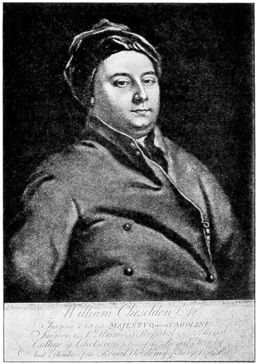
Cheselden,3 a renowned surgeon, and oculist to Her
Majesty, Queen Caroline of England, first announced,
in 1728, his success in making an artificial pupil by
means of his knife-needle. He made his puncture
back of the corneoscleral junction on the temporal
side, passing the knife across the posterior chamber,
and making a counter-puncture in the iris-membrane
near the nasal margin. He then cut through the iris
from behind forward as he withdrew the knife, the
incision being carried through two-thirds of its extent.
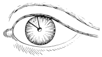
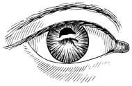
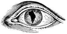
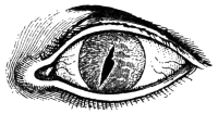
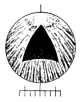
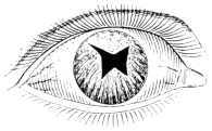
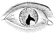
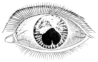
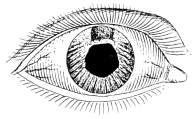
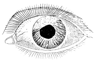
The pupillary opening thus made was a long
oval slit, horizontally placed. He has reported two successful
cases4 (Figs. 1 and 2), occurring in patients
who had previously undergone couching of the lens. His
instrument, strange to say, was practically of the same
general shape as the Hays knife-needle, but was larger,
and judging from the description more clumsily constructed,
as there was danger of leakage of the aqueous
and sometimes of the vitreous when it was used. Its
form resembled a combination of a bistoury and a sickle-shaped
knife, having a sharp edge on one side, a rounded
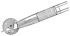
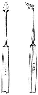
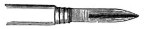
back, and an acute point. We possess two good illustrations
of this knife-needle, one by Cheselden himself
(Fig. 3), and the other by his pupil, Sharpe5 (Fig. 4).
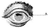
Fig. 1.—Original case of
iridotomy. Iris incised above (Cheselden).
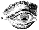
Fig. 2.—Second case of iridotomy.
Iris incised below (Cheselden).
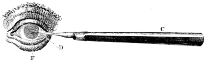
Fig. 3.—Original knife-needle in situ,
behind the iris (Cheselden).
For more than a century the method of Cheselden
seems to have been the storm center of controversy. Some
doubted his veracity, others essayed his operation but
failed, while a few had a moderate degree of success.
Many attributed to him statements which do not appear
in his published report. He says clearly that in each
of his cases couching had previously been performed,
and yet some have insisted that the lens was present, and
must have been wounded. He also states that his incision
was made from behind forward, and yet his followers,
Sharpe5 and Adams,6 both describe the incision
as being made from before backward. As Sharpe was
his pupil, and presumably had seen him operate, Guthrie7
suggests the possibility of his having made his incision
both ways, the technic being practically the same.
Morand,8 in his “Eulogy of Cheselden,” claims to have
personally seen him operate “on an eye in which the iris
was closed by an accident,” and gives a more detailed
description which closely follows the original method.
He states that Cheselden presented him with one of his
knife-needles as a souvenir of the occasion. Although
Morand does not record the exact date of his visit to
London, he does state that it occurred during the year
1729. Huguier,9 in his exhaustive thesis on artificial
pupil, also places the date of this visit in the year 1729.
This fact is important, as some writers have declared
that Morand neither made the visit to London nor saw
Cheselden operate, but only quoted the original account
given in the Philosophical Transactions. The publication
of Morand’s high encomiums in 1757 attracted renewed
interest to the subject of Cheselden’s operation
among men of scientific and medical attainments.
Sharpe,5 in 1739, performed this operation in the
same manner as Cheselden, except that after he had
entered the knife-needle through the sclerotic he passed
it through the iris and across the anterior chamber,
and then incised the iris-membrane from before backward.
Although he was Cheselden’s pupil, and dedicated
his small volume on surgery to him, he probably
did his master more harm than good, as all the
objections to Cheselden’s method seemed to be based
on the deprecatory remarks of Sharpe. He says, “I
once performed it with tolerable success, and a few
months after, the very orifice I had made contracted and
brought on blindness again.” He mentions the danger
of wounding the lens, the lack of success in paralytic iris
with affection of the retina, the danger of iridodialysis
from traction of the knife, and the possibility of failure
because the incision would not enlarge sufficiently.
Thirty years later (1769) he published the ninth edition
of his book without recording a single additional
case, but added the thought that, since extraction of
the crystalline lens showed the cornea was not so vulnerable
as had been believed, he would “imagine” that
a larger knife might be introduced perpendicularly
through the cornea and iris and a similar incision
made. In his first eight editions he pictures Cheselden’s
iris-knife (Fig. 4, vide p. 25), but in his ninth
edition he substitutes a broad lance-knife with two edges
which closely resembled the one Wenzel (vide Fig. 17)
had just introduced (1767), and which Sharpe suggests
“can also be used for the extraction of the cataract.”
He evidently did not have a very clear idea of the subject,
and only succeeded in casting doubt and discredit
on the method of Cheselden, which, judging by his own
statement, he had tried but once.
Heuermann,10 in 1756, had already antedated these
thoughts of Sharpe by practising a similar method. He
passed a double edged lance-knife through the cornea
instead of through the sclera, and then made a sweeping
incision through the iris-membrane without enlarging
the corneal wound. He was probably the first to puncture
the cornea with the iris-knife.
Janin,11 about 1766, performed Cheselden’s operation
several times with but little success owing to reclosure
of the wound by plastic exudate. He adopted Sharpe’s
modification, but later on changed the incision from a
horizontal to a vertical one with better results. He, however,
afterward abandoned this procedure and became
the originator of the other school, composed of those
who preferred to use the scissors.
Guérin,12 in 1769, made a free corneal incision with a
large cataract knife, and then introduced a small iris-knife,
with which he made a crucial incision from before
backward in the center of the iris-membrane. Although
Guthrie7 distinctly states that Guérin afterwards removed
the four angles of the cross with a pair of scissors
in order to prevent reclosure of the incision, no
direct confirmation of this statement can be found in
his writings.
Beer,13 in 1792, first published his method, which he
designated as “an improvement on Cheselden’s method.”
Although the technic is somewhat different, the
procedure is practically the same as that originated by
Heuermann in 1756. Beer selected certain cases in
which a prolapsed iris had followed the lower incision
for cataract, causing adherent leucoma with a tensely
drawn iris-membrane. He plunged his double-edged
lance-knife (Fig. 5) through the cornea and stretched
out iris, from above downward and a little obliquely
(Fig. 6), so as to incise the center of the tense iris
fibers crosswise, at right angles to the line of traction;
cutting horizontally when the traction was vertical, and
vertically when this was horizontal. In his monograph
on artificial pupil,14 1805, he substitutes for the lance-knife
his new broad iris-knife, which is practically the
same as that later shown by Walton (vide Fig. 12), as,
indeed, Walton’s procedure (vide Fig. 13) was almost
identical with that of Beer. For other conditions he
usually employed Wenzel’s operation until by chance he
encountered a puzzling case which led him to perform
the operation we now know as iridectomy (1797) and
which thereafter became his favorite procedure for artificial
pupil.
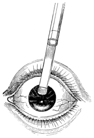
Fig. 6.—Beer’s iridotomy with broad iris-knife (after Mackenzie).
Adams,15 in 1812, revived the operation of Cheselden
with certain modifications. While his puncture was
made in the same location, his technic was different. He
entered the sclera with a small iris-scalpel of his own
special design (Fig. 7), which, like Sharpe, he passed
through the iris-membrane into the anterior chamber,
carrying it across to the nasal side (Fig. 8). From entrance
to exit he always kept the edge of the knife
turned back toward the iris, so as to cut from before
backward. He was thus able by the most delicate pressure
of his instrument, to make a long horizontal incision,
without causing iridodialysis (Fig. 9). If the first
incision appeared to be too short, he did not withdraw
the knife entirely, but again carried it forward and
partially withdrew it, always cutting in the same plane.
To quote his own words, “by repeating the efforts to
divide the iris (taking care in so doing to make as slight
a degree of pressure as possible upon the instrument, instead
of withdrawing it out of the eye at once, as recommended
by Cheselden), a division of that membrane
may, in almost all cases be effected, of a requisite size to
establish a permanent artificial pupil” (Figs. 10 and
11).
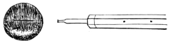
Fig. 8.—Adams’ iris scalpel in situ,
showing location of scleral puncture (after Lawrence).

Fig. 9.—Iridotomy
by Adams’ method (after Lawrence).
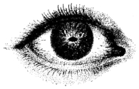
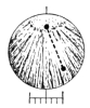
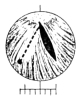
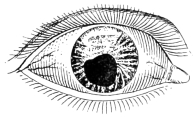
Fig. 10.—Occlusion of pupil
(Adams).
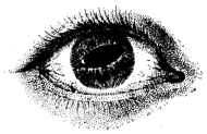
Fig. 11.—The resulting pupil
after iridotomy (Adams).
Here were three elements of success, a sharp knife, a
gentle sawing movement, and the most delicate pressure
of the instrument. His method was a decided advance,
and he reported success in nearly one hundred cases.
Others, less skilful, however, failed of success, and the
severe criticisms of Scarpa,16 though evidently unjust
and tinged by personal animosity,17 cast a shadow of
doubt on the method.
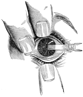
Fig. 13.—Iris-knife in position to make central pupil (Walton,
after Beer).
From that time on for nearly half a century this form
of iridotomy was practically abandoned, the pendulum
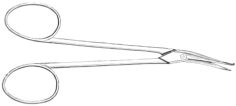
swinging toward the use of scissors, which Maunoir had
popularized and Scarpa had indorsed. Walton,18 however,
about 1852, proposed a method closely resembling
that of Heuermann and almost identical with that of
Beer (vide Fig. 6). His iris-knife (Fig. 12) was
practically the same as the broad iris-knife of Beer.
He incised the cornea near the limbus, and passed
the knife across the anterior chamber to the middle of
the iris-membrane which he punctured with a sweeping
vertical incision (Fig. 13). If the tissue still retained
its elasticity there appeared a long pupillary aperture,
elliptical and vertical (Figs. 14 and 15). This incision,
however, like all those made through a single set of the
iris fibers, was only successful when there was sufficient
resiliency remaining in the iris tissue to draw the slit
open, and thus keep the edges from uniting. While
this method never became very popular, there were some
who later practiced it by substituting a very narrow
Graefe knife for the iris-knife of Heuermann, Beer and
Walton. In fact, this latter procedure still has considerable
vogue, both for iridotomy and capsulotomy.
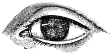
Fig. 14.—Occlusion of pupil
(Walton).
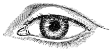
Fig. 15. New pupil after incision
with iris-knife (Walton).
During the following seventeen years no notable advance
was made, the scissors method still retaining its
hold on the profession, until in 1869, von Graefe, after
long reflection, became convinced of the dangers of that
method, and communicated to one of his pupils, M.
Meyer, his method of simple iridotomy performed with
the knife-needle. Meyer19 quotes his views as follows:
“For such cases von Graefe has suggested another method of
operation, the principle and execution of which are contained in
the following note written for us by that illustrious savant in
1869:
“When, in consequence of a cataract operation, the lens is
absent, and when there is highly developed retro-iritic exudation,
with disorganization of the iris tissue, flattening of the
cornea and the other sequelæ of a destructive iridocyclitis, I
substitute simple iridotomy for iridectomy, which is the operation
hitherto performed, generally without success. The operation
consists in inserting a double-edged knife, resembling in
shape a very sharp pointed lance-knife, through the cornea and
newly formed tissues till it pierces the vitreous body, and immediately
withdrawing it; and, while withdrawing it, enlarging
the wound in the membranes without increasing the size
of the corneal wound. Experience shows that such plastic
membranes attached to the atrophied iris and to the capsule
of the lens have a tendency to contract sufficient to maintain,
to a certain extent, the opening which has been made.
“If, in the ordinary method of iridectomy, combined with
laceration or extraction of the false membranes, we find that
the artificial pupil usually becomes closed, we must attribute
this to an excessive vulnerability, which immediately sets up
proliferation in those tissues which have been touched, and
which are endowed, in consequence on their structure, with an
irritability altogether peculiar. We know that even the transitory
reduction of the intraocular pressure, which follows the
evacuation of the aqueous humor, is sufficient to give rise to
hemorrhage in the anterior chamber, which interferes with the
perfect success of the intended operation; but most of our failures
in the ordinary methods are due to the irritation caused
by the forceps and the traction on the surrounding structures.
Simple iridotomy is free from such inconveniences; it is, so to
speak, a sub-corneal act, and enjoys the immunity which belongs
to subcutaneous operations.
“I have also reduced the corneal wound to a minimum, by
using small falciform knives. These are passed through the
false membranes, which are then cut from behind forward.”
Von Graefe thus proposed two methods, (1) by cutting
from before backward with a double-edged lance-knife,
according to the method of Heuermann, and (2) by
cutting from behind forward with a sickle-shaped knife,
after the original suggestion of Cheselden. Later in the
same year, as he lay on his last bed of illness, he became
so absorbed in the study of this subject that he sent a
telegram to the Heidelberg Congress20 (September,
1869), in which he advocated the method by the sickle-shaped
knife-needle as the best procedure. His last message
to his colleagues showed, therefore, that through
mature conviction he strongly favored the use of the
knife-needle, and the making of a sub-corneal incision in
the iris-membrane without evacuating the aqueous humor.
His untimely death, however, prevented him from
further perfecting this procedure and presenting it to
the profession.
Galezowski,21 in 1875, published a somewhat similar
method in which he used his falciform knife, aiguille-a-serpette
(Fig. 16), which he introduced through the
cornea and iris-membrane, making either a horizontal
or a vertical incision, with a “go-and-come” (sawing)
movement, after the suggestion of Adams. If this
single cut was not sufficient, he made a linear incision of
the cornea with a Graefe knife, drew out the iris and cut
it off with scissors. By a process of evolution, however,
he perfected the former procedure and eliminated the
scissors. This latter method was published in the third
edition of his book in 1888. He punctured the cornea
and iris-membrane with the sickle-shaped knife, making
first a horizontal incision by the sawing movement of
Adams, and finishing with a second cut in the vertical
direction, thus forming a T-shaped incision. In actual
practice, however, he almost always prolonged this second
cut, thus making a crucial incision after the manner
of Guérin.12
The writer,22 in 1888, was led to devise an operation
with a modified Hays knife-needle, in which through a
corneal puncture he made a converging incision in the
iris-membrane which resembled an inverted V. The resulting
pupil opened up and formed either a triangular
or an oval-shaped pupil depending on the degree of stiffness
or resiliency of the iris-membrane. This method
will be described in detail later on.