Professor of Histology and Pharmacognosy, College of
Pharmacy of the City of New York
Columbia University
TOTAL ISSUE, FOUR THOUSAND
NEW YORK
JOHN WILEY & SONS, Inc. London: CHAPMAN & HALL, Limited
Copyright, 1916, by
WILLIAM MANSFIELD
PREFACE
The object of the book is to provide a practical scientific
course in vegetable histology for the use of teachers and students
in schools and colleges.
The medicinal plants are studied in great detail because
they constitute one of the most important groups of economic
plants. The cells found in these plants are typical of the cells
occurring in the vegetable kingdom; therefore the book should
prove a valuable text-book for all students of histology.
The book contains much that is new. In Part II, which is
devoted largely to the study of cells and cell contents, is a new
scientific, yet practical, classification of cells and cell contents.
The author believes that his classification of bast fibres and
hairs will clear up much of the confusion that students have
experienced when studying these structures.
The book is replete with illustrations, all of which are from
original drawings made by the author. As most of these illustrations
are diagnostic of the plants in which they occur, they
will prove especially valuable as reference plates.
The material of the book is the outgrowth of the experience
of the author in teaching histology at the College of Pharmacy
of the City of New York, Columbia University, and of years
of practical experience gained by examining powdered drugs in
the laboratory of a large importing and exporting wholesale
drug house.
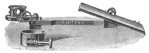
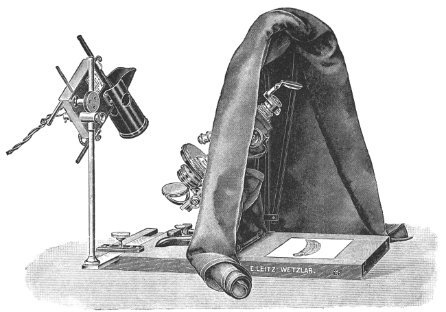
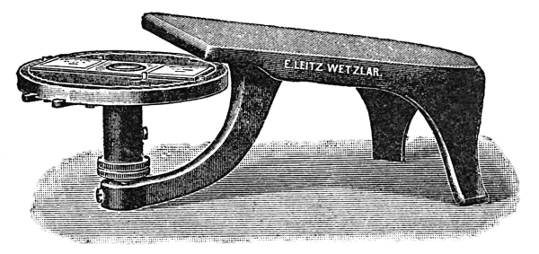
The author is indebted to Ernest Leitz and Bausch &
Lomb Optical Company for the use of cuts of microscopic
apparatus used in Part I of the book.
The author also desires to express his appreciation to Professor
Walter S. Cameron, who has rendered him much valuable
aid.
Part I SIMPLE AND COMPOUND MICROSCOPES AND MICROSCOPIC TECHNIC
CHAPTER I THE SIMPLE MICROSCOPES — FORMS OF SIMPLE MICROSCOPES
The construction and use of the simple microscope (magnifiers)
undoubtedly date back to very early times. There is
sufficient evidence to prove that spheres of glass were used as
burning spheres and as magnifiers by people antedating the
Greeks and Romans.
The simple microscopes of to-day have a very wide range of
application and a corresponding variation in structure and in
appearance.
Simple microscopes are used daily in classifying and studying
crude drugs, testing linen and other cloth, repairing watches,
in reading, and identifying insects. The more complex simple
microscopes are used in the dissection and classification of
flowers.
The watchmaker’s loupe, the linen tester, the reading glass,
the engraver’s lens, and the simplest folding magnifiers consist
of a double convex lens. Such a lens produces an erect, enlarged
image of the object viewed when the lens is placed so
that the object is within its focal distance. The focal distance
of a lens varies according to the curvature of the lens. The
greater the curvature, the shorter the focal distance and the
greater the magnification.
The more complicated simple microscope consists of two or
more lenses. The double and triple magnifiers consist of two
and three lenses respectively.
When an object is viewed through three lenses, the magnification
is greater than when viewed through one or two lenses,
but a smaller part of the object is magnified.
TRIPOD MAGNIFIER
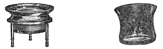
The tripod magnifier (Fig. 1) is a simple lens mounted on a
mechanical stand. The tripod is placed over the object and
the focus is obtained by means of a screw which raises or lowers
the lens, according to the degree it is magnified.
WATCHMAKER’S LOUPE
The watchmaker’s loupe (Fig. 2) is a one-lens magnifier
mounted on an ebony or metallic tapering rim, which can be
placed over the eye and held in position by frowning or contracting
the eyelid.
The folding magnifier (Fig. 3) of one or more lenses is mounted
in such a way that, when not in use, the lenses fold up like the
blade of a knife, and when so folded are effectively protected
from abrasion by the upper and lower surfaces of the folder.
Fig. 3.—Folding Magnifier Fig. 4.—Reading Glass
READING GLASSES
Reading glasses (Fig. 4) are large simple magnifiers, often
six inches in diameter. The lens is encircled with a metal band
and provided with a handle.
Fig. 5.—Steinheil Aplanatic Lens
STEINHEIL APLANATIC LENSES
Steinheil aplanatic lenses (Fig. 5) consist of three or four
lenses cemented together. The combination is such that the
field is large, flat, and achromatic. These lenses are suitable
for field, dissecting, and pocket use. When such lenses are
placed in simple holders, they make good dissecting microscopes.
Fig. 6.—Dissecting microscope
DISSECTING MICROSCOPE
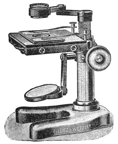
The dissecting microscope (Fig. 6) consists of a Steinheil
lens and an elaborate stand, a firm base, a pillar, a rack and
pinion, a glass stage, beneath which there is a groove for holding
a metal plate with one black and one white surface. The
nature of the object under observation determines whether a
plate is used. When the plate is used and when the object is
studied by reflected light it is sometimes desirable to use the
black and sometimes the white surface. The mirror, which has
a concave and a plain surface, is used to reflect the light on the
glass stage when the object is studied by transmitted light.
The dissecting microscope magnifies objects up to twenty
diameters, or twenty times their real size.
CHAPTER II COMPOUND MICROSCOPES — STRUCTURE OF THE COMPOUND MICROSCOPE — FORMS OF COMPOUND MICROSCOPES
The compound microscope has undergone wonderful changes
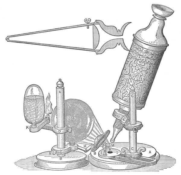
since 1667, the days of Robert Hooke. When we consider the
crude construction and the limitations of Robert Hooke’s microscope,
we marvel at the structural perfection and the unlimited
possibilities of the modern instrument. The advancement made
in most sciences has followed the gradual perfection of this
instrument.
The illustration of Robert Hooke’s microscope (Fig. 7) will
convey to the mind more eloquently than words the crudeness
of the early microscopes, especially when it is compared with
the present-day microscopes.
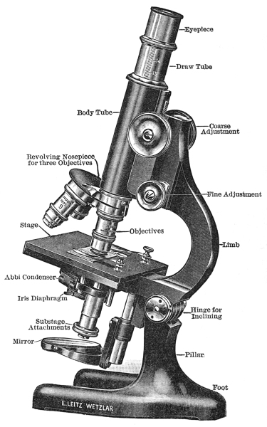
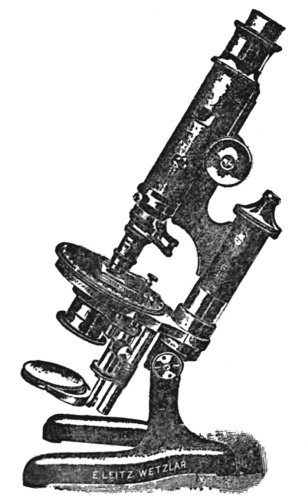
The parts of the compound microscope (Fig. 8) may be
grouped into—first, the mechanical, and, secondly, into the
optical parts.
THE MECHANICAL PARTS
1. The foot is the basal part, the part which supports all
the other mechanical and optical parts. The foot should be
heavy enough to balance the other parts when they are inclined.
Most modern instruments have a three-parted or tripod-shaped
base.
2. The pillar is the vertical part of the microscope attached
to the base. The pillar is joined to the limb by a hinged joint.
The hinges make it possible to incline the microscope at any
angle, thus lowering its height. In this way, short, medium,
and tall persons can use the microscope with facility. The
part of the pillar above the hinge is called the limb. The limb
may be either straight or curved. The curved form is preferable,
since it offers a more suitable surface to grasp in transferring
from box or shelf to the desk, and vice versa.
Fig. 7.—Compound Microscope of Robert Hooke
3. The stage is either stationary or movable, round or
square, and is attached to the limb just above the hinge. The
upper surface is made of a composition which is not easily
attacked by moisture and reagents. The centre of the stage is
perforated by a circular opening.
4. The sub-stage is attached below the stage and is for the
purpose of holding the iris diaphragm and Abbé condenser.
The raising and lowering of the sub-stage are accomplished by
a rack and pinion.
5. The iris diaphragm, which is held in the sub-stage below
the Abbé condenser, consists of a series of metal plates, so arranged
that the light entering the microscope may be cut off
completely or its amount regulated by moving a control pin.
6. The fine adjustment is located either at the side or at
the top of the limb. It consists of a fine rack and pinion, and
is used in focusing an object when the low-power objective is in
position, or in finding and focusing the object when the high-power
objective is in position.
7. The coarse adjustment is a rack and pinion used in raising
and lowering the body-tube and in finding the approximate
focus when either the high- or low-power objective is in position.
8. The body-tube is the path traveled by the rays of light
entering the objectives and leaving by the eye-piece. To the
lower part of the tube is attached the nose-piece, and resting
in its upper part is the draw-tube, which holds the eye-piece.
On the outer surface of the draw-tube there is a scale which
indicates the distance it is drawn from the body-tube.
9. The nose-piece may be simple, double, or triple, and it
is protected from dust by a circular piece of metal. Double and
triple nose-pieces may be revolved, and like the simple nose-piece
they hold the objectives in position.
THE OPTICAL PARTS
1. The mirror is a sub-stage attachment one surface of which
is plain and the other concave. The plain surface is used with
an Abbé condenser when the source of light is distant, while
the concave surface is used with instruments without an Abbé
condenser when the source of light is near at hand.
Fig. 8.—Compound Microscope
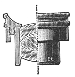
2. The Abbé condenser (Fig. 9) is a combination of two or
more lenses, arranged so as to concentrate the light on the
specimen placed on the stage. The condenser is located in the
opening of the stage, and its uppermost
surface is circular and flat.
Fig. 9—Abbé Condenser
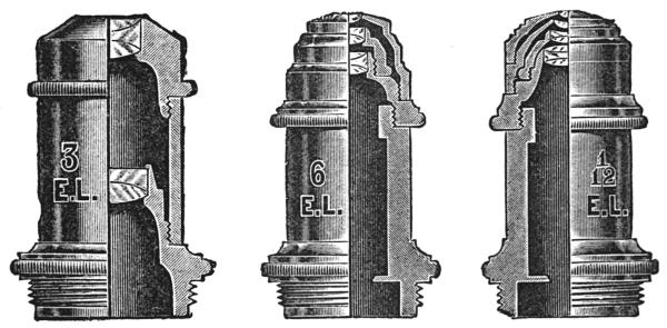
3. Objectives (Figs. 10, 11, and 12).
There are low, medium, and high-power
objectives. The low-power objectives have
fewer and larger lenses, and they magnify
least, but they show more of the object than
do the high-power objectives.
There are three chief types of objectives:
First, dry objectives; second, wet
objectives, of which there are the water-immersion objectives;
and third, the oil-immersion objectives. The dry
objectives are used for most histological and pharmacognostical
work. For studying smaller objects the water objective
is sometimes desirable, but in bacteriological work the
oil-immersion objective is almost exclusively used. The globule
of water or oil, as the case may be, increases the amount of light
entering the objective, because the oil and water bend many
rays into the objective which would otherwise escape.
Fig. 10. Fig. 11. Fig. 12. Objectives.
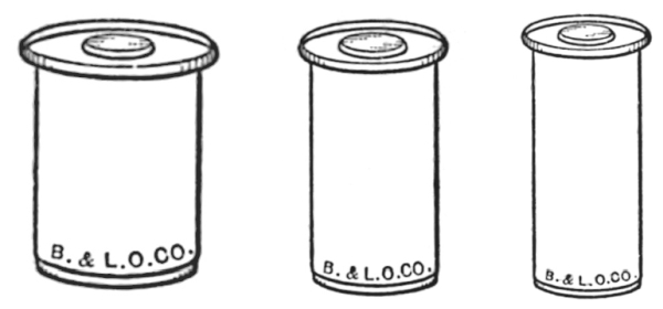
4. Eye-pieces (Figs. 13, 14, and 15) are of variable length,
but structurally they are somewhat similar. The eye-piece
consists of a metal tube with a blackened inner tube. In the
centre of this tube there is a small diaphragm for holding the
ocular micrometer. In the lower end of the tube a lens is fastened
by means of a screw. This, the field lens, is the larger
lens of the ocular. The upper, smaller lens is fastened in the
tube by a screw, but there is a projecting collar which rests,
when in position, on the draw-tube.
Fig. 13. Fig. 14 Fig. 15 Eye-Pieces.
The longer the tube the lower the magnification. For
instance, a two-inch ocular magnifies less than an inch and a
half, a one-inch less than a three-fourths of an inch, etc.
The greater the curvature of the
lenses of the ocular the higher will be
the magnification and the shorter the
tube-length.
The following descriptions refer to
three different models of compound
microscopes: one which is used chiefly
as a pharmacognostic microscope, one
as a research microscope stand, while
the third type represents a research
microscope stand of highest order,
which is used at the same time for
taking microphotographs.
Fig. 16.—Pharmacognostic
Microscope
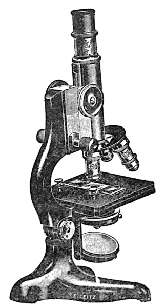
PHARMACOGNOSTIC MICROSCOPE
The pharmacognostic microscope
(Fig. 16) is an instrument which
embodies only those parts which are most essential for the
examination of powdered drugs, bacteria, and urinary
sediments. This microscope is provided with a stage of the
dimensions 105 × 105 mm. This factor and the distance of
80 mm. from the optical centre to the handle arm render it
available for the examination of even very large objects and
preparations, or preparations suspended in glass dishes. The
stand is furnished with a side micrometer, a fine adjustment
having knobs on both sides, thereby permitting the manipulation
of the micrometer screw either by left or right hand. The
illuminating apparatus consists of the Abbé condenser of numerical
aperture of 1.20, to which is attached an iris diaphragm for
the proper adjustment of the light. A worm screw, mounted
in connection with the condenser, serves for the raising and
lowering of the condenser, so that the cone of illuminating
pencils can be arranged in accordance to the objective employed
and to the preparation under observation. The objectives
necessary are those of the achromatic type, possessing a focal
length of 16.2 mm. and 3 mm. Oculars which render the best
results in regard to magnification in connection with the two
objectives mentioned are the Huyghenian eye-pieces II and IV
so that magnifications are obtained varying from 62 to 625.
It is advisable, however, to have the microscope equipped with
a triple revolving nose-piece for the objectives, so that provision
is made for the addition of an oil-immersion objective at any
time later should the microscope become available for bacteriological
investigations.
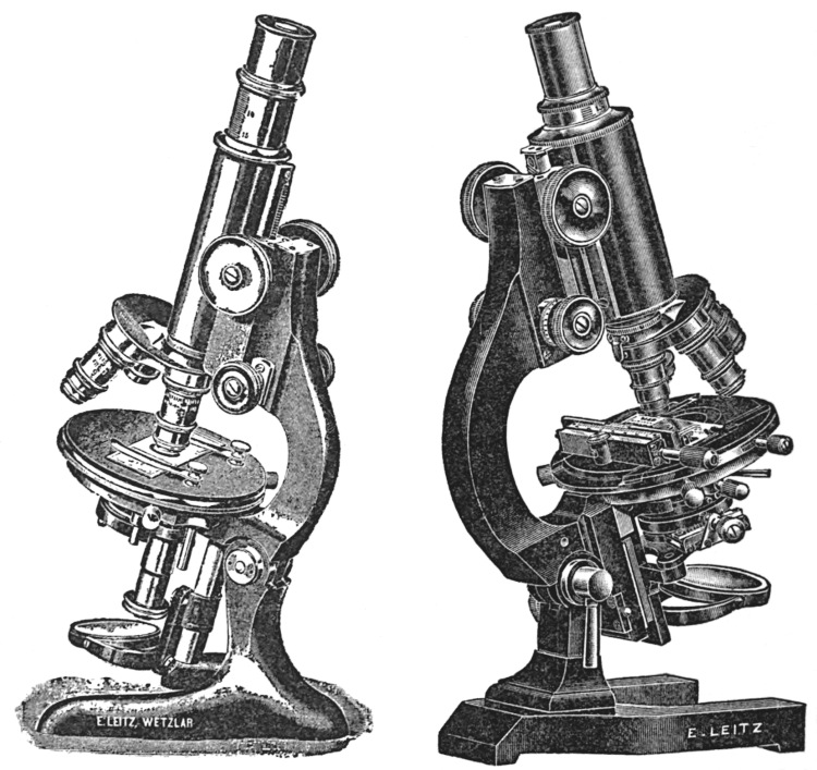
THE RESEARCH MICROSCOPE
The research microscope used in research work (Fig. 17) must
be equipped more elaborately than the microscope especially
designed for the use of the pharmacognosist. While the simple
form of microscope is supplied with the small type of Abbé
condenser, the research microscope is furnished with a large
illuminating apparatus of which the iris diaphragm is mounted
on a rack and pinion, allowing displacement obliquely to the
optical centre, also to increase resolving power in the objectives
when observing those objects which cannot be revealed to the
best advantage with central illumination. Another iris is
furnished above the condenser; this iris becomes available the
instant an object is to be observed without the aid of the condenser,
in which case the upper iris diaphragm allows proper
adjustment of the light. The mirror, one side plane, the other
concave, is mounted on a movable bar, along which it can
be slid—another convenience for the adjustment of the light.
The microscope stage of this stand is of the round, rotating
and centring pattern, which permits a limited motion to the
object slide: The rotation of the microscope stage furnishes
another convenience in the examination of objects in polarized
light, allowing the preparation to be rotated in order to
distinguish the polarization properties of the objects under
observation.
Fig. 17.—Research
Microscope Fig. 18.—Special Research Microscope
SPECIAL RESEARCH MICROSCOPE
A special research microscope of the highest order (Fig. 18)
is supplied with an extra large body tube, which renders it of
special advantage for micro-photography. Otherwise in its
mechanical equipment it resembles very closely the medium-sized
research microscope stand, with the exception that the
stand is larger in its design, therefore offering universal application.
In regard to the illuminating apparatus, it is advisable
to mention that the one in the large research microscope stand
is furnished with a three-lens condenser of a numerical aperture
of 1.40, while the medium-sized research stand is provided with
a two-lens condenser of a numerical aperture of 1.20. The
stage of the microscope is provided with a cross motion—the
backward and forward motion of the preparation is secured by
rack and pinion, while the side motion
is controlled by a micrometric worm
screw. In cases where large preparations
are to be photographed, the
draw-tube with ocular and the slider
in which the draw-tubes glide are
removed to allow the full aperture
of wide-angle objectives to be made
use of.
Fig. 19.—Greenough
Binocular Microscope
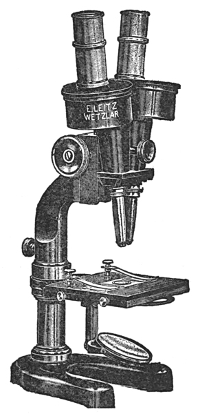
BINOCULAR MICROSCOPE
The Greenough binocular microscope,
as shown in Fig. 19, consists
of a microscope stage with two tubes
mounted side by side and moving on
the same rack and pinion for the
focusing adjustment. Either tube
can be used without the other. The
oculars are capable of more or less
separation to suit the eyes of different
observers. In each of the drub-like
mountings, near the point where the
oculars are introduced, porro-prisms
have been placed, which erect the image. This microscope
gives most perfect stereoscopic images, which are erect instead
of inverted, as in the monocular compound microscopes. The
Greenough binocular microscope is especially adapted for dissection
and for studying objects of considerable thickness.
POLARIZATION MICROSCOPE
The polarization microscope (Fig. 20) is used chiefly for the
examination of crystals and mineral sections as well as for the
observation of organic bodies in polarized light. It can, however,
also be used for the examination of regular biological
preparations.
Fig. 20.—Polarization Microscope
If compared with the regular biological microscope, the
polarization microscope is found characteristic of the following
points: it is supplied with a polarization arrangement. The
latter consists of a polarizer and analyzer. The polarizer is
situated in a rotating mount beneath the condensing system.
The microscope, of which the diagram is
shown, possesses a triple “Ahrens” prism
of calcite. The entering light is divided
into two polarized parts, situated perpendicularly
to each other. The so-called
“ordinary” rays are reflected to one side
by total reflection, which takes place on
the inner cemented surface of the triple
prism, allowing the so-called “extraordinary”
rays to pass through the condenser.
If the prism is adjusted to its
focal point, it is so situated that the
vibration plane of the extra-ordinary rays
are in the same position as shown in
the diagram of the illustration.
The analyzer is mounted within the
microscope-tube above the objective.
Situated on a sliding plate, it can be shifted into the optical
axis whenever necessary. The analyzer consists of a polarization
prism after Glan-Thompson. The polarization plane
of the active extraordinary rays is situated perpendicularly
to the plane as shown in the diagram. The polarization
prisms are ordinarily crossed. In this position the field of
the microscope is darkened as long as no substance of a double
refractive index has been introduced between the analyzer and
polarizer. In rotating the polarizer up to the mark 90, the
polarization prisms are mounted parallel and the field of the
microscope is lighted again. Immediately above the analyzer
and attached to the mounting of the analyzer a lens of a comparatively
long focal length has been placed in order to overcome
the difference in focus created by the introduction of the
analyzer into the optical rays.
The condensing system is mounted on a slider, and, furthermore,
can be raised and lowered along the optical centre by
means of a rack-and-pinion adjustment. If lowered sufficiently,
the condensing system can be thrown to the side to be removed
from the optical rays. The condenser consists of three lenses.
The two upper lenses are separately mounted to an arm, which
permits them to be tilted to one side in order to be removed
from the optical rays. The complete condenser is used only
in connection with high-power objectives. As far as low-power
objectives are concerned, the lower condensing lens alone is
made use of, and the latter is found mounted to the polarizer
sleeve. Below the polarizer and above the lower condensing
lens an iris diaphragm is found.
The microscope table is graduated on its periphery, and,
furthermore, carries a vernier for more exact reading.
The polarization microscope is not furnished with an objective
nose-piece. Every objective, however, is supplied with
an individual centring head, which permits the objective to be
attached to an objective clutch-changer, situated at the lower end
of the microscope-tube. The centring head permits the objectives
to be perfectly centred and to remain centred even if another
objective is introduced into the objective clutch-changer.
At an angle of 45 degrees to the polarization plane of polarizer
and analyzer, a slot has been provided, which serves for the
introduction of compensators.
Between analyzer and ocular, another slot is found which
permits the Amici-Bertrand lens to be introduced into the
optical axis. The slider for the Bertrand lens is supplied with
two centring screws whereby this lens can be perfectly and
easily centred. The Bertrand lens serves the purpose of
observing the back focal plane of the microscope objective. In
order to allow the Bertrand lens to be focused, the tube can be
raised and lowered for this purpose. An iris diaphragm is
mounted above the Bertrand lens.
If the Bertrand lens is shifted out of the optical axis, one can
observe the preparation placed upon the microscope stage and,
depending on its thickness or its double refraction, the interference
color of the specimen. This interference figure is called
the orthoscopic image and, accordingly, one speaks of the microscope
as being used as an “orthoscope.”
After the Bertrand lens has been introduced into the optical
axis, the interference figure is visible in the back focal plane of
the objective. Each point of this interference figure corresponds
to a certain direction of the rays of the preparation itself. This
arrangement permits observation of the change of the reflection
of light taking place in the preparation, this in accordance with
the change of the direction of the rays. This interference figure
is called the conoscopic image, and, accordingly, the microscope
is used as a “conoscope.”
Many types of polarization microscopes have been constructed;
those of a more elaborate form are used for research
investigations; others of smaller design for routine investigations.
In making critical examinations of powdered drugs, it is
frequently necessary to measure the elements under observation,
particularly in the case of starches and crystals.
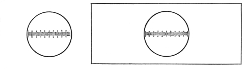
Microscopic measurements are made by the ocular micrometer
(Fig. 21). This consists of a circular piece of transparent
glass on the centre of which is etched a one- or two-millimeter
scale divided into one hundred or two hundred divisions respectively.
The value of each line is determined by standardizing
with a stage micrometer.
The stage micrometer (Fig. 22) consists of a glass slide upon
which is etched a millimeter scale divided into one hundred
equal parts or lines: each line has a value of one hundredth of
a millimeter.
STANDARDIZATION OF OCULAR MICROMETER WITH LOW-POWER OBJECTIVE
Having placed the ocular micrometer in the eye-piece and
the stage micrometer on the centre of the stage, focus until
the lines of the stage micrometer are clearly seen. Then adjust
the scales until the lines of the stage micrometer are parallel
with and directly under the lines of the ocular micrometer.
Ascertain the number of lines of the stage micrometer covered
by the one hundred lines of the ocular micrometer. Then
calculate the value of each line of the ocular. This is done in
the following manner:
If the one hundred lines of the ocular cover seventy-five
lines of the stage micrometer, then the one hundred lines of
the ocular micrometer are equivalent to seventy-five one-hundredths,
or three-fourths, of a millimeter. One line of the
ocular micrometer will therefore be equivalent to one-hundredth
of seventy-five one-hundredths, or .0075 part of a millimeter,
and as a micron is the unit for measuring microscopic objects,
this being equivalent to one one-thousandth of a millimeter,
the value of each line of the ocular will therefore be 7.5 microns.
Fig. 23.—Micrometer Eye-Piece
With the high-power objective in place, ascertain the value
of each line of the ocular. If one hundred lines of the ocular
cover only twelve lines of the stage micrometer, then the one
hundred lines of the ocular are equivalent to twelve one-hundredths
of a millimeter, the value of one line being equivalent
to one one-hundredth of twelve one-hundredths, or twelve ten-thousandths
of a millimeter, or .0012, or 1.2 µ.
It will therefore be seen that objects as small as a thousandth
of a millimeter can be accurately measured by the ocular
micrometer.
In making microscopic measurements it is only necessary
to find how many lines of the ocular scale are covered by the
object. The number of lines multiplied by the equivalent of
each line will be the size of the object in microns, or micromillimeters.
Fig. 24.—Micrometer Eye-Piece
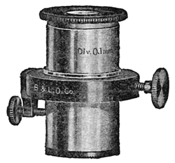
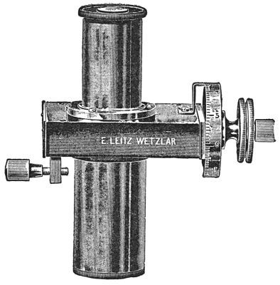
Micrometer eye-pieces (Figs. 23 and 24) may be used in
making measurements. These eye-pieces with micrometer combinations
are preferred by some workers, but the ocular micrometer
will meet the needs of the average worker.
Moving objects by hand is tiresome and unsatisfactory, first,
because of the possibility of losing sight of the object under
observation, and secondly, because the field cannot be covered
so systematically as when a mechanical stage is used for moving
slides.
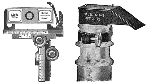
The mechanical stage (Fig. 25) is fastened to the stage by
a screw. The slide is held by two clamps. There is a rack and
pinion for moving the slide to left or right, and another rack and
pinion for moving the slide forward and backward.
Fig. 25. Mechanical Stage Fig. 26.—Camera Lucida
The camera lucida is an optical mechanical device for aiding
the worker in making drawings of microscopic objects. The
instrument is particularly necessary in research work where it
is desirable to reproduce an object in all its details. In fact, all
reproductions illustrating original work should be made by
means of the camera lucida or by microphotography.
Fig. 27.—Camera Lucida
A great many different types of camera lucidas or drawing apparatus
are obtainable, varying from simple-inexpensive to
complex-expensive forms. Figs. 26, 27, and 28 show simple
and complex forms.
Fig. 28.—Drawing Apparatus
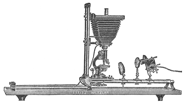
The microphotographic apparatus (Fig. 29), as the name
implies, is an apparatus constructed in such a manner that it
may be attached to a microscope when we desire to photograph
microscopic objects. It consists of a metal base and a polished
metal pillar for holding the bellows, slide holder, ground-glass
observation plate, and eye-piece. In making photographs, the
small end of the bellows is attached to the ocular of the microscope,
the focus adjusted, and the object or objects photographed.
More uniform results are obtained in making such
photographs if an artificial light of an unvarying candle-power
is used.
Fig. 29.—Microphotographic Apparatus
There are obtainable more elaborate microphotographic
apparatus than the one figured and described, but for most
workers this one will prove highly satisfactory. It is possible,
by inclining the tube of the microscope, to make good microphotographs
with an ordinary plate camera. This is accomplished
by removing the lens of the camera and attaching the
bellows to the ocular, focusing, and photographing.
CHAPTER IV HOW TO USE THE MICROSCOPE — ILLUMINATION — CARE OF THE MICROSCOPE — PREPARATION OF SPECIMENS FOR CUTTING — CUTTING SECTIONS
In beginning work with the compound microscope, place
the base of the microscope opposite your right shoulder, if you
are right-handed; or opposite your left shoulder, if you are left-handed.
Incline the body so that the ocular is on a level with
your eye, if necessary; but if not, work with the body of the
microscope in an erect position. In viewing the specimen, keep
both eyes open. Use one eye for observation and the other
for sketching. In this way it will not be necessary to remove
the observation eye from the ocular unless it be to complete
the details of a sketch.
Learn to use both eyes. Most workers, however, accustom
themselves to using one eye; when they are sketching, they use
both eyes, although it is not necessary to do so.
Open the iris diaphragm, and incline the mirror so that
white light is reflected on the Abbé condenser. Place the slide
on the centre of the stage, and if the slide contains a section
of a plant, move the slide so as to place this specimen over the
centre of the Abbé condenser. Then lower the body by means
of the coarse adjustment until the low-power object, which
should always be in position when work is begun, is within one-fourth
of an inch of the stage. Then raise the body by means
of the coarse adjustment until the object, or objects, in case a
powder is being examined, is seen. Open and close the iris
diaphragm, finally adjusting the opening so that the best possible
illumination is obtained for bringing out clearly the structure
of the object or objects viewed. Then regulate the focus
by moving the body up or down by turning the fine adjustment.
When studying cross-sections or large particles of powders, it
is sometimes desirable to make low-power sketches of the specimen.
In most cases, however, only sufficient time should be
spent in studying the specimen to give an idea of the size, structure,
and general arrangement or plan or structure if a section
of a plant, or, if a powder, to note its striking characters. All
the finer details of structure are best brought out with the
high-power objective in position.
In placing the high-power objective in position, it is first
necessary to raise the body by the coarse adjustment; then
open the iris diaphragm, and lower the body until the objective
is within about one-eighth of an inch of the slide. Now raise
the tube by the fine adjustment until the object is in focus,
then gradually close the iris diaphragm until a clear definition
of the object is obtained. Now proceed to make an accurate
sketch of the object or objects being studied.
In using the water or oil-immersion objectives it is first
necessary to place a drop of distilled water or oil, as the case
may be, immediately over the specimen, then lower the body
by the coarse adjustment until the lens of the objective touches
the water or the oil. Raise the tube, regulate the light by the
iris diaphragm, and proceed as if the high-power objectives were
in position.
The water or oil should be removed from the objectives and
from the slide when not in use.
After the higher-powered objective has been used, the body
should be raised, and the low-power objective placed in position.
If the draw-tube has been drawn out during the examination
of the object, replace it, but be sure to hold one hand on the
nose-piece so as to prevent scratching the objective and Abbé
condenser by their coming in forceful contact. Lastly, clean
the mirror with a soft piece of linen. In returning the microscope
to its case, or to the shelf, grasp the limb, or the pillar,
firmly and carry as nearly vertical as possible in order not to
dislodge the eye-piece.
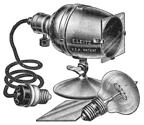
The illumination for microscopic work may be from natural
or artificial sources.
Fig. 30.—Micro Lamp
It has been generally supposed that the best possible illumination
for microscopic work is diffused sunlight obtained from
a northern direction. No matter from what direction diffused
sunlight is obtained, it will be found suitable for microscopic
work. In no case should direct sunlight be used, because it
will be found blinding in its effects upon the eyes. Natural
illumination—diffused sunlight—varies so greatly during the
different months of the year, and even during different periods
of the day, that individual workers are resorting more and more
to artificial illumination. The particular advantage of such
illumination is due to the fact that its quality and intensity
are uniform at all times. There are many ways of securing
such artificial illumination, no one of which has any particular
advantage over the other. Some workers use an ordinary gas
or electric light with a color screen placed in the sub-stage
below the iris diaphragm. In other cases a globe filled with a
weak solution of copper sulphate is placed in such a way between
the source of light and the microscope that the light is
focused on the mirror. Modern mechanical ingenuity has devised,
however, a number of more convenient micro lamps
(Fig. 30). These lamps are a combination of light and screen.
In some forms a number of different screens come with each
lamp, so that it is possible to obtain white-, blue-, or dark-ground
illumination. The type of the screen used will be varied according
to the nature of the object studied.
If possible, the microscope should be stored in a room of
the same temperature as that in which it is to be used. In
any case, avoid storing in a room that is cooler than the place
of use, because when it is brought into a warmer room, moisture
will condense on the ocular objectives and mirrors.
Before beginning work remove all moisture, dust, etc., from
the inner and outer lenses of the ocular, the objectives, the
Abbé condenser, and the mirror by means of a piece of soft,
old linen. When the work is finished the optical parts should
be thoroughly cleaned.
If reagents have been used, be sure that none has got on
the objectives or the Abbé condenser. If any reagent has got
on these parts, wash it off with water, and then dry them thoroughly
with soft linen.
The inner lenses of the eye-pieces and the under lens of the
Abbé condenser should occasionally be cleaned. The mechanical
parts of the stand should be cleaned if dust accumulates, and
the movable surfaces should be oiled occasionally. Never
attempt to make new combinations of the ocular or objective
lenses, or transfer the objectives or ocular from one microscope
to another, because the lenses of any given microscope form a
perfect lens system, and this would not be the case if they were
transferred. Keep clean cloths in a dust-proof box. Under no
circumstances touch any of the optical parts with your fingers.
Most drug plants are supplied to pharmacists in a dried
condition. It is necessary, therefore, to boil the drug in water,
the time varying from a few minutes, in the case of thin leaves
and herbs, up to a half hour if the drug is a thick root or woody
stem. If a green (undried) drug is under examination, this
first step is not necessary.
If the specimen to be cut is a leaf, a flower-petal, or other
thin, flexible part of a plant, it may be placed between pieces
of elder pith or slices of carrot or potato before cutting.
SHORT PARAFFIN PROCESS
In most cases, however, more perfect sections will be obtained
if the specimens are embedded in paraffin, by the quick
paraffin process, which is easily carried out.
After boiling the specimen in water, remove the excess of
moisture from the outer surface with filter paper or wait until
the water has evaporated. Next make a mould of stiff cardboard
and pour melted paraffin (melting at 50 or 60 degrees)
into the mould to a height of about one-half inch, when the
paraffin has solidified. This may be hastened by floating it
on cool or iced water instead of allowing it to cool at room
temperature.
The specimens to be cut are now placed on the paraffin,
with glue, if necessary, to hold them in position, and melted
paraffin poured over the specimens until they are covered to a
depth of about one-fourth of an inch. Cool on iced water,
trim off the outer paraffin to the desired depth, and the Specimen
will be in a condition suitable for cutting.
Good workable sections may be cut from specimens embedded
by this quick paraffin method. After a little practice the entire
process can be carried out in less than an hour. This method
of preparing specimens for cutting will meet every need of the
pharmacognosist.
LONG PARAFFIN PROCESS
In order to bring out the structure of the protoplast (living
part of the cell), it will be necessary to begin with the living
part of the plant and to use the long paraffin method or the
collodion method.
Small fragments of a leaf, stem, or root-tip are placed in
chromic-acid solution, acetic alcohol, picric acid, chromacetic
acid, alcohol, etc., depending upon the nature of the specimen
under observation. The object of placing the living specimen
in such solutions is to kill the protoplast suddenly so that the
parts of the cell will bear the same relationship to each other
that they did in the living plant, and to fix the parts so killed.
After the fixing process is complete, the specimen is freed
of the fixing agent by washing in water. From the water-bath
the specimens are transferred successively to 10, 20, 40, 60, 70,
80, 90, and finally 100 per cent alcohol. In this 100 per cent
alcohol-bath the last traces of moisture are removed. The
length of time required to leave the specimens in the different
percentages of alcohols varies from a few minutes to twenty-four
hours, depending upon the size and the nature of the specimen.
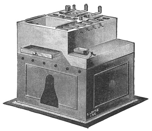
Fig. 31.—Paraffin-embedding Oven
After dehydration the specimen is placed in a clearing agent—chloroform
or xylol—both of which are suitable when embedding
in paraffin. The clearing agents replace the alcohol in
the cells, and at the same time render the tissues transparent.
From the clearing agent the specimen is placed in a weak solution
of paraffin, dissolved xylol, or chloroform. The strength of
the paraffin solution is gradually increased until it consists
of pure paraffin. The temperature of the paraffin-embedding
oven (Fig. 31) should not be much higher than the melting-point
of the paraffin.
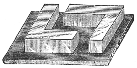
The specimen is now ready to be embedded. First make a
mould of cardboard or a lead-embedding frame (Fig. 32), melt
the paraffin, and then place the
specimen in a manner that will
facilitate cutting. Remove the
excess of paraffin and cut when
desired.
Fig. 32.—Paraffin Blocks
In using the collodion method
for embedding fibrous specimens,
as wood, bark, roots, etc.,
the specimen is first fixed with picric acid, washed with water,
cleared in ether-alcohol, embedded successively in two, five,
and twelve per cent ether-alcohol collodion solution, and finally
embedded in a pure collodion bath.
Specimens prepared as described above may be cut with a
hand microtome or a machine microtome.
HAND MICROTOME
In cutting sections by a hand microtome, it is necessary to
place the specimen, embedded in paraffin or held between
pieces of elder pith, carrot, or potato, over the second joints
of the fingers, then press the first joints firmly upon the specimen
with the thumb pressed against it. If they are correctly
held, the specimens will be just above the level of the finger and
the end of the thumb, and the joint will be below the level of
the finger.
Fig. 33.—Hand Microtome
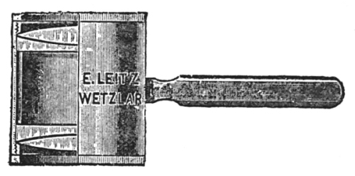
Hold the section cutter (Fig. 33) firmly in the hand with
the flat surface next to the specimen. While cutting the section,
press your arm firmly against your chest, and bend the
wrist nearly at right angles to the arm. Push the cutting edge
of the microtome toward the body and through the specimen
in such a way as to secure as thin a section as possible. Do
not expect to obtain nice, thin sections during the first or second
trials, but continued practice will enable one to become quite
efficient in cutting sections in this manner.
When the examination of drugs is a daily occurrence, the
above method will be found highly satisfactory.
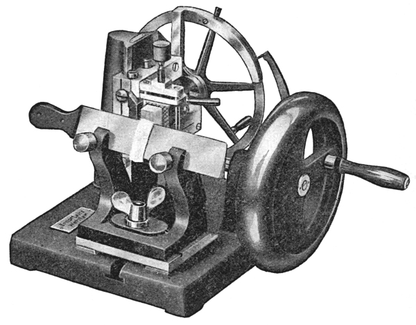
MACHINE MICROTOMES
When a number of sections are to be prepared from a given
specimen, it is desirable to cut the sections on a machine microtome,
particularly when the sections are to be prepared for the
use of students, in which case they should be as uniform as
possible.
Great care should be exercised in cutting sections with a
machine microtome—first, in the selection of the type of the
microtome; and secondly, in the style of knife used in cutting.
For soft tissues embedded in paraffin or collodion, the rotary
microtome with vertical knife will give best results. The thickness
of the specimen is regulated by mechanical means, so that
in cutting the sections it is only necessary to turn a crank and
remove the specimens from the knife-edge, unless there is a
ribbon-carrier attachment. If the sections are being cut from
a specimen embedded by the quick paraffin method, it is best
to drop the section in a metal cup partly filled with warm water.
This will cause the paraffin to straighten out, and the specimen
will uncoil. After sufficient specimens have been cut, the
cup should be placed in a boiling-water bath until the paraffin
surrounding the sections melts and floats on the water. Before
removing the specimen from the water-bath, it is advisable to
shake the glass vigorously in order to cause as many specimens
as possible to settle to the bottom of the cup. The cup is then
placed in iced water or set aside until the paraffin has solidified.
The cake-like mass is then removed from the cup, and the sections
adhering to its under surface are removed by lifting them
carefully off with the flat side of the knife and transferring them,
together with the sections at the bottom of the cup, to a wide-mouth
bottle, and covered with alcohol, glycerine, and water
mixture; or if it is desired to stain the specimens, they should
be placed in a weak alcoholic solution.
Specimens having a hard, woody texture should be cut on
a sliding microtome by means of a special wood knife, which
is especially tempered to cut woody substances. Woody roots,
wood, or thick bark may be cut readily on this microtome when
they have been embedded by the quick paraffin process. The
knife in the sliding microtome is placed in a horizontal position,
slanting so that the knife-edge is drawn gradually across the
specimen. After cutting, the sections are treated as described
above.
The thickness of the sections is regulated by mechanical
means. After a section has been cut, the block containing the
specimen is raised by turning a thumb-screw. In this microtome
the knife, as in the rotary type, is fixed, and the block containing
the specimen is movable.
If the specimen has been infiltrated with, and embedded in,
paraffin or collodion, the treatment of the sections after cutting
should be different.
In the case of paraffin, the sections are fastened directly to
the slide, and the paraffin is dissolved by either chloroform or
xylol. The specimen is then placed in 100, 95, and 45 per cent
alcohol, and then washed in water. These sections are now
stained with water-stains, brought back through alcohol, cleared,
and mounted in Canada balsam.
If alcoholic stains are used, it will not be necessary to dehydrate
before staining, and the dehydration after staining will
also be eliminated.
Sections infiltrated with collodion are either stained directly
without removing the collodion or after removal.
FORMS OF MICROTOMES
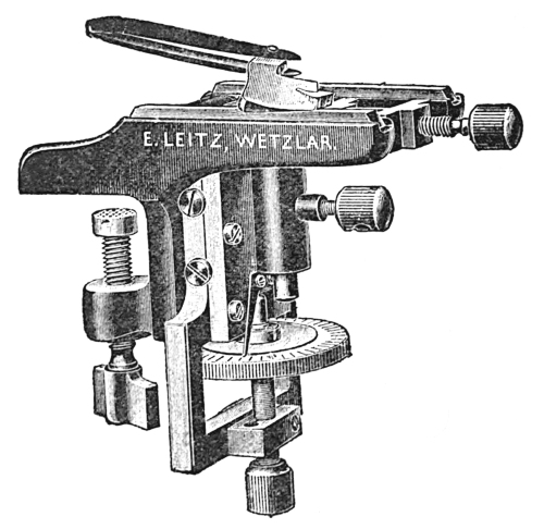
The hand cylinder microtome (Fig. 34) consists of a cylindrical
body. The clamp for holding the specimen is near the top
below the cutting surface. At the lower end is attached a micrometer
screw with a divided milled head. When moved forward
one division, the specimen is raised 0.01 mm. This micrometer
screw has an upward movement of 10 mm. The cutting surface
consists of a cylindrical glass ring.
Fig. 34.—Hand Cylinder Microtome
Fig. 35.—Hand Table Microtome
The hand table microtome (Fig. 35) is provided with a clamp,
by which it may be attached to the edge of a table or desk.
The cutting surface consists of two separated but parallel glass
benches. The object is held by a clamp and is raised by a
micrometer screw, which, when moved through one division by
turning the divided head, raises the specimen 0.01 mm.
The sliding microtome has a track of 250 mm. The object
is held by a clamp and its height regulated by hand. The disk
regulating the micrometer screw is divided into one hundred
parts. When this is turned through one division, the object is
raised 0.005 mm. or 5 microns, at the same time a clock-spring
in contact with teeth registers by a clicking sound. If the disk
is turned through two divisions, there will be two clicks, etc.
In this way is regulated the thickness of the sections cut. When
the micrometer screw has been turned through the one hundred
divisions, it must be unscrewed, the specimen raised, and the
steps of the process repeated. The knife is movable and is
drawn across the specimen in making sections.
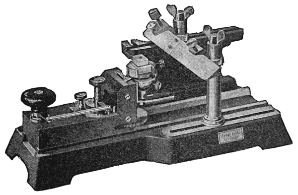
Fig. 36.—Base Sledge Microtome
The base sledge microtome (Fig. 36) has a heavy iron base
which supports a sliding-way on which the object-carrier moves.
The object-carrier is mounted on a solid mass of metal, and is
provided with a clamp for holding the object. The object is
raised by turning a knob which, when turned once, raises the
specimen one to twenty microns, according to how the feeding
mechanism is set.
Sections thicker than twenty microns may be obtained by
turning the knob two or more times. The knife is fixed and is
supported by two pillars, the base of which may be moved forward
or backward in such a manner that the knife can be
arranged with an oblique or right-angled cutting surface.
Fig. 37.—Minot Rotary Microtome
The Minot rotary microtome (Fig. 37) has a fixed knife, held
in position by two pillars, and a movable object-carrier. The
object is firmly secured by a clamp, and it is raised by a micrometer
screw. The screw is attached to a wheel having five
hundred teeth on its periphery. A pawl is adjusted to the teeth
in such a way that, when moved by turning a wheel to which
it is attached, specimens varying from one to twenty-five microns
in thickness may be cut, according to the way the adjusting disk
is set. When the mechanism has been regulated and the object
adjusted for cutting, it is only necessary to turn a crank in
cutting sections.
CARE OF MICROTOMES
When not in use, microtomes should be protected from dust,
and all parts liable to friction should be oiled.
Microtome knives should be honed as often as is necessary
to insure a proper cutting edge. After cutting objects, the
knives should be removed, cleaned, and oiled.
It should be kept clearly in mind that special knives are
required for cutting collodion, paraffin, and frozen and woody
sections. The cutting edges of the different knives vary considerably,
as is shown in the preceding cuts.
CHAPTER V REAGENTS — LIST OF REAGENTS
Little attention is given in the present work to micro-chemical
reactions for the reason that their value has been much overrated
in the past. A few reagents will be found useful, however,
and these few are given, as well as their special use. They are
as follows:
Distilled Water is used in the alcohol, glycerine, and water
mixture as a general mounting medium. It is used when warm
as a test for inulin and it is used in preparing various reagents.
Glycerine is used in preparing the alcohol, glycerine, and
water mixture, in testing for aleurone grains, and as a temporary
mounting medium.
Alcohol is used in preparing the alcohol, glycerine, and water
mixture, in testing for volatile oils.
Acetic Acid. Both dilute and strong solutions are used in
testing for aleurone grains, cystoliths, and crystals of calcium
oxalate.
Hydrochloric Acid is used in connection with phloroglucin
as a test for lignin and as a test for calcium oxalate.
Ferric Chloride Solution is used as a test for tannin.
Sulphuric Acid is used as a test for calcium oxalate.
Tincture Alkana is used when freshly prepared by macerating
the granulated root with alcohol and filtering, as a test
for resin.
Sodium Hydroxide. A five per cent solution is used as a
test for suberin and as a clearing agent.
Copper Ammonia is used as a test for cellulose.
Ammonical Solution of Potash is used as a test for fixed
oils. The solution is a mixture of equal parts of a saturated
solution of potassium hydroxide and stronger ammonia.
Oil of Cloves is used as a clearing fluid for sections preparatory
to mounting in Canada balsam.
Canada Balsam is used as a permanent mounting medium
for dehydrated specimens, and as a cement for ringing slides.
Paraffin is used for general embedding and infiltrating.
Lugol’s Solution is used as a test for starch and for aleurone
grains and proteid matters.
Osmic Acid. A two per cent solution is used as a test for
fixed oils.
Alcohol, Glycerine, and Water Mixture is used as a temporary
mounting medium and as a qualitative test for fixed oils.
Chlorzinc Iodide is used as a test for suberin, lignin, cellulose,
and starch.
Analine Chloride is used as a test for lignified cell walls of
bast fibres and of stone cells.
Phloroglucin. A one per cent alcoholic solution is used in
connection with hydrochloric acid as a test for lignin.
Hæmatoxylin-Delifields is used as a test for cellulose.
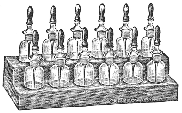
Fig. 38.—Reagent Set
REAGENT SET
Each worker should be provided with a set of reagent bottles
(Fig. 38). Such a set may be selected according to the taste
of the individual, but experience has shown that a 30 c.c. bottle
with a ground-in pipette and a rubber bulb is preferable to other
types. In such forms the pipettes are readily cleaned, and the
rubber bulbs can be replaced when they become old and brittle.
The entire set should be protected from dust by keeping it in a
case, the cover of which should be closed when the set is not
in use.
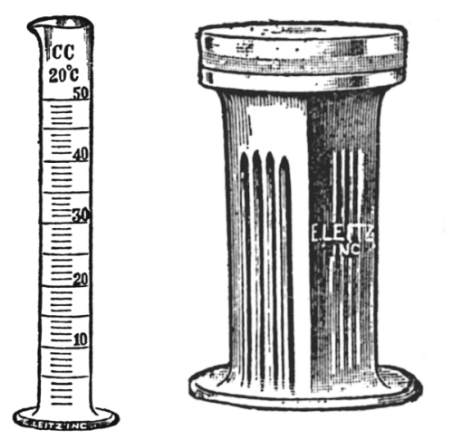
MEASURING CYLINDER
In order accurately to measure micro-chemical reagents, it
is necessary to have a standard 50 c.c. cylinder (Fig. 39) graduated
to c.c.’s. Such a cylinder should form a part of the reagent set.
Fig. 39.—Measuring CylinderFig. 40.—Staining Dish
STAINING DISHES
There is a great variety of staining dishes (Fig. 40), but
for general histological work a glass staining dish with groves
for holding six or more slides and a glass cover is most desirable.
CHAPTER VI HOW TO MOUNT SPECIMENS — TEMPORARY MOUNTS — PERMANENT MOUNTS — PRESERVATION OF MOUNTED SPECIMENS
The method of procedure in mounting specimens for study
varies according to the nature of the specimen, its preliminary
treatment, and the character of the mount to be made. As to
duration, mounts are either temporary or permanent.
In preparing a temporary mount, place the specimen in the
centre of a clean slide and add two or more drops of the temporary
mounting medium, which may be water, or a mixture
of equal parts of alcohol, glycerine, and water, or some micro-chemical
reagent, as weak Lugol’s solution, solution of chloral
hydrate, etc. Cover this with a cover glass and press down
gently. Remove the excess of the mounting medium with a
piece of blotting paper. Now place the slide on the stage and
proceed to examine it. Such mounts can of course be used only
for short periods of study; and when the period of observation
is finished, the specimen should be removed and the slide washed,
or the slide washing may be deferred until a number of such
slides have accumulated. At any rate, when the mounting
medium dries, the specimen is no longer suitable for observation.
Permanent mounts are prepared in much the same way as
temporary, but of course the mounting medium is different.
The kind of permanent mounting medium used depends upon
the previous treatment of the specimen. If the specimen has
been preserved in alcohol or glycerine and water, it is usually
mounted in glycerine jelly. If the specimen in question is a
powder, it is placed in the centre of the slide and a drop or two
of glycerine, alcohol, and water mixture added, unless the
powder was already in suspension in such a mixture. Cut a
small cube of glycerine jelly and place it in the centre of the
powder mixture. Lift up the slide by means of pliers, or grasp
the two edges between the thumb and finger and hold over a
small flame of an alcohol lamp, or place on a steam-bath until
the glycerine jelly has melted. Next sterilize a dissecting needle,
cool, and mix the powder with the glycerine jelly, being careful
not to lift the point of the needle from the slide during the
operation. If the mixing has been carefully done, few or no
air-bubbles will be present; but if they are present, heat the
needle, and while it is white hot touch the bubbles with its
point, and they will disappear. Now take a pair of forceps and,
after securing a clean cover glass near the edge, pass them three
times through the flame of the alcohol lamp. While holding it
in a slanting position, touch one side of the powder mixture and
slowly lower the cover glass until it comes in complete contact
with the mixture. Now press gently with the end of the needle-handle,
and set it aside to cool. When it is cool, place a neatly
trimmed label on one end of the slide, on which write the name
of the specimen, the number of the series of which it is to form
a part, etc. Any excess of glycerine jelly, which may have
been pressed out from the edges of the cover glass, should not
be removed at once, but should be allowed to remain on the
slide for at least one month in order to allow for shrinkage due
to evaporation. At the end of a month remove the glycerine
jelly by first passing the blade of a knife, held in a vertical
position, the back of the knife being next to the slide, around
the edge of the cover glass. After turning the knife-blade so
that the flat side is in contact with slide, remove the jelly outside
of the cover glass. Any remaining fragments should be removed
with a piece of old linen or cotton cloth. Finally, ring the edge
of the cover glass with microscopical cement, of which there
are many types to be had. If the cleaning has been done
thoroughly, there is no better ringing cement than Canada
balsam.
In mounting cross-sections, the method of procedure is
similar to the above, with the exception that the glycerine jelly
is placed at the side of the specimen and not in the centre.
While melting the jelly, incline the slide in order to allow the
melted glycerine jelly to flow gradually over the specimen, thus
replacing the air contained in the cells and intercellular spaces.
Finish the mounting as directed above, but under no conditions
should you stir the glycerine jelly with the section.
If specimens, after having been embedded in paraffin or
collodion, are cut, cleared, stained, and dehydrated, they are
usually mounted in Canada balsam. A small drop of this substance,
which may be obtained in collapsible tubes, is placed
at one side of the specimen. While inclining the slide, gently
heat until the Canada balsam covers the specimen. Secure a
cover glass by the aid of pliers, pass it through the flame three
times, and lower it slowly while holding it in an inclined position.
Press gently on the cover glass with the needle-handle, and keep
in a horizontal position for twenty-four hours, then place directly
in a slide box or cabinet, since no sealing is required.
Glycerine is sometimes used to make permanent mounts, but
it is unsatisfactory, because the cover glass is easily removed
and the specimen spoiled or lost, unless ringed—a procedure
which is not easily accomplished. If the specimen is to be
mounted in glycerine, it must first be placed in a mixture of
alcohol, glycerine, and water, and then transferred to glycerine.
Lactic acid is another permanent liquid-mounting medium,
which is unsatisfactory in the same way as glycerine, but like
glycerine, there are certain special cases where it is desirable
to use it. When this is used, the slides should be kept in a
horizontal position, unless ringed.
COVER GLASSES
Great care should be used In the selection of cover glasses,
however, not only as regards their shape but as to their thickness.
The standard tube length of the different manufacturers makes
an allowance of a definite thickness for cover glasses. It is
necessary, therefore, to use cover glasses made by the manufacturer
of the microscope in use.
Cover glasses are either square or round. Of each there are
four different thicknesses and two different sizes. The standard
thicknesses are:
The small size is designated three-fourths and the large
size seven-eighths.
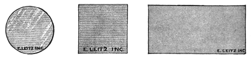
Cover glasses are circular (Fig. 41), square (Fig. 42), or
rectangular (Fig. 43) pieces of transparent glass used in covering
the specimens mounted on glass slides. A few years ago much
difficulty was experienced in obtaining uniformly thick and
transparent cover glasses, but no such difficulty is experienced
to-day. The type of cover glass used depends largely upon the
character of the specimen to be mounted. The square and
rectangular glasses are selected when a series of specimens are
to be mounted, but in mounting powdered drugs and histological
specimens the round cover glasses are preferable because they
are more sightly and more readily cleaned and rinsed.
GLASS SLIDES
Fig. 44.—Glass Slide
Glass slides (Fig. 44) are rectangular pieces of transparent
glass used as a mounting surface for microscopic objects. The
slides are usually three inches long by one inch wide, and they
should be composed of white glass, and they should have ground
and beveled edges. Slides should be of uniform thickness, and
they should not become cloudy upon standing.
SLIDE AND COVER-GLASS FORCEPS
Slides and cover glasses should be grasped by their edges.
To the beginner this is not easy. In order to facilitate holding
slides and cover glasses during the mounting process, one may
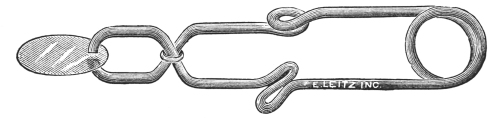
use a slide and a cover-glass forceps. The slide forceps consists
of wire bent and twisted in such a way that it holds a slide
firmly when attached to its two edges.
Fig. 45.—Histological Forceps
Fig. 46.—Forceps
Fig. 47.—Sliding-pin Forceps
There are various forms of cover-glass holders, but only two
types as far as the method of securing the cover glass is concerned.
First, there are the bacteriological and the histological
forceps (Fig. 45), which are self-closing. The two blades of such
forceps must be forced apart by pressure in securing the cover
glass. The second type of forceps is that in which the two
blades are normally separated (Fig. 46), it being necessary to
press the blades to either side of the cover glass in order to
secure and hold it. There is a modification of this type of
forceps which enables one to lock the blades by means of a sliding
pin (Fig. 47), after the cover glass has been secured. It is
well to accustom oneself to one type, for by so doing one may
become dexterous in its use.
NEEDLES
Fig. 48.—Dissecting Needle
Two dissecting needles (Fig. 48) should form a part of the
histologist’s mounting set. The handles may be of any material,
but the needle should be of tempered steel and about two inches
long.
SCISSORS
Fig. 49.—Scissors
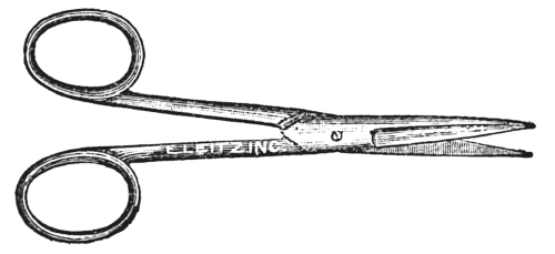
Almost any sort of scissors (Fig. 49) will do for histology
work, but a small scissors with fine pointed blades, are preferred.
Scissors are useful in trimming labels and in cutting strips of
leaves and sections of fibrous roots that are to be embedded
and cut.
SCALPELS
Fig. 50.—Scalpels
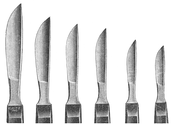
Scalpels (Fig. 50) have steel blades and ebony handles.
These vary in regard to size and quality of material. The
cheaper grades are quite as satisfactory, however, as the more
expensive ones, and for general use a medium-sized blade and
handle will be found most useful.
TURNTABLE
Fig. 51.—Turntable
Much time and energy may be saved by ringing slides on a
turntable (Fig. 51). There is a flat surface upon which to rest
the hand holding the brush with cement, and a revolving table
upon which the slide to be ringed is held by means of two clips.
In ringing slides, it is only necessary to revolve the table, and
at the same time to transfer the cement to the edge of the
cover glass from the brush held in the hand.
LABELING
There are many ways of labeling slides, but the best method
is to place on the label the name of the specimen, the powder
number, and the box, the tray or cabinet number. For
example:
Powdered Arnica Flowers
No. 80—Box A—600.
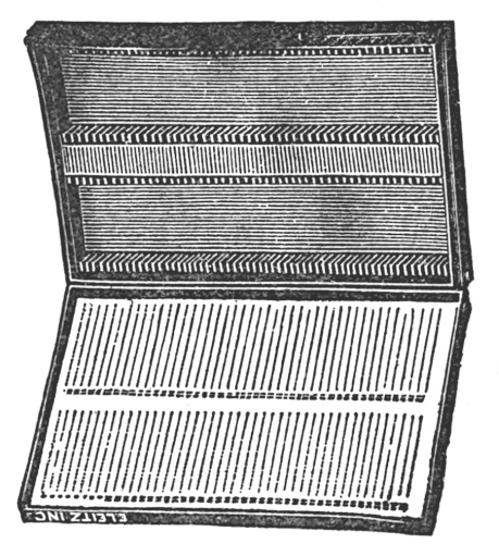
Fig. 52.—Slide Box
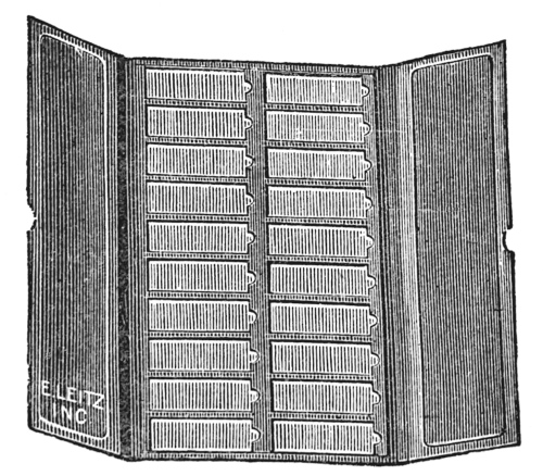
Fig. 53.—Slide Tray
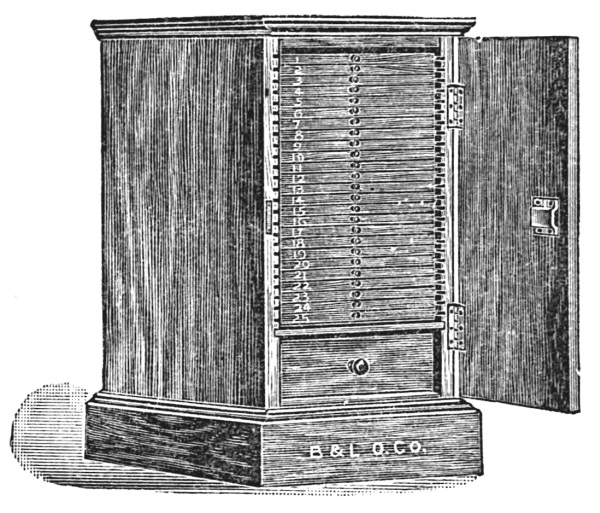
Accurately mounted, labeled, and ringed slides should be
filed away for future study and reference. Such filing may
be done in slide boxes, in slide trays, or in cabinets. Slide
boxes are to be had of a holding capacity varying from one to
one hundred slides. For general use, slide boxes (Fig. 52) holding
one hundred slides will be found most useful. Some workers
prefer trays (Fig. 53), because of the saving of time in selecting
specimens. Trays hold twenty slides arranged in two rows.
The cover of the tray is divided into two sections so that, if
desired, only one row of slides is uncovered at a time. Slide
cabinets (Fig. 54) are particularly desirable for storing large
individual collections, particularly when the slides are used
frequently for reference. Large selections of slides should be
numbered and card indexed in order to facilitate finding.
Fig. 54.—Slide Cabinet
Part II TISSUES CELLS, AND CELL CONTENTS
CHAPTER I THE CELL — TYPICAL CELL — CHANGES IN A CELL UNDERGOING DIVISION
The cell is the unit of structure of all plants. In fact the
cell is the plant in many of the lower forms—so called unicellular
plants. All plants, then, consist of one or more cells.
While cells vary greatly in size, form, color, contents, and
function, still in certain respects their structure is identical.
The typical vegetable cell is composed of a living portion or
protoplast and an external covering, or wall. The protoplast includes
everything within the wall. It is made up of a number
of parts, each part performing certain functions yet harmonizing
with the work of the cell as a whole. The protoplast (protoplasm)
is a viscid substance resembling the white of an egg.
The protoplast, when unstained and unmagnified, appears
structureless, but when stained with dyes and magnified, it is
found to be highly organized. The two most striking parts of
the protoplast are the cytoplasm and the nucleus. The part
of the protoplast lining the innermost part of the wall is the
ectoplast, which is less granular and slightly denser than most
of the cytoplasm. The cytoplasm is decidedly granular in
structure.
In the cytoplasm occurs one or more cavities, vacuoles, filled
with cell sap. Embedded in the cytoplasm are numerous
chromatophores, which vary in color in the different cells, from
colorless to yellow, to red, and to green. The nucleus is the
seat of the vital activity of the cell, and the seat of heredity.
The whole life and activity of the cell centre, therefore, in and
about the nucleus.
The outer portion of the nucleus
consists of a thin membrane
or wall. The membrane encloses numerous granular particles—chromatin—which
are highly susceptible to organic stains.
Among the granules are thread-like particles or linin. Near
the centre of the nucleus
are one or more small rounded nucleoli.
The liquid portion of the nucleus, filling the membranes and
surrounding the chromatin, linin, and nucleoli, is the nuclear
sap.
Other cell contents characteristic of certain cells are crystals,
starch, aleurone, oil, and alkaloids. The detailed discussion of
these substances will be deferred until a later chapter.
The cell wall which surrounds the protoplast is a product of
its activity. The structure and composition of the wall of any
given cell vary according to the ultimate function of the cell.
The walls may be thin or thick, porous or non-porous, and
colored or colorless. The composition of cell walls varies greatly.
The majority of cell walls are composed of cellulose, in other
cells of linin, in others of cutin, and in still others of suberin, etc.
In the majority of cells the walls are laid down in a series of
layers one over the other by apposition, similar to the manner
of building a pile of paper from separate sheets. The first layer
is deposited over the primary wall, formed during cell division;
to this is added another layer, etc. A modification of this
manner of growth is that in which the layers are built up one
over the other, but the building is gradually done by the deposit
of minute particles of cell-wall substance over the older deposits.
Such walls are never striated, as is likely to be the case
in cell walls formed by the first method. In other cells the walls
are increased in thickness by the deposition of new wall material
in the older membrane. The cell walls will be discussed more
fully when the different tissues are studied in detail.
INDIRECT CELL DIVISION (KARYOKINESIS)
The purpose of cell division is to increase the number of cells
of a tissue, an organ, an organism, or to increase the number of
organisms, etc. Such cell divisions involve, first, an equal
division of the protoplast and, secondly, the formation of a wall
between the divided protoplasts. The first changes in structure
of a cell undergoing division occur in the nucleus.
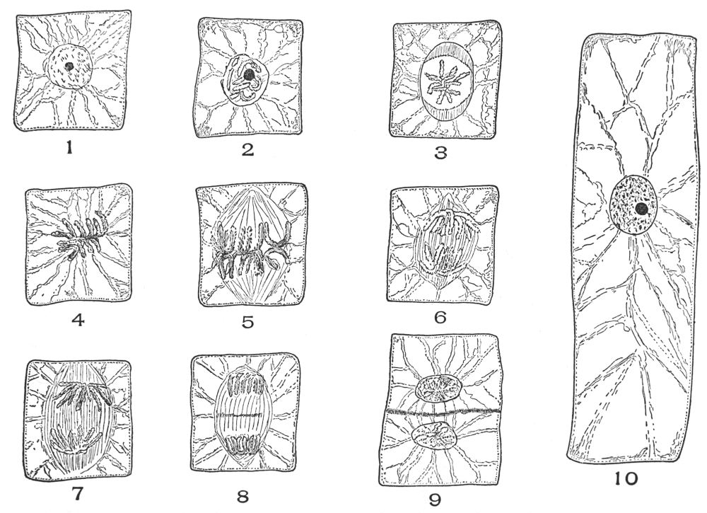
The linin threads become thicker and shorter. The chromatin
granules increase in size and amount; the threads and
chromatin granules separate into a definite number of segments
or chromosomes (Plate 1, Fig. 2). The nuclear membrane becomes
invested with a fibrous protoplasmic layer which later
separates and passes into either end of the cell, there forming
the polar caps (Plate 1, Fig. 3).
The nuclear membrane and the nucleoli disappear at about
this time. Two fibres, one from each polar cap, become attached
to opposite sides of the individual chromosomes. Other
fibres from the two polar caps unite to form the spindle fibres,
which thus extend from pole to pole. All these spindle fibres
form the nuclear spindle (Plate 1, Fig. 5).
The chromosomes now pass toward the division centre of
the cell or equatorial plane and form, collectively, the equatorial
plate (Plate 1, Fig. 5). At this point of cell division, the chromosomes
are U-shaped, and the curved part of the chromosomes
faces the equatorial plane. The chromosomes finally split into
two equal parts (Plate 1, Fig. 6). The actual separation of the
halves of chromosomes is brought about by the attached polar
fibres, which contract toward the polar caps (Plate 1, Fig. 7).
The chromosomes are finally drawn to the polar caps (Plate 1,
Fig. 8). The chromosomes now form a rounded mass. They
then separate into linin threads and chromatin granules. Nucleoli
reappear, and nuclear sap forms. Finally, a nuclear membrane
develops. The spindle fibres, which still extend from
pole to pole, become thickened at the equatorial plane (Plate 1,
Fig. 8), and finally their edges become united to form the cell-plate
(Plate 1, Fig. 9), which extends across the cell, thus completely
separating the mother cell into two daughter cells. After
the formation of the cell-plate, the spindle fibres disappear.
The cell becomes modified to form the middle lamella, on either
side of which the daughter protoplast adds a cellulose layer.
The ultimate composition of the middle lamella and the composition
and structure of the cell wall will differ according to
the function which the cell will finally perform.
PLATE 1
Nine figures, showing stages in the cell-division common to the onion root (Allium cepa, L.)
ORIGIN OF MULTICELLULAR PLANTS
All multicellular plants are built up by the repeated cell
division of one original cell. If the cells formed are similar in
structure and function, they form a tissue. In multicellular
plants many different kinds of tissues will be formed as a result
of cell division, since there are many different functions to be
performed by such an organism. When several of these tissues
become associated and their functions are correlated, they form
an organ. The association of several organs in one form makes
an organism. The oak-tree is an organism. It is made up of
organs known as flowers, leaves, stems, roots, etc. Each of
these organs is in turn made up of several kinds of tissue. In
some cases it is difficult to designate a single function to an
aggregation of cells (tissue). In fact, a tissue may perform
different functions at different periods of its existence or it
may perform two functions at one and the same time; as an
example, stone cells, whose primary function is mechanical, in
many cases function as storage tissue. The cells forming the
tissues of the plant, in fact, show great adaptability in regard
to the function which they perform. Nevertheless there is a
predominating function which all tissues perform, and the
structure of the cells forming such tissues is so uniform that it
is possible to classify them.
The functional classification of tissues is chosen for the
purpose of demonstrating the adaptation of cell structure to
cell function. If the cells performing a similar function in the
different plants were identical in number, distribution, form,
color, size, structure, and cell contents, there would not be a
science of histology upon which the art of microscopic pharmacognosy
is based. It may be said, however, with certainty,
that the cells forming certain of the tissues of any given species
of plant will differ in a recognizable degree from cells performing
a similar function in other species of plants. Often a tissue
is present in one plant but absent in another. For example,
many aquatic plants are devoid of mechanical fibrous cells.
The barks of certain plants have characteristic stone cells, while
in many other barks no stone cells occur. Many leaves have
characteristic trichomes; others are free from trichomes, etc.
Yet all cells performing a given function will structurally resemble
each other. In the present work the nucleus and other
parts of the living protoplast will not be considered, for the
reason that these parts are not in a condition suitable for study,
because most drugs come to market in a dried condition, a condition
which eliminates the possibility of studying the protoplast.
The general structure of the cells forming the different
tissues will first be considered, then their variation, as seen in
different plants, and finally their functions.
CHAPTER II THE EPIDERMIS AND PERIDERM — LEAF EPIDERMIS — TESTA EPIDERMIS — PLANT HAIRS ( TRICHOMES ) — FORMS OF HAIRS — PERIDERM — CORK PERIDERM — STONE CELL PERIDERM — PARENCHYMA AND STONE CELL PERIDERM
The epidermis and its modifications, the hypodermis and
the periderm, form the dermal or protective outer layer or layers
of the plant.
The epidermis of most leaves, stems of herbs, seeds, fruits,
floral organs, and young woody stems consists of a single layer
of cells which form an impervious outer covering, with the
exception of the stoma.
The cells of the epidermis vary in size, in thickness of the
side and end walls, in form, in arrangement, in character of
outgrowths, in the nature of the surface deposits, in the character
of wall—whether smooth or rough—and in size.
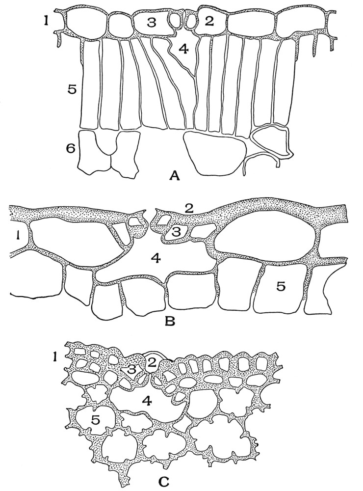
In cross-sections of the leaf the character of both the side
and end walls is easily studied.
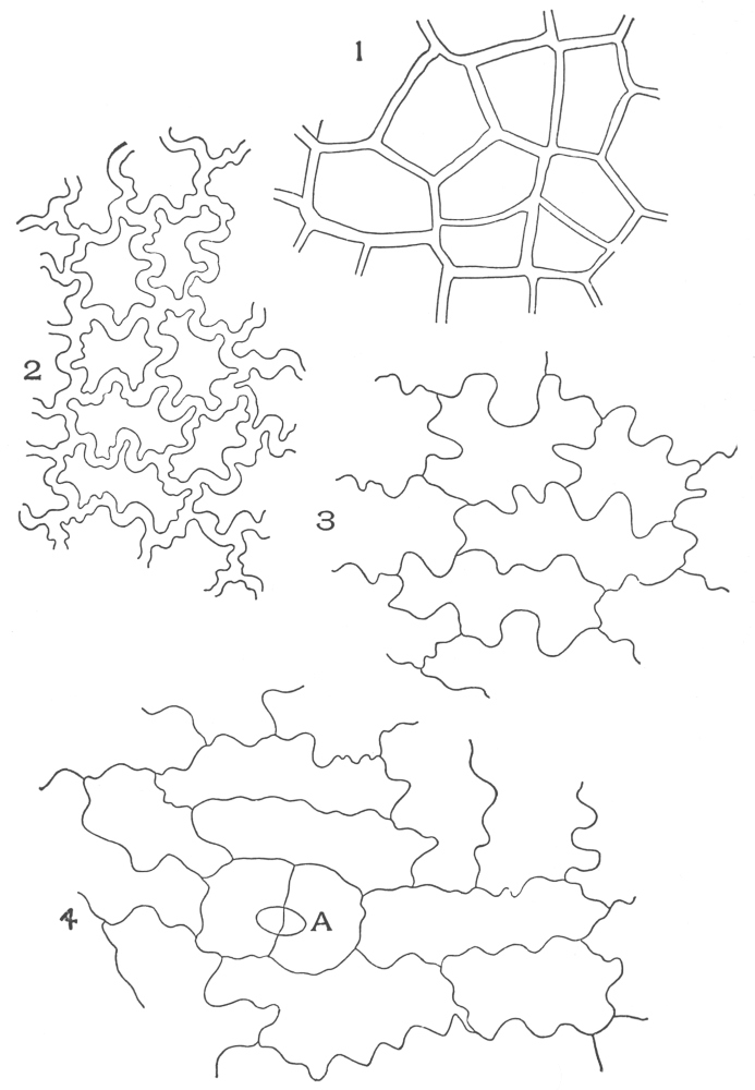
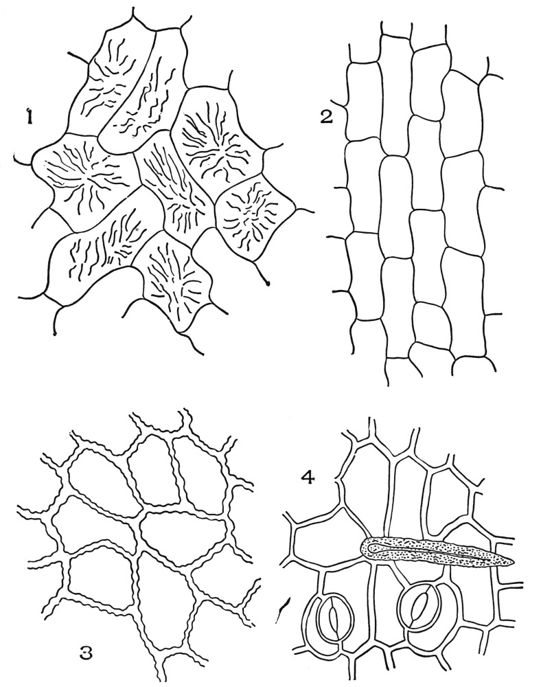
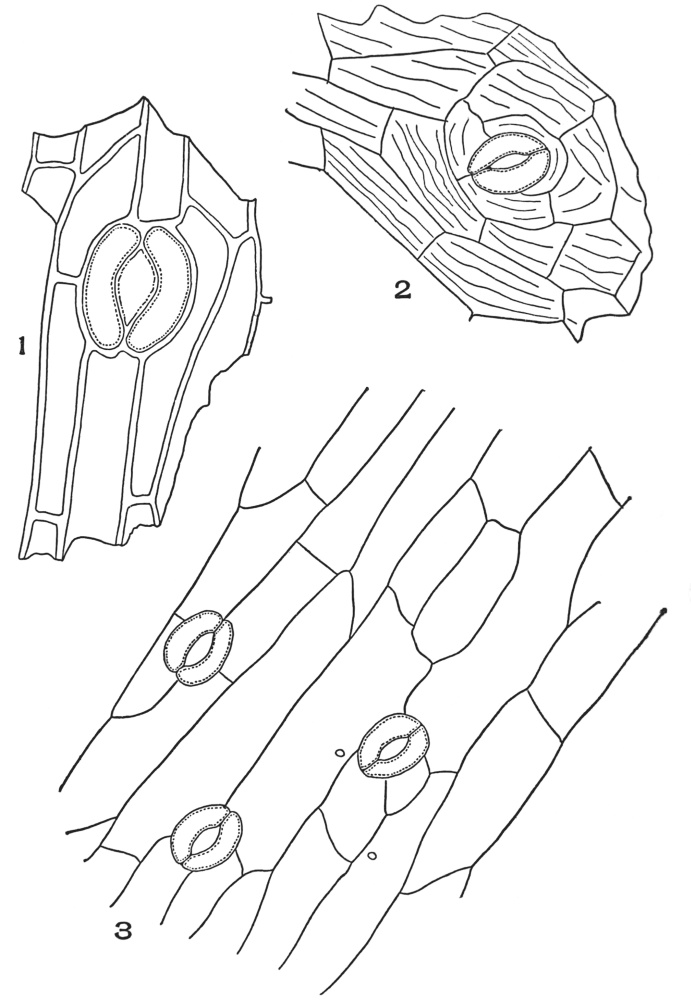
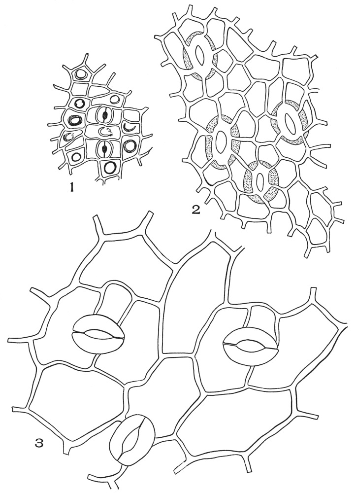
In surface sections—the view most frequently seen in powders—the
side walls are more conspicuous than the end wall
(Plates 2 and 3). This is so because the light is considerably retarded
in passing through the entire length of the side walls,
while the light is retarded only slightly in passing through the
end wall. The light in this case passes through the width
(thickness) of the wall only. The outer walls of epidermal cells
are characteristic only when they are striated, rough, pitted,
colored, etc. In the majority of leaves the outer wall of the
epidermal cells is not diagnostic in powders, or in surface
sections.
The thickness of the end and side walls of epidermal cells
differs greatly in different plants.
As a rule, leaves of aquatic and shade-loving plants, as well
as the leaves of most herbs have thinner walled epidermal cells
than have the leaves of plants growing in soil under normal
conditions, or than have the leaves of shrubs and trees.
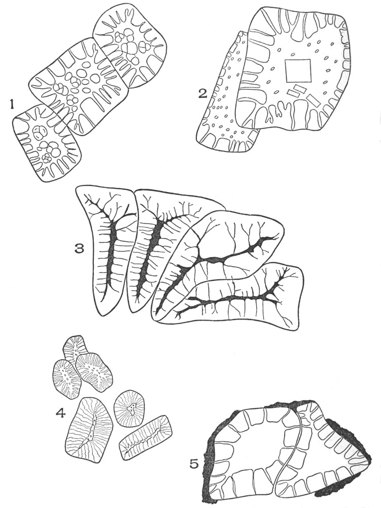
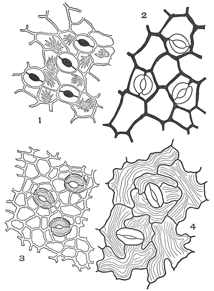
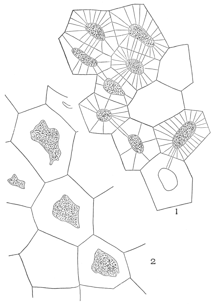
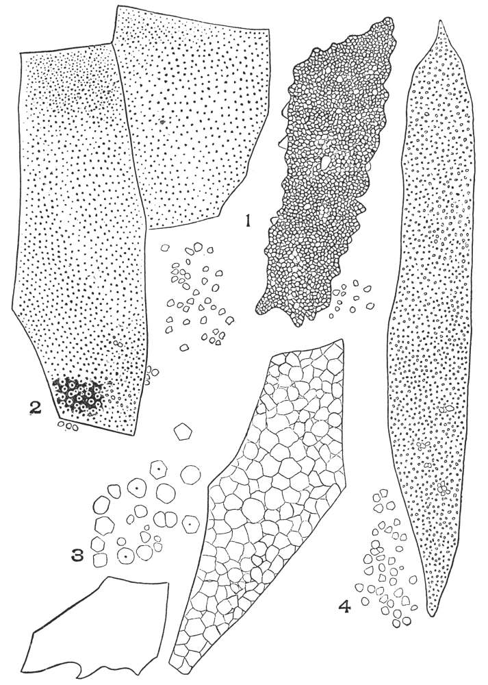
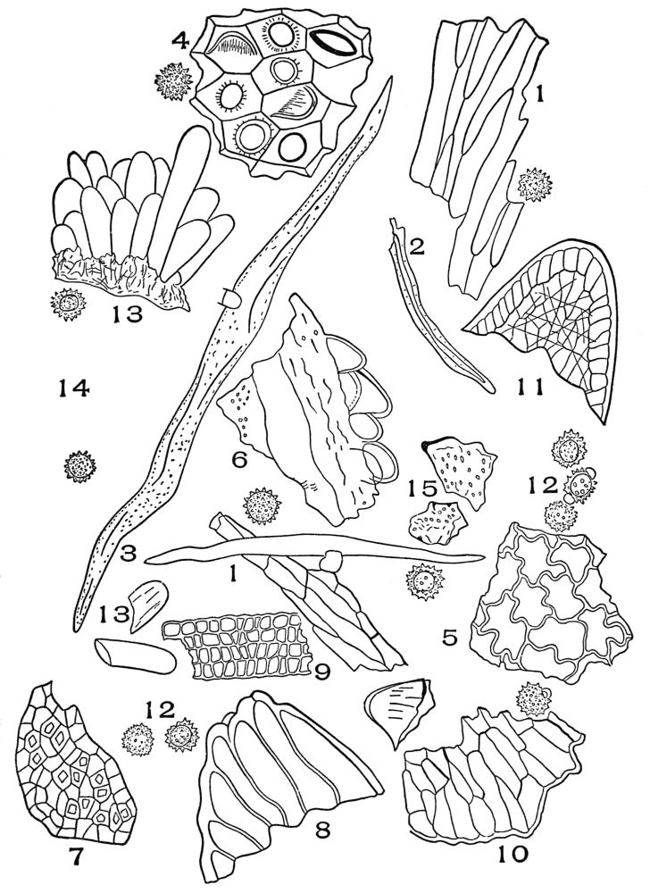
1. Upper striated epidermis of chirata leaf (Swertia chirata, [Roxb.] Ham.).
2. Green hellebore leaf (Veratrum viride, Ait.).
3. Boldus leaf (Peumus boldus, Molina).
4. Under epidermis of India senna (Cassia angustifolia, Vahl.).
The widest possible range of cell-wall thickness is therefore
found in the medicinal leaves, because the medicinal leaves are
collected from aquatic plants, herbs, shrubs, trees, etc.
The outer wall is always thicker than the side walls. Even
the side walls vary in thickness in some leaves, the wall next
to the epidermis being thicker than the lower or innermost
portion of the wall. Frequently the outermost part of the side
walls is unequally thickened. This is the case in the beaded
side walls characteristic of the epidermis of the leaves of laurus,
myrcia, boldus, and capsicum seed, etc. The thickness of the
side walls of the epidermal cells of most leaves varies in the
different leaves.
In most leaves there are five typical forms of arrangement of epidermal cells:
First, those over the veins which are elongated in the direction of
the length of the leaf; and, secondly, those on other parts of the
leaf which are usually several-sided and not elongated in any one
direction. If the epidermis of the leaf has stoma, then there is a
third type of arrangement of the epidermal cells around the stoma;
fourthly, the cells surrounding the base of hairs; and fifthly,
outgrowths of the epidermis, non-glandular and glandular hairs,
etc.
It should be borne in mind that in each species of plant the
five types of arrangement are characteristic for the species.
The character of the outer wall of the epidermal cells differs
greatly in different plants. In most cases the wall is smooth;
senna is an example of such leaves. In certain other leaves the
wall is rough, the roughness being in the form of striations.
In some cases the striations occur in a regular manner; belladonna
leaf is typical of such leaves. In other instances the wall
is striated in an irregular manner as shown in chirata epidermis.
Very often an epidermis is rough, but the roughness is not due
to striations. In these cases the epidermis is unevenly thickened,
the thin places appearing as slight depressions, the thick places
as slight elevations. Boldus has a rough, but not a striated
surface.
Surface deposits are not of common occurrence in medicinal
plants; waxy deposits occur on the stem of sumac, on a species
of raspberry, on the fruit of bayberry, etc. Resinous deposits
occur on the leaves and stems of grindelia species, and on yerba
santa.
In certain leaves there are two or three layers of cells beneath
the epidermis that are similar in structure to the epidermal cells.
These are called hypodermal cells, and they function in the
same way as the epidermal cells.
Hypodermal cells are very likely to occur on the margin of
the leaf. Uva-ursi leaf has a structure typical of leaves with
hypodermal marginal cells. Uva-ursi, like other leaves with
hypodermal cells has a greater number of hypodermal cells
at the leaf margin than at any other part of the leaf
surface.
The cutinized walls of epidermal cells are stained red with
saffranin.
Testa epidermal cells form the epidermal layers of such
seeds as lobelia, henbane, capsicum, paprika, larkspur, belladonna,
scopola, etc.
In surface view the end walls are thick and wavy in outline;
frequently the line of union—middle lamella—of two cells is indicated
by a dark or light line, while in others the wall between
two cells appears as a single wall. The walls are porous or
non-porous, and the color of the wall varies from yellow to
brown, to colorless. These cells always occur in masses, composed
partially of entire and partially of broken fragments.
In lobelia seed (Plate 4, Fig. 2) the line of union of adjacent
cell walls appears as a dark line. The walls are wavy in outline,
of a yellowish-red color and not porous.
In henbane seed (Plate 4, Fig. 3) the line of union between
the cells is scarcely visible; the walls are decidedly wavy, more
so than in lobelia, and no pits are visible.
In capsicum seed (Plate 4, Fig. 1) the cells are very wavy
and decidedly porous, the line of union between the cell walls
being marked with irregular spaces and lines.
In belladonna seed (Plate 5, Fig. 1) the walls between two
adjacent cells are non-striated and non-porous, and extremely
irregular in outline.
In star-anise seed (Plate 5, Fig. 2) the walls are irregularly
thickened and wavy in outline.
In stramonium seed (Plate 5, Fig. 3) the walls are very
thick, wavy in outline, and striated.
In histological work plant hairs are of great importance, as
they offer a ready means of distinguishing and differentiating
between plants, or parts of plants, when they occur in a broken
or finely powdered condition. There is no other element in
powdered drugs which is of so great a diagnostic value as the
plant hair. The same plant will always have the same type
of hair, the only noticeable variation being in the size. In
microscopical drug analysis the presence of hairs is always noted,
and in many cases the purity of the powder can be ascertained
from the hairs. Botanists seem to have given little attention to
the study of plant hairs. This accounts for the fact that information
concerning them is very meagre in botanical literature,
and, as far as the author can learn, no one has attempted to
classify them. In systematic work, plant hairs could be used
to great advantage in separating genera and even species.
Hairs are, of course, a factor now in systematic work. The
lack of hairs is indicated by the term glabrous. Their presence
is indicated by such terms as hispid, villous, etc. In certain
cases the term indicates position of the hair as ciliate when the
hair is marginal. When hairs influence the color of the leaf,
such terms as cinerous and canescent are used. In all the cases
cited no mention is made of the real nature of the hair.
In systematic work, as in pharmacognosy, we must work
with dried material, and it is only those hairs which retain
their form under such conditions which are of classification
value.
Hairs are the most common outgrowths of the epidermal
cells. They are classified as glandular or non-glandular, according
to their structure and function. The glandular hairs will
be considered under synthetic tissue.
Each group is again subdivided into a number of secondary
groups, depending upon the number of cells present, their form,
their arrangement, their size, their color, the character of their
walls, whether rough or smooth, whether branched or non-branched,
whether curved, twisted, straight, or twisted and
straight, whether pointed, blunt, or forked.
PAPILLÆ
Papillæ are epidermal cells which are extended outward in
the form of small tubular outgrowths.
Papillæ occur on the following parts of the plant: flower-petals,
stigmas, styles, leaves, stems, seeds, and fruits. Papillæ
occur on only a few of the medicinal leaves.
The under surface of both Truxillo (Plate 6, Fig. 3) and
Huanuca coca have very small papillæ. The outermost wall of
these papillæ are much thicker than the side walls. The papillæ
of klip buchu (Plate 6, Fig. 4), an adulterant of true buchu, has
large thick-walled papillæ.
The velvety appearance of most flower-petals (Plate 6,
Figs. 2 and 5) is due to the presence of papillæ. The papillæ
of flower-petals are very variable. In calendula flowers (Plate
6, Fig. 1) they are small, yellowish in color, and the outer wall
is marked with parallel striations which appear as small teeth
in cross-section. The ray petal papillæ of anthemis consist of
rather large, broad, blunt papillæ with slightly striated walls.
The papillæ of the ray petals of the white daisy consist of papillæ
which have medium sized, cone-shaped papillæ with finely striated
walls. The papillæ of the flower stigma vary greatly in different
flowers. In some cases two or more types of papillæ occur,
but even in these cases the papillæ are characteristic of the
species.
The papillæ differ greatly in the case of the flowers of the
compositæ, where two types of flowers are normally present—namely,
the ray flowers and the disk flowers.
In all cases observed the papillæ of the stigma of the ray
flowers are always smaller than the papillæ of the stigma of the
disk flowers. It would appear from extended observation that
the papillæ of the ray flower stigma are being gradually aborted.
The papillæ of the style are always different from the papillæ
of the stigma. The style papillæ are always smaller, and they
are of a different form.
PLATE 6 Papillæ
1. Calendula flowers (Calendula officinalis, L.).
2. White daisy ray flower (Chrysanthemum leucanthemum, L.).
3. Coca leaf (Erythroxylon coca, Lamarck).
4. Klip buchu.
5. Anthemis ray petal (Anthemis nobilis, L.).
UNICELLULAR NON-GLANDULAR HAIRS
True plant hairs are tubular outgrowths of the epidermal
cell, the length of these outgrowths being several times the
width of the hair.
The unicellular hairs are common to many plants. The two
groups of non-glandular unicellular hairs are, first, the solitary;
and secondly, the clustered hairs.
Solitary unicellular hairs occur on the leaves of chestnut,
yerba santa, lobelia, cannabis indica, the fruit of anise, and
the stem of allspice, senna, and cowage.
Chestnut hairs (Plate 7, Fig. 1) have smooth yellowish-colored
walls, and the cell cavity contains reddish-brown tannin. These
hairs occur solitary or clustered; the clustered hairs normally
occur on the leaf, but in powdering the drug, individual hairs of
the cluster become separated or solitary.
Yerba santa hairs (Plate 7, Fig. 4) are twisted, the lumen or
cell cavity is very small, and the walls, which are very thick,
are grayish-white.
Lobelia hairs (Plate 7, Fig. 5) are very large. The walls
are grayish-white, and the outer surface extends in the form
of small elevations which make the hair very rough. The hair
tapers gradually to a solid point.
Cannabis indica hairs (Plate 7, Fig. 6) are curved. The
apex tapers to a point and the base is broad, and it frequently
contains deposits of calcium carbonate. The walls are grayish-white
in appearance, and rough. The roughness increases
toward the apex.
The hairs of anise (Plate 7, Fig. 7) are mostly curved; the
walls are thick, yellowish-white, and the outer surface is rough;
this is due to the numerous slight centrifugal projections of the
outer wall.
Allspice stem hairs (Plate 7, Fig. 2) have smooth walls.
The cell cavity is reddish-brown. The hair is curved.
The hair of senna (Plate 7, Fig. 10) is light greenish-yellow
with rough papillose walls. The hair is usually curved and
tapering, and it does not have any characteristic cell contents.
1. and 2. European oak (Quercus infectoria, Olivier).
3. Kamala (Mallotus philippinensis, [Lam.] [Muell.] Arg.).
4. Witch-hazel leaf (Hamamelis virginiana, L.).
5. Althea leaf (Althæa officinalis, L.).
Cowage hairs (Plate 7, Fig. 3) are lance-shaped, and they
terminate in a sharp point. The outer wall contains numerous
recurved teeth-like projections. The cell cavity is filled with
a reddish-brown contents which are somewhat fissured.
Clustered unicellular hairs occur on the leaves of chestnut,
witch-hazel, althea, European oak, etc. In European oak (Plate
8, Figs. 1 and 2) clusters of two and three hairs occur. The
walls are yellowish-white, smooth, and the tip of the hair is solid.
In kamala (Plate 8, Fig. 3) clusters of seven or more hairs
occur; the walls are yellowish, and the cell cavity is reddish-brown.
In witch-hazel leaf (Plate 8, Fig. 4) clusters of a variable
number of hairs occur. The hairs, which are of various lengths,
have yellowish-white, thick, smooth walls, and reddish cell
contents.
In althea leaf (Plate 8, Fig. 5) the hairs are nearly straight
and the walls are smooth. The basal portions of the hair are
strongly pitted.
Branched solitary unicellular hairs occur on the leaves of
hesperis matronalis (Plate 7, Fig. 8), and on galphimia glauca
(Plate 7, Fig. 9).
The hair of hesperis matronalis has smooth walls, and the
two branches grow out nearly parallel to the leaf surface.
The hair of galphimia glauca has rough walls, and the two
branches grow upward in a bifurcating manner.
MULTICELLULAR HAIRS
Multicellular hairs are divided into the uniseriate and the
multiseriate hairs. Both of these groups are divided into the
branched and the non-branched hairs, as follows:
1. Uniseriate. (A)Non-branched. (B)Branched.
2. Multiseriate. (A)Non-branched. (B)Branched.
Multicellular uniseriate non-branched hairs occur on the
leaves of digitalis, Western and Eastern skullcap, peppermint,
thyme, yarrow, arnica flowers, and sumac fruit.
Digitalis hairs (Plate 9, Fig. 1) are made up of a varying
number of uniseriate-arranged cells of unequal length, frequently
placed at right angles to the cells above and below; the walls
are of a whitish color, and are rough or smooth.
Eastern skullcap (Plate 9, Fig. 4) has hairs with not more
than four cells; these hairs are curved, and the walls are whitish,
sometimes smooth, but usually rough. In Western skullcap
(Plate 9, Fig. 3) the hairs have sometimes as many as seven
cells. The walls are white and rough, and the individual cells
of the hair are much larger than are the cells of the hairs of
true skullcap.
Peppermint (Plate 9, Fig. 5) has from one to eight cells.
The hair is curved, and the walls are very rough.
Thyme (Plate 9, Fig. 6) has short, thick, rough-walled
trichomes, the terminal cell usually being bent at nearly right
angles to the other cells.
Yarrow hairs (Plate 9, Fig. 7) have a variable number of
cells. In all the hairs the basal cells are short and broad, while
the terminal cell is greatly elongated.
Arnica hairs (one form, Plate 9, Fig. 2) have frequently as
many as four cells, the terminal cell being longer than the basal
cells. The walls are white and smooth.
Sumac-fruit hairs (Plate 9, Fig. 9) have spindle-shaped,
reddish-colored hairs.
Multicellular multiseriate non-branched hairs occur on
cumin fruit and on the tubular part of the corolla of calendula.
The hairs on cumin fruit vary considerably in size. All the
hairs are spreading at the base and blunt or rounded at the apex.
The cells forming the hair are narrow and the walls are thick.
Three differently sized hairs are shown in Plate 10, Fig. 1.
The hairs of the base of the ligulate petals of calendula
(Plate 10, Fig. 2) are biseriate. The hairs are very long and
the walls are very thin.
Multicellular uniseriate branched hairs occur on the leaves
of dittany of Crete, mullen, and on the calyx of lavender flowers.
The dittany of Crete (Plate 11, Fig. 3) hair is smooth-walled,
and the branches are alternate.
In mullen (Plate 11, Fig. 1) the hairs have whorled branches,
the walls are smooth, and the cell cavity usually contains air.
1. Mullen leaf (Verbascum thapsus, L.).
2. Lavender flowers (Lavandula vera, D. C.).
3. Dittany of Crete (Origanum dictamnus, L.).
The lavender hairs (Plate 11, Fig. 2) have mostly opposite
branches, and the walls are rough. Thus the multicellular
branched hairs may be divided into subgroups which have
alternate, opposite, whorled, or in certain hairs irregularly arranged
branches. Each class may be again subdivided according
to color, character of cell termination, etc., as cited at the
beginning of the chapter.
Occasionally multicellular hairs assume the form of a shield
(Plate 12, Fig. 1); in such cases the hair is termed peltate, as in
the non-glandular multicellular hair of shepherdia canadensis.
Hairs grow out from the surface of the epidermis in a perpendicular,
a parallel, or in an oblique direction. Hairs which
grow parallel or oblique to the surface are usually curved, and
the outer curved part of the wall is usually thicker than the
inner curved wall.
The mature hairs of some plants consist of dead cells. In
other plants the cells forming the hair are living. When dried,
those hairs, which were dead before drying, contain air; while
those hairs which were living before drying, show great variation
in color and in the nature of the cell contents. The contents
are either organic or inorganic. The commonest organic constituent
is dried protoplasm. In cannabis indica are deposits
of calcium carbonate.
Multicellular multiseriate branched hairs are the ultimate
division of the pappus of erigeron, aromatic goldenrod, arnica,
grindelia, boneset, and life-everlasting.
The hairs of erigeron (Plate 13, Figs. 1 and 2) are slender;
the walls are porous. Each hair terminates in two cells, which
are greatly extended and sharp-pointed; the branches from the
basal part of the hairs (Plate 13, Fig. 1) are of about the same
length as the apical branches.
The hairs of aromatic goldenrod (Plate 13, Figs. 3 and 4) are
larger than those of erigeron; the diameter is greater and the
walls are non-porous. The apex of the hair terminates in a
group of about four cells of unequal length, which are sharp-pointed.
The branches of the basal cells (Plate 13, Fig. 3) are
similar to the branches of the apical cells.
The hairs of arnica (Plate 14, Figs. 1 and 2) have thick,
strongly porous walls; the branches terminate in sharp points.
The apex of the hair terminates in a single cell. The basal
branches (Plate 14, Fig. 2) are much longer than special
branches.
1. Basal hairs of erigeron (Erigeron canadensis, L.).
2. Apical hairs of erigeron (Erigeron canadensis, L.).
3. Basal hairs of aromatic goldenrod (Solidago odora, Ait.).
4. Apical hairs of aromatic goldenrod (Solidago odora, Ait.).
The hair of grindelia (Plate 14, Figs. 3 and 4) has very thick
walls with numerous elongated pores. The apex of the hair
terminates in a cluster of cells with short, free, sharp-pointed
ends. The basal branches (Plate 14, Fig. 4) are longer than
the apical branches.
Boneset hair (Plate 15, Figs. 1 and 2) has non-porous walls.
The apex of the hair terminates in two blunt-pointed cells.
The terminal wall is thicker than the side wall. Some of the
branches lower down terminate in cells with very thick or solid
points. The basal branches (Plate 15, Fig. 1) are longer, but
the cells are narrower and more strongly tapering than are the
branches of the apical part of the hair.
Life-everlasting (Plate 15, Figs. 3 and 4) has uniformly
thickened but non-porous walls. The hair terminates in two
blunt-pointed, greatly elongated cells.
The basal branches (Plate 15, Fig. 4) are narrower, slightly
tapering, and the base of the branches frequently curve downward.
The cell cavities of these hairs are filled with air.
The walls of hairs are composed of cutin, of lignin, and
of cellulose.
The periderm is the outer protective covering of the stems
and roots of mature shrubs and trees. The periderm replaces
the epidermis. The periderm may be composed of cork cells,
stone cell-cork, or a mixture of cork, parenchyma, fibres, stone
cells, etc.
The typical periderm is made up of cork cells. Cork cells
vary in appearance, according to the part of the cell viewed.
On surface view (Plate 16, Fig. A) the cork cells are angled
in outline and are made up of from four to seven side walls;
five- and six-sided cells are more common than the four-and
seven-sided cells. Surface sections of cork cells show their
length and width. These side walls usually appear nearly white,
while the end wall, particularly of the outermost cork cells,
usually appears brown or reddish-brown, or in some cases nearly
black.
Cork cells on cross-section are rectangular in form, and they
are arranged in superimposed rows, the number of rows being
gradually increased as the plant grows older. Such an increase
in the number of rows of cork cells is shown in the cross-section
of cascara sagrada (Plate 16, Fig. C).
Cork cells fit together so closely that there is no intercellular
spaces between the cells. In this case two rows of cork cells
occupy no greater space than the solitary row of cork cells
immediately over and external to them. As a rule, the outermost
layers of cork cells have a narrower radial diameter than
the cork cells of the underlying layers. This is due to the fact
that these outer cells are stretched as the stem increases in
diameter. This view shows the height of cork cells, but not
always the length, which will, of course, vary according to the
part of the cell cut across. In a section a few millimeters in
diameter, however, all the variations in size may be observed.
The color of the walls is nearly white.
The cavity may contain tannin or other substances. When
tannin is present, the cavity is of a brownish or brownish-red
color, or it may be nearly black. Most barks appear devoid of
any colored or colorless cell contents.
The radial section (Plate 16, Fig. B) of cork cells shows the
height of the cells and the width of the cells at the point cut
across. Some cells will be cut across their longest diameter, while
others will be cut across their shortest diameter. Cork cells are,
therefore, smaller in radial section than they are in cross-section.
The color of the walls is white, and the color and nature of the
cell contents vary for the same reasons that they vary in
cross-sections.
The number of layers of cork cells occurring in cross- and
radial-sections varies according to the age of the plant, to the type
of plant, and to the conditions under which the plant is growing.
The number of layers of cork cells is not of diagnostic importance,
nor is the surface view of cork cells diagnostic except
in certain isolated cases.
PLATE 16 Periderm of Cascara Sagrada (Rhamnus purshiana, D.C.)
A. 1, Outline of cork cells; 2, Line of contact of adjoining cork cells. B. Radial longitudinal section of cascara sagrada. 1, Cork cells; 2, Phellogen; 3, Forming parenchyma cells; 4, Cortical parenchyma cells. C. Cross-section of cascara sagrada. 1, Cork cells; 2, Phellogen; 3, Forming parenchyma cells; 4, Cortical parenchyma cells.
The presence or absence of cork or epidermal tissue in powders
must always be noted. The presence of cork enables one to
distinguish Spanish from Russian licorice. In like manner, the
presence of epidermis enables one to distinguish the pharmacopœial
from the unofficial peeled calamus. The absence of
epidermis in Jamaica ginger is one of the means by which this
variety is distinguished from the other varieties of ginger, etc.
In canella alba the periderm is replaced by stone cell-cork.
That is, the cells forming the periderm are of a typical cork
shape, but the walls are lignified, unequally thickened, and
the inner or thicker walls are strongly porous, and the walls
are of a yellowish color. Stone cell-cork forms the periderm
of clove bark also, but the cells are narrower and longer, and
the inner wall is not so thick or porous as is the case in canella
alba bark.
In canella alba (Plate 17, Fig. B) cork periderm is frequently
replaced by stone cells, particularly in the older barks. These
stone cells form the periderm because they replace the cork
periderm, which fissures and scales off as the root increases
in diameter.
The side and end walls of cork cells are of nearly uniform
diameter. Exceptions occur, but they are not common. In
buchu stem (Plate 101, Fig. 3), the cork cells have thick outer
walls, but thin sides and inner walls. The cell cavity contains
reddish-brown deposits of tannin.
As the trees and shrubs increase in diameter, cracks or fissures
occur in the periderm, or corky layer. In such cases the
phellogen cells divide and redivide in such manner as to cut
off a portion of the parenchyma cells, stone cells, and fibres of
the cortex which is inside of and below the fissure. All the
parenchyma cells, etc., exterior to the newly formed cork cells
soon lose their living-cell contents, since their food-supply is
cut off by the impervious walls of the cork cells. In time they
are forced outward by the developing cork cells until they
partially or completely fill the break in the periderm. In white
oak bark (Plate 18), as in other barks, a large part of the periderm
is composed of dead and discolored cortical cells.
PLATE 17
A. Cross-section of Mandrake Rhizome (Podophyllum peltatum, L.). 1. Epidermis. 2. Phellogen. 3. Cortical parenchyma. B. Stone cell periderm of white cinnamon (Canella alba, Murr.).
PLATE 18 Periderm of White Oak (Quercus alba, L.)
1. Outer layer of cork cells. 2. Cortical parenchyma cells. 3. Stone cells. 4. Phellogen. 5. Cortical parenchyma cells.
ORIGIN OF CORK CELLS
The cork cells are formed by the meristimatic phellogen
cells, which originate from cortical parenchyma. These cells
divide into two cells, the outer changing into a cork cell, while
the inner cell remains meristimatic. In other instances the
outer cell remains meristimatic, while the inner cell changes
into a cortical parenchyma cell. The development of a cortical
parenchyma cell from a divided phellogen cell is shown in Plate
101, Fig. 6. Both the primary and secondary cork cells originate
from the phellogen or cork cambrium layer. Cork cells do not
contain living-cell contents; in fact, in the majority of medicinal
barks the cork cells contain only air.
The walls of typical cork cells are composed, at least in part,
of suberin, a substance which is impervious to water and gases.
In certain cases layers of cellulose, lignin, and suberin have been
identified. Suberin, however, is present in all cork cells, and
in some cases all of the walls of cork cells are composed of suberin.
Suberized cork cells are colored yellow with strong sodium
hydroxide solutions and by chlorzinciodide.
The mechanical tissues of the plant form the framework
around which the plant body is built up. These tissues are
constructed and placed in such a manner in the different organs
of the plant as to meet the mechanical needs of the organ. Many
underground stems and roots which are subjected to radial pressure
have the hypodermal and endodermal cells arranged in the
form of a non-compressible cylinder. Such an arrangement is
seen in sarsaparilla root (Plate 38, Fig. 4). The mechanical
tissue of the stem is arranged in the form of solid or hollow
columns in order to sustain the enormous weight of the branches.
In roots the mechanical tissue is combined in ropelike strands,
thereby effectively resisting pulling stresses. The epidermis of
leaves subjected to the tearing force of the wind has epidermal
cells with greatly thickened walls, particularly at the margin of
the leaf. The epidermal cells of most seeds have very thick
and lignified cell walls, which effectively resist crushing forces.
The cells forming mechanical tissues are: bast fibres, wood
fibres, collenchyma cells, stone cells, testa epidermal cells, and
hypodermal and endodermal cells of certain plants. The walls
of the cells forming mechanical tissues are thick and lignified,
with the exception of the collenchyma cells and a few of the
fibres. Lignified cells are as resistive to pulling and other
stresses as similar sized fragments of steel. The hardness of
their wall and their resistance to crushing explain the fact that
they usually retain their form in powdered drugs and foods.
One of the most important characters to be kept in mind in
studying bast fibres is the structure of the wall. In fact, the
author’s classification of bast fibres is based largely on wall
structure. Such a classification is logical and accurate, because
it is based upon permanent characters. Another character used
in classifying bast fibres is the nature of the cell, whether branched
or non-branched. In fact, this latter character is used to separate
all bast fibres into two fundamental groups—namely, branched
bast fibres and non-branched bast fibres. The third important
character utilized in classifying fibres is the presence or absence
of crystals.
Bast fibres are classified as follows: 1. Crystal bearing. 2. Non-crystal bearing.
The crystal-bearing fibres are divided into two classes: 1. Of leaves. 2. Of barks.
The non-crystal bearing are divided into: 1. Branched. 2. Non-branched.
The branched and non-branched are divided into four classes: 1. Non-porous and non-striated. 2. Porous and non-striated. 3. Striated and non-porous. 4. Porous and striated.
CRYSTAL-BEARING BAST FIBRES
The crystal-bearing fibres are composed (1) of groups of
fibres, (2) of crystal cells, and (3) of crystals. In these cases
the groups of fibres are large, and they are frequently completely
covered by crystal cells, which may or may not contain a crystal.
The crystals found on the fibres from the different plants vary
considerably in size and form. As a rule, the fibres when separated
are free of crystal cells and crystals. This is so because
the crystal cells are exterior to the fibres, and in separating the
fibres during the milling process the crystal cells are broken down
and removed from the fibres. It is common, therefore, to find
isolated fibres and crystals associated with the crystal-bearing
fibres. The fibres which are crystal-bearing may be striated
or porous, etc.; but owing to the fact that the grouping of the
fibres and crystals is so characteristic, little or no attention is
paid to the structure of the individual fibres.
Crystal-bearing fibres occur in the barks of frangula (Plate
19, Fig. 1); cascara sagrada (Plate 19, Fig. 2); witch-hazel
(Plate 19, Fig. 4); in cocillana (Plate 20, Fig. 1); in white oak
(Plate 20, Fig. 2); in quebracho (Plate 20, Fig. 3); and in
Spanish licorice root (Plate 19, Fig. 3).
The crystal-bearing fibres of leaves are always associated
with vessels or tracheids and with cells with chlorophyl. The
presence or absence of crystal-bearing fibres in leaves should
always be noted. The crystal-bearing fibres of leaves are
composed of fragments of conducting cells, fibres, crystal cells,
and crystals. The crystal-bearing fibres of leaves occur in
larger fragments than the other parts of the leaf, because the
fibres are more resistant to powdering. Having observed that
a leaf has crystal-bearing fibres, in order to identify the powder
it is necessary to locate one of the other diagnostic elements
of the leaf—as the papillæ of coca (Plate 21, Fig. 1), or the hair
of senna (Plate 21, Fig. 3), or the vessels in eucalyptus (Plate 21,
Fig. 2).
Branched bast fibres occur in only a few of the medicinal
plants, notable examples being tonga root and sassafras root.
Occasionally one is found in mezereum bark.
The bast fibre of tonga root (Plate 22, Fig. 2) often has seven
branches, but four- and five-branched forms are more common.
The walls are non-porous, non-striated, and nearly white.
The bast fibre of sassafras (Plate 22, Fig. 1) has thick, non-porous,
and non-striated walls, and the branching occurs usually
at one end only of the fibre. Most of the bast fibres of sassafras
root are non-branched.
POROUS AND STRIATED BAST FIBRES
Porous and striated walled bast fibres occur in blackberry
bark of root, wild-cherry bark, and in cinchona bark.
The fibres of blackberry root bark (Plate 23, Fig. 1) have
distinctly porous and striated walls; the cavity, which is usually
greater than the diameter of the wall, contains starch. These
fibres usually occur as fragments.
In wild-cherry bark (Plate 23, Fig. 2) the fibre has short,
thick, unequally thickened walls, which are porous and striated.
Most of the fibres are unbroken.
PLATE 20 Crystal-Bearing Fibres of Barks
1. Cocillana (Guarea rusbyi, [Britton] Rusby).
2. White oak (Quercus alba, L.)
3. Quebracho (Aspidosperma quebracho-blanco, Schlechtendal).
Yellow cinchona bark (Plate 23, Fig. 3) has very thick,
prominently striated porous-walled fibres, with either blunt or
pointed ends. The cavity is narrow, and the pores are simple
or branched.
POROUS AND NON-STRIATED BAST FIBRES
Porous and non-striated bast fibres occur in marshmallow
root and echinacea root.
The fibres of marshmallow (Plate 24, Fig. 3) usually occur
in fragments. The walls have simple pores, and the diameter
of the cell cavity is very wide; the pores on the upper or lower
wall are circular or oval in outline (end view).
The bast fibres of echinacea root (Plate 24, Fig. 4) are seldom
broken; the walls are yellow, the pores are simple and numerous.
The edges and surface of the fibres are frequently covered with
a black intercellular substance.
NON-POROUS AND STRIATED BAST FIBRES
Non-porous and striated bast fibres occur in elm bark,
stillingia root, and cundurango bark. The bast fibres of elm
bark (Plate 25, Fig. 1) occur in broken, curved, or twisted fragments.
The central cavity is very small, and the walls are
longitudinally striated.
In powdered stillingia root (Plate 25, Fig. 2) the bast fibres
are broken, and the wall is very thick and longitudinally striated.
The central cavity is small and usually not visible. Bast fibres
of cundurango (Plate 25, Fig. 3) are broken in the powder.
The cavity is very narrow, and the striations are arranged
spirally, less frequently transversely.
NON-POROUS AND NON-STRIATED BAST FIBRES
Non-porous and non-striated walled bast fibres occur in
mezereum bark, in Ceylon cinnamon, in sassafras root bark,
and in soap bark.
The simplest non-porous and non-striated walled bast fibres
are found in mezereum bark (Plate 26, Fig. 4). The individual
fibre is very long. It often measures over three millimeters in
length, so that in the powder the fibre is usually broken. The
wall is non-lignified, white, non-porous, and of uniform diameter.
In Ceylon cinnamon (Plate 26, Fig. 2) the bast fibres measure
up to .900 mm. in length, so that in powdering the bark the
fibre is rarely broken. These bast fibres, unlike the bast fibres
of mezereum, have thick, white walls and a narrow cell cavity.
Both ends of the fibre taper gradually to a long, narrow point.
In Saigon cinnamon the bast fibres are not as numerous
as they are in Ceylon cinnamon. The individual fibres are
thicker than in Ceylon cinnamon, and the walls are yellowish
and rough and the ends bluntly pointed. These fibres are rarely
ever free from adhering fragments of parenchyma tissue.
In sassafras root bark (Plate 26, Fig. 3) the fibre has one
nearly straight side—the side in contact with the other bast
fibres—and an outer side with a wavy outline, caused by the
fibre’s pressing against parenchyma cells, the point of highest
elevation being the point of the fibre’s growth into the intercellular
space between two cells. The outer part of the wall
tapers gradually at either end to a sharp point. The walls
are white, thick, and non-porous.
In soap bark (Plate 26, Fig. 1) the bast fibres have thick,
white, wavy walls and a narrow cavity. One end of the cell is
frequently somewhat blunt while the opposite end is slightly
tapering.
The branched stone cells of wild-cherry bark have three or
more branches. The pores are small and usually non-branched,
and the striations are very fine and difficult to see unless the
iris diaphragm is nearly closed. The central cavity is very
narrow and frequently contains brown tannin.
The branched stone cells of hemlock bark are very large;
the walls are white and distinctly porous bordering on the cell
cavity, which contains bright reddish-brown masses of tannin.
In cross-section bast fibres occur singly or isolated, as in
Saigon cinnamon (Plate 34, Fig. 1); or in groups, as in menispermum
(Plate 27, Figs. 1 and 2); or in the form of continuous
bands, as in buchu stem (Plate 100, Fig. 5).
Bast fibres are seen in longitudinal view in powdered drugs.
The cell cavity shows throughout the length of the fibre. This
cavity differs greatly in different fibres. In soap bark (Plate
26, Fig. 1) there is scarcely any cell cavity, while in mezereum
bark (Plate 26, Fig. 4) the cell cavity is very large.
1. Menispermum rhizome (Menispermum canadensis, L.).
2. Althea root (Althæa officinalis, L.) showing two groups of bast fibres.
The pores, which are absent in many drugs, are, when
present, either simple, as in echinacea root (Plate 24, Fig. 4),
or they are branched, as in yellow cinchona (Plate 23, Fig. 3).
In each of the above fibres the length and width of the
fibre are shown. The fibres also have pores of variable length.
Such a variation is common to most fibres with pores. That
part of the wall immediately over or below the cell cavity shows
the end view or diameter of the pore, as in the fibre of marshmallow
root (Plate 24, Fig. 3). As a rule, however, the pores
show indistinctly on the upper and lower wall.
OCCURRENCE IN POWDERED DRUGS
In powdered drugs bast fibres occur singly or in groups.
The individual fibres may be broken, as in mezereum and elm
bark, or they may be entire, as in Ceylon cinnamon and in
sassafras bark (Plate 26, Figs. 2 and 3).
The lignified walls of bast fibres are colored red by a solution
of phlorogucin and hydrochloric acid, and the walls are stained
yellow by aniline chloride.
In fact, few of the fibres found in individual plants occur
in a broken condition.
Isolated bast fibres are circular in outline. Bast fibres, when
forming part of a bundle, have angled outlines when they are
completely surrounded by other bast fibres; but when they
occur on the outer part of the bundle, and when in contact with
parenchyma or other cortical cells, they are partly angled and
partly undulated in outline.
In the bast fibres the pores are placed at right angles to
the length of the fibre. The side walls show the length of the
pore (Plate 24, Fig. 3); while the upper or lower wall shows the
outline, which is circular, and the pore, which is very minute.
Most bast fibres have no cell contents. In some cases,
however, starch occurs, as in the bast fibres of rubus.
The color of the bast fibres varies, being colorless, as in
Ceylon cinnamon; or yellowish-white, as in echinacea; or bright
yellow, as in bayberry bark.
Bast fibres retain their living-cell contents until fully developed;
then they die and function largely in a mechanical
way.
The walls of bast fibres are composed of cellulose or of lignin.
Most of the bast fibres occurring in the medicinal plants give
a strong lignin reaction.
Wood fibres always occur in cross-sections associated with
vessels and wood parenchyma, from which they are distinguished
by their thicker walls, smaller diameter, and by the
nature of the pores, which are usually oblique and fewer in
number than the pores in the walls of wood parenchyma, and
different in form from the pores of vessels.
The wood fibre on cross-section (Plate 105, Fig. 4) shows
an angled outline, except in the case of the fibres bordering the
pith-parenchyma, etc., in which case they are rounded on their
outer surface, but angled at the points in contact with other
fibres. The pore of wood fibres is one of the main characteristics
which enable one to distinguish the wood fibres from bast fibres.
The pores are slanting or strongly oblique (Plate 28, Fig. 2),
and they show for their entire length on the broadest part of
the wall—i.e., the upper or the lower surface—while in the side
wall they are oblique; but they are not so distinct as they are
on the broad part of the wall.
Frequently the pores appear crossed when the upper and
the lower wall are in focus, because the pores are spirally arranged,
and the pore on the under wall throws a shadow across
the pore on the upper wall, or vice versa.
Wood fibres always occur in a broken condition (Plate 28,
Fig. 1) in powdered drugs. These broken fibres usually occur
both singly and in groups in a given powder.
The color of wood fibres varies greatly in the different medicinal
woods. Fragments of wood are usually adhering to
witch-hazel, black haw, and other medicinal barks. In each of
these cases the wood fibres are nearly colorless. In barberry
bark adhering fragments of wood and the individual fibres are
greenish-yellow. The wood fibres of santalum album are whitish-brown;
of quassia, whitish-yellow; of logwood and santalum rubrum,
red.
PLATE 28 Wood Fibres
1. White sandalwood (Santalum album, L.).
2. Quassia wood (Picræna excelsa, [Swartz] Lindl.).
3. Logwood with crystals (Hæmatoxylon campechianum, L.).
4. Black haw root (Viburnum prunifolium, L.).
Some wood fibres function as storage cells. In quassia the
wood fibres frequently contain storage starch. The wood fibres
of logwood and red saunders contain coloring substances, which
are partially in the cell cavity and partially in the cell wall.
The walls of wood are composed largely of lignin.
Collenchyma cells form the principal medicinal tissue of
stems of herbs, petioles of leaves, etc. In certain herbs the
collenchyma forms several of the outer layers of the cortex of
the stem. In motherwort, horehound, and in catnip the collenchyma
cells occur chiefly at the angles of the stem. In
motherwort (Plate 29, Fig. B) there are twelve bundles, one
large bundle at each of the four angles, and two small bundles,
one on either side of the large bundle. In catnip (Plate 29,
Fig. A) there are four large masses, one at each angle of
the stem.
Collenchyma cells differ from parenchyma cells in a number
of ways: first, the cell cavity is smaller; secondly, the walls
are thicker, the greater amount of thickening being at the angles
of the cells—that is, the part of the cell wall which is opposite
the usual intercellular space of parenchyma cells, while the wall
common to two adjoining cells usually remains unthickened.
In horehound stem (Plate 30, Fig. 2) the thickening is so great
at the angles that no intercellular space remains. In the side
column of motherwort stem (Plate 30, Fig. 1) the thickening
between the cells has taken place to such an extent that the
cell cavities become greatly separated and arranged in parallel
concentric rows.
The collenchyma of the outer angle of motherwort stem
(Plate 30, Fig. 3) is greatly thickened at the angles. There
are no intercellular spaces between the cells, and cell cavity
is usually angled in outline instead of circular, as in the cells
of horehound. In certain plants intercellular spaces occur between
the cells, and the walls are striated instead of being non-striated,
as in the stems of horehound, motherwort, and
catnip.
PLATE 29
A. Diagrammatic sketch of the cross-section of catnip stem Nepeta cataria, L.).
1. Collenchyma occurring at the four angles
of the stem. B. Diagrammatic sketch of the cross-section of motherwort stem
(Leonurus cardiaca, L.). 1, 2, 3. Twelve masses of collenchyma
tissue occurring at the four sides of the stem.
PLATE 30 Collenchyma Cells
1. Cross-section of a side column of the collenchyma of motherwort stem (Leonurus cardiaca, L.). 2. Cross-section of the collenchyma of horehound stem (Marrubium
vulgare, L.). 3. Cross-section of the collenchyma of the outer angle of motherwort stem.
Collenchyma cells retain their living contents at maturity.
Many collenchyma cells, particularly of the outer layers of
bark and the collenchyma of the stems of herbs, contain
chlorophyll.
The walls of collenchyma consist of cellulose.
Stone cells, like bast fibres, are branched or non-branched.
Each group is then separated into subgroups according to wall
structure (whether striated, or pitted and striated, etc.), thickness
of wall and of cell cavity, color of wall and of cell contents,
absence of color and of cell contents, etc.
BRANCHED STONE CELLS
Branched stone cells occur in a number of drugs. In witch-hazel
bark (Plate 31, Fig. 2) the walls are thick, white, and very
porous. In some cells the branches are of equal length; in
others they are unequal. In the tea-leaf (Plate 31, Fig. 1) the
walls are yellowish white and finely porous. When the lower
wall is brought in focus, it shows numerous circular pits. These
pits represent the pores viewed from the end. The branches
frequently branch or fork.
Branched stone cells also occur in coto bark, acer spicatum,
star-anise, witch-hazel leaf, hemlock, and wild-cherry barks.
Non-branched stone cells are divided into two main groups,
as follows:
1. Porous and striated stone cells, and, 2. Porous and non-striated stone cells.
POROUS AND STRIATED STONE CELLS
Porous and striated walled stone cells occur in ruellia root,
winter’s bark, bitter root, allspice, and aconite. These stone
cells are shown in Plate 33, Figs. 1, 2, 3, 4, and 5.
The stone cells of ruellia root (Plate 32, Fig. 1) are greatly
elongated, rectangular in form, with thick, white, strongly
porous walls. The central cavity is narrow and is marked with
prominent pores and striations.
The stone cells of winter’s bark (Plate 32, Fig. 2) vary from
elongated to nearly isodiametric. The pores are very large,
the light yellowish wall is irregularly thickened, and the central
cavity is very large. The pores are prominent.
The stone cell of bitter root (Plate 32, Fig. 3) is nearly
isodiametric. The walls are yellowish white and strongly porous
and striated. The central cavity is about equal to the thickness
of the walls.
The stone cell of allspice (Plate 32, Fig. 4) is mostly rounded
in form, and when the outer wall only is in focus it shows numerous
round and elongated pores. The central cavity is filled
with masses of reddish-brown tannin. The striations are very
prominent.
The diagnostic stone cell of aconite (Plate 32, Fig. 5) is
rectangular or square in outline; the walls are yellowish and
the central cavity has a diameter many times the thickness of
the wall. The side and surface view of the pores is prominent,
and the striations are very fine.
POROUS AND NON-STRIATED STONE CELLS
Porous and non-striated stone cells occur in Ceylon cinnamon,
in calumba root, in dogwood bark, in cubeb, and in echinacea
root.
The diagnostic stone cells of Ceylon cinnamon (Plate 33,
Fig. 1) are nearly square in outline; the walls are strongly
porous and the large central cavity frequently contains
starch.
The stone cells of calumba root (Plate 33, Fig. 2) vary in
shape from rectangular to nearly square, and the walls are
greenish yellow, unequally thickened, and strongly porous.
The typical stone cells contain several prisms, usually four.
The stone cells of dogwood bark (Plate 33, Fig. 3) have
thick, white walls with simple and branched pores. The central
cavity frequently branches and appears black when recently
mounted, owing to the presence of air.
The stone cells of cubeb (Plate 33, Fig. 4) are very small,
mostly rounded in outline, with a great number of very fine
simple pores which extend from the outer wall to the central
cavity. The wall is yellow and very thick.
The stone cells of echinacea root (Plate 33, Fig. 5) are very
irregular in form; the walls are yellowish and porous, and the
central cavity is very large. A black intercellular substance
is usually adhering to portions of the outer wall.
The color of the walls of the different stone cells is very
variable. In Ceylon cinnamon and ruellia the walls are colorless;
in zanthoxylium, light yellow; in rumex, deep yellow;
in cascara sagrada, greenish yellow.
The pores of stone cells, like the pores of bast fibres, are
either simple or branched, and they may or may not extend
through the entire wall. Many of the shorter pores extend for
only a short distance from the cell cavity.
The width of the cell cavity varies considerably in the stone
cells of the different plants. In aconite (Plate 32, Fig. 5), in
calumba (Plate 33, Fig. 2), and in Ceylon cinnamon (Plate 33,
Fig. 1), the cell cavity is several times greater than the thickness
of the cell wall.
In allspice (Plate 32, Fig. 4), in bitter root (Plate 32, Fig.
3), the diameter of the cell cavity and the thickness of the wall
are about equal. In cubeb (Plate 33, Fig. 4), in ruellia (Plate
32, Fig. 1), the wall is thicker than the diameter of the cell
cavity.
The cavity of many stone cells contains no characteristic
cell contents. In other stone cells the cell contents are as
characteristic as the stone cell. The stone cells of both Saigon
and Ceylon cinnamon (Plate 33, Fig. 1) contain starch; the
stone cells of calumba (Plate 33, Fig. 2) contain prisms of calcium
oxalate; the stone cells of allspice and sweet-birch bark contain
tannin.
In cross-sections, stone cells occur singly, as in Saigon cinnamon
(Plate 34, Fig. 1), ruellia (Plate 34, Fig. 2); in groups, as
in cascara sagrada (Plate 34, Fig. 3); and in continuous bands,
as in Saigon cinnamon (Plate 34, Fig. 4).
In powdered drugs, stone cells, like bast fibres, occur singly,
as in ruellia, calumba, etc.; or in groups, as in cascara sagrada,
witch-hazel bark, etc. In most powders they occur both singly
and in groups.
The individual stone cells are mostly entire, as in ruellia,
calumba, allspice, echinacea, etc. In cascara sagrada many of
the stone cells are broken when the closely cemented groups
are torn apart in the milling process. Many of the branched
stone cells of witch-hazel bark and leaf, wild cherry, etc., also
occur broken in the powder.
The walls of all stone cells are composed of lignin.
The form of stone cells varies greatly; in aconite the stone
cells are quadrangular; in ruellia they are rectangular; in
pimenta, they are circular or oval in outline; in most stone cells they
are polygonal.
The lignified walls of stone cells are stained red with a
solution of phloroglucin and hydrochloric acid, and the walls
are stained yellow by aniline chloride.
The endodermal cells of the different plants vary greatly
in form, color, structure, and composition of the wall, yet these
different endodermal cells may be divided into two groups:
first, thin-walled parenchyma-like cells, and secondly, thick-walled
fibre-like cells. In the thin-walled endodermal cells the
walls are composed of cellulose, and the cell terminations are
blunt or rounded. When the drug is powdered, the cells break
up into small diagnostic fragments. In the thick-walled endodermal
cells, the walls are lignified and porous, and the ends of
the cell are frequently pointed and resemble fibres.
Sarsaparilla root, triticum, convallaria, and aletris have
thick-walled endodermal cells.
STRUCTURE OF ENDODERMAL CELLS
The endodermal cells of sarsaparilla root (Plate 35, Fig. 1)
are never more than one layer in thickness. The walls are
porous and of a yellowish-brown color. Alternating with the
thick-walled cell is a thin-walled cell, which is frequently referred
to as a passage cell.
The endodermal cells of triticum (Plate 35, Fig. 2) are yellowish,
and the walls are porous and striated. There are one or two
layers of cells. The cells forming the outer layer have very
thin outer but thick inner walls, while the cells forming the
inner layer are more uniform in thickness.
The endodermal cells of convallaria (Plate 35, Fig. 3) are
yellowish white in color, and the walls are porous and striated.
The outer wall of the layer of cells is thinner than the inner
wall. The innermost layer of cell is more uniformly thickened.
The endodermal cells of aletris (Plate 35, Fig. 4) are yellowish
brown, slightly porous and striated. There are one or two
layers of these cells, and two of the smaller cells usually occupy
a space similar to that occupied by the radically elongated
single cell.
On a longitudinal view, the endodermal cells of sarsaparilla
triticum, convallaria, and aletris appear as follows:
Those of sarsaparilla (Plate 36, Fig. 1) are greatly elongated,
the ends of the cells are blunt or slightly pointed, and the walls
appear porous and striated.
Those of triticum (Plate 36, Fig. 2) are elongated, the walls
are porous and striated, and the outer wall is much thinner
than the inner wall. The end wall between two cells frequently
appears common to the two cells.
Those of convallaria (Plate 36, Fig. 3) are elongated, and
the end wall is usually blunt. The outer wall is thinner than
the inner wall.
Those of aletris (Plate 36, Fig. 4) are fibre-like in appearance;
the ends of the cells are pointed and the wall is strongly
porous. The longitudinal view of these cells is shown in plate 36.
Hypodermal cells occur in sarsaparilla root and in triticum.
In the cross-section of sarsaparilla root (Plate 37, Fig. 1) the
hypodermal cells are yellowish or yellowish brown. The outer
wall is thicker than the inner wall, and the cell cavity is mostly
rounded, and contains air. The walls are porous and finely
striated. On longitudinal view, the hypodermal cells of sarsaparilla
(Plate 37, Fig. 2) are greatly elongated, and the outer and
side walls are thicker than the inner walls. The ends of the
cells are blunt and distinct from each other.
In cross-section, the hypodermal cells of triticum (Plate 37,
Fig. 3) are nearly rounded in outline, and the walls are of nearly
uniform thickness. In longitudinal view (Plate 37, Fig. 4)
the same cells appear parenchyma-like, and the walls between
any two cells appear common to the two cells.
PLATE 36 Longitudinal Sections of Endodermal Cells
Most plants obtain the greater part of their food, first, from
the soil in the form of a watery solution, and, secondly, from the
air in the form of a diffusible gas. In a few cases all food
material is obtained from the air, as in the case of epiphytic
plants. In such plants, the aerial roots have a modified outer
layer—velamen—which functions as a water-absorbing and gas-condensing
tissue. Many xerophytic plants absorb water
through the trichomes of the leaf. Such absorption tissue
enables the plant to absorb any moisture that may condense
upon the leaf and that would not otherwise be available to the
plant. The water-absorbing tissue of roots is restricted to the
root hairs, which are found, with few exceptions, only on young
developing roots.
Root hairs usually occur a short distance back of the root
cap. There is, in fact, a definite zone of the epidermis on which
the root hairs develop. This zone is progressive. As the root
elongates the root hairs continue to develop, the zone of hairs
always remaining at about the same distance from the root
cap. With the development of new zones of growth the hairs
on the older zone die off and finally become replaced by an epidermis,
or a periderm, except in the case of sarsaparilla root, and
possibly other roots that have persistent root hairs.
Each root hair is an outgrowth from an epidermal cell (Plate
38, Fig. 3). The length of the hair and its form depend upon
the nature of the soil, whether loose or compact, and upon the
amount of water present.
A root hair is formed by the extension of the peripheral wall
of an epidermal cell. At first this wall is only slightly papillate,
but gradually the end wall is extended farther and farther from
the surface of the root, caused by the development of side
walls by the growing tip of the root hair until a tube-like structure,
root hair, is produced. The root hair is then a modified
epidermal cell. The protoplast lines the cell, and the central
part of the root hair consists of a large vacuole filled with cell
sap. The wall of the root hair is composed of cellulose, and
the outermost part is frequently mucilaginous. As the root
hairs develop, they become bent, twisted, and of unequal diameter,
as a result of growing through narrow, winding soil
passages. During their growth, the root hairs become firmly
attached to the soil particles. The walls of root hairs give an
acid reaction caused by the solution of the carbon dioxide excreted
by the root hair. The acid character of the wall attracts
moisture, and in addition has a solvent action on the insoluble
compounds contained in the soil. It will thus be seen that the
method of growth, structure, composition, and reaction of the
wall of the root hair is perfectly suited to carry on the work
of absorbing the enormous quantities of water needed by the
growing plant. It is a well-known fact that when two solutions
of unequal density are separated by a permeable membrane,
the less dense liquid will pass through the membrane to the
denser liquid. The wall of the root hair acts like an osmotic
membrane. The less dense watery solution outside the root
hair passes through its wall and into the denser cell sap solution.
As the solution is absorbed, it passes from the root hair into
the adjoining cortical parenchyma cells.
It is a fact that root hairs are seldom found in abundance
on medicinal roots. This is due to the fact that root hairs
occur only on the smaller branches of the root, and that when
the root is pulled from the ground the smaller roots with their
root hairs are broken off and left in the soil. For this reason
a knowledge of the structure of root hairs is of minor importance
in the study of powdered drugs. An occasional root hair is
found, however, in most powdered roots, but root hairs have
little or no diagnostic value, except in false unicorn root and
sarsaparilla. When false unicorn root is collected, most of the
root hairs remain attached to the numerous small fibrous roots,
owing to the fact that these roots are easily removed from the
sandy soil in which the plants grow. The root hairs of false
unicorn are so abundant and so large that they form dense
mats, which are readily seen without magnification. These
hairs are, therefore, macroscopically as well as microscopically
diagnostic. The root hairs of false unicorn (Plate 39, Fig. 2)
have white, wavy, often decidedly indented walls. The terminal,
or end wall, is rounded and much thicker than the side walls.
PLATE 38 Cross-Section of Sarsaparilla Root (Smilax officinalis, Kunth)
1. Epidermal cell developing into a root hair.
2. Developing root hair.
3. Nearly mature root hair.
4. Hypodermal cells.
In sarsaparilla (Plate 39, Fig. 1) the root hairs are curved
and twisted. The end wall is thicker than the side walls. In
some hairs the walls are as thick as the walls of the thin-walled
bast fibres. This accounts for the fact that the root hairs
are persistent on even the older portions of sarsaparilla root,
and it serves also to explain why these root hairs remain on
the root even after being pulled from the firmly packed earth
in which the root grows.
WATER ABSORPTION BY LEAVES
In many xerophytic terrestrial plants, the trichomes occurring
on leaves act as a water-absorbing tissue. In such plants the
walls of the hairs are composed largely of cellulose. It is obvious
that these hairs absorb the water of condensation caused
by dew and light rains—water which could not reach the plant
except by such means.
There is no special tissue set aside for the absorption of
gases from the air. Carbon dioxide, which contributes the
element carbon to the starch formed by photosynthesis, enters
the leaf by way of the stoma and lenticels. The structure and
the chief functions of these will be considered under aërating
tissue.
CHAPTER V CONDUCTING TISSUE — VESSELS — SIEVE TUBES — MEDULLARY BUNDLES, RAYS, AND CELLS — LATEX TUBES — PARENCHYMA
All cells of which the primary or secondary function is that
of conduction are included under conducting tissue. It will
be understood how important the conducting tissue is when the
enormous quantity of water absorbed by a plant during a
growing season is considered. It will then be realized that
the conducting system must be highly developed in order to
transport this water from one organ to another, and, in fact,
to all the cells of the plant. Special attention must be given to
the occurrence, the structure, the direction of conduction, and
to the nature of the conducted material.
The cells or cell groups comprising the conducting tissue
are vessels and tracheids, sieve tubes, medullary ray cells, latex
tubes, and parenchyma.
Vessels and tracheids form the principal upward conducting
tissue of plants. They receive the soil water expressed
from the cortical parenchyma cells located in the region of the
root, immediately back of the root hair zone. This soil water,
with dissolved crude inorganic and organic food materials, after
entering the vessels and tracheids passes up the stem. The
cells needing water at the different heights absorb it from the
vessels, the excess finally reaching the leaves. When the stem
branches, the water passes into the vessels of the branches and
finally to the leaves of the branch. In certain special cases the
vessels conduct upward soluble food material. In spring sugary
sap flows upward through the vessels of the sugar maple.
Vessels are tubes, often of great length, formed from a number
of superimposed cells, in which the end walls have become
absorbed. The vessels therefore offer little resistance to the
transference of water from the roots to the leaves of a plant.
The combined length of the vessels is about equal to the height
of the plant in which they occur. The length of the individual
vessels varies from a fraction of a meter up to several meters.
ANNULAR VESSELS
The annular vessels are thickened at intervals in the form
of rings (Plate 40, Fig. 1), which extend outward from and
around the inner wall of the vessel. In fact, it is the inner wall
which is thickened in all the different types of vessels. The
ring-like thickening usually separates from the wall when the
drug is powdered. Such separated rings occur frequently in
powdered digitalis, belladonna, and stramonium leaves. Annular
vessels are not, however, of diagnostic importance, because
more characteristic cells are found in the plants in which
they occur. Not infrequently a vessel will have annular thickenings
at one end and spiral thickenings at the other. Such
vessels are found in the pumpkin stem (Plate 40, Fig. 1).
Vessels are distinguished from other cells by their arrangement,
by their large size when seen in cross-section, and by the
thickening of the wall when seen in longitudinal sections of the
plant or in powders. The side walls of vessels are thickened in
a number of striking yet uniform ways. The chief types of
thickening of the wall, beginning with one that is the least
thickened, are annular, spiral, sclariform, pitted, and pitted
with bordered pores.
SPIRAL VESSELS
In the spiral vessel the thickening occurs in the form of a
spiral, which is readily separated from the side walls. This is
particularly the case in powdered drugs, where the spiral thickening
so frequently separates from the cell wall. There are
three types of spiral vessels: those with one (Plate 41, Fig. 1),
those with two, and those with three spirals. Single spirals
occur in most leaves; double spirals occur in many plants
(Plate 41, Fig. 2), but they are particularly striking in powdered
squills. Triple spirals are characteristic of the eucalyptus
leaf (Plate 41, Fig. 3); in fact, they form a diagnostic feature
of the powder. Frequently a spirally thickened wall indicates
a developmental stage of the vessel. Many such vessels are
spirally thickened at first, but later, when mature, an increased
amount of thickening occurs and the vessel becomes a reticulate
or pitted vessel. Many mature vessels, however, are spirally
thickened as indicated above. In herbaceous stems and in
certain roots and leaves spiral vessels are associated with the
sclariform reticulate and pitted type. In certain cases a single
spiral band will branch as the vessel matures.
There is a great variation in the amount of spiral thickening
occurring in a vessel. In leaves, particularly, the spiral appears
loosely coiled; while in squills and other rhizomes and roots
the spiral appears as a series of rings. When viewed by high
power only half of each spiral band is visible. At either side
of the cell the exact size and form of the thickening appear in
two parallel rows of dark circles or projections from the walls.
This thickening of the wall is rendered visible from the fact
that the light is retarded as it passes through that portion of the
spiral extending from the upper to the under side of the spiral;
while the light readily traverses the upper and lower cross bands
of the vessel.
It should be remembered that, when the upper part of the
spiral vessel is in focus, the bands appear to bend in a direction
away from the eye; while when the under side of the bands are
in focus, the bands appear to bend toward the eye. These
facts will show that it is necessary to focus on both the upper
and lower walls in studying spiral vessels. In double spiral
vessels the spirals are frequently coiled in opposite directions;
therefore the bands appear to cross one another. In eucalyptus
leaf the three bands are coiled in the same direction. In all
cases the thickening occurs on all sides of the wall. Its appearance
will, therefore, be the same no matter at what angle the
vessel is viewed.
SCLARIFORM VESSELS
Sclariform vessels have interrupted bands of thickening on
the inner walls. Two or more such bands occur between the
two side walls. The series of bands are separated by uniformly
thickened portions of the wall extending parallel to the length
of the vessel. Sclariform vessels are usually quite broad, so
that it is necessary to change the focus several times in order
to bring the different series of bands in focus. The series of
bands are usually of unequal width and length.
PLATE 40 Annular and Spiral Vessels
1. Pumpkin stem (Cucurbita pepo, L.).
2. Two characteristic views of spiral vessels.
3. (A) Upper part of spiral vessel in focus. (B) Under part of spiral vessel in focus.
4. Spiral vessel of the disk petal matricaria (Matricaria chamomilla, L.).
PLATE 41 Spiral Vessels
1. Single spiral vessel of pumpkin stem (Cucurbita pepo, L.).
2. Double spiral vessel of squill bulb (Urginea maritima, [L.] Baker).
3. Triple spiral vessel of eucalyptus leaf (Eucalyptus globulus, Labill).
Sclariform vessels occur in male fern (Plate 42, Fig. 2),
calamus, tonga root (Plate 42, Fig. 3), and sarsaparilla (Plate
42, Fig. 1). In each they are characteristic. Sclariform vessels,
with these few exceptions, do not occur in drug plants. In fact,
drugs derived from dicotyledones rarely have sclariform vessels.
They occur chiefly in the ferns and drugs derived from monocotyledenous
plants. Their presence or absence should, therefore,
be noted when studying powdered drugs.
RETICULATE VESSELS
Reticulate vessels are of common occurrence in medicinal
plants. In fact, they occur more frequently than any other
type of vessel. The basic structure of reticulate vessels (Plate
43, Fig. 1) occurring in different plants is similar, but they vary
in a recognizable way in different plants (Plate 43, Fig. 2).
The walls of reticulate vessels are thickened to a greater extent
than are the walls of spirally thickened vessels.
PITTED VESSELS
Pitted vessels are met with most frequently in woods and
wood-stemmed herbs. There are two distinct types of pitted
vessels—i.e., simple pitted vessels and pitted vessels with
bordered pores.
The pitted vessel represents the highest type of cell-wall
thickening. The entire wall of the vessel is thickened, with
the exception of the places where the pits occur. The number
and size of the pits vary greatly in different drugs. In quassia
(Plate 44, Fig. 1) the pits are numerous and very small, and the
openings are nearly circular in outline. In white sandalwood
(Plate 44, Fig. 3). the pits are few in number, but when they
do occur they are much larger than are the pits of quassia.
PITTED VESSELS WITH BORDERED PORES
Pitted vessels with bordered pores are of common occurrence
in the woody stems and stems of many herbaceous plants
(Plate 45, Figs. 3 and 4). In such vessels the wall is unthickened
for a short distance around the pits. This unthickened portion
may be either circular or angled in outline, a given form being
constant to the plant in which it occurs. The pits vary from
oval to circular. Pitted vessels with bordered pores occur in
belladonna and aconite stems.
PLATE 42 Sclariform Vessels
1. Sarsaparilla root (Smilax officinalis, Kunth).
2. Male fern (Dryopteris marginalis, [L.] A. Gray).
3. Tonga root.
1. Quassia, low magnification (Picræna excelsa, [Swartz] Lindl.).
2. Quassia, high magnification.
3. White sandalwood (Santalum album, L.).
PLATE 45 Vessels
1. Reticulate vessel of calumba root (Jateorhiza palmata, [Lam.] Miers).
2. Reticulate tracheid of hydrastis rhizome (Hydrastis canadensis, L.).
3. Pitted vessel with bordered pores of belladonna stem.
4. Pitted vessel with bordered pores of aconite stem (Aconitum napellus, L.).
Vessels and tracheids lose their living-cell contents when
fully developed. In the vessels the cell contents disappear at
the period of dissolution of the cell wall.
The walls of vessels and tracheids are composed of lignin,
a substance which prevents the collapsing of the walls when
the surrounding cells press upon them, and which also prevents
the tearing apart of the wall when the vessel is filled with ascending
liquids under great pressure. Lignin thus enables the
vessel to resist successively compression and tearing forces.
Tracheids are formed from superimposed cells with oblique
perforated end walls. The side walls of tracheids are thickened
in a manner similar to those of vessels. The tracheids in golden
seal are of a bright-yellow color, and groups of these short
tracheids scattered throughout the field form the most characteristic
part of the powdered drug. In ipecac root the tracheids
are of a porcelain-white, translucent appearance, and they are
much longer than are the tracheids of golden seal.
The cellulose walls of parenchyma cells are stained blue
with hæmatoxylin and by chlorzinciodide. Cellulose is completely
soluble in a fresh copper ammonia solution.
Sieve tubes are downward-conducting cells. They conduct
downward proteid food material. This fact is easily demonstrated
by adding iodine to a section containing sieve tubes, in
which case the sieve tubes are turned yellow.
Developing sieve tubes have all the parts common to a living
cell; but when fully mature, however, the nucleus becomes
disorganized, but a layer of protoplasm continues to line the
cell wall.
Sieve tubes (Plate 46, Fig. 1) are composed of a great number
of superimposed cells with perforated end walls and with non-porous
cellulose side walls. The end walls of two adjoining
cells are greatly thickened and the pores pass through both
walls. This thickened part of the porous end walls of two sieve
cells is called the sieve plate, and it may be placed in an oblique
or a horizontal position.
PLATE 46
1. Longitudinal section of sieve tube (Cucurbita pepo, L.).
2. Cross-section of sieve tube just above an end wall—sieve plate.
In a longitudinal section the sieve tubes are seen to be
slightly bulging at the sieve plate, and through the pores extend
protoplasmic strands. The strands are united on the upper
and lower side of the sieve plate to form the protoplasmic strands
of the living sieve tubes and the callus, layers of dried plants.
This callus is frequently yellowish in color, and in all cases is
separated from the cell wall. In certain plants the sieve plate
occurs on the side walls of the sieve tubes in contact with other
sieve tubes.
SIEVE PLATE
Sieve plates on cross-section (Plate 46, Fig. 2) are polygonal
in outline, and the pores are either round or angled. Large
sieve tubes and sieve plates occur in pumpkin stem; but, almost
without exception, in drug plants the sieve tubes are small
and the sieve plate is inconspicuous. When the drug is powdered,
the sieve tubes break up into undiagnostic fragments.
When studying sections of the plants, the extent, size, and
arrangement of the sieve tubes must always be noted.
Function
The medullary ray cells are the lateral conducting cells of
the plant. They conduct outwardly the water and inorganic
salts brought up from the roots by the vessels and tracheids;
and they conduct inwardly toward the centre of the stem the
food material manufactured in the leaves and brought down by
the sieve cells. The medullary rays thus distribute the inorganic
and organic food to the living cells of the plant, and
they conduct the reserve food material to the storage cells, and,
lastly, they function in certain plants as storage cells.
Occurrence
The form, size, wall structure, and the distribution of the
medullary ray bundles, rays, and cells are best ascertained by
studying: first, the cross-section of the plant; secondly, the
radial section; and, thirdly, the tangential section.
Students should be careful to distinguish between the medullary
ray bundle, the medullary ray, and the medullary ray cell.
In some plants the bundles are only one cell wide, but in other
plants the medullary ray bundle is more than one cell wide,
frequently several cells wide.
THE MEDULLARY RAY BUNDLE
The medullary ray bundle is made up of a great many medullary
ray cells. These bundles (Plate 106, Fig. 5) are of variable
length, height, and width. The bundles are isolated, and they
occur among and separate the other cells of the plants in which
they occur. Tangential sections show the medullary ray
bundle in cross-section. Such sections are lens-shaped, and
they show both the width and the height of the medullary ray
bundle. The length of the medullary ray bundle is shown in
cross-sections.
THE MEDULLARY RAY
The medullary ray (Plate 47) is a term used to indicate
that part of a medullary ray bundle which is seen in cross-sections
and in radial sections. In cross-sections the length
of the ray will be as great as the length of the bundle, and the
width of the ray will be as great as the width of the medullary
ray bundle at the point cut across. In longitudinal sections the
medullary ray will differ in height according to the thickness of
the bundle at the point cut.
When the medullary rays extend from the centre of the stem
to the middle bark, they are termed primary medullary rays;
when they extend from the cambium circle to the middle bark,
they are termed secondary medullary rays. As the plant grows,
the diameter of the organ becomes greater and the number of
medullary rays are increased. In each of these cases the medullary
rays may be one or more than one cell wide, according to
whether the medullary ray bundle is one or more than one cell
wide. Even in the same plant the width of the medullary rays
will vary if the bundle is more than one cell wide, according to
width of the medullary ray bundle at the point cut across.
PLATE 47 Radial Longitudinal Section of White Sandalwood (Santalum album, L.)
1. Medullary ray.
2. Wood fibres and wood parenchyma.
On cross-section the medullary rays are seen to vary greatly.
In many plants they are more or less straight radial lines, as
in quassia (Plate 105, Fig. 2); while in other plants they form
wavy lines where they bend or curve around the conducting
cells, as in piper methysticum, kava-kava (Plate 48, Fig. A).
In the study of powdered drugs the radial view of the medullary
rays is most frequently seen.
In a perfect radial section (Plate 107, Fig. 2) the medullary
rays are seen as tiers of cells in contact throughout their long
diameter, and they run at right angles to the long diameter of
the other cells. This view of the rays shows the length and
height of the medullary ray. In logwood the rays are often
forty cells high. In powdered barks, woods (Plate 47), and
woody roots the radial view of the medullary rays is frequently
diagnostic.
In guaiacum officianale wood the medullary rays are one
cell wide on cross-section, and up to six cells high on the tangential
section. In santalum album the rays are from one to
three cells wide on cross-section, and up to six cells high on
tangential section. In the greater number of plants the rays
are more than one cell wide.
THE MEDULLARY RAY CELL
The medullary ray cell (Plate 48, Fig. 1) is one of the individual
cells making up the medullary ray bundle and the
medullary ray.
The cross-sections of the cells which are seen in tangential
sections show the cells to be mostly circular in outline when
they occur in the central portion of medullary ray bundles of
more than two cells in width; but they are more irregular in
outline when the medullary ray bundle is only one cell wide.
Even the cells of the three or more cell-wide bundles have irregular,
outlined cells at the ends of the bundle and on the sides
in contact with the other tissues.
The length and height of the medullary ray cell are shown
in radial sections; while the width and length of the medullary
ray cells are shown in cross-sections.
Structure of Cells
The structure of the individual cells forming the medullary
rays differs greatly in different plants, but is more or less constant
in structure in a given species.
The medullary rays of the wood usually have strongly pitted
side and end walls, while the medullary rays of most barks are
not at all, or only slightly, pitted. In most plants the cells are
of nearly uniform size. Frequently, however, the cells vary in
size in a given ray, as shown in the cross-section of kava-kava.
Arrangement of the Cells in a Ray
The union of any two cells in a ray is also of importance.
In quassia the medullary ray cells have oblique end walls, so
that on cross-section the line of union between two cells is an
oblique wall. In most plants the medullary ray cells have
blunt or square or oblique end walls, so that the line of union
is a straight line.
In most plants the cells are much longer than broad, but the
cells of sassafras bark are nearly as broad as long.
The walls of the cortical medullary ray cells and the medullary
rays of most roots and stems of herbs are composed of cellulose;
while the walls of medullary ray cells occurring in woods
are frequently lignified.
There is a great variation in the character of the cell contents
of medullary rays. In white pine bark (Plate 48, Fig.
B1) are deposits of tannin; in quassia wood, starch; in canella
alba, rosette crystals of calcium oxalate, etc.
Living latex tubes, like sieve tubes, have a layer of protoplasm
lining the walls, and, in addition, have numerous nuclei.
In drug plants the nuclei are not distinguishable, but the protoplasm
is always clearly discernible.
PLATE 48
A. Cross-section of kava-kava root (Piper methysticum, Forst., f.). 1. Unequal diameter medullary ray cells. 2. Vessels. 3. Wood parenchyma. 4. Wood fibres. B. Cross-section of white pine bark (Pinus strobus, L.). 1. Wavy medullary rays with tannin. 2. Parenchyma cells. 3. Sieve cells.
Latex tubes function both as storage and as conducting cells.
They, like the sieve tubes, contain proteid substances chiefly,
yet frequently starch is found. The cells bordering the latex
tubes absorb from them, as needed, the soluble food material.
While our knowledge concerning the function of latex in some
plants is meagre, still in other plants it is practically certain
that the latex is composed of nutritive substances which are
utilized by the plant as food. In certain other plants the latex
appears to be used as a means of resisting insect attacks and as
a protection against injury.
There are two types of latex tubes common to plants, namely,
latex cells and latex vessels. Latex tubes developing from a
single cell do not differ materially from a latex tube originating
from the fusion of several cells. In each case the latex tube
branches to such an extent that it bears no resemblance to ordinary
cells. It would seem that the ultimate branches are formed
and develop in much the same manner as root hairs—that is,
by a growing tip of the branch. A mature plant may therefore
have latex tubes with almost numberless branches (Plate 50,
Fig. 1) and be of very great length.
The branches of latex tubes develop in such an irregular
manner that it is possible to obtain a cross and a longitudinal
section of the latex tubes by making a cross-section of stem.
Such a section is shown in the drawing of the cross-section of
the rhizome of black Indian hemp (Plate 49, Fig. B).
The color of the latex in medicinal plants varies from a
gray white in papaw (carica papaya), aromatic sumac, black
Indian hemp, and bitter root, to white in the opium poppy,
light orange in celandine, and deep orange in bloodroot (Plate
50, Fig. 2). In each of these cases it is the latex which yields
the important medicinal products.
The larger amount of plant tissue is composed of parenchyma
cells. These cells vary from square to oblong, or they may be
irregular and branched. The end walls are square or blunt,
and the wall is composed of cellulose, with the exception of the
wood parenchyma, which has lignified walls.
There are seven characteristic types of parenchyma cells:
(1) cortical parenchyma, (2) pith parenchyma, (3) wood parenchyma,
(4) leaf parenchyma, (5) aquatic plant parenchyma,
(6) endosperm parenchyma, (7) phloem parenchyma.
PLATE 49
A. Cross-section of black Indian hemp (Apocynum cannabinum, L.). 1. Longitudinal section of a latex tube. 2. Cross-section of latex tube. 3. Parenchyma. B. Cross-section of a part of black Indian hemp root. 4. Cross-section of a large latex tube. 5. Parenchyma.
PLATE 50 Latex Vessels
1. Radial-longitudinal section of dandelion root (Taraxacum officinale, Weber). 2. Cross-section of sanguinaria root (Sanguinaria canadensis, L.). 3. Cross-section of dandelion root.
Parenchyma cells, cortical, pith, aquatic plant, leaf, flower,
and endosperm, conduct in all directions—upward, downward,
and laterally. The direction of conduction depends upon the
needs of the different cells forming the plant. The fluids pass
from the cell with an abundance of cell sap to the cell with less
cell sap. In this wall all cells are provided with food.
Parenchyma cells conduct water absorbed by the roots and
soluble carbohydrate material chiefly.
The walls of all the different types of parenchyma cells are
composed of cellulose with the exception of the wood parenchyma
cells, the walls of which are lignified. The end walls of non-branched
parenchyma cells and the cell terminations of branched
cells are very blunt.
CORTICAL PARENCHYMA
Cortical parenchyma (Plate 51) differs greatly in size, thickness
of the walls, and arrangement. A study of the longitudinal
sections of different parts of medicinal plants reveals the fact
that the cortical parenchyma cells form superimposed layers
in which the end walls are either parallel, in which case the
arrangement resembles that of several rows of boxes standing
on end, or the end walls of the cells alternate with each other,
in which case the arrangement is similar to that of the arrangement
of the bricks in a building.
In certain plants the cortical parenchyma cells are long and
narrow and rectangular in shape, while in other plants the cells,
although still rectangular in outline, are very broad and approach
the square form.
All typical cortical parenchyma cells have uniformly thickened
non-pitted walls. In most barks the parenchyma cells
beneath the bark are elongated tangentially, but are very narrow
radially. The cells are always arranged around intercellular
spaces, which vary from triangular, quadrangular, etc., according
to the number of cells bordering the intercellular space.
PITH PARENCHYMA
Pith parenchyma (Plate 52) differs from cortical parenchyma
cells chiefly in the character of the walls, which are usually thicker
and always pitted.
PLATE 51 Parenchyma Cells
1. Longitudinal section of the cortical parenchyma of celandine root (Chelidonium majus, L.) 2. Cross-section of the cortical parenchyma of sarsaparilla root (Smilax officinalis, Kunth).
PLATE 52
A. Longitudinal section of the pith parenchyma of grindelia stem (Grindelia squarrosa, [Pursh] Dunal). 1. Cell cavity. 2. Cross-section of the porous end wall. 3. Surface view of the porous side wall. B. Cross-section of the pith parenchyma of grindelia stem. 1. Cell cavity. 2. Porous walls. 3. Pitted end walls.
LEAF PARENCHYMA
The parenchyma cells (Plate 109, Fig. 1) of leaves, of flower
petals, and the parenchyma cells of some aquatic plants are
branched; that is, each cell has more than two cell terminations.
These cell terminations are frequently quite attenuated and
usually very blunt. Such a cell structure provides for a greater
amount of intercellular space and a maximum exposure of surface.
This arrangement makes it possible for the parenchyma
cells of the leaf to absorb more readily the enormous amount
of carbon dioxide needed in the photosynthetic process.
AQUATIC PLANT PARENCHYMA
The parenchyma of aquatic plants (Plate 59) has large
intercellular spaces formed by the chains of cells.
WOOD PARENCHYMA
Wood parenchyma (Plate 105, Fig. 3) cells are the narrowest parenchyma cells occurring
in the plant. Their walls are always lignified and strongly pitted,
and in some cases the end walls common to two cells are obliquely
placed.
PHLOEM PARENCHYMA
Phloem parenchyma (Plate 100, Fig. 8) cells are usually
associated with sieve cells. They are very long, narrow, and
have thin, non-pitted walls. The thinness of the walls undoubtedly
enables the cells to conduct diffusible food substance
more quickly than the cortical parenchyma cells.
PALISADE PARENCHYMA
Palisade parenchyma of leaves is of the typical parenchyma
shape and the end walls are placed nearly on a plane, even
when more than one layer is present. The cells are very small,
however, and the walls are very thin and non-pitted.
CHAPTER VI AERATING TISSUE — WATER-PORES — STOMATA — LENTICELS
The aerating tissue of the plant performs a threefold function:
first, it permits the exchange of gases during photosynthesis;
secondly, it permits the entrance of oxygen and the
exit of carbon dioxide during respiration; and, thirdly, it permits
the exit of the excess of water absorbed by the plant.
The above functions are carried on by the stomata, the
water-pores, the lenticels, and the intercellular spaces of the
plant. The stoma functions as the chief channel for the passage
of CO₂-laden air into the leaf and of oxygen-laden air from the
leaf to the atmosphere. The stoma also functions as an organ
of transpiration, since through the stoma a large part of the
excess water of the plant passes off into the air.
In certain plants the primary epidermis is provided with
openings resembling stomata, but unlike stomata the orifice
remains open, and instead of being located on the upper or
lower surface of the leaf, they are located on the margin of
leaves immediately outward from the veins. Water is given
off to the atmosphere from these openings. Such an opening
is usually designated as a water-pore.
The chief external openings of the epidermis of leaves, of
herbs, and of young wood stems are known as stomata. Surrounding
the stoma are two cells known as guard cells.
Guard cells differ greatly in form, in size, in arrangement,
in occurrence, in association, in abundance (Plates 53, 54, and
55), and in color. The guard cells surrounding the stoma vary
in form from circular to lens-shaped. In most leaves the outline
of the guard cells is rounded or has a curved outline; but in a
few cases the guard cells have angled outlines.
PLATE 53
1. Stoma and surrounding cells of aconite stem (Aconitum napellus, L.). 2. Stoma and angled striated walled surrounding cells of peppermint stem (Mentha piperita, L.). 3. Stoma and elongated surrounding cells of lobelia stem (Lobelia inflata, L.).
PLATE 54 Types of Stoma
1. Under epidermis of short buchu (Barosma betulina, [Berg.] Bartling and Wendl., f.) showing stoma and deposits of hesperidin. 2. Under epidermis of Alexandria senna (Cassia acutifolia, Delile) showing stoma and thick-angled walled surrounding cells. 3. Upper epidermis of eucalyptus leaf (Eucalyptus globulus, Labill.) showing sunken stoma and slightly beaded walled surrounding cells. 4. Under epidermis of belladonna leaf (Atropa belladonna, L.) showing stoma and wavy, striated, walled epidermal cells.
The arrangement of the surrounding cells of the stoma
is one of the most important characteristics of the different
leaves. As a rule the number of surrounding cells about a
stoma is constant for a given species. In senna leaves (Plate
54, Fig. 2) there are normally two surrounding cells about
each guard cell, while in coca there are four (Plate 55, Fig. 1).
In senna the long diameter of the surrounding cells is parallel to
the long diameter of the guard cells; but in coca the long diameter
of two surrounding cells is at right angles to the long diameter of
the guard cells, while two cells are parallel to the long diameter
of the guard cells.
In most leaves there are more than two cells around the
guard cells.
The form and size of the surrounding cells must always be
considered. In most leaves they are variable in size and form.
Guard cells occur first, even with the surface of the leaf (Plate
56, Fig. A); secondly, above the surface of the leaf (Plate 56,
Fig. B); and, thirdly, below the surface of the leaf. (Plate 56,
Fig. C). Only one of the above types occurs in a given species
of plant. That is, plants with stomata above the surface of the
leaf do not have stomata on a level with or below the leaf
surface.
The number of stomata on a given surface of a different leaf
varies considerably.
In many of the medicinal leaves stomata occur only on the
under surface of the leaf. In other leaves stomata occur on both
surfaces of the leaf; but in such cases there are a greater number
on the under surface.
In certain leaves the long diameter of the guard cells is
parallel to the length of the leaf; in other cases the long diameter
of the stoma is arranged at right angles to the length of the leaf.
In other leaves the arrangement is still more irregular, the
guard cells assuming all sorts of positions in relation to the
length of the leaf.
PLATE 55 Leaf Epidermi with Stoma
1. Under epidermis of coca leaf (Erythroxylon coca, Lam.) with stoma on a level with the surface. 2. Under epidermis of false buchu (Marrubium peregrinum, L.) with stoma below the level of the surface. 3. Upper epidermis of deer tongue (Trilisa odoratissima,
[Walt.] Cass.) with stoma above the leaf surface.
PLATE 56
A. Cross-section of belladonna leaf (Atropa belladonna, L.). 1, Epidermal cells; 2, Guard cells even with the leaf surface; 3, Surrounding cells; 4, Air space below the guard cells; 5, Palisade cells; 6, Mesophyll cells. B. Cross-section of deer tongue leaf, 1. Epidermal cells; 2, Guard cells above the surface of the leaf; 3, Surrounding cells; 4, Air space below the guard cells; 5, Hypodermal cells. C. Cross-section of white pine leaf (Pinus strobus, L.). 1, Epidermal and hypodermal cells; 2, Guard cells below the leaf surface; 3, Surrounding cells; 4, Air space below the guard cells; 5, Parenchyma cells with projecting inner walls.
The relation of the stoma to surrounding cells is best shown
in cross-sections of the leaf. In powders the relationship of
the stoma to the surrounding cells is, however, readily ascertained.
If the guard cells come in focus first, they are above
the surface; if the guard cells and the surrounding cells come
in focus at the same time, the stomata are even with the surface;
if the stomata come in focus after the surrounding cells,
they are below the surface of the leaf. The relationship of
the stoma to the surrounding cells should always be ascertained,
not only in cross-sections of the leaf, but also in powders.
There is the greatest possible variation in the size of guard
cells. This fact must always be kept in mind when studying
leaves. This variation in the size of the guard cells is clearly
illustrated by coca, senna, and by deer’s-tongue. In coca the
stomata are very small; in senna they are larger; while in
deer’s-tongue the stomata are very large.
The width and length of the stoma or opening between the
guard cells are of a character which must not be overlooked.
Generally speaking, those leaves which have large guard cells
will have correspondingly large stomata.
The guard cells usually contain chloroplasts showing various
stages of decomposition.
In bay-rum leaf the guard cells are of a bright reddish-brown
color, but in most leaves the guard cells are colorless.
Lenticels are small openings occurring in the bark of plants.
The lenticels bear the same relationship to the stem that the
stomata do to the leaves. Lenticels, like stomata, have a threefold
function—namely, exchange of gases in photosynthesis,
in respiration, and the giving off of water.
Lenticels are macroscopically as well as microscopically
important. When unmagnified the lenticels are circular, lens-shaped,
or irregular in outline. They are arranged in parallel
longitudinal lines or parallel transverse lines, or they are irregularly
scattered. The latter is the usual arrangement. In
most cases they are elevated slightly above the surface of the
bark. In root barks particularly the lenticels stand out prominently
from the surface of the bark and in many cases appear
stalked.
The color of the lenticels differs greatly in the different
plants. In acer spicatium they are brown; in witch-hazel they
are gray; in xanthoxylium they are yellowish; and lastly, the
number of lenticels occurring in a given surface of the bark
should always be considered.
On cross-sections the lenticel (Plate 57, Fig. 2) is seen to
have a central depressed portion made up of loosely arranged
cells. Bordering the cavity are typical cork cells. The cork
cells immediately surrounding the lenticels are usually darker
in color, and many of the cells are partly broken down.
The size of lenticels will vary according to the type of the
lenticel. In studying sections more attention should be paid
to the character of the cells forming the lenticels than to the
size of the lenticel.
On cross-section the intercellular spaces (Plate 58) are triangular,
quadrangular, or irregular. The spaces between equal
diameter parenchyma cells is triangular if three cells surround
the space, and quadrangular if four cells surround the space,
etc. These spaces are in direct contact with similar spaces that
traverse the tissue at right angles to its long axis.
The branched mesophyll cells of the leaf and aquatic plant
parenchyma (Plate 59) are arranged around irregular cavities.
In leaves and aquatic plants these spaces run parallel to the
long axis of the organ.
In each of the above cases the cavity is formed by the separation
of the cell walls. There is still another type of irregular
cavities which is formed by the dissolution or tearing apart of
the cell walls. Such cavities are found in the stems and roots
of many herbs.
The pith cells in the stems of many herbs become torn
apart during the growth of the stem, with the result that large
irregular cavities are formed. These cavities are usually filled
with circulatory air.
In the stems of conium, cicuta, angelica, and other larger
herbaceous stems the pith separates into layers. When a
longitudinal section is made of such a stem it is seen to be composed
of alternating air spaces and masses of pith parenchyma.
The intercellular spaces are very large in leaves where
enormous quantities of carbon dioxide are vitalized in photosynthesis.
A. Cross-section of uva-ursi leaf (Arctostaphylos uva-ursi, [L.] Spreng.). 1. Irregular intercellular air spaces. B. Cross-section of the cortical parenchyma of sarsaparilla root (Smilax
officinalis, Kunth). 1, Triangular intercellular spaces; 2, Quadrangular intercellular
air spaces; 3, Pentagular intercellular air spaces.
In the rhizome of calamus and other aquatic plants the
intercellular spaces are very large. The cells of these plants
are arranged in the form of branching chains of cells which thus
provide for large intercellular spaces.
The cells of the middle layer of flower petals, like the mesophyll
of leaves, is loosely arranged owing to the peculiar branching
form of the cells.
Seeds and fruits contain, as a rule, few or no intercellular
spaces.
Under synthetic tissue are grouped all tissues and cells which
form substances or compounds other than protoplasm. Such
compounds are stored either in special cavities or in the cells
of the plant, as the glandular hairs; internal secreting cavities
of barks, stems, leaves, fruits, seeds, and flowers; photosynthetic
cells or cells with chlorophyll, and the parenchymatic
cells which form starch, sugar, fats, alkaloids, etc.
The most important non-glandular synthetic tissue is the
photosynthetic tissue, which is composed of the chlorophyll-bearing
cells of the plant. These are the so-called green cells
of leaves, of stems of herbs, of young woody stems, and in the
older woody stems of plants like wild cherry, birch, etc. The
greater part of the tissue of leaves is composed of chlorophyll-bearing
cells.
Leaves collectively constitute the greatest synthetic manufacturing
plant in the world, because the green cells of the leaf
produce most of the food of men and animals. The two compounds
utilized in the manufacture of food are carbon dioxide
(CO₂) and water (H₂O). These two compounds are combined
by chlorophyll through the agency of light into starch. Chemically
this reaction may be expressed as follows:
6CO₂ + 5H₂O = 2C₆H₁₀O₅ + 6O₂.
During the day a large quantity of starch is formed. At
night through the action of a ferment the excess of starch remaining
in the leaf is converted into sugar (C₆H₁₂O₆) - C₆H₁₀O₅ +
H₂O = C₆H₁₂O₆. In this form it is distributed to the living
cells of the plant. The presence or absence of starch in leaves
is easily ascertained by placing the leaf in hot alcohol to remove
the chlorophyll, and by adding Lugol’s solution. If starch is
present, the contents of the cells will become bluish black; but
if no starch is present, the cells remain colorless.
The glandular tissue of the plant is divided into two groups,
according to where it occurs. These groups are, first, external
glandular tissue, and secondly, internal glandular tissue. The
most important external glandular tissue is composed of the
glandular hairs. These are divided into two groups: first,
unicellular; and secondly, multicellular glandular hairs.
UNICELLULAR GLANDULAR HAIRS
The unicellular glandular hairs are either sessile or stalked.
Sessile unicellular hairs occur in digitalis leaves.
Stalked unicellular hairs of digitalis are shown on Plate 60,
Fig. 2.
Unicellular uniseriate stalked glandular hairs occur on the
stems of the common house geranium (Plate 61, Fig. 2), on the
leaves of butternut, the leaves and stems of marrubium peregrinum
(Plate 98, Fig. 5), and in arnica flowers. The stalk varies
from two to ten cells; in eriodictyon the cells vary from four
to eight cells.
Unicellular multiseriate stalked glandular hairs are not of
common occurrence.
MULTICELLULAR GLANDULAR HAIRS
Multicellular glandular hairs are divided into two groups:
first, sessile; and secondly, stalked hairs.
Multicellular sessile glandular hairs occur on the leaves of
peppermint (Plate 60, Fig. 3), horehound (Plate 97, Fig. 7),
and in hops (Plate 60, Fig. 4). In each of these hairs there are
eight secretion cells.
Stalked glandular hairs are divided into two groups: first,
uniseriate stalked; and secondly, multiseriate stalked glandular
hairs.
Multicellular uniseriate stalked glandular hairs occur on
the leaves of tobacco (Plate 61, Fig. 4), belladonna (Plate 61, Fig. 1),
and digitalis (Plate 60, Fig. 2), and of the fruit of rhus
glabra.
Multicellular multiseriate stalked glandular hairs occur on
the stems and leaves of cannabis indica (Plate 60, Fig. 5).
In the glandular hair of kamala (Plate 60, Fig. 1) the number
of secretion cells is variable and papillate in form, and the
cuticle is separated from the secretion cells.
In the glandular hair of hops the outer wall or cuticle is torn
away from the secretion cells, and the cavity thus formed serves
as a storage cavity. This distended cuticle of the hops shows
the outline of the cells from which it was separated.
In the glandular hairs of the mints the secreted products
(volatile oils) are stored between the secretion cells and the outer
detached cuticle. This cuticle is elastic, and it becomes greatly
distended as the volatile oil increases in amount.
In many of the so-called glandular hairs, tobacco, belladonna
geranium, etc., the synthetic products are retained in the glandular
cells, there being no special cavity for their storage.
These hairs usually contain an abundance of chlorophyll.
The division wall of multicellular glandular hairs may be
vertical, as in the two-celled hair of digitalis (Plate 60, Fig. 2);
as in horehound (Plate 97, Fig. 6), and as in peppermint (Plate
60, Fig. 3); in this case there are eight cells, and they form a
more or less flat plate of cells.
In other hairs the division wall is horizontal; this produces
a chain of superimposed secreting cells, as in some of the glandular
hairs of belladonna leaf (Plate 61, Fig. 1), etc.
In other hairs the division walls are both vertical and horizontal,
as in tobacco (Plate 61, Fig. 4), henbane (Plate 61, Fig.
3), belladonna (Plate 61, Fig. 1).
Other characters to be kept in mind in studying glandular
hairs are the following: Color of cell contents; size of the
cells, whether uniform or variable; character of wall, whether
smooth or rough.
Secretion cavities are divided into three groups, according
to the nature of the origin of the cavity: first, schizogenous
cavities, which originate by a separation of the walls of the
secretion cells; secondly, lysigenous cavities, which arise by the
dissolution of the walls of centrally located secretion cells; and
thirdly, schizo-lysigenous cavities, which originate schizogenously,
but later become lysigenous owing to the dissolution of
the outer layers of the secretion cells.
Schizogenous cavities occur in white pine bark (Plate 62,
Fig. B). The cells lining the cavity are mostly tangentially elongated,
and the wall extends into the cavity in the form of a
papillate projection. Immediately back from these cells are
two or three layers of cells which resemble cortical parenchyma
cells, except that they are smaller and their walls are thinner.
In white pine bark there is a single layer of thin-walled
cells lining the cavity. Immediately surrounding the secretion
cells is a single layer of thick-walled fibrous cells.
In klip buchu (Plate 63, Fig. B), as in white pine leaf (Plate
64, Fig. B), there is a single layer of thin-walled secretion cells
which are surrounded on three sides with parenchyma cells and
on the outer side by epidermal cells.
LYSIGENOUS CAVITIES
Lysigenous cavities occur on the rind of citrus fruits—bitter
and sweet orange, lemon, grapefruit, lime, etc., and in the leaves
of garden rue, etc.
In bitter orange peel, (Plate 64, Fig. A) the cavity is very
large, and the cells bordering the cavity are broken and partially
dissolved. The entire cells back of these are white, thin-walled,
tangentially elongated cells. There is a great variation in the
size of these cavities, the smaller cavities being the recently
formed cavities.
SCHIZO-LYSIGENOUS CAVITIES
Schizo-lysigenous cavities are formed in white pine bark
and many other plants owing to the increase in diameter of the
stem. In such cases the walls of the secreting cells break down.
The resulting cavity resembles lysigenous cavities.
Unicellular secretion cavities occur in ginger, aloe, calamus,
and in canella alba barb.
PLATE 62
A. Cross-section of calamus rhizome (Acorus calamus,
L.). 1, Intercellular space; 2, Parenchyma cells; 3, Secretion
cavity. B. Cross-section of white pine bark (Pinus
strobus, L.). 1, Parenchyma; 2, Secretion cavity; 3, Secretion
cells.
PLATE 63
A. Cross-section of a portion of canella alba bark (Canella alba, Murr.). 1. Excretion cavity. B. Cross-section of a portion of klip buchu leaf. 1. Epidermal cells. 2. Secretion cavity. 3. Secretion cells.
PLATE 64
A. Cross-section of bitter orange peel (Citrus
aurantium, amara, L.). 1, Internal secretion cavity formed
by the dissolution of the walls of the central secreting cells; 2,
Secretion cells. B. Cross-section of white pine leaf (Pinus
strobus, L.). 1, Epidermal and hypodermal cells; 2, Parenchyma cells with protruding
inner walls; 3, Endodermis; 4, Secretion
cavity; 5, Secretion cells.
In calamus (Plate 62, Fig. A) the cavity is larger than the
surrounding cells; it is rounded in outline, and it contains
oleoresin. These cavities are in contact with the ordinary
parenchyma cells, from which they are easily distinguished by
their larger size and rounded form.
The unicellular oil cavity of canella alba (Plate 63, Fig. A)
is rounded or oval in cross-section and is many times larger
than the surrounding cells. The wall, which is very thick, is
of a yellowish color.
Secretion cavities vary greatly in form, according to the
part of the plant in which they are found. In flower petals and
leaves they are spherical; in barks they are usually elliptical;
in umbelliferous fruits they are elongated and tube-like.
Mucilage cavities are not of common occurrence in medicinal
plants. They occur, however, in the stem and root bark of
sassafras, the stem bark of slippery elm, the root of althea, etc.
Most drug plants contain storage products because they
are collected at a period of the year when the plant is storing,
or has stored, reserve products. These products are stored
in a number of characteristic ways and in different types of
tissue.
The most important of the different types of storage tissue
that occurs in plants are the storage cells, the storage cavities,
and the storage walls.
Several different types of cells function as storage tissue.
These cells, which are given in the order of their importance,
are parenchyma, crystal cells, medullary rays, stone cells, wood
fibres, bast fibres, and epidermal and hypodermal cells.
CORTICAL PARENCHYMA
Cortical parenchyma of biennial rhizomes, bulbs, roots,
and the parenchyma of the endosperm of seeds store most of
the reserve economic food products of the higher plants.
Pith parenchyma of sarsaparilla root (Plate 65, Fig. 4) and
the pith parenchyma of the rhizome of memspermun, like the
pith parenchyma of most plants, function as storage cells.
WOOD PARENCHYMA
Wood parenchyma, particularly of the older wood, function
as storage tissue. The wood parenchyma of quassia, like the
wood parenchyma of most woods, contain stored products. In
some cases the wood parenchyma contain starch, in others crystals,
and in others coloring matter, etc.
PLATE 65
1. Stone cells with starch of Ceylon cinnamon (Cinnamomum
ceylanicum, Nees.). 2. Stone cells with solitary crystals of
calumba root (Jateorhiza palmata, [Lam.] Miers). 3. Parenchyma
cells, with starch of cascarilla bark (Croton eluteria,
[L.] Benn.). 4. Cortical parenchyma with starch of sarsaparilla
root (Smilax officinalis, Kunth). 5. Cortical parenchyma,
with starch of leptandra rhizome (Leptandra virginica, [L.]
Nutt.). 6. Crystal cells, with solitary crystals of quebracho bark
(Schlechtendal). 7. Bast fibre of blackberry root with starch
(Rubus cuneifolius, Pursh.).
PLATE 66 Mucilage and Resin
1. Cross-section of elm bark (Ulmus fulva, Michaux) showing two cavities filled with partially swollen mucilage. 2. Mucilage mass from sassafras stem bark (Sassafras variifolium, L.). 3. Mucilage mass from elm bark. 4. Resin mass from white pine bark (Pinus strobus, L.).
In many plants, however, the parenchyma cells contain
crystals. The parenchyma cells of rhubarb contain rosette
crystals, while the parenchyma cells of the cortex of sarsaparilla
and false unicorn root contain bundles of raphides. In every case
observed the raphides are surrounded by mucilage. This is true
of squills, sarsaparilla, false unicorn, etc. When cells with
raphides and mucilage are mounted in a mixture of alcohol,
glycerine, and water, the mucilage first swells and finally disappears.
Particular attention should be given to storage cavities
whenever they occur in plants, for the reason that they are
usually filled with storage products, and for the added reason
that storage cavities are not common to all plants. Storage
cavities occur in roots, stems, leaves, flowers, fruits, and seeds.
CRYSTAL CAVITIES
Characteristic crystal cavities occur in many plants. Such
a cavity containing a bundle of raphides is shown in the cross-section
of skunk cabbage leaf (Plate 67).
SECRETION CAVITIES
In white pine bark there are a great number of secretion
cavities which are partially or completely filled with oleoresin.
In the cross-sections of white pine bark the secretion cavities
are very conspicuous, and they vary greatly in size. This
variation is due, first, to the age of the cavity, the more recently
formed cavities being smaller; and secondly, to the
nature of the section, which will be longer in longitudinal section,
which will be through the length of the secretion cavity, and
shorter on transverse section. Such a section shows the width
of the secretion cavity.
Characteristic mucilage cavities occur in sassafras root, stem
bark, elm bark (Plate 66, Fig. 1), marshmallow root, etc.
These cavities form a conspicuous feature of the cross-section
of these plants. The presence or absence of mucilage cavities
in a bark should be carefully noted.
LATEX CAVITIES
The latex tube cavities are characteristic in the plants in
which they occur. These cavities as explained under latex
tubes are very irregular in outline.
PLATE 67 Cross-Section of Skunk-cabbage Leaf (Symplocarpus fœtidus,
[L.] Nutt.)
1. Crystal cavity. 2. Bundle of raphides.
OIL CAVITY
Canella alba contains an oil cavity resembling in form the
mucilage cavity of elm bark.
Secretion cavities occur in most of the umbelliferous fruits.
For each fruit there is a more or less constant number of cavities.
Anise has twenty or more, fennel usually has six cavities, and
parsley has six cavities.
In poison hemlock fruits there are no secretion cavities. In
certain cases, however, the number of secretion cavities can
be made to vary. This was proved by the author in the case
of celery seed. He found that cultivated celery seed, from
which stalks are grown, contains six oil cavities (Plate 122,
Fig. 2), while wild celery seed (Plate 102, Fig. 1), grown for its
medicinal value, always contains more than six cavities. Most
of the wild celery seeds contain twelve cavities.
Many leaves contain cavities for storing secreted products.
Such storage cavities occur in fragrant goldenrod, buchu, thyme,
savary, etc.
The leaves in which such cavities occur are designated as
pellucid-punctate leaves. Such leaves will, when held between
the eye and the source of light, exhibit numerous rounded
translucent spots, or storage cavities.
GLANDULAR HAIRS
The glandular hair of peppermint (Plate 60, Fig. 3) and other
mints consists of eight secretion cells, arranged around a central
cavity and an outer wall which is free from the secretion cells.
This outer wall becomes greatly distended when the secretion
cells are active, and the space between the secretion cells and
the wall serves as the storage place for the oil. When the mints
are collected and dried, the oil remains in the storage cavity
for a long time.
STONE CELLS
The stone cells of the different cinnamons (Plate 65, Fig. 1)
store starch grains; these grains often completely fill the stone cells.
The yellow stone cells of calumba root (Plate 65, Fig. 2)
usually contain four prisms of calcium oxalate, which may be
nearly uniform or very unequal in size.
BAST FIBRES
The bast fibres of the different rubus species (Plate 65,
Fig. 7) contain starch. The medullary rays of quassia (Plate
107, Fig. 2) contain starch; while the medullary rays of canella
alba contain rosette crystals. In a cross-section of canella alba
(Plate 81, Fig. 3) the crystals form parallel radiating lines which,
upon closer examination, are seen to be medullary rays, in each
cell of which a crystal usually occurs.
The epidermal and hypodermal cells of leaves serve as
water-storage tissue. These cells usually appear empty in a
section.
The barks of many plants—i.e., quebracho, witch-hazel,
cascara, frangula, the leaves of senna and coca, and the root
of licorice—contain numerous crystals. These crystals occur in
special storage cells—crystal cells (Plate 65, Fig. 6)—which
usually form a completely enveloping layer around the bast
fibres. These cells are usually the smallest cells of the plant
in which they occur, and with but few exceptions each cell
contains but a single crystal.
The epidermal cells of senna leaves and the epidermal cells
of mustard are filled with mucilage; the walls even consist of
mucilage. Such cells are always diagnostic in powders.
Storage walls (Plates 68 and 69) occur in colchicum seed,
saw palmetto seed, areca nut, nux vomica, and Saint Ignatius’s
bean. In each of these seeds the walls are strongly and characteristically
thickened and pitted. In no two plants are they
alike, and in each plant they are important diagnostic characters.
Storage cell walls consist of reserve cellulose, a form of
cellulose which is rendered soluble by ferments, and utilized
as food during the growth of the seed. Reserve cellulose is
hard, bony, and of a waxy lustre when dry. Upon boiling in
water the walls swell and become soft.
The structure of the reserve cellulose varies greatly in the
different seeds in which it occurs in the thickness of the walls
and in the number and character of the pores.
The cell contents of the plant are divided into two groups:
first, organic cell contents; and secondly, inorganic cell contents.
The organic cell contents include plastids, starch grains,
mucilage, inulin, sugar, hesperidin, alkaloids, glucocides, tannin,
resin, and oils.
The chloroplasts of the higher plants are green, and they
vary somewhat in size, but they have a similar structure and
form.
Chloroplasts are mostly oval in longitudinal view and rounded
in cross-section view. Each chlorophyll grain has an extremely
thin outer wall, which encloses the protoplasmic substance, the
green granules, a green pigment (chlorophyll), and a yellow
pigment (xanthophyll). Frequently the wall includes starch,
oil drops, and protein crystals.
Chloroplasts are arranged either in a regular peripheral
manner along the walls, or they are diffused throughout the
protoplast.
The palisade cells of most leaves are packed with chlorophyll
grains. In the mesophyll cells the chlorophyll grains are not
so numerous, and they are arranged peripherally around the
innermost part of the wall.
Chloroplasts multiply by fission—that is, each chloroplast
divides into two equal halves, each of which develops into a
normal chloroplast.
Chlorophyll occurs in the palisade, spongy parenchyma, and
guard cells of the leaf; in the collenchyma and parenchyma of
the cortex of the stems of herbs and of young woody stems, and,
under certain conditions, in rhizomes and roots exposed to
light. Almost without exception young seeds and fruits have
chlorophyll.
In powdered leaves, stems, etc., the chlorophyll grains occur
in the cells as greenish, more or less structureless masses. Yet
cells with chlorophyll are readily distinguished from cells with
other cell contents. In witch-hazel leaf the chlorophyll grains
appear brownish in color. Powdered leaves and herbs are
readily distinguished from bark, wood, root, and flower powders.
Leaves and the stems of herbs are of a bright-green color.
With the exception of the guard cells, the chloroplasts occur one
or more layers below the epidermis; but, owing to the translucent
nature of the outer walls of these cells, the outer cells of
leaves and stems appear green.
Wild cherry, sweet birch, and, in fact, most trees with smooth
barks have chloroplasts in several of the outer layers of the
cortical parenchyma. When the thin outer bark is removed
from these plants, the underlying layers are seen to be of a
bright-green color.
Leucoplastids, or colorless plastids, occur in the underground
portions of the plant; they may, when these organs in which
they occur are exposed to light, change to chloroplastids.
Leucoplasts are the builders of starch grains. They take
the chemical substance starch and build or mould it into starch
grains, storage starch, or reserve starch.
Other characteristic chromoplasts found in plants are yellow
and red. Yellow chromoplasts occur in carrot root and nasturtium
flower petals. Red plastids occur in the ripe fruit of
capsicum.
The chemical substance starch (C₆H₁₀O₅) is formed in chloroplasts.
The starch thus formed is removed from the chloroplasts
to other parts of the plant because it is the function of
the chloroplasts to manufacture and not to store starch.
The starch formed by the chloroplasts is acted upon by a
ferment which adds one molecule of water to C₆H₁₀O₅, thus
forming sugar C₆H₁₂O₆. This sugar is readily soluble in the
cell sap, and is conducted to all parts of the plant. The sugar
not utilized in cell metabolism is stored away in the form of
reserve starch or starch grains by colorless plastids or amyloplasts.
The amyloplasts change the sugar into starch by extracting
a molecule of water. This structureless material (starch) is
then formed by the amyloplast into starch grains having a
definite and characteristic form and structure.
Starch grams vary greatly in different species of plants,
owing probably to the variation of the chemical composition,
density, etc., of the protoplast, and to the environmental conditions
under which the plant is growing.
OCCURRENCE
Starch grains are simple, compound, or aggregate. Simple
starch grains may occur as isolated grains (Plates 70, 71, and
72), or they may be associated as in cardamon seed, white pepper,
cubeb, and grains of paradise, where the simple grains stick
together in masses, having the outline of the cells in which they
occur. These masses are known as aggregate starch.
Aggregate starch (Plate 76) varies greatly in size, form, and
in the nature of the starch grains forming the aggregations.
Compound starch grains may be composed of two or more
parts, and they are designated as 2, 3, 4, 5, etc., compound
(Plate 75).
The parts of a compound grain may be of equal size (Plate
75, Fig. 4), or they may be of unequal size (Plate 75, Fig. 2).
In most powders large numbers of the parts of the compound
grains become separated. The part in contact with other
grains shows plane surfaces, while the external part of the grain
has a curved surface. There will be one plane and one curved
surface if the grain is a half of a two-compound grain; two
plane and one curved surface if the grain is a part of a three-compound
grain, etc.
The simple starch grains forming the aggregations become
separated during the milling process and occur singly, so that
in the drugs cited above the starch grains are solitary and
aggregate.
Many plants contain both simple and compound starch
grains (Plate 74, Fig. 3).
In some forms—e.g., belladonna root (Plate 75, Fig. 2) the
compound grains are more numerous; while in sanguinaria the
simple grains are more numerous, etc.
OUTLINE
The outline of starch grains is made up of (1) rounded, (2)
angled, and (3) rounded and angled surfaces.
Starch grains with rounded surfaces may be either spherical,
as in Plate 74, Fig. 3, or oblong or elongated, as in Plate 71,
Fig. 1.
Other starches with rounded surfaces are shown on Plates
72 and 73.
Angled outlined grains are common to cardamon seed, white
pepper, cubebs, grains of paradise (Plate 76, Fig. 4), and to corn
(Plate 70, Fig. 3).
The outlines of all compound grains are made up partly of
plane and partly of curved surfaces.
SIZE
The size (greatest diameter) of starch varies greatly even
in the same species, but for each plant there is a normal variation.
In spherical starch grains the size of the individual grains is
invariable, but in elongated starch grains and in parts of compound
grains the size will vary according to the part of the grain
measured. In zedoary starch (Plate 71, Fig. 4), for instance,
the size will vary according to whether the end, side, or surface
of the starch grain is in focus.
The parts of compound grains often vary greatly in size.
Such a variation is shown in Plate 75, Fig. 2.
HILUM
The hilum is the starting-point of the starch grain or the
first part of the grain laid down by the amyloplast. The hilum
will be central if formed in the middle of the amyloplast, and
excentral if formed near the surface of the amyloplast. It
has been shown that the developing starch grain with eccentric
hilum usually extends the wall of the amyloplast if it does not
actually break through the wall. Starch grains with excentral
hilums are therefore longer than broad.
1. Galanga root (Alpinia officinarum, Hance). 2. Kola nut
(Cola vera, [K.] Schum.). 3. Geranium rhizome (Geranium
maculatum L.). 4. Zedoary root (Curcuma zedoaria, Rosc.).
4-A. Surface view of starch grain. 4-B. Side view of
starch grain. 4-C. End view of starch grain.
In central hilum starch grains the grain is laid down around
the hilum in the form of concentric layers. These layers are
of variable density. The dense layers are formed when plenty
of sugar is available, and the less dense layers are formed when
little sugar is available. The unequal density of the different
layers gives the striated appearance characteristic of so many
starch grains.
In eccentric hilum starch grains the starch will be deposited
in layers which are outside of and successively farther from the
hilum.
The term hilum has come to have a broader meaning than
formerly. Hilum includes at the present time not only the
starting-point of the starch grain, but the fissures which form
in the grain upon drying. In all cases these fissures originate
in the starting-point, hilum, and in some cases extend for some
distance from it. The hilum, when excentral, may occur in
the broad end of the grain, galanga, and geranium (Plate 71,
Figs, 1 and 3), or in the narrow end of the grain, zedoary (Plate
71, Fig. 4).
NATURE OF THE HILUM
The hilum, whether central or excentral, may be rounded
(Plate 75, Fig. 1); or simple cleft, which may be straight (Plate
71, Fig. 1); or curved cleft (Plate 71, Fig. 2); or the hilum may
be a multiple cleft (Plate 74, Fig. 3).
In studying starches use cold water as the mounting medium,
because in cold water the form and structure are best shown,
and because there is no chemical action on the starch. On the
other hand, the form and structure will vary considerably if
the starch is mounted in hot water or in solutions of alkalies
or acids. The hilum appears colorless when in sharp focus, and
black when out of focus.
Starch grains, when boiled with water, swell up and finally
disintegrate to form starch paste.
Starch paste turns blue upon the addition of a few drops of
weak Lugol solution. Upon heating, this blue solution is decolorized,
but the color reappears upon cooling. If a strong
solution of Lugol is used in testing, the color will be bluish black.
1. Aggregate starch of cardamon seed (Elettaria cardamomum, Maton). 2. Aggregate starch of white pepper (Piper nigrum, L.). 3. Aggregate starch of cubebs (Piper cubeba, L., f.). 4. Aggregate starch of grains of paradise (Amomum melegueta, Rosc.).
Inulin is the reserve carbohydrate material found in the
plants of the composite family.
The medicinal plants containing inulin are dandelion, chicory,
elecampane, pyrethrum, and burdock. Plate 77, Figs, 1 and 2
show masses of inulin in dandelion and pyrethrum.
In these plants the inulin occurs in the form of irregular,
structureless, grayish-white masses (Plate 77). In powdered
drugs inulin occurs either in the parenchyma cell or as irregular
isolated fragments of variable size and form. Inulin is structureless
and the inulin from one plant cannot be distinguished
microscopically from the inulin of another plant. For this
reason inulin has little or no diagnostic value. The presence
or absence of inulin should always be noted, however, in examining
powdered drugs, because only a few drugs contain inulin.
When cold water is added to a powder containing inulin it
dissolves. Solution will take place more quickly, however, in
hot water. Inulin occurs in the living plant in the form of cell
sap. If fresh sections of the plant are placed in alcohol or
glycerine, the inulin precipitates in the form of crystals.
Mucilage is of common occurrence in medicinal plants.
Characteristic mucilage cavities filled with mucilage occur in
sassafras stem (Plate 66, Fig. 2), in elm bark (Plate 66, Fig. 1),
in althea root, in the outer layer of mustard seed, and in the
stem of cactus grandiflorus. In addition, mucilage is found
associated with raphides in the crystal cells of sarsaparilla,
squill, false unicorn, and polygonatum.
When drugs containing mucilage are added to alcohol,
glycerine, and water mixture, the mucilage swells slightly and
becomes distinctly striated, but it will not dissolve for a long
time. Refer to Plate 79, Fig. 6.
Mucilage, when associated with raphides, swells and rapidly
dissolves when added to alcohol, glycerine, and water mixture.
The mucilage is, therefore, different from the mucilage found
in mucilage cavities, because it is more readily soluble.
PLATE 77 Inulin (Inula helenium, L.)
1. Inulin in the parenchyma cells of dandelion root. 2. Inulin from Roman pyrethrum root (Anacyclus pyrethrum, [L.] D. C.).
In coarse-powdered bark and other mucilage containing
drugs the mucilage masses are mostly spherical or oval in outline
(Plate 66, Figs. 2 and 3) the form being similar to the cavity
in which the mass occurs.
Acacia, tragacanth, and India gum consist of the dried
mucilaginous excretions.
Hesperidin occurs in the epidermal cells of short and long
buchu. It is particularly characteristic in the epidermal cells
of the dried leaves of short buchu. In these leaves the hesperidin
occurs in masses which resemble rosette crystals (Plate 54, Fig. 1).
Hesperidin is insoluble in glycerine, alcohol, and water, but
it dissolves in alkali hydroxides, forming a yellowish solution.
Volatile oils occur in cinnamon stem bark, sassafras root
bark, flowers of cloves, and in the fruits of allspice, anise, fennel,
caraway, coriander, and cumin.
In none of these cases is the volatile oil diagnostic, but its
presence must always be determined.
When a powdered drug containing a volatile oil is placed
in alcohol, glycerine, and water mixture the volatile oil contained
in the tissues will accumulate at the broken end of the
cells in the form of rounded globules, while the volatile oil
adhering to the surface of the fragments will dissolve in the
mixture and float in the solution near the under side of the
cover glass. Volatile oil is of little importance in histological
work.
Tannin masses are usually red or reddish brown. Tannin
occurs in cork cells, medullary rays of white pine bark (Plate 48,
Fig. B), stone cells, and in special tannin sacs.
The stone cells of hemlock and tamarac bark and the medullary
rays of white pine and hemlock bark contain tannin.
Tannin associated with prisms occurs in tannin sacs in white
pine and tamarac bark. These sacs are frequently several
millimeters in length and contain a great number of crystals
surrounded by tannin.
Deposits of tannin are colored bluish black with a solution
of ferric chloride.
Aleurone grains are small granules of variable structure,
size, and form, and they are composed of reserve proteins.
They occur in celery, fennel, coriander, and anise, fruits, in
sesame, sunflower, curcas, castor oil, croton oil, bitter almond,
and other oil seeds.
In many of the seeds the aleurone grains completely fill the
cells of the endosperm, embryo, and perisperm. In wheat, rye,
barley, oats, and corn the aleurone grains occur only in the
outer layer or layers of the endosperm, the remaining layers
in these cases being filled with starch.
In powdered drugs the aleurone grains occur in parenchyma
cells or free in the field.
STRUCTURE OF ALEURONE GRAINS
Aleurone grains are very variable in structure. The simplest
grains consist of an undifferentiated mass of proteid substance
surrounded by a thin outer membrane. In other grains the
proteid substance encloses one or more rounded denser proteid
bodies known as globoids. In other grains a crystalloid—crystal-like
proteid substance—is present in addition to the globoid.
In some grains are crystals of calcium oxalate, which may occur
as prisms or as rosettes. All the different parts, however, do
not occur in any one grain. In castor-oil seed (Plate 77a, Fig. 8)
are shown the membrane (A), the ground mass (B), the crystalloid
(C), and the globoid (D).
FORM OF ALEURONE GRAINS
Much attention has been given to the study of the special
parts of the aleurone grains, but one of the most important
diagnostic characters has been overlooked, namely, that of
comparative form. For the purposes of comparing the forms of
different grains, they should be mounted in a medium in which
the grain and its various parts are insoluble. Oil of cedar is
such a medium. The variation in form and size of the aleurone
grains when mounted in oil of cedar is shown in Plate 77a.
DESCRIPTION OF ALEURONE GRAINS
The aleurone grains of curcas (Plate 77a, Fig. 1) vary in form
from circular to lens-shaped, and each grain contains one or
more globoids. The globoids are larger when they occur singly.
In sunflower seed (Plate 77a, Fig. 2) the grains vary from reniform
to oval, and one or more globoids are present; many occur
in the center of the grain.
The aleurone grains of flaxseed (Plate 77a, Fig. 3) resemble
in form those of sunflower seed, but the grains are uniformly
larger and some of the grains contain as many as five
globoids.
In bitter almond (Plate 77a, Fig. 4) the aleurone grains are
mostly circular, but a few are nearly lens-shaped. A few of
the large, rounded grains contain as many as nine globoids;
in such cases one of the globoids is likely to be larger than the
others. The aleurone grains of croton-oil seed (Plate 77a, Fig. 5)
are circular in outline, variable in form, and each grain contains
from one to seven globoids.
In sesame seed (Plate 77a, Fig. 6) the typical grain is angled
in outline and the large globoid occurs in the narrow or constricted
end.
The aleurone grains of castor-oil seed (Plate 77a, Fig. 7) resemble
those of sesame seed, but they are much larger, and
many of the grains contain three large globoids. When these
grains are mounted in sodium-phosphate solution, the crystalloid
becomes visible.
TESTS FOR ALEURONE GRAINS
Aleurone grains are colored yellow with nitric acid and red
with Millon’s reagent.
The proteid substance of the mass of the grain, of the globoid,
and of the crystalloid, reacts differently with different reagents
and dyes.
The ground substance and the crystalloids are soluble in
dilute alkali, while the globoids are insoluble in dilute alkali.
The ground substance and crystalloids are soluble in sodium
phosphate, while the globoids are insoluble in sodium phosphate.
Calcium oxalate is insoluble in alkali and acetic acid, but
it dissolves in hydrochloric acid.
Calcium oxalate crystals form one of the most important
inorganic cell contents found in plants, because of the permanency
of the crystals, and because the forms common to
a given species are invariable. By means of calcium oxalate
crystals it is possible to distinguish between different species.
In butternut root bark, for instance, only rosette crystals are
found, while in black walnut root bark—a common substitute
for butternut bark—both prisms and rosettes occur. This is
only one of the many examples which could be cited.
These crystals, for purposes of study, will be grouped into
four principal classes, depending upon form and not upon crystal
system. These classes are micro-crystals, raphides, rosettes,
and solitary crystals.
MICRO-CRYSTALS
Micro-crystals are the smallest of all the crystals. Under the
high power of the microscope they appear as a V, a Y, an X,
and as a T. They are, therefore, three- or four-angled (Plate 78).
The thicker portions of these crystals are the parts usually
seen, but when a close observation of the crystals is made the
thin portions of the crystal connecting the thicker parts may
also be observed. Micro-crystals should be studied with the
diaphragm of the microscope nearly closed and with the high-power
objective in position. While observing the micro-crystals,
raise and lower the objective by the fine adjustment in order to
bring out the structure of the crystal more clearly. Micro-crystals
occur in parenchyma cells of belladonna, scopola,
stramonium, and bittersweet leaves; in belladonna, in horse-nettle
root, in scopola rhizome, in bittersweet stems, and in
yellow and red cinchona bark, etc.
The crystals in each of the above parts of the plant are similar
in form, the only observed variation being that of size. Their
presence or absence should always be noted when studying
powders.
RAPHIDES
Raphides, which are usually seen in longitudinal view, resemble
double-pointed needles. They are circular in cross-section,
and the largest diameter is at the centre, from which
they taper gradually toward either end to a sharp point.
Raphides occur in bundles, as in false unicorn root (Plate 79,
Figs. 6, A, B, and C), rarely as solitary crystals.
In ipecac root the crystals are usually solitary. In sarsaparilla
root, squill, etc., the raphides occur both in clusters,
part of bundle, or in bundles, and as solitary crystals.
In most drugs the crystals are entire; but in squills, where
the raphides are very large, they are broken. In phytolacca
(Plate 79, Fig. 1) and in hydrangea the raphides are usually
broken, owing to the fact that these drugs contain large quantities
of fibres which break them up into fragments when the
drug is milled.
There is the greatest possible variation in the size of raphides
in the same and in different drugs, but the larger forms are
constant in the same species.
Raphides are deposited in parenchyma cells and in special
raphides sacs. These crystals are always surrounded with
mucilage.
ROSETTE CRYSTALS
Rosette crystals are compound crystals composed of an
aggregation of small crystals arranged in a radiating manner
around a central core. This core appears nearly black, and
the whole mass is nearly spherical. The free ends of the crystals
are sharp-pointed or blunt.
Characteristic rosette crystals occur in frangula bark, spikenard
root, wahoo stem, root bark, rhubarb, etc. (Plate 80,
Figs. 1, 2, 3, 4, 5, and 6).
These crystals are very variable in size. This variation is
illustrated by the crystals of Plate 80.
Usually there is a variation in size of the crystals occurring
in a given plant, but for each plant there is a more or less uniform
variation. For instance, the largest rosette crystal occurring
in wahoo root bark (Plate 80, Fig. 5) is smaller than the
largest crystal occurring in rhubarb (Plate 80, Fig. 6), etc.
PLATE 79 Raphides
1. Phytolacca root (Phytolacca americana, L.). 2. Squills (Urginea maritima
[L.] Baker). 3. Hydrangea root (Hydrangea arborescens, L.). 4. Convallaria
(Convallaria majalis, L.). 5. Carthagean ipecac (Cephælis acuminata
Karst.) 6. Bundle of raphides from false unicorn root. A. Bundle surrounded with mucilage. B. Mucilage expanded and partially
dissolved. C. Bundle free of mucilage.
The prisms forming the rosette crystals, like all prisms,
decompose white light, with the result that rosette crystals
frequently appear variously colored. Rhubarb crystals, for
instance, are blue or violet. Most of the smaller rosette crystals,
however, appear grayish white with a darker-colored centre.
Rosette crystals occur in parenchyma cells (Plate 81, Fig. 4)
and in medullary rays (Plate 81, Fig. 3).
SOLITARY CRYSTALS
Solitary crystals are the most variable of all the forms of
calcium oxalate. They usually occur in crystal cells associated
with bast fibres and stone cells, less frequently in stone cells
(Plate 33, Fig. 2). There are many different and characteristic
forms of prisms. The more common are:
1. Rectangular: A. Parallelepipeds. B. Cubes. 2. Polyhedrons: A. Irregular polyhedrons. I. Flat bases. (a) Non-notched. (b) Notched. II. Tapering bases. B. Octohedrons.
The crystals occurring in Batavia cinnamon and henbane
leaves are parallelopipeds (Plate 82, Figs. 1 and 2).
The crystals occurring in cactus grandiflorus, hemlock bark,
krameria root, and soap bark are irregular polyhedrons (Plate
83). They are longer than broad, and the ends are tapering.
The crystal of cactus grandiflorus has the narrowest diameter
of these four, while the crystals of soap bark have the widest
diameter. In coca leaf, xanthoxylum bark, elm bark, Spanish
licorice, and in white oak (Plate 84), and in cocillina bark (Plate
82, Fig. 4) the crystals are all irregular polyhedrons with flat
bases. They are mostly longer than broad and they are all
widest in the centre; in each a few crystals are notched, but
most of the crystals are not notched.
The crystals in quassia wood, uva-ursi leaf, and most of those
of quebracho and wild cherry bark (Plate 86, Figs. 1, 2, 3, and 4)
are irregular polyhedrons with flat ends. They are longer than
broad, widest at the centre, and non-notched.
PLATE 81 Inclosed Rosette Crystals
1. Hops (Humulus lupulus, L.). 2. Bracts of cannabis indica (Cannabis sativa, variety Indica, Lamarck). 3. Medullary rays of canella alba. 4. Parenchyma cells of mandrake (Podophyllum peltatum, L.).
Cubes occur in senna, cascara sagrada, frangula, white pine,
tamarac (Plate 85), quassia, uva-ursi, quebracho, and in wild
cherry (Plate 86).
The crystals of morea nutgalls (Plate 82, Fig. 3) are octahedrons,
and they resemble the crystals of calcium oxalate found
in urinary sediments.
While studying the prisms, focus first on the upper surface
and then down to the under surface in order to observe the
forms accurately.
There are several plants in which more than one form of
crystal occur. Rosette crystals and prisms are associated, for
instance, in cascara sagrada, frangula, condurango, dogwood,
and pleurisy root (Plate 87, Figs. 1, 2, 3, 4, and 5).
An important factor to be kept in mind in studying crystals
is the number—whether abundant, as in rhubarb, or sparingly
present, as in mandrake, etc. Variation in the number of
crystals is not uncommon, even in different parts of the same
plants. In wahoo stem bark, for instance, there are several
times as many rosette crystals as there are in the root bark.
Crystals of calcium oxalate are freely soluble in dilute
hydrochloric acid without effervescence; but they are insoluble
in acetic acid and in sodium and potassium hydroxide solutions.
With sulphuric acid they form crystals of calcium sulphate.
Cystoliths consist of calcium carbonate deposited over and
around a framework of cellulose.
FORMS OF CYSTOLITHS
The forms of cystoliths differ greatly in the different plants
in which they occur.
In the rubber-plant leaf, the cystolith resembles a bunch of
grapes and is stalked; in ruellia root (Plate 87, Fig. 1) the cystoliths
vary from nearly circular to narrowly cylindrical, and no
stalk is present; also the cystolith nearly fills the cell in which
it occurs. In the hair of cannabis indica (Plate 88, Fig. 3), the
cystolith varies in form according to the size and shape of the
hair, but in all the hairs the cystolith appears to be attached to
the upper curved part of the inner wall of the hair.
PLATE 85 Solitary Crystals
1. India senna (Cassia angustifolia, Vahl.). 2. Cascara sagrada bark (Rhamnus purshiana, D. C.). 3. Frangula bark (Rhamnus frangula, L.). 4. White pine bark (Pinus strobus, L.). 5. Tamarac bark (Larix laricina [Du Roi], Koch).
Cystoliths occur, then, in special cavities, in parenchyma
cells (rubber-plant leaf, fig, pellionea, and mulberry), and in
non-glandular hairs (cannabis indica).
In powdered ruellia root the cystoliths occur in or are separated
from the parenchyma cells.
TESTS FOR CYSTOLITHS
When dilute hydrochloric acid or acetic acid is added to
cystoliths a brisk effervescence takes place with the evolution
of carbon dioxide gas.
Part III HISTOLOGY OF ROOTS, RHIZOMES, STEMS, BARKS, WOODS, FLOWERS, FRUITS, AND SEEDS
In Part II the different types of cells and cell contents found
in plants have been studied. In Part III it will be shown how
these different cells are associated and the nature of the cell
contents in the different parts of the plant. These parts are
the root, the rhizome, the stem of herbs, bark and wood of
woody stems, the leaf, the flower, the fruit, and the seed.
CHAPTER I ROOTS AND RHIZOMES
Some fifty-five roots, rhizomes, and rhizomes and roots are
official in the pharmacopœia and national formulary. About
5 of these are obtained from monocotyledonous plants, and
50 from dicotyledonous plants.
In studying the structure of roots and rhizomes, then, it
must first be determined whether the root in question is monocotyledonous
or dicotyledonous. This fact is ascertained by
determining the type of the fibro-vascular bundle. The bundle
is of the open collateral type in all rhizomes and roots obtained
from monocotyledonous plants, but it is closed, radial, or concentric
in the monocotyledonous type.
In both of these groups the cellular plan of structure is
similar, the chief variation being the absence of one or more
types of cells, the variation in the amount, in arrangement, in
the anatomical structure, in the color, and in the cell contents
of the individual cells. These facts will be impressed on the
mind while studying the rhizomes and the roots.
CROSS-SECTION PINK ROOT
The cross-section of pink root (Plate 89) has the following
structure:
Epidermis. The epidermal cells are small, nearly as long
as broad, and the outer wall is thicker and darker in color than
the side and inner walls. The cells usually contain air.
Cortex. The cortical parenchyma cells are very large and
somewhat rounded in outline, and the walls are white. There
are about twelve rows of these cells, and each cell contains
numerous small, rounded starch grains.
Endodermis. The endodermal cells are tangentially elongated,
and the walls are very thin and white. There are two
or three layers of endodermal cells; the cells’ outer layers are
larger than the cells of the inner layers.
PLATE 89 Cross-section of Root of Spigelia marylandica, L.
Pericycle. The cells forming the pericycle are sieve cells
and phloem parenchyma. The sieve cells are small, angled cells
with extremely thin, white walls.
The phloem parenchyma resemble the sieve cells, except
that they are larger.
Cambium. The cambium cells are rectangular in shape;
the walls are thin and white.
Xylem. The xylem is composed of tracheids, wood parenchyma,
and wood fibres.
Tracheids. The tracheids are the largest diameter cells of
the centre of the root. The walls are thick and the cells are
slightly angled in outline.
Wood Parenchyma. The wood parenchyma cells surrounding
the tracheids are five to seven, angled, and the walls are not
so thick as the walls of the tracheids.
Medullary Rays. The medullary ray cells resemble the
structure of the wood parenchyma cells, but they are radially
elongated.
Pith Parenchyma. The cells forming the pith parenchyma
are larger than the cells of wood parenchyma, but their structure
is similar.
CROSS-SECTION RUELLIA ROOT
The cross-section of ruellia root (Plate 90) shows the following
structure. It should be carefully noted how the structure
differs from that of pink root:
Epidermis. The epidermal cells are angled and variable in
size; many of the epidermal cells are modified as root hairs.
Hypodermis. The cells of the hypodermis are one layer
in thickness and their structure is similar to the epidermal
cells.
Cortex. The cortex contains parenchyma and stone cells.
The outer layers of the cortical parenchyma cells are round in
outline, and they contain dark-brown cell contents, while the
cortical parenchyma cells bordering on the endodermis are small
and they are free of dark-brown contents.
Many of the inner parenchyma cells contain amorphous
deposits of calcium carbonate.
PLATE 90 Ruellia Root (Ruellia ciliosa, Pursh.).
1. Epidermis with root hair. 2. Parenchyma cells with dark contents.
3. Sclerid. 4. Parenchyma without dark cell contents. 5. Endodermis.
6. Bast fibers and phloem. 7. Cambium. 8. Xylem. 10. Pith.
The stone cells are porous and striated, and the walls are
thick and white.
Endodermis. The endodermal cells are tangentially elongated,
and the walls are thin and white.
Pericycle. The cells forming the pericycle are the sieve
cells, bast fibres, and phloem parenchyma.
The sieve cells are small, angled cells with thin, white walls.
The phloem parenchyma cells resemble the sieve cells, but
they are larger.
The bast fibres occur singly or in groups of two or three.
They are rounded in outline, and the walls are white, non-porous,
and non-striated.
Xylem. The xylem is composed of vessels, wood parenchyma,
and wood fibres.
Vessels. The vessels are rounded in outline and few in
number.
Wood Parenchyma. The wood parenchyma cells are variable
in size and shape, but all the cells are angled in outline.
Medullary Rays. The medullary ray cells are not clearly
distinguishable.
Pith Parenchyma. The pith parenchyma cells of the centre
of the root resemble the cortical parenchyma cells.
That the structure of rhizomes is similar to the structure of
roots is shown by the drawings of spigelia rhizome (Plate 91),
and by ruellia rhizome (Plate 92).
CROSS-SECTION SPIGELIA RHIZOME
The cross-section of spigelia rhizome (Plate 91) is as follows:
Epidermis. The epidermal cells are nearly angled and free
of cell contents.
Cortex. The cortical parenchyma cells are usually slightly
tangentially elongated. The cells of the outer layers are larger
than the cells of the inner layers.
Phloem. The phloem contains sieve cells and phloem
parenchyma. The sieve cells are small, angled cells with thin,
white walls.
The phloem parenchyma cells resemble the sieve cells, but
they are larger.
PLATE 91 Cross-Section of Rhizome of Spigelia marylandica, L.
Cambium. The cambium cells are rectangular, and they are
usually not clearly seen because the walls are partially
collapsed.
Xylem. The xylem is composed of vessels, wood parenchyma,
medullary rays, and pith parenchyma.
Vessels. The vessels are slightly angled in outline and few
in number.
Wood Parenchyma. The wood parenchyma cells are small
and angled.
Medullary Rays. The medullary ray cells are tangentially
elongated, but in structure resemble the wood parenchyma cells.
Pith Parenchyma. The pith parenchyma cells are rounded
in outline and contain small, simple, rounded starch grains.
CROSS-SECTION RUELLIA RHIZOME
The cross-section of ruellia rhizome (Plate 92) differs from
the structure of spigelia rhizome. It is as follows:
Epidermis. The epidermal cells vary in shape from nearly
square to oblong, and they are filled with dark-brown cell
contents.
Cortex. The cortex contains parenchyma and stone cells.
The outer layer of the cortical parenchyma cells are variable
in size and many of the cells contain deposits of calcium carbonate
and dark cell contents; the inner parenchyma cells are
larger and they are free of the dark-brown cell contents, but
many of the cells contain deposits of calcium carbonate.
Stone cells with thick, white, porous, and striated walls occur
in among the cortical parenchyma cells.
Phloem. The phloem contains sieve cells, phloem, parenchyma,
and bast fibres.
The sieve cells are small and with thin, white, angled walls.
The phloem parenchyma cells resemble the sieve cells, but
they are larger.
The bast fibres occur singly or in groups of two or three.
The walls are white, non-porous, and non-striated.
Cambium. The cambium layer is composed of rectangularly
shaped cells, which are frequently obliterated.
Xylem. The xylem contains vessels, wood parenchyma,
and medullary rays.
The vessels are large, rounded cells with thick walls.
The wood parenchyma consists of thick-walled cells of irregular
size and form.
The medullary rays are tangentially elongated and rectangular
in form.
Pith parenchyma. The pith parenchyma cells are rounded
in outline and as large as the cortical parenchyma cells.
POWDERED PINK ROOT
When the roots and rhizomes of spigelia are powdered (Plate
93) they show the following structure:
The epidermal cells are small and brownish on surface view,
varying in size from 13 by 18 micromillimeters to 31 by 40
micromillimeters. When associated with parenchyma they appear
as black masses. The cortical parenchyma cells are rounded
and vary in size from 23 by 26 micromillimeters to 37.5 by 90
micromillimeters. Many of the cells from the root contain
larger quantities of minute single rounded starch grains varying
in size from 1 micromillimeter to 4 micromillimeters. The
larger round single starch grains are found in both the cortical
and pith parenchyma of the rhizome. They vary in size from
5 micromillimeters to 18 micromillimeters. The conducting
elements are pitted tracheids varying from 10 micromillimeters
to 38 micromillimeters in diameter. A few pitted and annular
vessels are also found. The only fibres occurring are found in
the xylem. They are not a prominent feature of the powder,
as their walls break up into minute fragments. The pith
parenchyma varies in size from 13 by 19 micromillimeters to
75 by 82.5 micromillimeters. It is in these cells that the largest
starch grains occur.
Distinguishing diagnostic characters of the powder: 1. Parenchyma with starch. 2. Dark masses of epidermal tissue. 3. Spigelia should contain starch, and it should not contain
cystoliths, stone cells, or long, white-walled bast fibres.
POWDERED RUELLIA ROOT
When the roots of ruellia root and rhizome are powdered
(Plate 94) they show the following structure:
PLATE 93 Powdered Spigelia marylandica, L.
1. Epidermis and cortical parenchyma. 2. Tracheids and fibres. 3. Parenchyma
cells of the root containing the small starch grains, longitudinal view.
4. Parenchyma of the rhizome containing the large starch grams, transverse
view. 5. Tracheids. 6. Surface view of the epidermal cells. 7. Starch scattered
through the field. 8 and 8´. Dark masses of epidermal and underlying tissue.
PLATE 94 Powdered Ruellia ciliosa, Pursh.
1. Short, broad cystoliths from the rhizome, 1´. Long cystoliths from the
root. 2 and 2´. Long, narrow, white-walled bast fibres. 3. Tracheal tissue
from the xylem of the stem. 4. Root parenchyma. 5. Tracheal tissue from
the xylem of the root. 6. Cortical parenchyma cells from the rhizome with
short, broad cystoliths. 7 and 7´. Long, thick-walled sclerids from the root.
8. Short, broad sclerids from the stem. 9. Pitted pith parenchyma from the
stem with intercellular space. 10. Parenchyma of the root with sclerid and
cystolith, longitudinal view.
The epidermal cells vary from 7.8 by 15.6 micromillimeters
to 15.1 by 16.6 micromillimeters. The cell contents are dark
and the walls are light. A few rows of the outer cortical parenchyma
cells of both the rhizome and the root have dark cell
contents and white walls. The dark contents disappear toward
the phloem. The cortical cells vary from 13.6 by 14.3 micromillimeters
to 89.5 by 90.9 micromillimeters. In the cortical
parenchyma cells of the rhizome are found the short, broad
cystoliths measuring up to 52 by 62 micromillimeters. In the
corresponding cells of the root are found the long, narrow cystoliths
which measure up to 68.4 by 187.2 micromillimeters.
Scattered throughout the powder are seen three distinct types
of sclerids (stone cells) which are associated with the cortical
parenchyma of both the stem and the root. Most of them are
found, however, in the roots. First, the short, broad stone
cells from the stem basis have square ends; the walls vary from
13 to 19.5 micromillimeters in thickness with branching pores
which extend toward the adjacent cell. These sclerids vary in
size from 52 by 54.6 micromillimeters to 45 by 130 micromillimeters.
Secondly, the long stone cells from the root vary from
32 by 96 micromillimeters to 45.5 by 542.5 micromillimeters
with walls 16 micromillimeters thick. The width of the cell
and the thickness of the wall vary but little throughout their
entire length. The third type of stone cell also from the root
has unequally thickened walls and the ends are square or blunt.
A few long, narrow, colorless, thin-walled bast fibres also occur.
They are 13 micromillimeters wide, with walls 3.9 micromillimeters
thick. Annular spiral and pitted vessels are also found
scattered throughout the powder.
The diagnostic characters of the powder are: 1. The short, broad, and long, narrow cystoliths. 2. The short, broad, and long, narrow sclerids. 3. The long, narrow, thin, white-walled bast fibres.
In poke root, ipecac, sarsaparilla, and veratrum are raphides.
In belladonna and horse-nettle roots are micro-crystals. In
calumba, stillingea, krameria, licorice, scamony root are prisms.
In saponaria, jalap, althea, spikenard, rumex, rhubarb are
rosette crystals. In pleurisy roots both prisms and rosettes
occur.
In gentian, senega, symphytuns, lovage, parsley, inula,
echinacea, angelica, burdock, and chicory no crystals of any
kind occur. Root hairs occur in cross-sections of sarsaparilla
root and false unicorn, but with these exceptions: root hairs do
not occur on roots, because the younger part of the root with
root hairs is not removed from the soil when the drug is collected.
In sarsaparilla root there are several layers of hypodermal cells;
in most roots there are no hypodermal cells. In the non-woody
roots or the roots of herbs the parenchyma cells form the greater
part of the tissues of the root. In ruellia root are stone cells;
in spigelia root and many other roots there are no stone cells.
In ruellia root are bast fibres; in spigelia, gentian, ipecac, chicory,
dandelion, symphytum, and lovage no bast fibres occur. In
all the woody roots there is a periderm consisting of typical
cork cells, as in black haw; or stone cells, as in asclepias; or
of a mixture of lifeless parenchyma, medullary rays, etc., as in
Oregon grape root.
Woody roots have a phellogen layer which is absent in the
non-woody roots.
The numbers of layers of cortical parenchyma differ in the
same root according to its age, but for a given root there is a
normal variation.
The number of layers of cortical parenchyma in proportion
to other cells is less in woody roots.
In woody roots there is no endodermis. The cambium in these
cases shows clearly between the phloem and the xylem part of
the fibro-vascular bundle.
In woody roots the wood fibres are well developed and form
a large part of the root, and the medullary rays have pitted
side and end walls.
The description given above of ruellia root is not typical of
all roots, but the structure represents the greater number of
the elements that it is possible to find in a root. In many roots,
for instance, there are no stone cells, in others no epidermis
and no endodermis. In asclepias, aconite, and calumba stone
cells occur. In symphytum, chicory, dandelion, burdock, elecampane,
pyre thrum, gentian, and senega no stone cells occur. In
aconite, althea, asclepias, belladonna, bryonia, columba, ipecac,
jalap, krameria, sarsaparilla, scamony, stillingea, and rumex
are characteristic starch grains. Symphytum, chicory, dandelion,
burdock, elecampane, and pyrethrum contain inulin, but
no starch. In saponaria, gentian, and senega neither starch
nor inulin occurs.
When studying roots the nature of the epidermis or the
periderm must be considered, as also the number of layers of
cortical parenchyma; the occurrence, distribution, and amount
of stone cells when present; the presence or absence of the
endodermis; the occurrence and structure of bast fibres when
present; the nature of the cambium cells; the width and structure
of the medullary rays, the size of the wood fibres and wood
parenchyma, and the nature of the cell contents and the arrangement
of the fibro-vascular bundle.
CHAPTER II STEMS — HERBACEOUS STEMS
When studying stems it should first be determined whether
they were derived from monocotyledonous or dicotyledonous
plants. This fact is ascertained by determining the type of
the fibro-vascular bundle. See Chapter XI. The next fact
to determine is whether the stem is from an herb or from a woody
plant. This fact is readily determined because herbaceous
stems have a true epidermis, masses of collenchyma at the
angles of the stem. The cortical cells contain chlorophyll, and
the pith is very large. Woody stems have a corky layer, a
phellogen layer, and the pith is very small except in the very
young woody stems.
Having determined these facts, a study should be made of
the arrangement, form, structure, color, and the cell contents
of the different cells in order to determine the species of plant
from which the stem was obtained.
The great variation in the structure of herbaceous stems is
shown in the cross-sections of spigelia (Plate 95); in ruellia
(Plate 96); in the charts of powdered genuine horehound,
powdered spurious horehound, and in the chart of powdered
insect flower stems.
CROSS-SECTION SPIGELIA STEM
Spigelia stem (Plate 95) has the following characteristic
structure:
Epidermis. The epidermal cells are papillate.
Cortex. The cortical parenchyma cells consist of tangentially
elongated cells which are oval in outline.
Phloem. The phloem consists of sieve cells, phloem parenchyma,
and of bast fibres.
PLATE 95 Cross-Section of Stem of Spigelia marylandica, L.
The sieve cells are small, and with thin, white, angled
walls.
The phloem parenchyma resembles the sieve cells, but they
are larger.
The bast fibres are rounded in outline and the walls are
thick, white, non-porous, and non-striated.
Cambium. The cambium cells are rectangular in shape or
the walls are collapsed and the cells indistinct.
Xylem. The xylem contains vessels, wood parenchyma,
medullary rays. The vessels are small and angled, the walls
are thick and white.
Wood parenchyma. The cells are variable in size and shape,
and the walls are thick. The medullary ray cells are small,
narrow, and tangentially elongated.
Internal Phloem. External to the pith parenchyma are
isolated groups of internal phloem consisting of sieve cells.
Pith Parenchyma. The pith parenchyma cells are oval in
form and irregularly placed. The cells contain small, simple
starch grains.
RUELLIA STEM
The cross-section of ruellia stem (Plate 96) is as follows:
Epidermis. The epidermal cells are variable in shape and
very large. There are no cell contents.
Cortex. The cortex consists of collenchyma and parenchyma
cells and stone cells.
The collenchyma cells have very small, angled cavities and
very thick walls. These cells make up the greater part of the
cortex.
The cortical parenchyma cells are variable in size and shape.
The stone cells occur singly or in groups. The walls are thick,
white, porous, and striated, and the central cavity is frequently
quite large.
Phloem. The phloem contains sieve cells, phloem parenchyma,
and bast fibres.
The sieve cells have thin, white, angled walls.
The phloem parenchyma cells are frequently tangentially
elongated, otherwise they resemble the sieve cells.
The bast fibres occur alone or in groups. The walls are
thick, white and porous.
PLATE 96 Cross-Section of Stem of Ruellia ciliosa, Pursh.
Cambium. The cambium cells are rectangular in shape
and the walls are thin.
Xylem. The xylem contains vessels, wood parenchyma, and
medullary rays.
The vessels are large; the walls are thick, white, and angled.
The wood parenchyma cells are variable in size and shape
and the walls are angled.
The medullary ray cells are radially elongated and rectangular
in shape.
Pith Parenchyma. The pith parenchyma cells are large and
rounded in shape.
POWDERED HOREHOUND
The structure of powdered horehound is shown in Chart 97.
The epidermal cells of the leaf (1) are wavy in outline, the guard
cells are elliptical, the stoma lens-shaped, the epidermis often
showing hairy outgrowth as in the illustration. The epidermal
cells of the petals (2) have irregularly thickened beaded walls.
The non-glandular hairs from the calyx (3); the long, thin-walled,
multicellular non-glandular twisted hairs (4) from the
leaves and stems; long, thin-walled, unicellular hairs (5) from
the tube of the corolla; the glandular hairs (6) with a one-celled
stalk and with two secreting cells divided by vertical walls; the
eight-celled glandular hair (7) as seen in surface and side view;
the spiral and reticulated conducting cells (8); the thick, white-walled
fibres from the stem (9); the pollen grains (10) with
nearly smooth walls.
The diagnostic elements of the U. S. P. horehound are the
long, twisted, multicellular hairs (4), the glandular hairs (7),
and the pollen grains (10).
POWDERED SPURIOUS HOREHOUND
Marrubium peregrinum, which is a related species of horehound
and which is a common adulterant of horehound, has
the following structure (Plate 98):
1. Epidermis of leaf showing the wavy epidermal cells, stoma, and a
clustered hair. 2. Surface view of the petal epidermis. 3. Non-glandular
hair from the calyx or corolla. 4. Long, thin-walled, twisted, non-glandular
hairs from the leaves and stem. 5. Unicellular non-glandular hair from the
tube of the corolla. 6. Glandular hairs with a one-celled stalk and with two
secreting cells divided by vertical walls. 7. Surface and side view of the
eight-celled glandular hairs. 8. Conducting cells. 9. Fibres from the stem.
10. Pollen grains.
PLATE 98 Spurious Horehound (Marrubium peregrinum, L.)
1. Surface view of the leaf epidermis. 2. View of the petal epidermis.
3. Non-glandular multicellular branched hair from the stem, leaves, or flowers
with a few of the lower branches broken. 4. Broken pieces and branches from
the compound hairs scattered throughout the field. 5. Unicellular glandular
hair with a two-celled stalk. 6. Under-surface view of an eight-celled glandular
hair. 7. Side view of eight-celled glandular hair. 8. Long, pointed,
unicellular, non-glandular hair from the corolla, the wall irregularly thickened
near the apex. 9. Fibres. 10. Pollen grains, 11. Conducting cells of leaf.
1. Surface view of epidermis. 2. Cross-section of epidermis. 3. Hairs. 4. Fibres. 5. Cross-section of fibres. 6. Longitudinal view of pith parenchyma. 7. Cross-section of pith parenchyma. 8. Conducting cells.
The wavy leaf epidermis (1) with stoma; the beaded wall
petal epidermis (2); the non-glandular, multicellular branched
hairs (3) from the stem leaves or flowers; the broken pieces and
branches of the compound hairs (4) scattered throughout the
field; the glandular hairs (5) with a two-celled stalk; the eight-celled
glandular hair (7) seen in surface view and a side view (8)
of a similar hair; the long, pointed, unicellular non-glandular
hair from the tube of the corolla, the wall irregularly thickened
near the apex; the fibres (9) from the stem; the pollen grains
(10) with prominent centrifugal projections; the conducting
cells.
The diagnostic elements of marrubium peregrinum are the
multicellular branched hairs (3) which occur on all parts of the
plant, usually much broken in the powder, with walls many
times thicker than the walls of the hairs found in U. S. P. horehound;
the pollen grains (10) with centrifugal projections and
the stalked glandular hairs (5).
INSECT FLOWER STEMS
Insect flower stems are the chief adulterant of insect flowers.
Until the passage of the insecticide law, it was a common practice
to sell (for insect powder) a mixture of powdered stems and
flowers. Since the passage of the law, the presence of the stems
in a powder is supposed to be declared on the label. In spite
of the penalties attached, their presence in a powder is frequently
not declared, as evidenced by a microscopical examination of
the insect powders obtained in the open market.
The structure of powdered insect flower stems (Chart 99) is
as follows:
The epidermal cells of the stems are prominently marked
with stoma and angled, striated wall cells (Fig. 1). On cross-section
(Fig. 2) the stem is seen to be made up of epidermal
cells with thick outer and thin side walls (Fig. 2). The T-shaped
hairs (Fig. 3) are longer than those found on any other part of
the plant. The fibres (Fig. 4) are the most characteristic part
of the powder. They are elongated, and the walls are white
and slightly porous and of nearly uniform thickness. They
occur free in the field or in groups of two or more. The
cross-section view of these fibres is shown in Fig. 5. The pith
parenchyma (Fig. 6) is abundant and is composed of thick,
porous-walled cells. On cross-section the cells are rounded
and are separated by intercellular spaces. The conducting cells
(Fig. 8) vary from spiral to reticulate.
CHAPTER III WOODY STEMS
BUCHU STEM
The cross-section of a buchu stem (Plate C), 1.6 millimeters
in diameter, shows a few of the epidermal cells modified into
thick-walled, roughish, unicellular trichomes (1). The remaining
epidermal cells have a thick, wavy outer wall (2). Beneath
the epidermis are several rows of cortical parenchyma cells (3)
which extend to the bast bundles and in which are found the
secretory cavities with the thin-walled secretory cells (4). The
bast fibres (5) occur in continuous bands, varying greatly in size;
the walls are whitish and of variable thickness. Inside the
bast fibres, the small irregular sieve cells (6) occur in groups,
surrounded by the phloem parenchyma (8). The radially
elongated cells of the medullary rays (7) extend outward from
the xylem, increasing in number in the outer portions of the
wood, and extending nearly to the bast fibres. No distinct
cambium layer is visible. The conducting cells (9) occur
throughout the xylem surrounded by the wood fibres and wood
parenchyma (10). The latter is not very abundant in buchu.
The medullary rays border on the conducting cells and extend
outward to the phloem. The pith parenchyma cells are nearly
circular in outline and often show a perforated end wall when
a cell happens to be cut just above or below that point.
MATURE BUCHU STEM
In Plate 101-A is shown the cork formation or secondary
growth as seen in the older, larger buchu stems. The wavy
epidermis (1), which is the primary epidermis and which has
disappeared on many portions of the stem, has thin side walls
and dark cell contents (2). Next to the epidermal cells occur
several rows of peculiarly arched cork cells with thick, white
outer walls (3) and reddish-brown cell contents (4). The cork
cambium (5) is typical in form, and it has formed one or two
layers of phelloderm cells (6) which have the same form as the
cambium cells but with thicker walls. Next to the phelloderm
occur the cortical parenchyma cells. The remaining structure
of the mature stem is identical with that of Fig. 2.
PLATE 100 Cross-Section of Buchu Stems (Barosma betulina [Berg.], Barth, and Wendl.)
1. Hairs. 2. Wavy epidermis. 3. Cortical parenchyma. 4. Secretion
cells and cavity. 5. Group of bast fibres. 6. Sieve cells. 7. Medullary rays.
8. Phloem parenchyma. 9. Vessels. 10. Wood fibres, and wood parenchyma.
11. Pith parenchyma.
PLATE 101
A. Cross-section of buchu stem (Barosma betulina [Berg.], Barth. and
Wendl.). 1, Outer wall of epidermis; 2, Cell cavity of epidermal cell; 3, Wall
of cork cell; 4, Cavity of cork cell; 5, Phellogen layer; 6, Divided phellogen
cell changing into a cortical parenchyma cell; 7, Cortical parenchyma cell. B. Cross-section of leptandra rhizome (Leptandra virginica [L.], Nutt.).
1, Parenchyma cells undergoing change in the composition of their walls; 2, A
break in the epidermal tissue; 3, Parenchyma cells undergoing division.
POWDERED BUCHU STEM
Powdered buchu stem (Plate 102) has many striking features
which make it easy of identification when mixed with buchu
leaves. A few unicellular, rough, thick, white-walled trichomes
(1) occur distributed throughout the field. They are straight
or slightly curved and vary in length from 40 to 100 microns;
in thickness at the bast they measure from 10 to 22 microns.
The central cavity varies greatly, and in some trichomes seems
to have disappeared entirely. The epidermal cells (2) are very
characteristic, occurring singly or in groups of two or more.
The cells from the older stems often appear reddish brown by
transmitted light, while the epidermal cells from the younger
stems appear whitish opaque (porcelain-like). They are usually
six-sided and angular in outline. The cortical parenchyma
cells (3) on transverse view have a rounded cell cavity and
intercellular spaces between the walls. The double walls vary
in thickness, the greatest thickness being about 9 microns. The
parenchyma cells (3) on longitudinal view show square ends
and often contain sphæro-crystalline masses of hesperidin. The
thin-walled sieve cells and the surrounding cells are scarcely
ever seen in the powder. The white-walled pointed stereomes
(4) are a characteristic feature of the powder; they vary greatly
in length, in diameter and in the thickness of their walls. In
a number eighty powder the fibres are mostly broken. The
greatest length of the unbroken fibres is 1.25 microns. The
thickest wall measured 5 microns and the greatest observed
width was 25 microns. The spiral reticulate and scalariform
thickened conducting cells occur scattered throughout the
powder. The reticulate and scalariform cells usually occur with
wood fibres. It is an interesting fact that the spiral thickening
in conducting cells is usually separate from the side wall and
nearly always appears as indicated at 5. An occasional rosette
crystal of calcium oxalate (6) is seen in the field. The wood
parenchyma (7), which makes up a very small percentage of
the xylem, is not readily found in the powder. The pith parenchyma
cells (8) have thick, porous side walls and perforated side
walls. The wood fibres (9) usually occur in masses surrounding
the conducting cells; when occurring singly, the oblique pores
readily distinguish them from the bast fibres.
1. Hairs. 2. Epidermal cells, the larger pieces reddish-brown; the smaller
aggregations white. 3. Transverse cortical parenchyma. 3’ Longitudinal
cortical parenchyma with sphæro crystalline masses of hesperidin. 4. Bast
fibres. 5. Spiral, sclariform, and reticulate vessels. 6. Rosette crystals of
calcium oxalate. 7. Wood parenchyma. 8. Pith parenchyma with porous
side and end walls. 9. Wood fibres.
The diagnostic elements of powdered buchu stems are:
First, trichomes; secondly, reddish-brown and white-angled
epidermal cells; thirdly, the long, white bast fibres.
CHAPTER IV BARKS
Barks are all obtained from dicotyledonous plants. In
studying barks there should be ascertained the thickness, arrangement,
form, structure, color, and cell contents of the cells
occurring in the outer, middle, and inner barks.
The outer bark includes the cork cells and the phellogen
layer. The middle bark includes all the cells occurring between
the phellogen layer and the beginning of the medullary rays.
The inner bark includes the medullary ray cells and all cells
associated with them. The plan of structure of all barks is
similar, but in each species of plant the structure of the bark
is uniform and characteristic for the species.
A great number of drugs consist of the bark of woody plants;
for this reason the bark is considered in a separate chapter from
the stem.
WHITE PINE BARK
The cross-section of white pine bark (Plate 103) has the
following structure:
Outer Bark. The periderm consists of several layers of
reddish-brown cork cells (1) which are narrow, elongated, and
with thin walls.
Middle Bark. The cells forming the middle bark are parenchyma
and secretion cells.
The parenchyma cells vary greatly in size, form, and thickness
of the walls. The cells beneath the cork cells and around
the secretion cells are tangentially elongated and oval in shape,
while the other parenchyma cells are more irregular in shape.
The secretion cells are arranged around the schizogenous
secretion cavities. The cells are tangentially elongated, and the
walls, which are slightly papillate, are white.
Inner Bark. The cells forming the inner bark are medullary
rays, parenchyma, sieve cells, and storage cavities.
PLATE 103 Cross-Section of Unrossed White Pine Bark (Pinus strobus, L.)
1. Cork cells of the epidermis. 2. Parenchyma cells filled with chlorophyl.
3. Intercellular space. 4. Secretion cavity with resin. 5. Secretion cells.
6, One or more circles of parenchyma filled with chlorophyl. 7. Parenchyma.
8. Medullary rays. 9. Sieve cells. 10. Storage cavities.
The medullary rays form wavy lines. The medullary ray
cells are radially elongated, rectangular in shape, and they contain
granular cell contents. The sieve cells are either square
or rectangular in shape. The walls are thin and white. The
storage cavities are either filled with starch or with prisms
and tannin.
POWDERED WHITE PINE BARK
White pine bark (Plate 104) when powdered shows the
following characteristic elements:
The microscopic structure of a powdered white pine is as
follows: The epidermis (1) consists of reddish-brown masses,
irregular in outline. The outer parenchyma cells are of a bright-green
color, owing to the presence of chlorophyll. (The above
elements are not usually found in the rossed bark.) The parenchyma
(3) with starch usually occurs in longitudinal sections
accompanied with sieve cells. Often the tissue separates transversely,
showing the medullary rays (4) with their granular cell
contents (9) and the inner parenchyma cells filled with starch
and the surrounding sieve cells.
The crystals are nearly perfect cubes and occur singly (5)
or in groups (6). On the longitudinal section of the bark the
crystals occur in parenchyma cells surrounded by a reddish cell
content and form parallel rows which are very characteristic.
The resin occurs either as white, angled fragments (7) in a water
mount, or as globular mass (8) or as reddish-brown pieces (10).
The starch is very abundant and is distributed through the
field. The diagnostic grain is lens-shaped, with a cleft hilum,
which is nearly straight, or slightly curved, and runs parallel
to the long diameter of the grain. The addition of ferric
chlorid T. S. will show the presence of tannin by forming a
dark coloration. The identification of the starch is facilitated
by the addition of a weak Lugol’s solution, which imparts a
blue coloration to the starch grain.
The form, amount, and distribution of the cells composing
the bark differ greatly in different plants.
In cramp bark the cork and phellogen cells are very large,
while in cascara sagrada the phellogen and the cork cells are
very small.
PLATE 104 Powdered White Pine Bark (Pinus strobus, L.)
1. Epidermis. 2. Parenchyma cells. 3. Parenchyma with starch. 4.
Medullary rays. 5. Solitary crystals. 6. Solitary crystals and tannin. 7, 8
and 10. Resin masses. 9. Starch.
In canella alba bark the periderm is composed of stone cell
cork or stone cells arranged in superimposed rows, which
form the outer layers of the bark.
In white oak and most barks from woody trees the periderm
consists of lifeless parenchyma, medullary rays, sieve cells,
bast fibres, and in some cases stone cells and of phellogen
cells.
In young wild cherry, cascara sagrada, and frangula are
several layers of tangentially elongated collenchyma cells with
chlorophyll. In the older barks of the above and in many
other barks no collenchyma cells occur.
In cramp bark and in tulip tree bark the outer layers of the
cortical parenchyma cells are beaded. In most barks there is
no beaded walled parenchyma. The outer layers of most
cortical parenchyma cells are tangentially elongated while the
inner parenchyma cells are mostly circular in outline.
In white oak, cascara sagrada and prickly ash are groups
of stone cells; in the cinnamon barks are bands of stone cells;
in cinchona bark are isolated stone cells. In cramp bark,
mezerum, elm, and white pine bark no stone cells occur.
In frangula, cascara sagrada, cocillina, cinnamon, cinchona,
sassafras, and wild cherry barks the bast fibres occur in groups.
In frangula, cascara sagrada, and cocillina the bast fibres are
surrounded by crystal cells with crystals.
In sassafras bark mucilage cells occur. In canella alba,
white pine, and sassafras barks secretion cells occur; but in
most barks no secretion cells occur.
In sassafras bark the medullary ray cells are nearly as broad
as long; in cramp bark they are elongated and oval in shape.
In cascara sagrada, as in most barks, the cells are longer than
broad and rectangular in shape.
In cascara sagrada the sieve cells are very large; in granatum
bark the sieve cells are very small.
In cassia cinnamon and in canella alba bark the walls of the
sieve cells have collapsed, with the result that the sieve cells
have become partly obliterated.
In witch-hazel, mountain maple, willow, and black walnut
are found prisms; in cramp bark, black haw, wahoo, pomegranate,
and cotton root bark are found rosette crystals; in
the cinnamon barks are found raphides; in cinchona bark,
micro-crystals.
In cocillina, frangula, cascara sagrada, white oak, poplar
and Jamaica dogwood barks are found crystal-bearing fibres
(Plates 19 and 20).
When studying barks we must consider the kind, structure,
and amount of the periderm; the nature of the phellogen; the
nature and amount of the cortical parenchyma; the occurrence,
distribution, and amount of stone cells, when present; the
occurrence and structure of the bast fibres; the presence or
absence of secretion cells; the width, distribution, and structure
of the medullary rays.
CHAPTER V WOODS
Quite a number of drugs consist of the wood of woody plants;
such drugs are quassia, red saunders, white sandalwood, and
guaiac.
When studying woods it is necessary to observe the cross,
tangential, and radial sections. Such sections of quassia are
shown in Plates 105, 106, and 107. When studying these sections
it should be remembered that while the types of cells forming
quassia wood are similar to the cells forming other woods,
still their structure, arrangement, and amount will vary in a
recognizable way in the different woods.
CROSS-SECTION QUASSIA
Plate 105 is a cross-section of quassia. It has the following
structure:
Vessels. The vessels occur singly or in groups of two to
eight cells. The cells are variable in size and shape. The
walls are yellowish white and porous.
Medullary Rays. The medullary rays vary from one to five
cells in width.
The medullary ray cells are radially elongated and the walls
are strongly porous.
Wood Parenchyma. The wood parenchyma cells have thin,
yellowish-white, angled walls.
Wood Fibres. The wood fibres have thick, yellowish-white,
angled walls. These cells are smaller in diameter than the
wood parenchyma cells.
RADIAL SECTION QUASSIA
The radial section of quassia (Plate 107) is as follows:
Vessels. The vessels appear as in the tangential section.
Medullary rays. The medullary rays vary from ten to
twenty cells in height according to the part of the medullary
ray bundle cut across.
PLATE 105 Cross-Section of Quassia Wood (Picræna excelsa [Sw.], Lindl.)
1. Showing the height and length of the medullary rays and cells.
2. Cells with starch.
3. Wood parenchyma and wood fibres.
The medullary ray cells exhibit their height and length.
The walls of the cells are yellowish white and strongly
porous.
Wood Parenchyma. The wood parenchyma cells have
yellowish, thin walls and blunt end walls.
Wood Fibres. The wood fibres have thick, yellowish-white
walls, and the end of the cells are strongly tapering.
TANGENTIAL SECTION QUASSIA
The tangential section of quassia (Plate 106) shows the
following structure:
Vessels. The vessels are very long and broad and the
yellow walls are marked with clearly defined pits.
Medullary Rays. The tangential section shows the cross-section
of the medullary ray bundle and the cross-section of
the medullary ray cell.
The medullary ray bundle varies in width from one to five
cells. The ends of the bundles are always one cell in width,
while the central part of the bundle is frequently five cells
in width.
The medullary ray cell varies in size, structure, and shape
according to the part of the cell cut across. The cells cut
across the centre show hollow spaces, but the cells cut just
above or below the end wall show a strongly pitted surface.
The cells forming the end of the bundle are larger than the cells
forming the centre of the bundle.
Wood Parenchyma. The wood parenchyma cells are greatly
elongated and the walls are thin and yellowish white. The
ends of the cells are blunt.
Wood Fibres. The wood fibres are elongated, the walls
are thick and the cells are strongly tapering.
In quassia, white sandalwood, red sandalwood, and guaiac
wood are characteristic crystals.
In quassia the vessels are finely pitted, yellowish, and distinct;
in white sandalwood the vessels are coarsely and sparingly
pitted and white translucent; in red saunders the vessels are
coarsely pitted, bright red and distinct.
When studying woods we must consider the width of the
medullary rays, the structure and cell contents of the medullary
ray cells; the structure, color, and cell contents of the wood
parenchyma; also the wood fibres.
CHAPTER VI LEAVES
Leaves collectively constitute the greatest manufacturing
plant in the world. Most of the food, clothing, and medicine
used by man is formed as a result of the work of the leaf. The
cell contents, structure, and arrangement of the different cells
of the leaf differ in a marked degree from the cell contents,
structure, and arrangement of the cells in the other organs of
the plant. This accounts for the presence of the large amount
of chlorophyll in the leaf, the presence of stomata, and the
peculiar arrangement of the cells.
It should be ascertained if the stomata are above, even with,
or below the epidermis; the nature of the epidermal cells, and,
when present, the nature of the hypodermal cells; the number of
layers of palisade parenchyma and whether it is present on
both surfaces of the leaf, and the nature of the outgrowths from
the epidermal cells.
KLIP BUCHU
The cross-section of klip buchu (Plate 108) has the following
structure:
Epidermis. The epidermal cells of klip buchu are modified
to form papillæ, the walls are yellowish white, and the papillate
portion of the cell is nearly solid.
Hypodermis. The hypodermal cells are never intact because
the mucilage contained in the cells swells when placed in water
and breaks the thin side walls.
Upper Palisade Parenchyma. The palisade parenchyma is
two layers in thickness. The cells of the outer layer are greatly
elongated and are packed with chlorophyll. The inner layer
of palisade cells is more irregular, and the cells are much shorter
than the cells of the outer palisade layer.
PLATE 108 Cross-Section of Klip Buchu just over the Vein
A. Papillate upper epidermis. B. Hypodermal cells with broken side walls, due to expansion of mucilage
contents. C. Palisade cells, showing two cells filled with chlorophyll. D. Palisade like mesophyll. E. Endodermis. F. Vascular strand of vein. G. Conducting cells with spirally thickened walls. H. Characteristic leaf mesophyll. I. Short, thick palisade cells on the under side of leaf, just under the vein. J. Under hypodermal cells. K. Papillate under epidermis.
Spongy Parenchyma. The spongy parenchyma cells are
branched; therefore, large intercellular spaces occur between
the cells.
Under Palisade Parenchyma. The palisade cells of the
under epidermis are short and broad, and they contain fewer
chlorophyll grains than the upper palisade cells of the upper
epidermis. These cells occur only under the veins.
Under Hypodermis. The under hypodermal cells are shorter
and broader than the upper hypodermal cells.
Under Epidermis. The under epidermal cells are modified
to form papillæ which are similar to the papillæ of the upper
epidermis.
Fibro-Vascular Bundle. The cells composing the vascular
bundle are sieve cells, vessels, and fibres.
The sieve cells are small and the walls are white and
angled.
The vessels have thick, white, angled walls.
The bast fibres are rounded in outline and the walls are thick
and white.
Endodermis. The endodermal cells encircle the fibro-vascular
bundles. The cells are large, thin-walled, and oval in shape.
Secretion Cells. Near the edges of the leaf are schizogenous
secretion cavities surrounded by thin-walled secretion
cells.
POWDERED KLIP BUCHU
When the leaf is powdered (Plate 109), the cells are quite
as characteristic in appearance. The upper epidermal cells
(1) have thick-beaded, yellowish-white walls and papillate outer
walls. No stomata occur on the upper surface. The under
epidermis (2) with numerous stomata, is surrounded by the
characteristic guard cells. The end walls are beaded as on the
upper surface. The palisade cells (3) appear as in the cross-section.
The conducting cells (4 and 4) are of the spiral and
pitted type. The papillæ (5 and 5) are very abundant in the
powder and very characteristic. The fragments of the epidermis
(6) are also abundant. The mesophyll (7) is characteristic, as
it retains its form when powdered. The fibres (8) are usually
associated with the conducting cells; occasionally they are
found free as in the illustration.
PLATE 109 Powdered Klip Buchu
1. Upper epidermis. 2. Under epidermis. 3. Palisade cells with chlorophyll.
4 and 4. Conducting cells. 5 and 5. Papillæ. 6. Fragments of the
epidermis. 7. Mesophyll. 8. Fibres.
MOUNTAIN LAUREL
Epidermis. The epidermal cells of mountain laurel are occasionally
modified, as unicellular hairs (Plate 110, Fig. 1),
particularly in the region of the veins. The ordinary epidermal
cells have thick outer walls and thin inner walls. Beneath
many of the epidermal cells are large air-spaces.
Upper Palisade Parenchyma. The palisade parenchyma
vary from four to five layers. The inner palisade cells are
shorter and broader than the outer layer of cells.
Parenchyma. The parenchyma cells (Fig. 4) are rounded
in form and they are arranged in the form of columns which are
one cell in thickness above, but two to three cells in thickness
near the under epidermis. Between each chain of cells is a
larger intercellular space (Fig. 6). In a few of the cells are large
rosette crystals.
Under Epidermis. The under epidermal cells are uniformly
smaller than the upper epidermal cells.
It is thus seen that mountain laurel leaf has no hypodermal
cells; no spongy parenchyma; no under palisade cells; no under
hypodermal cells, and no secretion cavities.
TRAILING ARBUTUS
Epidermis. The epidermal cells of the trailing arbutus
(Plate 111, Fig. 2) are variable in size. Many of the cells are
modified, as guard cells (Fig. 1).
Parenchyma. The parenchyma cells are round and they
are compactly arranged (Fig. 3) on the upper side of the leaf,
but on the under side they are arranged in round, small, intercellular
spaces (Fig. 5). In some of the intercellular spaces are
rosette crystals (Fig. 7).
Under Epidermis. The under epidermal cells are smaller
than the upper epidermal cells.
It will be seen that the structure of trailing arbutus leaf is
very simple and that its structure is different from that of klip
buchu and mountain laurel.
The structure of powdered leaves is very variable, yet characteristic
for a given species. The leaves from the insect flower
plant are collected with the stems, and ground and sold as a
substitute for insect flowers. These leaves, when powdered,
show the following structure (Plate 112):
PLATE 110 Cross-Section Mountain Laurel (Kalmia latifolia, L.)
PLATE 111 Cross-Section Trailing Arbutus Leaf (Epigæa repens, L.)
1. Stomata. 2. Epidermis. 3. Parenchyma. 4. Cell with chlorophyll.
5. Intercellular space. 6. Under epidermis. 7. Rosette crystal.
Both the upper and lower epidermis have stomata (Figs.
1 and 2), but they differ in that the surrounding cells of the
upper epidermis are wavy, while the corresponding cells of the
under epidermis are similar, though the under epidermis has
many attached hairs (Figs. 3 and 4). The T-shaped hairs form
the most abundant element of the powder. They are similar
in structure to those found on the scales and stem. Fragments
of the mesophyll have round cells and contain chlorophyll
(Fig. 6). The conducting cells are spiral or reticulate.
The different cells of the leaf differ greatly in structure, in
amount, and in arrangement. In uva-ursi, boldus, pilocarpus,
eucalyptus, and chimaphila leaves the outer walls of the epidermal
cell is very thick. In uva-ursi leaves this thick wall appears
bluish green when viewed under low power of the microscope.
In belladonna, stramonium, henbane, peppermint, spearmint,
digitalis, and horehound, the outer wall of the epidermal
cells is thin.
In witch-hazel, stramonium, coca, phytolacca, and peppermint
there is a single layer of palisade parenchyma on the
upper surface only of the leaf.
In senna there is one layer of palisade parenchyma on
the upper and one layer on the under side of the leaf. In
matico and tea leaves there are two layers of spongy parenchyma
on the upper side of the leaf.
In chestnut leaves there are three layers of palisade parenchyma
on the upper side of the leaf.
In eucalyptus leaves the entire central part of the leaf,
with the exception of the secretion cells and fibro-vascular
bundle, is made up of the palisade parenchyma.
In some leaves no palisade parenchyma occurs. Trailing
arbutus (Plate 111) is an example of such a leaf.
In stramonium leaves the spongy parenchyma is strongly
branched; in mountain laurel the spongy parenchyma is
mostly non-branched and circular in form, as in trailing arbutus
(Plate 111, Fig. 3), and as occurs in the midrib portion of most
leaves.
1. Upper epidermis. 2. Under epidermis showing stoma and hair scar.
3. Cross-section of under epidermis with attached hair. 4. Cross-section of
upper epidermis. 5. Hairs. 6. Mesophyll with chlorophyll bodies. 7. Conducting
cells.
In stramonium and chestnut are found rosette crystals.
In henbane, coca, and senna are found prisms. In belladonna,
scapola, and tobacco leaves are found micro-crystals.
In most leaves no crystals occur. In witch-hazel and tea
leaves stone cells occur, but in most leaves there are no stone
cells. In eucalyptus, thyme, jaborandi, buchu, rosemary, and
white pine leaves are secretion cells; while in belladonna,
stramonium cells occur. In senna and coca leaves are crystal-bearing
fibres; most leaves do not have crystal-bearing fibres.
In chimaphila and uva-ursi there are no outgrowths from
the epidermal cells.
In senna, witch-hazel, chestnut, and coca, numerous non-glandular
hairs occur on the epidermis. In tobacco, belladonna,
henbane, pennyroyal, peppermint, and spearmint both glandular
and non-glandular hairs occur on the epidermis.
When studying leaves there should be considered the absence
or presence of outgrowths and their nature; the nature of
the epidermis and, when present, the number of layers of the
hypodermis; the nature of the stoma, whether raised above,
even with, or below the level of the epidermis; the number of
layers, and the distribution, when present, of the palisade
parenchyma; the form and amount of the spongy parenchyma;
the absence or presence of secretion cells; the nature and form
of the fibro-vascular bundles, and the nature and amount of
the organic and inorganic cell contents.
CHAPTER VII FLOWERS
The histological structure of flowers is readily seen in the
powder; therefore, in studying flowers, it is not necessary to
section the various parts. Each part of the flower should be
isolated and powdered separately and each separated part
studied. In each case the powders will contain surface, cross-,
and radial sections of the parts powdered. While studying
flowers, special attention should be given to the pollen grains,
to the papillæ of the petals, to the papillæ of the stigma, and,
in certain flowers, to the style tissue. In the composite flowers
special attention should also be given to the involucre scales,
to the scales of receptacle, and, when present, to the pappus.
In addition, attention must be given to secretion cavities, as
in cloves.
POLLEN GRAINS
Pollen grains are one of the most characteristic elements
found in powdered flowers, because they are so small that they
are not broken up when the drug is milled.
The two principal groups of pollen grains are, first, those with
non-spiny walls (Plate 113); and, secondly, those with spiny
walls (Plate 114), as shown in the two charts.
In lavender flowers the pollen grains have six constrictions
of the outer wall. This wall is slightly striated and the cell
contents are granular.
In clover flowers the pollen grains are mostly rounded in
outline, the wall is uniformly thickened, and cell contents are
coarsely granular.
In belladonna flowers the pollen grains terminate in three
blunt points.
In Spanish saffron the pollen grains are spherical and the
cell contents are granular.
The non-spiny-walled pollen grains differ not only in microscopic
appearance, but also in size. Clove pollen grains are
the smallest, while Spanish saffron pollen grains are the largest.
NON-SPINY-WALLED POLLEN GRAINS
In cloves the pollen grains show a six-sided, angled cavity
and an outer wall which terminates in three slightly pointed,
narrowly notched portions, separated by nearly straight walls.
In santonica the pollen grains have smooth, unequally
thickened walls, which are strongly constricted at three points,
the outline resembling three half-circles placed together.
In elder flowers the pollen grains appear circular or three-parted.
The wall is of nearly uniform thickness, even at the
constricted part of the grain.
In century minor the pollen grains show three pronounced
restrictions. The wall at these points is very thin. In pichi
flowers the pollen grains are either circular or three-sided and
three-pointed. Inside of each point there is a nearly white pore.
In some of the grains the pollen tube has grown out of one of
the pores.
In cyani flowers the pollen grains are longer than broad and
the cell contents appear to be divided into two end portions
and an elevated middle portion.
SPINY-WALLED POLLEN GRAINS
In anthemis the pollen grains have unequally thickened
walls constricted in three places. The spines are short, broad
at the base, and sharp-pointed.
In arnica flowers the pollen grains show three light-colored
pores and numerous short spines.
In calendula flowers the pollen grains show one or more
pores, typically three pores. These pores appear as white spots,
and the wall immediately over the pore is smooth and thinner
than the remaining part of the wall; the spines are very numerous.
In cassia flower pollen grains the outer wall is extended into
a number of rounded projections which are frequently arranged
in sets of fours.
In American saffron flowers the pollen grains show one, two,
or three light-colored pores; the spines are short and broad.
In blue malva flowers the pollen grains are spherical and the
outer wall extends into numerous spinelike projections.
It will be observed that the spiny-walled pollen grains differ
greatly in size, the smallest being the pollen grain of anthemis
and the largest being the pollen grain of blue malva
flowers.
In matricaria are numerous, greenish-brown, spiny-walled
pollen grains. In anthemis are multicellular, uniseriate non-glandular
hairs with three or four short, broad, yellow-walled
basal cells and a greatly elongated, thin, gray-walled
apical cell.
In arnica are multiseriated branched hairs of the pappus,
and numerous large, yellowish, spiny-walled pollen grains.
STIGMA PAPILLÆ
The papillæ of the stigma of most flowers form a characteristic
element even when the flower is powdered. In the case
of composite flowers the papillæ of the disk and ray flowers
differ. In American saffron the papillæ of the style differ in a
recognizable way from the papillæ of the stigma.
The papillæ of the stigma of the ray and disk flowers of
arnica, anthemis, matricaria, and insect flowers differ greatly.
Even the papillæ of the stigma of the ray and disk flowers differ.
In all cases observed the papillæ of the ray flowers are smaller
than the papillæ of the disk flowers.
The papillæ of the stigma of saffron (Plate 115, Fig. 3) are
long and tubular. These papillæ are nearly uniform in diameter,
and the apex is blunt and rounded. The wall is slightly
granular in appearance. The papillæ of the stigma of American
saffron (Plate 116, Fig. 2) are short and tubular. Each papilla
is broadest at the base and tapers to a slender point. The
papillæ of that part of the style which emerges from the corolla
(Plate 116, Fig. 1) are large and curved, and the walls are very
thick. The apex of the papilla is frequently solid.
The papillæ of the stigma of the ray flowers of anthemis
(Plate 117, Fig. 1) have thin, slightly striated walls; while the
papillæ of the stigma of the disk flowers (Plate 117, Fig. 2) are
longer, the walls are thicker, and the cell content is denser.
1. Stigma papillæ of American saffron (Carthamus tinctorius, L.) from that part of the style that emerges from the corolla.
2. Papillæ from the upper part of the stigma of American saffron.
3. Papillæ of the stigma of the disk flowers of arnica (Arnica montana, L.).
PLATE 117 Papillæ of Stigmas
1. Stigma papillæ of the ligulate flowers of anthemis (Anthemis nobilis, L.).
2. Stigma papillæ of the tubular flowers of anthemis.
3. Stigma papillæ of the ligulate flowers of matricaria (Matricaria chamomilla, L.).
4. Stigma papillæ of the disk flowers of matricaria.
5. Stigma papillæ of the ligulate flowers of insect flower (Chrysanthemum cinerariifolium [Trev.], Vis.).
The papillæ of the stigma of the ray (Plate 117, Fig. 3) and
disk flowers (Plate 117, Fig. 5) of matricaria are similar in
structure, but the papillæ of the disk flowers are larger.
The papillæ of the stigma of the ligulate flowers of insect
flowers (Plate 117, Fig. 5) are tubular; the walls are striated,
and in each papilla there is a small yellow globule, while the
papillæ of the disk flowers (Plate 115, Fig. 2) are long and
tubular, and the walls are thick.
The papillæ of the stigma of the ray flowers of arnica (Plate
115, Fig. 1) are very short and tubular. The walls are thin
and the cell contents appear as small, bright-yellow globules,
while the papillæ of the stigma of the disk flowers (Plate 116,
Fig. 3) are broadest at the base, the apex is pointed, and the
yellow globules are larger.
The solitary hairs are divided into the branched and non-branched
hairs.
POWDERED INSECT FLOWERS
The microscopic examination of insect powder is difficult for
the reason that there are so many elements to be constantly
kept in mind. The parts of the flower which contribute characteristic
cells are the stem, involucre, ray flowers, disk flowers,
and the receptacle. In each of these parts there are many
different types of cells.
There are practically two types of flowers found in insect
powder of commerce: first, closed or immature flowers, and
secondly, open or mature flowers. As explained above, the
half-open flowers consist largely of the two above-named varieties.
Let us first consider the structure of the closed insect
flowers as illustrated in Plate 118.
1. Edge of scale. 2. Fibre of scale. 3. Hairs. 4. Upper epidermis of
ray flower. 5. Under epidermis of ray flower. 6. Cross-section of ray petal.
7. Parenchyma of ray flowers with crystals. 8. Lobe of disk petal. 9. Filament
tissue. 10. Calyx tissue, 11. Lobe of stamen. 12. Pollen. 13. Papillæ of
stigma. 14. Secretion cavity with surrounding cells. 15. Parenchyma of
the receptacle.
The involucre has many characteristic cells. The more
prominent ones seen in the powder are the edge of the scale with
the attached hair (Fig. 1). These hairs (Fig. 3) are T-shaped.
The terminal cell is expanded laterally, and it terminates in
two points. Connecting the terminal cell with the epidermis
are two or three cells which are slightly longer than broad.
In the powder the terminal cell is usually attached to fragments
only of the supporting cells. Fibres of the bracts have thick,
wavy, porous walls, and they have a tendency to occur in masses.
The upper epidermis (Fig. 4) of the ray-flower petal is prominently
papillate. The under epidermis consists of wavy cells
without papillæ. Another view of the papillæ is shown in
Fig. 6. The parenchyma of the ray flowers (Fig. 7) contain
cubical crystals. The lobe of the disk-flower petal (Fig. 8)
is papillate at the end, the terminal cells have thick outer and
thin inner walls. The filament tissue (Fig. 9) is composed of
nearly square cells. The calyx tissue (Fig. 10) is made up of
thin-walled cells with slightly papillate margins. The lobe of the
stamen (Fig. 11) consists of nearly uniform epidermal cells
which are in contact throughout their long diameter, while the
hypodermal cells are thin-walled and angled. The pollen grains
(Fig. 12) are dark yellowish green, thin, and the wall does not
appear perforated by pores. The papillæ of the stigma (Fig. 13)
are clustered, club-shaped, and nearly white in color. They
are usually found detached in the powder. All parts of the
pistil contain secreting cells, but the most conspicuous secreting
cavities (Fig. 14) are those of the ovary. These cavities appear
brownish in color and are surrounded by small cells which appear
indistinct on account of the great number of superimposed cells.
The parenchyma of the receptacle occurs in fragments which
have strongly marked porous walls.
OPEN INSECT FLOWERS
Many of the structures of open insect flowers (Plate 119)
are similar to those found in the closed flower. There is practically
no difference in the edge of the scale (Fig. 1); or the
fibre of the scale (Fig. 2); or the T-shaped hairs (Fig. 3); or the
upper epidermis of the ray flower (Fig. 4); or the under epidermis
of the ray flower (Fig. 5); or the cross-section of the ray petal
(Fig. 6); or the lobe of the disk petal (Fig. 7); or the filament
tissue (Fig. 8); or the lobe of the stamen (Fig. 9); or the papillæ
of the stigma (Fig. 12); or the parenchyma of the receptacle
(Fig. 15). The difference in structure is found, first, in the
involucre scales, which are more fibrous than the scales of the
closed flowers; secondly, in the pollen (Fig. 11), which is less
abundant than in the closed flower; it is also lighter in color
and usually shows the wall perforated by three pores; thirdly,
the outer layers of the achene consist of thick, porous-walled
stone cells (Fig. 13), which occur singly or in groups; fourthly,
the secretion cavity is broader and darker in color (Fig. 14).
These differences enable one at once to distinguish between
the closed and open insect flowers. Now, since the half-closed
flowers consist almost wholly of a mixture of equal parts of
closed and open flowers, it follows that the elements of these
two flowers will be mixed in about equal proportions. Thus
we are able to distinguish microscopically the three commercial
varieties of insect powder—namely, closed insect flowers, open
insect flowers, and half-open insect flowers.
PLATE 119 Powdered Open Insect Flower
(Chrysanthemum cinerariifolium, [Trev.] Vis.)
1. Edge of involucre scale. 2. Fibres of involucre scale. 3. Hairs.
4. Upper epidermis of ray flower. 5. Under epidermis of ray flower.
6. Cross-section of ray petal. 7. Lobe of disk flower. 8. Filament tissue.
9. Lobe of stamen. 10. Calyx tissue, 11. Pollen. 12. Papillæ of the stigma.
13. Stone cells from the achene and cross-section of achene. 14. Secretion
cavity with surrounding cells. 15. Parenchyma of the receptacle.
Insect flowers are the most valuable vegetable insecticide
known; yet much of its effectiveness is destroyed by the adulterants
which are so readily identified by the compound microscope.
POWDERED WHITE DAISIES
A common adulterant found in open insect flowers is the
flower-heads of European daisy (C. leucanthemum). Examination
of powdered flowers exported from Europe shows that the entire
flower-head is ground and mixed with the insect flowers. In
the cheaper varieties of open flowers, only the tubular flowers
are added after they have been separated from the heads by
crushing and sifting. These tubular flowers so closely resemble
the tubular flowers of the true insect flowers that it is practically
impossible to distinguish between them macroscopically. The
quickest and surest way to identify them is to reduce a portion
of the flowers to a fine powder and examine it microscopically.
Certain structures of the white daisies (Plate 120) are somewhat
similar to those found in insect flowers. These structures
are the papillæ of the ray petal (Figs. 3, 5, and 13), the lobe of
the disk petal (Fig. 14), and the lobe of the stamen and the
pollen (Fig. 8).
The differences are as follows: The under epidermis of the
ray flowers is composed of wavy cells which are more elongated
than the ray flowers of the under epidermis of the ray petal of
insect flower. The filament tissue is made up of slightly beaded
cells instead of smooth-walled cells. The papillæ of the stigma
are smaller than the papillæ of insect flowers. The most striking
difference is found in the structure of the achene. The epidermal
tissue of the achene is composed of palisade cells (Fig. 10), which
in the mature form have thick white walls and scarcely any
cavity. These cells swell perceptibly when placed in water.
The other striking feature of the achene is the bright red resin
masses which occur free in the field. Even a small trace of
daisies in insect powder can be identified.
PLATE 120 Powdered White Daisies (Chrysanthemum leucanthemum, L.)
1 and 2. Scale tissue. 3, 5 and 13. Papillæ of petals. 4. Scale tissue.
6. Lobe of ray petal. 7. Filament tissue. 8. Pollen. 9. Papillæ of stigma.
10. Palisade cells of achene. 11. Resin masses. 12. Parenchyma of receptacle.
14. Lobe of dish petal.
When studying flowers there should be considered the number
and structure of pollen grains; the nature of the papillæ of the
stigma and the petals; the nature of the hairs of the corolla and
calyx, when present. In the composite flowers we should also
consider the structure of the involucre scales, and, when present,
the structure of the receptacle scales, as in the case of anthemus,
and of the pappus hairs, as in the flowers of arnica, boneset,
grindelia, and aromatic goldenrod.
CHAPTER VIII FRUITS
There is great variation in the structure of fruits, such a
variation, in fact, that no one fruit has a structure typical of
all the other fruits. Each fruit, however, has a pericarp and
one or more seeds. The amount and structure of the cells
forming the pericarp and the seeds of fruits differ in different
fruits, but for each fruit there is a normal amount of, and a
characteristic, cellular structure. Nearly all the important
medicinal fruits are cremocarps or umbelliferous fruits.
The plan of structure of cremocarps is similar, but they all
have a different cellular structure. The epidermis may be
simple or modified as papillæ or hairs. The secretion cavities
may be absent (conium), or, when present, variable in number—cultivated
celery seed has six, wild celery seed up to twelve,
and anise up to twenty. The vascular bundles may be large or
small. The endocarp cells may be two or more layers in thickness.
The spermoderm may be thin or thick.
The endosperm cells may vary in size and the cell contents
may vary.
CELERY FRUIT
The fruit of celery (Plate 121), like other umbelliferous
fruits, is composed of the pericarp and the seed.
The pericarp is composed of epicarp cells, mesocarp cells,
endocarp cells, and in each rib a vascular bundle. The seed is
composed of the spermoderm, endosperm, and embryo. Each
of these parts has a characteristic structure.
Epicarp. The cells of the epicarp (Fig. 1) are papillæ and the
outer wall is striated. The papillæ do not show, however, unless
the cell is cut across the centre, which is the point at which the
papillæ are located.
PLATE 121 Cross-Section of Celery Fruit (Apium graveolens, L.)
1. Cross-section of wild celery seed (Apium graveolens, L.).
2. Cross-section of cultivated celery seed (Apium graveolens, L.).
Mesocarp. In the rib part of the mesocarp (Fig. 2) is a
vascular bundle, and between the ribs one or more secretion
cavities. The vascular bundles are small and are surrounded by
irregular-shaped mesocarp cells.
The secretion cavities (Fig. 7) are oval in form and the tissue
bordering the cavity is reddish brown in color. The mesocarp
cells around the secretion cavities are more elongated than the
other mesocarp cells.
Endocarp. The endocarp cells are three layers in thickness.
These cells are elongated transversely (Fig. 4).
Spermoderm. The cells of the spermoderm are indistinct,
compressed, and dark brown in color (Fig. 5).
Endosperm. The endosperm cells (Fig. 6) make up the greater
part of the fruit. The walls which are common to two cells
are thick, non-beaded, and non-pitted, and the cavities of the
cells are filled with aleurone grains.
Embryo. The embryo cells, which show only in certain
sections, are similar to endosperm cells.
In anise, hops, sumac, and cumin fruits are characteristic
hairs.
In star anise, sabal, allspice, cubeb, pepper, juniper, buckthorn,
and phytolacca fruits are stone cells.
In cubeb, pepper, and cardamon are characteristic masses of
aggregate starch.
In sabal, allspice, and juniper are characteristic secretion cells.
In all the umbelliferous fruits, with the exception of conium,
are yellow to brown secretion cavities.
In cubeb and pepper is aggregate starch. Colocynth contains
many single and double spiral vessels.
Bitter orange contains solitary crystals and spongy parenchyma.
When studying fruits we must consider the nature of the
epicarp cells—whether simple or modified as papillæ or hairs;
the form and structure of the mesocarp cells; the number, size,
and structure of the vascular bundle; the size and number of
the secretion cells or cavities; the number of layers and the
structure of the endocarp cells; the number of layers of stone
cells—when present; the color and width of the spermoderm
layer; the structure and cell contents of the endosperm cells;
the nature of the embryo cells, and the nature of the cell contents.
CHAPTER IX SEEDS — SPERMODERM — ENDOSPERM — EMBRYO
Seeds are very variable in structure, so much so, in fact,
that scarcely any two seeds have a similar structure. It is
necessary, therefore, when examining seeds, to compare the structure
of the seed under examination with authentic plates or
with the section of a genuine seed. The layers of the seed
are the spermoderm, perisperm, endosperm, and embryo. In
some seeds the spermoderm forms the greater part of the seed;
in others the perisperm is greatest in amount; in still others
the cotyledons make up most of the seed, as in the mustards.
The cells forming these different layers differ in form, structure,
and number; therefore it is not difficult to distinguish and to
differentiate between the different seeds when viewed as a section
or as a powder. Almond is studied because it has most of
the layers and cells found in seeds.
The spermoderm is the thin, brown, granular-appearing
skin of the almond. The layers of the spermoderm are the
epidermis, the hypoderm, the middle layers, and the inner
epidermis.
The epidermis consists of radially elongated, thick-walled
stone cells which occur alone or in groups of two or more, but
seldom as a continuous layer. The upper or outer part of the
stone cells is non-porous, but the inner walls are strongly porous
(Plate 123, Fig. 1).
The hypoderm. The cells forming the hypoderm are compressed,
the wall structure is practically indistinguishable, and
the whole mass is reddish brown (Plate 123, Fig. 2).
Occurring in this brown layer are several vascular bundles
(Plate 123, Fig. 3).
PLATE 123 Cross-Section Sweet Almond Seed
1. Epidermis. 2. Hypoderm. 3. Vascular bundle. 4. Middle layer.
5. Inner epidermis. 6. Endosperm. 7. Outer layer of the embryo. 8. Inner
layers of the embryo.
The middle layers. The cells forming the middle layers
(Fig. 4) have thin, wavy, light-colored walls which are frequently
compressed, and it is with much difficulty that their outlines
are made out.
The inner epidermis. The cells forming the inner epidermis
are rectangular in form, and they contain reddish-brown cell
contents (Plate 123, Fig. 5).
The endosperm. The cells forming the endosperm are large,
rectangular in outline, usually one layer thick, and they contain
aleurone grains.
The embryo. The cells forming the outer layer of the embryo
are smaller than the inner layers, and they are immediately
inward from the layer of endosperm cells (Plate 123, Fig. 7).
The cells forming the greater part of the embryo are large,
rounded, and they contain aleurone grains and fixed oil (Plate
123, Fig. 8).
In white and black mustard are characteristic mucilage and
palisade cells.
In mix vomica, stropanthus, and St. Ignatius’s bean are
characteristic hairs.
In physostigma and kola are characteristic starch grains.
In henbane, capsicum, stramonium, lobelia, and belladonna
seeds are characteristic epidermal cells.
In areca nut, colchicum, saw palmetto, and nux vomica are
characteristic thick-walled, reserve cellulose cells.
In cardamon seed are aggregate starch masses with irregular
outlines.
In bitter and sweet almond, linseed, pepo, and stropanthus
are aleurone grains.
In bitter and sweet almonds are stone cells.
In linseed, quince seed, and in white and black mustard are
epidermal cells with mucilaginous walls and contents, etc.
CHAPTER X ARRANGEMENT OF VASCULAR BUNDLES — TYPES OF FIBRO-VASCULAR BUNDLES
Having familiarized ourselves with the different types of
mechanical and conducting cells, we shall now consider the
different ways in which these cells are associated to form the
vascular and fibro-vascular bundles.
The simplest form of the vascular bundle occurs in petals,
floral bracts, and leaves. In these parts the vascular bundle
is made up of conducting cells only.
In the great majority of cases, however, the conducting cells
are associated with mechanical cells to form the fibro-vascular
bundle.
The fibro-vascular bundle is made up of, first, the phloem,
which consists of sieve tubes, companion cells, bast fibres, and
parenchyma; secondly, of the xylem, composed of vessels and
tracheids, wood fibres and wood parenchyma; thirdly, of medullary
rays (restricted to certain types); and fourthly, of the
bundle sheath (restricted to certain types).
There are three well-defined types of the fibro-vascular
bundle, namely, the radial, the concentric, and the collateral
types.
RADIAL VASCULAR BUNDLES
The radial type of bundle is met with most frequently in
monocotyledonous roots.
In this form (Plate 114) the xylem forms radial bands of
tissue which alternate with isolated groups of phloem. The
space between the phloem and xylem is filled in with either
parenchyma or fibres, or both. In some cases the vessels of
the xylem meet in the centre of the root, while in other cases
the centre of the stem is occupied by pith parenchyma. Each
bundle is surrounded by parenchyma cells, and in iris, calamus,
and veratrum, rhizomes, and endodermis, surrounds the bundles
located in the centre of the stem, consisting of thin-walled
(mechanical) cells.
PLATE 124 Cross-Section of a Radial Vascular Bundle of Skunk Cabbage Root
(Symplocarpus fœtidus [L.], Nutt.)
In sarsaparilla root, the pith is composed of thick-walled,
porous pith parenchyma cells with starch. Outside the pith
are arranged radial bands of oval vessels which decrease in size
toward the periphery. Between the ends of these bands occur
isolated groups of sieve cells.
Surrounding the sieve cells and vessels are thick-walled,
angled fibres.
External to these cells is an endodermis composed of lignified
brownish-colored cells one layer in thickness.
CONCENTRIC VASCULAR BUNDLES
There are two principal types of the concentric bundle,
namely, xylem-centric, in which the xylem is centric and the
phloem is peripheral, as in veratrum root; and phloem-centric
(Plate 125), in which the phloem is centric and the xylem peripheral,
as in calamus rhizome.
COLLATERAL VASCULAR BUNDLES
There are three types of collateral vascular bundles—namely,
closed collateral, bi-collateral, and open collateral.
In the closed collateral bundle the phloem and xylem are
not separated by a cambium layer, and in many cases the
bundle is surrounded by thick, angled walled fibres, as in palm
stem. The term closed bundle refers to the fact that there is
no cambium between the xylem and phloem, therefore the
bundle is “closed” to further growth, and not to the fact that
it is frequently surrounded by fibres which prevent further
growth. In podophyllum stem (Plate 126) the xylem portion
of the bundle faces the centre of the stem and the phloem portion
of the bundle faces the epidermis. The xylem and phloem are
separated by a cambium layer, and both are surrounded by
thick-walled angled fibres which are the chief mechanical cells
of the stem. This bundle is, in fact, mechanically closed, but
not physiologically because a cambium is present.
PLATE 126 Cross-Section of a Closed Collateral Bundle of Mandrake Stem
(Podophyllum peltatum, L.)
PLATE 127 Bi-collateral bundle of Pumpkin Stem (Curcurbita pepo, L.)
1. Vessels.
2. Sieve tubes.
BI-COLLATERAL VASCULAR BUNDLES
In the bi-collateral vascular bundle (Plate 127) the xylem is
in between two groups of phloem—namely, an inner group and
an outer group.
In pumpkin stem a bundle occurs in each angle of the stem.
The entire bundle is surrounded by parenchyma cells.
In an individual bundle the xylem consists of large circular
vessels and a phloem containing large sieve cells, many of
which show the yellow porous sieve plates.
OPEN COLLATERAL VASCULAR BUNDLES
In the open collateral bundle (Plate 100) the xylem and
phloem are separated by the cambium layer, which, through
its divisions, causes the stem to increase in thickness each year.
This type of bundle is characteristic of the stems and roots of
dicotyledonous plants.
The bi-collateral bundle occurs in many leaves. The xylem
in such cases is central, the phloem strands occupying upper
and lower peripheral positions.
The Table of Illustrations at the beginning of the book was created by the transcriber.
Inconsistencies in hyphenation such as “extra-ordinary”/“extraordinary”
have been maintained.
Minor punctuation and spelling errors have been silently corrected
and, except for those changes noted below, all misspellings in the
text, especially in dialogue, and inconsistent or archaic usage,
have been retained.
Page 53: “The outer portion of the nucelus” changed to “The outer portion of the nucleus”.
Page 54: “the centre of the nucelus” changed to “the centre of the nucleus”.
Page 62: “typical forms of arrangement of epidermal calls” changed to “typical forms of arrangement of epidermal cells”.
Page 104: “of logwood and santalum rubum” changed to “of logwood and santalum rubrum”.
Page 107: “the cross-section of catnip stem (Nepeta cateria” changed to “the cross-section of catnip stem (Nepeta cataria”.