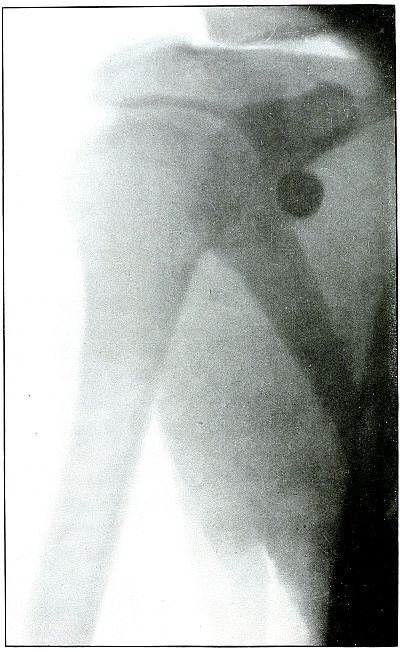
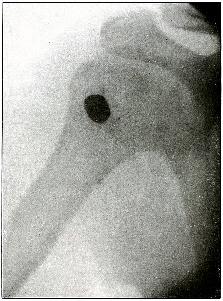
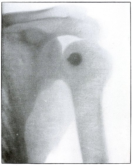
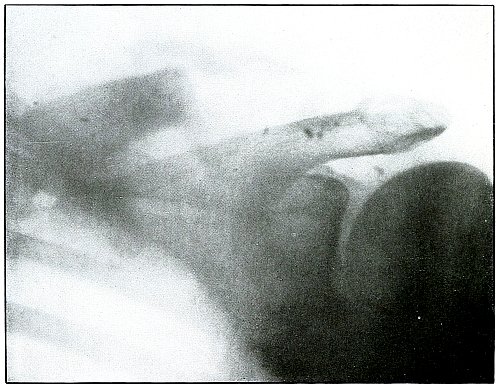
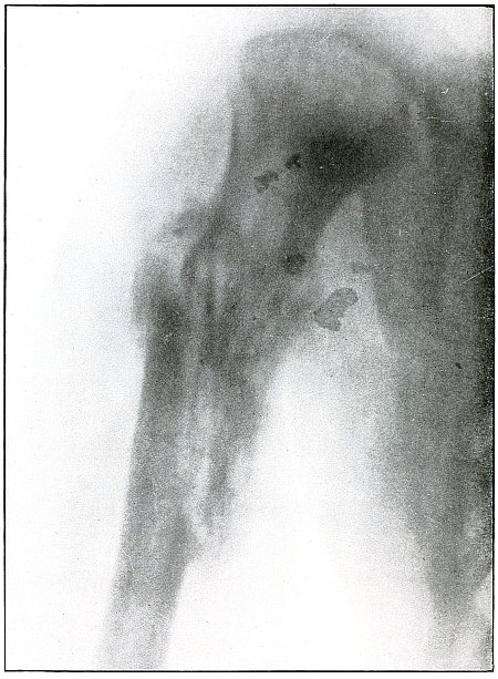
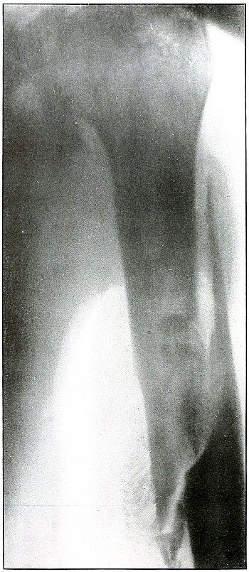
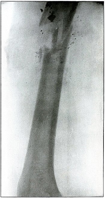
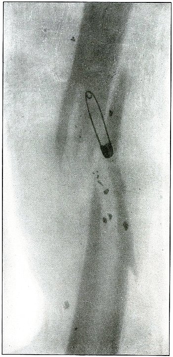
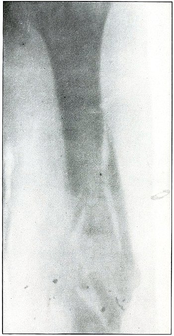
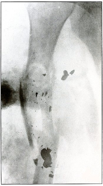
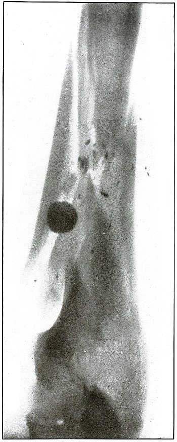
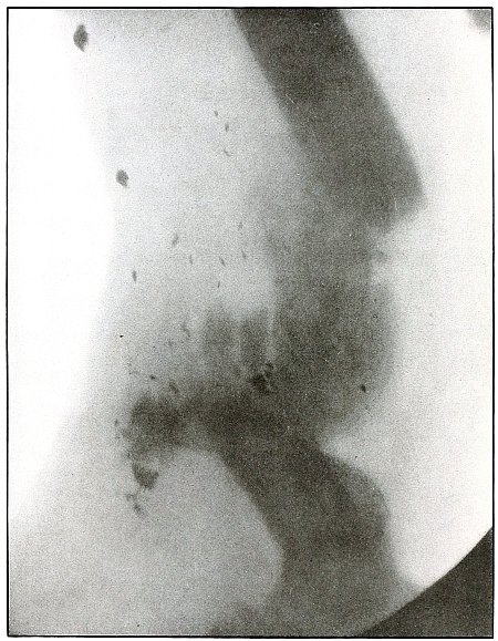
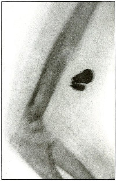
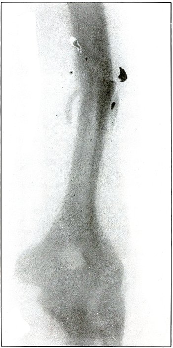
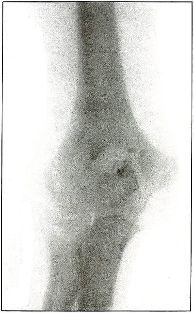
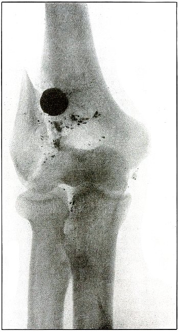
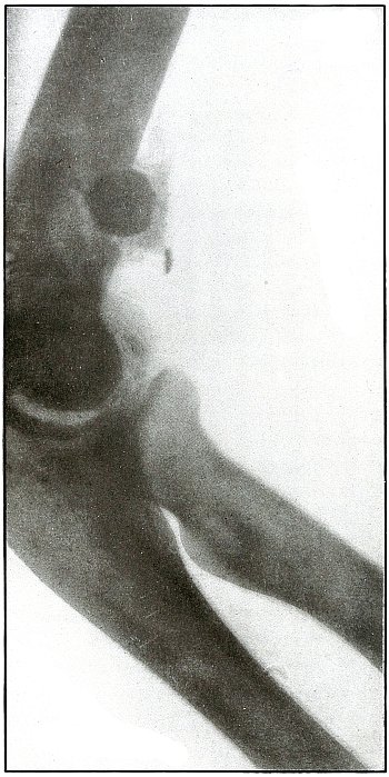
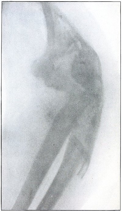
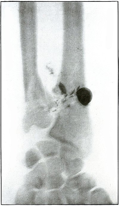
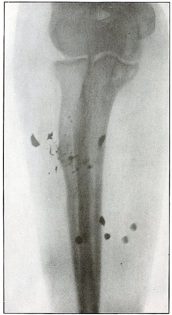
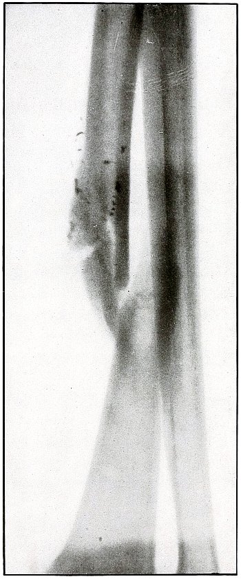
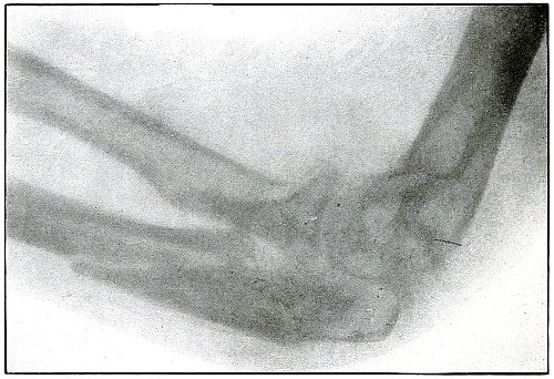
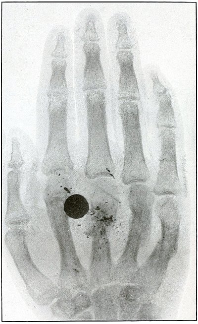
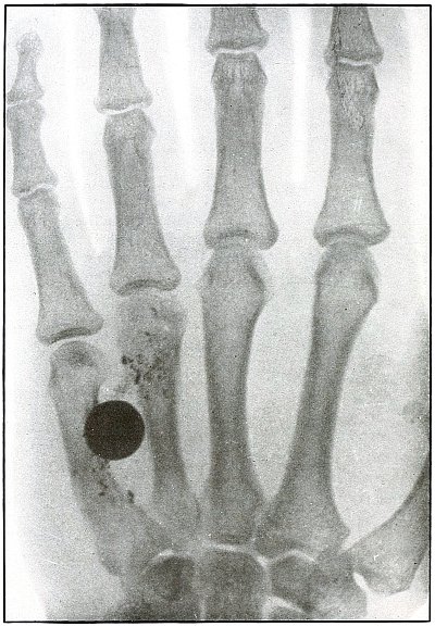
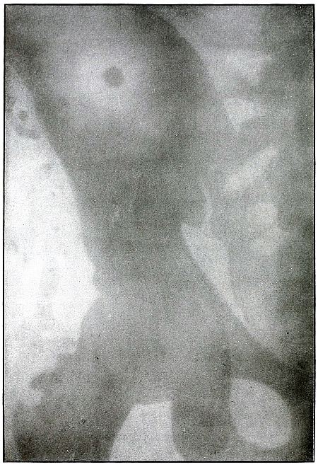
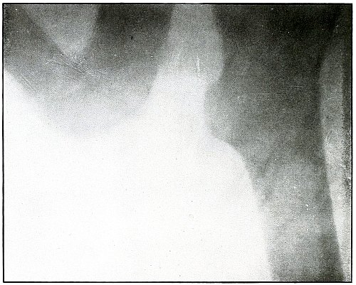
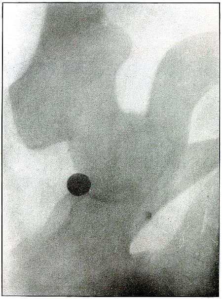
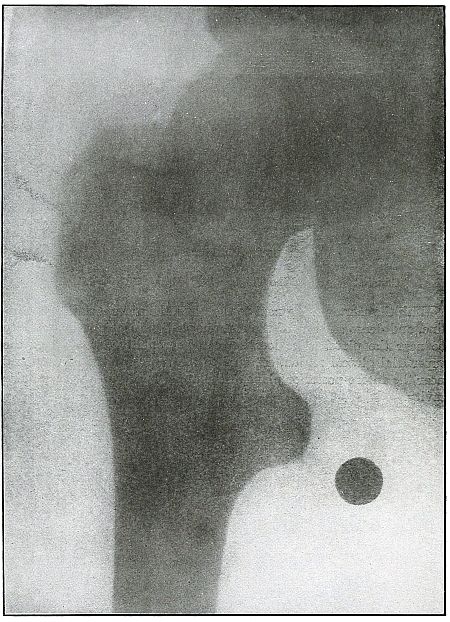
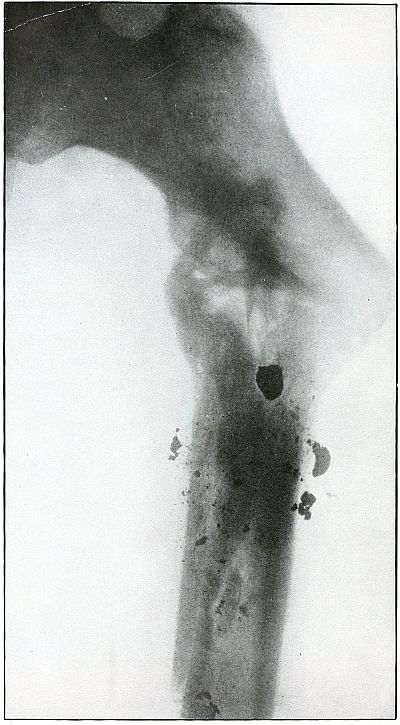
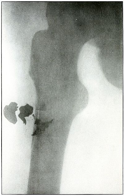
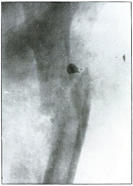
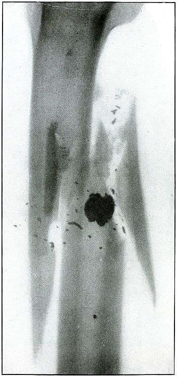
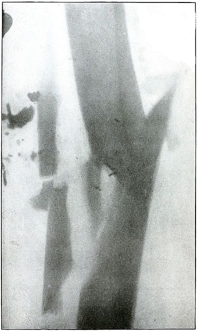
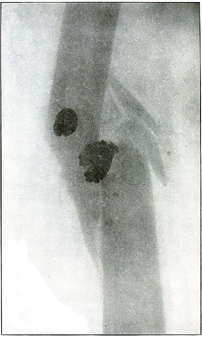
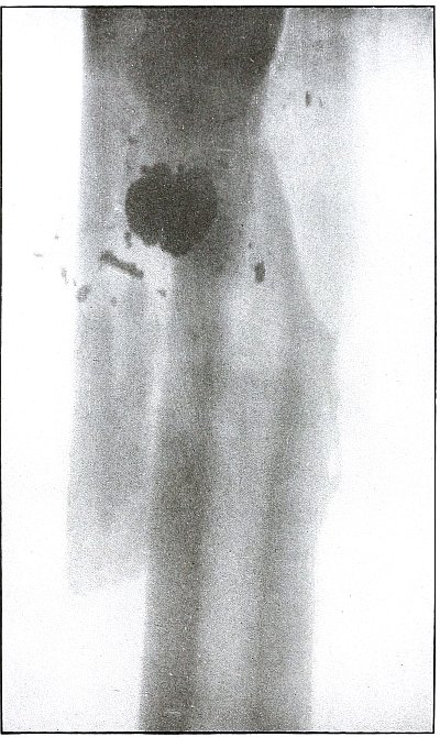
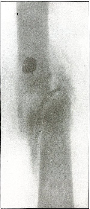
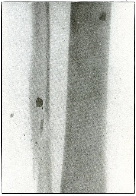
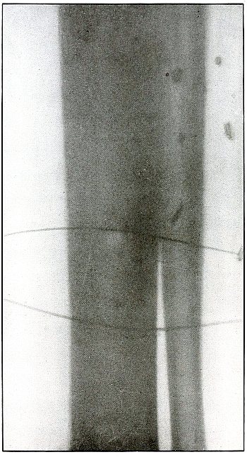
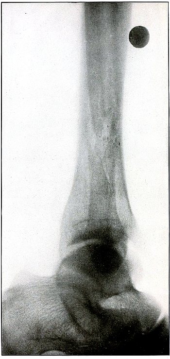
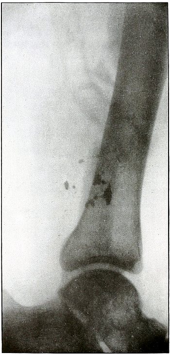
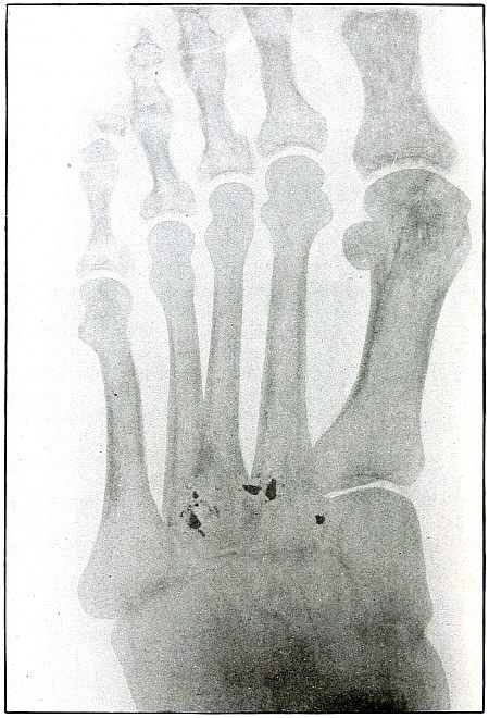
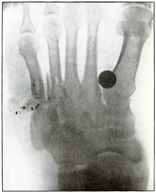
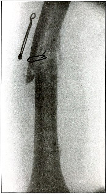
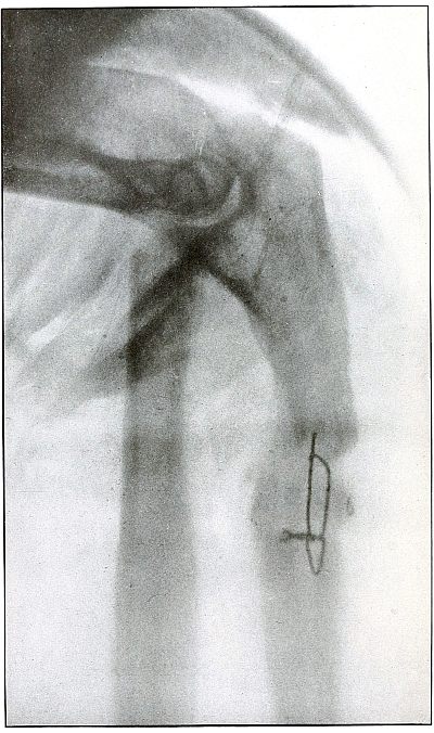
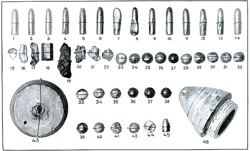
Plate 1.
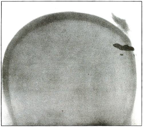
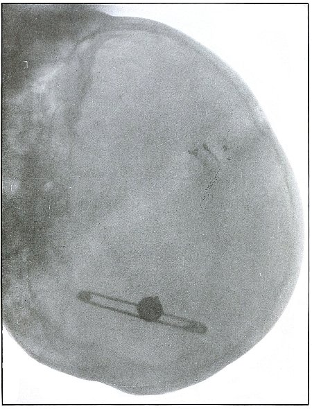
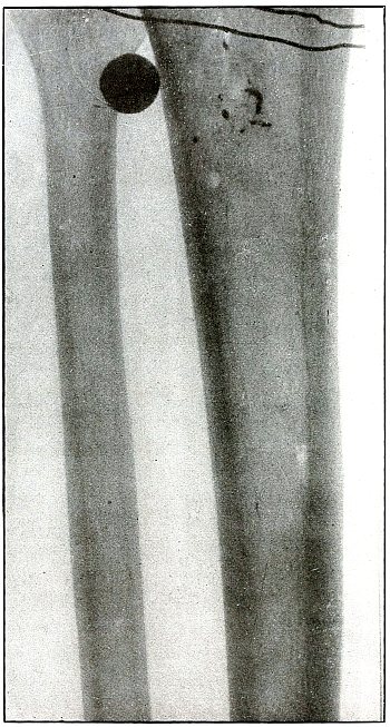
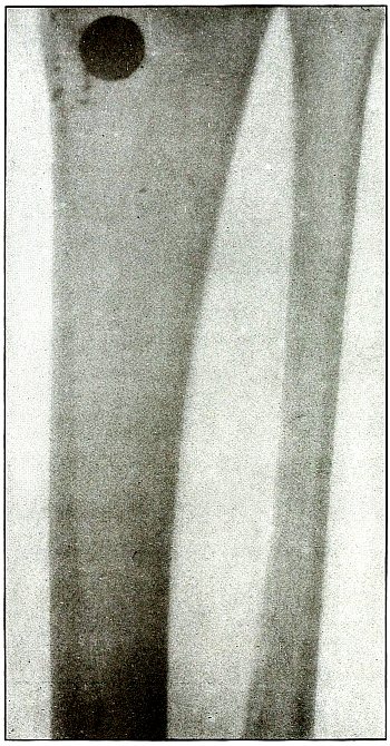
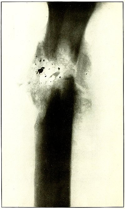
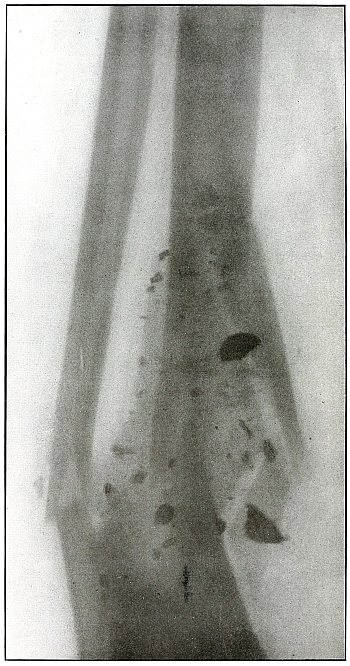
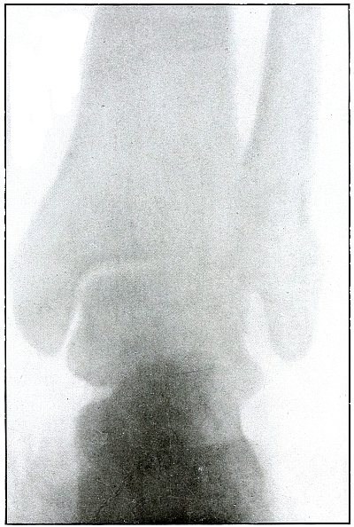
Rifle—Plate 1.
HEAD.
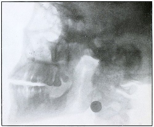
Gunshot Fracture of the Skull,
with Lodgment of the Missile.
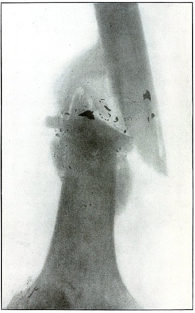
The bullet in this case was so badly deformed by ricochet that
part of both core and jacket were lost. While the appearance of the
shadow seems to indicate a direct impact of the nose of the bullet,
the line of contact with the skull must have been tangential, with
some laceration of the scalp; otherwise a cursory examination of the
scalp wound would have revealed the slightly protruding end of the
bullet. The dark shadow above the projectile is due to material used
in dressing. The great thickening of the scalp in the region of the
wound shows a marked cellulitis. Small particles of the lead core of
the bullet can be seen about the wound.
In such cases there is often a marked infection of the scalp
without extension of infection to the cranial cavity, except from
neglect. This is a case, though apparently simple, in which the
radiograph was necessary for correct diagnosis without exploration.
The treatment in such cases is conservative, with removal of
the projectile and care of the superficial infection or subsequent complications.
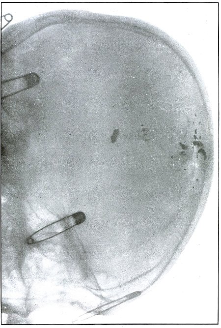
Plate 2.
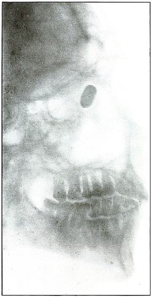
Rifle—Plate 2.
HEAD.
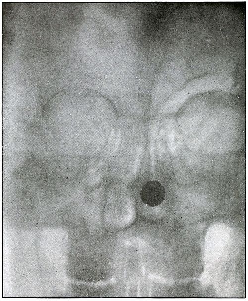
Gunshot Wound of the Head, with Lodgment of the Missile.
Wound of entrance, near outer canthus, with course through eyeball to
ethmoid body.
Wound of exit, none.
The Bulgarian Mannlicher bullet, shown half actual size on the plate,
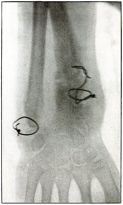
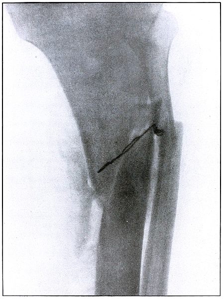
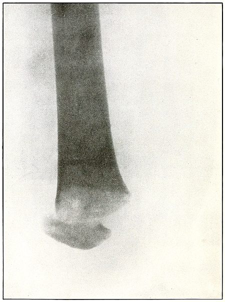
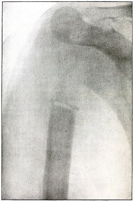
must be inclined on its long axis, about 30° from the perpendicular,
to the plane of the plate.
The slight penetration of the missile and its normal character show
that, having struck no intervening object, it indicted the wound at
extreme range.
The treatment should meet the indication for removal of missiles in
all superficial or easily accessible locations and when they cause
reaction.
Results to be expected are favorable except for loss of the eye.
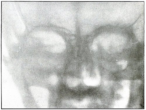
Plate 3.
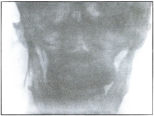
Rifle—Plate 3.
HEAD.
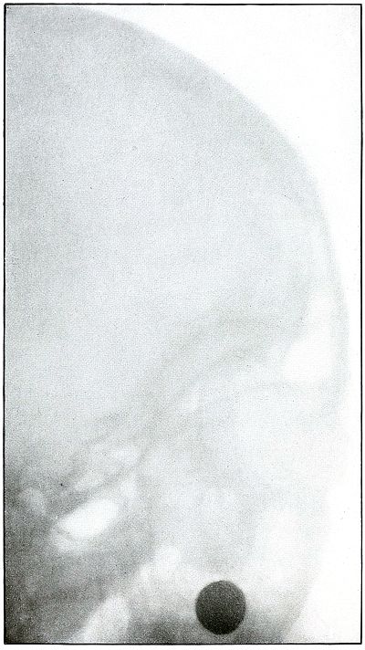
Gunshot Fracture of the Ramus of the Lower Jaw.
Wound of entrance, in the cheek behind the angle of the mouth.
Wound of exit, below the tip of the mastoid.
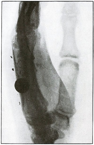
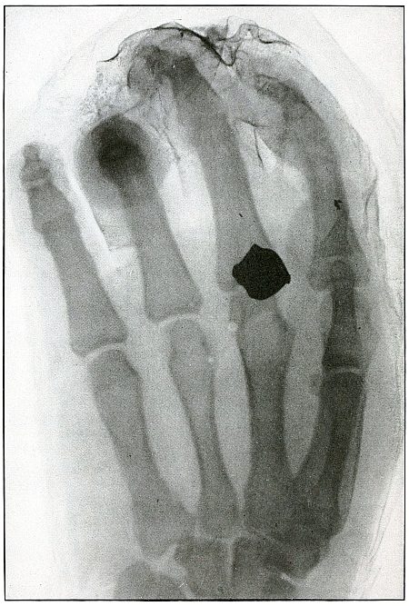
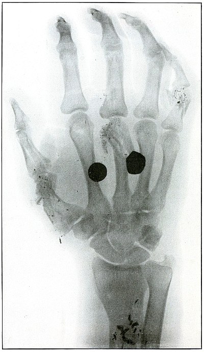
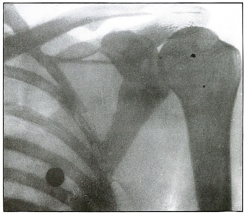
The course of the bullet was almost tangential to the ramus of the
jaw, anteroposteriorly. The slight fragmentation, which is hardly
more than a splitting of the bone, with little or no displacement,
indicates that the wound was made by a rifle bullet at moderate
velocity and at mid or long range.
Treatment is expectant.
Results are favorable.
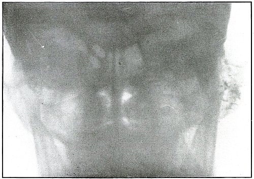
Plate 4.
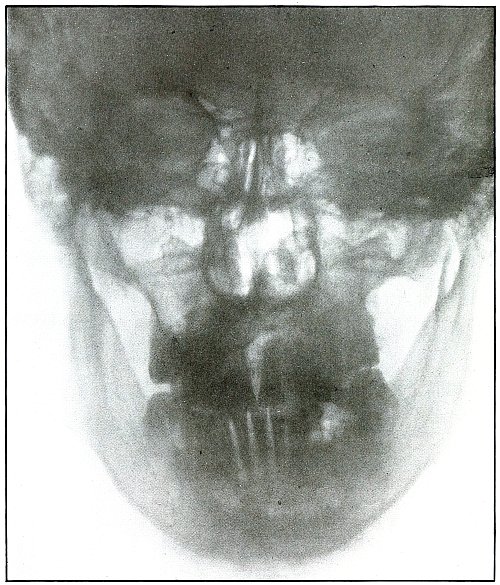
Rifle—Plate 4.
HEAD.
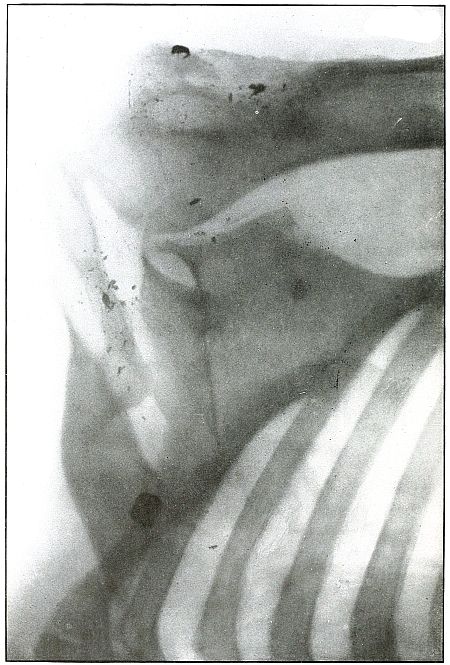
Gunshot Fracture of the Ramus of the Lower Jaw.
Wound of entrance, over the anterior border of the right ramus.
Wound of exit, beneath the lobe of the ear.
The wound was made by a rifle bullet with the velocity of long range,
because wounds of a shrapnel ball never show such slight injury
without lodgment or without marks of lead.
The damage of the bone was very slight, as only a superficial fragment
was chipped off. There were no signs of primary infection. Reaction
and periostitis suggested the radiograph after infection had rarefied
the fragment, shown but very faintly on the left side of the plate.
The postero-anterior skull radiograph was made with the face
superimposed upon the photographic plate.
Treatment, incision and drainage.
Results, good.
Plate 5.
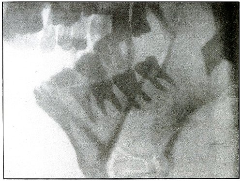
Rifle—Plate 5.
HEAD.
Gunshot Fracture of the Body of Lower Jaw,
with Great Fragmentation and Displacement.
Wound of entrance, to the left side of the median line of the lower
jaw below the alveolar process, with course ranging downward and
backward.
Wound of exit, with extensive laceration, beneath lower border of the
bone.
The wound was caused by a rifle bullet at high velocity at or less
than mid range. The fragments are many and rather small, so that much
bone was lost through the wound of exit. This effect was produced by
the splitting due to the relative friability of the bone and to the
imparting of the momentum of the missile to the detached fragments,
which, together with the missile, effected the considerable laceration
of the wound of exit.
Treatment, difficult; guided by septic conditions and surgical means
available.
Results in such cases are favorable to life but topically unsatisfactory.
Plate 6.
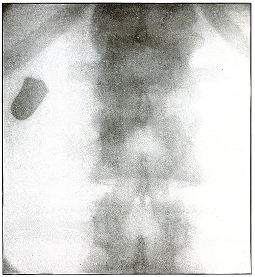
Rifle—Plate 6.
SPINAL REGION.
Gunshot Wound of the Spinal Region—
Lodgment
of the Missile in the Lumbar Muscles.
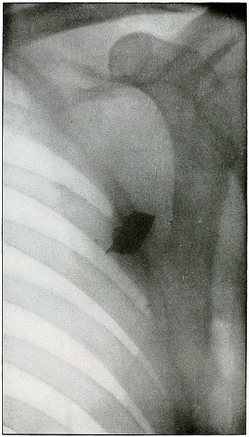
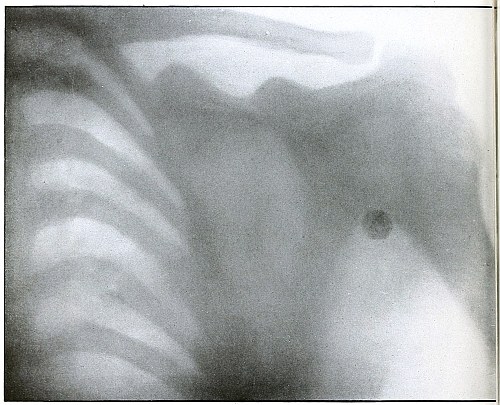
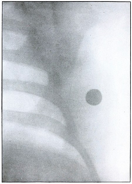
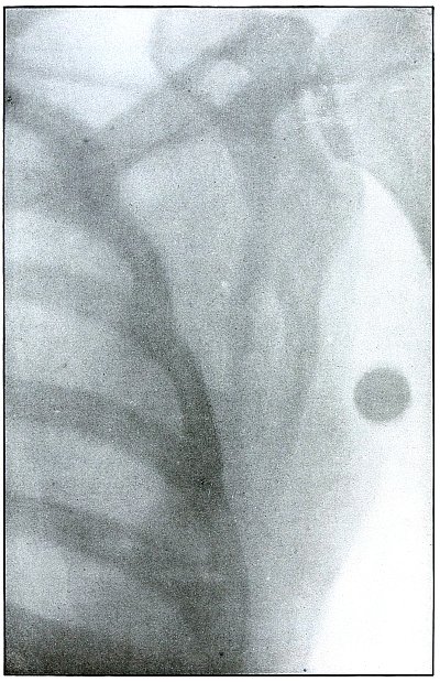
The bullet is lodged deep in the muscles of the back and not in the
abdomen, as determined by inspection of the plate.
(a) The shadow of the bullet is enlarged laterally, because, while
on the side of the body next to the plate and to the spine, it is at
some little distance from the plate, which accounts for the larger
diameter of the shadow; and it is shortened longitudinally, because
its long axis is inclined at an angle to the plate.
(b) The outline of the shadow is distinct, an evidence that
it is extra-abdominal, as otherwise its outline would be blurred by
the diaphragmatic movement of respiration imparted to the abdominal
viscera during the Röntgen exposure.
Plate 7.
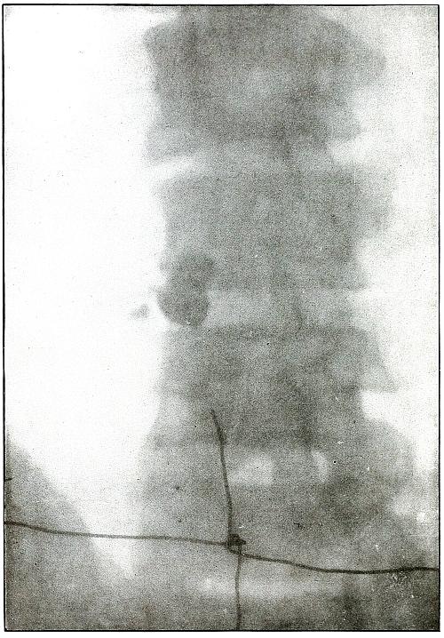
Rifle—Plate 7.
SPINAL REGION.
Gunshot Wound of the Spinal Region,
with Lodgment of the Missile.
The bullet was either dum-dummed or unjacketed because its soft nose
mushroomed, striking the crest of the ilium, penetrated the lumbar
muscles, and struck the side body of the third lumbar vertebra without
producing fracture.
The exposure, as the spinous processes show, was made with the spine
next to the plate, and the slight shadow, somewhat larger than the
projectile—to judge the size from the undeformed diameter—shows
it to be anterior to the vertebra. The shadow is deep enough to indicate
the location fairly near to the plate, and, almost certainly, not in
the abdominal cavity, where the distance from the plate would have
made the shadow less dense and the movement of respiration probably
would have given it a blurred outline. The shadow of the localizing
cross gives a standard of density to be compared with the shadow of
the projectile in making the estimation.
The treatment is conservative; only pain, paralysis, impaired
function, or sepsis indicate interference.
Plate 8.
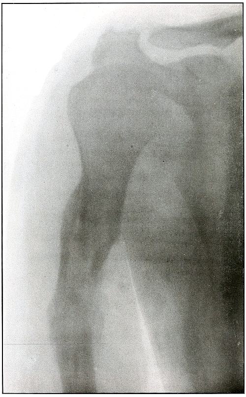
Rifle—Plate 8.
UPPER EXTREMITY.
Compound Fracture of the Humerus
in Advanced State of Repair with Callus Formation.
Wound of entrance, just above middle of anterior aspect of arm.
Wound of exit, about the same height, posteriorly.
The course of the missile was anteroposterior, with high velocity of
short range through the bone with a splitting effect, leaving a few
fragments, large and small, which were not much displaced and caused
but little deformity.
Wound was not infected. The absorption of smaller and the overlapping
of larger fragments caused some shortening.
Treatment, expectant.
Results, favorable.
Plate 9.
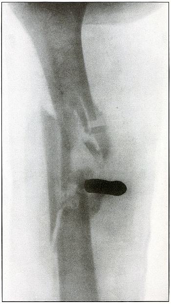
Rifle—Plate 9.
UPPER EXTREMITY.
Gunshot Fracture of the Right Humerus,
with Lodgment of the Missile.
Wound of entrance, antero-external aspect of upper third of arm.
Wound of exit, none.
The missile, deformed by ricochet, struck the bone with greatly
reduced velocity and without sufficient energy to perforate the bone
by which it was deflected slightly from its course and lodged in the arm.
This is something of the same effect that might have been caused by
a shrapnel ball, under the same ballistic conditions with a normal
shrapnel velocity giving about the same penetrating force.
The wound, without infection, is in the first week or two of repair,
before any callus has formed.
Treatment is expectant.
Results favorable.
Plate 10.
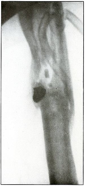
Rifle—Plate 10.
UPPER EXTREMITY.
Gunshot Fracture of the Left Humerus,
with Lodgment of the Missile.
Wound of entrance, anterior surface of upper third of the arm.
Wound of exit, none.
The shadow of the missile shows by its distinct outline and normal
diameter at the tip that the missile lies on the side near the plate;
the shortened length of the projectile indicates that the long axis
lay in an acute angle with a perpendicular to the plate.
The irregular outline of the base of the shadow and the fact of
lodgment shows that the missile was deformed and that it was
incidentally retarded in velocity by ricochet, so that its penetrating
force was not sufficient to carry it through the arm.
The fragments of bone are large and the wound is of the same character
as might have resulted from a shrapnel ball, for the normal ballistic
conditions of the latter simulate the conditions that produced the wound.
The drainage tubes seen in the plate indicate infection.
The conventional treatment in such cases is drainage and other
management of the infection without formal search for the projectile.
Results should be favorable.
Plate 11.
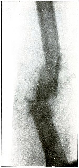
Rifle—Plate 11.
UPPER EXTREMITY.
Gunshot Fracture of the Humerus.
Wound of entrance, anterior internal aspect of middle and upper third
of arm.
Wound of exit, opposite.
The missile has struck the side of the bone and pursued a course
through the shaft, so that a transverse fracture, as well as the
separation of several medium-sized fragments, resulted from the
splitting effect of the missile.
A larger missile, i. e., a shrapnel ball, with the same striking
energy could have been stopped by the bone, but a wider distribution
of the same energy carried by a larger cross section would have
produced larger fragments.
In this case the location of the shrapnel ball would furnish
unquestioned evidence; or, if a shrapnel ball had produced this
particular bone destruction, its path among the fragments would have
been marked by traces of lead. Two metal fragments indicate that the
lead core of the bullet was exposed.
The wound, not infected, was treated expectantly.
Result in such cases is favorable.
Plate 12.
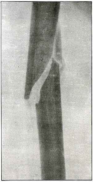
Rifle—Plate 12.
UPPER EXTREMITY.
Gunshot Fracture of the Humerus.
The course of the missile was anteroposterior through the middle of
the arm.
The ballistic conditions and lines of force applied to the bone
were somewhat, if not entirely, similar to those producing the
fracture shown in plate 11. The missile struck
the wall of the shaft without passing through the medullary canal, but
a secondary fragmentation of the two large fragments did not follow
except for the breaking of the tip of the distal fragment.
The range was long.
There was little deformity and no infection.
Plaster dressing was applied and the slight outline of callus
formation indicates the process of repair. The lack of contrast in
the shadow of the bone is due to the opacity of the plaster dressing
through which the Roentgen exposure was made.
Treatment in such cases is expectant.
Results should be uniformly good.
Plate 13.
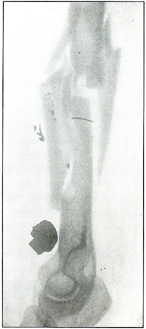
Rifle—Plate 13.
UPPER EXTREMITY.
Gunshot Fracture of the Right Humerus,
with Lodgment of the Missile.
Wound of entrance, about middle of the anteriorinternal aspect of the arm.
Wound of exit, none.
The course of the missile was from without, downward and inward to a
point of lodgment above the internal condyle. The distinct outline and
normal size of the base of the bullet shows it to be near the plate,
with the internal condyle next to the plate in the exposure.
The bullet mushroomed when it struck the bone with a “soft nose,” in
which the lead was not protected by a tough metal jacket. It may have
been dum-dummed; it is remotely possible that the nose of the jacket
was split by ricochet, or it is more probable that it was of the
unjacketed variety.
The effect is identical with that of a shrapnel ball, striking with
its normal low velocity, which is about the same as that of the
missile in this wound.
The invariable characteristic of a shrapnel wound of a bone, namely,
the small particles of metal marking its course in contact with the
bone, is seen in this plate.
The treatment in such cases is expectant, with due regard to the
character of the infection, and without primary search for the missile.
The results are generally favorable.
Plate 14.
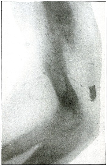
Rifle—Plate 14.
UPPER EXTREMITY.
Gunshot Fracture of the Humerus,
with Lodgment of the Missile.
The missile was a fragment of a ricocheted rifle ball, with a part of
the lead core carried in a portion of the jacket. The course was from
before, backward, striking the humerus in lower third, and leaving
particles of lead along its trade.
The wound was only slightly infected. Several detached fragments of
bone have been removed.
The treatment in such cases is conservative, with management of the
infection and without formal search for the projectile.
The results in such cases are favorable with some shortening of the bone.
Plate 15.
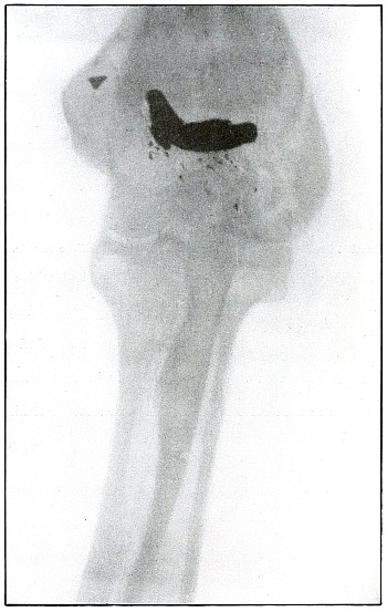
Rifle—Plate 15.
UPPER EXTREMITY.
Gunshot Fracture of the External Condyle of the Left Humerus,
with Lodgment of the Missile.
Wound or entrance, internal and posterior aspect of the arm above the
internal condyle.
Wound of exit, none.
The bullet was greatly deformed by ricochet, with the loss of the
greater part or all of its jacket.
The line of contact of the unprotected lead with the bone is marked by
the same small fragments of lead almost invariably seen in shrapnel
wounds. The ballistic conditions in this case are quite similar to
those of a shrapnel wound, as the projectile has struck the bone
with low velocity. The very slight displacement of a single large
fragment from which the missile is slightly withdrawn indicates that
the striking energy was relatively low and that the elastic tissues,
stretching around the missile at its striking point, contracted after
its energy had been expended and then withdrew the missile from its
farthest point of advance.
The treatment in such cases warrants only the interference suggested
by infection and the interference of the missile with function.
The results expected are most favorable.
Plate 16.
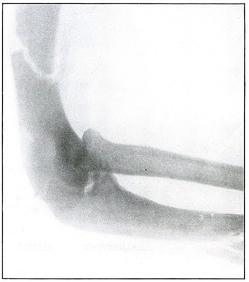
Rifle—Plate 16.
UPPER EXTREMITY.
Gunshot Fracture of the Humerus.
The transverse course of the bullet, striking the posterior wall of
the shaft without entering the medullary canal, has fractured the bone
transversely, with a tendency toward splitting off a large fragment
from the distal fragment.
The bullet under these ballistic conditions of high velocity and not
distant range might have bored its way through the cancellous tissue
of the epiphysis of the same bone without any fractures.
Gunshot Fracture of the Ulna.
The transverse course of the bullet in striking the ulna at high
velocity and not distant range has shown a tendency to bore a hole
through the bone. A smaller bullet or a larger bone of the same
structure might easily have provided conditions to permit this effect.
The wounds of exit and entrance in each of these wounds presented
almost identically the same appearance.
The treatment in such cases is that of a simple fracture, as there is
almost always no infection in such wounds.
Results are favorable.
Plate 17.
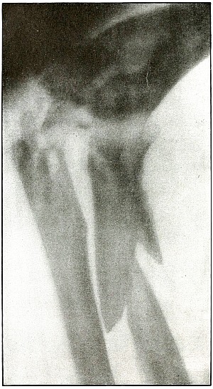
Rifle—Plate 17.
UPPER EXTREMITY.
Gunshot Fracture of the Elbow.
The bullet in transverse course and high velocity through both bones
of the forearm struck the head of the radius, thus starting several
splitting lines of fracture and separating large fragments. Smaller
fragments which received some of the energy of the missile have been
carried along with it in turn, striking the ulna and carrying away
smaller fragments from it and causing the laceration which marks the
wound of exit.
Such wounds, with laceration of soft parts and fragmentation of the
bone, are prone to infection, against which treatment is directed. The
indications to be met are much like those of the wound shown in plates
18 and 19. Excision or
immediate methods of bone repair are contraindicated by infection.
Results will depend upon the nature and extent of infection.
Plate 18.
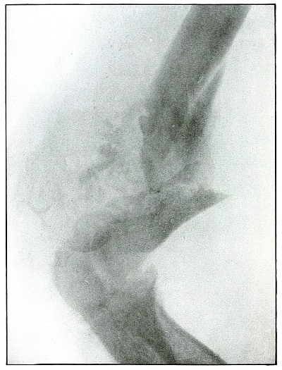
Rifle—Plate 18.
UPPER EXTREMITY.
Gunshot Fracture of the Elbow,
without Injury to the Great Vessels and Nerves.
Wound of entrance, posterior to the external condyle.
Wound of exit, large laceration in front and above the internal condyle.
The wound is an example of the misnamed “explosive” action of a rifle
bullet. The force and direction of the missile, in high velocity,
split the bone into many fragments, and, transmitting its energy to
some of the fragments, carried them through the skin and caused the
large laceration at the point of exit by the simultaneous escape of
the bullet and fragments. The wound was so heavily infected, that a
cellulitis advanced to the shoulder and to the wrist to such extent
that the arm was marked by eminent surgical opinion for amputation.
Free incision, drainage, antisepsis and incidental removal of detached
fragments controlled the infection and brought about slow resolution.
After six months of careful treatment the wound was healed with an
ankylosed elbow with normal function of the forearm, except for
limited rotation.
Treatment indicated in such cases is always conservative. Infections
contraindicate any formal surgical interference. The dangers of
infection in such cases are to be risked to avoid amputation.
Results may be considered favorable even with elbow ankylosis.
Plate 19.
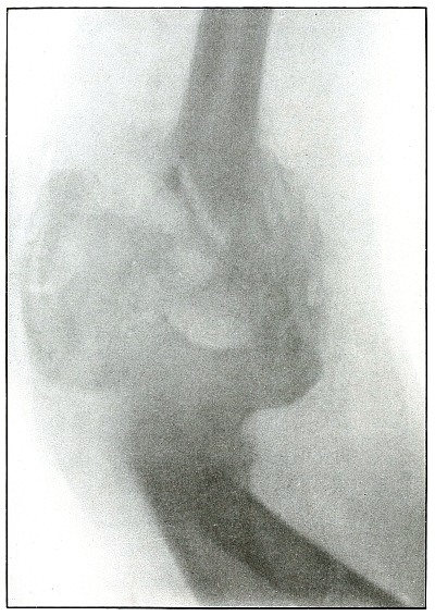
Rifle—Plate 19.
UPPER EXTREMITY.
Gunshot Fracture of the Elbow,
without Injury to the Great Vessels and Nerves.
This is a plate made of the same subject shown in plate 18, when convalescence was several weeks
farther advanced, as is indicated by the removal of fragments and
extensive callus formation.
Both radiographs were made after the apprehension of systemic
infection had passed; the second plate after an additional number of
fragments had been removed.
Plate 20.
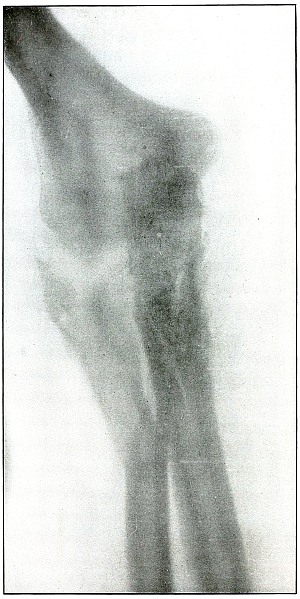
Rifle—Plate 20.
UPPER EXTREMITY.
Gunshot Fracture of the Elbow.
Wound of entrance, posterior aspect of forearm internal to and below
the olecranon.
Wound of exit, external border over head of radius.
The course of the bullet was diagonally anteroposterior from within
outward, striking the posterior border of the upper end of the ulna
and passing through the head of the radius, carrying the fragments
of the latter before it and lacerating the wound of exit. The energy
of impact also fissured the upper end of the shaft of the ulna and
fractured the neck of the radius without detaching the large fragments.
This is the effect of a rifle bullet at short range, or possibly a
ricochet shot at mid range.
The emergency treatment is antiseptic dressing with splint
immobilization.
The subsequent treatment is conservative, whether the wound is clean
or infected. The course of treatment of such an infected wound might
extend from four to six months.
Note.—As the
soldier always escapes the burden of explanation when the wound of
entrance is anterior rather than posterior, it should be remembered
that the forearm may occupy positions in relation to the body which
exposes the anatomically posterior aspect of the forearm to missiles
directed toward the anterior surface of the body; and as the wounds of
the forearm herein presented are described in the anatomical position,
there is no justifiable impeachment of the soldier’s valor in an
inference that he was shot from behind when the wound of entrance
involves the posterior aspect of the forearm.
Plate 21.
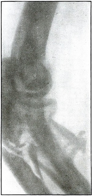
Rifle—Plate 21.
UPPER EXTREMITY.
Gunshot Fracture of the Radius and Ulna.
The course of the bullet at short range was transverse through
both of the bones, with a splitting effect and without much small
fragmentation.
The wound of exit in this case was slightly lacerated, but not very
much larger than the wound of entrance.
The treatment should be conservative. Emergency treatment should not
include exploration, and nothing but the conventional iodine dressing
and splints should be applied.
Plate 22.
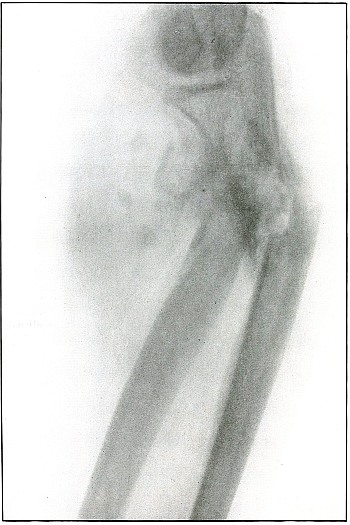
Rifle—Plate 22.
UPPER EXTREMITY.
Gunshot Fracture of the Radius and Ulna.
The course of the bullet at short range was transverse through the
upper forearm, striking the radius in the center of the shaft and the
ulna nearer the border. Several small fragments followed the course of
the bullet, but did not emerge with it at the wound of exit to cause a
laceration.
The capitellum was next to the photographic plate and the angular line
of the radius can be seen crossing the straighter line of the ulna.
Further information is obtained from the examination of another
view, plate 23, made of the same subject.
Plate 23.
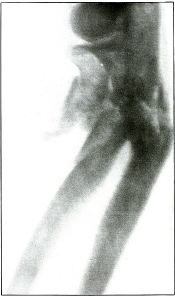
Rifle—Plate 23.
UPPER EXTREMITY.
Gunshot Fracture of the Radius and Ulna.
This plate was made from the wound shown in plate 22,
with the arm in greater inward rotation. This position shows the wide separation
of the large fragments of the radius.
Emergency treatment in such cases is antiseptic dressing only, without
exploration, and with fixation by splints for transportation. The
degree of infection determines the subsequent course of conservative
treatment, with operative methods for correction of deformity reserved
for further stage of convalescence and for best surgical facilities.
Plate 24.
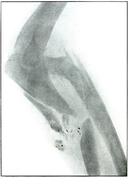
Rifle—Plate 24.
UPPER EXTREMITY.
Gunshot Fracture of the Radius and Ulna in the
Upper Third of the Forearm.
The course of the projectile was from within, outward and diagonally
forward, with a direct impact on the ulna, and a tangential impact on
the radius, with several lines of splitting fracture in the latter
without detaching fragments. Particles of metal, spattered around the
point of first impact, were deposited by the lead core of a bullet,
exposed by a torn jacket, which struck the second bone with its
jacketed surface.
The treatment is always conservative—meeting indications in case of
infection.
Results are good for saving the limb, but not for avoiding deformity.
Plate 25.
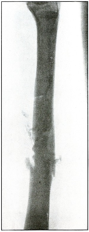
Rifle—Plate 25.
UPPER EXTREMITY.
Gunshot Fracture of the Radius.
Wound of entrance, posterior surface of forearm over radius above the middle.
Wound of exit, below and in front of wound of entrance.
The course of the ball in mid range was from behind, forward,
and slightly downward.
While the images of both bones of the forearm are superimposed,
because they both lay in the plane of the projection of the shadow, it
is probable that the radius lay nearer the photographic plate, because
the head of the radius is shown in clearer outline. The fragments of
the fracture can be seen as related to the outlines of the radius.
There is no displacement and only slight fragmentation, so that the
bullet must have almost succeeded in making a punctured wound in the
radius.
The treatment in such cases is regularly that for simple fracture, as
such wounds are almost always aseptic.
The results are uniformly good.
Plate 26.
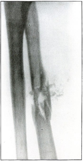
Rifle—Plate 26.
UPPER EXTREMITY.
Gunshot Fracture of the Radius.
Wound of entrance, midway between radius and ulna and midway between
elbow and wrist, anterior aspect of the forearm.
Wound of exit, over radius at point opposite.
The course of the bullet, in the medium velocity of mid range, in
piercing the medullary canal has almost succeeded in drilling the
bone without splitting off several longitudinal fragments. Small
fragments followed the course of the missile, without being energized
sufficiently to lacerate the point of exit by escaping with the
projectile.
The wound of exit in such cases hardly differs enough from the wound
of entrance to be distinguishable. This condition so often obtains
that the great majority of perforating rifle wounds of the forearm
do not show the blow-out or “explosive” effect which seems to be
generally misunderstood as a classic accompaniment.
The bullet was traveling at high velocity of perhaps less than
mid range.
The treatment is usually that of a simple fracture, and warrants no
interference except in case of occasional infection.
Results are almost always good.
Plate 27.
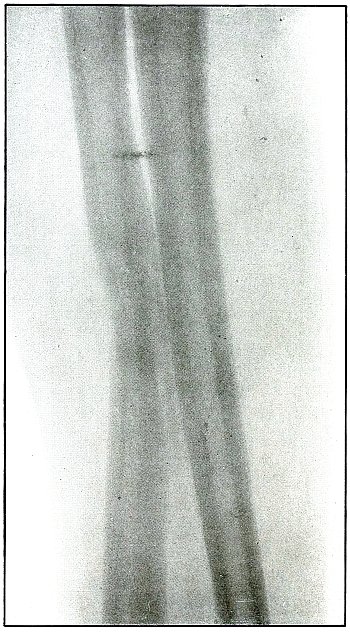
Rifle—Plate 27.
UPPER EXTREMITY.
Gunshot Fracture of the Radius.
The course of the bullet, at long range, has been diagonally
anteroposterior through the shaft, causing only a diagonal fracture.
The plate was made after a two-weeks’ convalescence, as is shown by
the beginning of callus formation.
The treatment is that of a simple fracture.
Results are good.
Plate 28.
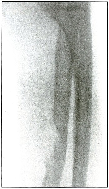
Rifle—Plate 28.
UPPER EXTREMITY.
Gunshot Fracture of the Radius.
The course of this bullet was anteroposterior and diagonally from
above downward through the shaft, punching out one side of the shaft
and effecting a diagonal fracture through the bone with only slight
displacement. The wound was infected.
The radiograph was taken during the course of treatment, after the
several small fragments found by the punched-out portion of the bone
were removed. A small drainage tube is in the wound, but the size of
the forearm shows that the reaction is very moderate.
The treatment is that of a simple fracture, except for the indications
to be met in the control of infection.
Results are good.
Plate 29.
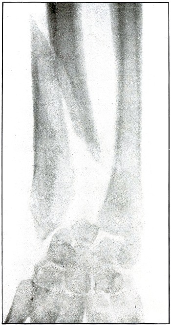
Rifle—Plate 29.
UPPER EXTREMITY.
Gunshot Fracture of the Lower End
of the Radius.
The course of the bullet in long range was diagonally anteroposterior
through the ulnar side of the lower end of the bone, with the wound of
entrance on the anterior and the wound of exit on the posterior aspect
of the wrist. The wound of exit was slightly lacerated by several
small fragments driven off from the ulnar side of the radius. These
fragments were removed through an incision before the radiograph was made.
The emergency treatment of such cases is only antiseptic dressing and
splint immobilization.
When wound is aseptic or after it has closed, a secondary operation
for coaptation, with proper facilities available, might be indicated.
The results as to full restoration of joint function are not favorable.
Plate 30.
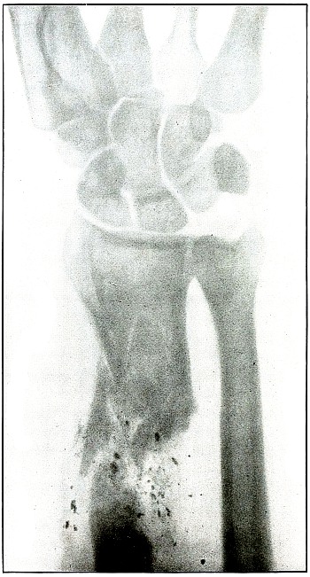
Rifle—Plate 30.
UPPER EXTREMITY.
Gunshot Fracture of the Lower End
of the Radius.
The course of the missile was diagonally transverse, striking the
radius in its lower third.
The projectile in this case is unknown, as it might have been either a
shrapnel ball or a deformed rifle bullet with a torn jacket, exposing
the lead core and marking its course with small particles of lead.
The fissures in the lower fragment and the finer fragmentation at the
seat of impact, indicate a great striking energy, that more often
resides in the high velocity of a rifle bullet than the low velocity
of a shrapnel ball. The wound is therefore classified with rifle wounds.
The treatment is conservative. The course in such cases, without
infection, is very favorable, and not unfavorable even with infection.
Results should be good.
Plate 31.
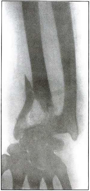
Rifle—Plate 31.
UPPER EXTREMITY.
Gunshot Fracture of the Lower End
of the Radius.
Wound of entrance, anterior aspect of wrist, over internal border of radius.
Wound of exit, posterior aspect of wrist between radius and ulna, with laceration.
The range was described as “close”—within a hundred yards—with
the bullet in high velocity. The energy of the projectile, imparted to
small fragments of cancellous tissue, drove them through the wound
of exit, and caused the laceration of the superficial tissues. The
wound was infected (swelling of soft parts clearly shown): resolution
followed extended treatment, with ankylosis of the wrist and radial
displacement of the carpus.
Emergency treatment in all such cases is antiseptic dressing
without exploration or manipulation of fragments, and with splint
immobilization.
Results are unfavorable as to function, depending upon extent of destruction of tendons.
Plate 32.
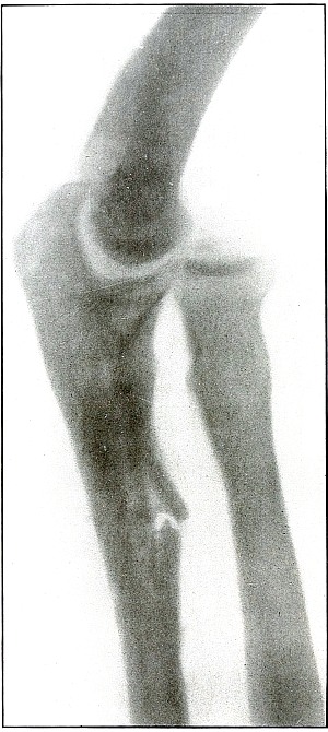
Rifle—Plate 32.
UPPER EXTREMITY.
Gunshot Fracture of the Ulna.
The course of the bullet was transverse through the arm at the
junction of the middle and upper thirds from behind the radial border
externally to the ulnar border internally, striking the wall of the
medullary canal with a punching effect that partly split off short
longitudinal fragments and caused transverse and longitudinal cracks,
without separation or displacement of fragments.
The same ballistic conditions applied to cancellous tissue at the end
of the bone would probably have bored through it without fracture.
This effect is generally seen in wounds of small-caliber bullets
traveling at reduced velocity of long range.
The treatment is that of a simple fracture.
Results, in such cases without infection, could not be bad.
Plate 33.
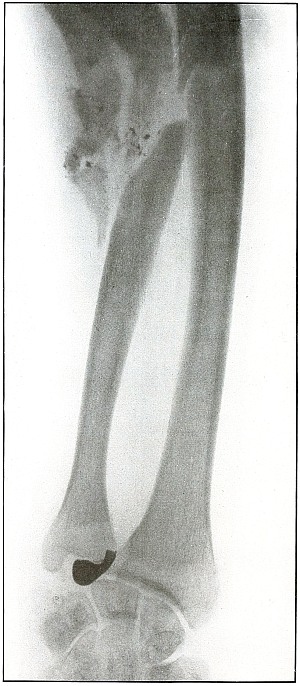
Rifle—Plate 33.
UPPER EXTREMITY.
Gunshot Fracture of the Left Ulna.
The course of the missile was from within outward, ranging downward to
the wrist, by deflection, after striking the ulna in its upper half.
The considerable striking energy retained in a small portion of the
mass—consisting of only the nose and a little more of the jacket of
the bullet, but sufficient to fragment a large section of the bone,
and then to traverse more than half the length of the forearm—leaves
no doubt that the shot was fired at very close range, and that the
bullet was broken on a nearly resisting surface, leaving in the nose
of the bullet a striking force equal to that of the entire projectile
at long range.
The posterior surface of the forearm is next to the plate, as the
distinct outline of the styloid process of the ulna and the posterior
border of the articular surface of the radius shows. The radius and
ulna are parallel in the most natural position of supination. The
normal diameter and sharp outline of the nose of the bullet show it to
be next to the plate and on the posterior surface between radius and ulna.
Fragments of the exposed lead core of the bullet have scraped off on
the line of fracture in a manner peculiar to shrapnel wounds, but
never seen in bullet wounds in which the jacket covers all of the lead core.
The treatment is regularly conservative and without interference, as
in this particular wound, which was aseptic.
Secondary treatment may indicate correction of bone deformity.
Plate 34.
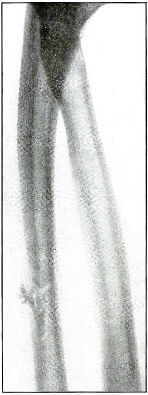
Rifle—Plate 34.
UPPER EXTREMITY.
Gunshot Fracture of the Ulna.
The ballistic conditions of the projectile causing the wound
shown in this plate are substantially those of the wound shown in
plate 32.
The wound of entrance and exit would be practically the same in
chipping off a few small fragments and causing a clean transverse
fracture without any displacement.
The bullet at long range has struck the wall of the medullary canal,
appearance.
Treatment that of a simple fracture.
Results must be good.
Plate 35.
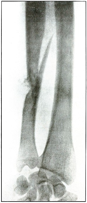
Rifle—Plate 35.
UPPER EXTREMITY.
Gunshot Fracture of the Ulna.
The course of the bullet at long range has been anteroposterior
through the middle of the forearm, passing through the side of the shaft,
chipping off a few small fragments and causing a long oblique fracture.
The conditions were much the same as those shown in plates
28 and 29,
except that the striking energy of the projectile was somewhat
greater with the velocity of mid range.
The treatment, without infection, is that of a simple fracture.
Results will be uniformly good.
Plate 36.
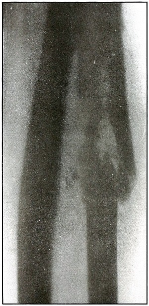
Rifle—Plate 36.
UPPER EXTREMITY.
Gunshot Fracture of the Ulna.
The course of the bullet was anteroposterior through the ulna a little
above the middle of the forearm, and fairly through the long axis.
This is a bone effect much similar to those shown in plates
28, 29, and 31,
except that this condition is due to the impact of a missile, with a
still higher velocity of shorter range, imparting its energy to small
fragments of bone, which added their momentum to the destructive force
of the projectile.
No large fragments were carried along with the missile to cause any
more destruction of tissue in exit than in entrance, so that the skin
wounds, under these conditions, are about the same in appearance.
The treatment is conservative and expectant with immobilization.
Results in such cases are uniformly good.
Plate 37.
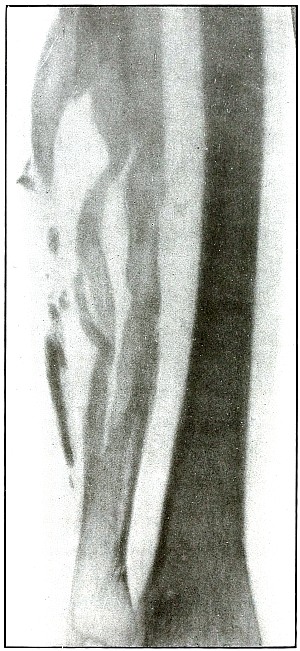
Rifle—Plate 37.
UPPER EXTREMITY.
Gunshot Fracture of the Ulna.
The course of the bullet was in an anteroposterior direction at a high
velocity of short range, which, imparting its energy to the fragments,
drove some of them through the tissues as “secondary missiles” and
caused a laceration of the wound to exist.
The longitudinal fragmentation and splitting indicates a considerable
energy of the projectile, which may have been deflected, as its long
axis was turned somewhat from the trajectory at the time of impact.
The emergency treatment is antiseptic dressing and splint immobilization.
The subsequent treatment is conservative with the removal of detached
fragments and with control of infection as the course indicates.
Plate 38.
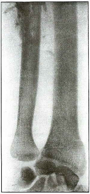
Rifle—Plate 38.
UPPER EXTREMITY.
Gunshot Fracture of the Left Ulna.
The course of the bullet was transverse through the middle of the
forearm, striking the posterior border of the ulna.
Small fragments were broken from the posterior wall of the medullary
canal, without destroying the longitudinal continuity of the anterior wall.
The velocity of the bullet was probably that of mid-range, as the
striking energy of the impact was fairly great.
The posterior surface of the forearm lay next to the plate.
The emergency treatment is antiseptic dressing and splint
immobilization.
The subsequent treatment is that of a simple fracture,
as infection is not usual.
Plate 39.
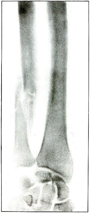
Rifle—Plate 39.
UPPER EXTREMITY.
Gunshot Fracture of the Left Ulna.
The course of the bullet, with the velocity of long range, was
anteroposterior through the lower third of the forearm, striking the
outer side of the bone. The initial velocity of the projectile was
much reduced, as is shown by the tendency to puncture the bone without
much fragmentation.
There was no displacement of fragments as a direct result of the
impact, although muscular contraction has caused some slight
subsequent overriding.
The wounds of entrance and exit were about the same, if not quite
similar in appearance.
The emergency treatment is the conventional antiseptic dressing with
splint immobilization.
The subsequent treatment is usually that of a simple fracture,
as infection in such cases is rare.
Plate 40.
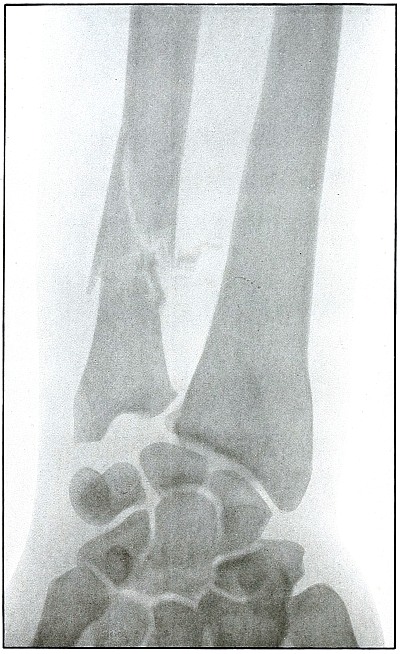
Rifle—Plate 40.
UPPER EXTREMITY.
Gunshot Fracture of the Ulna.
The course of the bullet was obliquely anteroposterior through the
lower third of the forearm, striking the radial edge of the bone with
a velocity of long range.
The wounds shown in plates 35 and 39
represent conditions similar to those causing this wound,
except that the ranges were progressively greater.
In this case the projectile exhibited a punching effect at the
point of impact, and although the lines of force are shown in
characteristically divergent fissures, the energy imparted to the
fragments—less than in the preceding cases—has not been sufficient
to separate or to displace the fragments.
The emergency and subsequent treatment is conventionally conservative,
as in the preceding cases.
Plate 41.
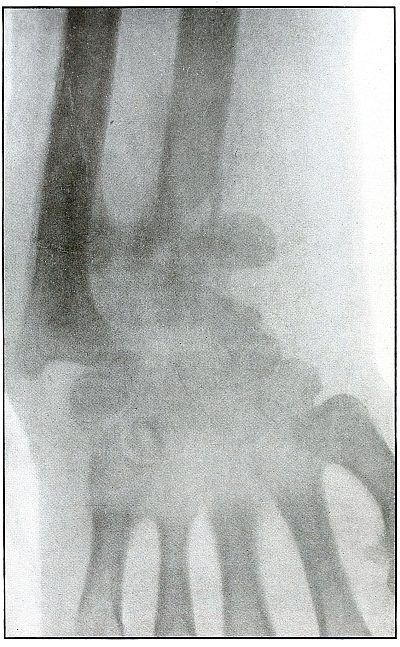
Rifle—Plate 41.
UPPER EXTREMITY.
Gunshot Fracture of the Wrist.
Wound of entrance, posterior aspect of forearm over the lower end of
the radius, with the bullet ranging forward and slightly downward to
the wound of exit and covering with great laceration the anterior
aspect of the wrist joint.
The range was close, and the energy of the high velocity of the
missile was imparted to fragments, which, becoming “secondary
missiles,” emerged with the projectile to cause extensive laceration
and destruction of tissue.
The case was received for amputation in the second week, when a grave
degree of infection extended in a cellulitis to the elbow. The ulnar
nerve and vessels were intact, but the flexor tendons were almost
entirely destroyed.
The plate, made after several weeks, when infection was under control
and after the end of the radius and fragments of the carpus had been
informally removed, shows a rarefaction of the carpus and proximal
ends of the metacarpus, due to infection and disuse.
Frequent incisions and extension of drainage, with removal of detached
fragments, was continued for several months. The wound was closed in
the sixth month, with ankylosis and deformity of the wrist,
as shown in plate 42.
Plate 42.
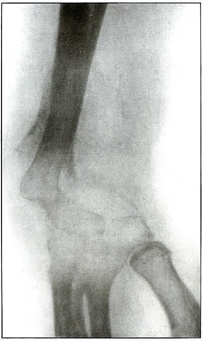
Rifle—Plate 42.
UPPER EXTREMITY.
Gunshot Fracture of the Wrist.
This plate, presenting a lateral view of the wound shown in
plate 41, shows considerable deformity of the
joint, after four months’ treatment, which was even more marked two
months later, when the case was discharged with an ankylosis of the
wrist joint, contracture of the flexor tendons of the fingers, and
slight flexor function of the thumb, permitting apposition with the
first finger.
The result, while leaving much to be desired, preserved a function of
the hand vastly superior to that of a forearm stump.
The treatment in such cases is always courageously conservative, with
amputation only as the extreme measure to save life, with risks of
judgment in favor of conservatism.
Corrective measures may be employed after management if the treatment
of the infection is successful and when the case passes out of the
military category. It is not possible, during a long infection, to
maintain better position in such cases.
Plate 43.
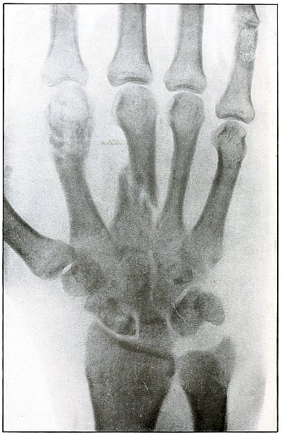
Rifle—Plate 43.
UPPER EXTREMITY.
Gunshot Fracture of the Metacarpus.
Wound of entrance, inner aspect of the hand over proximal end of the
fifth metacarpal.
Wound of exit, on the outer border of the hand over the distal end of
the second metacarpal.
The velocity of the bullet was in mid or long range, as it displaced
no fragments, and as it made a point of entrance and exit about the
same in appearance.
The wound was infected, which is more frequently the case in the hand
than in the forearm.
The treatment is conservative with free incision and drainage
in the management of infection.
Plate 44.
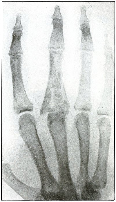
Rifle—Plate 44.
UPPER EXTREMITY.
Gunshot Fracture of the Third Phalanx.
The course of the bullet was anteroposterior through the base of the
proximal phalanx of the middle finger, with a velocity of long range.
It practically punctured the bones and split off a few fragments
without displacement.
The wound of entrance would be much the same as the wound of exit,
with the latter, but a little larger.
Treatment is conservative.
Plate 45.
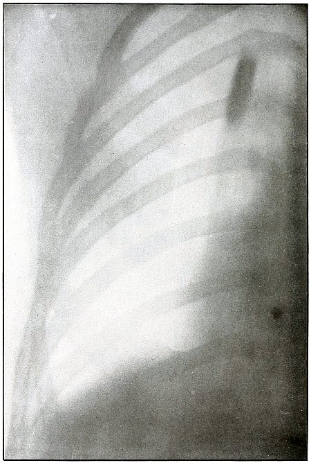
Rifle—Plate 45.
CHEST.
Penetrating Gunshot Wound of the Chest,
with Lodgment of the Projectile Near Posterior Chest Wall.
Point of entrance, pectoral border and fourth rib.
Point of exit, none.
The distinct shadow of the angle of the ribs shows that the posterior
chest wall was next to the photographic plate, and that the larger and
less distinct outline of the anterior portions of the upper ribs was
farther from the plate.
The nearly normal size of the shadow of the projectile shows it to be
much nearer the posterior than the anterior chest wall. The blurred
outline shows it to have moved with respiration. Such conditions
locate its position within the thoracic cavity.
The emergency treatment is antiseptic dressing and rest.
The subsequent treatment depends upon pleural involvement or the
extremely rare infection of the lung.
These cases are nearly all aseptic, and if the great vessels and
nerves of the chest escape injury results are generally favorable.
Plate 46.
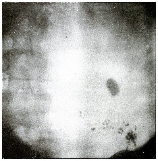
Rifle—Plate 46.
PELVIS.
Gunshot Wound of the Pelvis,
with Lodgment of the Missile in the Abdomen.
The course of the bullet was from behind forward, striking the crest
of the ilium, on which it was deflected, and spattering off some lead
fragments. The slight penetration indicates a velocity of extremely
long range and a striking energy lessened by ricochet.
The irregular outline of the shadow of the projectile shows its
deformity, and the blurred outline indicates intra-abdominal movement
with respiration.
While the missile, as revealed by its shadow, is not a shrapnel ball,
the distribution of lead particles is more suggestive of a shrapnel
than of a rifle projectile, and the ballistic conditions are more
characteristic of the former than of the latter.
There was no abdominal reaction; the invasion of the abdomen was
revealed by the radiograph.
The treatment in such cases is noninterference unless subsequent
developments furnish definite indications.
Plate 47.
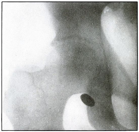
Rifle—Plate 47.
LOWER EXTREMITY.
Gunshot Wound of the Gluteal Region,
with Lodgment of the Bullet Near the Ischium.
Wound of entrance, over gluteal prominence on a transverse line
through the great trochanter.
Wound of exit, none.
There was no bone injury in this case. The bullet, to have lodged in
the soft parts after relatively slight penetration, must have struck
the body at extreme range when its energy was almost spent in flight,
for its normal outline indicates that it was not retarded by ricochet.
The long axis is almost perpendicular to the plate. As the posterior
pelvis was next to the plate, the fairly dense shadow shows the
projectile was not far from the plate and behind the ischium.
The treatment is conservative; infection in such cases is extremely
rare; and only pain or impaired function after many months of
convalescence justifies operation for removal of the missile.
Plate 48.
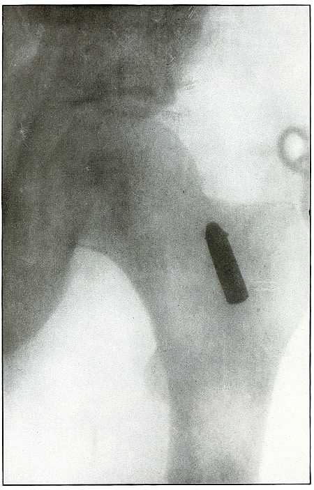
Rifle—Plate 48.
LOWER EXTREMITY.
Gunshot Wound of the Thigh,
with Lodgment of the Bullet.
Wound of entrance, outer aspect of the thigh at the junction of the
upper and middle thirds.
The slight penetration without bone injury and with slight deformity
of the nose of the bullet indicates that the wound was caused by a
ricochet shot at extreme range, after its energy was almost spent.
With the posterior aspect of the thigh next to the plate, the dense
shadow and the nearly normal size of its outline indicate that the
bullet was in the same relative position and that it lay posterior to
the neck of the femur.
As such wounds are rarely infected, the treatment is conservative,
and a search for the missile is only justified by serious infection,
pain, or impaired function.
Plate 49.
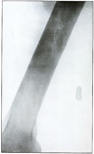
Rifle—Plate 49.
LOWER EXTREMITY.
Gunshot Wound of the Right Thigh,
with Lodgment of the Bullet Behind the Femur.
There is no injury of the bone in this case, as the bullet lodged in
the muscles posterior to the lower third of the femur without striking
the bone. The lighter circular area of the larger end of the shadow
of the projectile shows that its base is farther from the plate than
its nose, which was probably flattened and bent by the ricochet which
reduced its velocity so as to give it but slight power of penetration.
It is not easy to determine from inspection of the plate which side of
the leg lay next to the plate.
With a history of the wound of the right thigh and with the outside
of the leg next to the plate, the projectile must have lain near the
plate on the outside behind the lower end of the femur, midway between
the skin and bone.
The markings seen on the bone are not concerned with the wound, as the
same effect in the plate is seen in the areas beside the bone.
The treatment is conservative; infection is rare.
Plate 50.
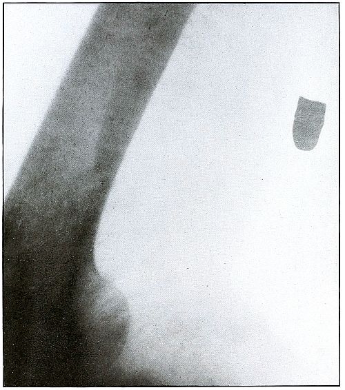
Rifle—Plate 50.
LOWER EXTREMITY.
Gunshot Wound of the Right Thigh,
with Lodgment of the Bullet Behind the Femur.
There is no injury to the bone. The large diameter, shortened length,
and slight density of the shadow show the bullet to be some distance
from and inclining toward the plate and lodged in the muscles behind
the femur, nearer the side away from the photographic plate. It is
difficult to identify the right or left thigh from the radiograph, but
with the history of the wound in the right thigh and the outside of
the leg next to the plate the ball would lie nearer the inside than
the outside of the thigh, nearer the surface behind the femur. As the
shadow shows irregular outline and the location of the bullet low
velocity, the wound was caused by a ricochet shot at very long range.
The treatment is expectant and the course naturally favorable.
Plate 51.
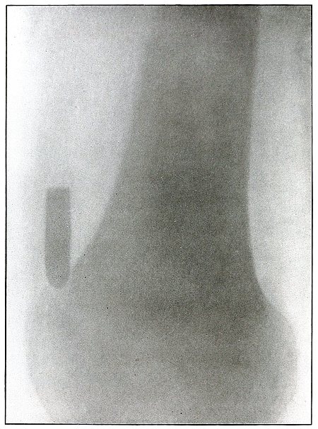
Rifle—Plate 51.
LOWER EXTREMITY.
Gunshot Wound of the Thigh,
with Lodgment of the Missile.
As there is no injury to the bone, the bullet is not deformed. Its
penetrating power was not great enough to carry it through the tissue
so it must have struck the leg at extreme range when its energy was
almost spent.
The actual length of the bullet is 1.25 inches; the length of the
shadow is about 1.50 inches.
The increased length and the relatively slight density of the shadow
indicate the bullet to be some distance from the plate. The case
history places the wound in the right thigh, and the posterior surface
of the leg lay next to the photographic plate. As the density of the
shadow is not greater than the thickest portion of the bone, the
bullet probably lies in front of the border of the outer tuberosity of
the femur.
Although the surgeon’s diagnosis had to be made from the only
available plate, there is something of a speculative element in these
deductions, because if the reaction in the knee joint prevented the
patient from extending the leg the increased length of the bullet
shadow could be accounted for by this position, which would permit the
bullet to lie behind the bone and yet far enough from the plate to
account for the shadow enlargement. The nose of the bullet is at
the epiphyseal line, which is shown in the femur.
Plate 52.
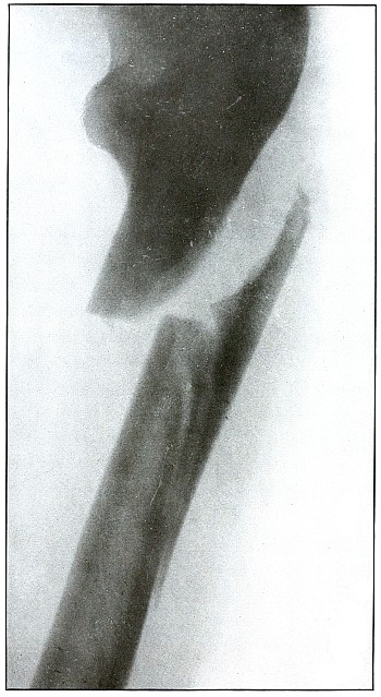
Rifle—Plate 52.
LOWER EXTREMITY.
Gunshot Fracture of the Upper Shaft of the Femur.
The course of the bullet was anteroposterior and pierced the axis
of the shaft of the femur with three radiating lines of fracture,
resulting from the perforating action of the bullet striking the bone
at long range and with greatly reduced energy.
This plate shows the lateral separation of large fragments,
which is typical of gunshot wounds of long range.
Such wounds are usually not infected.
Emergency treatment is antiseptic dressing and coaptation with
extension and temporary splint, so that it may support the bone for
transportation and may be easily removable at place of continued
treatment.
In these cases with lateral separation of fragments, it is imperative
to supplement extension with pressure in a line perpendicular to the
long axis of the femur.
Plate 53.
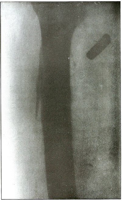
Rifle—Plate 53.
UPPER EXTREMITY.
Gunshot Fracture of the Shaft of the Femur
with Lodgment of the Bullet.
The course of the bullet was antero-posterior and diagonally inward
from the antero-external border of upper third of the thigh. A thin
longitudinal fragment was split off without transverse fracture.
The missile struck the thigh after its energy had been greatly
reduced by ricocheting as a result of striking a resisting object
which flattened its nose and “set up” its body, as shown by the wavy
outlines of the shadows.
The dense and normal-size shadow shows the bullet to be near the plate
and probably in the muscles superficially behind and below the lesser
trochanter.
As the prominent outline of the lesser trochanter shows that the
leg was in external rotation when the negative was made, it is
evident that, with the rotation back to the anatomical position, the
projection of the shadow of the bullet would fall close to or in line
with the shaft of the femur; the position of the bullet is behind the
femur.
The treatment is conservative, with no trouble to be expected from infection.
Plate 54.
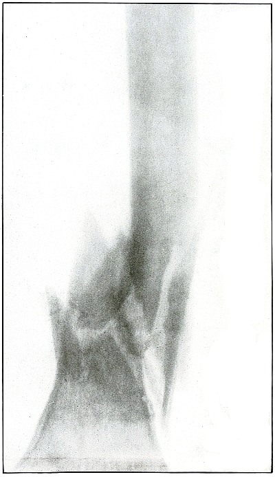
Rifle—Plate 54.
LOWER EXTREMITY.
Gunshot Fracture of the Lower End of the Shaft of the Femur.
The course of the bullet was anteroposterior through the axis of the
femur. Several large fragments which were not displaced were separated
by the force of impact. The separation of the fragments and the
overriding of the ends of the proximal and distal large fragments were
due to bearing bodily weight or to muscular contraction.
The projectile causing the wound was moving with the velocity of mid
range. The wound of exit was not lacerated.
The emergency treatment is antiseptic dressing and temporary splint
immobilization. Permanent dressing, with extension and lateral
compression, is the rule.
Infection in such cases is frequent owing to lack of facilities for
proper dressing on the field.
Results in saving life and limb are generally good.
Plate 55.
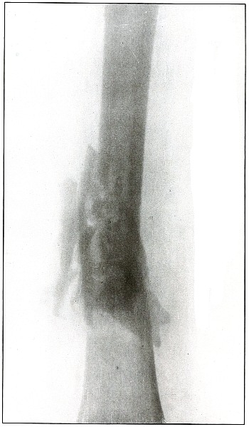
Rifle—Plate 55.
LOWER EXTREMITY.
Gunshot Fracture of the Lower Third of the Shaft of the Femur.
The course of the bullet was diagonally anteroposterior, with a
velocity near mid range, without causing much displacement of fragments.
The wound of entrance and exit would be almost the same in appearance.
Treatment and results would be similar to case shown on plate 54.
Many of these wounds are infected, due, no doubt, to the difficulties
of arranging a clean first-aid dressing and effecting satisfactory
immobilization during the first stage of transportation.
Infection from clothing carried into the wound is rare, as the fairly
high velocity of the bullet causes a spreading of the fibers without
division or punched-out section before the bullet.
As a rule the infected cases of this class recovered without loss of
limb. Amputation was very rare.
Plate 56.
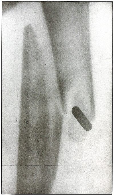
Rifle—Plate 56.
LOWER EXTREMITY.
Gunshot Fracture Below the Middle of the Femur,
with Lodgment of the Bullet Near the Fracture.
The course of the projectile was transverse. The long splitting
fracture, with few large fragments and the lodged undeformed missile,
indicate that the injury to the bone was caused by the missile
striking the bone with large cross section or at an inclined angle so
that all of the remaining energy of the projectile at long range was
absorbed by the bone.
Had the point of the ball struck the bone with the same energy, it
would have produced smaller fragments and might then have passed
beyond the bone. The normal size of the diameter, slightly shortened
length, greater density of the point of the shadow, shows the bullet
to lie behind the bone with its nose pointing slightly backward. The
actual length of the bullet is 1.25 inches: the length of the shadow
is 1 inch.
Treatment and results would be about the same as in
plates 49 and 50.
Plate 57.
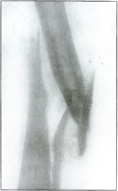
Rifle—Plate 57.
LOWER EXTREMITY.
Gunshot Fracture About the Middle of the Femur,
with Lodgment of the Fragments of a Deformed Bullet.
The course of the missile was transverse. All of the remaining energy
of the retarded velocity of the short range of a ricochet shot was
stopped by the bone with the result of a long splitting fracture, and
the lodgment of one large and a few small fragments of the missile.
The small notched metal fragment lying to the right of the upper
bone fragment is a small bent piece of the jacket, detached from the
greatly deformed lead core, which can be faintly seen lying behind the
lower end of the left side of the upper bone fragment.
The treatment is extension with lateral compression, although this
case, showing by callus formation advancing convalescence, reveals
very bad position.
Results as to life and limb are favorable in such cases, but some
deformity is to be expected.
It should be noted that this is a case from Gulhané Hospital, the best
military hospital in Constantinople, where the surgical service, under
Prof. Wieting Pasha, was skillfully conducted.
Plate 58.
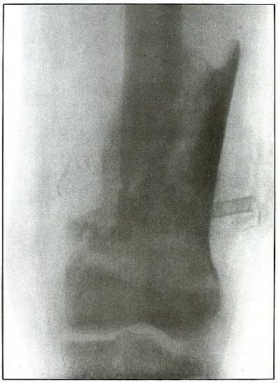
Rifle—Plate 58.
LOWER EXTREMITY.
Gunshot Fracture of the Lower End of the Femur.
The course of the bullet was anteroposterior.
The long, oblique, splitting fracture without separate fragments
indicates the long range of the missile in low velocity.
The wound was infected as is indicated by the drainage tubes in place.
The well-advanced callus formation indicates established convalescence.
Results are favorable for recovery with only fair position and some
shortening.
It may be observed that this is also a case from Gulhané Hospital.
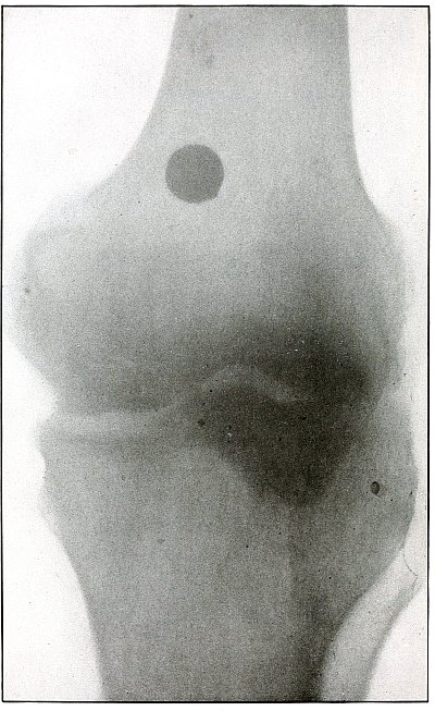
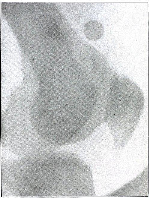
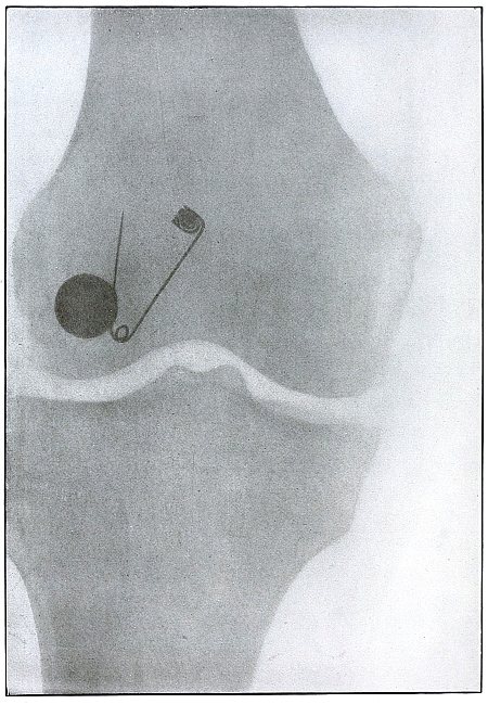
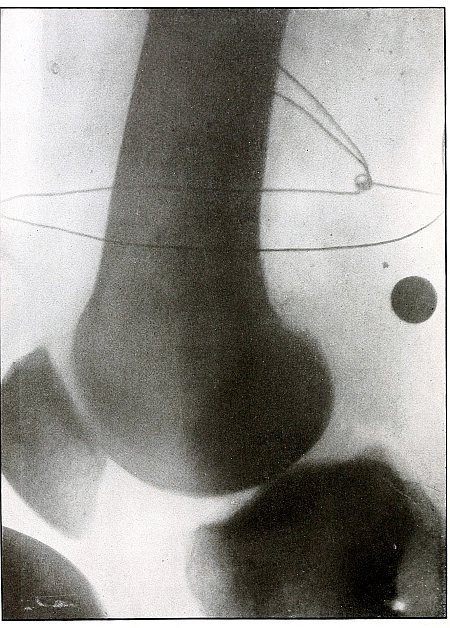
Plate 59.
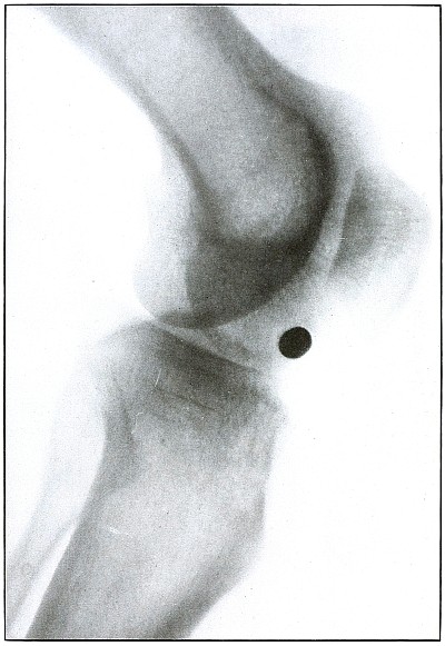
Rifle—Plate 59.
LOWER EXTREMITY.
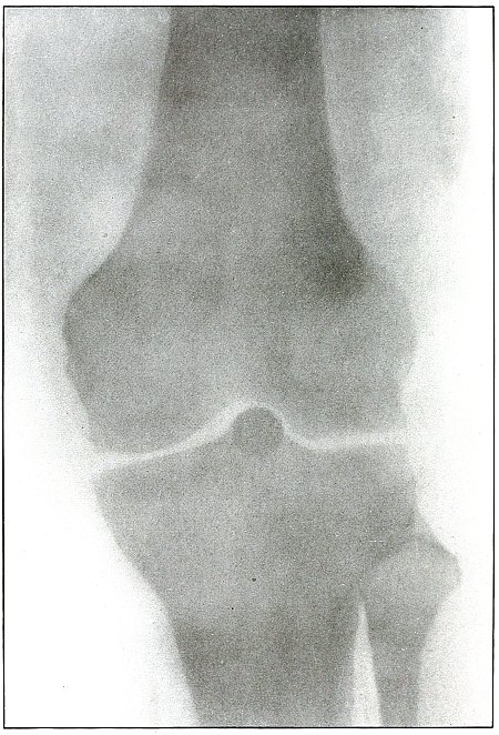
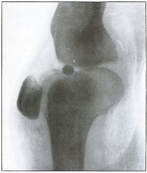
Gunshot Wound of the Left Knee-Joint,
with Lodgment of the Missile in the Joint.
The course of the bullet was transverse, entering the capsule
posterior to the patella without injury to the bone.
As its shadow projection is almost circular, the bullet must be
standing almost on its end pointing toward the plate with its long
axis almost parallel to the line of projection.
As the fibular side of the leg lay next to the plate and as the only
slightly enlarged shadow of the bullet indicates it to be near the
plate, its position is in the joint near the fibular side.
As the bullet is undeformed and its penetrating power very slight, its
velocity was that of extreme range.
The emergency treatment, is, of course, a simple antiseptic dressing
with the leg held in the most comfortable position by muscular action.
The subsequent treatment is removal of the bullet when proper surgical
conditions obtain.
Plate 60.
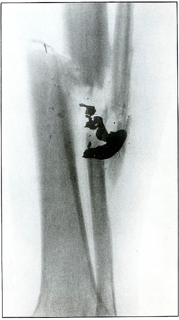
Rifle—Plate 60.
LOWER EXTREMITY.
Gunshot Fracture of the Tibia and Fibula,
with Lodgment of the Missile.
The course of the bullet was diagonal from within outward and backward
about the middle of the leg, with the impact tangential on the tibia
and direct on the fibula. The bullet lies just behind the tibia.
It is apparent that the bullet has been greatly deformed and that its
jacket has been badly torn from the core. The force of impact on the
object from which it ricocheted must have been contributed by the
velocity of short range, which reduced the striking energy so greatly
that the bullet was lodged by the resistance of the tibia and fibula.
The wound was not infected, and callus formation shows that repair has begun.
The treatment in such cases, without infection, is noninterference.
The lodgment of the missile need not prejudicate the prognosis, and
certainly the additional damage in the search for the bullet is not
warranted, except under special indications.
Plate 61.
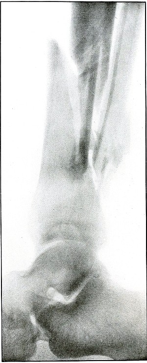
Rifle—Plate 61.
LOWER EXTREMITY.
Gunshot Fracture of the Lower Ends of the Tibia and Fibula.
The course of the bullet was transverse, with the velocity of
mid-range.
The fragmentation of the fibula, lying close to the skin, would
produce considerable laceration in the wound of exit.
The treatment is conservative. Infection would depend almost entirely
upon the integrity of the first dressings and immobilization.
Results should be favorable, with care in subsequent treatment.
Plate 62.
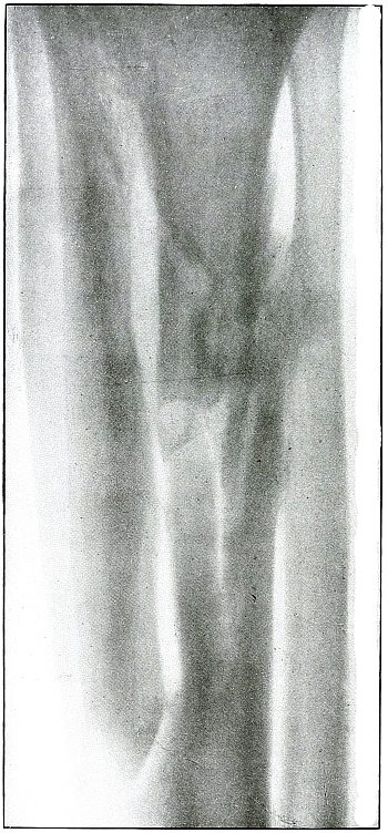
Rifle—Plate 62.
LOWER EXTREMITY.
Gunshot Fracture of the Upper Third of the Tibia,
with Large Longitudinal Fragment.
The course of the bullet was anteroposterior and slightly diagonal
from without inward through the shaft of the tibia.
The injury was due to the energy of impact of a bullet, in the high
velocity of short range, striking the axis of the diaphysis, in which
the greater part of its energy was expended in pushing away a wall of
the canal.
Convalescence is well established without infection, as shown by
callus formation.
The treatment in such cases is invariably conservative, with the
removal of such fragments as may be detached by suppuration and sequestration.
Plate 63.
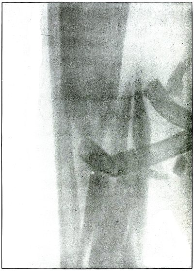
Rifle—Plate 63.
LOWER EXTREMITY.
Gunshot Wound of the Middle of the Tibia,
with Few Large Fragments.
The course of the bullet was transverse from without inward.
The direct impact of the bullet, in high velocity of short range, has
produced the typical “X” fracture due to the radiating lines of force.
The wound was infected, as is shown by the drainage tubes in the wound.
The emergency treatment in such cases is simple antiseptic dressing
and temporary splint immobilization.
The subsequent treatment is the management of the infection.
The results in such cases are favorable.
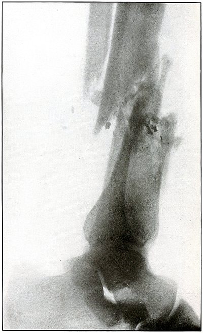
Plate 64.
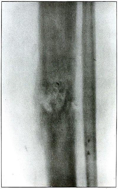
Rifle—Plate 64.
LOWER EXTREMITY.
Gunshot Fracture of the Tibia.
The course of the bullet was diagonal, from without inward and from
before backward through the middle of the tibia. Small particles of
metal have lodged at the site of the fracture—a condition which never
occurs in a rifle wound with the jacket of the bullet intact, while it
is the invariable accompaniment of a shrapnel wound of a bone.
It is inferred that the jacket of the bullet in this case was damaged
by ricochet, or that some metal particles from the object against
which the bullet ricocheted were carried into the wound, as some other
small pieces of metal are seen in areas distant from the seat of fracture.
As the fragments are small and not displaced, the velocity of
the missile, at least that of mid-range, was almost sufficient to
perforate the bone without fracture.
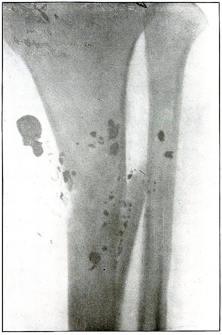
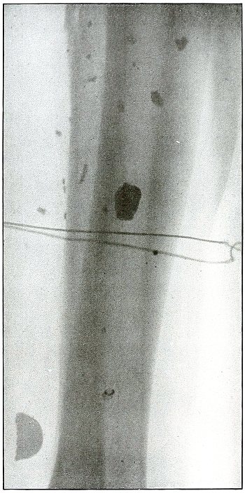
Plate 65.
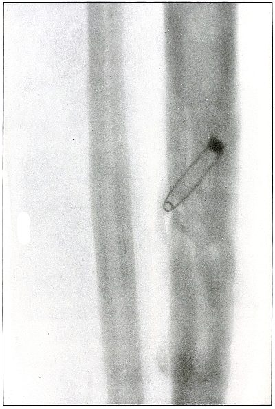
Rifle—Plate 65.
LOWER EXTREMITY.
Gunshot Fracture of the Tibia.
The course of the bullet was transverse, from within outward, striking
the bone near the outer border with the velocity of mid or long range,
producing long fissures without separation of fragments.
The safety pin, of course, lies in the dressings and on the side
away from the plate, as shown by its somewhat indefinite outline and
increased length.
The wounds of entrance and exit are practically the same.
The treatment in such cases is that of a simple fracture, except
for the management of an occasional infection, and the results are favorable.
Plate 66.
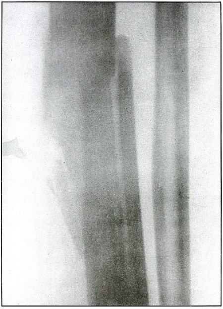
Rifle—Plate 66.
LOWER EXTREMITY.
Gunshot Fracture of the Middle of the Tibia,
with Lodgment of the Missile.
The course of the bullet was transverse, from without inward, striking
on the side of the shaft of the tibia.
The bullet was so badly damaged by ricochet that only a portion of it
was the cause of this wound.
The range was short, if not close, as the missile after striking a
resisting object with force enough to break itself retained enough
energy in a fragment of less than half its mass to cause a long
fissure fracture, with the separation of smaller fragments.
The treatment is noninterference, except for infection, which,
contrary to what might be expected from presumable contamination from
the object from which it ricocheted, does not occur more frequently in
ricochet than direct wounds with lodgment of the projectile.
Plate 67.
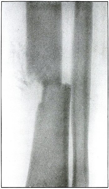
Rifle—Plate 67.
LOWER EXTREMITY.
Gunshot Fracture of the Lower Third of the Tibia.
The course of the bullet, with reduced energy of long range, was
anteroposterior, striking the inner border of the bone and punching
out a circular area of small fragments with a single transverse line
of fracture.
The wound of exit was slightly larger than the wound of entrance.
The treatment is conservative. Infection is not probable if emergency
dressing is clean.
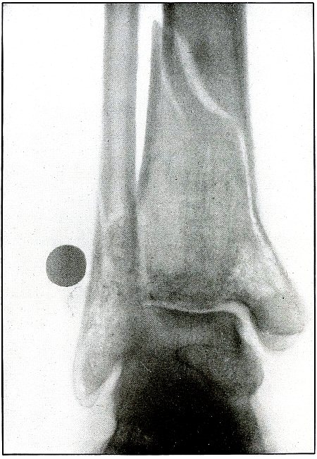
Plate 68.
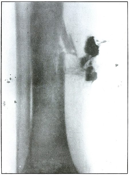
Rifle—Plate 68.
LOWER EXTREMITY.
Gunshot Fracture of the Lower Third of the Tibia.
The course of the bullet was diagonally anteroposterior, from without
inward, striking the internal border of the anterior surface of the
bone, and partially splitting off fragments from the side with a
perforating effect.
The range of the shot was long.
The dense and irregular shadows to the right of the fracture are
caused by the material used in dressing and indicate a slight
infection. The small shadows on the tibial side are not a part of the
wound, but are due to opaque material caught in the dressing.
Plate 69.
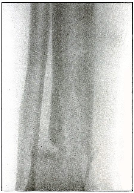
Rifle—Plate 69.
LOWER EXTREMITY.
Gunshot Fracture of the Lower End of the Tibia.
The course of the bullet was transverse, from without inward, through
the lower end of the bone, with a piercing effect and a fissuring of
the upper fragment.
The velocity was that of short range.
The wound of exit would be slightly larger than that of entrance, as
some small fragments can be seen extending along the tract of the
missile from the line of transverse fracture toward the internal
border of the leg. There was no laceration of the wound of exit. The
wound was clean.
The treatment is conservative.
Results should be favorable. Infection would depend most probably upon
the asepsis of the first dressing.
Plate 70.
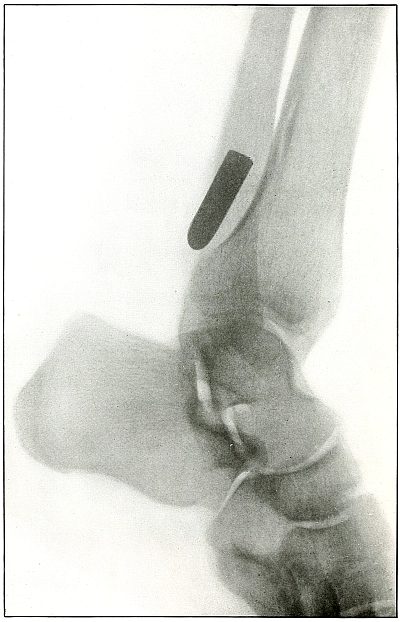
Rifle—Plate 70.
LOWER EXTREMITY.
Gunshot Fracture of the Tibia,
with Lodgment of the Missile.
The course of the bullet was from within outward, striking the
posterior surface of the tibia about 2 inches above the ankle, and
causing a slight crack in the bone at the point where its course was
deflected.
The velocity was that of extreme range, as the wholly normal outline
of the projectile and the slight penetration indicates that its energy
was almost entirely lost in flight and not by ricochet.
The sharp outlines of the lower border of the fibula and the external
border of the articular surface of the lower end of the tibia indicate
the position of the fibula as next to the photographic plate.
The bullet lies at a very slight angle with the plate, as is shown
by the curved outline of its base, which condition alone would give a
projection shadow somewhat shorter than the bullet. But as the shadow
is actually somewhat longer than the bullet (about one-eighth inch,
or one-tenth its length), the position of the bullet is some distance
from the plate and most probably lies behind the tibia, at the inside of the fibula.
Plate 71.
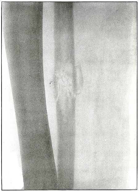
Rifle—Plate 71.
LOWER EXTREMITY.
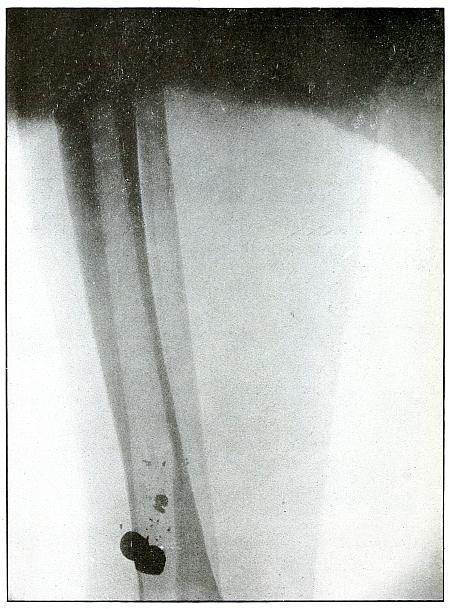
Gunshot Fracture of the Fibula.
The course of the bullet was anteroposterior through the lower third
of the leg, striking the fibula squarely, passing through the bone
with a perforating effect, accompanied by slight fragmentation and
with a reduced velocity of long range.
The wounds of entrance and exit would be almost the same in
appearance. Asepsis in such cases is the almost invariable rule, and
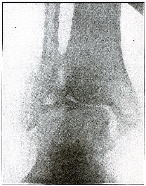
the treatment after the simple dressing is that of a simple fracture.
Plate 72.
Rifle—Plate 72.
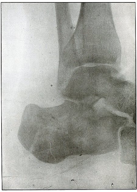
LOWER EXTREMITY.
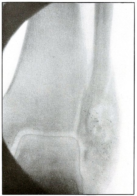
Gunshot Fracture of the Ankle.
The course of the bullet was anteroposterior, striking the fibula
from behind with a velocity of long range, and causing some slight
fragmentation without displacement of the fragments.
The joint architecture is slightly disturbed. The joint mortice is
a bit widened by the external deflection of the external malleolus,
which permits a slight outward rotation of the astragalus.
As the dangers of infection are usually escaped,
the treatment is that for Pott’s fracture.
Plate 73.
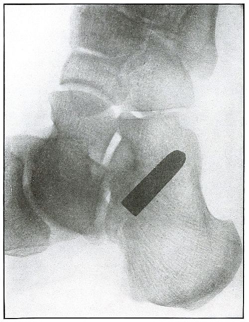
Rifle—Plate 73.
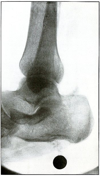
LOWER EXTREMITY.
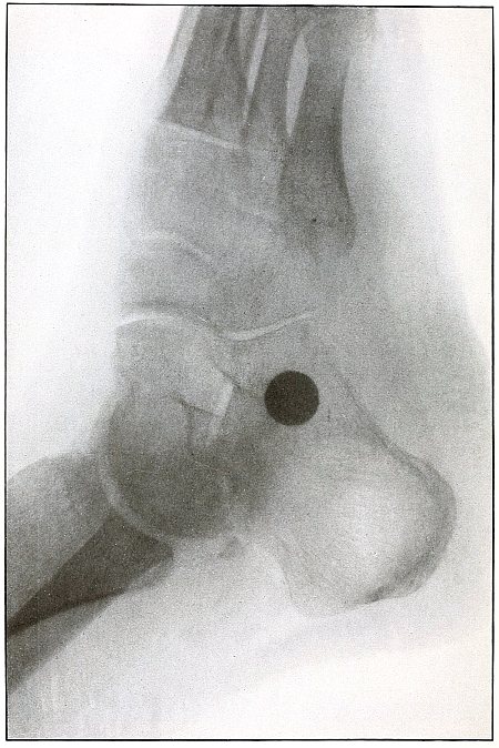
Gunshot Wound of the Heel,
with Lodgment of the Missile.
The course of the bullet was from behind forward through the insertion
of the tendo Achillis and its lodgment along the outer border of the
os calcis.
There was no injury to the bone. The path of the bullet is shown by
the slight mottling above the posterior extremity of the os calcis.
The nose of the bullet is slightly deformed by ricochet at long range.
The very slight penetration and the slight deformity of the nose of
the bullet indicates a velocity of extreme range of both impact of the
ricochet and of the wound.
The sharp outline of fibula and the base of fifth metatarsal shows the
fibula to be next to the plate. The only slight enlargement and square
base of the shadow of the bullet show it to be parallel to the plate,
or at right angles to the line of projection, and thus indicate its
position to be on the fibula side of the os calcis, below the tip of
the external malleolus.
Plate 74.
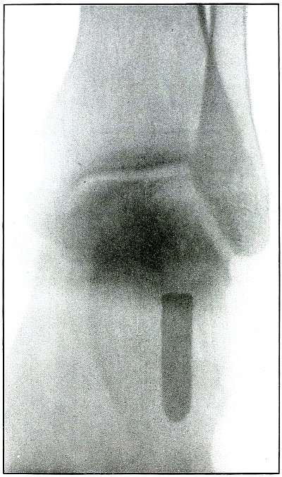
Rifle—Plate 74.
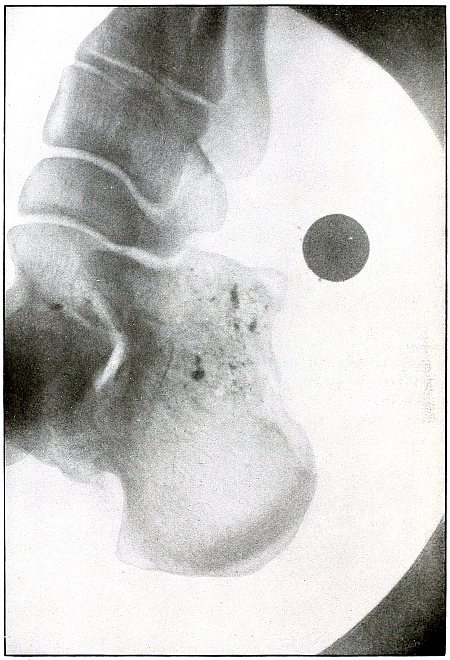
LOWER EXTREMITY.
Gunshot Wound of the Heel.
This is the same case as shown in plate 69,
but with the shadow projected from above downward instead of from side
to side, as in the preceding plate. The interpretation of the shadows
in the preceding plate is thus confirmed.
As the heel lay on the plate, the projectile at a sharp angle with the
plane of the plate, several inches farther from the plate than in the
preceding radiograph, and with the line of projection at about right
angles to the long axis of the projectile, the shadow projection is considerably enlarged.