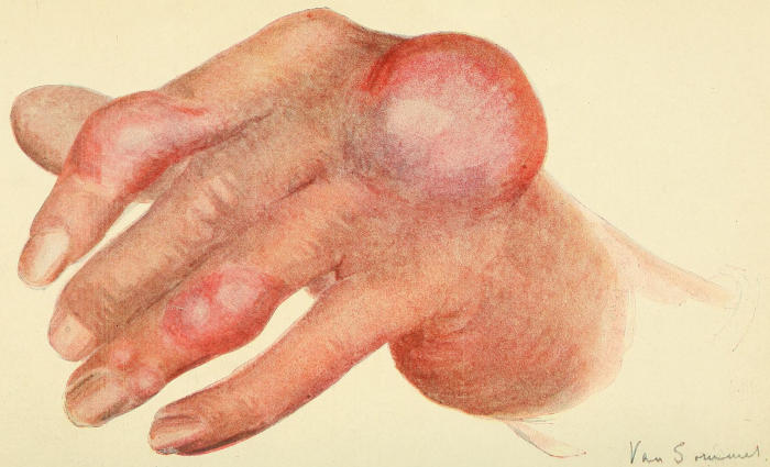
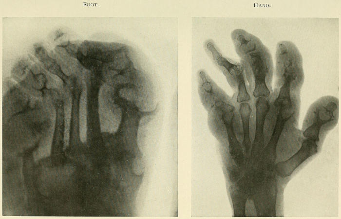
Note large tuberous swellings on knuckle and
metacarpo-phalangeal joints due to uratic deposits.
BY
LLEWELLYN JONES LLEWELLYN, M.B. Lond.
GOVERNOR AND SENIOR PHYSICIAN, ROYAL MINERAL WATER HOSPITAL, BATH;
FELLOW OF ROYAL SOCIETY OF MEDICINE; AUTHOR OF “ARTHRITIS DEFORMANS”;
CO-AUTHOR OF “FIBROSITIS”; CO-AUTHOR OF “MALINGERING, OR THE SIMULATION
OF DISEASE”; CO-AUTHOR OF “PENSIONS AND THE PRINCIPLES OF THEIR
EVALUATION”; CONTRIBUTOR TO LATHAM AND ENGLISH’S “SYSTEM OF TREATMENT,”
ALSO TO “OXFORD ENCYCLOPÆDIA OF TREATMENT” AND TO “STUDENT’S
TEXT-BOOK OF SURGERY.”
WITH A SECTION ON
OCULAR DISEASE IN THE GOUTY BY
W. M. BEAUMONT
CONSULTING OPHTHALMIC SURGEON TO THE SOUTH-WESTERN
REGION OF THE MINISTRY OF PENSIONS; AUTHOR OF “INJURIES
OF THE EYES OF THE UNEMPLOYED, PROBLEMS IN PROGNOSIS,” ETC.
ST. LOUIS
C. V. MOSBY COMPANY 1921
Printed in Great Britain.
Dedicated
TO
MY WIFE
PREFACE
“A knowledge of the real nature of gout ... is, in my
opinion, at the very foundation of all sound pathology,” wrote
Todd many years since; and the passing years have but invested
his reflection with deeper significance and something of prophetic
insight. For who can doubt that he who would elucidate the
pathological groundwork of gout must be at once a clinical
physician, a bio-chemist, a bacteriologist, a morbid anatomist?
and well may we ask, Who is sufficient for all this?
How vivid the light thrown upon the problems of clinical
medicine by the bio-chemists! The story of the fate of protein
and purin substances in the animal body, at one time a medley of
guesses and gaps, is gradually evolving into one of relative certitude
and completeness. Revolutionary, in truth, the change, and
many a cherished shibboleth has been ruthlessly cast aside!
With admiration not unmingled with awe we see them laying
well and truly the foundations upon which in the ultimate
scientific medicine must inevitably rest.
Of these the very corner-stones are chemical physiology and
chemical pathology, the rapid evolution of which is profoundly
altering our conceptions of health and disease. Those vital processes
of the organism that but yesterday we saw “as through a
glass, darkly,” are now in great part illumined, and the distortions
wrought in them by disease made more manifest.
How pregnant, too, with warning their findings! Processes
that to our untutored minds seem simple are revealed as infinitely
complex. Through what a maze must we thread our way if we
would disentangle the intricacies of metabolism! Intricate
enough, forsooth, in health, but how much more so in disease!
For, as Sir Archibald Garrod eloquently phrases it, “it is becoming
evident that special paths of metabolism exist, not only for
proteins, fats, and carbohydrates as such, but that even the individual
primary fractions of the protein molecule follow their several
katabolic paths, and are dealt with in successive stages by series
of enzymes until the final products of katabolism are formed. Any
of these paths may be locked while others remain open.”
It is with chastening reflections such as these that we may best
approach our study of gout, that riddle of the ages upon which
so many physicians from time immemorial have expended their
dialectic skill. But, vast though the increase in our knowledge
of the chemical structure of uric acid and its allies, uncertainty
still dogs our steps, and, doubtful of the pathway to solution of
the pathological mystery of gout, we must perforce approach the
problem in a more strictly catholic attitude.
Uric acid has apparently failed us as the causa causans. Neither
this substance nor its precursors can be held responsible for the
fever, local inflammation and constitutional disturbances in gout,
being, as they are, practically non-toxic. Albeit, though I hold
this view, I do not for one moment suggest that uric acid has
nothing whatever to do with gout. The fact that tophi, its pathognomonic
stigmata, are compounded of biurate of soda, would
per se stamp such an attitude as untenable. On the other hand,
uric acid must be viewed at its proper perspective as a concomitant
or sequel of gouty inflammation, the essential cause of which must
be sought elsewhere.
“The old order changeth, giving place to new,” and happily
with the advent of bacteriology our views, or rather our hazards,
as to the nature of joint diseases underwent profound modification.
But, strange to say, though quick to apprehend the significance
of infection, its causal relation to other joint disorders, we
still seem unaccountably loth to discard our timeworn conception
of “gouty” arthritis as of purely metabolic origin. This to my
mind is the more remarkable in that the onset, clinical phenomena,
and course of acute gout, and no less the life history of the
disorder as a whole, are emphatically indicative of the intrusion
of an infective element in its genesis.
The extreme frequency with which infective foci are met with
in the victims of gout, the frequency, too, with which exacerbations
of the disorder are presaged by acute glandular affections of
undeniably infective source, is by no means adequately realised.
For our forefathers gout began, and, forsooth, often ended, in the
“stomach,” or it was the “liver” that was impeached. But the
portal to the alimentary canal was for them only a cavity, the
contained structures of which, albeit, to their mind often betrayed
evidences of a “gouty diathesis.” They distinguished “gouty”
teeth, “gouty” tonsillitis, “gouty” pharyngitis, even “gouty”
parotitis; but all these they classed as tokens or sequelæ of gout,
not possible causes or excitants thereof.
Now as to the true significance of these acute glandular affections
held by clinicians of repute to be of “gouty” origin. What
of “gouty” tonsillitis, pharyngitis, parotitis? Still more, what
of our deductions regarding the relationship of these same when
met with in association with non-gouty forms of arthritis? Do we
not hold them each and all as evidences of infection? and, we may
well ask, why not in gout?
The marvel then is that even to-day many still hold that the
tonsillitis, pharyngitis, even the gingivitis, like the subsequent
articular lesions, are one and all attributable to the underlying
gout. We certainly should not do so in the case of any arthritis
other than “gouty,” and to my mind the time is ripe for a change
of attitude.
The “gouty” throats, like the “gouty” teeth, should be
regarded not as symptomatic of gout, but etiologically related
thereto. We should cease to talk of “gouty” throats, teeth,
etc., should renounce the prefix, for there is nothing specific of
gout either in the tonsillar, pharyngeal, or dental lesions. We
should instead view these various local disorders in their true
perspective as foci of infection, causally related to the subsequent
and secondary “gouty” arthritis.
Similarly, when we come to analyse the component elements of
an acute paroxysm of gout, how strongly indicative of the intrusion
of an infective element the following features: the onset,
temperature curve, character of local articular changes of the
disorder, the presence of leucocytosis, with secondary anæmia
and enlargement of the lymphatic glands! Again, how suggestive
the occasional complication of acute gout by lymphangitis
and phlebitis! Of like significance, too, the paroxysmal nature
and periodicity of the disorder, and the compatibility of the
morbid anatomical changes and the cytological content of the
aspirated joint fluid with their genesis by infection.
As to correlation of the metabolic phenomena of gout with the
postulated infective element, I would suggest that, although
abnormalities of metabolism form an integral part of gout, they
are of themselves inadequate to achieve its efflorescence. As we
shall see when we come to consider those elemental manifestations
of gout, i.e., uratic deposits, or tophi, neither the purely physical
nor the purely chemical theory of their origin will suffice, nor,
for that matter, can any solution of their formation be gleaned
from even a blend of the twain. In short, such hypotheses are
too mechanical.
The intrusion of some other factor, “something vital, something
biological,” seems essential for the elucidation of uratosis,
i.e., uratic deposition. For this, not uricæmia, is the specific
characteristic phenomenon of gout. If we cannot explain uratosis
on physical or chemical grounds, then how much less, in view of
the non-toxicity of uric acid, can we on this basis account for the
inflammatory phenomena of the disorder!
Now inflammatory reaction is, I hold, an invariable antecedent
in all gouty processes, whether of articular or ab-articular site.
Granted that inflammatory reaction is a necessary prelude, the
specificity of gout is attested by the fact that the same is followed
by local deposition of urates. But while this sequential uratic
deposition invests all forms of “gouty” inflammation with a
specific character unshared by any other disease, it follows that
the cause of the said inflammation must, if possible, be ascertained.
Now, as I believe, “gouty” subjects are ab initio victimised by
innate tissue peculiarities, doubtless reflected in corresponding
obliquities of tissue function and metamorphosis, and through
their medium the general resistance of the body to invasion by
infections is lowered; in other words, under the influence of
these morbific agencies the latent morbid potentialities of the
gouty become overt and manifest. For in the gouty, as Walker
Hall observes, “a slight injury or indiscretion of diet, an overloaded
intestine, or increased toxicity of the intestinal flora, may
be followed by a disturbance of the general nuclein metabolism
and a local reaction in certain tissues.”
Enough has been said to disclose the dominant trend of this
work, and although there are many aspects of the subject in
regard to which I hold somewhat iconoclastic views, yet exigencies
of space forbid me even to allude to them in this foreword. I
hasten therefore to discharge the pleasing duty of acknowledging
my great indebtedness to the acumen and discrimination which
has been brought to bear on this subject by a long succession of
eminent physicians, in proof of which I need only adduce the
names of those giants of the past the illustrious Sydenham, Sir
Thomas Watson, Sir Charles Scudamore, Jonathan Hutchinson,
not to mention Trousseau, Charcot, Lecorche, and Rendu. But
I should fail in my duty did I not in a special sense express my
deep indebtedness to the classic and epoch-making work of Sir
Alfred Garrod. For the rest, too, I have derived much enlightenment
from Sir Dyce Duckworth’s treatise and the various works
on the subject by Luff, Lindsay, and others.
From the bio-chemical aspect I owe much to the researches of
Walker Hall, and to those of our American confrères Folin, Denis,
Benedict, Pratt, McLeod, Walker Jones, Gideon Wells, etc.
Reverting to my own colleagues at the Royal Mineral Water
Hospital, Bath, I would tender my deep thanks to the Honorary
Physicians, Drs. Waterhouse, Thomson, Lindsay, and King
Martyn, for the uniformly generous manner in which they afforded
me opportunities for studying cases under their care.
To Dr. Munro, our senior pathologist, I am especially beholden
for invaluable, nay indispensable, help in the matter of blood
examinations, the cytological study of joint fluids, and the
microscopic verifications of tophi. To Dr. MacKay also my
cordial thanks are due for the skiagraphs contained in this
work.
For the section dealing with the ocular disorders met with in the
gouty my most sincere thanks are due to Mr. W. M. Beaumont,
of Bath, whose singularly wide experience in this sphere renders
him unusually equipped to deal with this highly controversial
aspect of gout. To Drs. Cave and Gordon, of Bath, also I am
indebted for many valuable suggestions kindly afforded me while
writing this volume. To my brother Dr. Bassett Jones I am
under deep obligation for unwearying assistance in our joint
endeavour to ascertain the exact relationship of gout to lumbago,
sciatica, and other types of fibrositis.
For the preparation of the index of this work I would proffer my
grateful thanks to Mr. Charles Hewitt and to Miss Donnan and
Miss Crosse for having undertaken the arduous task of typing the
manuscript thereof.
Lastly, I would express my thanks to my publisher, Mr. Heinemann,
for much consideration and many courtesies.
LL. J. LL.
31, Upper Brook Street, W. 1.
TABLE OF CONTENTS
CHAPTER I HISTORICAL AND INTRODUCTORY
The Antiquity of Gout. Prevalence of Gout in the Anglo-Saxon Period.
Views of the Humoralist. The Aphorisms of Hippocrates. Introduction
of the Word Gout. Early Views as to the Nature of Tophi.
The “Honour of the Gout.” That Gout confers Immunity from other
Disorders. Growing Infrequency and Attenuation of Gout
Tardy Dissociation of Chronic Gout. Identification of Muscular Rheumatism.
Differentiation of Chronic Gout from Arthritis Deformans.
Cleavage of Arthritis Deformans into Two Types. Elimination of the
Infective Arthritides
Revelations of the Bio-chemist. The Formation of Urea. Fate of the
Amino-acids. Seat of Formation of Urea. Amino-acids in Relation
to Gout. The Glycocoll Theory of Gout. Urea Excretion in Gout.
Creatine and Creatinine. Inborn Errors of Metabolism
The Isolation of Nucleic Acid. Researches on Spermatozoa. The Discovery
of Purins. Uric Acid a Derivative of Nucleic Acid. The
Chemistry of Uric Acid and the Purin Bodies. Chemical Constitution.
Properties of Uric Acid. Uric Acid in the Blood. Gudzent and
Schade’s Theories. Organic Combinations. Complexity of the
Problem
Exogenous Purins. Exogenous Uric Acid Excretion. Fate of the Unexcreted
Purins. Endogenous Purins. Source of Endogenous Purins.
Proteins and their Derivatives. Amino-acids and Dicarboxylic
Amino-acids. Endogenous Uric Acid Excretion. Factors influencing
Endogenous Uric Acid Excretion. Physiological Conditions. Pathological
States. Ingestion of Certain Drugs. Synthetic Formation of
Uric Acid
Uric Acid Excretion in Gout. Uric Acid Variations in Acute Gout. Uric
Acid Variations in Chronic Gout. Retarded Exogenous Uric Acid
Output. Lowered Endogenous Uric Acid Output. Other Anomalies
in Excretion in Gout. Purin Metabolism in other Disorders. Purin
Metabolism in Chronic Alcoholism and Plumbism
Anomalies in Uric Acid Excretion in Gout. Uricæmia in Nephritis. The
Relationship, if any, between the Amounts of Uric Acid and of Urea,
and Total Non-protein Nitrogen in Human Blood. Uricæmia not
necessarily due to Renal Defect. Uricæmia not Peculiar to Nephritis.
Uricæmia does not necessarily Portend Gout. To what may be ascribed
the Deficient Eliminating Capacity of the Kidney for Uric Acid.
Uratic Deposits in Nephritis. Differentiation of Uratic Deposits in
Gout and Nephritis. Clinical Associations of Gout and Granular
Kidney
Folin and Denis’s Method. Uric Acid a Normal Constituent of Blood.
Effect of Exogenous Purins. Uric Acid Content of Blood in Gout.
Hyperuricæmia in Non-gouty Arthritis. Variations in Uric Acid
Content of Blood independently of Diet. What Relationship, if any,
Exists between the Uric Acid Content of the Blood and Attacks of
Gout. Discussion of the Foregoing Data. The Significance of Uricæmia.
Sources of Fallacy in Uric Acid Estimation. Disabilities of Modern
Tests. Need for further Investigations
Constitution of Tophi. Mode of Formation. Localisation of Uratic
Deposits. The Causation of Tophi. Solubilities of Uric Acid. Tophi
in Relation to Uricæmia. Tissue Affinities for Uric Acid. Retention
Capacity of Tissues for Uric Acid. Clinical Evolution of Tophi. The
Cause of the Inflammatory Phenomena. Non-toxicity of Uric Acid.
Are the Precursors of Uric Acid Toxic?
Boerhaave’s Forecast of the Infective Theory. Ringrose Gore on Infective
Origin. Leucocytosis in Acute Gouty Polyarthritis. Chalmers
Watson’s Researches on Gout in a Fowl. Trautner’s Suggestion of a
Specific Infection
Local Foci of Infection: Dental, Nasal, Pharyngeal, etc. Gastro-intestinal
Disorders. Variation in Free HCL. Intestinal Disorders.
Infection or Sub—infection
Analysis of the Acute Paroxysm. The Evolution and Life History of
Gout. Analogies between Gout and the Specific Infective Arthritides.
Correlation of the Metabolic Phenomena of Gout with the Postulated
Infective Element
Acute Localised Gout. Prodromal Symptoms. Dyspepsia. Premonitory
Symptoms of Tophus Formation. Premonitory Articular Pains.
The Acute Paroxysm. Detailed Consideration of Phenomena. Mode
of Onset. Localisation. Nature of Pain. General Phenomena.
Pyrexia. Changes in the Blood. Uric Acid Excretion. Local Phenomena.
Tophus Formation
Acute Gouty Polyarthritis. Mode of Invasion. Distribution of Lesions.
Local Characters. Constitutional Symptoms. Changes in the Blood.
Leucocytosis. Collateral Phenomena of Gout. Lumbago, Sciatica,
etc. Incidence of Gouty Stigmata in Various Types of Fibrositis
Chronic Articular Gout. The Joint Deformities of Chronic Gout. Tophi:
Their Evolution and Distribution. Other Sites of Tophi. Affinities
between Gout and other Diseases. Gout in Relation to Glycosuria.
Gout in Relation to Phlebitis. Cutaneous Disorders. Gout and
Nephritis. Prognosis in Gout
Articular Gout. Etiological Diagnosis. Clinical Diagnosis. Introductory
Remarks. The Diagnostic Status of Tophi. Tophi in Relation to
Arthritis. Frequency of Tophi in True Gouty Arthritis Underestimated.
Difficulty of Detecting Tophi
Acute Articular Gout. Localised Variety. Differential Diagnosis. Infections.
Acute Gonococcal Arthritis. Traumatic Lesions. Acute
Osteoarthritis. Static Foot Deformities. Hallux Valgus with Inflamed
Bunion. Hallux Rigidus. Metatarsalgia. Gout in the Instep.
Gonococcal Arthritis. Tuberculosis and Syphilitic Disease of the
Tarsal Joints or the Related Joints. Pes Planus. Gout in the Heel.
Referred Pain. Local Sources of Fallacy. Post-calcaneal Bursitis.
Synovitis of the Tendo Achillis. Gout in the Sole. Plantar Neuralgia.
Erythromelalgia. Anomalous Sites for Initial Outbreaks
Acute Gouty Polyarthritis. Differential Diagnosis. Acute Articular
Rheumatism. Acute Gonococcal Arthritis. Etiology. Onset.
General Symptoms. Distribution of Lesions. Local Characters.
Associated Phenomena. Secondary Syphilitic Arthritis. Acute
Rheumatoid or Atrophic Arthritis. Age and Sex. Onset. General
Symptoms. Distribution of Lesions. Local Characters. Associated
Phenomena. Infective Arthritis of Undifferentiated Type
Chronic Articular Gout. Chronic Monarticular Gout. Monarticular Gout
in Large Articulation a Rarity. Chronic Gout of Oligo-articular
Distribution. Its Confusion with Chronic Villous Synovitis. Villous
Synovitis Static and Non-gouty in Origin. Clinical Symptoms of
Villous Synovitis. Bilateral Hydrarthrosis. Peri-synovial and Peri-bursal
Gummata. Chronic Gout of Polyarticular Distribution. Differential
Diagnosis. Osteoarthritis. Local Characters of Joint
Swellings. Rheumatoid Arthritis. Local Characters of Joint Swellings.
Nerve Arthropathies. Hæmophilic Arthritis
Skiagraphy. Significance of Local Areas of Rarefaction. The Radiographic
Types of Gouty Arthritis. Differential Diagnosis. Infective
Arthritis. Hypertrophic or Osteoarthritis. Rheumatoid or Atrophic
Arthritis
Historical Account. Murchison’s Views. Retrocedent Gout. Gout in the
Stomach. Cardiac and Cerebral Forms. Other Irregular Manifestations.
Conclusions. Infantile Gout
Evidence of Gout in the Eye. Deposition of Urates. Gouty Diathesis.
Significance and Location of Tophi. Relative Incidence of Iritis.
Metastasis. Arthritic Iritis. Gouty Iritis not a Clinical Entity.
Ocular Symptoms in Hyperuricæmia. False Gout. Retinal Hæmorrhage.
Neuro-retinitis. Glaucoma. Conclusions
Radical Treatment of Local Foci of Infection or Toxic Absorption. Diet
in Acute and Chronic Gout. The Fallacy of Fixed Dietaries. Thorough
Physical Examination a necessary Prelude to Dieting. Need for
Collaboration of Clinician and Bio-chemist
Regulation of Diet in the Gouty. The Individual Foodstuffs, Proteins,
Carbohydrates, Fats, Vegetables, Fruits, Condiments. Special
Dietaries. Amylaceous Dyspepsia. Hyperchlorhydria
CHAPTER XXVIII MEDICINAL AND OTHER MODES OF
THERAPY—ACUTE GOUT
Initial Purgation. Colchicum in Acute Gout. Method of Administration.
Preparations and Dosage. Colchicine, Salicylate of Colchicine.
Atophan. Alternative Remedies in Acute Gout. Salicylates. Alkalies.
Quinine. Thyminic Acid. Anodynes in Acute Gout. Local Measures.
Analgesics. Liniments, etc. Ionisation. Massage. Surgical Methods
CHAPTER XXIX MEDICINAL AND OTHER MODES OF
THERAPY (continued)—INTER-PAROXYSMAL PERIOD
Prophylactic Measures. Treatment of Atonic Dyspepsia. Hyperacidity
due to Organic Acids. Treatment of Hypochlorhydria. Alkalies,
Atophan, and Colchicum as Prophylactics
Climate. Choice of Residence. Clothing. Exercise. Massage. General
Hydro-therapy. Importance of thorough Physical Examination.
Individual Reactive Peculiarities. Prophylactic Measures. Contra-indications
and Untoward Complications. Methods of Application of
General Hydro-therapy. Immersion Baths. Aix and Vichy Massage.
Vapour Baths. Indications for Sub-thermal Baths. Local Hydro-therapy.
Varieties of Douche. Treatment by Hyperæmia
Difficulties of Definition and Classification. Radio-activity. General
Principles of Spa Treatment. Physiological Action of Radium Emanation.
Activation of Body Ferments. Influence of Uric Acid Metabolism.
Increased Excretion of Uric Acid. Subjective Phenomena of
Gout in Relation to Blood Content and Excretion of Uric Acid.
Therapeutic Action and Application. Alimentary Disorders. Glycosuria.
Raised Blood Pressure. Choice of Spa. The Spare and the
Obese. Waters Suitable for Various Types of Dyspepsia. Bickel’s
Experiments. Mineral Waters in Associated Morbid Conditions.
Glycosuria. Oxaluria. Phlebitis. Respiratory Disorders. Fibrositis.
Gouty Eczema. Uric Acid Gravel. Arterio-sclerosis. Chronic
Nephritis. Concluding Remarks on Spa Treatment
CHAPTER I HISTORICAL AND INTRODUCTORY — The Antiquity of Gout — Prevalence of Gout in the Anglo-Saxon Period — Views of the Humoralists — The Aphorisms of Hippocrates — Introduction of the Word “Gout” — Early Views as to the Nature of Tophi — The “Honour of the Gout” — That Gout confers Immunity from other Disorders — Growing Infrequency and Attenuation of Gout
“Teeth, bones, and hair,” quoth the Sage of Norwich, “give the
most lasting defiance to corruption,” and were it not that “Time
which antiquates antiquities and hath an art to make dust of
all things hath yet spared these minor monuments,” it might
perhaps have been inferred that gout was the primordial arthritic
disease that afflicted mankind.
That it was the first articular affection to achieve clinical
individuality may be allowed, but, from the aspect of antiquity,
gout is relatively modern—the appanage of civilisation. True,
Hippocrates, discoursing in the famous Asclepion at Cos, enunciated
his aphorisms on gout some 300 years before the Christian
Era, the dawn of which moreover found Cicero in his discussions at
Tusculum lamenting its excruciating tortures “doloribus podagræ
cruciari” and the peculiar burning character of its pains “cum
arderet podagræ doloribus.”
But what of that? For did not Flinders Petrie in the hoary
tombs of Gurob (dating back to the 28th Dynasty 1300 B.C.) find
in mouldering skeletons of bygone civilisations unequivocal
evidence of osteoarthritis.[1] But despite these sure though silent
witnesses of the prevalence of this disorder among the ancient
people of Egypt, yet in contrast with gout, no hint transpires in
the writings of Greek or Roman physicians, nor those of much
later date, that the condition was recognised clinically, as a joint
disorder, distinct from others of the same category.
Small call to marvel thereat, for how much more arresting the
clinical facies of gout, with its classic insignia—tumor, robor,
calor, et dolor—than of osteoarthritis, its etiolate tokens indicative
rather of infirmity than of disease. Apart from this, it may well
be that the early Egyptians owed their relative immunity from
gout, and alike their proneness to osteoarthritis, to living hard
laborious days, unenervated by that luxury and sloth, which in
the first century A.D. drew upon the ancient Romans the caustic
reproofs of Pliny and Seneca. For the old philosophers lamented
the growing prevalence of the disorder, almost unknown in the
early, more virile days of the Empire, rightly seeing in it but
another harbinger of impending decadence, clearly attributable
as it was to riotous living and debauchery.
Indeed, we have it on the authority of Galen that “In the time
of Hippocrates there were only a few who suffered from podagra,
such was the moderation in living, but in our own times, when
sensuality has touched the highest conceivable point, the number
of patients with the gout has grown to an extent that cannot be
estimated.”
Nothing, in truth, seems more clearly established than this, that
gout is the Nemesis that overtakes those addicted to luxurious
habits and dietetic excesses. On the testimony of eminent
travellers we are assured that amongst aborigines the disease is
unknown. The indigenous native tribes of India are immune,
but not so the immigrant flesh-loving Parsees. Strange to relate,
Anglo-Indians of gouty habit, while resident in the Orient, seem
exempt, some say, owing to cutaneous activity, but more probably
because quâ Rendu “these are countries in which we cannot survive
unless we are frugal.”
Nations too, like individuals, when fallen on hard times, lose
their gout. Thus the Arabs, at the zenith of their mediæval
Empire, were prone thereto, but in these latter days are almost
exempt from its ravages. But, on the other hand, if we are to
believe Professor Cantani, in no other disorder are the “sins of the
fathers visited upon the children” with such pertinacity, claiming
as he does that its marked incidence in Southern Italians is a
direct heritage from the ancient Greeks and Romans.
Reverting to our own country, what evidences as to its antiquity
are forthcoming? This much may at any rate be affirmed,
that according to Mason Good “Gout is one of the maladies
which seem to have been common in England in its earliest ages
of barbarism. It is frequently noticed by the Anglo-Saxon
historian, and the name assigned to it is Fot-adl.”
Cockayne, in his “Leechdoms Wortcumming and Starcraft,” of
early England, has it that the word “addle” appears to have been
a synonym for ailment, thus “Shingles was hight circle addle.”
That gout should have flourished so among our Anglo-Saxon forbears
is perhaps a matter for regret but not for astonishment,
when we recall their coarse Gargantuan feasts, washed down with
doughty draughts of ale, “sack and the well spic’d hippocras.”
Gout, we see then, even in our own land, is full ancient, and the
word, as Bradley as shown, may be traced in the English tongue
right through the literature of the various periods.[2] This not
only in the brochures of physicians, but also as in the days of
Lucian in the works of historians, and the satires of poets, which
indeed abound with allusions to the disease.
The Greek physicians, quite familiar as they were with the
overt manifestations of gout, did not, as far as its nosology was
concerned, commit themselves to any appellation that might
imply their adherence to any theory as to its causation. They
contented themselves with a mere topographical designation,
terming the affection, podagra, chirargra, etc., according as foot or
hand was the seat of the disorder, while for polyarticular types
the generic term arthritis was invoked.
Nevertheless the old Greek physicians had their views as to its
pathology. Thus the source of the peccant humours resided for
them in the brain, which they had invested with all the functions
of an absorbent and secreting gland. This hypothesis in time
was displaced by the true humoral theory, according to which the
bodily fluids, those found in the alimentary canal, the blood
stream, and the glandular organs, were the primordial agents of
disease. No need, albeit, for gibes on our part, for how true much
of their conception of the genesis of disease even to-day. Indeed,
what else than a fusion of the foregoing views? the modern theory
of Sir Dyce Duckworth, who would ascribe gout to the combined
influence of neural and humoral factors. And now to consider
briefly the individual views of the fathers of medicine.
In the eyes of the pioneer priest-physician, the disorder was
attributable to a retention of humours, and many of his dicta
have stood the corroding test of time. He noted, like Sydenham,
its tendency to periodicity, its liability to recur at spring and fall.
Also that eunuchs are immune and youths also, ante usum
veneris, while in females its incidence is usually delayed until after
the menopause.
The curability of the disease in its earlier stages was affirmed,
but that after the deposit of chalk in the joints it proved rebellious
to treatment, which for him resided in purgation and the local
application of cooling agents.
In the first and second centuries Celsus, Galen, and Aretæus the
Cappadocian recounted their views as to its nature and therapy,
while the Augustan poet in his Pontic epistles, like Hippocrates,
laments that his gouty swellings defy the art of medicine.
“Tollere nodosam nescit medicina podogram.”
Ovid, Ep. ex Pont., I, 3, 23.
To Celsus, venesection at the onset of an attack seemed both
curative and prophylactic. Corpulence of habit a state to be
avoided, and conformably he prescribed frugality of fare and
adequate exercise. Galen (130-200), more venturesome than his
contemporaries, voiced his belief that tophi were compact of
phlegm, blood, or bile, singly or in combination. For the rest, he
enjoined bleeding and purgation and local applications, contravening,
by the bye, Hippocrates’ claim as to the immunity of
eunuchs in that in his (Galen’s) day their sloth and intemperance
were such as readily begat the disorder.
About this period Lucian of Saramosta enumerated the various
anti-gout nostrums vaunted as specifics in his day. Though in
his comic poems, the Trago-podagra and Ocypus he rightly holds
up to scorn the charlatanism rampant at the time, still it is quite
clear that he possessed no mean knowledge of the clinical vagaries
of gout and was quite alive to the mischief of too meddlesome
treatment thereof.
Said the hero of the Trago-podagra:
“Irritantibus me
Soleo occupere multo iracundior
His vero qui cogitant nihil adversum mihi
Benignam adhibeo mentem, facilisque fio.”
Again, Seneca, in a jeremiad on the decadent habits of Roman
ladies of the patrician order, observes: “The nature of women is
not altered but their manner of living, for while they rival the men
in every kind of licentiousness, they equal them too in their very
bodily disorders. Why need we then be surprised at seeing so
many of the female sex afflicted with gout.” That the old philosopher’s
misgivings were but too well founded is obvious when we
recall that so widespread were the ravages of gout among the
Romans in the third century that Diocletian, by an edict,
exempted from the public burdens those severely crippled thereby,
in sooth a blatant illustration of political pandering to national
vice.
But to return to the researches of physicians, those of Aretæus
seem to have been the most enlightened of his time. A succinct
account of the mode of invasion of gout and its centripetal spread
in later stages to the larger joints is followed by enumeration of
the exciting causes of outbreaks. Anent these, he quaintly notes
the reluctance which the victims display to assigning the malady
to its true cause—their own excesses—preferring to attribute it
to a new shoe, a long walk, or an injury. Noting that men are
more liable than women, he tells us, too, that between the gouty
attacks the subject has even carried off the palm in the Olympic
games. The white hellebore, to his mind, at any rate in early
attacks, was the remedy par excellence. But, for the true nature
of the disease, he, with humility and piety, avows that its secret
origin is known only to the gods.
Not so his successor Cælius Aurelianus, who affirmed it to be not
only hereditary but due to indigestion, over-drinking, debauchery,
and exposure. Under their maleficent influence morbid humours
were generated which sooner or later found a vent in one or other
foot, with a predilection for tendons and ligaments; these structures
he averred being the locus morbi. An abstemious dietary
with exercise was his sheet anchor in therapy, with local scarification
in preference to cupping and leeching, but violent purging
and emetics he decried, and drugs to him made little appeal.
More ambitious than his predecessors, Alexander of Tralles, in
the sixth century, held that there were many varieties of gout,
some due to intra-articular effusions of blood, reminding us of
Rieken’s view (1829) that hæmophilia is an anomalous variant
of gout. Other cases, Alexander averred, were the outcome of
extravasation of bile or other peccant fluids between tendons and
ligaments. Abstinence, especially from wine and blood-forming
foods, was enjoined and a plentiful use of drastic purgatives,
elaterium, etc., with local sinapisms and blisters. For the absorption
of chalk stones he commended unguents containing oil,
turpentine, ammoniacum, dragon’s blood, and litharge.
Aetius, a contemporary, is noteworthy in that during the intervals
of attacks he highly eulogised the use of friction while, like
Alexander of Tralles, he seems to have been much impressed with
the virtues of colchicum, of which he says, “Hermodactylon
confestim minuit dolores.” Planchon, in 1855, in his treatise,
“De hermodactes au point de vue botanique et pharmaceutique,”
claims to have proved that the hermodactylon of the ancients was
Colchicum variegatum, of similar properties to the Colchicum
autumnale.
Paulus Ægineta, like most of his confrères, regarded gout and
rheumatism as the same disorder, differing only in their location.
He subscribed whole heartedly to the prevailing humoral theory,
but inclined to think the site of the discharged humours was
influenced by weakness or injury of the parts. He noted, too,
that mental states, sorrow, anxiety, etc., might act as determining
causes.
Nor will any historical résumé rest complete without a reference
to the numerous works of the Arabian physicians—Avicenna,
Rhazes, Serapion, and Haly Abbas—who one or other all maintained
gout to be hereditary, rare in women and due to peccant
humours, developed in the train of depletions, debaucheries, and
the like.
In the thirteenth century the Greek terms “podagra,”
“chirargra,” etc., were to a large extent abandoned, and following
Radulfe’s lead gave way to the use of the generic term
“gout,” derived from the Latin “gutta.” Its adoption was
doubtless traceable to the prevailing humoral views of the origin
of the disorder, as due to some morbid matter exuding by
“drops” into the joint cavities. Indeed, according to Johnson,
the word “gut” was used as a synonym for “drop” by
Scottish physicians even in his day.
In any case, the term found little difficulty in installing itself
among all nations, taking in French the form “goutte,” in German
“gicht,” in Spanish “gota,” etc. Trousseau thought it “an admirable
name, because in whatever sense it may have been originally
employed by those by whom it was invented, it is not now given
to anything else than that to which it is applied.” In contrast
therewith, that trenchant critic Pye-Smith complained of the
laxity with which the Germans invoked the word “gicht.” He
says it is popularly credited with all the pains which are called
“rheumatics” in England. “Sometimes ‘gicht’ is nothing but
bad corns and is rarely true gout.” Albeit, Pye-Smith did not,
as we shall see later, hold even his English confrères in this respect
void of offence.
From these remote times onwards through the Middle Ages to
the present day, an almost continuous series of historical records
testify that not only has gout always been with us, but that its
clinical characters throughout the ages have remained unaltered,
conforming ever to the primitive type. During the seventeenth
and eighteenth centuries many physicians, both British and
continental, ventilated their views as to the nature of gout, all
swearing allegiance to the old humoral pathology, notably
Sydenham, Boerhaave, Van Swieten, Hoffmann, Cadogan, etc.
The English Hippocrates, as Trousseau christened the illustrious
Sydenham, displayed his catholic outlook by the pregnant words:
“No very limited theory and no one particular hypothesis can be
found applicable to explain the whole nature of gout.” A live-long
martyr himself thereto, he brought all the strength of his
dominating intellect to bear upon its elucidation. As to its
causation, he held it to be due to a “morbific matter,” the outcome
of imperfect “coctions” in the primæ viæ and in the
secondary assimilating organs. He refrained from speculating as
to the constitution of the materia peccans, but as Trousseau
observes, “he made his morbi seminium play the part which
modern chemistry attributes to the products it has discovered.
Take it all in all,” he says, “the theory of the great English
physician is much more medical than the theories of modern
chemists.”
“Et tophus scaber, et nigris exesa chelydris Creta.”
Georg., ii., 214.
The word “tophus” or “tofus,” the Greek τοφος, seems to have
been applied to rough crumbling rock, the disintegrated volcanic
tufa. As to its constitution it is clear from the above quotation that
Virgil evidently associated it with chalk, a shrewder guess than the
fanciful hypothesis of Galen, though the views of Paracelsus
(1493-1541) enunciated some centuries subsequently, were even
more grotesque, a “mucous essence,” a “Tartarus” burning
“like hell fire.”
Nevertheless, our contempt need be chastened when we recollect
that, up to the latter half of the eighteenth century, equally weird
assumptions found acceptance. By some “various excrementitial
humours,” by others “checked and decomposing sweat” were
deemed the basis of tophi.
A mucilaginous extract, derived from the solid and liquid
intake, appealed to some as an explanation of their formation,
while to others, tophi were compounds of subtle and penetrating
salts.
But the later view, doubtless the reflex of etiological hypotheses,
was that tophi were of tartareous nature, closely similar to that
encrusting the interior of wine casks. Hoffmann declared that the
materies morbi actually was a salt of tartar circulating in the
blood. His investigations of tophi and also of the stools, saliva,
and urine of gouty subjects, convinced him that the peccant
matter was tartar of wine.
Hoffmann’s views, however, were laughed to scorn by M. Coste
as being obviously absurd, inasmuch as gout was not uncommon
amongst those who had never partaken of wine, ergo, never of
tartar. How infinitely more physicianly the inference of Sydenham,
who, like some of the older humoralists held the tophus to be
“undigested gouty matter thrown out around the joints in a
liquid form and afterwards becoming hardened.”
So it went on until, alchemy being displaced by chemistry, uric
acid was in 1775 discovered by Scheele, and in 1787 Wollaston
established its existence in tophi, and to the further elaboration
of our knowledge of this substance we shall allude later. Here
we would only observe that Wollaston’s researches marked the
coming substitution of the humoral and solidist theories by a
chemical hypothesis as to the etiology of gout.
The absurd delusion, not wholly dissipated even to-day, that
to have the gout, “Morbus Dominorum,” was highly creditable,
a mark of good breeding, was firmly ingrained in our forefathers.
We all recall the story of the old Scottish gentlewoman who would
never allow that any but people of family could have bonâ fide
gout. Let but the roturier aspire to this privilege, and she scouted
the very idea—“Na, na, it is only my father and Lord Gallowa’
that have the regular gout.” As to the origin of this mistaken
ambition, it most probably was the outcome of the fact that it was
peculiarly an appanage of the great, the wealthy, and alas! those
of intellectual distinction!
Statesmen, warriors, literary men and poets loom large amongst
its victims. Lord Burleigh suffered greatly therefrom, and good
Queen Bess on that account always bid him sit in her presence,
and was wont to say, “My Lord, we make much of you, not for
your bad legs, but for your good head!” With more humour,
Horace Walpole complained, “If either my father or mother had
had it I should not dislike it so much! I am herald enough to
approve it, if descended genealogically, but it is an absolute
upstart in me, and what is more provoking, I had trusted in my
great abstinence for keeping it from me, but thus it is!”[3]
Of warriors, Lord Howe, Marshal Saxe, Wallenstein, and Condé
were among its victims; while of literary men and poets thus
afflicted may be mentioned Milton, Dryden, Congreve, Linnæus,
Newton, and Fielding. Of physicians, the great Harvey was a
martyr to gout, and was wont to treat it after the following heroic
fashion. Sitting, in the coldest weather, with bare legs on the
leads of Cockaine House, he would immerse them in a pail of
water until he nearly collapsed from cold. Mrs. Hunter, wife of
John Hunter, in a letter to Edward Jenner about her distinguished
husband, dated Bath, September 18th, 1785, laments that “He
has been tormented with the flying gout since last March!” In
short, the disorder, with a notable frequency, figures in the life
history of some of the ablest men in all ages, hence the complacency
with which lesser men, often without good reason,
affect to have the gout.
“But nothing,” as Sir Thomas Watson says, “can show more
strongly the power of fashion than this desire to be thought to
possess, not only the tone and manners of the higher orders of
society, not their follies merely and pleasant vices, but their very
pains and aches, their bodily imperfections and infirmities. All
this is more than sufficiently ludicrous and lamentable, but so it
is. Even the philosophic Sydenham consoled himself under the
sufferings of the gout with the reflection that it destroys more rich
men than poor, more wise men than fools.”
“At vero (quod mihi aliisque licet, tam fortunæ quam Ingenii dotibus
mediocriter instructis, hoc morbo laborantibus solatio esse possit) ita
vixerunt atque ita tandem mortem obierunt magni Reges, Dynastæ,
exercituum classiumque Duces, Philosophi, aliique his similes haud pauci.
“Verbo dicam, articularis hicce morbus (quod vix de quovis alio adfirmaveris)
divites plures interemit quam pauperes, plures sapientes quam
fatuos.”
The Scotch at one time regarded gout as fit and meet punishment
for the luxurious living of the English. But, as was pointed
out, the cogency of the moral was somewhat spoilt by the fact
that the disorder was found to exist even among the poor and
temperate Faroe Islanders. In truth, although “the taint may
be hereditary, it may be generated by a low diet and abstinence
carried to extremes.”
The fallacy that longevity and freedom from other maladies
was ensured by gout was prevalent among our forefathers. In
satire of this, one Philander Misaurus issued a brochure entitled
“The Honour of the Gout,” and purporting to be writ, “Right in
the Heat of a violent Paroxysm; and now publish’d for the
common Good” (1735). “Bless us,” says he, “that any man
should wish to be rid of the Gout; for want of which he may
become obnoxious to fevers and headache, be blinded in his
understanding, loose the best of his Health and the Security of his
Life”; and forthwith in his zeal for the common good gives us
the following invocation:—
“Blessed Gout, most desirable Gout, Sovereign Antidote
Of murdering Maladies; powerful corrector of Intemperance;
Deign to visit me with thy purging Fires, and throw off the
Tophous Injury which I may have suffer’d by Wine and Wit,
Too hard for the Virtue of a Devotee upon a Holy Festival.
But fail not thy humble Supplicant, who needs thy
Friendly Help, to keep his tottering Tenement in
Order: Fail him not, every Vernal and Autumnal
Æquinox.”
He quaintly suggests that Paracelsus, if he would ensure men
against death, had but to inoculate them with gout. Gout,
indeed, was held to be a jealous disorder, intolerant of usurpation
by any other disease, recalling the remark of Posthumus to his
gaolers:—
“Yet am I better
Than one that’s sick o’ the Gout: since he had rather
Groan so in perpetuity, than be cur’d
By the sure physician, death: who is the key
To unbar these locks.”
Cymbeline.
Still the fallacy that gout was salutary died hard, and although
it seems incredible, yet, Archbishop Sheldon is said not only to
have longed for gout but actually to have offered £1,000 to any
one who would procure him this blessing; for he regarded gout
as “the only remedy for the distress in his head.” How ingrained
the notion may be gathered from the fact that in the early part
of the last century, M. Coste in his “Traité Pratique de la Goutte,”
observed: “A popular error, which I wish to expose in a few
words, is this prejudice, which has already lasted more than two
thousand years, and which has reached even the thrones of princes,
where the disease commonly shows itself, viz., that gout prolongs
life (que la goutte prolonge la vie). This error,” says he, “has
taken the surest method of introducing itself, by making flattering
promises, by persuading its victims that there is a singular
advantage in having gout, and that the malady drives away
all other evils, and that it ensures long life to those whom it
attacks.”
In like refrain, our own countryman Heberden deplores that
people “are neither ashamed nor afraid of it; but solace themselves
with the hope that they shall one day have the gout; or,
if they have already suffered it, impute all their other ails, not to
having had too much of that disease, but to wanting more. The
gout, far from being blamed as the cause, is looked up to as the
expected deliverer from these evils.” Such deluded views being
prevalent, it is hardly a matter for surprise that misguided persons
deliberately courted a “fit of the gout” by resorting to excess
and intemperance.
But alas, while the initial visitations of gout, after their passing,
may leave behind them a renewed sense of well-being, it is no less
certain that, when once installed, the intervals of respite grow
shorter and shorter. Crippledom grows apace, the general health
breaks and untimely senescence overtakes the worn-out victim,
and, as Heberden puts it, “that gout causes premature death,
when all the comforts of life ...
‘Multæ formæ infortunatorum,
Meditatio pœnæ, et consuetudo,
Podagros miseros consolentur.’
Lucian.
are destroyed, and the physical powers either insensibly undermined
or suddenly crushed by an attack of paralysis or apoplexy,
should hardly be reckoned among the misfortunes attending the
disease.”
But for our encouragement it may be observed that not always
does gout carry with it such a terrible Nemesis. “Gout is the
disease of those who will have it,” said a wise physician, and
though the inbred gouty tendency may be so strong as to cast
defiance at abstinence, yet it is by no means always so. A
man may inherit gout, but he need not foster it by self-indulgence.
Much less need he, as so often happens, acquire it by
depraved habits of life. In no disease do sobriety and virtuous
living ensure so great a reward. As Sir Thomas Watson long
since said to those inheriting this unwelcome legacy: “Let the
son of a rich and gouty nobleman change places with the son
of a farm servant, and earn his temperate meal by the daily
sweat of his brow, and the chance of his being visited with gout
will be very small.”
“O fortunatos nimium, sua si bona norint
Agricolas!”
Georg., ii., 458.
So accurate and graphic were the clinical pictures of gout
depicted by the ancient physicians that there is no doubt the gout
of to-day conforms to the primitive type as met with among the
Greeks and Romans. This certainly as regards the arthritic
phenomena of the disease; for in those remote ages little or no
account seems to have been taken of its irregular or ab-articular
manifestations. While disregard of the latter group renders more
credible their claims as to the widespread prevalence of the
affection, nevertheless, I think there can be no doubt that the
frequency of gout amongst the ancient Greeks and Romans was
probably over-estimated.
Can it be questioned that a large percentage of the cases of
gout in those bygone times consisted of undifferentiated infective
forms of arthritis. Syphilis and gonorrhœa must have existed
then as now, and their specific forms of arthritis, how easily confused
with “rich man’s gout!” Surely too, they, like ourselves,
must have suffered with states of oral sepsis, pyorrhœa alveolaris,
etc., not to speak of infective disorders, with their correlated
arthritides. In short, the differentiation of arthritic disorders
was then hardly in its infancy, and it is in light of this disability
that we must appraise their clearly extravagant assertions as to
the widespread ravages of gout in their day.
But passing to more recent times, there is little doubt that the
classical type of podagra is very much rarer to-day than, say, in the
time of Sydenham. Indeed, it may be said to be becoming progressively
infrequent. Thus, writing in 1890, Sir Dyce Duckworth
tells us that some twenty-six years prior to that date, Sir
George Burrows informed him that “he then saw fewer cases of
acute gout than he was accustomed to see in his earlier practice.”
It may be recalled, too, that Sir Charles Scudamore, in retrospect
of his own experience, of still earlier date, was led to much the
same conclusion. Moreover, not only is the disorder less frequent,
but its virulence seems to have suffered attenuation, and this to a
marked degree.
Again, Ewart, writing in 1896, observed that “goutiness” is
becoming relatively more common than declared gout. This, he
thought, by reason of the increasing attenuation in transmission
of the “gouty” taint. In this, as well as the more mitigated
character of the arthritic manifestations, he saw hope of “an
ultimate extinction of the bias in ‘gouty’ families.” For, as he
rightly says, side by side with “the tendency to a reproduction
of morbid parental peculiarities, there is a yet stronger tendency
in Nature to reproduce the healthy type of the race in each
successive generation.”
But while there is a general consensus of opinion as to the growing
rarity of acute regular gout, on the other hand, many, as if loth
to part with the disorder, claim that pari passu with the decline
of regular types the incidence of irregular manifestations grew
proportionately.
In my experience the incidence of regular gout has appreciably
diminished during the past twenty years. Moreover, such
examples as one has met with incline much more in character to
the asthenic than to the sthenic variety of podagra. But, in contrast
to many, I have observed no increase in the irregular manifestations
of gout. On the contrary, a steady diminution in the
nebulous content of this category, but to this vexed subject we
shall recur in a subsequent chapter dealing with the propriety or
not of retaining this ill-defined term in medical nomenclature.
My conclusion, then, is that not only is arthritic gout becoming
less prevalent, but that the type of the disease also has suffered
attenuation. Probably this dual change is the outcome of many
factors, not the least of these an increase in national sobriety. For
as Sir Alfred Garrod long since observed, “There is no truth in
medicine better established than the fact that the use of fermented
liquors is the most powerful of all the predisposing causes
of gout; nay, so powerful, that it may be a question whether
gout would ever have been known to mankind had such beverages
not being indulged in.
“Αυσιμελου Βάκχου, και λυσιμελους Αφοδίτης,
Γένναται θυχατηρ, λυσιμελὴς, Ποδὰγρα.”
CHAPTER II THE PEDIGREE OF GOUT — Isolation of Acute Articular Rheumatism From Gout — Tardy Dissociation of Chronic Gout From Chronic Rheumatism — Identification of Muscular Rheumatism — Differentiation of Chronic Gout From Arthritis Deformans — Cleavage of Arthritis Deformans into Two Types — Elimination of the Infective Arthritides
Under the vague term “articulorum passio” or “arthritis”
the physicians of antiquity handed down to posterity the clinical
description of a disease in the varied symptomatology of which we
may descry at one time the features of gout and anon those of
rheumatism. But centuries had to elapse before gout became
differentiated from rheumatism. For there is no doubt that not
only the Greek and Roman physicians, but those also of the
Græco-Arabian school, confounded these two disorders, or more
accurately failed to differentiate rheumatism.
So it is that Charcot, reviewing the antiquity of gout, while he
pays a graceful tribute to the ancient physicians for their masterly
disquisitions thereon, at the same time deplored their silence on
the subject of articular rheumatism.
This absence of allusion thereto is the more remarkable in that
the term “rheumatism” or “rheumes” dates from a very
remote period. Both words, in truth, were indifferently enlisted
to denote all those diseases deemed attributable to the defluxion
of some acrid humour upon one or other part of the body. Used
by the ancients more in accordance with its etymological sense,
the term “rheumes” or “rheumatism,” finds a place even in
the writings of Pliny and Ovid. But our modern conception of
the disorder differs widely from “the flux of humours” which the
Greeks named rheumatism, or “the sharpe and eager flux of
fleam” which for them characterised an attack of the “rheumes.”
The early English authors, too, invoked the word as a general
term descriptive of various forms of disease. Sir Thomas Elyot,
in his “Castel of Health,” so scoffed at by the faculty in his day,
inculcates abstemiousness in those afflicted with the “rheumes,”
and in “Julius Caesar,” Brutus is warned by Portia not to tempt
“the rheumy unpurged ayre of night,” a clear indication that the
term was used as a synonym for fluxions, humours and catarrhs of
all sorts. But as to the malign articular forms of the affection,
never a word; and this almost inexplicable silence led Sydenham,
Haecker and Leupoldt to surmise that articular rheumatism was
a modern disease unknown amongst the ancients.
Hallowed by tradition, this erroneous conception of the identity
of gout and rheumatism endured until 1642, when Baillon, in his
treatise “De Rheumatismo et Pleuritide,” effected a cleavage,
at any rate between the acute varieties of these two diseases.
Dissociating the term “rheumatism” from its primitive interpretation,
Baillon restricted its usage to that particular group of
symptoms we now call acute articular rheumatism. In the same
century Sydenham, in his “Classical Observations,” materially
clarified the existing clinical confusion, defining with his customary
lucidity the essential differences between the two disorders.
Bearing in mind the centuries that elapsed before the acute
articular forms of gout and rheumatism were dissociated, one
ceases to marvel that the task, incomparably more difficult, of
discriminating between the chronic forms of these diseases is even
now barely accomplished.
“Rheumatissimus agnatus podagræ” said our forefathers, the
axiom postulating not the actual identity of the two affections, but
a near relationship, and in this non-committal phrase we may, I
think, descry the birth of that modern term “L’arthritisme,” so
beloved of the French physicians. Even as late as the beginning
of the nineteenth century Chomel at the Saltpetrière taught his
pupils that gout and rheumatism were but clinical variants of an
underlying “arthritic diathesis,” his successor Pidoux being still
more insistent that the two disorders sprang from one common
root. Even Charcot and Trousseau, convinced as they were of
the essential distinctness of the two disorders, nevertheless admitted
that at the bedside their chronic manifestations were with
difficulty dissociated, the former pointing to the terms “rhumatisme
goutteux” and “rheumatic gout” as tacit acknowledgments
of our impotence.
Nor did this view that gout and alike rheumatism are the outcome
of a basic arthritic diathesis fail of doughty supporters in this
country. Thus Hutchinson, in his “Pedigree of Disease,”
observes “gout is but rarely of pure breed, and often a complication
of rheumatism. It so often mixes itself up with rheumatism,
and the two, in hereditary transmission, become so intimately
united, that it is a matter of considerable difficulty to ascertain
how far rheumatism pure can go ... when this complication
exists. It shows its power, we may suspect, by inducing a permanent
modification of tissue, and it is to this modification that
the peculiarities in the processes (transitory rheumatic pains in
joints, fasciæ, and muscles, chronic crippling arthritis, destructive
arthritis with eburnation, lumbago, sciatica) are due. Hence
the impossibility under many conditions of discriminating between
gout and rheumatism.”
Laycock also subscribed to Charcot’s view, and Sir Dyce Duckworth
confesses that the conception of “a basic diathetic habit of
body called arthritic has well commended itself to my mind,”
while as to the clinical commingling of the two disorders Sir Charles
Scudamore spoke with no uncertain voice. That an individual
may in youth suffer from acute articular rheumatism, and later in
life develop gout, is undeniable, as also the reverse, that a gouty
subject may be harassed by manifestations of chronic rheumatism
or fibrositis. But this mutual trenching of the one upon the clinical
territory of the other must not be allowed to impair our views as
to the essential distinctness of gout and rheumatism. It is undeniable
that the difficulty of differentiating between the chronic
forms of these two disorders is great, for not even the revelations
of skiagraphy, in the absence of a clinical history, will suffice to
effect a discrimination. But to a further consideration of this
vexed matter we refer the reader to the coming chapters on
Diagnosis.
But to resume our thread, one great step forward we owe to
Cullen, who not only differentiated acute from chronic articular
rheumatism, but also clearly portrayed the clinical distinctness
from both of muscular rheumatism. In so doing, he materially
assisted in the differentiation of these same disorders from gout.
But at the same time, owing to his immoderate advocacy of
“chill” as the one great cause of rheumatism in all its forms, he
undoubtedly retarded progress. For immediately there arose a
cloud of witnesses who claimed a “rheumatic kinship” for a
myriad visceral disorders, the victims of which had suffered
exposure. Thus throughout the seventeenth and eighteenth
centuries many of the conditions now assigned to irregular gout
were affiliated instead to rheumatism.
Apart from Cullen’s contribution the eighteenth century was
unmarked by any further advance in differentiating the mass of
heterogenous joint affections, indifferently classed as gout and
rheumatism. The physicians of this period, indeed, appear not
only to have done little themselves, but had omitted to utilise the
useful indications furnished by their predecessors.
Thus how much more swiftly would the clinical distinctness of
chronic articular gout from rheumatoid arthritis have been realised
had Sydenham’s dicta in the seventeenth century regarding this
intricate problem been duly appreciated. Up to his time, the
clinical descriptions of rheumatoid arthritis appeared now under
gout, now under rheumatism. As for Sydenham himself, he
placed the disorder, nosologically speaking, under chronic rheumatism,
of which he believed it to be an apyretic variety. But the
importance of his researches resides in this—he pointed out that it
differed essentially from gout, but that, in resemblance thereof, it
might endure throughout life, its course diversified by remissions
and exacerbations. Also he tells us that its excruciating pains,
even when of prolonged standing, sometimes cease spontaneously,
noting also that the joints are, so to speak, turned over, and that
there are nodosities, especially on the inside of the fingers.
Nevertheless, if we except Musgrave’s work (1703), “Arthritis
ex Chlorosi,” which included some undoubted examples of
rheumatoid or atrophic arthritis, no note was taken of Sydenham’s
contention until a century afterwards. True, John Hunter in
1759 described the morbid anatomy of osteoarthritis or the hypertrophic
forms of arthritis deformans, but not until 1868 was the
true significance of Sydenham’s work appreciated, a most generous
tribute being then accorded him by the great French physician
Trousseau.
In 1800 Landre Beauvais published his clinical description of
rheumatoid arthritis under the title “goutte asthenique primitif.”
That Beauvais, as Sir Archibald Garrod contends, included under
this title some cases of true gout is beyond doubt. But the words
“Doit admettre une nouvelle espèce de goutte,” go far to justify
Charcot in his claim that Beauvais, despite the title of his
brochure, fully realised that the disease differed from gout.
A few years later (1804-1816), Heberden, in his Commentaries,
insisted on the essential distinctness of rheumatoid arthritis from
gout. Thus he wrote, “The disease called chronical rheumatism,
which often passes under the general name of rheumatism and is
sometimes supposed to be gout, is in reality a very different distemper
from the genuine gout, and from the acute rheumatism,
and ought to be carefully distinguished from both.” As to its
salient features he noted its afebrile nature, the lack of redness in
the skin over the affected joints, the relative absence of pain, and
that it displayed no special tendency to begin in the feet. It was
further marked by a protracted course involving severe crippling,
while the peculiar nodosities on the fingers are still associated
with his name.
In 1805 Haygarth published his classical essay, “A Clinical
History of the Nodosity of the Joints,” the opening sentence of
which shows that, comparably with his successors, he lamented
the laxity with which the term “rheumatism” was invoked and
applied “to a great variety of disorders which beside pain, have
but few symptoms that connect them together.” A purist in
nosology, he equally deplored the term “rheumatick gout” as
tending to perpetuate its confusion with gout and rheumatism,
and suggested the term “Nodosities,” in the hope that “as a
distinct genus it will become a more direct object of medical
attention.”
Alas, even as late as 1868 Trousseau deplored the retention of
the term “rheumatic gout” by Garrod and Fuller and his own
countryman Trastour. But, in common justice to Garrod, it must
be allowed that in the third edition of his work he definitely applied
the term rheumatoid arthritis to the disorder in question. Nor
can we refrain from recording Fuller’s words that “the natural
history of rheumatic gout accords but little with that of acute
rheumatism, and is equally inconsistent with that of true gout.”
In reviewing the researches of the foregoing writers it will be
clearly seen that though they did yeoman service in differentiating
broadly gout from the disorders grouped under Arthritis Deformans,
there is little doubt that not for many years afterwards was their
distinctiveness sufficiently realised. This may be in large part
attributed to the fact that they still awaited the next great
process of fission as applied to chronic joint disorders.
I allude in the first place to Charcot’s momentous discovery of
the nerve arthropathies, and secondly, to the cleavage of arthritis
deformans into the rheumatoid or atrophic, and the osteoarthritic
or hypertrophic varieties.
It is to Vidal that we are indebted for the first clinical description
of the atrophic type. Charcot in his lectures refers to it as the
“Atrophic form of Vidal,” noting that in this variety “induration
of the skin, a sort of scleroderma develops, the cutaneous
covering is cold, pale, smooth, polished, and will not wrinkle,
adding also that in such cases atrophy of the bones and muscles
accompanies the wasting of the soft tissues.”
Notwithstanding this, Charcot, to our mind, unquestionably
refers to the category of chronic articular gout certain of these
examples of Vidal’s atrophic type of arthritis deformans. The
reasons he adduces for their gouty nature are, to say the least of it,
both conflicting and unconvincing. On the one hand, he admits
that they are clinically indistinguishable from Vidal’s type, in
respect of their pronounced atrophic changes; on the other, he
postulates them as gouty even though the uratic deposits “either
do not exist at all, or only mere traces of them, or when only the
articular cartilages are invaded by the urate of soda.” It must
be conceded that chronic articular gout and rheumatoid or atrophic
arthritis are totally distinct affections.
Now as to the hypertrophic variety, or osteoarthritis, which, of
the twain, more closely resembles gout, and whose confusion
therewith is far from infrequent even at the present time. Sir
Dyce Duckworth, while he recognises with Charcot a tophaceous
form of chronic articular gout, postulates the existence of another
type, arthritis deformans uratica. Unlike Charcot, however, he
seems only to have included under this term instances of the osteoarthritic
or hypertrophic variety. But like Charcot, his claim that
this particular variety is of gouty nature seems to rest on equally
frail foundations, as witness his statement that they “may be
complicated with visible or invisible tophaceous deposits!”
That osteoarthritis and gout may coexist in the same individual
is certain, and equally sure is it that uratic deposits may supervene
in joints the seat of osteoarthritis. But it is now, I think,
generally conceded that, despite these coincidences, gouty arthritis
and osteoarthritis are wholly distinct disorders, of wholly different
origin.
At this period of our historical résumé we see that by the withdrawal
of these three great groups—rheumatism, the nerve arthropathies
and arthritis deformans—the domain of gout has, through
these several allotments, undergone substantial shrinkage.
Yet again was the territory of gout destined to undergo further
restriction, and this largely owing to the rise of the science of
bacteriology. For in light of recent improvements in diagnostic
methods, who can escape the conviction that under the term
“gout” had been wrongfully included many forms of arthritis,
now known to be due to specific infections. What, for example,
of Hippocrates’ aphorism that gout was unknown in youths—ante
usum veneris—who can doubt that some of his reputed cases of
gout were examples of gonococcal or syphilitic arthritis?
What, too, of all the other infective arthritides—influenzal,
pneumoccocal, scarlatinal, typhoidal, meningococcal—to mention
only those actually affiliated to some specific organism. For gout,
be it noted, confers no exemption from other arthritic diseases,
but how in time past were such to be differentiated therefrom?
Again, gouty subjects, as has been recently emphasised, are
notoriously prone to pyorrhœa alveolaris, and how difficult, given
the supervention of an arthritis in such to define the causal agent—gout
or sepsis, which? Small wonder then, that the clinical
content of gout, not only to ancient, but also to latter day physicians,
loomed large, swollen as it undoubtedly was by the inclusion
of infective arthritides, not to mention those of traumatic or static
origin.
That more of these alien joint disorders—les pseudo-rheumatismes
infectieux, as M. Bouchard terms them, were relegated to the
“rheumatic” than to the “gouty” category, may perhaps be
allowed, but still gout was undoubtedly allotted its full share and
to boot. Moreover, if to “rheumatism” was wrongly affiliated
the lion’s share of the infective arthritides, on the other hand to
“gout” accrued a host of unrelated visceral disorders, not to
mention affections of the nervous and vascular structures, etc.
In endeavouring to summarise the results of our brief retrospect,
the somewhat chastening fact emerges, viz., that the
isolation of articular gout has been achieved not so much by an
increase in our knowledge as to what is gout, but through our
growing perception of what is not gout. For of the causa causans
of gout we are still as ignorant as in the days of Sydenham. But,
in contrast, our enlightenment as to the clinical and pathological
features of other forms of arthritis has steadily progressed. In
this way, shorn of many alien joint disorders, gouty arthritis has
slowly but surely asserted itself as a specific joint affection, distinct
both from rheumatism and arthritis deformans.
In the course of our sketch, too, we have traced the evolution
of the modern opinion that at least two separate conditions,
“rheumatoid arthritis” and “osteoarthritis,” are comprised
under arthritis deformans. This most tardily arrived at differentiation
has done more than any other to clarify our conceptions
as to what constitutes true “gouty arthritis.”
If to this be added the further differentiation, not only of the
nerve arthropathies, but also of the infective arthridites—both
specific and undifferentiated forms—it will be seen that the term
“gouty arthritis,” once the most comprehensive perhaps in all
medical nomenclature, has now been brought within, at any rate,
reasonable distance of more or less exact definition.
CHAPTER III EARLIER THEORIES OF PATHOGENESIS — Garrod’s Theory — Antagonistic Views — Histogenous Theories — Antecedent Structural Changes — Hepatic Inadequacy — Hyperpyræmia — Nervous Theories — Growing Scepticism as to Garrod’s Pathogeny of Gout
The fanciful views of the humoralists as to the etiology of gout
exercised almost undisputed sway up to the latter half of the
eighteenth century. At that time the great Scottish physician,
Cullen, took up arms against a doctrine which appeared to him
unjustifiable in conception and baneful in practice. He inclined
to the solidists rather than to the humoralists, claiming that gout
was the outcome of a peculiar bodily conformation, and more
especially of an affection of the nervous system. While he
categorically denied that any materia peccans was the cause of
gout, he yet admitted that in prolonged cases a peculiar matter
appeared in gouty patients. But, in view of latter day revelations,
Cullen, with singular prescience, maintained that the said matter
was the effect and not the cause of gout.
Albeit, notwithstanding the almost universal deference accorded
to Cullen, his theory, promulgated in 1874, though previously
adumbrated by Stahl and afterwards reinforced by Henle, secured
but few adherents. The source of this was not far to seek. For
ever since the discovery of uric acid by Scheele in 1776, and its
detection in tophi by Wollaston, an increasing body of opinion
inclined to the view, that in some obscure way the life history of
gout was bound up with that of uric acid.
Still, despite able advocacy in this country by Sir Henry Holland,
Wollaston, and others, not to mention Continental authorities,
such as Cruveilhier, it was felt that scientific proof of the truth of
their contention was still lacking. But not for long were they
left in doubt. For, in 1848, Sir Alfred Garrod’s momentous and
epoch-making discovery of the presence of uric acid in the blood
of the victims of gout allayed all doubts, and seemed then and for
long after an all-sufficient explanation of the protean manifestations
of the disease.
This distinguished physician enunciated his views in a series of
propositions which embodied the result of his researches and
incidentally laid the foundations of the uric acid theory.
This great physician held that, in true gout, uric acid in the
form of urate of soda was, both prior to and during an attack,
invariably present in the blood in abnormal quantities, and was
moreover essential to its production; but with this reservation,
that occasionally for a short time uric acid might be present in
the circulating fluid without exciting inflammatory symptoms.
This comparably with what obtains in lead poisoning, and on this
account therefore he did not claim that the mere presence of uric
acid therein would explain the occurrence of the gouty paroxysm.
He further averred that gouty inflammation is always accompanied
by a deposition of urate of soda, crystalline and interstitial,
in the inflamed part. Also that “the deposited urate of soda
may be looked upon as the cause and not the effect of the gouty
inflammation. Moreover, that the said inflammation tends to
destruction of the urate of soda not only in the blood of the inflamed
part, but also in the system generally.”
In addition, Garrod postulated implication of the kidneys,
probably in the early, and certainly in the chronic stages of gout;
and that the renal affection, though possibly only functional at
first, subsequently became organic, with alterations in the urinary
secretions.
As to the anomalous symptoms met with in gouty subjects,
and alike those premonitory of a paroxysm, he ascribed them to
the impure state of the blood, and due principally to the presence
therein of urate of soda. Of causes predisposing to gout, if we
except those attaching to individual peculiarities, they are either
such as will lead to increased formation of uric acid or to retention
of the same in the blood.
On the other hand, the determining causes of a gouty fit are
those which induce a less alkaline condition of the blood, or which
greatly augment for the time the formation of uric acid or such as
temporarily check the eliminating powers of the kidneys. Lastly,
his final axiom was that—in no disease but true gout is there a
deposition of uric acid.
No tribute to Garrod’s masterly achievement could err on the
side of generosity. A truly scientific physician, he built on the
rock of sound clinical and pathological observations. For
measured restraint, he stands out in pleasing contrast to those
who, lacking his clinical acumen and sound judgment, brought
not grist to the mill, but vain imaginings based on Garrod’s hard-won
facts. His researches in truth constitute a landmark in the
history of the pathology of gout, with their substitution of facts
for pure hypotheses. True, though it was that, for half a century
before, there was a growing suspicion that lithic (uric) acid was
the malign factor in the induction of gout, still it was not till
Garrod’s discovery of uric acid in the blood and tissues of the
“gouty,” that any definite step towards the elucidation of the
problem presented by gout was attained.
One aspect of Garrod’s theory that much exercised the minds
of his contemporaries was that for him uric acid was the alpha and
omega of the disease, and as Ewart remarks, “If we are not over-anxious
as to the stability of this mid-air foundation, everything
is evolved smoothly from it on the lines of the theory.” Fortunately,
however, for the progress of the art of medicine, men were
over-anxious as to the why and wherefore of that accumulation of
uric acid in the blood which Garrod held to be a necessary antecedent
of gout. He himself, as we know, attributed it to a functional
renal defect which may be inherited or acquired. To others, however,
this assumption of renal inadequacy was not wholly satisfying,
hence the origin of the many widely differing hypotheses from
time to time advanced as to the pathogeny of the disorder.
Broadly speaking, the various conceptions proffered as to the
causation of gout fall into one or other of the following categories.
The primary alteration in gout is variously assumed to be:—
(1) In the blood or tissues, the so-called histogenous theories.
(2) In the bodily structures, either inborn or induced.
(3) In hepatic inadequacy.
(4) In hyperpyræmia.
(5) In the nervous system.
In his classic work, “On Urine” (1860), Parkes expressed his
scepticism as to the valency of Garrod’s assumption of a primary
renal inadequacy. In shrewd forecast of latter day views he was
of opinion that both uric and phosphoric acids existed in some
untoward combination in the blood or organs, and that this same
impeded its excretion. As he says, “If this be the case, the deficient
elimination is, as it were, only a consequence of more important
antecedent aberrations in metamorphosis of which impeded
excretion is a natural sequence. What these are, however, is
quite unknown; but an unnatural formation of uric acid, either
from food or tissues, may possibly be part of them.”
In 1866 Barclay lodged another objection to Garrod’s hypothesis,
viz., that the baneful influence of uric acid was exercised
passively and physically. Not only did he regard it as “far too
mechanical,” but he also strongly dissented from his axiom that
gouty inflammation was invariably attended by uratic deposits.
Thus he asks, “Must we of necessity find urate of soda in the
stomach and the bronchi before we can admit gouty gastritis, or
gouty bronchitis?” Seemingly he believed in the existence of
these two clinical entities, and inasmuch as urate of soda had not
been detected in situ in these disorders, he felt justified in denying
that “true gouty inflammation is always associated with, or caused
by, the deposit.” Moreover, this conclusion, he considered,
derived colour from the fact that, “though the deposit and the
inflammation were associated together in the joints, the urate of
soda was seen in other parts without any evidence of its exciting
inflammation there.”
His own view was that the primary change lay in the blood
corpuscles, this being induced by the serial ingress of “gout producing
elements” into the blood stream. As to the retention of
uric acid, he deemed it not the cause, but merely a symptom, a
consequence of gout. Thus he says, “The good living and the
stimulants do not simply cause an excess of uric acid to form, but
they end by causing some more permanent change, and probably
one affecting the blood globules, which reacts on the kidney,
putting a stop to the excretion of uric acid, and causing its retention
in the serum, where, passing in the round of the circulation,
it is very apt to become deposited as urate of soda.” Moreover,
his observations of the effect of colchicum in checking a gouty
paroxysm, seem to indicate, “that there is a disease to which the
name ‘gout’ is applied, distinct from the excess of uric acid in
the blood serum which attends its progress.”
The imaginative insight of Barclay is very remarkable. If we
substitute the white for the red corpuscles we see how closely his
views accord with those prevalent at the present time, when so
important a rôle in the genesis of gout is attributed to the leucocytes.
Moreover, as Ewart observed, the views of Barclay and
Parkes approximate in principle to those afterwards propounded
by Ord and Ebstein, that the bodily tissues “take an active share
in determining the deposition of uric acid.”
It is, however, but fair to note that, long prior (1854) to Barclay,
Gairdner held that “the disappearance of urea and uric acid in
the urine and their accumulation in the blood” was but symptomatic
and not causative of gout, coupling with it the suggestion
that there was some antecedent nerve influence at work.
Laycock, too, it may be noted, considered Garrod’s theory
inadequate, adding that “Gout is characterised not by urates
in the blood but by the genesis of uric acid in the tissues, and its
action thereon, and is especially characterised by peculiar changes
in the innervation of the individual.”
In 1872 Ord took up the novel standpoint that there was an
inborn tendency in the fibroid tissues of gouty subjects to undergo
a special type of degeneration, which same might be inherited or
acquired. Also that this innate peculiarity was attended by
excessive formation of urate of soda in these tissues, and which
subsequently gaining entry therefrom into the blood, was deposited
promiscuously in the body with a predilection for relatively non-vascular
structures, viz., cartilages.
As to the local inflammations typical of gout, Ord’s attitude was
somewhat ambiguous. Thus he maintains, “The local inflammations
do not necessarily depend upon the deposit of urate and the
deposit is not a consequence of inflammation; at the same time,
it is probable that excess of urate in the blood produces irritation
of tissues.” Neither did he believe that the local inflammatory
reactions were of necessity in every instance specific, viz., due
invariably to mechanical irritation by uratic deposits, but that
they might be initiated by injuries, exposure to cold, etc. Lastly,
as to the migration of the disorder from place to place, he believed
that for its explanation direct or reflex nervous agencies had to be
invoked, for he held the opinion that the local gouty “degeneration
and inflammation tend to infect the rest of the system
through the blood, and to set up similar actions elsewhere through
reflex nervous influence.”
We see, therefore, that for Ord gout, as Ewart observes, was
“a mode of decay” or a “disease of degenerations.” “The local
tissue degeneracies supply a basis for the uratic deposits and
the general degenerative changes multiply the sites exposed to
an infiltration from the contaminated blood; whilst reflex
mechanisms step in as additional determining agents.”[4]
Reminiscent of Ord’s view is the hypothesis associated with the
name of Ebstein. As the outcome of experimental study he
arrived at the conclusion that the primary factor in the causation
of gout was a disturbance of tissue nutrition culminating in death
or necrosis of the damaged textures. The initial nutritional
derangement was ascribed by him to the irritant effect of soluble
neutral sodium urate. This necrotising agent, following the
development of “free acid” during the process of necrosis, was
transmuted into the acid urate. Subsequently this same was
deposited in crystalline form in the necrosed area. He held these
areas of necrosis quite as typical of gout as the uratic deposits, and
postulated their combinations to be necessary for the production
of a true gouty focus, claiming that he had detected such foci in
cartilage, tendons, kidneys, and connective tissue.
Experimentally, Ebstein endeavoured to induce a gouty condition
in fowls by ligaturing both ureters, thus thrusting upon the
circulation the dammed-up uratic secretion. In other instances
he administered to the same animals subcutaneous injections of
neutral chromate of potassium, attributing to this substance the
power of inhibiting the excretion of uric acid viâ the kidneys
through its action on the renal parenchyma. Subsequently, after
death he noted the incidence of uratic deposits in joints, muscles,
tendon sheaths, and liver, the same being more copious in those
animals subjected to chromate injections.
His conclusions were: (1) That necrosing and necrotic processes
are developed in various organs as a result of some irritant.
(2) That uratic deposits occur in the necrosed area resembling
those met with in gouty subjects. (3) That an inflammatory
reaction with small cell infiltration ensues in the vicinity of such
necrotic areas.
But, in regard to these experimental investigations, they are
obviously incomparable to the morbid processes that presumably
occur in gout in man. Moreover, as shrewdly pointed out by
Levison, experimental ligation of both ureters would certainly
engender uræmia rather than anything approximating to gout.
Again, his experiments with urates and uric acid, by which he
claims to have demonstrated their action as chemical irritants
capable of inducing necroses in tissues, have proved fallacious.
This, for the very cogent reasons pointed out by Luff, which run
as follows: “Not only is there no proof that the neutral sodium
urate upon which he depends for the starting of the gouty changes,
ever exists in the human body, but, on the other hand, very strong
evidence to show that it never can exist in the human body.”...
“The neutral sodium urate is an extremely caustic and
unstable compound, and is decomposed in the presence of carbonates,
so that it is impossible for it to exist in the blood. The
first factor upon which Ebstein relied for his theory for the
causation of gout therefore disappears.”
Again, Ebstein’s fundamental proposition that in gout uric acid
was produced in many tissues not normally concerned in its production,
was ruled out by Horbaczewski’s establishing the fact that
in health uric acid is a by-product of the metabolism of almost
all tissues. Lastly, the strong toxic properties accredited by him
to solutions of the urates was disproved by Pfeiffer’s experiments.
This observer showed that urates, in such degrees of concentration
as may exist in the human body, are incapable, when injected into
the tissues, of inducing necrosis.
In summarising the doctrines of Ord and Ebstein, it may be
observed that if the latter’s contentions have been disproved,
Ord’s claim that the tissues of gouty subjects exhibit a specific
tendency to degeneration has also as yet not been substantiated.
Albeit, we must not forget that in 1883 Ralfe subscribed to Ord’s
views as to the tendency to textural degenerations in gouty subjects,
either through heredity or acquirement. For this observer, however,
the basal factor in the production of the disease was a
diminished alkalinity of the blood, due to a surcharging of it with
acid and acid salts. Disagreeing with Garrod’s assumption that
deficient renal elimination was the prime cause of the retention
of uric acid, he was of opinion that “the first step in the
process lies in the failure of the tissues to reduce the acid, as it
occurs in health.”... “In the large glands or where the current
of the circulation is free, the uric acid is carried into the blood
and gradually reduced to urea; in tissues outside the current of
the circulation, the insoluble uric acid is not so readily carried
off, and so on the slightest disturbance is deposited, as is the
case in cartilages of the joint, the ear, etc.” As to the determining
cause of the gouty attack, he invokes the agency of the
nervous system to explain its incidence, for he held it probable
that the primitive failure of the tissues to reduce uric acid
eventually led to derangement of some special nerve centre,
which disturbance occasioned the gouty outbreak, with resultant
“accumulation of uric acid in the blood and deposition of urate
of soda in the tissues.”
In 1895 Berkart propounded a mode of genesis which may be
regarded as a variant of Ord’s theory. The severity of the symptoms
of acute gout were such as he deemed incompatible with
their production as a result of simple mechanical irritation by
crystals of biurate of soda. Uric acid, he held, must be afforded
a humbler rôle than that of a proximate cause. It was, for him,
but an epi-phenomenon, the accompaniment of a panarthritis, the
origin of which was as follows:—
While not postulating the identity of rheumatoid arthritis in
gout, he yet held that both disorders originated in some obscure
form of atrophy of the bone substance, and that the degenerative
change also overtook the cartilages and fibrous tissues of the
joints. Subsequently, there ensued a necrosis in the tissues in
and around the joint. The degeneration and subsequent necrosis,
he held, were the outcome of a profound “vice of nutrition.” The
pain, inflammatory reaction, œdema, and cuticular desquamation
were the direct result, he thought, of the necrosis. The
excess of uric acid in the blood he referred to leucocytosis, and in
part to disintegration of the tissues.
That some obscure connection obtained between gout and
hepatic disorder has been for long an axiom in high favour, with
both clinicians and pathologists. This hypothesis found its chief
exponents in Murchison and subsequently Latham, and to discussion
of their individual views we now proceed. The first named
authority held gout to be either merely a result or a variety of
what he termed lithæmia. In other words, gout was the outcome
of a depraved condition of the blood, due to faulty digestion and
functional disturbance of the liver.
Now the conditions that lead to functional derangement of the
liver are in the main such as favour the development of gout.
Nevertheless, such hepatic disturbances do not inevitably culminate
in outbreaks of gout, at any rate of classical type; but,
unquestionably, such may eventuate in symptoms currently
recognised as distinctive of incomplete gout, e.g., headache,
palpitation, cramps, dizziness, sleeplessness, etc. Moreover, if
the faulty habits leading to such hepatic derangement be persisted
in, they are but too likely to induce outbreaks of frank gout.
“Articular gout,” said Murchison, “is so to speak a local accident
which, though sometimes determined by an injury, yet may occur
at any time in persons in whom the normal process by which
albuminous matter becomes disintegrated in the liver into urea
is persistently deranged.” Following such hepatic disturbance,
the secretion of bile decreased with resultant abnormal metabolism
of proteins, and in this way was produced an accumulation of uric
acid. This, moreover, according to Murchison, was, especially in
the later stages of gout, reinforced by the concurrence of renal
inadequacy, which he also postulated as a factor in the production
of the disorder.
The tendency to lithæmia Murchison held to be hereditary, and
in this was supported by Goodhart, who, discussing its occurrence
in young children, was strongly of the opinion that it was due not
to dietetic irregularities but to a “constitutional tendency on the
part of the individual”; a conclusion, as he thought, strongly
supported by the fact that it is more commonly met with in the
children or descendants of the “gouty.”
But we have to recollect, as Duckworth observes, that lithæmia,
“even when persistent and not due to accidental causes, is not by
itself gout.” Moreover, gout is not the only morbid condition in
which urates are in excess in the blood, for such obtains, e.g., in
leukæmia, pneumonia, anæmia, Bright’s disease, etc. Also, underlying
Murchison’s theory is the further unwarrantable assumption,
viz., that the “gouty” diathesis is identical with the “uric acid”
diathesis.
Now, as we hope to show later, whatever be the proximate cause
of gout it is at any rate not uric acid. The fact, too, that higher
degrees of lithæmia are to be met with in conditions, not even
remotely connected with gout, renders it impossible to accept the
view that the excess of urates in the blood is responsible for all the
varied symptoms accredited by Murchison and his followers to
lithæmia. For the same reason, it is difficult to uphold the
hypothesis that “the tendency to lithæmia in early life may be an
early expression of the ‘gouty’ diathesis.”
In short, excess of uric acid in the blood or lithæmia is not
pathognomonic of gout, much less of “potential” gout. But
further discussion of this assumed relationship of lithæmia to
gout may well be postponed until, in the light of recent blood
analyses, we come to consider more narrowly the contention at
one time widely held, that lithæmia is an irregular manifestation
of gout.
If we are compelled to adopt a more judicial attitude in regard
to lithæmia, what of the similar assumptions as to the relationship
of lithuria or lithiasis to gout? Now lithuria, like lithæmia, was
and probably still is by some held to be an inherited “gouty”
proclivity. Sufferers in youth from lithiasis were deemed likely
to develop gout in later years. Not only was lithiasis observed
to precede but also to be a concomitant or sequel to gout. Nevertheless,
although uric acid, gravel and calculi, sometimes arise in
those of “gouty” diathesis, these instances are but isolated, so
rare indeed as to entitle them to be regarded as mere coincidences.
Moreover, when we recall the fact that the formation of calculi
takes place in the urinary passages, i.e., outside the economy, it
renders even more improbable the hypothesis that the two morbid
phenomena are diverse expressions of the “gouty” diathesis.
As a matter of fact, the large bulk of “gouty” subjects are
immune from gravel. Conversely, only a negligible percentage of
the victims of gravel develop gout. The geographical distribution
of the two disorders is wholly distinct one from the other.
The Indian native is a martyr to stone, but notably exempt from
gout. Coming nearer home, we find stone relatively common in
Scotland, but gout rare. Plowright’s researches, too, revealed no
correspondence between the incidence of gout and the prevalence
of stone in the several counties of England. In some counties in
which the mortality from gout was high that from stone was
low, and Norfolk, the one most prolific of stone in England, enjoys
comparative immunity from gout.
Reverting now to Latham’s views as to the hepatic origin of gout,
we find them very similar to those formulated by Murchison. He
held that the defective transmutation of glycocine into urea was
responsible for the occurrence of uric acid in the urine. These
chemical irregularities were attributed by him to functional disturbance
or partial suspension of the normal hepatic metabolism.
This, again, was referred back by him to some obscure change in
the central system, viz., that part of the medulla oblongata from
which the vagus takes origin.
Excess of carbonaceous materials in the blood was considered
by Hare to be an essential, though by no means the sole factor in
the genesis of gout. This same “hyperpyræmia,” as he terms it,
was also, he believed, responsible for migraine, asthma, epilepsy,
and other paroxysmal neuroses. For the alternation of attacks of
acute articular gout with paroxysms of migraine, asthma, and
epilepsy, seemed to him to indicate a kindred origin. The same
inference, also, he deemed might be drawn from the well-ascertained
fact that the temporary or even permanent cessation of
long standing asthma, migraine, and epilepsy, might exactly
coincide with the onset of acute gout.
These alternations and substitutions seem to suggest that the
preceding alterations in metabolism are similar in nature, finding
expression indifferently in gout, asthma, epilepsy, etc. Carbon
foods, he considered, are much more likely to accumulate in the
blood than the nitrogenous. Ingestion of the latter is swiftly
reflected in increased elimination of nitrogenous excreta. On
the other hand, following the intake of carbonaceous foodstuffs, no
such rapid and proportionate increase in the excretion of carbonic
acid ensues. In other words, the capacity of the organism to deal
with or katabolise in response to the absorption of excess of carbon
foods, is strictly limited. Muscular exercise and exposure to cold,
factors which but slightly influence protein katabolism, are largely
responsible for adequate carbon katabolism. Accordingly, given
deficient exercise, excess of carbonaceous food and a warm
temperature, an accumulation of the carbon content of the blood
is favoured.
Hare considered that present day habits of warm clothing, warm
rooms combined with excessive intake of starch and sugar, are
precisely the favourable conditions for producing a state of hyperpyræmia.
Increased fat formation would of course tend to
diminish such a tendency to carbon accumulation, but this capacity
in many subjects is conspicuously lacking, and may already
have attained its limit.
Hare contended also that excessive intake of starch and sugar
by inducing a state of “glycogenic distension” of the liver, might
through compression of the intra-hepatic portal capillaries, lead to
congestion of the retro-hepatic portal venous system, and sequentially
of the gastric and intestinal mucosa. Through consequent
inhibition of digestion and absorption, a condition of hyperpyræmia
is induced. This, under varying conditions, may eventuate
in acute gout, the coincident pyrexia of which is curative
of the underlying hyperpyræmic state, and of all those hyperpyræmic
manifestations (irregular or suppressed gout) which so
often are the harbingers of an on-coming articular outbreak.
It may be noted en passant that the influence of the nervous
system was frequently invoked directly or indirectly in many of
the theories already discussed. Stahl, it will be recalled, was the
pioneer in this direction, and later Cullen and Henle propounded
the view that “the origin of the affection was probably to be found
in the central nervous system.” Gairdner, too, by implication, as
also Laycock, postulated a neural origin for at any rate some of,
the phenomena of gout.
But it was reserved for Edward Liveing (1873) explicitly to
advocate the nervous origin of the disease, his reflections on the
paroxysmal nature of the attacks, its tendency to periodicity
leading him to suspect its kinship with other neuroses.
Those inclining towards the neural conception were later
strengthened in their convictions by Charcot’s momentous identification
of the nervous origin of certain arthropathies. Accordingly,
in 1880, we find Sir Dyce Duckworth advocating the view that
gout was “a primary neurosis,” “a functional disorder of a
definite tract of the nervous system.”
The gouty neurosis, Duckworth contended, may “be acquired,
intensified, and transmitted; also that it may be modified
variously and commingled with other neuroses; that it may
suffer metamorphic transformations, or be altogether repressed.”
Arguing by analogy, Duckworth saw in the paroxysmal attacks,
the tendency to periodicity and alternation in the manifestations,
evidence of an alliance between gout and the various neuroses.
He further postulated that “this diathetic neurosis determined
a disorder of nutrition and led to the perverted relations of uric
acid and sodium salts in the economy.” He also held that the
localisation of attacks, and the determination of urate of soda to
the affected part was also due, in all probability to nervous
influence. And the temporary renal incapacity for excretion of
uric acid was also attributed by him to the same nerve inhibition.
We see, therefore, from the above, that Duckworth was well
justified in describing his view as a combine of the humoral and
neural hypotheses. His pathological differentiation between
primary or inherited gout and secondary or acquired gout is as
follows: In the primary type “the toxæmia is dependent on the
gouty neurosis ... and is therefore a secondary manifestation.”
In secondary or acquired gout, “the toxæmia is directly induced
by such habits as overload the digestive and excretory organs, and
consequently prevents complete secondary disposal of nutritional
elements of food; that if, together with such toxæmia, distinctly
depressing and exhausting agencies, affecting the nervous system,
come into operation, the special neurotic manifestations of the
gouty diathesis will occur, and be impressed more or less deeply
upon the individual and his offspring.” It is generally conceded
that Duckworth’s theory as to the genesis of gout is pre-eminently
catholic in conception, because, as Ewart rightly remarks, “it
represents the most complete theory published in this country on
the general pathology of gout,” and because “the able advocacy
of its propounder has given it the support of arguments derived
from pathological analogy and from clinical influence which will
demand careful examination and searching criticism before they
can be either disproved or adopted.”
Albeit, it must be frankly admitted that Duckworth’s perception
of gout as a tropho-neurosis of central nervous origin has never
gained wide acceptance; this no doubt largely because it was
insusceptible of proof. In an endeavour to remove this reproach.
Sir Willoughby Wade promulgated the view that the causal
change in gout was partly in the central nervous system, partly in
the peripheral nerves of the inflamed limb. In other words, he
regarded gouty arthritis as the outcome of a local neuritis, this
latter being set up in the first instance by the faulty blood state,
viz., uricæmia. On the other hand, the central nerve centres might
occasion or aggravate the general gouty tendency through their
influence over “recognised seats of metabolic activity.” Also
the same might, through the medium of the vaso-motor nerves,
determine the incidence of local attacks. It will be seen that
Wade’s theory is but a variant of that propounded by Duckworth,
viz., neuro-humoral.
It will be recalled that as far back as 1889 Duckworth displayed
disquietude as to the adequacy of the purely chemical or purely
physical view of the pathogeny of gout, as sufficing for an explanation
of all its varied phenomena. Thus he writes: “It is incumbent,
I believe, to invoke not only a chemical and physical basis
for gouty disease, but to include also, in a comprehensive view,
the marked determining influence of the nervous factor in the
problem.”
Whether we agree or not with Duckworth’s view of gout “as a
diathetic neurosis, due to a central neurotic taint, and originating
from prolonged toxæmia,” it does, I think, mark the dawn of a
reaction from the uric acid theory of its causation. Still, this
latter conception continued to dominate the field until seriously
called in question by the results of Magnus Levy’s researches.
His revelations were, in truth, almost revolutionary, and doubts
now accumulated as to the propriety of the terms “uric acid
diathesis,” “uric acid intoxications,” “lithæmia,” etc., so long
credited as being responsible for not only nearly all the minor
ailments flesh is heir to, but especially those relating to joints and
muscles, all alike attributable to the presence of excess of uric acid
or urates in the blood.
To hasten the process of disillusionment there came from the
side of the physiologists the announcement, almost unanimous,
that uric acid, though in minimal amounts, is a normal constituent
of the blood, organs, and tissues. Thoroughly purged now of their
obsession as to the pathological potency of uric acid, there awoke a
spirit of inquiring scepticism. On all sides it was felt that the
whole problem must be looked at afresh, untrammelled by previous
conceptions, no matter how high the sanction. How else,
indeed, could the chaff be winnowed from the grain, the illusions
born of inaccurate observations be replaced by the substantial
form of truth?
Still, it would be ungracious to withhold our due meed of admiration
for the masterly manner in which the views of the earlier
physicians as to the causation of gout were elaborated, the shrewd
and often prophetic inferences, well buttressed by arguments based
on clinical and pathological analogies; these the more wonderful
when we recall the meagreness of the positive material at their
disposal, and that little, alas! how often ambiguous!
In light of latter day views, too, we may well admire the swiftness
with which the inadequacy of Garrod’s theory to explain all the
protean phenomena of gout was realised. Like Duckworth, they
rightly apprehended gout to be “something beyond the resultant
effects of aberrant relations of uric acid; that it consists in something
more than a perversion of animal chemistry; that it is not to
be explained as a mere outcome of gastric or hepatic distemper;
and that it is not the appanage only of the middle-aged or elderly
high liver, and intemperate drinker, because, as is well known, it
affects also sometimes in early life the high thinker and the
laborious bread-winner.”
That the uric acid theory should more than any other have found
ready acceptance, is not so much to be wondered at. The one
solitary pathological fact that emerged out of the mist of
hypotheses was that established by Garrod, viz., the excess of uric
acid in the blood. It survived and still survives the corroding test
of time. Surely such must be the fons et origo mali, and how
obtrusive the uratic deposits, so ready to hand, objective affirmations
of the truth of their contention.
This apparent simplicity how delusive! yet not wholly unprofitable.
For if in these latter days our knowledge of the life-history
of uric acid and purin bodies in the organism has evolved
from “a state of chaos and guesswork to one of system and
scientific accuracy,” the seeds thereof were sown by these hardy
pioneers, their, to us crude, researches in the dark regions of bio-chemistry.
Here it would appear opportune to outline our plan of procedure
in approaching this abstruse subject—gout. In the coming
chapter we shall attempt to define and classify the various types
of the disorder before passing on to discuss its etiology and morbid
anatomy.
Now all will agree that the more recent revelations of chemical
physiology and chemical pathology have an intimate bearing on the
problem to be considered. We shall therefore, before proceeding
to the more purely clinical aspects of the disease, deal with the
modern conceptions of protein and purin metabolism, more
especially the latter. Subsequently our scrutiny will be narrowed
to consideration of the chemical structure of uric acid, its solubility,
sources, mode of formation and destruction. In possession
of these facts the relationship of uric acid to gout will be dealt
with, in regard to respectively uric acid excretion, uricæmia, and
uratosis.
The inadequacy of the uric acid theory of the causation of gout
will then be discussed and the possible intrusion of an infective
element in its genesis advocated. This accomplished, we shall
proceed to the section dealing with the clinical aspects of the disorder,
its regular and so-called irregular manifestations. Thence
we shall proceed to the diagnosis of gout, while the terminal
chapter will be devoted to that all-important subject—the prophylaxis
and treatment of the affection.
CHAPTER IV DEFINITION, CLASSIFICATION, ETIOLOGY, AND MORBID ANATOMY — Definition and Classification — Classification — Suggested Classification of Articular Gout — Etiology and Morbid Anatomy — Summary — Morbid Anatomy
Said Locke long since, “There are some words which will not
be defined,” and surely of these is gout; for reflection upon the
proffered definitions thereof is apt to leave one with the depressing
impression that the disorder is both “incomprehensible and
infinite.”
But have we not by unjustifiable and unwarrantable assumptions
deliberately complicated the issue, in recklessly relegating
to the gouty category a myriad affections of the intimate nature of
which we are as ignorant as we are of gout? Doubtless, this is in
part the outcome of a too slavish adherence to tradition, an undue
reverence for authority. In all deference, our forefathers were
somewhat too hasty in their speculations as to the causation of
gout. Dominated by the prevalent philosophy of their days, they
strove to interpret the phenomena of gout through its lights,
endeavouring to reduce the study of its pathology to philosophical
order when the very elements of philosophical order were lacking.
The same is reflected in the earlier, and to a lesser extent in the
latter day definitions or descriptions, as some of them more aptly
may be designated, notably that put forward by Sir Charles
Scudamore.
“A constitutional disease, producing an external local inflammation of
a specific kind; the susceptibility to it often depending on hereditary
bodily conformation and constitution, but with great frequency wholly
acquired; almost never occurring before the age of puberty, not frequently
under the age of five-and-twenty, and most commonly between the ages
of twenty-five and forty; affecting chiefly the male sex, and particularly
persons of capacious chest and plethoric habit; in the first attack invading
usually one foot only, and most frequently at the first joint of the great
toe; but in its returns, affecting both feet or other situations, as the hands,
knees, and elbows; and not only in the articular structure, but also in the
other textures belonging to the moving powers, different parts being
affected together or in succession; often accompanied with sympathetic
inflammatory fever, which is usually marked by nocturnal exacerbations
and morning remissions; much disposed to return at periodical intervals,
and for the most part preceded by some premonitory symptoms.”
In extenuation of this prolixity, Scudamore observed that, “In an
attempt to mark the general characters of gout, I feel the necessity of
entering into a description rather in detail, instead of confining myself to
a brief definition, because the disease is too complex in its nature to be
distinguishable with certainty by a few signs alone.”
Trousseau, in his lectures on gout, refrains, perhaps wisely, from attempting
any definitions of the disorder, resting content with the reflection that,
“The production in excess of uric acid is a pathological phenomenon,
inherent, like all others, in the disease, and, like all the others, it is dominated
by a specific cause, which we know only by its effects, and which we term
the gouty diathesis.”
Sir William Roberts defines gout “as a constitutional disorder characterised
by paroxysmal attacks of inflammation of the joints, associated
with the formation of chalk-like concretions in and about the joints.”
Sir William Osler regards gout as “a nutritional disorder, one factor of
which is an excessive formation of uric acid, characterised clinically by
attacks of acute arthritis, by the gradual deposition of urate of soda in and
about the joints, and by the occurrence of irregular constitutional
symptoms.”
For myself, I would proffer the following definition, which, of
course, the reader must accept provisionally, viz., pending the
perusal of subsequent chapters, dealing with the pathology and
clinical features of gout, and the inferences drawn therefrom as to
the nature of the disease.
Definition.—Gout is an hereditary disorder, the intrinsic element
of which is an inborn instability of nuclein metabolism which may
remain latent, but under the influence of extrinsic factors, infections,
becomes manifest, as betokened by local inflammatory
tissue reactions in joints or elsewhere the specific character of
which is attested by the associated uratic deposition.
At the present time, under the most liberal interpretation, the
word “gout” comprises the following conditions, viz., acute and
chronic articular gout, and the so-called ab-articular forms. But
it is clear that not only the latter or irregular manifestations, but
even the former or regular varieties of the disorder seem destined
to undergo considerable pruning.
As to the classical podagra—acute articular gout—no question
can arise as to the propriety of its retention in this category. But
as to the chronic articular types we are on less certain ground, and
to discussion of this vexed question we now proceed.
Let us take, for example, Sir Dyce Duckworth’s classification
of the arthritic types of gout. He distinguishes the following
forms:—
(1) Acute articular gout.
(2) Chronic articular gout
the latter (2) further subdivided into—
(a) the “tophaceous” variety.
(b) the “deforming” variety.
Of these twain the “tophaceous” form need not detain us, and
why? Simply and solely this—the uratic deposits stamp the seal
of specificity on the disorder. In short, the presence of tophi
places the “gouty” origin of the arthritis beyond the pale of
cavil.
Now, if we accept, as we must, the fact that uratic deposition is
the solitary unequivocal clinical criterion of gout, we are not, I contend,
justified in classing any chronic arthritis as “gouty,” the
victim of which does not exhibit tophi of articular or ab-articular
site, either clinically demonstrable or deducible from skiagraphic
revelations.
Nevertheless, be it noted tophi do but bespeak the “gouty
diathesis,” not necessarily the “gouty” nature of an associated
arthritis. For clearly tophi, of ab-articular location, may coexist
with an arthritis of gonococcal or other origin. But given uratic
deposits, either in the joint proper or its related structures, all
reasonable doubts as to the true “gouty” nature of the arthritis
are disposed of. On the other hand, in the absence of tophi, the
diagnosis of an arthritis as “gouty” is presumptive, not absolute.
This even in acute classical outbreaks in the big toe, viz., pending
the finding or subsequent eruption of tophi.
In saying this, I do not for one moment depreciate the diagnostic
value of the clue afforded by location of the initial outbreak
of the disorder, in the vast majority of instances, in the big
toe. It is an invaluable, but not an infallible clue; for, unfortunately,
acute arthritic diseases other than gout may elect to
announce themselves in the great toe.
Moreover, there is nothing specific in the external characters of
acute “gouty” inflammation, nothing in the objective changes
which would stamp them on sight as “gouty,” save only their
location in the great toe; nothing distinctive about the angry
blush, œdema, and engorged veins, all of which may be met with
in infective arthritis. There may be, as Garrod with good reason
affirms, a local intra-articular deposit of urate of soda, but this lies
beyond our ken, presumptive but undemonstrable.
Let but the initial outbreak of gout occur elsewhere than at the
big toe, say, e.g., in the wrist, hand, ankle, or knee, and we are at
once, diagnostically speaking, en l’air. In this impasse how
impotent are we, and how painfully we realise that our diagnosis
of acute gout is largely topographical, not etiological! Not,
strictly speaking, etiologically diagnosable pending the eruption
and detection of tophi. In short, location per se in the big toe is
strongly suggestive but not diagnostic of “gout.” (Vide Chapters
on Diagnosis.)
If this be done in the green, what then shall be done in the dry?
In other words, if so precarious our diagnostic foothold in acute,
how much more so in chronic articular gout! for in the latter
even topography may wholly fail us, what then our diagnostic
criterion?—tophi and tophi alone—aye, and demonstrable at
that.
Yet both Charcot and Duckworth would have us recognise not
only a tophaceous but a non-tophaceous variety of chronic articular
gout, for that is what it amounts to. They apparently feel justified
in labelling a chronic arthritis as “gouty” even though tophi
“may not exist” or “be invisible”! Deformity, say they, is its
hall-mark, not tophi, and its character forsooth, they add, is not
only not peculiar to gout, but clinically indistinguishable from the
similar defacements met with in arthritis deformans. Nor is
Duckworth alone in his contention, for both Ewart and Luff also
recognise what they term “chronic deforming gout.”
Tophi, I contend, notwithstanding, are essential for the diagnosis
of gout. In their absence, the designation of an arthritis as
“gouty” is unjustifiable. Either tophi are, or tophi are not the
sole pathognomonic feature of gout. If the latter, then gout
ceases to exist as a clinically recognisable entity.
Holding tophi indispensable for the diagnosis of gout, I maintain
that Charcot and Duckworth’s plea for the recognition of a
chronic type of articular gout, apart from the tophaceous variety,
is untenable. Fraught, moreover, with risk, as I am satisfied that
their so-called, “deforming” type is largely made up of the
atrophic and hypertrophic forms of arthritis deformans.
For myself, as to the classification of the articular types of gout,
I would divide them into:
(1) Acute articular gout.
(2) Chronic articular gout.
Under the first group I would place not only the acute localised
type, but also those acute varieties of polyarticular distribution.
As to the second category, I would exclude therefrom, for the
reasons cited, the so-called “deforming” varieties of chronic
articular gout, recognising only the so-called tophaceous form. But
I would place a different interpretation on this term in opposition
to that generally accepted; for, by most writers the word
tophaceous is apparently limited in its application to examples
which, so to speak, exhibit tophi of phenomenal size or number.
Scudamore was the chief offender in this respect. As a consequence,
he found tophi in only 10 per cent. of his cases of gout.
Accordingly, he went so far as to postulate for such victims as did
present this peculiarity an idiosyncratic tendency to chalk-stones!
In other words, he would seem to suggest that there is a gout within
gout, that one displays chalk-stones, the other not. By inference,
Duckworth and his followers, by differentiating tophaceous gout,
tacitly concur, and so “confusion twice confounded” results.
This usage of the term “tophaceous” is unjustifiable and misleading.
The significance of tophi is qualitative not quantitative.
One tophus bespeaks the “gouty diathesis” as surely as a myriad
concretions. Solitary though it be, the same, given elimination of
all other sources of arthritis, will suffice to establish the “gouty”
nature of an associated joint disorder. It is in this more catholic
sense that I would translate the term “tophaceous gout.” The conclusion,
then, to my mind, is obvious—there is but one form of
articular gout, and one only, viz., an arthritis, the victim of which
exhibits uratic deposits in situ or elsewhere in his body. On this
and on no other grounds can a chronic arthritis, with any show of
certitude, be certified as truly “uratic” or “gouty.”
This rigid attitude may not commend itself to some, but I feel
convinced that, only by holding on grimly to the tophus, shall we
steer a safe course through all the pitfalls that beset the diagnosis
of the chronic arthritides. Only in this way, too, can we preserve
for ourselves a clear conception of gouty arthritis as a specific
disorder, the which otherwise loses its identity, submerged in a
medley of joint diseases. Prior to the differentiation of gout,
on the one hand, from rheumatism and arthritis deformans, and on
the other from the nerve arthropathies and the infective arthritides,
such laxity might be condonable, but not, we think, in the present
stage of our knowledge.
In drawing to a close my remarks on the classifications of
articular gout, it will be noted that I have made no reference to
that variety known as retrocedent gout, but to this I shall allude
at a more favourable juncture, viz., in the chapter devoted to the
clinical account of articular gout. Conformably, too, it will, I
think, be more convenient, to defer any criticisms of the term
“irregular gout” to the chapter I purpose devoting to consideration
of the varied clinical content of the same.
To the antiquity of gout and no less its distinctive clinical facies,
when of classic type, we owe not a little. Its salient phenomena
have endured unchanged from the time of Hippocrates onward
through the ages. So it is that, even allowing for the enhanced
powers of discrimination of latter days, we are in no doubt that
the gout of the ancients is the gout of to-day. How signal the
advantage thus accruing, when we come to consider the conditions
which engender or tend to engender the disease! For, quâ its
broad etiological factors, we find ourselves in accord with the
physicians of old, our experience a confirmation of their old-time
findings.
Our forefathers, like ourselves, realised the innate complexity
of the problem, that in the development of the disorder both
heredity and environment played a rôle. In other words, that
in the genesis of gout not only intrinsic but extrinsic agencies
were concerned. Of the intrinsic influences the most important
are age, sex, heredity, bodily conformation, and individual
peculiarities.
Age.—Gout is slow in evolution, tardy of appearance, confined
in the main to the middle and declining years of life. Said Sir
Thomas Browne, “Leprosie awakes not sometimes before forty,
the gout and stone often later.” Experience but confirms the
dictum, for, as Cullen long since observed, it rarely declares itself
under the age of five-and-thirty. This relative immunity of youth
is not the least striking feature of the disorder; whence the
Hippocratic aphorism, “Puer non laborat podagra, ante veneris
usum.” Both Sydenham and Heberden were also doubtful of its
occurrence before the age of puberty. Gairdner, however, records
the incidence of fits of gout even in infants at the breast! and in
one death therefrom. Garrod, too, met with two cases of classical
type in girls, both of them under ten years of age. But Scudamore
states that he never witnessed more than one example of
a first attack before twenty, or any after sixty-six.
For myself, I have never seen a case under thirty-five years of
age, and am sceptical as to the occurrence of infantile gout of
regular type, believing with Scudamore that “the commonly
asserted cases which represent the existence of the gout in very
early youth are really examples of rheumatism.” Nor am I less
but more inclined to cavil at the claims of Comby and others, as
to the frequency in children of irregular manifestations. As
Osler dryly observes, “The tendency in some families is to call
every affection gouty. Even infantile complaints such as scald-head,
naso-pharyngeal vegetations, and enuresis, are often regarded,
without sufficient grounds, I believe, as evidences of the
family ailment.”
To sum up, the majority of cases of gout ensue between thirty-five
and fifty years of age. But, given a strong hereditary taint, it
may break out in youths and young adults, or haply even in children.
But such, in my experience, are phenomenally rare. Indeed, it
may be said of gout that only exceptionally is it met with at either
extreme of life; though Garrod records several examples in which
the initial attack was postponed until nigh eighty years of age;
while in one instance, a lady experienced her first classical attack
of podagra in her ninety-first year.
Sex.—In the matter of liability to gout the sexes stand in marked
contrast, the disorder being infinitely more common in males.
Out of eighty cases submitted to the French Academy, seventy-eight
were men and only two women; but according to other
authorities, this is an under-estimate. Thus in James Lindsay’s
series of cases of gout, 84·7 per cent. were males, 15·3 per cent.
females, percentages which he notes “are in accordance with the
observations of other writers.” J. Lambert, out of 125 examples
of gout, noted that 102 were men, i.e., 81·6 per cent., twenty-three
women, i.e., 18·4 per cent.
From my own experience, the figures submitted to the French
Academy probably represent the ratio of incidence in males as
opposed to females. This certainly, if regular, in opposition to
“irregular,” types of gout be the criterion; for it must be
admitted that regular gout does occur in women, though exceptionally
rare either prior or subsequent to the climacteric.
As to the current opinion that the regular manifestations of
gout in women are of asthenic as opposed to sthenic character,
this has, I think, often proved a source of fallacy. At any rate,
in many of these cases the assumed gouty inflammation resolves
itself into one of inflamed bunion. Again, in but too many
instances, women, showing Heberden’s nodes, are held to have
gout or “rheumatic gout.” The latter term, as Pye-Smith
observed, “is a bad name for osteoarthritis,” to which category
Heberden’s nodes belong.
Judged by the one unequivocal diagnostic criterion, i.e., tophi,
gout in women is extremely rare. If to this be added the further
fact, viz., the rarity in their sex of classical attacks in the great
toe, we see clearly that the diagnosis of gout in women is often a
matter of assumption rather than of certitude.
Moreover, having regard to the fact that the diagnosis of gout
in women is frequently based on so-called “masked and irregular
manifestations,” I must admit that, to my mind, statistics, purporting
to indicate the percentage incidence of gout in women and
men, are not very convincing.
As to the why and wherefore of the relative immunity of women
it may be due to the fact that their habits and mode of life are less
calculated to evoke the disorder. There is also the further possibility
that the catamenial discharges to a certain extent are protective
against gout, for most authorities support Hippocrates’
aphorism, “Mulier podagra non laborat nisi ipsi menstrua
defecerint.”
Heredity.—By the ancient physicians gout was held to be
hereditary, and even to-day most will agree that “From father to
son its seeds are transmitted, and bear fruit in exact proportion
to the degree in which circumstances prove favourable to their
growth.” Cullen, indeed, went further and held it purely hereditary;
but, on the other hand, the belief, that it is often acquired
is widely countenanced.
That gout is an hereditary disease is, I think, beyond question,
and certainly, of all arthritic disorders, gout furnishes by far the
greater number of instances in which parents and children are
victimised by the same articular affection. Scudamore in 522
cases found that 332 could trace their disease to the father,
mother, grandfather, grandmother, or aunt. But in the remaining
190 no evidence of the existence of gout in their forbears could
be elicited. Out of eighty examples submitted to the French
Academy an hereditary predisposition was established in thirty-four,
and in the residue it appeared to have been acquired. Garrod
found that 50 per cent. of his hospital examples of gout were
hereditary, and of his private patients nearly 75 per cent. came
of gouty stock. Again, Sir William Roberts found that “fully
three-fourths of the cases of gout occurring among the easy classes,
can be traced back distinctly to a gouty ancestry.” Luff’s
estimate is even higher, inasmuch as analysis of a series of 300
examples disclosed a “definite family history of gout in 81·3 per
cent.”
Nevertheless, Garrod’s experience, he tells us, convinced him
that “in this country gout is frequently acquired even at a moderately
early age, for in many most inveterate cases not the least
hereditary influence could be discovered.” For myself, I find it
difficult to appreciate the attitude of those who, like this observer,
postulate innate or static morbid proclivities on the part of the
“gouty,” and in the same breath, as it were, are equally insistent
that it may be “acquired” de novo. Now, in the taking of family
histories positive evidence is more valuable than negative. Surely,
therefore, in the light of Garrod’s and Luff’s findings, it is obvious
that heredity plays not merely an important, but an essential and
indispensable rôle in the genesis of gout. To my mind, accordingly,
the balance of evidence is more in favour of the same
ancestral taint, though apparently undiscoverable, being present
in the remaining 20 or 25 per cent., than that the disease in their
instance was wholly and newly acquired.
Personally, I therefore question whether the alleged acquisition
de novo of gout is not apparent rather than real. My own opinion
is that the innate predisposition thereto is always inherited, and the
predisposing factors, that we presume may originate gout, are in
reality merely excitants or determining agents. In other words,
the remote, the primary or essential cause of gout, is an inborn
morbid tissue potentiality, and in the absence of this intrinsic
warp the various contributory or exciting causes are impotent to
evoke the disorder.
Apart from statistical proof of heredity, how else, save on the
basis of an organic predisposition to the disease, can we explain
the fact that of a large number of individuals, of slothful habit,
and given to alcoholic and dietetic excesses, not one may get
gout; while others who lead literally “a godly, sober, and righteous
life,” become martyrs thereto. How escape then the conviction
that in gout “breed is stronger than pasture”? for, apart
from gluttony and indolence, gout is much more prone to
arise in persons in whose pedigree it can be traced than in
others.
It is not gout, but the predisposition thereto, that is inherited.
This proclivity, moreover, may descend to the children of those
who, in their own persons, have never suffered from the disease.
In Luff’s series it was so in 27 per cent. of the cases, i.e., the disease
was transmitted from grandparents to grandchildren without the
fathers or mothers suffering from “active gout.” In other words,
the morbid potentiality may lie latent until evoked. Thus, the
females of “gouty” families, infinitely more often than not,
escape overt gout, but hand on nevertheless their inborn liability
thereto to their offspring. Looked at in this light, I see no difficulty
in accepting the fact that gout may skip a generation. The
son of a gouty parent, happily warned by the excesses of his father,
may remain immune, while in turn his son, forgetful of his evil
heritage, may bring it again to fruition.
The more one reflects on the essential cause of gout, the more
inevitable seems the conclusion that gouty individuals, as Walker-Hall
contends, “possess some inborn defect or alteration of nuclein
metabolism.” And the vague phrases “constitutional” or
“nutritional,” as applied by older writers to the disorder, are only
explicable on the basis of inherited structural peculiarities, with
their correlated perversions of tissue function.
That such constitute the pathological groundwork of gout, is, I
think, further indicated by the fact that “gouty” inflammation,
in virtue of its associated uratic deposition, is sui generis. No
tissues, other than the gouty, react in this specific fashion. Does
not this seem to indicate that the inborn tissue peculiarities dictate,
so to speak, the character of the pathological reaction; this
indifferently, whatever the nature of the so-called predisposing
causes which, if our assumption be correct, are merely provocative
of gout, in other words, do but evoke or make manifest what is
already latent.
To sum up, on statistical, and more cogently, general clinical
and pathological grounds, my own conclusions are that—
(1) Gout is always an hereditary disease.
(2) The factors currently regarded as predisposing agencies
are in reality merely determining agents, not the cause of
gout, but the occasion of its appearance.
(3) In the absence of an hereditary taint, these same are
powerless to evoke the specific manifestations of true
“gouty” inflammation as estimated by associated uratic
deposition.
Bodily Conformation and Individual Temperament.—Of the
hereditary character of gout no doubt remains, but as to the
influence of physical build and temperament no such certainty
prevails. Said Cullen, “Gout attacks especially men of robust
and large bodies, men of large heads, of full and corpulent habit,
and men whose skins are covered with a thicker rete mucosum,
which gives a coarser surface.” Doubtless, in its more sthenic
form, gout affects persons like Falstaff, of sanguine temperament
and corpulent habit. But its milder or more asthenic manifestations
occur often in men like Cassius, of lean and nervous
type.[5]
Of objective stigmata, I know of none, save tophi, that can be
truly regarded as pathognomonic of the outward semblance of the
“gouty.” The skin of the face may be coarse, unctuous, and
studded with ramifying venules. Such appearances, though not
always, betray the tippler. Indeed, such stigmata as these are
only of value as indicating the habits of the individual, favourable
or not, to the development of gout.
Again, it has become a tradition with us that gout produces
characteristic teeth. The mere fact that they are “ground down”
so as to display the dentine in section is held as evidence of a
“gouty” diathesis, or of lithæmia. The teeth of the gouty, it is
true, often appear long and square-topped; but the gouty, no
more than others, are immune from early recession of the gums.
Again, we must recollect that there are several causes which may
lead to the teeth being worn down more quickly than normally.
Thus the formation of the jaw may be such that the upper and
lower incisors meet edge to edge instead of overlapping. This
so-called “edge to edge bite” subjects the incisors to marked
attrition. Also we must recall that these effects may be aggravated
by the nature of the diet. All of us are familiar with the
fact that in old horses the teeth are ground down to the gums.
The same also is observed in races condemned to live on coarsely
prepared flour and hard vegetable food.
In conclusion, having regard to the marked frequency with
which disorders leading to early recession of the gums are met with
in the “gouty,” and the ease with which the early attrition of the
teeth is explicable on tangible mechanical reasons, I am inclined
to refer such changes to their combined agency, rather than to the
nebulous “gouty” diathesis.
Again, despite Duckworth’s assertion that “the gouty throat
is like no other,” I am convinced that it presents no specific
appearances. Nor have I been able to satisfy myself that striated
and fluted nails of, it is usually affirmed, exceptionally brittle
nature, are distinctive of gout any more than the premature
whitening of the hair so frequently accredited to the subjects of
this diathesis.
One point, however, I would emphasise is, the frequency with
which potentially gouty persons suffer from local syncopes and
asphyxias of the hands. They are precisely similar to those met
with in rheumatoid or atrophic arthritis, certain cases of which, as
we shall see later, have another affinity with gout, viz., retardation
in the output of exogenous purin.
Locality, Race, Climate, etc.—As to the geographical distribution
of gout, the salient fact would appear to be the almost complete
restriction of the disorder to the temperate zone. Among the
natives of Africa gout, according to Livingstone, is unknown.
Neither apparently is it to be met with in Turkey, China, Japan,
Peru, and the Brazils. The indigenous peoples of India, and the
East Indian Archipelago, also seem exempt, though the immunity
does not extend to Europeans resident in these tropical climes.
It is significant that Duckworth, inquiring of practitioners
from foreign parts as to their experience of gout, found that little
or none was forthcoming “save where Europeans have formed part
of the community.” This statement, to my mind, does but add
cogency to my contention that gout is always hereditary.
If we restrict our purview to the British Isles and the Continent,
we find that as a nation we have achieved the unenviable distinction
of being facile princeps in point of the liability to and incidence
of gout. The bulk of examples, too, are met with in England, the
disorder being much less frequent in Scotland and Ireland. Moreover,
in the two latter countries, the disorder is practically restricted
to the upper classes. By contrast, in England it has
extended to the lower orders also, in respect of which peculiarity
we stand unique as compared with all other countries.
Reverting to the Continent, gout appears to be more common in
France than in Germany, Austria, and Italy. Indeed, it is said to
be endemic in Normandy, Burgundy, and the Rhone Valleys. In
Holland, according to Duckworth, there is practically no gout, and
the same is true of Russia, save in Petrograd and the Baltic
Provinces.
In Belgium, also, gout is not common, and in Greece it is much
less prevalent than in France or England.
In regard to the incidence of gout, its greater prevalence in
temperate as opposed to tropical climes, and the disparities between
different countries, it would be unsafe to assume that the variations
are the outcome solely of climate. Thus the immunity of, e.g.,
strict Mohammedans is attributable in part to their sobriety and
the less highly nitrogenous character of their food. But, if
seduced into the ways of the “infidel,” their exemption, it is said,
ceases. Europeans, of “gouty” heritage, may, if temperate,
escape gout when resident in the tropics, otherwise they fall
victims thereto just as surely as at home.
Gout, indeed, is more a matter of morals than climate. In the
palmy days of the Roman Empire, when luxury and indolence
were rampant, gout flourished, but declined following the installation
of a republican form of government. In like fashion and for
similar reasons, the inhabitants of modern Greece suffer infinitely
less from the ravages of gout than of yore. In short, the climate
of Italy and Greece has presumably endured unchanged, but the
“habits” of their peoples have altered.
Formerly it was held that the incidence of gout in any country
or district varied according as to whether the population drank
wine and malt liquors, or distilled spirits. Where the taste for
the latter predominated, the disease was relatively rare, whence
the comparative immunity of Scotland, Russia, Poland, and Denmark.
But what of the rarity of gout in the wine-producing
country Spain? Nor for that matter have I ever seen it claimed
that gout was especially prevalent in Portugal, the home of
“port,” that bête noir of the “gouty.” In truth, dogmatism is
here out of place, for though overeating and overdrinking are
undeniably important factors in eliciting gout, they are not the
sole factors.
In reviewing the statements made as to the geographical distribution
and the race incidence of gout it is but too manifest that
they are largely provisional; indeed, such information as we do
possess as to its relative frequency in various countries, must be
taken cum grano salis. Thus, who can doubt that the various
affirmations must have been very largely influenced by the
“personal equation,” that what one authority would define as
gout would by another be deemed inadmissible to this category.
Moreover, many of the original statements were made at a time
when the differentiation of arthritic disorders, as we now know it,
was but in its infancy. For obvious reasons, therefore, no
researches in this sphere can ever be satisfactory, until the opinion
of the profession at home and abroad be crystallised into some
definite pronouncement, some precise definition, of the exact
criteria by which the diagnosis of gout stands or falls.
In justification of these strictures, may I cite some opinions as
to the frequency of gout in the United States. In 1890, Sir Dyce
Duckworth affirmed that in America gout was “practically
unknown.” But a few years later, we find Sir William Osier convinced
that gout was often unrecognised in the United States.
More pertinent still, only twenty years after Duckworth’s affirmation,
Luff quoted the statistics of the Johns Hopkins Hospital,
Baltimore, from which it appeared that during a period of fourteen
years 0·26 per cent. of the total admissions thereto were examples
of gout. This he contrasts with the number of cases of gout
admitted to St. Bartholomew’s Hospital, London, during a similar
period. Mirabile dictu, the percentage was only 0·37, but a third
more than that of the Johns Hopkins Hospital!
Yet again, J. H. Pratt, of Boston, writing in 1916, observes
“the greatest confusion exists in the minds of many practitioners
in America to-day regarding this disease (gout) and its diagnosis.
In some parts of the country the diagnosis is frequently made in
conditions that are not gout; in other sections there seems to be a
skepticism in the minds of many practitioners regarding the
existence of such a disease. In New England I have found that
chronic gout, even when tophi occur, is often mistaken for rheumatism
or arthritis deformans. Some physicians of large experience
assert that they see gout frequently. Enquiry has shown that they
mistake typical cases of arthritis deformans for gout, and the
swellings about the joints and even Heberden’s nodes for ‘gouty’
deposits.”
Can it for one moment be denied that even to ourselves, living
in England, the so-called “home of gout,” these trenchant
criticisms are but too applicable. So long, then, as such confusion
exists as to what does and what does not constitute gout, how can
we, with any show of scientific precision, presume to discuss, much
less lay down, dogmatic statements as to the geographical distribution
and the race incidence of gout?
Food, Drink, and Occupation.—Gout, it has been well said, is the
“Nemesis of high living,” for, unquestionably overeating is most
fertile in evoking any latent tendency thereto. Attempts to
throw all the blame on particular foodstuffs, e.g., red meats, etc.,
on the ground that these highly nitrogenous substances engender
excessive formation of uric acid, have failed of their object. Even
the much-maligned “purin bodies” have of late been largely
absolved of blame, and the virtues of a “purin-free” diet, e.g.,
milk, are probably referable to the intestinal asepsis that such a
regimen promotes.
My experience, like that of others, is, that it is not the quality,
but the quantity of the food that is responsible. Moreover, I
believe that the toxicity of the blood plasma thus produced exerts
its evil effects indirectly, viz., by lowering the vis resistantiæ of the
individual to microbic invasion. Nor have I any doubt that it
is this same but too common tendency to gluttony on the part of
the “gouty” which is in part responsible for the cardio-vascular,
hepatic, and renal changes so frequently associated with gout in
its later stages.
Reverting to alcohol, there are many who regard it as par
excellence the predisposing cause of gout, and some even question
whether gout would have evolved had alcohol been unknown to
mankind. But the interesting point is, that all forms of alcohol
are not equally pernicious in this respect, and the difference in
their potency in this direction is apparently little or at all referable
to their percentage content of alcohol. Port, madeira, sherry,
burgundy, strong ales, and stout are far more provocative of
gout than distilled spirits. In England, where gout is prevalent,
malt liquors are the common drink, whereas in Scotland, where
the predilection is for whisky, the disorder is much more rare, and
the same applies to Ireland. In the Burgundian province of
France gout is common, but exceptional in the Rhenish district
of Germany, where hock is largely consumed. The why and the
wherefore of these vagaries is not as yet explicable; but of those
forms of alcohol, most conducive to gout, neither their acidity,
sugar content, etc., can be impeached as imparting to the alcohol
its predisposing influence in this direction. Incidentally, to those
who advocate the primary renal origin of gout, one would propound
the question, why is it that distilled spirits are less provocative of
gout than wines, seeing these particular liquors are so fruitful of
granular kidney?
Again, if alcohol be such a potent factor in gout, why is it so
rarely met with in habitual drunkards, and how account for the
comparative rarity in gouty subjects of hepatic cirrhosis, or for
that matter of other disorders of alcoholic origin? Beset by these
eccentricities of behaviour, Sir William Roberts was tempted to
regard gout as “rather an incident of the legitimate dietetic use
of alcoholic beverages.”
The relationship of alcohol to gout is as erratic as it is to atrophic
cirrhosis. Thus an individual may drink hard through life, and
escape cirrhosis; another luckless wight, though he be quite
temperate, yet falls a prey thereto; still another, who may never
have tasted alcohol, acquires cirrhosis; lastly, cirrhosis is
occasionally met with in the lower animals, into whose diet alcohol
does not enter.
In like fashion, an habitually intemperate man may pass
through life without incurring gout. Another, handicapped by
his heritage, though he be strictly abstemious, yet falls a prey
thereto. Even a total abstainer, when coming of gouty stock,
may develop gout, haply through overeating.
To my mind, the only supposition deducible from these facts is
that some individuals are born with a tendency to gout, and that
this tendency may never assert itself as actual disease; that in
others the dormant proclivity, under the influence of alcohol,
forthwith becomes manifest; lastly, in some again, so nicely
poised is the equilibrium of their nuclein metabolism, that the
most venial alcoholic indulgence suffices to evoke an outbreak.
I incline, therefore, to the view that alcohol per se is not a cause
of gout; in other words, alcohol will not, in the absence of a
gouty heredity, produce gout. On the other hand, given an innate
proclivity thereto, alcohol, especially certain forms of it, will
almost infallibly evoke the disease; this often though the subject
be conspicuously moderate in its use.
That alcohol will produce the disorder even more swiftly and
surely if reinforced by overeating also, cannot, I think, be gainsaid.
As to the modus operandi of alcohol in inducing gout, I believe that
it acts indirectly, viz., by slowly sapping the protective mechanisms
of the body, and so paving the way to infections.
Much stress has been laid on the fact that certain occupations
conduce to gout; but, if we exclude plumbers, painters, or other
workers in lead, no other callings in life can be held to entail a
specific predisposition to its development, save in so far as they
promote overeating, overdrinking, and inactivity.
It is well established that workers in lead are specially prone to
develop gout. I take pride in noting that two of my predecessors
at the Royal Mineral Water Hospital, Bath, William Falconer
(1772) and Caleb Hillier Parry (1807), drew attention to the
frequent occurrence of gout in those exposed to the action of
lead; nevertheless the major part of our knowledge of lead as a
predisposing cause of gout we owe to Sir Alfred Garrod (1854).
This authority noted that at least one out of every four gouty
patients that had come under his care at King’s College Hospital
had at some time in their lives been the subjects of plumbism, and
for the most part were plumbers or painters. Out of 136 undoubted
examples of gout, Sir Dyce Duckworth noted that of
these twenty-five males showed signs of lead poisoning, and were
either plumbers, painters, compositors, or workers in lead mills.
My colleague, James Lindsay, out of a total of 482 instances of
males afflicted with gout, found that 108, or 22·4 per cent., were
workers in lead.
In light of these findings the question naturally arose as to
whether lead impregnation per se could produce gout. It was
then elicited that the association of lead with gout was noticeably
less frequent in Scotland and in the North of England than in
London. On this interesting point Dr. T. Oliver observes, “We
do not see in the north that intimate relationship between gout
and saturnine poisoning. Workmen from the south develop it in
the North of England. The natives of the north, though equally
exposed, seldom become gouty even when the kidneys are affected.”
Again, Osler tells us that in America lead-gout is comparatively
rare, though chronic lead poisoning is frequently met with in that
country in association with arterio-sclerosis and contracted kidneys.
Again, Frerichs, out of 163 cases of plumbism in the Berlin
Hospital, found not a single case of true gout.
Some remarkable instances illustrating the influence even of
medicinal doses of lead in determining outbreaks of gout are on
record. In a man aged 25-30, suffering from chronic diarrhœa,
Sir Lauder Brunton prescribed lead and opium pills. In less than
ten days he returned with gout in one of his joints, though he had
never previously suffered from an attack. My colleague, Dr.
Munro, tells me of an even more striking case. A lady under his
care had used a hair wash, for many years, with apparently no ill
effects. She recommended a friend of hers to try the same lotion,
and within a few days she developed acute arthritic gout, though
she had never previously experienced the disorder. Analysing the
preparation, Dr. Munro found the clue in the contained lead.
As to proffered explanations of lead-gout some have sought it
in the production by this poison of arterio-sclerosis and chronic
nephritis. But this is scarcely satisfying when we contrast the
frequency of chronic plumbism and associated arterio-sclerosis
and contracted kidneys with the relative rarity of lead gout.
The balance of evidence would appear to be in favour of the view
that lead per se cannot produce gout. For the incidence of lead-gout
is scarcely appreciable, save in a population amongst whom
from other causes gout is prevalent. In short, lead in the absence
of an hereditary bias, is impotent to evoke gout.
As to its modus operandi, I think it exerts its effect through
derangement of the intestinal secretions, and so favours the
migration inwards into the system of pathogenic bacteria.
As for occupations other than those concerned with lead, it is
certainly notorious that gout is extremely frequent in those that
are rich in opportunities for overeating, overdrinking, and sluggish
habits. This point has never been more clearly illustrated
than by James Lindsay, whose analysis I take the liberty of transcribing.
Thus, out of 482 males the victims of gout, eighty-one were
cabmen, coachmen, grooms, stablemen, and bus drivers; fifty-one
were draymen, publicans, barmen, cellarmen, potmen, innkeepers,
maltsters, coopers, storekeepers, brewers’ travellers, and
brewers’ labourers; twenty-five butlers, men servants, ship’s
stewards, and hotel servants; while forty-five were labourers,
and of the residue, although all kinds of trades and occupations
were represented, yet no other class reached ten in number.
Of these various stations and occupations, it cannot be held
that, in themselves, these callings necessarily contain the
“seeds of the gout.” The banefulness resides in the associated
habits of living; for but too frequently repletion, intemperance,
and indolence go hand in hand with these vocations. In other
words, dietetic excesses, overloaded intestines, and too much
alcohol, what more likely to impair the digestive functions, to
increase the toxicity of the intestinal flora, and in turn to upset the
equilibrium of general nuclein metabolism, with its associated
specific local reaction in certain tissues?
Mental and Physical Over-exertion, etc.—Sydenham said of gout
that it destroys “more wise men than fools,” and in a letter to
Dr. Short, he complains, “I send you a short tract upon Gout and
Dropsy instead of the thicker volume, which in my own mind I had
determined on, viz., a history of such chronic diseases as my
practice has most especially met with. By applying my mind,
however, to its utmost, and by bringing all my powers of thought
on the subject, I brought on a fit of gout, such as I had never before
suffered from; so that the fact itself warned me to lay aside, even
against my own will, such lucubrations, and to take care of myself;
well satisfied with having, in some measure, dealt with these two
diseases. Whenever I returned to my studies, gout returned to
me.”
It is doubtful if, in the absence of an hereditary proclivity,
intellectual strain would promote the development of gout. On
the other hand, there is, I think, no doubt that immoderate
mental exertion will indirectly precipitate an outbreak. I can
well understand that the illustrious Sydenham, absorbed in his life
study, forgot to take a normal amount of exercise. Perhaps, like
the renowned Jenner, he would have said, “I never walk at all
except from my house into my carriage. I hate walking, and if I
could, I would get my servants to carry me to bed.” Nevertheless,
I doubt not that Sydenham’s intellectual efforts necessarily
entailed sedentary habits, which brought in their wake digestive
and intestinal derangements, whence his occasional gouty outbreaks.
Gout, indeed, has taken its full toll of the “Intellectuals.” Thus
Scudamore tells us that “The late Mr. Pitt and his father had gout
at a very early period of life. The father was never a votary of
Bacchus, and neither of Venus (as we are told), but both were
ardent students.” Probably, in many instances, however, the
evils of immoderate study are reinforced by more reprehensible
excesses. The ancients insisted on sexual debauchery as favouring
outbreaks of gout. Whence the Latin verse:
“Ut Venus enervat vires, sic copia vini,
Et tentat gressus, debilitatque pedes.”
Doubtless, in some instances of this supposed origin, a gonococcal
arthritis was confused with gout. Doubtless sexual neurasthenia
with diatetic excess favours the onset of gout. In the same
way grief, anxiety, and other depressing emotions are provocative
of gout in that they impair the digestive functions, lead to hepatic
torpor, and sluggish bowels.
In conclusion, we have now dealt with those factors currently
regarded as predisposing causes of gout. Personally, as I have
before said, the differentiation of the foregoing from the so-called
exciting causes of gout is purely arbitrary. Thus even those who
countenance such division are forced to admit that many of the
predisposing causes will, “if at any time suddenly increased,”
immediately excite a fit of gout.
In other words, the difference is quantitative rather than qualitative.
Thus, a moderate drinker, if perchance he exceed his usual
limits, pays the penalty by an outbreak. Another habitually
addicted to the fleshpots eclipses himself, and a similar retribution
is exacted. Or, he exposes himself to a chill, with subsequent
gastro-intestinal or hepatic functional derangement. Yet again,
the cessation of wonted exercise, and more often the taking of it
when unaccustomed, may determine the onset of a paroxysm.
But far more arresting are the numerous and well authenticated
instances in which local trauma not only determines an outbreak
but also its locality. How frequently, too, have blows, strains,
sprains, fractures, dislocations, or other trivial or severe injuries,
been the signal for an attack. Now, as we hope to show later, local
foci of infection are extremely common in the gouty. Such are
especially frequent in the teeth, tonsils, naso-pharynx, etc. Is it
not then extremely probable that organisms may, viâ the blood-stream,
find their way to a joint, the resistance of whose tissues
has been lowered by a trauma, however slight its degree? This I
apprehend to be the true explanation of the undoubted intimate
connection between traumatisms and arthritic outbreaks of gout.
Of similar significance, too, the numerous instances on record in
which acute attacks of gout have followed acute tonsillitis, acute
pharyngitis, acute parotitis, etc. How frequently, also, competent
observers, such as Garrod, noted that boils and carbuncles
frequently appeared to be excitant of acute attacks. But to this
important point, the intrusion of an infective element in the
genesis of gout, we shall return in a later chapter entitled “Gout
as an Infection.” It will suffice here if we record our belief that—
(1) Heredity is the sole predisposing factor in gout.
(2) That the differentiation between the usually cited predisposing
and exciting causes is unwarrantable.
(3) That both alike are merely determinants.
(4) That their influence as such in exciting outbreaks is exerted
through the medium of infection, this achieved either
directly or indirectly.
It has been truthfully affirmed that we know more of the results
or products of gout, and less of its essential nature, than of almost
any other disease. Thus the post-mortem history of the disorder
is concerned almost exclusively with more or less graphic
accounts of the uratic deposits, their sites of predilection, and the
changes that they induce. Hence it is that the morbid anatomy
of gout relates for the most part to its regular or articular manifestations,
for it is in and around the joint structures that the deposits
for the main part occur.
As to the assumed localisations of the disease in the internal
organs, there is no anatomical evidence that they are due to an
invasion of the “gouty” inflammation. Not even the renal
changes, despite the attestation, as it were, of the gouty process by
uratic deposits in the papillæ, can be held as distinctive of gout.
Norman Moore found them present in only twelve out of eighty
cases. As Osler said, “The presence of uratic concretions at the
apices of the pyramids is not a positive indication of gout. They
are not infrequent in this country [U.S.], in which gout is rare....
It is not possible to say in a given case that the condition has been
due to gout unless marked evidence of the disease co-exists.”
If, then, nothing distinctive can be claimed of the renal, how
much less can we construe as “gouty” the anatomical alterations
that may or may not be met with in other organs in this disorder.
In short, it may be said of the renal as well as the other visceral
lesions, so often met with in association with gout, that they are
met with even more frequently in its absence, and most certainly
fail to attest their “gouty” nature by the appearances which
they present.
The only morbid structural changes, therefore, that can legitimately
be defined as specific of gout relate to its regular or articular
lesions. As to the anatomical alterations in the articulations, their
specificity depends essentially on the uratic deposits, rather than
upon the associated inflammatory and degenerative processes.
Given death during an acute paroxysm, examination reveals the
usual tokens of inflammation, hyperæmia, effusion, and swelling
of the ligamentous tissues. The synovial lining is injected and
spongy, while the exuded fluid is thick, scanty and turbid, containing
polymorphonuclear leucocytes as well as crystalline
deposits. Charged therewith, not only is the synovia thick, but
of a milky appearance. Examined microscopically, it is found
to contain the acicular crystals of sodium biurate. The synovia
has occasionally been found covered with blood, but no pus formation
has ever been known to occur in uncomplicated acute gouty
arthritis. The reaction of the synovial fluid is generally neutral
or alkaline, but exceptionally Garrod found it acid.
Uratic deposit has a predilection for cartilage, and in some
instances is strictly confined thereto; but in the more chronic
forms it permeates all the component elements of the articulation.
In the synovial membrane and fringes deposition takes place in
the subepithelial and subserous layers. It invades also the ligaments,
the tendons, and even the periarticular fibrous tissues
become impregnated with biurate. Moreover, like the synovial
sheaths of the tendons, the neighbouring bursæ are specially prone
to deposits, which again encroaching upon the subcutaneous connective
tissues, infiltrate the skin itself, forming chalk stones or
tophi.
Inspected after death, the articular cartilages are seemingly
overlaid with a white mortar or chalk-like material, i.e., sodium
biurate. Their surfaces, however, though defaced by stains,
streaks, or dull patches, nevertheless, at any rate at first, retain
their pristine smoothness. This because closer scrutiny reveals
that the deposit is not in reality upon the surface of the cartilage,
but is located interstitially in its substance.
Microscopic examination of a vertical section, taken at the site
of the deposit, shows clearly that it is composed of felted masses
of acicular crystals. Lying in the matrix of the cartilage, they are
more densely packed just below its free surface. The crystals
do not penetrate further than one-third or one-half of the depth
of the cartilage. Becoming more and more sparse towards the
deeper layers, those near the bone are entirely free from deposit.
This clearly indicates that the uratic matter originated from the
synovial fluid bathing the articular ends and was precipitated
therefrom.
As to the primary site of the deposition, it usually takes place
at the centre of the articular cartilage. Opinions differ as to
whether the cartilage cells are foci of deposition. Garrod thought
so, but Duckworth found no relationship between it and any histological
elements, while others locate it in the matrix.
While, as before stated, the cartilage at first retains its smoothness,
later it becomes pitted in patches. Ultimately the cartilage,
through atrophic changes and erosions, may disappear, the joint
cavity becomes filled with a plaster-like material, and the joint
structures undergo more or less disorganisation. Pari passu with
the central atrophy of the cartilage, hypertrophic outgrowths form
at its free margin. In late stages the bones, too, undergo changes;
their outer layers become more dense through proliferative
osteitis, while their spongy tissue becomes rarefied, and the cells
of the marrow fatty. Duckworth held that uratic deposits might
occur primarily in the bone without any similar implication of
the related cartilage. On the other hand, Garrod dissented,
claiming that when the bone was involved, it was only secondarily
to uratic deposition in the cartilage, of which indeed it was but an
extension.
In reviewing the foregoing findings, it must be admitted that
morbid anatomy fails to shed light on the essential cause of gout.
This, at any rate, is true of the older studies with which, up to the
present, we have been engaged. Still, as Berkart’s more recent
researches show, our knowledge of even the morbid anatomy of
gout is as yet but in its infancy.
Histological examination of the articular ends adjacent to gouty
joints reveals the presence of certain cystic changes in the diaphyses.
Thereupon Berkart propounded an hypothesis, explanatory of the
acute phenomena of a paroxysm of gout. His view was that
these cysts in the bone, at first minute, gradually enlarge. Ultimately,
through concomitant thinning of the surface bone, there
comes a day when the cyst bursts into the joint, its content voided
into the cavity thereof.
An interesting point noted by Berkart was that in many cases
of “acute” gout the articular cartilage was found apparently
normal and devoid of uratic deposits, and this although the attack
had been sufficiently severe. This would appear to contravene
Garrod’s dictum that “gouty inflammation is invariably attended
with deposition of urate of soda.” The same inference was drawn
from a case of Sir Dyce Duckworth’s. The subject had had two
attacks of gout in the right great toe joint. Yet at the autopsy
neither toe joint contained a speck of uratic deposit. Nevertheless,
this does not prove that uratic deposition had not ensued
during the gouty attacks. All it can be held to prove is, that such
deposits are not always permanent, and that, under certain conditions,
they may undergo resolution. That this is so is almost
certain, seeing that tophi in the ear have been seen to come and go,
and equally certainly, after an acute attack, tophi in the neighbourhood
of a joint may lessen even though fresh ones form coincidently
at another site.
Albeit, the importance of Berkart’s hypothesis and histological
findings resides in the fact that they suggest strongly that a
pathological process, more vital and biological than the mechanical
uric acid theory, is at the root of the clinical phenomena of a
gouty paroxysm. It does not put out of court Garrod’s assumption
that uric acid is an invariable accompaniment of acute gouty
inflammation, but it militates strongly against his contention that
uric acid deposition is the cause of acute gouty inflammation.
It indicates the reverse, viz., that the uratic deposition is the
consequence of a more vital underlying morbid process.
Let us revert now more in detail to Berkart’s findings. The
bones adjacent to gouty joints were fully prepared for microscopic
examination. Investigation of the first metatarsals, and
in some instances of the phalanges also, revealed the presence of
cystoid degeneration. Its starting point is in the epiphyses. Thence
it extends to the articular cartilage, through which it bores at one
or more points. The contents of the cyst then find their way into
the joint through the fistulous openings in the cartilage, with a
resultant acute “perforative synovitis.”
The cysts, at first minute, may be either single or multiple.
Small in size, they are easily concealed by the fat marrow, unless
the bone is properly prepared for microscopic examination. The
isolated cysts eventually coalesce, and so lead to considerable
excavation.
As to the contents of the cysts little is known, as, save through
accidents or surgical operations, they are rarely available for
examination. When of relatively recent origin they apparently
consist of a coagulable substance which later on become serous or
hæmorrhagic. So long as the fistulæ thus formed in the cartilage
remain pervious, a direct way into the articular cavity is provided,
and through this, if of sufficient calibre, the necrotic matter periodically
gains entry into the joint, with ensuing periodic outbreaks of
acute synovitis. In other instances in which the cysts are located
in proximity to, or within, the diaphysis they may fail to extend
to the cartilage, and no perforation ensues. In this event,
through accumulation of its contents, the cyst enlarges, and the
bone is gradually expanded through pressure.
Berkart holds that the histological changes in the affected
epiphyses indicate that the cystoid degeneration is the outcome
of an anomaly of the vascular and osseous structures. The
degenerative area contains an abundance of dilated and thin-walled
veins, evidence of a condition of chronic congestion. In consequence
thereof, the trabeculæ undergo decalcification, and the
adjacent fat marrow becomes fibrous. The areas of fibrosis thus
formed, owing to thrombosis of the related veins, become softened
and transmuted into cysts.
Now, as we all know, some persons, after indulging in wine,
almost immediately experience sharp twinges in the small bones
of their hands or feet. Garrod attached diagnostic importance to
such swift response as a sign of gout. These pains he attributed
to uric acid deposition. In contrast, Berkart attributes the
twinges to atony and consequent over-distension of the related
vessels, which lack the normal support afforded by the osseous
trabeculæ.
As far as we are aware, these findings of Berkart’s are as yet
unconfirmed. Nevertheless they provide us with a much more
probable explanation of the phenomena of acute gout than
the older uric acid theory, which, not to mention the many other
obstacles to its acceptance, has always laboured under the aspersion
of being too “mechanical” in conception.
Moreover, his studies clearly indicate that not only the intra-articular
surfaces but the adjacent bone-ends and marrow must, as
the somewhat rare opportunities present themselves, be exhaustively
investigated. For myself, I cannot believe that so passive
an agent as an “anomaly of the vascular and osseous systems” is
the fons et origo mali in gout. Some more vital element must,
I feel convinced, intrude, and I incline to think an infection.
Berkart himself brings forth evidence in favour of this contention,
inasmuch as he noted the frequent co-existence of lymphangitis,
so pronounced that the whole of the affected leg became the seat
of a leuco-phlegmatic œdema.
CHAPTER V PATHOLOGY OF GOUT—PROTEIN METABOLISM — Revelations of the Bio-Chemist — Protein Metabolism — The Formation of Urea — Fate of the Amino-Acids — Seat of Formation of Urea — Amino-Acids in Relation to Gout — The Glycocoll Theory of Gout — Urea Excretion in Gout — Creatine and Creatinine — Inborn Errors of Metabolism
Not only is the proximate cause of gout unknown, but the
essential nature of the disease is still shrouded in obscurity; for
the obliquity in trend of protein metabolism, manifested though
it be by striking phenomena, is clearly only the outcome of some,
as yet undetermined, derangement in the mechanism of intermediary
metabolic or bio-chemical change.
This is, of course, but to restate the problem we are confronted
with. Wholly to solve the enigma would postulate ability on our
part to trace ingested foodstuffs through all their vicissitudes
from the moment of entry into the blood or lymph-stream till
flung out as effete matter through the various avenues of excretion;
but, unhappily, we know the story only in part, its beginning
and end, but not what lies between.
We know much of the complex changes that take place in food
prior to absorption, and of the modus operandi of the latter not a
little. Comparably, too, we can gauge the quality and quantity
of end-products, the chemical outcasts, as they escape in the
urine, sweat or breath, and largely how achieved; but of the
intermediate steps between absorption and excretion we catch but
a glimpse here and there. The sequestered path by which the
inanimate molecules of food uprise to Life, and anon go down
to decay and death, are still hidden.
In other words, little do we know of the relationship of labile, or
food-protein, to tissue protein. True, the coarse fact of abnormal
protein loss in renal disease may be revealed in the urine, as likewise
the waste of albumoses in myeloma, etc., and the incidence
of amino-acids in disease of the liver. Similarly, the appearance
of cystin or of alkapton in the urine bespeaks flaws in protein
metabolism, failures in the normal disruption of amino-acids. All
these are of the grosser anomalies of protein metabolism, but more
subtle those of gout!
Complex, in truth, the problem here presented, than which none
more subtle exists in the realm of bio-chemistry. True, quantitative
variations in the content of the urine as to urea, uric acid,
etc., undoubtedly bear a direct relation to protein metabolism,
but they give us little, if any, substantial clue as to the particular
metabolic warp responsible. We see this particularly in regard
to uric acid, so long accredited with an essential rôle in gout.
Thus we cannot, on the basis of the variations in its excretion
only, presume to diagnose “gout.” This because even more
extensive variations occur in healthy persons. On the other hand,
attacks of gout never occur when urates are absent from the
blood. To reduce the amount of these urates is clearly then of
importance, and obviously to this end a knowledge of their
source is essential. We have an analogy to hand in diabetes, in
which the somewhat similar problem relating to glycosuria has been
partially solved.
But before proceeding to the more strictly biological aspect
of the relationship of uric acid to gout, we must, as in the study
of any other problem of metabolism, place ourselves in possession
of the main facts relating to the chemistry of protein, and more
particularly of purin or nuclein metabolism; for it was just this
same lack of even the most rudimentary facts, especially regarding
the chemistry of uric acid, that vitiated the conclusions arrived
at by the earlier workers in this sphere. Disabilities of technique
of necessity rendered inaccurate the results obtained by these
pioneers in research, while the significance of the facts they
laboriously gleaned was likewise misinterpreted.
But with the advent of highly trained organic chemists, well
skilled in the investigation of bio-chemical problems, a basis of
accurate chemical facts was established. The story of the fate of
protein and purin substances in the animal body, at one time a
medley of guesses and gaps, was brought to one of relative certitude
and completeness. The change involved has proved in truth
revolutionary, and many the cherished shibboleth that has been
ruthlessly cast aside.
How vivid the light thrown upon the problems of clinical
medicine by the bio-chemists! With admiration not unmingled
with awe we see them laying well and truly the foundations upon
which in the ultimate scientific medicine must inevitably rest. Of
these the very corner stones are chemical physiology and chemical
pathology, the rapid evolution of which is profoundly altering our
conceptions of health and alike disease. Those vital processes of
the organism that but yesterday we saw “as through a glass
darkly,” are now in great part illumined, and the distortions
wrought in them by disease made more manifest.
How pregnant, too, with warning their findings! Processes
that, to our untutored minds, seemed simple are revealed as
infinitely complex. Through what a labyrinth must we thread
our way if we would unravel the intricacies of metabolism!
Intricate enough, forsooth, in health, but how much more so in
disease!—for as Sir Archibald Garrod eloquently phrases it, “It
is becoming evident that special paths of metabolism exist, not
only for proteins, fats and carbohydrates as such, but that even
the individual primary fractions of the protein molecule follow
their several catabolic paths, and are dealt with in successive
stages by series of enzymes until the final products of catabolism
are formed. Any of these paths may be blocked, while others
remain open.”
It is with chastening reflections such as these that we may best
approach our study of gout, that Riddle of the Ages, upon the
elucidation of which so many physicians from time immemorial
have expended their dialectic skill. Would that we could affirm
that the bio-chemists of to-day had found the “Open Sesame!”
But, alas, it is not so! The chamber is still sealed.
Vast though the increase in our knowledge of the chemical
structure of uric acid and its allies, uncertainty still dogs our steps.
Doubtful of the pathway to solution of the pathological mystery of
gout, we must perforce approach the problem in a more strictly
catholic attitude. Uric acid has apparently failed us as the causa
causans. We can, therefore, no longer restrict our enquiry to
purin, but must take cognisance of protein metabolism as a whole,
for some, perhaps not unnaturally despairing of the uric acid
hypothesis, are turning therefrom to other end-products of metabolism,
e.g., creatinine. In keeping with this altered outlook, it
will not be out of place if we, at this juncture, allude, though in
brief, to the later revelations as to protein metabolism, before we
pass on to more detailed consideration of those relating to the
purin bodies.
No longer can we, like the older physiologists, envisage protein
as being absorbed as such from the alimentary canal and forthwith
incorporated with the body tissue, for the researches of Fischer
have revealed that the complex protein molecule must previously
undergo complete disruption into the a-amino-acids, its ultimate
“building stones,” this through the hydrolytic action of the
digestive enzymes of the alimentary tract. The fact that
Fischer[6] was able to maintain nitrogen equilibrium in animals
fed with completely digested protein mixtures is, of course, direct
evidence in favour of his contention, viz., that proteins undergo
disruption into amino-acids.
The question as to whether urea, the end-product of general
nitrogenous catabolism, was derived from the amino-acids, brought
in the portal blood to the liver, was for long a vexed one. This
because the earlier attempts to detect amino-acids in the portal
blood, during the digestion of copious amounts of protein, proved
futile. On the other hand, the same workers found that free
ammonia was present in greater amounts in the portal vein than
in the systemic circulation.
This, to their mind, seemed to indicate that the amino-acids,
during their passage through the intestinal mucous membrane,
underwent deaminisation. According to this view the ammonia,
thus split off from the amino-acids, was the precursor of urea.
But the claim that more free ammonia was present in the portal
vein than in the systemic circulation was disproved by Folin and
Denis. Invoking more delicate methods of hæmo-analysis, they
found that the amount of ammonia and urea in the portal blood
was not increased during the absorption of amino-acids from the
lumen of the intestine. Moreover, they found that the ammonia
present was of minimal amount, produced in the main by putrefactive
bacteria. Lastly, they discovered that amino-acids were
actually present in the portal blood.
In the gastro-intestinal tract the complex food proteins, under
the hydrolytic action of enzymes, break down into a variety of
substances, all of which belong to the group of a-amino-acids.
These same absorbed from thence into the blood are transported
to the various organs and tissues. Arrived thither the
amino-acids are subjected to a process of sifting. Thus
some are invoked for the reconstruction of broken down proteins,
i.e., are re-synthesised into the body’s own characteristic
tissues.
The surplus amino-acids, viz., those not required for purposes
of cell repair, undergo deaminisation. Two residues then result,
one represented by ammonia, and the other by the remaining
relics of the amino-acid molecule. The former is excreted as
urea and the latter is oxidised to produce energy.
But there is yet another source of amino-acids, viz., the disintegration
of tissue protein. To this end almost all bodily tissues
possess intracellular enzymes capable of converting their proteins
into the same simple products from which they took origin.
Comparably with those of exogenous origin, these amino-acids
of endogenous formation undergo a like deaminisation; in other
words, the bulk of their carbon, oxygen, and hydrogen is oxidised
to form CO₂ and water, the residue combining with nitrogen to
form urea, etc.
The main end-product, then, of protein metabolism is urea, with
traces of its forerunner ammonia. But there are also other waste
nitrogenous metabolites. Thus, of the various amino-acids that
become built up into tissue protein, some subsequently break
down into products not belonging to the amino-acid category,
viz., creatine and creatinine. Some of the amino-acids, too, are
excreted unchanged in the urine. Lastly, to these must be added
those closely related substances, the purin bodies, the end-products
of nuclein as opposed to general protein metabolism, of
which latter urea is the terminal product. To sum up, in a man
on ordinary diet about 90 per cent. of his total nitrogen is excreted
as urea, about 3 per cent. as ammonia, the residue of the nitrogen
appearing in the form of other nitrogenous metabolites.
The liver, it is generally held, is the main centre wherein urea
is produced from the amino-acids; but not exclusively so, for it
has been definitely established that, even after removal of the entire
liver in animals, its production may not cease.
Moreover, some researches of Otto Folin and W. Denis into
urea formation seem to indicate that the older views call for
revision. Experimenting on cats, they injected them with alanine
and glycocoll nitrogen and other amino-acids as well as Witte’s
peptone. They were able to prove definitely that, at the end of an
hour or more, the formation of urea from the absorbed amino-acids
was unmistakably demonstrable. Also they noted that
interesting fact, that the “urea nitrogen obtained from the
hepatic blood is not larger than the urea in the blood obtained at
about the same time from the iliac artery.” This they claim
indicates that “the liver has not brought about any demonstrable
specialised deaminisation.”
The experimental data forthcoming in their researches, while
they prove that the absorption of amino-acids is very swiftly
followed by the formation of urea, does not afford any definite
evidence as to the site of urea formation; but, as they rightly
contend, we have no satisfactory proof that deaminisation and urea
formation is localised. Consequently “we are not justified in
assuming that the process is a specialised process in the sense of
being confined to some particular organ.”
Indeed, they bring forward evidence that the process of urea
formation, far from being localised to any particular organ, i.e.,
the liver, is almost ubiquitous.
Thus, experimenting with the injection of alanine, they noted
that prior to the same the muscle content of non-protein nitrogen
and urea nitrogen was respectively 194 and 26 mg.; but
180 minutes after the injection the non-protein content in muscle
had risen to 232 and that of urea nitrogen to 41 mg. Working
with glycocoll, the non-protein and urea nitrogen in muscle before
injection of the same was 248 and 42 mg. respectively, while 240
minutes after injection the figures were 304 and 54 mg.
The significance of these figures is more striking when contrasted
with the fact that in the same subjects the urea nitrogen
content of the hepatic blood did not exceed that obtained almost
simultaneously from the iliac artery. The deduction made by
Folin and Denis is that—
(1) “The urea-forming process is one characteristic of all the
tissues, and by far the greatest amount of the urea is, therefore,
probably formed in the muscles.”
(2) “The negative results, so far as any localised urea formation
is concerned, is almost satisfactory proof that there is none, for if
there were one central focus from which all or nearly all of the urea
originated we could scarcely fail to find it.”
The vista opened up by these advances in physiology suggested
investigations into the amino-acids, their association with the output
of uric acid in gouty patients. No less than eighteen different
amino-acids enter into the constitution of protein, but of these
the most interesting from our point of view is glycocoll or amino-acetic-acid.
Now, glycocoll plays a great rôle in the organism
as a detoxicating agent, rendering innocuous, e.g., benzoic and
cholic acids by transmuting them into hippuric and glycocholic
acids. In short, the body always has glycocoll at its disposal for
coupling or combination purposes.
Now it appears likely that glycocoll can be split off from all
the amino-acids, a probability reinforced by the results of the
researches of Embden and Reese and Lipstein, these observers
having shown that amino-acids are present in all urines to about
1 per cent. of the total nitrogen output.
Ignatowski, working with the urine of gouty patients, found
amino-acids present in large amounts; not that it was peculiar
to such subjects, for he found it in other diseases, but only traces
were detectable in the urine of healthy individuals. Again,
Walker Hall, investigating urines drawn from the subjects of
gout, the victims of other diseases, as well as healthy and diseased
children, determined the presence of glycocoll in about 70 per cent.
of the cases. His researches, to his mind, confirmed the conclusion
that “normally a certain amount of glycocoll escapes through
or is eliminated by the renal filter.”
Burger and Schweriner, from their researches on gouty subjects,
have confirmed Walker Hall’s findings as to the excretion in
excess of amino-acids, especially glycocoll. Lastly, Almagia has
in gouty urines detected the presence of glyoxylic acid. What its
significance may be is uncertain, but it is at least interesting to
note that, as MacLeod suggests, the synthetic formation within
the body of glycocoll may very probably result from the interaction
of ammonia and glyoxylic acid.
Excessive meat feeding in dogs, according to Kochmann, induces
degenerative changes in the liver and kidneys. Similar tissue
alterations were noted by Walker Hall in rabbits, after injection
with hypoxanthine, while the same was observed by Kionka in
mice. These findings suggest that, although anatomical lesions
are not apparent in the livers of “gouty” men, it is at least
probable that functional damage results from the overeating of
meat.
Now, if glycocoll be added to a solution of (neutral) dialkali-urate,
it expedites the appearance of the (acid) mono-alkali-urate,
a reaction more noticeable with the sodium salt. Urea, in contrast
to glycocoll, markedly inhibits the formation of the acid salt. But
if glycocoll be added to a solution of the (neutral) dialkali-urate
and urea, the latter parts to some extent with its powers in this
respect, and the mono-alkali-urate is deposited.
It is reasonable, then, to suppose that if, as testified by Ignatowski
and Walker Hall, glycocoll is present in gouty urine, it is also
present in the tissue fluids of the gouty individual, and so the
precipitation of uric acid is favoured. Glycocoll, normally,
is almost entirely transmuted into urea by the urea-forming
ferment of the liver.
Impressed by these considerations, Kionka advances the
hypothesis that gout is due to:
(1) Functional changes in the liver, a depressed urea-ferment
action.
(2) A deficient uric acid excretion by the kidney, possibly due
to the changed uric acid combinations in the blood.
(3) These pathological conditions may be “hereditary” or
“acquired,” from overeating, alcohol, lead, etc.
In other words, given deficient action of the urea ferment in the
liver, then more glycocoll will be present in the blood-stream, and
the uric acid may be thrown out of solution.[7]
For it is possible, as Kionka suggests, that normally uric acid,
on its way to urea, may pass through a glycocoll stage. Now, in the
gouty individual the glycocoll may not be entirely transformed to
urea, and its excess in the tissue fluids may lead to uric acid
deposits. Perhaps, as Walker Hall observes, “since hepatic deficiency
is generally admitted in the gouty, diminished destruction
of uric acid and glycocoll may go hand in hand.”
In healthy cartilage glycocoll is undemonstrable. But, according
to Kionka, if bruised or damaged, a considerable amount
thereof is formed. Now, when blood, rich in uric acid, circulates
through injured cartilage, the presence of glycocoll favours precipitation
of the urates, a possible explanation of the formation of
tophi. Unfortunately for the valency of this theory, Aberhalden
and Schittenhelm show that the methods employed by Frey, to
isolate glycocoll from cartilage, were such as yield errors which
would quite account for the amount obtained by this worker.
They, therefore, deny the presence of glycocoll in damaged cartilages.
But, in conclusion, Kionka’s plea for a primary hepatic
functional disability derives colour from the fact that the drugs
which have gained most approval in the treatment of gout are
those which increase the quantity of bile without augmenting the
amount of bile acids; and the which are excreted in combination
with glycocoll, for instance, salicyclic acid combines with glycocoll,
and is excreted as salicyluric acid, and benzoic acid, which
combines with glycocoll to form hippuric acid. Albeit, we
must not overlook the fact that the presence of glycocoll is
not peculiar to gouty urine, but, as shown by Walker Hall
and Embden, is met with in other disorders. The glycocoll
hypothesis as to the origin of gout is, though attractive, therefore
still unproven.
According to Tilden Brown, the rhythm of urea excretion
constitutes a warning as to the approach of gout. A very lowered
elimination thereof he holds to be an excellent and pathognomonic
symptom. The excretion of urea may at times run so low as to
lead to a suspicion of renal disease. He considers that this sign
may find a place in the prophylaxis of gout, a signal for the
initiation of treatment with the object of lessening the severity
of symptoms (viz., extent of toxic action as manifested by
destruction of proteid, etc.).
This point was advanced by Brown (1905) during a discussion
at the Harvard Medical Society, but as far as we know it has not
been confirmed. Presumably it rested upon the assumed existence
of a normal ratio of uric acid elimination to that of urea with
the corollary that every deviation therefrom was due to a pathological
cause. Haig held this view, which was, however, disproved
by Herringham, Groves and Luff. The latter authority estimated
the daily eliminations of uric acid and urea in a healthy adult man
on a mixed diet for a period of fifty days, and clearly showed that
no constant ratio exists in a given individual between the excretion
of uric acid and urea.
Also, it is obvious that, before attaching any valency to Tilden
Brown’s dictum, it is essential that it be established that the cases
were instances of pure gout, unaccompanied by nephritis. Moreover,
modern workers tend more and more to rely not on analyses
of the urine but of the blood, especially in the unravelling of so-called
metabolic disorders. Also, it may be added, that their
findings in this sphere indicate no harmony between the urea and
the uric acid content of the blood. Thus, Otto Folin observes,
“One most interesting fact which we constantly meet with in
blood analysis is that there is no correspondence between uric acid
and the total non-protein nitrogen in the blood. In gout or lead
poisoning, or leukæmia, the blood is uniformly rich in uric acid, yet
the total non-protein nitrogen or urea nitrogen may be normal.”
As before pointed out, it has been suggested that these substances
may be in some obscure way related to the genesis of gout.
To this end a great amount of research has been expended on the
metabolism of creatine and creatinine. But although, as far as
I am aware, the revelations hitherto forthcoming have disclosed
no link between these substances and the development of gout,
still, by reason of the potentialities possibly resident therein, a
brief digression is permissible.
The exact origin of creatine and creatinine is still obscure. All
we know is that they are, in the main, the outcome of chemical
processes in the tissues, viz., products of endogenous metabolism.
Also of the creatine and creatinine present in food a moiety may
appear as creatine in the urine.
Creatinine occurs in the urine of adults, and is practically independent
of the protein intake. The amount excreted varies with the
size, and not with the weight of the body. In other words, it varies
with the volume or mass of the voluntary muscles, which structures
have the highest content of creatinine and creatine. MacLeod, discussing
this relationship, tells us that, “in the muscular atrophies
creatine excretion is distinctly below normal.” It must, he adds,
be the “mass of the muscles rather than their activities that is
the determining factor, for the creatine excretion does not become
increased by muscular exercises.” Otto Folin, discussing the
clinical application of pathological chemistry, observes, “Nothing
definite is as yet known concerning the creatinine output in
abnormal metabolism, except that in fevers and other diseases
there is an increase, sometimes a very large increase.” But
this much we do know that creatine, after ingestion, is almost
quantitatively excreted in the urine. Creatine, in considerable
amount, is a normal constituent of children’s urine, but in normal
adults hardly a trace occurs, though in some diseases it is met with
even in their case. In boys it gradually dwindles and disappears
at about seven years of age. On the contrary, in girls creatine is
excreted until puberty. Subsequently, its presence in the urine
is intermittent, its incidence confined to the menstrual cycles, the
period of pregnancy, and for some days after parturition.
From our point of view, the most interesting of the above
revelations is the fact that the largest percentage amount of
creatine and creatinine is located in the muscular tissues. On
this point we cannot do better than quote the following words
of Otto Folin:—
“It is to be noted that we are as yet entirely ignorant of the
origin and significance of the creatine which is so abundant in
muscles, and it is scarcely to be doubted that fundamentally
important metabolism problems somehow are connected with the
muscle creatine and urinary creatinine, but these are as yet
problems of normal metabolism, and it is too early to say whether,
or in what way, light may be thrown on clinical problems by
studies of these products. The fact that the muscles of mammals,
including man, contain 0·3-0·4 per cent. of creatine, and only
traces of the chief nitrogenous waste product urea, constitutes to
my mind strong presumptive evidence that creatine serves some
important function, and it is quite conceivable that metabolism
diseases of one kind or another may be associated with this
curious substance, but investigations rather than hypotheses are
needed in the study of such obscure problems.”
Apart from its intrinsic fascination, the tracing out of analogies,
clinical or pathological, between diseases apparently diverse has
often proved a fruitful source of enlightenment, for the natural
history of disease is such that one disorder trenches upon the
clinical territory of another, symptoms overlap and similarity if
not community of origin is revealed.
Few will gainsay that gouty individuals are the victims of some
inborn defect or eccentricity of metabolism, and instinctively the
thought arises, are there no other disorders of like character?
Immediately we bethink ourselves of alkaptonuria, cystinuria
and pentosuria. Sir Archibald Garrod, as we know, classed these
disorders as “chemical malformation” of hereditary origin. In
other words, all are the outcome of an abnormality in intermediary
metabolism.
In alkaptonuria the metabolic warp concerns the aromatic
groups, in cystinuria the sulphur-containing radicles of the
protein molecule. On the other hand, in pentosuria the origin of
the endogenous pentose is variously ascribed to the nucleo-protein
of the cell nuclei or to galactose. Lastly, in gout it is in the metabolism
of nucleo-protein, or rather of the nucleic acids of the cell
nuclei that the flaw resides.
We see, therefore, that Langdon Brown, discussing gout, is well
justified in observing that, “We may look upon a person who is
readily poisoned by purins in the same light as the person who
has cystinuria, alkaptonuria, or pentosuria, i.e., they all lack a
link in the chain of protein katabolism, so that intermediate
products appear in the urine instead of the usual end-products.”
In other words, they all display a pathological kinship, viz., in
that they are all due to inborn errors of metabolism.
Certain broad clinical resemblances also obtain. All members
of the group, including gout, display hereditary tendencies. All
occur much more often in males than in females. They all
alike tend to persist through life. Lastly, their distinctive
chemical products, including uric acid, are all apparently of low
toxicity.
But when we pass to the realm of their symptomatology, resemblance,
if it does not cease, becomes relatively obscured. Cystinuria
and pentosuria appear to be “harmless anomalies,” and the same
is true of alkaptonuria. The cystinuric, albeit, does suffer with
urinary concretions, and we may recall that some authorities hold
that gout and uric acid calculi are not unrelated. As to alkaptonuria,
it has this attenuated link with gout that in its later stages
the victims thereof tend to develop a degenerative type of arthritis,
while the frequently associated pigmentary change, ochronosis,
has a predilection for deposition in the cartilages of the ears and
joints.
But how colourless the clinical features of alkaptonuria, etc.,
as contrasted with the vivid arresting phenomena of gout! how
remote the latter disorder from these “harmless anomalies”!
Apart from this general distinction, before gout could with
justice be relegated to the same category of disorders, it would be
necessary to prove that uric acid was an intermediary and not a
terminal product of metabolism. All modern research, however,
tends to indicate that uric acid is an end-product, and, moreover,
that there are no uricolytic ferments within the body whereby its
destruction can be accomplished. The term “chemical malformation,”
therefore, though strictly applicable to alkaptonuria,
cystinuria, etc., is inapplicable to gout. In other words, though,
for example, the homogentisic acid met with in alkaptonuria is a
“chemical malformation,” uric acid cannot be regarded as such.
We see, therefore, that though gout may, superficially regarded,
appear to have kinship with alkaptonuria and its congeners, yet
in reality there is a profound and essential difference between it
and this fascinating group of disorders.
CHAPTER VI NUCLEIN METABOLISM — The Isolation of Nucleic Acid — Researches on Spermatozoa — The Discovery of Purins — Uric Acid a Derivative of Nucleic Acid — The Chemistry of Uric Acid and the Purin Bodies — Chemical Constitution — Structural Formulæ — Properties of Uric Acid — Uric Acid in the Blood — Gudzent and Schade’s Theories — Organic Combinations — Complexity of the Problem
No hard and fast line can be drawn between the metabolism of
protein and that of nuclein. For though, morphologically speaking,
the nuclei of cells are sharply differentiated from the circumambient
cytoplasm, and exhibit equally distinct staining reactions,
yet, chemically, the differences between them are quantitative rather
than qualitative.
But while, as far as chemical changes are concerned, nuclein
metabolism is comparable with that of protein, nevertheless the
former in respect of its “energy” and its bearing upon growth and
production, is infinitely more vital, incomparably more active;
for it is in nuclear changes that we may best discern evidence of
the initiation of oxidation processes and other varieties of enzymatic
activity. Moreover, as Walker Hall points out, “the
presence of masked iron phosphorus and certain forms of fat in
the cell nucleus strengthens this view, and thus we are led to
recognise the important part played by the nucleus in the life of
the cell, and to appreciate the influence of nuclein heredity in
cellular exchanges.”
So much by way of prelude, but the story of the growth of our
knowledge of nuclein as opposed to protein is so fascinating as to
be worthy of a slight digression.
Functionally regarded, the nucleus is the essential element of
the cell. Embedded within the cytoplasm, its isolation therefrom,
and this in quantities sufficient for analysis, may well have dismayed
the earlier workers. But the resources of Friedrich
Miescher were equal thereto. Treating surgical bandages soaked
with pus with a dilute solution of sodium sulphate, he extracted
the heavy pus cells. These, then, by careful decantation, were
easily disengaged. The pus cells, still intact, were then subjected
to the digestive action of artificial gastric juice. The protoplasm
was thus dissolved, but not the more resistant nuclei, which
remained as an insoluble grey powder. In this manner cell nuclei,
free from protoplasm, became available for chemical analysis.
Treating the insoluble nuclei thus obtained with dilute sodium carbonate,
a solution was formed. Acetic acid added thereto produced
a flocculent precipitate which was found to contain phosphorus,
and responded to protein colour tests. This substance Miescher
christened by the name of nuclein. Subsequent observers prepared
nuclein from the nuclei of yeast cells and the red blood
corpuscles of birds. All nucleins are insoluble acids which form
soluble salts with sodium. But while they respond to protein
colour reactions they differ from protein in that they contain
phosphorus and resist the solvent action of artificial gastric juice.
Migrating some ten years afterwards (1897) from Tubingen to
Basle, Miescher entered upon his celebrated researches into the
habits of the Rhine salmon. He found the belief had long been
current that the fish, during their passage from the sea up the
Rhine to their spawning haunts, never partook of food. That
this belief was well founded he was able to prove; for, saving
isolated and easily explicable exceptions, he noted that their
alimentary canal was devoid of food débris, while their digestive
juices were as a rule inert. One startling change he noted, that
while, on the one hand, their muscular tissue profoundly wasted
during their migration, their organs of reproduction enlarged
enormously, the inevitable conclusion being that eggs and
spermatozoa had been created from muscle protein.
Struck by the opportunities for scientific investigation during
the spawning season, Miescher determined to resume his work
upon nuclein. Spermatic fluid or lachsmilch, being readily obtainable
in great quantities, he had to hand a mass of material admirably
adapted for chemical examination of the cell nucleus. The
conclusion that the heads of the spermatozoa might be regarded
as a metamorphosed nucleus seemed obvious, and the opportunity
too good to be lost.
On examination he found the “sperm heads” protein-free,
made up almost entirely of a single chemical entity, a salt of an
organic base rich in nitrogen and an organic acid containing
phosphorus. The former was protamine, the latter nucleic acid.
The presence of this salt protamine nucleate led to the conclusion
that nuclein was merely a salt of protein and nucleic acid.
Miescher, who had already isolated nuclein and nucleic acid,
came nigh to one other equally important discovery. Heating a
specimen of protamine with nitric acid, he noted that a yellow spot
formed which turned to bright red when moistened with alkali.
Alive to the import of the reaction, Miescher requested Piccard
to examine salmon sperm for purin bases. Extracting the same
with hydrochloric acid, Piccard found guanine, and what he thought
was hypoxanthine, but which was in truth adenine.
Another distinguished worker in this sphere, Kossel, noted that,
subjected to the action of hydrolytic agents, nucleins always yield
purin derivatives; also that the same were derived, not from
the protein of the nuclein, but from the nucleic acid. Thus it was
to Kossel that we are indebted for the discovery of the purin
bases, hypoxanthine, xanthine, guanine, and lastly adenine. It
was, indeed, through his brilliant achievements that nucleic acid
became recognisable as a definite entity, distinguishable from
proteins and other body elements, this latter differentiation by
token of the purin bases which are contained in nucleic acid.
Moreover, it led to the dissipation of the old belief that uric acid
was an intermediate product of protein metabolism, for the
revelation of purin bases as decomposition products of nucleic
acid carried with it the inference that uric acid also had chemical
affinities therewith. The chemical structure of the purin bases
and that of uric acid betrayed a common likeness, and, therefore,
a presumptive physiological connection; in other words, that a
chemical nexus obtained between the cell nucleus or nucleic acid
and uric acid.
The physiological derivation of uric acid from nucleic acid did
not long lack experimental proof. In 1886 Minkowski found that,
given extirpation of their livers, the urine of birds contained
ammonium lactate, evidently a substitute for the uric acid normally
present, notwithstanding the uric acid never entirely disappeared
from the urine. This indicated the derivation of uric
acid from two sources:—
(1) Conversion in the liver of ammonium lactate into uric acid.
(2) Some other, though unknown, process of formation.
To clear up the obscurity regarding the latter, V. Mach, after
extirpating the livers of geese, injected them subcutaneously with
hypoxanthine, finding that the same was converted into uric acid,
which was excreted in the urine. In this way the capacity of the
organism to elaborate uric acid from a purin precursor was
demonstrated.
Despite V. Mach’s revelation, the origin of uric acid from
nucleic acid was still to seek. In the year following (1889)
Horbaczewski traced it to this source, and in the following manner.
Mixed with water, the pulp of the calf’s spleen was put to digest
at 50° until putrefaction began. The fluid was then sterilised
with a solution of lead acetate, and arterial blood being added it
was kept at 50°, a current of air meanwhile being passed slowly
through the mixture. Subsequently the fluid was found to
contain uric acid; but the experiment being repeated, without
the passage of air, xanthine and hypoxanthine and not uric acid
resulted.
While Horbaczewski’s experimental findings were amply confirmed,
some of his deductions therefrom were subsequently
proved faulty. (Thus, he thought putrefaction an essential factor;
also he believed that the formation of uric acid ensued before the
purin groups were disengaged from the nucleic acid, and definitely
affirmed that the uric acid was not produced by the oxidation of
free xanthine or hypoxanthine.)
But, nevertheless, this pioneer established that in both man and
rabbits uric acid was derived from nucleic acid. Also, having
observed that when after starvation the food intake was resumed,
a leucocytosis occurred, he announced his belief in the following
theory. Thus, he noted that leukæmics, whose blood showed a
high leucocyte count, excreted an unusually large amount of uric
acid; consequently, he came to the conclusion that uric acid was
formed from defunct leucocytes. Also that nuclein-rich food,
when ingested, contributed to the formation of uric acid only in
so far as it induced leucocytosis. Hence the origin of the increased
uric acid excretion which occurs when feeding is resumed after
starvation.
This increased excretion of uric acid after the ingestion of
food rich in nucleic acid has been amply confirmed; but all
the earlier attempts to achieve an increased excretion by the
ingestion of free purin bases, as opposed to the combined purin
bases, existing as such in nucleic acid, failed, although tried
repeatedly.
So much for the various stages by which our knowledge of the
purin derivatives of nucleic acid has been gradually acquired,
for though purin bases had, from early times, been known to
exist in animal tissues, their presence there could not be rationally
accounted for prior to the discovery of nucleic acid.
It still remains for us to deal in detail with the further developments
of our knowledge which concern the disruption of nucleic
acid in the body and the process by which uric acid is derived
therefrom.
But before proceeding to consider in detail the complex
series or enzymatic transformation that this entails, it will, I
think, be wiser to deal first with the chemistry of uric acid, its
solubilities, and its sources, whether exogenous, endogenous or
synthetic.
Much of the vague philosophy of disease in past times may
fairly be attributed to the complexity and mystery of action
inherent in living matter. The subjects of physics, chemistry and
biology, in their wider acceptation, were unevolved, and scientific
pathology, the offspring of this ancestry, was yet unborn. How
much we owe to physics, chemistry, and biology, those handmaids
of medicine, is inestimable! But something at least of our debt
thereto will be revealed in the following pages.
Of the purins in human urine, the most important is uric acid,
and far behind comes xanthine, while traces of hypoxanthine,
guanine, and adenine are also detectable. Some years ago the
current view was that the metabolism of any protein gave rise to
uric acid. This assumption has now proved to be erroneous,
for it is known that only certain foodstuffs lead to an increase
in the uric acid excretion; in other words, on a diet rich in
purin the output thereof is considerably higher than on a purin-free
diet, this being due to the large amount of nuclein and purin
bases in flesh foods, especially those containing glandular substances.
Under ordinary conditions the excretion of uric acid
ranges from 0·3-1·2 gm. per diem, or 0·02-0·10 per cent. The
oscillations in output vary with the state of health, diet, and personal
idiosyncrasy.
The empirical formula of the uric acid molecule, C₅H₄N₄O₃, has
for long been known, but it was reserved for Emil Fischer to
reveal the chemical structure thereof. Through his labours we
now know that uric acid is one of a group of substances which owe
their kinship to their possession in common of the heterocyclic
ring termed by Fischer the “purin nucleus” (1898).
The intimate relations of the purins of bio-chemical interest
to the purin nucleus, and alike to each other, will be rendered
more intelligible by examination of their structural formulæ as
hereafter given. All, as will be seen, are derivatives of a synthetically
formed body purin which, though unimportant in itself, is
yet interesting in that it is the basic substance from which the
following take origin:—
Purin
C₅H₄N₄
Hypoxanthine
C₅H₄N₄O
Monoxy-purin
}
Adenine
C₅H₃N₄NH₂
Amino-purin
}
Xanthine
C₅H₄N₄O₂
Dioxy-purin
}
Purin Bases.
Guanine
C₅H₃N₄ONH₂
Aminooxy-purin
}
Uric acid
C₅H₄N₄O₃
Trioxy-purin
}
It now devolves upon us to note the arrangement of the atoms
in the purin nucleus. To each atom is affixed a number indicating
the exact location of the various atoms and groups attached to the
said nucleus. The manner in which the various purin bodies are
built up around the purin nucleus C₅N₄ will become apparent
from a study of the following structural formulæ culled from
Wells’ “Chemical Pathology”:—
To describe the individual derivatives of purin we have to indicate
to which particular atom of the purin nucleus the combining
groups are attached. Thus, for example, adenine in structure is
classed as a 6-amino-purin, and accordingly has the following
formula:
Other important bodies built up round the purin nucleus C₅N₄,
variously designated as xanthine, alloxuric and nuclein bodies:—
It will be seen that the purin bases stand in very close chemical
relationship to uric acid in that the latter also is marked by the
possession of a group called the purin nucleus; indeed, the
relationship of uric acid to the purin bases is more intimate than
to urea (CON₂H₄), close though this latter be as may be seen from
the study of its constitutional formula. (For uric acid may be
regarded as composed of two urea radicles, linked by a tricarbon
chain. By oxidation and hydrolysis, two molecules of urea may
be obtained from one of uric acid, and conversely uric acid is produced
by the condensation of urea with hydroxy acids).
The first product of the oxidation of purin is hypoxanthine,
long recognised as a constituent of meat extracts. Adenine, the
amino derivative of hypoxanthine, is met with in combination
with other substances in nuclear material. The second oxidation
product of purin is xanthine, and its amino derivative guanine,
both of which are found in the same substances as hypoxanthine
and adenine. Further oxidation of purin gives rise to uric acid.
We have to recognise, also, that in addition to the purins of animal
origin there are some also derived from vegetables, viz., the methyl
purins, caffeine, theobromine, and theine.
Now, as will be seen later, certain compounds, containing
nitrogen and phosphorus, constitute the chief, if not the exclusive,
source of uric acid. These substances, long known as nucleins or
nucleo-proteins, exist in the animal tissues, and in special abundance
in those largely made up of cell nuclei, viz., thymus, lymph-glands,
etc. The important and, indeed, the distinguishing component
of the nucleins or nucleo-proteins is nucleic acid. This, in
that through the action of ferments, it is from the nucleic acids
that uric acid and the purin bases are derived.
But, apart from this, we have to recollect that nucleic acids yield
constituents other than purin bases, viz., the pyrimidine bases,
phosphoric acid, and a carbohydrate group. From a study of the
structural formulæ of the pyrimidine bases it will be seen that they
are closely related to the purin bases, lacking, however, one of
the urea radicles. Moreover, it is believed that, though included
in the makeup of nucleic acid, they are not derived from purin
but are primary products.
To sum up, the characteristic constituents of nucleic acid are
the purin bases (adenine, guanine, hypoxanthine, and xanthine),
pyrimidine bases (uracil, cytosine, thymine), phosphoric acid and
a carbohydrate group.
We have now discussed the chemical structure of uric acid and
its relationship to the purin bases; but before proceeding to
consider the various sources from which uric acid is derived, it will
I think be convenient to consider (1) the physical properties of
uric acid and (2) the condition in which it circulates in the blood.
When pure, uric acid is white in colour and crystallises in
rhombic form. In contrast to urea it is very insoluble, but much
less so in blood serum than in distilled water, viz., ⅟₄₀₀₀₀ of water
as opposed to ⅟₁₀₀₀ parts of plasma. It yields with alkalies two
series of salts, viz., the biurate or mono-basic, and the so-called
neutral or bi-basic urate, the latter of which is much more
soluble. In water the mono-basic urate forms a colloidal solution
from which the crystalline salt gradually precipitates.
The greater solubility of uric acid in blood plasma was, by
Garrod and Haig, attributed to the alkalinity of the plasma. But
it must be recalled that the earlier workers in this sphere judged of
the alkalinity of the plasma by its reaction to litmus, a crude procedure
as compared with the use of phenol-phthalein, and Frankel’s
electro-potential measurements. Working with these as criteria,
it has been shown that blood is normally alkaline in only a minority
of cases, and indeed, according to Flack and Hill, the plasma is in
reality neutral.
In the urine uric acid and the urates are held in solution by the
neutral phosphates. This because the decomposition of the urates
into uric acid by the acid salts of the urine is inhibited by the di-sodium
phosphate present therein. Its maintenance in solution
is possibly also reinforced through the influence of other constituents
in the urine, notably, the urinary pigments and sodium
chloride.
As to the form in which uric acid circulates in the blood, Sir
William Roberts believed that when dissolved in blood serum it
was transformed into the relatively soluble sodium quadriurate.
This authority held that in gout, either through deficient excretion
or over-production, the quadriurate accumulates in the blood.
Circulating therein, in a medium rich in sodium carbonate, it takes
up an additional atom of the base, and is transmuted into the
biurate, which is less soluble and less easily excreted by the
kidneys; consequently, the biurate is hoarded up in the blood, at
first in gelatinous, and later in an almost crystalline form, when its
precipitation is imminent or actually ensues. This, moreover, was
apt to occur at sites where the circulation was poor, the temperature
low, and more particularly in regions in which the plasma
contained a relatively high percentage of sodium chloride, e.g.,
synovial sheaths.
But, unfortunately for the valency of this otherwise plausible
theory, it was proved by Tunnicliffe, Rosenheim, and others, that
quadriurates do not exist as definite chemical compounds; in
short, it is generally conceded that their existence should no
longer be accepted.
Gudzent was of opinion that uric acid can only exist in the
blood as the mono-sodium-urate, of which there are two isomeric
varieties, the easily soluble unstable lactam, and the stable relatively
insoluble lactim urate. It is the former, or lactam, variety
that accumulates in the blood in gout and, according to Gudzent,
it is the transmutation thereof into the lactim modification that
determines the precipitation of urates in the tissues. The lactim
urate is soluble only to the extent of 8·3 mg. per 100 cc. serum,
whereas the lactam form is soluble up to 18 mg.
Others, like Bechhold, maintain that the urates are present in the
blood in a colloidal form, impossible of excretion by the kidneys.
Thus Schade contends that, in the presence of alkalies (hydrates),
uric acid or its salts may pass into a state in which it is far more
soluble than usual. Moreover, on its path to crystallisation from
this over-saturated solution, it passes through a colloid stage in
which it is relatively stable. The maintenance of this colloid
stage and consequently the retardation of precipitation is promoted
by certain substances, i.e., glycerine, urea, serum, albumen,
nucleic acid, etc. But hitherto the therapeutic possibilities suggested
have not been invoked.
It will be recalled that purin bodies cannot be detected in the
blood in health, though their administration by the mouth results
in an increase in the excreta. Minkowski, to account for this,
suggested that the purins in the blood were circulating in a combination
which prevented them from giving the usual reactions,
typical of their presence therein. We have an analogy in the
masking of arsenic and iron in the cacodyl compounds and the
ferrocyanide ion.[8]
The explanation proffered by Minkowski was elaborated by
Von Noorden. His view was that lying at the disposal of the
normal organism are a certain number of organic substances.
These latter can combine with uric acid and render it soluble.
It is then in this form passed through the blood in the kidneys,
which eliminate from it the uric acid. Now, in gout these organic
substances are deficient or wanting, and the result is that the
uric acid is passed into the blood in the form of urates, the elimination
of which only proceeds with difficulty; in other words, the
purins normally circulate in organic combination and abnormally
as salts of sodium.
It is worthy of note that, from a solution containing albuminous
substances, Burian and Walker Hall found that while it was easy
to remove the bulk of the purins, a certain percentage always
remained which it was difficult to extract.
The view that uric acid is probably carried in the blood in
combination with some other organic body and not, as was formerly
supposed, with sodium salts, rapidly gained adherents,
but the nature of the organic complex is still not accurately known.
Many believe that at least a moiety of the uric acid circulates in
combination with nucleic (thyminic) acid, but no such compound
has yet been isolated from the blood. Nevertheless, as MacLeod
suggests, this theory, were it proved correct, would account for the
fact that some purins at least are katabolised in the body when
they are given in a combined state, as thyminic acid, but are
excreted unchanged when ingested in a free state. Thus, certain
purins, e.g., adenine, when given freely, cause inflammation and
calculous deposits in the kidneys of dogs which, however, does not
ensue when they are fed with thymic acid.
But Walker Hall, discussing the good results obtained by
Schmoll and Fenner from the administration of thyminic acid,
states that his experiments do not indicate that the improvement
is at all associated with any change in the uric acid excretion.
To sum up, it is obvious, from the mere variety of the hypotheses
advanced, that we are still much in the dark as to the actual
form in which uric acid circulates in the blood. While on the one
hand the quadriurate theory appears no longer tenable,[9] on the
other the nature of the suggested uric acid organic complex is still
unknown.
Nay, more, Walker Hall, writing in 1913-14, states “there are
many who consider that the sodium mono-urate is the only possible
compound;” while Wells, in his “Chemical Pathology” (1918),
claims that the best evidence points to uric acid existing in the
blood “in a free state and not combined, as was at one time urged
by several students of gout.”
How complex, indeed, the task of the bio-chemist may be
gathered from some reflections of Walker Hall. He reminds us
that the oxidation and deaminisation of the nuclein derivatives,
nucleins, nucleotides and nucleosides, is never complete. For
purin bases and pyrimidin bases run side by side in the blood-stream
together with uric acid. Also, that the unstable but
soluble biurate is constantly changing into a less soluble type,
viz., from one isomer to another. Moreover, since the red blood
corpuscles abound in potassium, urates of potassium must also
occur, and to these may be added, too, ammonium and calcium
compounds in small quantities.
But more striking is his inference that the occurrence of
isomeric forms of uric acid suggests that isomers of purins and
pyrimidins also may occur. For the purin ring or pyrimidin
nucleus, with their numerous receptors for the linking up of other
substances, offer wide potentialities in the direction of isomerism.[10]
Some of these, he hazards, may be born of one type of cell nucleus,
some of another, while it is not inherently improbable that, “In
response to abnormal stimuli or excessive demand, other isomers
may be formed.”
Now, though uric acid and the urates can be extracted from the
blood, it does not, as he remarks, necessarily follow that they
circulate as such in vivo; for, despite modern achievements,
“the best of the existing methods for the determination of uric
acid in the blood are nearly barbarous in their crudity and
intensity.” The various procedures available for such estimates
fall short of distinction between the several tautomeric forms of
uric acid, much less do they furnish any information as to the
associations or combinations of purins or pyrimidins with other
substances.
For himself, recognising the generally more complex nature of
biological processes, he considers that “the circulation of the
purins as sodium mono-urate and its simple extraction by kidney
cells, seems almost too simple to be true.”
As to the solubilities of uric acid and urates in gouty blood he
points out that the suspension capability of the blood-stream for
uric acid much transcends the highest amount of uric acid as yet
found in the gouty subject. Accordingly, to him, therefore, it
seems that “neither chemical nor physico-chemical processes
suffice to explain the problem. There must be something more,
something vital, biological.”
Having ascertained as far as possible the measure of our knowledge
in regard to the foregoing points, we shall, in the succeeding
chapter, proceed to discuss the sources of uric acid, whether of
intrinsic or extrinsic origin.
CHAPTER VII SOURCES OF URIC ACID — Exogenous Purins — Exogenous Uric Acid Excretion — Fate of the Unexcreted Purin — Endogenous Purins — Source of Endogenous Purins — Proteins and their Derivatives — Amino-Acids and Dicarboxylic Amino-Acids — Endogenous Uric Acid Excretion — Factors Influencing Endogenous Uric Acid Excretion — Physiological Conditions — Pathological States — Synthetic Formation of Uric Acid
Uric acid, like the “purin bodies” (xanthine, hypoxanthine,
guanine, and adenine), is derived from nucleins, i.e., from the
breaking down of tissues rich in cells. The end-product of purin
or nuclein katabolism uric acid represents but a further stage in
the oxidation of the purin bodies. To the serial enzymatic
transformations that mark its derivation from nucleic acid we
shall allude later, but at this juncture we are concerned not with
the mode of formation of uric acid, but with the sources thereof.
In this sphere we are greatly indebted to the pioneer researches
of Burian and Schur. These observers noted that on a diet rich
in nucleins (sweetbreads, liver, kidneys) the total daily excretion of
uric acid was considerably higher than on a milk or purin-free
diet. This difference in response to varying dietaries, in respect
of the excretion of uric acid, led Burian and Schur to the conclusion
that the purins excreted must be partly of exogenous and
partly of endogenous origin; in other words, the exogenous
purins are derived from the nucleins ingested in the food, whereas
the endogenous are the outcome of the breaking down of the
cellular tissue of the organism itself.
Here it may be noted that all the ingested purins are not
excreted in the urine as uric acid, for some pass away as purins.
Moreover, the amount excreted will vary with the kind of purin
ingested, and also with the species of the animal that consumes it.
Thus, in man “only one half of the hypoxanthine administered as
such appears as uric acid in the urine, and but one fourth of the
purin in nuclein when that is fed. In the dog, compared with
man, about ten times as much purin disappears in its passage
through the organism; in the rabbit, about three times” (Flack
and Hill).[11]
In amount about 0·4-0·7 gramme of uric acid is excreted in
human urine daily, and the purin bodies, hypoxanthine, xanthine,
and adenine, in small quantities.
Beyond exogenous and endogenous purins there is yet one other
possible source of uric acid, viz., its synthetic formation within the
organism. This supposition took origin in Horbaczewski’s discovery
that in the laboratory he was able to produce uric acid by
the interaction of urea and glycocine, a finding afterwards confirmed
by Latham. The theory was then advanced that a similar
synthesis might be effected by the kidneys; but it was found
that glycocine and urea, even when given in excess to mammals,
caused no change in the uric acid excretion.
So much by way of preface to our detailed discussion seriatim
of the various sources of uric acid, and to which we now pass on.
The foodstuffs that cause an increase in purin excretion are
divisible into three groups:—
(a) Amino-purins.
(b) Oxy-purins.
(c) Methyl-purins.
Amino-purins.—In man the taking of food rich in nucleated
cells and therefore in nucleo-protein and nucleins, increases the
quantity of uric acid in the urine. Thymus gland, pig’s pancreas,
and herring roe, containing the characteristic conjugated proteins
of nuclei, or Liebig’s meat extract, rich in purin bases, when
ingested, lead to a distinct increase in purin excretion.
The researches of Kossel and Horbaczewski showed that such
augmentation was mainly due to the production of uric acid from
the nuclein substances of the food; in other words, it was due to
the katabolism of nuclein, the cleavage products of which comprise
adenine derived from thymus, and guanine from the pancreas,
both of these bodies being amino-purins. According to Burian
and Schur, of the amino-purins ingested, a fourth is excreted as
purin.
Oxy-purins.—To this group belong xanthine and hypoxanthine.
These substances occur in muscle, and in great abundance in meat
extract, and Minkowski noted that the ingestion of xanthine bases
markedly augmented the amount of uric acid excreted. In man,
given ingestion of hypoxanthine as such, only one half thereof
appears as uric acid in the urine. It may here be mentioned that
not all the purin bases ingested exist bound up in the nuclein
substances. An appreciable amount is present in the tissues in
a free state, e.g., hypoxanthine in the muscles; consequently, a
moiety of the intake of purin bodies, especially in the animal
constituents of the food, is to hand ready formed, and does not
require the disruption of nucleic acid for its liberation.
Methyl-purins.—The nuclei of vegetable cells also contain
nucleo-protein, and, therefore, can add their quota to the purin
intake. The most important are caffeine, theobromine, and
theophyllin, the active principles of tea, coffee, and cocoa. It may
here be recalled that of the purins administered in food, not all
are excreted as uric acid, but some as purins. Now it is doubtful
whether the methyl-purins lead to the formation of uric acid in
the organism, or whether they are excreted as purin bases in the
urine. According to Stewart, a fractional part of the purin bases
in the urine is composed of heteroxanthine, 1-methyl-xanthine,
and paraxanthine derived from the active principles of coffee,
tea, and cocoa when consumed as beverages. As stated by Burian
and Schur, one third of the methyl-purins ingested is excreted as
purin.
From the foregoing data it will be obvious that the exogenous
urinary purins are derived from nuclein and certain free xanthine
bases, and that the influence of other nitrogenous foodstuffs in
this direction is practically negligible.
As to the amount of exogenous purins that, when administered
orally, can be recovered from the urine, it would appear that a
certain rough parallelism obtains between the purin content of
the food and that of the urine. The amount of the exogenous
urinary purin differs for different forms of food, a variation well
illustrated by the following table, giving the results of Burian and
Schur’s researches.
Diet.
Total percentage of purin substances in diet.
Percentage of exogenous urinary purin.
Beef
0·06
0·030
Coffee
0·20
0·075
Calf’s liver
0·12
0·060
Calf’s spleen
0·16
0·080
Calf’s thymus
0·40
0·100
Walker Hall, experimenting with various purin-containing
foods, found that (1) with chicken 54·4 per cent., (2) with plaice
58·7 per cent., (3) with beef 47·4 per cent., (4) with haricot beans
55 per cent. of the food purin appears in the urine as exogenous
purin. These findings of Walker Hall’s, like Burian’s and Schur’s,
reveal that, roughly speaking, 50 per cent. of the purin content
in food is excreted in the urine.[12]
These figures must be taken as a broad average relating only to
healthy individuals upon diets capable of perfect assimilation.
More recently, Mendel and Lyman found that about 60 per cent.
of injected hypoxanthine, 50 per cent. of xanthine, 19-30 per cent.
of guanosine, and 30-37 per cent. of adenine were excreted in the
form of uric acid. While this is true of free purins, on the other
hand, when bound purins, i.e., nucleins are administered, only a
small proportion thereof appears as uric acid in the urine. But
before proceeding to canvass the fate of the missing purin, it
will, we think, be helpful if we interpolate here a table (Taylor and
Rose), illustrative of the variations in uric acid excretion that
attend a purin as opposed to a purin-free diet.
The subject of the experiment was, for three days, fed on a
purin-free diet of milk, eggs, starch and sugar. At the end of this
period a portion of the total nitrogen (3 grams) was administered
in the form of sweetbreads, thymus gland, etc., with a high percentage
content (0·482) of purin nitrogen. During the succeeding
four days still more (6 grams) of the total nitrogen was replaced
by sweetbread nitrogen. Subsequently the person was placed
on the original purin-free diet.
First period. Purin-free diet.
Second period.
Third period.
Fourth period. Purin-free diet.
Total urinary N
8·9
8·7
9·1
8·8
Urea N and NH₂
7·3
7·1
7·1
7·05
Creatine
0·58
0·55
0·56
0·47
Purin N (total)
0·11
0·17
0·26
0·10
Uric acid N
0·09
0·14
0·24
0·07
Remainder N
0·91
0·88
0·18
1·18
From a study of the table it will be noted that, following the
introduction of sweetbreads rich in nucleins, the uric acid content
of the urine markedly increased, to sink again when a purin-free
diet was substituted. But it will be seen also, as MacLeod points
out, that “the increase of uric acid accounted for less than half
of the purin nitrogen ingested. This appeared as uric acid, the
excretion of purin bases being practically unchanged.” In other
words, a moiety of the bound purins, i.e., nucleins ingested, appears
as uric acid in the urine.
As to what becomes of that portion of the ingested purin that,
so to speak, disappears in the body, is largely a matter of speculation.
As MacCallum states, “the liberation of guanine and
adenine is well in the line of uric acid formation,” but “the fate
of the pyrimidin groups, thymine and cytosine, is still uncertain.”
According to this observer, Levene has hitherto been unable to
find an enzyme which will decompose the nucleoside in which they
occur, and that since they cannot form uric acid, they are possibly
excreted as urea or in other forms. He adds that only 50 per cent.
of the nucleic acid nitrogen can be counted on for the production
of uric acid, viz., that in the guanine and adenine groups.
MacLeod, discussing this same point, suggests that some of the
unrecovered purin may undergo decomposition in the intestine,
but why so much should, after absorption of the blood, disappear
is, as he remarks, difficult of explanation; for while uricase,
which can decompose uric acid, exists in the tissues of the lower
animals, no such ferment is found in man, and uric acid is excreted
as such. According to MacLeod, too, “the destroyed purins
cannot be shown to influence any of the other well-known nitrogenous
metabolites of the urine.”
Lastly, Stewart, discussing the ultimate destiny of the absorbed
products of nucleic acid digestion, suggests that, when undergoing
further cleavages, “they may be in part utilised for the synthesis
of nucleo-proteins, replacing those destroyed in the process of cell
metabolism;” or, that it is “possible that they may be wholly disrupted
into their components, and these again re-synthesised.”...
“Finally, and this fate is probably not long delayed in the case of
the surplus of purin compounds contained in ordinary dietaries,
both the purins of the food and the purins arising from the waste
of the tissues, are for the most part converted into uric acid and
excreted in the urine.”
Also, it should be recollected that the purin bases normally
found in human fæces are in part of exogenous origin, and are
increased in amount after the ingestion of meat extract or thymus.
Even if we entirely eliminate all purin substances, by restricting
the diet to purin-free foodstuffs (bread, milk, cheese, eggs and
butter), purin in the form of uric acid is still excreted in the urine.
To this moiety the term endogenous purin is applied; for the
continued excretion of purin on such a diet is explicable only on
the view that they are derived from the waste of the tissues, the
daily “wear and tear” of cells. In other words, it is the outcome
of the katabolism of the nucleo-protein of the body tissues.
Is the nuclear destruction of localised or generalised distribution?
Mares (and subsequently many other observers), having noted
that, following the ingestion of purin-free protein food, a marked
increase in endogenous uric acid excretion ensued, suggested that
the said augmentation was the outcome of the “wear and tear”
entailed upon the nuclear material of the secretory glands of the
gastro-intestinal tract, following such intake.
The effects yielded on uric acid excretion by those antithetic
drugs, atropine and pilocarpine, certainly seem to lend colour to
Mares’ hypothesis.
Following the injection of atropine, the rise in uric acid output,
that normally follows the ingestion of protein, was inhibited. But
in sequence to pilocarpine, an excitant and not like atropine, a
depressor of secretory activity, a marked increase in uric acid
excretion followed. The contrast in response was naturally
translated as striking evidence of the important rôle played by the
digestive glands on uric acid excretion; in other words, it was
held that the major portion of the endogenous uric acid was the
reflex of such intensified glandular action.
In opposition, however, Burian, as the outcome of his experimental
studies, maintained that a fractional portion only of the
endogenous uric acid could be derived from the nucleo-protein of
the body cells. This, inasmuch as it would entail a far too extensive
katabolism of nuclear substance. Accordingly he propounded
the view that the endogenous uric acid in the main was derived
from the hypoxanthine of the inosinic acid present in muscular
tissue. In this connection it may be noted that, on a diet approximating
to Voit’s standard, 0·5 gram of purin is excreted daily.
This, it is calculated, is equivalent to nearly 100 grams of thymus
or allied tissue, which probably far exceeds the amount that could
be gleaned from cellular katabolism.
A comparison of the influence of proteins as contrasted with that
of their digested products, the amino-acids, it was thought, might
furnish a clue as to the extent of which the alleged activity of the
digestive glands was responsible for the increased uric acid output
that followed the intake of non-purin protein food.
Such was the supposition entertained by H. B. Lewis, M. S.
Dunn, and E. A. Doisy. Alive, moreover, to the deficiency of
the older procedure in use for the determination of small amounts
of uric acid, Lewis and his collaborators invoked the more accurate
colorimetric method of Folin and Denis (as modified by Benedict
and Hitchcock).
The experiments were conducted with great care, and with as
complete control as possible of the variable factors concerned.
The investigators realised that, if any significance was to be
attached to fluctuations in uric acid excretion following the intake
of proteins and their derivatives, it was essential that accurate
information be obtained as to the extent of the variations to be
expected normally in the subjects when fasting. “Controls,”
therefore, in which no food was consumed throughout the experiments,
were instituted at frequent intervals so as to make sure that
the level of endogenous uric acid metabolism was not altered by
the long-continued purin-free diet.[13]
Passing now to the results obtained, it was noted that, after the
intake of three types of purin-free protein food (egg white, cottage
cheese, and glidine), there ensued a rise in uric acid output, reaching
its maximum during the third or fourth hour after their intake.
No quantitative differences in the uric acid output after ingestion
of these three types of protein were observed; in short, the
findings did but confirm what had been repeatedly demonstrated,
viz., that the excretion of the endogenous uric acid is increased by
purin-free protein food.
But the further interesting fact emerged, viz., that glycocoll and
alanine, end-products of protein digestion, also augmented uric
acid excretion; moreover, this even more swiftly than proteins,
the maximum being reached within two hours after their intake.
In addition, like results followed the ingestion of the dicarboxylic
amino-acids (glutaminic and aspartic acids), the increase in endogenous
uric acid excretion being even more pronounced than with
glycocoll or alanine.
Now, it must be recalled that the amino-acids represent the end-products
of protein digestion. Accordingly, Lewis and his co-workers
argue that “since no digestive processes are required for
the utilisation of amino-acids, it can hardly be considered that
the rises in endogenous uric acid observed following the ingestion
of four different amino-acids can be attributed to the work of
the digestive glands.” The effect, they held, is more probably
attributable to “a direct stimulation of the body cells by amino-acids
or their katabolism products, a stimulation of nuclear metabolism,”
for it is known that amino-acids disappear very swiftly
from the blood-stream to be stored up temporarily in the tissues.
The question that now confronted the observers was whether
the stimulation of nuclear metabolism was an inherent property of
amino-acids. If so, “substituted amino-acids might be expected
to exert a similar influence.” But, if on the contrary, it was due
not to the amino-acids as such but “either to the cellular work of
their katabolism or to the intermediary products of their breakdown,
a substituted amino-acid which does not follow the normal
path of amino-acid catabolism would in all probability be devoid
of the power of stimulation.”
To this end, they selected sarcosine or methyl-glycocoll to elucidate
the point at issue; this, inasmuch as it has been found to
pass through the organism for the most part unchanged. The
result justified their inference, for no perceptible influence on uric
acid excretion was noted. Hence, on the basis of this experiment,
they inferred that the stimulation of uric acid metabolism was not
an inherent property of amino-acids; in other words, that if an
amino-acid when ingested does not undergo disruptive katabolism,
it is without effect on uric acid excretion.
Now deaminisation is the first stage in the katabolism of amino-acids,
yielding as products ammonia and a-ketonic or hydroxy acids.
The ammonia thus formed normally undergoes conversion into
urea and is excreted as such. In order to ascertain whether the
ammonia stimulated uric acid excretion, ammonium chloride was
administered, but no rise in the uric acid output above the
normal level ensued. Also, the ingestion of urea seemed to entail
no appreciable increase in the uric acid elimination; in other
words, these katabolic products of the nitrogenous moiety of the
amino-acids are without effect. As to the non-nitrogenous intermediary
products of the katabolism of amino-acids, i.e., the
a-ketonic or hydroxy acids, it was impossible to investigate the
influence of these on the endogenous uric acid elimination.[14]
Lusk also has brought forward evidence that in the presence of
amino-acids cellular activities are intensified markedly. According
to Taylor and Rose, too, not only nuclear katabolism, but also
nuclear anabolism, may be accelerated by the presence of large
amounts of amino-acids.
Lewis and his collaborators consider that the results of their
researches militate against Mares’ hypothesis, viz., that the origin
of the increased amounts of endogenous uric acid that follow the
intake of purin-free protein stuffs is referable to intensified activity
of the digestive glands.
They hold that “it can be accounted for equally well as the result
of a general stimulation of all cellular metabolism by the products of
digestion of proteins the amino-acids.”
The recorded increases in endogenous urinary purin are, they
consider, far too great to be the outcome of the stimulation of so
small a proportion of the cells of the body as those of the digestive
tract. On the other hand, they do not deem it necessary to
assume that the whole of the endogenous uric acid is the outcome
of nuclear disruption, concurring with Burian’s view, that a moiety
thereof may be derived from the hypoxanthine of muscular tissue.
The researches of Leathes and his collaborators permit the
deduction that endogenous uric acid excretion bears a definite
relation to the activity of cellular processes. Given unchanged
physiological conditions, e.g., muscular exercise, the amount of the
endogenous uric acid excreted is, for the same individual, fairly
constant, and this irrespective of diet; but it is not the same for
different individuals, even those of identical body weight.
According to MacLeod, the endogenous excretion in an adult
man fluctuates between 0·12 and 0·20 per cent. purin nitrogen.
The average daily endogenous uric acid output of a normal adult,
as stated by Walker Hall, is about 0·5 gram, while that of a gouty
individual is 0·45 gram.
Now Burian and Schur’s original contention was that, in a given
individual on a purin-free diet, the endogenous purin output was
constant, and this despite marked variations in the amount of the
purin-free food digested.
Recent researches, however, of Folin and of Hopkins and Hope,
indicate that this dictum must be modified to this extent, viz.,
that although it is true that the endogenous excretion continues
remarkably constant, with moderate variations in the amount of
purin-free food, it is not so in the presence of marked variations.
The subject (Hopkins and Hope), after a fast of six hours, was
given a meal of bread and potatoes, and at every subsequent hour
estimates were made of the amount of urea and uric acid excreted
in the urine.
Time.
Urea. Grams.
Uric acid. Milligrams.
Amount of urine. C.C.
10-11
1·07
26
175
11-12
1·13
27
118
12-1 p.m.
1·07
24
164
1-2 (meal).
0·64
21
60
2-3
1·12
22
43
3-4
1·16
38
41
4-5
0·84
40
53
5-6
1·16
56
59
6-7
1·20
39
56
7-8
1·37
30
95
8-9
1·47
33
183
9-10
1·33
24
155
10-11
1·33
23
180
It is clear from the results obtained that a very definite increase
of endogenous purin excretion ensued, and that the said increase
occurred sooner as regards uric acid than urea. This bears out
what Mares demonstrated many years ago, viz., that the greatest
increase in uric acid excretion occurs in a few hours after a meal,
whereas the increase in the case of urea comes more tardily, not
reaching its maximum until some hours after.
Horbaczewski referred such increase in uric acid excretion to a
digestive leucocytosis; in other words, that the uric acid was the
outcome of destruction of the leucocytes, and consequent formation
of purin from the released nucleic acid. Unfortunately for
this theory, the period of most marked augmentation in uric acid
excretion ensues when the leucocytes are most in evidence in the
blood-stream, not after they have disappeared, as would be the
case if uric acid was derived from the purin product of the nucleic
acid liberated by leucocytic destruction. We have a parallel
instance in the case of pneumonia, in which it has been shown that
the elimination of uric acid and other purins is at its acme when
the leucocytes are most abundant; in other words, the highest
uric acid output coincides with the period of most marked leucocytosis,
whereas during the post-critical stage, viz., when leucocytes
are being destroyed in great numbers, the output of uric
acid is very markedly lowered. Discussing Horbaczewski’s theory
in light of the above criticisms, MacLeod suggests, “that the facts
appear to indicate that the purin substance is a metabolic product
of the living leucocytes,” and not, so to speak, the chemical
outcast of their disruption and death.
Lastly, Walker Hall, discussing endogenous uric acid excretion,
emphasises the necessity of discriminating between the uric acid
output and the total purin output. He reminds us that the
actual cell nucleins belong chiefly to the group of amino-purins,
i.e., guanine and adenine, and that the oxypurines, xanthine and
hypoxanthine, are intermediate products on their way to excretion,
another and more advanced intermediate product being uric
acid. Now, only a proportion of these intermediary products
appears in the urine, this commonly cited to be approximately
50 per cent.
But this, as Walker Hall states, must be taken only as a very
broad estimate, for in the same individual the output varies
with the number of conditions, not as yet fully determined. But
the point most emphasised by him is, that though “the uric acid
output varies considerably, the total purin output does not show
similar variations; for when the uric acid excretion wanes that
of the purin bases usually rises. As a consequence, the total
purin output is more constant, less influenced by circumstances,
than the output of uric acid.”
This being so, we shall now pass on to consider other conditions
influencing endogenous uric acid excretion.
The output of endogenous uric acid excretion is influenced by
(1) Physiological conditions, (2) Pathological states, and (3) The
ingestion of certain drugs.
It is now recognised that the purin bases of the body exist not
only in the bound form (nucleic acid), but also free, especially in
muscular tissue. Also, that from such free purin bases uric acid
can be readily formed as from those liberated by the disruption
of nucleic acid. Thus, inosinic acid, a nucleotid first isolated
from meat extract, yields phosphoric acid and the purin base,
hypoxanthine. In possession of these facts, we shall be better able
to appreciate the significance of the researches of Burian and
others.
(a) Muscular Exercise.—According to Burian a large increase
in the excretion of uric acid was found to follow muscular exercise.
The same observer also noted the presence of hypoxanthine in
defibrinated blood after its perfusion through the hind legs of a
dog whose muscles had been thrown into tetanus. Moreover,
subsequent to contraction, the muscles themselves contained an
increased amount of oxypurine. From these findings Burian concluded
that hypoxanthine was a product of muscular action, and
that this substance or its precursor, inosinic acid, was an important
source of endogenous uric acid. The uric acid thus formed by
oxidation was then partly destroyed in the liver and partly
excreted by the kidneys. But Burian noted also during activity
of the muscles that a certain amount of the purin bases failed of
oxidation, and consequently a larger amount of the same, as compared
with uric acid, passed into the circulation.
Kennaway, discussing the effect of muscular exercise on the
excretion of endogenous purins, noted that during unaccustomed
exercise the uric acid output of the kidneys diminished, but that
of the purin bases is relatively augmented, but, on the whole, he
found that the total purin output (bases plus uric acid) was not
very much increased.
Leathes and others, investigating the effects on uric acid excretion
of strenuous exercise, established the occurrence of a distinct
increase. Given that the same kind of exercise is practised on the
day following, the said increase is much less marked. If, however,
some different form of muscular activity is undertaken, another
increase in uric acid follows. It would appear, therefore, that,
despite conflicting evidence, the balance of opinion favours the
view that muscular activity does lead to increase in endogenous
uric acid excretion.
(b) Periodic Variations.—Leathes noted diurnal and nocturnal
variations in the excretion of endogenous uric acid, the maximum
occurring within the early waking hours, and sinking to a minimum
towards the evening. His experiments, he held, indicated a variation
in the actual formation of endogenous uric acid at different
periods of the day. Rockwood also found that an increase
occurred during the daytime, and Pfeil, that there was a morning
rise in the amount of uric acid passed. The fact that doubt still
obtains as to whether muscular exercise has any effect on endogenous
uric acid excretion, renders explanation of this diurnal
variation difficult. This especially as there are no fluctuations
in the urinary functions that could in any way account for it.
Endogenous uric acid is increased under
certain pathological conditions. Leathes’ recent work confirmed
the view that there is an increased production of nitrogenous waste
in fevers. After taking a large dose of anti-typhoid serum his
temperature rose to 103° F. Experimenting on himself, he found
his output of urea, uric acid, and creatinine all increased, but of all
three uric acid showed the most marked alteration. The question
now arises as to whether such is due to increased production or
diminished destruction. Some further experiments conducted
by Leathes on himself may serve to elucidate this point. Subjecting
himself for a prolonged period to cold baths, a similar increase
in his uric acid output ensued. This would appear to indicate that,
through increased loss of heat, the bodily processes of combustion
were augmented to maintain the body temperature, with, as a
consequence, increased uric acid excretion.
In leukæmia protein-destroying forces are at work, and the
urine contains large quantities of uric acid. The same is attributed
to the formation and destruction of enormous numbers of
leucocytes, but the urinary findings in this respect have been
extremely variable. While increased uric acid elimination has
been vouched for by many authors, some have noted increase in the
purin bases, sometimes with, and sometimes without increase
in the uric acid; while others again have even noted a decrease
in uric acid and phosphoric acid excretion.
Apart from these contradictory findings, it would appear,
according to Magnus-Levy, that in acute leukæmias the relation
between the number of leucocytes and the uric acid output is
most variable. Lastly, the different types of leukæmia present
differences in regard of their uric acid output, the increase in the
myelogenous variety being much more marked than in the
lymphatic form.
Wells, discussing these conflicting data, considers that they are
but the reflex of the “known fluctuations in the course of the
pathological processes of leukæmia; the number of leucocytes,
the size of the lymphatic organs, and the general condition of the
patient all vary greatly from time to time, often with remarkable
rapidity and the excretion of products of metabolic activity must
vary likewise.” Continuing, he observes that the enormous increase
in the amount of lymphoid tissue in the body and blood
must give rise to a greatly augmented nuclein katabolism, with
sequential appearance of uric acid, purin bases, and phosphoric
acid in the urine. This he holds to be well demonstrated by the
increased elimination of uric acid and purin bases, together with a
general increase in the nitrogen output such as has been frequently
noted in sequence to the therapeutic use of X-rays in leukæmia,
this attributable to the increased autolysis known to be induced
by X-rays.
As to this question of the relationship of leucocytosis to uric acid
excretion, it must be borne in mind that the number of leucocytes
and the excretion of uric acid do not always vary directly. Parallel
studies of the blood and urine have shown that leucocytosis does
not invariably accompany increased uric acid excretion. Indeed,
Hutchison and MacLeod have recorded cases of leucopenia without
any reduction in the amount of uric acid eliminated.
Also, we have to recall that on a purin-free diet the amount of
endogenous uric acid is more than can come from nuclein destruction
in the body. As suggested by Burian, some may be derived
from the hypoxanthine in muscular tissue. In short, while
nuclein disintegration is the outstanding source of endogenous
purin, yet, for the reason cited, it cannot be regarded as the sole
source, for the exact origin of all the endogenous purin is not
as yet established.
In conclusion, it would appear that some drugs influence more or
less markedly the excretion of endogenous uric acid, notably,
atophan; but discussion of these will, we think, be best postponed
to the section dealing with the medicinal treatment of gout.
Meanwhile we shall proceed to consider the vexed question of the
formation within the organism of uric acid by synthesis.
Birds eliminate most of their nitrogen in the form of uric acid,
and, undoubtedly, in their instance synthetic formation of uric
acid in the liver takes place on a large scale. Thus, when blood
containing ammonium lactate is perfused through the liver of the
goose, an increase in the uric acid content of the blood occurs.
Also the ingestion of lactic, pyruvic and other organic acids leads
to augmented output of uric acid; in short, it is generally agreed
that in birds synthesis is the chief mode of formation of uric acid,
homologous with the formation of urea in the liver of mammals.
If this be true of birds, on the other hand, splitting and oxidation
of nucleins is in mammals the most important source of uric acid,
but there is evidence that it cannot all be accounted for in this
way. As before remarked, the old belief that purin excretion
remains almost constant on a purin-free diet, despite great variations
in the amount of the ingests, is not strictly true. Thus,
using swifter and more reliable methods for the estimation of
nitrogenous metabolites, Folin noted, on an absolutely purin-free
diet, that an increase in purin excretion ensued, given
marked variations in the intake of food. Again, the Dalmatian
dog, as we have seen, excretes uric acid in his urine. S. R. Benedict
was therefore able to demonstrate that a very distinct increase
in his uric acid output ensued in sequence to increase in the amount
of his non-purin food; moreover, that even when such non-purin
foods were continued for a year, “the total amount of uric
acid excreted was at least ten times greater than could have come
from the traces unavoidably included in the food” (MacLeod).
Also Ascoli and Izar, experimenting with dog livers, noted on
incubation thereof and passage through the same of oxygen that
the uric acid disappeared; but on the substitution of carbon
dioxide an accumulation thereof ensued. Wells, however, was
unable to confirm this re-synthesis of uric acid by dog livers, and
Spiers also failed to corroborate their findings.
On the other hand, there is evidence pointing to the fact that a
certain small percentage of synthetic formation does take place in
the organism. Thus certain chemical substances, and these not
purin, do cause an appreciable though slight increase in the
purin excretion of mammals, and a very marked augmentation
of the same in birds, viz., lactic, tartronic and B-oxybutyric acids.
But, as MacLeod, discussing these experimental and clinical
findings, observes, there are to hand even more direct proofs that
purin synthesis occurs in mammals. Thus, as McCallum has
pointed out, we cannot escape the admission that young mammals
are able to synthetise the purins essential for their growth, and
this from food containing no purin, e.g., milk. Again, prior to
incubation, a hen’s egg contains practically no nucleic acid, whereas
after development its content in the same increases by great
strides. The eggs of insects, too, with the progress of development,
amass purin very rapidly.
Again, Miescher noted long since that salmon, on leaving the
sea to ascend rivers for the object of spawning, have at that time
well-developed muscles; but on arriving at the upper reaches,
marked muscular wasting ensues, while the testes undergo
enormous enlargement. MacLeod, reflecting on these observations,
argues that, “as the fish takes no food during the migration,
there must be conversion of the protein of the muscles into the
cellular tissue of the sexual glands, and nucleic acid must be
produced.” MacLeod’s conclusion is that “Purin synthesis
undoubtedly occurs in the mammalian body, but it is difficult to
recognise in metabolism investigation, because it is a slow continuous
process ... whether or not changes in the activity of
purin synthesis occur in conditions of disease, is a question which
awaits investigation.” Lastly, the opinion of most authorities is
that, while they concede the possibility of synthetic formation, the
amount of uric acid produced in this manner is negligible, and that
by far the most important mode of formation in mammals is by
the splitting and oxidation of nucleins; in other words, that uric
acid in the main is derived from the amino-purins by deaminisation
and subsequent oxidation, and from the oxy-purins directly
by oxidation.
CHAPTER VIII FORMATION AND DESTRUCTION OF URIC ACID — Distribution of the Enzymes — Stages in Disruption of Nucleic Acid — Destruction of Uric Acid
The chemical structure and sources of uric acid having been
dealt with, we are now in a position to resume our narrative, and
to take up the thread at the point when Horbaczewski revealed
the derivation of uric acid from nucleic acid. It now devolves
upon us to scrutinise more narrowly the process by which the
formation of uric acid from nucleic acid is achieved. Incidentally,
it will not be unprofitable to note, if only briefly, the steps by which
the necessary expansion of our chemical and physiological knowledge
of nucleic acids has been acquired.
As may be imagined, the primary difficulty was to prepare
nucleic acids of such purity as admitted of their elementary
chemical analysis. The necessary researches were to a large
extent confined to two types of nucleic acid, one derived from
yeast, and the other from the thymus gland; in other words, to
representatives of the only two nucleic acids in nature, one derived
from the nuclei of animal cells, the other from the nuclei of
vegetable cells.
A feature common to nucleic acids of animal and vegetable
origin is that, on hydrolysis with boiling mineral acid, they yield
two purin derivatives, guanine and adenine, and a pyrimidin
derivative, cytosine. From thence as regards their remaining
constituent elements they display distinctions. Thus animal
nucleic acids yield thymine, and contain a hexose group in their
molecule. On the other hand, vegetable nucleic acids give forth
uracil and possess a hexose group.
To sum up, nucleic acid is a chemical complex, made up of
phosphoric acid with purin bases, pyrimidin bases and carbohydrate
radicles. Moreover, nucleic acids, whatever their source,
show a striking similarity in structure, containing always two
amino-purins (adenine and guanine), two pyrimidines (either
cytosine and uracil, or cytosine and thymine), and a carbohydrate.
Now, while purin bases are always present, yet, in respect of their
carbohydrate group, nucleic acids display variations; this,
according as they are of animal or vegetable origin. If the former,
the carbohydrate group is a hexose (contains six carbon atoms)
with thymine. If the latter, it contains pentose (five carbon
atoms) with uracil.
The constancy in the content of the various nucleic acids is
such that Levene and Jacobs have felt justified in putting forward
the following provisional formula as to the constitution of a
nucleic acid of animal origin.
Structural Formula of Nucleic Acid
The enzymes responsible for the disruption of the nucleic acid
complex are not to be found in all the body tissues. Moreover,
the distribution of the enzymes in the various organs and tissues
varies in different species of animals. Of the various organs the
liver, spleen, thymus, and pancreas more particularly contain
enzymes in abundance. As to their varied location in different
animals, it may be noted that the enzyme responsible for the
oxidation of xanthine into uric acid, viz., xanthine-oxidase, is found
in man only in the liver. In other animals, also, it is of localised
distribution, being as a rule only found in the liver or in the liver
and kidney. The dog, however, appears to be an exception,
xanthine-oxidase being found in a variety of its tissues.
Adenase, the deaminising enzyme, is not to be found in any
organs in man. Neither does it exist in any of the tissues of the
rat. Consequently, if adenine be injected subcutaneously in rats,
it undergoes oxidation, without abstraction of its amino group.
On the other hand, guanase, also a deaminising enzyme, is in
man to be detected in the kidney, lung, and liver, but not in the
pancreas or spleen. In the pig, however, guanase is lacking, and
its absence no doubt explains why deposits of guanine may occur
in the muscles constituting the so-called guanine gout met with in
swine. It is worthy of note also that in pigs’ urine the content
of purin bases exceeds that of uric acid.
To sum up, in man the enzyme, xanthine-oxidase, which forms
uric acid from xanthine, is located chiefly or exclusively in the
liver. This, of course, represents the final stage of purin metabolism,
but the antecedent chemical processes involved in the
disruption of nucleic acids are initiated by the action of enzymes
in the intestinal juices and wall, and to a consideration seriatim of
these changes we now proceed.
As might be expected from the complex structure of the nucleic
acid molecule, a number of ferments are concerned in its disruption.
The gastric and pancreatic juices contain not a trace of
any enzymes. Thus, when nucleo-protein is subjected to the
gastric juice a moiety of protein is readily split off and hydrolysed
to peptone and other products of proteolysis.
But the nuclein element remains unacted upon until it comes
under the action of the pancreatic juice. Hydrolysis then ensues,
and the ingested nuclein is broken down into nucleic acid and
protein. The nucleic acid remains unaffected by the pancreatic
juice, but, coming in contact with the succus entericus, it undergoes
partial decomposition through the action of a ferment called
nuclease or nucleic-acidase. Under its disruptive effect the nucleic
acids or poly-nucleotides are further split up into groups known as
nucleotides. The two pyrimidine nucleotides split off and undergo
no further change. But, through the action of another ferment,
nucleotidase, the purin nucleotides are further decomposed to
yield nucleosides (substances of the glucoside class made up of a
combination of a purin base with a carbohydrate group of the
nucleic acid with which also phosphoric acid is linked).
No further stage in hydrolysis of nucleic acid occurs in the
intestine, but the nucleosides are again in turn split up after reaching
the tissues, particularly in the spleen, liver, and thymus. This,
under the action of specific enzymes, nucleosidases, which succeed
in breaking the nucleosides down into the so-called “building
stones” of the nucleic acid molecule, phosphoric acid group, carbohydrate
group, pyrimidine and purin bases, especially adenine and
guanine. The adenine and guanine thus formed are, by the
action of the ferments adenase and guanase, converted and, by
the removal of their amino group, transformed, adenine into
hypoxanthine, and guanine into xanthine, thus:—
By the action of oxidases also present in the tissues hypoxanthine
is changed into xanthine and xanthine into uric acid
(trioxy-purine), this by a specific ferment xanthine oxidase.
Scheme Illustrating the Probable Stages
in the Passage of Purin through the Body (Walker Hall)
It will be seen that the disintegration of nucleic acid involves
many stages, and its complexity is such that we make no apology
for drawing upon the masterly monograph of Walter Jones for
further elucidation of this intricate question. In relating the
history of nucleic acid in the animal body Jones has found it convenient
to introduce certain terms wherewith to designate the
various elements of the nucleic acid molecule. Thus, the molecule
in its entirety is termed a tetra-nucleotide. The cleavage of this
complex is initiated by the action of two specific enzymes.
Through their agency the tetra-nucleotide is first cloven into two
di-nucleotides, which immediately divide up into four mono-nucleotides.
These ferments are:—
(1) Phospho-nuclease (which splits off the phosphoric acid
radicle, leaving a nucleoside, guanosine or adenosine).
(2) Purin-nuclease (which splits off the purin radicle, viz.,
separates out both phosphoric acid and carbohydrate
groups, leaving free purin bases).
Now, in sequence to either of the foregoing cleavages by the
phospho- or purin-nucleases another set of enzymes come into
the field. Under their deaminising effect the amino group is
abstracted, with the formation of either free oxy-purins or oxy-purins
still bound in glucoside-like combination with sugar.
If the oxy-purins are free, the following is the reaction:—
Should, however, the guanine glucoside be present:—
In the latter instance a hydrolysing enzyme, xanthosine-hydrolase,
by its action, splits off xanthine. We see, therefore, that by
either route the end-product is the same. Following a like series
of changes, the adenine radicle is transmuted into hypoxanthine.
This either directly by the action of adenase:—
or indirectly through the agency of adenosine-deaminase, the
hypoxanthine-glucoside (inosine) is formed, and subsequently the
hypoxanthine is split off.
Xanthine and hypoxanthine are, therefore, now to hand, and
given the presence of oxygen, their oxidation to uric acid ensues:—
Now, in man and the anthropoid apes, uric acid is the end-product
of purin catabolism. In contrast therewith in most mammals
only a minimal amount of the exogenous or endogenous
purins escapes in the urine as uric acid. Most of it undergoes
further oxidation into allantoin,[15] this change taking place in most
mammals chiefly in the liver. According to Schittenhelm, if
nucleic acid be given to dogs, pigs or rabbits, from 93-95 per
cent. thereof appears in the urine as allantoin, and only 3-6 per
cent. as uric acid, and 1-2 per cent. as purin bases.
Disruption of Nucleic Acid Molecule
(Amberg and Jones).
In man, as in most mammals, uric acid is formed chiefly in the
liver from purins, and in the preceding table Amberg and Walter
Jones schematically represent the various steps by which disruption
of the nucleic acid molecule is attained, and uric acid
formed.
Uricolysis, or the destruction of uric acid, is, in most mammals,
achieved through the agency of the oxidising enzyme uricase,
which oxidises uric acid to allantoin. Consequently, in their
instance, purin bases, ingested as such or set free in the tissues,
appear in the urine, not as uric acid, but in the form of allantoin.
On the other hand, both in man and in the anthropoid apes, this
particular enzyme uricase is absent. In accordance therewith,
only a trace of allantoin is to be found in the urine of man and the
higher apes, while in the lower animals, e.g., dogs, pigs, and rabbits,
a large proportion of the purin excretion assumes this form.
Now, the absence of uricase, in man, is held to be proved by the
fact established by Wiechowski and others, viz., that uric acid, if
injected subcutaneously, may be almost wholly recovered in the
urine, and moreover, unchanged. On the other hand, the total
excretion of uric acid and the other purin bodies by no means
tallies exactly with the amount of the uric acid ingested as purin
bases in the food and that produced from the tissues; in other
words, it has been found that, when given by the mouth, nucleic
acid or purins are by no means quantitatively excreted in the
urine, even though not only uric acid, but also allantoin and the
purin bases, are included within the estimate. According to most
experiments, a considerable proportion of the purin-nitrogen
intake, about 50 per cent., is excreted as urea.
The question then arises as to what becomes of that moiety of
the food purins which fails to appear in the urine as uric acid.
Now the amount of allantoin that appears in the urine is negligible.
Moreover, Ackroyd, having shown that the organism cannot
destroy allantoin, it is possible that the minimal amounts excreted
thereof in the urine are all derived from the food.
Accordingly, if, as experimental feeding with purins or nucleic
acid appears to indicate, purins are destroyed in the body they
“pass through some other route than allantoin, and possibly, that
part of the purin which is destroyed does not pass through the
stage of uric acid.” Such is Wells’ opinion, and he reminds us
that in vitro the destruction of uric acid can be attained by other
routes than through allantoin. Thus, it can be broken down into
glycocoll, ammonia, and CO₂, or by another method of disintegration
it furnishes first alloxan (C₄H₂N₂O₄), then parabanic acid
(C₃H₂N₂O₃), which in turn yields oxalic acid and urea.
But while it is probable that there is more than one way in
which uric acid can be decomposed in the body, nevertheless there
is, according to Wells, no evidence that either of the alternative
routes above suggested is ever affected in the animal body. In
this impasse Siven suggests the further possibility, viz., that the
moiety of the food-purins which fail of recovery from the urine
undergo partial destruction in the intestine by bacteria.
Stewart, however, in his “Physiology,” discussing uricolysis,
maintains that a considerable destruction of uric acid and other
purin bodies goes on in the body and mainly in the liver. He
reminds us that when uric acid is heated in a sealed tube with
strong hydrochloric acid, it breaks down into glycin, carbon-dioxide
and ammonia, and he maintains that “there are grounds
for believing that a similar decomposition takes place in the body,
and that the products are then transformed into urea in the
liver”; this, through the agency of a special ferment called the
uricolytic enzyme.
Also, Flack and Hill, discussing the metabolism of nuclein,
hold that some of the uric acid thus formed may be transmuted
into urea by an uricolytic ferment present in the liver, muscles,
and kidneys. This same agent they consider “probably destroys
a considerable amount of the uric acid formed in the body. Indeed,
uric acid, even when given in the food, owing to the presence
of this enzyme, causes no increase in the uric acid output of the
body.”
On the other hand, Wells, discussing the destruction of uric
acid, observes that repeated investigations show “that the
tissues of man have no power whatever to destroy uric acid in
vitro; the earlier reports of positive uricolysis undoubtedly being
erroneous.” His final conclusion, after weighing all available
evidence, is that it is highly probable that in man “most of the
purin absorbed from the food, and practically all the purin from
cell metabolism, is converted into uric acid and excreted as such.”
MacLeod, however, reflecting on the fact that uric acid is not
destroyed when extracts of the organs are incubated at body
temperature with uric acid or its precursors, bids us bear
in mind that, “although the uric acid is thus shown not to
be destroyed in vitro, it may nevertheless be destroyed in the
living animal.”
We see, therefore, that the question, Whether uric acid can
undergo destruction in the human body? is still a matter of dispute,
and must, pending further investigation, remain sub judice. Still,
despite the conflict of evidence, clinicians have felt justified in
assuming that one of the factors in the genesis of gout may be an
entire absence or a diminution in the amount or activity of this
uricolytic ferment.
But the awkward fact remains that all researches up to date
have failed to establish the presence in the human body of any
enzyme which can decompose uric acid. Should, therefore,
future investigators place beyond the reach of cavil the claim that
no uric-acid-destroying enzyme exists in the body, it would seem that,
ipso facto, man, through lack of this capacity for rapid oxidation
of uric acid, is, by this same disability, rendered a potential victim
of uric acid retention and deposition.
Elucidation of this vexed point seems more probable in view of
the striking discovery recently made by R. Benedict, viz., that in
one particular breed of dog, the Dalmatian, uricase is wholly
absent. In respect of this lack of a uric-acid-destroying ferment,
the Dalmatian breed of dog has a purin metabolism apparently
identical with that of man.[16] Thus, if fed on a purin-free diet, he
passes large quantities of uric acid, and if the latter be injected
subcutaneously, elimination in quantity as such ensues; this,
in striking contrast to what obtains in all other animals in whom,
as before noted, uric acid is mostly oxidised to allantoin before
excretion. Now, as MacLeod observes, investigation into the
metabolism of nucleic acid has, in man, been hampered greatly,
in that the absence of uricase from his tissues, prior to Benedict’s
discovery, rendered experimental researches on the lower animals
valueless. But, in light of the above revelation later by R.
Benedict, it may reasonably be hoped that in the near future our
knowledge as to the location and nature of the intermediary
chemical processes occurring in the metabolism of nucleic acids
may be materially clarified.
CHAPTER IX URIC ACID IN RELATION TO GOUT — Uric Acid Excretion in Gout — Uric Acid Variations in Acute Gout — Uric Acid Variations in Chronic Gout — Retarded Exogenous Uric Acid Output — Lowered Endogenous Uric Acid Output — Other Anomalies in Excretion in Gout — Purin Metabolism in other Disorders — Purin Metabolism in Chronic Alcoholism and Plumbism — Infantile Gout
It will be recalled that at the close of our chapter on Pathogenesis
we referred to the growing scepticism of Garrod’s views
as to the pathogeny of gout. Still, if we except Edward Liveing’s
pertinent observation that uricæmia was not peculiar to gout,
naught, save alternative hypotheses, unsupported by pathological
data, was advanced. Consequently, Garrod’s facts never being
seriously called in question, his position remained unassailable,
until, in the year 1898, his original observations as to the lowered
alkalinity of the blood in acute gout, and the increased uric acid
content thereof during the same, were definitely contradicted by
Magnus Levy.
Working with more modern and more reliable methods of
technique, this observer, in a series of seventeen cases of acute gout,
found no evidence of any lessening in alkalinity of the blood or of
any augmentation of its uric acid content as compared with the
inter-paroxysmal period.
Again, as to Garrod’s claim that there was a diminished excretion
of uric acid during the attack, this also, while supported by
Minkowski, was called in question by Pfeiffer, Levy, and Badt,
who found the reverse to be the case, i.e., a notable increase in the
excretion of uric acid during the paroxysm.
These results were again in 1900 confirmed by Chalmers Watson.
An exhaustive study of a series of cases of acute gouty polyarthritis
convinced him that:—
(1) The alkalinity of the blood is not diminished during the
attack.
(2) The excretion of uric acid is not lessened during the
paroxysm, but the reverse; there is, therefore, no ground
for the supposition that there is a temporary diminution
in the capacity of the kidneys to excrete uric acid.
(3) The amount of the uric acid in the blood is not greater during
the attack than in the intervening period, and if these
points be accepted, we must start de novo in search of the
cause of the acute paroxysm.
The iconoclastic revelations of the foregoing researches may
well form a preface to our discussion of gout from the triple
aspect of:—
(1) Uric acid excretion.
(2) Uricæmia.
(3) Uratosis.
The earlier investigations as to the behaviour of uric acid in the
organism were necessarily restricted to the noting of any variations
in the uric acid output in the urine. That the findings and, alike,
the deductions proved bewilderingly contradictory is not to be
marvelled at when we recall the many factors that govern the
amount of uric acid excreted in the urine.
How fallacious, it now transpires, were the assumptions based
upon the mere uric acid output in the urine, and how little understood
even to-day the many conditions that determine its variations.[17]
But, fortunately, we can now to some extent control and
review our urinary findings in light of the uric acid content of the
blood. But we anticipate, and meanwhile let us confine our discussion
to the variations in uric acid excretion that occur in gout,
and this as revealed by more modern students of the disease. This
will be more conveniently dealt with if we consider first the oscillations
in uric acid output in relation to acute attacks of the disorder,
and subsequently the same as met with in its more chronic
manifestations.
Generally speaking, there appears to be a consensus of opinion
on the following points:—
(1) That in the intervals between acute attacks of gout the
elimination of uric acid lies within the normal limits, but
that
(2) For one or two days prior to an acute attack an appreciable
decline in the output of uric acid occurs. This, however,
is not so marked as the subsequent
(3) Increased output of uric acid during the acute attack.
According to Magnus Levy the increase may reach from
0·3-0·5 gram, daily, and more, and may sometimes last
for a week or even two.
(4) Following attack a tardy decline in uric acid output to
former level.
To sum up, during an attack of acute gout the uric acid output
stands at a relatively low level between the paroxysms. But
one or two days before the oncoming attack a diminution in uric
acid output ensues. In contrast with the outbreak of the attack,
the uric acid excretion increases markedly, this enduring for a
week or more, when the output again declines. The augmented
output during the paroxysm is more constant than the diminished
excretion antecedent thereto. Now, while it may be taken that
the foregoing variations in uric acid output, prior, during, and
subsequent to, acute attacks, obtain as a general rule, such
behaviour is not invariable; for, unfortunately, as Wells
reminds us, instances are met with in which “the uric acid excretion
shows no variation from that of normal persons.”
It must never be forgotten that the elimination of uric acid
displays wide variations, this even when the subject under investigation
is on a constant diet. Consequently, as Folin has pointed
out, “even in the case of gout, which is distinctly associated with
uric acid, it is an extremely difficult matter to prove by means of
urine analyses that the uric acid elimination is not entirely
normal.” And he adds, “If it had not been for the fact that uric
acid, because of its insolubility, is so easily found in the joints,
it would unquestionably have been a very long time before any
definite relationship between uric acid and gout could have been
established.”
None can gainsay the truth of these reflections, for, when placed
on a fixed diet, the uric acid output in the victims of chronic gout
differs but little from that of normal individuals on a similar
regime; save in this respect, that, following the intake of purin-containing
substances, the period of augmented uric acid excretion
that ensues is prolonged as compared with the normal.
In 1901 Vogt showed that in gout the excretion of exogenous
purins was not only delayed but diminished. Giving simultaneously
to a gouty subject and a healthy individual a diet rich
in purins, he found that, in the former, retention and delayed
excretion of purins ensued. Vogt’s findings were confirmed by
Reach, Soetbeer, Pollak, Mallory, and others.
Brugsch and Schittenhelm also observed that, following the
intake of purin-containing substances, the exogenous uric acid
excretion was retarded and reduced; in other words, the percentage
of exogenous nitrogen excreted as uric acid nitrogen is
less than in normal individuals, although the increased elimination
extends over a longer period of time.
On the other hand, Walker Hall finds that, though there is
retardation, there is no diminution in the output in gouty subjects.
Thus he states: “When an adult takes a meal consisting of half
a pound of beef and a quarter of a pound of sweetbread, containing
about 0·620 gram purins, the moiety which usually occurs in the
urine, say 0·300 gram, is not fully excreted until 6-10 hours have
elapsed. When a similar meal is taken by a gouty individual the
full 0·300 gram is eliminated, but the rate of output is delayed,
some 48-72 hours being necessary.”
The same observer states that, given intravenous injection of
acid into a normal man, its elimination is spread over several days,
and the total amount injected fails to appear in the urine. But
if the injection be administered during a course of atophan, then
the uric acid excretion is completed within twenty-four hours,
and the whole amount injected can be recovered from the urine.
Now if in a gouty subject the same method of procedure be adopted,
the sequence of events is precisely similar, and like results have
been reported, following the administration of sodium salicylate
to vegetarians of five or more years’ standing. To sum up, the
above findings would appear to indicate that:—
(1) A gouty subject can excrete exogenous purins as adequately
as a normal man, but he takes longer to do so.
(2) If the extra purins be taken during a course of atophan,
even this departure from normal is obliterated, i.e., the
customary delay in excretion is obviated.[18]
But, unfortunately for the diagnostic valency of this symptom
in gout, viz., retarded exogenous purin output, it has not been
found to be invariable. Thus Magnus Levy has shown that, in some
instances of gout, the elimination of exogenous purin is neither
reduced nor protracted. Pratt, too, has confirmed this observer’s
findings, while, as we shall see later, this authority, also McClure,
Mallory, and others, have placed on record the still more disconcerting
fact, viz., that a diminished and retarded output of
exogenous purin is not peculiar to gout.
The inference then would appear to be that:—
(1) Reduction and retardation of the excretion of exogenous
purin, though common in gout, is not invariable.
(2) The same is not peculiar to gout, but occurs in other disorders.
(3) Its diagnostic valency, as a characteristic feature of gout,
is correspondingly depreciated.
As a rule, gouty subjects, on a purin-free diet, excrete less
endogenous uric acid than normal persons. Thus, according to
Walker Hall, the average daily endogenous urinary uric acid
output of a normal adult is about 0·5 gramme, while that of a gouty
subject is about 0·45 gramme. Brugsch and Schittenhelm hold that
in about 80 per cent. of cases the average endogenous excretion
is lower than normal.[19]
According to these same observers, “the maximum fluctuation
during attack-free periods was at first believed to be less than in
the normal cases; more recent examinations, however, have
shown that in the same case of gout there may be periods of high,
and periods of low, endogenous uric acid excretion.” These variations,
they hold, are not to be accounted for by either mild or
severe attacks of gout, for they occur in the attack-free period.
Again Laird, investigating the elimination of endogenous uric
acid in a case of chronic gout, noted that the output thereof was
sub-normal, and, as Brugsch and Schittenhelm observed, the same
presented marked variations. The leucocyte counts he found
normal, but the phosphorus output and the acidity were sub-normal.
Bloch again, while he agrees that endogenous purin
excretion is usually below the average in gouty subjects, found that
the output thereof is at its minimum before an acute attack of gout.
The foregoing observations would suggest that the retention
or delayed excretion of uric acid applies both to exogenous and
endogenous purins. But, when we come to analyse the foregoing
findings as to the variations in uric acid output, both in acute
and chronic gout, one feels inclined to agree with O. Folin, “that
the clinically useful contributions obtained by urine analysis have
not been very numerous.” Thus we cannot, on the basis of the
variations in uric acid excretion, presume to diagnose gout; in
other words, if we take urine analysis alone, it is extremely
difficult to prove that the uric acid elimination in gout is really
and truly abnormal. Our uncertainty, moreover, is the more
pronounced when we realise that in some cases of rheumatoid
arthritis, etc., there is a disturbance of purin metabolism which
in some of its features is reminiscent of that obtaining in typical
gout. But, before proceeding to discuss this interesting resemblance,
it will, we think, be convenient here to recall that the
obliquities in metabolism found in gout are not wholly restricted
to uric acid.
As Levene and Kristeller have shown, side by side with the
delayed excretion of ingested purins, there occurs also a tardy
elimination of the other nitrogenous products of protein food.
Vogt observed that fluctuations in nitrogen retention and nitrogen
loss are quite typical of gouty subjects. As to the why and
wherefore, however, of this variability, it remains a mystery.
Nor do we know the form in which the nitrogen is retained, though
Vogt maintains that the uneliminated moiety takes the form of
purin bodies. According to Brugsch, it is during the acute
attacks of gout that the nitrogen loss reaches its zenith, and he
suggests that the nitrogen retention in the inter-paroxysmal
periods is in part compensatory. On the other hand, the gain in
weight that ensues is not adequate to account for the sum total
of the nitrogen retention; while, as before observed, in gout
there occurs, not only retarded elimination of exogenous purins,
but also of other nitrogenous products of protein food. Yet,
according to Heffter, the ratio of purin bases to uric acid is
unaltered in the urine of gouty subjects.
Again, all nucleins contain a phosphoric acid group, and Futcher
found that the curve of the uric acid output ran in a striking
parallel with that of phosphoric acid. But the attempts of subsequent
investigators to show that the two end-products of
nuclein disintegration—uric acid and phosphoric acid—go hand-in-hand
prove contradictory. Hence Wells, in regard to phosphoric
elimination, observes that, “it seems probable that it
shows no characteristic alterations in gout.” Lastly, we will
recall to the reader that in the chapter on protein metabolism it
was pointed out that the amino-acids, especially glycocoll, are found
in excess in “gouty” urines.
In conclusion, it must, we fear, be admitted that the results of
urinary analyses have proved insufficient of themselves to unravel
the intricacies of metabolism in gout, and, after a brief digression,
we shall proceed to ascertain whether, on the other hand, chemical
analysis of the blood by modern methods can in any way shed
further light on this obscure problem.
Working at the Research Hospital for the Study of Special
Diseases at Cambridge, Strangeways (1910) commented on the
striking resemblance that obtained between certain cases of
so-called rheumatoid arthritis and gout, as evidenced by X-ray
findings and section of the joints. Their similitude in these
respects suggested that the nature of the apparent kinship of the
two disorders might be elucidated by a study of purin metabolism
in instances of rheumatoid arthritis.
To this end Ackroyd studied the purin metabolism in eleven
persons, the victims of rheumatoid arthritis. His conclusion
was that there was no important variation from the normal.
To quote his own words, he states that “it may be (1) completely
normal, or (2) while normal as regards endogenous excretion, the
period of increased uric acid excretion which follows the administration
of hypoxanthine may be prolonged for more than four
days. It is more likely that this effect is individual, than that it
is characteristic of any particular form of the disease, excepting
those cases in which the prolongation is accompanied by active
manifestation of the disease.”
W. J. Mallory, critically analysing Ackroyd’s cases, points out
that he used only hypoxanthine in his studies; also that, while
hypoxanthine has this advantage, that it is of definite and known
chemical composition, furnishing a known quantity of basic
nitrogen, it labours under this drawback: “It is by simple
oxidation converted into uric acid, and probably calls into action
only a limited part of the ferment system concerned in the formation
of uric acid.” As a consequence, therefore, the amount
of information on metabolism that can be gleaned through its
usage is more restricted than if nucleinic acid were invoked in
its place; for this substance, on the other hand, calls into action
all the various enzymes concerned in the disruption of nuclein
or nucleic acid.
Alive to these advantages, Mallory, in addition to hypoxanthine,
used nucleinic acid in his investigations of purin metabolism in a
series of eight “undoubted cases of so-called rheumatoid arthritis.”
The value of the inclusion of nucleinic acid is revealed by the fact
that some of the cases, when given exogenous purin in the form
of hypoxanthine, showed no deviation from the normal. On the
other hand, when given nucleinic acid, a prolongation of the period
of increased uric acid excretion ensued; this even though the
total amount of basic nitrogen in nucleinic acid is less than in
hypoxanthine. Thus 4 grams of nucleinic acid have 0·2632 gram
of basic nitrogen, while 0·75 gram hypoxanthine has 0·309 gram
of basic nitrogen.
Apart from his own series of cases, Mallory analysed those of
other observers, and his conclusions are as follows: “Of nineteen
cases of rheumatoid arthritis in which the purin metabolism
has been studied by three different observers, nine cases, or
47·3 per cent., show a marked variation from the normal in their
reaction to purin-containing substances while they are on a
purin-free diet.
“In these cases the period of increased uric acid excretion
which follows the administration of purin-containing substances
is much prolonged. Following the administration of purin-containing
substances, a considerable number of cases suffer
from attacks of sub-acute arthritis, accompanied in some cases
by an increased uric acid excretion.”
It will be seen that in virtue of:—
(1) The prolongation of the period of exogenous uric acid
excretion, and
(2) Increased uric acid excretion during attacks of sub-acute
arthritis,
certain cases of rheumatoid arthritis manifest a certain resemblance
to gout, though, as Mallory remarks, “other features characteristic
of that disease are lacking.”
It may be observed that the attacks of sub-acute arthritis that
ensued in rheumatoid subjects followed the administration of
hypoxanthine, as shown in Ackroyd’s series. That this reaction
to exogenous purin is suggestive of a relationship between
rheumatoid arthritis and gout derives colour from the fact that
it has been repeatedly induced in gouty subjects by the same
means. Thus, Brugsch and Mallory (1910), after giving 0·5 gram
of hypoxanthine to a gouty patient, noted a typical outbreak of
gout. Again, Brugsch and Schittenhelm, in the same year,
reported attacks of arthritis following the administration of
nucleinic acid to gouty subjects.
Another feature worthy of note is that, in the cases of rheumatoid
arthritis investigated by Ackroyd and Mallory, the percentage of
exogenous purin nitrogen excreted as uric acid nitrogen largely
exceeds that observed in any case of gout available for comparison
by these authors.
Mallory’s final conclusions were that “there seemed to be
sufficient data to show that, in certain cases of so-called rheumatoid
arthritis, the purin metabolism is not normal. Whether these
cases are real gout, or only resemble that disease in certain
features, must be determined by further studies.”
Quoting from Mallory’s contribution, we note that Pollak
investigated the purin metabolism in a series of cases of chronic
alcoholism. In five of the examples a marked derangement of
purin metabolism was noted and manifested, “partly in retention,
and partly in delayed excretion, or a combination of the two.”
Having observed these variations in cases of what he considered
were non-gouty alcoholics, Pollak felt justified in attaching but
limited diagnostic import to the results of the examination of
uric acid metabolism in gout, this though he realises the
importance of alcoholic excess as a cause of disturbed purin
metabolism.
Mallory’s observations, too, on uric acid excretion in gout
complicated by lead poisoning are highly interesting. In two
cases of this nature he noted that the percentage of uric acid
nitrogen excreted was relatively small—in this respect in full
accord with previous observations of Brugsch and Schittenhelm
on a gouty patient with a history of plumbism, with this
reservation, that in the latter observers’ example there were
indications of early renal disease, while in Mallory’s two cases
such was absent, the urine being normal, and likewise the blood
pressure.
Again, Pollak in a case of lead gout noted an extremely low
endogenous average, viz., a daily average endogenous excretion of
0·06 gram uric acid in a period of five days. Eschemburg, quoted
by Pollak, has recorded an instance of gout with plumbism in
which the excretion fell as low as 0·02-0·04 grams. It may be
noted that Pollak’s was the victim also of incipient renal disease.
Reverting to Mallory’s conclusions, this observer’s studies of
examples of gout with lead poisoning seem to indicate that
“these cases differ from normal persons to a greater degree than
do other cases of gout.” It may, he thinks, be affirmed that the
subjects of gout and plumbism, as a rule, show “some or all of the
following characteristics in a much more marked degree than do
cases unassociated with lead”:—
(1) Slight fluctuation in the endogenous excretion.
(2) Low endogenous average.
(3) Small percentage of exogenous purin nitrogen excreted as
uric acid nitrogen.
To sum up, then, we see that poisons, other than those responsible
for gout, may engender obliquities of general metabolism, with
disturbances of purin assimilation and output, viz., lead, alcohol,
and the causa causans of rheumatoid arthritis. Also, in respect of
lead poisoning and alcoholism, further affinities with gout are discernible
in that, like the latter disorder, they tend in their later
stages to be associated with arterio-sclerosis and renal inadequacy.
Apart from the fact that gouty arthritis may occur in young
children, Comby and other observers have noted that children
born of gouty parents display a tendency to inflammatory
changes in the cutaneous tissues and also in the mucous
membranes.
These proclivities are difficult of explanation, but some further
observations by Czerny, Paltauf, Escherich, and Pfaunder are also
worthy of note. Under the headings of “exudative diathesis” or
“neuro-lymphatismus” they have described a symptom complex
marked by lymphatism with asthma, occasional vomiting, defective
nervous equilibrium, and eosinophilia. These varied
phenomena also are common in the descendants of gouty, diabetic,
and arthritic subjects.
The clue to the true nature of these phenomena may possibly
reside in the fact noted and emphasised by Uffenheimer, viz., that
these children exhibit a purin metabolism identical with that met
with in gouty patients.
From the foregoing consideration it is clear that further
observations are called for in the sphere of purin metabolism, and
it is, perhaps, not too much to hope that extended investigations
of the uric acid content of the blood may clarify and illumine the
conflicting results obtained by urine analysis.
As before stated, we had intended in the next chapter dealing
with the question of “Uricæmia in Gout,” but on second thoughts
it appears desirable to us to interpolate a chapter devoted to
discussion of the primary renal origin of gout; for we take it
that, with the phenomena of uric acid excretion in gout fresh in
our minds, it will be more easy at this juncture to attempt solution
of this very intricate problem. This achieved, we shall resume our
thread and pass to the consideration of uricæmia and subsequently
uratosis in gout.
CHAPTER X THE RENAL THEORY OF GOUT — Anomalies in Uric Acid Excretion in Gout — Uricæmia in Nephritis — Uric Acid, Urea N, and Creatinine of Blood in Gout and Early and Late Nephritis — The Relationship, if any, between the Amounts of Uric Acid and of Urea, and Total Non-Protein Nitrogen in Human Blood — Uricæmia not Necessarily Due To Renal Defect — Uricæmia not Peculiar to Nephritis — Uricæmia does not Necessarily Portend Gout — To what may be Ascribed the Deficient Eliminating Capacity of the Kidney for Uric Acid? — Uratic Deposits in Nephritis — Differentiation of Uratic Deposits in Gout and Nephritis — Clinical Associations of Gout and Granular Kidney
This time-worn hypothesis as to the pathogeny of gout has,
perhaps, provoked more controversy than any other etiological
problem in the sphere of clinical medicine. Nor, unfortunately,
despite the endless laborious research expended upon its solution,
the dialectic skill exercised in attempts at its elucidation, can we
claim even to-day that its complexities have been wholly unravelled.
But, perhaps it will be wiser to postpone decision,
pending detailed analysis of the several grounds upon which the
theory of the primary renal origin of gout rests.
Broadly speaking, the arguments adduced tend to concern
themselves with or rather to arise out of certain apparent abnormalities
in uric acid excretion, currently held distinctive of gout,
certain resemblances also suggestive of a hidden nexus between
gout and renal disorders, viz.:—
(1) Anomalies in uric acid excretion in gout.
(2) Uricæmia, a condition common to gout and nephritis.
(3) Uratosis, a feature also common to both disorders.
(4) Occasional co-existence of gout and nephritis.
As to the variations in purin excretion that occur in gout, it must
be admitted that, notwithstanding the magnitude of the researches,
no very striking departures from normal have emerged. Such
as have been elicited occur in relation to (a) the acute paroxysm,
(b) the excretion of exogenous purin.
The Acute Paroxysm.—Garrod, it will be recalled, claimed that
during an acute attack of gout the excretion of uric acid was
diminished, and that coincidently therewith the uric acid blood
content rose. But these findings in the blood and urine, which
constituted the basis of his hypothesis that gout was due to renal
inadequacy, have, as previously noted, been categorically disproved.
Turning to the more modern findings upon which such refutation
of Garrod’s view was achieved, one point emerges that appears to
favour the assumption of renal block. It is that, one or two days
prior to an acute attack, an appreciable decline in the output of uric
acid occurs.
But this, be it noted, is neither so marked nor so constant as the
subsequent increase; in short, at the zenith of an acute attack,
an augmented output of uric acid ensues. At the very time when
presumably the alleged functional renal impediment would be
most pronounced, the impermeability of the organs for uric acid
accentuated! Surely such behaviour seems scarcely compatible
with the supposition that there is even a temporary diminution
in the capacity of the kidney to excrete uric acid. Does it not in
truth constitute strong proof of the reverse? Moreover, the said
vagaries that herald the oncoming and that chequer the course of
the paroxysm are not invariable, an obvious caveat against
hasty etiological inferences therefrom. Any tendency thereto
should also be curbed by the reflection that, viewing the character
of the uric acid excretion in gout as a whole, the variations therein
are not more extensive than in healthy individuals, and assuredly,
on the mere basis of the fluctuations in uric acid excretion, no
diagnosis of gout is possible.
Retarded Purin Elimination.—The mainstay in argument, however,
as advanced by more modern advocates of the renal theory
of gout, is that a retarded output of exogenous purin is typical of
this disorder; but, here, again, there is no room for dogmatism.
Thus Walker Hall reminds us that “the quantity of purins
present in the food does not overstep the solubility of urates in the
blood-stream, for once the material is metabolised and ready for
removal the amount of blood, so far as solubility goes, places the
whole amount of purins within the reach of the renal cells in less
than twenty-five minutes.” We see, therefore, as far as rapidity
of transport to the kidneys is concerned, there is no delay in presentation
of the opportunity for the excretion of exogenous purin.
While the alleged tardiness of output is attributed to defective
action of the kidneys, it is at least equally possible that the delay,
as Walker Hall states, “may be due to a defective or idiosyncratic
nuclear metabolism, which results in the formation of isomeric
purins or incomplete purin combination, and which makes greater
demands upon the selective activities of the renal cells;” for it
must be recollected that as yet we are ignorant as to the exact form
in which uric acid circulates in the blood-stream, whether as
sodium mono-urate or in organic combination.
Moreover, experimental injections of uric acid into the tissues or
veins show no impairment in the elimination capacity of the
kidneys for uric acid. Thus, Wells cites evidence that “the kidney
in gout shows no lack of ability to excrete uric acid injected into
the tissues.”
Again, given intravenous injection of uric acid into a normal man,
its excretion occupies several days, and it fails to appear quantitatively
in the urine. But if administered during a course of atophan,
then the whole amount injected is excreted within twenty-four
hours. If the same procedure be followed in a gouty individual,
precisely the same results are obtained; in other words, both
normal and gouty kidneys react in identical fashion to atophan.
Given an inherent functional defect, quâ uric acid excretion,
in the gouty kidney, one would scarcely expect a wholly normal
reaction thereto. Surely some disparity would be disclosed,
some aberration in response as compared with normal renal
organs.
Again, while McLester and others claim that atophan exerts
“a selective stimulating influence on uric acid excretion,” it is
quite possible that its rôle may be otherwise explained. May it
not influence the actual formation of uric acid, or, failing this, the
form in which it is presented to the kidneys for excretion?
Nicolaier and Dohrn, indeed, believe that atophan influences in
some way purin metabolism within the muscles and so leads to
increased formation and excretion of uric acid. At any rate, whatever
be the explanation of the action of atophan, the fact that
healthy and gouty kidneys react alike thereto cannot be interpreted
as proof of defective capacity for uric acid elimination in gouty
subjects, indeed the reverse.
Moreover, in all our attempts to saddle the kidneys with the
responsibility for the delay in exogenous purin excretion, we are
for ever hampered in that we know not whether the alleged renal
impairment is primary or secondary to the gout. That the kidneys
are frequently functionally inefficient in the later stages of gout
may be conceded. But what of the initial phases of the disorder?
Some talk very glibly of subjects who are, they say, “potentially
gouty.” But has the rate of disposal of ingested purins been
investigated in persons suffering from so-called “goutiness,”
or, perhaps more pertinently, in those individuals, not uncommon,
who, while exhibiting auricular tophi, have yet experienced no
frank attack of gout?
In this connection we may note that McClure has recently
emphasised the fact that the kidneys, in the later stages of gout,
are often functionally deficient, and that, accordingly, the faulty
elimination of exogenous uric acid by gouty persons may be
simply the result of such functional renal depression; in other
words, not due to gout, but to the secondary or associated renal
deficiency. Hence, having regard to the frequency with which
renal inefficiency is met with in gout of any standing, he
is inclined to discount the value of studies of exogenous uric
acid elimination as an aid to the diagnosis of gout. Consequently,
he holds that before the diagnostic status of retarded
purin elimination, as a symptom of gout, can be established, an
investigation of the output of uric acid in the different types of
nephritis is essential.
From the foregoing it will be seen that we stand in urgent need
of further studies of early or oncoming gout and of early nephritis
before we can with certitude impeach the kidneys as responsible
for the delay in exogenous purin output. Meanwhile, too, we
must be careful not to overlook the further disconcerting fact,
previously adverted to, that the retardation and diminution of
exogenous purin output is not invariable in gout. Nor, for that
matter, is it peculiar to gout, considerations both of which, if
confirmed, will still further discount the diagnostic significance
of this phenomenon.
Von Jaksch and Klemperer noted long since that in chronic
interstitial nephritis urates are always present in the blood. Now,
according to Folin and Denis, human blood contains 1·5-2·5 mg.
of uric acid per 100 c.c.; but if the eliminating powers of the
kidney be deficient, the uric acid content thereof rises, sometimes
to as high as from 15-20 mg. per 100 c.c.
But more interesting still the fact noted by Fine, viz., that even
in early interstitial nephritis the same feature is well marked, i.e.,
the blood may contain 4-8 mg. of uric acid per 100 c.c. Indeed,
according to this authority, increase in the uric acid content of the
blood is the first signal of impaired renal efficiency. This, be it
noted, without any coincident proportional increase in the blood
content of urea or creatinine. The sequence would appear to
indicate that the damaged organ encounters greater difficulty in
excreting uric acid than these other metabolites.
But, pari passu with the advance of the renal disease, retention
of urea is superadded, and still later creatinine. So constant,
indeed, the sequence that, by determining the percentage amount
of these three metabolites in the blood, the measure of the renal
mischief may be gauged.
But of striking significance is the further point established by
Myers and Fine, viz., that the blood content, in respect of uric
acid, urea, and creatinine, in early nephritis, is an almost exact
replica of that met with in typical cases of gout. The same is well
illustrated in the following table, in which the blood content, in
respect of these three metabolites, in cases of gout and early and
late nephritis is contrasted. The percentage of the retained
metabolites in relation to the severity of the case is gauged by the
blood pressure findings.
Diagnosis.
Uric acid.
Urea N. Mg. to 100 c.c. blood.
Creatinine.
Systolic blood pressure.
Typical cases of gout.
9·5
13
1·1
230
8·4
12
2·2
164
7·2
17
2·4
200
6·8
14
1·7
Typical early interstitial nephritis.
9·5
25
2·5
185
8·0
37
2·7
150
5·0
37
3·9
130
7·1
16
2·0
6·6
24
3·3
185
6·3
18
2·1
8·7
20
3·6
100
7·0
33
2·6
117
6·3
31
2·1
6·3
23
2·4
150
Chronic diffuse and chronic interstitial nephritis.
8·0
80
4·8
240
4·9
17
2·9
170
8·3
72
3·2
238
5·3
21
1·9
145
9·5
44
3·5
210
2·5
19
1·9
120
7·7
67
3·1
6·7
17
1·6
165
8·3
39
2·9
6·5
24
3·0
200
Typical fatal chronic interstitial nephritis.
22·4
236
16·7
210
15·0
240
20·5
225
14·3
263
22·2
220
13·0
90
11·1
265
8·7
144
11·0
225
(Myers and Fine: “Arch. Int. Med.,” 1916.)
The salient feature of the table is, however, the fact that in
early interstitial nephritis the retention of uric acid precedes that
of urea or creatinine.
Its importance resides in the reflection that it lends support to
those who contend that renal change, leading to failure of excretion
of uric acid, is the primary cause of gout; in other words, it
gives colour to Magnus Levy’s contention that the phenomena of
gout are referable to “a deficient and restricted secretory power
of the kidney.” The existence of such a selective excretory
incapacity, i.e., for uric acid, does not, he considers, predicate a
genuine nephritis. He maintains that such disability on the part
of the kidney for uric acid excretion may exist without morphological
change; in other words, he thinks it conceivable that a
single function of the kidney can become almost exclusively
insufficient, though later real damage to the organ and a nephritis
frequently follow.
Reflecting on the above theory, it is obvious that, if carried to its
logical conclusion, it would appear to postulate that gout is nothing
more than a form of renal disorder, marked simply by functional
inability to excrete uric acid. The postulate is no mean one, for,
as Sir Archibald Garrod says, “If the fault is in the kidneys alone,
gout must be removed once and for all from the category of
metabolic disorders, and placed among the sequelæ of renal
inadequacy, at least in so far as the uric acid phenomena of the
disease are concerned.”
Furthermore, Levy’s hypothesis involves the assumption that
the excreting functions of the kidney for uric acid and urea are
separate and independent of each other, and to discussion of this
we now pass on.
Taking samples of human blood from (1) unselected insane
subjects and (2) chronic nephritics, Folin and Denis determined
the amounts therein of urea, total non-protein nitrogen, and uric
acid. The figures obtained showed that “there is apparently no
relationship between the amount of uric acid and the amount of
urea or total non-protein in nitrogen, in human blood.”
That such a discrepancy should obtain is doubtless of profound
though as yet imperfectly grasped significance. These authorities
rightly assume that, since the kidney is practically the sole avenue
for nitrogenous waste excretion, it follows that the urea and total
non-protein nitrogen of the blood must in the main be inversely
proportional to the general efficiency of the renal organs. Then,
obviously, the same law, too, should, in lack of some other plausible
explanation, govern the excretion of uric acid also. But, as the
above generalisation portends, it is apparently not so.
Fortunately, Folin and Denis prosecuted their study still
further, taking in examples of gout (with and without clinically
recognisable nephritis), also instances of leukæmia and lead
poisoning.
The blood, again, in these disorders betrayed the same peculiarity,
viz., that while containing an excess of uric acid, it did not
contain correspondingly large amounts of urea or other waste
nitrogen; in other words, the findings in the blood in gout were
in full accord with the above generalisation, i.e., the apparent lack
of any relationship between the amount of uric acid and that of urea
or total non-protein nitrogen in the blood.
Now in leukæmia the cause of the uricæmia is over-production
of uric acid, but in this instance the same is correlated with an
increased elimination of uric acid by the kidneys.
Turning to lead poisoning, the medicinal administration of lead
acetate results in a great diminution of uric acid excretion, a
response consistent with the view that lead inhibits the excretory
power of the kidney for uric acid, the change, at first functional,
becoming later organic; for it is held that the uricæmia, associated
with plumbism, proves that the action of the lead is not due
to inhibition of the formation of uric acid.
Lastly, as to gout, opinion still wavers as to whether or not the
excretion of uric acid in this disorder is appreciably lowered. On
the other hand, it is significant that the reverse is never claimed,
viz., that in gout the uric acid output is abnormally raised.
Now, as we saw in leukæmia, the high uric acid blood content is
accompanied by a correspondingly high uric acid output; but, on
the contrary, in gout, despite the accumulation of uric acid in the
blood, there is no parallel increase in its elimination.
Reflecting on the above considerations, Folin and Denis claim
that “the mere fact that the uric acid may accumulate in the blood
of the gouty without being accompanied by an increased elimination
constitutes definite proof that the gouty kidney is damaged
with reference to its ability to eliminate uric acid.”
In all deference, we doubt the legitimacy of the inference, if
only for the very excellent reason that, to quote Von Noorden’s
words, even to-day “it remains uncertain whether the retention
of urate arises because the outlet is blocked, or because the uric
acid is held fast by chemical affinities.”
Apart from this, there are several objections to Folin and Denis’
assumption.
Thus Pratt, in some examples of his cases of gout, found that
there was no apparent diminution or delay in the output of
exogenous purin in the urine. On the other hand, he observes
that “our study of the blood shows that a marked increase in
retention of uric acid in the blood may result from the ingestion of
purin bases even when no evidence of retention is found on examination
of the urine.” This would appear to indicate that the uricæmia,
sequential to exogenous purin intake, ensues independently of and
apparently despite the absence of any delay or diminution in uric
acid elimination.
Again, Walker Hall, discussing the metabolism of exogenous
purins, reminds us that a gouty subject excretes an excess thereof
as completely as a normal individual, with only this difference,
that he takes a longer time to do so; but even this disability is
removed by a simultaneous intake of atophan. His comment is
that “the gouty kidney, therefore, is not poisoned beyond compensating
for and responding to an extra load.” “Perhaps,” he
says, “the situation may be summed up in the observation that
the uricæmia of the gouty is maintained in spite of a fair renal
elimination.”
Again, a grave obstacle to the acceptance of Folin and Denis’
inference is that uricæmia, though incidental to nephritis, is not
peculiar thereto.
Thus Roy Upham and Higley noted its presence in 85·6 per cent.
of their cases of nephritis; but, on the other hand, they found
that no less than 40 per cent. of another series of clinical cases, not
suffering from nephritis, also showed uricæmia.
This would appear to indicate that, while uricæmia is an exceedingly
common symptom of early chronic interstitial nephritis,
it is by no means specific for that disorder; in other words, its
diagnostic valency as a symptom of nephritis is distinctly limited.
Reverting now to gout, what evidence is there that the uricæmia
therein is due to defective eliminatory capacity on the part of the
kidney for uric acid? Certainly there is no proof that the kidney,
at any rate in the initial stages of gout, suffers from this particular
functional disability. Indeed, the fact that, at the very acme of
an acute attack, the output of uric acid is not only not diminished,
but actually increased, constitutes strong proof of the reverse.
Again, as modern investigations show, the variations in the uric
acid content of the blood, re the incidence or intensity of attacks,
are most erratic. Far from its being essential that uricæmia be
present, acute attacks may occur with even a sub-normal uric acid
blood content; in short, the variations are so erratic as to seem
quite out of keeping with the assumption that the uricæmia in gout
is primarily of renal origin.
If it were so, one would expect no such vagaries in the uric acid
content of the blood. One would rather, given the existence
ab initio of a renal functional defect, look for not only a permanent
uricæmia, but further, from time to time, augmentations and
diminutions thereof, synchronising with the rise and wane of
gouty paroxysms; in other words, that in gout the clinical course
and crises of the disorder would be linked up with harmonious
variations in the degrees of uricæmia.
If it were so, why does not every case of nephritis develop
gout? The researches of Myers and Fine have shown that uric acid
is the nitrogenous metabolite that first accumulates in the blood
in early interstitial nephritis. Only in its later stages do urea and
other waste nitrogenous products undergo like retention therein.
Now let us review these findings, re nephritis, in light of another
statement by Folin and Denis, which runs as follows:—
“In pure gout, unaccompanied by any abnormal urea retention
in the blood, the kidney is damaged (so far as we yet know)
only with its function of removing down to the normal level the
uric acid of the blood.”
Is it not clear, then, that in the early stages of nephritis, viz., prior
to retention of urea and other waste nitrogen, we have precisely
that isolated functional renal disability, i.e., inability to excrete uric
acid, that we postulate to be in operation in the initial stages of
gout?
Yet, notwithstanding this similitude in the blood content of
the two disorders, cases of nephritis do not necessarily develop
gout. Indeed, as a matter of fact, examples of nephritis, of all
grades and intensities, may run to their full end without manifesting
any symptoms even remotely reminiscent of gout. Even
Magnus Levy, ardent advocate as he is of the primary renal origin
of gout, could not but admit that this salient clinical obstacle
barred the way to acceptance of his otherwise plausible view.
However, he fails to proffer any other solution of the problem.
To our mind, albeit, the disparity carries with it the inevitable
postulate that in gout some other factor intrudes, some tertium
quid, something vital, something biological, haply an infection.
For even if we grant, for the sake of argument, that renal retention,
if it were proved, might explain such anomalies in the excretion
of uric acid and other nitrogenous metabolites as occur in
gout, yet, nevertheless, no one could possibly contend that this
factor alone could explain the nature of gout, could adequately
account for its dramatic and protean phenomena.
Naturally the advocates of the renal theory had to account in
some way for the alleged functional disability of the kidney. Thus,
Sir Dyce Duckworth, recalling the occasional occurrence in
hysteria of anuria, held that, judging from the general phenomena
of acute gout, “the influence of the nervous system ... must not
be left out of account as a possible determining factor for renal
inadequacy.”
Others, with whom Duckworth disagreed, propounded the view
that the deposition of urates in the renal tissues was essential for
the initiation of a nephritis in the gouty. Duckworth, on the
other hand, held that nephritis could develop in their absence.
Croftan considers the renal changes in gout identical with those
of chronic plumbism. From experiments with hypodermic injections
of xanthine and hypoxanthine over a prolonged period, he
concludes that the presence of minute quantities of purin bases in
the circulation is capable of producing marked renal changes. On
the other hand, uric acid, injected into the circulation of healthy
animals for a period of over three months, produced no renal
change whatever.
As to this possibility, viz., that the circulating uric acid might
lead to nephritis in the gouty, some reflections of Folin and Denis
are instructive. Normal blood, according to these observers, contains
not less than from 1-2 or 2-5 mg. per 100 grams, while
that of gouty blood does not, in their experience, exceed 6 mg.
Continuing, they observe, “There is, however, no reason to
suppose that a uric acid concentration of 4-6 mg. per 100 grams of
blood is very much more irritating or stimulating to the kidney
than the somewhat more dilute solution represented by normal
blood. Disregarding the insolubility of uric acid, the elevation of
its threshold of elimination from 2-4 or 6 mg. (per 100 grams of
blood) is certainly a small one. Kidneys in which the threshold
of elimination for urea has risen by 10-20 mg. (per 100 grams),
or even more, are extraordinarily common.”
While they consider that such urea and total nitrogen retention
may possibly bespeak latent or incipient nephritis, they recognise
that no appreciable effects on health have as yet been determined
in connection therewith. But more pertinently to our point, they
make the further pregnant observation, “In the case of uric acid
it seems to be purely a matter of insolubility that corresponding
or even smaller degrees of kidney insufficiency with slight uric acid
accumulation should result in all the serious consequences involved
in the development of gout”!
Again, some have attempted to account for the assumed renal
incapacity as being part of the tissue peculiarity of the gouty
subject. “Without doubt,” says Duckworth, “there are peculiarities
of tissue in the gouty, and with this may very possibly be
associated peculiarities of tissue function and metabolism.”
Naturally this suggests the further question, Are there any
distinctive histological changes in the gouty kidney? On this
point Walker Hall has some apposite reflections. Taking Folin’s
figures as a basis, it transpires that in acute and chronic nephritis,
also in arterio-sclerosis, there is an average content of 2·5 mg.
uric acid per 100 grams of blood. Now, notwithstanding the fact
that in these conditions an appreciable quantity of the renal
tissues is, functionally speaking, temporarily or permanently out
of action, nevertheless “the extraction of uric acid from the
blood and its subsequent excretion are practically normal.” The
inference is that a relatively small moiety of renal tissue suffices
for the excretion of the daily quantum of uric acid in the urine.
Now in contrast thereto, the blood content in gout and lead
poisoning is about 4·5 mg. uric acid per 100 grams of blood, or
“an increase of about 50 mg. in the total blood-stream at any one
moment (an increase from the normal 70 up to 120 mg.).” Continuing,
Walker Hall observes that “the gouty kidney per se, even
when arterio-sclerotic conditions prevail, does not show anything
like the amount of cellular damage which occurs in acute or
chronic diffuse nephritis.” Thence he argues if histological
changes be taken as a criterion of functional efficiency, then the
gouty kidney should be more capable of excreting freely than the
diffuse nephritic organ.
How does this work out in actual daily life? he asks. “0·5 gram,
in a normal adult, represents the average daily endogenous uric acid
excretion in the urine, while that of a gouty subject is about
0·45 gram. Now, assuming that the type and extent of the endogenous
metabolism is identical in each instance, then the balance,
i.e., 0·05 gram, is distributed between the uric acid pent up in
the tissues and the uratic deposits, i.e., tophi.” Walker Hall tells
us it has been stated that about 0·01 gram suffices to cover the
amount deposited as tophi every twenty-four hours. The residual
0·04 gram runs to swell the amount in the blood and lymph-streams.
“The increase is 0·0114 to 0·0118 gram per litre of
blood; in other words, the actual increase of uric acid circulating
through the kidneys is about 0·00047 per hour,” which, as Walker
Hall contends, “seems to be a very trifling difference, especially
as it is one of amount and not a type.” In other words, it is
quantitative, not qualitative. But, trivial as the disparity is, to
what may it be referred? To Walker Hall’s mind, if we are to
appreciate the standpoint of those who maintain that the gouty
uricæmia is referable to renal inadequacy, it is necessary to postulate
the presence of a poison acting upon the renal tubules specifically.
In the gouty uric acid excretion is maintained at a “low physiological
level to the very end,” and it is easier, he thinks, to adopt the
above hypothesis as to its cause than “to conceive of a poison
acting upon the nuclear processes in such a way as to induce
a persistently low uniform level” of purin excretion.
This view, viz., of a toxin acting specifically upon the uric acid
excreting cells of the kidney, seems to be the only reasonable
assumption available. But even this is difficult of adherence
when we recall the fact that the effect of the toxin is so readily
neutralised by a few grains of atophan. Always we have to
recollect, too, that under normal conditions, even given a constant
diet, the elimination of uric acid displays wide variations. Also
the uric acid output in the subjects of chronic gout, when placed
on a fixed diet, differs but little from that of normal individuals
on a like dietary. At most the excretion but tends to fall to, or
slightly below, the lower normal limits of uric acid elimination.
From the foregoing considerations it is but too obvious that
those who render obeisance to the primary renal origin of gout
have not only yet to prove that the functions of the kidney are
defective, but also upon them lies the onus probandi why gouty
subjects should exhibit, or acquire, such a disability.
Here, again, we light upon another point of contact between
gout and nephritis, for an interesting feature of the latter
disorder is that the retained uric acid, purins, and other excretory
products are deposited in cartilage and serous membranes. At
these sites they are frequently detected post mortem, though they
fail of ante-mortem recognition.
Impressed by the fact that, at post-mortems, uratic incrustation
of the articular cartilages was frequently observed in persons who
had never suffered from overt gout, Ord and Greenfield sought to
ascertain the frequency with which such uratic deposits were
associated with renal disease. Out of ninety-six cases presenting
renal lesions, no less than eighteen exhibited uratic deposits in
the joints.
A still more elaborate research in this sphere was undertaken
by Norman Moore. Out of forty-nine cases of chronic interstitial
nephritis, uratic deposits were present in twenty-two instances.
Again, out of nine cases of chronic parenchymatous nephritis,
deposits were found in the joints in two cases. With respect to
the first group he observes that “chronic interstitial nephritis
is not invariably accompanied by deposits in the articular cartilages,
though usually accompanied by traces of degeneration in
some of the articular cartilages.”
Levison, too, an ardent supporter of the primary renal origin
of gout, noted that all the subjects dying at the Communal
Hospital, Copenhagen, of granular kidney disease (during a period
of fourteen months) exhibited uratic deposits in one or other of
their joints, although they were never known to have had any
definite attack of gout.
Luff, in the following table, shows the results of the examination
of the joints in seventy-seven cases of granular kidney disease.
No. of cases.
Uratic deposits in joint or joints.
Known to have had gout
10
10
Never known to have had gout
67
31
Totals
77
41
Of the ten cases known to have suffered from gout, the renal
condition was in every instance defined as “markedly granular,”
or “fairly granular.” Uratic deposits were invariably present in
one or more joints. Of the sixty-seven examples not known to
have had gout, uratic articular deposits were found in 46 per cent.,
which approximates, more or less closely, to Norman Moore’s
findings. It is noteworthy that in several of the instances,
lacking uratic deposits in the joints, the kidneys were described
as “slightly granular,” or “faintly granular.”
No. of cases.
Uratic deposits in joint or joints.
Marked granular kidney disease
26
20
If of the sixty-seven cases there be selected only those described
as “markedly granular,” or “typical granular kidneys,” the
incidence of uratic deposits in the joints, as revealed by the second
table, reaches no less a figure than 77 per cent.
Another authority, discussing the frequency of incidence of
uratic deposits in the joints in cases of chronic interstitial
nephritis, states that, at post-mortem, from 50-80 per cent. show
their presence—this, moreover, in cases known not to have had gout.
Uratic deposits, it is true, occur in both these disorders. But
it is with a difference. In gout the uratic deposit assumes the
form of tophi, whereas in nephritis it is not so. In the latter the
uratic deposit is in the nature of a passive deposition—an uratic
incrustation of the articular cartilages.
Again, in gout the deposition is sudden and associated with an
acute paroxysm; while in nephritis it is gradual and unassociated
with inflammatory reaction.
In gout the uratic deposits are overt, manifest as tophi; in
nephritis, occult and unrevealed (ante-mortem).
Uratic deposits in the form of tophi occur in gout, in the absence
of clinically recognisable interstitial nephritis. But tophi do not
occur in nephritis if uncomplicated by gout.
In conclusion, the mere fact that uratic deposits affect such
widely disparate forms in these two disorders is to our mind a
sure indication that their mode of origin and formation is
equally diverse—the one vital, biological; the other passive,
mechanical.
It cannot be denied that gout and granular kidney are frequently
met with in close association. But neither can it be
disputed that in these disorders, as in many others, their outward
affinities do but hark back to inward disparities. The occasional
overlapping of the two affections, the trenching of the one upon
the clinical or pathological territory of the other, must not blind
us to the essential distinctness of the two morbid entities.
Doubtless to the earlier advocates of the renal theory their not
infrequent co-existence bespoke some hidden nexus, and at least
seemed confirmatory of their views as to the pathogeny of gout.
But, even if we allow that the connexion between the two disorders
seems superficially intimate, it cannot be gainsaid that it
is neither constant nor essential. For we have to recollect that—
(1) Some gouty subjects never develop granular kidney.
(2) Some individuals with granular kidney never develop
gout.
Also we have to recall that—
(1) Paroxysms of gout often occur for many years before the
symptoms of interstitial nephritis develop.
(2) In persons of gouty stock acute attacks may ensue at an
age at which nephritis is practically unknown.
Apart from the difficulty of reconciling these disparities, we
cannot overlook the fact that both gout and granular kidney are
very common diseases, sufficiently common, as Samuel West
pointed out, to be not infrequently associated accidentally, without
any cause or connection. Again, both affections, be it
observed, are prone to develop in the middle and later decades of
life. In light of this, is it not readily conceivable that both may
arise independently, mere coincidences, both evidences of pre-senilism?
Hastings Gilford, indeed, classes gout with syphilis,
lead, and alcohol as amongst “the chief promoters of pre-senility.”
Again, certain toxic agents which predispose to or initiate renal
mischief also favour apparently the incidence of gout, e.g., lead
and alcohol. Samuel West, discussing the relationship of both
gout and lead to granular kidney, maintains that, though each
may produce chronic change in the kidney, neither of them
causes granular kidney. But the presence of granular kidney, he
holds, greatly enhances the liability of the victim to gout on the
one hand and plumbism on the other; also, to both together and
in each affection alike markedly increases the gravity and the risk.
Sir William Roberts, too, has some wholly relevant observations
on this point. Thus all will agree with him that “it is difficult to
conceive that plumbism induces the same constitutional diathesis
as that which obtains in true gout.” He held that gout and
plumbism, though they differ in all other respects, yet have one
point in common, a tendency to uratic deposition. But such
precipitation, he contended, was the outcome of a gouty tendency,
reinforced by lead poisoning; or if, on the other hand, uratic
deposits occurred in plumbism, the same had but accentuated a
pre-existing gouty diathesis. In this connexion, too, it should be
recalled that the frequent association of gout and lead poisoning
which exists in London is not seen in the North of England or in
North America.
Is it not clear, then, that reflection on the broad clinical affinities
exhibited by gout and granular kidney does but emphasise the
essential distinctness of the two morbid entities? Inferentially,
too, it lends no colour to the assumption that gout is of primary
renal origin.
That the victim of gout, despite uricæmia and those unequivocal
tokens, tophi, may, notwithstanding repeated arthritic outbreaks,
be in the intervals in sound if not exuberant health, is a clinical
truism. His kidneys, too, may, as far as can be ascertained, be
normal; and his blood pressure not beyond what might be
expected at his age. His output of uric acid may but touch the
lower normal limit or a little less, and his metabolism of purin-rich
foods be but a little protracted. Thus he runs his course, more
frequently than not a strenuous one, chequered by occasional outbreaks
which not seldom he regards as salutary rather than otherwise.
Then, sooner or later, in one, two, or even three decades,
that Nemesis of age, arterio-sclerosis overtakes him with its
correlated chronic nephritic change.
Is not this very reminiscent of what Walker Hall reminds us of,
the sequence of events in lead poisoning and alcoholism? “These
poisons affect the general metabolism adversely and are connected
with disturbances of purin assimilation and output. At a later
stage they produce arterio-sclerosis and renal insufficiency.” And
as he shrewdly observes, “It is, therefore, of importance to exactly
appraise the stage of the disease when interpreting the results of
experiments upon gouty individuals. When this obtains widened
application, many generally accepted statements will have to be
re-written.”
In conclusion, therefore, we see that the weight of clinical
evidence is against the primary renal origin of gout, for not only
are renal changes frequently slight, but they are often entirely
lacking in gout. Confronted with these difficulties, the question
inevitably rises as to whether there does not exist a special morbid
entity, gout, which develops independently of renal abnormalities?
CHAPTER XI URICÆMIA IN GOUT — Folin and Denis’s Method — Uric Acid a Normal Constituent of Blood — Uric Acid, Total Non-Protein Nitrogen and Urea Nitrogen in Blood — Uric Acid, Total Non-Protein Nitrogen and Urea Nitrogen in Blood — Effect of Exogenous Purines — Uric Acid Content of Blood in Gout — Hyper-Uricæmia in Non-Gouty Arthritis — Variations in Uric Acid Content of Blood Independently of Diet — What Relation, if any, Exists between the Uric Acid Content of the Blood and Attacks of Gout? — Discussion of the Foregoing Data
In the summer of 1848, Garrod made his momentous announcement
that “the blood in gout always contains uric acid in the form
of urate of soda, which salt can be obtained from it in crystalline
state.” Some eleven years later in his classic work on gout, he
reiterated his affirmation, but appended thereto the words, “in
abnormal quantities.” Garrod’s analyses were mainly qualitative,
but, at any rate, in one instance, he obtained from a gouty patient
the equivalent of 5 mg. of uric acid per 100 gm. of blood serum,
maintaining, however, that this amount was much below that
really present.
But not until 1895 was a series of quantitative estimates undertaken
when Klemperer in three gouty subjects passing through an
attack found the blood content of uric acid to be 6·6 mg., 8·8 mg.,
and 9·5 mg. per 100 c.c. of blood. Some years later, Magnus Levy,
investigating seventeen gouty individuals, found that the amount
of uric acid in the blood ranged from 2·1-9·5 mg. per 100 c.c.
Brugsch and Schittenhelm noted that, in gouty victims, uric
acid was still present in the blood even when they had been on
purin-free diet for weeks or months. They held endogenous
uricæmia to be a constant symptom in gout. Even as late as 1913
the former investigator contended that, in a healthy person on a
purin-free diet, the presence of uric acid in the blood cannot be
satisfactorily demonstrated. But it must be recollected that the
precipitation (ammonical silver and cupric bisulphite) method was
beset with disadvantages. An approximate estimate only of the
blood content of uric acid was with difficulty to be achieved even
when large quantities were available.
Fortunately, however, our powers of analysis in this direction
became greatly enlarged with the introduction in 1913 of the
colorimetric method of Folin and Denis.
This colour reaction is so sensitive that one part of uric acid in a
million parts of water can be detected. Moreover, unlike the
older methods which required from 75-100 c.c. of blood or more,
determinations can be made with 20 c.c., and if the blood be rich
in uric acid only 10 c.c. Walker Hall observes that the procedure
“has many advantages and does not take up much more time than
some of the qualitative methods when once the technical difficulties
are overcome.” He described it as follows:—
Twenty cubic centimetres of blood are withdrawn into a wide-mouthed,
tared bottle containing 0·1 gramme of finely-powdered
potassium oxalate. The flask and contents are then weighed.
Five times the weight of n/100 acetic acid is heated to boiling.
The oxalated blood is poured into the boiling acetic acid solution,
and the heating continued until the solution has begun again to
boil. The mixture is filtered hot. The clear filtrate and wash
waters are acidified (0·5 c.c. of 50 per cent. acetic acid) and
evaporated to 3 c.c. Five drops of a 3 per cent. silver lactate
solution, two drops of magnesia mixture, and ten to fifteen drops
of strong ammonia hydrate are next added. The mixture is
centrifugalised. The supernatant fluid is removed. To the
residue five drops of freshly-saturated hydrogen sulphide water
and one drop of strong hydrochloric acid are added. The tube
is placed in a beaker of boiling water for ten minutes in order to
remove the hydrogen sulphide. The supernatant fluid is added
to 2 c.c. of a solution containing 100 grams of sodium tungstate
and 80 c.c. of 85 per cent. phosphoric acid in 1,000 c.c. of water
and 10 c.c. of a saturated sodium carbonate solution. The
resultant blue solution is then compared with a standard uric
acid solution, and the result obtained by the following formula:—
(20V)/(RW) mg. of uric acid per 100 grams blood,[20]
where 20 represents depth in millimetres of standard solution,
R, the depth of unknown solution,
V, the volume to which the unknown solution is diluted,
W, the weight of blood taken for the determination.
Up till quite recently it was held that in normal persons the
amount of uric acid in the blood was too small to be detected;
also that uric acid was not demonstrable in the blood of normal
individuals when on a purin-free diet. On the other hand, if the
subject’s blood was found to contain uric acid, while on a purin-free
diet, it was held a characteristic feature of gout and of prime
diagnostic import.
But, since the introduction of Folin and Denis’s method, it has
been established that uric acid is constantly present, in demonstrable
amounts, in human blood. These authorities, using their
colorimetric method, found that the uric acid content of the blood
ranged from 0·7-3·7 mg. per 100 grams. They believe that
1-2 mg. of uric acid per 100 grams of blood is well within the
normal variations, but “are not prepared to say that they
represent the full variations.”
However, before applying their colorimetric method to human
subjects, Folin and Denis conducted some researches into the
uric acid blood content of a variety of animals, the results of which
appear in the following table:—
(The Figures represent Milligrams per 100 grams of Blood.)
Uric acid.
Non-protein nitrogen.
Urea nitrogen.
Rabbit (6 cases)
0·05
31
13
Sheep (mixed blood)
0·05
28
13
Pig (mixed blood)
0·05
32
14
Horse (1 case, anti-toxin animal)
0·05
54
28
Monkey (1 case, poliomyelitis)
0·05
60
38
Beef (mixed blood)
0·2
24
14
Cat (2 cases, diet, liver)
0·2
60
34
Cat (2 cases, diet, milk and eggs)
0·2
67
37
Cat (2 cases, diet, rice and cream)
0·2
31
20
Chicken (6 cases, mixed blood)
4·9
32
8
Duck (4 cases, mixed blood)
4·8
34
7
Goose (1 case)
4·8
26
8
The most striking feature of these findings is the marked contrast
between the uric acid blood content of mammals as opposed
to avians. In the former the amount is minimal—0·2 mg. or less
per 100 grams of blood. On the other hand, in the blood of birds,
in whose instance the origin of uric acid is so different, it is present
in relatively large amounts. As Folin and Denis observe, the
small amounts of urea in the blood of birds, as compared with
that of mammals, is also worthy of note.
Reverting now to the findings in human subjects, as observed
by Folin and Denis, these have been summarised as follows by
Walker Hall:—
(The Figures represent Milligrams per 100 grams of Blood.)
Uric acid.
Non-protein nitrogen.
Urea nitrogen.
Human, group 1
0·8
36
19
Human, group 2
1·5
34
18
Human, group 3
2·8
36
19
Human, with blood pressures of 160
2·9
50
33
Human, with blood pressures of 200
1·2
50
22
Human, with blood pressures of 220
2·7
40
19
Human, with blood pressures of 260
3·9
52
20
Human, alcoholic
1·0
43
29
Human, chronic gout
3·9
25
13
Human, chronic gout
4·4
30
15
Human, chronic gout
5·2
20
13
Human, lead poisoning
4·7
50
31
Human, lead poisoning
4·8
52
32
Human, leukæmia
3·1
33
14
Human, acute nephritis with arterio-sclerosis
2·7
40
19
Human, chronic nephritis with arterio-sclerosis
2·5
38
19
Interesting and valuable as are the above findings, Folin and
Denis are careful to point out that, even as regards the first three
groups in the above table, “the figures can scarcely be said to
represent the strictly normal variations, for no attempt was made
to select physically normal persons.” The samples of blood were
drawn from patients newly admitted to the Boston Psychopathic
Hospital.
It may be noted that of all the mammals examined the blood
of man contains by far the greatest amount of uric acid. Also
that in humans the uric acid blood content varies in different
persons, and, moreover, as Walker Hall points out, “the figures
lend support to the view that there may be groups or families
exhibiting similar features.” It will be seen, too, that the blood
uric acid in gout and lead poisoning stands at a high level, though
not so elevated as was formerly maintained. From the figures,
too, it may be gleaned that apparently no relationship obtains
between the amount of uric acid and that of the urea or total
non-protein nitrogen in the blood.
At the same institution in Boston, Adler and Ragle conducted
a similar series of investigations, though on a more extended
scale. These observers, taking 156 unselected psychopathic
patients, found that in 107 examples the uric acid content of the
blood was from 1-2 mg., in thirty-eight more than 2 mg., and in
eleven instances less than 1 mg. To sum up, the uric acid content
varied from 0·7-4·5 mg. per 100 grams of blood, an average of
1·7 mg.
As before stated, it was until recently maintained that in the
blood of normal individuals, on a purin-free diet, uric acid was
undemonstrable. But McLester, utilising Folin’s method, found
uric acid in the blood of fifteen healthy individuals, who had been
on a purin-free diet for at least three days, and this in amounts
ranging from 0·5-2·9 mg. per 100 grams of blood, an average
of 1·4, as contrasted with 1·7, the average amount in Adler and
Ragle’s series. Pratt, discussing these findings, considers that
the fact that the average amounts approximate so nearly in the
two series is worthy of emphasis. This especially as all McLester’s
examples were young healthy adults on a purin-free diet, while
the patients studied by Adler and Ragle were of all ages and on
a mixed diet.[21]
As we are aware, the amount of uric acid excreted in the urine
increases markedly on a purin-rich diet. But recent researches
appear to raise doubts as to whether the uric acid content of the
blood rises correspondingly. Thus, according to Walker Hall, “a
state of uricæmia is said to exist if the amount of uric acid in the
blood exceeds 0·8 mg. per 100 c.c.” The figure he considers is probably
too low for, after an average meat breakfast, the blood uric
acid rises to 1 or 2 mg. per 100 c.c. in normal adults, or even higher
after an excessive intake of purin-rich foods.
On the other hand, Denis, investigating the effect of ingested
purin on the uric acid content of the blood, found that in normal
individuals no increase in the circulating uric acid follows the
intake; in other words, the kidney, in normal persons, is quite
capable of excreting any excess of uric acid presented to it, thereby
keeping the uric acid of the blood at the same level as obtains
when only the endogenous moiety thereof has to be eliminated.
Moreover, another factor, according to Folin and Denis, that
operates in the same direction is the binding capacity of the tissues
for uric acid. Pratt of Boston’s observations, too, appear to
indicate “That the uric acid derived from exogenous purin does
not accumulate in the blood unless there is a disturbance in the
uric acid metabolism.”
On the other hand, given damage to the kidney (even when this
has not progressed to the point when nitrogen retention is apparent,
as shown by the non-protein nitrogen values), an accumulation of
uric acid takes place in the blood after a short period of purin
feeding.[22]
Now, as to the second regulating factor, the retention capacity
of the tissues for uric acid, it may be said that the amount, in
normal subjects, would appear to be small. But Fine, it may be
noted, found that the uric acid content in divers tissues was
relatively proportionate to that of the blood, whether normal or
increased in amount.
But, to resume, Denis also demonstrated that the uric acid content
of the blood in patients suffering from various chronic diseases
other than gout was also not increased on a purin-rich diet. To
sum up, the researches of Denis would appear to indicate that:—
(1) In normal subjects no increase in the uric acid content of the
blood follows exogenous purin intake.
(2) The uric acid content of the blood in patients suffering from
chronic diseases, other than gout or renal disease, is similarly
not augmented on a purin-rich diet.
(3) The uric acid content of the blood is increased more or less
markedly, after a short period of purin feeding, in the
presence of defective renal elimination.
In amplification of the second of the foregoing postulates some
observations by Pratt may be quoted. This authority is of
opinion that the low amount of uric acid present in the blood of
unselected psychiatric patients on a mixed diet (Folin and Denis,
Adler and Ragle) shows that a retention of uric acid in the blood in
any considerable amount for twenty-four to twenty-eight hours
rarely occurs. For, as he informs us, these patients at the Boston
Psychopathic Hospital, when on ordinary diet, are eating purin-containing
food daily, and they might take as much or more purin
during the forty-eight hours preceding the blood analysis as is
contained in a single sweetbread meal. Now, as Pratt argues, if
the uric acid thus derived accumulated in the blood, the amount
found would be considerably greater than that of individuals on
a purin-free diet. But, on the contrary, it transpires that the
average amount of uric acid found by Adler and Ragle in the
blood of patients on an ordinary diet was only 0·3 mg. more than
that found by McLester in normal individuals on a purine-free
diet. The diagnostic significance of these observations will be
better appreciated when we come to discuss the sequential increase
of the uric acid content of the blood in gouty subjects after the
ingestion of purin-rich substances.
Taking 1-3 mg. per 100 c.c. as the normal, the uric acid content
of the blood, in typical cases of gout, according to MacLeod, rises
to nearly 10 mg. Gudzent, from his studies, maintains that the
blood, in almost all cases of gout, contains as much or even more
mono-sodium urate than it can hold in solution (1-8 mg.), in other
words, it is in effect a supersaturated solution of the relatively
insoluble lactim urate.
Pratt, working in Folin’s laboratory, investigated a series of
cases of gout, selecting only those in which (1) Tophi were found,
(2) A history of characteristic attacks of acute gout was obtained,
or (3) Typical symptoms developed while under observation.
At the time of examination the average uric acid content,
irrespective of the diet or condition, was 3·7 mg. In three patients
on ordinary diet, who were seen during attacks, the amounts were
4·5, 4·8 and 5·7 mg. of uric acid. In two other patients, also seen
during attacks, and while on a purin-free diet, the uric acid in four
estimates ranged from 2·4-5·1 mg., viz., an average of 3·6 mg.
None of these patients were taking atophan.
Seven patients, on a mixed diet, and free at the time from
symptoms of gout, contained on the average 4·3 mg. of uric acid
in their blood.
On the other hand, examination of the blood in six patients on a
purin-free diet, at the time manifesting no acute symptoms of
gout, revealed an average uric acid content of 3 mg.
From the foregoing considerations it may be deduced that:—
(1) In gout there is a condition not of uricæmia, but of hyper-uricæmia.
(2) That on the average the blood in gouty subjects contains
twice as much uric acid as that of non-gouty subjects, as
evidenced by comparison of the average uric acid content
of the blood in Pratt’s series of gouty cases (4·5 mg.) with
Adler and Ragle’s non-gouty examples (1·7 mg.).
(3) In contrast with non-gouty subjects, the uric acid content
of the blood in gouty subjects is augmented on a purin-rich
diet.
(4) Both in the inter-paroxysmal periods and during attacks
the uric acid content of the blood, when on a mixed diet,
is higher than when on a purin-free diet.
Pratt, of Boston, has found that a condition of hyper-uricæmia
is sometimes demonstrable in joint disorders other than gouty;
but he maintains that the following distinction obtains, viz., that
while in gout the hyper-uricæmia is generally constant, in other
forms of arthritis it appears to be transient.
He cites a case of infective arthritis, not exhibiting the clinical
features of gout, in which the first analysis of the blood by Denis
revealed a uric acid content of 7·6 mg.; but on a subsequent
examination, seven months later, only 0·8 mg. was found, this,
though the patient at the time was on a purin-rich diet. This
difference is response to exogenous purins in gouty, as opposed to
non-gouty arthritics, is well illustrated in the following table.
According to Walker Hall, the following example illustrates the effect of
purin-free as opposed to purin diet on the uric acid blood content in a
gouty subject:—
Uric acid.
Non-protein nitrogen.
Urea nitrogen.
Mgs. per 100 gms. of blood.
Gout with purin dietary
5·5
52
36
Gout with purin-free food
3·4
40
18
Gouty Polyarthritis (Pratt).
Milligrams of uric acid in 100 gms. of blood.
Milligrams of uric acid in 100 gms. of blood.
D. N. Gout. Purin-free diet.
3·1
52 hours after eating 280 grams haddock roe.
5·8
3 days after eating 300 grams roast beef.
6·2
K. Gout. Purin-free diet.
2·4
24 hours after eating 270 grams roast beef.
3·0
H. Gout. Purin-free diet.
1·7
3 days after eating 150 grams thymus.
3·6
P. Gout. Purin-free diet.
2·1
3 days after eating 160 grams thymus.
3·4
J. N. Gout. Purin-free diet.
2·2
48 hours after eating 190 grams thymus.
8·7
Average
2·2
Average
5·1
Non-Gouty Polyarthritis (Pratt).
Milligrams of uric acid in 100 gms. of blood.
Milligrams of uric acid in 100 gms. of blood.
McC. Chronic polyarthritis. Purin-free diet.
1·7
24 hours after eating 100 grams of thymus.
2·2
M. Chronic polyarthritis. Ordinary diet.
2·0
24 hours after eating 225 grams of thymus.
1·8
H. Chronic polyarthritis. Ordinary diet.
2·9
47 hours after eating 190 grams of thymus.
2·5
Average
2·1
Average
2·2
As Pratt points out, if the figures in the two tables be compared,
it will be seen that, prior to the sweetbread meal, the average uric
acid content of the blood in the gouty and the non-gouty patients
was identical. But twenty-four hours to three days, after the
purin intake, the average uric acid content of the blood in the
gouty was 5·1 mg., while in the non-gouty subjects it was only
2·2 mg.; in other words, in the five gouty individuals a pronounced
hyper-uricæmia was produced from one to three days after a
purin meal. On the other hand, in the non-gouty subjects the
uric acid content was found to be practically unaltered twenty-four
to forty-eight hours after the same purine intake.
It would seem, therefore, that some diagnostic importance may
be attached to the hyper-uricæmia that is induced in gouty subjects
after exogenous purines, as compared with its non-occurrence
in non-gouty subjects.
Another interesting point elicited by Pratt was that in his
gouty examples, although, after a purin meal, the uric acid
content of the blood rose markedly, yet there was no apparent
delay or diminution in the output of exogenous purin in the
urine. Thus, in one example, after the intake of 190 grams of
thymus gland, the uric acid in the blood, in the first twenty-four
hours, rose from 2·2 mg. to 4·4 mg., reaching, on the third day, a
maximum of 8·7 mg., which, on the fourth day, sunk to 2·7 mg.
Nevertheless, 26·2 per cent. of the ingested purin nitrogen was
excreted as uric acid. Now, as pointed out in the preceding
chapter, it has been shown by many observers that in gouty
subjects the excretion of exogenous purin is diminished and
retarded. But Pratt’s study of the blood shows that a marked
increase and retention of uric acid in the blood may result from
the ingestion of purin bases, even when no evidence of retention
is found on the examination of the urine.[23]
The clear inference from this is that it is desirable that our
urinary findings in respect of uric acid should be reviewed and
controlled in light of blood examinations to the same end.
Considerable variations in the uric acid content of the blood,
according to Pratt, may occur both in gouty and non-gouty subjects,
and which cannot be attributed to any purin intake.
Such oscillations, moreover, may ensue within a short time.
A patient of his, admitted to hospital suffering from a severe
attack of gout, was placed upon a purin-free diet. Twenty-four
hours afterwards examination revealed only 2·7 mg. of uric acid
in his blood. Subsequently, after having had no food containing
purins for fifteen days, it contained 5·1 mg.
Marked variations in the uric acid content of the blood may
likewise occur in non-gouty subjects. After being on a purin-free
diet for two days, a patient of Pratt’s, with recurrent iritis, had
2·2 mg., while a few months after, when on a mixed diet, his blood
contained only 0·8 mg.
Again, great oscillations in the blood content of uric acid,
independent of diet, are sometimes found in cases of non-gouty
arthritis. Thus, in one chronic case of this nature, the blood
when first examined contained 7·6 mg. of uric acid, but a few
months later, when on a purin-rich diet, only 0·8 mg. were
present. In another instance of primary polyarthritis the same
was strikingly exhibited. Aged twenty-two years, the subject
in October was on ordinary diet. His blood at that period
showed 2·7 mg. of uric acid per 100 mg. of blood; in December, on
a purin-free diet, 5·0; and in May, on a similar dietary, 1·6 mg.
As to whether in healthy individuals, on a purin-free diet,
similar variations in the uric acid content of the blood occur, is
not sufficiently ascertained. The solitary example that may be
cited is by McLester, who, as a result of four examinations of the
blood in a normal person on a purin-free diet, found that its
uric acid content was practically constant.
The deductions that may be drawn from the foregoing findings
are:—
(1) That in gouty subjects pronounced variations of the uric
acid content of the blood may occur which are not attributable
to the purin content of the food.
(2) That in non-gouty arthritis similar fluctuations in the blood
content of uric acid, irrespective of diet, also occur.
(3) That in normal persons, on a purin-free diet, the blood
content of uric acid, as far as is ascertained, does not
show such variations.
If uric acid be causally related to gout, it would seem reasonable
to expect that the blood content thereof would stand in
some clear relation to the incidence or intensity of attacks.
But, according to Pratt and others, no variations indicative
of such a relationship obtain. For, independently of acute
attacks, and, moreover, in the absence of any pronounced renal
inadequacy, the blood of gouty subjects, even on a purin-free diet,
contains, as a rule, 4-9 mg. of uric acid.
More pertinently to our point, in the experience of Daniels and
McCrudden, it transpires that, contrary to the usually accepted
teaching, typical acute attacks might occur without any variation
in the uric acid content of the blood or its excretion.
Nor did their iconoclastic findings cease here, for, mirabile
dictu! attacks ensued even when the uric acid blood content was
at a sub-normal level; this latter, owing to the victims being at
the time on atophan, which increases uric acid elimination. In
this connection it is worthy of note that, according to Pratt, the
uric acid content of the blood may at times be low, even when
atophan has not been taken. In one of his cases, on a purin-free
diet, only 1·7 mg. was present, and in another case, on a mixed
diet, 1·9 mg.
Daniels and McCrudden, too, note that the uric acid content
of the blood in gouty subjects may be persistently lowered, even
under the normal average. His, again, has recorded an instance
of a gouty subject, with multiple tophi, whose blood did not contain
an excess of uric acid. Bloch, also, took 200 c.c. of blood
from a man, aged twenty-five, suffering at the time from a typical
attack of gout in the big toe; but uricæmia was not present.
Bass and Herzberg injected uric acid into the blood of gouty
subjects until its content thereof reached 10 mg. per 100 c.c.,
this without any joint symptoms supervening. The same
observers, aspirating joint fluids in non-gouty subjects, noted that
the uric acid content was approximately the same as that of the
blood. But, in contradistinction thereto, in two gouty subjects,
victims of uræmia, they found in the joint fluids 18·5 and 20·8 mg.
of uric acid, while the blood content was only 10 mg. and 8·2 mg.
Furthermore, intravenous injection of uric acid engendered a
lesser degree of uricæmia in the gouty—this despite impaired renal
excretion. To their mind, therefore, the inference was that the
bodily tissues in gout display an enhanced capacity for taking up
uric acid.
Lastly, Walker Hall, discussing the question as to whether any
relation obtains between the degree of uricæmia and the onset of
acute attacks, observes that, “the evidence is more general than
specific.” Thus he reminds us that excessive intake of purin
food has sometimes been followed by, or associated with, an acute
outbreak. Also, that the leucocytic destruction which occurs
during acute lobar pneumonia and after the use of X-rays has
occasionally coincided with an acute paroxysm. To this, again,
must be added the fact that atophan curtails the duration of acute
attacks, apparently by exciting an increased uric acid output.
Superficially regarded, these facts might appear to be conclusive;
but, as Walker Hall states, it must be recalled that overeating,
overdrinking, trauma, mental disturbances, atmospheric vicissitudes,
and bacterial infections have also preceded acute outbreaks.
To sum up, the main conclusions deducible from the foregoing
clinical and experimental findings would appear to be that:—
(1) No constant relation has as yet been established between
the uric acid content of the blood and acute attacks of gout.
(2) No variations in the same apparently herald or accompany
typical acute paroxysms.
(3) Attacks may occur with a sub-normal uric acid blood content.
(4) The tissues of gouty subjects apparently possess an enhanced
binding capacity in respect of uric acid.
(5) Given impaired renal excretion in gouty subjects, the uric acid
content of the joint fluid rises markedly, exceeding that
of the blood.
While the researches of the past decade have proved distinctly
encouraging, yet we must not blind ourselves to the fact that the
foregoing findings, and alike our deductions therefrom, are largely
provisional; for we stand in grave danger of over-emphasising the
significance of the results forthcoming from the investigation of
isolated samples of blood from different individuals. The recorded
estimates of the uric acid content of the blood in strictly normal
persons are all too few, the findings in diseased subjects too conflicting,
to warrant dogmatic inferences, wide generalisations. In
truth, the problem is by no means as simple as may at first sight
appear, and this but a slight digression will suffice to make clear.
The Significance of Uricæmia.—It is generally maintained that
the blood content of uric acid in gout is above normal. Yet the
excretion of uric acid, save during acute attacks, rests within physiological
limits. Again, à propos of our claim that the blood is
surcharged therewith, we have the awkward fact, as yet inexplicable
on chemical or physico-chemical grounds, that the blood-stream
can hold in suspension far more uric acid than has ever yet
been met with in gout, according to Bechhold and Ziegler no
less than 50 mg. of uric acid per 100 c.c. of blood serum before
deposition tends to occur. On the other hand, urates are less
soluble therein, not exceeding 2·5 mg. per 100 c.c. How remote
from the limit of saturation the highest figures observed in gout!
What a large margin of solubility is still available!
Again, the uric acid blood content in gout is far less than was
formerly thought. Only by a few milligrams does it transcend
that found in normal individuals. Can this slight disparity have
such profound potentialities as to determine the incidence or not
of gout? and this with the saturation point still so remote. The
urates, too, being practically non-toxic, how difficult to conceive
that the almost trivial excess of the uric acid blood content over
the normal is adequate to produce the fulminant and dramatic
phenomena of acute gout.
Again, though we speak of uricæmia as a dominant characteristic
of gout, we are uncertain whether the alleged increase in the
uric acid content of the blood is real or merely apparent. We can,
it is true, extract uric acid and urates from the blood-stream, but
it does not necessarily follow that it is as such that they circulate
in vivo. We need walk circumspectly here for, despite the most
modern methods of blood analysis, we are still ignorant as to the
exact form in which uric acid exists in the blood-stream; whether
the purins of the food appear in the blood-stream as sodium monourate,
or in organic fusion. Accordingly, in the interests of progress,
it were well to bear in mind the pitfalls that beset uric acid
estimation, the insufficiently eclectic capacity of even the most
modern tests, and to consideration of these more chastening
aspects we now proceed.
Sources of Fallacy in Uric Acid Estimation.—With Folin’s
findings as his basis, Walker Hall estimates that, excluding the
lymphatics and lymph spaces, the entire blood-stream contains
normally 70 mg. of uric acid, i.e., 2 mg. of uric acid per 100 grams
of blood, 3,500 c.c. (total quantity of blood).
Thence he argues that, inasmuch as about 1 litre of blood
traverses the kidney per minute, the total content thereof of uric
acid would gain access to the renal organs in three and a half
minutes. Now the average total output of the kidneys is 500 mg.
per twenty-four hours. Accordingly, assuming that the blood
arriving at the kidneys contains as a constant the above 70 mg.
uric acid, the total daily output would pass through these organs
in twenty-five minutes.
Now, given immediate extraction of all the uric acid by the renal
cells, then the blood in the renal veins will become free of uric acid.
If so, the estimates of the uric acid content of the blood will reflect
exactly the measure of the endogenous or exogenous nuclein metabolism.
But, “if the renal vein blood is not purin-free, then the
estimations will fail to yield a true picture of the activities of nuclein
exchange.”
Again, as to the precise import of isolated estimates of the uric
acid blood content, we must recollect that the excretion of purins
is not distributed evenly over the twenty-four hours, varying as
it does under the influence of food, exercise, sleep, and other
factors. A propos of this, Pratt’s observations clearly show that
both in gouty and non-gouty subjects fluctuations in the uric acid
blood content also occur, and this independently of diet. To what,
then, may these variations be referred? Obviously a question of
great moment, especially when we recall the eccentric behaviour of
the blood uric acid in relation to the incidence of acute gouty
attacks. For, until the inward meaning of these vagaries is
revealed, the value of recorded estimates must necessarily be
discounted considerably.
We must recall, too, that a certain moiety of the purins derived
from nuclein metabolism lags in the lymph spaces and lymphatics,
and this, as Walker Hall reminds us, must reduce the quantity
present in the blood-stream at any one time. Also, as the same
authority reflects, the lymph stream being probably richer in
sodium ions than the blood, the entry of the nucleins therein
may be retarded and so lead to a still further reduction of the
blood content.
There is yet another possibility, he reminds us, viz., “that the
purin content of the blood varies in the peripheral pulmonary
hepatic and osseous streams, and that, while in some parts the
purins are being carried to the kidneys for excretion, in others
they are being transported from one organ to another for further
metabolism.”
For, as before pointed out, the enzymes responsible for the ultimate
disruption of the nucleosides are scattered in different organs,
and Walker Hall suggests that “a transport of half metabolised
nucleotides from one organ to another may form a part of the
normal processes of nuclein metabolism.” This may well lead us
on to consider the limitations that beset even the most modern
tests in use for uric acid determination.
Disabilities of Modern Tests.—With all its outstanding advantages,
even the Folin method of uric acid estimation has its drawbacks.
As Curtman and Lehrman have pointed out, different
workers have, even on identical blood samples, arrived at results
which vary widely. Nor, disconcerting though it be, do the
limitations of this mode of hæmo-analysis cease here.
Thus we know from Gudzent and Apolant that the soluble but
unstable biurate is constantly being transmuted into an insoluble
stable type, in other words, metamorphosis from one isomer into
another. But, unhappily, the tests to hand fail of differentiation
of the several tautomeric forms of uric acid. Also, as isomers of
uric acid actually exist, then quâ Walker Hall, why not isomers
of purins and pyrimidins also? But here again our tests are
insufficiently eclectic. They give us no clue as to the affinities
or blends of purins or pyrimidins for or with other substances.
Again, as our criteria fall short of identifying the exact form of
the “purin combinations” it follows, therefore, that they tell us
nothing as to whether “the increase is due to a more active
transport of purins from one organ to another for further metabolism
or simply to a transport to the kidneys for elimination.”
In other words, hampered by the above disabilities in our tests,
it is beyond our power to determine whether “the increases denote
a supernormal nuclein metabolism or an unusual type of nuclein
cleavage.” Should it ultimately transpire that the increase in the
purin blood content is a real one, viz., made up of “an excess of
normally formed and normally bound purins,” a great step
forward will have been achieved. For, to account for the
same it will, as Walker Hall observed, be necessary to postulate
a supernormal nuclear activity of generalised or localised
distribution.
Need for Further Investigations.—While none can doubt that, by
means of chemical investigation of the blood, the clinical problem
of gout will be elucidated to a much greater extent than has
been possible by means of urine analysis, still much remains to be
done before recent findings can be applied to the solution of the
etiology of gout.
The results of blood analyses up to now have afforded us no clue
as to the intimate nature of the warp in nuclein metabolism. At
the most, the researches in this sphere do but make it increasingly
clear that uricæmia is not the cause but the result of gout.
Albeit, this conclusion does not justify us in putting out of
court all thought of uric acid in connection with gout. Any
tendency thereto will be immediately checked when we recall that
uratic deposits, i.e., tophi constitute the solitary unequivocal token
of gout, and to this aspect of the question the ensuing chapter
will be devoted.
Meanwhile, systematic investigations of the purin content of
the blood, not only in gouty but in normal subjects, would surely
dissipate much of the obscurity that envelops this complex
question. It were well, too, that blood and urine analyses go hand
in hand in our investigations. How illumining these have been
in connection with atophan, the increased urinary output of uric
acid having been found to be correlated with a simultaneous
sinking in the level of the uric acid of the blood.
Again, the excretion of urinary purin ebbs and flows with the
intake of food and the degree of muscular activity, while sleep also
exerts an influence, not to mention constitutional disturbances,
e.g., fever infections, etc.
Can it be doubted that the blood content of uric acid varies with
these same vicissitudes? Walker Hall tells us that the data to
hand, “as to the rapidity of the appearance of purins in the blood-stream
after food, infections, fever, etc.,” though few in number, yet
suggest that “the excretion by the kidney is tidal in character, and
that the blood uric acid has similar characteristics.” In light of
these possibilities, we may well pause before attempting to appraise
exactly the significance of isolated blood examinations.
What, too, as the above authority observes, of the influence on
the gouty uricæmia of infancy, puberty, the menopause, and for
that matter the pre-senile and senile periods of life with their
associated vascular lesions? The researches of Uffenheimer prove
that even in young children the disorders of purin metabolism
distinctive of gout are to be met with, i.e., “infantile gout.”
We stand also in urgent need of knowledge as to the uric acid
blood content in the early as opposed to the late stages of gout; in
need, too, of further examinations of the blood to this end, not only
during the passage of acute attacks, but even more under those
conditions which are presumed to determine their incidence.
But, despite these gaps in knowledge, these disabilities of
technique, there is no need for despair. “It is a slow progress
along the zigzag which leads to the centre of the ‘gouty maze,’
but the researches of the last decade have opened up many new
and possible pathways thereto.”
CHAPTER XII URATOSIS IN RELATION TO GOUT — Constitution of Tophi — Marchand’s Analysis — Lehmann’s Analysis — Ebstein and Sprague’s Analysis — Mode of Formation — Localisation of Uratic Deposits — The Causation of Tophi — Solubilities of Uric Acid — Tophi in Relation To Uricæmia — Tissue Affinities for Uric Acid — Retention Capacity of Tissues for Uric Acid — Table I.—Concentration of Uric Acid in Human Tissues and Fluids per 100 Grams of Material — Table II.—Concentration of Uric Acid in Miscellaneous Human Tissues per 100 Grams of Material — Clinical Evolution of Tophi — The Cause of the Inflammatory Phenomena — Non-Toxicity of Uric Acid — Are the Precursors of Uric Acid Toxic?
The two salient features of the gouty diathesis are:—
(a) The tendency to excess of uric acid in the blood, i.e., hyper-uricæmia,
and
(b) The tendency to uratic deposition, i.e., uratosis.
With the former we have dealt, but before passing to discuss the
latter, it will, we think, be advisable to review both these morbid
tendencies in relation to gout.
Hyper-uricæmia and uratosis, though they both occur in gout,
are by no means of identical pathological valency or significance.
In hyper-uricæmia the uric acid, either in a free state or combined,
circulates in the blood and lymph. In uratosis the uric acid is
anchored in solid form in the substance of the tissues. In the
former, then, the uric acid, if it be noxious, acts as a chemical
poison, in the latter as a mechanical irritant.
But the more striking contrast is that while hyper-uricæmia is
not restricted to gout, but occurs in many other disorders; on the
other hand, uratosis is absolutely confined to the gouty state,
constituting its pathognomonic stigma.
Again, hyper-uricæmia may exist for prolonged periods without
producing uratosis. But uratosis cannot, as far as is ascertained,
occur without a co-existing hyper-uricæmia. From these disparities
it may legitimately be inferred that the factors responsible
for the genesis of hyper-uricæmia and of uratosis, are not identical;
in other words, that in uratosis some other agency or agencies
are at work over and beyond those that beget hyper-uricæmia.
Lastly, inasmuch as uratosis stamps the seal of specificity upon
gout, it follows, from this and the above considerations, that there
is a more intimate relation between gout and uratosis than between
gout and hyper-uricæmia, and that the factors which make for
uratosis have a more intimate bearing upon the pathogeny of
gout than those which lead to hyper-uricæmia.
“Et tophus scaber, et nigris exesa chelydris Creta.”
Virgil, Georg., ii., 214.
The view that tophi were composed of chalk prevailed for
centuries. As we see such was the conception of Virgil, and in our
own country John Hunter entertained the same erroneous notion,
while amongst the laity this view as to their nature is held widely
even to-day as it was in the time of Dryden.
“Knots upon his gouty joints appear,
And chalk is in his crippled fingers found.”
Dryden, Pers.
Albeit, the term “chalk-stones” is a misnomer, as tophi, when
pure, may be wholly devoid of lime. Modern analyses, too, have
failed to demonstrate the presence of calcium carbonate, the
essential ingredient being urate of soda.
On the authority of Rendu, we have it that Tennant and Pierson
were the first to demonstrate the presence of uric acid in gouty
deposits, which discovery was later confirmed by Fourcroy and
Wollaston (1797), these latter observers showing that they consisted
almost exclusively of urate of soda.
Subsequently to Wollaston’s day, many analyses have been
conducted, by Marchand, Lehmann, Wurzer, and Langier,
L’Heretier, Ebstein and Sprague. Of the various findings we
append those by Marchand, Lehmann and one of later date by
Ebstein and Sprague.
Urate of soda
34·20
Urate of lime
2·12
Carbonate of ammonia
7·86
Chloride of sodium
14·12
Animal matter
32·53
Water
6·80
Loss
2·37
100·00
Urate of soda
52·12
Urate of lime
1·25
Chloride of sodium
9·84
Phosphate of lime
4·32
Cellular tissue
28·49
Water, loss, etc.
3·98
100·00
Uric acid
59·70
Tissue, organic matter
27·88
Sodium oxide
9·30
Potassium oxide
2·95
Calcium oxide
0·17
MgO, Fe, P₂O₅, S
Traces.
It will be seen that all of them agree more or less closely as to
the essential ingredients being uric acid and soda. According to
Ebstein and Sprague they consist usually of almost pure biurate
of sodium and potassium. But, as a rule, after a time calcium
salts are deposited. Dunin, it may be noted, has found deposits
resembling gouty tophi, which contained only calcium salts.
Kahn, again, claims that tophi do not always consist solely or even
largely of urates, but that these may be replaced by calcium salts.
It may be added, too, that M. B. Schmidt has recorded, under the
designation of “Calcium gout,” a case in which there existed a
generalised deposition of calcium, and this in tissues other than
those usually involved in “metastatic calcification.” But, to
sum up, although there may be admixture of lime salts and
organic matter, the salient chemical constituent of tophi is
biurate of sodium.
Gouty tophi, like all pathological concretions, are laid down in
accordance with a definite law. In the first instance, a central
nucleus is essential. To this must be added a “binding substance”
or structural framework of different nature from the main mass of
the concretion.
Garrod, discussing the intimate structure of “chalk-stones,”
observes that, “the large amount of phosphate of lime occasionally
met with is probably derived not only from the tissue in which
the chalk-stones have been developed, but likewise from secondary
deposition, the result of ordinary inflammation around the original
nucleus (urate of soda) which acts as a foreign body.”
It is, however, quite possible that some substance other than
urate of soda constitutes the primary nucleus, for, as we now know,
concretions most frequently gather around masses of mucin,
clumped bacteria, desquamated cells, precipitated proteins, etc.
Thus, the renal uric acid infarcts, supposed to result from disruption
of the nucleo-proteins of the fœtal nucleated red corpuscles,
take origin around injured epithelial cells, which latter form the
nucleus.
As to gouty tophi, too, it has been suggested that they form in
response to any toxin, resistance to which may involve death of
the tissue cells with consequent disruption of their nucleins and
formation of urates. Such was the view held by Woods.
Hutchinson, who also thought that the calcareous accretions might
be regarded as “protective,” analogous to the formation of shells
in the invertebrates, the process here consisting in the deposition
of lime salts in cells already saturated with uric acid and urates.
In any case, whatever be the exact nature of the nucleus, the
urate of soda collects thereupon, the acicular crystals tending to
assume the form of radiating needles. But the successive depositions
not being of regular incidence, the surface of the crystals,
in the intervals of quiescence, becomes covered by mucin, animal
or earthy matter. Hence, the concretions display not only a
radiating, but a concentric or laminated structure.
The mucin acts as the “binding substance,” the crystals
lying in its meshes, and, moreover, remaining as the framework
of the concretion even after the crystals are dissolved
out; in other words, the gouty tophus is made up of a blend
of crystalloids and colloids, evolved from solutions of the same
character.
The importance of recognising the true nature of this binding
substance, i.e., mucin, merits a brief digression, in light of Ebstein’s
view that local tissue necrosis is a necessary antecedent to uratic
deposition. Now, exhaustive studies of the histology of uratic
deposits, both those experimentally induced and of spontaneous
gouty origin, have been conducted by Freudweiler, His, Krause,
and Rosenbach.
All their results, according to Gideon Wells, “indicate that uric
acid and urates excite some slight inflammatory reaction, cause a
slight local necrosis, and seem to act as a weak tissue poison.”
According to Rosenbach, however, this sequence is not invariable,
inasmuch as he noted that such deposits may occur without
inducing necrosis. More pertinently to our contention, however,
is it that Krause’s experience seems to indicate that errors of
interpretation were possible. Thus, he suggests that part of the
material in the areas of uratic deposits merely constituted the
framework of a crystalline deposit, though such were currently
regarded as strands of necrotic tissue.
But, to resume, tophi being blends of crystalloids and colloids, we
must recollect that the suspension capacity of colloidal solutions
for crystalloids is much superior to that of simple solutions, by
reason of the fact that at the surface of each colloidal particle
there exists a zone in which the crystalloids are much more closely
aggregated than elsewhere, thus permitting more crystalloids to be
dissolved in the solvent between the colloidal particles. But, be
it noted, this same tendency to concentration of the crystalloids
at the surface of the colloidal elements leads to the colloids acting
as determinants of precipitation when crystalloids are in excess.
Accordingly, when the crystalloids pass out of solution, they form
crystals or precipitates intimately blended with the colloids.
Thus, for example, when uric acid crystallises out of urine it
carries with it the colloidal pigments. On the other hand, if the
colloids are precipitated, the solvent capacity of the solution being
consequently depreciated, the crystalloids are deposited in
intimate relation with the colloids.
Again, Schade has pointed out that colloids may precipitate in
reversible form or not. If in irreversible (e.g., fibrin) form, the
concretion will remain permanent. But if the colloidal precipitate
is reversible, it may be redissolved, as happens with the uric acid
infarcts of the infant’s kidney. In conclusion, we see, therefore,
re crystalloids and colloids in animal juices, that the conditions of
their solubility are most complex, and though they do not explain
the nature of gout, the variations doubtless stand in intimate
relation to the formation of tophi.
Uratic deposits evince a decided predilection for cartilages,
tendons, muscles, and skin. This localised distribution of the
depositions would seem to suggest their dependence on local tissue
peculiarities. Now the presence of sodium salts in a solution
diminishes the solubility of urates therein. Consequently, in
seeking to explain the incidence of tophi, it was suggested that
cartilage and tendons, being richer in sodium ions than the blood,
this might account for the fact that urates tend to be precipitated
in these particular structures.
Again, Almagia, working in Hofmeister’s laboratory, noted
that thin sections of cartilage, if left for some hours in a solution of
sodium urate, will take up uric acid. Direct inspection readily
reveals the presence of white foci and diffuse opacities due to
uratic deposits. The marked affinity of normal cartilage for
uric acid is again attested by the fact that, given injection thereof
in quantity into the peritoneal cavity of rabbits, the uric acid
may often be detected by the murexide reaction in joint cartilage,
though apparently not in other tissues.
This behaviour would appear to justify the conclusion that the
observed accumulation of uric acid in the cartilages in the presence
of states of uricæmia, may be explicable on this same basis.
In any case, this marked affinity of even normal cartilage for uric
acid seems to disprove the necessity of Ebstein’s postulate,
viz., that the dissolved uric acid sets up inflammation, and that
an antecedent necrosis precedes the deposition of urates. Still,
even if we concede the fact that normal cartilage has a marked
affinity for uric acid, how is it that in leukæmics, despite their
high blood content of uric acid, no uratic deposits ensue? Does
not such disparity seem to indicate that in gout some other factor
intrudes? in other words, that the excess of sodium ions in
particular tissues, while it may favour deposition therein, is
inadequate of itself to actually determine the formation of
tophi.
Many and divers are the theories that have been propounded
to account for the genesis of tophi. For some their incidence
would appear to predicate something abnormal in the conditions
of uric acid solution and circulation. Others have pinned their
faith to some affinity on the part of the bodily tissues for uric
acid—an enhanced retention capacity on their part for this
substance. Some again, impressed by the objective changes that
mark the clinical evolution of tophi, have been led to regard them
as concomitants or sequels of gouty inflammation. But, be the
true explanation what it may, we may well preface our discussion
of the various theories by the obvious comment, viz., that the
origin of tophi must doubtless depend in the ultimate upon
constitutional or systemic, as well as local, factors.
In the older conceptions of the pathology of gout the hypothesis
that found most vogue was that the separation of uric acid from
the blood into the tissues was due to diminished alkalinity of the
blood and tissue juices; but, as before pointed out, it has been
established that the alkalinity of the blood is not reduced, and the
theory has consequently been abandoned.
But, with the advent of Gudzent’s findings, viz., that uric
acid existed in two forms—one soluble and unstable, and the other
insoluble and stable, and that the former is constantly changing
into the latter—another conception of the origin of tophi arose.
It was supposed that, by reason of the disparity in solubility of
these tautomeric types of uric acid, the blood in gouty subjects
must at times be in a state of super-saturation with uric acid;
and, moreover, that equilibrium could only be restored through
abstraction of the urates by crystallisation.
Unfortunately for this theory, it has been shown that the blood
of gouty subjects is not super-saturated therewith; indeed,
over and above the highest increments hitherto met with in gouty
blood, a considerable margin of solubility for uric acid is still
available. In truth, the problem is by no means so simple; for
the conditions governing the solvency of uric acid in the blood
are bewilderingly complex, subject as they are to the manifold
variations in solubility exhibited by crystalloids in the presence
of the many divers colloids.
But, to resume, Minkowski, it will be recalled, noted that
from a mixed solution of uric and nucleinic acids the former
cannot be precipitated by either acetic acid or alkaline ammonio-silver-magnesia
mixture. Accordingly he advanced the view
that uric acid “primarily exists in the blood and the tissue juices
in combination with nucleinic acid, and that, not only the conversion
of the purin bases into uric acid, but also the solubility
and transportation, as well as the further changes of the uric acid
in the living body, is regulated by this linking with a nucleinic
acid rest.”
But, unfortunately for the value of this hypothesis, there is
no proof that nucleinic acid is actually present in the blood;
for, as Fürth remarks, is this inhibition of the precipitation of
uric acid, in the presence of nucleinic acid, “necessarily indicative
of a true acid combination with nucleinic acid,”
“but such inhibition of precipitation is rather to be referred
to the general group of variations of solubility which are manifested
by crystalloid substances in the presence of all sorts of
colloids.”
Complex phenomena of solubility of this nature must be considered
in connection with the circulating uric acid. Nucleinic
acid is not the only important substance, but “the general mass
of the blood proteins must be particularly thought of.”
Continuing, Fürth reminds us that uric acid is much more
soluble in blood serum than in water, and forthwith envisages this
disparity in light of the factors that affect solubility of uric acid
in the urine. The latter is markedly influenced by the presence
of urea and di-sodium phosphate, and the relation of this to
mono-sodium phosphate. Nor, he reflects, is there any doubt
“of the importance of such inter-relations, too, in the formation
of uric acid deposits in the tissues.” But he adds, “Although
the importance of these complex conditions of solubility as they
prevail among colloid and crystalloid substances in the animal
juices may be accepted in relation to the formation of uric acid
concretions, there is no real reason for seeking the explanation of
gout in this sphere.”
It might be thought that some relationship might be established
between uratic deposits and the degrees of uricæmia, but the
data to hand give no countenance to the assumption. Thus, His
has recorded the case of a gouty subject with multiple tophi whose
blood did not yield an excess of uric acid. Pratt, again, could trace
no relation between the amount of uric acid in the blood and the
severity or character of the disease. Two of his patients had
numerous and widely distributed large deposits of sodium urate
beneath the skin, yet the blood content of uric acid in both was
less than the average amount found in gout. Thus, on a purin-free
diet, one had 2·4, the other 2·2 mg. These findings, he considers,
show that the presence of multiple tophi is no indication
that a state of hyper-uricæmia exists.
Walker Hall, discussing this same question, holds that there is
but little evidence “as to the relation of uricæmia to the formation
of tophi.” He asks the question whether the deposition is the
outcome of abnormal purin combination in the blood and lymph
stream? which latter at present, he states, are regarded as passive
carriers of the urates. For, he says, the small purin increase in
gouty blood cannot surely make all the difference, seeing the large
volume of solubility still available. The physico-chemical
hypothesis, he claims, is inadequate to explain the relationship
between uricæmia and the tophi, and hazards the suggestion that
after all it may be that “the uricæmia plays little or no part in the
depositions, and that these are due to the defective removal of
substances resultant from local nuclear activities.” He asks,
moreover, whether such substances differ in type from those of
normal nuclein metabolism and so fail to be suspended in the
surrounding lymph in such a way as to ensure their entrance into
the blood-stream? Like others, he notes that atophan brings about
a removal of some of the deposited urates. But such diminution
of the tophi may, of course, he says, be due to increased flow of
serum to the inflamed part; though, on the other hand, the more
massive deposits “are surrounded by layers of young granulation
tissue and phagocytes and peritophal fibrous tissue, and
these in turn offer some hindrance to the permeation of serum or
drugs.”
In reviewing the foregoing views as to the formation of tophi,
it is obvious we stand in urgent need of more knowledge. Neither
the chemical nor the physical theory or a combination of the twain
seems adequate. This for the salient reason that, as far as the
existing evidence permits us to draw conclusions, it would seem
probable that not only local but constitutional or systemic conditions
play an important rôle in tophi formation.
But as far as our discussion has advanced, we may, we think, be
justified in the following deductions:—
(1) That tophi are blends of crystalloids and colloids and subject
to the complex conditions of solubility attaching to
such combinations.
(2) That the relatively high sodium content of certain tissues,
e.g., cartilage, favours the incidence of uratic deposits
therein.
(3) That tissue necrosis is not necessarily an antecedent to
uratic deposits.
(4) That no relationship can be established between the incidence
or multiplicity of tophi and uricæmia.
It will be seen from these conclusions that the proximate cause
responsible for the genesis of tophi is yet to seek, and in pursuance
of our quest we turn to another aspect of this complex subject.
Injecting uric acid intravenously into gouty subjects, Umber
noted that at times the whole was retained, but on some occasions
was excreted in fractional portions. On the other hand, a normal
individual under similar circumstances eliminates it completely.
In explanation thereof, he proffered the opinion that this failure
on the part of gouty persons to excrete exogenous uric acid was
due to a special affinity of their tissues for uric acid.
As to intravenous injection of uric acid, however, modern
investigation has established that, both in normal as well as gouty
subjects, its excretion is spread over several days, and the whole
is not recoverable from the urine. Now this incomplete excretion
or retention of uric acid was attributed to defective elimination by
the renal cells; but, as shown in a previous chapter, this conception
fails of demonstration. Nor, for that matter is there any proof
either that the retention is due to fixation of the uric acid in the
blood serum. Accordingly, to our minds, it is permissible then to
canvass the further possibility adumbrated by Umber, viz., that
an increased affinity of the tissues for uric acid may haply account
for the diminished purin excretion, the excess of uric acid in the
blood, lymph, and tissues, and that these same may lead to
uratic deposition.
This last hypothesis derives colour from the findings of Schmoll,
Magnus Levy, Vogt, Reach and Bloch, who noted that, after
giving thymus to gouty persons, they found far less uric acid in
the urine than in the case of normal subjects. Also, that the
ingestion of thymus by the victims of chronic gout repeatedly
resulted in acute outbursts of the disease. Moreover, as we saw
when discussing the sources of uric acid, there are cogent
reasons for avoiding a too restricted conception which would make
the leucocytes, the muscles, or the digestive glands alone
responsible for the endogenous production of uric acid; in other
words, that a more catholic attitude on our part is indicated, one
which would envisage it as the outcome of continuous and
general cellular wear and tear. That an increased cellular destruction,
as induced experimentally, e.g., by exposure to Röntgen
rays, is capable of raising the blood content of uric acid in a gouty
subject, and of precipitating a gouty paroxysm, may be inferred
from the researches of P. Linsen.
It may be recalled that Wiechowski and others observed that
in man, of parenterally introduced uric acid, 80-90 per cent.
reappears in the urine. Accordingly, Schittenhelm and Wiener
argued that, if uric acid is indestructible in the human body, then,
given retarded elimination, the tissues should contain considerable
quantities thereof. Subsequently, in 1914, they sought to investigate
human tissues as to their content of uric acid. Their
studies were conducted on three examples, a case of anuria, one
of pernicious anæmia, and one of gout.
The case of anuria occurred in a male, aged sixty-two, in
sequence to thrombosis of both renal veins following operation.
Two-hundred gram samples of the following tissues, lung, heart,
spleen and liver, were examined for uric acid with wholly negative
results. The residue of the organ was worked up together, but
only 0·01 gram of uric acid was isolated.
In the case of pernicious anæmia no uric acid was demonstrable.
The gouty subject had for twenty-five years suffered from typical
attacks and exhibited many auricular tophi. The following
organs, in their entirety, were analysed, the liver (1,550 gram), no
uric acid; spleen (290 gm.), 10 mg. uric acid (3·5 mg. per 100 gm.);
kidney (270 gm.), no uric acid; lung (930 gm.), 15 mg. uric acid
(1·6 mg. per 100 gm.); muscle (440 gram), no uric acid; and
intestine (420 gram), no uric acid.
It will be seen that uric acid was either absent or present in
minimal amounts, and these results Schittenhelm and Wiener interpreted
as confirming their long advocated contention as to the
destructibility of uric acid in the human organism.
Morris S. Fine, from the results of similar investigations, considered
the failure of these observers to isolate uric acid in these
cases of anuria and gout as most remarkable. He considers “their
results may in part be ascribed to the use of hot sodium hydroxide
previous to the precipitation of the proteins in the extraction of
the tissues, as the instability of uric acid in alkaline solutions is a
well-known property.”
In this criticism Fine would appear to be fully justified, in
view of the marked contrast between the findings of Schittenhelm
and Wiener, and his own data is recorded in the tables on
p. 159.
While it is unfortunate that Fine’s theory contained no instances
of gout, his findings are a definite proof that, to quote his own
words, uric acid can be demonstrated in considerable concentrations
in human tissues. Incidentally, also, his tissue analyses are flatly
contradictory to Schittenhelm and Weiner’s persistent contention,
viz., that the human organism can decompose uric acid.
Gideon Wells states that in normal individuals the tissues contain
but little uric acid, and this not in quantities sufficient to
permit readily of its isolation in a pure state. Albeit, Wells found
considerable amounts of uric acid in the tissues of a young woman
who, in sequence to poisoning with HgCl₂, died after complete
suppression of urine for nine days.
Case.
E. E. Uremia.
T. D. Uremia.
S. H. Uremia.
M. F. Diabetes.
C. M. Diabetes.
S. T. Amputation.
H. J. Pneumonia.
mg.
mg.
mg.
mg.
mg.
mg.
mg.
Blood
15·4
14·3
17·0
0·7
0·7
0·7
Pleural fluid
16·7
15·9
Ascitic fluid
18·0
Pericardial fluid
14·3
18·0
Subcutaneous fluid
18·0
Spinal fluid
2·8
2·0
4·7
Skeletal muscle
8·0
3·9
5·8
0·7
2·6
2·0
Heart muscle
10·0
7·3
8·8
1·2
Liver
18·0
15·6
11·5
5·0
4·0
Spleen
12·6
14·3
9·1
1·2
Trace
Skin
13·0
Tissue.
Uric acid.
mg.
Pectoral muscle
2·5
Uterine muscle
2·0
Uterine muscle
2·5
Uterine muscle
1·2
Mixed tonsils
1·7
Thyroid
0·0
Again, as before alluded to, Bass and Herzberg found that
intravenous injection of uric acid caused less uricæmia in the gouty,
despite diminished renal excretion. Hence, they concluded that
in gout the retention capacity of the tissues for uric acid is
augmented.
Fürth, an ardent advocate of Umber’s hypothesis, emphasises
the fact that Wiechowski was never able to detect any evidence of
uricolysis in the human body. Continuing, he observes, if we
reject all idea of uric acid retention in the tissues, “It would be a
particularly difficult thing to understand why gouty patients do
not simply expel by a compensatory hyper-excretion the uric acid
which is accumulated from a supposed failure of uricolysis; precisely
as in leukæmia the patient compensates simply by an
exaggerated excretion of the excessive uric acid which is mobilised
in the body from the excessive purin decomposition.” His conclusion,
therefore, is that, “In the gouty individual there must exist
some cause which makes a compensatory uric acid excretion impossible;
and that is plainly a retention affinity of the tissues, because of which
the uric acid is actually held in the tissues.”
In light of Fine’s revelations the retention capacity of the bodily
tissue for uric acid may, we take it, be considered as fairly well
established. But, in view of the precipitation or anchoring of
urates in the tissues in gout it is most desirable that further investigations
be made to discover whether in gouty subjects the tissue
retention capacity for uric acid is enhanced.
“The impression,” says Fürth, “grows on one that this hitherto
little considered factor, of an increased affinity of the tissues for
uric acid in the gouty subject is very much closer to the real
kernel of the gout problem than, for example, the question of the
fixation of uric acid in the blood about which there has been so
much contention, and with which of necessity we are compelled,
at least, to some little extent to concern ourselves.” The results
of modern researches tend to support this more catholic conception.
We would recall that Lewis and his co-workers, seeking the
source of the increased endogenous purin excretion that follows
ingestion of purin-free food, were forced to reject the view that
it was solely derived from katabolism of the nuclear substance of
the digestive glands, and to refer it instead to “wear and tear”
of the body cells as a whole. Precisely the same change in attitude,
we may remind our readers, has overtaken us in regard to the site
of urea formation, viz., that not only the liver cells, but those of
the muscles also participate in its production.
While admitting that dogmatism is out of place, still to our
mind this theory of tissue retention makes strong appeal. In light
of it the nebulous “gouty diathesis” seems on its way to become
incarnate in some inborn peculiarity of tissue-function, a falling
short of full physiological activity, or, as M. Rendu termed it, a
“primordial vice of nutrition.”
In other words, in gout there is no rift nor lack of finish in the
orderly sequence of enzymatic reactions that eventuate in uric
acid. Uric acid is formed and, as far as we know, after a normal
fashion. But, here comes the flaw, viz., the uric acid, when
formed, fails of transport and elimination. It is precipitated and
anchored in the tissues, from whose grip it fails to detach itself. In
short, it is not the formation of uric acid, or its failure of further
metamorphosis, but the retention of uric acid, and more pertinently,
its fixation in the tissues that constitutes the salient feature of
gout.
Now, all modern research tends to indicate that uric acid is not
an intermediary, but a terminal product of metabolism, and, moreover,
that there are no uricolytic ferments within the body whereby
its destruction can be accomplished.
If we grant that—
(1) Uric acid is not an intermediate but an end-product of
metabolism; and
(2) That the human body is devoid of uric acid-destroying
enzymes,
then it follows that man, ipso facto, is potentially liable to uric acid
retention and deposition, the same objectivated as tophi. In this
innate potentiality of and to uratosis resides the “gouty”
diathesis.
If the postulates (1) and (2) be established, then, though it
sound rank heresy, it follows that gout is not, chemically speaking,
an “error of metabolism.” Not, at any rate, in the ordinary
acceptation, viz., not a failure in the transmutation of uric acid
into urea and intermediate products. If uric acid be an end-product,
then no further cleavage into urea, etc., occurs, and
in this connection the failure to discover uricolytic enzymes is
significant.
We have before proffered the suggestion that not only local, but
constitutional, or systemic influences also play a part in the origin
of tophi. Provisionally, therefore, we would infer that—
(1) The tissues of gouty subjects display an abnormal affinity
for uric acid, i.e., an increased retention capacity for the
same;
(2) That certain chemico-physical factors, previously alluded to
(content of sodium ions, etc.), favour the incidence of
uratic deposits in particular tissues.
In other words, we have in these two elements haply the constitutional
and local factors that we postulate as essential to the
formation of tophi. Albeit, they represent but latent tissue
potentialities, inadequate of themselves to determine the eruption
of tophi.
Moreover, be it recalled that the causa causans of gout must be
responsible not only for the incidence of tophi, but also for the
more dramatic features of gout, its arthritic outbreaks, etc. To
dissociate the cause or causes of the uratic deposits from that of
the joint inflammations would indeed appear impermissible.
But, taking this view, it is clear that, apart from the constitutional
and local factors above postulated, tophi and, alike, the
arthritic phenomena of gout, demand for their production the
intrusion of some further element, some tertium quid, vital and
biological. To this end, therefore, we purpose reviewing tophi in
their clinical aspects, as herein possibly we may find some further
clue to their exact mode of genesis.
As to the clinical characters that mark the genesis and maturation
of tophi not a little conflict of opinion seems to obtain. Do
tophi arise painlessly or not? Do the uratic depositions occasion
any local inflammatory reaction? Or, are they merely concomitants
or sequels thereof?
With what wearisome iteration has the same question been propounded
in regard of gouty arthritis. Are the attacks of pain and
inflammation due to deposits, or do the deposits take place at the
site of inflammation?
But, restricting our enquiries to tophi, we may remind the
reader that Aretæus, writing in the second century, A.D., made
the following observations: “Callosities also form in the joints;
at first they resemble abscesses, but afterwards they get more condensed,
and the humour being condensed is difficult to dissolve; at
last they are converted into hard white tophi (Πῶροι στερροὶ λευκοὶ),
and over the whole there are small tumours like vari and larger,
but the humour is thick white and like hailstones” (Περὶ Αρθριτιδος).
To our mind, it would be difficult to emulate, much less to surpass,
the succinct and, as we believe, accurate picture here drawn of the
various stages that mark the life history of tophi. It will be noted
that Aretæus says that in their initial stages they “resemble
abscesses,” and, turning to the writings of the elder Garrod and
others, we find abundant evidence that it is so.
Discussing the more frequent incidence of tophi in the hands
than in the feet, Garrod describes the physical characters presented
by tophi in the making as seen in one of his examples of
gout. “On the dorsal surface of the second phalangeal joints
of three fingers, small rounded protuberances were observed, the
skin over them being red; these bulgings appeared soft, as if
containing a thick fluid, but not the slightest indication of white
matter could be seen through the skin; they might have been
either gouty concretions in their early stage of formation or some
other form of swelling; from a simple inspection I could form no
opinion as to their true nature, but their history convinced me
that they arose from a deposit of urate of soda. Upon puncturing
one of the little swellings, full light was thrown upon the case, as
a thick white fluid immediately exuded, a drop of which placed,
under the microscope, with the use of polarised light, gave the
appearance represented in Fig. 1; the crystals were proved by
analysis to consist of urate of soda.”
We see, therefore, that Aretæus and Garrod were at one in their
observation that tophi in their initial stages are betokened by
small red swellings; in other words, as Aretæus says, they “resemble
abscesses.” Garrod also held that uratic deposits probably
form during an attack of gout, but occasionally they appear
shortly afterwards. Thus, in one of his cases no auricular tophi
were found when the subject left hospital, but within ten days, on
re-examination, a deposit was detected. “Perhaps,” he reflects,
“some fluid was effused during the fit, but being at first transparent,
could not easily be distinguished.”
That Garrod held the swellings to be inflammatory in nature is
clear from his writings. Thus, he says, “When tissues little liable
to take on inflammatory action become infiltrated (with urate
of soda), but slight vascular disturbance is produced. This is
especially the case with the fibro-cartilage of the ear, and although
we now and then meet with patients aware of the formation of
these little nodules, who experience in fact a gouty fit in the ear,
yet in the majority of cases attention has never been directed to
the part, so slight has been the inflammation caused by the
effusion.”
Again, James Moore, whose graphic description of tophi formation
is quoted by Garrod, also held that “this process is usually
preceded and accompanied by inflammation.” Hilton Fagge, too,
says, “it appears probable that the deposition of lithate of soda
causes inflammation in other tissues besides the joints. It does,
as we have seen, in the ear and occasionally in the skin.” But, he
also states, “in the pinna of the ear, in fact, gouty concretions
commonly form without any indications of previous inflammatory
action. In some instances the patient experiences sensations of
heat and pricking, and the part is tender, but more often he is
quite unconscious of the fact that such concretions in the pinna
are present.”
Duckworth also noted that uratic deposits are not always painless
during their formation, and he noted that not only may
auricular tophi be painful at this stage, but that, following the
subsidence of acute gout in a joint, painful swellings may develop
in its vicinity, which subsequently proved to have been tophi in
process of formation.
For myself, I am of opinion that each and every process of
tophus formation is preceded by local inflammatory reaction of
varying grades of severity. In a matter of this sort, positive is
more valuable than negative evidence. We see that all the
authorities quoted admit that tophi are associated with inflammation
and some measure of pain, though they add the reservation
that in many instances, if not the majority, these phenomena
have apparently been absent. Apparently, we say advisedly,
for conceding that the pain attending the formation of auricular
tophi is but slight, how all too easy for the subject to have
wholly forgotten it when he comes later under notice. By this
time the tophi, from being latent, have become overt. The
initial soft red swellings, their nature probably misinterpreted at
their initiation, are now transmuted into pearly concretions of
hard or semi-solid consistence. Small wonder, then, that attempts
to elicit the history of slight pain and pricking or tenderness often
prove barren; for, be it noted, tophi take months to mature, as
Garrod long since pointed out.
Moreover, we would emphasise the fact that tophi, more often
than is thought, occasionally precede by some years the outbreak
of arthritic attacks. Both Duckworth and Garrod are
quite definite on this, and we can confirm them.
In such instances, then, even granted that our attention be drawn
to them in their initial stages, how easy to misinterpret their true
nature! Thus, we have known tophi in their early stages of
formation confused with chilblains. In this connection we might
remind the reader that, according to Duckworth, amongst the
peculiarities of tissue in those goutily disposed is feebleness of
the peripheral capillary circulation, “a condition leading to disorders
of chilblain-type, the vessels filling slowly after being
emptied.”
We repeat that the cause or causes of tophi and, alike, of the
arthritic phenomena of gout are, and must be, one and indivisible,
for the process of tophus formation is but an attack in miniature
of gout. Although he may never have had an arthritic outbreak,
the individual who exhibits a tophus undeniably has gout. More
certainly so than if he had had an inflammatory outbreak in his
great toe; for this, at any rate, may be of non-gouty origin, but
the tophus, never!
Reflecting on the foregoing considerations, we would submit—
(1) That tophi are always preceded by local inflammatory
reaction of varying grades of severity, and that the uratic
deposits are sequels thereto;
(2) That in their early stages their presence is betokened by
soft red swellings associated often with sensations of
pricking and tenderness;
(3) That their transmutation into white pearly concretions is a
process that takes months to mature;
(4) That tophi and arthritic outbreaks have a common causal
origin.
The question that now thrusts itself upon us is,—What, then, is
the cause of the inflammatory phenomena which determines the
eruption of tophi, and alike of the arthritic outbreaks?
Garrod’s discovery that uric acid was present in the blood in
gout not unnaturally led to the assumption that herein lay the
origin of its symptomatology, and what else than confirmatory
could be the deposition of uratic deposits here and there in the
body. The corollary seemed obvious that, given the presence
of a certain proportion of uric acid in the blood, then gout must
result. But, unfortunately, it was not so; for, lo! and behold,
an excess of uric acid in the blood is in no sense pathognomonic of
gout.
Thus, the blood in leukæmia contains a high percentage of uric
acid, in some cases exceeding that found in gout, and enduring,
moreover, for a longer period. Yet no symptoms distinctive of
gout appear, nothing that can be referred to uric acid. But it was
contended the reason why gout does not ensue is because simultaneously
with increased formation there is increased elimination
of uric acid. But, unfortunately, despite augmented excretion,
the percentage of uric acid in the blood is still maintained at a
high level, and still no gout occurs.
Moreover, it is met with also in nephritis, simple and pernicious
anæmia, intestinal inflammation, certain fevers, notably in malaria
between attacks, and in typhus after the febrile stage, pneumonia,
plumbism, etc. Indeed, the ubiquitousness with which uric acid
is found in the blood, and this in conditions wholly distinct from
gout, would of itself seem sufficient to dissipate any lingering
doubts as to its being anything more than a symptom of gout and
not its proximate cause.
Obviously, with these revelations the uric acid theory was within
measurable distance of being uprooted. Deposed from its high
estate as a causal agent, and accredited with only a symptomatic
value, the question arose whether indeed this bogey, “uric acid,”
was even capable of fulfilling a minor rôle, of originating any
symptoms, much less gout, in its entirety. In other words, is
uric acid toxic or non-toxic?
The adherents of the uric acid theory did not hesitate to attribute
to its toxic action, not only the severe phenomena of acute
attacks, but even all the functional disorders of so-called irregular
or visceral gout. By the more ardent advocates, such as Haig, we
were treated to a word picture of how solid crystals of uric acid
erupted out of the blood-stream, and anchoring themselves in
nerve sheathes, the renal substance and the mucous membrane,
gave birth to chronic neuralgias, nephritis, rhinitis, and so forth.
But, alas, there is very serious doubt as to whether uric acid or the
urates are capable of acting even as mechanical, much less as true
toxic irritants.
Perhaps the most cogent evidence of the slight toxic effects of
uric acid or urates is that derived from a study of the uric acid
infarcts so frequently noted in infants dying within the first two
weeks of birth. According to Gideon Wells, “little or no change
occurs in the renal tubule as a result of these depositions, except
such as can be attributed to their mechanical effect.” This same
observer, discussing this question of the toxicity of uric acid,
observes, “It may be safely stated that at the present time there
exists no good evidence which makes it probable that uric acid
is responsible for any pathological conditions whatever, except
uric acid calculi, uric acid infarcts in the kidneys, and certain
manifestations of gout.” His further conclusion is that uric acid
possesses but a very slight degree of toxicity, and that an actual
intoxication of the organism with this substance probably never
occurs.
Again, we have the fact that in instances of malnutrition in
children excess of uric acid may occasionally be found in the blood.
Yet no symptoms comparable to gout occur, even though uric
acid calculi form. Also, as has been pointed out, showers of uric
acid may be present in their urine, and yet no symptoms arise,
save those referable to mechanical irritation of the renal or vesical
tissues.
Turning to experimental researches, the evidence is cumulative
as to the non-toxicity of uric acid. Rabbits and dogs seem quite
irresponsive, either to its ingestion in large quantities in their
food, or to repeated intravenous injections of 1 or 2 grammes
thereof, save only that the urine showed a large increase in uric
acid.
Again, Ransom, of New York, found that no systemic disturbance
ensued in two cases of chronic nephritis, following the taking
by the mouth of 3 grammes of uric acid per diem for three days in
succession. All that resulted was a notable increase in the uric
acid output. In one case, he went further, and on the fourth
day administered 6 grammes, but nothing happened.
Walker Hall, with commendable devotion, took large doses of
uric acid with resultant headache and malaise, which endured for
some hours. But as Luff shrewdly observes, “almost any substance,
however (common salt for example), will produce toxic
effects if taken in very excessive quantities.” Despite his brief
indisposition, Walker Hall maintains that uric acid is rather a
symptom of, than the precise materies morbi in gout.
Nor, apparently, even in gouty subjects can aggravation of the
condition be induced by intravenous injection of uric acid. Bass
and Herzberg did so until the blood content of uric acid reached
the high level of 10 mg., and yet no joint attack supervened.
Neither, for that matter, has it been possible to establish any
relationship between degrees of uricæmia and the incidence or
severity of gouty paroxysms.
Again, taking a typical instance of acute gout in the big toe, how
difficult to conceive that the same owes its origin to uric acid
circulating in the blood especially when we realise that the
blood content of uric acid in gout exceeds but by a few milligrammes
that in normal blood. Moreover, if it does so, then why
does it fail to ensue in leukæmia and in other states associated
with uricæmia. Also, we like to think that the penchant of acute
gout for the toe is that the circulation is inefficient at this peripheral
site. But how often is the circulation all too vigorous in
gout, and for that matter frequently feeble in leukæmia and in ill-nourished
children. But, notwithstanding that in the two latter
conditions, uricæmia exists yet, despite favouring circulatory
conditions, they develop no gout.
Garrod contended that the violent pain, intense inflammation,
and profound constitutional disturbance of acute gout were due
to mechanical irritation occasioned by the sudden deposition of
biurate crystals in the delicate interior of the implicated joint.
Also, that the absence of constitutional disturbance in the inter-paroxysmal
periods was because the deposition of urates, being
gradual, the tissues acquired tolerance, and yet, forsooth, this
same substance is held responsible for the fulminant outbreak
that ensues anon.
But it is, as Ringrose Gore shrewdly observed, “against the
usual laws of nature that, if an irritant foreign body remains in any
organ the symptoms should quickly subside, while the irritant
actually increases, for after each attack, and during the intervals
between the attacks, the deposits of such biurate enlarge.” In
conclusion, is it not infinitely more probable, as Gore states, that
the inflammatory reaction precedes the deposition of urates and
that these latter, in short, are the consequence and not the cause
of the gouty arthritis?
Reverting to tophi, their experimental production, it is claimed,
has been achieved by His.[24] Administering alcohol to dogs and
simultaneously injecting them locally with sodium mono-urate,
he produced deposits which seemed identical with tophi produced
spontaneously in gout. But, unfortunately, up to the present, it
has been found impossible to induce their formation by flooding
the circulation with urates. The utmost, indeed, that His and
other workers in this sphere feel able to postulate is that uric acid
is a “weak tissue poison.”[25] Scarcely the words in which to
describe the poison responsible for gout! for, as we have before
stated, the agent that is responsible for tophi must also be capable
of inducing the arthritic phenomena and other features of the
disorder.
The sum of our reflections is that the toxicity of uric acid has been
grossly over-estimated, and that, like its relative urea, it is practically
non-irritating and inert; in other words, it cannot any
longer be regarded as the essential cause of the acute or chronic
forms of gout, whether of articular or ab-articular site. Moreover,
far from its presence in excess in the blood being pathognomonic
of gout, it must, as Walker Hall contends, be held merely “as
symptomatic of conditions which help or prevent its solubility
and excretion and does not itself cause lesions which accompany
uricacidæmia.”
Naturally the upholders of the uric acid theory were loth to find
their fetish uric acid was allotted a meaningless rôle. That
it should be deemed inert was to dislodge the very corner-stone of
the imposing superstructure they had been at such pains to raise.
Uric acid not responsible for the genesis of gout! But, haply,
maybe their position was still unassailable; for what of the
purin bases, the forerunners of uric acid? Might not the blame
lie with these?
Straightway xanthin, hypoxanthin, adenin, etc., were credited
with pernicious potencies.[26] Nor did they lack apparent support
from the experimental side. Thus, Mandel affirmed that purin
bases, apart from infection, might originate pyrexia. Others,
again, noted that in dogs and rabbits fed on adenin, degenerative
changes in the kidneys ensued, with deposits resembling uric acid
and urates in their substance. The fact, too, that guanin-gout
was occasionally met with in swine, also lent colour to their
views.
Moreover, that ingestion of these congeners of uric acid led in
animals to renal lesions, seemed to support the contention of many,
that renal disorder might be the primary cause of gout. But,
unfortunately, Kolisch and Weintrand’s assertion that the
alloxur bases were found in increased quantities in the urine of
gouty patients was contradicted by Schmoll, His, Laquer, and
others.
Still more cogent, apparently, the announcement in 1910, by
Brugsch and Mallory, that they had seen a typical attack of gout
ensue in a gouty patient in sequence to a dose of 0·5 gram of
hypoxanthin. Nor did this reaction of gouty persons fail of
confirmation, as in the same year, Brugsch and Schittenhelm, in
gouty patients, noted attacks of arthritis, after the administration
of nucleinic acid.
Nevertheless, we must beware of laying too much stress on
isolated experiments of this nature, so hypersensitive are some
of the victims to any strange or unaccustomed ingesta. Were all
the myriad other determinants of gouty attacks eliminated, over-drinking,
trauma, mental disturbances, etc.? for be it recollected,
all the victims of these experiments with hypoxanthin and
nucleinic acid were gouty subjects, i.e., potentially liable to attacks
at any moment.
Even admitting the ingestion of, e.g., hypoxanthin was followed
by a gouty outbreak, it must be insisted that mere sequence does
not establish causation. Clinically, on the whole, there is little or
nothing to support the contention that the purin bases have much
to do with the pathogeny of gout. “The proof of the pudding
is in the eating,” and contrary to the view, at one time so prevalent,
that purin foodstuffs were most deleterious, it has been
found that, for the average gouty person, a purin-free dietary is
not only not essential, but prejudicial. Those, therefore, who may
be inclined to see in the above sequence proof of a causal connection,
would do well to recall Bacon’s dictum that “there is in the
human mind a peculiar tendency to dwell on affirmative and
to overlook negative instances.”
In conclusion, we must affirm our belief that neither uric acid nor
its precursors is responsible for the fever, local inflammation, and
general constitutional disturbance in gout, for uric acid and the
urates are themselves practically non-toxic. Albeit, though
holding this view, I do not for one moment suggest that uric acid
has nothing whatever to do with gout. The fact that tophi, its
pathognomonic stigmata, are compounded of biurate of soda, would
per se stamp such an attitude as untenable. On the other hand,
uric acid must be viewed in its proper perspective as a concomitant
or sequel of gout, the essential cause of which must be sought
elsewhere.
CHAPTER XIII THE RISE OF THE INFECTIVE THEORY — Suggestion of a Specific Infection
With the abandonment of the uric acid theory of the causation
of gout we see a reversion, curiously enough, to the hypothesis held
by the ancient physicians as to its pathogeny. Like Cælius
Aurelianus and Paulus Ægineta, we now incline to refer the
origin of the disorder to some derangement of the gastro-intestinal
tract. This conception indeed endured up to the latter half of
the eighteenth century, and was definitely maintained by Van
Swieten in his commentary on the aphorisms of his great preceptor,
Boerhaave. For him the fons et origo mali in gout was
disturbance of the functions of the alimentary tract.
“Indigestio viscorum pro origine proxima hujus morbi habitur.”
The English Hippocrates, too, Sydenham, in his classic treatise
observes: “The more closely I have thought upon gout, the
more I have referred it to indigestion, or to the impaired concoction
of matters both in the parts and juices of the body.”
Not only in regard of the initial site are we reverting to the
views of old-time physicians, but the tenor of our reflections upon
the nature of the disorder exhibits a like trend. Thus the older
physiologists, doubtless impressed by its fulminant onset and
clinical features, ranked gout amongst the fevers, describing it
indeed as a “tertian fever terminating in fourteen days.”
Indeed, the great Boerhaave avowed his belief that gout was
contagious—a forecast, we may take it, of the modern theory of
infection. Subsequently his pupil, Van Swieten, went a step
further, maintaining that sometimes wives, while nursing husbands
afflicted with gout, contracted the malady!
Passing now to relatively modern times, it will be noted that in
1864 Laycock classed acute gout with rheumatic fever as an
“excretory fever,” while Parkes even prior to this, in 1860, wrote:
“I define gout after Garrod as a febrile infection with inflammation
about the joints leading to a deposition of urate of soda.”
But it must be freely acknowledged that, subsequent to
Garrod’s discovery of uric acid in the blood in gout, the spell
exercised by the uric acid theory was such that it dominated
medical thought almost to the exclusion of all other possibilities.
All energies were forthwith centred upon endless laborious
researches into possible modes of uric acid formation, but which,
alas, did little to purge men’s minds of their obsession that uric
acid was the proximate cause of gout.
Still it would be unfair to infer that the disabilities attaching to
the uric acid hypothesis were wholly unrealised. Indeed, it may
be fairly said of Duckworth’s reflections on the pathogeny of gout
that they definitely foreshadowed the infective theory. His views
postulated what may be termed a toxic tropho-neurosis, wherewith
to explain the paroxysmal nature, the periodicity and protean
symptomatology of the disorder.
But in the early part of 1900 we may, I think, discern in some
words of Chalmers Watson a change coming over our thoughts
as to the pathology of gout, this as the result of some studies of
a series of examples of acute gouty polyarthritis. The results of
his researches were such that he challenged the accuracy of
Garrod’s original observations as to the lowered alkalinity of the
blood during acute attacks, also his claim that the uric acid
content of the blood was increased and the uric acid excretion
diminished during the paroxysm. If these points be accepted, he
says we must start de novo in search of the cause of the acute
paroxysm.
He noted also the interesting fact that during acute attacks of
gout a very marked leucocytosis was present. Another outstanding
feature was the presence in large numbers of peculiar
myelocyte-like cells, half as many in number as the ordinary
finely granular oxyphil leucocytes. Reflecting on these findings,
he observes: “It would, I think, be well if much less attention were
centred on the excretion of uric acid alone as the all-important
factor in the disease, whether in its acute or chronic form. The
results obtained by the line of investigation here followed suggest
the advisability of more attention being devoted to the histo-chemical
characters of the blood, the ratios of uric acid to other
important products of metabolism, and, if opportunity be afforded,
an examination of the bone marrow.”
To other interesting features of these researches of Watson’s we
shall allude later, but, concerned here more with tracing the
evolution of the infective theory of gout, we would hasten to add
that in September of the same year Ringrose Gore, discussing the
inadequacy of the uric acid theory, boldly avowed and ably propounded
his belief in the infective origin of the disorder.
Thus he writes: “I consider a toxin to be the cause of this
disease. If so, such toxin must be formed in the intestine. As
the symptoms of gout are constant, it must be a definite toxin, the
product of a definite bacillus acting upon the intestinal secretion.
As gout is capable of being caused in any subject, it must be one
of the bacilli normally found in the intestinal canal.”
Nor did Gore lack supporters in his suggestion that the alimentary
tract was the primary source of the changes in the metabolism
of gout. For in the same year Minkowski, Le Gendre, and in this
country Watson, hazarded the view that intestinal derangements,
through the medium of their resultant toxins, initiate disturbances
in the liver, and these in turn determine those obliquities
of metabolism typical of gout.
At the same time their contentions derive colour from the
researches of Grübe, who, despite traditional views, maintained
that in gout the hydrochloric acid of the gastric juice, far from
being increased, was in most cases diminished or wholly lacking,
while, on the other hand, lactic acid was present in some instances.
In 1903 Woods-Hutchinson ably contended that “gout and
lithæmia are mere symptom names for a miscellaneous group of
chronic toxæmic processes of widely varied origin, characterised by
the production of uric acid and the urates.” He held that the
uric acid in gout as well as the associated phosphoric acid are
merely a criterion of the measure to which the nucleins of the body
cells (chiefly probably of leucocytes) have undergone destruction
in consequence of their invasion by a toxin or toxins of organic or
inorganic nature. He furthermore contended that the rôle of
the liver in gout was purely negative, consisting in its inability
“to absorb or transform into harmless excretory substances the
excess of toxins brought to it by the portal vein.”
In 1904 Falkenstein furnished collateral evidence that the
starting point of gout lay in a diseased condition of the gastric
glands, those responsible for the secretion of hydrochloric acid.
The supply of hydrochloric acid being deficient in the gouty, their
digestive capacity is distinctly lowered. Abnormal fermentation
ensues with insufficient oxidation, and “the substances containing
quantities of nuclein are partly prevented from being further split
up, and partly favour the synthetic formation of uric acid.” He
would thus refer the excessive formation of this latter directly to
the diseased glands. He further observes that, despite the deficiency
of hydrochloric acid, the gastric juice is often hyperacid,
this being due to the presence therein of organic acids, such as
butyric, lactic, and acetic acids.
In the same year Chalmers Watson, as the outcome of investigations
into gout as it occurs in the fowl, held that:—
(a) There is ample evidence to prove that the uric acid in the
blood is not the primary factor in gout, and
(b) Uric acid can be deposited in cartilages and other tissues,
even in considerable amount, without the association of
any inflammatory phenomena.
He concludes that the last-mentioned point clearly proves
that:—
(c) Uric acid is not the factor which causes the inflammatory
phenomena characteristic of the acute attack.
He then proceeds to inquire as to the nature of the toxic principles
in the blood, and the factors that influence their passage
thence into the tissues. In connection with these queries he
emphasises the necessity of envisaging the all-important part
played by the alimentary canal, holding that herein doubtless
resides the clue to the solution of the problem.
Post-mortem examination of the fowl revealed marked catarrh
of ileum, duodenum and large intestine, while the pancreatic duct
was filled with catarrhal products. The congested spleen, apart
from proliferation of its endothelial elements, exhibited a marked
increase in the number of granular leucocytes in the capillaries and
sinuses as compared with the features of the control sections,
which, as Chalmers Watson observes, is the characteristic
reaction of this organ to invasion by bacteria or their products.
The kidneys on examination revealed here and there uratic
deposits surrounded by inflammatory tissue. The relationship
of these to the inter-lobular arteries was such as to suggest an
infection by the blood stream.
The collecting tubules in the deeper part of the cortex and
medulla were markedly dilated and choked with granular leucocytes.
Sections of the organs were examined bacteriologically
by Muir, the necrosed areas revealing the presence of “rod-like
bodies of the size of large bacilli massed together in dense clusters;
the appearance suggested that these rods were either degenerated
cell products of an unusual character, degenerated bacteria, or
crystalline in nature.” Examination of the same by polariscope
by Marshall disposed of the possibility that they were crystalline.
Finally it was thought that the appearances generally favoured the
view that the rods in question were bacteria which had lost their
reaction to bacterial stains owing to bacteriolytic or other changes.
As to the inference that the defunct fowl fell a victim to acute
gout, Watson based it on the existence of the uratic deposits in the
tissues, the changes in the synovia, the widespread thromboses,
and the renal necroses. As to the other lesions, the chief interest
centres in those located in the intestine, pancreas and kidney.
The state of the pancreatic duct raises the question as to whether
it points to any connection between these changes and the common
occurrence of glycosuria in gouty subjects. The alteration
in the leucocytes merits notice in that similar changes were found
by Watson in the blood in acute gouty polyarthritis, the same,
moreover, being subsequently confirmed by Bain.
Chalmers Watson’s final conclusion was that “the clinical
features of gout—regular or irregular, acute or chronic—are more
adequately explained by the light of our present knowledge of
infections, relapses, and immunity than by any other theory.
The distinctive feature of this infection in gout is that the toxin
or toxins have a special property of disturbing nitrogenous metabolism
in a manner favourable to the deposit of uric acid in certain
tissues.”
It will have been noted that, despite the growing number of
adherents to the infective theory, no attempt had been made to
saddle any particular organism with the responsibility of initiating
gout. But in 1905 Trautner, holding mucous colitis to be one of
the initial manifestations of gout, affirmed his belief that the
bacillus coli communis was the responsible microbic agent.
He claimed that this particular organism, during its passage
through the system, gives rise to a reducing agent which is subsequently
transmuted into xanthin and uric acid. This is, of
course, but a variant of Gore’s original view that the toxin of
gout is a product of certain bacteria normally present in the
intestine, but which under certain conditions take on a pathogenic
action.
The microbic theory fast gained ground. Thus Luff, who in
the first edition of his work advocated the renal origin of gout,
subsequently renounced the same in favour of its infective origin.
To sum up, the opinion generally was that it was more than
probable that gastro-intestinal derangements, with their altered
secretions, exert an influence on the intestinal flora with resultant
formation of toxins, and that these same, acting chiefly on the
liver, put in motion those obliquities of metabolism which eventuate
in gout.
Sikes, however, in 1907, discussing the rôle of gastro-intestinal
disorders, expressed a doubt as to whether the same are primarily
or secondarily related “to the actual chemical processes at the
base of the disease.”
He thinks it at least as probable that the gastro-intestinal
disorders are due to an alteration in the intestinal secretions from
an internal cause as that they are due to bacteria in the formation
of toxins, or, he suggests, to some alteration in the epithelial
cells, so that they take up chemical compounds of different
nature from the ordinary. To him it does not seem at all probable
that gout will ever be found affiliable to any specific micro-organism,
inclining rather to the belief that a solution of the
riddle will only be found in a closer and more extended study of
that most difficult subject, the actual chemistry of the cell
protoplasm.
CHAPTER XIV GOUT AS AN INFECTION — Local Foci of Infection — Summary
“The old order changeth, giving place to new,” and the uric
acid theory having failed us, it is essential that we cast round for
some other solution of the problem, carrying with us, however,
this guiding principle, that uric acid, having lost its etiological
status, be viewed in its right perspective as not the cause, but
the consequence, of gout.
Happily, with the advent of bacteriology our views, or rather
our hazards, as to the nature of joint diseases underwent profound
modification. But, strange to say, though quick to apprehend
the significance of infection, its causal relation to other joint
disorders, we still seem unaccountably loth to discard our time-worn
conception of “gouty” arthritis as of purely metabolic
origin. This, to my mind, is the more remarkable in that the onset,
clinical phenomena, and course of acute gout, and no less the
life history of the disorder as a whole, are emphatically indicative
of the intrusion of an infective element in its genesis.
In developing this hypothesis I purpose devoting the present
chapter to consideration of the frequency with which local foci
of infection are met with in gout, the frequency, too, with
which exacerbations of the disorder are presaged by acute glandular
affections of undeniably infective source. The latter part
of the text will concern itself with the rival claims of auto-toxæmia
and infection or sub-infection. In the subsequent chapter
we shall analyse critically the component elements of the acute
paroxysm of gout, their compatibility or not with an infective
origin. The affinities between gouty arthritis and the specific
infective arthritides will then be noted, and, finally, an endeavour
made to link up the specific stigmata of gout—its uratic deposits—with
the postulated infective element.
The extreme frequency with which infective foci are met with
in the victims of gout is by no means adequately realised. Moreover,
we are only now beginning to appreciate the grave significance
of such “nests” of infection and how devious are the ways
in which they work their malign influence. For our forefathers
gout began, and, forsooth, often ended, in the “stomach,” or it
was the “liver” that was impeached. But the portal to the
alimentary canal was for them only a cavity, the contained
structures of which, albeit, to their mind often betrayed evidences
of a “gouty diathesis.” They distinguished “gouty” teeth,
“gouty” tonsillitis, “gouty” pharyngitis, even “gouty”
parotitis; but all these they classed as tokens or sequelæ of gout—not
possible causes or excitants thereof.
Nevertheless, their observations on “gouty” teeth are of
deep interest, though their significance was misinterpreted.
Thus, Duckworth, for example, wrote: “The tendency to
shed sound teeth has been noted with some frequency in middle
or later life in goutily disposed persons, and they are more than
others liable to occasional and fugitive attacks of pain in several
sound teeth at a time, with a sensation as if these were starting
from their sockets, being tender to bite upon.” In truth, a
succinct picture of pyorrhœa alveolaris, of unprejudiced source,
hence the more valuable.
Garrod again tells us that he saw an initial attack of gout
supervene after extraction of a tooth, a sequence attributed by
him to loss of blood. How interesting this, in light of the fact
that exacerbations of joint disease have frequently been seen to
follow the removal of septic teeth. The same authority also
noted the incidence of a primary attack of gout following epistaxis,
and the same after copious hæmatemesis, and Todd several times
observed such articular outbreaks after venesection.
Lastly, says Garrod, “cases illustrative of the effects of the
suppression of an habitual hæmorrhoidal discharge are by no
means uncommon, and ... numerous instances arising from
boils and carbuncles have come under my notice.” By Garrod
and his contemporaries all these various determinants of gouty
paroxysms, i.e., loss of blood, etc., were believed to exert their
influence viâ the nervous system, with consequent disturbed
equilibrium of nutritive processes throughout the body. But
while it may be admitted that depression of the vis resistantiæ
plays a part, it does so, I believe, by favouring the occurrence of
infection.
Let us turn now to modern findings, and we shall see that they
do but confirm those of the older clinicians. Lambert in 125 cases
of gout found the teeth unsound in 82 per cent. of males and
1 per cent. of females, while in 9 per cent. of the former and
17 per cent. of the latter there was associated chronic dyspepsia.
Two years after (1909) Wynn, Wirgman and Turner noted the
invariable correlation of gout with local foci of infection. In
the majority, pyorrhœa alveolaris was present. Tonsillar sepsis,
too, was not uncommon, and much more rarely nasal disorders.
Again, out of fifty-two examples of so-called “gouty” throat
Edward McCracken found pyorrhœa alveolaris to be present in
thirty-nine, and Fenner also tells us that this affection is common
in the subjects of gout.
In truth, the victims of gouty arthritis are no more immune
from dental lesions than those of other types of joint disease.
Thus, Mr. Macdonald, dental surgeon to the Royal Mineral Water
Hospital, Bath, informs me that this form of oral sepsis is extremely
common in gouty individuals, and in my experience it
is but rarely that evidences of its presence are not forthcoming
in these subjects. The desirability of early recognition of such
foci—in light of their highly probable etiological significance—can
scarcely be overestimated. For their consequences, both
local and remote, are of paramount importance.
Thus, G. I. Stewart’s recent observations have conclusively
demonstrated that “bad teeth” are causally related to tonsillar
affections. How illumining this, in view of McCracken’s experiences
in “gouty throats.” As we saw, pyorrhœa alveolaris
was present in more than half the examples. But, more pertinently
to the point at issue, he comments on the frequency with which
the victims of gout develop acute tonsillitis, of lacunar or parenchymatous
type, also that such attacks frequently precede
outbreaks of arthritic gout. Duckworth again noted the same
liability of the gouty to unsound teeth and tonsillitis, and that
the latter was often followed by articular outbreaks. Luff also
observed that “gouty” tonsillitis was occasionally a precursor
of articular gout, always subsiding on the appearance of the
latter complication.
Again, acute and chronic pharyngitis are proverbially common
in “gouty” subjects. Moreover, in both types the subsidence of
the throat affection has frequently been signalised by an articular
outbreak of classic site. Parotitis, too, has been repeatedly met
with in gout, and, according to Luff, “rapidly subsides on the
appearance of regular gout in one or more joints.”
In truth, whether we peruse recent or older works on gout,
we cannot fail to remark the unanimity of opinion as to the
frequency of incidence of these glandular affections—these states
of oral sepsis—in the subjects of gout. Equally noteworthy is
their insistence on the constancy with which such local affections
have proved harbingers of oncoming articular outbreaks. Lastly,
the mere fact that our forefathers dignified these local disorders
with special appellations, “gouty” tonsillitis, pharyngitis, etc.,
is cogent proof that they regarded them as among the integral
features of gout.
Now, as to the true significance of these acute glandular affections,
held by clinicians of repute to be of “gouty” origin. What
of “gouty” tonsillitis, pharyngitis, etc.? Still more, what of our
deductions regarding the relationship of these same when met
with in association with other joint disorders? Do we not hold
them each and all as evidences of infection—“acute rheumatism,”
“gonorrhœal arthritis,” etc.?—and we may well ask, Why not in
gout?
Says Duckworth, “Angina tonsillaris—very painful but not
suppurating—may in the gouty suddenly yield to an acute
articular attack.” Is it not here more than likely that the tonsil
was the initial site or portal of infection, and the arthritis secondary
thereto? Is not this same interpretation in all probability true
also of all forms of “gouty” throats when followed by arthritic
outbreaks?
The marvel, then, is that not only have we held, but apparently
many still hold, that the tonsillitis, pharyngitis, even the gingivitis—like
the subsequent articular lesions—are one and all
attributable to the underlying gout. We certainly would not do
so in the case of any arthritis other than “gouty,” and to my
mind the time is ripe for a change of attitude. The “gouty”
throats, like the “gouty” teeth, should be regarded not as
symptomatic of gout, but etiologically related thereto. We
should cease to talk of “gouty” throats, teeth, etc., should
renounce the prefix, for there is nothing specific of gout either in
the tonsillar, pharyngeal, or dental lesions. We should instead
view these various local disorders in their true perspective as
foci of infection, causally related to the subsequent and secondary
“gouty” arthritis.
Gastro-Intestinal Disorders.—It is a matter of common
experience that acute attacks of gout are often preceded by or
associated with flatulence, heartburn, acidity, loss of appetite,
confined bowels, scanty, high-coloured urine, and a feeling of
lassitude. In short, nothing is more certain than that exacerbations
or relapses very commonly follow symptoms referable to
gastro-intestinal and hepatic disorders.
How well established is it that these subjects after unusual,
though not necessarily excessive, indulgence at the table, almost
inevitably, and sometimes almost immediately, suffer twinges
in the big toe, if not frank outbreaks of gout. Such reaction seems
to indicate clearly that the functional disturbances in the alimentary
tract stand in some causal relation to the subsequent
arthritic phenomena. The assumption gains colour, too, from
the very certainty with which freedom from such gouty manifestations
is attained by abstinence from, or more moderate indulgence
in, articles of diet predisposing to such ebullitions.
So much by way of prelude as to the probability—attested by
clinical observation and the results of treatment—that the
intestinal canal is often the source of the responsible microbe or
toxin. Let us now pass to consider what factors other than an
oral sepsis may favour the incidence of functional disorders of the
alimentary tract.
Variations in Free HCL.—Some years ago Grübe and Falkenstein
found that in gout the hydrochloric acid of the gastric juice,
far from being increased, was in most cases diminished or wholly
wanting.
Now, as we know, the gastric juice when of normal acidity is
quite capable of dealing with moderate quantities of pathogenic
bacteria. But in the presence of oral sepsis it is probable that a
greater number are swallowed than can be satisfactorily coped with.
Given therefore excess of pathogenic organisms and relative
insufficiency of free HCL, conditions favourable to the growth of
bacteria ensue, while incidentally the chance of such reaching the
intestine is materially enhanced.
When, however, the defensive barrier is wholly withdrawn, viz.
when there is an absence of free HCL, then of course the necessary
inhibition of microbic growth fails of achievement. Moreover,
also owing to diminished acidity, ill-digested protein substances
gain access to the intestine, and their subsequent putrefaction is
favoured.
In opposition to the foregoing, many hold that an excess of free
HCL in gout is not uncommon, and unquestionably some are thus
troubled. The pernicious effects of the hyperchlorhydria are
accentuated by the fact that intestinal indigestion ensues secondarily,
owing to the acid chyme completely antagonising pancreatic
secretion and thus impairing digestive capacity.
Intestinal rather than gastric indigestion is, I think, more
typical of the gouty subject. It will be recalled that the food
nucleins are unaffected by the gastric juice, and though the
protein moiety is split off from the nucleinic acid by the pancreatic
ferments, yet neither the poly- nor the mono-nucleotides are
thereby acted upon. It is in truth the succus entericus with its
nucleotidase that plays the most important digestive rôle as
regards nucleins, breaking them up into nucleosides which are,
to a large extent, absorbed as such.
To resume, this condition of intestinal indigestion may arise
from a variety of causes: excess or deficiency of gastric juice,
defective motility, and diminished secretion of intestinal juices,
and in all cases improper food may determine such intestinal
derangement.
The clinical features presented are very variable. It is often
difficult, if not impossible, on the basis purely of the subjective
symptoms, to decide in any given instance how far the symptoms
are referable to intestinal stasis, or to a chronic infection, with a
resultant catarrhal state of the mucosa, or to both causes combined
in varying proportions.
But, be the explanation what it may, in our experience the most
common antecedent or concomitant of gout is intestinal dyspepsia.
Its secondary consequences are far reaching, especially if the small
bowel be involved, catarrh of which may lead to reduction in the
secretion of bile and pancreatic juice.
How commonly in these cases do we meet with symptoms
indicative of sluggishness of the hepatic functions, such as turbidity
of the urine, a pale or abnormally dark colour of the alvine
evacuations. Also, whatever be its true etiology, they exhibit
not so uncommonly sugar in the urine, the so-called “gouty”
glycosuria.
Now, as a mere glance will show, diminution and impairment of
the biliary and pancreatic secretions have far-reaching consequences.
Foodstuffs undergo abnormal changes, are less easily
absorbed, and simultaneously chemical products are formed which
irritate the intestinal mucosa. Nor do the baneful effects cease
here, for, owing to the unusual nature and reaction of the intestinal
content, the bacterial flora in the bowel undergo modifications.
Thus, organisms normally present only in small numbers in the
small intestine find the altered medium more suitable for their
growth and multiplication; while others, whose usual habitat is
the large bowel, migrate upwards, and infect the ileum and duodenum,
and ultimately the biliary and pancreatic passages.
In the presence of such deficiency in the intestinal juices, proteins
are imperfectly digested, and putrefaction under microbic
action favoured. At the same time the digestion of carbohydrates
is impaired, organic acids are formed, and gases in larger
amounts liberated. Ultimately, owing to absorption of these
irritating products, a condition of chronic toxæmia results.
It now devolves upon us to decide whether the phenomena
of gout are best explicable as the outcome of auto-intoxication,
or of infection or sub-infection. The uric acid theory was in
truth one of auto-toxæmia, the varied manifestations of gout
being attributed to mechanical or toxic irritation by uric acid, the
end-product of purin metabolism. But, as we hope to have
shown conclusively, uric acid is not toxic, and per se is apparently
as innocuous as those other and intermediary products of metabolism
which give rise to cystinuria and alkaptonuria.
The question then arises, Is gout haply due to a retention of
other metabolites? That outbreaks of gout follow fast on the
heels of dietetic irregularities is proverbially true. But there is
no certain evidence that the symptoms generally ascribed to auto-toxæmia
are referable to substances derived from the foodstuffs
under the action of the digestive juices. Toxic as are peptones
and primary proteoses when they gain direct access to the tissues,
the symptoms produced in no way resemble those affiliated to
alimentary toxæmia, much less those of gout. Rather, according to
Adami, do they approximate to those typical of anaphylactic shock.
Normally, too, the mucous membrane proves an efficient
barrier, these poisonous bodies during their passage through it
being transmuted into harmless substances. Nor can we refer
the symptoms of gout to a toxæmia secondary to intestinal stasis
or other causes. In other words, it cannot be attributed to
assumed toxic action on the part of the intermediary and terminal
products of protein disintegration. For seemingly these chemical
outcasts of the economy become progressively less toxic on their
downward path to effete matter.
The diamines, too, produced by bacterial action on foodstuffs,
are so minimal as to be negligible, while the toxicity of cholin and
neurin is unestablished; and as for indol and skatol, they are
with difficulty absorbed from the healthy colon. Experimental
researches on carbohydrate and fatty disintegration have likewise
proved sterile, while there is no evidence that the anaerobes
present in the digestive tract produce ecto-toxins, or undergo lysis
with release and absorption of their endo-toxins.
In short, it is but too clear from the foregoing brief résumé of
recent experimental findings that, if uric acid cannot be held
responsible for the causation of gout, there is no evidence likewise
that the disorder owes its genesis to any other of the as yet
isolated chemical products of gastro-intestinal digestive activities.
Having dealt with this aspect of the question, we shall now
pass on to consider whether the phenomena of gout can be more
adequately explained on a basis of infection or sub-infection.
Infection or Sub-infection.—Our knowledge as to the exact
manner in which local foci of infection work their malign effects
almost daily undergoes expansion. It will be recalled that Stewart
has shown that “bad teeth” are often etiologically responsible
for tonsillar inflammation. It further is well established that
streptococci are of common incidence in the tonsils, and Rosenow
and Brown from experimental observation have established that
these hemolysing organisms, migrating viâ the blood stream,
exhibit a marked predilection for forming a fresh nidus in the gall
bladder. Here they may initiate a cholecystitis, and secondly
gallstones, and in sequence thereto the symptoms associated with
gall-bladder-dyspepsia. The same formidable list of sequels may
follow infection of the gall bladder from the teeth, stomach, or intestines,
notably from the vermiform appendix.
In like fashion the origin of appendicitis may be traced back to
septic foci in the mouth, tonsils, naso-pharynx, or to the gastro-intestinal
tract. Here again there ensue the symptoms of so-called
appendix-dyspepsia. As in the case of the gall-bladder variety,
the primary lesion in the appendix may be latent, and the exact
diagnosis may be a matter of great difficulty, often indeed only to
be achieved retrospectively, viz., when abatement of the symptoms
follows ablation of the appendix.
We see, therefore, how far-reaching are the consequences of local
foci of infection in the mouth or elsewhere. Now, the gouty subject
enjoys no immunity from the remote sequels of local sepsis. But
as a rule, unfortunately, whatever be the nature of his dyspeptic
symptoms, they are, like his dental anomalies, his tonsillar inflammations,
forthwith dismissed as symptomatic of gout, not etiologically
related thereto.
Now, I have seen pyorrhœa and chronic appendix-dyspepsia
running side by side in the same subject with recurring classical
attacks of gout in the big toe. The faulty teeth were extracted,
and later the chronically inflamed appendix removed; and
though he had an attack of gout shortly after the operation, there
has as yet been no recurrence thereof.
Again, by the older writers “gout in the liver” was most
firmly believed in—as one authority puts it, “a subacute catarrh
of the intrahepatic biliary system which may lead to a subacute
parenchymatous hepatitis”! But more pertinent to my point
is the insistence of older authors upon the frequent association
of gout and gall-stones. Senac, of Vichy, claimed indeed that
out of 166 cases of biliary lithiasis 95 had gout or an hereditary
predisposition thereto. Judging by modern experience, this
is probably a gross over-estimate. In contrast, our own
countryman Murchison dwelt upon the frequency of jaundice
in gout independently of biliary colic. And, as we shall see
later, Brinton held that many of the dramatic phenomena
accredited to “retrocedent gout” were unrecognised examples
of biliary colic.
But, controversy aside, the point I would lay stress on is, that
we should refrain from labelling offhand “dyspeptic” symptoms
in a “gouty” subject as gouty, this when we are so constantly
confronted with local foci of infection in the mouth, or elsewhere,
which afford us an explanation of the gastro-intestinal symptoms
at once more obvious and more rational. This also the more
especially in that—as far as subjective symptoms go—those
deemed typical of so-called “gouty” dyspepsia are indistinguishable
from those met with in appendix- or gall-bladder-dyspepsia.
Indeed, I might go further and point out that the
variations in free HCL in the gastric juice—as observed in gout—conform
to those met with in the above disorders. Thus, in
“gouty” dyspepsia, the free HCL may be normal, in excess,
or wholly absent, as in gall-bladder or appendix-dyspepsia. I
would therefore plead that in any “dyspepsia” arising in a
genuinely gouty subject we endeavour to elucidate the exact
nature of the underlying lesion, but to this we shall return again
when discussing diagnosis.
Again, the fact that gall-bladder or appendix lesions may be the
outcome of septic foci in the mouth enables us the more easily to
explain the not infrequent co-existence of gout and glycosuria.
For an infected gall-bladder may by extension determine a chronic
pancreatitis.
Lastly, what of the relationship of local foci of infection to
“gouty” synovitis and arthritis? Is one focal infection more
than another particularly related to arthritides? Whatever
be the true inference, if we take arthritides as a whole, nothing
seems so efficient a cause of their production as oral sepsis. Accordingly,
some are inclined to think that organisms, e.g., streptococcus
viridans, at the roots of the teeth or others in the tonsillar crypts,
pass, viâ the blood-stream, direct to the joints. Others, again, hold
that, given oral sepsis, infection of the stomach and lower levels of
the alimentary tract and its accessory cavities ensues. And in
sequence thereto infection of the joints may take place from local
foci throughout gastro-intestinal tracts.
Those who favour the view that direct infection viâ the
blood from foci of oral sepsis is the more probable modus
operandi are wont to produce the following points in support
of their view. Arthritis, they say, is relatively rare in
enteric fever. In yet another disorder, dysentery, which gives
every chance of absorption from the intestine, arthritis when it
occurs is seldom very acute, while in appendicitis it is distinctly
uncommon.
On the other hand, we must recall that even in normal animals
the alimentary and respiratory tracts, and alike the liver and
kidneys, constantly afford cultures of pathogenic and non-pathogenic
bacteria. Such was established by Adami and his
co-workers, who moreover found that such organisms, through
the agency of leucocytes, continually pass into the system, where
subsequently in the healthy animal they as constantly undergo
destruction.
If, however, inflammatory processes are at work, their migration
into the tissues is favoured. For under such conditions
leucocytes aggregate at the reactive focus, and concurrently,
their migration being more active, larger numbers of bacteria
achieve entry into the system. The subsequent course of events
is determined by the number and virulence of the organisms that
effect a lodgment in the tissues, where under favourable conditions
they originate other foci of infection or sub-infection.
By sub-infection is understood the fact that microbes carried
into the system undergo slight, if any, numerical increase, and do
not set up foci of suppuration. Here we may note that “gouty”
inflammation, however intense, never ends in pus formation.
But, to resume, the bacteria, instead of multiplying, undergo lysis,
and, their endo-toxins being released, the more highly specialised
tissue cells in the vicinity are destroyed. Coincidently the
lower grade connective tissue elements are by the self-same
poisons stimulated to proliferate, and an area of chronic interstitial
fibrosis is formed.
Incidentally this is interesting, inasmuch as the visceral organs
in gout evince a tendency to fibrosis. But, as Gideon Wells
observes, “the actual increase of uric acid in the blood and
tissues in gout is so slight that we are not warranted in saying
that the usual tendency to sclerosis in all the organs in gout is due
to the action of uric acid rather than to some other unknown
agent or agents.” In view of these revelations, is it not infinitely
more likely that the chronic interstitial fibroses in gout are the
outcome of such sub-infection?
The assumption gathers weight in light of the experimental
proof adduced by Adami that not only tubercle bacilli, but
streptococci and other organisms, taken orally, can gain an entrance
into the system. Upon this basis we get a clear conception of the
possible relationship of gout to local foci of infection. Thus,
whether it be a condition of oral sepsis—pyorrhœa alveolaris,
tonsillar sepsis, sinus disease, intestinal disorders, constipation,
and so forth—we see that it is highly probable that organisms
at any one of such infective foci may gain access to the blood-stream
with subsequent installation of local lesions in joints or
other structures.
Now, as pointed out, inflammatory states or functional derangements
of the alimentary tract, whether focal or diffuse, favour the
ingress into the tissues of organisms. Is it not reasonable, therefore,
we ask, to suppose that the functional derangements which so
commonly precede or accompany gout may modify the character
of the intestinal flora, and promote their migration inwards in
greater numbers? The inevitable swiftness with which relapses
or exacerbations of this disorder follow even venial dietetic indiscretions
distinctly favours this assumption, one, moreover,
substantiated by the amelioration or immunity which follows
abstention from the offending foodstuffs. The often prolonged
course, too, of gout, and its marked liability to periodic recurrence,
would be explicable as the outcome of a continued or intermittent
series of sub-infections.
My conclusions then are that:—
(1) The majority of cases of gout are marked by the presence
of local foci of infection, pyorrhœa alveolaris, tonsillar,
pharyngeal or nasal sepsis, etc., or by gastro-intestinal
derangements, constipation, etc.
(2) The said local foci should be regarded not as symptomatic of,
but etiologically related to, gouty arthritis, and that the
same is strongly indicated by the fact that
(3) Acute glandular affections of undeniably infective source—tonsillitis,
pharyngitis, etc.—frequently and immediately
precede acute paroxysms of articular gout, and, lastly,
(4) The gastro-intestinal defects, secretory or motor, which
chequer the course of gout, enhance the pathological
activities of the intestinal flora, and incidentally the
liability to infection, at various sites of the alimentary
tract.
CHAPTER XV GOUT AS AN INFECTION ( continued ) — Analysis of the Acute Paroxysm — The Evolution and Life History of Gout — Analogies between Gout and the Specific Infective Arthritides — Correlation of the Metabolic Phenomena of Gout with the Postulated Infective Element
If we reflect on the general features and local characters of
an initial outbreak of gout they are precisely such as would, did
they occur anywhere but at the classic site, the big toe, suggest
an infection. The abrupt onset, the local signs, the crisis, and no
less the subsequent swift restoration to health, how strikingly
reminiscent of an exanthematous fever! Moreover, does not this
outward clinical resemblance seem to predicate an inward pathological
similarity? And now to scrutinise more narrowly the
component elements that make up the content of a paroxysm of
gout.
Its fulminant onset, with shivering, if not a definite rigor, in a
person in sound and sometimes exuberant health, irresistibly
reminds one of the sudden onfall of an infective disorder. Doubtless,
as Duckworth says, “the conditions leading up to the attack
have been some time previously in operation.” But, as he
rightly contends, “some determining factor must now be invoked
to explain how, as it were, the train is fired.” Quite so, and
what more likely to call into the open these latent morbid potentialities
than an infection?
The constitutional disturbance is often profound, certainly out
of all proportion to the severity and extent of the local phenomena.
Especially prominent are the nervous concomitants—the
excruciating pain, the irascibility, etc. Viewing these in
light of the paroxysmal nature and periodicity of gout, Duckworth
postulated a kinship between the disorder and the paroxysmal
neuroses. But, given an infective element, what more plausible
than to attribute the nervous phenomena of gout to the simultaneous
action of its toxins on the higher centres?
The temperature curve, again, is obviously compatible with
this conception. It begins abruptly, its course punctuated by
daily remissions. No specific peculiarities apparently differentiate
it from other arthritides of established or assumed infective origin,
but its relatively low grade pyrexia recalls that typical of gonococcal
arthritis. Its most striking feature, however, is the disproportion
between the level of the pyrexia and the intensity of
the general and local phenomena. Moreover, the temperature is
not only low, but relatively ephemeral in duration, while the
inflammatory reaction in its violence emulates that of the most
sthenic forms of arthritis.
Albeit both the febrile disturbance and the local reaction display
infinite grades of severity. Thus, acute gouty polyarthritis
may be afebrile and the asthenic varieties of the affection marked
by little inflammatory reaction. All these vagaries, however, are
quite compatible with infection—the reflex, as it were, of varying
degrees of toxæmia.
Says Duckworth, “The pyrexia proper to acute gout is paroxysmal
with remission, and the pain of gout is likewise paroxysmal.
One is reminded of the influence of marsh poison upon the nervous
centres. This paroxysmal no less than periodic element in gout
stamps a nervous character upon the malady and binds it in
alliance with other well-recognised neuroses.”
How interesting these reflections by this distinguished physician
in light of latter-day revelations! For, in so far as these
features in gout are reminiscent of malaria, they disclose an
affinity, not for a malady of nervous, but one of established
infective, origin.
Simultaneously with the onset of pyrexia the pulse quickens.
The blood shows that increase in fibrin characteristic of inflammation,
a fact noted by Gulland, Cabot, Buchanan and others.
But more significant is the presence of leucocytosis. It may be of
high grade. In a case of acute gouty polyarthritis recently under
my care the leucocyte count reached 27,000. Even in a subacute
example of the classic monarticular type the leucocyte count
attained 25,920. It was of leucoid type and attended by moderate
anæmia due to deficiency of red corpuscles.
Nor is leucocytosis restricted to the periods of exacerbation,
but it may be met with in the inter-paroxysmal stages. In my
experience, even in cases of definitely chronic type it may reach
14,000. The higher grades of leucocytosis are obviously very
suggestive of an infection, and that lesser degrees should be
encountered in examples of definitely chronic type seems to
point to gout being of the nature of a chronic or serial infection.
I would here add also that the converse of leucocytosis, viz.,
leucopenia, is sometimes met with in chronic cases. Dr. Munro
and I have met with two instances of such in chronic gout in
the intervals between paroxysms. This decrease in the number of
leucocytes (leucopenia) is, of course, deeply interesting and,
needless to say, quite compatible with infections, e.g., enteric,
malaria, tuberculosis. In fact, it suggests that gout may be the
outcome of divers infections, and not due to any specific organism.
Enlargement of the lymphatic glands was, by older authors,
believed not to occur in gout. But obviously the lack of
macroscopic evidence does not exclude the possibility of
microscopic changes in these structures. The likelihood of such,
moreover, is enhanced by the occasional occurrence of lymphangitis
in connection with the inflammatory articular lesions.
Buzzard, indeed, long since claimed that there was “clinical
evidence of subacute gouty inflammation of lymph spaces in
certain regions due to uratic deposit and influence.”
As a matter of fact, enlargement of the lymphatic glands does
occur. Thus, my colleague James Lindsay cites an instance
thereof. The subject, a painter, fifty-three years of age, had
gout of some three years’ standing. During an acute paroxysm
thereof “there was a mass of glands in the right groin, synchronous
with an acute inflammation affecting the right knee and
periarticular tissues. On the subsidence of the gouty inflammation
the glands became smaller, but never entirely disappeared
during the four weeks he was subsequently under observation.”
Splenic enlargement, states Duckworth, has been met with in
many cases of gout, and occasionally infarcts. But such splenic
enlargement is, he thinks, not specifically related to gout, but is
due to associated conditions. Personally, I have not as yet met
with splenic enlargement in gout.
This aside, is it not palpably significant of infection that
Paget, Garrod, and others, repeatedly noted the incidence of
acute phlebitis in a limb the seat of acute articular gout? Did
we observe such a complication in any arthritis other than
gouty, should we not inevitably regard it as indicative of the
spread of an infection from the joint to the related veins?
Reverting to the local articular phenomena, they are not only
compatible with, but emphatically suggestive of, an infective
source. The typical signs of inflammatory reaction are swiftly
installed in acute classical gout, and this with an intensity
unrivalled save by the most sthenic types of acute arthritis.
Witness how insistent were our forefathers, e.g., Scudamore, on
the differentiation of acute gout, not so much from acute rheumatism
as from erysipelas or phlegmon. Garrod, indeed, held
that “if a medical man, by chance entirely ignorant of the
nature of gout, were to see a toe affected by this disease in
its full intensity, swollen, hot, red, and tender, he would
probably think that the affection must of necessity terminate
in suppuration, yet I believe this never happens as the result of
simple gouty inflammation.” This leads us to note a salient
feature of gouty inflammation, viz., it never results in pus formation.
Now, allowing for the increased powers of discrimination
that happily to-day are ours, is it not, I ask, significant that the
disorders deemed most likely of confusion with acute gout belong
to the frankly infective category?
That Garrod’s caveat was not uncalled for I feel sure, having
myself known an acute gouty arthritis incised in the hope of
evacuating pus. Sometimes the error in judgment is reversed
and pyæmic or septic conditions in or near the great toe joint
confounded with gout. Thus, Sir James Paget tells of an instance
in which a pyæmic abscess forming near the great toe and consequent
upon ligaturing of piles was thus confused. I recall, too,
another example in which the supposed gouty arthritis of a great
toe was of pyæmic nature, the outcome of a suppurating otitis
media. Garrod, it may be recollected, ranked pyæmia as one of
the disorders to which gouty subjects were especially liable.
Gouty inflammation resembles most other forms of the same
morbid change, but some, however, contend that the association
of œdema therewith is pathognomonic. Indeed, by some of the
older authors this concomitant feature of gouty inflammation
ranked as a criterion differentiating it from “true rheumatic
inflammation.” Œdema, of course, is not distinctive of gouty
as opposed to other forms of inflammation. But its occurrence
therein is, we would submit, but another token of its affinity
with the infective arthritides. We need but recall the constancy
with which local œdema is met with in, e.g., gonococcal arthritis.
More typical of gout, however, is the desquamation of the cuticule
that follows the subsidence of the acute arthritis. Here we are
reminded of the similar peeling of the skin that occurs in another
infective disorder associated with arthritis, i.e., scarlatina.
Acute gout is definitely paroxysmal. The attack, at any rate
when primary, is relatively ephemeral, lasts but a few days, and
after it has passed, as Cullen says, “leaves the person in very
perfect health, enjoying greater ease and alacrity in the functions
of both body and mind than that for a long time before experienced.”
In short, acute gout would appear to be a self-delimited disease,
its fleeting duration predicating that if an organism be responsible,
the same is short-lived. Even in chronic gout, though it
never quite loses its grip of those it has made its prey, yet nevertheless
there are intervals of respite between the attacks, however
long the latter may be. In other words, the disease never loses
its paroxysmal character, which to my mind is very suggestive
of a serial infection.
The periodicity of gout was, as we have seen, well known to the
ancients. Its recurrence in early spring and late autumn has
even been celebrated in verse:—
“On whose sacred internodial Altars I
Each Spring and Fall at least will sacrifice
Morbifick, painful loads of Matter tartarous,
With recrements of nervous juice impregnate.”
“The Honour of the Gout,” by Philander Misaurus.
Scudamore referred its prevalence at these particular seasons
to their attendant vicissitudes of heat and cold (the strongest of
all the exciting causes of gout). Trousseau states that “gout
with successive paroxysms shows itself early or late in the year,
at the beginning of spring or late autumn, the wherefore I know
not.”
This tendency on the part of acute gout to seasonal rhythm
is ultimately lost. For, once the disorder is established, no period
of the year confers absolute immunity. Whatever be the explanation
of the vernal and autumnal incidence of gout in its early
stages, this peculiarity is at any rate not incompatible with its
infective origin. In this connection it may be recalled that it
was once described as “a tertian fever terminating in fourteen
days.”
Again, further evidence may be obtained from the action of
colchicum, our sheet-anchor in the treatment of gout. Thus,
Dixon and Malden have shown that colchicine has no action on the
metabolism of purins or on the kidney. On the other hand, it
causes a primary diminution followed by a marked increase in
the number of leucocytes, which suggests the possibility that it
exerts its beneficial effects by combating infection.
Lastly, turning our attention to the anatomical changes as
disclosed after death during an acute articular paroxysm, these
present appearances quite compatible with their infective origin.
Dr. Munro, in one of my examples of acute gouty polyarthritis,
aspirated the knee joint. The results of cytological examination
were precisely such as are deemed characteristic of arthritides
of infective source.
The results of our analysis of the component elements of an
acute paroxysm of gout are, for the following reasons, strongly
indicative of the intrusion of an infective element:—
(1) The onset, temperature curve, the character of the local
phenomena, and course of the disorder.
(2) The presence of leucocytosis with secondary anæmia, and
exceptionally of leucopenia.
(3) Enlargement of the lymphatic glands, and possibly of the
spleen.
(4) Occasional complication of the acute articular disorder by
lymphangitis and phlebitis.
(5) The paroxysmal nature and periodicity of the disorder.
(6) The compatibility of the morbid anatomical changes and
the cytological content of aspirated joint fluid with their
genesis by infection.
If the onset, phenomena, and course of acute gout are reminiscent
of infection, so, also, does a review of the life history
of the disease, as a whole, carry with it the same inference.
For the course of gout, like other arthritides of chronic type,
is not one of steady, uninterrupted progress, but one marked
rather by periodic or intermittent advances, as if seemingly due to
a series of successive infections or sub-infections. One is reminded
of gonococcal arthritis in its more severe forms, the acute exacerbations
which chequer its course being generally referred to
intermittent absorption of fresh doses of the toxin from some
smouldering infection in the prostatic urethra.
Now, if the general course or evolution of gouty arthritis is
notably similar to that of the specific infective arthritides, so, also,
do the clinical features approximate. Thus its onset, more often
than not, is abrupt and attended by pyrexia of irregular or septic
type, with an occasional leucocytosis.
Again, that not all cases of gout are of acute fulminant type
may be admitted. We know that it may assume the guise of a
fleeting arthralgia or “flying gout,” a transient synovitis, as well
as an acute arthritis of mono-, oligo-, or poly-articular extent.
This same polymorphism in respect of the joint lesions in gout
is a replica of that met with in the specific infective arthritides.
The milder varieties betokened by arthralgia or synovitis tend
commonly to disappear, as it were, spontaneously in precisely
the same manner as the arthralgias or synovites that follow the
exanthemata, and we presume that, comparably with these latter,
the source of infection dries up and restitutio ad integrum of more
or less completeness follows.
But with repeated attacks, as in the specific infective arthritides,
progressive infiltration and thickening of ligaments, capsule, and
related tendinous and aponeurotic structures ensue. As far as
these anatomical changes are concerned, gouty arthritis and the
specific arthritides are at one, but with this outstanding difference,
the associated uratic deposition. Save in respect of this last, the
analogy is complete, and herein resides the specificity of gouty
arthritis.
Chalmers Watson, from his observations of “gouty deposits”
in human subjects in their relation to tendons, cartilage, and bone,
came to the conclusion that the tout ensemble of the pathological
lesions was very reminiscent of that typical of the more chronic
types of infective disorders. Thus necrotic areas in gouty tendons
stood in such clear relationship to the vascular supply as to
suggest some infection viâ the blood-stream. Again, areas of
erosion in the cartilage were found to be due, not to uric acid,
but to the disintegrating action of small round cells of the nature
of granulation tissue.
As to uratic deposits located in the bones, it was noted that
their vicinity was characterised by marked vascularity, the
existence of giant cells, and an accumulation of the small round
cells so commonly correlated with the action of bacterial toxins.
In reviewing the foregoing clinical and pathological data and,
alike, the inferences as to their significance, it cannot, we think,
be gainsaid that, collectively, they are more readily explicable as
being due to an infection than to any other morbid source.
A striking parallel can be drawn between the varied manifestations
of gout and those met with in specific infections. But, to
begin with, we must recall that our attitude towards infective disorders,
e.g., acute rheumatism, gonorrhœa, etc., is altered in that
we regard them now, not as local, but general, systemic infections.
Thus, following the revelations of bacteriologists, we now, for
example, recognise that in gonococcal infection not only may
there be articular involvement, but that muscular and nervous
lesions may be associated therewith. This same, also, in acute
articular rheumatism. True, its causal organism is still sub
judice, but data accumulate as to the frequency with which the
muscles are involved, and, to a less extent, the sheaths of nerves.
Take dysentery, again; it, too, as Sydenham pointed out, may
be complicated, not only by arthritis, but by myalgias, while more
recent experience emphasises the frequency with which neuralgias
are associated therewith. In syphilis, also, the association of
articular, muscular, and nerve lesions is well attested; and by
French physicians it is insisted that, in tubercle, myalgias and
neuralgias, as well as joint disorders, are infinitely more common
than is generally realised.
To sum up, this triad of arthritic, muscular, and nerve lesions,
either serially or simultaneously, is the most common complication
of specific infections. Now, is not this same congeries of articular,
muscular, and nerve disorders precisely the clinical content of
gout?
Thus its articular manifestations constitute the most striking
feature of the disease. As to the muscular troubles, there is a
consensus of opinion as to their relative frequency. Inflammatory
foci with associated uratic deposit have been found in muscles
and tendons. We may here recall that the purin bases of the
body exist, not only in the bound form (nucleic acid), but also
free, especially in muscular tissue, also that from such free purin
bases uric acid can be as readily formed as from those liberated by
disruption of nucleic acid.
Clinically, one meets with all forms of fibrositis in actual
association with acute articular gout. Such may affect either the
neck, shoulder, loin, or sciatic nerve. In their work on “Fibrositis,”
Bassett Jones and Llewellyn have shown that the disorder
develops with significant frequency in the victims of gout. This
but confirms the conviction held by Gowers, Garrod, Hilton Fagge,
and others, viz., that the muscular and nervous types of fibrositis
are frequently and obviously related to gout.
How noteworthy the well-established proclivity of gout to
involve bursæ, tendon sheaths, and fasciæ, especially the plantar!
Is not this exactly paralleled in certain infections? Note the
predilection of post-scarlatinal rheumatism for bursæ and tendon
sheaths; that of the gonococcus for these structures as well as
fasciæ, not to mention the frequency with which bursal enlargements
are traceable to syphilitic, tuberculous, and other
infections.
We see, therefore, that in virtue of its tendency, not only to
arthritic, but also to muscular and nerve disorders, gout falls into line
with the specific infections. Its predilection for bursal and fascial
structures is but another evidence of affinity with this group of
disorders. In view of these similitudes, one may well ask, Are
not these gouty manifestations, all of them, susceptible of a like
explanation, viz., that they are the outcome of an infection?
For, in reviewing the foregoing analogies, it cannot, we think,
be denied that in the aggregate they are emphatically suggestive
of an infective origin.
In essaying this difficult task, we must recall to the mind of
the reader our findings or deductions from the data disclosed
in preceding chapters.
The outstanding conclusions that we felt justified in formulating
were that:—
(a) Uric acid is not the cause but the consequence of gout.
(b) Inflammatory reaction is, we believe, an invariable precursor
in all gouty processes.
In other words, we suggest that, although abnormalities of
metabolism form an integral part of gout, they are of themselves
inadequate to achieve its efflorescence. Thus, when we came to
consider the elemental manifestations of gout, i.e., uratic deposits
or tophi, we saw that neither the purely physical nor the purely
chemical theory of their origin would suffice, nor, for that matter,
could any solution of this complex problem be gleaned from
even a blend of the twain. In short, such hypotheses are too
mechanical. The intrusion of some other factor, “some vital
something biological,” seems essential for the elucidation of
uratosis, i.e., uratic deposition. For this, not uricæmia, is the
specific characteristic phenomenon of gout. If we cannot explain
uratosis on physical or chemical grounds, then how much less,
in view of the non-toxicity of uric acid, can we, on this basis,
account for the inflammatory phenomena of the disorder?
Inflammatory reaction is, we hold, an invariable antecedent in
all gouty processes, whether of articular or ab-articular site.
Granted that inflammatory reaction is a necessary prelude, the
specificity of gout is attested by the fact that this same is followed
by the deposition of urates. But while the sequential uratic
deposition invests all forms of “gouty” inflammation with a
specific character, unshared by any other disease, it follows that
the cause of the said inflammation must, if possible, be ascertained.
For Walker Hall “the contention that gout lowers the general
tissue resistance, and so opens the way to bacterial infections, is
so obvious that it need hardly be formulated.” In light of
this, we need have the less diffidence in hazarding our opinion
that the morbific agent responsible for “gouty” inflammation
is an infection or sub-infection. Now, in all forms of arthritis
other than gouty, the intrusion of a germ is held to be self-explanatory
and final; in short, all the local morbid changes
and constitutional disturbances are held satisfactorily accounted
for by the organism or its toxins.
The problem of gout, however, is not so simple. Its arthritis
is peculiar in that it is always accompanied or followed by uratic
deposition, which, be it noted, is not an ordinary sequel of inflammation.
It is, in short, the outcome of inflammation supervening
in an individual of gouty diathesis. What do we know of this
latter?
The researches of the bio-chemists reveal that uric acid is the
end-product of nuclein metabolism—the summation of a long chain
of enzymatic reactions. Some indeed have thought to find an
adequate explanation of gout in enzymatic abnormalities. Thus,
Adami and McCrae suggest that gout is the outcome of insufficient
oxidation, whereby the precursors of uric acid and similar bodies
are not fully oxidised, and, by their accumulation and toxicity,
set up morbid changes, and the uric acid formed is, in its turn,
imperfectly oxidised and accumulates. This diminished oxidation
is due to a constitutional deficiency of oxydases, inherited or
acquired.
This opens up the old problem as to whether uric acid is an
intermediary or a terminal product of metabolism. But, from
evidence cited in preceding chapters, it appears probable, if not
certain, that uric acid is an end-product. Moreover, as Gideon
Wells observes, “the failure of recent studies on the enzymatic
transformation of purins to locate anywhere in the human body
an enzyme-destroying uric acid makes hazardous the attempt to
explain gouty metabolism as a result of enzymatic abnormalities.”
Indeed, in view of this, as hitherto ascertained, absence of
uricolytic enzymes, there can, as Wells says, “be little doubt that
the fundamental reason for the existence of uric acid gout in man
lies in the inability of the human organism to destroy uric acid.
Consequently, inasmuch as man, unlike other mammals, cannot
destroy uric acid rapidly by oxidation, he is always a potential
victim of uric acid retention and deposition.”
Now we have, we hope, shown that there is no evidence that the
uric acid retention in gout is due to functional inability on the
part of the kidney to excrete uric acid. This being so, we have,
as Von Noorden rightly says, no right to do violence to the facts
by assuming that, in a case lacking any other evidence of nephritis,
a condition of “latent nephritis” is the cause of the uric acid
retention and deposition.
Similarly, there is at present no evidence forthcoming that
the retention of uric acid is due to abnormal purin combinations
in the blood. Nay, according to Wells, on the best evidence
obtainable, uric acid exists in a free state in the blood, and not
combined, as has been urged by many workers in this sphere.
But if the cause of uric acid retention lies neither in the kidneys
nor in the blood, there must exist something abnormal in the gouty
individual which renders impossible what may be termed a
compensatory uric acid excretion. Now, as disclosed in the previous
chapter, experimental research, in diseases other than gout, has
shown that the bodily tissues have an appreciable capacity for
retention of uric acid (Fine). This, moreover, gains probability
from the fact that Wiechowski, in his prolonged studies as to the
possibility of uric acid decomposition in the human body, was
never able to detect any evidence of uricolysis. Furthermore,
on the clinical side, the fact that intravenous injection of uric acid
does not produce a corresponding degree of uricæmia seems, as
Bass and Herzberg suggest, to indicate that in gout the retention
capacity of the tissues for uric acid is augmented. Lastly, in the
precipitation and anchoring of urates in the tissues in gout, we
have objective proof, i.e., tophi, that the uric acid is actually
held in the tissues.
Does not this seem to indicate that there are peculiarities of
tissue in the gouty? What, then, the subtle change that determines
the retention and deposition of urates in the tissues in
gout?
May we not, with Walker Hall, hazard the reflection that there
may be differences between the nucleotides of normal and gouty
tissues? For, doubtless, if there be peculiarities of tissue in the
gouty, these will be reflected in abnormalities of tissue function and
metamorphosis.
Gowlland Hopkins, discussing the metabolism of purins, holds
that in gout there is some disturbance or defect in the fermentative
functions of the tissues. Of a verity the range of intranuclear
activities offers scope enough when we recollect that the cells
of all tissues contain not only nucleinase, but also nucleotidase and
nucleosidase. Even so, the resultant nucleins, the nucleotides, and
nucleosides, have still further changes of deaminisation and oxidation
to undergo, these carried out in the liver and elsewhere!
We may talk of defects in the enzymatic functions of the
tissues, but, viewing gout clinically, and more particularly the
hypersensitiveness of its victims to the most varied stimuli,
dietetic and other, one inclines rather to predicate in their instance
an inherent instability of nuclein metabolism. For in the gouty,
as Walker Hall observes, “a slight injury or indiscretion of diet,
an overloaded intestine, or increased toxicity of the intestinal
flora, may be followed by a disturbance of the general nuclein
metabolism, and a local reaction in certain tissues.”
With this pronouncement all clinicians will be in accord, and
herein, too, we may, I think, discern how the latent tissue idiosyncrasies
of the gouty are evoked, i.e., by infection; in other words,
that, under the influence of these morbific agents, the innate
morbid potentialities of the gouty become overt and manifest.
The exact modus operandi whereby the assumed organisms
or their toxins determine the efflorescence of gout is uncertain.
We know that, following the intake even of non-purin-containing
foodstuffs, an increase in uric acid excretion ensues, and that the
same is the outcome of the stimulation of general nuclein metabolism.
Is it not conceivable that the responsible toxin acts in
like fashion, and haply by disturbing the orderly sequence of
those exquisitely delicate enzymatic reactions which culminate in
the formation of uric acid, and with which potentialities every
living cell in the organism is dowered? Further than this we,
pending future researches by the bio-chemists, may not go, for
“the positive material is much too insufficient, and much too
ambiguous.”
In conclusion, I would postulate that in gouty subjects:—
(1) There is an inherent abnormality or instability of nuclein
metabolism, and conjoined therewith an enhanced tissue
affinity or augmented retention capacity for uric acid.
(2) These latent tissue peculiarities, through the agency of
infections or sub-infections, become manifest as gout.
(3) The said organism or organisms excite inflammatory
reaction with sequential uratic deposition, either of articular
or ab-articular site.
(4) The predilection of such uratic deposition for certain
particular tissues is determined by their greater content
of sodium ions as compared with the blood.
(5) The local and general phenomena of gout, its paroxysmal
nature and tendency to periodicity, are most readily
explicable on the basis of a chronic infection supervening
in a subject the victim of those innate peculiarities of
tissue with their correlated obliquities of function which
connote what we term the “gouty diathesis.”
CHAPTER XVI CLINICAL ACCOUNT — Acute Localised Gout — Prodromal Symptoms — The Acute Paroxysm — Detailed Consideration of Phenomena
If we would clarify somewhat the obscurity that enshrouds
the genesis of disease, our watchword must here, as in other
spheres, be “Despise not the day of small things.” We know not
the proximate cause of gout, it is true, nor the exact modus
operandi of those agents, infective or other, which bring to fruition
the latent morbid potentialities of its victims. But, even if so
handicapped, we should be quick to descry those portents of the
coming storm, those minor backslidings from physiological
righteousness, that doubtless foreshadow the outbreak of the
disorder.
For it cannot be doubted that the evil potentialities which
make for gout are for long in operation before their definite
installation in its chosen seat, the joints, ensues. As Trousseau
puts it, “The diathesis is in action before there is time for the local
affection to show itself in a precise form.” In short, given
imminence of an attack, the whole system is charged with gout,
or, as Sydenham laconically expressed it, “Totum corpus est
podagra.”
While we recognise that local inflammatory reaction in the
joints is more particularly characteristic of gout, it is no less
necessary that we take cognisance of the general precursory
symptoms that often, if not always, usher in its onfall. Gout
begins in a disorder of function.
Uncomfortable sensations may obtain days and weeks before
the incidence of the fit. To old time sufferers they are sufficiently
alarming. But their significance, as heralds of an initial attack,
by victim and too often by physician also, is usually only appreciated
when the threatened fit becomes an actuality. Speaking
of premonitory phenomena in gout, Sydenham remarked, “Its
only forerunner is indigestion and crudity of the stomach, of
which the patient labours some weeks before,” and doubtless
this is in the main true.
As Trousseau long since observed, the patient’s appetite often
becomes capricious. He likes his meat strongly spiced, and craves
for acids. But his satisfaction is short-lived. For eating is
followed by drowsiness, feelings of oppression and fulness, with
unpleasant eructations, or more rarely definite retching.
The bowels are generally costive, but in exceptional instances
diarrhœa has been noted. The state of the urine is variable.
Generally scanty and high-coloured, it may in some be copious
and pale.
Uneasiness in the right hypochondrium and even slight swelling
of the liver was noted by Trousseau and also by Scudamore. Such
congestion of the portal system and hepatic enlargement may be
only fugacious, but often the same is permanent, a penalty of
the same cause—free living—which leads to the production of gout.
For in many instances but too true is it that “for years together,”
as Sydenham said, “a man has drunk and feasted, has omitted his
usual exercise, has grown slow and sluggish, has been over-studious
or anxious, in short, has gone wrong in some important point of
life.”
But more palpable to his friends than to himself are the concomitant
changes in his disposition and character. From being
good-natured and easy-going he becomes morose and irritable.
The irascibility of the gouty is proverbial, and the explosive
mental outbursts to Duckworth appeared at times to be “a
metamorphic substitution for a more overt and regular attack,”
or, as Sydenham expressed it, “Non rectius podagræ quam iracundiæ
paroxysmus omnis dici potest.”
Sometimes his mental vagaries are exchanged for or accompanied
by neuralgia, painful cramps in the limbs, etc. In truth, the
premonitory phenomena of gout are protean, inasmuch as, given
any prior weakness or functional derangement of any viscus, the
symptoms of oncoming gout are masked by aggravation of the
same, it may be by cardiac irregularities, vesical irritability, or in
an old bronchial subject by increase of cough, etc.
But it may be objected, there is nothing specific about these
various functional disturbances. They are not more common in
the gouty than in others. Moreover, the habits of life productive
of gout favour the development of gastric and hepatic derangements.
The mental irritability, the gastric disturbances, etc.,
may be quite as well accounted for by overeating and overdrinking
as by gout.
Now, if there be nothing specific of gout in these so-called prodromata—“heartburn,
acidity, flatulence, etc.”—then what is
their true significance? For, obviously recognition of their true
import is most essential. Now to my mind the said “dyspeptic”
symptoms should be regarded not as symptomatic of gout, but as
etiologically related thereto.
For, though the etiology of gout is still much debated, the same
obscurity will certainly not be clarified, if we merely content ourselves
with dubbing such “dyspeptic” symptoms as “gouty.”
On the other hand, if we, at this early stage, endeavour to elucidate
the true origin of the “dyspeptic” symptoms, who will deny
that this is the more rational and scientific mode of procedure?
The timely elimination of septic foci in the mouth, tonsils, and naso-pharynx
conjoined with modification or restriction of food intake
and recognition betimes of the signs of intestinal infection and
constipation would perhaps stave off or avert the threatened
articular outbreak.
It has been suggested that there is some statistical evidence
that “acute rheumatism” has declined in frequency since the
introduction of tonsillectomy. In the same way, I cannot help
thinking that the growing infrequency and attenuation of gout is
in part due to increasing appreciation by the laity and the profession
of the vital importance of oral hygiene and timely and
radical treatment of local foci of infection. The fact that in
children, victims of so-called infantile gout, the purin metabolism
may show those same derangements held typical of the subjects
of gout, is surely an indication that the disorder begins betimes,
and that we too must not tarry if we would prevent these evil
potentialities coming later to fruition.
Now, if there be nothing specific of gout in the “dyspeptic”
derangements held prodromal thereof, the reader may well ask the
pregnant question, Are there any symptoms or signs that will
enable one to identify the victim of these minor discomforts as
being “actually” or “potentially” a “gouty” subject? In
attempting to answer this reasonable query one would emphasise
the fact that tophi in the ears or at other sites sometimes
antedate articular outbreaks.
Now given that an individual exhibits auricular tophi, one or
many, can anyone deny that he is “gouty,” nay more, that he
has gout, this even though he never has had, or may never
have, an articular outbreak? In truth, the eruption of a tophus in
the ear is as essentially a “fit of gout” as if it had occurred at the
classic site, the big toe.
How vivid the light then thrown upon the import, the etiological
significance, of otherwise inexplicable functional derangements!
How grim the potentialities of, e.g., “dyspeptic” symptoms as
revealed by detection in the subject of a tophus! Whether viewed
from the diagnostic or prognostic aspect, its importance cannot be
overestimated. For let us not forget that the tophus is the one
incontrovertible token of the “gouty diathesis.” This morbid
localisation is the sole outward expression of the inward and
dominant pathological trend.
The great Charcot did not despise its aid. He narrates the case
of a man thirty-five years of age, a sufferer for some months from
“acid dyspepsia,” in whom he predicted a fit of gout from noting
an uratic concretion in one ear. Is not the moral obvious that in
an individual complaining of gastric or hepatic disturbances, etc.,
we should, at any rate, examine the ears for tophi?
For, far more often than is currently realised, their eruption
antedates the articular outbreaks.[27] Moreover, they may not be
solitary, but numerous, the cutaneous gravel of older authors. In
truth, these cases of tophi, uncomplicated by articular lesions, seem
to merit some distinguishing term, representing as they do a
purely ab-articular form of gout.[28] They constitute what might be
termed primitive elemental gout, of which the subsequent articular
outbreaks are but an extension, a further manifestation of the
“gouty diathesis.” For it is just this same tendency to uratosis
or deposition of sodium biurate, and this alone, that to our mind
constitutes gout, this “primordial vice of nutrition,” not the
congeries of distempers that with the passing ages have clustered
around the primitive gout, well-nigh submerging its identity.
Premonitory Symptoms of Tophus Formation.—While tophi may
antedate articular attacks, we do not always meet with them as
mature concretions easily recognisable as such. We must have
regard therefore to the symptoms and signs indicative of their
impending eruption. Consequently in a patient complaining
of the various functional disturbances that so frequently anticipate
gout we should never dismiss as trivial any complaints of pricking
or tenderness in the ears.
Sometimes the pain in the ears is acute, the tenderness such as
forbids their pressure on a pillow. Graves, of Dublin, not only
noted that the pain in some instances was agonising, lasting a
few hours, but he himself suffered also from such attacks of
auricular pain, which only disappeared when gout supervened in
his fingers. I have myself frequently known the pain and soreness
referred to chilblains, though later their tophaceous nature
was disclosed.
Given such auricular pain and tenderness, we should examine the
pinna for small red swellings.[29] These, when definitely localised,
should be punctured and the thick white fluid which exudes
examined microscopically for urate of soda crystals. In some
instances the creamy-like exudate does not yield a crystalline
deposit, and Dr. Munro and I are inclined to believe that there
is a pre-uratic stage in the evolution of tophi. We have observed
this absence of crystalline deposits in apparently unmistakable
tophi, as evidenced by the usual pearly white concretions in the
rim of the ear. I recollect that the late Sir William Osler, when
visiting our laboratory, was deeply interested in this possibility,
as suggested by Dr. Munro, of a pre-uratic stage. Needless to say,
all local sources of fallacy—Woolner’s tip, fibroid nodules, sebaceous
cysts—were excluded, while, in the lack of crystalline proof, the
evidence in favour of the associated arthritis being gouty rested on
its being at the classical site, the great toe. Moreover, as an
alternative explanation we have the possibility of reabsorption.
We may recall Duckworth’s well-known example where a man had
two attacks of gout in the right great toe joint, yet autopsy
revealed no speck of uratic deposit. We know, too, that, following
an acute attack, tophi may diminish in size or even disappear, while
coincidently fresh tophi form at other sites.
Premonitory Articular Pains.—Again, when, in association with
indigestion or other premonitory symptoms, twinges in the toe
recur from time to time, especially after consuming wines or
certain articles of food, these same are very suggestive of impending
gout. Garrod is very definite on this point: “I have no doubt
that many persons experience extremely slight attacks of gout
before the development of the affection in an acute form, and
several of my patients have assured me that for years before their
first severe attack in the great toe they have felt slight periodic
twinges. I am of opinion that when such twinges occur deposition
has already taken place.”
In conclusion, we would urge that, given gastric or hepatic disturbances,
etc., in a subject predisposed by heredity or habits to
gout, we should note the presence or absence of the following:—
(1) Pain, pricking, or tenderness in the ears, with or without
small red swellings.
(2) Similar sensations at site of finger joints, with dorsal
swellings over which the skin may be red or unchanged.
(3) The existence or not of pearly white concretions, i.e. mature
tophi (as tested microscopically), at the above sites or
elsewhere.
Further signs that may be sought for in cases of doubtful nature,
i.e.unevidenced by tophi, would be:—
(4) The presence of uricæmia.
(5) A lowered or sub-normal output of uric acid in the urine.
(6) Diminution or retardation of the output of exogenous purin.
To take up the thread of our narrative regarding the prodromal
symptoms which at any moment may give place to an articular
outbreak. The determinants or exciting causes having been already
dealt with in the section on etiology, we shall here only note those
symptoms or signs that portend the imminence of the paroxysm.
These are very variable. But it is suggestive if without any
change in the habits the “dyspeptic” symptoms abate somewhat
or disappear.
Indeed, it is well recognised that, whatever the nature of the
prodromal phenomena, they all tend to cease just before the
oncoming attack. Occasionally a pre-existing depression gives
way to a feeling of exuberant health or well-being. We recall the
instance of a celebrated physician whose lectures always just
prior to an attack took on an added brilliance.
Reverting to more definite harbingers, it has been noted that
the urine becomes scanty, and its content of uric acid much
diminished, some three or four days before the paroxysm, though
such is not invariable. Easier of note and widely recognised is the
fact that in those exhibiting tophi pricking pains or tenderness
are experienced at their site. Scudamore, Garrod, and Duckworth
are all agreed on this point. Another sign noted by
Sydenham was that the veins of the part about to be affected
become engorged—a feature confirmed by Trousseau and others.[30]
A brief interlude, lasting a few hours or a day, frequently intervenes
between cessation of the prodromal discomforts and the
onset of the attack. This delusive sense of well-being deceives
none but the uninitiated, for to the old time sufferer it is but the
truce before the threatened assault.[31]
Still the subject feels better and more placid than his wont,
seeks his bed, and sinks to sleep (“sanus lecto somnoque committur”).
But suddenly, more commonly an hour or two after
midnight, he awakes to a pain in the foot, usually in the ball of
the great toe, though more rarely in the heel, instep, or ankle.
Simultaneously he becomes chilly, shivers, or has a rigor. But
as the pain, at first bearable, grows in intensity, these feelings
lapse, giving way to feverish restlessness. Posture after posture
is renounced, but, toss as he will, he strives in vain to find a place
of ease for the tortured limb. Even the pressure of the bedclothes
is intolerable. But towards morning (“sub galli cantu”) the pain
remits as suddenly as it began. Anon the sufferer breaks into a
gentle sweat, falls asleep, and wakes to find the painful part red,
swollen, tense, and shiny, surrounded with œdema and turgid veins.
The same series of events recurs, though often in mitigated
form, for some days and nights. During the day his pain is
lulled, but towards evening it gathers in intensity to cease or
diminish towards morning. The cycle continues from eight to ten
days; then pain ceases, redness fades, œdema subsides, and the
inflamed cuticle peels, with itching. The temperature meanwhile
has sunk to normal, the local tenderness and stiffness gradually
pass off, and health is restored. “Gout is the cure of the gout,”
said Mead long since, and certainly recovery from the first attack
of gout is usually speedy and complete. A renewed sense of bien-être
ensues, free from the discomforts that led up to the outbreak.
Indeed, in exceptionally rare instances the disease seemingly
exhausts itself in a single paroxysm, or decades may pass before
another visitation. Sir William Roberts tells of a Yorkshire
squire who sustained a classical attack in his twenty-seventh year,
the next in his eighty-ninth year. Frequently a second attack
may not occur for one, two, or even three years. But the tendency
to recurrence usually becomes more and more pronounced as the
years roll on, and eventually the gouty man resigns himself to
the doleful expectation of an attack once or twice a year, during
spring or fall, with some approach to periodic regularity.
Initial attacks of gout are usually monarticular, but consideration
of the polyarticular variety will best be postponed until we come to
consider acute gouty polyarthritis. Also we think it will be more
convenient for us to defer discussion of retrocedent gout to the
chapter dealing with the irregular or anomalous types of the disorder.
Meanwhile we will now proceed to detailed description of
the individual phenomena that make up the clinical content of
acute gout.
Onset.—From Sydenham’s classical account it might be inferred
that the onfall of gout is always fulminant. But this is far from
being the case. For I find myself in agreement with Hilton Fagge
that in many, if not the majority of instances, even the initial
outbreak of the disorder is installed in a far less dramatic manner.
Certainly in not a few cases its manner of approach is insidious,
not to say stealthy. At onset then the nature of the case is therefore
frequently misinterpreted both by victim and physician.
The free liver, fearing that Nemesis has overtaken him, is fertile
in suggestion. He has overwalked, his boot pinched him, or it is
a sprain. Local appearances may be non-committal. There may
be no swelling nor redness, and no access of pain at night. Still
there is discomfort when he walks. The so-called sprain lingers,
and one morning the great toe, instep, or ankle, is swollen, tender,
flushed, and the victim’s fears and the physician’s suspicions are
converted into certainty: it is gout!
Still in this matter of the onset I must not overlook the findings
of my colleague James Lindsay. In 569 cases, the onset was
sudden in 458, and in the remaining 111 examples gradual. It
was noted that only 14·5 per cent. of the male cases were of gradual
onset. But no less than 47·1 per cent. of the female cases developed
after this fashion.
Again to resume, it is by no means invariably the case that the
onset is nocturnal. For, as Duckworth has pointed out, many
attacks begin during the day, and this is perhaps more often the
case after the disorder is fully established.
Locality.—Gout in its classical form is monarticular in distribution.
In 375 out of 512 initial seizures, Sir Charles Scudamore
found that the metatarso-phalangeal joint of the great toe
of one or other foot was the joint affected. Garrod, too, noted
that, excluding the great toe, in not more than 5 per cent. were
other joints implicated. As to the frequency of incidence in joints
other than the big toe, opinions differ. For Scudamore it is the
ankle, for Garrod the instep, and afterwards the outer side of the
foot and the knee. In contrast, Hilton Fagge holds that next
to the great toe gout vents its initial fury with greatest frequency
upon the metacarpo-phalangeal joint of the index finger, adding,
“certainly not the thumb.” Most authorities however agree that
gout in its early stages rarely attacks the joints of the upper limb,
and even in its most inveterate form the shoulder and hip joints
are immune. Personally, I have never seen a case of gout in the
shoulder or hip; such cases are usually examples of osteo-arthritis.
Exceptionally, even in first seizures, more than one joint may
be affected. Thus it may migrate from one big toe to its fellow, or
travelling further afield, may invade ankle, knee, wrist, or elbow,
or small joints of hand. W. Gairdner held that in gout the joints
of the left were more commonly attacked than those of the right
limb. But James Lindsay’s figures would appear to indicate precisely
the reverse, viz. a predilection for the right side of the body.
Pain.—If we may accept the lurid imagery of its victims, even
the tortures of the Inquisition failed to transcend in agony the—
“... pangs arthritic
that infest the toe of libertine excess.”
Cowper.
Sydenham said that at its onset the pain was as that of a dislocation
(ossium dislocatio). At its zenith it was as if the flesh
was being gnawed, squeezed in a bootscrew, or scalded by molten
lead or boiling water. Sensory perversions are superadded, and,
as Ambrose Paré said, “some patients say they burn, while
others complain of icy coldness.”
Its peculiarly exasperating nature is well illustrated by Hosack,
an old time Professor of Medicine of New York, who thus delivered
himself: “Some compare it with the gnawing of a dog, the
pressure of a vice, or the pain of the actual cautery; this probably
is not far from the truth, judging from the anecdote I have heard
of a man subject to gout. This man falling asleep barefooted
before a large fire, the fire fell, and a large coal found its way to
his foot; half awake and half asleep, he cried out: ‘There’s that
d——d gout again!’ He at length awoke, when he found a
large coal frying his great toe. The sensation of the two evils
was probably the same.”
The pain is aggravated in that frequent “startings” of the limb
prevent the victim keeping the foot at rest. The slamming of a
door, or the incautious shaking of the bed, so quickens its throbbing
intensity as provokes a literal frenzy of rage. But fortunately
it is not always so. For though the pain of gout is unquestionably
severe, at times excruciating, yet it presents infinite grades of
severity. Also one must recollect that but too many of its victims
are already in a high state of irritability before the outbreak.
Moreover, their powers of self-control are too often sapped by
unbridled self-indulgence, and they have but slight reserves of
patience and fortitude to draw upon.[32]
Apart from the personal factor, in subacute cases the pain is
notably less severe than in the acute sthenic form. The pain of gout,
as a rule, is more intense than that of acute rheumatism, and, I fancy,
than that of all other varieties of acute arthritis.[33] Sir Thomas
Watson in his fascinating lectures tells of a witty Frenchman who,
comparing acute gout and acute rheumatism in respect of pain
intensity, remarked: “Screw up the vice as tightly as possible,
you have rheumatism; give it another turn, and that is gout.”
Lastly, in respect of the duration of the pain, it is not always
true that it wholly intermits at the approach of dawn. It does so
frequently, it is true, but in some instances pain, more or less
severe, continues during the day as well as the night. Occasionally,
on a crescendo scale, it continues increasing almost up to the
crisis. Generally speaking, too, the shorter the duration of the
paroxysm the more intense the pain, and the more prolonged the
less the degree of suffering.
Following the crisis, the pain gradually becomes less and less,
giving place to a feeling of numbness of the toe, which in older
subjects may endure for some days.
General Phenomena.—Symptoms, other than those referable to
the affected part, vary widely in different cases. In this respect
the acute sthenic forms contrast with the acute asthenic types. In
the former the pulse quickens; the temperature rises, but rarely
exceeds 101°-102°, though Garrod saw it reach 104°. The tongue
is furred, the breath foul, with anorexia and thirst. Though the
appetite is frequently impaired or lost, yet in some instances it is
retained. Dyspeptic symptoms, hiccough, eructations, etc., are
sometimes prominent, but often wholly lacking. The bowels are
constipated, as a rule, the stools pale, or dark and extremely
offensive. The urine is generally scanty, high-coloured, with a
lateritious sediment on cooling. It may contain a trace of
albumen. Severe cramps affecting muscles of the leg, thigh, and
upper parts of the body, are more or less prominent symptoms in
a considerable number of instances.
The pyrexia appears to be symptomatic, more or less in proportion
to the acuteness of the local phenomena. Comparably the
highest temperatures are usually met with in sthenic forms in
relatively young or robust middle-aged subjects. Duckworth
noted the interesting point that “a preliminary rise is commonly
noted for one, two, three or four days before a joint is actively
involved.” With the articular outbreak the febrile movement
becomes more active. The temperature runs up to 100° or over,
but with the morning abatement sinks to normal or nearly so.
The following evening it rises again frequently to a higher level,
102° with a morning remission, and so it continues for a variable
number of days, it may be only two or eight to ten. It then
subsides, and frequently for a few days remains sub-normal.
Lastly, the acute asthenic forms, that occur often in women
(Garrod), may be wholly afebrile.
Changes in the Blood.—Apart from its increased content of
uric acid, further morbid changes take place in the blood in gout.
Neusser in 1894 described what he termed “perinuclear basophilic
granules” over and about the nuclei of the leucocytes in
the blood of gouty patients. He held that the dark granules constituted
the mother substance from which uric acid was derived,
and that their presence in the blood was distinctive of the “gouty
diathesis.” Subsequent researches, however, by Futcher and
others appear to have shown the absence of any interrelationship
between the amount of these granules and uric acid elimination,
though Neusser claimed that cases showing them excreted
uric acid in excess.
More significant, however, is it that the blood in acute gout
may show a high grade leucocytosis with secondary anæmia.
In a case under my care of acute gout at classic site, though by
no means of unusual severity, the following was the content of the
blood picture:—
Blood Count.
Red corpuscles, per c. mm.
3,692,000
=
73·8
per cent.
Hæmoglobin
80
”
Colour index
1·08
”
Leucocytes, per c. mm.
25,920
Differential Count.
Lymphocytes
8·0
per cent.
=
2,074
per c. mm.
Large mononuclears
3·5
”
=
907
”
Polymorphonuclears
87·0
”
=
22,550
”
Eosinophiles
0·5
”
=
130
”
Mast cells
1·0
”
=
260
”
100·0
The salient feature of the blood picture is the high grade leucocytosis of
leucoid type with moderate anæmia—appearances quite compatible with,
and suggestive of, an infective arthritis. To these interesting blood changes
we shall again refer when dealing with the acute polyarticular variety, the
above case being of monarticular type, i.e., the big toe.
Uric Acid Excretion.—If when on a purin-free diet a gouty
subject develops a paroxysm, the curve of uric acid excretion in
the urine is so characteristic as to be almost pathognomonic of
the disorder. As His pointed out, immediately before the onset of
the paroxysm the endogenous uric acid sinks to a lower level
(termed by Umber the anacritical stage of depression). With the
onset of the attack the uric acid content of the urine quickly
increases, to reach its zenith on the second or third day.
F. Pfeiffer, who first noted this point, termed it an uric acid wave.
Subsequently, with the gradual subsidence of the paroxysm, it
again drops into what Umber termed the post-critical stage of
depression. While this curve of endogenous purin excretion may
be modified by oft recurring exacerbations, still Umber holds that
nevertheless it is of decided value in differential diagnosis.
Local Phenomena.—The site and character of the pain having
been dealt with, we now pass on to consider the objective changes
in the affected part. The local engorgement of veins that precedes
the articular outbreak becomes more pronounced, extending
from the vicinity of the painful joint as far as the leg. The overlying
skin of the joint quickly becomes red and tumid. It is not
a bright, but a dark red, the superjacent skin taking on a shining
smoothness that has been compared to the peel of an onion.
Indeed, in its more violent form it resembles but too closely the
ordinary appearance of an abscess, over which the skin is becoming
thin. The redness is not strictly confined to the surface of the
joint, but spreads a little beyond, and where it ceases œdema is
perceptible.
The redness in its intensity attains its zenith in from twenty-four
to forty-eight hours, and then in hue becomes more violaceous.
On the other hand, the œdema may go on increasing for some
days. At first, owing to tension, the presence of œdema may not
readily be elicited. But with the subsidence of inflammation the
swollen parts readily pit on pressure. It is scarcely possible to
detect intra-articular effusion unless it be the ankle joint that is
involved.
According to Duckworth, in the more sthenic forms there may
be local ecchymoses. With the crisis the redness, œdema, and
venous turgescence die down. The previously distended skin
becomes wrinkled, and with complete subsidence of inflammation
desquamation ensues. This process is generally attended with
troublesome itching. It is most noticeable about the feet and
hands, but more rare at other sites. Scudamore said that in
seventy-eight out of 234 cases no peeling occurred, but, as Garrod
observed, it may readily be overlooked unless especially sought for.
The exquisite sensitiveness of the parts, as before noted, gives
way to numbness. The diminished sensibility, coupled with
stiffness of the joint, renders walking difficult for some days, and,
indeed, a month or more may elapse before the joint, even in
favourable cases, recovers its customary mobility.
In acute asthenic forms great contrasts appear. Pain and
tenderness in the toe may be moderate, but there may be little
local heat or redness and no pyrexia. But œdema is generally in
evidence, and the usual desquamation of skin follows.
Tophus Formation.—To the local changes that mark their
eruption at ab-articular sites we have already alluded. Here we
would only reiterate that their formation follows the local joint
inflammation. Consequently if a few days after the attack local
pain or tenderness, with or without swelling in the vicinity of the
joint, should be complained of, it should not be dismissed as of
no account, but the affected parts should be scrutinised carefully
and, where possible, at short intervals. This in the interests of
diagnosis of a joint affection which may at the time have been of
doubtful nature, more especially if the primary attack occur
elsewhere than at the classical site. Some observations of
Trousseau are well worth quotation: “Physicians who have
watched the progress of the evolution of tophus believe that it is
formed during the paroxysm of gout. They are mistaken: the
deposit appears during the interval between attacks, or at least
when the attacks have not been of long duration, and when they
do not recur in such rapid succession as to run into one another,
in which cases their secretion has commenced during the preceding
and continued during the succeeding attack.”
CHAPTER XVII CLINICAL ACCOUNT ( continued ) — Acute Generalised Gout — Collateral Phenomena of Gout — Incidence of Gouty Stigmata in Various Types of Fibrositis
While gout may throughout its life history confine its ravages
to the foot, if not solely to the toe joints, it may, even in the initial
attack, involve many articulations. Such cases usually, if not
always, occur in persons of marked gouty heredity. In its simplest
forms the orthodox monarticular seizure is simply exchanged for
a sequential implication of each big toe joint. If so, as Trousseau
pointed out, the joint that is the last to be involved is least
affected, and the soonest to get well again, while the accompanying
œdema is of shorter duration. But in more severe cases not
only the big toe, but the tarsal joints, the knee and the hand,
may be invaded in the first attack. Occasionally, too, the disorder
displays concomitantly its tendency to involve other structures,
tendons and aponeuroses, e.g., the tendo Achillis, plantar fascia.
Such widespread initial involvement is usually preceded by prodromal
phenomena of unusual severity and prolonged duration.
These initial attacks of polyarticular distribution are extremely
rare.
Far more commonly acute gouty polyarthritis supervenes
after several attacks of classic location have been suffered. The
gouty inflammation in these cases invades the joints after a
serial fashion. But each joint as it becomes involved goes
through the same painful cycle. Thus, for five or six days the
pain goes on increasing, then abates, and finally the wished-for
crisis comes. So it happens that the gout may be raging simultaneously
in several articulations, though in each at different
stages of evolution. Consequently the symptoms do not pursue
an even tenor, but are made up rather of a series of little
attacks—series et catena paroxysmulorum, to invoke Sydenham’s
expression.
Frequently periods of apparent recovery take place. The
temperature remains normal for some days, and welcome convalescence
seems established, when, to the victim’s despair, the
temperature again rises, and the same weary cycle, though
perhaps shorter, is yet to be endured. Running this chequered
career, the disorder may last for six weeks or two or three months.
In such attacks not only the feet, knees, hands, and elbows,
may be promiscuously involved, but often also the ligaments,
bursæ, tendon sheaths, and aponeuroses. The suddenness with
which the disorder shifts its seat from one joint to another, or
from joints to bursæ or muscles, often leads to its confusion with
acute rheumatism. In other words, that fixity distinctive of gout
in its monarticular forms is here exchanged for mobility, that
specific quality of acute rheumatism.
Naturally, the implication of so many varied structures casts
its impress on the clinical picture, inasmuch as the physical
characters vary with the different textures involved, their capacity
for inflammatory distension, etc. On the dorsum of the hand and
foot redness and œdema will be prominent, and Scudamore noted
that the flush might be widely diffused, simulating erysipelas,
with here and there small ecchymoses.
When structures more deeply placed, i.e., tendon sheaths at
ankle, knee, and wrist, are singled out for attack, swelling is less
marked and redness of the skin more patchy in distribution. The
bursæ at the elbow or back of the knee may swell with extraordinary
rapidity. The parts become exquisitely tender and
painful, while the overlying skin takes on an angry blush. They
may subside, but more often continue permanently enlarged,
defiant of reduction.
Involvement of the olecranon bursa is very typical of gout.
Pratt, of Boston (1916), tells of a case in which the subject had
during twenty-seven years suffered from recurring attacks of
acute gouty polyarthritis. The eight or ten physicians who had
treated him had all regarded the disease as rheumatic fever.
Pratt himself observes: “I did not feel sure of the diagnosis until
I saw the swelling on his elbow, which presented the typical
picture of a chronic gouty olecranon bursitis.”[34] Occasionally
the bursæ when filled with uratic deposit undergo suppuration
following injuries. The bursa in connection with the great toe
frequently becomes acutely inflamed, and Scudamore in a gouty
hand saw an old ganglion take on the same inflammatory reaction.
The tendon sheaths when involved lead to great disablement,
as even the most tentative attempts at movement give rise to
sudden and agonising cramp. The tendo Achillis is a favourite
site, or the tendons of the wrist, or the ligament of the patella.
The same is true of the aponeuroses, the predilection being for
the lumbar or gluteal fascia, in which instance it may extend
to the sheath of the sciatic nerve. These extensions of gout to
tendon and nerve sheaths frequently outlast the articular lesions,
and may become the dominant element in the clinical picture.
Naturally, when not only joints, but bursæ and other structures,
are involved and implicate both upper and lower limbs, the
victim presents a pitiful spectacle, one of almost complete helplessness.
Œdema and general venous turgescence may be very
pronounced in one or more members, giving a subjective sensation
of almost overwhelming weight in the limb.
Reverting to the constitutional symptoms, the outstanding
feature is that, notwithstanding the widespread involvement of
joints with manifest local inflammatory reaction, the pyrexia is of
moderate grade, and so frequently, indeed, is it afebrile that this
peculiarity is of diagnostic significance.
Changes in the Blood.—The findings are extremely interesting in
view of the high grades of leucocytosis to be met with both in
pyrexial and apyrexial examples.
In a case of acute gouty polyarthritis under my care the blood
picture was a very striking one. The patient had suffered from
gout for some eleven years, with recurrent acute exacerbations.
There was widespread involvement of the joints both in upper
and lower limbs. On the dorsum of the mid-phalangeal joints
small semi-solid swellings were present, the exact nature of which
was somewhat puzzling. But inasmuch as the pinna in both ears
was studded with tophi, this seemed to provide a clue. The
auricular tophi were verified microscopically. The extra-articular
phalangeal swellings were then aspirated with a hypodermic
syringe. A turbid straw-coloured fluid issued, which
microscopically was found to contain biurate crystals. His
temperature rose nightly from 101° up to 102° F., with morning
remissions. The left knee and wrist were the seat of effusion,
and some of the small finger joints were inflamed.
Blood Count.
Red corpuscles, per c. mm.
4,432,000
=
88·6
per cent.
Hæmoglobin
60
”
Colour index
·68
”
Leucocytes, per c. mm.
27,200
Differential Count.
Lymphocytes
9
=
2,450
Large mononuclears
13
=
3,540
Polymorphonuclears
78
=
21,220
Eosinophiles
0
=
0
Mast cells
0
=
0
100·0
The left knee joint was aspirated by Dr. Munro. A clear fluid
of straw yellow tint was withdrawn, which yielded some fibrin on
standing. The cytological examination gave the following
results:—
Total Count.
44,800 per c. mm. (nearly all leucocytes).
Differential Count.
Polymorphonuclears
92
Lymphocytes
7
Large mononuclears
1
Eosinophiles
0
Basophiles
0
100
Attempts at culture on broth and agar proved sterile. In
addition three separate blood cultures, taken at intervals of a few
days, on agar slopes and broth, all gave negative results.
In another instance of polyarticular distribution the subject
was seen during the inter-paroxysmal period. He displayed
auricular tophi, the crystalline content of which was verified
microscopically.
Blood Count.
Red corpuscles, per c. mm.
5,732,000
=
194·6
per cent.
Hæmoglobin
65
”
Colour index
·57
”
Leucocytes, per c. mm.
13,200
Differential Count.
Lymphocytes
34
=
4,490
Large mononuclears
3
=
400
Polymorphonuclears
61·5
=
8,120
Eosinophiles
·1
=
130
Mast cells
·5
=
66
Chalmers Watson some years previously investigated the blood
in cases of acute gouty polyarthritis both during an exacerbation and
in the inter-paroxysmal period. His findings were as follows:—
During the attack the films showed very marked leucocytosis.
Also there was present a large number of peculiar myelocyte-like
cells, these more than half as numerous as the ordinary finely
granular oxyphil leucocytes.
“Each of these cells contained a large oval or horse-shaped nucleus,
poor in chromatin. The nucleus occupied about half of the total area of
the cell. It did not stain uniformly, and it was usually situated to one
side, coming right up to the cell outline, and occupying from a third to a
half of the total circumference. In many of these cells the nucleus was
almost round, with but one slight indentation; in others the indentation
was pronounced. The whole cell stained a pale blue, presented a degenerated
appearance, and contrasted markedly with the ordinary leucocyte
seen in the same film, with its brilliant blue nucleus and bright red eosinophil
granules. The special cells under description were also remarkable for
their size, measuring about 15 m. in diameter, a few being smaller,
about 10 m. Some of them contained vacuoles in the cytoplasm. The
general appearance of these cells suggested exhaustion in so far as the
cytoplasm did not contain the typical fine oxyphil granules characteristic
of the myelocyte. From the character of the nucleus and cytoplasm, they
were undoubtedly distinct from lymphocytes. The large lymphocytes
were scarce; small lymphocytes were numerous. True eosinophil cells
were also scarce. The main bulk of the leucocytes consisted of the ordinary
finely granular oxyphil leucocytes and the peculiar myelocyte cells
described.
“Blood plates.—Some of the blood-plates were large (4 m. in diameter),
and often formed very irregular torn-looking masses. The red cells were
apparently normal.”
Shortly after Chalmers Watson, Bain published his results of
blood examination in acute gouty polyarthritis. He also noted
the presence of a distinct leucocytosis. A differential count disclosed
a marked increase of the eosinophil cells, and he adds:
“There was present a moderate number of the peculiar myelocyte-like
cells originally described by Chalmers Watson.”
Dr. Munro, though he carefully examined the gouty blood
films to this end, was unable to identify the myelocyte-like cells
noted by these observers.
Recently, through the kindness of my colleague Dr. Waterhouse,
Dr. Munro and I had again an opportunity of examining
the blood in a case of this kind during the inter-paroxysmal period.
The subject, a male, had had repeated attacks at the classic site,
with subsequent extension to other joints. Multiple tophi were
present in both ears, and the crystals of uric acid were demonstrated
microscopically.
Blood Count.
Red corpuscles
7,364,000
=
147
per cent.
Hæmoglobin
74
”
Coloured index
0·5
”
Leucocytes
21,400
Differential Count.
Lymphocytes
28·5
per cent.
=
6,099
per c. mm.
Large mononuclears
2·5
”
=
535
”
Polymorphonuclears
64
”
=
13,696
”
Eosinophiles
3·5
”
=
749
”
Basophiles
1·5
”
=
321
”
The blood picture, it will be seen, is one of erythræmia—a
marked leucocytosis, a normal differential percentage count,
pronounced secondary anæmia.
Da Costa also notes, in a case of gout, erythræmia (7,125,000)
with a leucocyte count of 14,000. Ewing, too, records an instance
of chronic gout with huge tophi, seen in an acute exacerbation, in
which the blood gave a leucocyte count of 21,000 with 70 per cent.
hæmoglobin. In another severe instance of subacute type the
same observer again met with leucocytosis (15,000).
In regard to these interesting blood findings, it is to be cordially
hoped that as the somewhat rare opportunities occur they will be
taken full advantage of.
As to the other general clinical features the nervous system,
as might be expected, is often greatly perturbed, and the mental
distress and anxiety in some instances appear to cause even more
irritation than the bodily pain. Febrile movement when present
accords with that observed in monarticular sites, save only in the
tendency to relapses. Sweating is not a common feature, as in
acute rheumatism. Neither does acute endocarditis occur in gout.
On the other hand, as this acute polyarticular form may recur
throughout years, it may in its later stages be complicated by
nephritis.
Furthermore, in its differentiation from acute rheumatism the
more advanced age of the sufferer, always over thirty-five and
more often nearer fifty or over, will be of help. The presence of
an hereditary taint, the nature of the occupation and personal
habits, and more pertinently the history of a classical attack in the
great toe, may give a clue.
But in this, as in all other varieties of gouty arthritis, the one
and only unequivocal objective proof of the nature of the disorder
is the presence of tophi. Lamentable indeed is the frequency
with which this fact is forgotten, to our confounding and the
patient’s detriment. As Hilton Fagge, discussing the diagnosis of
gout, long since observed: “All those parts which are apt to be
the seat of tophi should be examined. If a single deposit of urate
of soda can be found it settles the question.”
In conclusion, before passing to consideration of chronic articular
gout, it will be convenient here to discuss certain concomitant
phenomena of the acute types. We refer to the muscular and
nerve forms of fibrositis, which, we hasten to add, are of common
occurrence also in the chronic types of articular gout.
The liability of acute articular gout to be complicated by
muscular and nerve disorders has already been briefly alluded to.
Also it was pointed out that in respect of this tendency gout
displayed an analogy with the specific infections, viz., in that these
latter too are prone not only to arthritic, but to muscular and
nerve, lesions also.
Our forefathers, greatly exercised by this apparent overlapping
of “gout” and “rheumatism,” in their efforts at discrimination
drew fanciful contrasts between the subjective sensations produced
by gout and rheumatism, but all to no end, for said
Heberden, “It must be owned that there are cases in which the
criteria of both are so blended together that it is not easy to
determine whether the pain be gout or rheumatism.”
Gradually, however, a change in attitude made itself felt. It
became no longer customary to regard such examples as blends of
gout and “rheumatism,” but to hold the muscular and nerve
disorders as also attributable to the underlying gout. Thus, so
impressed was Garrod with the frequency of the incidence of
lumbago and sciatica in gouty subjects that he was doubtful as to
whether they ought to have been classed by him as among the
“diseases to which gouty persons are particularly liable.” He
thought “they might perhaps have been properly classed among
the forms of irregular gout.”
Duckworth, again, felt sure “that much so-called ‘muscular’
rheumatism is really gouty,” and forthwith ranked its manifestations
among the irregular forms of gout. Hilton Fagge was likewise
convinced that the muscular types of fibrositis are frequently
and obviously related to gout; while Sir William Gowers, discussing
this same muscular fibrositis, is even more explicit: “It
is currently associated with gout, and the truth of the belief is soon
impressed upon the practitioner. But it is gout with a difference:
it may occur in those who are gouty in the common sense of the
word, but some of the most severe cases I have seen, especially
the brachial form, have been in those who have inherited a tendency
to gout, but have not merited its development.”
Turning to the nerve manifestations, Charcot long since pointed
out that gout and sciatica might co-exist, while Gowers is insistent
that “underlying most cases of sciatica is either the state
of definite gout, or that ‘rheumatic diathesis’ in which the
fibrous tissues suffer, especially those that are connected with the
muscles, a form closely connected with common gout by co-existence
or descent.”
As to my own opinion, I have, in collaboration with Bassett
Jones, discussed in detail this relationship of gout to fibrositis in
our monograph on the latter disorder, and I shall largely transcribe
our remarks therein on this vexed point.
Of all the conditions reputed to be etiologically related to
fibrositis, in none of them is the connection more obvious or more
easily traceable than between this affection and gout. Whether
or no the hyperplasia of the connective tissues be directly due to
the gouty toxin must perforce for the present remain uncertain.
But there is no doubt as to the relatively frequent incidence of
fibrositis in “gouty” subjects. It is as true to-day as when
Scudamore wrote it that occasionally “a patient when he has gout
in the regular situations suffers, in consequence of some partial
exposure to cold, a rheumatism in other parts, as in the muscles
of the neck, or in the shoulder joints; and a seizure of lumbago
at the time of the invasion of the gout is also not uncommon.”
Apart from the simultaneous incidence of gout and fibrositis
in the same subject, it is equally certain that the victims of a
“gouty” heritage are unusually prone to develop fibrositis.
Thus, in a series of 1,000 cases hereditary or acquired gout was
present in no less than 281—viz., a percentage incidence of 28·1.
While this taint was more in evidence in fibrositis of the joints,
it obtained appreciably in regard to all muscular types of the
affection, more especially lumbago, its influence also being very
obvious in the case of sciatica and other types of nerve sheath
involvement.
In light of this, we must admit being somewhat nonplussed
by those who confidently affirm that “gout” plays little or no
part in the production of “fibrositis.” Speaking from an extensive
experience, we confidently believe the reverse is the case, and
that the gouty element is but too frequently overlooked in examples
of this affection.
Approaching another aspect of this vexed question of the
relationship of gout, what of the ambiguous attitude of those
who, while denying it any share in the causation of lumbago and
other types of muscular fibrositis, yet at the same time attribute
to gout an important etiological rôle in the allied conditions
sciatica and brachialgia?
Thus, they maintain that the fleeting attacks of lumbar fibrositis
or lumbago which ensue after dietetic indiscretions have no
relation to gout, but are simply indicative of some digestive
disability on the part of the individual for certain articles of diet.
Hardly to our mind a satisfactory mode of differentiation;
much less can it be held to put out of court the influence of gout.
For are not the gouty precisely the very persons who display this
inability to cope satisfactorily with unusual or excessive meals?
Hence the frequency with which in their instance attacks of
lumbar fibrositis, often transient, almost invariably ensue when
any unwonted excess of purin-containing food has to be disposed
of, and especially when at the same time katabolic changes have
been stimulated in the body by the ingestion of alcohol, not
necessarily excessive in amount.
That the lumbar regions should have been singled out is the more
remarkable, for, if there be one form of fibrositis more than
another prone to be associated with gout, it is precisely lumbago.
Our difficulty, moreover, in appreciating the cogency of this
plea for excluding the influence of gout in muscular fibrositis
is the more accentuated in that those who advocate it claim that
this very gout is the salient etiological factor in sciatica and
brachialgia.
This position is untenable, and for the following reasons: the
pathological lesion in both instances is the same—viz., fibrositis;
in lumbago and deltoid rheumatism it implicates the sheaths
and interstitial tissues of the muscles, in sciatica and brachialgia
the similar investments of the nerves.
Strictly speaking, therefore, any differentiation that we can
effect between muscular and neuralgic types of fibrositis is
perforce merely topographical. To draw etiological distinctions
is well-nigh impossible, for the very continuity of the fibrous
tissues favours the passage of one type into the other. Hence
clinically we find that the bulk of our cases of sciatica are preceded
by lumbago, and similarly many cases of brachial neuralgia or
neuritis develop by extension out of a pre-existing deltoid
fibrositis.
In light of such transitions of muscular into neuralgic types of
fibrositis, it seems inconsistent to postulate a gouty origin for
the latter and at the same time to deny it any share in the
production of the former. For ourselves, we fully recognise gout
as the most potent predisposing factor in sciatica and brachialgia,
and ergo in the closely associated and often antecedent muscular
types of fibrositis.
While insisting on the importance of gout as a predisposing
factor in fibrositis, we feel called upon to emphasise the fact that
we are not sheltering ourselves under that nebulous term “latent”
gout, for our contention is based on the ground that in the vast
majority of the cases for which we claim a gouty origin
unequivocal proofs of gout, such as tophi, etc., were present.
Out of 343 instances of fibrositis of the joints such stigmata
of gout were present in 118 males and nine females. Of muscular
types, taking as our example lumbago, we find that out of
twenty-seven examples no less than ten displayed definite
evidences of a gouty taint. Similarly, out of thirty-eight cases
of lumbago complicated by other manifestations of fibrositis
eleven males and one female were of gouty habit. Also in
twenty-three cases of lumbago associated with arthritic
fibrositis six males, but no females, displayed the same
proclivity. Lastly, out of thirty-eight cases of lumbago
complicated by right or left sciatica ten were of the same
diathesis.
Passing in turn to consider this same factor in relation to
sciatica, we note that out of 142 examples twenty-four men and
three women were gouty. Occasionally, too, apart from glycosuria,
it appears to be responsible for bilateral sciatic pains, for
in three examples of this nature gout was present.
James Taylor is also very definite on this point, that, glycosuria
aside, affections of individual peripheral nerves occur frequently
in the gouty. There is little doubt, he says, that sciatica is
“frequently present in the gouty and is sometimes directly due
to that state.” While admitting that in many, if not most, cases
of sciatica there are associated arthritic changes in the hip joint,
he yet affirms his belief that “there are some in which the neuritis
is a primary condition.”
With this statement my own experience accords, but with a
reservation, viz., that the sciatic neuritis is apparently secondary
to a lumbar or gluteal fibrositis, with sequential involvement of
the sciatic nerve sheath and extension to the nerve trunk.
Taylor holds also that anterior crural and brachial neuritis
may be directly due to gout. As to brachial neuritis, he says:
“I have known it occur apart from any recognisable arthritic
change in a patient who was the subject of gout.”
Having regard to the flippant manner in which the term
“neuritis” is but too frequently bandied about, it is refreshing
to note that in all Dr. Taylor’s cases “the existence of the
neuritis” was shown “by the tenderness of the nerve trunks,
the spontaneous, often severe, pain, and atrophic changes both
in the skin and the muscles—the glossy skin and atrophied
muscles.”
As for the involvement of other nerves, trigeminal neuralgia
is held to be the most common; but, for myself, I have never felt
justified in claiming any such example as gouty. Nor am I
satisfied that persons of gouty habit are more prone than others
to attacks of migraine.
Lastly, I cannot confirm out of my own experience
Duckworth’s statement that “herpetic attacks in all varieties of
‘gout’ are common.” Nor have I seen shingles co-exist with
acute gout, neither have I come across any examples of so-called
gouty spinal meningitis!
In conclusion, I would submit that:—
(1) Acute articular gout is not infrequently complicated by
fibrositis.
(2) The same infection that determines the articular outbreak
is responsible for the concomitant muscular and nerve
phenomena.
(3) Persons of gouty heritage are especially liable to fibrositis,
notably lumbago and sciatica.
(4) Gout predisposes to fibrositis in that the inherent pathological
attributes of gouty tissues favour the incidence of
infection.
In regard of this last postulate, it is well known that gouty
persons who contract gonorrhœa are more prone than the non-gouty
to develop gonorrhœal rheumatism, in other words, to
sustain a widespread infection involving the fibrous tissues, not
only of the joints, but of the muscles and even of the nerve
sheaths. With this concrete example to hand, is it not reasonable
to suppose that such a constitutional taint will favour the
incidence also of other infections or sub-infections, and that
this may explain the relative frequency of fibrositis, not only
in the actually gouty, but in those of gouty heritage, this the
more cogently having regard to the fact that so much exact
evidence is forthcoming in favour of local infection as the cause
of all types of fibrositis?
CHAPTER XVIII CLINICAL ACCOUNT ( continued ) — Chronic Articular Gout — The Joint Deformities of Chronic Gout — Tophi: Their Evolution and Distribution — Other Sites of Tophi — Affinities Between Gout and Other Diseases — Gout in Relation to Glycosuria — Gout in Relation to Phlebitis — Cutaneous Disorders — Gout and Nephritis — Prognosis in Gout
In delineating the features of the acute polyarticular variety,
we have to a certain extent trenched on the clinical territory of
the chronic form; this is scarcely avoidable, inasmuch as the line
drawn between acute and chronic gout is purely arbitrary. Thus
one authority remarks of acute gout: “If the disease continue
beyond three or four weeks, it is to be considered as persistent
or chronic” (Flint). Trousseau, again, discussing the acute
variety, more particularly the acute polyarticular type, states
that it “may last for some weeks, or even for three months,”
adding: “Should it extend beyond that period, it is no longer
acute gout; it is chronic gout.”
The truth is that articular gout in this respect is very prone
to vagaries. Thus, in rare instances an attack of acute or sub-acute
gout, more particularly the latter, may merge without break
into the chronic form of the disease.[35] Far more commonly the
tragedy is more slowly played out. The unfortunate victim, after
passing through several more or less classical attacks, finds that
the intervals become shorter and shorter until they merge, as it
were, one into the other. Fortunately its course is not one of
continuous uniform severity. Remissions, but not complete
intermissions, occur, and every now and again intercurrent acute
attacks take place.
But, the reader may observe, surely this is very reminiscent
of acute gouty polyarthritis, with its serial content of acute
paroxysms?[36] Quite so; but there is this difference, that,
although such may last six weeks or three months, still there is a
period put to the sufferings. A respite of months or years of
immunity, and relative health may then supervene.
Not so, unfortunately, when such paroxysmal waves sweep over
the subject of long-standing or chronic gout. Not only do the
recurrent acute outbreaks occur with increasing frequency, but also
with increasing length of duration. It is here no longer a question
of the intercurrent acute attack lasting days, but weeks. Also
during such exacerbations either four, five, or six joints are
simultaneously attacked, or in such rapid sequence that before
one joint is free another is involved.
But a word here as to the variations in distribution of the
articular lesions in chronic gout. The well-marked penchant of
acute gout for the great toe continues throughout the life history
of the disorder, the predilection for this site being equally a
characteristic of the chronic type. As to the subsequent articular
involvement, Garrod held the sequence to be as follows: heels;
ankles; knees; the smaller articulations of the hands; lastly, the
shoulders and hips. It has never been my lot to see either the
shoulders or hips involved; but I have seen osteo-arthritis of the
hip in men displaying auricular tophi, and I am inclined to think
that, in the presence of the latter, it has sometimes been assumed
that the hip mischief was of gouty nature—the “hip gout” of the
older authors.
Moreover, the order of sequence is by no means invariable, for
oftentimes a local circumstance, i.e., injury or sprain, determines
the location. Again, chronic gout is very erratic in respect of the
number of joints implicated. In some almost all the joints may
be affected, while in others, no matter how ancient the disorder
and how oft its recurrence, it remains localised to but a few joints;
or it may progress after a leisurely fashion, with each attack
invading different joints in succession.
Naturally, if the disorder confine itself to a few joints, and
these, and these alone, are the seat of the oft-recurring attacks,
permanent changes sooner or later make their appearance. Nor
are the morbid effects limited to the joints, but they invade the
continuity of the limb, for the oft-repeated inflammatory reactions
lead to engorgements of persistent nature. The contour of the
affected members is distorted by the œdematous tumefaction,
which, more pronounced at the level of the joints, extends in
lesser degree beyond their confines. The skin, too, especially over
the fingers, undergoes a change in texture, often becomes smooth
and glossy, and through its dusky pink subjacent uratic deposits
may be discerned.
Old gouty subjects are often of sallow or parchment-like
complexion. The blood in these cases of polyarticular gout conforms
in attenuated degree, in the matter of leucocytosis and
secondary anæmia, to that observed in the more acute types, as
witness the following blood pictures.
All were males, the subjects of chronic articular gout of many
years’ standing. They all exhibited tophi, which were verified
microscopically. The examinations were conducted during the
inter-paroxysmal periods.
(1) Blood Count.
Red corpuscles, per c. mm.
4,832,000
=
96·6
per cent.
Hæmoglobin
=
64
”
Colour index
=
·66
”
Leucocytes, per c. mm.
11,000
Differential Count.
Lymphocytes
42
=
4,620
Large mononuclears
4
=
440
Polymorphonuclears
52·5
=
5,775
Eosinophils
0
=
0
Mast cells
1·5
=
165
(2) Blood Count.
Red corpuscles, per c. mm.
5,040,000
=
100·8
per cent.
Hæmoglobin
=
72
”
Colour index
=
0·72
”
Leucocytes, per c. mm.
13,400
Differential Count.
Lymphocytes
15
=
2,010
Large mononuclears
2·5
=
335
Polymorphonuclears
78
=
10,452
Eosinophils
1
=
134
Mast cells
3·5
=
469
(3) Blood Count.
Red corpuscles, per c. mm.
4,280,000
=
85·6
per cent.
Hæmoglobin
=
66
”
Colour index
=
0·77
”
Leucocytes, per c. mm.
12,000
Differential Count.
Lymphocytes
23
=
2,760
Large mononuclears
3
=
360
Polymorphonuclears
74
=
8,880
Eosinophils
0
=
0
Mast cells
0
=
0
In these chronic forms, save during exacerbations, there may
be little or no febrile movement, and local pain, heat, and redness
may be slight or wholly lacking. But the articular swellings never
wholly disappear, and the mobility of the joints is never entirely
regained. The articulations, few or many, become stiff, ankylosed,
and deformed, by the growth of tophaceous deposits. But
to detailed description of these latter we shall return later.
In inveterate cases of this nature the victim grows more and
more crippled and infirm, the inroads of the disease upon the
constitution more and more palpable. Pelion is heaped upon
Ossa, as one ailment sets in after another, now of the digestive
organs, now of the heart, or of the kidneys. These subjects of
chronic gout are more often than not dyspeptic, show signs of
arterio-sclerosis. Their blood pressure is raised, their vessels
tortuous and thickened, and the left ventricle enlarged.
As to the variations in uric acid excretion, these, as before
remarked, show no appreciable deviation from normal. Occasionally,
however, there is a tendency to uric acid gravel. Of more
sinister significance, albeit, is the fact that in many of these cases
the urine is copious, of low density, paler than normal, and shows
a trace of albumen with hyaline casts.
As the disease marches to its fell end the appetite becomes
impaired, gastric catarrh and diarrhœa may sap the waning
strength, or palpitations, fits of dyspnœa, or angina-like attacks,
sometimes fleeting, but often of organic source, may portend a
fatal issue. Ripe for the sickle, a kindly “stroke” perchance
puts an end to his protracted misery. Or symptoms, long since
manifest, of progressive renal failure may usher in the closing
scene, and happy the victim if he pass muttering, half conscious,
into that dream-like stupor drifting by insensible graduations to
death.
“The life of all his blood
Is touched corruptibly, and his pure brain
(Which some suppose the soul’s frail dwelling-house)
Doth by the idle comments that it makes
Foretell the ending of mortality.”
Shakespeare.
So much in attempted portraiture of the long-drawn-out
tragedy of inveterate chronic gout. But, fortunately, it is not
always thus, and nowadays, at any rate, the evils wrought on the
constitution by the malady are seldom so malignant. For not
only, as before stated, has gout become less frequent, but its
virulence also much attenuated. States of so-called “gouty
cachexia” were, however, more familiar to our forefathers. It
more commonly ensues in subjects of strongly hereditary tendency,
and particularly in those in whom the initial attacks ensue before
thirty years of age. I recall the instance of a colonel who sustained
his first attack of gout when a subaltern of but eighteen,
brought on, as he thought, through exposure while shooting
snipe in Peshawur. It is in such cases that this so-called “gouty
cachexia” may overtake a man while yet in his prime, and vest
him untimely “with all the characters of age.”
As to the milder types of regular chronic gout, such usually
arise, not in youth, but in men past the meridian of life. In their
instance the recurrence of gouty paroxysms is often erratic.
Periodicity becomes less pronounced or wholly lost. The life
history of the disease may be summed up in a few sporadic outbreaks,
occurring irregularly throughout a long life. Even when
at first the attacks occurred regularly in the spring and fall the
rhythm of incidence becomes broken. An attack comes before
its time, is belated, fails of appearance wholly, or an intermediate
paroxysm comes as a surprise. Moreover, in many such the gouty
manifestations with the passing years tend to become more
and more attenuated, maybe even to extinction. Thus, a man
who in the middle decades was a martyr to gout in old age
gains freedom from its visitations, the disease having apparently
exhausted its vicious potentialities.
Reviewing articular gout as a whole, one cannot but realise
that it does in respect of the recurrence of gouty paroxysms
exhibit inexplicable vagaries, inexplicable in that, as Sir William
Roberts long since said, “in many instances they are dependent
neither upon medical treatment nor upon altered dietetic habits,
but are due to spontaneous changes in the constitution. They
form part of the natural history of gout; and it is important to
bear their existence in mind when we seek to estimate the value
of therapeutic means in order to prevent ourselves from becoming
the dupes of misinterpreted sequences.”
Albeit, we would not end on too sombre a note. For, in respect
of the graver consequences of gout, it is unquestionable that
right living, aided by efficient therapy, may arrest the course or
mitigate the severity of the disorder. Moreover, as long as the
attacks do not follow quick upon each other, but are separated
by long intervals, there is little fear of a cachectic condition supervening.
Life may not be appreciably shortened, and such textural
degenerations as may ensue, though frequently attributed
to gout, may often with at least equal plausibility be assigned to
advancing years, but this with reservation, for, as Duckworth
says, “the wilful libertine is likely soon to become cachectic,
while the prudent man may altogether avoid this state or avert
its evils for many years or decades of years.”
The palpable changes in the affected joints differ widely in
different cases, and why is not apparent. Thus, the first attack,
if of prolonged duration, may bequeath a legacy of crippledom
comparable to that met with after repeated paroxysms. On the
other hand, some, although they have suffered from the disorder
off and on throughout their lives, yet escape those consecutive
deformities which in others deform and cripple the hands and
feet, though the disease may be of relatively brief duration.
But in the less fortunate cases the continued ravages of gout
lead to a pitiful disablement of the affected limbs, reaching its
acme in the hands and individual fingers, flail-like and semi-paralytic
as they so frequently become.
Not only are the digits variously distorted, their joints more or
less ankylosed, but the overlying skin, distended by the ever
increasing subjacent uratic deposits, becomes thinned and purplish
red in hue, and occasionally ulcerates. Similarly at ankle, knee,
wrist and elbow thickening and deformity ensue as the concretions
accumulate in and around the affected joints, these
further accentuated by the correlated inflammatory and degenerative
processes. Coincident deposits in the tendon sheaths and
related bursæ contribute their quota, and at knee and elbow the
bursal masses may reach extraordinary dimensions. Not only
do the joints become deformed, but distorted also, through reflex
muscular spasm and instinctive adoption of unnatural attitudes
for the avoidance of pain.
So much for the broad outline of the picture presented, but a
still closer scrutiny is called for. The deformities produced are
the outcome of uratic deposits, which, as Charcot long since
pointed out, take on the shape of “irregularly rounded or ovoid
swellings, bunched, and either large at the base or just the
opposite, i.e., provided with a pedicle.”
As shown in the coloured plate, the favourite site for their
development is the dorsum of the hand or the vicinity of the
metacarpo-phalangeal or mid-phalangeal joint. The resultant
disfigurement of the hand is very characteristic. Irregular
tuberous swellings surmount the knuckles, and spreading laterally,
obliterate the fossæ between them and their fellows. The same
extend forward over the first phalanges, from nigh the distal end
of which again arise similar bossy excrescences over the mid-phalangeal
joints. Encroaching thus upon the length of the first
phalanx from either end, but little of its shaft is ultimately left
exposed. In like fashion the mid-phalanx may be buried in
uratic deposits, its contour wholly lost; and, the terminal phalanx
participating, the digits become almost pedunculated, the nails
projecting from the ends thereof—the “parsnip hand” of Sydenham.
Such is the appearance presented in inveterate chronic gout.
Fortunately such examples are relatively rare, and a study of
the condition in its earlier stages reveals some interesting features.
The uratic deposits, it is important to note, are not located exactly
at the level of the articulation. Unattached as they are to the
articular ends, they are not compelled to, nor do they in any way,
adapt themselves, as it were, to the contour or shape thereof.
In short, the tophaceous formations are erratic, are not like
osteo-arthritic nodules, erupted from and continuous with the
articular bone ends. Again, relatively independent of the contiguous
tissues, they, unlike osseous outgrowths, are slightly
movable in lateral directions.
When of recent incidence, their consistence is soft, and, be they
never so ancient, they do not attain the density and hardness of
bony outgrowths. The overlying skin, to which they are sometimes
adherent, takes on a peculiar glossy and satin-like texture, its
dusky pink blotched with spots of dead white colour, i.e., subjacent
uratic deposits.
In other particulars also they differ from bony outgrowths.
The tophaceous masses may soften and disappear after exacerbations
of arthritis, and others may form at different sites. Following
such absorption or difference in their location, some increase in
joint mobility may happily ensue. This same fortunate occurrence
may follow discharge of the uratic masses through ulceration and
perforation of the skin.
In our chapter on Uratosis we dealt with the chemical nature
and mode of formation of tophi. Also we affirmed our belief that
tophi, whether articular or ab-articular in site, were always
preceded by local inflammatory reaction, and to the clinical tokens
of their impending eruption we need not recur. Albeit this
point, i.e., antecedent inflammation, is of such prime importance
that we have not hesitated to append to our text a lengthy footnote,[37]
this because, as Garrod, who quotes the same in extenso,
rightly claims, the genesis and evolution of tophi has never been
so graphically depicted as in Moore’s description.
It will be seen that this observer holds that tophus formation
“is usually preceded and accompanied by inflammation.” Garrod,
as we know, believed uric acid to be the cause, and not the consequence,
of gouty inflammation. But he emphasises the fact that
the phenomena attendant on the eruption of auricular tophi are
“exactly the same as when a joint is affected, and constitute, in
fact, a true gouty paroxysm, commencing with infiltration of the
tissue and subsequent inflammation.” Still, though venturing
to differ as to the sequence of events, we gladly invoke this
authority’s observations in proof of the fact that the inflammation
even in the ear is not always of negligible grade: “I have seen
many cases in which the ear symptoms have proved very annoying,
so that patients have been unable to rest their ears on the pillow.”
Subacute gout sometimes occurs in the ears, says Duckworth,
who furthermore believed that the indurations in the cartilage
observed by him in gouty subjects were the outcome of such
attacks. Laycock, too, long before noted that the ears of gouty
subjects often appeared to be “soldered.”
Pain or discomfort in auricular tophi often presages an oncoming
articular paroxysm. “Those gouty persons,” said Scudamore,
“who are affected with concretions (chalk-stones), experience for
a short time before the fit pricking pains in the parts where they
are situated. This is described even by those who have minute
points of concretions in the lobes of the ears and in no other parts
of the body.” Hence tophi have a prognostic as well as diagnostic
valency in that the incidence of pain at their site may foretell
the oncoming of articular outbreaks.
While, as before emphasised, the eruption of tophi may antedate
the occurrence of articular gout, on the other hand tophi
may be present at the joints, but lacking in the ears and all other
ab-articular sites. Auricular tophi, extracted occasionally by
patients, are sometimes shed spontaneously. According to
Duckworth’s statistics, in one-third of all well-marked cases of
gout the ears present tophi in the helix, the anti-helix and its
fossa and the lobule, and in some cases they may be situated on
the posterior surface of the pinna.
Apart from the external ears, tophi are apt to form in various
localities. Most frequently they are situated in the vicinity of
the joints and bursæ, especially that over the olecranon. As
attack follows attack at short intervals the tophaceous matter is
heaped up around the joint, and in this way many articulations
may be involved, even all of them, says Trousseau, “as happened
to Gordius, who composed on himself the following jocular
epitaph:—
“‘Nomine reque duplex ut nodus Gordius essem.’”
Uratic deposits sometimes attain a prodigious size. I have
seen them the equivalent of a small hen’s egg. The largest tophi
are invariably found in the neighbourhood of some joint, and the
upper extremities furnish the most marked examples. But even
when of considerable magnitude they may be non-adherent, the
skin gliding freely over their surface. As John Hunter wrote:
“The chalk shall remain for years without producing inflammation,
and seldom produces it at all but from quantity.”
Often, however, their presence at length induces irritation of the
overlying integument. As they approach the surface the skin
assumes a purple hue, becomes thin, and ulcerates. The uratic
ulcers thus formed have generally an indolent fungous base. As
the deposits extrude or are removed they are continually redeposited,
and in this manner ounces of urates may be discharged.
The same is followed by great relief and diminution of deformity.
Discussing such lesions, John Hunter tells us that “when the
interior surfaces are exposed they hardly take on common
inflammation and suppuration, healing more readily than a sore of
the same magnitude from any other cause; even a joint shall be
exposed, yet common inflammation shall not come on, nor shall
it suppurate: only a watery fluid shall come out, bringing the
chalk with it occasionally, and it shall heal up kindly.”
A glance, too, at the preceding footnote shows that Moore also
agreed with Hunter as to the absence of common inflammation
and suppuration. Moreover, even of recent years it has been
suggested that, because gouty tophi do not suppurate even when
ulcerated through the skin, the urates have antiseptic properties.
But Bendix (Zeit. klin. Med., 1902) failed to demonstrate such
qualities experimentally. The truth would appear to be that,
though gouty inflammation never ends in suppuration, yet abscess
formation very commonly occurs in the peri-tophal tissues. Such
more often ensues in subjects of frail health or of definitely
cachectic type. Garrod saw as many as five or six open at one
time in each hand, and others on the feet. In his experience they
give rise to but little constitutional disturbance. Scudamore,
speaking of the same, says the sores produced are “unusually
tender, aching, and sometimes very painful as the fit is making its
approach.”
Ultimately the gouty ulcers thus formed dry up, and this
indifferently whether or not the uratic deposits are wholly extruded
in the discharging pus. The wound then closes, leaving a
small scar, which, however, given a fresh attack of gout, is but too
likely to break down, and this process may occur repeatedly.
Bursal sacs containing tophi not infrequently suppurate, constituting
abscesses containing urates.
An interesting fact noted by Garrod was that so long as such
abscesses were discharging freely the subjects enjoyed comparative
immunity from overt gout. But, given healing of the same,
in several instances he had seen it the signal for a sharp outbreak.
Duckworth, too, states that “whenever ulceration and flow of
tophaceous matter occurs it is rare to meet with paroxysmal
attacks anywhere in the body. With the cessation of the discharge
renewed fits may intervene.”
While they tend to aggregate themselves round the joints, yet
tophi sometimes invade the integument of the limbs. They have
been seen in the skin over the ulna and tibia, and commonly over
the olecranon and patella. Pye Smith recalls the instance of a
man in whom a number of small ulcers, discharging urate of soda,
formed in the middle of his thighs and legs. The case is not an
isolated one, but the incidence of subcutaneous uratic deposits
in the limbs, save over articulations, is exceptional.
In the palms of the hands and the pulps of the fingers, the
knuckles and phalanges, tophi are found not infrequently; and
Trousseau tells of a lady of sixty in whom the cutaneous palmar
folds of both hands were “marked with radiating white lines
such as are seen in those who have long been employed in tempering
plaster.”[38] Similar deposits have been noted in the plantar
surface of the feet.
Reverting to the trunk, uratic deposits have been found in the
scapular region, also in the perineum. I have twice seen tophi in
the corpora cavernosa of the penis. In the face, apart from the
ears, they have been found in the alæ of the nose. In the eyelids
Duckworth noted uratic deposits in streaks resembling xanthoma;
they were chemically tested, and proved to be of this nature.
Speaking of “gout in the eye,” Garrod states: “I have witnessed
many cases in which conjunctivitis and sclerotitis appear to be
distinctly connected with the gouty diathesis, and in two cases
there existed deposits of urates on the surface.”
As before stated, we recognise only one type of gout, viz., the
tophaceous variety. But even so it must be noted that in some
instances the process of tophus formation is greatly accentuated.
In other words, the tophi may not only be of prominent size, but
of unusually widespread distribution. Indeed, poetic exaggeration
has it that one Baylas and one Acragas were entombed
while alive in their own uratic deposits. But, apart from such
imaginative flights, there are unquestionably some cases in which
tophi are most widely diffused. Thus Plater tells of a patient whose
whole body, even the eyelids, was studded with them: “ex toto
corpore, per poros, adeo ut etiam palpebræ oculorum non
exemptæ fuerint, ejusmodi materia gypsœa, circa poros cutis
mox in tophos mutata, prodisset.”
These cases of multiple tophi are far more common in men.
Duckworth met with some well-marked cases in women. They
may occur also in persons who have been lifelong abstainers.
Sometimes trauma seems to have played a part in determining
their localisation. Garrod held that, given prodigious uratic
deposition, the kidneys might be held as unsound and undergoing
sclerosis; and, according to Duckworth, the rule commonly holds
good.
Whatever be the explanation, no fact in practical medicine is
better established than this, viz., that certain disorders are
peculiarly liable to arise in gouty subjects. Of these the more
noteworthy are glycosuria, phlebitis, certain cutaneous disorders,
and nephritis. While, for myself, I prefer to regard these
affections as merely diseases to which the gouty are especially
subject, nevertheless each and all of them, by one authority or
other, have been classed as among the irregular manifestations
of gout.
This, on the assumption that these several morbid entities may
precede, alternate with, or follow arthritic seizures, frequently
also on the basis of their alternation in hereditary transmission
with arthritic gout. Thus, in a family of marked gouty proclivity,
while one son, despite a temperate life, may have severe
articular gout, on the other hand his brother may suffer only with
irregular manifestations, i.e., phlebitis, eczema, etc.
As to whether these particular disorders, phlebitis, glycosuria,
etc., are directly caused by the toxin of gout, or whether their not
infrequent association with gout is merely accidental, is a moot
point. But to the sources of fallacy in this connection we shall
allude more in detail when dealing later with irregular gout.
Meanwhile extended knowledge of the intimate etiology of phlebitis,
glycosuria, etc., tends to an attitude more critical than that
of our forefathers, who, faute de mieux, relegated a large number
of conditions whose pathology was inexplicable to the nebulous
domains of irregular gout.
That some obscure link existed between glycosuria and gout was
long since suspected. Prout noted it as far back as 1843, and
Bence Jones discussed the subject under the title “Intermitting
Diabetes” (1853), while in the following year Gairdner announced
that he had long surmised the kinship between the two disorders.
About the same time Claud Bernard remarked that gout and
glycosuria might alternate, and so did Trousseau, and many since
that day have ranked glycosuria as one of the forms of irregular
gout, whether legitimately or not is an open question, but at
any rate it does not affect the established clinical fact that glycosuria
occurs with significant frequency in gouty individuals.
Gouty glycosuria is more common in males than females. The
subjects are usually robust, middle-aged, and of full habit. Sugar
is found intermittently in their urine in small amounts, but no
acetone bodies. It is as a rule unaccompanied by thirst or wasting.
It quickly responds to dietetic restrictions. As Gull long since
observed, gouty glycosuria does not “discover itself,” but is
“not uncommonly discovered.” This tersely sums up the clinical
difference between this affection and true diabetes, albeit, as in
all glycosurias, there is always the risk that carbohydrate excess,
mental strain, or other adverse circumstances may aggravate the
disorder and the case merge into one of true diabetes.
Following the installation of glycosuria, the tendency to paroxysmal
articular outbreaks often ceases. The converse also has
been observed, viz., that when, in sequence to dietetic restrictions,
the sugar disappears, the articular pains may reappear. The fact
that attacks of glycosuria may alternate with attacks of gout led
to the assumption that a positive antagonism existed between the
two disorders. Hence the phrase “the more sugar the less gout,”
and vice versâ. This, however, with reservations, for a fugitive
glycosuria has been seen during an acute articular paroxysm, and
a classical outbreak in the toe has been known to supervene in the
course of a well-established glycosuria.
It is generally held that glycosuria is most commonly associated
with irregular forms of gout. But, in view of our ignorance of the
intimate nature of even regular gout, I should myself deprecate
affixing the prefix “gouty” to any glycosuria other than one
that has supervened in sequence to, or alternates with, gouty
arthritic seizures. Moreover, the glycosuria of gout is usually the
alimentary glycosuria of fat elderly people, in whom the sugar
excreted represents the unconsumed surplus of carbohydrate food.
But fat elderly people are not necessarily “gouty,” neither is
every so-called benign glycosuria inevitably linked on to a gouty
diathesis. In fact, the relegation of glycosuria to the gouty
category is but too often not a matter of diagnostic certainty, but
rather an inference. Hence my plea that the prefix “gouty”
would best be restricted to glycosurias occurring in individuals
who suffer regular attacks of gout, or those displaying those
objective tokens pathognomonic of the disorder, i.e., tophi.
Again, James Taylor has recently reminded us that, if nerve
affections are relatively common in true diabetes, the same may be
met with in gouty glycosuria, even when of temporary duration.
Thus symptoms indicative of peripheral neuritis may occur, i.e.,
lost knee jerks, paræsthesiæ, and paresis of the lower limbs. Now,
as this authority pertinently observes, the subjects of gouty glycosuria
are frequently given to alcohol. Consequently the question
whether or not the symptoms are due, not to sugar, but to alcohol,
arises forthwith.
In some undoubtedly the alcoholic factor plays a rôle, but such
symptoms may, on the contrary, arise in very abstemious individuals.
This notwithstanding, James Taylor holds that the
clinical complex differs substantially from that met with in true
alcoholic neuritis. It is slighter in degree, the paresis usually
restricted to lower limbs, while the exquisite tenderness to pressure
on nerve trunks so typical of alcoholic neuritis is little or not
at all in evidence. Nor is there the same tendency to contractures
in muscles as met with in the alcoholic variety, and withal there
is an absence usually of the mental changes—loss of memory—associated
therewith. Accordingly Taylor holds that we must
recognise the existence in the gouty of a true glycosuric peripheral
neuritis quite independent of alcoholic peripheral neuritis.
Other concomitant nerve troubles noted in this association are
severe intercostal neuralgia and, even more commonly, neuralgia of
the fifth nerve, and to this may be added migraine and that other
neurosis asthma. Intense mental irritability and depression is not
an infrequent sequel in gouty glycosuria. According to James
Taylor, melancholia even may result, especially if the glycosuria
have merged into true diabetes—a sequel, he says, especially
prone to occur in Jewish subjects.
Having seen and suffered many painful disillusionments through
too flippant relegation of neuralgias or neuritides to diatheses
“gouty” or “rheumatic,” I would emphasise the necessity for
great caution. In other words, before labelling a neuralgia or
neuritis as “gouty,” all possible causes, infective or other, should
be excluded, this always, but pre-eminently so in brachialgia,
sciatica, and trigeminal neuralgias. Nor even, should there be a
history of classic outbreaks or blatant tophi present, should we be
less vigilant.
By all means recognise the gouty diathesis. It often avails much
in treatment, but not if, e.g., dental caries, antral disease, cervical
rib, or pelvic growth be overlooked, not to speak of recent or concurrent
sources of infection or toxic absorption.
Lastly, we should always recollect that gouty glycosuria, as Gull
said, “does not discover itself”; it is not writ large on the subject
like true diabetes. But given the incidence of nerve troubles in a
gouty person, i.e., a paræsthesia, itching, neuralgia, etc., we should
always suspect its presence.[39] Incidentally our search may reveal
not only sugar, but also albumen, and the latter may explain much
that appeared inexplicable.
It is to Sir James Paget that we are indebted for recognition of
the fact that phlebitis occurs with significant frequency in gouty
subjects. This great surgeon held that the disorder was the outcome
of a modification or transformation of gout, the result of
“morbid conditions changing and combining in transmission from
parents to offspring.” At the present time some regard it merely
as a complication of gout, others as one of the irregular manifestations
of the disease.
It occurs most commonly in men, women being rarely the subjects
of gouty phlebitis. It may install itself insidiously with but
mild local discomfort, and yet on examination a cord-like hardness
is detected. In others it announces itself with pain, in rare
instances intense, this more commonly if the deep veins of the
calf are its seat.
Its predilection is for the veins of the lower extremity, the
superficial rather than the deep vessels. If the former, a faint
blush over the affected veins may be seen, but if the deep veins,
then œdema and tenderness may be the sole token of its presence.
Frequently the phlebitis is patchy in distribution and migratory.
Thus, as Paget says, it may on one day be located in a short
length of the saphenous vein, flitting the next day to some other
portion thereof, or, it may be, to the corresponding vein of the
opposite limb. This tendency on its part to metastasis and symmetry
led Paget to the conclusion that “the essential and primary
disease is not a coagulation of the blood, but an inflammation of
portions of the venous walls.”
Its duration is not uncommonly prolonged, and it displays a
marked tendency to recurrences, the latter determined by blows,
unusual exertion, or, according to some, exposure to cold. Most
cases end favourably, but death from embolism sometimes occurs.
Occasionally, given occlusion of the large veins, some degree of
swelling lingers permanently.
The instances most indicative of a gouty origin are those in
which the veins of a limb, the seat of acute gout, are simultaneously
attacked by phlebitis. In three cases of this nature recorded by
Garrod, the subjects, despite their suffering from acute articular
gout, persisted in leaving their beds to record their votes in the
parliamentary election of 1884. This type of case, according to
Garrod, is usually confined to men, and, with the exception of the
cases above noted, a varicose condition of the veins of the legs of
long standing existed in his series of examples.
Garrod recognises in addition a second type, in which phlebitis,
“usually of a much less acute character, ensues without the
previous development of gouty inflammation in the neighbourhood
of the part.” Here we may recall that, according to Paget, the
incidence of phlebitis in an elderly person without any external
cause warrants the suspicion of gout. Perhaps the chief justification
for such an assumption rests on the fact that phlebitis
appears sometimes to be hereditary. Paget cites the instance of a
man who suffered from phlebitis of both saphenous veins during
an attack of acute gout. On the maternal side his mother,
two uncles, a grandmother, and two cousins had been the subjects
of phlebitis.
In conclusion, for myself, I would suggest that the term “gouty”
phlebitis be restricted to those instances in which a limb, the seat
of acute articular gout, is complicated by phlebitis of the veins of
the affected part. Here we are dealing with what is palpably an
extension of the gouty inflammation from the affected joint to the
veins, and which, I venture to assert, is strongly confirmatory of
the view that an infective element intrudes in “gouty” arthritis.
We may recall that, e.g., gonorrhœal phlebitis of the lesser saphenous
veins is not so uncommon, while the frequency with which phlebitis
complicates infections calls for no emphasis.
Leaving aside these rare instances of acute gout complicated by
acute phlebitis, I think there is a too flippant tendency to regard
any phlebitis occurring in middle-aged or elderly subjects as
being of this nature, this often in the absence of any evidence,
hereditary or other, of a gouty element in the case. Frequently,
too, the subjects are women with varicose veins of long standing,
and ipso facto potentially liable to phlebitis. But why, in the
absence of ancestral or acquired gout, dub such cases forthwith
as “gouty”? We may, it is true, as in Paget’s classical instance,
elicit a familial tendency to phlebitis, but even so I doubt the legitimacy
of the inference that the phlebitis is necessarily “gouty.”
Is it not equally true that the tendency to varicose veins is
hereditary, and ergo predicates an enhanced liability to phlebitis?
The incidence of tophi in the skin naturally engendered the conception
that gout was responsible for many and diverse types of
cutaneous affections. In accordance with this, every effort was
made to prove that they were the outcome of uratic infiltrations,
but in vain.
Objective proof of this nature being lacking, the older clinicians
found their justification in the alternation of arthritic attacks with
cutaneous disorders, and their alternation in inheritance was laid
great stress upon, this especially by French dermatologists,
notably Bazin, but at the present day Jacquet’s non-committal
pronouncement is probably representative of the attitude of the
French school as a whole towards “arthritic” affections of the
skin: “Le lien admis entre le groupe de maladies dites arthritiques
est très mal connu dans son essence, mais il serait tout
aussi contraire à l’esprit scientifique de le nier avec rigueur que
de l’affirmer avec presomption.”
As to the skin disorders associated with acute types of gout,
perhaps the most interesting and well ascertained is herpes. It
may precede an acute attack, may alternate with it, or be a sequel
thereof. Rendu noted that acne, boils, and carbuncles also might
occur prior to, in alternation with, or in sequence to acute attacks,
and Scudamore noted the same in respect of erysipelas.
The noteworthy liability of the gouty to these disorders is but
another proof that gout predisposes its victims to infections. The
fact that acute gouty arthritis might follow acne, boils, etc., lends
colour to our contention that the same may be of infective origin.
But unfortunately the suspicion also intrudes that some of the
arthritides occurring in such association may, on insufficient
grounds, have been diagnosed as “gouty,” this especially if the
joint disorder were located anywhere save at its classic site, the
big toe.
Passing to skin affections associated with chronic gout, it must
be admitted that as a whole the contention that they are “gouty”
in origin is, to say the least of it, doubtful. French dermatologists
claimed that the cutaneous eruptions of the “gouty” might be
recognised by their polymorphism, circumscribed location, etc.,
but these are no more distinctive peculiarities than the concomitant
pricking, hyperæsthesia, and hyperalgesia upon which Bazin laid
such emphasis in their diagnosis.
In short, sequences, coincidences, and alternations are the basis
of much that has been written upon so-called “gouty” cutaneous
affections, criteria all of them fruitful sources of fallacy.
As to psoriasis, I have met with it so frequently in association
with non-gouty arthritides that I have never felt justified in
claiming any example as “gouty.” So-called “gouty” pruritis
and prurigo, these when they occur in the “gouty” are frequently
referable to an associated glycosuria, and when this is not
the case, it is frequently a senile prurigo. The claim that urticaria
is “gouty” may be dismissed without comment.
As to frequency of incidence in the “gouty,” eczema undoubtedly
must be awarded the palm. But whether the scaliness of skin on
extensor surfaces of arms and legs and back of neck, which ultimately,
under the influence of skin infection through scratching,
develops into a dermatitis which assumes the character of eczema,
can be, strictly speaking, held as of “gouty” origin, is questionable.
Certainly, whatever be the origin of the pruriginous
scaliness of the skin, there can be no doubt that the later dermatitis
is the outcome of infection by skin organisms. Frequently
the presumption that the eczema is “gouty” rests upon general
rather than specific grounds, on “goutiness” rather than “gout.”
Accordingly I think it would be wiser to regard eczema as an
occasional complication of gout rather than an integral element
thereof.
Lastly, there can be no doubt that many of the so-called
“gouty” cutaneous disorders ensue at what may be called the
arterio-sclerotic stage of life. It is when renal and cardio-vascular
changes are present that we meet with exudative
erythema, pityriasis, exfoliative dermatitis, and purpuric eruptions.
All these have at one time or another been foisted upon
gout, whereas they are far more closely related to the cardio-vascular
and renal changes with which the disorder is so frequently
associated.
The association between gout and renal disease is admittedly
intimate, in so far as gouty subjects often have granular kidneys,
while gout is a frequent complication of this type of renal disorder.
Nevertheless, the clinical relation between the two diseases is
ill defined and, moreover, somewhat erratic. In most instances
the renal defect is engrafted upon the antecedent gout, or the
sequence is reversed; and, again, the two conditions may arise
contemporaneously. Lastly, in sharp and disconcerting contrast
to this mutual overlapping of the two disorders, we have the
awkward fact that more commonly gout and granular kidney
run to their fell end quite independently of each other.
Thus, Sir William Roberts observed: “It is quite common
to see articular gout, even of chronic and inveterate character,
run its entire course without any accompanying signs of structural
disease of the kidneys.” The same, to be sure, is equally true
of granular kidney, which may pass to its close without any
suspicion of gout.
Now, as we have seen, the primary renal origin of gout fails
of demonstration. Is gout, then, causally related to granular
kidney, or is there some less direct relation between them?
As to this, to begin with, it is extremely rare that a “gouty”
subject develops acute nephritis. In the exceptional instances
when it does occur it is either purely accidental or else the outcome
of an exacerbation of a previously existing interstitial nephritis.
The question then arises, Can gout when long continued
originate per se the condition we are pleased to term “gouty
kidneys”? As seen above, such renal lesions are by no means
an inevitable sequel or concomitant of long-standing gout. Moreover,
there is nothing specific of gout in the so-called “gouty”
kidney. It is an interstitial nephritis, which may assume the
appearances of the ordinary “contracted kidney” or the “arterio-sclerotic”
type. There may be uratic deposits at the apex of the
pyramids, or even an uric acid calculus; but even so that of
itself constitutes no proof of the renal changes being “gouty” in
origin. In short, the prefix “gouty” as applied to these types
of renal lesion is just as unscientific and unwarrantable as used
in regard of “phlebitis,” “eczema,” and so forth.
Nor, quâgout as a causal factor, are we in better case if the
renal disorder be of the “arterio-sclerotic” type. No direct
relationship is established between gout and arterio-sclerosis
beyond the fact that both are usually met with in middle-aged or
elderly people. Moreover, a man may develop arterio-sclerosis
and arterio-sclerotic kidneys, yet never have any vestige of
gout.
There being nothing specific of gout in the lesions of so-called
“gouty” kidneys, we must revert to the clinical findings to
refute or establish any causal connection between gout and the
renal disorder. Now, gout is a disease of middle and late life,
and rarely of itself proves mortal. On the other hand, it appears
increasingly probable that the seeds of granular kidney are laid
in earlier life, and, on the average, its course is shorter than that
of gout. Also the two disorders have clinical facies absolutely
distinct the one from the other.
From the above considerations it is, I think, clear that, whatever
the hidden nexus between gout and “granular kidney,” it is
neither essential nor constant. It is rather, I believe, of the
nature of a coincidence. Furthermore, as applies to so many
problems pertaining to gout, and, for that matter, to “granular
kidney” also, we labour under the grave disability that both
terms are, especially “gout,” very vaguely applied and when used
are often a matter of personal opinion. Consequently, as Samuel
West shrewdly observes, “it is difficult to discuss satisfactorily
the relation of two conditions to each other when neither condition
admits of precise definition, for some authorities are more
easily satisfied in the diagnosis of gout than others; and, while
some place all forms of chronic interstitial nephritis in one and the
same category, others are not so comprehensive, and regard
granular kidney as a definite clinical disease, of which the interstitial
nephritis is only a part.” Under these circumstances, the
need for further and more exact researches in this sphere is but
too obvious.
Meanwhile, accepting the general opinion as to the frequency
of the co-existence of gout and granular kidney, is there any
explanation thereof? For myself, I am inclined to believe that
the common overlapping of the two disorders is in large measure
due to this, that the factors, i.e., excess in alcohol, overeating,
etc., that make for the eruption of gout, are largely identical with
those that promote the development of granular kidney. Hastings
Gilford holds “there is very little doubt that syphilis, lead, and
gout do not so much originate Bright’s disease as excite it into
activity when it already exists in a smouldering or latent
condition.”
With this view I feel much in accord, and if to the malign
effects of gout be superadded the effects of alcohol or, haply, lead
also, how incalculably greater the chances of fanning into flame
any latent tendency to nephritis—a legacy, perhaps, of some long
bygone infection.
Gout per se rarely, if ever, proves fatal. Certainly, as Sir
Thomas Watson long since said, “gout in the extremities is not
a mortal disease.” When death did occur during or in close
relation to an acute paroxysm, it was by our forefathers attributed
either to its retrocession or to some misplaced or irregular manifestation.
Indeed, their attitude was very much that of the
French physician who observed: “La goutte articulaire est celle
dont on est malade, et la goutte interne est celle dont on meurt.”
But, as we shall see later in our chapter on Irregular Gout,
most, if not all, of their instances of the assumed translation of
the materies morbi of gout to some vital organ are without
foundation. The demise, often dramatically sudden, was not
due to gout, but to some insidious, unguessed-at organic degeneration,
or to one of the accidental intercurrent maladies to which
these subjects seem especially liable. To sum up, the immediate
danger to life from regular gout when uncomplicated is slight.
Not that gout is salutary, lessens the liability to other diseases,
or promotes longevity. Very much the reverse—“a tendency
to recurrence is a law of the disease.” Broadly speaking, the more
pronounced the tendency to recurrence of articular outbreaks,
the more protracted the isolated paroxysms, the worse the outlook,
the more sombre, too, the greater the number of joints involved.
Conversely, if the disease, though it recur, restrict itself to the
classic site, the big toe, the longer, as a rule, the intervals of
freedom, the brighter the prospects of long life. Lastly, the more
the subject is crippled, the more pronounced the tendency to
tophaceous deposits, the more likely is the disease to pursue a
downward course, the greater the risk of associated degenerations
in renal and vascular tissues.
While these reflections are in the main, we think, justifiable,
we must recollect that in gout, as in other maladies, the elements
of prognosis reside in the individual, not the disease. Does he
come of a long-lived stock?—not uncommonly a feature of gouty
families. If so, the outlook is favourable. If he come of a short-lived
breed, then in all probability, no matter how carefully he
lives, he will not likely make “old bones,” this, certainly, if the
gout makes its appearance early in life, say under thirty.
As to the axiom, generally accepted, that the earlier in life gout
makes its début, the more unfavourable the outlook, there are
exceptions. Where longevity marks the stock, they usually are
true to type. Thus, even if the first outbreak occurs in the
twenties, I have known them reach the allotted span and over.
Nor if their urine show traces of albumen is this necessarily of
grave import, for these gouty veterans may for many years,
even to old age, exhibit traces of albumen without apparently
developing genuine Bright’s disease.
“There dies not above one of a thousand of the gout, although
I believe that more die gouty,” wrote Graunt long years since;
and this contains a kernel of truth, for the prognosis of gout rests
in the main not on the gout, but the conditions correlated therewith—the
absence or not of complications. For, be it always remembered,
gout, though it may appear in youth, is chiefly an appanage of
the middle and later decades, in short of the regressive period of
life.
This last is, I think, apt to be forgotten, and gout vicariously
saddled with all the infirmities of age. Thus, out of 2,680
examples of arterio-sclerosis Huchard held gout and lithiasis
responsible for no less than 693. An appalling indictment, but
what of the long arm of coincidence? For age unquestionably is
the chief factor in the production of arterio-sclerosis, though
many allot gout a dominant rôle in its genesis. This certainly is
by no means proven. Still, whatever be the relationship, gout and
arterio-sclerosis are very often found in association. If so, the
prognosis will obviously rest, not on the gout, but on the vascular
disease—the pulse tension. If therewith be correlated
albuminuria and a displaced apex beat, the outlook is unfavourable.
Again, is the subject lean or obese? If the former, so much
the better, for corpulency and gout are a sinister combination.
Gout in itself, as previously observed, favours microbic invasion,
and obesity accentuates the liability. Moreover, the gouty
obese are prone to arterio-sclerosis and granular kidneys, with
sometimes a superadded glycosuria, or even true diabetes. In
such subjects also the presence or absence of signs of cardiac
mural degeneration must enter into our forecast, which at best
is but gloomy.
Apart from arterial degeneration, we have to recollect the
tendency to phlebitis of recurrent type. This when present
always carries with it the risk of embolism and sudden death.
Cases therefore displaying this proclivity to phlebitis must be
judged accordingly. The gouty glycosuric, too, is always subject
to the risk that the condition may develop into one of true
diabetes. The absence of response to dietetic restrictions, viz.,
persistent sugar in the urine, the onset of thirst, polyuria, or other
concomitants of diabetes, will darken the prognosis.
Also I myself believe that the presence of local foci of infection
gravely prejudices the course of gout, accentuates any tendency
to recurrence of the attacks, and incidentally reinforces any
latent proclivity to vascular and visceral degenerations.
Last, but not least, what of the subject’s habits? The
“internal environment” of the tissue cells of the gouty is
presumably of itself none too good; but if to this be added the
poison of alcohol, lead, or the toxic products of gluttony, it is
incalculably worse. The painter or the plumber, if he can, would
be wise to change his calling. If the alcoholic be deaf to remonstrance
or the glutton continue to gorge, their chances of life
dwindle proportionately, and if given to both vices, still more
so.
In conclusion, the prospects of long life in gout depend in the
main on the presence or absence of associated morbid states.
If there be no complications such as I have indicated, the disease,
in my experience, is not likely to shorten life materially, always
provided that the victim is amenable to what should be the
watchword of the gouty,—
“The rule of not too much, by temperance taught
In what thou eat’st and drink’st, seeking from thence
Due nourishment, not gluttonous delight.”
Milton.
CHAPTER XIX ETIOLOGICAL AND CLINICAL DIAGNOSIS — Articular Gout — Etiological Diagnosis — Clinical Diagnosis — Introductory Remarks — The Diagnostic Status of Tophi — Tophi in Relation to Arthritis — Frequency of Tophi in True Gouty Arthritis Underestimated — Difficulty of detecting Tophi
The intimate cause of gout is unknown—a humiliating
reflection, albeit salutary, if it but engender a more catholic
attitude on our part when seeking to unravel the nature of this
obscure joint affection. For, to secure ideal ends, diagnosis must
be, not only clinical, but etiological. This is the more likely to
be attained if we shed all preconceived ideas and prejudices.
Confronted, then, with a suspected case of gout, whether acute
or chronic, what shall be our way of approach? Not the easy
and hazardous path of lightning diagnosis affected by those who
plume themselves on their so-called clinical “instinct,” but the
slow, laborious route of clinical “observation,” that leads more
surely to the vantage ground of truth, this assuredly in all
diseases, but in none more so than in joint disorders, whose
outward resemblances so oft hark back to inward disparities.
First, as to the manner of man, while one would not decry the
hints obtainable from physiognomical peculiarities, it is often
hard, if not impossible, to fit the subject to the so-called “gouty
diathesis.” They are not all of the John Bull type; not a few
are spare in build. Hence the danger of too ready inference from
so-called “gouty” traits, to be regarded rather as ancillary to,
but not substitutes for, more exact criteria.
Now, as to heritage, for, despite the fallacies that surround
inquiries into family predispositions, they often furnish valuable
hints as to the metabolic trend of the stock. True, ancestral
stories often prove indefinite, but fortunately less so in the
“gouty” than in those of “rheumatic” tendency. For, as Sir
Dyce Duckworth points out, even the laity are quick to recognise
what they term “chalky” gout, and so “if a history be given
of ancestors or relatives thus affected, there need be no hesitation
in pronouncing for true gout amongst them, and for the probably
gouty nature of such arthritic ailments as may be complained of
by the patients under examination.”
Turning to the individual himself—for the subjects of regular
gout are rarely women—what is his age? If he is over thirty-five
and has never previously had an arthritic disorder, it is much
more likely to be gout than rheumatism, this only as a broad
generalisation.
What occupation does he follow? What are his habits? Is he
of the “idle rich” who “fare sumptuously every day”? Is he
a plumber, a painter, or a butler, coachman, or club waiter,
these last being men who, as Sir Thomas Watson observes,
“often live more luxuriously and more idly a great deal than their
masters”? In short, we must search for any evidence of overeating,
overdrinking, and indolence. For of this triad of vices is
gout too oft begotten.
As to illnesses, his past may tell of classic outbreaks, one or
more in the great toe, and if to this be added a visible tophus, we
stand face to face with a “gouty diathesis.” More eloquent this
than a “cloud of witnesses” as to previous attacks of migraine,
asthma, eczema, etc. All these and more may emerge during
the subject’s recital, and by all means let them be ascertained.
But forget not that they often arise in the non-gouty. Above all,
though, miss not the significance of heightened blood pressure, a
cardiac lesion of degenerative type, sugar or albumen in the
urine. For these are of the things that will out, but let it not be
to our discomfiture!
Even presuming that all facts and observations up to now
point to a “gouty” origin of the arthritis, the end is not yet.
What has evoked the arthritis? We seek a cause. For to call an
arthritis “gouty” is but to restate the problem. How clear the
need then for a meticulously careful investigation, in the hope of
achieving not a merely nosological, but an etiological, diagnosis.
To narrow our field, we should, in the first place, exclude gonococcal
infection, and failing this, influenza, syphilis, or any of
the zymotic or other disorders prone to be followed by or associated
with joint affections.
If none of such be forthcoming, we should search for local foci
of infection. The mouth and its accessory cavities first claim
attention. It should be closely scanned for the presence of oral
sepsis, the most fruitful source of which is pyorrhœa alveolaris.
If dentures are worn it is wise not to take the subject’s word that
all his teeth have been extracted. Like others, I have in such
found the broken-off stumps still in situ. The condition of
“bridges” should be noted, fruitful sources of sepsis as they are.
Clinical examination of the mouth may prove inadequate, as
buried roots, cysts, or abscesses, not to mention alveolar rarefaction,
etc., demand for their detection radiographs.
The pharynx and tonsils should be thoroughly investigated, for
disorders of these same are by no means uncommon in “gouty”
subjects. Any history of aural or nasal discharges demands the
same careful local examination; and, needless to say, the same
course must be pursued in regard of any local infections of the
genito-urinary passages. In short, in gouty, as in non-gouty, forms
of arthritis, thorough and routine examination of every patient by
modern bacteriological methods is imperative.
Ignorant of the precise etiology of gouty arthritis, we can ill
afford to overlook any associated infective foci which may prejudice
the well-being of the victim, as, for aught we know to the
contrary, we may be overlooking the very fons et origo mali.
Compare our attitude towards other arthritides of cryptic origin,
how systematic our search for infective foci, and what a light has
thereby been shed on their intimate etiology!
Here may we lodge a plea for routine examination of the blood
in all cases of gout? For, as shown, the findings, leucocytosis, etc.,
have doubtless some profound significance. Apart from this, the
routine employment of complement-fixation tests for the organisms
responsible for local infections might illumine the obscurity that
overhangs this complex problem of their relationship to remote
pathological lesions.
If up to now our search for local foci prove futile, it remains
for us to note the presence or absence of functional derangements of
the alimentary tract, or its accessory glands. We must not,
because we think perhaps that the patient “looks gouty,” assume
that his dyspeptic symptoms are of like origin. It is our duty to
ascertain, if possible, the precise nature and origin of the dyspepsia.
We know that, given oral sepsis, sequential infections of the
appendix and gall bladder are not uncommon. Recently it has
been remarked that many gouty patients suffer with attacks of
pain in the region of the appendix, and simultaneously tenderness
over the gall bladder. That the subjects of gout enjoy no
immunity from appendix or gall bladder disorders is certain, and
at this we need not be surprised, seeing the frequency with which
they suffer from alleged causes thereof, i.e., dental sepsis, etc.
But what we would insist upon is that we should not be content
merely with dubbing these symptoms “gouty,” as they are much
more likely to be, not symptomatic of gout, but etiologically related
thereto. If then we are to arrive at the exact nature of the underlying
lesion, the probable site of infection or toxic absorption,
we must invoke all modern methods of investigation. Thus, how
valuable the existence of an X-ray barium or bismuth meal in
furnishing positive evidence of gastric or duodenal ulcer on the one
hand or of gall bladder or appendix disease on the other. What
an aid to the location of adhesions the demonstration of ileal
and cæcal stasis, etc.!
In obscure cases the fæces may have to be scrutinised for
evidences of pancreatic inefficiency, viz., bulky pale stools, undigested
meat fibres, and excess of neutral fat. Their bacterial
content, too, if abnormally high, should be noted. As in other
arthritides of unknown origin, the results following the administration
of vaccines prepared from the predominant organisms have
been such as to suggest a causal connection.
The urine should be subjected to chemical and bacteriological
examination. As to uric acid, the delusion still widely prevails
that gouty subjects excrete large amounts thereof. How frequently
is “the degree of acidity” of the urine or “its content of
uric acid” held to justify a diagnosis of gout. The deduction is
quite unjustifiable. Equally so the assumption that the reverse,
a defective excretion of uric acid, is an invariable feature of the
gouty diathesis. For though when on a purin-free diet the output
of uric acid in the gouty is low, it rarely, if ever, falls below the
level of normal. The truth is that we cannot on the mere basis of
the variations in uric acid excretion in the urine presume to diagnose
gout.
To have any semblance of diagnostic value, the patient should
be on a purin-free diet, and a long series of exact quantitative
examinations made. C. v. Noorden, to gauge the limit of tolerance
of his patients, gives them increasing amounts of purin, and
so determines the quantity the subject can deal with without
showing retention. But, as Von Fürth satirically observes,
“when a physician allows a quantitative analysis to be made of
any arbitrarily collected specimen of urine of his patient and then
makes a diagnosis of the presence or absence of a ‘gouty diathesis’
after a glance at the list of data of the analysis, he is really not
proving by his actions his possession of diagnostic acumen as
much as he is laying bare his total ignorance of bio-chemical
matters.”
So much for the diagnostic valency of uric acid estimates in
chronic gout, but if the patient be on purin-free diet, and an acute
attack ensue, the curve of uric acid excretion is fairly characteristic.
In other words, for a day or two preceding the outbreak, the uric
acid output falls below the usual level, but early in the attack
rises markedly, to be followed by a secondary fall.
Some aid in diagnosis has been afforded by the fact that after
ingestion of purin-containing food the gouty individual does not,
like a normal person, eliminate the excess of uric acid, but the
excretion is “spread out over a number of days.” But this
retardation and diminution in the excretion of exogenous purins
has been seen in non-gouty forms of arthritis, not to mention some
cases of nephritis and chronic alcoholism. Hence delayed nuclein
exchange, though highly suggestive of gout, is not infallibly
diagnostic thereof.
As to uric acid in the blood, it will, I fear, not for long, if ever,
be easy to prevail on patients to submit to withdrawal of the
amount of blood necessary, even by modern methods, for its
estimation. Fortunately, our American confrères appear to be
more successful in securing such opportunities. Pratt states that
in his twenty-one cases of genuine gout the uric acid content of
the blood, irrespective of diet, was 3·7 mg. per 100 grams, as
opposed to 1·7 mg., the average amount in 156 non-gouty cases
studied by Adler and Ragle. Still Pratt noted that in a few cases
of undoubted gout the uric acid content of the blood was within
normal limits, though it never fell, even on a purin-free diet, below
1·4 mg. Nevertheless he holds that there is conclusive evidence
that the uric acid content of the blood is in gouty individuals
notably increased both in the intervals and during attacks.
He has found the sweetbread meal an aid in diagnosis, and the
following is his method of procedure: “The patient is placed on
a purin-free diet, and the daily output of uric acid in the urine
determined. After having been on this diet for at least four days
the blood is analysed for uric acid, and 150 to 300 grams of sweetbread
(weighed raw) are fed. The purin-free diet is then resumed.
The blood of gouty subjects forty-eight to seventy-two hours
after the sweetbread meal has shown in every case examined an
abnormally high amount of uric acid, while in control subjects this
was not found. It is not improbable that this rise in the uric acid
content of the blood may occur in certain cases of nephritis and
other pathological conditions.”
A bacteriological examination of the urine should be undertaken.
Trautner held mucous colitis as one of the initial manifestations of
gout, and believes that the bacillus coli communis is the primary
agent in gouty affections. He suggests that it produces a reducing
substance which during its passage through the body is transmuted
into xanthin and uric acid. Be this as it may, there is
increasing evidence that an etiological potency may attach to
coliform bacilli, streptococci, and other organisms. Dr. Munro
in his researches at the Royal Mineral Water Hospital, Bath, noted
that the blood serum in one of my cases of acute gout agglutinated
B. coli. He has also found streptococci in the urine in acute gout,
and these subjects certainly enjoy no immunity from other forms
of bacteriuria.
It is beyond the scope of this volume to outline the methods of
differentiating and determining the exact organisms which may
be responsible for gouty arthritis. But if we aim at rational as
opposed to purely haphazard serum or vaccine therapy, we must
effect a differential specific diagnosis in a bacteriological sense.
How searching our investigations must be in these cases we
learn from Adami’s brilliant address on sub-infection when he laid
down the axiom that in all cases “there ought to be routine blood
cultures, routine examination and reports on the stools and their
predominant bacterial types, blood counts, hæmoglobin examination,
in fact the full clinical study of each case, so that nothing
is neglected.”
No apology is needed for our insistence on the imperative
necessity of routine systematic investigation from all sides of
these cases of gouty arthritis. For its origin still remains hidden,
and who can doubt that, to remove this long-standing reproach, we
must approach our study of these cases in a more catholic attitude
of mind, one bent on etiological, not merely nosological, diagnosis?
The word gout itself is void of offence, innocent of scientific
pretensions, neither expressing nor violating any article of pathological
belief. But let us not forget that the term is neither self-explanatory
nor final. Derived through the French goutte from the
Latin gutta, it but expresses laconically the fanciful doctrine of
those who so christened it.
What the old humoralists saw was the tophus, and would that
they had clung more steadfastly to this as their sheet anchor in
diagnosis! but casting their moorings, they launched forth on the
uncharted seas of abstract philosophy. Even in the writings of
the nineteenth century physicians we trace the influence of their
disquisitions, and we are tempted to think that some even of our
day still bide beneath their thrall.
But, with the advance of pathology to the dignity of a natural
science, we must assert our independence of misty hypotheses,
rendering obeisance only to facts. What then, may we ask, is the
outstanding fact of the “gouty diathesis”? It is, in a word,
the tophus! Even as the vague and shadowy constitutional warp
known as the “rheumatic diathesis” finds expression, or rather
becomes incarnate, in fibrous nodule and induration, so also does
the equally nebulous “gouty diathesis” become objective, crystallised
in the tophus.
This problem calls for more critical consideration than is
usually accorded thereto. The tophus is, in truth, the touchstone
of gout, yet not a little controversy obtains as to the
frequency of its incidence in “gouty” subjects.
At one extreme we find Sir Charles Scudamore maintaining
that tophi have occurred in only a few individuals “of particular
‘gouty’ idiosyncrasy,” in, according to him, less than 10 per
cent. of the victims. At the other Sir Alfred Garrod, discussing
these figures, observes: “From my own experience I consider these
numbers far below the real proportion, from their being deposited
in parts of the body scarcely to be expected.”
Now as to these distinguished physicians, who shall doubt that
of the twain Garrod stood on firmer ground than his predecessor?
In arriving at their diagnoses of gout, Scudamore rested on
clinical “instinct,” Garrod on clinical “observation.” To the
more scientific mind of the latter the tophus appealed with all
the insistence of a fact, while the former was yet in bondage to
abstract philosophy, dominated too much by crude and unproven
hypotheses.
Given the presence of tophi, the diagnosis of a “gouty diathesis”
is assured; in their absence it is but speculative.
It is upon this dictum that we would take our stand, and this
without depreciating in any way the pioneer researches of Garrod.
For it must be recognised that the increasing differentiation of
joint diseases has proceeded apace. How many are now affiliated
to specific germs, not to mention the undreamt-of light thrown
on their inward characters by X-rays! Scudamore’s work
appeared exactly a century ago, Garrod’s in 1876. The conclusion
then seems inevitable that many of their alleged cases of gout—at
any rate, those unattested by tophi—would now be relegated to
widely different categories.
But this zeal for infinite cleavage and subdivision, so characteristic
of the modern school, far from diminishing, does but
accentuate, the diagnostic valency of the tophus. It still remains
the infallible criterion of diagnosis, and, for myself, I feel convinced
that infinitely more good than harm would ensue if we
refuse to recognise any individual as being of the “gouty diathesis”
unless he exhibit these objective stigmata thereof.
Of course to some such a rigid attitude will spell anathema.
I hear them say in oracular tones: “Never forget gout, or awful
indeed will be your awakening.” More harm, say they, is wrought
by failure to recognise gout than by diagnosing gout where none
is. Doubtless they are right in counselling us not to forget gout,
but not to the exclusion of all else. For, at issue with them, I
hold it better to miss gout than to miss syphilitic, gonorrhœal,
and other forms of arthritis.
“A name being so readily found for an obscure disease, the
practitioner considers himself as excused from the difficult task
of nicer discrimination.” Thus wrote Scudamore a century since,
a rebuke and a warning for all time.
How elated we are, and rightly, when in an obscure form of
arthritis we pounce on these objective criteria of gout, how apt
to deem our diagnostic quest as ended, and with what fatal glibness
the time-worn “gout” slips from our lips, sure, alas, of
ready and almost complaisant acceptance. Fallacious inference,
all too prevalent, that the presence of tophi stamps any concomitant
arthritis as “gouty.”
True, tophi are pathognomonic of gout, but their existence does
not confer on their host immunity from all other forms of arthritis.
In view of the increasing light shed upon joint disorders, who can
doubt that (and this not only for our forefathers) the tophus
has too often proved a veritable snare, allaying all diagnostic
doubts, lulling us into false security? For an individual may, for
example, exhibit auricular tophi and be the victim also of an
arthritis, but the latter is not inevitably “gouty.” All that can
be assumed at sight is merely that the joint disorder, whatever
its nature, has ensued in a subject of “gouty diathesis.”
For it may be of specific infective origin, gonococcal, syphilitic,
pneumococcal, etc. Quâ a concomitant arthritis, then, the diagnostic
significance of tophi, at any rate when of ab-articular site,
must not be overrated. It is at once a beacon and a warning.
In other words, the diagnosis of a co-existent arthritis as “gouty”
should not be entertained pending the exclusion of all other
forms of arthritis.
Conversely, in the absence of tophi, the diagnosis of an arthritis
as “gouty” is not absolute, but presumptive.
For in the lack of these objective stigmata how can the authenticity
of our diagnosis be established? Is it not when achieved
a nosological rather than a diagnostic feat? Put otherwise, is not
our diagnosis, especially in initial attacks, largely topographical?
Not that we would for one moment decry the advantage of
realising the predilection of certain organisms for this or that
particular joint: of the gonococcus for the sterno-clavicular, of
typhoid for the hip, post-scarlatinal rheumatism for the phalangeal
joints, etc. But we would drive home the fact that our diagnosis
in initial attacks of “gout” is very largely topographical. Let
but inflammatory trouble ensue in the big toe, and forthwith we
assume it gout, as if, forsooth, this particular joint were immune
from all other forms of disease, this, too, while in the same
breath we comment on its extreme liability to injury. So, indeed,
we maintain, is the marked predilection of gout for the toe joint
explained. Is not this a little crude? Does not the same circumstance
increase its liability to infection and, we may add, not
less important, its proneness to static deformities? But to this
we shall recur when discussing differential diagnosis.
To return, how often, apart from the above pitfalls, is the
diagnosis “gout” arrived at without any search for tophi wherewith
to support the assumption. Our contention is that even
in primary attacks of gout our search for tophi should be exhaustive.
If undiscoverable, why not be honest with ourselves and
recognise that our diagnosis is presumptive pending their
development?
Sir William Roberts on this point observes: “As a rule, diagnosis
of acute articular gout is easy, but exceptional cases of difficulty
occur. The gouty character of the inflammation is affirmed by the
discovery of uratic concretions in the rim of the ear or elsewhere.”
Again, Sir William Osier, discussing the diagnosis of acute
gouty polyarthritis, remarks: “We have had of late years several
cases admitted for the third or fourth time with involvement of
three or four of the larger joints. The presence of tophi has settled
the nature of a trouble which in previous attacks has been regarded
as ‘rheumatic.’”
One may, we think, gather from these two statements the inference
that both these distinguished authorities hold tophi to be the
only infallible criterion upon which to base a diagnosis of gouty
arthritis. In my own practice I must affirm that I never feel
justified in christening any arthritis as gouty unless I have discovered
tophi, and then only when to the best of my ability all
other known causes of arthritis have been excluded.
In reviewing the statistics of authors as to the frequency of
the incidence of tophi in their cases of assumed gouty arthritis
I am inclined to think their relative infrequency is apparent
rather than real, in other words that many of their cases of
alleged “gouty” arthritis which lack tophi would, if investigated
by modern methods, have been shown to be due to other causes of
arthritis, this especially as regards their assumed cases of
chronic gout. For who can doubt that prior to the discovery of
X-rays many cases of osteoarthritis, etc., were thus erroneously
labelled? Nor indeed, as we hope to show later, is it improbable
that similar fallacies obtained even in regard to acute types of
gout, particularly when of polyarticular distribution.
It will be noted that we confine our criticisms to those examples
of “gouty” arthritis unassociated with tophi. But if, as we
maintain, our scepticism be justifiable, then it follows that it
diminishes to an unknown extent the percentage of cases of
genuine “gouty” or uratic arthritis which lack tophi.
Apart from the probability of such erroneous relegation to the
“gouty” category of non-gouty arthritides, there remains this
further consideration, the ease with which tophi, even when
superficial, may be overlooked. We look for pearly white concretions,
and if none are seen we straightway assume that tophi
are absent. This, I am sure, is a very common pitfall. At their
inception tophi are neither white nor hard. They are largely
fluid and soft to touch. The skin over them may be unchanged in
colour or reddened. Only when mature, and the overlying skin
is thin, do they assume the ordinary aspects of a tophus. These
observations apply not only to tophi in the ears, but to those in
the vicinity of the small joints of the hands and feet or elsewhere.
I would urge that in the case of all soft localised swellings of
dubious nature in the neighbourhood of the phalangeal joints
aspiration with a hypodermic syringe will often prove very helpful.
If fluid can be withdrawn and the same microscopically examined,
it will more often than is supposed reveal the presence of biurate
crystals.
More information is badly needed as to the relationship of
their formation to acute attacks of gout. Garrod on this point
remarks: “The deposits are probably formed during an attack
of gout, but occasionally they appear shortly afterwards. In one
case, of which I have notes, the ears were carefully examined
without result when the patient left the hospital, but within
ten days, on re-examination, a deposit was found. Perhaps some
fluid was effused during the fit, but being at first transparent,
could not be easily distinguished.” Sir Dyce Duckworth, too,
observes: “After acute attacks of gout have passed off there may
follow renewed pain in the neighbourhood of the joint, and later
there is discovered a nodular or soft swelling. In the latter case
there may be fluctuation, indicating a liquid collection of urates.
This should never be opened. In a few weeks this tumour tends
to indurate, grow more compact, and a so-called ‘chalky’
concretion is established.”
Reflection upon the foregoing considerations leads me to the
conclusion that not only was Garrod right in his affirmation
that “gouty inflammation is invariably attended with the deposition
of urate of soda,” but more that examples of true uratic
arthritis which lack tophi are exceptional, and that in their absence
their diagnosis as such cannot be with certitude established.
We have now, we trust, sufficiently defined our attitude towards
the tophus, the salient objective stigma of a “gouty diathesis,”
and the indispensable rôle it plays in enabling us to establish the
diagnosis of articular gout.
CHAPTER XX CLINICAL DIAGNOSIS ( continued ) — Acute Articular Gout—Localised Variety — Differential Diagnosis — Gout in the Big Toe — Static Foot Deformities — Gout in the Instep — Gout in the Heel — Local Sources of Fallacy — Gout in the Sole — Anomalous Sites for Initial Outbreaks
The nonchalance with which not a few writers dismiss the
diagnosis of acute gout when located in the great toe or elsewhere
in the foot is, to say the least of it, somewhat remarkable. “It
is a very easy matter,” say they, and as an earnest of their good
faith are silent as to the many pitfalls that await the unwary.
Should they deign to differential diagnosis, they are at pains to
discriminate between it and acute articular rheumatism, which
re classical outbreaks in the toe seems a little superfluous! But
not a word of traumatic lesions, infective processes and static
deformities, all infinitely more likely stumbling-blocks.
Did all cases conform to the classic type, acute sthenic gout,
it might be held relatively easy. But such are not, to say the
least of it, common nowadays. More often than not our examples
are, as Garrod terms them, of acute asthenic character. As he
observes: “There may be indeed pain and tenderness in the toe,
and some amount of swelling, but accompanied with little heat
or redness, and all febrile disturbance may be absent; still œdema
is generally observed and itching and desquamation follow.”
That diseases, like their victims, alter with environment is
but too clear. Who can doubt that the gout of the Regency has
to-day assumed a milder clinical facies? Physicians of those days
were haunted with the fear of confounding it with erysipelas
and phlegmon. Still, while no such fears apparently beset us
to-day, it were well to walk circumspectly.
Thus, recently a friend of mine came across an instance of
what he deemed acute gout in a metacarpo-phalangeal joint. Its
failure to respond to colchicum and the growing intensity and
extent of the local inflammation suggested incision, when, lo,
pus issued, to the subject’s comfort, but to his own chastening!
There are, however, many more likely sources of fallacy, these,
too, of the most diverse type, inasmuch as they differ according
to the exact location in the foot of the assumed gouty process.
For while the big toe is the site of predilection for the initial manifestation,
it is not always so. The primary outbreak may be
located in any of the smaller joints of the foot, or outside them in
related structures: in the heel, the sole, or the tendo Achillis.
These vagaries greatly enhance the difficulties of diagnosis. For
the process of differentiation will vary according to the particular
joint or structure involved, the predilections of certain infective
processes, not to mention the marked liability of the foot to painful
disturbances of static origin.
Inasmuch as the primary outbreak may be located in any part
of the foot, we purpose, for reasons just cited, dealing seriatim
with gout in (1) the big toe; (2) the instep; (3) the heel; (4) the
sole.
Infections.—There is no â priori reason why any of the infections
may not find a nidus in the first metatarso-phalangeal joint. Thus,
Garrod, as we know, held gouty subjects specially liable to pyæmia.
In rare instances, the primary focus has been in or near the
great toe, and has consequently been mistaken for gout. The
rapid progress of the disease would of course soon clear up the
nature of the case. But if the subject has previously suffered
from gout, such a diagnostic error at first sight is quite excusable.
Accordingly, as a safeguard in all doubtful cases, inquiry should
be made as to the existence of bladder troubles, piles, etc.,
especially any recent operation in this or other regions.
Again, while gout in its articular form is rarely, if ever, met
with in children, it must be recalled, on the authority of Sir James
Goodhart, that rheumatism in their instance is occasionally
limited to one joint. Moreover, this distinguished physician
actually saw it localised in the great toe, “in a case in which the
subsequent course of the disease showed that it was acute
rheumatism.”
Acute Gonococcal Arthritis.—We may recall that Van Swieten,
a disciple of Boerhaave, held that sometimes a wife while nursing
her gouty husband had contracted the same disorder. A tribute,
we fear, to Van Swieten’s diplomacy rather than to his clinical
acumen—an euphemism for gonorrhœal rheumatism!
Of course in adolescents or in young adults monarticular pain,
with or without swelling, heat, or redness, should not suggest
“gout,” but an infective disorder either in the joint or the
related bone-ends. At the same time middle-aged men enjoy
no immunity from gonorrhœa, and we may add that gonococcal
infection of the metatarso-phalangeal joints is not so uncommon.
When located in that of the great toe, it is easy to see how readily
the acute arthritis may be confused with gout. But, unlike the
latter, its duration is measured by months or weeks rather than
by days. It is well to recollect, too, that “gouty” persons are
more prone to develop arthritis following gonococcal infection.
Given therefore a history even remote, we should in doubtful
cases recall the longevity of the organism, its persistence in the
prostatic recesses, and the need for bacteriological investigation.
Traumatic Lesions.—Its exposed situation renders the big toe
very liable to trauma. Often, too, the injury being slight, and not
followed by any immediate consequences, the connection may
easily be overlooked. A blow or a fall may readily bruise the
synovial membrane without at first any external sign. But
given trivial hæmorrhage into the cavity or subjacent tissues, an
acute synovitis with effusion is induced.
Again, joint disorder following injury is usually monarticular,
whereas the same when the outcome of so-called “constitutional”
causes is generally oligo- or poly-articular. The relevancy of this
is obvious when we recall that initial attacks of gout are monarticular.
Accordingly, given a history of definite injury to the
toe joint, the question arises, Is it acute gout or acute traumatic
arthritis? this especially if the subject has not had a previous
attack of gout at this site.
Here I would lay stress on the fact that indirect rather than
direct traumatisms are more common antecedents or determinants
of gout, viz., sprains or strains. Moreover, in reviewing the
writings of the older physicians one is driven to the conclusion
that frequently a septic cellulitis, synovitis, or a frankly traumatic
arthritis was confused with acute gout.
The following examples cited by Scudamore are, we contend,
susceptible of such an explanation: “A gentleman much
subject to gout, when considering himself unusually well, underwent
the slight operation of having the nail of the great toe cut
on account of its improper growth. The toe was much pressed,
and gouty inflammation was the immediate consequence.” In
another case “the patient, never before having suffered the gout,
tore off a broken portion of the thumb-nail, so as to make the part
tender. Very soon the thumb and part of the hand put on a
swollen and shiny appearance, and was exquisitely painful. A
poultice was applied. Suddenly on the third evening the pain
quitted the thumb and seized the toe, next the ankle, then the
knee, and lastly the great toe of the other foot. Throughout he
secured ease and sleep till the first light of the morning appeared,
and hence facetiously observed that the gout in this respect
assumed all the behaviour of a ghost.” Was not this probably a
case of septic absorption with cellulitis and a mild degree of
sapræmia, evoking arthralgic pains?
In conclusion, without denying the potentialities of trauma,
whether direct or indirect, in determining an outbreak of gout, we
would submit that its diagnosis under such circumstances should
not be hastily arrived at, but by the slower process of elimination,
this especially if the trauma has involved slight abrasions with
the possibility of sepsis. A quick response to colchicum would of
course be highly suggestive of gout.
Acute Osteoarthritis.—It is perhaps not so widely recognised
as it should be that osteoarthritis not uncommonly attacks the
metatarso-phalangeal joint of the great toe. It becomes enlarged
owing to the hypertrophy of the articular ends. Like similar
lesions in the small joints of the hand, the big toe joint from
time to time undergoes exacerbation, with increased vascularity
and local heat, which, though of minor degree, may by a superficial
examiner be readily misinterpreted as gout. The parts are
painful, somewhat swollen, hot, and tender, but the local symptoms
are never intense, and constitutional disturbance is lacking.
The presence of osteoarthritic lesions elsewhere and the revelations
of skiagraphy will suffice for differentiation of such cases from
asthenic articular gout.
Hallux Valgus with Inflamed Bunion.—Scudamore in his
“Treatise on Gout” observes that “the bursal disease over the
first joint of the great toe, which is familiarly known as bunion, is
a very common complaint with gouty persons.” In view of the
fact that no reference is made in the context to the absence or
presence in such cases of a condition of hallux valgus, one is led
to believe that Scudamore overlooked the deformity and regarded
the local bursitis as the outcome of a gouty inflammation of this
structure.
Bradford and Lovatt, discussing hallux valgus, observe: “There
may be pain and irritability in the great toe joint, and in severe
cases extreme pain and difficulty in walking, which is usually
attributed by the patient to gout.” We would only qualify this
statement by the fact that the local heat, redness, and swelling
that in this condition so often follow slight injuries or excessive
walking is not only so interpreted by the patient, but far too
frequently also by his medical attendant.
Routine examination of the bare foot will minimise the chance
of such a fallacy, though of course it must be borne in mind that
a gouty subject may present this deformity. But when we recollect
that hallux valgus of slight degree “is almost universally
present after middle childhood,” we see that, given the presence
of this static foot deformity, any inflammatory process in the
superjacent structures is infinitely more likely to be due to an
inflammatory bursitis than to a gouty arthritis.
Given an inflamed bursa with cellulitis spreading over the
dorsum of the foot, confusion with acute sthenic gout is all too easy.
But in our experience, mirabile dictu, the ordinary more or less
chronic circumscribed redness over the bunion is but too commonly
misinterpreted as gout, this particularly in women,
despite the rarity with which gout attacks their toe and the
frequency with which their footgear is precisely adapted to produce
hallux valgus. Given therefore the presence of this static
foot deformity, we should in the absence of objective stigmata of
gout, viz., tophi, suspend our diagnosis pending observation of
the results obtained by local treatment of the displaced toe.
Hallux Rigidus.—This deformity is but too often overlooked,
and if marked by pain and more or less rigidity of the first metatarso-phalangeal
joint, it may, in lack of adequate examination,
be flippantly dismissed as “gout,” this more particularly in its
later stages, when, in addition to pain and stiffness therein, the
joint is swollen, tender to the touch, and the bony ends actually
enlarged. Here again local examination, if carefully carried out,
will suffice to obviate such errors, while the quick response to rest
and appropriate applications, with correction of the frequently
associated sunken arch, will sufficiently attest its true nature.
Metatarsalgia.—As pointed out in our previous work on Fibrositis,
“this painful condition is more often than not confused with
rheumatism or gout.” We have known subjects wander to
nearly every spa on the Continent under such a misconception.
Not to mention the financial expense, the dietetic penances
imposed, the consequences of such faulty diagnosis, are by no
means trivial, for the intensity of the suffering may reduce the
walking capacity to a minimum.
The neuralgic pain radiates into the toes and often upwards
into the leg, usually comes on while walking, and is relieved by
the removal of the boot. For its detection any altered relationship
in the position of the third, fourth, and fifth metatarsals,
especially their displacement to a lower level than normal, should
be noted, and any limitation of the power of dorsal flexion of the
foot likewise estimated. The presence of callosities under the
heads of the metatarsals is very suggestive of this painful condition.
Next to the metatarso-phalangeal joints, the tarsal articulations
are the most frequent site of initial attacks of gout. Here
again we would insist on the necessity of excluding infections of
the tarsal joints or shafts and even more important, static foot
deformities.
Gonococcal Arthritis.—In a table compiled by Garrod from those
of Foucart, Brandes, Rollet, and Fournier, the relative frequency
of the implication of individual joints in gonorrhœal arthritis
shows that out of a total of 119 the tarsus and metatarsus were
attacked in five instances. In the more acute cases the periarticular
swelling, local heat, and pink blush may be confused
with acute gout. The resemblance is enhanced in that, as in gout,
the overlying tendon sheaths are liable to become inflamed and
distended with fluid.
Tuberculous and Syphilitic Disease of the Tarsal Joints or the
Related Joints.—In cases of obscure pain and inflammatory trouble
in the instep the possibility of arthritic and bony lesions of this
nature should not be overlooked, especially if there be suggestive
lesions elsewhere, or if the history afford evidence of the possibility
of such contingencies.
Pes Planus.—In all cases of pain and swelling, with or without
redness, in the instep, it is well to recollect that, though flatfoot
may for a long time exist without giving rise to symptoms, it
frequently happens that, in sequence to some unusual strain on
the plantar arch, the static disturbance in the foot enters quite
abruptly on a painful phase. Congestion and swelling of the foot
is common, and actual teno-synovitis of the tibial and peroneal
muscles is not infrequent. Tenderness, too, at points of ligamentary
strain is almost always present, and more or less constant
pain.[40]
The frequency with which the local and referred pains of flatfoot
are misinterpreted as “gout” and dietetic restrictions and other
useless and uncalled-for methods of therapy enjoined is well
exemplified by a case which has just left our consulting room.
The subject, a middle-aged spinster of lean kind, came to Bath
for treatment of her supposed gout, and for which indeed she had
previously received spa therapy. Her feet when bared showed
a condition of double hallux valgus with related bursal thickenings.
The occasional inflammation of these latter structures and the
recital of doubtful ancestral proclivities were the sole evidence on
which was based the diagnosis of gout. In addition, as is so
frequent in hallux valgus, there was associated therewith a
bilateral flatfoot, and it may be added that in the left foot a hammer-toe
had been removed some years since. Unfortunately the
neglected symmetrical flatfoot had, as so frequently happens,
initiated, through the erroneous deflection of a body weight, a
condition of chronic villous synovitis in both knees. This again
was misinterpreted as but another proof of her assumed “gouty”
diathesis. Reflecting upon human nature, how curious the
reluctance with which such subjects elect to part with their
“gout.” Women especially hold tenaciously thereto, even those
of austere type, clinging to the taint handed down to them from
some far-off ancestor whose “superfluity of naughtiness” was a
by-word among his generation. To exchange gout, morbus
dominorum, for “flatfoot” and inflamed “bunions,” savours of
degradation, and to couple it with aspersions on their footgear
is well-nigh insupportable. Nor are the “lords of creation,” we
fear, exempt from this failing. We recall during the War being
consulted by a highly placed officer who complained of gout. A
well-preserved man of nigh sixty years of age, he obviously prided
himself on being immaculately booted. As such patients frequently
do, he brought his own diagnosis of “gout.” Removing
his footgear, manifestly too small, his crucified toes stood out
with bunions in a state of hot resentment. But impeachment of
his boots was too much for him. Persuasion and argument were
futile, and I doubt not he walks to-day stiff, a martyr to his
vanity. “Il faut souffrir pour être belle.”
In some instances the first manifestations of gout occur in the
heel, while in others the sheath of the neighbouring tendo Achillis
is the part first invaded. Probably there is no region of the foot
in which there exist more pitfalls, and doubly careful should we
be before concluding that any painful or inflammatory condition
thereof is one of “gout.”
Referred Pain.—Pain in the heel affords many loopholes for
misinterpretation. It may, as Sir James Paget pointed out many
years ago, be symptomatic of a renal calculus. In my own experience
it is sometimes complained of by the subjects of internal
hæmorrhoids, the pain waxing and waning with the variations
in the rectal trouble, and only disappearing permanently
when the piles have been radically treated. It is, again, a
symptom sometimes complained of by the victims of enlarged
prostate.
If the pain and tenderness be located on the under-surface of
the os calcis, there are several misconceptions possible.
(1) Careful examination may reveal a tendency to flatfoot, the
pain being referable to strain on the posterior insertion of
the plantar fascia.
(2) The root of the trouble may be a gonococcal inflammation
of the plantar fascia, or of the periosteum covering the os
calcis.
(3) A skiagram may show the existence of a bony spur on the
inferior surface of the os calcis.
(4) The bursa under the os calcis may be inflamed.
(5) Also, as Tubby has pointed out, pain in the heel may be
referable to shortening of one leg or constant standing,
and more rarely to tuberculous disease of the os calcis.
If the pain and tenderness be located on the posterior surface of
the os calcis, or in the tendo Achillis, the following should be
excluded before assigning the trouble to “gout”:—
(1) Post-calcaneal Bursitis.—Inflammation of the bursa lying
between the os calcis and the tendo Achillis is not uncommon.
It may be uni- or bi-lateral, and in the majority
of instances is attributable to violent exercise, or chafing
of the heel by ill-fitting boots. The local swelling and
tenderness at the site of the inflamed bursa and its
aggravation by plantar flexion of the foot will afford a
clue to its true nature. (An exostosis projecting from the
hinder surface is sometimes a cause of post-calcaneal
bursitis.)
(2) Synovitis of the Tendo Achillis.—Symptoms very similar to
those above described have been met with in a teno-synovitis
of the tendo Achillis, as evidenced by swelling
of the sheath, tenderness, and silky crepitus.
There is a wide disposition to regard all painful or unpleasant
sensations in the sole of the foot as evidences of “goutiness.” It
may be recalled that Strabo, according to Plutarch, apostrophised
heat or itching of the feet at night as “the lisping of the gout.”
Duckworth, too, emphasised the frequency of this symptom in
the gouty, and Sir Charles Scudamore also held heat and dryness
of the sole as frequent harbingers of acute attacks. Now, did we
but confine our hazards as to gout only to cases marked by heat
or itching in the sole, possibly little harm might result; but
unfortunately there is a flippant readiness to relegate all obscure
pains or abnormal sensations in the sole to the “gouty” category.
Needless to say, this is quite unjustifiable. We need not reiterate
the bounden necessity of excluding all static foot deformities, but
we should in addition recall the various types of plantar neuralgia.
Plantar Neuralgia.—Occasionally, as we have pointed out
elsewhere, the pain is of almost unendurable severity. It constitutes
one of the types of so-called partial sciatica, the pain and
paræsthesia being confined to the plantar nerves. Indeed, pain,
numbness, hyperæsthesia, or sweating of the sole are often
symptomatic of a definite neuritis. Such may follow typhoid
fever or caisson disease, and in this latter be of aggravated type.
When we realise that the pain in these cases may be limited to
the tips of the toes or the ball of the great toe, we see how readily it
may be confused with “gout.” Fortunately plantar neuralgia
is exceptionally rare; but even after exclusion of the foregoing
causes we should, before pronouncing any such neuralgia to be
“gouty,” recollect that plantar neuralgia or hyperæsthesia is
very common in alcoholism and hysteria.
Erythromelalgia.—Among the exceptional cases that find their
way to spas are examples of this rare disorder. Almost invariably
they come under the diagnosis of “gout” or “rheumatism.”
When we reflect that in the majority of instances the initial
burning pain typical of the disease is located in some part of the
sole of one foot, and that the associated redness and vascularity
may be delimited to the ball of the great toe, the heel, or outer or
inner side of the foot, we see the danger of its being too easily
referred to “gout.”
If seen at the zenith of an attack, the severe pain, the local
heat, the intense purplish redness, the distension of the veins,
and in some instances œdema, how close the resemblance to
gout! Precisely also, as in gout, the simulation of a deep-seated
inflammatory process is very pronounced. Indeed, in not
a few examples of erythromelalgia fruitless incisions have been
made. Accordingly in all cases of pain, redness, and swelling
in the sole of the foot, we should canvass the possibility that we
may be face to face with an instance of erythromelalgia, a disorder
which, like gout, is most frequently met with in men of middle age.
In drawing to a close our remarks on the diagnosis of acute
gout in the foot, we would emphasise the fact that in all such cases
the bare feet should be thoroughly examined. For, apart from
infective and traumatic lesions, the frequency with which the
various static foot deformities are confused with “gout” is
incredibly common. That gout may co-exist with hallux valgus
or other distortion we readily admit, but this does not absolve
us of our responsibility—correction of the static deformity.
Combine this, if you will, with constitutional treatment if there
be evidence, i.e., tophi, of a “gouty” diathesis, but, we repeat,
correct the mechanical defect. For gout may come and go, but
static errors remain. In so doing, the victim will be saved much
preventable suffering, and, for aught known to the contrary, the
removal of irritation and local congestion may haply minimise
the chances of subsequent gouty outbreaks.
While primary attacks are in the vast majority of instances
localised to the foot, if not actually to the toe, it is well to recollect
that very rarely the knee, the wrist, elbow, or ankle may be the
chosen spot. In such cases there is need for exceptional caution
before committing oneself to a diagnosis of gout. Certainly not
until all other known causes of acute arthritis of monarticular type
have been excluded.
If in the knee or wrist, any possibility of injury or strain should
be thoroughly canvassed. To make assurance doubly sure, a
radiograph should always be taken. Specific infective forms of
arthritis then call for careful elimination—i.e., gonococcal, etc.
If there be no history of such, a painstaking search should be made
for any local foci of infection, e.g., mouth and accessory cavities.
If any be found, they should be radically treated, as it is much
more likely that the arthritis is due thereto than to gout.
If, notwithstanding a meticulously careful investigation, no
cause can be assigned, we may entertain the possibility of its
being gout, the more legitimately if the subject be a middle-aged
man coming of gouty ancestry and exhibiting himself tokens of
this diathesis, i.e., tophi. It would be confirmatory, too, if, apart
from its exceptional localisation, the joint disorder in its course
conformed to that typical of gout in the toe, in other words if it
was of sudden nocturnal onset, showed marked daily remissions
in temperature and pain, responded swiftly to the action of colchicum,
and was not protracted beyond the usual week or ten days.
Sir Hale White, discussing the diagnosis of acute gout of
unusual localisation, remarks: “The real difficulty in acute
cases comes when it is suggested that an acute arthritis with
pyrexia and swelling and redness of a joint other than that of the
great toe is caused by gout. I have recently seen the difficulty
in one patient in the wrist, in another in the knee. Such cases,
if they are not gout, are some bacterial arthritis.”
CHAPTER XXI CLINICAL DIAGNOSIS ( continued ) — Acute Gouty Polyarthritis — Differential Diagnosis — Acute Articular Rheumatism — Acute Gonococcal Arthritis — Secondary Syphilitic Arthritis — Acute Rheumatoid or Atrophic Arthritis — Infective Arthritis of Undifferentiated Type
In the pathways of medicine, as in other walks in life, we are
apt to become stereotyped, to fall into grooves, and sooner or
later the inevitable rude awakening comes. Thus, so prone are
we to think of gout as belonging, so to speak, to the foot, that
when it erupts elsewhere it is often the last contingency to dawn
upon us. If we diagnose it too often and too readily in the foot,
we do so too seldom when it appears in joints remote.
Now, while in initial outbreaks of gout it is exceptional for
more than one joint to be affected, it is not always so. For
sometimes in those strongly predisposed by heredity not one, but
many joints, may be implicated in the primary attack. Such
cases, however, are extremely rare.
As a rule, this acute gouty polyarthritis occurs in individuals who
have already experienced articular paroxysms at the classic site;
but in the subsequent polyarticular attacks the toe joints are
often unimplicated, and the disease is located in the larger
articulations—the knees, ankles, wrists, or elbows. Herein
resides the difficulty of diagnosis in these cases: the likelihood of
confusion with other polyarthritides.
Confronted then with an acute polyarthritis of obscure nature
marked by pain, redness, swelling, and pyrexia, what are the
points necessary to establish it as being of gouty origin?
The sex and age should be noted, also the heritage, habits, and
occupation. A history of previous attacks located in the big toe
would be of prime value. The presence of a cardiac valvular
lesion, while it would suggest a previous attack of acute rheumatism,
would in no wise negative the possibility of the subject
developing gout in later life. Here I may say that if the subject
is over thirty-five and has never had acute rheumatism or acute
gout, it is much more likely at his time of life that his acute
polyarthritis is of gouty than of rheumatic origin.
As to the character of the pyrexia, it is usually of low grade;
but if the condition be afebrile, it is even more suggestive of a
gouty arthritis. But recollect, too, that the pyrexia in gonococcal
arthritis is also of low grade or absent.
There is nothing distinctive of gout either in the character or
distribution of the articular lesions. The ankles, knees, hands,
wrists, are most commonly involved, much more rarely the
elbows, shoulders, or hips. Naturally the local changes will
differ according to the joint involved and the structures implicated;
but these local variations in appearance, including
œdema, may all be met with in any form of infective arthritis.
As to uric acid excretion, Osler, who was deeply interested in
this type of arthritis, held that any lowering of the ratio of the
uric acid to the urea excretion would be significant of gout. Also
we should, as these cases of acute gouty polyarthritis are
of the nature of successive paroxysms (“series et catena paroxysmulorum,”
to use Sydenham’s expression), note any variations
in the uric acid output ensuing pari passu with their rise and
wane.
Last, but most important of all, a thorough search must be
instituted for tophi, not only in the ears, but elsewhere. If
anything could emphasise the indispensable rôle played by tophi
in the diagnosis of gout, it would be our utter inability to effect
in their absence a diagnosis of these acute types of gouty polyarthritis.
The establishment of the existence in situ of such
articular uratic deposits disposes forthwith of all possible doubts
as to the true nature of the case; but if, as so frequently happens,
the tophi when present are of ab-articular site, then we must
withhold our decision pending the exclusion of certain other
joint disorders, to the differentiation of which we now proceed.
The class of disorders that call for discrimination are those of
infective origin. In the first place, acute articular rheumatism
must be excluded; nor is it less necessary that we should eliminate
gonorrhœal and syphilitic types; while, last, but not least, that
vast group, the undifferentiated infective arthritides, is but too
often a source of confusion.
Unquestionably many cases of acute gouty polyarthritis have
been confounded with acute articular rheumatism, and vice versâ.
Garrod on this point remarks: “That many cases of acute gout
have been mistaken for acute rheumatism I do not doubt, and,
on the other hand, that some few cases of acute rheumatism have
been regarded as of a gouty nature I am no less certain. I may
refer to the oft-quoted case related by Dr. Haygarth in which
gout was supposed to have been transferred from the extremities
to the heart as an example of the latter error.”
It is only, of course, with that type of acute gouty polyarthritis
accompanied by pyrexia, and not the afebrile variety, that confusion
with acute articular rheumatism is possible. What then
are the points of discrimination?
Acute Articular Rheumatism.
Acute Gouty Polyarthritis.
Age and sex
Most common between ages of fourteen and thirty. Predominance
of males after twenty.
Maturity and old age. Males. Females markedly exempt.
Heredity
Very disputable.
Very pronounced.
Onset
Usually abrupt and often with tonsillitis.
Insidious, with premonitory gastro-intestinal symptoms.
General symptoms
High fever, sometimes hyperpyrexia. Profuse acid sweats.
Moderate pyrexia. Marked daily remissions.
Distribution of lesions
Preference for large joints and markedly mobile.
Small joints, hand or foot often involved. Fixity typical.
Local characters
Joints exhibit slight reddish flush. No subsequent desquamation.
No residual change.
Scarlet hue and œdema with later peeling of cuticle and itching.
Tendency to involvement of bursæ and tendons.
Pain
Chiefly evoked by movement.
Spontaneous, more intense.
Duration
Twenty to thirty days, sometimes longer.
Six weeks to three months.
Associated phenomena
Cardiac lesions common.
Tophi. Occasionally glycosuria and albuminuria.
Therapeutic test
Salicylates a specific.
Not so in gout, but colchicum takes this rôle.
This disorder, as we know, is sometimes of oligo- or poly-articular
distribution. Moreover, as the attendant pyrexia may
be slight or absent, it may readily be confounded with the afebrile
variety of acute gouty polyarthritis. Osier, discussing diagnosis
of the latter condition, observes: “A patient with three or four
joints red, swollen, and painful in acute rheumatism has fever,
and while pyrexia may be present, and often is, in gout, its absence
is, I think, a valuable diagnostic sign.”
This is of course true, but it still remains necessary, for reasons
above cited, to eliminate acute gonorrhœal arthritis. The tendency
to such confusion has been emphasised by Sir Rose Bradford and
Sir William Roberts, and I would urge the necessity of being
alive to this possibility even in middle-aged men. One thing is
certain, viz., we should be extremely chary of pronouncing any
coincident urethral discharge to be a so-called “gouty
urethritis”; nor should we translate any coincident conjunctivitis
or iritis as further evidence of the articular affection being
“gouty.” It is far more likely to be gonococcal. Apart from
these inflammatory ocular affections, the relics also of previous
attacks—viz., irregularity in contour or inequality in size of the
pupils—have before now put me on the right track in obscure
types of polyarthritis.
To sum up, the following are distinctive characters of generalised
gonorrhœal arthritis:—
Etiology.—History or presence of urethral discharge and
isolation of the gonococcus.
Onset.—Insidious, seldom acute.
General Symptoms.—Absent or slight relatively to extent and
severity of joint mischief. Pyrexia, low grade or absent.
Distribution of Lesions.—Preference for large joints. Special
liability of sacro-iliac, chondro-costal synchondroses, sterno-clavicular,
tibio-fibular, and temporo-maxillary joints.
Local Characters.—Persistent passive effusion or peri-articular
boggy swelling, with redness and local heat. No tendency to
migrancy. Joint swelling very persistent.
Associated Phenomena.—Involvement of fasciæ, especially
plantar, and of tendon sheaths, very distinctive, while coincident
iritis or conjunctivitis is almost diagnostic.
The ease with which a subacute arthritis of this nature may be
confounded with “gout” or “rheumatism” calls for comment.
We have met with cases despatched to spas under this impression.
The customary intermittent fever of secondary syphilis is usually
present. The detection of periosteal nodes in addition to the joint
swellings should arouse suspicion, while the presence of secondary
syphilides and the rapid response to specific treatment will be confirmatory.
I well recollect some years ago a young farmer being sent to me
by a medical man as suffering from gouty arthritis with gouty
eczema. The eruption was a typical roseola, and the condition
promptly cleared up under anti-syphilitic treatment.
While the old term “rheumatic gout” still clings to this affection,
it has now achieved its isolation from gout on the one hand
and rheumatism on the other. The fact that it occurs in young
women in whom gout never occurs, and has a very marked
clinical facies of its own, should almost preclude the possibility
of its being a source of confusion. Still, for the sake of completeness,
we append its chief characteristics.
Age and Sex.—Most common in young women.
Onset.—More or less acute.
General Symptoms.—Continuous low grade pyrexia, quick pulse,
and rapid emaciation, and commonly concomitant gastro-intestinal
derangements.
Distribution of Lesions.—Polyarticular. Beginning in the small
joints, it spreads centripetally, with a tendency to symmetry. No
migrant trend, but a steady, progressive involvement of joint after
joint, including temporo-maxillary and cervical articulations.
Local Characters.—Overlying skin of affected joint white or
semi-asphyxial in tint. Contour spindle-shaped, but in terminal
stages shrinkage from atrophy of articular structures sets in.
Muscular wasting and contracture conspicuous features.
Associated Phenomena.—Trophic and vasomotor changes
prominent, but no tendency to cardiac lesions.
It were well in approaching any acute polyarthritis of obscure
nature to bear in mind the axiom that any or all infections may be
complicated by arthropathies, also that if the said polyarthritis
does not respond quickly to colchicum or salicylate of soda we are
almost certainly dealing with an infective arthritis either of
specific or undifferentiated type. The specific forms of infective
arthritis, as far as seems necessary, have been dealt with, but
those rarer forms not referred to, viz., influenzal, pneumococcal,
dysenteric, meningococcal, etc., have also to be borne in mind, if the
history reveal any recent occurrence of these disorders.
Still far more common than any of these are the acute infective
arthritides of undifferentiated type. As we before remarked, an
extraordinary general clinical resemblance obtains between these
types of joint disorder and acute gouty polyarthritis. Indeed, in
the absence of tophi, their differentiation is well-nigh impossible.
Even the blood picture in both types of the disorder is strikingly
similar in the matter of leucocytosis and secondary anæmia.
Recently Dr. Henry A. Christian, lecturing at a clinic of the
Harvard Medical School, emphasised this clinical similarity and
the difficulty of discriminating between these two types of joint
disorder. As he rightly says, “while there is a definite acute gouty
polyarthritis (as evidenced by external tophi or deposits in bone or
cartilage with variations in uric acid output) and also an equally
definite infective arthritis, yet between those two there is a very
considerable number of cases that present some of the factors
suggestive of gout and other factors suggestive of an infectious
arthritis, and there is where the difficulty comes.”
This is precisely the state of affairs, and one may well ask
where gout ends and infection begins. Let us take an example.
A man exhibiting tophi, the subject also of pyorrhœa alveolaris,
develops an acute polyarthritis. What then is the nature of the
joint disorder? There is a gouty element in his case, as attested
by tophi, also an infective element, as evidenced by oral sepsis.
Now are we to regard such a case as one of infective arthritis of
undifferentiated type occurring in a gouty subject, or are we
to proceed on the assumption that the presence of tophi negatives
the possibility of infection and forthwith to class it as a case of
acute gouty polyarthritis of so-called metabolic origin?
This is no theoretical quibble. In the Royal Mineral Water
Hospital, Bath, one constantly meets with cases in which the very
elect would be puzzled as to whether they should be placed in the
category of gouty or in that of infective arthritis. I have at
present in my wards a middle-aged man, stout of body, rubicund
of face, with well-marked auricular tophi and widespread arthritis.
There are no tophi round his joints. On X-ray examination his
phalanges show Bruce’s nodes, and his phalangeal joints show
changes indistinguishable from those constantly met with in infective
arthritides occurring in non-gouty subjects.
Indeed, this overlapping may proceed still further, the gouty
and the infective characters neighbouring in such proximity as to
suggest actual fusion, a community of origin. What else in truth
can be the inference, when one meets with examples in which the
peri-articular tissues are the seat of demonstrable uratic deposits,
while the X-ray changes within the joint proper, the bone and
cartilage, are typically those met with in infective arthritis?
Now, who will deny that if tophi were absent in such a case
we should without hesitation hold the case to be one of infective
arthritis? My own contention is that even in the presence of
tophi the same appellation is indicated. In other words, I submit
that acute gouty polyarthritis is itself but a form of infective
arthritis which derives its specific character from the associated
uratic deposits.
As to differentiation of the latter from these cryptic infective
arthritides, this will rest mainly on—
(1) The presence of tophi;
(2) A history of previous attacks in the great toe;
(3) A swift response to colchicum.
In addition, acute gouty polyarthritis is confined to middle-aged
males, while no period of life is immune from infective arthritis,
and both sexes are equally liable.
Again, acute gouty polyarthritis may be afebrile. Pyrexia
when present is moderate in grade, its curve undulating as the
paroxysms rise and wane. In infective arthritis the temperature
curve is irregular and erratic.
Lastly, the uric acid output in acute gouty polyarthritis drops a
day or two before the paroxysm, rises markedly after its inception,
then sinks again. Also we may add that occasionally glycosuria
or albuminuria is present.
In conclusion, I would allow myself a brief digression regarding
these infective arthritides of undifferentiated type. They constitute
the bulk of the cases of arthritis that find their way to the
Royal Mineral Water Hospital, Bath, under one or other of the
appellations “gout,” “rheumatism,” and “rheumatic gout.”
It is within this category that most of the cripples met with at
spas fall, and their obduracy to “drug” treatment accounts for
their belated despatch thereto.
I would that I could sufficiently emphasise the imperative
necessity of early recognition of the true nature of these cases.
Colchicum is a most valuable drug, and so is salicylate of soda.
But they have their limitations. They act swiftly or not at all.
Persistence with them in the absence of any response is worse
than futile: it is definitely prejudicial. Because of our unreasoning
devotion, our almost fetishistic addiction, to these drugs, I often
feel that these agents, especially salicylate of soda, have made
more cripples than they have saved. For, unfortunately,
unqualified reliance on these drugs is apt to blind us to the surgical
necessities of these cases. Foci of infection pass unnoticed, joints
stiffen at unfavourable angles, and not infrequently a potential
bread-winner is lost.
I make no apology for this digression, for it is, strictly speaking,
wholly apposite, this in view of the fact that failure of quick
response to the action of colchicum or salicylate of soda, say
within a week, speaks in favour of the infection having ensued in a
non-gouty as opposed to a gouty subject.
CHAPTER XXII CLINICAL DIAGNOSIS ( continued ) — Chronic Articular Gout — Chronic Monarticular Gout — Monarticular Gout in Large Articulation a Rarity — Chronic Gout of Oligo-articular Distribution — Its Confusion with Chronic Villous Synovitis — Villous Synovitis Static and Non-gouty in Origin — Clinical Symptoms of Villous Synovitis — Chronic Gout of Polyarticular Distribution — Differential Diagnosis — Osteoarthritis — Local Characters of Joint Swellings — Rheumatoid Arthritis — Local Characters of Joint Swellings — Nerve Arthropathies — Hæmophilic Arthritis
If the diagnosis of acute types of articular gout often presents
difficulties, these same are, if anything, accentuated when we
approach its chronic manifestations. For, apart from uratic
deposits, the anatomical lesions that ensue in joints the seat of
long-continued gout have no specific character. In short, there is
nothing pathognomonic of gout in the changes produced, and which,
as a matter of fact, we know to be capable of production, by many
different morbid agencies. Surely this lack of specificity in its
structural lesions should make us very chary of admitting to the
category of chronic articular gout any examples of arthritis
destitute of uratic deposits.
In our chapter on classification we emphasised the desirability
of restricting the usage of the term chronic articular gout to that
type long since known as tophaceous gout, this because there is
little, if any, doubt that that variety known as chronic deforming
gout (syn. arthritis deformans uratica) is largely made up of
examples of rheumatoid or atrophic arthritis and hypertrophic
arthritis or osteoarthritis.[41]
As to osteoarthritis, I do not for a moment deny that uratic
deposits may be met with in its victims. One frequently meets
with patients, the subjects of osteoarthritis of the hip, who at the
same time exhibit tophi in the ears.
But such to our mind are to be regarded as merely instances
of osteoarthritis occurring in subjects of “gouty
diathesis.” This apparent blending of the two disorders must
not be allowed to impair the clarity of our conception as
to the essential distinctness of gouty arthritis and osteoarthritis.
Sir W. Hale White has some pregnant observations on this point.
Many patients, he says, “with chronic arthritis are quite wrongly
said to have gout; usually they have osteoarthritis. The
presence of bony outgrowths is strongly against gout, though it
is not conclusive, for such may occur in true gout either more or
less all round the joint or in the form of little nodules, but they
never attain the considerable size common in arthritis,” and he
adds: “If no urate of soda is visible anywhere the diagnosis may
be very difficult.”
To proceed, the general and local phenomena of chronic articular
gout are such as scarcely lend themselves to succinct definition,
and for their description we would refer the reader to the chapter
dealing with its clinical aspects. As that careful observer Sir
Alfred Garrod states: “Chronic gout is at times confined to one or
two joints, but sometimes numerous articulations are involved.”
In other words, chronic articular gout may be mono-, oligo-, or
poly-articular in distribution; and naturally the process of
differentiation is modified accordingly.
This being so, I purpose dealing in the first instance with chronic
monarticular gout, and in succession with the types of oligo- and
poly-articular location.
Occasionally gout in its recurrences clings obstinately to the
great toe and tarsal joints. But since the advent of radiography
there should be little or no difficulty in differentiating a chronic
gouty arthritis of the great toe from the only other arthritic lesion
with which it is likely at this stage to be confounded, viz., osteoarthritis.
But at the same time we would refer the reader back
to the chapter dealing with the differential diagnosis of the localised
variety of acute gout, as therein we dealt fully with other possible
sources of fallacy, i.e., static deformities, etc. We shall therefore
now proceed to discuss those exceptional cases in which chronic
gout is located not in one of the small, but in one of the larger,
articulations.
Given a chronic arthritis of one of the larger joints, say the ankle,
knee, or elbow, we should be careful not to jump too readily to
the conclusion that it is of “gouty” nature. The more obscure
it appears the more need for caution. Needless to say, if the objective
changes be but minimal and the condition be, so to speak,
practically a mere arthralgia, there rests upon us the paramount
necessity of careful discrimination before labelling it as “gouty”
in kind.
Thus, if it be the knee, it may be a referred pain due to hip
disorder, e.g., osteoarthritis or tuberculous disease, or it may be
symptomatic of an inflammatory process, or, having regard to the
usually mature or advanced age of the subject, it may be a
neoplasm in the bones.
On the other hand, suppose the subject come complaining not
much of pain in his joint, but more troubled because of its enlargements.
If now on examination we find also that there is little
or no tenderness, but simply a condition of peri-articular thickening
or intra-articular effusion, what shall be our method of procedure?
Certainly not to leap forthwith to the conclusion that it is gouty.
No, not even if he exhibit tophi in his ears.
Now, as to pain and tenderness, it is a blessed feature of gouty
arthritis that, generally speaking, in the chronic forms pain
becomes much attenuated. But let us at the same time recollect
that syphilitic arthritis, both secondary and tertiary, is relatively
painless. But it is the much rarer tertiary form, be it remembered,
which is usually monarticular. By the bye, too, we should never
forget that a Charcot’s joint is also painless.
Coming now to the objective phenomena, is the case predominantly
one of peri-articular infiltration or intra-articular effusion?
As to the former, while you never know, still tuberculous joint
disease is exceptionally rare at the age at which we usually meet
gout. On the other hand, gummatous synovitis, with or without
osteoperiostitis, is occasionally met with. It is just such a case
as this that may be confounded with gout, the irregular lumpy
thickening of the sub-synovial tissues with effusion being wrongly
attributed to a gouty process with uratic deposits. Do not be
misled if a history of injury be forthcoming in such cases, for it is
not uncommon and may be given in good faith.
Now what if synovial effusion of chronic or recurring form be
the striking objective feature of the case under review? In this
event always recollect that of all the causes of monarticular
disorder injury is far and away the most frequent. Not a few
cases of monarticular joint disease come annually to the Royal
Mineral Water Hospital, Bath, under the diagnosis of “gout”
and “rheumatism.” But in all too many the symptoms are
referable in truth to ligamentary strain, displaced cartilage, or
foreign bodies, lesions always to be sought for and excluded in
monarticular joint affection. Duckworth has it that hydrarthrosis
is met with in chronic articular gout, in his deforming
variety. He states that “hydrarthrosis is less commonly due to
gout than to rheumatism,” but, we would remark, apart from
traumatic lesions, more commonly due to gonorrhœa or syphilis
than to either of them.
But the reader may say, this dissertation notwithstanding,
Where does gout come in? Precisely so, and if his experience
tallies with mine, he will find that chronic articular gout localised
in one of the larger joints, and one only, is exceptionally rare.
Personally, I should never feel justified in making the diagnosis
unless I had elicited a history of (1) recurring classic outbreaks in
the great toe with (2) an absence of traumatic infective and nerve
arthropathies and, on the other hand, (3) demonstrable existence
of uratic deposits in situ in the peri-articular tissues, the cartilage or
bones as revealed by skiagraphy, or in the related bursæ.
In conclusion, if all these various pitfalls have been avoided, it
will almost without exception be found that the final differentiation
in doubtful cases will rest between gout and osteoarthritis;
and in the vast majority, I had almost said all, it is the latter
morbid process that will be found responsible.
This all too lengthy disquisition will not have been in vain
if it instil caution. For it is in the monarticular types of joint
affection that errors of catastrophic proportion occur more commonly
than in any other form of joint disorder.
The course of chronic articular gout, as has been observed, may
be chequered by acute outbreaks involving three, four, or more
of the larger joints. But, apart from this, there are those so-called
asthenic and afebrile types of gout in which two or more joints may
be the seat of a chronic gouty arthritis. In my experience it is
the knees that are most frequently attacked. The joints are
enlarged, the seat of more or less effusion, but the distinctive
feature is the presence of deformity due to the irregularly rounded
or ovoid swellings produced by uratic deposits. Enlargement of
the patellæ is also present, and they lose their sharp edges, and
sometimes they as well as the neighbouring articular ends are
studded with small bony outgrowths, but of minimal size compared
with those met with in osteoarthritis. The related bursæ, too,
are often the seat of deposits, a valuable clue to diagnosis. Needless
to say, such marked cases are nearly always the outcome of
oft-repeated attacks, the ultimate deformity being the result of
successive accretions of urate of soda laid down in the trail of the
exacerbations.
The frequency with which this misconception occurs is very
noticeable. If a middle-aged woman of florid complexion and
corpulent habit begins to complain of pain and stiffness in her
knees with more or less swelling thereof, there is a very prevalent
tendency to attribute not only her joint disorder to gout, but any
gastric or nervous symptoms that she may simultaneously complain
of are translated as being confirmatory of the assumption.
It gathers weight too from the traditional and widespread belief
that “the change of life” is the period par excellence at which
women develop the morbid vagaries associated with “irregular
gout.”
Now, as Bassett Jones and I have pointed out in a previous
work, this condition, symmetrical villous synovitis of the knees, is
a very common joint disorder in women at or near the menopause.
Frequently they give a history of numerous pregnancies, or of
rapidly increasing obesity, while with or without this latter they
display a faulty postural attitude, indicative of lowered muscular
and ligamentary tone. Following in the wake of these, the
subject develops a symmetrical flatfoot, which, according to the
stage at which it is seen, may be of flexible or rigid type.
Now, such is the mutual static interdependence of the component
parts of the lower limb that this condition of flatfoot promotes or
favours the incidence of villous overgrowths in the proximal joints,
the knees. As to the modus operandi we have put forward the
following explanation: “The everted foot, with its sunken arch, as
before stated, determines an alteration in the normal coaptation
of the articular surfaces in the knee, and this incongruence is
revealed in skiagraphs by the marked prominence laterally of the
external tibial tuberosity beneath the external femoral condyle.
Coincidently, and for the same reason, additional strain is thrown
upon the internal lateral ligament. This in turn favours a state of
passive congestion or hyperæmia of the synovial membrane, which
becomes relaxed and thrown into folds, especially at its reflexions
near the edges of the cartilages. If, as often happens, the individual
is the subject of varicose veins or suffers from a general lack
of tone in her muscular or ligamentary structures, this tendency to
venous engorgement of the knees is much enhanced.”
“Under the influence of these mechanical factors and their
associated circulatory disturbances, thickening and enlargement
of the synovial fringes ensue. The inflammatory condition thus
produced tends to increase automatically, as, owing to the
articular incongruence, the enlarged fringes are very prone to
become caught between the joint surfaces; in other words, a
vicious circle is produced, as with the increasing villous hypertrophy
the liability to internal traumatisms increases pari passu.”
In short, the joint disorder is fundamentally of static or mechanical
origin, and, this being so, the results of anti-gouty treatment
are open to a further misinterpretation. The victims, as we have
said, exhibit very generally a tendency to obesity. Now, Ebstein
and many others hold the view that obesity and gout have affinities.
Ebstein, moreover, believes that treatment directed to the reduction
of body weight will check the appearance of gouty arthritis
or ameliorate the same when avowed. What happens is this:
these corpulent subjects are placed on a special dietary. Hydrocarbon
foodstuffs are limited, bread, amylaceous food and liquids
being also restricted. Naturally, pari passu with reduction in
their body weight, their overburdened joints become more equal
to their office. But those who assume that the chronic arthritis
is of gouty origin attribute, and, we think, wrongly, the beneficial
results attained to correction of the underlying “gouty” habit.
We prefer to subscribe to the simpler or mechanical theory, and
in view of the widespread and, as we believe, erroneous belief in
the gouty nature of this arthritis, we take the liberty of appending
the salient features of these cases, while adding further a few
remarks on the differential diagnosis of other symmetrical affections
of the knees which may also be wrongly attributed to gout.
The onset is gradual and insidious. Stiffness is the salient
symptom, but sometimes the subject is more distressed by a sense
of weakness, distension and unreliability. Pain is slight or absent,
save when during walking pseudo-locking occurs. The mobility
of the joints is usually unimpaired, and if any limitation exist it is
the power of full extension that is usually restricted.
Objectively the joint shows either general enlargement or
swelling localised to the supra- or infra-patellar regions. Intra-articular
effusion which comes and goes is a very characteristic
feature. On palpation a peculiar soft silken crepitus will be heard
and felt as the patient alternately flexes and extends the limb.
The enlarged fringes are also easily to be felt as knots or nodules
which can be moved up or down on the underlying bone. The
ease with which they can be appreciated depends upon the degree
of effusion present at the time. Judging from their symptomatology,
it seems probable that some of the cases included in Sir Dyce
Duckworth’s category, chronic deforming gout, are of this
description.
The diagnosis of villous synovitis should not be considered
complete without skiagraphy being undertaken to reveal or exclude
osteophytic outgrowths. For the subjective symptoms and signs
of early osteoarthritis are practically identical with those of
villous arthritis, and although the presence of osteoarthritic
lesions elsewhere would be suggestive, skiagraphy alone will
enable us to effect a differentiation with certainty. Indeed,
Bassett Jones and myself are strongly of the opinion that the life
history of osteoarthritis involves two stages: (1) a primary or
pre-osteophytic phase, often of prolonged duration, whose clinical
characteristics are those of villous hypertrophy; (2) a secondary
or terminal stage, in which bony and cartilaginous outgrowths
make their appearance.
Other symmetrical disorders of the knees that may be
wrongly ascribed to gout are hydrarthrosis and gummatous
synovitis.
Bilateral Hydrarthrosis.—Given effusion into both knees which
is passive, copious, and persistent rather than recurrent, then its
possible infective origin must be carefully canvassed. The common
sources are gonorrhœa, syphilis, and tubercle. The history or
presence of an urethral discharge and detection of the gonococcus
will identify the first named. In syphilis the existence of
other lesions, the response to Wassermann’s test and specific
treatment are the points on which to rely, and in tubercle,
the detection of visceral foci and the sero-reaction will give
the clue.
As to the intermittent type of hydrarthrosis, the remarkable
periodicity in incidence of the effusion will suffice to obviate any
possibility of confusion.
Peri-synovial and Peri-bursal Gummata.—Affecting as they do
commonly the knee joints of adults, these may, in the absence of a
history of infection, be a source of error. The uneven and
nodular swelling may quite easily be confounded with uratic
deposits. Pain is slight and mobility but little impaired. The
presence of neighbouring scars, a positive Wassermann reaction,
and a favourable response to anti-syphilitic therapy will clear up
the diagnosis.
The clinical portrait of tophaceous gout when of widespread
distribution is one of the most striking to be met with in the
whole domain of medicine, but for the broad outlines of the
picture presented we would refer the reader to the chapter
dealing with the clinical description of chronic articular gout.
In these polyarticular forms the most distinctive joint
deformities are those met with in the hands or feet, more particularly
the former. The excrescences produced are the outcome
of successive uratic deposits. These latter when massive can
scarcely be confused with any other disorder, for the superjacent
skin, distended by the ever-increasing underlying uratic deposits,
becomes thinned and purplish red in hue, and occasionally
ulcerates. I have at present in my wards a case of this nature,
and the subject hoards up the exuding “chalk” in a small
bottle.
Fortunately such marked examples are relatively rare, though
easily recognised; but it is the less marked types that occasion
difficulty in diagnosis. The point at issue of course is the
differentiation of moderate-sized or small tophaceous swellings
from bony outgrowths. Frequently the task is impossible of
achievement without resorting to puncture, when, if anything can
be withdrawn, microscopic examination may reveal the presence
of biurate crystals.
For the rest, attention to the following points will prove helpful
in enabling us to differentiate clinically between gouty arthritis
and other chronic joint disorders attended by deformities:—
(1) Tophi when of recent incidence are soft, and when of long
standing are never so dense or so hard as bony outgrowths.
(2) The overlying skin is thin, and through its substance the
subjacent white concretions may sometimes be discerned.
It may be adherent, or the seat of ulcers.
(3) Uratic deposits are not located exactly at the level of the
articulation. They do not adapt themselves to the
contour or shape of the bone-ends.
(4) Unlike osseous growths, they may be slightly movable in
lateral directions.
(5) Tophi may soften or disappear after exacerbations of
arthritis.
The conditions likely to be confused with chronic polyarticular
gout are osteoarthritis, rheumatoid arthritis, and the multiple
arthropathies met with in affections of the central nervous
system.
Its chief characteristics may be summarised as follows:—It is a
disease rarely met with under forty years of age. The mode of
onset is generally insidious, never really acute. In this respect
it contrasts with gout, the initial outbreak of which is invariably
acute. Osteoarthritis attacks both sexes equally. Although it
may be polyarticular, its specific tendency is towards a mono-
or, more accurately speaking, oligo-articular distribution, with
no marked leaning to symmetry. It has a pronounced predilection
for attacking the hip, the shoulder, and the spine—sites rarely, if
ever, attacked by gout.
Unlike gout, constitutional symptoms, pyrexia and so forth, are
generally absent; muscular atrophy is slight, hardly ever pronounced,
likewise muscular spasm and contracture.
These are best appreciated in the small joints of the hand.
Osteoarthritis has an affinity for terminal joints—the so-called
Heberden’s nodes. Another favourite site is the carpo-metacarpal
joint of the thumb, while the preference of gout is
for the metacarpo-phalangeal joint thereof and for the same
articulation in the other digits.
The overlying skin, as a rule, is unchanged, and never assumes,
as in gout, a dusky red or purplish hue. The margin of the
articular surfaces, instead of being smooth and rounded, is broken
and irregular, its nodular contour being due to osseous outgrowths
studding the line of junction of the bones entering the
articulation.[42]
In this respect they contrast with tophi, which are located
near to, but not at, the level of the joint fissure. Again, unlike
tophi, bony outgrowths are fixed, immutable, undergoing no
change save in the direction of progressive enlargement. While
the tendency of an osteoarthritic joint is to expand and increase,
still the enlarged and gnarled joints never attain the colossal
bulbous appearance presented by inveterate examples of
tophaceous gout.
This affection differs from the foregoing disorder as well as
gout in that it is most commonly met with in persons under forty
years of age. Moreover, it attacks women much more frequently
than men. Of polyarticular distribution, it evinces a marked
tendency to symmetrical invasion. Like gout, it has a pronounced
preference for the smaller joints, while, similarly, it avoids the hip
and shoulder.
Constitutional symptoms are conspicuous: pulse quickened,
temperature raised, extreme wasting not uncommon. Intense
and widespread muscular atrophy with contracture is a prominent
feature, also trophic disturbances of varied nature and degree.
In early stages the skin over the joint is waxy white or semi-asphyxial
in tint, outline of joint smooth, rounded, and spindle
in shape, with, as a rule, no irregular bony projection at the level
of the articulation. If seen at a later stage, the swollen joint
will be found shrunken from atrophy of all the articular structures
as well as the skin. Ultimately it becomes reduced even below
its normal size, the small “end-joint” of atrophic or rheumatoid
arthritis. The deformities due to muscular spasm are usually
in the direction of luxation and hyperextension, and differ from
those of gout and osteoarthritis, in which there is more generally
lateral displacement.
The joint disorders incidental to tabes and syringomyelia do
occasionally find their way to spas for treatment under the
mistaken conception that they are instances of “gout,” “rheumatism,”
or “rheumatic gout.” In the case of tabes the mistake
is often referable to the confounding of its lightning pains with
“gout” or “rheumatism,” with subsequent relegation of the
swollen joints, whether single or multiple, to one or other of these
categories. In the syringomyelic it is, I think, the close
resemblance that obtains between the joint lesions and those of
osteoarthritis or so-called “rheumatic gout” that accounts for
their uncalled-for and utterly useless transference to spas. I
have known a case of syringomyelia with multiple arthropathies
in the hands sent to Bath under the mistaken idea that they were
of “gouty” origin.
These remarkable joint affections will be distinguished by their
fulminant onset with marked effusion and absence of pain,
tenderness, and heat. In addition, associated phenomena will be
present, viz., ataxic pupillary changes and lost knee-jerks in
tabes, while syringomyelia is characterised by dissociated
anæsthesia, trophic disturbances of bones, progressive muscular
atrophy with paralysis.
It will be recalled that Rieken held the view that so-called
“bleeders” were prone to attacks of gout, and that sometimes
these alternated with intra-articular hæmorrhages. Nor was he
devoid of supporters, for Sir Dyce Duckworth maintained that a
definite hereditary relationship obtained between gout and
hæmophilia, while that astute observer Jonathan Hutchinson
also contended that the vascular weakness was the outcome of
gout and aggravated by serial hereditary transmission. Wickham
Legg, however, in his masterly contribution questioned the
correctness of Rieken’s proposition.
Personally, I cannot out of my own experience confirm or
rebut the view that “bleeders” come of gouty stock, nor have I
ever met an avowed gouty subject who was likewise the victim
of hæmophilia. Quâ its joint complications, hæmophilia to our
mind would appear to display closer affinities with peliosis
rheumatica than with gout; but, in view of Duckworth and
Hutchinson’s claims, it were wiser on our part to withhold
judgment, while paying them the deference of being alive to the
possibility of there being some obscure connection, though not
proven, between the two disorders. Again, as emphasising the
necessity for discrimination, we would draw attention to the fact
that Konig recognised three stages in hæmophilic arthritis:
(1) hæmarthrosis; (2) an inflammatory process, with pyrexia and
spindle-shaped swellings apt to be confused with tuberculosis;
(3) extensive arthritic changes reminiscent of arthritis deformans.
Confusion with gouty arthritis clearly is only likely in the second
or third stages, and in the matter of diagnosis the personal and
family tendency to hæmorrhages is the most important clue.
As an aid to the differentiation of gouty from non-gouty forms
of arthritis skiagraphy has scarcely fulfilled the somewhat
optimistic claims at one time made on its behalf. As to acute
gout, radiography reveals nothing abnormal in the bones, though
Tousey holds that this of itself at once enables us to exclude
osteomyelitis, which, he says, “these cases sometimes resemble.”
Turning now to chronic articular gout, Huber in 1896 drew
attention to certain focal areas of rarefaction, or diminished
density, in the distal extremities of the phalanges. These localised
transparent areas in the bone-ends are discrete, circular, or oval
in shape. When located near the edges of the bones, they are of
segmental contour, and, viewed in profile, give the appearance of
small punched-out holes, simulating syphilitic dactylitis. Despite
their proximity to the diseased joints, they apparently do not
communicate with the articular cavities.
These bony changes Huber held distinctive of gout, and
Koehler, Drinberg, and Jacobsohn and other subsequent writers
confirmed Huber’s findings, and, like him, held these areas of
rarefaction to be the result of uratic deposits at their sites.
Strangeways has dissected bones in which these transparent
areas were demonstrated by X-rays. The examination revealed
that the bone was definitely excavated, filled with a gelatinous-looking
substance, and in advanced cases of gout, moreover, a
characteristic deposit of urates clung, as it were, to the margins
of the cavities.
Similar focal areas of decreased density were noted by Strangeways
in certain cases of rheumatoid arthritis, and both he and
Burt found it impossible on the X-ray findings to differentiate
between these and gout, i.e., without invoking the aid of clinical
data.
Radiographs of the Foot and Hand, showing extensive bony
and other changes in long-standing Gout.
Apart from these transparent areas, Wynne many years ago
pointed out that small nodes or bony deposits are sometimes met
with flanking the sides of the phalanges. More recently Ironside
Bruce by radiography has again drawn attention to these bony
outgrowths near the extremities of the phalanges (Bruce’s nodes).
At first these were thought by Bruce to be composed of urates,
but Strangeways from a study of macerated specimens has
demonstrated their true bony nature, and also that uratic
deposits are not opaque to X-rays, as was formerly thought.
In skiagrams of chronic gout all stages of ankylosis may be
seen in the interphalangeal joints. In some it is merely fibrous,
in others merging therefrom into bony, and not infrequently
true synostosis is observed. Deflections or subluxations are
frequent features, due either to the thrust of tophi or small bony
outgrowths or to peri-articular contractures.
So much for the skiagraphic findings in chronic gout, and now
to discuss their valency as aids to diagnosis of this arthritic
disorder. The chief controversy centres around the significance
of the focal areas of rarefaction which have been found in the bones
of the wrists, hands, ankles, and feet of gouty subjects. Here we
may comment on one grave handicap, viz., the lack of certainty
as to whether the cases radiographed by different observers were
true instances of gouty arthritis.
To justify our expression of doubt we may take, for example,
the series reported by Drinberg and Jacobsohn. The said “transparent
areas” were present in all the eighteen cases, but the
presence of tophi was only established in three. Now, as McClure
and McCarty rightly contend, “since the tophus is the only
universally accepted pathognomonic sign of gout, for studies of
that disease only those patients should be chosen in whom tophi
are found, and sodium urate crystals from them microscopically
demonstrated.” No one can, I think, gainsay the legitimacy of
this stipulation.
In accordance with their contention, McClure and McCarty
record their radiographic findings in a series of cases all of which
exhibited tophi, and from which sodium urate crystals were
isolated. Now, in all these cases the focal areas of diminished
density, generally held to be peculiar to gout, were present; but
they add that “similar changes were present in two other cases
which were clinically gout, but in which no tophi were demonstrated.”
Also “in another case without tophi, but which was
clinically gout, the focal areas of decreased bony density were not
found.”
In addition, they examined the skiagrams of 100 cases of
chronic arthritis which “had not been diagnosed clinically as
gout.” In thirteen of the examples “the focal areas of decreased
density, the so-called gouty bony changes, were found, and were
fully as well defined as in the cases of true gout.”
Judging from the skiagraphic appearances, there seemed no
reason to suppose that the factors responsible for the production
of these rarefied areas were in any way different from those
at work in true gout. Now, most authorities have claimed
that the bony changes were due to uratic deposits in the bones;
but it is at least possible that their origin may be otherwise
explained.
Thus Nichols and Richardson have shown that, given focal
absorption of lime salts, changes apparently identical with those
currently attributed to the presence of tophi may result. These
same, though they may not be demonstrable either macroscopically
or microscopically, are detectable by X-rays.
Such foci of absorption, viz., areas of decreased density, frequently
are located in regions subjected to pressure. Thus they
may occur at the inner surfaces of the heads of the first metatarsal
bones, or they may form underneath large tophaceous deposits
in the soft tissues.
Now, Strangeways has shown that it is impossible by radiography
to detect tophi in bones. Accordingly we have at present
no means of deciding in any given case whether the localised
transparent areas in the bones are due to tophi or to focal absorption
of lime salts. If we are to cling to the conception that they are
due to tophi, then it is clear that tophi in the bones are more common
than is currently suspected, and that, ergo, the frequency of
gouty arthritis is underestimated. For these transparent foci in
bones, according to McClure and McCarty, occur in from 10 to 12
per cent. of cases of chronic arthritis “which clinically are not
gout.”
Yet these authorities hold that, despite the fact that they occur
in cases of non-gouty arthritides, they have some diagnostic
significance. For, taking the work of other observers in conjunction
with their own observations, they come to the following
conclusions:—
(1) The focal areas of decreased density, heretofore considered
as peculiar to gout, are rarely absent in that disease.
(2) Their absence would be some evidence against the existence
of gout in a given case.
(3) On the other hand, their presence is no more than suggestive
of gout, since they are found in from 10 to 12 per cent. of
cases which clinically are not gout.
(4) The focal areas of decreased density, usually held due to
tophi, are probably very often merely focal areas of
absorption of lime salts.
In continuation these authorities have endeavoured to identify
the nature of the arthritis that occurs in gout. Like other workers
in this sphere, the study of the radiographs obtained of gouty
joints reveals that a variety of changes occur in the bones and
joints of the hands and feet in gouty subjects. These, they consider,
may be classified conveniently as follows:—
(1) Cases marked by focal areas of rarefaction, without any other
perceptible change;
(2) Cases presenting, in addition to these translucent areas,
minor degrees of lipping at the articular margins;
(3) Cases in which the changes in (1) and (2) are conjoined
with either localised atrophy of the bones entering into
an affected joint, or with generalised atrophy of all the
bones in the affected limb;
(4) In addition to all the above changes, narrowing of certain
joint spaces with marked proliferative and atrophic
changes occurs.
In reviewing these findings of McCarty and McClure, it is important
to recollect that they were found in a series of cases all
of which exhibited tophi. Moreover, it will be seen that the radiographic
changes in some are reminiscent of atrophic or rheumatoid
arthritis, in others of hypertrophic arthritis or osteoarthritis, while,
lastly, the appearances typical of infective arthritis are also represented.
In addition, if we recall that focal areas of rarefaction,
indistinguishable from those met with in gout, also occur in all
types of non-gouty arthritis, we at once see how impossible it is to
detect anything in the radiographic findings distinctive of gouty
as opposed to non-gouty arthritides.
McClure and McCarty, comparing the radiographic changes in
gouty as opposed to non-gouty arthritis, observe that, though
rarefactive foci occur in all varieties of non-gouty arthritis, they are
conjoined with other bony or joint changes. On the other hand,
translucent areas unassociated with any bone or joint alterations
have been found only in gout. But whether this can be claimed as
distinctive of gouty arthritis is, they think, uncertain, since
relatively few opportunities for X-ray examination of non-gouty
arthritis in its early stages have been forthcoming.
Continuing, of their four radiographic types of gouty arthritis
the second resembles osteoarthritis; but the third and especially
the fourth group, they consider, “fall into a peculiar class,” this
inasmuch as their characters resemble the infective type of non-gouty
arthritis. They claim, however, that a differentiation,
radiographically speaking, can be effected, this because of the
“sharply localised” extreme degree of bony atrophy which occurs
in the infective type of non-gouty arthritis. However, as they
admit that radiographs of the latter (infective non-gouty arthritis)
have been noted which “closely resemble” the “atrophic and
proliferative changes occurring in gout,” their final conclusion is
that in the skiagrams even of typical gouty arthritis there is
nothing in the bony or arthritic changes that is diagnostic of
gout.
For myself, I must admit that I have come to the same conclusion
as McClure and McCarty, viz., that the skiagraphic findings
in gouty and non-gouty arthritis trench so much the one upon the
characters of the other that I should be loth indeed to base a
diagnosis of gout simply on the revelations of skiagraphy. The
chief interest to my mind, as I have previously observed, centres
round those examples in which peri-articular tophi are associated
with underlying bony and arthritic changes (as revealed by X-rays)
indistinguishable from those typical of infective arthritis of non-gouty
type.
We have seen also that local foci of rarefaction may be met with
in infective arthritis of non-gouty type. Moreover, the proliferative
and marked atrophic bony changes found in some instances of
gouty arthritis are quite compatible with their infective origin.
Witness how impossible it is to differentiate skiagraphically
between atrophic arthritis and gouty arthritis, and at the same time
let it not be forgotten, on the other hand, that the dividing line
between the infective arthritides and the more acute forms of
atrophic or rheumatoid arthritis is by no means sharply defined.
Review this also in light of the fact of the close resemblance that
obtains between acute articular gout, especially the polyarthritic
variety, and types of arthritis of avowedly infective origin, and we
see at once how close is the clinical similitude. We turn to radiography,
and here again we are met with the same family resemblance,
suggestive of a probable community of origin. How clear
then the inference that it is on tophi, and tophi alone, that we must
base an absolute diagnosis of gouty arthritis. Moreover, since
tophi are not detectable by skiagraphy, our mainstay must be
physical examination directed to their detection. As for those
cases of so-called “clinical gout,” viz., unattested by tophi, it is
probable that their absolute identification, as such, will never be
attainable on purely clinical, but on bacteriological, data, which,
it is to be hoped, will before long be forthcoming.
Infective Arthritis.—According to Goldthwait, of Boston, no
changes can be detected by skiagraphy in bone or cartilage in
infective arthritis. The density of the former is in no way
diminished, while the cartilage retains its normal thickness. If,
however, the infective arthritis is of destructive character, new
bone may be thrown out in the process of repair. If then in such
cases sole reliance be placed on the X-ray appearances without
any reference to the clinical history and course of the disorder,
then, as Goldthwait says, this irregular formation of bone is likely
to be confused with the X-ray findings in osteoarthritis. It is,
however, possible, according to him, to distinguish the nature of
the case by careful scrutiny of the skiagraphs.
The new formation of bone in these destructive forms of arthritis
conforms in every way to that seen as a result of septic osteitis or
periostitis. In other words, the osteophytic outgrowths take
origin at the focus of infection, wherever that may be, and not, as
in gout and osteoarthritis, at the margin of the cartilage. Still one
must recollect that in gout exostoses (Bruce’s nodes) develop sometimes
at the sides of the phalanges. These outgrowths are not
peculiar to gout, but may be met with in infective forms of arthritis.
I am inclined therefore to refer their origin to a local osteitis or
periostitis of infective source. That Bruce’s nodes, though not
diagnostic of gout, are frequently met with therein, is, I submit,
but further evidence of the intrusion of an infective element in the
genesis of gouty arthritis.
Hypertrophic or Osteo-arthritis.—This is marked by proliferative
changes at the margins of the articular ends of the bones. With
the advance of the disease the shafts of the related bones become
increasingly dense. In chronic gout, too, the margins of the
cartilages may be studded with little nodules. Radiographically
speaking, they cannot be distinguished from those met with in
osteoarthritis, save only that they never attain the massive size
met with in the latter disorder. The diagnosis in doubtful cases
will practically always rest on the clinical history, and more
pertinently on the presence of tophi.
Rheumatoid or Atrophic Arthritis.—If the changes in the cartilage
and bone in osteoarthritis are active and proliferative, these same
in rheumatoid arthritis are retrograde and passive in character.
In short, hypertrophy of these structures in the former, atrophy
in the latter, constitute the distinguishing features.
The morbid process in the articular ends in rheumatoid arthritis
is one of rarefaction and softening. The cartilage may undergo
total or partial absorption, a change to be detected in the very
early stages. The bones participate in the pathological change,
the first evidence of which is an abnormal translucency to the rays,
usually, but not always, confined to that portion of the shaft
entering into the joint. Eventually the articular ends of the
phalangeal and metacarpal bones may undergo erosion, in some
cases to a very marked degree, the bones more or less telescoping
into each other.
Now, owing to the fact that in gout also considerable disintegration
of the bone may result, the appearances in some instances
may resemble those found in the skiagraphs of rheumatoid
joints. Stress has been laid on this similarity by Strangeways,
and it was also previously remarked by Goldthwait. The latter,
however, claims that while in atrophic or rheumatoid arthritis the
bone, though thin, maintains its outline, on the other hand in
gout the outline of the bone is not distinguishable owing to its
more complete destruction by the morbid process. Goldthwait
holds that the resemblance of chronic articular gout to rheumatoid
arthritis is most noticeable when the articular ends of the bone in
both instances are the seat of the disease. In other examples,
however, the shafts of the bones being affected by the gouty disorder,
they may show punched-out areas. These latter, however,
are but focal areas of rarefaction seen in profile, and inasmuch as
they may be met with in infective arthritides of non-gouty type,
too much reliance cannot be placed on their presence as distinctive
of gout, much less as a criterion of differentiation from other
arthritides.
We have to remember, too, that marginal proliferative changes
may occur in rheumatoid arthritis. They are, like those met with
in gout, miniature replicas of the bony outgrowths of osteoarthritis.
In conclusion, the resemblance between the skiagrams of chronic
gout and rheumatoid arthritis is so close as absolutely to forbid
our sole reliance on skiagraphy to effect a differential diagnosis.
In short, skiagraphy, though of great and probably increasing
value, cannot for one moment be allowed to usurp the place of
careful clinical observation, to which it must be held always
subsidiary. Last, but not least, pending fresh radiographic
revelations, our diagnosis of chronic articular gout and alike its
differentiation from rheumatoid arthritis and other arthritides
must rest on the one unimpugnable criterion, the presence of tophi.
“It is not a sacred disease. There will therefore be no profaneness
in handling it freely,” affirmed Benjamin Rush of gout
some hundred years since.
Nevertheless one approaches with diffidence this debatable
ground, so hedged in by high sanction and tradition. Albeit
reverence for authority must, in the interests of progress, be
tempered by that spirit of inquiring scepticism which would sift
the chaff from the grain, this the more emphatically in that of
all morbid conditions “irregular” gout, by the very vagueness
of its clinical content, lends itself the more easily to unbridled
inference, hazardous conjecture, and fanciful surmise.
As for the unlicensed freedom too often exercised in relegating
disorders to this category, Rush would appear to have out-heroded
Herod, pace the following rhetorical reflections, for
they could scarcely rank as clinical observations. “The great
toe and the joints of the hands and feet are no more its exclusive
seats,” said Rush, “than the stomach is the throne of yellow
fever. In short, gout may be compared to a monarch whose
empire is unlimited. The whole body crouches before it....
The gout affects the glands and lymphatics. It produced a
salivation of a profuse nature in Major Pearce Buller, which continued
for two days. It produced a bubo in the groin in a citizen
of Philadelphia. He had never been infected with the venereal
disease. Of course no suspicion was entertained by me of its being
derived from that cause.... Scrofula and all the forms of
dropsy are the effects in many cases of a disposition of the
gout to attack the lymphatic system.... A distressing collection
of air in the rectum, which renders frequent retirement
from company necessary to discharge it, is likewise a symptom
of gout.”
It is difficult to conceive that any such pronouncement could
have emanated from a physician of Rush’s standing and repute,
a contemporary, be it noted, of Heberden, to whose masterly
commentaries on the history and cure of gout all posterity is
indebted. Nevertheless as recently as 1854 Sir Spencer Wells
claimed that “any attempt to describe the nervous diseases of
females caused by gout would lead to an enumeration of almost
all forms of their diseases, especially those usually considered as
hysteric. The intestinal derangements with tympanitis, neuralgia,
or colic, the hysteric pain in the right epigastrium, the
cardialgia and gastrodynia, the hysteric vomiting and epigastric
spasms, the morbid sensibility of the pharynx and fauces, hysterical
palpitation, asthma or bronchitis, loss or alteration of the voice,
some forms of hysteric paraplegia or hemiplegia more or less
complete, chorea or tetanus, some of the curious paroxysmal
affections observed in hysteria, and all the varieties of neuralgia
and simulated inflammation, commonly called hysteric, frequently
arise from the presence in the blood of the impurities which are
the true characteristics of gout.”[43]
Truly, in light of this heavy indictment, one scarcely wonders
that Rush classed the domain of gout as limitless. But one must
recollect that, nosologically speaking, our forefathers were sadly
hampered. Anomalous symptoms and disorders had to be
referred to one or other of the available titles of disease, and
what term more elastic than the timeworn “gout,” so easy to
saddle with vicarious responsibilities?
But pari passu with advancing knowledge of the pathology
of the nervous system and the rise of hysteria to the status of a
clinical entity gout was shorn of the excrescences heaped on
it by Laycock, Wells, and others.
But nevertheless the emancipation of gout from nervous
and other alien disorders was slow of achievement, and largely,
I think, through the wide acceptation of Murchison’s theory of a
pathological state allied to gout and termed by him “lithæmia,”
or the “uric acid diathesis.”
According to this authority, the deposit of lithates in the urine
is a “manifestation of a morbid condition of the blood and of the
entire system,” the outcome of a functional derangement of the
liver. Such hepatic disturbance might endure for years without
manifesting any other symptom than a frequent deposition of
urates and occasionally uric acid in the urine. But, added
Murchison, the same if neglected “may ultimately be the means
of developing gout.”
Clinically it manifested itself by a variety of symptoms—depression
of spirits, irritability, lethargy, headache, pains and
aches in the limbs, vertigo, insomnia, dyspepsia, palpitation,
raised blood pressure. Such were the motley group of disorders
affiliated by Murchison to lithæmia. But his disciples, more
ardent, set no limit to the manifestations of uric-acidæmia.
Not only gout, but rheumatism and allied disorders, were
amongst its progeny, and Osler tells us that one writer enumerates
not fewer than thirty-nine separate morbid conditions associated
with lithæmia. But leaving aside the extravagant claims of
Haig and his followers, the impression even now is but too
prevalent that gout and lithæmia are convertible terms. There is
little or nothing to justify the assumption that an increased output
of uric acid in the urine or deposition of urates therein is
diagnostic of gout. Such, moreover, presupposes the further
assumption that uric acid is the cause of gout.
We may affirm that certain symptoms betoken malassimilation
of food or defective tissue metabolism. But it is by no means
certain, as Murchison held, that a functional disorder of the liver
is the fons et origo mali, much less that uric acid is the sole noxious
substance. Yet in a masterly discussion of the subject Pratt,
of Boston, tells us that as recently as 1895 a “leading clinical
teacher” affirmed that “headache, migraine, depression of
spirits, shooting pains, cramps, palpitation, vertigo, are a part of
the symptomatology of lithæmia.”
Surely, if we are to make any pretence of reducing the phenomena
of lithæmia or irregular gout to a scientific precision
worthy of the present status of medicine, we should fight shy of
such sweeping assertions. The caveat, we fear, is not superfluous.
For as that judicial physician, Austin Flint, once satirically
observed, “the designation ‘uric acid diathesis’ is used by some
physicians in a rather indefinite way to describe various morbid
states which may not at any time be accompanied by deposits
of urates, and in which there is no proof of an excess of uric acid
in the blood.”
Caustic as was this stricture, it was no less prophetic than
apposite. For recent blood analyses have, as Pratt states, demonstrated
that there is no increase of uric acid in the blood in that
medley of disorders attributed by Murchison and his followers to
“lithæmia,” or “the uric acid diathesis.” Here we would inveigh
strongly against the too prevalent habit of stigmatising as
“gouty” such symptoms as headache, vertigo, palpitation, etc.,
not only without any evidence of their being of this nature, but
frequently when no attempt has been made to eliminate “errors
of refraction,” aural disorder, etc. Moreover, granted that such
possible sources have been excluded, we have no justification
in invoking “gout.” For, as noted, all modern observations fail
to demonstrate the presence of uricæmia. Under such circumstances,
given that the anomalous symptoms are inexplicable,
would it not be wiser to content ourselves with the assumption
that their presence postulates, not lithæmia, but a toxic condition
of the blood plasma? This at least carries with it the inference
that a search should be made for the focus of toxic absorption,
whereas for but too many the term lithæmia, even when undemonstrated,
is held to be self-explanatory and final.
Turning to another aspect of this subject, we are reminded by
Duckworth that Hutchinson “directed attention to various
maladies affiliated with what he terms rheumatic gout and gout,
but differing somewhat from both, and these include various eye
troubles, such as iritis, hæmorrhagic retinitis, and some forms of
glaucoma, lumbago, sciatica, chronic rheumatoid arthritis,
Heberden’s nodes, and possibly hæmophilia.”
As to the so-called “gouty” origin of the various eye troubles,
these will be dealt with separately by Mr. Beaumont in his
section. For the rest, hæmophilia may, we think, be safely
discarded, Heberden’s nodes relegated to osteoarthritis, while
rheumatoid arthritis has long since vindicated its claim to clinical
individuality.
But as to lumbago and sciatica, these cannot be so easily disposed
of, as a reflection of Heberden’s brings home to us. “It
must be owned,” says he, “that there are cases in which the
criteria of both are so blended together that it is not easy to
determine whether the pain be gout or rheumatism.” Our own
attitude towards this vexed point was precisely defined in a
previous chapter in which we dealt with the affinities between
gout and other diseases.
Having dealt with the broader and more extravagant claims
made on behalf of the clinical content of irregular gout, we now
restrict our purview to those disorders, chiefly visceral in site,
which even to-day are referred by some to this category. We
shall in the first instance deal with that variety known as
retrocedent gout, and shall subsequently proceed to discuss
other so-called irregular manifestations of the disorder.
The term retrocedent or suppressed gout still lingers in medical
nomenclature, largely, we think, as a tribute to tradition, for
rarely indeed is it invoked in current literature. Known since
the days of Galen and Aretæus, it originally signified a condition
in which sudden inhibition of the acute joint affection is followed
by or coincides with the development of serious internal symptoms
referable either to the gastro-intestinal, cardio-vascular,
or nervous system. Thus, there may be, e.g., vomiting, diarrhœa,
dyspnœa, cardiac arrhythmia, cerebral hæmorrhage, delirium or
coma.
Naturally for our forefathers the abrupt subsidence of the
joint disorder with the incidence, as fulminant, of severe and
alarming visceral symptoms appeared to be an example of true
metastasis. That death, tragically sudden, so often ensued, but
rendered more imperious the necessity for explanation; and, in
the then state of knowledge, the proffered assumption could hardly
be regarded as anything other than a perfectly legitimate and
useful hypothesis.
The retrocession of the articular affection in acute examples of
gout ensues abruptly, but in chronic types it usually transpires
more gradually. Often no cause is assignable, but frequently the
so-called metastasis has followed exposure, chill, or the imprudent
application of cold to the inflamed joints. The late Dr. Parry, of
Bath, in one winter saw two instances of apoplexy follow “the
removing of gout in the extremities by immersing the feet affected
in cold water.” In some, following the same revulsive procedures,
severe cardiac pain has ensued with syncopal attacks,
sometimes fatal, while in others gastro-enteric symptoms of like
gravity have developed.
It is the asthenic types of gout that, according to Duckworth,
are most prone to metastasis. When the phenomena occur
rapidly, flitting from place to place, they have been designated
“flying gout.” Nevertheless, as before said, the most dramatic
examples arise in acute sthenic gout, though in their instance less
likely to occur spontaneously than in sequence to depressing
external agents, e.g., cold lotions, etc.
Reviewing the recorded examples of retrocedent gout in the
light of latter day experience, it is, we think, most significant
that no such dramatic examples apparently occur nowadays,
at least none to which the term “retrocedent gout” appears
applicable. The designation, indeed, bids fair to become obsolete.
What then is the explanation? That even to-day cases of acute
articular gout yield to, or are replaced by, functional visceral
disturbances, of varying degrees of gravity, is certainly true.
But, partly through increase of knowledge and partly through
the growth of a more critical attitude, we seldom, if ever, feel
justified in ascribing them to gouty metastasis.
Take “gout in the stomach,” to which organ, in the days of the
Regency, it appeared to fly on the slightest pretext, a “vulgar
belly-ache taking rank by courtesy” as such, before Sir Thomas
Watson pricked the bubble of these pretentious ailments by his
suggestion that “gout” (so called) in the stomach sometimes
turned out, under the test of an emetic, to be nothing more than
pork in the stomach. A caustic stricture, but doubtless well
merited. For the symptoms held typical of “gout in the
stomach” are but those of gastric irritation, with nothing to
suggest that they are of “gouty” origin.
Again, as Brinton in his classic exposure of this clinical myth
observes, some of the recorded instances doubtless derive their
melodramatic aspects from unrecognised biliary colic. As he
rightly says, some of the cases instanced by Scudamore were
jaundiced usque ad unguem. Gallstones, too, are among the
many derivatives of the so-called “gouty” habit. The age
incidence of both disorders is identical, whence doubtless the
conception of the relationship. But what of the abdominal
catastrophes attached to gallstones, all wholly unguessed at in
those days? What, too, of the unrecognised and doubtless
frequently coincident renal disease, with its menace of uræmia—pain,
vomiting and prostration, not to mention Buzzard’s
reflection that some cases of gout in the stomach were probably
referable to gastric “crises,” i.e., tabes dorsalis?
If we recall that none of these sources of fallacy—and we have
named but the more common—were eliminated, and also the
absence of any anatomical proof post mortem of gastric lesions,
one may well ask with Brinton, “Is there any ‘gout in the
stomach’ left after the subtraction of these various affections?”
Personally, I can say with Brinton that “I know of no such
case; have never seen one; have never been able to get trustworthy
evidence of one from some of the most accomplished
physicians living, or from the best records.”
Reverting to the cardiac and cerebral phenomena that have
sometimes followed the retrocession of acute gout, the same
difficulties confront us. The conclusion that they are examples
of retrocedent gout is drawn from premises which really do not
support it. Strictly speaking, there is nought but the time
relation to go upon, and the laconic comment “Non sequitur” is
obvious.
To continue, the more we know of the causes and effects of
disease, the less relevant becomes the time relation, and the
nearer do our conceptions of cause and effect approximate to the
more truly valid conception of ground and consequence. But
what grounds have we for assuming that the anginal, the syncopal
attack, or the apoplexy is the consequence of gout, that it is
exclusively due to the materies morbi of gout?
The very disparity between the local lesions seems to exclude
the possibility of their being due to one and the same cause: in
the joint, acute inflammation; in the heart and brain, degenerative
changes. Where then the nexus? Surely it is but a time relation,
a coincidence, the outcome of, concurrent though unrelated
cardiac and vascular lesions. In short, the cases of so-called
retrocedent “cardiac” and “cerebral” gout usually resolve
themselves into cardio-mural degeneration, arterio-sclerosis, or
renal disease.
The more dramatic examples of retrocedent gout, as before
stated, occur mainly in the acute varieties of the disorder. But
the same visceral metastases are occasionally linked up with the
less severe articular manifestations, or with that vague clinical
entity known as “goutiness,” the same being frequently known
as “wandering” or “flying” gout. Most of the examples met
with in the present day belong to the last category. This may
possibly find its explanation in the growing infrequency of the
more acute or sthenic types of gout.
Both of the mild and of the severe forms of metastasis the same
pathological interpretation is hazarded. The gout is described as
“suppressed” or “retrocedent.” According to the former conception,
the gouty process itself suffers inhibition, while the latter
term signifies deflection of the materia peccans of gout from the
joint into the viscera. Of the twain the former hypothesis seems
to me the more plausible. Thus, given an acute gout at its
inflammatory zenith, it is conceivable that, if abruptly checked, the
same might reflexly precipitate the occurrence of internal lesions
in structures undermined by insidious and pre-existing degenerative
changes. In other words, the aborted attack is not the cause,
but the occasion, of the cardiac failure, the apoplectic stroke, the
uræmia, etc.
On the other hand, given that such were due to actual transference
of the gouty poison, one would expect that it would induce
the same inflammatory phenomena in the viscera as in the joint.
But there is no anatomical proof that such occurs, no evidence of
an actual invasion of the impeached viscus by the gouty inflammation.
Uratic deposits have, it is true, been found post mortem
at the site of visceral lesions, but, be it noted, generally in
degenerating tissue altered by other morbid processes. Some,
however, affirm that in such the gouty process has quâ the uratic
deposits left, so to speak, its attestation behind it.
But any degenerative focus may in a gouty subject become the
seat of such a deposition. Yet it would be presumptuous to infer
its gouty origin from this fact alone. Such are common in
chronic nephritis, and this apart from gout. What need for wonder
then that the same should occur in gouty subjects, with their
blood surcharged with uric acid?
Were such uratic deposits located at the site of inflammatory as
opposed to degenerative visceral foci, it would to our mind give
more colour to the assumption that they were the outcome of a
true gouty process; in other words, that, as in the joints, they
were the sequel or concomitant of acute gouty inflammation. But
it is not so.
As for the structural or organic degenerations met with in gouty
subjects, very many, if not all, as Longstreth rightly says, “belong
to some one of the great general classes of tissue changes, some of
which are due to special causes, but the most of them own many
causes. One of these many causes can be under certain circumstances
gout, but there is really nothing special in the appearances
by which we can unequivocally pronounce them of gouty origin.”
With this view few would join issue, save only the reservation
that the scleroses so commonly met with in gout, if due
thereto, must owe their origin to some more vital agent than
uric acid, a few milligrammes more or less in the blood content
thereof.
It will be seen then that the anatomical evidence that gout can
affect the internal organs is wholly lacking. The criteria then
upon which the assumption is based that this or that functional
disturbance is a manifestation of irregular or visceral gout are
wholly clinical.
Doubtless the conception of irregular gout was derived from
“the unaided operation of custom.” Thus, when one clinical
event, A, was noticed frequently to precede another, B, the idea of
an association between A and B was generated, and by virtue of
this association A was said to be the cause of B. But obviously
the fact that B has followed A does not establish any necessary
connection between the two clinical events. In other words, the
idea of a causal relation is in a sense a purely intellectual feat, a
clinical inference presumptive and retrospective.
The evidence that a relationship exists between irregular and
regular manifestations of gout rests upon the following sequences
and alternations of clinical events:—
(1) The subsidence or disappearance of functional visceral
derangements following the development of articular gout;
(2) Their occurrence directly after an articular paroxysm has
been apparently suppressed;
(3) Their recurrence and alternation with arthritic outbreaks,
this in some instances repeatedly;
(4) Lack of any appreciable cause for the functional derangements
and their indefinite anomalous character;
(5) Their favourable response to specific gouty therapy.
As before noted, digestive troubles frequently precede the initial
outbreak of articular gout. The same likewise are frequent in
chronic cases between the intervals of arthritic seizures.
Now it has been customary to regard the alimentary or other
disturbances that precede an initial articular attack as “gouty,”
and likewise those that intervene between the gouty paroxysms
in long-standing cases. In other words, in light of the subsequent
articular outburst, what was mysterious ceases to be a mystery.
All the vague “acidities, flatulencies, megrims, and biliousnesses,
of whatever occult kind,” are forthwith hailed as “gouty,” this
merely upon the ground of the sequence of clinical events, because
the symptoms have disappeared from, e.g., the stomach to reveal
themselves in the joint or joints, or vice versâ.
Now my own view is that the antecedent hepatic or digestive
disturbances that so frequently precede initial attacks of gout are
in all probability, nay assuredly, not gout. They should rather
be regarded as the cause, the foundation, of the malady than
its effect, a cause inoperative save in the presence of individuals
victimised by inherent morbid tissue potentialities. For
similar symptoms are but too common in the non-gouty. They
are very common antecedents of, e.g., rheumatoid or atrophic
arthritis. Nevertheless we do not when the arthritic disorder
subsequently manifests itself talk of the preceding digestive disturbances
as “rheumatoid” dyspepsia. Then why this presumptive
and retrospective diagnosis of similar prodromal phenomena
as “gouty” dyspepsia? For there is pending the articular outbreak
nothing distinctive in the digestive derangements, nothing
that would enable us to diagnose them as “gouty.” They
might, for aught we know, be significant of oncoming rheumatoid
arthritis.
Now in the case of the latter we regard the prodromal digestive
phenomena as probably indicative of some infection located somewhere
in the alimentary tract. It would be wiser, I think, to adopt
the same attitude in regard to our “gouty” examples. Moreover,
as we know, such dyspeptic symptoms recur from time
to time throughout the life history of both rheumatoid and gouty
arthritis. In the former disorder we regard them as indicative of
recurring infection, followed as they so uniformly are by exacerbations
of the joint trouble. Is it not time we adopted the same
attitude towards the gastric or hepatic functional disorders that
punctuate the course of chronic gout with a periodicity that rivals
that of the articular paroxysms?
Unquestionably to my mind when we have regard to the
extreme frequency with which local foci of infection, e.g., oral
sepsis, etc., are found in gouty subjects, this would be the more
rational attitude, the one more in conformity with modern medical
thought.
But if we would condemn those who, in the presence of unequivocal
tokens of gout, label antecedent or intercurrent dyspepsias,
etc., as “gouty,” what are we to say of those that even in patients
who have never had regular gout or exhibited tophi yet presume to
classify their associated digestive troubles as “gouty”? This, I
contend, is wholly unjustifiable. I would say more, that such
conjectures are hazardous in the extreme, this both in the
overtly gouty as well as in the non-gouty. I recall the instance
of an individual who suffered from classical articular gout which
palpably alternated with attacks of abdominal pain, but the
clue to the true nature of the latter symptoms, as revealed at
operation, was a chronically inflamed appendix. If so in this case,
how many so-called “gouty” acidities have resolved themselves
into appendicular or gall-bladder dyspepsia!
My conclusion then is that the gastro-intestinal disorders attributed
to gout cannot legitimately be regarded as examples of
irregular gout. They should not be held “symptomatic” of, but
etiologically related to, gout, a view more calculated to lead to exact
diagnosis and rational therapy, and incidentally to elucidate the
true nature of gout.
In respect of other organs and the symptoms connected with
them in “gouty” persons the case is very much the same.
Always and ever are we confronted with the same difficulty, inability
to determine whether antecedent, co-existing, or consecutive
affections in certain examples of gout, are not associated merely
by coincidence.
Disturbed cardiac action is not uncommon in gouty subjects,
palpitation and arrhythmia and syncopal threatenings, and frequently
symptoms difficult of differentiation from true angina
pectoris.
I am reminded of an old physician whom I saw in consultation
some years ago, who suffered from alarming attacks of precordial
anxiety. He was well on in the sixties, and very obese. He was
convinced that his cardiac irregularities, etc., were of gouty origin,
and often exclaimed regretfully: “If I only dared to take two
bottles of port, and got it in my toe, all would be well.” He had
never had an articular outbreak, and based the diagnosis of his
case on the fact that from time to time his urine for long since contained
excess of urates. Having suffered much of many physicians,
he at last grew restive, took the bit between his teeth, rushed to a
spa, and forthwith embarked on a very strenuous course of
“waters and baths.” At once he got a severe attack of acute
polyarthritic gout, and mirabile dictu, all his cardiac troubles
straightway ceased.
Retrospectively viewed, many would regard the preceding
cardiac condition as of “gouty” source. That the old gentleman,
of florid countenance, plethoric build, and lethargic habit, was
potentially “gouty,” there is no doubt. But he was also abnormally
fond, not of alcohol, but, curiously enough, of sweetmeats
and cakes of all sorts, hence “dyspeptic.” He had a feebly
acting heart, but no detectable valvular lesion, though mural
degeneration seemed likely. My own diagnosis was flatulent
dyspepsia with secondary cardiac disturbance, and finally acute
gout, the exciting cause of which, as I have so frequently seen,
was a course of hydrotherapy. The patient never regretted his
venture, and, I am glad to say, lived for some years.
Such cardiac paroxysms are not uncommon in the “gouty,”
and, alarming though they are, I question if purely functional
disturbances of this nature ever prove fatal. As to the valvular
lesions and mural degenerations observed in the “gouty,” there is
little or no evidence that they are dependent on gout. Indeed, the
lack of a tendency to endocarditis is one of the criteria distinguishing
gout from acute rheumatism. I note that in one textbook
pericarditis is classed among the cardiac manifestations of
irregular gout. But it must not be forgotten that renal disease,
a frequent concomitant of gout, predisposes to pericarditis, which,
indeed, occurs in granular kidney even when unassociated with
gout.
As to the respiratory organs, such chronic maladies as bronchitis
and asthma are very frequent in the “gouty,” but I question if
they are more so than in non-gouty subjects. In any case their
symptomatology and course are the same whether gout be present
or not. Much, too, has been made of the fact that asthmatic
and arthritic manifestations may alternate. But we must recollect
that asthma per se has a paroxysmal tendency; it has a tendency
to periodicity and a liability to be excited or aggravated by much
the same factors as favour outbreaks of gout. It is said, too,
that there is a “gouty” pneumonia, and that the same has been
replaced by an acute articular paroxysm. But, in respect of all
these alleged “gouty” respiratory disorders, would it not be
more scientific to cease talking of them as “gouty” and instead
to speak of them as bronchitis and asthma occurring in “gouty”
subjects? This, I may remark, is not to say that we should take
no count of the reigning diathesis in our treatment of all associated
affections.
Of the nervous phenomena relegated to gout we hear nowadays
less and less. “Gouty” headaches are almost a thing of the past.
The acute “gouty” delirium of older writers in many cases was
but an euphemism for alcoholism, and likewise the spinal paralyses;
while the convulsions and comas were certainly almost always
attributable to uræmia. It would be held rash to-day to speak,
like our forefathers, of “gouty” cystitis, urethritis, or orchitis,
for there is no evidence of any pathological connection between
them; and the same stricture is also applicable to the many
cutaneous affections affiliated without sound pretext to the
materies morbi of gout.
In the early part of the nineteenth century the French school
were most insistent on the prevalence and variety of the cutaneous
manifestations of l’arthritisme; but even by them the all-pervading
influence of gout in the etiology of skin disorders is no
longer held even as a working hypothesis.
Conclusions.—The sum of my experience and reflections on
so-called “irregular” gout leads me to regard it as an “abstraction”
rather than as a proven clinical fact. Moreover, if I may
judge by the “admission certificates” to the Royal Mineral Water
Hospital, Bath—a fair test, as I maintain—many are of the same
mind as myself, for during the past ten years I do not recall a
single instance in which a patient sought admission thereto as
suffering from “irregular” gout.
But some writers on gout—indeed, I think I may say all—whatever
doubts they entertain as to the propriety of retaining
the term, yet qualify their pronouncement in favour of
some particular variety of anomalous gout, visceral, cutaneous,
or other. Still, in justification of my own uncompromising
attitude, I must say that dispassionate analysis of their
eclectic claims, in light of present day knowledge, to my mind
fails to show any adequate reason for the faith that is in them.
Of some of them I feel sure that sub-consciously they have
been influenced by a respect for tradition, forgetful of Pliny’s
sentiment,—
“Quamvis enim cedere auctoritati debeam, rectius tamen arbitror,
in tanta re, ratione quam auctoritate superari.”—Lib. i., Ep. 20.
But, to resume, this much at any rate may be affirmed, viz.,
that there is no proof that visceral disturbances or cutaneous
disorders are due to uric acid. On the other hand, in view of my
contention that the inherent morbid potentialities of the “gouty”
demand for their fruition the intervention of an infection, the
reader may rightly ask whether the same agent may not be
capable of evoking the visceral or cutaneous, as opposed to the
arthritic, manifestations of gout.
Trousseau, a whole-hearted advocate of irregular gout, drew
an analogy between gout and syphilis. Somewhat contemptuously
he observes: “To those physicians in whose eyes localisation
constitutes the particular disease the differences in appearances
are so many different diseases, while to those who consider that
the disease consists much more in the aggregate of the general
phenomena, in their evolution, in their progress (and that, thank
Heaven! is the direction in which sound observation leads),
these affections, differing in appearance, are only multiplied
expressions of the same species of morbid action. To the real
physician exostosis, alopecia, psoriasis, roseola, bubo, and
chancre are always syphilis—syphilis in different garbs.” In the
same way he held that the infinitely varied manifestations of
irregular gout were all affiliable to one and the same morbid agent.
He claimed, too, that visceral gout was “the result of a sort of
imperfect inflammation analogous to that which manifests itself
in the joints.”
Unfortunately for the cogency of the argument, there is no
proof that such visceral inflammations as do occur in the “gouty”
are of gouty origin. Unfortunately, too, the microbic agent
that we postulate as responsible for “gouty” arthritis is as yet
unisolated. If this disability be removed, it might be found that
the said organism was capable of originating, not only the arthritic,
but the alleged visceral, forms of gout. But pending such
discovery I am of opinion that the term “visceral” gout should
be abandoned, in other words that we should cease to talk
of, e.g., bronchitis, dyspepsia, etc., as “gouty,” and should talk of
them as bronchitis or dyspepsia occurring in the gouty. In this
way we may escape, or, better, render uncalled for, the scathing
criticism of Pye Smith:—“It has become common to ascribe
bronchitis, dyspepsia, gastralgia, iritis, gravel, cystitis, and even
psoriasis to the ‘gouty’ diathesis; but the evidence is very slight,
and the ‘gout’ to which such evidence as there is applies is the
distillation of morbid humours which belong to a bygone
pathology.... There is no reason to believe that gout ever
flies to the stomach, but over-indulgence at the table may produce
acute dyspepsia as well as inflammation of the great toe.
Elderly people are liable to gravel, gout and cough; and while
lead and drink may lead to gout and chronic Bright’s disease,
cirrhotic kidneys favour an attack of gout.”
While subjects of gout have told me that they had had an attack
in their teens, I have never myself seen an instance. Still less
can I claim to have seen what I felt justified in calling “gout” in
children. On the other hand, if, as one authority states,
“tonsillitis (quinsy), enlarged tonsils, granular states of the
pharynx, and catarrhal conditions of the throat and respiratory
mucous membranes are not infrequent expressions of gouty
inheritance in children,” then, of course, all of us must be quite
familiar with “infantile gout.”
But even this formidable list of legacies from gouty parents is
eclipsed by a more recent writer, J. Comby (1902), who, discussing
“infantile arthritism,” divides children coming of gouty stock
into two types: the “lymphatic” and “nervous.” The
children of the former class suffer from fleeting swelling of the
lymphatic glands, rhino-pharyngitis, tonsillitis, and, if they be
girls, from chlorosis. Also they are given markedly to tachycardia,
bradycardia, and vasomotor ataxia. They are also
especially liable to asthma and the crises of dyspnœa, and
pulmonary congestion may alternate with urticarial and eczematous
eruptions. Truly, their lot is hard, for they fall a ready
prey to colic, constipation, all varieties of dyspepsia, not to
mention nocturnal and diurnal enuresis!
Comby also claims that these gouty children are especially
liable to recurrent or cyclical vomiting. In this matter he is
confirmed by J. Thomson, who noted that these children not
infrequently give a history of having had asthma, urticaria,
eczema, stammering, and other nervous complaints, also that
in many instances uric acid crystals or a copious deposit of urates
have been noted in their urine.
As to the “nervous” type, they labour with insomnia, night
terrors, convulsions, and when older with migraine. To these
liabilities must be added undue proneness to acne, seborrhœa,
psoriasis, chilblains, angio-neurotic œdema, urticaria, etc., not
to mention muscular and joint aches and pains.
Whether this medley of distempers can with any pretensions
to scientific reason be affiliated to a gouty heritage, or whether they
can be regarded as expressions of a budding “gouty diathesis,” is,
I submit, of the nature of pure speculation. That the child who
suffers with cyclical vomiting may show uric acid crystals or
urates in his urine is certainly no proof that he has inherited gout,
much less that he is actually “gouty.” In uro-lithiasis the uric
acid is precipitated in the urinary passages, viz., strictly speaking,
outside the body, whereas in gout the pathological error originates
within the organism. More apposite is Uffenheimer’s observation,
previously noted, that children of this type suffer the same
disturbances of purin metabolism as are met with in adult gouty
subjects.
If the fact is confirmed that the output of exogenous purin in
such children is diminished or retarded, it would certainly be a
most interesting finding, possibly with a now unguessed-at
significance. But we should recall that even in the subjects of
regular gout such is not invariable, and, moreover, occurs in
diseases other than gout. Pending further exact investigations
I think it would be wiser not to indulge in such vast generalisations,
mindful of the sentiments expressed by the illustrious
Sydenham in his letter to Dr. Gould:—
“I have bin very careful to write nothing but what was the product of
careful observation. So when the scandall of my person shall be layd
aside in my grave it will appear that I neither suffered myselfe to be
deceived by indulging in idle speculations nor have deceived others by
obtruding anything to them but downright matter of fact.”
CHAPTER XXV OCULAR DISEASE IN THE GOUTY — Iritis .
By W. M. Beaumont
With the passing of Jonathan Hutchinson disappeared the
premier British exponent of l’arthritisme, that generic term so
attractive to our French confrères. Whether gout and rheumatism
are branches of one common stem need not detain us, for
it is an abstraction more suitable to the philosophic age of
medicine before pathology emerged as an exact science. Be
this as it may, there has been in the past, and there still remains in
the present, as a bond of union, a universal belief that both are
subtle causes of disease of the eye. But the age of hypothesis is
giving place to the era of facts, and we find in recent writings a
more cautious expression of individual opinion, a less dogmatic
positivism regarding the relationship of gout and rheumatism to
ocular disease.
In referring to modern text-books we find Parsons[44] describes
gout as one of the “alleged causes” of iritis. In rheumatic
iritis he states that the patients “are often gouty.” The gouty
nature of iritis is indicated by the similarity of onset of some
cases of iritis with that of gout. “Iritis in an elderly patient is
likely to be gouty, often starting suddenly in the night and sometimes
ushering in an attack of gouty arthritis.” In episcleritis
“rheumatism and gout are commonly indicated as the chief
causes.”
Werner[45] includes gout in a list of disorders of metabolism
which produce iritis “by means of toxins of a chemical nature.”
Sim[46] considers that iritis occurs in gout “as the result of some
toxic influence”; and in addition he says, “Iritis is to be met
with in gout.”
These authors express accurately, I think, the present views
with regard to gout as it affects the eye; with each there is a
tone of restraint and suggestion rather than of boldness and
assertion, and the contrast to Hutchinson’s emphasis is noteworthy:
“I believe,” he tells us, “that iritis due to the arthritic
diathesis is a common malady.”
Among the many and indiscriminate diseases of the eye which
have been considered to be due to gout are included blepharitis,
conjunctivitis, episcleritis, scleritis, orbital cellulitis, neuro-retinitis,
retro-bulbar neuritis, optic neuritis, optic atrophy,
iritis, cyclitis, choroiditis, glaucoma and retinal hæmorrhage.
Truly an all-embracing rather than an eclectic list, a medley
of diseases without any melody.
Evidence of Gout in the Eye.—When we inquire what is the
evidence which justifies the belief that gout causes ocular disease
we find little more than a traditional hypothesis inherited in a
long line of succession from the Fathers of Medicine. Nevertheless
the opinion that there is a connection is widespread, not only
in Europe, but also in America.
In considering this relationship we cannot overlook the effects
of the diathesis on other viscera. How in these is a diagnosis of
gouty origin arrived at? It would appear that the assumption
of an irregular form of gout is based upon the following observations:—
(i.) That it sometimes happens that an undoubted attack
of articular gout aborts and is followed by symptoms
referable to a grave visceral disorder, e.g., gout in the
stomach (retrocedent gout);
(ii.) That sometimes the converse occurs, viz., that an attack
of visceral disorder may suddenly be replaced by an
acute articular manifestation;
(iii.) That such visceral derangements may alternate, not only
with articular, but also with other, such as cutaneous,
outbreaks;
(iv.) That eye disease has been known to wax and wane in
unison with concurrent arthritic gouty manifestations;
(v.) That occasionally in gouty people an attack of iritis of
sudden onset in the night has been followed by remission
of the symptoms in the day[47];
(vi.) That visceral symptoms in the gouty are anomalous and
inexplicable on any other basis;
(vii.) That the treatment usually advocated for gout has a
favourable influence.
Deposition of Urates.—Two cases are recorded by Garrod in
which there was a deposit of urates in the sclera. These instances
do not appear to have been confirmed by other observers, and
they may be regarded as exceptional cases, occurring, it should
be noted, in the outer envelope of the eye. But though these
tophaceous deposits may occur in the sclera and in the eyelid,
they have never been known to invade the intrinsic structures,
such as the iris or ocular media. The eye, in fact, is on all fours
with the sites of urates elsewhere—deposition occurs in parts of
relatively low vitality.
Although urates are not found within the eye, there is in other
morbid ocular conditions quite frequently a deposition of foreign
matter, such as alien crystals, of varied description. For
instance:—
In the sclera on rare occasions we find osteomatous degeneration.
In the choroid there may be true bone which forms a cup so
extensive that it can be felt by the finger, or, again, there may be
calcareous plaques.
The retina may undergo colloidal changes or be the site of
carbonate of lime or of cholesterin.
The vitreous may sparkle with showers of cholesterin.
The lens may contain both tyrosine and cholesterin.
The aqueous shows similar crystals.
In the iris degenerative calcareous or osseous deposits are
occasionally seen.
The cornea may be affected by hyaline degeneration with
deposition of lime salts.
The conjunctiva may be calcareous.
In all these cases the foreign particles, whether crystals or
otherwise, are usually the retrogressive changes of senescence
proclaiming that the forces which make for degeneration are
more potent than those which make for regeneration.
But urates are not found in the eye, even though the patient
is gouty. On the other hand, both in gouty joints and in other
similarly affected parts of the body we find a deposition of urate
of soda.
Gouty Diathesis.—From the days of Sydenham—himself a
martyr to gout—diathesis has been a name to conjure with, and
an all-sufficient diagnosis. In the podagrous patient any intercurrent
disorder, any inexplicable ache or pain, was ascribed to
gout, and patient and doctor were alike satisfied. “Tempora
mutantur,” but still we are prone to call morbid conditions
gouty when they occur in gouty people.
If we accept the theory that gout is due to an excess of uric
acid in the blood, the view which I have expressed elsewhere[48] that
gout does not cause iritis is directly challenged. For if it be
granted that a sudden outpouring of so non-toxic an acid causes
an acute inflammation—for instance, in the synovia of the great
toe—why should not our faith incline us to go further and find in
this malevolent, though bland, acid a source of inflammation
affecting the fibro-muscular meshwork of the iris?
If, however, we adopt the infective theory, then the association
of the uratic deposits no longer dominates our creed—we
view them as mere clinkers and by-products erupted from the
furnace.
The infective theory of gout also lends plausibility to an association
with iritis, for this latter is a disease of infective origin. For
the intimate relationship of all forms of asthenic arthritis with
iritis is of very frequent occurrence, but is practically never seen
in the more sthenic arthritides: rheumatic fever, acute gout and
traumatic arthritis.
Nearly fifty years ago Jonathan Hutchinson drew up a “Report
on the Forms of Eye Disease which occur in connection with
Rheumatism and Gout.”[49]
At the present day it is not easy to differentiate his 117 cases
according to modern classification, but he includes gout, rheumatism,
rheumatic arthritis, etc. The differential diagnosis between
gout and rheumatism was simplified by the creation of a mule—“rheumatic
gout”—and upon its back were packed the doubtful
cases.
Hutchinson’s views regarding the essential difference between
gout and rheumatism are crystallised in his statement that in
rheumatism there is an arthritic susceptibility to weather, in gout
an arthritic susceptibility to diet.
Osteoarthritis also seems to have been included as one of the
gouty diseases, probably because post-mortem examination revealed
uratic deposits in the disorganised cartilage. This, however,
would appear to be an epi-phenomenon, and must not be allowed
to obscure the essential distinction between true gout and osteoarthritis.
It is a sign of articular disorganisation of long standing,
and is the homologue of the similar deposition of crystals, etc.,
already referred to as occurring in the eye as the result of chronic
disease therein.
The significance of tophi, as the touch-marks of gout, is undoubted,
but even if they are detected in the eyelids or elsewhere,
we are skating on thin ice if we rashly declare that a
coexisting intra-ocular disorder is gouty. Most forms of iritis
betray the same clinical facies, although the etiological causes
are diverse. But in none do we find any appearances pathognomonic
of gout.
The argument that because a patient has tophi therefore the
iritis is also gouty does not hold good, for gout does not confer
immunity from other diseases, and even though we cannot prove
an alibi for the diathesis, we can often in these cases also indict
gonorrhœa, pyorrhœa or some other pathogenic agent.
The favourite site for tophi is one in which blood-vessels are
sparse; but, although the cornea is void, imbibition of blood from
the marginal looped plexus of capillaries and an abundant lymph
supply provide amply for nutrition, and tophi are not found in
this locality. The sclerotic, however, has a meagre supply of
vessels, and for some unexplained reason tophi rarely invade it.
In the eyelids, on the other hand, possibly from the cartilage
being rich in sodium, tophi are occasionally seen. If we accept
the tophus as the one unequivocal criterion of gout, we are not
justified in labelling an iritis as gouty in its absence. If we do,
our diagnosis is presumptuous and not absolute. Strictly
speaking, the diagnosis cannot be made. We may the more
readily admit our limitations, inasmuch as they are a blessing
in disguise, and suggest a further etiological search.
In Hutchinson’s list of eye diseases which occur in association
with rheumatism and gout there is a history of gonorrhœa in
twenty-six cases, syphilis in seventeen, of both gonorrhœa and
syphilis in six. Herpes occurred in two, pustular acne in one,
eczema in one, albuminuria in one, ague in one. Bad teeth are
reported in two. In all the total was fifty-seven cases out of 117
(48·7 per cent.) in which there was a possible source of infection.
It is probable that this percentage would have been materially
increased if at that time it had been recognised how great is the
influence of pyorrhœa and other sources of infection in the etiology
of irido-cyclitis.
With regard to all infections it is only in the present day that
full advantage is taken of bio-chemical and bacteriological
methods of differentiation. How frequently the true origin of
disease must have been overlooked when the pallid spirochæte
was unknown, when the Wassermann test was not applied, and
when the complement fixation test for gonorrhœa was not
recognised.
With regard to a combined cause it has been maintained that
gonorrhœa is always more severe in the gouty than in other people,
and it may be that the more intense the gonorrhœa the more
likely may it be to produce constitutional symptoms, of which
iritis is one. In all such the combination of gout and iritis
would indelibly impress upon the mind of the surgeon the intimate
association of joint and eye. It was long ago recognised that
many forms of joint disorder were associated with iritis, and, as
the cause of the arthritis was not always gout, Mackenzie introduced
the generalisation “arthritic iritis.” “Not being able,”
he tells us,[50] “to determine the diathesis which predisposes to
this ophthalmia” (iritis), “I use arthritic as a conventional term,
without adopting it in the strict sense of gouty.” The expression
is well worthy of retention for the reason that it warns us to be
prepared for an attack of iritis in many forms of arthritis and
arthralgia.
In the following articular diseases the triad joint, muscle and
nerve disorders is not uncommonly linked with iritis:—
Tuberculous arthritis;
Syphilitic arthritis;
Gonococcal arthritis;
Certain forms of specific arthritis: malarial, dysenteric, etc.;
Infective arthritis of undifferentiated type, as yet unaffiliated to specific germs.
In the following forms of arthritis iritis is less common:—
Acute articular rheumatism;
Acute gout;
Osteoarthritis (hypertrophic);
Rheumatoid arthritis (atrophic).
Iritis occurring in these last suggests the possibility of error
in the diagnosis of the putative parent disease. Especially is the
clinical similarity of gonorrhœal (polyarticular) rheumatism to
rheumatoid arthritis to be borne in mind.
The Relative Incidence of Iritis.—In the text-books it is often
stated that the syphilitic form of iritis is the one most frequently
met with, and that gouty iritis, if it is met with at all, is much
more rare. But in these comparative statements we have no
clue to the frequency of iritis with syphilis, nor of iritis with
gout. For a true analogy we do not want the syphilographer to
tell us the aggregate number of cases of iritis that he has seen,
but what is the percentage of cases of syphilis in which iritis
occurs, and we want the gout physician to state his percentage
of iritides in gout, or, negatively, what is the percentage of
cases in which iritis does not occur.
If gout is a more prevalent disease than syphilis, it does not
follow that “gouty” cases of iritis will be more numerous than
those due to syphilis. Let us suppose, for the sake of clearness,
that 1 per cent. of people suffering from gout get iritis, and that
also 1 per cent. of people infected by syphilis get iritis, and that
in a certain town there are two hundred people who are gouty
and one hundred people who are syphilitic. It is probable that
there will be two persons suffering from gouty iritis (always
supposing there is such a disease), but only one from syphilitic
iritis. The absolute totals will differ, but the relative will be
identical. It is clear, then, that infectivity cannot be gauged
by the statistical enumeration of the consulting-room. Gout
is a rarer disease than our patients would have us believe,
but accepting their views, even then we should expect to
see more cases of iritis caused by it, if such existed; we
should expect to find more definite proof of a causal connection,
and less frequently a history of gonorrhœa, of pyorrhœa, and of
syphilis.
No Uratosis, no Gout.—If we pin our faith to the equation
Hyperuricæmia + Uratosis = Gout,
we can at once exclude all cases of ocular disease as gouty in the
absence of either factor. According to Garrod, “true gouty
inflammation is always accompanied with a deposit of urate of
soda in the inflamed part.” We should therefore expect that
uratosis would occur in situ if an iritis were gouty. But it does
not: the touch-mark is absent, and there are no chemical,
pathological or clinical signs of urates in the iris after the inflammation
has subsided. What then is the alternative? Either
Garrod’s aphorism is inaccurate or iritis is never gouty. In
other words, we must postulate that an iritis may be regarded
as gouty without uratic deposits. If this be the case, the so-called
gouty iritis may well rank with the occult migraines,
flatulencies and acidities which are termed irregular, suppressed
or latent gout. Strictly then it would be a latent gouty iritis fit
to rank with that last refuge of the uric acid enthusiasts, the
“latent nephritis” which they worship as the fons et origo mali of
gout.
Metastasis.—The predilection of the gonococcus for synovial
membranes is seen not only secondarily to urethral infections, but
also in ophthalmia neonatorum, in which the joints of infants
are affected sequentially to the eyes.[51]
The gonococcus also has been found in cases of peritonitis,
pleurisy, pericarditis, etc., but it is said to have only once been
isolated in the eye in iritis.[52]
It is not only the gonococcus which can initiate a metastasis
from the eye to the joints, to the peritoneum, or elsewhere. The
same process may be started by the bacillus typhosus, by
the streptococcus of erysipelas, and by that of puerperal septicæmia.
De Grandmont[53] records the case of a young man recovering
from typhoid, complicated with jaundice and nephritis, who was
attacked by iritis with posterior synechiæ and hypopyon. Paracentesis
was done, and the pus of the anterior chamber was transferred
to agar-agar. Two days later a pure culture was obtained
presenting all the reactions and characteristics of the bacillus of
Eberth. Of this culture a small quantity was injected into the
vitreous of a rabbit. A month later the rabbit was killed, when
the liver and intestines were found to be infiltrated with the same
bacillus of Eberth.
In erysipelas de Grandmont has seen a hyalitis from which a
culture was grown on gelatine that presented all the morphological
characteristics of erysipelas.
He has also met with a case of hyalitis associated with puerperal
septicæmia, and he has no doubt that it was the result of a similar
microbic invasion of the vitreous.
Gout does not render patients immune from tuberculous,
syphilitic or gonococcal disease, and when in such so-called
diathetic stocks an iritis occurs, especially in gonorrhœa, years
after the primary disease, it is probable that gout, rather than
lues, will be assigned as the cause.
The local appearances of iritis are identical in gonococcal and
other infective iritides; they resemble clinically those seen in
syphilis and tubercle except that in these there are sometimes
condylomata of the iris in the one and tuberculous nodules in the
other. To be comparable a gouty iritis should be characterised
by iritic tophi.
“Arthritic” Iritis.—Forty-eight is a large percentage in Hutchinson’s
cases of ocular disease associated with gout and rheumatism,
and it is justifiable to assume that there was something more than
coincidence in the triple entente of diathesis, arthritis and iritis.
But the fact that the poisons of syphilis and gonorrhœa, etc., are
potent causes of iritis is indisputable, and therefore the patients
might have suffered from it even if they had never had either gout
or “rheumatism.” Consequently these articular diseases are
both superabundant and superfluous, and they may have no
etiological status. A patient afflicted with arthritis is very
susceptible to an associated attack of iritis provided that there
is a septic focus anywhere in the body.
A practical point to remember, especially in gonorrhœa, is that
the onset of joint trouble should warn us to anticipate the possibility
of an associated iritis and should prompt us to instil atropine
at an early stage. We should forestall the disease by treating
the suspicion. The frequency with which gonorrhœa is followed
sooner or later by iritis entitles this ocular phenomenon to be
considered a secondary symptom of gonorrhœa, as it is of syphilis.
Before the potency of distant infective foci (for example, in
nasal disorders, pyorrhœa, sinusitis, etc.) to produce ocular
disease was recognised, there was justification for the inclusion of
a so-called idiopathic iritis, but it is seldom now that we have to
be satisfied with this negative diagnosis. Nevertheless the
assignment of a toxæmic etiology must be based on a definitely
ascertained focus of toxic absorption, or failing this, at least on
symptoms of general malaise which render such a focus highly
probable.
Frequency a Factor in Diagnosis.—It was known a century
before the birth of bacteriology that gonorrhœa caused iritis. It
was also noted that certain constitutional symptoms occurred in
syphilis, and that among them not infrequently iritis was one.
Observation and deduction was the process with our forefathers,
and it seldom led them astray.
If in any sequence of events cause and effect are to be established
when there is no obvious proof of connection, we may have
to be content with an empirical diagnosis, and this was the position
before the discoveries of bacteriology enabled us to place the
etiology of iritis on a firm basis. How then did our ancestors
know that syphilis and gonorrhœa caused iritis? Was it not—
(1) That the frequency of the association was the essence of the
diagnosis,
(2) That there was absence of any other recognised cause, and
(3) In the former disease the effect of anti-syphilitic therapy?
Applying these rules to gout, we find—
(1) No marked frequency of association of ocular disease and
gout,
(2) That when iritis does occur there is often some other possible
source of origin, and
(3) That anti-gout treatment has only a doubtfully beneficial
effect.
“Gouty” Iritis is not a Clinical Entity.—Before a symptom or
affection can be classed as secondary to a primary disease there
must be evidence of a connection stronger than post hoc, ergo
propter hoc. For instance, in syphilis an iritis frequently follows
which may be of the specific condylomatous type, and a laboratory
examination of the inflamed iris may demonstrate the presence of
the spirochæte. On the other hand, an iritis occurring in a gouty
patient is indistinguishable from that form which results from
infections of undifferentiated type. Moreover, iritis so seldom
occurs associated with gout, and when it does there are so often
present other well-recognised possible causes, such as pyorrhœa
or gonorrhœa, that the doubt about the paternal relationship of
gout to the iritis is overwhelmingly strong.
In the following table a comparison is made between types of
iritis:—
Gout.
Syphilis.
Tubercle.
Toxæmia.
Pathognomonic symptoms.
0
Gumma of iris.
Tubercles of iris.
0
If pathognomonic symptoms were always present the differentiation
of the various causes of iritis would be less difficult.
But this is not the case, and consequently, whatever the primary
cause, the appearances of the iritis, in spite of the pathogenesis,
objectively resemble each other in very many instances.[54]
Medical authorities call certain cases (not varieties) of iritis
gouty; they are content to rest the diagnosis on the ground that
they occur in gouty people. Yet there is not a single ocular
symptom which differentiates the disease from a similar one in
non-gouty subjects. Before the dogma can be accepted that
because a gouty man has iritis it is therefore a gouty iritis and,
like the poet’s primrose, nothing more, it must be shown that
irido-cyclitis is proportionately more frequent in people who are
gouty than in those who are not. Even then it is suggestive,
but not conclusive, for it is conceivable that, although gout
is not strictly the cause, yet it may so reduce the resisting
power of the iris that it becomes a readier prey to some lurking
organism.
It is commonly reported that the existence of a gouty diathesis
gives to any inflammatory condition of traumatic origin—synovitis,
for instance—a special tendency to chronicity, and I would not
deny that it may have the same influence in the case of iritis of
traumatic endogenous origin.
If then a gouty man is not immune from other possible causes
of iritis, one of these, and not gout, may be responsible for it.
Especially is a gouty diagnosis doubtful when there is a focus of
suppuration in the tonsils, teeth or elsewhere. Also the prolonged
hibernation of the gonococcus, for many years after the attack
of gonorrhœa, is apt to be overlooked. The presence of excess
of uric acid in the blood, which sometimes occurs in these patients,
may mislead us into the belief that we have a true gouty iritis
to deal with. But even although it is ascertained that a
hyperuricæmia of 4-8 mg. of uric acid is present, it is no proof
that the co-existing iritis is necessarily gouty. We might have
an even higher content of uric acid in the blood in leukæmia, and
yet no iritis be present. It may be admitted that on rare occasions
iritis occurs in leukæmia, but no one suggests that the leukæmia
or the associated iritis is due to uric acid toxæmia. We should be
on infinitely surer ground if not uricæmia, but uratosis, were
present. We could then, at any rate, confidently assert that,
whatever the origin of the iritis, it had supervened in a subject
of gouty habit. I do not think that we, as clinical observers of
iritis, should go further than to say: “The man is gouty; his
iris is inflamed.” Here in Bath, among hecatombs of gouty
people, irido-cyclitis is one of the rarer associated diseases
requiring treatment. When it does occur it is usually of obviously
septic genesis rather than of gouty origin.
Contrasting gonorrhœa with gout, we find in the former when
there is systemic infection, as shown by arthritic complications,
there may be also iritis, so often, in fact, that it is legitimate to
bracket it as a related symptom. It is a toxæmic condition in
which we rely on the frequency of the combination to diagnose
the cause.
In writing on iritis in 1908,[55] I referred to the rarity of the
association of gout and iritis. In an analysis of 17,197 cases of
“rheumatism” and rheumatoid arthritis occurring at the Royal
Mineral Water Hospital, Bath, in twenty years, there were twenty
patients who suffered from acute or subacute iritis. During the
same period there were 2,159 gouty patients not one of whom
had iritis. In a special hospital it is possible that the diagnosis
of gout might be limited by a stricter nosological differentiation
than occurs in private practice. It is, moreover, not uncommon
for ophthalmic surgeons to see patients who call themselves
gouty, or who say that their doctors have told them that they
are, and yet on examination no corroboration is found, no
clinical outbreak, or, more pertinent, no tophi. They come to
us with an attribution of iritis to gout without the filmiest shadow
of evidence.
In considering the correlation of cause and effect it not infrequently
happens that we find no obvious connection between the
one and the other. In syphilis, for instance, alopecia is a usual
secondary symptom, and we rely on the frequency of the sequence
to satisfy ourselves that it is no mere coincidence. If it could be
shown that alopecia did not occur more frequently in syphilitic
people than in non-syphilitic we might justly doubt the connection.
The same reasoning may be applied to iritis and gout: the
association is so rare that it is negligible. To justify a causal
connection between diseases the possibility of a fortuitous concurrence
must be excluded, for when the double event occurs only
very exceptionally, it is difficult to exclude the long arm of
coincidence.
A man has iritis and tophi; ergo we say he has gouty iritis.
But why? They co-exist, it is true, but where is the link of
attachment of cause and effect? How different is our attitude
if we know in another case that our tophaceous iritic patient has
gonorrhœa. We then say, gonorrhœal iritis in a gouty subject.
Would it not also in the first case be more scientific if we frankly
confessed that it was an infective iritis of undifferentiated type
occurring in a person of gouty diathesis?
In considering the iritides in relation to gout there are two
types which demand our attention. With the possible exception
of traumatic iritis, this grouping embraces all the etiological
varieties of the affection. In the first are those cases which are
due to specific infection, such as syphilis, gonorrhœa and tuberculosis.
In the second are those infections of undifferentiated
type in which the causal germ has not yet been isolated. Now
clearly we must read the latter in the light of their analogues,
the specific iritides. In them the modes of onset, the clinical
course, are duplicated, presenting similar variations, and they
are doubtless the reflexes of the varying grades of intensity of
the causal organism.
Concussion iritis would fall into line, for it is possible in this
case that the iris is rendered a pars minoris resistentiæ by the
blow, and that the iritis which follows is due to a cryptic focus, it
may be in the gastro-intestinal tract or elsewhere. The chief
sources of iritis are syphilis, gonorrhœa, tubercle and infections
from undifferentiated organisms of low grade. If these said
iritides occur in a person of gouty diathesis they are unmodified
by it clinically or pathologically, macroscopically or microscopically,
save possibly in the direction of chronicity—a result,
it may be, of those inherent peculiarities of tissue metabolism
ingrained in a gouty subject, and in which presumably the iris
shares.
And that which has been said of iritis in the gouty applies
equally to other forms of so-called gouty ocular manifestations.
There are no statistics available to show that there is any differential
frequency in those who are gouty compared with those who
are not. Authors have laboriously recorded cases of eye diseases
which have waxed and waned in unison with podagrous toes,
but the publication of these cases is in itself a confession of the
rarity of the coincidence, a rarity which destroys the authenticity
of any communal kinship. Coincidence is merely another name
for the rigid and immutable law of chance, for a cycle of events
which occurs with irregular regularity. If it could be shown that
a diet rich in purins brought on an attack of ocular disease in
gouty people, and if the experiment could be repeated with a
similar result and sufficiently often to exclude all probability
of coincidence, scepticism would no longer be justified. But
until more definite evidence is forthcoming “gout” in the eye is
nebulous.
In attempting to define the relationship of gout to ocular
disease, there is one author to whose opinion we turn with the
respect due to a master. Garrod’s judicial summing up supports
the view that there is a connection between gout and ocular disease,
but his cautious statement seems to imply that the affection of
the eye is modified by rather than due to gout. His statement
is as follows[56]:—
“Gout of the Eye.—A form of ophthalmia connected with
gout has long been recognised, and appears to be tolerably
well established, but as rheumatic inflammation of the eyes is
equally allowed to exist, difficulties may at once arise in the
diagnosis. I have witnessed many cases in which conjunctivitis
and sclerotitis appeared to be distinctly connected
with the gouty diathesis, and in two cases there existed
deposits of urates on the surface; gouty iritis also occasionally
occurs. I once saw a case of acute inflammation of
the sclerotic coat and iris which supervened a few days after
the operation for cataract in a gouty subject. By active
treatment the disease was arrested, but distinct articular gout
soon manifested itself.
“Our information on this subject may be thus summed
up: patients having a well-marked gouty diathesis now and
then experience attacks of inflammation of the different
structures of the eye; and it is important to bear in mind
the fact that the state of the habit considerably modifies and
keeps up such affections, and also that treatment directed
to the gouty condition of the system proves very effectual in
curing the local mischief.”
It will be observed that Garrod tells us that his two important
cases of sclerotitis “appeared to be distinctly connected with the
gouty diathesis.” With the reticence of the careful and accurate
observer, he does not say they were due to it even though there
were deposits of urates on the surface. He would seem to recognise
that cases of sclerotitis with uratic deposits were unusual
events, and that generalisations cannot be based upon exceptional
cases. A gouty man is gouty to his innermost cells, and the eye,
like every other part of the body, is a potential uratic site. We
must grant therefore that the course of an iritis, however caused,
may be influenced, though not necessarily dominated, by the
diathesis. Consequently it may be necessary that cases of iritis
of undoubted gonococcal or other infective source occurring in
gouty people should be treated by iodides, salicylates, atophan or
colchicum.
From the academic point of view ocular gout may exist, but
from the practical point we should invariably seek, and we shall
probably find, some still more important source of infection
requiring treatment.
Ocular Symptoms in Hyperuricæmia.—The popular view that
gout depends upon uricæmia is so generally accepted that the expressions
“uric acid diathesis” and “gouty diathesis” are tantamount
to tautology. Nevertheless they are different, the first postulating
the supposed cause, the second the inferred result. There
is a commingling of cause and effect. Uricæmia is a normal
condition of the blood, but in certain diseases—gout, leukæmia,
plumbism, pneumonia, etc.—a considerable excess of urates is
found. No form of ocular disease is included as an associate of
hyperuricæmia unless one or other of the ancillary diseases is also
present.
In leukæmia when severe there is an extremely pale fundus,
with a yellowish tint; hæmorrhages, when they occur, are often
pale; the choroidal vessels also, if they can be seen, are pallid;
the veins in the retina are full and tortuous. There may also be
yellow foci, and occasionally retinitis with white spots. In a word,
the leaking vessels tell of vascular disease.
In lead-poisoning we find paralysis of ocular muscles, amblyopia,
contracted fields of vision, papillitis and retro-bulbar neuritis.
It is the nervous system upon which the stress principally
falls.
In pneumonia we do not expect to find any ocular complications;
in spite of the uricæmia, the eyes are scatheless.
It seems unlikely that hyperuricæmia can produce such widely
different signs in the eyes. Rather, on the other hand, the ocular
symptoms conform to the type we should expect to find associated
with leukæmic blood in the first and with lead-poisoned nerves in
the second.
In this congeries of ocular symptoms, marked by hyperuricæmia,
we do not find iritis included, and yet this is a commonly accepted
gouty affection of the eye.
False Gout.—It often happens that patients tell us that they
are gouty although they do not claim to suffer from attacks in the
old-fashioned way. With them there is a wide difference between
the substantive “gout” and the adjective “gouty,” the latter
apparently implying an attenuated form of the former. Such
patients are seen at health resorts and are very frequently those
in whom obesity and plethora are present to a marked extent.
The full-blooded appearance involves the head, body and limbs,
but the eyelids, for some unexplained reason, may escape. The
patients have lived not wisely, but too well. On examination an
increased quantity of uric acid in the urine is found, and is
supposed to justify the diagnosis of gout. Sometimes the malassimilation,
is associated with arterio-sclerosis, with diabetes, or
with albuminuria. But the patient is almost invariably convinced
that he has gout, that it is hereditary, that it has been handed
down to him through a long line of ancestry from primeval days,
and that an ascetic life would not have prevented it in his case.
Should such a one be attacked by iritis, the circularity of the
argument is complete: he has iritis, therefore he is gouty; he is
gouty, therefore he has iritis. But usually in the early days of
this so-called gout we see no ocular changes; the time for organic
disease (inflammatory and hæmorrhagic) has not yet arrived;
auto-intoxication has not yet begun. But sooner or later with
the maturation of disease come ocular degenerative signs, retinal
hæmorrhages, and so on. The sequence is malassimilation,
“goutiness,” sub-infection, ocular disease. Thus in diabetes
melitus (omitting toxic and traumatic forms) we find pancreatic
disease, nutritional changes, and not usually until late retinitis,
cataract, iritis, etc. In renal disease retinitis is also late and often
ushers in the final scene.
If in these cases of so-called gout we implicitly accept the
patient’s nomenclature of disease, we shall find plenty of gouty
iritis, but we may overlook the fundamental condition of his
arteries, of his kidneys, and of other organs.
The sins of repletion in such patients may be relieved by the
virtue of abstinence, not by colchicum.
Retinal Hæmorrhage.—That retinal hæmorrhage may be caused
by gout was firmly maintained by Jonathan Hutchinson.[57] This
opinion was shared by Gowers, who states that the “influence
seems well-established.”[58]
Hutchinson pointed out that in cases of retinal hæmorrhage of
renal origin, stellate white deposits occurred, whereas in gouty cases
they were absent. By this criterion he classified his cases. In
his first patient Hutchinson relates that he can only state from
memory that there was no albumen, but that “he seemed in
good health and that there was reason to suspect gout, although
he had not had a definite attack.” In his second case, a woman
who had suffered from rheumatic gout and true gout, there were
numerous hyaline casts in the urine, but no albumen. In both
cases the hæmorrhages were flame-shaped, and Hutchinson lays
stress on the shape in gouty retinitis hæmorrhagica. The group
consisted of fifteen patients, eleven men and four women.
“Gout had been positively present in six, and was strongly probable
in four or five others. In one the gout was complicated, and
probably in part produced, by lead poisoning, and this is the only
instance in which the urine contained much albumen. In another
in which no history of gout was obtained, the patient, a man æt. 67,
had diabetes, which was the probable cause of the retinitis....
In about a third of the cases albumen was found in the urine,
but it was usually a mere trace and only present occasionally....
In four, including the case of diabetes, white deposits characteristic
of renal retinitis were present in small quantity, and in all these
albumen was found in the urine.”
Hutchinson sums up his cases with the catholic observation
that retinitis hæmorrhagica is a malady the boundaries of which
are very indefinite. And when we bear in mind the changes of
modern medical opinion with regard to the influence of arterio-sclerosis
on the retinal circulation and the effects of vascular
hypertension the etiological difficulties regarding retinal hæmorrhages
are hardly less illimitable than they were when Hutchinson
penned his valuable contribution. In all his cases (as in those
which we see now forty-two years later) there are many factors
which may have been responsible for the hæmorrhages apart from
gout.
In renal disease gout is widely recognised as a possible precursor.
So we are again in the same quandary that we experience in
considering the relationship of gout to iritis. Are the retinal
hæmorrhages due to gout or to the resulting renal disease? The
claim of gout to be the deus ex machina once more seems to be
superfluous, for retinal hæmorrhages are an end result which
may be reached by a variety of pathological routes. Gout may
be one, but if so it acts viâ interstitial nephritis. In other words,
hæmorrhagic retinitis is the apanage of nephritis and the appendix
of gout.
It is impossible to affirm that a retinitis is gouty, for there are
no distinctive features, but it occurs in gout when vascular
disease has supervened, not gouty retinitis, therefore, but
retinitis in the gouty. This is all that can be affirmed when we
find albumen in the urine and tophi in the ears, eyelids, etc.
Moreover, it is wiser in the interest of the patient to take this
broad view. There may be a link between the kidney and the
diathesis, but it is invisible.
Neither are we absolved from searching for some other cause
of renal disease. The case may be fundamentally one of arterio-sclerosis
with a secondarily induced sclerotic kidney, or, on the
other hand, the hæmorrhages may be symptomatic of pernicious
anæmia and due to toxins. With regard to prognosis it is helpful
to remember that retinal hæmorrhages, especially when they are
isolated, suggest the possibility of death ensuing suddenly from
cerebral hæmorrhage; but albuminuric retinitis is itself frequently
a terminal stage of chronic renal disease. We have not sufficient
proof to call retinitis gouty, and we should adhere to the more
catholic appellation “nephritic retinitis.”
James Taylor, writing on neuro-retinitis in the gouty,[59] states
that—
“Commonly, of course, it occurs in association with albuminuria,
yet it is met with apart from this even in cases
where no very obvious cardio-vascular changes can be
demonstrated in other regions. And thromboses in retinal
veins, apart from cardiac hypertrophy and demonstrable
changes in the arteries or in the blood pressure, are of frequent
occurrence. In such cases gout is possibly—in many cases
demonstrably—a very important factor in the etiology.”
The opinion that cases of neuro-retinitis may be gouty is
based upon (a) the fact of the apparent absence of cardio-vascular
disease elsewhere, (b) the lack of any other ostensible
cause. Doubtless many cases of retinal hæmorrhage are seen
for which we are unable to assign a cause; in some of these there
is no suggestion of gout and nothing to support a postulation of
a latent form of that diathesis. Taylor’s statement that gout
in many cases is demonstrably a very important factor in the
etiology cannot be lightly set aside, but as the appearances of
neuro-retinitis are similar whether gout is present or absent, it is
legitimate to question if the diathesis is really necessary.
Glaucoma.—Brudenell Carter, Hutchinson and Nettleship
have claimed that gouty people are more apt than others to
suffer from glaucoma, but no convincing argument has been
brought forward in proof of any definite nexus.
The conclusion I would arrive at is that it is unwarrantable
to speak of “gouty” ocular disease, for there is nothing in the
character of the inflammation specific of gout. We renounce the
prefix in order—
(1) That we may not be lulled into false etiological security, and
(2) That we may approach the elucidation of the case and the
treatment thereof free from preconceptions. The mouth
and its accessory cavities are the primary sphere of our
investigation. This is no mean task, including as it does
the radiography of the teeth, even though these are
apparently healthy. In the tortuous route of elimination
we look for concealed dental roots, rarefying osteitis,
buried tonsils, post-nasal infections, antral disorders.
The view that non-traumatic iritis is only a symptom imposes
upon us a wide outlook in our search for a diagnosis. In this no
viscus can be overlooked, no organ forgotten. All are members
one of another, and the wise physician takes cognisance of their
interdependence. The recognition of an inflamed iris is only the
first stage in the diagnosis, for iritis is the sequel of a story written
elsewhere. It is a question, not an answer.
But we know not what the future has in store, and though, with
our present knowledge, I affirm that I can find no evidence that
the eye is a locus signi for gout, the day may come when, either
from bacteriological or other sources of progress, it may be shown
that there is a mystic source of intercommunity. In other words,
it may yet happen that the mysterious materies morbi of gout,
whether microbic or chemical, may be demonstrated experimentally
as capable of inducing, not only the arthritic phenomena,
but also those inflammatory lesions in the eyes which provisionally
are sometimes called “gouty.”
Lastly, I would enter a plea for more systematic, more scientific,
investigation of the true link, if any, between iritis and arthritis.
The war has taught us the value of “team-work”; it has taught
us that the clinician must be reinforced by the bio-chemist, the
bacteriologist and the pathologist. The work and the workers
must be co-ordinated in our daily struggle with disease as we meet
with it in our individual patients. The realm of medicine, with
ever widening borders, is too vast for single control. In the
foregoing pages I have said much about iritis, and it is a good
example of what I mean. In justice to our patient, we may
call for a Wassermann or a complement fixation test; we may
require the teeth-roots made visible by an X-ray expert, or, it may
be, the passage of a bismuth meal radiographed, hidden tonsils
explored by the laryngologist, or the antrum illuminated; the
fæces may need bacteriological examination. A gynæcologist
may help us regarding a leucorrhœa or a possible ovarian
abscess.
With many of our patients, alas! considerations of expense
compel us to forego our aspirations.
What is the remedy? Is it not State help, central clinics
staffed by highly trained experts engaged in research work?
Here the poor could be examined and reports supplied to the
attendant doctors free, and less impecunious patients at an inclusive
fee. Centres such as these would do much to advance the
science of medicine and thereby raise the standard of health and
make the sick and ailing healthy citizens of a great empire.
Salus populi suprema lex.
CHAPTER XXVI TREATMENT OF GOUT — Radical Treatment of Local Foci of Infection or Toxic Absorption — Diet
Adaptation is the keynote to progress in therapy—adaptation
of our therapeutic measures to the ceaseless advances of pathology.
In the history of gout it has ever been so, the changing, oftentimes
contradictory, vogues in treatment, always the reflex of equally
mutable and conflicting views as to its pathogeny. For who can
doubt that the facts of pathology supply the indices of rational as
opposed to empirical methods of therapy?
Albeit, much remains to be done before we can claim to fulfil
the demands of ideal treatment of gout. For we are still ignorant
of its exact etiology, cannot yet boast of our control of the morbid
potentialities that constitute the pathological groundwork of the
malady. We cannot obliterate the diathesis, and must still
deplore with Sydenham that “as for a radical cure, one altogether
perfect, and one whereby the patient might be freed from even
the disposition to the disease, this lies, like truth, at the bottom of
a well; and so deep is it in the innermost recesses of nature that
I know not when or by whom it will be brought forward into light
of day.”
But although we cannot dissipate the inherent proclivities to
the disorder, we can, I think, claim to fulfil the humbler rôle, viz.,
obviate their coming to fruition. Haply in the fulness of time
we may be able to influence the endogenous factors that make for
gout, may through the labours of the bio-chemist be able to
translate or assess them in terms of functional inefficiency of this
or that particular viscus. But meanwhile we must perforce
content ourselves with the eradication or control of the exogenous
factors of gout—the excitants whereby or through whose agency
the malady from being latent becomes manifest and overt.
When discussing the etiology of gout we emphasised the probability
of the intrusion of an infective element in its genesis.
We commented, too, on the extreme frequency with which local
infective foci are encountered in gouty subjects and the imperative
necessity of their early recognition and radical treatment. In
doing so, we but conform to what should be regarded as the
salient canon in the treatment of any form of arthritis, viz., a
diligent search for a focus of infection. A monarticular arthritis,
such as gout in its initial outbreaks almost invariably is, calls for
the same painstaking investigation as a polyarticular, for one
never knows when the former may merge into the latter. Nor,
if we find one focus, should we rest content, assuming that this
is the only one of significance. For in many instances there are
probably several foci. Thus, how frequently are septic teeth
conjoined with tonsillar and aural troubles, and, as modern investigation
shows, these, again, may be associated with remote foci in
gall bladder, appendix, etc.
To begin with, a thorough examination of the mouth and nasopharynx
is essential. During the inspection any artificial dentures
must be removed, lest we overlook concealed and septic stumps.
“Bridges,” again, are a notable source of sepsis. The roots upon
which they are fixed or the related gums may be infected. Phlebitis,
as we know, is a common associate of gout, and C. A. Clark,
emphasising the septic potentialities of bridges, cites an obstinate
case of phlebitis which only cleared up after removal of a filthy
device of this nature.
Again, devitalised teeth that have been “crowned” should
always be suspect. Infection at the root is common, with abscess
formation. Such are not necessarily painful, and may give no
indication of their presence until they find an exit of discharge,
maybe by a gumboil or viâ the antrum, etc. These abscesses
around the apices of non-vital teeth are difficult of diagnosis in
their early stages. Even the X-rays may fail to detect them
when minute, this owing to the small amount of pus, or because
abstraction of the lime salts from the bone has not proceeded to
an extent that may be appreciable by skiagraphy. The first
indication of their presence is a small area of rarefaction in the
bone around the apex of the root.
It is important to recognise that teeth that appear sound upon
external examination are not necessarily so. In short, ordinary
clinical examination may be quite inadequate. Not only must
the condition of the “crowns” of the teeth, but that of their
roots also, be ascertained. For when we reflect that, in addition
to abscesses, cysts, buried roots, inflamed and impacted molars may
be present, we see, if we are to achieve a full and accurate diagnosis,
radiographs of the jaws are essential. A single-plate negative is
practically of no value. A series of films taken all round the mouth
is the only satisfactory procedure. Such give finer detail, and
show up the interstices of the teeth—the sites of predilection for
periodontal disease or pyorrhœa alveolaris.
Passing to pyorrhœa alveolaris, which has been defined as the
twentieth century scourge, it cannot be denied that if all the
evils attributed thereto are to be nipped in the bud, then X-ray
examination of the teeth must be resorted to at a much earlier
stage than it commonly is. Clean as well as unclean mouths fall
a prey thereto, and, as a rule, investigation of the teeth is an
after-thought, this particularly in the subjects of gouty arthritis.
Usually the gout has been in full swing for years. The patient’s
dyspeptic symptoms have been dismissed as “gouty,” and
“alkaline stomachics,” etc., have been his lot, though his teeth
may be in a foul condition—one which would not have been
tolerated probably in any form of arthritis other than “gouty.”
But if to diagnose pyorrhœa alveolaris in its early stages we
must needs invoke radiography, on the other hand we should be
careful not to overlook its presence when advanced. The gums
may be pale and shrunken, at other times red and swollen and
very prone to bleed. When pockets form round the teeth, pus
and blood may be expressed. Probing may not reveal their
true depth, whereas X-rays do.
Sometimes only one or two teeth are affected, at other times
many, and these not necessarily adjacent to each other. Thus it
happens that the disease is more advanced at one part of the mouth
than at another. Exacerbations frequently occur—a blessing in
disguise. The affected teeth become tender to bite on and loose
in their sockets, but often pain lessens, and the tooth again
tightens up, and the all-necessary visit to the dentist is again
and again postponed. Sometimes abscesses form, which discharge
into the peridental pockets. Eventually the teeth may
drop out almost painlessly. Herein resides the danger of the
condition, its relative painlessness. Hence the ease with which
secondary infections may ensue, e.g., in the tonsils, the gastrointestinal
tract, etc., while the original source may be altogether
overlooked.
The subjects of gout are often middle-aged or old. We should
recollect then that chronic periodontitis may in their instance
ensue in sequence to senile atrophy of the alveolus. Recently in a
patient of mine nearly eighty, a sufferer from gout and sciatica,
a persistent pyrexia, of apparently cryptic origin, forthwith ceased
after extraction of his teeth. He lived some considerable time
afterwards, but I often regret that his septic teeth had not been
drawn long before.
Unfortunately no specific germ can as yet be saddled with the
responsibility for pyorrhœa alveolaris, though some would convict
the endamœba buccalis. Spirillæ and staphylococci form a
large proportion of the bacterial flora met with in oral sepsis, but
the results of vaccine treatment would seem to indicate that
streptococci, diplococci, and staphylococci are the most frequent
causes of complications. Still it must not be forgotten that the
streptococcus viridans is by some held to be specially related to
arthritis. Hartzell (1915) invariably found it in the teeth and
peridental tissues in 220 patients suffering from arthritis. This
streptococcus hæmolyticus frequently leads to secondary tonsillar
sepsis, and, as previously noticed, to subsequent gall bladder
infection, etc.
Passing to local treatment, if oral sepsis or pyorrhœa alveolaris
exists, carious teeth when present should be extracted, or their
cavities cleansed and filled. Accumulations of tartar should be
removed, and unhealthy gums attended to. Thus “pockets”
should be swabbed, syringed, or subjected to ionisation. Exacerbations
frequently follow the extraction of teeth. Acute
paroxysms of gout have followed this simple operation. In cases
where the extraction of many teeth is called for, it should be
preceded by as thorough a cleansing of the mouth as can be
assured. It is a matter of common experience that severe
exacerbations of arthritis follow neglect of this precaution, owing
to the enhanced toxic absorption from the extensive raw surface.
Unquestionably, whether it be a matter of curettage, of “pockets,”
alveolar abscesses, or extraction of teeth, it is wiser to proceed
gradatim. Hartzell, when many septic foci exist in the gums
and teeth, allows three to six days to intervene between “treatments,”
this in order to gain full advantage of what may be
called surgical auto-inoculation. For, as he contends, any local
measures, curettage, etc., necessarily involve inoculation of
the subject with a large number of organisms, thus producing an
effect similar to that of an efficient vaccine, “with the added
advantage that the constant supply is shut off from the focus
disturbed.”
Recurring attacks of tonsillitis—well-recognised determinants
of gouty outbreaks—demand thorough local treatment. If this
fail, the propriety of removing the tonsils will call for consideration.
But, as tonsils may be very misleading in appearance,
the aid of an expert is often indispensable. Thus the worst types
of tonsillar sepsis may exist in the small “buried” tonsil. In
such cases the indications for enucleation are the more emphatic
when we note the increasing evidence that tonsillar sepsis may
be etiologically related to appendicitis or cholecystitis.
Again, as before pointed out, Wynn Wirgman noted that some
cases of gout are associated with nasal disorder, and certainly
in non-gouty forms of arthritis expert treatment has reacted
very beneficially on the joint condition. Watson Williams has
recently drawn attention to “latent sinusitis” as a cause of
systemic infections. He cites two cases of chronic rheumatoid
arthritis which, previously resistant to treatment, were greatly
improved by operation on the sphenoidal sinuses. The washings
from the sinuses were free from pus, but on culture showed
growths, in the one case of streptococcus albus and in the other of
streptococcus aureus and streptococcus brevis.
Needless to say, the genito-urinary tract should be carefully
investigated, especially in polyarticular gout, or monarticular
when located in unusual articular sites, this if only to eliminate
the possibility of a latent gonococcal infection. Apart from this,
we should recollect that cystitis is common in gouty subjects, and,
according to older authors, might occur as a result of “metastasis,”
not to mention the cases of so-called “gouty” urethritis,
which, it is claimed, not uncommonly supervenes at the end of
an articular attack of gout. Nor should we forget the rectum,
for hæmorrhoids are not uncommon in these subjects. Years
ago Garrod noted that the cessation of a habitual hæmorrhoidal
discharge frequently proved the signal for an outbreak of gout.
Moreover, there is increasing evidence that rectal ulcerations may
be causally related to some forms of arthritis.
When all the foregoing regions have been thoroughly investigated
the lower levels of the gastro-intestinal tract must be
thoroughly examined by all modern methods. Diminution,
absence, or excess of free HCL may call for determination, while
X-ray studies may afford us an explanation of dyspeptic symptoms.
Lastly, the urine and fæces may call for exhaustive investigation.
In conclusion, however, if there be any local focus of infection
so situated as to admit of radical measures, these should be undertaken
prior to resorting to vaccine therapy.
But, obviously for the success of vaccine therapy, it is essential
that an accurate bacteriological diagnosis of the case under consideration
be accomplished, which of course is comparatively easy
if we are able to isolate the particular organism by direct cultural
experiment. To this end cultures should be made from the roots
of extracted teeth, the gums, tonsils, or nasal or other discharges.
Albeit, we must never be content to select haphazard any organism
that we may isolate from the patient’s mouth, nose, urine, fæces,
or elsewhere. Doubtless the true clue will lie in the institution of
complement fixation tests for the organisms responsible for local
infections. Research to this end is now in course of progress at
the Royal Mineral Water Hospital, Bath, for it is becoming
increasingly clear that nothing short of “team-” work will suffice
for the full elucidation of the “gouty” and the non-gouty
arthritides.
Truly in respect of diet the gouty have “suffered much of
many physicians,” have been the butt, so to speak, of all the fads
and frailties of medical opinion. Should that chemical outcast
“uric acid” but appear in excess in the urine, it was, and still is
for many, an infallible index, not only of gout, but of gout maintained
and nurtured by improper feeding. The inference seemed
obvious: the ideal diet for the gouty was a diet free from any uric
acid-forming material. This achieved, the gouty “will be free
from his leprosy, and henceforward, if he abide by the prescribed
regime, all will be well with him.” But, as Sir James Goodhart,
from whom we take this last passage, pertinently asks, “is this
so?” The answer is, I fear, in the negative. For who has not
met with gouty veterans who, having run the gamut of endless
dietetic experiments, still remain “gouty,” though, mirabile dictu,
still avid for fresh ventures?
For myself, I know of no stereotyped diet for the “gouty,” for
in this respect every man is a law unto himself. “Get the acid
out of your system,” is the watchword of many, and, I fear, often
to the undoing of their victims. Bent on the annihilation of the
disease, they overlook the individual. But, weary of futile chasing
of uric acid out of the economy, most students of gout now agree
that the aim of all dietetic measures should be to secure, as far as
possible, gastro-intestinal asepsis. For, as experimental studies
have shown, it is possible, by means of a judiciously selected and
varied diet, to modify the character and even to inhibit the growth
of the intestinal flora. The far-reaching nature of such an influence
is clear when we reflect that all abnormal fermentative and putrefactive
processes in the alimentary canal appear to be referable to
the action of microbial agents.
Diet in Acute Paroxysms.—The initial outbreak of gout may
occur at any age, and respect must be had to this as well as to
other individual peculiarities. If the subject be young, say in
the forties, and a free liver, he may at the onset experience distaste
for food, if not actual nausea. If so, let him follow his bent,
and confine himself to hot water, barley-water, or hot weak tea.
Such a modified process of starvation is beneficial rather than
harmful. Drinking freely of bland diluents promotes the elimination
of toxic or waste materials, while the intake of hot water
stimulates the hepatic cells and promotes the excretion of bile.
Milk, easy of digestion and rapid absorption by a febrile patient,
is the ideal form of nourishment. Moreover, a milk diet constitutes
the most effectual means of attaining a comparative degree of intestinal
asepsis. From two and a half to three pints may be taken in
the twenty-four hours. While some will find no difficulty in
assimilating it, others soon experience nausea, vomiting, and even
diarrhœa, from the passage of undigested curds. It is therefore
advisable to begin with small amounts given at regular intervals.
If ill digested, it may be diluted with some alkaline water, or
three to five grains of citrate of soda added to each tumblerful.
To obviate monotony the intake may be varied by oatmeal or
barley gruel, veal, mutton, chicken, or vegetable broth, but strong
soups and animal extracts must be avoided. There is no objection,
however, to bread and milk, tapioca, semolina, or sago puddings.
With the disappearance of fever and the decline of acute
symptoms fish may be introduced into the dietary, with later on
a little white meat or chicken. This may be safely done when
local pain and tenderness decline, and alike the tension of the parts,
as shown by pitting. Moreover, at this stage the appetite usually
asserts itself. Still the return to regular diet must be made slowly
and cautiously, if we wish to combat the very common tendency
in these patients to functional gastro-intestinal and hepatic derangements.
Lastly, in acute sthenic gout occurring in a robust subject
there is no need whatever for alcohol in the febrile stage. Also, it
may be added, the younger and the stronger the patient, the
better will he thrive on a pure milk or a lacto-farinaceous diet,
and the less urgent the necessity for relinquishing the same until
all fear of a relapse has passed.
On the other hand, in acute asthenic gout in an elderly and
perhaps somewhat debilitated subject one must more than ever
have regard to the individual, especially if he be an old time
sufferer, perhaps with multiple joint involvement. Such a man
“has not so much the gout as the gout has him.” He has to be
helped to support his burden. In short, the diet for the young
and plethoric differs from that suitable for the old and asthenic.
Restriction of such to a milk diet is sometimes positively harmful.
The mischief is, that, once begun, every attempt at a more
varied diet immediately provokes a relapse.[60] As Sir Thomas
Watson wisely observed: “They must be allowed a certain
quantity of their accustomed good cheer, or they become an
easier prey to the disease. In such cases you must ‘trim’ as well
as you can between opposite dangers, between the Scylla of
excess and the Charybdis of debility.” In short, you must maintain
their vigour and their strength. For this a pure milk diet
will not suffice. In addition thereto, pounded or minced chicken,
a little fish, sole or whiting, may be given. When also, as often
happens, they have been wont to take alcohol, two ounces of
mature brandy or whisky, well diluted with Salutaris or other
mineral water, may be allowed them every twenty-four hours.
Indeed, if the subject be old and broken down, it may be
necessary to increase the amount of stimulant. In such cases to
go on treating the disease, heedless of long-established habits,
is bad policy. Wholly to withhold alcohol may well precipitate
disaster. Homilies on abstinence or temperance are best postponed
pending convalescence.
Moreover, these cases of acute gouty polyarthritis, whether in the
middle-aged or old, very commonly run an afebrile course. If
then, as not infrequently happens in these distressing cases, the
patient’s progress is slow and unsatisfactory, there need be no
hesitation in renouncing a strictly lacto-farinaceous regimen in
favour of a more mixed diet: clear vegetable soups, well-made
Julienne, mutton, veal, and chicken broths, lightly boiled fish of
the digestible kinds, pounded and minced chicken, etc.
Even when in these cases pyrexia is present, it is rarely continuous,
but is diversified by afebrile intervals. If then, while
the paroxysm is in full flare, it be deemed advantageous to adopt a
milk diet, it is all the more important that in the inter-paroxysmal
stages we take full advantage of these periods of enhanced
digestive capacity.
Diet in Chronic Gout.—Sydney Smith, writing to the Countess
of Carlisle in his seventy-first year, humorously apostrophises his
gout: “What a very singular disease it is! It seems as if the
stomach fell down into the feet. The smallest deviation from
right diet is immediately punished by limping and lameness,
and the innocent ankle and blameless instep are tortured for the
vices of the nobler organs.”
Precisely so, but what constitutes the “right” diet? We are
told that this is good and that is bad for gout. Some would have
us eschew red meat and cleave unto white; for others common
salt is the devil that must be cast out. Some speak ill of all
the fruits of the earth. Strawberries especially, they say, are
the bane of gout, yet in these same Linnæus found salvation.
For many, again, sugar is anathema, tea and coffee but uric acid
solutions, and alcohol in all forms rank poison!
What a trial the gouty subject who, obsessed by his “acidity,”
has passed through the furnace of many spas! “Everything
I eat turns to acid” is his plaint. He has been all things by
turns—a fruitarian, a vegetarian, no meat or all meat, etc.
Such persons, in truth, are “uric acid” maniacs. But—forget
it not—they are of our making, and often in their multitude of
counsellors have found, not wisdom, but starvation! Well would
it be if the evil done ceased with themselves, but unfortunately
they seek converts to whatever cult they affect.
The Fallacy of Fixed Dietaries.—As Bacon in his “Regimen of
Health” (1597) wisely observes: “Some physicians are so regular
in proceeding according to art for the disease as they respect not
sufficiently the condition of the patient.” The wisdom of this
has, I fear, been overlooked by those who hold that the ideal
diet for the “gouty” is one destitute of the precursors of uric acid.
Its acceptance makes not for flexibility in dieting, but rigid,
undiscriminating routine.
Apart from the violation of principle, the direct result is, that
the victim runs amok among the carbohydrates, with as a frequent
penalty an acute outbreak, this although uric acid is not a
direct product of carbohydrate metabolism; nor, as far as is
known, does the carbohydrate intake influence the rate of
formation of uric acid. Thus do theory and practice come into
conflict, and, as usual, the patient pays. How pernicious this
obsession that uric acid is a morbid agent! Uric acid is not an
etiological entity, and, as Walker Hall rightly observes, “it is
high time that every practitioner made a point of fully educating
the public in this respect.”
Any attempt to formulate a set dietary proves but a snare and
a delusion. No regime is applicable to the “gouty” as a class,
nor even to the individual “gouty” subject at all periods of
his life history. His disease persists for an indefinite period, and,
like most chronic maladies, undergoes variations. Pari passu his
digestive capacity rises and wanes. The regime therefore must
be adapted and readapted to his varying necessities. For it is
the patient, and not the “gout,” dictates the diet. But only too
commonly the disease is dieted, the victim ignored—hinc illæ
lachrymæ!
Our dietetic ventures must obviously rest on a sounder basis.
This we do know, viz., that functional disturbances of digestion
generally precede a “gouty” paroxysm, and that their amelioration
is followed by relief of symptoms. The aim of our dietetic
measures then is the prevention of digestive disturbances, not the
routine withdrawal or reduction of uric acid precursors.
The dietetic treatment of the “gouty” is that of the “dyspeptic,”
with, if anything, an added discrimination, for an
unknown factor intrudes—the “gouty diathesis”—which has
also to be reckoned with, but of this later. Now “dyspeptics”
cannot be dieted by rule of thumb. Whether they be “gouty”
or “non-gouty” matters not. A careful study of the phenomena
of digestion, if not essential in every case, is certainly called
for in the more obstinate and obscure forms. In short, an attempt
should be made to determine the special form of “indigestion.”
Is the derangement of function a disorder of motility or secretion?
Is it catarrhal or nervous in origin?
That such is the proper mode of procedure is undeniable. For
are we not too prone to assume that the “dyspepsias” of the
“gouty” are sui generis, all due to one cause, the materies
morbi of gout, instead of being merely “excitants” of gout and
due to a variety of digestive functional disabilities, and these of
equally diverse origin?
Thorough Physical Examination a Necessary Prelude to Dieting.—When
we recall that the “dyspepsias” of the “gouty” endure
through long years, the suggestion that every effort should be
made to localise and establish the exact nature of the underlying
derangement seems almost superfluous. Yet how often is the
question dealt with offhand, though, ideally speaking, I cannot
help thinking that the primary outbreak of gout should be the
signal for an exhaustive examination by all modern methods.
The consequences of dyspepsia in the “non-gouty” are bad enough,
but infinitely more so in the “gouty” subject. But it is the
former group, not the latter, that has been the favoured object
of study by experts, which is, I think, somewhat unfair.
There is need of a searching investigation, a more common
invoking of the tests whereby the functional efficiency of the
stomach may be gauged. We know that there may exist on the
one hand hyperchlorhydria and on the other hypochlorhydria.
But we need to know more as to disturbances in gastric motility,
delay or hurry in digestion, not to mention abnormalities in
shape, position, size, tone, pyloric end rhythm, etc. In view of
the almost general admission that gastro-intestinal derangements
are causally related to outbreaks of gouty arthritis, surely our
remissness in this matter is somewhat surprising, the more so
in light of the heroic procedures, viz., ileocolostomy and colectomy,
advocated in rheumatoid arthritis, a condition by many deemed
related to gout.
Leaving such aside, none will, I think, deny not only the value of
test meals for free HCL variations and experimentation with all
kinds of foodstuffs, but also of X-ray examination of the alimentary
tract. How subversive of all preconceptions the revelations of
radiography in gastric and intestinal conditions, of what infinite
value in disentangling the ambiguous significance of purely subjective
sensations! Thus, alterations in gastric tone, motility,
and peristalsis may hark back to remote lesions in gall bladder or
appendix, and these also account for variations in free HCL.
“Great eaters,” said Sydenham, “are liable to gout, and of these
the costive more especially”—an observation the truth of which
all will confirm. It may be taken as a maxim that nothing for
the gouty is more prejudicial than constipation. Here it is
obviously of importance that we know the site of delay, whether
in the lower coils of the ileum, the colon, or merely the rectum,
i.e., dyschezia. How else obtain this information, save through
X-ray examination?
Any departure from normal in consistency, colour or content of
the fæces should be noted so as to identify hepatic or pancreatic
derangements. An analysis of the urine should always be undertaken,
its reaction noted, the presence of albumen or casts ascertained.
It is important, too, that we do not overlook glycosuria
or oxaluria, or substances indicative of excessive intestinal
putrefaction. All these bear far more pertinently on diet than
estimates of uric acid.
The behaviour of the skin, whether inactive or not, must also
be taken into consideration. Moreover, as the subjects of gout
are usually middle-aged or old, it is highly important to note the
general drift of metabolism, whether in the direction of obesity
or undue leanness. Herewith we must take an inventory, as it
were, of the subject’s general mode of living. What are his
habits in respect of food and drink? Is his diet excessive or
improper in quality? Are his meals taken at irregular times?
Does he masticate his food properly?
In the matter of exercise, his occupation or pursuits require
thought. Do they involve excessive exertion or favour a
sedentary habit? For both these factors bear narrowly on
his power or not of disposing of ingested material. Indeed, all
the foregoing reflections stand in close relationship to the complex
processes of digestion and metabolism, and the efficacy of
our prescribed regime will depend on how far our suggested
innovations meet the particular needs of the subject under review.
Need for Collaboration of Clinician and Bio-chemist.—Before we
shall be able to lay down a diet for the “gouty” on truly
scientific grounds much remains to be done. Our clinical examinations,
in no way to be despised, must nevertheless be supplemented
by the more subtle tests of the bio-chemist. Consider the
complexity of the problem. We have to diet not only the “gouty,”
but the “potentially” gouty.
They shade the one into the other. Even the man who has had
gout has his periods of respite, of apparent unblemished health.
If seen at such a juncture, he may display the relics of his gouty
attacks, i.e., tophi. But can we say of him that he actually now
has gout? He has crossed the Rubicon, disclosed his morbid
trend, but meanwhile he has apparently recrossed to the vantage
ground of normal metabolism. He stands again with those who
are about to, but have not yet developed the disorder, i.e., the
“potentially” gouty.
Who will deny that it is when a man is, so to speak, gravid with,
but not yet delivered of, gout that dietetic measures will avail
him most? But this, alas! carries with it as its postulate the
diagnosis of latent gout. Now, Walker Hall suggests that “the
nuclein metabolism of the gouty patient is run at high pressure or
full capacity, instead of the usual normal quarter or at half-pressure
capacity, in order to cope with the ordinary processes of
assimilation, and that there is very little reserve energy.” Does
not this seem to indicate that a fruitful sphere of research might
be the invoking of “endurance” tests and other methods of determining
the functional capacity or efficiency of the various viscera?
At present we content ourselves with blaming the stomach, the
liver, the kidneys, etc., and often on very inadequate grounds.
It would be a great step forward if we could determine betimes
which particular viscus is functionally deficient. There are signs
that this boon will not be long withheld, signs that not only can
the functional efficiency of the stomach be tested, but also of the
liver, kidneys, and even the spleen.
Thus Labbe and Daughin study the colloidal nitrogen in the
urine, and find the ratio to the total nitrogen much augmented
when the functional efficiency of the liver is depressed. Again,
Bauer and Spiegel use the bilirubin content of the blood to the same
end. They maintain that there is a bilirubin threshold, the
assessment of which denotes the functional capacity of the liver.
In health the blood content thereof is surprisingly uniform. But
in passive congestion of the liver it rises very markedly, and the
same after administration of cholagogues.
In like fashion the value of blood urea concentration is extolled
by Kast and Wardell as a satisfactory index of the functional
power of the kidney. The uric acid content of the blood is by
Baumann, Hansmann, Davis, and Stevens regarded as a very
delicate index of renal function, though unreliable in the presence
of œdema, cardiac decompensation, or when the urine is highly
concentrated in hot weather. These are but a few of the methods
available, and in the same way Frey has devised tests for the
functional efficiency of the spleen, while Barton invokes the
administration of urea, chlorides, adrenalin, creatine, etc., to
assess the functional capacity of the liver, kidney or spleen.
Such is the trend of modern medicine—to link up clinical with
laboratory findings—and in gout perhaps more than in any other
disease is this collaboration urgently called for. For who can
doubt that gout is a malady of mixed intrinsic (endogenetic) and
extrinsic (ectogenetic) origin?
We need to know more about the endogenous factor, the basal
perversion of cell structure or function, that differentiates the
tissues of the “gouty” from those of their fellows. For it is these
inherent peculiarities—structural, physical, or chemical—that
give to the disease its sui generis character. How then in the
“living” subject shall these hidden morbid potentialities be
identified? How save through the medium of function, the
outward expression of metabolic activities, in other words by
appraisement of the functional capacity of the various viscera?
For gout primarily is a disorder of function, or, as Rendu phrased
it, a primordial “vice of nutrition.” Hence our insistence on the
invoking of the various laboratory methods for elucidating the
functional powers, the efficiency or not of the liver, kidneys, etc.
This satisfactorily achieved, we may, through their reflected
functional disability or disabilities, divine somewhat the nature
of the innate tissue peculiarities of the “gouty,” may hope at long
last to translate the misty “gouty diathesis” in terms of functional
deficiency, deficiency of the working capacities of the
stomach, liver, or kidneys, and perhaps find that the basal flaw in
some lies in the liver, in others in the kidney, and thus the older
clinicians be justified of their claims for “hepatic” or “renal”
varieties of gout.
That exogenous factors, i.e., infections, bring to fruition these
latent morbid tissue potentialities of the gouty, is my belief.
Hence my contention that dietaries for the “gouty” should be
such as promote gastro-intestinal asepsis. Albeit, infections are
but the “seeds,” and who can doubt that the ideal diet for the
“gouty” should also take cognisance of the “soil”—the pathological
groundwork of gout?
The “soil” in gout is, I believe, one peculiarly favourable to
microbic invasion, and here again recent studies of the cytology
of the blood bid fair to yield us some criterion whereby the “degree
of body resistance” to infections may be gauged.
But until the bio-chemist reveals to us the why and the wherefore
of their peculiar tissue vulnerability our dieting of the “gouty”
must perforce consist largely in diminishing the exogenous excitants
of the malady. Ultimately, when researches now in progress have
fructified, we may be able to influence the endogenous factors,
may correct the functional defect of this or that viscus, stabilise
the nuclein exchanges, and heighten the resistance of the tissues.
In short, as Sir Archibald Garrod puts it, “we must consider the
soil as well as the seed which falls upon it, and that he is the best
exponent of the healing art who treats the individual patient
rather than his disease.”
“The Accessory Food Factors.”—“Due nourishment, not
gluttonous delight,” is the true clue to the rational dietetic treatment
of the “gouty.” But this question of “due nourishment,”
how complex it grows in light of recent revelations. Thus, it is
now generally agreed that to satisfy the animal needs for growth
and the maintenance of nutrition something more than a due
admixture of proteins, fats, carbohydrates, and inorganic salts is
essential.
We must, of course, ensure that the caloric value of the food
intake be adequate, and the supply of protein sufficient to maintain
the nitrogen balance, also that the intake of protein suffice for
the exigencies of tissue waste, not forgetting that for this is
required a sufficient quota of the individual primary fractions of
the protein molecule.
But this, we now know, is not all, for there are other and indispensable
dietetic components. In the food we eat are substances
of hitherto unguessed-at potency—the “vitamines,” or, as they
are now more properly termed, “accessory factors of diet.”
Of these elusive bodies but three as yet have been isolated:
fat-soluble A, water-soluble B, and water-soluble C. In infancy
absence of the first-named “vitamine” leads to rickets. Lack of
the second engenders scurvy, of the third beri-beri.
But, apart from these well-defined “deficiency diseases,”
McCarrison has pointed out that the absence of these “accessory
food factors” leads to grave functional derangements, especially
of the organs of digestion and assimilation and those subserving
endocrine functions, not to mention malnutrition of the nervous
system and the induction of hyperadrenalinæmia and chronic
inanition.
A heavy indictment, but, more pertinently to our subject,
McCarrison inclines to think that bilious vomiting, cyclical acidosis,
mucous disease, and other metabolic disorders met with in children
are very probably due to deficiency of certain “accessory food
factors.” In light of this it is interesting to recall that these
same symptom complexes are by Comby and others regarded as
manifestations of infantile gout.
More arresting still is McCarrison’s observation that all the
clinical phenomena distinctive of “deficiency diseases” as a
whole are apparently the result of nuclear starvation of all tissue
cells. In short, these “accessory food factors” are essential to
due nutrition of the nuclear substance. How interesting this in
light of the generally accepted view that “gouty” individuals
are victimised by some inherent defect or alteration of nuclein
metabolism. Does not this seem to indicate that one of the
clues to successful dieting of the “gouty” must be adaptation of
the nuclein intake to the needs of the individual, in short that
we must strive for the stable maintenance or conservation of
nuclein metabolism?
Again, as before stated, one of the results attaching to deficiency
of “vitamines” is functional derangement of the organs of digestion
and assimilation. Such disturbances are prominent in gout, and
that “errors of diet” play an important rôle in the genesis of the
disorder can scarcely be denied. At the same time there is no
proof as yet that the absence of some mysterious “accessory
factor” makes for the development of the disorder.
Nevertheless reflection on these findings is, I think, wholly
apposite. It should, at any rate, chasten those who affect
extreme dietaries on insufficient grounds. They are not justified
of results, for, with Sir Archibald Garrod, I venture to doubt
“whether by dieting our ‘gouty’ patients we achieve nearly as
much as we think we do.”
Such good, moreover, as we do compass, is, I think, exerted
indirectly. Even in the inter-paroxysmal periods of the disorder,
despite good or perhaps exuberant health, gout is there. Its
morbid tissue potentialities are latent, though functionally inactive.
To maintain this state of passivity is the aim of all dietetic
measures, viz., to diminish or withdraw the dietetic factors that
are provocative of gout.
The diet most void of offence is one the least calculated to excite
digestive disturbance—one that makes more surely for gastro-intestinal
asepsis. At this juncture it is interesting to note
McCarrison’s observation that although the absence of certain
“accessory food factors” is the essential etiological agent in the
genesis of “deficiency diseases,” yet he holds that infections and
parasitic agencies are often important causes in determining the
onset of symptoms.
Similarly in the causation of gout, “errors of diet” are not
the only agencies at work. They are in truth but contributory
factors in that such indiscretions favour the incidence of catarrhal
states. These again promote increased toxicity of the intestinal
flora with sequential disturbance of general nuclein metabolism
and associated specific local reactions in certain tissues.
I make no apology for this somewhat lengthy digression. In
the sphere of dietetics, as elsewhere, “a little knowledge is a
dangerous thing.” We need to walk more circumspectly in this
matter of dieting; and to this end reflection on the disabilities that
still beset us cannot fail to be salutary, and will form, I think, a
fitting prelude to our suggested mode of procedure.
CHAPTER XXVII TREATMENT OF GOUT ( continued ) — Regulation of Diet in the Gouty — The Individual Foodstuffs — Special Dietaries — Beverages in Gout — Alcohol in Gout — The Various Alcoholic Beverages
The victim of gout is easier led than driven. Show no “bowels
of compassion” for his failings, talk to him in the spirit of a
dehumanised disciplinarian, and your tenure of his confidence
will likely be short. You deal with a man more often wise than
foolish, not merely a digestive tube.
The physician must be authoritative and yet tactful. Thus if
the victim be a hearty eater, and you think he eats too much, try
and get him to eat less. But do not bid him straightway live
as an anchorite. Be a little more diplomatic. A good appetite
is not a sin. An appetite over-stimulated by condiments or
endless varieties of courses is. He should eat to satisfy his
hunger, not to gratify his palate.
Do not irritate the patient with a strict dietary if avoidable.
Far more often than not it is the quantity, not the quality, of the
food that is at fault. Frequently mere reduction in bulk of the
pabulum of all kinds taken will suffice. In short, restriction, not
elimination, is the wiser maxim, especially so in indolent or
sedentary persons in whom we may with advantage limit the food
intake to the minimum consistent with the due maintenance of
nutrition.
On the other hand, there is a widely prevalent idea among
“gouty” subjects that if they take enough exercise they can eat
and drink what they please and as much as they please. That
active habits do counteract to some extent the evil effects of
overeating and overdrinking is certainly true. Nevertheless,
as Sir Thomas Watson reminds us, gout was extremely common
in the old time squire, who not only “rode hard,” but “lived
hard” also. It is so, though to a less extent, even to-day,
and often such subjects prove very refractory. Often a reference
to their “weight” will appeal more than any hygienic considerations.
Make use, therefore, of their sensitiveness on this
point, and so by diplomacy attain your end.
Overeating often goes with overdrinking at meals. I well
recall a gouty old sportsman of bucolic type who was accustomed
to swill his food down with several whiskies and sodas. Very
conservative in all ways, it was not easy to break him of the habit.
Fortunately it occurred to me to ask him, “Did you ever see a
horse eat and drink at the same time?” Reflecting solemnly,
with obvious reluctance came the reply, “Damme, I never did,
now that I come to think of it.” That settled the question. I
had no more trouble, neither had he. Not often is one so lucky.
But “Eat your meals dry” is not a bad rule in cases of sheer
overeating. The appetite is sooner blunted.
As to the quality of the food, always recollect that “gouty”
people are very prone to idiosyncrasies; but the idiosyncrasy is
purely personal, not applicable to the “gouty” as a class. With
them it is very much a case of “What’s one man’s meat is another
man’s poison.” The physician who rides rough-shod over idiosyncrasies
in the matter of foods rides for a fall. Let him rely on
his own experience and knowledge in the matter of general
dietetic principles; but when it comes to details—the eschewing
or not of this or that—let him trust, not in his own, but in his
patient’s, experience.
Many of these subjects know perfectly well what suits them
and what does not. Their experience is your best guide. Having
elicited this information, an appeal to their common sense rarely
fails. Of the “gouty” it is especially true that “every man at
forty is either a fool or his own physician.” For the fool there is
but one corrective—dolor acerrimum naturæ pharmacum. The
wise has only to be reminded of his own experience, viz., that
certain articles of diet infallibly disagree with him—verbum
sapienti satis.
In dieting the “gouty” we should never forget that the main
groups of foodstuffs must be duly represented. We may reduce
this or that, but never for long will they do well if one or other of
the essential ingredients of human food be wholly eliminated.
It is, as Sir Archibald Garrod remarks, doubtful whether even a
minor constituent, such as the purins, can be continuously
withheld with impunity.
Still, even from the more extreme dietaries advocated by some
one may glean this useful lesson, that the temporary benefit that
often accrues points the moral that simplicity of meals is best for
these subjects. If they crowd soup, fish, meat, game, sweets,
etc., all into one meal, they always pay the penalty. As Burney
Yeo wisely says: “We should not mix up albuminates, fats,
and carbohydrates, or flesh, vegetables, fat, and fruit in the same
meal. One meal should be composed almost exclusively of
nitrogenous food, another of fats and carbohydrates, and
another of fruit, at proper intervals, and they will all agree and
be suitable, but the contrary will be the case if they are mixed
together in the same meal, one hindering the digestion of the
other.”
I have often found the old rule, “One meal of meat, one meal
of fish, and one of neither,” an excellent way of impressing on
these patients the importance in their instance of simple, as
opposed to elaborate, meals. Arrangement of their daily bill of
fare along these or similar lines is well worth the trouble.
If the food of the “gouty” needs to be carefully selected, it is
no less necessary that the cooking be simple and appropriate.
For the manner of the cooking is, I am sure, in many instances,
more responsible for “gouty” outbreaks than the nature or the
quantity of protein or other intake. Twice-cooked food, made-up
dishes rich in extractives, are unquestionably pernicious. Nor
are pickled or salted meats desirable. Similarly, strong meat
consommés, hare soup, and beef extracts are best avoided. The
same applies to rich gravies, sauces, spices, etc., all obviously
likely to upset the digestion. Again, as to fish, it is better boiled
or grilled than fried, and still better not fried in fat. In short,
it is the “trimmings” more often than not that do the mischief,
these strongly abetted by “second helpings.”
The physician may well insist upon a sufficient interval elapsing
between meals. To attain due space between lunch and dinner,
I invariably forbid “gouty” subjects to eat anything with their
tea. The repasts should be taken in a leisurely fashion, if possible
in a cheerful atmosphere. “Unquiet meals make ill digestions.”
The nerve element in these cases is so pronounced that caution
is anything but superfluous, viewing the inhibitory effects of
worry and mental excitement on the secretory mechanisms of
the alimentary tract.
The importance, too, of thorough mastication and regularity
in meal-times should be impressed upon them. The desirability,
too, of postponing the drinking of fluids to the end of the meal,
and then only in small amounts, should be emphasised.
Strenuous exercise, mental or bodily, immediately after a meal
is undesirable, and if, as is so often the case, there be any gastric
disorder, the subject should rest both before and after meals.
This is best taken lying down, especially if there be any tendency
to gastric dilatation or visceroptosis. The same in hyperacidity,
but in this latter sleep is contra-indicated. For it has been
shown experimentally that the acidity of the stomach content is
greater during rest than movement. But this is just one of those
points on which authorities differ, and decision may have to be
left to the patient’s own experience.
Lastly, we should always endeavour to ascertain as nearly as
possible the actual amount of the food intake per diem. This
then must be weighed in light of the subject’s age, body weight,
and mode of life. The middle-aged “gouty” person tends more
often than not to obesity, and it will fall to the physician to decide
whether the food intake be excessive or his habits too sedentary.
Far more often than not it will be found that it is not that he
walks too little, but that he eats too much. The appetite of
youth goes ill with the inactivity of middle age. With these
general considerations we will now pass to a discussion of the
main elements of diet.
Proteins.—Ruthless cutting off of protein foods, though not so
usual as of yore, is still far too commonly practised. The number
of unfortunate wretches who are docked of their red meats is still
astonishing. The “uric acid” bugbear dies hard. Unless red
meat is known to disagree, I never advise a “gouty” subject to
abstain wholly therefrom. I feel sure that it is rarely, if ever,
called for, and when enjoined has frequently wrought much harm.
As far as I know, there is no scientific reason for the very prevalent
idea that for the “gouty” white meat is preferable to red. It is
certain that both contain an equal quantity of extractives, and
equally certain that for some, mirabile dictu, white meat, e.g.,
rabbit, is more indigestible than red.
Accordingly let your “gouty” patient eat animal food, at any
rate at one meal, but let it be of one sort only. Let his appetite
be his only sauce, his meat plainly cooked and well masticated.
It matters not if it be red or white. What does matter is whether
for him it be digestible. Mutton or lamb are more likely to prove
so than beef or pork, and chicken, turkey, or fresh game more
easily assimilable than duck or goose.
From Walker Hall’s experiments Luff thinks “it would appear
reasonable to administer sweetbread to gouty patients, since its
nuclein portion is only slightly absorbed, for thymus sweetbread
contains principally adenin, which is rapidly excreted, and pancreas
sweetbread contains mainly guanin, an amino-purin incapable
of increasing the urinary purin output and of exerting any injurious
effects upon the tissues.”
Nevertheless I still think that “gouty” subjects are better
without thymus, pancreas, and other highly nucleated substances.
Despite our ignorance of the true nature of gout, we do know
that there is generally an excess of uric acid in the blood, and that
to increase the content thereof is undesirable. Again, we know
that in normal men, after ingestion of pancreas and thymus
gland, the uric acid output in the urine is markedly increased.
But, on the other hand, many observers—Magnus-Levy, Vogt,
etc.—have noted that after the eating of thymus by gouty individuals
they found far less uric acid in the urine than in the case of
normal persons. In short, such purin-rich foods in their instance
is followed by uric acid retention.
More pointed still is the fact that acute outbreaks of gout have
been repeatedly brought on by administering thymus to the subjects
of chronic gout. Surely, in light of this, it is impolitic to approve
of sweetbreads as desirable ingredients of a “gouty” dietary, this
even if only on empirical as opposed to scientific grounds. That
some “gouty” persons eat sweetbreads with impunity is undeniable.
But certainly if on inquiry a subject overtly gouty
admits a penchant for these foods, we should at any rate advise
their restriction or elimination.
Fish.—This pre-eminently, with certain exceptions, is very
desirable food for the “gouty,” especially those whose digestive
powers seem unequal to coping with the stronger kinds of animal
food. The white-flesh fishes, i.e., sole, whiting, turbot, brill, cod,
plaice, flounder, are the preferable. On the other hand, the red-flesh
fish, i.e., salmon, mackerel, herring, sprat, pilchard, eel, etc.,
contain much fat, and are more likely to upset the “queasy”
stomachs of the “gouty.”
But fish, it must be recollected, is less stimulating and satisfying
than the flesh of birds and mammals. If wholly denied butcher’s
meat and restricted to white fish, the subjects soon tire of it. It
is best to prescribe fish for one meal, say lunch, and in addition
one type of meat for dinner. Also it is important even in white
fish to choose those most easy of digestion, viz., sole, whiting, or
flounder, in preference to cod and haddock. Above all, let the
fish be fresh, and not “out of season.” Again, fish which is
dried, salted, smoked or pickled, is much less digestible than when
fresh. If, even when taken in moderation and only occasionally,
it has been found to upset digestion, then abstinence is the better
course. Caviare is, I think, best abstained from, and hard or soft
roes generally only taken in strict moderation.
As to shell-fish, they have the reputation of being most unfriendly
to the “gouty.” Toxic symptoms after lobster and crab are held
to be more common in their instance than others. But many are,
I fear, ready to extend a personal idiosyncrasy on the part of some
particular “gouty” subject into a law for all “gouty” subjects.
I myself have seen no harm follow them when taken in moderation,
this as regards the fleshy parts, more digestible in the
lobster than the crab. It is, I think, wise to abstain from the
spawn of the female lobster, still more from the sauces for which
it is so largely used. As to oysters, I do not think there is any
objection to a “gouty” individual eating them raw, and when
“in season,” but in moderation.
In conclusion, there is, in the matter of flesh or fish, no rule
applicable to all “gouty” persons. Far from being harmful, it
is both necessary and beneficial if taken in moderation. No small
part of the objections raised to red meat is referable to the other
rich foods that often accompany it rather than to the meat
itself.
Carbohydrates.—If it be wrong to withdraw recklessly all proteins,
it is no less inadvisable, in the absence of special indications,
wholly to eliminate sugar or starchy foods. Nevertheless far
more often than not “gouty” subjects get the impression
that if they wholly eschew sugar all will be well. Latterly,
too, I have noticed that the ban is being extended to starchy
foods also.
Surely this as a routine procedure is wrong, the more so if,
as too often happens, the unhappy subject is left without any
guidance as to how long he is to suffer this deprivation. As a
temporary measure it is often beneficial. But “gouty” subjects
form no exception to the ordinary law that if nutrition is to be
maintained, their diet must contain a due proportion of the main
groups of foodstuffs. A “due” proportion, not excess, for
though quâuric acid carbohydrates may appear ideally suitable,
yet the “gouty” are unfortunately no more immune than others
from the dyspeptic disturbances that almost infallibly ensue
when sweet foods are taken immoderately.
It is this tendency in some “gouty” subjects to amylaceous
dyspepsia that has been wrongly extended into a law for all gouty
subjects, whether they exhibit any carbohydrate intolerance or
not. The only law is that for the “gouty,” as well as for the
non-gouty, carbohydrates, whether taken as starch or sugar, are
harmful if eaten to excess.
With this reservation, bread may be given as crisp toast, or
rusks, or in the form of Zwiebach. Nor is there any objection to
milk puddings—sago, tapioca, etc.—always provided that they
are found digestible when taken in moderate quantity. Similarly
in regard to sugar there is, as Sir Archibald Garrod says, “no
reason to believe that to a gouty man a lump of sugar is poison,
and provided that it is digestible it must surely be immaterial
whether the allowance of carbohydrates be taken in the form of
sugar or starch.”
Fats.—Apart from obesity, there does not seem to be any scientific
reason why fats should be denied to the “gouty.” All depends
on their digestibility, and in this they display variations. The fat
of ducks and geese is well dispensed with in favour of bacon fat and
pork fat, which are much more digestible. The fats of meat,
when roasted or browned, are best avoided.
Similarly there is no harm in a moderate amount of butter or
cream, but fatty sauces and soups are, I think, best refrained from.
Ebstein considered the best form of fat for the gouty was good
fresh butter. As to cream, Sir James Goodhart, discussing the
treatment of uric acid, observes: “In strict moderation it is
seldom hurtful, for there is very little in the common objection that
it makes one bilious. Those who avoid it are commonly ‘bilious’
because all their organs are starved.” But he makes this further
observation for our guidance, that “after middle age cream taken
in any excess may associate itself in some with the output of
uric acid.”
Vegetables.—The various green vegetables are eminently suitable
for the “gout,” not so much on account of their
nutritious qualities, but because of the important inorganic
salts they supply, notably salts of potash. They give a wholesome
variety and relish to food, render the urine more alkaline,
and do not favour the deposit of fat. Their indigestible
residue, too, stimulates the intestinal coat, and so promotes
regular action of the bowels.
Cabbages, greens, savoys, Brussels sprouts, cauliflower and
broccoli are familiar examples. These, provided they are fresh
and well cooked, are preferable to root vegetables, with the exception
of potatoes. Nor must we overlook the fact that green
leaves are rich in fat-soluble vitamine. Fortunately, too, in the
case of this particular vitamine, the loss involved by ordinary
cooking is not serious. Spinach, too, is rich in vitamines, and is
laxative, but, being rich in oxalates, is contra-indicated in
oxaluria. Sorrel, by reason of its acid oxalates, is also undesirable
under such conditions, and the same is true of rhubarb. Tomatoes
also in former days were forbidden in gout under the erroneous
idea that their content of oxalic acid was high. Cucumbers, I
think, when eaten raw, are apt to upset “gouty” people.
Asparagus and onions should be taken sparingly, as they are rich
in purins.
For “gouty” subjects, of all vegetables, the cruciferous or
cabbage tribe is the most suitable, provided they are young,
fresh, well cooked, and taken in moderation. In addition to
its rich vitamine content, cabbage, like cauliflower and lettuce,
is almost purin-free. The excessive intake of meat may often to
some extent be counteracted by encouraging such gluttons to
eat freely of green vegetables. In this way we obviate that
sinking sensation which habitual overeaters suffer on limitation
of their pabulum. Lastly, vegetable soups are most suitable,
preferably those easily digestible, relatively poor in purins, or
rich in potassium salts.
Of the roots and tubers even of potatoes the “gouty,” I
think, should eat sparingly. The large percentage of starch in
potatoes is apt to upset those of feeble digestion. Much depends
on whether they are “mealy” or “floury,” and not “waxy” and
“watery.” Steaming is the best method of cooking them.
Turnips are best abstained from, and also carrots, parsnips,
beetroot and radishes, save at any rate in minimal quantities.
Lastly, the legumes—peas, beans, lentils—because of their
high purin content, should always be partaken of sparingly,
though perhaps it may yet be found that their purin content
may be more than compensated for by their contained vitamines.
The edible fungi, mushrooms and truffles are best dispensed
with.
Fruits are a valuable food for the “gouty.” They impart
alkalinity to the urine, and promote intestinal action, always
provided they are not partaken of immoderately or when unripe
or overripe, when they are apt to set up gastro-intestinal irritation.
Most fresh ripe fruits are wholesome. Their content, too, of
vitamines cannot be overlooked, especially that of oranges.
Moreover, the fact that vitamines, as a rule, are destroyed at
cooking temperatures seems to indicate that some uncooked food
should on principle be taken daily by the “gouty” as well as
others. Raw ripe fruits in this respect, like salads, have an
advantage over cooked fruits or vegetables.
In my own experience I do not know that there is any fruit,
even strawberries, that will infallibly disagree with the “gouty.”
One meets now and again with gouty subjects who are unable to
eat certain fruits with impunity, but one meets with quite as
many of the same idiosyncrasies in the non-gouty. The objections
to certain fruits, e.g., strawberries, as to certain vegetables, e.g.,
asparagus, are largely theoretical rather than practical. Let
the subject’s own experience be your guide in this matter, or if
he be unobservant, teach him to observe for himself what fruits,
if any, disagree with him.
With this reservation I am of opinion that, taken in moderate
quantity, fruits are most useful constituents of diet for the
“gouty.” In some cases it will be found that they cannot take
fruit when mixed up with other food, but both enjoy and derive
benefit from it when taken by itself. It is well, again, in others
to restrain their immoderate indulgence in lemons, as these
frequently, as Sir James Goodhart pointed out, upset gastric or
intestinal digestion. It is advisable also to warn them that
plums, currants, gooseberries, and other fruits containing large
quantities of free acids are apt to have the same effect, while
the melon, too, is prone to give rise to gastric disturbance.
Peaches and nectarines, on the other hand, are eminently
suitable for the “gouty,” also apples, pears, oranges, grapes,
cherries, etc.
Condiments.—These substances give a flavour and relish to
food, while their carminative properties stimulate appetite and
favour digestion. On the other hand, their excessive use is a
potent source of gastric irritation. Thus, for example, they are
valuable in atonic dyspepsia, though liable to aggravate a condition
of chronic gastritis.
The most important and most extensively used is common
salt. Some incline to think that its use by persons of the “uric
acid diathesis” is prejudicial. But unquestionably it is a necessary
and wholesome article for the “gouty” when taken in
moderation. In cases of gout complicated by hyperchlorhydria
strict moderation in or abstinence from salt seems indicated, this
because, the HCL of the gastric juice being wholly derived from
the chlorides of the blood, it seems irrational to increase the supply.
Some therefore forbid its use both as a condiment or in the cooking
of food, making the subject depend on the salts naturally present
in foodstuffs.
Vinegar, I am sure, upsets some “gouty” persons’ digestion,
and I think Sir Dyce Duckworth is right when he counsels
abstinence therefrom. Thus some “gouty” subjects can digest
raw cucumber, but not with vinegar. Others find salmon if
combined with the same condiment upsets them, but not without.
As to the various other condiments—mustard, pepper, horseradish,
etc.—there is no objection whatever to their use by the
“gouty” in moderation and in the absence of any definite
gastric disorder.
So much for our consideration of the individual foodstuffs—their
suitability or not—for the subjects of gout. That such a
general survey is advisable may perhaps be conceded, but in so
far as it may approximate to fixed rules it savours of evil, this
at any rate as far as the dieting of dyspeptic derangements of
the “gouty” is concerned. To bring into prominence my
point, I would lay down the postulate that there is no specific
form of “gouty” dyspepsia.
The gastric derangements met with in gout are in no sense
peculiar thereto. In other words, they present no symptoms
specific of gout, despite the term “gouty dyspepsia.” The term
“hyperacidity” as commonly invoked is far too laxly used in
regard to “gouty dyspepsia.” The “acid risings” of the “gouty”
are of two kinds. Hyperacidity may be due to organic acids,
butyric, lactic, or acetic, the outcome of fermentation, or the
condition may be one of hyperchlorhydria, or excessive secretion
of HCL.
Hyperacidity due to organic acids is met with in some cases of
atonic dyspepsia, chronic gastritis, and dilatation of the stomach.
The free HCL is either diminished or absent. Differentiation of
this type from hyperchlorhydria may be impossible without
examination of the stomach contents.
Hyperchlorhydria per se sometimes occasions pyloric spasm, and
minor degrees of dilatation follow, this the more commonly as
the subjects of gout are middle-aged or elderly. In others the
dilatation is part of a general neurasthenia from which the “gouty”
no more than others are immune.
Again, gout of long standing is often associated with chronic
gastric catarrh. Such is very common in beer-drinkers or men
employed in breweries. A state of atonic dilatation of the stomach
is a common sequel. In these cases of chronic gastritis in the
“gouty” the free HCL may be normal, diminished or absent.
Eventually, as the result of oft-repeated gastritis, the parts
involved undergo widespread fibrotic degeneration. Also we have
to recollect that the “gouty” are very subject to obstinate
gastralgias.
We see therefore that the “dyspepsias” of the “gouty” may
be not only of varied nature, but may also demand differential
dieting at different phases of their evolution. The subject in the
early stages of his gout may suffer from hyperchlorhydria, while
later chronic gastritis may ensue, with the reverse condition,
hypochlorhydria. The protein dietary suitable for the former has
in the presence of the latter to be replaced by carbohydrates,
despite their tendency to fermentation. Again, if either condition
be complicated by dilatation other exigencies must be met. The
meal content must be light, not bulky, and the fluid intake
restricted.
Again, the foregoing gastric disorders may be complicated by
glycosuria or albuminuria, each with separate dietetic indications.
How inevitably futile then must be any attempt at stereotyped
diet for the “gouty.” In short, the dyspepsias and other
morbid states of the “gouty” call for eclectic, not routine,
dieting, and to this end I append a few suggestions.
Amylaceous Dyspepsia.—Not a few “gouty” subjects suffer
from atonic dyspepsia, with acidity due to organic acids arising
from fermentation. Frequently it appears to be the outcome of
some previously prescribed regimen forbidding all flesh food,
or of too frequent or irregular meals or habitual overfeeding,
food-bolting, excessive smoking, etc. Correction of such faulty
habits is an essential prelude to any dietetic rules.
In such cases it is advisable to reduce the intake of starchy and
saccharine foodstuffs, such as bread, potatoes, pastry, sugar, milk
puddings, and the like. Bread should be stale, or dextrinised by
dry heat, as in thin toast and rusks. Potatoes, if taken, should
be in the form of purées. Sweet dishes should be partaken of
sparingly, if at all. They should be encouraged to take the more
digestible forms of meat and the lighter kinds of fish. Green
vegetables and fresh ripe fruit, raw or cooked, are valuable. Fats
of the more digestible sorts are also desirable. Grilled but not
fried fat bacon is easily digested.
As to beverages, light China tea is more suitable than strong
Indian kinds. Frequently I have found substitution of cocoa most
beneficial. But in any case the amount of fluid at meal-times
should be restricted.
As a guide to the formulation of a dietary in such cases the
following may be suggested:—
On awaking in the morning a tumbler of hot water should be
slowly sipped. A squeeze of lemon may be added if liked.
Breakfast.—Boiled or plainly grilled sole, whiting, or flounder,
or a slice of crisp grilled bacon or lean cold tongue, or a soft-boiled
egg. A slice or two of crisp dry toast or stale bread and a
little butter. At close of meal sip slowly one cup of weak China
tea, or the same of cocoa or milk and water.
Lunch.—Chicken or game, or lamb, mutton, or beef, hot or cold,
roast or boiled. Gravy to be free from fat. One only of the former
kinds of meat to be taken with a reasonable quantity of tender,
well-boiled vegetables. Spinach, kidney or French beans, sea or
Scotch kale, vegetable marrow, or salad may be taken, but without
oil, vinegar, or beetroot. Dry toast or rusks. Half a tumbler of
water sipped after eating.
Afternoon Tea.—One or two cups of weak tea with milk or one
cup of cocoa.
Dinner (two courses only).—Fish of the kinds allowed for
breakfast without potatoes, or a slice of any tender meat, e.g.,
saddle or loin of mutton or thick part of an underdone chop, or
small portion of fresh game, without bread sauce or crumbs.
One or two slices of stale bread or dry toast. A little well-stewed
fruit or custard, junket, or jelly. Half a tumbler of water with
from one to two tablespoonfuls of spirit if desired.
If there be any suspicion of chronic gastritis, condiments and
stimulants must be renounced, but not in purely atonic dyspepsia,
in which they are of value. In either disorder the patient should
abstain from salted and cured meats, tinned foods, pastry, sweets,
raw vegetables, and cheese. Before retiring a tumbler of hot
water with a squeeze of lemon may be sipped slowly.
Hyperchlorhydria.—This, the true “acid gouty dyspepsia,”
is the most troublesome type of dyspepsia met with in the
“gouty.” Regulation of the diet is the best means whereby to
combat the excess of HCL in the stomach. All irritating spices
or condiments, mustard, vinegar, etc., should be avoided. Salt
especially should be used sparingly or wholly abstained from. As a
rule, alcoholic stimulants are not well borne, and may, in the
absence of special indications, be prohibited. In a limited number
of instances a light wine may be allowed as a stomachic. To
avoid irritation, hard substances, such as nuts, should be interdicted,
and food thoroughly masticated, and taken neither too
hot nor too cold. Bolting large morsels of food may readily
excite pyloric spasm.
In these cases of superacidity the most suitable foods are
proteins, which combine and neutralise the excess of acid. A
liberal meat diet consisting in the main of chicken, beef, mutton or
ham, is indicated. Also fish, eggs, hard or soft boiled, are permissible.
Farinaceous foods are not well tolerated, and if given
must be of the most digestible kind. Vegetables should be mashed
and strained to rid them of cellulose, and only the more digestible
kinds taken, and in the form of purées. Fats tend to lessen acidity,
and are therefore indicated in the form of butter, cream, olive oil,
and such like. Of beverages alkaline and mineral waters,
Apollinaris, Seltzer, and Vichy, prove very beneficial. Their
contained carbonic acid exerts a sedative effect and diminishes
the secretion of acid. Milk or stimulants may be profitably
diluted therewith.
Coffee is best abstained from, and cocoa and tea freshly made
with half milk substituted. Soups are best avoided. If with the
hyperacidity there be associated any degree of atonic dilatation,
the fluid taken at meals should be restricted. Also in this instance
the food should be taken in small quantities and at frequent
intervals. Otherwise these cases of hyperacidity do well on three
meals per diem provided they are separated by an adequate
interval.
If the foregoing measures prove ineffectual the carbohydrate
content of the food should be withdrawn, and the patient limited
to a strictly meat diet, taken either raw or very slightly cooked.
It goes best when finely minced or grated on stale bread. According
to Osler, an ample dietary is afforded if three and a quarter
ounces of meat and two medium slices of stale bread be taken
three times a day, with a glass of Apollinaris water or soda-water,
or what in this authority’s opinion is just as satisfactory—spring
water. For the bread a little dry toast or twice baked (Zwiebach)
bread may be substituted. Some advocate the meals being taken
wholly dry, or with two ounces of fluid only; but two hours later
a half to two pints of hot water should be slowly sipped. A
month or six weeks of such a diet will usually suffice, after which a
gradual return may be made to a mixed dietary.
Apart from the binding of excess of acid by the protein substances
and consequent relief of discomfort, the so-called Salisbury
diet has other advantages. Abstraction of the carbohydrates
obviates intestinal fermentation and flatulence. Also, the food
administered being small in bulk, and taken more or less dry, a
dilated or atonic stomach tends to revert to its normal size.
In conclusion, in regard to these cases of hyperchlorhydria it
must be realised that not only their diet and the manner of their
eating, but their general habits of living, must also be revised.
They must be warned of the great tendency to recurrence and the
necessity of orderly and regular habits and of strict abstemiousness
in regard to not only alcohol, but tobacco. The nerve element
in some of these cases is very pronounced, and sometimes nothing
short of a rest cure will suffice.
Hypochlorhydria.—Chronic gastric catarrh due to overeating
and overdrinking is not an infrequent complication of gout in
its later stages. Such subjects suffer with daily or periodical
vomiting of stringy mucus. In such cases the HCL of the gastric
juice is deficient or absent. Consequently protein foods are
digested with difficulty, and carbohydrates are more easily
disposed of. The lighter forms of meat, such as chicken or fish
or raw scraped beef, are indicated. Nor, in view of the chronic
nature of the derangement and the necessity of maintaining
nutrition, should we hesitate to allow such “gouty” subjects
other digestible forms of meat, such as sweetbreads, brains, etc.
Fats also and carbohydrates up to the limit of tolerance should
be allowed. Ingestion, however, of fluid at meals should be reduced
as far as possible. Of beverages milk, and especially buttermilk,
is particularly suitable.
Despite the deficiency of HCL, they may suffer much with acid
eructations or flatulency owing to organic acids arising through
fermentation. If so, farinaceous foods must be restricted, particularly
potatoes and the coarser vegetables, while of course
pastry and sweet foods should be prohibited. Bread should be
taken in the form of dry toast or rusks.
Hyperuricæmia.—As far as is known, the endogenous moiety
of urinary uric acid is uninfluenced by diet. On the other hand,
as has been shown in previous chapters, the exogenous fraction
can be reduced by suitable dieting. To this end, in order to
prevent the intake of food containing uric acid precursors, the
purin-free dietary was devised. Under such a regime both red
and white meats must be proscribed, also fish and the legumes—peas,
beans, lentils, asparagus, onions and oats—as these last are
all rich in purins. Tea, coffee, and chocolate must also be
eschewed.
In lieu of these substances more or less poor in purin bodies
must be exclusively taken: milk sour or curdled, buttermilk and
whey, white bread, butter, cheese, eggs, rice, tapioca, macaroni,
sago, cereal foods, nuts and fruit. Even strawberries are permissible,
for Weiss noted that the addition to an ordinary diet
of 1 lb. of strawberries, 1½ lb. cherries, or 2 lb. of grapes, diminishes
the amount of uric acid excreted by almost 50 per cent.
With the exception of those interdicted above, all vegetables
are allowable, cabbage, cauliflower and lettuce being almost
purin-free.
Personally I am not enamoured of purin-free diets for the
“gouty,” any more than I am of the purely vegetarian regime,
so extolled by some as the means of averting gout. The purin-free
diet, if I may say so, smacks too much of the laboratory,
its raison d’être the baseless assumption that uric acid is the fons
et origo mali.
I am very doubtful of the intrinsic merits claimed for it. I do
not think it exerts a direct or specific anti-gouty influence. Such
advantages as do accrue are referable, in my opinion, to the
greater measure of intestinal asepsis that such a regimen promotes.
It is suitable, therefore, in cases in which there are evidences of
intestinal putrefaction. Distinctly unappetising, it is useful, too,
as a disciplinary measure for those prone to overeating.
Its advocates claim that it tends to diminish the excess of uric
acid in the blood. But, as was pointed out when dealing with
uricæmia, variations may occur in the uric acid content of the blood
independently of diet. Moreover, acute attacks have been observed
even when the uric acid blood content was at a sub-normal level.
To place all “gouty” subjects on a purin-free diet as a routine
procedure is to my mind wholly impermissible. The fact that
prolonged adherence thereto is usually found impracticable is
surely an indication that we are violating nature’s laws. It may
prove beneficial in a few isolated cases, and then only for a time;
but in the vast majority of instances it is frankly prejudicial.
Given a carefully revised mixed diet, it will, in my experience,
be rarely, if ever, necessary to subject “gouty” individuals to
this dietetic penance.
The Reduction of Obesity.—Unfortunately obesity is a common
associate of gout, and with it not infrequently comes glycosuria.
Middle-aged “gouty” subjects have in their youth often been
given to strenuous exercise. But notwithstanding that with
advancing years their capacity and disposition for exercise
lessens, they nevertheless take the same amount of food as of
yore.
It is most difficult to make them realise that, with the alteration
of their habits, the amount of food which at one time was
but adequate is now excessive. I have found it useful to remind
such of Ebstein’s dictum, “The gouty who have grown old in
spite of their disease are almost always those who have been able
to avoid obesity.” Still it is only fair to add that in some of the
gouty obese no accusation of overeating or overdrinking can be
lodged, and their aptitude for fattening seems often hereditary.
Reduction of the body weight when excessive in gouty
subjects is hardly, I think, sufficiently emphasised. The victim
himself is but too often convinced, however, that he ought not to
be “lowered,” and sometimes, I think, infects the physician with
his apprehensiveness on this score. But, as Harry Campbell
rightly observes, “people do not die of starvation so easily as is
generally thought, and it is very difficult for the physician to
kill his patients in this way.” Yes, and, on the contrary, how
often do gouty people “dig their graves with their teeth.”
Again, there is the static element to be considered in these
cases. The articular manifestations of gout are by preference
located in the lower extremities. It is clear then that excessive
stoutness, particularly if of recent development, must inevitably
throw increased strain on the already-hampered articulations.
The feet of the “gouty” are their most vulnerable point, and the
number who are flat-footed is noteworthy. In the presence of
this static fault, “strains” or “sprains”—those fertile excitants
of gouty outbreaks—are much more liable to occur, and I myself
feel sure that in this way the frequency of attacks in the feet and,
for that matter, in the knees also, is favoured.
Lastly, the gouty obese is frequently elderly, his vessels somewhat
the worse for wear. Also he may show signs of cardiac
weakness or a trace of albumen or sugar in his urine. Even so
his weight should be reduced if possible. His watchword should,
like Falstaff’s, be:—
“Make less thy body hence, and more thy grace;
Leave gormandising; know the grave doth gape
For thee thrice wider than for other men.”
There are so many dietetic methods of treating obesity that
they cannot all be outlined here. The Banting method, like the
Salisbury, is too severe for the average patient, while the Weir-Mitchell
method has one cogent objection to general adoption,
viz., the expense entailed. Nevertheless as regards the last-named,
or skim milk, method, it certainly achieves marvellous
results in those cases in which there is a mere accumulation of fat
without any other complication.
Generally speaking, however, one has to be content with the
following suggestions: All varieties of lean meat may be taken,
as well as poultry, game, and fish, subject to idiosyncrasies and
digestibility. Meat may be taken twice daily, not exceeding six
ounces at one time. All starchy and farinaceous food is to be
reduced to a minimum or wholly forbidden. Leaf vegetables
may be taken freely, but the roots and tubers, such as potatoes,
abstained from. Bread should be largely reduced in quantity and
thoroughly torrified. Sugar must be prohibited, and saccharine
or saxin substituted. Fresh fruits may be eaten, but milk should
be avoided, also cream, or very strictly limited. Where feasible,
it is often a good plan if the subject can for one day a week content
himself with a diet of skim milk.
Alcohol, as far as possible, should be rigidly excluded, or only a
very moderate quantity of good whisky or a light wine, such as
hock or Moselle, permitted. As a rule, only a little hot water
should be sipped at meals. But I think in these subjects of
so-called “uric acid diathesis” it is well not to restrict their fluid
too markedly. A pint of hot water may be drunk one and a half
hours before each of the three meals, and one pint more half an
hour before bedtime. Weak hot tea may be substituted, as many
subjects find a difficulty in drinking so much plain hot water at
one time. If while on this regime the subject lose weight and
pari passu gain strength, all is well. If he lose weight and therewith
lose strength, further reduction or its continuance is contra-indicated.
It should be recollected that the reducing effects of
dieting may be markedly enforced by an open-air life, with riding
or other outdoor exercise.
Glycosuria.—This condition conjoined with obesity is not
uncommon in middle-aged “gouty” subjects. It is of benign
type, and the amount of sugar excreted is usually reduced to a
mere trace by extraction of the carbohydrates in the food. Thus,
we should forbid, e.g., sugar, pastry, sweet wines, and dishes made
with flour, rice, or sugar. In these cases von Noorden considers
that it is better after reduction of the amount of sugar by dieting
to a mere trace to be content rather than to get rid of it wholly
by a rigid elimination of all carbohydrates. To this end the
patient may be allowed to eat a limited amount of bread, potatoes,
and other vegetables, while he may eat freely of butter, bacon,
and other fats.
One should recollect also that these “gouty” glycosurics have
periods of enhanced carbohydrate tolerance, this, as Burney
Yeo pointed out, quite “independently of any therapeutic interference.”
In short, at times they can take quite a considerable
amount of carbohydrates without passing sugar in their water.
Needless to say, the regime must be adapted to each individual
case. The urine should be examined frequently, and the influence
on the sugar content of different articles of food noted; also the
weight should be frequently taken. It is certainly unnecessary
in gouty glycosurics to wholly banish the carbohydrates. By
such a plan we are more likely to do material injury than by
exceeding by a little their limits of tolerance. Our remarks of
course apply strictly to alimentary glycosuria. But we should
also remember that exceptionally a case of gouty glycosuria may
emerge into one of true diabetes.
Albuminuria.—In the so-called “gouty” contracted kidney, if
the amount of albumen in the urine be very large, or when there
are symptoms of nephritis, a milk diet for a few days or a week at
a time may be given. More often it is unnecessary, or it is
frequently badly borne or rejected. Consequently a modified
milk diet has to be adopted, and a gradual return to a mixed diet
permitted, provided no increase in the quantity of albumen ensues.
But at the same time it must be realised that no rigid rule can
be laid down for “gouty” albuminurics. As a guide to the
suitability of a diet it is better to rely on the general condition
than on variations in the amount of albumen. For, as Professor
H. Andrew Smith, of New York, long since said, “if on changing
from a non-nitrogenous diet to a nitrogenous one we find a
general improvement in the patient’s condition, it is an evidence
that the change is beneficial, no matter if the albumen fills a larger
portion of the test tube. On the other hand, if we cut off a large
proportion of animal food from the diet, and our patient grows
more dyspeptic, weaker, more anæmic, more dropsical, it is nothing
to the point that only one half or one-third of the former quantity
of albumen is found in the urine; the change has done harm, and
the sooner we change back again the better. We should, above
all things, seek that diet for the patient which he can best digest
and assimilate, for we may rest assured that the products of faulty
digestion and assimilation will irritate the kidneys more than any
amount of normal material they may be called upon to eliminate,
while, at the same time, the general system will suffer from lack of
support.”
Lastly, up to this juncture all our suggested dietetic modifications
have been in the direction of reduction or abstinence. But
we must recollect that in practice we find that not a few gouty
persons are most careful and prudent in diet. They commit no
indiscretions, but nevertheless their gout is still with them.
They are of the asthenic type, thin, pale, sallow, and given to
neuralgic forms of fibrositis. They do not want “lowering”; to
curtail their food is harmful. In their instance, with due respect
to digestive idiosyncrasies, a more or less generous diet should be
prescribed. Let them forsake dietetic schedules and follow their
instincts. Let your advice be that of Sir William Temple:
“Simple diet, limited by every man’s experience to his own easy
digestion, and thereby proportioning as near as can be the daily
repairs to the daily decays of our wasting system.”
It cannot be gainsaid that the beneficial effects of so-called
“water cures” are in great measure referable to the increased
amount of water ingested during their progress. Absorbed in the
main in the small intestine, it passes into the general blood stream,
whence it is excreted viâ the skin, kidneys, lungs, and fæces. Its
elimination through these various channels sufficiently accounts
for its value as a means of flushing the bodily tissues and hastening
the excretion of retrograde and toxic products. Indeed, its
efficiency as a depurative agent cannot be over-estimated, and
nothing is more beneficial for the subjects of a “gouty” diathesis
than regular consumption of an adequate quantity of this
admirable solvent.
By general consent, the water ingested should be preferably
hot. Water of a higher temperature than that of the blood
stimulates the hepatic cells, and promotes biliary excretion. It
has been shown, moreover, by Glax that while draughts of cold
water raise vascular tension and diminish pulse frequency, on the
other hand hot water diminishes arterial tension and accelerates
the pulse rate.
It is also claimed that the increased elimination of water viâ the
kidneys is correlated with an augmented output of the solid
constituents of the urine; that the phosphates, sulphates,
sodium chloride, and likewise urea are for the time excreted in
greater amounts.
The point at issue, however, is whether or no this increase in
the amount of urea excreted can be held to indicate enhanced
tissue change in the nitrogenous elements in the body fabric.
Winternitz claims that it does, in contrast to Von Noorden, who
holds that nitrogenous tissue change and the formation of urea
and uric acid are uninfluenced by the amount of water imbibed.
In further contradistinction some contend that following the
ingestion of water the excretion of uric acid is diminished.
Fortunately for “gouty” subjects, the beneficial effects of
water flushing of their systems occur independently of any coincident
increase in their uric acid output. This is true even of
mineral waters. Thus Bain and Edgecombe noted that following
the ingestion of the old sulphur water of Harrogate the excretion of
uric acid was diminished. Nevertheless cases of “gout, especially
of the asthenic type, derived the most marked benefit from its
use.” “This fact is mentioned,” they say, “because some writers
attach the greatest importance to an augmented excretion of uric
acid in the urine as a necessary concomitant of successful treatment.
This we firmly believe to be an erroneous view.”
Indeed, ignorant as we are of the exact etiology of gout, we must
at any rate provisionally attribute the proved efficacy of water-drinking
in gout to its flushing action on the tissues, its furtherance
of the excretion of waste products. Nor can we doubt that the
ingestion of hot water, involving as it does equalisation of its
temperature with that of the body, must exert a profound and
intimate effect upon processes of cell nutrition. Moreover,
through its solvent and penetrative quality, its mineral or chemical
constituents are enabled to penetrate freely the interstices of the
tissues throughout the economy.
It is, therefore, well to advise “gouty” subjects to drink
daily on rising from eight to ten ounces of hot water, repeating
the same half an hour before lunch and dinner, and finally the
last thing at night. For in my experience “gouty” subjects on
the whole do better if they drink some time before their meals
than during their progress. It is an old belief that hard waters
are unsuitable for the “gouty.” Sir Dyce Duckworth believed
so, and Sir Charles Scudamore in 1823 delivered himself as follows:
“The kind of water denominated hard has always been considered
as unfriendly to health, and especially injurious to persons
afflicted with gravel or stone. Many probably imagine that the
earthy salts which it contains assist in making up the mass of
the calcareous concretion.”
But more probably, as Sir Archibald Garrod suggests, the old
view that tophi were composed of chalk had probably something
to do with the origin of the tradition. That hard waters may be
noxious in so far as they favour constipation may be granted.
But, on the other hand, we have to reconcile with this the awkward
fact that earthy or calcareous waters, e.g., those of Bath, etc.,
are among those whose efficacy in gout is beyond question.
Again, how can we reconcile with this view the prevalent
practice of placing “gouty” subjects, temporarily at any rate,
on a milk diet, this although milk is especially rich in lime?
That a regime of milk in the young and robust “gouty” subject
is often extremely beneficial is beyond question. On the other
hand, it is equally certain that others do not thrive thereon. In
prescribing it, therefore, we must be guided largely by personal
idiosyncrasy.
Lastly, as to tea and coffee, there is a theoretical objection that
both contain methyl purins. Albeit, it must be seldom indeed
that gout is met with in pure tea-drinkers who at the same time
abstain wholly from alcohol. Either tea or coffee, if taken apart
from food, usually agrees well with the “gouty,” always
provided that they be well made and not over-strong. Of the
twain tea is, I think, more generally suitable than coffee, and
where both disagree cocoa is an excellent substitute.
Said Sir Thomas Watson: “I am sure it is worth any young
man’s while who has had the gout to become a teetotaler.” Few
will gainsay the wisdom of this advice. But I would fain go
further and impress on gouty parents the incumbent duty of
bringing up their children as total abstainers. For gout, once
avowed, has a vicious tendency to recurrence. The illustrious
Sydenham, I think, would have approved of such advice:
“Water alone is bad and dangerous, as I know from personal
experience. When taken as the regular drink from youth upwards
it is beneficial.”
When, however, gout attacks a man for the first time in middle
or late life, most authorities agree that an abrupt change of habit
in respect of stimulants is of questionable wisdom. In saying
this, I do not for one moment mean that excess should be approved,
but that I do not believe that the enforcement of total abstinence
is prudent. In such cases restriction, not total elimination, is the
better course. One must recollect, too, that total abstainers are
by no means exempt from gout, while, on the other hand, many,
if not the majority, of drunkards are. The latter have their
penalties, cirrhosis, etc., but not inevitably gout.
I agree that gout is infinitely more common in those who take
alcohol than in those who abstain therefrom. But nowadays,
at any rate, the “gouty” as a class cannot with fairness be ranked
as among the confessedly intemperate. With relatively few
exceptions, they belong rather to those given to what may be
termed the legitimate use of alcoholic beverages. My conclusions
then are that:—
(1) The children of “gouty” parents should be brought up as
total abstainers.
(2) The incidence of a first attack in a young man should be the
signal for abstinence from alcohol in all forms.
(3) Given its occurrence in an older subject who has used
alcohol but sparingly and stands in no need of it as a
stimulant, the same total abstinence should be inculcated.
(4) In middle-aged or old subjects habituated to the use or
abuse of alcohol abstemiousness, not abstinence, is the
safer course.
Malt Liquors.—I think we should distinguish between the
“strong” and the “mild” varieties, even as we do between
“heavy” and “light” wines. “Strong” malt liquors unquestionably
are most provocative of gout, and it is not without
significance that most “gouty” subjects have, frequently on
their own initiative, abandoned their use. So much importance,
indeed, do I attach to this, that if I were called to
formulate any rule in the matter of alcohol for “gouty” subjects
it would be the unsuitability of “strong” malt liquors, which
not only increase the tendency to recurrence of the paroxysms,
but appreciably lengthen their duration.
The prefix “strong” I use advisedly, as the volume of alcohol
contained in different beers may vary by as much as from 1 to
10 per cent. Thus Scotch ale contains as much as 8·5 per cent.,
and, generally speaking, all “old” ales are usually “strong”
ales. Albeit, to condemn malt liquors unreservedly is, I am sure,
inadvisable. The truth is that in respect of their gout-inducing
power malt liquors, like wines, display great variations.
Thus “strong” malt liquors, like “heavy” wines, are markedly
provocative of gout, whereas the “milder” ales, like the
“lighter” wines, are relatively impotent in this respect. Said
the elder Garrod on this point: “The lighter wines, as claret,
hock, and Moselle, although capable of acting as the exciting
cause of an attack in gouty subjects, have when taken in moderation
but comparatively little inducing power, and in this respect
rank with the weaker kinds of malt liquors.” In this connection
is it not significant that gout is extremely rare among agricultural
labourers, who drink freely of that popular and ancient beverage
mild beer? Sydenham on this point is very definite: “This is a
rule for the gouty: they may take those liquors which neither
chill the stomach nor intoxicate in any moderate quantity. Such
is the small beer in our own country, which in foreign countries
may be replaced by weak wine-and-water.”
I hold no brief for alcohol, but of the twain I am sure it is wiser
to advise a poor man, even though “gouty,” to stick to “mild
beer” rather than urge him to betake himself instead to “ardent
spirits.” For the rich man, too, while in his prime and still capable
of vigorous exercise, I am firmly of opinion that, with due deference
to idiosyncrasy, a mild beer not containing more than from
3 to 6 per cent. of alcohol will do him not more, but less, harm
than whisky.
I have yet to learn that the working man who has gout and sticks
to mild, sound beer in moderation gets attacks more often or
more severely than the rich man who affects whisky. “It must,”
as Sir Archibald Garrod observes, “be confessed that among
hospital patients who could not, if they would, follow out any
strict rules of dietary, who seldom pay heed to our advice that
they should give up beer, and who, as soon as an acute attack is
over, revert to their previous habits of life, the course of gout does
not seem to differ materially as regards the character, frequency,
and severity of the attacks from that followed in people who are
able to adjust their living according to the best advice to be
obtained.”
I think then in this matter of malt liquors, their suitability or
not for “gouty” subjects, we should be well advised to reconsider
our attitude. In other words, I would urge that we draw a
distinction between “strong” and “mild” malt liquors. By all
means let us continue to condemn the “heavy” varieties, while
not extending the ban to the “lighter” forms. I would, however,
make the following reservations: that—
(1) It be a “light” beer, in which the “bitter” principles
predominate, and the extractives are small in amount[61];
(2) It be “sound” and not “turned” beer, and of course free
from any possibility of lead impregnation;
(3) It be taken in moderation, not exceeding one to two pints
per diem;
(4) The “gouty” subjects by whom it is taken be physically
active;
(5) Due respect be paid to personal idiosyncrasy, reserving its
use for those in whom its effects are definitely stomachic
and tonic, while discountenancing its use when followed
by heaviness, drowsiness, and biliousness.
As to cider, there is no doubt that the partially fermented or
sweet variety is more harmful than “dry” or “rough” cider.
Still there is no room for dogmatism even here, for a “gouty”
man, if he be unaccustomed to cider, may find that, whether “dry”
or not, it may provoke an attack. Sir Archibald Garrod tells us
that he has known not a few gouty patients who alleged that cider
suited them admirably, but he adds: “An experience of some
months has usually modified their opinion on this point.”
Wines.—When we come to consider wines, we are on very
uncertain ground, this especially if we base our opinion too
much on the chemical analysis of this or that variety. The current
belief is that the most unsuitable wines are those that contain
large amounts of alcohol, sugar, or free acid. Then we discover it
is not the amount of alcohol in the fluid that determines the
incidence of gout and, in witness to our perspicuity, call to note
the rarity of gout in Scotland, where whisky is the favourite
beverage; ergo whisky par excellence is the drink for the “gouty,”
and so we find ourselves in this impasse: we fulminate against
alcohol as the cause of gout, and in the same breath advise our
“gouty” patients to drink precisely those fluids containing the
highest percentage thereof.
True, when we turn to wines we find that it is precisely
those that are richest in alcohol that most potently predispose
to gout, e.g., port. But seeing that whisky contains infinitely
more alcohol than port, and yet is little gout-provoking, we
search round for some other constituent in wine on which
to lay the blame. So we decline on the varying degrees
of acidity in different wines. But this again on reflection, it
is decided, is of little moment. For, on the one hand, port
and sherry are among the least acid wines, and yet, like
the even less acid malt liquors, most productive of gout,
while, on the other hand, the more acid clarets and hocks
are deemed relatively harmless. Then the varying quantities
of sugar in the different wines come under the ban. But
here again no definite line can be drawn. For some wines most
provocative of gout have a high sugar content, while others
equally potent in this direction contain but small amounts.
Now the most salient deduction from the foregoing considerations
is our inability at present on chemical grounds to determine
why certain wines are productive, and others are relatively nonproductive,
of gout. We can impeach neither the alcohol content,
the acidity, nor the sweetness. For on all these points startling
exceptions preclude dogmatism. Does not the clue to these varying
reactions quâ gout reside in the individual, not in the character
of the wine? We are far too prone to say that this or that wine
is “gouty,” to say that this wine gives you gout, and that not.
The evil potentialities that make for gout reside not in this or
that particular wine, but in the individual. This or that wine
does not give him gout, but evokes or brings to fruition a morbid
tendency already latent in him. In the absence of this inherent
proclivity, it would not have elicited those specific local reactions
typical of gout. But for those who would interpret this statement
as approval on my part of alcohol or an absolving thereof from
any part in the genesis of gout I would issue this caveat: Alcohol
will not originate gout in a non-gouty subject, but it will almost
infallibly in those in whom there exists by heredity an inborn
tendency to gout. In short, the “gouty” are hypersensitive to
the evils of alcohol; the greater the need then for abstemiousness
and self-restraint.
But, to resume the thread, in the absence of any absolute
chemical criterion as to the relative suitability of this or that wine
for the “gouty,” what shall be our advice to the “gouty” individual
on this point? In a word, his “palate” and his “stomach”
are the best criteria of the wholesomeness of this or that particular
wine in his particular instance. If he is in the habit of taking
wine do not forthwith, as is so often done, forbid him wine
in all forms and order him whisky. This is done so light-heartedly
that I do not wonder that the subject goes away frequently
with the idea that whisky is not only innocuous, but
desirable.
The whisky-drinker is not immune from gout, and I have no
hesitation in affirming that “light” wines when fine, mature, and
of a good quality are, subject to individual idiosyncrasy, quite as
suitable for the “gouty” as whisky. Of the many wise warnings
given us by Sir James Goodhart none is more apt for the present
day than the following. Discussing the “treatment of uric
acid,” he observes: “I am as much as ever an opponent of the
prevalent dictum, ‘You must not touch wine; you must drink
whisky,’ which too often means to the patient, ‘A glass of good
wine is poison; I may take as much whisky as I like, and it is
harmless,’ one of the most mistaken and mischievous beliefs that
ever plagued a world.”
Again, if a “gouty” man takes wine, there is always one
special variety that agrees with him, and as certainly one other or
more that infallibly upset him. Putting aside all preconceived
ideas, find out his personal idiosyncrasies in this respect. In this
way you elicit and utilise for your ends the individual’s own
experience, the only experience that counts.
A man of a certain age, he generally by the time he develops
overt gout has arrived at definite conclusions as to what does and
what does not suit him. He will frequently tell you he has had
to give up malt liquors, perhaps port and champagne, and now
finds that whisky, gin, or the lighter wines agree with him better.
Or, as like as not, he may upset all your preconceived ideas. He
may be of those gouty subjects—and they are not so few—who can
drink champagne in moderation, or even a glass or two of port,
daily with apparent impunity. On the other hand, a glass of
claret or hock plays the mischief with him. He may, like some
individuals cited by Burney Yeo, be one of those exceptional
gouty subjects in whom even stout is well borne, Yeo thought
largely because they habitually took daily large quantities of
alkaline salts.
The sum of these erratic findings is that you must respect
idiosyncrasy. It is said that the best wine for a “gouty” subject
is the one that causes most diuresis. For myself, I am equally
positive that the worst wine for a gouty man is that which upsets
his stomach, which gives him some hours afterwards “acidity”
or a feeling of “liver.” Often, too, it is not the wine, but the
stomach into which it is put, that is to blame. Thus the “gouty”
subject with atonic dyspepsia finds a glass of sound wine helps
digestion. On the other hand, if he suffers from hyperacidity or
chronic gastritis, the reverse will probably be the case.
Again, it is not the name, but the quality, of the wine that counts.
It makes all the difference whether the wine taken is fine, mature,
and of good quality, or some factitious, mixed, or adulterated
product, passing muster, say, as claret. Fine mature clarets or
red Bordeaux wines are most suitable for the gouty. But, as
Burney Yeo rightly says, “a more injurious beverage than bad
claret or imperfectly matured claret—and, speaking generally, all
but the more expensive or most carefully selected clarets are
bad—was never drunk.”
It seems opportune here to lodge a protest against fashions in
wines for the “gouty.” At one time it was claret, then light
white wines, Moselle, etc., and anon whisky, and even cider had its
vogue. Fashions in wines, as in other spheres, postulate uniformity
and lack of discrimination. Any one of the foregoing beverages
may suit some particular “gouty” individual, but not all
“gouty” subjects.
We have said that in some instances the wine, in others the
stomach, is at fault. But intrinsically it may lie in neither,
but simply in the quantity of the wine drunk. It may not, as
judged by ordinary standards, be an excessive amount, but it
may be excessive for that particular man. It is here that a medical
man may often intervene with advantage. For a man may,
quite unbeknown to himself, be really a heavy drinker, one of
those unfortunates who are peculiarly tolerant of alcohol. A
tactful reminder of the amount he is consuming per diem will
frequently come as a shock, often a very beneficial one.
Apart from this, it is, I am sure, wise to fix the daily amount.
The daily habit in the “gouty” is all-important. If accustomed
to take one or two glasses, and they stick to it, all may be well;
but it is on the occasions when the glass grows into a half-bottle
that the trouble comes. Metabolically speaking, the “gouty”
subject is most unstable, and disturbance of what one may call
his daily alcoholic rhythm is always perilous. Unfortunately it
works either way, too, both in the matter of “too little” as well
as “too much.” Here experience and sound judgment will
alone enable the physician to decide how far he may tamper with
long-established habits.
At other times the amount, though, strictly speaking, not
excessive, is so when judged in light of the victim’s habits. He
may be an intellectual worker, but sedentary, and he must either
lessen his drink or increase his exercise, or he may overeat as
well as overdrink—a victim of the prevalent idea that the one
evil counteracts the other. Between the Scylla of overeating
and the Charybdis of overdrinking there is no safe course save
by way of reduction of both.
We see therefore that in the matter of the choice of a wine for
a “gouty” subject there are many points to be considered. Of
these personal idiosyncrasy stands first. By this criterion, and
none other, can the suitability of the wine be decided. Then the
state of the digestive functions, if deranged, has to be considered.
Is the gastric disorder such as will be benefited or, on the other
hand, impaired by wine of any sort? Again, glycosuria, albuminuria,
raised blood pressure, or cardiac degeneration may intervene
to complicate the issue, and so our policy in regard to alcohol
has constantly to be adapted and readapted to the varying
requirements of the organism.
It will be seen that I have largely refrained from specifying the
wines suitable or unsuitable for the “gouty.” To have done so
would have been alien to the general tenor of my remarks,
convinced as I am that our trend in the matter of alcohol for the
“gouty” should be more in the direction of “individualisation”
than of “standardisation.” Subject to the reservations
that such an attitude portends, I would proffer the following
suggestions:—
As a general rule, wines which are “dry” are much more
suitable for the “gouty” than those which are “sweet.” Wines
are termed “dry” when nearly all the sugar of the grape is
transmuted into alcohol, as in “dry” sherries. They are termed
saccharine or “sweet” when the process of fermentation is
arrested before all the sugar has been exhausted, as in Malaga.
Again, wines which are both “strong” and “sweet,” e.g., Malaga,
sweet champagne, Tokay, etc., are more pernicious than those
which are “strong” yet “dry,” e.g., port, sherry, Madeira.
Sir James Goodhart, discussing wines suitable for the “gouty,”
confesses to a “personal leaning in favour of the wholesomeness
of a glass of good old sherry in those cases where a little wine
seems a judicious prescription.” With this I fully agree, and
would suggest that of sherries the “gouty” should favour the
“Manzanilla” rather than the “Amontillado” class. The former,
save for the addition of a small quantity of spirit, are mostly
shipped in the natural state; also they are light and “dry” as
compared with the latter, which are generally “sweet” and full-bodied.
It is interesting to recall, too, that Sydenham regarded sherry
as preferable either to Rhenish or French wines. He himself in
his own person found “sack” or Canary the most helpful. “I
have during the fits of the last years tried many things to lessen
the symptoms. Nothing, however, effected my purpose so much
as a small draught of Canary wine, taken now and then, when the
faintness or sickness were most oppressive.” Here one may note
that sherry was the first wine to be known as “sack” in this
country, and that the wine shipped in Sydenham’s time was
of the “dry” variety. Chemically the “sweet” sherry differs
from the natural “dry” light wine through its relatively high
content of alcohol and sugar.
Turning to port, Sir Alfred Garrod absolutely tabooed its usage
by the gouty. But this appears too sweeping, and nowadays,
at any rate, it is well recognised that, especially in asthenic
types of gout, a glass or two of old port is well borne. Consequently
in such cases we should not, in the absence of any adverse
symptoms, forbid its continuance. The bad reputation achieved
by port is, I am sure, a relic of the Regency. But “three-bottle”
men are no longer with us, and, allowing for personal
idiosyncrasy, I very much question if sound port taken in
moderation wholly merits the aspersions cast upon it. I do not
by that for one moment suggest its general adoption by “gouty”
people, but that where an old or elderly gouty subject takes
port and can be trusted not to extend his glass to half a bottle
his inclination may be respected. The sugar content of port varies
according to the vintage, ranging from 7 to 15 per cent., and the
“gouty” man should favour the “drier” varieties.
As for champagne—a manufactured article rather than a
natural wine—there is no question that, if taken at all, it should
be a “dry” brand. Such contains from 9 to 12 per cent.
of alcohol and from 1 to 4 per cent. of sugar, whereas the
“sweet” brands may hold as much as 16 per cent. All
depends on the quality of the champagne, and, as Ewart wisely
remarks, “it is wiser for the ‘gouty’ patient not to incur considerable
risks by trying brands with which he is not familiar,
though he may sometimes with impunity, and occasionally with
benefit, enjoy a glass of champagne which he can trust from
personal experience.”
If, on the whole, the most unsuitable wines for the “gouty”
are the strong sweet spirituous or liqueur wines, on the other
hand the red or astringent and white wines are the most esteemed.
As to the red wines, there is no doubt that the delicate Gironde
(Bordeaux) wines are par excellence the most suitable. They
have sufficient body and alcohol without being heavy or fiery,
while their acidity and sugar content are very low. But of
course the quality of the wines varies considerably with the
vintage. Subject to this, sound, well-made clarets taken in
moderation and somewhat diluted with water are the most
wholesome wines we can prescribe for the “gouty.”
The more full, though still not coarse, wines of the Burgundy
district are by some denounced. One authority, I note, states
that in elderly gouty subjects he had often found that two or
three glasses of claret or Burgundy were in the course of a few
hours followed by eczema. While such idiosyncrasies may obtain,
it is not true of the “gouty” as a class. I should consider a
“gouty” subject in any case unwise to take two or three glasses
of Burgundy. But I do not hesitate to order one or perhaps two
glasses somewhat diluted with water. Being a stronger and more
tonic wine than Bordeaux, it is useful in the more asthenic types
of gout. The pity is, of course, that the cheaper Bordeaux and
Burgundy wines are so largely adulterated, while the more mature
clarets are available only for the rich. As good substitutes for
French wines Burney Yeo commends the red Hungarian wines,
such as Carlovitz, while we may add that some of the Dalmatian
wines are of fair quality and somewhat resemble Burgundy.
Reverting to white wines, these, as compared with the red wines
of the Gironde, contain less tannin and more free acid. According
to Burney Yeo, they exert, too, a more diuretic effect, and to
remove their excessive acidity he advises their dilution with some
alkaline table water. Most of the white wines come from the
Rhine or Moselle districts. The Rhenish are relatively full-bodied
and of marked vinosity, while the Moselle wines are mostly light
and of a somewhat delicate nature. Light hocks and still Moselles
are quite permissible, also the white wines of France, such as
vin de Grave, all varieties of the latter being fairly “dry” and
light in character. On the other hand, the white wines of Sauterne,
like some of the Hungarian wines—i.e., Ruster—are rich in saccharine
constituents. In this respect they contrast with the
Rhenish and Moselle wines. Of these last Johannisberg contains
only 0·42 per cent. of sugar, Rudisheimer 0·39, Zeltinger 0·13,
and Stein-Reisling 0·01, while Ruster contains no less than
21·74 per cent. of sugar.
In conclusion, I would re-emphasise the fact that, if wine be
taken, the patient’s own experience is the best test as to which
particular wine is the most suitable in his case. This elicited, the
subject should be counselled to adhere to it, taking it only at meal-times,
and establishing a rigid rule as to quantity. The least
excess is harmful, and breaking of the ordinary routine in the
matter of the amount drunk is a fertile source of “gouty” outbreaks.
In any case the quality of the wine should be above
suspicion, and if the expense is prohibitive, he had best eschew
wine altogether in favour of mature spirits.
Spirits.—Brandy, whisky, and gin are the spirits most in vogue
in Great Britain, and it has become an axiom with some that, if
alcohol in any form be requisite for the “gouty,” the least harmful
is one or other of these beverages when adequately diluted. Of
the three whisky undoubtedly has found most favour, and the
pernicious and far too prevalent idea is that whisky not only does
not beget gout, but is actually beneficial for gout. The consequence
is that many “gouty” people take far more whisky than
is desirable, seemingly oblivious of the fact that, if whisky in
moderation be suitable for the “gouty,” whisky in excess is
as deleterious for them as for the non-gouty. In short, like any
other form of alcohol, if taken immoderately, it will bring to
fruition a latent gout, this, as suggested by Ford Robertson, not
by the direct action of the alcohol, but by the “indigestion
toxæmia” it sets up.
In advising therefore a “gouty” subject to take whisky we
should insist that (1) it should be taken only at meal-times and
(2) only in moderation. The habit of occasional “nips” at all
times of the day should be unreservedly condemned. As to what
constitutes a moderate quantity is often a difficult question to
decide.
When feasible, I endeavour to limit the daily allowance to a wine-glassful,
distributed over lunch and dinner. But it is quite
impossible to lay down hard and fast rules in a matter in which
personal idiosyncrasy plays such a strong rôle. Frequently, in
addition to the above amount, one has to relent to the extent of a
“nightcap,” or but too commonly one has to be content if one
can compass reduction to an amount which for the particular
individual under review seems apparently compatible with no
appreciable damage to health.
Sometimes one of the frequent accompaniments of gout comes
to our assistance. It may be raised blood pressure, glycosuria,
albuminuria, obesity, etc. In such cases a word in season may
reduce an otherwise recalcitrant subject to reason.
Needless to say, in regard to “ardent spirits,” as to wines or
malt liquors, we must, when determining the quantity to be taken
per diem, review the same in light of the person’s habits, whether
active or sedentary, whether associated with overeating or not.
For manifestly all these bear on the point at issue.
Lastly, as to whether the subject should take brandy or gin in
preference to whisky is a matter for himself to decide. Whichever
suits him best is the best for him. Albeit, I confess to a
leaning in favour of “dry Plymouth” gin, this being more diuretic
than other spirits by reason of the juniper contained therein.
But, in whatever form “ardent spirits” be taken, it is most
essential that it be sound. Brandy should be of the finest quality,
the whisky mature, and inferior kinds of both wholly eschewed.
CHAPTER XXVIII MEDICINAL AND OTHER MODES OF THERAPY—ACUTE GOUT — Medicinal Therapy — Acute Gout — Colchicum in Acute Gout. — Local Measures in Acute Gout.
The illustrious Sydenham, for more than thirty years a sufferer
from gout, was clearly in doubt as to whether a cure of the malady
was to be attempted or even desired. It was Nature’s prerogative,
he contended, to dispose of the peccant matter after her own
fashion by depositing it in the joints, whence it might be dissipated
by insensible transpiration. Evacuant measures were, he thought,
frankly prejudicial in that they occasioned reabsorption into the
blood of morbid substances already cast forth from the system
into the joints, with haply deflection of the same upon the
viscera, with all its added perils.
In view of these conceptions, Sydenham naturally discountenanced
any attempt to arrest or control the course of
acute gout. “Nay, more,” said he, “I can confidently affirm
that the greater part of those who are supposed to have died of
the gout have died of the medicine rather than the disease.”
Not only were purging, blood-letting, and alike the use of
diaphoretics by him condemned, but, more, a policy of “inert
expectancy” enjoined. The true rôle of the physician was that
of the bystander viewing the workings of the vis medicatrix
Naturæ, while for the tortured victim remained the pithy
consolation that his gout was to be regarded as a minister of
health, whose presence and stay ought by all means to be
courted. Long years after Sydenham’s death his laisser faire
attitude survived in Meade’s epigram, “the gout is the only
cure of the gout,” and in Cullen’s depressing axiom that “in
patience and flannel alone” lay salvation. Can we wonder that
this policy of masterly inactivity made gout the happy hunting
ground of the charlatan?
It may be admitted that an initial attack of gout often leaves
the subject better than before, but the deep remedial forces of
Nature, at first apparently all-sufficing, later prove unequal to
their task. Secondary evils follow the attacks, and “the racking
pains, unfitness of motion and other disorders which afflicted him
during the greatest part of his life” are at once a proof of Nature’s
limitations and the fallaciousness of Sydenham’s doctrines.
So much by way of prelude, but perhaps, as Heberden surmised,
the chief reason why Sydenham and his disciples found it advisable
to do nothing to curb the violence of gout was that they knew
nothing wherewith to achieve such control. Thus, though
colchicum had been used from time immemorial by the ancient
physicians, it had in Sydenham’s time almost been abandoned
as a remedy in gout, when, long after, according to Scudamore, a
Mr. Want (“Essays in the Medical and Physical Journals,”
No. 185, etc.) drew attention to its specific value in gout, and
restored the drug to its pristine status.
In the main the general principles of treatment conform to
those adopted for other inflammatory ailments, with, of course,
the important reservation that our mode of procedure be adapted
to the individual case; viz., due regard must be had to age, the
intensity of the attack, and the presence of complications. For
obviously the treatment suitable for a robust plethoric subject
might prove the very reverse of salutary for a broken-down
victim more or less worn out by previous attacks and haply
the subject also of arterial and renal changes.
The medicinal treatment of acute gout necessarily divides
itself into constitutional and local measures. In pursuance of the
first, we seek to control the inflammation and febrile disturbance
while assisting the organism to eliminate those toxic substances
in the blood that have determined the incidence of the paroxysm.
The attack usually supervenes while the subject is in bed, and,
generally speaking, keeps him there. Indeed, rest is imperative,
and as far as possible sources of worry should be excluded. The
nature of the diet suitable for the acute stages has already been
adverted to, and, practically speaking, the first point that calls
for investigation is the state of the bowels. Were they prior to
the attack open regularly, loose, or confined? Constipation is
the usual forerunner, and if so, a swiftly acting purge must be
given. Indeed, even if previously the action of the bowels has
been free, they usually, with the onset of the attack, become
bound, the intensity of the local pain presumably exerting an
inhibitory effect.
As to the advisability of initial purgation in acute gout,
Sydenham was definitely opposed thereto. In contrast, others,
who held with Scudamore that portal congestion was an etiological
factor, highly eulogised free catharsis. The truth, as usual, lay
between the opposing views. In other words, we must strike the
happy mean between adequate evacuation and excessive purgation,
for there is good reason to believe that the latter often
so to speak, defeats its own end. It may determine more rapid
recurrence of the disorder or its perpetuation in a chronic and
asthenic form.
Incidentally one may recall that the objections unfairly
launched against colchicum took origin in the misconception that
its good offices were referable to its cathartic action. Hence by
our forefathers the drug was pushed until the characteristic
“colchicum stools” appeared, with their attendant nausea and
prostration. This of course led to this valuable drug being
looked at askance, whereas the error lay, not in the drug, but
in the method of its administration, for, fortunately, its specific
effect in gouty arthritis may be secured without the induction of
depression, nausea, or purgation. Indeed, as Sir Alfred Garrod
long since pointed out, it “frequently proves of most benefit
when its operation is unattended with increased alvine
evacuation.”
Albeit, from the tenor of our digression it must not be inferred
that purgation is inadvisable in gout, but only that this salutary
purpose must not be effected by colchicum. At the same time we
must not think that mere purgation will of itself allay the articular
inflammation, for it has been repeatedly shown that it exercises
little or no control in this direction; but, on the other hand, it
sweeps out irritating matter, promotes the return to a healthy
state of the alimentary canal, and in this way reinforces the
beneficial effect of colchicum.
My own opinion is that, if seen just before or just after the acute
attack begins, it is wiser to secure a free action of the bowels
before placing the subject on colchicum, this the more imperatively
if it be a case of acute sthenic type supervening in a
robust subject evidently labouring under constipation. If there
be no palpable derangement of the liver, we may content ourselves
with ordering at bedtime a full dose of Gregory’s powder, or
Pil. rhei co. gr. 5-8, or Pil. colocynth co. gr. 5-8, followed in
the morning by a saline aperient. French physicians, especially
Robin, in such circumstances rely solely on sodium sulphate. The
salt has the advantage of not lessening the secretion of urine, and
the dose advocated is 1 ounce.
If the conjunctivæ show an icteric tinge or the character of the
stools suggests that the liver is at fault, small doses of a mercurial
preparation, such as calomel (gr. 4) or blue pill, may be given at
night, either Pil. hydrarg. gr. 5 or Pil. hydrarg. gr. 1, in combination
with Pil. coloc. cum hyoscy. gr. 4, and the same followed in
the morning by 1-2 ounces of Mist. sennæ co. The nauseating
flavour of the latter is best disguised by 1-2 drachms of Glyl.
vanillæ, or for the “black draught” we may substitute 4-6
drachms of sodium sulphate, a Seidlitz powder, or a full dose of
Carlsbad or Condal water.
When mercurials are contra-indicated or from experience
known by the subject to disagree, podophyllin may be prescribed
in some such form as the following: Podophyllin gr. ¼ c̄, Pil.
coloc. hyoscy. gr. 4.
In other instances the cholagogue effect of the mercurial is
procured by substitution for it of a small dose of the Ext. colchici
in combination with the compound colocynth pill. Our forefathers,
too, frequently prescribed colchicum and mercury
conjoined with aloes or colocynth, and the following is an excellent
and well-tried formula:—
℞
Ext. colchici acet.
gr. ½
Ext. aloes barb.
gr. 1
Ext. hyoscy. vir.
gr. 1
Pil. hydrarg.
gr. 1½
Fiat pil., 1.
In short, in the matter of the initial purgation the drug chosen
must be suited to the individual and the degree of purgation also
graded. The old rule was that if the belly is hard, the subject
can stand purging, but not otherwise; and it is, I think, a fairly
sound index. Mercurials in aged subjects are best avoided, and
likewise in those with defective kidneys. Salivation in their
instance is easily provoked, and Sir Alfred Garrod held that
mercury “in advanced forms of gout should be altogether avoided.”
French physicians think saline aperients preferable to the English
method of giving calomel, and unquestionably they are eminently
suitable for robust and plethoric subjects.
Whatever be its mode of action, colchicum still remains par
excellence the remedy for acute gout. As before noted, Dixon
and Maiden hold that colchicine has no action on the metabolism
or excretion of purins, nor on the kidneys. On the other hand, it
influences markedly the leucocytes, their number in the circulation
undergoing a primary decrease and a secondary increase. Bain,
it is true, found that under colchicum a slight increase in uric acid
excretion occurred, but thinks it extremely doubtful if its influence
in this direction is invariable.
That colchicine should exert such a marked effect in gout and
yet apparently be destitute of any influence on uric acid excretion
is of course very striking. It calls, moreover, for more reflection
on the part of those who seem to contend that because a drug
increases uric acid elimination it must necessarily be beneficial
both for the gout as well as for the victim.
Method of Administration.—In initial attacks the drug must be
exhibited with caution. Some persons are idiosyncratically
sensitive thereto. Attacks of faintness may ensue, or even
ordinary doses occasion purging or nausea and vomiting. Their
incidence shows either that the subject is abnormally sensitive
or the dose too large. Such untoward phenomena indicate its
abandonment or drastic reduction. With these reservations,
colchicum may be administered in every primary attack and most,
if not all, of the succeeding paroxysms.
Albeit, the older the subject and the more ancient the gout, the
more warily should we proceed, especially if there is marked
irritability of the digestive system, renal disorder, or cardiac
degeneration. Robin holds that if visceral gout is present, or the
urine contains much albumen, no colchicum should be given.
It is notorious that old persons stand colchicum badly. In
their instance we need be the less ready to resort to heroic doses
when we recall that acute gout is a self-delimited disease, and
while it would be improper to do nothing in elderly subjects
exhibiting visceral lesions, etc., it is well that we proceed
cautiously, so as to avoid the induction of circulatory depression
or the excitation of nausea or a diarrhœa which may be, and often
is, difficult of control. Moreover, if these untoward symptoms
are allowed to ensue, there is a tendency for the gout to recur as
soon as the effects of the colchicum have worn off.
Preparations of Colchicum and Dosage.—Those most commonly
in use are the tincture and wine of colchicum, and of these the
latter is most in favour. As to dosage, most authorities
agree that a full dose—30-40 minims of the vinum colchici—should
be given at the inception of treatment and followed
by smaller amounts, from 10-20 minims two or three times a
day.
By French physicians the tincture of colchicum is preferred,
and by Robin that extracted from the flowers rather than the
seeds. The dose of our own official Tinct. colchici seminum
ranges from 5-15 minims, and by the French Codex the maximum
single dose is placed at 25 minims, and the maximum intake
during twenty-four hours at 100 minims approximately. It
may be noted that preparations from the seeds are more purgative
than those derived from the corn. Lastly, we have the official
extract of colchicum and the acetic extract. The dose of the
former ranges from ¼-1 grain, and it is frequently given in the
form of a pill with ipecacuanha and mercury. The addition of
a small dose of atropine is often advisable, to obviate the vomiting
and diarrhœa often primarily induced by increase of peristalsis.
By many the acetic extract is preferred, J. S. Matthews holding
that the effective dose ranges from 2-6 grains.
Turning now to discuss the most opportune juncture at which
to administer colchicum, certainly in the initial and the earlier
paroxysms we may with advantage postpone the giving of
colchicum pending free clearance of the bowels. No harm will
follow its being withheld until the second day. Indeed, many
physicians, both British and Continental, counsel delay in its
exhibition until the fit is well established.
Again, in primary paroxysms—viz., while ignorant of the
personal equation in respect of colchicum—it is well that our
maximum dose should not exceed 20 minims, and when the bowels
have not been previously purged, the drug should be combined
with sulphate and carbonate of magnesia, as in the following
prescription:—
℞
Magnesii sulphatis
gr. 60
Magnesii carbonatis
gr. 10
Potassii citratis
gr. 30
Vini colchici
♏︎ 20
Aquæ menthæ viridis
ad unciam.
Misce. fiat mistura. Two tablespoonfuls with two of
hot water every three hours until bowels have been freely opened.
This achieved, the sulphate of magnesia may be omitted, and
the mixture taken every six instead of every three hours; but it
is a good plan to keep the original mixture at hand, giving a dose
every morning while the acute stage lasts. Then during the day
the colchicum should be persisted with either in the form of the
wine or the tincture.
Moreover, there are often special indications to be met. Thus
the skin in acute gout is often hot and dry, and the urine unusually
scanty and high-coloured. In this event the colchicum may
with advantage be given with the citrate, bicarbonate or nitrate of
potash, so as to produce a diaphoretic and diuretic effect. In
other instances acidity is a prominent symptom, and here the
combination of colchicum with the citrate of potash and magnesia
will be found useful.
As the intensity of the inflammation wanes and the local pain
and tension subside the dose of the colchicum should be gradually
diminished; but even after convalescence is established it may
advantageously be continued for a few days in small doses, say,
5 minims of the vinum colchici three times a day. The condition
of the tongue will usually furnish a safe index, as rarely will it
clean up prior to abatement of the gouty fit.
In subsequent paroxysms and alike in the acute exacerbations
that chequer the course of chronic gout we may, in the absence of
idiosyncrasy or other contra-indication, proceed more boldly.
Thus, if the bowels have not previously been briskly purged, we
may at once prescribe the following formula:—
℞
Vini colchici
♏︎ 15-20
Magnesii carbonatis
gr. 15
Magnesii sulphatis
gr. 60
Aquæ cinnamomi ad ℥j.
Fiat mistura, ℥ij. statim sumenda et ℥j. quartis horis p.p.a.
The primary dose, as will be noted, will contain either 30 or 40
minims of the wine, and, though Sir Alfred Garrod prescribed up
to a drachm, it will scarcely, if ever, be necessary to exceed
30-40 minims. The subsequent dose of from 15-20 minims
may after a day or two be reduced, and instead the mixture
given in ½-ounce doses three or four times a day. Or, if preferred,
the mixture in its reduced dosage may be renounced in favour
of a pill containing either the extract of colchicum or, what
many consider even better, the acetic extract thereof. Thus
the following pill may be taken every night for a few days,
and then on alternate nights:—
℞
Ext. colchici
gr. ¼
Aloin
gr. ⅛
Ext. bellad. alcoh.
gr. ⅛
Capsicin
gr. ⅟₂₀
Ext. rhei
gr. 1
Fiat pil., 1.
The acetic extract may be given in larger doses, and was in
great favour as the chief constituent of many so-called “gout
pills” in combination with ipecacuanha and mercury:—
℞
Ext. colchici acet.
gr. 1½
Pulv. ipecac. co.
gr. 1½
Ext. colocynth co.
gr. 1½
Fiat pil., 1 nocte sumenda.
An excellent formula, in which the colchicum is combined with
mercury, has already been furnished on a preceding page. Needless
to say, it should, like the above pill, be followed in the morning,
if necessary, by a mild saline purgative.
Colchicine.—This, the active principle of colchicum, has of
recent years been widely and successfully employed in acute gout,
as well as in exacerbations of the chronic type. By some subjects,
especially the old, it is often better borne than colchicum preparations.
The dose ranges from ⅟₁₀₀-⅟₃₂ grain in a pill. By the
French Codex the maximum single dose is fixed at ⅟₃₂ grain, and
the maximum administered during twenty-four hours should not
exceed ⅟₁₆ grain approximately.
The following is a suitable combination in an elderly subject,
and it may be noted that the drug has been found of use, too, in
cerebral congestion and uræmia:—
℞
Colchicine
gr. ⅟₁₀₀
Ext. belladon. alcoh.
gr. ⅛
Ext. nucis vomicæ
gr. ¼
Fiat capsule or cachet, 1 every three or four hours.
In robust subjects the colchicine may be given in larger doses.
Luff states that few patients will tolerate ⅟₅₀ grain, because of
the diarrhœa and griping pains produced. He found that
colchicine (⅟₇₀ grain), in combination with henbane and nux vomica
in the form of a pill, rapidly relieved gout when taken every three
or four hours.
Or as a substitute we may use the salicylate of colchicine,
the dose of which is ⅟₆₀ grain. Colchicine is readily soluble in
methyl salicylate, and is conveniently administered in the form
of capsules, each containing ⅟₂₅₀ gram (Martindale). In acute
gout one capsule may be given every two hours, or two
capsules three or four times a day. These doses are well within
the limits of safety, and, if necessary, may be combined with nux
vomica and belladonna; but with the alkaloids, as with the
colchicum itself, it is essential that the bowels should be kept
freely opened. Also with the waning of the pain and inflammation
the colchicine or its salicylate should be given less frequently,
so as to obviate any depression.
Atophan.—This is a yellowish cream-coloured amorphous
powder of the composition 2-phenylchinolin 4-carbonic acid.
Its use has been much extolled both in acute and subacute gout.
The dose ranges from 30-45 grains daily for three to four days,
according to some authorities, but others consider that it may be
taken for prolonged periods with advantage. It is most conveniently
exhibited in the form of tablets containing 4 or 8 grains.
Agotan, a drug identical with that formerly sold under the German
registered name “atophan,” is supplied in the form of crystalline
powder and in 7½-grain tablets, and appears to be equally efficient.
Atophan is incompatible with sodium bicarbonate and other
alkalies in vitro, but Martindale and Westcott “did not find it
incompatible with potassium iodide.” It has been repeatedly
noted that, following the intake of atophan, the uric acid content
of the blood falls and the urinary uric-acid output increases.
Weintraud in gouty patients on a purin-free diet noted that
after atophan the quantity of uric acid excreted in the urine was
more than doubled, but subsequently the excretion fell below
normal.
Even in healthy individuals when taking atophan the urine
becomes turbid from the presence of urates. This excess of uric
acid in the urine may in gouty individuals be precipitated in the
urinary tract, and so induce an attack of gravel. Consequently
Weintraud advises that the drug be taken with alkalies, either
sodium bicarbonate or alkaline waters. Drs. Jansen and Plum,
of Copenhagen, during their recent visit to the Royal Mineral
Water Hospital, Bath, informed me that they had met no instances
of gravel, but in several of their cases atophan had induced
urticaria.
As to the simultaneous intake of alkalies, while the precaution
is probably desirable, I have known gouty patients take atophan
without alkalies for several weeks in full doses without any apparent
ill effects. Brugsch, indeed, states that in polyarthritis urica the
subjects are able to take 15-30 grains of atophan per diem for
a year without any affection of the kidneys. Moreover, uric acid
excretion was increased throughout the whole period, and when
the drug was discontinued, the pains recurred.
As to the usurpation of colchicum by atophan, I do not think
it is at all likely, for it does not, I consider, exert the specific
effect of the former on the gouty inflammatory process, taking the
humbler rôle of obviating the formation or promoting the absorption
and elimination of uratic deposits. In other words, colchicum
influences the causa causans of gouty inflammation, atophan only
the consequences or sequels thereof.
The most favourable juncture at which to prescribe atophan
is at the close of a paroxysm. I have myself been much impressed
with the manner in which it produces softening and palpable
diminution in the size of tophaceous deposits, so much so,
indeed, that I feel sure we have in this drug a valuable agent
wherewith to prevent the formation of uratic deposits.
A two or three weeks’ course of atophan after an acute attack
is, I have found, very advantageous, in doses of 30-45 grains
per diem, after meals. It may, for the special reasons given, be
well combined with an alkaline stomachic mixture taken half an
hour or more before food.
℞
Potassii bicarbonatis
gr. 15
Sodii bicarbonatis
gr. 20
Spts. ammoniæ aromat.
♏︎ 20
Tinct. zingiberis fort.
♏︎ 5
Inf. aurantii co. ad unciam.
Fiat haustus, thrice daily well diluted with water.
To sum up, our medicinal treatment of acute gout consists in
initial purgation, followed by maintenance of an adequate daily
evacuation; secondly, the exhibition of colchicum or its active
principle and continuance of the same in diminishing doses until
pain and inflammatory phenomena have departed. With the
passing of the paroxysm atophan should be resorted to in
combination with an alkaline stomachic mixture. Supervision
of the subject should not cease until the digestive and assimilative
functions have, as far as possible, attained functional efficiency,
for it is certain that this is the most important point in the
management of the gouty constitution. Conjoined therewith,
the bowels should never be allowed to become constipated, the
urine maintained free from acid lithates, and the skin active by
regular exercise.
Alternative Remedies in Acute Gout.—Of the various drugs
advocated as substitutes for colchicum the salicylate group alone
seems to have evoked something like enthusiasm. Thus Germain
Sée affirmed that sodium salicylate was the best remedy for gout,
whether of acute or chronic type. In this country Haig strongly
upheld its claim. On the other hand, Ebstein, when he used this
salt in acute gout, found that the inflammation, though it quickly
subsided in one joint, immediately reappeared in another, even
though the administration of the drug was continued.
Lecorche, again, though he found it useful in acute gout, was
equally certain that it was altogether inferior to colchicum. It
did lessen the pain and the violence of the paroxysm, but in no
way shortened its duration. But, on the other hand, he attached
a prophylactic value to it in chronic gout marked by recurring
subacute attacks. His method was to give it in the intervals
of paroxysms in doses of from 60-80 grains a day, whereby he
claimed to abort attacks, prevent ankyloses, and facilitate
absorption of uratic deposits.
As to its mode of action, salicylate of soda, both in gouty and
healthy subjects, determines an immediate increase in the uric
acid excretion, 30-60 per cent. The increase, however, is but
ephemeral, the excretion of uric acid sinking gradually to normal
in about forty hours, and this whether the drug be persisted with
or not. The increase in total nitrogen excretion does not reach
10 per cent.
Discussing this mode of response, MacLeod (who noted the same
after citrates) is of opinion that salicylate of soda and citrate act,
not by influencing the metabolic processes that originate uric
acid, but by promoting the excretion thereof. Walker Hall and
Magnus Levy, albeit, suggest that the increase is due to diminution
in the normal destruction of purins in the organism, with resulting
transmission of the same in larger quantities to the kidneys for
excretion. It is possible therefore that in the presence of
sodium salicylate there is diminished oxidation of uric acid.
Bain, however, studying the elimination of nitrogen in a gouty
subject, found, contrary to expectation, that after sodium
salicylate there ensued only a small increase in the uric acid, with
a slight augmentation of the alloxur bases relative to the acid.
We see therefore that all the above observers agree that an
increased uric acid output in the urine follows the administration
of salicylate of sodium, though they differ in opinion as to the
manner of its production.
As to the employment of salicylate of soda in acute gout,
it must, I think, be seldom called for, save in the presence of
idiosyncratic sensitiveness to colchicum. Now, though of this
latter much has been written, yet I venture to affirm it is
exceptional, and the adverse symptoms are attributable rather to
injudicious dosage of the individual than to inherent peculiarities
on his side in respect to this valuable drug.
If we should encounter such an instance, there is no objection
to our giving the salicylates of sodium or potassium a trial,
especially in an acute febrile attack occurring in a healthy subject.
In isolated cases the relief to pain may be swift and striking; but,
generally speaking, the results are neither so decisive nor so prompt
as those obtained in suitable cases by colchicum. The potassium
and lithium salicylates are usually given the preference, and they
may be combined with citrate or bicarbonate of potash. If the
latter salt be used, the draught will prove more grateful if given in
effervescent form, viz., by the addition of citric acid or lemon juice.
Frequently it happens that the patient, to begin with, has been
placed on colchicum, and, symptoms of irritation having supervened,
the drug has had to be withdrawn. The tardy resort then
to salicylates is fortunately rather beneficial than otherwise,
for, apart from its analgesic effect, it reinforces the increased uric
acid output in the urine that follows the attack, and so tends to
counteract the tendency to uric acid deposition.
There is yet another contingency in which resort to salicylates
may be indicated, viz., in those long-standing cases of chronic gout
with recurring exacerbations in which the colchicum, formerly
beneficial, has now through acquired tolerance become impotent.
Here, either during or immediately after the acute phases, the
salicylates may be exhibited, if the stomach be tolerant and there
be an absence of cardiac or renal degeneration. As to the employment
of salicylates in massive doses in the inter-paroxysmal periods
as a prophylactic measure, I think this is better attained by
occasional courses of atophan.
Lastly, there are instances in which both colchicum and
salicylates appear to be contra-indicated. In this event we may
either rely simply on alkalies—and we have Duckworth’s
authority that in many cases of acute gout they have proved
satisfactory—or we may adopt Sir Alfred Garrod’s plan of
giving quinine (2½-5½ grains) suspended by tragacanth in
combination with the bicarbonate or citrate of potash. By
this means the pyrexia is controlled, and, according to the
above authority, any tendency on the part of the disorder to
wander from joint to joint; and he holds it especially valuable
in those subacute attacks that so commonly chequer the course
of chronic gout.
Thyminic or nucleotin-phosphoric acid in doses of 5-10 grains
has also been highly eulogised, being held to have an affinity for
and a solvent action on uric acid. But Walker Hall, while he
agrees that the results obtained in gout are encouraging, yet
from his own experiments does not find that “the improvement
is at all associated with any change in uric acid excretion—a result
which also applies to citerin.”
As to quinic acid and its synthetic combinations, their popularity
seems to have been evanescent. It is claimed that “urosin” or
lithium quinate speedily controls the acute manifestations of
gout, this apparently without any evidences of cinchonism, even
though given in 7½-grain tablets ten times daily in acute
paroxysms. Others give quinic acid in combination with
piperazine, i.e. “sidonal” in doses of 1-1½ grains per diem.
But of these, as well as of many other vaunted specifics, I feel
inclined to say, with Bianca, “Old fashions please me best; I
am not so nice to change true rules for new inventions.”
Anodynes in Acute Gout.—On this point it may be recalled that
such was the prejudice at one time against colchicum that
Ebstein thought it preferable to relieve the pain of acute gout by
hypodermic injections of morphia, which, he thought, acted
“quicker, more easily, and with less danger.” Fortunately,
however, it is only very exceptionally that colchicum fails to
mitigate the pain in acute gout.
In rebellious instances salicylate of soda in full doses for a few
hours sometimes succeeds. Hypodermic injections of morphia
must rarely, if ever, be called for, and I have never had occasion
to invoke them. In fact, opium in any form is best avoided, and
if unavoidable is best given in the form of Dover’s powder in
combination with aspirin and phenacetin, viz., 2½ grains of each
in a cachet at bedtime. Luff in some cases found a full dose of
extract of hyoscyamus a very useful anodyne, and if sleeplessness
through pain prevails, advises 7 grains of veronal or 10 grains of
trional. Sir William Whitla’s “routine hypnotic in gout” is
paraldehyde.
All these are preferable to opium in any form, for we deal with
a disease in which defective elimination is a prominent feature,
and that we should exhibit unnecessarily a drug which inhibits
all excretory processes save that subserved by the skin seems
wholly irrational. Of this we have an object lesson in the clay
stools that in some gouty subjects, as Burney Yeo remarked,
persist for some days after even a very small dose of morphia.
My own rule is to rely on colchicum as far as possible, and for any
extra anodyne effect on local applications, to the consideration
of which I now proceed.
The affected limb must be kept at rest on a firm pillow and
slightly raised above the body level. The inflamed part should
be protected from pressure by a cradle. In the majority of
instances, as the elder Garrod long since pointed out, the only
local application needed is cotton wool covered with oiled silk,
evenly and lightly bandaged. By this means the joint surface
is kept both warm and moist, and moisture is important, as dry
heat is not grateful and seems to aggravate the pain. The
dressing soon becomes wet, requires changing two or three times
in the twenty-four hours, when dry warm wool should again
be applied, and in this way a local vapour bath of sorts is provided
for the inflamed part.
If pain be marked, hot fomentations or stupes or even a foot-bath
may be substituted. At one time warm spirituous lotions
on lint covered with oiled silk were popular, whisky and water
being much in vogue. Others (Pye-Smith) spoke well of a lotion
containing 1 drachm of sulphuric ether to 6 ounces of water.
Some pack the joint with warm alkaline lotions to which opium
or belladonna, or both, are added; but, whether dealing with
spirituous or alkaline lotions, all are agreed that cold applications
should be carefully shunned.
As to anodyne preparations, we suffer from what may be truly
described as un embarras de richesse; but the inexplicable
variability of response is such that it is always well to have another
shot in one’s therapeutic locker. Practically all the analgesic
remedies in use have been recommended by some one or other as
useful for the relief of pain in acute gout. The mere enumeration
of these might be indefinitely extended, for in truth every one
has a favourite remedy wherewith to meet certain indications,
and the good results obtained are exactly proportional to the skill
displayed in exhibiting their use. We should not, however, resort
forthwith to the more potent analgesics. It is wiser to give the
simpler remedies a prior trial, such as poppy-head fomentations.
If these fail, the liniment or unguentum methyl salicylatis co.
may be tried, applied on lint covered with oiled silk and a flannel
bandage. Equal parts of chloroform and belladonna or of opium
and menthol liniment are also very soothing when sprinkled on
lint and covered with cotton wool.
In the more intense cases with great local sensitiveness the
following preparations may be lightly applied to the affected
area with a brush. Anodyne colloid is one of the most
reliable, or we may use glycerine of belladonna or atropine,
subsequently covering the part with warm, moist dressings or
spongiopiline enveloped in oiled silk, cotton wool, and a flannel
bandage. In using atropine preparations, however, care must be
exercised, as absorption through the unbroken skin may readily
cause toxic symptoms.
In less acute types or as local sensitiveness declines various
ointments may be gently rubbed into the affected joint, and in
this way the beneficial effects of massage superadded. The most
suitable unguents are those containing methyl salicylate in
hydrous wool fat with or without menthol, or the methyl
salicylate may be mixed with equal parts of olive oil and applied
with friction. Used in this way, it is rapidly absorbed, and is
much to be preferred to the natural oil of winter-green, which not
infrequently proves very irritating to the skin.
Under the influence of one or other of the foregoing methods,
the pain in these forms of acute gout is usually quickly subdued.
Occasionally, however, owing to incomplete absorption of
inflammatory exudate, the case proves more obstinate, and a
variable degree of pain and stiffness lingers on in the affected
articulation.
If means permit, the ideal course to pursue is for the patient to
be sent to some spa where, conjointly with the general treatment,
he may enlist the advantage of hydrotherapy, massage, and so
forth. In default of such facilities, we must of course fall back
upon friction with liniments, being careful not to induce tenderness
of the joint. Of these there are endless varieties, those containing
ammonia, turpentine, camphor, or capsicum being most in
vogue. Inasmuch as their efficacy is largely referable to the
rubbing which accompanies their use, we should favour preparations
containing oleic acid, either alone or diluted with a fatty
oil. As an oily liniment perhaps one of the most useful is the
linimentum succini co., containing equal parts of the oils of amber
and clove combined with twice the quantity of olive oil, or one
of the following excellent combinations may be selected:—
℞
Olei origani vel olei terebinthini
Partes æquales.
Linimenti ammoniæ.
To be applied with friction to the affected part.
℞
Ætheris
1 drachm.
Linimenti belladonnæ
½ ounce.
Tinct. capsici
To 2 ounces.
To be well rubbed into painful parts night and morning.
When a joint is the seat of fixed pain with definite thickening,
it may be necessary to resort to blisters; but frequently they are
so utilised as to merit the aspersion of “adding additional distress
to unrelieved pain.” They are of course unsuitable for cases
with acute serous effusion of recent origin; but are often beneficial
when applied over joints, bursæ, or tendon sheaths, when the seat
of long-standing passive distension.
Garrod held them of most advantage in gout of asthenic character
with lingering effusion; but he thought them inadvisable in the
later stages of gout with defective kidneys and where the joints
were the seat of massive uratic deposits, as in the last event sores
difficult of healing may follow.
In the vast majority of instances such drastic counter-irritation
is unnecessary, as the pain usually relents to less strenuous
measures. For the dissipation of exudates and thickening
nothing is superior to iodine and mercurial preparations. Painting
with strong iodine may occasion soreness, but not if the glycerinum
iodi be used. Better still is it if we use iodine preparations that
admit of gentle inunction. The iodide of potassium and soap
liniment is of course most reliable, or we may use one of the
proprietary preparations, e.g., leukion. The oleate of mercury
(10 per cent.) is often useful, and if pain linger on in the joint,
may be combined with morphia (½ grain to 1 drachm). Ichthyol
lanoline also deserves mention, and is best rubbed in after a
fomentation followed by a dry pack. It is extremely probable
that the stimulant and absorbent action of these agents is
markedly reinforced by the friction which accompanies their
usage.
In conclusion, it must be recalled that the swelling, stiffness,
and pain are of dual origin, viz., in part due to inflammatory
products and in part to uratic deposits, and it is the former that
will be the most influenced by the foregoing measures. As to the
means available for the reduction or dissipation of tophaceous
matter, we shall best postpone their consideration until we come
to discuss the treatment of tophi.
Ionisation in Acute Gout.—The introduction of medicinal
substances into the interior of the joints by means of an electrical
current is now in extensive vogue, both as a means of relieving
pain and promoting absorption in periarticular and synovial
affections. It is of course in the more accessible or superficial
articulations that its beneficial effects are most easily attainable.
The sensitiveness of the parts in acute gout is such that I have
never felt justified in invoking ionisation therein. Nevertheless
Finzi tried it in two cases of acute gout. Instant relief of pain
and diminution of swelling followed the first séance, while in
sequence to a second all local tenderness disappeared. Finzi
used a combination of lithium and iodine, the former at the
positive and the latter at the negative pole. I would only add
that in non-gouty forms of arthritis, if relief of pain be desired,
salicylic ions, by general consent, would appear to be the most
reliable. As far as I know, most authorities confine ionisation to
chronic articular gout, and to this I shall refer later.
Massage.—The success that follows the application of stimulant
or absorbent preparations in the after-treatment of acute gout
largely depends on the skill and persistence with which the
rubbing or kneading is performed. Hence it is that treatment
by external medication has been largely superseded, and rightly
so, by skilled massage, the outstanding advantages of which find
increasing appreciation.
It need hardly be said that massage is contra-indicated in the
acute, and its use should be confined to the subacute, stage. Moreover,
I would suggest that its aid be sought more frequently during
the decline of acute attacks, instead of its being reserved, as it very
largely is, for the more chronic varieties of articular gout. In
the latter instance organisation of the inflammatory products
has already ensued, and their dispersal is correspondingly more
difficult, whereas in the former the soft nature of the effused
material renders dissipation more easy and, what is more important,
minimises the chances of recurrence. Gentle massage also
will promote the absorption and resolution of tophi, for the uratic
deposits, even when pre-existing, are often found after an attack
to be softened and more mobile, and now is the time to profit by
these changes, so as to compass their elimination.
In these subacute stages, of course, vigorous excitation must
be avoided, effleurage or light stroking being the only permissible
measure at this stage; and of course it should be exercised
centripetally. In presence of any marked sensitiveness, too, it
is advisable that derivative massage of the limb above the joint
should always precede any direct friction of the latter.
The measure of success will depend on the technique of the
masseur. If he be unskilful or rough, aggravation of the inflammatory
process will almost certainly ensue, with prolongation of
stiffness and pain. He should proceed tentatively, the energy
displayed being gradually increased as the parts grow more
tolerant of manipulation.
Again, it is at the close of a massage séance that passive movements
are most advantageously employed, and where irritation
and pain follow their performance, gentle centrifugal stroking of
the actual joint surface will exercise a grateful, soothing effect.
Surgical Methods.—The intensity of the inflammation and
swelling in acute gout has, as before said, sometimes led to its
confusion with purulent arthritis. It was just such a mishap that
led Riedel to discuss seriously the operative treatment of gout
when of monarticular type, e.g., in the great toe joint. He cites
the case of a man, aged forty-five, suffering with acute gout of
classic site which was operated on under the impression that the
joint contained pus. None issued, but the synovial membrane
was found covered with urates. The latter were removed, the
wound healed in five weeks, and no recurrence of articular gout
followed till fourteen years after. The second example occurred
in a lady seventy years of age, who likewise suffered an acute
attack of gout in the right great toe joint. An incision was made
and the revealed urates removed, and the wound healed in a few
weeks, and no subsequent attacks followed!
Despite the apparent good results obtained, one can scarcely
believe that incision and removal of urates during acute gout
will ever be seriously considered. Nevertheless the apparent
impunity, if not actual benefit, that followed the above operative
interference does, I think, indicate that in these aseptic days we
need be less timorous; in other words, that, while the operative
treatment of acute gout is unnecessary, yet in chronic cases
surgery has its sphere. But to this I shall return later, when
discussing the treatment of tophaceous deposits in chronic gout.
CHAPTER XXIX MEDICINAL AND OTHER MODES OF THERAPY ( continued )—INTER-PAROXYSMAL PERIOD — Treatment in the Inter-paroxysmal Period
Despite the fact that heredity plays so dominant a rôle in the
genesis of gout, how relatively scanty the attention paid to
prophylactic measures! It is the second, never the initial, attack
of gout that we endeavour to avert. Surely to inhibit the
development rather than to prevent the recurrence of the malady
is the better part. The idea is not wholly utopian, for such
shrewd observers as Scudamore, Austin Flint, and others, were
convinced that by timely and judicious intervention a threatened
outbreak might be averted or at least its severity mitigated.
It was to this end that in a preceding chapter I urged the
desirability of more attention being paid to the prodromal
symptoms of gout and the clarifying light that might be thrown
on otherwise inexplicable derangements by the disclosure of a
hereditary tendency thereto. The failure to elicit such familial
predisposition is but too common, and so those who may be led
to expect the gout by inheritance are bereft of all the benefit that
might accrue from a prophylactic regimen.
The indications may, indeed, be more explicit, for, apart from
hereditary proneness, some subjects, even though they may
never have experienced an articular outbreak, yet exhibit tophi,
and therewith frequently suffer from gastric or hepatic
disturbance. Obviously, is not this the opportune time for
prophylaxis—the institution of hygienic and dietetic rules
conjoined with the occasional use of medicines? For the
“potentially” gouty, as Scudamore said, cannot “too early be
taught to pay the most careful regard to their constitution, nor
too surely confirm the best habits by long practice.” We hear
much nowadays of the “beginnings” of disease, and who can
doubt that this is a fruitful sphere for their study?
Unfortunately it is, as a rule, only when the disease has made
its invasion that we wake up to our responsibilities in this respect;
but happily even then we may do much to prevent consolidation
of its tyranny, for even in those who have experienced regular
gout there are derangements to correct, warnings to note, if we
do but take heed. Of these the more common are dyspepsia
and costiveness, signs of portal congestion, etc., not to mention
local symptoms, i.e., the onset of pricking and tenderness in
already existing tophi, twinges in the toes, etc.
We see therefore that in regard to the prophylaxis of gout the
victims fall into two main groups, i.e., firstly, the “potentially”
gouty, who as yet have not experienced arthritic outbreaks, and,
secondly, those who have, and wish to obviate their recurrence.
Happily the premonitory gastric symptoms, though varied in
character, are in both instances more or less similar; nor need
I say that in either alike medicinal measures are but the smallest
part of the prophylaxis, whose chief stay and strength must be
the golden rule of temperance in eating and drinking conjoined with
adequate exercise. For, whatever our views as to the proximate
origin of gout, there is every reason to suppose that
the principal exciting cause resides in the alimentary canal.
As Sydenham long since said, “The more closely I have
thought upon gout, the more have I referred it to indigestion
or to the impaired concoction of matters both in the
parts and the juices of the body.” Even so to-day do medical
men suffering from gout tell me that they, like Sydenham,
have found by experience that almost invariably dyspeptic
symptoms are the forerunners of attacks, and that their
avoidance or timely correction is the best means wherewith to
avert paroxysms.
As to the nature of the “indigestion,” it is of subacute or chronic
type, and only very exceptionally is it acute. As to symptomatology,
there is nothing distinctive about the “dyspepsias” of
the “gouty,” save perhaps their marked tendency to recurrence,
the facility with which they are occasioned by trivial causes, their
frequent obduracy to ordinary measures, and their response to
such as take cognisance of the constitutional factor.
Nevertheless such is the frequency and persistence often of
gastric derangements in the “gouty” that it may safely be
affirmed that the treatment of these subjects in their inter-paroxysmal
periods is essentially that of the “dyspeptic.” It is,
in truth, the basal indication in their therapy, for correction of
the “gouty” man’s digestive disabilities is the surest way to
prevent articular outbreaks, or, failing this, to postpone their
recurrence or mitigate their severity.
The primary object of medicinal treatment is the restoration
to functional efficiency of the alimentary tract and its accessory
glands and maintenance of the action of the kidneys and skin.
While diet and general hygienic regulations are all essential, we
often have to fight the vicious inclination of the victim who is
desirous that some medicine may be found which will avert the
threatened gout without his being obliged to forego his accustomed
indulgence.
He must be promptly and firmly disabused of this fallacy.
Indiscriminate drugging is the bane of these cases. They fly from
one remedy to another rather than give up this or that excess, and
so frequently superimpose a “drug dyspepsia” upon the original
disorder.
The most common form of indigestion in the “gouty” is atonic
dyspepsia. The primary indication is, of course, to correct the
dietetic errors, i.e., to remove the cause, and, secondly, to stimulate
the secretory and motor power of the stomach. To this end, the
alkalies and alkaline carbonates should be administered shortly
before meals in combination with nux vomica, bitters, and
carminatives.
℞
Mag. carb.
gr. 10
Sodæ bicarb.
gr. 15
Tinct. nucis vomicæ
♏︎ 7
Spts. chloroformi
♏︎ 10
Infus. calumbæ ad unciam, ter in die sumenda ante cibos.
In the milder type of case the above may suffice, supplemented,
if necessary, by some gentle aperient. Unfortunately in many
instances the condition is apt to become complicated by fermentation
with excessive formation of organic acids. It is to these
that the “acid risings” are due, and not to excess of HCL. Indeed,
the reverse is the case, viz., a deficiency of HCL. It is this that is
responsible for the fermentation with the production of butyric,
lactic, and acetic acids. Moreover, in middle-aged “gouty”
subjects of sedentary habits there is often superadded motor
defect, which may end in dilatation.
For treatment of this condition careful dieting alone may suffice.
Indeed, attention to general health often succeeds where stomachic
therapy alone conspicuously fails. The medicinal indications in
these cases will vary according to the stage at which they are seen.
In the milder forms we may stimulate the flow of gastric juice
by alkalies and bitters before meals, or hydrochloric acid after
food may be taken to replace the defect.
Now, theoretically speaking, this may sound very simple, but
in practice it is often not so. Moreover, the less the attention
paid to correction of diet and faulty habits, the more bewildering
and disappointing the effect of drugs. Conversely, the more care
expended on the selection of food and hygienic measures, the less
need for drugs and the clearer the indications for such as may be
helpful.
Thus simply under a revised diet the acid eructations may
wholly disappear and the subject suffer only with languor,
discomfort, or drowsiness after meals. Here dilute hydrochloric
acid combined with strychnine and pepsine after food will be
found useful.
On the other hand, many of these patients are impatient of
dietetic control, yet nevertheless clamour for relief of their
“acid” eructations. In such cases alkalies must be given an
hour or so after meals to correct the excess of organic acids. The
same may be combined with antiseptics—carbolic, B. naphthol,
creosote, etc.—or some artificial digestive, such as papain,
pancretin, or taka-diastase.
℞
Bismuth carb.
gr. 15
Calcii carb.
gr. 5
Acid. carbol. pur.
♏︎ ½
Oleum cajeput
♏︎ 2
Fiat cachet, 1 or 2 an hour or more after meals.
The calcium and bismuth carbonates are best, inasmuch as,
though they act slowly, they give rise to no secondary hypersecretion.
Of digestive adjuvants in these cases, Luff speaks
highly of taka-diastase (gr. 2½) immediately before meals in
addition to bismuth and alkalies. The taka-diastase assists the
digestion of carbohydrate foodstuffs, and so lessens or inhibits the
formation of organic acids. But as a digestive adjuvant in these
cases of “dyspepsia” with acid eructations papain is most
useful, acting in acid, alkaline, or neutral media. This indifference
on its part enables us to combine it with alkalies, and so attain
the dual effect of reinforcing the digestive capacity and neutralising
hyperacidity.
℞
Mag. carb. pond.
gr. 15
Sodii bicarb.
gr. 10
Papain
gr. 3
Oleum caryophylli
♏︎ ½
Fiat cachet, 1 or 2 after meals when acidity is at its height.
Our forefathers in such cases thought highly of rhubarb and
magnesia, and certainly in hospital patients some such
combination as the following is invaluable in so-called “gouty
dyspepsia”:—
℞
Mag. carb.
gr. 10
Sodii bicarb.
gr. 15
Acid. carbol. pur.
♏︎ 1
Tinct. rhei co.
♏︎ 15
Spts. chloroformi
♏︎ 15
Inf. caryophylli ad unciam, ter in die post in cibos.
Almost always in these cases more or less constipation prevails,
which must be corrected. To this end, of course, the magnesia
contained in one of the above cachets may be all that is
necessary; but often it is not so, especially if the subject be
taking bismuth. Moreover, inasmuch as we have to take
cognisance of the constitutional taint in these individuals, we may
with advantage give an occasional aloetic pill containing colchicum
or a nightly dose of guaiacum and sulphur. Again, if the urine
be scanty, we may substitute a morning draught of phosphate
of soda (¼-½ ounce), which not only stimulates the liver, but
exercises also an antacid and diuretic effect.
℞
Ext. colchici
gr. ¼
Aloin
gr. ¼
Menthol
gr. ½
Ext. rhei
gr. 1
Fiat pil., 1 alternis noctibus sumenda.
As soon as the symptoms of fermentation and organic acidity
relent to the above or similar measures we should, if possible,
dispense with drugs and rely wholly on diet and general hygienic
regulations; but unfortunately a case of atonic “dyspepsia,”
if of prolonged duration, gradually merges into one of chronic
gastritis. Especially in those given to alcoholic indulgence do
we meet with a condition of mucous catarrh, with its associated
deficiency of hydrochloric acid. Indeed, apart from alcoholic
excess, such is the frequency in “gouty” subjects of this form of
“dyspepsia,” i.e., fermentation with excess of organic acids, that
I am inclined to think that the subacidity which permits of such
developing is, if I may say so, the inherent digestive disability
in “gouty” subjects. Unquestionably hyperacidity—i.e., excess
of organic acids due to hypochlorhydria—is in their instance
infinitely more common than hyperchlorhydria, or excess of
HCL, that is, much more frequent than the latter as differentiated
from organic acidity by the only possible means, viz., an
examination of the stomach contents.
Again, accepting the view that infections are the chief excitants
of gouty paroxysms, the desirability of reinforcing the antiseptic
action of the gastric juice is obvious. In other words, the
defensive powers of the stomach against intruding microbes must
be raised by prescribing hydrochloric acid. For this purpose it is
well to use strong or fuming hydrochloric acid in doses of from
5-10 minims, freshly mixed at the time of administration with
6-8 ounces of water, and the same taken at every meal. The
hydrochloric acid may with convenience be placed in a drop
bottle, and beginning with 5 minims, an extra minim may be
added every few days until 10 or 15 minims are taken at each
meal.
Some years ago Armstrong, of Buxton, spoke highly of its value
in alimentary toxæmia, which is precisely the condition we are
confronted with in gout. Falkenstein, again, highly eulogises
it in the latter disorder. He gives from 40-60 drops of pure
hydrochloric acid in a large quantity of effervescing water each
day. It is taken during meals, and the dilution with water is
arranged so that the patient is struck by the acid taste. The
diet taken was an ordinary one, and yet Falkenstein noted that
the dyspeptic symptoms disappeared and the attacks of gout
grew more rare and much less severe. As an alternative to water,
either still or effervescing, the hydrochloric acid in from 5-15
drops may be given in mucilage, or, if preferred, we may give
the dilute hydrochloric acid in some such form as the following:—
℞
Acid. hydrochlor. dil.
♏︎ 10-15
Glycerin pepsinæ
dr. 1
Liq. strych. hyd.
♏︎ 3
Tinct. capsici
♏︎ 1
Inf. aurantii co. ad unciam, ter die sumenda statim post cibos.
Sometimes it so happens that while taking the above acid
mixture the subjects an hour or more after meals complain of
acidity. If so, it is due to fermentation, and at the time of its
occurrence must be met by an adequate dose of an alkali with or
without some antiseptic preparation. It goes without saying
that if such can be referred to dietetic errors—food-bolting, etc.—these
also must be corrected.
In these cases, too, the liver is frequently inactive, though often
their attacks of “biliousness” signify nothing but constipation;
but when there are definite signs of hepatic torpor we may
substitute for the hydrochloric acid in the above mixture the
dilute nitro-muriatic acid. Simultaneously we may occasionally
at night give a pill containing calomel, iridin, or podophyllin
combined with aloes and belladonna, or we may instead invoke
colchicum as a cholagogue, following it up in either case the
morning after by a saline purge. As a substitute we may
in suitable cases order every morning for some days a full dose
of Rubinat, Hunyadi Janos, or other bitter water.
So much for the more common derangements of the inter-paroxysmal
period and the measures wherewith to combat them.
Collectively their aim is the restoration to functional efficiency of
the alimentary canal and its accessory glands, this to the end that,
as far as possible, a condition of intestinal asepsis may be attained,
with its correlated diminution of the excitants most fertile of
outbreaks.
Nevertheless, from time to time, either from failure of the
patient’s co-operation or other mischances, our best efforts at
prophylaxis fail, and it becomes clear that an attack is imminent.
What then, save brisk purgation, can be done to avert the
threatened outbreak? Our forefathers, in cases in which the
fits recurred periodically and might be forecast with tolerable
accuracy, were accustomed some time before the threatened attack
to place the subject on a rigorous diet, this in combination with
a course of alkalies, those of soda being given the preference in
dyspeptic or hepatic disturbance and those of potash when the
urine was scanty.
That this was sound treatment and abundantly justified by
the results is well acknowledged, and all I would suggest is that
this is yet another juncture at which atophan is indicated, viz.,
immediately before a paroxysm. At this particular time the
urinary output of uric acid is often diminished, a clear indication
for enlistment of atophan, which increases uric acid elimination.
I note that both Retzlaff and Brugsch consider it an especially
advantageous time for its exhibition. As a prophylactic measure
the former gives 30-45 grains daily for three days at intervals of
a fortnight, and if cardialgia or heartburn ensues, adds ¼-½
teaspoonful of bicarbonate of soda to each gramme of the drug.
Lastly, we have to recollect, too, that colchicum is credited
with some prophylactic powers. Some, it is true, discountenance
its employment in the inter-paroxysmal period, on the plea that
it tends to favour recurrence and chronicity of attacks. While I
think it is better to reserve it for acute or subacute attacks, still
I have not found that colchicum, given in what Sir Thomas
Watson called alterative doses, is anything but beneficial, provided
that it be taken intermittently, not continuously, for long periods.
As to special indications for its employment, I think it is more
effectual in aborting a threatened attack when, in addition to
dyspeptic phenomena, the imminence of a fit is betokened by
what may be termed specific harbingers thereof, i.e., pricking and
tenderness in existing tophi, twinges in the toes, etc.
In conclusion, are we not in this matter of the prophylaxis of
gout somewhat too prone to resort, both in and out of season,
to what we are pleased to term special medication? Almost every
week some new and of course infallible “uric acid solvent” is
lauded ad nauseam in the press. Are not we ourselves, too,
somewhat overanxious to go with the tide in prescribing these
vaunted “gout specifics”?
The true aim of prophylaxis is rather to prevent the incidence
of those functional derangements that in some subtle way determine
the disturbances in nuclein metabolism, with secondary accumulation
of uric acid in the organism. That drugs, such as atophan,
which diminish the uric acid content of the blood and increase its
urinary output, fulfil an invaluable rôle in ridding the system
of excessive amounts of this chemical outcast, I fully admit.
Albeit, this achievement does but deal with the consequences or
sequels, not the exciting causes, of gout. In truth, there is no
short cut to the cure of gout. Only under suitable treatment,
applied to meet all the necessities of individual cases, will any
long immunity from its onslaughts be secured or their intensity
attenuated.
CHAPTER XXX MEDICINAL AND OTHER MODES OF THERAPY ( continued )—CHRONIC ARTICULAR GOUT AND ASSOCIATED MORBID CONDITIONS — Chronic Articular Gout — Local Measures in Chronic Articular Gout — Treatment of Associated Morbid Conditions
The gastric derangements that precede acute gout nearly always
become aggravated when the disease is permanently established.
Consequently from time to time many of the measures outlined
in previous paragraphs must be resorted to. But there is this
also to be borne in mind, that in these later stages of the evolution
of the disorder the intervals of relative health grow shorter and
shorter. The victim lies more or less always under the shadow
of the malady, and its peculiar impress on the constitution becomes
more and more ingrained. Therewith his sensitiveness to the
exciting causes becomes more and more pronounced.
To detect and thereafter to eliminate the most fruitful excitant
of articular outbreaks in individual examples will form no small
portion of the task that falls on physician and patient alike. In
the majority, it will be some indiscretion of food or drink; in
some, insufficient exercise; in others, intellectual strain or worry,
etc. By determining the particular nature of the excitant in
the individual under review we arrive at his personal idiosyncrasy—a
matter of much moment in prophylaxis.
Moreover, with increasing experience the physician cannot
help noting that the vulnerability to assaults varies in different
subjects. Thus the development of gouty paroxysms in some
will be found to be associated with gastro-intestinal or hepatic
derangements, in others with defects of elimination through
kidneys, bowels, or skin.
In short, these cases must be approached in a catholic frame of
mind, and the success of medicinal therapy will depend on the
judgment and clinical acumen displayed in meeting the ever-varying
necessities of individual instances, not by an almost
flippant prescribing of alkalies or, it may be, iodides, colchicum,
or guaiacum. All these are valuable, but only if invoked with
discrimination, and not after stereotyped or routine fashion.
The joints in these cases are the seat of chronic change, and
though they call for due attention, it is the constitutional taint
that is of paramount importance. How best shall we influence
this? How else save by recognition of the morbid content of
the blood and cleansing of the impurities with which it is charged?
I have before stated my belief that functional derangements of
the alimentary canal are the determining cause. It is through
their agency that the toxicity of the blood plasma ensues. In
the vast majority of cases these constitute the dominant departure
from physiological righteousness. It is to these that the high
uric acid content of the blood may presumably be referred. Yet
nevertheless retention of the same in the form of tophi may be
relatively slight. Is it that in their instance the avenues of
excretion are more permeable?
In others, again—not necessarily those with more pronounced
dyspeptic symptoms—the retention of uric acid proceeds apace.
Clinically the explanation appears to be that cases with massive
tophaceous deposits often display renal changes. In other words,
it is the channels of elimination that appear to be at fault.
But, more than likely, these superficial differences hark back
to infinitely more subtle inward disparities, to tissue peculiarities
of function, with correlated variations in their retention capacity
for uric acid. Here again the clinician waits upon the bio-chemist,
and meanwhile must base his differential treatment on
somewhat coarser indices.
Thus in one class of case the toxicity of the blood seems
attributable to dyspepsia or mal-assimilation, and the physician’s
efforts must be concentrated on correction of the same. In
another type such may be absent, and yet uric acid accumulates,
tophi multiply, and his attention turns to the kidneys, the avenues
of excretion. In others, again, both symptom complexes may be
combined, and his resourcefulness is taxed to the utmost to meet
these several necessities, for, as Sydenham wisely said, “the
weakness of all the digestions and the loss of natural strength in
the several parts are the essence of gout. Each must be dealt
with.”
Nor must we forget that long-continued gout engenders not
only a depravation of general nutrition, but paves the way also
for the insidious production of structural organic changes. In a
word, it favours the onset of pre-senilism, with all that such
connotes. So it is that in these subjects, too often past the
meridian of life, we have not only gout to combat, but the grisly
appanage of oncoming age.
It is this larger view of our responsibilities that we must cultivate
if our treatment of chronic gout is to be in any sense rational, and
not wholly haphazard. Also, if my contention be correct that the
alimentary canal is the fons et origo mali, the major source of the
provocatives of gout, then the basal and essential part of our
therapy must be diet and regimen. As to the subsidiary medicinal
indications, it will perhaps be more convenient if I proceed to
discuss shortly the use of the various drugs that experience has
shown to have been of most avail in chronic gout.
Alkalies.—It was formerly thought that by increasing the
alkalescence of the blood its solvent effect on uratic deposits was
enhanced and their formation thereby delayed or prevented.
But subsequent researches by Sir William Roberts conclusively
proved to his mind “that alkalescence as such has no influence
whatever on the solubility of sodium biurate.”
Luff, again, from his investigations, held that administration
of the ordinary alkalies, of lithium salts, of piperazine, and of
lysidine, with the object of removing gouty deposits, appears to
be useless. He also claims that no general acidity of the system
is associated with gout, and no relationship exists between the
acidity of the urine and the alkalinity of the blood.
Nevertheless let us not sin against light, which is exactly what
we are very prone to do if we allow ourselves to be obsessed by
uric acid and overlook the records of clinical experience. Take
Sir Thomas Watson; he, without any reference to uric acid
elimination, recommended as a prophylactic against gout 15
grains of bicarbonate of potash in combination with tincture of
rhubarb and some light bitter, to be taken every day. Fagge,
again, observes, “Alkalies are decidedly useful in gout,” and the
same views were held by the elder Garrod, Lecorche, and Dieulafoy,
etc.
In short, forgetting for the moment the existence of uric acid
and shedding all hope of their dissolving tophi, we find that
alkalies have ample justification besides for their employment.
They are valuable as antacids, diuretics, and as alteratives, and,
moreover, their routine employment has been tried and approved.
In other words, the benefit of alkalies depends, not upon any
special solvent effect upon uratic deposits, but upon their remedial
influence upon associated and, I believe, causally related gastric
and hepatic disorders, and through these on general metabolism.
Albeit, let us be guided by rational considerations when we invoke
their aid.
The fact that alkalies are incapable of dissolving tophi is a clear
indication that that deleterious habit of continuously taking
potash and lithia water is not only unnecessary, but undesirable.
Alkalies should be prescribed in short courses and to meet special
indications, i.e., intermittent, not continuous, administration.
Thus in the minor gastric disturbances which in chronic gout
we are constantly called upon to treat there is no doubt as to the
superiority of the sodium compounds. Moreover, apart from their
value in gastric or intestinal catarrh, we have to note their
usefulness in dyspeptic states complicated by torpid liver.
When constipation exists, the sodium bicarbonate may be
combined with magnesia, and all will admit the striking benefit
attained by short courses of these drugs with the addition of
rhubarb, some simple bitter infusion, quinine, or strychnine, all
rendered more effective by the addition of some carminative or
aromatic.
Sydenham’s electuary was compounded of gastric tonics, and
the more recent Portland and Pistoja powders are in this respect
but an imitation thereof. Indeed, stomachics are the most
valuable tonics in gout. Lastly, it is in the inter-paroxysmal
periods of chronic gout that the sodium compounds are indicated,
when the joints, though enlarged, are quiescent, and the more
prominent symptom in the clinical picture is lack of gastric tone,
with or without acidity.
As to the potash compounds, it is during and immediately after
articular paroxysms of acute or subacute gout that they find
their chief sphere of usefulness. Apart from this, they are,
because of their diuretic properties, valuable at all times in cases
in which the renal secretion appears to be deficient. The
bicarbonate, citrate, or acetate of potash are the most valuable.
The last-named is the most diuretic, but is rarely used, the citrate
enjoying more favour, being more palatable and most eligible
when no direct antacid effect is desired, in which case it should
be replaced by the bicarbonate.
In those instances in which the skin is notably inactive Garrod
thought highly of the phosphate of ammonium, holding that
“there is much clinical evidence to prove its value in the treatment
of chronic gout.” Many, like Burney Yeo, believe that a
combination of soda and potash compounds acts better than
when either is given singly. Thus Garrod in cases in which the
action of the liver was defective frequently used the bicarbonate
of soda in combination with the citrate of potash.
As to the lithium salts, general opinion has it that their value
in the treatment of gout has been greatly over-estimated, and that
they are not so serviceable as the potassium and sodium salts.
They are also more toxic and lowering, and Luff some years ago
issued the following caveat: “I constantly meet with patients
suffering from cardiac depression, and even dilatation, as the
result of the excessive and continued consumption of lithia
tablets, which are so persistently, so speciously, and so wrongly
vaunted as curative of gout.”
In conclusion, it will be seen that clinical experience testifies
with no doubtful voice to the value of most salines. At the same
time it is clear, also, that we should use discrimination, seeing that
some, as Garrod says, are “certainly more adapted to particular
cases than others.” They should also be given well diluted, in
moderate doses, and not continuously but intermittently. These
rules should certainly be followed when alkalies are invoked in
gout, not for local antacid effect, but for their general influence on
metabolism. Moreover, at the close of a course of alkalies for
this purpose, we may often with advantage place the subject on
acids in combination with quinine, nux vomica, or strychnine.
Alteratives.—Of these the salicylates and the benzoates are, by
some, greatly prized, in that they act especially on uric acid,
promoting the elimination thereof. The benzoates are favoured
in examples in which the kidneys are not above suspicion, the
latter disability contra-indicating the use of the salicylates.
Some, like Ewart, advocated a course of benzoates in alternation
for periods of a week or a fortnight with a saline treatment.
By Lecorche and Haig, salicylate of soda in doses of 60 grains
or more per diem was advocated continuously for prolonged
periods.
In my opinion, salicylates are best used during a paroxysm,
when, for some reason, colchicum is contra-indicated, or immediately
after, to promote elimination of uric acid and to minimise
or inhibit the development of tophi. As to the benzoates of
sodium and potassium, they are now rarely, if ever, prescribed,
though hexamine is growing in favour as a “urinary antiseptic
diuretic and anti-lithic.” Occasionally I have met with B. coli
infections in gout, and in such cases one of the preparations of
hexamine, with lithium or sodium benzoates, is of value, whilst
among the laity urodonal stands in high repute as a means of
removing uric acid and allied bodies.
Iodides.—Turning to well-tried alterative remedies, there is
no doubt that the iodides well deserve their high repute in the
treatment of chronic gout. Thus in subacute and obstinate
swelling of the joints, and alike in painful gouty affections of the
muscles, fasciæ, and nerve sheaths, iodine in one or other of its
forms is par excellence our most reliable remedy.
In no type of case is it more serviceable than when the joint
inflammation is of low asthenic type—enlarged, stiff, and painful.
It may be given in some such combination as the following:—
℞
Pot. iodidi
gr. 5-10
Pot. bicarb.
gr. 15
Spts. ammon. aromat.
♏︎ 20
Vin. colchici.
♏︎ 5
Tinct. capsici.
♏︎ 1
Inf. aurantii co. ad unciam ter in die sumenda post cibos.
If colchicum be contra-indicated, a useful substitute to quell
pain is tincture cimicifugæ in 15-minim doses. The dose of the
iodide need not exceed 3-5 grains, as, from my observation, no
appreciable advantage is gained by larger doses. Iodine-albumen
compounds may be substituted for the alkaline iodides as less
likely to cause derangement of the stomach, inasmuch as assimilation
is believed to take place in the intestine. Thus iodo-protein
may be administered in doses from 10-15 grains. A tablet
containing 10 grains is equivalent to 1 grain of combined iodine.
Excretion being taken as the measure of absorption, it has
been experimentally shown that, six hours after administration,
a lower percentage of iodine was excreted in the case
of iodised protein than with potassium iodide. In other words,
the iodine-albumen compound yield up their iodine to the
system at a slower rate. In short, they disintegrate more slowly
and are, therefore, the more likely to exert a longer continued
activity.
An organic compound of iodine with vegetable albumen, i.e.,
iodo-glidine, has been somewhat extensively used in gout, as
being non-depressant. Said to undergo almost complete
disruption in the intestines, it is slowly absorbed without toxic
symptoms. Iodo-casein and seroden, a combination of iodine
with blood proteins, are similar preparations, with which, if
necessary, the alkaline iodides may be replaced.
More recently the advantages of collosol preparations of iodine
have been emphasised, and these colloidal solutions, unquestionably,
have an increasing sphere of usefulness in all cases of gout
where iodine is indicated. It has been truly said, collosols “are
not new drugs, they are simply familiar drugs in a new form—a
form in which their therapeutic potency is greatly heightened
while their undesirable properties are reduced to vanishing
point.”
Doubtless, collosols exist in a form very meet for assimilation,
inasmuch as they conform to the essentially colloidal character
of all the normal fluids and secretions of the organism. Moreover,
toxins or bacterial poisons appear also to exist in colloidal form,
and also, according to A. B. Searle, “to a large extent in the
reactions which create immunity.”
These physical similarities between collosols and the bodily
fluids and tissues ensure their ready assimilation with minimal
constitutional disturbance. Inasmuch as the colloidal
state, too, predicates low chemical affinity, the combination
and absorption of collosol preparations take place gradually and
uniformly.
For internal administration colloidal iodine is prepared in
aqueous suspension. It is stated that the whole of the iodine is
absorbed in molecular combination with protein. An iodo-amino
acid results, and ultimately reaches the blood-stream.
Arrived there, it, through its reducing action on lipoids, exerts
an alterative effect.
The dose of collosol iodine ranges from 1-4 teaspoonfuls daily
after meals. It does not cause nausea if the amount be slowly
increased, and only exceptionally does it set up iodism. In
these respects it has an undoubted advantage over the alkaline
iodides. As to whether they will wholly usurp the place of
this latter is, I think, very doubtful; this, if only for the convenience
with which the alkaline iodides lend themselves to
combination with other alterative remedies. But in individuals
intolerant of iodide of potassium they are excellent substitutes
when the indication is to improve and modify the general metabolism.
Lastly, too, apart from their use in chronic gouty arthritis, we
have to recollect that the iodides are the most reliable means we
possess of influencing the renal and vascular changes so often
associated with gout. Garrod doubted if they possessed any
power of promoting the dissipation of uratic deposits, but their
power of eliminating lead must not be lost sight of when treating
gout in the subjects of plumbism. Obviously, when invoked for
this purpose or to arrest or retard degenerative processes in
kidneys and vessels, their administration must be continued for
long periods—six to eight weeks. The same persistence, too, is
demanded to remove the inflammatory products in the joints.
Simultaneously or alternately short courses of atophan—30-40
grains for two to three days in a month or oftener in more
severe cases—will also be of help in dislodging articular deposits
in the form of tophi.
Guaiacum.—Many years ago, Sir Alfred Garrod[62] highly eulogised
the value of this drug in the treatment of chronic gout. Said he,
“There is no remedy of which I can speak so confidently. I have
known patients who have been confined to their beds for many
weeks with asthenic chronic gout so far recover within two or three
days under the use of this remedy as to be able to walk about.”
He held that it exerted a specific action on the fibrous tissues,
and advocated its exhibition in chronic forms of gout with feeble
circulation. He adds the further interesting differentiation
regarding the employment of guaiacum as opposed to iodide of
potassium. Both these drugs he held valuable when the fibrous
coverings of the joints were especially affected. If the articular
pains were increased by warmth, iodide of potassium is “peculiarly
indicated”; but, under such circumstances, “the use of guaiacum
is either altogether contra-indicated or, at least, it should be
administered in combination with iodide of potassium or some
other saline.”
As to its mode of action, Bain noted that, while the excretion
of urea fell somewhat, that of uric acid was markedly increased.
But he found, also, that “neither the phosphorus pentoxide
nor the bases showed a corresponding increase—thereby denoting
that this drug probably acts, not by increasing the production
of uric acid, but by eliminating a part of that stored in the blood.”
Moreover, it was noted that the increased uric acid excretion did
not cease with cessation of the drug, but continued for some time
after, the patient’s condition meanwhile undergoing amelioration.
Bain considers that his researches confirm the opinion arrived at,
on empirical grounds, by Sir Alfred Garrod—that guaiacum was
a powerful prophylactic agent in gout.
Returning to its method of exhibition, it must be admitted that
it is most unpalatable when given in the form of a mixture, and
is preferably prescribed in a cachet or tablet. The resin of
guaiacum may be given in doses of from 5-15 grains, and the
larger doses may excite nausea or purging. In smaller amounts,
however, it merely exerts a beneficial laxative effect; it is also
useful in cases where the liver is torpid. This latter action may
be reinforced by simultaneous administration of calomel in
fractional doses.
℞
Hydrarg. sub chlor.
gr. ⅟₁₀
Guaiaci resinæ
gr. 3
Sulph. precip.
gr. 3
Fiat. cachet. Ter in die sumenda post cibos.
Such is useful as a corrective in intestinal fermentation when
given for three or four days, after which the calomel may be
withdrawn and the guaiacum and sulphur continued in doses
adequate to produce a daily evacuation.
The compound confection of guaiacum or the Chelsea Pensioner
Powder have also stood the test of time, and may be taken in the
appropriate dose daily for weeks. Personally, I have for years
employed a cachet containing guaiacum, iodide of potassium,
colchicum, and cinchona as a prophylactic measure in the inter-paroxysmal
periods of chronic gout.
I would here, too, advance a plea for the use of guaiacum as
the most useful laxative for constipation in the gouty, and in
combination with sulphur where there is inactivity of the skin.
The following is an excellent and well-tried formula:—
℞
Sulph. precip.
Pulv. guaiaci
Pot. bitart. āā
ʒj.
Pulv. tragac. co.
ʒij.
Fiat pulv., ½ to 1 teaspoonful to be taken stirred up
in water or milk at bedtime.
Such is a useful laxative and, during the day, may be often
advantageously combined, in lingering articular gout, with a
mixture containing iodide of potash, nux vomica, and cinchona.
Luff, by the bye, holds that iodide of potash acts more beneficially
when given in the compound decoction of sarsaparilla—the latter
also is lauded by Garrod as having properties somewhat similar
to guaiacum.
In conclusion, the subjects of long-standing gout not infrequently
are anæmic. In such cases iron is often not well borne,
and has been said to favour recurrence of acute attacks. Small
doses, however, of a less astringent form agree well, always provided
that the bowels are kept freely open. The ammonio-citrate, the
iodide of iron, or one or other of the organic iron compounds are
most suitable. These may be given alone or in combination with
arsenic. For improving the general condition and promoting the
nutrition of such subjects, Robin speaks highly of the following:—
℞
Acidi arseniosi
gr. ⅟₈₀
Potassii iodidi
gr. 1
Pulveris rhei
Extracti gentianæ
āā q.s.
Misce. Fiat pilula i. “Two pills to be taken daily at meal time.”
In drawing to a close our discussion of the drugs most useful
in the treatment of chronic articular gout, it will be noted that
our usage of them is largely empirical, viz., the outcome of
experience. We do not know exactly their mode of action, but
this we do know, that they have stood the most searching of tests—that
of results. That we should be largely, if not entirely, ignorant
of their mode of action is regrettable, but no reason whatever
why we should discard them in favour of newer compounds
administered on some pseudo-rational basis.
What we need in gout is, rather, what Harry Campbell calls
“a broad commonsense-rationalism—not a meddling finnicking
pseudo-rationalism.” Ignorant of the intimate etiology of gout,
we are not as yet capable of determining the exact nature of the
underlying morbid processes. Much less are we in a position to
devise a rational system of drug treatment whereby to antagonise
the same. For us, then, the wiser, if more humble, rôle of correcting,
if we may, such obvious deviations from physiological
righteousness as we may discern, but ever mindful that we
assist, not thwart, the subtle workings of the vis medicatrix
naturæ, whereby the balance of the nuclein exchanges is restored.
When treating of these in relation to acute articular gout,
we dealt with the topical applications best calculated to achieve
the absorption of inflammatory exudates and mitigation of the
pain and stiffness associated therewith. It now remains for us
to discuss the treatment of that specific product of gouty inflammation,
viz., uratic deposition.
Treatment of Tophi.—Subcutaneous tophi in the neighbourhood
of joints sometimes become tense and painful, and restrict the
movements of the adjacent articulations. Garrod held that the
continuous application of pledgets soaked in solutions of carbonate
of lithia or of potash to tophaceous swellings had some power in
reducing their size, and even effecting, in some cases, their total
absorption. Also, with the intent of dissipating collateral
inflammatory thickening, he sometimes used a solution composed
of equal parts of iodide of potassium and carbonate of lithia.
More recently, Robin states that the resolution of tophi may
be hastened by local applications of mineral waters containing
magnesia and sodium chloride or a solution of sodium perborate,
these affixed with considerable pressure over the harder parts of
the tophus, gentle massage of which, he thinks, at other times, will
aid absorption.
Luff, however, on this point, holds that “the application of the
so-called solvents externally to affected joints is useless, as they
are not solvents of sodium biurate,” and I am inclined to agree
with him that but little is to be hoped for from this method of
treatment. A more effectual method of local medication is by
the electrolytic introduction of drugs.
Ionisation.—We may enlist cataphoresis, either for its analgesic
or its sclerolytic effect. If relief of pain be desired, the electrolytic
introduction of the salicylic ion from a cathode of a 2 per cent.
sodium salicylate solution is most valuable. A current of 20-40
milliamperes, but only gradually raised, should be passed for
twenty to thirty minutes. Too frequent applications of the latter
strength may cause injury to the skin, and, in Lewis Jones’s
opinion, are inadvisable oftener than twice a week.
If we wish to exercise a favourable stimulant effect in alterations
of nutrition and atrophy caused by gouty inflammation, the
chlorine ion definitely accelerates return to the normal state.
Six-fold pads of ample size, soaked in a warm solution of sodium
chloride, are used as a cathode and bandaged round the joint.
The other “indifferent” electrode, similar in nature, is applied
to different parts at different sittings, and in this way the pathway
of the current through the joint changed. Currents up to 100
or even 200 milliamperes may be tolerated when applied to the
knee; but the intensity of the current should only be raised
gradually, as the burns that sometimes ensue occasion no small
pain to the subject as well as discomfiture to the operator.
Iodine has a like sclerolytic action, and is commonly held to be
superior to that of chlorine. On the other hand, the iodine ion
is much less readily tolerated by the skin, and, according to Leduc,
the necessary reduction in intensity of the current employed goes
far to nullify the greater sclerolytic effect it may possess.
In chronic gouty arthritis we are, as before remarked, confronted
not only with inflammatory products, but also with
uratic deposits. Fortunately, these latter also are benefited by
the electrolytic introduction of the lithium ion under an anode
of lithium chloride. The current serves a double purpose in this
case, for it not only drives in the lithium, but removes the uric
acid ion which is to be found in the electrode. Edison, some years
ago, suggested ionisation with lithium in gout, and the good results
he obtained have since been abundantly confirmed by others.
The Schnee four-cell bath may be used where ionisation is
indicated. It possesses obvious facilities for local or sectional
application, as drugs can be added to the water in the cell and
carried through the skin by means of the continuous current.
Thus, in cases of large gouty deposits, the joint may be placed in
a bath containing a 2 per cent. solution of iodide of lithium or
of 5 per cent. bicarbonate of potash. The positive electrode is
then located in the bath, while the negative pole, moistened with
hot water, is applied to the lumbar region. Working with lithium
in this manner, Bordier detected the presence of uric acid in the
fluid of the bath, thus demonstrating introduction of the cation-lithium
and removal of the anion-uric acid at one and the same
time. In this way proof is adduced that the lithium penetrates
the tissues, and coming directly into contact with uratic deposit,
tends partly to dissolve it.
To achieve the dual purpose, viz., absorption of inflammatory
and uratic deposits, we may with advantage give the chlorine or
iodine ions, for their sclerolytic effect on the tissues, in alternate
sittings with the lithium ions. When time is no great object,
two or three séances a week will suffice, but where time presses, the
sittings, according to Leduc, may be given daily, always provided
that the position of the electrode and therewith the direction of
the current in its passage through the joint be changed at each
application. It is necessary to emphasise the fact that the use
of too mild currents is futile, and to secure adequate results a
prolonged series of séances is necessary. Also I would suggest
that the simultaneous taking of a course of atophan would appear
to be indicated.
Surgical Treatment.—In pre-antiseptic days, catastrophes,
e.g., erysipelas, gangrene, etc., having occasionally followed the
evacuation or removal of tophi, it was deemed inadvisable to
interfere with them by any surgical methods. But, the danger
of sepsis having been largely removed, it is now regarded as quite
permissible to attempt the removal of uratic deposits for aesthetic
reasons, or, more cogently, when they give rise to pain or restrict
the movement of the related articulations.
Moreover, the old view that incision or evacuation of tophi was
followed by obstinate ulceration is not borne out by modern
experience. Thus my colleague Lindsay found that healing takes
place quite naturally provided the incision is made over the more
healthy skin towards the base of the swelling. With this I am
in agreement, and furthermore would suggest that tophi when of
large size and fluctuating are better opened with aseptic precautions
than allowed to evacuate their contents spontaneously,
for in this last event suppuration ensues, the sores become troublesome,
and remain open for a long time. Sir Alfred Garrod in
such circumstances advised touching up of the indolent parts
with nitrate of silver.
In my search through the literature I have only come across
two instances in which operation has been undertaken for the
removal of gouty deposits in relation to tendon sheaths, bursæ,
and skin. These were performed by Alexis Thomson. In one
the subject was a medical man, aged thirty, the subject of inherited
gout. Multiple tumour-like nodules, ranging in size from a pea to
a cherry, were located over the knuckles, and the same were
attached to the extensor tendons and moved with them. At the
patient’s request, they were removed on account of their unsightliness.
Their dissection from the tendons was achieved with some
difficulty, and healing, though retarded a little by watery discharge
from the wounds, was “in the end quite satisfactory.”
The other example occurred in a tailor, aged thirty-seven, in
whom massive tophi developed at many sites. The larger
deposits were located over the left external malleolus, the left
olecranon, and the right malleolus. Because of their exposure
to injury and pressure, removal of these various gouty tumours
was decided upon. In all save the specially large swelling over the
left external malleolus this was easily accomplished by cutting
through the surrounding healthy tissues. But that at the site
named “was so fixed to the bone that it had to be separated with
a chisel; it was then seen that the chalky deposit occupied spaces
in the spongy interior of the bone, and in the substance of the
internal lateral ligament; the removal of the chalky material in
the area of the wound was carried out more satisfactorily
by scrubbing the tissues with gauze and hot water than
with the sharp spoon. The peronei tendons exhibited a very
pretty deposit of urates under the endothelium covering
them.”
Healing of the wounds took place very satisfactorily, and nine
months later the general health was excellent; no further
tophaceous deposits had accumulated, and the scars of the wound
had remained quite sound. As Alexis Thomson observed, that
though, even as this particular case showed, spontaneous disappearance
of tophi may follow a subsequent attack of gout, still such
could not be hoped for in the case of a massive deposit in the left
internal malleolus, invading as it did the interior of the bone. He
sums up his conclusions in the following words: “My own
experience is limited. So far as it goes, it establishes not only the
safety of the removal of gouty tumours by surgical means, but
also the improvement in the general health which follows the
removal of large masses of urates from the exposed parts
of the body. So far as appearances are concerned, the substitution
of linear scars for unsightly tumours is a decided improvement.”
Now, if we take Riedel’s two instances of acute gout and their
tolerance of incision and local cleansing of urates and the equally
happy tolerance exhibited by Alexis Thomson’s two cases of
chronic gout, we see that, in these days of aseptic surgery, even
gouty arthritis can no longer be regarded as outside the pale of
operative interference. But, as a caveat against rash ventures,
I would point out that recently I canvassed the desirability of
operation in a case of massive tophaceous deposits in the hands
and feet; but fortunately a skiagraph was taken which revealed
that several of the phalangeal shafts had at some sites in their
length undergone total absorption.
Nevertheless, given sound general health and failure of other
modes of treatment, I think that operative measures should be
seriously considered, for in the more inveterate types of
tophaceous gout the crippledom and painful ulceration
renders the victim’s life a misery, and one incapable of
appreciable amelioration by medicinal or other methods of
therapy.
When dealing with the clinical account, it was pointed out that
acute gout is frequently complicated by symptoms pointing to
involvement of the fibrous tissues in muscles and nerve sheaths;
moreover, that both during acute attacks and alike in the inter-paroxysmal
periods gouty subjects are prone to two special
varieties of fibrositis, viz., lumbago and sciatica. I may add,
too, that in these individuals it is not uncommon for them to
suffer with these local varieties of fibrositis from time to time
prior to the advent of regular or articular gout.
Fibrositis.—As Bassett Jones and I in our work on fibrositis
have dealt exhaustively with the treatment of its various forms,
our remarks here must necessarily be devoted largely to the
general principles of therapy, and for further details we would
refer the reader to our treatise on the subject.
Whatever the type of fibrositis we are confronted with, the
therapeutic indications are precisely similar to those advocated
for articular gout. In other words, the indispensable preliminary
measures are to control the production, absorption, and elimination
of toxins. To this end, we must secure adequate evacuation of
the bowels, free action of the skin, and diuresis. These ends will
the more surely be attained if at the onset a temporary fast be
enjoined, or a suitable dietary with copious drinking of bland,
unirritating fluid. The general malaise and pyrexia indicate
clearly that these cases of acute fibrositis must be treated in
accordance with the general rules applicable to the febrile state.
Acute Lumbago.—Brisk purgatives are here our most efficient
allies, for there is usually marked functional derangement, with
high-coloured urine and dark, offensive fæces. In such cases it
is well to give 3-4 grains of calomel at night, followed in the
morning by a saline purge, and the same often has to be repeated
once or twice during the acute stage. Also the following prescription,
preferably rendered effervescent by adding a few grains
of citric acid to each dose, may swiftly abate the intensity of the
suffering:—
℞
Pot. bicarb.
gr. 15
Pot. nitrat.
gr. 10
Vin. colchici
♏︎ 10-15
Aquæ ad unciam, quartis horis sumenda.
Or, as in acute gout, we may give an initial large dose of 30-40
minims of colchicum wine, with subsequent attenuation of the
same. It is in cases with scanty, high-coloured urine and costive
bowels that colchicum succeeds best. On the other hand, in some
cases of acute lumbago the urine is copious and light-coloured,
and the bowels regular, and the fæces normal in appearance.
Here the value of the initial purge is not so apparent, and a
mixture containing salicylates and alkalies will be more likely to
give relief, aided, if necessary, by nightly doses of Dover’s powder,
followed by a morning draught of some aperient.
Where the case shows some disposition to linger on in a sub-acute
form, and the urine still remains charged with lithates, a mixture
of citrate or nitrate of potash, spirits of nitrous ether, and infusion
of buchu should be given thrice daily. Sometimes, too, in
broken-down subjects with sluggish circulation, we have found
the addition of a few minims of tincture of digitalis most helpful
in clearing up the condition.
Next to lumbago, pleurodynia is the most common muscular
type of fibrositis to be met with in the trunk. I have seen its
subsidence prove the signal for an outbreak of acute articular
gout in the great toe. This significant sequence indicates the
necessity of being alive to the possibility of a gouty basis in such
cases and the advisability of combining colchicum with our
remedies.
Sciatica.—This almost invariably is the outcome of a preceding
attack of lumbago, of which, indeed, it is but an extension. If
seen sufficiently early, and certainly when there are febrile
symptoms, a mercurial purge, followed by an alkaline and diuretic
mixture in combination with colchicum or salicylate of soda,
should be resorted to vigorously, so as to abort, if possible, the
attack.
If the underlying constitutional anomaly has been correctly
diagnosed as gouty, the colchicum will exert its specific effect, and
quickly, and the more speedily the sooner it is exhibited in the
early stage. But even when seen later it is well worth trying the
following combination:—
℞
Quin. hydrochloratis
½ drachm.
Pot. iodidi
2 drachms.
Vin. colchici
1 ounce.
Tinct. aurantii
2 ounces.
Aquam chloroformi
To 8 ounces.
Sig., 2 teaspoonfuls in a wineglass of water twice a day.
Having regard also to the gouty origin, it is hardly necessary
to insist on regular and adequate evacuation of the bowels.
Of all varieties of gouty fibrositis the acute brachial type is the
most difficult to treat, owing to the marked tendency to prolongation
of the acute stages. The only chance of cutting short such
an attack is not to treat lightly its earlier manifestations, but to
bear in mind its evil potentialities; but only too often they
do not come under observation until the condition is well
established.
Gowers, who has written so illuminingly on this particular type
of fibrositis, obtained the best results in the early stages from a
combination of nitrous ether, citrate of lithium, and colchicum,
with in addition, in the more intense forms, small doses of
perchloride of mercury. Of the salicylates, salicin and aspirin,
he speaks with but faint praise, and I have myself known them
afford but little aid.
In reviewing the foregoing acute types of fibrositis, I would, as
to relief of pain, insist on internal medication of all sorts being
held as purely secondary to the infinitely more important matter
of procuring rest and fixation for the inflamed structures.
Immobilisation, indeed, supplemented by thermic and anodyne
applications, hardly, if ever, fails to alleviate the suffering. If
these prove futile, aspirin and phenacetin, either singly or in combination,
should be exhibited. Here I would remark also on the
value of cimicifuga, which I have found strikingly useful. Lastly,
only very exceptionally in my experience is morphia called for.
In chronic or recurring forms of fibrositis we should mark any
deviation from health in the shape of gastro-intestinal or hepatic
derangement, while noting also any inactivity on the part of
the kidneys or skin. As to drugs, chief reliance must be placed
on the iodides guaiacum, arsenic, and sulphur. They are most
useful, of course, when exhibited during the early stages, before
organisation of the inflammatory products ensues. When hard
and resistant nodules or infiltrations have formed, their
administration is of little value.
It is for this reason that I have elsewhere advocated early resort
to local massage, viz., at the close of acute attacks, as only by
this means can we hope to dissipate these new formations, the
persistence of which determines the inveterate tendency to
recurrence. In the space at my disposal it will be impossible to
consider in detail all the special modes of therapy—electrical,
thermal, hydrotherapeutic, etc.—and for this information I must
refer the reader to the next chapter.
Oxaluria.—This condition is sometimes met with in the gouty.
The subjects are often nervous, irritable, and languid, complaining
at the same time of vague pains, most frequently in the loins and
along the distribution of the sciatic nerve. It occurs more
commonly in persons of sedentary habit, and correction of this,
as well as revision of their diet, is almost the only effectual method
of getting rid of the complaint.
All vegetables rich in oxalates, such as rhubarb and spinach,
must be eliminated, and likewise tea. At the same time, as Sir
Alfred Garrod suggests, we should, to influence the proportions
of the bases present in the urine, cut off all foods rich in calcium,
i.e., milk and eggs. On the other hand, we should advise the
intake of such as are rich in magnesium and yet poor in oxalic
acid, viz., peas, beans, and coffee, and oxalate-free foods, such as
all kinds of meat.
In my own person I suffered for some weeks with obstinate
lumbar pain, the cause of which appeared to be obscure until the
existing state of oxaluria was discovered by a professional
colleague. I would here emphasise the fact that when a case
of lumbar or sciatic pain is defiant of cure by the usual methods
of therapy we should always suspect this possibility of oxaluria,
for commonly the urine is copious and clear; hence perhaps the
frequency with which the condition is overlooked.
As to medicinal measures, we should bear in mind that Sir John
Rose Bradford pointed out that the production of oxalates was
initially due to deficient HCL, with secondary fermentation of
foodstuffs, i.e., carbohydrates, especially sugar. The primary
indication, then, is to reinforce the digestive capacities by
administering hydrochloric acid or, as many prefer the dilute
nitric muriatic acid, in combination with nux vomica, and in some
cases pepsin. As Sir William Whitla remarks, if organic acidity
be prominent, this may be intensified by mineral acids, in which
event alkalies combined with a few grains of papain two hours
after meals are indicated.
In conclusion, the nerve element in these cases is so pronounced
that a thorough change of air and habits is often indicated,
combined with freedom from worry, abundance of exercise, and
a stimulating course of hydrotherapy.
Glycosuria.—The variety met with in the gouty, being of the
alimentary type, is usually very responsive to dietetic measures,
and these have been already dealt with. A course of spa treatment
is often the best possible mode of therapy for these cases.
As to medicinal measures, these certainly should not be
embarked upon until the effect of dietetic revision has been
ascertained. If under their influence the glycosuria wholly
disappears, then drugs may be uncalled for, save perhaps
occasional aperients, antacids, or similar remedies of like nature.
If, on the other hand, the glycosuria cannot be kept under
control by dietetic measures, then recourse must be had to drugs.
Here it may be noted that guaiacum diminishes the amount of
sugar excreted, and it may be given a trial before resorting to
codeia. Generally speaking, when the case requires codeia it
has passed out of the realm of gouty glycosuria into that of true
diabetes, the treatment of which grave disorder is beyond the
scope of this work.
Hyperchlorhydria.—The disorder is rare in gouty patients, and
its diagnosis from organic acidity cannot be made without
examination of the stomach contents. Moreover, when found to
be present an endeavour should be made to elucidate its cause.
If gastric and duodenal ulcer can be excluded and the condition
appears to be of the nature of a secretory neurosis, then the
underlying nervous defect will call for treatment.
In some cases the exciting cause lies in errors of diet and habits
of living. These when faulty require correction; very often
in these cases it is not so much the nature of the food as the
hurried manner in which it is bolted that is responsible. At
times, when free from hurry or worry, they can eat any kind of
food with impunity. When the attacks are on, it frequently
happens that all types of food, whether easily digestible or not, are
equally provocative of acidity. We see then how large a part
general hygiene plays in the successful treatment of these cases.
As to diet, this is, of course, of primary importance, the main
indications being the reduction or withdrawal of farinaceous foods,
and for a short period on a Salisbury regimen or some modification
thereof.
The medicinal indication is to afford relief during the acute
attacks. Fortunately these are usually intermittent, and when
the occasion arises are fairly readily controlled by massive doses
of alkalies, frequently combined with an artificial digestive,
i.e., papain. While magnesia and bicarbonate of soda are, as a
rule, invoked, others speak well of sodium phosphate (5 grams)
with menthol (0·25 gram), given two hours after each meal.
Luff, discussing hyperchlorhydria in the gouty, highly extols
hopogan (magnesium peroxide), in doses from 20-30 grains, one
hour after food, the amount to be reduced if it causes purgation.
To inhibit excessive production of HCL, belladonna, in 5-minim
doses of the tincture, before meals results in reduction of acid
values, both absolute and relative, of the stomach contents, and,
moreover, relieves spasmodic contractions. Constipation in these
gouty subjects is best combated by an occasional nightly pill
containing colchicum in combination with aloes or other laxative.
Lastly, when digestion becomes more normal nervine tonics, such
as arsenic, valerian, and the like, may be given to correct, if
possible, the underlying nerve element in these troublesome
cases.
Gouty Phlebitis.—To obviate the risk of embolism, absolute
immobilisation of the affected limb is imperative, and it should
be slightly elevated and protected by a cradle. In many cases
it will suffice if the limb be enveloped in cotton wool and a broad,
many-tailed bandage lightly and evenly applied. If the pain be
severe, equal parts of glycerine and the green extract of belladonna
should be smeared along the course of the inflamed vein and hot
fomentations applied. Internally saline aperients should be
given to secure daily evacuation of the bowels, and a mixture
containing iodide of potassium and ammonium carbonate taken
three or four times a day, to promote solution of the clot.
Gouty Eczema.—In these cases the primary indication is to
revise thoroughly the diet, to the end of correcting a frequently
associated gastro-intestinal derangement, notably any tendency
to constipation. If these the basal indications are not fulfilled,
all local forms of medication will be futile. As to these last, the
primary desideratum is to protect the parts from all accidental
irritants, and the chief source of offence is scratching by the
victim himself. Otherwise we should take every care to prevent
irritation by clothing, hard collars, etc.
While cleanliness is essential, the soaps used should be of the
neutral kinds, or bran or barley-water substituted. Ointments
and lotions are best avoided. In the milder cases protection of
the parts is the essential. In the limbs this may be attained by
glyco-gelatine zinc; but on the neck, the groin, the inside of the
thighs or lower parts of the abdomen, the tragacanth pastes or
similar preparations are more suitable. Both the gelatine and the
pastes and varnishes may be readily medicated with drugs of
anti-pruritic properties, such as oil of cade, carbolic acid, resorcin,
or salicylic acid.
Gouty Nephritis.—This must be treated on the lines recognised
as suitable for chronic Bright’s disease, the underlying gouty
condition being always borne in mind, as also any contamination
with lead. As general measures any excess in eating or drinking
must be avoided, the skin kept active, and the bowels open; and
where possible favourable climatic conditions should be attained.
Frequently the treatment resolves itself into that of the
associated arterio-sclerosis with high blood pressure. This we
should endeavour to control by regulation of the subject’s habits
in the matter of diet, work, exercise, etc., rather than by flying
forthwith to the use of vaso-dilators, premature resort to which
has often proved the beginning of the end.
Moreover, I would, like many others, strongly deprecate the far
too prevalent practice of dilating to these subjects on the evil
potentialities of raised blood pressure. Not a few thenceforth
literally walk in the valley of the shadow, and the fear of sudden
death is ever before them. It is not only cruel, but frequently
unnecessary. Often the increase of tension is no more than their
age would account for, and equally often no symptoms indicative
of raised blood pressure are complained of. Why then make the
subject miserable?
By all means, take the blood pressure, but say as little as
possible about it. These people, in my experience, need more
often to be reassured than frightened. Only now and again does
one meet with individuals who must for their own sake be gravely
warned of the dangers incidental to their condition, men who,
despite the warnings afforded by giddiness, epistaxis, etc., will
not alter their ways of living.
As to medicinal measures, all agree that an occasional blue pill
or a dose of calomel, ½-1 grain, for three or four nights in
succession, and followed in the morning by a saline purge, is one
of the most satisfactory procedures to adopt. Hand in hand with
this, a course of iodides, gr. 10-15 three times a day, for some
weeks, is also most beneficial. After six or eight weeks of the
above we may substitute a course of nitrites, e.g., nitro-glycerine,
sodium nitrite, or erythrol tetranitrate. Nothing is better in the
milder cases than Sir Lauder Brunton’s formula, the efficacy of
which I have often proved:—
℞
Sodium nitrite
gr. ½-2
Potassium nitrate
gr. 10-20
Potassium bicarb.
gr. 10-30
Fiat pulvis, to be taken in a tumblerful of water every morning.
Needless to say, vaso-dilators should not be exhibited if signs
of waning cardiac power are evident. Also when there is much
albumen the use of mercurial purges calls for great discretion,
and saline aperients should be given the preference. Insomnia,
a frequent trouble in these cases, is best met by bromides, and I
agree with Luff that in gouty subjects sleeplessness is better
combated by measures which reduce arterial tension than by
resort to hypnotic drugs.
Lastly, symptoms of cardiac dilatation and failure, which
should be suspected when the arterial pressure falls without the
previous use of vaso-dilators, will call for the exhibition of cardiac
tonics: digitalis, strophanthus, and strychnine; and threatened
uræmia may be postponed by a judicious dietary, saline purgation,
and diaphoretic measures.
CHAPTER XXXI. CLIMATO-THERAPY, HYDRO-THERAPY, ETC — Climate and Residence — Exercise — General Massage — General Hydro-therapy — Methods of Application of General Hydro-therapy — Methods of Local Hydro-therapy — Treatment by Hyperæmia
While, naturally, individuals display wide differences in their
capacity of adjustment to variations in climate and season, there
is no doubt that the gouty, as a class, are abnormally deficient
in their power of adaptation in this respect. Said Hippocrates
in one of his aphorisms, “Podagrici affectus vere et autumno
plerumque moventur,” and this peculiar influence of season, viz.,
the aptness of gout to recur in spring and autumn, especially in
its early stages, is a very singular feature.
The increased incidence at these particular periods of the year
is, I think, a striking proof of their deficiency in the defensive
mechanisms that enable normal persons to withstand with comparative
impunity sudden transitions from cold to heat, dryness
to damp, and so forth. So it is that rapid changes in the surrounding
air, in its degree of warmth, or its motion by wind are
fertile of attacks. The east and north-easterly winds of spring
account for no few examples, as likewise inadequate protection
from cold or damp.
In short, a variable climate is the most provocative of gout, that
is, most likely to elicit gouty manifestations in one predisposed
thereto. Conversely, stability in climate favours freedom from
attacks. Thus some obtain exemption from their gout by
removing to a hot climate, others when removed to one of cold,
dry character. Doubtless these differences in response hark back
to individual constitutional peculiarities.
It is the vigorous plethoric persons, who eat too much, that do
well in dry, cold climates. The low temperature and dryness of
the air stimulate tissue changes, dispose them to active exercise
with consequent increased efficiency of digestion, assimilation,
and excretion.
On the other hand, many victims of gout are spare feeders,
with feeble powers of digestion and metabolism, and for them a
mild, moderately warm and not too damp climate is the more
suitable, involving, as it does, less strain upon their capacities of
adjustment.
If one may be permitted the generalisation, the ideal climatic
conditions for the gouty are low relative humidity, abundant
sunshine, and a low rainfall. But, alas! too often, permanent
residence in such desirable surroundings is unattainable, and the
most that can be compassed is a brief sojourn in some more congenial
environment.
Still, some discrimination must be shown in the matter of
choice, and the physician, while mindful of the gout, must envisage
the individual as a whole; for in advocating a change of climate
his hope is that he may remove or antagonise certain noxious
influences and coincidently restore the physiological machine as
near as can be to a state of functional efficiency.
The former purpose will often be fulfilled by mere withdrawal
of the subject for a time from his usual calling and environment.
How frequently will simple rest and recreation succeed when
medicinal and other forms of therapy have wholly failed! And
how largely, may we remark, does the Vis Medicatrix Naturæ work
through the medium of things psychic, not physical, to achieve its
beneficent ends!
Still, not always is it rest that is needed, and some regard must
be had to previous habits. Thus, for the sedentary, repair to a
bracing climate is indicated, some inland resort of moderate
elevation, where oxidation processes are quickened, and where if
they eat more they take more exercise, and so adequate elimination
is assured.
On the other hand, for the elderly or old, or those enervated by
long-continued gout, a sedative climate must be sought, one marked
by mildness, dryness, and equability. Bath, with its low diurnal
range of temperature, is eminently suitable for this class of gouty
patient, who often stand but ill the, for them, too stimulating
qualities of more bracing localities, such as Llandrindod, Harrogate,
or Buxton, etc.
Frequently, however, our choice is dominated by some associated
morbid affection. Our patient may suffer from a so-called
gouty eczema; if so, he must avoid cold, damp places, or windy
localities, and, for that matter, sea air often, at first at least,
aggravates the affection. Or it may be that he suffers with
“dyspepsia,” some catarrhal condition of the alimentary canal,
or a “sluggish liver.” For him, then, a dry inland health resort
of moderate or high altitude is preferable to one of sea level, which
will likely find him drowsy, indisposed to exercise, and a prey to
what he terms biliousness.
If renal disease be a complication, he should seek a dry, warm
climate, and, if he can, winter abroad, say, in Upper Egypt or
Algiers, which, though it is warm and dry, is nevertheless somewhat
marred by the wide diurnal range of temperature and the
not infrequent cold winds. If, therefore, very sensitive to vicissitudes
of temperature, the relatively dry, warm marine health
resorts of the Riviera are open to him.
If to go abroad is out of the question, one of the mild southwestern
seaside resorts at home is eligible, i.e., Bournemouth,
Sidmouth, or the more sheltered parts of Torquay, and Falmouth.
Again, if he show a tendency to cardiac dilatation, with or without
valvular disease, high altitudes will obviously be unsuitable, and
dry, inland resorts of moderate or low elevation should be given
the preference.
Lastly, before despatching any gouty subject for a so-called
change, it is wise to elicit any personal idiosyncrasies that he may
be victimised by. In other words, find out his most vulnerable
points. Thus some gouty persons are able to brave cold and
damp with impunity, but pay dearly for the slightest indiscretion
in diet. They must therefore be warned that no climatic change,
however suitable, will absolve from the ill-effects of improper or
immoderate eating.
Others again—the majority—are extremely sensitive to atmospheric
changes, and such good as they might derive from a well-selected
climate is nullified through thoughtless omission of simple
precautions against “chills.” In short, the victims of gout can
only ensure for themselves the beneficial effects of climatic therapy
if, at the same time, they vigilantly shield themselves from what
experience has shown to be, in their particular instance, the
most fertile extrinsic or intrinsic source of relapses or exacerbations
of their disorder.
Choice of Residence.—In these days it may savour of irony to
talk of a choice of residence where none is. Still, much of the
benefit accruing from a change of air will depend not only on the
place resorted to, but the situation of the dwelling or hotel, in
which the subject takes up his temporary abode. Thus in inland
resorts some parts of the town may lie in a hollow, others on
adjoining slopes or plateaux at an altitude higher by some hundreds
of feet. Sites like the latter are obviously unsuitable for
the victims of cardiac dilatation, etc. Again, where the patient’s
condition is such that he must perforce spend a large portion of
his time indoors, it is a matter of some moment that the situation
and aspect of the building and, more pertinently, the subject’s
suite or room be ascertained before he takes up residence.
Apart from sanitary and hygienic considerations, the quality
of the cuisine, general comfort, class of society, have all to be
thought of, if the best results are to be obtained. They are not
unimportant details. How often is the benefit of a stay nullified
by uncongenial surroundings, by a gloomy outlook, the fret and
jar of ill-assorted or discordant elements, that forbid that cheerful
intercourse that does much to restore that sense of bien-être so
eagerly sought.
In short, attainment of the best results of climato-therapy can
only be achieved through the medium of a closer co-operation
between those who practise at health resorts or spas and the
patient’s usual medical attendant. But, meanwhile, for most
of us, our dwelling place doth rest upon our calling, and we
may take this for our comfort that climatic vicissitudes may
be greatly mitigated by a well-situated and sanitary residence.
I allude, of course, here to a permanent abode, and the gouty, if
he enjoy any latitude, should live in a house built on a well-drained
gravel soil on a slope, sheltered from the north and
north-east winds.
Again, with the question of habitation is bound up the equally
important matter of clothing. By our forefathers flannel underwear
was deemed the most eligible, despite its low or deficient
power of absorption. But, without discussing the various
arguments adduced in favour of this or that particular fabric, I
would myself favour silk as being, perhaps, the ideal underwear;
next to this a single woollen or woven linen vesture, and it goes
without saying that, prone to excessive sweating as these gouty
subjects are, frequent changes are essential. The feet of the gouty
are their weak spot, and, apart from the correction of static
deformities, flatfoot, etc., it is extremely desirable that they not
only be well shod, but be especially careful not to sit about in wet
boots or socks.
For the rest, their outer layers of clothing should be light,
warm, easy and not tight fitting. Extremely liable as they are to
lumbago and sciatica, they should be careful to enlist a thicker
fabric than is usual for the back of their waistcoats; while sufferers
from sciatica may, with advantage, insert a silk lining to
the seat of their trousers.
Lastly, it is most important that the gouty, and of course all
persons, should dress according to the prevailing weather and not
according to the time of year. For them, more than any other
class of person, to doff summer and don winter clothing for no
other reason than some arbitrary date is fraught with mischances,
freedom from which would be purchased by a little commonsense.
It has been wisely said that “those threatened with gout
should imitate as far as practicable and consistent with comfort
the habits of agricultural labourers,” for sedentary occupations
and idle ways not only favour the invasion of the disorder but
hasten its recurrence.
How frequently, indeed, is it the direct outcome of an abrupt
change in habits—a hunting man, predisposed to gout, sustains
an accident, can no longer ride to hounds and takes to motoring.
Unless he forthwith curb his food intake, the disorder overtakes
him. Sometimes misfortune proves a blessing in disguise, as in
the case related by Van Swieten of an opulent and gouty old priest,
who, captured by Barbary corsairs and forced to work as a galley
slave, soon lost his gout.
Again, it is notorious that in those in whom the brunt of the
disorder falls on the lower limbs, the outlook is more sombre than
in those more severely crippled in the hands. In short, capacity
for and willingness to take adequate exercise is one of the
most potent measures wherewith to fend off the gout. The
amount should be in proportion to the age, strength, and previous
habits. The exercise should be taken not after a spasmodic
but systematic fashion. For erratic, like excessive, exertion
often converts the expected benefit into the exciting cause of
an attack. The aim should be not exhaustion but wholesome
fatigue.
Young and fairly vigorous persons, if previously sedentary or
indolent in habit, should take regular exercise, gradually increased.
In the middle-aged, especially if obese, it should be graduated
according to the capacity of their circulatory organs, and more
stress laid on respiratory exercises and dietetic restrictions. For
in their instance fatigue or over-exertion is easily induced, often
with grave consequences. Also, in long-standing cases, neurasthenic
from long-continued pain, it is well to begin more or less
tentatively, and in many cases to prescribe a course of massage
before proceeding to active exercise. For not seldom such
subjects have but a small fund of nerve energy to draw upon.
In the gouty, even the malign influence of bad habits of living
is greatly mitigated by active exercise and labour. “The gout
very rarely visits the poor man’s cottage.” Nothing can so
effectually counteract a predisposition to the disorder, and what
Sydenham thought of its value we may guess from his trenchant
remarks on horse exercise. “And, indeed, I have often thought
if a person was possessed of as effectual a remedy as exercise is,
in this and most chronic diseases, and had the art likewise of
concealing it, he might easily raise a considerable fortune.”
In conclusion, if healthful exercise of the body is imperative
for the gouty, I need scarcely labour the desirability also of congenial
and adequate exercise of the mental faculties. Adequate,
but not excessive, lest, like Sydenham, it reacts in added sickness.
Otherwise absorption in some honourable pursuit will do much
to dissipate that tendency to introspection and depression so often
born of the consciousness of an ever-constant menace to long-continued
health.
“Orandum est, ut sit mens sana in corpore sano.”
In judiciously prescribed and skilfully applied massage we have
an agent of pre-eminent value in the treatment of gout. Indeed,
Sir William Temple—a martyr to the disease—when speaking of
the benefit of “friction,” frankly stated that “No man need have
the gout who can keep a slave.” In this there is much truth, for
the beneficial results of massage are not confined merely to the
affected joints or muscles, but, as a result of the improved circulation,
waste products are more readily excreted, the nerve
centres regain tone, and a general feeling of bien-être is promoted.
It cannot be doubted that general massage of the trunk and
limbs accelerates the elimination of the chemical outcasts of
metabolism, and simultaneously, by ensuring a more adequate
supply of fresh blood to the tissues, must result in an access of
renewed vitality. Many, indeed, have adduced experimental
proof that such a quickening of the vital processes does ensue after
general massage. We should therefore, in elderly or feeble subjects,
or in those of sluggish habits, advocate from time to time a
course of general massage for its prophylactic effects.
It is the more especially indicated in those gouty subjects prone
to attacks of muscular fibrositis—attacks of which are undoubtedly
favoured by the retention of the toxic and waste products of
muscular metabolism. Nothing, again, so facilitates the dissipation
of exudates, nodules, or thickenings in these structures,
for the treatment and prophylaxis of which massage is indispensable.
In enlisting the prophylactic action of general massage, it is not
desirable that the séance should exceed forty minutes, and it
should in all cases be followed by an hour’s rest; to practise it
too soon after a meal is inadvisable, and in those cases in which
it favours the advent of sleep it is best undertaken in the late
evening.
In conclusion, I would urge the importance of more interest
being taken by the physician himself in the technique of massage,
as I am assured that, if rationally and perseveringly practised, it
constitutes one of the most valuable adjuncts in the prophylaxis
of gout.
That hydro-therapy constitutes one of the most effective, if not
the most effective, methods of treating gout, is almost a truism,
if judged by that most “acid test”—Experience. The particular
applications we are about to discuss entail exposure of the
whole body. In other words, those measures directed more especially
to treatment of the underlying causal state—the correction of
that warp of metabolism or auto-toxic state of which the arthritic,
muscular, or other lesions are but local manifestations.
It goes without saying that, with this for our objective, there is
much need for discrimination in the selection of measures suitable
for individual cases, and, naturally, the basis of such differential
treatment is thorough physical examination prior to embarking on
the course. Yet how frequently on their own initiative do these
subjects enter lightly upon a series of baths, mingling stimulant
and sedative procedures in haphazard fashion, unwitting that
hydriatric measures are capable of infinite harm when misapplied.
For in hydro-therapy the personal equation is of prime importance,
varying with individual reactive peculiarities. In short, the
character of the response differs widely in different subjects,
efficiency or not of the nervous and circulatory functions being
the chief controlling factors. Hence accurate appraisement of
the reactive powers of the constitution to hot and cold applications
is the basis of rational hydro-therapy.
Thus nervous irritability is the outstanding characteristic of some
gouty constitutions. They are therefore intolerant of extremes
of temperature, and for them sedative or sub-thermal baths are
the most suitable.
Others suffer from torpid circulation and sluggish vascular
response, and in their instance, stimulant measures, hot or cold,
in sequence or in alternation, are indicated. Again, some are
obese, and others lean, and while of the twain the former react
more feebly, on the other hand, they stand prolonged courses of
hydro-therapy more satisfactorily. In contrast, the thin, though
they react well, are more readily exhausted, and so apt to suffer
from “secondary chill” or other untoward consequences.
Prophylactic Measures.—As we have noted, the “gouty,” more
than others, are intolerant of atmospheric vicissitudes. Accordingly,
if we would strengthen their defensive mechanisms in this
respect, we must endeavour to train them to react more
adequately to cold or sub-thermal impressions.
Of the value of thermal applications in chronic gout I am convinced,
but, on the other hand, we must not blind ourselves to
the value—prophylactically speaking—of their judicious conjunction
with sub-thermal or cold procedures. For these latter
stimulate oxidation processes, and in chronic gout, according to
many, there is deficient oxidation of protein waste, while, apart
from this, in the gouty obese there is imperfect oxidation of
adipose tissue.
Prophylactic measures to be of any real avail must be continuous,
and spas or hydropathic establishments should indeed be
more alive to their educational responsibilities in this matter,
for the benefits of hydro-therapy as regards prophylaxis are
within the reach of all possessed of the convenience of a modern
bath-room.
They should be taught to inure themselves by taking daily a
tepid bath followed by a cold shower or rain spray. If this be
too drastic, let them, while standing in warm or tepid water,
briskly rub the rest of the body with a coarse towel wrung out of
cold water, into which, if liked, salt has been added to the point
of saturation. In this way the tonic effects upon the skin of
hypo-thermal or cold applications may be invoked, and thereby its
undue sensitiveness is reduced and therewith the liability to
subsequent attacks.
It now devolves upon us to consider under what circumstances
hydro-therapy is inadvisable, and, incidentally, those ill-effects
that indicate its modification or abandonment.
Contra-indications and Untoward Complications.—Authorities
generally seem unanimous on this point, viz., that the chief sphere
of hydro-therapy is in chronic or subacute gout. On the other
hand, they are equally unanimous in proscribing its exhibition
in acute forms of the malady. Speaking for myself of the Bath
waters, I am quite convinced that the distinguished physician,
Caleb Hillier Parry’s pronouncement, “that the Bath waters,
in no form whatever, are beneficial during the paroxysms of gout,
or in any inflammatory disposition which may exist in the
interval,” is not only true but capable of extension to all forms
of hydro-therapy as applied at this stage of the disorder.
Nor do I think it advisable if prodromal symptoms suggestive
of the imminence of an attack are present. It is too, I think,
impolitic—after the subsidence of a severe attack—to resort too
quickly thereto, the more especially if there is a disposition to
what our forefathers termed “flying gout.” In either event, an
acute outbreak is most likely to ensue, and, though there is doubtless
some truth in the old idea that a gouty subject feels better
after an attack, still it is not, I think, the physician’s rôle to evoke
gout, but rather to prevent its explosion. Occasionally mischances
of this sort will happen, but only exceptionally, if the
foregoing safeguards be respected. Certainly if the patient seems
palpably on the verge of an attack it is but discreet to postpone
baths until the good effects of free purgation and strict dieting
have been derived.
Indeed, I would go further, in that I think that, under all circumstances,
a course of hydro-therapy should be preceded by free
evacuation of the bowels. We should then hear less of that disturbance
of the system known as “well-fever” or “poussée-thermale,”
which, it is not sufficiently recognised, may follow not only internal
but external use of mineral waters. I allude to dyspepsia,
lassitude, or skin eruptions, etc., symptoms which formerly were
thought to be of critical or beneficial nature, but which, I agree
with Dresch, are probably the result of an auto-intoxication, and,
as he thinks, more or less combined with an infection by the
bacillus coli. Should such symptoms supervene during a course,
the baths had better be omitted for two or three days pending
their disappearance under appropriate measures.
In the same way, there are reservations regarding the suitability
of baths in chronic gout. Thus, if complicated by advanced
cardiac or renal disease, hydro-therapy is often contra-indicated,
and, in any instance under such circumstances, has to be undertaken
very cautiously. On the other hand, it is wonderful how
well many examples respond if due discrimination be exercised.
Especially suitable are those cases of gout and renal disease in
which the heart is hampered by increased peripheral resistance.
Such cases under Aix massage, frequently, as Bain and Edgecombe
pointed out, improve strikingly through the consequent
reduction of arterial pressure.
Turning now to the untoward results or complications of hydro-therapy,
hot baths, if of too high a temperature or too prolonged,
may cause vertigo, fainting, or palpitation—an indication for reduction
in temperature and duration. Or, if douches are used, the
pressure may be excessive. Insomnia, again, may follow over-stimulation,
and our measures must be altered accordingly. Loss
of appetite and strength, too, may be the outcome of too drastic
treatment and excessive reaction.
It is well, also, that during a course of hydro-therapy the
patient’s body weight be taken from time to time, and note made
whether he is gaining or losing in muscular strength. If weight
sink and, coincidently, strength increase, all is well with the
gouty obese; but if muscular capacity be waning, excessive
demands are being made on the subject’s oxidising and eliminative
processes.
Examination of the urine, too, may yield indications of value.
A notable increase in the output of chlorides suggests excessive
tissue waste, and of phosphates too marked excitation of the nerve
centres, and the appearance of albumen or sugar, if previously
absent, will indicate reduction in intensity of the hydriatric procedures.
In short, we should be alive to the profound influence of
hydro-therapy on general metabolism and the consequent necessity
of medical supervision throughout the course.
Subject to reservations, stimulant hydro-therapeutic procedures
are in chronic gout the special indication. Thermal waters, therefore,
are, generally speaking, the most suitable, and cæteris paribus,
the higher the temperature the greater the excitant action. Of
natural thermal baths, those of Bath, Aix-les-Bains, Gastein,
Wiesbaden, etc., enjoy great repute, and, judging from personal
experience of the first-named spa, I doubt not that—given due
discrimination—all waters of this group are unquestionably
beneficial.
Having regard to their radio-active properties, the best results
are undoubtedly to be obtained by a combined bath, drink, and
inhalation cure. Simple immersion baths, deep or reclining, with
or without massive under-current douches, enjoy well-deserved
vogue for their general and local effects.
Moreover, as the local douching exerts a sedative as well as a
resolvent effect, they prove most useful not only in articular gout,
but in the treatment of those frequent concomitants, lumbago and
sciatica. Their efficacy is much enhanced by coincident or subsequent
massage, and to increase the powers of resistance, such
hot baths should be followed by graduated sub-thermal or cold
applications.
Sool-Bader, or natural thermal brine waters, exert a similar
effect, but such are not available in this country. But cold brine
baths—given at temperatures of 98° F. and upwards—are obtainable
at Droitwich, Harrogate, and Builth, and are invoked more
particularly in long-standing cases with great thickening of the
arthritic or related muscular structures.
Again, gouty subjects not seldom suffer with high blood pressure,
and our treatment must be modified accordingly. Here we
may enlist what are known as Aix massage baths, which tend to
lower arterial pressure through the profound influence they exert
on metabolism, and the elimination of toxic and waste products.
As in warm immersion baths enervating after-effects are apt to
ensue unless the tonic-bracing action of a terminal cold application
be invoked. On the other hand, in some cases of chronic gout
the abdominal musculature is very flabby, with a tendency to
visceroptosis and low blood pressure. In their instance, a Vichy
bath, inasmuch as it tends to raise the blood pressure, is preferable
to Aix massage.
Another powerful method at our disposal for the elimination of
waste products is the vapour bath. It is not suitable for the old
and feeble, or those with advanced cardiac or renal disease, but it
is especially adapted to gouty subjects with harsh, thickened or
irritable skin, and those of obese habit.
While the foregoing procedures, generally speaking, are eligible
for the more robust type of individual, we must have regard also
to those of delicate constitution, whose strength is sapped by long-continued
gout, or who show signs of pre-senilism. In such
instances, sub-thermal baths (82° to 97° F.) have a great sphere of
usefulness. Thus in the presence of high arterial pressure, a course
of immersion baths, say, at 93° F., combined with fan douches,
and applied according to the Bourbon-Lancy method, are very
effectual in reducing arterial tension. Of marked sedative action,
such neutral baths are peculiarly eligible also for all types of gout
associated with insomnia, irritable skin affections, or showing
signs of vasomotor instability.
Moreover, these sub-thermal baths are valuable in articular gout
of subacute or lingering character if the douches used are of low
pressure, which latter is essential if the joints are sensitive.
Their therapeutic action is of course more pronounced in those
natural mineral waters which more nearly fulfil the requirements
of a neutral bath. Such are possessed in high degree by the
waters of Buxton, Ragatz, and Baden-Weiler, the average
temperature of which approximates to the point of thermal
indifference.
In one form or another, douches have for centuries been used
for chronic joint affections. Three factors have to be considered—the
volume, pressure, and temperature of the impinging water.
The size and form of the stream determine its thermic and
mechanical effect, and cæteris paribus, the more massive the
volume, the more marked the results produced.
The pressure, again, is a most important factor, as the influence
on the circulatory and lymph flow in deep-seated tissues is directly
proportional to the force of delivery.
We see, therefore, that in douches we have a weapon of great
power for good or ill, and the results achieved will be exactly
proportional to the judgment displayed in meeting individual
requirements in the matter of their temperature, duration, and
pressure.
The space at our disposal forbids entering into great detail,
and a few general principles are all that can be enunciated. Thus,
while extreme pressures are permissible in selected cases, they are
contra-indicated when dealing with regions the seat of pain. In
such instances we should always begin with mild measures, viz.,
a tepid fan or spray douche (80° to 92° F.) of low (4 to 8 pounds)
pressure. These later may be replaced by a hot (104° to 110° F.)
broken jet, or rain douche, of 10 to 15 pounds pressure, and by
such graduated procedures pain will often be relieved.
For the relief of stiffness and swelling alternating jets or sprays
are most suitable, the hot and cold douches being each of fifteen
to thirty seconds duration. The more remote the extremes of
temperature, and the more abrupt the transition from hot to cold,
the greater the excitant effect.
Simultaneous massage greatly reinforces the stimulating and
absorbing action of douches, which latter also may in various
ways be advantageously combined with other local procedures,
viz., local vapour, hot air, and electric light baths.
Enhanced oxidation and destruction of nitrogen-containing
waste and toxins being one of the primary indications in the
treatment of all so-called auto-toxic states, it is not surprising
that hot air baths—both luminous and non-luminous types—are
so extensively employed in gout, either for their curative or
prophylactic action.
For the practical details of their administration I must refer
the reader to special works on the subject, contenting myself with
a brief reference to their therapeutic indications. While all gouty
subjects at some period of their life-history may be eligible for
hot air baths, their use is contra-indicated in pyrexia. Nor are
they appropriate if the case is complicated by any irritable skin
condition, or in the graver forms of glycosuria. They are unsuitable,
too, if there are any evidences of hyper-thyroidism, from
which the gouty, no more than others, are immune.
Again, in cardiac dilatation they must be exhibited with great
caution, and where extreme, are of course impermissible, as also
in the later stages of chronic nephritis. On the other hand, they
are eminently suitable for the treatment of fibrositis in gouty
persons, especially of obese type, nor does the presence of
glycosuria in such association constitute a bar to their use.
Moreover, as a prophylactic measure, they are extremely well
adapted to those victims of gout who lead sedentary lives, as to
a limited extent they counteract the evil effects of muscular inactivity.
Here also, by the judicious use of graduated after-applications
of cold or hypothermal grade, the undue sensitiveness
of the skin in these subjects is reduced, and their liability to
so-called “liver chills” diminished.
In conclusion, it is the proper blending of their eliminative and
prophylactic qualities, according to individual needs, that constitutes
the key to rational treatment by hyperæmia. These
same postulates are applicable also to the various peat, mud, and
fango baths, whose action in essence depends on the varying
degree of hyperæmia they produce, and to which, in all probability,
their well-established efficacy in gouty affections is attributable.
That there are other valuable accessory methods of treatment—electrical,
hydro-electric, etc.—in vogue at spas, I am perfectly
aware, but the number of special works available for reference on
this point relieves me of the necessity of alluding to them in
detail.
CHAPTER XXXII MINERAL SPRINGS AND CHOICE OF SPA — Mineral Springs — Radio-active Waters — Influence on Uric Acid Metabolism — Therapeutic Action and Application — Choice of Spa — Associated Morbid Conditions — Concluding Remarks on Spa Treatment — Spas from a National Aspect
Though many have tried, no one has yet been able to define
exactly what does and what does not constitute a “mineral
water.” Criterion after criterion has been suggested—chemical,
therapeutic, thermic, cryoscopic, ionic, etc.—but to all there seem
objections, and doubtless will be, pending the advent of more
exact knowledge regarding these—the most complex pharmacological
compounds in our Materia Medica.
Naturally, “mineral waters” being so refractory of definition,
it follows that all suggested classifications are equally perplexing.
In fact, all attempts to reduce them to order according to their
generic and specific differences are, it must be admitted, unsatisfactory.
Perhaps the most serviceable differentiation at our
disposal at present is one based on their chemical composition.
As Sir Hermann Weber says, “a statement of the salts contained
in a mineral water often tells the ordinary medical man something
of the nature and probable effects of the water in question,
whilst the results of an analysis expressed in ‘ions’ would simply
bewilder him.”
“Probable effects,” says Weber, and, I think, advisedly; for
in estimating the effects of spa treatment how difficult to discriminate
between what is due to “mineral waters” and what is due
to change of air, diet, mode of life, and mental occupation. Yet,
again, how bewildering the fact that “waters” of the most varying
chemical content prove to be equally beneficial in gout. Small
wonder, then, that physicians sought to refer their therapeutic
efficacy not to their mineral constituents, but to the vehicle
common to all of them, viz., to the diluent and solvent action of
the water itself, its flushing effects in washing out urates and other
toxic substances.
From this it was but a short step to the further assumption that,
other things being equal, the drinking of water at home would do
just as well as resorting to a mineral spring. But, as has been
shrewdly said, the “other things” never are “equal.” What of
the daily worries left behind, the change of air and scene, the
modifications of diet, the leisure for outdoor exercise, not to
mention hydro-therapy and other integral or collateral factors
of spa treatment?
But, even frankly admitting our ignorance, the lessons of
experience, nevertheless, can neither be flouted nor ignored, least
of all in the treatment of gout. “Mineral waters” are but used
empirically, says the critic, forgetful that the use of colchicum lies
open to the same aspersion. Especially valid the imputation, as
he thought, in regard to the so-called “simple” or “indifferent”
thermal waters. These—despite the testimony of centuries to
their worth—must be discarded in favour of some pseudo-rational
method, and this, forsooth, because their mode of action seems
inexplicable! Yet, by the irony of Fate, within a brief span these
“indifferent” waters were found to contain a substance—“radium”—whose
powers few, at present, pretend to gauge or
limit. There seems, in truth, a peculiar fitness in the coincidence
that it should be in this very group that experimental investigations
have proved so fruitful.
Is it not, moreover, a striking fact that the waters of nearly all
the natural springs which for centuries have been used in the
treatment of gout are thus dowered, and those which are most
lowly mineralised seem to possess the greatest degree of radio-activity?[63]
It was to their possession in varying measure of this
common property that the therapeutic efficacy of waters so
widely different in their chemical content was presumably in
large part attributable, viz., to their radio-activity.
For alike in simple thermal, sulphurous, alkaline, sulphated
alkaline, or muriated waters, the presence of radio-activity has
been established. But if, e.g., in the simple thermal, their therapeutic
potency is referable to their higher degree of radio-activity
and not to their mineral content, in others their relative deficiency
in radio-activity is compensated for by their mineral constituents—these
present in sufficient quantity to exert a specific action,
alterative, aperient, tonic, etc.
In this matter of mineral content we are reminded of the
grievous controversy that has long obtained with regard to the
use of natural waters, the chief constituents of which are sodium
salts. Sir William Roberts, as we know, from his experiments,
ascertained that sodium salts promoted the conversion of the
quadriate into the biurate, thus augmenting the precipitation of
the latter.
This behaviour on the part of the carbonates, bicarbonates,
and phosphates of sodium led him to the sweeping generalisation
that all sodium salts, including the chloride, were, as far as practicable,
to be avoided by gouty subjects. Naturally, in conformity
with this view, such patients were warned to flee those spas whose
waters contained these peccant salts.
Carried to its logical end, this dictum would have cut the gouty
off from, e.g., the carbonate of soda waters of Vichy, the chloride
of sodium springs of Homburg, the sulphate of soda waters of
Karlsbad, not to mention the muriated (sodium chloride-containing)
sulphur waters of Harrogate, Llandrindod, etc., despite the
overwhelming clinical evidence as to their efficacy in certain cases
of gout.
Fortunately, as Burney Yeo observed, “in spite of all the
theoretical denunciations of the use of sodium salts in gout, the
gouty, from all quarters of the globe, have resorted, and continue
to resort, in steadily increasing numbers, to those Continental
springs in which the salts of sodium are overwhelmingly predominant.”
In short, Sir William Roberts’s experiments, while they dissipated
the fallacious view of the action of alkalies as solvents in the
blood of sodium biurate, have not for one moment imperilled the
clinical and practical estimate, deep-rooted in experience, of their
general utility in gout.
For myself, I incline to the view of the French authors, who
would refer the value of alkalies and alkaline mineral waters in
gout rather to their general influence on metabolism than to the
now no longer tenable conception of their solvent action on
uric acid.
Reflecting on the varied constituents of mineral springs and
the claim that each and all of them are of value in gout, one naturally
looks for certain conditions common to them all. These are,
as Burney Yeo rightly says,—
(1) The quantity of water, more or less pure, taken into the
body under regulated conditions daily.
(2) The altered mode of life, the regular exercise in the open
air, the modified diet, the early hours, the absence of
business cares.
(3) In many foreign spas there is the drier and hotter Continental
climate.
(4) The stimulating effect to excretion and “tissue change”
which the baths, douches, frictions, and manipulations
applied at most of them induce.
Now, admitting, as one freely does, the important influence
exerted by these factors on what may be termed the pathological
groundwork of gout, still it is equally certain that some
cases of gout do better than others at certain spas.
This leads me on to the further reflection that the favourable
or unfavourable reaction in different cases depends on the varying
nature of what I may term the “excitants” of gout. For the
deviations from health that evoke the disorder are manifold and
diverse, each carrying with it its own therapeutic indications.
Something more is needed than what may be termed a blind or
unintelligent “washing out” process. In every gouty patient
there is some functional flaw or defect, and cæteris paribus, that
natural spring will suit him best whose mineral or other content
is best calculated to correct or minimise his particular deficiency.
In short, we must get rid of our too common habit of asserting
that this or that particular water is “indicated in all cases of
gout,” and its use “attended with the most remarkable results.”
The question that we should be more anxious to decide is, whether
of all natural springs this or that particular mineral water is par
excellence the one that will most surely and most swiftly correct
or minimise that particular functional derangement which in the
subject under review experience has shown to be the most fertile
source of gouty outbreaks. But to this we shall refer later when
dealing with the individual peculiarities upon which our selection
of a spa will depend.
To sum up, in consonance with these views, the general principles
of spa treatment, as I take it, are:—
(1) To correct or relieve those functional derangements,
gastro-intestinal or other, that appear to be the determining
causes or excitants of outbreaks of regular gout.
(2) To reduce the toxicity of the blood plasma and tissues by
promoting the elimination of uric acid and toxins through
all avenues of excretion.
(3) To restore the organism as far as possible to a state of health
or functional efficiency, and therewith to adopt such
prophylactic measures as shall diminish the liability to
recurrence of the disorder.
To discuss the application of these principles to all types of
mineral waters is beyond the compass of this work, and I shall
perforce have to confine myself very largely to discussion of the
salient properties of radio-active waters, with subsequently such
brief allusions to the salient therapeutic indications of other
varieties as may be indispensable to intelligent selection of a
spa in any individual case.
As to the physical properties of radium, it is, according to the
theory of transformation, a “changing element,” emitting alpha,
beta, and gamma rays, and in addition a radio-active emanation.
This latter product exists in a gaseous form, and is the outcome
of the ceaseless metamorphosis taking place in radium itself,
each atom of which continuously ejects at high velocity an atom
of helium.
This expulsion of helium having ensued, the parent atom no
longer exists as radium, but as radium emanation, or niton, as it
has more recently been designated. Now, from a therapeutic
point of view, the salient fact is that elicited by Lowenthal, viz.,
that the active agent is not, as was previously thought, radium
itself, but the emanation derived from it.[64]
Now, as a reference to our footnote shows, the Bath waters
are radio-active to a remarkable degree, and as Maché, Curie, and
Laborde hold that “the higher the emanation from a given
spring, the more striking are the physiological results,” a brief
reference thereto seems called for.
Physiological Action of Radium Emanation.—When inhaled,
radium emanation swiftly passes from the alveolar spaces of the
lungs into the blood, and thence to the tissue cells, which, according
to their specific solubility, absorb the same. Eventually, if
the inhalation is prolonged sufficiently, saturation of the blood
therewith ensues, to be followed by its escape viâ the lungs, intestines,
kidneys, and skin.
Taken orally, radium emanation, according to Lazarus, in
large amount, passes into the arterial blood, an observation confirmed
by other investigators experimenting on animals. Its
absorption into the blood takes place slowly from the intestines,
and its exit thence out of the system is, in like fashion, only
slowly effected, taking hours for complete excretion. On the
other hand, when inhaled the emanation is quickly absorbed and
as swiftly excreted, i.e., within a few seconds. As to its power of
penetrating the skin most authorities are sceptical, but Engelmann
stoutly contends that he has proved that in immersion
baths the emanation does actually pass through the skin; but
pending further researches this question of cutaneous absorption
must remain sub judice.
Again, radium emanation appears to be endowed with the
power of energising or activating the body ferments or enzymes,
in other words, can stimulate to greater efficiency the proteolytic,
glycolytic, and diastatic ferments that set in motion that long chain
of cleavage processes in the various foodstuffs, the necessary
prelude to their absorption, assimilation, and ultimate transmutation
into live protoplasm. Nay more, for the same mysterious
agent, it is claimed, can activate those oxidising enzymes which
initiate the equally intricate disruptive processes that mark the
disintegration of living protoplasm.
Thus Neuberg, Lowenthal, Edelstein, and others contend that
they have demonstrated such an increase of efficiency on the part
of the autolytic enzymes responsible for cleavage of the protein
molecule into nitrogenous bases and amido-bodies. That radium
emanation should exert such a profound effect on organic metabolism,
both in its anabolic and katabolic phases, would, if
established, go far to dissipate the dark shadow of empiricism that
has for so long clouded the practice of mineral water drinking and
bathing. For it is precisely in those conditions collectively
termed “disorders of nutrition” that radio-active waters have
found their traditional rôle.
In 1909 Gudzent, working in His’s clinic at Berlin, affirmed that
in gouty subjects uric acid vanishes from the blood in the presence
of radium emanation; moreover, that under the same conditions
tophi had been observed to melt away. The explanation proffered
by Gudzent was, that the emanation inhibited the transmutation
of the unstable and more soluble lactam into relatively insoluble
and stable lactim urate, this latter being the salt found in the blood
of the gouty.
W. His also claims that the gouty individual benefits by this
method of treatment, which, he maintains, not only reduces the
uric acid content of the blood, but dissipates uratic deposits in the
tissues. On the other hand, at the last Congress of Internists at
Wiesbaden (1912) Gudzent and His’s views, though stoutly supported
by some, were unacceptable to others, who, although they
admitted the favourable influence of radium emanation upon the
symptoms of gout, yet contended that its effects could with difficulty
be interpreted either in the sense of increasing the solubility
of the monourate of sodium or its decomposition into CO₂ and
ammonia.
Thus E. V. Knaffl-Lenz and Wiechowski, working in the Vienna
Pharmacological Institute, were unable to confirm Gudzent and
His’s claim that treatment by radium emanations resulted in
destruction or increase in solubility of the mono-sodium urate.
On the other hand, in view of the admittedly favourable effect of
the emanation on gout, they suggest that it might be due to what
they term activation of an uric acid oxidase existing in the human
tissues. Yet another view is that in some obscure way radium
emanation facilitates the elimination of uric acid through the
kidneys.
In regard to the claim that radium emanation has the power of
keeping uric acid compounds in their more easily soluble forms,
it is interesting to recall the conclusion arrived at by the Lancet’s
special commission for investigation of the Bath waters: “The
thermal waters of Bath exert a distinct solvent action on uric
acid; in our experiments, e.g., it was shown that Bath waters
dissolved over five times the amount of uric acid that distilled
water would similarly take up at blood heat—i.e., just under
100° F. Since the waters are drunk hot and used hot for bathing
purposes, this fact may have an important relation to the therapeutics
of Bath waters in the treatment of chronic gouty affections
and rheumatism.”
Increased Excretion of Uric Acid.—Delayed excretion of exogenous
purin is held to be one of the most characteristic signs of gout.
Now, following emanation treatment, Lowenthal and others have
noted that this disability on the part of the gouty was apparently
removed. For when, at the close of the course, the capacity for
dealing with exogenous purins was tested by the intake of large
amounts of purin bodies, excretion of the same ensued after a
normal fashion. It would seem then that there is not only
increased uric acid excretion actually during the progress of a
séance of emanation, but also that this enhanced power of coping
with purins is maintained subsequently.
Subjective Phenomena of Gout in Relation to Blood Content and
Excretion of Uric Acid.—Now, as we have seen, it is claimed that
uric acid disappears from the blood in the presence of emanation.
But, while in the majority of instances amelioration of symptoms
follows reduction of the uric acid blood content, in others improvement
ensues even though the amount of uric acid in the blood
remains unaltered.
Thus in one of His’s patients under radium emanation striking
amendment followed notwithstanding that the blood contained
uric acid. In another, the subject of multiple tophi, no uric acid
was found in the blood throughout the treatment, and yet the
victim had repeated attacks of gout during this period.
Turning to the excretion of uric acid in the urine, similar discrepancies
emerge. Thus Mandel out of seven gouty patients under
treatment by radium emanation found that an increased uric acid
excretion ensued in two subjects. Of the remainder, in two no
alteration in average excretion took place, in two a slight diminution,
and in one a marked increase, to the extent of 50 per cent.
Now in four of the seven cases an undoubted clinical and subjective
improvement was observed, although the uric acid curve
showed diminution rather than increase. It seems, therefore,
clear that the beneficial effect of radio-active waters in gout involves
something more than the dissipation of the uric acid in the blood
and its increased elimination in the urine.
We have in previous sections emphasised the importance of
treating local foci of infection, in view of their possible causal
relationship to gout. For, uncertain as we are of the etiology of
the disorder, we cannot afford to neglect treatment of any possible
source of toxic absorption.
Now a significant number of independent workers contend
that in the presence of radium emanation the growth of organisms
is retarded, if not actually inhibited. The same bactericidal power
has been claimed for radio-active waters even of low grade.
Should recent contentions as to the efficacy of the same in states
of oral sepsis be confirmed, it will constitute an important weapon
wherewith to combat not only the local, but the remote malign,
effects of sepsis in the mouth or its accessory cavities.
Again, it is recognised that pharyngeal and nasal affections
when present derive benefit from the inhalation of radio-active
waters atomised by steam or air. In this connection it may be
noted that, by an ingenious apparatus installed at Bath, the
natural “niton” gas is now extensively used for inhalation or
douching of the mouth and neighbouring cavities.
Alimentary Disorders.—Accepting the fact that gastro-intestinal
derangements are the most common excitants of gouty outbreaks,
it seems to me highly probable that the good effects of
radio-active waters are partly attributable to their mysterious
power of activating the body ferments. Thus, through their
stimulating action on the digestive enzymes, they may inhibit
the formation of abnormal substances, or, through their quickening
of the autolytic ferments, may hasten the disruption and excretion
of such when formed.
How frequently in these cases do we find that some functional
hepatic or gastro-intestinal derangement is the prelude to a gouty
outbreak. Again, as pointed out, how often do these subjects
suffer with fermentative dyspepsia and “organic acidity,” with
associated lowered tolerance for carbohydrates. In such cases,
if given in copious quantity and frequently, the radio-active waters
prove most beneficial. This is in part attributable to the mechanical
flushing of the alimentary canal and tissues, and in part
perhaps to their activating influence on the digestive enzymes.
In contrast to the foregoing, such abundant ingestion of the
waters is inadvisable in atonic types of dyspepsia marked by
dilatation and diminished secretion. But here again, if given
in small amounts, radio-active waters undoubtedly exercise a
beneficial effect. Also in those gouty subjects who suffer from
neurasthenia and nervous dyspepsia a similar favourable reaction is
frequently observed. This I apprehend to be due to the fact that
radium emanation exerts a sedative effect on the nervous system.
Thus it has been noted that guinea-pigs when exposed to radium
emanation drop into a state of somnolence and torpor. May
not this account for the undoubted fact that highly strung individuals
when subjected to a combined bath, drinking, and inhalation
cure become less irritable and lose their distressing tendency
to insomnia?
Again, intestinal irrigation with these radio-active waters after
the Plombières technique is justly esteemed in those cases of
gout attributable to intestinal catarrhs and mucous colitis. The
constipation these subjects so frequently suffer from is counteracted,
and the regular removal of waste and toxic material
achieved.
As to the morbid affections associated with gout, notably
fibrositis, it is well recognised that muscular and nerve types of
this disorder prove very amenable to a course of these waters.
Here I would lay stress, too, on the swiftness with which the
glycosuria of gouty subjects vanishes under the same conditions.
The pruriginous and eczematous eruptions met with are also
favourably influenced by a combination of internal and external
treatment. I may note, too, that this mode of therapy is not
contra-indicated in increased arterial blood pressure. For it has
been shown by Deutelmoser, Saubermann, and others that under
the influence of radium emanations the blood pressure is reduced.
As regards the administration of radio-active waters, it cannot
be doubted that the combined bath, drinking, and inhalation cure
is the most advantageous. The subcutaneous injection of
radio-active waters does not seem to possess any outstanding
advantages, while the danger of sepsis has always to be considered.
As to the relative merits of artificial as opposed to natural
radio-active waters, it does not appear to me that the therapeutic
action of the former is swifter or more infallible than the products
that issue from nature’s laboratory. The limitations and
capacities of the latter have been fixed by centuries of experience,
and, as far as present researches go, the newly born commercial
imitation, at any rate as regards the treatment of gout, has yet
to prove itself endowed with a greater range of therapeutic
efficacy.
While naturally my attention has been largely centred upon
the mineral waters of Bath, I would by no means convey the
impression that a thermal radio-active spring is the only one that I
think beneficial in the treatment of gout. Far from it, for if, from
my description, I appear to have claimed such to be universally
applicable to all gouty subjects, the explanation really resides in
the fact that the resources of most spas can be readily adapted so
as to suit different kinds of cases.
Nevertheless, as I have said, I favour the tendency towards
specialisation of spas, as bit by bit the indications for their
differential application become more and more sharply defined.
In view, then, of this trend, it is manifestly only fair to our
patients that we endeavour to select that particular spa that seems
most eligible in their particular instance.
Let us assume, then, that the subject is in such circumstances
that a certain latitude of choice is permissible. This being so,
the physician’s selection will be the more satisfactory if regard
is had not only to the gout, but to the individual himself, and not
the least important of the considerations involved have been
already embodied in my remarks on climato-therapy. For the
ideal sought, if I may again say so, is not only physical, but psycho-physical;
and the physician who leaves out the mental element
will scarcely choose wisely.
Narrowing our field to consideration of the physical requirements
of the subject under review, what manner of man is he,
metabolically speaking? Is he of spare habit, one in whom katabolic
changes hold sway and “the vital fires blaze more fiercely,”
or is he obese, one in whom anabolic processes are dominant, with
hoarding up of substance?
Now, I have taken these two types, the spare and the obese, as
in gout we are dealing primarily with a “disorder of nutrition.”
Cæteris paribus, we wish to correct the morbid metabolic trend,
in other words provide differential treatment. Obviously the
salient indication in the lean individual is that he shall drink of a
spring which will tend to enhance digestive capacity and facilitate
assimilation of foodstuffs, with as its outcome increase of general
nutrition.
To compass such effects, muriated chloride or common salt
waters are, other things being equal, most desirable; that is to
say, unless they are taken in such quantities as to produce catarrh
of the stomach and intestines, they do not cause emaciation. On
the contrary, I have, for example at Llandrindod, seen an increase
of weight in these persons ensue during, and, moreover, continue
after, a suitably arranged course of these waters. Among other
muriated waters in this country may be mentioned Llangammarch
Wells, which, as Sir Hermann Weber suggested, is suitable
in cases of chronic gout, “especially where any emaciation is to be
avoided,” and Woodhall Spa also deserves mention in this connection,
or, reverting to the Continent, the waters of Homburg,
Kissingen, Wiesbaden, Baden-Baden, etc., may be selected.
The muriated (chloride-containing) alkaline waters are also
eligible in cases where loss of flesh is to be avoided, such as Ems,
Royat, Chatel Guyon, or La Bourboule. They should, for this
reason, be given the preference over the simple alkaline waters,
such as Vichy, Vals, Neuenahr.
Reverting now to the obese, plethoric type of man, a heavy
eater and often of sedentary habit, what is the end to be achieved?
Here loss of flesh is to be courted, and a spa sought whose mineral
waters will by their action supplement the all-important dietetic
and regimenal treatment of the subject.
The class of waters pre-eminently suitable will be those known
as the sulphated and the sulphated alkaline varieties, which, taken
internally, will through their purgative and diuretic effects assist
our purpose. The stronger varieties of the sulphated waters are
chiefly used as occasional aperients at home, as, generally speaking,
at the site of the spring there is no proper spa accommodation.
We allude to Franz Joseph, Hunyadi Janos, Rubinat, and
Condal waters, etc.
In England several sulphated springs exist, but, as far as their
use is concerned, may be regarded as obsolete. Perhaps the best
known is the original spring, no longer used, at Epsom, whence
the English term for magnesium sulphate, “Epsom salts.”
According to Weber, in Charles II.’s reign these native laxative
saline waters were taken at the wells early in the morning, and
Pepys in his diary tells how on August 11th, 1667, at seven
o’clock on a very cold morning, he found many people drinking
the waters at Barnet Wells.
It is, however, the sulphated-alkaline springs that have achieved
the greatest reputation in this sphere, notably Karlsbad, Marienbad,
Franzensbad, Tarasp Schuls, etc., and perhaps of these
Marienbad is the most frequented. But in any case, in exercising
a choice, we should take into consideration not only the temperature
and mineralisation of the waters and their balneo-therapeutic
resources, but also the climate and the time of year. Thus,
for example, the climate at Tarasp is alpine, and the altitude of
Marienbad is over 2,000 feet, while that of Karlsbad is but 1,200.
Another point to consider is whether the obesity of the subject is
attended with anæmia. If so we may with advantage choose
Tarasp, which, in addition to sulphated alkaline, has chalybeate
waters, and the same dual advantages are to be found at
Marienbad.
For those unable to go abroad the muriated sulphated waters of
Leamington or Cheltenham in this country are available. The
flat contour of these spas is very suitable for those cases in which
obesity is complicated by cardiac debility. I may note, too, that
Bain and Edgecombe, discussing the treatment of obesity at
Harrogate, speak well of the strong muriated sulphur water, substituted
in anæmic cases by a chalybeate water in conjunction
with an aperient.
So much for the broader indications, metabolically speaking,
that should guide us in our choice of a spa. And now to consider
other special conditions which in the gouty call for consideration,
notably digestive disorders.
Dyspepsia and Chronic Gastro-intestinal Disorders.—In these
conditions it is especially imperative that we take a broad view,
particularly in respect of climate and altitude. Inland spas at
moderate or high altitudes are generally preferable. Doubtless
the beneficial result is in part due to the influence that change to
a mountainous region, with abundant open-air exercise, exerts on
the nervous system. But the same has its drawbacks in the
“gouty dyspeptics,” for often, as I have observed, they tend at
first to overeat. As Weber rightly says: “The feelings of ‘sinking’
and ‘lowness’ in the gouty and dyspeptic are frequently
mistaken by the patients themselves as indications for taking
food, stimulants, or tonic medicine.” In short, we must in their
instance institute immediately at the commencement of their
course the requisite dietetic innovations. Again, being in these
more bracing localities more disposed to take exercise, the adverse
effect on digestion of over-fatigue must be guarded against.
Indeed, in the more aggravated types of so-called gouty dyspepsia
there is little doubt that sojourn in a sanatorium may at
first be advisable, so that the patient’s digestive disabilities may
be thoroughly studied by the help of test meals, while dietetic
treatment can be more readily and surely supervised.
Passing to the question of mineral waters, there is no doubt that
a previous investigation of the secretory and motor functions of the
stomach would supply valuable indications as to the type of
“waters” most suitable. Incidentally, too, researches in this
line might tend to clear up the obscurity that enshrouds the mode
of action of mineral waters in cases of gouty and other forms of
dyspepsia. For experimental findings and clinical observations
are here somewhat conflicting.
Thus it has generally been supposed that simple alkaline
mineral waters promote the secretion of acid gastric juice. But
Pawlow, experimenting on dogs, found that alkaline sodium salts
tended rather to inhibit than to stimulate gastric and pancreatic
secretions. In order, therefore, to reconcile his findings with the
well-ascertained benefit that follows their exhibition in gastric
disorders, he suggests that they prevent the too prolonged or
excessive secretion that is so often a concomitant of catarrhal
conditions.
Adolf Bickel, again, has confirmed Pawlow’s conclusion that
the simple alkaline group of mineral waters depress rather than
stimulate the secretory activities of the gastric mucous membrane[65];
but Sir Hermann Weber, discussing Bickel and Pawlow’s deductions,
puts forward, as I think, a more reasonable hypothesis than
that advanced by the latter authority.
Thus he suggests that “a possible explanation of the beneficial
effects of alkaline salts in many digestive disorders (gouty
dyspepsia, irritable hyperacidity, etc.), in tendency to ‘biliousness,’
and in various so-called ‘gouty manifestations’ is that these
salts when taken up into the circulation exercise a favourable
influence on the metabolic processes generally, thereby improving
the general health and thus indirectly, apart from any special
local action, helping to remove conditions of dyspepsia, gouty
bronchitis, etc.”
From his experiments Bickel came to the conclusion that in
conditions of subacidity supervening on chronic gastric catarrh
the most suitable are muriated waters, or muriated alkaline waters,
or simple gaseous waters.
Gastric Insufficiency, or Atonic Dyspepsia.—Now, as I have
before emphasised, this is the functional gastric disorder most
commonly met with in the gouty; the hyperacidity is due to
excess of organic acids, and this, again, is the outcome of not
excess, but deficiency, of HCL. Now in cases of this nature with
subacidity of the gastric juice numerous observers—Von Noorden,
Dapper, Boas, and others—have reported an increase in the secretion
of hydrochloric acid following a course of muriated waters.
My own clinical experience of the muriated waters of Llandrindod
abundantly confirms the results obtained by these authorities.
The enhanced digestive capacity of the patients is evidenced
by relief of epigastric pain and discomfort after meals and the
decline of flatulent distension. For, following the increased
secretion of hydrochloric acid, their intolerance of carbohydrates,
due to subacidity, disappears, and, fermentation no longer taking
place, the over-distended walls of the stomach gradually recover
tone. It is customary for patients to walk either during or after
the consumption of water. But in decidedly atonic conditions of
the stomach with dilatation it is better, as Ageron suggests, that
such subjects lie down after drinking. Nor must the beneficial
effect of muriated waters on the associated constipation be overlooked.
The daily thorough evacuation of the intestinal canal
minimises or prevents toxic absorption, and at the same time
depletes the overloaded portal system.
The sources of blood contamination being removed, the general
symptoms of languor, drowsiness, and mental depression give
place to a more cheerful tone of mind. At the same time relaxation
from business, an outdoor life, and bracing air, with change
of scene and society, doubtless contribute to dissipate those feelings
of supreme misery which are the bane of the dyspeptic.
As to the foregoing remarks, I have but chosen Llandrindod as
a type. Thus some of the Harrogate waters are equally eligible
for inclusion in the muriated as in the sulphurous group. Again,
to these may be added Builth Wells and Llangammarch Wells,
the latter distinguished by its content of chloride of barium, which
is said to raise the blood pressure and promote diuresis through its
tonic action on the muscular coat of the arteries. Woodhall Spa,
too, calls for mention, the presence of iodides and bromides in its
muriated waters investing it, according to some, with special
alterative properties; lastly, the strong brine waters of Droitwich,
which find their special sphere in external application by
baths.
As to the Continental springs in this category, the most noteworthy
are Homburg, Kissingen, and Kreuznach, the last decidedly
radio-active; while of thermal muriated waters Wiesbaden and
Baden-Baden are the most representative.
Chronic Gastric Catarrh.—In this condition, not uncommon in
the gouty, and which Ewald aptly characterises as “the best
fostered and widest spread of this world’s ills,” a deficiency of
gastric secretion with impaired motility is constantly present.
With this is frequently correlated distension of the small intestine,
due to abnormal fermentative and putrefactive changes in the food.
Leaving aside the vexed question as to whether muriated or
saline waters can be regarded as direct excitants of gastric secretion,
it cannot be doubted that the prolonged and systematic
lavage of the stomach, ridding it of viscid mucus and hastening
the removal of retained fermenting foodstuffs, must favour
restoration of a healthy state of the mucous membrane, and thus
indirectly promote its secretory activities. Again, inasmuch as
the bulk of saline waters undergoes absorption in the small intestine,
the duodenal catarrh usually associated with this condition is
also markedly benefited by the removal of toxic accumulations.
Indeed, Niemeyer, discussing the therapeutic efficacy of mineral
waters in such states, goes so far as to remark that “the results
obtained are the most brilliant that have ever been attained in
medicine.” To achieve these salutary effects the “waters” must be
taken in amounts adequate to produce copious daily evacuation.
For, if insufficient to ensure this same, discomfort and distension
ensue pending the more tardy removal of the water by the
kidneys.
Now, while in these cases the muriated waters above alluded to
are suitable, the muriated alkaline are equally eligible. Of these
the highest in repute are Royat, Chatel Guyon, and Saint Nectaire,
and in Germany Ems, Wildbad, Assmannshausen, and
Wildungen.
Here a reservation in regard to Bickel’s researches, viz., it has
been found that in some cases of chronic gastric catarrh in robust
subjects simple alkaline springs, such as those of Vichy, Vals, and
Neuenahr, have, despite his experimental findings, proved actually
beneficial. On the other hand, these same waters have this cogent
objection, that long courses are apt to cause depression and
emaciation, and, moreover, may aggravate the gastric trouble.
Worse still, these simple alkaline waters are more likely to produce
an attack of acute gout, whereas the muriated alkaline varieties
are free from these objections.
Hyperchlorhydria.—Bickel’s experiments led him to this further
conclusion, that in organic gastric disorders accompanied by
excess of HCL the simple alkaline and sulphated alkaline group are
to be preferred to the muriated waters.
Now, inasmuch as some authorities hold hyperchlorhydria as
due to a chronic glandular gastritis, it would seem that these should
be given a trial. Personally, I have no practical experience that
I can draw upon for substantiation or refutation as to the correctness
of Bickel’s assumption. Nor have I on this question been
able to find any reference in the literature or clinical findings
emanating from these spas.
Much controversy, again, obtains in regard of the usage of
muriated waters in these cases. Formerly their employment was
unreservedly condemned, but more extended experience has modified
this too dogmatic attitude. Albeit, that the results obtained
in hyperacidity (excess of HCL) are uncertain is undeniable,
and unfortunately it is impossible to foretell whether or no any
given case will derive benefit. The pronounced nerve element in
these cases, with probably other unknown factors, has doubtless
much to say to the conflicting clinical results.
But the experience of most of us will accord with that of Dapper
and Von Noorden, that muriated waters, such as those of Homburg,
Kissingen, etc., often prove beneficial in cases of neurasthenia
with hyperacidity. On the other hand, it is equally true
that some examples of apparently the same nature derive no
benefit, indeed are aggravated. But, according to Von Noorden,
such are in the minority.
Fortunately this secretion of an abnormally acid gastric juice
is relatively rare in the gouty. Being of the nature of a secretory
neurosis, it occurs most frequently in those of neurotic or neurasthenic
type. Now, holding the view that many cases of neurasthenia
are due primarily to toxic absorption, secondarily to
alimentary derangements, the beneficial effects observed are, I
presume, probably attributable in large part to the removal of
toxic accumulations through flushing. These deleterious substances
not being absorbed in such amounts as before, improvement
in the general nerve tone ensues, in which doubtless the
secretory mechanisms of the digestive system participate.
But, as we have seen, these same muriated waters prove most
salutary in precisely the opposite condition—hypochlorhydria, or
deficiency of HCL. That such a beneficial effect should ensue in
diametrically opposed states, viz., hyper- and hypo-acidity, gives
point, I think, to the contention that the action of muriated waters
on the digestive organs must in great part be exerted not locally,
but indirectly, that is, secondarily to improvement of the general
health and toning up of the nervous system.
Indeed, the pronounced nerve element in these cases is probably
the explanation why, especially in instances palpably due to
mental fatigue, insomnia, etc., a course of baths or hydrotherapy
at some simple thermal spa, preferably those at certain altitudes,
such as Buxton, Wielbad, Gastein, Plombière, Ragatz, etc., often
suffices without any internal treatment.
Functional Hepatic Disorders.—Fothergill held that some
persons were born with “congenitally incompetent livers,” an
unwelcome legacy unduly incident among those of gouty heritage.
Now the intimate interdependence of hepatic and gastro-intestinal
disorders has long been recognised; indeed, the swiftness with
which retribution, in the shape of so-called “biliousness,” overtakes
those who fare not wisely, but too well, is proverbial even
among the laity.
Thus chronic hyperæmia of the liver, due to stasis in the portal
area, commonly ensues in those gouty subjects who eat and drink
too much, especially alcohol. The same Nemesis awaits those
who lead too sedentary a life, and in the train of chronic constipation
develop such hepatic congestion, with in some instances
attacks of catarrhal jaundice.
As to treatment of these cases by mineral waters, a preference
must be given to alkaline, sulphated alkaline, or muriated
waters, according to the special indications of individual cases.
Thus suppose the subject is stout and plethoric, and given perhaps
to hæmorrhoids or pruritis ani, then spas with sulphated
alkaline waters (Marienbad, Karlsbad, etc.) may be recommended.
But equally good results will follow a course at home of muriated
waters, or muriated sulphur waters, such as Harrogate, Llanwyrtid,
and Strathpeffer, etc.
Bearing in mind that functional hepatic disorders are in large
part secondary to gastro-intestinal derangements, it is probable
that the beneficial effect of the above types of waters on the liver
is exerted indirectly, though we must recollect that the salts of
soda have a direct stimulant action on the hepatic function.[66]
Given in adequate doses, they act as mild, unirritating laxatives,
the daily evacuations thus produced relieving hepatic congestion
and coincidently any tendency to portal engorgement. Through
their dual action of flushing the digestive canal and stimulating
hepatic and gastro-intestinal secretory activities, we find the
explanation of the decided benefit that follows their use in gastric
catarrhs, especially of alcoholic origin, also in catarrhal jaundice,
incipient cirrhosis of the liver, and so-called abdominal
venosity.
As we know, Sir Lauder Brunton long since pointed out that
the ingestion of saline mineral waters tends to counteract any
tendency to catarrh of the biliary passages, the biliary secretion
tending to become less viscid; consequently the passage of gall-sand
is promoted. Some, like Hans Kehr, of Holberstadt, advise
a course of saline waters after operations for the removal of gall-stones;
others advocate their employment before surgical intervention.
It is obvious, however, that their range of usefulness in
this affection must be limited and is largely to be attributed to
their power of mitigating inflammatory or catarrhal changes in
the gall bladder and its related ducts.
Intestinal Derangements.—Constipation is, as is well known,
the bête noire of the gouty, and, while the basal indications of its
therapy have to be carefully ascertained in every individual,
still much may be done by a properly chosen and adequately
supervised course of spa treatment. This, of course, entails
revision of the diet and habits, notably in regard to exercise; in
some of sedentary habit a mere change of air to a more bracing
climate, with its associated increase of exercise, may suffice; in
others of stout plethoric type a visit to one of the sulphated
alkaline spas will be of benefit; while in weaker subjects of the
lean kind muriated waters will be more suitable.
In many the habitual constipation is due to a catarrhal condition
of the intestine. Trautner, as we know, considers that gout
originates in a mucous colitis. In France the gaseous muriated
waters of Chatel Guyon are in great vogue for chronic catarrhal
conditions of the intestines, especially those associated with
abdominal plethora and constipation. Indeed, because of its
success in these cases, it is sometimes called the French Kissingen.
On the other hand, the Plombières Spa is the one that par
excellence devotes itself to the treatment of mucous colitis by a
combination of (1) intestinal douches, (2) sedative warm baths,
and (3) “under-water” douches directed against the abdomen.
Treatment by the Plombières method is now available at most
English spas, and, while I can speak highly of its benefits, I think
perhaps there is sometimes a tendency to resort to it after a
routine fashion irrespective of the presence of any special indications
for its usage. The after-results in some cases are not enviable,
and recently a distinguished surgeon informed me that he
had met with instances in which ill-advised and prolonged usage
of such irrigation resulted in an atonic condition of the colon.
Glycosuria.—The more chronic and benign forms met with in
gout frequently derive benefit from a course of mineral waters,
though, of course, revision of the diet and regimen in general are
essential concomitants thereof. Indeed, the high reputation
achieved by certain Continental spas—Karlsbad, Vichy, Neuenahr,
etc.—in this disorder is in large part due to the care and attention
bestowed on these the basal indications.
In the gouty obese, with a tendency to piles and abdominal
plethora, the sulphated alkaline and simple alkaline waters, such as
Karlsbad, Vichy, Neuenahr, Brides-les-Bains, etc., are suitable,
and in this country the muriated sulphurous waters of Harrogate
and Llandrindod.
In some of the gouty obese their bouts of glycosuria sometimes
alternate with attacks of uric acid gravel, and not infrequently
there is also present a slight degree of albuminuria. In these
cases the earthy or calcareous waters enjoy a considerable reputation,
notably Contrexéville, and not a few with uric acid gravel
and slight albuminuria resort to Wildungen.
I have before alluded to the beneficial effects in glycosuria of
Bath waters, which, like Contrexéville and Wildungen, have an
earthy or calcareous content. For the less robust and lean type
of glycosuric Sir Hermann Weber recommends “simple thermal
baths, such as can be obtained at many resorts of moderate
elevation (Gastein, Wildbad, Buxton, Schlangenbad, and Ragatz).”
As an alternative, he states that “the internal use of muriated
alkaline or simple alkaline waters (Vichy, Neuenahr, Obersalzbrunn,
Royat, La Bourboule), in association with thermal baths or alone,
may often be recommended.”
Oxaluria.—This condition, like glycosuria, is often met with in
the gouty. It is of course often due to faulty diet, but in many
instances there is a strong nerve element in the case. In the
former instance dietetic restrictions are the basal indication. In
these cases, if there be constipation, a visit to the muriated springs
in this country or to Kissingen, Homburg, etc., is advisable.
Otherwise, alkaline springs, i.e., Vichy, or alkaline earthy springs,
such as Vittel, Contrexéville, or Martigny-les-Bains, may be given
the preference. For those instances in which the nerve element
is predominant the character of the mineral waters is quite subsidiary
compared with the all-important point of procuring the
subject freedom from worry.
Gouty Phlebitis.—It is believed that gaseous muriated waters,
both internally and in the form of baths, are useful in counteracting
any tendency to phlebitis. Obviously, if there be any
symptom or sign of existing phlebitis, any such procedure would
be fraught with risk. Still patients who have had phlebitis frequently
resort to such spas, notably Bagnoles-de-l’Orme, where
the resident physicians have made a special study of the constitutional
tendencies to chronic phlebitis. The waters are but
weakly mineralised, and may be classed in the simple thermal
group (81°-84° F.).
Respiratory Disorders.—While, as I have said, I deprecate any
notion of specific gouty types of bronchitis, asthma, etc., there is
no doubt that gouty subjects, like many others, are prone to
bronchial affections, and for such mineral water treatment at a
favourable season of the year, is equally beneficial. In gouty
bronchitics of plethoric type, courses of sulphated alkaline waters
will often do much to relieve the symptoms. Again, many
sulphur, muriated alkaline, and muriated spas, have achieved a
great reputation in the same sphere, e.g., Ems, Royat, Eaux-Bonnes,
Baden-Baden, and Soden. Nor need we go outside our
own country, for many of our mountain health resorts are in
the summer months equally eligible for treatment of these
disorders of the respiratory system.
Fibrositis.—Adequately to describe all the methods, internal
and external, in vogue at spas for the treatment of, e.g., chronic
lumbago and sciatica, would be quite futile in the space at my
command. I have the less compunction in being unusually
brief inasmuch as Bassett Jones and I have dealt exhaustively
with the subject in our work on fibrositis.
The groundwork of successful treatment will rest on the application
of the general principles in force for the treatment of the
underlying gout. They will, of course, include internal and
external treatment by simple thermal waters, the thermal muriated
and thermal sulphurous waters, etc. Frequently, too, cold
muriated and other waters artificially heated are invoked for this
dual purpose.
The benefits of external treatment by douches of varying
character will depend on the measure of discrimination exercised
in adapting their application to suit the individual necessities of
the case. But I would here lodge a plea against the far too great
frequency with which such cases are sent to spas during the acute
phases, whereas it is only the subacute or chronic forms that are
eligible for treatment by hydrotherapy.
Gouty Eczema.—The climatic suitability of the spa is of primary
importance, and while, as a rule, cold, damp and windy localities
are to be avoided, still personal idiosyncrasy plays a large part in
the decision, some cases of eczema being aggravated by cold, others
by heat and sunlight.
The spa treatment of gouty eczema has for its aim the correction
of the constitutional taint by the internal exhibition of
mineral waters in conjunction with baths. To this end, the eliminative
effects of courses of alkaline (Vichy, Vals), muriated
(Llandrindod), sulphurous (Harrogate, Strathpeffer, Llanwyrtid),
or muriated sulphurous waters (Uriage, Aix-la-Chapelle, etc.), are
often invoked with marked benefit.
Again, the thermal muriated alkaline waters of Royat and the
arsenical springs of La Bourboule have acquired a great reputation
in gouty eczema, and in obstinate but non-pruriginous types
the prolonged tepid baths in vogue at Loèche-les-Bains, in Switzerland.
In many instances of senile or atrophic type a course during
the summer of simple thermal baths is often beneficial. Buxton
is suitable, also Wildbad, Schlangenbad, Ragatz, etc. Lastly,
in eczema of seborrhœic type thermal sulphurous waters, e.g.,
Schinznach, Aix-les-Bains, Bagnères-de-Luchon, etc., are highly
commended.
Uric Acid Gravel.—Though, as before stated, there is no specific
connection between this disorder and gout, still the gouty no more
than others are immune therefrom. For the stout, plethoric,
and constipated, sulphated and sulphated alkaline springs are
indicated. But if, on the other hand, there is a tendency to
diarrhœa, these aperient waters must be renounced in favour of
simple alkaline springs. In those of less robust type the simple
thermal or earthy waters, notably Contrexéville and Wildungen,
are to be preferred, and failing these, the muriated waters.
Arterio-sclerosis.—It is hardly necessary to say that in all but
the slightest forms high altitudes are contra-indicated. In these
less advanced cases, if the subject be stout and plethoric, the
sulphated alkaline waters (Karlsbad, Marienbad, etc.) are useful;
while in thin persons the muriated waters are more suitable.
In more advanced cases we may during summer advocate a
course of treatment at some simple thermal spa, such as Buxton,
and many of these cases do well at Bath during the spring, or
they may be sent to Bourbon Lancy, which has been termed the
French rival to Nauheim, because of the excellent results obtained
in cases of raised blood pressure.
Chronic Nephritis.—Clearly in these cases a quiet life, without
mental worry, gentle and not excessive exercise, with residence
in an equable climate, are the primary indications. In the early
stage, when the patient’s condition is good, the tension not high,
and the quantity of albumen small, the subjects derive much
benefit from an annual visit to certain mineral springs. Not that
mineral waters have any curative influence; they merely help the
interstitial circulation and promote flushing.
Of mineral waters the simple thermal or the weak alkaline are
generally considered the most eligible, e.g., Vichy, which is useful
also in cases of combined albuminuria and glycosuria. In cases
with cardiac dilatation care should be taken not to prescribe
mineral waters in excessive amount. If complicated by anæmia,
chalybeate waters, according to Weber, are “not rarely useful.”
Bain and Edgecombe, discussing gouty albuminuria, state that
the magnesia water of Harrogate, in combination with the old
sulphur, has a marked effect in reducing the absolute amount of
albumen in the urine, e.g., from one-fourth by volume to a mere
trace. They add, that if the specific gravity of the urine be low
chalybeate water is indicated with, if necessary, a morning
aperient draught. If glycosuria and albuminuria co-exist, the
“sulphur waters may be tried tentatively” as the specific gravity
does not help us in these cases. “When in doubt, it is safer to give
an iron water and trust to diet and baths for a diminution in the
excretion of these substances.” Not a few of these cases find
their way to Bath and Buxton, often for relief of their increased
arterial tension, and the experience of most is that in the more
robust types a course of Aix massage is advantageous, while for
others more advanced in years baths after the Bourbon Lancy
method.
It is well that the potency and complexity of spa treatment be
realised, involving as it does not only drinking or internal treatment,
but also balneotherapy, electro-therapy, and all the other
accessory therapeutic methods now at command. With all
these powerful weapons to hand, it is obvious that their use
demands a corresponding degree of discrimination, this even in
cases otherwise suitable, and here a word as to the types of gout
most suitable for the internal exhibition of mineral waters.
In this matter the rules laid down for hydrotherapy, or the
external use of waters, are in the main applicable. In other
words, acute cases of gout are always ineligible, as likewise those
instances in which an attack appears imminent or those in which
recovery from an acute paroxysm is barely accomplished. On
the other hand, mineral waters are indicated in chronic gout and
in the inter-paroxysmal periods that mark the early stages of the
disorder. Indeed, I know of no other treatment that is as effectual,
and, with Sir William Roberts, “I do not think, therefore,
that gouty patients, if they can afford the time and expense,
should forego the advantages of the time-honoured practice of a
visit to a mineral spring.”
But, to attain the best results of spa treatment, not only should
the cases be suitable, but they should be despatched at the right
season. Even in spas that are open all the year round we should
try to select the most congenial month. Thus, if the subject
is intolerant of heat, we should not advise him, say, to go to Bath
in July or August, or, for that matter, during the hottest summer
months to Aix-les-Bains, Baden-Baden, Wiesbaden, Neuenahr, etc.
If he has to take his course at this period of the year, and a
thermal spring is indicated, Buxton will be more suitable than
Bath, and we have a large choice of other spas in more bracing
localities, such as Harrogate, Llandrindod, Strathpeffer. In
short, some discrimination must be exercised. Again, if a course
be indicated in the winter, we should favour those spas where the
hotels are in proximity to the springs, so as to obviate unnecessary
exposure, e.g., Bath, Wiesbaden, Helouan, etc.
As to duration of a course, there is, I think, in many spas a too
great tendency to be dominated by tradition. Not only is the
duration of the cure arbitrarily fixed, but, still worse, the drinking
of the waters, the bathing, and even the dietaries are frequently
in danger of becoming stereotyped, with, as a consequence, a lack
of that eclecticism necessary in the best interests of individual
cases. A certain amount of routine is unavoidable, and has this
advantage, that persons find it easier to submit to irksome
restrictions when they see others conforming thereto. But
even so there is ample scope for such modifications as may be
required, and upon their adoption the success of spa treatment
mainly depends.
If arbitrary rules in respect of drinking, bathing, etc., are to be
deprecated, the same applies with unvarying fixity to the duration
of a cure for all cases. Generally speaking, three to four weeks
is the average stay at spas. But obviously it should be
varied to suit the patient’s condition, and in many instances of
chronic gout it may with advantage be extended to six or eight
weeks.
Again, I think perhaps in this country the advantages of an
after-cure are insufficiently realised. In this respect our Continental
brethren set us an example, attaching the greatest
importance as they do to an after-cure, especially after a course
of laxative waters, e.g., Karlsbad, Marienbad, and Kissingen.
Certainly to plunge forthwith into work immediately after a cure
leads but too often to another breakdown and the undoing of any
advantages that may have been reaped. Of late I have noted,
especially in business men, a tendency to interrupt even their
course by travelling considerable distances on non-bathing days
to attend to their affairs. The folly of this is obvious, and the
results are almost invariably unsatisfactory. Indeed, in these all
too strenuous days one almost despairs of after-cures, for it is
difficult enough oftentimes to prevail on people to stay even for
their course of three weeks, and frequently one is asked to conduct
their treatment after a more intensive fashion, and so abridge it
to a fortnight or even a week!
As to the nature and site of the resorts suitable for an after-cure
it is impossible to lay down general rules, as individual
peculiarities have to be considered. But the physician who prescribes
such ought, as Sir Hermann Weber remarks, “to be
acquainted with the nature of the locality recommended, if possible
by personal visits, and the reports of thoroughly judicious
people.” For, as he rightly says, there are numerous places in
the British Isles perfectly suitable for an after-cure, to mention
but a few in England, Ilkley, Ben Rhydding, Malvern, Haslemere,
Church Stretton, Crowborough; in Scotland, Braemar, Ballater,
etc.; and in Wales, Llanberis, Llangollen, etc.
But brief reflection on the foregoing considerations suffices to
make it clear that the various spas and health resorts with which
this country, through Nature’s beneficence, has been so bountifully
endowed, are but members one of another, in short complementary,
not antagonistic, as I fear is sometimes thought. This
latter is a view to be discarded in favour of a more rational conception
of these various centres from their collective aspect as
integral parts of a therapeutic whole.
Now what, in a word, is the outstanding feature of our national
life to-day? Co-operation—a veritable furore of national and
international effort such as the world has never seen. Spas, too,
must fall in line with the national trend, must organise and
co-operate, if they would play their full rôle in the drama of
reconstruction. Now, from the point of view of the State, the
true objective in therapeutics is the achievement and maintenance
of national efficiency—the production of healthy citizens, sound
economic units. This then is the high purpose with which those
responsible for spas must ever be animated—an aim only to be
attained by their whole-hearted co-operation one with the other.
The lay custodians, too, of spas must increasingly realise that
they do but hold in trust their healing springs to be safeguarded
in the interests of the community. Mineral waters, like coal,
issue from the bowels of the earth. Both are natural products;
both are national assets. I doubt not that the growing movement
for effectual popular control so rapidly obtaining a grip
over the political and economic life of the nation will shortly be
extended to our spas, with, as its outcome, their unification and
co-ordination under the controlling influence of a central body of
experts vested with plenary powers to inspect, control, and
inspire the development of these hydrotherapeutic centres.
“Salus populi suprema est lex.”
FOOTNOTES
[1] Ewart, discussing the antiquity of gout, observes that it is “certainly as ancient
as civilisation,” and as far as we can identify them in the accounts handed down from
remote ages, the etiology, the leading symptoms, the outward characters of the articular
gout of the ancients were practically the same as belong to gout in our own times!
But of its relative prevalence in antiquity we have no means of judging. Continuing,
he holds that “the ultimate lesions of gouty arthritis and its pathology are presumably
as immutable as those of osteoarthritis.” This may be so, but such objective evidence
as we possess certainly points to the greater antiquity of osteoarthritis as the following
quotation from our work, “Arthritis Deformans,” testifies:—
“During the course of some excavations undertaken by the Survey Department of
the Egyptian Government in that tract of Nubia lying immediately south of the First
Cataract, over 6,000 bodies were brought to light, comprising among them representatives
of all periods from early pre-dynastic times down to the fifth century after
Christ. As the result of their examination of this vast accumulation of human débris,
Professor Elliot Smith, in the Nubian Survey Bulletin, states that “The disease which
shows itself with by far the greatest frequency in the bodies of all periods is rheumatoid
arthritis” (Osteoarthritis).
[2] 920 (S. Eng. Leg.), “There cam a goute In is knee, of Anguische gret.... So
longue, that is kneo to-swal.”
1310 (In Wright Lyric), “A goute me hath ygreythed so, Ant other eveles monye
mo.”
1377 (Langl., P. Pl.), “He ... gyued me in goutes, I may noughte go at large.”
1400 (Lanfranc’s Cirurg.), “A man that hath arteticam, that is as myche to seie as
a goute.”
1450 (M.E. Med. Bk., Heinrich), “Here wyth anoynte the goutes.”
1566 (J. Alday, tr. Baoystuau’s Theat. World), “Their legges full of gouts.”
1579 (Langham, Gard. Health, 1633), “For all goutes, seethe Leekes and Otemeale
with sheepes tallow, and apply them hot.”
1590 (Spenser, F. Q.), “And eke in foote and hand A grievous gout tormented him
full sore.”
1697 (Dryden, Virg. Georg.), “From Winter keep Well fodder’d in the Stalls, they
tender Sheep.... That free from Gouts thou mayst preserve thy Care.”
1704 (Fuller, Med. Gymn.), “There have been some Gouts ... which nothing could
remove but a very low Diet.”
1732 (Pope, Ess. Man.), “So, when small humours gather to a gout The Doctor
fancies he has driv’n ’em out.”
1822 (Ld. Eldon, in Twiss Life), “I found the King in bed yesterday. He has had
a pretty severe gout.”—New English Dictionary, Oxford, 1901. (Ed. Sir James Murrary.)
[3] Pitt, in one of his last letters to the Marquess Wellesley, deplores his slow recovery
from severe attacks of gout with which, by the bye, the statesman Fox was likewise
affected.
[4] Both Norman Moore and Bowlby subsequently upheld Ord’s view that uratic
deposits only occur in tissues already degenerated. “Ebstein’s view has been modified
by Von Noorden, who holds that a special ferment leads to the tissue change, to which
the deposit of the urate is secondary.”
[5]Physiognomy of the Goutily Disposed.—Taking the principles as laid down by
Laycock, the peculiarities of those thus affected fall under the head of the sanguine
arthritic diathesis. (That careful observer did not fail to note the modifying influences
of gout upon struma and other cachexia.) Thus may be compared the physiognomy
of the diathesis and its associated cachexia (developed in time):—
Blood-vessels numerous; heart large and powerful; blood-corpuscles numerous;
skin over malar bones highly vascular (florid complexion); skin fair, firm, oleaginous,
perspirable; eyes blue; hair thick, not falling easily; teeth massive, well-enamelled,
regular, even, undecayed in advanced life; malar bones flattened; head symmetrical;
nasal bones well-formed, nose aquiline or of mixed form; lower jaw massive; lips
symmetrical.
Form.—Figure for the most part tall; thorax broad at the summit; ribs well-curved;
abdomen full; muscles firm, large; limbs large, robust; gait erect, well-poised.
Nutrition active; digestion vigorous; appetite great for animal food and
alcoholic stimuli. Respiration deliberate, deep; circulation vigorous; animal heat
abundant; locomotion active; aptitude for exercise and outdoor amusements.
Reproductive powers active; innervation abundant, the mental powers vigorous and
enduring.
Physiognomy of the Sanguine Gouty Cachexia.—Blood-vessels largely developed over
the malar bones and varicose; skin oily, yellow from subcutaneous deposit of fat;
hair thick and white; teeth numerous, discoloured, crusted with tartar; lips bluish,
nose reddish, hypertrophied; arcus senilis; abdomen pendulous; limbs thick;
joints nodose; nodosities on the ends of the fingers, lobes of ears, fascia of muscles,
and tendons; respiration hurried, wheezing; pulse intermittent, irregular; stomach
flatulent; digestion acid; urine loaded with lithates; temper irritable; mind sometimes
enfeebled.
The local diseases of the arthritic cachexia are principally seen in adult males past
the age of forty-five. They consist especially in chronic inflammation of the muscular
and articular tissues; in calcification of the basilar and coronary arteries, and of the
cardiac valves. These changes give rise to hæmorrhagic apoplexy, angina pectoris,
cardiac hypertrophy and dilation; and to secondary pulmonary affections, as emphysema,
pulmonary apoplexy, and asthma. Irritation of the mucous surfaces may give
rise to nephritis, pharyngeal and laryngeal coughs, and diarrhœa.—Med. Observation
and Research, 2nd edition, pp. 96-98.
[6] According to Fischer the protein molecule can be split up into amino-acids, di-amino-acids,
aromatic-amino-acids, nitrogenous derivatives of the benzene ring,
pyrimidine bases, pyrrolidine derivatives, cystin, and ammonia. During proteolysis
the amino-acids exist in groups, e.g., glycine and leucine (glycyl-leucine), two leucine
radicles (alanyl-leucine), etc.—which combinations Fischer termed polypeptides, and
some of which he has been able to produce synthetically. Furthermore, Fischer proved
that nitrogen equilibrium can be maintained in animals by feeding them upon these
polypeptide products of proteolytic digestion which no longer gives the biuret reaction.
The derivation of amino-acids, etc., from peptone is the outcome of the action of a
special intestinal ferment—erepsin. This enzyme is found not only in the alimentary
tract, but in all tissues of the body, its action being especially developed in the renal
tissues.
[7] Glycocoll in solution dissociates more H-ions than OH-ions. In the presence of
alkalies this acid character is more marked, so that it tends to throw the uric acid
salts out of solution. The inhibitory influence of the urea upon the precipitation of
uric acid from solutions is due to its basic nature.
[8] Recent researches by S. R. Benedict show that uric acid, in the blood of most
mammals, exists in combination, but not in that of the bird. Fresh ox-blood (Folin
method) contains only 0·30005 gram, free uric acid per 100 grams of blood. But after
boiling the protein-free blood filtrate with hydrochloric acid the uric acid content was
about ten times as high. Moreover, this same augmented uric acid content was found
to exist “in whole blood that had been allowed to stand for some time, indicating that
the uric acid compound can be split by means of an enzyme.” The compound exists,
not in the plasma, but in the corpuscles. MacLeod, to whose work on bio-chemistry
we are indebted, remarks that “It is of some significance that after thus setting free
the uric acid, there should be about 50 per cent. more of it present in the blood of the
ox than in that of the bird, where most exists in a free state in the serum, although the
urine of the ox contains only the smallest trace of uric acid, and that of the blood is
loaded with it. Investigation of the condition of uric acid in human blood is at present
in progress.”
[9] According to Sir William Roberts, there are three compounds of uric acid (H₂U)—the
neutral urate, M₂U, in which the metal replaces all the displaceable hydrogen, the
biurate, MHU, in which half the displaceable hydrogen is replaced by the metal, and
the quadriurate H₂UMHU, in which one-fourth of the displaceable hydrogen of two
molecules is replaced by the metal.
Hutchison and Tidy suggest “that if Roberts’ salt be considered as NaHU. MH₂U
instead of Na. HU, his hypothesis remains unaltered, whilst much of the criticism
urged against it is nullified. The possibility of such a substance is shown by the
existence of the compound LiHU₄HU. Roberts’ theory, or such a modification, is not
inconsistent with Von Noorden’s views if these intermediate salts be regarded as
within the tabernacle of organic combinations from which the kidneys can split off
and excrete the uric acid.”
[10] “If further investigations yield facts which sustain such an idea, it may be more
easy to comprehend the types of the demands which are made upon the renal functions....
One of the next stages of research will be the determination of the behaviour of
renal tissue to the various purin isomers. This may lead on to the identification of
the types of nuclein derivations and their precise cellular origin. Perhaps this in turn
may reveal whether there are any differences between the nucleotides of normal and
gouty tissues. To this end progress in the technics of the cultivation of tissues in
vitro may furnish a means for the elucidation of some of these questions.”—Walker
Hall.
[11] As a further illustration of the differences which may exist in the purin metabolism
in different kinds of animals, in man and the anthropoid apes the quantity of
purin bases in the urine is small in proportion to the quantity of uric acid. In the
pig, which is included among the animals that form allantoin from uric acid, the
purin bases exceed the uric acid in amount, whereas in the dog, which likewise excretes
allantoin, the purin bases exist in very small amount compared with the uric acid.—Stewart’s
“Manual of Physiology.”
[12] The findings of Soetbeer and Ibrahim also indicate that 50 per cent. of the exogenous
purin bodies undergo oxidation to uric acid, and 50 per cent. undergo further
disruption and are excreted as urea or intermediate bodies.
[13] The subject of the experiments—a healthy male (M. S. D.), 22 years of age and
58 kilos in weight—was placed for over six months upon a meat-free low protein diet,
free also from purin-containing beverages. This with the exception of a few meals in
the holidays, during which a small amount of meat was taken. “No attempt was
made to secure a quantitative uniformity of the diet.” On the evening preceding the
day of an experiment a light supper was eaten, and no further food was ingested until
the completion of the day’s experiment, save the substance whose influence on uric
acid excretion was to be studied. The urine was collected hourly, 200 c.c. of water
being ingested hourly throughout the experimental period.—“Uric Add Metabolism,”
11—H. B. Lewis, M. S. Dunn, and E. A. Doisy, “Journal of Biological Chemistry,”
1918.
Two other men also served as subjects. Many of the experiments were duplicated,
and similar results obtained with these other subjects, but inasmuch as the experiments
with M. S. D. were more comprehensive and extended over a longer period of
time, the data of these experiments alone are presented.
[14] Quoting from the same article, Journal of Biological Chemistry, 1918, by Lewis,
Dunn and Doisy, these authorities observe that—re glycocoll and alanine, Lusk
concluded that “the chemical stimulation of protoplasm which is responsible for the
phenomena of increased heat production (specific dynamic action) results from the
action of their intermediary products, glycocollic and lactic acids, rather than from the
amino-acids themselves. The phenomena of the stimulation of uric acid metabolism
by amino-acids run parallel to those of the specific dynamic action of the amino-acids
(except in the case of the dicarboxylic amino-acids), and it is possible that the same
chemical factors are responsible for both.”
[16] Experimenting on a Dalmatian coach-hound, Gideon Wells was able to confirm
Benedict’s observation that it excretes large quantities of uric acid. But inasmuch
as the liver of this same dog was able to destroy uric acid in vitro, the inference is that
the presence of uric acid in the urine of the Dalmatian is not attributable to the absence
of uricase in its tissues. “The kidney did not exhibit uricolytic activity. Neither the
liver nor spleen converted xanthine into uric acid, but the liver deaminised both
guanine and adenine.”—Journal of Biological Chemistry, 1918.
[17] Wells, in his “Chemical Pathology,” observes that the amount of uric acid that
appears in the urine depends upon a variety of factors which may be summarised as
follows:—
(1) The amount of purin bodies taken in the food upon which chiefly depends the
amount of exogenous uric acid.
(2) The amount of destruction of tissue nucleo-proteins.
(3) The amount of purin bases formed in the muscle tissue.
(4) The amount of conversion of purin bases into the uric acid.
(5) The amount of destruction of uric acid, if any, occurring in the body.
(6) Possibly upon the capacity of the tissues to synthesize uric acid; and in case
such power to synthesize uric acid exists upon the presence of the precursors
of uric acid in the body.
(7) The retention of uric acid in the blood and tissues.
(8) The power of the kidney to excrete uric acid.
(9) The solubility of uric acid in urine—dependent upon the amount of neutral
phosphates present, the temperature, reaction and concentration thereof.
[19] Walker Hall states that: “Taking the total volume of blood at three and a half
litres, and the volume passing through the lungs as four and a half litres per minute,
and through the kidneys as one litre per minute, and the solubility of lactim-urate as
0·1 grm. per 4,000 c.c. of blood, it would seem that the average daily output of
0·5 grm. could be suspended in the quantity of blood passing through the lungs in
five minutes or through the kidneys in twenty minutes normal.”
[20] Criticising the colorimetric method on the ground that “different workers obtain
on the same blood samples results which vary considerably,” L. J. Curtman and A.
Lehrman have devised a new volumetric method for the determination of uric acid in
blood. The following is the summary of their researches:—
(1) An experimental study of a number of metallic salts as precipitants for uric acid
in a solution alkaline with sodium carbonate was made. The results showed that
nickel is the best of those tried.
(2) A 0·0004 N iodine solution was found suitable for the estimation of small amounts
of uric acid provided certain conditions are adhered to.
(3) Based upon the above considerations, a new method has been developed for the
determination of uric acid in blood, the chief features of which are (a) the precipitation
of the uric acid by means of nickel acetate in a solution alkaline with sodium carbonate.
(b) The estimation of the uric acid in the precipitate by means of a dilute solution of
iodine.
(4) The method was applied with good results to aqueous solutions of uric acid as
well as to blood serum to which known amounts of uric acid were added.
(5) Low and inconsistent results were obtained when the method was applied to
sheep’s blood to which known amounts of uric acid were added. This was shown to
be due to the inadequacy of the procedure generally employed, for the coagulation and
preliminary treatment of the blood. The colorimetric method when used in the
analysis of samples of the same blood also gave low and inconsistent results for the
same reason.
(6) Comparison tests show that the volumetric method is fully as accurate as the
colorimetric method, and possesses the advantage of requiring no special apparatus.
Uric Acid of Blood in Disease. (McLester, “Archives of Internal Medicine.”)
Milligrams in 100 grams Blood.
Mitral lesion
0·5
Rheumatic fever
0·6
Acute syphilis
0·8
Chronic tuberculous pleurisy
0·8
Pneumonia
1·2
Pneumonia
1·3
Sciatica
1·3
Typhoid fever
1·4
Ulcer of stomach
1·5
Pneumonia
1·6
Amœbic dysentery
1·6
Neurasthenia (?)
1·7
Acute tuberculous pleurisy
1·7
Chronic interstitial nephritis
1·8
Malaria
1·8
Pneumonia
1·9
Uremia
2·1
Polycythemia
2·2
Graves’ disease
2·5
Pneumonia
2·7
Gout
3·3
Arterial hypertension
3·3
Intermittent gastric supersecretion
3·7
Gout
4·5
[22] However, as Walker Hall reminds us, Taylor, writing in 1912, stated, “That the
margin of safety with regard to renal excretion is an exceedingly narrow one, that the
kidney excretes uric acid slowly, and that its powers are soon overstepped.”
[23] Magnus Levy and McClure have also noted that the excretion of exogenous
purin is not invariably retarded or diminished.
[24] When experimentally injected, the urates are absorbed slowly by phagocytic
leucocytes and giant cells.—Gideon Wells.
[25] Because the gouty tophi do not suppurate, even when ulcerated, through the skin,
it has been suggested that the urates have antiseptic properties. Bendix (Zeit. klin.
Med., 1902 (44), 165), however, could not demonstrate such antiseptic properties
experimentally.—Gideon Wells.
[26] Levinthal, in a personal experiment, injected half a gram of xanthin dissolved in
piperazine into his cubital vein. A few days later, after a moderate strain upon the
limbs through dancing, he was suddenly seized with a fairly acute painful attack in
one of his knees, attended with some swelling and local heat.
[27] “Tophi sometimes precede by some years ... the development of gouty attacks
in joints. The same is true also of auricular tophi.”—Duckworth: “A Treatise on Gout.”
[28] “While, however, tophaceous concretions generally show themselves after attacks
of articular gout, cases occur, as I have already told you, in which the secretion of
calcareous matter takes place irrespective of any arthritic attack. This sort of
cutaneous gravel, if I may employ a comparison based on the great analogy between
the composition of urinary gravel and tophaceous concretions, gravel of the skin,
constitutes the sole manifestation of the diathesis, and is accompanied merely by a
slight feeling of pain, of pricking unattended by any disturbance of the general health.”—Trousseau’s
“Clinical Medicine.”
[29]Redness of the skin overlying a developing tophus is not invariable. In a case
recently under my care, the dorsum of the mid-phalangeal joints was the seat of
small soft localised swellings. The superjacent skin was unchanged in colour.
Aspiration of the contents by a hypodermic syringe disclosed the presence of a
turbid white fluid, which, when microscopically examined, was found loaded with
the acicular crystals of sodium biurate.
[30] “Quod in omnibus podagricorum paroxysmis solemne est, insignior intumescentia
venerum membro vexato intertextarum se in conspectu dat.”—Sydenham.
[31] Sydenham’s classical description: “Towards the end of January or the beginning
of February suddenly, and with scarcely any premonitory feelings, the disease breaks
out. Its only forerunner is indigestion and crudity of the stomach, which troubles
the patient for some weeks previous to the attack. His body also feels swollen, heavy,
and windy—symptoms which increase from day to day until the fit breaks out. But
a few days before this torpor comes on, and a feeling of flatus along the legs and thighs.
Besides this, there is a spasmodic affection, whilst the day before the fit the appetite
is unnaturally hearty. The victim goes to bed in good health and sleeps. About
two o’clock in the morning he is awakened by a severe pain, generally in the great toe,
more rarely in the heel, ankle, or instep. This pain is like that of a dislocation of
the bones of these parts, and is accompanied by a sensation as of chilly water poured
over the membranes of the suffering joint. Then follow chills and shivers and a
little fever. The pain, which was at first moderate, becomes gradually more intense,
and while it increases the chills and shivers die out. Every hour that passes finds it
greater, until at length at night-time it reaches its worst intensity, and insinuates
itself with most exquisite cruelty among the numerous small bones of the tarsus and
metatarsus, in the ligaments of which it is lurking. Now it is a violent stretching
and tearing of the ligaments, now it is gnawing pain, and now a pressure and tightening.
So exquisite and lively meanwhile is the feeling of the part affected that it cannot
bear the weight of the bedclothes nor the jar of a person walking in the room. Hence
the night is passed in torture and a restless rolling first to one side, then to the other,
of the suffering limb, with perpetual change of posture, the tossing about of the body
being as incessant as the pain of the tortured joint, and being at its worst as the fit
is coming on. Hence the vain efforts by change of posture, both in the body and
the limb affected, to obtain an abatement of the pain.
“This comes only towards the second or third hour of the morning (a whole day
and night after the first outbreak of the fit), such time being necessary for the moderate
digestion and dispersion of the peccant matter. The patient then has a sudden
respite, which he falsely attributes to the last change of position. A gentle perspiration
is succeeded by sleep. He wakes freer from pain and finds the part recently swollen.
Up to this time the only visible swelling has been that of the veins of the affected
joint. Next day (perhaps for the next two or three days), if the generation of the
gouty matter have been abundant, the part affected is painful, getting worse towards
evening and better towards morning. A few days after the other foot swells, and
suffers the same pains. The pain in the latter regulates the state of the one first
attacked, for the more acutely it is tortured the more perfect is the abatement of
suffering and the return of strength in the other. Nevertheless, there is a repetition
in the second case of all the misery of the first both as regards intensity and duration.
Sometimes during the first days of the disease the peccant matter is so exuberant
that one foot is insufficient for its discharge. It then attacks both, and that with
equal violence. Generally, however, it takes the feet in succession. After it has
attacked each foot the fits become irregular both as to the time of their coming and
as to their duration. One thing, however, is constant—the pain increases at night
and abates in the morning. Now a series of lesser fits like these constitute a true
attack of gout, long or short, according to the age of the patient. To suppose
that an attack two or three months in length is all one fit is erroneous. It is rather
a series of minor fits. Of these the latter are milder and more limited in their extent
than the former, so that the peccant matter is discharged by degrees, and recovery
follows. In strong constitutions, when the previous attacks have been few, a fortnight
is the length of an attack. With age and impaired habits gout may last two months.
With very advanced age, and in constitutions very much broken down by previous
gout, the disease will hang on till the summer is far advanced. For the first fourteen
days the urine is high-coloured, has a red sediment, and is loaded with gravel. Its
amount is less than a third of what the patient drinks. During the same period the
bowels are confined. Want of appetite, general chills towards evening, heaviness,
and a troublesome feeling even in the parts which are free from the attack, attend
the fit throughout. As it goes off the foot itches intolerably, mostly between the
toes; the cuticle scales off, and the feet desquamate, as if venomed. The disease
being disposed of, the vigour and appetite of the patient return, and this in proportion
to the violence of the last fits. In the same proportion the next fit either comes on
or keeps off. Where one attack has been sharp, the next will take place that time
next year, not earlier.”—Sydenham.
[32] “Pain is better borne by the poor man, as I have had opportunities of seeing,
than by the man who acquired or promoted his gout with two or three bottles of port
wine daily, with the surroundings which such luxury implies.”—Longstreth, “On Gout.”
[33] “The pain is altogether disproportionate to the other signs of inflammation,
and, even more, to the consequent structural changes in the inflamed part.”—Paget.
As to the peculiar character of the pain, Duckworth states: “Nothing at all like
it occurs in any other joint disease.”
[34] Sydenham noted that “sometimes the morbific matter is thrown upon the
elbows and occasions a whitish swelling, almost as large as an egg, which becomes
gradually inflamed and red.”
[35] “Regular gout may supervene suddenly, and be chronic; that is to say, its outbreak
need not have been preceded by paroxysms in any way characteristic of acute gout.”—Trousseau,
“On Gout.”
[36] As Trousseau puts it: “Regular chronic gout, in respect of the frequency of the
recurrence of the paroxysms, resembles acute gout with successive paroxysms, there
being this capital difference, however, that its attacks are longer and during the
intervals are not entirely absent.”
[37] Mr. James Moore, surgeon to the Second Regiment of Life Guards (Medico-Chirurgical
Transactions, 1809, Vol. I.):—
“This effusion” (meaning the milky fluid containing the urate of soda) “occurs not
only during fits of gout, but likewise in the intervals; and as the extremities, particularly
the hands and feet, are the principal seat of gout, it is there the greatest accumulation
of chalk takes place. Though this process is usually preceded and accompanied
by inflammation, the chalk is never inclosed in a cyst, like pus in an abscess. It lies
usually in the cellular membrane, in the bursæ mucosæ, or in the cavities of the joints.
I have even seen it thrown out between the cutis and the cuticle. But, as the gouty
inflammation is of the erythematous kind, there is no extravasation of coagulable
lymph, and no new-formed covering surrounding the chalk. This point is of the
first importance, and explains many of the peculiarities of gout, which is generally
considered as a phlegmon. But the absence of coagulable lymph in the inflamed
parts I consider as full evidence of the inflammation being erythematous.
“The chalky liquid when first secreted gives to the finger the feeling of fluctuation,
and cannot be distinguished from the ordinary serous effusion of gout. But unfortunately
the absorbents cannot suck up the chalky particles. The consistence of
the liquid therefore becomes thicker and thicker, till at last nothing remains but a
hard mass. When even a considerable effusion of this kind occurs, the quantity of
chalk which ultimately remains is comparatively small, as by far the greater quantity
is merely serum. It therefore usually requires repeated effusions to form any great
mass of chalk, and the consistency depends upon its age and the activity of the
absorbents. The quantity at last accumulated by repeated paroxysms is in some
instances immense, which augments very seriously the sufferings of the gouty. The
distress, however, is not owing to any irritating quality in the chalk, but to its
obstructing the motion of the tendons and joints, occasioning often complete anchylosis,
and pressing and distending the surrounding parts by its bulk. It acts, therefore,
by mechanically embarrassing the machine of the body, and not upon the living
principle, for it will often remain for years in parts highly sensible without exciting
the slightest pain or inflammation. Although these concretions are of so mild a
nature, they often are the cause of extensive mischief, bursting externally, occasioning
ulcers very difficult to heal. When a violent fit of the gout attacks a chalky tumour,
the appearance is frequently very alarming, the new paroxysm being accompanied
with a fresh serous and chalky effusion, which, added to the old deposit of chalk,
occasions a prodigious swelling; the cutis when distended to the utmost opens, yet
sometimes the cuticle remains entire. The chalky or serous liquid may then be seen
through the semi-transparent epidermis. The surrounding integuments appear of a
deep red, or of a purple hue, threatening mortification, while the pain is excruciating.
“At length the cuticle gives way, a discharge of serum and chalk takes place, and
a remission of all the symptoms usually follows. During the whole of this alarming
process suppuration never occurs; but soon after the opening has taken place suppuration
commences, and pus and chalk are then discharged from the ulcer. There are
several unexpected occurrences in the progress of such ulcerations. When an
opening is formed, the whole of the chalk never escapes, and its complete evacuation
is often a very tedious process; this is owing to its being diffused through the cellular
membrane, as in the cells of a sponge. One cell must sometimes give way after another,
and small portions of chalk are successively thrown out, so that months and even
years pass away before the whole is discharged. It also frequently happens that
the orifice contracts and closes over, leaving portions of chalk underneath. This
kind of cicatrix sometimes stands its ground, but more commonly breaks out again
and again to discharge chalk. Even openings into joints, which are so dangerous
when occasioned by other extraneous bodies, are often attended with no serious
symptoms when the joint is filled with chalk. On such an accident happening a
surgeon unacquainted with these peculiarities might be tempted to propose large
openings, or even amputation, as the only resource for hindering extensive inflammation
and carious bones. But if he treats the disease mildly, he will find that no such
severe plans are requisite, for the parts will probably fall into a very tranquil or indolent
state; a sore will continue for a certain period, discharging pus, and occasionally a
bit of chalk, till at last the orifice will close up. Independent of the opening formed
by a fit of the gout, the skin, stretched over a mass of chalk, is sometimes thinned,
absorbed, and pierced by mere pressure. At other times this is effected by common
inflammation and suppuration. When openings take place in these milder ways, a
less quantity of chalk is usually evacuated; but this depends entirely upon the degree
of inflammation. When the suppuration is great, it naturally detaches and washes
out a greater quantity of chalk.
“The last peculiarity is the rarest, namely, that a dry, hard piece of chalk shall
pierce the skin, and remain like an excrescence, without exciting either inflammation
or suppuration.”
[38] According to Adler, about one-tenth as much uric acid is excreted in the sweat
as in the urine, sweat containing 0·1 mg. per cubic centimetre.
[39] In this connection the tendency of gouty glycosurics to exhibit boils and sometimes
carbuncles should not be forgotten.
[40] Max Strunsky, of New York, discussing the frequency with which by the older
physicians gouty forms of arthritis were confused with gonorrhœal, syphilitic, and
other undifferentiated forms of infective arthritis, makes the following observation:
“Also flatfeet must have added herds of cases, for this pathological entity was as yet
unknown. The rich man in pursuit of his pleasure and the poor man from prolonged
standing at his labour strained their arches then as they do now, and women by their
ultra-fashionable shoes, which fashion decreed upon them in certain periods of history,
produced painful feet which were undoubtedly mistaken for gout. That patients with
local foot trouble were treated for gout the writer can speak from experience. A
typical case is of a woman who came to his office two years ago. She had broken-down
anterior arches. Hallux valgus, hammer-toes, and bunions were present, and
the heels were small and undeveloped. Her feet were one mass of pain, and they
looked infantile, reminding one of a Chinese woman. All her adult life she wore
high-heeled, narrow, pointed, tight shoes. She said that for twenty years she had
been treated for gout.”
[41] In support of this contention, I would note that Charcot, while he gives us an
inimitable account of the tophaceous variety of gout, introduces also another variant,
as he deems it, of chronic articular gout. The joint changes in this latter are
marked by what he terms “a thorough atrophy,” including the superjacent skin,
which “is pale, shining and polished.” With this are associated ankyloses, angular
deflections, and partial dislocations. The joints, he states, “may be absolutely free
from swelling, for example when the extra-articular urate deposits either do not exist
at all, or only mere traces of them, or when only the articular cartilages are invaded by the
urate of soda.” It cannot, we think, be doubted that the clinical content of this
group is largely made up of rheumatoid or atrophic arthritis. The evidence that these
examples, quâuratic deposits, are of “gouty” nature, is obviously very slender.
Reverting to Sir Dyce Duckworth, this authority also recognises two varieties of
chronic articular gout: (a) tophaceous gout; (b) chronic deforming gout, with as its
synonym arthritis deformans uratica. As to the clinical content of this latter group he
writes: “The fingers, hands, and wrists show various deformities depending on over-growth
of articulating ends of bone, cartilage, ligaments, and bursæ. These may be complicated
with visible or invisible tophaceous deposits.” As to these anatomical alterations,
Duckworth regards them as “similar to, but not the same as, those induced by rheumatic
disease.” But he adds: “It is very rare for the deformities of true gout to attain
the gross characters peculiar to chronic rheumatic arthritis; they are altogether of
lesser degree in the majority of the worst instances.” From a careful study of their
anatomical characters, I cannot avoid the conclusion that they were in the main
examples of the hypertrophic variety of arthritis deformans, viz., osteoarthritis.
[42] As Sir W. Hale White has pointed out, “pads” not uncommonly develop on
the dorsal aspects of the mid-phalangeal joints. They range in size from a split pea
to a hazel nut. Histologically they are the outcome of an excessive overgrowth of
fibrous tissue beneath the corium. They in no way involve the joints, but, according
to Hale White, they have been confused with osteoarthritis. Their frequent association
with Dupuytren’s contracture might conceivably lead to their confusion with gout
also, inasmuch as that deformity is so widely attributed to a “gouty” habit.
[43] Sir Spencer Wells in his “Practical Observations on Gout and its Complications
and on the Treatment of Joints stiffened by Gouty Deposits.”
“Of the many cases related by authors as anonymous disease by far the greater
portion were connected with a gouty diathesis, as indicated both by the formation
of calculi, by the occurrence of regular paroxysms of gout, and by the descent of the
individual from gouty ancestors; they are cases, in fact, which would have been better
understood and better treated if they had been termed ‘anomalous gout’; but as
the subjects are young females, they are of course set down as ‘anomalous hysteria.’”—Laycock:
“Nervous Diseases of Women.”
[54] A very rare form of iritis was described by the late Mr. Doyne as guttate iritis
(Trans. Ophth. Soc., Vol. XXX., p. 91) because the appearances resembled drops of
lymph on the margin of the pupil. In his view it was a true form of gouty iritis, but
the diagnosis does not seem to have been confirmed by other observers. Even if we
grant that his view is possibly correct, we must beware how we generalise on a basis
of exceptions and freaks.
[60] Sydenham, discussing a milk diet, observes that “it has done good as long as it
has been rigidly attended to. The moment, however, that the patient swerves from it
a hair’s breadth, and the moment he betakes himself to the diet of a healthy man (no
matter how mild and simple), the gout returns worse than ever.”
[61] “The lighter beers of Germany, Austria, and Scandinavia, appear to be harmless
for the gouty unless taken immoderately. Residents in towns goutily disposed,
leading sedentary lives, are seldom long tolerant even of light laager beer.”
[62] Sir Archibald Garrod has suggested that guaiacum has a distinct effect in reducing
the amount of uric acid excreted, i.e., it was thought that the uric acid is eliminated
in some other form, possibly hippuric acid. Accordingly Martindale and Westcott
conducted investigations to determine whether this resin increases or decreases the
elimination of uric acid from the human body.
A normal individual took guaiacum resin in 5-grain doses daily in the morning, and
the uric acid was estimated in the urine the same afternoon. Hippuric acid was also
estimated in specimens of the same urine by the method given by Allen, “Chemistry
of Urine,” p. 186. After a day’s interval the acids were estimated on several days
without administration of the drug. The two series were then repeated on the same
lines after an interval. Seeing that the diet of the individual could not well be controlled
in weighed amounts of food, as would strictly be necessary for an investigation
of this kind, it was thought that to express the results in percentage ratios of uric acid
to excess of solids (R.U.A.) over water might yield more comparable results.
Joulie employs this method of indicating the constituents of urine by ratios; cf. Vol. I.,
p. 736. Thus, taking a specimen of urine with the following “normal” factors in
grams per litre:—
Specific gravity
1017·8
Excess of solids over water
17·8
Physiological acidity in terms of H₂SO₄
0·849
Total P₂O₄
2·083
Cl
6·865
Urea
18·75
Uric acid
0·416
Hippuric acid
1·3
(mean).
One may express the constituents as the following percentage ratios:—
Normal.
“R.A.”—Ratio of physiological acidity to excess of solids over water
4·77
0·849 × 100
17·8
“R.P.”—Ratio of total P₂O₄ to excess of solids over water
11·17
“R.U.”—Ratio of urea to excess of solids over water
100·53
“R.U.A.”—Ratio of uric acid to excess of solids over water
2·33
“R.H.A.”—Ratio of hippuric acid to excess of solids over water
7·3
“R.P./R.A.”—Ratio of phosphoric acid to ratio of acidity
(Joulie’s factor, cf. Vol. I., p. 737)
2·45
Ratio of uric acid, for example, is arrived at thus
0·416 × 100
17·8
= 2·33
The results which we obtained are given in the following table:—
Effects of Guaiacum Resin on the Urine of a Normal Individual.
Date.
Sp. Gr.
Urea.
Uric Acid.
Hippuric Acid.
“R.U.A.”
“R.H.A.”
With guaiacum
28/12/11
1·0107
2·29
0·09
0·09
4·43
4·34
With guaiacum
29/12/11
1·0247
1·29
0·08
0·1
3·34
4·04
With guaiacum
1/1/12
1·0215
2·56
0·015
0·15
4·88
6·97
With guaiacum
2/1/12
1·0141
2·82
0·10
0·2
4·2
8·20
Without guaiacum
4/1/12
1·0229
2·42
0·06
0·1
2·62
4·36
Without guaiacum
5/1/12
1·0249
2·42
0·08
0·2
3·16
8·03
Without guaiacum
8/1/12
1·0255
3·09
0·10
0·225
4·11
8·12
With guaiacum
11/1/12
1·0233
2·56
0·09
0·1
3·7
4·29
With guaiacum
12/1/12
1·0213
1·88
0·075
0·1
3·05
4·69
Without guaiacum
24/1/12
1·0239
2·42
0·08
0·038
3·45
1·57
Without guaiacum
25/1/12
1·0229
2·15
0·06
0·05
2·61
2·18
Average uric acid ratio under guaiacum resin
=
3·39
Average uric acid ratio without guaiacum resin
=
3·19
Average hippuric acid ratio under guaiacum resin
=
5·43
Average hippuric acid ratio without guaiacum resin
=
4·49
The quantity of hippuric acid normally found is known to vary enormously, e.g.,
between 0·02 and 0·25 per cent. From this we deduced, for purpose of this investigation,
a mean normal R.H.A. of 7·3. A number of other investigations were conducted
on analogous lines, but need not be recorded.
From the results of these experiments one notices an average increase of uric and
hippuric acids during the “+ guaiacum” periods. It is not possible to draw a conclusion
without further corroboration. The amount of each acid from day to day is
seen to be erratic, and the process of estimation of hippuric acid is not accurate.
[63] “Weakly mineralised thermal muriated waters, such as those of Baden-Baden in
Germany, and Bourbon-Lancy in France, which in character and action approach the
simple thermal group, may also show great radio-activity. In fact, the Büttquelle
of Baden-Baden in this respect rivals some of the most radio-active springs of Gastein.
Of Bourbon-Lancy springs, according to A. Piatot, the least mineralised are the most
radio-active.”—“Climato-therapy and Balneo-therapy,” by Sir Hermann Weber.
Osmotic pressure equivalent to that of a salt solution containing per litre
1·09
grams NaCl.
Volume of gas in twenty-four hours from—
Litres.
King’s Well
4,927
Cross Spring
218
Hetling Spring
218
(estimated).
5,363
Analysis of gas (King’s Well)—
Parts per 10,000.
Carbon dioxide
360
Nitrogen, etc.
9,640
No oxygen, no hydrogen, no marsh gas.
The nitrogen contains—
Argon
73·63
Neon
23·34
Helium
2·97
From all three wells in twenty-four hours—
Litres.
Argon
39
Neon
12½
Helium
1½
Gases dissolved in Pump-room Water.—This water contains 18·5 volumes of gas per 1,000 of water. Its composition is—
Carbon dioxide
6·9
Nitrogen
11·6
It had become somewhat aerated on drawing, but allowance has been made for
that.
Milligrams per million litres.
Radium in the water of the King’s Well
0·1387
Niton (radium emanation) in the water of King’s Well
1·73
”””” of Cross Bath
1·19
”””” of Hetling Bath
1·70
””” in the gas from King’s Well
33·65
[65] Bickel in a series of experiments in which the acid contents as well as the total
amount of gastric juice were considered obtained the following results: simple
gaseous waters (Apollinaris, Giesshübl), muriated waters (Rakoczy spring of Kissingen,
Wiesbaden, Kochbrunnen), and muriated alkaline waters (Ems, Selters), all of them,
as compared to distilled water and ordinary tap water, rather increased than decreased
the specific secretory activity of the gastric mucous membrane, whereas simple alkaline
waters (Vichy) and sulphated alkaline waters (Karlsbad) had a slight tendency to
diminish, and the sulphated “bitter” waters (Hunyadi Janos water) decidedly
diminished, the gastric secretory activity, although “bitter” waters sometimes induced
a watery flow from the gastric mucosa, which increased the fluid contents of the
stomach.
[66] Bain, of Harrogate, from his observations on a man with permanent cutaneous
biliary fistula, found that the old sulphur spring of Harrogate increased both the
quantity of bile and the bile solids. This, he thinks, may “fairly be taken as an index
of the degree to which it stimulates the liver, and is, in fact, the most reliable indication
of the value of a cholagogue.”