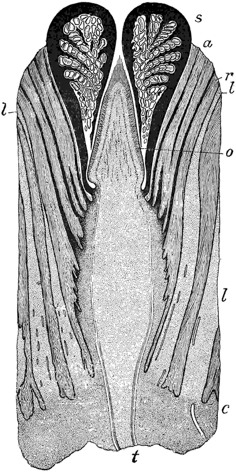
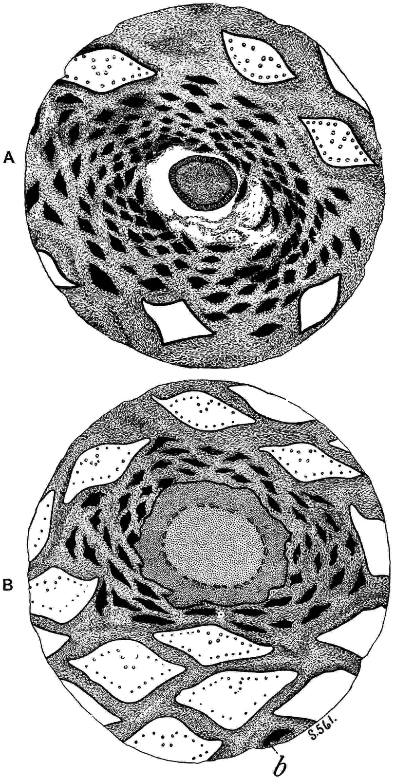
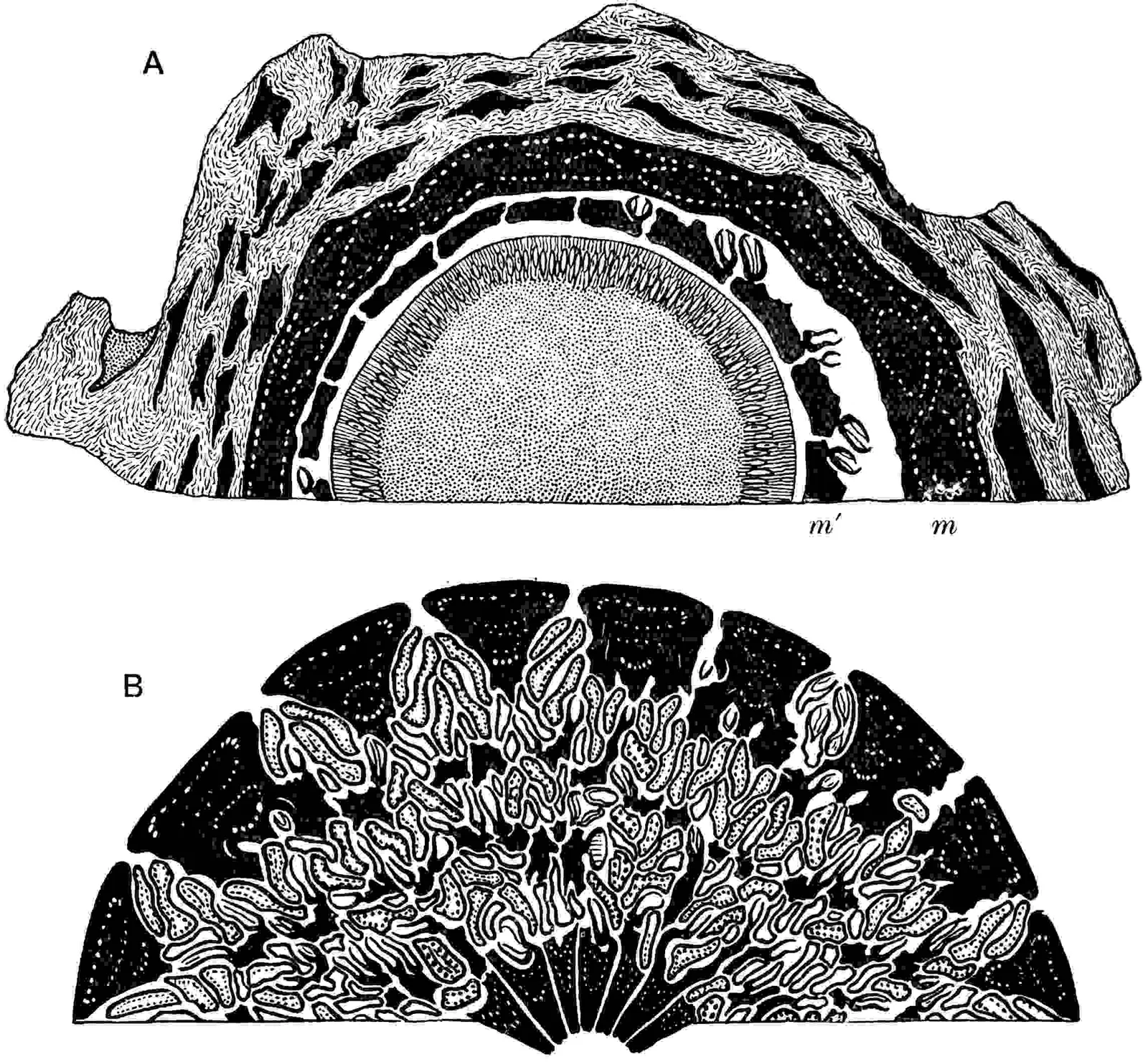
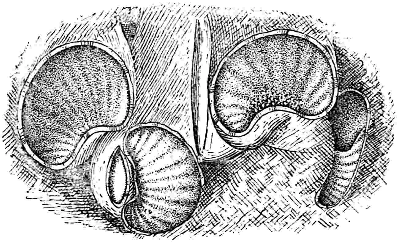
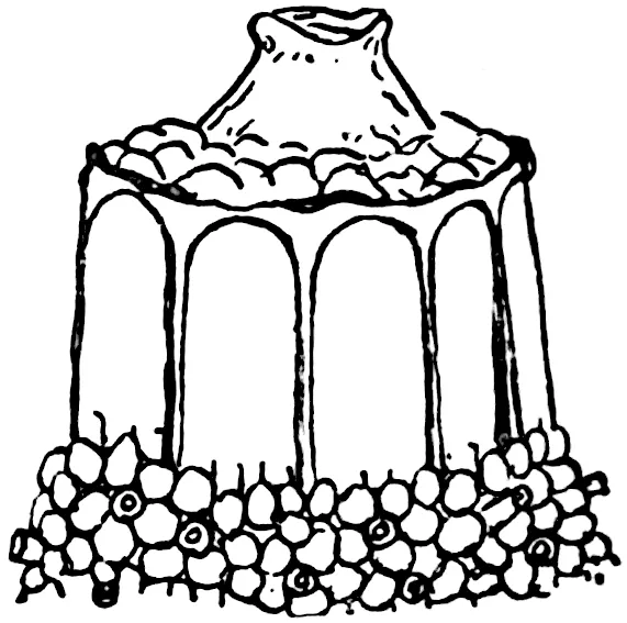
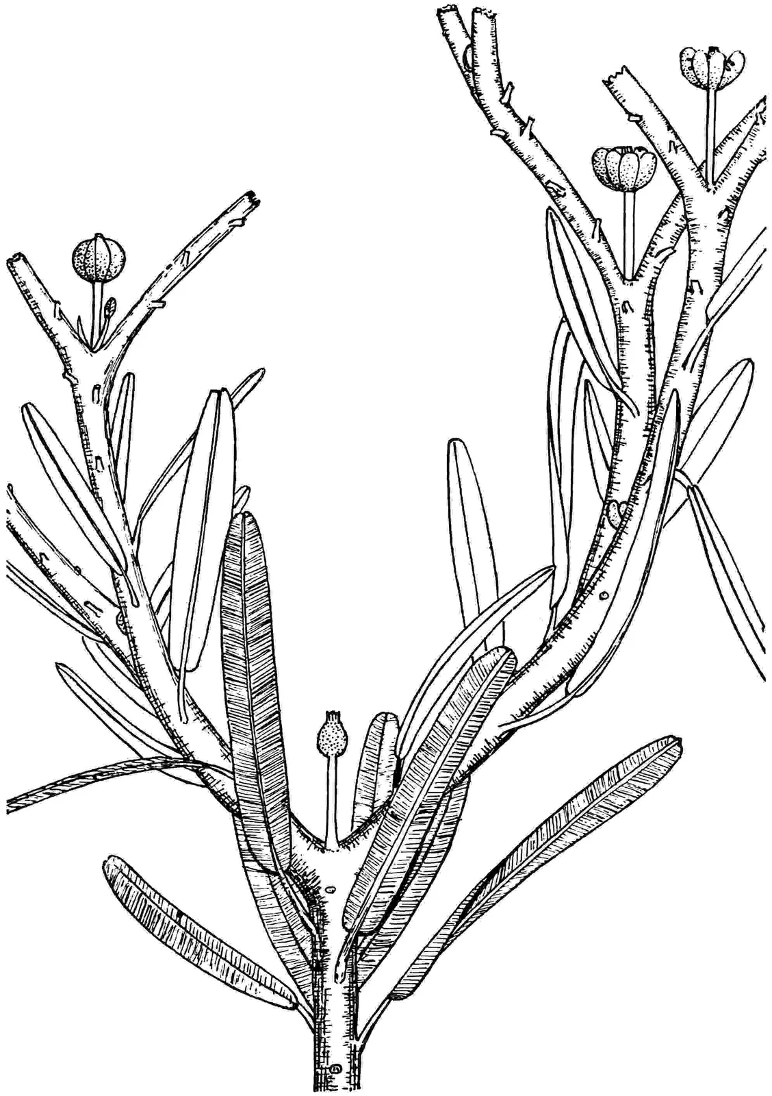
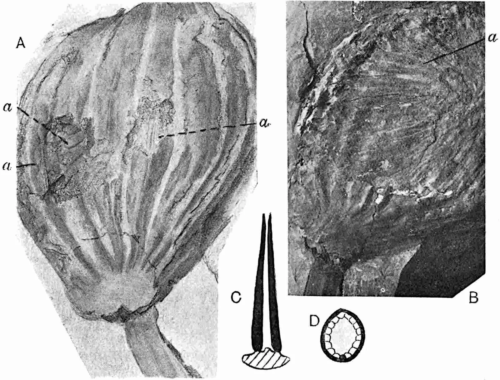
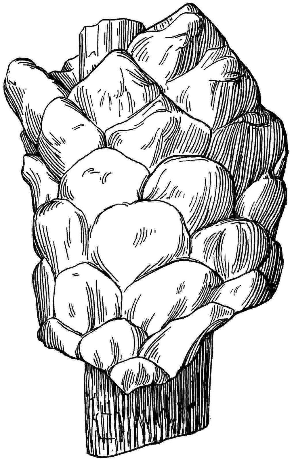
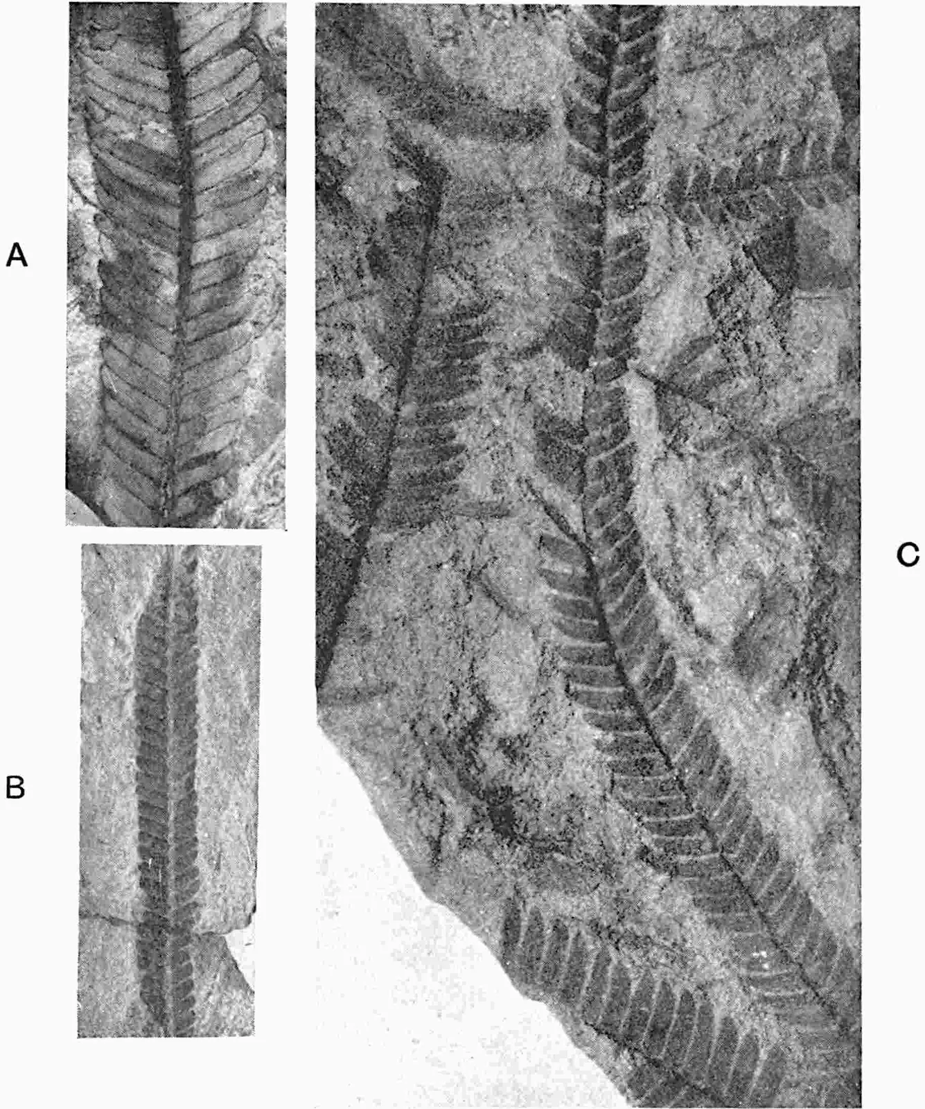
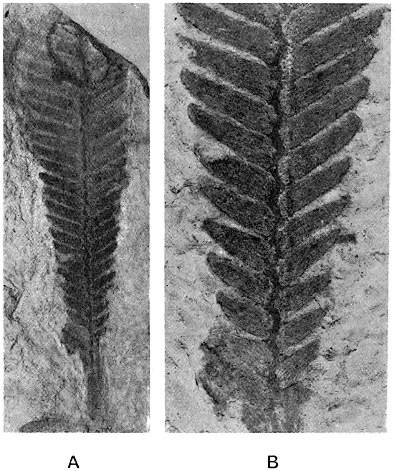
CAMBRIDGE UNIVERSITY PRESS
C. F. CLAY, Manager London: FETTER LANE, E.C. Edinburgh: 100, PRINCES STREET
London: H. K. LEWIS AND CO. Ltd., 136, GOWER STREET, W.C. London: WILLIAM WESLEY AND SON, 28, ESSEX STREET, STRAND New York: G. P. PUTNAM’S SONS Bombay, Calcutta and Madras: MACMILLAN AND CO., Ltd. Toronto: J. M. DENT AND SONS, Ltd. Tokyo: THE MARUZEN-KABUSHIKI-KAISHA
All Rights reserved
CHARLES RENÉ ZEILLER
Membre de l’Institut
Inspecteur général des Mines
Professeur à l’École Natle Supérieure des Mines
(1847–1915)
A TEXT-BOOK FOR STUDENTS OF BOTANY AND GEOLOGY
BY
A. C. SEWARD
M.A., F.RS., Hon. Sc.D. Dublin
PROFESSOR OF BOTANY IN THE UNIVERSITY;
MASTER OF DOWNING COLLEGE AND HONORARY
FELLOW OF EMMANUEL COLLEGE, CAMBRIDGE
ILLUSTRIOUS AS A MAN OF SCIENCE
AND A NOBLE-HEARTED GENTLEMAN
WHOM TO KNOW WAS TO REVERE
PREFACE
IN the Preface to Volume II published in 1910 I rashly stated that
it was my intention “to devote such space as is available within the
limits of a text-book to the neglected subject of the geographical
distribution of plants at different stages in the history of the
earth,” also that Volume III would be completed with as little delay as
possible. Though nearly seven years have elapsed since the publication
of the second volume it may fairly be said that the delay is not
entirely due to causes which it was in my power to control. The subject
of geographical distribution receives no connected treatment in Volumes
III and IV for the simple reason that I underestimated the space
required for the description of the Gymnosperms. The alternatives were
either to insert a greatly compressed survey of the successive floras
of the world at the end of Volume IV or to attempt a fuller and less
technical treatment of the subject in a separate book. In choosing the
latter course I am conscious that a further obligation is undertaken
which it may not be possible to fulfil; but the risk is deliberately
taken. Volume IV is in the press and will, it is hoped, be published
before the end of 1917.
It is a pleasant duty to repeat my thanks to many friends who have
helped me in various ways. Dr Kidston generously and without reserve
allowed me access to his splendid collection of Palaeozoic plants,
and the frequent occurrence of his name in the list of illustrations
shows how freely I have availed myself of his kindness. He has read
some of the chapters and greatly assisted me by his friendly criticism
and encouragement. By reading the proofs of this volume Dr Scott has
further increased my already large debt to him. It is impossible to
thank him adequately; he not only corrected many careless mistakes but
by wise counsel and advice he rendered me a service which I greatly
appreciate.
The exchange of views with Prof. Zeiller has been a constant source of
profit and enjoyment, and it is hard to realise that the completed book
will not receive his kindly criticism. He was a singularly unselfish
and generous colleague, always ready to help fellow workers, and he
had the faculty in an unusual degree of influencing those who had the
privilege of his friendship by his sound advice and lovable personality.
The death of Count Solms-Laubach has deprived Palaeobotany of one of
its most learned and strongest supporters. In common with all students
of fossil plants I owe much to the critical treatment of the subject in
the Einleitung in die Paläophytologie. Prof. Jeffrey has very
kindly given me several photographs and sections which have been of
great service, and I am similarly indebted to Prof. Zalessky. To Prof.
Nathorst my thanks are due for the great interest he has taken in my
work and for his generosity in providing drawings and showing to me
many of the treasures in the famous Stockholm Museum. Prof. Bertrand
and Prof. Lignier freely supplied photographs and drawings of specimens
in their possession, and I am particularly grateful to them for the
willingness with which they always responded to my requests.
Through the death of Prof. Lignier in March 1916 Palaeobotany has
been deprived of another original thinker who devoted himself with
whole-hearted enthusiasm to botanical research and for many years
faithfully served the University of Caen: he was a generous friend to
whom one never appealed in vain for assistance. Through the kindness
of the Director of the Indian Geological Survey I have been able to
examine several fossils from the Calcutta Museum described by Oldham
and Morris and by Feistmantel. With the Director’s permission several
photographs and drawings made for a forthcoming paper to be published
by the Indian Survey are reproduced in this volume. I take this
opportunity of thanking friends in Australia who recently afforded me
facilities for examining fossil plants in their charge, and I would
especially thank Mr A. B. Walkom of the University of Brisbane, who
has recently undertaken an investigation of the rich plant-beds in the
Ipswich district, for all that he did to enable me to make the most of
a very short time available for palaeobotanical work.
For the loan of specimens and for other help I am indebted to Prof.
Bayly Balfour, Prof. Bower, Prof. Margaret Benson, Prof. Oliver, Sir
David Prain, Dr Smith Woodward, Prof. Weiss, the Director of the
Geological Survey, Dr A. H. Church, Dr Arber, and other friends. I
would also acknowledge a debt, by no means inconsiderable, to my
Colleague Mr Hamshaw Thomas. Among younger friends in the Cambridge
Botany School to whom I am indebted I wish particularly to thank Miss
Ruth Holden, Miss Bancroft, Mr Sayers, Mr Dutt and others who have
rendered me willing help.
In the List of Illustrations mention is made of Corporate Bodies and
individuals from whom blocks have been obtained, and I am grateful to
them for readily responding to my applications.
My Wife, though prevented by more urgent calls in the later stages of
my task from giving as much time to the illustrations as in the two
former volumes, has contributed several drawings, and my daughter
Phyllis Seward has also given me much help in preparing drawings from
previously published figures.
In spite of the vigilance and wise counsel of many friends numerous
blemishes remain and for these the author is alone responsible.
A. C. SEWARD.
Downing College Lodge, February 10, 1917.
Note. The letters A and B added to references in
the footnotes indicate that the works will be found in the
Bibliographies at the end of Volumes I and II.
Several of the illustrations are printed from blocks for which I am
indebted to learned societies or to individuals. The sources from
which clichés were obtained are mentioned within square brackets.
The names of donors of photographs or drawings are added after the
descriptions of the figures.
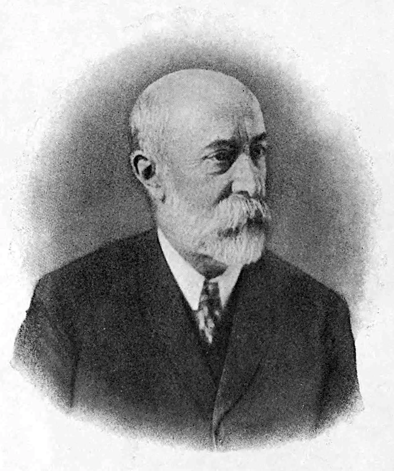
Frontispiece. Charles René Zeiller. From a photograph given to
me by Madame Zeiller.
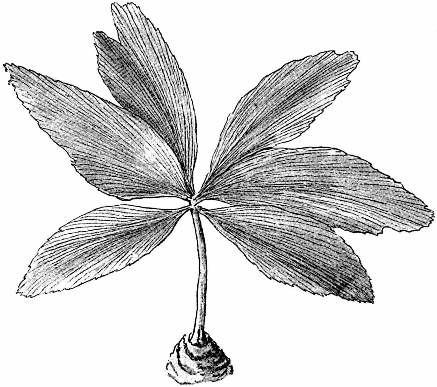
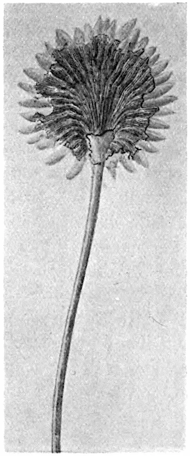
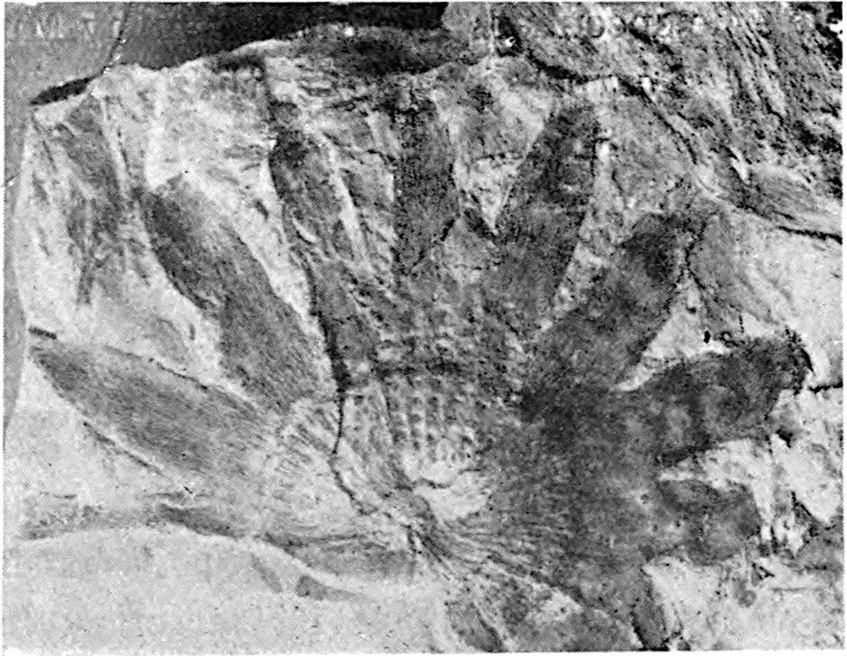
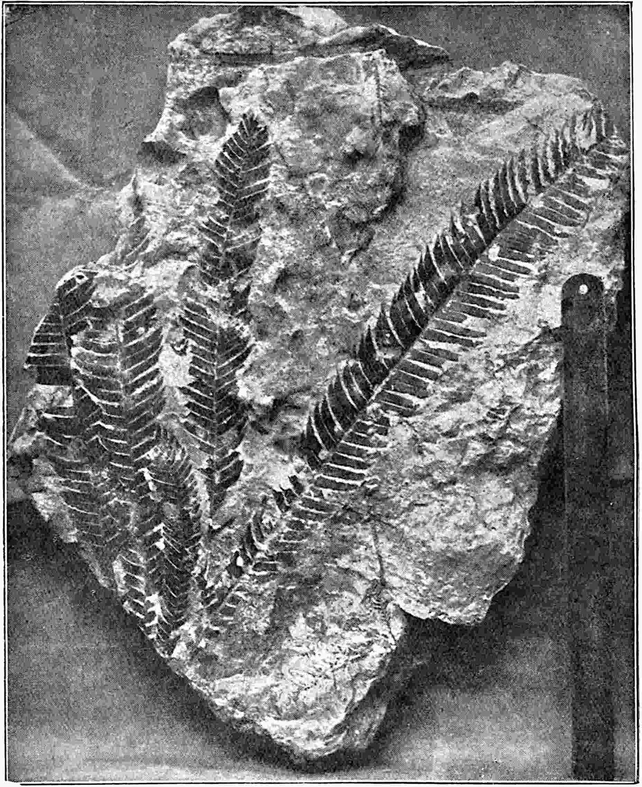
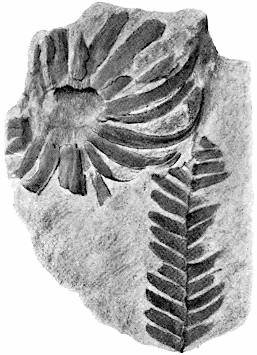
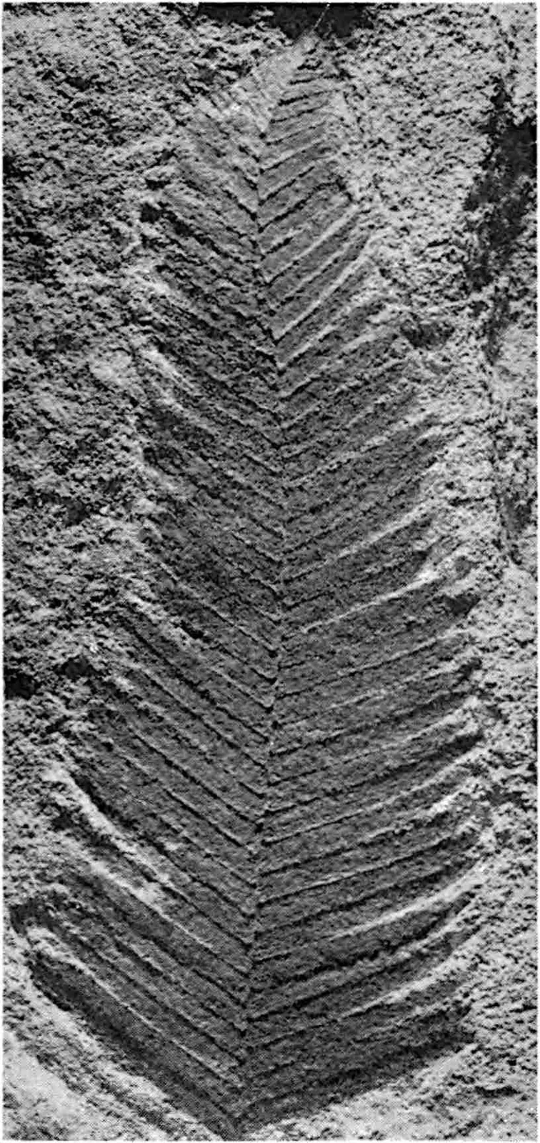
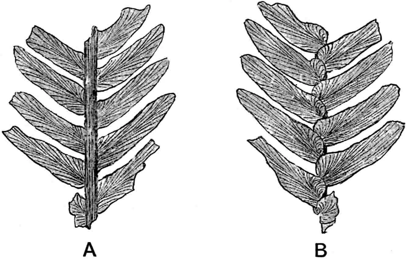
Sphenozamites Belli. [Trustees of the British Museum.]
588
CHAPTER XXVIII.
CYCADALES (RECENT).
Among the fossil genera described in the last chapter of the second
volume some were spoken of as true Ferns though most of them, it
was added, ‘may safely be regarded as plants which will ultimately
be shown to belong to some other group, in most cases that of the
Pteridosperms.’ Since this was written additional evidence has
been obtained in favour of the inclusion of certain genera in the
Pteridosperms. In the case of Taeniopteris, one of the genera
already described, there is reason to believe that at least one species
is a member of the Cycadales and not a true Fern as formerly supposed.
The Pteridosperms so far described are represented for the most part
by sterile leaves preserved as impressions, the genera founded on
more satisfactory material having been reserved for treatment in this
volume. As these genera are founded to a large extent on anatomical
characters oscillating in their essential features between recent Ferns
and Cycads, it is important that the student should be in possession of
the anatomical characteristics of both of these classes; and for this
reason a general account of recent Cycads is intercalated between the
Pteridosperms already described and those reserved for treatment in
this volume.
The section of the Gymnosperms known as the Cycadales, represented
by nine recent genera and less than 100 species, is of exceptional
importance phylogenetically and demands special attention from
palaeobotanical students. Familiarity with the morphology of recent
forms is essential not only in relation to extinct cycadean plants
but also to types which, though not sufficiently close to surviving
species to be included with them in one class, exhibit features
regarded by many botanists as indications of an affinity either to true
Cycads or to some generalised stock of which they are an offshoot.
The Cycads of to-day may fairly be spoken of as anachronisms, plants
appropriate to a former age but out of harmony with the present.
They are confined to tropical and sub-tropical regions in both the
old and new world. In habit many of them resemble tree-ferns, but
the columnar stem, which may live to a great age and attain a height
of 20 metres, differs from that of ferns in its gradually tapered
form consequent on the presence of one or more cambial cylinders.
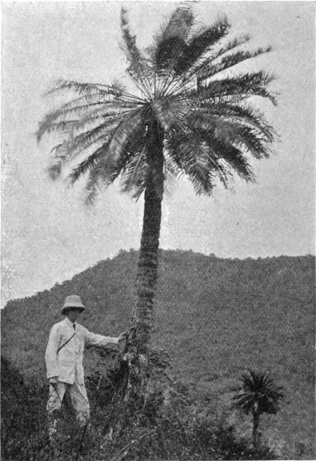
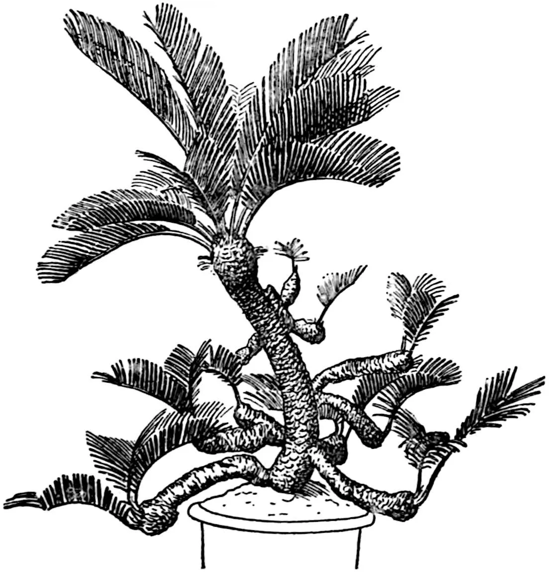
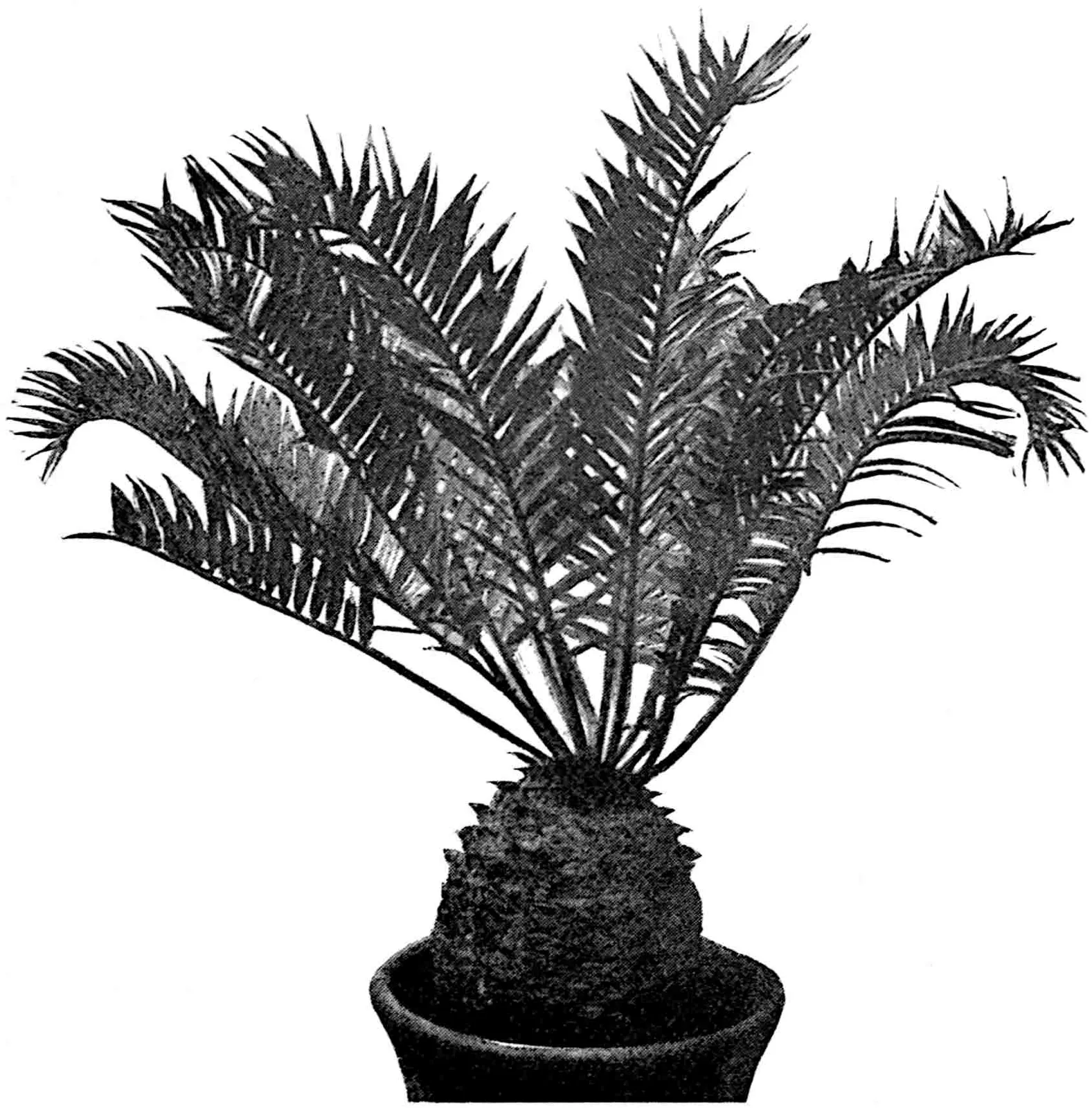
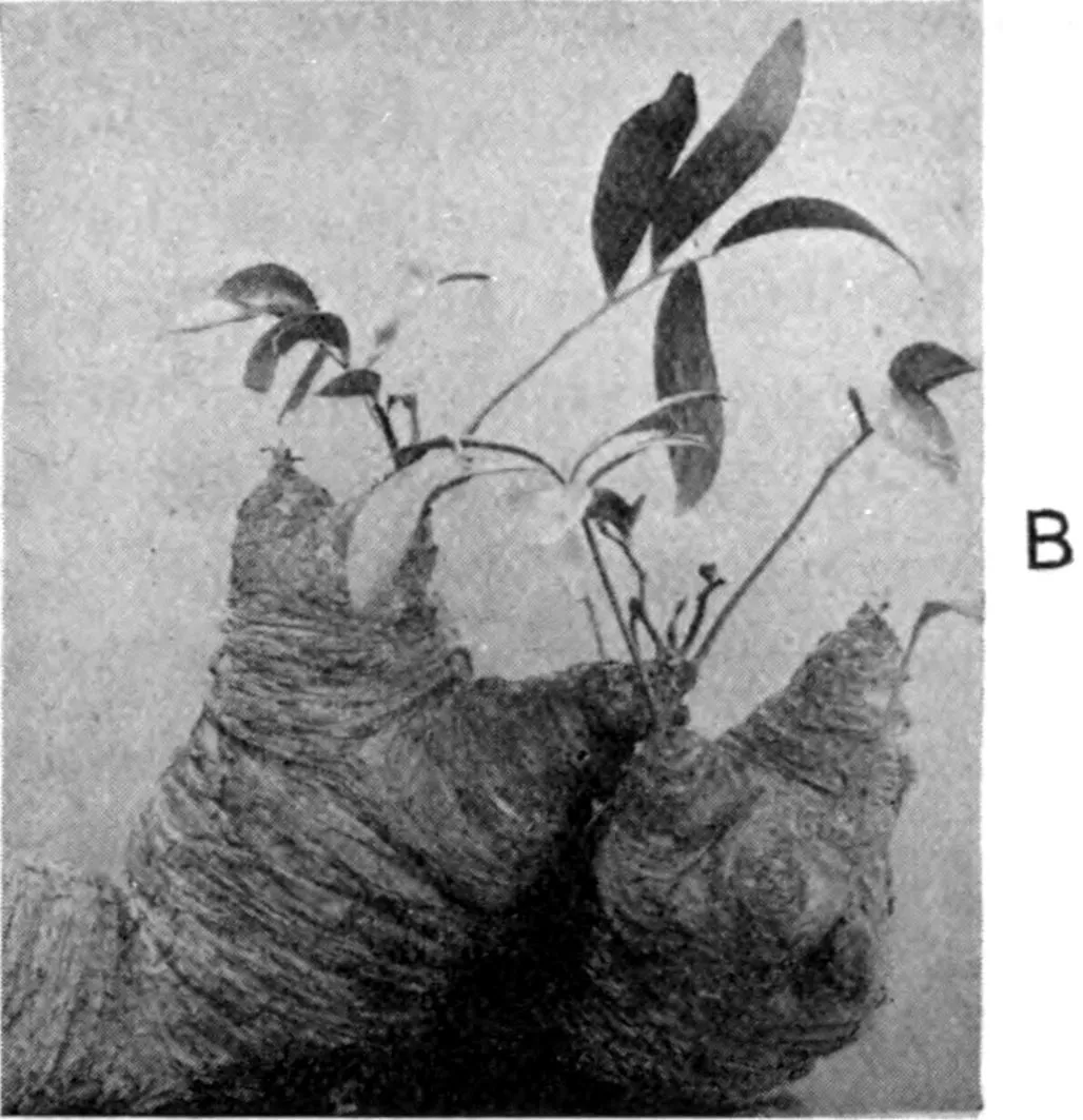
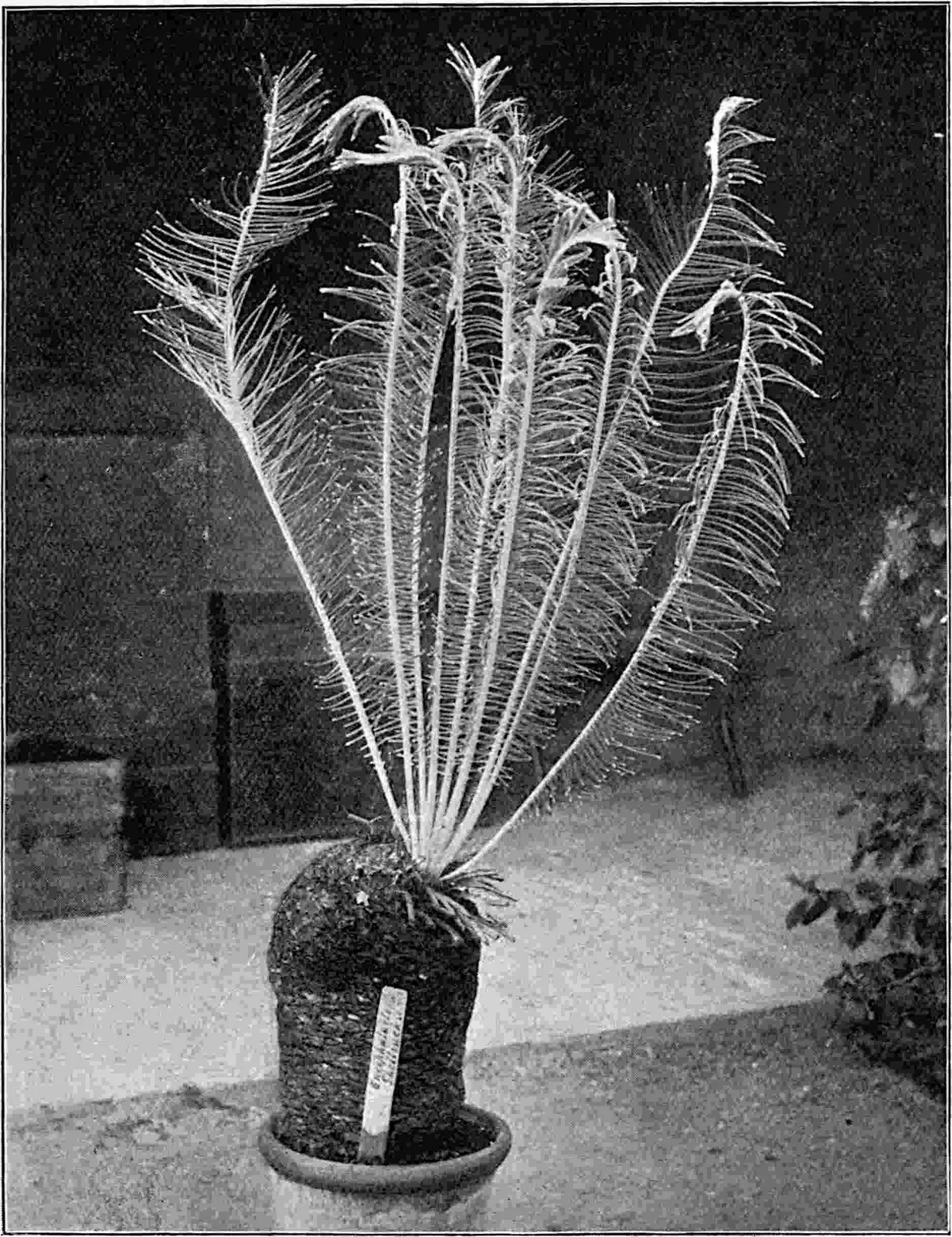
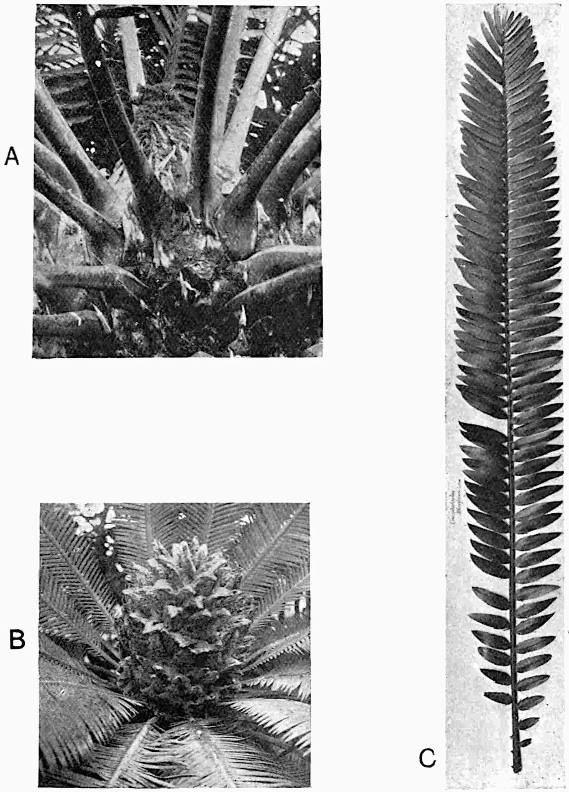
Though often unbranched (fig. 377) branching of the main trunk is
by no means unusual (fig. 378; fig. 381, B). Many Cycads are
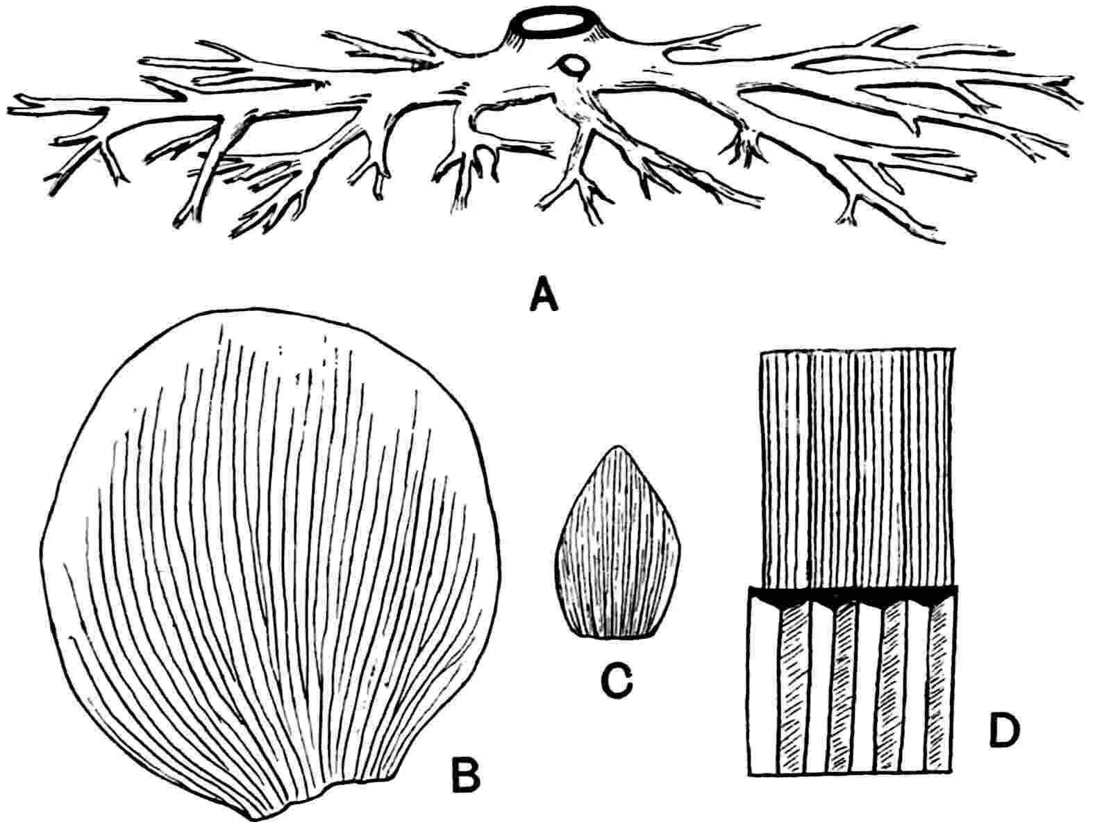
geophilous and have short tuberous stems (figs. 383, 395, 1a;
396, E): the genus Zamia includes a few epiphytic forms[1].
The typical cycadean stem is covered with persistent petiole-bases
with or without an admixture of smaller scale-leaf bases (figs. 379,
380), while in several species a transversely wrinkled or irregularly
fissured periderm forms the superficial tissue (figs. 381, B; 383).
The foliage-leaves are relatively large and, with the exception of the
bipinnate fronds of Bowenia (fig. 391), they are always pinnate.
The fronds usually form a terminal crown (figs. 377, 379) and as many
as 100 may be produced from one bud. In Zamia pygmaea[2] the
fronds are only 10–12 cm. long, but in some cycads they reach a length
of several metres. On both young foliage-leaves and scale-leaves long
and very rarely branched[3] unicellular hairs (fig. 396, N) form a
characteristic feature and take the place of the ramental scales of the
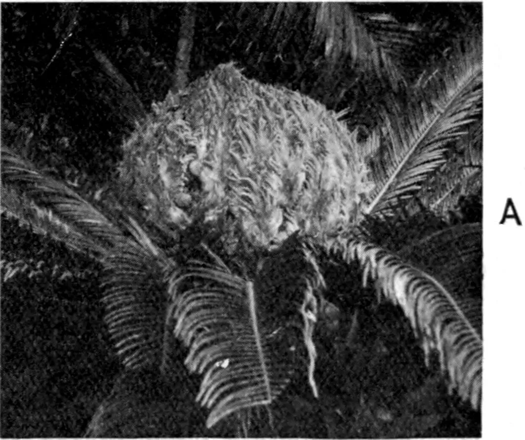
majority of ferns. The apex of the stem shown in fig. 386, A is covered
with a mass of woolly hairs and several scale-leaves are seen on the
lower part of the bud.
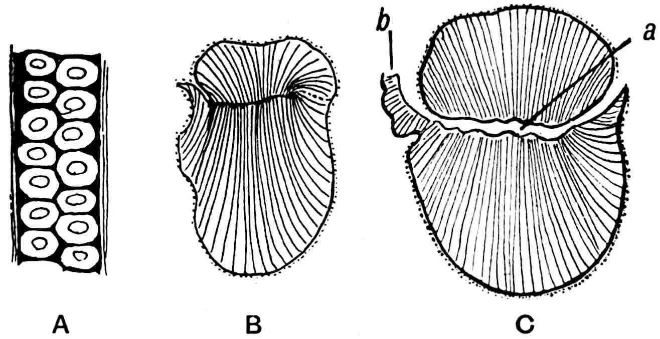
Fig. 377.Cycas circinalis. From a
photograph taken by Mr A. Malins Smith at Teldeniya (Ceylon).
Fig. 378.Cycas revoluta, as grown by
Japanese horticulturalists. (After Wieland.)
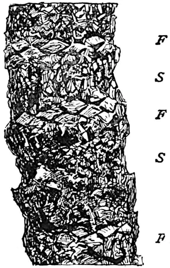
Fig. 379.Encephalartos horridus.Fig. 380.Cycas circinalis. Stem showing
alternate zones of leaf-bases (F) and scale-leaf bases (S). (From the Encyclopaedia Britannica.)
All recent Cycads are dioecious. The reproductive shoots, except the
megasporophylls of Cycas—which have departed to a less extent
than those of other genera from the foliage-leaf plan (fig. 381, A;
fig. 392, A–C) and are borne in a terminal cluster through which the
stem subsequently pushes its way—consist of a varying number of micro-
or mega-sporophylls in dense spirals on the axis of an elongated or
oval strobilus (figs. 386, B, 393, 394). The microsporophylls are
occasionally verticillate[4]. The strobili are sometimes though rarely
branched[5] and generally but by no means invariably[6] terminal on
the main stem which branches sympodially[7]. A striking example of
lateral strobili has recently been described by Chamberlain[8] who
figures a stem of Macrozamia Moorei with fertile shoots wedged
among the persistent petiole-bases, a condition very similar to that in
the Mesozoic Bennettitales. Pearson has also described clear cases of
laterally-borne cones in Encephalartos.Cycas exhibits
two kinds of branching, the female plants being monopodial while in
the male the branching is sympodial. The microspores are produced in
sporangia grouped in more or less well defined sori (figs. 389, A; 392,
E–G). There is no definite annulus, but in the occurrence of groups
of thick-walled cells some microsporangia recall those of certain
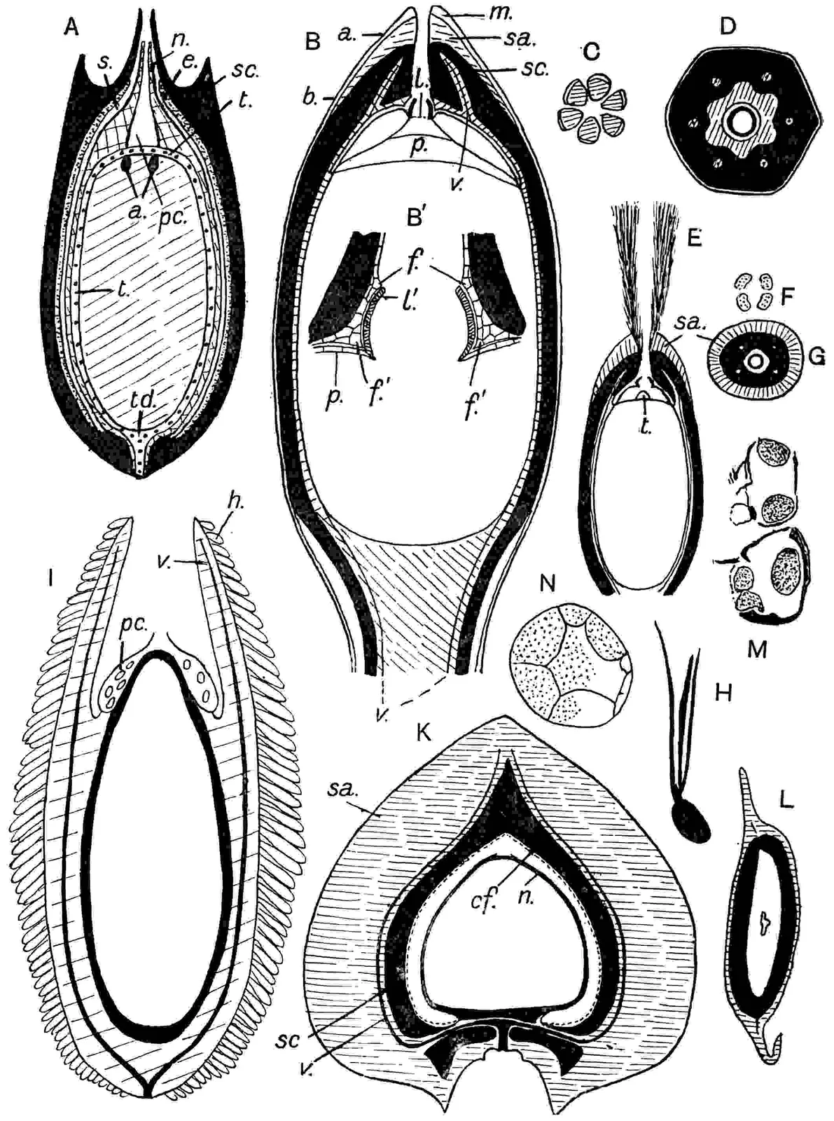
ferns[9]. The ovules vary considerably in size, sometimes exceeding 5
cm. in diameter: there are usually two on each megasporophyll (figs.
393, C; 394; 395, 1d) but in most species of Cycas (fig.
392, B) and occasionally in other genera the number is larger[10]. A
thick integument encloses the nucellus with which it is fused except
in the apical region (fig. 396, A, B). Below the comparatively long
micropylar tube is a well-developed pollen-chamber (fig. 396, B′,
p), a striking feature of Cycadean ovules, immediately above
the megaspore; the latter is filled with prothallus-tissue and bears
a small apical group of archegonia on the floor of a depression (fig.
396, A–B′). In Microcycas[11] as many as 200 archegonia are
recorded—a very exceptional case—and these are not confined to the
apical region, though only the apical archegonia are functional.
Each archegonium is characterised by a very large oval egg-cell and
a much reduced neck[12]. The microspores usually produce a single
prothallus-cell, a stalk-cell, and body-cell, and from the body-cell
are developed two spirally ciliated spermatozoids (fig. 396, M). In
this respect also the monotypic genus Microcycas is peculiar:
it may have as many as 8 body-cells and 16 male gametes in a single
pollen-tube (fig. 396, G), while in Ceratozamia[13] 4 gametes
have been seen in one tube. The pollen-tube grows like a fungal
mycelium into the nucellar tissue and the male gametes are formed in
the distended proximal end which on bursting liberates the motile
sperms with the watery cell-sap. Fertilisation is succeeded by the
development of a homogeneous proembryo partially or completely filling
the zygote (fertilised egg): by the formation of long suspensors the
embryo is brought into contact with the food-store of the prothallus.
In some Cycads, e.g.Encephalartos, the embryogeny
exhibits a close resemblance to that of Ginkgo[14]. The embryo
is dicotyledonous[15].
Fig. 381. A. Cycas revoluta, megasporophylls. B. Zamia Loddigesii, branched stem.
The single stele of the stem is characterised by a large pith which
in some genera (e.g.Encephalartos, Macrozamia)
contains an anastomosing system of collateral bundles. The vascular
tissue of a cycadean stem forms a cylinder of secondary xylem and
phloem, the primary xylem being represented only by a few, usually
crushed, protoxylem elements on the inner margin of the reticulately
pitted or scalariform tracheids. Both xylem and phloem are traversed
by numerous broad and deep medullary rays[16]. The looser texture and
more parenchymatous structure of Cycadean wood afford a ready means
of distinguishing it from the wood of Conifers: for the Cycadean type
the term manoxylic is proposed and pycnoxylic for the
more compact coniferous wood[17]. Rims (or ‘bars’) of Sanio, of which
much has been said in discussions on the phylogeny of Conifers, have
recently been described in the petiolar xylem of Cycas revoluta:
the rims are short and ‘cling closely to the borders of the pits,’
features which also characterise the rims found in the cones of the
Araucarineae and in the root- and cone-wood of certain Pines[18]. In
some Cycads the secondary xylem and phloem form a single cylinder,
but in others (Cycas, Encephalartos, Macrozamia,
Bowenia) the cambium is succeeded by one or several concentric
cylinders of meristem which have their origin in the pericycle.
The spasmodic occurrence of separate arcs of inversely orientated
secondary xylem and phloem between the normal cylinders is a feature of
importance from the point of view of comparison with the Palaeozoic
Medulloseae[19]. The occurrence of concentric cauline strands in
the cortex of Cycas is also a peculiarity worthy of notice.
Successive bands of periderm, and occasionally a considerable amount of
phelloderm[20], are formed in the peripheral region of the stem.
The leaf-traces in an adult stem exhibit a striking feature in
their indirect or girdle-like course to the leaves (fig. 396, H,
g) and in the gradual change from an endarch (fig. 396, O)
to an apparently mesarch structure (fig. 400) as they pass from the
perimedullary zone to the petiole: except at the base of the petiole
the vascular bundles of the frond-axis consist of (i) centripetally
developed xylem with a median protoxylem and a much smaller amount of
centrifugal xylem (fig. 400) separated by a few parenchymatous elements
from the centripetal xylem, (ii) an external arc of protophloem and
within this metaphloem and parenchyma[21]. In the slender petiole of
Bowenia there are a few collateral bundles arranged in the form
of a circle or ellipse[22]; in Cycas and some other genera the
more numerous bundles form a pattern like an inverted U, and in some
species of Encephalartos the number is greater and the strands
more irregularly scattered[23]. In the vegetative stems there is no
centripetal xylem in the stele, but scattered centripetal tracheids
occasionally occur internal to the protoxylem in the steles of the
peduncles[24].
Cycadeae. Megasporophylls each bearing 2–8 ovules, borne
separately like foliage-leaves and not in strobili. Pinnae have a
midrib but no lateral veins (figs. 384, 387, A). Cycas (fig. 377).
Zamieae. Both kinds of sporophylls form strobili. Pinnae
have several dichotomously branched, more or less parallel veins.
Zamia (figs. 388–390), Macrozamia, Encephalartos
(figs. 379, 386, C), Ceratozamia, Dioon (fig. 386, B),
Microcycas.
Stangerieae. Strobili as in Zamieae. Pinnae fern-like, numerous
dichotomously branched lateral veins given off from a midrib.
Stangeria.
Bowenieae. Leaves bipinnate (fig. 391), strobili as in Zamieae.
Bowenia.
Distribution. The most widely spread genus, Cycas,
occurs in Siam, India, the Nicobar Islands, Ceylon, Madagascar, and
Australia, in many of the islands in the Indian and Pacific oceans, in
New Guinea, Borneo, New Caledonia, New Britain, China and Japan[25].
Zamia, the most northerly genus, extends from North Mexico and
Florida through Central America and some of the West Indian islands to
Ecuador, Bolivia, Chile, and Peru. Dioon and Ceratozamia
are confined to South Mexico, and Microcycas flourishes on
the Cuban mountains. The continent of Africa possesses two endemic
genera Encephalartos and Stangeria.Encephalartos
extends from Cape Colony through Natal and Zululand to Zanzibar
and Mombasa[26]: a specimen in the Kew Herbarium (probably E.
Hildebrandti) is said to have been collected as far north as
the Soudan. Two species are recorded from the Congo[27] and E.
Barteri, discovered by Barter in Central Africa, is recorded
from the Gold Coast[28]. Stangeria has a much more limited
range in S.E. Africa[29]; Australia possesses Macrozamia,
represented by several species in Western Australia, New South Wales
and Queensland, Cycas in Queensland and the Northern territory
and the Queensland genus Bowenia. There are no Cycads in
New Zealand. As a whole Cycads have a limited range and with the
exception of Cycas and Zamia none of them extend beyond
the limits of a single continent. They are as a rule not gregarious
plants and play a subordinate part in the facies of the vegetation.
Macrozamia forms dense thickets[30] in some districts and occurs
both in exposed situations and in association with Palms in damp
Queensland forests. Chamberlain[31] speaks of 100 plants of Dioon
edule as visible in one view in South Mexico where the species
forms a mountain forest. In Florida Zamia pumila[32] grows in
dense moist woods, a habitat in contrast to that of many Cycads. The
Mexican Ceratozamia is associated with luxuriant vegetation,
while its compatriot Dioon[33] lives in blazing sunshine.
Sir Joseph Hooker[34] speaks of Cycas living in the deepest
and hottest valleys in Sikkim. Encephalartos is essentially a
xerophilous genus. Stangeria paradoxa is said to be confined to
forests in Cape Colony, and another species grows among the grass of
the Park-lands in open country[35]. While it is true that many Cycads
are characteristic of dry regions some species flourish in places where
shade and moisture are abundant.
Though it is impossible in many cases to form an estimate of the age
of individual plants, there are clear indications that some specimens
afford notable instances of longevity. Chamberlain estimates the age
of some plants of Dioon spinulosum as exceeding 400 years and
mentions an example of D. edule that is probably 1000 years
old. An unusually tall plant of Encephalartos in the Botanic
Garden of Amsterdam is believed by Prof. de Vries to have reached the
venerable age of 2000 years[36]. The restricted range and in many cases
the solitary existence of recent Cycads, with their tall stems clothed
with the persistent cork-covered stumps of thousands of fronds, deepens
the impression of antiquity derived from a study of the geological
history of this dwindling race.
Stems. The tall columnar stems of some species of Cycas,
often branched or bearing numerous ovoid buds like enlarged
bulbils[37], are characterised by the regular alternation of large
and small leaf-bases as seen in the stem of C. circinalis
reproduced in fig. 380. In older stems of this species the leaf-bases
are exfoliated and the stem is covered with wrinkled and fissured cork;
but in Cycas revoluta the leaf-bases are even more persistent.
The columnar but relatively stout stems of Encephalartos (figs.
379, 382, 386, A) and Ceratozamia are similarly encased in a
covering of petiole-bases, but in these genera the differences between
foliage-leaves and bud-scales is much less obvious and there is no
zonal alternation. On the stems of Macrozamia the rhomboidal
leaf-bases are more uniform in size and there are no scale-leaves. The
tall and often palm-like stems of Microcycas sometimes show
transverse rings on the bark marking the position of former terminal
buds, and in older trunks these may disappear, leaving a fissured
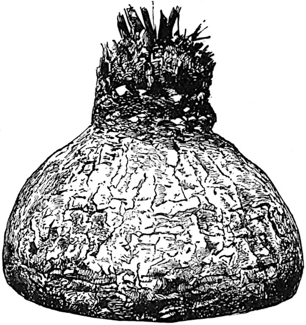
bark[38]. In Cycas siamensis the tuberous stem is similarly
covered with a rough bark (fig. 383) and the stems of Zamia
are also characterised by an absence of persistent leaf-bases (figs.
381, B; 395, Ia, a). It is pertinent to remind the
palaeobotanical student of the occurrence of flowering plants with
stems closely simulating those of some Cycads. Prof. Bower[39] in
describing Rhynchopetalum montanum, an Abyssinian Lobeliaceous
plant, drew attention to the similarity in surface-features and to some
extent in anatomical structure to cycadean stems. The resemblances are
further emphasised in a more recently published account of the same
species under a different name, Lobelia Rhynchopetalum[40].
Fronds. A general acquaintance with the various types of
fronds illustrated by recent Cycads is important to the student of
fossils not only to enable him to compare existing and extinct forms
but as affording safeguards against possible sources of error in the
description and identification of impressions[41]. The vernation
exhibits less uniformity than in Ferns: in Cycas the rachis
is straight and the pinnae circinately coiled (fig. 220, B, vol.
ii. p. 283); in Zamia and Stangeria the rachis
is bent and the pinnae straight, while in Ceratozamia and other
genera both the axis and leaflets are straight. As Braun pointed out,
there is as a rule no terminal leaflet, or it may be pushed to one side
giving a forked appearance to the frond apex[42].
Fig. 384.Cycas circinalis, abnormal
frond. (From a specimen in the British Museum.)
Cycas. The presence of a strong midrib and the absence of
lateral veins are distinguishing features: the lower margin of the
lamina is frequently decurrent (fig. 387, A). In C. circinalis
the pinnae may reach 40 cm. in length with a fairly uniform breadth
of 2 cm. A frond of this species in the British Museum, not quite
complete, has a length of 112 cm.: on the lower part of the rachis
strong spines replace the leaflets and near the apex of the leaf
concrescent pinnae form a continuous lamina traversed by seven ribs
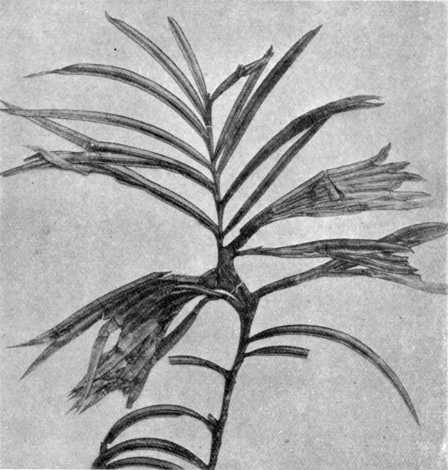
and dissected at the margin into acuminate teeth (fig. 384): some of
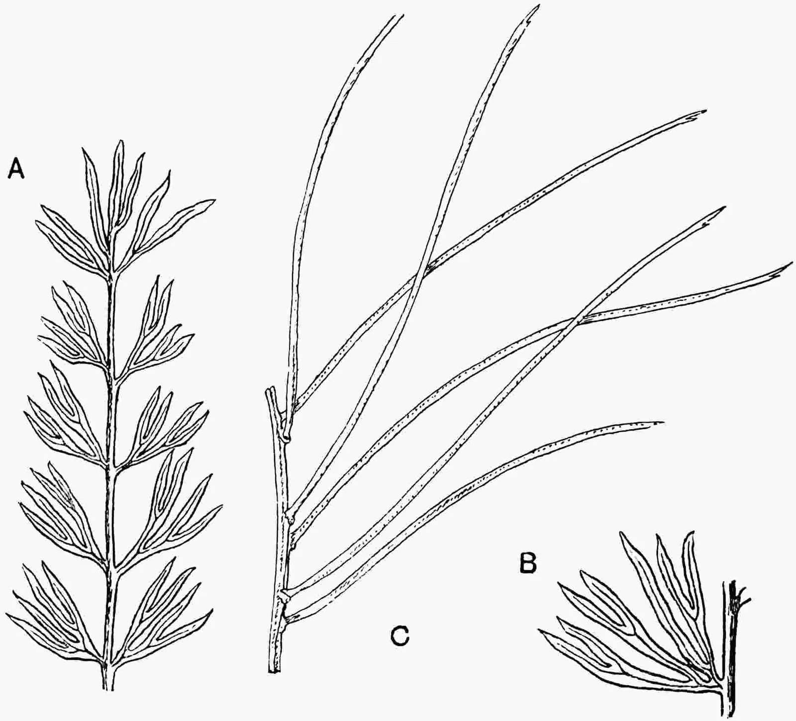
the pinnae are forked as in Cycas Micholitzii[43] (fig. 385).
Several years ago I noticed a similar instance of concrescence in a
small plant of C. circinalis in the Royal Gardens, Kew (fig.
387, I). In Cycas Micholitzii the pinnae, reaching a length
of 20 cm., are repeatedly and deeply forked (fig. 385, A, B; fig.
400): the pinnae of C. Rumphii var. bifida[44] are also
deeply dissected. Cycas Beddomei has very narrow pinnae (15
cm. × 2 mm.) similar to those of the Wealden species Cycadites
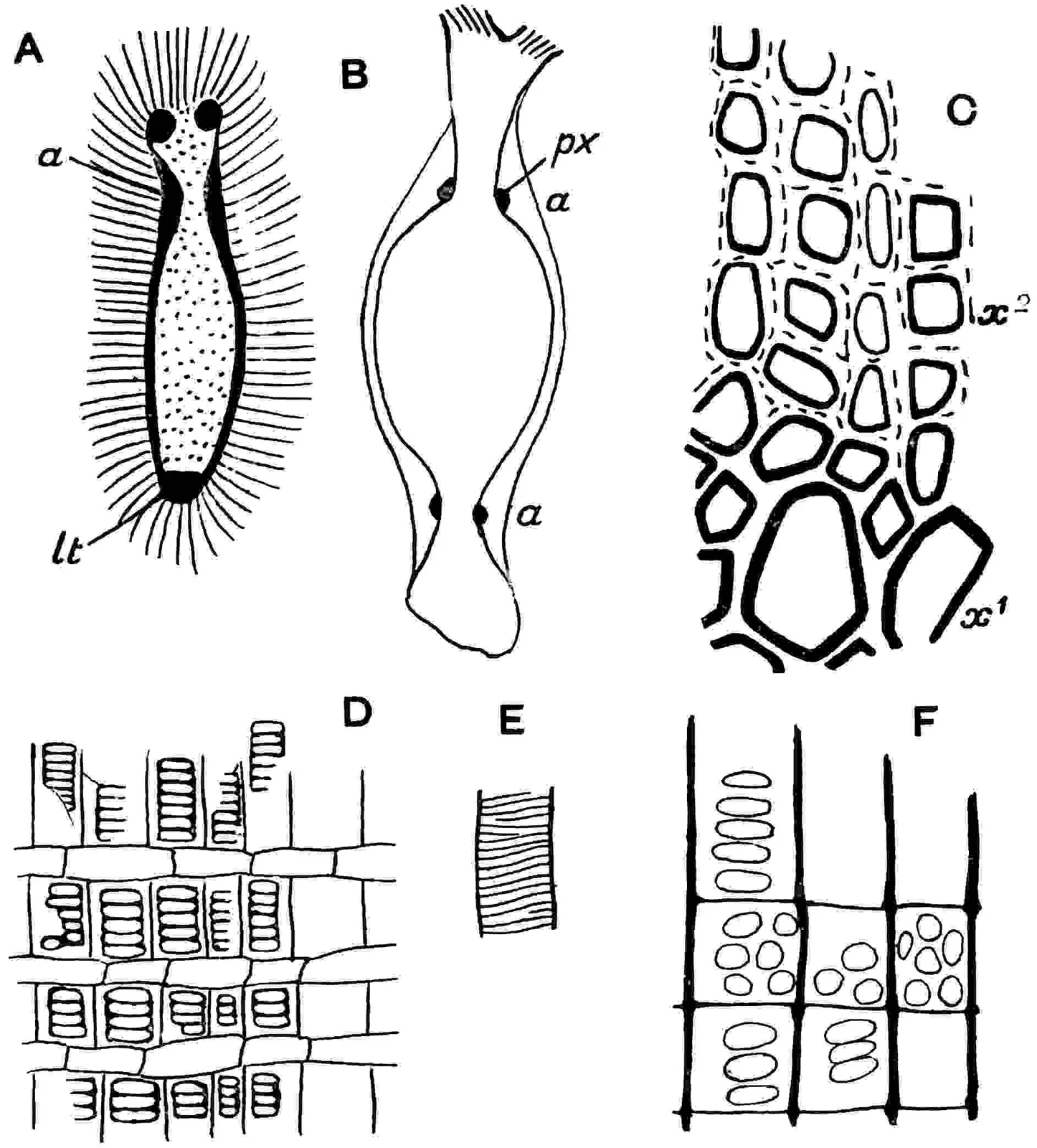
Saportae, and it is noteworthy that narrow leaflets with a strongly
revolute lamina would produce casts with two parallel ribs (the
grooves between the midrib and the edge of the lamina) simulating the
double midrib of the fossil genus Pseudocycas. In some fronds,
e.g.C. Cairnsiana, the midrib is hardly visible on
the upper face of a dried pinna which shows a longitudinal wrinkling
simulating parallel venation.
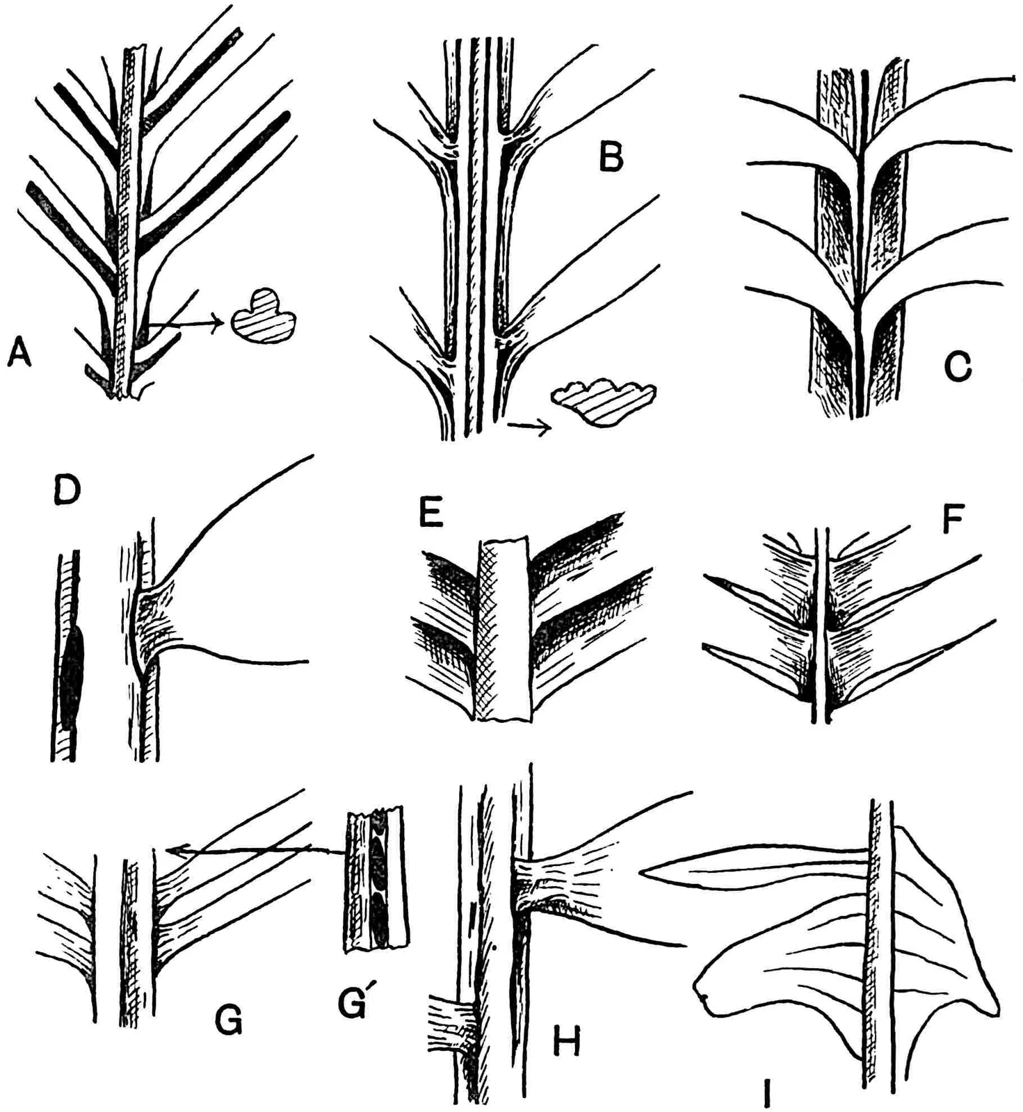
Encephalartos. The fronds of this genus, in Encephalartos
Laurentianus[45] reaching the exceptional length of 7 metres, bear
alternate pinnae exhibiting a considerable range in form and breadth.
In E. longifolius, E. Altensteinii (fig. 386, C), E.
Lehmanni, etc., the pinnae are for the most part linear, reaching
a length of 20 cm. and a breadth of 2 cm.: in E. caffer (fig.
387, D), E. latifolius, and others the pinnae are broader
and shorter and often spinous. A frond of E. longifolius
or E. Altensteinii may bear both entire and lobed, spinous
pinnae. In E. Frederici-Guilielmi (fig. 387, G) and E.
Ghellinckii[46] (fig. 382) the pinnae are very narrow and almost
filiform, with revolute edges. The thick and leathery pinnae of some
species are attached obliquely to the edge or to the upper sloping
sides of the rachis which forms a prominent ridge between the rows
of leaflets, and characteristic oval scars are left on the fall of
the pinnae (fig. 387, D, G′). The lamina in most species contains
several veins more or less parallel to the margins and often much more
prominent on the lower than on the upper surface.
Fig. 386.
Encephalartos Altensteinii, apex of stem.
Dioon edule,megastrobilus. (From a photograph by Mr S. M. Wadham.)
Encephalartos Altensteinii, frond.
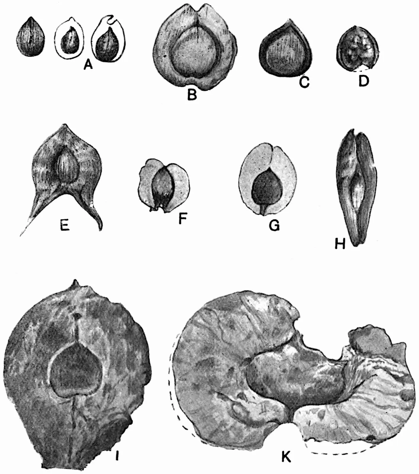
Fig. 387. Cycadean fronds. A, Cycas
circinalis; B, Macrozamia Fraseri; C, Macrozamia
Denisoni; D, Encephalartos caffer; E, F, Dioon edule
from below and above; G, Encephalartos Frederici-Guilielmi, G′,
side-view; H, Ceratozamia mexicana; I, Cycas circinalis,
lower part of young frond.
Zamia. In Zamia angustifolia (fig. 385, C) and
Z. linifolia the pinnae are long and very narrow: the
other extreme is represented by Z. Wallisii[47] (fig.
388) with broad ovate segments reaching a length of nearly ·5
metre and attached to the rachis by a short stalk; the veins are
prominent and dichotomously branched. Other forms of pinnae are
represented by Z. integrifolia, Z. floridana, and
Z. Loddigesii (figs. 389, 390, 395). The broad and short
pinnae of Z. furfuracea[48] bear a close resemblance, except
in the absence of an auriculate base, to those of some species of
the fossil genus Otozamites. The broadly linear pinnae of
Z. pseudoparasitica (45 cm. × 3 cm.) often show longitudinal
wrinklings on drying which suggest comparison with the corrugated
lamina of the fossil species Nilssonia brevis. A basal pad or
callosity on the slender bases of the pinnae is characteristic of many
Zamia fronds.
Fig. 388. Pinna of Zamia Wallisii. From
a drawing after A. Braun in the Kew Herbarium. (⅓ nat. size.)
Fig. 389.Zamia integrifolia bearing
a megastrobilus and showing foliage-leaves and scale-leaves. A,
microsporophyll; B, megasporophyll. (After Rendle, from Jacquin.)
Fig. 390. Small frond of Zamia Loddigesii. (⅔ nat. size.)
Ceratozamia. The fronds bear a fairly close resemblance to those
of Macrozamia: in Ceratozamia mexicana the linear pinnae
reach a length of over 30 cm. and a breadth of 2–3 cm.; the lamina
tapers to a narrow apex and is more abruptly contracted at the base
(fig. 387, H). The veins in Ceratozamia are sub-parallel and
dichotomy occurs up to the middle of the lamina[49]. A striking feature
is the occurrence of two opposite stipule-like projections a short
distance above the base of the petiole.
Macrozamia. A noteworthy feature in some species is the
attachment of the linear pinnae along the middle line of the rachis
(fig. 387, C); in others (fig. 387, B) the leaflets are attached
laterally and may have a basal callosity. The parallel veins,
which branch dichotomously near the base of the lamina, are often
much more prominent on the lower than on the upper face. In M.
heteromera[50] (fig. 396, F, F′) the narrow pinnae are deeply
forked and strongly revolute. The spirally twisted rachis of M.
spiralis, M. heteromera, etc., is a striking feature
recalling the Rhaetic fern Camptopteris spiralis Nath[51].
Dioon. The arrangement of the linear pinnae of D. edule
(fig. 386, B), D. spinulosum, and D. Purpusii[52] forms
a ready means of distinguishing the fronds of this genus: the pinnae,
often contiguous and at right-angles to the rachis, are attached
in a lateral groove by an expanded and slightly decurrent base.
The difference between the lower and upper face of a frond (fig.
387, E, F) affords a good illustration of a common source of error
in the identification of fossil specimens. The leaflets of D.
spinulosum, which except in their spinous margin are very similar
to those of D. edule, may reach a length of 15 cm. and a breadth
of 8 mm. The parallel veins are unbranched[53].
Microcycas[54]. The pinnae of this genus, very like those of the
Wealden species Zamites Buchianus, reach a length of 20 cm. and
a breadth of 8 mm.: on falling they leave oblong scars resembling those
on the rachis of Encephalartos.
Stangeria. This genus is particularly interesting because
of its fern-like habit and venation. The large fronds of S.
paradoxa[55] bear broadly linear acuminate pinnae with entire,
unevenly lobed, serrate, or pinnatifid margins. Some leaflets are so
deeply dissected as almost to justify the appellation pinnate. Both
entire and dissected leaflets may occur on one frond and the lower ones
may be stalked while the upper pinnae are sessile. The venation agrees
closely with that of the genus Taeniopteris[56].
Bowenia. The large fronds of this genus (fig. 391) are peculiar
in being bipinnate; they may reach a length of 2 metres and have
a long slender petiole: the asymmetrical lamina of the segments,
entire or deeply serrate, is attached by a very short stalk; the
veins branch dichotomously[57] and diverge slightly. Both entire and
serrate pinnae may occur on the same plant, but Chamberlain has revived
André’s specific term serrulata in preference to the generally
adopted designation for the serrate forms, B. spectabilis var.
serrata[58].
Fig. 391.Bowenia spectabilis, frond.
(From the Encyclopaedia Britannica.)
Reproductive shoots[59]. In Cycas circinalis, C.
Rumphii, and other species the megasporophylls reach a considerable
length and bear several lateral ovules each of which may be as
large as a goose’s egg: the sterile distal end has the form of a
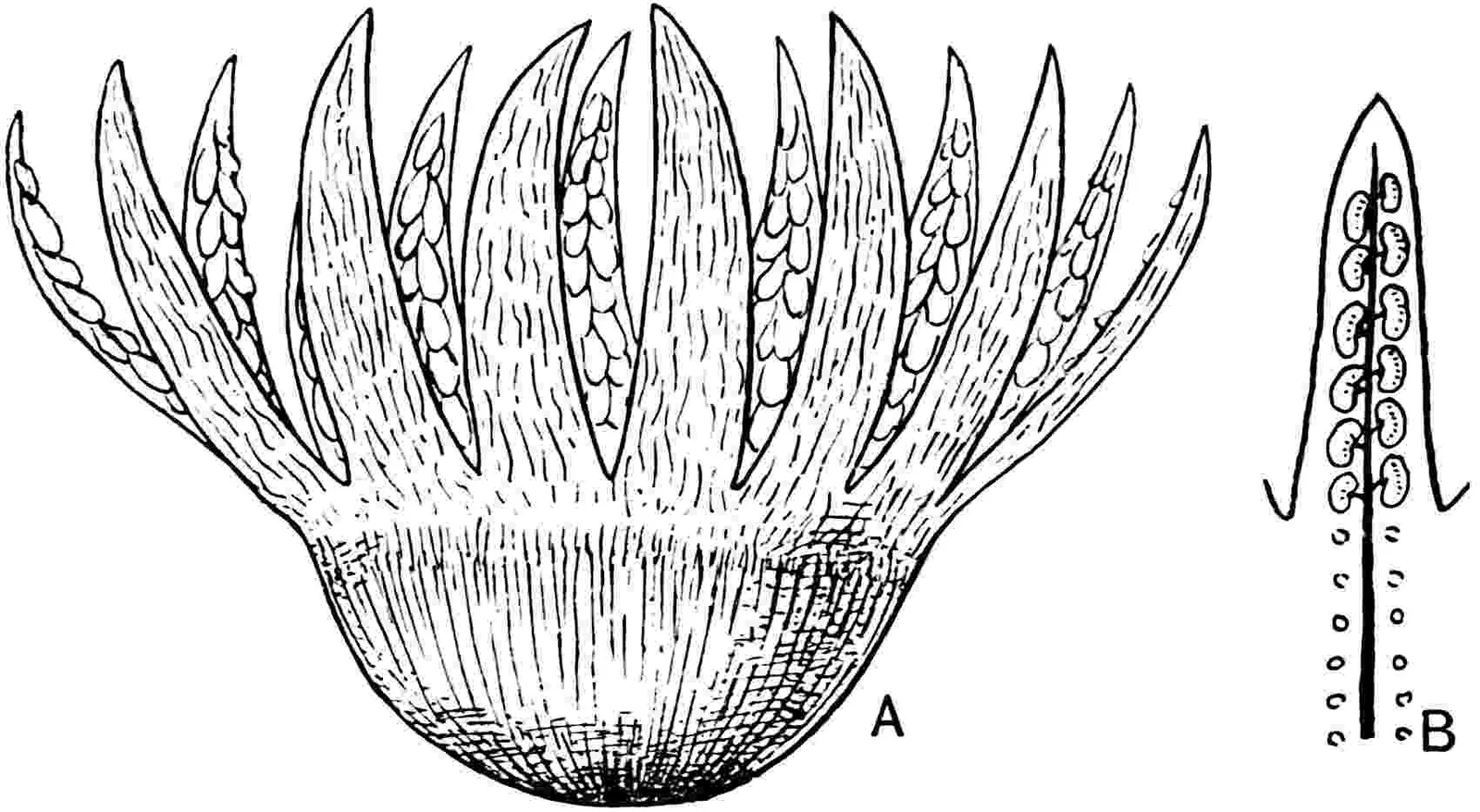
spear-point with an irregularly serrate edge. In C. revoluta,
C. pectinata, etc., the sterile part is deeply dissected
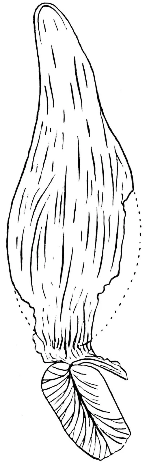
and may break off (fig. 392, A) from the fertile portion of the
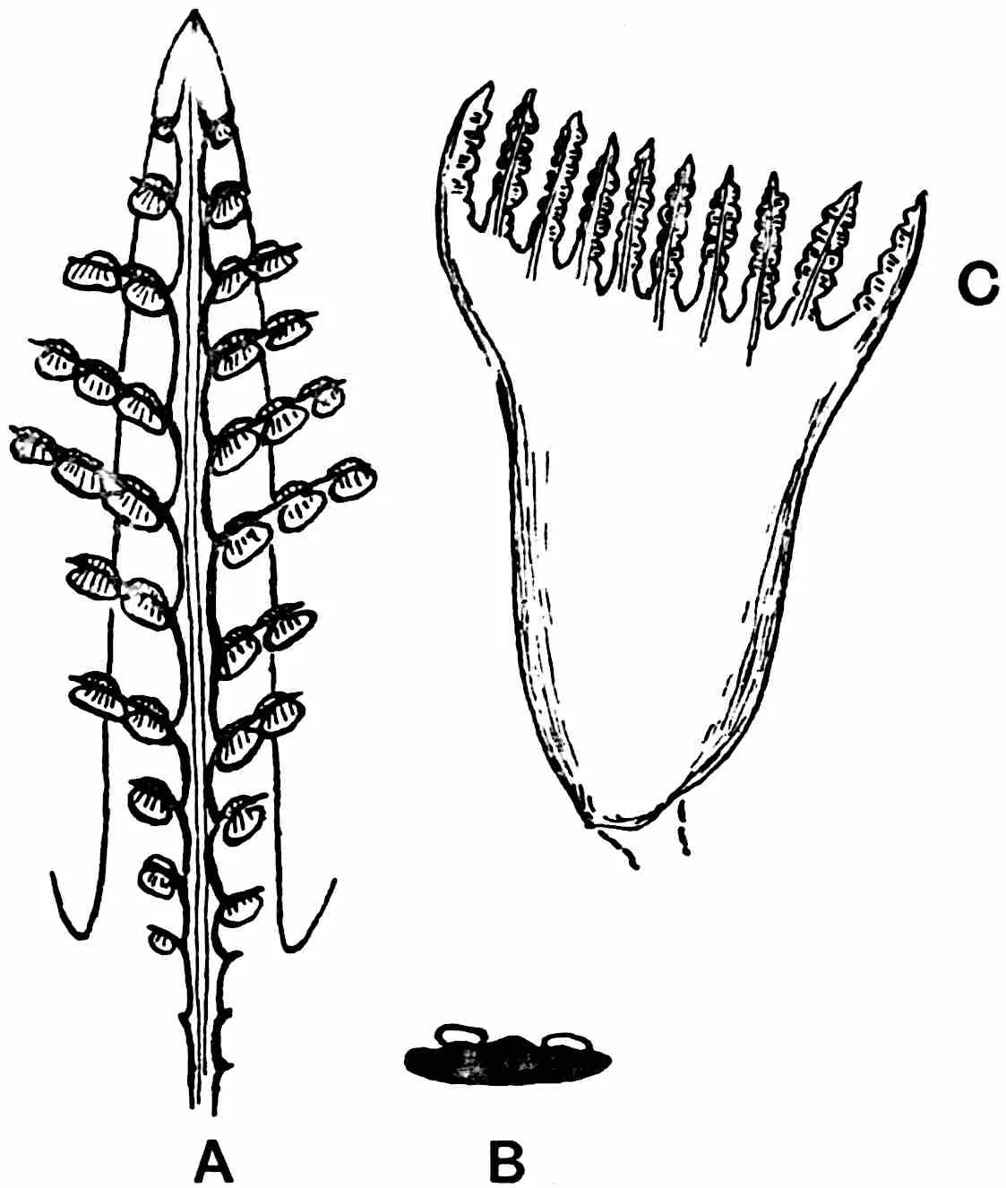
sporophyll. The megasporophylls of C. Riuminiana exhibit a
striking variation in form (fig. 392, B, C); some are 15 cm. long with
several ovules, while others, reduced to 8 cm., bear only two ovules
and resemble the sporophylls of Dioon. In all other genera the
megasporophylls are aggregated into cones, but in Dioon the
strobili are characterised by their more ovoid form and by the looser
arrangement of the sporophylls (fig. 386, B), each of which consists
of a horizontal stalk expanded distally into a broadly lanceolate
upturned end covered with a thick felt of hairs and bearing at its
base usually 2, rarely 5–6, ovules on cushion-like swellings. In
Dioon spinulosum the cones may be 50 cm. long. Between the cones
of Microcycas, over 90 cm. long, and those of some Zamias, a
few centimetres long, there are many intermediate forms. The large
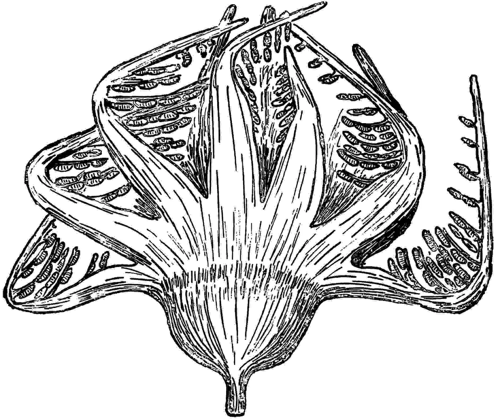
strobilus of an Encephalartos reproduced in fig. 393, D, shows
the convex ends of the sporophylls with a jagged edge, and in monstrous
cones the marginal lobes may be abnormally developed and assume the
appearance of pinnae[60]. Each megasporophyll bears two large ovules
(fig. 393, B). In certain species of Encephalartos the swollen
ends of the sporophylls have a truncate centre like the flattened umbo
of some Pines (fig. 392, D). The presence of two divergent spines is a
peculiarity of the megasporophylls of Ceratozamia (fig. 393,
C): in Macrozamia (fig. 394) the distal ends are prolonged as
tapered processes. The surface of the strobilus of Stangeria is
formed by imbricate and rounded ends of sporophylls (fig. 392, H) not
unlike the cone-scales of Pinus excelsa or P. cembra. The
megasporophylls of Zamia are expanded into regular cushion-like
hexagons with a flat central area (figs. 389, B; 395, Ib).
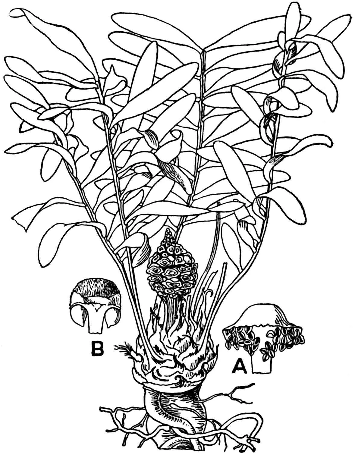
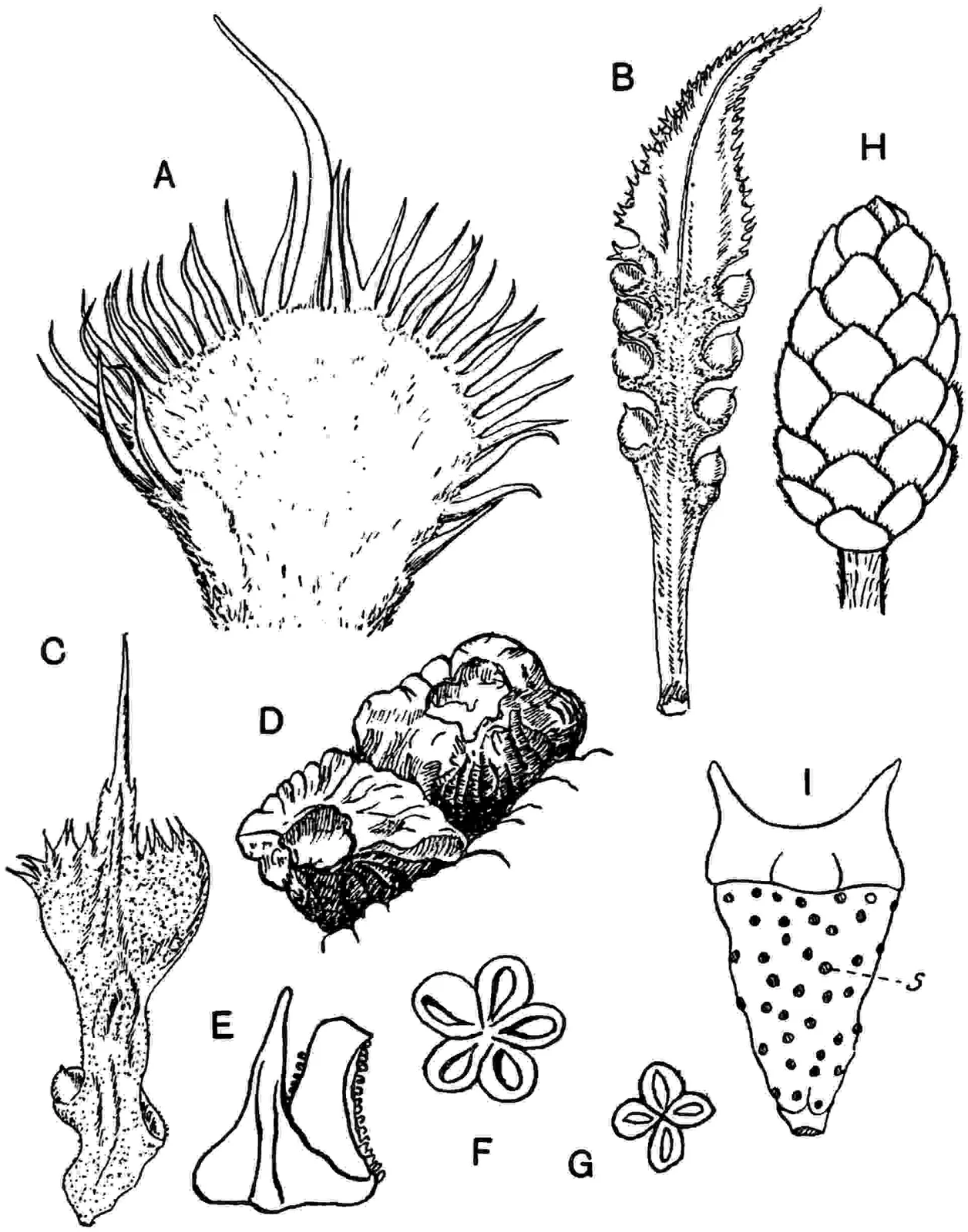
Fig. 392.
A. Cycas pectinata, apex of megasporophyll. (¾ nat. size.)
B, C. Cycas Riuminiana, megasporophyll. (¾ nat. size.)
D. Encephalartos Altensteinii. Distal end of megasporophylls. (From the Gardeners’ Chronicle.)
E, F. Cycas angulata, microsporophyll and sorus.
G, I. Ceratozamia mexicana, I, microsporophyll with scars of sori (s); G, sorus. (After Thibout.)
H. Stangeria paradoxa, megastrobilus.
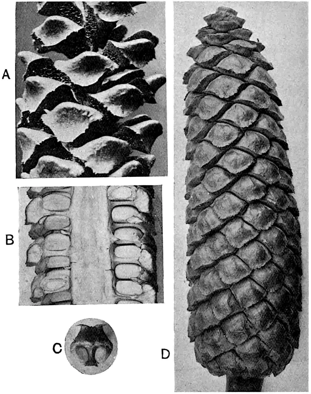
Fig. 393.
A. Stangeria paradoxa, part of microstrobilus.
B, D. Encephalartos villosus, megastrobilus in surface-view and in section. (¼ nat. size.)
C. Ceratozamia mexicana, single megasporophyll.
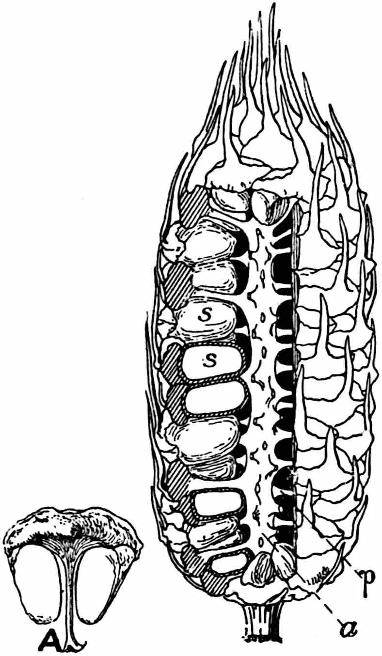
Fig. 394.Macrozamia Preissii,
megastrobilus and (A) single megasporophyll; a, axis of cone;
p, stalk of megasporophyll; s, unripe seeds. (After
Rendle.)
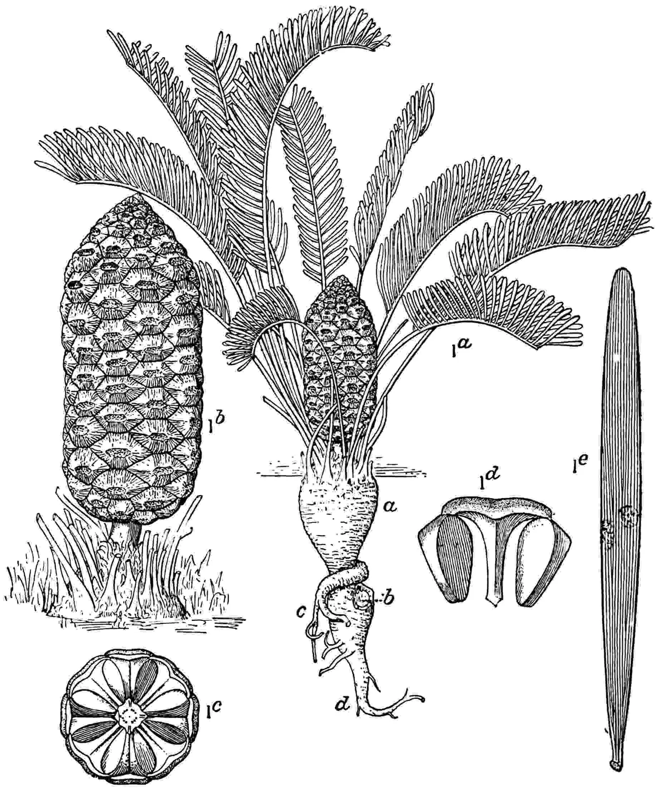
Fig. 395.Zamia floridana. Ia,
complete plant; a, main trunk; b, branch-scar; c,
secondary root; d, primary tap-root. (⅛ nat. size.) Ib,
Ic, megastrobilus. (¼ nat. size.) Id, megasporophyll. (½
nat. size.) Ie, pinna. (¾ nat. size. After Wieland.)
The microsporophylls (figs. 389, A; 392, E) are in all genera
aggregated into strobili which often bear a close resemblance
to seed-cones (fig. 393, A). On a single sporophyll of Cycas
circinalis there may be as many as 700 sporangia while in Zamia
floridana there are only two microsporangia. The spore-output is
large and in extreme cases, e.g. in Dioon spinulosum, the
average number of spores in a sporangium is said to be 30,000[61].
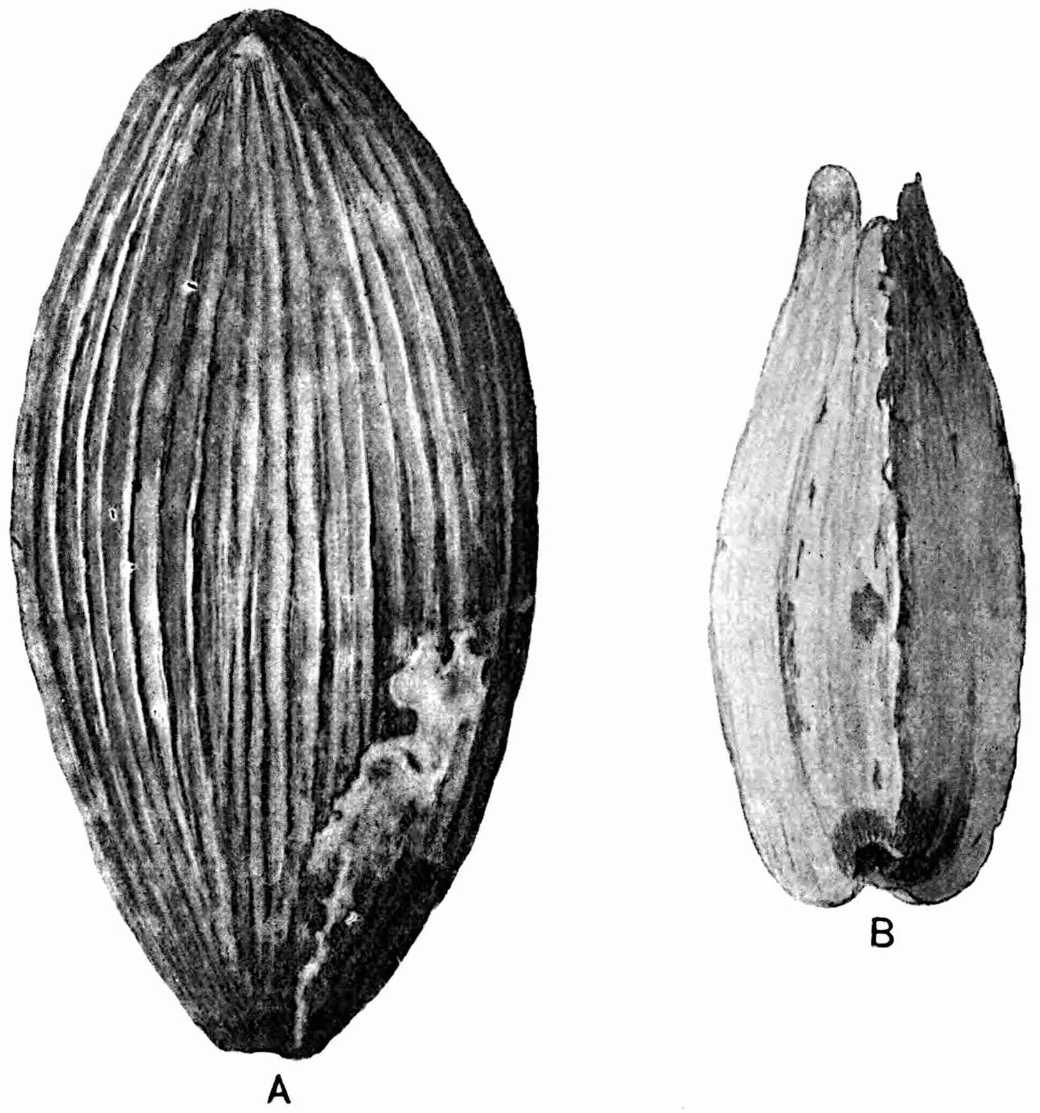
Seeds. In the great majority of recent species the seeds may
be described as large and afford a striking contrast to the small
seeds of the Mesozoic Bennettitales. A feature of interest from the
point of view of comparison with Palaeozoic seeds is the absence of a
resting stage, germination in some cases following seed-fall without
an interval. As Warming pointed out, the embryo is often undeveloped
when the seeds are shed. An interesting fact is recorded by Capt.
Dorrien-Smith[62] with regard to seed-dispersal: he describes the
heavy pebble-like seeds of a Macrozamia as being hurled from
the ripe cones a distance of 12 ft. The seeds of Cycas are
platyspermic; the woody shell exposed on removal of the outer flesh
is slightly flattened and has two prominent angles, but three-angled
seeds may occur as in Ginkgo biloba (fig. 631, C). In other
genera the seeds are radiospermic. The seed of Encephalartos
Altensteinii[63] (fig. 396, D) has a square-cut distal end with
a small papilla at the summit of the unusually long micropylar canal
(17 mm.). The stone of this seed (fig. 396, C) shows parallel curved
ridges which mark the position of vascular strands in the inner region
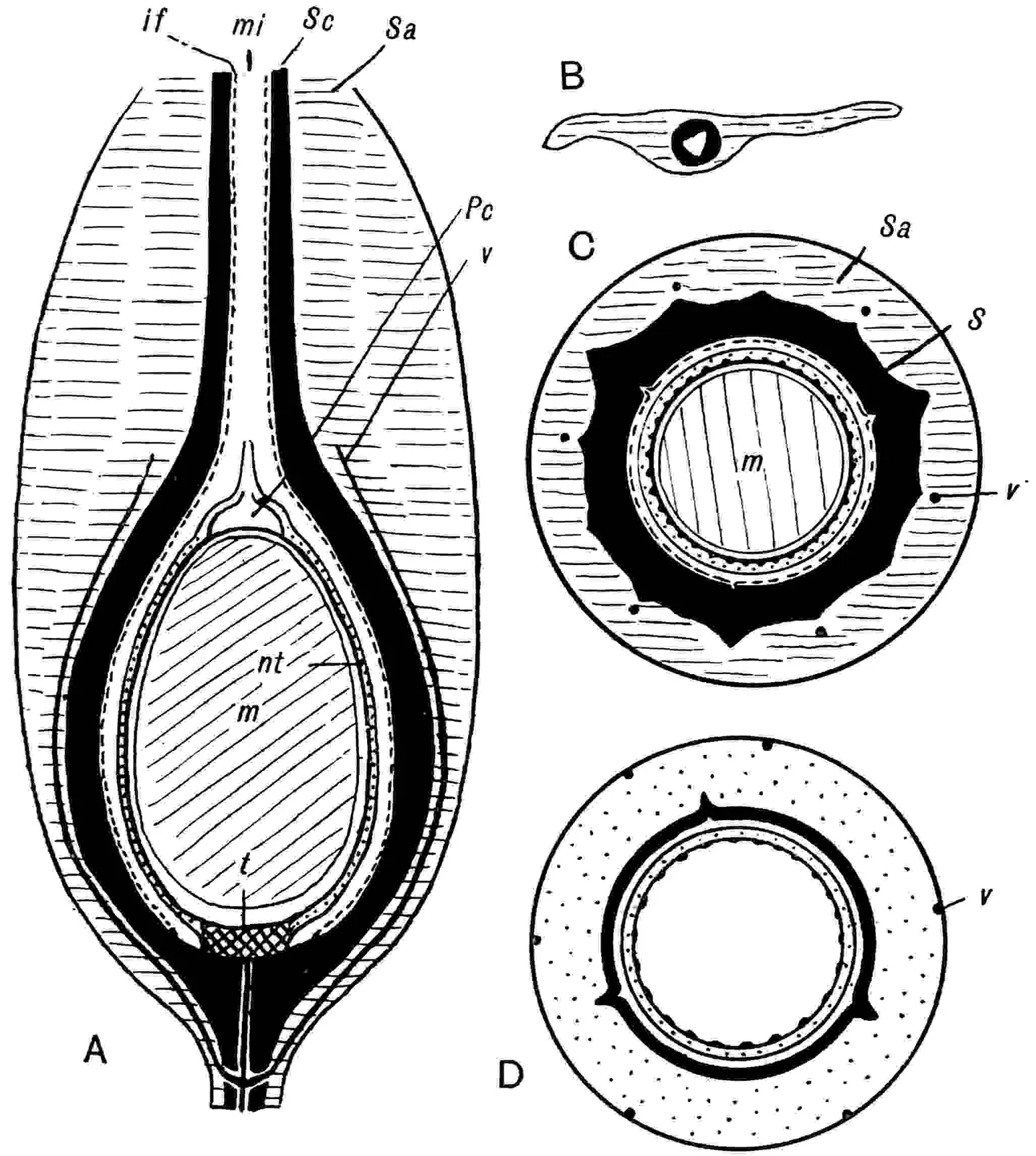
of the outer flesh. The large ovules of Cycas circinalis[64]
have an integument 1 cm. thick consisting of an outer and inner flesh
and an intervening stony layer which reaches its greatest development
at the base and apex. Three vascular strands enter the base of the
seed, the concentric strand breaks up in the broad inner flesh into a
group of bundles which embrace but do not penetrate the lower end of
the nucellus. Each of the two lateral strands branches in the outer
flesh near its entrance into the seed; the outer and larger collateral
and mesarch bundle passes up close to the surface of the shell to
the seed-apex, while the inner branch penetrates the shell and,
occasionally branching, passes up the inner region of the inner flesh
as far as the micropyle. In other seeds the tracheal supply of the
outer flesh consists of several bundles and not two as in Cycas.
The inner flesh abuts on the nucellus and is connected with it except
at the apex (fig. 396, B). In ripe seeds the nucellus is reduced to a
thin membrane enclosing the large megaspore at the upper end of which
is a depression (fig. 396, B′) or sometimes two depressions (fig.
396, I) in the prothallus containing the archegonia. In the seed of
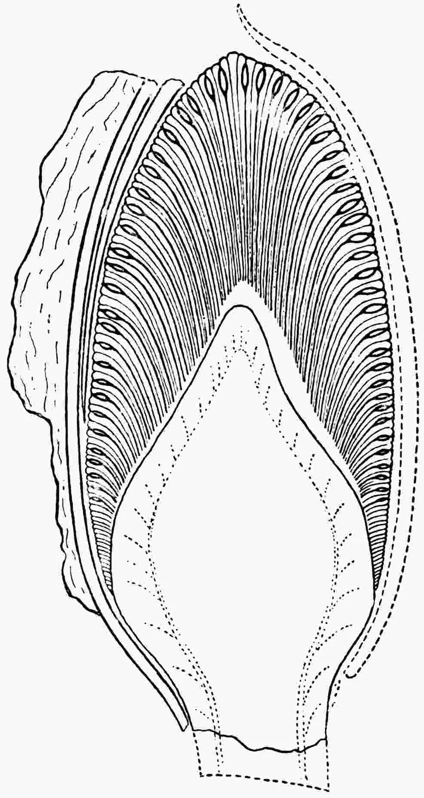
Dioon edule[65] (fig. 396, A) the position of the absciss-layer
(s) is indicated by a slight transverse constriction. In the
seeds of Bowenia, constructed on the same plan, the inner series
of vascular strands appears to be nucellar in position, thus differing
from the strands in Dioon, Cycas, and other genera
which are confined to the integument. Miss Kershaw[66] in describing
Bowenia speaks of an upper and a lower pollen-chamber; the
former serves as a storage-place for the microspores prior to their
further development in the lower chamber. Dr Stopes[67] regards the
integument as double in origin, a view suggested by Griffith[68] in
1835, and as homologous with the single integument plus the
cupule of Lagenostoma. This view is supported by Mrs Thoday[69]:
on the other hand Miss Kershaw’s investigation of Bowenia seeds
leads her to regard the integument as single. Although there would seem
to be a prima facie case in favour of the dual nature of the
integument, the arguments on the other side have greater weight[70].
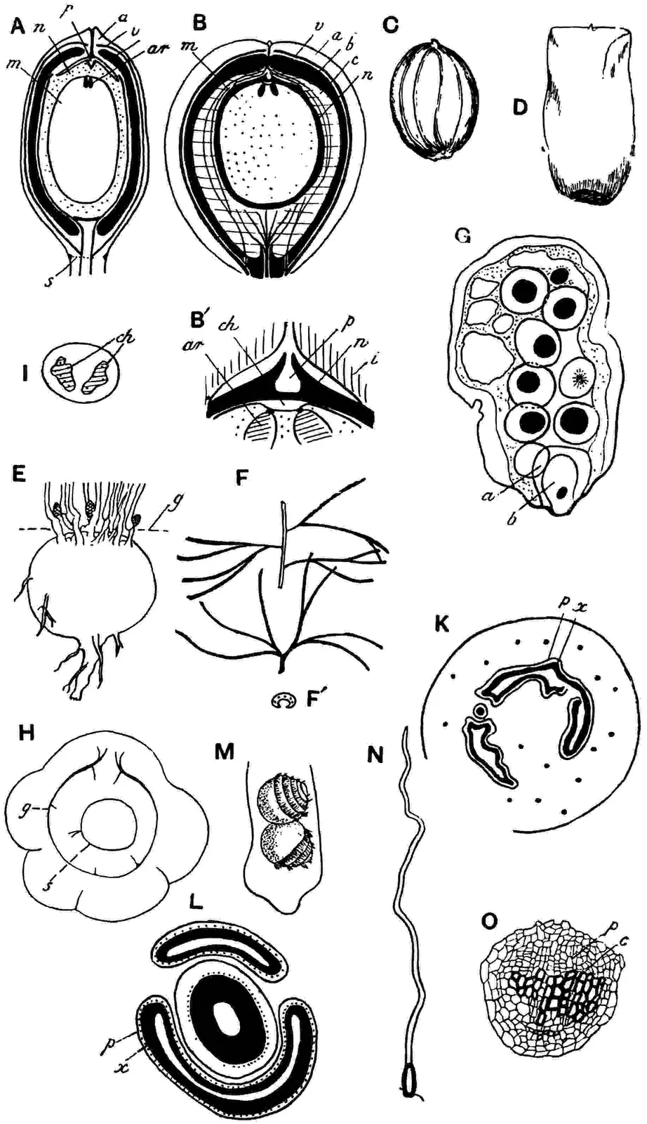
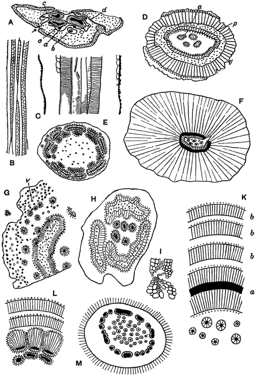
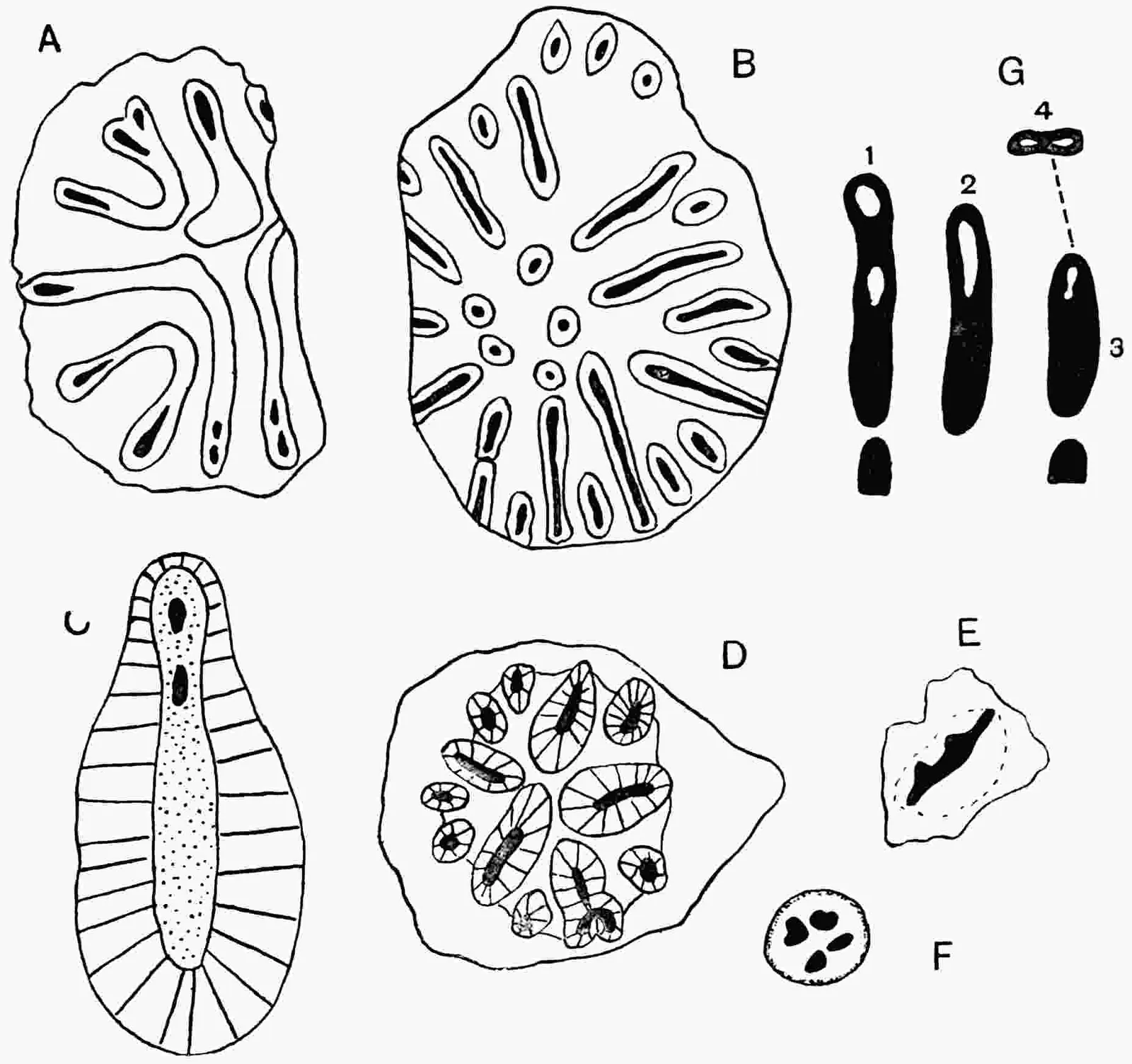
Fig. 396.
A. Seed of Dioon edule in longitudinal section; a, integument;
v, vascular tissue; m, prothallus; n, nucellus; p, pollen-chamber; s, absciss-layer; ar, archegonia. (After Chamberlain.)
B, B′. Seed of Cycas circinalis; a, v, integument (sarcotesta) and
vascular tissue; b, sclerotesta; c, inner sarcotesta; m, n, prothallus and nucellus. (After Stopes.)
B′. Apex of nucellus; p, pollen-chamber; i, integument; n, nucellus;
ch, archegonial chamber; ar, archegonia.
C, D. Seed of Encephalartos Altensteinii; C, surface of stone. (After Stopes.)
E. Stem of Bowenia serrulata; g, level of ground. (After Chamberlain.)
F, F′. Pinnae of Macrozamia heteromera.
G. Pollen-tube of Microcycas Calocoma. (After Caldwell.)
H. Transverse section of stem of Encephalartos horridus; s, stele; g, girdle-bundles. (After Mettenius.)
I. Apical view of prothallus of Cycas showing two archegonial chambers
(ch). (After Treub.)
K. Encephalartos Barteri. Transverse section of stem; x, xylem; p, phloem. (After Matte.)
L. Cycas siamensis. Transverse section of vascular tissue of young stem. (After Matte.)
M. Cycas revoluta; two motile sperms. (After Miyake.)
N. Long hair with short basal cell from the petiole of Macrozamiaheteromera. (After Robertson.)
O. Vascular bundle of Dioon edule from base of petiole; p, phloem; c, cambium. (After Mettenius.)
Recent observations point to the probability that insects play a part
in the pollination of cycadean ovules. Kraus[71] drew attention to
the strong smell emitted by the microstrobili of Dioon edule
and noticed that small bees were attracted to the ripe strobili
of Macrozamia, while odourless cones of a neighbouring
Ceratozamia received no attention. Pearson[72]
and Rattray[73]
have obtained evidence that beetles and weevils act as pollinators to
species of Encephalartos.
Anatomical features. Allusion has already been made to some
of the more striking anatomical features; the large pith, the
occasional occurrence of medullary vascular bundles, the presence of
one or more cambiums, the large size of the medullary rays, etc. It
is worthy of remark that the occurrence of an anastomosing system
of medullary bundles is not a constant feature within a genus; in
Macrozamia Fraseri such a system is present, but absent in
M. Denisoni[74]. In the pith of stems with no medullary
bundles cylinders of collateral bundles may occur in connexion with
a fertile shoot. These bundles arise from the inner face of the main
cylinder and pass upwards as a domical system into the base of the
terminal strobilus which is eventually pushed to one side by the
growth of a lateral bud[75]. The secondary xylem tracheids are usually
provided with several rows of bordered pits on the radial walls and
resemble those of the Araucarieae[76], but in Cycads the pits are
often not contiguous and less compact in their distribution. The
wood of Stangeria is peculiar in consisting of scalariform
tracheids[77] (fig. 397). Chamberlain describes growth-rings in
the wood of Dioon; but this is exceptional. In tangential
sections of the stele leaf-trace bundles are constantly seen passing
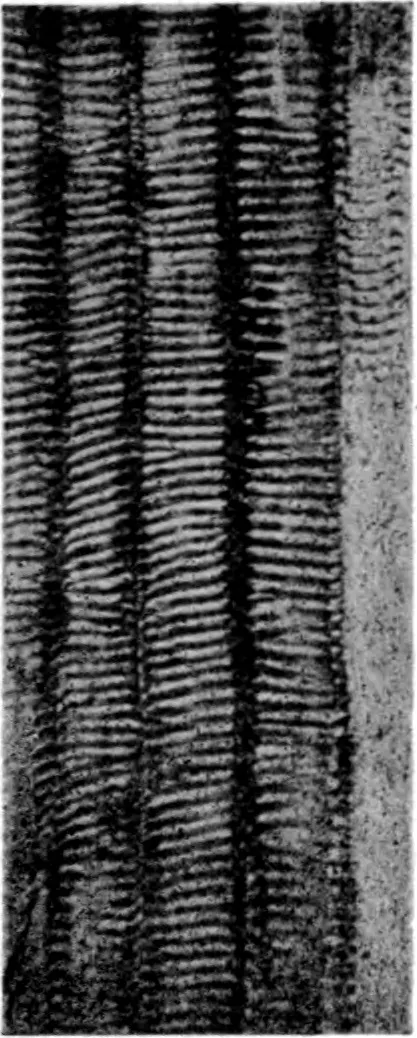
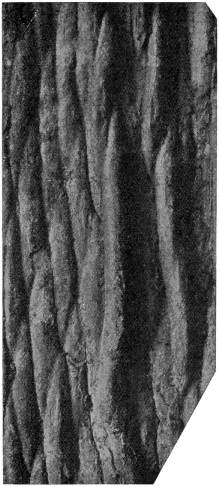
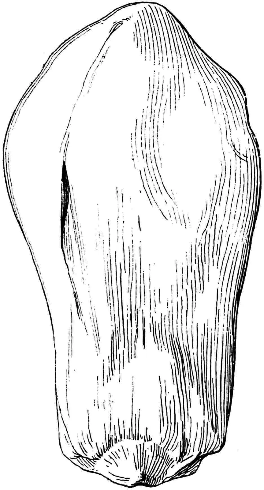
horizontally through the broad and deep medullary rays. The pith-cast
of a cycadean stem reproduced in fig. 398 shows the wide meshes in
the reticulum of tracheal tissue originally occupied by parenchyma,
which on decay left lenticular depressions represented on the cast by
tapered convex areas occasionally bearing the impress of an outgoing
trace in the form of a narrow groove. The secondary phloem often rivals
the xylem in breadth and is not always easily distinguishable from
it; it consists of sieve-tubes, parenchyma, and fibres. The secondary
cambial cylinders characteristic of Cycas, Encephalartos,
Macrozamia, and Bowenia, to which reference was made in
the summary of anatomical features, arise in the pericycle, and a few
layers of pericyclic parenchyma occur between adjacent extrafascicular
cylinders of xylem and phloem. In a stem of Cycas media 35 cm.
in diameter examined by Worsdell there were 12 concentric cylinders.
Matte[78] and Miss Dorety[79]
have described partially flattened arcs
of extrafascicular xylem and phloem in the hypocotyl of Ceratozamia
mexicana. Worsdell[80] first drew attention to the occasional
occurrence of short tracheids on the inner edge of the secondary
wood and to the spasmodic development of cambial arcs in the tissue
between the extrafascicular cylinders forming strands of inversely
orientated xylem and phloem. More recent work by Matte gives support
to Worsdell’s comparison between Medullosan stems and those of recent
Cycads with inversely orientated arcs or concentric vascular cylinders.
The French author draws attention to the close resemblance between the
seedling stems of such species as Encephalartos Barteri (fig.
396, K) and Cycas siamensis (fig. 396, L) with their polystelic
type of structure and the adult stems of Medullosa[81]. In
the stems of Dioon, Microcycas, Stangeria, and
Zamia no extrafascicular cylinders are recorded. Two main
vascular bundles enter the cortex from each leaf-base and in most stems
these diverge right and left and more or less completely encircle
the stele before passing through the medullary rays and joining the
inner portion of the xylem of the stele either as double or single
bundles. These girdle-bundles (fig. 396, H) first described by Karsten
and Mettenius form a very characteristic cycadean feature[82].
Adjacent girdles are joined by connecting cortical bundles and, in
addition, there are cauline collateral bundles in the cortex which
form an anastomosing system. In some cases, e.g. species of
Macrozamia and occasionally in Stangeria, the female
peduncle of a Ceratozamia, and in seedlings of Bowenia
and Cycas revoluta[83], the leaf-traces pursue a direct course
from petiole to stele as in stems of Bennettitales. It is noteworthy
that in seedlings of Microcycas[84], a genus characterised by
a large number of male gametes—presumably a primitive feature—the
leaf-traces are of the girdle-type. The two bundles at the base of a
petiole by repeated subdivision give rise to the numerous collateral
strands of the rachis. A leaf-trace in its passage to the leaf is like
that of a Conifer in having the protoxylem on its inner edge, whereas
in the petiole and elsewhere in the frond it is characterised by an
arrangement of the xylem that has usually been described as mesarch. A
typical vascular bundle from a cycadean frond is seen in fig. 399, C;
by far the greater part of the xylem is centripetal, the centrifugal
xylem being confined to an arc of scattered tracheids or a small strand
separated by a few parenchymatous cells from the protoxylem.
Fig. 397. Tracheids from the stem of Stangeria paradoxa. (After Marsh.)
Fig. 398. Pith-cast of a Macrozamia stem, (⅖ nat. size.)
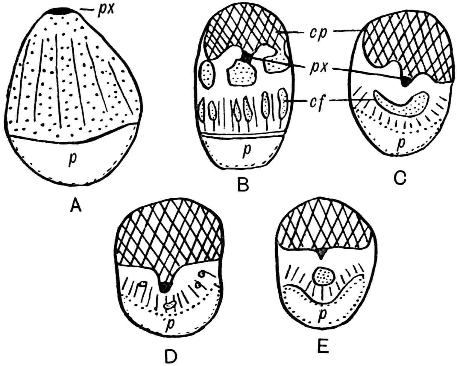
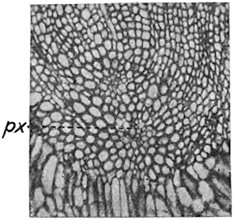
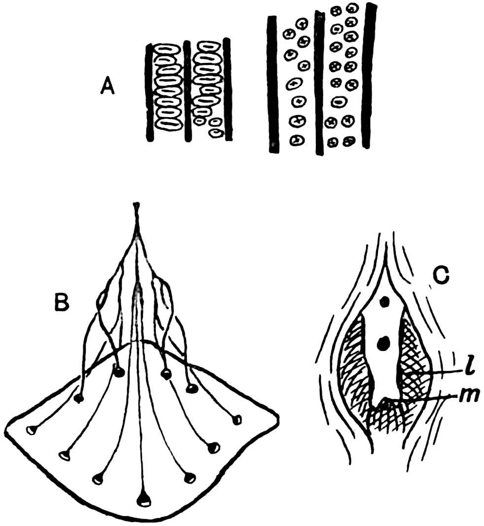
Fig. 399. Sketches illustrating the changes
in the structure of Cycadean vascular bundles in their course from
stem to leaf: cp, cf, centripetal and centrifugal xylem;
p, phloem; px, protoxylem. (After Marsh.)
As considerable stress has been laid on the anatomical features of
the cycadean foliar bundles in discussions on the affinities and
phylogeny of certain Palaeozoic genera, it is important to consider
the facts more closely[85]. French anatomists described the cycadean
bundle as diploxylic on the ground that the centripetal and centrifugal
xylems are distinctly different things, the centripetal xylem being
primary—a relic of a former organisation—and the centrifugal xylem
secondary and homologous with the normal wood of the cauline
bundle. The term mesarch has in recent years been applied to the
cycadean type of bundle. A mesarch bundle is, however, one in which
centripetal and centrifugal xylem are alike in origin, both being
primary structures derived from a desmogen strand. Typical mesarch
bundles occur in several recent ferns; in the stele of the Osmundaceae,
Gleichenia, and other genera; but in these plants the xylem is
all produced directly from one primary desmogen region and there is no
question of ‘primary’ and ‘secondary’ as in the two portions of the
xylem of a cycadean bundle. Recent researches into the development of
cycadean foliar bundles show that they do not conform to the mesarch
type as generally understood. A leaf-trace at the base of a petiole
(fig. 399, A) comprises centrifugal xylem only, and this consists
of regular rows of tracheids separated by medullary rays: in the
lower part of the petiole the structure is gradually modified, the
centrifugal xylem is reduced and the formation of centripetal xylem is
initiated. At a higher level (fig. 399, B) the centripetal xylem is in
excess of the centrifugal and the latter, for a time connected with the
former, eventually becomes separated by a few parenchymatous cells from
the protoxylem and persists as a small strand or arc of tracheids. Fig.
399 illustrates stages in the transformation of a typical collateral
bundle, at the base of a Stangeria petiole, into one in which
the xylem is almost wholly centripetal at a higher level in the axis
of the frond. A cambium is present in all: in B the centrifugal xylem
is more or less clearly differentiated into two portions, loosely
arranged tracheids near the phloem, and the more compact groups
abutting on the centripetal xylem: figs. C–E show a further reduction
in the centrifugal tracheids. The conclusion drawn from developmental
study is that the two xylem portions of the bundle are independent in
origin[86]. Marsh has, however, shown that in Stangeria bundles
near the base of the petiole the centrifugal xylem consists of rows of
secondary tracheids and an inner portion not in rows which connects the
centrifugal with the centripetal elements; this connecting portion, he
adds, is ‘probably primary and connects up the Cycadean foliar bundle
with the truly mesarch bundle of the Cycadofilices.’
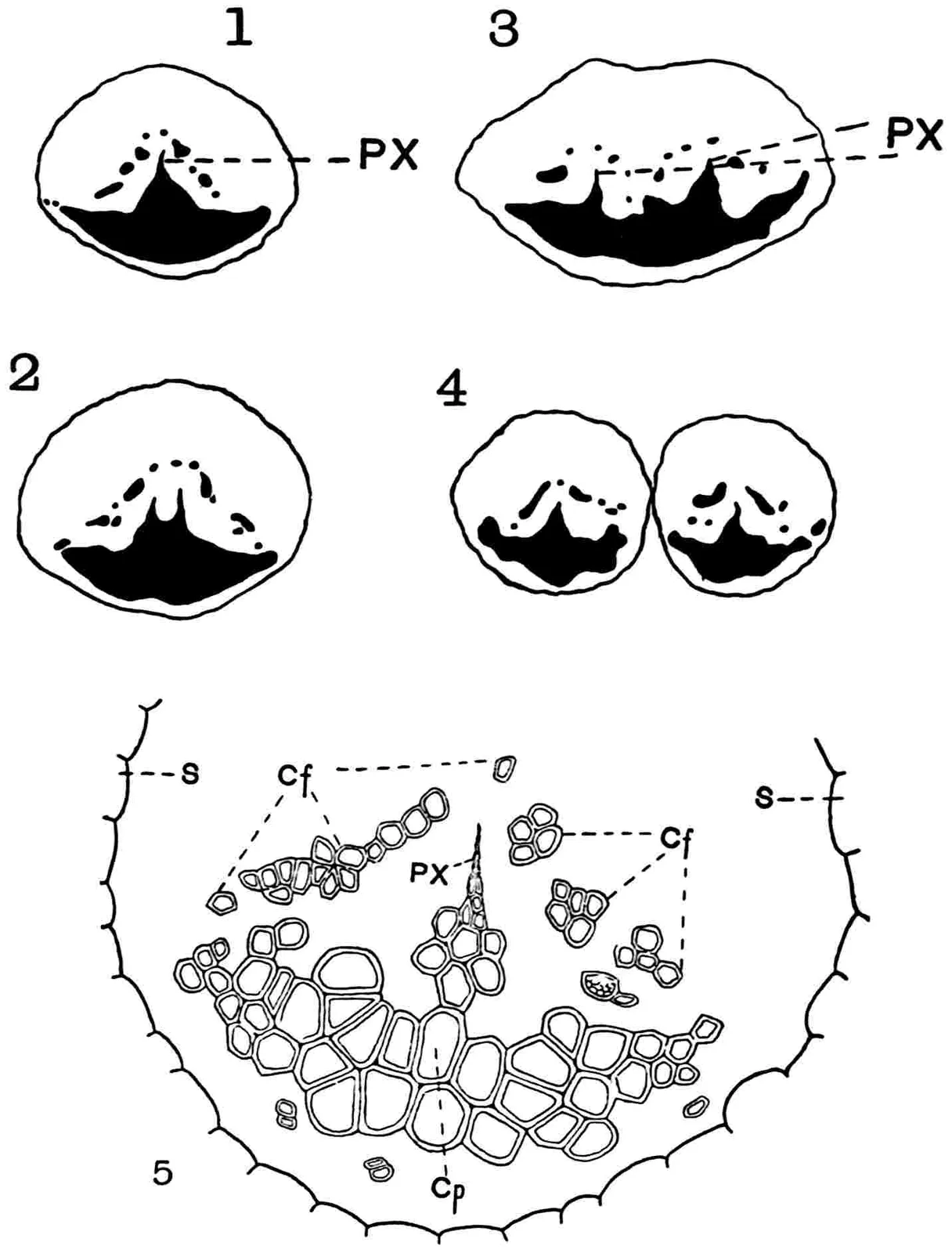
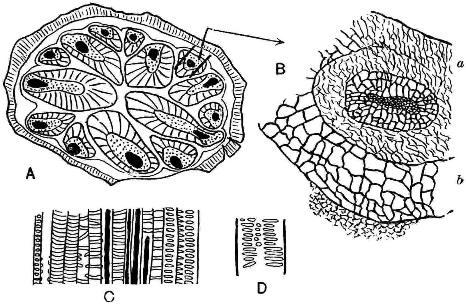
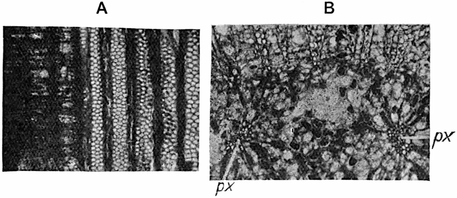
Fig. 400.Cycas Micholitzii. Vascular
bundles in a forked pinna; px, protoxylem; s, sheath of
thick-walled cells; cf, cp, centrifugal and centripetal
xylem.
In the xylem portion of the bundle from the midrib of a forked pinna
of Cycas Micholitzii shown in fig. 400 the centrifugal xylem
elements are unusually numerous: the space between the two xylems is
occupied by parenchyma and the whole strand is enclosed by a sheath
of crystal-containing cells, s, with thick inner walls. Fig.
400, 1–4, illustrates the gradual change in the form of the bundle
in the region of dichotomy[87]. The ground-tissue of the petiole
is abundantly supplied with secretory canals and in the hypodermal
region is a cylinder of stereome. In some petioles, e.g.Macrozamia heteromera[88], the ground-tissue cells are lignified
and reticulately pitted, a feature met with in some Mesozoic cycadean
leaves[89]. In Cycas media Worsdell noticed a tendency of
the leaf-trace bundles towards a concentric arrangement and similar
vascular strands are recorded in the peduncle of Dioon edule, in
various sporophylls[90] and in other cases. It is possible, as Worsdell
believes, that the fairly frequent occurrence of concentric bundles
in plants characterised by collateral bundles may have a phylogenetic
significance.
The pinnae are dorsiventral and the veins exarch or pseudomesarch:
secretory canals occur between (Encephalartos), above, or below
the veins. The mesophyll of Cycas is characterised by the
presence of isolated xylem-elements passing from the midrib to the edge
of the lamina and, as Lignier[91] suggests, these may be regarded as a
reduced system of lateral conducting strands.
The epidermal cells of the leaflets have straight or slightly
curved walls except in Stangeria where they are undulate and
fern-like[92]. The stomata, with few exceptions confined to the lower
epidermis, are larger than in other gymnosperms (on the average ·075
× ·034 mm.) and are more or less depressed below the surface; the
guard-cells are usually surrounded by 4–6 subsidiary cells.
The roots exhibit no feature to which attention need be called: the
pericycle is several cells broad and as in the stem there may be
extrafascicular cylinders of xylem and phloem.
CHAPTER XXIX. — I. LYGINOPTERIDEAE. — LYGINOPTERIS. — HETERANGIUM.
PTERIDOSPERMEAE.
The genus Lyginopteris is selected for the first place in this
chapter simply on the ground that we have a fuller knowledge of its
morphology than in the case of other types. It is not regarded as
the most primitive member of its class. Lyginopteris may be
described in a few words as a plant having the habit and to a large
extent the anatomical features of a Fern, but differing from existing
ferns in the possession of integumented megasporangia or seeds and in
the power of secondary growth in thickness by means of a cambium in
both stem and root. The seed (Lagenostoma) agrees with those of
recent Cycads and Gnetales more closely than with the corresponding
organs in Conifers or any other group, while the structure of the
secondary wood is practically identical with that of Cycads. The
microsporangia occur as groups of small bilocular sporangia, or
synangia, at the tips of fertile pinnae of highly compound fronds.
Nomenclature and Historical Summary. In 1866 E. W. Binney[93]
of Manchester published a short description of a small petrified stem
from the Lower Coal Measures of Lancashire and named it Dadoxylon
oldhamium, employing Endlicher’s term Dadoxylon which
that author substituted for Pinites as previously used by
Witham[94]. Three years later Williamson[95]
drew attention to certain
features in which Binney’s type differs from the genus Dadoxylon
and substituted a new name Dictyoxylon, suggested by the
reticulate pitting on the walls of the tracheids. In a subsequent
paper Williamson[96] gave a fuller description of Binney’s species
and spoke of it as ‘one of the most common plants in the calcareous
nodules of the Lower Coal Measures’ of Lancashire and Yorkshire. He
connected certain casts of arborescent dimensions with Binney’s type
on the ground that the surface-features of the casts are such as
would be produced by partially decorticated stems having a hypodermal
reticulum of mechanical tissue like that preserved in the small
petrified specimen described by Binney (fig. 402). Mr Carruthers
called Williamson’s attention to a paper by Mr Gourlie[97] in which
the generic name Lyginodendron is instituted for stem-casts
identical in surface-features with the fossils figured by Williamson.
In spite of the much larger dimensions of the reticulum on the casts
described by Gourlie as compared with that in the outer cortex of
Binney’s stem, Williamson concluded that Lyginodendron is
‘undoubtedly an inorganic cast of the prosenchymatous layer of the
bark of Dictyoxylon.’ It is but fair to add that Williamson
was influenced in coming to this conclusion by a discovery by Mr
Nield of a piece of a large petrified stem believed to be generically
identical with Binney’s type, but subsequently referred to a distinct
genus[98], which was comparable in size with the stems responsible for
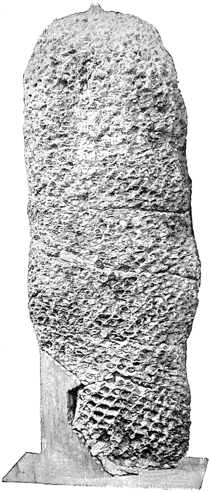
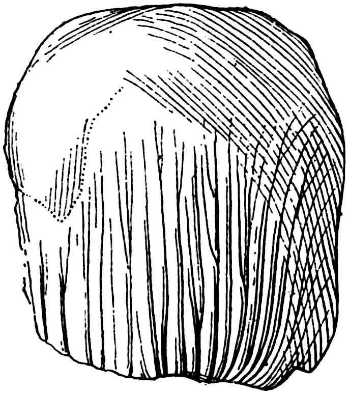
Gourlie’s Lyginodendron casts. The type-specimen of Gourlie’s
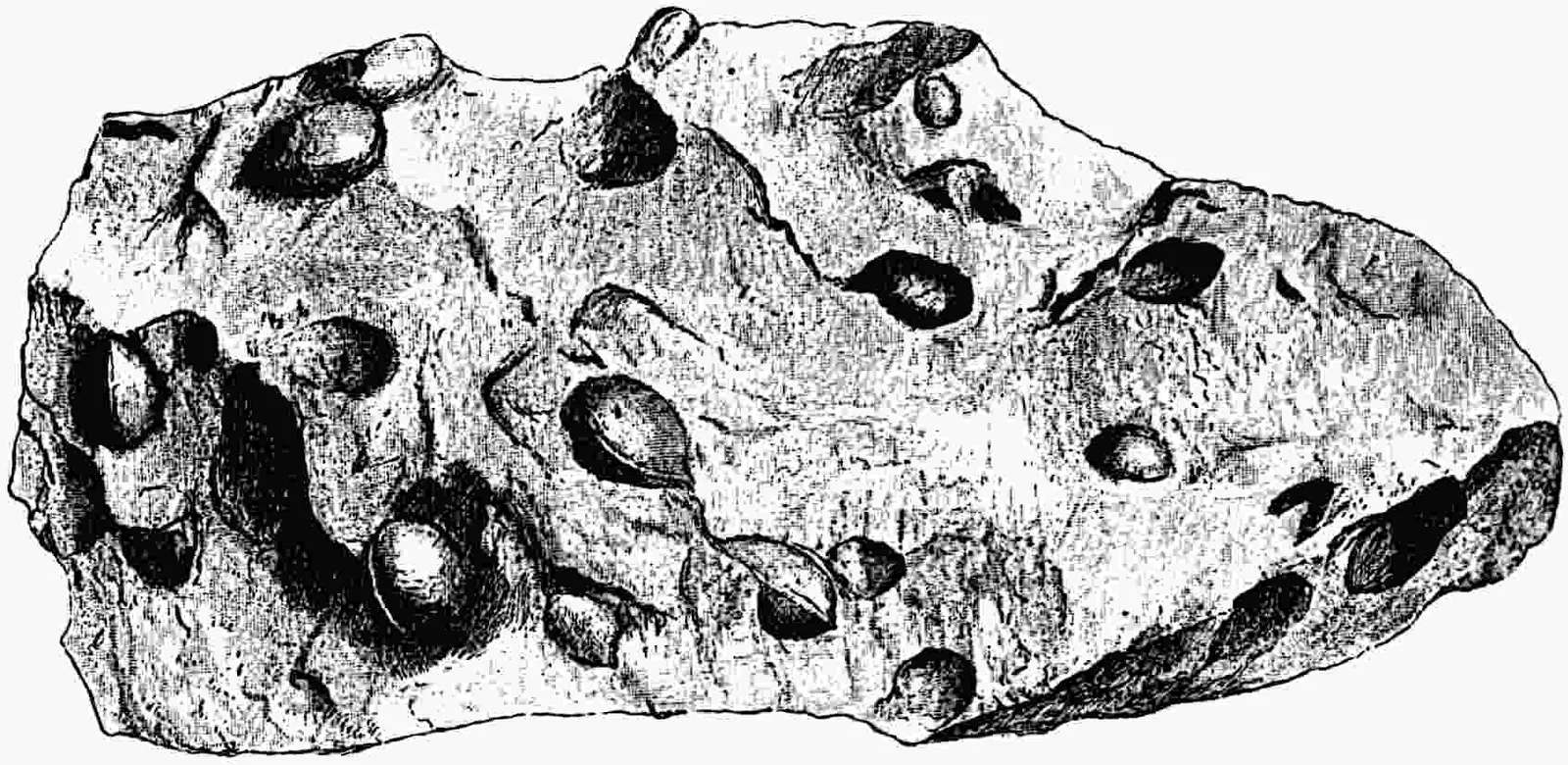
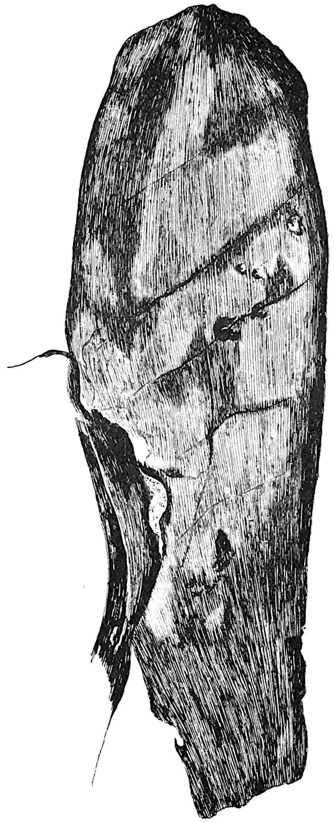
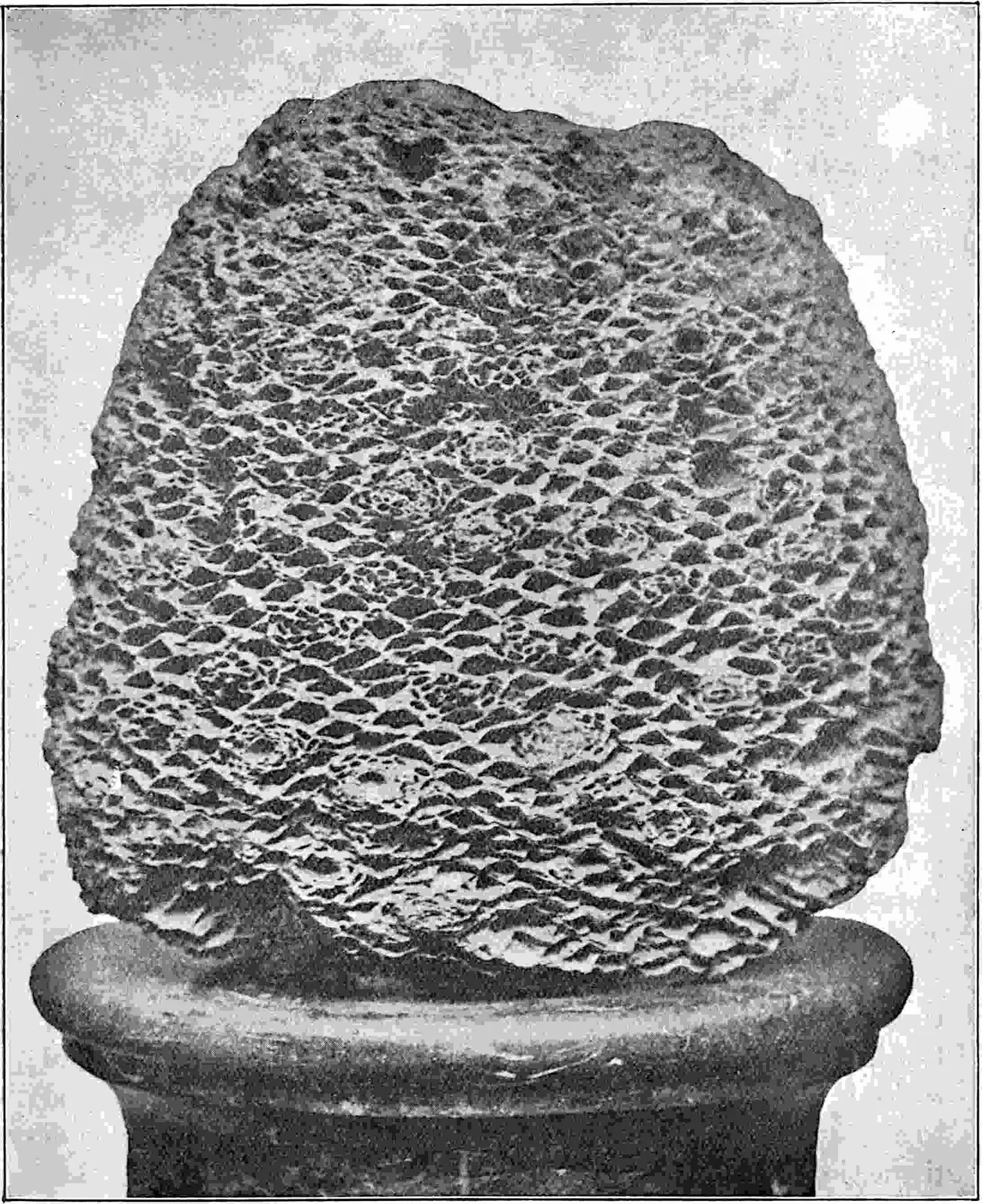
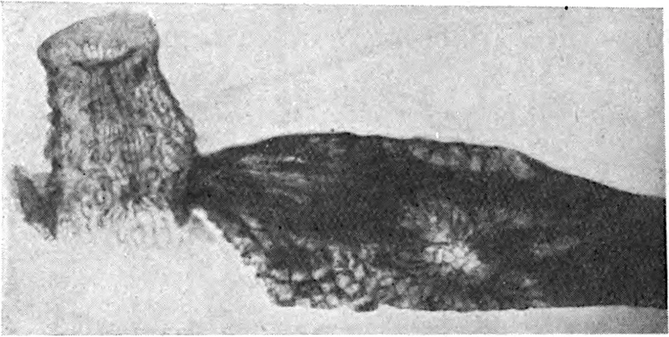
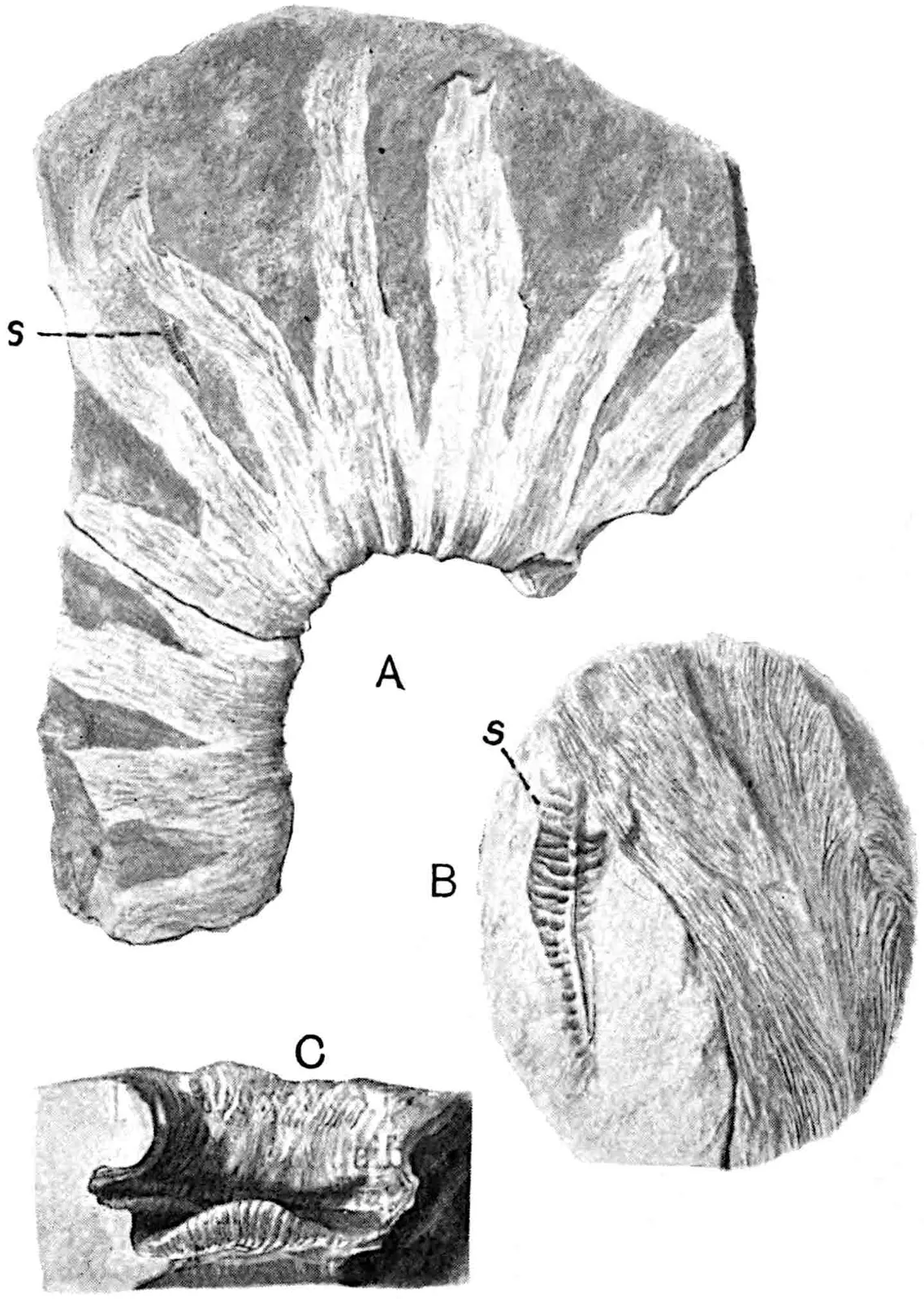
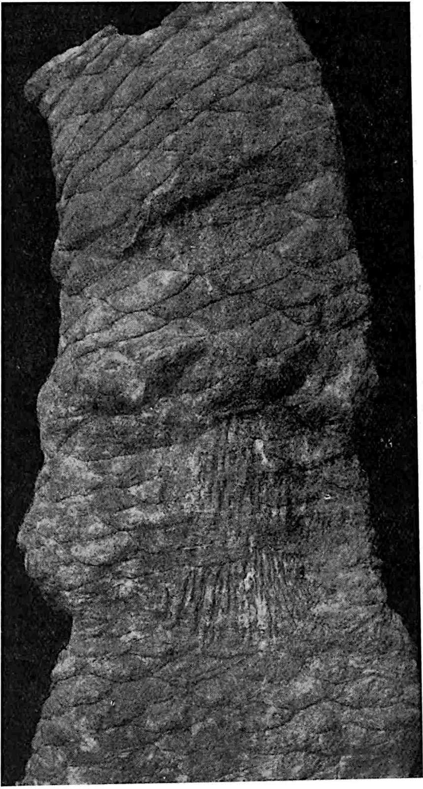
Lyginodendron Landsburgii[99], from Carboniferous rocks at
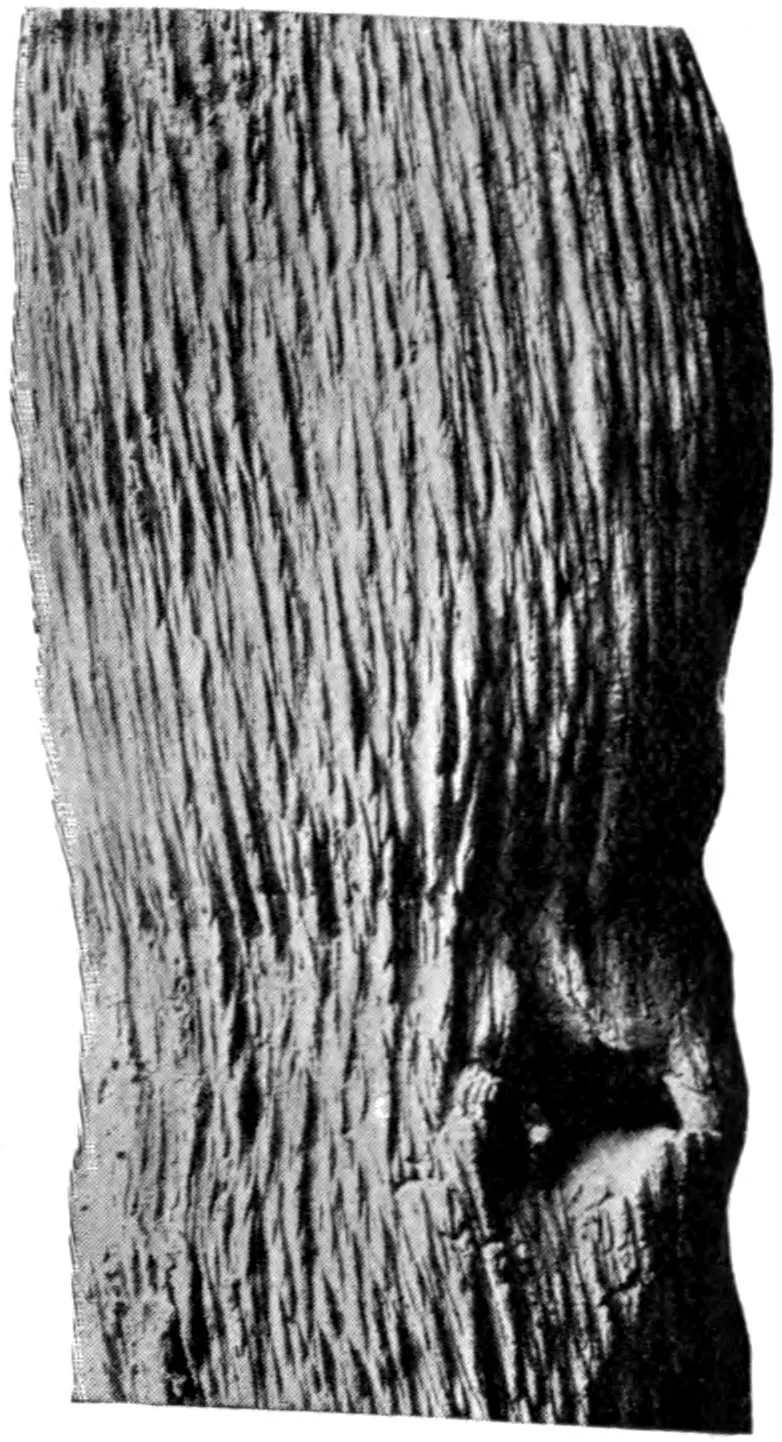
Stevenston in Ayrshire, Scotland, is represented in fig. 401. The
convex areas represent casts of depressions in a reticulum of cortical
tissue, originally occupied by comparatively delicate cells, which
decayed or shrunk more quickly than the enclosing framework of stronger
fibrous elements that remained as a prominent reticulum and produced
the depressions bounding the raised portions of the cast. Such a cast
would undoubtedly be formed by the stem on which Binney founded his
species: the radially disposed bands of thick-walled cells seen in
the outer part of the section (fig. 402) are portions of an irregular
anastomosing mechanical system, the reticulate arrangement of which
is seen in the impression of a rachis of a Lyginopteris frond
shown in fig. 405, E, and indicated in the more slender axis reproduced
in fig. 404, A, b. This reticulate form of cortical stereome
on which Brongniart founded the genus Dictyoxylon[100], a
term since applied by Solms-Laubach and other authors to a certain
type of cortex not confined to a single genus of plants, occurs also
in some Palaeozoic lycopodiaceous stems[101] and in itself cannot
be regarded as a safe criterion of botanical affinity. The largest
example of Gourlie’s Lyginodendron that has come under my
notice is an incomplete sandstone cast from Upper Carboniferous strata
near Harrogate reaching a length of 100 cm. and with convex areas 13
cm. long. A similar cast, 36 cm. broad, has recently been figured by
Nathorst[102] from the Culm of Spitzbergen, and from the Upper Devonian
of Ellesmere Land the same author has described impressions of a
cortical reticulum under the name Lyginodendron Sverdrupi[103].
These specimens are interesting as pointing to the former occurrence
in the Arctic regions of stems—probably Lepidodendroid—reaching the
dimensions of a fairly large tree. As Potonié[104] pointed out,
Gourlie’s generic name serves a useful purpose for casts of stems
of the type shown in fig. 401 that cannot be assigned to a definite
systematic position. The genus was first used for a specimen which has
nothing to do with the plant usually spoken of as Lyginodendron
oldhamium (Binney). Though loath to give up a name by which
Binney’s type has long been known, in spite of its retention in the
second volume of this work I feel compelled so far to conform to the
recognised principles governing nomenclature as to adopt Potonié’s
generic term Lyginopteris.
Lyginopteris oldhamia, Potonié, Lehrbuch der Pflanzenpalaeontologie, p. 171.
ii.
Leaf.
1828.
Sphenopteris Hoeninghausi, Brongniart, Prodrome, p. 51.
1872.
Edraxylon, Williamson, Proc. R. Soc. vol. xx. p. 438.
1874.
Rachiopteris aspera, Williamson, Phil. Trans. R. Soc. vol. clxiv. p. 684.
1877.
Calymmatotheca Hoeninghausi, Stur, Culm Flora, ii. p. 266.
1905.
Crossotheca Hoeninghausi, Kidston, Proc. R. Soc. vol. lxxvi. p. 358.
iii.
Seed.
1877.
Lagenostoma, Williamson, Phil. Trans. R. Soc. vol. clxvii. p. 234.
1903.
Lagenostoma Lomaxi, Oliver and Scott, Proc. R. Soc. vol. lxxi. p. 477.
iv.
Root.
1876.
Kaloxylon Hookeri, Williamson, Phil. Trans. R. Soc. vol. clxvi. p. 23.
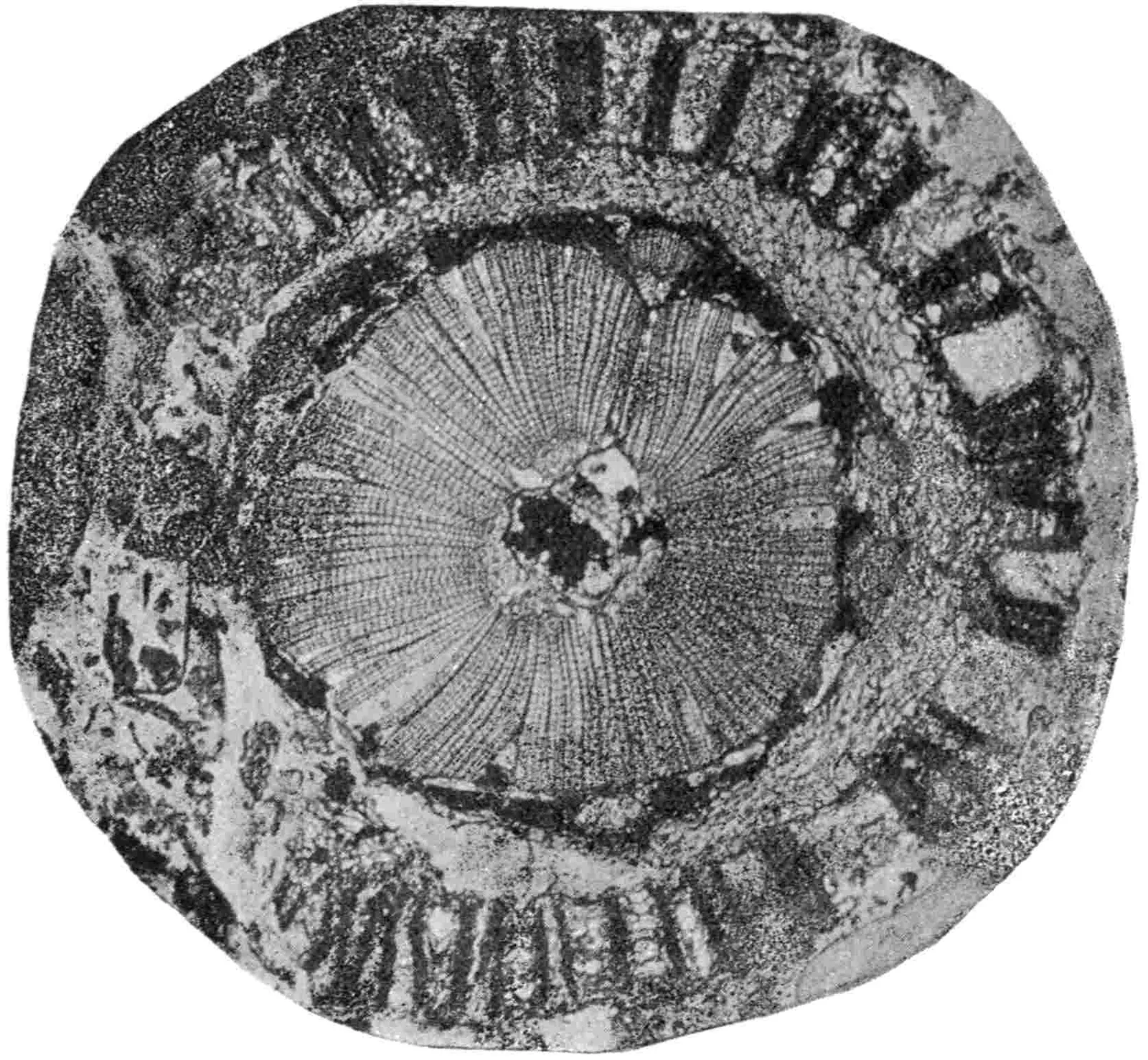
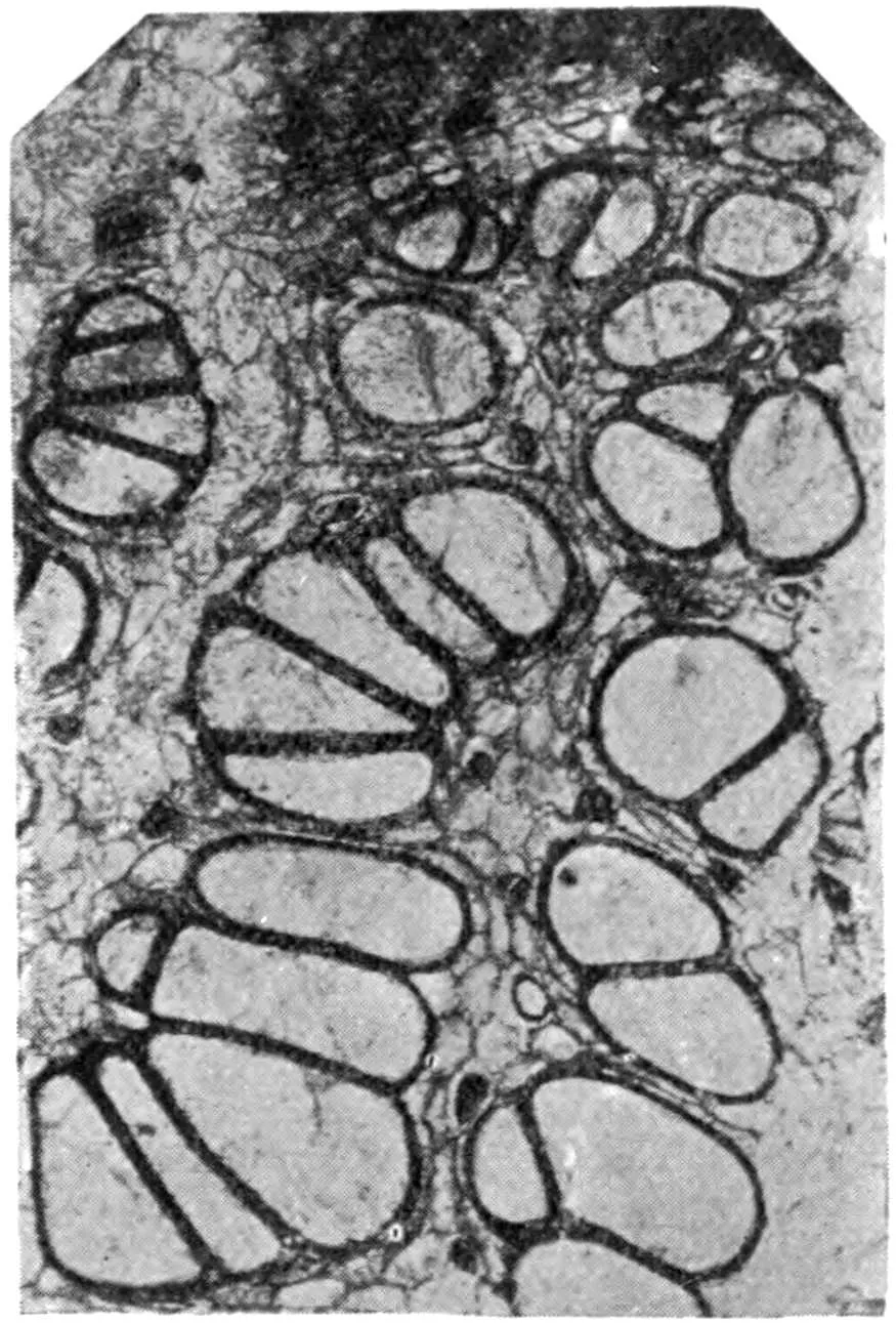
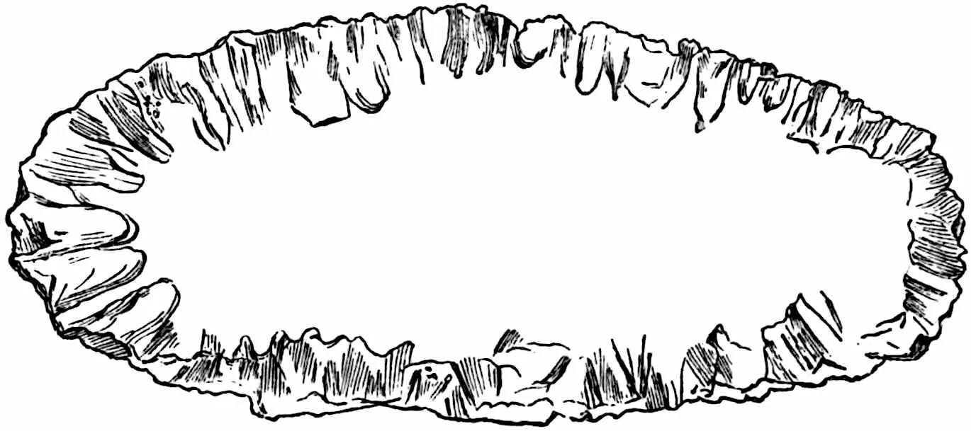
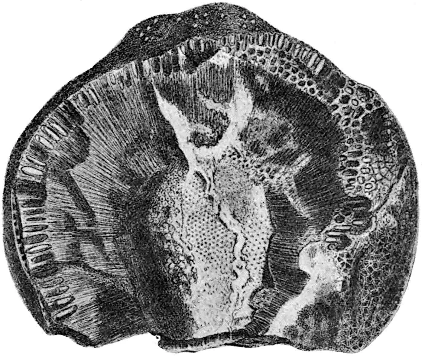
Fig. 402.Lyginopteris oldhamia.
Transverse section of the type-specimen in the Binney Collection,
Sedgwick Museum, Cambridge. (After Arber.)
i. Stem.
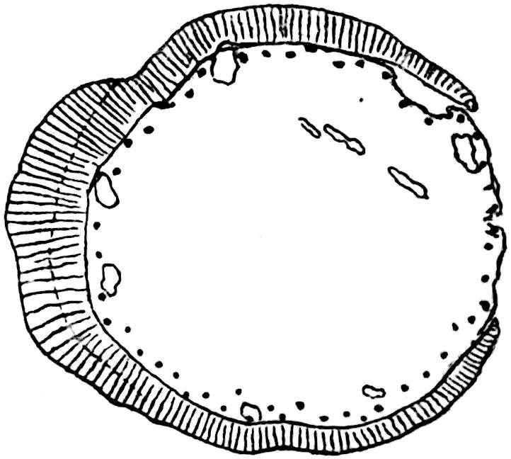
The petrified stem on which Binney founded the species was first
figured by Dr Arber[105] from a section in the Binney collection in
the Sedgwick Museum, Cambridge: this section (13 mm. in diameter)
is reproduced in fig. 402. The most striking features are: (i) the
pith consisting of an unusually large and irregular group of dark
thick-walled parenchyma, (ii) the broad cylinder of manoxylic secondary
xylem characterised by multiseriate medullary rays, (iii) the outer
cortex composed of dark radially disposed and oblique bands of
mechanical tissue separated from one another by partially destroyed
and tangentially elongated parenchymatous elements. It is this outer
cortex that Williamson aptly compared with the Roman numerals on a
clock-face. In the perimedullary region and in contact with the inner
edge of the secondary-xylem cylinder are six strands of primary xylem
representing the xylem halves of collateral bundles separated from
the primary phloem strands by the intervening cylinder of secondary
wood. Two of the primary xylem strands in lateral contact are seen in
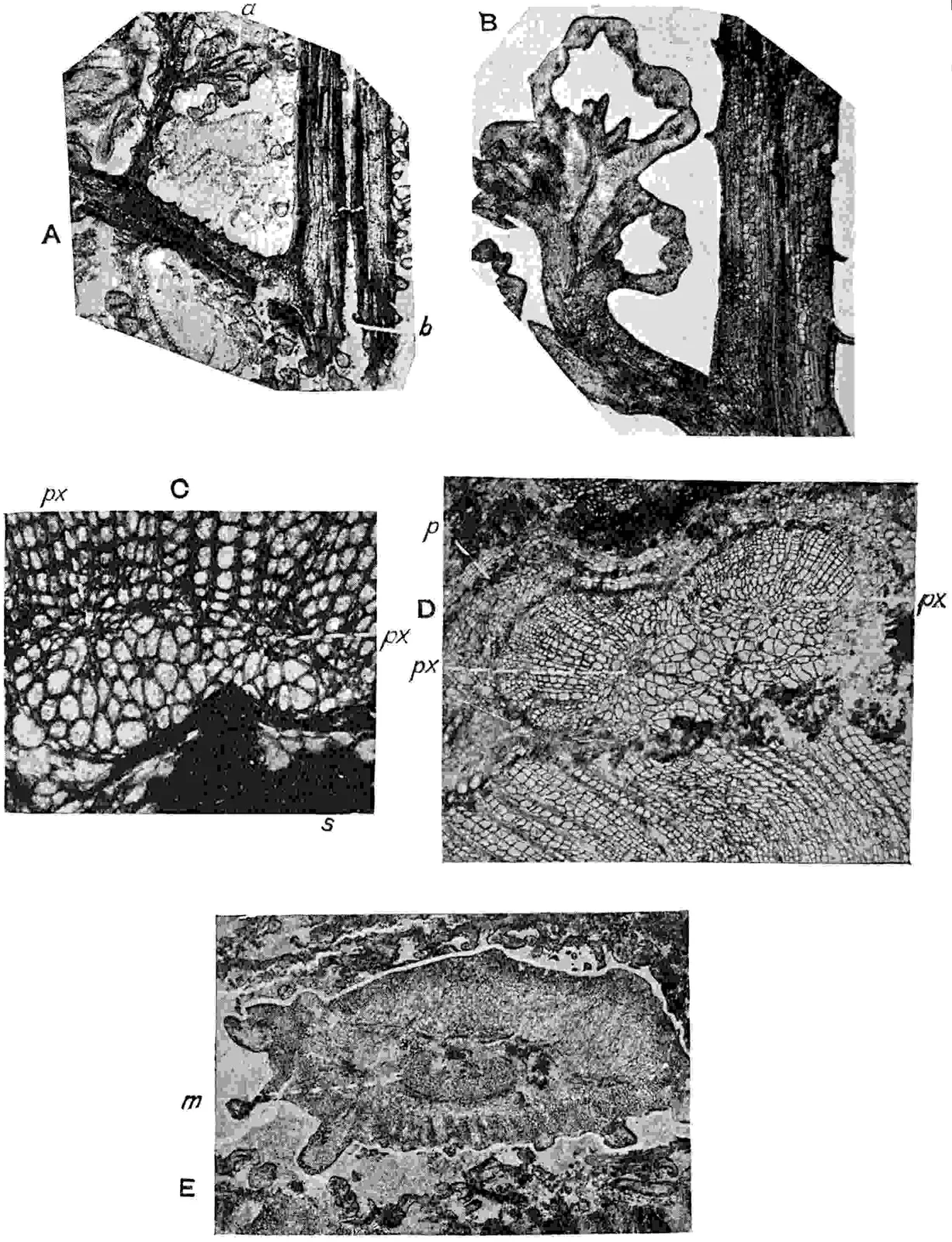
fig. 404, C; the other four occur as separate bundles. Each primary
xylem strand contains a small group of spirally thickened protoxylem
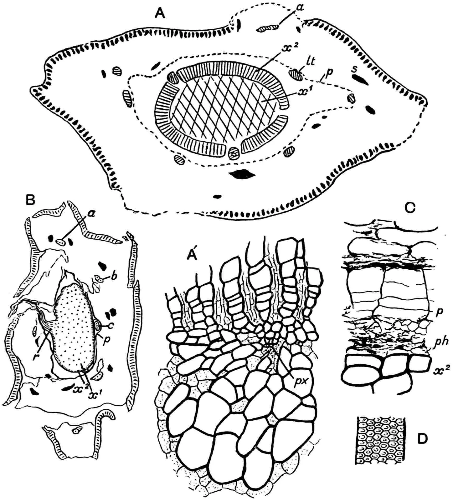
elements (px) associated with a few parenchymatous cells. The
large primary tracheids internal to the protoxylem are characterised
by multiseriate bordered pits on their walls, while those external
to the protoxylem, which are in contact with the innermost secondary
tracheids, have scalariform pitting. The dark patch s (fig. 404,
C) is a portion of the large group of sclerenchymatous cells, shown in
figs. 402, 403. The perimedullary xylem strands of mesarch structure
are the lower portions of leaf-traces and, as Scott points out,
‘each of the bundles surrounding the pith is, in fact, a sympodium,
composed of the united lower ends of successive adjacent leaf-traces.’
The larger of the two bundles shown in fig. 404, C, is on the point
of passing out to a leaf, while the smaller strand is on its way to
a higher level before bending outwards through the secondary wood.
Slightly beyond the middle of the secondary xylem there is an arc of
narrower tracheids comparable with an incomplete annual ring. Although
zones or arcs of narrow tracheids are not uncommon in the wood of
Lyginopteris there is no satisfactory evidence of regularly
recurring seasonal changes. On the outer face of the secondary wood
are a few leaf-trace strands pursuing a vertical course in the
pericycle region; but the structure and behaviour of these bundles
are more clearly illustrated in the stem reproduced in fig. 403. The
tissue between the crushed phloem and pericycle and the outer cortex
(fig. 402) consists of radially compressed parenchyma with scattered
secretory cells separated from the more internal tissue by a narrow
band of periderm formed by a phellogen in the outer part of the
pericycle.
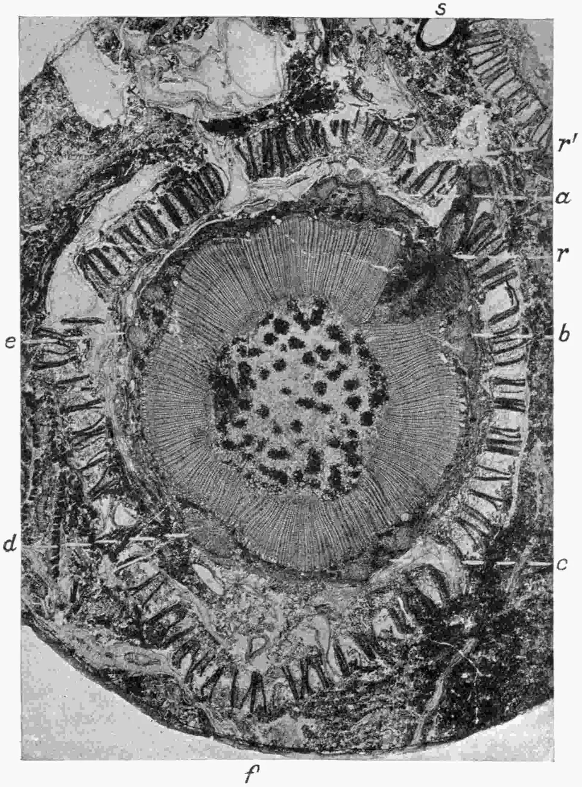
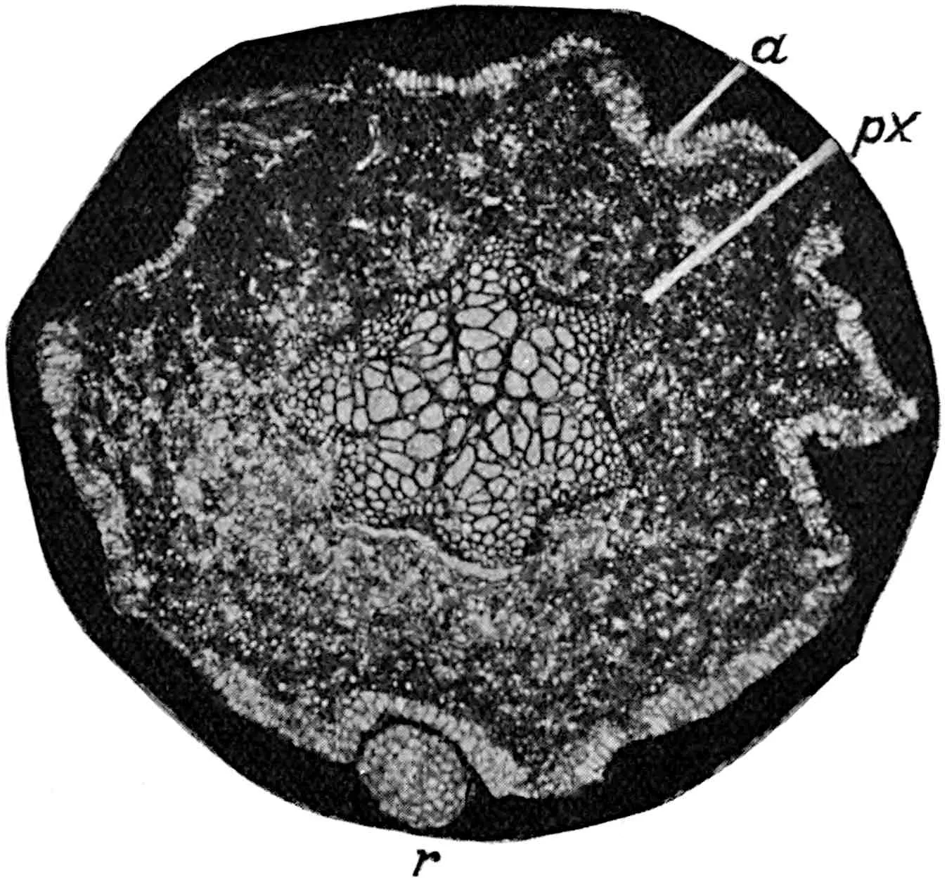
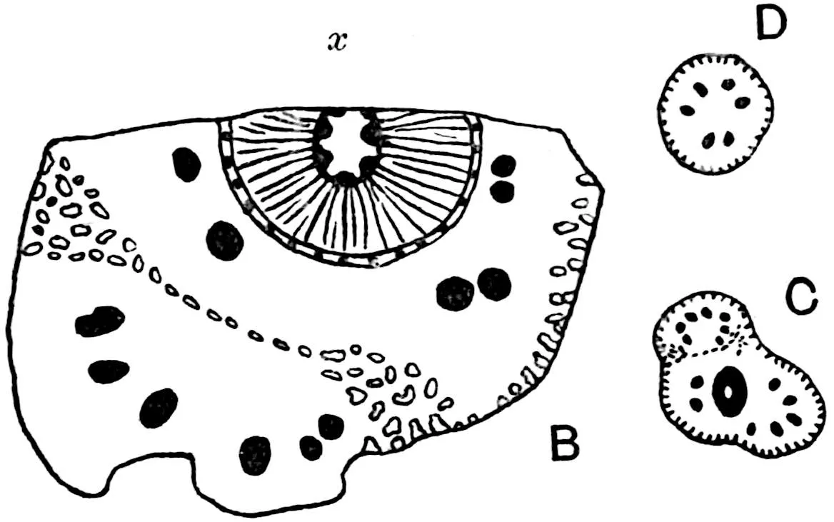
A larger and better preserved stem, 3·7 cm. in diameter, is seen
in fig. 403. In this stem the pith of parenchyma and scattered
sclerenchymatous nests is larger in proportion to the stele than
in Binney’s type-specimen. From the inner edge of the secondary
xylem several primary xylem-strands project as rounded wedges
or tangentially elongated groups where two traces are laterally
united in the perimedullary zone. The cylinder of secondary wood
is partially interrupted at r by the bending outwards of the
stele of an adventitious root cut across transversely as it bends
down after emerging from the outer cortical region. In more or less
close association with the outer surface of the secondary xylem are
four pairs of leaf-trace bundles and one larger trace at d
containing two widely separated protoxylem strands and faced externally
with an arc of secondary xylem: this is a leaf-trace which shows by
the slight constriction on the outer edge of its primary xylem that
it is beginning to divide into a pair of equal strands. A precisely
similar strand is shown on a larger scale in fig. 404, D. The twin
bundles seen at b, fig. 403, represent a divided leaf-trace at
a slightly higher level than the partially severed trace at d,
and the arcs of secondary xylem are narrower. The appearance of the
double leaf-trace at a still higher level is shown at c: the
two strands are farther apart and the secondary xylem has almost
disappeared, while those at e, nearer their entrance into
the leaf-stalk, consist exclusively of primary xylem and phloem. At
a the two strands of a leaf-trace, still nearer to the petiole,
are inclined towards one another preparatory to reunion after reaching
the leaf-stalk. A slender root is seen in transverse section at
r′ immediately outside the two leaf-bundles. As Williamson and
Scott[106] have pointed out, there are always five leaf-traces beyond
the xylem cylinder of a Lyginopteris stem as seen in transverse
section, and these traces in the pericycle, separated from one another
by ⅔ of the circumference, alternate in position with the lower
portions of leaf-traces in the perimedullary region of the same stem.
The phyllotaxis is thus seen to be ⅖.
Fig. 403.Lyginopteris oldhamia.a–e, foliar bundles; f, decurrent base of petiole;
r, r′, roots; s, seed (Lagenostoma). (× 3. Kidston Coll. 592, B.)
The secondary wood is succeeded by a cambium of normal structure
passing gradually into a narrow band of secondary phloem which in
well-preserved stems is seen to consist of sieve-tubes and parenchyma
with medullary rays rather broader than those in the xylem. Beyond the
phloem is the comparatively broad pericycle consisting of parenchyma
with nests of sclerenchyma like those in the pith and scattered
secretory cells. In the outer layers of the pericycle a phellogen
was formed at an early stage in the growth of the plant, producing
several layers of secondary tissue, which is regarded as periderm and
forms a conspicuous feature in Lyginopteris stems; it appears
as a comparatively dark sinuous band where it bends outwards to wrap
round the leaf-traces in their almost vertical course through the
pericyclic region (fig. 403). The periderm is clearly seen at p
close to the crushed secondary phloem of the dividing leaf-trace in
fig. 404, D. All the leaf-traces seen in fig. 403 beyond the secondary
wood are still within the deep-seated periderm and, as Williamson
and Scott showed, each leaf-trace after emerging from the secondary
wood remains in the pericycle-zone for a length of five internodes as
it very gradually inclines outwards. Once free from this region the
twin bundles bend more sharply towards the petiole. Stated briefly,
the history of each leaf-trace from the perimedullary region to the
leaf-base is as follows: at the outer edge of the pith a single trace
consists of a mesarch xylem bundle with one protoxylem strand; it
passes vertically through five internodes and then bends out through
a foliar gap in the xylem-cylinder, and the primary tracheids receive
additions from the cambium of the stele on their outer face. As
the trace leaves the secondary xylem it bends upwards and, as seen
at d, fig. 403, begins to divide into twin bundles. As the
trace passes higher the bisection of the protoxylem and metaxylem is
completed and the secondary xylem-arcs are gradually lost until the
separate strands of each pair are reduced to single mesarch bundles
composed wholly of primary tracheids. As the trace bends outwards
through the cortex the phloem gradually encircles each xylem-strand
until a concentric structure is substituted for the collateral
disposition of the conducting tissue. At the same time the protoxylem
strands divide and occupy a position near the inner edge of the
metaxylem. On reaching the petiole or after passing some distance
up the axis of the frond, the twin bundles unite and usually form a
V-shaped vascular strand (figs. 404, E; 405, A). The single meristele
subsequently divides into two equal portions preparatory to the
bifurcation of the petiole (fig. 406).
The inner cortex, consisting of parenchymatous tissue and many
secretory cells with an occasional group of sclerenchymatous elements
in place of the abundant nests of this tissue in the pericycle,
has been invaded in the stem shown in fig. 403 by numerous rootlets
of Stigmaria and Lyginopteris, some of which are seen
interrupting the continuity of the outer cortex. The greater width of
the cortical region at f, fig. 403, is due to the decurrent
base of a petiole the meristele of which is not included in the
section. The lighter and broader bands between the cross-sections of
the stereome-network in the outer cortex are occupied by remains of
tangentially stretched parenchymatous cells, and beyond this zone
in a younger stem there are a few layers of parenchyma forming the
superficial tissue, but there appears to be no well-defined epidermal
layer.
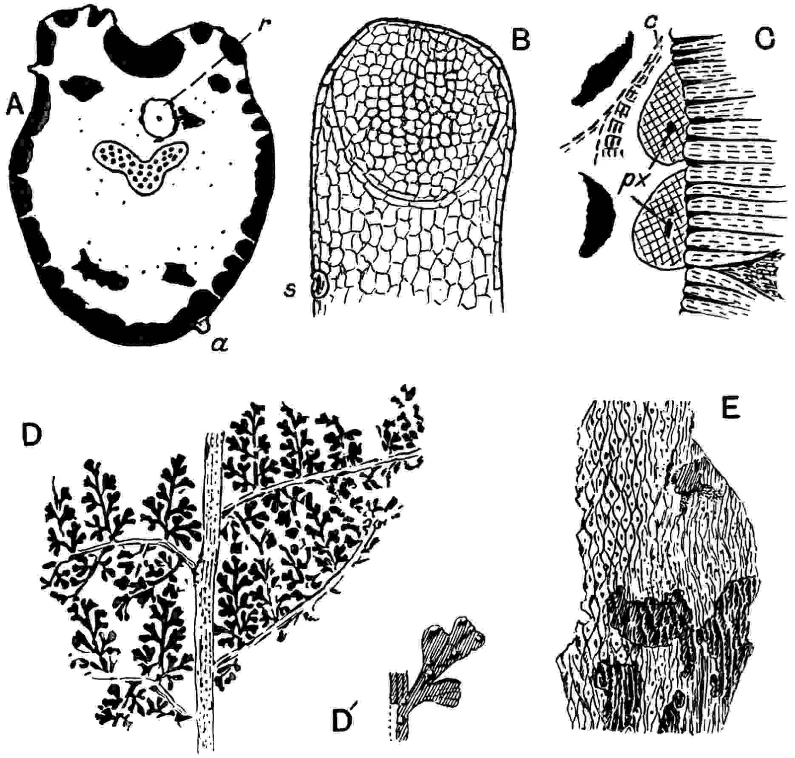
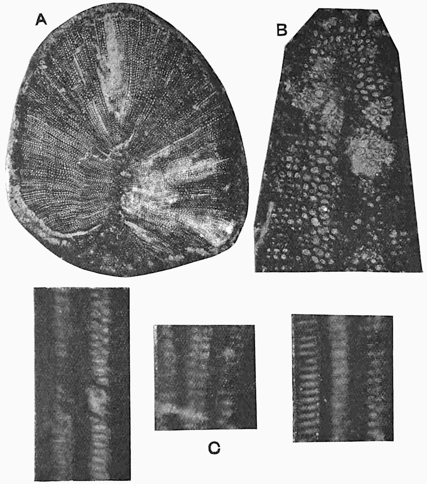
Fig. 404.Lyginopteris oldhamia.
A, B. Frond fragments; a, pinnule; b, reticulum of sclerenchyma.
C. Portion of stele of the stem reproduced in fig. 402.
D. px, protoxylem; s, sclerenchyma; leaf-trace close to the edge of the secondary xylem; p, periderm.
Young stems have been recognised in which there is very little
secondary xylem and phloem: in these the stereome bands in the outer
cortex are closer together than in the stretched hypodermal tissue
of older shoots and the scattered sclerous nests are represented by
unthickened cells. In addition to young stems Williamson and Scott
described a distinct type of small stem in which the primary xylem
forms an almost complete ring[107] comparable with the primary xylem
of some adult Sigillarian stems (vol. ii. p. 220) but
distinguished by its mesarch structure and by the reticulate pitting of
the centripetal xylem.
A characteristic feature of the stem is the occurrence of emergences
from the outer cortex which have the structure either of spinous
processes, broadly linear or flask-shaped, or of stalked glands[108].
A portion of a glandular emergence is shown in fig. 405, B: the group
of small cells immediately below the blunt apex is in this instance
still intact though showing signs of disorganisation in the centre;
but in many cases the secretory tissue has not been preserved and the
head of the emergence is occupied by a space. A single stoma is seen at
s in longitudinal section. Further reference to the emergences
is made in the description of the leaf.
It occasionally happens that a meristematic layer is formed in the
parenchymatous tissue immediately internal to some of the perimedullary
xylem strands of a Lyginopteris stem from which either secondary
parenchyma is produced or a zone of secondary xylem and phloem, the
phloem facing the centre of the pith. An example of such internal
xylem was figured by Williamson[109] and similar occurrences are more
fully dealt with by Williamson and Scott[110] who consider that the
perimedullary cambium may represent an internal extension through a
leaf-gap of the normal cambial cylinder. In the stem represented in
fig. 403 there are two perimedullary xylem strands to the left of the
bottom of the V-shaped gap in the secondary xylem-cylinder, r,
and on the inner face of one of these, as shown in fig. 405, C, there
is a narrow arc of internal secondary xylem, c, between the
xylem-strand and the outer edge of one of the sclerous nests. The
sporadic occurrence of arcs of inversely orientated secondary vascular
tissue affords an interesting parallel with a similar morphological
feature in some recent Dicotyledonous genera such as Tecoma
and Iodes. As Williamson and Scott point out, this similarity
affords ‘a striking warning against the indiscriminate use of
even conspicuous anatomical characters[111].’ While admitting the
necessity of guarding against the danger of attaching importance to
occasional and abnormal characters they may have some significance as
collateral evidence in comparisons of different types of stems. It is
conceivable that these anomalous arcs of secondary tissue on the inner
side of the primary xylem strands may, as Worsdell[112] maintains, be
reversions to an ancestral character and in this sense comparable with
the strands of inverted vascular tissue in some recent Cycadean stems.
The question of relationship of Lyginopteris and allied types to
recent Cycads and the Palaeozoic Medulloseae is considered in a later
chapter.
In 1902 Lomax[113] described two branching specimens of
Lyginopteris, and more recently two others have been discovered
at a locality near Bacup in Lancashire which have been thoroughly
investigated by Miss Brenchley[114] who constructed models from
drawings of serial sections[115]. One specimen shows six leaf-bases
in a length of 4¾ inches and branches spring from the axils of five
of them: some of the branches show secondary ramifications. The
phyllotaxis of the leaf-bases on a branch is always the reverse of
that on the main stem, a divergence to which no parallel was found
in a selection of trees and shrubs examined by Miss Brenchley. The
secondary wood of the stem swells below the point of exit of a branch
and frequently a fairly large amount of wood occurs in the pith when
a branch is given off: this anomalous wood may help to close the
branch-gap.
ii. Leaf.
In his account of Lyginopteris stems published in 1873
Williamson[116] suggested that the vascular bundles met with outside
the xylem-cylinder might be the leaf-traces of large fronds and
expressed the opinion that the ‘stems or petioles’ previously
described by him under the generic name Edraxylon might
belong to Lyginopteris. A year later he substituted the name
Rachiopteris aspera for the petioles previously referred
to Edraxylon and inclined to the view that this type of
Rachiopteris may be the petiole of the Carboniferous fronds
known as Sphenopteris Hoeninghausi Brongn., an inference based
to a large extent on the occurrence of emergences on Rachiopteris
aspera (fig. 404, E) preserved as petrifactions like those
on impressions of Sphenopteris Hoeninghausi as figured by
Brongniart (figs. 404, A; 405, D, D’). In 1890 Williamson was able to
demonstrate the truth of the surmise that Rachiopteris aspera
and Lyginopteris oldhamia are respectively the petiole and stem
of the same plant, which he believed to be an arborescent fern[117].
The petioles of Lyginopteris fronds, which may reach a diameter
of 1 cm., are attached by a broad base to the stem, and as already
suggested by the number of internodes traversed by each leaf-trace, the
leaves are comparatively far apart. A transverse section of a petiole
is shown diagrammatically in fig. 405 A. The hypodermal stereome is
a prominent feature, but the narrow radial plates of the stem-cortex
tend to be replaced in the rachis by broader and confluent masses of
strengthening elements: the upper surface of the petiole is slightly
grooved. Glandular and spinous emergences are often very abundant, as
in the section reproduced in fig. 404, E. A glandular emergence is seen
at a in fig. 405, A. The spinous emergences may be compared with
those of Davallia (Odontosoria) aculeata[118],
a West Indian fern of climbing habit and with the prickles on
Hemitelia and other recent Cyatheaceous fronds[119], while
capitate glands, though simpler than those of Lyginopteris,
occur on the leaf-stalks of some recent Polypodiaceous species[120].
The concentric meristele may consist in the lower part of the petiole
of two separate and slightly curved strands like those seen in fig.
404, E, m: sooner or later the two strands unite to form a
wide-open V or a W-shaped bundle with several slightly internal
protoxylem groups close to the lower edge. The two sections represented
in fig. 406, A and B show the gradual divergence of the two meristeles
of a petiole as they approach the level where it divides into two equal
branches, a characteristic feature of Sphenopteris Hoeninghausi
and allied fronds. At a lower level than that represented in fig.
406 the vascular strand of the petiole would have the form of a W as
figured by Williamson in one of his earlier memoirs[121]. The phloem
with scattered secretory sacs and the adjacent tissue of the leaf-stalk
are occasionally preserved in wonderful perfection[122]. No endodermis
has been recognised. Sclerous nests are scattered in the ground-tissue
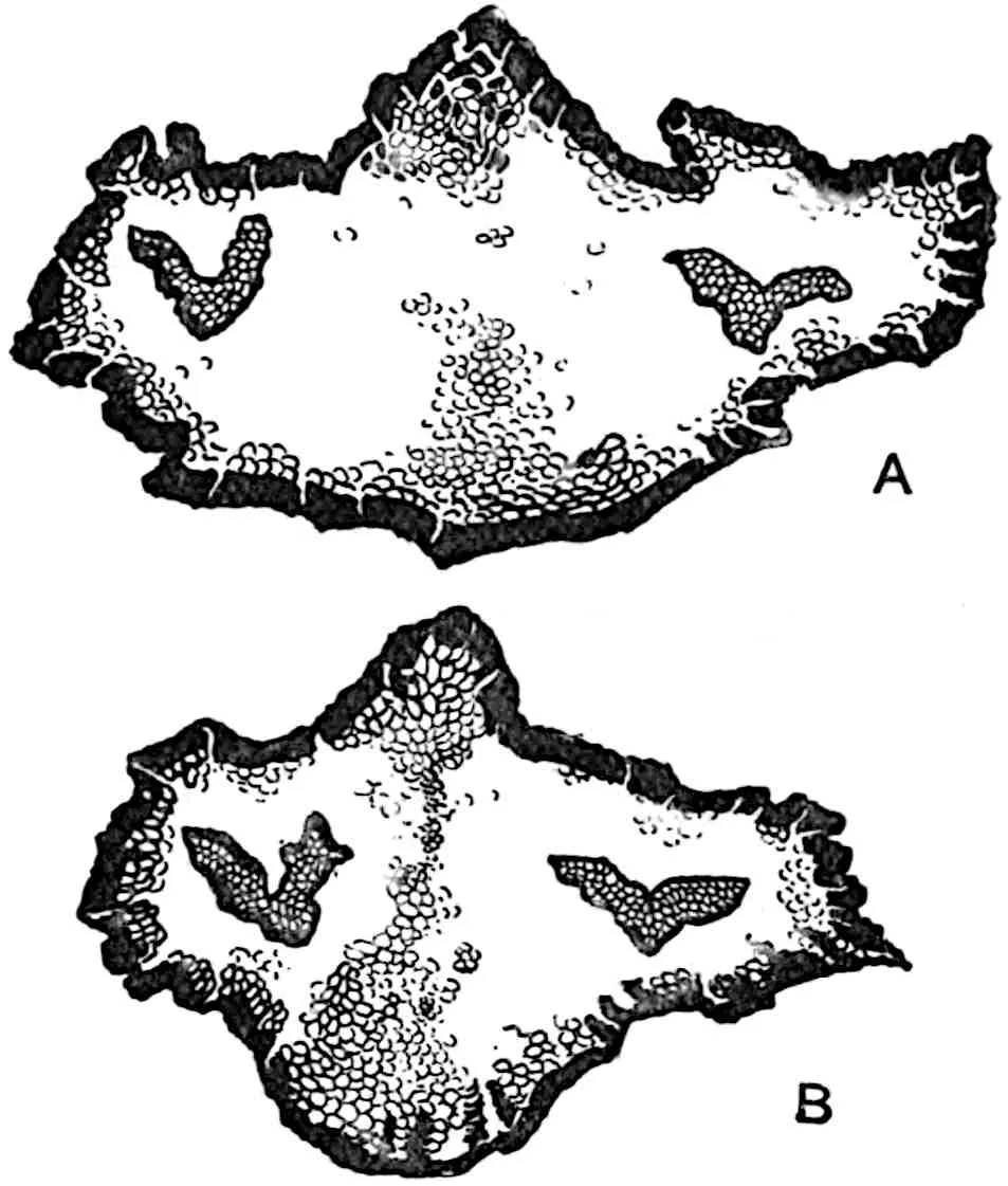
as are also secretory sacs (figs. 404, E; 405, A). A small root
r has penetrated the parenchyma of the rachis shown in fig. 405, A.
Fig. 405.Lyginopteris oldhamia. A,
petiole section; a, glandular emergence; r, root. B,
stalked gland; s, stoma. C, inner edge of wood of a stem;
c, arc of inversely orientated vascular tissue. D, D’, part of a
frond of Sphenopteris Hoeninghausi. E, part of axis of D. (A, C,
D, Kidston Coll.; B, Manchester Coll. R. 645.)
Fig. 406.Lyginopteris oldhamia.
Transverse section illustrating branching of petiole. × 5. (From a
drawing supplied by Prof. Oliver.)
Sphenopteris Hoeninghausi Brongn.[123], founded on material
from English Coal Measures, was regarded by Williamson as the foliage
of Lyginopteris chiefly on the ground of the occurrence of
emergences on the axes (figs. 404, A, B) and laminae of the impressions
like those on the petrified stems, and this comparison received support
from the resemblance of the fragments of pinnules associated with
Lyginopteris and its petioles in the calcareous nodules to the
leaflets of Brongniart’s type. This identification is supported by
subsequent work. The quadripinnate fronds, which attain a considerable
size, resemble those of recent species of Davallia and other
ferns, but the forking of the rachis and branches of the frond is
a striking feature: the pinnae may reach a length of 15 cm.[124]
The portion of carbonised rachis shown in fig. 405, E, reveals the
existence of a hypodermal reticulum like that in the outer cortex of
a Lyginopteris stem and the same feature is seen in the more
slender axis represented in fig. 404, A, at b.[125] The pinnules
are usually deeply lobed and the segments may be comparatively broad
and blunt or narrow[126] (fig. 290, C, vol. ii. p. 399; fig.
404, A, a, B; fig. 405, D’). The lamina has a well marked
dorsiventral structure: the palisade-tissue next the upper surface
is separated from the epidermis by small hypodermal cells, possibly
functioning as a water-storage layer, and the central part of the
mesophyll consists of loose aerenchyma: the veins are collateral as in
recent ferns and stomata occur in the lower epidermis. Emergences are
seen both on impressions (fig. 405, D’) and on petrified specimens. A
striking feature of the pinnules is the rounded surface caused by the
revolute edge of the lamina as seen in the section reproduced in fig.
404, B. This character coupled with the occasional occurrence of groups
of short tracheal elements at the termination of the veins denotes a
tendency to a xerophilous habit.
On the strength of a very close resemblance between Sphenopteris
Hoeninghausi and Calymmatotheca Stangeri (fig. 408,
E, F)—characterised by fertile pinnules bearing stellate groups
of small linear valves, regarded by Stur as the open lobes of
an indusium—Zeiller included Brongniart’s type in the genus
Calymmatotheca. The resemblance in general habit between
the two species extends to the presence in their rachises of the
Dictyoxylon form of cortex[127]. The view formerly held by
some authors that the valve-like appendages to the fertile segments
of Calymmatotheca are sporangia is incorrect: a re-examination
of Stur’s specimen (fig. 408, E, F) has confirmed the original
description[128]. The stellate lobes are now regarded as portions of
a cupular investment of a seed similar to Lagenostoma Lomaxi,
the female reproductive apparatus of Lyginopteris oldhamia.
The axes of the fertile pinnae bear small thorn-like emergences
probably identical with those on the cupule of Lagenostoma
and on the petioles of Lyginopteris oldhamia. It was stated
in vol. ii that the fronds known as Sphenopteris
Linkii (Goepp.) represent, with other closely allied forms,
leaves belonging to Heterangium stems. This statement was
based on a misconception: the rachis of Sphenopteris Linkii,
as I have satisfied myself by an examination of impressions shown to
me by Dr Kidston, exhibits the reticulate pattern characteristic of
Lyginopteris and not the transverse ribs characteristic of
Heterangium.
It is not an easy task even for those most familiar with Carboniferous
fronds to distinguish clearly between species agreeing generally
with Sphenopteris Hoeninghausi, a species regarded by
some authors as the type of a group of very similar and closely
allied forms all of which were probably borne on stems referable
to the genus Lyginopteris. The species Lyginopteris
oldhamia as generally understood probably includes more than one
specific type, and it is safe to assert that in the Carboniferous
period Lyginopteris was represented by several forms
characterised by highly compound fronds with forked rachises like
Sphenopteris Linkii, S. Hoeninghausi, and others. The
features characteristic of fronds included in the Sphenopteris
Hoeninghausi group have recently been described by Gothan[129].
Stur’s generic name Calymmatotheca originally applied to
the species C. Stangeri was applied to Sphenopteris
Hoeninghausi by Zeiller, and although the fronds of the
latter type have not been found with fertile appendages of the
Calymmatotheca type there can be no doubt as to the generic
identity of these, barely distinguishable, species both of which belong
to stems of Lyginopteris. Prof. Johnson[130] has recently
described some impressions from the Coal Measures of Ireland, which
he refers to S. Hoeninghausi, bearing stellate groups of lobes
like those of Calymmatotheca, and in one case he describes a
seed in the middle of the carbonised remains of a stellate group of
cupular segments. An examination of the specimen in Dublin convinced
me that there is no satisfactory evidence of the seed-nature of
the appearance on the rock believed by Johnson to be an elliptical
Lagenostoma-like seed. The actual attachment of the stellate
lobes to the pinnae of the frond is not clearly demonstrated.
iii. Microsporangia.
In 1905 Kidston[131] announced the discovery of microsporangia on
fronds of Lyginopteris: he described specimens from the
Coal Measures of Dudley identified by him with Sphenopteris
Hoeninghausi showing sterile and fertile pinnae in organic
connexion. The fertile pinnules (fig. 407, B) are slightly expanded
distally into an oval limb about 2 mm. long bearing 6 to 7 bilocular
fusiform microsporangia 3 mm. long and 1·5 mm. broad: in the immature
condition the sorus is hemispherical, the summit being formed of
the incurved apices of the sporangia. At maturity the sporangia
spread out, the sorus assuming the form of an epaulet. Fig. 408, H,
shows a sorus in transverse section and in fig. 408, G, the limb
and two pendulous sporangia are shown. The microspores, 50–70 µ in
diameter, are studded with numerous blunt spines and each spore shows
a triradiate ridge. The section reproduced in fig. 407, A, from the
Coal Measures of Oldham is probably a bilocular sporangium of the
same type as those described by Kidston from Dudley. Dr Kidston[132]
describes a second type of microsporangial sorus as Crossotheca
Hughesiana which agrees closely with C. Hoeninghausi, but
the fertile segments are not associated with any sterile pinnae. The
generic name Crossotheca, founded by Zeiller[133] in 1883, was
substituted for Sphenopteris on the ground that Brongniart’s
species S. Hoeninghausi is shown to possess sporangia of the
Crossotheca type. If Kidston’s specific determination is
correct, his discovery demonstrates that Lyginopteris fronds
bore microsporangia having the characters of Crossotheca, a
type characteristic of several Carboniferous species belonging both to
the form-genera Sphenopteris and Pecopteris. Reference
has already been made to the difficulty of distinguishing between
impressions of fronds of the SphenopterisHoeninghausi
group, a difficulty that is illustrated by Dr Gothan’s statement[134]
that the Dudley specimens of Crossotheca are not identical
in the character of the sterile pinnules with Sphenopteris
Hoeninghausi. An examination of Dr Kidston’s specimens led me
to agree with his determination; but, it may be asked, have we any
evidence of the association with Lyginopteris fronds of
sporangia other than those of the Crossotheca type? Prof.
Chodat[135] believes that certain petrified fragments of pinnules
occasionally met with in the calcareous nodules bearing sessile and
apparently annulate sporangia belong to Lyginopteris fronds.
These sporangia appear to be identical with those named by Scott
Pteridotheca Butterworthi[136] and regarded by him as filicean
sporangia that cannot be referred to any known Carboniferous genus. The
piece of lamina bearing an empty sporangium, which may or may not have
possessed an annulus, reproduced in fig. 407, C, occurs in association
with the larger specimen shown in fig. 404, B, and it would seem not
unreasonable to regard both as parts of the same frond, namely a frond
of Lyginopteris. As Prof. Weiss[137] points out, the accurate
determination of small pieces of petrified pinnules is exceedingly
difficult and without more decisive evidence we are hardly justified
in asserting that the sporangia figured by Chodat and Scott and that
shown in fig. 407 belong to the genus Lyginopteris. Although
the available data appear to favour the view generally held that
Kidston’s conclusion is correct additional evidence would be welcome.
Telangium. Reference was made in vol. ii.[138] to the
genus Telangium instituted by Dr Benson for some petrified
sporangia from the Coal Measures regarded by her as the microsporangia
of a Pteridosperm, probably Lyginopteris. The sporangia of
Telangium are similar to those of Crossotheca. Scott
points out that they are borne on a flat disc or lamina ‘quite
comparable to a fertile pinna of Crossotheca[139],’ and he
concludes that these sporangia are not generically distinct from
the impressions on which the genus Crossotheca was founded.
Kidston[140] regards Telangium Scotti, Benson, as the
microsporangium of a Pteridosperm though not of Lyginopteris,
on the ground that the microsporangia described by Miss Benson are not
attached to a limb and that they have a single loculus in place of the
double loculus (fig. 407, A) of Crossotheca. The presence of
a limb in Telangium recognised by Scott removes one of these
distinguishing features. There are, however, no adequate reasons
for regarding Telangium Scotti as specifically identical
with Crossotheca Hoeninghausi. The synangium of Telangium
Scotti, 5 mm. in length, consists of 6–12 sporangia united basally
and opening when ripe by longitudinal dehiscence. Fig. 493, E, shows
eight sporangia of a synangium in transverse section: the two sporangia
at the lower end of the section are less distinct than the others,
some are full of spores and others have shed their contents by the
splitting of the thin inner walls of the loculi. The sporangial walls
are composed of an outer layer of large cells with dark contents
succeeded by 2–3 layers of smaller and crushed cells. The spores, 5–6
µ × 3·5–4 µ in diameter, have a reticulately sculptured exine: Dr
Benson[141] states that they agree closely with pollen-grains found
in the pollen-chamber of Lagenostoma ovoides except in their
slightly smaller size; she notes the association of Telangium
with fragments of the vegetative organs of Lyginopteris and
draws attention to resemblances in the structure of the tissues; but
the most interesting comparison, at least in an academic sense, is
with the seed Lagenostoma, the integumented megasporangium of
Lyginopteris. Dr Benson points out that a transverse section of
a Lagenostoma in the plane of the canopy, showing the nucellar
apex surrounded by radially disposed chambers (fig. 409), presents a
certain resemblance to a synangium of Telangium Scotti; and it
is suggested that the chambers encircling the nucellus may represent
sterilised sister-sporangia[142]. ‘The seed in fact is assumed to be
a synangium in which all but one of the sporangia are sterile, and
form an integument to the one fertile sporangium which has become a
megasporangium with one large megaspore.’ This view, though clearly
incapable of confirmation in the present state of our knowledge,
is not merely an ingenious hypothesis but a stimulating suggestion
as to possible homologies: as an argument in favour of associating
Lagenostoma and Telangium as the spore-bearing organs of
the same plant it has but little weight.
iv. The Seed.Lagenostoma Williamson.
Lagenostoma Lomaxi, Oliver and Scott ex Williamson, MS.
In 1877 Williamson[143] proposed the generic name Lagenostoma
for some petrified seeds from the Lower Coal Measures of Lancashire
and described two species, Lagenostoma ovoides and L.
physoides: in his MS. Catalogue a third type is referred
to as Lagenostoma Lomaxi. It is this third type that Prof.
Oliver was the first to recognise as the megaspore-bearing organ
of Lyginopteris oldhamia. Its structure has been thoroughly
described by Oliver and Scott[144] and these authors contribute a
judicial summary of the evidence on which Lagenostoma and
Lyginopteris are believed to stand for one and the same plant.
The evidence is based chiefly on the following considerations: an
agreement in the structure of the vascular bundles in the investments
of the seed with those in the leaves of Lyginopteris; the
presence in the outer envelope of the seed of stalked glands identical
with those on the stems and petioles. The evidence does not as yet
amount to absolute proof, as the seeds, which occur either with or
without a stalk, have not been found attached to a Lyginopteris
frond. But ‘where vegetative and reproductive organs presenting
identical structural features, not known to occur in other plants, are
thus found in close and constant association, the inference that the
one belonged to the other appears irresistible.’ While most botanists
believe that a satisfactory case is established there are a few[145]
who refuse to believe in a connexion between Lagenostoma and
Lyginopteris until an actual union has been demonstrated.
The discovery by Kidston[146] of seeds attached to pinnae bearing
Neuropteris pinnules and the demonstration of organic continuity
between seeds and the pinnules of other Palaeozoic fern-like fronds
supply abundant confirmatory evidence that leaves no doubt as to
the occurrence of seeds on modified pinnae of Sphenopteris
Hoeninghausi and of other closely allied fronds which represent the
foliage of different forms of Lyginopteris. In this connexion
it is pertinent to add that Grand’Eury[147] has found seeds of the
Lagenostoma type in close association with impressions of
Sphenopteris Dubuissonis and other leaves of similar habit.
A seed of Lagenostoma Lomaxi reaches a length of 5·5 mm. with
a maximum diameter of 4·4 mm.; it is broadly oval or barrel-like
(fig. 408, C) and when immature was invested by a loose irregularly
lobed glandular envelope (fig. 408, B) from which the seed eventually
freed itself by a natural process of abscission. The central body or
nucellus, except in the apical region, is concrescent with a fairly
stout integument or testa (fig. 408, C, f) the outer portion
of which is characterised by regular longitudinal rows of palisade-like
cells comparable with the broad palisade-layer in the sporocarp of
Pilularia. On the exposed surface of this palisade-tissue
are small dark structureless pegs[148], possibly the remains of a
mucilaginous layer such as occurs on the seed-coats of some recent
Flowering plants. At the base of the nucellus the chalazal region, fig.
408, C, ch, is provided with sclerous elements and forms a hard
investment to the axial vascular strand from the pedicel. It is at the
base of this chalazal region that the seed is eventually cut off by an
absciss-layer. The integument is supplied throughout its length by nine
vascular bundles of endarch, or approximately endarch, structure. The
free portion of the integument seen from the outside (fig. 408, B) has
the form of a fluted cone with a circular opening at its summit. The
greater part of this domical apex, as seen in longitudinal section in
fig. 408, C, appears to be hollow, but in the living state the dome, or
canopy as Williamson called it, was filled with parenchyma in which the
vascular bundles were embedded and, as shown in the transverse section
in fig. 409, the canopy is divided into compartments by radial septa
which in its basal region are replaced by regular and deep furrows
on the inner face. Enclosed by the canopy, with its outer surface
fluted as the result of the partial collapse of the outer wall of each
compartment due to the decay of the filling tissue, is the flask-shaped
apex of the nucellus; between the apical cone of nucellar parenchyma
and the superficial layer is an annular cavity which Williamson[149]
called the lagenostome. The parenchymatous core tapers to a narrow
summit which slightly overtops the integument and is constricted at the
broad base (fig. 493, A, B; page 311). The bottle-shaped apical tissue
is separated by an annular space, c, fig. 493, B, from the
limiting layer of the nucellus: this space is the pollen-chamber formed
in the living seed by the disorganisation of the subepidermal cells of
the nucellar apex. The pollen-chamber is a feature characteristic of
recent cycadean ovules (see p. 6). In Lagenostoma the annular
form of the pollen-chamber is a peculiarity distinguishing this type
of seed from those of recent Gymnosperms and most other Palaeozoic
seeds. As Oliver says, it marks an ‘advance in precision’[150] over
other forms as the microspores which fall into the chamber are brought
direct to the surface of the underlying megaspore and presumably to
the archegonia which, it is reasonable to believe, were disposed in a
circle at the base of the annular crevice. Microspores frequently occur
in the pollen-chamber and some have been discovered apparently in the
act of liberating male gametes[151].
Fig. 408. A, A′, Lagenospermum
Sinclairi. B, Lagenostoma, restoration. C, Lagenostoma
Lomaxi; c, micropyle; d, space between integument and
nucellus; e, cupule; f, integument; ch, chalaza.
D, microspores of Lagenostoma ovoides. E, F, Calymmatotheca
Stangeri. G, H, Crossotheca Hoeninghausi. H, section of G
in line of arrow. (A, after Arber; B, C, E, F, after Oliver; D, after
Benson; G, H, after Kidston.)
Fig. 409.Lagenostoma. Transverse
section near the micropyle, showing the pollen-chamber, pc, the
space, s, between the nucellus and integument, the fluted canopy
with vascular bundles, v. (After Oliver.)
The outer wall of the nucellus is bounded externally by a similar
circular space (d, figs. 408, C; 493, B) which separates it
from the domical canopy. In the great majority of specimens the
central tissue of the seed is not preserved and an empty sac supported
from the base of the nucellus-apex occupies nearly the whole of the
interior: the shrunken wall of the sac is all that remains of the
large megaspore. It would seem, then, that the nucellus was almost
completely destroyed as a consequence of the growth of the megaspore or
embryo-sac, which eventually occupied nearly the whole of the seed-body.
In an exceptionally well preserved specimen recently described and
admirably illustrated by Mr McLean[152] part of the parenchymatous
tissue of the prothallus which originally filled the megaspore is
clearly seen: its surface-layer consists of small cells succeeded by
a broad band of radially elongated elements closely resembling the
alveoli in the prothalli of some recent Gymnosperms, particularly
certain Conifers. No archegonia have been discovered. The cupular
envelope of immature seeds, compared by Oliver and Scott with the lobed
and glandular husk of Corylus colurna L.[153], receives several
vascular bundles of collateral and mesarch structure from the axial
strand, and these subdivide as they ascend. The glands which occur
on all parts of the cupule are sessile or stalked and identical with
those on the vegetative organs of Lyginopteris. Assuming that
pollination occurred at a comparatively early stage in the development
of the seed when the cupule was still intact, it is conceivable, as
Sir Joseph Hooker suggested, that the glandular secretion may have
attracted insects and so aided in the transport of pollen which were
perhaps drawn down the narrow pollen-chamber by exuded mucilage as
in recent Conifers. The evidence obtained in recent years in favour
of insect-pollination in certain Cycads and in Welwitschia
lends support to this view: the dragon flies hovering over a fertile
Lyginopteris frond in a recent restoration[154] may be a
legitimate addition.
A striking feature of Lagenostoma as of other Palaeozoic seeds
is the absence of an embryo: this and other considerations have led
certain authors, notably Chodat[155], to question the justification
for the use of the term seed. Various suggestions have been offered in
explanation of this fact. In recent Cycads, as already pointed out, the
development of the embryo does not always occur before seed-fall. It
may be that these older seeds had no resting-period or there may have
been a period of rest after fertilisation and not as now at a stage
subsequent to the formation of the embryo[156]; it is also suggested
by Scott that ‘the nursing of the embryo had not yet come to be one of
the functions of the seed, and that the whole embryonic development was
relegated to the germination stage[157].’ In this connexion reference
may be made to a statement by Miss Gibbs[158] who speaks of seeds of
a Podocarpus picked up from the ground in apparently a mature
state and with the associated bracts coloured and swollen as though
ready to aid in dispersal but with no embryo: the seeds had matured
before fertilisation and fell from the tree after pollination. Whatever
may be the true explanation of the absence of embryos this negative
character should not be allowed to outweigh the evidence furnished
by morphological features as to the applicability of the term seed.
As Prof. Oliver says, ‘there is a long chapter in evolution to be
deciphered before we can connect ... the seed of Lyginodendron
with the sporangium of any fern at present known to us’[159].
The cupule of Lagenostoma has been homologised with the outer
part of the integument of a recent cycadean seed[160] which, it is
suggested, consists of an inner and an outer envelope that have
become concrescent, and this hypothesis is supported by another
author by a comparison between Lagenostoma and Gnetalean
seeds[161]. A comparison has also been made between the collar
of a Ginkgo seed and the much more conspicuous cupule of
Lagenostoma[162]. Dr Benson and Miss Welsford[163]
institute
a comparison between the vascular supply of the outer integument
of the ovules of Carpinus and Morus and that of
the cupule of Lagenostoma, a comparison suggested by Miss
Kershaw’s remarks[164] on the similarity between the vascular system
of the ovules of Myrica Gale and Trigonocarpus. In
1908 Dr Benson[165] described some germinating microspores in the
pollen-chamber of another species of Lagenostoma, L.
ovoides, and recognised what she believed to be antherozoids. I
am indebted to this author for allowing me to make a drawing from her
section (fig. 408, D). Two microspores are seen with thick outer walls
showing irregular holes probably of secondary origin and not part of a
regular reticulum as Dr Benson suggests. Close to the upper microspore
is a hemispherical body, s, described as a male gamete, and a
similar body is seen still enclosed by the lower microspore. It is by
no means improbable that these are antherozoids: they were presumably
ciliate like those of Ginkgo and recent Cycads (fig. 396, M).
The microspores are approximately 70μ in length and the supposed
antherozoids have a maximum diameter of 45μ, the latter being about
⅔ the size of the sperms of Microcycas and ⅙ the diameter of
those of Zamia. The smaller and more delicate cells near the
lower microspore (fig. 408, D) are no doubt fungal cells as Miss Benson
suggests. With reference to the difficulty of determining the nature
of Miss Benson’s supposed gametes it is worth calling attention to
some figures given by Zopf[166] of vesicular cells and sporangia of
the Phycomycetous genera Rhizophidium and Lagenidium in
the pollen of Flowering plants and Pines. It has been suggested by
Burlingame[167] that the ‘gametes’ may be prothallial cells; but this
is very improbable.
Lagenostoma ovoides Williamson.
In the memoir in which the genus was founded Williamson described two
species from the Lower Coal Measures of Lancashire, Lagenostoma
ovoides and L. physoides[168]. The seeds described under the
latter name had previously been assigned by him to another new genus,
Physostoma, and named P. elegans[169]. Lagenostoma
physoides was afterwards figured by Butterworth[170] who recognised
some new features. For this species Prof. Oliver[171] has adopted
Williamson’s earlier name, Physostoma elegans. The former
species, which has recently received exhaustive treatment by Miss
Prankerd[172], agrees generally in its morphological characters with
L. Lomaxi, but differs in the structure of the surface-tissue
of the integument and in some anatomical features. Moreover no
cupules have been found and there is ‘very little trace of a layer
of separation’ such as occurs in L. Lomaxi. Over the surface
of the integument is a layer of prismatic cells, much shorter and
less palisade-like than those in L. Lomaxi, and there are
none of the pegs which are a constant feature in that species. There
are, however, indications that mucilage was poured out by the
rupture of the distended cells. Some microspores were found in the
pollen-chamber with an average size of 72 × 53μ; they may be as much
as 90μ long. None were observed with sperm-like contents like those
described by Dr Benson. Miss Prankerd discusses the morphology of
the integument in relation to that of cycadean seeds and makes an
instructive comparison between the lagenostome (that is the modified
nucellar apex) and such fern sporangia as those of Angiopteris,
Osmunda, and Schizaea, but especially the sporangia of
Senftenbergia[173] with their multiseriate annulus.
An interesting feature is shown in the longitudinal section reproduced
in fig. 493, A (p. 311). The apex of the nucellar cone appears to be
composed of thick-walled, dark cells and it is suggested that this
may have served as a stopper blocking up the circular orifice of the
pollen-chamber (seen below the apex between the nucellar cone and the
thick surface-layer of the nucellus) and serving as a protection to the
embryo. A comparable closing-up of the micropyle occurs in the seeds
of Gnetum Gnemon[174] and in the beak of cycadean seeds. At the
time of pollination, when the pollen-chamber must have extended to the
apex of the lagenostome, the tip of the nucellar cone may have secreted
some sticky substance to which the microspores would adhere.
Prof. Lignier[175] has recently described some large megasporangia from
the Westphalian Coalfield of Ostrau in Austrian Silesia which he made
the type of a new genus Mittagia, after Herr Mittag, Director
of Mines. Two sporangia, between 2 and 3 mm. in diameter, were found
in close association as though belonging to a single sorus; one was
empty and the other contained four megaspores. The structure of the
thick wall of the sporangia is very similar to that of the testa of
Lagenostoma Lomaxi, but it apparently split into two valves.
Lignier refers the new type, Mittagia seminiformis, to some
unknown Palaeozoic group of heterosporous Filicineae, possibly the
ancestral stock of the Pteridosperms, and he thinks it probable that
the sporangia resembled seeds in their facilities for dispersal. In the
structure of the sporangial wall Mittagia also resembles the
sporocarp of Pilularia.
Seeds presented as impressions, without internal structure,
superficially resembling Lagenostoma.
Lagenospermum Nathorst.
This generic name is adopted for seeds represented by casts or
impressions agreeing in external features with Lagenostoma but
which on the available evidence cannot be confidently assigned to
that genus[176].
Two types of seed were described by Arber[177] from
the Lower Coal Measures of Scotland as Lagenostoma Kidstoni
and L. Sinclairi: the former has been removed by Oliver[178]
to Physostoma and both are included by Arber[179] in a recent
paper in the genus Radiospermum. This new generic term is
proposed by Arber for a number of small sub-cylindrical seeds founded
on impressions including ‘small seeds which, when the structure
is preserved, are known as Lagenostoma, Physostoma,
and Conostoma.’ The question of nomenclature is invariably
raised by cases in which impressions resemble in their superficial
characters genera founded on anatomical characters: the seeds
originally referred to Lagenostoma Sinclairi afford a good
example of this difficulty. Nathorst has recently proposed the generic
name Lagenospermum as preferable to Lagenostoma and
Radiospermum in the case of Lagenostoma Sinclairi
and similar seeds which afford no proof of the possession of such
morphological characters as would justify their inclusion in the genus
Lagenostoma but which may be examples of that genus. As Nathorst
points out, the adoption of Radiospermum for L. Sinclairi
is inadvisable on the ground that it is also applied to seeds of a
different type. The type-species of Lagenospermum is L.
Sinclairi and Nathorst describes additional species from Lower
Carboniferous rocks in Spitzbergen.
Lagenospermum Sinclairi (Arber ex Kidston MS.).
Although it is not certain that these seeds are morphologically
identical with the genus Lagenostoma, a brief description is
intercalated here as the habit of the seed-bearing axes supplies a
probable key to the habit of the fertile fronds of Lyginopteris.
The type-specimens were collected by Mr Sinclair from the Lower
Coal Measures of Ayrshire, Scotland, and recorded by Kidston as
Lagenostoma sp.: they were afterwards named by him in manuscript
L. Sinclairi and handed to Dr Arber for description. The seeds
are elliptical-oblong, 4–5·5 × 1·5–3 mm., radially symmetrical and
enclosed by a loose envelope which is longitudinally ribbed and divided
distally into several linear-lanceolate lobes (fig. 408, A, A′).
This covering, though much longer than the cupule of Lagenostoma
Lomaxi, is probably a homologous structure. The most interesting
point is the attachment of the seeds to slender branches of a compound
axis (fig. 408, A). It is probable that the seeds were borne on a
frond characterised by the reduction or complete abortion of the
sterile lamina or perhaps, as in the recent Fern Thyrsopteris
elegans[180], some of the pinnae of a large compound frond
were fertile. It is worthy of note that Arber recognised pinnules
of Sphenopteris obtusiloba[181] in association with L.
Sinclairi, a fact, as he says, in itself of no value but which
acquires significance in view of the discovery by Carpentier[182] of
cupules in close proximity to the same species of frond. Specimens
described by Dr Stopes from Westphalian rocks of New Brunswick as
Pterispermostrobus bifurcatus[183] bear a close resemblance to
L. Sinclairi.
Lagenospermum oblongum (Kidston).
The species recently described by Dr Kidston[184] as Lagenostoma
oblonga from the South Staffordshire coal-field appears to be
closely allied to Arber’s L. Sinclairi: it is represented by
pairs of seeds borne at the ends of forked branchlets: the seed is 2·5
mm. long by 1·5 mm. broad and is surrounded by a longer oblong cupule
divided distally into 6 free lobes.
A larger type of seed, 3 cm. long and 2·5 cm. broad, is described
by Kidston[185] from the same coal-field as Lagenostoma?
urceolaris. A characteristic feature is the truncate apex
surrounded by a prominent canopy formed of the expanded apical free
portion of the integument. The lack of anatomical data in both these
seeds is a reason for the substitution of some less committal term than
Lagenostoma.
Grand’Eury[186] and Carpentier[187]
have published accounts of
impressions of seeds from the Coal Measures of France compared by
them with species of Lagenostoma though not assigned to new
species. These and similar seeds should be referred to Nathorst’s genus
Lagenospermum.
The difficulty of recognising the true nature of seed-like impressions
is illustrated by some specimens in the Goldenberg collection in
Stockholm described by Arber[188] as Carpolithus Nathorsti:
these consist of pieces of Sphenopteris pinnae probably, as
Zeiller suggested, Sphenopteris Schaumburg-Lippeana (Stur)
bearing at the ends of the segments of deeply divided pinnules what
appeared to be seeds 1 mm. long, oval and longitudinally ribbed, and
possibly enclosed in a cupule. Arber considered the ‘seeds’ to be
related to Lagenostoma, probably belonging to some member of
the Lyginopterideae. An examination of the specimens by Nathorst[189]
showed that the supposed seeds are collections of spores; but whether
the spores of a true Fern or the microspores of a Pteridosperm cannot
be determined.
Dr Stopes[190] has recently called attention to a resemblance
between specimens from the Westphalian of New Brunswick, described
by her as Pterispermostrobus bifurcatus, and Lagenospermum
Sinclairi. The New Brunswick fossil is made the type of a new genus
Pterispermostrobus, which is employed for fructifications of
Pteridosperms that cannot be associated with a known species of parent
and may be either seeds or complex male organs borne on a definitely
branched rachis. The type-species is represented by a slender axis
bearing lateral branches divided into two widely divergent arms each of
which bears a terminal body, 4 × 3 mm., characterised by 3–5 apical
lobes extending 2 mm. beyond the distal end of the seed-like organ and
resembling a cupule. In this as in many other cases it is impossible to
determine the true nature of the reproductive bodies, whether they are
small seeds or groups of microsporangia: the new generic name serves
a useful purpose though it is not always possible definitely to refer
doubtful fructifications of this kind to a Pteridosperm. The organs in
question may also be compared with Codonotheca[191].
Pterispermostrobus pusillus (Nathorst).
The name, Codonotheca pusilla, is given by Nathorst[192] to
some doubtful specimens from the Culm of Spitzbergen representing
short stalks bearing linear-lanceolate scale-like bodies, 9–10 mm.
long by 1 mm. broad, coalescent at the base. Nathorst compares them
with Sellard’s species, Codonotheca caduca, but adds that they
may be cupules of some Pteridosperm and calls attention to their
resemblance to some fossils figured by Carpentier as Calymmatotheca
acuta. Both Nathorst’s species and the French specimens described
by Carpentier[193] as cupules may be referred to Dr Stopes’ genus
Pterispermostrobus as their morphological nature cannot be
determined.
v.Roots.
In 1876 Williamson[194] described some petrified vegetative organs
from the Lower Coal Measures of Lancashire under the name Kaloxylon
Hookeri characterised by a division of the secondary xylem into
cuneate masses (fig. 415, C) like those in some recent Bignoniaceous
stems. Williamson at first believed Kaloxylon to be a stem,
but in a later memoir he expressed the opinion that ‘it is difficult
to believe that these organs have been other than roots’[195].
Felix[196] had meanwhile described a specimen from the Coal Measures
of Westphalia as Kaloxylon cf. Hookeri and suggested that it
might be a waterplant. In 1894 Williamson and Scott[197] demonstrated
that Kaloxylon Hookeri is the root of Lyginopteris, a
conclusion independently reached by Hick[198].
The roots of Lyginopteris arise on all sides of the radially
symmetrical stem in the pericycle region; they are copiously
branched as is shown by the abundance of roots of various sizes in
close association. No roots have been discovered exceeding 1 cm. in
diameter nor have any been recorded with secondary periderm-tissue.
The absence of root-hairs and the more or less lacunar structure of
the cortex are indicative of swampy ground. It is seldom that the
palaeobotanist has an opportunity of investigating the growing-points
of Palaeozoic plants, and for this reason some well-preserved apices
of Lyginopteris roots, attributed to that genus on the ground
of constant association with fragments of stems in the calcareous
nodules of Dulesgate, are of special interest. One of these specimens
was figured by Dr Stopes and Mr Watson[199] in their account of
plant-bearing nodules, and a description of that and other examples has
since been published by Prof. Weiss[200]. A longitudinal section of a
root-tip, ·21 mm. in diameter at its broadest part, shows a root-cap
which suggests an origin from a single cell, but an examination of
the plerome-cylinder in a slightly tangential section does not afford
conclusive evidence of the occurrence of a single initial cell. Weiss
on the whole inclines to the view that Lyginopteris possessed a
single apical cell like the roots of Leptosporangiate Ferns, though he
prefers to leave the decision open. Attention is drawn to the fact that
the plate of tracheidal tissue in emerging lateral rootlets is vertical
as in Phanerogams and not horizontal as in recent Pteridophyta.
The vascular tissue of a Lyginopteris root (fig. 410) consists
of from three to eight alternate strands of centripetal xylem and
phloem, and with the metaxylem is associated a small amount of
conjunctive parenchyma which does not form a central pith. The
pericycle, one to several layers broad, is succeeded by an endodermis
which occasionally shows the characteristic thickenings on the radial
walls. A broad cortex of thin-walled lacunar tissue with numerous
secretory cells is bounded externally by a superficial cylinder of two
or more layers of comparatively large and thin cells, the outermost of
which are radially elongated. This superficial tissue forms a striking
feature by which a Lyginopteris root may often be recognised
at a glance. The root represented in fig. 410, approximately 2 mm.
in diameter, has a heptarch stele divided into seven xylem-groups
by crushed bands of parenchyma and a protoxylem strand occupies the
apex of each projecting angle (fig. 410, px). The superficial
cylinder of clear cells is seen at a. A very small root is seen
at r in fig. 410.
Fig. 410.Lyginopteris oldhamia, young
root. × 30. (Kidston Coll. 403.)
Fig. 415, C, represents part of an older root in which the pentarch
primary xylem is enclosed by broad wedge-like groups of secondary
xylem and phloem separated by conspicuous medullary rays opposite the
protoxylem strands (px). Crushed primary phloem arcs, p,
are often clearly recognisable beyond the cambium. The secondary
thickening, as Williamson and Scott state, ‘takes place exactly in the
manner typical of roots of Dicotyledons, so that this fossil might
very well be used for purposes of demonstration as illustrating the
secondary growth of a root with diagrammatic clearness’[201]. The
young roots of Lyginopteris resemble in many respects those
of Marattiaceous Ferns, though the presence of a single apical cell,
if such occurs, is a distinguishing feature; but in the presence of
secondary conducting tissue they agree with those of Phanerogams.
Distribution of Lyginopteris.
The frequency with which petrified fragments of Lyginopteris
stems occur in the calcareous nodules of the English coal seams
shows that the genus must have been plentifully represented in the
Upper Carboniferous vegetation, and the occurrence in both North
American[202] and European localities of fronds identical with or
closely resembling Sphenopteris Hoeninghausi affords evidence
of wide geographical range. Petrified specimens were recorded by
Felix[203] from Westphalia in 1886, and Zalessky[204]
has recently
discovered Lyginopteris in the Donetz coal-basin of Russia.
An investigation by Kubart[205] of the calcareous nodules, to which
attention was first drawn by Stur, in the Ostrau Coal Measures led to
the discovery of several examples of Lyginopteris stems. The
descriptions and figures so far published are hardly sufficient to
enable us to estimate the degree of relationship to the English type,
but some of the stems appear to be new species and Kubart considers
them all to be specifically distinct from Lyginopteris oldhamia.
Lyginopteris heterangioides contains scattered tracheids in
the pith and thus affords an interesting transitional type between
Lyginopteris and Heterangium. In L. lacunosum
the inner cortex is lacunar and the primary xylem bundles pursue
an independent course in the stele in contrast to the anastomosing
arrangement in L. oldhamia and in another Hungarian species
L. tristichum. The species recorded by Kubart occur in the
Millstone grit and the Coal Measures.
The geological range of Lyginopteris as represented by petrified
stems does not extend beyond the limits of the Carboniferous system.
The generic name Heterangium was first used by Corda[206]
for a piece of stem from the Coal Measures of Radnitz, Bohemia,
represented by part of the vascular axis of a stem consisting of
strands of large reticulately pitted tracheids intermixed with
parenchyma and exhibiting structural features differing apparently
from those of any known type. Corda’s material has been re-examined
by Kubart[207] who figures a section from it. Heterangium is
a genus closely allied to Lyginopteris both in habit and in
general anatomical characters. The stem is monostelic; the vascular
cylinder prior to secondary thickening resembles the protostele of
certain recent species of Gleichenia and may be compared
also with Trichomanes scandens[208]. It agrees with that of
Lyginopteris in the possession of primary mesarch bundles but
differs in the substitution of a cauline axial mass of metaxylem
for the pith of Lyginopteris. The secondary vascular tissue
agrees closely with that of recent Cycads and Lyginopteris. A
characteristic feature is the occurrence of numerous horizontal bands
of sclerous cells in the cortex (fig. 412) of the stem and in the
ground-tissue of the rachis and larger branches of the fronds. The stem
was erect and rarely branched ‘giving off large foliar appendages at
somewhat distant intervals and from its entire circumference’[209].
Our knowledge of the reproductive organs is less precise than in
the case of Lyginopteris; but we are justified in asserting
that Heterangium is a Pteridosperm which in all probability
bore fern-like microsporangia and seeds similar in general plan to
Lagenostoma.
The association of some seeds included in Williamson’s genus
Conostoma with Heterangium Grievii in the Pettycur
beds and their resemblance to Lagenostoma, the seed of
Lyginopteris, suggested the possibility of actual connexion:
further evidence in support of this view has recently been brought
forward by Dr Benson[210] in the case of a species of Conostoma
which she transfers to a new genus Sphaerostoma.
The two species Heterangium Grievii and H. tiliaeoides
are described in illustration of the genus and reference is made to a
few other types.
Heterangium Grievii Williamson.
(Stem.)
1872.
Dictyoxylon Grievii, Williamson, Brit. Ass. Rep. (Edinburgh Meeting), p. 112.
1873.
Heterangium Grievii, Williamson, Phil. Trans. R. Soc. Vol. 162, p. 404.
(Leaf.)
[1720.
Fumaria officinalis, Volkmann, Silesia subterranea, p. 111, Pl. xiv. fig. 2.]
1822.
Filicites (Sphenopteris) elegans, Brongniart, Class. Vég. p. 233.
1828.
Sphenopteris elegans, Brongniart, Hist. Vég. Foss. p. 172.
1836.
Cheilanthites elegans, Goeppert, Foss. Farnkr. p. 233.
1877.
Diplothmema elegans, Stur, Culm Flora, ii. p. 130.
i. Stem.
At the Edinburgh meeting of the British Association Williamson[211]
gave a brief account of some petrified stems from the Lower
Carboniferous strata of Burntisland on the Firth of Forth which he
included in the genus Dictyoxylon. One of these was named D.
Grievii after Mr Grieve the discoverer of the specimens. In a
later and more complete description Williamson adopted Corda’s generic
name on the ground of the close resemblance of the Scotch stem to the
Bohemian fragment Heterangium paradoxum. In 1873[212] Williamson
added new facts in regard to H. Grievii and in 1890[213] he
described a very closely allied type from the Lower Coal Measures
of Lancashire. Five years later his descriptions were considerably
extended and modified in the joint memoir with Dr Scott[214].
The great difference in age between the English Upper Carboniferous
stem and the Scotch specimens from the Lower Carboniferous beds of
Burntisland suggests a probable specific difference. Dr Scott has
recently adopted the name Heterangium Lomaxi, proposed but not
published by Williamson, for the English type. Though in the following
account the species Heterangium Grievii is treated in the
broader sense it should be recognised that the geologically younger
stem is worthy of specific recognition; it is characterised, to quote
Scott[215], by ‘the great distinctness of the primary xylem strands,
by their nearly exarch structure, with little primary centrifugal
wood, by the abundant secretory sacs of the stele, and by the rather
scattered leaves.’
Fig. 411.Heterangium Grievii.
lt, a, b, c, leaf-traces; p,
pericycle with periderm; ph, phloem; x1, x2,
primary and secondary xylem; px, protoxylem; r, root;
s, sclerous tissue.
Heterangium Grievii has a radially symmetrical stem bearing
compound leaves with decurrent petioles which give to the otherwise
cylindrical axis an angular outline as seen in transverse section (fig.
411, A). The phyllotaxis appears to be ⅜. The stem rarely exceeds
1·5 cm. in diameter: in the centre is a comparatively large stele
consisting in young stems of primary xylem and phloem, but in older
stems these are separated by a cylinder of secondary vascular tissue
which in this species is always narrower than in Lyginopteris
oldhamia and, as Williamson pointed out, often of unequal thickness
on different radii. The medullated stele of Lyginopteris is
replaced by a solid xylem-cylinder consisting mainly of groups of large
tracheids, reaching ·3 mm. in diameter, with multiseriate bordered pits
(fig. 411, D) embedded in an anastomosing parenchymatous tissue-system.
In the stele reproduced in fig. 411, B, which with the exception of
a very narrow zone of secondary xylem, x2, consists entirely
of primary xylem, x1, the parenchyma is represented by a
darker reticulum (cf.fig. 415, B) dividing the metaxylem into
islands as in Gleichenia. In the peripheral portion of the
xylem the tracheids are rather narrower and arranged in more definite
groups in many of which is a single strand of narrow spiral elements
(fig. 411, A′, px) close to the outer margin. These peripheral
primary bundles in which protoxylem is recognisable may be described
as leaf-traces of mesarch structure consisting of centripetal xylem
and, to a much less extent, of smaller centrifugal elements for the
most part with dense spiral bands in place of the multiseriate pits
of the rest of the metaxylem. The structure of these leaf-traces
is practically identical with that of the primary bundles of
Lyginopteris. There is, however, a difference to which attention
is drawn by Williamson and Scott. While in Lyginopteris in any
transverse section the primary bundles in the stele are equal in number
to the leaf-traces in the pericycle and cortex, in Heterangium
the peripheral groups in the stele may be as many as twenty, a number
considerably in excess of the leaf-traces beyond the limits of the
primary xylem of the stele. It may be that the leaf-trace of each
leaf, which joins the stele at a distance of 6–10 internodes below
its entrance into the cortex from the leaf-stalk, may branch in its
descent in the axial region, or some of the primary groups of xylem may
be confined to the axial region and independent of the leaf-traces.
Portions of the peripheral region of the stele may be occupied by
metaxylem groups without protoxylem and identical with those which make
up the bulk of the metaxylem.
Scott[216] has recently published a note in which he states that most
of the British Coal Measures Heterangiums were polydesmic. Two bundles,
and not a single strand as in the Scotch H. Grievii, leave the
stele for each leaf, and these divide into four, in some cases at
least, before entering the petiole.
The secondary xylem is continuous at its inner edge with the outermost
primary tracheids (fig. 411, A′) and consists of rows of tracheids,
1–3 elements broad, alternating with numerous broad medullary rays
of radially elongated parenchyma. Beyond a typical cambium-zone the
secondary phloem consists of parenchyma and sieve-tubes bounded by
crushed arcs of primary phloem. Abutting on the phloem is a pericycle
composed of several layers of small parenchymatous cells (fig. 411,
A, p) and in the outer layers of this tissue a phellogen (fig.
411, C, p) and some periderm are usually present though, as
Williamson and Scott point out, the periderm is less regular and
narrower than in Lyginopteris. The inner cortex, composed of
short parenchymatous cells, is traversed by numerous narrow bands of
dark, thick-walled cells similar in the structure of the elements,
though peculiar in the horizontal elongation of the groups, to the
sclerous nests in the pericycle and pith of Lyginopteris. These
characteristic bands are chiefly seen in the oblique longitudinal
section of a stem represented in fig. 412. In this section, 25 mm. in
length, the lighter band, p, is the pericycle and in it a few
obliquely cut leaf-traces are shown as dark patches. The horizontal
bands are similar in structure and shape to the diaphragms of thick
cells in the pith of Abies magnifica[217], and in both plants
they probably serve as supports to the softer parenchyma. There may be
as many as 46 bands in a vertical length of cortex of 1 inch (about 19
per centimetre). It was the occurrence of precisely similar transverse
lines on the carbonised impressions of the rachis of Sphenopteris
elegans that led Kidston[218] to suggest a connexion between that
species and the stem of Heterangium Grievii.
The outer cortex, consisting of alternate strands of parenchyma and
stereome similar to that of Lyginopteris, is much narrower and a
less conspicuous feature than in Heterangium; the stereome bands
do not form so regular a hypodermal network and extend much further
vertically without anastomosing. The epidermis has been described as
a layer of fairly thick cells showing in one case an appearance of
a depressed stoma[219]. There are no secretory canals like those of
Cycads but, as in Lyginopteris, scattered cells with dark
contents in the stem-tissues probably represent secretory sacs.
The leaf-traces on leaving the stele pursue a very gradually ascending
course to the petiole; they retain their collateral structure in the
pericycle and cortex and have no secondary xylem, but become concentric
as they enter the base of a leaf.
Fig. 412.Heterangium Grievii. Oblique
longitudinal section of stele and part of cortex; p, pericycle. × 3. (Kidston Coll. 529.)
Before passing to the description of the leaves, the more striking
features in the stem may be summarised with reference to the
diagrammatic sketches shown in fig. 411. Fig. 411, A, represents a
section of Heterangium Grievii approximately 2 cm. in its
maximum diameter; at the periphery of the primary xylem, x1,
and close to its outer margin are several protoxylem groups, not shown
in the drawing, each of which marks the position of a mesarch trace.
The zone of secondary xylem, x2, is interrupted by the exit
of leaf-traces and one of these is seen at a in fig. 415, A,
separated from the central primary xylem by a foliar gap filled with
parenchyma. The pericycle is shown at p in fig. 412 and its
outer boundary at p in fig. 411, A. Beyond the pericycle is
the broad parenchymatous cortex with leaf-traces, lt, and some
sclerenchymatous patches, s. The vascular strand a is
passing into the base of a leaf-stalk. In the stem shown in fig. 411,
B, 1·4 cm. × 7 mm., a decurrent petiole is seen at the upper end with
its single vascular strand, a, and two sclerous nests; a similar
though detached leaf-base occurs at the opposite end of the long
diameter. Other leaf-traces are seen at b and c. From the
left-hand side of the stele a curved strand of tracheids is passing out
to supply a root, r.
ii. Root.
Adventitious roots of endogenous origin are occasionally met with in
Heterangium stems, but we have less information as to their
anatomy than in the case of Lyginopteris. In a specimen of
Heterangium Lomaxi figured by Williamson and Scott[220] three
roots are seen in a vertical series growing outwards through the cortex
of a stem. The roots agree generally with those of Lyginopteris
but the outermost cortical layers possess no special features.
iii. Leaf.
The large compound fronds long known as Sphenopteris elegans
were recognised by Kidston as the leaves of Heterangium by
the closely arranged transverse striae or narrow ribs on the rachis
and pinnae which are the expression on the carbonised impressions of
the horizontal plates of sclerous tissue in the petrified stems and
petioles of Heterangium. The dichotomously branched fronds
are included by Stur in his genus Diplotmema and that author
figures several typical examples in his ‘Culm Flora’[221]. Fig. 413,
A, shows a forked axis with the bases of more slender branches and the
characteristic transverse bands and in fig. 413, B, part of a pinna is
reproduced. In general appearance, except in the bifurcating pinnae,
the fronds resemble those of Davallia tenuifolia with which
Brongniart compared the Palaeozoic species. There is little doubt that
Sphenopteris dissecta and some other species were also borne on
Heterangium stems. The rachis and petioles differ from those
of Lyginopteris fronds in the absence of emergences (cf.fig. 404, E). The petioles (fig. 411, B, a) have a single
concentric vascular bundle with internal protoxylem.
Fig. 413.Sphenopteris
(Diplotmema) elegans. A. Forked rachis with bases of
lateral branches (Kidston Coll.); B, pinna. (After Stur.)
iv. Reproductive organs.
As yet no satisfactory evidence has been published with regard
to the nature of the microsporangia but in all probability these
were constructed on the same plan as those of Lyginopteris.
There is a strong prima facie case for assigning the seed
Sphaerostoma to Heterangium: absolute proof of organic
connexion is still lacking though Dr Benson’s recent account of the
seeds associated with Heterangium Grievii almost amounts to
demonstration of continuity between vegetative organs and seeds.
Fig. 414.Sphaerostoma ovale. A.
Longitudinal section showing the cupule, c, integument,
e, f, with vascular bundle, v, the upper part of
the nucellus, n, and megaspore, m; a, archegonia.
B. Transverse section through the roof of the pollen-chamber,
pc, and the summit of the nucellus, n. (After Benson.)
Sphaerostoma ovale (Williamson).
In 1877 Williamson described some detached petrified seeds from the
Lower Carboniferous rocks of Fifeshire, Scotland, as Conostoma
ovale and C. intermedium. Dr Benson’s investigation[222]
of these two forms leads her to confirm Williamson’s doubts as to the
validity of a specific separation and she assigns the single type to
the new genus Sphaerostoma. The seeds are always associated
with the vegetative organs of Heterangium Grievii. In 1909
Oliver[223] expressed the opinion that the Burntisland species of
Conostoma (= Sphaerostoma) is probably the seed of
Heterangium. The seed consists of a central body representing
the nucellus, an inner integument, and an enveloping cupule or outer
integument: most specimens have lost the cupule and in this condition
they are 3·5 mm. long with a maximum breadth of 2·2 mm. In the middle
the seed is circular in transverse section and octagonal near the
base and apex. The free apical part of the integument forms a frill
(canopy) round the micropyle and extends beyond the nucellar apex
which consists of a relatively flat plinth surmounted by a central
dome or lagenostome (fig. 414). The lagenostome is surrounded by an
annular pollen-chamber on to the lower surface of which abuts the
large embryo-sac, and remains of archegonia were noticed below the
pollen-chamber. The roof of the chamber in the young state consists of
a layer of thin-walled cells extending across the flattened apex of the
nucellus, n, but as the pollen-chamber becomes differentiated
from the nucellar tissue by the disorganisation of the zone of cells
its roof-cells thicken their vertical walls and assume the structure
of a multiseriate annulus, which acts as a mechanism for opening the
pollen-chamber by a circular dehiscence in such a way that the edge of
the ruptured roof of the pollen-chamber slightly overlaps the periphery
of the central column of nucellar tissue after it has returned to
its original position subsequent to the entrance of the microspores.
The micropylar region is surrounded by eight lobes of the integument
and each is characterised by a crest of radially elongated cells,
fig. 414, f, especially prominent on the outer side. External
to this is the slightly longer cupular sheath (fig. 414, c)
which may also have been lobed. The surface of the integument below
the terminal crests consists of a layer of cells with small papillae
which eventually ruptured and discharged mucilage. Both integuments
have a vascular supply, that of the inner integument being represented
by eight vascular bundles, some of which were found to have mesarch
xylem, given off from the single strand in the pedicel. Fig. 414 shows
the apical region of a seed of Sphaerostoma: the flat-topped
nucellar cap, n, is surrounded by the annular pollen-chamber,
pc, below which are indicated the archegonia: the wall of
the megaspore (embryo-sac) is seen at m and external to this
vascular bundles, v, run up the inner portion of the integument
accompanied by some large cells (aqueous tissue). The elongated
epidermal cells at the apices of the lobes of the integument form the
frill, f, and at a lower level the cells of the same layer are
much smaller and papillate (e): the outer integument, c,
forms the so-called cupule. The transverse section shown in fig. 414,
B, is taken at the level of the roof of the pollen-chamber and of the
nucellar cap; it illustrates the contrast between the ‘multiseriate
annulus’ and the central column of small parenchyma.
Sphaerostoma differs from Lagenostoma in the whorl
of crests around the micropyle, in the nearly hemispherical form
of the lagenostome and in the relatively wider pollen-chamber with
its peculiar form of dehiscence. Miss Benson, while regarding
Sphaerostoma as similar to Lagenostoma in general plan,
believes the distinguishing features of the former to be such as are
consistent with a more primitive form.
An important argument in support of connecting this seed with
Heterangium is derived from the juxtaposition of some seeds
and portions of Heterangium petioles, a juxtaposition that is
believed to demonstrate original continuity.
Grand’Eury[224] has recorded the association of two species of leaves,
Sphenopteris elegans and S. dissecta, with small seeds
compared by him with Lagenostoma. In the absence of petrified
specimens it would be practically impossible to distinguish between
Lagenostoma and Conostoma or Sphaerostoma.
Carpentier[225] has described some impressions from French Westphalian
beds as Conostoma and he records cupules without seeds on
fronds of Sphenopteris obtusifolia which he speaks of as having
transverse striations like those of Heterangium. Dr Kidston
pointed out to me that the surface-features of the Sphenopteris
rachis are probably due to ramental scales and not to the presence of
horizontal sclerous bands. Carpentier’s seeds may be compared with
Lagenospermum Sinclairi.
Heterangium tiliaeoides Williamson, Phil. Trans. R. Soc. Vol.
178, p. 289.
This species, founded by Williamson on material from the Lower Coal
Measures of Halifax, Yorkshire, while agreeing in the structure of
the primary stele and in the general features of the cortex with the
older Heterangium Grievii, is clearly distinguished by certain
well-marked characters. Sclerous groups occur in the inner cortex as
in H. Grievii but they are present also in the pericycle.
The peripheral leaf-traces in the stele show the mesarch structure
rather more distinctly than in H. Grievii, and the secondary
xylem, which forms a much broader cylinder than in the Scotch type,
is divided by broad medullary rays into characteristic cuneate masses
each of which rests at its base on the centrifugal tracheids of a
leaf-trace strand of xylem (fig. 415, B). The most striking distinctive
feature is afforded by the secondary phloem, which is often preserved
in wonderful perfection; this is unusually thick and owing to the
tangential expansion of the principal medullary rays the secondary
phloem is divided into separate masses which decrease in breadth
towards the external arcs of primary phloem. The triangular form of the
phloem rays, composed of tangentially stretched parenchyma, suggested
the specific name tiliaeoides on account of their striking
resemblance to the rays of Tilia. The leaf-traces are nearly
always in pairs as they pass out through the cortex; they subsequently
divide and appear as four vascular strands in the petiole. The portion
of stem reproduced in fig. 415, B, 8 mm. broad, shows clearly
the separation of the secondary xylem and phloem into wedge-shaped
groups: in each group there are several narrow medullary rays. The
extrastelar tissues are represented by a few fragments only. Several
layers of crushed periderm occur in the pericyclic region but the more
external tissues have been almost completely exfoliated[226].
Fig. 415. A, B, Heterangium. A, H.
Grievii, a, leaf-traces. B, H. tiliaeoides. C, root
of Lyginopteris.
(A, B, Kidston Coll., 529, 294; C, Williamson Coll., 1631.)
Reference has already been made to Heterangium Lomaxi, the
English type originally included by Williamson in Heterangium
Grievii. The provisional species Heterangium cylindricum
Williamson and Scott[227] differs, as Scott says, in no important
respect from H. Lomaxi and should not be retained. A new
species, H. minimum Scott[228], has been founded on a very small
stem from the Coal Measures of Dulesgate in which the leaf-traces leave
the stele as single bundles as in the Scotch H. Grievii.
The French species Heterangium Duchartrei[229] Ren. from Permian
rocks was originally referred by Renault to the genus Poroxylon:
it is represented by little more than the xylem of the stele and
bears a close resemblance to H. tiliaeoides. Heterangium
punctatum Ren. and H. Renaulti[230] (Brongn.) also from the
Permian of France were originally placed in the genus Lycopodium
and afterwards recognised as stems of Heterangium. A fourth
French Permian species, H. bibractense[231], is peculiar in the
possession of a very small primary stele encircled by deep wedges of
secondary xylem, but without more information it is impossible to speak
with confidence as to its systematic position. Kubart[232] has recently
published brief descriptions of some stems from the Ostrauer coal-basin
in Moravia all of which he regards as specifically distinct from the
English types. In Heterangium Sturi the primary xylem is almost
exarch and the peripheral xylem groups are not very clearly defined:
in H. alatum, so called from the presence of lateral wings on
the petioles, the leaf-trace strands are more sharply differentiated
from the rest of the stele. H. polystichum is a similar type,
and H. Andrei, with a relatively larger amount of parenchyma
in the stele and thicker stems forms an additional link between
Heterangium and Lyginopteris[233].
Prof. Johnson[234]
has described a species of Heterangium, H. hibernicum,
from Upper Devonian and Lower Carboniferous beds in Co. Cork, Ireland,
based on some impressions of frond fragments without any pinnules. The
occurrence of numerous transverse striae on the rachis and lateral
branches suggests comparison with Heterangium fronds, but an
examination of the specimens led me to suspect that some at least
of the striae are cracks and not original features. The presence of
spur-like appendages from the lower surface of the pinnae near their
origin from the rachis is recorded as a peculiar character, and some
obscure oval bodies, the nature of which is extremely doubtful, are
considered to be seeds. The imperfection of the material hardly
justifies the institution of a new species of Heterangium.
Heterangium ranges from the Lower Carboniferous to the Permian
strata and is thus older than Lyginopteris which in the
form of petrified stems is not recorded from the Lower beds of the
Carboniferous system. Heterangium has been described as having
a ‘great preponderance of fern-like characters,’ but having regard
to the resemblance of the primary xylem of the latter to that of the
Osmundaceae it would seem doubtful whether in their relation to the
Ferns there is any important difference. Heterangium may safely
be spoken of as the more primitive genus. The polydesmic character of
the petioles of most species is particularly interesting as it brings
the genus nearer to the Medulloseae and to Rhetinangium[235].
CHAPTER XXX. — II. MEDULLOSEAE. — MEDULLOSA. — COLPOXYLON . Brongniart. — RHEXOXYLON . Bancroft. — SUTCLIFFIA. Scott. — III. A. STELOXYLEAE. — Steloxylon. Solms-Laubach.
The term Medulloseae was first employed by Goeppert and Stenzel[236]
for a family of Palaeozoic plants that appears to have reached its
maximum development in the Permian period: the oldest representatives
so far discovered are of Upper Carboniferous age. Our knowledge of the
family is chiefly derived from a study of the anatomical characters of
stems, and it is therefore on this basis that any grouping of genera
or species should be attempted. Although there is little information
with regard to the reproductive organs of Medullosa, the
type-genus, it is certain that the Medulloseae are Pteridosperms
differing from members of that group included in the Lyginopterideae in
the presence of more than one stele in the stem, in the habit of the
fronds, and in the structure of the rachis, as also in the structure
of the seeds, though these organs bear a fairly close resemblance to
the seeds of Lyginopteris and Heterangium. The fronds
of the Lyginopterideae are of the Sphenopteris type while in
the case of such species of Medullosa as afford evidence of
connexion between stems and leaves the latter have the characters
of Neuropteris, Alethopteris, Odontopteris,
Linopteris, and other form-genera usually included in the
Neuropterideae. Dr Lotsy[237] speaks of Lyginopteris and
Heterangium as members of the Sphenopteridophylla and assigns
species of Medullosa either to the Neuropteridophylla or to
the Pecopteridophylla, the latter subdivision including species with
fronds of the Alethopteris type. There is, however, little doubt
that other forms of leaves, such as Odontopteris and possibly
Taeniopteris, were borne on Medullosan stems. It is undesirable
except in the absence of more trustworthy criteria to make use of
so protean a feature as leaf-form as a basis of classification. The
name Neuropterideae has been frequently employed for Pteridosperms
other than the Lyginopterideae on the ground that the foliage of
Medullosa is represented by species assigned to form-genera
included in the Neuropterideae. It is, however, preferable to restrict
the family-name Neuropterideae to fronds and to speak of the second
family of Pteridosperms as the Medulloseae, including the genera
Medullosa, Sutcliffia, and Rhexoxylon.
Some species of Medullosa probably resembled in habit
Angiopteris evecta and the larger Marattias; they had short
and relatively thick stems clothed with the large decurrent bases of
long compound fronds superficially like those of some recent Ferns
and the leaves of the Cycad Bowenia. It is probable that, as
Zeiller[238] has pointed out, the fronds of Medullosa and of
other Pteridosperms had a greater tendency than those of true Ferns
to a dichotomy of the rachis. In other types the stems reached a
considerable length and leaves and branches were separated by several
feet of bare stem. The large size of the leaf-stalks in proportion
to the diameter of the stem as shown by such species as Medullosa
anglica and M. Leuckarti (fig. 416) suggests either a
short and thick main axis or, in the case of long stems bearing
scattered leaves, a plant that supported itself partially at least by
a habit of growth comparable with that of tropical Aroids or other
lianes. While Medullosa anglica with its contiguous leaf-bases
affords an example of the first type, the occurrence of stems of a
Permian species, M. stellata, 3½ metres long without branches
or leaf-scars, suggests the habit of a liane; similarly a specimen
of Medullosa Leuckarti in the Chemnitz Museum bearing a few
spreading petioles but little narrower than the stem and given off at
a wide angle would seem to favour the view that some species were ill
adapted to be mechanically self-supporting plants. The longest piece of
stem that has come under my notice is a specimen of M. stellata
in the Chemnitz Museum reaching a length of nearly 8 metres: some
species attained a diameter of about 50 centimetres.
Medullosa is always polystelic: the plan of the vascular system
varies considerably as regards both the number and form of the steles,
but there is a uniform type of structure within the limits of each
stele that recalls the single stele of Heterangium. The steles
consist of a central region composed of primary xylem, originally
surrounded by phloem, which in its mesarch or exarch structure agrees
with the vascular tissue of some species of Gleichenia or
Lygodium. To this central region a cambium added secondary xylem
and phloem either in the form of a cylinder of uniform breadth, or
more frequently the centrifugally developed xylem exceeded in amount
the secondary conducting tissue added to the inner side of the primary
region. Apart from anatomical details a Medullosan stem with its
several steles, each with secondary tissue, embedded in parenchymatous
ground-tissue resembles the stems of some Dicotyledonous climbers
such as Thinouia scandens, species of Serjania and
Paullinia[239].
Anatomically the main features of the stelar system of
Medullosa, neglecting the secondary xylem and phloem, are in
closer agreement with the stems of Ferns than with those of any other
plants. It has been shown that the genus Heterangium bears a
close resemblance to Gleichenia in the structure of the primary
stele (fig. 418, C): one of the oldest types of Medullosa, M.
anglica, may be described as a Heterangium with three steles
and may be compared with a dictyostelic Fern in which the irregular
vascular framework is made up of three main strands. In certain types
of Medullosa (fig. 416) the ground-plan of the vascular system
recalls that of a solenostelic Fern, while in others the greater
complexity suggests comparison with such Ferns as Matonia,
Angiopteris, Psaronius, or Cyathea; ‘it is as
though Nature were at the Carboniferous moment in the midst of a series
of amazing engineering experiments, most of which were either buried
deep in Palaeozoic oblivion, or permitted to survive only as vestigial
relics and atavistic ghosts’[240]. Though many Medullosae resemble
Ferns there is an important difference between the two groups in the
origin of the various plans of Medullosan stelar systems: in Ferns the
leaf is the determining factor in the evolution of stelar arrangement,
while in Medullosa the occasional interruption of a solenostele
or the development of an apparently complex dictyostele are features
independent of the leaf and leaf-traces. In the structure of the
secondary xylem and phloem and in root-structure Medullosa
agrees with recent Cycads. The genus is in short a generalised type
with filicinean and cycadean affinities. In the possession of seeds
borne on modified pinnae of compound fronds, Medullosa resembles
both Cycas and the Lyginopterideae. The seeds exhibit a fairly
close agreement with those of Lyginopteris, Heterangium
and recent Cycads, but they appear to have advanced further towards the
cycadean type than is the case with the closely related seeds of the
Lyginopterideae. The microsporophylls are very imperfectly known but
they were undoubtedly much less advanced and more fern-like than the
megasporophylls.
The genus Medullosa is recorded from the Permian strata of
Saxony, France, and Bohemia[241]; also from the Coal Measures of
England, and the discovery of petrified petioles of Myeloxylon,
the type borne on Medullosan stems in European species, may be taken
as evidence of the existence of the genus in North America during the
Carboniferous period[242].
The name Medullosa was applied by Cotta[243] to three types,
Medullosa elegans, M. stellata, and M. porosa,
from the Rothliegende of the Chemnitz district. The first of these was
recognised by Brongniart[244] as a distinct genus for which he proposed
the designation Myeloxylon and this was afterwards identified by
Renault, Williamson, and other palaeobotanists as a petiole and not a
stem. Further reference is made to Myeloxylon on a later page.
Cotta spoke of Medullosa as the most puzzling of the genera
dealt with in his ‘Dendrolithen,’ and in spite of the many additions
to our knowledge the position of this Palaeozoic genus is still a
fertile source of speculation. The generic designation Medullosa
is applied to stems, with or without petioles; petioles or rachises
of fronds that frequently occur apart from stems are referred to
the genus Myeloxylon. The leaves of Medullosa include
several well-known species of Carboniferous and Permian genera
such as Alethopteris, Neuropteris and others that
have in recent years been transferred from the Filicales to the
Pteridosperms. In a few instances seeds have been found in organic
connexion with Medullosan foliage, and there can be no reasonable
doubt that Trigonocarpus, some forms of Rhabdocarpus,
Pachytesta, and other seeds represent the integumented
megasporangia of Medullosa or some closely allied genus.
Before attempting to summarise the salient features of Medullosa
a description of a few selected types will serve to place us in a
better position to consider the genus as a whole. The British species
are placed first on the ground that they are both geologically
the oldest though, historically, the most recently described,
representatives of the genus; and in the organisation of the stem
they are simpler than the continental species. Their resemblance to
Heterangium serves to some extent to bridge the gap between
the majority of species of Medullosa and the simpler types of
Pteridosperms represented by Heterangium and Lyginopteris.
Prior to the discovery of this species the genus Medullosa had
not been recorded from Britain. A section in the Williamson collection
recognised by Scott as that of a Medullosa had been identified
by Williamson as a large Heterangium stem. An undescribed
specimen was found by Arber[246] in the Binney collection at Cambridge
which afforded some additional information as to the structure of the
roots.
The specimens on which Scott’s thorough description is based were
obtained by Messrs Wilde and Lomax from the Lower Coal Measures
of Lancashire. The stem of this oldest species has the habit of a
tree-fern and is almost completely invested by the stout decurrent
bases of the petioles of large spirally disposed compound fronds with
a phyllotaxis of ⅖, the leaves of the same orthostichy being separated
from one another by a vertical distance of approximately 10 cm.
A transverse section of a slightly flattened stem is shown in fig.
416, A, the bases of three petioles give to it an angular form. Its
dimensions are approximately 10 × 4 cm. The ground-tissue of two of
the petioles is continuous with that of the stem, while that of the
third leaf-stalk is cut through near its separation from the stem and
its adaxial face is already defined by a hypodermal band of stereome,
d. The surface of the stem is characterised by fine longitudinal
ribs caused by the slightly projecting stereome in the outer cortex,
and from the narrow furrows between the leaf-bases adventitious roots
emerge in vertical series. The position of an interfoliar furrow is
shown by a small arrow in fig. 416, A. There are three steles, 2–3
cm. × 6–10 mm. in diameter: each agrees very closely in structure
with the single stele of Heterangium. Medullosa anglica
may be described as a polystelic Heterangium and as having the
same relation to Heterangium as regards the stelar system as
Primula auricula bears to the monostelic Primula. The
central core of the stele (the black patches in the diagram, fig. 416,
A) consists of an anastomosing system of tracheal groups embedded in an
irregular parenchymatous reticulum. The large primary tracheids reach
a diameter of 150μ and have multiseriate pitting: at the periphery of
the primary xylem there is a more definite grouping of tracheids as
in Heterangium, and the slightly internal (mesarch) protoxylem
elements are associated with scalariform and densely spiral tracheids
(fig. 416, B, C) narrower than the more internal reticulate elements.
The secondary xylem is manoxylic as in Cycads, tracheids in 2–4 radial
series alternating with medullary rays 1–3 cells broad and usually of
considerable depth (fig. 416, B). The principal rays are continuous
with the parenchymatous matrix of the central core. Thick-walled
tubular elements, no doubt of the nature of sieve-tubes, form a
conspicuous feature in the phloem.
The three steles occasionally divide and fuse with one another. The
tissue between the steles is crushed and disorganised and in the living
plant was probably small in amount. In the imperfectly preserved
inner cortical region there is a sinuous band of secondary parenchyma
(periderm; fig. 416, A, c) developed from a deep-seated
phellogen; in older stems this formed the superficial tissue after the
fall of the leaves. There is no definite boundary between the cortex of
the stem and the petiole-bases except when the hypoderm cuts across the
cortex preparatory to the separation of a leaf-stalk. The stem-cortex
and the ground-tissue of the petioles consist of parenchyma with
numerous secretory canals, not sacs only as in Heterangium, and
are abundantly supplied with scattered vascular bundles of collateral
and exarch structure.
Fig. 416.Medullosa stems. A-C,
Medullosa anglica; A, transverse section; a, accessory
vascular strand; b, accessory strand enclosed by periderm;
c, band of periderm encircling steles; d, sclerenchyma
between leaf and stem. B and C, longitudinal sections. (After Scott.)
D, Medullosa stellata; a, star-rings; p, ‘partial
pith.’ (After Weber and Sterzel.) E, Medullosa Solmsi. (After
Weber and Sterzel.) F, Medullosa stellata, from a specimen in the
British Museum (No. 13767). G, Medullosa stellata var.
cortica; v, leaf-bundles. (After Weber and Sterzel.)
H, I, Medullosa Leuckarti. (After Weber and Sterzel and
Solms-Laubach.) K, Medullosa stellata var. gigantea;
a, concentric stele; b, b, later cylinders of
centrifugal tissue. (Adapted from Weber and Sterzel.) L, Medullosa
Solmsi var. lignosa. M, Medullosa porosa. (L, M,
after Weber and Sterzel.)
The leaf-traces are furnished by the peripheral tracheal groups at
the free surface of the primary portion of each stele: each trace
is at first concentric and consists of primary xylem with one or
more protoxylem strands near the outer surface and is completely or
partially enclosed by secondary xylem and phloem. In the course of its
passage to the leaf a leaf-trace loses its secondary tissues, which
were added by the cambium during the traverse of the zone of secondary
wood, and divides into small collateral bundles consisting mainly of
spiral and scalariform tracheids. The collateral bundles accompanied by
some narrow fibres are of the Myeloxylon type (fig. 420), the
xylem being wholly centripetal. In the behaviour of the leaf-traces
and in the vascular system of the petioles Medullosa differs
from Heterangium and Lyginopteris. Each leaf-base is
supplied by sets of vascular strands which pass into it from the stem
at different levels; a large leaf-base reaching 4 cm. in diameter
receives as many as 70–80 bundles. The hypoderm is like that first
described in the French species Myeloxylon Landriotii[247] and
often spoken of as the Sparganum type of hypoderm. The branching
of the rachises points to a compound frond, and the occurrence of
numerous linear pinnules with revolute margins (fig. 420, D) in
association with the stem suggests that the ultimate segments were of
the Alethopteris form. This inference receives confirmation from
the occurrence of petrified specimens of undoubted Alethopteris
rachises with the structure of Myeloxylon. It is practically
certain that the leaves borne on the stems of Medullosa anglica
are those long known as Alethopteris lonchitica (Vol.
ii. A, p. 553, fig. 364).
An interesting feature in the stems is the occurrence of cortical
vascular strands (fig. 416, A, a, b), reaching a diameter
of 7 mm., containing scattered tracheids in a parenchymatous core
surrounded by secondary xylem and phloem. These cauline bundles are
almost identical both in structure and distribution with the accessory
steles in the stem of a recent Cycas, and the agreement is
emphasised by the presence of short square-ended tracheids in the
primary xylem.
The roots branch freely and may attain a diameter of more than 1 cm.:
they are generally triarch and the triangular primary xylem is enclosed
by secondary xylem except opposite the protoxylem. The cortex is like
that of Lyginopteris roots and a conspicuous double layer of
superficial tissue is another feature common to both (cf.fig. 410). The exceptionally well preserved specimens described by
Arber[248] show very clearly the thick zone of periderm which forms the
covering of older roots, and in some of the sieve-tubes groups of dark
brown patches show the form and arrangement of the sieve-plates.
Reproductive organs. We have as yet no precise information in
regard to the reproductive organs of Medullosa anglica, but
there can be little or no doubt that the fronds bore seeds that have
long been known under the generic name of Trigonocarpus. Many
years ago Mr Hemingway noticed the almost constant association of the
fronds of Alethopteris lonchitica with Trigonocarpus, and
Dr Kidston’s discovery[249] of seed-bearing Neuropteris pinnae
considerably strengthened the evidence derived from mere association.
The structure of Trigonocarpus is described later (p. 117) in a
section devoted to reproductive organs attributed to Medullosa.
Nothing is known as to the microspore-bearing organs.
While in the structure of each of the steles Medullosa anglica
agrees very closely with Heterangium, it differs from that
genus in the presence of three steles and in the structure of the
petioles which are much less fern-like than the simpler petioles of
Heterangium and Lyginopteris. From the continental
species the British species is distinguished by its simpler stelar
system, though there is a close correspondence as regards individual
steles.
Medullosa pusilla Scott.
This species, briefly referred to by Scott in 1909[250] and fully
described in a recent paper[251], is founded on material from the
Lower Coal Measures of Colne, Lancashire. It agrees in essential
features with Medullosa anglica, but differs in the following
particulars: the linear dimensions of the stem are about one quarter
those of a typical stem of the older species; the leaf-traces possess
little or no secondary xylem and the relatively large decurrent
leaf-bases have a narrower and simpler hypoderm. The stem has a
tri-stelar vascular system enclosed in a ring of internal periderm, and
each stele (3 mm. in diameter) consists of a roughly triangular strand
of reticulate tracheids and a small amount of scattered parenchyma. The
protoxylem is either exarch or, as in M. anglica, mesarch, the
exact position being difficult to determine in the available material.
The secondary xylem closely resembles that of M. anglica.
Scott suggests the possibility that Alethopteris decurrens
may be the foliage of Medullosa pusilla. It is possible
that there is no specific difference between M. pusilla and
M. anglica, but on the present evidence the employment of a
distinctive name is desirable.
Medullosa centrofilis de Fraine.
This species was founded by Miss de Fraine[252] on a petrified stem
from the Lower Coal Measures of Lancashire. The maximum diameter of
the flattened stem including four decurrent leaf-bases is 5 cm. The
vascular system consists of an outer group of four steles, reduced
to three by fusion in the upper part of the specimen, enclosing a
central smaller stele or star-ring (fig. 417). It is the presence
of the star-ring that distinguishes this type from the other two
British species and forms a connecting link with certain continental
Medullosae. The peripheral steles agree with the steles of M.
anglica but, as in M. pusilla, there is some doubt as to
the exarch or mesarch position of the protoxylem. In the structure
of the xylem the central stele conforms to the rest of the vascular
system and a strand of protoxylem is preserved that is almost certainly
exarch. There is evidence that the peripheral steles occasionally
anastomose, but the central stele follows an independent course at
least in the piece of stem examined. Leaf-traces are furnished by the
primary xylem of the outer steles, and they appear to be without
secondary tracheids as in M. pusilla. A zone of secondary
cortex encloses the vascular system as in the other British stems:
it is pointed out by Miss de Fraine[253] that this tissue, usually
described as a deep-seated periderm, must have differed from cork in
that there is no sign of drying up or decay in the tissues external to
it. The leaf-bases are of the usual Myeloxylon type. In size
this species is intermediate between Medullosa anglica and M.
pusilla.
Fig. 417.Medullosa centrofilis.
Transverse section showing the deep-seated ‘periderm’ (broken line)
surrounding four steles. (After de Fraine.)
Medullosa stellata Cotta.
Cotta[254] described Medullosa stellata as a stem characterised
by the occurrence of several many-rayed stellate columns
(‘vielstrahlige Sternsäule’) in a pith enclosed by a double cylinder
of secondary xylem. The so-called pith is the central ground-tissue
of the stem and the double ‘striated ring’ of Cotta is a cylindrical
stele identical in structure with each of the steles of Medullosa
anglica but having a tubular form instead of forming a relatively
broad and short band (cf.fig. 416, D and A). Goeppert[255] in
his Permian Flora gave a detailed account of the species, some
of his sections being cut from Cotta’s material, and by the employment
of varietal epithets emphasised the range of variation within the
limits of the type. Goeppert and Stenzel[256] and, several years
later, Weber and Sterzel[257] adopted the same plan as a convenient
method of drawing attention to differences in anatomical characters.
As Schenk[258] pointed out, there is a considerable risk in the
case of small pieces of stems of attaching excessive importance to
structural variations, and it is by no means improbable, as he said,
that differences which are the expression of states of preservation or
stages in development have been incorrectly regarded as distinguishing
marks of individual plants. It is, however, convenient to recognise
some of the more striking deviations from the type-species by speaking
of the different forms as varieties though, as Weber and Sterzel
fully admit, such varieties and even some of the species must be
looked upon as provisional. Weber and Sterzel give expression to the
provisional nature of their grouping by classifying the species with
their varieties into form-cycles. Under the form-cycle Medullosa
stellata five more or less well defined forms are recognised, the
type-species being Medullosa stellata var. typica[259].
Medullosa stellata var. typica.
Part of a transverse section of a cylindrical stem is represented
diagrammatically in fig. 416, D. Very little of the cortex is
preserved: a parenchymatous axial region with scattered secretory
canals contains four oval or cylindrical vascular steles, the stellate
columns of Cotta or star-rings of later authors. These are of the
same nature as the small central stele in the English Medullosa
centrofilis. The central region of the stem in this specimen is
completely surrounded by a narrow cylinder of inversely orientated
secondary xylem and phloem (fig. 416, D), the phloem being on the
inner side of the xylem. Beyond the xylem is a parenchymatous band
containing scattered groups of primary xylem tracheids with spiral,
scalariform, and reticulate pitting, and this zone, which is usually
designated the ‘partial pith,’ is succeeded by a second and broader,
normally orientated, cylinder of secondary xylem and phloem. In this
section the two concentric cylinders separated by the partial pith form
a solenostele like that of several recent Ferns except in the presence
of secondary tissue. The term ‘partial pith’ applied to the tissue
between the two cylinders of secondary tissue is misleading: this
tissue (fig. 416, D, p) is the primary xylem of the stele and is
homologous with the primary portion of the stele of Heterangium
and of the steles of M. anglica. In many sections the continuity
of the tubular stele is broken. In a section in the British Museum cut
from one of Cotta’s specimens[260], 6 × 3·5 cm. in diameter to the
outer edge of the vascular tissue, the cylindrical stele is interrupted
at two places. An example of the interrupted type of stele is shown
in fig. 416, F, and in fig. 416, H: the latter belongs to a distinct
species. The complete type of cylindrical stele is exceptional and
occurs occasionally at different levels in the stem. An important point
is that the frequent breaks in the cylinder are not connected with the
exit of leaf-traces and do not, therefore, correspond to the foliar
gaps in the solenostele or dictyostele of a Fern.
The secondary xylem is of the cycadean type (fig. 418, B, D) like that
of Heterangium and Lyginopteris and several other stems.
Each of the star-rings in the axial region consists of a parenchymatous
core with scattered primary tracheids enclosed by secondary vascular
tissue (fig. 418, B). The star-ring shown in fig. 418, B, from a
Chemnitz stem illustrates the characteristic cycadean character of
the secondary xylem with broad medullary rays: some of the innermost
elements are in contact with the primary tracheids. The phloem is
rendered conspicuous by the black contents in some of the elements.
Both the star-rings and the larger peripheral steles are constructed
on the same plan and agree with the steles of M. anglica. The
star-rings occasionally branch and anastomose with one another and with
the encircling stele. The star-ring in fig. 416, D at a is about
to give off a small strand.
Leaf-traces are furnished by the primary xylem at the edge of the
‘partial pith’ of the outer stele: as a leaf-trace passes outwards
through the outer cylinder of secondary xylem the cambium invests it
with secondary xylem and phloem, but as it passes through the cortex of
the stem it becomes reduced to its primary elements, and by successive
branching gives rise to small collateral bundles which enter the
petioles. The piece of stem shown in fig. 416, G, illustrates the
exit of leaf-traces from the stele and their subsequent division into
several small bundles, v, which are scattered in the cortex with
strands of sclerenchyma. In a specimen identified with Medullosa
stellata, Schenk[261] found part of a leaf-base attached to the
stem: its vascular system was of the Myeloxylon type, the
bundles being identical with those in the cortex of the stem seen in
fig. 416, G.
In some stems of M. stellata the outer, centrifugally developed,
portion of the main stele is very much broader than in the example
represented in fig. 416, D. The diagrammatic sketch reproduced in
fig. 416, F, represents a section of a Chemnitz specimen in the
British Museum[262] in which the axial region containing several
star-rings is almost enclosed by an inner zone of secondary xylem,
and beyond the narrow primary xylem (black in the sketch) the rest
of the block consists exclusively of secondary xylem 5·5 cm. broad.
This example illustrates a common tendency in Medullosa towards
a large excess of centrifugal over centripetal secondary vascular
tissue. A similar specimen of Medullosa stellata is figured by
Mougeot[263] from the Vosges showing a considerable development of
centrifugal xylem comparable with that in the British Museum stem.
Weber and Sterzel[264] describe stems of Medullosa stellata
showing slight periodic swellings which it is suggested, though there
is no evidence in support of the opinion, may be connected with
reproductive organs.
Medullosa stellata var. corticata[265]. The specimen
referred to this variety, represented in fig. 416, G, has already been
quoted as affording data with regard to the origin and behaviour of
the leaf-traces. In this type of stem the outer portion of the main
stele is narrower than in M. stellata var. typica and the
stele never forms a complete tube. The star-rings in the centre of the
stem are more numerous than in the type-species of the genus. In the
axial region of some stems included in the form-cycle to which M.
stellata belongs there may be flatter and tangentially elongated
vascular strands in addition to the cylindrical star-rings; these are
termed plate-rings.
In Medullosa stellata var. lignosa[266] the outer xylem
reaches a breadth of 4 cm. and the star-rings are reduced to one. The
form M. stellata var. gigantea[267] (fig. 416, K) is
of special interest as an example of a stem reaching a diameter of
nearly 50 cm. and having as many as 43 large and small star-rings in
the axial region. A large tubular stele like that of the type-species
(fig. 416, D) surrounds the central region, but in this form the
cylindrical stele a is succeeded by concentric cylinders of
normally orientated xylem and phloem (fig. 416, K, bb) produced
by successive cambiums either cortical or pericyclic in origin. This
type of stem presents a striking resemblance to stems of Cycas
and Macrozamia except in the possession of a double cylindrical
stele consisting of both centripetal and centrifugal secondary xylem
and phloem separated by a zone of primary xylem (partial pith).
Medullosa gigas Renault.
This species was founded on a piece of stem from the Permian of
Autun[268], consisting almost entirely of secondary xylem, which
Brongniart had previously placed in his genus Palaeoxylon[269].
The secondary xylem reaches a diameter of 45–50 cm. and in the portion
of the central region preserved there are a few vascular strands like
the star-rings of other species. The considerable development of
secondary xylem indicates a form of stem similar to some forms of M.
stellata (e.g.fig. 416, F), but as the available data are
insufficient for accurate determination Renault’s specific name is
retained. Renault describes the internal xylem cylinder (i.e.
the centripetal xylem) as very slightly developed or as hardly visible,
a feature in which the French specimen shows a nearer approach to the
structure of a recent Cycad.
Medullosa porosa Cotta.
The second of Cotta’s species[270], which has been fully investigated
by Weber and Sterzel, is constructed on the same plan as that of M.
stellata, but the stem is distinguished by the greater number of
star-rings and, more especially, by the presence of an outer system
of vascular strands in the axial region (fig. 416, M): these form a
frequently interrupted cylinder of anastomosing strands characterised
by the feeble development of secondary xylem and phloem or by the
absence of this tissue on the outer face of the strands. The component
parts of this outer series occasionally fuse with the internal
star-rings.
Medullosa Solmsi Schenk[271] var. typica Web. and
Ster.[272]
This type has a large axial region containing several very small
star-rings enclosed by two concentric zones of separate plate-rings
(fig. 416, E) each consisting of a complete flattened cylinder of
secondary xylem and phloem enclosing primary xylem. As the complete
cylindrical stele of the stem of Medullosa stellata shown in
fig. 416, D, was compared with the solenostele of a Fern, so in this
stem (fig. 416, E) the vascular cylinder may be compared at least
superficially with a dictyostele. From the inner circle of plate-rings
strands are given off in the form of star-rings and these pass through
the gaps in the outer system, eventually breaking up in the cortex
into numerous collateral bundles. In another form of this species,
var. lignosa (fig. 416, L), the axial region is enclosed by
a circle of plate-rings like those in the type-form, but these are
succeeded by a circle of very asymmetrically developed and large steles
with the outer xylem and phloem much broader than the inner. Moreover
in this form additional cylinders of normally orientated vascular
tissue are added as in M. stellata var. gigantea and
in some recent Cycads. It is noteworthy that the secondary wood of
Medullosa Solmsi is rather more compact than in other species, a
feature in which it to some extent agrees with the South African genus
Rhexoxylon.
Medullosa Leuckarti Goeppert and Stenzel.
In this species[273], also from the Permian of Saxony, the central
region including some star-rings is surrounded by sinuous flattened
concentric steles (snake-rings) agreeing anatomically with the steles
of other species and characterised by the comparatively small breadth
of the secondary xylem and phloem (fig. 416, H). Leaf-traces are given
off, as in M. anglica and other species, from the outer edge of
the primary xylem. In some forms there is a single set of snake-rings;
in others there is a double series. Fig. 418, D, shows part of the
secondary xylem of a stele of this species from Chemnitz: the tracheids
are in some places continuous with the primary xylem, and on the
outer edge of the secondary wood is a cylinder of phloem. A section
of Medullosa Leuckarti figured by Goeppert and Stenzel[274]
shows some radial rows of very thick-walled elements in the secondary
phloem which they describe as bast sclerenchyma, but Solms-Laubach[275]
believes them to be sieve-tubes. Precisely similar elements are figured
by Scott[276] in M. anglica and as this author suggests the
thick walls are probably not an original feature. The structure of the
primary xylem is more clearly seen in fig. 418, C, and the relation
between primary and secondary xylem is shown in fig. 416, I, where
the position of the protoxylem may be either exarch or mesarch. The
protoxylem is only occasionally recognisable but some of the peripheral
primary tracheal groups are undoubtedly mesarch. External to the stele,
a part of which is reproduced in fig. 418, D, are strands of stereome
elements and beyond them a band of radially elongated cells that may
be ‘periderm’: still farther out there are some imperfectly preserved
vascular bundles that are leaf-traces. This species is important as
affording a complete demonstration of the organic connexion between
the stem and petioles of the Myeloxylon Landrioti type which
indicate that the fronds were probably Alethopteroid.
Fig. 418. A, Myeloxylon radiatum,
part of petiole. B, Medullosa stellata; star-ring; × 14. C, D,
Medullosa Leuckarti; C, primary xylem; D, part of primary stelar
tissues and secondary xylem and phloem; × 6. (A–D, Kidston Coll., 1014, 1307, 1238.)
The specimen on which the diagrammatic drawing reproduced in fig.
419 is based has been investigated by Weber and Sterzel[277] and by
Solms-Laubach[278]. The figure is a slightly simplified version of that
given by Weber and Sterzel; it represents the stem of Medullosa
Leuckarti as a transparent object, the two lower transverse
sections, B and C, being seen in perspective through the longitudinal
faces. The steles are shaded obliquely in the longitudinal sections,
and in the three transverse sections, A, B, C, the primary xylem
(partial pith) is black and the enclosing secondary vascular tissue
radially shaded. The whole block is 9 cm. in length and 6 cm. broad.
Only a part of the axial region is shown internal to the peripheral
snake-rings and in it are the star-rings S, S, b,
and c. Outside the main steles is the narrow cortex R
and portions of leaf-bases I–IV. The lowest section, C, shows
part of a peripheral snake-ring with a slight swelling at f on
its inner side which, as seen in sections B and A, foreshadows the
separation of the star-ring S and the consequent break in the
continuity of the snake-ring (d, e, sect. B). In section
A the gap is closed: in the longitudinal section between B and A the
star-ring S is seen to form two branches, a and b,
the branch a closing the gap between d and e in
section B. These sections demonstrate the formation of a star-ring from
the main peripheral stele and the formation of additional star-rings by
branching.
Fig. 419.Medullosa Leuckarti.
Diagrammatic sketch of stem showing longitudinal and transverse
sections: for explanation see text. (After Weber and Sterzel.)
Numerous vascular bundles destined for the leaves are scattered in the
cortex. The course of the decurrent leaf-base I is shown on
the longitudinal faces, its boundary being marked by crowded stereome
strands (of the Myeloxylon Landrioti type); other leaf-bases are
represented by II, III, and IV.
In habit Medullosa Leuckarti differs from such a type as M.
stellata in its relatively shorter and stouter stem and in the
shorter internodes.
Leaves and Reproductive Organs.
i. Leaves. It has already been stated that in some cases
petioles occur in organic connexion with Medullosan stems, notably in
M. anglica and M. Leuckarti: in the exceptionally rich
collection in the Chemnitz Museum, which forms a fitting memorial
of the work of the late Prof. Sterzel, there is a stem of M.
Leuckarti bearing large petioles of the type known as Myeloxylon
radiatum. The occurrence of vascular bundles in the cortex of other
species of stem identical with those in the attached petioles points
to a uniform type of leaf-structure so far as regards the petioles
and rachises of Medullosa. While it is clearly unnecessary to
distinguish by a special generic title the petrified portions of fronds
known to belong to certain species of stems, the frequent occurrence of
detached petioles necessitates some distinctive term. The name employed
is Myeloxylon: the genus was instituted by Brongniart in 1849
for Cotta’s species Medullosa elegans the petiolar nature of
which was suspected by Binney in 1872.
There is a very close agreement in general anatomical structure
between the numerous specimens of Myeloxylon from the Permian
strata of Saxony and France and the Coal Measures and Millstone
Grit of England[281]; the genus is also recorded from the Upper
Carboniferous of Kansas[282]. Two well-defined types instituted
by Renault are, however, readily distinguished by the form of the
hypodermal stereome strands. Myeloxylon may be defined as
follows: Oval or cylindrical branched axes, reaching a diameter of
15 cm., bearing pinnae having the characters of Alethopteris,
Neuropteris, Odontopteris, and some other genera that
were formerly classed as Ferns. Below a single-layered epidermis, in
which stomata have been recognised, occur a few layers of parenchyma:
this superficial tissue, which is rarely preserved, is succeeded by
a hypodermal region consisting of parenchymatous tissue and numerous
vertical groups of narrow thick-walled fibres arranged as radial
plates or circular, oval, or reniform strands (the Sparganum
type of cortex). In the hypoderm as in the ground-tissue generally
secretory canals, often accompanied by stereome, are a characteristic
feature. The vascular system is represented by a considerable number of
collateral bundles scattered through the ground-tissue and especially
abundant in the outer region: the bundles sometimes assume a more or
less regular disposition in concentric circles. Each bundle consists
of a small group of xylem tracheids, for the most part spiral or
scalariform, though reticulately pitted elements are by no means rare,
with a single protoxylem group on the outer face next the phloem (fig.
420, B, C). As a rule the xylem is wholly centripetal, but occasionally
the exarch structure becomes mesarch by the occurrence of a few
centrifugal tracheids. The phloem, rarely preserved (fig. 420, B),
consists of narrow sieve-tubes with parenchyma, and the bundle as a
whole is often partially enclosed by a sheath of fibres.
Superficially the anatomical structure is similar to that of
the petioles of Angiopteris or Marattia, and both
Williamson[283] and Renault placed Myeloxylon in the Ferns;
but the collateral form of the vascular bundles, the position of the
protoxylem, and the arrangement of the hypoderm tissues, are cycadean
features.
Myeloxylon radiatum (Renault).
This type is characterised by the radially elongated stereome of the
hypoderm. Prof. Zeiller[284], who has given a very clear and concise
description of Myeloxylon, is disposed to regard Cotta’s
Medullosa elegans as a specifically distinct form on the ground
that there are two concentric zones of stereome in the hypoderm; but
this feature is shown only in one of Cotta’s figures, and Weber and
Sterzel[285] point out that a doubting of the hypoderm zone may be
caused by accidental juxtaposition of two faulted pieces of peripheral
tissue. The drawing reproduced in fig. 420, A, shows the structural
plan of an unusually large petiole from the Permian of Autun: a portion
of the outer tissue is seen in fig. 418, A. The vascular bundle, fig.
420, C, from a Millstone Grit specimen[286], shows the centripetal
nature of the xylem and fragments of phloem in the outer half of the
bundle, with imperfectly preserved fibres abutting on the xylem. The
characteristic hypoderm is shown also in fig. 418, A; the double
xylem strand on the left illustrates a common feature caused by the
branching of vascular bundles. Several secretory canals are scattered
in the ground-tissue. The pinnules of Myeloxylon radiatum, or
at least of some specimens, have been shown by Renault to be of the
Neuropteris type.
Fig. 420. A, B, Myeloxylon radiatum;
A, petiole ⅔ nat. size; B, vascular bundle. C, Myeloxylon sp.
vascular bundle; px, protoxylem. D, Medullosa anglica;
section of pinnule. (A, B, after Zeiller; D, after Scott.)
In this species the distinguishing feature is the occurrence of the
hypodermal stereome in the form of circular, oval, or reniform strands
in place of the radial plates of M. radiatum. It is this form
of petiole that was borne by the stems of Medullosa anglica
and M. Leuckarti. In M. anglica the pinnules (fig.
420, D) are of the Alethopteris type, almost certainly A.
lonchitica. Renault and Zeiller have described French specimens
of Myeloxylon Landrioti bearing pinnules like those of
Alethopteris aquilina and A. Grandini.
Myeloxylon topekense (Penhallow).
The occurrence of Myeloxylon petioles in the New World was
recorded by the late Prof. Penhallow[288] who founded this species
on some imperfectly petrified specimens from Upper Carboniferous
strata at Topeka, Kansas. Enough material was available to show the
Myeloxylon characters, but the preservation is too imperfect to
admit of a complete diagnosis. The hypodermal stereome shows a tendency
to form tangentially extended strands in place of the more circular or
radially elongated groups in the European species.
In addition to Alethopteris, Neuropteris (including
Cyclopteris) and Odontopteris fronds, which are known
to possess rachises with the Myeloxylon features, there is
reason to believe that the Permian Callipteris fronds and
possibly some of the older Taeniopteris leaves may also belong
to Medullosa[289]. It is, however, unsafe to assume that
the occurrence of Myeloxylon petioles necessarily denotes
the existence of Medullosa. The French stem Colpoxylon
aeduense Brongn.[290] bore leaves with the same general anatomical
features as those of a typical Myeloxylon, and there can be
little doubt that other genera of the Medulloseae also possessed fronds
constructed on the same plan as those known to have been borne by
Medullosa. An interesting illustration of an injured organ that
had produced a wound-cambium is afforded by a Myeloxylon petiole
from the Coal Measures described by Mr Holden[291].
Reproductive Organs.
Reference was made in volume ii. to the reasons which led
to the removal of several genera of Carboniferous and Permian fronds
from the Filicales to the Pteridosperms, and in Chapter xxix.
of the present volume certain species of Sphenopteris are
described as the foliage of Lyginopteris and Heterangium.
It is with such genera as Neuropteris, Alethopteris,
Linopteris and others that we are now more especially
concerned, as they represent some of the types of leaves borne by
Medullosa and other members of the Medulloseae. The absence of
any specimens among the large number of these common genera bearing
undoubted sporangia aroused suspicion as to the correctness of the
generally accepted view that these fern-like fossils were the leaves
of Palaeozoic Ferns. Subsequently the suspicion based on negative
evidence was confirmed by researches into the anatomical structure
of the leaf-bases, petioles, and fragments of pinnae attached to
and associated with stems of Medullosa. It is only in a few
cases that actual organic connexion between reproductive organs and
Medullosan leaves has been demonstrated, but from such facts as are
established it is safe to make the general statement that stems of
Medullosa—a generic term that undoubtedly includes plants
which, had we a fuller knowledge of them as complete individuals,
would be assigned to more than one generic type—possessed fronds
simulating in habit those of certain Ferns with some of the pinnae
bearing seeds often of considerable size and in all cases of complex
structure, agreeing in many respects with those of existing Cycads,
while other fronds, or in some cases it may be other pinnae, bore
microsporangia similar in form to the sporangia of Ferns.
a. Microsporangia. Neuropteris. A. Neuropteris heterophylla[292].
Several examples of supposed fertile specimens of Neuropteris
are recorded in palaeobotanical literature, but it was not until
1887 that any satisfactory specimen was discovered. In that year
Kidston[293] described a specimen of Neuropteris heterophylla
from the Lower Coal Measures of Scotland in which slender forked
branchlets bear small bodies at their tips some of which appear to
represent four-valved organs (fig. 421, D), though the imperfect state
of preservation renders impossible any definite pronouncement as to
their structure. To the specimen are attached a few sterile pinnules,
showing that it is a portion of a frond of N. heterophylla
characterised by the substitution of reproductive organs for pinnules.
The subsequent discovery of seeds attached to pinnae of the same
species afforded strong presumptive evidence, almost amounting to
proof, of the microsporangial nature of the Scotch specimen. For
this specimen, although no precise diagnosis is possible, Dr P.
Bertrand[294] has proposed the generic name Neurotheca. In 1911
the Abbé Carpentier[295] described some small ovoid bodies, 1–1·5 mm.
long, from the Coal Measures of France arranged in groups of 4 to
6 and in some cases said to be borne on a slender pedicel which he
found in association with N. heterophylla and compared with
sporangia described by Lesquereux from the Coal Measures of Arkansas
as Sorocladus stellata[296]. These supposed microsporangia have
recently been assigned by Bertrand[297] to Sphenophyllum.
Fig. 421.
A. Potoniea adiantiformis Zeill. (very slightly enlarged).
B. Microsporangia of Potoniea (enlarged).
C. Linopteris obliqua; side-view of microsporophyll.
D. Neuropteris heterophylla, pinnules and microsporangia (?).
E. Linopteris obliqua, surface-view of microsporophyll.
(A, C, E, after P. Bertrand; B, after Carpentier; D, after Kidston.)
B. Neuropteris gigantea, etc. Potoniea, Zeiller.
In 1899 Zeiller[298] instituted the name Potoniea for some
peculiar fertile leaves found in the Coal Measures of Heraclea
consisting of a branched axis with cuneate segments, 7–10 × 6–8
mm., bearing numerous fusiform bodies, 1–1·5 mm. long, at the
upper edge (fig. 421, A). These marginal bodies he regarded as
sporangia and expressed the opinion that Potoniea may be the
fructification of some form of Neuropteris, Mariopteris
or Alethopteris. Carpentier[299] afterwards described similar
though rather larger examples from the Pas-de-Calais coal-field as
microsporophyll fragments of some Pteridosperm: he stated that similar
specimens had been found by Kidston in England. In a later work
Carpentier[300] described the sporangia as crowded in groups (fig.
421, B) in the substance of the thick lamina of Potoniea, and
he connected the fertile segments with Neuropteris gigantea,
N. pseudogigantea Pot. and Linopteris obliqua (Bunb.).
Similar specimens are said to have been found in Holland and Silesia.
Bertrand[301] also records the association of Potoniea with
Neuropteris gigantea and N. pseudogigantea: he describes
some specimens as belonging to N. gigantea Sternb. while others,
distinguished only by small differences, he attributes to N.
pseudogigantea. Kidston[302] has recently drawn attention to the
inconstancy of the characters mentioned by Potonié as distinguishing
features of N. pseudogigantea, and he shows good cause for
referring the examples so named to N. gigantea. The fertile
lamina is almost orbicular in surface-view and attached to a slightly
excentric pedicel; the microsporangia are borne on the lower surface
and probably in groups as described by Carpentier.
Neuropteris Carpentieri Kidston. Kidston[303] has recently
described some fertile leaflets under this name from the Westphalian
series of South Staffordshire which he identifies with French examples
referred by Carpentier[304] to Potoniea adiantiformis Zeill.
The fertile pinnules are thick and sub-cyclopteroid in form; the upper
surface bears densely packed, narrow and long, microsporangia, 4 × 0·5
mm., containing more or less spherical microspores 45–60μ in diameter;
the ventral face of the lamina on the removal of the spores shows
several strong veins. With these are associated sterile pinnules of
the Neuropteroid type, and examples are described intermediate between
the sterile and fertile leaflets. Kidston believes the specimens
to be microsporophylls of some species of Neuropteris, but
as the material does not suffice for identification with any known
species a new name is proposed. It is pointed out that in some states
of preservation the leaflets resemble Goeppert’s Permian species
Dictyothalamus Schrollianus[305].
Alethopteris. Linopteris.
No specimens of Alethopteris fronds have so far been described
that afford any information as to the nature of the microsporangia, and
we have no means of knowing whether they were borne on naked pedicels
as in Neuropteris heterophylla, or on modified pinnules as in
N. gigantea.
Zeiller in 1888[306] described some fertile pinnules of Dictyopteris
Schützii Roem. from Commentry bearing two rows of long
sporangia: he subsequently transferred this species to the genus
Linopteris[307] and expressed the opinion that the sporangia
occur singly and not in groups as he originally believed. Zeiller
compares the fertile pinnules with the type Crossotheca.
Bertrand[308], as the result of examining similar specimens, has
suggested that the fringe of pendulous bodies regarded by Zeiller
as sporangia may be tooth-like lobes of modified pinnules which
served to protect microsporangia borne on the lower surface of the
lamina. The nature of the impressions is not clear, though there is
little doubt that they are microsporophylls. The fertile pinnae of
Linopteris obliqua (Bunb.) described by Carpentier[309] and
Bertrand[310] closely resemble the microspore-bearing organs which
have been referred to Neuropteris gigantea; they have the
characters of Potoniea and consist of oval laminae similar to
the sterile pinnules but about half their size: the lamina was attached
excentrically to a slender stalk (fig. 421, C, E) and traversed by
numerous occasionally anastomosing veins. No actual microsporangia have
been discovered in organic connexion with the lamina.
b. Megasporangia. Neuropteris.
Kidston’s discovery of undoubted seeds attached to pinnae of
Neuropteris[311] marked an important step in our more exact
knowledge of the morphology of Medullosan sporophylls. Specimens from
the ironstone balls (Coal Measures) of Coseley near Dudley showed seeds
attached to portions of pinnae bearing pinnules of Neuropteris
heterophylla. The seeds are approximately 3 cm. long and from 1·10
to 1·40 cm. broad; oblong and gradually tapering from the middle to a
slightly curved and obtuse apical snout (fig. 422). The outer surface
shows numerous longitudinal ribs which no doubt represent hypodermal
fibres. The preservation of the seeds, which appear to be circular in
section, is not such as to render possible a description of structural
features. In their asymmetrical form the seeds agree with the genus
Platyspermum as recently defined by Arber, but Kidston’s
specimens are in all probability radiospermic. Kidston compares the
Neuropteris seeds with Rhabdocarpus tunicatus as figured
from the Commentry coalfield[312] and with specimens from Gard named by
Grand’Eury R. subtunicatus[313].
Additional proof of the occurrence of seeds on Neuropteris
fronds is furnished by examples from the Coal Measures of Holland
described by Kidston and Jongmans[314]: these seeds are of the same
general type as those from Coseley but nearly twice as large, and
they were borne at the tips of a dichotomously branched pedicel of
Neuropteris obliqua. Grand’Eury in 1904[315] recorded the
association of radiospermic seeds with Neuropteris fronds though
no case of actual attachment was found. It is, however, noteworthy that
he speaks of the frequent association with Neuropteris of seeds
characterised by six or rarely twelve longitudinal keels, a feature
recalling the sclerotesta of Trigonocarpus and allied seeds.
Until petrified specimens are available it is impossible to refer the
seeds of Neuropteris to a generic type founded on structural
features: the seeds described by Kidston are, as he says, very similar
in external characters to species assigned to Rhabdocarpus,
and there can be little doubt as to the generic identity of the
Neuropteris seeds and some of the impressions referred to
Rhabdocarpus which are characterised by a similarity in form, an
apical snout that gives an asymmetrical appearance to the specimens and
the presence of numerous longitudinal striations[316]. It is, however,
by no means certain that these seeds possessed the morphological
features of Rhabdocarpus as described by Brongniart in petrified
examples from St Étienne[317].
The seeds of Neuropteris may, as Kidston suggests, agree
anatomically more closely with Pachytesta[318], a type that
Grand’Eury associates with Alethopteris fronds. With a view to
avoid the danger of incorrectly identifying petrified specimens and
impressions that cannot be proved to belong to the same generic type,
I have suggested the restriction of the name Rhabdocarpus[319]
to seeds that do not furnish evidence as to internal structure, and
the employment of the designation Rhabdospermum for seeds that
conform to those described by Brongniart as Rhabdocarpus. The
seeds of Neuropteris may be of the Rhabdospermum type
or they may agree anatomically more closely with Trigonocarpus
or Pachytesta; they are members of either the Trigonocarpales
or the Cardiocarpales, probably the former group. Dr P. Bertrand[320]
assigns to Neuropteris gigantea some seeds of the
Hexapterospermum (= Hexagonocarpus[321]) type which
occur in association with cupule-like organs. These supposed cupules
resemble the Potoniea leaflets with microsporangia also referred
to the same species of Neuropteris; they are characterised by a
laciniate edge and may be compared with the Indian fossil described by
Zeiller as Ottokaria bengalensis[322]
(fig. 433). Dr Arber[323]
and Dr P. Bertrand[324] have independently proposed the generic name
Neurospermum for the seeds of Neuropteris heterophylla
and N. obliqua in preference to Rhabdocarpus: the former
author speaks of the seeds of N. heterophylla as Neurospermum
Kidstoni. The generic term Neuropterocarpus used by
Grand’Eury in 1904[325], though not defined by him, has priority
and avoids the adoption of a new designation for seeds attached to
Neuropteris fronds. In his definition of Neurospermum
Arber makes no reference to the obliquity of the apical snout
that is clearly shown in fig. 422. There is no evidence that
Neuropterocarpus possessed a cupular investment comparable with
that of Lagenostoma. Additional instances of the association of
seeds with Neuropteris fronds are recorded by Renier from the
Belgian Coal Measures, also by Bertrand and Chodat from France: Renier
found seeds associated with N. Schlehani Stur and with the same
species of frond Bertrand found impressions of oval ribbed seeds. Prof.
Chodat[326] has figured some fragments of Neuropteris pinnules
referred to N. auriculata Brongn. from the Stephanian of France
which show small seed-like bodies apparently in organic connexion
with the lamina; but the specimens are too imperfect to afford any
satisfactory evidence as to the nature of the reproductive organs.
Lotsy[327] has expressed the opinion that the bodies attached to
Neuropteris pinnae described by Kidston and other authors as
seeds may possibly be vegetative buds, but if this were the case one
would expect to find some evidence of the bud-nature in some at least
of the specimens that have already been found.
It would seem that the microsporophylls of Neuropteris were
constructed on different plans, some being of the type described by
Zeiller and other observers as Potoniea, while others bore
sporangia on pinnae without any accompanying laminae; but our knowledge
of the latter form represented by Kidston’s specimen of Neuropteris
heterophylla (fig. 421, D) is very incomplete. On the other hand
the seeds appear to have been characterised by features suggesting a
close affinity to Trigonocarpus and pointing to membership of
the same family.
Alethopteris.
Although no specimens have been discovered showing actual
connexion between fronds and seeds, it is practically certain that
Alethopteris leaves, or at least some species of the genus,
bore seeds of the Trigonocarpus type. The association of
Trigonocarpus Parkinsoni with Alethopteris lonchitica is
too frequent to be fortuitous and there is further evidence afforded
by certain anatomical resemblances. In France other species of
Alethopteris, e.g.Alethopteris Serlii and A.
Grandini, occur in association with Pachytesta[328], a large
seed similar to Trigonocarpus, and Trigonocarpus is found
in the Pas-de-Calais coal-field with Alethopteris Serlii.
TRIGONOCARPUS. Brongniart.
The generic name Trigonocarpum was instituted by
Brongniart in 1828[329] for ovoid longitudinally ribbed ‘fruits’
from Upper Carboniferous strata, the type-species being named
T. Parkinsoni. The generic name is often altered to
Trigonocarpon: Williamson[330], who adopted this form,
states that Brongniart substituted Trigonocarpon for
Trigonocarpum in his Tableau[331], but in that work
the original termination is used, the form Trigonocarpon,
probably the result of a slip, appearing only in the index. In his
later work on seeds Brongniart adopted the name Trigonocarpus,
and in recent years this has been widely employed. Among other
species named by Brongniart are two previously referred by Sternberg
to Palmacites. Several examples of Brongniart’s genus were
described by Lindley and Hutton, and in their description of T.
Noeggerathii the statement is made that a fractured specimen
demonstrated that ‘the fossil in its ordinary state is an interior
part divested of fleshy covering’[332]; this suspicion of the true
nature of the nut-like fossils was afterwards proved correct by
the investigations of Hooker and Binney[333] and by the later work
of Williamson. The specimens on which the genus was founded are
casts of seed-cavities and it is in this state that the seeds are
usually preserved, often in large numbers, in the sandstones of the
Coal Measures, as in the block shown in fig. 423 from the famous
quarry at Peel near Bolton, Lancashire. Another type of preservation
is represented by the seeds figured by Lindley and Hutton as
Carpolithes alata[334], but the generic identity of the two
states was not recognised until the discovery of petrified material
afforded the clue. Figs. 424, 1, 425 illustrate the appearance of
Trigonocarpus when preserved as a carbonised impression
showing a thick fleshy envelope enclosing an oval kernel with
a hard wall prolonged upwards as a longer or shorter micropyle.
Casts of the seed-cavity are represented in figs. 423; 424, 2, 3.
The surface of these casts occasionally shows one or more short
cylindrical projections which are probably extensions of the sand
or mud into holes formed in the testa by boring insects. The view
that Trigonocarpus seeds are ‘obviously Palm fruits’ was not
accepted by Hooker and Binney who inclined to regard them as the
seeds of Conifers and compared them especially with the similar nuts
of Ginkgo seeds (cf. fig. 631, C). It was Mr Wild[335]
who was first struck by the association of Trigonocarpus and
the petioles of Medullosa (Myeloxylon) and by some
resemblances in structure between the testa and the hypoderm of the
petioles; though, as Scott and Maslen[336] point out, the agreement
is not so close as Wild believed, his view of a possible connexion
between the reproductive and vegetative organs has been confirmed.
Williamson extended our knowledge of the genus by his account of
Trigonocarpus olivaeformis Lind. and Hutt., a form that is
specifically identical with T. Parkinsoni Brongn. This author
also drew attention to the close resemblance between Brongniart’s
three genera Trigonocarpus, Hexapterospermum,
Tripterospermum and expressed doubts as to the possibility of
founding specific differences on casts of the Trigonocarpus type
without the evidence of anatomy. Our knowledge of the structure of
Trigonocarpus has in recent years been considerably extended by
the researches of Oliver, Scott and Maslen, and Salisbury.
Fig. 423. Casts of the seed-cavity of Trigonocarpus Parkinsoni.
(Manchester Museum. ½ nat. size.)
The seeds of this species like all examples of the genus are
radiospermic, that is radially symmetrical in contrast to the flattened
or platyspermic seeds. The complete seed is elongate oval in form when
preserved as an impression (fig. 425, A) and reaches a length of 4–5
cm.: the casts of the seed-cavity are ovoid and provided with three
prominent ridges (fig. 424, 2, 3). The testa forms a thick covering
differentiated into three regions, an outer flesh or sarcotesta,
a sclerous shell or sclerotesta, and an inner flesh. Transverse
sections show that the sclerotesta has three sharp longitudinal keels
with corresponding furrows on the inner face, and between each pair
of main ribs are 2–3 less prominent ridges, usually 12 in all (fig.
426). The sarcotesta consists of thin-walled parenchyma passing
externally into a more lacunar tissue with a palisade-like hypoderm:
the sclerotesta consists of thick cells which interlace and form an
efficient protective shell. Both the sarcotesta and sclerotesta are
continued into the apical region as the wall of the long micropyle,
the sarcotesta being prolonged beyond the sclerotesta at the apex of
the integument[338]. The micropyle is triangular in section and may
exceed in length the whole seed (figs. 425; 426, A). Its form as seen
in transverse section (fig. 426, B) suggests the presence of wings:
this appearance may be deceptive and due to pressure or, more probably,
it represents an original feature. The seed-body, that is the portion
enclosed by the integument, consists of the nucellus, represented by a
few crushed layers of cells, bounded by a well-defined epidermis; the
nucellus is separated from the integument from the base of the seed
upwards, an important feature in which this and some other Palaeozoic
seeds differ from Lagenostoma and the seeds of recent Cycads
which are characterised by an integument adnate to the nucellus up to
the level of the shoulder; the seeds of the Conifer Phyllocladus
afford an example of separation of integument and nucellus as in
Trigonocarpus. The innermost layer of the nucellus consists
mainly of tracheal tissue investing the large megaspore (fig. 426,
A, C, m) which is preserved as a contracted membrane detached
from the nucellus after the death of the seed. At the summit of the
nucellus is a relatively small pollen-chamber (fig. 426, A, Pc)
like a broad and low cupola bearing a terminal beak which extended some
distance into the micropylar tube. No microspores have been found in
this species, but Oliver[339] records the occurrence of multicellular
microspores in Trigonocarpus pusillus. The pedicel of the seed
had a central strand of sclerous tissue penetrated by a concentric
vascular bundle which gives off six strands to supply the sarcotesta
(fig. 426, C, v) and then passes into the nucellus where it
forms a tracheal sheath (fig. 426, A, nt) surrounding the lower
part of the megaspore and at a higher level breaks up into anastomosing
strands of tracheids which reach up to the plane of insertion of the
pollen-chamber.
Fig. 424.Trigonocarpus. 1. Section of
imperfect seed showing a micropyle; b, sclerotesta; c,
cast of seed-cavity; 2, 3, apical and basal view of seed-cast.
(Approximately nat. size.) M. S.
Fig. 425.Trigonocarpus Parkinsoni.
A. Impression showing the nucule and sarcotesta. (= Carpolithesalata Lind. and Hutt.)
B. Specimen with long micropyle. (A, nat. size; from the Lower Coal
Measures of Kilmarnock; B, nat. size; Middle C. M. of Yorkshire. Kidston Coll., 1579 and 1062.)
Fig. 426.Trigonocarpus Parkinsoni.
A, longitudinal section; Sa, Sc, if,
sarcotesta, sclerotesta, inner flesh; mi, micropyle;
Pc, pollen-chamber; v, vascular bundles; m,
megaspore and prothallus; t, tracheal disc; nt,
tracheids in the nucellus. B, transverse section of the upper region
showing the sarcotesta, sclerotesta (black) and the micropyle. C,
transverse section of T. Parkinsoni. D, transverse section of
Trigonocarpus shorensis. (A, C, after Scott; B, after Wild; D,
after Salisbury.)
A second species described by Scott and Maslen as Trigonocarpus
Oliveri has been further investigated by Salisbury who finds
that it is an 8-angled seed which cannot be retained in the genus
Trigonocarpus: its systematic position ‘must for the present
remain uncertain[340].’ Dr Arber has recently described a new species
of Trigonocarpus, T. Moyseyi[341], from the Nottingham
Coal-field (Middle Coal Measures), similar to T. Parkinsoni but
much broader in proportion to its length: this species is founded on an
impression without structure.
The species Trigonocarpus Dawesi Lind. and Hutt.[342], from the
Middle Coal Measures of Lancashire, was founded on casts differing in
their large dimensions from those of T. Parkinsoni: specimens
referred to this species were described by Fiedler[343] from Saxony
in 1857 and Lesquereux[344] figures similar casts from the Upper
Carboniferous and Permian rocks of North America.
Trigonocarpus shorensis Salisbury.
This species, founded on specimens from the Lower Coal Measures of
Shore, Lancashire[345], may exceed 4 cm. in length and has a breadth of
2·5 cm. In general plan it agrees with T. Parkinsoni but there
are certain well-marked differences: the micropyle is much shorter;
the thick sarcotesta, attaining a breadth of 6 mm. at the base of the
micropyle, is characterised by the presence of six peripherally placed
vascular bundles (fig. 426, D, v) in contrast to the deeply
embedded bundles of T. Parkinsoni. Below the epidermis of the
sarcotesta is a hypoderm formed of radially disposed plates of sclerous
tissue similar to that of Myeloxylon and different from the
palisade-like hypoderm of the type-species. Within the sarcotesta is
a hard shell, the sclerotesta, characterised by three prominent ribs
extending from base to apex and three shorter ribs which reach from
the chalaza to about a third of the length of the seed. The fact that
the sarcotesta and sclerotesta pass gradually into one another is a
point in favour of the view that the integument is a single structure.
There appears to be good evidence of the restriction of an inner flesh
to the micropylar region, whereas this tissue in T. Parkinsoni
was probably continuous over the whole inner face of the sclerotesta.
The sarcotesta is lacunar in its outer part as in some other types
of Palaeozoic seeds, a feature probably connected with floating
efficiency. Trigonocarpus shorensis occurs in association with
Myeloxylon petioles, and there is a resemblance between the
seed and the vegetative organs in the structure of the hypoderm as also
in the structure of the secretory sacs which are particularly numerous
in this species. Salisbury draws attention to the close resemblance
between the form of T. shorensis and the seeds found in organic
connexion with pinnae of Neuropteris obliqua[346].
The species T. corrugatus described by Renault[347] bears a
close resemblance to T. shorensis.
Other Genera founded in part on Reproductive Organs which may
belong to the Medulloseae.
Codonotheca caduca Sellards. This genus was founded on some
spore-bearing bodies from the Coal Measures of Illinois[348]: nothing
is known as to the plant which bore them, but Sellards is inclined
to associate them with Neuropteris decipiens Lesq.[349], a
species abundant in the same coal-field. Whatever may have been the
parent-plant it is probable, as the author of the genus believes, that
Codonotheca is the microspore-bearing organ of a Pteridosperm.
As shown in fig. 427, 5, the form is that of a stalked cup consisting
in the basal portion of a stout axis, the peripheral tissue of which
is believed to have been fleshy, containing an axial rod of conducting
tissue running up to the floor of the cup, c, and then dividing
into six vascular strands, each of which forks into two branches. The
upper part is composed of six linear segments united basally to form
the sloping surface of the cup. On the inner face of each segment is
a more or less well-defined depression covered with large elliptical
spores ·29–·31 mm. long by ·18–·19 mm. broad (fig. 427, 6, 8). The
presence of a median ridge (fig. 427, 8) indicates a bilateral origin.
‘There is no grouping of the spores or other indication of the location
of the sporangia, which were doubtless more or less completely immersed
in the tissue, the dividing wall disappearing at maturity.’ The spores
are seen in fig. 427, 2, 3, on the inner face of the lobes. Some of
the specimens have a fairly long pedicel: in the example shown in
fig. 427, 1, the fleshy part of the basal portion is not preserved,
only the more resistant vascular core. In a later account of these
organs Sellards speaks of several lying by the side of a central
stalk to which he thinks they were originally attached by slender
pedicels. In view of Dr Benson’s interpretation of the morphology
of Telangium it is permissible to suggest that if a central
sporangium in such a synangium as that of Codonotheca developed
a megaspore and the peripheral sporogenous lobes were sterilised, the
result would be an arrangement not unlike the apical region of the
seed Physostoma, the tentacles of which have been homologised
with the canopy of Lagenostoma. There are obvious difficulties
in the way of this, perhaps strained, comparison: the larger size of
the spore-bearing linear segments of Codonotheca led Sellards
to regard each as a synangium rather than a single sporangium. But
precise information as to the structure of the American fossils
is not as yet available. If the association of Codonotheca
with Neuropteris fronds has any significance it would favour
a reference of these organs to the Medulloseae. In the absence
of anatomical data it is impossible in some cases to distinguish
microspore-bearing organs of the Codonotheca type from small
seeds enclosed in a lobed cupule or even seeds with a lobed integument:
a case in point is the New Brunswick species Pterispermostrobus
bifurcatus Stopes[350].
Fig. 427.Codonotheca caduca. 1. The
vascular tissue preserved as a conical base which was originally
surrounded by fleshy tissue (cf. 5); I–III, VI, vascular bundles
(× 2). 2, 3. Segments with spores; c, floor of cup (nat. size).
4. Vascular tissue as a cone in the middle of the fleshy base (nat.
size). 5. Plan of the whole organ opened out flat (nat. size). 6.
Spores (× 28). 8. A single spore showing median slit (× 84). (After
Sellards.)
A Spitzbergen, Culm, fossil recently described by Nathorst[351] as
Codonotheca (?) pusilla is briefly referred to under the genus
Pterispermostrobus.
SCHÜTZIA. Geinitz.
This generic name was instituted by Geinitz[352] for some Permian
fossils obtained by Bergmeister Schütz and regarded by the author of
the genus as probably fertile branches of some Conifer. A more complete
account was published by Goeppert[353] in his ‘Permian Flora,’ where
the name Anthodiopsis Beinertiana occurs on the Plates, printed
before the publication of Geinitz’s description, but in the text the
specimens are referred to Schützia anomala.
Schützia anomala Geinitz.
The type-species, recorded from Bohemia and Silesia, is represented
by fertile shoots consisting of a thick main axis bearing apparently
two-ranked though probably spirally disposed short lateral branches,
each of which terminates in a receptacle with numerous crowded
linear-lanceolate bracts superficially resembling a partially
expanded inflorescence of a Composite. Goeppert believed that
the branches bore seeds and he refers to this species a number
of detached, longitudinally striated and bluntly terminated,
seeds. The same author describes other specimens from the same
localities associated with Schützia anomala, which he names
Dictyothalamus Schrollianus[354]: in habit these agree closely
with Schützia but the receptacles, the reticulate appearance
of which suggested the generic name Dictyothalamus, bear a
large number of small bodies regarded as seeds. The preservation of
the fossils is not such as to enable us to determine their true nature
but it is probable that Schützia and Dictyothalamus are
not generically distinct. In his description of Dictyothalamus
Goeppert suggests that the two associated types may be the male and
female shoots of one plant, but he speaks of seeds in both cases.
Schimper[355], who unites Dictyothalamus with Schützia,
regards the latter as female and the former as male.
Schützia Bennieana Kidston.
This species, described by Kidston[356] from the Calciferous series of
Scotland, differs from S. anomala in its much more slender axis
and in the relatively narrower and less globular clusters of bract-like
appendages. The principal axis bears three lateral branches with
terminal clusters of acute and narrow linear scale-leaves. No seeds
were found in association with the specimens.
Schützia permiensis (Renault).
Renault founded this Permian species as Antholithus
permiensis[357] on a specimen from Lodève; it consists of an
incomplete inflorescence 6·4 cm. long bearing four lateral branches
with stalks 1·5 to 2 cm. long terminated by clusters of small oval
bracts 5 mm. long. Renault compares the fossil with the recent Conifers
Glyptostrobus and Tsuga, but it exhibits a much closer
resemblance to Schützia anomala.
The genus Schützia, originally described from Permian strata,
is recorded also from Westphalian strata in North Africa[358] as well
as from Lower Carboniferous rocks in Scotland. The data at present
available are insufficient to determine the morphological nature of the
fertile branches: the evidence adduced by Goeppert in support of the
occurrence of seeds is not convincing and the interpretation of the
bract-like appendages is still an open question; they may have formed
a cupular investment to seeds, but in the Scotch species the general
appearance rather suggests that they may be microspore-bearing organs
comparable with those of Codonotheca[359]. There are no adequate
grounds for supposing Schützia to belong to the Coniferales, a
view advanced by some authors; it is much more likely to represent the
fertile shoots of a Pteridosperm.
WHITTLESEYA. Newberry.
The genus Whittleseya, referred by many authors to the
Ginkgoales, has no substantial claim to be regarded as allied to
that group: its position is still uncertain, but the recent discovery
of fertile specimens suggests the probability of a relationship to
Potoniea and an identification of Whittleseya as another form of
microsporophyll of a Pteridosperm.
Fig. 428.
A, C. Whittleseya elegans. A, single leaflet, from Pennsylvania. (After Lesquereux; ⅚ nat. size.)
C, diagrammatic sketch of part of a leaflet showing the teeth, the
striated texture of the carbonised layer, and the vein-like markings on a lower surface. (After Thomas; enlarged.)
B. Whittleseya brevifolia. (After White; the smaller figure ⅚ nat. size.)
The generic name was given by Newberry[360] to some leaves, or possibly
leaflets, originally described by C. Whittlesey from the Coal Measures
of Ohio. Whittleseya is represented by species from several North
American localities[361] in Ohio, Pennsylvania, Arkansas, Nova Scotia,
and New Brunswick[362]; it occurs in Silesia and has recently been
found in the English Coal Measures[363]. The genus is confined to Upper
Carboniferous strata.
The leaves are fairly thick; the lamina is oblong, cuneate, broadly
triangular or linear, usually rounded and truncate (fig. 428, A,
C), generally dentate at the distal end, the proximal portion being
gradually or abruptly contracted and occasionally prolonged into a
short pedicel. The veins or ribs are parallel to the sides of the
lamina and except near the base unbranched.
Whittleseya elegans Newberry.
The type-species, from North America and Europe, is characterised
by its shovel-like lamina from 3 to 6 cm. long closely resembling
in shape some lepidopterous scales; the surface is ribbed, each rib
corresponding to a tooth on the distal margin; on each of the parallel
ridges are 4–5 longitudinal lines indicating either veins or stereome
strands (fig. 428, A, C). The examination of preparations made by
Dr Kidston from a leaflet of this species enables me to add a few
facts with regard to the microspores. The spores, which cover almost
the whole surface of the lamina, show a tendency to a more or less
definite arrangement in longitudinal rows. Two types of cuticularised
membrane are represented among the associated fragments: in some
pieces of cuticle the cells are short and have straight walls while
in others the preservation is inferior and the cells appear to be
longer and narrower. One or both of these membranes probably belong
to the sporangia. The oval slit, which is a striking feature on
several of the spores (fig. 429), points to their bilateral nature and
dehiscence along the major axis. A comparison of these spores with
those obtained by Kidston from the English species Whittleseya
fertilis reveals a very close agreement both in size and shape
and confirms the identification of the Staffordshire specimens as
leaflets of Whittleseya. The large size of the microspores
and the gaping oval aperture in some of them are features in which
they agree closely with the spores of Dolerophyllum fertile
described by Renault[364]. In both cases the spores tend to be
arranged in long groups and they are practically identical in form and
in the nature of the exine; those of Dolerophyllum are 280μ
long while those of W. fertilis reach a length of 220μ. In
some of the Whittleseya spores the exine has split as in the
specimen shown in fig. 429, but in others there are two curved lines
along which dehiscence has begun, a character in which the spores
appear to be identical with those of Dolerophyllum described
by Renault who speaks of dehiscence by means of an operculum. There
is, I venture to think, little doubt as to the very close affinity of
the two types. The systematic position of Dolerophyllum is not
certainly established; if the generic identity of the leaves described
as D. Berthieri Ren. and the petrified specimens named D.
fertile is assumed, it is a legitimate inference that the genus
is founded on fertile pinnules of a Pteridosperm with foliage of the
Neuropteris or Cyclopteris form. It would seem probable
that both Whittleseya and Dolerophyllumfertile
are microspore-bearing leaflets of Pteridosperms, possibly of some
Medullosan plants. The leaflets of Whittleseya agree in form
fairly closely with those of Potoniea adiantiformis Zeill.
described on a previous page[365] as the male organs of a Pteridosperm.
Fig. 429. Microspores of Whittleseya
elegans. A, a group of spores; B, a single spore. (Preparations
made from an American specimen, No. 2314, in Dr Kidston’s Collection.)
The specimens described by Lesquereux from Pennsylvania as W.
integrifolia and W. undulata are less satisfactory than
W. elegans. The Arkansas species W. microphylla[366],
characterised by the obcuneate form of the lamina, is said to occur
not only as detached leaflets but in loose bunches at the ends of
slender axes, a circumstance favourable to the suggestion, based on
the recently described English specimens, that the Whittleseya
leaves may be fertile pinnules of a Pteridosperm frond. Among other
species attributed to Newberry’s genus is W. brevifolia Wh. from
Nova Scotia[367] with much smaller broadly triangular leaves 7 mm.
long exclusive of the petiole and 8 mm. broad at the distal end (fig.
428, B). Dr Matthew[368] has also described a Canadian species W.
concinna from New Brunswick in beds assigned by Dr Stopes[369] to
the Westphalian series.
Whittleseya fertilis Kidston.
Since the discovery of Whittleseya elegans in the Coal Measures
of Staffordshire recorded by Mr Thomas, Dr Kidston has published
an account of some specimens from the same district under the name
Whittleseya (?) fertilis[370]: these consist of smaller cuneate
scale-leaves or leaflets 1·4–2·4 cm. long and 8–9 mm. broad; the
lamina has a dentate upper margin and is longitudinally ribbed. The
scales occur in superposed pairs, closely fitting but not organically
connected, at least in the state in which they are preserved; each
pair forms a sporangium-like case enclosing numerous spores but the
actual sporangia or synangia have not been preserved. Kidston describes
the spores as 210–222μ in length, elliptical, and characterised in
many cases by an oval slit; they are practically identical with the
microspores of W. elegans.
DOLEROPHYLLUM. Saporta.
This name was proposed[371] primarily for a large ovoid petrified bud
composed of rolled Cyclopteroid leaves from Permian rocks in the Ural
Mountains, which had been previously described by more than one writer
under different names and regarded as a young shoot of a Palm or other
Monocotyledon. Eichwald[372], who published good drawings, called the
fossil Noeggerathia Goepperti. Saporta connected with this
species some leaf-impressions from the Permian of Bohemia described
by Goeppert[373] as Noeggerathia cyclopteroides: in his family
Dolerophylleae[374] the French author included other leaves which are
probably not closely related to the type-species, Dolerophyllum
Goepperti. The Dolerophylleae are spoken of by Saporta and
Marion[375] as Progymnosperms. Before the publication of Saporta’s
note Grand’Eury had instituted the genus Doleropteris[376]
and the family Doleropteroideae; in the former he included several
forms of leaves agreeing generally with Goeppert’s Noeggerathia
cyclopteroides. Zeiller[377] adopts Grand’Eury’s designation for
the Russian fossil in preference to Dolerophyllum, a choice
justified by considerations of priority; but the latter name is
retained in this account as it was assigned by Saporta to the specimen
of greatest botanical interest, namely Dolerophyllum Goepperti,
and because it does not suggest affinity to Ferns.
Dolerophyllum Goepperti (Eichwald).
The type-species is from the Zechstein of Orenburg in the Urals
and no specimens having precisely the same structure have been
found elsewhere. Eichwald assigned it, with leaf-impressions of
various kinds, to the Noeggerathieae and named it Noeggerathia
Goepperti: it had previously been described by Kutorga[378] as
Aroides crassispatha and Unger[379] included it among the Palms
as Palaeospathe aroidea. The species has been described also by
Saporta and Marion and by Renault[380]. The following account is based
on sections cut from a specimen in the British Museum[381] which,
though assigned on the label (within a query) to France and named
Dolerophyllum Berthieri, is undoubtedly Eichwald’s species from
East Russia.
Fig. 430.Dolerophyllum Goepperti.
Bud in surface-view, A; in longitudinal section, B, C; in transverse
section, D. (British Museum; A, ⅔ nat. size.)
The specimen (fig. 430) is 9 cm. long and 4·2 cm. broad: at the
slightly contracted and broken base is a piece of immature axis (fig.
430, B, a) 12 mm. in diameter overtopped by a mass of closely
packed leaves encircling one another like the bulb-scales of an Onion
(fig. 430 A, C)[382]. Most of the leaves included in the bud were
attached to the axis below the broken base. The curved, dichotomously
branched, veins are seen on some of the pieces of lamina on the surface
of the bud (fig. 430, A). The considerable breadth of the leaves is
demonstrated by the longitudinal and transverse sections. In fig. C
most of the laminae can be traced through the whole height of each of
the steep-sided arches: a few overlapping margins are seen in fig.
D. The veins are for the most part imperfectly preserved and appear
as clear spaces at regular intervals in the brown mesophyll. The
axis of the shoot consists of homogeneous parenchyma except near the
sloping sides where narrow dark bands (fig. 430, B, a) mark
the position of desmogen-strands of thin-walled elongated elements
representing an early stage in the development of vascular bundles
some of which have already produced spiral tracheids. Short secretory
cells accompany the immature conducting elements. The lamina slightly
exceeds 2 mm. in thickness in the broadest part: the mesophyll is
composed of large parenchymatous cells of elliptical or spherical
form often loosely attached owing to the well-developed system of
intercellular spaces. The lower epidermis, assuming that the outer
face of the rolled leaves is the morphologically lower surface, forms
a uniform layer of palisade cells characterised by their free conical
ends (fig. 431, A, B, e) which in some oblique sections appear
as sharply pointed papillae with almost filiform apices; but while the
cells were doubtless papillose like those of the epidermis of a velvety
petal, the pointed form is due in part to the greater distinctness of
the dark contents as compared with the lighter cell-walls. The upper
epidermis is much less distinct; it consists of smaller flattened
cells with occasional stomata. Renault[383] figures a specimen with
stomata in a better state of preservation. The vascular bundles are
rendered conspicuous by large secretory cells on the lower side, in
the larger veins in the form of an arc or irregular group (fig. 432,
A), but in the finer veins as single cells (fig. 431, A, B). These
sacs resemble the tannin cells accompanying the veins in a leaf of
Ginkgo (cf. fig. 631, G). The xylem-elements are of
two kinds, (i) elongated spiral and scalariform conducting elements,
forming a vertical plate of a few rows in the larger veins (figs. 431,
432) or a small compact group in the more slender veins (fig. 413,
A, B); (ii) much larger isodiametric cells with reticulate or spiral
thickening resembling the transfusion-tracheids of Conifers or, perhaps
more closely, similar elements in the leaves of Lepidodendron.
These short tracheids are especially abundant on the flanks of the
conducting tracheids (figs. 431, t; 432, A, t), but
they sometimes form a complete investment. In the obliquely cut vein
reproduced in fig. 431, D, the transfusion-tracheids are abundant: a
few are enlarged in fig. 431, E. In the smaller veins (fig. 431, A, B)
they are represented by the larger elements, t, on the sides of
the conducting strands. The protoxylem lies close to the upper edge
in the middle line (px, figs. 431, A; 432, A); it is difficult
to determine its precise position, but it would seem to be slightly
internal, the bundle being not quite endarch. No phloem was recognised
in the British Museum specimen, but it presumably occurred, if present,
where the black patch is shown in fig. 432, A. Renault describes some
phloem in sections which he examined.
Fig. 431.Dolerophyllum Goepperti.
Transverse sections of leaves. e, epidermis; s, secretory
cells; t, transfusion-tracheids; px, protoxylem. (British Museum.)
The mesophyll next the upper surface is in most cases represented
by spaces between the veins which give a crenulated outline to the
parenchyma (fig. 430, C, D); in some places the spaces contain remains
of very loose and crowded cells suggesting the original presence of
very lacunar tissue or possibly of thin-walled storage-cells. The
confinement of stomata to what is assumed to be the upper surface may,
as Renault and others have suggested, indicate leaves which floated on
water, an inference opposed to the view that the gaps in the mesophyll
mark the position of water-tissue.
No specimens have been described which enable us to correlate with
certainty mature leaves or foliage-shoots with the petrified bud. It
is, however, not improbable that the impression from Mount Pelé near
Epinac named by Renault Dolerophyllum Berthieri[384] may be
correctly referred to the same genus. The type-specimen consists of
an axis, whether a rachis of a compound leaf or a shoot with simple
leaves cannot be determined, bearing partially overlapping more or
less orbicular leaves 18–20 cm. in diameter, with a Cyclopteris
venation. Among other leaves of unknown affinity referred to the
same genus attention is drawn to Dolerophyllum pseudopeltatum
(Grand’Eury)[385] with an orbicular lamina reaching in some specimens
22 × 19 cm. Specimens of Dolerophyllum pseudopeltatum are
figured by Renault from the Commentry coal-field[386], some of which
reach a diameter of 12 cm. The only British specimen of a leaflet of
this type which I have seen is one in Dr Kidston’s collection from the
Stephanian series, Glamorganshire. It is probable that some at least of
the impressions assigned to Dolerophyllum or Doleropteris
would be more appropriately included in Cyclopteris or
Cardiopteris and may have been borne on the axis of large
Pteridosperm fronds. Grand’Eury[387] has also called attention to the
difficulty of distinguishing the larger Cyclopteris leaflets
from Dolerophyllum. Some of the Cyclopteroid leaflets figured
by Roehl[388] on Neuropteris fronds differ but slightly from
those of D. pseudopeltatum. The shoot showing large leaf-scars
figured by Saporta and Marion[389] as probably the axis of a
Dolerophyllum may well be a piece of Cordaites.
Fig. 432. A. Dolerophyllum Goepperti,
section of vein. B. Dolerophyllum Berthieri; s,
microspores. C. Dolerophyllum fertile. D. Dolerophyllum
fertile, microspores. (After Renault.)
Microsporophylls assigned to Dolerophyllum.
Certain problematical fossils found in association with the sterile
leaves of Dolerophyllum Berthieri have been described by
Renault as the male organs of that species. These are elliptical
discs, 6 × 5 cm., with an excentrically placed stalk: embedded in a
carbonised lamina are numerous rows of elliptical bodies, 410μ × 280μ,
characterised by two curved longitudinal grooves on the surface and
regarded by Renault as pollen-grains. The chains of these microspores
radiate outwards from the neighbourhood of the stalk and cover most
of the surface of the disc (fig. 432, B). Some silicified pieces of
similar spore-bearing discs from Grand’ Croix named Dolerophyllum
fertile[390] afford additional information as to these remarkable
reproductive organs. The earlier account of this species by Renault is
confirmed by Solms-Laubach[391] who examined the original sections.
The peltate fleshy discs preserved as incomplete specimens consist
of lacunar parenchyma 15–18 mm. thick traversed at right angles to
the surface by numerous loculi (fig. 432, C), circular or oval in
transverse section, containing large numbers of microspores, s,
similar in size and form to those on the carbonised discs of the
Mt Pelé specimen. Vascular strands occur between and parallel to
the spore-chambers. The spores contain 8–10 cells (fig. 432, D) and
Renault believes that dehiscence of the exine occurred along the two
deep grooves which mark the limits of an operculum. He emphasises
the peculiar structure of the microspores by speaking of them as
prepollinia: in size and in the presence of internal cells (? male
prothallus) they resemble the spores found in the pollen-chamber of
a seed described by Renault as Aetheotesta elliptica[392]
which he thinks may belong to a member of the Dolerophylleae. It
has also been suggested that Codonospermum may be a seed of
Dolerophyllum[393]. An unconvincing specimen described by
Saporta and Marion[394] as a seed-bearing bract is regarded by them as
referable to Dolerophyllum, but the evidence for any connexion
is far from satisfactory.
There is nothing definite to be said with regard to the affinity of
Dolerophyllum Goepperti or the microsporophylls represented
by D. fertile and the specimens associated with D.
Berthieri. Renault considers that both sterile and fertile
specimens belong to the same genus, which he assigns to a position
between Pteridophytes and Cycads. As Solms-Laubach says, the evidence
supplied by the structure of the veins of D. Goepperti in favour
of a cycadean alliance is not convincing. The type of vernation
is unlike that of any known Cycad or indeed of any Gymnosperm: the
large size of the leaves is another though weaker objection to this
comparison, as the pinnae of Bowenia (fig. 391) and especially
those of some species of Zamia (fig. 388), are of equal or
larger dimensions. If, as seems probable, the xylem-strands are mesarch
that is a point of contact with recent Cycads, but the bundle as a
whole bears but a remote resemblance to that of a cycadean leaf and
is much more like the veins of Ginkgo. The bud shown in fig.
430, A, is probably a young shoot and not merely a large compound
leaf. If it were an unexpanded frond of Neuropteris bearing
Cyclopteris pinnules we should expect to find indications of
scattered desmogen-strands such as would occur in the Myeloxylon
type of rachis. The resemblance to most forms of Cordaites
is by no means close though a few leaves referred to that genus
(e.g.C. circularis, fig. 468, B) are similar to those of
Dolerophyllum[395].
The male organs are unlike those of any other plant: they may be
described as sporophylls with microsporangia or perhaps synangia
embedded in the mesophyll and containing microspores similar to
those of some Pteridosperms or true Gymnosperms. Attention has been
called to the close resemblance of the spores shown in fig. 432, C,
D to those recently discovered by Kidston and referred to the genus
Whittleseya (fig. 429), and it is very probable that the
striking similarity is an index of affinity.
Ottokaria. Zeiller.
Ottokaria bengalensis Zeiller. A specimen of doubtful
affinity from the Lower Gondwana (Karharbari beds) of Passerabhia,
India, was originally described by Zeiller[396] as Feistmantelia
bengalensis, but in a postscript he substituted the name
Ottokaria on the ground that Feistmantelia had previously
been employed by Lester Ward. Fig. 433 is drawn from the original
specimen: it consists of a stalk attached in a slightly excentric
position to an almost orbicular lamina, 2·5 cm. in diameter, with
subacute marginal teeth and traversed by numerous radially disposed
striations. Zeiller compares the fossil with Whittleseya
elegans and Rhipidopsis ginkgoides and assigns it with some
hesitation to the Salisburieae. An examination of the type-specimen led
me to form the opinion that it may be a cupular organ of a Pteridosperm
that enclosed a seed. The lamina is slightly concave and has the form
of a shallow cup; moreover the surface-features resemble those of a
bract rather than the regularly veined lamina of a foliage-leaf. The
specimen bears a very close resemblance to one figured by Bertrand[397]
as the cupule of Hexapterospermum modestae which he connects
with fronds of Neuropteris gigantea.
Fig. 433.Ottokaria bengalensis. (Nat. size; drawn from the type-specimen.)
Ottokaria occurs in association with fronds of Glossopteris
indica and with the large seeds described by Zeiller[398] as
Cardiocarpus indicus. I have lately obtained some evidence
in favour of assigning Feistmantel’s seeds Carpolithes
Milleri[399] to the genus Glossopteris: among several
specimens from the Lower Gondwana rocks of India I found an
example showing a seed partially covered by a scale-leaf in its
natural position which appears to be identical with scale-leaves
of Glossopteris. It may be that the specimen represented in
fig. 433 belongs to Cardiocarpus indicus, though this is a
mere guess: my belief is that Ottokaria is a cupular organ
that enclosed the base of a seed borne on a Pteridosperm. There is
little doubt that as additional data are obtained it will be found
that Pteridosperms played no inconsiderable part in the vegetation of
Gondwana Land.
Fig. 434.Strobilites Milleryensis. (Natural size; after Renault.)
Strobilites. Schimper and Mougeot.
Strobilites Milleryensis (Renault).
This species, from the Permian of France, was placed by Renault in
Cycadospadix[400], but having regard to the fact that it
differs essentially in habit from Mesozoic examples of that genus
the provisional name Strobilites[401] is suggested. The
type-specimens are long and narrow spikes or loose strobili, 8–16
cm. long and 2–2·6 cm. broad; a stout axis bears spirally disposed
bracts 8–10 mm. long attached by a slender decurrent pedicel expanded
distally into a fan-shaped laciniate lamina with a convex upper
face, and there are said to be two seeds attached to the sides of
each pedicel (fig. 434). The oval seeds appear to be platyspermic and
resemble Samaropsis fluitans Daws. Two of the strobili figured
by Renault are attached at right angles to a second axis, a habit
suggesting comparison with that of a large compound frond. Renault
is inclined to regard these fertile shoots as cycadean and suggests
a possible connexion with the Permian stems Ptychoxylon or
Poroxylon, both of which are known to have produced fairly
numerous branches. In habit the spikes are similar to some of the
longer examples of Cordaianthus, but their preservation is not
sufficiently good to afford accurate information as to the relation of
seed to sporophyll. Strobilites Milleryensis is, perhaps, more
likely to be the fertile branch of a compound frond of a Pteridosperm,
and it is significant that the seeds have been found in association
with Callipteris leaves.
Colpoxylon aeduense Brongniart. The genus was founded by
Brongniart[402] on a piece of stem 15 cm. in diameter from the Permian
of the Autun district and regarded by him as a distinct type, with
certain resemblances to recent Cycads. A thick section in the British
Museum, 13 cm. in diameter (fig. 435, A), illustrates the main
anatomical features described by Renault[403], to whom our knowledge
of the genus is chiefly due. There are two large steles of irregular
outline closely resembling those of Medullosa Leuckarti
(cf.fig. 416, H); each consists of a band of secondary xylem
with broad medullary rays and a narrow zone of phloem enclosing a
central region composed of parenchyma, in which strands of primary
tracheids, both reticulate and spiral, pursue a more or less horizontal
course, associated with a few small groups of vertical xylem-strands
at the inner edge of the secondary wood. The manoxylic nature of the
wood is clearly shown in fig. 436; the continuous ink-line marks the
position of the cambium and the dots show the internal protoxylem.
Homogeneous parenchyma surrounds the steles and beyond this is crushed
tissue containing large secretory canals and nests of stereome fibres
either as separate groups or in contact with the canals (fig. 435,
C). In the same peripheral tissue occur scattered collateral vascular
bundles (fig. 435, D) identical with those of Myeloxylon. The
outer cortex of the stem is marked off from the more homogeneous inner
region by a fairly distinct line where there is some indication of
periderm. The anatomical features are clearly shown in fig. 436, a
photograph from a section in Dr Kidston’s collection. At a is
an imperfectly preserved vascular bundle with a crescentic group of
secondary xylem which is probably a leaf-trace that has just emerged
from the secondary cylinder. Renault speaks of these more or less
circular strands as possibly connected with reproductive shoots,
but it is more probable that they are homologous with the strands
in the pericycle and inner cortex of Medullosa and represent
leaf-traces before division into smaller collateral strands. Renault
describes the stem as possessing seven vascular cylinders in the apical
region and suggests branching of the main axis as the cause of the
increase in number: there is, however, no evidence to support such
correlation. The two steles seen in fig. 435, A, become merged at a
lower level into a single stele of sinuous form (fig. 435, B).
Fig. 435.Colpoxylon aeduense. A.
Diagram of a transverse section of a specimen in the British Museum.
(V. 9393. ½ nat. size.) B. Stele; much reduced from Renault’s figure.
C, D. Secretory canal with fibres and collateral vascular bundle
(Kidston Coll., 1946).
Fig. 436.Colpoxylon aeduense.
Transverse section of half of the stem; a, stele of branch.
Slightly enlarged. (Kidston Coll., 1946.)
Beyond the facts furnished by the leaf-trace bundles in the outer
cortex and the occurrence of two large scars about 5 cm. in breadth
on a stem figured by Renault, we have no positive information as to
the form of the leaves or the structure of the reproductive organs.
There is little doubt that the fronds were large and compound like
those of most species of Medullosa. There is, however, some
slight evidence that Alethopteris Grandini Brongn. and seeds of
the Pachytesta type (fig. 497) were borne on Colpoxylon
stems; this rests solely on the association in the Loire
coal-basin[404] of Alethopteris fronds with stems presenting
structural resemblances to Colpoxylon aeduense.
The striking resemblance between Colpoxylon and Medullosa
Leuckarti has led certain authors[405] to propose the substitution
of Medullosa for Colpoxylon. The resemblances
though close are hardly sufficient to warrant this course. In
Colpoxylon the stelar system is simpler; there is no central
region with star- or plate-rings as in Medullosa Leuckarti but,
as in Medullosa anglica, the vascular tissue consists only of
large steles without a medullary system. Colpoxylon differs from
M. anglica in the reduction in some parts of the stem of the
vascular system to a single stele and, moreover, the primary portion of
the steles is much more parenchymatous in structure and contains more
irregularly anastomosing tracheal strands than is the case in M.
anglica.
The alteration in the pattern formed by the vascular system
at different levels in some Medullosan stems, especially in
Colpoxylon, may be compared with the varying disposition of the
vascular strands in the thick dorsiventral rhizomes of Polypodium
heracleum Kunz. and P. quercifolium L. In the rhizome of
P. heracleum there are two vascular systems, an outer, cortical,
system in the form of a hollow cylinder composed of a lattice-work with
polygonal meshes from which branches are given off to the roots, and a
more complex medullary system that is concerned with the emission of
leaf-traces. As shown by a series of drawings reproduced in an account
by Klein[406] of the anatomy of these species of Polypodium,
the inner system of steles consists of two cylinders connected
towards the upper surface of the stem by a rounded arch of vascular
strands; nearer the leaf-base the two cylinders meet and eventually a
larger cylinder is produced partly from the upper halves of the two
cylinders of the previous section and in part from the connecting
arch: the remains of the two smaller cylinders become connected with
the outer vascular system. These and other changes suggest comparison
with Colpoxylon as also with the stelar changes in the stem
of Ptychoxylon. The comparison cannot be carried beyond the
grosser features and is chiefly interesting as affording a further
illustration of a similarity in plan between some recent Ferns and
extinct Pteridosperms and other Palaeozoic genera.
Fig. 437.Rhexoxylon africanum.
Transverse section of stem showing peripheral steles and central
(c) stele. (After Bancroft; × 2.)
Rhexoxylon africanum Bancroft. The genus Rhexoxylon was
instituted for a new type of stem represented by a single incomplete
specimen from the Karroo series of South Africa: its precise geological
horizon is not known but it may be referred provisionally to the
lower or Palaeozoic portion of the series. Though our knowledge of
the morphological features of the type-species is far from complete
owing in part to the method of preservation of the specimen and in
part to the destruction of the outer portion of the vascular tissue
and the whole of the cortex, Miss Bancroft’s careful description[407]
demonstrates the existence of characters which justify the employment
of a new generic name. Rhexoxylon is more nearly related to the
Medulloseae than to any other group and is particularly interesting as
the first recorded example of this group from the Southern Hemisphere.
Fig. 437 shows a transverse section (7 × 5 cm.) of the stem. The
ground-tissue consists of fairly large-celled parenchyma with sclerous
nests and a few bands of periderm. At the periphery of the stem are
radially disposed groups of vascular tissue varying in size and to
some extent in shape. Unfortunately the stem is incomplete and it is
impossible to say how much vascular or other tissue originally existed
beyond the present corroded edge. The vascular groups, or steles as
they may legitimately be called, follow a vertical course through
the length of the block (6·5 cm.) and afford only slight evidence of
branching or anastomosing. A close examination of the steles shows that
they consist of portions of two series, an inner and outer set; there
is also a curved vascular band in the central ground-tissue (fig. 437,
c) and some isolated and scattered patches of vascular elements.
Each stele of the inner series is made up of two parts, an outer
smaller and normally orientated group of secondary xylem and a larger
inversely orientated inner group of identical structure. A single
stele of the inner series is shown in fig. 438, B, C; the larger inner
portion consists of slightly divergent rows of tracheids and uniseriate
medullary rays and is separated from the smaller portion by a narrow
space, a, occupied by crushed tissue which may correspond to the
‘partial pith’ or primary xylem of a Medullosan stele. The two groups
of xylem are no doubt the products of two cambium arcs, the protoxylem
of each group being situated on the flat inner face. The cambium and
phloem are represented only by crushed brown cells on the curved outer
edge of the xylem. The separate individuality of the two portions of
each stele is indicated not only by the presence of the ‘partial pith’
but by the discontinuity of the tissue at the ends of the narrow space.
The tracheids seen at b, fig. C, are in oblique longitudinal
section and are probably being detached to form a leaf-trace. This type
of stele may be compared with the steles of Medullosa Solmsi
(fig. 416, L) but those of Rhexoxylon differ in the lack of
continuity of the secondary xylem round the narrow band of crushed
primary xylem. The other steles of the inner ring exhibit the same
dual nature though with local modifications. In the stele seen in
fig. 438, B, there is a close approach to a continuous cylinder of
secondary xylem especially on the right-hand side. External to the
inner series are several portions of normally orientated secondary
xylem-groups (fig. 437): these probably represent a second series
of steles separated from the inner series by a narrow crushed arc
of tissue on which the protoxylem strands of the outer groups abut.
The xylem of the outer steles agrees in its normal orientation with
the outer and smaller part of the inner steles and, as there is no
accompanying group of inversely orientated xylem corresponding to the
larger mass of secondary xylem of the inner series, the outer strands
are designated partial steles. The central stele consists of two curved
irregular bands composed of vertically and obliquely running tracheids:
the central part of this stele consists of crushed tissue that probably
represents primary xylem like that between the two parts of each of the
peripheral steles.
Fig. 438.Rhexoxylon africanum. A,
radial wall of tracheid; B, C, peripheral steles; a, primary
portion of stele; b, lateral strand. (After Bancroft.)
Rhexoxylon differs from the usual Medullosan type in the
structure of the secondary xylem which is composed of tracheids with
an Araucarian form of pitting: there are usually two alternate rows
of contiguous pits (fig. 438, A) and occasionally one or three rows.
The medullary rays are uniseriate and 3 to 15 cells in depth, a
feature characteristic of coniferous wood and not of the wood of the
Medulloseae.
In the absence of more complete information as to the anatomical
structure of this stem and of all information as to the leaves
or reproductive organs it is impossible to fix with precision
the systematic position of the genus. It is, however, clear that
Rhexoxylon is closely connected with Medullosa in
certain features though in the structure of the wood it exhibits
important peculiarities. The imperfectly known stem Cladoxylon
Kidstoni[408] shows a fairly close agreement with the African plant
in the form of the steles (fig. 460) but the pitting is scalariform.
Sutcliffia insignis Scott.
The generic name Sutcliffia was given by Dr Scott[409] to
a peculiar type of stem from the Lower Coal Measures of Shore,
Lancashire, in recognition of the valuable services rendered to
Palaeobotany by the late Mr W. H. Sutcliffe, the owner of the colliery
from which several new types of plants have been obtained. Two stems
are recorded, the type-specimen and a more recently discovered stem,
also from Shore, described by Miss de Fraine[410], which differs in
several particulars from Dr Scott’s species. In view of the well-marked
peculiarities of the second stem it is convenient to speak of it as
forma β instead of including it without a distinctive epithet in
Sutcliffia insignis. We know nothing of the reproductive organs
of the genus.
1. Sutcliffia insignis, forma α.
This consists of a piece of stem approximately 12 × 7 cm. in diameter
characterised by a broad cortex of parenchyma with secretory sacs
and ducts and strands of mechanical tissue (fig. 439, A). Decurrent
and massive leaf-bases form a prominent feature as in the stem
of Medullosa anglica. The stele, though compressed before
petrifaction, was probably not quite cylindrical but more or less
polygonal or broadly triangular in section; it consists of groups of
large primary tracheids (350μ in diameter) with numerous bordered pits
(fig. 439, B, and fig. 440) embedded in an anastomosing system of
parenchyma containing scattered secretory sacs, a type of protostele
like that of Heterangium and Medullosaanglica
except in the possession of exarch protoxylem strands. The metaxylem
tracheids contiguous to the external protoxylem elements have a dense
spiral or scalariform type of pitting. In the lower part of the stem
the primary xylem is enclosed by a cambium which has added a few
secondary tracheids (120μ in diameter), but in the upper part of the
specimen the cambium is only partially developed and the addition of
secondary xylem has hardly begun (fig. 440). A narrow band of secondary
phloem was recognised in places consisting of small-celled parenchyma
with some sieve-tubes and medullary rays continuous internally
with the parenchyma of the primary stele. In close association and
occasionally in organic connexion with the surface of the stele are
several tangentially elongated and large groups of vascular tissue
associated with smaller oval strands varying considerably in size.
These groups, designated meristeles (fig. 439, A), are identical in
structure with the main stele and are occasionally invested by a
feebly developed zone of secondary xylem and phloem. The meristeles
are detached at intervals from the parent stele around which they form
by anastomoses an irregular network: the larger meristeles give off
smaller strands and from these the actual leaf-traces are produced by
subdivision. It appears, however, that in this type the meristeles are
not completely used up in the production of the leaf-traces, portions
of them behaving as cauline vascular strands. A protoxylem of a
meristele still attached to the central protostele occupies an internal
position, and at a higher level, as separation of the meristele is
effected, the spiral tracheids occur on the inner face. New meristeles
are given off at intervals from the main stele ‘to compensate for
those parts of the reticulum which were used up in the formation of
leaf-trace strands[411].’ The meristeles form the starting-point for
the leaf-traces, an intermediate system between the main stele and the
actual leaf-traces; they differ, therefore, from the parent leaf-traces
of Medullosa anglica, which are completely used up by repeated
subdivision. Moreover in Sutcliffia the leaf-bundles are
concentric and not collateral.
Fig. 439.
Sutcliffia insignia Scott. A, B, E, forma α. (After Scott.) C, D, forma β. (After de Fraine.)
A. Transverse section of stem; a, b, double rows of leaf-trace bundles.
The larger black masses are the meristeles; the smaller patches represent bundles derived from the meristeles.
B. Longitudinal section of a radially symmetrical bundle from a leaf-base.
C. The junction between the primary and secondary xylem.
D. Vascular system in transverse section: m, m′, meristeles; s, main
stele; e, extrafascicular strands; lt, leaf-traces; c, secondary cortex.
E. Vascular bundle from petiole showing a ring of large sieve-tubes
surrounding the xylem, and portions of stereome strands.
Fig. 440.Sutcliffia insignis. Part of
the primary xylem showing on the upper edge the beginning of secondary
growth. (Cambridge Botany School, 560.)
A conspicuous feature of the stem of forma α is the occurrence
of two double rows of vascular strands stretching across the cortex
(fig. 439, A, a, b). These are interpreted by Scott as
downward continuations in the stem of the inner surface of leaf-bases.
The outer cortex of the stem and leaf-bases has hypodermal strands
of stereome which remain separate or rarely anastomose, and form a
superficial zone exactly like that of some species of Medullosa.
The leaf-trace bundles may be radially symmetrical or unilateral in the
arrangement of the xylem which is in all cases completely surrounded by
phloem. Fig. 439, B, shows part of a longitudinal section of a large
leaf-trace bundle: spiral protoxylem elements (px) abut on the
phloem (ph) and are succeeded to the left by narrow scalariform
and large reticulately pitted tracheids. In the larger and radially
constructed traces there are several protoxylem-strands distributed
over the surface of the xylem, while in the smaller unilateral traces
there may be one or two protoxylem strands. A characteristic feature
of the xylem of the leaf-traces is the admixture of parenchyma with
the tracheids (fig. 439, B, E) and another noteworthy character is the
occurrence of large thin-walled tubes in the phloem described by Scott
as sieve-tubes and compared with the large sieve-tubes in Marattiaceous
leaf-bundles. Immediately internal to the hypoderm is a row of
leaf-bundles (fig. 439, A) each of which is accompanied by stereome
strands.
The petioles, which reach a diameter of 12 cm., contain numerous,
occasionally anastomosing, concentric bundles. Nothing is known of the
fronds as a whole beyond the fact that they are spirally disposed and
had decurrent bases of large dimensions in proportion to the stem.
Sutcliffia insignis, forma β.
This form, described by Miss de Fraine as Sutcliffia
insignis[412], is represented by a stem rather smaller than
the type-specimen described by Scott, though it is probably an
older example of the same species. It is distinguished by a greater
development of secondary xylem and phloem both on the main stele
and the meristeles; it differs also in the absence of the greater
part of the cortex and leaf-bases which have been cut off as the
result of the formation of a deep-seated periderm (fig. 439, D,
C). The meristeles are smaller and fewer than in the larger form
and are distinguished by some other peculiarities. At m′ in
fig. 439, D, a meristele is seen attached to the main stele. In
Sutcliffia insignis forma α the main stele is enclosed by an
irregular network of subsidiary steles or meristeles and these form
the points of departure of the leaf-traces, but the meristeles are not
completely used up in the process of conversion into leaf-traces. In
Sutcliffia insignis forma β the meristeles agree in structure
with the main stele except in the smaller amount of secondary tissue:
they do not, however, form a network as in forma α but occur
as strands parallel to the central stele, ‘giving off leaf-traces and
ultimately dividing up into smaller strands, often unequal in size,
the primary wood of the meristeles being entirely used up in the
production of radially symmetrical or unilateral bundles.’ In this
respect, as Miss de Fraine points out, forma β agrees more
closely than forma α with Medullosa anglica and supports
Scott’s view that Sutcliffia is a primitive type of Medullosan
stem. The leaves were given off at fairly long intervals as in some
species of Medullosa: the leaf-traces are exarch and similar
to those of the type-species. The most striking feature of the new
stem is the presence of a vascular network (fig. 439, D, e)
which encloses both the main steles and the meristeles; it consists
of extrafascicular strands composed of normally orientated bands of
secondary xylem and phloem often assuming a fan-like arrangement and
occasionally almost concentric or inversely orientated. These strands
are always accompanied by short, usually reticulate, tracheids on the
inner margin of the xylem: similar isodiametric tracheids also occur
in the pericyclic region. The extrafascicular strands are believed to
be secondary structures phylogenetically independent of the meristeles
and main stele, comparable with the successive cylinders or arcs of
secondary xylem and phloem in some recent Cycads and in some species of
Medullosa[413].
The stele is exarch and roughly triangular; except in the broader zone
of secondary tissue it agrees with the protostele of forma α.
The secondary xylem (fig. 439, C) possesses numerous medullary rays
3–4 cells broad and of considerable depth: the secondary phloem is
characterised by the presence of thick-walled elements, presumably
sieve-tubes, like those of Medullosa Leuckarti and M.
anglica.
The ground-tissue is rich in secretory tissue and the stem-surface,
from which the leaf-bases have been detached, is limited by a wide zone
of secondary tissue produced by a phellogen.
Sutcliffia Williamsoni (Seward).
1876.
Myelopteris (pars) Williamson, Phil. Trans. R. Soc. Vol. 166, Pl. ii. figs. 7, 8; Pl. iv. fig. 17.
1893.
Rachiopteris Williamsoni Seward, Ann. Bot. Vol. vii. p. 1.
1894.
Rachiopteris Williamsoni Seward, ibid. Vol. viii. p. 207, Pl. xiii.
1906.
Sutcliffia Williamsoni Scott, Trans. Linn. Soc. Vol. vii. pt iv. p. 62.
In an account of Myelopteris (= Myeloxylon) published in
1876 Williamson included some sections of petioles from the Lower Coal
Measures which I afterwards with his concurrence transferred to the
genus Rachiopteris as R. Williamsoni. In Rachiopteris
Williamsoni the vascular bundles are concentric and not collateral,
and are further distinguished from those of Myeloxylon by the
association of parenchyma with the tracheids. In the arrangement of
the bundles and in the structure of the ground-tissue the petioles of
Rachiopteris Williamsoni agree with those of Myeloxylon.
An outstanding feature of the vascular strands of the former is the
occurrence at fairly regular intervals in the peripheral part of the
phloem of comparatively large tubes described by me as secretory
canals on the ground that evidence was furnished of their development
by the schizogenous separation of cells to form a central canal. The
canals were compared with the large sieve-tubes of the Marattiaceae,
but with the qualification that ‘their mature form and their manner
of development are strongly suggestive of small secretory canals.’
Nothing was known as to the stem which bore these petioles until
Scott’s discovery of Sutcliffia with vascular bundles in the
cortex and leaf-bases of the same type as those of Rachiopteris
Williamsoni. As Scott says, there are a few distinguishing
features which suggest that R. Williamsoni is not specifically
identical with the petioles of Sutcliffia insignis, though the
agreement is such as to justify the substitution of Sutcliffia
for Rachiopteris. The tubular elements in the phloem which I
considered to be secretory canals are regarded by Scott and Miss de
Fraine as sieve-tubes.
Miss de Fraine gives an excellent summary of our knowledge of the
genus and discusses, in the light of the additional facts furnished by
the second stem, the position of Sutcliffia in a phylogenetic
series. The new form strengthens the comparison instituted by Scott
between Sutcliffia and Medullosa and gives support to a
close connexion between the Medulloseae and the Cycadaceae. Scott lays
stress on the fact that the Medulloseae, except Sutcliffia,
are polystelic, while the Cycads, at least the adult stems, are
monostelic. The views of Worsdell and other botanists who believe that
the Medulloseae and the Cycads are intimately related are discussed
on another page: in reference to these views Miss de Fraine adds a
caveat as to the danger of attaching excessive importance to evidence
based on seedling anatomy when we are concerned with broad phylogenetic
questions. The stele of a cycadean stem may be derived from a
protostelic type such as that of Sutcliffia by the gradual
disappearance of the internal tracheids: in Sutcliffia, as in
some Cycads and species of Medullosa, extrafascicular strands
and cylinders are a characteristic feature and these may well have
arisen independently of the central stele in response to physiological
requirements. From such a type as Sutcliffia evolution may have
proceeded along two lines; in one direction new types were produced
in which increasing complexity, as represented by a multiplication
of steles, was an outstanding feature. These forms, illustrated by
Medullosa anglica and other more elaborate species, proved
inefficient and were unproductive. Along another line the protostelic
condition was maintained though in some cases extrafascicular strands
or cylinders and cortical steles were superadded: it was this line that
led to the recent Cycads.
General considerations suggested by the anatomical features of
Medullosa.
A comparison of the stems described under the generic name
Medullosa reveals a considerable range in the grosser
anatomical features superadded to certain fundamental characters
denoting a common origin[414]. If additional data were available
giving us a fuller knowledge of individual plants differences between
species would be more clearly defined and would provide adequate
grounds for the institution of new genera for some of the types now
included in the comprehensive genus Medullosa. A proposal
by Dr Lotsy[415] to adopt the names Pecopteromedullosa
and Neuropteromedullosa rests primarily on the relatively
unimportant difference between the fronds associated with certain
Medullosan stems and, as Scott[416] points out, this two-fold division
if applied to such a species as Medullosa Leuckarti would
result in its separation from species which anatomically are clearly
of the same generic type. In Medullosa anglica, one of the
oldest and simplest types, there are three steles of equal importance,
and each of them is practically identical with the single stele of
Heterangium. Each stele—as indeed all Medullosan steles—consists
of a strand of primary xylem enclosed by secondary xylem and phloem,
and it is obvious that the development of a constantly increasing
cylinder of secondary conducting tissue about three centres would
lead to serious mechanical difficulty: a stem constructed on the plan
of Medullosa anglica or the smaller M. pusilla could
not increase the thickness of its secondary vascular tissue beyond
a certain point without detriment to its efficiency. In some types
this difficulty is partially overcome by the production of complete
concentric cylinders of centrifugally developed conducting tissue
external to an inner system of concentric steles agreeing individually
with those of Heterangium (fig. 415, B). Medullosa
anglica, regarded from the point of view of the architectural
efficiency of its vascular system, affords a much less promising
point d’appui for further evolution than some of the forms
described under Medullosa stellata in which the mechanical
impasse is avoided by the adoption of the cycadean plan as represented
by such genera as Cycas and Macrozamia. The English
species Medullosa centrofilis (fig. 417) affords the first
example of a characteristic Medullosan feature, namely the presence
of a small concentric stele in the central region of the stem: this
so-called star-ring differs not only in its smaller dimensions but in
its more cylindrical form from the larger peripheral steles. In the
later Permian species, e.g.Medullosa porosa and M.
Solmsi, the single star-ring of the older M. centrofilis is
replaced by a large number of precisely similar conducting strands.
These star-rings are structurally comparable with the cortical steles
of Cycas and, in position, with the medullary system of bundles
in a Macrozamia; they are essentially cauline and take no part
in the emission of leaf-traces. Medullosa Leuckarti (fig. 416,
H) resembles in its vascular plan M. centrofilis, but in this
larger stem there are several star-rings and the enlarged peripheral
steles are more or less sinuous. In Medullosa Solmsi (fig. 416,
E) the star-rings are still more numerous and the main vascular system
consists of a double series of concentric steles, each agreeing with
the larger peripheral steles of M. Leuckarti.
Some of the forms included in Medullosa stellata appear to
be very different from M. anglica and M. Leuckarti
(cf.fig. 416, F, A, H), but their similarity is apparent if
we imagine Medullosa anglica with only one main stele (with
the addition of star-rings) which is stretched tangentially until it
becomes a long and narrow plate-ring and is then rolled into a hollow
cylinder like that in fig. 416, D. A modification of the tubular type
of stele is seen in Medullosa stellata var. corticata
(fig. 416, G) in which the vascular cylinder is broken up into
two or more curved plate-rings, a change superficially similar to
that by which a dictyostele is produced from a solenostele, but in
Medullosa it is not the overlapping of leaf-gaps that is the
cause of the change. A striking feature in the stem of Medullosa
stellata represented in fig. 416, D and F (especially F), is the
inequality in breadth of the centrifugal and centripetal xylem: this
inequality is an expression of the difficulty caused by the presence
of an internal as well as an external addition of secondary conducting
tissue. A limit is set to the production of secondary centripetal xylem
by the space available for extension, whereas there is unlimited room
for increase in the case of the centrifugal tissue. This tendency to
a greater development of xylem and phloem on the outer side of the
primary portion of the steles is illustrated also in M. Solmsi
var. lignosa (fig. 416, L) where the outer of the two series of
peripheral steles has a much larger proportion of centrifugal xylem.
In M. anglica the secondary xylem on the inner side of the
steles is not infrequently broader than the corresponding tissue on
the abaxial side[417]; but this is exceptional in the genus. A further
development of centrifugal conducting tissue without any corresponding
development of centripetal tissue is effected in certain cases (fig.
416, K, L) by the addition of concentric cylinders of centrifugal xylem
and phloem beyond the original concentric steles. The type illustrated
by Medullosa Solmsi var. lignosa and M. stellata
var. gigantea (fig. 416, L, K) at once suggests comparison with
stems of Cycas, Macrozamia, and Encephalartos, the
chief difference being the presence in Medullosa of an inner
series of concentric steles and a central ground-tissue containing
star-rings, though the latter may be regarded as corresponding to the
medullary system of bundles in Macrozamia. In Macrozamia
the central region of the stem is considered to be the pith of a
monostelic stem, whereas in Medullosa the stem is polystelic.
In recent cycadean stems it is not uncommon to find patches of
inversely orientated xylem and phloem internal to one or more of the
cylinders of centrifugal vascular tissue. These abnormal developments
are considered by Worsdell[418] to be relics of the inner portions
of concentric steles possessed by the Medullosan ancestors of
recent Cycads. This interpretation affords a means of bringing into
closer relationship the polystelic Medulloseae and the monostelic
Cycadaceae, the apparent simplicity of the latter being the result
of the progressive loss of centripetal xylem and phloem, the normal
cycadean cylinder being therefore regarded as a one-sided remnant of a
concentric Medullosan stele. In other words, the Cycads are descended
from polystelic ancestors. As further evidence in support of this view
Worsdell points to the occurrence of concentric steles in the cortex of
Cycas and their occasional presence in the pith of other genera.
Matte[419] has shown that in the seedling stem of Encephalartos
Barteri (fig. 396, K) there are three concentric steles each
similar to a normal Medullosan stele: at a higher level in the axis the
steles become ‘unrolled’ and assume the form of one-sided cylinders of
centrifugal xylem and phloem.
In the peduncles of some recent Cycads, e.g.Stangeria,
there is a tendency towards a somewhat irregular orientation of
the collateral bundles that constitute the vascular cylinder, and
tracheids occasionally occur internal to the protoxylem of the
individual bundles[420]. Worsdell regards these features as evidence
of a Medullosan ancestry. If the sinuous plate-rings of a stem of
Medullosa Leuckarti (fig. 416, H) were broken up into separate
portions and wholly or in part deprived of the centripetal xylem, the
result would be an arrangement of bundles comparable with that in a
Stangeria peduncle[421]. The scattered centripetal tracheids
discovered by Scott in Stangeria and other cycadean peduncles
are interpreted by Scott and by Worsdell as relics of some ancestral
centripetal xylem, but with this important difference in the point of
view; Scott believes that they represent the almost completely aborted
centripetal xylem of a single stele like that of Lyginopteris,
while Worsdell sees in them fragmentary vestiges of the central primary
xylem of two or more Medullosan steles. An abnormal seedling of
Araucaria Bidwillii described by Shaw[422] exhibits features
analogous to those in some cycadean seedlings: within the normal stele
a cambium forms an inversely orientated vascular cylinder which at a
lower level becomes continuous with the outer centrifugal tissue, the
whole vascular system being eventually represented by two concentric
steles. The polystelic stage is a development of a monostelic
condition, and the inner or inverted portion of each of the two
concentric steles is derived from an inversely orientated cylinder in
the central region of the root. This abnormal root does not, however,
supply an argument in favour of the derivation of a monostelic type
of stem from one that was polystelic, but it shows a close relation
between the two plans in one organ. The seedling is not altogether
normal in form apart from structure and it is not improbable that the
anatomical abnormality is connected with some pathological cause.
It has been suggested[423] that Worsdell attaches too much phylogenetic
significance to the irregularities in the disposition and form of the
vascular bundles in the peduncle of Stangeria, and the criticism
that insufficient allowance is made for the possible reaction on
structure of the special physiological requirements of reproductive
shoots is well founded. Granting an overestimate of the arguments
drawn from the occasional occurrence of concentric vascular strands,
a considerable body of evidence remains in favour of Worsdell’s main
contention.
Mrs Thoday[424] has drawn attention to certain features exhibited
by the inflorescence-axes of Welwitschia, particularly the
occurrence of concentric and inversely orientated bundles, similar to
those characteristic of the seedling of Cycas siamensis[425]
described by Matte, and to anatomical characters occasionally present
in adult cycadean stems and normally represented in Medullosa.
She is of opinion that the occurrence in Welwitschia of certain
Medullosan features has a phylogenetic significance. The differences
between the Gnetales, Medulloseae, and Cycadales are considerable,
and it would seem unlikely that the anatomical resemblances described
by Mrs Thoday are of great value as criteria of close relationship.
The comparison of Lagenostoma with gnetalean seeds is alluded
to elsewhere. To the statement that the presence of concentric and
inversely orientated steles in Welwitschia are reminiscent of
the polystelic Medulloseae, Mrs Thoday adds the qualifying remark
that the occurrence of four concentric groups of vascular tissue in
the hypocotyl of Welwitschia is not sufficient to justify the
conclusion that the ancestral type was polystelic. This reservation
accords with the contention of Scott and other botanists, that the
occasional occurrence in cycadean seedlings and adult stems of
anatomical features suggestive of polystely does not in itself furnish
an adequate reason for doubting that the apparent monostely of Cycads
is phylogenetically what it seems to be, namely, an indication of
monostelic ancestry. This brings us to the question of a possible
monostelic ancestor. It may be that the Upper Carboniferous genus
Sutcliffia affords a clue to the problem of the origin of the
polystelic type illustrated in various forms by Medullosa.
The protostele of Sutcliffia bears a close resemblance to
each of the three steles of Medullosa anglica; the fact that
Sutcliffia is exarch and that Medullosa anglica has
mesarch xylem is of secondary importance, particularly as exarchy is
represented within the genus Medullosa. The extrafascicular
strands of xylem and phloem and the accessory strands are points in
which Sutcliffia and Medullosa anglica agree and, as
Miss de Fraine[426] adds, the meristeles of Sutcliffia may be
homologous with the leaf-trace strands of Medullosa. Scott[427]
gave expression to the characters shared by these two types by
describing Sutcliffia as the most primitive of the Medulloseae.
It is suggested that the protostelic axis of Sutcliffia may be
regarded as the starting-point of the monostelic Cycads, the central
mass of tracheal tissue being replaced by a parenchymatous pith, while
the extrafascicular and accessory strands arose independently of the
central stele in response to increased physiological demands consequent
on the increase in size of the stem. From the same starting-point
evolution may have progressed along another line through such a type
as Medullosa anglica leading to the more complex Permian
species of Medullosa. Chodat’s view[428] that the Medulloseae
are Protocycadaceae, if we include Sutcliffia as well as
Medullosa in the Medulloseae, is probably correct. There is
clear evidence of a close bond of union between recent Cycads and the
Medulloseae, and Sutcliffia offers a possible means of deriving
complex polystelic types from a monostelic ancestor.
Worsdell’s opinion[429] that the stele of Lyginopteris affords
evidence of derivation from a polystelic ancestor and is not homologous
with the true monostele of Heterangium is opposed to the
undoubted signs of intimate connexion exhibited by these genera. The
Lyginopterideae are, as Scott[430] says, a less advanced group than
the Medulloseae and, it may be added, they are more remote from the
modern representatives of the Cycadales. The Lyginopterideae and
the Medulloseae are probably offshoots of a common stock, but the
Medulloseae occupy a position farther removed from the filicinean
ancestry than Heterangium and Lyginopteris[431].
The relative meagreness of our knowledge of the reproductive organs of
the Medulloseae gives precedence to anatomical data in phylogenetic
considerations, but the evidence furnished by Trigonocarpus and
other seeds that may fairly be assigned to Medullosan plants is in
harmony with the conclusions based on vegetative characters with regard
to a close affinity between the Medulloseae and Cycads.
The comparative examination of recent Cycads naturally suggested by any
attempt to compare the group as a whole with Palaeozoic types leads to
some apparently contradictory results. The habit of the megasporophyll
of Cycas is usually quoted as a primitive attribute: the close
resemblance in plan and in manner of occurrence on the stem between
megasporophylls and foliage leaves recalls both Ferns and Medullosan
fronds. On the other hand the production of eight body-cells in the
pollen-tube of Microcycas[432] in place of the usual single
cell may also be regarded as a primitive character. It is perhaps
possible, as Miss Dorety[433] says, that the polyspermy may be a
case of recurrence and not a direct inheritance. Microcycas
differs from Cycas in having only one vascular cylinder, and
if the presence of several concentric cylinders in Cycas be
interpreted as an indication of a closer connexion with a Medullosan
ancestry, the Microcycas type would represent a more advanced
stage in evolution. Attempts to arrange plants according to a natural
sequence are frequently frustrated by instances of unequal progress in
the development of vegetative and reproductive organs; one or other
set of members lags behind; some characters point to the retention of
primitive traits while others indicate a marked progressive tendency.
It is noteworthy that the Mesozoic Bennettitales are characterised by a
greater simplicity of stem-structure than is the rule in recent Cycads,
and both in their vegetative features and in the structure of the seeds
they are further removed from the Medullosan type.
Fig. 441.Steloxylon Ludwigii. A,
surface-view (longitudinal) of stem showing leaf-bases. (After
Solms-Laubach.) B, longitudinal section showing anastomosing vascular
strands. C, steles, the lower one showing the emission of a leaf-trace.
(B, C, after Schenk.)
Steloxylon Ludwigii (Goeppert and Stenzel). The genus is founded
on a piece of stem from Siberia, possibly of Permian age though not
improbably older, which was originally described as Medullosa
Ludwigii[434]. It is characterised by numerous cylindrical
and band-like vascular strands forming an irregular anastomosing
system (fig. 441, B) and by crowded spiral leaf-scars on the exposed
face. The appearance presented by the transverse section figured by
these authors, while suggesting comparison with Medullosa,
reveals a distinctive character, namely the absence of a definite
peripheral system of vascular rings such as forms a striking feature
of the continental Medulloseae. A more complete description was
afterwards published by Schenk[435] who recognised more fully the
peculiar features and hinted at the possibility that the species
might more appropriately be regarded as a member of a distinct group.
Solms-Laubach[436] went a step further and instituted the generic name
Steloxylon, and in a later publication gave a fuller account
of the anatomical characters. The complete stem must have reached a
diameter of approximately 13 cm. The homogeneous ground-tissue forms
a matrix enclosing an anastomosing vascular system of cylindrical or
oval steles (fig. 441, C). Each strand consists of a band of secondary
xylem tracheids with one or several rows of circular or oval bordered
pits on the radial walls and narrow medullary rays usually 1–2 cells
broad and 1–4 cells deep, though occasionally deeper. No phloem is
preserved. The tissue in the centre of each stele is very imperfectly
preserved, but it is clear that the secondary xylem enclosed a central
region (‘partial pith’) like that in the steles of a Medullosa,
doubtless consisting of primary xylem and conjunctive parenchyma.
The stem is covered with leaf-bases of oval or circular section and
between them are small organs, probably multicellular hairs (fig. 441,
A). A leaf-base consists of an outer zone of strengthening tissue and
a parenchymatous ground-tissue traversed by two or more small vascular
strands which assume various forms. These petiolar strands are simply
portions of the main vascular system which bend outwards at the
periphery of the anastomosing network. The more noteworthy features in
which Steloxylon differs from Medullosa, particularly
such species as M. stellata and M. Leuckarti, are
(i) the crowded and comparatively small leaf-bases in place of the
massive decurrent petioles of Medullosa; (ii) the supply of
the leaves by compact branches of the stelar network instead of the
bundles detached as leaf-traces from a stem-stele of Medullosa
(the origin of a leaf-trace in Steloxylon is shown in fig. 441,
C); (iii) the absence of a peripheral system of vascular plate-rings
and the irregular distribution of cylindrical and plate-steles in
the ground-tissue. Nothing is known of the reproductive organs or
leaves beyond the structure of the attached leaf-bases. The opinion
expressed by P. Bertrand[437] that the fossil described by Stenzel as
Asterochlaena (Clepsydropsis) kirgisica is the
petiole of Steloxylon was abandoned after the additional facts
published by Solms-Laubach.
As regards the affinities of Steloxylon: the structure of the
steles agrees closely with that of the star- and plate-rings of a
Medullosa, while the pitting of the tracheids is more like
that in Medullosa than Cladoxylon. In the tendency
to a more radial than tangential disposition of the band-like
steles Steloxylon recalls Cladoxylon rather than
Medullosa, but in Cladoxylon the vascular system does not
form an irregular network as in Steloxylon. The information as
to the structure of the primary xylem is very meagre, but it points to
a closer connexion with Medullosa than with Cladoxylon.
On the whole Steloxylon may perhaps be defined as a genus allied
to the Medulloseae in the anatomical features of the stem more closely
than to other genera, but sufficiently distinct to be excluded from the
Medulloseae as at present understood[438].
CHAPTER XXXI.
PTERIDOSPERMS REPRESENTED BY SEED-BEARING LEAVES ONLY OR LEAVES IN
CONSTANT ASSOCIATION WITH SEEDS.
Before describing other genera represented by petrified vegetative
organs exhibiting in their anatomical features points of contact with
the Medulloseae, a short account is intercalated of some imperfectly
known seed-bearing fronds and seeds belonging to the Pteridosperms but
which do not afford sufficient data to admit of their reference to a
more precise position in a natural classification.
Pecopteris Pluckeneti (Schlotheim).
In the section in Volume ii. devoted to the genus
Pecopteris reference was made to the species P.
Pluckeneti[439], further treatment being deferred until other
Pteridosperms had been described. The fern-like fronds originally
described by Schlotheim as Filicites Pluckeneti[440]
and afterwards transferred by Brongniart and other authors to
Pecopteris[441] are now recognised as the leaves of a
Pteridosperm. Some doubt has been expressed as to the specific identity
of the specimens figured by Schlotheim and Brongniart respectively,
but Potonié’s examination of the type-specimen of the earlier author
convinced him that Brongniart’s leaves were correctly named. The
large fronds of Pecopteris Pluckeneti are characterised by the
bifurcation of the principal axis which bears opposite pairs of bi-
or tri-pinnate branches and in the angles of the bifurcations of the
rachis undeveloped buds occur on prolongations of the axis, a habit
recalling recent species of Gleichenia[442] (figs. 225 and
226, vol. ii.). The variation in the form of the pinnules
is shown in fig. 442, A, which represents both the apical portion
and parts of pinnae 10 cm. lower on the rachis of a large leaf from
the Coal Measures of Radstock. The species is characteristic of the
Upper Coal Measures and is recorded also from Permian strata. In 1883
Sterzel adopted for this species the generic name Dicksonites
because of the occurrence of shallow circular cups at the end of the
lowest lateral vein on some of the pinnules which he believed to be
sori of the Cyatheaceous type. The cups have an involute margin and
occasionally a small scar in the centre (fig. 442, B). Stur[443]
declined to accept Sterzel’s evidence as satisfactory and suggested
a fungal origin for the sorus-like impressions, a view, as Sterzel
objected, that is rendered improbable by the constant position of the
single cups on several pinnules. The nature of Sterzel’s ‘sori’ has
not been demonstrated: it is suggested by Grand’Eury[444] that they
mark the position of microsporangia. Some seed-bearing specimens in
Dr Kidston’s collection show cups, like those figured by Sterzel, on
pinnules from which the seeds have fallen, and it is not improbable
that they are the scars of seeds. In 1905 Grand’Eury published a
description and photographs of specimens of P. Pluckeneti from
the St Étienne coal-field showing hundreds of well-preserved seeds,
many of them attached to pinnules characterised by a very slightly
reduced lamina. Some fronds were found to be entirely fertile, while
others bore both sterile and fertile pinnae. The smallest seeds,
5 mm. long and 3 mm. broad, were found at the tips of unexpanded
leaves: the mature seeds, only slightly larger, agree in their
broadly oval form and narrow marginal ‘wing’ with small examples of
Samaropsis[445].
The seeds were figured by Grand’Eury[446]
in an earlier work as Carpolithes granulatus. They are
believed to have hung free from the lamina, a conclusion based on
the position of the seeds relative to the plane of the pinnule in
well-preserved examples. Prof. Zeiller informs me that he is by no
means certain that Grand’Eury’s seed-bearing fronds should not be
referred to Pecopteris Sterzeli; but as that species and P.
Pluckeneti are very closely allied forms and may well have borne
the same general type of fructification, the question of specific
difference does not affect the significance of Grand’Eury’s discovery.
A statement was made in vol. ii.[447], quoted from Grand’Eury,
that the fronds of P. Sterzeli were borne on a Psaronius
stem, but Prof. Zeiller told me that in his opinion the fronds
and stem are merely in association and not in organic contact. It
is probable that the Psaronius stem bore fronds of some
species of Pecopteris with sori of the Asterotheca or
Scolecopteris type and not seeds. Pecopteris Pluckeneti
and P. Sterzeli are no doubt the fronds of a Pteridosperm[448],
but apart from the seed-impressions there is no evidence as to the
nature of the reproductive organs or stem. The form of the seeds with a
fairly thick sarcotesta, which gives them a winged appearance, suggests
a member of the Medulloseae rather than a plant with seeds like those
of Lyginopteris and Heterangium.
Fig. 442.Pecopteris Pluckeneti. A,
apex and lower pinnae (Kidston Coll., 234); B, pinnule with supposed
sorus (after Sterzel); C, pinnule with seeds (after Zeiller).
Eremopteris artemisaefolia Steinberg with Samaropsis
acuta Lindley and Hutton.
The generic name Eremopteris was instituted by Schimper for
a type of frond from the Coal Measures of Newcastle described by
Sternberg as Sphenopteris artemisaefolia. He included also a
second species, E. Neesii, from the Permian of Bohemia: this
was removed by Zeiller to Callipteris. The type-species of
Eremopteris is included in this chapter on the ground that
the almost constant association with the fronds of seeds comparable
with those described by White as Aneimites (Wardia)
fertilis affords a strong argument in favour of assigning
Eremopteris artemisaefolia to the Pteridosperms.
1826.
Sphenopteris artemisaefolia Steinberg, Flor. Vorwelt, Fasc. iv. p. 15, Pl. lvi. fig. 1.
1833.
Sphenopteris crithmifolia Lindley and Hutton, Foss. Flor. Vol. i. Pl. xlvi.
1833.
Cardiocarpon acutum, Ibid. Pl. lxxvi.
1869.
Eremopteris artemisaefolia Schimper, Trait. Pal. Vég. Vol. i. p. 416.
1914.
Samaropsis acuta Kidston, Trans. R. Soc. Edinb. Vol. l. Pt i. p. 156.
The large compound fronds of this species[449] are characterised by
the regular dichotomy of the main branches, a feature frequently met
with in Palaeozoic fern-like leaves: the cuneate or oval-cuneiform
pinnules (fig. 443, A, C) vary considerably in breadth from the typical
cuneate type of segment as figured by Brongniart[450] to narrow, almost
linear, leaflets like those of Sphenopteris crithmifolia.
Several spreading veins traverse the lamina. Lindley and Hutton,
while admitting a very close resemblance between their species and
S. artemisaefolia, adopted a distinctive name. The only
evidence so far obtained as to the stem of the plant is furnished by
some specimens in the Hutton collection (Newcastle-upon-Tyne) one of
which shows a piece of rhizome bearing several petioles (fig. 443,
B): there are no pinnules attached to the rachises but some occur
in close association. Brongniart noticed the frequent association
of Eremopteris fronds with small seeds, but he regarded it
as accidental. Dr Kidston[451] has recently drawn attention to a
note by Prof. Duns published in 1872 on the juxtaposition of seeds
and fronds, and Mr Howse[452] in his Catalogue of the Hutton plants
considers that the seeds were borne on the Eremopteris leaves;
in his synonymy of E. artemisaefolia he includes Cardiocarpon
acutum Lind. and Hutt. as the ‘spore-cases or sporangia.’ The
Eremopteris seeds are of the platyspermic (Samaropsis)
type, broadly oval and about 7 mm. long with an obtuse base and two
slightly divergent acute processes at the apex (fig. 444). Some
specimens in Dr Kidston’s collection from the Lower Coal Measures of
Midlothian, which were associated with Eremopteris fronds,
are preserved as mummified cuticular membranes and on microscopical
examination they show clearly the presence of a pollen-chamber. The
seeds are of the Samaropsis type. The drawings reproduced in
fig. 444 were made for me by Dr Kidston from two specimens, in his
collection, of exceptionally well-preserved seeds from Midlothian:
the seeds of this species vary considerably in size and form; some are
almost orbicular and show no distinction between nucule and border
(fig. 444, A) while in others (B) the impression of the flattened and
longitudinally striated sarcotesta is clearly distinguished. Kidston
is of opinion that in younger seeds there is a single apical point
replaced in a later stage of development by two cusps, as seen in
figs. A and B, formed by the opening of the micropylar tube. A ridge
in the middle of the flattened surface indicates the position of the
vascular bundles in the principal plane as in Cardiocarpus.
The fully developed seeds are 8–9 mm. long. The correlation of the
seeds represented in figs. 443 and 444 with Eremopteris fronds
furnishes an additional illustration of the impossibility of trusting
to external form as a criterion of affinity, for it is known that
seeds of the Samaropsis type were produced by Pteridosperms
with foliage represented by Eremopteris artemisaefolia and
Pecopteris Pluckeneti, also by some members of the Cordaitales
(e.g.fig. 480). Dr Arber[453] has recently proposed a new
generic name Cornucarpus for Cardiocarpon acutum, but
the drawings that he gives of seeds from the Kent coal-field referred
to this species suggest a type distinct from that of Lindley and
Hutton. In the absence of specimens showing actual attachment it
is impossible to say how the seeds were borne, but the analogy of
Wardia fertilis and Pecopteris Pluckeneti lends support
to the view that the seeds were attached to pinnules with a reduced
lamina. Eremopteris artemisaefolia occurs in the Lower and
Middle Coal Measures of England: a species recorded by Kidston from
the Calciferous sandstone of Scotland as E. Macconochii[454] is
now believed by that author to be generically distinct[455]. With the
exception of the unsatisfactory specimen reproduced in fig. 443, B, we
have no information with regard to the habit of the stem to which the
Eremopteris leaves were attached.
Fig. 443.Eremopteris artemisaefolia. A,
part of a frond with associated seeds of Samaropsis acuta; B,
rhizome with fragments of fronds; C, pinnule. (Drawn by Mr L. D. Sayers
from specimens in the Hutton Collection, Newcastle. A, B, ⅔ nat. size.)
Fig. 444.Samaropsis acuta. (From
drawings by Dr Kidston of specimens in his collection, 3475 (A), 3316 (B); × 2.)
Fig. 445.Wardia fertilis (White). A,
sterile portion of frond; B, D, enlarged pinnules; C, seed. (After
White. A, ca, nat. size; B–D, × 2.)
WARDIA. White.
Wardia fertilis (White). Another example of a fern-like frond
bearing seeds is afforded by specimens from the Lower Pottsville series
of Virginia (correlated with the Millstone grit of British geologists)
described by Mr David White[456] as Aneimites (Wardia)
fertilis. The compound fronds usually referred to the genus
Adiantides or Adiantites[457] are characterised by
cuneate pinnules with a thin lamina and forked, slightly divergent
veins (fig. 445, A, D). White discards the name Adiantides
in favour of Dawson’s genus Aneimites on the ground that
Goeppert[458], who instituted the former term, applied it in the first
instance to leaves of Ginkgo which he identified as simple
Fern fronds. In spite of this misapplication of the name it has been
constantly used and is well established. The discovery of seeds is,
however, a reason for the adoption of a new generic name, and as White
proposed Wardia for the seeds it may appropriately be extended
to the fronds in place of the provisional term Aneimites. The
seeds which occur at the apices of slender pedicels on pinnae bearing
relatively small pinnules with a reduced lamina (fig. 445, B, C) are
rhomboidal in shape, 4·5 mm. long and 2·5 mm. broad. The bilaterally
symmetrical seeds were probably enclosed, as White suggests, in a
fleshy integument which on pressure became laterally extended as a
wing-like border. In some of the seeds there is an indication of
a ‘slight collapse within the apex of the nutlet,’ which may mean
the presence of a pollen-chamber; but while the preservation is too
imperfect to afford any decisive evidence as to anatomical features,
there is no reason to doubt the conclusion as to the seed-nature of
the organs described by White. Nothing is known of the stem, though
the opinion may be hazarded that Wardia is a member of the
Medulloseae.
Adiantites bellidulus Heer and Lagenospermum Arberi
Nathorst.
Reference is made to the genus Lagenospermum in the account
of Lagenostoma[459]. The species Lagenospermum Arberi
has recently been founded by Dr Nathorst[460] on some seeds obtained
from Lower Carboniferous rocks in Spitzbergen: a brief description
is intercalated here because it is probable that they were borne
on fronds of the Adiantites type similar to those on which
White found the seeds described by him as Wardia. The seeds
of L. Arberi, 14–18 × 5 mm., are spindle-shaped with an
obtuse apex and longitudinally ribbed with a stalk at least 7 mm.
long. Nathorst considers that a cupule was probably present: the
specimens do not convey the impression of naked seeds and in some
examples there are indications of an investing envelope, though this
may be the result of tearing of the testa. Nathorst regards the
Spitzbergen seeds as probably specifically identical with a specimen
described by Schmalhausen[461] from Carboniferous rocks in the Urals
as Rhabdocarpus orientalis Eich., a species which agrees
closely with Lagenospermum nitidulum as described by Heer[462]
(under the name Carpolithes nitidulus) and Nathorst[463] from
Spitzbergen. It is also possible that Kidston’s Rhabdocarpus
elongatus[464], from the Lanarkshire coal-field and elsewhere,
recently transferred by Arber[465] to Platyspermum, is an
example of the same species. The chief interest of Lagenospermum
lies in the fact, assuming Nathorst’s correlation of the seeds with
Adiantites bellidulus Heer to be correct, that it is a typical
radiospermic seed, while Wardia, borne on foliage of the same
general type, is an equally typical platyspermic seed.
In this chapter are included several types represented by stems,
but which in the absence of definite information with regard to the
reproductive organs cannot be assigned to the Pteridosperms.
This genus is represented by a single species founded on a piece of
stem from the Lower Coal Measures of Lancashire discovered in the
Binney Collection in the Sedgwick Museum, Cambridge[466]. Nothing
is known as to the leaves, reproductive organs, or roots. The
type-specimen consists exclusively of primary and secondary xylem.
Megaloxylon Scotti Seward.
The type-specimen, reproduced natural size in fig. 446, A, B, consists
of a fragment of stem which at first sight bears a close resemblance to
Cordaites, but the apparent pith, 1·9 cm. in diameter, is the
primary xylem of the stele and is enclosed by an incomplete cylinder
of secondary xylem 2 cm. broad. The distinction between the lighter
primary xylem and the darker secondary cylinder is especially well
marked in the longitudinal section (fig. 446, B). The central region,
shown on a larger scale in fig. 447, is occupied by groups of tracheids
varying in size and shape associated with thin-walled parenchyma: the
latter is represented by lighter patches in the section. The majority
of the tracheids are characterised by their great breadth—in some
cases ·4 mm.—and their isodiametric or even horizontally elongated
and flattened form. Some of these large metaxylem elements are seen
in longitudinal section at m in fig. 448 where the shrinkage
and partial decay of the parenchymatous tissue have resulted in the
separation of transverse bands of xylem simulating the discoid pith
of a Cordaites. The walls of these tracheal cells are covered
with multiseriate pits. With the short and sometimes flattened xylem
elements occur others of greater length, but these are chiefly met
with in the more peripheral part of the central region where some
of the tracheids are much narrower and have the form of ordinary
water-conducting elements. On one side of the primary xylem in fig.
447 an oval and more compact group of narrow and longer tracheids
is seen at lt1; this is a leaf-trace about to enter the
secondary-xylem cylinder on its outward course. The same leaf-trace is
shown at lt in fig. 446, B; as it descends the trace becomes
less distinct and its elongated elements gradually merge into the
general mass of metaxylem. A portion of this leaf-trace is seen in
fig. 448, B, lt, close to the inner edge of the secondary
xylem, x2, and abutting internally on the contracted tissue,
m, which consists mainly of large and short tracheae with
remains of associated parenchyma. The trace includes some conjunctive
parenchyma interspersed with the tracheids: on its outer surface,
that is on the abaxial edge of the ovate xylem strand as shown at
lt1 in fig. 447, there are six external protoxylem strands. In
the peripheral region of the primary xylem of the section reproduced
in fig. 447 there are several more or less well-defined leaf-traces,
e.g.lt2, lt3; these differ from that seen at
lt1 in their greater tangential breadth and in the less compact
arrangement of the tracheids. As each trace is followed downwards in
the primary region of the stele it tends to become broader, especially
in a tangential direction; the spiral protoxylem strands are more
widely separated (fig. 448, A, px) and the elongated and
comparatively narrow tracheids as they spread out fan-wise are reduced
in length, finally passing over into the broad and short reticulately
pitted cells. Each leaf-trace can be followed through approximately
four internodes before its individuality is lost in the general mass
of metaxylem. The disposition of the peripheral traces is such as to
justify the conclusion that the phyllotaxis of the stem is ⅖.[467]
Fig. 446.Megaloxylon Scotti. A, B.
Transverse and longitudinal sections. x1, x2, primary
and secondary xylem; lt, leaf-traces. Nat. size. C. Secondary xylem.
Fig. 447.Megaloxylon Scotti. Transverse
section of the primary portion of the stele with leaf-traces,
lt, and the inner edge of the secondary xylem.
Fig. 448.Megaloxylon Scotti. A,
transverse section at the junction of the primary and secondary
tissues; B, longitudinal section showing the inner edge of the
secondary xylem, x2, a leaf-trace, lt, and the metaxylem, m.
The secondary xylem (fig. 446, C) resembles that of Lyginopteris
and Heterangium though it is less parenchymatous. The medullary
rays are numerous and vary in breadth from 1 to 5 cells, while the
tracheids, with multiseriate bordered pits on their radial walls, form
bands 1 to 8 elements in breadth. There are no regular rings of growth
but occasional arcs of narrow tracheids interfere slightly with the
otherwise uniform structure of the wood. A leaf-trace in its oblique
outward course through the wood becomes completely enclosed by a
cylinder of secondary xylem and thus appears to be concentric. Owing
to the absence of any tissue external to the secondary xylem of the
stem, statements as to the subsequent behaviour of the leaf-traces on
emerging from the stele are purely hypothetical. It is, however, not
improbable that each concentric trace lost its secondary tissue and
broke up into several collateral strands, a suggestion based on the
behaviour of the leaf-traces in Medullosa anglica.
The most striking characteristics of Megaloxylon are: (i) the
structure of the primary xylem, particularly the unusual form of the
majority of the metaxylem tracheids, a form obviously correlated
with storage rather than with conduction of water; (ii) the gradual
spreading of the leaf-traces and their absorption as they descend
into the main mass of the xylem; (iii) the exarch structure of the
primary xylem. Confining our attention to the primary region of the
stele; a comparison is at once suggested with Heterangium.
In Megaloxylon the peculiarities are the substitution of
the large storage-tracheids for the normal xylem-elements; the
greater irregularity in the groups of metaxylem; and an exarch
instead of a mesarch structure. In these last features the primary
xylem agrees with that of recent species of the Schizaeaceous Fern
Lygodium and the external protoxylem is a character shared with
Rhetinangium. The occurrence of short tracheids similar to
those of Megaloxylon in the inner portion of the stele of the
Osmundaceous Fern Zalesskya gracilis (Eich.)[468] may be quoted
as an example of parallel modification but, as Scott[469] points out,
the resemblance has no phylogenetic significance. The secondary xylem
though less parenchymatous than in recent Cycads agrees more closely
with the manoxylic than with the pycnoxylic type.
Megaloxylon affords an interesting example of a combination of
primary stelar anatomical features, comparable in the exarch position
of the protoxylem with the stele of Lygodium, and secondary wood
similar to that of Lyginopteris and Heterangium. The
large metaxylem tracheids may be regarded as derivatives of elements
which in some ancestral type were structurally fitted for the rôle of
water-transport and made up the xylem of a Lygodium-like stele
with little or no secondary xylem. As the cambial activity increased
and centrifugal xylem became a prominent feature, usurping the function
of the centripetal xylem, the latter became modified and fitted for a
new service.
Rhetinangium Arberi Gordon.
The stem on which this genus is founded was discovered by Dr
Gordon[470] in the Calciferous Sandstone series of Pettycur: a
specimen collected by Dr Kidston in Berwickshire may be specifically
identical with the Pettycur plant. We know nothing of the leaves or
reproductive organs of Rhetinangium. The stem, approximately 2
cm. in diameter, was probably cylindrical; it possesses a single stele
consisting mainly of a central primary region occupied by anastomosing
groups of tracheids, 130–150μ in diameter, embedded in parenchyma
containing numerous secretory sacs and ducts. In the peripheral region
of the stele the groups of tracheids consist of narrower elements
characterised by exarch protoxylem. Each peripheral group forms the
base of a wedge of secondary xylem (fig. 450, x2), the primary
medullary rays being in direct connexion with some of the parenchyma
of the primary xylem. The secondary tracheids, 45–85μ in diameter,
have multiseriate bordered pits on their radial walls and the rays
are broad and deep as in Heterangium and Lyginopteris.
With the exception of the external position of the protoxylem, the
stele of Rhetinangium is practically identical with that
of Heterangium, though in Rhetinangium the primary
tracheids form larger and fewer groups. The inner cortex is composed
of thin-walled cells with many secretory sacs: there are no stereome
elements. In the outer cortex (fig. 449, cr) radially disposed
bands of stereome form a reticulum with narrow and very long meshes
like that of Medullosan petioles.
The recently recorded occurrence of polydesmic petioles[471] in
Heterangium is of special interest from the point of view of the
comparison of that genus with Rhetinangium and the Medulloseae.
The very broad decurrent petiole-bases are a striking feature, the
major diameter of the pulvinus-like base of the leaf-stalk exceeding
that of the stem (fig. 449, p). Several xylem-strands from the
peripheral region of the primary xylem go to form a single leaf-trace:
these [** symbol U]-shaped strands of xylem destined for a leaf are
connected laterally by parenchyma and form an irregularly corrugated
band. Fig. 450 shows a petiole-trace still enclosed on each side by the
secondary xylem cylinder x2.
Fig. 449.Rhetinangium Arberi Gord.
Transverse section of stem with large leaf-base; cr, outer
cortex of stem; p, petiole. (After Gordon; × 1¾.)
There are several protoxylem groups in a leaf-trace, one on the outer
face of each xylem-strand. In the petiole the xylem groups are more
intimately connected and the trace has the form of a flat band with
abaxial protoxylem. There is no indication that a leaf-trace undergoes
division into separate strands. The roots are described as tetrarch
with well-developed secondary xylem.
Fig. 450. Transverse section of leaf-trace of
Rhetinangium Arberi showing the exarch structure; x2,
secondary xylem. (After Gordon.)
The primary xylem of Rhetinangium agrees in its exarch structure
with the Palaeozoic genera Sutcliffia, Megaloxylon
and Stenomyelon, also with Lygodium and some other
recent Ferns: the secondary wood is of the manoxylic type like
that of Lyginopteris, Heterangium and other genera.
In the general structure of the stele Rhetinangium agrees
with Sutcliffia and, except in the exarch structure of the
primary xylem, with the steles of Heterangium and Medullosa
anglica[472]; but the structure and origin of the leaf-traces
are characters which mark it off from Sutcliffia. The
sclerenchymatous bands in the inner cortex of Heterangium are
unrepresented in Rhetinangium, and in the latter genus the
abundance of secretory sacs and ducts is a characteristic feature,
moreover in Rhetinangium, the leaf-trace consists of several
groups of primary xylem-elements. Dr Gordon regards Megaloxylon
as the type which comes nearest to Rhetinangium; but the
differences in the structure of the secondary wood and the marked
contrast between the leaf-traces are too pronounced to justify a
preference for Megaloxylon over Heterangium in the
order of affinity. Gordon considers that the undivided leaf-trace
of Rhetinangium may represent a form transitional between
the simple leaf-trace of Lyginopteris and the much divided
type in Medullosa. The external position of the protoxylem
is a character to which too much weight may easily be attached: the
difference in position between the protoxylem of Rhetinangium
and Heterangium is in some examples of the latter genus
hardly perceptible. Kubart[473] speaks of the stele of his species
Heterangium Sturi as being almost exarch. The inconstancy in the
position of the protoxylem in the xylem of Osmundaceous stems and in
the primary bundles of Eristophyton and other Palaeozoic genera
is worthy of consideration in this connexion.
Fig. 451.Stenomyelon tuedianum Kidst.
Transverse section of stem. The black patches represent leaf-traces. (After Kidston; × 1½.)
Stenomyelon tuedianum Kidston. The specimens on which this
monotypic genus is founded[474] were obtained from the Lower
Carboniferous rocks (Calciferous Sandstone series) at Norham Bridge,
Berwickshire, Scotland. They consist of petrified pieces of a flattened
stem, a fragment of a rachis and portions of laminae: there is no
evidence as to the nature of the reproductive organs. The original
form of the stem is obscured by the destruction of a considerable part
of the cortex and the consequent flattening of the whole with the
production of wing-like extensions of the imperfectly preserved tissues
enclosing the almost cylindrical stele (figs. 451, 452).
The stele consists of a bluntly triangular core of primary xylem,
3–4 mm. in diameter, composed almost entirely of reticulately pitted
tracheids reaching a diameter of 160μ: a few parenchymatous cells
occur in the peripheral region and a band of parenchyma extends from
the middle of each of the three sides of the xylem to the centre of
the stele, thus dividing the primary conducting tissue into three
groups which are the expression of a phyllotaxis of ⅓. The tracheids
near the outer face of each xylem-group are narrower than the others
and have scalariform pitting. The secondary xylem first appears along
the slightly concave sides of the primary stele, eventually enclosing
the whole: it consists of tracheids with multiseriate pits on the
radial walls and numerous deep medullary rays 1–6 cells broad. No
phloem is preserved though it is probable that a narrow band was
originally present. A characteristic feature is afforded by a zone of
thick-walled cells, regarded as periderm, encircling the stele and
formed by a deep-seated phellogen. On the outer face of this band there
are projecting bosses, and similar sclerous nests are scattered in the
cortex. The outer cortex has a Sparganum[475] type of hypoderm,
that is long vertical strands of fibres alternating with parenchyma.
The leaf-traces are formed from the blunt angles of the primary xylem;
an angle becomes nipped off as a more or less cylindrical strand
enclosed by a zone of secondary tracheids which is very narrow on
the adaxial side (fig. 452). Protoxylem was recognised only in the
leaf-traces and not in the rest of the stele. A pair of protoxylem
strands occurs on the outer edge of a prominent angle of xylem before
it becomes detached from the stele, and these form a single strand
at a lower level. As a leaf-trace passes outwards, the exarch xylem
strand becomes mesarch and there is a single protoxylem group except
at a point near the bifurcation of a trace. In its passage through
the cortex a leaf-trace divides repeatedly, the secondary xylem on the
outer face of each strand being retained for a considerable time.
Our meagre knowledge of the nature of the leaves is based on incomplete
fragments found in association with the stem. The leaf is believed to
have been simple and characterised by a thick lamina with a hypodermal
zone of sclerous strands and several vascular bundles.
As Kidston and Gwynne-Vaughan[476] remark, Stenomyelon is a very
distinct type; while resembling Sutcliffia in some respects
it differs from that genus not only in the structure of the primary
stele but in the absence of the system of meristeles which form so
characteristic a feature of the latter genus.
This generic term[477] is applied to a few types of Permian and Upper
Carboniferous stems possessing a vascular cylinder, which may reach a
considerable breadth, of secondary centrifugally developed xylem and
phloem enclosing a large pith containing either a narrow, peripherally
placed, and more or less continuous cylinder of inversely orientated
conducting tissue or scattered bands of centripetal xylem and phloem.
The secondary xylem is manoxylic, while the internal vascular tissue
recalls that of Ptychoxylon and to a less extent the inverted
arcs that are rarely met with in Lyginopteris stems. A brief
diagnosis of two species may serve to illustrate the genus: a third
species is included in Cycadoxylon, but it is founded on
material too incomplete to admit of satisfactory diagnosis.
Cycadoxylon Fremyi Renault.
This Permian species[478] is represented by a piece of stem 2–2·5
cm. in diameter (fig. 453, B) characterised by (i) a fairly broad
parenchymatous cortex with secretory canals and several hypodermal
nests of sclerous tissue, (ii) a cylinder of secondary xylem and phloem
nearly the whole of which is centrifugal, (iii) a large pith containing
several scattered narrower bands or arcs of centripetally developed
xylem and phloem. The tracheids, with 4–6 series of hexagonal pits,
form radially disposed rows, 1–4 elements broad, separated by broad and
deep medullary rays. Renault does not mention the occurrence of any
primary xylem as distinct from the secondary centrifugal xylem, but
in a section which I examined some years ago in his laboratory there
appeared to be a group of primary tracheids. There are no anastomoses
between the main cylinder and the internal bands of inversely
orientated tissue.
Cycadoxylon robustum (Seward).
This species[479] is based on a piece of stem from the Lower Coal
Measures of Lancashire first described by Williamson and identified
as an unusually large example of Lyginopteris. Williamson and
Scott, while recognising certain features in addition to the large size
of the stem, which must have reached 14 cm. in diameter, expressed
the view that ‘there is a presumption that it really belonged to a
Lyginodendron, or to some plant of the same type of structure.’
The examination of additional material led me to adopt the name
Lyginodendron robustum, though I suggested that possibly
Cycadoxylon might be the more appropriate genus. Subsequently
Scott[480] proposed the substitution of Cycadoxylon for
Lyginodendron.
The type-specimen consists of secondary xylem agreeing closely in
structure with Lyginopteris and recent Cycads: the pith, 2·9
cm. in breadth, is incompletely preserved; there is a narrow band of
centripetal xylem[481] at the periphery of the perimedullary region
and close to the inner face of the main mass of wood (fig. 453, C;
the black line marks the position of the centripetal xylem). Nests of
sclerous tissue and secretory canals are scattered in the medullary
parenchyma and deeper in this region are arcs of secondary parenchyma,
possibly periderm. In places the centripetal and centrifugal xylem
are in contact and occasionally the tapered ends of the rows of
centrifugal tracheids merge into groups of primary xylem elements. The
preservation in the central region is far from complete, and although
the occurrence of primary xylem groups is probable it cannot be said to
be positively established. At the inner edge of the centrifugal xylem
and in tangential longitudinal sections a few leaf-traces are seen, but
nothing is known as to the nature of the leaf-traces in their course
beyond the stele nor have we any data with regard to the leaves or
reproductive organs.
This older species differs from Cycadoxylon Fremyi in the
limitation of the centripetal xylem to the outer portion of the
pith and in the presence of sclerous nests in the medullary region,
though the latter character is probably of no great taxonomic
value. Cycadoxylon robustum approaches more closely to
Lyginopteris, and although the differences are sufficient to
justify a distinctive generic name, there can be little doubt as to a
fairly intimate relationship between this type of Cycadoxylon
and Lyginopteris.
Ptychoxylon[482]Levyi Renault. Like many Palaeozoic
genera founded on anatomical features, Ptychoxylon[483] is
represented only by stems, our knowledge of the leaves being confined
to the leaf-traces in the stem which appears to have a phyllotaxis of
⅜. The stem of this Permian species has a diameter of 5–6 cm.: the
comparatively broad cortex contains numerous secretory canals, but
in place of hypodermal strands of stereome there is a superficial
periderm. The vascular tissue, consisting of secondary xylem and
phloem, assumes different patterns at different levels. There is an
outer vascular cylinder of centrifugally developed xylem and phloem;
the xylem is manoxylic and the tracheids have 3–5 rows of bordered
pits on the radial walls. At intervals the continuity of the main
stele is broken by the formation of leaf-gaps and before one gap is
repaired a second may be produced, thus converting the cylinder into
two crescentic and infolded bands (fig. 453, A). A striking character
is the occurrence in the large parenchymatous central region of
internal vascular bands or arcs varying in size and number at different
levels and composed of centripetally developed secondary xylem and
phloem. These internal bands differ from the outer and broader
cylinder both in their inverse orientation and their limited vertical
range. The connexion between the inner and outer vascular tissue and
the alteration in plan of the conducting tissue at different levels
are illustrated by fig. 453, 1–4, simplified from some of Renault’s
figures of successive sections through a vertical distance of 4–5 cm.
In section 1 the main cylinder is continuous except for a small gap
where a leaf-trace is about to be given off: there are three internal
vascular bands similar in structure to the outer stele but inversely
orientated. At a higher level (section 2) the leaf-gap is larger and
in it is a double leaf-trace of two collateral strands consisting of
primary centripetal xylem and a fan-like group of secondary xylem and
phloem. The free edges of the outer stele of section 1 have curved
inwards and united with the two lateral medullary bands, while the
lower internal band of section 1 has increased in extent and forms a
discontinuous arc with the upper portions enclosed by the loops formed
by the infolded ends of the outer vascular tissue. In section 3 a
second leaf-gap has been formed in the outer stele and its invaginated
ends have fused with the internal bands. In section 4 the first
leaf-gap is closed and the invaginated bands of section 3 have broken
up into an irregular circle of shorter bands. The section reproduced
in fig. 453, A, shows the main cylinder in the form of two curved and
flattened loops, each composed partly of the centrifugally developed
xylem and phloem of the main stele and in part of the inversely
orientated tissue of the inner bands. At a lower level the two bands
b, b, will become detached as the upper leaf-gap is
closed and form part of an inner cylinder like the discontinuous
ellipse formed by the two bands c. The section of a branch-stele
is seen at a.
Fig. 453.
A. Ptychoxylon Levyi; transverse section of stem; a, stele of branch. (After Renault.)
B. Cycadoxylon Fremyi; transverse section of stem. (After Renault.)
C. Cycadoxylon robustum; centre of stem.
1–4, diagrams of sections of the stem of Ptychoxylon at different levels. (Simplified from Renault.)
Ptychoxylon differs considerably from Medullosa,
which Renault included in the Cycadoxyleae, in the plan of
the vascular system: there is nothing corresponding to the ‘partial
pith’ or primary region which forms the central portion of the
plate- and snake-rings in Medullosa. The double leaf-trace
and the absence of the Medullosa type of hypoderm are other
distinguishing features. The paired leaf-bundles suggest comparison
with Lyginopteris among other genera and, as Scott[484] points
out, the internal arcs of inversely orientated tissue which sometimes
occur in the peripheral region of the pith of Lyginopteris (fig.
405, C, c) behave like the internal bands of Ptychoxylon
in occasionally joining the main cylinder at a leaf-gap; but the
differences outweigh the resemblances. As regards the general
arrangement of the vascular tissue in two irregular concentric circles
and their connexions with one another, but not in the structure of the
xylem and phloem, there is a similarity between this genus and such a
Fern as Matonia. In the varying patterns formed by the vascular
system at different levels in the stem Ptychoxylon resembles the
Ferns Polypodium quercifolium and P. heracleum[485].
In 1856 Unger[486] described several fragmentary petrifactions from
Thuringian strata of Upper Devonian age, the majority of which
he referred to the Calamarieae and the Rhachiopterideae. In an
earlier publication[487] he gave a list of species including two
families, the Haplocalameae and the Calamoxyleae, assigned by him
to the group Calamarieae: in the Haplocalameae he placed the new
genera Kalymma, Calamosyrix, Calamopteris,
and Haplocalamus. These were subsequently examined by Graf
Solms-Laubach and identified as portions of petioles, for the most part
belonging to unknown stems. In his second family, the Calamoxyleae,
Unger included the single genus Calamopitys represented by the
type-species C. Saturni. The type-specimens have been thoroughly
investigated by Solms-Laubach[488] who instituted the family-name
Calamopityeae and recognised a close anatomical affinity between
Calamopitys and Lyginopteris, a conclusion which led
to the incorporation of Unger’s genus in the Pteridosperms. Further
data have been supplied by Zalessky[489] and, more recently, by Scott
and Jeffrey[490] who have recognised Calamopitys in Lower
Carboniferous beds in Kentucky.
Calamopitys Saturni Unger.
Our knowledge of this and other species is confined to stems and
petioles. One of the largest examples of the species is a piece of
stem with a diameter of 1·5 cm.: the single stele consists of a
parenchymatous pith enclosed by secondary xylem made up of tracheids
with 4–8 rows of bordered pits and medullary rays more than one cell
broad and of considerable depth. Between the inner edge of the wood
and the pith are groups of primary xylem (fig. 454, B, x)
which, like those in Lyginopteris, constitute the leaf-traces:
each has a single internal protoxylem strand (fig. 455, B). The
comparatively wide cortex consists of parenchyma with a hypoderm
of the Sparganum type. Each primary xylem-strand passes out
as a single leaf-trace through the secondary xylem and on emerging
divides into two as in Lyginopteris: these branch in the
cortex and the two are replaced by six in the leaf-base (fig. 454,
B–D). As seen in figs. 454, C, 455, A, the boundary between the stem
proper and the decurrent leaf-base is marked by a line of stereome
strands. The petioles of Calamopitys Saturni agree generally
in structure with the imperfect specimens on which Unger founded his
genus Kalymma[491], so named in reference to the structure
of the hypodermal zone. A specimen described by Solms-Laubach as a
Kalymma petiole occurs in organic connexion with a stem of
Calamopitys (fig. 454, C: a detached petiole is shown in fig.
454, D). The identification by White[492] of this attached petiole with
Unger’s K. grandis has been confirmed by Scott and Jeffrey. A
fuller account of Kalymma (fig. 456) is given on a later page,
as the petioles so named belong to more than one species of stem.
Fig. 454.Calamopitys Saturni. B,
stem with leaf-base; x, primary xylem and pith; black patches
represent leaf-traces; C, stem with two leaf-bases; D, section of
petiole. (After Solms-Laubach.)
Fig. 455.Calamopitys Saturni. A, stem
with leaf-bases; × 5. B, stele showing leaf-trace after emerging from
the secondary xylem; × 16. (After Zalessky.)
In Calamopitys Saturni we have a plant agreeing with
Lyginopteris in the possession of secondary xylem of the
manoxylic type and in the structure of the common primary bundles,
while it is distinguished from Lyginopteris by the greater
number and by the structure of the bundles in the axis of the leaf.
This species, originally assigned to the genus Stigmaria,
has a more strongly developed primary vascular system and there is
a more decided tendency towards the formation of a continuous zone
of primary xylem on the inner edge of the secondary wood; but where
the protoxylem tracheids occur the metaxylem elements form definite
strands, like those of C. Saturni. It has been pointed out by
Scott and Jeffrey that there is some evidence of the occurrence of
tracheids in the parenchymatous pith of this species, an important
feature distinguishing it from C. Saturni and connecting it with
C. americana. Information with regard to the behaviour of the
leaf-traces is far from complete, but there are indications that each
trace divides into two before emerging from the secondary xylem[494].
The leaf-traces in the cortex are concentric as in C. Saturni.
Calamopitys americana Scott and Jeffrey.
This Lower Carboniferous species[495] from the Waverley shales
of Kentucky is represented by portions of stems and leaf-bases
and detached petioles. The secondary wood consists of tracheids,
30–60μ in diameter, with deep and broad rays; the small pits on the
tracheids form 5–6 alternating series. Phloem and cambium are very
imperfectly preserved. The outer cortex is of the same type as in
other species. At the inner edge of the secondary xylem there is a
ring of primary xylem strands of mesarch structure composed of rather
larger tracheids, 80–120μ in diameter, separated from one another
by narrow strips of parenchyma. So far the vascular tissue agrees
with that of C. annularis. In the American species the axial
region is not a parenchymatous pith but a protostele, consisting of
parenchyma and a larger or smaller number of tracheal groups, the
number being less in stems with a larger central region. The peripheral
strands alone are concerned with the emission of leaf-traces, as in
Heterangium. Each primary xylem strand divides into two as
it leaves the perimedullary zone and passes through the secondary
xylem as two bundles, each being accompanied by an arc of secondary
tracheids which, in the cortical region, completely surrounds the
primary elements. At a later stage the single protoxylem of each trace
divides into two and before entering the leaf-base there is a further
division. In some specimens leaf-bases of the Kalymma type were
found attached to the stem. The occurrence of tracheids in the axial
region is a distinguishing feature and suggests a comparison with
Heterangium, while C. Saturni agrees more closely with
Lyginopteris; the species C. annularis would appear, from
the recent observations of Scott and Jeffrey, to occupy an intermediate
position.
Calamopitys, as the generic designation is here employed, is
confined to central Germany and Kentucky and occurs in Upper Devonian
and Lower Carboniferous strata. There is, however, some doubt as to
the exact geological horizon of the rocks in both countries though in
neither case is there any question of an horizon higher than Lower
Carboniferous. Certain specimens from the Lower Carboniferous of
Scotland described by Scott[496] as species of Calamopitys have
been made by Zalessky the type of a new genus, Eristophyton, and
are dealt with under that name.
Kalymma grandis (petiole of Calamopitys). Under the
generic name Kalymma Unger described specimens from Thuringia of
Upper Devonian age which he assigned to two species, K. grandis
and K. striatum. Solms-Laubach has shown that Kalymma
is not an independent stem as Unger believed but a petiole of
Calamopitys, and this has been confirmed by Scott and Jeffrey
who found a leaf-base with the Kalymma type of structure in
connexion with a piece of Calamopitys stem, probably C.
americana. An examination of a section (2·3 cm. broad) of Unger’s
K. grandis in the collection of the Geological Survey enables
me to confirm the conclusions recently published by Scott and Jeffrey.
The best specimens of Kalymma, which appear to be identical in
essential features with Unger’s type-species, are from Kentucky, some
from the Genessee shales of Upper Devonian age and others from beds
(Waverley shales) believed to be Lower Carboniferous. Through the
kindness of Prof. Bower I have had an opportunity of examining sections
from the older horizon in his possession. The transverse section
reproduced in fig. 456 has a diameter of 3·8 × 2·2 cm.[497]: on one
side the radially placed plates of stereome are clearly shown, and in
the outer portion of the ground-tissue is a ring of vascular bundles
varying in size and shape but with a general tendency to a radially
elongated form. The ground-tissue consists of homogeneous parenchyma:
in one place I noticed what appeared to be a large secretory canal,
but secretory tissue, generally at least, is unrepresented. The xylem
is composed of imperfectly preserved elements, which appear to have
scalariform pits; spiral protoxylem strands, embedded in the metaxylem
as two or four groups, occur near the ends of the long axis of the
bundle and in some cases also near the centre. The phloem probably
surrounded the xylem, though it is not certain whether the arrangement
was collateral or concentric: there are no secondary-xylem tracheids,
though in some places I noticed a tendency to a radial disposition
of cells at the periphery of the vascular tissue simulating an
early stage of secondary growth. Unger’s second species Kalymma
striata is characterised by an arrangement of the bundles similar
to that in a petiole described by Scott and Jeffrey as Calamopteris
Hippocrepis which differs from Kalymma in the partial
substitution of bands of vascular tissue for separate bundles and to
some extent in the disposition of the bundles. The two types of petiole
Kalymma and Calamopteris, as Scott and Jeffrey state,
are very closely allied. Dawson and Penhallow[498] have also described
Kalymma grandis from Kentucky but they, like Unger, mistook the
hypodermal stereome for an outer zone of vascular bundles. The petioles
from Germany and North America included under the name Kalymma
grandis, though too similar to be referred to different species,
no doubt represent petioles of stems which are unquestionably distinct
types: as in the case of Myeloxylon in its relation to the genus
Medullosa, Kalymma stands for several closely allied
forms of petioles belonging to several species of Calamopitys.
Fig. 456.Kalymma grandis. (From a
section in the possession of Prof. Bower; × 4.)
Eristophyton fasciculare (Scott). The generic name
Eristophyton[499] was proposed by Zalessky[500]
for two incomplete stems of Lower Carboniferous age provisionally[501]
referred by Scott to Araucarioxylon and subsequently to
Calamopitys[502]. The species E. fasciculare was founded
on material obtained by Dr Kidston from the Lower Carboniferous of
Dumbarton, Scotland, and on a specimen in the Williamson collection
from Northumberland. There is a small pith of parenchyma, 2–3 mm. in
diameter, with eight strands of primary xylem of varying diameter
(fig. 457) and in each a single protoxylem-group. The primary xylem
elements are considerably larger than the secondary tracheids.
These xylem-bundles are leaf-traces and their disposition points
to a phyllotaxis of ⅖. The traces attain their maximum size when
about to pass out through the secondary xylem. The tracheids are
reticulate and scalariform while some have an intermediate type of
pitting. A leaf-trace on reaching the pith gradually moves further
from the xylem-cylinder and may be separated from it by 2–6 layers
of parenchyma: as it passes down the pith the protoxylem strand
assumes an almost endarch position consequent on the reduction of
the centripetal xylem. In Scott’s words, ‘each circummedullary
strand branches at regular intervals; the one branch, that on the
anodic side [turned towards the course of the genetic spiral],
becomes the leaf-trace and passes out, while the other continues
its course up the stem as a reparatory strand, until the next leaf
of the orthostichy has to be supplied[503].’ The secondary xylem
consists almost entirely of tracheids with 3–4 rows of pits on the
radial walls and medullary rays usually one cell broad varying in
depth from 1 or 2 to 16 or more cells. A characteristic feature of
the secondary xylem is the occurrence on its inner face of numerous
short and broad tracheae similar to the still larger tracheae in the
primary stele of Megaloxylon[504]. Nothing is known as to the
behaviour of the leaf-trace in the extrastelar region, but the fact
that an outgoing trace was found to have two protoxylems points to a
subdivision similar to that of the foliar bundles of Calamopitys
Saturni. A well-marked difference between Eristophyton
fasciculare and Calamopitys and Lyginopteris is the
more compact structure of the secondary wood; it is pycnoxylic and
not manoxylic. Prof. Zalessky in criticising the use of the generic
title Calamopitys puts forward several arguments in support of
his institution of a new designation: (i) the primary xylem strands
of Eristophyton are not confined to the periphery of the pith
as is the case in Calamopitys Saturni, though he speaks of
one leaf-trace in the latter species separated by several layers
of cells from the xylem-cylinder; (ii) some of the pith-cells have
thick walls and dark contents in distinction to the homogeneous
parenchyma of Calamopitys, a feature of little importance;
(iii) the difference in the structure of the secondary wood already
alluded to, though this loses some of its significance by the
occurrence of narrower rays, more like those of Eristophyton,
in C. annularis; (iv) the more elliptical and broader pits
in the secondary tracheids in place of the more regular hexagonal
form in Calamopitys. While admitting a certain degree of
relationship between the two types, Zalessky asserts that as yet we
have insufficient evidence to justify their generic union. Scott[505]
maintains that Zalessky does not attach sufficient weight to the form
and mesarch structure of the primary xylem bundles as a feature common
to both genera.
Fig. 457.Eristophyton fasciculare.
Transverse section showing the relation of primary (black) to secondary
xylem. (After Scott.)
Ullmannites Beinertianus Tuzson, Result. Wiss. Erforsch. Balatonsees, Bd i. Teil i. p. 24.
1911.
Eristophyton Beinertianum Zalessky, Com. Geol. St Pétersb. p. 24.
The pith, 13–15 mm. in diameter, is rather larger than in E.
fasciculare and is characterised by the occurrence of dark
sclerotic nests surrounded by radially disposed rows of parenchyma.
The primary xylem strands are more numerous and smaller than in E.
fasciculare and these increase in diameter as they approach the
secondary wood. In places the primary xylem elements form a more or
less continuous band as in Calamopitys annularis. The largest
leaf-trace bundles at the periphery of the pith are mesarch (fig. 458),
but as each trace passes down the pith the reduction in the centripetal
xylem is carried further than in E. fasciculare until the
xylem-strand becomes endarch in the lower part of its course. The
secondary tracheids have usually two contiguous rows of pits and the
medullary rays are one cell broad.
Fig. 458.Eristophyton Beinertianum. A
strand of primary xylem, showing the protoxylem, px, abutting on
the secondary xylem. (× 35. After Zalessky.)
There can be no doubt as to the generic identity of the two species
referred to Eristophyton, but the question as to the degree
of affinity to Calamopitys is more difficult to settle. There
is force in Zalessky’s contention that these two stems should not
be retained in Calamopitys: the recently described American
species, C. americana Scott and Jeffrey, gives emphasis to the
view that the restriction of Calamopitys to the German (and
American) types is the safer course. While Calamopitys as thus
restricted is almost certainly a Pteridosperm, the inclusion of the
types referred to Eristophyton in the same category rests on a
more slender basis.
This order was founded by Unger[507] for some imperfectly preserved
stems from Palaeozoic strata in Thuringia and in it he included
the two genera Cladoxylon and Schizoxylon. There is
some doubt as to the precise age of the Thuringian beds; they were
assigned by Richter to the Devonian system and subsequently placed
in the Culm: Solms-Laubach in his later reference to Unger’s plants
favours a Devonian horizon[508]. Unger included the Cladoxyleae in the
Lycopodiales, and though this conclusion is not accepted the position
of the order is still uncertain. His genus Schizoxylon has no
claim to generic separation from Cladoxylon. An inspection of
the illustrations in the memoir by Richter and Unger reveals a striking
resemblance in the main anatomical features between several types
assigned to different genera and distributed among the Cladoxyleae and
Rhachiopterideae (a term first used by Corda for petrified rachises
or petioles of ferns) and other orders. Solms-Laubach[509], to whom
our more accurate information as to Unger’s plants is chiefly due,
is inclined to regard the specimens referred by Unger to the genus
Arctopodium as young stems of Cladoxylon, and he draws
attention to a close similarity between Hierogramma, another
of Unger’s genera, and Cladoxylon. Paul Bertrand[510] goes
further in considering that the following genera represent one generic
type, namely Syncardia (fig. 459, F), Hierogramma,
Arctopodium, Cladoxylon, and Schizoxylon. The same
author interprets the fossils so named by Unger as stems and does
not agree with the inclusion of any of them in the Rhachiopterideae.
Without losing sight of the fact that Bertrand’s conclusion is
not based on proof but is the expression of a view suggested by a
close agreement in general anatomical plan, I venture to adopt the
designation Cladoxylon in a wide sense primarily on the ground
that Bertrand’s view is probably correct and in part for the sake of
convenience of description. As Unger’s species of Cladoxylon
differ from one another in features which may fairly be regarded as of
minor importance, they are included under one specific name.
Cladoxylon mirabile Unger[511]. The following are regarded
as specifically identical with or closely allied to Cladoxylon
mirabile: C. dubium, Schizoxylon taeniatum,
Hierogramma mysticum, Syncardia pusilla, Arctopodium
insigne and A. radiatum[512].
i. Stems. The stems assigned to Cladoxylon are
characterised by a complex system of steles, either simple or branched
and occasionally anastomosing, presenting in transverse section the
form of oval or cylindrical strands or narrow, straight or curved
bands arranged on a more or less clearly marked radial plan (fig.
459, A, B, D). In some stems the primary vascular tissue is enclosed
by secondary xylem and phloem (fig. 460, B), while in others (Unger’s
Arctopodium, Hierogramma[513], Syncardia) there
is no evidence of secondary thickening. The diagrammatic drawing
represented in fig. 459, F, shows a section of a small axis, regarded
by Unger and Solms as a petiole (3 mm. in diameter), containing four
vascular strands composed exclusively of primary xylem, each with
one or, in the case of a double strand, two protoxylem groups. This
type may be a slender stem or branch or possibly a petiole. The other
extreme, as regards complexity of vascular structure, is represented
by such stems as those shown in fig. 459, A, B, D. In Unger’s
Cladoxylon mirabile (fig. 459, A; fig. 460, B) the stem reaches
a diameter of 3 cm. and consists of several radially disposed plates
of vascular tissue with an occasional smaller oval or cylindrical
stele embedded in a ground-tissue composed of thick-walled cells. The
plates are curved like a U or sinuous and not infrequently
anastomosing. In a section of this type figured by Unger the vascular
plates appear to form a complex anastomosing system, but Solms[514]
states that the drawing exaggerates the amount of fusion between the
strands, and an examination of a section in the collection of the
English Geological Survey cut from Unger’s specimen enables me to
confirm this statement. Each vascular plate consists of a narrow median
region composed of primary tracheids with a scalariform type of pitting
surrounded by secondary tracheids with interspersed medullary rays one
cell broad. The thickness of the secondary xylem varies considerably
in the same specimen and in places this tissue is hardly represented,
a fact of importance in view of the very striking resemblance between
Arctopodium and Cladoxylon, the sections referred by
Unger to the former genus having steles without any secondary xylem.
The occurrence of one or two elongated spaces (shown in black in
fig. 459, A) near the distal end of each plate mark the position of
the protoxylem tracheids. Fig. 459, C, represents a stele of a stem
referred by Unger and by Solms to Cladoxylon dubium which shows
the typical Cladoxylon structure, namely the central primary
xylem with distally placed protoxylem and the enclosing sheath of
secondary xylem. In the stem shown in fig. 459, D (C. dubium)
there are 12 steles, each constructed on the plan already described,
differing in their relatively broader and shorter form and in the
greater breadth of the secondary xylem from those seen in fig. 459, A
(C. mirabile). The black areas in fig. 459, A, show the primary
xylem, and the protoxylem is seen in fig. 459, C. A stem described by
Dawson[515] as Asteropteris noveboracensis from Devonian beds is
compared by him with Unger’s Cladoxylon mirabile and regarded
as possibly allied to it. The radial plates of xylem in Dawson’s
plant meet in the centre like those of Asterochlaena and the
leaf-traces are of the Clepsydropsoid type.
Fig. 459. A, Cladoxylon mirabile,
section of stem; B, Cladoxylon taeniatum, section of stem;
C, Cladoxylon dubium, section of stele; D, C. dubium,
section of stem; E, Cladoxylon (?) mirabile, section of
petiole; F, Syncardia pusilla; G, development of leaf-trace in
Cladoxylon taeniatum. (A–E, after Solms-Laubach; F, after Unger;
G. after P. Bertrand.)
The type of stem for which Unger founded his genus Schizoxylon
is represented in fig. 459, B; there are five small steles in the
centre and external to these eleven radially arranged plates, with
oval steles between them, in the peripheral region of the stem. Each
stele consists of primary (black in the figure) and secondary xylem and
agrees with the steles in the other stems.
From the type of stem illustrated by Cladoxylon mirabile to that
on which the genera Arctopodium[516]
and Hierogramma[517]
were founded is a very small step: the vascular tissue has the same
characters both as regards gross and minute anatomy, but there is no
evidence of cambial activity in the stems referred to the two latter
genera, a difference in itself hardly worthy of generic recognition.
ii. Leaves. Before describing a second type of stem referred to
Cladoxylon it is important to consider briefly such evidence as
we have as to the vascular supply of the leaves. Nothing is known of
the reproductive organs and there is no satisfactory information with
regard to the form of the fronds. Solms-Laubach has described the only
known example of a lateral branch of a Cladoxylon stem (fig.
459, E): this has a single concentric vascular strand of plate-like
form with two blunt projections and there are four protoxylem-groups,
two in the angle of the plate and two at the base of the projections.
The structure is essentially fern-like; the xylem is wholly primary.
This type of vascular strand agrees fairly closely with that of a
petiole described by Unger as Megalorhachis elliptica, a
section of which is in the Museum of the Geological Survey[518]. The
petiole is oval in section and laterally winged, and the meristele is
tangentially elongated and has two blunt projections almost identical
with those in fig. 459, F. There is no evidence as to the nature of
the supporting stem, but there can be little doubt as to the close
connexion with Megalorhachis and the section shown in fig.
459, F. In a note published in 1908 P. Bertrand stated that he had
identified several of Unger’s genera as stems which bore leaf-traces
having the form and structure of Clepsydropsis, one of the
types referred by Unger to the Rhachiopterideae and described in the
second volume of this work[519] as a Coenopteridean petiole. Bertrand
points out that in the oval or plate-like steles of Cladoxylon,
Arctopodium, Hierogramma, etc., there is a single
protoxylem group near the distal end of the primary xylem, and he
adds that the leaf-traces were formed of strands cut off from the
distal portions of the vascular plates. Similarly the hour-glass-like
leaf-trace in the primary rachis of Clepsydropsis gives off
from each end a ring of xylem to supply a secondary rachis. These
laterally detached annular strands are, he believes, similar to the
leaf-trace cut off from the steles in a Cladoxylon stem. The
conclusion is that Cladoxylon is a fern stem and its leaf-trace
represents the simplest form of the Clepsydropsis type, namely
an oval bundle of xylem with a central protoxylem, which is also the
form of the trace given off from the stem of Asterochlaena.
Solms[520], while admitting that Bertrand may be correct in uniting
under one genus Cladoxylon and such types as Syncardia,
Hierogramma, and Arctopodium, disagrees with the view
that they are Clepsydropsis stems. A Clepsydropsoid leaf-trace
has never been found in direct association with any of the stems of
the Cladoxylon type and such evidence as there is indicates a
leaf-trace of an entirely different form (fig. 459, E). In his more
recent memoir on Asterochlaena Bertrand[521] draws attention
to Solms’ figures of a stele of Cladoxylon (fig. 459, C) in
which the distal portion is on the point of being separated as a small
annular strand. This, Bertrand considers, would gradually become
converted into a Clepsydropsis form of stele as it passed to
the petiole. Bertrand’s drawings made from a section of Cladoxylon
taeniatum (Ung.) (fig. 459, G) illustrate successive stages in the
departure of a leaf-trace from one of the plate-like steles of the stem
(fig. 459, B). In fig. 459, G, 1, a piece of the stele is detached and
near its extremity is a group of thin-walled cells with protoxylem: a
later stage is seen in fig. 2, and in fig. 3 a small ring of xylem is
being detached which, Bertrand assumes, would later in its course be
converted into the Clepsydropsoid strand (fig. 4), which consists of
primary tissue. The weak point in Bertrand’s contention[522] is the
absence of any proof of a true Clepsydropsoid trace in connexion with
a Cladoxylon stem, and there is a strong probability that the
leaf-trace of Cladoxylon has the form shown in fig. 459, E.
This species, founded on imperfectly preserved material in Dr
Kidston’s collection from Lower Carboniferous rocks in Berwickshire,
is referred to Cladoxylon on evidence that cannot be regarded
as convincing. The type-specimen consists of a small piece of
stem about 3 cm. in breadth showing three complete oval steles and
portions of two others which seem to be in their original position
and probably formed part of a series of peripheral steles such as
those shown in fig. 459, D. Each stele consists mainly of secondary
xylem (fig. 460, A) with some crushed tissue, presumably phloem, on
its outer face. The secondary xylem is narrower on the inner side of
each stele where a wedge-shaped piece is partially detached. In the
centre there is a narrow area parallel to the long axis of the stele
containing crushed tissue which probably consists of parenchyma and
primary xylem, but the preservation is very imperfect. The secondary
xylem has a fairly compact structure and the rays are narrow, 1–10
cells in depth. The pits of the tracheids are described by Solms as
scalariform with occasionally two rows of elliptical pits on the
radial walls. A careful examination of the type-specimen leads me
to describe the pits as uniseriate and transversely elongated, very
like those of Protopitys, or biseriate and almost circular
like those of Conifers, the pits of the two rows being alternate or
sometimes opposite (fig. 460, C): in places three rows of bordered
pits are present. There is a certain degree of resemblance between
the steles of this species and those of the South African stem
Rhexoxylon[524], but the data are inadequate for a satisfactory
comparison.
Fig. 460.
A, C. A single stele and tracheids of Cladoxylon Kidstoni. (Kidston Coll. 630 B, 630 C.)
B. Cladoxylon mirabile, part of a stele. (Museum of Practical Geology, 15872.)
There is a close similarity between the vascular systems of
Cladoxylon and Medullosa, but an obvious difference is
the substitution of the oval, transversely elongated, pits on the
xylem elements for the multiseriate pitting of Medullosa. In
Cladoxylon Kidstoni the pitting shows transitional forms between
a narrow scalariform uniseriate type and a biseriate or triseriate
arrangement similar to that in the Araucarineae and Cordaitales. In
Cladoxylon, as limited by Unger, the presence of secondary wood
is a generic feature, but by the inclusion of Arctopodium and
other forms this character no longer holds good. The inclusion of these
more fern-like stems without secondary xylem brings Cladoxylon
(in the wider sense) into closer contact with Asterochlaena,
a comparison previously suggested by more than one author. In
Medullosa the development of secondary xylem is on a larger
scale than in Cladoxylon, and the vascular system of the former
genus assumes a more complex form. Moreover the Myeloxylon type
of petiole, which is a distinctive feature of Medullosa, differs
widely from any form of leaf-trace associated with Cladoxylon.
Völkelia refracta (Goeppert). The generic name
Völkelia[525] was proposed by Solms-Laubach[526]
as a substitute
for Sphenopteris[527] in the case of some petrified stems or
petioles associated with fragmentary impressions of fronds from Lower
Carboniferous rocks in Silesia. Both leaf-impressions and petrifactions
were included in the genus Sphenopteris: Solms, while retaining
Goeppert’s designation for the leaf fragments, proposed a new generic
name for the petrifactions on the ground that there is insufficient
evidence of their connexion with the leaves. The short account of
Goeppert’s petrified specimen given by Graf Solms-Laubach in his
‘Fossil Botany[528]’ is supplemented by a fuller description in a
later paper. The fragments of highly compound fronds are characterised
by very small filiform ultimate segments, but the specimens are too
imperfect to afford a clear idea of the habit of the leaf. The ‘stem’
bears a close superficial resemblance to that described by Unger as
Cladoxylon dubium (fig. 459, C, D) and was regarded by him as
an example of that species: it contains several radially placed steles
represented by fairly well-preserved xylem, but no phloem has been
recognised. The steles vary in size and shape: five reach almost to
the centre (fig. 461, A) and smaller xylem groups occupy a peripheral
position. Each stele is excentric in structure and consists of (i) an
outer zone of secondary tracheids of horse-shoe form in transverse
section, but the apparent gap in the secondary xylem on the outer
edge of each stele is due to the crushing of the tracheal tissue and
to its smaller breadth in the distal part of each group; this is
shown in fig. 461, B, where the apparent gap is seen to be occupied
by distorted and crushed tracheids, a, identical with those
which form the rest of the outer zone (fig. 461, B, b); (ii)
a zone of tracheal tissue continuous with and originally identical
in appearance—except that the elements are rather narrower—with the
outer secondary xylem; (iii) an excentrically situated island composed
of tracheids enclosing a small central area occupied by thin-walled
parenchyma. This third region, represented by black patches in fig.
461, A, in all probability represents the primary part of each stele to
which the rest of the tissue has been added by the cambium. A striking
feature of the secondary xylem is the absence of medullary rays: the
tracheids resemble those of Cladoxylon and Protopitys
in the transverse elongation of the pits (fig. 461, D) which form
either a single row or several irregularly distributed rows. The
primary xylem consists in the peripheral region of tracheids with very
narrow scalariform pitting which at first sight suggest close spiral
bands (fig. 461, C), while the inner tracheids are either annular or
reticulate and associated with elongated parenchyma. The imperfectly
preserved ground-tissue appears to consist of homogeneous parenchyma
with radially disposed bands of stereome in the outer cortex.
Fig. 461.Völkelia refracta. A.
Transverse section of a specimen in the Breslau Museum. B. Portion of
a stele; a, the crushed xylem on the outer side; b, the
inner side. C. Longitudinal section showing the median protoxylem. D.
Tracheid from the secondary xylem. (After Solms-Laubach.)
Our knowledge of Völkelia, though far from complete, justifies
its generic separation from Cladoxylon from which it differs
in the lack of medullary rays and in the structure of the primary
portion of each stele. In the form and arrangement of the pits in the
secondary tracheids Völkelia differs from Medullosa and
resembles Cladoxylon. The opinion expressed by P. Bertrand[529]
that Völkelia is probably the stem of one of the Zygopterideae
is based on the older accounts of the genus and not on the fuller
description of 1910.
The only species so far described is that for which Goeppert founded
the genus in 1850, substituting Protopitys[530] for the name
Araucarites, adopted in an earlier paper, on the ground that
the structure of the xylem denoted a distinct generic type. The
type-species is from the Upper Devonian rocks of Falkenberg in Silesia.
Protopitys Buchiana Goeppert.
1845.
Araucarites Buchianus Goeppert, in Wimmer’s Flor. Schlesien (edit. ii.) p. 218.
1847.
Pinites Goepperti Unger, Chlor. Protog. p. 31.
1847.
Dadoxylon Buchianum Endlicher, Syn. Conif. p. 300.
1850.
Protopitys Buchiana Goeppert, Foss. Conif. p. 229, Pl. xxxvii. figs. 4–7; Pl. xxxviii. figs. 1, 2.
In his Monograph of Fossil Conifers Goeppert figured a large piece
of stem consisting mainly of secondary wood and described the more
important anatomical features. He recognised the narrow, transversely
elongated, pits on the radial walls of the tracheids as a feature of
special interest indicating a type of pitting transitional between that
of Ferns and Conifers. A further description was given by Kraus[531]
who included under Goeppert’s name both the Falkenberg stem and a
second specimen from Basel though the latter is Triassic in age and a
distinct plant; he suggested a comparison of Protopitys with
Sigillaria and Stigmaria rather than with Conifers. It
is, however, to Graf Solms-Laubach[532] that we owe the most thorough
account of this species. Nothing is known of the leaves or reproductive
organs. The largest piece of stem is nearly 1 ft in diameter and
consists mainly of secondary xylem resembling that of Conifers and
Cordaites except in the form of the bordered pits on the
radial walls of the tracheids (fig. 462, D). The centre of the stem is
occupied by a parenchymatous pith, elliptical in transverse section,
enclosed by a band of primary xylem composed of large polygonal
tracheids (fig. 462, C, x1) characterised by a delicate
scalariform pitting on all their walls (fig. 462, E). The primary
xylem forms a narrow layer on the sides of the ellipse, 1–3 elements
broad (fig. 462, A, B), but it increases in breadth at the ends of
the long axis where the tracheids are intermixed with parenchyma. The
primary xylem and pith-tissue at the ends of the major axis of the
central region assume different forms at different levels, owing to
the detachment of leaf-traces and the consequent formation of foliar
gaps as portions of the primary xylem pass obliquely outwards into
the secondary xylem on the way to the distichously arranged alternate
leaves. The diagram, fig. 462, A, shows the inner part of the secondary
xylem (see also fig. 462, C, x2) which at one end, lt, has
formed an oval group about to pass out as a leaf-trace: at the opposite
end the strand is detached and divided into two equal branches. The two
swellings of the primary xylem ellipse shown at a in figs. 462,
A and B, are a characteristic feature: these are clearly seen after the
leaf-trace has become detached; at the inner edge of each of them there
appears to be a protoxylem strand. After the formation of a foliar
gap these swellings of the xylem gradually meet and so re-establish
continuity below the outgoing leaf-trace. No protoxylem has been
detected in the actual trace, which is believed to be concentric. The
formation of the leaf-gap and the shoulders bordering it constitute
interesting filicinean features, recalling corresponding characters in
solenostelic Ferns. At the upper end of the diagram, fig. 462, B, the
outgoing leaf-trace is undergoing dichotomy while at the opposite end
the trace has passed out of view. The secondary xylem shows incomplete
rings or arcs of narrower elements, which at first sight give the
impression of annual rings: the occurrence of similar incomplete or
pseudo-rings is a common feature in Lepidodendron and other
Palaeozoic stems. The secondary tracheids (54·4μ in tangential
diameter, 68·5μ in radial diameter) have usually a single series of
broadly oval bordered pits on the radial walls with here and there two
rows (fig. 462, D). In one case only were the pits of the medullary
rays recognised (fig. 462, F). The rays are uniseriate, generally 1–2
cells deep, but occasionally 3 cells in depth and very rarely deeper.
The cambium is of the normal type, and in some specimens secondary
phloem was found consisting of bands, 4–5 layers broad, of stone-cells
alternating with tubular thin-walled elements, presumably sieve-tubes.
Fig. 462.Protopitys Buchiana. A, B.
Central region of the stem showing the pith, the primary xylem (black
in fig. A) and (A) the inner part of the secondary xylem; a,
shoulders of xylem at the leaf-gaps; lt leaf-trace; px
protoxylem. C. Secondary, x2, and primary xylem, x1.
D. Radial longitudinal section of the secondary xylem. E. Scalariform
tracheid of the primary xylem. F. Pits on the medullary-ray cells.
(After Solms-Laubach.)
Fig. 462.Protopitys Buchiana. A, B.
Central region of the stem showing the pith, the primary xylem (black
in fig. A) and (A) the inner part of the secondary xylem; a,
shoulders of xylem at the leaf-gaps; lt leaf-trace; px
protoxylem. C. Secondary, x2, and primary xylem, x1.
D. Radial longitudinal section of the secondary xylem. E. Scalariform
tracheid of the primary xylem. F. Pits on the medullary-ray cells.
(After Solms-Laubach.)
As Solms-Laubach says, it is highly probable that each leaf-trace,
which forks close to its exit from the primary xylem, became further
subdivided before reaching the leaf. Morphologically, Protopitys
is of special importance as a type possessing characters that indicate
a connexion with Conifers or Cordaitean genera, notably the structure
of the secondary wood, while the presence of foliar gaps is a feature
reminiscent of Ferns. The primary xylem resembles that of some of the
Palaeozoic arborescent Lycopodiales, but in Protopitys the
interruptions in this tissue are due to the emission of leaf-bundles,
whereas in the discontinuous primary xylem of some Sigillarias[533]
the gaps have no connexion with leaf-traces. Moreover the distichous
leaves of Protopitys and the larger, branched, leaf-traces are
other distinguishing features. The pitting of the primary xylem is like
that in the Lycopodiales and Filicales, while that of the secondary
wood shows a closer approach to the coniferous type. A comparison may
also be made with the transversely elongated pits of Cladoxylon
Kidstoni[534].
A piece of wood agreeing anatomically with the Silesian species of
Protopitys has been found in the Yoredale rocks of England[535].
The peculiarities of the genus have been emphasised by Solms-Laubach
by the institution of a family-name Protopityeae: the genus is
essentially a generalised type exhibiting in the structure of its stem
both Filicean and Coniferous features. The bordered pits differ from
those in recent Conifers in their flatter form, but in this respect
they exhibit a closer agreement with the transversely stretched pits of
Xenoxylon phyllocladoides Goth.[536], a Mesozoic species.
CHAPTER XXXIII. — A. POROXYLEAE . — Poroxylon . Renault. — B. CORDAITEAE. — Cordaites . Unger.
CORDAITALES.
In 1879 Renault[537] briefly summarised the anatomical features of
some silicified vegetative shoots from the Permian of Autun for which
he instituted a new family, the Poroxyleae. The more complete account
contains a description of two species, Poroxylon Boysseti and
P. Duchartrei: the latter was afterwards recognised as a stem
of Heterangium. Renault considered this new genus to be closely
allied to Sigillaria and Sigillariopsis and pointed out
its resemblance to Cordaites. Additional species have since been
described but as yet the genus has not been found outside France in
Permo-Carboniferous strata of Autun and the St Étienne district. The
results of a more detailed investigation of the anatomy of the genus
were published by Bertrand and Renault in 1882 and since then[538]
Bertrand, Renault, and Scott have added to our knowledge of this
interesting type. In several respects Poroxylon stems present a
striking resemblance to Lyginopteris, but the recent discovery
of the genus Mesoxylon has given greater significance to the
characters in which Poroxylon agrees with representatives of
the Cordaitales. Our knowledge of the genus, though exceptionally full
with regard to the anatomy of vegetative shoots, does not include any
precise information as to the reproductive organs.
The slender cylindrical stems, not exceeding 2–3 cm. in diameter in
specimens so far recorded, bore large broadly linear leaves similar
in form and venation to those of some species of Cordaites
which were attached singly to slightly swollen nodes separated from
one another by internodes several centimetres long. The base of the
rather fleshy lamina passes imperceptibly from the narrow lower
portion into a tangentially expanded petiole which forms a decurrent
ridge on the stem. Axillary buds frequently occur. Little is known
of the leaf-impressions, but if Grand’Eury[539] is correct in his
identification of certain specimens from French Stephanian beds as the
leaves of Poroxylon, the lamina reached a length of 1 met. and a
breadth of 15–20 cm. In habit the stems probably resembled some of the
larger-leaved Bamboos. The only evidence bearing on the nature of the
reproductive organs is furnished by Grand’Eury who believes that some
Rhabdocarpus seeds and bractless inflorescences associated with
the leaves assigned to Poroxylon belong to that genus.
The single cylindrical stele has a relatively large solid pith, the
perimedullary region being characterised by the occurrence of a row of
primary crescentic strands of centripetal xylem of exarch type, though
not improbably in some cases slightly mesarch, varying in size and
shape and forming single or paired bundles. These strands represent the
xylem of collateral leaf-traces similar to those of Lyginopteris
but differing in the absence of well-defined centrifugal elements:
the curved form of some of the xylem strands gives them an appearance
similar to that of the leaf-traces of Lyginopteris. The
leaf-traces, except in the lower part of their course through the pith,
are double and pass through several internodes before the centripetal
tracheids die out. The secondary xylem (fig. 463) is manoxylic and
very similar to that of Lyginopteris though rather less
parenchymatous. The secondary phloem and cambium are often very well
preserved. No endodermis and no distinct pericycle has been recognised.
The cortex is parenchymatous and, like the pith and to some extent
the phloem, contains numerous secretory sacs; in the outer cortex the
presence of hypodermal strands is a prominent feature. At an early
stage in the growth of the stem a deep-seated phellogen forms secondary
tissue both externally and internally and decortication ensues.
Fig. 463.Poroxylon Edwardsii. A.
Transverse section of stem. B. The central region of another stem of
the same species. (A, × 9; University College Collection; B, from a
photograph supplied by Prof. Bertrand of a specimen in the Renault
Collection, Paris.)
The bundle of each leaf-trace is accompanied by an arc of secondary
centrifugal xylem as it passes through the secondary wood and this is
retained in the leaf except in the finer veins. After entering the
petiole the leaf-trace branches and an arc of bundles is produced, the
concave side facing the upper surface of the thick lamina (fig. 464,
A). Further reference is made to the structure of the leaves in the
description of Poroxylon stephanense. The specimens of roots
so far described are characterised by a diarch plate of primary xylem
and two masses of secondary vascular tissue separated by two medullary
rays opposite the protoxylems. Bertrand mentions the occurrence of
roots of P. stephanense with more than two protoxylem strands.
The phellogen was produced in the pericycle as in the roots of recent
Gymnosperms. It is suggested by Lignier[540] that some silicified
rootlets from Grand’ Croix (Loire) described by him as Radiculites
reticulatus and at first compared with roots of Sequoia may
belong to some Cordaitalean plant, possibly Poroxylon.
Poroxylon Edwardsii Renault.
This species[541] affords a good illustration of the generic
characters already summarised. The strap-like leaves are fleshy
and the occasionally forked, parallel or slightly divergent, veins
are embedded in a homogeneous mesophyll with hypodermal strands of
mechanical tissue. The pith consists of parenchyma in vertical series
with scattered secretory sacs and differs from that of Cordaites
and Mesoxylon in the absence of transverse discs. There are 13
primary-xylem strands close to the inner edge of the secondary wood:
the centripetal tracheids are scalariform or have multiseriate pitting
like that in the secondary xylem. The structure of the leaf-traces is
clearly shown in fig. 464: the double trace seen in fig. 464, C, has
two protoxylem-strands accompanied by some parenchyma, and these are
almost enveloped by the metaxylem tracheids which abut on the secondary
wood. At this stage in its course, that is just before bending
outwards, the centripetal xylem reaches its maximum development and the
trace forms a prominent and broad twin-strand in striking contrast to
the two narrower and tangentially extended strands shown in fig. 464,
E, D. Each of these strands with a single protoxylem-group would at a
higher level assume the broader and more compact form and contain two
protoxylems as in fig. 464, C. The tracheids of the secondary xylem
have 4–7 alternate rows of contiguous alternate pits on the radial
walls: the medullary rays are 2–3 cells broad and may be 60 cells deep.
According to Renault[542] several small oblique pits occur on the
radial walls of the ray cells. The secondary phloem, separated by a
normal cambium from the xylem, forms a broad band of sieve-tubes with
lateral sieve-plates like those in Medullosa anglica alternating
with tangential rows of parenchyma. The cortex is relatively narrow
and in older stems is chiefly occupied by secondary tissue formed from
deep-seated phellogens.
Fig. 464.Poroxylon. A, B, Poroxylon
Boysseti. A, transverse section of leaf in the region of lateral
expansion of the petiole; B, portion of vascular tissue of A. C–E,
Poroxylon Edwardsii. C, leaf-trace showing recent separation of
the two protoxylems; D, leaf-trace at a lower level with more widely
separated protoxylems, px; E, leaf-trace intermediate between C
and D. (From photographs supplied by Prof. Bertrand.)
Poroxylon Boysseti Renault.
The stems of this species agree closely with those of P.
Edwardsii, the chief difference being in the structure of the
secondary phloem which does not show the regular concentric alternation
of sieve-tubes and parenchyma.
Poroxylon stephanense Bertrand and Renault.
This the oldest species, from Stephanian beds at Grand’ Croix, differs
in no essential features from the other representatives of the genus.
It is from a study of the leaves of this type that Bertrand and
Renault have obtained most of the facts with regard to the anatomy
of Poroxylon foliage. In the median region of the fleshy
leaf the bundles are characterised by a comparatively large amount
of centripetal xylem accompanied by a considerable development of
secondary centrifugal tracheids: the bundles are connected laterally
by both centripetal and centrifugal xylem and thus at certain levels
in the lamina the vascular tissue has the form of a continuous plate
(fig. 464, A, B). The veins become independent on branching and near
the edge of the lamina they consist only of primary elements. Secretory
sacs of elongated form are scattered in the homogeneous mesophyll, and
thick stereome-strands underlie the epidermis. The epidermal cells are
rectangular and rows of stomata occur on both surfaces.
A preliminary statement with regard to nomenclature may serve to
remove possible misconceptions in connexion with the application
of the generic name Cordaites. It has been the general
practice to apply this name to certain forms of linear leaves which
are particularly abundant in Carboniferous and Permian strata in
Europe and North America, and in recent years a few palaeobotanists
have substituted Cordaites for Noeggerathiopsis as
the more suitable designation for Permo-Carboniferous specimens
abundant in the rocks of Gondwana Land. It has been customary to
assign to Cordaites certain reproductive shoots, seeds,
and stems described under the generic names Cordaianthus,
Cordaicarpus, Cordaicladus, Cordaioxylon, etc.
Stems agreeing anatomically in their main features with those of
recent Araucarineae have long been attributed to Cordaites,
but a few years ago a new type of stem was discovered which, though
almost identical with that of Cordaites, is distinguished
by the character of the primary xylem. For this new type the name
Mesoxylon[543] was proposed. Nothing is known as to the
reproductive organs borne on Mesoxylon stems, but the leaves
are externally at least indistinguishable from those referred to
Cordaites. It is therefore obvious that when we apply the
name Cordaites to leaves or other plant-organs, under that
designation are undoubtedly included specimens belonging both
to Mesoxylon stems and to stems with the characters of
Cordaites (Cordaioxylon). Further research may enable
us to subdivide Cordaites into more precisely defined types
distinguished by well-marked morphological characters, but at present
the only course would seem to be to restrict the term Mesoxylon
to petrified stems exhibiting the features of that genus and to retain
Cordaites as a comprehensive designation in accordance with
the general account of the genus given in the following pages. This
widely distributed and mainly Palaeozoic genus is especially well
represented in the coalfields of France where in some localities it
contributed largely to the formation of seams of coal[544], and it
is chiefly from the researches of French Palaeobotanists that our
knowledge of its morphology is derived. Cordaites has shared the
fate of most other abundant fossil plants in the distribution of its
disjuncta membra among several genera and classes, but on the
whole the information that is now available enables us to reconstruct
the complete plant with a greater degree of confidence than is usually
attainable.
Cordaites may be described as a forest-tree closely resembling
in habit and probably in size the recent Conifer Agathis, more
especially such species as A. macrophyllus, A. vitiensis
and others with leaves considerably longer than those of the Kauri
Pine (A. australis)[545]. The main stem reached a considerable
height before giving off scattered branches bearing spirally
disposed, sessile, and often crowded leaves[546] like the foliage of
Agathis. The absence of any evidence of a two-ranked arrangement
of leaves on lateral branches suggests a general tendency towards a
vertical rather than a horizontal direction of growth. The sessile
and closely set leaves for the most part of leathery texture vary
considerably in length and breadth in different types (figs. 466–472):
in some the broadly linear lamina with its parallel veins and perfectly
constructed [Symbol: I]-shaped girders (fig. 465) reached a length of
nearly 100 cm., in shape like the blade of a straight broad-sword or
the leaves of a Yucca, torn by the wind into strips; in other
forms the lamina is shorter and more obovate, while in some the leafy
shoots must have looked like slender stems of the smaller-leaved
Bamboos. There is no proof that young vegetative branches with their
spirally rolled leaves[547] were protected by bud-scales, but some oval
triangular scales (fig. 468, C), occasionally found in association
with larger foliage-leaves, may have served that purpose. The branches
from which leaves had recently fallen at the time of fossilisation
are characterised by transversely elongated oval scars, occasionally
showing a slightly curved row of pits like the marks of leaf-traces
on the scars of a Horse Chestnut, sometimes terminating a feebly
projecting decurrent leaf-cushion (fig. 466, C). The leaves persisted
for a comparatively long period as in Araucaria imbricata, and
on older leafless branches the scars are transversely stretched; the
leaf-cushion loses its individuality and eventually the development
of secondary cortical tissue causes the exfoliation of the superficial
bark.
In the form and structure of the fertile shoots Cordaites
parts company with Agathis; the trees bore no cones in the
ordinary sense, but unisexual inflorescences—whether on one plant or on
different individuals is uncertain—were produced in the axils or from a
supra-axillary position as compound spikes or compact racemes. Both the
longer female shoots and the shorter and more compact male branches are
constructed on a similar plan. The ovulate inflorescence may exceed 30
cm. in length (fig. 479); a stout axis bears two-ranked linear bracts
subtending short lateral bud-like shoots with one or several sessile
or stalked ovules (fig. 480) between the sterile scales. The seeds
are platyspermic and agree much more closely with those of Cycads and
Gingko than with the seeds of Conifers. The male inflorescence
is on a smaller scale, in habit not unlike the elongated male shoot of
Cephalotaxus pedunculata and some other Conifers; each bract
subtends a small oval bud composed of imbricate scales and highly
modified microsporophylls borne singly or in clusters (figs. 481,
F; 482). A microsporophyll consists of a comparatively long pedicel
bearing at its apex a few long microsporangia. The term microsporophyll
implies a morphological interpretation which is not accepted by all
palaeobotanists, some of whom prefer to regard the microsporangia as
stamens or microsporophylls reduced to their simplest terms and sessile
on an elongated flower-stalk.
The stem agrees very closely in its more important features with
that of an Araucaria or an Agathis: the primary xylem
forms the inner surface of the thick cylinder of secondary wood,
merging gradually into it as in recent Conifers; there are no separate
bundles of primary centripetal xylem. The medullary rays are narrow:
in other words the secondary xylem is of the pycnoxylic type. The
pitting of the tracheids is Araucarian and, as in Agathis, the
leaf-traces arise as twin-bundles. The pith is larger than in the
Araucarineae and more homogeneous in structure; it shares with the
pith of Juglans and some other recent plants an almost constant
tendency to assume a discoid structure. Anatomically the leaves agree
more closely in the structure of the vascular bundles with Cycads than
with Conifers though there are points of contact with both of these
classes. The roots branch freely and their horizontally extended arms
(figs. 468, A; 478) suggest growth in swampy ground; anatomically they
conform to the recent Gymnospermous type and there is good evidence
that in some cases fungal mycelia lived symbiotically in the cortex of
coralline rootlets.
Sternberg[548] figured some leaves of Cordaites
from Carboniferous rocks in Bohemia under the generic name
Flabellaria in the belief that they belonged to a Palm.
Brongniart substituted a new name Pycnophyllum[549] on the
ground that Corda had disproved the supposed relationship with
Monocotyledons. The name Cordaites was instituted by Unger[550],
his definition being based on leaf-form as well as on stem-anatomy.
It has recently been proposed to revive the forgotten designation
Pycnophyllum[551], but the reasons given are hardly likely
to induce botanists to discard the familiar generic name which
perpetuates the memory of Corda. As already pointed out, the name
Cordaites, even though employed in what has always been
regarded a legitimate sense, is no doubt often given to specimens
of some other allied member of the Cordaitales which can only be
recognised as such in the case of more completely preserved material.
The naming of wood of the Cordaitean type, but which may equally well
belong to another genus, raises a difficult question: if there is
satisfactory evidence from collateral sources that the wood is that
of a Cordaites Grand’Eury’s name Cordaixylon[552] or
Schenk’s form Cordaioxylon[553] may be used, though there seems
to be no adequate reason against the use of the name Cordaites.
If there is no confirmatory evidence available and it is impossible to
say whether the wood is that of a Conifer or a Cordaites, or
some other plant with the same type of secondary xylem, Endlicher’s
term Dadoxylon[554] is most conveniently employed. The confusion
liable to follow from the use of the two generic names Dadoxylon
and Araucarioxylon for wood of the same type differing only in
geological age is an argument in favour of extending Dadoxylon
to all specimens having certain anatomical characters, which cannot
be certainly assigned either to the Araucarineae or the Cordaitales,
irrespective of geological age. The term Cordaicladus sometimes
applied to branches is hardly necessary, but the subgeneric names
Eu-Cordaites, Dory-Cordaites, and Poa-Cordaites,
instituted by Grand’Eury for different forms of leaf, are frequently
employed and serve a useful purpose as descriptive terms though the
characters which they connote are of small importance and by no means
always well defined or constant. For inflorescences it is customary
to adopt the name Cordaianthus suggested by Grand’Eury as a
substitute for Antholithus and some other terms. The same author
uses Rhizo-Cordaites for roots.
The nomenclature of seeds is more difficult: in a few instances seeds
occur in organic connexion with Cordaitean shoots, but there is no
doubt that many platyspermic Palaeozoic seeds preserved as detached
fossils belong to Cordaites or some other member of the group.
The difficulty is that in the present state of knowledge we cannot
definitely determine in many cases whether a seed is Cordaitean or
whether it belonged to a genus of Pteridosperms. For this reason the
account of several seeds that were probably borne on Cordaites
or some allied genus is given in a later chapter devoted to
Gymnospermous seeds. There is no doubt that under the generic names
Cardiocarpus, Cordaicarpus, and Samaropsis are
included true Cordaitean seeds, though it would be incorrect to say
that all the seeds so named belong to members of the Cordaitales.
Cordaites reached its maximum development in the Carboniferous
and Permian periods; the genus or some closely allied types persisted
into the Triassic and Rhaetic periods, and there is reason to believe
that the group was represented in some post-Rhaetic floras. The genus
is one of many remarkable examples of the high degree of specialisation
attained by Palaeozoic plants. The complex mechanisms represented by
Cordaites and similar types give force to the conviction that we
cannot hope to penetrate below the higher branches of the genealogical
tree which had its roots in a period of the earth’s history
inaccessible to botanical investigation. The plants of the present
age are to a large extent the result of evolutionary tendencies more
correctly described as the result of degeneration or simplification
than as the latest phase in a series composed of a succession of types
gradually growing in complexity. Cordaites is essentially a
generalised type, a composite product of an age characterised by an
activity in the elaboration of the complex from the simple. Botanical
records furnished by the geological series available for investigation
furnish evidence of the sorting of characters among gradually
diverging races and of changes in plant-organisation tending towards
simplification and increased efficiency.
Cordaites, using the generic designation in a wide sense, occurs
in Carboniferous and Permian strata in Europe, North America, and
China; it is recorded from several localities in Russia and Siberia
for the most part from Permian rocks, from Permo-Carboniferous (Lower
Gondwana) beds in India, Australia, South Africa, and South America.
Wood agreeing generally in the structure of its secondary tracheids
with that of Cordaites is represented in Devonian rocks, and
there can be no doubt as to the existence of Cordaitalean plants in
pre-Carboniferous floras. It is represented in the Rhaetic flora of
Tonkin and has recently been discovered in strata probably of Rhaetic
age in Mexico.
Leaves.
It is important to recognise the fact that leaves included under the
generic name Cordaites were in many cases not borne on stems
or branches with the anatomical characters of Cordaites.
Scott in his account of the genus Mesoxylon says, ‘I feel no
doubt that most of the British specimens of Cordaitean leaves really
belong to Mesoxylon, which is a much commoner type of stem
in the Coal Measure petrifications than that of Cordaites
itself[555].’ Some of the Cordaitean leaves were probably attached
to stems of the Poroxylon type[556] and it is not improbable
that, as investigations are extended, additional genera of vegetative
shoots will be discovered provided with leaves similar at least
in external characters to those which it is customary to refer
to Cordaites. In the present state of our knowledge we
cannot make use of anatomical characters as criteria by which to
distribute the foliage of the Cordaites form among the genera
Cordaites, Mesoxylon, and Poroxylon, using these
names as designations of certain types of anatomical structure. The
specimen reproduced in fig. 465 is in all probability a piece of a
leaf of Cordaites principalis, but on anatomical grounds Miss
Benson[557] has made it the type of a new species, C. Felicis,
and more recently Scott[558] has brought forward evidence supporting
the view that it is a leaf of Mesoxylon. As, therefore, neither
impressions nor petrifications of Cordaitean leaves can in the great
majority of cases be referred with confidence to their respective
genera of stems, pending fuller information the only course would
seem to be to use the name Cordaites in a comprehensive sense
indicating in special cases where evidence is available the more
precise systematic position of the specimen. The classification of
Cordaitean leaves proposed by Grand’Eury[559] is based partly on the
form of the lamina and in part on the equality or inequality of the
‘veins.’ The actual veins, which are embedded in the fairly thick
mesophyll, do not directly affect the superficial ribbing on the
carbonised impression of the leaves and, as seen in fig. 465, the most
prominent hypodermal strands of supporting tissue which would appear as
the main veins or primary ribs on an impression do not correspond in
position with the vascular bundles. Although in some cases the largest
stereome-strands coincide with the veins, forming the upper and lower
parts of [Symbol: I]-shaped girders the centre of which is occupied by
the veins, this is by no means always the case. Grand’Eury has drawn
attention to the difference between the upper and lower surface of some
carbonised leaves: in C. crassifolius (fig. 468, D)[560] there
are five to seven finer ribs between each pair of primary ribs on one
face while the other shows ridges and grooves with a rib corresponding
to each. Attention is called on a later page to the variable character
of the ribbing even on different parts of the same lamina. The lower
surface of the leaf, seen in section in fig. 465, would show a number
of approximately equal ribs, or possibly primary ribs (midway between
the veins) separated by two interstitial ribs, while on the upper face
there would be three rather smaller secondary ribs. In a section
of a leaf called by Renault C. crassus[561], a specific name
used also by Lesquereux[562] for an impression of a leaf originally
described by Goeppert as Noeggerathia crassa, there are deep
stereome-strands between the veins next the lower epidermis alternating
with single smaller strands, while on the upper surface the hypodermal
strands occur only immediately above the veins. In a section figured
by Felix[563] from North Germany as C. robustus, the hypodermal
stereome forms continuous bands; on the upper face the bands are
uniform in thickness but next the lower epidermis they form a series of
ribs.
Grand’Eury’s subgeneric terms Cordaites, Dory-Cordaites,
and Poa-Cordaites have therefore very little value as regards
differences in the ribbing of leaf-impressions: the large size of
leaves included in Dory-Cordaites and the more acute apex of
the lamina as compared with the obtuse apex of smaller leaves of
Cordaites are features of limited application and of minor
importance as diagnostic characters. The name Poa-Cordaites
is, however, usefully employed for the narrower linear leaves with an
obtuse apex.
The structure of a Cordaites leaf is clearly shown in fig.
465; the lamina is approximately 1 mm. thick and there are about 30
veins in a breadth of 2 cm. Strong [Symbol: I]-shaped girders with
the webbing composed of thick-walled cells divide the mesophyll into
rectangular compartments: the intervening hypodermal strands differ
in number and size on the two faces. The epidermis is not preserved:
specimens of other leaves show that the stomata[564] occur in rows
on the lower surface. The mesophyll shows no differentiation into
palisade and spongy parenchyma, and in this respect the leaf agrees
with many other forms; but in some leaves the palisade-tissue is well
developed, as in C. lingulatus Ren.[565] The central region
of the lamina consisted of lacunar tissue, portions of which are
preserved, with a more compact sheath of parenchyma enclosing each
vein. In some leaves there is a narrower sheath of thick-walled cells
more sharply contrasted with the mesophyll. The vascular bundles agree
in structure with those in the rachis of a Cycadean frond more closely
than with the veins of an Araucarian or other Coniferous leaf. The
xylem consists mainly of centripetal elements which form a deltoid
strand with the protoxylem at the apex, and in close association with
this is a larger or smaller amount of narrower centrifugal tracheids:
in the section shown in fig. 465 the centrifugal xylem may extend all
round the centripetal tracheids, but it usually forms an irregular arch
with its base attached to the sides of the larger tracheal strand,
cp, separated, except at the base of the arch, by a small amount
of conjunctive parenchyma from the centripetal xylem. The phloem is not
preserved and is represented only by a few patches, ph, below
the centrifugal tracheids. Dr Benson[566] in her account of this type
of leaf gives additional details and compares the anatomical features
with those in other species. The dual nature of the xylem like that
characteristic of recent Cycads has usually been regarded as a definite
feature of Cordaites leaves; Dr Stopes, on the other hand, interprets
the narrower tracheids (occupying a position similar to those in fig.
465) in some sections of a leaf identified with C. principalis
from Grand’ Croix, as an inner sheath of transfusion elements
(‘primitive transfusion tissue’) possibly derived from the centripetal
xylem with which it is clearly connected at the sides precisely as in
fig. 465; but in the Grand’ Croix leaf the phloem is enclosed within
the sheath of narrower tracheids and not external to it as it is in
the section shown in fig. 465 and in a section of C. lingulatus
figured by Dr Stopes. It is, however, difficult to recognise any
fundamental difference between the ‘inner transfusion tissue’ and
centripetal xylem. The cells of the outer sheath in Dr Stopes’s
specimens of C. principalis have bordered pits on their walls
and this character is mentioned also by Renault in other specimens.
Prof. Lignier[567] has described the structure of fragments of adult
leaves from the Stephanian of Grand’ Croix (Loire) which he refers to
Cordaites lingulatus, and the same author gives an interesting
account of the anatomical features of a bud of the same species. The
bud, which resembles in general appearance that of Dolerophyllum
(fig. 430, p. 133) is 3 cm. long, oval in transverse section—as the
result of compression—and consists of four convolute leaves and a piece
of a fifth. The outer leaves have 75 to 80 veins: the inner laminae are
sinistral in their curvature while the three outer leaves are dextral.
In the second, the first in which the tissues are recognisable, the
small desmogen-strands afford some evidence that the phloem preceded
the xylem in the order of differentiation as is often the case in
recent plants. The first tracheids occur almost in the centre of
the desmogen-strand and to these are added the other tracheids of
the centripetal xylem, the oldest elements being spiral, the next
scalariform and the later tracheids reticulate. The centrifugal
xylem is formed at a later stage, and at about the same time are
differentiated the elements called by Dr Stopes the inner sheath and
by Lignier the ‘bois diaphragmatique.’ Lignier also describes the
development and structural features of the other tissues of the young
leaves and compares the anatomical features of the French leaves with
those of Cordaites Felicis described by Prof. Benson.
The main features of Cordaites leaves are (i) the presence of
two kinds of xylem in the veins, the larger centripetal tracheids,
or chief water-conducting elements, and the narrower tracheids, in
some cases attached to the sides of the centripetal xylem, in others
forming free groups, usually between the protoxylem and the phloem,
but sometimes enclosing the phloem; (ii) the frequent presence of a
well-defined sheath of cells round each vein composed of comparatively
thick-walled elements comparable with the transfusion-tracheids in
Conifers; (iii) the presence of lacunar tissue in the centre of
the mesophyll and in some cases of transversely extended tracheids
similar to those in some Podocarp leaves; (iv) a well-developed system
of stereome-strands and [Symbol: I]-shaped girders. The structural
features on the whole suggest a xerophilous type, and the frequent
absence or feeble development of palisade tissue points to diffused
rather than to brilliant sunlight.
The considerable range in size and form among Cordaitean leaves as well
as the obvious dependence on conditions of preservation or growth of
such a relatively unimportant feature as the presence or absence of
the so-called false or interstitial veins—the variability of which has
been demonstrated in several instances—renders specific determination
exceedingly difficult. The following species are briefly described
rather with a view to illustrate the nature of the characters employed
by authors than as implying the existence of so many well-defined types.
Cordaites principalis (Germar).
This species was founded[568] on a large specimen from the Coal
Measures of Wettin showing a fan-like cluster of longitudinally torn
and partially overlapping leaves spread out in the position that
would be assumed on the compression of a shoot with a close spiral
phyllotaxis. This form of Cordaites is the most abundant in the
British Coal Measures. The broadly linear lamina is characterised by an
obtuse apex (fig. 466, A), a tendency to split into strips, close-set
parallel ribs, the stouter ribs or veins separated from one another
by 2–3 or it may be as many as 5 finer ribs or interstitial ‘veins.’
A statement by Weiss that in Germar’s type-specimen the longitudinal
ribbing of the lamina is very imperfectly preserved confirms the
scepticism that is justly felt as to the validity of this character as
a satisfactory specific criterion.
Fig. 466. A. Cordaites principalis,
part of leaf. B. Artisiatransversa, pith-cast. C.
Cordaites principalis, branch with leaf-scars. (A, ⅖ nat. size;
B, C, nat. size; Kidston Coll.)
The incomplete example shown in fig. 466, A, is 19·5 cm. long and has
a maximum breadth of 3·5 cm., but the complete leaf was much larger
and tapered gradually to the comparatively broad and slightly concave
or amplexicaul base. The narrow elliptical proximal end of a specimen
figured by Kidston[569] from the Middle Coal Measures of Yorkshire is
2·8 cm. broad indicating that the tangentially expanded leaf-scars on
a branch recently deprived of its foliage must have been a conspicuous
feature. In his synonymy of this species Kidston[570] includes
Knorria taxina, a species founded by Lindley and Hutton[571] on
a piece of stem from the Coal Measures of Newcastle. The type-specimen,
as Mr Howse[572] states, is much larger than the published drawing and
closely resembles in the decurrent leaf-bases with broad apices the
piece of stem represented in fig. 466, C, which Kidston identifies as
C. principalis. Geinitz[573] refers to this species the seeds
named Cordaicarpus Cordai (Gein.), but there is no evidence of
connexion. Kidston[574] points out that this seed is rare in Britain:
he believes that Cordaianthus Pitcairniae (Lind. and Hutt.) is
probably the inflorescence of C. principalis.
Cordaites principalis occurs in both Carboniferous and Permian
strata. The leaves described by Lesquereux[575] from Pennsylvania as
C. Mansfieldi agree closely with C. principalis. Another
similar or possibly identical form is represented by C. Ottonis
Gein.[576]
Cordaites borassifolius (Sternberg).
The leaves of this species, originally referred to
Flabellaria[577], resemble those of C. principalis
but differ in the ovate-lanceolate and less obtuse apex and in the
presence of only one or rarely two finer striations between the
stronger ribs. Corda’s drawing[578] affords a good illustration of
the crowded spiral disposition of the foliage comparable with that on
an Agathis shoot. The lamina is usually 4–8 cm. broad but in
exceptional cases may reach a breadth of 12 cm. The species occurs
in the Coal Measures, especially in the Westphalian series and in
Permian rocks. Feistmantel[579] unites with this type Cordaianthus
Pitcairniae (fig. 480, A), but as in other cases there may be a
confusion between C. borassifolius and C. principalis.
Leaves described by Lesquereux as C. communis[580] are,
as White says, not distinguished by any well-marked characters
from this species. White[581] figures some good examples of C.
borassifolius from Missouri, reaching in one case a length of
40 cm., showing on the lamina the fructifications of a fungus,
Hysterites cordaitis[582] Grand’Eury. The leaves described from
Canada and the United States as C. Robbii[583] Daws. are closely
allied to if not identical with Sternberg’s type. Among other species
differing in no definite character from C. borassifolius is
C. lancifolius described by Schmalhausen[584] from the Permian
of Russia.
Cordaites lingulatus Grand’Eury.
The leaves of this species[585] are characterised by the obovate
lamina and bluntly rounded or almost truncate apex; it affords a
good illustration of the uncertainty of the ribbing as a diagnostic
character. The lamina of a well-preserved specimen from the Blanzy
coalfield described by Zeiller[586] reaches a length of 35 cm. and a
breadth of 10–11 cm. decreasing to 4 cm. at the base (fig. 467). In the
lower part of the lamina Zeiller describes the ribs as unequal in
prominence, the stronger ones being separated by 1–3 finer ribs, while
in the middle and upper portions the ribs appear to be of equal size.
Some of the finer ribs are due to folding of the lamina and are not
represented, as are the ribs due to the presence of stereome-strands,
by dark streaks in the detached cuticle.
Reference has already been made to the anatomical features of leaves of
this species described by Lignier[587] and other authors.
Cordaites grandifolius Lesquereux.
The leaves so named by Lesquereux[588], from the Coal Measures of
Pennsylvania, are distinguished by the elongate cuneate lamina, which
reaches a length of 38 cm. with a narrow base and a slightly rounded
truncate distal end, 16 cm. broad, characterised by a few broad and
shallow crenulations. By contrast with some American specimens in
Dr Kidston’s collection Lesquereux’s figures convey an imperfect
idea of the size of the leaf. A large leaf from the Coal Measures
of Belgium described by Cambier and Renier as a new species of
Psygmophyllum, P. Delvali[589], is perhaps identical with
the American type; the lamina of sub-triangular form has approximately
the same dimensions; the veins are numerous and repeatedly forked.
The leaf is much longer than any known Psygmophyllum and the
veins are much more numerous than in P. majus Arb.[590], the
largest representative of that genus. Palaeobotanists who have seen the
type-specimen inform me that they have no doubt as to the Cordaitean
nature of the Belgian specimens, which may be designated Cordaites
Delvali. It is, however, not impossible that Psygmophyllum
and Cordaites are allied genera: our knowledge of the former is
limited to unimportant characters.
This Permian type, originally described by Goeppert[591] as
Noeggerathia palmaeformis, is characterised by numerous slender
veins: according to Weiss[592] there may be as many as 3–5 in 1 mm.
The leaf is broadly lanceolate; it tapers gradually to an acute apex
reaching a length of 80 cm. and a breadth of 10 cm. In habit the
young foliage-shoots[593] resemble those of C. principalis and
C. borassifolius. Grand’Eury records the frequent association
of Samaropsis seeds with this species; it occurs in Upper
Carboniferous and in Permian strata and is recorded from a few British
localities.
Weiss[594] first figured this species from drawings supplied by
Goldenberg at whose suggestion the name C. microstachys was
adopted. The type-specimen consists of a slender axis bearing numerous
narrow linear leaves and a few imperfect fertile axillary shoots.
A specimen is figured by Kidston[595] from the Upper Coal Measures
of Radstock: it is a rare type in Britain. The species is readily
distinguished from C. principalis and similar forms by the
narrow lamina which varies considerably in length, rarely as long as
30 cm. and not exceeding 1 cm. in breadth. The apex is obtuse and the
ribs are either equal in strength or 1–2 finer striae may alternate
with the stronger ribs. The base of the lamina is 3–4 mm. wide and the
leaf-scars have a slightly arched upper margin and an almost straight
lower edge[596]. The foliage of this species, generally regarded as
identical with C. linearis Grand’Eury, bears a close resemblance
to that of the Mesozoic genus Phoenicopsis from which it is
distinguished by the occurrence of the leaves in bunches.
C. gracilis Lesq.[597] is a similar type. The shoot on which
Lesquereux founded his genus Desmiophyllum[598] may perhaps be
an example of Poa-Cordaites. Poa-Cordaites tenuifolius
Schmal.[599] from the Permian of Russia may be identical with C.
microstachys.
As examples of other forms of leaf referred to Cordaites,
though as in other cases without any proof of connexion with branches
having the anatomical features of the genus, reference may be made to
Cordaites circularis Grand’Eury[600] from Gard (fig. 468, B) and
a smaller leaf from the same locality compared with C. Lacoei
(fig. 468, C) Lesq. Cordaites circularis is characterised by
the almost orbicular lamina traversed by slightly spreading veins; it
recalls some of the larger Cyclopteris pinnules of Pteridosperm
fronds and is indistinguishable from some leaves assigned to the genus
Dolerophyllum[601].
Fig. 468. A, Cordaites root-system
(Rhizo-Cordaites); B, Cordaites circularis leaf; C,
Cordaites sp., cf. Cordaites Lacoei; D, Cordaites
crassifolius, upper and lower surface of leaf. (After Grand’Eury.)
The species C. Lacoei was founded by Lesquereux[602] on some
detached specimens 3–12 cm. long and 1·5–5 cm. broad; it is by no
means certain that a specimen referred by Grand’Eury[603] with some
hesitation to this species is Cordaitean.
The generic name Scuto-Cordaites was proposed by Renault[604]
for a specimen from Commentry consisting of a flattened branch
bearing a few imperfectly preserved leaves. The surface of the
branch shows semicircular leaf-scars on decurrent, spirally disposed
leaf-cushions and bears a certain resemblance to a slender stem of
a Clathrarian Sigillaria. The leaves of the type-specimen of
Scuto-Cordaites Grand’Euryi appear to be broadly linear, 13 cm.
long, the breadth gradually increasing from the base: a short distance
from the proximal end the lamina is broken up into narrow segments; the
veins are ·5 mm. apart with finer striations between them.
Some specimens from Pennsylvania made by Dawson[605] the type of a
new sub-genus and named Dictyo-Cordaites Lecoi agree in shape
and arrangement with some species of Cordaites, but differ
in an occasional anastomosis of the veins as in Psygmophyllum
flabellatum. It is, however, impossible to determine the true
nature of the fossils from the published figures.
Cordaitean leaves from India, the Southern Hemisphere, and
Siberia. Noeggerathiopsis, Feistmantel; Rhiptozamites,
Schmalhausen; Euryphyllum, Feistmantel.
In 1845 Goeppert[606] instituted the species Noeggerathia
aequalis (fig. 469) and N. distans for incomplete broadly
linear and obovate leaves, from Siberian Permian strata, having a
contracted base and equal parallel veins. The specimens so named are no
doubt specifically identical. Goeppert’s species N. aequalis has
recently been carefully investigated by Zalessky[607] who agrees with
Kosmovsky[608] in identifying it with Noeggerathiopsis Hislopi
(Bunb.) and Rhiptozamites Goepperti Schmal. Schmalhausen[609]
had previously pointed out the probable identity of his species with
Noeggerathia palmaeformis Goepp. (= Cordaites). The
question of specific identity of these leaves from different localities
and of other hardly distinguishable forms is of secondary importance;
the main point is that they are all examples of Cordaitean leaves,
Cordaites or some allied genus, and point to the existence
of this group of Gymnosperms during Permo-Carboniferous times in
Siberia, China, India, Australia, South Africa, and S. America, also
in the Rhaetic floras of Tonkin[610] and Mexico[611].
The fragments
from Devonian strata at Iguana Creek, Australia, named by McCoy[612]Cordaites australis are probably pieces of the rachis of some
large frond.
Wieland[613] recently discovered Cordaitean leaves exhibiting a wide
range in size and shape in the Mixteca flora of Mexico in the lower
members of a series which extends from the ‘upper borders of the
Rhaetic’ through the Liassic to the lower beds of the Inferior Oolite.
These leaves are referred to Noeggerathiopsis Hislopi, and it is
clear from an examination of photographs received from Dr Wieland, one
of which is reproduced in fig. 470, that the Mexican Cordaites
cannot be specifically distinguished from Bunbury’s type as represented
by specimens described from India, South Africa, Siberia, Tonkin, and
elsewhere.
The occurrence of Noeggerathiopsis is also recorded by Newberry
from the Rhaetic series of Honduras[614].
Noeggerathiopsis. This genus was founded by Feistmantel[615] for
some leaves from Lower Gondwana rocks in India originally described
by Bunbury[616] as Noeggerathia (Cyclopteris?) Hislopi (figs.
470–472) and regarded by him as probably Cycadean. Several authors have
added to our knowledge of this widely spread southern type and in many
localities the leaves occur in association with platyspermic seeds of
the Samaropsis or Cordaicarpus type, pieces of stems with
Cordaitean leaf-scars, and petrified wood agreeing in the structure of
the secondary xylem with that of European species of Cordaites.
In some Permo-Carboniferous sandstones at Vereeniging, South Africa,
stumps and spreading roots (fig. 478) resembling those described from
France by Grand’Eury (cf. fig. 468, A) have also been discovered. A
remarkable occurrence of roots and prostrate stems of some forest-tree
was recorded some years ago in the bed of the Vaal river near
Vereeniging where the surface of a seam of coal was exposed over an
area of more than two acres[617]. Large branched
roots (fig. 478) spreading over the coal for a distance of several feet and thick stems
40–50 ft in length with very few branches and but little decrease in
diameter afford a striking picture of a forest-floor. The frequent
occurrence of Cordaites (Noeggerathiopsis) Hislopi in the
associated strata suggests a reference of the stems and roots to
that species. Moreover the structure of the secondary xylem of some
petrified pieces of stem sent to me by Mr Leslie from Vereeniging
agrees closely with that of a European Cordaitean stem.
Fig. 471.Cordaites (Noeggerathiopsis)
Hislopi. From Vereeniging. (A, ca, ⅕ nat. size; B,
ca, ⅙ nat. size.)
Fig. 472.Cordaites (Noeggerathiopsis)
Hislopi. From India. (Calcutta Museum; ⅔ nat. size.)
The leaves of Cordaites (Noeggerathiopsis) Hislopi vary
considerably in size, in some cases reaching a length of 80 cm. (fig.
471); the lamina tapers gradually from a short distance behind
the obtuse apex to a relatively narrow base: in venation and form
the leaves are very similar to those of C. principalis and
other European and North American species. The specimen from India
represented in fig. 472 shows several spathulate leaves attached in a
close spiral to a branch. As White[618] and Zalessky have shown, the
stronger ribs are separated by less prominent striations indicating the
presence of two sizes of hypodermal strands. The obvious resemblance
between Noeggerathiopsis Hislopi and species of Cordaites
has long been recognised and many authors have included Feistmantel’s
genus in the Cordaitales[619]. Prof. Zeiller[620]
preferred to retain the name Noeggerathiopsis as a precautionary measure,
chiefly on the ground that the stomata appeared to be less definitely
arranged in rows and more scattered than in the European leaves of
Cordaites, and because of the absence of interstitial veins. We
have as yet little information as to the arrangement of the stomata,
but in view of the irregularity in stomatal grouping in recent
leaves this feature is, perhaps, of minor importance. The presence
of interstitial ‘veins’ has now been established in Indian[621] and
South American[622] leaves. In a paper published in 1908[623]
the name Cordaites was substituted for Noeggerathiopsis and
Zalessky’s recent work supports this step. The description by Zalessky
of the ribbing in Goeppert’s species Cordaites aequalis from
Siberia shows how uncertain and variable a character the venation is
even in different parts of the same leaf.
Cordaites Clerci Zalessky.
This species (fig. 469, D) was instituted for some small lanceolate
or spathulate leaves from the Petschora basin (Adzva River)[624]
reaching a length of 6 cm. and a breadth of 1 cm. It is separated
from Cordaites aequalis on the ground that the veins are more
numerous, as many as 44 in a breadth of 1 cm.
A recent investigation by Miss Holden[625] of the carbonised cuticles
of some Indian specimens, sent to Cambridge by the Director of the
Indian Geological Survey, and a comparison of them with preparations
made from European Cordaites leaves, have revealed certain
distinguishing features which support Zeiller’s view that the
Gondwana-Land leaves, though similar superficially to those of
Cordaites, are probably distinct. It is, however, impossible
in many cases to obtain any information with regard to epidermal
characters, and though it would seem probable that had we a fuller
knowledge of the Indian and southern hemisphere plants represented
for the most part by leaf-impressions well-defined distinguishing
features would be recognised, the comprehensive name Cordaites
may conveniently be retained on the ground that in the absence of
well-preserved cuticles no satisfactory distinguishing features are
exhibited by the impressions of Noeggerathiopsis.
Phylladoderma. Zalessky.
Phylladoderma Arberi Zalessky.
Zalessky[626] founded this genus on some Permian leaves from the
Petschora basin (Adzva River) which closely resemble those of
Cordaites but are characterised by a coarser venation. The
lanceolate lamina reaches a length of 18 cm. and a breadth of 4·2 cm.;
the veins are 2 mm. apart and occasionally forked near the base of the
leaf. The epidermal cells have straight walls and stomata are abundant
on the lower surface. As Zalessky says, the systematic position of the
leaves is uncertain though they are probably Cordaitean. The coarseness
of the venation is a feature of minor importance and hardly worthy of
generic recognition.
Rhiptozamites Schmalhausen.
This genus was instituted by Schmalhausen[627] for leaves from beds
in the Kusnezk basin regarded by him as Jurassic. These strata are
now recognised as Permian[628] and homotaxial with those from which
Schmalhausen[629] subsequently recorded the same species. The leaves,
though smaller than many of the Indian and South African specimens of
Cordaites (Noeggerathiopsis) Hislopi, may belong
to that species. Zeiller and others definitely assigned the Russian
leaves to Cordaites.
Euryphyllum. The Indian leaves for which Feistmantel[630]
proposed this name are, as several writers have pointed out, in all
probability referable to Cordaites.
The general conclusion to be drawn from this imperfect summary of
an extensive literature is that the employment of the generic names
Noeggerathiopsis, Rhiptozamites, Euryphyllum, and
others has tended to exaggerate the difference between the European and
Southern botanical provinces during the Permo-Carboniferous period.
Scale-leaves, seeds, and stems.
The occurrence of small scale-like leaves of the type
represented in fig. 468, C, in association with Cordaites
(Noeggerathiopsis) Hislopi[631] in India, Brazil,
Siberia, and elsewhere may mean that these organs are scales of
large foliar buds. The occurrence of several forms of platyspermic
seeds, in some cases apparently identical with European forms and
sometimes distinct types, in close association with Cordaites
(Noeggerathiopsis) Hislopi has already been mentioned.
Examples of such seeds are described in Chapter xxxv. under
the genus Samaropsis.
There are very few satisfactory examples of Cordaitean branches
from the southern hemisphere. Schmalhausen[632] figures good
specimens from Siberian rocks from which his Rhiptozamites
leaves were obtained. Branches with spirally disposed leaf-scars
figured by Zeiller[633] from the Rhaetic of Tonkin closely resemble
Cordaicladus. Feistmantel’s drawing of a fossil from the
Karharbari series, compared by him with a Fern rhizome[634], may be
a Cordaitean branch, and the same author describes a stem[635] from
New South Wales as Caulopteris Adamsi which bears a close
resemblance to a branch of Cordaites. Similarly a leafy shoot
described from India by Zeiller as Araucarites Oldhami[636] may
be compared with branches of the Poa-Cordaites type.
Fig. 473.Cordaites (or
Mesoxylon?) stem showing the discoid pith partially enclosed by wood. (½ nat. size.) M. S.
Stems. i. Pith-casts.
Artisia. Sternberg.
A character to which authors tend to attach excessive importance
as a diagnostic feature is the almost invariable tendency of the
parenchymatous pith of Cordaites to break up on contraction
into transverse diaphragms, thus producing what is known as a
discoid pith. In the stem shown in fig. 473 the pith is represented
by a more or less cylindrical cast characterised by fairly regular
transverse ribs and narrow grooves; in the upper part of the fossil
the peripheral tissue of the pith is preserved in the form of narrow
plates projecting from the inner face of the wood. As Renault[637]
pointed out, this type of pith is the expression of certain conditions
of growth and is not a satisfactory distinguishing feature of any
particular genus or family. The same tendency to form a discoid pith is
characteristic of Mesoxylon, and it occurs also in some other
Palaeozoic genera. Corda long ago figured a stem attributed by him to
Lomatofloyos with a typical discoid pith, and a similar pith is
recorded in a stem of Dicranophyllum[638]. Among recent plants
Juglans regia affords perhaps the most familiar instance of an
identical form of pith: the same type occurs in the white Jasmine,
in Ceropegia peltata, and some other flowering plants. An
interesting case is that of the tree Groundsel, Senecio praecox
D.C.[639], of Mexico: in this plant, which grows in arid districts,
the pith serves as a water-store and as the water is drawn off the
thick turgescent discs contract and form thin transverse diaphragms
separated by wide spaces, as is also the case on drying in some
succulent Euphorbia stems. It may be that in Cordaites
the medullary region also served as a water-reservoir and the depth of
the medullary discs would vary according to the state of their contents.
The earlier writers regarded the pith-casts as stems with scars of
amplexicaul leaves: Artis[640] described specimens from the English
Coal Measures as Sternbergia, one of which he stated to be 6 ft
long; a few years later Sternberg[641] proposed the name Artisia
and this has been generally used on the ground that Sternbergia
is the name of a recent flowering plant. A specimen of Artisia
transversa (Art.) from the Coal Measures of Yorkshire is shown in
fig. 466, B, and similar specimens varying considerably in diameter up
to about 10 cm. are abundant in European and American Coal Measures.
The prominence and depth of the transverse ridges, the presence or
absence of anastomoses between adjacent discs are, as Zeiller[642]
says, of very doubtful value as specific characters. Dawson in
1846[643] spoke of Artisia as probably the pith of a tree, a
view suggested to him by Mr Dawes. In 1851 Williamson[644] published
a description of some specimens in which a pith-cast, Artisia
approximata Lind. and Hutt., was enclosed by wood showing very
clearly Cordaitean characters. Further demonstration of the true nature
of Artisia was supplied by Grand’Eury from St Étienne material.
If the generic name Artisia is applied to all pith-casts showing
the transverse ridges and grooves like those seen in fig. 466, B, it
must be remembered that it is not safe to assume a connexion with
Cordaites or Mesoxylon. A Liassic species described by
Lignier[645] from France as Artisia alternans is quoted by
authors as evidence of the persistence of Cordaites into the
Jurassic period; but in view of the fact that the discoid type of
pith is not by any means confined to Cordaites or even to the
Cordaitales the occurrence of Artisia is in itself of no great
botanical significance.
It is also true that a discoid pith is not an invariable attribute
of stems closely allied to the genus Cordaites; but if these
reservations are made the use of the generic term Artisia serves
a useful purpose.
ii. Petrified stems.
Dadoxylon. Endlicher.
Palaeobotanical literature contains numerous descriptions of Palaeozoic
petrified wood occasionally enclosing an Artisia pith-cast
described under such names as Dadoxylon, Cordaioxylon,
Araucarioxylon, etc., and regarded as portions of Cordaitean
stems. It is, however, certain that much of this material belonged
to stems other than those of Cordaites. Recent research
has demonstrated the insufficiency of the secondary xylem alone,
however well preserved, as a safe guide to generic position: stems
identical in the structure of the secondary xylem differ in that of
the primary portion of the stele, and it is on the characters of
the latter tissues that several genera have recently been founded.
Mesoxylon affords a striking example of the importance of the
primary xylem as a distinctive feature. As Gothan[646] points out, the
species of Calamopitys recently made the type of a new genus
Eristophyton[647] would, in the absence of the primary xylem,
probably be regarded as Cordaitean. It is important to recognise the
limitations imposed by the imperfection of the material; we cannot
in most cases determine whether a specimen should be referred to
Cordaites or Mesoxylon, and while it may be described
as probably Cordaitean in affinity there remains the possibility
that some of the Palaeozoic plants with secondary wood like that of
Cordaites, if their reproductive organs were known, would not
be included in the Cordaitales. Goeppert’s species Araucarites
Tchihatcheffi, which Renault[648] quotes as Cordaites, has
recently been assigned to a new genus Mesopitys[649] because
of certain distinctive features of the primary xylem. Additional
examples might be quoted pointing to the tendency of recent and more
thorough investigation to establish the fact that the occurrence of
Permo-Carboniferous wood of the Araucarian type does not necessarily
denote the existence of Cordaites. The question of nomenclature
is necessarily raised in this connexion.
In recent years it has been customary to assign Palaeozoic wood with
Araucarian pitting to the genus Dadoxylon, while wood of the
same general type from more recent strata is by many authors referred
to Araucarioxylon[650]. This arbitrary distinction based
on a difference in age is open to serious objection. Fossil wood
of the Araucarian type is widely scattered in strata ranging from
Carboniferous to Jurassic periods; it also occurs in later formations.
The fact that on the one hand Araucarian plants, as recognised by
cones and foliage-shoots, are especially characteristic of Jurassic
floras and occur more rarely in Rhaetic and Triassic floras, and on
the other hand that Cordaites and its allies reached their
greatest development in Permo-Carboniferous times, renders it probable
that in the majority of cases a distinctive name based on geological
age would be in accordance with botanical differences. But we have
no satisfactory data as to the upper limits of the Cordaiteae or
the lower limits of the Araucarineae: in all probability the two
families overlapped and co-existed for more than one geological
period. It is, moreover, the plants from formations where overlapping
occurred that are the most critical from a botanical standpoint. The
age-distinction is therefore at best an artificial one and may be
seriously misleading. Potonié[651] and Gothan[652]
have emphasised the desirability of adopting the name Dadoxylon for all wood
of the Araucarian type irrespective of age. If a particular specimen
can be correlated definitely with Cordaites or some other
genus it should be so designated, but the fragmentary nature of the
records usually precludes this simple course. The most logical plan
is to use the name Dadoxylon for all woods with Araucarian
characters if there is no sufficient reason for employing a less
provisional term. If the evidence clearly points to the Araucarineae
the generic name Araucarioxylon should be added in parentheses
after Dadoxylon, but whether or not this is done, a statement
as to the geological age of the fossil will in itself be some
assistance in enabling the student to form an opinion on the balance
of probability in favour of a Cordaitean or an Araucarian affinity.
The course suggested by Gothan[653], namely to add Cordaites
after Dadoxylon if an Artisia pith is present, is
rendered inoperative now that we know that a discoid pith occurs in
more than one genus. In this chapter we are concerned primarily with
Cordaites and with such stems as may fairly be regarded as
Cordaitean: examples of fossil wood from later formations are dealt
with in another place. A distinction between Araucarioxylon
and Cordaioxylon stems has been based by Felix on the nature
of the pith-casts; those of the Artisia type he refers to
Cordaioxylon, while Palaeozoic stems with Tylodendron
pith-casts are assigned to Araucarioxylon[654]. This distinction
can, however, only be made in the comparatively few cases in which the
pith-cast is preserved. Its validity is, moreover, open to question. A
Tylodendron (= Schizodendron) cast shows on its surface
the characters of the inner face of the secondary xylem, projecting
spindle-shaped areas representing the inner ends of medullary rays
and a reticulum of grooves formed by the more resistant and prominent
inner edges of the rows of tracheids (fig. 746). A pith-cast of a stem
in which the destruction by decay of the medullary parenchyma had
not extended to the edge of the xylem-cylinder might show transverse
diaphragms. The occurrence of Tylodendron casts means that
decay had extended to the surface of the wood. But in view of the
occurrence of Tylodendron casts in stems that are not those
of Cordaites a short account of the genus is given on another
page[655].
The main features of the stem of Cordaites have already been
enumerated. The stele agrees with that of Araucaria and
Agathis and especially with Agathis in the double
nature of the leaf-trace. Williamson[656] in 1877 described pieces of
wood from the English Coal Measures and the Lower Carboniferous of
Scotland which he referred to Dadoxylon but without any specific
name. These include the Coalbrookdale stem in which he had previously
demonstrated the connexion between Artisia and Dadoxylon.
The structure of the xylem is like that in D. Brandlingii
and the specimens may belong to that species. The most interesting
fact recorded by Williamson is the occurrence of double leaf-traces,
a feature which led him to suspect a remote generic affinity to
Ginkgo. This double trace may be an important diagnostic
feature but unfortunately the majority of descriptions of species of
Dadoxylon throw no light on the character of the foliar bundles.
Thomson and Allin[657] have recently pointed out that a double
leaf-trace occurs in a stem from the Permian of Kansas described by
Penhallow[658] as Pityoxylon chasense and referred to that genus
because of the supposed occurrence of resin-canals in some of the
medullary rays: the canals are apparently leaf-traces traversing broad
rays in the secondary wood.
The primary xylem of Cordaites is in direct continuity with
the secondary tracheids and does not form mesarch strands as in
Mesoxylon. The pith is usually discoid. The pitting on the
tracheids is a character of special importance: while it is true to
say that as a rule the number of pits on the radial walls of a single
tracheid is larger than in the Araucarineae, this is not always the
case. In Araucaria there are occasionally as many as five rows
of alternate polygonal pits (fig. 691, A) and in some Palaeozoic
Dadoxylons there are only one[659] or two rows. The very broad zone
of transitional elements at the inner edge of the xylem-cylinder
is a characteristic feature shared by the Araucarineae[660]; the
spiral protoxylem-tracheids are succeeded by scalariform elements and
these, by the gradual anastomosing of the transverse bars, pass into
tracheids with multiseriate pitting. In this broad zone we probably
have a primitive feature, an epitome in a single stem of the course
of development of multiseriate from scalariform pitting. In some
Palaeozoic species with wood of the pycnoxylic type and agreeing
generally with typical Cordaites the bordered pits are
sometimes separate and circular, and opposite pits occasionally replace
the usual alternate arrangement. Another feature on which stress
has been laid is that in Cordaites the pits occupy the whole
breadth of the tracheal wall; but this, though frequently the case,
is by no means a constant feature. In Dadoxylon Newberryi[661]
the pits tend to form groups, leaving unpitted areas, as in the genus
Coenoxylon[662]. In the stem of Dadoxylon materiarum
Daws. represented in fig. 475 the pits do not always cover the whole
of the tracheid-walls: this stem is also instructive as an example of
the different appearance presented by pitted tracheids according to the
state of preservation. In some places an oblique pore is well shown
while in others only the outer border of the pit is seen. Gothan[663]
has described a specimen in which some of the pits are circular and
occupy only the central area of the xylem elements: separate circular
pits occur also in D. Pedroi Zeill.[664] (fig. 476). Similar
departures from the normal are illustrated by recent species of
Araucarineae. The absence of a torus is another feature shared by
Dadoxylon and true Araucarian wood. Annual rings other than
incomplete and spasmodically formed rows of narrower tracheids are not
as a rule present, and in this respect also Araucaria affords
a close analogy. Thomson[665] has figured a transverse section of a
root from English Coal Measures in which rings of growth are well
defined; and other instances are recorded. In an Australian species
named by Arber D. australe[666], there are well-marked rings
of growth, and this is equally the case in some Indian wood[667] of
Permo-Carboniferous age, more nearly allied to Mesoxylon than to
Cordaites, and in a Dadoxylon of similar age from South
Africa. On the other hand the statement that annual rings occur in
Palaeozoic wood is often incorrect, partial rings having been confused
with regular concentric cylinders of summer elements. Dawson and
Matthew[668] described rings in D. ouangondianum, and Goeppert
and Stenzel[669], who examined the Canadian material, refer to circles
like annual rings; but Penhallow[670] states that there is no evidence
of true growth-rings.
The medullary rays are uniseriate and consist of thin parenchymatous
cells with unpitted walls; they vary considerably in depth, usually
comparatively shallow but in some cases 40 or 50 cells deep. In
recent Araucarineae the rays are generally shallower. The absence of
special receptacles, other than occasional resiniferous tracheids, for
products of secretion is a feature common to Dadoxylon and the
Araucarineae. The phloem presents no features of special interest, but
our knowledge of this tissue is comparatively meagre.
Among other examples of large Dadoxylon stems some of which no
doubt bore Cordaitean foliage—though as a rule we have insufficient
information as regards anatomical characters to enable a decision to
be made between Cordaites and Mesoxylon—reference should
be made to the imposing array of silicified trunks in the grounds of
the Chemnitz Museum[671]. These were obtained from Lower Permian strata
at Hilbersdorf near Chemnitz from beds overlain by porphyry tuff and
resting on quartz porphyry, the volcanic material which furnished
the siliceous solutions. Several large pieces of wood were found in
association with stems of Medullosa and Psaronius, leaves
of Cordaites, Artisia pith-casts, and Cardiocarpus
seeds with specimens of Walchia, Gomphostrobus and
other plants. Sterzel describes a stem 16·5 met. long and 1·5 met. in
diameter; on the main trunk the branch-scars are scattered but on some
branches there is a tendency to a whorled arrangement. This and many
other stems are referred to Araucarioxylon (or Dadoxylon)
saxonicum, a species first described by Reichenbach as
Megadendron saxonicum. In one specimen Sterzel states that the
bordered pits are generally in 1–2 rows, though rarely in 3–4 rows,
on the radial walls of the tracheids which they do not completely
cover: the medullary rays reach a depth of between 20 and 30 cells.
It is noteworthy that the stem 16·5 met. long has a pith-cast of the
Tylodendron type.
Dadoxylon (Cordaites) Brandlingii (Lindley and Hutton).
1831.
Pinites Brandlingii Lindley and Hutton, Foss. Flor. Vol. i. Pl. i.
1850.
Araucarites Brandlingii Goeppert, Foss. Conif. p. 232, Pls. xxxix.–xli.
1890.
Cordaioxylon Brandlingii Schenk, in Schimper and Schenk, p. 853, fig. 408.
This species was founded on ‘a fossil giant of the vegetable kingdom’
discovered at Wideopen near Newcastle in Carboniferous strata on the
estate of Mr Brandling. The stem, 72 ft long and far from complete,
showed an irregular and not a whorled distribution of branch-scars.
It is noteworthy that in D. medullaris (Goepp.)[672], a
Permian species from Saxony, the branch-scars, while for the most
part irregularly scattered, in one case showed an approach to a
whorled disposition as in recent Araucarias. Witham[673] gave a fuller
account of the structure of the stem than is included in the original
description, and the species has been described by many later authors
from both Permian and Carboniferous localities. The pith is discoid
and the broad transitional region at the inner edge of the wood is a
characteristic feature[674]. Thomson[675]
points out that there is a tendency to a retention of the scalariform type of pitting in the
region of the medullary rays. There are 1–5 rows of pits on the radial
walls of the tracheids. The rays may reach a depth of 40 cells; they
are usually one cell broad. It has recently been shown that as many
as six vascular strands[676] may form one leaf-trace instead of the
customary pair, a feature suggesting comparison with Metacordaites
Rigolloti Ren. with its five foliar bundles. Other species agree
very closely with D. Brandlingii and it is impossible to
determine with accuracy the precise specific limits of stems agreeing
generally with this type; but for the sake of emphasising the variation
in anatomical structure it is worth while to draw attention to a few
more or less divergent forms from different geographical areas.
Dadoxylon protopityoides Felix.
An interesting feature in this Westphalian type from Germany is the
occurrence of transversely elongated pits on the tracheids[677]
associated with those of normal form closely simulating the pits in the
xylem elements of Protopitys.
Dadoxylon nummularium White.
In this Brazilian wood[678] from Permo-Carboniferous beds the medullary
rays are very numerous, mostly uniseriate and 1–30 cells in depth. The
pits on the tracheids are in 1–2 rows and are often contiguous. In
another type, D. meridionale, described by the same author[679],
the pits are strictly uniseriate and generally contiguous. As White
says, the absence of the pith and cortex and of any evidence as to
the structure of the primary xylem renders impossible any definite
expression of opinion as to the affinity of these and many other
species.
Dadoxylon Nicoli Seward.
Dr Arber[680] in naming this species, from the Newcastle
(Permo-Carboniferous) Series of New South Wales, Dadoxylon
australe, does not mention Crié’s earlier account of some wood
from New Caledonia under the name Araucarioxylon australe[681].
The latter generic name according to the usage adopted in this volume
should be superseded by Dadoxylon, and this necessitates a
fresh specific name for Arber’s specimens. The name Nicoli is
suggested in place of australe, as the sections on which Arber
founded his species form part of the Nicol collection in the British
Museum.
The xylem shows distinct rings of growth, a feature also seen in
Indian stems of approximately the same geological age and recorded
by Shirley[682] in wood from Queensland which needs more careful
examination. The bordered pits, usually multiseriate and contiguous,
are not infrequently in 1–2 rows and separate. The uniseriate
medullary rays are very numerous as in White’s Brazilian species D.
nummularium, and as a rule 6–12 cells deep. Some well-preserved
specimens from Permo-Carboniferous strata in Natal and Zululand have
been described by Warren[683] as Dadoxylon australe Arb.,
showing interesting anatomical features, but the material almost
certainly includes more than one specific type and would repay more
detailed investigation.
Dadoxylon materiarum Dawson.
This species was described by Dawson[684] from Carboniferous strata
in Nova Scotia and afterwards referred by Penhallow[685] to the genus
Cordaites. In the transverse section reproduced in fig. 474, A,
the tracheid-walls have been reduced in thickness by partial decay, but
some of the bordered pits are clearly shown on the radial walls; the
pits usually form 2–4 contiguous rows (fig. 475) in some cases with an
oblique pore while others are represented either by the outer border
of the pit or by the pore only. The narrow medullary rays are as a
rule uniseriate and may be 60 cells deep (fig. 474, B). Dawson states
that some specimens have large Artisia pith-casts, a fact that
formerly would have been regarded as proof of the Cordaites
nature of the wood, but in the absence of evidence with regard to the
nature of the primary xylem it is impossible to say whether the stem is
Cordaites or Mesoxylon.
Dadoxylon sp.
Some wood received from Mr Leslie, collected at Vereeniging, South
Africa, in Permo-Carboniferous rocks, shows well-defined rings of
growth. The pits form either a single row, a double, alternate and
contiguous row, or rarely three series on the tracheid walls. The
medullary rays are usually uniseriate and 1–30 cells deep.
Dadoxylon Kayi Arber.
This species is represented by some large trunks, in some cases with
a diameter of 40 cm., discovered by Mr Kay in the Coal Measures of
Worcestershire[686]. The pith is very small and shows no indication of
a discoid structure, but owing to its poor preservation no sections
could be obtained of this region. The secondary wood is characterised
by the large number of uniseriate medullary rays 1–27 cells in depth;
the tracheids have usually two or sometimes three rows of alternate
and contiguous bordered pits on the radial walls. Arber regards the
absence of a discoid pith as a fatal objection to a reference of the
stems to Cordaites and speaks of them as affording further
evidence of the occurrence of Coniferae in the higher Coal Measures of
the Midlands. It is, however, impossible to determine the position of
the species in the absence of any data with regard to the structure
of the perimedullary region, and without such information we are
hardly justified in regarding Dadoxylon Kayi as a member of the
Coniferales.
Fig. 474.Dadoxylon materiarum.
Transverse (A) and tangential (B) sections of the secondary xylem. (Kidston Coll. 222, 224.)
Fig. 475.Dadoxylon materiarum. A–C
radial longitudinal sections of the secondary xylem. (Kidston Coll. 225.)
Dadoxylon Pedroi Zeiller.
This species from Upper Carboniferous or possibly Lower Permian strata
in Brazil[687] has a pith 3·8 cm. in diameter composed of parenchyma
with scattered secretory sacs and characterised by the occurrence of
three equidistant bays projecting into the cylinder of wood (fig.
476, A) which extend through the length of the specimen (6 cm.):
these, as Zeiller suggests, may be connected with the departure of
leaf-traces or branches. The xylem is entirely composed of centrifugal
elements and shows a broad transitional zone (fig. 476, B) including
spiral, scalariform, and reticulate tracheids, but the bordered
pits are less numerous and less crowded than in many species of
Dadoxylon. The rays are 1–2 cells broad and reach a depth of 50
cells. The most striking features are the solid and not discoid pith
with its three rounded bays and secretory canals, also the smaller
number and frequently circular form of the pits on the tracheids.
Zeiller considers that the stem is that of some Cordaitean plant
though probably not a true Cordaites. White[688] questions the
advisability of adopting the generic name Dadoxylon and suggests
the possibility, though without any satisfactory evidence, that it
is the stem of a Gangamopteris. Failing further information,
there would seem to be no sufficient reason for the institution of a
distinctive generic name.
Fig. 476.Dadoxylon Pedroi. A,
transverse section showing the pith and part of the secondary wood. B,
longitudinal section of part of the secondary wood. (After Zeiller.)
Dadoxylon permiense (Renault).
This Permian species from Autun[689] differs from typical examples of
the genus in the differentiation of the pith into a central thin-walled
region contracted into transverse diaphragms surrounded by a cylinder
of stouter tissue and in the greater breadth of the medullary rays. The
tracheids have 3–4 rows of pits of the usual type. Spirally disposed,
decurrent, leaf-bases occur on the surface of the stem, and the
cortex includes secretory canals and strands of hypodermal stereome.
A small number of veins pass up the median part of the lamina which
in this respect and in its greater thickness differs from that of
Cordaites leaves. Renault speaks of the rays as a cycadean
feature, but they are only two cells in width and shorter than in
recent Cycads.
Dadoxylon spetsbergense Gothan.
In this species[690] from Spitzbergen, of doubtful age though probably
Palaeozoic, there is no xylem-parenchyma and the medullary rays are
from 2 to 5 cells deep; the bordered pits occur in 1–2 or rarely 3
rows on the radial walls of the tracheids; they are alternate but
not flattened and characterised by their small size (7μ high); they
do not cover the whole face of the tracheids. It is pointed out that
in many Palaeozoic and Mesozoic Dadoxylons the pits are larger than
in recent species (16–17·5μ as compared with 9–12μ) while in D.
spetsbergense they are still smaller. The large size of the
medullary-ray cells is another noteworthy feature, also the absence of
annual rings, a character possibly connected with conditions of growth
in northern regions. It is, however, pointed out by Nathorst[691] that
the fossil was not found in situ and, as he says, it may have
been carried by currents from a more southern locality.
Metacordaites. Renault.
Metacordaites Rigolloti Renault.
Renault founded this species[692] and genus on a stem from Autun
which, like D. Pedroi, differs in certain respects from
stems usually attributed to Cordaites. The pith is solid and
contains secretory ducts and cells; the tracheids have often a single
row of pits, and multiseriate pitting is much less common than in
Dadoxylon. The medullary rays are generally 1–6 cells deep. A
striking feature is the occurrence of groups of five vascular bundles
penetrating the secondary wood in [Symbol: V]-shaped groups, each group
being regarded as a multiple leaf-trace, a type recently recognised by
Thomson in D. Brandlingii. In one of Renault’s figures a larger
scar, presumably a branch-scar, is shown immediately above a group
of foliar bundles. The genus Metacordaites is considered by
its author to be intermediate between Conifers and the Cordaitales,
but nearer to the former. This conclusion is, however, based on
insufficient evidence, as nothing is known of the reproductive organs.
Roots.
In 1871 Williamson[693] gave an account of a petrified plant
from the Lancashire Coal Measures which he named Dictyoxylon
radicans, but he afterwards came to the conclusion that the
specimens so named were portions of the subterranean axis of some
other plant, possibly Asterophyllites, and proposed a new
generic term Amyelon[694]. In 1874 he brought forward fresh
evidence in support of connecting Amyelon radicans with
Asterophyllites or Sphenophyllum, genera which Williamson
believed to be very closely related. It has since been recognised that
Amyelon is the root of Cordaites or of some closely
allied member of the Cordaitales. Our knowledge of Cordaitean roots
is based chiefly on the work of Williamson and Renault[695], and more
recently Osborne[696] has added new facts of considerable interest.
In the larger roots the primary xylem may be diarch or there may be
as many as four or five protoxylem groups (fig. 477). The primary
tracheids are spiral or scalariform and the space, s, separating
them from the surrounding secondary xylem seen in fig. 477, B, was no
doubt originally occupied by conjunctive parenchyma. The secondary wood
is composed of tracheids, with contiguous bordered pits identical with
those in the xylem of the stem, and narrow medullary rays. The section,
4 mm. in diameter, represented in fig. 477, A, shows a tetrarch
primary xylem strand enclosed by secondary wood composed of rather
thin-walled elements succeeded by a zone of phloem including some
secretory sacs, and beyond this is a cylinder of periderm, p.
In a section of a root figured by Renault from Autun the periderm is
separated from the stele by a broad band of parenchyma which appears
to be cortical, but in the British specimens the deep-seated origin
of the periderm is clearly shown: Osborne states that it arises in
a layer immediately outside the endodermis. In one of the specimens
figured by Williamson[697] the secondary wood shows clearly marked
irregular concentric lines simulating rings of growth, but there is no
evidence of any regularly recurring variation in the diameter of the
xylem-elements. From the descriptions of Williamson and Osborne it is
evident that the roots of Cordaites were profusely branched and,
as the latter author has shown, the method of branching points to the
formation of coralline roots like those of recent Cycads, some Conifers
and Dicotyledons. Osborne found that the cortex of small rootlets
is composed of two zones, an outer parenchyma without cell-contents
and an inner parenchymatous tissue characterised by the occurrence in
some of the cells of tangled masses of fungal hyphae almost always
unseptate. In some cases the hyphae bear terminal vesicles similar to
those observed on fungal hyphae in the cortex of Podocarpus
roots. Osborne makes out a good case for regarding the fungus
as symbiotically related to the tissues of the lateral roots, a
relationship identical with that in many existing trees, particularly
Myrica and Alnus. It is suggested that the formation of
the coralline root-tubercles is a feature consistent with the view that
Cordaites lived in saline marshes, a physiologically dry habitat
favourable to the occurrence of mycorhiza.
Fig. 477. A, root of Cordaites. B,
centre of the root enlarged; p, periderm; s, space.
(Kidston Coll. 1906.)
Fig. 478. Root (Cordaites?) exposed in
the bed of the Vaal river. (After Mellor and Leslie.)
Reference has already been made to the habit of Cordaitean roots in the
general account of the genus (figs. 468, A, 478). The specimen shown in
fig. 478 may be a root of Cordaites (Noeggerathiopsis)
Hislopi, but nothing is known as to its structure[698].
Reproductive Organs.
Cordaianthus. Grand’Eury.
We have as yet no definite knowledge of the nature of the reproductive
organs of Mesoxylon and Poroxylon, but having regard
to their close resemblance in other respects to Cordaites,
particularly in the case of Mesoxylon, the presumption is that
some of the seeds and fertile shoots attributed to Cordaites
may belong to other members of the Cordaitales. Despite the abundance
of Cordaites, or at least of material assigned to that genus,
and the comparative frequency of fertile shoots in actual connexion
with foliage-shoots, the practical identity of Mesoxylon and
Cordaites leaves precludes any confident use of the latter name
in a strict sense.
Fig. 479.Cordaianthus. From the Middle
Coal Measures of Lancashire. (Manchester Museum; ⅓ nat. size.)
In 1822 Brongniart[699] described a small bud-like fossil of Tertiary
age as Antholithes liliacea, and this generic name in the
form Antholithus became widely used for fertile shoots or
flowers from different geological horizons. As knowledge became more
precise other names replaced Antholithus, though Renault[700]
retained it for some inflorescences from Commentry which could not
definitely be included in Cordaites. Lindley and Hutton[701]
employed the genus for a specimen, now recognised as a Cordaitean
fertile shoot, from the Coal Measures which they called Antholithus
Pitcairniae, the specific name being chosen to indicate a
possible affinity to the Bromeliaceous genus Pitcairnia. A
few years later Morris[702] described a similar inflorescence as
A. anomalus. In 1872 Carruthers[703] substituted Brongniart’s
term Cardiocarpon for Antholithus and called A.
Pitcairniae Cardiocarpon Lindleyi and Morris’s species C.
anomalum. The specific name Lindleyi has been widely
adopted, but there would seem to be no adequate reason for disregarding
the priority-rule. It is, however, customary to use Grand’Eury’s term
Cordaianthus for all Cordaitean inflorescences. Goeppert[704]
suggested the name Botryoconus for an inflorescence similar
to C. Pitcairniae and for this C. E. Weiss[705] substituted
Noeggerathianthus on the ground that he considered Goeppert’s
specimen to be the male inflorescence of Noeggerathia.
Grand’Eury resuscitated Botryoconus[706] for some spikes
from the Gard coalfield connected by him with Dory-Cordaites.
The nature of the seeds borne by the inflorescences has largely
influenced authors in the choice of a generic name: Carruthers used
Cardiocarpon while Zeiller[707] speaks of Samaropsis
Pitcairniae. The genus Cardiocarpon was founded by
Brongniart for compressed cordiform seeds, but it was not until later
that their gymnospermous nature was recognised. Further reference to
the nomenclature of seeds of the Cardiocarpon type will be
found in Chapter xxxv. The correlation by Grand’Eury and
other authors of different species of inflorescences and species
of Cordaites is frequently based on association, and in the
absence of more satisfactory evidence the safer course is to deal with
Cordaitean fertile shoots in a general sense.
(a) Ovulate shoots. These are represented by a
considerable number of forms in both European and American localities.
In rare cases the compound shoot reaches a length of 30 cm. (fig. 479),
but it is usually much shorter; the lateral compact buds may be more
or less widely separated: the seeds have long pedicels (fig. 480, A)
or appear to be sessile (fig. 480, B) and there may be one or several
seeds on a single lateral shoot. The seeds are platyspermic and, as
seen in fig. 480, A, in some inflorescences they show very clearly the
Samaropsis features. It would, however, be unsafe to assume
that all Samaropsis seeds were borne on Cordaitean plants.
Among other types of seed referred by authors to Cordaites
are Cardiocarpus, Cordaicarpus, Sarcotaxus,
Taxospermum, Diplotesta, and Leptocaryon. But
in most cases there is no evidence of actual connexion between
seeds and vegetative organs, and while it is possible to state
with confidence that many of the seeds represented by impressions
described as species of Samaropsis and Cordaicarpus are
undoubtedly Cordaitean, it is certain that not all seeds referable to
these genera were borne by Cordaitalean plants. Cordaitean seeds are
characterised by certain morphological features recalling those found
in recent Cycads and in the seeds of Ginkgo as illustrated by
species of Cardiocarpus and some allied types. As most of the
Palaeozoic seeds known in a petrified state cannot be assigned to their
parent-plants they are dealt with in a separate chapter[708].
Cordaianthus Pitcairniae (Lindley and Hutton).
This type of inflorescence[709] is considered by Kidston to belong
to the tree which bore leaves known as Cordaites principalis,
but if this is the case it is probable that the stem possessed the
anatomical characters of Mesoxylon.
Fig. 480. A, Cordaianthus Pitcairniae
with Samaropsis seeds. B, Cordaianthus Volkmanni. B′,
portion of B enlarged. (A, B, nat. size; Kidston Coll. 2374, 1174.)
The portion of an inflorescence shown in fig. 480, A, from the Middle
Coal Measures of Yorkshire, illustrates the occurrence of the
bud-like fertile shoots and the stalked Samaropsis seeds. A
species described by Renault[710] from Commentry as Cordaianthus
acicularis may be identical with the British species.
Cordaianthus Volkmanni Ettingshausen.
The example of this species[711] seen in fig. 480, B, shows the
relatively small size of the lateral buds, presumably unexpanded,
compared with the large subtending bracts.
Petrified specimens of Cordaianthus.
Our knowledge of the structure of Cordaianthus is based on
the researches of Renault[712], supplemented by those of Prof.
Bertrand[713] to whose kindness I owe the photographs reproduced
in fig. 481. The inflorescences described by Renault are referred
by him to different species, but in the following brief account
these are treated from a generic standpoint. The tangential section
of Cordaianthus Williamsoni Ren. shown in fig. 481, D, was
originally figured by Renault and more recently by Bertrand; it shows
the spirally disposed leaf-traces in the lower part of a stout axis,
and at the sides some vascular bundles are seen passing up into the
bracts. A very small proportion of the bracts subtend ovules; two are
seen at a and b, and at c is the tangentially cut
micropylar canal of a third borne near the apex and covered by the
terminal cluster of bracts. The ovule a, separated by a narrow
space from its short stalk, consists of a thick single integument—not
two as stated by Renault—extended at the apex as a micropylar canal:
the apical extension is more completely shown in the tangentially
cut ovule b. The central body is much contracted and the two
spaces, s, at the base are regarded by Bertrand as cavities
in the integument separated from one another by a central strand of
conducting tissue which gives off two bundles to the integument, one
at each end of the long axis of the seed (fig. 481, A, v). The
dark patch, n (fig. D), is the upper and broader end of the
shrunken nucellus the apex of which extends upwards as a slender beak,
and this originally no doubt fitted into the micropyle. Fig. 481,
C, shows a female inflorescence in transverse section; the stele
consists of a ring of bundles separated by broad medullary rays and
enclosing a comparatively large pith: the leaf-traces are seen in the
cortex and one is cut through as it bends out into a bract which is not
yet free from the axis. Two ovules, seen in section at a and
b, are represented by the bilaterally symmetrical and compressed
integument enclosing small pieces of nucellar tissue. Fig. 481, E, is a
transverse section of an inflorescence at a higher level and above the
apex of the axis: there are four large ovules and one aborted ovule,
a. Bertrand describes two vascular bundles in the integument of
the ovule a, one at each end of the long axis.
Fig. 481. A, B, Cordaianthus
Grand’Euryi, ovule showing apex of nucellus, B, with microspores,
p; pc, pollen-chamber; b, beak of nucellus;
v, vascular bundle. C, transverse section of flower-bud with
ovules, a, b. D, Cordaianthus Williamsoni,
longitudinal section; a–c ovules; s, spaces;
n, nucellus. E, Cordaianthus Zeilleri, transverse section
showing four large ovules and one aborted ovule (a); v,
vascular bundle. F, transverse section of male flower; m,
microsporangia. (From photographs supplied by Prof. Bertrand.)
Fig. 481, A, B, Cordaianthus Grand’Euryi Ren., shows a
longitudinal section of the nucellus, 1·5 × ·7 mm., and part of the
integument of an ovule at the time of pollination, which was probably
aided by the secretion of a drop of mucilage as in the ovules of recent
Conifers. The integument, separated by a broad space from the nucellus,
is cut in the plane of the two vascular strands, v. From the
centre of its broad upper surface the nucellus projects upwards as a
beak, b, and this originally engaged with the micropylar canal
formed by the integument: the lighter patch below the beak is the
pollen-chamber (fig. 481, B, pc) containing two microspores,
and two more, p, are seen above the nucellar beak. In another
species described by Renault, C. Lacattii, the nucellus fills
the space bounded by the integument.
(b) Staminate inflorescences. The male inflorescence,
though smaller, is similar in habit to the ovulate shoot: the secondary
branch consists of a short axis bearing crowded, spirally disposed,
bracts, and the actual flowers are represented by single stamens or
groups of 2–3 highly specialised microsporophylls. Each microsporophyll
consists of a long filament with a central vascular strand bearing at
its apex 3–4 long microsporangia (fig. 481, F, m) which open
longitudinally as seen in fig. 482, A. The microsporangia are 2·5–3 mm.
long covered by dark palisade cells and thin-walled parenchyma, shown
as indistinct patches in the photograph. Some of the elliptical and
comparatively large microspores are seen in fig. 482, B; the exine is
finely punctate and inside are the remains of a few thin cells in which
presumably spermatozoids were developed. The microspores shown in fig.
482, B, have a maximum length of 0·1 mm.: Renault describes some as 0·9
mm. long while others are much smaller. Fig. 481, F, is a transverse
section of a staminate inflorescence showing near the centre five
groups of microsporangia, each sporangium having the form of a curved
incomplete dark band indicating that dehiscence has occurred.
Fig. 482.Cordaianthus. Microsporangia,
A, and Microspores, B. (University College Collection, London.)
Mesoxylon. Scott and Maslen.
This generic name was instituted for stems obtained by Messrs Lomax
from the Lower Coal Measures of Lancashire[714] previously referred
by Scott[715] to Cordaites and Poroxylon. Further
investigation showed that while agreeing closely with those genera they
possessed certain distinctive features demanding recognition. The name
chosen suggests the intermediate nature of the stems. The more striking
features may be summarised as follows: In the largest specimens so far
described the stem, including leaf-bases, reaches a diameter of 5 or
6 cm.; the large pith consists in the central region of diaphragms of
parenchyma separated by horizontal spaces produced by splitting and
shrinkage consequent on the failure of the tissue to keep pace with the
general growth of the stem. The secondary xylem is of the Araucarian
type and has narrow medullary rays varying in depth from 1 to 25 cells.
The leaf-traces are represented by twin-bundles which fuse in the
downward direction, the level at which fusion occurs being regarded as
a specific character. The presence of centripetal xylem is an essential
feature of the traces: the occurrence of single or double traces
consisting of centripetal elements and, externally, a large amount of
centrifugal xylem is an important feature in which Mesoxylon
differs from Cordaites. The double leaf-traces divide after
emerging from the secondary wood and each strap-like leaf receives
several collateral bundles (fig. 483, C). An axillary bud may occur at
the base of each leaf (fig. 483, A, b). The phloem, including
sieve-tubes and secretory sacs, is succeeded by a broad pericycle,
and the comparatively narrow cortex is traversed by successive bands
of periderm. In the outer region of the cortex the presence of radial
bands of fibres is a characteristic feature. The reproductive organs
are unknown. The anatomical features are well illustrated by M.
Sutcliffii first described by Scott, who provisionally placed it in
Poroxylon, and afterwards more fully investigated by Maslen[716].
Mesoxylon Sutcliffii Scott.
The average diameter of the stem is 3 cm.: the section reproduced in
fig. 483, A, has a maximum breadth of 3·5 cm.; the leaf-bases cut
at different levels give an irregular contour to the surface like
that of a Lepidodendron. An axillary bud, either reproductive
or vegetative, is seen at b consisting of a short axis
bearing crowded bud-scales. The leaves are crowded and according to
Maslen have a phyllotaxis of ⁸⁄₂₁: the lamina is linear like that
of Cordaites with 16 collateral bundles in the petiole. The
presence of a meristematic band at the base of the lamina affords
evidence of a deciduous habit. The large size of the pith is a striking
feature with its central tissues in the form of transverse diaphragms
and a narrower peripheral zone of solid parenchyma (fig. 483, A,
a). The secondary wood of the stele is composed of tracheids
with 2–3 contiguous alternate rows of bordered pits on the radial
walls, but none on the tangential walls. In the stem shown in fig.
483, A, the secondary wood is preserved only in patches. Numerous
blunt teeth varying in prominence project into the pith; these consist
chiefly of serially disposed centrifugal tracheids distinguished by
their spiral and scalariform structure and by the medullary rays which
are broader than those in the more external xylem. Further reference
is made to these perimedullary strands in the description of the
leaf-traces. The medullary rays are uniseriate and usually 1–6 cells
in depth: beyond the secondary wood is a cambium and a cylinder of
secondary phloem (fig. 483, D, ph2) consisting of tubular
elements, presumably sieve-tubes, and elongated secretory sacs. The
pericycle is composed of several rows of rather large and short
cells and has an ill-defined outer boundary. A succession of arcs of
periderm-like tissue and phellogen, which may invade the pericycle
and phloem, forms a prominent feature in the cortex; radially placed
bands of fibres similar to those in Lyginopteris and other
genera occur in the outer cortex. At the edge of the pith the more
prominent projections of xylem are arranged in pairs (fig. 483, B)
and as each pair travels downwards the component strands gradually
fuse[717]. Each bundle of a double trace consists internally of an
arc of centripetal xylem, the elements of which are arranged in rows
(fig. 483, B, cp), with a single protoxylem group in the middle
of the inner face, px. It is not clear whether any primary
centrifugal tracheids are present, but there are indications that such
are occasionally represented. In most cases the primary xylem of the
leaf-traces is exarch, but the existence of mesarch bundles is not
improbable. The bulk of each foliar bundle is formed of a fan-shaped
mass of secondary centrifugal xylem (fig. 483, B, cf) and an
island of parenchyma occurs next the protoxylem. There is no clearly
defined boundary between the outer or centrifugal xylem of the
leaf-traces and the tracheids of the stem-wood; the latter may consist
exclusively of tracheids with bordered pits or the inner rows of the
xylem-cylinder may be of the scalariform or spiral type. Differences
shown in transverse sections of the inner portion of the xylem are due
to the circumstance that in certain parts of the inner face of the
secondary wood leaf-traces are unrepresented, while in other places
the dwindled remains of the outer, centrifugal, portions of a trace
are still recognisable. As each double leaf-trace passes down the
pith the bundles fuse and the single strand retains for a time some
centripetal xylem; this gradually disappears and at a lower level
the centripetal xylem also dies out. The space enclosing the obtuse
apices of the bundles shown in fig. 483, B, was originally occupied
by thin-walled tissue which accompanied the trace in its outward
course. In Mesoxylon Sutcliffii the leaf-strands pass almost
horizontally through the secondary wood, bend outwards in the phloem
and follow a steeply ascending course to the leaves. In fig. 483, B, a
double leaf-trace is seen at the inner edge of the secondary wood with
the centrifugal xylem, cf, continuous with that of the stele:
fig. 483, D, lt, shows a leaf-trace in the pericycle where one
of the bundles has divided and the other is tangentially extended and
partially divided. The branching is carried further in the cortex, as
seen in fig. 483, C, where the trace is represented by a curved row
of six bundles, lt, and at a higher level further subdivision
may occur. The leaf-bundles are collateral and in the leaf retain
both centripetal and centrifugal tracheids. In the section shown in
fig. 483, C, the oval stele of an axillary shoot is seen at s
subtended by the row of collateral bundles: the stele has a fairly
large pith surrounded by a zone of secondary xylem with broad medullary
rays.
Fig. 483.Mesoxylon Sutcliffii.
Transverse sections of stem. A. a, outer pith; b,
axillary bud; x2 secondary xylem. B. cp, cf,
centripetal and centrifugal xylem; px, protoxylem. C. lt,
leaf-traces in the cortex; s, stele of axillary branch;
ph2, secondary phloem. D. Leaf-trace bundles, lt,
external to the phloem, ph2. (A, C, D, sections in the
Manchester Collection, 717 b; B, Cambridge Botany School, 530.)
Among other species of Mesoxylon mention may be made of M.
Lomaxi and M. poroxyloides. M. Lomaxi Scott and
Maslen[718] generally resembles M. Sutcliffii but shows the
following distinctive features: the leaves are more scattered and
less crowded; the twin-bundles of the leaf-traces fuse immediately on
entering the pith, thus appearing for the most part as single and not
double strands in the perimedullary zone; the centripetal xylem is
well developed, the medullary rays are deeper and the outer cortex has
shorter bands of mechanical tissue.
In Mesoxylon poroxyloides Scott and Maslen[719], the
twin-bundles of the traces unite soon after reaching the pith as in
M. Lomaxi, the secondary tracheids have only two rows, or
sometimes a single row, of bordered pits and the tracheids are rather
smaller than in M. Lomaxi (20–40μ as compared with 30–60μ) and
the medullary rays are shallower. There is a particularly broad zone
of spiral and reticulate transitional tracheids at the inner edge of
the wood as in Cordaites and in Dadoxylon Pedroi (fig.
476). The leaves of this species are believed to be represented by
the type described by Dr Benson as Cordaites Felicis (fig.
465)[720], but, as already suggested, it is very probable that many or
possibly nearly all the leaves from British Coal Measures described as
Cordaites may belong to Mesoxylon.
The chief interest of the genus Mesoxylon is its close
resemblance in certain characters to Cordaites and
Poroxylon: the presence of strands of centripetal xylem in the
perimedullary region is an important feature in which Mesoxylon
differs from stems assigned (under the generic name Dadoxylon)
to Cordaites. Mesoxylon differs from Poroxylon
in having a discoid pith like that of Cordaites, but a more
important difference is the absence in the leaf-trace xylem of
Mesoxylon of bordered pits of the Araucarian type, whereas
in Poroxylon Araucarian pits occur in both the centripetal
and centrifugal tracheids. In Poroxylon the secondary xylem
is manoxylic; in Mesoxylon, as in Cordaites, it is
pycnoxylic.
The range of Cordaites and a consideration of other imperfectly known genera.
An increased precision in knowledge derived from anatomical
investigation often tends to demonstrate the untrustworthiness
of criteria based on external features previously employed with
confidence. This inevitable though, from the point of view of
the systematist, inconvenient result of intensive study is well
illustrated by the recent discovery of the stems named by Scott and
Maslen Mesoxylon[721]. A separation of Cordaites from
Mesoxylon, which no doubt extended far beyond the British area,
is possible only if well-preserved petrified material is available.
The leaves of Mesoxylon, so far as our imperfect knowledge
of them enables us to express an opinion, are constructed on a plan
almost identical with those of Cordaites and, as already
stated, it is almost certain that many of the impressions referred to
Cordaites were borne on Mesoxylon stems. An additional
source of confusion is supplied by the Cordaites-like leaves
of Poroxylon. It is evident, therefore, that even within the
limits of the Carboniferous and Permian formations the recognition of
true Cordaites leaves must often be attended with considerable
risk of error. Apart from the possible confusion between the foliage
of Cordaites and Mesoxylon there are other difficulties
as regards detached leaves which depart more or less widely from
the typical Cordaitean form. Leaves such as C. circularis
(fig. 468, B) and C. grandifolius emphasise the lack of any
thoroughly satisfactory dividing line separating single pinnules of
Cardiopteris or Cyclopteris on the one hand and leaves of
Psygmophyllum on the other from Cordaites. The petrified
buds described as Dolerophyllum[722] have been quoted by several
authors as examples of unexpanded shoots of Cordaites though
anatomical evidence warrants a generic separation. In the case of
species founded on leaves described in this chapter as Cordaites
it should be remembered that further research may necessitate an
alteration in nomenclature.
Among the species included in Cordaites is Noeggerathiopsis
Hislopi[723] (figs. 470–472), a type widely spread in India
and in other parts of Gondwana-Land: if the change of generic name
is accepted it involves the extension of the geographical range
of Cordaites from Northern Europe and North America to the
southern botanical province. We have as yet no proof of the existence
of Cordaites in the Arctic regions. The range in time of
Cordaites or of the Cordaitales has generally been stated to
be from the Upper Devonian to the Permian. It is, however, by no
means certain that the genus flourished before the Carboniferous
period, though it is clear that closely allied types must have lived
in pre-Carboniferous floras. The strata in New Brunswick from which
Dawson recorded his supposed Devonian Cordaites have been
shown to be Upper Carboniferous in age[724]. As regards the length
of time during which the Cordaitales existed we have no decisive
evidence. In recent years the tendency has been to extend their range
into the Mesozoic era, and there are several pieces of evidence in
favour of this. There is no doubt that considerations of age based on
the arbitrary divisions of the geological scale sometimes insinuate
themselves too thoroughly into questions connected with the duration
of plant-types whether represented by families or genera. We have
been accustomed to regard Cordaites as a genus confined to
the Palaeozoic period, a type which with many others carried on the
tradition of Upper Carboniferous forests to the Permian floras and then
made way for the precursors of Mesozoic types. There is, however, no
valid reason for supposing that Cordaites and other Palaeozoic
genera did not survive as less prominent members in succeeding floras.
It must be admitted that evidence in support of Mesozoic Cordaitales is
not above suspicion, though the probability is that Cordaites
or some allied genera still flourished in the earlier stages of the
Mesozoic era. The data on which this opinion is based cannot be fully
discussed in a general treatise, but a few of the facts may be briefly
considered. Zeiller[725] and other authors have expressed the view
that the Cordaitales were not exclusively Palaeozoic. In addition to
Cordaites (Noeggerathiopsis) Hislopi recorded
from Rhaetic floras, other possible representatives of the group are
illustrated by specimens included in such genera as Yuccites,
Bambusium and Krammera.
Pelourdea gen. nov.
The name Yuccites[726] was given to some detached, broad,
linear leaves from the Bunter sandstone of the Vosges which
were compared with the foliage of Yucca and classed
among Monocotyledons. The authors of the genus also described a
cylindrical cast as a Yuccites stem, including both stem
and leaves in Yuccites vogesiacus. The supposed stem, as
Fliche[727] has shown, is a pith-cast and is appropriately named by
him Endolepis vogesiacus. The Vosges leaves are assigned by
this author to the genus Cordaites, a change of name which
may eventually be justified though as yet based on insufficient
evidence. There are objections to the institution of a new name
in place of Yuccites, but it is undesirable to retain a
designation suggesting false ideas with regard to affinity. A new
name Pelourdea (after M. Pelourde of Paris, whose recent
death deprives Palaeobotany of an able and promising investigator)
is therefore proposed for leaves of the Yuccites type which
in form, venation, and spiral phyllotaxis agree with those of
Cordaites but cannot confidently be assigned to that genus
or even to the Cordaitales. For linear leaves, especially from
Jurassic strata, resembling those of Phoenicopsis the name
Desmiophyllum[728] is employed: these are very similar to those
of Pelourdea; they are characterised by their fairly uniform
breadth and afford no indication of their arrangement on the supporting
axis.
Pelourdea vogesiaca (Schimper and Mougeot).
The linear-lanceolate leaves described by Schimper and Mougeot
as Yuccites vogesiacus and transferred by Fliche to
Cordaites are probably specifically identical with specimens
described by Mr Wills[729] from Lower Keuper rocks in Worcestershire.
The English leaves were described by Arber[730] as Zamites
grandis,—the name Zamites vogesiacus having been previously
used by Schimper and Mougeot,—on the ground that the supposed leaves
were probably pinnae of a cycadean frond, a view in agreement with
an opinion previously expressed with regard to similar leaves from
Stonesfield[731]. A later discovery by Wills of specimens, on which
the drawing reproduced in fig. 484 is based, of the same type of
leaf showing the foliar nature of the fossils necessitated the
abandonment of the pinna-hypothesis, and the original name Yuccites
vogesiacus was resuscitated[732]. The leaves reach a length of 50
cm. and a maximum breadth of 6·5 cm.; the lamina is entire, lanceolate
or linear-lanceolate, the apex acuminate, and the lower part rather
abruptly contracted and attached by a broad crescentic base; veins
numerous, parallel, and occasionally forked. Fliche records the
occurrence of a small Artisia-like pith-cast and pieces of stem
with leaf-scars (4 × 3 mm.) in association with leaves of Pelourdea
vogesiaca in Triassic strata in Lorraine. An imperfectly preserved
specimen described by Fliche as Cordaianthus Minieri[733]
resembles an inflorescence of Cordaites. It consists of an
axis 15 cm. long, the lower part forming a peduncle, and on the upper
portion are linear bracts subtending oblong bodies which may be lateral
fertile shoots.
Fig. 484.Pelourdea vogesiaca.
Reconstruction of a foliage-shoot. (After Wills.)
Pelourdea hadroclada (Halle).
Dr Halle[734] recently published an account of some imperfect
leaves and stem-fragments from the Rhaetic of Scania which he named
Phyllotenia (?) hadroclada, the generic name
provisionally adopted having been proposed by Salfeld[735] for some
rather obscure remains from the Corallian of Germany. It appears to
have escaped the notice of both authors that Saporta[736] in 1894
had adopted the designation Phyllotenia for some examples of
broad parallel-veined leaves from Lower Cretaceous rocks in Portugal
very similar to Velenovský’s Krammera mirabilis[737]. Some
other generic name must therefore be used. For the Rhaetic species
the name Pelourdea would seem appropriate. The type-specimen
consists of an axis 10–12 mm. in diameter with spirally disposed
transversely elongated leaf-scars bearing sessile linear leaves
similar to Poa-Cordaites; none of them are complete, the
largest is 6 cm. long and 5–7 mm. broad with 8–12 parallel veins.
An examination of the original specimens in the Stockholm Museum
satisfied me that Dr Halle is justified in the opinion that they may
be fragments of some Cordaitalean plant and that he was well advised
to avoid the use of the name Cordaites. Salfeld’s species,
Phyllotenia longifolia, may be an imperfectly preserved example
of Phoenicopsis[738], but the material is too incomplete to be
identified with any degree of confidence.
Pelourdea Imhofi (Heer).
The Triassic leaves from Switzerland described by Heer[739], and more
recently by Leuthardt[740], as Bambusium Imhofi, were referred
by Fliche[741] to the genus Cordaites. The lamina is ensiform,
25 cm. long with a maximum breadth of 2·4 cm. Leuthardt’s photograph of
aerial stems and rhizomes of this supposed Monocotyledon are far from
convincing.
Pelourdea keuperiana (Compter).
The leaves from the Lower Keuper of Thuringia assigned by Compter[742]
to Cordaites without adequate evidence resemble those of
P. vogesiaca, but there is no evidence as to their manner of
attachment; they are 30–40 cm. long and from 1·5 to 2 cm. broad.
Pelourdea megaphylla (Phillips).
This species was first described by Phillips[743] from the
Middle Jurassic Stonesfield Slate and afterwards referred to
Zamites[744]: the leaves bear a striking resemblance to foliage
of the type Cordaites borassifolius; the lamina is 30 cm. long
and attains a breadth of 3 cm., the apex is acuminate and slightly
contracted towards the broad concave base. My former comparison of
these Stonesfield leaves with the long pinnae of Ceratozamia
mexicana seemed to be supported by Phillips’s type-specimen of
Palaeozamia longifolia. It may be that the supposed pinnae in
Phillips’s type are spirally disposed leaves: if this is the case
the specimen may be a fragment of a Podozamites; its specific
identity with the larger detached specimens, though probable, cannot
be demonstrated. Some leaves figured by Zigno[745] from Jurassic rocks
of Italy as Yuccites Schimperianus may be identical with P.
megaphylla.
Pelourdea mirabilis (Velenovský ex Corda MS.).
The generic name Krammera, suggested by Corda, was employed
by Velenovský[746] for large Cordaites-like leaves from the
Lower Cretaceous of Bohemia, for casts of cones regarded by him as
stems, and for fruit-like bodies. The leaves, previously described as
Flabellaria chamaeropifolia Goepp., Dammara albens Presl,
etc., bear a close resemblance to the large broadly linear leaves of
Cordaites; the lamina reaches a length of 40 cm. and between the
veins occur 1–4 finer striations. The fossils identified by Velenovský
as stems bearing crowded imbricate scales, which he regarded as the
persistent bases of Krammera leaves, are probably cones; they
agree very closely in size and shape, also in the form of the scales,
with cones of Agathis and some other recent Conifers. As the
designation Krammera was instituted primarily for cones and not
leaves the name Pelourdea is substituted for it.
Niponophyllum. Stopes and Fujii.
Niponophyllum cordaitiforme Stopes and Fujii.
The generic name Niponophyllum[747] was proposed for some
petrified specimens of leaves or possibly leaflets from Upper
Cretaceous beds in Japan which, though not definitely assigned
to a group or family, are considered by the authors of the genus
‘to lie [anatomically] somewhere between Cordaites and
Cycadeoidea’ ‘with a closer similarity to the former than to the
latter if we compare the whole Cordaites leaf with our blade.’
The data on which this conclusion is based are, however, insufficient
to justify a reference of Niponophyllum to the Cordaitales
or indeed to lend any substantial support to the opinion that the
Japanese specimens are anatomically more akin to Cordaites than
to other plants. The type-species is represented by two specimens of
leaf-fragments about 0·4 mm. thick and from 6 to 9 mm. broad containing
from 21 to 33 vascular bundles; the upper part of the mesophyll is
composed of palisade tissue and the stomata appear to be confined to
the lower epidermis. Each bundle is accompanied by an I-shaped
girder, and small patches of sclerenchyma occur next the upper
epidermis between the girders; there are no resin-canals: the vascular
bundles are collateral, the xylem is said to be almost entirely
centripetal and exarch, but in the absence of evidence afforded by
longitudinal sections the details of structure cannot be definitely
determined. A comparison is made with Cycadean leaves and with
leaves of Araucarineae and Podocarpeae, also with Cordaites;
another type with which Niponophyllum may be compared is
Desmiophyllum Solmsi[748].
The genus is interesting as an example of a petrified gymnospermous
type of leaf characterised by the absence of resin-ducts and
transfusion-tracheids, the possession of collateral, apparently
exarch, bundles enclosed in a double sheath; but the data supplied
are insufficient to enable us to allocate the specimens to a position
within the class.
A specimen described by Schenk[749]
as Eolirion primigenium
from Lower Cretaceous beds in the Carpathian mountains closely
resembles in habit a foliage-shoot of Poa-Cordaites; the leaves
are narrow and linear with obtuse apices and attached, apparently, in a
close spiral. Schenk assigns the plant to the Monocotyledons, but its
systematic position must be left unsettled.
The list of Mesozoic specimens resembling Cordaites leaves
might be extended. Apart from some Triassic and Rhaetic examples which
may well be Cordaitalean, there are many others which, though similar
in form and venation to Cordaites, are in all probability
more closely related to Agathis and other genera; the species
Dammarites Bayeri recently described by Zeiller[750] from
the Upper Cretaceous of Bulgaria is a case in point. The Araucarian
character of the wood of Cordaites precludes any satisfactory
discrimination between Mesozoic Araucarian stems and those of
Cordaitalean species, at least in the case of such material as is
usually available.
Titanophyllum. Renault.
Titanophyllum Grand’Euryi Renault. The remarkable leaves on
which this genus and species are founded[751] were discovered in the
Commentry coalfield; they occur as detached specimens and cannot be
correlated with any known stem. Renault suggests that the Autun stems
referred to Colpoxylon may have borne the Titanophyllum
leaves, but this correlation rests only on the dimensions of the stems
and the occurrence of transversely elongated scars on the surface.
The lamina is thick and coriaceous, 70–75 cm. long and 20–25 cm. in
breadth; the veins are parallel but not branched; numerous longitudinal
striations on the upper surface indicate the presence of hypodermal
stereome-strands; stomata are abundant on the lower surface and the
more or less rectangular cells in the neighbourhood of the stomata
appear to be papillose (fig. 485, A, B). The distal region of the
lamina is often torn into strips (fig. 485, A); the approximately
rectangular leaf has a broad elliptical base 9–10 × 3–4 cm.
Fig. 485.Titanophyllum Grand’Euryi. A,
leaves ¹⁄₃₀ nat. size; B, stomata on the lower surface of the lamina. (After Renault and Zeiller.)
Dr White[752] describes a specimen from the Lower Coal Measures
of Missouri as ? Titanophyllum Brittsii which he speaks of
as the thick base of a leaf similar to that described by Renault
but, as White says, no formal diagnosis is possible without more
satisfactory material. Such evidence as is available suggests that
Titanophyllum is a type of Cordaitalean leaf probably closely
allied to Cordaites.
CHAPTER XXXIV. — C. PITYEAE. — Pitys . Witham.
CORDAITALES
.
This generic name in the form Pitus was first used by
Witham[753] for some petrified pieces of large stems from Lower
Carboniferous strata in the south of Scotland (Berwickshire). In an
earlier paper Witham[754] gives an account of the beds on the banks of
the Tweed from which the specimens of Pitys and other plants
were obtained. The name was revived by Goeppert[755] as a substitute
for Pissadendron[756]
adopted by Endlicher and Unger: Scott[757]
employed the older name in the account of his investigations,
which have added greatly to our knowledge of Witham’s types. The
distinguishing characters of Pitys are: the Araucarian pitting
of the secondary xylem, the large pith, the occurrence of numerous
small primary xylem strands of mesarch structure in the peripheral
region of the pith, and the simple nature of the leaf-traces. Nothing
is known of the extrastelar tissues, and beyond the fact that the
species were arborescent we have no information with regard to the
foliar[758] or reproductive organs.
Pitys antiqua Witham.
Fig. 486.Pitys antiqua. Central
region of stem, showing primary xylem strands as black dots. (Kidston
Collection, 598 A.)
In 1899 Scott[759] published a preliminary description of some sections
in the possession of Dr Kidston from Lennel Braes (Berwickshire:
Calciferous series) under the name Araucarioxylon antiquum, but
in the more complete account the name Pitys[760] is adopted. The
following description is based on Scott’s paper and on the examination
of the sections. Fig. 486 represents a diagrammatic sketch of a section
(2·7 × 2·7 cm.) of the central region of a stem which had lost most
of its secondary wood. In some cases the pith reaches a diameter of
3 cm.; it consists of large parenchymatous cells broader than deep,
several of which contain a black substance and are probably secretory
elements: the intercellular spaces are fairly large. Horizontally
extended gaps, due to shrinkage and collapse, occur in the pith and
give to it an appearance not unlike that of Cordaites. Numerous
small elliptical strands of primary xylem, 0·15–0·3 mm. in diameter,
are scattered in the peripheral region (fig. 486) at varying distances
from the inner edge of the secondary wood and occasionally, when
about to bend outwards as a leaf-trace, a primary xylem-strand abuts
directly on the secondary xylem. The protoxylem occupies a more or less
central position in each mesarch strand (fig. 487, B, px): the
centrifugal tracheids have the Araucarian pitting while the centripetal
elements are spiral. A few parenchymatous cells are associated with the
more central tracheids. A leaf-trace about to bend outwards into the
zone of secondary wood is double, but at a slightly higher level in
its course it assumes the form of a single strand. The foliar bundles
are thus single and not true double traces. Scott recognised, at the
extreme edge of the pith, an association with outgoing leaf-traces of
a single reparatory strand deeper in the medullary tissue. In this
species there is no evidence that an outgoing trace is accompanied
by an arc of secondary xylem, but that such was the case is rendered
highly probable by the discovery of an arc of wood added by the cambium
to a leaf-trace in Pitys Withami[761]. The radial disposition
of the pith-cells, many of which appear to be secretory, around each
primary xylem-strand (fig. 487, B) is a characteristic feature. A
difficult problem, namely the relation of primary xylem and phloem, is
raised by the fact that the primary strands are composed exclusively
of xylem and are in most cases separated from the secondary wood by
several parenchymatous cells. Scott favours the view that the primary
phloem was originally at some distance from the primary xylem, the
cambium being formed nearer to the phloem, an arrangement foreign
to recent Gymnosperms. It is perhaps conceivable that the primary
conducting strands included no true phloem.
Fig. 487.Pitys antiqua. A, radial
section of secondary xylem; B, transverse section showing the inner
edge of the secondary wood and two primary-xylem strands with
protoxylem, px. (Kidston Coll.)
The secondary xylem consists of tracheids with 4–5 alternate rows of
hexagonal pits on the radial walls (fig. 487, A) and not infrequently
on the tangential faces. Near the ends of the tracheids the pits are
occasionally more scattered and separate and may be reduced to a single
row[762], but on the whole the pitting is essentially Araucarian. The
medullary rays are generally 4 cells in breadth, but may be 7 cells
broad. In depth the rays extend to 70 cells. As seen in fig. 487, B,
the inner ends of the rays are especially broad owing to the tangential
dilatation of the cells. The innermost secondary tracheids are
characterised by pitting intermediate between spiral and reticulate.
There are no regular rings of growth in the wood.
Pitys primaeva Witham. This species[763], also founded on
material from the Calciferous sandstone of Berwickshire, differs
from P. antiqua in the broader and shorter form and greater
breadth of the medullary rays (fig. 488, A, B), also in the larger
tracheids and in the less crowded arrangement of the bordered pits
in which the circular form sometimes replaces the hexagonal type.
The structure of the pith is not known, but Scott was able to
recognise in the partially preserved pith of a branch indications of
primary-xylem groups and other features pointing to a close resemblance
to P. antiqua[764]. A piece of stem originally described by
Williamson[765] as Lyginodendron (?) anomalum from the
Lower Carboniferous volcanic ash of the Isle of Arran would appear
to be closely related to, if not specifically identical with, P.
primaeva and, as Scott says, it should at least be included in the
same genus.
Fig. 488.Pitys primaeva. A, transverse
section of the secondary xylem; B, tangential section of the secondary
xylem. (From sections of the type-specimen in the Edinburgh University
Botanical Department.)
Pitys Withami (Lindley and Hutton). The Craigleith Tree.
1831.
Pinites Withami Lindley and Hutton, Foss. Flora, Vol. i. Pl. ii.
1831.
Pinites medullaris, Ibid. Pl. iii.
1902.
Pitys Withami Scott, Trans. R. Soc. Edinb. Vol. xl. p. 354, Pl.
ii. fig. 10: Pl. vi. fig. 21.
This species was founded on some sections cut from an incomplete stem
36 feet long obtained in 1826 from the Calciferous sandstone of the
Craigleith quarry near Edinburgh. The specimen named by Lindley and
Hutton Pinites medullaris is no doubt specifically identical
with the larger stem. In the same year (1831) Witham[766] published an
account of a still larger stem from the same locality, 47 ft long and 5
ft in diameter in the lower part, and in his book the name Pinites
Withami is adopted. A large specimen of this tree is erected in the
grounds of the Natural History Museum, London, and other specimens are
preserved in the Edinburgh Botanic Garden. Goeppert in 1850 referred
the species to Dadoxylon and later to Pitys[767],
while Brongniart[768] assigned it to his genus Palaeoxylon.
Scott[769], who examined Witham’s sections placed it in Pitys.
The pitting of the secondary tracheids is often multiseriate, but the
medullary rays are narrower than in Pitys antiqua and P.
primaeva, rarely exceeding 4 cells in breadth, though still of the
manoxylic type. Primary-xylem strands occur in the peripheral region of
the pith agreeing closely with those of P. antiqua. There are
no regular and continuous annual rings though as Witham stated there
are concentric markings on the wood which superficially resemble true
rings. The leaf-traces are single and in their passage through the
cylinder of wood an arc of secondary tracheids is added to the primary
elements.
Archaeopitys. Scott and Jeffrey.
Archaeopitys Eastmanii Scott and Jeffrey. The genus
Archaeopitys[770] has recently been instituted for a specimen
of a stem from the Lower Carboniferous rocks of Kentucky which differs
from Pitys antiqua, the type with which it is most closely
allied, in the position and behaviour of the primary vascular strands
in the pith. The type-specimen is a piece of stem 2·7 cm. in diameter
including a solid parenchymatous pith 5·5 mm. broad and a cylinder of
secondary wood. The wood is similar to that of Cordaites; the
tracheids have 2–3 rows of pits but for the most part the details of
structure are not preserved; the medullary rays are both uniseriate
and multiseriate and not very deep; the structure is more Cordaitean
than Cycadean. A characteristic feature is the occurrence of about
30 vascular bundles in the medullary region: these mesarch strands,
with the protoxylem near the centre, are scattered through the pith
and several lie on the outer edge in contact with the secondary xylem
or are partially embedded in the xylem-cylinder. The examination of
a series of sections demonstrated the fusion of perimedullary with
medullary strands and the occasional union of the strands of both
regions with one another. It appears that the perimedullary strands
are the leaf-traces while the strands deeper in the pith are merely
branches from the peripheral leaf-trace strands.
In Pitys antiqua the medullary xylem strands are confined
to the outer zone of the pith and constitute the leaf-traces: in
Archaeopitys, on the other hand, the medullary strands are
scattered through the pith and the leaf-traces are restricted to the
circummedullary region in actual contact with the secondary xylem. In
the Devonian genus Callixylon there are similar strands but they
are confined to the edge of the pith and are usually in contact with
the wood as are the circummedullary strands in Archaeopitys. The
grouping of the secondary xylem of Callixylon into wedge-shaped
masses at the inner edge of the cylinder is a characteristic feature;
this feature is less definite in Archaeopitys and absent from
Pitys.
Callixylon. Zalessky.
Callixylon Trifilievi Zalessky. This genus is represented by a
single species[771], originally referred to Dadoxylon, founded
on material from Upper Devonian rocks in the Donetz basin, Russia,
and based solely on the anatomical characters of the secondary wood
and central region of an arborescent stem. The wood is pycnoxylic and
of the Araucarian type; the inner portion of the xylem-cylinder is
divided into groups, similar to the regular wedges in a Calamite stem,
consisting of tracheids converging towards an obtuse apex occupied by
a primary xylem strand, fig. 489, A, a, b. On the radial
walls of the tracheids the bordered pits not infrequently form a single
row of flattened ovals, but more usually there are two or sometimes
three rows of alternate contiguous pits (fig. 489, B): circular and
separate pits also occur. As Zalessky states, the pits do not always
occupy the whole radial face; unpitted patches sometimes interrupt
the continuity of the pitted areas[772]. Similar circular and more
scattered pits are unusually abundant on the tangential walls. There
are no complete rings of growth. The medullary rays are narrow and,
except at their dilated inner ends, uniseriate; usually one or a few
cells deep, they may reach a depth of 12 or more cells. The pits on
the radial walls of the ray-cells are said to be 4–7 in the field. The
pith consists of thin-walled flattened parenchyma frequently elongated
in a radial direction. At the inner edge of the secondary xylem and
generally in contact with it are several anastomosing strands of
primary xylem, mesarch in structure but with the protoxylem nearer the
inner edge. These bundles may be single (fig. 489, A, a) or
double (fig. 489, A, b, and C) and closely resemble those of
Pitys antiqua except in their closer relation to the secondary
wood. The leaf-traces have not been described, but the occurrence of
twin-bundles like those in fig. 489, C, suggests that they were double.
The primary-xylem elements show particularly well transitional forms of
pitting connecting the multiseriate and scalariform types.
Callixylon, though conveniently and justly regarded as
a distinct genus, exhibits in its primary xylem a fairly close
agreement with Pitys[773]. The above account is based in part
on Zalessky’s description and partly on specimens in Dr Kidston’s
possession.
Fig. 489.Callixylon Trifilievi. A, C,
the inner margin of the secondary xylem showing strands of primary
xylem, a, b, and C. B, tracheids of the secondary xylem.
(A, C, from photographs supplied by Prof. Zalessky; B, Cambridge Botany School.)
Miss Elkins and W. Wieland[774] have recently described some Upper
Devonian wood from Indiana characterised by a grouping of the
circular or elliptical bordered pits in the radial walls of the
tracheids similar to those in Callixylon Trifilievi which
they include together with the Middle Devonian species Cordaites
Newberryi[775] in Zalessky’s genus. Though these two American
species are comparable in the discontinuous arrangement of the tracheal
pits with the Russian type the latter is characterised by the presence
of primary xylem-strands, a feature that has not been recognised in
the American stems: it would seem, then, undesirable to adopt the
designation Callixylon in preference to Dadoxylon unless
there is evidence as to similar characters in the primary region of the
xylem.
Coenoxylon. Zalessky.
Coenoxylon Scotti Zalessky. Prof. Zalessky[776] gave the name
Coenoxylon to a small and incomplete piece of stem of doubtful
provenance but possibly from the Ural Permian beds. The pith, 2 cm.
broad, consists of parenchyma associated in the central region with
numerous large sclerous cells. In one section a sinuous band of
meristematic tissue was observed near the periphery of the pith: the
appearance of this tissue in a photograph given to me by Prof. Zalessky
suggests comparison with occasional strips of similar dividing cells
in the pith of Lyginopteris. The secondary wood is composed of
tracheids with 1–2 rows of flattened or hexagonal pits on the radial
walls and narrow uniseriate medullary rays reaching a depth of 15 cells
and with 2–7 oval pits in the field. As in the wood of Mesopitys
Tchihatcheffi[777] there are distinct and apparently complete rings
of growth.
It is on the ground of the arrangement and structure of the primary
xylem that Zalessky instituted a new generic name. The primary xylem
forms teeth of variable size which project into the pith from the edge
of the secondary xylem: the prominent portions of the main mass of
primary xylem give off branches, differing considerably in size and
shape, some of which become separated by a comparatively broad band
of parenchyma from the parent xylem-tissue. These bundles anastomose
in their course through the pith and in doing so incorporate between
them patches of parenchyma. The bundles of primary xylem are endarch.
From the centrifugal strands at the periphery of the pith double
leaf-traces are produced which pass almost horizontally through the
secondary wood. As Zalessky points out, the leaf-traces in their dual
nature and in the elongated and narrow form of the tracheal groups,
as seen in tangential section of the secondary wood, bear a close
resemblance to those of Ginkgo biloba.
This Russian genus agrees in its double leaf-trace with
Mesoxylon, Mesopitys, and Antarcticoxylon: among
recent plants Ginkgo would seem to be the most closely allied type.
Parapitys. Zalessky.
The designation Parapitys[778] has been proposed for a single
Upper Carboniferous species characterised by the possession of
secondary wood like that of Cordaites, double leaf-traces, and
small mesarch primary xylem-strands. Nothing is known of the leaves or
reproductive organs.
Parapitys Spenceri (Scott).
In 1880 Williamson[779] published a short account of a transverse
section of a specimen found by Mr J. Spencer in Upper Carboniferous
strata near Halifax in Yorkshire which afforded evidence of the
occurrence of double leaf-traces. The following description is taken
from Scott’s account[780] of the species, which he named Dadoxylon
Spenceri. The parenchymatous pith, 5–6 mm. in diameter, is obtusely
pentagonal, the prominent angles corresponding to the points of exit
of paired leaf-traces like those of Mesoxylon, Ginkgo,
and other genera. The secondary xylem consists of narrow tracheids with
crowded multiseriate pits on the radial walls and narrow medullary
rays one-cell broad and 1–8 cells deep. In contact with the inner
margin of the secondary wood are a few small mesarch strands of primary
xylem, the protoxylem and some parenchyma occupying a more or less
central position. A leaf-trace about to enter the secondary xylem is
represented by twin-bundles which retain their double nature as they
traverse the stele, but at a lower level the two components fuse and
appear as single bundles at the outer edge of the pith. The division
of a leaf-trace into two before passing out, as in Poroxylon,
constitutes a difference from Lyginopteris in which the division
occurs later.
Zalessky’s generic name Parapitys is an appropriate substitute
for Dadoxylon in view of the presence of separate primary xylem
strands, a feature foreign to typical Dadoxylons which agree with
recent Conifers and Cycads in the absence of vascular strands distinct
from the endarch centrifugal wood. As Scott[781] says, Parapitys
‘is best regarded as a near ally of Mesoxylon.’
Mesopitys. Zalessky.
Mesopitys Tchihatcheffi (Goeppert). The genus Mesopitys
was instituted by Zalessky for a Permian species founded by
Goeppert[782] on a piece of decorticated stem from the Kousnetzk
basin in Siberia and afterwards more fully described by Goeppert and
Stenzel[783]. The structure of the secondary wood led Goeppert to
adopt the name Araucarites for which Kraus[784] substituted
Araucarioxylon. A recent investigation of additional material
by Zalessky brought to light the existence of groups of primary
xylem abutting on the secondary xylem and projecting into the pith,
characterised by the occurrence of spiral protoxylem elements
on the inner edge. The recognition of this important feature
justified Zalessky in the adoption of a new generic term. In general
anatomical characters Mesopitys agrees with Eristophyton
Beinertianum (Goepp.) but is distinguished by the more feebly
developed primary-xylem groups and by their endarch structure.
The examination of sections from some of Prof. Zalessky’s material
lent to me by Dr Kidston enables me to confirm Zalessky’s description,
though I am not convinced that the primary xylem-strands are
exclusively endarch: in most of the primary groups the protoxylem is
clearly on the inner edge, but in a few cases there may be a small
amount of centripetal xylem present. The characters of Mesopitys
Tchihatcheffi may be summarised as follows:
Annual rings well defined, varying considerably in breadth; the summer
wood is represented by several rows of narrower tracheids (fig. 490,
A). In the piece of stem shown in the figure the breadth of the wood
from the flattened and crushed pith to the broken outer edge is 6
cm. The medullary rays are numerous, uniseriate, rarely 2 cells in
breadth; the pits on the radial walls of the ray-cells, 7–10 in the
field, are apparently simple, oval, and oblique: the rays are generally
3–4 cells in depth but may be deeper. There are 1–3 rows of hexagonal
alternate rows of pits on the radial walls of the tracheids. The
primary xylem consists of groups, varying in size and sometimes reduced
to a very few elements, in contact with the secondary xylem, usually
though probably not invariably endarch. In the two narrow radially
elongated and partially destroyed primary strands shown in fig. 490,
B, the protoxylem, px, is on the inner edge. The leaf-trace
passes through the secondary wood as a single strand. In the section
reproduced in fig. 490, A, the crushed and flattened pith measures 9
by 2 mm.; it consists of thin-walled parenchyma with a few scattered
thicker-walled cells.
Nothing is known as to the structure of the cortical tissue or leaves.
Fig. 490.Mesopitys Tchihatcheffi. A,
transverse section showing rings of growth in the wood. B, primary
xylem with protoxylem, px, and the edge of the secondary xylem,
x2. (Kidston collection, 2156.)
Antarcticoxylon. Seward.
Antarcticoxylon Priestleyi Seward. The specimen on which this
genus was founded was discovered by Mr Raymond Priestley[785] in a
boulder, probably derived from the Upper Beacon sandstone, on the
Priestley Glacier south of 74° S. lat. in the course of the journey
of the Northern Party of Capt. Scott’s second Antarctic Expedition.
The type-specimen is a piece of silicified stem 1 ft long and 3
ins. in diameter; there were no tissues preserved external to the
secondary wood and it is impossible to say what proportion of the
original thickness is represented. Annual rings are clearly marked
at least macroscopically (fig. 491, C) though there is very little
difference between the spring and summer tracheids: the centre of the
compressed stem is occupied by a lighter coloured elliptical area 7
× 2 mm. which superficially resembles a pith, but in the peripheral
region it consists of portions of a cylinder of spiral and scalariform
tracheids, the actual pith being not more than 2–3 mm. in breadth.
The pith consists of lacunar parenchyma separated by horizontal bands
of dark cells containing some secreted substance (fig. 491, F): the
preservation is, however, not sufficiently good to enable one to
describe it in detail. The secondary xylem is of the pycnoxylic type;
the tracheids have either a single row of contiguous and partially
flattened pits on their radial walls or a double row of alternate
polygonal pits; the medullary rays are nearly always uniseriate (fig.
491, E) and from 1 to 24 cells in depth. At the inner edge of the
secondary wood there was a fairly broad zone of more delicate tracheids
(fig. 491, A, x) characterised by spiral or scalariform bands
and by their relatively small diameter. This innermost zone, which
supplies the leaf-traces, is spoken of as the primary xylem; it
appears to be endarch though this cannot be definitely determined.
A characteristic feature of the primary xylem in the perimedullary
region, as also in the leaf-traces on their way through the secondary
wood, is the presence of short and broad tracheids (fig. 491, D,
t) with reticulate thickening bands: these short elements may
represent centripetal xylem and are similar to the short tracheids
described by Scott[786] in the sheath of Mesoxylon Lomaxi and to
the larger elements in the stem of Megaloxylon[787].
An interesting feature seen in transverse sections of the secondary
wood is the occurrence of light bands concentric with the rings of
growth which are broadest near the long axis of the stem (fig. 491, C).
In their narrower parts these bands are clearly due to the partial
destruction of the secondary tracheids, but in other places crushed
parenchymatous tissue occurs which may be a traumatic phenomenon or
possibly comparable with Nördlinger’s ‘medullary spots[788]’ formed
by local hypertrophy of medullary tissue. Although the structure of
the leaf-traces cannot be definitely determined, it would seem that
each trace passed into the perimedullary region as a single bundle of
relatively large size composed of spiral and scalariform tracheids
narrower than the secondary elements. The traces during their outward
course were accompanied by some parenchymatous tissue continuous with
that in the pith, and the inner spiral tracheids of the trace were
connected with isodiametric reticulate elements. The dimensions of the
leaf-traces point to leaves of fairly large size.
Fig. 491. A–F, Antarcticoxylon
Priestleyi. A, B, transverse sections of the xylem; a,
small tracheids simulating protoxylem; x, xylem at the edge
of the pith. C, transverse section showing leaf-traces, l,
a, b. D, leaf-trace with short tracheids, t, in
the pith; s, sclerous cells. E, tangential section. F, pith in
longitudinal section. G, Pityosporites antarcticus; see Vol.
iv. (All the sections are in the British Museum.)
In the structure of the secondary wood Antarcticoxylon agrees on
the whole with an Araucarian stem: the broad zone of xylem composed of
spiral and scalariform tracheids at the edge of the pith is a feature
common to Mesoxylon, Cordaites, and Araucaria.
There is no evidence of the occurrence of double leaf-traces such as
characterise certain Cordaitalean genera and some existing members of
the Araucarineae. In the single nature of the leaf-traces the Antarctic
stem resembles Mesopitys Tchihatcheffi also in the presence of
rings of growth[789], but in Antarcticoxylon the preservation
of the primary xylem is too imperfect to admit of any satisfactory
comparison as regards this important tissue with other types.
The precise age of the Beacon sandstone has not been determined,
but the probability is that the upper beds from which the boulder
containing Antarcticoxylon was derived are not older than the
Rhaetic period. The chief interest of this imperfectly preserved stem
with undoubted Araucarian affinities is its occurrence in the rocks of
Antarctica in association with other remains of comparatively large
stems.
CHAPTER XXXV. — I. Lagenostomales. — II. Trigonocarpales. — III. Cardiocarpales. — IV. Miscellaneous Seeds.
PALAEOZOIC GYMNOSPERMOUS SEEDS.
Seeds are abundantly represented as fossils from Carboniferous to
Post-Tertiary deposits. The importance of fossil and sub-fossil
species in the later geological series has been demonstrated by the
investigations of Mr and Mrs Clement Reid and a few other workers in
this neglected field. In cases where it is possible to assign seeds
to their parent-plants the descriptions of casts, impressions, or
petrifactions are added to the account of vegetative organs; but
it frequently happens that seeds are preserved only as detached
specimens many of which have little or no value as botanical records,
while others that afford striking examples of the possibilities of
petrifaction as a means of preserving the most delicate structures,
are of great importance. In Volume ii. an account was
given of such Palaeozoic seed-bearing organs as Lepidocarpon
and Miadesmia, and the genera Lagenostoma,
Sphaerostoma, and Trigonocarpus are dealt with in
this volume under Lyginopteris, Heterangium, and
Medullosa. Certain seeds afford some evidence as to the
systematic position of the parent-plants though insufficient to warrant
more than a surmise as to the nature of the vegetative organs: in
several cases it is only from the resemblance of detached seeds to
types that on the strength of more or less convincing evidence are
referred to definite parent-plants that any conclusions can be drawn
with regard to precise systematic position. In view of the occurrence
of several different types of seeds that retain their morphological
features, but cannot be assigned with any degree of certainty to
genera founded on vegetative organs, a special chapter is devoted to a
comparative study of selected examples with the object of directing
attention to data bearing on evolutionary problems. The chief interest
of Palaeozoic seeds to the botanist lies in the facts they contribute
towards the elucidation of questions connected with the promotion of
the megasporangium and megaspore of the Pteridophyta to the higher
stage represented by the integumented megasporangium (nucellus) and
single megaspore that in the main fulfil the definition of a seed[790].
‘With the evolution of the seed,’ as Oliver says, ‘the plant rose
at a bound to a higher plane, and this structure in its perfected
form has become the very centre of the plant’s existence[791].’ We
can as yet form a very partial conception of the successive stages
in the adoption of the seed-habit, but since 1855, the year in which
Hooker and Binney[792] published their paper on the structure of
Trigonocarpus, ample proof has been furnished of the importance
of Palaeozoic seeds from the standpoint of affinity between recent
Gymnosperms and extinct seed-bearing plants, and of the modus
operandi of evolutionary tendencies. A cursory examination of
Palaeozoic seeds suffices to demonstrate their resemblance to those
of recent Cycads and the seed of Ginkgo biloba; but while it
is clearly with these Gymnosperms that the majority of the seeds
described in the following pages are most closely allied, the extinct
types possess many distinguishing features that throw light on some
at least of the factors concerned in the production of the modern
type. In many of the Palaeozoic seeds the nucellus stands free within
the integument, to which it is attached only in the chalazal region,
in contrast to the lateral union between integument and nucellus in
the ovules of recent Cycads. It has been suggested by Oliver[793]
that the seed of the Conifer Torreya affords a clue to the
interpretation of this difference and that the lower part of the
seed in Cycads and Torreya represents a later intercalation
in the basal portion of the ovule, the ancient type having a free
nucellus in contrast to the nucellus of modern seeds which is free
only at the apex. It has been objected[794] that there are no adequate
grounds for assuming the addition of an intercalated zone or of the
elongation of the ovule that this implies, the more probable view
being that the lateral union of nucellus and integument represents
congenital fusion in the ancestral type, a fusion comparable with that
of the coherent petals of a gamopetalous corolla. In the presence
of a pollen-chamber most of the Palaeozoic seeds agree with those
of recent Cycads, but in the extinct forms it is usually a more
highly developed structure. The name pollen-chamber was given by
Brongniart[795] to the pollen-containing cavity in the free region of
the nucellus in the petrified seeds from St Étienne in ignorance of the
use of the same term by Griffith[796] in a posthumous work published
in 1852 for the nucellar chamber in Cycas[797]. The genus
Stephanospermum (fig. 494, A) illustrates the means by which the
pollen-chamber was liberally supplied with water and thus adapted to
the requirements of fertilisation by motile gametes. The pollen-chamber
and its vascular supply paved the way for siphonogamy, that is the
development of a pollen-tube for the more direct transmission of the
male sperms. The highly developed mantle of tracheal tissue at the
periphery of the nucellus in Stephanospermum, represented on
a reduced scale by the separate vascular strands of other seeds,
may be compared with the tracheal investment to the nucellus in the
recent Dicotyledonous genus Cassytha[798]. The presence of a
nucellar vascular system in several Palaeozoic seeds is a feature in
which they differ from those of recent Cycads with the exception of
Bowenia. The retreat of the vascular supply from the immediate
neighbourhood of the pollen-chamber in recent Cycads may, as Oliver
points out, be correlated with the evolution of the pollen-tube—the
substitution of siphonogamy for zoidiogamy. The diagram reproduced
in fig. 492 represents a synthetic type based on such seeds as
Stephanospermum and Cardiocarpus which illustrate an
arrangement of conducting tissue frequently found in Palaeozoic seeds:
the main strand gives off a pair of bundles in the sarcotesta in the
principal plane, as in Cardiocarpus[799]; from the tracheal mass
in the chalazal region numerous bundles pass up the nucellus as far as
the floor of the pollen-chamber. The nature of the vascular supply
in this generalised type and in individual genera should be compared
with that in the seeds of recent Cycads[800] described in Chapter
xxviii.
Fig. 492. ‘A conjectural synthetic type of
seed embodying the characters of such a seed as Stephanospermum
with those of a Cardiocarpus’; n, nucellus; pc,
pollen-chamber. (After Oliver.)
Recent research has revealed the not unexpected fact that in such
Upper Carboniferous petrified seeds as have been investigated—a
small proportion of the large number produced in the Palaeozoic
forests—there was a remarkable range in the mechanism connected with
pollination and the maturation of the microspores. A comparison of
the genera Physostoma, Lagenostoma, Conostoma,
Trigonocarpus, Stephanospermum and others reveals the
occurrence of very different though not unrelated structural features
especially in the apical region of the seed. These seeds, including
Physostoma, probably the most archaic type, represent a stage in
evolution already far removed from the starting-point: the diversity of
plan recalls the variety in the form of the chloroplasts in the Green
Algae, and in both cases we are in touch with an experimental phase
representing a tentative advance towards greater efficiency.
In its differentiation into an outer fleshy region, the sarcotesta, a
stony layer, the sclerotesta, and in many cases an inner flesh, the
Palaeozoic seeds resemble recent Cycads: in both extinct and modern
seeds the balance of evidence would seem to be in favour of attributing
a single rather than a double origin to the integument.
Among the numerous types of Palaeozoic seeds are several which invite
comparison with the fruits or carpels, apart from the seeds, of
Angiosperms. Impressions of Samaropsis seeds (figs. 502, B–K;
503; 504) bear a close resemblance to the laterally expanded fruits
of the common Crucifer Thlaspi arvense; the ribbed testa of
Hexagonocarpus (fig. 506, H) and other genera recalls the
fruit-wall of Alstroemeria; the recently described Lower
Carboniferous seed Thysanotesta sagittula Nath. (fig. 506, F)
simulates a carpel of Erodium. These and similar instances of
a close parallelism in external features between organs that are not
homologous, though in themselves of no morphological significance,
are at least interesting as illustrating the plasticity displayed
by reproductive structures, which in the Palaeozoic period marked a
morphological achievement comparable in its importance with the still
greater achievement represented by the highly specialised fruits of
the modern Flowering plants. The range in form and surface-features of
Angiospermous fruits was foreshadowed by Palaeozoic seeds. Structural
types and in some cases, superadded to these, features which may
reasonably be supposed to have facilitated dispersal had been acquired
by the seeds of Palaeozoic plants in forms that in a much later
period were adopted by fruits even to a greater degree than by seeds.
Characters useful in seed-dispersal, that are now shared by fruits
and seeds, are illustrated by the fleshy and possibly edible seeds of
extinct Gymnosperms, the plumes and hairy beak of Gnetopsis
(fig. 494, E) and Thysanotesta (fig. 506, F) suggestive of
feathery stigmas and other appendages. The lacunar sarcotesta of
Aetheotesta, the thick endotesta of Pachytesta (fig.
497), and the air-chamber of Codonospermum (fig. 498), are
strictly comparable with aids to buoyancy in fruits of existing
Flowering plants. The mucilage-hairs and superficial cells in
Physostoma (fig. 494, I) and Stephanospermum may be
compared with the thick mucilaginous investment of the megaspores of
recent water-ferns and with similar tissues of some Angiospermous seeds.
The bionomics of Palaeozoic plants is a subject worthy of more
serious attention than it has so far received. The search for
morphological characters that may have facilitated the wanderings
of widely distributed genera and species and a closer investigation
of physiological-anatomical problems presented by the vegetative
organs of petrified plants would not only extend our knowledge of the
morphology of ancient types but would stimulate comparative study
and, incidentally, relieve the dullness of pure description. It may
be argued that we should first establish a more solid foundation
by further observations on living plants; but even at the risk of
allowing speculation too free a hand the attempt is worth making, and
it may be urged that, as in phylogenetic enquiries so in other branches
of botany, facts obtained from plants of other ages may serve to
supply deficiencies in knowledge based only on existing forms. One of
the difficulties inseparable from the study of fossil plants, namely
the identification of impressions and casts with specimens exhibiting
anatomical characters, is particularly well illustrated by seeds. The
description of a genus based on mere external form may sometimes be
extended without great risk of error to include species founded on
anatomical characters, but on the other hand, there are many instances
in which—despite a general resemblance in form and size between
petrifactions and impressions—lack of evidence of generic identity
requires the employment of distinctive names. The determination of
impressions is, as Lesquereux recognised, ‘subject to a great deal of
uncertainty,’ and many of the genera founded on external features are
purely artificial and include species that have no essential features
in common. Moreover in the case of petrified specimens the apparent
absence of an external fleshy layer is often due to destruction before
preservation: as Solms-Laubach[801] points out, it is obviously
impossible to be certain as to the number of integumental layers in
seeds that are not well preserved in all their parts. Goeppert founded
a new genus, Acanthocarpus, on a Permian seed described as A.
xanthioides[802], because of the occurrence of spinous processes
attached to an obcordate kernel: these apparent spines are in all
probability the remains of a very imperfectly preserved sarcotesta. The
preservation of the central portion of a seed, that is the seed-cavity
with the enclosing shell, conveniently called the nucule, has often led
to an unnecessary multiplication of generic terms. Other examples of
confusion resulting from different states of preservation are quoted in
the accounts of some of the selected types.
Williamson in 1877 pointed out that we learn from the large number
of different kinds of Palaeozoic seeds that ‘there were in the
Carboniferous forests many gymnospermous stems clothed with foliage
of which we have not yet discovered any traces, probably because
these Gymnosperms did not flourish upon the low swampy grounds which
were the homes of the great mass of the coal-producing plants[803].’
Prof. Zeiller[804] has also drawn attention to the numerical excess of
seeds over vegetative organs. This discrepancy has to a large extent
been explained by the discovery that many of the supposed Ferns were
seed-bearing plants, and a further explanation is suggested by the
superiority of seeds over stems and leaves in their adaptation to
dispersal by water.
In 1874 Brongniart[805] described several petrified seeds from material
discovered by Grand’Eury in the St Étienne coal-field, and seven years
later his descriptions were republished[806], with the addition of
several beautifully executed drawings, as a posthumous volume edited by
his distinguished pupil Renault. Williamson’s researches supplied much
additional information, and in recent years the more detailed study
of French and English seeds by Bertrand and particularly by Oliver
and his pupils has further emphasised the interest and importance of
this field of work. Brongniart proposed a two-fold classification
of French seeds: (i) bilaterally symmetrical seeds, more or less
flattened in section, which he believed to be Cordaitean; (ii) radially
symmetrical seeds, circular in transverse section: the latter group he
considered to be less closely allied to recent types. The employment
of the terms Platyspermeae and Radiospermeae, proposed by Oliver[807]
for Brongniart’s divisions, serves a useful purpose if due regard
is paid to the adequacy of the evidence as to symmetry and if it is
recognised that this classification cannot be rigidly employed in all
cases. It was pointed out by Brongniart that the occasional occurrence
of tricarinate seeds of Ginkgo (fig. 631) and Taxus
is an exception to the general rule of bilateral symmetry: seeds of
Cycas are normally bilateral, but radially symmetrical forms
also occur[808]. The genus Conostoma (fig. 494, B) represents
an intermediate type which, though almost radially symmetrical,
exhibits a slight tendency towards platyspermy. Evidence recently
brought forward by Nathorst[809] renders probable a connexion of a
presumably radiospermic seed Lagenospermum Arberi[810] with
the Lower Carboniferous fronds Adiantites bellidulus Heer,
and this furnishes an interesting illustration of the association of
both platyspermic and radiospermic seeds with the same generic type
of foliage. While retaining Radiosperm and Platysperm as convenient
descriptive terms, I have not adopted them as group-designations on the
ground that they do not in themselves serve as trustworthy criteria of
relationship. Attention is called by Salisbury[811] to the occurrence
of bilaterally and radially symmetrical fruits among British Carices
and to a similar mixture in the family Polygonaceae.
The acquisition of more detailed and accurate knowledge of Palaeozoic
seeds led to an extension of the two-fold division of Brongniart
and Oliver which is based on such characters as the position of
the vascular tissue in relation to the integument and nucellus,
the form of the pollen-chamber, and other features. The division
Lagenostomales has been instituted for Lagenostoma and
some other Radiosperms connected by certain important characters:
these seeds may be referred to the Pteridospermeae though it is
only in the case of Lagenostoma, and to a less extent
Sphaerostoma, that a correlation between vegetative organs
and seeds has been rendered sufficiently probable to justify an
assumption of generic identity. The name Trigonocarpeae[812] has
recently been used for a section of Radiosperms represented by
Trigonocarpus, Stephanospermum, and other genera.
Although the genus Stephanospermum, as Oliver[813] says, may be
regarded as the type-genus of a group of seeds, it is more fitting,
as the same author[814] insists, to adopt a divisional term based on
the generic name of the much more widely spread and more familiar
Trigonocarpus. For the sake of uniformity in nomenclature it is
proposed to adopt the name Trigonocarpales instead of Trigonocarpeae to
rank with Lagenostomales.
The Platyspermeae comprise such seeds as Cardiocarpus,
Mitrospermum, and Rhabdospermum, genera characterised
by well-marked anatomical features and probably Cordaitean; it has,
however, been shown that typical Platysperms were also borne on leaves
of Pteridosperms and, as Mrs Arber[815] says, the notion that every
member of the Platyspermeae was necessarily a Cordaitean seed has
been discredited by the discovery of the seeds of Aneimites
(Wardia) and Pecopteris Pluckeneti[816]. For general
purposes it is hardly necessary to adopt the subdivisions of the
Lagenostomales used by Oliver and Salisbury[817], though as facts
accumulate we shall no doubt be able to make further advances towards
a natural system of classification. The following three divisions of
Permo-Carboniferous seeds include genera founded on petrified specimens
and thus afford valuable morphological data. The groups Lagenostomales
and Trigonocarpales include types belonging to closely related plants,
a relationship clearly expressed in the seed-characters.
The seeds included in this group are for the most part Radiosperms, but
in its slightly developed bilateral symmetry Conostoma oblongum
is a type transitional between Radiosperms and Platysperms. The
testa may be ribbed and the ribs vary in number. The nucellus
(megasporangium) is united to the integument not only at the base
but laterally as far as the shoulder of the seed up to a level
corresponding to the base of the pollen-chamber (lagenostome) as in
all recent Cycads and in the majority of Conifers. The seeds proper
apart from the cupule are supplied with a single set of vascular
bundles; there is no vascular tissue in the nucellus, a feature no
doubt correlated with the fusion of nucellus and integument[818].
The free portion of the integument is more or less deeply lobed or,
in Lagenostoma, it forms a pyramidal canopy of fused lobes
enclosing the lagenostome. The presence of a tapetal zone surrounding
the megaspore is believed to be a feature characteristic of the
group[819]. The testa, wholly or partially ribbed, is relatively
thinner than in the Trigonocarpales and Cardiocarpales, and in its
differentiation agrees less closely with the testa of recent Cycadean
seeds. In Lagenostoma and possibly in other genera a loose
sheath or cupule surrounded the ovule, while in Gnetopsis a
similar envelope enclosed two to four seeds.
The microspores are multicellular and smaller than those of
Trigonocarpales, the average dimensions (Conostoma,
Physostoma, Lagenostoma) being 67μ × 52μ.
Lagenostoma may safely be referred to Lyginopteris,
and Sphaerostoma with but little risk of error to
Heterangium: the parent-plants of the other genera are unknown,
but all may be regarded as the seeds of Pteridosperms and probably
of genera more nearly allied to the Lyginopterideae than to the
Medulloseae. The genus Lagenospermum, recently instituted by
Nathorst[820], is dealt with in Chapter xxxi.
Physostoma. Williamson.
Physostoma elegans Williamson.
The generic name Physostoma[821]
was applied by Williamson[822]
to a seed from the Lower Coal Measures of Lancashire which he named
P. elegans; he afterwards described it as Lagenostoma
physoides, but the original name has been revived by Oliver[823]
to whom our knowledge of this type is chiefly due. The specimens
figured by Williamson[824] as Sporocarpon ornatum also belong
to Physostoma elegans. The seeds are circular in section,
approximately 6 mm. long with a maximum diameter of 2 mm. The testa has
about 10 longitudinal ribs passing in the apical region into a ring
of free lobes or tentacles surrounding and considerably overtopping
the nucellar apex: these tentacles take the place of a micropylar
tube (fig. 494, I; fig. 493, D) and are a feature ‘in which this seed
differs from all other known seeds, fossil or recent[825].’ A single
vascular strand passes through the chalazal region and divides into
10 bundles, one to each rib and tentacle. The single integument
consists of a few layers of cells, those of the epidermis being
prolonged into clavate mucilaginous hairs, fig. 494, I, h, that
may reach a length of ·5 mm. and in the living seed almost covered
the whole surface of the testa, being especially long on the ribs and
tentacles. There is no special development of sclerous tissue, the
vascular bundles, v, being embedded in parenchyma in the inner
portion of the integument. The nucellus is represented by a zone rich
in secretory cells, and internal to this is a tapetum. Integument
and nucellus are coalescent up to the apical region where the former
splits into 10 tentacles. The nucellar apex has the form of a tall dome
surrounded by a bell-shaped pollen-chamber (fig. 494, I, pc;
fig. 493, C, D, c) into which it projects like the base of a
wine-bottle. The circular opening of the pollen-chamber overtops the
roof of the dome formed of the secretory tissue of the nucellus and the
carbonised remains of the tapetum: this dark band surrounds the large
megaspore-cavity (fig. 494, I). Physostoma is the only member
of the Lagenostomales in which the megaspore projects into the free
nucellar apex: in other genera intercalary growth has produced a more
or less prominent plinth, the name given to the free portion of the
nucellus between the megaspore and the pollen-chamber. Williamson[826]
described the mammillated apex of the nucellus as pushed up into the
base of the lagenostome which ‘looks like a bladder half full of fluid
resting upon and overhanging the end of a soda-water bottle’: it was
this appearance that suggested the name Physostoma. The section
reproduced in fig. 493, D, shows in the centre the limiting tissue of
the nucellus surrounded by the pollen-chamber, c, and external
to this are the tentacles with their groups of long hairs: the vascular
bundles are represented by spaces in the more internal small-celled
tissue (see also fig. 494, I). A characteristic feature is the presence
of a tapetum or megaspore-jacket[827] in the nucellus: immediately
internal to the vascular bundles stretching from the chalaza to
the apex of the megaspore-cavity is a layer of delicate cells with
secretory sacs, and this is succeeded by a broad black layer of rather
larger cells, a tissue which was probably in full activity in a younger
stage of development.
Fig. 493. A, Lagenostoma ovoides,
longitudinal section of the lagenostome. B, Lagenostoma
Lomaxi, transverse section of the lagenostome and canopy;
c, pollen-chamber; d, space between the nucellus
and integument. (A, after Miss Prankerd; B, from a section in the
Manchester Museum, R 1048.) C, Physostoma elegans, longitudinal
section showing microspores in the pollen-chamber, c. (From
a section in the University College Collection.) D, Physostoma
elegans, transverse section of the apical region showing the
tentacles, pollen-chamber, c, etc. E, Telangium Scotti,
transverse section of sporangia (Manchester Coll. R 1047). F, G,
Stephanospermum akenioides. F, section of sclerotesta. G,
tracheids from the nucellus. (University College Coll.)
A comparable tapetal layer is described by Lang[828] in the ovule of
Stangeria: the majority of the sporogenous cells surrounding the
megaspore become disintegrated and are absorbed, but the outermost zone
forms a more definite tapetal layer: as already suggested, this tissue
in Physostoma may be a group-character. No archegonia have
been found, but in a few cases some of the delicate prothallus-tissue
occurs in the interior of the seed. Microspores are often abundant
in the pollen-chamber (fig. 493, C, c); in one seed 80 are
recorded. The occurrence of so many microspores suggested to Oliver
that insect-agency may have been responsible for the precision in
pollination that is greater than one would expect in anemophilous
plants. The spores are smaller than those of Lagenostoma
(55μ × 45μ) and in several of them the remains of a cellular tissue
are preserved (fig. 494, N), also some sub-reniform bodies (fig.
494, M) similar to those described as spermatozoids by Dr Benson in
Lagenostoma (fig. 408, D).
The most interesting features of Physostoma are: the absence
of a continuous micropylar tube and its replacement by a circle of
integumental lobes; the apical prolongation of the nucellar apex into
the pollen-chamber, and the presence of long mucilaginous hairs on the
integument. The large pollen-chamber is a character which distinguishes
Physostoma from Conostoma and its form is very different
from that in Lagenostoma.
The tentacles of the integument and the form of the nucellar apex are
features consistent with Oliver’s view that Physostoma is the
most primitive of Palaeozoic seeds though, as Burlingame[829] says, the
elaborate form of the encasing envelope marks a considerable advance
beyond the earliest type of megasporangium integument.
A new type of Physostoma has been briefly described by
Gordon[830], without a specific designation, from the Lower
Carboniferous beds of Pettycur (Fife): it was found in association with
Heterangium and Sphaerostoma ovale.
We have no knowledge of the plant to which Physostoma belonged,
but the general plan of organisation of the seed points to a near
relationship to Lagenostoma and presumably, as regards the
parent-plant, to a genus related to Lyginopteris.
Conostoma. Williamson.
This name[831], suggested by the funnel-like pollen-chamber or
lagenostome, was applied by Williamson[832] to some seeds from the
Lower Coal Measures of Lancashire and Yorkshire and from the Lower
Carboniferous beds of Burntisland. The Burntisland seeds, referred by
Williamson to two species, have recently been united and described by
Miss Benson as Sphaerostoma ovale[833]. The English species has
been thoroughly investigated by Oliver and Salisbury[834] who have also
described a second species, C. anglo-germanicum, from the Coal
Measures of Lancashire and Germany.
Conostoma oblongum Williamson.
This rare type is represented by approximately cylindrical seeds with
an average length of 5 mm. and a maximum breadth of 2·3 mm. borne on a
relatively stout stalk and tapering to a blunt apex characterised by a
canopy of six short lobes (fig. 494, B, C) in marked contrast to the
long tentacles of Physostoma. In the basal region the integument
has six prominent ribs which soon die out when traced upwards: a
transverse section through the body of the seed is hexagonal (fig.
494, D), the angles corresponding to the basal ribs, and there is a
slight tendency to platyspermy. The testa has an epidermal mucilaginous
layer which becomes exfoliated through the lifting-up of the cuticle
by the underlying mucilage: below this, at the apex of the seed, is a
cap of fleshy tissue (fig. 494, B, sa) which, it is suggested,
may have had a secretory function in connexion with a drop-mechanism
in pollination like that in recent Conifers. No microspores have been
found in the pollen-chamber. The epidermis, called by Oliver and
Salisbury the blow-off layer (fig. 494, B, m), together with
the cap of soft tissue constitute a feebly developed sarcotesta. A
sclerotesta consisting of a palisade-layer and a fibrous hypoderm
extends over the main body of the seed below the epidermis; it forms
the basal ribs and increases considerably in breadth at the apical
region to form a sclerous cone penetrated by six strands of parenchyma
enclosing vascular bundles (fig. 494, D) which pass up from the
conducting tissue immediately external to the nucellus. The nucellus
is coalescent with the integument, as in Physostoma, as far as
the level of the domical free apex of the nucellus where the tapetal
tissue that lines the seed-cavity passes across the almost flat top
of the central region originally occupied by the megaspore. In some
sections prothallus-tissue was found with an apical ‘tent-pole’
protuberance. A striking feature of Conostoma is the mechanism
for the reception of the microspores. The free part of the nucellus
consists of the plinth, a broad tapering region originally filled
with parenchyma but in most cases represented only by its epidermis:
the plinth, p, is seen in fig. 494, B, to be two-storeyed,
the upper and narrower storey being a space formerly filled by a pad
of tissue suspended from the floor of the superposed lagenostome
(pollen-chamber)[835]. The greater development of the domical plinth is
a feature in which Conostoma differs from Physostoma. At
the apex of the plinth and resting on a slight depression is a small
lagenostome, bowl-shaped in section, and like the pollen-chamber of
Lagenostoma, formed as the result of enzyme-action on the apical
papilla of the nucellus (fig. 494, B, B′, l′). The mouth of the
lagenostome engages with the micropylar tube by a projecting flange
(fig. 494, B′, f) of tissue lining the micropylar canal and
by a second flange (f′) at the base of the lagenostome where
the roof of the plinth (fig. 494, B′, p) bends downwards and
inwards. The walls of the lagenostome are formed by strong cells with
thickening bands giving them the appearance of tracheids (l′),
but the floor of the lagenostome is made of thinner cells which
become disorganised, allowing the microspores to fall into the large
plinth-cavity below (p, fig. 494, B), an arrangement comparable
with the two-storeyed pollen-chamber of Bowenia[836] and, to
a less extent, with the micropyle of the Conifer Tsuga. The
microspores are multicellular and ellipsoidal measuring 75μ × 65μ.
Fig. 494. A, Stephanospermum akenioides;
s, space between integument and nucellus; n,
nucellus; e, inner part of testa; sc, sclerotesta;
t, t, tracheidal mantle; pc, pollen-chamber;
a, archegonia; td, tracheidal pad. B–D, Conostoma
oblongum; a, level of the transverse section shown in
fig. C; b, level of transverse section D; m, lobes of
tissue surrounding the micropyle; sa, sarcotesta; v,
vascular bundles; p, plinth. B′, section of lagenostome and
part of the integument; f, f′, flanges; l′,
wall of micropyle-funnel. E, F, G, Gnetopsis elliptica;
t, tent-pole. F, section of the plumes. G, section through
the lagenostome. H, Gnetopsis from Barnsley, Yorkshire,
slightly enlarged. I, Physostoma elegans; h, hairs. K,
L, Mitrospermum compressum; cf, inner flesh; n,
nucellus and megaspore. L, transverse section. M, microspores and
(?) male gametes of Physostoma elegans. N, microspore of
Physostoma showing part of exine and the internal cells. (A–I,
N, M, after Oliver; H, Kidston Coll. No. 1255; K, L, after Mrs Arber.)
The species Conostoma anglo-germanicum agrees closely with
C. oblongum in general form and organisation, but it has eight
ribs, four more prominent than the others, and differs also in other
minor characters from the rather shorter seeds of the type-species.
Conostoma differs from Lagenostoma in the absence of the
tubular prolongation of the lagenostome, the micropyle being like
that in recent Gymnosperms. In Conostoma the tracheid-like
elements of the lateral wall of the lagenostome are a characteristic
feature, and no evidence has been found of the existence of a central
core of tissue such as occupies the centre of the seed-apex in
Lagenostoma. The long hairs of Physostoma are represented
in Conostoma by the much smaller mucilaginous cells of the
epidermis and in Lagenostoma by the less closely united
mucilage-cells of the superficial layer of the testa.
Sphaerostoma. Benson.
As already pointed out in Chapter xxix. where this genus
is described as probably the seed of Heterangium, there is
a fairly close general resemblance between Sphaerostoma
and Lagenostoma. In the presence of free apical lobes the
former genus resembles Conostoma, and while agreeing with
Lagenostoma in its annular pollen-chamber it is peculiar in
the retention of an epidermis over the roof of the pollen-chamber: as
in Lagenostoma the seed is enclosed by an outer integument or
cupule.
Lagenostoma. Williamson.
An account of this type of seed is included in the description of
Lyginopteris[837]. The more striking peculiarities are exhibited
by the pollen-chamber and the free region of the integument: the
annular pollen-chamber (fig. 493, D, c; fig. 409) surrounds a
central nucellar cone and is prolonged upwards as a tube engaging with
the micropyle in contrast to the form of the pollen-chamber and the
absence of a tubular prolongation in Conostoma. The tentacles of
Physostoma and the short apical lobes of Conostoma are
replaced by an apical cone formed by the coalescence of the integument
containing nine cavities originally filled with parenchyma (figs.
409; 493, B). The presence of a cupule is a characteristic feature
of young seeds, but from negative evidence in the case of most other
seeds it is unsafe to assume that the cupule of Lagenostoma
is an exceptional feature. The nucellus and testa are united as far
as the shoulders of the seed as in the seeds of recent Cycads and
in contrast to their lateral independence in Trigonocarpus,
Stephanospermum, and other genera.
Gnetopsis. Renault.
This generic name was given by Renault[838] to some small petrified
seeds from the Stephanian of Grand’ Croix and to impressions from the
Commentry coal-field which he believed to belong to some Gnetaceous
plant. Saporta and Marion[839] and other authors have accepted these
seeds as evidence of the existence of Palaeozoic Gnetales: it has,
however, been shown[840] that Gnetopsis has no claim to such
relationship and is a type of seed closely allied to Conostoma.
Renault described three species, afterwards adding three from another
locality[841]; the genus is recorded also from Commentry[842]
and Gard[843]. More recently Depape and Carpentier[844]
have described examples from the Westphalian of Valenciennes which they place in
the Pteridosperms in accordance with the conclusion of Oliver and
Salisbury. Gnetopsis has also been discovered by Mr Hemingway in
the Middle Coal Measures of England[845] (fig. 494, H).
Gnetopsis elliptica Renault.
The seeds of this species, slightly oval in section, occur in groups of
2–4 in a cupular investment (fig. 506, E, p. 359) described by Renault
as an ovary but correlated by Oliver and Salisbury with the cupule
which surrounds the single ovule of Lagenostoma. The cupule
is lined with hairs similar to those on the wall of the cupule of
Lagenostoma. A characteristic feature of the French seeds is the
presence of three or four long plumes of hairs at the apex (fig. 494,
E, F). As seen in fig. 494, E, a small lagenostome (pollen-chamber)
rests on the roof of a broad plinth precisely as in Conostoma,
and four vascular bundles, corresponding to the six bundles in
Conostoma, pass into the apical cap of sclerous tissue enclosed
by a sarcotesta, sa (fig. 494, E, G). A ‘tent-pole’ prolongation
(fig. 494, E, t) occurs at the apex of the prothallus. Renault
described a portion of the integument as consisting of lacunar tissue
which Oliver and Salisbury homologise with the superficial mucilaginous
layer of Conostoma: this is seen above the sclerotesta in the
apical region of fig. 494, E, sa.
Gnetopsis anglica Kidston MS.
This species (fig. 494, H) is represented by seeds from the Middle Coal
Measures near Barnsley, Yorkshire, 4 mm. long with apical appendages
at least 2·2 cm. in length and probably four in number. The appendages
do not show the hairs which form a prominent feature in the French
specimens, but this is probably the result of imperfect preservation:
there are indications of hairs on other specimens in Dr Kidston’s
Collection. The type-specimen, in Dr Kidston’s Collection, was
generously lent to me for examination.
Gnetopsis, while agreeing with Conostoma in the more
important features, is distinguished by the apical plumes, the very
slight development of a tent-pole prolongation of the nucellar apex
(fig. 494, E, t), the smaller number of vascular bundles, and
by the presence of an enclosing cupule (fig. 506, E). In its slight
departure from radial symmetry Gnetopsis forms a transition
between the Radiospermeae and the Platyspermeae. It is undoubtedly the
seed of a Pteridosperm, but nothing is known as to the nature of the
vegetative organs of the parent-plant.
In this group are included radially symmetrical seeds for the most
part belonging to members of the Medulloseae. The peripheral zone of
the nucellus is supplied with vascular tissue and the nucellus is
free within the integument except at the base; it is superior and
not semi-inferior[846] as in recent Cycads and in Lagenostomales.
The ovule of Myrica Gale, in which the nucellus stands free
within the single integument, affords an interesting parallel to seeds
of this class in contrast to the usual Angiospermous type with a
laterally coalescent nucellus. In Myrica Gale[847] the vascular
supply is confined to the integument. There is a comparatively broad
pollen-chamber and in some types the lateral tissue of the nucellus is
prolonged as a tube within the micropyle. The usually ribbed testa is
differentiated into an outer flesh (sarcotesta), a sclerotesta, and
probably in most cases an endotesta or inner flesh. The ribs of the
sclerotesta are in the majority of genera in multiples of three and in
position correspond to the outer ring of vascular bundles. The presence
of lacunar tissue in the sarcotesta of several genera may be correlated
with dispersal by water. The apical region of the integument is not
lobed but extends as a longer or shorter micropylar tube above the
summit of the nucellus. In the differentiation of the testa, the form
of the pollen-chamber, and in some other features, the seeds of this
group present a general agreement with those of recent Cycads.
The microspores are multicellular and larger than those of the
Lagenostomales: in Stephanospermum akenioides they measure 160μ
× 100μ[848] while in Aetheotesta[849]
they reach 360μ × 290μ.
A description of the morphological features of Trigonocarpus
Parkinsoni and T. shorensis is given in the chapter on
Medullosa (p. 117), as there is good evidence that they are
the seeds of that genus. There is considerable difference in size and
to some extent in the form of casts included in Trigonocarpus
and, in the absence of anatomical data, it is hardly possible to
determine the actual systematic position of many of the specimens
so named. Dr Arber[850] has recently proposed a new generic name
Schizospermum for casts very like those of T. Parkinsoni,
but distinguished by the splitting of the shell into three valves,
a character which leads him to conclude that it is the external
surface which is preserved and not a mere cast of the seed-cavity. It
is, however, more probable that the specimens are casts of a split
sclerotesta. In Trigonocarpus pusillus[851] the shell is divided
into three valves, the dividing lines being marked by greatly reduced
ribs, and in T. schizocarpoides Grand’Eury[852], a species
that may not be a true Trigonocarpus, there is also evidence
of splitting. Arber points out that the species Rhabdocarpus
Boschianus Berg. is founded on a Trigonocarpus from which
the outer flesh has disappeared leaving the shell as the external
covering. Trigonocarpus seeds are widely distributed in
Carboniferous and Permian rocks in Europe and North America: from the
latter continent Newberry[853] has described several different forms
that afford good examples of the abundance and variety of the genus.
Some of the specimens included by Newberry[854] in Trigonocarpus
are probably distinct generic types: his species T.
multicarinatus may be identical with the ribbed cast shown in fig.
506, A. The casts described by Lindley and Hutton and by other authors
as T. Dawesi[855] are nearly 5 cm. long, and if these are
correctly included in the genus they point to the occurrence of seeds
much larger than the type-species. The French species Trigonocarpus
pusillus[856] Brongn., one of the smallest Palaeozoic seeds, from
6·5 to 15 mm. long, differs from Trigonocarpus Parkinsoni and
T. shorensis in the absence of prominent ribs and in the much
feebler development of the sarcotesta. Specimens of the German type
T. sporites Weiss, believed by some authors to be megaspores,
were described by Zeiller[857] from Valenciennes as seeds: these are
from 2·5 to 3·5 mm. long and have three small ribs. Zeiller quotes
the presence of cell-outlines on the surface as evidence of their
seed-nature, but it may be that this feature represents a sculpturing
of the exine of a spore. Typical Trigonocarpus seeds agree in
several morphological characters with those of recent Cycads. They
differ in the lack of a lateral union between nucellus and integument;
the presence of nucellar tracheids, though a feature shared with
Bowenia, distinguishes them from the majority of recent Cycadean
seeds. In the comparatively long and fleshy micropylar tube a seed
of Encephalartos Lehmanni presents a fairly close resemblance
to a Trigonocarpus. Salisbury has pointed out that the three
species T. Parkinsoni, T. shorensis, T. pusillus
form a consecutive series illustrating the gradual disappearance of the
secondary ribs that form a prominent feature in T. Parkinsoni;
but for a comparison of these with other types of fossil and recent
seeds the reader is referred to Salisbury’s summary[858].
Tripterospermum. Brongniart.
The seed on which this genus was founded by Brongniart[859] is clearly
very closely related to Trigonocarpus and, as Oliver[860]
says, the distinguishing character described by the author of the
genus is unimportant. Brongniart describes the type species, T.
rostratum, as characterised by the presence of three prominent
wings composed of a testa differentiated into an inner hard tissue
and an outer lacunar tissue. It is, however, hardly possible to say
whether the outer soft tissue was originally flattened in the form of
‘wings’ or pressed down on to the harder shell. Renault[861] notes the
association of seeds that he refers to this genus with the leaves of
Dorycordaites, but apart from the improbability of any connexion
between Tripterospermum and Cordaites, Renault’s seeds
are too imperfect to demonstrate their identity with Brongniart’s
genus. Kidston[862] has described an impression of a three-winged
seed from the coal-field of Staffordshire as Tripterospermum
ellipticum, a form described on page 357 as Polypterocarpus
anglicus (fig. 496, B).
Ptychotesta. Brongniart.
The type-species of this genus[863],
Ptychotesta tenuis[864],
about 3 cm. long, is characterised by six very prominent flanges or
wings formed by the fissured or folded sclerotesta (fig. 495, B).
There is no information as to the vascular supply or other anatomical
details. It is not at all improbable that there is no real distinction
between this genus and Brongniart’s genus Hexapterospermum.
Hexapterospermum. Brongniart.
In this genus Brongniart[865] included two species, Hexapterospermum
stenopterum and H. pachypterum: the shell is hexagonal in
transverse section, each angle being prolonged as a narrow flange.
In one of the sections figured by Brongniart (fig. 495, E) the
ribs are not fissured: this is said to be a feature distinguishing
Hexapterospermum from Ptychotesta, but the occurrence of
a fissured rib in another section suggests that in the structure of
the ribs there is no essential difference between the two genera. In
Ptychotesta pachypterum the testa is prolonged at the chalazal
end as in Polylophospermum, and it is possible that there is no
generic difference. Williamson described a cast from the Coal Measures
of Lancashire as Hexapterospermum [= Hexagonocarpus]
Noegerrathi[866] (fig. 506, H), but in the absence of anatomical
characters it is preferable to avoid the use of Brongniart’s term and
to assign them to Renault’s genus Hexagonocarpus[867]. Similarly
the seeds referred by Dr P. Bertrand[868] to Hexapterospermum
may appropriately be included in the genus Hexagonocarpus.
Fig. 495. A, Polypterospermum Renaultii.
B, Ptychotesta tenuis. C, D, Polylophospermumstephanense; sa, sarcotesta; s, spaces; v,
vascular bundles. E, Hexapterospermum stenopterum. F,
Diplotesta avellana. G, TaxospermumGrüneri. H,
Diplotesta Grand’Euryana. (After Brongniart; C, after Oliver.)
Polypterospermum. Brongniart.
This generic name was proposed for an ovoid seed from St Étienne
described as Polypterospermum Renaultii[869]; hexagonal in
section with six deep and sharp flanges at the angles alternating with
six secondary ridges distinguished by their blunter edges and slightly
smaller depth (fig. 495, A). Without further anatomical details it
is hardly possible to say whether or not the species represents a
well-defined generic type, but it is not improbable that a fuller
knowledge would confirm Brongniart’s institution of a new generic
designation. The seeds described by Arber[870]
and Kidston[871]
respectively as Radiospermum ornatum and Polypterospermum
ornatum are transferred to Polygonocarpus on the ground
that they afford no evidence of anatomical characters of the
Polypterospermum type.
Pachytesta. Brongniart.
Brongniart[872] established this genus for some unusually large seeds
(fig. 496, A) from the Coal Measures of France reaching a length of
11–12 cm. and resembling in form and size a hen’s egg. Petrified
examples have been described by Brongniart and Renault[873], and
Oliver[874] has given a lucid statement of the more interesting
features of this Permo-Carboniferous genus.
Pachytesta gigantea (Grand’Eury).
In the separation of nucellus and integument Pachytesta agrees
with Stephanospermum and Trigonocarpus. There is a double
series of vascular bundles in the outer part of the testa or exotesta
(fig. 497, ex) consisting of a spongy tissue bounded externally
and internally by sclerotic layers: the exotesta is succeeded by a
broader endotesta of spongy consistency which Renault suggested may
have served as a floating mechanism, and this is intimately associated
with the nucellus by means of grooves engaging with short ridges (fig.
497, nr) on the surface of the nucellar tissue. The exotesta
is divided into three valves by radial extensions of the sclerotic
tissue (b, c, fig. 497; at a the exotesta is
shown in an unsplit condition). In connexion with each radial plate
are two curved plates of fibrous tissue which extend towards the
grooves in the endotesta (fig. 497, t): as Oliver suggests,
these plates may have served a mechanical purpose for the support of
the bulky nucellus. The peripheral region of the nucellus is supplied
by several vascular bundles (fig. 497, n) from the tracheal
chalazal disc. The exotesta is regarded by Oliver as corresponding to
the sarcotesta and sclerotesta of other seeds, and the grooves in the
endotesta interlocking with the nucellar ridges are compared with the
interlocking of nucellus and canopy in Lagenostoma, as also with
the ruminated endosperm of Torreya.
Fig. 496. A, Pachytesta incrassata. B,
Polypterocarpus anglicus. (A, after Renault; B, after Kidston.)
Fig. 497.Pachytesta. Transverse section
of a seed showing the exotesta, ex, with sclerous layers and
vascular strands (black), the endotesta (dotted), the nucellus with its
ring of vascular bundles, n, and the prothallus, p. The
three radial plates in the exotesta are seen at a, b,
c, with the trabeculae, t, and internal to these are the
grooves in the endotesta engaging with ribs on the nucellus, nr. (After Oliver.)
We have no definite information as to the plants which bore
Pachytesta, but it is probable that they were members of the
Medulloseae. Grand’Eury[875] believes Pachytesta to be the
seed of Alethopteris Grandini, though this view requires
confirmation. This author figures several Pachytesta seeds
attached in two oblique rows to a comparatively slender axis which may
be the rachis of a large compound frond[876].
Renault and Zeiller[877]
have figured specimens of P. gigantea and P. incrassata
from Commentry which afford a good idea of the form of these large
seeds; the genus is recorded also from Gard, Blanzy[878], and other
localities. Kidston[879] described a large oval seed, blunt at each
end, from the Middle Coal Measures of Lancashire as Carpolithus
Wildii which he thinks may be allied to Pachytesta, but
adds, ‘I do not think we are justified in placing mere impressions of
plants in genera, whose distinctive characters are derived from their
internal organisation, unless there is conclusive evidence to show
their identity.’ On this specimen Arber[880] has founded a new genus
Megalospermum, but as the type-specimen shows no distinctive
features other than large dimensions it would seem preferable to retain
the more general designation Carpolithus[881].
Another example of a seed that may be generically identical with
Pachytesta is that described by Lesquereux from North American
Coal Measures as Rhabdocarpus Mansfieldi and more recently
recorded by White from Missouri as Rhabdocarpus (Pachytesta)
Mansfieldi[882]. There is little doubt that this and other species
of seeds preserved as impressions are examples of Pachytesta
but, as Kidston says, it is preferable to reserve the name for
specimens showing anatomical features.
Stephanospermum. Brongniart.
The genus Stephanospermum, founded by Brongniart[883] on
petrified specimens from French Stephanian beds, affords a good example
of a radiospermic seed without ribs differing in certain well-marked
characters from such seeds as Lagenostoma, Physostoma,
and Conostoma, notably in the complete separation of the
nucellus from the integument and in the possession of a nucellar
vascular system. The descriptions by Brongniart and Renault[884] have
been extended by the thorough investigations of Oliver[885].
Stephanospermum akenioides Brongniart.
This species is represented by ellipsoidal seeds, 10 mm. long with
a maximum breadth of 4–4·5 mm., circular in transverse section.
The integument consists of a thick sclerotesta lined internally
by a thinner soft layer, the endotesta (fig. 494, A, sc,
e), and in all probability covered by an outer flesh or
sarcotesta as in Trigonocarpus, though this tissue is not
preserved and is omitted from the section shown in fig. 494, A. The
sclerotesta is composed of a broad outer band of thick-walled palisade
cells—the contracted contents of which are a striking feature in the
silicified seeds (fig. 493, F); these are succeeded by an inner
zone of longitudinal fibres. In the structure of the palisade-layer
Stephanospermum bears a close resemblance to the sporocarp
wall of Pilularia[886]. In the apical region the shell forms
a circular ridge surrounding a peri-micropylar trough, a character
expressed by the term ‘crown-seed[887]’ employed by Grand’Eury:
the trough sometimes contains partially destroyed tissue that may
be a remnant of a sarcotesta. A Stephanospermum seed, with
its apical core and surrounding depression with remnants of some
partially destroyed tissue, may be compared with a fruit of Attolia
speciosa (Palmae) in which a tuft of fibrous tissue picked out
by decay from the mesocarp surmounts the conically pointed apex of
the harder interior of the fruit-wall. The base of the sclerotesta is
perforated by a vascular strand which expands into a tracheal disc,
td, fig. 494, A, at the base of the megaspore from which a
continuous mantle 2–3 cells broad, of short spiral and scalariform
tracheids (fig. 493, G), spreads over the whole of the nucellus
immediately below the nucellar epidermis as far as the lower part of
the sides of the pollen-chamber: this mantle forms the floor of the
large domical pollen-chamber excavated out of the nucellar cone (fig.
494, A, pc). The fact that in sections of older seeds the
tracheal floor shows signs of splitting and disorganisation led Oliver
to conclude that in the living seed the tracheids underwent a gradual
disintegration prior to fertilisation, thus allowing the passage
of the antherozoids to the egg-cells (fig. 494, A, a). The
presence of a continuous tracheal sheath instead of separate vascular
bundles is a special feature in which Stephanospermum differs
from Trigonocarpus and other Palaeozoic seeds as well as from
those of recent Cycads; as Oliver suggests, ‘the apparent perfection
of the vascular mantle in Stephanospermum may have proved an
obstacle to further development[888]’ and was not retained by the
more successful types. In its tracheal sheath Stephanospermum
resembles the seeds of Ginkgo. The nucellar cone is prolonged
as a beak into the micropyle formed by the tubular integument. The
megaspore occupies the central portion of the seed and in the course
of its development it compressed the megasporangium (nucellus) to such
an extent that little more than the epidermis remains: there is a
definite megaspore-membrane surrounding the prothallus-tissue in which
there were probably two archegonia (fig. 494, A, a)[889]. The
nucellus stands free within the integument from which it is separated
by a space (s, fig. 494, A). Microspores are frequently met with
in the pollen-chamber in which they matured after their introduction
through the micropyle, probably with the aid of a drop of mucilage:
they are large oval bodies with an average size of 160μ × 100μ, some
reaching over 200μ, in length and, as Renault was the first to point
out, within a finely granulate exospore there are several thin-walled
cells: this statement was not accepted by some authors but Prof.
Oliver’s researches have amply confirmed it, and an examination of the
original preparations convinced me that Renault had correctly described
the structural features. Oliver shows that there are about 20 cells
within each microspore regularly arranged as rows of five wedge-shaped
elements with their pointed ends directed towards the centre, and he
thinks that these cells may have undergone further division to produce
sperm-mother-cells, though there is not such good evidence of this
as in the differently constituted microspores of Stephanospermum
caryoides. No trace of pollen-tubes was found and it is probable
that the antherozoids were liberated by the rupture of the delicate
prothallus-tissue. The tracheal sheath afforded an adequate means
of water-supply to the pollen-chamber and this enabled the motile
antherozoids to reach the archegonia.
This species, founded on a specimen from ., is a larger seed (15·5
× 12·5 mm.) and more globular than S. akenioides; there is a
shorter micropylar beak and a less developed peri-micropylar trough.
As in S. akenioides the nucellus is free from the base and the
two species conform to the same general type of construction. The
microspores are distinguished by the presence of only two internal
cells which do not occupy the whole of the spore-cavity but are
surrounded by a large peripheral cell comparable with the tube-cell in
recent microspores, though there is no proof that a tube was formed:
in the case of S. akenioides Oliver considers that fertilisation
was not assisted by the production of a pollen-tube. The two cells by
further division gave rise to a secondary cell-complex consisting of
at least eight antherozoid-mother-cells. No antherozoids have been
found in the microspores but it is possible that two small bodies,
17μ × 15μ, met with in a pollen-chamber may represent the nuclei of
sperms. Their small size differentiates them from the much larger male
gametes of Cycads and from the supposed sperms of Physostoma and
Lagenostoma.
We have no proof as to the nature of the plant which bore seeds of the
Stephanospermum type, but it is significant that the specimens
occur in close association with fragments of Alethopteris
and Myeloxylon, a circumstance that favours the view, based
on the resemblance of these seeds to Trigonocarpus, that
Stephanospermum is the seed of a member of the Medulloseae.
The type-species Polylophospermum stephanense Brongn.[892],
founded on partially preserved material from Grand’ Croix, is a
narrow hexagonal seed 15 mm. long. Additional facts as to the
structural features have been contributed by Oliver[893]. The testa
is differentiated into an inner shell (sclerotesta) and an outer
flesh (sa, fig. 495, C, D): the sclerotesta has six prominent,
fissured, ribs, one at each angle, and between these are six solid and
less prominent secondary ribs. Oliver states that there is an outer
series of vascular bundles in the sarcotesta, one bundle immediately
external to each secondary rib (fig. 495, C, v). In the presence
of two kinds of ribs and in the relation of ribs to tracheal strands
Polylophospermum agrees with Trigonocarpus Parkinsoni.
Strands of short tracheids supply the peripheral region of the nucellus
and, as in Stephanospermum, reticulate elements extend as
far as the floor of the large pollen-chamber. There was probably no
lateral union between nucellus and integument. A striking feature
is the prolongation of the testa at each end of the seed to form an
open chamber surrounding the micropylar beak and the seed-base (fig.
495, D, s, s): the apical chamber, though relatively
more prominent, is comparable with that in Stephanospermum,
while the basal chamber recalls that in the seed named by Scott and
Maslen Trigonocarpus Oliveri[894] but subsequently removed
by Salisbury[895] from that genus. There is no evidence as to the
parent-plant of Polylophospermum, but it may be assumed to have
been a Pteridosperm, probably one of the Medulloseae.
Fig. 498. A, B, Codonospermum. A, C.
olivaeforme, longitudinal section showing the seed-proper and the
air-chamber, s; pc, pollen-chamber. B, Diagrammatic
sketch of a Codonospermum showing the vascular supply, v,
and the fibrous arcs, f, in the wall of the lower part of the
seed. C, Aetheotesta elliptica, s, sarcotesta with
lacunae; v, vascular supply. (After Renault.)
Our knowledge of this peculiar genus is derived from Brongniart’s
description of the type-species Codonospermum anomalum[897]
(fig. 506, B, C) from St Étienne and from Renault’s account of C.
olivaeforme[898]. The seeds are globular or ellipsoidal and reach
a length of 2 cm.: the testa is differentiated into an outer flesh and
a sclerotesta and has usually eight ribs. The most striking feature is
the division of the seed into two regions, an upper portion containing
the nucellus and megaspore and a lower portion in the form of an empty
chamber that probably served as a float (fig. 498, A, s).
Externally the upper half or seed-proper forms a depressed cupola with
eight slightly developed ribs, separated by a circular transverse
constriction from the basal chamber (fig. 498, B). There is a fairly
large pollen-chamber, pc, in which Renault found multicellular
microspores in C. olivaeforme. In C. anomalum 16 vascular
bundles (fig. 498, B, v) surround the central region of the seed
probably in the peripheral tissue of the nucellus; these bundles unite
in the chalazal region to form a strand that passes up the centre of
the empty chamber. In C. anomalum the testa of the lower half
has eight ribs and corresponding with each rib is a strand of fibrous
tissue (fig. 498, B, f). In C. olivaeforme the testa is
thicker than in C. anomalum and the lower region of the seed is
smooth and circular in section.
Impressions of Codonospermum are described[899] from Commentry,
from the Loire—the Gard district[900], and by Zeiller[901]
from Blanzy.
There is no decisive evidence as to the parent-plant, but some French
authors[902] regard the frequent association of Codonospermum
with leaves of Dolerophyllum as significant.
Aetheotesta. Brongniart.
Aetheotesta elliptica Renault.
Brongniart instituted the genus Aetheotesta[903] for an
incomplete seed (A. subglobosa) from Grand’ Croix, and
Renault[904] subsequently founded the species A. elliptica
on much better material. The testa of the elliptical seed, 3 cm.
long, consists of an outer region very thin on the flanks but highly
developed at the apex and base which forms a sarcotesta characterised
in the apical region by large radially disposed spaces, fig. 498, C,
s; the sclerotesta, composed of harder tissue, is prolonged
as an apical beak. There is a large pollen-chamber, pc, in
which Renault found multicellular microspores (320μ–400μ). In the
chalazal region the main vascular supply forms a cup-like investment,
v, in the basal portion of the nucellus, and at a higher level
this breaks up into several nucellar strands. Renault thinks that
Aetheotesta is the seed of Dolerophyllum, but there is no
proof of any connexion. The presence of large spaces in the sarcotesta
may be interpreted, as Renault suggests, as evidence of adaptation to
dispersal by water.
Eriotesta. Brongniart.
Brongniart[905] instituted this genus[906]
on an incomplete transverse
section of a seed from Grand’ Croix, 8 mm. in diameter, which he called
Eriotesta velutina, characterised by a ribbed and probably
octagonal testa bearing numerous elongated hairs over the whole
surface. The material is, however, too meagre to throw any light on the
important features of the seed.
Gaudrya. Grand’Eury.
Gaudrya trivalvis Grand’Eury.
The generic name Gaudrya[907] was proposed for two petrified
seeds from the Gard coal-field briefly described as G.
trivalvis, the type-species, and G. lagenaria. The testa of
the former species shows signs of splitting along three equidistant
lines; it consists of a sarcotesta enclosing an endotesta with six
longitudinal ribs: Grand’Eury speaks of lacunae between the endotesta
and nucellus which he regards as spaces in a tissue which made the seed
buoyant and facilitated dispersal by water. It is not clear whether the
nucellus and integument were originally connected or laterally free:
the lacunae may be the remains of an inner flesh and not spaces in a
spongy tissue. The genus is radiospermic and characterised by a long
micropyle and a broad pollen-chamber. On the available evidence it is
difficult to assign the specimens to their systematic position, but the
genus is probably a member of the Trigonocarpales.
Platyspermic seeds for the most part belonging to Cordaitalean plants
and agreeing in their plan of organisation, even more closely than
the seeds of the Trigonocarpales, with those of recent Cycads. The
nucellus is free laterally from the integument and there is a series of
vascular bundles in the nucellus. The testa consists of an outer flesh,
the sarcotesta, which may reach a considerable thickness, a shell
or sclerotesta, and in some genera an endotesta. In seeds preserved
as impressions the sarcotesta often gives them a winged appearance
(Samaropsis). The pollen-chamber is relatively small, resembling
in this respect the chamber in recent Cycads. A central prolongation
of the prothallus-tissue in the form of a blunt column or ‘tent-pole’
is a characteristic feature; the same feature occurs in seeds of the
Lagenostomales, but in the Cordaitales it resembles more nearly the
‘tent-pole,’ as it was called by Hirase, in recent Cycadean seeds
and in Ginkgo. The presence of two vascular strands in the
sarcotesta in the principal plane of the seeds is a character shared
with the seeds of Cycas: the position and course of these
bundles are useful characters for distinguishing different types within
the group. The microspores are multicellular.
Genera. Cardiocarpus; Cyclospermum;
Cycadinocarpus; Rhabdospermum; Mitrospermum;
Diplotesta; Leptocaryon; Taxospermum;
Compsotesta. These generic names are all used in the following
pages for seeds known to possess certain anatomical features; there
are also included in the Cardiocarpales the genera Samaropsis,
Cordaicarpus, and Rhabdocarpus, but it is proposed to
limit their use to specimens which furnish no anatomical data and
cannot therefore be assigned with equal confidence to a section of
seeds based on definite morphological characters. It is certain that
some at least of the seeds described under these names would, if
preserved as petrified specimens, be included in one or other of the
genera named above.
There is ample proof that some of these seeds were borne on Cordaitean
plants and that the group as a whole represents the seeds of the
Cordaitales[908]. It is, however, certain that some Platyspermic
seeds were produced by Pteridosperms. No little confusion has been
caused by the employment of the same generic names for petrified
seeds and for casts and impressions affording no evidence as to
similarity in anatomical characters. With a view to avoid the risks
necessarily entailed by following this practice it is suggested that
a clearer distinction should be drawn between genera based primarily
on structural features and form-genera. The following notes on the
genera Cardiocarpus, Cordaicarpus, Cyclocarpon,
Cycadinocarpus, Jordania, and Samaropsis, may
serve to illustrate some of the difficulties connected with the
terminology of Palaeozoic seeds.
Cardiocarpus. Brongniart[909] proposed the name
Cardiocarpon in 1828 for Upper Carboniferous seeds described as
compressed lenticular, cordiform or reniform ‘fruits’ with an acute
apex: in his later work[910] he recognised their true morphological
nature and gave an account of some exceptionally well-preserved
examples from Grand’ Croix. Brongniart in common with other authors
believed Cardiocarpus seeds to belong to Cordaitean plants,
a view that in several cases is based on conclusive evidence. The
specimen represented in fig. 501, D, illustrates the characteristic
form of a cast of a Cardiocarpus seed, and the sections
shown in fig. 501, A and B, are from an identical or a very closely
allied species. The generic characters are: (i) the presence of a
narrow flattened border or wing surrounding a platyspermic nucule,
(ii) the cordiform base and more or less pointed apex, (iii) the
differentiation of the testa into a sarcotesta and sclerotesta free
from the nucellus except at the base, (iv) the ‘tent-pole’ prolongation
of the prothallus (fig. 510, A, b) and the presence of a fairly
large pollen-chamber, pc, (v) the occurrence of two sets of
vascular bundles, an inner nucellar series and two double vascular
strands (fig. 500, A, v, B) which are given off from the main
supply before it reaches the sclerotesta. The term Cardiocarpus
as used by Brongniart signifies a type of seed possessing both certain
anatomical and external characters. The proposal is to restrict the
generic appellation to seeds exhibiting definite structural features
agreeing in essentials with Cardiocarpus sclerotesta and C.
drupaceus.
Cordaicarpus. This name was first employed by Geinitz[911] in
the form Cordaicarpon, the type-species being C. Cordai
(fig. 502, C) from the Coal Measures of Germany, a seed referred by
the author of the genus to Cordaites principalis but, as
Kidston has pointed out, there is evidence that this correlation
may be incorrect: there is, however, no doubt as to its Cordaitean
parentage. Specimens included in this genus agree closely with species
of Cardiocarpus, but they are usually described as being
distinguished by the absence of a flat border and by a more rounded
and less cordate base. In the example of Cordaicarpus Cordai
shown in fig. 502, C, and in other species assigned by authors to
this genus there is a narrow border and the form of the base is an
inconstant character. As Kidston[912] and other authors point out,
there are no definite and constant characters by which to distinguish
Cardiocarpus from Cordaicarpus as regards the form of
the seeds preserved as casts or impressions. A further account of
Cordaicarpus is given on a later page.
Cordaispermum. This designation was formerly adopted by
Renault[913] for seeds having the form and anatomical features of
Cardiocarpus which there is good reason for attributing to
Cordaites or to some allied genus. In view of the fact that the
majority of the seeds under consideration are undoubtedly Cordaitean
there is no need to employ this additional generic name.
Cyclocarpon. Fiedler[914] instituted this term for seeds
previously described by Berger as Cardiocarpon emarginatum (fig.
502, B, now included in Samaropsis) and added a new species
Cyclocarpon nummularium. Brongniart[915] subsequently described
the structure of two species, C. tenue and C. nummulare,
which he referred to Fiedler’s genus, and Bertrand[916] has shown that
these differ from the genus Cardiocarpus in the recurrent course
of the bundles given off from the chalazal strand as in the genus
Rhabdospermum (cf. fig. 501, E). There are no good grounds for
retaining the designation Cyclocarpon for casts and impressions,
as the specimens so named are indistinguishable from impressions
referred to Cordaicarpus. The generic name Cyclocarpus,
retained by Bertrand only for C. tenuis and C. nummularis
simply for anatomical reasons, is now altered to Cyclospermum on
the ground that the designation Cyclocarpon has been used for
impressions affording no information with regard to anatomical features.
Cycadinocarpus. Renault[917] transferred to this genus
Brongniart’s species Cardiocarpus augustodunensis on the ground
that the vascular system exhibits in a greater degree than the other
types included by Brongniart in Cardiocarpus a resemblance to
that in recent cycadean seeds. Bertrand[918] confirms Renault’s account
and retains Cycadinocarpus augustodunensis as a species worthy
of generic distinction. A short account of this seed is given on a
later page.
Jordania. This name was given by Fiedler[919] to compressed
ovate-cordate seeds characterised by a broad membranous border
bearing a superficial resemblance to the seeds of Bignonia.
The type-species, J. bignoniodes (fig. 502, I), from the
Coal Measures of Saarbrücken has the form usually associated with
the designation Samaropsis, and as the latter term is
generally adopted there are no adequate reasons for the retention of
Jordania. The name Jordania has also been applied, by
Schenk[920], to fossil Dicotyledonous wood and was previously used by
Boissier[921].
Fig. 499.Samaropsis emarginata, from
the Westphalian series, Yorkshire. (Kidston Coll., 4227; × 2.)
Samaropsis. Goeppert[922] defined Samaropsis as ‘Fructus
samaroideus membranaceus, compressus, margine alatus, monospermus.’
The type-species, S. ulmiformis, from the Permian of Brenau,
is a small seed with a broad wing or border, but a better example
of Samaropsis is figured by Goeppert as the wing of an
insect[923]. Examples of the genus are shown in figs. 502, A–H; 503;
504. Seeds included in Samaropsis differ from those referred
to Cordaicarpus in the presence of a broader and more clearly
defined border which in some cases, as in the genus Mitrospermum
(fig. 494, K, L), undoubtedly represents a lateral wing-like extension
of the sarcotesta. In some instances the wing may be a tangentially
expanded integument comparable with the perianth of Welwitschia,
and in some Jurassic seeds referred by Heer[924] to Samaropsis
the lateral appendages are probably true wings. It is advisable to
restrict the designation Samaropsis to Palaeozoic seeds. Nucules
deprived of the broad border would be referred to Cordaicarpus
as usually employed for impressions. The generic name Samaropsis
serves a useful purpose as a distinctive term for platyspermic seeds
preserved as casts or impressions characterised by the possession
of a wide border or wing broader than in typical examples of
Cordaicarpus. The specimen represented in fig. 499 affords a
good illustration of the difference between Samaropsis and
Cordaicarpus. In this specimen the border clearly consists of
two portions, an inner narrower border (black in the drawing) and
an outer more delicate portion; the former is the impression of the
sclerotesta and the outer represents the fleshy sarcotesta which in
the living seed may have formed a wing. If, as often happens, the seed
were preserved with the narrow border only it would be assigned to
Cordaicarpus, many species of which are undoubtedly incomplete
Samaropsis seeds.
The seeds described by Lindley and Hutton as Cardiocarpon acutum
(fig. 444, p. 171) have been made by Arber[925] the type of a new genus
Cornucarpus, the distinguishing feature being the triangular
form and the apical horns of the wing. The seeds figured by Arber[926]
from the Kent coalfield as Cornucarpus acutus are, however, not
identical with the type of Lindley and Hutton, which has the characters
of Samaropsis. Samaropsis is widely distributed in
Permo-Carboniferous rocks in Europe and North America and is recorded
also from India[927] (fig. 504), China[928],
South Africa[929] (fig.
503), South America[930] (fig. 502, F, G) and Australia[931].
Some seeds of this form were certainly borne on Cordaitean plants (cf.
fig. 480, A), but seeds of similar type have been found in organic
connexion with the foliage of Pteridosperms (figs. 442, 445, pp.
167, 172). The Permian ‘cone-scales’ bearing seeds described by
Geinitz as Cardiocarpon triangulare[932], represented by well
preserved impressions in the Dresden Museum, appear to be of the
Samaropsis type: the same author referred some Samaropsis
seeds to the Conifer Walchia, but Weiss[933] dissents from this
correlation as the seeds often occur in beds in which Walchia
is not represented. Renault states that the seeds of the fertile
shoot described by him as Cycadospadix Milleryensis[934] from
Autun [= Strobilites Milleryensis (Ren.)] closely resemble
Samaropsis fluitans Daws. as figured by Weiss. The suggestion by
White[935] that Samaropsis seeds were borne on fertile leaves
of Gangamopteris adds a further difficulty to the use of the
generic characters of Samaropsis as criteria of systematic
position. The Permian seeds figured by Goeppert[936] as Oreodoxites
Martianus are possibly specimens of Samaropsis.
The designation Samaropsis, though usually restricted to
Palaeozoic species, is applied by some authors to ‘winged’ seeds from
Mesozoic strata; but as some of the Jurassic seeds[937] so named appear
to have true wings like those of some recent Conifers it is advisable
to adhere to the more limited use of the name.
It is safe to assert that many Samaropsis seeds agreed
generally in structure with Cordaitean seeds such as the petrified
examples described by Brongniart as Cardiocarpus. The species
Mitrospermum compressum[938] is an example of a petrified seed
having the external features of Samaropsis.
Cardiocarpus.
Fig. 500.Cardiocarpus drupaceus var.
expansus. A, transverse section of the testa approximately in
the middle of the seed; v, vascular bundles. B, diagrammatic
sketch, adapted from Brongniart’s drawing of the base of the seed
showing the sarcotesta, sa, the sclerotesta, sc, and the
pad of vascular tissue, d, at the base of the nucellus. (After Brongniart.)
This generic title I propose to restrict to petrified seeds exhibiting
the characters described by Brongniart[939] and more recently by
Bertrand[940] in C. sclerotesta and C. drupaceus. In
general organisation seeds of this generic type agree with those of
recent Cycads and with the seed of Ginkgo biloba, but there are
certain distinguishing features. An important character is afforded
by the course and place of origin of the lower vascular strands from
the main supply at the base of the seed. The outer vascular system
consists of two bundles given off from the main strand, before it
reaches the sclerotesta, which pass up the sarcotesta (fig. 500, B).
In Rhabdospermum, on the other hand, the corresponding bundles
arise at a higher level and form recurrent strands which penetrate the
sclerotesta before passing up the fleshy part of the integument (cf.
fig. 501, E).
Cardiocarpus sclerotesta Brongniart. The testa is
differentiated into an inner shell and an outer sarcotesta (fig. 501,
A; the sclerotesta is shown in black); there is a well developed
pollen-chamber (pc) and below this the prothallus-tissue is
prolonged as a blunt and short tent-pole, b, as in Gingko
and in several fossil seeds. On each side of the apical tent-pole the
slightly shrunken prothallus shows two small archegonia, a,
which in the relatively small size and spherical form of the egg-cells
resemble those of Ginkgo. In transverse section (fig. 501, B)
the seed is bi-convex and at each end of the major axis the sclerotesta
forms a small keel. There are two sets of vascular bundles concerned
in the supply of material to the ovule; a lower pair of bundles given
off from the central strand in the sarcotesta (fig. 500, B) which pass
to the apical region in the inner tissues of the sarcotesta in the
principal plane (fig. 500, A, v), and an inner set of bundles
that pass up the peripheral tissue of the nucellus.
The species described by Brongniart as Cardiocarpus (Cyclocarpus)
tenuis and C. nummularis have been removed by Bertrand[941]
from Cardiocarpus on the ground that the integumental bundles
pursue a course like that in Rhabdospermum; it is now referred
to the new genus Cyclospermum[942].
Fig. 500. A, Cardiocarpus sclerotesta in
longitudinal section; pc, pollen-chamber, b, tent-pole,
a, archegonia. B, transverse section; a, archegonia. D,
cast of the same or a similar type of seed. C, E, Rhabdospermum
cyclocaryon, longitudinal section and a diagrammatic sketch of
the seed-base; m, micropyle; b, tent-pole; a,
archegonium. (A, B, C, after Brongniart; D, after Renault and Zeiller;
E, adapted from Brongniart.)
Cyclospermum. Gen. nov. (= Cyclocarpus Bertrand).
As stated on a previous page Bertrand[943] re-establishes the
generic name Cyclocarpus, founded on impressions without
reference to anatomical characters, for two petrified seeds from
St Étienne described by Brongniart as Cardiocarpus tenuis
and Cyclocarpus nummularis[944]. These types differ from
Rhabdospermum in the absence of an apical snout but agree with
that genus in the steeply descending course of the vascular strands in
the basal region of the seed. As stated on a previous page, the name
Cyclospermum is proposed as a substitute for Cyclocarpus
because of the employment of the latter term for impressions.
Cycadinocarpus augustudunensis (Brongniart[946]). In the
possession of two sets of vascular bundles this type agrees with
Cardiocarpus, but the more internal strands pass up on the
inner face of the sclerotesta without penetrating into the nucellus,
a feature in which Cycadinocarpus agrees with the majority
of recent cycadean seeds: the outer bundles are given off from the
main supply after it has entered the sclerotesta and not before as in
Cardiocarpus; they follow an oblique course in the sclerotesta
and emerge into the sarcotesta at the shoulders of the basal curve
of the seed. As in Cardiocarpus and Rhabdospermum the
two outer bundles lie in the principal plane of the seed. There is
a pollen-chamber at the apex of the nucellus and the latter tissue
is prolonged as a tent-pole which engages with the micropyle. In
the absence of data as to the course of the vascular bundles in the
chalazal region it would not be possible to distinguish between this
genus and Cardiocarpus.
Rhabdocarpus Berger and Rhabdospermum gen. nov.
It is proposed to restrict the name Rhabdocarpus[947] to
impressions and casts of seeds of the type represented by R.
tunicatus as figured by Berger[948] and reproduced in fig. 506,
K, the term Rhabdospermum being applied to seeds of similar
form in which are shown certain distinguishing anatomical features.
Rhabdocarpus tunicatus Berger is a species founded on a specimen
from the Coal Measures of Silesia characterised by an outer carbonised
testa prolonged apically as a blunt snout and, as seen in fig. 506,
K, covering an apparently ribbed nucule, but the ‘ribs’ are due to
the presence of fibrous strands and are not ribs in the ordinary
sense. The outer tissue shows numerous longitudinal striations due
presumably to the presence of fibrous elements in the sarcotesta like
those shown in the petrified seed represented in fig. 501, C. The genus
is defined by Berger as follows: ‘Semina ovata vel elliptico-oblonga
secundum longitudinem parallele nervosa vel tenuissime striata,
putamine (interdum deficiente) instructa.’ As used by Berger and
many other authors Rhabdocarpus includes a miscellaneous
collection of seeds often differing widely from the type-species.
Many of the examples correctly referred to Berger’s genus are
platyspermic though a bilateral symmetry is by no means always clear.
Renault and Zeiller[949] in their definition of Rhabdocarpus
include bilateral symmetry as a characteristic feature and speak of
the seeds as oblong or oval with a pointed or truncate apex and a
rounded base. Impressions of Rhabdocarpus differ from those
of Cardiocarpus or Cordaicarpus in their more elongate
form, always longer than broad, and in the absence of a basal sinus.
The seeds found attached to Neuropteris pinnae and, in external
features, agreeing with many specimens included in Berger’s genus, have
been transferred by P. Bertrand[950] and Arber[951]
to a new genus Neurospermum[952]. Arber[953]
in his recent revision of British seeds proposes to restrict the name Rhabdocarpus to platyspermic
seeds having a ‘large unsymmetrical nucule enclosed in a large
unsymmetrical wing or sarcotesta,’ that is to forms symmetrical in
one plane. In this category he includes Rhabdocarpus tunicatus
Berg. (fig. 506, K) and R. subtunicatus[954] Grand’Eury, but
it is not clear on what grounds Berger’s species is spoken of as
symmetrical in only one plane: in the species R. Lilleanus
Arb.[955] the symmetry is hardly sufficiently well defined to rank as
a generic character. In the case of the Neuropteris seeds the
apical snout is slightly curved, thus giving them an unsymmetrical
appearance (cf. fig. 422, p. 114). The Carboniferous and Permian seeds
usually referred to Rhabdocarpus are transferred by Arber to
his genus Platyspermum[956], a designation for which it is
proposed to substitute Nathorst’s genus Holcospermum[957]. In
seeds preserved as more or less flattened impressions it is practically
impossible in many cases accurately to determine the symmetry: as
fig. 506, A, shows, casts indistinguishable from some examples of
Platyspermum are radially symmetrical. Brongniart[958] extended
the original definition of Rhabdocarpus to include certain
anatomical characters, and these have been more fully defined by
Bertrand[959]. It is for seeds showing these anatomical features that
the name Rhabdospermum is now proposed. This course is followed
on the ground that it is advisable to avoid confusion between petrified
specimens and impressions which in spite of superficial resemblance
may not be closely related. In some cases it is practically certain
that an impression of the Rhabdocarpus type is generically
identical with a seed of similar form showing the anatomical
structure of Rhabdospermum, but unless identity is established
a distinct terminology is preferable. The use of the generic name
Rhabdospermum carries with it an implication of platyspermy, but
under Rhabdocarpus may be included seeds which are radiospermic
and platyspermic. Some seeds agreeing with Rhabdospermum are
referred by Grand’Eury[960] to Poroxylon, and it is probable
that Rhabdospermum like Cardiocarpus is a Cordaitean
seed. On the other hand Rhabdocarpus may well include species,
apart from those transferred to Neurospermum, that belong
to Pteridosperms. A species, Rhabdocarpus Oliveri, recently
described by Kidston[961] from the Staffordshire coalfield is an
example of a radiospermic seed which may be assigned to a Pteridosperm.
The type-specimen is an ovate seed 4 cm. long and 2 cm. broad
agreeing in form and surface-features with Rhabdocarpus as
already defined, but the evidence it affords of internal structure is
insufficient to determine its position with regard to genera founded
on anatomical characters. Other examples of Rhabdocarpus
are described by Lesquereux[962] and White[963]
from American Coal
Measures, by Grand’Eury[964] from the Loire, by Renault[965]
from Autun, and by many other authors.
Rhabdospermum. Gen. nov.
The platyspermic seeds included in this genus agree in size and form
with impressions assigned to Rhabdocarpus and as regards the
main features conform anatomically to Cardiocarpus; they
were probably borne on Cordaitean plants. Fig. 501, C, represents a
longitudinal section of the species Rhabdospermum cyclocaryon
described by Brongniart as Rhabdocarpus cyclocaryon: the
sarcotesta is particularly well developed in the apical region; at
the apex a portion of the micropyle is seen at m and near the
nucellus are pieces of the sclerotesta shown in black. The presence
of anastomosing fibres near the surface is a characteristic feature:
these, as Bertrand points out, do not form a hypodermal tissue in
the strict sense as they may be separated by some of the thin-walled
parenchyma of the sarcotesta from the epidermis. The sclerotesta is
only partially preserved but the inner portion forms a dark line
enclosing the nucellus, the superficial tissue of which is separated
from the shrunken prothallus represented by the almost spherical dotted
region: the remains of an archegonium are seen at a (fig. 501,
C) and the characteristic tent-pole apex of the prothallus is shown
at b. While in shape and in the general plan of organisation
Rhabdospermum agrees with Cardiocarpus, the vascular
system in the chalazal region constitutes a distinguishing feature.
In Rhabdospermum the main vascular strand passes through the
sclerotesta, e, e′, fig. 501, E, before giving off
two bundles which bend back (‘faisceaux récurrents’), traverse the
shell, and then pass up the sarcotesta in correspondence with the
feebly developed lateral keels as far as the micropyle, while in
Cardiocarpus (fig. 500, B) the bundles are given off before the
main strand reaches the sclerotesta. Similar recurrent bundles occur
also in Mitrospermum (fig. 494, K)[967].
Mitrospermum. A. Arber.
Mitrospermum compressum (Williamson). Mrs Arber[968] proposed
the name Mitrospermum[969], suggested by the peculiar form of
the seed-base, as a substitute for Cardiocarpon for Williamson’s
species C. compressum[970] from the Lower Coal Measures of
Lancashire. The seed is platyspermic and there is some evidence that
it split into two valves along the principal plane (the longer axis
of the section, fig. 494, L). The diagrammatic and partially restored
longitudinal section reproduced in fig. 494, K, shows the main
features: a sarcotesta, sa, covers the surface of the testa as a
thin layer except at the edges of the flattened sides where it forms a
wing-like border; preserved as an impression the seed would be assigned
to Samaropsis. The sclerotesta, sc, has a pointed apex
which surrounds the lower third of the micropyle and a broad base
perforated by the chalazal vascular strand. There was probably a narrow
inner flesh as in Trigonocarpus and recent Cycadean seeds (fig.
494, K, cf). The nucellus was free from the integument except at
the base, as in Trigonocarpus and Stephanospermum (fig.
494, K, n): internal to the shrivelled remains of the inner
flesh there was a nucellar tapetum surrounding the megaspore. Details
as to the pollen-chamber are lacking though there are indications
that it resembled that of some species of Cordaitean seeds. The main
vascular supply passes through the sclerotesta and then forms a low
cushion of short reticulate elements below the base of the nucellus
from which two bundles are given off (fig. 494, K, v) in the
principal plane. The course of the bundles which pierce the sclerotesta
led Mrs Arber to remove this seed from Cardiocarpus, as recently
defined by Bertrand[971], since in that genus the integumental
bundles have their origin below the sclerotesta. In the course of
the vascular bundles Mitrospermum is intermediate between
Rhabdospermum and Taxospermum.
This genus is founded on a detached seed, but its resemblance to
undoubted Cordaitean species favours its attribution to that group
though, as Mrs Arber points out, some markedly platyspermic seeds are
known to have been borne on fern-like fronds and Mitrospermum
may belong to some genus of Pteridosperms.
Diplotesta. Brongniart.
Diplotesta Grand’Euryana Brongniart. The generic name
Diplotesta, suggested by Grand’Eury, was given by
Brongniart[972] to a Grand’ Croix seed which he compared with that of
the Conifer Cephalotaxus. The type-specimen is elliptical and
platyspermic (fig. 495, H, p. 322), and differs from Taxospermum
in the cordate form of the seed-cavity, also in the more restricted
union of nucellus and testa. The testa is differentiated into a
sarcotesta (sa) and sclerotesta, and the latter forms two feebly
developed keels in the principal plane: a characteristic feature shared
with Mitrospermum is the splitting of the shell into two equal
valves (fig. 495, F). Fig. 495, H, shows the contracted cylindrical
nucellus and the pollen-chamber: the sarcotesta (sa) is only
partially preserved. Diplotesta differs from Cardiocarpus
in the course of the integumental vascular bundles which are of the
type illustrated by Rhabdospermum, Taxospermum, and
Cyclocarpus, but from these genera it is distinguished by the
dehiscence of the shell, also by its form and certain anatomical
features as described by Brongniart and Bertrand[973].
In this genus Bertrand includes Brongniart’s species Sarcotaxus
avellana (fig. 495, F), a correlation suggested by the latter
author.
Leptocaryon. Brongniart.
Brongniart[974] founded this genus for a single species, Leptocaryon
avellana, represented by a Grand’ Croix specimen 12 × 10 mm., which
he believed to be related to Taxus. Leptocaryon differs
from Taxospermum in the structure of the testa, but resembles
it in external features. Bertrand[975] in his revision and extension
of Brongniart’s account says that the sections throw no light on the
nature of the vascular supply, and it is therefore impossible to form a
satisfactory opinion as to the relationships of the genus. Renault[976]
referred this genus to the Cordaitales, but we have no evidence as to
the parent-plant.
Taxospermum. Brongniart.
Brongniart[977] gave this name to a small elliptical seed, 15×9
mm., recalling in external features the seed of Taxus, a
genus to which he believed the Grand’ Croix species to be related.
The type-species Taxospermum Grüneri (fig. 495, G) has a
comparatively thin testa characterised by the absence of a sub-chalazal
pad formed by the swelling of the sclerotesta. The nucellus is attached
by a broad base to the testa and the two regions appear to be connected
for a short distance on the flanks[978]; in this feature the seed
is comparable with that of the Conifer Torreya and differs
from the other platysperms, Diplotesta, Rhabdospermum,
etc. Bertrand[979] states that the main vascular strand extends from
the hilum to the chalaza before giving off the two opposite bundles
which ‘follow the floor of the shell-cavity, and on reaching the
flanks traverse the shell obliquely from below upwards[980].’ In the
course and position of the integumental bundles Taxospermum
differs from Cardiocarpus, Cycadinocarpus, and
Rhabdospermum. In this genus Bertrand includes Sarcotaxus
angulosus Brongn. and S. olivaeformis Brongn.[981]
Compsotesta. Bertrand ex Brongniart MS.
Compsotesta Brongniarti Bertrand. The generic name
Compsotesta[982], though adopted by Brongniart for some
incomplete specimens from Grand’ Croix, was not published either by
him or Renault: it has recently been revived by Bertrand[983] in
his account of the anatomical details of Brongniart’s sections.
This seed appears to be closely allied to the polypterous forms
Ptychotesta and Hexapterospermum: the testa consists of
a sarcotesta differentiated into two zones the outer of which contains
vascular bundles in correspondence with the ribs, and an inner shell.
There is a nucellar vascular supply and the nucellus is free on the
flanks as in Stephanospermum and Trigonocarpus.
Samaropsis. Goeppert.
The characters of this Permian and Carboniferous genus have already
been described: the name has reference only to superficial features
especially the samara-like ‘wing,’ and connotes no special anatomical
features.
Samaropsis fluitans (Dawson). The species described by
Dawson[984] as Cardiocarpum fluitans from Carboniferous
strata in Nova Scotia, is represented by oval seeds with a fairly
broad border usually showing an apical notch. Fig. 502, A, is a
copy of Dawson’s figure: the apparent absence of an apical sinus in
the ‘wing’ is probably due either to an error in interpretation or
to some imperfection in the specimen. As fig. 502, A, shows, the
type-specimens are far from satisfactory, and it may be that they
are not specifically identical with the more complete specimens from
European strata referred to Dawson’s species. Zeiller[985] points out
that seeds of this species vary considerably in size, but there is
always in well-preserved examples a bifid beak at the apex. Seeds of
similar form though not specifically identical are described from the
Coal Measures of Missouri as Cardiocarpon (Samaropsis) Branneri
Fairch. and White[986]. Good examples of S. fluitans are figured
by Weiss[987] from the Coal Measures of Saarbrücken and the species is
widely distributed in Upper Carboniferous beds generally.
Samaropsis bicaudata Kidston. This species (fig. 502, E)
originally described[988] from Lower Carboniferous rocks in Scotland
as Cardiocarpus bicaudata and subsequently assigned to
Samaropsis, is characterised by a greater development of
the flat wing-like border which is divided into two long tapering
basal lobes. Seeds of similar form are figured by Lesquereux[989]
from Pennsylvania as Cardiocarpus (Ptilocarpus)
bicornutus.
Samaropsis (Samarospermum) moravica (Helmhacher).
This type[990] (fig. 502, H) is characterised by the great length of
the wing-like border and on that account it was transferred by Arber
to a new genus. It was originally described by Helmhacher from the
Permian of Moravia as Jordania moravica and the type-specimen
has been refigured by Zeiller[991] who records the species from Upper
Carboniferous and Permian rocks in France: it is recorded also from
several localities in Germany[992].
Seeds figured by Potonié[993]
from the Permian of Thuringia as Samaropsis Crampii (Hartt)
are undoubtedly examples of S. moravica: the true S.
Crampii has recently been well illustrated by Dr Stopes[994] from
the Westphalian of New Brunswick. The species is recorded by Arber[995]
from the Kent coalfield.
Samaropsis emarginata (Goeppert and Barger).
The seed represented in fig. 502, B, from the Lower Coal Measures
of Kilmarnock, Scotland[996], affords a good example of the genus:
the species was originally described by Berger as Cardiocarpon
emarginatum and it was on this type that Fiedler founded
the genus Cyclocarpon[997]. It has been referred by many
authors to Cardiocarpon and might be regarded as a type
intermediate between Cordaicarpus, as used in this chapter, and
Samaropsis, though the breadth of the border is more in keeping
with the latter designation. The figured specimen is 1·6 cm. long and
1·4 cm. broad; the nucule has a slightly cordate base and shows several
faint converging ribs which are too inconspicuous to be represented in
a natural-size drawing. A narrow median groove in the apical region
shows the position of a vascular strand. The species is recorded
from several countries: similar though specifically distinct seeds,
described by Dawson as Cardiocarpon cornutum, have recently been
re-described by Dr Stopes[998] from the Westphalian of New Brunswick
where they occur in association with the leaves of Cordaites
Robbii Daws.
Fig. 502. A, Samaropsis fluitans.
B, Samaropsis emarginata. C, D, Cordaicarpus Cordai.
E, Samaropsis bicaudata. F, Samaropsis Seixasi. G,
Samaropsis barcellosa. H, Samaropsis moravica. I,
Samaropsis bignonioides. K, Samaropsis Newberryi. (A,
after Dawson; B, R. K. 1576; C, R. K. 1899; D, R. K. 4647; E, after
Kidston; F, G, after White; H, after Zeiller; I, after Fiedler; K, R.
K. 2313. All nat. size.)
Samaropsis Newberryi Andrews.
This species was originally described by Andrews[999] from the Coal
Measures of Ohio: the specimen shown in fig. 502, K, was sent to Dr
Kidston by Mr Claypole. The whole seed is 5 cm. wide and 3·5 cm. in
depth; it is characterised by a short and relatively broad nucule
surrounded by a very broad and flat border showing faintly marked
radially disposed lines and in places some irregularly distributed
pits. The apex is emarginate and there is a broad and deep sinus in the
sarcotesta in the chalazal region. The seed resembles Samaropsis
alata Kidst.[1000] and S. Baileyi
(Daws.)[1001] but it
differs from these in the greater breadth of the ‘wing’ and in the form
of the nucule.
The seed described by Fiedler[1002] as Jordania bignonioides
(fig. 502, I) agrees closely with S. alata Kidst. but is
probably specifically distinct.
Samaropsis barcellosa (White).
White[1003] described this species (fig. 502, G) from
Permo-Carboniferous rocks in Brazil (Rio Grande do Sul) as
Cardiocarpon barcellosum. The nucule is said to be cordate but,
as seen in the figure, there is no clear indication of a basal sinus:
the presence of a relatively broad ‘wing,’ as White says, entitles the
seed ‘to a place in the Samaropsis section of the genus.’ The
author of the species compares it with seeds described from Westphalian
rocks in Ohio[1004] and Pennsylvania[1005].
It is interesting to find
a type which is common in both Europe and North America in the western
portion of Gondwana Land. Seeds of similar form are recorded also from
India, South Africa, and Australia[1006].
Samaropsis Seixasi (White).
This Brazilian species from the same beds is described by White as
Gangamopteris (Samaropsis) Seixasi[1007]: it is
characterised by a small ovate nucule 8–10 mm. long and 5 mm. broad,
in some specimens surrounded by a complete ‘wing’ extending above
the apex and below the base, giving the seed an appearance similar
to that of Samaropsis (Samarospermum) moravica
while sometimes, as in the example shown in fig. 502, F, the broad
border is preserved only at the sides. These seeds are abundant in the
Santa Catharina beds, where they were discovered by Dr Esdras do Prado
Seixas, in association with leaves of Gangamopteris, and White
thinks that they were borne on the fertile leaves of that genus which
he has named Arberia[1008]. Although there is as yet no proof of
a connexion between Gangamopteris and seeds of this or any other
type it is almost certain that it was a seed-producing plant.
Samaropsis Leslii sp. nov.
Fig. 503.Samaropsis Leslii. From
Vereeniging, S. Africa. (Mr Leslie’s Collection; nat. size.)
The seed on which this species is founded was discovered by Mr T. N.
Leslie in the Ecca beds (Permo-Carboniferous) of Vereeniging, South
Africa, a locality from which the same geologist has obtained leaves of
Cordaites, Psygmophyllum, Glossopteris and other
genera[1009]. In the slightly cordate base and tapered apex (fig. 503)
the nucule agrees closely with those of European examples, but the
Vereeniging type is distinguished by its larger dimensions and by the
wider border indicating a thick sarcotesta continued basally into a
stout stalk. The apex is emarginate and a median rib marks the position
of a vascular strand. There is no evidence as to the nature of the
parent-plant.
Samaropsis indica (Zeiller).
Prof. Zeiller[1010] described this species as Cardiocarpus
indicus from the Karharbari (Lower Gondwana) beds of India. An
examination of the type-specimens enables me to confirm the accuracy
of the original account. The platyspermic seed is 5·5 cm. long and
4·5 cm. broad; a cordate nucule is enclosed by a flat border similar
to that of Samaropsis Leslii but narrower especially on the
sides of the nucule. At the apex there is a deep sinus extending
to the nucule, and at the base a fairly broad band of carbonaceous
matter shows the position of the chalazal vascular strand. The seed
is characterised by its large size and by its almost orbicular form:
it occurs as a detached specimen in beds containing Cordaites
(Noeggerathiopsis) and Glossopteris.
A species from Arkansas described by Lesquereux[1011] as
Cardiocarpus ingens [= Cordaicarpus ingens (Lesq.)]
affords another example of a large seed similar to C. indicus
and, except in its more orbicular form, to Samaropsis Leslii.
An examination of the type-specimen from the Calcutta Museum
enables me to amplify the original description in an important
particular. The species was found in Lower Gondwana (Katharbari)
beds in India and referred by Feistmantel[1012] to the genus
Carpolithes. Arber[1013], who tentatively employed the generic
name Cardiocarpus in place of Carpolithes, suggests that
the seeds may be radiospermic, as Feistmantel’s drawings show only
a very narrow border to the nucule. The specimen reproduced in fig.
504 was figured by Feistmantel without any indication of a definite
sarcotesta or wing, but as seen in the drawing the ovate cordate
sclerotesta is surrounded on one side and at the base by an outer
envelope: this is clearly seen at the apex where it shows a rounded
termination sloping downwards towards the micropyle precisely as in
Samaropsis indica (Zeill.). The border is narrow at the sides
and broader at the base as in S. Leslii. The seed is 4·5 cm.
long and 2·3 cm. broad, differing from S. indica in its rather
smaller size and in the slightly narrower nucule. Though there is no
decisive evidence as to the parent-plant the occurrence of a specimen
of this species partially covered by a scale-leaf of a type[1014]
very similar to that which is generally recognised as belonging to
Glossopteris suggests the possibility that the seeds may belong
to that genus. Scale-leaves of Glossopteris are described in
Volume ii., but it may be added here that leaves similar
in form to those from India, Australia, and elsewhere are figured
by Geinitz[1015] from the Altai Mountains as Trigonocarpus?
actaeonelloides: the specimens are represented with the basal
scar at the apex.
Cordaicarpus. Geinitz.
In view of the fact that the generic names Cardiocarpus,
Cordaicarpus, and other designations have been applied to casts
and impressions which cannot be distinguished by any constant or
important feature it is proposed to adopt the name Cordaicarpus
for platyspermic seeds, preserved as casts or impressions, having
a comparatively narrow border enclosing an ovate or cordate-ovate
nucule; the base is either rounded or cordate. The choice between
Cordaicarpus and Samaropsis depends on the breadth of the
border. Cordaicarpus, though more suggestive of a Cordaitean
alliance, may in some cases be a Pteridosperm seed.
Cordaicarpus Cordai (Geinitz). Lenticular seeds more or less
orbicular or broadly ovate (fig. 502, C, D), often slightly cordate at
the base of the nucule and with a broadly acute apex: the border is
narrow or sometimes hardly represented as in the seeds described by
Berger as Rhabdocarpus ovoides, a species similar to but more
oval than Cordaicarpus Cordai. The latter species[1016] occurs
in several coalfields in Britain, France, Germany, and elsewhere.
Fig. 502, C, shows a good example from the Middle Coal Measures of
Yorkshire, 9 mm. long by 8 mm. broad; on the very slightly cordate
nucule are several faint ribs converging towards the base and apex
and between them fine striations, characters too indistinct to be
reproduced in the natural-size drawing. The flat border represents
the sclerotesta. The seed shown in fig. 502, D, from the Westphalian
series of Warwickshire belongs to the same species or is a closely
allied type: the faint suggestion of reticulation on its surface
might be regarded as a reason for referring it to C. areolatus
Boul.[1017], a form characterised by a reticulation on the testa,
described by Zeiller[1018] and other authors. This reticulation is,
however, in some cases at least, formed by crumpling and splitting of
the superficial carbonised film into more or less regular meshes: the
figured specimen occurs with several other seeds of the same type,
most of which have a smooth surface. Dr Kidston tells me that a recent
critical examination of seeds in his collection leads him to regard
some specimens (e.g.fig. 502, D) previously referred by him
to C. Cordai as identical with Carpolithes membranaceus
Goepp.[1019]
Fig. 505.Cordaicarpus Cordai. Cuticle of the testa. (Kidston Collection.)
The photograph reproduced in fig. 505 shows the result of chemically
treating the carbonised cuticle of a seed of Cordaicarpus
Cordai, a method little used as yet in the case of Palaeozoic
plants but which may be useful in distinguishing seeds which cannot be
satisfactorily separated by microscopic features. The superficial cells
have very thick walls and present an appearance similar to that of the
sclerous cells in the testas of some petrified specimens.
The larger seed shown in fig. 501, D, from Commentry, described by
Renault and Zeiller[1020] as Cardiocarpus sclerotesta, is
another example of Cordaicarpus as now defined.
In this section are included genera based on characters of
comparatively little morphological importance; their claim to treatment
under a common heading is that we know little or nothing of their
anatomical features or of the parent-plants.
i. Ribbed seeds.
The seeds represented by the following genera possess ribs or
flanges usually in multiples of three; the transverse section may be
triangular, hexagonal, or polygonal. The symmetry is generally radial,
but it is difficult to determine whether a slight departure from the
radiospermic form is an original feature or the result of pressure.
Some of the ribbed seeds with which we are now concerned are no doubt
closely allied to Trigonocarpus and should be included in the
Trigonocarpales, but others may well be distinct forms.
This generic name is adopted by Renault[1021] for casts from
Commentry which, while probably identical with Brongniart’s
Hexapterospermum, afford no confirmatory anatomical evidence.
The species Hexagonocarpus crassus[1022] is represented by
casts or nucules characterised by six well-defined ribs and grooves
with a length of nearly 3 cm. In this genus may be included the cast
from the Coal Measures of Lancashire described by Williamson[1023] as
Hexapterospermum Noeggerathi (fig. 506, H), and another British
example is afforded by Hexagonocarpus Hookeri Kidst.[1024], a
rare fossil in the South Staffordshire coalfield. It is impossible
confidently to assign these ribbed casts to genera founded on petrified
specimens as they almost certainly belong to different types, but the
employment of the name Hexagonocarpus may conveniently be used
for casts or impressions of seeds with six longitudinal ribs differing
in their relatively broader and less prominent form from the wing-like
flanges of Polypterocarpus (cf. fig. 496, B). The seeds
described by Dr P. Bertrand[1025] as Hexapterospermum modestae,
and believed by him to belong to a plant with Neuropteris
fronds, should be included in Hexagonocarpus as we have no
definite knowledge of their anatomical features.
Decagonocarpus. Renault.
This name, proposed by Renault[1026], is applied to seeds without
petrified tissues characterised by ten ribs; an example is afforded
by Decagonocarpus olivaeformis from the Commentry coalfield,
an elliptical seed which bears a close resemblance to Holcospermum
sulcatum (fig. 506, A) except in the smaller number of ribs.
Polypterocarpus. Grand’Eury.
Grand’Eury[1027] adopted this generic name for seeds from St Étienne
characterised by the presence of three, six, or more deep wings or
flanges. The term Pterospermum has recently been proposed
by Arber[1028] for a seed from the Coal Measures of Staffordshire,
which he names P. anglicum: the type-species of the genus
has three deep wings, one from each angle. For the same seed
Kidston[1029] proposed the name Tripterospermum ellipticum,
but as Arber’s account was published first his specific name has
priority. Pterospermum had, however, been previously used for a
genus of Sterculiaceae, and partly on this account but mainly because
Grand’Eury’s genus Polypterocarpus is available the latter
designation is adopted. In Polypterocarpus anglicus (fig. 496,
B) the flanges project slightly beyond the apex of the seed and there
is a small notch at the base; the nucule is 5 cm. long and 1 cm. broad.
While it is not improbable that this seed is generically identical
with Brongniart’s Tripterospermum[1030], it is safer, in the
absence of structural details, to employ the less committal term. There
is no information with regard to the nature of the parent-plants of
species of Polypterocarpus. The English seed from the Middle
Coal Measures of Derbyshire and the Staffordshire coalfield described
by Arber[1031] as Radiospermum ornatum
and by Kidston[1032]
as Polypterospermum ornatum affords another example of
Polypterocarpus as the generic name is here employed.
Rhynchogonium. Heer.
Heer[1033] proposed this generic name for some globose, ovate, or
oblong ‘fruits’ from Lower Carboniferous strata in Spitzbergen,
including also fragments of ‘leaves’ which without satisfactory
evidence he believed to belong to the same plant. The supposed fruits
are clearly seeds, and Nathorst regards the ‘leaves’ as portions of
a Fern rachis. Heer described four species, but these have since
been reduced to two, and indeed it is probable that only one type,
Rhynchogonium costatum, is represented. Nathorst[1034]
compares Heer’s seeds with a Lower Carboniferous species described
by Young[1035] as Trigonocarpum gloagianum, the resemblance
of which to the Spitzbergen seeds was pointed out by Kidston. A seed
of Rhynchogonium costatum is about the size of a hazel-nut
and may reach a length of 21 mm.; it is ovate, with a broad rounded
base, and in the upper third is tapered and conical, the sides of the
characteristic snout being distinguished from the smooth surface of
the rest of the seed by the presence of eight ribs converging towards
the apex (506, G). Zalessky[1036] recorded closely allied seeds from
Lower Carboniferous beds in Northern Russia, assigning them to a new
genus Boroviczia, the type-species being B. Karpinskii;
he adduced arguments in favour of Heer’s interpretation of the fossils
as fruits but, according to Nathorst, this view has been abandoned.
In his recent memoir on the Culm flora of Spitzbergen Nathorst[1037]
discusses the morphological nature of Rhynchogonium seeds and
describes additional specimens. Without the aid of petrified examples
it is hardly possible to determine the true nature of the fossils.
Fig. 506. A, Holcospermum sulcatum.
B, C, Codonospermum anomalum. D, Diplopterotesta
spitzbergensis (× 3). E, Gnetopsis elliptica (cupule). F,
Thysanotesta sagittula. G, Rhynchogonium costatum. H,
Hexagonocarpus Noeggerathi. I, Boroviczia Karpinskii.
K, Rhabdospermum tunicatum. (A, Kidston Collection; B, after
Grand’Eury; C, after Renault and Zeiller; D, F, G, after Nathorst;
E, after Renault; H, after Williamson; I, after Zalessky; K, after Berger.)
Some specimens of Rhynchogonium sulcatum in Dr Kidston’s
collection show the original surface-features: the carbonised
integument is divided in the upper region into linear lobes separated
from one another at their origin by fairly wide sinuses, a type of
integument suggesting comparison with Physostoma. This species
was originally described by Lindley and Hutton as Carpolithes
sulcata[1038] from Lower Carboniferous rocks at Newhaven in
Scotland and has recently been figured by Zalessky[1039] from specimens
in the Kidston collection from the Lower Calciferous sandstone of
Midlothian. Nothing definite can be said as to the parent-plant, but it
is significant that in Midlothian Rhynchogonium sulcatum occurs
in a bed full of isolated pinnules of a Cardiopteris almost to
the exclusion of any other fossils[1040].
The seed figured by Nathorst[1041] from the Culm of Spitzbergen
as Lagenospermum? glandiforme agrees closely with
Rhynchogonium and Boroviczia, and it is impossible to
decide whether the lobed appearance is due to the presence of a cupule
or to the divisions of an integument.
Boroviczia. Zalessky.
The specimens from Lower Carboniferous beds in Russia[1042] on which
this genus was founded are perhaps unnecessarily separated from
Rhynchogonium; they are ovate and beaked, 10 mm. long with a
maximum breadth of 6 mm. The type-species, Boroviczia Karpinskii
(fig. 506, I), is represented both by specimens in which the cast is
complete and by others in which the apical snout of the integument is
split into separate lobes identical in form and apparently in number
with those of Rhynchogonium. Nathorst[1043], who retains the
generic name and describes two additional species from Spitzbergen,
points out that in Boroviczia the tapered apex is more sharply
differentiated from the broader basal portion, a difference hardly
worthy of generic recognition. If Boroviczia is retained as a
genus distinct from Rhynchogonium it should include the seeds
described by Young as Trigonocarpum gloagianum.
Diplopterotesta. Nathorst.
Diplopterotesta spitzbergensis (Heer). Heer[1044] included
in Samaropsis some seeds, described as Samaropsis
spitzbergensis, from Lower Carboniferous strata in
Spitzbergen which differ considerably from typical examples of
the genus. These have recently been made the type of a new genus
Diplopterotesta[1045]: they are platyspermic seeds nearly
twice as long as broad, 6–9 × 3·5–5 mm., characterised by a thin
sclerotesta expanded throughout the length of the seed into two
prominent transversely striated wing-like ribs or flanges (fig. 506,
D). At the apex, which is broad in contrast to the pointed basal end
of the seed, the sclerotesta forms a crown of eight lobes about the
flat summit of the seed-body. As Nathorst points out, this species
closely resembles the Carboniferous seeds figured by Grand’Eury[1046]
as Polypterocarpus, but in the latter type there are more than
two wings. Nathorst’s figures, one of which is reproduced in fig. 506,
D, suggest a difficulty in determining the number of the flanges, which
would seem to be more than two, but this appearance is regarded by
Nathorst as misleading and he believes that except at the apex there
are only two wings.
Musocarpus. Brongniart.
Brongniart[1047] proposed this name for two species, M.
prismaticus and M. difformis, from French Coal Measures,
but gave no description of the specimens. The type-species, described
from the Loire by Grand’Eury as Musocarpus prismaticus[1048],
is an ovate seed nearly 3 cm. long with six longitudinal ribs,
three being more prominent than the others, and characterised by a
ribbed prolongation of the base of uniform diameter spoken of as
a carpophore, which was apparently articulated to the lower part
of the seed-proper from which it was easily detached by a natural
absciss-layer. Nothing is known of the structure or of the affinity of
the genus. Some specimens in Dr Kidston’s collection from Westphalian
beds in Lanarkshire are closely allied to or perhaps identical with
M. prismaticus. In the presence of a distinct basal region
Musocarpus resembles Codonospermum[1049], though without
any knowledge of the anatomy of the former type it is impossible to say
whether or not this resemblance has any morphological importance.
Holcospermum. Nathorst.
Holcospermum sulcatum (Sternberg). The cast reproduced in fig.
506, A, affords a good example of a form of seed recorded under
several generic names and not uncommon in Upper Palaeozoic rocks in
Europe and North America, which in most cases cannot be assigned to
a genus implying the possession of certain anatomical characters.
This type was figured by Sternberg[1050] from the Coal Measures of
Radnitz as Carpolites sulcatus. Some ‘fruits’ collected on
the beach near Newhaven, Midlothian, from the Calciferous Sandstone
series, are figured by Lindley and Hutton[1051] as Carpolithes
sulcata, but as already stated these have been transferred[1052]
to the genus Rhynchogonium. Several authors have referred
specimens of the type now included in Holcospermum to
Rhabdocarpus, but that genus, as stated on a previous page,
is restricted to seeds agreeing in form with R. tunicatus.
Renault[1053] instituted the genus Colpospermum for a partially
petrified seed from Commentry agreeing externally with Carpolites
sulcatus Sternb. and regarded by him as specifically identical,
characterised by longitudinal ribs which represent folds of the testa,
the intervening grooves being occupied by an irregular reticulum
formed by occasionally anastomosing smaller ribs. The generic name
Colpospermum should therefore be reserved for ribbed seeds
showing the anatomical features described by Renault and Zeiller:
its application to Sternberg’s species is inadvisable on the ground
that we have no information with regard to the morphological nature
of the ribbing. More recently Arber[1054] has proposed the name
Platyspermum, a name previously applied to a Cruciferous plant,
for Stephanian and Permian seeds formerly assigned to Berger’s genus
Rhabdocarpus, which are symmetrical in two planes. In this genus
he includes Platyspermum sulcatum and among other species P.
Kidstoni founded on a seed originally identified by Kidston[1055]
as Rhabdocarpus multistriatus Sternb. which, though probably a
distinct species, is of the same general type as C. sulcatus Sternb.
The cast represented in fig. 506, A, from the Middle Coal Measures of
Yorkshire is 3·5 cm. long and has 18 regular longitudinal ribs: it is
radially symmetrical and does not conform in this respect to Arber’s
definition of Platyspermum. In many cases, e.g. the
flattened seed figured by Kidston as Rhabdocarpus multistriatus,
it is impossible to determine the symmetry of the seed. The old generic
name Carpolites, Carpolithes or Carpolithus has
been used in a very wide sense and does not connote any well-defined
features; it should be reserved, in the form Carpolithus as
used by Linnaeus, for seeds that cannot be assigned to a systematic
position or which do not exhibit any distinctive characters worthy
of emphasis by the institution of a special name. The generic term
Holcospermum, recently proposed by Nathorst[1056], is a suitable
name for this type of seed; as defined by the author, it includes
seeds that may be radiospermic or platyspermic. Specimens of the
type-species, H. dubium[1057],from the Lower Carboniferous beds
of Spitzbergen, agree closely in size and shape with some examples of
Rhynchogonium and Boroviczia, but they are distinguished
by prominent ribs extending the whole length of the cast. In some seeds
similar to H. sulcatum the ribs form sharp ridges, but the
difference between rounded and sharp ridges is often determined by the
method of preservation: a specimen deprived of its outer flesh would
form a cast more sharply ribbed than a seed in which the sarcotesta had
been moulded on to the ribbed sclerotesta. On the other hand some ribs
are formed by blunt sclerotestal folds as in Colpospermum: both
types of ribbing are included in the genus Holcospermum.
ii. Other Genera.
Malacotesta. Williamson.
This generic name was instituted by Williamson[1058] for a petrified
seed from the Coal Measures of Lancashire which he named Malacotesta
oblonga: the type-specimen is a small seed 6–7 mm. long
characterised by a thick fleshy integument. The structure is however
insufficiently known to admit of a satisfactory comparison of the
imperfectly preserved specimen figured by Williamson with other types.
Thysanotesta. Nathorst.
Nathorst[1059] founded this genus on a single seed from the Lower
Carboniferous plant-beds of Spitzbergen which exhibits features
sufficiently distinctive to justify its recognition as the type of a
separate genus.
Thysanotesta sagittula Nathorst. The type-specimen is a long
and narrow seed, 20 mm. long, ovate below and prolonged apically
into a slender beak, 8 mm. in length, which probably represents a
micropylar tube; the beak bears numerous stiff hairs (fig. 506, F).
The seed closely resembles a carpel of Erodium without the
horizontal part of the awn. There is no evidence as to the nature of
the parent-plant but the species affords an interesting example of a
Palaeozoic seed apparently adapted for wind-dispersal, or possibly
the bristles may have served the same purpose as in the fruits of
Erodium. Attention is called elsewhere[1060] to the frequent
resemblance of Palaeozoic seeds such as Thysanotesta to fruits
of recent flowering plants.
Carpolithus. Linnaeus.
Nathorst[1061] has pointed out that this generic name was employed by
Linnaeus for fossil fruits: in the form Carpolites (Sternberg)
or Carpolithes[1062] it has been widely used and differently
defined by authors, usually in a comprehensive sense including fossil
seeds from both Palaeozoic and Mesozoic strata which cannot be assigned
to a definite position in the plant-kingdom. It is desirable to adopt
some designation for seeds from strata of different ages which do not
exhibit any features sufficiently distinctive to justify the creation
of a special genus. By employing such a name as Carpolithus,
without attempting to define its characters within prescribed limits,
for casts or impressions of seeds which are not distinguished by any
striking characters and cannot be allocated to any particular section
of seed-bearing plants the unnecessary multiplication of generic titles
is avoided: when any additional data are obtained differentiating
particular types from other forms of Carpolithus the provisional
term should be superseded by some distinctive generic name. Among seeds
from the Coal Measures there are several examples of both large and
small types without any regular ribs or lacking such features as serve
to distinguish the genera already described, which are appropriately
included in Carpolithus. The two species Carpolithus
Wildii Kidst.[1063] and C. bivalvis
Goepp.[1064] are two
examples of seeds from Upper Carboniferous rocks which it is desirable
to refer to this comprehensive genus pending further discoveries as to
their morphological features. The Jurassic species C. conicus
Lind. and Hutt.[1065] is another type which it has been customary to
include in Carpolithus.
(Microspermum. Arber.)
The generic name Microspermum has recently been proposed by
Arber[1066]
for some Westphalian seeds described by Carpentier[1067]
from the North of France as Carpolithes? samaroides and for
similar fossils from the Middle Coal Measures of Nottingham. The
supposed seeds are small, ovate or pyriform bodies, 5–12 mm. long
and 2·5–6 mm. broad, with one extremity broadly rounded and the
other acute: one side is keeled, the other grooved, and a large
foramen occurs near the broader end and on the grooved surface. Arber
emphasises the fact that the specimens are symmetrical in one plane
only. As the author of the genus points out the morphological nature
of the fossils is not thoroughly established, nor is the parent-plant
known. In view of the ill-defined characters of the specimens so far
discovered it is hardly desirable to institute a new generic name
implying their seed-nature; moreover Microspermum has previously
been employed for a genus of Compositae. An examination of specimens
leads me to doubt their seed-nature and it would seem more likely that
they are foliar organs, possibly bracts which originally bore seeds or
sporangia.
An examination of some of the specimens of supposed Permian seeds
for which Geinitz[1068] proposed the generic name Guilelmites
convinced me that they are inorganic structures, probably nodules in
shale smoothed and rounded by slickensiding.
CHAPTER XXXVI. — BENNETTITALES. — Cycadeoidea. Buckland. — Amphibennettites. Fliche. — Vectia. Stopes. Genus incertae sedis .
CYCADOPHYTA.
The term Cycadophyta, suggested by Nathorst[1069], is used in a
comprehensive sense to include both recent Cycads and the much
larger number of extinct types which it is customary to speak of
as Cycadean plants or fossil Cycads. The designation ‘Cycads’ in
the case of the majority of the fossil forms is, however, open to
criticism on the ground that they differ too widely from existing
genera to be associated with them in one class. It would be pedantic
and inconvenient to give up the almost universal practice of extending
the term Cycad beyond the limits defined by the characters of
recent species. The important point is to adopt some classification
which gives expression to our views as to the degree of affinity
between recent and extinct types. In a considerable number of cases,
especially impressions of presumably Cycadean fronds and stems that
occur without any fertile shoots, it is impossible to determine the
degree of relationship to modern types. It was with a view to a
rational group-designation for such fossils that Nathorst proposed
the term Cycadophyta, including in it the two classes Cycadales and
Bennettitales. The Cycadales comprise the recent genera and such
extinct types as may legitimately be included in the same class, but
as we shall see later there are hardly any fossil species that can be
assigned to this section on thoroughly satisfactory grounds. The term
Bennettitales is used by Engler, Nathorst, and several other authors as
a class-designation for a large number of Mesozoic Cycads agreeing in
their more important morphological characters with the Lower Cretaceous
stems on which Carruthers[1070] founded the genus Bennettites,
placing it in a new tribe, the Bennettiteae. Some authors have followed
Carruthers in the use of the family-name Bennettiteae for a subdivision
of the Cycadales equal in rank to the Cycadaceae, while others, wishing
to give greater emphasis to the difference between the extinct and
recent plants, prefer to adopt the class-name Bennettitales. Carruthers
regarded Bennettites as occupying the same position in relation
to other Cycads as Taxus holds with regard to the cone-bearing
members of the Coniferae. The adoption of Bennettitales is intended
to convey the impression that the class is more distantly related to
the recent Cycads than is implied by the analogy of Taxus.
Nathorst[1071], as the result of his discovery of certain reproductive
organs associated with some Rhaetic fronds described as Dioonites
spectabilis, proposed a third subdivision, the Dioonitales, but he
subsequently[1072] restored the fronds to their original designation
Nilssonia pterophylloides, and on further examination found
that the supposed microspore-bearing organs were seeds. Nilssonia
pterophylloides would therefore find a more natural place in the
class Nilssoniales instituted by Thomas[1073].
In 1827 Buckland[1074] proposed the generic name Cycadeoidea for
some petrified stems from the Purbeck beds in the Isle of Portland and
published a description of two species, Cycadeoidea megalophylla
and C. microphylla. Brongniart[1075] considered Buckland’s
term Cycadeoidea inappropriate and proposed in its place
Mantellia, the type-species being Mantellia nidiformis
Brongn., the Portland stem which Buckland a month or two later
called independently Cycadeoidea megalophylla. Subsequently
Brongniart[1076] withdrew Mantellia as it had been previously
used by Parkinson for a sponge and substituted Cycadites.
Some years later Carruthers[1077] revived Mantellia for a
type of Cycadean stem from Portland though one species, apparently
indistinguishable from those referred to Mantellia, he named
Bennettites portlandicus. There is no adequate reason for the
retention of the generic name Mantellia. The close resemblance
of the short and thick stems (12–30 cm. in height) described by
Buckland to those of some recent Cycads was recognised by Robert
Brown and Loddiges and the former suggested to Buckland the inclusion
of the fossils in a new family Cycadeoideae. It was this suggestion
that led Buckland to adopt Cycadeoidea as a generic name. In a
later account of the Portland stems Buckland[1078], in deference to
Brongniart’s opinion, substituted Brongniart’s name Cycadites
for Cycadeoidea. It is noteworthy that no reference is made in
the original description to the occurrence of lateral shoots among
the persistent petiole-bases that encase the Portland stems, but in
a later account such shoots are represented in one of the figured
stems and are compared with the buds occasionally produced on stems
of Cycas[1079].
The subsequent researches of Carruthers[1080]
demonstrated the reproductive nature of precisely similar lateral
shoots in the stem on which he founded the genus Bennettites.
The generic name Echinostipes given by Pomel[1081], who had
a passion for instituting new nomenclature, to Buckland’s Portland
stems has not been adopted: his genus Crossozamia proposed
for certain stems and fronds was revived by Carruthers[1082] but has
not been generally used. A further complication in the nomenclature
of Cycadean stems was introduced by Saporta’s institution[1083] of
Bulbopodium and Cylindropodium: in the former genus
he included the small ovoid stem figured by Lindley and Hutton as
Cycadeoidea pygmaea[1084] and two French Jurassic species
which might reasonably be assigned to Cycadeoidea. To
Cylindropodium were referred some large French stems from
Jurassic strata: an examination of the type-specimens in Paris
convinced me that they are typical forms of Cycadeoidea. As
Ward[1085] has pointed out, both Saporta’s genera may be merged in
Cycadeoidea. Fliche and Zeiller[1086] also include the small
globular stems named by Saporta Bulbopodium in the older
genus Cycadeoidea: one such type from the Portlandian of
Boulogne is described by these authors as Cycadeoidea pumila.
Saporta also proposed the name Platylepis[1087], the
type-species being Cycadeoidea micromyela Mor., from the Lias
of Calvados, which has recently been investigated by Lignier[1088]
who wisely adopts Morière’s designation. Another unnecessary
generic name is Schizopodium given by Morière to a stem,
S. Renaulti[1089], regarded by Lignier as indistinguishable
specifically from C. micromyela.
The two names Cycadeoidea and Bennettites have been used
by authors for stems which are unquestionably generically identical
and as is often the case much confusion has been caused through the
failure of palaeobotanists to arrive at an agreement. Lester Ward,
a staunch advocate of the rule of priority, repeatedly pointed out
that Buckland’s name Cycadeoidea should take precedence of
Bennettites on the ground that stems for which these genera
were instituted are clearly of the same type, and he added that the
older genus, though abandoned by its author, cannot be given up
without violating the inexorable rules of priority. On the other
hand it has been urged that the genus Bennettites as defined
by Carruthers is characterised by the possession of (i) fertile
shoots bearing strobili and (ii) vegetative organs exhibiting certain
anatomical characters. Buckland’s species of Cycadeoidea,
though bearing lateral shoots, are not well enough preserved to
afford definite information as to the morphological features of the
strobili, nor have we satisfactory data with regard to the degree of
resemblance between the vegetative features of Bennettites and
the Portland stems. Carruthers laid stress on the elliptical section
of the stele in Bennettites in contrast to the cylindrical
cylinder of Buckland’s Cycadeoidea trunks. Subsequent research
has shown that this difference is not in itself a valid criterion
either of generic or specific rank but, as Wieland says, it is an open
question to what extent the stem structure of Cycadeoidea and
Bennettites agrees. The probability is that were our knowledge
of the Portland stems less incomplete, they would be found to possess
no anatomical features inconsistent with this generic union. Granting
the correctness of this view, the adoption of the later generic term
would be a reasonable course to follow on the ground that it stands for
stems showing well preserved structure, while Cycadeoidea was in
the first instance applied to stems showing only partially preserved
stumps of strobili, and the name was not retained by its author. Graf
Solms-Laubach[1090] and some other authors have used Bennettites
for Cycadean stems possessing strobili of known structure such as
those of Bennettites Gibsonianus, reserving Cycadeoidea
for similar stems but without strobili sufficiently well preserved
to afford evidence of morphological characters. This practice I
followed in 1895[1091], but my usage of the two generic names has
not been consistent, the name Cycadeoidea being afterwards
employed[1092] in a more comprehensive sense. The investigation of the
reproductive shoots of American Lower Cretaceous and Upper Jurassic
Cycadean stems[1093] has demonstrated the practical identity of their
ovulate strobili with those of the English Bennettites. It
is clear that the American stems as well as numerous specimens from
Italy and other European localities are generically inseparable from
Bennettites. The adoption of Cycadeoidea by Wieland,
following Ward, has naturally strengthened the claim of Buckland’s
genus at least so far as wide usage is concerned, and with some
reluctance I propose to fall in with this terminology and for the
sake of convenience to give up the use of Bennettites. Among
Mesozoic Cycadean stems agreeing with those of recent genera in habit
and in the presence of an armour of persistent leaf-bases are several
which afford no evidence either of the occurrence of fertile shoots
or of lateral shoots with terminal flowers. To such stems the name
Cycadeoidea has been applied, the designation Bennettites
being restricted to stems with lateral fertile shoots: this practice
of treating the absence of reproductive shoots as a characteristic
feature of Cycadeoidea is regarded by Wieland as illogical and
artificial; and if, as seems probable, he is correct in believing
that the majority of the Mesozoic Cycadean plants culminated their
vegetative period by prolific development of flowers, the absence
of flowering branches would be contingent on a certain stage of
development and not a generic character. Dr Stopes[1094] has recently
published facts with regard to the anatomical features of Cycadean
stems which have a bearing on the vexed question of nomenclature, but
are more important from a morphological point of view as they indicate
a closer agreement between certain types of Lower Cretaceous stems and
those of Cycas and other recent genera, in which successive
rings of vascular tissue are developed, than has previously been
recognised. In her diagnosis of Cycadeoidea she includes the
following statement: ‘In its internal anatomy the trunk shows two or
more (up to eight are recorded) zones of secondary wood, the zones
composed of distinct series of tracheids each more or less regularly
arranged in radial sequence.’ It is claimed that the addition of this
character constitutes for the first time a clear distinction between
Cycadeoidea and Bennettites. Dr Stopes states that the
two stems on which Buckland founded the genus Cycadeoidea are
lost and adds that Buckland’s type has certain anatomical features
which are not found in Bennettites. Buckland in his description
of C. microphylla states that there are ‘two laminated circles’
in the stem instead of the usual single cylinder, a character
suggestive of Cycas. In the original figure of this species
there are no indications of any lateral fertile shoots though, as Dr
Stopes says, many of the Portland stems undoubtedly possessed such
flowers. This author makes no reference to Buckland’s later description
of C. microphylla: in this it is stated that the stem bears
numerous buds rising from the axillae of petioles, and these are shown
in the illustration[1095]. Without access to the actual specimen
it is impossible to say whether or not the two ‘laminated circles’
described and figured by Buckland[1096] are two distinct cylinders
or parts of one cylinder separated by the infiltration of some
inorganic substance. In his description of Cycadeoidea Yatesii
Carruthers spoke of the occurrence of two cylinders of wood, and this
was confirmed in a subsequent account of the type-specimen[1097]. Dr
Stopes gives a fuller account of Carruthers’ type and re-confirm a
the existence of two vascular cylinders; she also describes a new
species, C. buzzardensis[1098] (fig. 578), in which there are
3–8 or more cylinders. Neither of these stems affords any evidence of
the possession of fertile shoots; they agree closely with stems of the
Bucklandia type in their comparatively slender habit and differ
in this respect from Buckland’s Portland species. The occurrence of
more than one vascular cylinder in the stems Cycadeoidea (=
Bucklandia) Yatesii and C. (= Bucklandia)
buzzardensis suggests the possibility that this feature was
characteristic of other species included in Bucklandia.
It is clear that some at least of the stems referred to the genus
Bucklandia bore flowers of the Williamsonia type[1099],
and it is not improbable that the stems described by Dr Stopes
as Cycadeoidea Yatesii and C. buzzardensis (fig.
578) possessed fertile shoots comparable with those of the Middle
Jurassic species W. gigas. Dr Stopes’s contribution, while
establishing a close agreement in anatomical features between some
Lower Cretaceous stems and those of Cycas, does not warrant
the further conclusion that these stems were in other morphological
characters closely allied to modern Cycads. The main features of
Cycadeoidea may be summarised as follows: The principal trunk
is generally unbranched (fig. 507) and identical in habit with some
species of Macrozamia, Dioon, and Encephalartos
(cf. figs. 379, 382); in some species, e.g.Cycadeoidea
Marshiana, C. superba[1100],
C. nana[1101], the plant
is represented by several approximately equal, thick, tuberous stems,
in some cases easily separated from one another: a similar clustered
habit is exhibited by certain forms of Encephalartos. The
size of a Cycadeoidea trunk varies from a few centimetres in
length with a diameter of similar dimensions, as in C. pumila,
to over a metre long as in C. gigantea (fig. 535), or as much
as 3–4 metres in C. Jenneyana with a diameter of about half a
metre. The surface is covered with persistent leaf-bases, exactly as
in many recent Cycads, embedded in a thick mass of ramental scales
which often stand out as a prominent reticulum, the petioles having
partially decayed before the penetration of the mineralising solution
through their harder tissues (fig. 507). On the conical terminal bud
preserved in some stems (fig. 536) the foliage-leaves are replaced by
linear scales. The leaf-bases often show the vascular bundles which
form a simpler system than in the majority of recent species, their
arrangement being, as Wieland says, more Fern-like; they form a series
of collateral strands following the outline of the petiole-base[1102]
with a U-shaped invagination in the middle of the upper
face (fig. 517, A). Beyond the structure of the bundles at the base
of the fronds and in the cortex of the stem our knowledge of the
anatomy of the vascular supply of the rachises is very meagre. At the
petiole-base each bundle consists either entirely of radial rows of
centrifugal scalariform tracheids and medullary rays usually one-cell
broad (fig. 519, B) or of centrifugal and centripetal xylem in varying
proportions. The ground-tissue is well supplied with large secretory
canals and in the larger leaf-bases there is a considerable development
of periderm at the surface (fig. 517, B) as in recent Cycads. The
ramenta formed from the epidermal cells consist in most species of
fairly broad scales one-cell thick at the edges and broader in the
middle; in Cycadeoidea nigra[1103] they are generally one-cell
thick throughout and similar ramenta are common in C. Gibsoniana
(fig. 517, B). In C. micromyela unicellular hairs replace the
scaly ramenta, but transitional forms occur between hairs and scales.
In Williamsonia scotica (fig. 562) and in an Indian species
of that genus the ramenta are exclusively long hairs as in recent
Cycads, the scale-form of the ramenta in Cycadeoidea being a
Fern-character. The degree of development of the ramental tissue varies
in different species; in C. Stilwelli and C. excelsa it
is feebly developed while in Cycadeoidea micromyela the ramenta
almost cover the exposed leaf-base armour. The exceptional abundance of
the ramental scales is a striking characteristic of some American stems
referred to a separate genus, Cycadella[1104]. In the abundance
of the ramental tissue, in the compact structure of the well protected
cones, and in the thickly cuticularised epidermis of the bracts and
leaves Cycadeoidea exhibits xerophilous characters in a very
high degree.
Fig. 507.Cycadeoidea marylandica. The
first American stem to be described. On the side of the stem shown in
the photograph there are about 30 young strobili scattered among the
leaf-bases. (After Wieland; ¼ nat. size.)
Fig. 508.Cycadeoidea Wielandi.
Transverse sections of the secondary xylem of the stem. A, the middle
region; B, the inner edge of the xylem. (After Wieland; × 100.)
With very few exceptions the stems of Cycadeoidea so far
described afford no satisfactory evidence of the presence of more
than one vascular cylinder in the main stem: in this respect
Cycadeoidea agrees with such recent Cycads as Dioon,
Ceratozamia, and Stangeria. In the stem of C.
Jenneyana the secondary vascular tissue reaches a considerable
thickness and shows signs of ‘rings’ in the xylem, but these are
probably the result of some periodic interference with the uniform
growth and not an indication of more than one xylem-cylinder. The
secondary xylem (figs. 508, 509) consists only of centrifugal tracheids
with some spiral elements on the inner edge; though manoxylic,
Cycadeoidea has more compact wood than that of recent Cycads;
the medullary rays are 1–2 cells broad except near the perimedullary
region where they are broader and the individual cells are tangentially
stretched (fig. 508, B). The tracheids are scalariform, but in C.
micromyela, an exceptional type, some of the xylem-elements have
1–2 rows of separate bordered pits (fig. 538). The phloem (fig. 518, B)
is characterised by an alternation of thick-walled elements and thinner
cells[1105]. From the main stele branches are given off to supply the
flowering shoots in the form of a cylindrical stele like that of the
primary axis, and each leaf-trace arises as a single bundle which,
in the form of a U-shaped strand (fig. 519, A), pursues a
straight course to the leaf-base (fig. 510) where it breaks up into
several branches (fig. 538, B). Both leaf-trace and peduncle stele are
given off from the lower edge of a medullary ray. In the parenchymatous
cortex large secretory canals (fig. 518, B) are a conspicuous feature,
and accessory cortical steles similar to those in Cycas
occasionally occur (fig. 511). The large pith consists of parenchyma
and secretory canals, and internal periderm[1106] may be present; no
medullary bundles have been discovered.
Fig. 509.Cycadeoidea Wielandi.
Tangential section of secondary xylem of the stem. (After Wieland; × 100.)
Fig. 510.Cycadeoidea sp. A piece of
stem showing (A) the persistent leaf-bases in side-view and at b
the scars where the bases have broken off, and (B) the inner face of
the same piece with the exposed leaf-traces passing into the leaves;
one of the traces is seen at t in fig. A. (From a specimen in
the British Museum; ½ nat. size.)
Fig. 511.Cycadeoidea Gibsoniana. Stele
in the cortex. (British Museum, 8406.)
Fig. 512.Cycadeoidea ingens. Transverse
section of a young frond, above the tip of the rachis, embedded in
ramenta. The rachis is situated on the upper side of the two ranks of
pinnae. The arrow points towards the axis of the stem. (After Wieland;
× 4.)
The correlation of the numerous fronds[1107] preserved in Mesozoic
strata with their parent-stems is seldom possible. It is known
that leaves of the Zamites type were borne on stems
(Bucklandia) agreeing with Cycadeoidea in certain
features but differing in the habit of the fertile shoots (fig. 542),
and there is evidence that similar stems bore Ptilophyllum and
Dictyozamites fronds[1108]; but only unexpanded leaves have been
found in actual connexion with Cycadeoidea stems. Wieland[1109]
discovered young pinnate fronds, agreeing in the form of the pinnae
with Zamites and with some forms of Encephalartos,
embedded in a thick mass of ramental scales in the terminal bud of
a stem: the rachises were erect and the two-ranked pinnae imbricate
and folded inwards (fig. 512), the vernation being like that of
Dioon and not circinate as in Cycas and Ferns. The
mesophyll of the leathery pinnae is differentiated into palisade
and lacunar parenchyma, and the bundles are said to be ‘mesarch’
though on this point more information is desirable. Each bundle is
surrounded by a sheath of thick-walled cells and the same tissue forms
I-shaped girders as in similar recent leaves. In one species,
Cycadella ramentosa[1110], the rachis of a young frond found
among the leaf-bases had a broad U-shaped vascular strand.
In most Cycadeoidea stems a characteristic feature is the
occurrence of reduced leaves, or bracts, arranged spirally about a
cone which sometimes projects slightly beyond the general level of the
persistent leaf-bases, or the cones may be fully developed yet still
more or less hidden within the armour of petiole-bases (fig. 517, C).
These cones, or flowers, are borne at the apex of lateral axillary
shoots, and it is characteristic of the genus that they never project
more than a very short distance beyond the truncate stumps of the old
leaf-bases. The axis of a flowering branch bears numerous linear,
hairy, bracts (figs. 513; 514, b) which with their thick felt
of ramenta constitute an efficient protective investment. The summit
of a fertile shoot forms a rounded hemispherical cushion (fig. 514),
or the receptacle may be more elongate and conical (fig. 513) and in
some species pyriform[1111]. The flowers are generally bisporangiate,
but the fact that in flowers with mature microsporophylls the ovules
are small and apparently immature (fig. 513) makes it difficult
to determine whether the megasporophylls are merely immature or
functionless as in the male flower of Welwitschia (cf. fig. 818). It would seem, however, that in most cases the flowers were
bisexual[1112] (fig. 513). A remarkable feature is the enormous number
of flowers on a single stem; in Cycadeoidea Dartoni[1113], 54
cm. long and 35 cm. broad, 500–600 flowers were counted on one side.
On the other hand the large stem of C. gigantea (fig. 536)
shows no flowers. All the flowers on a single trunk are approximately
in the same stage of development: it would seem, as Wieland points
out, that after a vegetative period of some considerable duration the
plant produced a large crop of flowers, and it is conceivable that as
in Corypha and some other recent Flowering plants this supreme
effort set a limit to the plant’s existence. In the development of
flowers from the old stem Cycadeoidea affords a striking example
of cauliflory.
Fig. 513.Cycadeoidea. Restoration of
an unexpanded bisporangiate strobilus with some of the hairy bracts
removed. Compare Cycadeoidea dacotensis, figs. 529, 530. (After
Wieland; ca nat. size.)
Fig. 514. Ovulate strobilus of
Cycadeoidea. Longitudinal section showing the pith, m,
stele, x, p, and cortex, c, of the stem, the
ovulate strobilus with bracts, b, seeds, s, etc.,
enclosed by foliage-leaves, l; a, insertion of leaves
on the stem; d, insertion of disc of microsporophylls. (After
Wieland.)
The structure of the flowers is one of the most obvious differences
between Cycadeoidea and recent Cycads (cf. figs. 393,
394). The bisexual flowers of the fossil stems are comparatively
small, not exceeding a few centimetres in length, like a hen’s egg
or a medium-sized pear and in marked contrast to the male and female
cones of modern Cycads (cf. figs. 393, 394). A verticil of
pinnate microsporophylls is attached by a circular collar formed of
the concrescent bases of the sporophylls to the lower portion of
the receptacle; each pinnule bears several shortly-stalked synangia
divided into loculi containing microspores usually with smooth walls
and similar, except in their larger size, to those of modern Cycads
(fig. 532). Sections of some microspores figured by Wieland[1114]
show what appear to be cells in the spore-cavity, but they are larger
than ordinary prothallial cells and their true nature is doubtful. We
lack information both as regards the nature of the male gametes and
the tissue formed on the germination of the spores. The free portions
of the microsporophyll-whorl may consist of as many as 20 pinnate
sporophylls, but the number is generally smaller; these are much longer
than the ovulate part of the flower and in an immature state the ends
of the sporophylls are bent inwards and downwards (fig. 513) between
the bracts and the receptacle. After expanding and shedding the spores
the whole male disc is thrown off leaving a narrow rim below the
hemispherical or conical receptacle (fig. 514, d). The sterile
distal end of a microsporophyll is generally slightly expanded and
spathulate; in C. colossalis[1115] it bears a pair of wing-like
appendages (fig. 533).
The upper part of the receptacle is covered with two kinds of organs
which together form a layer of uniform depth; these are believed to be
homologous, foliar structures; the majority are sterile and have the
form of slender long and narrow appendages (fig. 514) each normally
with a single axial vascular strand. These so-called interseminal
scales increase in breadth in the apical region and form polygonal
summits less than a millimetre in breadth or exceeding 2 mm. The
low pyramidal apices of the interseminal scales give a mosaic-like
appearance to the surface of the ovulate receptacle (fig. 515). In
the lower part of the receptacle these sterile sporophylls are the
only appendages, but over the greater part of the receptacle they
are intermixed with a smaller number of similar appendages which
bear a single terminal orthotropous ovule (fig. 514, s). Each
ovule-bearing organ or megasporophyll (seed-stalk) is surrounded by
5—6 interseminal scales, and the narrow cylindrical micropylar tubes
are readily distinguished from the sterile sporophylls in surface-view
(figs. 516, 564). The seeds appear to be exalbuminous and the embryo
is dicotyledonous (figs. 516, 521, 523)[1116]. The seeds are very
small in comparison with those of living Cycads. The structure of the
megasporophylls, both sterile and fertile, is described in more detail
in the account of Cycadeoidea Morierei and other forms. Fig.
516 represents a longitudinal section of one of the smallest seeds
of Cycadeoidea, between 3 and 4 mm. long; an embryo occupies
the greater part of the seed-cavity: the micropylar tube is formed
of three layers, a strong inner palisade-layer, a thin middle layer,
and an outer palisade-tissue, the interior of the tube being filled
with parenchyma; at the shoulder of the seed above the root-end of the
embryo the surface of the testa is characterised by 5–6 wings formed
by the radial extension of palisade-cells. After the shedding of the
spores and the abscission of the microsporophylls the flowering shoot
probably increased slightly in length, thus pushing the ripening seeds
beyond the ends of the surrounding leaf-bases. The ripe flowers were
eventually cut off by an absciss-layer immediately below the receptacle
(figs. 521, C, a; 522, a), large cup-like depressions
being left on the surface of the stem (fig. 534). The ovulate
cone on which the species Cycadeoidea Morierei is founded
probably represents the condition of a naturally detached flower of a
Cycadeoidea. It is possible that the detached flowers freed from
their encircling bracts may have been edible, the small seeds being
dispersed by animal agency.
Fig. 515.Cycadeoidea sp. Surface-view
of a young ovulate strobilus showing the swollen ends of interseminal
scales grouped about the circular micropyles. (After Wieland; × 10.)
Fig. 516.Cycadeoidea Dartoni.
Longitudinal section of seed with embryo. (After Wieland; × 19.)
The splendid petrified stem, Cycadeoidea etrusca Cap.[1117],
one of the treasures of the Bologna Museum, was found on a tomb 20
miles west of that city where it was doubtless placed by the Etruscans
who obtained it from the Upper Jurassic scaly clays in the Apennine
Hills. It was in an imperfectly preserved flower of this fossil
that Graf Solms-Laubach discovered some microspores, but it was not
until Wieland’s examination of the more complete American stems that
information was obtained as to the spore-bearing organs. Another
Italian stem, Cycadeoidea montiana Cap., was described in 1753
as a ‘congeries of barnacles[1118].’ The specimen named by Goeppert
Raumeria Reichenbachiana[1119] was described in the middle
of the eighteenth century as a mass of Hippurites or coral
cups: this was found in a swamp in Galicia and is now one of the most
striking objects in the Dresden Museum; a brief account of it is
given on page 409. In 1859 Tyson discovered two large trunks in the
Potomac beds of Maryland for which Fontaine subsequently proposed the
name Tysonia marylandica[1120], but as Ward pointed out the
species is clearly a Cycadeoidea (fig. 507). In 1894 Lester
Ward[1121] recorded several additional stems from Tyson’s locality. It
is, however, from the Black Hills of Dakota, an isolated spur of the
Rocky Mountains, formed of older rocks encircled by Lower Cretaceous
strata[1122], that the greatest number of Cycadean trunks have been
obtained: the magnificent collection now in the Yale Museum is largely
due to the energy and munificence of the late Prof. Marsh. From the
Upper Jurassic beds in Carbon Co., Wyoming[1123], several stems have
been collected, and a preliminary study of their external features
led Ward to institute 20 species of a new genus Cycadella.
Stems have also been discovered in the Freezout Hills of Wyoming and
additional specimens have been found in the Potomac formation of
Maryland. The discovery of over 1000 specimens of Cycadean stems in
the Lower Cretaceous and Upper Jurassic beds of a few localities in
the United States bears striking testimony to the abundance of these
extinct Gymnosperms during the latter part of the Mesozoic era. It is
perhaps true that, as Lester Ward wrote, ‘Cycads are to the vegetable
kingdom what Dinosaurs are to the animal, each representing the
culmination in Mesozoic times of the ruling Dynasties in the life of
their age[1124].’ Although the number of stems obtained from European
countries is relatively small, the abundance of specimens in the
Upper Jurassic strata of the Isle of Portland and Northern Italy and
their occurrence in Belgium[1125], France, Germany, Austria, Poland,
India and elsewhere, together with an abundance of Cycadean fronds
in practically all Jurassic and Lower Cretaceous plant-bearing beds,
demonstrate the dominant position of the Bennettitales. The occurrence
of the genus Williamsonia in many European localities as well
as in India, Afghanistan, and other countries, and the not infrequent
occurrence of stems assigned to Bucklandia, Yatesia,
Cylindropodium, and other genera furnish evidence of the rich
development of the Bennettitales in later Jurassic and Lower Cretaceous
floras. Wieland’s recent discovery[1126] of numerous Cycadean fronds,
Williamsonia flowers, and stems in Liassic rocks in Mexico shows
that the group had already assumed an important position in the early
part of the Jurassic period.
The memoir by Carruthers[1127] on British Cycadean stems contains the
first account of the morphological features of Bennettitalean flowers
based on petrified material. In 1891 Solms-Laubach contributed an
important paper on the Bennettitean flower and several years later
Lignier[1128] contributed additional data derived from a careful
study of a well preserved strobilus[1129] from the Gault of Normandy.
Wieland’s researches[1130] added very greatly to our knowledge,
particularly with regard to the morphology of the microsporophylls.
Among the earlier descriptions of the reproductive organs of the
Bennettitales is Buckland’s account[1131] of a specimen from the
Inferior Oolite of Charmouth, Dorset (fig. 560), which he referred to
the genus Podocarya (= Williamsonia Bucklandi[1132]): a
thorough examination of the type-specimen, which unfortunately cannot
be found, would probably furnish many important facts.
These two species serve to illustrate the type of stem for which
Carruthers instituted the tribe Bennettiteae and the genus
Bennettites. Bennettites Gibsonianus is the most
important of the five species described by Carruthers[1133], as it
was from its fertile shoots that information was first obtained
as to the nature of the ovulate strobili of the Bennettitales.
B. Saxbyanus[1134], originally described as Cycadites
Saxbyanus from a specimen found by Mr Saxby at Bonchurch in the
Isle of Wight and represented by Wealden examples from Brook Point,
is chiefly interesting for the clear preservation of the anatomical
features of the stem. Three other species were also described by
Carruthers; they are: Bennettites maximus Carr., a large stem
similar to B. Saxbyanus, in the Museum of the Geological Survey;
B. portlandicus from the Lower Purbeck of the Isle of Portland,
in the collection of the Geological Society; and B. Peachianus,
a piece of a compressed stem, in the British Museum, from the Upper
Jurassic of Helmsdale in N.E. Scotland.
Fig. 517.Cycadeoidea Gibsoniana. A,
transverse section of petiole showing vascular bundles and ramenta;
B, portion of A enlarged showing periderm and ramenta. C, tangential
section of stem with leaf-bases, ramenta (black) and two flowers, 1,
2. (A, slightly enlarged, Manchester Collection, R, 1069; C, after
Carruthers.)
Cycadeoidea (Bennettites) Gibsoniana.
This species was founded on a petrified portion of a large stem of
Lower Greensand age discovered by Mr Gibson in Luccomb Chine in the
Isle of Wight: the type-specimen is represented by pieces of the
original block in the British Museum and in one of the Museums at the
Royal Gardens, Kew. The following description is based partly on the
published accounts and in part on an examination of sections in the
British Museum, the Manchester University Museum, and in Dr Scott’s
collection.
The elliptical form of the stem and vascular cylinder as seen in
transverse section, regarded by Carruthers[1135] as a feature
characteristic of the genus, has no morphological significance and is
in part at least due to compression. In the large pith, the armour of
persistent petiole-bases separated by dense ramental scales (fig. 517,
A, B), and in habit, Cycadeoidea Gibsoniana agrees generally
with the stems of many recent Cycads, but the resemblance of the
vegetative organs is in marked contrast to the differences exhibited by
the reproductive shoots. The pith consists of parenchyma with scattered
secretory ducts but no medullary vascular bundles. In the pith of
Cycadeoidea Peachiana there are patches of tissue superficially
resembling vascular strands, but these are probably bands of internal
phloem like those described by Solms-Laubach[1136] in some Italian
stems. In none of the English stems are there any clear indications of
the occurrence of more than one cambium in the stele. The secondary
xylem is entirely centrifugal with some smaller crushed elements,
presumably protoxylem, at the inner edge where the medullary rays are
especially broad. The xylem is of the manoxylic type (fig. 518, A)
as in recent Cycads; though the medullary rays, 1–2 cells broad, are
rather smaller than in recent species. The tracheids are scalariform as
in Stangeria (fig. 397). As in recent Cycads there is a broad
cylinder of secondary phloem (fig. 518, B)[1137] composed of alternate
layers of thick-walled and thinner elements—sieve-tubes and parenchyma:
traces of lateral sieve-plates occur on some of the elongated elements.
Each leaf-trace arises as a single strand from the lower edge of a mesh
formed by the large inner end of a medullary ray: on emerging into the
inner cortex the trace has the form of a U-shaped
strand (fig. 519, A, lt) which passes direct to the petiole,
following a steeply ascending course in Cycadeoidea Saxbyana and
almost horizontal in C. Gibsoniana. Before entering the base of
a leaf the trace breaks up into several collateral strands and these
form a simple pattern as seen in a tangential section of the leaf-bases
(fig. 517, A, C), the central bundles on the upper side forming a
U-shaped involution. There is no indication of
any girdling of the foliar bundles. The arrangement of the petiolar
bundles is simpler and more Fern-like than in the majority of existing
Cycads. Each vascular bundle consists in the petiole-base mainly of
centrifugal secondary xylem and medullary-ray tissue (fig. 519, B); a
group of centripetal tracheids is usually recognisable internal to the
protoxylem: the structure is of the mesarch type with a variable amount
of centripetal xylem, and agrees generally with that in recent Cycads.
Fig. 518.Cycadeoidea Gibsoniana. A, secondary xylem of
stem; B, secondary phloem and part of the cortex with secretory ducts,
s. (A, B, British Museum, 13206.)
Fig. 519.Cycadeoidea Gibsoniana. A, transverse section
of the vascular cylinder of stem, with leaf-trace, lt; c,
cambium, px, protoxylem. B, vascular bundle of petiole. (British Museum.)
The comparatively narrow cortex is composed of parenchyma with
scattered secretory sacs (fig. 518, B, s), and occasionally an
elliptical vascular strand (fig. 511) is present which is probably
cauline as are the similar strands in Cycas. The persistent
petiole-bases are covered with ramental scales which form a dense
interfoliar packing: these frequently consist of a single layer of
fairly thick-walled cells or, as seen in transverse section, of
spindle-like scales one-cell thick at the edges and 2–3 cells broad in
the middle (fig. 517, B).
Fig. 520.Cycadeoidea Gibsoniana.
Ground-tissue of bract. (British Museum. 41388.)
The presence of fertile shoots, presumably axillary, is one of the most
striking features of Bennettitean stems. The branching is monopodial
and not sympodial as in most recent Cycads. The nearest approach to the
method of branching in Cycadeoidea is afforded by some stems of
Macrozamia recently described by Chamberlain[1138]. It is not
improbable that some of the flowering branches of Cycadeoidea
were branched. Solms-Laubach[1139] states that he observed certain
characters in the secondary axes of Cycadeoidea Gibsoniana
which led him to suspect that there may have been fascicled systems
of fertile shoots emerging in a body or in close proximity to one
another between the leaf-bases of the stem. An example of a branched
flowering shoot is described in Williamsonia scotica[1140]. A
fertile shoot agrees in structure with the main axis and Scott[1141]
states that the phloem is particularly well developed, a feature by
no means unusual in fertile branches. These lateral shoots (fig.
514) bear numerous linear bracts each with three collateral vascular
bundles, and ground-tissue composed of thick-walled cells characterised
by scalariform pitting (fig. 520): secretory sacs are abundant. The
hypodermal stereome is especially developed on the inner face of
the bracts. The apex of a lateral shoot has the form of a rounded
cushion (figs. 514; 521, A, C) similar to the receptacle of many
Composites, and on this are borne the sporophylls. It is characteristic
of Cycadeoidea that the flowers project very slightly beyond
the surface of the stem and the peduncles are not exposed except in
tangential sections through the leaf-base armour (fig. 517, C, 1,
2). On the fall of the flower or ripe ‘fruit,’ which is effected by
an absciss-layer (figs. 521, C, a; 522, a), a cup-like
scar is left on the stem (cf.fig. 534). This method of
flower-production suggests comparison, mutatis mutandis, with
the phenomenon of cauliflory in certain recent Flowering plants; it is
one of the distinguishing features between Cycadeoidea and stems
bearing longer fertile shoots with flowers of the Williamsonia
type.
Fig. 521.Cycadeoidea (Bennettites)
Gibsoniana. A, C, longitudinal sections; a, absciss-layer;
b, bracts; pr, ‘pericarp’; s, scales, sterile and
fertile. B, transverse section of female flower; i, interseminal
scales; m, megasporophylls. D, transverse section of seed
showing the testa, t, separated by a space, sp, from the
embryo. E, embryo; r, radicle. (A, B, D, E, after Solms-Laubach;
C, section in the British Museum.)
Fig. 517, C, shows a tangentially cut surface of a portion of the stem
of Cycadeoidea Gibsoniana with two axillary fertile shoots; one,
1, is cut across below the receptacle and shows the axis surrounded
by bracts while the other, 2, is seen at a higher level, above the
receptacle, where the section has exposed the numerous sporophylls.
The rounded surface of the receptacle is covered with a dense cluster
of long slender appendages, seed-stalks and interseminal scales, the
latter being much more numerous than the former (fig. 521, A, B). These
organs are believed to be homologous foliar structures, the seed-stalks
being megasporophylls and the interseminal scales abortive or potential
sporophylls (see also figs. 562, 563). Fig. 514 shows the form of a
single flower from an American stem closely allied to Cycadeoidea
Gibsoniana.
Fig. 522.Cycadeoidea Gibsoniana.
Longitudinal section of flower showing half the receptacle with part of
the absciss-layer, a. (British Museum.)
Fig. 523.Cycadeoidea Gibsoniana. Seed
with embryo. (British Museum, 41388.)
The megasporophylls are more or less polygonal in transverse section:
there is a central concentric vascular strand surrounded by a thick
cortex (fig. 527): at its upper end the sporophyll bears a single
orthotropous seed containing a dicotyledonous embryo (figs. 521,
D, E; 523). The vascular strand pierces the base of the testa and
expands to form a shallow cup of tracheal tissue in the chalaza;
there are no bundles in the single integument. The testa (fig. 521,
D, t) consists of three regions, a median layer of rather
large rectangular or palisade-cells with an inner and outer tissue
composed of much smaller cells. The testa is prolonged distally as
a slender cylindrical column or micropylar tube at the base of which
it becomes broader owing to the increase in breadth of the middle or
palisade layer. A nucellar beak projects as a cone into the base of the
micropylar tube. No pollen-chamber has been found. It is noteworthy
that the micropyle is closed in the ripe seeds. Internal to the testa
there is a crushed membrane separated from the embryo by a space
(fig. 521, D, sp): this is the remains of the nucellus and, as
Solms-Laubach points out, there is no proof that any endosperm was
present in the ripe seeds[1142]. The embryo consists of a short axis,
the conical radicle and the very short apex of the epicotyl, also
two equal cotyledons each with a few vascular bundles (fig. 521, D).
The long interseminal scales, as seen in transverse section in the
lower part of a flower between the sporophylls, appear as compressed
polygonal organs (fig. 521, B, i) with an axial vascular strand
surrounded by parenchyma and limited by a strong epidermis; they pass
up between the seed-stalks, m, and in the distal end become
considerably enlarged (figs. 514, 515), gradually expanding to form
a truncate or slightly pyramidal apex (figs. 553, 563). The swollen
peltate apices of adjacent scales form a continuous covering to the
flower interrupted, except in the lower sterile part of the flower,
by symmetrically disposed cylindrical micropylar tubes (fig. 515).
The peripheral interseminal scales form a homogeneous parenchymatous
tissue which springs from below the edge of the receptacle (fig. 521,
A, B, pr) and the individuality of the scales composing this
‘pericarp’ is indicated by occasional invaginations of the epidermal
layer which marks the boundary of the polygonal areas. The structure of
the megasporophylls and scales is more fully described in the case of
Cycadeoidea (Bennettites) Morierei[1143].
The flowers of Cycadeoidea Gibsoniana appear to be unisexual
and ovulate: in view of the discovery by Wieland of numerous bisexual
fertile shoots on American species of Cycadeoidea it would be
reasonable to regard the apparently unisexual flowers of the English
species as originally bisexual but preserved at a stage subsequent
to the loss, by abscission, of the disc of microsporophylls. Fig.
514 shows a flower of an American Cycadeoidea practically
identical with those of C. Gibsoniana, but the remains of the
staminate disc are clearly seen at d below the swollen apex of
the flowering axis. In the photograph of half of the same region in
C. Gibsoniana reproduced in fig. 522 there is no indication
of any similar trace of a whorl of microsporophylls. Admitting the
fact that a shrivelled remnant of the persistent base of a whorl of
microsporophylls might easily escape observation, it is difficult to
believe that any fertile leaves ever existed in the flower represented
in figs. 521, 522. It is equally difficult to regard the specimen
represented in fig. 561 as other than a true unisexual (ovulate) flower.
Cycadeoidea (Bennettites) Morierei (Saporta and Marion).
The name Bennettites was adopted by Lignier for this species
which was founded on a detached flower from the Gault[1144] of
Vaches-Noires, Normandy. It was first described by Morière[1145], then
by Saporta and Marion[1146], who named it Williamsonia Morierei,
and it has since been thoroughly investigated by Lignier[1147].
There is no evidence as to the nature of the parent-stem though
Morière suggested a possible connexion with Zamia Brongniarti
Mor. (figured by Saporta[1148] as Fittonia Brongniarti)
from the same locality. There can be no reasonable doubt that
Bennettites Morierei was borne on a lateral branch of a stem
of the Cycadeoidea type: it is constructed on essentially
the same plan as the flowers of Cycadeoidea (Bennettites)
Gibsoniana, and in both cases there is no indication of the
presence of microsporophylls. The type-specimen is 5·5 cm. long and
3·5 cm. broad, ovoid, and characterised by a depressed cushion-like
receptacle as in Cycadeoidea Gibsoniana. The enveloping bracts
with their ramental scales agree generally in structure with those
of the English species and, except as regards the ramenta, with
Williamsonia scotica: numerous stomata occur on the lower
surface; the ground-tissue consists of thick-walled parenchyma and the
narrow pit-canals simulate the scalariform bands in Cycadeoidea
Gibsoniana (cf.fig. 520). The pitted sclerous cells in
the bracts of W. scotica are of the same type. There is an
anastomosing system of secretory canals, also several vascular bundles
like those in C. Gibsoniana but more numerous; the latter are
composed of a group of phloem-elements abutting externally on fibres,
a well-defined cambium, and radially disposed scalariform tracheids:
internal to the protoxylem is a group of elements considered by Lignier
to be centripetal xylem. From the branching and slightly divergent
course of the bundles in the upper part of the bracts Lignier concluded
that these organs represent the basal portions of leaves originally
provided with a terminal limb. It may be that the small lateral
appendages to some of the bracts of Williamsonia scotica (fig.
561, l) afford support to this view.
Megasporophylls (seed-stalks and seeds). These organs, 3–4·5
cm. long and approximately 1·5 mm. in diameter, are in most cases
imperfectly preserved. In transverse section and at a level of 1
cm. above the receptacle a stalk bearing an atrophied seed shows
the following features: an epidermis with very thick internal walls
encloses a parenchymatous ground-tissue with an axial conducting
strand. At a higher level the epidermal cells have walls of uniform
thickness, and a hypodermal layer of cells with coloured contents is
differentiated from the ground-tissue (fig. 524, D, ac). As the
seed-base is approached the epidermal cells tend to separate from one
another and divide longitudinally, the compact epidermal layer being
replaced by an envelope of tubular, dissociated, cells (fig. 524,
D, Et). Similar tubular elements occur in the seed-stalks of
Cycadeoidea Gibsoniana. A fully developed seed-stalk bearing
a complete seed cut across in the middle region shows the following
features: a concentric vascular strand surrounded by thin-walled
parenchyma and the coloured hypodermal layer (fig. 524, D, ac),
the whole being enclosed in the tubular envelope of epidermal origin
(Et) which is more strongly developed than in the stalks of
imperfect seeds: there may be as many as 10–12 tubular cells on one
radius. When traced downwards towards the receptacle the tubular
envelope decreases in breadth, though the cells become more numerous
and smaller, until in the immediate neighbourhood of the receptacle the
tubular tissue is transformed into a compact parenchyma of large cells
each of which corresponds to a group of tubes formed by the septation
of the cells. Traced upwards this thin-walled parenchyma gradually
passes into thick-walled tubes, and near the seed-base the tubular
envelope is reduced to two layers, an inner consisting of relatively
small cells with sinuous radial walls (the folded layer of Lignier) and
an outer tubular layer. Lignier describes an interesting abnormality, a
bifid seed-stalk enclosed in a common epidermis which he compares with
the occasional branching of the seed-stalks of Ginkgo biloba.
Fig. 524.Cycadeoidea (Bennettites) Morierei.
A. Transverse section of the micropylar tube containing the apex of the nucellus;
Ei, inner epidermis; Tf, ground-tissue; e, outer epidermis with the
thickening of the walls shown in a few of the cells.
B. Transverse section of the upper part of the micropylar tube; lettering as in fig. A.
C. Transverse section at the level of the wings formed by the fibrous layer and
corresponding to the angles of the seed, approximately at the level cp in
fig. 525; cn, nucellar cavity; Ei, inner epidermis of seed-coat; cf, fibrous
layer of wings, f, which extend across the fleshy layer, tch, to the radial
layer ar (cf.fig. 525); at, tubular layer; Ee, epidermis of adjacent scale
detached from the other tissues, ase, along the line of fracture d; EE, epidermis
of two adjacent scales.
D. Transverse section of peduncle; tr, tracheids; ac, coloured subepidermal
layer; Et, tubular epidermis, double in places; EE, epidermis formed
from neighbouring scales. (From Prof. Lignier’s drawings; A, B, D, × 118; C, × 90.)
Seeds. The seeds (figs. 525, 526), 6–7 × 2·5–3 mm., are
tetragonal or pentagonal in the upper half (fig. 527, 1, 2), the angles
being formed by thick but not very prominent wings of tissue (fig.
524, C, cf); they are orthotropous and have a single integument
(figs. 525, 526). The tubular layer forming the outer portion of a
seed-stalk near the seed-base is prolonged over the surface of the
testa as a discontinuous covering in the form of isolated or small
groups of tubes, giving a striated appearance to the seeds. This layer
though apparently a part of the seed is derived from the neighbouring
interseminal scales of which it is the epidermal layer; it consists
of elongated cells and scattered stomata[1149]. The folded layer
persists only in the lower part of the seed, being replaced by a
layer of radially extended cells (radial layer; ar, figs. 524,
C; 525, ar; 527, c) in the upper region of the seed.
In the micropylar region both the tubular and folded layers undergo
further change; the external, tubular, layer forms a kind of epidermis
(at, figs. 524, 525), the subepidermal layer, consisting
of the radially elongated cells (figs. 524, 525, ar; 527,
c). The superficial layer assumes a different aspect in the
micropylar region, its cells being thicker walled, as are also those
of the subepidermal layer (fig. 524, A and B). The micropylar canal
(figs. 524, 525, cm) is cylindrical in the lower part but at
the apex is reduced to a long and narrow slit bounded by an internal
epidermis of radially stretched cells which in the lower portion of
the tube are thinner walled and isodiametric. The rest of the testa is
differentiated into two regions; an outer fleshy region immediately
internal to the folded (or radial) layer and an inner fibrous region
next the nucellus, which is one-layer broad between the wings but
opposite the wings it extends radially outwards across the fleshy
tissue to the radial layer (figs. 524, C; 525; 527): in the winged
portion of the seed this tissue forms the inner fibrous portion of the
wings but in the lower part of the seed below the wings it forms a
single layer of uniform breadth finally spreading out as a basal cup
in the chalazal region where it is pierced by the vascular strand. The
fleshy portion of the testa forms one layer in the proximal part of
the seed and becomes several layers wide between the fibrous wings
in the upper part. This fleshy tissue appears to be derived from the
coloured, subepidermal, layer of the seed-stalk, while the internal
parenchyma is regarded by Lignier as the equivalent of the fibrous
layer of the testa. In the micropylar tube the fibrous layers are
replaced by 2–3 layers of sclerous cells (fig. 524, A, B). The testa
has no vascular supply: the concentric strand of the seed-stalk passes
through the fibrous tissue at the base of the seed and reaches the base
of the nucellus (fig. 525). The nucellar apex extends into the lower
part of the micropyle (fig. 525, n), and in some cases Lignier
noticed an axial canal formed by the breaking-down of the cells which
abutted below on a cavity above the embryo, probably the remains of a
pollen-chamber (figs. 525, 527). No microspores have been found. Two
dark spots shown at co, fig. 525, may represent archegonia but
this is doubtful, especially as the seed contains a well-developed
embryo. The nucellus is free from the testa only in the apical region
and a dicotyledonous embryo occupies the interior without any trace of
endosperm. Lignier points out that in some seeds containing embryos the
nucellar beak is not completely perforated: in this fact and in the
absence of microspores in the micropylar canals he is inclined to see
evidence of parthenogenesis[1150].
Fig. 525.Cycadeoidea (Bennettites)
Morierei. Section of a seed and adjacent interseminal scales.
tm, micropylar tube; n, nucellar beak in the micropylar
canal, cm; cp, pollen-chamber; co, archegonia
(?); em, position of the embryo; ch, chalaza; dn,
remains of the nucellus; af, fibrous layer passing below into
the shell, cq, and the peduncle, Tf; tch, fleshy
layer; ar, radial and tubular layers represented in the peduncle
by the tubular envelope at; Ec, swollen ends of the
interseminal scales. (From a drawing kindly supplied by Prof. Lignier; × 10.)
Fig. 526.Cycadeoidea (Bennettites)
Morierei. Longitudinal section of a seed and interseminal scales.
(From a photograph supplied by Prof. Lignier.)
In several respects the seeds of Cycadeoidea agree with those
of Gnetum: reference should be made to the account of the seed
of Gnetum africanum in Chapter lii. and to the papers
referred to in the footnote[1151].
Interseminal scales. The young seed-stalks agree very closely
with the interseminal scales: each seed-stalk is surrounded by 5–6
scales (fig. 527) as in other Bennettitean flowers (fig. 564). For the
greater part of their length the interseminal scales are compressed,
but above the seeds, where they are free from pressure (fig. 526),
they become much thicker and form a covering (‘pericarp’) as in
Cycadeoidea Gibsoniana. In its upper region an interseminal
scale consists of an epidermis with strongly thickened inner walls,
hypodermal thick-walled cells and parenchymatous ground-tissue with
a single axial bundle. In the lower part the scale is more flattened
and the thick-walled hypoderm is replaced by thin-walled parenchyma.
Some of the scales at the periphery of the receptacle where there are
no seed-stalks are broader and may have six vascular bundles; this,
Lignier suggested, might be regarded as evidence of the reduction of
the interseminal scales from leaves possessing a terminal limb; but a
further examination of scales at the periphery of the flower led him
to the conclusion that the distal swelling of the scales is solely due
to the hypertrophy of conjunctive tissue and is not the result of the
modification of a reduced limb[1152]. The morphological value of the
interseminal scales and seed-stalks has not been definitely settled,
though the probability is that they are homologous organs and foliar.
Solms-Laubach[1153] suggested that both may be axial, the seed-stalks
representing axes ending in a flower reduced to a single ovule; or,
he adds, the seed-stalks may just as well be carpels, though in that
case we should have the unusual phenomenon of terminal seeds. The
interseminal scales may be aborted seed-stalks crushed between the
latter; or if the seed-stalks are axes, the scales may be foliar. He is
inclined to see in the scales the bracts and prophylls of seed-stalks
to which must be added such bracteoles, preceding the seed or flower,
as may happen to spring from the seed-stalks. Pearson[1154], on the
assumption that the seed-stalks are axial structures, institutes
a comparison between Welwitschia and Bennettites
and regards each flower of Welwitschia as a much reduced
Bennettitean strobilus. Lignier believes the interseminal scales to
be leaves borne on the swollen apex (receptacle) of an axis of the
second order, while the seed-stalks are fertile leaves of a unifoliar
bud of the third order possibly axillary though not necessarily so to
the interseminal scales. My own view, influenced by the examination
of the immature flower of Williamsonia scotica, is that the
seed-stalks (megasporophylls) and scales are homologous, the former
being sporophylls and the latter sterile foliage leaves, the whole
flower, as Wettstein[1155] says, consisting of a conical axis bearing
numerous fertile and sterile carpels enclosed by a perianth of bracts.
Worsdell[1156], who shares Čelakovsky’s opinion that sporophylls were
originally radially symmetrical organs bearing a terminal sporangium,
regards the flowers of Cycadeoidea Gibsoniana and other
species as more primitive than those of recent Cycads: he does not
see any justification for the view that the Bennettitalean flowers
are in advance of those of existing Cycadean plants as regards a
supposed tendency towards the Angiospermous type. He maintains that
Bennettites ‘shows absolutely no indication of such an advanced
structure in its essential organs, the sporophylls, which remain
primitively radial in structure, bearing the ovules in a terminal
position.’
Fig. 527.Cycadeoidea Morierei.
Transverse section of interseminal scales and seeds near the apex
of the latter; seeds 1 and 2 show the pollen-chamber, a; the
fibrous layer, b, forming wings and extending across the fleshy
tissue; c, the radial layer; seeds 3 and 4 are aborted. (After
Lignier.)
The letters a, b, c, in this figure correspond to cp, cf, and ar in figs. 525, 526.
Cycadeoidea dacotensis (McBride).
McBride[1157] first described this Lower Cretaceous species from
the Black Hills of South Dakota as Bennettites dacotensis,
the generic name Cycadeoidea being adopted by Ward[1158]
who distinguished some of McBride’s specimens as Cycadeoidea
McBridei. The stem is elliptical in section, 32 cm. long and 45
cm. in girth: owing to the partial decay of the petiole-bases the
ramental reticulum forms a prominent feature. Numerous flowers, all
of which are approximately at the same stage of development, project
like conical buttons above the general level of the stem-surface[1159].
The bisporangiate flowers consist of a conical receptacle bearing
interseminal scales and megasporophylls, the whole being surrounded
by spirally disposed bracts (fig. 529). A whorl of 18 or 19 nearly
mature microsporophylls is attached to the base of the receptacle
(figs. 528, 530); each microsporophyll has a strong rachis extending
beyond the apex of the flower-axis and bent inwards and downwards
with the distal end tucked between the ascending rachis and the
receptacle. The slender pinnules, in two alternate series, are bent
downwards and bear synangia in rows. A transverse section just below
the ovulate portion of the cone shows a continuous ring of tissue
encircling the receptacle with vascular bundles parallel to the surface
(fig. 530, A, m); at a higher level the bundles fall
into groups preparatory to the breaking up of the disc into separate
microsporophylls. Immediately above the apex of the central cone the
coherent basal portion of the verticil of sporophylls is replaced by
the separate rachises which in section have the form of isosceles
triangles (fig. 530, B). Each microsporophyll is approximately
10 cm. long; the longest pinnule being 1·5 cm. in length and bearing
two lateral series of synangia, 10 in each row. At maturity the
microsporophylls probably spread out and the whole whorl was thrown
off leaving an annular rim (fig. 514, d) such as is often seen
at the base of older ovulate strobili. The immaturity of the ovules
in this (fig. 528) and other specimens led to the suggestion that
the strobili of Cycadeoidea were protandrous. The synangia
(fig. 531) are several times broader than long and similar in size
to those of the Fern Marattia. In each synangium are two rows
of elongated loculi (fig. 532) containing oval microspores usually
with a smooth exine and rather larger than those of recent Cycads.
The synangium-wall is composed of a palisade-layer of thick-walled
cells and a layer of smaller and thinner cells: the palisade-layer
is narrowest at the apex where dehiscence occurs and broadest at the
base where the hypodermal tissue is more abundant and forms a short
thick pedicel. The two rows of loculi are bounded by flattened cells
and there are 10–20 loculi in each of the two rows: dehiscence of the
individual loculi occurs in the middle of the inner wall.
Fig. 528.Cycadeoidea dacotensis.
Longitudinal section through a bisporangiate strobilus; s,
folded microsporophylls with synangia; o, receptacle bearing
short megasporophylls and interseminal scales; a, eroded surface
of bracts; r, ramenta between the bracts and leaf-bases;
l, leaf-base; c, cortex of stem; t, vascular
cylinder of peduncle. (After Wieland; nat. size.)
Fig. 529.Cycadeoidea dacotensis.
Transverse sections of a young ovulate strobilus. A, near the summit
of the strobilus 2 cm. below the surface of the armour of leaf-bases,
showing numerous bracts surrounding the receptacle and embedded in
a mass of ramenta, also sections of some leaf-bases with vascular
bundles. B, in this section, 2·5 cm. below A, the peduncle of the
strobilus is seen with a cylinder of vascular bundles; b, a
bract belonging to an adjacent strobilus. (After Wieland; nat. size.)
Fig. 530.Cycadeoidea dacotensis.
Transverse sections through a bisporangiate strobilus. A, showing the
receptacle with its interseminal scales and ovules, the continuous
disc of microsporophylls, m, with vascular bundles, and the
separate (m′) infolded portions of the microsporophylls with
some synangia; bracts and ramenta. B, this section, cut above the apex
of the receptacle, shows the microsporophylls and synangia. (After Wieland.)
Fig. 531.Cycadeoidea dacotensis.
Synangia exposed by weathering and showing the radially disposed loculi. (After Wieland; × 6.)
Cycadeoidea Marshiana Ward.
Fig. 532.Cycadeoidea dacotensis.
Synangium in longitudinal section showing the thick pedicel, the line
of dehiscence, and two loculi with a few microspores. (After Wieland; × 40.)
This Lower Cretaceous Black Hills species[1160] affords an admirable
example of a method of branching exhibited by some stems: as shown in
Wieland’s photographs[1161] there may be five or six large and massive
trunks all in the same stage of fructification, differing but little
in size and forming a cluster resembling independent plants growing in
close proximity to one another. There is apparently no central or major
axis and the habit is therefore very different from that of a branching
Bucklandia. Cycadeoidea Marshiana bore large ovulate
pyriform strobili projecting slightly beyond the leaf-bases; they are
5 cm. long with a maximum diameter of 3·5 cm. and have an elongated
receptacle bearing short scales and sporophylls. The apical bud of one
of the trunks shows particularly well in transverse section the dense
ramental packing between the bud-scales[1162]. Similar clusters of
short and thick trunks occur in Cycadeoidea superba and other
American species.
In the small bisporangiate flowers of this species[1163] the
microsporophylls, 11 or 12 in the verticil, are characterised by
the presence of a ventral furrow which, as Wieland suggests, may
be comparable on a much simpler scale with the winged sporophylls
of Cycadeoidea colossalis (fig. 533). The disc splits up
at a distance of 1 cm. above the apex of the receptacle into the
microsporophylls, each of which is 5·5 cm. long and bears two rows
of pinnules which in the middle region have a length of 1 cm.; a
microsporophyll is elongate, elliptical, and acuminate. In this species
as in some others there is a brush of sterile scales at the apex of the
receptacle.
Cycadeoidea colossalis Ward.
Wieland[1164] has recently described some interesting features in the
microsporophylls of a bisporangiate flower assigned to this Lower
Cretaceous species from the Black Hills illustrating a departure
from the usual type. The hairy bracts extend considerably above the
apex of the flower-proper; in fig. 533, A, a transverse section
above the receptacle, they are shown grouped about a circle of V-shaped
structures, converging towards a central point, which
are the sterile prolongations of the ten rachises of the free portions
of the staminate disc. Each rachis is divided by a deep ventral furrow
into a pair of wings (fig. 533, B, C), and it is these pairs of wings
that form the V-shaped structures in fig. 533, A. The
wings form a dome-like group above the flower-apex (fig. 533, D, E).
The synangia are borne in two rows on the concrescent disc and on
the free sporophylls, which in this type are much simpler than in
other species and agree in the absence of pinnules with some forms of
Williamsonia (cf.fig. 556). The receptacle is pyriform
and bears very short scales and seed-stalks; from its apex several
interseminal scales are prolonged as a terminal brush, a feature of
interest in connexion with flowers of Williamsonia. Wieland
compares the wings of the microsporophylls to the two horns on the
distal surface of the corresponding organs of Ceratozamia and
draws a comparison between them and the canopy of some Palaeozoic
seeds, but it is doubtful whether homologies can be established between
these elaborate sporophylls and the integuments of Pteridosperm
seeds[1165].
Cycadeoidea Reichenbachiana (Goeppert).
Goeppert referred this species to his genus Raumeria[1166], a
generic name retained by Carruthers though, as Solms-Laubach[1167]
points out, the abundance of lateral fertile shoots among the
leaf-bases shows it to be essentially similar to Cycadeoidea
Gibsoniana. The type-specimen in the Dresden Museum is a
splendid example of a fossil cycadean stem; it is probably of Lower
Cretaceous age, but was found in Galicia as an isolated specimen.
It consists of a portion of a cylindrical trunk similar in habit to
C. gigantea, ·5 met. high and about the same in diameter. The
pith, according to Schuster[1168], is 13 cm. in diameter and the
xylem 8 cm. broad. There are many flowering shoots some of which are
represented by cup-like depressions, the base of the cup consisting
of the cylindrical axis and the sides bearing the impress of the
encircling bracts (fig. 534). Goeppert compared these lateral shoots
with the buds frequently produced on the stems of Cycas. It is
interesting to find that some of the cups correspond very closely in
size with Williamsonia flowers, another indication of the very
close agreement between the Williamsonia and Bennettites
types of strobili. Some flowers still in place show 16 bipinnate
microsporophylls that were petrified before the synangium-bearing
fronds unfolded[1169]. The ramental scales and other tissues figured by
Schuster are of the usual type. A second specimen found as a boulder
in Silesia and named by Goeppert Raumeria Schulziana may, as he
suggests, be a younger example of C. Reichenbachiana. Another
Galician stem, probably also Lower Cretaceous in age, is described by
Raciborski[1170] as Cycadeoidea Niedźwiedzkii.
Fig. 533.Cycadeoidea colossalis.
Diagrammatic restorations of the bisporangiate flower. A, transverse
section above the receptacle showing the bracts (black), a petiole
with vascular bundles, ramenta, and (1–10) the wings of the
microsporophylls. B, C, longitudinal section of flower showing the
receptacle with the small megasporophylls, the staminate disc with
winged microsporophylls and synangia, and the terminal brush of
interseminal scales (indicated by the arrow in B). The megasporophylls
and synangia are represented larger than the actual size. D, this shows
on one side the dome-like arrangement of the microsporophylls and, on
the right, a microsporophyll in side-view. E, longitudinal section as
far as the plane T surmounted by the apex of the collection of
microsporophylls, c; s, microsporophyll with synangia;
A, recurved apex of microsporophyll; B, bracts; D,
insertion of disc; L, outer bracts next the petiole bases.
(After Wieland; ca. nat. size.)
Fig. 534.Cycadeoidea Reichenbachiana.
Cup-like cavity on the stem showing the axis and surrounding bracts of
a lateral fertile shoot. (After Schuster, from a block kindly supplied
by Prof. Rothpletz.)
Cycadeoidea gigantea Seward.
This species is founded on a large stem from the Upper Purbeck series
of the Isle of Portland[1171] where it was discovered in a shaly clay
17 ft above the great Dirt bed which yielded the trunks described
by Buckland and other authors (fig. 535). The stem (fig. 554) is 1
met. 18 cm. high and has a maximum diameter of 1 met. 7 cm. It is the
tallest fossil Cycadean stem so far found in a single piece though
Cycadeoidea Jenneyana probably reached a greater height. The
stem is elliptical in section (fig. 537), a form due in part at least
to compression. The only tissues preserved are in the superficial
region of the persistent leaf-bases. As the result of decay before
mineralisation many of the petiole-bases are represented by cavities or
meshes in a prominent reticulum of silicified ramental scales. Towards
the apex the leaf-bases are smaller and a conical bud surrounded by
linear scale-leaves occupies the summit: an irregular cap of ramental
scales forms the apex of the terminal bud. In section the bud would no
doubt present an appearance like that shown in Wieland’s photograph of
the terminal cone of C. Marshiana[1172]. There is a striking
resemblance both in the ramental cap and in the form of the protective
scales between the fossil stem and such a Cycad as Encephalartos
Altensteinii[1173]. A remarkable feature of C. gigantea
is the absence of any fertile shoots among the leaf-bases. In one
tangential section a small bud was found, but it affords no evidence
of the presence of any reproductive organs. A similar bud is described
by Lignier[1174] in the stem of C. micromyela. The absence of
lateral flowers is, however, hardly a sufficient reason for separating
this stem generically from other species of Cycadeoidea:
negative evidence in this case is of doubtful value. While it is
possible that the strobili were terminal as in most recent Cycads,
it is more probable that they were lateral. The surface-features,
though not perfect, are for the most part sufficiently well preserved
to enable one to recognise the bract-encircled axes of strobili were
any present. If as Wieland believes, and he is probably correct, the
production of flowers was the culminating event in the life of these
Cycadean plants, the absence of fertile shoots is merely an expression
of immaturity. It is, however, difficult to understand how lateral
branches could find their way to the surface among the crowded and
cork-covered leaf-bases of the stem. The absence of flowers may be
due to some unfavourable external conditions. The petioles consist
of parenchymatous ground-tissue with many secretory sacs and in some
cases twelve partially preserved vascular bundles: the xylem consists
of radial rows of centrifugal tracheids and medullary rays, but it
is not possible to say whether any centripetal xylem was present. A
fairly thick band of phelloderm and periderm, apparently subepidermal
in origin, forms the peripheral tissue, and in places epidermal cells
with attached ramenta are clearly preserved. The ramenta are of the
type characteristic of the majority of fossil Cycadean stems. Stomata
were found showing a pair of guard-cells and apparently two subsidiary
cells: the epidermal cells have straight walls.
Fig. 535. Section of the quarry in the Isle of
Portland in which Cycadeoidea gigantea was found.
Fig. 536.Cycadeoidea gigantea. (The
original is in the British Museum; ⅛ nat. size.)
Fig. 537.Cycadeoidea gigantea.
Transverse section of the stem.
Fig. 538.Cycadeoidea micromyela. A,
tracheids from the stele. B, course of the vascular bundles at the base
of a leaf. C, section of a leaf-trace entering the stele; m,
l, median and lateral lobes of the trace. (After Lignier.)
Cycadeoidea micromyela Morière.
This Liassic species from Normandy has been fully investigated by
Lignier[1175]; originally referred to the Conifers, it was afterwards
described by Morière[1176] as Cycadeoidea micromyela. As
regards external features the stem is of the usual Cycadeoidea
type: Saporta[1177] assigned it to the genus Platylepis. The
secondary xylem consists of tracheids with bordered pits on the radial
walls arranged as single contiguous rows, resembling scalariform
pitting, or as 1–2 rows of separate circular pits (fig. 538, A), a type
different from that of the great majority of Mesozoic species. The
uniseriate medullary rays are 7–20 cells deep, a character recalling
the compact wood of Indian Williamsonia stems. Ramental scales
are for the most part replaced by long unicellular hairs like those
of Williamsonia scotica and some Indian stems. Lignier states
that true ramenta occur on the young leaves and in older fronds become
transformed by separation of the cells into long tubes. Each leaf-trace
arises as a single bundle dividing into three as it leaves the stele
and eventually splits up into several collateral bundles (fig. 538,
B, C). The stele is elliptical. No fertile shoots are preserved: a
single axillary bud is described which agrees very closely with that in
Cycadeoidea gigantea.
Fig. 539.Cycadeoidea Jenneyana (?).
Longitudinal section of an ovulate strobilus, showing the pyriform
receptacle with vascular supply, the long interseminal scales and
megasporophylls, and a few of the surrounding bracts. (After Wieland; × 1½ nat. size.)
Cycadeoidea Jenneyana Ward.
The stems of this species, from the Black Hills of Dakota[1178],
resemble those of Cycadeoidea gigantea and C.
Reichenbachiana in their thick columnar form but differ from
the former in the presence of bisporangiate flowers associated
with the leaf-bases. This species probably reached a length of
3–4 met. A striking feature[1179] is the unusual thickness of the
xylem-cylinder, which shows clear indications of concentric rings,
probably the expression of seasonal changes during the production of
secondary conducting tissue by a single cambium. Similar concentric
rings have been described in the stems of the recent Cycad Dioon
spinulosum[1180]. Fig. 539 represents an ovulate strobilus referred
to this species by Wieland.
Cycadeoidea pumila Fliche and Zeiller.
This French Portlandian species[1181] affords a good example of a very
small bulbiform trunk, 3·5–4 by 2·5–3 cm., superficially resembling
a Pine-cone: the leaf-bases are, however, less uniform in size and
shape than Abietineous cone-scales; there are also indications of
interpetiolar ramenta and a small lateral, presumably fertile, branch.
A stem of similar form is described by Saporta[1182] from Corallian
beds in France as Bulbopodium micromerum, and Cycadeoidea
nana Ward[1183] from Dakota is another example of a small
Zamia-like Mesozoic stem. A small tuberous stem, 8 × 7·5 cm., was
described by Lindley and Button from Lyme Regis as Cycadeoidea
pygmaea[1184]; no flowers are shown in the drawing, but occasional
irregularities in the arrangement of the leaf-bases may be due to the
presence of lateral fertile shoots.
Cycadella. Ward.
This generic name was instituted by Lester Ward[1185] for some
petrified stems from Jurassic beds in Wyoming of relatively small
size, bulbous or more or less spherical, and characterised by the
presence of a thick layer (5–15 mm.) of dense ramental tissue. The
unusually vigorous development of the ramental scales is an interesting
feature, though it is perhaps questionable whether it is worthy of
generic recognition. The flowers of Cycadella, though less
than those of most Cycadeoidea stems, are of the same type. In
a later account of the genus Ward[1186] describes a few additional
species and in Cycadella ramentosa he notes the preservation
of young fronds. The fronds are small and bear a few pinnae: the
xylem is said by Wieland[1187] to be mesarch, but in the absence of
more details the foliar bundles cannot be fully described. In some
Cycadellas young flowers, less than 1 cm. in diameter, are preserved,
those of Cycadella wyomingensis (7 mm. in diameter) being the
smallest bisporangiate strobili so far recorded. The French species,
Cycadeoidea micromyela Mor., resembles Cycadella in the
profuse development of ramental tissue which may cover the surface of
the stem.
This generic name was instituted[1188] for two species founded on very
imperfectly preserved ‘fruits’ of Lower Cretaceous (Albian) age from
the Argonne which, though in close agreement with Bennettites
as represented by B. Gibsonianus and B. Morierei, are
referred to a separate genus on the ground that the preservation is
not such as to establish their generic identity with those species and
because of certain distinctive features.
Amphibennettites Bleicheri Fliche. The ovulate strobili are
sub-elliptical, 3·5 × 3 cm., with several elliptical pits close to the
surface originally occupied by seeds borne on short stalks and larger
than the seeds of other species of Cycadeoidea (Bennettites).
Interseminal scales occur between the seeds. The second species,
Amphibennettites Renaulti, is rather larger and more conical:
the seeds reach a length of 11 mm. greater than that of any of the
American seeds described by Wieland. In neither species is there
any indication of an involucre of bracts. The preservation of the
specimens leaves much to be desired, but it would seem that the Argonne
fossils agree in their main characters with Bennettitean strobili and
it is doubtful whether a distinctive generic name is necessary. The
occurrence of seeds over the whole surface, a feature that suggested
the name Amphibennettites, may be only apparent and the result
of cutting the cone transversely. It is worthy of note that one of
Fliche’s sections[1189] bears a close resemblance to an Araucarian
cone, and in the absence of structure the two types of cone might
easily be confused.
Fig. 540.Vectia luccombensis.
Transverse section showing the alternation of fibres, s1,
s2, and radial pairs of pitted elements, v1 and
v2; m, medullary-ray cells; a, parenchyma cell
between four thin-walled elements; sp, pits between adjacent
fibres; l, much reduced lumen of fibre. (After Stopes.)
Vectia luccombensis Stopes.
The generic name Vectia has been given by Dr Stopes[1190] to
some petrified secondary phloem discovered by her at Luccomb Chine in
the Isle of Wight: the fossil is from Aptian beds. The mass of phloem
is 26 mm. in breadth and consists of regularly alternating bands
of thin-walled sieve-tubes and very thick fibres associated with a
little parenchyma (fig. 540). To the naked eye the specimen presents
an appearance suggestive of rings of growth but this is due to the
presence of bands of 2–3 narrow cells which are probably cork. The
whole is penetrated by uniseriate medullary rays. A striking feature
is the regular alternation of single rows of fibres with two bands of
sieve-tubes; in places the two bands of sieve-tubes are separated by
2–4 rows of very flat, presumably, cork-cells, and similar bands may be
adjacent to or pass obliquely across the fibres. The elongated elements
described as sieve-tubes, though thin in comparison with the fibres,
have thickened walls and on their radial faces are single rows of
circular pits, often in pairs; these are almost certainly sieve-areas
which have lost the finer pitting of the sieve-plates. The fibres
are more or less square in transverse section and have excessively
thick walls, the lumen being reduced to a small hole. Parenchymatous
cells (fig. 540, a) occupy the angles between the sieve-tubes
and occasionally stretch tangentially between a pair of tubes: these
are compared by Dr Stopes to companion-cells, but their manner of
occurrence hardly justifies the interesting suggestion that they may be
precursors of the Angiosperm companion-cells. The medullary rays may be
as wide as the elements which they traverse; they are characterised by
their wavy walls, as seen in radial longitudinal sections; no pitting
was noticed. A remarkable feature of the specimen is its considerable
breadth: it is pointed out that in a giant stem of Sequoia with
a girth of over 40 ft the secondary phloem does not exceed 3–4 mm.
in breadth. The great thickness of the phloem in the fossil suggests
comparison with the corresponding tissue in recent and fossil Cycadean
stems, and the alternation of hard and soft phloem is a feature
exhibited also by Cycadeoidea Gibsoniana (fig. 518, B). Dr
Stopes concludes that the phloem is in the main similar to that in some
Cupressineae, Taxineae, and Taxodineae. It is noteworthy that similar
phloem with sieve-tubes and fibres associated with cork is described
by Graf Solms-Laubach[1191] from Upper Jurassic beds in Franz Josef
Land. The systematic position of Vectia cannot be definitely
determined, but I believe that it is more closely allied to Cycadean
than to Coniferous phloem.
Note. It is unfortunate that Mr Wieland’s second volume
dealing with American Cycads [Wieland (16)] did not come into my hands
until nearly the whole of this volume was in type. Students will
find in it many additions to our knowledge of Cycadeoidea and
Williamsonia, much theoretical discussion that is suggestive
and interesting, useful summaries of our knowledge of fossil Cycads,
and many beautiful photographic plates illustrating the morphology of
American species of Cycadeoidea.
This genus was first figured by Young and Bird[1192] from specimens
obtained from Lower Estuarine beds near Whitby: these authors compared
the fossils to the head of an Artichoke (Cynara integrifolia),
‘the covering or calyx consisting of numerous lanceolate and
striated leaves’ (fig. 544). In 1840 Williamson[1193] noticed the
association with fronds of Zamia gigas Lind. and Hutt. of ‘a
remarkable fossil, apparently connected with the fructification of a
Cycas,’ and some years later Yates[1194] expressed the opinion
that the fructifications figured by Young and Bird probably belonged
to the plants which bore the fronds known as Zamites gigas.
Leckenby[1195] figured some leaves of Palaeozamia pecten (=
Ptilophyllum pecten) in close association with a small flower
of Williamsonia which was subsequently recognised as a whorl
of microsporophylls. In 1870 two papers of exceptional interest were
published, one by Williamson[1196] who was the first to attempt an
exhaustive account of the genus, and the other by Carruthers[1197] who
proposed the name Williamsonia, thus associating ‘with a group
of the most characteristic Yorkshire fossils two men (father and son)
who have largely contributed to the exposition of Yorkshire geology.’
Carruthers instituted a new tribe Williamsonieae for the genus
Williamsonia, the type-species being Williamsonia gigas:
the specific name had been previously given by Lindley and Hutton to
the fronds (Zamia gigas) of the plant which was believed to
have borne the flowers for which the new designation was proposed. Two
other species, Williamsonia hastula and W. pecten, were
assigned to the new genus. The conclusion arrived at by Williamson as
to a connexion between Zamites gigas and Williamsonia
flowers was, however, not accepted by Saporta[1198], who figured and
described several exceptionally good specimens from the Yorkshire coast
which formed part of the Yates collection in the Paris Natural History
Museum. In 1897 a short account was published[1199] of the Yates
specimens, an examination of which convinced me of the correctness of
Williamson’s views as to an organic connexion between stems, peduncles,
flowers, and fronds. During a visit to Paris several photographs were
taken, but these were not published: similar photographs have since
been reproduced by Wieland[1200] and reduced copies from two negatives
in my possession are shown in figs. 541, 542. The restoration by
Williamson in his well-known memoir is probably correct so far as the
general habit of the plant is concerned, though the flowers which
he speaks of as male are now known to be ovulate. The position of
the male organs, whether borne separately or on the same axis as the
megasporophylls, has not been definitely settled.
In 1891 the Marquis of Saporta thus introduced his discussion on
Williamsonia,—‘avec les Williamsonia nous abordons un des
problèmes les plus difficiles, un des sujets des plus controversés,
mais aussi les plus curieux, peut-être même le plus remarquable de tous
ceux que nous offre l’ensemble des plantes jurassiques.’ Wieland’s
investigations have placed us in possession of many important facts
with regard to the closely allied flowers of Cycadeoidea and
have enabled us considerably to extend our knowledge beyond the stage
represented by the work of Carruthers, Williamson, and other authors;
and more recently Nathorst’s important discoveries have demonstrated
the close agreement between Williamsonia and Bennettites
(Cycadeoidea). Several problems still remain unsolved. Having
regard to the deficiency of the data concerning the morphology of
the Williamsonia type of flower and the wider question as to
a phylogenetic relationship that some botanists believe to exist
between the Bennettitales and the Angiosperms, Saporta’s words
are still pertinent. Wieland’s discoveries in Mexico[1201] have
furnished additional evidence of the wide geographical distribution
of the Williamsonia type of flower in the Jurassic period,
and it may be confidently asserted that the Bennettitales, including
both Williamsonia and Cycadeoidea, occupied a
dominant position in the floras of the world during the stage of
plant-development immediately preceding the evolution and rapid spread
of the Angiosperms, the present dominant class.
There has been considerable uncertainty among authors with regard to
the application of the name Williamsonia. In former accounts
of the genus the name was employed by me both for leaves and flowers
on the ground that Williamson was correct in his opinion as to the
connexion between Williamsonia gigas and Zamites gigas.
The type of frond to which the latter term is applied is by no means
uncommon in Jurassic strata though it is not always associated with
flowers, and the use of the generic term Williamsonia is
therefore not invariably justifiable. Nathorst[1202] has recently
reiterated his opinion that it is inadvisable to employ the name
Williamsonia except for the flowers or the complete plant
and strongly urges palaeobotanists to retain the provisional genus
Zamites when the fronds only are in question. While agreeing
with the contention that the greatest care should be exercised to
avoid the use of generic names implying a correlation of vegetative
and reproductive organs that rests on any evidence short of
demonstration, it may be suggested that the better plan is to add
the name Williamsonia in parentheses after Zamites or
Ptilophyllum in cases where there is no reason to doubt that the
fronds belong to a Williamsonia plant.
Williamsonia gigas (Williamson).
The species selected for a rather detailed description is still
imperfectly known, but it is particularly interesting as the type on
which the first scientific account of the genus was based. The name
Williamsonia gigas is now generally employed for the flowers
which bore megasporophylls as the essential organs: they may have
been bisporangiate,—a view that seems to me the more probable,—but
this has not been demonstrated. There are very few cases in which
fronds of Zamites gigas occur in organic connexion with stems,
and we cannot with safety employ other than a provisional generic
term for fossil stems which it is believed bore flowers of the
Williamsonia type. For the imperfectly preserved piece of stem
shown in fig. 541 the name Williamsonia (Bucklandia) gigas is
employed, as there is no reasonable doubt that in addition to the
fronds of Zamites gigas it bore peduncles (fig. 541, a),
with Williamsonia flowers. This and other stems found in close
association with Williamsonia flowers in England, India, and
Mexico are of the type known as Bucklandia[1203]; but it would
in most instances be unwise to add Williamsonia even as a
subordinate title. Casts of stems in close association with fronds and
flowers are not uncommon in
collections of plants from the Yorkshire coast; the surface-features
are of the type shown in fig. 576, rhomboidal or lozenge-shaped bases
of petioles as described under the genus Bucklandia[1204].
The stem reproduced in fig. 541, about 5 cm. broad, is imperfectly
preserved and the leaf-bases are not clearly seen. Saporta’s
figure[1205] conveys but a poor idea of the actual specimen. To one
side of the stem, 5 cm. from the lower, broken, end, are attached the
petioles of two clearly preserved fronds of Zamites gigas,
and above these is part of a third frond apparently in its original
position. The main axis is prolonged obliquely upwards to the left
as a branch, a, 3 cm. broad and 14 cm. long, covered with
hairy bracts and bearing distally several narrow, linear-lanceolate,
scale-leaves. This branch is undoubtedly a fertile shoot or peduncle.
A specimen figured (from a drawing) by Saporta[1206] as a peduncle
of a Williamsonia flower and reproduced in fig. 542 is, in
surface-features, identical with the branch a shown in fig. 541,
but at the apex it bears a bud covered with linear bracts identical
with those of Williamsonia gigas. This bud is almost certainly
a young flower. Similar peduncles are described by Williamson, and
he speaks of one which is bifurcated: this specimen is probably that
reproduced in fig. 543 and now in the Leeds Museum: at the base the
axis is 3·5 cm. in diameter; the two divergent arms bear numerous
bracts identical with those of Williamsonia gigas and in
addition are a few shorter ovate scales recalling those figured by
Nathorst as probably belonging to Williamsonia pecten. The
Leeds specimen is from the Lower sandstone and shale near Scarborough.
Similar branched peduncles are represented in the Whitby Museum and
in the National Collection. Wieland[1207] has also figured a peduncle
bearing a ‘typical fruit bud’ of Williamsonia gigas similar to
that reproduced in fig. 542. These specimens fully justify Williamson’s
restoration published in his paper of 1870.
Fig. 541.Williamsonia gigas. Fronds
(Zamites gigas) and flowering shoot, a, attached to a
stem (Bucklandia). (Yates Collection, Paris; nat. size.)
Fig. 542.Williamsonia gigas. Flowering
shoot and flower-bud. (Yates Collection, Paris.)
In a former account of this species[1208] the opinion was expressed
that the flowers described by Williamson as male were ovulate and
constructed on the plan of those of Bennettites Gibsonianus
Carr.
This conclusion has since been confirmed by Nathorst[1209]
who succeeded in obtaining excellent preparations of the cuticular
membranes of interseminal scales and micropylar tubes (fig. 545),
demonstrating their very close agreement with those of the flowers of
Cycadeoidea.
Fig. 543.Williamsonia gigas. Two
peduncles, B and C, with imperfectly preserved bracts. Diameter of
axis, A, 3·5 cm. (From a specimen in the Leeds Museum; nat. size.)
One of Williamson’s ‘carpellary discs’ has been shown by Nathorst to be
a verticil of microsporophylls bearing synangia, but both this author
and Lignier[1210] think that the two specimens figured by Williamson as
carpellary discs are distinct organs, one being a staminate whorl and
the other a sterile infundibuliform organ. My own view is that both are
of the same nature and consist of microsporophylls.
Fig. 544.Williamsonia gigas. Portion
of a flower showing the protective bracts, the annular zone formed by
numerous sterile sporophylls and interseminal scales, and the large
central cavity originally occupied by the receptacle. (From a specimen
in the Williamson collection, Cambridge Botany School; nat. size.)
Fig. 545.Williamsonia gigas. Micropylar tube. (After Nathorst.)
Fig. 544 represents the usual form in which the flowers of W.
gigas are found; it consists of linear bracts covered with
hairs identical with those on the peduncles shown in figs. 541–543;
they surround a pyriform axis and form what Williamson called an
involucrum. The base of the fossil is characterised by an annular
zone formed of crowded, radially disposed, narrow ridges now known
to be casts of interseminal scales. At the outer edge of this
annular area impressions of the peltate ends of interseminal scales
are not infrequently preserved. Fig. 545 is a photograph of one of
Nathorst’s preparations showing the very great similarity between a
micropylar tube of W. gigas and the corresponding structures
in Cycadeoidea. The small micropylar tubes are surrounded
by 5–6 polygonal expanded apices of interseminal scales as in
Cycadeoidea (fig. 515; cf. also fig. 563), and the apex
of each peltate distal end projects slightly as a central papilla
composed of more strongly cuticularised cells. In most specimens the
megasporophylls and interseminal scales (sterile megasporophylls)
are preserved only as an annular zone at the base of the receptacle
(fig. 548, as), but it is clear from some specimens of W.
gigas and other species figured by Saporta[1211],
Nathorst[1212],
and Krasser[1213] that originally the whole surface of the pyriform
axis was beset with these organs which fell off, presumably, when the
seeds had reached maturity. No satisfactory examples of seeds have
been found in English specimens. Krasser has described some specimens
of Williamsonia from Jurassic rocks in Sardinia to which he
assigns some associated seeds, but, as he admits, there is no proof of
any connexion. In some cases a funnel-like depression is seen at the
upper end of a strobilus of W. gigas (fig. 546, B, C, a)
identical in the occurrence of radially disposed ridges with the
annular zone at the base and due to the preservation of interseminal
scales and aborted megasporophylls in the upper part of the receptacle:
in this region also the impressions of polygonal apices of the scales
are sometimes found. The probability is that while the greater part of
the armour of scales and seeds was thrown off, at the upper and lower
end of the receptacle some sterile megasporophylls and scales remained
(fig. 548, as, dl).
Fig. 546.Williamsonia gigas. A, apical
portion of flower. B, cast of A; a, persistent interseminal
scales. C, flower in longitudinal section. C′, interseminal scales from
the base. (Williamson Collection, Botany School, Cambridge.)
Williamson regarded the funnel-shaped depression as the impression
of the lower surface of a laterally expanded portion of the axis of
the flower, and to this expansion he gave the name lenticular disc
(figs. 546, 547, a). It is, however, much more likely that
the apparent extension of the axis is due to the preservation of
the sterile zone of armour which formed a cluster of appendages,
the impressions of which are seen on the sides of the funnel-like
depression, the receptacle being prolonged as a slender axis (fig.
547, C). The next point to consider is the form of the axis beyond the
level of the collar of sterile armour. Williamson described the axis
as spreading out to form the lenticular disc and then prolonged as a
narrow conical pyramidal axis which is slightly extended horizontally
immediately below a terminal mammilla: the apical mammilla he
designated the corona (fig. 547, C, r). As already stated,
the lenticular disc is probably not an expanded part of the axis but
the result of the preservation of a spreading mop-like cluster of
interseminal scales. This is the view expressed by Lignier[1214] who
kindly furnished the block from which fig. 548 is reproduced. The
lower face of Williamson’s lenticular disc is characterised by a
series of spoke-like radiating ridges (fig. 547, A′) between which are
less distinct radially disposed lines, and at the periphery there are
impressions (fig. 547, A′′), continuous with some of the radiating
ridges, of the terminal shields of interseminal scales. In fig. 547,
drawn from one of the original specimens described by Williamson, these
features are shown at A′ and B: fig. A′ represents the circular area,
which is at right-angles to the axis of the flower, in surface-view.
In the centre of this circular area is a depression ending in a short
papilla surrounded by a narrow basal rim: this feature is shown on
a cast of the specimen represented in fig. 547, B. In this case
Williamson’s corona is seated on a very short axis whereas in fig.
547, C, also from one of Williamson’s specimens, the corona forms the
apex of a longer pyramidal axis. Wieland[1215] regarded the circular
area seen in fig. 547, A, as the impression of the apical portion
of a bisporangiate strobilus, the ridges marking the edges of the
incurved distal portions of microsporophylls bent over the apex of the
ovulate cone (cf.fig. 513), and he interpreted the polygonal
depressions at the periphery (fig. 547, A′′) as those of sori, an
interpretation entirely different from that of Lignier. The latter
author[1216] in part reasserted his opinion but modified it as regards
the meaning of the ridges on the circular area, agreeing so far with
Wieland as to consider them as having been formed by the folded-over
rachises of microsporophylls attached as a concrescent collar to the
base of the ovulate cone. This interpretation does not, however,
explain the relation between the radial striations on the circular
area and the polygonal impressions at its periphery. Wieland still
dissents from Lignier’s opinion and suggests that the circular area has
not been demonstrated to belong to the apical end of a flower. Fig.
547 shows that its position is apical. Fig. 548 represents Lignier’s
view as to the nature of the rim surrounding the apical mammilla: he
suggested that several interseminal scales borne at the apical region
of the receptacle were concrescent and formed linear bracts the edges
of which are represented by the main ridges in fig. 547, A′. These
concrescent scales bent upwards and were closely applied to or perhaps
concrescent with the pyramidal axis and were then prolonged as a wide
infundibuliform apparatus (Williamson’s carpellary disc). This organ
was, however, easily detached, and the rim seen at r in fig.
547, C, represents its narrow broken base. With this view I am in
general agreement; but while Lignier regards the funnel-like appendage
as sterile and considers that similar organs, but with a large central
cavity at the base of the funnel, may have been microsporophyll-discs
which were borne below the ovulate strobilus in the position
occupied by the microsporophylls in Cycadeoidea (fig. 528)—my
inclination is to see in the terminal appendage a whorl of concrescent
microsporophylls. This view lacks the support of demonstration. It
is obvious from Williamson’s specimens and from others described by
Saporta, Nathorst, and Lignier that the receptacle of Williamsonia
gigas was not so simple in its termination as that of the flowers
of Cycadeoidea. In Cycadeoidea dacotensis Wieland showed
that the apex of the receptacle bore a tuft of long interseminal
scales, and it is readily conceivable that these apical appendages were
still further developed in some Williamsonia flowers to form a
whorl of concrescent leaves borne on the prolonged apex of the axis.
There is little doubt as to the homology of interseminal scales and
microsporophylls, and there is no difficulty in supposing that while in
some flowers the foliar organs assumed the form of interseminal scales
of unusual length, in other species they became microsporophylls.
Fig. 547.Williamsonia gigas. A,
flower in longitudinal section, showing, especially on the left
side, interseminal scales and megasporophylls and a pyriform cavity
representing the central axis (nat. size). A′, the under surface of
the apical region. A′′, interseminal scales from A′. B, cast of A′.
C, apical region of another specimen; a, interseminal scales;
c, column; r, ridge. (Williamson Collection, Botany
School, Cambridge.)
Fig. 548.Williamsonia gigas.
Restoration showing an ovulate strobilus bearing a terminal
infundibuliform appendage. Lignier, to whom the restoration is due,
points out that the apical portion of the axis at dl should
be represented as straight and not, as in the figure, horizontally
expanded. bi, bracts; as, persistent interseminal scales
forming the annular zone; r, receptacle; cs, caducous
megasporophylls and interseminal scales; dl, persistent
interseminal scales, an extension of which formed the large funnel-like
appendage, at; ap, apex of the receptacle. (After Lignier.)
It is noteworthy that the radiating ridges on the circular area
shown in fig. 547, A′, agree in position and approximately at
least in number with those on the sides of the cupular disc of the
microsporophyll-verticil of Williamsonia whitbiensis[1217].
Nathorst describes a specimen seen in a private collection in which
an infundibuliform appendage appeared to be preserved in situ at the
apex of a flower of Williamsonia gigas (cf.fig. 548,
at). Thomas[1218], in his description of Williamsoniella,
compares the radial ridges on the apical sterile portion of a flower of
Williamsonia gigas to the ridges on his Williamsoniella
which are formed by the tips of infolded microsporophylls.
Fig. 549.Williamsonia gigas. A,
diagrammatic drawing showing the position of the synangia, at S,
and part of the staminate disc. B, a single synangium; × 6. (After Thomas.)
Williamsonia gigas (Microsporophylls).
In the course of an examination of the Williamsonia specimens
(from Yorkshire) in Paris in July of last year (1914) Mr Thomas[1219]
found a specimen previously overlooked, which is undoubtedly either
a male flower or, as I am inclined to think, the staminate disc of
a bisporangiate flower of Williamsonia gigas. The nature of
the matrix shows that it came from the neighbourhood of Whitby. It
consists of an urn-shaped organ formed of the concrescent bases of
18–20 microsporophylls each 7–8 mm. wide; the cup is 5–6 cm. broad, the
base being torn but tapered (fig. 549) as though originally prolonged
downwards into a stalk as in W. spectabilis. Along the middle
line of each sporophyll is a series of depressions, probably the same
in nature as those on W. whitbiensis described by Nathorst,
though it is not clear whether, in this case at least, they represent
aborted synangia. Some reniform synangia (fig. 549, B) occur in the
rock just above the cup. The sporophylls spread outwards from the
base and then curve inwards, bending outwards again as they become
free. A portion of a microsporophyll is shown in fig. 550 bearing
segments projecting inwards as in W. spectabilis (fig. 551).
This specimen, which occurs in association with female flowers, is
regarded by Mr Thomas as part of a unisexual flower. He discusses
the possibility of its connexion with an ovulate receptacle and
expresses the opinion that if it were borne at the upper end of a
bisporangiate flower the whole would be top-heavy and the arrangement
uneconomical. On the other hand if, as suggested on page 434, the
flowers were bisexual the staminate disc, which reached maturity
before the ovules, may have been thrown off, as in Cycadeoidea,
before the seeds were ripe. The form of the disc resembles that of the
Indian specimen described on another page as Williamsonia sp.,
cf.W. setosa Nath.; it does not, I venture to think,
afford an argument against the view that the microsporophyll-cup of
some Williamsonia flowers was attached near the apex of the
receptacle and was formed of modified foliar organs homologous with
those which, in the ovulate portion of the flower, constitute the
interseminal scales and megasporophylls.
Fig. 550.Williamsonia gigas. Side-view
of an incomplete staminate disc showing the basal cup torn at the
lower end and part of one of the free microsporophylls. (Diagrammatic
drawing, after Thomas; nat. size.)
A further consideration of the microsporophylls of Williamsonia
will be found in a later section of this chapter.
Williamsonia spectabilis Nathorst.
This species[1220], the first example of undoubted microspore-bearing
organs referred to Williamsonia, was founded on material
discovered by Prof. Nathorst in the Lower Estuarine series of Whitby;
it has also been obtained from beds of the same age at Marske in the
Cleveland district of Yorkshire[1221]. Williamsonia spectabilis,
though indubitably a male organ, has not been found attached to a stem,
and there is no decisive evidence as to its connexion with a particular
species of frond. Nathorst believes that it belongs to the plant which
bore the leaves known as Ptilophyllum pecten, an opinion based
chiefly on association. The more complete specimens consist of a broad
funnel-shaped organ prolonged below into a slender stalk and divided at
the margin into several linear-lanceolate segments (microsporophylls)
the apices of which were rolled inwards like young fern-fronds (figs.
551, 552). The synangia agree closely in form and in such structural
features as can be made out from cuticular preparations with those
described by Wieland in American species of Cycadeoidea; they
are slightly reniform, 5–6 mm. long and 2 mm. broad and divided into
several loculi by transverse partitions (fig. 552). The microspores,
58–65µ in length, are rather narrow, ovate and very similar to
those described by Solms-Laubach[1222] in Cycadeoidea etrusca.
The synangia are attached in two rows to slender lateral segments which
appear to be given off from the upper face near the median line of
the broad linear sporophylls (fig. 565, A). Nathorst points out that
the position of the fertile pinnae brings the sporophylls into close
relation with the vegetative fronds of Ptilophyllum pecten and
other Cycadean fronds in which the pinnae are attached to the upper
face of the rachis. While the longer pinnae in the middle portion of
a sporophyll bear several synangia, those near the base and apex are
shorter and, in the proximal region nearer the broad cup formed by the
coherent bases of the sporophylls, occur singly, thus approaching the
condition characteristic of W. whitbiensis (fig. 565, B) in
which they are sessile on the simple microsporophylls. It is noteworthy
that in some specimens figured by Nathorst there is a tendency of the
lower part of the cup to break away from the coherent bases of the
sporophylls (fig. 551)[1223], and it is not unlikely that some of the
impressions described as infundibuliform appendages are incomplete
examples of Williamsonia spectabilis.
Fig. 551.Williamsonia spectabilis and
leaves of Ptilophyllum pecten. (After Nathorst; ⅚ nat. size.)
Fig. 552.Williamsonia spectabilis.
Restoration of an almost mature male flower. (After Thomas;
approximately nat. size.)
Fig. 553.Williamsonia Leckenbyi.
Surface-view and in section.
(Restoration after Nathorst.)
Williamsonia Leckenbyi Nathorst.
This species, founded on specimens from the Middle Estuarine
beds exposed on the Yorkshire coast at Cloughton Wyke[1224], is
characterised by the almost spherical form of the strobilus, 4·5–5 cm.
in diameter. The relatively small receptacle is covered by a thick
mass of megasporophylls and interseminal scales except in the lower
part which bears only sterile scales. Nathorst believes that the seeds
were very small, but no undoubted examples have been found. A specimen
in the British Museum, figured in 1900[1225], shows the surface-view
of an impression of the base of the flower; a small circular raised
boss occupies the centre—the scar of the receptacle—and surrounding
this is a reticulum formed by the impression of the distal ends of the
interseminal scales. The uniform nature of the reticulum, the meshes
of which are all of the same type, shows that in the basal region of
the flower the organs borne on the receptacle were all sterile as
in Cycadeoidea (Bennettites) Morierei. Except in the smaller
diameter of the receptacle this specimen is practically identical with
that of Williamsonia Carruthersi Sew. reproduced in fig. 559.
The form of the strobilus is shown in Nathorst’s restoration[1226]
represented in fig. 553. The interseminal scales have broad peltate
distal ends characterised by a patch of lighter and thinner-walled
cells at the apex (fig. 554); the micropylar tubes are slightly
expanded at the summit and their epidermal cells are papillose as in
Williamsonia scotica (cf.fig. 563, B). Nathorst in
1909 adopted the name Williamsonia pecten Carr.[1227] for the
specimens originally referred to W. Leckenbyi as well as for
microsporophylls that he believed to belong to the same plant as the
ovulate strobili: but in a later paper[1228] he restricts the name
Williamsonia pecten to the male strobili, reserving W.
Leckenbyi for the ovulate forms, as there is no proof that both
were borne on the same plant. From the evidence at present available
it is reasonable to regard W. Leckenbyi as a unisexual flower.
In all probability the fronds known as Ptilophyllum pecten are
the foliage of the parent-plant of W. Leckenbyi, though in the
absence of proof it is advisable to retain both names.
Fig. 554.Williamsonia Leckenbyi.
Micropyle and interseminal scale.
(After Nathorst.)
Williamsonia whitbiensis Nathorst.
Under this name Nathorst[1229] described some interesting specimens
of microsporophylls formerly attributed by him to Williamsonia
pecten, but the discovery of additional material led him
to distinguish the Whitby (Lower Estuarine) fossils as W.
whitbiensis, retaining the name W. pecten for the type
originally figured by Leckenby[1230] from the Middle Estuarine series
at Cloughton Wyke on the Yorkshire coast. In the type-specimen, 8–10
cm. in diameter, there are 15 linear segments coalescent basally into
a thick cup differing from that of W. spectabilis in the absence
of a stalk (figs. 555, 556). A more important distinctive feature is
the production of synangia on the simple sporophylls (figs. 556, B;
565, B) and not on special fertile segments as in W. spectabilis
(fig. 552). The inner face of each sporophyll, as seen in impressions,
shows two regular rows of small depressions, one on each side of the
median line; these become gradually smaller towards the base of the
cup-like disc (figs. 555, 556). On the actual carbonised surface of
the inner face of the cup small and transversely elongated projections
take the place of the depressions and these show the same decrease in
size when traced from the free segments to the cupular organ. Nathorst
obtained microspores only from the larger projections and none from
the smaller, a circumstance which may indicate that only the upper and
larger synangia were fully developed[1231].
Fig. 555.Williamsonia whitbiensis.
(After Nathorst; ⅚ nat. size.)
Fig. 556.Williamsonia whitbiensis. A,
male flower. B, sporophyll with synangia. (After Nathorst.)
This species is especially interesting as throwing light on the
nature of one of the specimens (from the Whitby Museum) figured by
Williamson as a ‘carpellary disc[1232]’: the ‘seeds’ of Williamson
are no doubt, as Nathorst believes, synangia, while the smaller
pairs of markings figured by Williamson represent rudimentary
synangia and not ‘abortive ovules.’ Though the specific identity
of Williamson’s specimen and Williamsonia whitbiensis is
not certain, the latter is undoubtedly a closely allied form
of a microsporophyll-verticil. A specimen figured in 1900 as a
flower of Williamsonia pecten[1233], designated by Nathorst
Williamsonia sp., is a very similar if not an identical type;
it consists of a fairly deep basal cup the surface of which is
characterised by the presence of several regular ridges between which
are pairs of small depressions containing carbonaceous matter. In the
light of Nathorst’s researches it is clear that this is an incomplete
example of a whorl of microsporophylls. The base of the disc is
incomplete, but it is certain from the small size of the basal hole
with torn edges that the cup could not have been attached to the base
of a receptacle as are the microsporophylls in Wieland’s bisporangiate
flowers of Cycadeoidea. The specimens referred by Nathorst to
Williamsonia pecten[1234] (Leck. ex parte) are similar to
those described as W. whitbiensis, but differ in the texture of
the cup and in the degree of cuticularisation of the synangial walls.
The synangia of W. pecten are of the usual reniform type and
multicellular as in W. spectabilis.
Williamsonia setosa Nathorst.
The distinguishing features of this species[1235], founded on material
collected by Dr Halle from Lower Estuarine beds at Whitby, are (i)
the greater number of linear sporophylls which bear numerous bristles
or stout hairs, (ii) the loose coherence of the contracted proximal
portion of the linear segments, and (iii) a narrower basal disc in
place of the deeper cup of other species. One of the specimens referred
to this species, formerly regarded by Nathorst as an infundibuliform
organ of an ovulate strobilus of W. gigas[1236], bears a
striking resemblance to an Indian fossil described by Feistmantel from
India[1237].
Indian species of Williamsonia (Flowers).
Several specimens of Williamsonia have been described from the
Rajmahal and other Jurassic series in India, some of which exhibit
a close agreement with Williamsonia gigas. It is, however,
noteworthy that no fronds of the Zamites gigas type have been
discovered in Indian beds; on the other hand the association of fronds
of the same type as Ptilophyllum pecten with Williamsonian
strobili is significant, as also the occurrence of stems apparently
identical in surface-features with English and Mexican species.
Williamsonia sp.
Oldham and Morris[1238] figured a specimen from the Rajmahal Hills
consisting of a circular disc enclosed by a zone of ‘closely packed
tubes,’ the basal portion of an ovulate Williamsonia strobilus,
which they regarded as a pressed mass of young leaves ‘probably
related to Palaeozamia’ [Ptilophyllum]. The figured
specimen shows that the radially disposed ‘tubes’ surrounding the
circular area are interseminal scales some of which are seen at the
periphery in surface-view as small polygonal areas as in English
specimens. Feistmantel[1239] refigured this specimen and referred
it to Williamsonia gigas though on insufficient grounds. To
the same species Feistmantel[1240] assigns two other specimens from
the Rajmahal series, one of which consists of several narrow linear
bracts partially enclosing a strobilus with a portion of the annular
zone at the base in which the seminiferous scales are shown in
longitudinal-view and a few in apical-view.
Fig. 557.Williamsonia sp. A, whorl of
microsporophylls; s, synangia (?). C, side-view of the basal
portion of A. B, part of a microsporophyll enlarged. (Indian Geological
Survey; A, nat. size.)
Williamsonia sp. cf.Williamsonia setosa Nathorst.
A third example from the same locality (fig. 557) is described
by Feistmantel[1241] as part of one of Williamson’s ‘carpellary
discs[1242],’ a comparison that is fully justified. The accompanying
drawing has been carefully made from the actual specimen: portions of
10 very hairy bracts radiate in a horizontal plane from a continuous
lamina with a wrinkled and ridged surface bent sharply back at
right-angles to the bracts and forming a double curve as seen in the
sectional view (fig. 557, C). The form assumed by the vertical part
of the disc is, I believe, the result of compression. Wieland[1243]
regards this fossil as a whorl of microsporophylls originally
attached to the lower portion of the receptacle of a bisexual flower.
Close to the edge of one of the bracts is an imperfectly preserved
structure (fig. 557, B, s) which may represent two alternately
arranged rows of synangia belonging to one of the hairy bracts; but
we have no evidence as to the position of the microsporophylls on the
flower-axis. The central space enclosed by the crushed concrescent
portion of the disc is large enough to have embraced a receptacle
but, on the other hand, the portion preserved may have broken off
from a proximal cup like that of W. spectabilis[1244], which,
as Nathorst’s specimens show, is sometimes broken across near the
upper edge of the basal funnel. This specimen is spoken of by
Feistmantel as Williamsonia gigas. It is impossible to say
whether these Rajmahal specimens belong to one species, and they
are therefore provisionally designated Williamsonia sp. and
Williamsonia sp. cf.W. setosa.
Williamsonia microps Feistmantel.
This species is based on a compressed ovate strobilus surrounded by
linear bracts and a portion of the cylindrical axis[1245]. It is
possible that this smaller, bud-like, specimen may be a younger example
of the species referred by Feistmantel to Williamsonia gigas.
Williamsonia Blandfordi Feistmantel.
Founded on a small strobilus enclosed by linear bracts, from the
Jurassic rocks of Cutch[1246], very similar to Williamsonia
pecten; as seen in fig. 558, drawn from the original specimen, the
flower is associated with a Ptilophyllum frond indistinguishable
from some examples of Ptilophyllum pecten.
Williamsonia indica, sp. nov.
This name is proposed for some imperfect specimens described
by Feistmantel from the Godaveri district and named by him
Williamsonia sp. cf.Williamsonia gigas[1247].
They differ from Williamsonia gigas in the larger size of
the bracts which reach a length of 13 cm. and may be compared with
those of a large specimen recorded from Mexico as Williamsonia
Cuauhtemoc[1248].
Fig. 558.Williamsonia Blandfordi and
Ptilophyllum frond.
(Geological Survey of India; nat. size.)
It is almost certain that some at least of the Indian flowers
were borne on stems with the foliage known as Ptilophyllum
acutifolium, an inference based not only on the almost constant
association of flowers and fronds but also on the juxtaposition of
both kinds of organs with stems precisely similar to those described
from England and Mexico. Though none of the specimens are sufficiently
well preserved to afford much information as to structural features,
Miss Bancroft[1249] has shown that the bracts of one of the examples
assigned by Feistmantel to W. gigas are similar anatomically to
those of Williamsonia scotica and are clothed with simple hairs.
The important point is the very close correspondence between the Indian
and English types of Williamsonia, as regards flowers, fronds,
and stems.
Fig. 559.Williamsonia Carruthersi. A,
unexpanded flower. A′, the reticulate lamellae projecting from the
face of a bract. B, the basal portion of a larger and expanded flower
showing the impressions of the interseminal scales and the base of the
receptacle. (British Museum, A, V. 3177; B, V. 3201.)
British Specimens.
Williamsonia Carruthersi Seward.
This species was founded on several specimens from Wealden beds on
the Sussex coast none of which afford information as to anatomical
structure[1250]. It is not improbable that more than one species is
represented. The ovulate cone, 6 cm. long, is surrounded by several
linear bracts (fig. 559, A) and in shape resembles Bennettites
Morierei Lign.; the bracts are broken across near the base, as
is frequently the case in Williamsonia gigas, exposing an
annular zone formed by persistent interseminal scales. From the
inner face of some of the bracts project slender radiating plates
(fig. 559, A′) which no doubt mark the boundary of the superficial
and relatively large interseminal scales, like those forming the
so-called pericarp in Bennettites Gibsonianus. The receptacle
appears to have been conical, a feature recalling Bennettites
rather than Williamsonia. The saucer-like impression shown
in fig. 559, B, is practically identical with the corresponding
portion of Williamsonia Leckenbyi: the centre is occupied by
a raised area, the basal part of the receptacle, on which a series
of peripheral prominences represents the vascular strands; the
sides of the saucer show very clearly the reticulum formed by the
distal ends of interseminal scales. One reason for assigning this
species to Williamsonia rather than to Bennettites (or
Cycadeoidea) is the occurrence in the same bed of a peduncle 12
cm. long and 3 cm. broad which probably belonged to the parent-plant of
the cone. The surface of the peduncle shows spirally disposed scars of
bracts crowded at the distal end and more widely separated in the lower
portion.
Williamsonia Bucklandi (Unger).
In 1837 Buckland[1251] gave an account of a ‘unique and beautiful
fossil fruit’ from Inferior Oolite beds at Charmouth in Dorsetshire
and stated that the type-specimen was in the Oxford Museum. Professor
Sollas kindly searched for the specimen some years ago but without
success. Buckland considered that the fruit was related to the
Pandanaceae and described it as follows: ‘The size of this fruit is
that of a large orange, its surface is occupied by a stellated covering
or epicarpium, composed of hexagonal tubercles, forming the summits
of cells, which occupy the entire circumference of the fruit. Within
each cell is contained a single seed, resembling a small grain of rice
more or less compressed, and usually hexagonal. When the epicarpium
is removed, the points of the seeds are seen, thickly studded over
the surface of the fruit. The bases of the cells are separated from
the receptacle by a congeries of foot-stalks formed of a dense mass
of fibres, resembling the fibres beneath the base of the seeds of the
modern Pandanus.’ At the suggestion of Robert Brown he called
the ‘fruit’ Podocarya, the specific name Bucklandi being
afterwards given by Unger[1252]. Brongniart[1253]
called attention to the resemblance of Buckland’s specimen to Williamsonia,
and that name has been adopted by Saporta[1254], Nathorst, and
other authors[1255]. Sowerby’s drawings illustrating the original
description, one of which is reproduced in fig. 560, show that this
unusually fine specimen is an ovulate Bennettitean strobilus very
similar in its thick conical receptacle to some of Wieland’s species of
Cycadeoidea, e.g.C. dacotensis (fig. 528): the
armour of scales and megasporophylls agrees exactly with that of some
species of Williamsonia from Yorkshire and with the flowers of
Cycadeoidea. Though included in the genus Williamsonia it
would not be out of place in Cycadeoidea.
The type-specimen was found by Hugh Miller near Cromarty (N.E.
Scotland) and figured as a cone of peculiar form[1256]; it was obtained
from a limestone nodule probably derived from Upper Jurassic rocks.
The fossil is 11 cm. long and has a maximum breadth of 6 cm. (fig.
561): numerous linear bracts cover the surface and in the lower
portion many of them are broken. A noteworthy feature is the absence
of any clean-cut base, a fact pointing to fracture rather than a
natural abscission of the fertile axis. The following description may
serve to give a general idea of the salient characters. Flowering
shoot ovoid, covered with linear bracts some of which are prolonged
above the conical apex as slender tapered organs and two of them
bear a few short lateral appendages (fig. 561, l), probably
reduced leaflets, near their distal ends. The cylindrical axis,
completely hidden by bracts, 1·5 cm. in its widest part, bears in the
lower or sterile region bracts and long hairs and in the upper part
interseminal scales and immature megasporophylls which together form
a narrow band (fig. 562, S) 2 mm. broad extending over the
incompletely preserved and conical apex, as in some of the American
examples of Cycadeoidea. The strobilus was probably borne
at the apex of a lateral branch given off from a stem covered with
persistent petiole-bases: there is no evidence that this was the
case, but the appearance of the ovoid cone suggests comparison with
those of Williamsonia gigas which were terminal on fairly
long branches and not partially hidden among the bases of fronds as
in Cycadeoidea. It is, however, possible that the cone of
Williamsonia scotica is a lateral structure: this suggestion
is based on the occurrence of a small branch or bud, which may be the
apex of the whole fertile shoot, given off from the cone-axis but only
revealed in transverse sections. The interseminal scales, 2 mm. long
and 0·23 mm. broad at the truncate distal end (fig. 563), are polygonal
in section and arranged as rosettes of 5–6 around each megasporophyll
(fig. 564, a section tangential to the peripheral layer of scales and
sporophylls). The megasporophylls, equal in length to the scales,
consist of a cylindrical axis bearing a terminal megasporangium, an
undifferentiated nucellus, enclosed in a single integument prolonged
as a micropylar tube above the conical end of the nucellus (fig. 563,
B, C, a). Fig. 562 represents a transverse section through the
cone showing the cylindrical axis with its compact covering layer (fig.
563, A, s) of sterile and fertile appendages, and beyond this
sections of the enveloping bracts embedded in a dense felt of long
hairs. The tissue of the axis, though very imperfectly preserved, shows
occasional groups of secretory sacs and a few patches of scalariform
tracheids: there is evidence of the occurrence of peripheral conducting
tissue in the lower portion of the axis such as occurs in the peduncles
of American species of Cycadeoidea described by Wieland. The
bracts nearer the axis are more shrivelled than those farther away, the
result of the feebler development of hypodermal stereome in the more
internal bracts. Sunken stomata occur on the lower surface of some of
the bracts: several collateral bundles are present in each and large
secretory ducts are abundant. The numerous hairs on the bracts and the
sterile region of the cone are outgrowths of epidermal cells; most of
them consist of a short basal cell and a very long thick-walled tubular
hair reaching a length of several centimetres. In some cases the basal
cell bears a group of short cells each of which is the starting-point
of a long hair: this is worthy of notice from the point of view of
comparison with the ramenta of other Bennettitalean flowers. The short
proximal cell of a hair is surrounded by a cuticular ring like a
rounded base-moulding where it rests on the epidermis: this has been
aptly compared to the dark rings that form a striking feature of the
cuticular membrane of Ptilophyllum leaflets[1257].
Fig. 561.Williamsonia scotica.
Strobilus in surface-view; l, bract with short lateral
appendages. (Royal Scottish Museum, Edinburgh; ¾ nat. size.)
Fig. 562.Williamsonia scotica.
Transverse section; S, scales and megasporophylls; a,
bract showing detached superficial tissue on the inner side.
(ca. × 2.)
Fig. 563.Williamsonia scotica.
Megasporophylls and interseminal scales in longitudinal section. A,
part of the axis showing the attachment, s, of a scale and
megasporophyll. B, apex of micropylar tube showing funnel-shaped cavity
and papillose epidermal cells on the integument and on the adjacent
scale; a, apex of nucellus. C, upper part of a scale and
megasporophyll; a, apex of nucellus. (A, ca. × 20; B, C, × 100.)
Fig. 564.Williamsonia scotica.
Transverse section near the distal end of a micropylar tube and the
surrounding polygonal interseminal scales. (ca. × 100.)
In the examination of the type-specimen the first section cut was
transverse to the axis (fig. 562), and this happened to traverse the
lowest part of the fertile region of the receptacle, as was shown by
the fact that in the next lower section the axis bore only bracts and
hairs. It is clear that the sterile portion of the receptacle passed
abruptly upwards into the fertile region, and it is extremely unlikely
that any microsporophylls were borne at the base of that portion of
the cone-axis which produced the scales and megasporophylls. The cone
was, in all probability, unisexual. On the analogy of the cones shown
in figs. 513, 514, one would expect to find between the sterile and
fertile regions either a verticil of microsporophylls or the remains of
an annular disc from which the effete sporophylls had been detached.
There is no trace of any such disc, and the fact of the immaturity of
the megasporophylls renders it unlikely that were the cone bisexual
the microsporophylls would have been detached. As previous records
show, there is nothing improbable in the occurrence of a unisexual
Bennettitean flower. These remarks are made in view of an opinion
expressed by Dr Wieland that the bracts with lateral appendages (fig.
561, l), to which allusion has been made, are microsporophylls
and that if the cone had been sliced longitudinally the presence of a
microsporophyll-disc would have been discovered. The latter possibility
has already been considered, and as regards the former there is nothing
in the structure of the small lateral appendages of the longest bracts
to indicate that they were connected with spore-production. It is not
unlikely that the bracts with small outgrowths (fig. 561, l)
correspond to the more leaf-like bracts of Wielandiella
and Williamsoniella. The two sets of organs spoken of as
interseminal scales and megasporophylls are probably homologous,
foliar, structures; in the one case leaves transformed into cylindrical
organs bearing terminal integumented and undifferentiated megasporangia
and, in the other, sterile or sterilised sporophylls. The polygonal
truncate distal end of an interseminal scale is flat or slightly
concave and covered by a thick epidermis, and on the sides of the
scale many of the surface-cells are strongly papillose (figs. 563,
564). The rest of the interseminal scale consists mainly of elongated
cells, which in the lower portion of the axis of the scale assume a
tubular form, presumably immature conducting elements: in one scale
only was any tracheal tissue found and that was represented by 2–3
scalariform tracheids. The scales appear to arise from the axis like
the bracts as superficial outgrowths, and probably in a later stage of
development the centre of each scale would be occupied by a vascular
strand. The megasporophylls bear a close resemblance to the scales,
but in transverse section they appear as smaller and circular organs
each the centre of a group of polygonal interseminal scales precisely
as in other Bennettitean flowers (fig. 564; cf.fig. 515). The
proximal part of a megasporophyll consists of a column of parenchyma
(fig. 563, A, s) extending through half of the length; from
this column is detached a narrow cylinder of small crushed cells which
most likely represents the remains of tissue that originally occupied
the space surrounding the axial column. At a higher level the axial
column becomes broader and its short cells more elongated and slightly
divergent towards the sloping sides of the conical nucellus. The loose
cylinder of tissue is attached to the nucellar cone and prolonged
beyond its apex as a broad integument enclosing a very small micropyle
(fig. 563, C). The apex of the integument has the form of a shallow
funnel: its epidermal cells are papillose (fig. 563, B, C) and the
presence of short transversely elongated cells is a characteristic
feature of the tissue lining the micropylar canal. The bracts agree
generally with those of Cycadeoidea Gibsoniana, Cycadeoidea
Morierei, and the American species. The ground tissue is composed
of sclerenchyma comparable with the scalariform elements in the bracts
of Cycadeoidea Gibsoniana (cf.fig. 520). It is in the
possession of long hairs like those on the leaves of Dioon and
other recent Cycads that Williamsonia scotica differs from
previously described flowers in all of which the fern-like ramental
scales are a conspicuous feature. It is interesting to find that
similar hairs are substituted for scales in some Indian stems described
by Miss Bancroft[1258]. Lignier[1259]
mentions the occurrence of long unicellular hairs on Cycadeoidea micromyela (p. 415), a
Jurassic French species, but the ramenta are in part multicellular
lamellae and the presence of transitional forms suggests a possible
derivation of hairs from scales both in fossil species and in recent
Cycads. The megasporophylls and interseminal scales are much shorter
than in Cycadeoidea Gibsoniana and other species in which the
axis of the cone forms a depressed receptacle (cf.fig. 521, A,
C), but they correspond closely with those of several American species.
In Cycadeoidea Gibsoniana and C. Morierei the distal ends
of the interseminal scales are much broader and their diameter greatly
exceeds that of the micropylar tubes, 2·8 mm. as compared with 0·25
mm., whereas in Williamsonia scotica the scales are 0·23 mm.
broad and the micropylar tubes 0·15 mm. The mummified micropylar tubes
of W. pecten bear a striking resemblance in form and in the
papillose epidermal cells to those of the Scotch species.
There are two additional points suggested by the structure of the
fertile region, namely the possibility that the megasporophylls are
arrested rather than immature organs and, secondly, the method of
pollination. In regard to the first there would seem to be no adequate
reason for doubting the correctness of the view that the sporophylls
are potentially perfect ovules which were petrified at a comparatively
early stage in development. The dense woolly covering investing the
surface of the scales and megasporophylls recalls an inflorescence of
Aesculus hippocastanum in its winter-fur and hardly suggests a
collection of ovules accessible to microspores. In all probability at a
later stage the protecting bracts with their felt of hairs would bend
outwards leaving exposed the receptive micropyles.
Microsporophylls.
In view of the association of microsporophylls and ovulate strobili
in the flowers of Cycadeoidea described by Wieland, most of
which are bisexual, it is reasonable to expect a similar association
in the flowers of Williamsonia which agree closely in the
essential features of both micro- and mega-sporophylls with those of
Cycadeoidea. It is, therefore, surprising that in no single
case have the microsporophylls attributed to Williamsonia been
found in actual connexion with a receptacle bearing interseminal
scales and megasporophylls. The same statement holds good with regard
to the Williamsonias discovered in Mexico. Nathorst believes that the
microsporophylls on which he has founded several species are unisexual
flowers with the possible exception of W. pyramidalis[1260].
This species, found by Dr Halle at Cloughton Wyke, is represented
by a small ovulate strobilus characterised by a conical receptacle
with a blunt mucronate apex: with it is associated a microsporophyll
bearing synangia. The orientation of the two specimens is such as to
suggest an original connexion. As Nathorst says, there is, however,
no proof that the two belong to one flower. Wieland[1261], though
believing that the existence of bisporangiate Williamsonia flowers
is ‘reasonably certain,’ agrees with Nathorst’s conclusion as to the
unisexual character of W. spectabilis and W. pecten.
On the other hand, he regards the microsporophyll-verticil which was
first described by Williamson as a carpellary disc, then named by
Nathorst[1262]W. bituberculata and afterwards identified as
a microsporophyll-disc closely allied to W. whitbiensis, as
the staminate part of W. gigas. This view is, in my opinion,
impossible to reconcile with the nature of the specimen. If, as
Wieland suggests, it is the staminal collar split off from the base
of a large ovulate cone like that of Williamsonia gigas, one
would expect to find a central space in the middle of the cupular
base large enough to embrace the receptacle. Neither in this specimen
nor in several other forms of microsporophyll-verticils is there
such a central space. It is clear that the discs described as W.
spectabilis, W. whitbiensis, and other species were not
borne as concrescent collars on a stout axis as are the corresponding
organs in Cycadeoidea. The Indian specimen reproduced in fig.
557 and the very closely allied type W. setosa are incomplete
at the centre and may possibly have been borne at the base of an
ovulate bisporangiate strobilus, but there is no definite evidence
that this was the case. Moreover, in W. spectabilis the lower
part of the cup (fig. 551) easily splits away from the rest of
the staminate disc, and this may explain the central space in the
specimen shown in fig. 549, A. If W. spectabilis, to take
one example, is a complete flower there are certain difficulties
which are not easily explained: as Nathorst has shown, in this type
there is a short stalk, but in W. whitbiensis the base of the
funnel has no stalk and there is no interruption of the stout lamina
at the centre or any indication of a scar. Nathorst compares the
funnel-like region of W. whitbiensis to a kind of cupule which
became detached after flowering[1263]. But a cupule is supported on
an axis and, though no scar is apparent on some of the more complete
specimens, it is obvious that the verticil must have been supplied
with vascular tissue from some axial organ. This brings us to the
consideration of a morphological point which cannot be definitely
answered. Nathorst has previously raised the question—is it possible
that the microsporophylls were attached to the upper part of an ovulate
strobilus; were the flowers bisexual and epigynous? He believes the
answer to be in the negative. Reference has already been made to the
probable occurrence at the apex of the receptacle of Williamsonia
gigas of a funnel-like organ of the type described by Williamson
as ‘carpellary disc[1264],’
a term under which Nathorst[1265] believes
that Williamson included two different things,—staminate discs
borne on separate, unisexual, flowers, and sterile organs called
by Lignier[1266] the infundibuliform apparatus. The latter, it is
believed, were attached to the apex of an ovulate strobilus as shown in
fig. 548, comparable in position with the leaves at the summit of an
inflorescence of Ananas. A comparison of the fossils regarded as
infundibuliform appendages with some of the microsporophyll-verticils
shows that they are identical in form, the only difference being that
on the former there are no synangia. This fact can hardly be regarded
as negative evidence fatal to the morphological identity of these
sterile and fertile organs. The available evidence, though far from
complete, is favourable to the view that in some Williamsonia
flowers, e.g.W. gigas, the microsporophylls were
produced at the apex of the axis in the position shown in fig.
548. To cite a rough analogy,—in Cycadeoidea the flower was
hypogynous as in Erica; in Williamsonia epigynous as in
Vaccinium. This view does not exclude the possibility of the
existence of unisexual flowers in some species, but the evidence in
favour of a separation of the sexes is by no means decisive. Another
difficulty is the absence of protective bracts in the case of the
microsporophylls, a striking contrast to the bract-enclosed ovulate
strobili of Williamsonia or to the bract-covered flowers of
Cycadeoidea. Were the microsporophylls borne on a separate axis
general considerations would lead us to expect the association of
bracts with the essential organs. The fact that no such association has
been recorded is a fact favourable to the view that the flowers were
bisporangiate.
Fig. 565. A, Williamsonia spectabilis,
microsporophyll; B, W. whitbiensis, section of sporophyll; C,
W. mexicana. (A, B, after Nathorst; C, after Wieland.)
The student is warned that the opinion expressed with regard to the
position of the microsporophylls is contrary to that which is held by
several palaeobotanists[1267].
Williamsonia mexicana Wieland.
This species is one of the numerous types discovered by Wieland in
Mexico[1268]. It is distinguished by the deep campanulate concrescent
portion of the whorl of microsporophylls and by the ten short free
lobes which bear two rows of lateral synangia (fig. 565, C). Among the
British forms Williamsonia whitbiensis would appear to be the
most closely allied type. W. mexicana occurs in association
with Otozamites fronds, as is the case with an Italian specimen
figured by Zigno[1269], but in the Mexican species there is no
conclusive evidence of organic connexion.
Other species of Williamsonia.
Williamsonia Haydeni Seward.
A Jurassic species from Afghanistan[1270] founded on a single specimen
of a flattened broadly-oval flower similar to W. gigas
but smaller and enclosed by narrow linear bracts. An interesting
feature is the occurrence of a funnel-shaped depression at the apex
showing radiating lines on its surface as in the Williamsonia
represented in fig. 546, A. In all probability these lines denote the
persistence of a collar of interseminal scales on the upper part of
the elongated conical receptacle. An examination of the carbonaceous
material revealed the presence of some short rods agreeing in size and
shape with the interseminal scales of other species.
Williamsonia Otozamitis (Zigno).
Zigno[1271] instituted the generic name Blastolepis for some
specimens from the Jurassic rocks of north Italy which he referred
to three species, B. falcata, B. acuminata, and B.
Otozamitis. These are true Williamsonias closely allied to W.
gigas. An examination of the type-specimen of B. Otozamitis
in the Padua collection led me to the conclusion that Zigno was correct
in representing the ovulate strobilus, which shows an annular area like
that in specimens of W. gigas, as being in organic connexion
with an Otozamites frond. Wieland has recently discovered
species of Williamsonia in Mexico that he correlates with
Otozamites leaves.
Williamsonia sp. Seward.
Though by no means satisfactory as a trustworthy record of the genus,
the specimens so designated from the Kimmeridgian of Scotland[1272] are
probably imperfect examples of the genus.
Williamsonia oregonensis Fontaine.
The type-specimen from Jurassic rocks in Oregon[1273] consists of
a stout axis bearing several contiguous linear bracts forming a
more or less spherical cluster 5 cm. broad. The form suggests a
Williamsonia, but the evidence is hardly decisive.
Williamsonia Forchammeri Nathorst.
A Bornholm species[1274] from Lower Jurassic strata represented by
an annular zone surrounding a central area, probably a portion of an
ovulate strobilus.
Williamsonia minima Saporta.
This Upper Jurassic species from Portugal[1275] consists of a number of
small bracts or leaves attached to a central axis and is of no value as
evidence of the occurrence of Williamsonia.
Williamsonia problematica (Newberry).
This species was first described from the Amboy clays as
Palaeanthus (Williamsonia) problematica[1276]
and compared to a Composite inflorescence with long ray-florets.
More recently Hollick[1277] has referred to this species specimens
from the Cretaceous of Long Island and Martha’s Vineyard, but none
of them afford conclusive evidence of Williamsonian affinity. Some
of the examples, as suggested by Hollick, agree fairly closely with
Williamsonia cretacea Heer.
Williamsonia Smockii Newberry.
The curious urn-like bodies from the Amboy clays[1278] on which this
species is based are not unlike the capsules of a Papaver but
bear no real resemblance to a Williamsonia.
Williamsonia Riesii Hollick.
A species from the Cretaceous rocks of Martha’s Vineyard[1279] referred
to Williamsonia on rather slender grounds and represented by
imperfect material.
Williamsonia (?) phoenicopsoides Ward.
This fossil from the Cretaceous of the Black Hills, Dakota[1280], is
much too imperfect to be accepted as a record of Williamsonia.
Williamsonia elongata Lesquereux.
This species, founded on part of a conical receptacle with some
appendages superficially resembling those of Williamsonia[1281],
is too incomplete to be determined with any confidence. The
type-specimen is from Cenomanian beds in Kansas.
Williamsonia virginiensis Fontaine.
This is undoubtedly a true member of the genus; it is characterised by
narrow ovate bracts bearing conspicuous hairs surrounding the base of
a receptacle which shows a portion of an annular zone of interseminal
scales[1282].
Williamsonia (?) gallinacea Ward.
This species from the Potomac beds[1283] is of no value as a record of
Williamsonia.
Williamsonia (?) Bibbinsi Ward.
The imperfectly preserved specimen from the Potomac series so named
by Ward[1284] is probably part of an Abietineous cone as stated by
Berry[1285] who includes Ward’s type with other specimens referred to
Abietites macrocarpus Font.
Williamsonia cretacea Heer.
Two specimens, figured by Heer[1286] from the Lower Cretaceous of
Greenland, on which this species is founded, consist of an axis covered
with small scale-leaves and at the broad apex bearing numerous narrow
linear bracts forming a more or less spherical cluster 3·8 cm. in
breadth. The species resembles W. problematica (Newb.).
Williamsonia recentior Dawson.
The specimens from the Middle Cretaceous of Canada figured by
Dawson[1287] under this name are very imperfect and of no botanical
value.
The specimens on which this genus is founded were originally
described by Nathorst from Höör in Scania as Williamsonia
angustifolia[1288]: in a second paper[1289]
a restoration of
the plant was published (fig. 566). The examination of additional
specimens from the Rhaetic of Bjuf and of cuticular preparations
led to the establishment of a new genus Wielandia[1290]
for which Wielandiella[1291] was afterwards substituted,
Wielandia having been previously employed for an existing
plant. Wielandiella agrees in the general morphology of its
bisporangiate flowers with Cycadeoidea, but differs widely
from nearly all other members of the Bennettitales in the repeatedly
forked slender stem which is in marked contrast to the vegetative axis
of any recent Cycad. Among recent Cycads with terminal strobili an
indication of a primitive dichasium is afforded by the occurrence of
an aborted bud in a stem of Dioon edule described by South and
Compton[1292]; but in the habit of the stem Wielandiella is far
removed from any recent Cycadean type. The microsporophylls are smaller
and simpler than in Williamsonia or Cycadeoidea and the
foliage-leaves are of the Anomozamites type (cf.fig.
615).
Fig. 567.Wielandiella angustifolia.
Branched stem and receptacle. (After Nathorst.)
Wielandiella angustifolia Nathorst.
In an account of this species in 1902 Nathorst described two types of
strobilus, male and female, but a re-investigation of the material led
to a modification of the earlier conclusions. The stem is slender,
rarely exceeding 1·5 cm. in breadth, repeatedly branched as a dichasial
system with a fertile shoot in the forks formed by the equal and
widely divergent branches. Nathorst’s restoration, as he points out,
may exaggerate the regularity of the branching, but an examination of
the original specimens in the Stockholm Museum convinced me that the
habit represented in fig. 566 is substantially correct. The method
of branching is similar to that in the inflorescences of Gnetalean
plants and recalls some Gleichenia fronds. The surface of
the thicker pieces of stem shows fine longitudinal striae, while
transverse striations like those on the axis of a Heterangium
frond characterise the more slender specimens. Closely set polygonal
leaf-scars cover the stem for a short distance below each bifurcation
and the surface of the short and relatively stout peduncles of
the strobili (fig. 567). Though for the most part confined to the
region of false dichotomy, leaf-scars occasionally occur on other
parts of the stem. Small fronds, 7–8 cm. long, agreeing closely with
Anomozamites minor Brongn., occur in the same beds at Bjuf,
and the striking resemblance between their long linear and winged
petioles and the transversely striated bracts enclosing the strobili
of Wielandiella amply justifies Nathorst’s conclusion that
Wielandiella bore fronds of the Anomozamites form[1293].
Small scars marking the position of bracts occur immediately below
each strobilus and occasionally form narrow zones between the larger
foliage leaf-scars. The strobili are met with in two forms representing
two states of preservation and, probably, different ages. In one form
the strobilus consists of a small pyriform axis separated from the
peduncle by an annular swelling characterised by parallel striations
(fig. 567), the so-called palisade-ring. From this ring Nathorst
obtained many microspores scattered and in groups on the surface
of short sporophylls, 2·5–3 mm. in length. It is these sporophylls
which form the parallel striations; they occur as a circle of rather
broad linear organs with irregularly toothed distal ends and an
epidermis of papillose cells. The oval microspores, 32–42μ long, vary
in size and, as Nathorst says, this may indicate immaturity. The
precise mode of occurrence of the spores has not been ascertained,
but they were probably produced in sporangia on the surface of the
small microsporophylls. These strobili have in all probability
lost the female organs which were borne on the pyriform axis, and
the inference is that the strobili were protogynous. Thomas[1294]
compares the ring at the base of the flower-axis from which spores
were obtained by Nathorst to the whorl of microsporophylls of
Williamsoniella, but in Wielandiella the sporophylls
are greatly reduced and possibly functionless. Wielandiella
may be intermediate between the bisexual Williamsoniella and
the unisexual Williamsonia scotica. In the second form of
strobilus the pyriform axis is hidden and the specimens consist of a
central ovate body, approximately 3 cm. long, surrounded by several
linear bracts (fig. 568, A). The carbonised surface of the central
region revealed on chemical treatment a fairly regular pattern formed
by the contiguous polygonal ends of interseminal scales arranged
round smaller cylindrical micropylar tubes which project beyond the
level of the scales (fig. 568, B). This arrangement agrees closely
with that of the corresponding organs in Williamsonia and
Cycadeoidea (cf. figs. 515, 564). In Wielandiella
the micropylar tubes are of uniform diameter and the cells of the
epidermis have smooth walls in contrast to the micropylar tubes of
Williamsonia (fig. 563). The strobilus in this state, before the
scales and ovules have become detached from the axis, may be described
as a small Williamsonia, but the habit of the stem is in itself
a sufficient reason for the use of a distinctive generic name[1295].
Fig. 568.Wielandiella angustifolia.
A, conical receptacle with bracts. B, surface-view of scales and
micropylar tubes. (After Nathorst; A, ¾ nat. size.)
A second species, Wielandiella punctata, described by
Nathorst[1296] from Scania is founded on pieces of forked stems
associated with fragments of a palisade-ring formed of contiguous
segments (microsporophylls) with microspores 58μ in length. Fronds of
Anomozamites minor occur in the same beds.
Mr Hamshaw Thomas[1297]
described some specimens from the Middle
Jurassic beds of Marske in Yorkshire which he suggested might be pieces
of a Wielandiella stem. Additional material was subsequently
found and this enabled Thomas to produce evidence in favour of
connecting the branched vegetative axis with bisporangiate strobili and
the fronds of Taeniopteris vittata. For the stems and flowers
the new designation Williamsoniella has been proposed. Further
research will no doubt show that the Wielandiella type of stem
was not exceptional in Rhaetic and Jurassic floras.
Nathorst[1298] suggests the possibility that some specimens from the
Solenhofen Slates described by Thiselton-Dyer[1299] as Condylites
squamatus may be allied to Wielandiella. The generic name
Condylites was suggested by the elbow-like branching of stems
which bore imperfectly preserved and apparently terminal cones; the
surface of the branches is covered with the scars of leaves. The
resemblance to Wielandiella is, however, slight and it would
seem more probable that the Solenhofen fossils are Coniferous, though,
as Nathorst says, the supposed scale-like ‘leaves’ may be scars of
Cycadean fronds.
Wieland[1300] records the occurrence in Mexico of stems similar to
those of Wielandiella associated with some Otozamites
fronds.
This genus was instituted for specimens discovered by Mr Hamshaw
Thomas[1301] in the Middle Estuarine series of the Middle Jurassic
plant-bed at Gristhorpe on the Yorkshire coast, and the genus is
recorded also from the Cleveland district in the same county.
Williamsoniella occurs in those parts of the Gristhorpe bed
where fronds of Taeniopteris vittata are abundant.
Williamsoniella coronata Thomas.
The type-species is represented by fertile shoots consisting of a
central axis bearing both megasporophylls and microsporophylls (figs.
569, 571, A). Below its crown-like sterile apex the pyriform peduncle
is covered with small interseminal scales and ovules similar to those
of Williamsonia: this portion is 6 mm. in diameter and 1 cm.
long. A whorl of separate cuneate microsporophylls forms a hypogynous
ring below the basal interseminal scales: each sporophyll is attached
by a narrow base (fig. 570) and bears 5–6 reniform synangia containing
microspores. The flower is thus bisexual: it affords no conclusive
evidence of the occurrence of any covering bracts like those of most
Bennettitalean flowers. In young specimens the microsporophylls are
closely packed round the axis (fig. 571, B). The flower-stalks reach
a length of 3·5 cm. and are 3 mm. in diameter. Fig. 571, C, shows a
receptacle from which the sporophylls have fallen: the microsporophylls
having been no doubt attached to the collar-like swelling at the
base. Towards the apex the axis becomes broader and at s a few
interseminal scales are left: above these is the apical disc (corona)
characterised by longitudinal ribs. An apical disc is reproduced in
fig. 572; it has the form of a royal crown 1–2 mm. high with 12–16
vertical ridges separating flat surfaces formed by the pressure of
microsporophyll apices in the unexpanded flower. The corona is
surmounted by a small conical elevation which represents the apex of
the fertile axis. An apical view of an unexpanded flower is shown in
fig. 571, B; the tips of 12 sporophylls are closely pressed against the
corona which probably consists in part at least of fused interseminal
scales. The microsporophylls were shed after the dehiscence of the
synangia. Each sporophyll is flattened on the sides and thicker on
the curved outer edge; the synangia, usually in two rows of three,
are borne on the sloping sides (figs. 569, 570). The surface of a
microsporophyll is covered with small rounded projections which produce
a characteristic appearance. The form of a microsporophyll in section
is shown in fig. 570: the synangia are similar in shape to those of
Cycadeoidea and Williamsonia (cf. figs. 531, 549,
etc.) but there are no external indications of septa like those seen
in some other types. On macerating some specimens it was found that
the spores occur in about 20 groups. The circular or elliptical spores
are 0·02 mm. in diameter. The walls of the epidermal cells of the
microsporophylls are straight: the stomata, which show the features
characteristic of the Bennettitales, agree closely with those of
Taenopteris vittata.
Fig. 569.Williamsoniella coronata.
Vertical section of the flower showing the pyriform axis with small
megasporophylls and interseminal scales and the microsporophylls with
synangia. (After Thomas; × 2.)
Fig. 570.Williamsoniella coronata.
Diagrammatic sketch of a microsporophyll in section and in side-view.
(After Thomas; × 3.)
Fig. 571.Williamsoniella coronata.
A, flower showing two microsporophylls and the central axis with
megasporophylls. B, apical view of an unexpanded flower. C, flower-axis
showing the shallow grooves made by the infolded microsporophylls,
which have fallen, and a few megasporophylls, s. (After Thomas;
A, × 1⅓; B, C, × 3.)
Fig. 572.Williamsoniella coronata.
Apical disc with megasporophylls and interseminal scales at the base.
(After Thomas; × 3.)
The interseminal scales associated with the ovules and covering the
pyriform axis above the microsporophylls are more or less flattened
and hexagonal and the micropylar tubes often project far beyond the
scales. Each micropyle-tube is surrounded by 5–6 interseminal scales
(cf.fig. 564). The ovules differ from those of Cycadeoidea
Gibsoniana in the absence of a distinct pedicel and agree with
the corresponding organs of Williamsonia scotica[1302]. In the
absence of epidermal papillae the micropylar tubes resemble those
of Wielandiella angustifolia. As already stated, the flowers
appear to be without protective bracts, but in the shale from which
the specimens were obtained a few bract-like organs were discovered
consisting of a lanceolate lamina 1·5 cm. long, and 2–3 mm. broad
near the base, and some showed a small oval lamina at the apex with
a midrib and dichotomously branched lateral veins. These bracts with
the terminal lamina are regarded by Thomas as almost certainly reduced
leaves of the Taeniopteris vittata type: they are represented
in the restoration (fig. 573) as occurring at the base of the
flower-peduncle. Stomata were found on the bracts exactly like those on
the microsporophylls, and this affords a strong argument in support of
the view that Williamsoniella belongs to the plant which bore
Taeniopteris fronds. There is a close parallelism between the
bracts accompanying Williamsoniella flowers and those described
by Nathorst in connexion with Wielandiella[1303]. It seems
reasonable to regard the bracts as serving the purpose of bud-scales.
Fig. 573.Williamsoniella coronata.
Restoration of part of a plant: the upper leaves are represented only
by the petiole-bases. (After Thomas; ca. ⅜ nat. size.)
An important point is the constant association with the flowers of
Taeniopteris vittata fronds, a species described in Vol.
ii. and by most palaeobotanists regarded as a Fern frond.
Taeniopteris leaves occasionally show a clean-cut base[1304]
and in specimens described by Thomas there are two small humps on the
surface of the proximal end of the petiole which represent vascular
bundles. Humps of similar size occur on the leaf-scars of stems
which are believed to have borne both Taeniopteris leaves and
Williamsoniella flowers. Fragments of the stems were found
in association with flowers: they are 7 mm.–2 cm. in diameter and
frequently forked and there is evidence that the flowers were borne at
the forks, the shoot forming a dichasial system. The habit of the plant
is represented in fig. 573 reproduced from Mr Thomas’s paper.
Williamsoniella Lignieri (Nathorst).
This species described by Nathorst as Williamsonia?
Lignieri from Whitby is regarded by Thomas as a
Williamsoniella. The stomata on the microsporophylls agree with
those of Taeniopteris and the spores obtained from Nathorst’s
flower-buds are like those of Williamsoniella coronata. Evidence
is adduced by Thomas in favour of regarding W. Lignieri as
bisexual and not unisexual as Nathorst supposed.
In habit Williamsoniella resembles Wielandiella: in
the latter genus the foliage-leaves were confined to portions of
the stem near the forks, while in Williamsoniella they were
more uniformly scattered: Wielandiella bore leaves of the
Anomozamites form while Williamsoniella flowers are
always associated with Taeniopteris fronds. The much greater
spore-output of Williamsonia may, it is suggested by Thomas, be
correlated with the unisexual nature of the flowers of that species.
The microsporophylls of Williamsoniella differ in their more
reduced form from the pinnate microsporophylls of Williamsonia,
e.g.W. spectabilis, and agree more closely with those
of Wielandiella. The microsporophylls of Williamsoniella
are free and not connate at the base as in Cycadeoidea and
Williamsonia. In its pyriform axis Williamsoniella
resembles Williamsonia gigas and differs from Williamsonia
Leckenbyi in the possession of a sterile apical corona:
Williamsoniella has megasporophylls and microsporophylls both
of which appear to be functional, while in Cycadeoidea fully
developed microsporophylls occur in association with megasporophylls
which are immature and must have matured much later than the
microsporophylls. The new genus agrees with Williamsonia in its
general features, but the flowers are smaller and are characterised by
the considerable reduction and simplification of the male organs.
This genus was founded[1305] on a specimen from the Lower Rhaetic
of Scania, at first regarded as a megastrobilus and on further
examination[1306] found to be a collection of microsporophylls
resembling those of Williamsonia and Cycadeoidea. The
type-species is Cycadocephalus Sewardi, and a second species,
C. minor, was subsequently discovered by Dr Halle at a
slightly higher horizon in the Rhaetic series. Prof. Nathorst’s most
recent account of the genus affords a striking illustration of the
possibilities of the method, which he has employed with conspicuous
success, of investigating carbonised fossils by means of cuticular
preparations.
Fig. 574.Cycadocephalus Sewardi. A,
surface-view; a, appendages. B, the same specimen, after the
removal of some of the microsporophylls, showing the appendages. C,
section of a microsporophyll showing the attachment of appendages. D,
section of an appendage. (After Nathorst. A, B, ¾ nat. size.)
Cycadocephalus Sewardi Nathorst.
The type-specimen consists of an oval cluster of 16–18 linear
microsporophylls, 9 cm. long, springing from a small circular disc
formed of their concrescent and narrow bases. The whole flower (fig.
574) exclusive of the peduncle is 10 cm. long and 7 cm. broad. The
portion of the comparatively slender peduncle that is preserved shows
no trace of leaf-scars. In the middle of each linear microsporophyll
is a keel-like midrib and on either side of this is a series of linear
appendages (fig. 574) 2–3 cm. long lying in a radial direction towards
the centre of the flower. These appendages were originally thought to
be seeds (fig. 574, a), but it was suggested by Wieland that
they might be synangia, the circle of leaves being the male portion
of a bisexual flower of the Cycadeoidea type. Nathorst’s more
complete investigation of the specimen confirmed the first of these
suggestions, but there is no evidence that there was an ovulate
receptacle in the centre of the flower. The appendages are attached
by a rather broad and slightly cordate base and are represented by
a thin carbonised cuticle of rectangular cells showing in one case
a row of imperfectly preserved stomata: on this are numerous groups
of tetrahedral microspores, about 55μ in diameter, which show a
more or less well marked arrangement in rows transverse to the long
axis of the thin laminae. It is clear from Nathorst’s researches
that the groups were enclosed in loculi bounded by thin-walled
cells[1307], the loculi being in transverse rows on each side of a
midrib. Nathorst speaks of the appendages as synangia characterised
by the large number of the sporogenous compartments, and he compares
them especially to the fertile leaflets of Danaea elliptica
as described by Bower[1308], each appendage being comparable with a
revolute Danaea pinnule in which the edges of the lamina are
united. This is illustrated by the section of an appendage (fig. 574,
D) reproduced from Nathorst’s restoration of a Cycadocephalus
microsporophyll. From a morphological point of view it would seem more
appropriate to speak of the appendages as highly modified pinnules
rather than synangia. The second species, C. minor, agrees
closely except in its smaller size with the type-species. Nathorst
regards Cycadocephalus as a unisexual flower differing from
those of Williamsonia and from the microsporophyll-verticils
of Cycadeoidea in the structure of the synangia and in
the tetrahedral form of the spores, though the latter feature he
considers to be of secondary importance, as both bilateral and radial
spores occur in recent Marattiaceae. He includes the genus in the
Bennettitales but suggests that it should be referred to a separate
family as an indication of the possession of characters which mark it
off from Williamsonia, Weltrichia, Wielandiella,
and Cycadeoidea.
The name Weltrichia was given by Braun[1309] to some Rhaetic
fossils discovered by Weltrich near Culmbach in Franconia which
represent funnel-shaped structures, the lower part having the form of
an incomplete cup made of the concrescent bases of about 20 broadly
linear segments which in the upper part are separate lanceolate lobes
each with a midrib and slightly curved inwards at the apex. The whole,
nearly 10 cm. long and 9 cm. in diameter at the upper edge, is very
similar to the specimen of Williamsonia spectabilis reproduced
in fig. 551. Braun described three species, but he realised the
possibility that the different forms may be different stages in the
development of a single type Weltrichia mirabilis. He assigned
the genus to the Rhinantheae. Saporta[1310] drew attention to the
resemblance of Braun’s species to some examples of Williamsonia
from Yorkshire which he considered to be portions of a sterile
appendage borne at the apex of the flower. Some account is given of
two types of funnel-like structures connected with Williamsonia
flowers on a previous page[1311]: one of these has been shown by
Nathorst to be a whorl of microsporophylls, and it is with this that
Weltrichia agrees. An important feature of Weltrichia
is the occurrence of short linear segments, 5–8 mm. long, attached
to the inner face of each of the free portions of the linear lobes:
the lobes, or more correctly the free apical portions of the fertile
leaves, and their slender appendages are compared by Nathorst[1312] to
the microsporophylls and relatively long synangia-bearing appendages
of Cycadocephalus. These, presumably fertile, segments of
Weltrichia project in the flattened impressions beyond the edges
of the free lobes of the campanulate flower and look like marginal
teeth, though they are actually attached on each side of the midrib and
originally extended, as in Cycadocephalus, towards the centre of
the funnel-shaped flower. The examination of one of the type-specimens
acquired by Nathorst[1313] for the Stockholm Museum enabled him to
confirm his earlier conclusion that Weltrichia represents
the male portion of a flower, whether unisexual or bisexual cannot
be definitely determined, of a Bennettitalean plant. There is, as
Nathorst states, a close agreement in plan between Weltrichia,
Cycadocephalus, and Williamsonia, and indeed it is not
clear in what respects Weltrichia is sufficiently distinct from
Cycadocephalus to be retained as a separate genus. Our knowledge
of Weltrichia is, however, less complete than in the case of
Cycadocephalus and Williamsonia. It is noteworthy that
Braun’s specimens and those on which Cycadocephalus was founded
were obtained from Rhaetic rocks. An account of Weltrichia
has also been published by Schuster[1314] who differs from Nathorst
in his interpretation of the type-specimens: he considers that
another fossil described by Braun and named by him Palaeoxyris
microrhombea is the central, female, portion of a Weltrichia
flower, a view that is not supported by any substantial evidence.
The specimens referred by Braun to Palaeoxyris and afterwards
transferred by Schimper[1315] to the genus Lepidanthium are too
obscurely preserved to be determined with any degree of confidence,
and their connexion with Weltrichia is purely hypothetical.
With Weltrichia Schuster also connects the fronds known as
Otozamites brevifolius Braun and some impressions of stems,
combining all in a restoration of a complete Weltrichia
plant which rests more on imagination than on fact. Attention has
elsewhere[1316] been called to some wholly misleading and incorrect
statements made by Schuster which vitiate the value of his descriptions.
Saporta[1317] described a species of Weltrichia, W.
Fabrei. from French Rhaetic strata at Mende (Lozère) which
bears at least a close superficial resemblance to Williamsonia
spectabilis, and the same author founded another species,
Weltrichia oolithica, on a drawing by Zigno of a specimen from
Jurassic rocks in Italy; but this appears to be too imperfect for
accurate identification.
All that can be said as to the nature of Weltrichia, as
illustrated by the type-species, is that it represents a Rhaetic
example of a verticil of microsporophylls very similar to those of
Williamsonia and Cycadocephalus, if not generically
identical with the latter form.
CHAPTER XXXVIII. — I. Cycadean Stems other than Cycadeoidea. — BUCKLANDIA. Presl. — COLYMBETES. Stopes. — II. Reproductive Organs of Cycadean Plants other than those of the Bennettitales. — CARPOLITHUS [1373] . Linnaeus. — CYCADOSPADIX. Schimper. — BEANIA. Carruthers.
CYCADOPHYTA.
Most of the stems now under consideration are represented by casts
or impressions and afford no information with regard to anatomical
characters. They are in many cases more slender and less tuberous than
typical Cycadeoideas, and a few are characterised by an irregular
form of branching, as is shown in some specimens of pith-casts from
Wealden strata in Tilgate Forest figured by Mantell[1318] and now
in the British Museum. The genus Wielandiella[1319] (fig.
566) is an altogether distinct type represented by flowers as well
as vegetative organs. Several generic names have been proposed for
Cycadean stems agreeing with those of many recent Cycads in the
possession of an armour of persistent leaf-bases, but distinguished
from Cycadeoidea in the absence of any fertile lateral shoots
intercalated among the petiole-bases. It is, however, impossible in
most cases to give any satisfactory definition by which these genera
can be distinguished from one another; the characters employed by
Carruthers[1320], Saporta[1321],
and other authors are of comparatively
little importance as trustworthy criteria and to a large extent are
merely the expression of different states of preservation or of
differences in age. Attention has elsewhere been called to the absence
of any clear dividing line between stems referred to Bucklandia,
Yatesia, Fittonia and Cylindropodium. The species
Cycadeoidea gigantea described on a previous page affords an
instructive example of the difficulty of drawing a generic distinction
between certain types of Cycadean stems: in habit, in the form and
structure of the leaf-bases, and in the ramenta this species is
identical with other species of Cycadeoidea, but it differs
in the absence of lateral fertile shoots, a feature that may have no
morphological significance. It has already been pointed out that the
absence of flowers intercalated among the leaf-bases may simply mean
that the plant had not reached the stage of flower-production, or their
absence may be due to some unfavourable conditions. Similarly the
stems for which Saporta proposed the generic name Clathropodium
agree in every respect with Cycadeoidea except in the absence,
apparent or real, of lateral flowering branches. Such types as
Clathropodium foratum Sap. and C. sarlatense Sap.[1322],
the latter probably from Upper Jurassic beds and the former from an
unknown locality, should be included in the genus Cycadeoidea.
The stem referred by Saporta to his genus Platylepis as P.
micromyela[1323] was originally assigned to Cycadeoidea and
more recently Lignier has wisely adopted the original generic name. The
generic term Bolpopodium, also instituted by Saporta[1324], is
applied to small tuberous stems which appear to be identical with the
Cycadeoidea type.
Having regard to the meagre data supplied by casts of stems preserved
in various stages of defoliation, and in view of the impossibility
of drawing other than purely arbitrary generic distinctions, it is
preferable to employ one generic name in a liberal sense for stems
that there is good reason to regard as plants that cannot reasonably
be referred to Cycadeoidea. The name Bucklandia is
thus employed, and a few examples are described in illustration of
the external features of stems that are undoubtedly Cycadean but in
most cases readily distinguished from Cycadeoidea. There are
substantial grounds for stating that plants which bore flowers of the
Williamsonia type possessed stems having the characters of
Bucklandia. It should, however, be remembered that we cannot
always draw a clearly defined distinction between flowers included in
Williamsonia and Bennettites, or Cycadeoidea,
particularly when they are represented only by detached ovulate
strobili as in Cycadeoidea (Bennettites) Morierei
and Williamsonia scotica.
Bucklandia was proposed by Presl[1325] for a plant described by
Mantell[1326] from the Wealden of Tilgate Forest and compared by him
to the Euphorbiaceae and arborescent Ferns; the same generic name was
given by Robert Brown in 1832 to a recent member of the Hamamelidaceae.
Stokes and Webb[1327] referred the same fossil to Clathraria,
a name applied by Brongniart[1328] to certain types of Sigillarian
stems and afterwards adopted by him for the Tilgate Forest species,
C. Lyelli. Presl ‘with remarkable discrimination’ recognised the
Cycadean nature of the specimen. Carruthers[1329] in his definition
of Bucklandia includes a statement as to the nature of the
carpellary leaves and suggests that a cone associated with the stems
may be a staminate strobilus: the cone is undoubtedly a megastrobilus
of an Araucarian plant and there is no evidence with regard to the
nature of either the male or female reproductive organs in the material
that he describes though, as already pointed out, there are reasons
for believing that Williamsonia flowers were borne on branches
of Bucklandia stems. The flowering shoots were not short and
intercalated among the petiole-bases as in Cycadeoidea with the
strobili barely projecting beyond the surface of the leaf-base armour,
but they formed comparatively long branches, sometimes forked, at the
apex of the main stem (cf.figs. 541–543).
Bucklandia is usually represented by casts, from Rhaetic
to Lower Cretaceous strata, differing from Cycadeoidea in
the absence of numerous axillary short fertile shoots, in the more
slender form and greater length of the stems, and in the less uniform
size of the persistent leaf-bases which assume various forms. Some
of the specimens reach a length of 4 feet and afford evidence of
occasional branching: the surface is covered with leaf-bases preserved
as imbricate, broad, and obtuse or truncate scales (fig. 575), or
as slightly convex polygonal areas in some cases showing a tendency
towards an irregular zonal arrangement of larger and smaller leaf-bases
(fig. 576). Within the armour of leaf-bases there may be a cast of the
large pith the surface-features of which are practically identical
with the cast of a recent pith reproduced in fig. 398. Casts of
the pith preserved as separate fossils are included in the genus
Cycadeomyelon.
There is evidence of the occurrence of more than one zone of vascular
tissue in a stem from Lower Greensand beds of Bedfordshire described
by Carruthers as Yatesia Morrisii[1330] (= Bucklandia
Yatesii), and Dr Stopes[1331] has recently described a species,
B. buzzardensis (fig. 578), with several zones of conducting
tissue. This feature has not so far been satisfactorily demonstrated
in Cycadeoidea. An Indian species, Bucklandia indica,
shows that the secondary xylem is more compact than in typical
Cycadeoidea stems, and the tracheids have multiseriate pitting.
Two long and narrow stems figured by Nathorst from the Rhaetic of
Scania as Bucklandia Saportana[1332] differ from other species
in the irregular arrangement of the leaf-bases which in certain
regions are crowded as in the typical example of the genus shown in
fig. 576, but in the intervening portions of the stem they are few in
number and widely separated by the finely striated bark. This type,
though similar to some specimens of English, Mexican, and Indian
Bucklandias in the zonal differences in the leaf-bases, represents an
extreme case of the alternation of smaller and crowded and larger and
scattered leaf-scars. It is by no means unlikely that Bucklandia
Saportana forms a transition between Bucklandia and the
stem of Wielandiella described by Nathorst from the same
region: in Wielandiella the leaf-scars are concentrated at the
region of forking but a few occur elsewhere: in B. Saportana
there is no evidence of branching and in this respect it differs from
Wielandiella.
Bucklandia anomala (Stokes and Webb).
This species, from Wealden beds in Sussex, was first described by
Stokes and Webb[1333] as Clathraria anomala, and the same type
was figured by Mantell and other authors as Clathraria Lyelli.
The specimens referred by Carruthers to Bucklandia anomala and
B. Mantelli[1334] do not exhibit any well defined specific
differences, and there would seem to be no reason for retaining both
specific names. The petiole-bases are usually sub-rhomboidal in form
and convex or flat, but in some stems more of each petiole is preserved
and the surface is covered with broad imbricate scales (fig. 575)
similar to some of the detached scales described under the generic
name Cycadolepis. Pith-casts occur both in connexion with the
Bucklandia stems and as detached specimens. The leaf-bases often
show an irregular zonation of smaller and larger rhomboidal areas.
The pith-cast in the lower part of the specimen from the Wealden of
Cuckfield in Sussex shown in fig. 575 is 5 × 3·5 cm. in diameter.
Fig. 575.Bucklandia anomala. (British
Museum, No. V. 3690; rather less than natural size.)
Bucklandia Ruffordi Seward.
This species, from the Wealden beds on the Sussex coast[1335], was
originally described as Fittonia Ruffordi, but in the absence
of any well defined distinctive features that can be regarded
as of morphological significance it is better to include it in
Bucklandia. The species affords a good example of a long
and narrow type of stem, one specimen reaching a length of nearly
80 cm. with a breadth of about 10 cm.; the surface is covered with
persistent leaf-bases 1·7 cm. in depth with a scar agreeing in size
and shape with the base of a frond of Otozamites Goeppertianus
(Dunk.)[1336] found in the same beds. There is no indication of
any alternation of large and small leaf-bases, and the species is
characterised by the uniform size and relatively greater depth in a
vertical direction of the leaf-base areas. In all probability the stem
bore fertile branches similar to those of Williamsonia gigas
with flowers of the Williamsonia type: the fronds may have been
those known as Otozamites Goeppertianus, but this has not been
demonstrated. A stem described by Carruthers from the Lias of Lyme
Regis as Yatesia gracilis[1337] and afterwards included by me in
Cycadeoidea[1338] is very similar to B. Ruffordi in its
long and narrow form and in the shape of the leaf-bases; it should be
transferred to Bucklandia as B. gracilis (Carr.).
Bucklandia Milleriana Carruthers.
This species was founded on a cast from Lower Oolite beds at Brora
in Sutherlandshire[1339] characterised by leaf-bases very similar
to those of B. anomala but smaller. Casts from the same
locality were named by Carruthers Yatesia crassa and Y.
Joassiana[1340], but an examination of specimens in the Dunrobin
Museum leads me to regard these forms as indistinguishable from B.
Milleriana. The specimen reproduced in fig. 576 from the Great
Oolite of Brora illustrates the external characters of a typical stem
and shows the variation in the size of the leaf-bases. A portion of the
pith-cast is exposed in the lower part of the stem.
Fig. 576.Bucklandia Milleriana. Brora,
Scotland. (Manchester Museum, L. 7229. ca. ½ nat. size.)
Bucklandia Yatesii (Carruthers).
This type from the Lower Greensand of Bedfordshire (fig. 577) was
described by Carruthers as Cycadeoidea Yatesii and subsequently
named Yatesia Morrisii[1341]. Ward expressed the opinion that
the name should be Yatesia Yatesii, but as Bucklandia
is now used to include Yatesia this combination is fortunately
avoided. The stem is cylindrical, 20–30 cm. long and 12 cm. in
diameter, covered with rhomboidal leaf-bases separated from one
another by a ramental reticulum. There are two concentric vascular
cylinders as stated by Carruthers. In a recent account of this species
Dr Stopes[1342] adds further details: the xylem-cylinders are 5–8 mm.
wide and the tracheids occur in single rows or there may be bands 4–5
elements broad; the circular bordered pits are uniseriate or in two
alternate series. The medullary rays are broad but the cells are not
preserved. The pith-cast is of the usual Cycadean type.
Fig. 577.Bucklandia Yatesii.
Type-specimen in the British Museum.
The type-specimen was presented by the Cirencester College to the
British Museum.
Fig. 578.Bucklandia buzzardensis.
Rough sketch of a block of wood showing parts of at least eight
concentrically arranged rings of secondary wood. (After Stopes; × ⅔.)
Bucklandia buzzardensis (Stopes).
This species, from Lower Greensand beds at Leighton Buzzard and
believed to be derived from Wealden strata, is described by Dr
Stopes[1343] as Cycadeoidea buzzardensis. Though agreeing
generally with B. Yatesii, the stem is specifically separated
on the ground that the petiole-bases are more expanded laterally and
because of the occurrence of several vascular cylinders (fig. 578),
sometimes as many as eight, each with a maximum diameter of 1 cm. Dr
Stopes thinks it possible that B. buzzardensis is an older form
of B. Yatesii.
Bucklandia squamosa (Brongniart).
Sternberg first described this species as Conites
Bucklandi[1344] and regarded it as a cone bearing large
imbricate cone-scales; it was named by Brongniart Bucklandia
squamosa[1345] and Carruthers[1346]
retained this designation. The
type-specimen, in the Oxford Museum, from the Stonesfield Slate is 18
cm. long, showing in the lower part a cast of the pith. The surface of
the stem is covered with thick imbricate petiole-bases very like those
on the stem of a recent Encephalartos.
Bucklandia (Fittonia) squamata (Carruthers).
Carruthers[1347] founded the genus Fittonia on a single
specimen from the Wealden beds of the Isle of Wight, separating it
from Bucklandia on the ground of the occurrence on a portion
of the stem of large imbricate leaf-bases which are at first reflexed
and then ascending; the stem is also broader and more tuberous than
most species of Bucklandia. The type-specimen, in the Museum of
the Geological Survey (Jermyn street), bears a close resemblance to a
trunk of a recent Encephalartos, but the part of the stem from
which the imbricate stumps have fallen is practically identical with
a Bucklandia. As in certain recent Cycads the surface-features
probably changed with the age of the plant; when the foliage-leaves
were first shed a portion of the ascending petiole remained on the
stem, and at a later stage this was cut off leaving a clean-cut
rhomboidal scar like those on the Bucklandia shown in fig. 576.
The difference between Fittonia and Bucklandia may,
therefore, be a question of age. While substituting Bucklandia
for Fittonia as the generic name the latter designation is added
in parentheses to denote the possession of certain features which,
though possibly of generic value, are not regarded as sufficiently
important morphologically to warrant generic recognition.
The type-specimen of Saporta’s species Fittonia insignis[1348],
in the Paris Museum, from the Oxfordian of Poitiers, appears hardly
distinguishable from F. squamata Carr. Another type with broader
imbricate petiole stumps is described by Saporta from the Portlandian
near Boulogne as Fittonia Rigauxi[1349].
Fig. 579.Bucklandia indica.
A, side-view showing the leaf-bases and attached petioles of
Ptilophyllum, also (to the left) a fragment of a detached
Ptilophyllum leaf. B, transverse section showing the pith, xylem
cylinder, cortex, and sections of petioles. (British Museum; nat.
size.)
Bucklandia indica sp. nov.
Oldham and Morris[1350]
and subsequently Feistmantel[1351] described
some specimens of Cycadean stems from the Rajmahal Hills of India
of Lower Jurassic age: the latter author regarded them as stems of
Williamsonia because of their association with flowers of that
type, a conclusion fully justified by the evidence. Feistmantel also
called attention to the resemblance of the Indian stems to specimens
described from British strata as Bucklandia and Yatesia.
Although the Indian examples are very similar to stems from Mexico
discovered by Wieland[1352] and to some of the English types, it seems
desirable to refer to them under a specific name and I therefore
suggest the institution of the specific name indica, the
type-specimen being that represented in fig. 579. This specimen is
particularly interesting because it affords some information as to
anatomical features and is one of the few fossil stems preserved in
organic connexion with leaves (fig. 579, B). A short account of it
was published in 1900[1353]
and more recently Miss Bancroft[1354]
has made a fuller investigation of this and other Indian specimens.
The stem shown in fig. 579 from the Rajmahal Hills, and now in the
British Museum, bears fronds of Ptilophyllum cutchense Morr.,
a type that appears to be indistinguishable from P. pecten;
and with similar stems from the same beds are associated flowers of
Williamsonia. Miss Bancroft describes a bract-covered shoot
which agrees very closely with those of English stems reproduced
in figs. 541, 542. In addition to the evidence based on close
association, there is the more important argument furnished by the
discovery of ramental hairs like those on the bracts of Williamsonia
scotica and of anatomical characters in the bracts similar to
those in the Scotch strobilus. The persistent leaf-bases are far
from uniform in size; in this respect and in their form they agree
closely with those on Bucklandia stems from English and Mexican
localities. The secondary wood is more compact than in recent Cycads
or in Cycadeoidea, though it resembles that of Cycadeoidea
micromyela; the medullary rays are uniseriate and the tracheids
have multiseriate bordered pits on their radial walls instead of the
scalariform pitting in the majority of Cycadeoidea stems.
Secretory canals are abundant in the parenchymatous ground-tissue; the
cambium and phloem are not preserved[1355].
The transparent nature of the silicified material rendered very
difficult the examination of the tissues, but enough was discovered
to show that these Indian stems are characterised by certain
features, the more compact nature of the secondary xylem and the
presence of multiseriate pitting, which distinguish them from the
Cycadeoidea type. Further knowledge of the anatomical features
of the Williamsonia (Bucklandia) stems from other
localities might enable us to recognise these or other peculiarities as
constant distinguishing characters of Bucklandia in contrast to
the Cycadeoidea stems which bore the Bennettites type of flower.
Cycadeomyelon. Saporta.
Casts of the pith-cavity of Cycadean stems, like that shown in fig. 575
projecting beyond the armour of leaf-bases, are occasionally found as
separate fossils and cannot always be referred to a particular species
of stem. For such detached casts Saporta[1356] instituted the name
Cycadeomyelon: they are characterised by their comparatively
large diameter and by the possession of surface-features similar to
those on the corresponding cast from a recent Cycadean stem shown
in fig. 398, namely spirally disposed, more or less prominent,
lozenge-shaped areas formed by the sand or mud filling the cavities
left on the decay of the parenchyma of the broad medullary rays of a
manoxylic stem. Occasionally a slit at the lower end of a medullary ray
area marks the position of the leaf-trace bending outwards from the
lower angle of the mesh in the xylem-lattice[1357]. Lignier figures
part of a pith-cast of Cycadeomyelon Apperti[1358] in which each
medullary-ray area has a circular depression and not a slit extending
from the lower angle: this may indicate that the surface shown on the
cast is slightly external to the inner edge of the stele and in a plane
where the leaf-traces were embedded in the parenchyma of the rays and
free from the xylem-cylinder.
Large and branched examples of Cycadeomyelon were figured by
some of the earlier authors from English Wealden beds as species
of Clathraria[1359] and in many cases these are undoubtedly
pith-casts of Bucklandia stems: a similar cast is figured under
this name by Schenk from the Wealden of North Germany. From Liassic
beds in Normandy Lignier figures two species of Cycadeomyelon,
C. Apperti and C. densecristatum. The surface-features
of Cycadeomyelon resemble those of the Palaeozoic genus
Tylodendron (see Vol. iv.), but in the latter genus
the nodal swellings are a characteristic peculiarity. Though medullary
casts of this type are of no great botanical importance and their
specific distinctions are of little value, it is safe to assume that
broad medullary casts with comparatively large lozenge-shaped areas
belong to Cycadean stems, while narrower specimens with smaller
lozenges are more likely to be pith-casts of Coniferous stems.
Lester Ward[1360] instituted the genus Feistmantelia for some
Lower Cretaceous casts from the Black Hills which he compared with
an Indian fossil from Cutch described by Feistmantel as ‘the stem
of a Coniferous plant[1361],’ and with pith-casts figured by Stokes
and Webb as Clathraria anomala. It is impossible to determine
the systematic position of such imperfect specimens as that on which
Ward founded his species F. oblonga: they may, as Hollick and
Jeffrey[1362] suggest, be casts of the bark of some Conifer; there is
certainly no good reason for connecting them with Cycads.
Colymbetes Edwardsi Stopes. This genus[1363] is founded on the
inner portion of a petrified trunk which was probably cylindrical
and more than 12 cm. in diameter, consisting of a pith, 7·5 cm. in
diameter, and part of a vascular cylinder of remarkable structure.
The type-specimen is of Aptian age and may have come from Leighton
Buzzard (Bedfordshire). The pith (fig. 580, p) consists of large
parenchymatous cells and numerous secretory canals: the perimedullary
zone, pm, is characterised by the occurrence of loosely disposed
tracheids in groups and radial rows pursuing a sinuous longitudinal
course in the accompanying parenchyma. The tracheids in this region
are small in diameter and have oval, scalariform, or circular pits.
Abutting on the perimedullary zone is the secondary xylem the inner
edge of which forms bays, and this is composed of alternating zones of
vertical and horizontal tracheids (fig. 580, y1–x5;
fig. 581) with bordered, scalariform, pits on their walls traversed
by medullary rays generally biseriate and from 4 to 30 cells deep.
The disposition of the tracheids is such as to render transverse and
radial longitudinal sections practically identical in appearance;
the first zone of secondary xylem with its bayed inner edge consists
of vertically running elements; this is succeeded by a zone in which
the tracheids pursue a horizontal course, and beyond this second zone
is another band of vertical elements (fig. 581). ‘Where the one zone
passes into the next, a curving of the elements is frequently evident,
and in a few cases it is quite possible to trace a single radial series
of tracheids through an angle of 90° running in the same section,
first as a transverse and then as a vertical series. One and the same
medullary ray also can sometimes be followed, first in transverse and
then in radial longitudinal section, which later again turns to true
transverse. The inference is therefore drawn that there was but a
single cambium, which had periodic changes of direction.’ Leaf-traces
(fig. 580, lt) are large and numerous; they are spirally
disposed and pass nearly straight through successive xylem-zones:
each trace consists of a small-celled ground-tissue including
stone-cells and patches of tracheids in more or less regular radial
rows. Tangential sections of the wood show that the tracheids follow a
sinuous course forming loops enclosing numerous medullary rays.
Fig. 580.Colymbetes Edwardsi.
Transverse section showing ten alternating zones of wood outside
the perimedullary zone, pm; y1, y2,
etc., vertically running xylem series; x1, x2,
etc., horizontally running xylem series; p, pith; lt,
leaf-traces. (After Stopes; × 2.)
Fig. 581.Colymbetes Edwardsi. Diagram
of stem in transverse (A) and radial longitudinal (B) section.
p, pith; pm, perimedullary xylem; b, bays of
first, vertically running, secondary xylem; x1, x2,
etc., zones of horizontally running secondary xylem cut transversely
in the radial and radially in the transverse section of the stem;
y1, y2, etc., longitudinally running xylem cut
transversely in the transverse and longitudinally in the radial
section. (After Stopes.)
As the pith and xylem are the only tissues preserved it is on their
structure that any speculation as to affinity must be based. The close
arrangement of the leaf-traces (about 1 cm. apart), as Dr Stopes says,
indicates small leaf-bases, assuming that each leaf received a single
trace. In some respects the xylem and medullary rays resemble those of
Cycads, and the author of the genus includes it in the Cycadophyta; but
as she points out there are many peculiar features, and it is clearly
impossible to assign the new type to a more precisely defined position.
The possibility of any purely mechanical explanation of the course of
the tracheids in the alternating zones is ruled out by the straight
course of the outgoing leaf-traces, and it would seem that the cambium
must have turned over at right-angles at regular intervals during the
growth of the stem.
Cycadolepis. Saporta.
This name was used by Saporta[1364] for linear-lanceolate scales from
Upper Jurassic rocks in France which he compared with bud-scales
of recent Cycads. The imperfect scale described as Cycadolepis
villosa bears a striking resemblance to the hairy bracts of
Williamsonia and may well belong to that genus. Saporta’s
term may be usefully employed in a more extended sense, including
not only lanceolate scales but larger and much broader scales
resembling the flattened petiole-bases on stems of Macrozamia,
Encephalartos, and some other recent genera, as well as detached
carpellary scales, other than Cycadospadix, and microsporophylls
which cannot be assigned to a particular stem. Two qualifying
subgeneric terms have been proposed[1365]:
i. Cycadolepis (Dory-Cycadolepis). Scales more or less
linear-lanceolate like those described by Saporta and a specimen from
Jurassic rocks of India named by Feistmantel[1366]Cycadolepis
pilosa. This type of Cycadolepis may be identical with the
bracts of Williamsonia flowers, though in the absence of any
definite evidence of such affinity the provisional generic name is more
appropriate.
ii. Cycadolepis (Eury-Cycadolepis). Broadly oval or
orbicular thick scales (figs. 582, 583), the broadest part being
frequently nearer the distal than the proximal end. These larger scales
though usually found as detached fossils have in one instance been
obtained attached to an imperfectly preserved stem.
Fig. 582.Cycadolepis
(Eury-Cycadolepis) sp. From the Wealden beds of Sussex. (Brit. Mus. No. V. 2799.)
Fig. 583.Cycadolepis
(Eury-Cycadolepis) sp. Scale from the Wealden beds of Sussex.
(British Museum, No. V. 2131a; nat. size.)
Eury-Cycadolepis sp.
This type of scale, represented by specimens from the Wealden of
Sussex[1367] (figs. 582, 583), reaches a length of 13 cm. and a breadth
of 7 cm. and is sometimes almost orbicular. The lamina is convex
but shows no definite venation and bears a close resemblance to the
scale-like petiole-stumps on an old stem of Macrozamia. On
some of the smaller specimens (fig. 583) several forked veins extend
vertically from the broad base. Since these specimens were first
described additional examples have been discovered in the Wealden beds
of Sussex, some of which are attached to a piece of stem[1368] in such
a manner as to give support to the view that they are leaf-bases
very similar to those on such fossil stems as Bucklandia
(Fittonia) Rigauxi (Sap.)[1369] and B.
(Fittonia) squamata (Carr.)[1370]. One partially
carbonised scale yielded pieces of cuticle showing numerous stomata
similar to those of recent Cycads and the outlines of very thick-walled
epidermal cells[1371].
Eury-Cycadolepis Jenkinsiana (Tate).
The large and approximately orbicular or broadly ovate scales so
named are believed to be identical with Tate’s Cyclopteris
Jenkinsiana[1372] from the Uitenhage series of Cape Colony
(Wealden). The scales reach a length of 12 cm. and were attached
by a broad base; the lamina, which may be strongly bent as though
folded over some immature organ as a protective bract, shows numerous
repeatedly forked veins of the Cyclopteris type and several
anastomosing and irregular lines between the veins suggesting that the
scales were tomentose.
The fact that practically all known Cycadean stems bore flowers either
of the Bennettites or Williamsonia type prepares us
for the scarcity of reproductive organs like those of recent Cycads.
No specimens have been discovered in a petrified state affording any
evidence of their close affinity to the cones, sporophylls, or seeds of
the Cycadales. Such genera as Cycadospadix, Androstrobus,
and Zamiostrobus, as the following descriptions show, are
founded on material that is too imperfect to throw much light on their
true morphological nature. The probability is that some at least of
the specimens included in these genera are the reproductive organs of
Cycadean plants more closely allied to the existing Cycads than to
the Bennettitales. Among the numerous fossil seeds referred to such
genera as Cycadeospermum and Cycadinocarpus there are but
few that can confidently be assigned to the Cycadales rather than to
the Ginkgoales or Coniferales. While the seeds of the Bennettitales
are clearly distinguished by their much smaller size from those of
modern Cycads, many of the latter agree in size and form with those of
some other Gymnosperms and in the absence of anatomical details could
not easily be identified as fossils. Some of the examples included
in the miscellaneous collection described by authors as species of
Carpolithus or Carpolithes agree closely in external
features with the seeds of modern Cycads, but it is seldom possible to
accept them as undoubted records of Cycadalean plants.
The general conclusion is that such meagre evidence as we possess
affords strong confirmation of the conclusion based on stems and
foliage from Jurassic and Cretaceous strata, namely that the present
representatives of the Cycadophyta are a relatively late product of
evolution, though retaining in their anatomical features many survivals
from a remote antiquity. The occurrence of Cycadean characteristics
in the vegetative organs of the Medulloseae and the recurrence of
what may be called the Cycadean seed-plan, with certain more or less
striking peculiarities reminiscent of earlier stages of evolution,
in several types of Palaeozoic seeds such as Cycadinocarpus,
Stephanospermum, Lagenostoma and others bear testimony to
the antiquity of the Cycadean stock.
This generic name, as Nathorst[1374] has recently pointed out, was
used by Linnaeus in 1768 for ‘Phytolithus fructus’ and has therefore
priority over Sternberg’s genus Carpolites employed in 1825.
Lester Ward[1375] attributes Carpolithus to Stokes and Webb
(1824) and states that in the plural form the name was used by Walch
in 1771. Carpolithus is a convenient term to apply to fossil
seeds that cannot be assigned to a particular group of plants and
which do not exhibit any peculiarities of form sufficiently striking
to deserve generic recognition. Pomel[1376] proposed the genus
Ulospermum but it never came into general use. Schimper’s genus
Cycadinocarpus and Saporta’s Cycadeospermum (preferable
in its morphological implication), though useful in the case of
detached seeds of undoubted Cycadean affinity, can seldom be employed
without an admission that they may imply a relationship that cannot be
absolutely established. In the great majority of cases the better plan
is to be content with the more non-committal term Carpolithus
with the addition of a family-name when there are reasonably good
grounds for a more definite reference. No useful purpose would be
served by attempting a complete survey of the numerous casts and
impressions of supposed Cycadean seeds recorded in palaeobotanical
literature, but a few types are briefly described as examples of
specimens with fairly well defined characters, which are in all
probability Cycadean.
Fig. 584.Carpolithus conicus. The
type-specimens in the Manchester Museum; nat. size. (a, No. 361;
b, c, No. 360.)
Fig. 585.Carpolithus conicus. From a
specimen in the Malton Museum; nat. size.
Carpolithus conicus (Cycadales?) Lindley and Hutton.
The original specimen figured by Lindley and Hutton[1377] from the
Coralline Oolite of Malton, Yorkshire, as Carpolithes conica
and now in the Manchester Museum, is represented in fig. 584. A second
‘species,’ Carpolithus Bucklandi Lind. and Hutt. ex Will.
MS.[1378], from the same locality is no doubt specifically
identical with C. conicus. The seeds are conical, broadly
truncate at one end, presumably the base, and tapered to a blunt apex;
the broad end is characterised by the presence of three ridges or in
some specimens by a single median ridge illustrating an oscillation
between the radiospermic and platyspermic form similar to that in
Ginkgo biloba. As usually obtained the seeds are probably
nucules or casts showing the surface-features of the inner wall of the
sclerotesta, the sarcotesta having been destroyed before fossilisation:
the irregular marginal teeth at the truncate end suggest casts of
vascular bundles in the integument. The scattered tubercles on the
sides of some of the seeds (fig. 584, a) are probably casts
of holes in the shell bored by insects and comparable with those
occasionally preserved on the casts of Trigonocarpus. A specimen
in the Malton Museum shown in fig. 585 which may be an example of this
species illustrates the occurrence of an internal cast enclosed by
the remains of a thick testa. These Jurassic casts resemble the seeds
of Macrozamia Fraseri, but it is impossible to determine their
systematic position with confidence.
Carpolithus sp. (Cycadales?) Seward.
An unusually well preserved specimen from the Wealden beds of the
Sussex coast described under this name in 1895[1379] consists of a
kernel and mould, 1·8 × 1·1 cm. The mould from which the kernel is
readily removed is lined with a thin structure representing part of the
testa and between this and the surrounding rock is a layer of coal.
On the surface of the kernel, probably the cast of the seed-cavity,
a reticulum of narrow grooves indicates the course of the vascular
bundles over the surface of the nucellus.
Carpolithus (Cycadales?) Pomelii (Saporta).
The specimen from the Upper Corallian of Châteauroux (Indre) on
which this species was founded by Saporta[1380] under the name
Cycadeospermum Pomelii is a large ovate cast, 5·5 cm. long and
3·5 cm. broad, closely resembling some of the larger recent Cycadean
seeds: it cannot be accepted as a true record of the group without
reservation.
Saporta describes other species of Cycadeospermum but none
of them are of any real importance from a botanical point of view:
the same remark applies to the seeds referred by Fontaine[1381] from
Potomac beds to the same genus, also to many other recorded examples of
seeds that afford no decisive evidence of affinity.
Some specimens described by Compter[1382] from the Lettenkohle of
Apolda (Thuringia) as Cycadean fruits—too imperfect to be determined
with accuracy—furnish an additional illustration of the slender
foundation on which many of the records of supposed Cycadean
reproductive organs are based.
This name was proposed by Schimper[1383] for some French Jurassic
fossils, described by Pomel[1384] as Crossozamia Hennocquei
and C. Moraeana, on the ground that they bear a close
resemblance to the megasporophylls of Cycas. Their occasional
association with Otozamites fronds suggested a reference
to the same parent-plant, but such data as we have point to
Otozamites fronds having been borne by plants with the
Williamsonia type of flower. Schenk[1385], who figured a
specimen of Cycadospadix from France as the inflorescence of a
Cycad, expresses the more probable opinion that it belonged to a plant
with Cycadites fronds. A Permian species described by Renault
as Cycadospadix Milleryensis is now transferred to the genus
Strobilites[1386].
Cycadospadix Pasinianus Zigno.
This species, first described from Jurassic strata in Northern
Italy[1387],
is recorded also from the Kimmeridgian of France[1388] and
Scotland[1389]. Zigno’s figures give a fairly accurate representation
of the type-specimens in the Padua Museum. The megasporophylls, almost
identical in shape with those of some recent species of Cycas
(figs. 381; 392, A–C), consist of a broadly lanceolate or triangular
limb with deeply laciniate sides terminating a pedicel, or the distal
expansion may be preserved without the stalk from which it was no
doubt easily detached as in certain recent Cycads (cf.fig. 392,
A). In the specimen from Scotland there are no clear indications of
veins in the lamina, which may have been woolly as in Cycas. In
some of the specimens figured by Saporta[1390] and now in the École des
Mines, Paris, the stalk is absent, but in pedicellate examples scars
occur on the sides of the narrow axis and casts of seeds are found in
the same beds. A good example of Cycadospadix Hennocquei is
figured by Saporta from a drawing supplied by Schimper showing two
seed-scars near the base of the lamina: the same specimen, as figured
by Saporta and Marion[1391], bears a seed, but this is presumably a
partial restoration. The occurrence of Cycadites rectangularis
Brauns at Hettange in association with Cycadospadix strengthens
the conclusion, based on the form of the megasporophylls, that some
of the Jurassic Cycads bore megasporophylls very similar to those of
existing species of Cycas.
Cycadospadix integer Nathorst.
This Rhaetic species from the south of Sweden[1392] was instituted for
an imperfect broadly lanceolate lamina recalling the distal end of the
megasporophyll of a Cycas: the discovery of a more complete
example[1393] justifies Nathorst’s use of the name Cycadospadix,
though without further evidence one hesitates to regard the species as
a thoroughly trustworthy record of a Cycadean fertile leaf. The species
is characterised by the entire margin of the broad and relatively short
and thick terminal limb borne on a broad stalk with alternate lateral
projections presumably marking the position of the seeds.
These species of Cycadospadix are particularly interesting as
evidence—though not amounting to demonstration—of the production by
some Jurassic and Rhaetic plants of fertile leaves agreeing closely
with those of Cycas. It would seem from the abundance of
Bennettitalean flowers and the very scanty remains of fertile leaves
or cones like those of modern Cycads that the existing type was
exceptional in Mesozoic floras.
Beania gracilis Carruthers.
Fig. 586.Beania gracilis. (After
Carruthers; ½ nat. size.)
The generic name Beania[1394] was given to a branched fertile
shoot (fig. 586) from the Middle Jurassic beds at Gristhorpe,
Yorkshire, characterised by loosely disposed sporophylls bearing two
sessile seeds: each sporophyll is given off at a wide angle from a
fairly stout axis and the seeds are borne on the adaxial side of a
peltate distal expansion. Carruthers compared the type-species with
a cone of Zamia with which it agrees in the general plan of
construction but differs in the more open habit and in the longer and
more slender seed-bearing pedicels. The same type of shoot was figured
by Lindley and Hutton[1395] as Sphaereda paradoxa. Beania
is generally regarded as a Cycadean reproductive shoot, but there is
no doubt that the majority of Jurassic Cycadophyta possessed flowers
of the Bennettites types, and it is clear that Beania
differs considerably from Bennettites and Williamsonia.
Another suggestion is that Beania may belong to some member of
the Ginkgoales[1396]: though very different from the normal ovuliferous
shoot of a Ginkgo, it resembles some abnormal forms (e.g.
fig. 631, D) in which the ovules occur on elongated pedicels, but they
are borne singly and the micropyle is directed outwards, while in
Beania the ovules are attached in pairs to the inner face of a
distal expansion. There is no conclusive evidence in support of either
interpretation, though the general agreement between the Jurassic type
and the cones of recent Cycads would seem to favour the inclusion of
Beania among the Cycadophyta.
A specimen described by Nathorst[1397] from Upper Jurassic rocks in
the North of Scotland as Beania Carruthersi closely resembles
the type-species, differing chiefly in its smaller size and in the
rather closer arrangement of the sporophylls. The seed-like bodies
borne in pairs on the adaxial side of the terminally expanded pedicels
are covered with small granulations which Nathorst thinks may be
clusters of microspores, the apparent seeds being ‘antherangia.’
The granulations are, however, very similar to those on the larger
detached seed-like bodies originally described by Nathorst from Rhaetic
beds in Sweden as Antherangiopsis rediviva[1398]: subsequent
examination of that species demonstrated that the granulations are due
to the presence of resinous bodies in the tissues of true seeds[1399],
and it is not improbable that a similar interpretation may hold
for the surface-features in the supposed male organs of Beania
Carruthersi. Pending further evidence it may be suggested that
Beania Carruthersi is like B. gracilis a seed-bearing
shoot. The Rhaetic specimens described by Nathorst as Stenorrachis
scanicus[1400] are similar in habit to Beania but differ in
the forking of the sporophylls (fig. 656) and in the absence of any
terminal swelling on which the seeds are borne: Nathorst considers
that Stenorrachis may be the female organ of a Nilssonia
and it is not improbable that that genus and Beania are closely
allied types. We have no definite information with regard to the
reproductive organs of the Nilssoniales: the closer resemblance which
their fronds bear in the structure of the epidermal cells to those of
recent Cycads is consistent with the view that their fertile shoots
were also more like those of existing types. It is, however, still an
unsettled point whether Beania is more closely allied to the
Cycadophyta or to the Ginkgoales, but the balance of opinion is in
favour of the former alliance.
Though instituted by Endlicher[1401] for a cone figured by Lindley and
Hutton as Zamia macrocephala[1402] which is almost certainly
Abietineous and has no claim to be included in the Cycadales, the
genus Zamiostrobus has been adopted by many authors for
Cycadean ovulate cones, not only such as are believed to be closely
allied to those of Zamia but for Cycadean cones generally.
Carruthers[1403] suggested Cycadeostrobus as a more suitable
name on the ground that it is less limited in its implication of
affinity; but, as Fliche points out, Endlicher’s generic name has
been widely adopted in a comprehensive sense as standing for Cycadean
megastrobili, excluding the supposed Cycas-like megasporophylls,
included under Cycadospadix.
Many of the specimens described as species of Zamiostrobus
are of little or no value as records of Cycadean plants, e.g.Zamiostrobus orientalis Heer[1404] from the Jurassic beds of
Amurland. A Lower Cretaceous (Albian) species described by Fliche[1405]
as Zamiostrobus Loppineti, though not entirely satisfactory,
is more likely to belong to the Cycadales. The type-specimen is an
elliptical strobilus, 5·5 cm. × 3·2 cm., consisting of an axis bearing
at right-angles numerous small, contiguous, peltate megasporophylls
each with two small seeds on the lower surface. The figures given by
Fliche are, however, not convincing. An examination of specimens in
the British Museum, from Wealden and Jurassic rocks, described by
Carruthers as species of Cycadeostrobus, convinced me that
several are undoubtedly Araucarian cones[1406].
Solms-Laubach[1407]
called attention to the Araucarian appearance of Cycadeostrobus
Brunonis, a cone from an unknown locality, and this with other
species, e.g.C. elegans, C. sphaericus, C.
truncatus, etc., may safely be referred to Araucarites. The
specimen figured by Lindley and Hutton[1408] as Zamia crassa
from the Inferior Oolite of Towcester (Northamptonshire) affords no
satisfactory evidence of Cycadean affinity. The Lower Cretaceous
Bohemian specimens described by Corda[1409]
and Velenovský[1410] as
Microzamia gibba should not be included in a genus implying
Cycadean affinity: though Velenovský states that the megasporophylls
bear a pair of seeds his illustrations do not afford any satisfactory
evidence of this Cycadean character. Similarly the fossil regarded
by Carruthers[1411] as probably a male flower of Bucklandia
is almost certainly an Araucarian cone. A small cone from the
Lower Miocene of Armissan (Aude) named by Schimper Zamiostrobus
Saportana and figured by Saporta and Marion[1412] may, as
Solms-Laubach says, be Cycadean, but we have no information with regard
to the internal structure or as to the presence or position of the
seeds.
Androstrobus. Schimper.
Schimper[1413] instituted this genus for ‘amenta, cycadeacea
antherifera, cylindrica, e squamis imbricatis, latere postico antheras
sessiles ferentibus efformata.’ It may conveniently be applied to
fossils which resemble the male cones of recent Cycads sufficiently
to justify the use of a name implying relationship. As so defined,
Androstrobus is used in a more restricted sense than the word
suggests, just as Masculostrobus[1414] has been employed for
fossils that are believed to be the corresponding organs of Conifers.
Among the few species assigned to Schimper’s genus reference may
be made to A. Balduini Sap., originally named by Schimper
A. zamioides, from the Upper Bathonian of Etrochy, and A.
Guerangeri (Brongn.), another French type[1415]. Heer’s species
A. sibirica[1416] of Jurassic age is represented by a slender
axis bearing numerous appendages which in surface-view have the form
of polygonal discs: there are no indications of microsporangia and
the evidence of Cycadean affinity is far from convincing. Nathorst’s
Rhaetic species A. borealis[1417] is no more satisfactory as
a record of a Cycadean strobilus. A fossil from the Lower Cretaceous
of Bohemia described as Zamites familiaris and regarded by
Corda[1418] and Carruthers[1419]
as a male flower of a Cycad though not
above suspicion may be included in Androstrobus.
Under the name Fričia nobilis Velenovský[1420] described some
cones from the Lower Cretaceous plant-beds of Bohemia which he
regards as male strobili of some Cycadean plant: the cone, as shown
in Velenovský’s restoration, is 10 cm. long and 5 cm. in diameter; it
bears a close superficial resemblance to a large cone of Zamia
and consists of a stout axis bearing contiguous peltate, hexagonal,
scales gradually contracted towards the proximal end, similar to
those of Androstrobus. The evidence on which this species is
identified as a male cone rests on the occurrence of numerous pits
on the surface of the scales; but no spores or sporangia were found
and the pits as shown in the published figures do not present the
appearance of scars of sporangia.
The Rhaetic specimen originally named by Nathorst Androstrobus
Scotti and afterwards transferred to the genus Lycostrobus
was described in Volume ii.[1421]
Androstrobus Nathorsti Seward.
The type-specimens were obtained from the Wealden beds of Sussex: they
were referred to the genus Androstrobus on evidence which cannot
be regarded as decisive[1422]. A fairly stout axis, 6·5 cm. long,
bearing spirally disposed sub-triangular scales hexagonal in section
and attached to the axis by a broad base; the scales, or sporophylls,
are 1–1·5 cm. long and gradually tapered towards a pointed or slightly
rounded apex. Near the proximal end of some of the sporophylls there
are regularly arranged polygonal depressions which may be impressions
of microsporangia. The regular disposition of the depressions is a
striking feature and in contrast to the less regular reticulum exposed
after the removal of the sporangia from the microsporophyll of a recent
Cycad. An examination of the cuticular membrane of the microsporophylls
shows that the epidermal cells have thick and straight walls[1423],
characters consistent with the supposed Cycadean affinity of the
strobilus.
CHAPTER XXXIX. — I. Bennettitales. — PTILOPHYLLUM. Morris. — ZAMITES. Brongniart. — OTOZAMITES. Braun. — DICTYOZAMITES. Oldham. — PTEROPHYLLUM. Brongniart. — { CYCADITES. Sternberg. PSEUDOCYCAS. Nathorst. — II. Nilssoniales. — NILSSONIA. Brongniart. — CTENIS. Lindley and Hutton. — PSEUDOCTENIS. Seward. — III. Cycadean Fronds which cannot be assigned to a family-position. — SPHENOZAMITES. Brongniart. — PLAGIOZAMITES . Zeiller.
CYCADOPHYTAN FRONDS.
Occasional reference is made to Cycadean fronds in the account of
flowers and stems but it is seldom that genera or species founded
on leaves can be definitely correlated with particular types of
reproductive organs or stems. As in the case of Ferns and Pteridosperms
so also with detached leaves believed to be Cycadean, a large
number of generic names have been employed for impressions which
afford no information with regard to anatomical characters except,
in some of the more favourably preserved specimens, a few facts as
to the epidermal cells. Though association often suggests original
connexion it is inadvisable except in well established cases to
extend to fronds generic terms based on reproductive shoots. The
designation Zamites has long been used for fronds that are
clearly not closely related to recent species of Zamia, and
were it not an old established genus the significance of which is
not likely to be misunderstood, it would be wiser to substitute for
it some name implying no affinity with any existing type. On the
other hand the employment by some authors of such generic names as
Encephalartos and Ceratozamia is not warranted by the
evidence furnished by the imperfect material. Prof. Newberry[1424]
described as Encephalartos? denticulatus a piece of a
frond from Rhaetic beds in Honduras characterised by lanceolate pinnae
(30 × 6 mm.) gradually narrowed towards the acute apex and abruptly
contracted at the base: the method of attachment of the pinnae appears
to agree with that in the genus Zamites. In this case there is
no valid reason for assuming a relationship with Encephalartos
or with any recent type. An impression from Lower Cretaceous,
Dakota, beds in Kansas described as Encephalartos cretaceus
Knowlt. ex Lesq. ms.[1425] consists of a piece of lamina, 9 × 4 cm.,
obovate-oblong and with a cuneate base, a serrate margin and thick
diverging veins: the specimen is too incomplete to serve as a record
of any Cycadean genus. The generic name Encephalartopsis was
applied by Fontaine[1426] to some imperfect pinnae from the Potomac
beds characterised by a linear-elliptical lamina with a spinous margin
and slightly diverging and occasionally anastomosing veins. The figured
examples of the type-species, E. nervosa, suggest pinnae like
those of Ctenis; but in the absence of a rachis the method of
attachment of the segments cannot be ascertained. Saporta[1427] named
a specimen from the Miocene flora of Koumi, Greece, Encephalartos
Gorceixianus because of its resemblance in habit to some species
of the recent genus; but the designation Zamites would be more
appropriate. Ettingshausen recorded an imperfect impression of a pinna
from Tertiary rocks in Styria as Ceratozamia Hofmanni[1428]
although it is by no means certain that the fragment is even Cycadean.
The genus Taeniopteris was described in the second volume of
this book as probably a Pteridophyte, though of uncertain systematic
position: it has, however, been shown by Mr Thomas[1429] that the
Jurassic species T. vittata was almost certainly borne on a
stem with reproductive organs constructed on the Bennettitalean plan.
Further research may enable us to fix the position of other species but
as yet T. vittata is the only representative of the genus which
there is good reason for assigning to the Bennettitales.
It is undoubtedly true that Cycadean plants, using the term in a wide
sense to include the Bennettitales as well as the Cycadales, bulked
largely in Upper Triassic, Rhaetic, Jurassic-Wealden floras; the
Bennettitales probably reached their maximum development as regards
wealth of form and geographical range in the latter part of the
Jurassic period and in the earliest phase of the Cretaceous epoch. In
Triassic floras Cycadean plants are represented almost solely by fronds
but the very close resemblance between Keuper species and forms that
in Jurassic rocks are found in association with fertile shoots leaves
little doubt as to the affinity of Keuper and Rhaetic species.
The evidence obtained from Permo-Carboniferous strata is much more
meagre, at least as regards Cycadean leaves: the occurrence of certain
morphological Cycadean features is revealed by petrified vegetative
organs of Palaeozoic plants, and the Cycadean plan of organisation is
conspicuous in many Carboniferous and Permian seeds. The discovery
of frond-impressions identical in external characters with Mesozoic
genera may be accepted as a substantial indication that genera
already existed possessing foliage of the Cycadean type, though we
have no certain information with regard to the nature of the other
organs of the parent-plants. A few examples of Palaeozoic species are
included among those selected in illustration of the different genera,
namely Plagiozamites Planchardi, Sphenozamites Rochei,
Pterophyllum Fayoli, P. Cambryi: among other recorded
instances of Palaeozoic species are Pterophyllum Cottaeanum
Gutb.[1430], a Permian type similar in habit to Ctenis
but without anastomosing veins, a feature in which it resembles
Pseudoctenis; the Carboniferous species Pterophyllum
inflexum Eich.[1431] from the Altai mountains, transferred by
Zeiller to Dioonites; P. blechnoides Sand.[1432] from
the Stephanian of Oppenau; Pterophyllum Grand’Euryanum[1433]
from Upper Carboniferous beds in France, and P. gonorrachis
Goepp.[1434] from Silesia. An examination of the type-specimens of
the Carboniferous species Cycadites gyrosus Goepp. and C.
taxodinus Goepp.[1435] in the Breslau Museum led me to regard the
material as too imperfect to determine.
A conclusion that is forced upon us by a consideration of the
geological range of Cycadean fronds is that at the close of the
Wealden period, a period very closely linked in the character of
the vegetation with the preceding Jurassic floras, there appears
to have been a relatively sudden decrease in the number of members
of the Cycadophyta: the decline in the fortunes of Cycadean plants
is coincident with the rise and remarkably rapid extension of the
Angiosperms. From Middle and Upper Cretaceous and from Tertiary beds
very few Cycadean remains have been obtained and many of them are
represented by fragmentary fossils that afford no definite evidence
of affinity to recent genera. The antiquity of the Cycadales, that
is the section represented by existing Cycads, cannot be determined;
but it would seem probable that if the Cycads apart from the
Bennettitales existed in Jurassic and Lower Cretaceous floras they
occupied a very subordinate position in comparison with the extinct
Bennettitales. There are no data pointing to any widespread occurrence
of the Cycadales in the Northern Hemisphere in Tertiary times at all
comparable with the geographical range of Tertiary ancestors of the
solitary survivor of the Ginkgoales.
The following records of Tertiary Cycadean fronds illustrate
the paucity of the records. Reference has already been made to
Encephalartos Gorceixianus Sap. of Miocene age, a species that
has no claim to be regarded as an example of the recent South African
genus. The specimen described by Saporta and Marion as ? Zamites
palaeocenicus[1436] from the Eocene of Gelinden is too imperfect
to serve as a trustworthy record. A more satisfactory species, similar
in habit to Zamites gigas, is that on which Saporta founded
the species Zamites epibius[1437] from Lower Miocene beds at
Bonnieux (Vaucluse), France. Another Tertiary species is mentioned by
Krasser[1438] from Pliocene strata in Brazil as Zamia praecedens
Krass. ex Ett. MS. Ettingshausen has described a Tertiary
species from New South Wales, either Lower Miocene or Upper Eocene in
age, as Anomozamites Muelleri[1439], characterised by truncate
segments with simple veins and set obliquely to the rachis.
While certain form-genera of fronds can only be referred to the
Cycadophyta, in other cases it is possible to assign fronds to a
section of this comprehensive group characterised by a particular type
of fertile shoot and by certain well defined epidermal features.
The investigation of the cuticular structure of various Cycadean
fronds by Nathorst[1440]
and especially by Mr Thomas[1441] has
supplied a basis of classification which affords the best criterion of
affinity so far available. The majority of fronds are placed in the
Bennettitales while the three genera Ctenis, Nilssonia,
and Ctenopteris (or Ptilozamites[1442]) are placed in the
Nilssoniales.
I. Bennettitales. Epidermal cells characterised by sinuous walls
and generally rectangular; the cuticle is thin; the stomata, confined
to the lower surface of the pinnae, tend to be arranged at right-angles
to the veins and are on a level with the epidermis or very slightly
depressed; two large laterally placed subsidiary cells more or less
surround the guard-cells and these are provided with thickenings of a
definite shape (figs. 594, 609).
Genera: Ptilophyllum, Zamites, Otozamites,
Dictyozamites, Pterophyllum and Anomozamites,
Taeniopteris, Pseudocycas.
II. Nilssoniales. Epidermal cells with straight walls, not
sinuous, irregular in form, rounded, hexagonal, or rectangular; the
stomata are below the level of the epidermis; the cuticle may be
thin or thick. There is no regular arrangement of the stomata; the
guard-cells are surrounded by 6–8 subsidiary cells (fig. 625) which
often form an overarching canopy; thickening lamellae like those on the
guard-cells of the Bennettitales are seldom present.
Genera: Nilssonia, Ctenis, Ctenopteris.
It is a noteworthy fact that the representatives of the smaller group,
the Nilssoniales, in their cuticular features, are more akin than the
Bennettitales to modern Cycads. Until definite evidence is obtained
as to the nature of the reproductive organs of Nilssonia,
Ctenis, and Ctenopteris it is impossible to say how
closely these genera agree in essential characters with existing
members of the Cycadales. If, as has been suggested, the fertile
shoots known as Beania[1444] belong to Nilssonia their
resemblance in plan of construction to the cones of recent genera, much
greater than in the case of the flowers of the Bennettitales, is in
accordance with the evidence of the epidermal characters.
Reference was made in the second Volume of this book to several genera
founded on fronds which through lack of evidence as to the nature of
the reproductive organs cannot be assigned with certainty either to
Ferns or Cycads: it was stated that the genera Ptilozamites and
Ctenopteris are probably Cycadean, and the structure of the
epidermal cells in the latter genus lends support to this view[1445].
Among other genera of doubtful position not included in the following
descriptions of fronds is Zamiopsis of Fontaine[1446] founded
on large compound fronds from the Potomac formation: the venation and
form of the pinnae are more Fern-like than in Ctenopteris, but
in general habit the two genera are not very dissimilar. The generic
name Zamiopsis is misleading as the species bear no resemblance to
Zamia or Zamites.
The generic name Ptilophyllum was instituted in 1840 for some
specimens of pinnate fronds from Cutch: in this genus Morris[1447]
included with the Indian leaves the English Jurassic species
Pterophyllum pecten Lind. and Hutt.[1448] (figs. 587, etc.) and
some other forms. He defined Ptilophyllum as follows: ‘Fronds
pinnate; pinnae closely approximated, linear, lanceolate, more or less
elongate, imbricate at the base, attached obliquely; base semicircular
or rounded; veins equal, slender, parallel.’ Morris adds that he
instituted a new genus in preference to Zamites because of
the ‘oblique insertion of the pinnae and their overlapping each other
at the base.’ In a later paper Morris[1449] states that the pinnae
of Ptilophyllum fronds are ‘sometimes auriculed in the upper
and sometimes in the lower part’ of the base of the lamina. In his
catalogue[1450] he adopted Endlicher’s genus Palaeozamia instead
of Ptilophyllum. Without discussing the generic nomenclature
adopted by various authors for the Indian types and similar fronds it
is important to refer briefly to the treatment of Morris’s species
by Oldham and Morris and by Feistmantel. In the first of the series
of Memoirs on Gondwana floras[1451]Ptilophyllum is retained
for a section of Palaeozamia together with Otozamites
and Sphenozamites as other sectional subdivisions: in the
subgenus Ptilophyllum are included Palaeozamia acutifolia
and P. cutchensis (fig. 588, A, C), also P. affinis,
P. rigida, and P. bengalensis. The last species is
in all probability an Otozamites: P. affinis and
P. rigida are almost certainly indistinguishable from P.
cutchensis. Feistmantel[1452] dealt in detail with the genus
Ptilophyllum: he wrote, ‘with Schimper and Schenk I therefore
look upon this genus as an Indian type especially characterised by
its ... more or less slender leaves, angustate towards the apex
and base, and petiolate, with regularly adfixed leaflets.... The
leaflets are equal to each other, for the most part elongate linear,
and auriculate and free at the upper angle at the base, but
adfixed at the lower angle, and each is decurrent behind the upper
angle of the leaflet next below it, thus the leaflets are almost
imbricate. The veins are rather numerous, simple, and forked, and more
or less divergent[1453].’ Feistmantel distinguishes certain varieties
of P. cutchense (fig. 588, A, C), none of which appear to
be well defined. A specimen from the Rajmahal Hills with unusually
long pinnae, the frond having a breadth of 8 cm., is described as
Ptilophyllum acutifolium var. maximum[1454], but it
differs in no important feature from the smaller and commoner form.
The next point to be considered is the variability of certain species
referred by Feistmantel to Otozamites. He figures specimens from
the Jabalpur group as O. Hislopi Feist. ex Old. MS.,
O. gracilis (Kurr), O. angustatus Feist. (fig. 588, B)
and O. distans[1455]. An examination of the figured specimens
leads me to regard O. Hislopi and O. gracilis as
identical with the Ptilophyllum fronds: the pinnae exhibit
no distinguishing features and there is no reason for a specific,
still less a generic, separation. Otozamites angustatus is
indistinguishable from Otozamites sp. as figured from the Madras
coast and from Ptilophyllum cutchense, P. cutchense var.
curvifolium and var. minimum. The drawings reproduced
in fig. 6a, Pl. x. of the Madras flora[1456] and in
fig. 8a, Pl. vi. of the Jabalpur flora[1457] showing
auriculate bases are inaccurate: in all the fronds named the pinnae
are straight with rounded edges precisely as in Ptilophyllum.
The conclusion forced upon me by a comparison of the actual specimens
is that the Indian fronds are not separable into well-defined species
and should all be included in Ptilophyllum cutchense. Moreover
in this comprehensive species should be included the specimens
described by Feistmantel as Otozamites Hislopi (fig. 589), O.
angustatus, and O. gracilis. It may well be that a more
detailed investigation of the numerous forms comprised in this protean
species, particularly if specimens are obtained from which cuticular
preparations can be made, may lead to the recognition of additional
species or well-defined varieties. The resemblance between the various
forms of P. pecten from the Jurassic strata of Yorkshire and
those of P. cutchense from India is very striking, and at least
in many cases no specific separation is possible so far at least as
the form of the fronds and pinnae is concerned. The occasional close
association of Ptilophyllum fronds and Williamsonia
flowers is an important agreement between the English and Indian fronds
(fig. 590).
Fig. 587.Ptilophyllum pecten. (British
Museum, V. 3795; ¼ nat. size.)
Fig. 588.Ptilophyllum pecten. A, C,
specimens figured by Feistmantel as Ptilophyllum cutchense. B,
Feistmantel’s Otozamites angustatus. (⅚ nat. size; Calcutta Museum.)
Fig. 589.Ptilophyllum pecten. A frond figured by
Feistmantel as Otozamites Hislopi. A, ⅚ nat. size; B, enlarged (Calcutta Museum).
Fig. 590.Ptilophyllum pecten (P.
cutchense) and Williamsonia Blanfordi. [Drawn from a
specimen figured by Feistmantel (762) Pl. xii. fig. 6.]
Fig. 591. Part of the type-specimen of
Ptilophyllum cutchense Morris. (British Museum; × 3.)
Fig. 592.Ptilophyllum pecten.
(Manchester Museum, Williamson Coll. 3726.)
In the first part of the Catalogue of Jurassic plants from Yorkshire
the opinion was expressed[1458], based on an examination of Morris’s
type-specimen of Ptilophyllum cutchense and of a large number
of English and other fronds identical with or closely allied to
Pterophyllum pecten Lind. and Hutt., that the Indian and
European fronds belong to the same genus. In a later paper[1459] it
was maintained that P. cutchense and P. acutifolium
are probably identical with the English type, and a drawing was
published—reproduced in fig. 591—of Morris’s type-specimen. Dr
Halle[1460] has discussed the genus Ptilophyllum and his
investigations lead him to a different conclusion; he, like Zeiller
and some other authors, employs Ptilophyllum in Feistmantel’s
sense. The pinna-base is said to agree in its asymmetrical form with
that in Otozamites, while it differs from the symmetrical
base of Zamites pinnae. In Ptilophyllum both edges of
the pinna-base are said to bend down in joining the rachis; at the
upper corner the base thus becomes rounded and for some distance
free from the rachis; it is also sometimes a little auriculate; at
the lower edge the pinna is decurrent on the upper surface of the
rachis. In a subsequent paper Halle[1461] repeats the view that
the decurrence of the pinnae by their lower edges is an important
distinguishing feature of Ptilophyllum, thus agreeing with
Feistmantel whose illustrations appear to be confirmatory. The drawings
in Feistmantel’s memoirs are, however, misleading and in some cases
incorrect. An examination of a photograph of Morris’s type-specimen
of P. cutchense led Halle to conclude that the two edges of
the pinna-base bend downwards on joining the rachis as described by
Feistmantel. This feature is not shown in the drawing reproduced in
fig. 591: it is clear that either the drawing is incorrect or that
there has been some mistake in the interpretation of the photograph.
Through the courtesy of Dr Halle I have been able to examine the
actual print: when viewed in its correct position the two edges of
the pinnae appear to bend down as described by Halle, but if it is
examined in the reverse position the lower angle of the pinnae is seen
to be slightly rounded as in fig. 591, the apparent decurrence being
due to a confusion between the appressed lower edge of one pinna,
which is faintly shown, and the stronger downward trend of the upper
edge of the pinna next below. The upper edges of the pinnae are more
prominent because they are less appressed to the rachis while the
lower half of the base is closer to the rachis and is frequently,
though not in Morris’s specimen, overlapped by the upper edge of the
next lower pinna. A re-examination of the type-specimen in the British
Museum confirms this interpretation. The pinnae of Ptilophyllum
are characterised by their attachment to the upper face of the rachis
which they almost completely cover; the upper angle is rounded and
in a few cases auriculate (fig. 592); the lower angle of the base is
slightly rounded and not infrequently hidden by the imbrication of
the adjacent pinna; it is occasionally auriculate (fig. 593). The
pinnae are attached by nearly the whole base, but the upper angle
is free. The veins are parallel, sub-parallel or, especially in the
proximal portion of the lamina, oblique. The pinnae are linear,
varying considerably in relation of length to breadth and in the form
of the apex; they are straight or more or less falcate. The epidermal
cells of such Ptilophyllum fronds as have been examined are
characterised by strongly looped or sinuous walls; the stomata,
confined to the lower surface, are roughly circular and the guard-cells
are at right-angles to the veins and not appreciably sunk. Fig. 594
represents the appearance of a stoma in surface-view: ‘on either side
of the central slit-like pore are two elliptical or hemispherical
structures; they are somewhat flattened when they abut on the pore,
and have rounded ends.... Between these and the subsidiary cells lie
two other thickened patches, more or less hemispherical in shape, and
apparently overlying the central structures.’ On the analogy of similar
appearances in recent Cycads Mr Thomas[1462] interprets the two pairs
of thickened patches as belonging to the upper and lower sides of the
highly inclined guard-cells. This author calls special attention to
the abundance on some of the fronds included in the aggregate species
P. pecten of regular rows of circular hair-scars preserved as
small annulate projections, ·03–·04 mm. in diameter. A comparison of
the cuticles of different forms of Ptilophyllum pecten enabled
Thomas to recognise more than one type: for one of these the name
Ptilophyllum hirsutum is proposed. It is by such work as this
that we may hope to discover differentiating characters.
Fig. 593.Ptilophyllum pecten. From the
Lower Estuarine beds, Cleveland, Yorkshire. (After Thomas; nat. size.)
Fig. 594.Ptilophyllum pecten; stoma.
(After Thomas and Bancroft.)
The different forms of Williamsonia flowers found in association
with fronds of the Ptilophyllum habit also point to the
inclusion of more than a single species under the group-species P.
pecten. As additional evidence is obtained further analysis will
be possible, but in dealing with impressions which include specimens
transitional from one form of frond to another, the most convenient
and to my mind the most logical course is to treat a species as
an aggregate- or group-species. Some authors believe that the two
fronds described by Phillips as Cycadites pecten and C.
pectinoides are distinct species[1463], but there would seem to be
no adequate reason for this view. The fronds described by Heer[1464]
from the Lower Cretaceous of Greenland as species of Zamites and
similar leaves from Graham Land included in that genus by Halle[1465]
are in my opinion inseparable from Ptilophyllum, and this
applies equally to Otozamites Hislopi (Old.) (fig. 589) and
O. abbreviatus as figured by Feistmantel and by Halle. The
nomenclature of Cycadean fronds having the habit of Ptilophyllum
pecten is a great difficulty: after carefully reconsidering
the whole question and comparing Feistmantel’s figured specimens
with the large series of English fronds the conclusion reached is
that the characters exhibited by ordinary impressions do not admit
of any satisfactory grouping under well-defined specific types.
In the first place, as already indicated, the use by some authors
of the generic names Ptilophyllum, Otozamites, and
Zamites creates a false impression of the degree of difference
between the numerous forms of frond agreeing more or less closely
with the specimens on which were founded the species Ptilophyllum
acutifolium, P. cutchense, P. pecten, and P.
pectinoides (figs. 587, 591, 595, etc.). In his important memoir on
the Jurassic flora of Graham Land Halle[1466] discusses the limitation
of Ptilophyllum, Zamites, and Otozamites. He
employs Zamites for fronds with linear pinnae attached to the
upper face of the rachis by a base which is more or less, but often
very little, rounded and always asymmetrical, with or without a basal
callosity: fronds of the type Z. gigas he includes in the
section Euzamites, while Z. borealis and similar forms
(fig. 597) are referred to a second section, Subzamites. It
is in the sense of Halle’s section Euzamites that the generic
name Zamites is employed in this chapter. On the other hand
the fronds grouped by Halle as Subzamites have pinnae with
the basal angles of the lamina very slightly rounded precisely as
in Ptilophyllum as seen in fig. 596 (cf.fig. 598
which represents fronds referred by Halle to Zamites); and
they are not distinguished by any feature of generic importance from
Ptilophyllum as defined on page 519. The species Zamites
pusillus, Z. Anderssoni, and Z. antarcticus (fig.
598) described by Halle from Graham Land as well as Heer’s Arctic
forms[1467]Z. borealis (fig. 597), Z. speciosus, Z.
brevipennis, and others are transferred to Ptilophyllum as
types agreeing very closely with P. pecten and in some cases not
clearly distinguishable from it even specifically.
Otozamites is the name employed by Halle for fronds with
pinnae having contracted, asymmetrical and auriculate, bases, the
anterior lobe being more developed than the posterior. The asymmetry
of the pinna-base is considered an essential feature. As Halle states
it is very difficult in some instances to draw a distinction between
Otozamites and Ptilophyllum. As used in this chapter
Otozamites signifies fronds with pinnae characterised by an
auriculate base, a lamina usually broader than in Ptilophyllum,
and by more spreading veins (fig. 604). The not infrequent occurrence
of auriculate pinnae on fronds (fig. 603, A) which cannot be
separated from typical examples of P. pecten illustrates the
narrow dividing line as regards the form of the pinna-base between
Ptilophyllum and Otozamites. The Indian species O.
Hislopi (fig. 589) and O. abbreviatus, to which Halle
refers some Antarctic fronds, cannot be distinguished from the English
P. pecten or Feistmantel’s and Morris’s Indian fronds described
as P. cutchense and P. acutifolium: these forms are
therefore included in Ptilophyllum.
Ptilophyllum (Williamsonia) pecten (Phillips).
This designation is employed in a wide sense for a group of fronds
exhibiting a considerable range in size, in the relative breadth
and length of the linear pinnae, and in other features. Under P.
pecten are included (i) the English fronds from Yorkshire first
described by Phillips[1468] as Cycadites pecten and C.
pectinoides, the former from the Middle shale, the latter from the
Lower shale of the Yorkshire coast, together with the Stonesfield slate
specimens named by Sternberg[1469]Polypodiolites pectiniformis
(fig. 595) and by Brongniart[1470]
and Lindley and Hutton[1471]Zamia pectinata; (ii) the Indian specimens already considered
and (iii) numerous examples recorded under different names from
Jurassic strata in many countries. Fronds from the Yorkshire coast
named by Brongniart[1472]Zamia Goldiei, though regarded by
some authors as examples of Otozamites, are probably referable
to P. pecten. Andrae’s Pterophyllum rigidum[1473] (fig.
596) from Steierdorf is almost certainly a form of Ptilophyllum
pecten. The specimen shown in fig. 587 is one of the few examples
of fronds apparently preserved in their original position attached in a
cluster to a Williamsonia (Bucklandia) type of stem. The
range in size and form of the pinnae is illustrated in figs. 588, 592,
593.
Fig. 595.Ptilophyllum pecten. The
specimen figured by Sternberg as Polypodiolites pectiniformis.
(Oxford Museum; ⅚ nat. size.)
Fronds linear, tapering gradually towards the base and apex, often
characterised by a marked uniformity in breadth. The pinnae, short or
comparatively broad or long and narrow, are straight or more or less
falcate; the apex is obtuse or acute or the upper margin of the lamina
may be almost straight and the lower edge curved abruptly upwards
at the apex; the pinnae are usually attached obliquely to the rachis
but may be almost at right-angles; with the exception of the upper
angle, the whole of the base is attached to the frond-axis; the base
of the lamina may be symmetrical, both angles being slightly rounded,
or asymmetrical, the upper or less frequently the lower corner being
auriculate (figs. 592, 593). There is no basal callosity on the lamina
nor is there a median sinus. The veins are more or less spreading at
the base but for the most part parallel. The features of the epidermal
cells and stomata are mentioned in the general account of the genus.
Fertile shoots of plants with this type of frond are described under
Williamsonia. Very little is known of the stems which bore
Ptilophyllum fronds, but as stated on page 488 Indian specimens
show leaves of Ptilophyllum cutchense attached to a piece
of stem (fig. 579) having the characters of Bucklandia and
characterised by a xylem-cylinder denser than in recent Cycads; the
structure of the wood at least in the Indian stem is rather pycnoxylic
than manoxylic.
The fronds grouped under Ptilophyllum pecten are very
widely distributed in Jurassic floras; they are recorded from many
localities in Europe, from Turkestan[1474],
India, Graham Land[1475],
Patagonia[1476], and elsewhere.
As thus defined this ‘species,’ or more correctly this group of forms,
undoubtedly includes more than one species in the strict sense, but
without additional data it is maintained that the recognition of
clearly defined specific types or varieties is beyond our power.
It may be urged that in view of the wide geographical range of the
Ptilophyllum pecten type of frond and the admitted probability
that several species in the narrower sense are represented, distinctive
specific names should be retained even though under such designations
are included forms that, so far as can be seen from impressions,
exhibit no constant distinguishing features. My purpose is to emphasise
the futility of attempting to found well-marked species on the
available material. The student must decide for himself what course
to pursue, whether to retain such a specific name as cutchense
for the Indian fronds or to employ that and other specific names as
designations of geographical types differing in no clearly defined
or constant characters from examples of the English Ptilophyllum
pecten. A comparative examination of the cuticular membranes in the
comparatively few cases where that is possible would probably furnish a
basis for a satisfactory subdivision of the group-species.
The names Ptilophyllum Anderssoni, P. boreale, and P.
antarcticum are retained for certain forms selected from a number
of closely allied types partly on the ground that these fronds exhibit
some more or less well-marked distinctive characters and in part as a
recognition of the existence of geographical forms.
Ptilophyllum boreale (Heer).
Heer[1477] described numerous well-preserved impressions of pinnate
fronds from the Lower Cretaceous plant-beds of Kome, Greenland, which
he referred to several species of Zamites though the differences
between them are hardly of specific rank. An inspection of several
of the figured specimens in the Stockholm Museum showed that Heer’s
drawings are in the main accurate. Zamites borealis (fig. 597)
is characterised by small linear pinnae attached to the upper face of
the rachis, the stoutness of which is a noteworthy feature; the veins
are parallel and simple. In the shape of the pinnae, including the
base-characters, this species agrees closely with P. pecten.
The slightly falcate or straight pinnae are 1·7–2 mm. broad and reach
a length of 15 mm.; in Z. speciosum Heer, probably specifically
identical with P. boreale, the pinnae are relatively longer, and
in Z. brevipennis Heer they resemble the shorter pinnae of some
of the narrow fronds of P. pecten.
A preparation of the cuticle of a pinna of P. boreale made
by Mr Thomas from a specimen in the Stockholm Museum shows a marked
difference between the upper and lower epidermis: the cells on the
upper surface of the lamina have very thick and sinuous walls precisely
as in P. pecten, but the cells are often broader than long and
not elongated parallel to the long axis of the pinna; those of the
lower epidermis are thinner and less clearly preserved: the stomata,
which appear to be like those of P. pecten, are scattered and
not in rows.
Fig. 597.Ptilophyllum boreale (Heer).
Drawn from one of the specimens from the Lower Cretaceous beds at
Ekkorfat, Greenland, figured by Heer. (Stockholm Museum; nat. size and
a few pinnae enlarged.)
Ptilophyllum antarcticum (Halle).
The fronds from the Jurassic rocks of Graham Land, described by Halle
as Zamites antarcticus[1478], are very similar to some forms of
P. pecten and to P. boreale and other Greenland forms;
the linear subacute pinnae are attached to the upper face of the rachis
at a wide angle and the base of the lamina is truncate and may be very
slightly constricted. The veins are dense, as many as 5 in 1 mm., and
they are occasionally forked near the base (fig. 598, A).
Fig. 598. A, A′, Ptilophyllum
antarcticum. B, P. Anderssoni. (After Halle.)
Ptilophyllum Anderssoni (Halle).
This Graham Land species (fig. 598, B), referred by Halle to
Zamites[1479], is of the same general type as P. pecten,
but is characterised by a coarser venation and by the wider angle of
attachment of the pinnae.
Ptilophyllum Dunkerianum (Goeppert).
Goeppert[1480] proposed the name Pterophyllum Dunkerianum
for some specimens from the Wealden of North Germany which were
afterwards figured by Dunker[1481].
Miquel[1482] included the species
in his genus Dioonites and this name has been adopted by other
authors. Attention has been drawn to the inconsistent use of the title
Dioonites[1483], and I have previously employed the name in a
sense similar to that in which it has been adopted by Nathorst, that is
for fronds with long and narrow pinnae without any basal constriction
and not auriculate, attached more or less at right-angles to the upper
face of the rachis; but so defined Dioonites differs in no
essential particular from forms of Ptilophyllum or from fronds
referred by authors to Schimper’s genus Ctenophyllum. The name
Ctenophyllum[1484] was instituted for certain fronds differing
in some points from Otozamites and Dioonites. One such
type is Ctenophyllum (Ptilophyllum) pecten
(Lind. and Hutt.): this is quoted in Zittel’s Handbuch as a
typical representative of the genus. Schimper followed Feistmantel in
his definition of Ptilophyllum, a definition which is not in
accordance with the characters of the fronds on which it was founded by
Morris. Fontaine, on the other hand, has applied Ctenophyllum
to fronds of a different type which are now included in the genus
Pseudoctenis. There would seem to be no adequate ground for the
retention of Ctenophyllum as a generic designation.
The Wealden species P. Dunkerianum is characterised by the
following features: rachis fairly stout, pinnae approximate, linear 2–3
mm. broad and reaching a length of 11 cm. or more, gradually narrowed
towards the apex, attached in two almost contiguous rows to the upper
face of the frond-axis: the lower margin of the lamina may be slightly
decurrent, e.g. in the apical part of the frond or very slightly
broadened and bluntly rounded. The pinnae are attached at right-angles
or, near the apex, obliquely; veins 5–6, parallel. The epidermal cells
as figured by Schenk[1485] have very sinuous walls and are identical
with those of the English and Indian forms of the group-species
P. pecten; the stomata are confined to the lower surface. If
Schenk’s drawings of the stomata are correct they differ from those of
other species of the genus in their simpler structure; there are no
subsidiary cells and the guard-cells show no cuticularised bands. In
habit this species resembles Encephalartos Ghellinckii Lehm.
(fig. 382). The superficial resemblance of the narrow linear pinnae to
those of Cycadites led Dunker and Schenk to refer to that genus
some specimens which have since been described as identical with P.
Dunkerianum.
In the first instance the generic name Zamites was used
in addition to Zamia for certain pinnate fronds including
species[1486], such as Z. Bechei, which are now regarded as
typical examples of Otozamites. Subsequently Brongniart gave up
Zamia for fossil fronds and applied Zamites to fronds
with entire pinnae, not truncate at the apex and not decurrent but
slightly constricted at the base. Braun’s two genera Podozamites
and Pterozamites were relegated to the position of subgenera.
The name Podozamites has been employed by Schenk[1487]
for specimens now included in Zamites, and Zamites
is used by him[1488] for some fossils which are examples of
Podozamites as generally understood. Goeppert’s definition[1489]
of Zamites includes fronds with pinnae of the Otozamites
type, and this author pertinently compares Zamites with
recent Encephalartos leaves. Pomel[1490] proposed the name
Crossozamia for certain fronds of the Zamites type, but
this genus with several others instituted by the same author has not
been adopted. Bornemann[1491] described Zamites as comprising
species with a greater or less resemblance to the fronds of recent
Zamias.
As defined below, Zamites fronds may be compared with those
of some species of Encephalartos, Ceratozamia, and
Macrozamia. There has been considerable difference of opinion
with regard to the range of form in the pinnae that it is advisable to
include in Zamites. The name Zamiophyllum was proposed
by Nathorst[1492] for a Wealden species, described by Ettingshausen
as Pterophyllum Buchianum (fig. 601, A–C), characterised by
a decrease in the breadth of the linear pinnae towards the point
of attachment and, according to Nathorst’s description, by the
lateral attachment of the pinnae. An examination of specimens of
this type from English rocks[1493] enabled me to show that the
pinnae are attached to the upper face of the rachis. Zeiller[1494]
has also included Zamiophyllum in Zamites, but
Halle’s recent definition[1495] of the latter genus excludes fronds
of the Zamiophyllum type. Schimper instituted the name
Glossozamites[1496] for fronds bearing pinnae with rounded and
slightly contracted bases and borne on grooves on the upper surface
of the rachis (fig. 601, F). In venation the pinnae agree with those
of Otozamites but the base is not auriculate. Kurr’s Liassic
species Pterophyllum oblongifolium[1497] included by Schimper in
Glossozamites is probably an Otozamites. Another species
referred to this genus is Schenk’s Podozamites Zitteli[1498]
from the Urgonian of Austria (fig. 601, F): in this species the
slightly falcate pinnae with obtuse apices have rounded and not
auriculate bases and there is some evidence of a basal callosity. This
species agrees so closely with such a typical Zamites as Z.
gigas that it is difficult to see on what grounds the retention of
Glossozamites is desirable. A Portuguese specimen referred by
Saporta[1499] to Glossozamites, G. brevis, is founded on
a single specimen very like a leaflet of Otozamites Klipsteinii
(Dunk.); and G. parvifolia Yok.[1500] from China has no claim
to be included among Cycadean fronds. Feistmantel’s Glossozamites
Stoliczkanus[1501] is almost certainly a leaf of Cordaites.
For certain fronds originally described as Zamites Zeiller[1502]
has instituted the genus Plagiozamites. An important question as
to the type of frond which may conveniently be included in the genus
Zamites was raised by the application of this name by Heer
to some Lower Cretaceous fronds from Greenland, Z. borealis
(= Ptilophyllum boreale, fig. 597), and others which differ
in the form of the pinnae from species usually regarded as typical
of the genus. Halle[1503] accepts the Greenland species as examples
of Zamites though he distinguishes them by a sectional name
Sub-Zamites; he defines Zamites as including fronds with
pinnae attached to the upper face of the rachis with a contracted and
always symmetrical base though in some cases the basal contraction is
exceedingly small; there is a more or less distinct basal callosity.
He recognises two types, (i) Eu-Zamites, e.g.Zamites
gigas (fig. 599), in which the pinnae are strongly contracted
basally and have a callosity, and (ii) Sub-Zamites, e.g.Z. borealis[1504], etc., in which the pinnae are not so broadly
rounded at the base and retain the same breadth to the point of
insertion where they are ‘very rapidly and very little contracted,’
with or without a basal callosity. The basal callosity in these forms
is not shown in many of the pinnae and is at most but a slight rounding
of the angles of the truncate base. The specimens referred by Halle to
Sub-Zamites do not appear to differ in any feature worthy of
generic rank from Ptilophyllum.
In the following definition Zamites is employed in a sense
more or less in accordance with Brongniart’s usage but with the
inclusion of such forms as Z. Buchianus, the type of Nathorst’s
Zamiophyllum, and the exclusion of Heer’s Arctic and Halle’s
Antarctic fronds referred by them to Zamites.
Fronds broadly lanceolate reaching a length of over 60 cm.; pinnae
more or less oblique or at right-angles to the rachis (fig. 599),
attached to the upper surface but not completely covering it, linear
or linear-lanceolate, acuminate or obtuse, usually abruptly contracted
at the rounded base and more rarely (e.g.Z. Buchianus,
fig. 601) gradually tapering to the proximal end, with or without
a callosity at the symmetrical base; veins divergent at the base,
simple or dichotomously branched, for the most part parallel to the
edges of the lamina and slightly divergent in the apical region. The
presence of a basal callosity, such as is best seen in the pinnae
of recent species of Macrozamia, is not a feature of great
importance and cannot be easily recognised in many impressions. In
the process of fossilisation the pinnae are often flattened against
the surface of the rachis and this may produce transverse wrinklings
of the lamina suggestive of a basal thickening. In some cases stems
of the Bucklandia type occur in connexion with Zamites
fronds (fig. 541). An account of these stems, and of inflorescences of
Williamsonia which were borne by some at least of the plants
with Zamites leaves is given in Chapter xxviii. In
such examples of Zamites as have been examined the epidermal
cells have sinuous walls and the stomata[1505], confined to the lower
face of the lamina, occur in rows with their long axis at right-angles
to that of the pinnae.
Zamites ranges from Rhaetic to Lower Cretaceous strata.
Zamites truncatus Zeiller.
A Rhaetic species from Tonkin[1506] characterised by oval linear pinnae
given off at a wide angle, alternate and not contiguous; apex obtusely
truncate, gradually narrowed to a cuneate base which is rounded or
elliptical and has a more or less definite callosity. The pinnae vary
from 3 to 5 cm. in length and from 5 to 13 mm. in breadth.
Zamites gigas (Lindley and Hutton).
The specific name gigas is retained in preference to that of
Mantelli employed by Brongniart[1507] on the ground that he
afterwards discarded it in favour of the designation proposed by
Lindley and Hutton[1508], and because their name has been generally
adopted.
Fronds large, exceeding 60 cm.; broadly linear lanceolate; the
comparatively slender rachis bears alternate linear lanceolate pinnae
with a rounded and usually slightly swollen base and an acuminate apex.
At the apex of the frond the pinnae are narrow and linear (fig. 599)
and almost parallel to the rachis; in the lower part they are shorter
and relatively broader and attached approximately at right-angles. The
numerous veins diverge from the centre of the base but for the most
part are parallel to the edge of the lamina. The form of the epidermal
cells and the structure of the stomata have recently been described by
Mr Thomas. The external features of the stem (fig. 541) are described
under the genus Williamsonia. In the case of fronds of this type
from English Jurassic rocks it would be legitimate to speak of them as
Williamsonia gigas, but in view of the fact that such fronds
usually occur as detached specimens and without any associated flowers
it is advisable, as Nathorst maintains, to retain the non-committal
genus Zamites.
Fig. 599.Zamites gigas. (British
Museum, V. 2723a; ½ nat. size.)
This type of frond is widely distributed in Jurassic strata. The
occurrence of many forms agreeing generally with the type-specimen
but differing from it in features that are not constant or of real
morphological importance renders accurate specific delimitation
very difficult. Species that appear to be indistinguishable from
Zamites gigas by any well-marked characters are, Zamites
Feneonis[1509]
Brongn., Z. Moreaui[1510] Brongn., Z.
Renevieri[1511] Heer,
and Z. claravallensis[1512] Sap.;
similarly Z. Schmiedelii And.[1513] is probably identical
with Z. gigas. The Lower Cretaceous species Zamites
bohemicus[1514]
Vel. and Z. iburgensis[1515] Hos. and von d.
Marck represent very similar forms.
Zamites recta (Tate).
Fig. 600.Zamites recta. (Uitenhage series, S. Africa.)
This species was originally described by Tate[1516] as
Palaeozamia (Otozamites) recta from Wealden strata
in South Africa and subsequently transferred to Zamites[1517];
it bears a very close resemblance to Z. gigas. The fronds bear
alternate linear pinnae attached to the upper face of the rachis by
a slightly contracted and swollen base. The lamina has an acuminate
asymmetrical apex and the upper edge is slightly falcate; the larger
pinnae are over 6 cm. long and nearly 1 cm. broad; the veins are
frequently forked as they converge towards the base of the lamina.
No clear evidence of association of these fronds in the Uitenhage
series of South Africa with Williamsonia flowers has been
discovered, but a specimen[1518] in the Tate collection in the British
Museum may be a badly preserved cluster of bracts belonging to a
Williamsonia. The rachis of this species shows some peculiar
features in the form of two rows of alternate cushions in some
partially decayed specimens. One of these is shown in fig. 600; the
cushions are raised oval projections with a flat top, and pieces of the
rachis without pinnae might easily be mistaken for a Coniferous stem.
Zamites Carruthersi Seward.
A species founded on specimens from the Wealden of Sussex[1519] and
recorded from Kimmeridge beds in Scotland[1520], characterised by
linear or linear-elliptical pinnae attached obliquely to the outer
part of the upper surface of the rachis, somewhat abruptly narrowed
at the proximal end but slightly broadened at the actual base (fig.
601, D, E). The pinnae appear to have been caducous and, as in fig.
601, D, the position of an absciss-layer is occasionally visible. In
habit the fronds bear a close resemblance to those of Encephalartos
longifolius Lehm. The veins diverge from the base and are for the
most part parallel, though divergent at the bluntly rounded apex.
If, as may be the case, a specimen figured by Hugh Miller[1521] from
Scotland as Zamites and subsequently named by Richards[1522]Podozamites Milleri is identical with Z. Carruthersi, the
specific name Milleri has priority.
Fig. 601. A–C, Zamites Buchianus. D,
E, Zamites Carruthersi. F, Zamites Zitteli. (A–C after
Seward; British Museum; A, V. 2363; B, V. 2123a; C, V. 2227; F,
after Schenk.)
Fig. 602.Zamites Buchianus. (British
Museum, V. 2120; ⅙ nat. size.)
Zamites Buchianus (Ettingshausen).
A Wealden and Lower Cretaceous species[1523] (figs. 601, A–C; 602)
represented in several European localities, also in North America
and Japan, reaching a length of over 70 cm.; in habit very similar
to Ceratozamia mexicana, Macrozamia Macleayi and some other
recent Cycads. The rachis has a fairly broad median groove on the upper
surface; pinnae alternate, opposite or sub-opposite, from 3 to 20 cm.
long and from 1·5 to 2 cm. broad, linear, generally narrowed towards
the base, but in the more slender segments the reduction in breadth
is less obvious; attached obliquely to the rachis, slightly thickened
and broadened at the base (fig. 601, C), separated from the rachis
by a distinct absciss-layer and leaving an elliptical scar; usually
inclined at about 45° but the angle varies considerably in different
parts of a frond (fig. 602); apices generally tapering to a point, or
more or less obtusely rounded; veins numerous, parallel, and not as a
rule prominent. It is by no means unlikely that specimens figured by
Goeppert[1524] and some other authors as Pterophyllum saxonicum
or Dioonites saxonicus are examples of this species.
Fontaine[1525] speaks of Dioonites Buchianus as one of the most
widely distributed and characteristic members of the Potomac flora and
it is described from Japan by Yokoyama[1526]
and Nathorst[1527]. This
type appears to be especially characteristic of Wealden strata.
Braun[1528] proposed the name Otozamites for certain Mesozoic
fronds formerly included in Zamites, one of his types
being Otozamites obtusus (Lind. and Hutt.) (fig. 603, B)
originally regarded by Brongniart as a Fern and named Filicites
Bucklandi[1529]. The auriculate form of the base of the pinnae and
the spreading veins were emphasised in the definition of the genus.
As in the case of many other Cycadean fronds the limits of the genus
are not always easy to define, but as described below the genus is
on the whole fairly distinctive. It is a very widely spread Jurassic
type and extends from Triassic to Lower Cretaceous rocks. The supposed
Cretaceous species from Greenland, O. groenlandica Heer[1530],
is probably not a plant-impression but a polished groove in the rock.
Fronds pinnate, reaching a length of 50 cm. or more in some species;
pinnae alternate, separate or contiguous and imbricate, attached by
a portion of the base to the upper surface of the rachis, long and
narrow (fig. 603, A), broadly oval or almost orbicular, apex acute
or obtuse, base auriculate and asymmetrical[1531], the anterior lobe
being more prominent than the posterior edge of the lamina which is
usually rounded. The veins radiate from the base and pass obliquely
with occasional branching to the edge of the pinna; in the more linear
pinnae the veins may be parallel or nearly so. Zigno[1532] figured a
piece of an Otozamites frond from Jurassic Italian strata in
connexion with a Williamsonia and the actual specimen in the
Padua Museum amply justifies the impression produced by the published
drawing. Wieland’s investigations[1533] in Mexico have brought to
light many cases of association of Otozamites fronds and
Williamsonia flowers.
Fig. 603. A, Otozamites Goeppertianus.
B, Otozamites obtusus (= O. Bechei); type-specimen. (A,
British Museum, V. 2360; B, Oxford Museum.)
The structure of the epidermis is partially described by Schenk[1534]
and more fully by Thomas[1535]: the epidermal cells have sinuous
walls and the stomata, apparently confined to the lower surface,
have guard-cells with hemispherical or spindle-shaped thickened
patches like those of some Zamites fronds. In one species,
O. Feistmanteli Zig., Thomas found about 100 stomata in 1 sq.
mm. of lamina forming almost contiguous lines between the veins. In
the account of the genus Ptilophyllum reference is made to
the occurrence of pinnae with asymmetrical and auriculate bases, and
it is only by the comparison of a large number of specimens that a
distinction can be drawn between fronds that should be assigned to
Otozamites and forms of Ptilophyllum which exhibit a
well-marked tendency towards the Otozamites type of pinna
(cf. figs. 592, 593). The variation in the form of the apices
of pinnae and the relative position of the pinnae in different
parts of the same frond are features worthy of notice in the
determination of species[1536]. The different appearance presented by
an Otozamites frond as viewed from the upper and lower face
is illustrated in fig. 604. There are no recent Cycads in which the
segments have auriculate bases, but in this feature as in the sinuous
epidermal walls Otozamites agrees with some species of the Fern
Aneimia, e.g.A. rotundifolia Schrad. (fig. 223,
Vol. ii. p. 288).
Fig. 604.Otozamites Bechei. A specimen in the Bristol
Museum seen from below (A) and above (B). M. S.
Otozamites Bechei Brongniart.
There is considerable confusion in the nomenclature of this species
described by Brongniart[1537] from Jurassic strata as FilicitesBechei: in it Brongniart included a specimen from the Lias of
Axminster (Dorsetshire) previously figured by De la Beche as a fossil
Fern. It was on the Axminster specimen that Lindley and Hutton founded
the species Otopteris obtusa[1538], and as there is no doubt
as to the specific identity of their type-specimen (fig. 603, B) and
De la Beche’s fossil, Brongniart’s designation has prior claim[1539].
Otozamites Bechei is, perhaps, best regarded as a comprehensive
type or a group-species in which numerous Otozamites fronds
described by authors, on inadequate grounds, as distinct species may
well be included. Fronds agreeing generally with O. Bechei were
very widely spread in Rhaetic and Jurassic floras.
Specimens from the Middle Jurassic rocks of Yorkshire have been
described as Otozamites obtusus var. ooliticus[1540] to
denote a slight difference in the form of the pinnae from the Liassic
fronds from Dorsetshire; but the distinctive features of the variety
ooliticus are unimportant and hardly worthy of consideration in
a general account of the species interpreted in a wide sense.
Fronds pinnate; pinnae usually more or less falcate, occasionally
straight and with parallel sides, attached obliquely to the upper side
of the rachis; imbricate or separate, the upper edge of the base of the
lamina strongly auriculate, the lower edge rounded; apex obtuse; veins
strongly divergent especially in the lobed base and extending obliquely
to the upper and lower edge of the lamina.
The specimen, from the Lias of Lyme Regis, drawn in outline in fig.
605, is an almost perfect frond: the pinnae are obtusely pointed,
slightly falcate, and there is a prominent lobe on the upper edge of
the base of the laminae.
The Jurassic species Otozamites graphicus[1541] (Leck.
ex Bean MS.), O. vicetinus Zig.[1542], O.
Hennocquei[1543] (Pom.), O. recurrens Sap., O.
Terquemi[1544] Sap., O. linearis[1545]
Halle, are some of many examples of fronds agreeing closely with O. Bechei,
or in the case of O. Terquemi with O. graphicus. The
fronds described by Halle from Graham Land as O. linearis
afford a good illustration of the range of variation in the pinnae:
a characteristic feature is the considerable length, exceeding 20
cm., of the narrow linear fronds. The impressions from the Kome
(Cretaceous) beds of Greenland described by Heer as Glossozamites
Schenkii[1546] are probably closely allied to O. Bechei.
The Rhaetic species O. Bucklandi Schenk[1547], O.
indosinensis Zeill.[1548], and O. Polakii
Krass[1549],
illustrate older examples of closely allied types. Zeiller records
Otozamites pinnae similar to those of O. indosinensis
from Rhaetic beds in Persia[1550].
Otozamites Beani (Lindley and Hutton).
Founded on a specimen in the Scarborough Museum from the Middle
Jurassic of Yorkshire[1551] which is indistinguishable from Leckenby’s
type-specimen of Otopteris mediana[1552] in the Sedgwick
Museum, Cambridge. Long and narrow fronds exceeding 20 cm. in length,
characterised by the broadly oval, deltoid or sub-orbicular pinnae,
separate or partially imbricate and attached by a broad auriculate
base; apex bluntly rounded; veins numerous and spreading from the base
of the lamina. The pinnae, which may be 3·5 cm. long and nearly 2 cm.
broad, are narrower and longer in the distal part of the frond (fig.
606).
The Italian Jurassic species Otozamites molianus Zig.[1553],
recorded also from Bornholm, is a very similar type. The generic name
Cyclozamia suggested by Pomel for this form of frond has not
been adopted though it is applied by Schimper to O. Bunburyanus Zig.
Otozamites Bunburyanus Zigno.
A Jurassic species[1554] similar in the long and narrow form of the
frond to O. Beani but distinguished by the much smaller segments
and by their more orbicular lamina (fig. 606, B). In habit the fronds
agree closely with the Fern Nephrolepis Duffi. Leckenby’s
species, Otopteris tenuata[1555], is probably the same as
Zigno’s O. Bunburyanus, the type-specimen of which in the Padua
Museum consists of a long and narrow frond with leaflets not exceeding
8 cm. in length; the rachis is hidden by the imbricate auriculate bases
of the leaflets. Feistmantel[1556] refers to this species some pieces
of very narrow fronds with overlapping pinnae from Upper Gondwana
beds in India. A still smaller form is described by Möller[1557]
from Bornholm as O. tenuissimus, and O. Bunburyanus is
recorded from the same flora. O. Feistmanteli Zig.[1558] agrees
in the form of the frond and in its short and broad pinnae with O.
Bunburyanus.
Fig. 606. A, Otozamites Beani. B,
Otozamites Bunburyanus.
This Indian species from the Rajmahal Hills[1559] and specimens of the
same type from the Cutch flora described as O. contiguus Feist.
afford another illustration of long and narrow fronds with short and
relatively broad pinnae. The drawings published by Oldham and Morris
accurately represent the specimens: the longest frond is 21 cm. long
and neither end is complete (fig. 607); it is 9 mm. broad at the
narrower end and 1·5 cm. at the broader end. The rachis is represented
by a deep and broad groove; the actual bases of the leaflets are
not preserved, but their position shows that they were attached to
the upper face: this is clearly seen in the specimen described by
Feistmantel as O. contiguus which shows also that the lamina is
auriculate at the upper edge of the base. The pinnae vary in shape;
the smaller ones are characterised by a strongly curved lower margin
and the upper edge is slightly curved or straight, while the larger
leaflets have more parallel edges and blunter rounded apices: the
latter form is well shown in the specimens unnecessarily distinguished
by Oldham and Morris as var. obtusa.
Otozamites Klipsteinii (Dunker).
This Wealden species, first described by Dunker as Cyclopteris
Klipsteinii[1560], is remarkable for the large pinnae. The rachis
is fairly stout; the broadly oval or oblong pinnae vary considerably in
size and in the relation of breadth to length, in rare cases reaching
a length of over 8 cm. and a breadth of 2·4 cm.; apex obtuse, base
slightly auriculate and asymmetrical; veins numerous, radiating from
the point of attachment to the margin of the lamina. The occurrence
of finer lines between the more prominent veins may indicate the
presence of hypodermal stereome strands. Fig. 608 illustrates the
striking variation in the size of the pinnae on a single frond and
their attachment to the upper face of the rachis. In the breadth of the
segments O. Klipsteinii resembles O. Beani (Lind. and
Hutt.), O. decorus Sap.[1561], O. lagotis
Brongn.[1562]
and two species from South Russia described by Thomas[1563] as
O. Izuimensis and O. giganteus. The pinnae of O.
giganteus exceed 10 cm. in length and 3 cm. in breadth, thus
surpassing the largest segments of O. Klipsteinii. Some pinnae
from the Jurassic flora of Oregon made by Fontaine the type of a
new species, O. oregonensis[1564], may be fragments of O.
Klipsteinii. Nathorst[1565] records the occurrence in Lower
Cretaceous or Wealden beds in Spitzbergen of Cycadean leaflets very
like those of O. Klipsteinii.
Otozamites Goeppertianus (Dunker).
This Wealden species, named by Dunker[1566]Pterophyllum
Goeppertianum, agrees in habit with some of those to which
reference is made under O. Bechei, e.g. O. linearis Halle, and
represents a type of the genus with unusually narrow pinnae (fig.
603, A). The auriculate form of the lamina is feebly developed; the
linear-lanceolate pinnae are occasionally falcate and have acute
apices; as in many other species they were apparently deciduous. It is
by no means easy to distinguish some of these fronds from specimens
included in Ptilophyllum pecten.
Fig. 608.Otozamites Kliptsteini. (British Museum, V. 3709; nat. size.)
This generic name was instituted[1567] for pinnate fronds from the
Rajmahal series of India, assigned by Feistmantel to a Lower Jurassic
horizon, but probably nearer in age to the Middle Jurassic series.
Morris, joint author with Oldham of the Memoir on the Rajmahal plants,
adopted the name Dictyopteris. Dictyozamites agrees
very closely with Otozamites but is readily distinguished by
the anastomosing veins (fig. 609), while the absence of a midrib
differentiates it from the Palaeozoic genus Dictyopteris. The
genus is recorded also from Jurassic strata in England, Bornholm,
Japan[1568], Korea[1569],
and from a locality 60 nautical miles N.W.
of Cape Horn in Tierra del Fuego[1570]: the specimens from these
localities differ only in unimportant details from the Indian type.
Dictyozamites is confined to Jurassic floras and appears to be
especially characteristic of Middle Jurassic floras. In India[1571]
fronds occur in close association with stems of the Bucklandia
type and, although there is no proof of any connexion with stems or
flowers, it is probable that the fronds of this generic type were borne
on stems similar to those of Bucklandia and that the flowers
were constructed on the Williamsonia plan.
Dictyozamites falcatus (Morris).
Morris[1572] described this Rajmahal species under the names
Dictyopteris falcata and D. falcata var. obtusa;
the specimens were included by Feistmantel under the designation
Dictyozamites indicus, the original term falcata being
discarded without adequate reason. An examination of several of the
figured fronds enables me to confirm the accuracy of the published
drawings and to extend the description in a few particulars. Fronds
pinnate of the habit of Otozamites; pinnae comparatively
thick, broadly linear, alternate, varying considerably in size and
in the form of the apex which may be obtuse or acute. The pinnae
are attached by the middle part of the lamina only; the upper edge
of the base is slightly rounded or auriculate; the lower angle is
generally more definitely lobed. The imbricate bases completely cover
the upper face of the comparatively slender rachis. The longest pinna
seen on an Indian frond is 6 cm. with a breadth of 2 cm. Some of the
pinnae on magnification exhibit a finely tuberculate appearance, due
no doubt to the presence of papillae on the epidermal cells like
those on the pinnae of English and Bornholm specimens. This species
is recorded by Yokoyama[1573] from Japan as D. falcatus var.
distans, and a similar form, referred by him to a distinct
species, D. grossinervis, may be more appropriately named
D. falcatus var. grossinervis[1574]. The Indian type
occurs also in Jurassic rocks of Korea, and Halle has discovered
some Dictyozamites leaves in Tierra del Fuego[1575] which he
describes as Dictyozamites sp. cf.D. falcatus:
this record is of special interest from a phytogeographical point of view.
Dictyozamites Johnstrupi Nathorst.
This species[1576], from rocks usually spoken of as Liassic but not
improbably Middle Jurassic in age, differs from D. falcatus in
the slightly more falcate form of the pinnae and in the more strongly
auriculate upper angle of the base of the lamina. The stomata[1577]
are confined to the lower epidermis and the strongly cuticularised
guard-cells frequently lie at right-angles to the course of the veins.
The epidermal cells have very sinuous walls (fig. 609, C) and in the
middle of each is a prominent papilla.
Dictyozamites Hawelli Seward.
This Middle Jurassic type from Marske[1578], Yorkshire, differs from
D. falcatus in the relatively broader segments, their straighter
form (fig. 609, B), and in the attachment to the rachis being slightly
below the middle of the pinna base. Fig. 609, A, shows the base of a
pinna and an oval scar similar to those on the rachis of some recent
Cycads (cf.fig. 387). The epidermis is like that in D.
Johnstrupi and as in that species the stomata are confined to
the lower surface. There are approximately 120 stomata per square
millimetre; each with two subsidiary cells (fig. 609, C) of elongated
form, and the guard-cells have small thickened outgrowths or papillae
as in D. Johnstrupi.
The epidermal features are described under D. Johnstrupi and
D. Hawelli (fig. 609, C): a fuller account of those in the
latter species will be found in the paper by Mr Thomas and Miss
Bancroft.
Fig. 609.Dictyozamites Hawelli. (A, B,
British Museum; C, after Thomas and Bancroft.)
The name Pterophyllum was first applied by Brongniart[1579]
to some fronds from the Lias of Höör, which he named P. majus
and P. minus, species subsequently removed by Nathorst[1580]
to Schimper’s genus Anomozamites. Brongniart[1581] defined
Pterophyllum fronds, e.g.P. Jaegeri (fig. 610),
as pinnate, bearing pinnules of almost equal breadth attached by the
whole width of the base and with a truncate apex; veins slender, equal,
simple and slightly arched. There has been considerable confusion and
laxity in regard to the application of the name Pterophyllum and
in many cases no clear distinction has been recognised between this
genus and Nilssonia. In Nilssonia the distinctive feature
is the complete absence of any rachis uncovered by the lamina on the
upper face of well-preserved specimens, while in Pterophyllum
the continuity of the lamina is broken by a greater or less breadth
of rachis in the middle line of the frond; the lamina does not cover
the rachis but is attached laterally, or at least the two laminae of
the frond, whether entire or deeply dissected, do not meet in the
middle of the rachis. A specimen from the Cretaceous of Greenland
described by Heer as Pterophyllum concinnum[1582] and now in
the Stockholm Museum is probably a piece of a Nilssonia; the
rachis is not exposed on the surface of the frond. In Nilssonia
the veins are with few exceptions simple; in Pterophyllum
they are often branched especially near their origin from the
rachis: in Nilssonia the segments are of unequal breadth; in
Pterophyllum they are usually equal. It has been the practice
of several authors to follow Schimper[1583] in the employment of
the generic name Anomozamites for fronds with a more or less
entire or irregularly pinnatisect lamina which bear a very close
resemblance to Nilssonia except that a portion of the rachis is
exposed on the upper face. Potonié[1584] used Pterophyllum in a
wider sense including both fronds with equal pinnae and those of the
Anomozamites type: this more extended use of Pterophyllum
is adopted by Zeiller[1585] who prefers to retain Anomozamites
only as a sub-genus. It is in this sense that the following definition
is framed.
Fig. 610.Pterophyllum Jaegeri. From the
Keuper of Lunz; part of a frond 23·5 cm. long and incomplete. (British
Museum, V. 1044; nat. size.)
Fronds pinnate; pinnae linear, attached by the whole base, which may be
enlarged; the apex is truncate, rounded, or occasionally pointed; the
veins are simple or dichotomously branched and parallel to the edge of
the lamina. In some fronds (sub-genus Anomozamites) the segments
are unequal (fig. 615), short, broad and truncate, and the lamina may
be entire or divided into a few very unequal segments. The surface
of the rachis is never completely covered by the lamina on the upper
face of the frond and is often characterised by transverse wrinklings,
possibly due to the presence of ramental scales.
Some interesting xerophilous features have recently been described
by Halle[1586] in the lamina of Pterophyllum (Anomozamites)
marginatum, a Rhaetic species figured by Schenk and Nathorst from
Franconia and Scania. The pinnae were thick and succulent. Stomata are
confined to the lower surface and the edges of the upper face of the
lamina form a rounded curve causing the thick upper cuticle to abut on
the thinner stomatiferous lower cuticle, thus producing in impressions
the appearance of thickened margins.
There is no satisfactory evidence as to the nature of the reproductive
organs or stems of the plants which bore fronds of the type
represented by P. Jaegeri (fig. 610) and other forms with
equal segments. A stem referred by Heer[1587]
and by Leuthardt[1588]
to Pterophyllum was not found in connexion with leaves, and
there is the same absence of any convincing evidence in the case of
the imperfect specimens from the Keuper of Basel believed by Leuthardt
to be male and female inflorescences. On the other hand leaves of
Anomozamites are known to have been borne on comparatively
slender stems with dichasial branching, and the reproductive shoots
are of the same general type as those known as Bennettites.
This discovery is due to Nathorst who founded the genus
Wielandiella[1589] on fertile stems with Anomozamites
fronds. Mr Thomas[1590] has recently obtained evidence from
Yorkshire Jurassic material pointing to a similar connexion
between Anomozamites fronds and Wielandiella stems.
Pterophyllum is further distinguished from Nilssonia
by the folded walls of the epidermal cells and by the presence of
two crescentic subsidiary cells (fig. 611, A) instead of the ring
of cells which surrounds the guard-cells in Nilssonia (fig.
611, B). Schenk[1591] figured a few pieces of cuticle from fronds
referred to Pterophyllum in which the walls are more or less
sinuous, but the cells of Pterophyllum crassinerve Goepp.[1592]
appear to have straight walls: this species is, however, probably
a Nilssonia. The stoma represented in fig. 611, A, is from
the lower epidermis of Pterophyllum (Anomozamites)
Nilssoni (Phill.)[1593].
Pterophyllum extends from Upper Carboniferous to Lower
Cretaceous strata and is especially characteristic of Keuper
and Rhaetic floras. A possible representative of the sub-genus
Anomozamites is described by Ettingshausen from Tertiary beds in
New South Wales as Anomozamites Muelleri[1594].
Fig. 611. A, Pterophyllum Nilssoni; B,
Nilssonia mediana. (After Thomas and Bancroft.)
Pterophyllum Fayoli Renault and Zeiller.
This species[1595] was founded on a large frond from the Commentry
coalfield with a stout rachis bearing linear pinnae, attached along
a narrow groove on its outer edge, reaching a length of 12 cm. and a
breadth of 1 cm.; adjacent pinnae are united at the base. The apices
are pointed and there are 10–12 unbranched veins in the lamina. Fig.
612 shows a small piece of the large specimen in the Natural History
Museum, Paris.
Pterophyllum sp. Arber.
The only representative of a Carboniferous frond of Cycadean habit
discovered in Britain is that recorded by Arber[1596] from Westphalian
strata at a depth of 1834 ft at Barfreston in Kent. A fairly stout
rachis bears incomplete pinnae the longest of which is slightly more
than 1 cm. in length and 1–1·5 cm. broad; each segment, very slightly
contracted at the base, has 3–4 occasionally forked veins. Arber
compares the fragments with Pterophyllum blechnoides Sand. from
Germany[1597].
Pterophyllum Cambryi Renault.
This Permian species from Autun[1598] is similar to P. Fayoli,
but differs in the more slender rachis and in the finer venation of the
linear, acute, pinnae. The pinnae in Renault’s specimen reach a length
of 4·8 cm. and are from 3·5–4 mm. broad; they are slightly confluent at
the base and the veins occasionally fork.
Fig. 612.Pterophyllum Fayoli. (From the
type-specimen, Paris; ⅓ nat. size.)
Among other Palaeozoic species are Pterophyllum Grand’Euryanum
Sap. and Mar.[1599] discovered by Grand’Eury in the Coal Measures of
France, P. blechnoides Sand.[1600] from the Upper Coal Measures
of Oppenau and recorded by Geinitz[1601] from the Middle Permian of
Germany, P. Cottaeanum Gutb.[1602], a similar type, also from
Permian rocks, but with coarser venation. The specimens figured by
Geinitz as P. blechnoides and P. Cottaeanum are in the
Dresden Museum: the former species is represented by a good impression
on shale which shows the generic characters very clearly.
Pterophyllum Jaegeri Brongniart.
Brongniart[1603] founded this species (fig. 610) on material from the
Keuper of Stuttgart. The fronds are characterised by the relatively
narrow parallel-sided pinnae with rounded or truncate apices and little
or no basal contraction; the pinnae may be contiguous or separate.
There are 14–16 veins in each pinna. Leuthardt[1604], who figures
several examples of this species from Keuper strata at Basel, speaks of
the breadth-index of the pinnae as 12, a term employed to denote the
relation of the breadth to the length of a segment. Pterophyllum
longifolium[1605] is a very similar Keuper type: in this species
the pinnae are rather shorter and broader than in P. Jaegeri and
contracted at the base; the apex is truncate or obtuse. The differences
between these species is, however, very trifling. The imperfectly
preserved fossils figured by Leuthardt[1606] as the inflorescences of
Pterophyllum have not been found in connexion with fronds and
their nature is problematical. This and other species are recorded by
Krasser[1607] from the Lunz beds in Austria.
Pterophyllum Bronni Schenk.
A Keuper species[1608] from Raibl in Carinthia distinguished by its
large pinnae, 15 cm. or more in length and 0·5 cm. broad, and by
their almost digitate disposition on the rachis. The fronds appear to
be relatively short: the upper pinnae are highly inclined while the
lower are given off at an obtuse angle; they are obcuneate or more
or less oblanceolate, with a truncate asymmetrical apex and rounded
angles; the base is slightly contracted. Arber[1609] has figured
some specimens from the Munich Museum in one of which there are five
spreading pinnae and a portion of the rachis; one pinna is undoubtedly
attached and the others, though probably in place, are not in actual
connexion with the axis. There is a close resemblance between this
species and Sphenozamites Rogersianus Font.[1610]: in the
American type the fronds must have been 1 met. long; the leaflets, 19
× 9 cm., are elliptical, broadest near the apex and attached by the
middle of the rounded base. Between the divergent and forked veins
there is a fine granulation probably due to the presence of papillae.
Pterophyllum Bronni and P. Rogersianum might be placed
in Schimper’s genus Macropterygium[1611], though another
Raibl species, originally described by Schenk as Pterophyllum
giganteum[1612] and referred by Schimper to Macropterygium,
has much longer and relatively narrow pinnae and agrees more closely
with Pterophyllum as the genus is usually defined. In order
to emphasise the distinctive features of Pterophyllum Bronni
and Fontaine’s Virginian species they may conveniently be spoken of
as Pterophyllum (Macropterygium) Bronni and Pterophyllum
(Macropterygium) Rogersianum.
Fig. 613.Pterophyllum Braunianum. (A,
nat. size; B, enlarged; after Schenk.)
Pterophyllum Braunianum Goeppert.
In this Rhaetic species[1613] (fig. 613) the narrow linear pinnae,
contiguous or more or less distant, are 1–2 mm. broad and may exceed 5
cm. in length though they are usually shorter; they are attached more
or less at right-angles to the sides of a transversely wrinkled rachis.
The base of the lamina is broadened and the veins are parallel, simple
or forked. The epidermal cells have sinuous walls.
Pterophyllum Tietzei Schenk.
A Rhaetic species founded[1614] on specimens from Persia and described
also by Zeiller[1615] from Tonkin represented by fronds 1 met. long
and at least 15 cm. broad. The segments, 3–7 cm. long and 8–18 mm.
broad, are often set at right-angles to the rachis; the edges are
parallel but the lower edge bends upwards at the apex. The veins are
simple or forked and approximately 0·3 mm. apart.
Pterophyllum Bavieri Zeiller.
This Rhaetic species from Tonkin[1616]
and Persia[1617] is interesting
as affording an example of a frond with exceptionally narrow and
contiguous pinnae. The frond is narrow and lanceolate, 15–20 cm. long
with pinnae normal to the rachis, ·5–1 mm. broad and 7–25 mm. long
attached by the whole base which is sometimes slightly enlarged. The
veins are simple or forked, ·15–·25 mm. apart: the apices of the
segments are rounded or truncate and the form of the whole is similar
to that of Pterophyllum aequale but narrower.
Pterophyllum (Anomozamites) inconstans Braun.
In habit this Rhaetic species[1618] resembles Nilssonia compta.
The linear-lanceolate fronds, as described by Zeiller[1619] in his
Tonkin memoir, attain a length of 20 cm. and a breadth of 4 cm.
The rachis, characterised by numerous transverse ridges, bears
alternate or sub-opposite leaflets often at right-angles; they are
almost rectangular and hardly longer than broad, attached by the
whole of the base, which may be broadened, to the upper face of
the rachis, but the lamina does not cover the whole of the rachis
as in Nilssonia. The pinnae vary in breadth on the same
frond and occasionally the lamina is continuous as in some forms of
Nilssonia. Schenk[1620] figures examples from the Rhaetic of
Franconia in which the lamina is very irregularly dissected and may be
entire for a considerable distance. The epidermal cells have slightly
sinuous walls. A similar type is represented by Pterophyllum
(Anomozamites) Schenki Zeill.[1621], but the segments
are contracted basally and become wider towards the apex where, as in
P. inconstans, the edges are parallel. Feistmantel’s species
PlatypterygiumBalli[1622], though superficially
similar to Pterophyllum, appears to agree more closely with
Pseudoctenis.
Fig. 614. A, B, Pterophyllum Nathorsti; A from below;
B from above. C, Nilssonia tenuicaulis. (A, B, Gunn Collection,
British Museum; C, Arber Coll. Sedgwick Museum, 539; nat. size.)
Pterophyllum Nathorsti Seward.
This species, founded on a specimen in the Sedgwick Museum, Cambridge,
from the Middle Jurassic beds of Yorkshire, was formerly referred to
the genus Dioonites[1623], but since the Yorkshire specimens
were supplemented by material from Kimmeridge strata in Scotland[1624]
affording more definite information with regard to the attachment of
the pinnae it has been transferred to Pterophyllum. Pinnae
laterally attached to the rachis which in some specimens shows a
transverse wrinkling, narrow, linear, 3–4 mm. wide and reaching a
length of 9 cm. The base of the segments is slightly expanded; the
apex is acuminate or bluntly pointed: there are approximately three
veins in 1 mm. of lamina. This type (fig. 614, A, B) is similar to
Pterophyllum aequale Brongn. as figured by Zeiller[1625] from
the Rhaetic of Tonkin and agrees closely with some of the large Rhaetic
specimens referred by Schenk to P. Braunianum. The Jurassic
fronds from Oregon described by Fontaine[1626] as Ctenophyllum
angustifolium are similar forms and a specimen from the Wealden
of Germany included by Schenk in P. Dunkerianum[1627] should
probably be included in this species. Other closely allied forms
are represented by P. distans Old. and Morr.[1628] and P.
Footeanum from India.
The type-specimen, from the Jurassic beds on the Yorkshire coast
and now in the York Museum, was described by Phillips[1629] as
Aspleniopteris Nilssoni and placed in Pterophyllum
by Lindley and Hutton and many other authors[1630]. The fronds are
linear; the lamina may be almost entire or divided into unequal deep
truncate segments; the veins are simple or dichotomously branched (fig.
615). The leaves are very similar to those of Nilssonia; they
were probably borne on stems similar to those described by Nathorst
as Wielandiella. Reference is made to the structure of the
epidermal cells in the account of the genus.
Pterophyllum (Anomozamites) Lyellianum (Dunker).
This Wealden species, originally referred by Dunker[1631]
to Pterophyllum, was afterwards transferred to
Anomozamites[1632]. The fronds, in habit resembling the larger
Rhaetic species P. Braunianum Goepp., are characterised by the
narrow linear pinnae of almost equal breadth attached approximately at
right angles; there is a narrow line of rachis separating the two rows
of segments (fig. 616).
The generic name Cycadites was first used by Sternberg[1633]
though it has since been shown that the specimens for which he
proposed it belong to other genera. As employed by Brongniart[1634]
and other authors Cycadites stands for fossil fronds agreeing
in habit with the pinnate leaves of recent species of Cycas
(fig. 387, A); the presence of a single median in the linear pinnae
is generally regarded as an essential feature. Cycadites may
be defined as follows: Fronds pinnate; pinnae alternate or opposite,
linear entire, with a midrib and no lateral veins, attached to the
rachis by the entire base, the lower margin of which may be slightly
decurrent or narrowed near the point of attachment. Epidermal cells
with straight walls and stomata agreeing in structure with those of
the recent genus. As thus defined, including the characters of the
epidermal cells, the genus has no representatives; but the generic name
should also be applied, as Miss Holden[1635] has recently suggested,
to fronds of the Cycas form which afford no information
with regard to the structure of the epidermis. The resemblance of
Cycadites fronds to those of Cycas, coupled with the
fact that impressions superficially resembling megasporophylls of
the recent genus are recorded from Mesozoic strata, was considered
sufficient justification for the employment of a name implying close
affinity to or even generic identity with Cycas. A fuller
knowledge of the epidermal characters of some of the fronds hitherto
included in Cycadites has necessitated the substitution of a new
genus for Cycadites. An examination by Nathorst[1636] of Heer’s
Lower Cretaceous Greenland species Cycadites Steenstrupi and
of other fronds showed that the epidermal cell-walls are sinuous and
the stomata arranged differently from those of Cycas. For these
and other reasons Nathorst proposed a new name Pseudocycas. He
also pointed out that the pinnae in the fossil fronds have as a rule
a double ‘midrib.’ Nathorst’s discovery raised the question whether
other fronds described as Cycadites should be transferred to
Pseudocycas. Miss Holden’s investigation of other supposed
Mesozoic representatives of Cycas leads to the conclusion that
‘it is not impossible that all Mesozoic specimens of Cycadites
will turn out to be Pseudocycas.’ This observer shows that
Cycadites Saportae Sew. and C. Roemeri Schk. agree in
the sinuous walls of the epidermal cells with Pseudocycas; and
so far no species of Cycadites, in which it was possible to
examine the cuticular membrane, has been found that does not conform
to Nathorst’s definition of Pseudocycas at least as regards the
structure of the epidermal cells. Nathorst defined Pseudocycas
in the following sense: Fronds similar to those of Cycadites and
Cycas but with pinnae characterised by a double ‘midrib,’ the
stomata being confined or almost confined to the groove between the two
‘midribs.’ Nathorst also notes that in Pseudocycas the bases of
the pinnae are not contracted as is often the case in Cycadites
and Cycas, but this is a feature of secondary importance. Miss
Holden has, however, expressed the opinion that the double ‘midrib’
is an untrustworthy character and that the stomata are not always
arranged as in the fronds examined by Nathorst. Nathorst states that
in Pseudocycas Steenstrupi and P. insignis the midrib
though usually double may be single: the same variation occurs in P.
Saportae. Miss Holden found that an impression made from a pinna
of Cycas siamensis showed a double line on the upper face and a
single rib on the lower; that is Pseudocycas and Cycas
characters occur on a single pinna. On drying, the ridge on the lower
face of the lamina collapses and a groove is formed bounded by two
ridges.
In a recently published and interesting paper on ‘Some Xerophytic
Leaf-structures in Mesozoic Plants,’ Dr Halle[1637] makes some
important additions to our knowledge of Pseudocycas. He agrees
with Miss Holden’s views with regard to the use of the generic names
Pseudocycas and Cycadites, but dissents from her
interpretation of the median groove as the result of shrinkage of
a midrib. Part of the evidence presented by Halle is based on the
structure of the leaf-cuticle as shown in microtome sections[1638]. He
found that a distinct median groove is always present on the pinnae
of Pseudocycas insignis and other species. If the groove were
due to collapse of the tissue of a true median rib the presence of
stomata—which in P. insignis are confined to the middle line
of the lamina—below the vein would be an unusual feature. He found no
indication of a median vein or of any vascular tissue. All that is
revealed by an examination of the cuticle is that the lamina has a
well-defined groove bounded by prominent ridges, and in Pseudocycas
Roemeri the groove is deeper and is formed by the bending-back of
the lamina. The stomata are practically confined to the groove and in
P. insignis and P. Steenstrupi an additional protection
against excessive transpiration was afforded by elongated papillae.
In the sinuous walls of the epidermal cells and in the structure of
the stomata Pseudocycas agrees with the Bennettitalean type as
defined by Thomas and Bancroft[1639]. The ‘midrib’ of the pinnae marks
the position of a pronounced groove and we have no information with
regard to the venation. As Halle says, protection against drought
is afforded by ‘other means than those usual among existing Cycads,’
which provide against excessive transpiration by the sinking of the
individual stomata below the surface. The epidermal features of
Pseudocycas afford a striking example of the danger of basing
conclusions on mere impressions and they further emphasise the great
difference between Mesozoic Cycadean fronds and those of recent genera.
We have no knowledge of the nature of the reproductive organs of the
plants which bore Pseudocycas fronds, but the agreement of
the stomata and epidermal cells with those in some Bennettitalean
types favours the inclusion of the genus in that class. It has
previously been pointed out that the impression figured by Heer[1640]
in close association with the Greenland specimens of Pseudocycas
Steenstrupi as a carpellary leaf of the Cycas type is much
too imperfect to be identified and has no claim, as an examination
of the actual specimen in the Copenhagen Museum convinced me, to be
compared with a megasporophyll of Cycas. This view is shared by
Nathorst.
In some cases the name Cycadites has been applied to fragments
that might equally well belong to Conifers or other plants, and
not infrequently a careful examination of fossils referred to
Cycadites shows that the pinnae afford no evidence of a
true midrib. Casts of revolute pinnae like those of the recent
species Encephalartos Ghellinckii (fig. 382) would present an
appearance closely resembling a strong midrib. Heer’s Siberian Jurassic
species Cycadites sibiricus[1641] is probably a piece of a
Taeniopteris or Nilssonia frond, and C. gramineus
Heer[1642] should be referred to Taxites. An examination of the
Indian fronds described by Oldham and Morris and Feistmantel as species
of Cycadites leads me to discard all of them as trustworthy
records of the genus: in Cycadites confertus[1643] and C.
Blandfordianus[1644] there appear to be several veins in the pinnae
and not a single midrib. Cycadites constrictus[1645] is almost
certainly a Conifer as Nathorst has also suggested. No satisfactory
example of Cycadites has been described from Palaeozoic strata;
Goeppert’s Cycadites gyrosus[1646] is founded on material too
imperfect to determine, and an examination of his Culm species C.
taxodinus in the Breslau Museum led me to refer it to a Conifer.
The specimens figured[1647] by Zeiller from Lower Gondwana rocks
in India as Cycadites? sp. are portions of linear leaves or
leaflets 5 mm. broad with a median rib but they can hardly be accepted
as evidence of the presence of a Cycadean plant.
Fig. 617.Pseudocycas insignis. From the
Lias of Höör. A, ⅚ nat. size; B, ca. × 2. (After Nathorst.)
Pseudocycas insignis Nathorst.
The fronds of this Cenomanian species from the West of Greenland reach
a breadth of 16 cm.; the rachis, 1 cm. broad, has two parallel ridges
on the upper surface and to these are attached at a wide angle the
broad bases of narrow linear pinnae reaching a length of 9 cm. and a
breadth of 2–3 mm. (fig. 617). The pinnae are thick and separate and
each has a double ‘midrib’ consisting of two parallel lines (fig.
617, B) which are occasionally replaced by one. The so-called double
‘midrib’ represents the two prominent ridges bordering the groove. The
stomata, confined to the median groove, are approximately circular and
the short epidermal cells have sinuous walls[1648].
Pseudocycas Steenstrupi (Heer).
Nathorst refers this species, described by Heer as Cycadites,
from the Cenomanian of Greenland, to Pseudocycas on the ground
that the epidermal cells are like those of P. Steenstrupi
and the stomata are confined to a median groove. Heer figures an
impression in close association with the frond of this species which he
identifies, without any adequate reason, as a carpellary leaf like that
of a Cycas.
Pseudocycas Saportae (Seward).
It has recently been shown by Miss Holden that this Wealden English
species[1649] has the epidermal characters of Pseudocycas. In
external form it agrees closely with Cycadites tenuisectus
Sap.[1650] from Lower Cretaceous rocks in Portugal, but nothing is
known of the epidermal features of the latter type. The rachis is broad
and flattened and bears alternate or sub-opposite pinnae of uniform
breadth attached at right-angles or obliquely to its upper surface
(fig. 618). The pinnae are 6–7 cm. long and about 1 mm. broad; their
bases are slightly expanded and the apices sharply pointed. Miss
Holden[1651] finds that the median rib is sometimes double and that
the middle region of the lamina is covered with stomata; the epidermal
cells have sinuous walls and stomata occasionally occur on each side of
the main median stomatal area. Dr. Halle[1652] of Stockholm, who has
examined cuticular preparations made from English specimens, confirms
Miss Holden’s observations as regards the stomata, but he shows that
there is no evidence of the existence of a midrib in the strict sense,
the appearance of a single or double median vein being due to the
presence of a dorsal stomatiferous groove, the edges of which would
produce an impression on the matrix of a double vein or, if the groove
became narrower, the impression would suggest a single midrib.
Fig. 618.Pseudocycas Saportae. From the
Wealden, near Hastings. (British Museum, V. 2777; ⅕ nat. size.)
Pseudocycas Roemeri (Schenk).
This Wealden species recorded from North Germany[1653] and the Sussex
coast[1654] has broader pinnae laterally attached to the rachis. Miss
Holden describes the stomata as covering the whole lower surface of the
pinnae except over the midrib. This description must be revised in the
light of Halle’s results obtained from an examination of the cuticle of
the type-specimen of the species in the Berlin Museum. He finds that
the edges of the pinnae are strongly revolute and thus form a deep
dorsal canal which is almost closed[1655], a form of lamina, as seen in
section, very similar to that of an Empetrum leaf. The stomata
are confined to the groove. It may be that the specimen examined by
Miss Holden is specifically distinct or, as Halle also suggests, the
cuticle obtained from the English fossil may not represent the whole
surface but only the walls of the canal with possibly a portion of the
cuticle of the outer dorsal surface. This type of leaf described by
Halle is similar to that of P. insignis with the xerophilous
character intensified.
Cycadites Saladini Zeiller.
This species was founded by Zeiller[1656] on fronds from the Rhaetic of
Tonkin reaching a length of 40 cm. and 2–8·5 cm. broad with a rachis
3–4 mm. wide bearing contiguous linear pinnae 1·5–4·5 cm. long and
1·5–3 mm. broad with a more or less contracted base and a rounded or
obtusely pointed apex. The pinnae are attached almost at right-angles
or at an angle of 60°–80°. There is a clearly defined midrib.
It is not improbable that were the structure of the epidermal
cells known this species would be found to be another example of
Pseudocycas.
Cycadites rectangularis Brauns.
This species[1657], first recorded from Rhaetic strata in Germany,
differs in the narrower and more acutely pointed pinnae from C.
Saladini. Miss Holden describes the midrib as double, but until
specimens are found which admit of an examination of the cuticles
it is impossible to decide whether it should be transferred to
Pseudocycas. The species is widely distributed in Rhaetic and
Jurassic strata and specimens are recorded from the Lias of Lyme Regis
on the south coast of England.
Cycadites Renaulti Lignier.
This species, from the Middle Lias of Normandy[1658], is founded
on fronds 2·2–2·6 cm. broad with a rachis 3 mm. in breadth bearing
laterally-inserted pinnae almost at right-angles 11–12 mm. long and
1·25–1·50 mm. broad; it is very similar to C. rectangularis
which occurs in the same beds.
This genus[1659] was named after the Swedish naturalist Nilsson who
had figured certain plants from the Lias of Höör referred by Fries to
the Ferns: these were named by Brongniart Nilssonia brevis and
N. elongata and classed with the Cycads. The type-specimens
of the latter species are regarded by Nathorst as a form of N.
brevis. The history of the genus is fully discussed in Nathorst’s
able monograph[1660]. Nilssonia, founded on fronds which with
one exception are known only as casts or impressions, may be thus
defined: Fronds with circinate vernation, at least in the type-species
Nilssonia brevis (fig. 619, C, D), similar in shape to the
leaves of Scolopendrium vulgare and Polypodium irioides
and to such unusual forms of Cycas fronds as those reproduced
in figs. 384, 387, I[1661], in which the laminae of the pinnae are
concrescent and form a continuous or deeply cut lamina. They may reach
a length of 60 cm. and a breadth of 10 cm. and some specimens are
known in which the leaf is less than 1 cm. in width. The lamina is
occasionally entire, but in the great majority of cases it is more or
less deeply cut into segments of unequal breadth varying considerably
in size and shape even in the same species, usually deep and truncate
and sometimes long and narrow (e.g. N. pterophylloides, fig.
619, B). An important distinguishing feature is the attachment of
the lamina to the upper face of the rachis which it covers: the
extension of the lamina over the surface of the frond axis and the
rare occurrence of branching of the veins are characters in which
Nilssonia differs from Taeniopteris. The veins are given
off at right-angles or obliquely from a very narrow groove occupying
the median line of the rachis; they are nearly always simple and in
some of the more fleshy leaves, e.g. N. brevis, they are seldom
visible. The surface of the lamina may be smooth or transversely
corrugated. The rachis forms a prominent rib on the under surface of
the frond (fig. 620, A) and may be continued beyond the lamina as a
short petiole. Epidermal cells polygonal or rectangular, with straight
walls in contrast to the sinuous walls of Taeniopteris; some
of the cells bear strongly cuticularised papillae, possibly the
bases of hairs (compare the hairs on the fronds of Acrostichum
crinitum, a Fern with leaves similar in shape to some of the
large entire fronds of N. orientalis though relatively broader
and with anastomosing veins). Stomata usually confined to the lower
epidermis and not very numerous; guard-cells of rounded contour and
without any special thickening surrounded by 6–8 accessory cells with
thickened walls (fig. 611, B) forming a chimney slightly raised over
the stomatal pore[1662]. In Nilssonia the guard-cells do not
exhibit that regularity of orientation which characterises the stomata
of Ptilophyllum, Otozamites, and other Bennettitalean
fronds. Little is known of the anatomy of the leaves: in a petrified
leaf of N. orientalis[1663] from Upper Cretaceous rocks in
Japan there is no palisade-tissue and small groups of sclerenchyma
occur above and below the veins. The xylem appears to be centripetal,
but the preservation is far from satisfactory. A few secretory canals
were found near the edge of the leaf. We have no certain knowledge of
the reproductive organs. The small circular projections figured by
Schenk[1664] on some leaves of Nilssonia polymorpha as sporangia
have no claim to be so regarded. In the description of N. brevis
reference is made to some seeds discovered by Nathorst which may belong
to that species. Nathorst suggests the possibility that the Rhaetic
fossil Stenorrachis scanicus Nath. may be the seed-bearing shoot
of a Nilssonia, either N. brevis or N. polymorpha,
and if this surmise is correct it almost necessarily involves the
genus Beania[1665] which has been tentatively referred to the
Ginkgoales though without any convincing evidence to support such
connexion. Nothing is known of the stems: the occasional occurrence
of leaves converging towards a common support points to a Cycadean
habit. It is possible, as Nathorst suggests, that the Rhaetic species
Bucklandia Saportana[1666] may be the stem of a Nilssonia.
Fig. 619. A, seed (Nilssonia?); B,
Nilssonia pterophylloides; C, D, Nilssonia brevis; E,
N. polymorpha. (A–D ⅚ nat. size; E, slightly enlarged; after Nathorst.)
Nilssonia is especially characteristic of Jurassic and Rhaetic
floras; it occurs also in Triassic beds and extends into Cretaceous
floras.
Nathorst instituted the genus Nilssoniopteris[1667] for some
specimens from the Yorkshire coast which he believed to be examples
of Nilssonia tenuinervis on the ground that the veins are
dichotomously branched and the epidermal cells have sinuous walls.
Mr Hamshaw Thomas[1668] has, however, shown by an examination of the
specimens in the Stockholm Museum that they belong to Taeniopteris
vittata, and Prof. Nathorst agrees with this conclusion. The name
Nilssoniopteris must therefore be abandoned. Miquel[1669]
proposed the name Hisingera for some Nilssonia fronds,
but it has not been adopted.
Nilssonia polymorpha Schenk.
Linear fronds varying considerably in breadth and in the degree
of dissection of the lamina, which may be entire; the margin may
show broad and shallow crenulations or there may be a few narrow
and deep sinuses cutting the otherwise entire lamina into long and
narrow segments. More usually the lamina is divided to the rachis
into numerous truncate segments traversed by parallel, simple, veins
extending from a narrow groove in the middle of the rachis on the
upper surface of the frond (fig. 619, E). The veins run in very narrow
grooves in the generally flat but occasionally corrugated lamina[1670].
This species agrees very closely in habit with N. compta and
N. brevis and, as Nathorst points out, it is in some cases
almost impossible to distinguish Nilssonia polymorpha from N.
brevis. Some of the fronds from the Rhaetic of Franconia described
by Schenk as N. polymorpha are examples of N. brevis. In
N. polymorpha the distal ends of the segments are blunter, the
lamina is much less folded and when folds or corrugations occur they
are less regular than in N. brevis. The epidermal cells are
polygonal and have thick, straight, walls: oval or circular papillae
occur both on the lamina and rachis. The stomata, confined to the lower
surface, are between the veins and each is accompanied by a ring of
subsidiary cells (cf.fig. 611, B). N. polymorpha is a
member of Rhaetic floras and is recorded also from Liassic rocks at
Höör[1671].
Fig. 620. A, B, Nilssonia brevis; A,
section of the frond; B, section of the lamina parallel to the rachis.
C, N. saighensis. (A, B, after Nathorst.)
Nilssonia brevis Brongniart.
This species[1672] (fig. 619, C, D), one of those from Höör on
which the genus was founded, has been very fully investigated by
Nathorst[1673] whose researches have cleared up many obscure features.
The young fronds are circinate like those of Ferns and Cycas.
The linear fronds agree generally in habit with those of N.
polymorpha and N. compta, but the lamina is thicker and is
characterised by regular transverse corrugations; the veins are very
rarely visible except in young leaves which have not reached their full
thickness. The fronds may be 50 cm. long and vary in breadth from 1·5
to 12 cm.; the petiole is very short or the frond may be sessile.
Between the regular grooves on the upper surface the lamina is more
or less convex; seen from below the grooves are represented by ridges
and between each pair of ridges there are parallel folds, sometimes
three (fig. 620, B) or as many as six; and where the segments are
narrow, that is the strips of lamina between two grooves (or ridges),
there may be only a single fold which gives the appearance of linear
segments with a single midrib as in Cycadites, a misleading
resemblance of special interest in reference to the Nilssonia
fronds described by Oldham and Morris[1674]
and by Feistmantel[1675]
as Cycadites rajmahalensis. Owing to the fleshy nature of the
leaf the difference between the upper and lower side is particularly
well marked in this species. The lamina may be more or less entire,
but is usually divided by deep sinuses extending to the rachis into
truncate or distally tapered segments varying in breadth (fig. 619, C,
D), the narrow segments being characterised by a more pointed apex than
in N. polymorpha. The structure of the stomata and epidermal
cells is of the type described in the definition of the genus. Strongly
cuticularised papillae, incorrectly described by Schenk[1676] in N.
polymorpha as stomata, occur on some of the epidermal cells: these
may be the bases of hairs. In some preparations Nathorst found patches
of a resinous substance, an interesting discovery in view of the
occurrence of secretory canals in the petrified leaf of Nilssonia
orientalis described by Dr Stopes[1677].
Nilssonia brevis occurs in Upper Rhaetic and Lower Liassic
strata.
Nilssonia pterophylloides Nathorst.
A Rhaetic Scanian species originally referred by Nathorst[1678] to
Nilssonia but later removed by him to Dioonites[1679]
and in 1909 reinstated in Nilssonia[1680] on the ground that
the lamina covers the upper face of the rachis. The segments are
fairly uniform in breadth and linear; they reach 10 cm. in length and
are usually 5–7 mm. broad; the lamina is slightly tapered towards
the apex (fig. 619, B). There are 3–10 parallel grooves on the upper
face of the segments and between each pair is a single unbranched
vein. In close association with portions of three fronds of this
species Nathorst found several oval bodies, 1 cm. × 7 mm., which he
at first regarded as ‘antherangia’ comparable with the sporocarps
of the Hydropterideae, but an examination of the carbonised tissue
demonstrated that the small rounded bodies contained in each of the
‘antherangia,’ originally believed to be pollen-sacs, are grains of
resin internal to a cuticle of thick-walled cells and probably formed
by secretory sacs in a fleshy tissue. Internal to the resin-bodies is
a second cuticle which may be the remains of a nucellus, the outer
cuticle and the resin belonging to the sarcotesta. Nathorst’s careful
examination of these fossils shows that they are seeds (fig. 619, A)
and were probably borne on plants of N. pterophylloides, though
an accidental association is not improbable.
Nilssonia compta (Phillips).
Broadly linear fronds varying considerably in size and in the breadth
and number of the truncate segments. In some cases the fronds exceeded
40 cm. in length and had a breadth of 9 cm. (fig. 622). The veins are
simple, parallel, and fairly prominent and the lamina shows clearly the
attachment to the upper surface of the rachis which is covered by it
as in all species of the genus[1681]. The structure of the epidermis
and stomata has recently been described by Thomas[1682]. Brongniart
mentioned in the Prodrome a species from the Lower Oolite of
Yorkshire under the name Pterophyllum Williamsonis[1683],
but in a later work this is given as a synonym of N. compta.
Nilssonia compta bears a close resemblance in habit to N.
polymorpha; it agrees also with the large fronds described from
the Upper Gondwana of India as Pterophyllum princeps[1684].
Since attention was first called to this similarity an examination
of several of the figured specimens has convinced me that the Indian
fronds are either identical with or at least closely allied to the
European leaves. The apparent lateral attachment of the lamina in
the drawings published by Oldham and Morris and Feistmantel is due
to imperfect preservation; the lamina ends in two raised ridges, and
the discovery of a specimen in which the lamina completely covers
the rachis confirmed the impression made by the other specimens. The
Amurland Jurassic species Nilssonia Schmidtii[1685] Heer though
probably not identical with N. compta is a very similar type;
it was originally described by Heer as Anomozamites Schmidtii
and transferred by Nathorst[1686] to Nilssonia: the veins are
occasionally forked near the rachis and are rather farther apart (·5
mm.) than in N. compta and the segments are broader and deeper
than in the English species. Nilssonia nipponensis Yok.[1687]
a Jurassic species recorded from Japan and North America is another
similar type.
In this Jurassic and Lower Cretaceous species, founded on material
from Jurassic rocks in Siberia[1688], the lamina is generally entire.
The fronds exhibit a wide range in size and shape; they may have the
form of very narrow linear leaves barely 1 cm. across or may reach a
breadth of nearly 10 cm. The leaves from Middle Jurassic British strata
named by Nathorst N. tenuinervis[1689] are probably identical
with Heer’s type, and N. Johnstrupi[1690] Heer from the Lower
Cretaceous of Greenland may also be referred to N. orientalis.
The veins are at right-angles or oblique to the rachis and are very
numerous, three or more in a breadth of 1 mm.[1691] A species recently
described by Halle[1692] from Graham Land as N. taeniopteroides
agrees closely in habit with N. orientalis: in the southern form
the frond may have a length of 40–50 cm. and a breadth of 3 cm. The
entire lamina of N. taeniopteroides tapers gradually towards the
base and is more rapidly narrowed in the distal region; the petiole is
at least 6–7 cm. long: the simple or branched veins, 15–20 in 1 cm.,
are given off from the axis at a wide angle. The more crowded veins in
N. orientalis constitute one of the few features in which it
differs from Halle’s species, but the fact that the degree of closeness
of the veins is inconstant within the same species renders this
distinction of doubtful value. Halle compares his species also with
N. densinervis (Font.) originally described as Platypterigium
densinerve from the Potomac beds[1693] and afterwards transferred
by Berry[1694] to Nilssonia and considered by him to be
identical with Fontaine’s Platypterigium Rogersianum. An
examination of some of Fontaine’s specimens in the Washington Museum
led me to refer the fronds to Nilssonia[1695].
It is impossible to speak with confidence as to the absolute specific
identity of N. orientalis Heer, N. Johnstrupi Heer, N.
taeniopteroides Halle, and some similar forms; but it is clear
that the linear fronds of this type characterised by an entire or
occasionally pinnatisect lamina were widely distributed in Jurassic
and Lower Cretaceous strata and persisted to the Upper Cretaceous
series in Japan. This form of frond is recorded from England, Scotland,
North America, Greenland, Spitzbergen[1696], Siberia, Russia,
Afghanistan[1697], Japan[1698],
Graham Land, and elsewhere. Reference
has already been made to a petrified specimen of N. orientalis
described by Dr Stopes from Japan[1699].
Specimens described by Salfeld[1700] from the Corallian of Germany
as Taeniopteris sp. may be N. orientalis; Bartholin’s
N. polymorpha from Bornholm, Velenovský’s Lower Cretaceous
N. bohemica and Yokoyama’s N. ozvana from Japan
are other examples of fronds which may be identical with N.
orientalis[1701].
Nilssonia tenuicaulis (Phillips).
This Jurassic species, while agreeing generally in habit with N.
compta, is characterised by the narrow and numerous linear segments
with a more acuminate apex. The fronds reached a length of more than
20 cm.[1702]
Leckenby’s name Pterophyllum medianum[1703] was
given to a specimen from the Yorkshire coast and now in the Sedgwick
Museum, Cambridge, which, with Pterophyllum angustifolium, is
clearly identical with Cycadites tenuicaulis Phillips. The
specific name mediana frequently used[1704] for this type should
be given up in favour of the older term tenuicaulis. A species
of similar habit is described but not figured by Krasser[1705] as N.
Sturii from the Lunz beds. N. tenuicaulis is fairly common
in the Middle Jurassic beds near Scarborough and is recorded from the
Kimmeridgian of Scotland under the name N. mediana (fig. 614,
C)[1706].
Nilssonia princeps (Oldham and Morris).
Oldham and Morris speak of Pterophyllum princeps[1707] as one
of the commonest plants in the Rajmahal beds in India. The fronds
are characterised by their large size and by the very broad oblong
and distally truncate pinnae with parallel and occasionally branched
veins. In the specimens figured by Oldham and Morris and Feistmantel
the pinnae are cut off at their bases (fig. 623), which form ridges
parallel to the rachis: this feature is not clearly shown in the
published drawings. By breaking some pieces of rock from a slab
containing figured specimens other pieces were found in which the
laminae of the bases of segments meet in the middle of the upper face
of the rachis. The pinnae vary in breadth from 1 cm. to 3·5 cm. and
reach a length of 8 cm.; the distal end is truncate, the lower edge
being strongly curved: the segments are slightly broadened at the base.
The veins are prominent and parallel, from 0·5 to 0·8 mm. apart. The
Rajmahal fronds described as Pterophyllum Morrisianum[1708]
appear to be indistinguishable from this species.
Fig. 623.Nilssonia princeps. (Slightly
reduced; Indian Geological Survey, Calcutta.)
Nilssonia Schaumburgensis (Dunker).
This species, first described as Pterophyllum
Schaumburgense[1709] from the Wealden of Germany, affords a
good illustration of a small type of frond closely resembling the
much larger species N. compta. Examples from English Wealden
rocks seldom exceed 1 cm. in breadth and are often much narrower;
the linear lamina is almost entire or divided into short and broad
truncate segments of unequal breadth: the veins are parallel and
simple. For figures of this species, recorded from different European
localities and from Japan, reference should be made to the accounts by
Schenk[1710], Nathorst, Yokoyama, Yabe, and Fontaine.
The genus Ctenis[1711] was founded on a piece of pinnate frond
from the Middle Jurassic rocks of the Yorkshire coast previously
figured by Phillips as Cycadites sulcicaulis. Lindley and
Hutton regarded the plant as probably Cycadean and proposed to apply
the name Ctenis to all leaves having the general characters of
Cycadeae, but with ‘the veins connected by forks or transverse bars.’
While many authors have accepted Ctenis as a Cycadean genus, by
others[1712] it has been placed among the Ferns, on the ground that
some small circular elevations on the lamina of the segments of certain
species are believed to be sori or sporangia. Since Ettingshausen[1713]
and Schenk[1714]
first noticed this feature Raciborski[1715] has
figured several examples from Poland and Staub[1716] records similar
circular bodies on a Liassic Hungarian species. In no single instance
have sporangia been found. Nathorst[1717] proposed the designation
Anthrophyopsis (because of the resemblance of the lamina in form
and venation to the leaves of the Fern Anthrophyum) for some
Rhaetic specimens from Scania, but as they were subsequently found
to be segments of a pinnate frond he adopted the name Ctenis.
An example in the Manchester Museum[1718] (fig. 624) from Yorkshire,
mentioned by Nathorst in an early paper as probably a new species of
Anthrophyopsis, afforded good preparations of the cuticle (fig.
625) which revealed the existence of circular elevations on the outer
walls of the epidermal cells: these may explain the nature of some at
least of the supposed sporangia. The walls of the epidermal cells are
straight. The name Ctenis is applied to pinnate fronds which
are probably Cycadean though we have no evidence as to the nature
of the stem or the reproductive organs. Ctenis fronds are
among the largest Cycadean leaves from Mesozoic floras; in Ctenis
hungarica Staub the frond is said to have reached a length of 2
met. and in other species the dimensions exceed those of most pinnate
leaves. The stout rachis bears linear or broad-oblong pinnae attached
at a wide angle and varying considerably in size and shape; the
pinnae are attached by the whole base which is usually broadened and
decurrent but in a few cases slightly contracted (e.g.C.
Zeyschneri Rac.[1719]). For some fronds bearing broad and basally
narrowed segments Raciborski proposed the name Ctenidiopsis in
distinction from Euctenis which he applies to the typical forms.
The pinnae may be long and narrow reaching a length of over 12 cm.,
or short and broad: in some cases the frond would be more correctly
described as deeply pinnatisect; the apex of the segments is acute,
bluntly rounded or truncate. Several veins pass into the base of
each pinna and pursue a course approximately parallel to the edge of
the lamina; a characteristic feature is the occasional occurrence of
oblique cross-connexions between the veins. It is possible that in some
species the pinnae had spinous margins as in the pinnae of uncertain
affinity figured by Fontaine[1720] as Encephalartopsis nervosa
from the Potomac series. Our knowledge of the epidermal cells is very
meagre: in the specimen represented in fig. 625 the epidermal cells
have straight walls and a central papilla. The genus extends from
Triassic to Upper Jurassic rocks.
Fig. 624.Ctenis sp. Single pinna.
(Manchester Museum, No. 53.)
Fig. 625.Ctenis sp. The epidermis of
the pinna shown in fig. 624. A, surface-view; p, papillae;
s, stomata. B, side-view. (Manchester Museum, No. 53.)
Ctenis lunzensis Stur.
This species, first recorded by Stur from the Lunz Upper Triassic
flora, has recently been examined by Krasser[1721] and compared with
the Jurassic species Ctenis sulcicaulis (Phill.) (= C.
falcata Lind. and Hutt.) and C. Potocki Rac. No figures or
detailed descriptions have been published.
Ctenis fallax Nathorst.
This Scanian Rhaetic type[1722], including Anthrophyopsis
crassinervis, A. Nilssoni, and probably A. obovata
Nath., is characterised by very large obtusely pointed pinnae reaching
at least 20 cm. in length and over 3 cm. in breadth. The veins are
often 2 mm. apart and form long polygonal meshes. Nathorst figures
small circular patches on the surface of the lamina. Raciborski[1723],
on slender evidence, refers a fragment from the Jurassic of Cracow to
this species.
Ctenis hungarica Staub.
A species[1724] characterised by very large fronds reaching 2 met.,
bearing broad linear pinnae almost at right-angles and attached to the
rachis by decurrent bases, attaining a length of 21 cm. and a breadth
at the base of the lamina of 3·5 cm. and 4 cm. a short distance from
the acute apex. The veins are parallel to the edge of the lamina,
but Staub does not appear to have seen distinct anastomoses. It is
not improbable that Staub’s species may be identical with C.
asplenioides (Ett.) from the Lias of Hungary, specimens of which
are preserved in the École des Mines, Paris.
Ctenis asplenioides (Ettingshausen).
This species was first described by Ettingshausen[1725] from
Liassic strata in Austria as Taeniopteris asplenioides and
compared by him to large simple fronds of Asplenium nidus
but distinguished by a deeply dissected lamina. Schenk[1726] found
that the veins anastomose and adopted the name Ctenis. In
habit very similar to C. sulcicaulis, but the pinnae are much
broader, exceeding 4 cm., and the veins are 3–5 mm. apart. Only the
basal part of the segments is figured by Ettingshausen. Specimens in
the École des Mines, Paris, from Hungary have pinnae nearly 40 cm.
long and 2·5 cm. in breadth, the parallel veins being connected by a
few oblique branches. Both Ettingshausen and Schenk speak of small
circular sori on the lower epidermis, though these are not figured
nor were any sporangia obtained. This species and other examples
of large Ctenis fronds are described by Raciborski from
Lower Jurassic rocks in Poland. He figures part of a leaf of C.
Zeyschneri[1727] 40 cm. long bearing alternate pinnae approximately
10 cm. long and nearly 4 cm. broad, characterised by obtuse apices, a
decurrent lamina, and anastomosing veins closer together (16–24 per
1 cm.) than those of C. asplenioides. In one Polish specimen,
C. remotinervis[1728], with pinnae 5 cm. broad the lamina is
represented in the upper part of the frond as continuous over the
face of the rachis, and—if the drawing is accurate—the frond in this
respect differs from typical examples of Ctenis and agrees
with Nilssonia. A species of Ctenis with broad pinnae
is described by Yokoyama[1729] from the Jurassic of China as C.
Kaneharai.
Ctenis sulcicaulis (Phillips).
This type[1730] is more familiar under the name Ctenis falcata
given to it by Lindley[1731],
but if we follow Lester Ward[1732] in
strict observance of the rule of priority, a course from which I have
frequently deviated on the ground of long usage, the less familiar
designation must be adopted. Fronds pinnate; long and tapered linear
pinnae sometimes exceeding 12 cm. in length and 1 cm. in breadth
attached obliquely or almost at right-angles to a fairly broad
rachis (fig. 626). The lamina is broadest at the base and the lower
margin is strongly decurrent, tapering distally to an acute apex.
The parallel and frequently anastomosing veins diverge slightly in
the basal region of the pinnae. Though usually separate to the base,
the laminae of adjacent pinnae may be continuous as in a pinnatisect
leaf[1733]. This species is recorded also from North America, and
fronds of similar habit are figured by Fontaine[1734] from the Jurassic
of Oregon as Ctenis orovillensis: a fragment recorded from
Jurassic rocks in Afghanistan as Ctenis sp. may belong to C.
sulcicaulis[1735].
Ctenis latifolia (Brongniart).
This species[1736], originally referred to Taeniopteris,
was transferred to Ctenis as the result of the discovery of
anastomosing veins in the type-specimen in the Oxford Museum. The
segments are shorter and broader than in C. sulcicaulis and very
similar to those of C. orovillensis Font.
Fig. 626.Ctenis sulcicaulis. (British
Museum, V. 9012.)
Ctenis sp.
The pinna shown in fig. 624 from the Jurassic beds in Yorkshire[1737]
is interesting as one of the few examples of the preservation of the
cuticular membrane. The straight or curved walls of the epidermal cells
agree with those of recent Cycads except Stangeria, and each
cell bears a large central papilla that gives to the lamina the finely
punctate appearance which has been regarded as evidence of sporangia.
The gaps in the cuticle probably mark the position of sunken stomata.
This genus[1738] was established for certain pinnate fronds agreeing in
habit with Ctenis sulcicaulis but distinguished by the almost
complete absence of any cross-connexions between the veins. Some of the
forms with narrow pinnae closely resemble species of Zamites,
the distinguishing feature being the decurrent lower margin of the
leaflets. Though such fronds might not unreasonably be included in
Ctenis, it is more convenient on the analogy of the occasional
occurrence of Glossopteris fronds with very few anastomoses to
employ a distinctive designation.
Fig. 627.Pseudoctenis eathiensis.
(British Museum, No. V. 2744; ½ nat. size.)
Pseudoctenis eathiensis (Richards).
Some of the specimens on which this species[1739] is founded were
originally figured by Miller[1740] from Upper Jurassic strata
in Scotland and afterwards named by Richards[1741]Zamites
eathiensis. The general appearance of the fronds is very like
that of Ctenis sulcicaulis; the pinnae vary in breadth from
about 3 mm. to 1·5 cm.; they are attached at right-angles or, in the
apical region, may be almost parallel to the rachis. The longest
pinna recorded is 17 cm. and the lamina tapers to a slender apex; the
veins are parallel and occasionally forked, but cross-connexions are
rare. The partially petrified rachis of one specimen showed hypodermal
stereome and some secretory canals as in recent Cycads. The specimen
from Wealden rocks near Hastings (fig. 627), originally described as
Zamites sp.[1742], is probably specifically identical with
Pseudoctenis eathiensis. The South African Wealden species,
originally described by Tate as Palaeozamia Rubidgei[1743],
agrees closely with Pseudoctenis and is probably an example
of that genus. This type bears a close resemblance in the form
of the frond to Ctenophyllum grandifolium Font.[1744] and
C. Wardi[1745] Font. from American Trias and Jurassic rocks
respectively. Pseudoctenis crassinervis Sew.[1746] is another
Scotch form with coarser veins.
A frond very similar in habit to P. eathiensis is represented
by Pseudoctenis ensiformis Halle[1747] from the Jurassic strata
of Graham Land originally referred by Nathorst to Pterophyllum.
An examination of the type-specimen in the Stockholm Museum revealed
its resemblance to the Scotch species P. eathiensis: a single
anastomosis was noticed in one of the pinnae. The broadly ensiform
obtuse pinnae, reaching a length of 7 cm. with a maximum breadth of
3 cm. at their expanded bases, vary in breadth and are attached at
right-angles to a slender rachis. The veins, 1–1·5 mm. apart, are
parallel and strong. The varying breadth of the pinnae irrespective
of their position on the rachis is a distinctive feature. Halle draws
attention to a resemblance of this Antarctic species to Ctenophyllum
latifolium Font. a Potomac type referred by Berry[1748] to the
genus Ctenopsis on the rather slender ground that the veins
are arranged in pairs. Berry in a footnote expresses the opinion that
Ctenopsis is very closely related to or possibly identical with
Pseudoctenis.
If the bases of the pinnae in the specimen of P. ensiformis
figured by Halle are complete, as they appear to be, the frond
agrees very closely with Pterophyllum Carterianum Old.
and Morr.[1749] from India. A comparison of the Rajmahal specimen
with Halle’s figure leads me to regard the two fronds as probably
identical. The veins in the Indian species, which should also be
referred to Pseudoctenis, are prominent and from ·5 to 1 mm.
apart; the base of the lamina is slightly broadened and the veins
curve downwards towards the rachis in the lower decurrent portion. The
other Indian type, Pterophyllum Morrisianum[1750], with which
Halle compares his species is, however, not a Pseudoctenis but
should be included in Nilssonia and is probably identical with
Pterophyllum princeps Old. and Morr.
Pseudoctenis Lanei Thomas.
This species[1751], recently described from Middle Jurassic beds at
Marske, Yorkshire, has narrower linear lanceolate pinnae. The linear
pinnae reach a length of 10 cm. and a breadth of 9 mm. The veins
are numerous, 10 in a breadth of 6 mm., and only one cross-vein was
noticed. In the decurrent lower edge of the laminae the fronds are
identical with Ctenis sulcicaulis. A very good example of this
species[1752] in the York Museum is reproduced by Mr Thomas in his
account of Cleveland Jurassic plants.
Pseudoctenis Balli (Feistmantel).
A species described originally by Feistmantel[1753] as Anomozamites
Balli from the Barakar group of the Damuda series and afterwards
transferred to the genus Platypterygium, characterised by
broadly linear segments of unequal breadth attached obliquely or at
right-angles to a slender rachis. The apices of the segments though
usually imperfect appear to be truncate; the bases of the pinnae are
decurrent by their lower edge. The veins are approximately 3 per
millimetre; they are generally forked at the base and in one or two
places show cross-connexions. In the middle of the frond the narrow
rachis is exposed but in the apical region it is covered by the
laminae. The occurrence of two divergent pinnae at the apex of the
frond is a feature met with also in Ctenis sulcicaulis. This
description is based on an examination of the specimens figured by
Feistmantel.
This name was proposed by Brongniart[1754] as a subgenus of
Otozamites denoting pinnate fronds bearing leaflets without
an auriculate base: he suggested that the subgenus might eventually
be raised to generic rank and this was done by Zigno[1755] though in
too wide a sense. In the case of Otozamites Beani (Lind. and
Hutt.), quoted by Brongniart in illustration of Sphenozamites,
the latter designation is inapplicable as the pinnae are auriculate.
Another species, Zamites undulatus Sternb., to which Brongniart
applied his new subgeneric term, is probably identical with
Otozamites acuminatus (L. and H.)[1756].
Although the distinction between Sphenozamites and
Otozamites is often ill defined the former name may be
conveniently adopted for pinnate fronds similar to those of Zamia
Skinneri and some species of Encephalartos characterised
by leaflets of an asymmetrical, obovate, or rhomboidal form with
a contracted or cuneate base and numerous branched divergent
veins. Nothing is known of the structure or reproductive organs
of Sphenozamites, and the genus, though serving a useful
descriptive purpose, is founded solely on form, and in the absence
of other data it would be rash to assume that its use implies close
natural affinity. In the case of many other genera of Cycadean fronds
there is additional evidence of relationship, but this is not the case
with Sphenozamites. The pinnae resemble those of such recent
Cycadean fronds as Zamia Skinneri and Z. muricata. The
genus ranges from Lower Permian to Jurassic rocks.
Sphenozamites Rochei Renault.
This species[1757], from the Lower Permian of the Autun district, is
founded on a pinnate frond bearing alternate pinnae, 2·5 × 1 cm., with
an asymmetrical oblong lamina attached obliquely near the edge of
the rachis with a cuneate base slightly decurrent below and a broadly
rounded apex; the veins dichotomise once or twice as they diverge
from the narrow base (fig. 628). The pinnae resemble the leaflets of
Noeggerathia foliosa Sternb. and are similar in shape to those
of the Jurassic species Sphenozamites Geylerianus Zig.
This Middle Jurassic species from Stonesfield[1758] is founded on
detached pinnae possibly identical with a specimen figured by Buckman
as ?Naiadea obtusa[1759]. The pinnae are wedge-shaped, tapering
gradually to an acute apex and attached by a narrow base: the leaflet
shown in fig. 629 is 9 cm. long; the veins are numerous and more
divergent than in some leaves of similar form referred to the genus
Podozamites. With this species may be compared several Italian
Jurassic specimens described by Zigno[1760] as four distinct species
but more appropriately assigned to a single type S. Geylerianus.
An examination of Zigno’s fronds in the Padua Museum, which he named
S. Rossii, leads me to regard the irregularly serrate edge of
the pinnae as the result of tearing of an originally entire lamina and
to compare the specimens with Otozamites Beani (L. and H.). An
imperfect ovoid pinna recalling S. Belli is figured by Newberry
from the Rhaetic of Honduras as Sphenozamites robustus[1761]. A
French Upper Jurassic species figured by Saporta as Sphenozamites
latifolius bears a very close resemblance in the form of the
leaflets to the Wealden plant Sewardia latifolia[1762]. Kurtz
records the occurrence of Sphenozamites, S. Geinitzianus,
from Rhaetic strata in Argentina[1763], but I have not seen any figures
of this species.
The name Plagiozamites was proposed by Zeiller[1764] for some
pinnate fronds and detached leaflets from the Stephanian of Commentry
and the Lower Permian of the Vosges. The better preservation of the
Vosges material enabled him to recognise certain features which led
to the substitution of Plagiozamites for Zamites, the
name originally employed by Renault for the Commentry specimens.
Plagiozamites bears a superficial resemblance to Zamites
and Otozamites and differs but little from Sphenozamites
and Noeggerathia; it stands for pinnate fronds bearing ovate
pinnae with a sub-amplexicaul oblique attachment to the rachis as is
clearly shown in the species P. Planchardi (Ren.)[1765]. In the
case of Palaeozoic fronds assigned to the Cycadophyta on the ground of
the resemblance of their pinnae to those of undoubted Cycadean species,
it is particularly important to recognise the fact that decisive
evidence as to systematic position is lacking. We know nothing of the
stem, the reproductive organs, or the epidermal and stomatal characters
of Plagiozamites, and it is by no means certain that the genus
is a true representative of the group in which it is provisionally
included.
Plagiozamites Planchardi (Renault).
Fronds pinnate; pinnae ovate-lanceolate reaching 5 cm. in length and
1·6 cm. in breadth, inserted obliquely on the rachis, but not along a
line parallel to the long axis of the rachis as in Zamites, and
partially embracing it: the edge of the lamina is finely denticulate;
veins slightly divergent and occasionally branched. The leaflets
are narrow and more acute than those of Noeggerathia and
Sphenozamites Rochei Ren. Plagiozamites is recorded from
the Stephanian of Commentry, the Coal Measures of Manchuria[1766],
and the Lower Permian of the Vosges. Renault described six species
of Zamites from Commentry all, except Z. carbonarius,
based on detached pinnae. The type-specimen of Z. carbonarius
consists of a piece of stout rachis bearing ovate acute pinnae, 2·2
× 1 cm. Potonié[1767], who refers a piece of pinnate frond from the
Permian of Thuringia to Z. carbonarius, includes the other
species of Renault under that name. Whether or not the differences in
the venation and form of the pinnae are of specific significance cannot
be definitely settled without better material, but the important point
is that these Permo-Carboniferous fronds are sufficiently distinct
from Zamites to be placed in a separate genus. Plagiozamites
carbonarius is recorded also by Zalessky[1768] from the Permian
of Manchuria though the examples figured do not afford satisfactory
evidence of the mode of insertion of the segments on the rachis. P.
Planchardi has recently been recorded from the Coal Measures of
Maryland; the discovery is interesting both on phytogeographical
grounds and as the first satisfactory record of a Palaeozoic
Cycadophyte from North America[1769].
Cycadorachis. Saporta.
The employment of this generic name may serve a useful purpose if used
for specimens, whether preserved as petrifactions or impressions,
believed to be portions of Cycadean frond-axes, but which in the
absence of pinnae cannot be assigned to one of the recognised genera
of fronds. Saporta[1770] describes two species from Kimmeridgian beds
in France, Cycadorachis abscissa and C. armata: the
first may be the winged base of a Cycadean petiole, but it agrees
equally well with the broad base of an Osmundaceous leaf and should
not be referred to a genus implying affinity with a particular class.
The other species, C. armata, is probably a piece of a spinous
axis like that of the Wealden plant Sewardia latifolia (Sap.).
A fragment figured by Fliche and Bleicher[1771] from the Jurassic of
Nancy as C. tuberculata is another example of a fossil which
cannot be referred with any certainty to the Cycads.
Dr Stopes[1772], inadvertently overlooking the previous
institution of Cycadorachis, has recently proposed the name
Cycadeorachis for pinnately branched rachises of Cycadean fronds
which, ‘while indicating the general character of the frond, do not
show the shape of the pinnae well enough to be associated with any of
the many foliage-genera.’
LIST OF WORKS REFERRED TO IN THE TEXT ( Volumes III. and IV. )
[With a few exceptions this list does not include books and papersgiven in the Bibliographies in volumes I. and II.]
The following are some of the Bibliographies which students will find
useful for additional references:—Geological Literature added to the
Geological Society’s Library, published from time to time by the
Society; Prof. Zeiller’s lists in the ‘Revue Générale de Botanique’
(Paris); lists given by Arber in the ‘Progressus Rei Botanicae’
(Leiden), vol. i. Heft i. p. 218, 1907; Jongman’s ‘Die
Palaeobotanische Literatur’ (Jena), 1910–13; also the International
Catalogue of Scientific Literature (Botany and Geology). For the
Literature dealing with Cretaceous plants the student should refer
to Dr Marie Stopes’ ‘Cretaceous Flora’ i. and ii.
(British Museum Catalogues, 1913, 1915).
The dates of books published in parts given in the footnotes to this
volume are as a rule those of the concluding part. For the dates of
separate parts of books relating to Palaeozoic floras the student is
referred to Prof. Zeiller’s valuable list at the end of the ‘Flore
Fossile du Bassin Houiller de Valenciennes.’ Useful bibliographies of
the writings of Saporta, Heer, and Ettingshausen have been compiled by
Zeiller (96), Malloizel and Zeiller (N.D.), and Krasser (97).
Aase, Hannah C. (15) Vascular anatomy of the megasporophylls of
Conifers. Botanical Gazette, vol. lx. p. 277.
Affourtit, M. F. A. and H. C. C. La Rivière. (15) On the
ribbing of the seeds of Ginkgo. Annals of Botany,
vol. xxix. p. 591.
Andersson, J. G. (10) Die Veränderungen des Klimas seit dem
Maximum der letzten Eiszeit. (Collection of papers published
by the Int. Geol. Congress; edited by J. G. Andersson.)
Stockholm.
Andrews, E. B. (75) Descriptions of Fossil Plants from the Coal
Measures of Ohio. Geol. Surv. Ohio.
Antevs, E. (14) Lepidopteris Ottonis (Göpp.) Schimp.
and Anthotithus Zeilleri Nath. K. Svensk.Vetenskapsakad. Hand. Bd. li. No. 7.
—— (142) The Swedish Species of Ptilozamites Nath.
Ibid. Bd. li. No. 10.
Arber, Agnes. (See also Robertson, A.) (10) On the structure
of the Palaeozoic seed Mitrospermum compressum (Will.).
Ann. Bot. vol. xxiv. p. 491.
—— (14) A note on Trigonocarpus. Ibid. vol.
xxviii. p. 195.
Arber, E. A. Newell. (02) Notes on the Binney collection
of Coal-Measure Plants. Pt. iii. The type-specimens of
Lyginodendron oldhamium (Binney). Proc. Camb. Phil.
Soc. vol. xi. pt. iv. p. 281.
—— (03) On the roots of Medullosa anglica. Ann. Bot. vol.
xvii. p. 425.
—— (032) Discussion on Dr Kurtz’s paper (1903). Quart. Journ.
Geol. Soc. vol. lix. p. 26.
—— (04) Cupressinoxylon Hookeri sp. nov. a large silicified
tree from Tasmania. Geol. Mag. [v], vol.
i. p. 7.
—— (05) On some new species of Lagenostoma, a type of
Pteridospermous seed from the Coal Measures. Proc. R. Soc.
vol. lxxi. B, p. 245.
—— (07) On Triassic species of the genera Zamites and
Pterophyllum, types of fronds belonging to the
Cycadophyta. Trans. Linn. Soc. vol. vii. pt. vii.
p. 109.
—— (08) On a new Pteridosperm possessing the Sphenopteris
type of foliage. Ann. Bot. vol. xxii. p. 57.
—— (09) On the Fossil Plants of the Waldershare and Fredville series
of the Kent Coalfield. Quart. Journ. Geol. Soc. vol.
lxv. p. 21.
—— (092) On the affinities of the Triassic plant Yuccites
vogesiacus Schimp. and Moug. Geol. Mag. [v],
vol. vi. p. 11.
—— (12) On Psygmophyllum majus sp. nov. from the Lower
Carboniferous rocks of Newfoundland, together with a Revision of
the genus and Remarks on its affinities. Trans. Linn. Soc.
vol. vii. p. 391.
—— (122) Fossil Plants from the Kent Coalfield. Geol. Mag.
[v], vol. ix. p. 97.
—— (13) A preliminary note on the Fossil Plants of the Mount Potts
beds, New Zealand, collected by Mr D. G. Lillie, Biologist to
Capt. Scott’s Antarctic Expedition in the “Terra Nova.” Proc.
R. Soc. vol. lxxxvi. p. 344.
—— (132) The structure of Dadoxylon Kayi. Quart. Journ.
Geol. Soc. vol. lxix. p. 454.
—— (14) A Revision of the Seed impressions of the British Coal
Measures. Ann. Bot. vol. xxviii. p. 81.
—— (142) On the Fossil Flora of the Kent Coalfield. Quart.
Journ. Geol. Soc. vol. lxx. p. 54.
Arber, E. A. Newell and J. Parkin. (07) On the origin of
Angiosperms. Journ. Linn. Soc. vol. xxxviii. p.
29.
—— (08) Studies on the Evolution of the Angiosperms. Ann.
Bot. vol. xxii. p. 489.
Arnoldi, W. (01) Beiträge zur Morphologie einiger Gymnospermen.
Bull. Nat. Moscow, No. 4, 1900.
Bailey, I. W. (09) The structure of the wood in the Pineae.
Bot. Gaz. vol. xlviii. p. 47.
—— (11) A Cretaceous Pityoxylon with marginal tracheids.
Ann. Bot. vol. xxv. p. 315.
Baily, W. H. (69) Notice of Plant-remains from Beds
interstratified with the Basalts in the county of Antrim.
Quart. Journ. Geol. Soc. vol. xxv. p. 357.
Bain, F. and Sir W. Dawson. (85) Notes on the Geology and
Fossil Flora of Prince Edward Island. Canadian Rec. Sci.
vol. i. (1884–85) p. 154.
Baker, R. T. and H. C. Smith. (10) Research of the Pines
on Australia. Dpt. Public Instruction, Tech. Educ.
Ser. No. 16. Sydney.
Bancroft, Nellie. (13) On some Indian Jurassic Gymnosperms and
Rhexoxylon africanum, a new Medullosan stem. Trans.
Linn. Soc. vol. viii. pt. ii. p. 69.
—— (14) Pteridosperm Anatomy and its relation to that of the Cycads.
New Phyt. vol. xiii. p. 41.
Barber, C. A. (92) On the nature and development of the corky
excrescences on stems of Zanthoxylum. Ann. Bot.
vol. vi. p. 155.
—— (98) Cupressionoxylon vectense; a fossil Conifer from the
Lower Greensand of Shanklin, in the Isle of Wight. Ann.
Bot. vol. xii. p. 329.
Bartholin, C. T. (94) Nogle i den bornholmske Juraformation
forekommende Planteforsteninger. Bot. Tidskrift
(Copenhagen), Bd. xix. p. 87.
Bartlett, A. W. (13) Note on the occurrence of an abnormal
bisporangiate strobilus of Larix europaea DC. Ann.
Bot. vol. xxvii. p. 575.
Bassler, H. (16) A Cycadophyte from the North American Coal
Measures. Amer. Journ. Sci. vol. xlii. p. 21.
Bayer, A. (08) Zur Deutung der weiblichen Blüten der
Cupressineen nebst Bemerkungen über Cryptomeria.
Beiheft Bot. Cent. Bd. xxiii. Abt. i. p.
27.
Beissner, L. (91) Handbuch der Nadelholzkunde. Berlin.
Bennett, J. J. and R. Brown. (52) Plantae Javanicae
rariores. London, 1838–52.
Benson, Margaret. (08) On the contents of the pollen-chamber of
a specimen of Lagenostoma ovoides. Bot. Gaz. vol.
lv. p. 409.
—— (12) Cordaites Felicis, sp. nov., a Cordaitean leaf
from the Coal Measures of England. Ann. Bot. vol.
xxvi. p. 202.
—— (14) Sphaerostoma ovale (Conostoma ovale et
intermedianum, Williamson), a Lower Carboniferous ovule
from Pettycur, Fifeshire, Scotland. Trans. R. Soc.
Edinburgh, vol. l. pt. i. No. i. p. 1.
Benson, M. and E. J. Welsford. (09) The morphology of
the ovule and female flower of Juglans regia and of a few
allied genera. Ann. Bot. vol. xxiii. p. 623.
Berger, R. (48) De fructibus et seminibus ex formatione
lithanthracum. Diss. Inaug. Vratislaviae.
Bergeron, J. (84) Note sur les strobiles du Walchia
piniformis. Bull. soc. géol. France[3], Tome
xii. p. 583.
Berridge, E. M. (11) On some points of resemblance between
Gnetalean and Bennettitean seeds. New Phyt. vol.
x. p. 140.
—— (12) The structure of the female strobilus in Gnetum
Gnemon. Ann. Bot. vol. xxvi. p. 987.
Berridge, Emily M. and Elizabeth Sanday. (07) Oogenesis
and embryogeny in Ephedra distachya. New Phyt. vol.
vi. p. 127.
Berry, E. W. (03) The Flora of the Matawan Formation. Bull.
New York Bot. Gard. vol. iii. No. 9, p. 45.
—— (05) Additions to the fossil Flora from Cliffwood. Bull.
Torrey Bot. Club, vol. xxxii. p. 43.
—— (06) Contributions to the Mesozoic Flora of the Atlantic Coastal
Plain. I. Ibid. vol. xxxiii. p. 33.
—— (07) Coastal Plain Amber. Torreya, vol. vii. p. 4.
—— (072) Contributions to the Pleistocene Flora of North Carolina.
Journ. Geol. vol. xv. No. 4, p. 338.
—— (08) Some Araucarian remains from the Atlantic Coastal Plain.
Bull. Torrey Bot. Club, vol. xxxv. p. 249.
—— (082) A Mid-Cretaceous species of Torreya. Amer.
Journ. Sci. vol. xxv. p. 382.
—— (09) A Miocene Flora from the Virginian Coastal Plain. Journ.
Geol. vol. xvii. p. 19.
—— (092) Pleistocene Swamp deposits in Virginia. Amer. Nat.
vol. xliii. p. 432.
—— (10) A revision of the Fossil Plants of the genus
Nageiopsis of Fontaine. Proc. U. S. Nat. Mus. vol.
xxxviii. p. 185.
—— (102) The epidermal characters of Frenelopsis
ramosissimus. Bot. Gaz. vol. l. p. 305.
—— (103) Additions to the Pleistocene Flora of New Jersey.
Torreya, vol. x. p. 261.
—— (104) Contributions to the Mesozoic Flora of the Atlantic
Coastal Plain. V. North Carolina. Bull. Torrey Bot. Club,
vol. xxxvii. p. 181.
—— (11) The Lower Cretaceous deposits of Maryland. (Berry, Clark,
and Bibbin.) Maryland Geol. Surv.
—— (112) A Lower Cretaceous species of Schizaeaceae from Eastern
North America. Ann. Bot. vol. xxv. p. 193.
—— (113) The Flora of the Raritan Formation. Geol. Surv. New
Jersey, Bull. 3.
—— (114) A Revision of several genera of Gymnospermous plants from
the Potomac group in Maryland and Virginia. Proc. U. S. Nat.
Mus. vol. xl. p. 289.
—— (12) The age of the plant-bearing shales of the Richmond
coal-field. Amer. Journ. Sci. vol. xxxiv. p. 224.
—— (122) Notes on the genus Widdringtonites. Bull. Torr.
Bot. Club, vol. xxxix. p. 341.
—— (123) Contributions to the Mesozoic Flora of the Atlantic
Coastal Plain. VII. Texas. Bull. Torrey Bot. Club, vol.
xxxix. p. 387.
—— (124) Pleistocene plants from the Blue Ridge in Virginia.
Amer. Journ. Sci. vol. xxxiv. p. 218.
—— (14) The Upper Cretaceous and Eocene floras of South Carolina and
Georgia. U. S. Geol. Surv. Professional papers, No. 84.
—— (15) The Mississippi River Bluffs at Columbus and Hickman,
Kentucky, and their Fossil Flora. Proc. U. S. Nat. Mus.
vol. xlviii. p. 293.
—— (16) The Geological history of Gymnosperms. The Plant
World, vol. xix. p. 27.
Bertrand, C. E. (74) Anatomie comparée des tiges et des feuilles
chez les Gnétacées et les Conifères. Ann. Sci. nat. [v],
vol. xx. p. 5.
—— (83) Note sur le genre Vesquia, Taxinée fossile du Terrain
Aachénien de Tournai. Bull. soc. bot. France[3], Tome
v. p. 293.
—— (89) Les Poroxylons végétaux fossiles de l’époque houillière.
Ann. soc. Belg. de Microscopie, Tome xiii. Fasc.
i.
—— (98) Remarques sur la structure des grains de pollen de
Cordaites. Assoc. Franç. pour l’avanc. des Sci.
Nantes (1898), p. 436.
—— (07) Les caractéristiques du genre Rhabdocarpus d’après
les préparations de la collection B. Renault. Bull. soc.
bot.[4], Tome vii, p. 654.
—— (072) Les caractéristiques du genre Diplotesta de
Brongniart. Bull. soc. bot. France[4], tome vii.
p. 388.
—— (073) Les caractéristiques du genre Leptocaryon de
Brongniart. Ibid. p. 452.
—— (074) Les caractéristiques du genre Taxospermum de
Brongniart. Ibid. p. 213.
—— (075) Remarques sur le Taxospermum angulosum. Compt.
rend. d’assoc. Franç. pour l’avanc. des sci. (Reims, 1907),
p. 410.
—— (08) Les caractéristiques du Cycadinocarpus
angustodunensis de B. Renault. Bull. soc. bot. France[4], tome viii. p. 326.
—— (082) Les caractéristiques du genre Cardiocarpus d’après
les graines silicifiées étudiées par Ad. Brongniart et B.
Renault. Ibid. p. 391.
—— (083) La spécification des Cardiocarpus de la collection
B. Renault. Ibid. p. 454.
—— (09) Sur le genre Compsotesta de Ad. Brongniart. Ann.
Jard. Bot. Buit.[2], suppl. 3.
—— (11) Le Bourgeon femelle des Cordaites. Nancy.
Bertrand, C. E. et B. Renault. (82) Recherches sur
les Poroxylons. Arch. bot. du Nord de la France, vol.
ii. p. 243.
Bertrand, P. (08) Sur les stipes de Clepsydropsis.
Compt. Rend., Nov. 16, 1908.
—— (11) Structure des stipes d’Asterochlaena laxa Sterzel.
Mém. soc. géol. Nord, Tome vii. i.
—— (13) Les Fructifications de Neuroptéridées recueillies dans le
terrain houiller du Nord de la France. Ann. soc. géol.
Nord, Tome xlii. p. 113.
—— (14) État actuel de nos Connaissances sur les Genres ‘Cladoxylon’
et ‘Steloxylon.’ Compt. Rend. de l’assoc. franç. pour
l’Avancement des Sciences (Havre, 1914), p. 446.
Beust, F. (85) Untersuchung über fossile Hölzer aus Grönland.
Neue Denksch. allgem. Schweiz. Ges. gesammt. Naturwiss.
Bd. xxix.
Binney, E. W. (66) On Fossil wood in calcareous nodules found
in the upper foot coal near Oldham. Proc. Lit. Phil. Soc.
Manchester, vol. v. p. 113.
Bleicher and Fliche. (92) Contribution à l’étude des Terrains
Tertiaires d’Alsace. Bull. soc. géol. France[3], Tome
xx. p. 375.
Bodenbender, W. (96) Beobachtungen über Devon- und
Gondwana-Schichten in der Argentinischen Republik. Zeitsch.
Deutsch. geol. Ges. Bd. xlviii. p. 743.
—— (02) Contribucion al Conocimiento de la Precordillera de San Juan
de Mendoza. Bot. Acad.Nac. Cienc. Cordoba, vol.
xvii. p. 203.
Boodle, L. A. (15) Concrescent and solitary Foliage-leaves in
Pinus. New Phyt. vol. xiv. p. 19.
Boodle, L. A. and W. C. Worsdell. (94) On the comparative
anatomy of the Casuarineae, with special reference to the
Gnetaceae and Cupuliferae. Ann. Bot. vol. viii.
p. 231.
Boulay. (79) Recherches de paléontologie végétale dans le
terrain houiller du Nord de la France. Ann. soc. scient.
Bruxelles, ann. iv. pt. 2, 1880.
—— (87) Notice sur la Flore tertiaire des environs de Privas
(Ardiche). Bull. soc. bot. France, Tome xxxiv. p.
227.
—— (88) Notice sur les Plantes fossiles des grès tertiaires de
Saint-Saturnin (Marne et Loire). Journ. Bot. Ann. 2, p.
921.
Bower, F. O. (81) On the germination and histology of the
seedlings of Welwitschia mirabilis. Quart. Journ. Micr.
soc. vol. xxi. pp. 15, 571.
—— (82) The germination and embryogeny of Gnetum Gnemon.
Ibid. vol. xxii. [N.S.], p. 277.
—— (84) On the structure of Rhynchopetalum montanum.
Journ. Lin. Soc. vol. xx. p. 440.
—— (12) Studies in the Phylogeny of the Filicales. II.
Lophosira, and its relation to the Cyatheoideae and other
Ferns. Ann. Bot. vol. xxvi. p. 269.
Bowerbank, J. S. (40) History of the Fossil fruits and seeds of
the London Clay. London.
Braun, A. (75) Die Frage nach der Gymnospermie der Cycadeen.
Monatsber. K. Preuss. Akad. Wiss. Berlin, p. 289.
—— (752) Die Diagnosen dreier im Jahre 1873 von G. Wallis in Neu
Granada entdeckter Cycadeen. Ibid. p. 376.
Braun, C. F. W. (43) Beiträge zur Petrefactenkunde Bayreuth
(Graf zu Münster), Heft vi.Bayreuth.
—— (47) Die Fossile Gewächse aus den Grenzschichten zwischen dem
Lias und Keuper des neu aufgefundenen Pflanzenlagers in dem
Steinbrüche von Veitlahm bei Culmbach. Flora, p. 81.
—— (49) Beiträge zur Urgeschichte der Pflanzen. VI.
Weltrichia eine neue Gattung fossiler Rhizantheen.
Progr. iii. Jahresber. K. Kreis-Landwirthsch. und Gewerbschule
zu Bayreuth.
Brauns, D. (66) Der Sandstein bei Seinstedt unweit des
Fallsteins und die in ihm vorkommenden Pflanzenreste.
Paleont. Bd. ix. p. 47.
Brenchley, Winifred E. (13) On Branching specimens of
Lyginodendron Oldhamium Will. Journ. Linn. Soc.
vol. xli. p. 349.
Bristow, H. W. (62) The Geology of the Isle of Wight. Mem.
Geol. Surv. Great Britain.
Brongniart, A. (25) Observations sur les végétaux fossiles
renfermés dans les grès de Hoer en Scanie. Ann. Sci. nat.
vol. iv. p. 200.
—— (28) Essai d’une Flore du grès bigarré. Ann. Sci. nat.
vol. v. p. 435.
—— (33) Notice sur une Conifère fossile du terrain d’eau douce de
l’île d’Iliodroma. Ann. Sci. nat. tome xxx. p.
175.
—— (74) Études sur les graines fossiles trouvées à l’état silicifié
dans le terrain Houiller de Saint Étienne. Ann. Sci. nat.
tome xx.[5], p. 234.
—— (81) Recherches sur les graines fossiles silicifiées.
Paris.
Bronn, H. G. (58) Beiträge zur triassischen Schiefer von Raibl.
Neues Jahrb. Min. p. 129.
Brooks, F. T. and A. Sharples. (14) Pink disease.
Bull. No. 21, Depart. Agric. Fed. Malay States.
Brooks, F. T. and W. Stiles. (10) The structure of
Podocarpus spinulosus (Smith) R. Br. Ann. Bot. vol.
xxiv. p. 305.
Buchman, J. (45) Outline of the Geology of the neighbourhood
of Cheltenham (in collaboration with R. I. Murchison and H. E.
Strickland). London.
Buckland, W. (28) On the Cycadeoideae, a Family of Fossil Plants
found in the Oolite quarries of the Isle of Portland. Trans.
Geol. Soc. [2], vol. ii. p. 395.
—— (37) Geology and Mineralogy considered with reference to Natural
Theology. London.
Burckhardt, C. (11) Bemerkungen zu einigen Arbeiten von W.
Gothan und A. G. Nathorst. Cent. Min. Geol.; Paleont. p.
442.
Burgestein, A. (06) Zur Holzanatomie der Tanne, Fichte und
Lärche. Ber. deutsch. Bot. Ges. Bd. xxiv. Heft
vi. p. 295.
—— (08) Vergleichende Anatomie des Holzes der Koniferen.
Wiesner-Festschrift, Wien.
Burlingame, L. (08) The staminate cone and male gametophyte of
Podocarpus. Bot. Gaz. vol. xlvi. p. 161.
—— (13) The Morphology of Araucaria brasiliensis. Bot. Gaz.
vol. lv. p. 97.
—— (15) The Morphology of Araucaria brasiliensis. Ibid. vol.
lix. p. 1.
—— (152) The Origin and Relationships of the Araucarians.
Ibid. vol. lx. p. 1.
Butterworth, J. (97) Some further investigations of Fossil seeds
of the genus Lagenostoma (Williamson) from the Lower
Coal Measures, Oldham. Mem. Proc. Manchester Lit. and Phil.
Soc. vol. xli. ix. p. 1.
Caldwell, O. W. (07) Microcycas calocoma. Bot. Gaz. vol.
xliv. p. 118.
Caldwell, O. W. and C. F. Baker. (07) The identity of
Microcycas calocoma. Ibid. vol. xliii. p.
130.
Cambier, R. et A. Renier. (10) Psygmophyllum
Delvali n. sp. du Terrain houiller de Charleroi. Ann. soc.
géol. Belg. Tome ii. p. 23. (Mém. in 4to.)
Capellini, G. and Conte E. Solms-Laubach. (92) I Tronchi
di Bennettitee dei Musei Italiani. Mem. Reale Acad. Sci.
Istit. Bologna[5], tom. ii. p. 161.
Carano, E. (04) Contribuzione alla conoscenza della Morfologia e
dello sviluppo del fascio vascolare delle foglie delle Cicadacee.
Ann. di Bot. vol. i. p. 109 (Rome).
Carpentier, A. (11) Sur quelques fructifications et
inflorescences du Westphalien du Nord de la France. Rev. Gén.
Bot. tome xxiii. p. 1.
—— (13) Contribution à l’étude du Carbonifère du Nord de la France.
Mém. soc. géol. du Nord, tome vii. ii. p. 1.
Carruthers, W. (66) On Araucarian cones from the Secondary beds
of Britain. Geol. Mag. vol. iii. p. 249.
—— (662) On some fossil Coniferous fruits. Ibid. vol.
iii. p. 534.
—— (67) On Cycadeoidea Yatesii sp. nov. a fossil Cycadean
stem from the Potton sands, Bedfordshire. Ibid. vol.
iv. p. 199.
—— (672) On Gymnospermous Fruits from the Secondary rocks of
Britain. Journ. Bot. vol. v. p. 1.
—— (673) On some Cycadean Fruits from the Secondary rocks of
Britain. Geol. Mag. vol. iv. p. 101.
—— (68) British Fossil Pandanaceae. Ibid. vol. v. p.
153.
—— (69) On Beania, a new genus of Cycadean Fruit from the
Yorkshire Oolite. Ibid. vol. vi. p. 1.
—— (692) On some undescribed Coniferous fruits from the Secondary
rocks of Britain. Ibid. vol. vi. p. 1.
—— (70) On Fossil Cycadean stems from the Secondary rocks of
Britain. Trans. Linn. Soc. vol. xxvi. p. 675.
—— (71) On two undescribed Coniferous fruits from the Secondary
rocks of Britain. Geol. Mag. vol. viii. p. 1.
—— (77) Description of a new species of Araucarites from the
Coralline Oolite of Malton. Quart. Journ. Geol. Soc. vol.
xxxiii. p. 402.
—— (93) On Cycas Taiwaniana, sp. nov. and C. Seemanni
R. Br. Journ. Bot. vol. xxxi. p. 1.
Carter, M. Geraldine. (11) A Reconsideration of the origin of
Transfusion-tissue. Ann. Bot. vol. xxv. p. 975.
Caspary, R. and R. Triebel. (89) Einige fossile
Hölzer Preussens. K. Preuss. Geol. Landesanstalt, Bd.
ix. Heft iii. p. 113.
Čelakovský, L. (82) Zur Kritik der Ansichten von den
Fruchtschuppe der Abietineen. Abh. K. böhm. Ges. Wiss.
Prag [vi], Bd. ii.
Chamberlain, C. J. (06) The ovule and female gametophyte of
Dioon. Bot. Gaz. vol. xlii. p. 321.
—— (09) Dioon spinulosum. Ibid. vol. xlviii. p. 401.
—— (10) See Coulter and Chamberlain.
—— (102) Fertilization and embryogeny in Dioon edule.
Bot. Gaz. vol. l. p. 415.
—— (11) The adult Cycad Trunk. Ibid. vol. lii. p. 81.
—— (12) Morphology of Ceratozamia. Ibid. vol.
liii. p. 1.
—— (122) A round-the-world Botanical Excursion. Pop. Sci.
Monthly, vol. lxxxi. p. 417.
—— (123) Two species of Bowenia. Bot. Gaz. vol.
liv. p. 419.
—— (13) Macrozamia Moorei, a connecting link between living
and fossil Cycads. Bot. Gaz. vol. lv. p. 141.
Chapman, F. (09) Jurassic Plant-remains from Gippsland, Pt. ii.
Rec. Geol. Surv. Victoria, vol. iii. pt. i. p.
103.
Chrysler, M. A. (15) The Medullary rays of Cedrus.
Bot. Gaz. vol. lix. p. 387.
Church, A. H. (14) On the Floral Morphology of Welwitschia
mirabilis (Hooker). Phil. Trans. R. Soc. vol. 205, p.
115.
Cockerell, T. D. A. (06) The Fossil Flora and Fauna of the
Florissant (Colorado) shales. Univ. Colorado Series, vol.
iii. No. 3.
—— (08) Description of Tertiary Plants. II. Amer. Journ. Sci.
vol. xxvi[4], p. 537.
—— (082). The Fossil Flora of Florissant, Colorado. Bull. Amer.
Mus. Nat. Hist. vol. xxiv. p. 71.
—— (083) Florissant; a Miocene Pompeii. Pop. Sci. Monthly
(Aug. 1908, p. 112).
Coemans, E. (66) Description de la flore fossile du premier
étage du terrain Crétacé du Hainaut. Mém. Acad. R. Belg.,
tome xxxvi.
Coker, W. C. (03) On the gametophyte and embryo of
Taxodium. Bot. Gaz. vol. xxxvi. p. 1.
—— (09) Vitality of Pine seeds and the delayed opening of cones.
Amer. Nat. vol. xliii. p. 677.
Compter, G. (94) Die fossile Flora des untern Keupers von
Ostthüringen. Zeitsch. für Naturwiss.Leipzig, Bd.
lxvii. p. 205.
—— (03) Cycadeenfrüchte aus der Lettenkohle von Apolda. Zeitsch.
für Naturwiss.Stuttgart, Bd. lxxv. p. 171.
Compton, R. H. (08) See South and Compton.
Cowentz, H. (78) Ueber ein tertiares Vorkommen Cypressenartiger
Hölzer bei Calistoga in Californien. Neues Jahrb. Min. p.
800.
—— (82) Fossile Hölzer aus der Sammlung der König. geol.
Landesanstalt zu Berlin. Jahrb. K. preuss. geol. Land.
Bergakad. Berlin für das Jahr 1881, p. 144.
—— (85) Sobre algunos arboles fosiles del Rio Negro. Bol. Acad.
Nac. Cienc. Cordoba, tom. vii. p. 575.
—— (86) Die Angiospermen des Bernsteins. Danzig.
—— (89) Ueber Thyllen und Thyllen-ähnliche Bildungen, vornehmlich im
Hölze der Bernsteinbäume. Ber. Deutsch. Bot. Ges. Bd.
vii. p. (34).
—— (92) Untersuchungen über fossile Hölzer Schwedens. K. Svensk.
Vetenskapsakad. Hand. Bd. xxiv. No. 13.
—— (01) The past history of the Yew in Great Britain and Ireland.
Rep. 71st Meeting Brit. Assoc. (Glasgow), p.
839.
Coulter, J. M. and C. J. Chamberlain. (03) The Embryogeny
of Zamia. Bot. Gaz. vol. xxxv. p. 184.
—— (10) Morphology of Gymnosperms. Chicago.
Cramer, C. (68) Fossile Hölzer der Arctischen Zone. Heer’s
Foss. Flor. Arct. vol. i. p. 167.
Crié, L. (89) Beiträge zur Kenntniss der fossilen Flora einiger
Inseln des Südpacifischen und Indischen Oceans. Pal. Abhand.
(Dames and Kayser) (N.F.), Bd. i. Heft ii.
Daguillon, A. (90) Recherches morphologiques sur les feuilles
des Conifères. Rev. Gén. Bot. tome 11, p. 154.
Dawson, J. W. (46) Notices of some Fossils found in the Coal
Formation of Nova Scotia. Quart. Journ. Geol. Soc. vol.
ii. p. 132.
—— (62) On the Flora of the Devonian period in North-eastern
America. Ibid. vol. xviii. p. 296.
—— (63) Further Observations on the Devonian Plants of Maine, Gaspé,
and New York. Ibid. vol. xix. p. 458.
—— (632) Synopsis of the Flora of the Carboniferous Period in
Nova Scotia. The Canadian Nat. and Geologist, vol.
viii. p. 431.
—— (81) Notes on New Erian (Devonian) plants. Quart. Journ. Geol.
Soc. vol. xxxvii. p. 299.
—— (85) On the Mesozoic Floras of the Rocky Mountains Region of
Canada. Trans. R. Soc. Canada, sect. iv. p. 1.
—— (90) On new plants from the Erian and Carboniferous, and on the
characters and affinities of Palaeozoic Gymnosperms. Canadian
Rec. Sci. vol. iv. p. 1.
—— (93) On new species of Cretaceous plants from Vancouver Island.
Trans. R. Soc. Canada, sect. iv. p. 53.
Dawson, Sir J. W. and D. P. Penhallow. (91) Note on the
specimens of Fossil wood from the Erian (Devonian) of New York
and Kentucky. The Canadian Rec. Sci. vol. iv. p.
242.
Depape, G. (13) Sur la présence du Ginkgo biloba L.
dans le Pliocène inférieur de Saint-Marcel-d’Ardèche. Compt.
Rend. vol. 157, p. 957.
Depape, G. and A. Carpentier. (13) Présence des genus
Gnetopsis B. Ren. and R. Zeill. et Urnatopteris
Kidst. dans le Westphalien du Nord de la France. Ann. soc.
géol. du Nord, tome xlii. p. 294.
—— (15) Sur quelques graines et fructifications du Westphalien du
Nord de la France. Rev. Gén. Bot. vol. xxvii. p.
321.
Dorety, Helen A. (08) The embryo of Ceratozamia, a
physiological study. Bot. Gaz. vol. xlv. p. 412.
—— (082) The seedling of Ceratozamia. Ibid. vol.
xlvi. p. 205.
—— (09) The extrafascicular cambium of Ceratozamia.
Ibid. vol. xlvii. p. 149.
—— (092) Vascular anatomy of the seedling of Microcycas
Calocoma. Ibid. p. 139.
Dorrien-Smith, A. A. (11) A Botanizing expedition to West
Australia. Journ. R. Hort. Soc. vol. xxxvi. p.
285.
Douvillé, H. et R. Zeiller. (08) Sur le terrain houiller
du Sud Oranais. Compt. Rend. tome cxlvi. p. 732.
Drude, O. (90) Handbuch der Pflanzengeographie. Stuttgart.
Dümmer, R. (12) Podocarpus formosensis.Gard.
Chron. Oct. 19, p. 295.
Dun, W. S. (10) Notes on some Fossil plants from the roof of the
coal seam in the Sydney Harbour Colliery. Journ. Proc. R. Soc.
New South Wales, vol. xliv. p. 615.
Duns, J. (72) On Cardiocarpon. Proc. R. Soc.
Edinburgh, p. 692.
Dušánek, F. (13) Spaltöffnungen der Cycadaceen. (Abstract in the
Bot. Cent. Bd. cxxv. p. 340.)
Dusén, P. (99) Über die Tertiäre Flora der Magellans-Länder.
Wiss. Ergeb. Schwed. Exped. nach den Magellansländern unter
Leitung von O. Nordenskjöld, Bd. i. No. iv. p. 87.
—— (08) Über die Tertiäre Flora der Seymour-Insel. Wiss. Ergeb.
Schwed. Südpolar-Exped. 1901–03, Bd. iii. Lief.
iii. p. 1.
Duthie, Augusta V. (12) Anatomy of Gnetum africanum.
Ann. Bot. vol. xxvi. p. 593.
Eames, A. J. (13) The Morphology of Agathis australis.
Ann. Bot. vol. xxvii. p. 1.
Eberdt, O. (94) Die Braunkohlen Ablagerungen in der Gegend
von Senftenberg. Jahrb. K. Preuss. Geol. Land. Bergakad.
Berlin, Bd. xiv. p. 212.
Eichler, A. W. (81) Über die weiblichen Blüthen der Coniferen.
Monatsber. K. Akad. Wiss. Berlin, p. 1020.
—— (82) Über Bildungsabweichungen bei Fichtenzapfen. Sitzber. K.
Akad. Wiss. Berlin, p. 40.
—— (89) Gymnospermae. Engler and Prantl; Die Natürlichen
Pflanzenfamilien, Teil ii.
Eichwald, E. (53–68) Lethaea rossica. Stuttgart.
Elkins, Marion G. and G. R. Wieland. (14) Cordaitean
wood from the Indiana Black Shale. Amer. Journ. Sci. vol.
xxxviii. p. 65.
Elwes, H. J. (12) The Flora of Formosa. Gard. Chron. July
13, 1912, p. 25.
Elwes, H. J. and A. Henry. (60) The trees of Great
Britain and Ireland, vol. i.Edinburgh.
Endlicher, S. (40) Genera plantarum secundum ordines naturales
disposita. Vindobonae, 1836–40.
—— (47) Synopsis Coniferarum. Sangalli.
Engelhardt, H. (85) Die Tertiärflora des Jesuitengrabens bei
Kundratitz in Nordböhmen. Nov. Act. K. Leop.-Car. Deutsch.
Akad. Naturforsch. Bd. xlviii. No. 3, p. 299.
—— (91) Über Tertiärpflanzen von Chile. Abh. Senckenberg.
naturforsch. Ges. p. 629.
—— (12) Weiterer Beitrag zur Kenntniss der fossilen Tertiärflora
Bosniens. Wiss. Mitt. aus Bosnien und der Herzegowina, Bd.
xii. p. 593.
Engelhardt, H. and F. Kinkelin. (08) Oberpliocene Flora
und Fauna des Unter-Maintales. Abh. Senck. Naturforsch.
Ges. Bd. xxix. Heft iii. p. 151.
Engler, A. (89) Engler and Prantl; Die Natürlichen
Pflanzenfamilien, Teil ii. Abt. i.
—— (95) Die Pflanzenwelt Öst Afrikas, etc. Th. C.
Berlin.
—— (97) Engler and Prantl; Die Natürlichen Pflanzenfamilien.
Nachtrag zu Teilen ii–iv.
Essner, B. (86) Ueber den diagnostischen Werth der Anzahl und
Höhe der Markstrahlen bei den Coniferen. Abh. naturforsch.
Ges. Halle, Bd. xvi. p. 1.
Etheridge, R. (93) On the occurrence of a plant allied to
Schizoneura in the Hawkesbury Sandstone. Rec. Geol.
Surv. New South Wales, vol. iii. pt. iii, p. 74.
Ettingshausen, C. von. (51) Ueber einige neue und interessante
Taeniopteris Arten aus den Sammlungen des Kais. Hof
Mineralien Cabinetes und der K.k. geol. Reichsanstalt.
Naturwiss. Abh. W. Haidinger, vol. iv. p. 95.
—— (52) Beitrag zur näheren Kenntniss der Flora der Wealdenperiode.
Abh. K.k. geol. Reichs. Wien, Bd. i. Abth. iii.
No. 2, p. 1.
—— (522) Die Steinkohlenflora von Stradonitz. Ibid. Bd.
i. Abth. iii. No. 4.
—— (55) Die Tertiäre Flora von Häring in Tirol. Ibid. Bd.
ii. Abth. ii. No. 2.
—— (57) Die Fossile Flora von Köflach in Steiermark. Jahrb. K.k.
geol. Reichs. Bd. viii. p. 738.
—— (58) Beiträge zur Kenntniss der fossilen Flora von Sotzka in
Untersteiermark. Sitz. K. Akad. Wiss. Wien, Bd.
xxviii. p. 471.
—— (67) Die Kreideflora von Niederschoena in Sachsen. Ibid.
Bd. lv. Abth. i. p. 235.
—— (672) Die fossile Flora des Tertiär-Beckens von Bilin.
Denksch. Wiss. Akad. Wien, Bd. xxvi. p. 79.
—— (70) Beitrag zur Kenntniss der Tertiärflora Steiermarks. Sitz.
K. Akad. Wien, Bd. lx. Abth. i. p. 17.
—— (72) Die Fossile Flora von Sagor in Krain. Denksch. Wiss.
Akad. Wien, Bd. xxxii. p. 32.
—— (78) Beitrag zur Erforschung der Phylogenie der Pflanzenarten.
Ibid. Bd. xxxviii. p. 65.
—— (79) Report on Phyto-Palaeontological Investigations of the
Fossil Flora of Sheppey. Proc. R. Soc. vol. xxix.
p. 388.
—— (80) Report on Phyto-Palaeontological Investigations of the
Fossil Flora of Alum Bay. Ibid. vol. xxx. p. 228.
—— (85) Die Fossile Flora von Sagor in Krain. Denksch. Wiss.
Akad. Wien, Bd. l. p. 1.
—— (86) Beiträge zur Kenntniss der Tertiärflora Australiens.
Ibid. Bd. liii. p. 81.
—— (87) Beiträge zur Kenntniss der Fossile Flora Neuseelands.
Denksch. K. Akad. Wiss. Wien, Bd. liii. p. 3.
—— (88) Contributions to the Tertiary Flora of Australia. Mem.
geol. Surv. N.S.W. Pal. No. 2.
—— (882) Die Fossile Flora von Leoben in Steiermark. Denksch. K
Akad. Wiss. Wien, Bd. liv. Abth. i. p. 261.
—— (90) Die Fossile Flora von Schoenegg bei Wies in Steiermark.
Ibid. Bd. lvii. p. 61.
Ewart, A. J. (08) On the longevity of seeds. Proc. R. Soc.
Victoria, vol. xxi. (N.S.), pt. i. p. 1.
Feistmantel, 0. (72) Beitrag zur Kenntniss der Ausdehnung des
sogennanten Wyřaner Gasschiefers und seiner Flora. Jahrb. K.k.
geol. Reichs. Wien, Bd. xxii. p. 289.
—— (76) Notes on the age of some Fossil Floras in India. Rec.
Geol. Surv. Ind. vol. ix. pt. iv. p. 115.
—— (762) Jurassic (Oolitic) Flora of Kach. Fossil Flora of the
Gondwana System, pt. i. vol. ii. 1880.
—— (77) Notes on Fossil Floras in India. Rec. Geol. Surv.
Ind. vol. x. pt. iv. p. 196.
—— (772) Jurassic (Liassic) Flora of the Rajmahal group in the
Rajmahal hills. Foss. Flor. Gond. Syst. vol. i.
pt. ii.
—— (773) Jurassic (Liassic) Flora of the Rajmahal group from
Golapili, near Ellore, S. Godaveri. Ibid. pt. iii.
—— (774) Flora of the Jabalpur group (Upper Gondwanas) in the
Son-Narbada region. Ibid. vol. ii. pt. ii.
—— (775) Ueber die Gattung Williamsonia Carr. in Indien.
Palaeontolog. Beit. Palaeontograph. Suppl. iii.
Lief. iii.
—— (79) Upper Gondwana Flora of the outliers on the Madras coast.
Foss. Flor. Gond. Syst. vol. i. pt. iv.
—— (792) The Flora of the Talchir-Karharbari beds. Ibid.
vol. iii. pt. i.
—— (80) The Flora of the Damuda-Panchet Divisions. Ibid. vol.
iii. pt. ii.
—— (802) Note on the fossil genera Noeggerathia Sternberg,
Noeggerathiopsis Feist. and Rhiptozamites Schmal.
Rec. Geol. Surv. India, vol. xiii. pt. i. p. 61.
—— (803) Further notes on the correlation of the Gondwana Flora
with other Floras. Rec. Geol. Surv. Ind. vol.
xiii. pt. iii. p. 190.
—— (81) Palaeontological notes from the Hazáribágh and Lohárdagga
Districts. Ibid. vol. xiv. pt. iii. p. 241.
—— (812) The Flora of the Talchir-Karharbari beds. Foss. Flor.
Gond. Syst. vol. iii. pt. i.
—— (813) The Flora of the Damuda-Panchet Divisions. Ibid.
vol. iii. pt. iii.
—— (82) The Fossil Flora of the South Rewah Gondwana Basin.
Ibid. vol. iv. pt. i.
—— (86) The Fossil Flora of some of the Coalfields in Western
Bengal. Ibid. vol. iv. pl. ii.
—— (89) Übersichtliche Darstellung der geologisch-palaeontologischer
Verhältnisse Süd-Afrikas. Th. i. Abh. K. böhm. Ges. Wiss.
[vii], Bd. iii.
Felix, J. (82) Beiträge zur Kenntniss fossiler Coniferen-Hölzer.
Engler’s Jahrb. Bd. iii. p. 260.
—— (84) Die Holzopale Ungarns in Palaeophytologischen Hinsicht.
Mitt. Jahrb. K. Ung. geol. Anst. Bd. vii.
—— (87) Untersuchungen über fossile Hölzer. Zeitsch. Deutsch.
geol. Ges. p. 517.
—— (94) Untersuchungen über fossile Hölzer. Ibid. Heft
i. p. 79.
—— (96) Untersuchungen über fossile Hölzer. Ibid. Heft
ii. p. 249.
Fiedler, H. (57) Die Fossile Früchte der Steinkohlen-formation.
Acad. Caes. Leop. Nov. Acta, Bd. xxvi. p. 239.
Fliche, P. (96) Étude sur la flore fossile de l’Argonne
(Albien-Cenomanien). Bull. soc. sci. Nancy.
—— (97) Note sur les nodules et bois minéralisés trouvés à St
Parres-les-vaudes (Aube) dans les grès verts infracrétacés.
Mem. soc. Acad. de l’Aube, tome lx.
—— (99) Note sur quelques fossiles végétaux de l’Oligocène dans les
Alpes Françaises. Bull. soc. géol. France[3], tome
xxvii. p. 466.
—— (00) Contribution à la Flore fossile de la Haute-Marne
(Infracrétacée). Bull. soc. sci. Nancy.
—— (002) Note sur un bois fossile de Madagascar. Bull. soc.
géol. France[3], tome xxviii. p. 470.
—— (03) Note sur des bois silicifiés Permiens de la vallée de Celles
(Vosges).
—— (05) Note sur des bois fossiles de Madagascar. Bull. soc.
géol. France[4], tome v, p. 346.
—— (10) Flore Fossile du Trias en Lorraine et Franche-Comté avec des
considérations finales par M. R. Zeiller. Paris.
Fliche, P. and Bleicher. (82) Étude sur la flore de
l’oolithe inférieure aux environs de Nancy. Bull. soc. sci.
Nancy.
Fliche, P. and R. Zeiller. (04) Note sur une florule
Portlandienne des environs de Boulogne-sur-mer. Bull. soc.
géol. France[4], tome iv, p. 787.
Fontaine, W. M. (93) Notes on some Fossil plants from the
Trinity Division of the Comanche series of Texas. Proc. U. S.
Nat. Mus. vol. xvi. p. 261.
Foxworthy, F. W. (11) Philippine Gymnosperms. Philipp. Journ.
Sci. (C) Botany, vol. vi. No. 3, p. 149.
Fraine, E. de. (12) On the structure and affinities of
Sutcliffia, in the light of a newly discovered specimen.
Ann. Bot. vol. xxvi. p. 1031.
—— (14) On Medullosa centrofilis, a new species of
Medullosa from the Lower Coal Measures. Ibid. vol.
xxviii. p. 251.
Fritel, P. H. and R. Vignier. (11) Étude anatomique de
deux bois Éocènes. Ann. sci. nat.[9], tome xiv.
p. 63.
Fujii, K. (96) On the different views hitherto proposed
regarding the morphology of the flowers of Ginkgo biloba.
Bot. Mag. Tokyo, vol. x. No. 109, p. 13.
—— (10) Some remarks on the Cretaceous Fossil Flora and the causes
of extinction. Bot. Mag. Tokyo, vol. xxiv. No.
284, p. 197.
Fujioka, M. (13) Studien über den anatomischen Bau des Hölzes
der japanischen Nadelbäume. Journ. Coll. Agric. Imp. Univ.
Tokyo, vol. iv. No. 4, p. 201.
Gardner, J. S. (86) A monograph of the British Eocene Flora,
vol. ii. Gymnospermae. Palaeont. Soc. London.
—— (862) Second Report of the Committee appointed for the purpose
of reporting on the Fossil Plants of the Tertiary and Secondary
beds of the United Kingdom. Rep. of the 56th Meeting Brit. Assoc.
Geinitz, H. B. (42) Charakteristik der Schichten und Petrefacten
des Sächsischen Kreidegebirges. Heft iii.Dresden und
Leipzig.
—— (62) Dyas oder die Zechstein-formation und das Rothliegende, Heft
ii.Leipzig.
—— (63) Über zwei neue Dyadische Pflanzen. Neues Jahrb. Min.
p. 525.
—— (71) Über fossile Pflanzen aus der Steinkohlen-Formation am
Altai. Leipzig.
—— (73) Versteinerungen aus dem Brandschiefer der unteren Dyas von
Weissig bei Pillnitz in Sächsen. Neues Jahrb. Min. p. 681.
—— (75) Über neue Aufschlüsse im Brandschiefer der unteren Dyas von
Weissig bei Pillnitz in Sächsen. Ibid. p. 1.
—— (80) Nachträge zur Dyas I. Mittheil. aus dem K. Min.-geol. und
Praehist. Mus. Dresden, Heft 3.
Gerry, E. (10) The distribution of the Bars of Sanio in the
Coniferales. Ann. Bot. vol. xxiv. p. 119.
Geyler, T. and F. Kinkelin. (90) Oberpliocän-Flora aus
den Baugraben des Klärbeckens bei Niederrad etc.Abh.
Senck. Naturforsch. Ges. Bd. xv. p. 1.
Gibbs, L. S. (12) On the Development of the female strobilus in
Podocarpus. Ann. Bot. vol. xxvi. p, 515.
Glück, H. (02) Eine fossile Fichte aus dem Neckerthal. Mitt.
Grossh. Bad. geolog. Landesanst. Bd. iv. Heft iv. p.
399.
Goc, M. J. le. (14) Observations on the centripetal and
centrifugal xylems in the petioles of Cycads. Ann. Bot.
vol. xxviii. p. 183.
Goebel, K. (05) Organography of Plants. Pt. ii. Oxford.
Goeppert, H. R. (40) Über die neulichst im Basalttuff des
hohen Seelbachkopfes bei Siegen entdeckten bituminösen und
versteinerten Hölzer, so wie über die der Braunkohlenformation
überhaupt. Arch. für Min., Geog., etc. (Karsten und von
Decken), Bd. xiv. p. 182.
—— (41) Über den Bernstein.... Uebersicht der Arbeiten und
Veränderungen der Schles. Ges. für Vaterländ.Kultur.Breslau.
—— (412) Taxites scalariformis, eine neue Art fossilen
Holzes. Arch. für Min., Geog., etc. Bd. xv. p.
727.
—— (44) Ueber die fossilen Cycadeen überhaupt, mit Rücksicht auf
die in Schlesien vorkommenden Arten. Uebersicht Arbeit.
und Veränd. Schlesisch. Ges. vat. Kult. 1843, p. 114.
Breslau.
—— (45) F. Wimmer’s Flora von Schlesien nebst einer Uebersicht der
Fossilen Flora Schlesiens von H. R. Goeppert. Breslau.
—— (452) Description des végétaux fossiles recueillis par M. P. de
Tchihatcheff en Sibérie. Voyage scientifique dans l’Altai
oriental, p. 379. Paris.
—— (46) Ueber die fossile Flora der mittleren Juraschichten in
Oberschlesien. Uebers. Arbeit. und Veränd. Schles. Ges. vat.
Kultur im Jahre 1845, Breslau, 1846, p. 139.
—— (47) Zur Flora des Quadersandsteins in Schlesien. Nachtrag.
Nov. Act. Ac. Caes. Leop.-Car. vol. xxii. p. 355.
—— (52) Fossile Flora des Übergangsgebirges. Nova Acta Caes.
Leop.-Carol. Nat. Cur. Bd. XX:I. (supplement).
—— (53) Ueber die gegenwärtigen Verhältnisse der Paläontologie in
Schlesien, so wie über fossile Cycadeen. Denksch. Schles. Ges.
für Vaterländ. Kultur.
—— (65) Die fossile Flora der Permischen Formation. Palaeont.
Bd. ii. p. 1.
—— (652) Über die fossile Kreideflora und ihre Leitpflanzen.
Zeit. Deutsch. geol. Ges. Bd. xvii. p. 638.
—— (66) Beiträge zur Kenntniss fossilen Cycadeen. Neues Jahrb.
Min. p. 129.
—— (80) Beiträge zur Pathologie und Morphologie fossiler Stämme.
Palaeontol. [N.F.], Bd. viii. iii. p. 131.
—— (81) Revision meiner Arbeiten über die Stämme der fossilen
Coniferen, insbesondere der Araucariten, und über die
Descendenzlehre. Bot. Cent. Bd. v., vi.
p. 378.
Goeppert, H. R. and G. Stenzel. (81) Die Medulloseae.
Palaeontol. [N.F.], Bd. viii. p. 113.
—— (88) Nachträge zur Kenntniss der Coniferenhölzer der
Palaeozoischen Formationen. Abh. K. Preuss. Akad. Wiss.
Berlin.
Gomes, B. A. (65) Vegetaes Fosseis. Flora fossil do Terrens
Carbonifero das visinhanças dio Porto, serra do Bussaio, e moinho
d’ordem proximo a alcacer do Sal. Comm. geol. Portugal.
Lisbon.
Gordon, Marjorie. (12) Ray-tracheids in Sequoia
sempervirens. New Phyt. vol. xi. p. 1.
GORDON, W. T. (10) On a new species of Physostoma from
the Lower Carboniferous rocks of Pettycur (Fife). Proc. Camb.
Phil. Soc. vol. xv. pt. v. p. 395.
—— (12) On Rhetinangium Arberi, a new genus of Cycadofilices
from the Calciferous sandstone series. Trans. R. Soc.
Edinb. vol. xlviii. pt. iv. p. 813.
Gothan, W. (05) Zur Anatomie lebender und fossiler
Gymnospermer-Hölzer. Abh. K. Preuss. geol. Landes. [N.F.],
Heft xliv. p. 1.
—— (06) Die fossilen Coniferenhölzer von Senftenberg. Abh.
K. Preuss. Geol. Landesanst. Bergakad. [N.F.], Heft
xlvi. p. 155.
—— (062) Fossile Hölzer aus dem Bathonien von Russisch-Polen.
Verhand. K. Russ. Min. Ges. zu St Petersburg [ii], Bd.
xliv. Lief. i. p. 435.
—— (063) Piceoxylon Pseudotsugae als fossiles Holz.
Potonié’s Abbild. und Beschreib. Foss. Pflanz. Lief.
iv. 80.
—— (07) Über die Wandlungen der Hoftüpfelung bei den Gymnospermen im
Laufe der geologischen Epochen und ihre physiologische Bedeutung.
Sitz. Ges. Naturforsch. Freunde, No. 2, p. 13.
—— (072) Die Fossilen Hölzer von König Karls Land. K. Svensk.
Vetenskapsakad. Hand. Bd. xlii. No. 10, p. 1.
—— (08) Die Fossile Hölzer von der Seymour- und Snow Hill-Insel.
Wiss. Ergeb. Schwedis. Südpolar-Exped. 1901–03, Bd.
iii. Lief. viii. Stockholm.
—— (082) Die Frage der Klimadifferenzierung im Jura und in der
Kreideformation im Lichte paläobotanischer Tatsachen. Jahrb.
K. Preuss. Geol. Landes. für 1908, Bd. xxix. Th. ii.
Heft 2, p. 220.
—— (09) Über Braunkohlenhölzer des rheinischen Tertiärs. Jahrb.
K. Preuss. geol. Land. Bd. xxx. Teil i. Heft 3, p.
516.
—— (10) Die Fossile Holzreste von Spitzbergen. K. Svensk.
Vetenskapsakad. Hand. Bd. xlv. No. viii.
—— (11) Über einige Permo-Carbonische Pflanzen von der unteren
Tunguska (Sibirien). Zeitsch. Deutsch. Geol. Ges. Bd.
lxiii. Heft 4, p. 418.
—— (13) Die oberschlesische Steinkohlenflora. Teil i.K.
Preuss. geol. Landes. [N.F.], Heft lxxv.
Gourlie, W. (44) Notice of the Fossil Plants in the Glasgow
Museum. Proc. Phil. Soc. Glasgow, vol. i. 1844,
p. 105.
Grand’Eury, C. (00) Sur les tiges debout, les souches et racines
de Cordaites. Compt. Rend. tome cxxx.
(April 30).
—— (04) Sur les graines des Neuroptéridées. Compt. Rend. tome
cxxxix. p. 23.
—— (042) Sur les graines des Neuroptéridées. Ibid. p. 782.
—— (05) Sur les Rhabdocarpus, les graines et l’évolution des
Cordaitées. Ibid. tome cxl. p. 995.
—— (052) Sur les graines de Sphenopteris, sur l’attribution
des Codonospermum et sur l’extréme variété des ‘graines de
fougères.’ Ibid. p. 812.
—— (13) Recherches géobotaniques sur les forêts et sols fossiles et
sur la végétation et la flore houillères, en deux parties et dix
livraisons. Pt. i. Livr. ii., Paris et Liège.
Graner, (94) Die geographische Verbreitung der Holzarten. I. Die
Coniferen. Forstwissenschaft. Centralblatt, Berlin.
Griffith, W. (59) Remarks on Gnetum. Trans. Linn.
Soc. vol. xxii. pt. iv. p. 299.
Groom, P. (10) Remarks on the Oecology of Coniferae. Ann.
Bot. vol. xxiv. p. 241.
Groom, P. and W. Rushton. (13) The structure of the wood
of East Indian species of Pinus. Journ. Linn. Soc. vol.
xli. p. 457.
Groppler, R. (94) Vergleichende Anatomie des Hölzes der
Magnoliaceen. Biblioth. Bot. Bd. vi. Heft 31.
Stuttgart.
Grossenbacher, J. G. (15) Medullary spots and their cause.
Bull. Torr. Bot. Club, vol. xlii. p. 227.
Guppy, H. B. (06) Observations of a Naturalist in the Pacific
between 1896 and 1899. London.
Gutbier, A. von. (49) Die Versteinerungen des Zechsteingebirges
und Rothliegenden oder des Permischen Systems in Sachsen.
Dresden and Leipzig.
Halle, T. G. (10) A Gymnosperm with Cordaitean-like leaves from
the Rhaetic beds of Scania. Arkiv för Bot. Upsala, Bd.
ix. No. 14.
—— (12) On the occurrence of Dictyozamites in South America.
Palaeobot. Zeitsch. Bd. i. Heft i. p. 40.
—— (13) Some Mesozoic plant-bearing deposits in Patagonia and Tierra
del Fuego and their Floras. K. Svensk. Vetenskapsakad.
Hand. Bd. li. No. 3.
—— (132) The Mesozoic Flora of Graham Land. Wiss. Ergeb. Schwed.
südpolar. Exped. 1901–03, Bd. iii. Lief. 14, p. 1.
—— See Möller, H. J. and T. G. Halle (13).
—— (15) Some xerophytic leaf-structures in Mesozoic plants. Geol.
Fören. Stockholm Förhand. Bd. xxxvii. H. v. p. 493.
Hallier, H. (05) Provisional scheme of the Natural
(Phylogenetic) system of Flowering Plants. New Phyt. vol.
iv. p. 151.
Harshberger, J. W. (98) Water-storage and conduction in
Senecio praecox DC. from Mexico. Contrib. Bot.
Labt. (Univ. Pennsylvania), vol. ii. No. 1.
—— (11) Phytogeographic Survey of N. America. (Die Veget. der
Erde; Engler and Drude, xiii.Leipzig.)
Harker, A. (06) The Geological structure of the Sgurr of Eigg.
Quart. Journ. Geol. Soc. vol. lxii. p. 40.
—— (08) The Geology of the small Isles of Inverness-shire. Mem.
Geol. Surv. Scotland.
Harpe, P. de la. (62) See Bristow, H. W.
Hartz, N. (96) Planteforsteninger fra Cap Stewart i Østgrønland.
Meddel. om Grønland, xix.Copenhagen.
Hayata, A. (06) On Taiwania, a new genus of Coniferae
from the Island of Formosa. Journ. Linn. Soc. vol.
xxxvii. p. 330.
—— (07) On Taiwania and its affinity to other genera. Bot.
Mag. (Tokyo), vol. xxi. p. 21.
—— (10) Botanical Survey by the Govt. of Formosa. Congr. Int.
Bot. (Bruxelles), p. 59.
Heer, O. (62) On the Fossil Flora of Bovey Tracey. Phil.
Trans. R. Soc. vol. clii. p. 1039.
—— (68) i. Die in Nordgrönland, auf der Melville-Insel, im
Banksland, an Mackenzie, im Island und in Spitzbergen entdeckten
fossilen Pflanzen. Flor. Foss. Arct. vol. i.Zürich.
—— (69) Beiträge zur Kreide-Flora. I. Flora von Moletein in Mähren.
Neue Denksch. Allgem. Schweiz. Ges. gesammt. Naturwiss.
Bd. xxiii.
—— (71) iii. Die Miocene Flora und Fauna Spitzbergens. Flor.
Foss. Arct. vol. ii.
—— (712) Beiträge zur Kreide-Flora. II. Kreide Flora von
Quedlinburg. Neue Denksch. Allgem. Schweiz. Ges. gesammt.
Naturwiss. Bd. xxiv.
—— (75) ii. Die Kreide-Flora der arctischen Zone. Flor. Foss.
Arct. vol. iii.
—— (752) iii. Nachträge zur Miocenen Flora Grönlands. Ibid.
—— (76) Flora Fossilis Helvetiae. Zürich.
—— (762) Über Permische Pflanzen von Fünfkirchen in Ungarn.
Mitt. Jahrb. K. Ung. Geol. Anst. Bd. v.
—— (77) i. Beiträge zur fossilen Flora Spitzbergens. Flor. Foss.
Arct. vol. iv.
—— (772) ii. Beiträge zur Jura-Flora Ost Sibiriens und des
Amurlandes. Ibid.
—— (78) ii. Beiträge zur fossilen Flora Sibiriens und des
Amurlandes. Ibid. vol. iv.
—— (81) Contributions à la Flore du Portugal. Sect. Trav. Geol.
Port. (Lisbon).
—— (812) Zur Geschichte der Ginkgo-artigen Bäume. Engler’s Bot.
Jahrb. Bd. i. p. 1.
—— (82) i. Flora fossilis Grönlandica. Flor. Foss. Arct. vol.
vi.
—— (83) Flora fossilis Grönlandica. Ibid. vol. vii.
Helmhacher, R. (71) Sitzber. d. K. Böhm. Ges. Wiss. p. 81.
Henry, A. (06) See Elwes and Henry.
Herzfeld, S. (10) Die Entwicklungsgeschichte der weiblichen
Blüte von Cryptomeria japonica Don. Ein Beitrag zur
Deutung der Fruchtschuppe der Coniferen. Sitzber. Akad. Wiss.
Wien, Bd. cxix. Abt. i. p. 807.
Hick, T. (95) On Kaloxylon Hookeri Will. and
Lyginodendron Oldhamium Will. Mem. Proc. Manchester
Lit. Phil. Soc. [4], vol. ix. p. 109.
Hilderbrand, F. (61) Die Verbreitung der Coniferen. Rhein.
und Westphal. Verhand. Bd. xviii. p. 199.
Hill, T. G. and E. de Fraine. (10) On the seedling
structure of Gymnosperms. IV. Ann. Bot. vol.
xxiv. p. 319.
Hirase, S. (98) Études sur la fécundation et l’embryogenie du
Ginkgo biloba. Journ. Coll. Sci. Imp. Univ. Tokyo, vol.
xii. p. 103.
Höhlke, F. (02) Ueber die Harzbehälter und die Harzbildung bei
den Polypodiaceen und einigen Phanerogamen. Beiheft Bot.
Cent. Bd. xi. p. 8.
Holden, H. S. (10) Note on a wounded Myeloxylon. New
Phyt. vol. ix. p. 253.
Holden, Ruth. (13) Some fossil plants from Eastern Canada.
Ann. Bot. vol. xxvii. p. 243.
—— (132) Contributions to the anatomy of Mesozoic Conifers. No. 1.
Jurassic Coniferous wood from Yorkshire. Ann. Bot. vol.
xxvii. p. 533.
—— (133) Cretaceous Pityoxyla from Cliffwood, New Jersey. Proc.
Amer. Acad. Arts and Sci. vol. xvi. p. 609.
—— (14) Contributions to the anatomy of Mesozoic Conifers. II.
Cretaceous Lignites from Cliffwood, N. Jersey. Bot. Gaz.
vol. lviii. p. 168.
—— (142) On the relation between Cycadites and
Pseudocycas. New Phyt. vol. xiii. p. 334.
—— (15) A Jurassic wood from Scotland. Ibid. vol.
xiv. p. 205.
—— (152) On the cuticles of some Indian Conifers. Bot. Gaz.
vol. lx. p. 215.
Hollick, A. (97) The Cretaceous clay marl exposed at Cliffwood,
N. J. Trans. N. Y. Acad. Sci. vol. xvi. p. 124.
—— (04) Additions to the Palaeontology of the Cretaceous formation
on Long Island. No. II. Bull. N. Y. Bot. Gard. vol.
iii. No. 11, p. 403.
—— (06) The Cretaceous Flora of southern N. Y. and New England.
U. S. Geol. Surv. Mon. vol. l.
—— (062) Systematic Palaeontology of the Pleistocene deposits of
Maryland. Contributions from the New York Bot. Gard. No.
85.
—— (12) Additions to the Palaeobotany of the Cretaceous formation on
Long Island. Bull. N. Y. Bot. Gard. vol. viii.
No. 28, p. 154.
Hollick, A. and E. C. Jeffrey. (06) Affinities of
certain Cretaceous plant-remains commonly referred to the
genera Dammara and Brachyphyllum. Amer. Nat. vol.
xl. p. 189.
Holmes, W. H. (78) Fossil Forests of the Volcanic Tertiary
formations of the Yellowstone National Park. Ann. Rep. Geol.
Geogr. Surv. U.S.A. pt. ii. p. 47.
Hooker, J. D. (52) Dacrydium laxifolium. Icones Plant.
vol. v. pl. 815.
—— (60) Flora Tasmanica. London.
—— (62) On the Cedars of Lebanon, Taurus, Algeria, and India.
Nat. Hist. Rev. p. 11.
—— (63) On Welwitschia, a new genus of Gnetaceae. Trans.
Linn. Soc. vol. xxiv. p. 1.
Hooker, J. D. and E. W. Binney. (55) On the structure of
certain limestone nodules enclosed in seams of bituminous coal,
with a description of some Trigonocarpons contained in them.
Phil. Trans. R. Soc. vol. cxlix.
Hörich, O. (06) Potonié’s Abbildungen und Beschreibungen
fossiler Pflanzen-Reste. Lief. iv. 69, 70.
Howse, R. (88) A catalogue of Fossil Plants from the Hutton
collection. Nat. Hist. Trans. Northumberland, Durham, and
Newcastle-upon-Tyne, vol. x.
Hutchinson, A. H. (14) The male gametophyte of Abies. Bot.
Gaz. vol. lvii. p. 148.
—— (15) On the male gametophyte of Picea canadensis. Ibid.
vol. lix. p. 287.
Jeffrey, E. C. (03) The comparative anatomy and phylogeny of
the Conifers. I. The genus Sequoia. Mem. Boston Soc. Nat.
Hist. vol. v. No. 10, p. 441.
—— (04) A fossil Sequoia from the Sierra Nevada. Bot.
Gaz. vol. xxxviii. p. 321.
—— (05) The comparative anatomy and phylogeny of the Conifers. The
Abietineae. Mem. Boston Soc. Nat. Hist. vol. vi.
No. 1.
—— (06) The wound Reactions of Brachyphyllum. Ann.
Bot. vol. xx. p. 383.
—— (07) Araucariopitys, a new genus of Araucarians. Bot.
Gaz. vol. xliv. p. 435.
—— (08) Traumatic ray-tracheids in Cunninghamia sinensis.
Ann. Bot. vol. xxii. p. 593.
—— (082) On the structure of the leaf in Cretaceous Pines.
Ibid. vol. xxii. p. 207.
—— (10) A new Prepinus from Martha’s Vineyard. Proc.
Boston Soc. nat. Hist. vol. xxxiv. No. 10, p. 333.
—— (102) A new Araucarian genus from the Triassic. Ibid.
vol. xxxiv. No. 9, p. 325.
—— (103) On the affinities of Yezonia. Ann. Bot. vol.
xxiv. p. 769.
—— (11) The affinities of Geinitzia gracillima. Bot.
Gaz. vol. l. p. 21.
—— (12) The History, Comparative Anatomy and Evolution of the
Araucarioxylon type. Proc. Amer. Acad. Arts Sci.
vol. xlviii. No. 13, p. 532.
—— (14) Spore-conditions in hybrids and the mutation hypothesis of
de Vries. Bot. Gaz. vol. lviii. p. 322.
Jeffrey, E. C. and M. A. Chrysler. (06) On Cretaceous
Pityoxyla. Bot. Gaz. vol. xlii. p. 1.
—— (062) The Lignites of Brandon. Contrib. from the Phanerogamic
Labt. of Harvard Univ. No. vi.
—— (07) The microgametophyte of the Podocarpineae. Amer. Nat.
vol. xli. No. 486, p. 355.
Jeffrey, E. C. and Ruth D. Cole. (16) Experimental
Investigations on the genus Drimys. Ann. Bot. vol.
xxx. p. 359.
Jeffrey, E. C. and R. E. Torrey. (16) Ginkgo
and the microsporangial mechanisms of the seed plants. Bot.
Gaz. vol. lxii. p. 281.
Johnson, T. (11) A seed-bearing Irish Pteridosperm,
Crossotheca Höninghausi Kidst. Sci. Proc. R. Dublin
Soc. vol. xiii. p. 1.
—— (12) Heterangium hibernicum sp. nov. a seed-bearing
Heterangium from Co. Cork. Ibid. vol.
xiii. No. 20.
—— (14) Ginkgophyllum kiltorkense sp. nov. Ibid. vol.
xiv. p. 169.
Johnston, R. H. (86) Fresh contributions to our knowledge of the
Plants of Mesozoic age in Tasmania. Papers and Proc. R. Soc.
Tasmania for 1886, p. 160.
Johnstrup, M. F. (83) Recherches sur les fossiles appartenant
aux formations Crétacée et Miocène, sur la côte occidentale du
Grønland. Medd. om Grønland, vol. v.
Jones, W. S. (12) The structure of the Timbers of some common
genera of Coniferous trees. Quart. Journ. Forestry,
April.
—— (13) The minute structure of the wood of Cupressus
macrocarpa. Ibid.
—— (132) Ray-tracheids in Sequoia sempervirens and their
pathological character. Lampeter.
Karsten, G. (92) Beitrag zur Entwickelungsgeschichte einiger
Gnetum Arten. Bot. Zeit. p. 205.
—— (93) Untersuchungen über die Gattung Gnetum. I. Ann.
Jard. Bot. Buitenzorg, tome xi. p. 195.
—— (932) Zur Entwickelungsgeschichte der Gattung Gnetum.
Cohn’s Beit. Biol. Pflanz.vi. p. 337.
Kershaw, E. M. (09) The structure and development of the ovule
of Myrica Gale. Ann. Bot. vol. xxiii. p.
353.
—— (12) Structure and development of the ovule of Bowenia
spectabilis. Ibid. vol. xxvi. p. 625.
Kidston, R. (84) On a new species of Schützia from the
Calciferous sandstone of Scotland. Ann. Mag. Nat. Hist.
vol. xiii. p. 77.
—— (86) Notes on some fossil plants collected by Mr R. Dunlop,
Airdrie, from the Lanarkshire coal-field. Trans. Geol. Soc.
Glasgow, vol. viii. p. 47.
—— (90) The Yorkshire Carboniferous Flora. Trans. Yorks. Nat.
Union, pt. xiv.
—— (92) Notes on some fossil plants from the Lancashire Coal
Measures. Trans. Manchester Geol. Soc. pt. xiii. vol.
xxi.
—— (04) On the Fructification of Neuropteris heterophylla
Brongn. Phil. Trans. R. Soc. vol. cxcvii. p. 1.
—— (042) On the Fructification of Neuropteris heterophylla
Brongn. Proc. R. Soc. vol. lxxii. p. 487.
—— (043) Some Fossil Plants collected by Mr A. Sinclair from the
Ayrshire coalfield. Kilmarnock Glenfield Ramblers Soc.
Annals (1901–04), No. iv. Kilmarnock.
—— (05) Preliminary Note on the occurrence of Microsporangia in
organic connection with the Foliage of Lyginodendron.
Proc. R. Soc. vol. lxxvi. p. 358.
—— (11) Les Végétaux houillers recueillis dans le Hainaut Belge.
Mém. Mus. Roy. d’hist. nat. Belg. tome iv.
—— (14) On the Fossil Flora of the Staffordshire coalfields. Pt.
iii. The Fossil Flora of the Westphalian series of the S. Staffs.
coalfield. Trans. R. Soc. Edinb. vol. l. pt. i.
p. 73.
Kidston, R. and D. T. Gwynne-Vaughan. (12) On the
Carboniferous Flora of Berwickshire. Pt. i. Stenomyelon
tuedianum Kidst. Trans. R. Soc. Edinb. vol.
xlviii. pt. ii. p. 263.
Kidston, R. and W. J. Jongmans. (11) Sur la
Fructification de Neuropteris obliqua Brongn. Arch.
Neerl. sci. exact. nat. [iii. B], tome i.
p. 25.
Kirby, J. W. (64) On some remains of Fishes and Plants from the
‘Upper limestone’ of the Permian series of Durham. Quart.
Journ. Geol. Soc. vol. xx. p. 349.
Kirchner, O., E. T. Loew, and C. Schröter. (06)
Die Coniferen und Gnetaceen Mitteleuropas. Stuttgart.
Kirk, T. (89) The Forest Flora of New Zealand. Wellington.
Kirsch, S. (11) The Origin and Development of resin-canals in
the Coniferae, with special reference to the Development of
Thyloses and their correlation with the Thylosed strands of the
Pteridophytes. Trans. R. Soc. Canada, sect. iv. p. 43.
Kisch, Mabel H. (13) The Physiological Anatomy of the periderm
of fossil Lycopodiales. Ann. Bot. vol. xxvii. p.
281.
Kleeberg, A. (85) Die Markstrahlen der Coniferen. (Inaug.
Diss.) Bot. Zeit. Bd. xliii.
Klein, L. (81) Bau und Verzweigung einiger dorsiventral gebaute
Polypodiaceen. Nov. Act. K. Leop. Car. Deutsch. Akad.
Naturforsch. Bd. xlii. No. 7, p. 335.
Knowlton, F. H. (89) Description of the fossil woods and
lignites from Arkansas. Ann. Rep. Geol. Surv. Arkansas,
vol. ii. p. 249.
—— (892) Fossil wood and Lignite of the Potomac formation. Bull.
U. S. Geol. Surv. No. 56.
—— (90) A Revision of the genus Araucarioxylon of Kraus, with
the compiled descriptions and partial synonymy of the species.
Proc. U. S. Mus. vol. xii. p. 601.
—— (93) The Laramie and the overlying Livingstone formation in
Montana. Report on the Flora. Bull. U. S. Geol. Surv. No.
105.
—— (99) Fossil Flora of the Yellowstone National Park. Monographs.
xxxii.U. S. Geol. Surv. pt. ii. chap.
xiv.
—— (00) Flora of the Montana formation. Bull. U. S. Geol.
Surv. No. 163.
—— (05) The geology of the Perry basin in South-eastern Maine, with
a chapter on the fossil plants. U. S. Geol. Surv. Prof.
Papers, No. 35.
—— (11) The correct technical name for the Dragon tree of the
Kentish Rag. Geol. Mag. [v], vol. viii. p. 467.
—— (14) The Jurassic Flora of Cape Lisburne, Alaska. U. S. Geol.
Surv. Prof. Papers, No. 85-D, p. 39.
Kny, L. (10) Über die Verteilung des Holzparenchyma bei Abies
pectinata DC. Ann. Jard. Bot. Buit.[2], Suppl.
iii. p. 645.
Koetlitz, R. (98) Observations on the geology of Franz Josef
Land. Quart. Journ. Geol. Soc. vol. liv. p. 620.
Kosmovsky, C. (92) Quelques mots sur les couches à végétaux
fossiles dans la Russie orientale et en Sibérie. Bull. Soc.
Imp. Nat. Moscou [N.S.], tome v. p. 170.
Kramer, A. (85) Beiträge zur Kenntniss der
Entwickelungsgeschichte und des anatomischen Baues der
Fruchtblätter der Cupressineen und der Placenten der Abietineen.
Flora, xliii. p. 519.
Krasser, F. (91) Über die fossile Flora der rhätischen Schichten
Persiens. Sitzber. K. Akad. Wiss. Wien, Bd. c.
Abth. i. p. 413.
—— (97) Constantine Freiherr von Ettingshausen. Oesterr. bot.
Zeitsch. Nos. 9 and 10.
—— (03) Konstantin von Ettingshausen’s Studien über die fossile
Flora von Ouricanga in Brasilien. Sitzber. K. Akad. Wiss.
Wien, Bd. cxii. Abt. i. p. 852.
—— (05) Fossile Pflanzen aus Transbaikalien der Mongolei und
Mandschurei. Denksch. K. Akad. Wiss. Wien, Bd.
lxxviii. p. 589.
—— (09) Zur Kenntniss der fossilen Flora der Lunzer Schichten.
Jahrb. K.k. geol. Reichs. Wien, Bd. lix, Heft i.
p. 101.
—— (12) Williamsonia in Sardinien. Sitzber. K. Akad.
Wien, Bd. cxxi. Abt. i. p. 944.
—— (13) Die fossile Flora der Williamsonien bergenden Juraschichten
von Sardinien. Ibid. Bd. cxxii.
Kraus, G. (64) Mikroskopische Untersuchungen über der Bau
lebenden Nadelhölzer. Würzb. Naturwiss. Zeitsch. Bd.
v. p. 144.
—— (66) Über den Bau der Cycadeenfiedern. Prings. Jahrb. Bd.
iv. p. 305.
—— (83) Beiträge zur Kenntniss fossiler Hölzer. I. Hölzer aus den
Schwefelgruben Siciliens. Abh. Naturf. Ges. Halle, Bd.
xvi. p. 79.
—— (92) Beiträge zur Kenntniss fossiler Hölzer. Abh. Naturforsch.
Ges. Halle, Bd. xvii. p. 67.
—— (96) Physiologisches aus den Tropen. Ann. Jard. Bot. Buit.
vol. xiii. p. 217.
Kräusel, R. (13) Beiträge zur Kenntniss der Hölzer aus der
Schlesischen Braunkohle. Teil i. (Inaug. Diss.
Breslau.) Bot. Cent. Bd. cxxiii. p. 123.
Kryshtofovich, A. (10) Jurassic Plants from Ussuriland. Mem.
Com. Geol. [N.S.], Livr. 56.
—— (15) Plant remains from Jurassic lake-deposits of Transbaikalia.
Mém. Soc. Imp. Russe Mineralog.[2], li.
Kubart, B. (08) Pflanzenversteinerungen enthaltende Knollen aus
dem Ostrau-Karwiner Kohlenbecken. Sitzber. K. Akad. Wiss.
Wien, Bd. cxvii. Abt. i. p. 573.
—— (11) Corda’s sphaerosiderite aus dem Steinkohlenbecken
Radnitz-Braz in Böhmen nebst Bemerkungen über Chorionopteris
gleichenioides Corda. Ibid. Bd. cxx. Abt. i.
p. 1035.
—— (112) Podocarpoxylon Schwendae, ein fossiles Holz von
Altersee (Oberösterreich). Österr. bot. Zeitsch. No. 5, p.
161.
—— (14) Über die Cycadofilicineen Heterangium und
Lyginodendron aus dem Ostrauer Kohlenbecken. Österr.
bot. Zeitsch. No. i. ii. p. 8.
Kurtz, F. (03) Remarks upon Mr E. A. Arber’s communication on
the Clarke collection of Fossil Plants from New South Wales.
Quart. Journ. Geol. Soc. vol. lix. p. 25.
Kutorga, S. (42) Beitrag zur Palaeontologie Russlands.
Verhand. Russ.-Kais. Mineral. Ges. St Petersburg.
—— (44) Zweiter Beitrag zur Palaeontologie Russlands. Ibid.
p. 62.
Land, W. J. G. (04) Spermatogenesis and Oogenesis in Ephedra
trifurca. Bot. Gaz. vol. xxxviii. p. 1.
Lang, W. H. (97) Studies in the Development and Morphology
of Cycadean sporangia. I. The microsporangia of Stangeria
paradoxa. Ann. Bot. vol. xi. p. 421.
—— (00) Studies in the Development and Morphology of Cycadean
sporangia. II. The ovule of Stangeria paradoxa.
Ibid. vol. xiv. p. 281.
Lange, T. (90) Beiträge zur Kenntniss der Flora des Aachener
Sandes. Zeitsch. Deutsch. geol. Ges. Bd. xlii. p.
658.
Laurent, L. (12) Flore fossile des Schistes de Manat
(Puy-de-Dôme). Ann. Mus. d’hist. nat. Marseille
(Geol.), tome xiv. p. 3.
Lawson, A. A. (04) The gametophytes, fertilization, and
embryo of Cryptomeria japonica. Ann. Bot. vol.
xviii. p. 417.
—— (09) The gametophytes and embryo of Pseudotsuga Douglasii.
Ibid. vol. xxiii. p. 163.
—— (10) The gametophytes and embryo of Sciadopitys
verticillata. Ibid. vol. xxiv. p. 403.
Lebour, G. A. (77) Illustrations of Fossil Plants; being an
autotype reproduction of selected drawings. London.
Lesquereux, L. (74) Contributions to the Fossil Flora of the
Western Territories. Pt. i. The Cretaceous Flora.
Rep. U. S. Geol. Surv. Territ. vol. vi.
—— (78) On the Cordaites and their related generic divisions, in the
Carboniferous formation of the United States. Proc. Amer.
Phil. Soc. (Philadelphia), vol. xvii. p. 315.
—— (83) Contributions to the Fossil Flora of the Western
Territories. Pt. iii. The Cretaceous and Tertiary Floras. U.
S. Geol. Surv. Territ. vol. iii.
—— (91) The Flora of the Dakota group. Monographs U. S. Geol.
Surv. vol. xvii.
Leuthardt, F. (03) Die Keuper Flora von Neuewelt bei Basel.
Abh. Schweiz. palaeont. Ges. Bd. xxx. p. 1.
Lignier, O. (92) La nervation taenioptéridée de folioles de
Cycas et le tissu de transfusion. Bull. soc. Linn.
Normandie[4], vol. vi. fasc. 1.
—— (94) La nervation des Cycadées est dichotomique. Assoc. Franç.
pour l’avancement de sci. (Caen).
—— (942) Végétaux Fossiles de Normandie. Structure et affinités du
Bennettites Morierei S. and M. sp. Mém. soc. Linn.
Normand. tome xviii. p. 1.
—— (95) Végétaux fossiles de Normandie. II. Contributions à la flore
liassique de Ste Honorine-la-Guillaume (Orne). Ibid. vol.
xviii.
—— (01) Ibid. III. Étude anatomique du Cycadeoidea
micromyela Mor. Ibid. vol. xx. p. 331.
—— (03) Le fruit du Williamsonia gigas Carr. Mém. soc.
Linn. Normand. vol. xxi. p. 19.
—— (032) La Fleur des Gnétacées est-celle intermédiaire entre celle
des Gymnospermes et celle des Angiospermes? Bull. soc. Linn.
Normand.[5], vol. vii. p. 55.
—— (04) Notes complémentaires sur la structure du Bennettites
Morierei S. and M. Bull. soc. Linn. Normand.[5], vol.
viii. p. 3.
—— (06) Radiculites reticulatus, radicelle fossile de
Séquoinée. Bull. soc. bot. France, tome vi.
[iii.], p. 193.
—— (07) Sur un moule litigieux de Williamsonia gigas (L. and
H.) Carr. Ibid.[6], vol. 1.
—— (072) Végétaux fossiles de Normandie. IV. Bois divers (sér. 1).
Mém. soc. Linn. Normand. vol. xxii. p. 239.
—— (09) Le Bennettites Morierei (S. and M.) Lignier ne
serait-il pas d’origine infracrétacée? Bull. soc. Linn.
Normand.[6], vol. ii. p. 214.
—— (11) Le Bennettites Morierei (S. and M.) Lign. se
reproduisait probablement par parthénogénèse. Bull. soc. bot.
France[4], tome xi. p. 224.
—— (112) Les “Radiculites reticulatus” Lign. soit
probablement des radicelles de Cordaitales. Assoc. Franç.
Avanc. Sci.xl. (Dijon), p. 509. [See
also Lignier (06).]
—— (12) Stomates des écailles interséminales chez le Bennettites
Morierei (S. and M.). Ibid. tome xii. p. 425.
—— (13) Végétaux fossiles de Normandie. VII. Contributions à la
Flore Jurassique. Mém. soc. Linn. Normand. vol.
xxiv. p. 69.
—— (132) Différenciation des tissus dans le Bourgeon végétatif du
Cordaites lingulatus B. Ren. Ann. sci. nat.[7],
vol. xvii. p. 233.
Lignier, O. et A. Tison. (11) Les Gnétales sont des
Angiospermes apétales. Compt. Rend. Jan. 23.
—— (12) Les Gnétales, leurs fleurs et leur position systématique.
Ann. sci. nat. [N.S.].
—— (13) L’ovule tritégumenté des Gnetum est probablement un
axe d’inflorescence. Bull. soc. bot. France[4], tome
xiii. p. 64.
—— (132) Un nouveau Sporange Séminiforme, Mittagia
seminiformis, gen. et sp. nov. Mém. Soc. Linn.
Normandie, tome xxiv. p. 49.
Lima, W. de. (88) Flora Fossil de Portugal. Monographia
do Genero Dicranophyllum. Comm. dos Trab. geol.
Portugal.
Lindley, J. See Murchison and Lyell (29).
Lingelsheim, A. (08) Über die Braunkohlenhölzer von Saarau.
Jahres-Ber. Schles. Ges. Vaterländ. Cultur. Bd.
lxxxv.
Lloyd, F. E. (02) Vivipary in Podocarpus. Torreya,
ii. p. 113.
Lomax, J. (02) On some features in relation to Lyginodendron
oldhamium. Ann. Bot. vol. xvi. p. 601.
Lotsy, J. (99) Contributions to the life-history of the genus
Gnetum. Ann. Jard. Bot. Buit.[2], vol. i.
p. 46.
—— (09) Vorträge über botanische Stammesgeschichte. Bd. ii.Jena.
—— (11) Ibid. Bd. iii.
Ludwig, R. (61) Fossile Pflanzen aus der ältesten Abtheilung der
Rheinisch-Wetterauer Tertiär-Formation. Palaeontograph.
Bd. viii. p. 39.
—— (69) Fossile Pflanzenreste aus den paläolithischen Formation der
Umgegend von Dillenburg, Biedenkopf und Friedberg und aus den
Saalfeldischen. Ibid. Bd. xvii. p. 105.
Lyon, H. L. (04) The Embryogeny of Ginkgo. Minnesota
Bot. Stud.xxiii. p. 275.
McBride, T. H. (93) A new Cycad. Amer. Geologist, vol.
xii. p. 248.
Mackie, S. J. (62) The Dragon Tree of the Kentish Rag.
Geologist, vol. v. p. 401.
McLean, R. C. (12) Two Fossil prothalli from the Lower Coal
Measures. New Phyt. vol. xi. p. 305.
McNab, W. R. (70) On the structure of a Lignite from the Old Red
Sandstone. Trans. Bot. Soc. Edinburgh, vol. x. p.
312.
Mahlert, A. (85) Beiträge zur Kenntniss der Anatomie der
Laubblätter der Coniferen mit besonderer Berücksichtigung des
Spaltöffnungs-Apparates. Bot. Cent. Bd. xxiv. p.
54.
Malloizel, G. and R. Zeiller. (N.D.) Bibliographie et
tables iconographiques (O. Heer). Stockholm.
Mansell-Pleydell, J. C. (85) Notes on a cone from the Inferior
Oolite Beds of Sherborne. Proc. Dorset Nat. Hist. Antiq. Field
Club, vol. v. p. 141.
Mantell, G. (27) Illustrations of the Geology of Sussex.
London.
—— (43) Description of some fossil plants from the chalk formation
of the south-east of England. Proc. Geol. Soc. vol.
iv. p. 34.
—— (46) Description of some Fossil Fruits from the Chalk formation
of the south-east of England. Quart. Journ. Geol. Soc.
vol. ii. p. 51.
Marion, A. F. (84) Sur les caractères d’une Conifère tertiaire,
voisine des Dammarées (Doliostrobus Sternbergi). Compt.
Rend. vol. xciv. p. 821.
Marsh, A. S. (14) Notes on the Anatomy of Stangeria
paradoxa. New Phyt. vol. xiii. p. 18.
Marty, P. (08) Sur la Flore fossile de Lugarde (Cantal).
Compt. Rend. vol. cxlvii. p. 395.
Maslen, A. J. (10) See Scott and Maslen.
—— (11) The structure of Mesoxylon Sutcliffi (Scott). Ann.
Bot. vol. xxv. p. 381.
Massalongo, A. (59) Studii sulla Flora Fossile e geologia
stratigrafica del Senigalliese. Verona.
Massalongo, A. and G. Scarabelli. (58) Studii sulla Flora
Fossile e geologia stratigrafica del Senigalliese. Verona.
Masters, M. T. (91) Review of some points in the comparative
morphology, anatomy, and life-history of the Coniferae. Journ.
Linn. Soc. vol. xxviii. p. 236.
—— (93) Notes on the genera of Taxaceae and Coniferae. Ibid.
vol. xxx. p. 1.
—— (00) Taxodium and Glyptostrobus. Journ. Bot.
(February, 1900).
Matte, H. (04) Recherches sur l’appareil libéro-ligneux des
Cycadées. Caen.
—— (08) Sur le développement morphologique et anatomique des
Cycadacées. Mém. soc. Linn. Normand. tome xxiii.
Matthew, G. F. (10) Revision of the Flora of the Little River
group. Trans. R. Soc. Canada, sect. iv. vol. iii.[3], p. 77.
Mercklin, C. E. von. (55) Palaeodendrologicon Rossicum. St
Petersburg.
Miller, H. (58) The Cruise of the Betsey. Edinburgh.
Miquel, F. A. W. (42) Monographia Cycadearum.
—— (47) Collectanea nova ad Cycadearum cognitionem. Linnaea,
Bd. xix. p. 411.
—— (51) Over de Rangschikking der fossiele Cycadeae. Tijdsch.
Wiss. Nat. Wet. vol. iv. p. 205.
—— (69) On the sexual organs of the Cycadaceae. Journ. Bot.
vol. vii. p. 64.
Mirande, M. (05) Recherches sur le développement et l’anatomie
de Cassythacées. Ann. Sci. nat. [ix], vol. i. p.
181.
Miyake, K. (06) Über die Spermatozoiden von Cycas revoluta.
Ber. Deutsch. bot. Ges. Bd. xxiv. p. 78.
—— (10) The Development of the gametophytes and embryogeny in
Cunninghamia sinensis. Beiheft Bot. Cent. Bd.
xxxvii. Abt. i. Heft 1.
Mogan, L. (03) Untersuchungen über eine fossile Konifere.
Sitzber. K. Akad. Wiss. Wien, Bd. cxii. Abt. i.
p. 829.
Mohl, H. von. (62) Einige anatomische und physiologische
Bemerkungen über das Holz der Baumwurzeln. Bot. Zeit. p.
225.
Möller, H. (03) Bidrag till Bornholms Fossila Flora (Rhät och
Lias). Gymnospermer. K. Svensk. Vetenskapsakad. Hand. Bd.
xxxvi. No. vi.
Möller, H. J. and T. G. Halle. (13) The Fossil Flora of
the Coal-bearing deposits of south-eastern Scania. Arkiv Bot.
(Stockholm), Bd. xiii. No. 7.
Morière, J. (69) Note sur deux végétaux trouvés dans le
département du Calvados. Mém. soc. Linn. Normand. vol.
xv.
Morris, J. (40) Memoir to illustrate a Geological map of Cutch
(Grant, C. W.). Trans. Geol. Soc. [2], vol. v.
pt. ii. p. 289.
—— (41) Remarks upon the Recent and Fossil Cycadeae. Ann. Mag.
Nat. Hist. vol. vii. p. 110.
—— (54) A Catalogue of British Fossils. London.
Müller, C. (90) Ueber die Balken in den Holzelementen der
Coniferen. Ber. Deutsch. bot. Ges. Bd. viii. p.
17.
Murchison, Sir R. and R. Harkness. (64) On the Permian
rocks of the North-west of England, and their extension into
Scotland. Quart. Journ. Geol. Soc. vol. xx. p.
144.
Murchison, Sir R. and C. Lyell. (29) On the Tertiary
Freshwater formations of Aix, in Provence, including the
coal-field of Fuveau. Description of plants by J. Lindley.
Edinburgh New Phil. Journ. p. 287.
Nakamura, Y. (83) Ueber den anatomischen Bau des Hölzes der
wichtigsten Japanischen Coniferen. Unters. aus dem Forstbot.
Instit. zu München.iii. Berlin.
Nathorst, A. G. (75) Om en Cycadékotte från den rätiska
formationens lager vid Tinkarp i Skåne. Öfver. K.
Vetenskapsakad. Förh. No. 10.
—— (78) Om Ginkgo crenata Brauns sp. från sandstenen vid
Senstedt nära Braunschweg. Ibid. No. 3.
—— (80) Några anmärkningar om Williamsonia, Carruthers.
Ibid. No. 9.
—— (81) Berättelse, afgifven till Kongl. Vetenskaps-Akad. Öfver.
K. Vetenskapsakad. Förhand. No. 1.
—— (86) Om Floran i Skånes kolförande Bildningar. I. Floran vid
Bjuf. Sver. geol. Unters. ser. C, Nos. 27, 33, 85
(1878–86).
—— (88) Nya anmärkningar om Williamsonia. Öfver. K.
Vetenskapsakad. Förh. No. 6.
—— (89) Sur la présence du Genre Dictyozamites, Old. dans les
Couches Jurassiques de Bornholm. Övers. K. Dansk. Vidensk.
Selsk. Förhandl. p. 96.
—— (93) Beiträge zur Geologie und Palaeontologie der Republik Mexico
(Felix und Link), Th. ii. Heft i. Leipzig.
—— (97) Zur Mesozoischen Flora Spitzbergens. K. Svensk.
Vetenskapsakad. Hand. Bd. xxx. No. 1, p. 5.
—— (972) Nachträgliche Bemerkungen über die Mesozoische Flora
Spitzbergens. Öfvers. K. Vet.-Akad. Förhand. No. 8.
—— (99) The Norwegian North Polar Expedition 1893–96. Scientific
Results, edit. F. Nansen. III. Fossil Plants from Franz Josef
Land. London and Christiania.
—— (02) Beiträge zur Kenntniss einiger Mesozoischen Cycadophyten.
K. Svensk. Vetenskapsakad. Hand. Bd. xxxvi. No. 4.
—— (06) Om några Ginkgovaxter från Kolgrufvorna vid Stabbarp i
Skane. Lunds Univ. Årsskrift [N.F.], Afd. ii. Bd.
ii. No. 8.
—— (07) Über Trias- und Jurapflanzen von der Insel Kotelny. Mém.
Acad. Imp. Sci. St Petersburg, vol. xxi. No. 2.
—— (072) Paläobotanische Mitteilungen. 2. Die Kutikula der Blätter
von Dictyozamites Johnstrupi Nath. K. Svensk.
Vetenskapsakad. Hand. Bd. xlii. No. 5.
—— (073) Ibid. I. Pseudocycas, eine neue
Cycadophytengattung aus den Cenomanen Kreideablagerungen
Grönlands.
—— (08) Über die Untersuchungen kutinisierter fossiler
Pflanzenteile. Paläobot. Mitt. 4–6. K. Svensk. Vetenskapsakad.
Hand. Bd. xliii. No. 6, p. 3.
—— (082) Paläobot. Mitt. 7. Über Palissya,
Stachyotaxus and Palaeotaxus. Ibid. Bd.
xliii. No. 8.
—— (09) Paläobot. Mitt. 8. Über Williamsonia,
Wielandia, Cycadocephalus und Weltrichia.
Ibid. Bd. xlv. No. 4.
—— (092) Über die Gattung Nilssonia Brongn. mit besonderer
Berücksichtigung Schwedischen Arten. K. Svensk.
Vetenskapsakad. Hand. Bd. xliii. No. 12.
—— (10) Excursion C 3. Dépôts fossilifères (plantes) quaternaires de
Skâne. Compt. Rend. du XIe Congrès Géol. Internat. p. 1353.
—— (11) Paläobot. Mitt. 9. Neue Beiträge zur Kenntniss der
Williamsonia-Blüten. K. Svensk. Vetenskapsakad. Hand. Bd.
xlvi. No. 4.
—— (112) Bemerkungen über Weltrichia Fr. Braun. Arkiv
Bot. (K. Svensk. Vetenskapsakad. Stockholm), Bd.
ii. No. 7, p. 1.
—— (113) Fossil floras of the Arctic Regions as evidence of
geological climates. Geol. Mag. [v], vol. viii.
p. 217.
—— (114) Paläobot. Mitt. 10. Über die Gattung
Cycadocarpidium Nath. nebst einigen Bemerkungen über
Podozamites. K. Svensk. Vetenskapsakad. Hand. Bd.
xlvi. No. 8.
—— (12) Die Mikrosporophylle von Wielandiella. Arkiv Bot.
Stockholm, Bd. xii. No. 6, p. 1.
—— (122) Paläobot. Mitt. 11. Zur Kenntniss der
Cycadocephalus Blüte. K. Svensk. Vetenskapsakad.
Hand. Bd. xlviii. No. 2.
—— (123) Einige paläobotanische Untersuchungsmethoden. Paläobot.
Zeitsch. Bd. i. Heft i. p. 26.
—— (13) How are the names Williamsonia and
Wielandiella to be used? A question of nomenclature.
Geol. Fören. Stockholm Förh. Bd. xxxv. H. vi. p.
361.
—— (132) Die pflanzenführenden Horizonte innerhalb der
Grenzschichten des Jura und der Kreide Spitzbergens. Geol.
Fören. Stockholm Förh. Bd. xxxv. H. iv. p. 273.
—— (14) Zur Fossilen Flora der Polarländer. Teil i.
Lief. iv. Nachträge zur Pälaozoischen Flora Spitzbergens.
Stockholm.
—— (15) Zur Devonflora des westlichen Norwegens. Bergens Mus.
Aarbog, No. 7.
—— (152) Tertiäre Pflanzenreste aus Ellesmere-Land. Rep. second
Norwegian Arct. Exped. in the “Fram” 1898–02, No. 35.
Kristiania.
Negri, G. (14) Sopra alcuni legni fossili del Gebel Tripolitano.
Boll. Soc. geol. Ital. vol. xxxiii. p. 321.
Nestler, A. (95) Ein Beitrag zur Anatomie der Cycadeenfiedern.
Pringsheim’s Jahrb. Bd. xxvii. p. 341.
Newberry, J. S. (54) New Fossil Plants from Ohio. Annals
of Science; including the Transactions of the American
Association for the Advancement of Science, vol. i.Cleveland, 1853–54, p. 116.
—— (73) Report of the Geological Survey of Ohio, vol. i.
—— (88) Rhaetic Plants from Honduras. Amer. Journ. Sci. vol.
xxxvi. p. 342.
Newberry, J. S. and A. Hollick. (95) The Flora of
the Amboy clays. U. S. Geol. Surv. Monographs, vol.
xxvi.
Newton, E. T. and J. J. H. Teall. (97) Notes on a
collection of rocks and fossils from Franz Josef Land, made by
the Jackson-Harmsworth Exped. during 1894–96. Quart. Journ.
Geol. Soc. vol. liii. p. 477.
—— (98) Additional notes on rocks and fossils from Franz Josef Land.
Ibid. vol. liv. p. 646.
Noack, F. (87) Der Einfluss des Klimas auf die Cuticularisation
und Verholzung der Nadeln einiger Coniferen. Pringsheim’s
Jahrb. Bd. xviii. p. 519.
Noelle, W. (10) Studien zur vergleichenden Anatomie und
Morphologie der Koniferen Wurzeln mit Rücksicht auf die
Systematik. Bot. Zeit. p. 169.
Norén, C. O. (08) Zur Kenntniss der Entwickelung von
Saxegothaea conspicua Lind. Svensk. Bot. Tids. Bd.
ii. H. ii. p. 101.
Oliver, F. W. (02) On some points of apparent resemblance in
certain Fossil and Recent Gymnosperm seeds. New Phyt. vol.
i. p. 145.
—— (03) The ovules of the older Gymnosperms. Ann. Bot. vol.
xvii. p. 451.
—— (04) Notes on Trigonocarpus, Brongn. and
Polylophospermum, Brongn., two genera of Palaeozoic seeds.
New Phyt. vol. iii. p. 96.
—— (05) Über die neuentdeckten Samen der Steinkohlenfarne.
Biolog. Centralblatt, Bd. xxv. No. 12, p. 401.
—— (06) The seed, a chapter in Evolution. Rep. 76th Meeting Brit.
Assoc. (York), p. 725.
—— (07) Note on the Palaeozoic seeds Trigonocarpus and
Polylophospermum. Ann. Bot. vol. xxi. p. 303.
—— (09) On Physostoma elegans Will. an archaic type of seed
from the Palaeozoic rocks. Ibid. vol. xxiii. p. 73.
—— (13) Makers of Modern Botany. A collection of Biographies by
living Botanists, edited by F. W. Oliver. Cambridge.
Oliver, F. W. and E. J. Salisbury. (11) On the structure
and affinities of the Conostoma group of Palaeozoic seeds.
Ann. Bot. vol. xxv. p. 1.
Oliver, F. W. and D. H. Scott. (03) On Lagenostoma
Lomaxi, the seed of Lyginodendron. Proc. R.
Soc. vol. lxxi. p. 477.
—— (04) On the structure of the Palaeozoic seed Lagenostoma
Lomaxi, with a statement of the evidence upon which it is
referred to Lyginodendron. Phil. Trans. R. Soc.
vol. cxcvii. p. 193.
Osborne, T. G. B. (09) The lateral roots of Amyelon
radicans Will. and their Mycorhiza. Ann. Bot.
vol. xxiii. p. 603.
Palibin, J. (01) Quelques données relatives aux débris végétaux
contenus dans les sables blancs et les grès quartzeux de la
Russie méridionale. Bull. Com. Geol. tome xx. p.
447.
—— (04) Pflanzenreste vom Sichota-Alin Gebirge. Verhand. K. Russ.
Mineral. Ges. Bd. xlii. Lief. i. p. 31.
Patrick, J. S. (44) On the Fossil vegetables of the Sandstone of
Ayrshire. Ann. Mag. Nat. Hist. vol. xiii. p. 283.
Pavolini, A. F. (09) La Stangeria paradoxa Th. Moore.
Nuov. Giorn. Bot. Italiano [N.S.], vol. xvi. p.
335. Firenze.
Pax, F. (07) Beiträge zur fossilen Flora der Karpathen.
Engler’s Bot. Jahrb. Bd. xxxviii. p. 272.
Pearson, H. H. W. (06) Notes on South African Cycads. I.
Trans. S. Afr. Phil. Soc. vol. xvi. p. 341.
—— (062) Some observations on Welwitschia mirabilis.
Phil. Trans. R. Soc. vol. cxcviii. p. 265.
—— (09) Further observations on Welwitschia. Ibid.
vol. cc. p. 331.
—— (12) On the Microsporangium and Microspore of Gnetum, with
some notes on the structure of the Inflorescence. Ann.
Bot. vol. xxvi. p. 603.
—— (15) A note on the inflorescence and flower of Gnetum.
Ann. Bolus Herb. vol. i. pt. iv. p. 152.
—— (152) Notes on the Morphology of certain Structures concerned in
Reproduction in the genus Gnetum. Trans. Linn. Soc.
vol. viii. pt. viii. p. 311.
Penhallow, D. P. (91) See Dawson and Penhallow.
—— (97) Myelopteris topekensis n. sp. a new Carboniferous
Plant. Bot. Gaz. vol. xxiii. p. 15.
—— (00) Notes on the North American species of Dadoxylon.
Trans. R. Soc. Canada[2], vol. vi. sect. iv. p.
51.
—— (02) Notes on Cretaceous and Tertiary Plants of Canada. Trans.
R. Soc. Canada[2], vol. viii. sect. iv. p. 31.
—— (03) Notes on Tertiary Plants. Ibid. vol. ix.
sect. iv. p. 83.
—— (04) The anatomy of the North American Coniferales together with
certain exotic species from Japan and Australia. Amer.
Nat. vol. xxxviii. pp. 243, 523.
—— (07) A Manual of the North American Gymnosperms. Boston.
—— (08) Report on a collection of Fossil woods from the Cretaceous
of Alberta. The Ottawa Naturalist, vol. xxii. No.
iv. p. 82.
Phillips, J. (71) The Geology of Oxford and the valley of the
Thames. Oxford.
Pilger, R. (03) Taxaceae. Das Pflanzenreich (A. Engler),
Heft xviii. (iv. 5). Leipzig.
Platen, P. (08) Untersuchungen fossiler Hölzer aus dem Westen
der Vereinigten Staaten von Nordamerika. Leipzig.
Pomel, A. (49) Matériaux pour servir à la flore fossile des
terrains jurassiques de la France. Amt. Ber. Versam. Ges.
deutsch. Naturforsch. und Ärzte. Aachen.
Porsch, C. (05) Der Spaltöffnungsapparat im Lichte der
Phylogenie. Jena.
—— (10) Ephedra campylopoda C. A. Mey. eine entomophile
Gymnosperme. Ber. Deutsch. Bot. Ges. Bd. xxviii.
p. 404.
Potonié, H. (88) Die fossile Pflanzen-Gattung
Tylodendron. Jahrb. K. Preuss. geol. Landes. p. 311.
—— (96) Ueber Autochthonie von Carbonkohlen-Flötze und der
Senftenberger Braunkohlen-Flötze. Ibid. (1895), p. 1.
—— (02) Fossile Hölzer aus der oberen Kreide Deutsch-Östafrikas. Die
Reisen des Bergassessors Dr Dantz in Deutsch-Östafrika in den
Jahren 1898–00. Mitt. aus den deutschen Schutzgebieten,
Bd. xv. Heft iv. p. 227.
—— (03) Pflanzenreste aus der Jura-formation. Durch Asien.
Bd. iii. Lief. i. Berlin.
—— (04) Abbildungen und Beschreibungen fossilen Pflanzen-Reste.
Lief. ii. No. 40. K. Preuss. geol. Landes.
Bergakad.
Potonié, H. and C. Bernard. (04) Flore Dévonienne de
l’étage H. de Barrande. Leipzig.
Prankerd, T. L. (12) On the structure of the Palaeozoic seed
Lagenostoma ovoides Will. Journ. Linn. Soc. vol.
xl. p. 461.
Prestwich, J. (54) On the structure of the strata between the
London Clay and the Chalk in the London and Hampshire Tertiary
systems. Quart. Journ. Geol. Soc. vol. x. p. 75.
Raciborski, M. (91) Flora Retycka Pólnocnego Stoku Gór
Świętokrzyskich. Rozprawy Wydzialu Akad. Umiej. Krakowie,
tom. xxiii.
—— (92) Przyczynek do Flory Retyckiej Polski. Ibid. tom.
xxii. p. 1.
Radais, M. (94) L’anatomie comparée du fruit des Conifères.
Ann. Sci. nat.[7], vol. xix. p. 165.
Ratte, F. (87) Note on two new fossil plants from the Wianametta
shales. Proc. Linn. Soc. N.S.W.[2], vol. i. p.
1078.
—— (88) Additional evidence on Fossil Salisburiae from Australia.
Proc. Linn. Soc. N.S.W.[2], vol. ii. p. 159.
Rattray, C. (13) Notes on the pollination of some South African
Cycads. Trans. R. Soc. S. Africa, vol. iii. p.
259.
Reid, Clement and Eleanor M. Reid. (08) On the Preglacial
Flora of Britain. Journ. Linn. Soc. vol. xxxviii.
p. 206.
—— (10) The Lignites of Bovey Tracey. Phil. Trans. R. Soc.
vol. cci. p. 161.
—— (15) The Pliocene Floras of the Dutch-Prussian border. Meded.
Rijksopsporing Delfstoffen, No. 6. The Hague.
Renault, B. (79) Sur un nouveau groupe de tiges fossiles
silicifiées de l’époque houillière. Compt. Rend. tome
lxxxviii. p. 35.
—— (80) Sur une nouvelle espèce de Poroxylon. Compt. Rend.
tome xci. p. 860.
—— (802) Cours de Botanique fossile. Tome i.Paris.
—— (83) Cours de Botanique fossile. Tome iii.
—— (85) Cours de Botanique fossile. Tome iv.
—— (87) Note sur le Clathropodium Morieri. Bull. soc.
Linn. Normand.[4], vol. i. p. 3.
—— (88) Les Plantes fossiles. Paris.
—— (89) Sur un nouveau genre fossile de tige cycadéenne. Compt.
Rend. vol. cix. p. 1173.
—— (96) Note sur le genre Métacordaite. Soc. d’hist. nat.
d’Autun.
Renault, B. and R. Zeiller. (85) Sur un nouveau type de
Cordaitée. Compt. Rend. vol. c. p. 867.
—— (86) Sur quelques Cycadées houillières. Ibid. vol.
cii. p. 325.
Rendle, A. B. (96) Gymnospermae. The Plants of Milanji,
Nyasa-Land, collected by Mr A. Whyte. Trans. Linn. Soc.
vol. iv.[2], p. 60.
—— (04) The Classification of Flowering plants. Vol. i.Cambridge.
Renier, A. (07) Trois espèces nouvelles, Sphenopteris
Dumonti, S. Corneti et Dicranophyllum Richiri du
Houiller sans Houille de Baudour, Hainaut. Ann. soc. géol.
Belg. vol. xxxiv. Mém. p. 181.
—— (10) Documents pour l’étude de la paléontologie du terrain
houiller. Liège.
—— (102) Paléontologie du Terrain Houiller. Liège.
Renner, O. (04) Über Zwitterblüthen bei Juniperus communis.
Flora, Bd. xcii. p. 92.
Richards, J. T. (84) On Scottish Fossil Cycadaceous leaves
contained in the Hugh Miller Collection. Proc. R. Phys. Soc.
Edinburgh.
Robertson, Agnes. (02) Notes on the anatomy of Macrozamia
heteromera Moore. Proc. Camb. Phil. Soc. vol.
xii. pt. i. p. 1.
—— (04) Studies in the Morphology of Torreya californica
Torrey. New Phyt. vol. iii. p. 205.
—— (06) Some points in the Morphology of Phyllocladus alpinus
Hook. Ann. Bot. vol. xx. p. 259.
—— (07) The Taxoideae; a phylogenetic study. New Phyt. vol.
vi. p. 92.
Rosen, F. (11) Die biologische Stellung der abessinischen
Baumlobelie (Lobelia Rhynchopetalum). Beit. Biol.
Pflanzen. Cohn und Rosen, Bd. x. Heft ii. p. 265.
Rothert, W. (99) Ueber parenchymatische Tracheiden und Harzgänge
im Mark von Cephalotaxus-Arten. Ber. deutsch. bot.
Ges. Bd. xvii. p. 275.
Rushton, W. (15) Structure of the wood of Himalayan Junipers.
Journ. Linn. Soc. vol. xliii. p. 1.
—— (16) The Development of ‘Sanio’s Bars’ in Pinus Inops. Ann.
Bot. vol. xxx. p. 419.
Russow, E. (72) Vergleichende Untersuchungen der
Leitbündel-Kryptogamen. Mém. l’Acad. Imp. Sci. St
Pétersbourg [vii], tome xix. p. 1.
—— (83) Zur Kenntniss des Holzes, insonderheit des Coniferenholzes.
Bot. Cent. Bd. xiii. p. 29.
Salisbury, E. J. (13) Methods of Palaeobotanical reconstruction.
Ann. Bot. vol. xxvii. p. 273.
—— (14) On the structure and relationship of Trigonocarpus
shorensis. Ann. Bot. vol. xxviii. p. 39.
Sanday, Elizabeth. (07) See Berridge and Sanday.
Sandberger, F. von. (64) Die Flora der oberen
Steinkohlenformation im badischer Schwarzwalde. Verh. Nat.
Ver. Carlsruhe, vol. i. p. 30.
—— (90) Ueber Steinkohlenformation und Rothliegendes im Schwarzwald
und deren Flora. Jahrb. K.k. geol. Reichs. Bd.
xl. Heft i. p. 77.
Sanio, K. (74) Anatomie der Gemeinen Kiefer (Pinus
silvestris L.). Pringsheim’s Jahrb. Bd. ix.
p. 78.
Saporta, G. de. (62) Études sur la végétation du sud-est de
la France à l’époque Tertiaire. Ann. Sci. Nat. tome
xvi.[4], p. 309.
—— (682) Note sur la flore fossile des regions arctiques. Bull.
soc. géol. France[2], tome xxv. p. 315.
—— (73) Études sur la végétation du sud-est de la France à l’époque
Tertiaire. Ann. Sci. nat. tome xvii.[5], p. 81.
—— (74) Sur la présence d’une Cycadée dans le dépôt Miocène de Koumi
(Eubée). Compt. Rend. vol. lxxviii. p. 1318.
—— (75) Sur la découverte de deux types nouveaux de Conifères dans
les schistes Permiens de Lodève (Hérault). Ibid. vol.
lxxx. p. 1017.
—— (78) Observations sur la nature des végétaux réunis dans le
groupe des Noeggerathia; généralités et type du
Noeggerathia foliosa Sternb. Ibid. vol.
lxxxvi. p. 746.
—— (782) Ibid. Types du Noeggerathia flabellata L.
and H. et du N. cyclopteroides Goepp. Ibid. vol.
lxxxvi. p. 801.
—— (783) Ibid. Type des Noeggerathia expansa
et cuneifolia de Brongniart. Ibid. vol.
lxxxvi. p. 860.
—— (784) Sur le nouveau groupe Paléozoique des Dolerophyllées.
Ibid. vol. lxxxvii. p. 393.
—— (82) Sur quelques types de végétaux récemment observés à l’état
fossile. Ibid. vol. xliv. p. 922.
—— (84) Paléontologie Française. Plantes Jurassiques, tome
iii.
—— (91) Ibid. tome iv. Types Proangiospermiques.
—— (93) Revue des travaux de Paléontologie végétale. Rev.
Gén. tome v. p. 1.
Saporta, G. de and A. F. Marion. (76) Recherches sur les
végétaux fossiles de Meximieux. Arch. Mus. d’hist. nat. de
Lyon. Lyon.
—— (78) Révision de la flore Heersienne de Gelinden. Mém. cour.
et Mém. sav. étrang. tome xli. (Acad. roy. sci.
etc. Belg.).
—— (85) L’Évolution du Règne végétal. Tome i. Les
Phanérogames. Paris.
Saxton, W. T. (10) Contributions to the life-history of
Widdringtonia cupressoides. Bot. Gaz. vol.
l. p. 31.
—— (102) Contributions to the life-history of Callitris.
Ann. Bot. vol. xxiv. p. 557.
—— (103) Notes on the anatomy of Widdringtonia and
Callitris. S. African Journ. Sci. p. 282.
—— (104) The development of the embryo of Encephalartos.
Bot. Gaz. vol. xlix. p. 13.
—— (12) Note on an abnormal prothallus of Pinus maritima L.
Ann. Bot. vol. xxvi. p. 943.
—— (13) Contributions to the life-history of Actinostrobus
pyramidalis Miq. Ann. Bot. vol. xxvii. p. 321.
—— (132) The classification of Conifers. New Phyt. vol.
xii. p. 242.
—— (133) Contributions to the life-history of Tetraclinis
articulata, Masters, with some notes on the Phylogeny of
the Cupressoideae and the Callitroideae. Ann. Bot. vol.
xxvii. p. 577.
Schauroth, C. von. (52) Herr von Schauroth an Herrn Beyrich.
Zeit. Deutsch. geol. Ges. Bd. iv. p. 538.
Schenk, A. (67) Ueber die Flora der schwarzen Schiefer von
Raibl. Würzburg. Naturwiss. Zeitsch. vol. vi. p.
10.
—— (68) Ueber die Pflanzenreste des Muschelkalkes von Recoaro.
Benecke’s Geog.-Pal. Beit. Bd. ii.München.
—— (682) Beiträge zur Flora der Vorwelt. Palaeontol. Bd.
xvi. p. 218.
—— (71) Die fossilen Pflanzen der Wernsdorfer Schichten in der
Nordkarpathen. Ibid. Bd. xix. p. 1.
—— (80) Ueber fossile Hölzer aus der Libyschen Wüste. Bot.
Zeit. Bd. xxxviii. p. 657.
—— (82) Ueber Medullosa elegans. Engler’s Jahrb. Bd.
iii. p. 156.
—— (822) Die von dem Gebrüdern Schagintweit in Indien gesammelten
fossilen Hölzer. Engler’s Bot. Jahrb. Bd. iii. p.
353.
—— (89) Ueber Medullosa Cotta. Abh. K. Sächs. Ges.
Wiss. Bd. xv. p. 523.
Schlechtendal, D. von. (02) Thuja occidentalis thuringiaca.
Zeitsch. Naturwiss. Stuttgart. Bd. lxxv. p. 33.
Schmalhausen, J. (83) Die Pflanzenreste der Steinkohlenformation
am östlichen Abhange des Ural Gebirges. Mém. Acad. S.
Pétersbourg, vol. xxxi. No. 13.
—— (832) Beiträge zur Tertiär Flora süd-west Russlands.
Palaeontol. Abh. (Dames and Kayser), Bd. i. Heft
iv. p. 285.
—— (87) Die Pflanzenreste der Artinskischen und Permischen
Ablagerungen im Osten des Europäischen Russlands. Mém. Com.
géol. St Pétersbourg, vol. ii. No. iv. p. 1.
—— (90) Wissenschaftliche Resultate der von der Akad. der Wiss. zur
Erforschung des Janalandes und der Neusibirischen Inseln in den
Jahren 1885, 1886 ausgesandten Expedition. Abt. ii.
Tertiäre Pflanzen der Insel Neusibirien. Mém. l’acad. Imp.
Sci. St Pétersbourg [vii], vol. xxxvii. No. 5.
Schneider, W. (13) Vergleichend-morphologische Untersuchungen
über die Kurztriebe einiger Arten von Pinus. Flora [N.F.],
Bd. v. p. 385.
Schroeter, C. (80) Untersuchung über fossile Hölzer aus
der arctischen Zone. (Heer’s Flor. Foss. Arct. vol.
vi. 1882.)
—— (97) Ueber die Vielgestaltigkeit der Fichte (Picea excelsa
Link.). Vierteljahr. Naturforsch. Ges. Zürich. Jahrg.
xlii. p. 125.
Schuster, J. (11) Ueber Goeppert’s Raumeria im Zwinger zu
Dresden. Sitzber. K. Bayer. Akad. Wiss. p. 489.
—— (112) Weltrichia und die Bennettitales. K. Svensk.
Vetenskapsakad. Hand. Bd. xlvi. No. 11.
—— (113) Pagiophyllum Weismanni im unteren Hauptmuschelkalk
von Würzburg. Geog. Jahresheft, Bd. xiii. p. 149.
—— (114) Bemerkungen über Podozamites. Ber. Deutsch. Bot.
Ges. Bd. xix. Heft 7, p. 450.
Schütze, E. (01) Beiträge zur Kenntniss der Triassischen
Koniferen-Gattungen Pagiophyllum, Voltzia,
Widdringtonites. Jahresheft Ver. Vat. Naturkunde,
Stuttgart, p. 256.
Scott, D. H. (97) The anatomical characters presented by the
peduncle of Cycadaceae. Ann. Bot. vol. xi. p. 399.
—— (99) On the structure and affinities of Fossil Plants from the
Palaeozoic rocks. III. On Medullosa anglica, a new
representative of the Cycadofilices. Phil. Trans. R. Soc.
vol. cxci. p. 81.
—— (992) On the primary wood of certain Araucarioxylons. Ann.
Bot. vol. xiii. p. 615.
—— (02) On the Primary structure of certain Palaeozoic stems with
the Dadoxylon type of wood. Trans. R. Soc. vol.
xl. p. 331.
—— (03) The origin of seed-bearing plants. R. Instit. Great Brit.
Weekly Evening meeting, Febry. 15.
—— (06) On Sutcliffia insignis, a new type of Medulloseae
from the Lower Coal Measures. Trans. Linn. Soc. vol.
vii. pl. iv. p. 45.
—— (09) The Palaeontological Record. II. Plants. Darwin and
Modern Science, Art. xii.Cambridge.
—— (12) The structure of Mesoxylon Lomaxi and M.
poroxyloides. Ann. Bot. vol. xxvi. p. 1011.
—— (11) The Evolution of Plants. London.
—— (14) On Medullosa pusilla. Proc. R. Soc. vol.
lxxxvii. p. 221.
—— (15) The Heterangiums of the British Coal Measures. British
Assoc. (Manchester Meeting 1915). (Abstract.)
Scott, D. H. and E. C. Jeffrey. (14) On Fossil Plants
showing structure, from the base of the Waverley shale of
Kentucky. Phil. Trans. R. Soc. vol. 205, p. 315.
Scott, D. H. and A. J. Maslen. (07) The structure of the
Palaeozoic seeds Trigonocarpus Parkinsoni Brongn. and
T. Oliveri sp. nov. Ann. Bot. vol. xxi. p.
89.
—— (10) On Mesoxylon, a new genus of Cordaitales.
Ibid. vol. xxiv. p. 236.
Sellards, E. H. (03) Codonotheca, a new type of
spore-bearing organ from the Coal Measures. Amer. Journ.
Sci. vol. xvi. p. 87.
—— (07) Notes on the spore-bearing organ Codonotheca and its
relationship with the Cycadofilices. New Phyt. vol.
vi. p. 175.
Sernander, R. (93) Die Einwanderung der Fichte in Skandinavien.
Engler’s Bot. Jahrb. Bd. xv. p. 1.
Seward, A. C. (90) Tylodendron Weiss and Voltzia
heterophylla Brong. Geol. Mag. vol. vii.[3],
p. 218.
—— (93) On the genus Myeloxylon. Ann. Bot. vol.
xxv. p. 1.
—— (94) On Rachiopteris Williamsoni sp. nov., a new Fern from
the Coal-Measures. Ibid. vol. viii. p. 207.
—— (96) A new species of Conifer, Pinites Ruffordi, from
the English Wealden formation. Journ. Linn. Soc. vol.
xxxii. p. 417.
—— (962) Notes on the geological history of Monocotyledons. Ann.
Bot. vol. x. p. 205.
—— (97) On Encephalartos Ghellinckii Lem., a rare Cycad.
Proc. Camb. Phil. Soc. vol. ix. p. 340.
—— (972) A contribution to our knowledge of Lyginodendron. Ann.
Bot. vol. xi. p. 65.
—— (973) On the association of Sigillaria and
Glossopteris in S. Africa. Quart. Journ. Geol. Soc.
vol. liii. p. 315.
—— (974) On the leaves of Bennettites. Proc. Camb. Phil.
Soc. vol. ix. p. 273.
—— (00) Notes on some Jurassic plants in the Manchester Museum.
Mem. Proc. Manchester Lit. Phil. Soc. vol. xliv.
pt. iii. No. 8.
—— (03) On the occurrence of Dictyozamites in England, with
Remarks on European and Eastern Mesozoic Floras. Quart. Journ.
Geol. Soc. vol. lix. p. 217.
—— (06) Notes on Cycads. Proc. Camb. Phil. Soc. vol.
xiii. pt. v. p. 299.
—— (07) Permo-Carboniferous plants from Kashmir. Rec. Geol. Surv.
India, vol. xxxvi. pt. i. p. 57.
—— (072) Jurassic plants from Caucasia and Turkestan. Mém. com.
géol. St Pétersbourg [N.S.], Livr. 38.
—— (11) Jurassic plants from Chinese Dzungaria. Mém. com. geol.
St Pétersbourg [N.S.], Livr. 75.
—— (112) The Jurassic Flora of Sutherland. Trans. R. Soc.
Edinb. vol. xlvii. pt. iv. p. 643.
—— (113) Links with the Past in the Plant world. Cambridge.
—— (114) The Jurassic Flora of Yorkshire. The Naturalist,
January, 1911.
—— (12) Mesozoic plants from Afghanistan and Afghan-Turkestan.
Mem. Geol. Surv. India, Pal. Ind. [N.S.], vol.
iv. mem. No. 4.
—— (122) A petrified Williamsonia from Scotland. Phil.
Trans. R. Soc. vol. cciii. p. 101.
—— (123) Jurassic plants from Amurland. Mém. com. geol. St
Pétersbourg [N.S.], Livr. 81.
—— (13) A contribution to our knowledge of Wealden Floras, with
special reference to a collection of plants from Sussex.
Quart. Journ. Geol. Soc. vol. lxix. p. 85.
—— (14) Antarctic Fossil plants. British Antarctic (Terra
Nova) Expedit. 1910. Nat. Hist. Report,
Geology, vol. i. No. 1. London.
—— (142) Wealden Floras. Hastings and East Sussex
Naturalist, vol. ii. No. 3, p. 126.
Seward, A. C. and N. Bancroft. (13) Jurassic Plants from
Cromarty and Sutherland, Scotland. Trans. R. Soc. Edinb.
vol. xlviii. pt. iv. p. 867.
Shaw, F. J. F. (08) A contribution to the anatomy of Ginkgo
biloba. New Phyt. vol. vii. p. 85.
—— (09) The seedling structure of Araucaria Bidwillii.
Ann. Bot. vol. xxiii. p. 321.
Shaw, W. R. (96) Contribution to the life-history of Sequoia
sempervirens. Bot. Gaz. vol. xxi. p. 332.
Shirley, J. (98) Additions to the Fossil Flora of Queensland
Geol. Surv. Bull. No. 7. Brisbane.
Siebold, P. F. von. (70) Flora Japonica, 1842–70. Leipzig.
Sifton, H. B. (15) On the occurrence and significance of
“bars” or “rims” of Sanio in the Cycads. Bot. Gaz. vol.
lx. p. 400.
Sigrianski, A. (13) Quelques observations sur l’Ephedra
helvetica Mey. Univ. Genève. Faculté des sciences,
Prof. Chodat[8], Fasc. x.
Sinnott, E. W. (09) Paracedroxylon, a new type of Araucarian
wood. Rhodora, vol. ii. No. 129, p. 165.
—— (11) Some features of the anatomy of the foliar bundle. Bot.
Gaz. vol. li. p. 258.
—— (13) The morphology of the reproductive structures in the
Podocarpineae. Ann. Bot. vol. xxvii. p. 39.
Smith, F. G. (07) Morphology of the trunk and development of the
microsporangium of Cycads. Bot. Gaz. vol. xliii.
p. 187.
Smith, J. E. (1797) Characters of a new genus of plants named
Salisburia. Trans. Linn. Soc. vol. iii. p.
330.
Solereder, H. (99) Systematische Anatomie der Dicotyledonen.
Stuttgart. —— (08) Ibid. Ergänzungaband.
Solms-Laubach, Graf zu. (84) Die Coniferenformen des Deutschen
Kupferschiefers und Zechsteins. Pal. Abhand. (Dames and
Kayser), Bd. ii. Heft ii. p. 81.
—— (90) Die Sprossfolge der Stangeria und der übrigen
Cycadeen. Bot. Zeit. Jahrg.xlviii.
—— (91) On the Fructification of Bennettites Gibsonianus
Carr. Ann. Bot. vol. v. p. 419.
—— (92) See Capellini and Solms-Laubach.
—— (93) Ueber die in den Kalksteinen des Kulm von
Glätzisch-Falkenberg in Schlesien enthaltenen Structur bietenden
Pflanzenreste. Bot. Zeit. Jahrg.li. p. 197.
—— (97) Ueber Medullosa Leuckarti. Ibid. Heft
x. p. 175.
—— (99) Das Auftreten und die Flora der rhätischen Kohlenschichten
von La Ternera (Chili). Neues Jahrb. Min. Beilage, Bd.
xii. p. 581.
—— (04) Die strukturbietenden Pflanzengesteine von Franz
Josefs Land. K. Svensk. Vetenskapsakad. Hand. Bd.
xxxvii. No. 7, p. 3.
—— (06) Die Bedeutung der Palaeophytologie für die systematische
Botanik. Mitt. Philomath. Ges. in Elsass-Lothringen, Bd.
iii. p. 353.
—— (10) Über die in den Kalksteinen des Culm von
Glätzisch-Falkenberg in Schlesien erhaltenen structurbietenden
Pflanzenreste. IV. Völkelia refracta, Steloxylon
Ludwigii. Zeitsch. Bot. Jahrg. ii. Heft viii.
p. 529.
South, F. W. and R. H. Compton. (08) Notes on the anatomy
of Dioon edule Lind. New Phyt. vol. vii.
p. 222.
Spiess, Karl von. (02) Ginkgo, Cephalotaxus,
und die Taxaceen. Eine phylogenetische Studie. Öster. Bot.
Zeitsch. Jahrg. lii. p. 432.
—— (03) Ibid. Jahrg. liii. p. 1.
Sprecher, A. (07) Le Ginkgo biloba L. Genève.
Squinabol, S. (92) Contribuzioni alla flora fossile dei terreni
terziarii della Liguria. Gênes, 1889–92.
Stapf, O. (89) Die Arten der Gattung Ephedra. Denksch.
K. Akad. Wiss. Wien, Bd. lvi.
—— (96) On the Flora of Mount Kinabalu in North Borneo. Trans.
Linn. Soc.[2], vol. iv. p. 69.
—— (14) Encephalartos Hildebrandtii.Bull. Miscell.
Information, R. Bot. Gard. Kew, No. 10, p. 386.
Starr, Anna M. (10) The microsporophylls of Ginkgo.
Bot. Gaz. vol. xlix. p. 51.
Staub, M. (85) Pinus palaeostrobus Etting. in der
fossilen Flora Ungarns. Természetrajzi Füzetek,
ix. p. 47.
—— (96) Die Fossilen Ctenis-Arten und Ctenis hungarica n. sp.
Földtani Közlöny, vol. xxvi.
Stefani, C. de. (01) Flore Carbonifere e Permiane della
Toscana. R. Inst. Stud. sup. pratici e di perfezionamento in
Firenze.
Stenzel, G. (76) Beobachtungen an durchwachsenden Fichtenzapfen.
Nov. Act. Leop. Carol. Bd. xxxviii.
—— (88) See Goeppert and Stenzel.
Sterzel; J. T. (83) Ueber Dicksonia Pluckeneti
(Schloth.). Bot. Cent. Bd. xiii. p. 282.
—— (00) Gruppe verkieselter Araucariten Stämme. Bericht
Naturwiss. Ges. Chemnitz, Bd. xiv.
—— (03) Ein verkieselter Riesenbaum aus dem Rothliegenden von
Chemnitz. Ibid. Bd. xv. p. 23.
—— (07) Die Karbon- und Rotliegendfloren im Grossherzogtum Baden.
Mitt. Badisch. geol. Landes. Bd. v. Heft ii. p.
347.
—— (12) Der “verkieselte Wald” im Garten des König Albert Museums
und des Orth-Denkmal in Chemnitz-Hilbersdorf. Bericht
Naturwiss. Ges. Chemnitz, Bd. xviii. p. 51.
Stiles, W. (08) The anatomy of Saxegothaea conspicua
Lind. New Phyt. vol. vii. p. 209.
—— (12) The Podocarpeae. Ann. Bot. vol. xxvi. p. 443.
Stirling, J. (00) Notes on the Fossil Flora of South Gippsland
Jurassic beds. Rep. on the Vict. coal-fields, No. 7.
(Dpt. Mines, Victoria.)
Stokes and Webb. (24) Descriptions of some fossil
vegetables of the Tilgate Forest in Sussex. Trans. Geol.
Soc.[2], vol. ii. p. 421.
Stopes, Marie C. (03) On the leaf of Cordaites. New
Phyt. vol. ii. p. 91.
—— (04) Beiträge zur Kenntniss der Fortpflanzungsorgane der
Cycadeen. Flora, Bd. xciii. Heft iv. p. 435.
—— (05) On the double nature of the Cycadean integument. Ann.
Bot. vol. xix. p. 561.
—— (07) The Flora of the Inferior Oolite of Brora (Sutherland).
Quart. Journ. Geol. Soc. vol. lxiii. p. 375.
—— (10) Adventitious budding and branching in Cycas. New
Phyt. vol. ix. p. 235.
—— (102) The internal anatomy of Nilssonia orientalis.
Ann. Bot. vol. xxiv. p. 389.
—— (11) The Dragon tree of the Kentish Rag, with remarks on the
treatment of imperfectly petrified wood. Geol. Mag.[5],
vol. viii. p. 55.
—— (112) The name of the Dragon tree. Ibid. p. 468.
—— (11³) A reply to Prof. Jeffrey’s article on Yezonia and
Cryptomeriopsis. Ann. Bot. vol. xxv. p.
269.
—— (114) On the true nature of the Cretaceous plant Ophioglossum
granulatum Hr. Ann. Bot. vol. xxv. p. 903.
—— (14) The “Fern Ledges” Carboniferous Flora of St John, New
Brunswick. Dpt. Mines, Geol. Surv., Canada, Mem. 41, No.
38, Geol. Ser. Ottawa.
—— (142) A new Araucarioxylon from New Zealand. Ann.
Bot. vol. xxviii. p. 341.
—— (15) Catalogue of the Mesozoic Plants in the British Museum (Nat.
Hist.). The Cretaceous Flora. Pt. ii. Lower Greensand (Aptian)
plants of Britain. London.
—— (16) An early type of the Abietineae(?) from the Cretaceous of
New Zealand. Ann. Bot. vol. xxx. p. 111.
Stopes, M. C. and K. Fujii. (10) Studies on the structure
and affinities of Cretaceous Plants. Phil. Trans. R. Soc.
vol. cci. p. 1.
Stopes, M. C. and E. M. Kershaw. (10) The anatomy of
Cretaceous Pine leaves. Ann. Bot. vol. xxiv. p.
395.
Stopes, M. C. and D. M. S. Watson. (08) The present
distribution and origin of the calcareous concretions in
coal-seams known as “coal-balls.” Phil. Trans. R. Soc.
vol. cc. p. 167.
Strasburger, E. (66) Ein Beitrag zur Entwickelungsgeschichte der
Spaltöffnungen. Pringsheim’s Jahrb. Bd. v. p. 297.
—— (91) Ueber den Bau und die Verrichtungen der Leitungsbahnen in
den Pflanzen. Histol. Beit. Heft iii. Jena.
Strübing, O. (88) Die Vertheilung der Spaltöffnungen bei den
Coniferen. (Inaug. Diss. Univ. Königsberg.)
Stur, D. (68) Beiträge zur Kenntniss der geologischen
Verhältnisse der Umgegend von Raibl und Kaltwasser. Jahrb.
K.k. geol. Reichs. Wien, Bd. xviii. p. 71.
—— (77) Die Culm Flora. Heft ii. Wien.
Suzuki, Y. (10) On the structure and affinities of two new
Conifers and a new fungus from the Upper Cretaceous of Hokkaido
(Yezo). Bot. Mag. Tokyo, vol. xxiv. No. 284, p.
183.
Sykes, M. G. (10) (See also Thoday, M. G.) The anatomy of
Welwitschia mirabilis Hook. f. in the seedling and adult
stages. Trans. Linn. Soc. vol. vii. pt. ii. p.
327.
—— (102) The anatomy and morphology of the leaves and
inflorescences of Welwitschia mirabilis. Phil. Trans. R.
Soc. vol. cci. p. 179.
Takeda, H. (13) A theory of transfusion-tissue. Ann. Bot.
vol. xxvii. p. 359.
—— (132) Some points in the anatomy of the leaf of Welwitschia
mirabilis. Ibid. vol. xxvii. p. 347.
Tate, R. (67). On the secondary fossils from S. Africa.
Quart. Journ. Geol. Soc. vol. xxiii. p. 130.
Tenison Woods, J. E. (83) On a species of Brachyphyllum
from Mesozoic coal beds, Ipswich, Queensland. Proc. Linn. Soc.
N.S.W. vol. vii. p. 659.
Thibout, E. (96) Recherches sur l’appareil mâle des
Gymnospermes. Lille.
Thiselton-Dyer, W. T. (72) On some Coniferous remains from
the Lithographic stone of Solenhofen. Geol. Mag. vol.
ix. p. 1.
—— (01) The carpophyll of Encephalartos. Ann. Bot. vol.
xv. p. 548.
—— (012) Persistence of the leaf-traces in Araucaria.
Ibid. vol. xv. p. 547.
—— (02) Enumeration of the plants known from China proper, Formosa,
etc. Cycadaceae. Journ. Linn. Soc. vol. xxvi. p.
559.
Thoday, Mary G. (Sykes, M. G.). (11) The female
inflorescence and ovules of Gnetum africanum, with notes
on G. secundum. Ann. Bot. vol. xxv. p.
1101.
Thoday, M. G. and Emily M. Berridge. (12) The anatomy and
morphology of the Inflorescences and flowers of Ephedra.
Ann. Bot. vol. xxvi. p. 953.
Thomas, F. (66) Zur vergleichenden Anatomie der
Coniferen-Laubblätter. Pringsheim’s Jahrb. Bd.
iv. p. 23.
Thomas, H. Hamshaw. (11) The Jurassic Flora of Kamenka in the
district of Isium. Mém. com. géol. St. Pétersbourg [N.S.],
Livr. 71.
—— (12) Note on the occurrence of Whittleseya elegans Newb.
in Britain. Palaeobot. Zeitsch. Bd. i. Heft i. p.
46.
—— (13) On some new and rare Jurassic plants from Yorkshire:
Eretmophyllum, a new type of Ginkgoalean leaf. Proc.
Camb. Phil. Soc. vol. xvii. pt. iii. p. 256.
—— (132) The Fossil Flora of the Cleveland district of Yorkshire.
I. The Flora of the Marske quarry. Quart. Journ. Geol.
Soc. vol. lxix. p. 223.
—— (15) On some new and rare Jurassic plants from Yorkshire: The
male flower of Williamsonia gigas. Proc. Camb. Phil.
Soc. vol. xviii. pt. iii. p. 105.
—— (152) On Williamsoniella, a New Type of Bennettitalean
Flower. Phil. Trans. R. Soc. vol. 207, p. 113.
Thomas, H. H. and Nellie Bancroft. (13) On the cuticles
of some recent and fossil Cycadean fronds. Trans. Linn.
Soc. vol. viii. pt. v. p. 155.
Thompson, W. P. (10) The origin of ray-tracheids in the
Coniferae. Bot. Gaz. vol. l. p. 101.
—— (12) Ray-tracheids in Abies. Ibid. vol.
liii. p. 53.
—— (122) The anatomy and relationship of the Gnetales. I. The genus
Ephedra. Ann. Bot. vol. xxvi. p. 1077.
—— (12³) The structure of the stomata of certain Cretaceous
Conifers. Bot. Gaz. vol. liv. p. 63.
—— (16) The Morphology and Affinities of Gnetum. Amer. Journ.
Bot. vol iii. p. 135.
Thomson, R. B. (07) The Araucarineae, a Protosiphonogamic method
of Fertilisation. Science [N.S.], vol. xxv. p.
272.
—— (09) On the pollen of Microcachrys tetragona. Bot.
Gaz. vol. xlvii. p. 26.
—— (13) On the comparative anatomy and affinities of the
Araucarineae. Phil. Trans. R. Soc. vol. cciv. p.
1.
—— (14) The spur-shoot of the Pines. Bot. Gaz. vol.
lvii. p. 362.
Thomson, R. B. and A. E. Allin. (12) Do the Abietineae
extend to the Carboniferous? Bot. Gaz. vol. liii.
p. 339.
Tison, A. (09) Sur le Saxegothaea conspicua Lind. Mém.
soc. Linn. Normand. vol. xxiii. p. 139.
—— (12) Sur la persistance de la nervation dichotomique chez les
Conifères. Bull. soc. Linn. Normandie [vi], vol.
iv. p. 30.
—— (122) See Lignier and Tison.
Tupper, W. W. (11) Notes on Ginkgo biloba. Bot.
Gaz. vol. li. p. 374.
Tuzson, J. (09) Monographie der fossilen Pflanzenreste
der Balatonseegegend. Result. der Wiss. Erforsch. des
Balatonsees, Bd. i. Teil i. Budapest.
—— (092) Beiträge zur fossilen Flora Ungarns. Mitt. Jahrb. K.
Ung. Geol. Reichs. Bd. xxi. Heft viii. p. 233.
—— (14) Beiträge zur fossilen Flora Ungarns. Mitt. Jahrb. K.
Ungarisch. Geol. Reichs. Bd. xxi. Heft viii.
Unger, F. (45) Synopsis Plantarum Fossilium. Leipzig.
—— (47) Chloris Protogaea. Leipzig.
—— (49) Einige interessante Pflanzenabdrücke aus der K.
Petrefactensammlung in München. Bot. Zeit. p. 345.
—— (51) Die Fossile Flora von Sotzka. Denksch. K. Akad. Wiss.
Wien, Bd. ii. Abt. ii. p. 131.
—— (52) Ueber einige fossile Pflanzen aus den lithographischen
Schiefer von Solenhofen. Palaeontograph. Bd. ii.
p. 251.
—— (522) Iconographia plantarum fossilium. Abbildungen und
Beschreibungen fossiler Pflanzen. Denksch. K. Akad. Wiss.
Wien, Bd. iv. p. 73.
—— (54) Zur Flora des Cypridinenschiefers. Sitzber. K. Akad.
Wiss. Wien, Bd. xii. p. 595.
—— (542) Jurassische Pflanzenreste. Palaeontograph. Bd.
iv. p. 39.
—— (59) Der versteinerte Wald bei Cairo. Sitzber. K. Akad. Wiss.
Wien, Bd. xxxiii. p. 209.
—— (67) Die fossile Flora von Kumi. Denksch. K. Akad. Wiss.
Wien, Bd. xxvii. p. 27.
Veitch’s Manual of the Conifers. (00) London.
Velenovský, J. (81) Die Flora aus den Ausgebrannten Tertiären
Letten von Vršovic bei Laun. Abh. böhm. Ges. [vi], Bd.
ii.
—— (87) Neue Beiträge zur Kenntniss der Pflanzen des böhmischen
Cenomans. Sitzber. K. böhm. Ges. Wiss. Jahrg. 1886, p. 633.
—— (88) Ueber einige neue Pflanzenformen der böhmischen
Kreideformation. Ibid. Jahrg. 1887.
—— (89) Květena Českého Cenomanu. Abh. K. böhm. Ges. Wiss.
[vii], Bd. iii.
—— (07) Vergleichende Morphologie der Pflanzen. Teil ii. Prag.
Vernon, R. D. (12) On the geology and palaeontology of the
Warwickshire coalfield. Quart. Journ. Geol. Soc. vol.
lxviii. p. 587.
Vetters, K. L. (84) Die Blattstiele der Cycadeen. (Inaug. Diss.)
Leipzig.
Vierhapper, F. (10) Entwurf eines neuen Systems der Coniferen.
Jena.
Ward, L. F. (85) Synopsis of the Laramie group. 6th Ann. Rep.
U. S. Geol. Surv.
—— (87) Types of the Laramie Flora. Bull. U. S. Geol. Surv.xxxvii.
—— (88) The geographical distribution of Fossil Plants. 8th Ann.
Rep. U. S. Geol. Surv. (1887–88).
—— (94) Fossil Cycadean trunks of North America, with a revision of
the genus Cycadeoidea Buckland. Proc. Biol. Soc.
Washington, vol. ix. p. 75.
—— (942) Recent discoveries of Cycadean trunks in the Potomac
formation of Maryland. Bull. Torrey Bot. Club, vol.
xxi. No. vii. p. 291.
—— (94³) The Cretaceous Rim of the Black Hills. Journ. Geol.
vol. ii. No. iii. p. 250.
—— (96) Some analogies in the Lower Cretaceous of Europe and
America. 16th Ann. Rep. U. S. Geol. Surv.
—— (98) Descriptions of the species of Cycadeoidea or fossil
Cycadean trunks thus far determined from the Lower Cretaceous
rim of the Black Hills. Proc. U. S. Nat. Mus. vol.
xxi. p. 21.
—— (00) Elaboration of the Fossil Cycads in the Yale Museum.
Amer. Journ. Sci. vol. x. p. 327.
—— (002) Description of a new genus and 20 new species of fossil
Cycadean trunks from the Jurassic of Wyoming. Proc. Washington
Acad. Sci. vol. i. p. 253.
—— (00³) Report on the petrified forests of Arizona. Depart. of
the Interior, Washington.
—— (04) A famous fossil Cycad. Amer. Journ. Sci. vol.
xviii. p. 40.
Warming, E. (77) Recherches et remarques sur les Cycadées.
Oversigt K. D. Vidensk. Selsk. Forh. (Copenhagen),
p. 16.
Warren, E. (12) On some specimens of fossil woods in the Natal
Museum. Ann. Natal Mus. vol. ii. pt. iii. p. 345.
Webber, H. J. (01) Spermatogenesis and fecundation of
Zamia. U. S. Dpt. Agric. Bur. Plant Industry, Bull.
No. ii.
Weiss, C. E. (72) Fossile Flora der jüngsten
Steinkohlen-formation und des Rothliegenden im Saar-Rhein-Gebiet.
Bonn, 1869–72.
—— (74) Note in Zeitsch. Deutsch. Geol. Ges. Bd.
xxvi. p. 616.
—— (79) Die Flora des Rothliegenden von Wünschendorf bei Lauban in
Schlesien. Abh. geol. Specialkarte von Preussen Bd.
iii. Heft i. p. 1.
Weiss, F. E. (12) Report of the 80th meeting (Portsmouth) of the
British Assoc. for the Advancement of Science, p. 550.
—— (13) The root-apex and young root of Lyginodendron.
Mem. Proc. Manchester Lit. Phil. Soc. vol. lvii.
pt. iii.
—— (132) A Tylodendron-like fossil. Ibid. vol.
lvii. pt. iii.
Wernham, H. F. (11) Floral Evolution; with particular reference
to the sympetalous Dicotyledons. New Phyt. vol.
x. p. 73.
Wettstein, R. V. (90) Die Omorika-Fichte, Picea
omorica (Panc.). Sitzber. K. Akad. Wiss. Wien, Bd.
xcviii. Abt. i. p. 503.
—— (11) Handbuch der systematischen Botanik. (Edit. ii.)
Leipzig.
Wherry, E. T. (12) Silicified wood from the Triassic of
Pennsylvania. Proc. Acad. Sci. Philadelphia, vol.
lxiv. pt. ii. p. 366.
White, D. (90) On Cretaceous Plants from Martha’s Vineyard.
Amer. Journ. Sci. vol. xxxix. p. 93.
—— (01) The Canadian species of the genus Whittleseya and
the systematic relations. The Ottawa Naturalist, vol.
xv. No. iv. p. 98.
White, D. and C. Schuchert. (98) Cretaceous series of the
West coast of Greenland. Bull. Geol. Soc. America, vol.
ix. p. 343.
Wieland, G. R. (99) A study of some American Fossil Cycads. Pt.
ii. The Leaf structure of Cycadeoidea. Amer. Journ.
Sci. vol. vii. p. 305.
—— (02) Notes on living Cycads. I. On the Zamias of Florida.
Ibid. vol. xiii. p. 331.
—— (082) Two new Araucarias from the Western Cretaceous. Geol.
Surv. South Dakota.
—— (09) The Williamsonias of the Mixteca Alta. Bot. Gaz. vol.
xlviii. p. 427.
—— (11) On the Williamsonia Tribe. Amer. Journ. Sci. vol.
xxxii. p. 433.
—— (112) A study of some American Fossil Cycads. Pt. v.Ibid. vol. xxxii. p. 133.
—— (12) Ibid. pt. vi. On the smaller flower-buds of
Cycadeoidea. Ibid. vol. xxxiii. p. 73.
—— (13) The Liassic Flora of the Mixteca Alta of Mexico, its
composition, age, and source. Ibid. vol. xxxvi.
p. 251.
—— (14) A study of some American Fossil Cycads. Pt. vii. Further
notes on disk structure. Ibid. vol. xxxviii. p.
117.
—— (16) American Fossil Cycads. Vol. ii. Taxonomy.
Washington.
Wiesner, J. (03) Die Rohstoffe des Pflanzenreiches. Bd.
ii.Leipzig.
Wild, G. (00) On new and interesting features in
Trigonocarpus olivaeformis. Trans. Manchester Geol.
Soc. vol. xxvi. pt. xv. p. 434.
Williamson, W. C. (40) On the Distribution of Fossil remains
on the Yorkshire coast from the Lower Lias to the Bath Oolite
inclusive. Trans. Geol. Soc.[2], vol. v. p. 223.
—— (51) On the structure and affinities of the plants known as
Sternbergiae. Manchester Lit. Phil. Soc.[2], vol.
ix. p. 340.
—— (69) On the structure and affinities of some exogens from the
Coal Measures. Monthly Micros. Journ. vol. ii. p.
66.
—— (70) Contributions towards the history of Zamia gigas L.
and H. Trans. Linn. Soc. vol. xxvi. p. 663.
—— (72) Notice of further researches among the plants of the Coal
Measures. Proc. R. Soc. vol. xx. p. 435.
—— (722) On the structure of the Dicotyledons of the Coal Measures.
Rep. 41st meeting (Edinburgh) of the Brit.
Assoc. p. 111.
—— (74) On the Organisation of the Fossil Plants of the Coal
Measures. VI. Phil. Trans. R. Soc. vol. clxii. p.
675.
—— (76) On some fossil seeds from the Lower Carboniferous beds of
Lancashire. Rep. 45th meeting (Bristol) of the
Brit. Assoc. p. 159.
—— (86) On the morphology of Pinites oblongus. Mem. Proc.
Manchester Lit. Phil. Soc. vol. x.[3], p. 189.
—— (87) On the Organisation of the Fossil Plants of the Coal
Measures. XIII. Phil. Trans. R. Soc. vol. 178.
—— (90) Ibid. XVII. Ibid. vol. clxxxi. p. 89.
Wiliamson, W. C. and D. H. Scott. (94) The root of
Lyginodendron oldhamium Will. Proc. R. Soc. vol.
lvi. p. 128.
—— (95) Further observations on the Organisation of the Fossil
Plants of the Coal Measures. III. Lyginodendron
and Heterangium. Phil. Trans. R. Soc. vol.
clxxxvi. p. 703.
Wills, Lucy. (14) Plant cuticles from the Coal Measures.
Geol. Mag.[6], vol. i. p. 385.
Wills, L. T. (10) The fossiliferous Lower Keuper rocks of
Worcestershire. Proc. Geol. Assoc. vol. xxi. p.
249.
Winkler, C. (72) Zur Anatomie von Araucaria brasiliensis.
Bot. Zeit. Jahrg. xxx. p. 581.
Witham, H. (30) On the vegetable fossils from Lennel Braes near
Coldstream, upon the banks of the Tweed in Berwickshire. Phil.
Mag. vol. viii. p. 16.
—— (31) Observations on fossil vegetables accompanied by
representations of their internal structure as seen through the
microscope. Edinb. Journ. Sci. vol. v. p. 183.
Worsdell, W. C. (96) The anatomy of the stem of
Macrozamia compared with that of other genera of Cycadeae.
Ann. Bot. vol. x. p. 601.
—— (97) On transfusion-tissue, its origin and function in the
leaves of Gymnospermous plants. Trans. Linn. Soc. vol.
v.[2], p. 301.
—— (98) The vascular structure of the sporophylls of the Cycadaceae.
Ann. Bot. vol. xii. p. 203.
—— (982) The comparative anatomy of certain genera of the
Cycadaceae. Journ. Linn. Soc. vol. xxxiii. p. 437.
—— (99) Observations on the vascular system of the female flowers of
Conifers. Ann. Bot. vol. xiii. p. 527.
—— (00) The comparative anatomy of certain species of
Encephalartos. Trans. Linn. Soc. vol. v.
pt. xiv. p. 445.
—— (002) The affinities of the Mesozoic fossil Bennettites
Gibsonianus Carr. Ann. Bot. vol. xiv. p. 717.
—— (01) Contributions to the comparative anatomy of the Cycadaceae.
Trans. Linn. Soc. vol. vi. pt. ii. p. 109.
—— (012) The vascular structure of the flowers of the Gnetaceae.
Ann. Bot. vol. xv. p. 766.
—— (04) The structure and morphology of the ovule. Ann. Bot.
vol. xviii. p. 57.
—— (05) Fasciation, its meaning and origin. New Phyt. vol.
iv. p. 55.
—— (06) The structure and origin of the Cycadaceae. Ann. Bot.
vol. xx. p. 129.
Wright, W. B. (14) The Quaternary Ice Age. London.
Yabe, H. (13) Mesozoische Pflanzen von Omoto. Sci. Rep. of
the Tokoku Imp. Univ. (ser. ii. Geol.), Bd. i. Heft
iv. p. 57.
Yates, J. (55) Notice of Zamia gigas. Proc. Yorks.
Phil. Soc. vol. i. p. 37.
Yokoyama, M. (94) Mesozoic plants from Kozuke, Kii, Awa,
and Tosa. Journ. Coll. Sci. Imp. Univ. Japan, vol.
vii. pt. iii. p. 201.
—— (05) Mesozoic plants from Nagato and Bitchu. Ibid. vol.
xx.
Young, J. (69) Trans. Nat. Hist. Soc. Glasgow, vol.
i. pl. iv. (No text.)
—— (76) Catalogue of the Western Scottish fossils. Glasgow.
Young, Mary S. (07) The male gametophyte of Dacrydium.
Bot. Gaz. vol. xliv. p. 189.
—— (10) The morphology of the Podocarpineae. Ibid. vol.
l. p. 81.
Zalessky, M. (05) Über Früchte aus den Untercarbon-Ablagerungen
des Mstabeckens in Nord Russland. Bull. acad. Imp. sci. St.
Pétersbourg, tome xxii. No. iii. p. 113.
—— (052) Notiz über die obercarbonische Flora des
Steinkohlenreviers von Jantai in der südlichen Mandshurei.
Verhand. K. Russ. Min. Ges. St. Pétersbourg[2], Bd.
xlii. p. 385.
—— (09) Communication préliminaire sur un nouveau Dadoxylon
provenant du Dévonien supérieur du bassin du Donetz. Bull.
acad. Imp. sci. St. Pétersbourg.
—— (10) On the discovery of the calcareous concretions known as coal
balls in one of the seams of the Carboniferous strata of the
Donetz basin. Ibid. p. 477.
—— (11) Étude sur l’anatomie du Dadoxylon Tchihatcheffi
Goepp. sp. Mém. com. géol. St. Pétersb. [N.S.], Livr. 68,
p. 1.
—— (112) Note préliminaire sur le Coenoxylon Scotti, nov.
gen. et sp. Études Paléobotaniques, pt. i.St.
Petersburg.
—— (12) Sur le Cordaites aequalis Goepp. sp. de Sibérie et
sur son identité avec la Noeggerathiopsis Hislopi Bunb.
sp. de la Flore du Gondwana. Mém. com. géol. St. Pétersb.
[N.S.], Livr. 86.
—— (122) On the impressions of plants from the coal-bearing
deposits of Sudženka, Siberia. Bull. soc. Natural. Orel.
pt. iv.
—— (13) Flore Gondwanienne du Bassin de la Pétchora. I. Rivière
Adzva. Bull. soc. Oural. d’amis des Sci. Nat. à
Ekatérinebourg, vol. xxxiii.
Zang, W. (04) Die Anatomie der Kiefernadel und ihre Verwendung
zur systematischen Gliederung der Gattung Pinus. (Diss.
Inaug.) Giessen.
Zeiller, R. (78) Sur une nouvelle espèce de
Dicranophyllum. Bull. soc. géol. France[3], vol.
vi. p. 611.
—— (80) Note sur quelques plantes fossiles du terrain permien de la
Corrèze. Ibid. vol. viii.[3], p. 196.
—— (96) Le Marquis G. de Saporta, sa vie et ses travaux. Bull.
soc. géol. France[3], vol. xxiv. p. 197.
—— (00) Sur les végétaux fossiles recueillis par M. Villiaume dans
les gîtes charbonneux du Nord-ouest de Madagascar. Compt.
Rend. Jun. 5.
—— (02) Nouvelles observations sur la flore fossile du bassin de
Kousnetzk (Sibérie). Ibid. tome cxxxiv. p. 887.
—— (05) Sur les plantes Rhétiennes de la Perse recueillies par M. J.
de Morgan. Bull. soc. géol. France[4], tome v.
p. 190.
—— (052) Sur quelques empreintes végétales de la formation
charbonneuse supracrétacée des Balkans. Ann. Mines.
—— (08) See Douvillé and Zeiller.
—— (11) Note sur quelques végétaux infraliasiques des environs de
Niort. Bull. soc. géol. France[4], tome xi. p.
321.
—— (112) Sur une flore triasique découverte à Madagascar. Compt.
Rend. vol. cliii. p. 230.
—— (12) Sur quelques végétaux fossiles de la Grande Oolite de
Marquise. Bull. soc. acad. Boulogne-sur-mer, tome
ix. p. 5.
—— (N.D.) See Malloizel and Zeiller.
Zeiller, R. and P. Fliche. (04) Découverte de strobiles
de Sequoia et de Pin dans le Portlandien des environs de
Boulogne-sur-mer. Compt. Rend. tome cxxxvii. p.
1020.
Zeiller, R. and L. M. Vidal. (02) Sobre algunas
impresiones vegetales del Kimeridgense de Santa María de
Meyá. Mem. Real Acad. Cienc. y Artes Barcelona, vol.
iv. No. 26.
Zigno, A. de. (53) Découverte d’une flore Jurassique analogue
à celle de Scarborough dans les couches oolitiques des Alpes
Vénitiennes. Bull. soc. géol. France[2], vol. x.
p. 268.
Zopf, W. (92) Ueber einige niedere Algenpilze (Phycomyceten) und
eine neue Methode ihre Keime aus dem Wasser zu isoliren. Abh.
Naturforsch. Ges. Halle, Bd. xvii. p. 79.
ERRATUM
ForElwes, H. J. and A. Henry (60) read (06)
[25] The species Cycas taiwanensis was founded by Mr
Carruthers (93) on material from Formosa, and C. revoluta has also been recorded from Formosa [Thiselton-Dyer (02) p. 559], but
according to Mr Elwes it is very doubtful whether any native Cycad occurs on the island.
[142] In his account of the ovule of Stangeria Lang
(00) discusses the possibility of homologising the ovule with a sorus consisting of one sporangium.
[353] Goeppert (65) p. 161, Pls. XXIII–IV. The
specimens figured by Goeppert, which I saw some years ago in the Breslau Museum, do not show the finer characters very clearly.
[382] A specimen from Orenburg in the Dresden Museum shows the
same surface-features as the British Museum specimen and agrees with the originals of Goeppert’s figures which are in the Breslau University Museum.
[395] Schmalhausen (87) Pl. v. figs. 7–9: this
specimen, figured as Dolerophyllum Goepperti, is very similar to Cordaites circularis [Grand’Eury (90) A. Pl. vi. fig. 15].
[399] Feistmantel (79) B. p. 30; (81) A. p. 59, Pl.
xxx. fig. 14; (82) B. p. 43, Pl. xv. figs. 5–12. Dr
Arber [(05) B. p. 205] has substituted for Carpolithes the more appropriate designation Cardiocarpus.
[405] Goeppert and Stenzel (81) p. 125; Weber and Sterzel
(96) B. p. 79. Solms-Laubach [(97) p. 196] draws attention to the resemblances between the leaf-scars of Colpoxylon and Medullosa.q
[438] In a recent note on Steloxylon to which Dr Scott
has drawn my attention Bertrand records the genus from Saalfeld (Upper Devonian) and expresses the opinion that Steloxylon may be a
condition of Cladoxylon and not a distinct type—P. Bertrand (14) p. 448.
[455] White refers some fronds from the Missouri Coal Measures
to Eremopteris, but their generic identity with the type-species is open to doubt. White (99) B. p. 16.
[470] Gordon (12). The rich development of secretory tissue
suggested the name Rhetinangium (ῥητίνη, resinous gum): the same prefix had previously been adopted by Renault in his genus
Retinodendron [Renault (96) A. p. 365].
[522] Since this account was printed my attention has been
drawn by Dr Scott to a note by Bertrand in which he considerably modifies his views. He finds that Hierogramma and
Syncardia are probably different states of the petiole of Cladoxylon and while agreeing with Solms-Laubach’s
conclusions he has been able to add important new facts. Bertrand has now given up the opinion that Clepsydropsis is the petiole
of Cladoxylon. For further details students should consult the preliminary note which it is hoped will be followed by a fully
illustrated memoir [Bertrand, P. (14)].
[523] Solms-Laubach (10) p. 537, Pl. iii. figs. 7,
11, 13.
[530] The name Palaeopitys, with which
Protopitys might be confused, was used by McNab for an imperfect specimen from the Old Red Sandstone of Scotland described as
Palaeopitys Milleri; McNab (70).
[535] In the form of the pits on the tracheids and in the
structure of the medullary rays the English species (as represented in Dr Kidston’s Collection) agrees very closely with Goeppert’s type.
[538] Bertrand and Renault (82); Bertrand, C. E. (89); Renault
(96) A. p. 279, (93) A. Pls. lxxiv.lxxv; Scott (09)
B. p. 500. See also Scott and Maslen (10) Maslen (11) p. 409.
[546] For restorations, see Grand’Eury (77) A. Pl. d;
good examples of foliage-shoots are figured by Renault and Zeiller
(88) A. Pls. lxvi.lxxxi.; Grand’Eury (90) A. Pl.
lxiv.; Kidston (02) B. Pl. lxiv. fig. 2.
[547] Renault (79) B. Pl. xvi. fig. 1; Lignier
(132). Cf. Dolerophyllum, p. 133.
[596] Zeiller (80) A. p. 146, Pl. clxxv. fig. 1;
Grand’Eury (77) A. p. 225; (90) A. Pl. vii. fig. 5; Renault
and Zeiller (88) A. Pl. lxvii. figs. 1, 2.
[763]Ibid. p. 39, Pls. viii., xvi.;
Scott (02) p. 355, Pls. ii., vi.
[764] Since this chapter was written Prof. W. T. Gordon has
kindly supplied the following summary of his unpublished work on the genus Pitys.
‘The re-examination of Pitys primaeva and P. antiqua in the light of the structure exhibited by numerous specimens of a new
species recently discovered in Haddingtonshire has shown that all three types are similar as regards the structure of the primary wood. In each
case the primary cylinder is comparable with that in Archaeopitys Eastmanii [see p. 290], i.e., there are medullary as well
as circummedullary xylem-strands. In certain specimens of the new species, Pitys Dayii, the bark and leaves are preserved and thus
the details of leaf-trace emission from the stem have been determined. The leaves are short and stout and taper gradually to a point, quite
distinct from the long, thin, spatulate Cordaitean foliage. The internal structure of the leaf renders it easily distinguishable from
the Cordaites type while it tends to accentuate the possible relationship of Pitys with the Lyginodendreae.’
[765] Williamson (78) A. p. 352, Pl. xxv. figs.
90–92; Williamson and Scott (95) p. 770; Seward (972) p. 80.
[942] This name has been used for a section of a genus
of recent Umbelliferae but not as a generic name (De Candolle’s ‘Prodromus,’ Pars iv. p. 105, 1830).
[956] In selecting a generic name for a fossil plant or part
of a plant it has not been the universal practice to avoid the use of a designation previously employed for a recent plant. It is clearly
in accordance with the Rules adopted by the International Botanical Congress and with general convenience to avoid the employment of the
same name for two different generic types even if one is known only in a fossil state. My attention has been called by Mr W. N. Edwards
of the British Museum to the fact that the names Platyspermum, Microspermum, and Pterospermum recently proposed by
Dr Arber for Palaeozoic seeds have previously been given to recent flowering plants. Though I have often neglected to consult the Index
Kewensis and the Genera Siphonogamarum before proposing a ‘new’ generic term, I fully recognise the importance of avoiding the
employment of names in current use or names which have ‘lapsed into synonymy[966].’
[972] Brongniart (74) p. 247, Pl. xxi. figs. 12–14;
(81) p. 21, Pl. A. fig. 12; Pl. xiv; Renault (802) p.
106, Pl. xv. figs. 12, 13; Grand’Eury (77) A. p. 239, Pl.
xxvi. fig. 27.
[1142] It is possible that, as Scott suggests, a small patch
of endosperm is represented in a seed of this species figured by him. Scott (09) B. p. 569, fig. 203, D, e.
[1234] Nathorst (11) p. 19, Pl. v. figs. 1–8; Pl.
vi. figs. 1–3. The name Palaeozamia pecten was applied by Leckenby [Leckenby (64) A. p. 77, Pl. ix. fig. 47] both
to the fronds and an associated flower which is now recognised as a whorl of microsporophylls. I formerly employed the name Williamsonia
pecten for fronds and flowers, but in view of Nathorst’s work it is advisable to follow his example and to use separate names for male
and female flowers when there is no proof that they belong to one type; similarly the fronds may conveniently be spoken of as Ptilophyllum pecten.
[1245] Feistmantel (772) Pl. xli. fig. 4; (77) Pl.
i. fig. 6; Pl. ii. fig. 4.
[1246]Ibid. (762) p. 52, Pl. xii. figs. 5–7;
(775) Pl. i. figs. 4, 5; Pl. ii. fig. 6.
[1247] Feistmantel (773) p. 181, Pl. vii. figs. 1–4;
(771) Pl. ii. figs. 1–3.
[1248] My thanks are due to Mr Wieland for a photograph of
this species that is mentioned but not figured in his paper on the Mexican Flora, Wieland (13).
[1295] Nathorst retains the name Anomozamites minor for
the leaves, restricting Wielandiella to the plant as a whole
including strobili and stems which usually occur in organic union [Nathorst (13)].
[1442] The genus Ptilozamites described briefly
in Volume ii. has recently been fully investigated by
Antevs[1443] who had access to the rich material in the Stockholm
Museum from the Rhaetic beds of Scania. He thinks that the genus is intermediate between Anomozamites and Ctenopteris; its
systematic position has not been definitely established but, as Antevs says, there seems to be no reason why it should not be a member of the
Cycadophyta. In some species, e.g.Ptilozamites fallax Nath. and P. Nilssoni Nath., the rachis is forked, while in
P. Heeri Nath. with linear fronds reaching a length of 53 cm. the rachis is undivided.
[1634] Brongniart (28) A. p. 93. Specimens named by Brongniart
Cycadites Nilssoniana are probably fragments of Dicotyledonous leaves [Nathorst (81) p. 83].