The different parts of the book have been included in the page numbering: prefixes I, P, A and H
for the Preface / Introduction and the parts on Plant biology, Animal biology, and Human biology respectively.
THE MACMILLAN COMPANY NEW YORK · BOSTON · CHICAGO
ATLANTA · SAN FRANCISCO
MACMILLAN & CO., Limited LONDON · BOMBAY · CALCUTTA
MELBOURNE
THE MACMILLAN CO. OF CANADA, Ltd. TORONTO
KENTISH PLOVER WITH EGGS AND YOUNG (Protective Resemblance)
From the Exhibit in the British Natural History Museum. (Morgan.)
FIRST COURSE IN BIOLOGY
BY L. H. BAILEY PART I. PLANT BIOLOGY
AND WALTER M. COLEMAN PART II. ANIMAL BIOLOGY PART III. HUMAN BIOLOGY
New York
THE MACMILLAN COMPANY
1909
All rights reserved
Copyright, 1908,
By THE MACMILLAN COMPANY.
Set up and electrotyped. Published July, 1908. Reprinted
October, 1908; February, September, 1909.
Norwood Press
J. S. Cushing Co.—Berwick & Smith Co.
Norwood, Mass., U.S.A.
PREFACE
The present tendency in secondary education is away
from the formal technical completion of separate subjects
and toward the developing of a workable training in the
activities that relate the pupil to his own life. In the
natural science field, the tendency is to attach less importance
to botany and zoology and physiology as such,
and to lay greater stress on the processes and adaptations
of life as expressed in plants and animals and men. This
tendency is a revolt against the laboratory method and
research method of the college as it has been impressed
into the common schools, for it is not uncommon for the
pupil to study botany without really knowing plants, or
physiology without knowing himself. Education that is
not applicable, that does not put the pupil into touch with
the living knowledge and the affairs of his time, may be
of less educative value than the learning of a trade in a
shop. We are coming to learn that the ideals and the
abilities should be developed out of the common surroundings
and affairs of life rather than imposed on the pupil
as a matter of abstract, unrelated theory.
One of the marks of this new tendency in education
is the introduction of unit courses in biology in the secondary
schools, in the place of the formal and often dry
and nearly meaningless isolated courses in botany, zoology,
and physiology. This result is one of the outcomes of the
recent nature-study discussions.
The present volume is an effort to meet the need for
a simple and untechnical text to cover this secondary
biology in its elementary phases. The book stands between
the unorganized nature-study of the intermediate
grades and the formal science of the more advanced
courses. It is a difficult space to bridge, partly because
the subjects are so diverse, and partly because some
teachers do not yet understand the importance of imparting
to beginners a general rather than a special
view point.
Still another difficulty is the lack of uniformity in the
practice of different schools. It is not urged that it is
desirable to have uniformity in all respects, but the lack
of it makes it difficult to prepare a book that shall equally
meet all needs. It is hoped, however, that the present
book is fairly adaptable to a variety of conditions, and
with this thought in mind the following suggestions are
made as to its use:
Being in three separate parts, the teacher may begin
with plants, or with animals, or with human physiology.
If a one-year course is desired, the topics that are
printed in large type in Parts II and III may be used,
and a choice from the chapters in Part I.
For three half-year courses, all the parts may be covered
in full.
If the course in biology begins in the fall (with the
school year), it may be well to study plant biology two
days in the week and animal biology three days until
midwinter; when outdoor material becomes scarce, human
biology may be followed five days in the week; in spring,
plants may be studied three days and animals two days.
If the use of the book is begun at midyear, it will probably
be better to follow the order in the book consecutively.
If it is desired to take only a part of the plant biology,
Chapters VI, XIV, XX, XXIII, XXIV may be omitted,
and also perhaps parts of other chapters (as of X, XII,
XIII) if the time is very short. The important point is
to give the pupil a rational conception of what plants are
and of their main activities; therefore, the parts that deal
with the underlying life processes and the relation of the
plant to its surroundings should not be omitted.
If more work is wanted it is best to provide the extra
work by means of the study of a greater abundance of
specimens rather than by the addition of more texts; but
the teacher must be careful not to introduce too much
detail until the general subject has first been covered.
The value of biology study lies in the work with the
actual things themselves. It is not possible to provide
specimens for every point in the work, nor is it always
desirable to do so; for the beginning pupil may not be
able to interest himself in the objects, and he may become
immersed in details before he has arrived at any general
view or reason of the subject. Great care must be exercised
that the pupil is not swamped. Mere book work or
memory stuffing is useless, and it may dwarf or divert
the sympathies of active young minds.
Every effort should be made to apply the lessons to
daily life. The very reason for knowing plants and animals
is that one may live with them, and the reason for
knowing oneself is that he may live his daily life with
some degree of intelligence. The teacher should not be
afraid to make all teaching useful and practical.
In many cases a state syllabus designates just what
subjects shall be covered; the topics may be chosen easily
from the text, and the order of them is usually left largely
to the discretion of the teacher.
Finally, let it be repeated that it is much better for the
beginning pupil to acquire a real conception of a few
central principles and points of view respecting common
forms that will enable him to tie his knowledge together
and organize it and apply it, than to familiarize himself
with any number of mere facts about the lower forms of
life which, at the best, he can know only indirectly and
remotely. If the pupil wishes to go farther in later years,
he may then take up special groups and phases.
GENERAL INTRODUCTION — PRELIMINARY EXPERIMENTS — How a Candle Burns — I. Oxygen — II. Carbon — III. Carbon dioxid
These experiments are inserted for those pupils who have not
had instruction in chemistry and physics, to give them a point of
view on the subjects that follow. At least a general understanding
of some of these subjects is necessary to a satisfactory elementary
study of biology.
Elements and Compounds.—The material world is made
up of elements and compounds. An element is a substance
that cannot be separated into two or more substances.
A compound is formed by the union of two or
more elements. All the material or substance of which
the earth and its inhabitants is composed is formed of the
chemical elements; this substance taken all together is
known as matter.
Carbon and iron are examples of elements. Compare a
bit of charcoal, which is one form of carbon, with a new
iron nail. Which is brighter? Heavier for its size?
Tougher? More brittle? Harder? More readily combustible?
Resistant to change when left exposed to air
and dampness? There are two other forms of carbon:
graphite or black lead (used in pencils and stove polish);
and diamond, which occurs in crystals and is the hardest
known substance. Iron does not have varied forms like
carbon. Sulfur is another element. What is its color?
Has it odor? Taste? Will it dissolve in water? Is it
heavy or light? Will it burn? What is the color of
the flame? Of the fumes? Phosphorus, another element,
burns so readily that it ignites by friction and is used in
matches. Rub the tip of a match with the finger. What
is the odor of phosphorus? Phosphorus exists in nature
only in combination with other elements. Lead, tin, silver,
gold, copper, zinc, nickel, platinum, are elements.
There are less than eighty known elements; but the compounds
formed of them are innumerable. Carbon is found
in all substances formed by the growth of living things.
That there is carbon in sugar, for example, can easily be
shown by charring it on a hot shovel or a stove until its
water is driven off and only charcoal is left. Part of the
starch in a biscuit remains as charcoal when it has been
half burned.
Oxygen and the Air.—The great activity of pure oxygen
in attacking other substances can be shown by passing
into a fruit-jar a lighted splinter, a piece of lighted magnesium
ribbon, an old watch spring (or a bit of picture
wire), the end of which has been dipped in sulfur and
lighted. About one fifth of the air is oxygen and about
four fifths is nitrogen and other inactive gases. Pure
nitrogen will quickly extinguish a lighted splinter thrust
into it. It is the oxygen in the air that supports all forms
of burning. Less than one half of one per cent of the
air is an inactive gas called carbon dioxid, a compound
of carbon and oxygen. It is formed not only when wood
or coal is burned, but also by the life processes of animals
and plants.
Favorable and Unfavorable Conditions for Evaporation.—Pour
the same quantity of water (half a glassful) into
three saucers and two bottles. Place one saucer near a
hot stove; place the other two in a cool place, having first
covered one of them with a dish. Place one of the bottles
by the stove and the other by the remaining saucers. After
some hours, examine the saucers and bottles and compare
and record the results. Explain. State three conditions
that are favorable to evaporation. State three ways in
which evaporation may be prevented or decreased.
Tests for Acid, Alkaline, and Neutral Substances.—For
acid tests, use sour buttermilk (which contains lactic acid),
or hydrochloric acid diluted in ten parts water, or strong
vinegar (which contains acetic acid). Has the acid a characteristic
(“sour”) odor and taste (test it only when very
dilute)? Rub dilute acid between the fingers; how does
it feel? Is there any effect on the fingers? Obtain litmus
paper at a druggist’s. Dip a strip of red litmus and of
blue litmus paper into the acid. What result?
For alkaline tests, dissolve in a glass of water a spoonful
of baking soda or some laundry soap; or dissolve an inch
stick of caustic soda in a glass of water. Test odor and
“feel” of last solution as with the acid; likewise test effect
of alkaline solution on red and blue litmus paper. Record
results. Alkalies are strong examples of a more general
class of substances called bases, which have the opposite
effect from acids.
Test pure water. Has it odor? A taste? Test it with
red and blue litmus paper. Water is a neutral substance:
that is, it is neither an acid nor an alkali (or base).
After making appropriate tests, write ac, al, or neu after
each name in the following list (or write in three columns):
vinegar, soda, saliva, sugar, juice of apple, lemon, and
other fruits, milk, baking powder, buttermilk, ammonia,
salt water.
Pour some of the alkaline solution into a dish, gradually
add dilute acid (or sour buttermilk), stirring with glass rod
and testing with litmus until the mixture does not turn red
litmus blue nor blue litmus red. The acid and alkali are
then said to have neutralized each other, and the resulting
substance is called a salt. The salt may be obtained by
evaporating the water of the solution. Most common
minerals are salts. If the last experiment is tried with
soda and sour buttermilk, the demonstration will show
some of the facts involved in bread making with the use
of these substances.
Test for Starch.—Starch turns blue with iodine. The
color may be driven away by heat, but will return again as
the temperature lowers. Procure a few cents’ worth of tincture
of iodine and dilute it. Get a half dozen pieces of
paper and cardboard, all different, and test each for starch
by placing it over mouth of bottle and tipping the bottle
up. If much starch is present the spot will be blue-black
or dark blue; if little starch, pale blue; if no starch, brown
or yellowish.
Make pastes with wheat flour, potato starch, and corn
starch. Treat a little of each with a solution of rather
dilute tincture of iodine. Try grains from crushed rice
with the same solution. Are they the same color? Cut
a thin section from a potato, treat with iodine and examine
under the microscope.
To study Starch Grains.—Mount in cold water a few
grains of starch from each of the following: potato, wheat,
arrowroot (buy at drug store), rice, oats, corn. Study under
microscope the sizes, forms, layers, fissures, and location
of nuclei, and make a drawing of a few grains of each.
Test for Grape Sugar.—Make a thick section of a bit of
the edible part of a pear and place it in a bath of Fehling’s
solution. After a few moments boil the liquid containing
the section for one or two minutes. It will turn to an
orange color, showing a deposit of an oxid of copper and
perhaps a little copper in the metallic form. A thin section
treated in like manner may be examined under the
microscope, and the fine particles, precipitated from the
sugar of the pear, may be clearly seen. (Fehling’s solution
is made by taking one part each of these three solutions
and two parts of water: (1) Copper sulfate, 9 grams in
250 cubic centimeters of water; (2) sodium hydroxid, 30
grams in 250 c.c. water; (3) Rochelle salts, 43 grams in
250 c.c. water.)
Test for Nitrogenous Substances, or Proteids.—Put a little
white of egg into a test tube and heat slowly. What change
takes place in the egg? Put another part of the white of
egg into a test tube and add dilute nitric acid. Compare
the results of the two experiments. White of egg is an example
of a proteid; that is, it is the form of nitrogen most
commonly found in plant and animal tissue, and it can be
formed only by life processes. Do acid and heat harden
or soften most substances? Either of the above tests
reveal proteid, if present. Does cooking tend to soften or
toughen lean meat?
Another test for proteid is nitric acid, which turns proteid
(and hardly anything else) yellow. Proteid when
burned has a characteristic odor; this will be noticed if lean
meat or cheese is charred in a spoon. The offensive odor
from decomposing proteid is also characteristic, whether
it comes from stale beans, meat, mushrooms, or other
things containing proteid.
Test for Fats and Oils.—Place a little tallow from a
candle on unglazed paper and warm. Hold the paper up
to the light and examine it. What effect has the fat had on
the paper? Place a little starch, sugar, powdered chalk, or
white of egg on paper and repeat the experiment; is the
effect the same? Place some of the tallow in a spoon, and
heat. Compare the effect of heat on fat and proteid.
Water also makes paper semi-transparent, but it soon
evaporates: fat does not evaporate.
Another test for fats is to mount a thin section of the
endosperm of castor-oil seed in water and examine with
high power. Small drops of oil will be quite abundant.
Treat the mount with alcanin (henna root in alcohol).
The drops of oil will stain red. This is a standard test for
fats and oils.
To make or liberate Oxygen.—If there is a chemistry
class in school, one of its members will doubtless be glad
to prepare some of the gas called oxygen, and furnish
several glass jars filled with it to the biology class. If it
is desired to make oxygen, the following method may
be employed: Provide a dry glass flask of three to
four ounces capacity. It should have a glass delivery
tube, inserted through a one-holed rubber stopper, and
so bent as to pass under the surface of water contained
in a deep dish. Fill several pint fruit-jars with water,
cover with pieces of stiff pasteboard, and turn mouth
downwards in the dish of water. From one half to
two thirds ounces of an equal mixture of potassium
chlorate and manganese dioxid (procured at drug store)
is put in the flask and heated by means of a gas or
alcohol lamp. When the oxygen begins to form, collect
some in jars by inserting the end of delivery tube under
the jars as they stand in water. Caution: Remove
delivery tube from water before cooling the flask, to prevent
any water being drawn back.
Oxidation.—That something besides wood or coal is
necessary to a fire can be shown by shutting off entirely
the draught of a stove. Fire and other forms of combustion
depend on a process called oxidation. This consists
in the uniting of oxygen with other substances. When
wood decays, the carbon in it oxidizes (unites with oxygen)
and carbon dioxid gas is formed. When wood burns, the
oxidation is more rapid. When iron oxidizes, iron rust is
formed. When hydrogen is oxidized, water is formed.
Kerosene oil contains hydrogen, and water is formed when
it is burned. Almost every one has noticed the cloud of
moisture which collects on the chimney when the lamp is
first lighted. By using a chimney which has been kept
in a cold place, the moisture becomes apparent; soon
the chimney becomes hot and the water no longer collects,
but it continues to pass into the room as long as the lamp
burns. Fats also contain hydrogen. Hold a piece of cold
glass or an inverted tumbler above the flame of a tallow
candle. Does water collect on it?
Oxidation may be said to be the basis of all life processes
for this reason: oxidation gives rise to heat and sets free
energy, and all living things need heat and energy in order
to grow and live. The heat of animals is very noticeable.
The oxidation in plants also forms a slight amount of heat.
In both animals and plants oxidation is much slower than
in ordinary fires. That heat is formed even in slow oxidation
is shown by fires which arise spontaneously in masses
of decaying material. The rotting of wood is not only
accompanied by heat but sometimes by light, as when
“fox fire” is emitted. Rub the end of a match on your
finger in the dark. Explain the result. Strike a match
and notice the white fumes which rise for an instant.
These fumes are not ordinary smoke (particles of carbon),
but they are oxid of phosphorus. Why will water (oxid
of hydrogen) not burn? Sand is oxid of silicon. Explain
how throwing sand on a fire puts it out. [See also experiments
with candle and breath, in The Principles of
Biology.]
Inorganic and Organic Matter.—Test for Minerals.—The
earth was once in a molten condition, which would
have destroyed any combustible material if any had then
existed. Before plants and animals existed, the earth consisted
mostly of incombustible minerals, known as inorganic
matter. Substances formed by animals and plants are
organic matter, so called because built up by organized or
organ-bearing or living things; starch is an example, being
formed in plants. Organic substances are composed chiefly
of carbon, oxygen, hydrogen, and nitrogen. (See page 1
of “Animal Biology.”) Coal-oil, and all combustible materials
have their origin in life. Hence, burning to find
whether there is an incombustible residue is also a test for
minerals. Meat, bread, oatmeal, bone, wood, may be tested
for mineral matter by burning in a spoon held over a hot
fire, or flame of gas or lamp. The substance being tested
should be burned until all black material (which is organic
carbon and not a mineral) has disappeared. Any residue
will be mineral matter.
Protoplasm.—Inside the cells of plants and animals is
the living substance, known as protoplasm. It is a structureless,
nearly or quite colorless, transparent jelly-like
substance of very complex and unstable composition.
Eighty per cent or more is water; the remainder is proteid,
fats, oils, sugars, and salts. Protoplasm has the
power of growth and reproduction; it can make living substance
from dead or lifeless substances. It has the power
of movement within the cell, and it is influenced (or is irritable)
by heat, light, touch, and other stimuli. When protoplasm
dies the organism dies.
Physics is the science that treats of the properties and
phenomena (or behavior) of matter or of objects; as of
such properties or phenomena or agencies as heat, light,
force, electricity, sound, friction, density, weight, and the
like.
Chemistry is the science that treats of the composition of
matter. All matter is made up, as we have seen, of elements.
Very few elements exist in nature in a free or
uncombined form. The nitrogen and oxygen of the air
are the leading uncombined elements.
In order to express the chemical combinations clearly,
symbols are used to represent each element, and these
symbols are then combined to represent the proportions
of each in the compound. If C stands for carbon and O
for oxygen, the carbon dioxid might be represented by the
formula COO. In order to avoid the repetition of any
letter, however, a number is used to denote how many
times the element is taken: thus the formula always used
for carbon dioxid is CO₂. The formula for hydrogen
oxid, or water, is H₂O; that for starch is C₆H₁₀O₅. N
stands for nitrogen; P, for phosphorus; K, potassium;
Fe, iron; S, sulfur.
Biology is the science that treats of life; that is, of all
knowledge of plants and animals of all kinds. (See page
1, “Animal Biology.”)
Some of the foregoing suggestions may be readily explained
and illustrated by simple experiments with a burning candle.
The following directions for such experiments are by G. W.
Cavanaugh.
The materials needed for this exercise are: a piece of candle
about two inches long, a lamp chimney (one with a plain top is
best), a piece of white crockery or window glass, a piece of fine
wire about six inches long, a bit of quicklime about half the
size of an egg, and some matches. All of these, with the possible
exception of the quicklime, can be obtained in any household.
If you perform the experiment requiring the lime, be sure that you
start with a fresh piece of quick or stone lime, which can be had
of any lime or cement dealer. During the performance of the
following simple experiments, the pupil should describe what he
sees at each step. The questions inserted in the text are offered
merely as suggestions in the development of the desired ideas.
The answers are those which it is desired the pupils shall reach
or confirm by their own observation.
Light the candle and place it on a piece of blotting
paper (A). What do you see burning? Is anything burning
besides the candle? The answer
will probably be “no.” Let us see.
Place the lamp chimney over the
lighted candle, and partly cover the
top by a piece of stiff paper, as in
Fig. A. Ask the pupils to observe
and describe how the flame goes out;
i.e. that it is gradually extinguished
and does not go out instantly. Why
did the flame go out? The probable
thought will be,
“Because there was no air.” (If there
was no air within the chimney, some
could have entered at the top.)
A.—The Beginning of
the Candle Experiment.
B.—Supplying Air underneath
the Chimney.
Place two pencils beside the relighted
candle and on them the chimney
(B). What is the difference between
the way in which the candle
burns now and before the chimney
was placed over it? It flickers, or
dances about more. What makes
boys and girls feel like dancing about when they go out
from a warm schoolroom? What makes the flame dance
or flicker when the chimney is raised by the pencils?
Because it gets fresh air under the chimney.
Repeat the first experiment, in which the flame grows
gradually smaller till it is extinguished. Why does the
flame die out now? Is it really necessary to have fresh
air in order to keep a flame burning?
To prove this further, let the candle be relighted. Place
the chimney over it, now having the top completely closed
by a piece of paper. Have ready a lighted splinter or
match, and just as soon as the candle is extinguished
remove the paper from the chimney top and thrust in the
lighted splinter. Why does the light on the splinter go
out? What became of the freshness that was in the air?
It was destroyed by the burning candle.
Evidently there is some decided difference between unburned
air and burned air, since a flame can continue to
burn only in air that has the quality known as freshness.
This quality of fresh air is due to oxygen, represented by O.
Why was the splinter put out instantly, while the candle
flame died out gradually? When the splinter was thrust
in, the air had no freshness or oxygen at all, while when
the candle was placed under the chimney, it had whatever
oxygen was originally in the air within the chimney.
Endeavor to have this point clearly understood: that the
candle did not go out as long as the air had any oxygen
and that the splinter was extinguished immediately because
there was no oxygen left.
Relight the candle. A former question may now be
repeated: Is anything else burning besides the candle?
When the subject of the necessity of fresh air and consequently
of oxygen for the burning of the candle seems
to be understood, the following questions, together with
any others which suggest themselves, may be asked: What
is the reason that draughts are opened in stoves? Why is
the bottom of a “burner” on a lamp always full of holes?
Let us now observe the blackened end of a burned match
or splinter. This black substance is usually known by the
name of charcoal. If handled, it will blacken the fingers.
Try this. The same substance is found on the bottoms of
kettles which have been used over a wood fire, but it is
there a fine powder.
Let us see what was burning when the candle was
lighted, besides the oxygen in the air. Relight the candle
and hold the porcelain or glass about
an inch above the bright part of the
flame. What happens to it there?
Next, lower it directly into the flame
(C). What is the black stuff that
gets on the glass? Look closely and
see whether it is not deposited here
also as a fine powder. Will this deposit
from the candle blacken the
fingers?
C.—The Carbon (or
Soot) is deposited
on the Glass.
Instead of using the name charcoal for this black substance,
let us call it carbon, the better name, because
there are several kinds of carbon, and charcoal is only
that kind which is rather light and easily blackens the
hands.
The carbon from the candle flame came mostly from the
wax or tallow; only a very small part came from the wick.
It cannot be seen in the tallow, neither can it be seen in
unburned wood, and yet it can be found when the wood is
partly burned.
Why, now, is the glass blackened when held in the flame
and not when held directly above it? It is because the
carbon from the candle has not been completely burned
at the middle of the flame; but it is burned beyond the
bright part of the flame. When the glass is held in the
flame, the carbon that is not yet completely burned is deposited
on it, because it is cooler than that in the surrounding
flame.
A fine deposit of carbon can be had from any of the
luminous parts of the flame; and it is these thousands of
little particles of carbon, getting white hot, which glow
like coals in the stove and make the light. Just as soon
as they are completely burned, there is no more light, as
coals cease to glow when burned to ashes.
D.—The Test
with the Suspended
Film
of Limewater.
Let us now inquire what becomes of the carbon that we
find in the bright part of the flame and of the oxygen that
was in the air in the lamp chimney. When the candle was
extinguished within the chimney, there was no oxygen left,
as shown by the lighted splinter, which was put out immediately.
Neither could any of the particles of carbon be
found except on the wick. Yet they both still exist within
the chimney, but in an entirely different condition. While
the candle was burning, the little particles of carbon that
we find ascending in the flame are joining with the oxygen
of the air and making an entirely new substance. This
new substance is a gas and cannot be seen in the air.
Of what two substances is this new substance made?
It is CO₂.
Place a bit of quicklime in about half a glass of water
on the day previous to the experiment. When ready for
use there will be a white sediment at the bottom and a thin
white scum on the top of the clear limewater.
The pupils should see this white
scum, as a question about it will follow.
Make a loop in the end of the piece of
wire by turning it around the point of a
lead pencil. Remove the scum from the
limewater with a piece of paper and insert
the loop into the clear water. When
withdrawn, the loop ought to hold a film
of clear water. Pass the wire through a
piece of cardboard or stiff paper, and
arrange as shown in D.
Place the chimney over the lighted candle. Lower the
loop into the chimney and cover the top of the chimney
with the paper. Withdraw the wire two minutes after the
candle goes out. Note the cloudy appearance of the film
of water on the wire. The cloudiness was caused by the
carbon dioxid formed while the candle was burning.
Omitting the candle, hang the freshly wetted wire in the
empty chimney. Let the film of limewater remain within
the chimney for the same length of time as when the candle
was used. It does not become cloudy now. The
cloudiness in clear limewater is a test or indication that
carbon dioxid is present.
What caused the white scum on the limewater which
stood overnight?
How does the CO₂ get into the air? It is formed whenever
wood, coal, oil, or gas is burned.
The amount of CO₂ in ordinary air is very small, being
only three parts in ten thousand. If the limewater in the
loop be left long enough in the air, it will become cloudy.
The reason it clouds so quickly when the candle is being
burned is that a large amount of CO₂ is formed. Besides
being made by real flames, CO₂ is formed every time we
breathe out air. Renew the film of water in the loop and
breathe against it gently for two or three minutes.
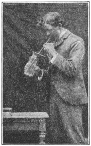
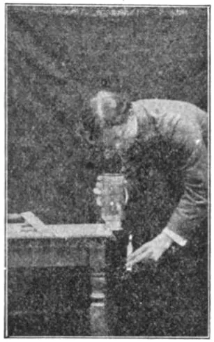
The presence of CO₂ in the breath may be shown better
by pouring off some of the clear limewater into a clean
glass and blowing into it through a straw.
Why does water put out a fire? The answer is, not
alone because it wets and shuts off the supply of free
oxygen, but because it cools the carbon, which must be
hot in order to unite with the oxygen, and prevents the
oxygen of the air from getting as near the carbon as
before.
PLANT BIOLOGY — CHAPTER I NO TWO PLANTS OR PARTS ARE ALIKE
Fig. 1.—No Two Branches are Alike.
(Hemlock.)
If one compares any two plants of
the same kind ever so closely, it will be
found that they differ from each other. The
difference is apparent in size, form, color, mode
of branching, number of leaves, number of flowers, vigor,
season of maturity, and the like; or, in other words, all
plants and animals vary from an assumed or standard type.
If one compares any two branches or twigs on a tree, it
will be found that they differ in size, age, form, vigor, and
in other ways (Fig. 1).
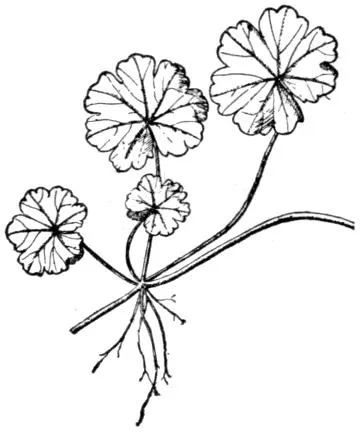
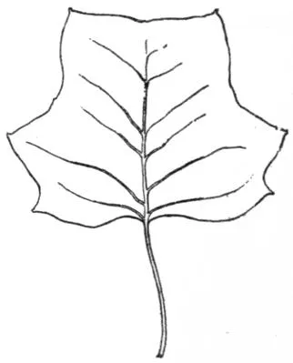
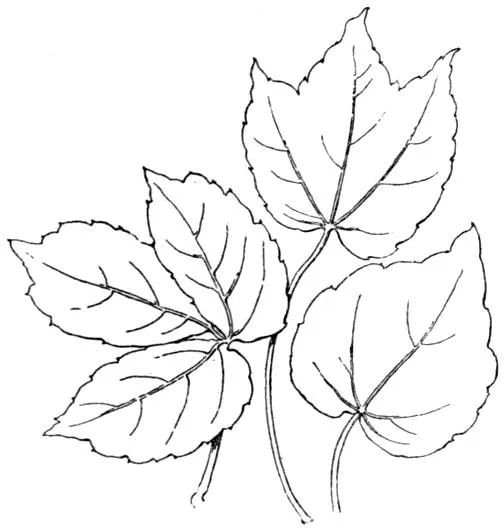
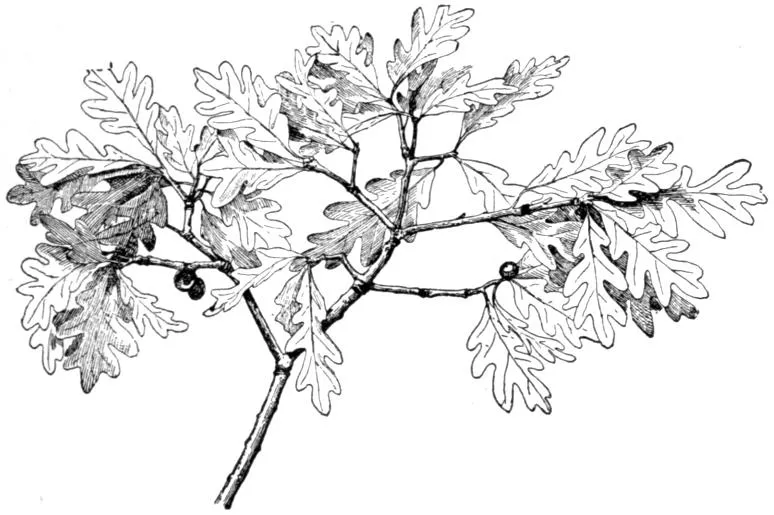
If one compares any two leaves, it will be found that
they are unlike in size, shape, color, veining, hairiness,
markings, cut of the margins, or other small features. In
some cases (as in Fig. 2) the differences are so great as to
be readily seen in a small black-and-white drawing.
Fig. 2.—No Two Leaves are Alike.
If the pupil extends his observation to animals, he
will still find the same truth; for probably no two living
objects are exact duplicates. If any person finds two objects
that he thinks to be exactly alike, let him set to work to
discover the differences, remembering that nothing in
nature is so small or apparently trivial as to be overlooked.
Variation, or differences between organs and also between
organisms, is one of the most significant facts in
nature.
Suggestions.—The first fact that the pupil should acquire
about plants is that no two are alike. The way to apprehend this
great fact is to see a plant accurately and then to compare it with
another plant of the same species or kind. In order to direct and
concentrate the observation, it is well to set a certain number of
attributes or marks or qualities to be looked for. 1. Suppose
any two or more plants of corn are compared in the following
points, the pupil endeavoring to determine whether the parts
exactly agree. See that the observation is close and accurate.
Allow no guesswork. Instruct the pupil to measure the parts
when size is involved:
(1) Height of the plant.
(2) Does it branch? How many secondary stems or “suckers”
from one root?
(3) Shade or color.
(4) How many leaves?
(5) Arrangement of leaves on stem.
(6) Measure length and breadth of six main leaves.
(7) Number and position of ears; color of silks.
(8) Size of tassel, and number and size of its branches.
(9) Stage of maturity or ripeness of plant.
(10) Has the plant grown symmetrically, or has it been
crowded by other plants or been obliged to struggle for light
or room?
(11) Note all unusual or interesting marks or features.
(12) Always make note of comparative vigor of the plants.
Note to Teacher.—The teacher should always insist on personal
work by the pupil. Every pupil should handle and study
the object by himself. Books and pictures are merely guides and
helps. So far as possible, study the plant or animal just where it
grows naturally.
Notebooks.—Insist that the pupils make full notes and preserve
these notes in suitable books. Note-taking is a powerful aid in
organizing the mental processes, and in insuring accuracy of observation
and record. The pupil should draw what he sees, even
though he is not expert with the pencil. The drawing should not
be made for looks, but to aid the pupil in his orderly study of the
object; it should be a means of self-expression.
Laboratory.—Every school, however small, should have a
laboratory or work-room. This work-room may be nothing more
than a table at one side of the room where the light is good.
Here the specimens may be ranged and studied. Often an
aquarium and terrarium may be added. A cabinet or set of
shelves should be provided for a museum and collection.
The laboratory may be in part out of doors, as a school garden;
or the garden may be at the pupil’s home, and yet be under the
general direction of the teacher.
CHAPTER II THE STRUGGLE TO LIVE
Every plant and animal is exposed to unfavorable conditions.
It is obliged to contend with these conditions in
order to live.
Fig. 3.—A Battle for Life.
No two plants or parts of plants are identically exposed
to the conditions in which they live. The large branches
in Fig. 1 probably had more room and a better exposure
to light than the smaller ones. Probably no two of the
leaves in Fig. 2 are equally exposed to light, or enjoy
identical advantages in relation to the food that they receive
from the tree.
Examine any tree to determine under what advantages
or disadvantages any of the limbs may live. Examine
similarly the different plants in a garden row (Fig. 3); or
the different bushes in a thicket; or the different trees in
a wood.
The plant meets its conditions by succumbing to them
(that is, by dying), or by adapting itself to them.
The tree meets the cold by ceasing its active growth,
hardening its tissues, dropping its leaves. Many herbaceous
or soft-stemmed plants meet the cold by dying
to the ground and withdrawing all life into the root parts.
Some plants meet the cold by dying outright and providing
abundance of seeds to perpetuate the kind next season.
Fig. 4.—The Reach for Light of a Tree on the Edge of a Wood.
Plants adapt themselves to light by growing toward it
(Fig. 4); or by hanging their leaves in such position that
they catch the light; or, in less sunny places, by expanding
their leaf surface, or by greatly lengthening their
stems so as to overtop their fellows, as do trees and vines.
The adaptations of plants will afford a fertile field of
study as we proceed.
Struggle for existence and adaptation to conditions are
among the most significant facts in nature.
The sum of all the conditions in which a plant or an animal
is placed is called its environment, that is, its surroundings.
The environment comprises the conditions of climate,
soil, moisture, exposure to light, relation to food supply,
contention with other plants or animals. The organism
adapts itself to its environment, or else it weakens or dies.
Every weak branch or plant has undergone some hardship
that it was not wholly able to withstand.
Suggestions.—The pupil should study any plant, or branch of
a plant, with reference to the position or condition under which it
grows, and compare one plant or branch with another. With
animals, it is common knowledge that every animal is alert to
avoid or to escape danger, or to protect itself. 2. It is well to
begin with a branch of a tree, as in Fig. 1. Note that no two
parts are alike (Chap. I). Note that some are large and strong
and that these stand farthest towards light and room. Some are
very small and weak, barely able to live under the competition.
Some have died. The pupil can easily determine which ones of
the dead branches perished first. He should take note of the
position or place of the branch on the tree, and determine whether
the greater part of the dead twigs are toward the center of the
tree top or toward the outside of it. Determine whether accident
has overtaken any of the parts. 3. Let the pupil examine
the top of any thick old apple tree, to see whether there is any
struggle for existence and whether any limbs have perished. 4. If
the pupil has access to a forest, let him determine why there are
no branches on the trunks of the old trees. Examine a tree of
the same kind growing in an open field. 5. A row of lettuce
or other plants sown thick will soon show the competition between
plants. Any fence row or weedy place will also show it. Why
does the farmer destroy the weeds among the corn or potatoes?
How does the florist reduce competition to its lowest terms?
what is the result?
CHAPTER III THE SURVIVAL OF THE FIT
The plants that most perfectly meet their conditions are
able to persist. They perpetuate themselves. Their offspring
are likely to inherit some of the attributes that
enabled them successfully to meet the battle of life. The
fit (those best adapted to their conditions) tend to survive.
Adaptation to conditions depends on the fact of variation;
that is, if plants were perfectly rigid or invariable
(all exactly alike) they could not meet new conditions.
Conditions are necessarily new for every organism. It is
impossible to picture a perfectly inflexible and stable succession
of plants or animals.
Breeding.—Man is able to modify plants and animals.
All our common domestic animals are very unlike their
original ancestors. So all our common and long-cultivated
plants have varied
from their ancestors. Even
in some plants that have
been in cultivation less than
a century the change is
marked: compare the common
black-cap raspberry
with its common wild ancestor,
or the cultivated blackberry
with the wild form.
Fig. 5.—Desirable and Undesirable
Types of Cotton Plants. Why?
By choosing seeds from a plant that pleases him, the
breeder may be able, under given conditions, to produce
numbers of plants with more
or less of the desired qualities;
from the best of these,
he may again choose; and so
on until the race becomes
greatly improved (Figs. 5, 6,
7). This process of continuously
choosing the most suitable
plants is known as selection.
A somewhat
similar
process proceeds
in wild
nature, and it
is then known
as natural selection.
Fig. 6.—Flax Breeding.
A is a plant grown for seed production;
B, for fiber production. Why?
Fig. 7.—Breeding.
A, effect from breeding
from smallest
grains (after four
years), average
head; B, result
from breeding from
the plumpest and
heaviest grains
(after four years),
average head.
Suggestions.—6.
Every pupil
should undertake
at least
one simple experiment
in selection
of seed. He may select kernels from the
best plant of corn in the field, and also from the
poorest plant,—having reference not so much to
mere incidental size and vigor of the plants that
may be due to accidental conditions in the field,
as to the apparently constitutional strength and
size, number of ears, size of ears, perfectness of
ears and kernels, habit of the plant as to suckering,
and the like. The seeds may be saved and
sown the next year. Every crop can no doubt
be very greatly improved by a careful process
of selection extending over a series of years.
Crops are increased in yield or efficiency in three
ways: better general care; enriching the land
in which they grow; attention to breeding.
CHAPTER IV PLANT SOCIETIES
In the long course of time in which plants have been
accommodating themselves to the varying conditions in
which they are obliged to grow, they have become adapted
to every different environment. Certain plants, therefore,
may live together or near each other, all enjoying the
same general conditions and surroundings. These aggregations
of plants that are adapted to similar general conditions
are known as plant societies.
Moisture and temperature are the leading factors in
determining plant societies. The great geographical
societies or aggregations of the plant world may conveniently
be associated chiefly with the moisture supply,
as: wet-region societies, comprising aquatic and bog
vegetation (Fig. 8); arid-region societies, comprising desert
and most sand-region vegetation; mid-region societies,
comprising the mixed vegetation in intermediate regions
(Fig. 9), this being the commonest type. Much of the
characteristic scenery of any place is due to its plant
societies. Arid-region plants usually have small and hard
leaves, apparently preventing too rapid loss of water.
Usually, also, they are characterized by stiff growth, hairy
covering, spines, or a much-contracted plant-body, and
often by large underground parts for the storage of water.
Plant societies may also be distinguished with reference
to latitude and temperature. There are tropical societies,
temperate-region societies, boreal or cold-region societies.
With reference to altitude, societies might be classified
as lowland (which are chiefly wet-region), intermediate
(chiefly mid-region), subalpine or mid-mountain (which are
chiefly boreal), alpine or high-mountain.
The above classifications have reference chiefly to great
geographical floras or societies. But there are societies
within societies. There are small societies coming within
the experience of every person who has ever seen plants
growing in natural conditions. There are roadside, fence-row,
lawn, thicket, pasture, dune, woods, cliff, barn-yard
societies. Every different place has its characteristic vegetation.
Note the smaller societies in Figs. 8 and 9. In the
former is a water-lily society and a cat-tail society. In
the latter there are grass and bush and woods societies.
Fig. 8.—A Wet-region Society.
Some Details of Plant Societies.—Societies may be composed
of scattered and intermingled plants, or of dense
clumps or groups of plants. Dense clumps or groups are
usually made up of one kind of plant, and they are then
called colonies. Colonies of most plants are transient:
after a short time other plants gain a foothold amongst
them, and an intermingled society is the outcome. Marked
exceptions to this are grass colonies and forest colonies, in
which one kind of plant may hold its own for years and
centuries.
In a large newly cleared area, plants usually first establish
themselves in dense colonies. Note the great patches
of nettles, jewel-weeds, smart-weeds, clot-burs, fire-weeds
in recently cleared but neglected swales, also the fire-weeds
in recently burned areas, the rank weeds in the neglected
garden, and the ragweeds and May-weeds along the recently
worked highway. The competition amongst themselves
and with their neighbors finally breaks up the
colonies, and a mixed and intermingled flora is generally
the result.
Fig. 9.—A Mid-region Society.
In many parts of the world the general tendency of neglected
areas is to run into forest. All plants rush for the
cleared area. Here and there bushes gain a foothold.
Young trees come up; in time these shade the bushes and
gain the mastery. Sometimes the area grows to poplars
or birches, and people wonder why the original forest trees
do not return; but these forest trees may be growing unobserved
here and there in the tangle, and in the slow processes
of time the poplars perish—for they are short-lived—and
the original forest may be replaced. Whether one
kind of forest or another returns will depend partly on the
kinds that are most seedful in that vicinity and which,
therefore, have sown themselves most profusely. Much
depends, also, on the kind of undergrowth that first springs
up, for some young trees can endure more or less shade
than others.
Fig. 10.—Overgrowth and Undergrowth in
Three Series,—trees, bushes, grass.
Some plants associate. They grow together. This is
possible largely because they diverge or differ in character.
Plants associate
in two ways:
by growing side by
side; by growing
above or beneath.
In sparsely populated
societies,
plants may grow
alongside each
other. In most
cases, however,
there is overgrowth
and undergrowth:
one kind grows beneath another. Plants that have become
adapted to shade are usually undergrowths. In a cat-tail
swamp, grasses and other narrow-leaved plants grow
in the bottom, but they are usually unseen by the casual
observer. Note the undergrowth in woods or under trees
(Fig. 10). Observe that in pine and spruce forests there
is almost no undergrowth, partly because there is very little
light.
On the same area the societies may differ at different
times of the year. There are spring, summer, and fall societies.
The knoll which is cool with grass and strawberries
in June may be aglow with goldenrod in September.
If the bank is examined in May, look for the young plants
that are to cover it in July and October; if in September,
find the dead stalks of the flora of May. What succeeds
the skunk cabbage, hepaticas, trilliums, phlox, violets,
buttercups of spring? What precedes the wild sunflowers,
ragweed, asters, and goldenrod of fall?
The Landscape.—To a large extent the color of the landscape
is determined by the character of the plant societies.
Evergreen societies remain green, but the shade of green
varies from season to season; it is bright and soft in
spring, becomes dull in midsummer and fall, and assumes
a dull yellow-green or a black-green in winter. Deciduous
societies vary remarkably in color—from the dull browns
and grays of winter to the brown greens and olive-greens
of spring, the staid greens of summer, and the brilliant
colors of autumn.
The autumn colors are due to intermingled shades of
green, yellow, and red. The coloration varies with the kind
of plant, the special location, and the season. Even in the
same species or kind, individual plants differ in color; and
this individuality usually distinguishes the plant year by
year. That is, an oak which is maroon red this autumn is
likely to exhibit that range of color every year. The autumn
color is associated with the natural maturity and
death of the leaf, but it is most brilliant in long and open
falls—largely because the foliage ripens more gradually
and persists longer in such seasons. It is probable that
the autumn tints are of no utility to the plant. Autumn
colors are not caused by frost. Because of the long, dry
falls and the great variety of plants, the autumnal color of
the American landscape is phenomenal.
Ecology.—The study of the relationships of plants and
animals to each other and to seasons and environments is
known as ecology (still written œcology in the dictionaries).
It considers the habits, habitats, and modes of life of living
things—the places in which they grow, how they
migrate or are disseminated, means of collecting food,
their times and seasons of flowering, producing young,
and the like.
Suggestions.—One of the best of all subjects for school instruction
in botany is the study of plant societies. It adds definiteness
and zest to excursions. 7. Let each excursion be confined to one
or two societies. Visit one day a swamp, another day a forest,
another a pasture or meadow, another a roadside, another a weedy
field, another a cliff or ravine. Visit shores whenever possible.
Each pupil should be assigned a bit of ground—say 10 or 20 ft.
square—for special study. He should make a list showing (1)
how many kinds of plants it contains, (2) the relative abundance
of each. The lists secured in different regions should be compared.
It does not matter greatly if the pupil does not know all
the plants. He may count the kinds without knowing the names.
It is a good plan for the pupil to make a dried specimen of each
kind for reference. The pupil should endeavor to discover why
the plants grow as they do. Note what kinds of plants grow next
each other; and which are undergrowth and which overgrowth;
and which are erect and which wide-spreading. Challenge every
plant society.
CHAPTER V THE PLANT BODY
The Parts of a Plant.—Our familiar plants are made up
of several distinct parts. The most prominent of these
parts are root, stem, leaf, flower, fruit, and seed. Familiar
plants differ wonderfully in size and shape,—from fragile
mushrooms, delicate waterweeds and pond-scums, to floating
leaves, soft grasses, coarse weeds, tall bushes, slender
climbers, gigantic trees, and hanging moss.
The Stem Part.—In most plants there is a main central
part or shaft on which the other or secondary parts are
borne. This main part is the plant axis. Above ground,
in most plants, the main plant axis bears the branches,
leaves, and flowers; below ground, it bears the roots.
The rigid part of the plant, which persists over winter
and which is left after leaves and flowers are fallen, is the
framework of the plant. The framework is composed of
both root and stem. When the plant is dead, the framework
remains for a time, but it slowly decays. The dry
winter stems of weeds are the framework, or skeleton of
the plant (Figs. 11 and 12). The framework of trees is
the most conspicuous part of the plant.
The Root Part.—The root bears the stem at its apex,
but otherwise it normally bears only root-branches. The
stem, however, bears leaves, flowers, and fruits. Those
living surfaces of the plant which are most exposed to
light are green or highly colored. The root tends to grow
downward, but the stem tends to grow upward toward light
and air. The plant is anchored or fixed in the soil by the
roots. Plants have been called “earth parasites.”
The Foliage Part.—The leaves precede the flowers in
point of time or life of the plant. The flowers always
precede the fruits and seeds. Many plants die when the
seeds have matured. The whole mass of leaves of any
plant or any branch is
known as its foliage.
In some cases, as in
crocuses, the flowers
seem to precede the
leaves; but the leaves
that made the food for
these flowers grew the
preceding year.
Fig. 11.—Plant of a
Wild Sunflower.
Fig. 12.—Framework
of Fig. 11.
The Plant Generation.—The
course of a
plant’s life, with all the
events through which
the plant naturally
passes, is known as
the plant’s life-history.
The life-history embraces
various stages,
or epochs, as dormant
seed, germination, growth, flowering, fruiting. Some plants
run their course in a few weeks or months, and some live
for centuries.
The entire life-period of a plant is called a generation.
It is the whole period from birth to normal death, without
reference to the various stages or events through which it
passes.
A generation begins with the young seed, not with germination.
It ends with death—that is, when no life is left
in any part of the plant, and only the seed or spore
remains to perpetuate the kind. In a bulbous plant, as a
lily or an onion, the generation does not end until the bulb
dies, even though the top is dead.
When the generation is of only one season’s duration,
the plant is said to be annual. When it is of two seasons,
it is biennial. Biennials usually bloom the second year.
When of three or more seasons, the plant is perennial.
Examples of annuals are pigweed, bean, pea, garden sunflower;
of biennials, evening primrose, mullein, teasel; of
perennials, dock, most meadow grasses, cat-tail, and all
shrubs and trees.
Duration of the Plant Body.—Plant structures which
are more or less soft and which die at the close of the
season are said to be herbaceous, in contradistinction to
being ligneous or woody. A plant which is herbaceous to
the ground is called an herb; but an herb may have a
woody or perennial root, in which case it is called an
herbaceous perennial. Annual plants are classed as herbs.
Examples of herbaceous perennials are buttercups, bleeding
heart, violet, water lily, Bermuda grass, horse-radish,
dock, dandelion, golden rod, asparagus, rhubarb, many
wild sunflowers (Figs. 11, 12).
Many herbaceous perennials have short generations.
They become weak with one or two seasons of flowering
and gradually die out. Thus, red clover usually begins to
fail after the second year. Gardeners know that the best
bloom of hollyhock, larkspur, pink, and many other plants,
is secured when the plants are only two or three years
old.
Herbaceous perennials which die away each season to
bulbs or tubers, are sometimes called pseud-annuals (that
is, false annuals). Of such are lily, crocus, onion, potato,
bull nettle, and false indigo of the Southern states.
True annuals reach old age the first year. Plants which
are normally perennial may become annual in a shorter-season
climate by being killed by frost, rather than by dying
naturally at the end of a season of growth. They are climatic
annuals. Such plants are called plur-annuals in the
short-season region. Many tropical perennials are plur-annuals
when grown in the north, but they are treated as
true annuals because they ripen sufficient of their crop the
same season in which the seeds are sown to make them
worth cultivating, as tomato, red pepper, castor bean,
cotton. Name several vegetables that are planted in
gardens with the expectation that they will bear till frost
comes.
Fig. 13.—A Shrub or Bush. Dogwood osier.
Woody or ligneous plants are usually longer lived than
herbs. Those that remain low and produce several or
many similar shoots from the
base are called shrubs, as lilac,
rose, elder, osier (Fig. 13). Low
and thick shrubs are bushes.
Plants that produce one main
trunk and a more or less elevated
head are trees (Fig. 14). All
shrubs and trees are perennial.
Fig. 14.—A Tree. The weeping
birch.
Every plant makes an effort
to propagate, or to perpetuate its
kind; and, as far as we can
see, this is the end for which
the plant itself lives. The seed
or spore is the final product of
the plant.
Suggestions.—8. The teacher may assign each pupil to one
plant in the school yard, or field, or in a pot, and ask him to bring
out the points in the lesson. 9. The teacher may put on the
board the names of many common plants and ask the pupils to
classify into annuals, pseud-annuals, plur-annuals (or climatic
annuals), biennials, perennials, herbaceous perennials, ligneous
perennials, herbs, bushes, trees. Every plant grown on the farm
should be so classified: wheat, oats, corn, buckwheat, timothy,
strawberry, raspberry, currant, tobacco, alfalfa, flax, crimson clover,
hops, cowpea, field bean, sweet potato, peanut, radish, sugar-cane,
barley, cabbage, and others. Name all the kinds of trees you
know.
CHAPTER VI SEEDS AND GERMINATION
Fig. 15.—Parts
of the Bean.
R, cotyledon; O,
caulicle; A, plumule;
F, first
node.
The seed contains a miniature plant, or embryo. The
embryo usually has three parts that have received
names: the stemlet, or caulicle; the seed-leaf, or cotyledon
(usually 1 or 2); the bud, or plumule, lying between or
above the cotyledons. These parts are well
seen in the common bean (Fig. 15), particularly
when the seed has been soaked for a
few hours. One of the large cotyledons—comprising
half of the bean—is shown at
R. The caulicle is at O. The plumule is
shown at A. The cotyledons are attached
to the caulicle at F: this point may be taken
as the first node or joint.
The Number of Seed-leaves.—All plants having two
seed-leaves belong to the group called dicotyledons. Such
seeds in many cases split readily in halves, e.g. a bean.
Some plants have only one seed-leaf in a seed. They
form a group of plants called monocotyledons. Indian
corn is an example of a plant with only one seed-leaf:
a grain of corn does not split into halves as a bean does.
Seeds of the pine family contain more than two cotyledons,
but for our purposes they may be associated with the dicotyledons,
although really forming a different group.
These two groups—the dicotyledons and the monocotyledons—represent
two great natural divisions of the
vegetable kingdom. The dicotyledons contain the woody
bark-bearing trees and bushes (except conifers), and most
of the herbs of temperate climates except the grasses,
sedges, rushes, lily tribes, and orchids. The flower-parts
are usually in fives or multiples of five, the leaves mostly
netted-veined, the bark or rind distinct, and the stem often
bearing a pith at the center. The monocotyledons usually
have the flower-parts in threes or multiples of three, the
leaves long and parallel-veined, the bark not separable,
and the stem without a central pith.
Every seed is provided with food to support the germinating
plant. Commonly this food is starch. The food may
be stored in the cotyledons, as in bean, pea, squash; or outside
the cotyledons, as in castor bean, pine, Indian corn.
When the food is outside or around the embryo, it is
usually called endosperm.
Fig. 16.—External
Parts of
Bean.
Seed-coats; Markings on Seed.—The embryo and endosperm
are inclosed within a covering made of two or
more layers and known as the seed-coats.
Over the point of the caulicle is a minute
hole or a thin place in the coats known as
the micropyle. This is the point at which
the pollen-tube entered the forming ovule
and through which the caulicle breaks in
germination. The micropyle is shown at M in Fig. 16.
The scar where the seed broke from its funiculus (or stalk
that attached it to its pod) is named the hilum. It occupies
a third of the length of the bean in Fig. 16. The
hilum and micropyle are always present in seeds, but they
are not always close together. In many cases it is difficult
to identify the micropyle in the dormant seed, but its location
is at once shown by the protruding caulicle as germination
begins. Opposite the micropyle in the bean (at the
other end of the hilum) is an elevation known as the raphe.
This is formed by a union of the funiculus, or seed-stalk,
with the seed-coats, and through it food was transferred
for the development of the seed, but it is now functionless.
Seeds differ wonderfully in size, shape, color, and other
characteristics. They also vary in longevity. These
characteristics are peculiar to the species or kind. Some
seeds maintain life only a few weeks or even days, whereas
others will “keep” for ten or twenty years. In special
cases, seeds have retained vitality longer than this limit,
but the stories that live seeds, several thousand years old,
have been taken from the wrappings of mummies are unfounded.
Germination.—The embryo is not dead; it is only dormant.
When supplied with moisture, warmth, and oxygen
(air), it awakes and grows: this growth isgermination.
The embryo lives for a time on the stored food, but gradually
the plantlet secures a foothold in the soil and gathers
food for itself. When the plantlet is finally able to shift
for itself, germination is complete.
Early Stages of Seedling.—The germinating seed first
absorbs water, and swells. The starchy matters gradually
become soluble. The seed-coats are ruptured, the caulicle
and plumule emerge. During this process the seed
respires freely, throwing off carbon dioxid (CO₂).
The caulicle usually elongates, and from its lower end
roots are emitted. The elongating caulicle is known as
the hypocotyl (“below the cotyledons”). That is, the
hypocotyl is that part of the stem of the plantlet lying
between the roots and the cotyledon. The general direction
of the young hypocotyl, or emerging caulicle, is downwards.
As soon as roots form, it becomes fixed and its
subsequent growth tends to raise the cotyledons above the
ground, as in the bean. When cotyledons rise into the
air, germination is said to be epigeal (“above the earth”).
Bean and pumpkin are examples. When the hypocotyl
does not elongate greatly
and the cotyledons remain
under ground, the germination
is hypogeal (“beneath
the earth”). Pea
and scarlet runner bean
are examples (Fig. 48).
When the germinating
seed lies on a hard surface,
as on closely compacted
soil, the hypocotyl
and rootlets may not be able to secure a foothold and they
assume grotesque forms. (Fig. 17.) Try this with peas
and beans.
Fig. 17.—Pea. Grotesque forms assumed
when the roots cannot gain entrance to
the soil.
The first internode (“between nodes”) above the cotyledons
is the epicotyl. It elevates the plumule into the
air, and the plumule-leaves expand into the first true leaves
of the plant. These first true leaves, however, may be
very unlike the later leaves in shape.
Fig. 18.—Cotyledons
of Germinating
Bean spread apart
to show Elongating
Caulicle and
Plumule.
Germination of Bean.—The common bean, as we have
seen (Fig. 15), has cotyledons that occupy all the space
inside the seed-coats. When the hypocotyl,
or elongated caulicle, emerges,
the plumule-leaves have begun to enlarge,
and to unfold (Fig. 18). The
hypocotyl elongates rapidly. One end
of it is held by the roots. The other
is held by the seed-coats in the soil.
It therefore takes the form of a loop,
and the central part of the loop “comes up” first (a, Fig.
19). Presently the cotyledons come out of the seed-coats,
and the plant straightens and the
cotyledons expand. These cotyledons,
or “halves of the bean,”
persist for some time (b, Fig.
19). They often become green
and probably perform some
function of foliage. Because of
its large size, the Lima bean
shows all these parts well.
Fig. 19.—Germination Of
Bean.
Fig. 20.—Sprouting
of Castor
Bean.
Fig. 21.—Germination
of Castor Bean.
Endosperm at a.
Fig. 22.—Castor
Bean.
Endosperm at a, a; cotyledons
at b.
Fig. 23.—Germination
Complete in Castor
Bean.
Germination of Castor Bean.—In
the castor bean the hilum
and micropyle are at the smaller end
(Fig. 20). The bean “comes up” with a
loop, which indicates that the hypocotyl
greatly elongates. On examining germinating
seed, however, it will be found
that the cotyledons are contained inside a fleshy body,
or sac (a, Fig. 21). This sac is the endosperm. Against
its inner surface the thin, veiny cotyledons
are very closely pressed, absorbing
its substance (Fig. 22). The cotyledons increase
in size as they reach the air (Fig. 23), and become functional
leaves.
Fig. 24.—Sprouting
Indian Corn.
Hilum at h; micropyle
at d.
Fig. 25.—Kernel
of Indian Corn.
Caulicle at b; cotyledon
at a; plumule
at p.
Fig. 26.—Indian
Corn.
Caulicle at c; roots emerging at
m; plumule at p.
Fig. 27.—Indian Corn.
o, plumule: n to p, epicotyl.
Germination of Monocotyledons.—Thus far we have studied
dicotyledonous seeds; we may now consider the monocotyledonous
group. Soak kernels of corn. Note that
the micropyle and hilum are at the smaller end (Fig. 24).
Make a longitudinal section through the
narrow diameter; Fig. 25 shows it. The
single cotyledon is at a, the caulicle at b, the plumule
at p. The cotyledon remains in the seed. The food is
stored both in the cotyledon and as endosperm, chiefly the
latter. The emerging shoot is the plumule, with a sheathing
leaf (p, Fig. 26). The root is emitted from the tip of
the caulicle, c. The caulicle is held in a sheath
(formed mostly from the seed-coats), and some of
the roots escape through the upper end
of this sheath (m, Fig. 26). The
epicotyl elongates, particularly if
the seed is planted
deep or if it is
kept for a time
confined. In Fig.
27 the epicotyl has
elongated from n to p. The true plumule-leaf is at o, but
other leaves grow from its sheath. In Fig. 28 the roots
are seen emerging from the two ends of the caulicle
sheath, c, m; the epicotyl has grown to p; the first plumule-leaf
is at o.
Fig. 28.—Germination is Complete.
p, top of epicotyl; o, plumule-leaf;
m, roots; c, lower roots.
In studying corn or other fruits or seeds, the pupil should
note how the seeds are arranged, as on the cob. Count the
rows on a corn cob. Odd or
even in number? Always the
same number? The silk is
the style: find where it was
attached to the kernel. Did
the ear have any coverings?
Explain. Describe colors and
markings of kernels of corn;
and of peas, beans, castor
bean.
Gymnosperms.—The seeds
in the pine cone, not being
inclosed in a seed-vessel,
readily fall out when the cone
dries and the scales separate.
Hence it is difficult to find
cones with seeds in them after
autumn has passed (Fig. 29).
The cedar is also a gymnosperm.
Fig. 29.—Cones of Hemlock
(above), White
Pine, Pitch Pine.
Remove a scale from a
pine cone and draw it and
the seeds as they lie in place
on the upper side of the scale.
Examine the seed, preferably with a magnifying glass. Is
there a hilum? The micropyle is at the bottom or little
end of the seed. Toss a seed upward into the air. Why
does it fall so slowly? Can you explain the peculiar whirling
motion by the shape of the wing? Repeat the experiment
in the wind. Remove the
wing from a seed and toss it and an
uninjured seed into the air together.
What do you infer from these experiments?
Fig. 30.—Muskmelon Seedlings, with
the unlike seed-leaves and true leaves.
Suggestions.—Few subjects connected
with the study of plant-life are so
useful in schoolroom demonstrations as
germination. The pupil should prepare
the soil, plant the seeds, water them, and
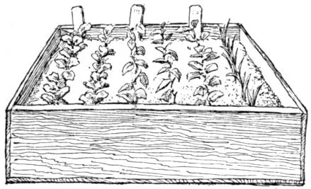
care for the plants. 10. Plant seeds in
pots or shallow boxes. The box should
not be very wide or long, and not over
four inches deep. Holes may be bored
in the bottom so it will not hold water.
Plant a number of squash, bean, corn,
pine, or other seeds about an inch deep
in damp sand or pine sawdust in this
box. The depth of planting should be
two to four times the diameter of the
seeds. Keep the sand or sawdust moist
but not wet. If the class is large, use
several boxes, that the supply of specimens
may be ample. Cigar boxes and
chalk boxes are excellent for individual
pupils. It is well to begin the planting
of seeds at least ten days in advance of
the lesson, and to make four or five different
plantings at intervals. A day or two
before the study is taken up, put seeds
to soak in moss or cloth. The pupil
then has a series from swollen seeds to
complete germination, and all the steps can be made out. Dry
seeds should be had for comparison. If there is no special room
for laboratory, nor duplicate apparatus for every pupil, each experiment
may be assigned to a committee of two pupils to watch
in the schoolroom. 11. Good seeds for study are those detailed
in the lesson, and buckwheat, pumpkin, cotton, morning glory,
radish, four o’clock, oats, wheat. It is best to use familiar seeds
of farm and garden. Make drawings and notes of all the events
in the germination. Note the effects of unusual conditions, as
planting too deep and too shallow and different sides up. For
hypogeal germination, use the garden pea, scarlet runner or Dutch
case-knife bean, acorn, horse-chestnut. Squash seeds are excellent
for germination studies, because the cotyledons become green and
leafy and germination is rapid. Its germination, as also that of the
scarlet runner bean, is explained in “Lessons with Plants.” Onion
is excellent, except that it germinates too slowly. In order to study
the root development of germinating plantlets, it is well to provide
a deeper box with a glass side against which the seeds are
planted. 12. Observe the germination of any common seed
about the house premises. When elms, oaks, pines, or maples are
abundant, the germination of their seeds may be studied in lawns
and along fences. 13. When studying germination, the pupil
should note the differences in shape and size between cotyledons
and plumule-leaves, and between plumule-leaves and the normal
leaves (Fig. 30). Make drawings. 14. Make the tests described
in the introductory experiments
with bean, corn, the
castor bean, and other seed
for starch and proteids. Test
flour, oatmeal, rice, sunflower,
four o’clock, various nuts, and
any other seeds obtainable.
Record your results by arranging
the seeds in three
classes, 1. Much starch (color
blackish or purple), 2. Little
starch (pale blue or greenish), 3. No starch (brown or yellow).
15.Rate of growth of seedlings as affected by differences in temperature.
Pack soft wet paper to the depth of an inch in the bottom
of four glass bottles or tumblers. Put ten soaked peas or beans into
each. Cover each securely and set them in places having different
temperatures that vary little. (A furnace room, a room with a
stove, a room without stove but reached by sunshine, an unheated
room not reached by the sun.) Take the temperatures occasionally
with a thermometer to find difference in temperature. The
tumblers in warm places should be covered very tightly to prevent
the germination from being retarded by drying out. Record the
number of seeds which sprout in each tumbler within 1 day; 2 days;
3 days; 4 days, etc. 16.Is air necessary for the germination and
growth of seedlings? Place damp blotting paper in the bottom of a
bottle and fill it three fourths full of soaked seeds, and close it
tightly with a rubber stopper or oiled cork. Prepare a “check
experiment” by having another bottle with all conditions the same
except that it is covered loosely that air may have access to it,
and set the bottles side by side (why keep the bottles together?).
Record results as in the preceding experiment. 17.What is the
nature of the gas given off by germinating seeds? Fill a tin box or
large-necked bottle with dry beans or peas, then add water; note
how much they swell. Secure two fruit-jars. Fill one of them a
third full of beans and keep them moist. Allow the other to remain
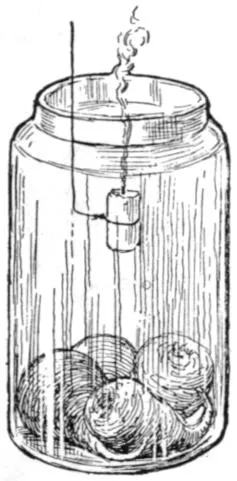
empty. In a day or two insert a lighted splinter or taper into
each. In the empty jar the taper burns: it contains oxygen.
In the seed jar the taper goes out: the air has been replaced
by carbon dioxid. The air in the bottle may be tested for
carbon dioxid by removing some of it with a rubber bulb attached
to a glass tube (or a fountain-pen filler) and bubbling it through
lime water. 18.Temperature. Usually there is a perceptible
rise in temperature in a mass of germinating seeds. This rise may
be tested with a thermometer. 19.Interior of seeds. Soak
seeds for twenty-four hours and remove the coat. Distinguish
the embryo from the endosperm. Test with iodine. 20.Of
what utility is the food in seeds? Soak some grains of corn
overnight and remove the endosperm, being careful not to
injure the fleshy cotyledon. Plant the incomplete and also some
complete grains in moist sawdust and measure their growth at
intervals. (Boiling the sawdust will destroy molds and bacteria
which might interfere with experiment.) Peas or beans may be
sprouted on damp blotting paper; the cotyledons of one may be
removed, and this with a normal seed equally advanced in germination
may be placed on a perforated cork floating in water in
a jar so that the roots extend into the water. Their growth
may be observed for several weeks. 21.Effect of darkness on
seeds and seedlings. A box may be placed mouth downward
over a smaller box in which seedlings are growing. The empty
box should rest on half-inch blocks to allow air to reach the
seedlings. Note any effects on the seedlings of this cutting off
of the light. Another box of seedlings not so covered may
be used for a check. Lay a plank on green grass and after a
week note the change that takes place beneath it. 22.Seedling
of pine. Plant pine seeds. Notice how they emerge. Do the
cotyledons stay in the ground? How many cotyledons have
they? When do the cotyledons get free from the seed-coat?
What is the last part of the cotyledon to become free? Where is
the growing point or plumule? How many leaves appear at
once? Does the new pine cone grow on old wood or on wood
formed the same spring with the cone? Can you always find
partly grown cones on pine trees in winter? Are pine cones
when mature on two-year-old wood? How long do cones stay
on a tree after the seeds have fallen out? What is the advantage
of the seeds falling before the cones? 23.Home experiments.
If desired, nearly all of the foregoing experiments may be
tried at home. The pupil can thus make the drawings for the
notebook at home. A daily record of measurements of the change
in size of the various parts of the seedling should also be made.
24.Seed-testing.—It is important that one know before planting
whether seeds are good, or able to grow. A simple seed-tester
may be made of two plates, one inverted over the other (Fig. 31).
The lower plate is nearly filled with clean
sand, which is covered with cheese cloth
or blotting paper on which the seeds are
placed. Canton flannel is sometimes
used in place of sand and blotting paper.
The seeds are then covered with another
blotter or piece of cloth, and water is
applied until the sand and papers are
saturated. Cover with the second plate.
Set the plates where they will have about
the temperature that the given seeds
would require out of doors, or perhaps a
slightly higher temperature. Place 100 or more grains of clover,
corn, wheat, oats, rye, rice, buckwheat, or other seeds in the tester,
and keep record of the number that sprout. The result will give
a percentage measure of the ability of the seeds to grow. Note
whether all the seeds sprout with equal vigor and rapidity. Most
seeds will sprout in a week or less. Usually such a tester must
have fresh sand and paper after every test, for mold fungi are likely
to breed in it. If canton flannel is used, it may be boiled. If
possible, the seeds should not touch each other.
Fig. 31.—A Home-made
Seed-tester.
Note to Teacher.—With the study of germination, the pupil
will need to begin dissecting.
For dissecting, one needs a lens for the examination of the
smaller parts of plants and animals. It is best to have the lens
mounted on a frame, so that the pupil has both hands free for
pulling the part in pieces. An ordinary pocket lens may be
mounted on a wire in a block, as in Fig. A. A cork is slipped on
the top of the wire to avoid injury to the face. The pupil should
be provided with two dissecting needles (Fig. B), made by
securing an ordinary needle in a pencil-like stick. Another convenient
arrangement is shown in Fig. C. A small tin dish is used
for the base. Into this a stiff wire standard is soldered. The
dish is filled with solder, to make it heavy and firm. Into a cork
slipped on the standard, a cross wire is inserted, holding on the
end a jeweler’s glass. The lens can be moved up and down and
sidewise. This outfit can be made for about seventy-five cents.
Fig. D shows a convenient hand-rest or dissecting-stand to be
used under this lens. It may be 16 in. long, 4 in. high, and 4 or 5
in. broad.
Various kinds of dissecting microscopes are on the market, and
these are to be recommended when they can be afforded.
A.—Improvised
Stand for Lens.
B.—Dissecting
Needle
¹⁄₂ natural
size.
C.—Dissecting Glass.
D.—Dissecting Stand.
Instructions for the use of the compound microscope, with
which some schools may be equipped, cannot be given in a brief
space; the technique requires careful training. Such microscopes
are not needed unless the pupil studies cells and tissues.
CHAPTER VII THE ROOT—THE FORMS OF ROOTS
The Root System.—The offices of the root are to hold
the plant in place, and to gather food. Not all the food
materials, however, are gathered by the roots.
Fig. 32.—Tap-root
System of Alfalfa.
Fig. 33.—Tap-root of the Dandelion.
The entire mass of roots of any plant is called its root
system. The root system may be annual, biennial or perennial,
herbaceous or woody, deep or shallow, large or small.
Kinds of Roots.—A strong leading central root, which
runs directly downwards, is a tap-root. The tap-root forms
an axis from which the side roots may branch. The side
or spreading roots are usually smaller. Plants that have
such a root system are said to be tap-rooted. Examples
are red clover, alfalfa, beet, turnip,
radish, burdock, dandelion, hickory
(Figs. 32, 33).
A fibrous root system is one that
is composed of many nearly equal
slender branches. The greater
number of plants have fibrous roots.
Examples are many common
grasses, wheat, oats, corn. The
buttercup in Fig. 34 has a fibrous
root system. Many trees have a
strong tap-root when very young,
but after a while it ceases to extend
strongly and the side roots
develop until finally the tap-root
character disappears.
Fig. 34.—A Buttercup
Plant, with fibrous roots.
Shape and Extent of the Root System.—The
depth to which roots
extend depends on the kind of plant, and the nature of the
soil. Of most plants the roots extend far in all directions
and lie comparatively near the surface. The roots usually
radiate from a common point just beneath the surface of
the ground.
The roots grow here and there in search of food, often
extending much farther in all directions than the spread
of the top of the plant. Roots tend to spread farther in
poor soil than in rich soil, for the same size of plant.
The root has no such definite form as the stem has. Roots
are usually very crooked, because they are constantly
turned aside by obstacles. Examine roots in stony soil.
The extent of root surface is usually very large, for the
feeding roots are fine and very numerous. An ordinary
plant of Indian corn may have a total length of root
(measured as if the roots were placed end to end) of several
hundred feet.
The fine feeding roots are most abundant in the richest
part of the soil. They are attracted by the food materials.
Roots often will completely surround a bone or other
morsel. When roots of trees are exposed, observe that
most of them are horizontal and lie near the top of the
ground. Some roots, as of willows, extend far in search
of water. They often run into wells and drains, and into
the margins of creeks and ponds. Grow plants in a long
narrow box, in one end of which the soil is kept very dry
and in the other moist: observe where the roots grow.
Buttresses.—With the increase in diameter, the upper
roots often protrude above the ground and become bracing
buttresses. These buttresses are usually largest in trees
which always have been
exposed to strong winds
(Fig. 35). Because of
growth and thickening,
the roots elevate part of
their diameter, and the
washing away of the soil
makes them to appear as
if having risen out of
the ground.
Fig. 35.—The Bracing Base of a
Field Pine.
Aërial Roots.—Although roots usually grow underground,
there are some that naturally grow above ground. These
usually occur on climbing plants, the roots becoming supports
or fulfilling the office of tendrils. These aërial roots
usually turn away from the light, and therefore enter the
crevices and dark places of the wall or tree over which the
plant climbs. The trumpet creeper (Fig. 36), true or
English ivy, and poison ivy climb by
means of roots.
Fig. 36.—Aërial Roots
of Trumpet Creeper
or Tecoma.
Fig. 37.—Aërial Roots of an Orchid.
In some plants all the roots are
aërial; that is, the plant grows above
ground, and the roots gather food
from the air. Such plants usually
grow on trees. They are known as
epiphytes or air-plants. The most familiar
examples are some of the tropical
orchids, which are grown in glass-houses
(Fig. 37). Rootlike organs of dodder and other
parasites are discussed in a future chapter.
Some plants bear aërial roots, that may propagate the
plant or may act as braces. They are often called prop-roots.
The roots of Indian corn are familiar (Fig. 38). Many
ficus trees, as the banyan of India, send out roots from
their branches; when these roots reach the ground they
take hold and become great trunks, thus spreading the
top of the parent tree over large
areas. The muscadine grape of the
Southern states often sends down
roots from its stems. The mangrove
tree of the tropics grows along
seashores and sends down roots
from the overhanging branches
(and from the fruits) into the shallow
water, and thereby gradually
marches into the sea. The tangled
mass behind catches the drift, and
soil is formed.
Fig. 38.—Indian Corn,
showing the brace roots
at oo.
Adventitious Roots.—Sometimes
roots grow from the stem or other
unusual places as the result of some
accident to the plant, being located
without known method or law.
They are called adventitious (chance)
roots. Cuttings of the stems of
roses, figs, geraniums, and other plants, when planted,
send out adventitious roots and form new plants. The
ordinary roots, or soil roots, are of course not classed as
adventitious roots. The adventitious roots arise on occasion,
and not as a normal or regular course in the growth
of the plant.
No two roots are alike; that is, they vary among themselves
as stems and leaves do. Each kind of plant has its
own form or habit of root (Fig. 39). Carefully wash away
the soil from the roots of any two related plants, as oats
and wheat, and note the differences in size, depth, direction,
mode of branching, number
of fibrils, color, and other
features. The character of the root system often governs
the treatment that the farmer should give the soil in which
the plant or crop grows.
Fig. 39.—Roots of Barley atAand Corn atB.
Carefully trace the differences.
Roots differ not only in their form and habit, but also in
color of tissue, character of bark or rind, and other features.
It is excellent practice to try to identify different plants by
means of their roots. Let each pupil bring to school two
plants with the roots very carefully dug up, as cotton,
corn, potato, bean, wheat, rye, timothy, pumpkin, clover,
sweet pea, raspberry, strawberry, or other common plants.
Root Systems of Weeds.—Some weeds are pestiferous
because they seed abundantly, and others because their
underground parts run deep or far and are persistent.
Make out the root systems in the six worst weeds in your
locality.
CHAPTER VIII THE ROOT.—FUNCTION AND STRUCTURE
The function of roots is twofold,—to provide support or
anchorage for the plant, and to collect and convey food materials.
The first function is considered in Chapter VII;
we may now give attention in more detail to the second.
The feeding surface of the roots
is near their ends. As the roots
become old and hard, they serve
only as channels through which
food passes and as hold-fasts or
supports for the plant. The root-hold
of a plant is very strong.
Slowly pull upwards on some plant,
and note how firmly it is anchored
in the soil.
Fig. 40.—Wheat growing
under Different Soil
Treatments. Soil deficient
in nitrogen; commercial
nitrogen applied
to pot 3 (on right).
Roots have power to choose their
food; that is, they do not absorb
all substances with which they
come in contact. They do not take
up great quantities of useless or
harmful materials, even though
these materials may be abundant in the soil; but they
may take up a greater quantity of some of the plant-foods
than the plant can use to advantage. Plants respond very
quickly to liberal feeding,—that is, to the application of
plant-food to the soil (Fig. 40). The poorer the soil, the
more marked are the results, as a rule, of the application
of fertilizers. Certain substances, as common salt, will kill
the roots.
Roots absorb Substances only in Solution.—Substances
cannot be taken in solid particles. These materials are
in solution in the soil water, and the roots themselves
also have the power to dissolve the soil materials to some
extent by means of substances that
they excrete. The materials that
come into the plant through the
roots are water and mostly the mineral
substances, as compounds of potassium,
iron, phosphorus, calcium,
magnesium, sulfur, and chlorine.
These mineral substances compose
the ash when the plant is burned.
The carbon is derived from the air
through the green parts. Oxygen
is derived from the air and the soil
water.
Fig. 41.—Nodules on Roots
of Red Clover.
Nitrogen enters through the Roots.—All
plants must have nitrogen;
yet, although about four fifths of
the air is nitrogen, plants are not
able, so far as we know, to take it in through their leaves.
It enters through the roots in combination with other elements,
chiefly in the form of nitrates (certain combinations
with oxygen and a mineral base). The great family of
leguminous plants, however (as peas, beans, cowpea,
clover, alfalfa, vetch), use the nitrogen contained in the air
in the soil. They are able to utilize it through the agency
of nodules on their roots (Figs. 41, 42). These nodules
contain bacteria, which appropriate the free or uncombined
nitrogen and pass it on to the plant. The nitrogen
becomes incorporated in the plant tissue, so that these
crops are high in their nitrogen content. Inasmuch as
nitrogen in any form is
expensive to purchase in
fertilizers, the use of leguminous
crops to plow under
is a very important agricultural
practice in preparing
the land for other crops.
In order that leguminous
crops may acquire atmospheric
nitrogen more freely
and thereby thrive better,
the land is sometimes sown
or inoculated with the nodule-forming
bacteria.
Fig. 42.—Nodules on Vetch.
Fig. 43.—Two Kinds of Soil that have
been Wet and then Dried. The
loamy soil above remains loose and capable
of growing plants; the clay soil below
has baked and cracked.
Roots require moisture in order to serve the plant. The
soil water that is valuable
to the plant is not
the free water, but the
thin film of moisture
which adheres to each
little particle of soil. The
finer the soil, the greater
the number of particles,
and therefore the greater
is the quantity of film
moisture that it can hold.
This moisture surrounding
the grains may not
be perceptible, yet the
plant can use it. Root absorption may continue in a soil
which seems to be dust dry. Soils that are very hard and
“baked” (Fig. 43) contain very little moisture or air,—not
so much as similar soils that are granular or mellow.
Proper Temperature for Root Action.—The root must be
warm in order to perform its functions. Should the soil of
fields or greenhouses be much colder than the air, the
plant suffers. When in a warm atmosphere, or in a dry
atmosphere, plants need to absorb much water from the
soil, and the roots must be warm if the root-hairs are to
supply the water as rapidly as it is needed. If the roots are
chilled, the plant may wilt or die.
Roots need Air.—Corn on land that has been flooded by
heavy rains loses its green color and turns yellow. Besides
diluting plant-food, the water drives the air from the soil, and
this suffocation of the roots is very soon apparent
in the general ill health of the plant.
Stirring or tilling the soil aërates it. Water
plants and bog plants have adapted themselves
to their particular conditions. They
get their air either by special surface roots,
or from the water through stems and leaves.
Rootlets.—Roots divide into the thinnest
and finest fibrils: there are roots and there
are rootlets. The smallest rootlets are so
slender and delicate that they break off
even when the plant is very carefully lifted
from the soil.
Fig. 44.—Root-hairs
of the
Radish.
The rootlets, or fine divisions, are clothed with theroot-hairs
(Figs. 44, 45, 46). These root-hairs attach to the
soil particles, and a great amount of soil is thus brought
into actual contact with the plant. These are very delicate
prolonged surface cells of the roots. They are borne
for a short distance just back of the tip of the root.
Rootlet and root-hair differ. The rootlet is a compact
cellular structure. The root-hair is a delicate tubular
cell (Fig. 45), within which is contained living matter
(protoplasm); and the protoplasmic lining membrane of the
wall governs the entrance of
water and substances in solution.
Being long and tube-like,
these root-hairs are
especially adapted for taking
in the largest quantity
of solutions; and they are
the principal means by which
plant-food is absorbed from
the soil, although the surfaces
of the rootlets themselves
do their part. Water
plants do not produce an
abundant system of root-hairs, and such plants depend
largely on their rootlets.
Fig. 45.—Cross-section of Root,
enlarged, showing root-hairs.
Fig. 46.—Root-hair, much enlarged,
in contact with the soil
particles (s). Air-spaces at a;
water-films on the particles, as
at w.
The root-hairs are very small, often invisible. They,
with the young roots, are usually broken off when the
plant is pulled up. They are
best seen when seeds are germinated
between layers of dark
blotting paper or flannel. On
the young roots, they will be
seen as a mold-like or gossamer-like
covering. Root-hairs soon
die: they do not grow into roots.
New ones form as the root grows.
Osmosis.—The water with its
nourishment goes through the
thin walls of the root-hairs and rootlets by the process
of osmosis. If there are two liquids of different density
on the inside and outside of an organic (either vegetable
or animal) membrane, the liquids tend to mix through the
membrane. The law of osmosis is that the most rapid
flow is toward the denser solution. The protoplasmic lining
of the cell wall is such a membrane. The soil water
being a weaker solution than the sap in the roots, the
flow is into the root. A strong fertilizer sometimes causes
a plant to wither, or “burns it.” Explain.
Structure of Roots.—The root that grows from the lower
end of the caulicle is the first or primary root. Secondary
roots branch from the primary root. Branches of secondary
roots are sometimes called tertiary roots. Do the secondary
roots grow from the cortex, or from the central
cylinder of the primary root? Trim or peel the cortex
from a root and its branches and determine whether the
branches still hold to the central cylinder of the main root.
Internal Structure of Roots.—A section of a root shows
that it consists of a central cylinder (see Fig. 45) surrounded
by a layer. This layer is called the cortex. The
outer layer of cells in the cortex is called the epidermis,
and some of the cells of the epidermis are prolonged
and form the delicate root-hairs. The cortex resembles
the bark of the stem in its nature. The central cylinder
contains many tube-like canals, or “vessels” that convey
water and food (Fig. 45). Cut a sweet potato across (also
a radish and a turnip) and distinguish the central cylinder,
cortex and epidermis. Notice the hard cap on the tip
of roots. Roots differ from stems in having no real pith.
Microscopic Structure of Roots.—Near the end of any
young root or shoot the cells are found to differ from each
other more or less, according to the distance from the
point. This differentiation takes place in the region just
back of the growing point. To study growing points, use
the hypocotyl of Indian corn which has grown about one
half inch. Make a longitudinal section. Note these points
(Fig. 47): (a) the tapering root-cap beyond the growing
point; (b) the blunt end of the root proper and the rectangular
shape of the cells found there; (c) the group
of cells in the middle of the first layers beneath the root-cap,—this
group is the growing
point; (d) study the slight differences
in the tissues a short distance
back of the growing point.
There are four regions: the central
cylinder, made up of several rows
of cells in the center (pl); the endodermis,
(e) composed of a single
layer on each side which separates
the central cylinder from the bark;
the cortex, or inner bark, (e) of several
layers outside the endodermis;
and the epidermis, or outer layer of
bark on the outer edges (d). Make
a drawing of the section. If a
series of the cross-sections of the
hypocotyl should be made and studied,
beginning near the growing
point and going upward, it would
be found that these four tissues become more distinctly
marked, for at the tip the tissues have not yet assumed
their characteristic form. The central cylinder contains
the ducts and vessels which convey the sap.
Fig. 47.—Growing Point
of Root of Indian Corn.
d, d, cells which will form the
epidermis; p, p, cells that
will form bark; e, e, endodermis;
pl, cells which will form
the axis cylinder; i, initial
group of cells, or growing
point proper; c, root-cap.
The Root-cap.—Note the form of the root-cap shown in
the microscopic section drawn in Fig. 47. Growing cells,
and especially those which are forming tissue by subdividing,
are very delicate and are easily injured. The
cells forming the root-cap are older and
tougher and are suited for pushing
aside the soil that the root may penetrate
it.
Region of most Rapid Growth.—The
roots of a seedling bean may be marked
at equal distances by waterproof ink or
by bits of black thread tied moderately
tight. The seedling is then replanted
and left undisturbed for two days.
When it is dug up, the region of most
rapid growth in the
root can be determined.
Give a reason why a root
cannot elongate throughout its length,—whether
there is anything to prevent
a young root from doing so.
Fig. 48.—The Marking
of the Stem
and Root.
In Fig. 48 is shown a germinating
scarlet runner bean with a short root
upon which are marks made with
waterproof ink; and the same root
(Fig. 49) is shown after it has
grown longer. Which part of it
did not lengthen at all? Which
part lengthened slightly? Where
is the region of most rapid growth?
Fig. 49.—The Result.
Geotropism.—Roots turn toward
the earth, even if the seed
is planted with the micropyle up.
This phenomenon is called positive
geotropism. Stems grow away
from the earth. This is negative
geotropism.
Fig. 50.—The Grasp of a Plant on the Particles
Of Earth. A grass plant pulled in a garden.
Fig. 51.—Plant
growing
in Inverted
Pot.
Fig. 52.—Holes in Soil made by Roots, now
decayed. Somewhat magnified.
Suggestions (Chaps. VII and VIII).—25.Tests for food. Examine
a number of roots, including several fleshy roots, for the
presence of food material, making the tests used on seeds. 26.Study of root-hairs. Carefully germinate radish, turnip, cabbage,
or other seed, so that no delicate parts of the root will be injured.
For this purpose, place a few seeds in packing-moss or in the folds
of thick cloth or of blotting paper, being careful to keep them moist
and warm. In a few days the seed has germinated, and the root
has grown an inch or two long. Notice that, except at a distance
of about a quarter of an inch behind the tip, the root is
covered with minute hairs (Fig. 44). They are actually hairs;
that is, root-hairs. Touch them and they collapse, they are so
delicate. Dip one of the plants in water, and when removed the
hairs are not to be seen. The water mats them together along
the root and they are no longer evident. Root-hairs are usually
destroyed when a plant is pulled out of the soil, be it done
ever so carefully. They cling to the minute particles of soil
(Fig. 46). The hairs show best against a dark background.
27. On some of the blotting papers, sprinkle sand; observe how
the root-hairs cling to the grains. Observe how they are flattened
when they come in contact with grains of sand. 28.Root
hold of plant. The
pupil should also
study the root hold.
Let him carefully pull
up a plant. If a plant
grow alongside a
fence or other rigid
object, he may test
the root hold by securing
a string to
the plant, letting the
string hang over the
fence, and then adding
weights to the
string. Will a stake
of similar size to the
plant and extending
no deeper in the
ground have such
firm hold on the soil?
What holds the ball
of earth in Fig. 50?
29.Roots exert pressure. Place a strong bulb of hyacinth or
daffodil on firm-packed earth in a pot; cover the bulb nearly to
the top with loose earth; place in a cool cellar; after some days
or weeks, note that the bulb has been raised out of the earth by
the forming roots. All roots exert pressure on the soil as they grow.
Explain. 30.Response of roots and stems to the force of gravity,
or geotropism. Plant a fast-growing seedling in a
pot so that the plumule extends through the drain
hole and suspend the pot with mouth up (i.e. in
the usual position). Or use a pot in which a plant
is already growing, cover with cloth or wire gauze
to prevent the soil from falling, and suspend the
pot in an inverted position (Fig. 51). Notice the
behavior of the stem, and after a few days remove
the soil and observe the position of the root. 31. If
a pot is laid on one side, and changed every two
days and laid on its opposite side, the effect on the
root and stem will be interesting. 32. If a fleshy
root is planted wrong end up, what is the result?
Try it with pieces of horse-radish root. 33. By
planting radishes on a slowly revolving wheel the
effect of gravity may be neutralized. 34.Region of
root most sensitive to gravity. Lay on its side a pot containing a
growing plant. After it has grown a few days, wash away the earth
surrounding the roots. Which turned downward most decidedly,
the tip of root or the upper part? 35.Soil texture. Carefully turn
up soil in a rich garden or field so that you have unbroken lumps
as large as a hen’s egg. Then break these lumps apart carefully
with the fingers and
determine whether
there are any traces
or remains of roots
(Fig. 52). Are there
any pores, holes, or
channels made by
roots? Are the roots
in them still living?
36. Compare another
lump from a
clay bank or pile
where no plants
have been growing.
Is there any difference
in texture? 37. Grind up this clay lump very fine, put it in
a saucer, cover with water, and set in the sun. After a time it
will have the appearance shown in the lower saucer in Fig. 43.
Compare this with mellow garden soil. In which will plants grow
best, even if the plant-food were the same in both? Why? 38.To
test the effect of moisture on the plant, let a plant in a pot or box dry
out till it wilts; then add water and note the rapidity with which
it recovers. Vary the experiment in quantity of water applied.
Does the plant call for water sooner when it stands in a sunny window
than when in a cool shady place? Prove it. 39. Immerse
a potted plant above the rim of the pot in a pail of water and let
it remain there. What is the consequence? Why? 40.To test
the effect of temperature on roots. Put one pot in a dish of ice
water, and another in a dish of warm water, and keep them in a
warm room. In a short time notice how stiff and vigorous is the
one whose roots are warm, whereas the other may show signs of
wilting. 41.The process of osmosis. Chip away the shell from
the large end of an egg so as to expose the uninjured membrane
beneath for an area about as large as a dime. With sealing-wax,
chewing-gum, or paste stick a quill about three inches long to
the smaller end of the egg. After the tube is in place, run a
hat pin into it so as to pierce both shell and membrane; or use
a short glass tube, first scraping the shell thin with a knife and
then boring through it with the tube. Now set the egg upon the
mouth of a pickle jar nearly full of water, so that the large end
with the exposed membrane is beneath the water. After several
hours, observe the tube on top of the egg to see whether the water
has forced its way into the egg and increased its volume so that
part of its contents are forced up into the tube. If no tube is at
hand, see whether the contents are forced through the hole which
has been made in the small end of the egg. Explain how the law
of osmosis is verified by your result. If the eggshell contained
only the membrane, would water rise into it? If there were no
water in the bottle, would the egg-white pass down into the bottle?
42.The region of most rapid growth. The pupil should
make marks with waterproof ink (as Higgins’ ink or indelible
marking ink) on any soft growing roots. Place seeds of bean,
radish, or cabbage between layers of blotting paper or thick cloth.
Keep them damp and warm. When stem and root have grown
an inch and a half long each, with waterproof ink mark spaces
exactly one quarter inch apart (Figs. 48, 49). Keep the plantlets
moist for a day or two, and it will be found that on the stem some
or all of the marks are more than one quarter inch apart; on the
root the marks have not separated. The root has grown beyond
the last mark.
Note to Teacher.—The microscopic structure of the root can
be determined only by the use of the compound microscope; but
a good general conception of the structure may be had by a careful
attention to the text and pictures and to explanations by the
teacher, if such microscopes are not to be had. See note at close
of Chapter X.
CHAPTER IX THE STEM—KINDS AND FORMS; PRUNING
The Stem System.—The stem of a plant is the part
that bears the buds, leaves, flowers, and fruits. Its office
is to hold these parts up to the light and air; and through
its tissues the various food-materials and the life-giving
fluids are distributed to the growing and working parts.
The entire mass or fabric of stems of any plant is called
its stem system. It comprises the trunk, branches, and
twigs, but not the stalks of leaves and flowers that die and
fall away. The stem system may be herbaceous or woody,
annual, biennial, or perennial; and it may assume many
sizes and shapes.
Stems are of Many Forms.—The general way in which
a plant grows is called its habit. The habit is the appearance
or general form. Its habit may be open or loose,
dense, straight, crooked, compact, straggling, climbing,
erect, weak, strong, and the like. The roots and leaves
are the important functional or working parts; the stem
merely connects them, and its form is exceedingly variable.
Kinds of Stems.—The stem may be so short as to be
scarcely distinguishable. In such cases the crown of the
plant—that part just at the surface of the ground—bears
the leaves and flowers; but this crown is really a very
short stem. The dandelion, Fig. 33, is an example. Such
plants are often said to be stemless, however, in order to
distinguish them from plants that have long or conspicuous
stems. These so-called stemless plants die to the
ground every year.
Fig. 53.—Strict Simple
Stem of Mullein.
Fig. 54.—Strict Upright Stem
of Narrow-leaved Dock.
Fig. 55.—Trailing Stem of Wild Morning Glory (Convolvulus arvensis).
Stems are erect when they grow straight up (Figs. 53,
54). They are trailing when they run along on the ground,
as melon, wild morning-glory (Fig. 55). They are creeping
when they run on the ground and take root at places,
as the strawberry. They are decumbent when they lop
over to the ground. They are ascending when they lie
mostly or in part on the ground but stand more or less
upright at their ends; example, a tomato. They are
climbing when they cling to other objects
for support (Figs. 36, 56).
Fig. 56.—A
Climbing Plant
(a twiner).
Trees in which the main trunk or the
“leader” continues to grow from its tip
are said to be excurrent in growth. The
branches are borne along the sides of the
trunk, as in common pines (Fig. 57) and
spruces. Excurrent means running out or
running up.
Fig. 57.—Excurrent
Trunk. A pine.
Trees in which the main trunk does
not continue are said to be deliquescent.
The branches arise from one
common point or from each other. The
stem is lost in the branches. The apple
tree, plum (Fig. 58), maple, elm, oak, China
tree, are familiar examples. Deliquescent
means dissolving or melting away.
Fig. 58.—Deliquescent Trunk
of Plum Tree.
Each kind of plant has its own peculiar
habit or direction of growth; spruces always
grow to a single stem or trunk, pear
trees are always deliquescent, morning-glories are always
trailing or climbing, strawberries are always creeping.
We do not know why each plant has its own habit, but
the habit is in some way associated with the plant’s genealogy
or with the way in which it has been obliged to live.
The stem may be simple or branched. A simple stem
usually grows from the terminal bud, and side branches
either do not start, or, if they start, they soon perish.
Mulleins (Fig. 53) are usually simple. So are palms.
Branched stems may be of very different habit and shape.
Some stem systems are narrow and erect; these are said
to be strict (Fig. 54). Others are diffuse, open, branchy,
twiggy.
Nodes and Internodes.—The parts of the stem at which
buds grow are called nodes or joints and the spaces between
the buds are internodes. The stem at nodes is
usually enlarged, and the pith is usually interrupted. The
distance between the nodes is influenced by the vigor of
the plant: how?
Fig. 59.—Rhizome or Rootstock.
Stems vs. Roots.—Roots sometimes grow above ground
(Chap. VII); so, also, stems sometimes grow underground,
and they are then known as subterranean stems, rhizomes,
or rootstocks (Fig. 59).
Stems normally bear leaves and buds, and thereby are
they distinguished from roots; usually, also, they contain
a pith. The leaves, however, may be reduced to mere
scales, and the buds beneath them may be scarcely visible.
Thus the “eyes” on a white potato are cavities with a
bud or buds at the bottom (Fig. 60). Sweet potatoes have
no evident “eyes” when first dug (but they may develop
adventitious buds before the next growing-season).
The white potato is a stem:
the sweet potato is probably a root.
Fig. 60.—Sprouts
arising from the
Buds, or eyes, of a
potato tuber.
How Stems elongate.—Roots elongate
by growing near the tip. Stems elongate
by growing more or less throughout
the young or soft part or “between
joints” (Figs. 48, 49). But any part
of the stem soon reaches a limit beyond which it cannot
grow, or becomes “fixed”; and the new parts beyond
elongate until they, too, become rigid. When a part of
the stem once becomes fixed or hard, it never increases in
length: that is, the trunk or woody parts never grow longer
or higher; branches do not become farther apart or higher
from the ground.
Stems are modified in form by the particular or incidental
conditions under which they grow. The struggle for light
is the chief factor in determining the shape and direction
of any limb (Chap. II). This is well illustrated in any
tree or bush that grows against a building or on the margin
of a forest (Fig. 4). In a very dense thicket the
innermost trees shoot up over the others or they perish.
Examine any stem and endeavor to determine why it took
its particular form.
The stem is cylindrical, the outer part being bark and
the inner part being wood or woody tissue. In the dicotyledonous
plants, the bark is usually easily separated from
the remainder of the cylinder at some time of the year; in
monocotyledonous plants the bark is not free. Growth in
thickness takes place inside the covering and not on the very
outside of the plant cylinder. It is evident, then, that the
covering of bark must expand in order to allow of the expansion
of the woody cylinder within it. The tissues,
therefore, must be under constant pressure
or tension. It has been determined that the
pressure within a growing trunk is often as
much as fifty pounds to the square inch. The
lower part of the limb in Fig. 61 shows that
the outer layers of bark (which are long since
dead, and serve only as protective tissue) have
reached the limit of their expanding capacity
and have begun to split. The pupil will now
be interested in the bark on the body of an old
elm tree (Fig. 62); and he should be able to
suggest one reason why stems remain cylindrical,
and why the old bark becomes marked
with furrows, scales, and plates.
Fig. 61.—Cracking
of the
Bark on an
Elm
Branch.
Fig. 62.—Piece of
Bark from an
Old Elm Trunk.
Most woody plants increase in diameter by the
addition of an annual layer or “ring” on the
outside of the woody cylinder,
underneath the bark. The monocotyledonous
plants comprise very few trees and
shrubs in temperate climates (the palms,
yuccas, and other tree-like plants are of
this class), and they do not increase
greatly in diameter and they rarely branch
to any extent. Consult the woodpile for
information as to the annual rings.
Bark-bound Trees.—If, for any reason,
the bark should become so dense
and strong that the trunk cannot expand,
the tree is said to be “bark-bound.” Such condition
is not rare in orchard trees that have been neglected.
When good tillage is given to such trees, they may not
be able to overcome the rigidity of the old bark, and,
therefore, do not respond to the treatment. Sometimes
the thinner-barked parts may outgrow in diameter the
trunk or the old branches below them. The remedy is
to release the tension. This may be done either by softening
the bark (by washes of soap or lye), or by separating
it. The latter is done by slitting the bark-bound part
(in spring), thrusting the point of a knife through the
bark to the wood and then drawing the blade down the
entire length of the bark-bound
part. The slit is
scarcely discernible at first,
but it opens with the growth
of the tree, filling up with
new tissue beneath. Let the
pupil consider the ridges
which he now and then finds
on trees, and determine
whether they have any significance—whether
the tree
has ever been released or injured
by natural agencies.
Fig. 63.—Proper Cutting of a
Branch. The wound will soon be
“healed.”
The Tissue covers the
Wounds and “heals” them.—This
is seen in Fig. 63, in which a ring of tissue rolls out
over the wound. This ring of healing tissue forms most
rapidly and uniformly when the wound is smooth and regular.
Observe the healing on broken and splintered limbs;
also the difference in rapidity of healing between wounds
on strong and weak limbs. There is difference in the
rapidity of the healing process in different kinds of trees.
Compare the apple tree and the peach. This tissue may in
turn become bark-bound, and the healing may stop. On
large wounds it progresses more rapidly the first few years
than it does later. This roll or
ring of tissue is called a callus.
The callus grows from the living
tissue of the stem just about
the wound. It cannot cover long
dead stubs or very rough broken
branches (Fig. 64). Therefore,
in pruning the branches should be
cut close to the trunk and made
even and smooth; all long stubs
must be avoided. The seat of
the wound should be close to the
living part of the trunk, for the
stub of the limb that is severed
has no further power in itself
of making healing tissue. The
end of the remaining stub is
merely covered over by the
callus, and usually remains a
dead piece of wood sealed inside
the trunk (Fig. 65). If
wounds do not heal over speedily,
germs and fungi obtain
foothold in the dying wood
and rot sets in. Hollow trees
are those in which the decay-fungi
have progressed into the
inner wood of the trunk; they
have been infected (Fig. 66).
Fig. 64.—Erroneous
Pruning.
Fig. 65.—Knot in a Hemlock
Log.
Large wounds should be protected with a covering of
paint, melted wax, or other adhesive and lasting material,
to keep out the germs and fungi.
A covering of sheet iron or tin may
keep out the rain, but it will not exclude
the germs of decay; in fact,
it may provide the very moist conditions
that such germs need for
their growth. Deep holes in trees
should be treated by having all the
decayed parts removed down to the
clean wood, the surfaces painted or
otherwise sterilized, and the hole
filled with wax or cement.
Fig. 66.—A Knot Hole,
and the beginning of a
hollow trunk.
Stems and roots are living, and
they should not be wounded or
mutilated unnecessarily. Horses
should never be hitched to trees.
Supervision should be exercised
over persons who run telephone,
telegraph, and electric light wires,
to see that they do not mutilate
trees. Electric light wires and trolley
wires, when carelessly strung
or improperly insulated, may kill
trees (Fig. 67).
Fig. 67.—Elm Tree killed
by a Direct Current
from an Electric
Railroad System.
Suggestions.—Forms of stems.43. Are the trunks of trees ever perfectly
cylindrical? If not, what may
cause the irregularities? Do trunks often
grow more on one side than the other?
44. Slit a rapidly growing limb, in spring,
with a knife blade, and watch the result
during the season. 45. Consult the
woodpile, and observe the variations in
thickness of the annual rings, and especially
of the same ring at different places
in the circumference. Cross-sections of
horizontal branches are interesting in this connection. 46. Note
the enlargement at the base of a branch, and determine whether
this enlargement or bulge is larger on long, horizontal limbs than
on upright ones. Why does this bulge develop? Does it serve
as a brace to the limb, and is it developed as the result of constant
strain? 47.Strength of stems. The pupil should observe the
fact that a stem has wonderful strength. Compare the proportionate
height, diameter, and weight of a grass stem with those of
the slenderest tower or steeple. Which has the greater strength?
Which the greater height? Which will withstand the most wind?
Note that the grass stem will regain its position even if its top is
bent to the ground. Note how plants are weighted down after a
heavy rain and how they recover themselves. 48. Split a cornstalk
and observe how the joints are tied together and braced with
fibers. Are there similar fibers in stems of pigweed, cotton, sunflower,
hollyhock?
Fig. 68.—Potato. What are roots, and what stems? Has the plant more than
one kind of stem? more than two kinds? Explain.
CHAPTER X THE STEM—ITS GENERAL STRUCTURE
There are two main types of stem structure in flowering
plants, the differences being based on the arrangement of
bundles or strands of tissue. These types are endogenous
and exogenous (page 20). It will require patient laboratory
work to understand what these types and structures are.
Endogenous, or Monocotyledonous Stems.—Examples of
endogenous stems are all the grasses, cane-brake, sugar-cane,
smilax or green-brier,
palms, banana, canna, bamboo,
lilies, yucca, asparagus,
all the cereal grains.
For our study, a cornstalk
may be used as a type.
Fig. 69.—Cross-section of Cornstalk,
showing the scattered fibro-vascular
bundles. Slightly enlarged.
A piece of cornstalk,
either green or dead, should
be in the hand of each
pupil while studying this
lesson. Fig. 69 will also
be of use. Is there a swelling at the nodes? Which
part of the internode comes nearest to being perfectly
round? There is a grooved channel running along one
side of the internode: how is it placed with reference to
the leaf? with reference to the groove in the internode
below it? What do you find in each groove at its lower
end? (In a dried stalk only traces of this are usually
seen.) Does any bud on a cornstalk besides the one at
the top ever develop? Where do suckers come from?
Where does the ear grow?
Cut a cross-section of the stalk between the nodes (Fig.
69). Does it have a distinct bark? The interior consists
of soft “pith” and tough woody parts. The wood is found
in strands or fibers. Which is more abundant? Do the
fibers have any definite arrangement? Which strands are
largest? Smallest? The firm smooth rind (which cannot
properly be called a bark) consists of small wood strands
packed closely together. Grass stems are hollow cylinders;
and the cornstalk, because of the lightness of its contents,
is also practically a cylinder. Stems of this kind are admirably
adapted for providing a strong support to leaves
and fruit. This is in accordance with the well-known law
that a hollow cylinder is much stronger than a solid
cylinder of the same weight of material.
Cut a thin slice of the inner soft part and
hold it up to the light. Can you make out
a number of tiny compartments or cells?
These cells consist of a tissue called parenchyma,
the tissue from which when young all
the other tissues arise and differentiate (Parenchyma
= parent + chyma, or tissue). The
numerous walls of these cells may serve to
brace the outer wall of the cylinder; but their
chief function in the young stalk is to give
origin to other cells. When alive they are
filled with cell sap and protoplasm.
Fig. 70.—Diagram
to show
the Course of
Fibro-vascular
Bundles
in Monocotyledons.
Trace the woody strands through the nodes.
Do they ascend vertically? Do they curve
toward the rind at certain places? Compare
their course with the strands shown in Fig. 70. The woody
strands consist chiefly of tough fibrous cells that give rigidity
and strength to the plant, and of long tubular interrupted
canals that serve to convey sap upward from the root and to
convey food downward from the leaves to the stem and roots.
Monocotyledons, as shown by fossils, existed before
dicotyledons appeared, and it is thought that the latter
were developed from ancestors of the
former. It will be interesting to trace
the relationship in stem structure. It
will first be necessary to learn something
of the structure of the wood strand.
Fig. 71.—Diagram of
Wood Strands or
Fibro-vascular
Bundles in a
Root, showing the
wood (x) and bast
(p) separated.
Wood Strand in Monocotyledons and
Dicotyledons.—Each wood strand (or
fibro-vascular bundle) consists of two
parts—the bast and the wood proper.
The wood is on the side of the strand
toward the center of the stem and contains
large tubular canals that take the watery sap upward
from the roots. The bast is on the side toward the bark
and contains fine tubes
through which diffuses
the dense sap containing
digested food from
the leaves. In the root
(Fig. 71) the bast and
the wood are separate,
so that there are two
kinds of strands.
Fig. 72.—Part of Cross-section of Rootstock
Of Asparagus, showing a few fibro-vascular
bundles. An endogenous stem.
In monocotyledons,
as already said, the
strands (or bundles) are
usually scattered in the
stem with no definite arrangement (Figs. 72, 73). In
dicotyledons the strands, or bundles, are arranged in a
ring. As the dicotyledonous seed germinates,
five bundles are usually formed in
its hypocotyl (Fig. 74); soon five more are
interposed
between
them, and
the multiplication
continues, in
tough plants, until the
bundles touch (Fig. 74,
right). The inner parts
thus form a ring of wood
and the outer parts form
the inner bark or bast. A
new ring of wood or bast
is formed on stems of dicotyledons
each year and
the age of a cut stem is
easily determined.
a
b
Fig. 73.—The
Scattered
Bundles or
Strands, in
monocotyledons
at a, and the bundles
in a circle in
dicotyledons at b.
Fig. 74.—Dicotyledonous Stem of One Year at Left
with Five Bundles, and a Two-year Stem at Right.
o, the pith; c, the wood part; b, the bast part; a, one year’s growth.
Fig. 75.—Fibro-vascular Bundle of
Indian Corn, much magnified.
A, annular vessel; A′, annular or spiral vessel;
TT′, thick-walled vessels; W, tracheids or
woody tissue; F, sheath of fibrous tissue surrounding
the bundle; FT, fundamental tissue
or pith; S, sieve tissue; P, sieve plate; C,
companion cell; l, intercellular space, formed
by tearing down of adjacent cells; W′, wood
parenchyma.
Fig. 76.—The Dicotyledonous Bundle or Wood Strand. Upper figure
is of moonseed:
c, cambium; d, ducts; 1, end of first year’s growth; 2, end of second year’s growth; bast
part at left and wood part at right. Lower figure (from Wettstein) is sunflower; h, wood-cells;
g, vessels; c, cambium; p, fundamental tissue or parenchyma; b, bast; bp, bast
parenchyma; s, sieve-tubes.
When cross-sections of
monocotyledonous and dicotyledonous
bundles are
examined under the microscope,
it is readily seen
why dicotyledonous bundles form rings of wood and monocotyledonous
cannot (Figs. 75 and 76). The dicotyledonous
bundle (Fig. 76) has, running across it, a layer of brick-shaped
cells called cambium, which cells are a specialized
form of the parenchyma cells and retain the power of
growing and multiplying. The bundles containing cambium
are called open bundles. There is no cambium in
monocotyledonous bundles (Fig. 75) and the bundles are
called closed bundles. Monocotyledonous stems soon cease
to grow in diameter. The stem of a palm tree is almost
as large at the top as at the base. As dicotyledonous
plants grow, the stems become thicker each year, for the
delicate active cambium layer forms new cells from early
spring until midsummer or autumn, adding to the wood
within and to the bark without. As the growth in spring
is very rapid, the first wood-cells formed are much larger
than the last wood-cells formed by the slow growth of the
late season, and the spring wood is less dense and lighter
colored than the summer wood; hence the time between
two years’ growth is readily made out (Figs. 77 and 78).
Because of the rapid growth of the cambium in spring and
its consequent soft walls and fluid contents, the bark of
trees “peels” readily at that season.
Fig. 77.—White Pine Stem, 5 years old. The outermost layer is bark.
Medullary Rays.—The first year’s growth in dicotyledons
forms a woody ring which almost incloses the pith,
and this is left as a small cylinder which does not grow
larger, even if the tree should live a century. It is not
quite inclosed, however, for the narrow layers of soft cells
separating the bundles remain between
them (Fig. 78), forming radiating
lines called medullary rays
or pith rays.
Fig. 78.—Arrangement of
Tissues in Two-year-old
Stem of Moonseed.
p, pith; f, parenchyma. The fibro-vascular
bundles, or wood
strands, are very prominent, with
thin medullary rays between.
The Several Plant Cells and their
Functions.—In the wood there are
some parenchyma cells that are
still with thin walls, but have lost
the power of division.
They are
now storage cells.
There are also
wood fibers which
are thick-walled
and rigid (h, Fig.
76), and serve to support the sap-canals
or wood vessels (or tracheids) that are
formed by the absorption of the end
walls of upright rows of cells; the canals
pass from the roots to the twigs and even
to ribs of the leaves and serve to transport
the root water. They are recognized (Fig.
79) by the peculiar thickening of the wall
on the inner surface of the tubes, occurring
in the form of spirals. Sometimes the
whole wall is thickened except in spots
called pits (g, Fig. 76). These thin spots
(Fig. 80) allow the sap to pass to other
cells or to neighboring vessels.
Fig. 79.—Markings
in Cell Walls
of Wood Fibers.
sp, spiral; an, annular;
sc, scalariform.
Fig. 80.—Pits in
the Cell Wall.
Longitudinal section of
wall at b, showing
pit borders at o, o.
The cambium, as we have seen, consists
of cells whose function is growth. These
cells are thin-walled and filled with protoplasm. During
the growing season they are continually adding to the
wood within and the bark without;
hence the layer moves outward
as it deposits the new
woody layer within.
Fig. 81.—Sieve-tubes, s, s;
p shows a top view of a sieve-plate,
with a companion cell. c, at the
side; o shows sieve-plates in the
side of the cell. In s, s the protoplasm
is shrunken from the walls
by reagents.
Fig. 82.—Thick-walled
Bast
Cells.
The bark consists of inner or
fibrous bark or new bast (these
fibers in flax become linen), the
green or middle bark which functions
somewhat as the leaves,
and the corky or outer bark.
The common word “bark” is
seen therefore not to represent
a homogeneous or simple structure,
but rather a collection of
several kinds of tissue, all separating
from the wood beneath
by means of cambium. The new
bast contains (1) the sieve-tubes
(Fig. 81) which transport the
sap containing organic substances, as sugar
and proteids, from the leaves to the parts
needing it (s, Fig. 76). These tubes have
been formed like the wood vessels, but
they have sieve-plates to allow the dense
organic-laden sap to pass with sufficient
readiness for purposes of rapid distribution.
(2) There are also thick-walled bast
fibers (Fig. 82) in the bast that serve for
support. (3) There is also some parenchyma
(parent tissue) in the new bast;
it is now in part a storage tissue. Sometimes
the walls of parenchyma cells in the cortex thicken
at the corners and form brace cells (Fig. 83) (collenchyma)
for support; sometimes the whole wall is thickened, forming
grit cells or stone cells (Fig.
84; examples in
tough parts of
pear, or in stone
of fruits). Some
parts serve for
secretions (milk,
rosin, etc.) and
are called latex
tubes.
Fig. 83.—Collenchyma
in Wild
Jewelweed or
Touch-me-not (Impatiens).
Fig. 84.—Grit Cells.
The outer bark of old shoots consists of corky cells that
protect from mechanical injury, and that contain a fatty substance
(suberin) impermeable to water and of service to
keep in moisture. There is sometimes a cork cambium (or
phellogen) in the bark that serves to extend the bark and
keep it from splitting, thus increasing its power to protect.
Transport of the “Sap.”—We shall soon learn that the
common word “sap” does not represent a single or simple
substance. We may roughly distinguish two kinds of more
or less fluid contents: (1) the root water, sometimes called
mineral sap, that is taken in by the root, containing its
freight of such inorganic substances as potassium, calcium,
iron, and the rest; this root water rises, we have found, in
the wood vessels,—that is, in the young or “sapwood” (p.
96); (2) the elaborated or organized materials passing back
and forth, especially from the leaves, to build up tissues
in all parts of the plant, some of it going down to the roots
and root-hairs; this organic material is transported, as we
have learned, in the sieve-tubes of the inner bast,—that is,
in the “inner bark.” Removing the bark from a trunk in
a girdle will not stop the upward rise of the root water so
long as the wood remains alive; but it will stop the passage
of the elaborated or food-stored materials to parts below
and thus starve those parts; and if the girdle does not
heal over by the deposit of new bark, the tree will in time
starve to death. It will now be seen that the common
practice of placing wires or hoops about trees to hold
them in position or to prevent branches from falling is
irrational, because such wires interpose barriers over which
the fluids cannot pass; in
time, as the trunk increases
in diameter, the wire girdles
the tree. It is much better
to bolt the parts together by
rods extending through the
branches (Fig. 85). These
bolts should fit very tight in
their holes. Why?
Fig. 85.—The Wrong Way to
brace a Tree. (See Fig. 118).
Wood.—The main stem
or trunk, and sometimes
the larger branches, are the
sources of lumber and timber.
Different kinds of wood have value for their special
qualities. The business of raising wood, for all purposes,
is known as forestry. The forest is to be considered as a
crop, and the crop must be harvested, as much as corn or
rice is harvested. Man is often able to grow a more productive
forest than nature does.
Resistance to decay gives value to wood used for shingles
(cypress, heart of yellow pine) and for fence posts (mulberry,
cedar, post oak, bois d’arc, mesquite).
Hardness and strength are qualities of great value in
building. Live oak is used in ships. Red oak, rock maple,
and yellow pine are used for floors. The best flooring is
sawn with the straight edges of the annual rings upward;
tangential sawn flooring may splinter. Chestnut is common
in some parts of the country, being used for ceiling and
inexpensive finishing and furniture. Locust and bois d’arc
(osage orange) are used for hubs of wheels; bois d’arc
makes a remarkably durable pavement for streets. Ebony
is a tropical wood used for flutes, black piano keys, and
fancy articles. Ash is straight and elastic; it is used
for handles for light implements. Hickory is very strong
as well as elastic, and is superior to ash for handles, spokes,
and other uses where strength is wanted. Hickory is
never sawn into lumber, but is split or turned. The
“second growth,” which sprouts from stumps, is most
useful, as it splits readily. Fast-growing hickory in rich
land is most valuable. The supply of useful hickory is
being rapidly exhausted.
Softnessis often important. White pine and sweet gum
because of their softness and lightness are useful in box-making.
“Georgia” or southern pine is harder and stronger
than white pine; it is much used for floors, ceilings, and
some kinds of cabinet work. White pine is used for
window-sash, doors, and molding, and cheaper grades for
flooring. Hemlock is the prevailing lumber in the east for
the framework and clapboarding of buildings. Redwood
and Douglas spruce are common building materials on the
Pacific coast. Cypress is soft and resists decay and is
superior to white pine for sash, doors, and posts on the
outside of houses. Cedar is readily carved and has a
unique use in the making of chests for clothes, as its odor
repels moths and other insects. Willow is useful for baskets
and light furniture. Basswood or linden is used for
light ceiling and sometimes for cheap floors. Whitewood
(incorrectly called poplar) is employed for wagon bodies
and often for house finishing. It often resembles curly
maple.
Beauty of grain and polish gives wood value for furniture,
pianos, and the like. Mahogany and white oak are
most beautiful, although red oak is also used. Oak logs
which are first quartered and then sawn radially expose the
beautiful silver grain (medullary rays). Fig. 86 shows one
mode of quartering.
The log is quartered
on the lines a, a, b, b;
then succeeding
boards are cut from
each quarter at 1,
2, 3, etc. The nearer
the heart the better
the “grain”: why?
Ordinary boards are
sawn tangentially,
as c, c. Curly pine,
curly walnut, and
bird’s-eye maple are
woods that owe their
beauty of grain to wavy lines or buried knots. Merely a
stump of curly walnut is worth several hundred dollars.
Such wood is sliced very thin for veneering and glued
over other woods in making pianos and other pieces. If
the cause of wavy grain could be found out and such wood
grown at will, the discovery would be very useful. Maple is
much used for furniture. Birch may be colored so as very
closely to represent mahogany, and it is useful for desks.
Fig. 86.—The Making of Ordinary Boards,
and One Way of Making “Quartered”
Boards.
Special Products of Trees.—Cork from the bark of the
cork oak in Spain, latex from the rubber and sap from the
sugar maple trees, turpentine from pine, tannin from oak
bark, Peruvian bark from cinchona, are all useful products.
Suggestions.—Parts of a root and stem through which liquids
rise.49. Pull up a small plant with abundant leaves, cut off the
root so as to leave two inches or more on the plant (or cut a leafy
shoot of squash or other strong-growing coarse plant), and stand it
in a bottle with a little water in the bottom which has been colored
with red ink (eosin). After three hours examine the root; make
cross-sections at several places. Has the water colored the axis
cylinder? The cortex? What is your conclusion? Stand some
cut flowers or a leafy plant with cut stem in the same solution and
examine as before: conclusion? 50. Girdle a twig of a rapidly
growing bush (as willow) in early spring when growth begins (a)
by very carefully removing only the bark, and (b) by cutting away
also the sapwood. Under which condition do the leaves wilt?
Why? 51. Stand twigs of willow in water; after roots have formed
under the water, girdle the twig (in the two ways) above the roots.
What happens to the roots, and why? 52. Observe the swellings
on trees that have been girdled or very badly injured by wires or
otherwise: where are these swellings, and why? 53.Kinds of
wood. Let each pupil determine the kind of wood in the desk,
the floor, the door and window casings, the doors themselves, the
sash, the shingles, the fence, and in the small implements and
furniture in the room; also what is the cheapest and the most
expensive lumber in the community. 54. How many kinds of
wood does the pupil know, and what are their chief uses?
Note to Teacher.—The work in this chapter is intended to be
mainly descriptive, for the purpose of giving the pupil a rational
conception of the main vital processes associated with the stem,
in such a way that he may translate it into his daily thought. It
is not intended to give advice for the use of the compound microscope.
If the pupil is led to make a careful study of the text, drawings,
and photographs on the preceding and the following pages,
he will obtain some of the benefit of studying microscope sections
without being forced to spend time in mastering microscope
technique. If the school is equipped with compound microscopes,
a teacher is probably chosen who has the necessary skill to
manipulate them and the knowledge of anatomy and physiology
that goes naturally with such work; and it would be useless to
give instruction in such work in a text of this kind. The writer is
of the opinion that the introduction of the compound microscope
into first courses in botany has been productive of harm. Good
and vital teaching demands first that the pupil have a normal,
direct, and natural relation to his subject, as he commonly meets
it, that the obvious and significant features of the plant world be
explained to him and be made a means of training him. The
beginning pupil cannot be expected to know the fundamental
physiological processes, nor is it necessary that these processes
should be known in order to have a point of view and trained
intelligence on the things that one customarily sees. Many a
pupil has had a so-called laboratory course in botany without
having arrived at any real conception of what plants mean, or
without having had his mind opened to any real sympathetic
touch with his environment. Even if one’s knowledge be not
deep or extensive, it may still be accurate as far as it goes, and
his outlook on the subject may be rational.
Fig. 87.—The Many-stemmed Thickets of Mangrove of Southernmost
Seacoasts, many of the trunks being formed of aërial roots.
CHAPTER XI LEAVES—FORM AND POSITION
Leaves may be studied from four points of view,—with
reference (1) to their kinds and shapes; (2) their position, or
arrangement on the plant; (3) their anatomy, or structure;
(4) their function, or the work they
perform. This chapter is concerned
with the first two categories.
Fig. 88.—A Simple Netted-veined Leaf.
Fig. 89.—A Simple Parallel-veined
Leaf.
Fig. 90.—Compound or Branched Leaf
of Brake (a common fern).
Kinds.—Leaves
are simple or unbranched (Figs. 88,
89), and compound or
branched (Fig. 90).
The method of compounding or branching follows the
mode of veining. The veining, or venation, is of two general
kinds: in some plants the main veins
diverge, and there is a conspicuous network
of smaller veins; such leaves are
netted-veined. They are characteristic of
the dicotyledons. In other plants the
main veins are parallel, or nearly so, and
there is no conspicuous network; these
are parallel-veined leaves (Figs. 89, 102).
These leaves are the rule in monocotyledonous
plants. The venation of netted-veined
leaves is pinnate or feather-like
when the veins arise from the side of a
continuous midrib (Fig. 91); palmate or
digitate (hand-like) when the veins arise
from the apex of the petiole (Figs. 88, 92). If leaves were
divided between the main veins, the former would be
pinnately and the latter digitately compound.
Fig. 91.—Complete
Leaves of
Willow.
Fig. 92.—Digitate-veined Peltate
Leaf of Nasturtium.
Fig. 93.—Pinnately Compound
Leaf of Ash.
It is customary to speak of a leaf as compound only
when the parts or branches are completely separate blades,
as when the division extends to the midrib (Figs. 90, 93,
94, 95). The parts
or branches are known as leaflets.
Sometimes the leaflets themselves are compound, and the
whole leaf is then said to be bi-compound or twice-compound
(Fig. 90). Some leaves are three-compound, four-compound,
or five-compound. Decompound is a general
term to express any degree of
compounding beyond twice-compound.
Fig. 94.—Digitately
Compound
Leaf of Raspberry.
Fig. 95.—Poison Ivy. Leaf and Fruit.
Fig. 96.—Lobed Leaf of
Sugar Maple.
Leaves that are not divided as
far as to the midrib are said to
be:
lobed, if the openings or sinuses
are not more than half the depth
of the blade (Fig. 96);
cleft, if the sinuses are deeper
than the middle;
parted, if the sinuses
reach two thirds or more
to the midrib (Fig. 97);
divided, if sinuses
reach nearly or quite to
the midrib.
The parts are called
lobes, divisions, or segments,
rather than leaflets.
The leaf may be
pinnately or digitately
lobed, parted, cleft, or divided. A pinnately parted or
cleft leaf is sometimes said to be pinnatifid.
Fig. 97.—Digitately Parted Leaves
of Begonia.
Leaves may have
one or all of three
parts—blade, or
expanded part; petiole,
or stalk; stipules,
or
appendages
at the base of the
petiole. A leaf that
has all three of these
parts is said to be
complete (Figs. 91,
106). The stipules
are often green and
leaflike and perform
the function
of foliage, as in
the pea and Japanese quince (the latter common in yards).
Fig. 98.—Oblong-ovate Sessile Leaves of
Tea.
Leaves and leaflets that have no stalks are said to be
sessile (Figs. 98, 103),
i.e. sitting. Find several examples.
The same is said of flowers and fruits.
The blade of a sessile leaf may partly or
wholly surround the stem, when it is said
to be clasping. Examples: aster (Fig. 99),
corn. In some cases the leaf runs down
the stem, forming a wing; such leaves are
said to be decurrent (Fig. 100). When
opposite sessile leaves are joined by their
bases, they are said to be connate (Fig. 101).
Fig. 99.—Clasping
Leaf of a
Wild Aster.
Leaflets may have one or all of these
three parts, but the stalks of
leaflets are called petiolules
and the stipules of leaflets are
called stipels. The leaf of the
garden bean has leaflets, petiolules,
and stipels.
Fig. 100.—Decurrent
Leaves of
Mullein.
The blade is usually attached
to the petiole by its lower edge.
In pinnate-veined leaves, the petiole seems to
continue through the leaf as a midrib (Fig. 91).
In some plants, however, the petiole joins
the blade inside or beyond the margin (Fig. 92). Such
leaves are said to be peltate
or shield-shaped. This
mode of attachment is particularly
common in floating
leaves (e.g. the water
lilies). Peltate leaves are
usually digitate-veined.
Fig. 101.—Two Pairs of Connate
Leaves of Honeysuckle.
How to Tell a Leaf.—It
is often difficult to distinguish
compound leaves from
leafy branches, and leaflets
from leaves. As a rule leaves can be distinguished by
the following tests: (1) Leaves are temporary structures,
sooner or later falling. (2) Usually buds are borne in their
axils. (3) Leaves are usually borne at joints or
nodes. (4) They arise on wood of the current
year’s growth. (5) They have a more or less
definite arrangement. When leaves fall, the twig
that bore them remains; when leaflets fall, the
main petiole or stalk that bore them also falls.
Fig. 102.—Linear-acuminate
Leaf of
Grass.
Fig. 103.—Short-oblong
Leaves of Box.
Shapes.—Leaves and leaflets are infinitely
variable in shape. Names have been given to
some of the more definite or regular shapes.
These names are a part of the language of botany.
The names represent ideal or
typical shapes; there are no two
leaves alike and very
few that perfectly conform
to the definitions.
The shapes are likened
to those of familiar objects
or of geometrical
figures. Some of the
commoner shapes are as
follows (name original
examples in each class):
Linear, several times longer than broad, with the sides
nearly or quite parallel. Spruces and most grasses
are examples (Fig. 102). In linear leaves, the main
veins are usually parallel to the midrib.
Oblong, twice or thrice as long as broad, with the sides
parallel for most of their length. Fig. 103 shows the
short-oblong leaves of the box, a plant that is used
for permanent edgings in gardens.
Elliptic differs from the oblong in having the sides gradually
tapering to either end from the middle. The
European beech (Fig. 104) has elliptic
leaves. (This tree is often planted in
this country.)
Lanceolate, four to six times longer than
broad, widest below the middle, and
tapering to either end. Some of the
narrow-leaved willows are examples.
Most of the willows and the peach
have oblong-lanceolate leaves.
Spatulate, a narrow leaf that is broadest
toward the apex. The top is usually
rounded.
Fig. 104.—Elliptic
Leaf
of Purple
Beech.
Fig. 105.—Ovate
Serrate Leaf of
Hibiscus.
Fig. 106.—Leaf of Apple, showing blade, petiole,
and small narrow stipules.
Ovate, shaped somewhat like the longitudinal section of an
egg: about twice as long as broad, tapering from near
the base to the apex. This is one of the commonest
leaf forms (Figs. 105, 106).
Obovate, ovate inverted,—the wide part towards the apex.
Leaves of mullein and leaflets of horse-chestnut and
false indigo are obovate. This form is commonest
in leaflets of digitate leaves: why?
Reniform, kidney-shaped. This form is sometimes seen in
wild plants, particularly in root-leaves. Leaves of
wild ginger are nearly reniform.
Orbicular, circular in general outline. Very few leaves are
perfectly circular, but there are many that are
nearer circular than any other shape (Fig. 107).
Fig. 107.—Orbicular
Lobed Leaves.
Fig. 108.—Truncate
Leaf of Tulip Tree.
The shape of many leaves is described in combinations
of these terms: as ovate-lanceolate, lanceolate-oblong.
The shape of the base and apex of the leaf or leaflet
is often characteristic. The base may be rounded (Fig.
104), tapering (Fig. 93), cordate or heart-shaped (Fig. 105),
truncate or squared as if cut off. The apex may be blunt
or obtuse, acute or sharp, acuminate or long-pointed, truncate
(Fig. 108). Name examples.
The shape of the margin is also characteristic of each
kind of leaf. The margin is entire when it is not indented
or cut in any way (Figs. 99, 103). When not
entire, it may be undulate or wavy (Fig. 92), serrate or
saw-toothed (Fig. 105), dentate or more coarsely notched
(Fig. 95), crenate or round-toothed, lobed, and the like.
Give examples.
Leaves often differ greatly in form on the same plant.
Observe the different shapes of leaves on the young
growths of mulberries (Fig. 2) and wild grapes; also
on vigorous squash and pumpkin vines. In some cases
there may be simple and
compound leaves on the
same plant. This is
marked in the so-called
Boston ivy or ampelopsis
(Fig. 109), a vine
that is used to cover
brick and stone buildings.
Different degrees
of compounding, even
in the same leaf, may
often be found in honey
locust and Kentucky
coffee tree. Remarkable
differences in forms are seen by comparing seed-leaves
with mature leaves of any plant (Fig. 30).
Fig. 109.—Different Forms of Leaves
from one Plant of Ampelopsis.
The Leaf and its Environment.—The form and shape
of the leaf often have direct relation to the place in which
the leaf grows. Floating leaves are usually expanded and
flat, and the petiole varies in length with the depth of
the water. Submerged leaves are usually linear or thread-like,
or are cut into very narrow divisions: thereby
more surface is exposed, and possibly the leaves are less
injured by moving water. Compare the sizes of the leaves
on the ends of branches with those at the base of the
branches or in the interior of the tree top. In dense
foliage masses, the petioles of the lowermost or undermost
leaves tend to elongate—to push the leaf to the light.
On the approach of winter the leaf usually ceases to
work, and dies. It may drop, when it is said to be deciduous;
or it may remain on the plant, when it is said to be
persistent. If persistent leaves remain green during the
winter, the plant is said to be evergreen. Give examples
in each class. Most leaves fall by breaking off at the
lower end of the petiole with a distinct joint or articulation.
There are many leaves, however, that wither and
hang on the plant until torn off by the wind; of such
are the leaves of grasses, sedges, lilies, orchids, and other
plants of the monocotyledons. Most leaves of this character
are parallel-veined.
Leaves also die and fall from lack of light. Observe the
yellow and weak leaves in a dense tree top or in any
thicket. Why do the lower leaves die on house plants?
Note the carpet of needles under the pines. All evergreens
shed their leaves after a time. Counting back from
the tip of a pine or spruce shoot, determine how many
years the leaves persist. In some spruces a few leaves
may be found on branches ten or more years old.
Arrangement of Leaves.—Most leaves have a regular
position or arrangement on the stem. This position or
direction is determined largely by exposure to sunlight. In
temperate climates they usually hang in such a way that
they receive the greatest amount of light. One leaf shades
the other to the least possible degree. If the plant were
placed in a new position with reference to light, the leaves
would make an effort to turn their blades.
When leaves areoppositethe pairs usually alternate.
That is, if one pair stands north and south, the next pair
stands east and west. See the box-elder shoot, on the
left in Fig. 110. One pair does not shade the pair beneath.
The leaves are in four vertical ranks.
There are several kinds ofalternatearrangement. In the
elm shoot, in Fig. 110, the third bud is vertically above the
first. This is true no
matter which bud is taken
as the starting point.
Draw a thread around
the stem until the two
buds are joined. Set a
pin at each bud. Observe
that two buds are
passed (not counting the
last) and that the thread
makes one circuit of the
stem. Representing the
number of buds by a denominator,
and the number
of circuits by a
numerator, we have the
fraction ¹⁄₂, which expresses
the part of the circle that lies between any two buds.
That is, the buds are one half of 360 degrees apart, or
180 degrees. Looking endwise at the stem, the leaves
are seen to be 2-ranked. Note that in the apple shoot
(Fig. 110, right) the thread makes two circuits and five
buds are passed: two-fifths represents the divergence
between the buds. The leaves are 5-ranked.
Fig. 110.—Phyllotaxy of Box Elder,
Elm, Apple.
Every plant has its own arrangement of leaves. For
opposite leaves, see maple, box elder, ash, lilac, honeysuckle,
mint, fuchsia. For 2-ranked arrangement, see
all grasses, Indian corn, basswood, elm. For 3-ranked
arrangement, see all sedges. For 5-ranked (which is one
of the commonest), see apple, cherry, pear, peach, plum,
poplar, willow. For 8-ranked, see holly, osage orange,
some willows. More complicated arrangements occur in
bulbs, house leeks, and other condensed parts. The buds
or “eyes” on a potato tuber, which is an underground stem
(why?), show a spiral arrangement (Fig. 111).
The arrangement of leaves on the stem is
known asphyllotaxy (literally, “leaf arrangement”).
Make out the phyllotaxy on six
different plants nearest the schoolhouse door.
In some plants, several leaves occur at one
level, being arranged in a circle around the
stem. Such leaves are said to be verticillate,
or whorled. Leaves arranged in this way are
usually narrow: why?
Fig. 111.—Phyllotaxy
of the Potato
Tuber.
Work it out
on a fresh
long tuber.
Although a definite arrangement of leaves
is the rule in most plants, it is subject to
modification. On shoots that receive the
light only from one side or that grow in difficult
positions, the arrangement may not be
definite. Examine shoots that grow on the
under side of dense tree tops or in other partially
lighted positions.
Fig. 112.—Cowpea.
Describe
the leaves. For
what is the plant
used?
Suggestions.—55. The pupil should match leaves to determine
whether any two are alike. Why? Compare leaves from the
same plant in size, shape, color, form of margin, length of petiole,
venation, texture (as to thickness or thinness), stage of maturity,
smoothness or hairiness. 56. Let the pupil take an average
leaf from each of the first ten different kinds of plants that
he meets and compare them as to the above points (in Exercise
55), and also name the shapes. Determine how the various
leaves resemble and differ. 57. Describe the stipules of rose,
apple, fig, willow, violet, pea, or others. 58. In what part of
the world are parallel-veined leaves the more common? 59. Do
you know of parallel-veined leaves that have lobed or dentate margins?
60. What becomes of dead leaves? 61. Why is there
no grass or other undergrowth under pine and spruce trees?
62. Name several leaves that are useful for decorations. Why
are they useful? 63. What trees in your vicinity are most
esteemed as shade trees? What is the character of their foliage?
64. Why are the internodes so long in water-sprouts and suckers?
65. How do foliage characters in corn or sorghum differ when the
plants are grown in rows or broadcast? Why? 66. Why may
removal of half the plants increase the yield of cotton or sugar-beets
or lettuce? 67. How do leaves curl when they wither?
Do different leaves behave differently in this respect? 68. What
kinds of leaves do you know to be eaten by insects? By cattle?
By horses? What kinds are used for human food? 69. How
would you describe the shape of leaf of peach? apple? elm?
hackberry? maple? sweet-gum? corn? wheat? cotton? hickory?
cowpea? strawberry? chrysanthemum? rose? carnation? 70. Are
any of the foregoing leaves compound? How do you describe the
shape of a compound leaf? 71. How many sizes of leaves do you
find on the bush or tree nearest the schoolroom door? 72. How
many colors or shades? 73. How many lengths of petioles?
74. Bring in all the shapes of leaves that you can find.
CHAPTER XII LEAVES—STRUCTURE OR ANATOMY
Besides the framework, or system of veins found in
blades of all leaves, there is a soft cellular tissue called
mesophyll, or leaf parenchyma, and an epidermis or skin
that covers the entire outside part.
Fig. 113.—Section of a Leaf, showing the air spaces.
Breathing-pore or stoma at a. The palisade cells which chiefly
contain the chlorophyll are at b. Epidermal cells at c.
Mesophyll.—The mesophyll is not all alike or homogeneous.
The upper layer is composed of elongated cells
placed perpendicular to the surface of the leaf. These
are called palisade cells. These cells are usually filled
with green bodies
called chlorophyll
grains.
The grain contains
a great
number of chlorophyll
drops
imbedded in
the protoplasm.
Below the palisade
cells is the
spongy parenchyma, composed of cells more or less spherical
in shape, irregularly arranged, and provided with many
intercellular air cavities (Fig. 113). In leaves of some
plants exposed to strong light there may be more than one
layer of palisade cells, as in the India-rubber plant and
oleander. Ivy when grown in bright light will develop
two such layers of cells, but in shaded places it may be
found with only one. Such plants as iris and compass
plant, which have both surfaces of the leaf equally exposed
to sunlight, usually have a palisade layer beneath each
epidermis.
Epidermis.—The outer or epidermal cells of leaves do
not bear chlorophyll, but are usually so transparent that
the green mesophyll can be seen through them. They
often become very thick-walled, and are in most plants
devoid of all protoplasm except a thin layer lining the
walls, the cavities being filled with cell sap. This sap is
sometimes colored, as in the under epidermis of begonia
leaves. It is not common to find more than one layer of
epidermal cells forming each surface of a leaf. The epidermis
serves to retain moisture in the leaf and as a general
protective covering. In desert plants the epidermis, as a
rule, is very thick and has a dense cuticle, thereby preventing
loss of water.
There are various outgrowths of the epidermis. Hairs
are the chief of these. They may be (1) simple, as on
primula, geranium, nægelia; (2) once branched, as on wallflower;
(3) compound, as on verbascum or mullein; (4)
disk-like, as on shepherdia; (5) stellate, or star-shaped, as
in certain crucifers. In some cases the hairs are glandular,
as in Chinese primrose of the greenhouses (Primula
Sinensis) and certain hairs of pumpkin flowers. The hairs
often protect the breathing pores, or stomates, from dust
and water.
Stomates (sometimes called breathing-pores) are small
openings or pores in the epidermis of leaves and soft stems
that allow the passage of air and other gases and vapors
(stomate or stoma, singular; stomates or stomata, plural).
They are placed near the large intercellular spaces of the
mesophyll, usually in positions least affected by direct
sunlight. Fig. 114 shows the structure. There are two
guard-cells at the mouth of each stomate, which may in
most cases open or close the passage as the conditions
of the atmosphere may require. The guard-cells contain
chlorophyll. In Fig. 115 is shown a case in which there
are compound guard-cells, that of ivy. On the margins
of certain leaves, as of fuchsia, impatiens, cabbage, are
openings known as water-pores.
Fig. 114.—Diagram of Stomate
of Iris (Osterhout).
Fig. 115.—Stomate of Ivy,
showing compound guard-cells.
Stomates are very numerous, as will be seen from the numbers
showing the pores to each square inch of leaf surface:
Lower surface
Upper surface
Peony
13,790
None
Holly
63,600
None
Lilac
160,000
None
Mistletoe
200
200
Tradescantia
2,000
2,000
Garden Flag (iris)
11,572
11,572
The arrangement of stomates on the leaf differs with
each kind of plant. Fig. 116 shows stomates and also the
outlines of contiguous epidermal cells.
Fig. 116.—Stomates
of Geranium Leaf.
The function or work of the stomates
is to regulate the passage of gases into
and out of the plant. The directly
active organs or parts are guard-cells,
on either side the opening. One
method of opening is as follows: The
thicker walls of the guard-cells (Fig. 114) absorb water
from adjacent cells, these thick walls buckle or bend and
part from each other at their middles on either side of the
opening, causing the stomate to open, when the air gases
may be taken in and the leaf gases may pass out. When
moisture is reduced in the leaf tissue, the guard cells part
with some of their contents, the thick walls
straighten, and the faces of the two opposite
ones come together, thus closing the stomate
and preventing any water vapor from passing
out. When a leaf is actively at work
making new organic compounds, the stomates
are usually open; when unfavorable conditions
arise, they are usually closed. They
also commonly close at night, when growth
(or the utilizing of the new materials) is most
likely to be active. It is sometimes safer to
fumigate greenhouses and window gardens
at night, for the noxious vapors are less
likely to enter the leaf. Dust may clog or
cover the stomates. Rains benefit plants
by washing the leaves as well as by providing
moisture to the roots.
Fig. 117.—Lenticels
on
Young Shoot
of Red Osier
(Cornus).
Lenticels.—On the young woody twigs
of many plants (marked in osiers, cherry,
birch) there are small corky spots or elevations
known as lenticels (Fig. 117). They mark the location
of some loose cork cells that function as stomates,
for green shoots, as well as leaves, take in and discharge
gases; that is, soft green twigs function as leaves. Under
some of these twig stomates, corky material may form
and the opening is torn and enlarged: the lenticels are
successors to the stomates. The stomates lie in the epidermis,
but as the twig ages the epidermis perishes and
the bark becomes the external layer. Gases continue to
pass in and out through the lenticels, until the branch becomes
heavily covered with thick, corky bark. With the
growth of the twig, the lenticel scars enlarge lengthwise
or crosswise or assume other shapes, often becoming characteristic
markings.
Fibro-vascular Bundles.—We have studied the fibro-vascular
bundles of stems (Chap. X). These stem bundles
continue into the leaves, ramifying into the veins,
carrying the soil water inwards and bringing, by diffusion,
the elaborated food out through the sieve-cells. Cut
across a petiole and notice the hard spots or areas in it;
strip these parts lengthwise of the petiole: what are they?
Fall of the Leaf.—In most common deciduous plants,
when the season’s work for the leaf is ended, the nutritious
matter may be withdrawn, and a layer of corky cells is completed
over the surface of the stem where the leaf is attached.
The leaf soon falls. It often falls even before it is killed
by frost. Deciduous leaves begin to show the surface line
of articulation in the early growing season. This articulation
may be observed at any time during the summer. The
area of the twig once covered by the petioles is called the
leaf-scar after the leaf has fallen. In Chap. XV are shown
a number of leaf-scars. In the plane tree (sycamore or
buttonwood), the leaf-scar is in the form of a ring surrounding
the bud, for the bud is covered by the hollowed end of
the petiole; the leaf of sumac is similar. Examine with a
hand lens leaf-scars of several woody plants. Note the
number of bundle-scars in each leaf-scar. Sections may
be cut through a leaf-scar and examined with the microscope.
Note the character of cells that cover the leaf-scar
surface.
Suggestions.—To study epidermal hairs: 75. For this study,
use the leaves of any hairy or woolly plant. A good hand lens will
reveal the identity of many of the coarser hairs. A dissecting microscope
will show them still better. For the study of the cell structure,
a compound microscope is necessary. Cross-sections may be made
so as to bring hairs on the edge of the sections; or in some
cases the hairs may be peeled or scraped from the epidermis and
placed in water on a slide. Make sketches of the different kinds of
hairs. 76. It is good practice for the pupil to describe leaves in
respect to their covering: Are they smooth on both surfaces? Or
hairy? Woolly? Thickly or thinly hairy? Hairs long or short?
Standing straight out or lying close to the surface of the leaf?
Simple or branched? Attached to the veins or the plane surface?
Color? Most abundant on young leaves or old? 77. Place a
hairy or woolly leaf under water. Does the hairy surface appear
silvery? Why? Other questions: 78. Why is it good practice
to wash the leaves of house plants? 79. Describe the leaf-scars
on six kinds of plants: size, shape, color, position with reference
to the bud, bundle-scars. 80. Do you find leaf-scars on monocotyledonous
plants—corn, cereal grains, lilies, canna, banana,
palm, bamboo, green brier? 81. Note the table on page 88.
Can you suggest a reason why there are equal numbers of stomates
on both surfaces of leaves of tradescantia and flag, and none on
upper surface of other leaves? Suppose you pick a leaf of lilac
(or some larger leaf), seal the petiole with wax and then rub
the under surface with vaseline; on another leaf apply the vaseline
to the upper surface; which leaf withers first, and why? Make a
similar experiment with iris or blue flag. 82. Why do leaves and
shoots of house plants turn towards the light? What happens
when the plants are turned around? 83. Note position of leaves
of beans, clover, oxalis, alfalfa, locust, at night.
CHAPTER XIII LEAVES—FUNCTION OR WORK
We have discussed (in Chap. VIII) the work or function
of roots and also (in Chap. X) the function of stems.
We are now ready to complete the view of the main vital
activities of plants by considering the function of the
green parts (leaves and young shoots).
Sources of Food.—The ordinary green plant has but two
sources from which to secure food,—the air and the soil.
When a plant is thoroughly dried in an oven, the water
passes off; this water came from the soil. The remaining
part is called the dry substance or dry matter. If the dry
matter is burned in an ordinary fire, only the ash remains;
this ash came from the soil. The part that passed off as
gas in the burning contained the elements that came from
the air; it also contained some of those that came from
the soil—all those (as nitrogen, hydrogen, chlorine) that
are transformed into gases by the heat of a common fire.
The part that comes from the soil (the ash) is small in
amount, being considerably less than 10 per cent and
sometimes less than 1 per cent. Water is the most
abundant single constituent or substance of plants. In a
corn plant of the roasting-ear stage, about 80 per cent of
the substance is water. A fresh turnip is over 90 per
cent water. Fresh wood of the apple tree contains about
45 per cent of water.
Carbon.—Carbon enters abundantly into the composition
of all plants. Note what happens when a plant is burned
without free access of air, or smothered, as in a charcoal
pit. A mass of charcoal remains, almost as large as the
body of the plant. Charcoal is almost pure carbon, the ash
present being so small in proportion to the large amount
of carbon that we look on the ash as an impurity. Nearly
half of the dry substance of a tree is carbon. Carbon
goes off as a gas when the plant is burned in air. It does
not go off alone, but in combination with oxygen in the
form of carbon dioxid gas, CO₂.
The green plant secures its carbon from the air. In
other words, much of the solid matter of the plant comes
from one of the gases of the air. By volume, carbon dioxid
forms only a very small fraction of 1 per cent of the air.
It would be very disastrous to animal life, however, if this
percentage were much increased, for it excludes the life-giving
oxygen. Carbon dioxid is often called “foul gas.”
It may accumulate in old wells, and an experienced person
will not descend into such wells until they have been tested
with a torch. If the air in the well will not support combustion,—that
is, if the torch is extinguished,—it usually
means that carbon dioxid has drained into the place. The
air of a closed schoolroom often contains far too much of
this gas, along with little solid particles of waste matters.
Carbon dioxid is often known as carbonic acid gas.
Appropriation of the Carbon.—The carbon dioxid of the
air readily diffuses itself into the leaves and other green
parts of the plant. The leaf is delicate in texture, and when
very young the air can diffuse directly into the tissues.
The stomates, however, are the special inlets adapted for
the admission of gases into the leaves and other green
parts. Through these stomates, or diffusion-pores, the outside
air enters into the air-spaces of the plant, and is finally
absorbed by the little cells containing the living matter.
Chlorophyll (“leaf green”) is the agent that secures
the energy by means of which carbon dioxid is utilized.
This material is contained in the leaf cells in the form of
grains (p. 86); the grains themselves are protoplasm, only
the coloring matter being chlorophyll. The chlorophyll
bodies or grains are often most abundant near the upper
surface of the leaf, where they can secure the greatest
amount of light. Without this green coloring matter,
there would be no reason for the large flat surfaces which
the leaves possess, and no reason for the fact that the
leaves are borne most abundantly at the ends of branches,
where the light is most available. Plants with colored
leaves, as coleus, have chlorophyll, but it is masked by
other coloring matter. This other coloring matter is
usually soluble in hot water: boil a coleus leaf and notice
that it becomes green and the water becomes colored.
Plants grown in darkness are yellow and slender, and
do not reach maturity. Compare the potato sprouts that
have grown from a tuber lying in the dark cellar with
those that have grown normally in the bright light.
The shoots have become slender and are devoid of chlorophyll;
and when the food that is stored in the tuber is
exhausted, these shoots will have lived useless lives. A
plant that has been grown in darkness from the seed will
soon die, although for a time the little seedling will grow
very tall and slender: why? Light favors the production
of chlorophyll, and the chlorophyll is the agent in the making
of the organic carbon compounds. Sometimes chlorophyll
is found in buds and seeds, but in most cases these
places are not perfectly dark. Notice how potato tubers develop
chlorophyll, or become green, when exposed to light.
Photosynthesis.—Carbon dioxid diffuses into the leaf;
during sunlight it is used, and oxygen is given off. How the
carbon dioxid which is thus absorbed may be used in making
an organic food is a complex question, and need not
be studied here; but it may be stated that carbon dioxid
and water are the constituents. Complex compounds are
built up out of simpler ones.
Chlorophyll absorbs certain light rays, and the energy
thus directly or indirectly obtained is used by the living
matter in uniting the carbon dioxid absorbed from the air
with some of the water brought up from the roots. The
ultimate result usually is starch. The process is obscure,
but sugar is generally one step; and our first definite
knowledge of the product begins when starch is deposited
in the leaves. The process of using the carbon dioxid of
the air has been known as carbon assimilation, but the
term now most used is photosynthesis (from two Greek
words, meaning light and to put together).
Starch and Sugar.—All starch is composed of carbon,
hydrogen, and oxygen (C₆H₁₀O₅)ₙ. The sugars and the
substance of cell walls are very similar to it in composition.
All these substances are called carbohydrates. In making
fruit sugar from the carbon and oxygen of carbon dioxid
and from the hydrogen and oxygen of the water, there
is a surplus of oxygen (6 parts CO₂ + 6 parts H₂O
= C₆H₁₂O₆ + 6 O₂). It is this oxygen that is given off
into the air during sunlight.
Digestion.—Starch is in the form of insoluble granules.
When such food material is carried from one part of the
plant to another for purposes of growth or storage, it is
made soluble before it can be transported. When this
starchy material is transferred from place to place, it is
usually changed into sugar by the action of a diastase.
This is a process ofdigestion. It is much like the change
of starchy foodstuffs to sugary foods by the saliva.
Distribution of the Digested Food.—After being changed
to the soluble form, this material is ready to be used in
growth, either in the leaf, in the stem, or in the roots.
With other more complex products it is then distributed
throughout all of the growing parts
of the plant; and when passing
down to the root, it seems to pass
more readily through the inner
bark, in plants which have a definite
bark. This gradual downward
diffusion through the inner
bark of materials suitable for
growth is the process referred to
when the “descent of sap” is mentioned.
Starch and other products
are often stored in one growing
season to be used in the next season.
If a tree is constricted or
strangled by a wire around its
trunk (Fig. 118), the digested food
cannot readily pass down and it is stored above the girdle,
causing an enlargement.
Assimilation.—The food from the air and that from the
soil unite in the living tissues. The “sap” that passes
upwards from the roots in the growing season is made up
largely of the soil water and the salts which have been
absorbed in the diluted solutions (p. 67). This upward-moving
water is conducted largely through certain tubular
canals of the young wood. These cells are never continuous
tubes from root to leaf; but the water passes readily
from one cell or canal to another in its upward course.
The upward-moving water gradually passes to the growing
parts, and everywhere in the living tissues, it is of
course in the most intimate contact with the soluble carbohydrates
and products of photosynthesis. In the building
up or reconstructive and other processes it is therefore
available. We may properly conceive of certain of the
simpler organic molecules as passing through a series of
changes, gradually increasing in complexity. There will
be formed substances containing nitrogen in addition to
carbon, hydrogen, and oxygen. Others will contain also
sulfur and phosphorus, and the various processes may
be thought of as culminating in protoplasm. Protoplasm
is the living matter in plants. It is in the cells, and is
usually semifluid. Starch is not living matter. The
complex process of building up the protoplasm is called
assimilation.
Respiration.—Plants need oxygen for respiration, as
animals do. We have seen that plants need the carbon
dioxid of the air. To most plants the nitrogen of the air
is inert, and serves only to dilute the other elements; but
the oxygen is necessary for all life. We know that all
animals need this oxygen in order to breathe or respire.
In fact, they have become accustomed to it in just the
proportions found in the air; and this is now best for
them. When animals breathe the air once, they make it
foul, because they use some of the oxygen and give off
carbon dioxid. Likewise, all living parts of the plant must
have a constant supply of oxygen. Roots also need it, for
they respire. Air goes in and out of the soil by diffusion,
and as the soil is heated and cooled, causing the air to
expand and contract.
The oxygen passes into the air-spaces and is absorbed
by the moist cell membranes. In the living cells it makes
possible the formation of simpler compounds by which
energy is released. This energy enables the plant to
work and grow, and the final products of this action are
carbon dioxid and water. As a result of the use of this
oxygen by night and by day, plants give off carbon dioxid.
Plants respire; but since they are stationary, and more or
less inactive, they do not need as much oxygen as animals,
and they do not give off so much carbon dioxid. A few plants
in a sleeping room need not disturb one more than a family
of mice. It should be noted, however, that germinating
seeds respire vigorously, hence they consume much oxygen;
and opening buds and flowers are likewise active.
Transpiration.—Much more water is absorbed by the
roots than is used in growth, and this surplus water passes
from the leaves into the atmosphere by an evaporation process
known astranspiration. Transpiration takes place more
abundantly from the under surfaces of leaves, and through
the pores or stomates. A sunflower plant of the height
of a man, during an active period of growth, gives off a
quart of water per day. A large oak tree may transpire
150 gallons per day during the summer. For every ounce
of dry matter produced, it is estimated that 15 to 25 pounds
of water usually passes through the plant.
When the roots fail to supply to the plant sufficient water
to equalize that transpired by the leaves, the plant wilts.
Transpiration from the leaves and delicate shoots is increased
by all of the conditions which increase evaporation,
such as higher temperature, dry air, or wind. The
stomata open and close, tending to regulate transpiration
as the varying conditions of the atmosphere affect the
moisture content of the plant. However, in periods of
drought or of very hot weather, and especially during a
hot wind, the closing of these stomates cannot sufficiently
prevent evaporation. The roots may be very active and
yet fail to absorb sufficient moisture to equalize that given
off by the leaves. The plant shows the effect (how?).
On a hot dry day, note how the leaves of corn “roll” towards
afternoon. Note how fresh and vigorous the same
leaves appear early the following morning. Any injury to
the roots, such as a bruise, or exposure to heat, drought, or
cold may cause the plant to wilt.
Water is forced up by root pressure or sap pressure.
(Exercise 99.) Some of the dew on the grass in the morning
may be the water forced up by the roots; some of it is
the condensed vapor of the air.
The wilting of a plant is due to the loss of water from
the cells. The cell walls are soft, and collapse. A toy
balloon will not stand alone until it is inflated with air
or liquid. In the woody parts of the plant the cell walls
may be stiff enough to support themselves, even though
the cell is empty. Measure the contraction due to wilting
and drying by tracing a fresh leaf on page of notebook,
and then tracing the same leaf after it has been
dried between papers. The softer the leaf, the greater
will be the contraction.
Storage.—We have said that starch may be stored in
twigs to be used the following year. The very early flowers
on fruit trees, especially those that come before the leaves,
and those that come from bulbs, as crocuses and tulips,
are supported by the starch or other food that was organized
the year before. Some plants have very special storage
reservoirs, as the potato, in this case being a thickened
stem although growing underground. (Why a thickened
stem? p. 84.) It is well to make the starch test on winter
twigs and on all kinds of thickened parts, as tubers and bulbs.
Carnivorous Plants.—Certain plants capture insects and
other very small animals and utilize them to some extent
as food. Such are the sundew, that has on the leaves
sticky hairs that close over the insect; the Venus’s flytrap
of the Southern states, in which the halves of the leaves
close over the prey like the jaws
of a steel trap; and the various
kinds of pitcher plants that collect
insects and other organic
matter in deep, water-filled, flask-like
leaf pouches (Fig. 119).
The sundew and Venus’s flytrap
are sensitive to contact.
Other plants are sensitive to the
touch without being insectivorous.
The common cultivated
sensitive plant is an example.
This is readily grown from seeds
(sold by seedsmen) in a warm
place. Related wild plants in
the south are sensitive. The
utility of this sensitiveness is not understood.
Fig. 119.—The Common
Pitcher Plant (Sarracenia
purpurea) of the North, showing
the tubular leaves and the
odd, long-stalked flowers.
Parts that Simulate Leaves.—We have learned that
leaves are endlessly modified to suit the conditions in which
the plant is placed. The most marked modifications are in
adaptation to light. On the other hand, other organs often
perform the functions of leaves. Green shoots function as
leaves. These shoots may look like leaves, in which case
they are called cladophylla. The foliage of common
asparagus is made up of fine branches: the real morphological
leaves are the minute dry functionless scales at the
bases of these branchlets. (What reason is there for calling
them leaves?) The broad “leaves” of the florist’s smilax
are cladophylla: where are the leaves on this plant? In
most of the cacti, the entire plant body performs the functions
of leaves until the parts become cork-bound.
Leaves are sometimes modified to perform other functions
than the vital processes: they may be tendrils, as the
terminal leaflets of pea and sweet pea; or spines, as in
barberry. Not all spines and thorns, however, represent
modified leaves: some of them (as of hawthorns, osage
orange, honey locust) are branches.
Fig. 120.—Excluding
Light and
CO₂ from Part
of a Leaf
Fig. 121.—The
Result.
Fig. 122.—To show the Escape
of Oxygen.
Fig. 123.—To illustrate
a Product
of Respiration.
Fig. 124.—Respiration
of Thick
Roots.
Fig. 125.—To illustrate Transpiration.
Suggestions.—To test for chlorophyll.84. Purchase about a
gill of wood alcohol. Secure a leaf of geranium, clover, or other
plant that has been exposed to sunlight for a few hours, and, after
dipping it for a minute in boiling water, put it in a white cup with
sufficient alcohol to cover. Place the cup in a shallow pan of
hot water on the stove where it is not hot enough for the alcohol
to take fire. After a time the chlorophyll is dissolved by the
alcohol, which has become an intense green. Save this leaf for
the starch experiment (Exercise 85). Without chlorophyll, the
plant cannot appropriate the carbon dioxid of the air. Starch
and photosynthesis.85. Starch is present in the green leaves
which have been exposed to sunlight; but in the dark no starch
can be formed from carbon dioxid. Apply iodine to the leaf from
which the chlorophyll was dissolved in the previous experiment.
Note that the leaf is colored purplish brown throughout. The leaf
contains starch. 86. Secure
a leaf from a plant
which has been in the darkness
for about two days.
Dissolve the chlorophyll as
before, and attempt to stain
this leaf with iodine. No
purplish brown color is produced.
This shows that
the starch manufactured in
the leaf may be entirely
removed during darkness.
87. Secure a plant which
has been kept in darkness
for twenty-four hours or
more. Split a small cork
and pin the two halves on opposite sides of one of the leaves, as
shown in Fig. 120. Place the plant in the sunlight again. After
a morning of bright sunshine dissolve the chlorophyll in this leaf
with alcohol; then stain the leaf with the iodine. Notice that the
leaf is stained deeply except where the cork was; there sunlight and
carbon dioxid were excluded, Fig. 121. There is no starch in the
covered area. 88. Plants or parts of plants that have developed
no chlorophyll can form no starch. Secure a variegated leaf of
coleus, ribbon grass, geranium, or of any plant showing both white
and green areas. On a day of bright sunshine, test one of these
leaves by the alcohol and iodine method for the presence of starch.
Observe that the parts devoid of green color have formed no
starch. However, after starch has once been formed in the leaves,
it may be changed into soluble substances and removed,
to be again converted into starch in certain other parts of
the living tissues. To test the giving off of oxygen by day.89. Make the experiment illustrated
in Fig. 122. Under a funnel
in a deep glass jar containing
fresh spring or stream water place
fresh pieces of the common
waterweed elodea (or anacharis).
Have the funnel considerably
smaller than the vessel, and support
the funnel well up from the
bottom so that the plant can more
readily get all of the carbon dioxid
available in the water. Why would
boiled water be undesirable in this
experiment? For a home-made
glass funnel, crack the bottom off
a narrow-necked bottle by pressing
a red-hot poker or iron rod
against it and leading the crack
around the bottle. Invert a test-tube
over the stem of the funnel.
In sunlight bubbles of
oxygen will arise and collect in
the test-tube. If a sufficient
quantity of oxygen has collected,
a lighted taper inserted in the
tube will glow with a brighter flame, showing the presence of
oxygen in greater quantity than in the air. Shade the vessel.
Are bubbles given off? For many reasons it is impracticable
to continue this experiment longer than a few hours. 90. A
simpler experiment may be made if one of the waterweeds
Cabomba (water-lily family) is available. Tie a lot of branches
together so that the basal ends shall make a small bundle. Place
these in a large vessel of spring water, and insert a test-tube of
water as before over the bundle. The bubbles will arise from the
cut surfaces. Observe the bubbles on pond scum and waterweeds
on a bright day. To illustrate the results of respiration
(CO₂). 91. In a jar of germinating seeds (Fig. 123) place carefully
a small dish of limewater and cover tightly. Put a similar dish in
another jar of about the
same air space. After a few
hours compare the cloudiness
or precipitate in the
two vessels of limewater.
92. Or, place a growing
plant in a deep covered
jar away from the light,
and after a few hours insert
a lighted candle or
splinter. 93. Or, perform
a similar experiment with
fresh roots of beets or
turnips (Fig. 124) from
which the leaves are mostly
removed. In this case,
the jar need not be kept dark; why?
To test transpiration.94. Cut a succulent
shoot of any plant, thrust the end of it through a hole in a cork,
and stand it in a small bottle of water. Invert over this a fruit
jar, and observe
that a mist soon
accumulates on
the inside of the
glass. In time
drops of water
form. 95. The experiment
may be
varied as shown in
Fig. 125. 96. Or,
invert the fruit
jar over an entire
plant, as shown in
Fig. 126, taking
care to cover the
soil with oiled
paper or rubber
cloth to prevent
evaporation from
the soil. 97. The
test may also be
made by placing
the pot, properly
protected, on balances,
and the loss of weight will be noticed (Fig. 127). 98. Cut
a winter twig, seal the severed end with wax, and allow the twig
to lie several days; it shrivels. There must be some upward
movement of water even in winter, else plants would shrivel
and die. 99.To illustrate sap pressure.
The upward movement of sap water often
takes place under considerable force. The
cause of this force, known as root pressure,
is not well understood. The pressure varies
with different plants and under different
conditions. To illustrate:
cut off a strong-growing
small plant near
the ground. By
means of a bit of
rubber tube attach
a glass tube with
a bore of approximately
the diameter
of the stem.
Pour in a little
water. Observe
the rise of the
water due to the
pressure from below
(Fig. 128). Some plants yield a large
amount of water under a pressure sufficient
to raise a column several feet; others force
out little, but under considerable
pressure (less easily demonstrated).
The vital processes
(i.e., the life processes).
100. The pupil
having studied
roots, stems,
and leaves,
should now
be able to describe
the main
vital functions
of plants: what
is the root function?
stem function? leaf function? 101. What
is meant by the “sap”? 102. Where and how
does the plant secure its water? oxygen? carbon?
hydrogen? nitrogen? sulfur? potassium?
calcium? iron? phosphorus? 103. Where is all the starch in the
world made? What does a starch-factory establishment do?
Where are the real starch factories? 104. In
what part of the twenty-four hours do
plants grow most rapidly in length? When
is food formed and stored most rapidly?
105. Why does corn or cotton turn yellow
in a long rainy spell? 106. If stubble,
corn stalks, or cotton stalks are burned
in the field, is as much plant-food returned
to the soil as when they are plowed
under? 107. What process of plants is
roughly analogous to perspiration of animals?
108. What part of the organic
world uses raw mineral for food? 109. Why
is earth banked over celery to blanch it?
110. Is the amount of water transpired
equal to the amount absorbed? 111. Give
some reasons why plants very close to a
house may not thrive or may even die.
112. Why are fruit-trees pruned or thinned
out as in Fig. 129? Proper balance between
top and root.113. We have learned
that the leaf parts and the root parts work
together. They may be said to balance
each other in activities, the root supplying
the top and the top supplying the root
(how?). If half the roots were cut from
a tree, we should expect to reduce the top
also, particularly if the tree is being transplanted.
How would you prune a tree or
bush that is being transplanted? Fig. 130 may be suggestive.
Fig. 126.—To illustrate
Transpiration.
Fig. 127.—Loss of Water.
Fig. 128.—To show
Sap Pressure.
Fig. 129.—Before and after Pruning.
Fig. 130.—An Apple
Tree, with suggestions
as to pruning when it
is set in the orchard. At
a is shown a pruned
top.
CHAPTER XIV DEPENDENT PLANTS
Thus far we have spoken of plants with roots and
foliage and that depend on themselves. They collect the
raw materials and make them over into assimilable food.
They are independent. Plants without green foliage cannot
make food; they
must have it made for
them or they die.
They are dependent. A
sprout from a potato
tuber in a dark cellar
cannot collect and elaborate
carbon dioxid. It
lives on the food stored
in the tuber.
Fig. 131.—A Mushroom, example of a saprophytic
plant. This is the edible cultivated
mushroom.
All plants with naturally
white or blanched
parts are dependent. Their leaves do not develop. They
live on organic matter—that which has been made by a
plant or elaborated by an animal. The dodder, Indian
pipe, beech drop, coral root among flower-bearing plants,
also mushrooms and other fungi (Figs. 131, 132) are examples.
The dodder is common in swales, being conspicuous
late in the season from its thread-like yellow or orange
stems spreading over the herbage of other plants. One
kind attacks alfalfa and is a bad pest. The seeds germinate
in the spring, but as soon as the twining stem attaches
itself to another plant, the dodder
dies away at the base and becomes
wholly dependent. It produces flowers
in clusters and seeds itself freely
(Fig. 133).
Fig. 132.—A Parasitic
Fungus, magnified.
The mycelium, or
vegetative part, is
shown by the dotted-shaded
parts ramifying
in the leaf tissue.
The rounded haustoria
projecting into
the cells are also
shown. The long
fruiting parts of the
fungus hang from the
under surface of the
leaf.
Parasites and Saprophytes.—A plant
that is dependent on a living plant or
animal is a parasite, and the plant or
animal on which it lives is the host.
The dodder is a true parasite; so are
the rusts, mildews, and other fungi that
attack leaves and shoots and injure
them.
The threads of a parasitic fungus
usually creep through the intercellular
spaces in the leaf or stem and send
suckers (or haustoria) into the cells
(Fig. 132). The threads (or the hyphæ)
clog the air-spaces of the leaf
and often plug the stomates,
and they also appropriate and
disorganize the cell fluids; thus
they injure or kill their host. The mass of hyphæ
of a fungus is called mycelium. Some of the
hyphæ finally grow out of the leaf and produce
spores or reproductive cells that answer
the purpose of seeds in distributing
the plant (b, Fig. 132).
Fig. 133.—Dodder
in
Fruit.
A plant that lives on dead or decaying
matter is a saprophyte. Mushrooms
(Fig. 131) are examples; they
live on the decaying matter in the
soil. Mold on bread and cheese is an
example. Lay a piece of moist bread on a plate and
invert a tumbler over it. In a few days it will be moldy.
The spores were in the air, or perhaps they had already
fallen on the bread but had not had opportunity to grow.
Most green plants are unable to make any direct use of
the humus or vegetable mold in the soil, for they are not
saprophytic. The shelf-fungi
(Fig. 134) are saprophytes.
They are common
on logs and trees.
Some of them are perhaps
partially parasitic, extending
the mycelium into the
wood of the living tree
and causing it to become
black-hearted (Fig. 134).
Fig. 134.—Tinder Fungus (Polyporus
igniarius) on beech log. The external
part of the fungus is shown below; the
heart-rot injury above.
Some parasites spring
from the ground, as other
plants do, but they are
parasitic on the roots of
their hosts. Some parasites
may be partially
parasitic and partially
saprophytic. Many (perhaps
most) of these
ground saprophytes are
aided in securing their
food by soil fungi, which spread their delicate threads over
the root-like branches of the plant and act as intermediaries
between the food and the saprophyte. These fungus-covered
roots are known as mycorrhizas (meaning “fungus
root”). Mycorrhizas are not peculiar to saprophytes.
They are found on many wholly independent plants, as,
for example, the heaths, oaks, apples, and pines. It is
probable that the fungous threads perform some of the
offices of root-hairs to the
host. On the other hand,
the fungus obtains some
nourishment from the
host. The association
seems to be mutual.
Fig. 135.—Bacteria of Several
Forms, much magnified.
Saprophytes break
down or decompose organic
substances. Chief
of these saprophytes are
many microscopic organisms
known as bacteria (Fig. 135). These innumerable
organisms are immersed in water or in dead animals and
plants, and in all manner of
moist organic products. By
breaking down organic
combinations, they produce
decay. Largely through
their agency, and that of
many true but microscopic
fungi, all things pass into
soil and gas. Thus are the
bodies of plants and animals
removed and the continuing
round of life is maintained.
Fig. 136.—American Mistletoe
growing on a Walnut Branch.
Some parasites are green-leaved.
Such is the mistletoe
(Fig. 136). They anchor
themselves on the host and
absorb its juices, but they
also appropriate and use
the carbon dioxid of the air. In some small groups of
bacteria a process of organic synthesis has been shown to
take place.
Epiphytes.—To be distinguished from the dependent
plants are those that grow on other plants without taking
food from them. These are green-leaved plants whose
roots burrow in the bark of the host plant and perhaps
derive some food from it, but which subsist chiefly on
materials that they secure from air dust, rain water, and
the air. These plants are epiphytes (meaning “upon
plants”) or air plants.
Epiphytes abound in the tropics. Certain orchids are
among the best known examples (Fig. 37). The Spanish
moss or tillandsia of the South is another. Mosses and
lichens that grow on trees and fences may also be called
epiphytes. In the struggle for existence, the plants
probably have been driven to these special places in which
to find opportunity to grow. Plants grow where they
must, not where they will.
Suggestions.—114. Is a puffball a plant? Why do you
think so? 115. Are mushrooms ever cultivated, and where
and how? 116. In what locations are mushrooms and toadstools
usually found? (There is really no distinction between mushrooms
and toadstools. They are all mushrooms.) 117. What
kinds of mildew, blight, and rust do you know? 118. How do
farmers overcome potato blight? Apple scab? Or any other
fungous “plant disease”? 119. How do these things injure
plants? 120. What is a plant disease? 121. The pupil should
know that every spot or injury on a leaf or stem is caused by
something,—as an insect, a fungus, wind, hail, drought, or other
agency. How many uninjured or perfect leaves are there on
the plant growing nearest the schoolhouse steps? 122. Give
formula for Bordeaux mixture and tell how and for what it is used.
CHAPTER XV WINTER AND DORMANT BUDS
A bud is a growing point, terminating an axis either long
or short, or being the starting point of an axis. All
branches spring from buds. In the growing season the
bud is active; later in the season it ceases to increase the
axis in length, and as winter approaches the growing
point becomes more or less thickened and covered by protecting
scales, in preparation for the long resting season.
This resting, dormant, or winter body is what is commonly
spoken of as a “bud.” A winter bud may be defined
as an inactive covered growing point, waiting for spring.
Structurally, a dormant bud is ashortened axisorbranch,
bearing miniature leaves or flowers or both, and protected
by a covering. Cut in two, lengthwise, a
bud of the horse-chestnut or other plant
that has large buds. With a pin separate
the tiny leaves. Count them.
Examine the big bud of the
rhubarb as it lies under the
ground in late winter or early
spring; or the crown buds of
asparagus, hepatica, or other
early spring plants. Dissect
large buds of the apple
and pear (Figs. 137, 138).
Fig. 137.—Bud
of Apricot,
showing the
miniature
leaves.
Fig. 138.—Bud of
Pear, showing
both leaves and
flowers. The
latter are the little
knobs in the
center.
The bud is protected by firm and dry scales. These
scales are modified leaves. The scales fit close. Often
the bud is protected by varnish (see horse-chestnut and
the balsam poplars). Most winter buds are more or less
woolly. Examine them under a lens. As we might
expect, bud coverings are most prominent in cold and dry
climates. Sprinkle water on velvet or flannel, and note
the result and give a reason.
All winter buds give rise to branches, not to leaves alone;
that is, the leaves are borne on the lengthening axis.
Sometimes the axis, or branch, remains very short,—so
short that it may not be noticed. Sometimes it grows
several feet long.
Whether the branch grows large or not depends on the
chance it has,—position on the plant, soil, rainfall, and
many other factors. The new shoot is the
unfolding and enlarging of the tiny axis
and leaves that we saw in the bud. If the
conditions are congenial, the shoot may
form more leaves than were tucked away
in the bud. The length of the shoot usually
depends more on the lengths of the
internodes than on the number of leaves.
Fig. 139.—Leaf-scars.—Ailanthus.
Where Buds are.—Buds are borne in theaxilsof the leaves,—in the acute angle
that the leaf makes with the stem. When
the leaf is growing in the summer, a bud
is forming above it. When the leaf falls,
the bud remains, and a scar marks the
place of the leaf. Fig. 139 shows the large leaf-scars of
ailanthus. Observe those on the horse-chestnut, maple,
apple, pear, basswood, or any other tree or bush.
Sometimes two or more buds are borne in one axil; the
extra ones are accessory or supernumerary buds. Observe
them in the Tartarian honeysuckle (common in yards),
walnut, butternut, red maple, honey locust, and sometimes
in the apricot and peach.
If the bud is at the end of a shoot, however short the
shoot, it is called a terminal bud. It continues the growth
of the axis in a direct line. Very often
three or more buds are clustered at the tip
(Fig. 140); and in this case there may be
more buds than leaf scars. Only one of
them, however, is strictly terminal.
Fig. 140.—Terminal
Bud
between two
other Buds.—Currant.
A bud in the axil of a leaf is an axillary
or lateral bud. Note that there is normally
at least one bud in the axil of every leaf on
a tree or shrub in late summer and fall. The
axillary buds, if they grow, are the starting
points of new shoots the following season. If
a leaf is pulled off early in summer, what
will become of the young bud in its axil?
Try this.
Fig. 141.—A Gigantic Bud.—Cabbage.
Bulbs and cabbage heads may be likened to buds; that is,
they are condensed stems, with scales or modified leaves
densely overlapping
and forming a
rounded body (Fig.
141). They differ
from true buds, however,
in the fact
that they are condensations
of whole
main stems rather
than embryo stems
borne in the axils of
leaves. But bulblets (as of tiger lily) may be scarcely distinguishable
from buds on the one hand and from bulbs
on the other. Cut a cabbage head in two, lengthwise,
and see what it is like.
The buds that appear on roots are unusual or abnormal,—they
occur only occasionally and in no definite order.
Buds appearing in unusual places on any part of the plant
are called adventitious buds. Such usually are the buds
that arise when a large limb is cut off, and
from which suckers
or water sprouts
arise.
Fig. 142.—Fruit-bud
of Pear.
Fig. 143.—The
opening of
the Pear
Fruit-bud.
Fig. 144.—Opening
Pear
Leaf-bud.
Fig. 145.—Opening
of the
Pear-bud.
How Buds Open.—When
the bud
swells, the scales
are pushed apart,
the little axis elongates
and pushes
out. In most plants
the outside scales
fall very soon, leaving a little ring of scars.
With terminal buds, this ring marks the end
of the year’s growth: how?
Notice peach, apple, plum,
willow, and other plants. In
some others, all the scales grow for a time,
as in the pear (Figs. 142, 143, 144). In
other plants the inner bud scales become
green and almost leaf-like. See the maple
and hickory.
Sometimes Flowers come out of the
Buds.—Leaves may or may not accompany
the flowers. We saw the embryo flowers in
Fig. 138. The bud is shown again in Fig.
142. In Fig. 143 it is opening. In Fig. 145
it is more advanced, and the woolly unformed flowers are
appearing. In Fig. 146 the growth is more advanced.
Fig. 146.—A single
Flower
in the Pear
cluster, as
seen at 7 A.M.
on the day of
its opening. At
10 o’clock it
will be fully expanded.
Fig. 147.—The
opening of
the Flower-bud
of
Apricot.
Fig. 148.—Apricot
Flower-bud, enlarged.
Buds that contain or
produce only leaves are
leaf-buds. Those which contain only flowers are flower-buds
or fruit-buds. The latter occur on
peach, almond, apricot, and many very
early spring-flowering plants. The
single flower is emerging from the
apricot bud in Fig. 147. A longitudinal
section of this bud, enlarged, is
shown in Fig. 148. Those that contain
both leaves and flowers are mixed buds,
as in pear, apple, and most late spring-flowering
plants.
Fig. 149.—Fruit-buds
and Leaf-buds of Pear.
Fruit buds are usually thicker or
stouter than leaf-buds. They are borne
in different positions on different plants.
In some plants (apple, pear) they are
on the ends of short branches or spurs;
in others (peach, red maple) they are
along the sides of the last year’s
growths. In Fig. 149 are shown
three fruit-buds and one leaf-bud on E, and leaf-buds on
A. See also Figs. 150, 151, 152,
153, and explain.
Fig. 150.—Fruit-buds of Apple
on Spurs: a dormant bud at
the top.
Fig. 151.—Cluster
of Fruit-buds
of sweet
Cherry, with
one pointed
leaf-bud in center.
Fig. 152.—Two
Fruit-buds
of Peach
with a leaf-bud
between.
Fig. 153.—Opening of Leaf-buds and Flower-buds of Apple.
“The burst of spring” means in large part the opening of
the buds. Everything was made ready the fall before. The
embryo shoots and flowers were tucked away, and the food
was stored. The warm rain falls, and the shutters open
and the sleepers wake: the frogs peep and the birds come.
Arrangement of Buds.—We have found that leaves are
usually arranged in a definite order; buds are borne in the
axils of leaves: therefore buds must exhibit phyllotaxy.
Moreover, branches grow from buds: branches, therefore,
should show a definite arrangement; usually, however, they
do not show this arrangement because not all the buds grow
and not all the branches live. (See Chaps. II and III.)
It is apparent, however, that the mode of arrangement of
buds determines to some extent the form of the tree: compare
bud arrangement in pine or fir with that in maple or
apple.
Fig. 154.—Oak Spray. How are the leaves borne with reference to
the annual growths?
The uppermost buds on any twig, if they are well
matured, are usually the larger and stronger and they are
the most likely to grow the next spring; therefore, branches
tend to be arranged in tiers (particularly well marked in
spruces and firs). See Fig. 154 and explain it.
Winter Buds show what has been the Effect of Sunlight.—Buds
are borne in the axils of the leaves, and the size or
vigor of the leaf determines to a large extent the size of the
bud. Notice that, in most instances, the largest buds are
nearest the tip (Fig. 157). If the largest ones are not
near the tip, there is some special reason for it. Can you
state it? Examine the shoots on trees and bushes.
Fig. 155.—An
Apple Twig.
Fig. 156.—Same twig before leaves fell.
Suggestions.—Some of the best of all observation lessons are
those made on dormant twigs. There are many things to be
learned, the eyes are trained, and the specimens are everywhere
accessible. 123. At whatever time of year the pupil takes up the
study of branches, he should look for three things: the ages of
the various parts, the relative positions of the buds and leaves, the
different sizes of similar or comparable buds. If it is late in
spring or early in summer, he should watch the development of
the buds in the axils, and he should determine whether the
strength or size of the bud is in any way related to the size and
vigor of the subtending (or supporting) leaf. The sizes of buds
should also be noted on leafless twigs, and the sizes of the former
leaves may be inferred from the size of the leaf-scar below the
bud. The pupil should keep in mind the fact of the struggle
for food and light, and its effects on the developing buds.
124.The bud and the branch. A twig cut from an apple tree
in early spring is shown in Fig. 155. The most hasty observation
shows that it has various parts, or members. It seems to
be divided at the point f into two parts. It is evident that the
part from f to h grew last year, and that the part below f grew
two years ago. The buds on the two parts are very unlike,
and these differences challenge investigation.—In order to understand
this seemingly lifeless twig, it will be necessary to see it as
it looked late last summer (and this condition is shown in Fig.
156). The part from f to h,—which has just completed its
growth,—is seen to have its leaves growing singly. In every axil
(or angle which the leaf makes when it joins the shoot) is a bud.
The leaf starts first, and as the season advances the bud forms in
its axil. When the leaves have fallen, at the approach of winter,
the buds remain, as seen in Fig. 155. Every bud on the last
year’s growth of a winter twig, therefore, marks the position
occupied by a leaf when the shoot was growing.—The part below
f, in Fig. 156, shows a wholly different arrangement. The leaves
are two or more together (aaaa), and there are buds without
leaves (bbbb). A year ago this part looked like the present shoot
from f to h,—that is, the leaves were single, with a bud in the
axil of each. It is now seen that some of these bud-like parts
are longer than others, and that the longest ones are those which
have leaves. It must be because of the leaves that they have
increased in length. The body c has lost its leaves through some
accident, and its growth has ceased. In other words, the parts
at aaaa are like the shoot fh, except that they are shorter, and
they are of the same age. One grew from the end or terminal
bud of the main branch, and the others from the side or lateral
buds. Parts or bodies that bear leaves are, therefore, branches.—The
buds at bbbb have no leaves, and they remain the same
size that they were a year ago. They are dormant. The only way
for a mature bud to grow is by making leaves for itself, for a leaf
will never stand below it again. The twig, therefore, has buds of
two ages,—those at bbbb are two seasons old, and those on the
tips, of all the branches (aaaa, h), and in the axil of every leaf,
are one season old. It is only the terminal buds that are not
axillary. When the bud begins to grow and to put forth leaves,
it gives rise to a branch, which, in its turn, bears buds.—It will
now be interesting to determine why certain buds gave rise to
branches and why others remained dormant. The strongest
shoot or branch of the year is the terminal one (fh). The
next in strength is the uppermost lateral one, and the weakest
shoot is at the base of the twig. The dormant buds are on the
under side (for the twig grew in a horizontal position). All this
suggests that those buds grew which had the best chance,—the
most sunlight and room. There were too many buds for the space,
and in the struggle for existence those that had the best opportunities
made the largest growths. This struggle for existence
began a year ago, however, when the buds on the shoot below f
were forming in the axils of the leaves, for the buds near the tip
of the shoot grew larger and stronger than those near its base.
The growth of one year, therefore, is very largely determined by
the conditions under which the buds were formed the previous
year. Other bud characters.125. It is easy to see the swelling
of the buds in a room in winter. Secure branches of trees and
shrubs, two to three feet long, and stand them in vases or jars,
as you would flowers. Renew the water frequently and cut off
the lower ends of the shoots occasionally. In a week or two the
buds will begin to swell. Of red maple, peach, apricot, and other
very early-flowering things, flowers may be obtained in ten to
twenty days. 126. The shape, size, and color of the winter buds
are different in every kind of plant. By the buds alone botanists
are often able to distinguish the kinds of plants. Even such
similar plants as the different kinds of willows have good bud
characters. 127. Distinguish and draw fruit-buds of apple, pear,
peach, plum, and other trees. If different kinds of maples grow
in the vicinity, secure twigs of the red or swamp maple, and the
soft or silver maple, and compare the buds with those of the sugar
maple and Norway maple: What do you learn?
Fig. 157.—Buds of the Hickory.
CHAPTER XVI BUD PROPAGATION
We have learned (in Chap. VI) that plants propagate
by means of seeds. They also propagate by means of bud
parts,—as rootstocks (rhizomes), roots, runners, layers, bulbs.
The pupil should determine how any plant in which he is
interested naturally propagates itself (or spreads its kind).
Determine this for raspberry, blackberry, strawberry, June-grass
or other grass, nut-grass, water lily, May apple or
mandrake, burdock, Irish potato, sweet potato, buckwheat,
cotton, pea, corn, sugar-cane, wheat, rice.
Plants may be artificially propagated by similar means,
as by layers, cuttings, and grafts. The last two we may
discuss here.
Cuttings in General.—A bit of a plant stuck into the
ground stands a chance of growing; and this bit is acutting.
Plants have preferences, however, as to the kind of a bit
which shall be used, but there is no way of telling what this
preference is except by trying. In some instances this preference
has not been discovered, and we say that the plant
cannot be propagated by cuttings.
Most plants prefer that the cutting be made of the soft
or growing parts (called “wood” by gardeners), of which
the “slips” of geranium and coleus are examples. Others
grow equally well from cuttings of the hard or mature parts
or wood, as currant and grape; and in some instances this
mature wood may be of roots, as in the blackberry. In
some cases cuttings are made of tubers, as in the Irish
potato (Fig. 60). Pupils should make cuttings now and
then. If they can do nothing more, they can make cuttings
of potato, as the farmer does; and they can plant
them in a box in the window.
Fig. 158.—Geranium Cutting.
Fig. 159.—Rose Cutting.
The Softwood Cutting.—The softwood cutting is made
from tissue that is still growing, or at least from that
which is not dormant. It comprises one or two joints, with
a leaf attached (Figs. 158, 159). It must not be allowed
to wilt. Therefore, it must be protected from direct sunlight
and dry air until it is well established; and if it has
many leaves, some of them should be removed, or at least cut
in two, in order to reduce the evaporating surface. The
soil should be uniformly moist. The pictures show the
depth to which the cuttings are planted.
For most plants, the proper age or maturity of wood for
the making of cuttings may be determined by giving the
twig a quick bend: if it snaps and hangs by the bark, it is in
proper condition; if it bends without breaking, it is too
young and soft or too old; if it splinters, it is too old and
woody. The tips of strong upright shoots usually make
the best cuttings. Preferably, each cutting should have a
joint or node near its base; and if the internodes are very
short it may comprise two or three joints.
The stem of the cutting is inserted one third or more its
length in clean sand or gravel, and the earth is pressed firmly
about it. A newspaper may be laid over the bed to exclude
the light—if the sun strikes it—and to prevent too
rapid evaporation. The soil should be moist clear through,
not on top only.
Fig. 160.—Cutting-box.
Loose sandy or gravelly soil is used. Sand used by
masons is good material in which to start most cuttings; or
fine gravel—sifted of most
of its earthy matter—may
be used. Soils are avoided
which contain much decaying
organic matter, for these
soils are breeding places of
fungi, which attack the soft
cutting and cause it to “damp
off,” or to die at or near the surface of the ground. If the
cuttings are to be grown in a window, put three or four
inches of the earth in a shallow box or a pan. A soap
box cut in two lengthwise, so that it makes a box four or
five inches deep—as a gardener’s flat—is excellent (Fig.
160). Cuttings of common plants, as geranium, coleus,
fuchsia, carnation, are kept at a
living-room temperature. As long
as the cuttings look bright and
green, they are in good condition.
It may be a month before roots
form. When roots have formed,
the plants begin to make new
leaves at the tip. Then they may
be transplanted into other boxes
or into pots. The verbena in Fig. 161 is just ready for
transplanting.
Fig. 161.—Verbena Cutting
ready for Transplanting.
It is not always easy to
find growing shoots from
which to make the cuttings.
The best practice,
in that case, is to cut back
an old plant, then keep it
warm and well watered,
and thereby force it to throw
out new shoots. The old
geranium plant from the
window garden, or the one
taken up from the lawn
bed, may be treated this
way (see Fig. 162). The
best plants of geranium
and coleus and most window
plants are those which are not more than one year
old. The geranium
and fuchsia cuttings
which are
made in January,
February, or March
will give compact
blooming plants for
the next winter;
and thereafter new
ones should take
their places (Fig.
163).
Fig. 162.—Old Geranium Plant
cut back to make it throw out
Shoots from which Cuttings
can be made.
Fig. 163.—Early Winter Geranium, from
a spring cutting.
The Hardwood
Cutting.—Best results
with cuttings
of mature wood are
secured when the cuttings are made in the fall and then
buried until spring in sand in the cellar. These cuttings
are usually six to ten inches long. They are not idle while
they rest. The lower end calluses or heals, and the roots
form more readily when the cutting is planted in the
spring. But if the proper season has passed, take cuttings
at any time in winter, plant them in a deep
box in the window, and watch. They will
need no shading or special care. Grape,
currant, gooseberry, willow, and poplar
readily take root from the hardwood.
Fig. 164 shows a currant cutting. It has
only one bud above the ground.
Fig. 164.—Currant
Cutting.
The Graft.—When the cutting is inserted
in a plant rather than in the soil, it is a
graft; and the graft may grow. In this
case the cutting grows fast to the other
plant, and the two become one. When
the cutting is inserted in a plant, it is no
longer called a cutting, but a cion; and the
plant in which it is inserted is called the
stock. Fruit trees are grafted in order
that a certain variety or kind may be perpetuated,
as a Baldwin or Ben Davis variety
of apple, Seckel or Bartlett pear, Navel
or St. Michael orange.
Plants have preferences as to the stocks on which they
will grow; but we can find out what their choice is only
by making the experiment. The pear grows well on the
quince, but the quince does not thrive on the pear.
The pear grows on some of the hawthorns, but it is an
unwilling subject on the apple. Tomato plants will grow
on potato plants and potato plants on tomato plants.
When the potato is the root, both tomatoes and potatoes
may be produced, although the crop will be very small;
when the tomato is the root, neither potatoes nor tomatoes
will be produced. Chestnut will grow on some kinds of
oak. In general, one species or kind is grafted on the
same species, as apple on apple, pear on pear, orange on
orange.
The forming, growing tissue of the stem (on the plants
we have been discussing) is the cambium (Chap. X), lying
on the outside of the woody cylinder beneath the bark. In
order that union may take place, the cambium of the cion
and of the stock must come together. Therefore the cion
is set in the side of the stock. There are many ways of
shaping the cion and of preparing the stock to receive it.
These ways are dictated largely by the relative sizes of
cion and stock, although many of them are matters of
personal preference. The underlying principles are two:
securing close contact between the cambiums of cion and
stock; covering the wounded surfaces to prevent evaporation
and to protect the parts from disease.
On large stocks the commonest form of grafting is the
cleft-graft. The stock is cut off and split; and in one or
both sides a wedge-shaped cion is firmly inserted. Fig.
165 shows the cion; Fig. 166, the cions set in the stock;
Fig. 167, the stock waxed. It will be seen that the lower
bud—that lying in the wedge—is covered by the wax;
but being nearest the food supply and least exposed to
weather, it is the most likely to grow: it will push through
the wax.
Cleft-graftingis practiced in spring, as growth begins.
The cions are cut previously, when perfectly dormant, and
from the tree which it is desired to propagate. The cions
are kept in sand or moss in the cellar. Limbs of various
sizes may be cleft-grafted,—from one half inch up to four
inches in diameter; but a diameter of one to one and one
half inches is the most convenient size. All the leading
or main branches of a tree top may be grafted. If the
remaining parts of the top are gradually cut away and
the cions grow well, the entire top will be changed over to
the new variety.
Fig. 165.—Cion
Of Apple.
Fig. 166.—The
Cion Inserted.
Fig. 167.—The
Parts Waxed.
Another form of grafting is known as budding. In this
case a single bud is used, and it is slipped underneath the
bark of the stock and securely tied (not waxed) with soft
material, as bass bark, corn shuck, yarn, or raffia (the last
a commercial palm fiber). Budding is performed when the
bark of the stock will slip or peel (so that the bud can be
inserted), and when the bud is mature enough to grow.
Usually budding is performed in late summer or early
fall, when the winter buds are well formed; or it may be
practiced in spring with buds cut in winter. In ordinary
summer budding (which is the usual mode) the “bud” or
cion forms a union with the stock, and then lies dormant
till the following spring, as if it were still on its own twig.
Budding is mostly restricted to young trees in the nursery.
In the spring following the budding, the stock is cut off
just above the bud, so that only the shoot from the bud
grows to make the future tree. This prevailing form of
budding (shield-budding) is shown in Fig.
168.
Fig. 168.—Budding.
The
“bud”; the
opening to receive
it; the
bud tied.
Suggestions.—128. Name the plants that the
gardener propagates by means of cuttings.
129. By means of grafts. 130. The cutting-box
may be set in the window. If the box does not
receive direct sunlight, it may be covered with a
pane of glass to prevent evaporation. Take care
that the air is not kept too close, else the damping-off
fungi may attack the cuttings, and they will
rot at the surface of the ground. See that the
pane is raised a little at one end to afford ventilation;
and if the water collects in drops on the
under side of the glass, remove the pane for a
time. 131. Grafting wax is made of beeswax,
resin, and tallow. A good recipe is one part (as
one pound) of rendered tallow, two parts of beeswax,
four parts of rosin; melt together in a kettle;
pour the liquid into a pail or tub of water to solidify
it; work with the hands until it has the
color and “grain” of taffy candy, the hands being
greased when necessary. The wax will keep any
length of time. For the little grafting that any
pupil would do, it is better to buy the wax of a
seedsman. 132. Grafting is hardly to be recommended
as a general school diversion, as the making
of cuttings is; and the account of it in this
chapter is inserted chiefly to satisfy the general
curiosity on the subject. 133. In Chap. V we had
a definition of a plant generation: what is “one
generation” of a grafted fruit tree, as Le Conte
pear, Baldwin, or Ben Davis apple? 134. The
Elberta peach originated about 1880: what is
meant by “originated”? 135. How is the grape
propagated so as to come true to name (explain
what is meant by “coming true”)? currant?
strawberry? raspberry? blackberry? peach?
pear? orange? fig? plum? cherry? apple? chestnut?
pecan?
CHAPTER XVII HOW PLANTS CLIMB
We have found that plants struggle or contend for a
place in which to live. Some of them become adapted to
grow in the forest shade, others to grow on other plants,
as epiphytes, others to climb to the light. Observe how
woods grapes, and other forest climbers, spread their foliage
on the very top of the forest tree, while their long
flexile trunks may be bare.
There are several ways by which plants climb, but most
climbers may be classified into four groups: (1) scramblers,
(2) root climbers, (3) tendril climbers, (4) twiners.
Scramblers.—Some plants rise to light and air by resting
their long and weak stems on the tops of bushes and
quick-growing herbs. Their stems may be elevated in part
by the growing twigs of the plants on which they recline.
Such plants are scramblers. Usually they are provided
with prickles or bristles. In most weedy swamp thickets,
scrambling plants may be found. Briers, some roses, bed-straw
or galium, bittersweet (Solanum Dulcamara, not the
Celastrus), the tear-thumb polygonums, and other plants are
familiar examples of scramblers.
Root Climbers.—Some plants climb by means of true
roots. These roots seek the dark places and therefore
enter the chinks in walls and bark. The trumpet creeper
is a familiar example (Fig. 36). The true or English
ivy, which is often grown to cover buildings, is another
instance. Still another is the poison ivy. Roots are
distinguished from stem tendrils by their irregular or
indefinite position as well as by their mode of growth.
Fig. 169.—Tendril, to show
where the coil is changed.
Tendril climbers.—A slender coiling part that serves to
hold a climbing plant to a support is known as a tendril.
The free end swings or curves
until it strikes some object, when
it attaches itself and then coils
and draws the plant close to the
support. The spring of the coil
also allows the plant to move in
the wind, thereby enabling the
plant to maintain its hold. Slowly pull a well-matured
tendril from its support, and note how strongly it holds
on. Watch the tendrils in a wind-storm. Usually the
tendril attaches to the support by coiling about it, but the
Virginia creeper and Boston ivy (Fig. 170) attach to walls
by means of disks
on the ends of the
tendrils.
Fig. 170.—Tendril
of Boston Ivy.
Since both ends
of the tendril are
fixed, when it finds
a support, the coiling
would tend to
twist it in two. It
will be found, however,
that the tendril
coils in different directions
in different parts of its length. In Fig. 169, showing
an old and stretched-out tendril, the change of direction
in the coil occurred at a. In long tendrils of cucumbers
and melons there may be several changes of direction.
Tendrils may represent either branches or leaves. In the
Virginia creeper and grape they are branches; they stand
opposite the leaves in the position of fruit clusters, and
sometimes one branch of a fruit cluster is a tendril. These
tendrils are therefore homologous with fruit-clusters, and
fruit-clusters are branches.
In some plants tendrils are leaflets (Chap. XI). Examples
are the sweet pea and common garden pea. In
Fig. 171, observe the leaf with its two great
stipules, petiole, six normal leaflets, and two
or three pairs of leaflet tendrils and a terminal
leaflet tendril. The cobea, a common
garden climber, has a similar arrangement.
In some cases tendrils are stipules, as probably
in the green briers
(smilax).
The petiole or midrib
may act as a tendril, as
in various kinds of clematis.
In Fig. 172, the
common wild clematis
or “old man vine,” this
mode is seen.
Fig. 171.—Leaves of Pea,—very
large stipules, opposite
leaflets, and leaflets
represented by tendrils.
Twiners.—The entire
plant or shoot may wind about a support. Such a plant is
a twiner. Examples are bean, hop, morning-glory, moonflower,
false bittersweet or waxwork (Celastrus), some
honeysuckles, wistaria, Dutchman’s pipe, dodder. The
free tip of the twining branch sweeps about in curves, much
as the tendril does, until it finds support or becomes old
and rigid.
Each kind of plant usually coils in only one direction.
Most plants coil against the sun, or from the observer’s
left across his front to his right as he faces the plant.
Examples are bean, morning-glory. The hop twines from
the observer’s right to his left, or with the sun.
Fig. 172.—Clematis climbing by Leaf-tendril.
Suggestions.—136. Set the pupil to watch the behavior of any
plant that has tendrils at different stages of maturity. A vigorous
cucumber plant is one of the best. Just beyond the point of a young
straight tendril set a stake to compare the position of it. Note
whether the tendril changes position from hour to hour or day
to day. 137. Is the tip of the tendril perfectly straight? Why?
Set a small stake at the end of a strong straight tendril, so the
tendril will just reach it. Watch, and make drawing. 138. If a
tendril does not find a support, what does it do? 139. To test the
movement of a free tendril, draw an ink line lengthwise of it, and
note whether the line remains always on the concave side or the
convex side. 140. Name the tendril-bearing plants that you know.
141. Make similar observations and experiments on the tips of
twining stems. 142. What twining plants do you know, and which
way do they twine? 143. How does any plant that you know get
up in the world? 144. Does the stem of a climbing plant contain
more or less substance (weight) than an erect self-supporting
stem of the same height? Explain.
CHAPTER XVIII THE FLOWER—ITS PARTS AND FORMS
The function of the flower is to produce seed. It is
probable that all its varied forms and colors contribute
to this supreme end. These forms and colors please the
human fancy and add to the joy of living, but the flower
exists for the good of the plant, not for the good of man.
The parts of the flower are of two general kinds—those
that are directly concerned in the production of seeds, and
those that act as covering and protecting organs. The
former parts are known as the essential organs; the latter
as the floral envelopes.
Envelopes.—The floral envelopes usually bear a close
resemblance to leaves. These envelopes are very commonly
of two series or kinds—the
outer and the inner. The outer series,
known as the calyx, is usually smaller
and green. It usually comprises the
outer cover of the flower bud. The
calyx is the lowest whorl in Fig. 173.
Fig. 173.—Flower Of
A Buttercup in Section.
The inner series, known as the
corolla, is usually colored and more
special or irregular in shape than the calyx. It is the
showy part of the flower, as a rule. The corolla is the
second or large whorl in Fig. 173.
The calyx may be composed of several leaves. Each
leaf is a sepal. If it is of one piece, it may be lobed or
divided, in which case the divisions are called calyx-lobes.
In like manner, the corolla may be composed of petals, or
it may be of one piece and variously lobed. A calyx of
one piece, no matter how deeply lobed, is gamosepalous.
A corolla of one piece is gamopetalous. When these
series are of separate pieces, as in Fig. 173, the flower is
said to be polysepalous and polypetalous. Sometimes both
series are of separate parts, and
sometimes only one of them is so
formed.
The floral envelopes are homologous
with leaves. Sepals and
petals, at least when more than
three or five, are in more than
one whorl, and one whorl stands
below another so that the parts
overlap. They are borne on the
expanded or thickened end of the
flower stalk; this end is the torus.
In Fig. 173 all the parts are seen
as attached to the torus. This
part is sometimes called the receptacle,
but this word is a common-language term of
several meanings, whereas torus has no other meaning.
Sometimes one part is attached to another part, as in the
fuchsia (Fig. 174), in which the petals are borne on the
calyx-tube.
Fig. 174.—Flower Of
Fuchsia in Section.
Subtending Parts.—Sometimes there are leaf-like parts
just below the calyx, looking like a second calyx. Such
parts accompany the carnation flower. These parts are
bracts (bracts are small specialized leaves); and they form
an involucre. We must be careful that we do not mistake
them for true flower parts. Sometimes the bracts are
large and petal-like, as in the great white blooms of the
flowering dogwood: here the real flowers are several,
small and greenish, forming a small cluster in the center.
Essential Organs.—The essential organs are of two
series. The outer series is composed of the stamens. The
inner series is composed of the pistils.
Stamens bear the pollen, which is made up of grains or
spores, each spore usually being a single plant cell. The
stamen is of two parts, as is readily seen in Figs. 173,
174,—the enlarged terminal part or anther, and the stalk
or filament. The filament is often so short as to seem to
be absent, and the anther is then said to be sessile. The
anther bears the pollen spores. It is made up of two or
four parts (known as sporangia or spore-cases), which
burst and discharge the
pollen. When the pollen is
shed, the stamen dies.
The pistil has three
parts: the lowest, or seed-bearing
part, which is the
ovary; the stigma at the
upper extremity, which is
a flattened or expanded
surface, and usually roughened
or sticky; the stalk-like
part or style, connecting
the ovary and stigma.
Sometimes the style is apparently wanting, and the stigma
is said to be sessile on the ovary. These parts are shown
in the fuchsia (Fig. 174). The ovary or seed vessel is at a.
A long style, bearing a large stigma, projects from the
flower. See also Figs. 175 and 176.
Fig. 175.—The Structure of a
Plum Blossom.
se, sepals; p, petals; sta, stamens; o, ovary;
s, style; st, stigma. The pistil consists of
the ovary, style, and stigma. It contains
the seed part. The stamens are tipped with
anthers, in which the pollen is borne. The
ovary, o, ripens into the fruit.
Stamens and pistils probably are homologous with leaves.
A pistil is sometimes conceived to represent anciently a
leaf as if rolled into a tube; and an anther, a leaf of which
the edges may have been turned in on the midrib.
Fig. 176.—Simple
Pistils of Buttercup,
one in
longitudinal section.
The pistil may be of one part or compartment,
or of many parts. The different
units or parts of which it is composed are
carpels. Each carpel is homologous with
a leaf. Each carpel bears one or more
seeds. A pistil of one carpel is simple;
of two or more carpels, compound. Usually
the structure of the pistil may be determined
by cutting horizontally across the lower or seed-bearing
part, as Figs. 177, 178 explain. A flower may
contain a simple pistil (one carpel), as
the pea (Fig. 177); several simple pistils
(several separate carpels), as the
buttercup (Fig. 176); or a compound
pistil with carpels united, as the Saint
John’s wort (Fig. 178) and apple. How
many carpels in an apple? A peach?
An okra pod? A bean pod? The
seed cavity in each carpel is called a
locule (Latin locus, a place). In these
locules the seeds are borne.
Fig. 177.--Pistil of
Garden Pea, the
stamens being pulled
down in order to disclose
it; also a section
showing the single
compartment (compare
Fig. 188).
Fig. 178.—Compound
Pistil of a St.
John’s Wort. It
has 5 carpels.
Conformation of the Flower.—A
flower that has calyx, corolla, stamens,
and pistils is said to be complete (Fig.
173); all others are incomplete. In
some flowers both the floral envelopes
are wanting: such are naked. When
one of the floral envelope series is
wanting, the remaining series is said
to be calyx, and the flower is therefore
apetalous (without petals). The knotweed
(Fig. 179), smartweed, buckwheat, elm are
examples.
Fig. 179.—Knotweed, a very common but inconspicuous
plant along hard walks and roads. Two flowers,
enlarged, are shown at the right. These flowers are
very small and borne in the axils of the leaves.
Some flowers lack the pistils: these are staminate,
whether the envelopes are missing or not.
Others lack the stamens: these are pistillate.
Others have neither stamens nor pistils: these
are sterile (snowball and hydrangea). Those
that have both stamens and pistils are perfect,
whether or not the envelopes are missing.
Those that lack
either stamens or
pistils are imperfect
or diclinous.
Staminate and
pistillate flowers
are imperfect or
diclinous.
Fig. 180.—Staminate Catkins Of
Oak. The pistillate flowers are in the
leaf axils, and not shown in this picture.
Fig. 181.—Begonia
Flowers.
Staminate at A; pistillate
below, with the
winged ovary at B.
When staminate and pistillate flowers are borne on the
same plant, e.g. oak (Fig. 180), corn,
beech, chestnut, hazel, walnut, hickory,
pine, begonia (Fig. 181), watermelon,
gourd, pumpkin, the plant is monœcious (“in one house”).
When they are on different plants, e.g. poplar, cottonwood,
bois d’arc, willow (Fig. 182),
the plant is diœcious (“in two
houses”). Some varieties of
strawberry, grape, and mulberry
are partly diœcious. Is
the rose either monœcious
or diœcious?
Fig. 182.—Catkins of a Willow.
A staminate flower is shown at s, and a
pistillate flower at p. The staminate
and pistillate are on different plants.
Fig. 183.—Flower Of
Garden Nasturtium.
Separate petal at a. The
calyx is produced into a
spur.
Fig. 184.—The Five Petals
Of the Pansy, detached to
show the form.
Fig. 185.—Flower
Of Catnip.
Flowers in which the parts
of each series are alike are
said to be regular (as in Figs.
173, 174, 175). Those in
which some parts are unlike
other parts of the same series
are irregular. Their regularity may be in calyx, as in
nasturtium (Fig. 183); in corolla (Figs. 184,
185); in the
stamens (compare nasturtium, catnip,
Fig. 185, sage); in the pistils. Irregularity
is most frequent in the corolla.
Various Forms of Corolla.—The corolla often assumes
very definite or distinct forms, especially when gamopetalous.
It may have a long tube with a wide-flaring limb,
when it is said to be funnelform, as in morning-glory
and pumpkin. If the tube is very narrow and the limb
stands at right angles to it, the corolla is salverform, as
in phlox. If the tube is very short and the limb wide-spreading
and nearly circular in outline, the corolla is
rotate or wheel-shaped, as in potato.
A gamopetalous corolla or gamosepalous calyx is often
cleft in such way as to make two prominent parts. Such
parts are said to be lipped or labiate. Each of the lips or
lobes may be notched or toothed. In 5-membered flowers,
the lower lip is usually 3-lobed and the upper one 2-lobed.
Labiate flowers are characteristic of the mint family (Fig.
185), and the family therefore is called the Labiatæ. (Literally,
labiate means merely “lipped,” without specifying the
number of lips or lobes; but it is commonly used to designate
2-lipped flowers.) Strongly 2-parted polypetalous
flowers may be said to be labiate; but the term is oftenest
used for gamopetalous corollas.
Labiate gamopetalous flowers
that are closed in the throat (or
entrance to the tube) are said to
be grinning or personate (personate
means masked, or person-like).
Snap-dragon is a typical
example; also toadflax or butter-and-eggs (Fig. 186), and
many related plants. Personate flowers usually have
definite relations to insect pollination. Observe how an
insect forces his head into the closed throat of the toadflax.
Fig. 186.—Personate Flower
Of Toadflax.
The peculiar flowers of the pea tribes are explained in
Figs. 187, 188.
Fig. 187.—Flowers of the
Common Bean, with one
flower opened (a) to show
the structure.
Fig. 188.—Diagram of Alfalfa Flower
in Section:
C, calyx; D, standard; W, wing; K, keel; T, stamen-tube;
F, filament of tenth stamen; X,
stigma; Y, style; O, ovary; the dotted lines at
E show position of stamen tube, when pushed
upward by insects. Enlarged.
Spathe Flowers.—In many plants, very simple (often
naked) flowers are borne in dense, more or less fleshy
spikes, and the spike is inclosed in or attended by a leaf,
sometimes corolla-like, known as a spathe. The spike of
flowers is technically known as a spadix. This type of
flower is characteristic of the great arum family, which is
chiefly tropical. The commonest wild representatives in
the North are Jack-in-the-pulpit, or Indian turnip, and
skunk cabbage. In the former the flowers are all diclinous
and naked. In the skunk cabbage all the flowers are
perfect and have four sepals. The common calla is a
good example of this type of inflorescence.
Fig. 189.—Head of Sunflower.
Compositous Flowers.—The head (anthodium) or so-called
“flower” of sunflower (Fig. 189), thistle, aster,
dandelion, daisy, chrysanthemum, goldenrod, is composed
of several or many little flowers, orflorets. These
florets are inclosed in a more or less dense and usually
green involucre. In the thistle (Fig. 190) this involucre is
prickly. A longitudinal
section discloses the florets,
all attached at bottom
to a common torus,
and densely packed in
the involucre. The pink
tips of these florets constitute
the showy part of
the head.
Fig. 190.—Longitudinal Section
of Thistle Head; also a Floret
of Thistle.
Each floret of the thistle
(Fig. 190) is a complete
flower. At a is the ovary. At b is a much-divided
plumy calyx, known as the pappus. The corolla is long-tubed,
rising above the pappus, and is enlarged and 5-lobed
at the top, c. The style projects
at e. The five anthers
are united about the style in
a ring at d. Such anthers
are said to be syngenesious.
These are the various parts
of the florets of the Compositæ.
In some cases the
pappus is in the form of
barbs, bristles, or scales, and
sometimes it is wanting.
The pappus, as we shall see
later, assists in distributing
the seed. Often the florets
are not all alike. The corolla
of those in the outer circles may be developed into a long,
straplike, or tubular part, and the head then has the appearance
of being one flower with a border of petals. Of
such is the sunflower (Fig. 189), aster, bachelor’s button or
cornflower, and field daisy (Fig. 211). These long corolla-limbs
are called rays. In some cultivated composites, all
the florets may develop rays, as in the dahlia and chrysanthemum.
In some species, as dandelion, all the florets
naturally have rays. Syngenesious arrangement of anthers
is the most characteristic single feature of the
composites.
Fig. 191.—Petals Arising from the Staminal
Column of Hollyhock, and accessory
petals in the corolla-whorl.
Double Flowers.—Under the stimulus of cultivation and
increased food supply, flowers tend to become double.
True doubling arises
in two ways, morphologically: (1) stamens
or pistils may
produce petals (Fig.
191); (2) adventitious
or accessory
petals may arise in
the circle of petals.
Both of these categories
may be present
in the same
flower. In the full
double hollyhock the petals derived from the staminal column
are shorter and make a rosette in the center of the
flower. In Fig. 192 is shown the doubling of a daffodil
by the modification of stamens. Other modifications of
flowers are sometimes known as doubling. For example,
double dahlias, chrysanthemums, and sunflowers are forms
in which the disk flowers have developed rays. The snowball
is another case. In the wild snowball the external
flowers of the cluster are large and sterile. In the cultivated
plant all the flowers have become large and sterile.
Hydrangea is a similar case.
Fig. 192.—Narcissus Or Daffodil. Single flower at the right.
Suggestions.—145. If the pupil has been skillfully conducted
through this chapter by means of careful study of specimens rather
than as a mere memorizing process, he will be in mood to challenge
any flower that he sees and to make an effort to understand
it. Flowers are endlessly modified in form; but they can be
understood if the pupil looks first for the anthers and ovaries.
How may anthers and ovaries always be distinguished? 146. It is
excellent practice to find the flowers in plants that are commonly
known by name, and to determine the main points in their structure.
What are the flowers in Indian corn? pumpkin or squash?
celery? cabbage? potato? pea? tomato? okra? cotton? rhubarb?
chestnut? wheat? oats? 147. Do all forest trees have flowers?
Explain. 148. Name all the monœcious plants you know.
Diœcious. 149. What plants do you know that bloom before
the leaves appear? Do any bloom after the leaves fall? 150. Explain
the flowers of marigold, hyacinth, lettuce, clover, asparagus,
garden calla, aster, locust, onion, burdock, lily-of-the-valley, crocus,
Golden Glow rudbeckia, cowpea. 151. Define a flower.
Note to the Teacher.—It cannot be urged too often that
the specimens themselves be studied. If this chapter becomes a
mere recitation on names and definitions, the exercise will be
worse than useless. Properly taught by means of the flowers
themselves, the names become merely incidental and a part of
the pupil’s language, and the subject has living interest.
CHAPTER XIX THE FLOWER—FERTILIZATION AND POLLINATION
Fertilization.—Seeds result from the union of two elements
or parts. One of these elements is a cell-nucleus
of the pollen-grain. The other element
is the cell-nucleus of an egg-cell,
borne in the ovary. The
pollen-grain falls on the stigma
(Fig. 193). It absorbs the juices
exuded by the stigma, and grows
by sending out a tube (Fig. 194).
This tube grows downward through
the style, absorbing food as it goes,
and finally reaches the egg-cell in
the interior of an ovule in the
ovary (Fig. 195), and fertilization,
or union of a nucleus of the pollen and the
nucleus of the egg-cell in the ovule, takes place.
The ovule and embryo within then develops
into a seed. The growth of the pollen-tube is
often spoken of as germination of the pollen,
but it is not germination in the sense in which
the word is used when speaking of seeds.
Fig. 193.—B, Pollen escaping
from anther; A, pollen
germinating on a stigma.
Enlarged.
Fig. 194.—A
Pollen-grain
and
the Growing
Tube.
Better seeds—that is, those that produce
stronger and more fruitful plants—often result
when the pollen comes from another flower.
Fertilization effected between different flowers
is cross-fertilization; that resulting from the
application of pollen to pistils in the same flower is close-fertilization
or self-fertilization. It will be seen that the
cross-fertilization relationship may be of many degrees—between
two flowers in the same cluster, between those
in different clusters on the same
branch, between those on different
plants. Usually fertilization takes
place only between plants of the
same species or kind.
Fig. 195.—Diagram to
represent Fertilization.
s, stigma; st, style; ov, ovary; o,
ovule; p, pollen-grain; pt,
pollen-tube; e, egg-cell; m,
micropyle.
In many cases there is, in effect,
an apparent selection of pollen when
pollen from two or more sources is
applied to the stigma. Sometimes
the foreign pollen, if from the same
kind of plant, grows, and fertilization
results, while pollen from the
same flower is less promptly effective.
If, however, no foreign pollen
is present, the pollen from the
same flower may finally serve the
same purpose.
In order that the pollen may grow, the stigma must be
ripe. At this stage the stigma is usually moist and sometimes
sticky. A ripe stigma is said to be receptive. The
stigma may remain receptive for several hours or even
days, depending on the kind of plant, the weather, and how
soon pollen is received. Watch a certain flower every day
to see the anther locules open and the stigma ripen. When
fertilization takes place, the stigma dies. Observe, also,
how soon the petals wither after the stigma has received
pollen.
Pollination.—The transfer of the pollen from anther
to stigma is known as pollination. The pollen may
fall of its own weight on the adjacent stigma, or it
may be carried from flower to flower by wind, insects, or
other agents. There may be self-pollination or cross-pollination,
and of course it must always precede fertilization.
Usually the pollen is discharged by the bursting
of the anthers. The commonest method of
discharge is through a slit on either side of the
anther (Fig. 193). Sometimes it discharges
through a pore at the apex, as in azalea (Fig.
196), rhododendron, huckleberry, wintergreen.
In some plants a part of the anther wall raises
or falls as a lid, as in barberry (Fig. 197), blue
cohosh, May apple. The opening of an anther
(as also of a seed-pod) is known as dehiscence (de, from;
hisco, to gape). When an anther or seed pod opens, it is
said to dehisce.
Fig. 196.—Anther of
Azalea,
opening by
terminal
pores.
Fig. 197.—Barberry
Stamen,
with anther
opening by
lids.
Most flowers are so constructed as to increase the chances
of cross-pollination. We have seen that the stigma may
have the power of choosing foreign pollen. The
commonest means of necessitating cross-pollination
is the different times of maturing of stamens
and pistils in the same flower. In most cases
the stamens mature first: the flower is then
proterandrous. When the pistils mature first,
the flower is proterogynous. (Aner, andr, is a
Greek root often used, in combinations, for stamen,
and gyne for pistil.) The difference in
time of ripening may be an hour or two, or it
may be a day. The ripening of the stamens
and pistils at different times is known as dichogamy, and
flowers of such character are said to be dichogamous.
There is little chance for dichogamous flowers to pollinate
themselves. Many flowers are imperfectly dichogamous—some
of the anthers mature simultaneously with the pistils,
so that there is chance for self-pollination in case foreign
pollen does
not arrive. Even
when the stigma
receives pollen
from its own
flower, cross-fertilization
may
result. The hollyhock
is proterandrous.
Fig.
198 shows a
flower recently
expanded. The center is occupied by the column of stamens.
In Fig. 199, showing an older flower, the long
styles are conspicuous.
Fig. 198.—Flower of Hollyhock; proterandrous.
Fig. 199.—Older Flower of Hollyhock.
Some flowers are so constructed as to prohibit self-pollination.
Very irregular flowers are usually of this kind.
With some of them,
the petals form a
sac to inclose the
anthers and the pollen
cannot be shed
on the stigma but is
retained until a bee
forces the sac open;
the pollen is rubbed
on the hairs of the
bee and transported.
Regular flowers usually
depend mostly on dichogamy and the selective power
of the pistil to insure crossing. Flowers that are very
irregular and provided with nectar and strong perfume are
usually pollinated by insects. Gaudy colors probably attract
insects in many cases, but perfume appears to be a greater
attraction.
Fig. 200.—Flower of
Larkspur.
Fig. 201.—Envelopes of a
Larkspur. There are five
wide sepals, the upper one being
spurred. There are four
small petals.
The insect visits the flower for the
nectar (for the making of honey) and
may unknowingly carry the pollen.
Spurs and sacs in the flower are nectaries
(Fig. 200), but in spurless flowers
the nectar is usually secreted in the
bottom of the flower cup. This compels
the insect to pass by the anther and
rub against the pollen before it reaches
the nectar. Sometimes the anther is a
long lever poised on the middle point and the insect
bumps against one end and lifts
it, thus bringing the other end
of the lever with the pollen sacs
down on its back. Flowers that
are pollinated by insects are said
to be entomophilous (“insect loving”).
Fig. 200 shows a larkspur.
The envelopes are separated in
Fig. 201. The long spur at once
suggests insect pollination. The
spur is a sepal. Two hollow
petals project into this spur, apparently
serving to guide the
bee’s tongue. The two smaller
petals, in front, are peculiarly
colored and perhaps serve the bee in locating the nectary.
The stamens ensheath the pistils (Fig. 202). As the insect
stands on the flower and thrusts its head into the center,
the envelopes are pushed downward and outward and
the pistil and stamens come in contact with its abdomen.
Since the flower is proterandrous, the
pollen that the pistils receive from the
bee’s abdomen must come from another
flower. Note a somewhat similar arrangement
in the toadflax or butter-and-eggs.
Fig. 202.—Stamens
of Larkspur, surrounding
the pistils.
In some cases (Fig. 203) the stamens
are longer than the pistil in one flower
and shorter in another. If the insect
visits such flowers, it gets pollen on its
head from the long-stamen flower, and
deposits this pollen on the stigma in the
long-pistil flower. Such flowers are dimorphous
(of two forms). If pollen from its own flower
and from another flower both fall on the stigma, the probabilities
are that the stigma will choose the foreign pollen.
Fig. 203.—Dimorphic Flowers of Primrose.
Many flowers are pollinated by the wind. They are said
to be anemophilous (“wind loving”). Such flowers produce
great quantities of pollen, for much of it is wasted.
They usually have broad stigmas, which expose large
surfaces to the wind. They are usually lacking in gaudy
colors and in perfume. Grasses and pine trees are typical
examples of anemophilous plants.
Fig. 204.—Flowers of Black Walnut: two pistillate
flowers at A, and staminate catkins at B.
In many cases cross-pollination is insured because the
stamens and pistils are in different flowers (diclinous).
Monœcious and
diœcious plants
may be pollinated
by wind or
insects, or other
agents (Fig. 204).
They are usually
wind-pollinated,
although willows
are often, if not
mostly, insect-pollinated.
The
Indian corn is a
monœcious plant.
The staminate
flowers are in a
terminal panicle
(tassel). The pistillate flowers are in a dense spike (ear),
inclosed in a sheath or husk. Each “silk” is a style.
Each pistillate flower produces a kernel of corn. Sometimes
a few pistillate flowers are borne in the tassel and a
few staminate flowers on the tip of the ear. Is self-fertilization
possible with the corn? Why does a “volunteer”
stalk standing alone in a garden have only a few grains
on the ear? What is the direction of the prevailing wind
in summer? If only two or three rows of corn are
planted in a garden where prevailing winds occur, in which
direction would they better run?
Fig. 205.—Common Blue Violet. The
familiar flowers are shown, natural size.
The corolla is spurred. Late in the season,
cleistogamous flowers are often borne on
the surface of the ground. A small one is
shown at a. A nearly mature pod is shown
at b. Both a and b are one third natural
size.
Although most flowers are of such character as to insure
or increase the chances of cross-pollination, there are some
that absolutely forbid crossing. These flowers are usually
borne beneath or on the
ground, and they lack
showy colors and perfumes.
They are known
as cleistogamous flowers
(meaning “hidden flowers”).
The plant has
normal showy flowers
that may be insect-pollinated,
and in addition
is provided with these
simplified flowers. Only
a few plants bear cleistogamous
flowers. Hog-peanut,
common blue
violet, fringed wintergreen,
and dalibarda are
the best subjects in the
Northern states. Fig.
205 shows a cleistogamous
flower of the blue
violet at a. Above the
true roots, slender stems bear these flowers, that are
provided with a calyx, and a curving corolla which does
not open. Inside are the stamens and pistils. Late in
the season the cleistogamous flowers may be found just
underneath the mold. They never rise above ground.
The following summer one may find a seedling plant, in
some kinds of plants, with the remains of the old cleistogamous
flower still adhering to the root. Cleistogamous
flowers usually appear after the showy flowers have
passed. They seem to insure a crop of seed by a
method that expends little of the plant’s energy. The
pupil will be interested to work out the fruiting of the peanut
(Fig. 206).
Unbaked fresh
peanuts grow
readily and can
easily be raised
in the North in
a warm sandy
garden.
Fig. 206.—Pods of Peanuts ripening underground.
Fig. 207.—Struggle for Existence among the
Apple Flowers.
Suggestions.—152.Not all the
flowers produce
seeds. Note that
an apple tree may
bloom very full,
but that only relatively
few apples
may result (Fig. 207). More pollen is produced than is needed to
fertilize the flowers; this increases the chances that sufficient
stigmas will receive acceptable pollen to enable the plant to
perpetuate its kind. At any time in summer, or even in fall,
examine the apple trees carefully to determine whether any dead
flowers or flower stalks still remain about the apple; or, examine
any full-blooming plant to see whether any of the flowers fail.
153. Keep watch on any plant to see whether insects visit it.
What kind? When? What for? 154. Determine whether the
calyx serves any purpose in protecting the flower. Very carefully
remove the calyx from a bud that is normally exposed to heat
and sun and rain, and see whether the flower then fares as well as
others. 155. Cover a single flower on its plant with a tiny paper
or muslin bag so tightly that no insect can get in. If the flower
sets fruit, what do you conclude? 156. Remove carefully the
corolla from a flower nearly ready to open, preferably one that has
no other flowers very close to it. Watch for insects. 157. Find
the nectar in any flower that you study. 158. Remove the stigma.
What happens? 159. Which of the following plants have perfect
flowers: pea, bean, pumpkin, cotton, clover, buckwheat, potato,
Indian corn, peach, chestnut, hickory, watermelon, sunflower, cabbage,
rose, begonia, geranium, cucumber, calla, willow, cottonwood,
cantaloupe? What have the others? 160. On wind-pollinated
plants, are either anthers or stigmas more numerous?
161. Are very small colored flowers usually borne singly or in
clusters? 162. Why do rains at blooming time often lessen
the fruit crop? 163. Of what value are bees in orchards?
164.The crossing of plants to improve varieties or to obtain new
varieties.—It may be desired to perform the operation of pollination
by hand. In order to insure the most definite results,
every effort should be made rightly to apply the pollen which it
is desired shall be used, and rigidly to exclude all other pollen.
(a) The first requisite is to remove the anthers from the flower
which it is proposed to cross, and they must be removed before the
pollen has been shed. The flower-bud is therefore opened and the
anthers taken out. Cut off the floral envelopes with small, sharp-pointed
scissors, then cut out or pull out the anthers, leaving only
the pistil untouched; or merely open the corolla at the end and
pull out the anthers with a hook or tweezers; and this method is
often the best one. It is best to delay the operation as long as
possible and yet not allow the bud to open (and thereby expose
the flower to foreign pollen) nor the anthers to discharge the
pollen. (b) The flower must next be covered with a paper bag to
prevent the access of pollen (Figs. 208, 209). If the stigma is not
receptive at the time (as it usually is not), the desired pollen is
not applied at once. The bag may be removed from time to time
to allow of examination of the pistil, and when the stigma is
mature, which is told by its glutinous or roughened appearance,
the time for pollination has come. If the bag is slightly moistened,
it can be puckered more tightly about the stem of the plant.
The time required for the stigma to mature varies from several
hours to a few days. (c) When the stigma is ready, an unopened
anther from the desired flower is crushed on the finger nail or a
knife blade, and the pollen is rubbed on the stigma by means of a
tiny brush, the point of a knife blade, or a sliver of wood. The
flower is again covered with the bag, which is allowed to remain
for several days until all danger of other pollination is past. Care
must be taken completely to cover the stigmatic surface with
pollen, if possible. The seeds produced by a crossed flower produce
hybrids, or plants having parents belonging to different
varieties or species. 165. One of the means of securing new
forms of plants is by making hybrids. Why?
Fig. 208.—A Paper Bag,
with string inserted.
Fig. 209.—The Bag tied
over a Flower.
Fig. 210.—Fig. The fig is a hollow torus with flowers borne on the inside,
and pollinated by insects that enter at the apex.
CHAPTER XX FLOWER-CLUSTERS
Origin of the Flower-cluster.—We have seen that
branches arise from the axils of leaves. Sometimes the
leaves may be reduced to bracts
and yet branches are borne in
their axils. Some of the branches
grow into long limbs; others become
short spurs; others bear
flowers. In fact, a flower is itself
a specialized branch.
Flowers are usually borne
near the top of the plant. Often
they are produced in great numbers.
It results, therefore, that
flower branches usually stand
close together, forming a cluster.
The shape and arrangement
of the flower-cluster differ
with the kind of plant, since
each plant has its own mode of
branching.
Fig. 211.—Terminal Flowers
of the Whiteweed (in some
places called ox-eye daisy).
Certain definite or well-marked
types of flower-clusters have received
names. Some of these
names we shall discuss, but the
flower-clusters that perfectly match the definitions are the
exception rather than the rule. The determining of the
kinds of flower-clusters is one of the most perplexing subjects
in descriptive botany. We may classify the subject
around three ideas: solitary flowers, centrifugal or determinate
clusters, centripetal or indeterminate clusters.
Solitary Flowers.—In many cases flowers are borne
singly; they are separated from other flowers by leaves.
They are then said to be solitary. The solitary flower may
be either at the end of the
main shoot or axis (Fig. 211),
when it is said to be terminal;
or from the side of the shoot
(Fig. 212), when it is said to
be lateral or axillary.
Fig. 212.—Lateral Flower Of
an Abutilon. A greenhouse
plant.
Centripetal Clusters.—If
the flower-bearing axils were
rather close together, an open
or leafy flower-cluster might
result. If the plant continues
to grow from the tip, the
older flowers are left farther
and farther behind. If the
cluster were so short as to be
flat or convex on top, the outermost
flowers would be the
older. A flower-cluster in which the lower or outer flowers
open first is said to be a centripetal cluster. It is sometimes
said to be an indeterminate cluster, since it is the
result of a type of growth which may go on more or less
continuously from the apex.
The simplest form of a definite centripetal cluster is a
raceme, which is an open elongated cluster in which the
flowers are borne singly on very short branches and open
from below (that is, from the older part of the shoot)
upwards (Fig. 213). The raceme may be terminal to the
main branch; or it may be lateral to it, as in Fig. 214.
Racemes often bear the
flowers on one side of
the stem, thus forming
a single row.
When a centripetal
flower-cluster
is long
and dense and
the flowers are
sessile or nearly so,
it is called a spike
(Fig. 215). Common
examples of spikes
are plantain, mignonette,
mullein.
Fig. 213.—Raceme of Currant.
Terminal or lateral?
Fig. 214.—Lateral Racemes (in fruit) of Barberry.
Fig. 215.—Spike
of
Plantain.
A very short and
dense spike is a head.
Clover (Fig. 216) is
a good example. The
sunflower and related
plants bear many
small flowers in a
very dense and often flat head. Note that in the
sunflower (Fig. 189) the outside or exterior flowers
open first. Another special form of spike is the catkin,
which usually has scaly bracts, the whole cluster being
deciduous after flowering or fruiting, and the flowers (in
typical cases) having only stamens or pistils. Examples
are the “pussies” of willows (Fig.
182) and flower-clusters of oak (Fig.
180), walnuts (Fig. 204), poplars.
Fig. 216.—Head of Clover
Blossoms.
Fig. 217.—Corymb of Candy-tuft.
When a loose, elongated centripetal flower-cluster has
some primary branches simple, and others irregularly
branched, it is called a panicle. It is a branching raceme.
Because of the earlier growth of the lower branches, the
panicle is usually broadest at the base or conical in outline.
True panicles are not very common.
When an indeterminate flower-cluster is short, so that
the top is convex or flat, it is a corymb (Fig. 217). The
outermost flowers open first. Centripetal flower-clusters
are sometimes said to be corymbose in mode.
When the branches of an indeterminate cluster arise from
a common point, like the frame of an umbrella, the cluster
is an umbel (Fig. 218). Typical umbels occur in carrot,
parsnip, caraway and other plants of the parsley family:
the family is known as the Umbelliferæ, or umbel-bearing
family. In the carrot and many other Umbelliferæ, there
are small or secondary umbels, called umbellets, at the end
of each of the main branches. (In the center of the wild
carrot umbel one often finds a single, blackish, often
aborted flower, comprising a 1-flowered umbellet.)
Fig. 218.—Remains of a Last Year’s Umbel of Wild Carrot.
Centrifugal or Determinate Clusters.—When the terminal
or central flower opens first, the cluster is said to be
centrifugal. The growth of the shoot or cluster is determinate,
since the length is definitely determined or stopped
by the terminal flower. Fig. 219 shows a determinate or
centrifugal mode of flower bearing.
Dense centrifugal clusters are
usually flattish on top because of
the cessation of growth in the
main or central axis. These compact
flower-clusters are known
as cymes. Centrifugal clusters
are sometimes said to be cymose
in mode. Apples, pears (Fig.
220), and elders bear flowers in
cymes. Some cyme-forms are
like umbels in general appearance.
A head-like cymose cluster
is a glomerule; it blooms from
the top downwards rather than
from the base upwards.
Mixed Clusters.—Often the
cluster is mixed, being determinate
in one part and indeterminate
in another part of the same cluster.
The main cluster may be indeterminate, but the
branches determinate. The cluster has the appearance of
a panicle, and is usually so called, but it is really a thyrse.
Lilac is a familiar example of a
thyrse. In some cases the main
cluster is determinate and the
branches are indeterminate, as in
hydrangea and elder.
Fig. 219.—Determinate or
Cymose Arrangement.—Wild
geranium.
Fig. 220.—Cyme of Pear.
Often imperfect.
Inflorescence.—The mode or
method of flower arrangement is
known as the inflorescence. That
is, the inflorescence is cymose, corymbose,
paniculate, spicate, solitary, determinate, indeterminate.
By custom, however, the word “inflorescence”
has come to be used for the flower-cluster itself in works
on descriptive botany. Thus a cyme or a panicle may be
called an inflorescence. It will be seen that even solitary
flowers follow either indeterminate or determinate methods
of branching.
Fig. 221.—Forms of Centripetal Flower-clusters.
1, raceme; 2, spike; 3, umbel; 4, head or anthodium; 5, corymb.
Fig. 222.—Centripetal Inflorescence, continued.
6, spadix; 7, compound umbel; 8, catkin.
Fig. 223.—Centrifugal Inflorescence.
1, cyme; 2, scirpioid raceme (or half cyme).
The flower-stem.—The stem of a solitary flower is
known as a peduncle; also the general stem of a flower-cluster.
The stem of the individual flower in a cluster is
a pedicel. In the so-called stemless plants the peduncle
may arise directly from the ground, or crown of the plant,
as in dandelion, hyacinth, garden daisy; this kind of
peduncle is called a scape. A scape may bear one or
many flowers. It has no foliage leaves, but it may have
bracts.
Suggestions.—166. Name six columns in your notebook as
follows: spike, raceme, corymb, umbel, cyme, solitary. Write
each of the following in its appropriate column: larkspur, grape,
rose, wistaria, onion, bridal wreath, banana, hydrangea, phlox,
China berry, lily-of-the-valley, Spanish dagger (or yucca), sorghum,
tuberose, hyacinth, mustard, goldenrod, peach, hollyhock, mullein,
crêpe myrtle, locust, narcissus, snapdragon, peppergrass,
shepherd’s purse, coxcomb, wheat, hawthorn, geranium, carrot,
elder, millet, dogwood, castor bean; substitute others for plants
that do not grow in your region. 167. In the study of flower-clusters,
it is well to choose first those that are fairly typical of the
various classes discussed in the preceding paragraphs. As soon
as the main types are well fixed in the mind, random clusters
should be examined, for the pupil must never receive the impression
that all flower-clusters follow the definitions in books. Clusters
of some of the commonest plants are very puzzling, but the
pupil should at least be able to discover whether the inflorescence
is determinate or indeterminate. Figures 221 to 223 (from the
German) illustrate the theoretical modes of inflorescence. The
numerals indicate the order of opening.
CHAPTER XXI FRUITS
The ripened ovary, with its attachments, is known as the
fruit. It contains the seeds. If the pistil is simple, or of
one carpel, the fruit
also will have one compartment.
If the pistil
is compound, or of
more than one carpel,
the fruit usually has an
equal number of compartments.
The compartments
in pistil and
fruit are known as locules
(from Latin locus,
meaning “a place”).
Fig. 224.—Dentaria, or Tooth-wort, in
fruit.
The simplest kind
of fruit is a ripened
1-loculed ovary. The
first stage in complexity
is a ripened 2- or
many-loculed ovary. Very complex forms may arise by the
attachment of other parts to the ovary. Sometimes the style
persists and becomes a beak (mustard pods, dentaria,
Fig. 224), or a tail as in clematis; or the calyx may be
attached to the ovary; or the ovary may be embedded in
the receptacle, and ovary and receptacle together constitute
the fruit: or an involucre may become a part of the
fruit, as possibly in the walnut and hickory (Fig. 225), and
cup of the acorn (Fig. 226). The chestnut and the beech
bear a prickly involucre, but the nuts,
or true fruits, are not grown fast to it, and the involucre
can scarcely be called a part of the fruit. A ripened ovary
is a pericarp. A pericarp to which other parts adhere has
been called an accessory or reënforced fruit. (Page 169.)
Fig. 225.—Hickory-nut.
The nut is the fruit, contained
in a husk.
Fig. 226.—Live-oak Acorn.
The fruit is the “seed” part;
the involucre is the “cup.”
Fig. 227.—Key of
Sugar Maple.
Fig. 228.—Key
of Common
American Elm.
Some fruits are dehiscent, or split open at maturity and
liberate the seeds; others are indehiscent, or do not open.
A dehiscent pericarp is called a pod.
The parts into which such
a pod breaks or splits are
known as valves. In indehiscent
fruits the seed is
liberated by the decay of
the envelope, or by the
rupturing of the envelope
by the germinating seed.
Indehiscent winged pericarps
are known as samaras or key fruits. Maple (Fig.
227), elm (Fig. 228), and ash (Fig. 93) are examples.
Pericarps.—The simplest pericarp is a dry, one-seeded,
indehiscent body. It is known as an akene. A
head of akenes is shown in Fig. 229, and the
structure is explained in Fig.
230. Akenes may be seen in
buttercup, hepatica, anemone,
smartweed, buckwheat.
Fig. 229.—Akenes
Of
Buttercup.
Fig. 230.—akenes
Of Buttercup,
one in longitudinal
section.
A 1-loculed pericarp which
dehisces along the front edge
(that is, the inner edge, next
the center of the flower) is a follicle. The fruit of the
larkspur (Fig. 231) is a follicle. There are usually five of
these fruits (sometimes three or
four) in each larkspur flower, each
pistil ripening into a follicle. If
these pistils were united, a single
compound pistil would be formed.
Columbine, peony, ninebark, milkweed,
also have follicles.
Fig. 231.—Follicle
of Larkspur.
Fig. 233.—Capsule of
Castor-oil Bean
after Dehiscence.
Fig. 232.—A
Bean Pod.
A 1-loculed pericarp that dehisces
on both edges is a legume.
Peas and beans are typical examples
(Fig. 232); in fact, this character gives
name to the pea family,—Leguminosæ.
Often the valves of the
legume twist forcibly and
expel the seeds, throwing
them some distance. The
word “pod” is sometimes restricted to
legumes, but it is better to use it generically
for all dehiscent pericarps.
A compound pod—dehiscing pericarp
of two or more carpels—is a capsule (Figs. 233,
234,
236, 237). Some capsules are of one
locule, but they may have been compound
when young (in the ovary stage) and the
partitions may have vanished. Sometimes
one or more of the carpels are uniformly
crowded out by the exclusive growth of
other carpels (Fig. 235). The seeds or
parts which are crowded out are said to
be aborted.
Fig. 234.—Capsule
of Morning
Glory.
Fig. 235.—Three-carpeled Fruit
of Horse-chestnut. Two locules
are closing by abortion of the ovules.
There are several ways in which capsules
dehisce or open. When they break
along the partitions (or septa), the mode is known as septicidal
dehiscence (Fig. 236);
In septicidal dehiscence the
fruit separates into parts
representing the original
carpels. These carpels
may still be entire, and
they then dehisce individually,
usually along the inner
edge as if they were follicles. When the compartments
split in the middle, between the
partitions, the mode is loculicidal
dehiscence (Fig. 237). In some
cases the dehiscence is at the top,
when it is said to be apical (although
several modes of dehiscence
are here included). When
the whole top comes off, as in purslane
and garden portulaca (Fig.
238), the pod is known as a pyxis. In some cases apical
dehiscence is by means of a hole or clefts.
Fig. 236.—St.
John’s
Wort. Septicidal.
Fig. 237.—Loculicidal
Pod of
Day-lily.
The peculiar capsule of the mustard family, or Cruciferæ,
is known as a silique when it is distinctly longer than
broad (Fig. 224), and a silicle when its breadth nearly
equals or exceeds its length. A cruciferous capsule is
2-carpeled, with a thin partition, each locule containing
seeds in two rows. The two valves detach from below
upwards. Cabbage, turnip, mustard, water-cress, radish,
rape, shepherd’s purse,
sweet alyssum, wallflower,
honesty, are
examples.
Fig. 238.—Pyxis of Portulaca
or Rose-moss.
Fig. 239.—Berries of Gooseberry.
Remains of calyx at c.
Fig. 240.—Berry of the Ground Cherry or Husk Tomato, contained in the inflated calyx.
Fig. 241.—Orange; example of a berry.
The pericarp may be fleshy and
indehiscent. A pulpy pericarp
with several or many seeds is a
berry (Figs. 239, 240, 241). To
the horticulturist a berry is a
small, soft, edible fruit, without
particular reference to its structure. The botanical and
horticultural conceptions of a berry are, therefore, unlike.
In the botanical sense, gooseberries, currants, grapes, tomatoes,
potato-balls, and even eggplant fruits and oranges
(Fig. 241) are berries; strawberries, raspberries, blackberries
are not.
Fig. 242.—Plum; example
of a drupe.
A fleshy pericarp containing one relatively large seed
or stone is a drupe. Examples are plum (Fig. 242), peach,
cherry, apricot, olive. The walls of
the pit in the plum, peach, and cherry
are formed from the inner coats of
the ovary, and the flesh from the
outer coats. Drupes are also known
as stone-fruits.
Fig. 243.—Fruit of Raspberry.
Fig. 244.—Aggregate
Fruit of Mulberry;
and a separate fruit.
Fruits that are formed by the subsequent
union of separate pistils are
aggregate fruits. The carpels in
aggregate fruits are usually more or less fleshy. In the
raspberry and blackberry flower, the pistils are essentially
distinct, but as the
pistils ripen they cohere
and form one
body (Figs. 243, 244).
Each of the carpels or pistils in the
raspberry and blackberry is a little
drupe, or drupelet. In the raspberry
the entire fruit separates from
the torus, leaving the torus on the
plant. In the blackberry and dewberry
the fruit adheres to the torus, and the two are removed
together when the fruit is picked.
Accessory Fruits.—When the pericarp and some other
part grow together, the fruit is said to be accessory or
reënforced. An example is the strawberry
(Fig. 245). The edible part is a
greatly enlarged torus, and the pericarps
are akenes embedded in it. These akenes
are commonly called seeds.
Fig. 245.—Strawberry;
fleshy
torus in which akenes
are embedded.
Various kinds of reënforced fruits have
received special names. One of these is
the hip, characteristic of roses. In this
case, the torus is deep and hollow, like an
urn, and the separate akenes are borne
inside it. The mouth of the receptacle
may close, and the walls sometimes become fleshy; the
fruit may then be mistaken for a berry. The fruit of the
pear, apple, and quince is known as a
pome. In this case the five united carpels are completely
buried in the hollow torus, and the torus makes most of
the edible part of the ripe fruit, while the pistils are represented
by the core (Fig. 246). Observe the sepals on the
top of the torus (apex of the fruit) in Fig. 246. Note
the outlines of the embedded pericarp in Fig. 247.
Fig. 246.—Section of
an Apple.
Fig. 247.—Cross-section
of an Apple.
Gymnospermous Fruits.—In pine, spruces, and their kin,
there is no fruit in the sense in which the word is used
in the preceding pages, because there is no ovary. The
ovules are naked or uncovered, in the axils of the scales of
the young cone, and they have neither style nor stigma.
The pollen falls directly on the mouth of the ovule. The
ovule ripens into a seed, which is usually winged. Because
the ovule is not borne in a sac or ovary, these plants are
called gymnosperms (Greek for “naked seeds”). All the
true cone-bearing plants are of this class; also certain
other plants, as red cedar, juniper, yew. The plants are
monœcious or sometimes diœcious. The staminate flowers
are mere naked stamens borne beneath scales, in small
yellow catkins which soon fall. The pistillate flowers are
naked ovules beneath scales on cones that persist (Fig.
29). Gymnospermous seeds may have several cotyledons.
Suggestions.—168. Study the following fruits, or any five fruits
chosen by the teacher, and answer the questions for each: Apple,
peach, bean, tomato, pumpkin. What is its form? Locate the
scar left by the stem. By what kind of a stem was it attached?
Is there any remains of the blossom at the blossom end? Describe
texture and color of surface. Divide the fruit into the seed
vessel and the surrounding part. Has the fruit any pulp or flesh?
Is it within or without the seed vessel? Is the seed vessel simple
or subdivided? What is the number of seeds? Are the seeds
free, attached to the wall of the vessel, or to a support in the
center? Are they arranged in any order? What kind of wall has
the seed vessel? What is the difference between a peach stone
and a peach seed? 169. The nut fruits are always available for
study. Note the points suggested above. Determine what the
meat or edible part represents, whether cotyledons or not. Figure
248 is suggestive. 170. Mention all the fleshy fruits you know,
tell where they come from, and refer them to their proper groups.
171. What kinds of fruits can you buy in the market, and to what
groups or classes do they belong? Of which ones are the seeds
only, and not the pericarps, eaten? 172. An ear of corn is always
available for study. What is it—a fruit or a collection of fruits?
How are the grains arranged on the cob? How many rows do
you count on each of several ears? Are all the rows on an ear
equally close together? Do you find an ear with an odd number
of rows? How do the parts of the husk overlap? Does the
husk serve as protection from rain? Can birds pick out the grains?
How do insect enemies enter the ear? How and when do weevils
lay eggs on corn? 173.Study a grain of corn. Is it a seed?
Describe the shape of a grain. Color. Size. Does its surface
show any projections or depressions? Is the seed-coat thin or
thick? Transparent or opaque? Locate the hilum. Where is
the silk scar? What is the silk? Sketch the grain from the two
points of view that show it best. Where is the embryo? Does
the grain have endosperm? What is dent corn? Flint corn?
How many kinds of corn do you know? For what are they used?
Fig. 248.—Pecan
Fruit.
Note to Teacher.—There are few more interesting subjects
to beginning pupils than fruits,—the pods of many kinds, forms,
and colors, the berries, and nuts. This interest may well be
utilized to make the teaching alive. All common edible fruits
of orchard and vegetable garden should be brought into this discussion
(some of the kinds are explained in “Lessons with
Plants”). Of dry fruits, as pods, burs, nuts, collections may be
made for the school museum. Fully mature fruits are best for
study, particularly if it is desired to see dehiscence. For comparison,
pistils and partially grown fruits should be had at the
same time. If the fruits are not ripe enough to dehisce, they
may be placed in the sun to dry. In the school it is well to have
a collection of fruits for study. The specimens may be kept in
glass jars. Always note exterior of fruit and its parts: interior
of fruit with arrangement and attachment of contents.
CHAPTER XXII DISPERSAL OF SEEDS
It is to the plant’s advantage to have its seeds distributed
as widely as possible. It has a better chance of surviving
in the struggle for existence. It gets away from competition.
Many seeds and fruits are of such character as to
increase their chances of wide dispersal. The commonest
means of dissemination may be classed under four heads:
explosive fruits; transportation by wind; transportation by
birds; burs.
Fig. 249.—Explosion of
the Balsam Pod.
Fig. 250.—Explosive
Fruits of Oxalis.
An exploding pod is shown
at c. The dehiscence is
shown at b. The structure
of the pod is seen at a.
Explosive Fruits.—Some pods open with explosive force
and discharge the seeds. Even bean and everlasting peas
do this. More marked examples are the locust, witch
hazel, garden balsam (Fig. 249), wild jewel-weed or impatiens
(touch-me-not), violet, crane’s-bill or wild geranium,
bull nettle, morning glory, and the oxalis (Fig. 250). The
oxalis is common in several species in the wild and
cultivation. One of them is known as wood sorrel. Fig.
250 shows the common yellow oxalis. The pod opens
loculicidally. The elastic tissue suddenly contracts when
dehiscence takes place, and the seeds are thrown violently.
The squirting cucumber is easily grown in a garden (procure
seeds of seedsmen), and the fruits discharge the seeds
with great force, throwing them many
feet.
Wind Travelers.—Wind-transported
seeds are of two general kinds: those
that are provided with wings, as the flat
seeds of catalpa (Fig. 251) and cone-bearing
trees and the samaras of ash, elm,
tulip-tree, ailanthus, and maple; and
those which have feathery buoys or parachutes
to enable them to float in the air.
Of the latter kind are the fruits of many
composites, in which the pappus is
copious and soft. Dandelion and thistle
are examples. The silk of the milkweed
and probably the hairs on the cotton seed
have a similar office, and also the wool of
the cat-tail. Recall the cottony seeds of
the willow and poplar.
Fig. 251.—Winged
Seeds of Catalpa.
Dispersal by Birds.—Seeds of berries and of other
small fleshy fruits are carried far and wide by birds. The
pulp is digested, but the seeds are not injured. Note how
the cherries, raspberries, blackberries, June-berries, and
others spring up in the fence rows, where the birds rest.
Some berries and drupes persist far into winter, when they
supply food to cedar birds, robins, and the winter birds.
Red cedar is distributed by birds. Many of these pulpy
fruits are agreeable as human food, and some of them
have been greatly enlarged or “improved” by the arts of
the cultivator. The seeds are usually indigestible.
Burs.—Many seeds and fruits bear spines, hooks, and
hairs, which adhere to the coats of animals and to clothing.
The burdock has an involucre with hooked scales, containing
the fruits inside. The clotbur is also an involucre.
Both are compositous plants, allied to thistles, but the
whole head, rather than the separate
fruits, is transported. In some compositous
fruits the pappus takes the
form of hooks and spines, as in the
“Spanish bayonets” and “pitchforks.”
Fruits of various kinds are
known as “stick tights,” as of the
agrimony and hound’s-tongue. Those
who walk in the woods in late summer
and fall are aware
that plants have means
of disseminating themselves
(Fig. 252). If it
is impossible to identify
the burs which one
finds on clothing, the seeds may be planted and specimens
of the plant may then be grown.
Fig. 252.—Stealing a Ride.
Suggestions.—174. What advantage is it to the plant to have
its seeds widely dispersed? 175. What are the leading ways in
which fruits and seeds are dispersed? 176. Name some explosive
fruits. 177. Describe wind travelers. 178. What seeds are carried
by birds? 179. Describe some bur with which you are
familiar. 180. Are adhesive fruits usually dehiscent or indehiscent?
181. Do samaras grow on low or tall plants, as a rule?
182. Are the cotton fibers on the seed or on the fruit? 183.
Name the ways in which the common weeds of your region are
disseminated. 184. This lesson will suggest other ways in which
seeds are transported. Nuts are buried by squirrels for food; but
if they are not eaten, they may grow. The seeds of many plants
are blown on the snow. The old stalks of weeds, standing through
the winter, may serve to disseminate the plant. Seeds are carried
by water down the streams and along shores. About woolen mills
strange plants often spring up from seed brought in the fleeces.
Sometimes the entire plant is rolled for miles before the winds.
Such plants are “tumbleweeds.” Examples are Russian thistle,
hair grass or tumblegrass (Panicum capillare), cyclone plant
(Cycloloma platyphyllum), and white amaranth (Amarantus
albus). About seaports strange plants are often found, having
been introduced in the earth that is used in ships for ballast.
These plants are usually known as “ballast plants.” Most of them
do not persist long. 185. Plants are able to spread themselves by
means of the great numbers of seeds that they produce. How
many seeds may a given elm tree or apple tree or raspberry bush
produce?
Fig. 253.—The Fruits
of the Cat-tail are
loosened by Wind
and Weather.
CHAPTER XXIII PHENOGAMS AND CRYPTOGAMS
The plants thus far studied produce flowers; and the
flowers produce seeds by means of which the plant is propagated.
There are other plants,
however, that produce no seeds,
and these plants (including bacteria)
are probably more numerous
than the seed-bearing plants.
These plants propagate by means
of spores, which are generative cells,
usually simple, containing no embryo.
These spores are very small,
and sometimes are not visible to
the naked eye.
Fig. 254.—Christmas Fern.—Dryopteris
acrostichoides;
known also as Aspidium.
Fig. 255.—Fruiting Frond
of Christmas Fern.
Sori at a. One sorus with its indusium
at b.
Prominent among the spore-propagated
plants are ferns. The
common Christmas fern (so called
because it remains green during
winter) is shown in Fig. 254. The
plant has no trunk. The leaves
spring directly from the ground.
The leaves of ferns are called
fronds. They vary in shape, as
other leaves do. Some of the
fronds in Fig. 254 are seen to be
narrower at the top. If these are
examined more closely (Fig. 255),
it will be seen that the leaflets are contracted and are
densely covered beneath with brown bodies. These bodies
are collections of sporangia or spore-cases.
Fig. 257.—Sori and Sporangium
of Polypode.
A chain of cells lies along
the top of the sporangium,
which springs back elastically
on drying, thus disseminating
the spores.
Fig. 258.—The Brake
Fruits underneath
the Revolute
Edges of the Leaf.
Fig. 259.—Fruiting Pinnules
of Maidenhair Fern.
The sporangia are collected into little groups, known as
sori (singular, sorus) or fruit-dots. Each sorus is covered
with a thin scale or shield, known as
an indusium. This indusium separates
from the frond at its edges, and
the sporangia are exposed. Not all
ferns have indusia. The polypode
(Figs. 256, 257) does not; the sori
are naked. In the brake (Fig. 258)
and maidenhair (Fig. 259) the
edge of the frond turns over
and forms an indusium. The
nephrolepis or sword fern of
greenhouses is allied to the
polypode. The sori are in a
single row on either side the
midrib (Fig. 260). The indusium
is circular or kidney-shaped
and open at one edge
or finally all around. The
Boston fern, Washington fern,
Pierson fern, and others, are
horticultural forms of the
common sword fern. In some
ferns (Fig. 261) an entire
frond becomes contracted to
cover the sporangia.
Fig. 260.—Part of Frond of
Sword Fern. To the pupil: Is
this illustration right side up?
Fig. 261.—Fertile and
Sterile Fronds of the
Sensitive Fern.
The sporangium or spore-case of a fern is a more or less
globular body and usually with a stalk (Fig. 257). It contains
the spores. When ripe it
bursts and the spores are set free.
In a moist, warm place the spores
germinate. They produce a small,
flat, thin, green, more or less heart-shaped
membrane (Fig. 262). This
is the prothallus. Sometimes the
prothallus is an inch or more
across, but oftener it is less than
a dime in size. Although easily
seen, it is commonly unknown except
to botanists. Prothalli may
often be found in greenhouses where ferns are grown.
Look on the moist stone or
brick walls, or on the firm soil
of undisturbed pots and beds;
or spores may be sown in a
damp, warm place.
Fig. 262.—Prothallus of a
Fern. Enlarged.
Archegonia at a; antheridia at b.
On the under side of the
prothallus two kinds of organs
are borne. These are the
archegonium (containing egg-cells) and
the antheridium (containing
sperm-cells). These organs are minute specialized
parts of the prothallus. Their positions on a particular
prothallus are shown at a and b in Fig. 262, but in some
ferns they are on separate prothalli (plant diœcious). The
sperm-cells escape from the antheridium and in the water
that collects on the prothallus are carried to the archegonium,
where fertilization of the egg takes place. From the fertilized
egg-cell a plant grows, becoming a “fern.” In
most cases the prothallus soon dies. The prothallus is the
gametophyte (from Greek, signifying the fertilized plant).
The fern plant, arising from the fertilized egg in the
archegonium, becomes a perennial plant, each year producing
spores from its fronds (called the sporophyte); but
these spores—which are merely detached special kinds of
cells—produce the prothallic phase of the fern plant,
from which new individuals arise. A fern is fertilized but
once in its lifetime. The “fern” bears the spore, the
spore gives rise to the prothallus, and the egg-cell of the
prothallus (when fertilized) gives rise to the fern.
A similar alternation of generations runs all through the
vegetable kingdom, although there are some groups of
plants in which it is very obscure or apparently wanting.
It is very marked in ferns and mosses. In algæ (including
the seaweeds) the gametophyte is the “plant,” as
the non-botanist knows it, and the sporophyte is inconspicuous.
There is a general tendency, in the evolution of
the vegetable kingdom, for the gametophyte to lose its relative
importance and for the sporophyte to become larger and
more highly developed. In the seed-bearing plants the
sporophyte generation is the only one seen by the non-botanist.
The gametophyte stage is of short duration and
the parts are small; it is confined to the time of fertilization.
The sporophyte of seed plants, or the “plant” as we
know it, produces two kinds of spores—one kind becoming
pollen-grains and the other kind embryo-sacs. The
pollen-spores are borne in sporangia, which are united into
what are called anthers. The embryo-sac, which contains
the egg-cell, is borne in a sporangium known as an ovule.
A gametophytic stage is present in both pollen and embryo
sac: fertilization takes place, and a sporophyte arises. Soon
this sporophyte becomes dormant, and is then known as anembryo. The embryo is packed away within tight-fitting
coats, and the entire body is the seed. When the conditions
are right the seed grows, and the sporophyte grows
into herb, bush, or tree. The utility of the alternation of
generations is not understood.
The spores of ferns are borne on leaves; the spores of
seed-bearing plants are also borne amongst a mass of
specially developed conspicuous leaves known as flowers,
therefore these plants have been known as the flowering
plants. Some of the leaves are developed as envelopes
(calyx, corolla), and others as spore-bearing parts, or sporophylls
(stamens, pistils). But the spores of the lower
plants, as of ferns and mosses, may also be borne in specially
developed foliage, so that the line of demarcation
between flowering plants and flowerless plants is not so
definite as was once supposed. The one definite distinction
between these two classes of plants is the fact that one class
produces seeds and the other does not. The seed-plants are
now often called spermaphytes, but there is no single
coördinate term to set off those which do not bear seeds.
It is quite as well, for popular purposes, to use the terms
phenogams for the seed-bearing plants and cryptogams for
the others. These terms have been objected to in recent
years because their etymology does not express literal facts
(phenogam signifying “showy flowers,” and cryptogam
“hidden flowers”), but the terms represent distinct ideas
in classification. The cryptogams include three great
series of plants—the Thallophytes or algæ, lichens, and
fungi; the Bryophytes or mosslike plants; the Pteridophytes
or fernlike plants.
Fig. 263.—Diagram to explain
the Terminology of the
Frond.
Suggestions.—186.The parts of a fern leaf. The primary
complete divisions of a frond are called pinnæ, no matter whether
the frond is pinnate or not. In
ferns the word “pinna” is used in
essentially the same way that leaflet
is in the once-compound leaves
of other plants. The secondary
leaflets are called pinnules, and in
thrice, or more, compound fronds,
the last complete parts or leaflets
are ultimate pinnules. The diagram
(Fig. 263) will aid in making
the subject clear. If the frond
were not divided to the midrib, it
would be simple, but this diagram
represents a compound frond.
The general outline of the frond,
as bounded by the dotted line, is
ovate. The stipe is very short.
The midrib of a compound frond
is known as the rachis. In a decompound
frond, this main rachis
is called the primary rachis. Segments
(not divided to the rachis)
are seen at the tip, and down to
h on one side and to m on the
other. Pinnæ are shown at i, k, l, o, n. The pinna o is entire;
n is crenate-dentate; i is sinuate or wavy, with an auricle at the
base; k and l are compound. The pinna k has twelve entire pinnules.
(Is there ever an even number of pinnules on any pinna?)
Pinna l has nine compound pinnules, each bearing several entire
ultimate pinnules. The spores.—187. Lay a mature fruiting frond
of any fern on white paper, top side up, and allow it to remain in
a dry, warm place. The spores will discharge on the paper.
188. Lay the full-grown (but not dry) cap of a mushroom or
toadstool bottom down on a sheet of clean paper, under a ventilated
box in a warm, dry place. A day later raise the cap.
The pupil who has acquired skill in the use of the compound
microscope may desire to make more extended excursions
into the cryptogamous orders. The following
plants have been chosen as examples in various groups.
Ferns are sufficiently discussed in the preceding chapter.
If an infusion of ordinary hay is made in water and allowed to
stand, it becomes turbid or cloudy after a few days, and a drop
under the microscope will show the presence of minute oblong
cells swimming in the water perhaps by means of numerous hair-like
appendages, that project through the cell wall from the protoplasm
within. At the surface of the dish containing the infusion
the cells are non-motile and are united in long chains. Each
of these cells or organisms is a bacterium (plural, bacteria).
(Fig. 135.)
Bacteria are very minute organisms,—the smallest known,—consisting
either of separate oblong or spherical cells, or of
chains, plates, or groups of such cells, depending on the kind.
They possess a membrane-like wall which, unlike the cell walls of
higher plants, contains nitrogen. The presence of a nucleus has
not been definitely demonstrated. Multiplication is by the fission
of the vegetative cells; but under certain conditions of drought,
cold, or exhaustion of the nutrient medium, the protoplasm of the
ordinary cells may become invested with a thick wall, thus forming
an endospore which is very resistant to extremes of environment.
No sexual reproduction is known.
Bacteria are very widely distributed as parasites and saprophytes
in almost all conceivable places. Decay is largely caused
by bacteria, accompanied in animal tissue by the liberation of
foul-smelling gases. Certain species grow in the reservoirs and
pipes of water supplies, rendering the water brackish and often
undrinkable. Some kinds of fermentation (the breaking down or
decomposing of organic compounds, usually accompanied by the
formation of gas) are due to these organisms. Other bacteria
oxidize alcohol to acetic acid, and produce lactic acid in milk and
butyric acid in butter. Bacteria live in the mouth, stomach, intestines,
and on the surface of the skin of animals. Some secrete
gelatinous sheaths around themselves; others secrete sulfur or
iron, giving the substratum a vivid color.
Were it not for bacteria, man could not live on the earth, for
not only are they agents in the process of decay, but they are
concerned in certain healthful processes of plants and animals.
We have learned in Chap. VIII how bacteria are related to nitrogen-gathering.
Bacteria are of economic importance not alone because of their
effect on materials used by man, but also because of the disease-producing
power of certain species. Pus is caused by a spherical
form, tetanus or lock-jaw by a rod-shaped form, diphtheria by
short oblong chains, tuberculosis or “consumption” by more slender
oblong chains, and typhoid fever, cholera, and other diseases
by other forms. Many diseases of animals and plants are
caused by bacteria. Disease-producing bacteria are said to be
pathogenic.
The ability to grow in other nutrient substances than the natural
one has greatly facilitated the study of these minute forms
of life. By the use of suitable culture media and proper precautions,
pure cultures of a particular disease-producing bacterium
may be obtained with which further experiments may be conducted.
Milk provides an excellent collecting place for bacteria coming
from the air, from the coat of the cow and from the milker. Disease
germs are sometimes carried in milk. If a drop of milk is
spread on a culture medium (as agar), and provided with proper
temperature, the bacteria will multiply, each one forming a colony
visible to the naked eye. In this way, the number of bacteria
originally contained in the milk may be counted.
Bacteria are disseminated in water, as the germ of typhoid fever
and cholera; in milk and other fluids; in the air; and on the
bodies of flies, feet of birds, and otherwise.
Bacteria are thought by many to have descended from algæ by
the loss of chlorophyll and decrease in size due to the more
specialized acquired saprophytic and parasitic habit.
The algæ comprise most of the green floating “scum” which
covers the surfaces of ponds and other quiet waters. The masses
of plants are often called “frog spittle.” Others are attached to
stones, pieces of wood, and other objects submerged in streams
and lakes, and many are found on moist ground and on dripping
rocks. Aside from these, all the plants commonly known as seaweeds
belong to this category; these latter are inhabitants of salt water.
The simplest forms of algæ consist of a single spherical cell,
which multiplies by repeated division or fission. Many of the
forms found in fresh water are filamentous, i.e. the plant body
consists of long threads, either simple or branched. Such a plant
body is termed a thallus. This term applies to the vegetative
body of all plants that are not differentiated into stem and leaves.
Such plants are known as thallophytes (p. 181). All algæ contain
chlorophyll, and are able to assimilate carbon dioxid from the air.
This distinguishes them from the fungi.
Nostoc.—On wet rocks and damp soil dark, semitransparent
irregular or spherical gelatinous masses about the size of a pea are
often found. These consist of a colony of contorted filamentous
algæ embedded in the jelly-like mass. The chain of cells in the
filament is necklace-like. Each cell is homogeneous, without
apparent nucleus, and blue-green in color, except one cell which
is larger and clearer than the rest. The plant therefore belongs
to the group of blue-green algæ. The jelly probably serves to
maintain a more even moisture and to provide mechanical protection.
Multiplication is wholly by the breaking
up of the threads. Occasionally certain cells
of the filament thicken to become resting-spores,
but no other spore formation occurs.
Fig. 264.—Filament of Oscillatoria, showing one
dead cell where the strand will break.
Oscillatoria.—The blue-green coatings
found on damp soil and in water frequently
show under the microscope the presence of
filamentous algæ composed of many short
homogeneous cells (Fig. 264). If watched
closely, some filaments will be seen to wave
back and forth slowly, showing a peculiar power
of movement characteristic of this plant.
Multiplication is by the breaking up of the
threads. There is no true spore formation.
Fig. 265.—Strand
of Spirogyra,
showing the chlorophyll
bands.
There is a nucleus
at a. How
many cells, or
parts of cells, are
shown in this figure?
Spirogyra.—One of the most common forms
of the green algæ is spirogyra (Fig. 265). This
plant often forms the greater part of the floating green mass (or
“frog spittle”) on ponds. The threadlike character of the thallus
can be seen with the naked eye or with a hand
lens, but to study it carefully a microscope
magnifying two hundred diameters or more
must be used. The thread is divided into long
cells by cross walls which, according to the
species, are either straight or curiously folded
(Fig. 266). The chlorophyll is arranged in
beautiful spiral bands near the wall of each cell.
From the character of these bands the plant
takes its name. Each cell is provided with a
nucleus and other protoplasm. The nucleus is
suspended near the center of the cell (a, Fig.
265) by delicate strands of protoplasm radiating
toward the wall and terminating at certain
points in the chlorophyll band. The remainder
of the protoplasm forms a thin layer lining the
wall. The interior of the cell is filled with
cell-sap. The protoplasm and nucleus cannot
be easily seen, but if the plant is stained with
a dilute alcoholic solution of eosin they become
clear.
Spirogyra is propagated vegetatively by the
breaking off of parts of the threads, which continue
to grow as new plants. Resting-spores,
which may remain dormant for a time, are formed by a process
known as conjugation. Two threads lying side
by side send out short projections, usually from
all the cells of a long series (Fig. 266). The
projections or processes from opposite cells
grow toward each other, meet, and fuse, forming
a connecting tube between the cells. The
protoplasm, nucleus, and chlorophyll band of
one cell now pass through this tube, and unite
with the contents of the other cell. The entire
mass then becomes surrounded by a thick
cellulose wall, thus completing the resting-spore,
or zygospore (z, Fig. 266).
Fig. 266.—Conjugation
of
Spirogyra.
Ripe zygospores
on the left; a,
connecting
tubes.
Fig. 267.—Strand,
or Filament of
Zygnema, freed
from its gelatinous
covering.
Zygnema is an alga closely related to spirogyra
and found in similar places. Its life
history is practically the same, but it differs
from spirogyra in having two star-shaped
chlorophyll bodies (Fig. 267) in each cell, instead
of a chlorophyll-bearing spiral band.
Fig. 268.—Thread of Vaucheria with
Oögonia and Antheridia.
Vaucheria is another alga common in shallow water and on
damp soil. The thallus is much branched, but the threads are
not divided by cross walls as in spirogyra. The plants are attached
by means of colorless root-like organs which are much like the
root hairs of the higher plants: these are rhizoids. The chlorophyll
is in the form of grains scattered through the thread.
Vaucheria has a special mode of asexual reproduction by
means of swimming spores or swarm-spores. These are formed
singly in a short enlarged lateral branch known as the sporangium.
When the sporangium bursts, the entire contents escape, forming
a single large swarm-spore, which swims about by means of
numerous lashes or cilia on its surface. The swarm spores are so
large that they can be seen with the naked eye. After swimming
about for some time they come to rest and germinate, producing
a new plant.
The formation of resting-spores of vaucheria is accomplished by
means of special organs, oögonia (o, Fig. 268) and antheridia
(a, Fig. 268). Both of
these are specially developed
branches from the
thallus. The antheridia
are nearly cylindrical, and
curved toward the oögonia.
The upper part of an antheridium
is cut off by a
cross wall, and within it
numerous ciliated sperm-cells are formed. These escape by the
ruptured apex of the antheridium. The oögonia are more enlarged
than the antheridia, and have a beak-like projection turned
a little to one side of the apex. They are separated from the
thallus thread by a cross wall, and contain a single large green
cell, the egg-cell. The apex of the oögonium is dissolved, and
through the opening the sperm-cells enter. Fertilization is thus
accomplished. After fertilization the egg-cell becomes invested
with a thick wall and is thus converted into a resting-spore, the
oöspore.
Fucus.—These are rather large specialized algæ belonging to the
group known as brown seaweeds and found attached by a disk to
the rocks of the seashore just below high tide (Fig. 269). They
are firm and strong to resist wave action and are so attached as to
avoid being washed ashore. They are very abundant algæ. In
shape the plants are long, branched, and multicellular, with either
flat or terete branches. They are olive-brown. Propagation is by
the breaking off of the branches. No zoöspores are produced,
as in many other seaweeds; and reproduction is wholly sexual.
The antheridia, bearing sperm-cells, and the oögonia, each bearing
eight egg-cells, are sunken in pits or conceptacles. These pits
are aggregated in the swollen lighter colored tips of some of the
branches (s, s, Fig. 269). The egg-cells and sperm-cells escape
from the pits and fertilization takes place in the water. The
matured eggs, or spores, reproduce the fucus plant directly.
Fig. 269.—Fucus. Fruiting
branches at s, s. On the
stem are two air-bladders.
Fig. 270.—Nitella.
Nitella.—This is a large branched and specialized fresh-water
alga found in tufts attached to the bottom in shallow ponds (Fig.
270). Between the whorls of branches are long internodes consisting
of a single cylindrical cell, which is one of the largest cells known in
vegetable tissue. Under the microscope the walls of this cell are
found to be lined with a layer of small stationary chloroplastids,
within which layer the protoplasm, under favorable circumstances,
will be found in motion, moving up one side and down the other
(in rotation). Note the clear streak up the side of the cell and its
relation to the moving current.
Some forms of fungi are familiar to every one. Mushrooms
and toadstools, with their varied forms and colors, are common
in fields, woods, and pastures. In every household the common
molds are familiar intruders, appearing on old bread, vegetables,
and even within tightly sealed fruit jars, where they form a felt-like
layer dusted over with blue, yellow, or black powder. The
strange occurrence of these plants long mystified people, who
thought they were productions of the dead matter upon which they
grew, but now we know that a mold, as any other plant, cannot
originate spontaneously; it must start from something which is
analogous to a seed. The “seed” in this case is a spore. A spore
may be produced by a vegetative process (growing out from the
ordinary plant tissues), or it may be the result of a fertilization
process.
Favorable conditions for the growth of fungi.—Place a piece
of bread under a moist bell jar and another in an uncovered
place near by. Sow mold on each. Note the result from day to
day. Moisten a third piece of bread with weak copper sulfate
(blue vitriol) or mercuric chlorid solution,
sow mold, cover with bell jar, note results,
and explain. Expose pieces of different kinds
of food in a damp atmosphere and observe
the variety of organisms appearing. Fungi
are saprophytes or parasites, and must be
provided with organic matter on which to
grow. They are usually most abundant in
moist places and wet seasons.
Fig. 271.—Mucor
mucedo, showing habit.
Fig. 272.—Spores
of Mucor, some
germinating.
Mold.—One of these molds (Mucor mucedo),
which is very common on all decaying
fruits and vegetables, is shown in Fig.
271, somewhat magnified. When fruiting, this mold appears as a
dense mass of long white hairs, often over an inch high, standing
erect from the fruit or vegetable on which it is growing.
The life of this mucor begins with a minute rounded spore
(a, Fig. 272), which lodges on the decaying material. When the
spore germinates, it sends out a delicate thread that grows rapidly
in length and forms very many branches that
soon permeate every part of the substance on
which the plant grows (b, Fig. 272). One of
these threads is termed a hypha. All the
threads together form the mycelium of the
fungus. The mycelium disorganizes the material
in which it grows, and thus the mucor
plant (Fig. 271) is nourished. It corresponds
physiologically to the roots and stems of other
plants.
When the mycelium is about two days old, it begins to form the
long fruiting stalks which we first noticed. To study them, use a
compound microscope magnifying about two hundred diameters.
One of the stalks, magnified, is shown in a, Fig. 274. It consists
of a rounded head, the sporangium, sp, supported on a long,
delicate stalk, the sporangiophore. The stalk is separated from
the sporangium by a wall which is formed at the base of the sporangium.
This wall, however, does not
extend straight across the thread, but it
arches up into the sporangium like an
inverted pear. It is known as the columella,
c. When the sporangium is
placed in water, the wall immediately
dissolves and allows hundreds of spores,
which were formed in the cavity within
the sporangium, to escape, b. All that
is left of the fruit is the stalk, with the
pear-shaped columella at its summit, c.
The spores that have been set free by the
breaking of the sporangium wall are now
scattered by the wind and other agents.
Those that lodge in favorable places begin
to grow immediately and reproduce
the fungus. The others soon perish.
Fig. 273.—Mucor,
showing formation
of zygospore on
the right; germinating
zygospore
on the left.
Fig. 274.—Mucor.
a, sporangium; b, sporangium
bursting; c, columella.
The mucor may continue to reproduce itself in this way indefinitely,
but these spores are very delicate and
usually die if they do not fall on favorable
ground, so that the fungus is provided with
another means of carrying itself over unfavorable
seasons, as winter. This is accomplished
by means of curious thick-walled resting-spores
or zygospores. The zygospores are formed on
the mycelium buried within the substance on
which the plant grows. They originate in the
following way: Two threads that lie near together
send out short branches, which grow
toward each other and finally meet (Fig. 273).
The walls at the ends, a, then disappear, allowing
the contents to flow together. At the same
time, however, two other walls are formed at
points farther back, b, b, separating the short
section, c, from the remainder of the thread.
This section now increases in size and becomes
covered with a thick, dark brown wall ornamented
with thickened tubercles. The zygospore
is now mature and, after a period of
rest, it germinates, either producing a sporangium
directly or growing out as mycelium.
The zygospores of the mucors form one of the most interesting
and instructive objects among the lower plants. They are, however,
very difficult to obtain. One of the mucors (Sporodinia
grandis) may be frequently found in summer growing on toadstools.
This plant usually produces zygospores that are formed
on the aërial mycelium. The zygospores are large enough to be
recognized with a hand lens. The material may be dried and
kept for winter study, or the zygospores may be prepared for
permanent microscopic mounts in the ordinary way.
Fig. 275.—Yeast
Plants.
Yeast.—This is a very much reduced and simple fungus, consisting
normally of isolated spherical or elliptical cells (Fig. 275)
containing abundant protoplasm and probably
a nucleus, although the latter is not
easily observed. It propagates rapidly by
budding, which consists of the gradual extrusion
of a wart-like swelling that is sooner or
later cut off at the base by constriction, thus
forming a separate organism. Although simple
in structure, the yeast is found to be
closely related to some of the higher groups of
fungi as shown by the method of spore formation.
When grown on special substances like potato or carrot, the
contents of the cell may form spores inside of the sac-like mother
cell, thus resembling the sac-fungi to which blue mold and mildews
belong. The yeast plant is remarkable on account of its power to
induce alcoholic fermentation in the media in which it grows.
There are many kinds of yeasts. One of them is found in the
common yeast cakes. In the process of manufacture of these
cakes, the yeast cells grow to a certain stage, and the material is
then dried and fashioned into small cakes, each cake containing
great numbers of the yeast cells. When the yeast cake is added
to dough, and proper conditions of warmth and moisture are provided,
the yeast grows rapidly and breaks up the sugar of the
dough into carbon dioxid and alcohol. This is fermentation.
The gases escape and puff up the dough, causing the bread to rise.
In this loosened condition the dough is baked; if it is not baked
quickly enough, the bread “falls.” Shake up a bit of yeast cake
in slightly sweetened water: the water soon becomes cloudy from
the growing yeasts.
Parasitic fungi.—Most of the molds are saprophytes. Many
other fungi are parasitic on living plants and animals (Fig. 285).
Some of them have complicated life histories, undergoing many
changes before the original spore is again produced. The willow
mildew and the common rust of wheat will serve to illustrate the
habits of parasitic fungi.
The willow mildew (Uncinula salicis).—This is one of the sac
fungi. It forms white downy patches on the leaves of willows
(Fig. 276). These patches consist of numerous interwoven
threads that may be recognized under the microscope as the
mycelium of the fungus.
The mycelium in this
case lives on the surface
of the leaf and nourishes
itself by sending
short branches into the
cells of the leaf to absorb
food materials from
them.
Fig. 276.—Colonies of Willow Mildew.
Fig. 277.—Summer-spores of
Willow Mildew.
Fig. 278.—Perithecium of Willow
Mildew.
Fig. 279.—Section
through Perithecium
of Willow
Mildew.
Numerous summer-spores are formed of short, erect branches all
over the white surface. One of these branches is shown in Fig.
277. When it has grown to a certain
length, the upper part begins
to segment or divide into spores
which fall and are scattered by the
wind. Those falling on other willows
reproduce the fungus there.
This process continues all summer,
but in the later part of the season
provision is made to maintain the
mildew through the winter. If some
of the white patches are closely examined
in July or August, a number
of little black bodies will be seen among the threads. These little
bodies are called perithecia, shown in Fig. 278. To the naked eye
they appear as minute specks,
but when seen under a magnification
of 200 diameters they
present a very interesting appearance.
They are hollow spherical
bodies decorated around
the outside
with a fringe
of crook-like
hairs. The
resting-spores of the willow
mildew are
produced in
sacs or asci inclosed
within
the leathery
perithecia. Figure 279 shows a cross-section of a perithecium
with the asci arising from the bottom. The spores remain securely
packed in the perithecia. They do not ripen in the autumn, but
fall to the ground with the leaf, and there remain securely protected
among the dead foliage. The following spring they mature
and are liberated by the decay of the perithecia. They are then
ready to attack the unfolding leaves of the willow and repeat the
work of the summer before.
Fig. 280.—Sori containing
Teleutospores
of Wheat
Rust.
The wheat rust.—The development of some of the rusts, as the
common wheat rust (Puccinia graminis), is even more interesting
and complicated than that of the
mildews. Wheat rust is also a true
parasite, affecting wheat and a few
other grasses. The mycelium here
cannot be seen by the unaided eye,
for it consists of threads which are
present within the host plant, mostly
in the intercellular spaces. These
threads also send short branches, or
haustoria (Fig. 132), into the neighboring
cells to absorb nutriment.
Fig. 281.—Teleutospore
of Wheat
Rust.
The resting-spores of wheat rust
are produced in late summer, when
they may be found in black lines
breaking through the epidermis of
the wheat stalk (black-rust stage).
They are formed in masses, called
sori (Fig. 280), from the ends of
numerous crowded mycelial strands just beneath the epidermis of
the host. The individual spores are very small and can be well
studied only with a microscope of high power
(× about 400). They are brown two-celled bodies
with a thick wall (Fig. 281). Since they are
the resting or winter-spores, they are termed teleutospores
(“completed spores”). Usually they do
not fall, but remain in the sori during winter.
The following spring each cell of the teleutospore
puts forth a rather stout thread, which does not
grow more than several times the length of the
spore and terminates in a blunt extremity. This
germ tube, promycelium, now becomes divided
into four cells by cross walls, which are formed
from the top downwards. Each cell gives rise to a short, pointed
branch which, in the course of a few hours, forms at its summit
a single spore called a sporidium. This in turn germinates and
produces a mycelium. In Fig. 282 a germinating teleutospore
is drawn to show the promycelium, p, divided into four cells,
each producing a short branch with a little sporidium,
s.
Fig. 282.—Germinating
Teleutospore
of Wheat
Rust.
Fig. 283.—Leaf
of Barberry
with Cluster-cups.
Fig. 284.—Section through a
Cluster-cup on Barberry Leaf.
A most remarkable circumstance in the life
history of the wheat rust is the fact that the mycelium
produced by the sporidium can live only
in barberry leaves, and it follows that if no barberry
bushes are in the neighborhood the sporidia
finally perish. Those which happen to lodge on
a barberry bush germinate immediately, producing
a mycelium that enters the barberry leaf and
grows within its tissues. Very soon the fungus
produces a new kind of spores on the barberry
leaves. These are called æcidiospores. They are
formed in long chains in little fringed cups, or
æcidia, which appear in groups on the lower side
of the leaf (Fig. 283). These orange or yellow
æcidia are termed cluster-cups. In Fig. 284 is
shown a cross-section of one of the cups, outlining
the long chains of spores, and the mycelium in the tissues.
The æcidiospores are formed in the spring, and after they have
been set free, some of them lodge on wheat or other grasses,
where they germinate immediately. The germ-tube enters the
leaf through a stomate, whence it spreads among the cells of the
wheat plant. In summer one-celled reddish uredospores (“blight
spores,” red-rust stage) are produced in a manner similar to the
teleutospores. These are capable of germinating immediately,
and serve to disseminate the fungus during the summer on other
wheat plants or grasses. Late in the season, teleutospores are
again produced, completing the life cycle of the plant.
Fig. 285.—How a Parasitic Fungus works. Anthracnose on a bean pod
entering the bean beneath. (Whetzel.)
Many rusts besides Puccinia graminis produce different spore
forms on different plants. The phenomenon is called heterœcism,
and was first shown to exist in the wheat rust. Curiously enough,
the peasants of Europe had observed and asserted that barberry
bushes cause wheat to blight long before science explained the
relation between the cluster-cups on barberry and the rust on
wheat. The true relation was actually demonstrated, as has since
been done for many other rusts on their respective hosts, by sowing
the æcidiospores on healthy wheat plants and thus producing
the rust. The cedar apple is another rust, producing the curious
swellings often found on the branches of red cedar trees. In the
spring the teleutospores ooze out from the “apple” in brownish
yellow masses. It has been found that these attack various
fruit trees, producing æcidia on their leaves. Fig. 285 explains
how a parasitic fungus works.
Puffballs, mushrooms, toadstools, and shelf fungi.—These
represent what are called the higher fungi, because of the size and
complexity of the plant body as well as from the fact that they
seem to stand at the end of one line of evolution. The mycelial
threads grow together in extensive strands in rotten wood or in
the soil, and send out large complex growths of mycelium in connection
with which the spores are borne. These aërial parts are
the only ones we ordinarily see, and which constitute the “mushroom”
part (Fig. 131).
Only asexual spores (basidiospores)
are produced,
and on short stalks (basidia)
(Fig. 286). In the puffballs
the spores are inclosed
and constitute a large part
of the “smoke.” In the
mushrooms and toadstools
they are borne on gills, and
in the shelf fungi (Fig. 134)
on the walls of minute pores
of the underside. The mycelium
of these shelf fungi
frequently lives and grows
for a long time concealed in
the substratum before the
visible fruit bodies are sent
out. Practically all timber
decay is caused by such
growth, and the damage is
largely done before the fruiting bodies appear. For other accounts
of mushrooms, see Chap. XIV.
Fig. 286.—Part of Gill of the Cultivated
Mushroom.
Lichens are so common everywhere
that the attention of the student is sure
to be drawn to them. They grow on
rocks, trunks of trees (Fig. 287), old
fences, and on the earth. They are
thin, usually gray ragged objects, apparently
lifeless. Their study is too
difficult for beginners, but a few words
of explanation may be useful.
Fig. 287.—Lichen on an
Oak Trunk. (A species
of Physcia.)
Lichens were formerly supposed to
be a distinct or separate division of
plants. They are now known to be organisms,
each species of which is a constant
association of a fungus and an alga.
The thallus is ordinarily made up of fungous
mycelium or tissue within which
the imprisoned alga is definitely distributed.
The result is a growth unlike
either component. This association of
alga and fungus is usually spoken of as symbiosis, or mutually
helpful growth, the alga furnishing some things, the fungus others,
and both together being able to accomplish work that neither
could do independently. By others this union is considered to
be a mild form of parasitism, in which the fungus profits at the
expense of the alga. As favorable to this view, the facts are cited
that each component is able to grow independently, and that under
such conditions the algal cells seem to thrive better than when
imprisoned by the fungus.
Lichens propagate by means of soredia, which are tiny parts
separated from the body of the thallus, and consisting of one or
more algal cells overgrown with fungus threads. These are readily
observed in many lichens. They also produce spores, usually
ascospores, which are always the product of the fungus element,
and which reproduce the lichen by germinating in the presence of
algal cells, to which the hyphæ immediately cling.
Lichens are found in the most inhospitable places, and, by
means of acids which they secrete, they attack and slowly disintegrate
even the hardest rocks. By making thin sections of the
thallus with a sharp razor and examining under the compound
microscope, it is easy to distinguish the two components in many
lichens.
The liverworts are peculiar flat green plants usually found
on wet cliffs and in other moist, shady places. They frequently
occur in greenhouses where the soil is kept constantly wet.
One of the commonest liverworts is Marchantia polymorpha,
two plants of which are shown in Figs. 288, 289. The plant
consists of a ribbon-like thallus that creeps along the ground,
becoming repeatedly forked as it grows. The end of each branch
is always conspicuously notched. There is a prominent midrib
extending along the center of each branch of the thallus. On the
under side of the thallus, especially along the midrib, there are
numerous rhizoids which serve the purpose of roots, absorbing
nourishment from the earth and
holding the plant in its place. The
upper surface of the thallus is divided
into minute rhombic areas
that can be seen with the naked
eye. Each of these areas is perforated
by a small breathing pore
or stomate that leads into a cavity
just beneath the epidermis. This
space is surrounded by chlorophyll-bearing
cells, some of which stand
in rows from the bottom of the
cavity (Fig. 290). The delicate
assimilating tissue is thus brought in close communication with the
outer air through the pore in the thick, protecting epidermis.
Fig. 288.
Fig. 289.
Plants of Marchantia.
Fig. 290.—Section of Thallus
of Marchantia. Stomate at a.
At various points on the midrib are little cups containing
small green bodies. These bodies are buds or gemmæ which are
outgrowths from the cells at the bottom of the cup. They become
loosened and are then dispersed by the rain to other places, where
they take root and grow into new plants.
The most striking organs on the thallus of marchantia are the
peculiar stalked bodies shown in Figs. 288, 289. These are
termed archegoniophores and antheridiophores or receptacles. Their
structure and function are very interesting, but their parts are so
minute that they can be studied only with the aid of a microscope
magnifying from 100 to 400 times. Enlarged drawings will guide
the pupil.
Fig. 291.—Section through Antheridiophore of Marchantia,
showing antheridia. One antheridium more magnified.
The antheridiophores are fleshy, lobed disks borne on short stalks
(Fig. 291). The upper surface of the disk shows openings scarcely
visible to the naked eye. However, a section of the disk, such as
is drawn in Fig. 291, shows that the pores lead into oblong cavities
in the receptacle. From the base of each cavity there arises
a thick, club-shaped body, the antheridium. Within the antheridium
are formed many sperm-cells which are capable
of swimming about in water by means of long
lashes or cilia attached to them. When the antheridium
is mature, it bursts and allows the ciliated
sperm cells to escape.
The archegoniophores are also elevated on stalks
(Fig. 289). Instead of a simple disk, the receptacle
consists of nine or more finger-like rays. Along
the under side of the rays, between delicately
fringed curtains, peculiar flask-like bodies, or archegonia,
are situated. The archegonia are not visible
to the naked eye. They can be studied only with
the microscope (× about 400). One of them
much magnified is represented in Fig. 292. Its
principal parts are the long neck, a, and the
rounded venter, b, inclosing a large free cell—the
egg-cell.
Fig. 292.—Archegonium
of
Marchantia.
Fig. 293.—Archegoniophore,
with Sporogonia,
of Marchantia.
We have seen that the antheridium at maturity discharges its
sperm-cells. These swim about in the water provided by the dew
and rain. Some of them finally find their way
to the archegonia and egg-cells, the latter
being fertilized, as pollen fertilizes the ovules
of higher plants.
After fertilization the egg-cell develops into
the spore capsule or sporogonium. The mature
spore capsules may be seen in Fig. 293. They
consist of an oval spore-case on a short stalk,
the base of which is imbedded in the tissue of
the receptacle, from which it derives the necessary
nourishment for the development of the
sporogonium. At maturity the sporogonium
is ruptured at the apex, setting free the spherical
spores together with numerous filaments
having spirally thickened walls (Fig. 294). These filaments are
called elaters. When drying, they exhibit rapid movements by
means of which the spores are scattered. The spores germinate
and again produce the thallus of marchantia.
Fig. 294.—Spores and Elaters of Marchantia.
Fig. 295.—Polytrichum commune.f,f, fertile plants, one on the left in fruit,
m, antheridial plant.
Fig. 296.—Section of Leaf of Polytrichum commune.
If we have followed carefully the development of marchantia,
the study of one of the mosses will be comparatively easy. The
mosses are more familiar plants than the liverworts.
They grow on trees, stones, and on the
soil both in wet and dry places. One of the
common larger mosses, known as Polytrichum
commune, may serve as an
example, Fig. 295. This plant
grows on rather dry knolls,
mostly in the borders of open
woods, where it forms large
beds. In dry weather these
beds have a reddish brown
appearance, but when moist
they form beautiful green
cushions. This color is due,
in the first instance, to the
color of the old stems and
leaves, and, in the second instance,
to the peculiar action
of the green living leaves
under the influence of changing
moisture-conditions. The
inner or upper surface of the
leaf is covered with thin, longitudinal
ridges of delicate
cells which contain chlorophyll.
These cells are shown
in cross-section in Fig. 296, as dots or granules. All the other
tissue of the leaf consists of thick-walled, corky cells which do
not allow moisture to penetrate. When the air is moist the green
leaves spread out, exposing the chlorophyll cells to the air, but in
dry weather the margins of the leaves roll inward, and the leaves
fold closely against the stem, thus protecting the delicate assimilating
tissue.
The antheridia and archegonia of polytrichum are borne in
groups at the ends of the branches on different plants (many
mosses bear both organs on the same branch). They are surrounded
by involucres of characteristic leaves termed perichætia
or perichætal leaves. Multicellular hairs known as paraphyses are
scattered among the archegonia and antheridia. The involucres
with the organs borne within them are called receptacles, or, less
appropriately, “moss flowers.” As in marchantia, the organs are
very minute and must be highly magnified to be studied.
Fig. 297.—Section through a
Receptacle of Polytrichum
commune, showing
paraphyses and antheridia.
The antheridia are borne in broad cup-like receptacles on the
antheridial plants (Fig. 297). They are much like the antheridia
of marchantia, but they stand free
among the paraphyses and are not
sunk in cavities. At maturity they
burst and allow the sperm cells or
spermatozoids to escape. In polytrichum,
when the receptacles have
fulfilled their function, the stem continues
to grow from the center of
the cup (m, Fig. 295). The archegonia
are borne in other receptacles
on different plants. They are like
the archegonia of marchantia except that they stand erect on the
end of the branch.
The sporogonium which develops from the fertilized egg is
shown in a, b, Fig. 295. It consists of a long, brown stalk bearing
the spore-case at its summit. The base of the stalk is imbedded
in the end of the moss stem by which it is nourished. The
capsule is entirely inclosed by a hairy cap, the calyptra, b. The
calyptra is really the remnant of the archegonium, which, for a
time, increases in size to accommodate and protect the young
growing capsule. It is finally torn loose and carried up on the
spore-case. The mouth of the capsule is closed by a circular lid,
the operculum, having a conical projection at the center.
The operculum soon drops, or it may be removed, displaying a
fringe of sixty-four teeth guarding the mouth of the capsule. This
ring of teeth is known as the peristome. In most mosses the
teeth exhibit peculiar hygroscopic movements; i.e. when moist
they bend outwards, and upon drying curve in toward the mouth
of the capsule. This motion, it will be seen, serves to disperse
the spores gradually over a long period of time.
Not the entire capsule is filled with spores. There are no
elaters, but the center of the capsule is occupied by a columnar
strand of tissue, the columella, which expands at the mouth into a
thin, membranous disk, closing the entire mouth of the capsule
except the narrow annular chink guarded by the
teeth. In this moss the points of the teeth are
attached to the margin of the membrane, allowing
the spores to sift out through the spaces between
them.
When the spores germinate they form a green,
branched thread, the protonema. This gives rise
directly to moss plants, which appear as little
buds on the thread. When the moss plants have
sent their little rhizoids into the earth, the protonema
dies, for it is no longer necessary for the
support of the little plants, and the moss plants
grow independently.
Funaria is a moss very common on damp,
open soil. It forms green patches of small fine
leaves from which arise long brown stalks terminated
by curved capsules (Fig. 298). The structure
is similar to that of polytrichum, except the
absence of plates on the under side of the leaves,
the continuous growth of the stem, the curved
capsule, double peristome, monœcious rather than diœcious receptacles,
and nearly glabrous unsymmetrical calyptra.
Fig. 298.—Funaria
hygroscopica.
There are about twenty-five species of equisetum, constituting
the only genus of the unique family Equisetaceæ. Among these
E. arvense (Fig. 299) is common on clayey and sandy soils.
In this species the work of nutrition and that of spore
production are performed by separate shoots from an underground
rhizome. The fertile branches appear early in spring. The stem,
which is 3 to 6 inches high, consists of a number of cylindrical,
furrowed internodes, each sheathed at the base by a circle of scale
leaves. The shoots are of a pale yellow color. They contain no
chlorophyll, and are nourished by the food stored in the rhizome
(Fig. 299).
The spores are formed on specially developed fertile leaves or
sporophylls which are collected into a spike or cone at the end of
the stalk (a, Fig. 299). A single sporophyll is shown at b. It
consists of a short stalk expanded into a broad, mushroom-like
head. Several large sporangia are borne on its under side. The
spores formed in the sporangia are very interesting and beautiful
objects when examined under the microscope (× about 200).
They are spherical, green bodies, each surrounded by two spiral
bands attached to the spore at their intersection, s. These bands
exhibit hygroscopic movements by means of which the spores become
entangled, and are held together. This is of advantage to the
plant, as we shall see. All the spores are alike, but some of the prothallia
grow to a greater size than the others. The large prothallia
produce only archegonia while the smaller ones produce antheridia.
Both of these organs are much like those of the ferns, and fertilization
is accomplished in the same way. Since the prothallia are
usually diœcious, the special advantage of the spiral bands, holding
the spores together so that both kinds of prothallia may be in
close proximity, will be easily understood. As in the fern, the
fertilized egg-cell develops into an equisetum plant.
Fig. 299.—Equisetum arvense.
st, sterile shoot; f, fertile shoot showing the spike at a; b, sporophyll, with sporangia;
s, spore.
The sterile shoots (st, Fig. 299) appear much later in the season.
They give rise to repeated whorls of angular or furrowed branches.
The leaves are very much reduced scales, situated at the internodes.
The stems are provided with chlorophyll and act as
assimilating tissue, nourishing the rhizome and the fertile shoots.
Nutriment is also stored in special tubers developed on the rhizome.
Other species of equisetum have only one kind of shoot—a tall,
hard, leafless, green shoot with the spike at its summit. Equisetum
stems are full of silex, and they are sometimes used for scouring
floors and utensils; hence the common name “scouring rush.”
Isoëtes or quillwort is usually found in water or damp soil on
the edges of ponds and lakes. The general habit of the plant is
seen in Fig. 300, a. It consists
of a short, perennial stem bearing
numerous erect, quill-like
leaves with broad sheathing bases.
The plants are commonly mistaken
for young grasses.
Fig. 300.—Isoëtes, showing habit
of plant at a; b, base of leaf, showing
sporangium, velum, and ligule.
Isoëtes bears two kinds of
spores, large roughened ones,
the macrospores, and small ones
or microspores. Both kinds are
formed in sporangia borne in an
excavation in the expanded base
of the leaf. The macrospores are
formed on the outer and the
microspores on the inner leaves.
A sporangium in the base of a
leaf is shown at b. It is partially
covered by a thin membrane,
the velum. The minute triangular
appendage at the upper end
of the sporangium is called the
ligule.
The spores are liberated by
the decay of the sporangia. They
form rudimentary prothallia of two
kinds. The microspores produce
prothallia with antheridia, while
the macrospores produce prothallia
with archegonia. Fertilization
takes place as in the mosses or liverworts, and the fertilized
egg-cell, by continued growth, gives rise again to the isoëtes plant.
The club-mosses are low trailing plants of moss-like looks and
habit, although more closely allied to ferns than to true mosses.
Except one genus in Florida, all our club mosses belong to the
genus Lycopodium. They grow mostly in woods, having 1-nerved
evergreen leaves arranged in four or more ranks. Some of them
make long strands, as the ground pine, and are much used for
Christmas decorations. The spores are all of one kind or form,
borne in 1-celled sporangia that open on the margin into two
valves. The sporangia are borne in some species (Fig. 301)
as small yellow bodies in the axils of the ordinary leaves near the
tip of the shoot; in other species (Fig. 302) they are borne
in the axils of small scales that form a catkin-like spike. The
spores are very numerous, and they contain an oil that makes them
inflammable. About 100 species of lycopodium are known.
The plants grown by florists under the name of lycopodium are
of the genus Selaginella, more closely allied to isoëtes, bearing
two kinds of spores (microspores and macrospores).
Fig. 301.—A Lycopodium
with Sporangia in
the Axils of the Foliage
Leaves. (Lycopodium
lucidulum).
Fig. 302.—A Club-moss
(Lycopodium complanatum).
ANIMAL BIOLOGY — CHAPTER I THE PRINCIPLES OF BIOLOGY
Biology (Greek, bios, life; logos, discourse) means the
science of life. It treats of animals and plants. That
branch of biology which treats of animals is called zoology
(Gr. zoon, animal; logos, discourse). The biological
science of botany (Gr. botane, plant or herb) treats of
plants.
Living things are distinguished from the not living by a
series of processes, or changes (feeding, growth, development,
multiplication, etc.), which together constitute what
is called life. These processes are called functions. Both
plants and animals have certain parts called organs which
have each a definite work, or function; hence animals and
plants are said to be organized. For example, men and
most animals have a certain organ (the mouth) for taking
in nourishment; another (the food tube), for its digestion.
Because of its organization, each animal or plant is said
to be an organism. Living things constitute the organic
kingdom. Things without life and not formed by life
constitute the inorganic, or mineral, kingdom. Mark I for
inorganic and O for organic after the proper words in this
list: granite, sugar, lumber, gold, shellac, sand, coal, paper,
glass, starch, copper, gelatine, cloth, air, potatoes, alcohol,
oil, clay. Which of these things are used for food by
animals? Conclusion?
Energy in the Organic World.—We see animals exerting
energy; that is, we see them moving about and doing
work. Plants are never seen acting that way; yet they
need energy in order to form their tissues, grow, and raise
themselves in the air.
Source of Plant Energy.—We notice that green plants
thrive only in the light, while animal growth is largely independent
of light. In fact, in the salt mines of Poland
there are churches and villages below the ground, and
children are born, become adults, and live all their lives
below ground, without seeing the sun. (That these people
are not very strong is doubtless due more to want of fresh
air and other causes than want of sunlight.)
Fig. 1.—Surfaces of a Leaf,
magnified.
Fig. 2.—A Leaf storing
Energy in Sunlight.
The need of plants for
sunlight shows that they
must obtain something
from the sun. This has
been found to be energy.
This enables them to lift
their stems in growth, and form the various structures
called tissues which make up their stems and leaves. (See
Part I, Chap. XIII.) It is noticed
that they take in food and water
from the soil through their roots.
Experiments also show that green
plants take in through pores
(Fig. 1), on the under side of their
leaves, a gas composed of carbon
and oxygen, and called carbon
dioxid. The energy in the sunlight
enables the plant to separate out the
carbon of the carbon dioxid and
build mineral and water and carbon
into organic substances. The oxygen of the carbon dioxid
is set free and returns to the air (Fig. 2). Starch, sugar,
oil, and woody fiber are examples of substances thus
formed. Can you think of any fuel not due to plants?
How Animals obtain Energy.—You have noticed that
starch, oil, etc., will burn, or oxidize, that is, unite with the
oxygen of the air; thus the sun’s energy, stored in these
substances, is changed back to heat and motion. The
oxidation of oil or sugar may occur in a furnace; it may
also occur in the living substance of the active animal.
Fig. 3.—Colorless plants, as Mushrooms,
give off no oxygen.
A green leaf, even after it is cut, gives
off oxygen (O) if kept in the sun.
Fortunately for the animals the plants oxidize very little
of the substances built up by them, since they do not move
about nor need to keep themselves warm. We notice that
animals are constantly using plant substances for food, and
constantly drawing the air into their bodies. If the sunlight
had not enabled the green plant to store up these
substances and set free the oxygen (Fig. 3), animals
would have no food to eat nor air to breathe; hence we
may say that the sunlight is indirectly the source of the
life and energy of animals. Mushrooms and other plants
without green matter cannot set oxygen free (Fig. 3).
Experiment to show the Cause of Burning, or Oxidation.—Obtain
a large glass bottle (a pickle jar), a short candle,
and some matches. Light the candle and put it on a table
near the edge, and cover it with the glass jar. The flame
slowly smothers and goes out. Why is this? Is the air
now in the jar different from that which was in it before
the candle was lighted? Some change must have taken
place or the candle would continue to burn. To try
whether the candle will burn again under the jar without
changing the air, slide the jar to the edge of the table and
let the candle drop out. Light the candle and slip it up
into the jar again, the jar being held with its mouth a little
over the edge of the table to receive the candle (Fig. 5).
The flame goes out at once. Evidently the air in the jar
is not the same as the air outside. Take up the jar and
wave it to and fro a few times, so as to remove the old air
and admit fresh air. The candle now burns in it with as
bright a flame as at first. So we conclude that the candle
will not continue to burn unless there is a constant supply
of fresh air. The gas formed by the burning is carbon
dioxid. It is the gas from which plants extract carbon.
(See Plant Biology, Chap. V.) One test for the presence
of this gas is that it forms a white, chalky cloud in lime
water; another is that it smothers a fire.
Experiment to show that Animals give off Carbon Dioxid.—Place
a cardboard over the mouth of a bottle containing
pure air. Take a long straw, the hollow stem of a weed,
a glass tube, or a sheet of stiff paper rolled into a tube,
and pass the tube into the bottle through a hole in the
cardboard. Without drawing in a deep breath, send one
long breath into the bottle through the tube, emptying the
lungs by the breath as nearly as possible (Fig. 4). Next
invert the bottle on the table as in the former experiment,
afterward withdrawing the cardboard. Move the bottle
to the edge of the table and pass the lighted candle up
into it (Fig. 5). Does the flame go out as quickly as
in the former experiment?
If you breathe through a tube into clear lime water,
the water turns milky. The effect of the breath on the
candle and on the lime water shows that carbon dioxid is
continually leaving our bodies in the breath.
[1] From Coleman’s “Physiology
for Beginners,” Macmillan Co., N.Y.
Oxidation and Deoxidation.—The union of oxygen with
carbon and other substances, which occurs in fires and
in the bodies of animals, is called oxidation. The separation
of the oxygen from carbon such as occurs in the
leaves of plants is called deoxidation. The first process
sets energy free, the other process stores it up. Animals
give off carbon dioxid from their lungs or gills, and plants
give off oxygen from their leaves. But plants need some
energy in growing, so oxidation also occurs in plants, but
to a far less extent than in animals. At night, because
of the absence of sunlight, no deoxidation is taking place
in the plant, but oxidation and growth continue; so at
night the plant actually breathes out some carbon dioxid.
The deepest part of the lungs contains the most carbon
dioxid. Why was it necessary to empty the lungs as
nearly as possible in the experiment with the candle? Why
would first drawing a deep breath interfere with the experiment?
Why does closing the draught of a stove, thus
shutting off part of the air, lessen the burning? Why does
a “firefly” shine brighter at each breath? Why is the pulse
and breathing faster in a fever? Very slow in a trance?
The key for understanding any animal is to find how
it gets food and oxygen, and how it uses the energy
thus obtained to grow, move, avoid its enemies, and get
more food. Because it moves, it needs senses to guide it.
The key for understanding a plant is to find how it gets
food and sunlight for its growth. It makes little provision
against enemies; its food is in reach, so it needs no senses
to guide it. The plant is built on the plan of having the
nutritive activities near the surface (e.g. absorption by roots;
gas exchange in leaves). The animal is built on the plan
of having its nutritive activities on the inside (e.g. digestion;
breathing).
Cell and Protoplasm.—Both plants and animals are
composed of small parts called cells. Cells are usually
microscopic in size. They have various shapes, as spherical,
flat, cylindrical, fiber-like, star-shaped. The living
substance of cells is called protoplasm. It is a stiff, gluey
fluid, albuminous in its nature. Every cell has a denser
spot or kernel called a nucleus, and in the nucleus is a still
smaller speck called a nucleolus. Most cells are denser and
tougher on the outside, and are said to have a cell wall,
but many cells are naked, or without a wall. Hence the
indispensable part of a cell is not the wall but the nucleus,
and a cell may be defined as a bit of protoplasm containing
a nucleus. This definition includes naked cells as well as
cells with walls.
One-celled Animals.—There are countless millions of
animals and plants the existence of which was not suspected
until the invention of the microscope
several centuries ago. They are
one-celled, and hence microscopic in size.
It is believed that the large animals and
plants are descended from one-celled animals
and plants. In fact, each individual
plant or animal begins life as a single
cell, called an egg cell, and forms its
organs by the subdivision of the egg cell into many cells.
An egg cell is shown in Fig. 6, and the first stages in the
development of an egg cell are shown in Fig. 7.
Fig. 6.—Egg cell of
mammal with yolk.
Fig. 7.—Egg cell subdivides into many cells forming a sphere (morula) containing
a liquid. A dimple forms and deepens to form the next stage (gastrula).
The animals to be studied in the first chapter are one-celled
animals. To understand them we must learn how
they eat, breathe, feel, and move. They are called Protozoans
(Greek protos, first; zoon, life). All other animals
are composed of many cells and are called Metazoans
(Greek meta, beyond or after). The cells composing the
mucous membrane in man are shown in Fig. 8. The cellular
structure of the leaf of a many-celled plant is illustrated
in Fig. 1. (See also Chap. I, Human Biology.)
Fig. 8.—Mucous Membrane
formed of one
layer of cells. A few
cells secrete mucus.
Method of Classifying Animals.—The various animals
display differences more or less marked. The question
arises, are not some of them more closely related than
others? We conclude that they are, since the difference
between some animals is very slight, while the
difference between others is quite marked.
To show the different steps in classifying
an animal, we will take an example,—the
cow. Even little children
learn to recognize a cow, although individual
cows differ somewhat in form,
size, color, etc. The varieties of cows,
such as short-horn, Jersey, etc., all
form one species of animals, having the
scientific name taurus. Let us include
in a larger group the animals closest
akin to a cow. We see a cat, a bison,
and a dog; rejecting the cat and the
dog, we see that the bison has horns,
hoofs, and other similarities. We include
it with the cow in a genus called
Bos, calling the cow Bos taurus, and
the bison, Bos bison. The sacred cow
of India (Bos indicus) is so like the
cow and buffalo as also to belong in the genus Bos. Why
is not the camel, which, like Bos bison, has a hump, placed
in the genus Bos? (Fig. 390.)
The Old World buffaloes,—most abundant in Africa
and India,—the antelopes, sheep, goats, and several other
genera are placed with the genus Bos in a family called
the hollow horns.
This family, because of its even number of toes and
the habit of chewing the cud, resembles the camel family,
the deer family, and several other families. These are all
placed together in the next higher systematic unit called
an order, in this case, the order of ruminants.
The ruminants, because they are covered with hair
and nourish the young with milk, are in every essential
respect related to the one-toed horses, the beasts of
prey, the apes, etc. Hence they are all placed in a
more inclusive division of animals, the class called
mammals.
All mammals have the skeleton, or support of the
body, on the inside, the axis of which is called the vertebral
column. This feature also belongs to the classes
of reptiles, amphibians, and fishes. It is therefore
consistent to unite these classes by a general idea or
conception into a great branch of animals called the
vertebrates.
Returning from the general to the particular by successive
steps, state the branch, class, order, family, genus,
and species to which the cow belongs.
The Eight Branches or Sub-kingdoms.—The simplest
classification divides the whole animal kingdom into
eight branches, named and characterized as follows, beginning
with the lowest: I. Protozoans. One-celled.
II. Sponges. Many openings. III. Polyps. Circular;
cup-like; having only one opening which is both mouth and
vent. IV. Echinoderms. Circular; rough-skinned; two
openings. V. Mollusks. No skeleton; usually with external
shell. VI. Vermes. Elongate body, no jointed legs.
VII. Arthropods. External jointed skeleton; jointed
legs. VIII. Vertebrates. Internal jointed skeleton with
axis or backbone.
CHAPTER II PROTOZOA (One-celled Animals) — The ameba — The Slipper Animalcule or Paramecium
Suggestions.—Amebas live on the slime found on submerged
stems and leaves in standing water, or in the ooze at the bottom.
Water plants may be crowded into a glass dish and allowed to
decay, and after about two weeks the ameba may be found in
the brown slime scraped from the plants. An ameba culture
sometimes lasts only three days. The most abundant supply
ever used by the writer was from a bottle of water where some
oats were germinating. Use ¹⁄₅ or ¹⁄₆ inch objective, and cover
with a thin cover glass. Teachers who object to the use of
the compound microscope in a first course should require a
most careful study of the figures.
Fig. 9.—Ameba proteus, much enlarged.
Fig. 10.—Ameba.
cv, contractile vacuole; ec, ectoplasm; en,
endoplasm; n, nucleus; ps, pseudopod;
ps′, pseudopod forming; ectoplasm protrudes
and endoplasm flows into it.
Form and Structure.—The ameba (also spelled amœba)
looks so much like a clear drop of jelly that a beginner
cannot be certain that he
has found one until it moves.
It is a speck of protoplasm
(Fig. 9), with a clear outer
layer, the ectoplasm; and a
granular, internal part, the
endoplasm. Is there a distinct
line between them?
(Fig. 10.)
Note the central portion
and the slender prolongations
or pseudopods (Greek,
false feet). Does the endoplasm extend into the pseudopods?
(Fig. 10.) Are the pseudopods arranged with any
regularity?
Sometimes it is possible to see a denser appearing portion,
called the nucleus; also a clear space, the contractile
vacuole (Fig. 10).
Movements.—Sometimes while the pseudopods are being
extended and contracted, the central portion remains
in the same place (this is motion).
Usually only one pseudopod
is extended, and the body
flows into it; this is locomotion
(Fig. 11). There is a new foot
made for each step.
Fig. 11.—The same ameba seen
at different times.
Feeding.—If the ameba crawls near a food particle, the
pseudopod is pressed against it, or a depression occurs (Fig.
12), and the particle is soon embedded in the endoplasm.
Often a clear space called a food vacuole is noticed around
the food particle. This is the water that is taken in with
the particle (Fig. 12). The water and the
particle are soon absorbed and assimilated
by the endoplasm.
Fig. 12.—The
Ameba taking
food.
Excretion.—If a particle of sand or other
indigestible matter is taken in, it is left behind
as the ameba moves on. There is a clear
space called the contractile vacuole, which
slowly contracts and disappears, then reappears
and expands (Figs. 9 and 10). This
possibly aids in excreting oxidized or useless
material.
Circulation in the ameba consists of the
movement of its protoplasmic particles. It
lacks special organs of circulation.
Feeling.—Jarring the glass slide seems to
be felt, for it causes the activity of the ameba
to vary. It does not take in for food every
particle that it touches. This may be the
beginning of taste, based upon mere chemical
affinity. The pseudopods aid in feeling.
Reproduction.—Sometimes an ameba is seen
dividing into two parts. A narrowing takes
place in the middle; the nucleus also divides,
a part going to each portion (Fig. 13). The mother ameba
finally divides into two daughter amebas. Sex is wanting.
Fig. 13.—Ameba, dividing.
Source of the Ameba’s Energy.—We thus see that the
ameba moves without feet, eats without a mouth, digests
without a stomach, feels
without nerves, and, it
should also be stated,
breathes without lungs,
for oxygen is absorbed
from the water by its whole
surface. Its movements require energy; this, as in all animals,
is furnished by the uniting of oxygen with the food.
Carbon dioxid and other waste products are formed by the
union; these pass off at the surface of the ameba and taint
the water with impurities.
Questions.—Why will the ameba die in a very small quantity of
water, even though the water contains enough food? Why will it die
still quicker if air is excluded from contact with the drop of water?
The ameba never dies of old age. Can it be said to be immortal?
According to the definition of a cell (Chapter I), is the ameba a
unicellular or multicellular animal?
Cysts.—If the water inhabited by a protozoan dries up,
it encysts, that is, it forms a tough skin called a cyst.
Upon return of better conditions it breaks the cyst and
comes out. Encysted protozoans may be blown through
the air: this explains their appearance in vessels of water
containing suitable food but previously free from protozoans.
Suggestions.—Stagnant water often contains the paramecium as
well as the ameba; or they may be found in a dish of water containing
hay or finely cut clover, after the dish has been allowed to
stand in the sun for several days. A white film forming on the
surface is a sign of their presence. They may even be seen with
the unaided eye as tiny white particles by looking through the side
of the dish or jar. Use at first a ¹⁄₃ or ¹⁄₄ in. objective. Restrict
their movements by placing cotton fibers beneath the cover glass;
then examine with ¹⁄₅ or ¹⁄₆ objective. Otherwise, study figures.
Shape and Structure.—The paramecium’s whole body,
like the ameba’s, is only one cell. It resembles a slipper
in shape, but the pointed end is the hind end, the front end
being rounded (Fig. 14). The paramecium is propelled
by the rapid beating of numerous fine, threadlike appendages
on its surface, called cilia (Latin, eyelashes) (Figs.).
The cilia, like the pseudopods of the ameba, are merely
prolongations of the cell protoplasm,
but they are permanent. The separation
between the outer ectoplasm
and the interior granular endoplasm
is more marked than in the ameba
(Fig. 14).
Fig. 14.—Paramecium,
showing cilia, c.
Two contractile vacuoles, cv;
the macronucleus, mg;
two micronuclei, mi; the
gullet (Œ), a food ball
forming and ten food balls
in their course from gullet
to vent, a.
Fig. 15.
Nucleus and Vacuoles.—There is
a large nucleus called the macronucleus,
and beside it a
smaller one called the
micronucleus. They are
hard to see. About one
third of the way from
each end is a clear, pulsating
space (bb. Fig.
15) called the pulsating
vacuole. These
spaces contract until
they disappear, and then
reappear, gradually expanding.
Tubes lead from the vacuoles which probably
serve to keep the contents of the cell in circulation.
Fig. 16.—Two Paramecia exchanging
parts of their nuclei.
Feeding.—A depression, or groove, is seen on one side,
this serves as a mouth (Figs.). A tube which serves as a
gullet leads from the
mouth-groove to the interior
of the cell. The
mouth-groove is lined
with cilia which sweep
food particles inward.
The particles accumulate
in a mass at the inner end of the gullet, become separated
from it as a food ball (Fig. 14), and sink into the soft protoplasm
of the body. The food balls
follow a circular course through the
endoplasm, keeping near the ectoplasm.
Reproduction.—This, as in the ameba,
is by division, the constriction being in
the middle, and part of the nucleus going
to each half. Sometimes two individuals
come together with their
mouth-grooves touching and
exchange parts of their nuclei
(Fig. 16). They then separate
and each divides to form two
new individuals.
We thus see that the paramecium,
though of only one
cell, is a much more complex and advanced
animal than the ameba. The tiny paddles,
or cilia, the mouth-groove, etc., have their
special duties similar to the specialized organs
of the many-celled animals to be studied later.
Fig. 17.—Vorticella
(or bell
animalcule), two
extended, one
withdrawn.
Fig. 18.—Euglena.
Fig. 19.—Shell of a Radiolarian.
If time and circumstances
allow a prolonged study, several
additional facts may be
observed by the pupil, e.g.
Does the paramecium swim
with the same end always
foremost, and same side
uppermost? Can it move
backwards? Avoid obstacles?
Change shape in a
narrow passage? Does refuse
matter leave the body at any particular place? Trace
movement of the food particles.
Draw the paramecium.
Which has more permanent parts, the ameba or paramecium?
Name two anatomical similarities and three differences;
four functional similarities and three differences.
The ameba belongs in the class of protozoans called
Rhizopoda “root footed.”
Other classes of Protozoans are the Infusorians, which
have many waving cilia (Fig. 17) or one whip-like flagellum
(Fig. 18), and the Foraminifers which possess a calcareous
shell pierced with holes. Much chalky limestone has been
formed of their shells. These and the radiolarians, which
have flinty shells (Fig. 19), are often placed in the class
rhizopoda. To which class does the paramecium belong?
Protozoans furnish a large amount of food to the higher
animals.
To the Teacher. If plant, animal, and human biology are to be
given in one year as planned, and full time allowed for practical work,
the portions of the text in small type, as Chapter III, may be omitted
or merely read and discussed. Any two of the three parts forming the
course may be used for a year’s course by using all of the text and
spending more time on practical and field work.
CHAPTER III SPONGES
Suggestions.—In many parts of the United States, fresh-water
sponges may, by careful searching, be found growing on rocks and logs
in clear water. They are brown, creamy, or greenish in color, and resemble
more a cushion-like plant than an animal. They have a characteristic
gritty feel. They soon die after removal to an aquarium.
A number of common small bath sponges may be bought and kept
for use in studying the skeleton of an ocean sponge. These sponges
should not have large
holes in the bottom; if
so, too much of the
sponge has been cut
away. A piece of marine
sponge preserved in alcohol
or formalin may be
used for showing the
sponge with its flesh in
place. Microscopic slides
may be used for showing
the spicules.
Fig. 21.—Fresh-water Sponge.
Fig. 22.—Section of fresh-water sponge
(enlarged).
The small fresh-water
sponge (Fig. 21) lacks
the more or less vase-like
form typical of sponges. It is a rounded mass growing
upon a rock or log. As indicated by the arrows, where does
water enter the sponge? This
may be tested by putting coloring
matter in the water near
the living sponge. Where does
the water come out? (Fig. 22.)
Does it pass through ciliated
chambers in its course? Is the
surface of the sponge rough or smooth? Do any of the skeletal
spicules show on the surface? (Fig. 21.) Does the sponge thin
out near its edge?
Fig. 23.—Eggs and spicules of fresh-water
sponge (enlarged).
The egg of this sponge is shown in Fig. 23. It escapes from
the parent sponge through the osculum, or large outlet. As in
most sponges, the first
stage after the egg is
ciliated and free-swimming.
Marine Sponges.—The
grantia (Fig. 24) is
one of the simplest of
marine sponges. What is the shape of grantia? What is its length
and diameter? How does the free end differ from the fixed end?
Are the spicules projecting from its body few or many?
Fig. 24.—Grantia.
Where is the osculum, or large outlet? With what
is this surrounded? The osculum opens from a central
cavity called the cloaca. The canals from the pores
lead to the cloaca.
Buds are sometimes seen growing out from the
sponge near its base. These are young sponges formed
asexually. Later they become detached from the
parent sponge.
Fig. 25.—Plan of
a sponge.
Commercial “Sponge.”—What part of the complete
animal remains in the bath sponge? Slow growing
sponges grow more at the top and form tall, simple,
tubular or vase-like animals. Fast growing sponges
grow on all sides at once and form a complicated system of canals,
pores, and oscula. Which of these habits of growth do you think
belonged to the bath sponge? Is there a large
hole in the base of your specimen? If so, this
is because the cloaca was reached in trimming
the lower part where it was attached to a rock.
Test the elasticity of the sponge when dry and
when wet by squeezing it. Is it softer when wet
or dry? Is it more elastic when wet or dry?
How many oscula does your specimen have?
How many inhalent pores to a square inch?
Using a probe (a wire with knob at end, or small hat pin), try
to trace the canals from the pores to the cavities inside.
Do the fibers of the sponge appear to
interlace, or join, according to any system?
Do you see any fringe-like growths on the
surface which show that new tubes are beginning
to form? Was the sponge growing
faster at the top, on the sides, or near the
bottom?
Burn a bit of the sponge; from the odor,
what would you judge of its composition?
Is the inner cavity more conspicuous in a simple sponge or in a
compound sponge like the bath sponge? Is the bath sponge
branched or lobed? Compare a number of specimens (Figs. 26,
27, 28) and decide whether the common sponge has a typical
shape. What features do their forms
possess in common?
Fig. 26.—Bath Sponge.
Fig. 27.—Bath Sponge.
Fig. 28.—Bath Sponge.
Sponges are divided into three classes,
according as their skeletons are flinty
(silicious), limy (calcareous), or horny.
Some of the silicious sponges have
skeletons that resemble spun glass in
their delicacy. Flint is chemically nearly
the same as glass. The skeleton shown
in Fig. 29 is that of a glass sponge which
lives near the Philippine Islands.
Fig. 29.—Skeleton of a
glass sponge.
The horny sponges do not have spicules
in their skeletons, as the flinty and
limy sponges have, but the skeleton
is composed of interweaving fibers of
spongin, a durable substance of the same chemical nature as silk
(Figs. 30 and 31).
Fig. 30.—A horny
sponge.
Fig. 31.—Section
of horny sponge.
The limy sponges have skeletons made of numerous spicules of
lime. The three-rayed spicule is the commonest form.
The commercial sponge, seen as it grows in the ocean, appears
as a roundish mass with a smooth, dark exterior, and having about
the consistency of beef liver. Several large openings (oscula),
from which the water flows, are visible on the upper surface.
Smaller holes (inhalent pores—many of them so small as to be
indistinguishable) are on the sides. If the sponge is disturbed,
the smaller holes, and
perhaps the larger
ones, will close.
The outer layer of
cells serves as a sort
of skin. Since so
much of the sponge
is in contact with
water, most of the
cells do their own
breathing, or absorption
of oxygen and giving off of carbon dioxid. Nutriment is
passed on from the surface cells to nourish the rest of the body.
Reproduction.—Egg-cells and sperm-cells are produced by
certain cells along the canals. The egg-cell, after it is fertilized
by the sperm-cell, begins to divide and form new cells, some of
which possess cilia. The embryo sponge passes out at an osculum.
By the vibration of the cilia, it swims about for a while.
It afterwards settles down with the one end attached to the ocean
floor and remains fixed for the rest of its life. The other end develops
oscula. Some of the cilia continue to vibrate and create
currents which bring food and oxygen.
The cilia in many species are found only in cavities called
ciliated chambers. (Figs. 22, 32.) There are no distinct organs
in the sponge and there is very little specialization of cells. The
ciliated cells and the reproductive cells are the only specialized
cells. The sponges were for a long time considered as colonies
of separate one-celled animals classed as protozoans. They are,
without doubt, many-celled animals. If a living sponge is cut
into pieces, each piece will grow and form a complete sponge.
That the sponge is not a colony of one-celled animals, each like
an ameba, but is a many-celled animal, will be realized by examining
Fig. 32, which shows a bit of sponge highly magnified. A
sponge may be conceived as having developed from a one-celled
animal as follows: Several
one-celled animals
happened to live side by
side; each possessed a
thread-like flagellum (E,
Fig. 32) or whip-lash for
striking the water. By
lashing the water, they
caused a stronger current
(Fig. 25) than protozoans
living singly
could cause. Thus they
obtained more food and
multiplied more rapidly
than those living alone.
The habit of working
together left its impress
on the cells and was transmitted
by inheritance.
Fig. 32.—Microscopic plan of ciliated chamber.
Each cell lining the chamber has a nucleus,
a whip-lash, and a collar around base of
whip-lash. Question: State two uses of
whip-lash.
Cell joined to cell
formed a ring; ring
joined to ring formed a tube which was still more effective than
a ring in lashing the water into a current and bringing fresh food
(particles of dead plants and animals) and oxygen.
No animals eat sponges; possibly because spicules, or fibers,
are found throughout the flesh, or because the taste and odor is
unpleasant enough to protect them. Small animals sometimes
crawl into them to hide. One species grows upon shells inhabited
by hermit crabs. Moving of the shell from place to place is an
advantage to them, while they conceal the crab and thus protect it.
Special Report:Sponge “Fisheries.” (Localities; how sponges
are taken, cleaned, dried, shipped, and sold.)
CHAPTER IV POLYPS (CUPLIKE ANIMALS) — The Hydra, or Fresh Water Polyp
Fig. 33.—A Hydra.
Suggestions.—Except in the drier regions of the United States,
the hydra can usually be found by careful search in fresh water ponds
not too stagnant. It is found attached to stones, sticks, or leaves,
and has a slender, cylindrical body from a quarter to half an
inch long, varying in thickness from that of a fine
needle to that of a common pin. The green hydra
and the brown hydra, both very small, are common
species, though hydras are often white or colorless.
They should be kept in a large glass dish filled with
water. They may be distinguished by the naked
eye but are not studied satisfactorily without a
magnifying glass or microscope. Place a living specimen attached
to a bit of wood in a watch crystal filled with water, or on a hollowed
slip, or on a slip with a bit of weed to support the cover
glass, and examine with hand lens or lowest power of microscope.
Prepared microscopical sections, both transverse and longitudinal,
may be bought
of dealers in microscopic
supplies.
One is
shown in Fig. 39.
Is the hydra’s
body
round or two-sided?
(Fig.
35.) What is
its general shape? Does one individual keep the same
shape? (Fig. 34.) How does the length of the threadlike
tentacles compare with the length of the hydra’s body?
About how many tentacles are on a hydra’s body? Do all
have the same number of tentacles? Are the tentacles
knotty or smooth? (Fig. 35.) The hydra is usually extended
and slender; sometimes it is contracted and rounded.
In which of these conditions is the base (the foot) larger
around than the rest of the body? (Fig. 34.) Smaller?
How many openings into the
body are visible? Is there a
depression or an eminence at
the base of the tentacles? For
what is the opening on top of
the body probably used? Why
are the tentacles placed at the
top of the hydra’s body? Does
the mouth have the most convenient
location possible?
Fig. 34.—Forms assumed by Hydra.
Fig. 35.—Hydra (much
enlarged).
The conical projection bearing
the mouth is called hypostome
(Fig. 34). The mouth
opens into the digestive cavity.
Is this the same as the general
body cavity, or does the stomach have a wall distinct from
the body cavity? How far down does the body cavity
extend? Does it extend up into the tentacles? (Fig. 39.)
If a tentacle is touched, what happens? Is the body ever bent?
Which is more sensitive, the columnar body or the tentacles? In
searching for hydras would you be more likely to find the tentacles
extended or drawn in? Is the hypostome ever extended
or drawn in? (Fig. 34.)
Locomotion.—The round surface, or disk, by which the
hydra is attached, is called its foot. Can you move on
one foot without hopping? The hydra moves by alternately
elongating and rounding the foot. Can you discover
other ways by which it moves? Does the hydra
always stand upon its foot?
Fig. 36.—Nettling Cell.
II. discharged, and I. not discharged.
Lasso Cells.—Upon the tentacles (Fig. 35) are numerous
cells provided each with a thread-like process (Fig. 36)
which lies coiled within the
cell, but which may be
thrown out upon a water
flea, or other minute animal
that comes in reach. The
touch of the lasso paralyzes
the prey (Fig. 37). These
cells are variously called
lasso cells, nettling cells, or
thread cells. The thread is
hollow and is pushed out by the pressure of liquid within.
When the pressure is withdrawn the thread goes back as
the finger of a glove may be turned back into the glove by
turning the finger outside in.
When a minute animal, or
other particle of food comes in
contact with a tentacle, how
does the tentacle get the food
to the mouth? By bending
and bringing the end to the
mouth, or by shortening and
changing its form, or in both
ways? (Fig. 34, C.) Do the
neighboring tentacles seem to
bend over to assist a tentacle in
securing prey? (Fig. 34, C.)
Fig. 37.—Hydra capturing a
water flea.
Digestion.—The food particles
break up before remaining
long in the stomach, and the nutritive part is absorbed
by the lining cells, or endoderm (Fig. 39). The indigestible
remnants go out through the mouth. The hydra is
not provided with a special vent. Why could the vent not
be situated at the end opposite the mouth?
Circulation and Respiration.—Does water have free
access to the body cavity? Does the hydra have few or
nearly all of its cells exposed to the water in which it
lives? From its structure, decide whether it can breathe
like a sponge or whether
special respiratory cells are
necessary to supply it with
oxygen and give off carbon
dioxid. Blood vessels are
unnecessary for transferring
oxygen and food from
cell to cell.
Fig. 38.—Hydras on pondweed.
Reproduction.—Do you
see any swellings upon the
side of the hydra? (Fig. 34, A.) If the swelling is near
the tentacles, it is a spermary; if near the base it is an
ovary. A sperm coalesces with or fertilizes the ovum after
the ovum is exposed by the breaking of the ovary wall.
Sometimes the sperm from one hydra unites with the ovum
of another hydra. This is called cross-fertilization. The
same term is applied to the process in plants when the
male element, or pollen, of the flower unites with the
ovules, or female element, of the flower on another plant.
The hydra, like most plants and some other animals, is
hermaphrodite, that is to say, both sperms and ova are
produced by one individual. In the autumn, eggs are
produced with hard shells to withstand the cold until
spring. Sexual reproduction takes place when food is
scarce. Asexual generation (by budding) is common with
the hydra when food supply is abundant. After the bud
grows to a certain
size, the
outer layer of
cells at the base
of the bud constricts
and the
young hydra is
detached.
Compare the
sponge and the
hydra in the following
respects:—many
celled,
or one celled;
obtaining food;
breathing; tubes
and cavities;
openings; reproduction;
locomotion.
Which
ranks higher
among the metazoa? The metazoa, or many celled animals,
include all animals except which branch?
Fig. 39.—Longitudinal section of hydra (microscopic
and diagrammatic).
Figure 39 is a microscopic view of a vertical section of a hydra to
show the structure of the body wall. There is an outer layer called the
ectoderm, and an inner layer called the endoderm. There is also a thin
supporting layer (black in the figure) called the mesoglea. The mesoglea
is the thinnest layer. Are the cells larger in the endoderm or the ectoderm?
Do both layers of cells assist in forming the reproductive bud? The ectoderm
cells end on the inside in contractile tails which form a thin line and
have the effect of muscle fibers. They serve the hydra for its remarkable
changes of shape. When the hydra is cut in pieces, each piece makes a
complete hydra, provided it contains both endoderm and ectoderm.
In what ways does the hydra show “division of labor”? Answer
this by explaining the classes of cells specialized to serve a different
purpose. Which cells of the hydra are least specialized? In what particulars
is the plan of the hydra different from that of a simple sponge?
An ingenious naturalist living more than a century ago, asserted that it
made no difference to the hydra whether the ectoderm or the endoderm
layer were outside or inside,—that it could digest equally well with
either layer. He allowed a hydra to swallow a worm attached to a
thread, and then by gently pulling in the thread, turned the hydra inside
out. More recently a Japanese naturalist showed that the hydra could
easily be turned inside out, but he also found that when left to itself
it soon reversed matters and returned to its natural condition, that
the cells are really specialized and each layer can do its own work and
no other.
Habits.—The hydra’s whole body is a hollow bag, the
cavity extending even into the tentacles. The tentacles
may increase in number as the hydra grows but seldom
exceed eight. The hydra has more active motion than
locomotion. It seldom moves from its place, but its tentacles
are constantly bending, straightening, contracting,
and expanding. The body is also usually in motion, bending
from one side to another. When the tentacles approach
the mouth with captured prey, the mouth (invisible
without a hand lens) opens widely, showing five lobes or
lips, and the booty is soon tucked within. A hydra can
swallow an animal larger in diameter than itself.
The endoderm cells have ameboid motion, that is, they
extend pseudopods. They also resemble amebas in the
power of intra-cellular digestion; that is, they absorb the
harder particles of food and digest them afterwards, rejecting
the indigestible portions. Some of these cells have
flagella (see Fig. 39) which keep the fluid of the cavity
in constant motion.
Sometimes the hydra moves after the manner of a small
caterpillar called a “measuring worm,” that is, it takes
hold first by the foot, then by the tentacles, looping its
body at each step. Sometimes
the body goes end over end in
slow somersaults.
The length of the extended
hydra may reach one half
inch. When touched, both
tentacles and body contract
until it looks to the unaided
eye like a round speck of
jelly. This shows sensibility,
and a few small star-shaped
cells are believed to be nerve
cells, but the hydra has not a nervous system. Hydras
show their liking for light by moving to the side of
the vessel or aquarium whence the light comes.
Fig. 40.—Hydroid Colony, with
nutritive (P) reproductive (M) and
defensive (S) hydranths.
The Branch Polyps
(sometimes called Cœlenterata).—The
hydra is
the only fresh water representative
of this great
branch of the animal
kingdom. This branch
is characterized by its
members having only
one opening to the body.
The polyps also include
the salt water animals
called hydroids, jellyfishes,
and coral polyps.
Fig. 41.—“Portuguese Man-o’-War”
(compare with Fig. 40). A floating
hydroid colony with long, stinging (and
sensory) streamers. Troublesome to
bathers in Gulf of Mexico. Notice
balloon-like float.
Hydroids.—Figure 40
shows a hydroid, or
group of hydra-like
growths, one of which
eats and digests for the group, another defends by nettling
cells, another produces eggs. Each hydra-like part of a
hydroid is called a hydranth. Sometimes the buds on the
hydra remain attached so long that a bud forms upon the
first bud. Thus three generations are represented in one
organism. Such growths show us that it is not always
easy to tell
what constitutes
an individual
animal.
a
b
g
e
h
c
d
f
Fig. 42.—The formation of many free swimming jellyfishes
from one fixed hydra-like form. The saucer-like
parts (h) turn over after they separate and become like
Fig. 43 or 44. Letters show sequence of diagrams.
Hydroids
may be conceived
to have
been developed
by the failure
of budding hydras
to separate
from the
parent, and by
the gradual formation of the habit of living together and
assisting each other. When each hydranth of the hydroid
devoted itself to a special function of digestion, defense, or
reproduction, this group lived longer and prospered; more
eggs were formed, and the habits of the group were transmitted
to a more numerous progeny than were the habits
of a group where members worked more independently of
each other.
As the sponge is the first, lowest, and simplest example
of the devotion of special cells to special purposes,
the hydroid is the first, lowest, and simplest
example of the occurrence of organs, that is of special
parts of the body (groups of cells) set aside for a
special work.
How many mature hydranths are seen in the hydroid
shown in Fig. 40? Why are the defensive hydranths
on the outside of the
colony? Which hydranths
have no tentacles?
Why not?
Jellyfish.—Alternation
of Generations.—Medusa.—With
some
species of hydroids, a
very curious thing happens.—The
hydranth
that is to produce the
eggs falls off and becomes
independent of
the colony. More surprising
still, its appearance
changes entirely and instead of being hydra-like, it
becomes the large and complex creature called jellyfish
(Fig. 43). But
the egg of the
jellyfish produces
a small
hydra-like animal
which gives
rise by budding
to a hydroid,
and the cycle is
complete.
Fig. 43.—A Jellyfish.
Fig. 44.—A Jellyfish (medusa).
The bud (or
reproductive
hydranth) of
the hydroid
does not produce a hydroid, but a jellyfish; the egg of the
jellyfish does not produce a jellyfish, but a hydroid. This is
called by zoologists, alternation of generations. A complete
individual is the life from the germination of one egg to
the production of another. So that an “individual” consists
of a hydroid colony fixed in one place together with
all the jellyfish produced from its buds, and which may
now be floating miles away from it in the ocean. Bathers
in the surf are sometimes touched and stung by the long,
streamer-like tentacles of the jellyfish. These, like the
tentacles of the hydra, have
nettling cells (Fig. 41).
The umbrella-shaped free
swimming jellyfish is called a
medusa (Fig. 44).
Coral Polyps.—Some of the
salt water relatives of the hydra
produce buds which remain
attached to the parent without,
however, becoming different
from the parent in any way.
The coral polyps and corallines are examples of colonies of
this kind, possessing a common stalk which is formed as
the process of multiplication goes on. In the case of coral
polyps, the separate animals and the flesh connecting them
secrete within themselves a hard, limy, supporting structure
known as coral. In some species, the coral, or stony part,
is so developed that the polyp seems to be inserted in the
coral, into which it withdraws itself for partial protection
(Fig. 45).
Fig. 45.—Coral Polyps (tentacles,
a multiple of six). Notice
hypostome.
The corallines secrete a smooth stalk which affords
no protection, but they also secrete a coating or sheath
which incloses both themselves and the stalk. The
coating has apertures through which the polyps protrude
in order to feed when no danger is near (Fig. 46).
The red “corals” used for jewelry are bits of stalks of corallines.
The corallines (Figs. 47, 48) are not so abundant
nor so important
as the coral polyps
(Figs. 45, 49).
Fig. 46.—Red Coralline
with crust and
polyps (eight tentacles).
Fig. 47.—Sea Fan (a coralline).
Colonies of coral
polyps grow in
countless numbers
in the tropical seas.
The coral formed
by successive colonies
of polyps accumulates
and builds
up many islands
and important additions
to continents. The Florida “keys,” or islands, and
the southern part of the mainland of Florida were so
formed.
Fig. 48.—Organ Pipe “Coral” (a coralline).
The Sea Anemone, like the coral
polyp, lives in the sea, but like
the fresh water hydra, it deposits
no limy support for its body. The
anemone is much larger than the
hydra and
most coral
polyps,
many species
attaining
a
height of
several
inches. It
does not
form colonies.
When its arms are drawn in,
it looks like a large knob of shiny but opaque jelly. Polyps
used to be called zoophytes (plant-animals), because of
their flower-like appearance (Figs. 50, 51).
Fig. 49.—Upright Cut
through coral polyp × 4.
ms, mouth; mr, gullet; ls,
ls, fleshy partitions (mesenteries)
extending from outer
body wall to gullet (to increase
absorbing surface);
s, s, shorter partitions; mb,
fb, stony support (of lime,
called coral); t, tentacles.
Fig. 50.—Sea Anemone.
Fig. 51.—Sea Anemones.
CHAPTER V ECHINODERMS (SPINY ANIMALS) — The Starfish — The Sea Urchin — Other Echinoderms
Suggestions. Since the echinoderms are aberrant though interesting
forms not in the regular line of development of animals, this
chapter may be
omitted if it
is desired to
shorten the
course.—The
common starfish
occurs
along the Atlantic
coast. It
is captured by
wading along
the shore when
the tide is out.
It is killed by immersion in warm, fresh water. Specimens are usually
preserved in 4 per cent formalin. Dried starfish and sea urchins are also
useful. A living starfish kept
in a pail of salt water will be
instructive.
Fig. 52.—Starfish on a rocky shore.
Fig. 53.—Plan of starfish; III, madreporite.
External Features.—Starfish
are usually brown
or yellow. Why? (See
Fig. 52.) Has it a head or
tail? Right and left sides?
What is the shape of the
disk, or part which bears
the five arms or rays? (Fig. 53.) Does the body as a whole
have symmetry on two sides of a line (bilateral symmetry), or
around a point (radial symmetry)? Do the separate rays have
bilateral symmetry? The skeleton consists of limy plates embedded
in the tough skin (Fig. 54). Is the skin rough or smooth? Hard
or soft? Are the projections (or spines)
in the skin long or short? The skin is
hardened by the
limy plates, except
around the
mouth, which is
at the center of
the lower side and surrounded by a membrane.
Which is rougher, the mouth side,
(oral side) or the opposite (aboral side)?
Which side is more nearly flat? The
vent is at or near the center of the
disk on the aboral surface. It is usually
very small and sometimes absent. Why a vent is not of much
use will be understood after learning how the starfish takes food.
Fig. 54.—Limy Plates
in portion of a ray.
Fig. 55.—Starfish (showing
Madreporite).
An organ peculiar to animals of this
branch, and called the madreporic plate,
or madreporite, is found on the aboral
surface between the bases of two rays
(Fig. 55). It is wartlike, and usually
white or red. This plate is a sieve; the
small openings keep out sand but allow
water to filter through.
Fig. 56.—Water tube
System of starfish.
m, madreporite; stc, stone
canal; ap, ampulla.
Movements: the Water-tube System.—The
water, which is filtered through
the perforated madreporite, is needed
to supply a system of canals (Fig. 56).
The madreporite opens into a canal
called the stone canal, the wall of which
is hardened by the same kind of material
as that found in the skin. The stone
canal leads to the ring canal which surrounds
the mouth (Fig. 56). The ring
canal sends radial canals into each ray to supply the double row
of tube feet found in the groove at the lower side of each ray
(Fig. 57). Because of their arrangement in rows, the feet are
also called ambulacral feet (Latin ambulacra, “forest walks”).
There is a water holder (ampulla), or muscular water bulb at the
base of each
tube foot (Fig.
58). These contract
and force
the water into
the tube feet and
extend them.
The cuplike
ends of the
tubes cling to
the ground by
suction. The
feet contain
delicate muscles
by which they
contract and
shorten. Thus
the animal pulls
itself slowly
along, hundreds of feet acting together. The tube feet, for their
own protection, may contract and retire into the groove, the
water which extended them being sent back into the ampulla.
This system of water
vessels (or water-vascular
system) of
the echinodermata
is characteristic of
them; i.e. is not
found elsewhere in
the animal kingdom.
The grooves and the
plates on each side
of them occupy the
ambulacral areas. The rows of spines on each side of the grooves
are freely movable. (What advantage?) The spines on the aboral
surface are not freely movable.
Fig. 57.—Starfish, from below; tube feet extended.
Fig. 58.—Section of one ray and central portion
of starfish.
f1, f2, f3, tube feet more or less extended; au, eye spot;
k, gills; da, stomach; m, madreporite; st, stone canal;
p, ampulla; ei, ovary.
Respiration.—The system of water vessels serves the additional
purpose of bringing water containing oxygen into contact with
various parts of the body, and the starfish was formerly thought
to have no special respiratory organs. However there are holes in
the aboral wall through which the folds of the delicate lining membrane
protrude. These are now supposed to be gills (k, Fig. 58).
The nervous system is so close to the aboral surface that much
of it is visible without dissection. Its chief parts are a nerve ring
around the mouth, which sends off a branch along each ray.
These branches may be seen by separating the
rows of tube feet. They end in a pigmented
cell at the end of each ray called the eye-spot.
The food of starfish consists of such animals
as crabs, snails, and oysters. When the prey
is too large to be taken into the mouth, the
starfish turns its stomach inside out over
the prey (Fig. 59). After the shells separate,
the stomach is applied to the soft digestible
parts. After the animal is eaten, the stomach
is retracted. This odd way of eating is very
economical to its digestive powers, for only
that part of the food which can be digested and absorbed is taken
into the body. Only the lower part of the stomach is wide and
extensible. The upper portion (next to the aboral surface) is
not so wide. This portion receives the secretion from five
pairs of digestive glands, a pair of which is situated in each ray.
Jaws and teeth are absent. (Why?) The vent is sometimes
wanting. Why?
Fig. 59.—Starfish eating
a sea snail.
b, stomach everted.
Reproduction.—There is a pair of ovaries at the base of each
ray of the female starfish (Fig. 58). The spermaries of the male
have the same position and form as the ovaries, but they are
lighter colored, usually white.[2]
[2] The sperm cells and egg cells are poured out into the water by the adults,
and the sperm cell, which, like all sperm cells, has a vibratory, tail-like flagellum
to propel it, reaches and fertilizes the egg cell.
Regeneration after Mutilation.—If a starfish loses one or more
rays, they are replaced by growth. Only a very ignorant oyster-man,
angry at the depredations of starfish upon his oyster beds,
would chop starfish to pieces, as this only serves to multiply them.
This power simulates multiplication by division in the simplest
animals.
Steps in Advance of Lower Branches.—The starfish and other
echinodermata have a more developed nervous system, sensory
organs, and digestion, than forms previously studied; most distinctive
of all, they have a body
cavity distinct from the food
cavity. The digestive glands,
reproductive glands, and the
fluid which serves imperfectly
for blood, are in the body
cavity. There is no heart or
blood vessels. The motions
of the stomach and the bending
of the rays give motion to
this fluid in the body cavity.
It cannot be called blood,
but it contains white blood
corpuscles.
Fig. 60.—Young starfish crawling upon
their mother. (Challenger Reports.)
The starfish when first
hatched is an actively swimming
bilateral animal, but it soon becomes starlike (Fig. 60). The
limy plates of the starfish belong neither to the outer nor inner
layer (endoderm and ectoderm) of the body wall, but to a third
or middle layer (mesoderm); for echinoderms, like the polyps,
belong to the three-layered animals. In this its skeleton differs
from the shell of a crawfish, which is formed by the hardening
of the skin itself.
Protective Coloration.—Starfish are brown or yellow. This
makes them inconspicuous on the brown rocks or yellow sands
of the seashore. This is an example of protective coloration.
External Features.—What is the shape of the body? What
kind of symmetry has it? Do you find the oral (or mouth) surface?
The aboral surface? Where is the body flattened? What
is the shape of the spines? What is their use? How are the tube
feet arranged? Where do the rows begin and end? Would you
think a sea urchin placed upside down in water, could right itself
less or more readily than a starfish?
What advantage in turning
would each have that the
other would not have? The
name sea urchin has no reference
to a mischievous boy, but
means sea hedgehog (French
oursin, hedgehog), the name
being suggested by its spines.
Comparison of Starfish and
Sea Urchin.—The water system
of the sea urchin, consisting
of madreporite, tubes, and
water bulbs, or ampullæ, is
similar to that of the starfish.
The tube feet and locomotion are alike. There is no need for
well-developed respiratory organs in either animal, as the whole
body, inside and out, is bathed in water. The method of reproduction
is the same.
Fig. 61.—A Sea Urchin crawling up
the glass front wall of an aquarium
(showing mouth spines and tube feet).
Fig. 62.—A Sea Urchin
with spines removed,
the limy plates showing
the knobs on which the
spines grew.
Fig. 63.—Section of Sea Urchin
with soft parts removed, showing the
jaws which bear the teeth protruding
in Fig. 62.
The starfish eats animal food. The food of the sea urchin is
almost exclusively vegetable, hence it needs teeth (Fig. 62, 63);
its food tube is longer than that of a starfish, just as the food tube
of a sheep, whose food digests slowly, is much longer than that of
a dog.
Fig. 64.—The Sea Otter,
an urchin with
mouth (o) and vent (A)
on same side of body.
The largest species of sea urchins are almost as big as a
child’s head, but this size is unusual. The spines are mounted
on knobs, and the joint resembles a
ball-and-socket joint, and allow as wide
range of movement. Some sea urchins
live on sandy shores, other species live
upon the rocks. The sand dollars are
lighter colored. (Why?) They are usually
flatter and have lighter, thinner
walls, for there is danger of sinking
into the sand. The five-holed sand
cake or sand dollar has its weight still
further diminished by the holes, which
also allow it to rise more easily through
the water. The flattened lower surface
of both starfish and sea urchin causes
the body to remain still while the tube
feet are stretching forward for another
step.
The sea cucumbers, or holothurians, resemble
the sea urchin in many respects,
but their bodies are elongated,
and the limy plates
are absent or very minute.
The mouth is surrounded
by tentacles (Fig.
65).
Fig. 65.—Sea Cucumbers.
Fig. 66.—A Brittle Star.
The brittle stars resemble
the starfish in form,
but their rays are very
slender, more distinct
from the disk, and the tube feet are on the edges of the rays, not
under them (Fig. 66).
The crinoids are the most ancient of the echinoderms.
(Figs. 67, 68.) Their fossils are very
abundant in the rocks. They
inhabited the geological
seas, and it is believed that
the other echinoderms descended
from them. A few
now inhabit the deep seas.
Some species are fixed by
stems when young, and later
break away and become free-swimming,
others remain
fixed throughout life.
The four classes of the branch echinoderms are
Starfish (asteroids), Sea urchins (echinoids), Sea
cucumbers (holothurians), and Sea lilies (crinoids).
Fig. 67.—Crinoid,
arms closed.
Fig. 68.—Disk of Crinoid
from above, showing
mouth in center
and vent near it, at
right (arms removed).
Comparative Review
Make a table like this as large as the page of the
notebook will allow, and fill in without guessing.
Ameba
Sponge
Hydra
Coral Polyp
Starfish
Is body round, two-sided, or irregular
What organs of sense
Openings into body
Hard or supporting parts of body
How food is taken
How move
How breathe
CHAPTER VI WORMS
Suggestions:—Earthworms may be found in the daytime after
a heavy rain, or by digging or turning over planks, logs, etc., in
damp places. They may be found on the surface at night by
searching with a lantern. Live specimens may be kept in the
laboratory in a box packed with damp (not wet) loam and dead
leaves. They may be fed on bits of fat meat, cabbage, onion,
etc., dropped on the surface. When studying live worms, they
should be allowed to crawl on damp paper or wood. An earthworm
placed in a glass tube with rich, damp soil, may be watched
from day to day.
External Features.—Is the body bilateral? Is there a
dorsal and ventral surface? Can you show this by a test
with a live worm? Do you know of an animal with dorsal
and ventral surface, but not
bilateral?
Fig. 69.—An Earthworm.
Can you make out a head?
A head end? A neck? Touch
the head and test whether it can be made to crawl backwards. Which end is more
tapering? Is the mouth at the tip of the head end or on
the upper or lower surface? How is the vent situated?
Its shape? As the worm lies on a horizontal surface, is
the body anywhere flattened? Are there any very distinct
divisions in the body? Do you see any eyes?
Experiment to find whether the worm is sensitive (1) to touch,
(2) to light, (3) to strong odors, (4) to irritating liquids. Does it
show a sense of taste? The experiments should show whether
it avoids or seeks a bright light, as a window; also whether any
parts of the body are especially sensitive to touch, or all equally
sensitive. What effect when a bright light is brought suddenly near
it at night?
Is red blood visible through the skin? Can you notice
any pulsations in a vessel along the back? Do all earthworms
have the same number of divisions or rings? Compare
the size of the rings or segments. Can it crawl faster
on glass or on paper?
Fig. 70.—Mouth and Setæ.
Fig. 71.—Earthworm,
mouth end
above.
A magnifying glass will show on most species tiny bristle-like
projections called setæ. How are the setæ arranged?
(d, Fig. 70.) How many on
one ring of the worm? How
do they point? Does the worm
feel smoother when it is pulled
forward or backward between
the fingers? Why? Are setæ on the lower surface?
Upper surface? The sides? What is the
use of the setæ? Are they useful below ground?
Does the worm move at a uniform rate? What
change in form occurs as the front part of the
body is pushed forward? As the hinder part is
pulled onward? How far does it go at each
movement? At certain seasons a broad band,
or ring, appears, covering several segments and
making them seem enlarged (Fig. 71). This is
the clitellum, or reproductive girdle. Is this girdle
nearer the mouth or the tail?
Draw the exterior of an earthworm.
Dorsal and Ventral Surfaces.—The earthworm
always crawls with the same surface to the ground; this
is called the ventral surface, the opposite surface is the
dorsal surface. This is the first animal studied to which
these terms are applicable. What are the
ventral and dorsal surfaces of a fish, a frog,
a bird, a horse, a man?
The name “worm” is often carelessly applied
to various crawling things in general. It is properly
applied, however, only to segmented animals
without jointed appendages.
Although a caterpillar crawls,
it is not a worm for several
reasons. It has six jointed
legs, and it is not a developed
animal, but only an early stage
in the life of a moth or butterfly.
A “grubworm” also
has jointed legs (Fig. 167).
It does not remain a grub, but
in the adult stage is a beetle.
A worm never develops into
another animal in the latter
part of its life; its setæ are
not jointed.
Fig. 72.—Food
Tube of earthworm.
(Top
view.)
Fig. 73.—Food
Tube and
Blood Vessels
of earthworm
showing
the ring-like
hearts. (Side
view.)
The Food Tube.—The earthworm has
no teeth, and the food tube, as might be
inferred from the form of the
body, is simple and straight. On
account of slight variation in size
and structure, its parts are named
the pharynx (muscular), gullet,
crop, gizzard (muscular), and the
long intestine extending through the last three
fourths of its body (Fig. 72). The functions of
the parts of the food tube are indicated by their
names.
Fig. 74.
Circulation.—There is a large dorsal blood
vessel above the food tube (Fig. 73). From the
front portion of this tube arise several large tubular rings
or “hearts” which are contractile and serve to keep the
blood circulating. They lead to a ventral vessel below the
food tube (Fig. 74). The blood is red, but the coloring
matter is in the liquid, not in the blood cells.
Nervous System.—Between the ventral blood vessels
is a nerve cord composed of two strands (see Fig. 75).
There is a slight swelling, or ganglion, on each strand, in
each segment (Fig. 75). The strands separate
near the front end of the worm, and a
branch goes up each side of the gullet and
enters the two pear-shaped cerebral ganglia,
or “brain” (Fig. 75).
Fig. 75.—Ganglia
Near Mouth
and part of
nerve chain of
earthworm.
Food.—The earthworm eats earth containing
organic matter, the inorganic part passing
through the vent in the form of circular casts
found in the morning at the top of the earthworm’s
hole. What else does it eat?
The earth worm needs no teeth, as it
excretes through the mouth an alkaline fluid
which softens and partly digests the food
before it is eaten. When this fluid is poured out upon a
green leaf, the leaf at once turns brown. The starch in
the leaf is also acted upon. The snout aids in pushing
the food into the mouth.
Kidneys.—Since oxidation is occurring in its tissues,
and impurities are forming, there must be some way of
removing impurities from the tissues. The earthworm
does not possess one-pair organs like the kidneys of
higher animals to serve this purpose, but it has numerous
pairs of small tubular organs called nephridia which serve
the purpose. Each one is simply a tube with several coils
(Fig. 76). There is a pair on the floor of each segment
(Fig. 76). Each nephridium has an inner open end within
the body cavity, and its outer end opens by a pore on the
surface between the setæ (Fig. 78).
The nephridia absorb waste water
from the liquid in the celom, or body
cavity surrounding the food tube,
and convey it to the outside.
Fig. 76.—Two pairs
of Nephridia.
Respiration.—The skin of the
earthworm is moist, and the blood
capillaries approach so near to the
surface of the body that the oxygen
is constantly passing in from the air, and carbon dioxid
passing out; hence it is constantly breathing through all
parts of its skin. It needs no lungs nor special respiratory
organs of any kind.
Fig. 77.—Sperm (sp) and
egg glands (es) of earthworm.
Reproduction.—When one individual animal produces both
sperm cells and egg cells, it is said to be hermaphrodite. This
is true of the earthworm. The egg cell
is always fertilized, however, not by the
sperm cells of the same worm, but by
sperm cells formed by another worm.
The openings of these ova or egg glands
consist of two pairs of small pores found
on the ventral surface of the fourteenth
and fifteenth segments in most species
(see Fig. 77). There are also two pairs
of small receptacles for temporarily
holding the foreign sperm cells. One
pair of the openings from these receptacles
is found (with difficulty) in the
wrinkle behind the ninth segment (Fig.
77), and the other pair behind the tenth
segment. The sperm glands are in front of the ovaries (Fig. 77),
but the sperm ducts are longer than the oviducts, and open behind
them (Figs. 77, 78).
The worms exchange sperm cells, but not
egg cells. The reproductive girdle, or clitellum, already spoken of,
forms the case which is to hold the eggs (see Fig. 71). When the
sperm cells have been exchanged, and the ova are ready for fertilization,
the worm draws itself backward from the collarlike case or
clitellum so that it slips over the head. As it passes the fifteenth
and sixteenth segments, it collects the ova, and as it passes the
ninth and tenth segments, it collects the sperm cells
previously received by touching another worm. The
elastic, collar-like clitellum closes at the ends after it
has slipped over the worm’s head, forming a capsule.
The ova are fertilized in this capsule, and some of them
hatch into worms in a few days. These devour the
eggs which do not hatch. The eggs develop into
complete but very small worms before the worms
escape from the capsule.
Fig. 78.—Side
view
showing setæ,
nephridia
pores, and
reproductive
openings.
Habits.—The earthworm is omnivorous. It
will eat bits of meat as well as leaves and other
vegetation. It has also the advantage, when
digging its hole, of eating the earth which must
be excavated. Every one has noticed the fresh
“casts” piled up at the holes in the morning.
As the holes are partly filled by rains, the
casts are most abundant after rains. The
chief enemies of the earthworm are moles and
birds. The worms work at night and retire so
early in the morning that it takes a very early bird to catch
a worm. Perhaps the nearest to an intelligent act the
earthworm accomplishes is to conceal the mouth of its hole
by plugging it with a pebble or bit of leaf. They hibernate,
going below danger of frost in winter. In dry weather
they burrow several feet deep.
The muscular coat beneath, and much thicker than the
skin, consists of two layers: an outer layer runs around the
body just beneath the skin, and an inner, thicker layer of
fibers runs lengthwise. The worm crawls by shortening
the longitudinal muscles. As the bristles (setæ) point
backward, they prevent the front part of the body from
slipping back, so the hinder part is drawn forward. Next,
the circular muscles contract, and the bristles preventing
the hind part from slipping back, the fore portion is pushed
forward. Is the worm thicker when the hinder part is
being pulled up or when the fore part is being thrust forward?
Does the earthworm pull or push itself along, or
does it do both? Occasionally it travels backward, e.g. it
sometimes goes backward into its hole. Then the bristles
are directed forward.
The right and left halves of the body are counterparts of
each other, hence the earthworm is bilaterally symmetrical.
The lungs and gills of animals must always be kept moist.
The worm cannot live long in dry air, for respiration in the
skin ceases when it cannot be kept moist, and the worm
smothers. Long immersion in water is injurious to them,
perhaps because there is far less oxygen in water than in
the air.
Darwin wrote a book called “Vegetable Mold and Earthworms.”
He estimated that there were fifty thousand earthworms
to the acre on farm land in England, and that they
bring up eighteen tons of soil in an acre each year. As
the acids of the food tube act upon the mineral grains that
pass through it, the earthworm renders great aid in forming
soil. By burrowing it makes the soil more porous and
brings up the subsoil.
Although without eyes, the worm is sensitive to light
falling upon its anterior segments. When the light of a
lantern suddenly strikes it at night, it crawls quickly to its
burrow. Its sense of touch is so keen that it can detect a
light puff of breath. Which of the foods kept in a box of
damp earth disappeared first? What is indicated as to a
sense of taste?
Why is the bilateral type of structure better adapted for
development and higher organization than the radiate type
of the starfish? The earthworm’s body is a
double tube; the hydra’s body is a single
tube; which plan is more advantageous, and
why? Would any other color do just as well
for an earthworm? Why, or why not?
The sandworm (Nereis) lives in the sand of the
seashore, and swims in the sea at night (Fig. 79).
It is more advanced in structure than the earthworm,
as it has a distinct head (Fig. 80), eyes, two
teeth, two lips, and several pairs of antennæ, and
two rows of muscular projections which serve as
feet. It is much used by fishermen for bait. If
more easily obtained, it may be studied instead of
the earthworm.
Fig. 79.—Sand
Worm × ²⁄₃
(Nereis).
There are four classes in the branch Vermes:
1) the earthworms, including sandworms and leeches; 2)
the roundworms, including trichina, hairworms,
and vinegar eels; 3) flatworms,
including tapeworm and liver fluke; 4)
rotifers, which are mere specks in size.
Fig. 80.—Head
of Sandworm
(enlarged).
The tapeworm is a flatworm which has lost
most of its organs on account of its parasitic
life. Its egg is picked up by an herbivorous
animal when grazing. The embryo undergoes
only partial development in the body
of the herbivorous animal, e.g. an ox. The
next stage will not develop until the beef is eaten by a
carnivorous animal, to whose food canal it attaches itself
and soon develops a long chain of segments called a
“tape.” Each segment absorbs fluid food through its
body wall. As the segments at the older end mature,
each becomes full of germs, and the segments become
detached and pass out of the canal, to be dropped and
perhaps picked up by an herbivorous animal and repeat
the life cycle.
The trichina is more dangerous to human life than the
tapeworm. It gets into the food canal in uncooked pork
(bologna sausage, for example), multiplies there, migrates
into the muscles, causing great pain, and encysts there,
remaining until the death of the host. It is believed to
get into the bodies of hogs again when they eat rats, which
in turn have obtained the cysts from carcasses.
Summary of the Biological Process.—An earthworm is
a living machine which does work (digging and crawling;
seizing, swallowing, and digesting food; pumping blood;
growing and reproducing). To do the work it must have
a continual supply of energy. The energy for its work is
set free by the protoplasm (in its microscopic cells) undergoing
a destructive chemical change (oxidation). The
waste products from the breaking down of the protoplasm
must be continually removed (excretion). The broken-down
protoplasm must be continually replaced if life is to
continue (the income must exceed the outgo if the animal
is still growing). The microscopic cells construct more
protoplasm out of food and oxygen (assimilation) supplied
them by the processes of nutrition (eating, digesting,
breathing, circulating). This protoplasm in turn oxidizes
and releases more energy to do work, and thus the cycle
of life proceeds.
CHAPTER VII CRUSTACEANS — Crawfish
Suggestions.—In regions where crawfish are not found, a live
crab may be used. Locomotion and behavior may be studied by
providing a tub of water, or better, a large glass jar such as a
broad candy jar. For suggestions on study of internal structure,
see p. 58.
Habitat.—Do you often see crawfish, or crayfish, moving
about, even in water where they are known to be abundant?
What does your answer suggest as to the time
when they are probably most active?
Why do you never see one building its chimney, even
where crawfish holes are abundant? Is the chimney
always of the same color as the surface soil? Are the
crawfish holes only of use for protection? In what kind
of spots are crawfish holes always dug? Why? What
becomes of crawfish when the pond or creek dries up?
How deep are the holes? How large are the lumps of
mud of which the chimney is built? How does it get
them out of the hole? Why is the mud built into a chimney
instead of thrown away? (What would happen to a
well with its mouth no higher than the ground?) Why
are crawfish scarce in rocky regions, as New England?
How does the color of the crawfish compare with its
surroundings? Is its color suited to life in clear or muddy
water? Define protective coloration.
Habits.—Does the crawfish walk better in water or out
of it? Why? Does it use the legs with the large claws
to assist in walking? Do the swimmerets (under the abdomen)
move fast or slow? (Observe it from below in a
large jar of clear water.) What propels it backward?
Forward? Does the crawfish move at a more uniform
rate when swimming backward or forward? Why? In
which way can it swim more rapidly? Do the big legs
with claws offer more resistance to the water while it is
swimming backward or forward? How does it hold the
tail after the stroke, while it is darting backward through
the water? Hold a crawfish with its tail submerged and
its head up. Can the tail strike the water with much
force? Allow it to grasp a pencil: can it sustain its own
weight by its grip?
Feeding.—Offer several kinds of food to a crawfish that
has not been alarmed or teased. Does it prefer bread,
meat, or vegetables? How does it get the food to its mouth?
Does it eat rapidly or slowly? Does it tear the food with
the big pincers? Can it gnaw with the small appendages
near the mouth?
Breathing.—Does the crawfish breathe with gills or
lungs? Place a few drops of ink near the base of the hind
legs of a crawfish resting quietly in shallow water. Where
is the ink drawn in? Where does it come out? To explain
the cause and purpose of this motion, place a crawfish
in a large glass jar containing water, and see the
vibratory motion of the parts under the front portion of
the body. There is a gill paddle, or gill bailer, under the
shell on each side of the body that moves at the same rate.
Senses.—Crawfish are best caught with a piece of meat
or beef’s liver tied to a string. Do they always lose hold
as soon as they are lifted above the water? What do you
conclude as to the alertness of their senses? Does the covering
of its body suggest the possession of a delicate or dull
sense of touch?
Of what motions are the eyes capable? Touch one of
the eyes. The result? Can a crawfish see in all directions?
To test this, place a crawfish on a table and try
whether you can move to a place where you can see the
crawfish without seeing its eyes. What are the advantages
and disadvantages of having the eyes on stalks?
Fig. 81.—Crawfish
(dorsal surface).
Fig. 82.
Touch the body and the several appendages of the
crawfish. Where does it seem most sensitive to touch?
Which can reach farther, the antennæ or the big claws?
Why are short feelers needed as well as long ones?
Make a loud and sudden noise without jarring the crawfish.
Is it affected by sound?
External Anatomy (Figs. 81, 82,
83, 84).—Is the body of
the crawfish rounded out (convex) everywhere, or is any
part of its surface either flat or rounded in (concave)?
What color has the crawfish? Is this color of any use to
the crawfish?
Fig. 83.—Lateral view of Crawfish.
Fig. 84.—Fourth
Abdominal
Segment
of Crawfish
with swimmeret.
Make out the two distinct regions or divisions of the body
(Fig. 81). The anterior (front) region is called the head-chest
or cephalothorax, and the posterior (rear) region is
called the tail.
Which region is
larger? Why?
Which is flexible?
Why?
Is the covering
of the body hard
or soft? What
is the advantage of such a covering? What are its disadvantages?
How is the covering modified at the joints
to permit motion?
Tail.—How many joints, or segments, on the tail? (Figs.
81, 83.) Does the hard covering of each segment slip
under or over the segment behind it when
the abdomen is straight? Does this lessen
friction while swimming forward?
Is there a pair of swimmerets to each
segment of the abdomen? (Figs. 82, 86.)
Notice that each swimmeret has a main
stalk (protopod), an outer branch (exopod),
and an inner branch (endopod) (Fig. 84).
Are the stalk and the branches each in
one piece or jointed? The middle part of the tail fin is
called the telson. By finding the position of the vent,
decide whether the food tube goes into the telson
(Fig. 82). Should it be called an abdominal segment?
Are the side pieces of the tail fin attached to the telson
or to the sixth segment? Do these side pieces correspond
to swimmerets? Do they likewise have the Y-shaped
structure? (Fig. 86.)
If the swimmerets on the first abdominal segment are
large, the specimen is a male. If they are small, it is a
female. Which sex is shown in Fig. 82?
Fig. 86?
Carapace.—The covering of the head
chest (cephalothorax) is called the carapace.
Has it free edges? The gills are
on the sides of the body and are covered
by the carapace (Fig. 87). The projection
in front is called the rostrum, meaning beak.
Does the rostrum project beyond the eyes?
There is a transverse groove across the carapace
which may
be said to divide
the head from the
abdomen. Where
does this groove end at the sides?
Legs.—How many legs has the
crawfish? How many are provided
with large claws? Small claws?
Is the outer claw hinged in each
of the large grasping pincers?
The inner claw?
Fig. 86.—Crawfish
(ventral surface).
Appendages for Taking Food.—If
possible to watch a living crawfish
eating, notice whether it places
the food directly into the mouth with the large claws. Bend
the large claws under and see if they will reach the mouth.
Attached just in front of the legs the crawfish has three
pairs of finger-like appendages, called foot jaws (maxillipeds),
with which it passes the food from the large pincers
to its mouth (Figs. 85, 86). They are in form and use more
like fingers than feet. In front of the foot jaws are two
pairs of thin jaws
(maxillæ) and in
front of the thin
jaws are a pair of
stout jaws (mandibles)
(Fig. 85). Do
the jaws move
sidewise or up
and down? Which
of the jaws has a
jointed finger (palp) attached to it? Do all of the appendages
for taking food have both exopod and endopod
branches on a basal stalk or protopod? Which of the
appendages have a scalloped edge? How would you know
from looking at the crawfish that it is not merely a
scavenger? Why are there no pincers on the hind feet?
Fig. 87.—Gill cover removed and gills exposed.
Mp, gill bailer.
Sense Organs.—Find the antennæ, or long feelers (Figs.
82, 90). Are the antennæ attached above or below the
eyes? (Fig. 87.)
Fig. 88.—Lengthwise Section of Male Crawfish.
c, heart; Ac, artery to head; Aa, artery to abdomen; Km, stomach; D, intestine;
L, liver; T, spermary; Go, opening of sperm duct; G, brain; N, nerve chain.
Find the pair of antennules, or small feelers. Are their
divisions like or unlike each other? Compare the length
of the antennules and the antennæ. Compare the flexibility
of the antennæ with that of the other appendages.
Observe the position of the eyes (Figs. 81, 88). How long
are the eyestalks? Is the stalk flexible or stiff? Touch the
eye. Where is the joint which enables the stalk to move?
Is the outer covering of the eye hard or soft? A mounted
preparation of the transparent covering (cornea) of the
eye, seen with lower power of microscope, reveals that the
cornea is made up of many divisions, called facets. Each
facet is the front of a very small eye, hundreds of which
make up the whole eye, which is therefore called a compound
eye. The elongated openings to the ear sacs are
located each on the upper side of the base of a small feeler
just below the eye.
Respiratory System.—The respiratory organs are gills
located on each side of the thorax in a space between the
carapace and body (Fig. 87). The gills are white, curved,
and feathery. Is the front gill the largest or the smallest?
The gills overlap each other; which is the outermost gill?
On the second maxilla is a thin, doubly curved plate called
a gill bailer (Fig. 85). The second maxilla is so placed
that the gill bailer comes at the front end of the gill
chamber. The bailer paddles continually, bringing the
water forward out of the gill. The gills are attached
below at the base of the legs. Are the gills thick or thin?
How far upward do they go? Does the backward motion
in swimming aid or hinder the passage of the water through
the gills? Does a crawfish, when at rest on the bottom
of a stream, have its head up or down stream? Why?
Openings.—The slitlike vent is on the under side of
the telson (Figs. 82, 88). The mouth is on the under side
of the thorax behind the mandibles. At the base of the
long antennæ are the openings from the green glands, two
glands in the head which serve as kidneys (Fig. 89).
The openings of the reproductive organs are on the third
pair of legs in the female, and the fifth
pair of legs in the male (Fig. 88). The
eggs are carried on the swimmerets.
Fig. 89.—Level lengthwise
section showing
h, heart. d, green gland. le, liver. kie, gills. kh, gill cavity. ma, stomach.
(After Huxley.)
Internal Structure.—Suggestions.If
studied by dissection, it will be necessary
to have several crawfish for each pupil, one
for gaining general knowledge, and others for
studying the systems in detail. Specimens
should have lain in alcohol for several days.
Fig. 90.—Section of Crawfish showing
stomach s, liver li, and vent a.
The Food Tube.—Is the stomach in the
head portion of the cephalothorax or in the
thoracic portion? (Figs. 88, 89). Is the stomach
large or small? What is its general shape?
Does the gullet lead upward or backward?
Is it long or short? (Fig. 88.) The mid tube,
which is the next portion of the food tube, is
smaller than the stomach. On each side of
it are openings from the bile ducts which
bring the secretion from the digestive gland,
sometimes called the liver. Does this gland
extend the whole length of the thorax? Is
it near the floor or the top of the cavity?
The third and last portion of the food tube
is the intestine. It extends from the thorax
to the vent. Is it large
or small? Straight or
curved? The powerful
flexor muscles of the tail
lie in the abdomen below
the intestines. Compare
the size of these muscles
with the extensor muscle
above the intestine (Fig.
90). Why this difference?
Does the food tube extend
into the telson? Locate
the vent (Fig. 90).
The Circulation.—The blood is a liquid containing white corpuscles.
It lacks red corpuscles and is colorless. The heart is in
the upper part of the thorax. It is surrounded
by a large, thin bag, and thus it is
in a chamber (called the pericardial sinus).
The blood from the pulmonary veins enters
this sinus before it enters the heart. The
origin of this pericardial sinus by the fusing of
veins is shown in Fig. 130. Does one artery,
or do several arteries, leave the heart? There
is a larger dorsal artery lying on the intestine
and passing back to the telson; there are
three arteries passing forward close to the
dorsal surface (Figs. 89, 91). One large artery
(the sternal) passes directly downward (Figs.
88, 91), and sends a branch forward and
another backward near the ventral surface.
The openings into the heart from the sinus
have valvular lips which prevent a backward
flow of blood into the sinus. Hence, when
the heart contracts, the blood is sent out into the several
arteries. The arteries take a supply of fresh blood
to the eyes, stomach, muscles, liver, and the various
organs. After it has given oxygen to the several organs
and taken up carbon dioxid, it returns by veins to pass
through the gills on each side, where it gives out the useless
gas and takes up oxygen from the water. It is then
led upward by veins into the pericardial sinus again.
A double nerve chain of ganglia supplies nerve
force to the various nerves (Fig. 92). This main
nerve chain lies along the ventral surface below the
food tube (Fig. 90), except one pair of ganglia which
lie above the esophagus or gullet (Fig. 88), and are
called the supra-esophageal ganglia, or brain.
Fig. 91.—Showing heart
and main blood vessels.
Fig. 92.
Crustacea.—Because of the limy crust which covers
the crawfish and its kindred, they are placed in the class
called Crustacea.
Fig. 93.—Crab from
below.
Fig. 94.—Hermit Crab,
using shell of sea snail
for a house.
Decapods.—All crustacea which have ten feet belong
in the order called decap′oda (ten-footed). This order
includes the crabs, lobsters, shrimp,
etc. The crabs and lobsters are of
considerable importance because of
use as food. Small boys sometimes
catch crawfish, and in some instances
are known to cook and eat them for
amusement,
the only part cooked being the
muscular tail. The crab’s tail is
small and flat and held under the
body (Fig. 93).
Fig. 95.—Development of a Crab.
a, nauplius just after hatching; b, c,
d, zoëa; e, megalops; f, adult.
Question: Which stage is most like a crayfish? Compare
with metamorphoses of insects.
Since the limy covering to serve
the purpose of protection is not
soft enough to be alive and growing, it is evident that the
crustacea are hampered in their growth by their crusty
covering. During
the first
year the crawfish
sheds its
covering, or
molts three
times, and
once each year
thereafter. It
grows very fast
for a few days
just after molting,
while the
covering is soft
and extensible.
Since it is at
the mercy of birds, fish, and other enemies while in this
soft and defenseless condition, it stays hidden until the
covering hardens. Hence it cannot eat much, but probably
by the absorption of water the tissues grow; that is, enlarge.
In the intervening periods, when growth is impossible, it
develops; that is, the tissues and organs change in structure
and become stronger. “Soft-shelled crab” is a popular dish,
but there is no species by that name, this being only a crab
just after molting which has been found by fishermen in
spite of its hiding.
General Questions.—How do crawfish choose their food?
How long can they live out of water? Why do their gills remain
moist out of water longer than a fish? How do they breathe
out of water? Are they courageous or cowardly animals? When
they lose appendages when fighting or molting, they are readily
reproduced, but the part molts several times in regaining its
size. Have you seen crawfish with one claw smaller than the
other? Explain.
Compare the crawfish and crab (Figs. 81,
93, and 95) in the
following particulars: shape, body, eyes, legs, abdomen, habitat,
movement.
KEY TO THE FOUR CLASSES IN BRANCH ARTHROPODS
1.
Insects
3 body divisions, 6 legs
2.
Arachnids
2 body divisions, 8 legs
3.
Myriapods
many body divisions, many legs
4.
Crustaceans
gill breathers, skeleton (external) limy
By the aid of the key and of figures 96-105, classify the following
Arthropods: tick, thousand-leg centipede, king crab, pill bug, spider,
scorpion, beetle.
Fig. 96.—Pill Bug.
Fig. 97.—Lady Beetle.
Fig. 98.—Scorpion.
Fig. 101.—One Segment of Centipede with
one pair of legs.
Fig. 99.—Tick before and after feeding.
Fig. 102.—One Segment of Thousand Legs
with two pairs of legs.
Fig. 103.—Thousand Legs.
Fig. 100.—Centipede.
Fig. 104.—A Spider.
Fig. 105.—King Crab.
Illustrated Study.Classification of Arthropods.Key on p. 61.
CHAPTER VIII INSECTS — The Grasshopper — Anatomy and General Characteristics of the Class Insecta
Suggestions.—Collect grasshoppers, both young and full-grown,
and keep alive in broad bottles or tumblers and feed on
fresh grass or lettuce. When handling a live grasshopper, never
hold it by its legs, as the joints are weak. To keep them for
some time and observe their molts, place sod in the bottom of a
box and cover the box with mosquito netting or wire gauze.
What is the general shape of its body? (Fig. 106.)
Where is the body thickest? Is it bilaterally symmetrical,
that is, are the two sides of the
body alike? Is the skeleton, or hard
part of the body, internal or external?
Is the skeleton as stiff and thick
as that of a crawfish? What is the
length of your specimen? Its color?
Why does it have this coloration? In what ways does the
grasshopper resemble the crawfish? Differ from it?
Fig. 106.—A Grasshopper.
The Three Regions of the Body.—The body of the grasshopper
is divided into three regions,—the head, thorax, and
abdomen. Which of these three divisions has no distinct
subdivisions? The body of the grasshopper, like that of
the earthworm, is made of ringlike segments. Are the
segments most distinct in the head, thorax, or abdomen?
Which region is longest? Shortest? Strongest? Why?
Which region bears the chief sense organs? The appendages
for taking food? The locomotory appendages?
Which division of the body is most active in breathing?
The Abdomen.—About how many segments or rings in
the abdomen? Do all grasshoppers have the same number
of rings? (Answer for different species and different
individuals of the same species.) The first segment and
the last two are incomplete rings. Does the flexibility of
the abdomen reside in the rings, or the joints between the
rings? Is there merely a thin, soft line between the rings,
or is there a fold of the covering? Does one ring slip into
the ring before it or behind it when the abdomen is bent?
As the grasshopper breathes, does each ring enlarge
and diminish in size? Each ring is divided into two parts
by folds. Does the upper half-ring
overlap the lower half-ring, or the
reverse? With magnifying glass, find
a small slit, called a spiracle, or breathing
hole, on each side of each ring just
above the side groove (Fig. 106). A
tube leads from each spiracle. While
the air is being taken in, do the two
portions of the rings move farther
apart? When they are brought
together again, what must be the
effect? In pumping the air, the abdomen may be said to
work like a bellows. Bellows usually have folds to allow
motion. Is the comparison correct?
How many times in a minute does the grasshopper take
in air? If it is made to hop vigorously around the room
and the breathing is again timed, is there any change?
Fig. 107.—A Grasshopper
Dissected.
Find the ears on the front wall of the first abdominal
ring (Fig. 107). They may be seen by slightly pressing
the abdomen so as to widen the chink between it and
the thorax. The ears are merely glistening, transparent
membranes, oval in form. A nerve leads from the inner
surface of each membrane. State any advantage or disadvantage
in having the ears located where they are.
Ovipositor.—If the specimen is a female, it has an egg-placer
or ovipositor, consisting of four blunt projections at
the end of the abdomen (Fig. 107). If it is a male,
there are only two appendages. These are above the
end of the abdomen, and smaller than the parts of the
ovipositor. Females are larger and more abundant than
males. In laying the eggs, the four blunt points are
brought tightly together and then
forced into the ground and opened
(Fig. 108). By repeating this, a pit
is made almost as deep as the abdomen
is long. What sex is shown in
Fig. 106? Fig. 107?
Draw a side view of the grasshopper.
Fig. 108.—Grasshopper
Laying Eggs. (Riley.)
Thorax.—This, the middle portion
of the body, consists of three
segments or rings (Fig. 107). Is the division between the
rings most apparent above or below? Which two of the
three rings are more closely united?
The front ring (prothorax) of the thorax has no rings.
Is it larger above or below? Does it look more like a
collar or a cape? (Fig. 106.) A spiracle is found on the
second ring (mesothorax, or middle thorax) just above the
second pair of legs. There is another in the soft skin
between the prothorax and mesothorax just under the
large cape or collar. The last ring of the thorax is called
the metathorax (rear thorax).
How many legs are attached to each ring of the thorax?
Can a grasshopper walk? Run? Climb? Jump?
Fly? Do any of the legs set forward? (See Fig.
106.)
Outward? Backward? Can you give reasons for the position
of each pair? (Suggestion: What is the use of each
pair?) If an organ is modified so that it is suited to serve
some particular purpose or function, it is said to be specialized.
Are any of the legs specialized so that they serve
for a different purpose than the other legs?
The leg of a grasshopper (as of all insects) is said to
have five parts, all the small parts after the first four parts
being counted as one part and called the foot. Are all
the legs similar, that is, do the short and long joints in all
come in the same order? Numbered in order from the
body, which joint of the leg is the largest,—the first, second,
third, or fourth? Which joint is the shortest? The
slenderest? Which joint has a number of sharp points or
spines on it? Find by experiment whether these spines
are of use in walking (Fig. 106). Jumping? Climbing?
In what order are the legs used in walking? How many
legs support the body at each step?
Fig. 109.—How a Grasshopper
Walks.
Fig. 110.—How a Spider
Walks.
All animals that have ears have ways of communicating
by sounds. Why would it be impossible for the grasshopper
to have a voice, even if it had vocal cords in its
throat? The male grasshoppers of many species make a
chirping, or stridulation, by rubbing the wing against the
leg. Look on the inner side (why not outer side?) of the
largest joint of the hind leg for a row of small spines visible
with the aid of a hand lens (Fig. 111). The sound is
produced by the outer wings rubbing against the spines.
Have you noticed whether the sound is
produced while the insect is still or in
motion? Why? The male grasshoppers
of some species, instead of having
spines, rub the under side of the front
wing on the upper side of the hind wing.
Fig. 111.—A, Row of
Spines, z, used in
chirping.
B, the same more enlarged.
Wings.—To what is the first pair
of wings attached? The second pair?
Why are the wings not attached to the
prothorax? Why are the wings attached
so near the dorsal line of the body? Why are the second
and third rings of the thorax more solidly joined than the
first and second rings?
Compare the first and second pairs of wings in shape,
size, color, thickness, and use (Fig. 112). How are the
second wings folded so as to go
under the first wings? About
how many folds in each?
Fig. 112.—Grasshopper in
Flight.
Draw a hind wing opened out.
Head.—What is the shape of
the head viewed from the front, the
side, and above? Make sketches.
What can you say of a neck? Is
the head movable in all directions?
What is the position of the large
eyes? Like the eyes of the crawfish,
they are compound, with many facets. But the grasshopper
has also three simple eyes, situated one in the middle
of the forehead and one just above each antenna. They
are too small to be seen without a hand lens. How does
the grasshopper’s range of vision compare with that of the
crawfish?
Are the antennæ flexible? What is their shape? Position?
Are they segmented? Touch an antenna, a wing,
a leg, and the abdomen in succession. Which seems to be
the most sensitive to touch? The antennæ
are for feeling; in some species of insects they
are also the organs of hearing.
Fig. 113.
The mouth parts of a grasshopper are highly
specialized. They should be compared with
the mouth parts of a beetle shown in Fig. 113,
since the mouth parts of these two insects
correspond closely. If the grasshopper is fed with a blade
of fresh grass, the function of each mouth part may be
plainly seen. It is almost impossible to understand these
functions by studying a dead specimen, but a fresh specimen
is much better than a dry one.
Fig. 114.—a, Food Tube of Beetle.
b, gizzard; d, intestine; c, biliary vessels.
See Fig. 127.
The upper lip, or labrum, is seen in front. Is it tapering
or expanded? In what direction is it movable? The dark
pointed biting jaws (mandibles) are next. Are they curved
or straight? Sharp or blunt pointed? Notched or smooth?
Do they work up and down, or sideways? The holding jaws
(maxillæ), each with two jaw fingers (maxillary palpi) are
behind the chewing jaws. Why? The lower lip (labium)
has a pair of lip fingers (labial palpi) upon it. The brown
tongue, usually bathed in saliva, is seen in the lower part of
the mouth. Since the grasshopper has no lips, or any way
of producing suction, it must lap the dew in drinking. Does
it merely break off bits of a grass blade, or does it chew?
The heart, circulation, nervous system, digestive and respiratory
organs of the grasshopper agree mainly with the
general description of the organs of insects given in the
next section.
Microscopic Objects.—These may be bought ready
mounted, or may be examined fresh. A portion of the
covering of the large eye may be cut off and the dark layer
on the inside of the covering scraped off to make it transparent.
What is the shape of the facets? Can you make
any estimate of
their number? A
portion of the
transparent hind
wing may be used,
and the “veins”
in it studied. A
thin bit of an abdominal segment containing a spiracle
will show the structure of these important organs.
Fig. 115.—Egg and Molts of a Grasshopper.
Growth of the Grasshopper.—Some species hibernate in
sheltered places and lay eggs in the spring, but adult species
are scarce at that season. Most species lay the eggs in the
fall; these withstand the cold and hatch out in the spring.
Those hatched from one set of eggs sometimes stay together
for a few days. They eat voraciously, and as they grow, the
soft skin becomes hardened by the deposit of horny substance
called chitin. This prevents further growth until the
insect molts, the skin first splitting above the prothorax. After
hatching, there are five successive periods of growth. At
which molt do the very short wings first appear?
(Fig. 115.)
After the last molt the animal is complete, and changes
no more in size for the rest of its life. There has been an
attempt among writers to restrict the term
grasshopper to the long-winged, slender
species, and to call the shorter winged,
stouter species locusts according to old
English usage.
Fig. 116.—Cockroach.
Fig. 117.—Praying Mantis, or devil’s
horse.
Fig. 118.—Cricket.
Fig. 119.—Mole Cricket.
Economic Importance of Grasshoppers.—Great
injury is often done to vegetation by
grasshoppers; however, the millions of tiny
but ravenous eaters hatched in early spring
are usually soon thinned out by the birds. The migratory
locusts constitute a plague when they appear, and
they have done so since ancient times. The Rocky Mountain
locusts flying eastward have darkened the sky, and
where they settled to the earth
ate almost every green thing.
In 1874-5 they produced almost
a famine in Kansas, Nebraska,
and other Western states. The young hatched away
from the mountains were not healthy,
and died prematurely, and their devastations
came to an end. Of course the
migrations may occur again. Packard
calculates that the farmers of the
West lost $200,000,000 because of their
ravages in 1874-5.
Fig. 120.—Front
Leg of Mole
Cricket. × 3.
The cockroaches (Fig. 116), kindred of the grasshoppers,
are household pests that have migrated almost everywhere
that ships go. The praying mantis (Fig. 117), or devil’s
horse, also belongs to this order. It is beneficial, since it
destroys other insects. Which of its legs are specialized?
The walking stick (Fig. 121) and cricket (Fig. 118), like
most members of the order, are vegetarian.
Fig. 121.—Four Walking Stick
Insects.
Are grasshoppers more common in fields and meadows,
or in wooded places? How many different colors have you
seen on grasshoppers? Which
colors are most common?
Grasshoppers are very scarce
in Europe as they love dry,
warm countries. Why do locusts
migrate? Give an instance
in ancient times.
How long do most grasshoppers
live? Does a grasshopper
spread its wings before
it flies? Does it jump and fly
together? Can it select the
place for alighting?
Note to Teacher.—Field work in
Zoology should be systematic. Every trip
has a definite region and definite line of
study in view, but every animal seen should be noted. The habitat, adaptation
by structure and habits to the environment, relations to other animals,
classification of animals seen, should be some of the ideas guiding the study.
The excursions may be divided somewhat as follows, according as opportunities
offer: Upland woods, lowland woods, upland pastures, fields, swamps, a fresh-water
lake, a pond, lower sea beach, higher sea beach, sand hills along shore,
roadside, garden, haunts of birds, insect visits to flowers, ground insects,
insects in logs.
An alphabetical letter file may be used for filing individual field observations.
These should be placed before the class orally or in writing. If accepted as
reliable (repeated and revised if necessary), the observations should be filed
away and credit given the student on a regular scale. Thus will grading and
marks be placed to encourage intelligent study of nature rather than book
or laboratory cram. One per cent to be added to the final grade may be credited
for every species of pupa, every rare insect (with an observed fact as to its
habits) brought in, every bird migration observed, every instance of protective
coloration, mimicry (p. 146), outwitting of enemy, instance of injurious insects,
and how to combat them, etc. Sharp eyes and clear reasoning will then count
as much on school grades as a memory for words or mechanical following of
laboratory directions. On scale of 100, class work = 50, examination = 25,
field work = 25.
Collecting Insects.—In cities and towns insects, varying with
the season, are attracted by electric lights. Beetles and bugs will
be found under the lights, moths on posts near the lights, grasshoppers
and crickets and other insects in the grass near by. A
lamp placed by a window brings many specimens. In the woods
and in rocky places insects are found under logs and stones, and
under the bark of dead trees. In open places, prairies, meadows,
and old fields with grass and flowers, it will be easy to find grasshoppers,
butterflies, and some beetles. Ponds and streams are
usually rich in animal forms, such as bugs and beetles, which swim
on or under the surface, and larvæ of dragon flies crawling on the
bottom. Dragon flies and other insects that lay eggs on the water
are found flying in the air above. (In the spring, newly hatched
crawfish, tadpoles, and the eggs of frogs and toads should also be
collected, if found.) Moths may be caught at night by daubing
molasses or sirup made from brown sugar upon the trunks of
several trees, and visiting the trees at intervals with a lantern.
An insect net for catching butterflies and for dredging ponds
may be made by bending a stout wire into a circle one foot in
diameter, leaving enough straight wire to fasten with staples on an
old broomstick. To the frame is fastened a flour sack, or cone
made of a piece of mosquito netting.
Butterflies and moths should be promptly killed, or they will
beat their wings to pieces. The quickest method is by dropping
several drops of gasoline upon the ventral (under) side of the
thorax and abdomen. (Caution: Gasoline should never be used
near an open fire, or lamp, as explosions and deaths result from
the flame being led through the gasoline-saturated air to the vessel
containing it.)
A cigar box and a bottle with a notched cork may be used for
holding specimens. Cigar boxes may be used for holding collections
of dried insects. Cork or ribbed packing paper may be
fixed in the bottom for supporting the insect pins. Moth balls or
tobacco may be placed in each box to keep out the insect pests
which infest collections.
It is pleasant and profitable to take to the fields a small book
like this one, or even Comstock’s “Manual of Insects,” or Kellogg’s
“American Insects,” and study the insects and their habits
where they are found.
Captured insects which, in either the larval or perfect stage, are
injurious to vegetation, should always be killed after studying their
actions and external features, even if the internal structure is not to be
studied. Beneficial insects, such as ladybugs, ichneumon flies, bees,
mantis (devil’s horse), dragon flies, etc., should be set free uninjured.
The body of an insect (e.g. a wasp, Fig. 122) is divided
by means of two marked narrowings into three parts:
the head (K), chest (B), and
abdomen (H).
Fig. 122.—A Wasp.
The head is a freely movable
capsule bearing four pairs of
appendages. Hence it is regarded
as having been formed
by the union of four rings,
since the ancestor of the insects
is believed to have consisted
of similar rings, each ring
bearing a pair of unspecialized
legs. The early grub or caterpillar stage of insects is
believed to resemble somewhat the ancestral form.
The typical mouth parts of an insect (Fig. 123), named
in order from above, are (1) an upper lip (labrum, ol)
(2) a
pair of biting jaws (mandibles, ok) (3) a pair of grasping
jaws (maxillæ, A, B), and (4) a lower lip (labium, m, a, b).
The grasping jaws bear two pairs of
jointed jaw fingers (maxillary palpi,
D, C), and the lower lip bears a pair
of similar lip fingers (labial palpi, d).
The biting jaws move sideways; they
usually have several pointed notches
which serve as teeth. Why should the
grasping jaws be beneath the chewing
jaws? Why is it better for the lower
lip to have fingers than the upper lip?
Why are the fingers (or palpi)
jointed? (Watch a grasshopper
or beetle eating.) Why does an
insect need grasping jaws?
Fig. 123.—Mouth
Parts of Beetle.
Fig. 124.—External Parts
of a Beetle.
The chest, or thorax, consists
of three rings (Fig. 124) called
the front thorax (prothorax),
middle thorax (mesothorax) and
hind thorax (metathorax), or
first, second, and third rings.
The first ring
bears the first
pair of legs, the second ring bears the
second pair of legs and the upper or front
wings, and the third ring bears the third
pair of legs and the under or hind wings.
Fig. 125.—Leg
of Insect.
The six feet of insects are characteristic
of them, since no other animals have that
number, the spider having eight, the crawfish
and crabs having ten, the centipedes still more, while
the birds and beasts have less than six. Hence the insects
are sometimes called the Six-Footed class (Hexapoda).
The insects are the only animals that have the body in
three divisions. Man, beasts, and birds have only two
divisions (head and trunk); worms are not divided.
Define the class insecta by the two facts characteristic of
them (i.e. possessed by them alone), viz.: Insects are animals
with ____ and ____. Why would it be ambiguous
to include “hard outer skeleton” in this definition? To
include “bilateral symmetry”? “Segmented body”? The
definition of a class must include all the individuals of the class,
and exclude all the animals that do not belong to the class.
The leg of an insect (Fig. 125) has five joints (two short
joints, two long, and the foot). Named in order from above, they
are (1) the hip (coxa), (2) thigh ring (trochanter), (3) thigh
(femur), (4) the shin (tibia), (5) the foot, which
has five parts. Which of the five joints of a
wasp’s leg (Fig. 122) is thickest? Slenderest?
Shortest? One joint (which?) of the foot
(Fig. 122) is about as long as the other four
joints of the foot combined. Is the relative
length of the joints of the leg the same in
grasshoppers, beetles, etc.,
as in the wasp (Figs.)? Figure 125 is
a diagram of an insect’s leg cut lengthwise. The leg consists of
thick-walled tubes (o, n) with their ends held together by thin,
easy-wrinkling membranes which serve as joints. Thus motion is
provided for at the expense of strength. When handling live
insects they should never be held by the legs, as the legs come
off very easily. Does the joint motion of insects most resemble
the motion of hinge joints or ball-and-socket joints? Answer by
tests of living insects. There are no muscles in the foot of an
insect. The claw is moved by a muscle (m) in the thigh with which
it is connected by the long tendon (z, s, t, v). In which part are
the breathing muscles? As the wings are developed from folds
of the dorsal skin, the wing has two layers, an upper and a lower
layer. These inclose the so-called “nerves” or ribs of the wing,
each of which consists of a blood tube inclosed in an air tube.
Fig. 126.—Foot of
Fly, with climbing
pads.
The abdomen in various species consists of from five
to eleven overlapping rings with their foldlike joints between
them. Does each ring overlap the ring in front
or the one behind it?
The food tube (Fig. 127) begins at the mouth, which
usually contains salivary glands (4, Fig. 127). What
is the color of the grasshopper’s saliva? The food tube
expands first into a croplike enlargement; next to this
is the stomach (6, Fig. 127), which resembles the gizzard
in birds, as its inner wall is furnished with chitinous teeth
(b, Fig. 114). These reduce the food fragments that were
imperfectly broken up by the biting jaws before swallowing.
Glands comparable to the liver of higher animals
open into the food tube where the stomach joins the small
intestine. At the junction of the small and large intestine
(9) are a number of fine tubes (8) which correspond to
kidneys and empty their secretion into the large intestine.
Fig. 127.—Viscera of
Grasshopper.Key
in text. Compare with
Fig. 114.
Fig. 128.—Air Tubes of Insect.
The breathing organs of the insects are peculiar to
them (see Fig. 128). They consist of tubes which are
kept open by having in their walls continuous spirals of
horny material called chitin. Most noticeable are the
two large membranous tubes filled with air and
situated on each side of the body. Do these
tubes extend through the thorax? (Fig. 128.) The
air reaches these two main tubes by a number
of pairs of short windpipes, or tracheas, which
begin at openings (spiracles). In which division
are the spiracles most numerous? (Fig. 128.)
Which division is
without spiracles?
Could an insect
be drowned, i.e.
smothered, by holding its
body under water? Could
it be drowned by immersing
all of it but its head? The
motion of the air through
the breathing tubes is caused by a bellowslike motion of the
abdomen. This is readily observed in grasshoppers, beetles,
and wasps. As each ring slips into the ring in front of it,
the abdomen is shortened, and the impure air, laden with
carbon dioxid, is forced out. As the rings slip out, the
abdomen is extended
and the fresh air comes
in, bringing oxygen.
Fig. 129.—Insect’s
Heart (plan).
Fig. 130.—Diagrams of Evolution
of Pericardial Sac around insect’s
heart from a number of veins
(Lankester).
Fig. 131.—Position of Insect’s Heart,
food tube, and nerve chain.
The Circulation.—Near
the dorsal surface
of the abdomen (Fig.
131) extends the long, slender heart (Fig. 129). The heart
has divisions separated by valvelike partitions. The blood
comes into each of the heart compartments through a pair
of openings. The heart contracts from the rear toward
the front, driving the blood forward. The blood contains
bodies corresponding to the white corpuscles of human blood,
but lacks the red corpuscles and the red color. The blood
is sent even to the wings. The ribs on the wings consist of
blood tubes inclosed in air tubes, so that the blood vessels
are surrounded by air, and the purification of the blood is
taking place throughout the course
of the circulation. Hence the imperfect
circulation is no disadvantage.
The perfect provision for
supplying oxygen explains the
remarkable activity of which insects
are capable and their great
strength, which, considering their
size, is unequaled by any other
animals.
Fig. 132.—Nervous System
of Bee.
The Nervous System.—The
heart in backboned animals, e.g.
man, is ventral and the chief nerve trunk is dorsal. As
already stated, the heart of an insect is dorsal; its chief
nerve chain, consisting of a double row of ganglia, is near
the ventral surface (Fig. 131). All the ganglia are below
the food tube except the first pair in the head, which are
above the gullet. This pair may be said to
correspond somewhat to the brain of backboned
animals; the nerves from the eyes and feelers
lead to it. With social insects, as bees and
ants, it is large and complex (Fig. 132). In a
typical insect they are the largest ganglia.
Fig. 133.—Feeler
of a beetle.
The Senses.—The sense of smell of most insects
is believed to be located in the feelers.
The organ of hearing is variously located in different insects.
Where is it in the grasshopper? The organs of
sight are highly developed, and consist of two compound
eyes on the side of the head and three simple eyes on the
top or front of the head between the compound
eyes. The simple eye has nerve
cells, pigments, and a lens resembling
the lens in the eyes of vertebrates (Fig.
134). The compound eye (Fig. 135) has
thousands of facets, usually hexagonal,
on its surface, the facets being the outer
ends of cones which have their inner
ends directed toward the center of the
eye. It is probable that the large, or
compound, eyes of insects only serve to distinguish bright
objects from dark objects. The simple eyes afford distinct
images of objects within a
few inches of the eye. In general,
the sight of insects, contrary
to what its complex sight organs
would lead us to expect, is not at
all keen. Yet an insect can fly
through a forest without striking
a twig or branch. Is it better for
the eyes that are immovable in
the head to be large or small?
Which has comparatively larger
eyes, an insect or a beast?
Fig. 134.—Diagram
of simple eye of
insect.
L, lens; N, optic
nerve.
Fig. 135.—Compound Eye
of Insect.
1, hexagonal facets of crystalline
cones. 6, blood vessel in optic nerve.
Inherited Habit, or Instinct.—Insects and other animals
inherit from their parents their particular form of
body and of organs which perform the different functions.
For example, they inherit a nervous system with a structure
similar to that of their parents, and hence with a tendency
to repeat similar impulses and acts. Repeated acts
constitute a habit, and an inherited habit is called an instinct.
Moths, for example, are used to finding nectar in
the night-blooming flowers, most of which are white. The
habit of going to white flowers is transmitted in the structure
of the nervous system; so we say that moths have
an instinct to go to white objects; it is sometimes more
obscurely expressed by saying they are attracted or drawn
thereby.
Instincts are not Infallible.—They are trustworthy in
only one narrow set of conditions. Now that man makes
many fires and lights at night, the instinct just mentioned
often causes the death of the moth. The instinct to
provide for offspring is necessary to the perpetuation of
all but the simplest animals. The dirt dauber, or mud
wasp, because of inherited habit, or instinct, makes the
cell of the right size, lays the egg, and provides food for
offspring that the mother will never see. It seals stung
and semiparalyzed spiders in the cell with the egg. If
you try the experiment of removing the food before the
cell is closed, the insect will bring more spiders; if they
are removed again, a third supply will be brought; but if
taken out the third time, the mud wasp will usually close
the cell without food, and when the egg hatches the grub
will starve.
The Development of Insects.—The growth and molting
of the grasshopper from egg to adult has been studied.
All insects do not develop exactly by this plan. Some
hatch from the egg in a condition markedly different from
the adult. The butterfly’s egg produces a wormlike caterpillar
which has no resemblance to the butterfly. After
it grows it forms an inclosing case in which it spends a
quiet period of development and comes out a butterfly.
This change from caterpillar to butterfly is called the
metamorphosis. The life of an insect is divided into four
stages: (1) egg, (2) larva, (3) pupa, and (4) imago, or perfect
insect (Figs. 136, 137, 138).
Fig. 136.—Measuring worm,
the larva of a moth.
Fig. 137.—Pupa
of a mosquito.
Fig. 138.—The Four Stages of a Botfly, all enlarged.
a, egg on hair of horse (bitten off and swallowed);
b, larva; c, larva with hooks for holding
to lining of stomach; d, pupal stage, passed in the earth; e, adult horse fly.
The egg stage is one of development, no nourishment
being absorbed. The larval stage is one of voracious feeding
and rapid growth. In the pupa
stage no food is taken and there is
no growth in size, but rapid development
takes place. In the perfect
stage food is eaten, but no
growth in size takes place. In this
stage the eggs are produced. When
there is very little resemblance between the
larva and imago, and the pupa is quiescent,
the metamorphosis, or change, is said to be
complete. When, as with the grasshopper,
no very marked change takes place between
the larva and imago (that is to say, during
the pupa stage, which is active), the metamorphosis
is said to be incomplete. By
studying the illustrations and specimens, and by thinking
of your past observations of insects, determine which of the
insects in the following list have a complete metamorphosis:
beetle, house fly, grasshopper, butterfly, cricket, wasp.
TABLE FOR CLASSIFYING INSECTS (class Insecta) INTO
ORDERS
Order
A₁ Biting Insects; mouth parts for grasping and biting
B₁ Wingless; changes (metamorphosis) incomplete
No Wings (Aptera)
B₂ Under wings thinner than upper wings, and fold like a fan
beneath them; changes incomplete
Fan Wings (Orthoptera)
B₃ Upper wings hard and thick, protecting under wings, which
fold both lengthwise and crosswise beneath them; changes incomplete
Sheath Wings (Coleoptera)
B₄ All four wings nearly alike, finely veined, transparent
C₁ Hind wings smaller than fore, two or three filaments attached to abdomen,
antennæ long; changes complete
Nerve Wings (Neuroptera)
C₂ Hind wings not smaller than fore; no filaments on abdomen, antennæ
inconspicuous; changes incomplete
False Nerve Wings (Pseudoneuroptera)
A₂ Sucking Insects; mouth parts for sucking or licking
B₁ Basal half of upper wing usually leathery, other half of wing
transparent; lower lips transformed into a tube; true bugs
Half Wings (Hemiptera)
B₂ Four wings covered by scales, holding-jaws (maxillæ) elongated
to form a sucking tube coiled under head; changes complete
Scaly Wings (Lepidoptera)
B₃ Four wings, membranous, hind wings hook to fore wings in flight;
mouth parts for both biting and sucking; abdomen with sting; changes complete
Joined Wings (Hymenoptera)
B₄ Two wings, mandibles rudimentary, mouth a soft beak
Two Wings (Diptera)
B₅ No wings, mandibles rudimentary, mouth a horny beak
Moths and Butterflies.—Order ____? Why ____ (p. 82)?
The presence of scales on the wings is a never-failing
test of a moth or butterfly. The wings do not fold at all.
They are so large and the legs so weak and delicate
that the butterfly keeps its balance with difficulty when
walking.
The maxillæ are developed to form the long sucking
proboscis. How do they fit together to form a tube?
(See Fig. 147.) The proboscis varies from a fraction of an
inch in the “miller” to five inches in some tropical moths,
which use it to extract nectar from long tubular flowers.
When not in use, it is held coiled like a watch spring under
the head (Fig. 148). The upper lip (labrum), under lip
(labium), and lip fingers (labial palpi) are very small, and
the mandibles small or wanting (Fig. 146).
The metamorphosis is complete, the contrast between
the caterpillar or larva of the moth and butterfly and the
adult form being very great. The caterpillar has the
three pairs of jointed legs typical of insects; these are
found near the head (Fig. 141). It has also from three
to five pairs of fleshy unjointed proplegs, one pair of
which is always on the last segment. How many pairs
of proplegs has the silkworm caterpillar? (Fig. 143.)
The measuring worm, or looper? (Fig. 136.) The pupa
has a thin shell. Can you see external signs of the
antennæ, wings, and legs in this stage? (Fig. 143.) The
pupa is concealed by protective coloration, and is sometimes
inclosed in a silken cocoon which was spun by the
caterpillar before the last molt. Hairy caterpillars usually
produce butterflies, and the naked ones usually produce
moths. Hairy ones are uncomfortable for birds to eat.
The naked and brightly marked ones (warning coloration)
often contain an acrid and distasteful fluid. The injuries
from lepidoptera are done in the caterpillar stage. The
codling moth (Fig. 141) destroys apples to the value of
$6,000,000 annually. The clothes moth (Fig. 171) is a
household pest. The tent caterpillar denudes trees of their
leaves. The only useful caterpillar is the silkworm (Fig.
143). In Italy and Japan many of the country dwellings
have silk rooms where thousands of these caterpillars are
fed and tended by women and children. Why is the cabbage
butterfly so called? Why can it not eat cabbage?
Why does sealing clothes in a paper bag prevent the
ravages of the clothes moth?
Flight of Lepidoptera.—Which appears to use more exertion
to keep afloat, a bird or a butterfly? Explain why.
Of all flying insects which would more probably be found
highest up mountains? How does the butterfly suddenly
change direction of flight? Does it usually fly in a straight
or zigzag course? Advantage of this? Why is zigzag
flight unnecessary to moths? Bright colors are protective,
as lepidoptera are in greatest danger when at rest on
flowers. Are the brightest colors on upper or under
side of wings of butterfly? Why? (Think of the colors
in a flower.) Why is it better for moths to hold their
wings flat out when at rest? Where are moths during
the day? How can you test whether the color of the
wings is given by the scales?
State how moths and butterflies differ in respect to:
body, wings, feelers, habits; abundance of scales.
Insects and Flowers.—We are indebted to insects for
the bright colors and sweet honey of flowers. Flowers
need insects to carry their pollen to other flowers, as cross-fertilization
produces the best seeds. The insects need the
nectar of the flowers for food, and the bright colors and
sweet odors are the advertisements of the flowers to attract
insects. There were no flowers in the world before
flying insects were developed. Moths, butterflies, and
bees carry most pollen (see Plant Biology, Chap. VI).
Comparative Study.—Make a table like this, occupying entire page
of notebook, leaving no margins, and fill in accurately:—
Fig. 141.—Codling Moth, from egg to
adult. (See Farmers’ Bulletin, p. 95.)
Fig. 142.—Cabbage Butterfly, male
and female, larva and pupa.
Fig. 143.—Life History of Silkworm.
Fig. 144.—Scales from
Butterflies’ Wings, as
seen under microscope.
To the Teacher: These illustrated studies require
slower and more careful study than the text. One, or at
most two, studies will suffice for a lesson. The questions can
be answered by studying the figures. Weak observers will
often fail and they should not be told, but should try again
until they succeed.
Fig. 145.—Scales
on Moth’s Wing.
Fig. 146.—Head
of Butterfly.
Fig. 147.—Section
of Proboscis of
butterfly showing
lapping joint and
dovetail joint.
Fig. 148.—Head
of Butterfly
(side view).
Figs. 141-148.Illustrated Study of Lepidoptera.—Study
the stages in the development of codling moth, silkworm
moth, and cabbage butterfly.
Where does each lay its eggs? What does the larva of
each feed upon? Describe the pupa of each. Describe
the adult forms. Find the spiracles and prolegs on the
silkworm. Compare antennæ of moth and butterfly.
Which has larger body compared to size of wings?
Describe the scales from a butterfly’s wings as seen under
microscope (144). How are the scales arranged on moth’s
wing (145)? By what part is scale attached to wing? Do
the scales overlap?
Study butterfly’s head and proboscis (Figs. 146-148).
What shape is compound eye? Are the antennæ jointed?
Is the proboscis jointed? Why not call it a tongue?
(See text.)
Which mouth parts have almost disappeared? What is
the shape of cut ends of halves of proboscis? How are
the halves joined to form a tube?
If you saw a butterfly on a flower, for what purpose
would you think it was there? What, if you saw it on a
leaf? How many spots on fore wing of female cabbage
butterfly? (Fig. 124, above.)
Does the silkworm chrysalis fill its cocoon? Eggs may
be obtained from U. S. Dept. of Agriculture.
Fig. 149.
Fig. 150.
Fig. 151.
Fig. 152.
Fig. 153.
Fig. 154.
Fig. 155.
Fig. 156.
Fig. 157.
Fig. 158.—Anatomy of bee.
Figs. 149-161.Illustrated
Study of Bees and their Kindred.—Head
of worker (Fig.
149): o, upper lip; ok, chewing
jaws; uk, grasping jaws;
kt, jaw finger; lt, lip finger;
z, tongue.
How do heads of drone
(150) and queen (151) differ
as to mouth, size of the two
compound eyes, size and position of the three simple eyes? Is the head of a
worker more like head of drone or head of queen? Judging by the head, which
is the queen, drone, and worker in Figs. 154-156? Which of the three is largest?
Smallest? Broadest?
Figure 152 shows hind leg of worker. What surrounds the hollow, us, which
serves as pollen basket? The point, fh, is a tool for removing wax which is
secreted (c, Fig. 157) between rings on abdomen.
In Fig. 158, find relative
positions of heart, v, food tube, and nerve chain. Is crop, J, in thorax or abdomen?
In this nectar is changed to honey, that it may not spoil. Compare
nerve chain in Fig. 132.
Fig. 159.—Ichneumon
fly.
Fig.
160.
Fig. 161.—Wasp using pebble.
From Peckham’s “Solitary Wasps,”
Houghton, Mifflin & Co.
Compare the cells of
bumble bee (Fig. 153) with
those of hive bee. They
differ not only in shape but
in material, being made of
web instead of wax, and
they usually contain larvæ
instead of honey. Only a
few of the queens among
bumble bees and wasps
survive the winter. How
do ants and honey bees
provide for the workers
also to survive the winter?
Name all the social
insects that you can think of. Do
they all belong to the same order?
The ichneumon fly shown enlarged in
Fig. 159 lays its eggs under a caterpillar’s
skin. What becomes of the eggs? The
true size of the insect is shown by the
cross lines at a. The eggs are almost
microscopic in size. The pupæ shown
(true size) on caterpillar are sometimes
mistaken for eggs. The same mistake is
made about the pupa cases of ants.
Ichneumon flies also use tree-borers as
“hosts” for their eggs and larva. Is
this insect a friend of man?
The digging wasp (Figs. 160 and 161)
supplies its larva with caterpillars and
closes the hole, sometimes using a stone
as pounding tool. Among the few
other uses of tools among lower
animals are the elephant’s use
of a branch for a fly brush, and
the ape’s use of a walking stick.
This wasp digs with fore feet
like a dog and kicks the dirt
out of the way with its hind
feet.
Are the wings of bees and
wasps more closely or less
closely veined than the wings
of dragon flies? (Fig. 177.)
For an interesting account
of the order “Joined-wings”
(bees and their kindred) see
Comstock’s “Ways of the Six-footed,”
Ginn & Co.
Illustrated
Study of
Beetles.
Fig. 162.—Diving beetle (Dysticus), with larva, a.
Fig. 163.—Weevil.
Fig. 164.
Fig. 165.
Fig. 166.—Click beetle.
Fig. 167.—May Beetle.
Fig. 168.
Fig. 169.—Colorado beetle (potato bug).
Illustrated Study of Beetles (Figs. 162-169).—Write the life history of the
Colorado beetle, or potato bug (Fig. 169), stating where the eggs are laid and describing
the form and activities of each stage (the pupal stage, b, is passed in the ground).
Do the same for the May beetle (Figs. 167-168). (It is a larva—the white
grub—for three years; hogs root them up.) Beetles, like moths, may be trapped
with a lantern set above a tub of water.
Where does a Scarab or sacred beetle of the Egyptians, also called tumble
bug (Fig. 164), lay its eggs (Fig. 165)? Why?
How does the click beetle, or jack snapper (Fig. 166), throw itself into the air?
For what purpose?
The large proboscis of the weevil (Fig. 163) is used for piercing a hole in which
an egg is laid in grain of corn, boll of cotton, acorn, chestnut, plum, etc.
How are the legs and body of the diving beetle suited for swimming (Fig. 162)?
Describe its larva.
What is the shape of the lady bug (Fig. 97)? It feeds upon plant lice (Fig. 185).
Is any beetle of benefit to man?
Fig. 170.—Life history of ant lion.
Illustrated Study of Ant Lion, or Doodle Bug (Fig. 170).—Find the pitfall
(what shape?); the larva (describe it); the pupa case (ball covered with web and
sand); the imago. Compare imago with dragon fly (Fig. 177).
How does ant lion prevent ant from climbing out of pitfall (see Fig. 170)?
What is on edge of nearest pitfall? Explain.
Ant lions may be kept in a box half filled with sand and fed on ants. How is
the pitfall dug? What part of ant is eaten? How is unused food removed?
How long is it in the larval state? Pupal state? Keep net over box to prevent
adult from flying away when it emerges.
Fig. 171.
Fig. 172.—Metamorphosis
of house fly
(enlarged).
Fig. 173.—Metamorphosis of flea.
Fig. 174.—Louse.
Fig. 175.—Bed bug. × 5.
Fig. 176.—Life history of mosquito.
Illustrated Study of Insect Pests (Figs. 171-176).—Why does the clothes
moth (171) lay its eggs upon woolen clothing? How does the larva conceal itself?
The larva can cut through paper and cotton, yet sealing clothes in bags of paper
or cotton protects them. Explain.
The house fly eats liquid sweets. It lays its eggs in horse dung. Describe its
larval and pupal forms. Banishing horses from city would have what beneficial
effect?
Describe the louse and its eggs, which are shown attached to a hair, natural size
and enlarged.
Describe the bed bug. Benzine poured in cracks kills bed bugs. Do bed
bugs bite or suck? Why are they wingless?
Describe the larva, f, pupa, g, and the adult flea, all shown enlarged. Its
mandibles, b, b, are used for piercing. To kill fleas lather dog or cat completely
and let lather remain on five minutes before washing. Eggs are laid and first
stages passed in the ground.
How does the mosquito lay its eggs in the water without drowning (176)? Why
are the eggs always laid in still water? Which part of the larva (wiggletail) is held
to the surface in breathing? What part of the pupa (called tumbler, or bull head)
is held to the surface in breathing? Give differences in larva and pupa. Where
does pupa change to perfect insect? Describe mouth parts of male mosquito (at
left) and female (at right). Only female mosquitoes suck blood. Males suck
juice of plants. Malarial mosquito alights with hind end of body raised at an
angle. For figure see Human Biology, Chap. X. Why does killing fish and frogs
increase mosquitoes? 1 oz. of kerosene for 15 ft. of surface of water, renewed
monthly, prevents mosquitoes.
What is the use to the squash bug (Fig. 184) of having so bad an odor?
Fig. 177.
Fig. 177.Illustrated Study of Dragon Fly.—3 shows dragon fly laying its
eggs in water while poised on wing. Describe the larval form (water tiger). The
extensible tongs are the maxillæ enlarged. The pupa (1) is active and lives in
water. Where does transformation to adult take place (5)? Why are eyes of
adult large? its legs small? Compare front and hind wings.
Do the eyes touch each other? Why is a long abdomen useful in flight?
Why would long feelers be useless? What is the time of greatest danger in the
development of the dragon fly? What other appropriate name has this insect?
Why should we never kill a dragon fly?
Fig. 178.—The tarantula.
Fig. 179.—Trap-door
spider.
Fig. 180.
Fig. 181.—Anatomy of spider.
Fig. 182.—Laying egg.
Fig. 183.—Foot of spider.
Illustrated Study of Spiders (Figs. 178-183).—The tarantula, like most spiders,
has eight simple eyes (none compound). Find them (Fig. 178). How do
spiders and insects differ in body? Number of legs? Which have more joints to
legs? Does trap-door spider hold the door closed (Fig. 179)? How many pairs
of spinnerets for spinning web has a spider (Spw, 180)? Foot of spider has how
many claws? How many combs on claws for holding web? Spiders spin a
cocoon for holding eggs. From what part of abdomen are eggs laid (E, 182;
2, 181)? Find spider’s air sacs, lu, Fig. 181; spinning organs, sp; fang, kf; poison
gland, g; palpi, kt; eyes, au; nerve ganglia, og, ug; sucking tube, sr; stomach, d;
intestine, ma; liver, le; heart, h, (black); vent, a. Give two reasons why a spider
is not an insect. How does it place its feet at each step (Fig. 110)? Does the
size of its nerve ganglia indicate great or little intelligence? Why do you think
first part of body corresponds to both head and thorax of insects?
Fig. 184.—Squash bug, or
stink bug.
The following Farmer’s Bulletins are available for free
distribution to those interested, by the U. S. Department
of Agriculture, Washington, D.C.:—
Farmer’s Bulletin No. 47, Insects affecting the Cotton Plant;
No. 59, Bee Keeping; No. 70, The Principal Insect Enemies of
the Grape; No. 80, The Peach Twig
Borer; No. 99, Three Insect Enemies
of Shade Trees; No. 120, The Principal
Insects affecting the Tobacco Plant;
No. 127, Important Insecticides; No.
132, The Principal Insect Enemies of
Growing Wheat; No. 145, Carbon Bisulphid
as an Insecticide; No. 146,
Insecticides and Fungicides; No. 152,
revised, Mange in Cattle; No. 153, Orchard Enemies in the
Pacific Northwest; No. 155, How Insects affect Health in Rural
Districts; No. 159, Scab in Sheep; No. 165, Silkworm Culture;
No. 171, The Control of the Codling Moth; No. 172, Scale Insects
and Mites on Citrus Trees; No. 196, Usefulness of the
Toad; No. 209, Controlling the Boll Weevil in Cotton Seed and
at Ginneries; No. 211, The Use of Paris Green in controlling the
Cotton Boll Weevil; No. 212,
The Cotton Bollworm; No.
216, The Control of the Boll
Weevil; No. 223, Miscellaneous
Cotton Insects in Texas;
No. 247, The Control of the
Codling Moth and Apple Scab.
Fig. 185.—Female plant louse, with and
without wings (enlarged).
The following bulletins of
the Bureau of Entomology may
be obtained from the same source at the prices affixed: Bulletin
No. 25 (old series), Destructive Locusts, 15c.; No. 1 (new series),
The Honey Bee, 15c.; No. 3, The San José Scale, 10c.; No. 4,
The Principal Household Insects of the U. S., 10c.; No. 11, The
Gypsy Moth in America, 5c.; No. 14, The Periodical Cicada,
15c.; No. 15, The Chinch Bug, 10c.; No. 16, The Hessian Fly,
10c.; Nos. 19, 23, and 33, Insects Injurious to Vegetables, 10c.
each; No. 25, Notes on Mosquitoes of the U. S., 10c.; No. 42,
Some Insects attacking the Stems of Growing Wheat, Rye, Barley,
and Oats, 5c.; No. 50, The
Cotton Bollworm, 25c.; No. 51,
The Mexican Boll Weevil, 25c.
Bureau of Plant Industry—Bulletin
No. 88, Weevil-resisting
Adaptations of the Cotton Plant,
10c. This gives an instructive
account of the struggle of a plant for existence against an insect
enemy.
Fig. 186.—Gall fly (enlarged) and oak
gall with larva, and one from which
a developed insect has escaped.
Fig. 187.—Plan of Mouth Parts of the Insect Orders.A, straight
wings, nerve wings, false nerve wings; B, joined wings; C, scaly wings; D, half
wings; E, two wings.
Suggestions.—The mussel is usually easy to procure from
streams and lakes by raking or dredging. In cities the hard-shelled
clam, or quahog, is for sale at the markets, and the following
descriptions apply to the anodon, unio, or quahog, with
slight changes in regard to the siphons. Mussels can be kept
alive for a long time in a tub with sand in the bottom. Pairs of
shells should be at hand for study.
External Features.—The shell is an elongated oval,
broader and blunter at one end (Fig. 188). Why does
the animal close its shell? Does it open the shell?
Why? Does it thrust the foot forward and pull up to it,
or thrust the foot back and push? (Mussels and clams
have no bones.) Does it go with the blunt or the more
tapering end of the shell forward? (Fig. 188.) Can a
mussel swim? Why, or why not?
Lay the shells, fitted together, in your hand with the hinge
side away from you and the blunt end to the left (Fig. 188).
Is the right or the left shell
uppermost? Which is the
top, or dorsal, side? Which
is the front, or anterior,
end? Is the straight edge
at the top or the bottom?
Our word “valve” is derived
from a word meaning shell,
because the Romans used
shells for valves in pumps. Is the mussel a univalve or a
bivalve? Which kind is the oyster? The snail?
Does the mussel have bilateral symmetry? Can you
find a horny covering, or epidermis, over the limy shell
of a fresh specimen? Why is it necessary? Does water
dissolve lime? Horn? Find a bare spot. Does any of
the shell appear to be missing there?
The bare projection on each shell is called the umbo.
Is the umbo near the ventral or the dorsal line? The
posterior or anterior end? Is
the surface of the umbones
worn? Do the umbones rub
against the sand as the mussel
plows its way along? How are
the shells held together? Where
is the ligament attached? (Fig.
189.) Is it opposite the umbones
or more to the front or
rear? (Fig. 189.) Is the ligament
of the same material as the shell? Is the ligament
in a compressed condition when the shell is open or when
it is closed? (Fig. 189.) When is the muscle relaxed?
Fig. 188.—Anodon, or fresh-water
mussel.
Fig. 189.—Diagram of Shell
open and closed, showing muscle,
m, and ligament, b.
Fig. 190.—Mussel crawling
in sand.
Notice the lines on the outside of the shell (Figs. 188
and 190). What point do they surround? They are lines
of growth. Was each line once the
margin of the shell? If the shell
should increase in size, what would
the present margin become? (Fig.
191.) Does growth take place on
the margin only? Did the shell
grow thicker as it grew larger?
Where is it thinnest?
Draw the outside of the shell from
the side. Draw a dorsal view. By the drawings write the
names of the margins of the shell (p. 98) and of other parts
learned, using lines to indicate the location of the parts.
Study the surface of the shell inside and out. The
inside is called mother-of-pearl. Is it of lime? Is the
deeper layer of the shell of lime? (When weak hydrochloric
acid or strong vinegar is dropped on limy substances,
a gas, carbon dioxid, bubbles up.) Compare the thickness
of the epidermal layer, the middle chalky layer, and the
inner, pearly layer.
Fig. 191.—Diagram.
Change of points of attachment
of muscles as mussel
enlarges. (Morgan.)
Anatomy of the Mussel.—What parts protrude at any
time beyond the edge of the shell? (Fig. 190.) The shell
is secreted by two folds of the outer
layer of the soft body of the mussel.
These large, flaplike folds hang
down on each side, and are called
the mantle. The two great flaps
of the mantle hang down lower than
the rest of the body and line the
shell which it secretes (Fig. 192).
The epidermis of the mantle secretes the shell just as the
epidermis of the crawfish secretes its crust. Can you find
the pallial line, or the line to which the mantle extended
on each shell when the animal was alive? A free portion
of the mantle extended like a fringe below the pallial line.
Fig. 192.—Cross Section
of Mussel. (Diagram,
after Parker.)
The shells were held together by
two large adductor muscles. The
anterior adductor (Fig. 193) is near
the front end, above the foot. The
posterior adductor is toward the rear
end, but not so near the end as the
anterior. Can you find both muscle
scars in the shells? Are they nearer
the ventral or dorsal surface? The
points of attachment traveled downward
and farther apart as the animal
grew (see Fig. 191). Higher
than the larger scars are small scars, or impressions, where
the protractor and retractor muscles that extend and draw
in the foot were attached.
Fig. 193.—Anatomy of Mussel. (Beddard.)
The muscular foot extends downward in the middle, halfway
between the shells (Fig. 193). On each side of the
foot and behind
it hang down
the two pairs of
gills, the outer
pair and the inner
pair (Fig.
192). They may
be compared to
four V-shaped
troughs with
their sides full of holes. The water enters the troughs
through the holes and overflows above. Is there a marked
difference in the size of the two pairs of gills? A kind of
chamber for the gills is made by the joining of the mantle
flaps below, along the ventral line. The mantle edges are
separated at two places, leaving openings called exhalent
and inhalent siphons.
Fresh water with its oxygen, propelled by cilia at the
opening and on the gills, enters through the lower or
inhalent siphon, passes between the gills, and goes to an
upper passage, leaving the gill chamber by a slit which
separates the gills from the foot.
For this passage, see arrow
(Fig. 194). The movement of
the water is opposite to the way
the arrow points. After going
upward and backward, the water
emerges by the exhalent siphon.
The gills originally consisted of
a great number of filaments.
These are now united, but not
completely so, and the gills still
have a perforated or lattice
structure. Thus they present a
large surface for absorbing oxygen
from the water.
Fig. 194.—Mussel.
A, left shell and mantle flap removed. B, section through body.
Question: Guided by other figures,
identify the parts to which lines are
drawn.
The mouth is in front of the foot, between it and the
anterior adductor muscle (Fig. 194). On each side of the
mouth are the labial palps, which are lateral lips (Fig. 195).
They have cilia which convey the food to the mouth after
the inhalent siphon has sent food beyond the gill chamber
and near to the mouth. Thus both food and oxygen enter
at the inhalent siphon. The foot is in the position of a
lower lip, and if regarded as a greatly extended lower lip,
the animal may be said to have what is to us the absurd
habit of using its lower lip as a foot. The foot is sometimes
said to be hatchet-shaped (Fig. 195). Do you see
any resemblance? Does the foot penetrate deep or shallow
into the sand? (Fig. 190.) Why,
or why not?
Fig. 195.—Mussel. From
below. Level cut across
both shells.
Se, palp; P, foot; O, mouth;
G, liver; Gg, Vg, Pg, ganglia.
Fig. 196.—Heart of
Mussel, with intestine
passing through it.
Fig. 197.
The food tube of the mussel is comparatively
simple. Behind the mouth it
enlarges into a swelling called the stomach
(Fig. 193). The bile ducts of the
neighboring liver empty into the stomach.
The intestine makes several turns in the
substance of the upper part of the foot,
and then passing upward, it runs approximately
straight to the vent (or anus),
which is in the wall of the exhalent
siphon. The intestine not only runs
through the pericardial cavity (celome)
surrounding the heart, but through the
ventricle of the heart itself (Fig. 196).
The kidneys consist of tubes which
open into the pericardial chamber above
and into the gill chamber below (Neph.,
Fig. 193). The tubes are surrounded by
numerous blood vessels (Fig. 198) and
carry off the waste matter from the blood.
The nervous system consists of three
pairs of ganglia and nerves (Fig. 197).
The ganglia are distinguishable because of
their orange color. The pedal
ganglia on the front of the foot
are easily seen also; the visceral
ganglia on the posterior
adductor muscle may be seen
without removing the mussel
from the shell (Fig. 193). The reproductive organs
open into the rear portion of the gill cavity (Fig. 193).
The sperms, having been set free in the water, are drawn into
the ova by the same current that brings the food. The eggs
are hatched in the gills. After a while the young mussels go out
through the siphon.
Summary.—In the gills (Fig. 198) the blood gains what?
Loses what? From the digestive tube the blood absorbs nourishment.
In the kidneys the blood is partly purified by the loss of
nitrogenous waste.
The cilia of the fringes on the inhalent, or lower, siphon,
vibrate continually and drive water and food particles into
the mouth cavity. Food particles that are brought near the
labial palps are conveyed by them
to the mouth. As the water passes
along the perforated gills, its oxygen
is absorbed; the mantle also absorbs
oxygen from the water as it passes.
The water, as stated before, goes
next through a passage between the
foot and palp into the cavity above
the gills and on out through the exhalent
siphon. By stirring the water,
or placing a drop of ink near the
siphons of a mussel kept in a tub,
the direction of its flow may be seen.
The pulsations of the heart are
plainly visible in a living mollusk.
Habits of the Mussel.—Is it abundant in clear or muddy
water; swift, still, or slightly moving water? Describe
its track or furrow. What is its rate of travel? Can you
distinguish the spots where the foot was attached to the
ground? How long is one “step” compared to the length
of the shell? The animal usually has the valves opened
that it may breathe and eat. The hinge ligament acts like
the case spring of a watch, and holds the valves open unless
the adductor muscles draw them together (Fig. 189).
When the mussel first hatches from the egg, it has a triangular
shell. It soon attaches itself to some fish and thus
travels about; after two months it
drops to the bottom again.
Other Mollusca.—The oyster’s shells
are not an exact pair, the shell which
lies upon the bottom being hollowed
out to contain the body, and the upper
shell being flat. Can you tell by examining
an oyster shell which was the
lower valve? Does it show signs of
having been attached to the bottom?
The young oyster, like the young mussel,
is free-swimming. Like the arthropoda, most mollusks
undergo a metamorphosis to reach
the adult stage (Fig. 199).
Fig. 201.—Cypræa. (Univalve,
with a long opening to shell.)
Examine the shells of clams,
snails, scallops, and cockles. Make
drawings of their shells. The slug
is very similar to the snail except
that it has no shell. If the shell of the snail shown in
Fig. 202 were removed, there would be left a very good
representation of a slug.
Economic Importance of
Mollusca.—Several species
of clams are eaten. One of
them is the hard-shell clam
(quahog) found on the Atlantic
coast from Cape Cod
to Texas. Its shell is white. It often burrows slightly
beneath the surface. The soft-shell clam is better liked as
food. It lives along the shores of all northern seas. It
burrows a foot beneath the surface and extends its siphons
through the burrow to the surface when the tide is in,
and draws into its shell the water containing animalcules
and oxygen.
Oysters to the value of many millions of dollars are gathered
and sold every year. The most valuable oyster fisheries
of the United States are in Chesapeake Bay. The
young oysters, or “spat,” after they attach themselves to
the bottom in shallow water, are transplanted. New oyster
beds are formed in this way. The beds are sometimes
strewn with pieces of rock, broken pottery, etc., to encourage
the oysters to attach themselves. The dark spot in the
fleshy body of the oyster is the digestive gland, or liver.
The cut ends of the tough adductor muscles are noticeable
in raw oysters. The starfish is very destructive in oyster
beds.
Pearls are deposited by bivalves around some irritating
particle that gets between the shell and the mantle. The
pearl oyster furnishes most of the pearls; sometimes
pearls of great value are obtained from fresh-water mussels
in the United
States. Name
articles that are
made partly or
wholly of mother-of-pearl.
Fig. 202.—A Snail.
l, mouth; vf, hf, feelers; e, opening of egg duct; fu, foot;
ma, mantle; lu, opening to lung; a, vent.
Study of a Live
Snail or Slug.—Is
its body dry or
moist? Do land
snails and slugs have lungs or gills? Why? How many pairs
of tentacles has it? What is their relative length and position?
The eyes are dark spots at bases of tentacles of snail and at the
tips of the rear tentacles of slug. Touch the tentacles. What
happens? Do the tentacles simply stretch, or do they turn inside
out as they are extended? Is the respiratory opening on the
right or left side of the body? On the mantle fold or on the body?
(Figs. 202-3-4.) How
often does the aperture
open and close?
Place the snail in a
moist tumbler. Does
the whole under surface seem to be used in creeping? Does the
creeping surface change shape as the snail creeps? Do any folds
or wrinkles seem to
move either toward the
front or rear of its
body? Is enough mucus
left to mark the
path traveled? The
fold moves to the front,
adheres, and smooths
out as the slug or snail
is pulled forward.
Cephalopods.—The
highest and best developed
mollusks are
the cephalopods, or “head-footed” mollusks. Surrounding the
mouth are eight or ten appendages which serve both as feet and
as arms. These appendages have two rows of sucking disks by
which the animal attaches itself to the sea bottom, or seizes fish
or other prey with a firm grip. The commonest examples are the
squid, with a long body and ten
arms, and the octopus, or devilfish,
with a short body and
eight arms. Cephalopods have
strong biting mouth parts and
complex eyes somewhat resembling
the eyes of backboned,
or vertebrate, animals. The
large and staring eyes add to the uncanny, terrifying appearance.
Fig. 205.—A Squid.
The sepia or “ink” discharged through the siphon of the squid
makes a dark cloud in the water and favors its escape from
enemies almost as
much as its swiftness
(Fig. 205). The squid
sometimes approaches
a fish with motion so
slow as to be imperceptible,
and then suddenly
seizes it, and
quickly kills it by biting
it on the back behind
the head.
The octopus is more
sluggish than the squid.
Large species called
devilfish sometimes have a spread of arms of twenty-five feet.
The pearly nautilus (Fig. 206) and the female of the paper argonaut
(Fig. 207) are examples of cephalopods that have shells.
The cuttlefish is closely related to the squid.
Fig. 206.—Pearly Nautilus. (Shell sawed
through to show chambers used when it was
smaller, and siphuncle, S, connecting them. Tentacles,
T.)
Fig. 207.—Paper Argonaut (female).
× ¹⁄₃ (i.e. the animal is three times as long
and broad as figure).
Fig. 208.—Paper Argonaut
(male). × ¹⁄₂.
General Questions.—The living parts of the mussel are
very soft, the name mollusca having been derived from
the Latin word mollis, soft. Why is it that the softest
animals, the mollusks, have the hardest coverings?
To which class of mollusks is the name acephala (headless)
appropriate? Lamellibranchiata (platelike gills)?
Why is a smooth shell suited to a clam and a rough
shell suited to an oyster? Why are the turns of a snail’s
shell so small near the center?
Why does the mussel have no use for head, eyes, or projecting
feelers? In what position of the valves of a mussel
is the hinge ligament in a stretched condition? How does
the shape of the mussel’s gills insure that the water current
and blood current are brought in close contact?
The three classes of mollusks are: the pelecypoda
(hatchet-footed); gastropoda (stomach-footed); and cephalopoda
(head-footed). Give an example of each class.
Comparison of Mollusks
Mussel
Snail
Squid
Shell
Head
Body
Foot
Gills
Eyes
Comparative Review.—(To occupy an entire page in notebook.)
Grass- hopper
Spider
Cray- fish
Cen- tipede
Mussel
Bilateral or radiate
Appendages for locomotion
Names of divisions of body
Organs and method of breathing
Locomotion
CHAPTER X FISHES
Suggestions.—The
behavior of a
live fish in clear
water, preferably in
a glass vessel or an
aquarium, should be
studied. A skeleton may be
prepared by placing a fish in
the reach of ants. Skeletons
of animals placed on ant beds
are cleaned very thoroughly.
The study of the perch, that follows, will apply to almost any
common fish.
Movements and External Features.—What is the general
shape of the body of a fish? How does the dorsal, or
upper, region differ in form from the ventral? Is there a
narrow part or neck where the head joins the trunk?
Where is the body thickest? What is the ratio between
the length and height? (Fig. 209.) Are the right and left
sides alike? Is the symmetry of the fish bilateral or
radial?
The body of the fish may be divided into three regions,—the
head, trunk, and tail. The trunk begins with the
foremost scales; the tail is said to begin at the vent, or
anus. Which regions bear appendages? Is the head
movable independently of the trunk, or do they move
together? State the advantage or disadvantage in this.
Is the body depressed (flattened vertically) or compressed
(flattened laterally)? Do both forms occur among fishes?
(See figures on pages 123, 124.)
How is the shape of the body advantageous for movement?
Can a fish turn more readily from side to side, or
up and down? Why? Is the head wedge-shaped or conical?
Are the jaws flattened laterally or vertically? The
fish swims in the water, the bird swims in the air. Account
for the differences in the shape of their bodies.
Fig. 209.—White Perch (Morone Americana).
Is the covering of the body like the covering of any animal
yet studied? The scales are attached in little pockets,
or folds, in the skin. Observe the shape and size of scales
on different parts of the body. What parts of the fish are
without scales? Examine a single scale; what is its
shape? Do you see concentric lines of growth on a scale?
Sketch a few of the scales to show their arrangement.
What is the use of scales? Why are no scales needed on
the head? How much of each scale is hidden? Is there
a film over the scale? Are the colors in the scale or
on it?
The Fins.—Are the movements of the fish active or
sluggish? Can it remain stationary without using its fins?
Can it move backward? How are the fins set in motion?
What is the color of the flesh, or muscles, of a fish? Count
the fins. How many are in pairs? (Fig. 209.) How many
are vertical? How many are on the side? How many
are on the middle line? Are the paired or unpaired fins
more effective in balancing the fish? In turning it from
side to side? In raising and lowering the fish? In propelling
it forward? How are some of the fins useful to
the fish besides for balancing and swimming?
The hard spines supporting the fins are called the fin
rays. The fin on the dorsal line of the fish is called the
dorsal fin. Are its rays larger or smaller than the rays of
the other fins? The perch is sometimes said to have two
dorsal fins, since it is divided into two parts. The fin
forming the tail is called the tail fin, or caudal fin. Are
its upper and lower corners alike in all fishes? (Fig. 228.)
On the ventral side, just behind the vent, is the ventral
fin, also called the anal fin. The three fins mentioned are
unpaired fins. Of the four-paired fins, the pair higher on
the sides (and usually nearer the front) are the pectoral
fins. The pair nearer the ventral line are the pelvic fins.
They are close together, and in many fish are joined
across the ventral line. The ventral fins are compared to
the legs, and the pectoral fins to the arms, of higher vertebrates.
(Fig. 244.) Compare fins of fish, pages 123, 124.
Make a drawing of the fish seen from the side, omitting
the scales unless your drawing is very large.
Are the eyes on the top or sides of the head, or both?
Can a fish shut its eyes? Why, or why not? Is the eyeball
bare, or covered by a membrane? Is the covering of
the eyeball continuous with the skin of the head? Is
there a fold or wrinkle in this membrane or the surrounding
skin? Has the eye a pupil? An iris? Is the eye of
the fish immovable, slightly movable, or freely movable?
Can it look with both eyes at the same object? Is the
range of vision more upward or downward? To the front
or side? In what
direction is vision
impossible? Can a
fish close its eyes
in sleep? Does
the eyeball appear
spherical or flattened
in front?
The ball is really
spherical, the lens is very convex, and fish are nearsighted.
Far sight would be useless in a dense medium like water.
Fig. 210.—Blackboard Outline of Fish.
In what direction are the nostrils from the eyes? (Fig.
211.) There are two pairs of nostrils, but only one pair of
nasal cavities, with two nostrils opening into each. There
are no nasal passages to the mouth,
as the test with a probe shows
that the cavities do not open into
the mouth. What two functions
has the nose in man? What function
has it in the fish?
Fig. 211.—Head of Carp.
There are no externalears.
The ear sacs are embedded in the
bones of the skull. Is hearing acute or dull? When fishing,
is it more necessary not to talk or to step lightly,
so as not to jar the boat or bank?
What is the use of the large openings found at the back
of the head on each side? (Fig. 211.) Under the skin at
the sides of the head are thin membrane bones formed from
the skin; they aid the skin in protection. Just under these
membrane bones are the gill covers, of true bone. Which
consists of more parts, the membranous layer, or the true
bony layer in the gill cover? (Figs. 211 and 212.)
Fig. 212.—Skeleton of Perch.
Is the mouth large or small? Are the teeth blunt or
pointed? Near the outer edge, or far in the mouth?
(Fig. 212.) Does the fish have lips? Are the teeth in
one continuous row in either jaw? In the upper jaw
there are also teeth on the premaxillary bones. These
bones are in front of the maxillary bones, which are without
teeth. Teeth are also found in the roof of the mouth,
and the tongue bears horny appendages similar to teeth.
Are the teeth of the fish better suited for chewing or for
grasping? Why are teeth on the tongue useful? Watch
a fish eating: does it chew its food? Can a fish taste?
Test by placing bits of brown paper and food in a vessel
or jar containing a live fish. Is the throat, or gullet, of the
fish large or small?
The skeleton of a fish is simpler than the skeleton of
other backboned animals. Study Fig. 212 or a prepared
skeleton. At first glance, the skeleton appears to have
two vertebral columns. Why? What bones does the fish
have that correspond to bones in the human skeleton?
Are the projections (processes) from the vertebræ long or
short? The ribs are attached to the vertebræ of the trunk,
the last rib being above the vent. The tail begins at the
vent. Are there more tail vertebræ or trunk vertebræ?
Are there any neck (cervical) vertebræ (i.e. in front of
those that bear ribs)? The first few ribs (how many?) are
attached to the central body of the vertebræ. The remaining
ribs are loosely attached to processes on the
vertebræ. The ribs of bony fishes are not homologous
with the ribs of the higher vertebrates. In most fishes
there are bones called intermuscular bones attached to the
first ribs (how many in the perch?) which are possibly homologous
to true ribs; that is, true ribs in the higher vertebrates
may have been developed from such beginnings.
Fig. 213.
Which, if any, of the fin skeletons (Fig. 214) are not
attached to the general skeleton? Which fin is composed
chiefly of tapering, pointed rays? Which fins consist of
rays which subdivide
and widen
toward the end?
Which kind are
stiff, and which are
flexible? Which of
the fin rays are segmented, or in two portions? The outer
segment is called the radial, the inner the basal segment.
Which segments are longer? There is one basal segment
that lacks a radial segment; find it (Fig. 212).
Fig. 214.—Soft-rayed and Spiny-rayed Fins.
What is the advantage of the backbone plan of structure
over the armor-plate plan? You have seen the spool-like
body of the vertebra in canned salmon. Is it concave,
flat, or convex at the ends?
Fig. 215.—Carp, with
right gill cover removed
to show gills.
Fig. 216.—Skeleton around Throat of Fish.
The gills are at the sides of the head (Fig. 215) under
the opercula, or gill covers. What is the color of the gills?
Do the blood vessels appear to be
very near the surface of the gills, or
away from the surface? What advantage
in this? Are the gills smooth
or wrinkled? (Fig. 215.) What advantage?
The bony supports of the
gills, called the gill arches, are shown
in Fig. 216 (k₁ to k₄). How many
arches on each side? The gill arches have projections
on their front sides, called gill rakers, to prevent food
from being washed
through the clefts
between the arches.
The fringes on the
rear of the gill
arches are called
the gill filaments (a,
Fig. 216). These
filaments support
the thin and much-wrinkled
borders of
the gills, for the
gills are constructed
on the plan of exposing the greatest possible surface to
the water. Compare the plan of the gills and the human
lungs. The gill opening on each side is guarded by
seven rays (kh, Fig. 216) along the hinder border of the
gill cover. These rays grow from the tongue bone. (Zu,
Fig. 216. This is a rear view.)
Fig. 217.—Circulation
in Gills.
Fig. 218.—Nostrils, Mouth, and Gill Openings of
Sting-ray.
Watch a live fish and determine how the water is forced
between the gills. Is the mouth opened and closed in the
act of breathing? Are the openings behind
the gill covers opened and closed? How
many times per minute does fresh water reach
the gills? Do the mouth and gill covers
open at the same time? Why must the water
in contact with the gills be changed constantly? Why
does a fish usually rest with its
head up stream? How may a
fish be kept alive for a time
after it is removed from the
water? Why does drying of
the gills prevent breathing? If
the mouth of a fish were propped open, and the fish returned
to the water, would it suffocate? Why, or why not?
Fig. 219.—Gill Openings of
Eel.
Food Tube.—The gullet is short and wide. The stomach is
elongated (Fig. 220). There is a slight constriction, or narrowing,
where it joins the intestine. Is the intestine straight, or does
it lie in few or in many loops? (Fig. 220.) The liver has a gall
bladder and empties into the intestine through a bile duct. Is the
liver large or small? Simple or lobed? The spleen (mi, Fig. 220)
lies in a loop of the intestine. The last part of the intestine is
straight and is called the rectum. Is it of the same size as the
other portions of the intestine? The fish does not possess a pancreas,
the most important digestive gland of higher vertebrates.
bf, barbels on head (for feeling); h, ventricle of heart; as, aortic bulb for regulating flow to
gills; vk, venous sinus; ao, dorsal aorta; ma, stomach; l, liver; gb, gall cyst; mi, spleen;
d, small intestine; md, large intestine; a, vent; s, s, swim bladder; ni, ni, kidney; hl,
ureter; hb, bladder; ro, eggs (roe); mhe, opening of ducts from kidney and ovary.
Questions: Are the kidneys dorsal or ventral? The swim bladder? Why? Why is the
swim bladder double? Does blood enter gills above or below?
The ovary lies between the intestine and the air bladder. In Fig.
220 it is shown enlarged and filled with egg masses called roe. It
opens by a pore behind the vent. The silver lining of the body cavity
is called the peritoneum. (See Chap. VII, Human Biology.)
Is the air bladder simple or partly divided in the perch? In the carp?
(Fig. 220.) Is it above or below the center of the body? Why? The
air bladder makes the body of the fish about as light as water that it
may rise and sink with little effort. When a fish dies, the gases of
decomposition distend the bladder and the abdomen, and the fish turns
over. Why?
Where are the kidneys? (Fig. 220.) Their ends unite close under
the spinal column. The ureters, or tubes, leading from them, unite,
and after passing a small urinary bladder, lead to a tiny urinary pore
just behind the opening from the ovary. (Colored figure 4.)
The Circulation.—The fish, unlike other vertebrates, has its
breathing organs and its heart in its head. The gills have already
been described. The heart of an air-breathing vertebrate is near
its lungs. Why? The heart of a fish is near its gills for the same
reason. The heart has one auricle and one ventricle. (Colored
figure 1.)
Fig. 221.—Plan of Circulation.
Ab, arteries to gills; Ba, aortic bulb; V, ventricle.
Blood returning to the heart comes through several veins into a
sinus, or antechamber, whence it passes down through a valve
into the auricle; from the auricle it goes forward into the ventricle.
The ventricle sends it into an artery, not directly, but through a
bulb (as, Fig. 220), which serves to maintain
a steady flow, without pulse beats, into the
large artery (aorta) leading to the gills. The
arteries leading from the gills join to form a
dorsal aorta (Ao, Fig. 221), which passes
backward, inclosed by the lower processes of
the spinal column. After going through the
capillaries of the various organs, the blood
returns to the heart through veins.
The color of the blood is given by red
corpuscles. These are nucleated, oval, and
larger than the blood corpuscles of other vertebrates.
The blood of the fish is slightly
above the temperature of the water it inhabits.
Fig. 222.—Brain of
Perch, from above.
n, end of nerve of smell;
au, eye; v, z, m, fore,
mid, and hind brain;
h, spinal bulb; r, spinal
cord.
Notice the general shape of the brain
(Fig. 222). Are its subdivisions distinct or
indistinct? Are the lobes in pairs? The
middle portion of the brain is the widest,
and consists of the two optic lobes. From these lobes the optic
nerves pass beneath the brain to the eyes (Sn, Fig. 223). In
front of the optic lobes lie the two cerebral lobes, or the cerebrum.
The small olfactory lobes are seen (Fig. 224) in front of the cerebrum.
The olfactory nerves may be traced to the nostrils. Back
of the optic lobes (mid brain) is the cerebellum (hind brain), and
back of it is the medulla oblongata,
or beginning of the spinal cord.
Fig. 223.—Brain of Perch,
side view.
Fig. 224.—Brain of Perch,
from below.
Taking the eyeball for comparison, is the whole brain as large
as one eyeball? (Fig. 222.) Judging from the size of the parts of
the brain, which is more important with the fish, thinking or perception?
Which is the most important sense?
The scales along a certain line on each side of the fish, called
the lateral line, are perforated over a series of lateral line sense
organs, supposed to be the chief organs of touch (see Fig. 209).
Fig. 225.—The Stickleback. Instead of depositing the eggs on
the bottom, it makes a nest of water plants—the only fish that does
so—and bravely defends it.
Questions.—Which of the fins of the fish have a use
which corresponds to the keel of a boat? The rudder? A
paddle for sculling?
An oar? State several
reasons why the head
of the fish must be
very large, although
the brain is very small.
Does all the blood go
to the gills just after
leaving the heart?
Make a list of the
different species of
fish found in the
waters of your neighborhood;
in the markets of your town.
Fig. 226.—Artificial Fecundation. The
egg-cells and sperm-cells are pressed out into
a pan of water.
Fig. 227.—Newly hatched Trout, with
yolk-sac adhering, eyes large, and fins mere
folds of the skin. (Enlarged.)
Reproduction.—The female fish deposits the unfertilized
eggs, or ova, in a secluded spot on the bottom. Afterward
the male fish deposits the sperms in the same place (see
Fig. 225). The eggs, thus unprotected, and newly hatched
fish as well, are used for food by fish of the same and other
species. To compensate for this great destruction, most
fish lay (spawn) many thousands of eggs, very few of
which reach maturity. Higher vertebrates (e.g. birds) have,
by their superior intelligence,
risen above
this wasteful method
of reproduction. Some
kinds of marine fish,
notably cod, herring,
and salmon, go many
miles up fresh rivers
to spawn. It is possible that this is because they were
originally fresh-water species; yet they die if placed in
fresh water except during the spawning season. They go
because of instinct, which is simply an inherited habit.
Rivers may be safer than the ocean for their young. They
are worn and exhausted by the journey, and never survive
to lay eggs the second time.
Fig. 228.—A Shark (Acanthias vulgaris).
The air bladder is developed from the food tube in the
embryo fish, and is homologous with lungs in the higher
vertebrates. Are their functions the same?
Fish that feed on flesh have a short intestine. Those
that eat plants have a long intestine. Which kind of food
is more quickly digested?
There are mucous glands in the skin of a fish which
supply a secretion to facilitate movement through the
water; hence a freshly caught fish, before the secretion
has dried, feels very slippery.
The air bladder, although homologous to lungs, is not a
breathing organ in common fishes. It is filled by the
formation of gases from the blood, and can be made
smaller by the contraction of muscles along the sides of
the body; this causes the fish to sink. In the gar and
other ganoids, the air bladder contains blood vessels, is connected
with the gullet, and is used in breathing. Organs
serving the same purpose in different animals are said to be
analogous. To what in man are the gills of the fish analogous?
Organs having a like position and origin are
said to be homologous. The air bladders of a fish are
homologous with the lungs of man; but since they have
not the same use they are not analogous.
How does the tail of a shark or a gar differ from the
tail of common fishes? (Fig. 228.) Do you know of fish
destitute of scales? Do you know of fish with whiplike
feelers on the head? (Figs.) Why are most fishes white
on the under side?
Comparative Review.—(Copy table on one page or two facing pages
of notebook.)
Is there a Head? A Neck?
Method of Feeding
Digestive Organs and Digestion
Reproduc- tion
Senses
Ameba
Sponge
Hydra
Starfish
Earthworm
Wasp
Mussel
Fish
Fig. 229.—Drawing the Seine.
Fig. 230.—Sunfish.
Fig. 235.—Carp.
Fig. 231.—Tunny.
Fig. 232.—Swordfish.
Fig. 233.—Swellfish.
Fig. 234.—Turbot.
Fig. 236.—Herring.
Fig. 237.—Speckled Trout.
Fig. 238.—Perch.
Fig. 239.—Salmon.
Seven Food Fish. Three Curious Fish.
Special Reports. (Encyclopedia, texts, dictionary.)
Fig. 240.—Sea
Horse
(hippocampus),
with incubating
pouch, Brt.
Fig. 243.—Lantern Fish (Linophryne lucifer). (After Collett.)
Fig. 244.—Lung Fish of Australia
(Ceratodus miolepis).
Fig. 241.—Band Fish.
Fig. 242.—Torpedo. Electrical
organs at right and
left of brain.
Radiate (around a center). Without head; all aquatic, resembling plants, and often fixed to bottom
C₁
Walls of body serving as digestive organs
D₁
Many openings, no tentacles
II.
Sponges (Porifera)
D₂
One opening, which is both mouth and vent; tentacles for seizing prey
III.
Polyps (Cœlenterata)
C₂
Digestive tube distinct from body wall, spiny skin
IV.
Echinoderms
B₂
Bilateral. With anterior and posterior end; dorsal and ventral surface
C₁
Body of successive segments; legs without joints
V.
Vermes
C₂
External skeleton of successive rings; jointed legs
VI.
Arthropods
C₃
Body soft; no skeleton; usually bearing a limy shell
VII.
Mollusks
C₄
Internal jointed skeleton, attached to an axis or vertebral column
VIII.
Vertebrates
Examples.—Tell the branch to which each of the following animals
belongs: crayfish, earthworm, thousand leg, white grub, sea anemone,
ameba, tapeworm, caterpillar, beetle, sparrow, snake, oyster, starfish,
fish. Be prepared to state the reason for each classification.
The classes in the branch vertebrata are: 1. Fishes (pisces).
2. Frogs and Salamanders (batrachia). 3. Reptiles (reptilia).
4. Birds (aves). 5. Mammals (mammalia).
Fig. 247.—A Snail. (Which branch? Why?)
CHAPTER XI BATRACHIA — Tadpoles — Frogs
The theory of evolution teaches that animal life began in a very
simple form in the sea, and that afterward the higher sea animals
lost their gills and developed lungs and legs and came out to live
upon the land; truly a marvelous procedure, and incredible to
many, although the process is repeated every spring in countless
instances in pond and brook.
In popular language, every cold-blooded vertebrate breathing
with lungs is called a reptile. The name reptile is properly
applied only to lizards, snakes, turtles, and alligators. The common
mistake of speaking of frogs and salamanders as reptiles
arises from considering them only in their adult condition. Reptiles
hatch from the egg as tiny reptiles resembling the adult
forms; frogs and salamanders, as every one knows, leave the egg
in the form of tadpoles (Fig. 248). The fact that frogs and
salamanders begin active life as fishes, breathing by gills, serves to
distinguish them from other cold-blooded animals, and causes
naturalists to place them in a separate class, called batrachia
(twice breather) or amphibia (double life).
Suggestions.—Tadpoles may be studied by placing a number
of frog’s eggs in a jar of water, care being taken not to place
a large number of eggs in a small amount of water. When they
hatch, water plants (e.g. green algæ) should be added for food.
The behavior of frogs may be best studied in a tub of water. A
toad in captivity should be given a cool, moist place, and fed well.
A piece of meat placed near a toad may attract flies, and the toad
may be observed while catching them, but the motion is so swift
as to be almost imperceptible. Live flies may be put into a glass
jar with a toad. Toads do not move about until twilight, except
in cloudy, wet weather. They return to ponds and brooks in
spring at the time for laying eggs. This time for both frogs and
toads is shown by trilling. All frogs, except tree frogs, remain in
or near the water all the year.
Fig. 248.—Metamorphoses of the Frog, numbered in order.
Do eggs hatch and tadpoles grow more rapidly in a
jar of water kept in a warm place or in a cold place?
In pond water or drinking water? Can the tadpoles be
seen to move in the eggs before hatching? When do
the external gills show? (Fig. 248.)
What parts may be described in a tadpole? What is
the shape of the tail? Compare the tadpole with the fish
as to (1) general
shape, (2) covering,
(3) fins, (4)
tail, (5) gills.
Do the external
gills disappear
before or after any rudiments of limbs appear?
(6, 7, Fig. 248.) Can you locate the gills after they become
internal? (Fig. 249.)
Fig. 249.—Tadpole, from below, showing intestine
and internal gills. (Enlarged.)
In what state of growth are the legs when the tadpole
first goes to the surface to breathe? Which legs appear
first? What advantage is this? What becomes of the
tail? Is the tail entirely gone before the frog first leaves
the water? Are tadpoles habitually in motion or at
rest?
Is the intestine visible through the skin? (Fig. 249.)
Is it straight or coiled? Remembering why some fish
have larger intestines than others, and that a cow has a
long intestine and a cat a short one, state why a tadpole
has a relatively longer intestine than a frog.
Compare the mouth, jaws, eyes, skin, body, and habits
of tadpole and frog.
Prove that frogs and toads are beneficial to man. Did
you ever know of a frog or toad destroying anything
useful, or harming any one, or causing warts? How
many pupils in class ever had warts? Had they handled
frogs before the warts came? Frogs are interesting,
gentle, timid animals. Why are they repulsive to some
people?
Environment.—Where are frogs found in greatest
numbers? What occurs when danger threatens them?
What enemies do they have? What color, or tint, is most
prominent on a frog? Does the color “mimic” or imitate
its surroundings? What is the color of the under
side of the body? (Fig. 250.) Why is there greater
safety in that color? What enemies would see water frogs
from below? Do tree frogs mimic the bark? The
leaves?
Can a frog stay under water for an indefinite time?
Why, or why not? What part of a frog is above the
surface when it floats or swims in a tub of water? Why?
Do frogs croak in the water or on the bank? Why do
they croak after a rain? Do toads croak?
Are the eggs laid in still or flowing water? In a clear
place or among sticks and stems? Singly, or in strings or
in masses? (Fig. 248.) Describe an egg. Why do frogs
dig into the mud in autumn in cold climates? Why do
they not dig in mud at the bottom of a pond? Why is
digging unnecessary in the Gulf states?
Describe the position of the frog when still (Fig. 250).
What advantage in this position? Does the frog use
its fore legs in swimming or jumping? Its hind legs?
How is the frog fitted for jumping? Compare it in this
respect with a jumping insect; a jumping mammal. How
is it fitted for swimming? Is the general build of its body
better fitted for swimming or jumping? How far can a
frog jump?
Fig. 250.—Painted Frog (Chorophilus ornatus), of Mexico.
External Features.—The frog may be said to have two
regions in its body, the head and trunk. A neck hardly
exists, as there is only one vertebra in front of the shoulders
(Fig. 252), although most vertebrates have seven neck
(cervical) vertebræ. There are no tail (caudal) vertebræ,
even in the tadpole state of frogs and toads.
The head appears triangular in shape when viewed from
what direction? The head of a frog is more pointed than
the head of a toad. Is the skull a closed case of broad
bones or an open structure of narrow bones? (Fig. 252.)
Describe the mouth. Observe the extent of the mouth
opening (Fig. 251). Are teeth present in the upper jaw?
The lower jaw? Are the teeth sharp, or dull? Does the
frog chew its food? Is the tongue
slender or thick? (Fig. 251.) Is
it attached to the front or the back
of the mouth? In what direction
does the free end extend when the
tongue lies flat? Is the end pointed
or lobed? How far out will the
tongue stretch? For what is it
used? Why is it better for the teeth to be in the upper
jaw rather than in the lower jaw? That the teeth are of
little service is shown by the fact that the toad with similar
habits of eating has no teeth. Will a toad catch and
swallow a bullet or pebble rolled before it? The toad is
accustomed to living food, hence prefers a moving insect
to a still one.
Fig. 251.—Head of Frog.
The Senses.—Compare the eyes with the eyes of a
fish in respect to position and parts. Are the eyes protruding
or deep-set? Touch the eye of a live frog. Can
it be retracted? What is the shape of the pupil? The
color of the iris? Is the eye bright or dull? What
probably gave rise to the superstition that a toad had a
jewel in its head? Is there a third eyelid? Are the
upper and lower eyelids of the same thickness? With
which lid does it wink? Close its eye?
Observe the large oval ear drum or tympanum. What
is its direction from the eye? (Fig. 251.) The mouth?
Is there a projecting ear? Does the frog hear well?
What reason for your answer? As in the human ear, a
tube (the Eustachian tube) leads from the mouth to the
inner side of the tympanum.
How many nostrils? (Fig. 251.) Are they near together
or separated? Large or small? A bristle passed
into the nostril comes into the mouth not far back in the
roof. Why must it differ from a fish in this?
How do the fore and hind legs differ? How many toes
on the fore foot or hand? On the hind foot? On which
foot is one of the toes rudimentary? Why is the fore limb
of no assistance in propelling the body in jumping? Do
the toes turn in or out? (Fig. 250.) How does the frog
give direction to the
jump? What would
be the disadvantage
of always jumping
straight forward
when fleeing? Which
legs are more useful
in alighting?
Fig. 252.—Skeleton of Frog.
Divisions of the
Limbs.—Distinguish
the upper arm, forearm,
and hand in the
fore limb (Figs. 252 and 253). Compare with skeleton of
man (Fig. 399). Do the arms of a man and a frog both
have one bone in the upper arm and two in the forearm?
Both have several closely joined bones in the wrist and
five separate bones in the palm. Do any of the frog’s
fingers have three joints? Compare also the leg of manand the hind leg
of the frog (Figs.
253 and 399). Does
the thigh have one
bone in each? The
shank of man has
two bones, shin and
splint bone. Do
you see a groove
near the end in the
shank bone of a
frog (Fig. 252), indicating
that it
was formed by the
union of a shin and
splint bone? The
first two of the five bones of the ankle are elongated, giving
the hind leg the appearance of
having an extra joint (Fig. 253). The
foot consists of six digits, one of which,
like the thumb on the fore limb, is
rudimentary. The five developed toes
give the five digits of the typical vertebrate
foot. Besides the five bones corresponding
to the instep, the toes have
two, three, or four bones each. How
is the hind foot specialized for swimming?
Which joint of the leg contains
most muscle? (Fig. 254.) Find other bones of the
frog analogous in position and similar in form to bones in
the human skeleton.
Fig. 253.—Skeleton of Frog.
Fig. 254.—Leg Muscles
of Frog.
Is the skin of a frog tight or loose? Does it have any
appendages corresponding to scales, feathers, or hair of
other vertebrates? Is the skin rough or smooth? The
toad is furnished with glands in the skin which are sometimes
swollen; they form a bitter secretion, and may be,
to some extent, a protection. Yet birds and snakes do not
hesitate to swallow toads whole. Show how both upper
and under surfaces of frog illustrate protective coloration.
All batrachians have large and numerous blood vessels
in the skin by which gases are exchanged with the air,
the skin being almost equal to a third lung. That the
skin may function in this way, it
must not become dry. Using this
fact, account for certain habits of
toads as well as frogs.
If a frog is kept in the dark or
on a dark surface, its skin will become
darker than if kept in the light
or on a white dish. Try this experiment,
comparing two frogs. This
power of changing color is believed
to be due to the diminution in size
of certain pigment cells by contraction,
and enlargement from relaxation.
This power is possessed to a certain
degree not only by batrachians but
also by many fishes and reptiles.
The chameleon, or green lizard of
the Gulf states, surpasses all other
animals in this respect (Fig. 280).
What advantage from this power?
Fig. 255.—Digestive
Canal of Frog.
Mh, mouth; Z, tongue pulled
outward; S, opening to
larynx; Oe, gullet; M, stomach;
D, intestine; P, pancreas;
L, liver; G, gall
bladder; R, rectum; Hb,
bladder; Cl, cloaca; A,
vent.
Digestive System.—The large mouth cavity is connected
by a short throat with the gullet, or esophagus (Fig. 255).
A slit called the glottis opens from the throat into the
lungs (Fig. 255). Is the gullet long or short? Broad
or narrow? Is the stomach short or elongated? Is the
division distinct between the stomach and gullet, and
stomach and intestine? Is the liver large or small? Is
it simple or lobed? The pancreas lies between the
stomach and the first bend of the intestines (Fig. 255).
What is its shape? A bile duct connects the liver with
the small intestine (Dc, Fig.
255). It passes through the
pancreas, from which it receives
several pancreatic
ducts. After many turns, the
small intestine joins the large
intestine. The last part of
the large intestine is called
the rectum (Latin, straight).
The last part of the rectum is
called the cloaca (Latin, a
drain), and into it the ducts
from the kidneys and reproductive
glands also open. The
kidneys are large, elongated,
and flat. They lie under the
dorsal wall. The urinary bladder is also large. Does the
salamander have a similar digestive system? (Fig. 256.)
Why are the liver and lungs (Fig. 256) longer in a salamander
than in a frog?
Respiration.—How many lungs? Are they simple
or lobed? (Fig. 256.) A lung cut open is seen to be
baglike, with numerous ridges on its inner surface.
This increases the surface with which the air may come
in contact. In the walls of the lungs are numerous
capillaries. Does the frog breathe with mouth open or
closed? Does the frog have any ribs for expanding the
chest? What part of the head expands and contracts?
Is this motion repeated at a slow or rapid rate? Regularly
or irregularly? There are valves in the nostrils for
opening and closing them. Is there any indication of
opening and closing as the throat expands and contracts?
The mouth and throat (pharynx) are filled with air each
time the throat swells, and the exchange of gases (which
gases?) takes place continually through their walls and
the walls of the lungs. At intervals the air is forced
through the glottis into the lungs. After a short time
it is expelled from the lungs by the muscular abdominal
walls, which press upon the abdominal organs, and so
upon the lungs. Immediately the air is forced back
into the lungs, so that they are kept filled. In some
species the lungs regularly expand at every second contraction
of the throat. This is shown by a slight outward
motion at the sides. Does the motion of the throat
cease when the frog is under water? Why would the
frog be unable to breathe (except through the skin) if its
mouth were propped open? Why does the fact that the
breathing is so slow as to almost cease when hibernating,
aid the frog in going through the winter without
starving? (Chap. I.) Why must frogs and toads keep their
skins moist? Which looks more like a clod? Why?
The Heart and Circulation.—What is the shape of the heart?
(Fig. 257.) Observe the two auricles in front and the conical
ventricle behind them. The great arterial trunk from the ventricle
passes forward beyond the auricles; it divides into two branches
which turn to the right and left (Fig. 257). Each branch immediately
subdivides into three arteries (Fig. 257), one going to
the head, one to the lungs and skin, and a third, the largest,
passes backward in the trunk, where it is united again to its
fellow. (Colored Fig. 2.)
Both of the pulmonary veins, returning to the heart with pure
blood from the lungs, empty into the left auricle. Veins with the
impure blood from the body empty into the right auricle. Both
the auricles empty into the ventricles,
but the pure and impure blood
are prevented from thoroughly mixing
by ridges on the inside of the
ventricle. Only in an animal with
a four-chambered heart does pure
blood from the lungs pass unmixed
and pure to all parts of the body,
and only such animals are warm-blooded. The purer (i.e. the more
oxygenated) the blood, the greater the oxidation and warmth.
The red corpuscles in a frog’s blood are oval and larger than those
of man. Are all of them nucleated? (Fig. 258.) The flow of blood in
the web of a frog’s foot is a striking and interesting sight. It may
be easily shown by wrapping a small frog in a wet cloth and laying
it with one foot extended upon a glass slip on the stage of a
microscope.
Fig. 257.—Plan of Frog’s
Circulation.
Venous system is black; the arterial,
white. AU, auricles; V, ventricle;
L, lung; LIV, liver. Aorta has one
branch to right, another to left, which
reunite below. Right branch only
persists in birds, left branch in beasts
and man.
Fig. 258.—Frog’s Blood (magnified 2500
areas). Red cells oval, nucleated, and
larger than human blood cells. Nuclei of
two white cells visible near center. (Peabody.)
Fig. 259.—Brain
of Frog.
Fig. 260.—Nervous System
of Frog.
The brain of the frog (Fig. 259) is much like that of a fish
(Fig. 224). The olfactory, cerebral, and optic lobes, cerebellum
and medulla are in the same relative position, although their relative
sizes are not the same. Compared with the
other parts, are the
olfactory lobes more
or less developed
than in a fish? The
cerebral hemispheres?
The optic lobes? The
cerebellum? There is
a cavity in the brain.
It is readily exposed
on the under surface
of the medulla by cutting
the membrane,
which is there its only
covering (Fig. 259).
Fig. 261.—Position of legs in tailless
(A) and tailed (B) amphibian.
Frogs and toads are beneficial (why?) and do not the slightest
injury to any interest of man. If toads are encouraged
to take up their abode in a garden, they will aid in ridding
it of insects. A house may be made in a shady corner with
four bricks, or better still, a hole a foot deep may be dug to
furnish them protection from
the heat of the day. A toad’s
muzzle is not so tapering as a
frog’s (why?), its feet are not
so fully webbed (why?), and its
skin is not so smooth (why?).
In case of doubt open the mouth
and rub the finger along the upper
jaw; a frog has sharp teeth,
a toad none at all. The tadpoles of frogs, toads, and salamanders
are much alike. In toad’s spawn the eggs lie in
strings inclosed in jelly; frogs spawn is in masses (Fig. 248).
Any batrachian may easily be passed around the class after placing
it in a tumbler with gauze or net tied over top. It should be kept in a
box with two inches of moist earth on the bottom. If no live insects
are obtainable for feeding a toad, bits of moist meat may be dangled
from the end of a string. If tadpoles are placed in a pool or tub in a
garden, the toads hatched will soon make destructive garden insects
become a rarity.
Does a frog or a salamander have the more primitive
form of body? Why do you think so? Salamanders are
sometimes called mud puppies. The absurd belief that
salamanders are poisonous is to be classed with the belief
that toads cause warts. The belief among the ancients
that salamanders ate fire arose perhaps from seeing them
coming away from fires that had been built over their
holes on river banks by travelers. Their moist skin protected
them until the fire became very hot.
Describe the “mud puppy” shown in Fig. 262. In the
West the pouched gopher, or rat (Fig. 371), is sometimes
absurdly called a salamander.
Fig. 262.—Blind Salamander (Proteus anguinus). × ¹⁄₂. Found in caves and
underground streams in Balkans. Gills external, tail finlike, legs small.
CHAPTER XII REPTILIA (REPTILES) — Study of a Turtle or Tortoise
This class is divided into four orders which have such
marked differences of external form that there is no difficulty
in distinguishing them. These orders are represented
by Lizards, Snakes, Turtles, and Alligators. Of these, only
the forms of lizards and alligators have similar proportions,
but there is a marked difference in their size,
lizards being, in general, the smallest, and alligators the
largest of the reptiles.
Fig. 263.—A Salamander.
Fig. 264.—A Lizard.
Comparison of Lizards and Salamanders.—To make clear
the difference between reptiles and batrachians, it will be
well to compare the orders in the two classes which resemble
each other in size and shape; namely, lizards and
salamanders (Figs. 263 and 264). State in a tabular form
their differences in skin, toe, manner of breathing, development
from egg, shape of tail, habitat, habits. Each has
an elongated body, two pairs of limbs, and a long tail, yet
they are easily distinguished. Are the differences suggested
above valid for the other batrachians (frogs) and
other reptiles (e.g. turtles)? Trace the same differences
between the toad or frog (Fig. 250) and the “horned
toad,” which is a lizard (Fig. 265).
Fig. 265.—“Horned Toad” Lizard, of the Southwest
(Phrynosoma cornita). × ²⁄₃.
Suggestions.—Because of the ease with which a tortoise or
turtle may be caught and their movements and habits studied, it is
suggested that one of these be studied as an example of reptiles.
Besides a live specimen, a skeleton of one species and the shells of
several species should be available.
The body (of a turtle or tortoise) is divided distinctly into
regions (Fig. 266). Is there a head? Neck? Trunk?
Tail? The trunk is inclosed by the so-called shell, which
consists of an upper portion, the carapace, and a lower
portion, the plastron. How are the other regions covered?
What is the shape of the head? Is the mouth at the
front, or on the under side? Where are the nostrils?
Are the motions of breathing visible? Is there a beak or
snout? Do the jaws contain teeth?
Do the eyes project? Which is thinner and more
movable, the upper or lower lid? Identify the third eyelid
(nictitating membrane). It is translucent and comes
from, and is drawn into, the inner corner of the eye. It
cleanses the eyeball. Frogs and birds have a similar
membrane. The circular ear drum is in a depression back
of the angle of the mouth. What other animal studied
has an external ear drum?
The tortoise has a longer, more flexible neck than any
other reptile. Why does it have the greatest need for
such a neck? Is the skin over the neck tight or loose?
Why?
Do the legs have the three joints or parts found on the
limbs of most vertebrates? How is the skin of the legs
covered? Do the toes have claws? Compare the front
and hind feet. Does the tortoise slide its body or lift it
when walking on hard ground? Lay the animal on its
back on a chair or table at one side of the room in view
of the class. Watch its attempts to right itself. Are
the motions suited to accomplish the object? Does the
tortoise succeed?
What are the prevailing colors of turtles? How does
their coloration correspond to their surroundings?
What parts of the tortoise extend at times beyond the
shell? Are any of these parts visible when the shell is
closed? What movements of the shell take place as it is
closed? Is the carapace rigid throughout? Is the plastron?
The Skeleton (Fig. 267).—The carapace is covered with
thin epidermal plates which belong to the skin. The bony
nature of the carapace is
seen when the plates are
removed, or if its inner
surface is viewed (Fig.
267). It is seen to consist
largely of wide ribs
(how many?) much flattened
and grown together
at their edges. The ribs
are seen to be rigidly attached
to the vertebræ.
The rear projections of
the vertebræ are flattened
into a series of bony plates
which take the place of
the sharp ridge found
along the backs of most
vertebrates. Show that the shell
of a turtle is not homologous with
the shells of mollusks. Does the
turtle have shoulder blades and
collar bones? Hip bones? Thigh
bones? Shin bone (fibia) and splint
bone (fibula)? (Fig. 267.)
Fig. 267.—Skeleton of European
Tortoise.
C, rib plates; M, marginal plates; B, plastron;
H, humerus bone; R, radius; U, ulna;
Fe, femur.
Fig. 268.—Three-chambered
Heart of a Reptile
(tortoise).
a, veins; b, f, right and left auricles;
cg, ventricle; d, arteries to
lungs; e, veins from lungs; i, n,
two branches of aorta. Compare
with Fig. 269 and colored Fig. 2.
Do the plates formed by the ribs
extend to the edge of the carapace?
See Fig. 267. About how
many bony plates form the carapace?
The plastron? Do the
horny plates outside correspond
to the bony plates of the shell?
How many axial plates? How many costal (rib) plates?
How many border plates? Which plates are largest?
Smallest? Do the horny plates
overlap like shingles, or meet edge
to edge? Is there any mark where
they meet on the bony shell?
Basing it upon foregoing facts,
give a connected and complete description
of the structure of the
carapace. Compare the skeleton
of the turtle with that of the snake,
and correlate the differences in
structure with differences in habits.
Fig. 269.—Plan of Reptilian
Circulation.
See arrows.
Draw the tortoise seen from the
side or above, with its shell closed, showing the arrangement
of the plates.
Place soft or tender vegetable
food, lettuce, mushroom, roots, berries,
and water, also meat, in reach
of the turtle. What does it prefer?
How does it eat? It has no
lips; how does it drink?
Study the movements of its eyeballs
and eyelids, and the respiratory
and other movements already
mentioned. State a reason for
thinking that no species of land
animals exists that lacks the simple
power of righting itself when
turned on its back.
Tortoise, Turtle, Terrapin.—The
turtles belong to the order of reptiles
called chelonians. No one
can have any difficulty in knowing a member of this order.
The subdivision of the order into families is not so easy,
however, and the popular attempts to classify chelonians
as turtles, tortoises, and terrapins have not been entirely
successful. Species with a vaulted shell and imperfectly
webbed toes and strictly terrestrial habits are called tortoises.
Species with flattened shells and strictly aquatic
habits should be called terrapins (e.g. mud terrapin). They
have three instead of two joints in the middle toe of each
foot. The term turtle may be applied to species which are
partly terrestrial and partly aquatic (e.g. snapping turtle
(Fig. 271)). Usage, however, is by no means uniform.
Fig. 271.—Snapping Turtle (Chelydra serpentina).
Most reptiles eat animal food; green terrapins and some
land tortoises eat vegetable food. Would you judge that
carnivorous chelonians catch very active prey?
The fierce snapping turtle, found in ponds and streams,
sometimes has a body three feet long. Its head and tail
are very large and cannot be withdrawn into the shell.
It is carnivorous and has great strength of jaw. It has
been known to snap a large stick in two. The box tortoise
is yellowish brown with blotches of yellow, and like its
close kinsman, the pond turtle of Europe (Fig. 266), withdraws
itself and closes its shell completely. Both lids of the
plastron are movable, a peculiarity belonging to these two
species. The giant tortoise of the Galapagos Islands, according
to Lyddeker, can trot cheerfully along with three
full-grown men on its back. “Tortoise shell” used for
combs and other articles is obtained from the overlapping
scales of the hawkbill turtle, common in the West Indies.
The diamond-back terrapin, found along the Atlantic Coast
from Massachusetts to Texas, is prized for making soup.
Fig. 272.—A Rattlesnake.
Fig. 273a.—Head of
Viper, showing typical
triangular shape of head
of venomous snake.
Fig. 273b.—Side View,
showing poison fangs; also
tongue (forked, harmless).
Fig. 274.—Viper’s Head,
showing poison sac at
base of fangs.
Fig. 275.—Skull, showing
teeth, fangs, and quadrate
bone to which lower jaw
is joined. See Fig. 284.
Poisonous snakes of United
States named in order of virulence:
1. Coral snakes, Elaps, about seventeen
red bands bordered with yellow
and black (colored figure 6)
(fatal). 2. Rattlesnakes (seldom
fatal). 3. Copperhead (may kill
a small animal size of dog).
4. Water moccasin (never fatal).
5. Ground rattler.—Effects: Pulse
fast, breathing slow, blood tubes
dilated, blood becomes stored in abdominal
blood tubes, stupefaction
and death from blood being withdrawn from brain. Always
two punctures, the closer together the smaller the
snake. Remedies: Ligature between wound and heart,
lance wound and suck; inject into wound three drops of 1
per cent solution of chromic acid or potassium permanganate.
Give strychnine, hypodermically, until strychnine
symptoms (twitchings) appear. If symptoms of collapse
recur, repeat dose. Digitalin or caffein acts like strychnine;
alcohol has opposite effect.
Protective Coloration and Mimicry.—When an animal
imitates the color or form of its inanimate surroundings
it is said to be protectively colored or formed. Give an
instance of protective colorationor form among
lizards; butterflies; grasshoppers;
amphibians; echinoderms.
When an animal imitates the color or form of
another animal it is said to mimic the animal. Mimicry
usually enables an animal to deceive
enemies into mistaking it for an animal
which for some reason they avoid.
The milkweed butterfly has a taste
that is repulsive to birds. The viceroy
butterfly is palatable to birds, but
it is left untouched because of its
close resemblance to the repulsive
milkweed butterfly. The harlequin
snake (Elaps) of the Gulf states is the
only deadly snake of North America
(Figs. 277, 278). It is very strikingly colored with rings of
scarlet, yellow, and black. This is an example of warning
coloration. The coral snake (Lampropeltis) has bands of
scarlet, yellow, and black (colored Fig. 6) of the same tints,
and it is hardly distinguishable from the harlequin. The
coral snake is said to mimic the harlequin snake. It also
imitates the quiet inoffensive habits
of the harlequin snake, which
fortunately does not strike except
under the greatest provocation.
The rattles of the less poisonous
and seldom fatal rattlesnake
(Fig. 272) may be classed as an
example of warning sound which
most animals are quick to heed
and thus avoid encounters which
might be destructive to either the
snake or its enemy.
Fig. 1.
Fig. 2.
Fig. 3.
Colored Figures 1, 2, 3.—Circulation in Fish, Reptile, Mammal.
In which is blood from heart all impure? Mixed? Both pure and impure?
Fig. 4.—Anatomy of Carp. For description see
Fig. 220, page 117.
The Harmless Coral Snake mimics the Deadly Harlequin Snake.
Fig. 5.—Harlequin Snake (Elaps).
Fig. 6.—Coral Snake (Lampropeltis).
Fig. 279.—Gila Monster (Heloderma suspectum), of Arizona. If poisonous, it
is the only instance among lizards. It is heavy-built, orange and black mottled,
and about 16 inches long. Compare it with the green lizard (Fig. 280).
Fig. 280.—Chameleon (Anolis),
or green lizard of southern
U.S. Far excels European
chameleon (Fig. 281) and all
known animals in power of
changing color (green, gray,
yellow, bronze, and black).
Survival of the Fittest.—The two
facts of most far-reaching importance
in the history of animals and plants
are: (1) Heredity; animals inherit
the characteristics of their parents.
(2) Variation; animals are not exactly
like their parents. The first
fact gives stability, the second makes
progress or evolution possible. The climate of the world is slowly
changing, and animals must change to adapt themselves to it. A
more sudden change of environment (surroundings) of animals
occurs because of migration or isolation; these in turn are caused
by the crowding of
other animals or by
the formation or disappearance
of geographical
barriers,
such as deserts, water,
mountain chains.
Fig. 281.—Chameleon of Southern Europe.
The young vary in
many ways from their
parents. Some have
a more protective color
or form, sharper claws,
swifter movements, etc. The individuals possessing such beneficial
variations live longer and leave more offspring, and because
of heredity transmit the desirable qualities to
some of their young. Variations which are disadvantageous
for getting food, defense, etc., cause
shorter life and fewer offspring. Thus the fittest
survive, the unfit perish; an automatic natural
selection occurs.
Darwin taught that variations are infinitesimal
and gradual. Recent experiments and observations
seem to show that many variations are by
sudden jumps, somewhat resembling so-called
“freaks of nature.” As to whether these “sports,”
or individuals with new peculiarities, survive,
depends upon their fitness for their environment.
“Survival of the fittest” results from this
natural selection, but the selection occurs between
animals of marked, not infinitesimal, differences,
as Darwin taught. Darwin’s theory is
probably true for species in the usual state of nature; the new
theory (of De Vries) is probably true for animals and plants under
domestication and during rapid geographical changes.
Fig. 282.—Embryo
of a
Turtle, showing
four gill slits.
(Challenger Report.)
Table for Review (for notebooks or blackboards).
Fish
Tadpole
Frog
Turtle
Lizard
Limbs, kind and number
Are claws present? How many?
Covering of body
Teeth, kind of, if present
Which bones found in man are lacking?
Chambers of heart
Respiration
Movements
Fig. 283.—Big-headed Turtle (Platysternum megalocephalum). × ¹⁄₂. China.
This and Fig. 282 suggest descent of turtles from a lizardlike form. Figure 282
shows earlier ancestors to have been gill breathers.
CHAPTER XIII BIRDS
Suggestions.—The domestic pigeon, the fowl, and the English
sparrow are most commonly within the reach of students. The
last bird has become a pest and is almost the only bird whose
destruction is desirable. The female is somewhat uniformly mottled
with gray and brown in fine markings. The male has a black
throat with the other markings of black, brown, and white, in
stronger contrast than the marking of the female. As the different
species of birds are essentially alike in structural features, the directions
and questions may be used with any bird at hand. When
studying feathers, one or more should be provided for each pupil
in the class. The feet and bills of birds should be kept for study.
Does the body of the bird, like the toad and turtle, have
a head, trunk, tail, and two pairs
of limbs? Do the fore and hind
limbs differ from each other more or
less than the limbs of other backboned
animals? Does any other vertebrate use
them for purposes as widely different?
Eye.—Does the eyeball have parts corresponding
to the eyeball of a fish or frog; viz., cornea, iris, pupil?
Which is more movable, the upper or lower eyelid? Are
there any lashes? The bird (like what other animal?) has
a third eyelid, or nictitating membrane. Compare its
thickness with that of the other lids. Is it drawn over
the eyeball from the inner or outer corner of the eye?
Can you see in the human eye any wrinkle or growth
which might be regarded as remains, or vestige, of such a
membrane?
How many nostrils? In which mandible are they
located? Are they nearer the tip or the base of the
mandible? (Fig. 284.) What is their shape? Do the nasal
passages go directly down through the mandible or do they
go backward? Is the inner nasal opening into the mouth
or into the throat?
The beak or bill consists of the upper and lower mandibles.
The outside of the beak seems to be of what kind
of material? Examine the decapitated head of a fowl or of
a dissected bird, and find
if there is a covering on
the bill which can be cut
or scraped off. Is the
mass of the bill of bony
or horny material? With
what part of the human
head are the mandibles
homologous? (Fig. 284.)
Fig. 284.—Skull of Domestic Fowl.
q, quadrate (“four-sided”) bone by which lower
jaw is attached to skull (wanting in beasts, present
in reptiles; see Fig. 277).
Ears.—Do birds have
external ears? Is there an external opening leading to the
ear? In searching for it, blow or push forward the feathers.
If found, notice its location, size, shape, and what
surrounds the opening. There is an owl spoken of as the
long-eared owl. Are its ears long?
The leg has three divisions: the uppermost is the thigh
(called the “second joint” in a fowl); the middle division
is the shank (or “drumstick”); and the lowest, which is
the slender bone covered with scales, is formed by the
union of the ankle and instep. (The bones of the three
divisions are named the femur, tibiotarsus, and tarsometatarsus).
The foot consists entirely of toes, the bones of
which are called phalanges. Is there a bone in each claw?
(See Fig. 285.) Supply the numerals in this sentence:
The pigeon has ____ toes, the
hind toe having ____ joints;
of the three front toes, the
inner has ____ joints (count
the claw as one joint), the
middle has ____
joints, and the
outer toe has ____
joints (Fig. 285).
Is the thigh of a bird bare or
feathered? The shin? The
ankle? Where is the ankle
joint of a bird? Do
you see the remains
of another bone (the
splint bone, or fibula)
on the shin bone of
the shank? (Fig. 285
or 286.) Why would
several joints in the
ankle be a disadvantage
to a bird?
Questions: Which is the stiffest portion of the
vertebral column? How are the ribs braced against
each other? Which is longer, thigh bone or shin?
Compare shoulder blade with man’s (Fig. 399). Which
is the extra shoulder bone? Compare tail vertebræ
with those of extinct bird, Fig. 290.
The thigh hardly projects beyond the skin of the trunk,
as may be noticed in a plucked fowl. The thigh extends
forward from the hip joint (Figs. 286, 299) in order to bring
the point of support forward under the center of weight.
Why are long front toes more necessary than long hind toes?
As the bird must often bring its head to the ground, the
hip joints are near the dorsal surface and the body swings
between the two points of support somewhat like a silver
ice pitcher on its two pivots. Hence stooping, which makes
a man so unsteady, does not cause a bird to lose steadiness.
The wing has three divisions which correspond to the
upper arm, forearm, and hand of man (Fig. 286). When
the wing is folded, the three divisions lie close alongside
each other. Fold your arm in the same manner. The
similarity of the bones of the first and second divisions to
the bones of our upper arm and forearm is very obvious
(Fig. 286). Explain.
The hand of
a bird is furnished
with only three digits
(Fig. 287). The
three palm bones
(metacarpals) are
firmly united (Fig.
287). This gives
firmness to the
stroke in flying.
Fig. 287.—Hand and Wrist of Fowl
(after Parker).
DG. 1-3, digits; MC. 1-3, metacarpals;
CC. 3, wrist.
Fig. 288.—Hand, Wrist (c), Forearm, and
Elbow of Young Chick (after Parker).
That the bird is
descended from animals
which had the
fingers and palm bones less firmly united is shown by
comparing the hands of a chick and of an adult fowl
(Figs. 287, 288).
The wrist also solidifies with age, the
five carpals of the chick being reduced to two in the fowl
(Figs. 287, 288). The thumb or first digit has a separate
covering of skin from the other digits, as
may be seen in a plucked bird. The degenerate
hand of the fowl is of course
useless as a hand (what serves in its
place?) but is well fitted for firm support
of the feathers in flying. The two bones
of the forearm are also firmly joined.
There are eighteen movable joints in our
arm and hand; the bird has only the three
joints which enable it to fold its wing.
The wrist joint is the joint in the forward angle of the wing.
Fig. 289.—Breastbone
and Shoulder
Bones of
Cassowary.
Fig. 290.—A Fossil Bird (archæopteryx)
found in the rocks of a former geological
epoch.
Question: Find two resemblances to reptiles in
this extinct bird absent from skeletons of extant birds.
Since the fore limbs are taken up with locomotion,
the grasping function has been assumed
by the jaws. How does their
shape adapt them to this use? For
the same reason the neck of a bird
surpasses the necks of all other animals
in what respect? Is the trunk
of a bird
flexible or
inflexible?
There is
thus a correlation
between structure
of neck and trunk.
Explain. The same
correlation is found in
which of the reptiles?
(Why does rigidity of
trunk require flexibility
of neck?) Why does
the length of neck in birds correlate with the length of
legs? Examples? (See Figs. 314, 315, 332.) Exceptions?
(Fig. 324.) Why does a swan or a goose have a long
neck, though its legs are short?
To make a firm support for the wings the vertebræ of
the back are immovably joined, also there are three bones
in each shoulder, the collar bone,
the shoulder blade, and the
coracoid bone (Fig. 286). The
collar bones are united (why?)
and form the “wishbone” or
“pulling bone.” To furnish surface
for the attachment of the
large flying muscles there is a
prominent ridge or keel on the
breastbone (Fig. 286). It is
lacking in most birds which do
not fly (Fig. 289).
Fig. 291.—Quill Feather.
D, downy portion.
The feathers are perhaps the
most characteristic feature of
birds. The large feathers of the
wings and tail are called quill
feathers. A quill feather (Fig.
291) is seen to consist of two
parts, the shaft, or supporting
axis, and the broad vane or web.
What part of the shaft is round? Hollow? Solid? Is
the shaft straight? Are the sides of the vane usually
equal in width? Can you tell by looking at a quill whether
it belongs to the wing or tail, and which wing or which
side of the tail it comes from? Do the quills overlap
with the wide side of the vane above or beneath the next
feather? Can you cause two parts of the vane to unite again
by pressing together the two sides of a split in the vane?
Does the web separate at the same place when pulled until
it splits again?
Fig. 292.—I, Contour Feather.
II, III, Parts of Quill Feather,
enlarged.
The hollow part of the
shaft of a quill feather is
called the quill. The part
of the shaft bearing the
vane is called the rachis
(rā-kis). The vane consists
of slender barbs which are
branches of the shaft (II,
Fig. 292). As the name
indicates (see dictionary), a
barb resembles a hair. The
barbs in turn bear secondary
branches called barbules,
and these again have
shorter branches called barbicels
(III, Fig. 292). These are sometimes bent in the
form of hooklets (Fig. 292, III), and the hooklets of
neighboring barbules interlock, giving
firmness to the vane. When two
barbules are split apart, and then reunited
by stroking the vane between
the thumb and finger, the union may
be so strong that a pull upon the vane
will cause it to split in a new place
next time.
Fig. 293.—A Down
Feather, enlarged.
There are four kinds of feathers,
(1) the quill feathers, just studied;
(2) the contour feathers (I, Fig. 292),
which form the general surface of the body and give it its
outlines; (3) the downy feathers (Fig.
293), abundant on
nestlings and found among the contour feathers of the
adult but not showing on the surface; (4) the pin feathers,
which are hair-like, and which are removed from a plucked
bird by singeing. The contour feathers are similar in
structure to the quill feathers. They protect the body
from blows, overlap so as to shed the rain, and, with the
aid of the downy feathers retain the heat, thus accounting
for the high temperature of the bird. The downy feathers
are soft and fluffy, as they possess few or no barbicels;
sometimes they lack the rachis (Fig. 293). The pin feathers
are delicate horny shafts, greatly resembling hairs, but
they may have a tuft of barbs at the ends.
A feather grows from a small projection (or papilla) found
at the bottom of a depression of the skin. The quill is
formed by being molded around the papilla. Do you see
any opening at the tip of the quill for blood vessels to enter
and nourish the feather? What is in the quill? (Fig. 291.)
The rachis? A young contour
or quill feather is inclosed
in a delicate sheath
which is cast off when the
feather has been formed.
Have you seen the sheath
incasing a young feather in
a molting bird?
There are considerable
areas or tracts on a bird’s
skin without contour feathers.
Such bare tracts are
found along the ridge of the breast and on the sides of
the neck. However, the contour feathers lie so as to overlap
and cover the whole body perfectly (Fig. 294).
Fig. 294.—Dorsal and Ventral
View of Plucked Bird, showing
regions where feathers grow.
The shedding of the feathers is called molting. Feathers,
like the leaves of trees, are delicate structures and lose
perfect condition with age. Hence the annual renewal
of the feathers is
an advantage. Most
birds shed twice a
year, and with many
the summer plumage
is brighter colored
than the winter
plumage. When a
feather is shed on
one side, the corresponding
feather on
the other side is
always shed with it. (What need for this?) A large
oil gland is easily found on the
dorsal side of the tail. How does
the bird apply the oil to the
feathers?
Fig. 295.—Wing of Bird.
1, false quills (on thumb); 2, primaries; 3, secondaries;
tertiaries (dark) are one above another at right;
a, b, coverts.
Fig. 296.
A, point dividing primaries from secondaries;
B, coverts.
Fig. 297.—Cedar Waxwing,
with regions of body marked.
In describing and classifying
birds, it is necessary to know the
names of the various external
regions of the body and plumage.
These may be learned by
studying Figs. 295, 296, 297,
298. The quills on the hand
are called primaries, those on the forearm are the secondaries,
those on the upper arm are the tertiaries. Those
on the tail are called the tail quills. The feathers at the
base of the quills are called the coverts. The thumb bears
one or more quills called the spurious quills. Is the wing
concave on the lower or upper
side? What advantage is this
when the bird is at rest? When
it is flying?
Fig. 298.—Plan of Bird.s, center of gravity.
Fig. 299.—Position of
Limbs of Pigeon.
Control of Flight.—Did you ever
see a bird sitting on a swinging
limb? What was its chief means
of balancing itself? When flying,
what does a bird do to direct its
course upward? Downward? Is
the body level when it turns to
either side? Birds with long,
pointed wings excel in what respect? Examples? Birds
with great wing surface excel in what kind of flight? Examples.
Name a common bird with short wings which
has a labored, whirring flight. Is its tail large or small?
Does it avoid obstacles and direct its
flight well? Why or why not? When
a boat is to be turned to the right,
must the rudder be pulled to the right
or the left? (The rudder drags in
the water and thus pulls the boat
around.) When the bird wishes to
go upward, must its tail be turned up
or down? How when it wishes to go
down? When a buzzard soars for an hour without flapping
its wings, does it move at a uniform rate? For what does
it use the momentum gained when going with the wind?
Fig. 300.
a, clambering foot of chimney sweep; b,
climbing foot of woodpecker; c, perching foot of
thrush; d, seizing foot of hawk; e, scratching foot of pheasant; f, stalking foot of kingfisher;
g, running foot of ostrich; h, wading foot of heron; i, paddling foot of gull;
k, swimming foot of duck; l, steering foot of cormorant; m, diving foot of grebe; n, skimming
foot of coot. Question: Does any bird use its foot as a hand? (Fig. 320.)
Flying.—When studying the quill feathers of the wing,
you saw that the wider side of the vane is beneath the
feather next behind it. During the downward stroke of
the wing this side of the vane is pressed by the air against
the feather above it and the air cannot pass through the
wing. As the wing is raised the vanes separate and
the air passes through. The convex upper surface of
the wing also prevents the wing from catching air as
it is raised. Spread a wing and blow strongly against
its lower surface; its upper surface. What effects are
noticed?
Study the scales on the leg of a bird (Fig. 300). Why is
the leg scaly rather than feathered from the ankle downward?
Which scales are largest? (Fig. 300.) How do
the scales on the front and back differ? What can you
say of the scales at the bottom of the foot; at the joints
of the toes? Explain. How does the covering of the
nails and bill compare in color, texture, hardness and firmness
of attachment with the scales of the leg?
Fig. 301.—An Altrical Bird,
i.e. poorly developed at hatching.
Young pigeon, naked,
beak too weak for eating.
Fig. 302.—A Precocial Bird
(well developed at hatching).
Feathered, able to run and to
pick up food. Precocity is a
sign of instinctive life and low
intelligence. A baby is not precocious.
Question: Is pigeon or fowl exposed
to more dangers in infancy?
Draw an outline of the bird seen from the side. Make
drawings of the head and feet
more detailed and on a larger
scale.
Why does a goose have more
feathers suitable for making pillows
than a fowl? In what
country did the domestic fowl
originate? (Encyclopedia.) Why
does a cock crow for day?
(Consider animal life in jungle.)
Activities of a Bird.—Observe
a bird eating. Does it seem to
chew or break its food before
swallowing? Does it have to
lift its head in order to swallow
food? To swallow drink? Why
is there a difference? After feeding
the bird, can you feel the
food in the crop, or enlargement
of the gullet at the base of the
neck? (Fig. 304.)
Feel and look for any movements
in breathing. Can you find how often it breathes
per minute? Place hand under the bird’s wing. What
do you think of its temperature; or better, what temperature
is shown by a thermometer held under its wing? Do
you see any connection between the breathing rate and the
temperature? Test (as with the crayfish) whether a bird
can see behind its head? Notice the movements of the
nictitating membrane. Does it appear to be transparent?
Watch a bird fly around a closed room and review the
questions on Control of Flight.
Bend a bird’s leg and see if it has any effect upon its
toes. Notice a bird (especially a large fowl) walk to see
if it bends its toes as the foot is lifted. Pull the rear
tendon in a foot cut from a fowl for the kitchen. Does
the bird have to use muscular exertion to grasp a stick
upon which it sits? Why, or why not? When is this
bending of the toes by bending the legs of special advantage
to a hawk? To a duck? A wading bird? Why
is a fowl safe from a hawk if it stands close to a tree?
Fig. 303.—Head of Woodpecker.
c, tongue; a, b, d, hyoid bone; e, q, windpipe;
f, salivary gland.
Do you see any signs of teeth in the bird’s jaws? Why
are duck’s “teeth” (so called by children) not teeth?
Can the tongue of a bird be
pulled forward? (Fig. 303.)
What is its shape? If there
is opportunity, dissect and
study the slender, bony
(hyoid) apparatus to which
the base of the tongue is
attached (Fig. 303), the opening
of the windpipe, or
trachea, the slit-like opening
of windpipe which is so narrow as to prevent food falling
into the windpipe.
Fig. 304.—Anatomy of Dove × ¹⁄₂.
bk, keel of breastbone; G, g, brain; lr,
windpipe; lu, lung; h, heart; sr, gullet;
k, crop; dr, glandular stomach;
mm, gizzard; d, intestine; n, kidney;
hl, ureter; eil, openings of ureter and
egg duct into cloaca, kl.
Fig. 305.—Food Tube of Bird.
P, pancreas; C, cæca.
Question: Identify each part by means
of Fig. 304.
The Internal Organs, or Viscera (Figs. 304 and 305).—The
viscera (vis′se-ra), as in most vertebrates, include
the food tube and its glands; the lungs, the heart, and
larger blood vessels; the kidneys and bladder and the
reproductive organs. The lower part, or gullet, is enlarged
into a crop. It is largest in grain-eating birds. It
is found in the V-shaped depression at the angle of the
wishbone, just before the food tube enters the thorax.
The food is stored and softened in the crop. From the
crop the food passes at intervals into the glandular stomach.
Close to this is the muscular stomach, or gizzard. Are the
places of entrance and exit on opposite sides of the gizzard,
or near together? (Fig. 304.) Is the lining of the gizzard
rough or smooth? Why? Is the gizzard tough or weak?
Why are small stones in the gizzard? Why do not hawks
and other birds of prey need a muscular gizzard? The
liver and pancreas empty their secretions into the intestines
by several ducts a little way beyond the gizzard. Beyond
the mouths of two cæca (Fig. 305) the many-coiled
intestine empties into the straight
rectum, which terminates in a
widened part called the cloaca.
Not only the intestine, but the
two ureters of the urinary system
and the two genital ducts of the
reproductive system all empty into
the cloaca (Figs. 304, 305).
Fig. 306.—Position of Lungs
and Air Sacs (Pigeon).
Tr, windpipe; P, lungs; Lm, sac
under clavicle with prolongation
(Lh) into humerus; La, sacs in
abdomen.
The lungs have their rear surfaces
attached to the spinal
column and ribs (lu, Fig. 304).
They are connected with thin-walled,
transparent air sacs which
aid in purifying the blood. When
inflated with warm air, they probably
make the body of the bird
more buoyant. For the names,
location, and shape of several
pairs of air sacs, see Fig. 306.
The connection of the air sacs with
hollows in the humerus bones is also shown in the figure.
Many of the bones are hollow; this adds to the buoyancy of
the bird. The pulmonary artery, as in man, takes dark
blood to the lungs to exchange its carbon dioxide for
oxygen. Of two animals of the same weight, which expends
more energy, the one that flies, or the one that runs
the same distance? Does a bird require more oxygen
or less, in proportion to its weight, than an animal that
lives on the ground? Are the vocal cords of a bird
higher or lower in the windpipe
than those of a man?
(Fig. 307.)
The heart of a bird, like a
man’s heart, has four chambers;
hence it keeps the
purified blood separate from
the impure blood. Since
pure blood reaches the organs
of a bird, oxidation is
more perfect than in the
body of any animals yet
studied. Birds have higher
temperature than any other class of animals whatsoever.
Tell how the jaws, tail, and wings of the fossil bird
Archæopteryx differed from living birds (Fig. 290).
Fig. 307.—Position of Vocal
Cords (str) of Mammal and Bird.
Question: Does a fowl ever croak after
its head and part of its neck are cut off?
Explain.
Suggestions.—In the field work, besides seeking the answers to
definite questions, pupils may be required to hand in a record of the
places and times of seeing a certain number of birds (20 to 40), with
the actions and features which made each distinguishable. Also, and
more important, each pupil should hand in a record of a careful and
thorough outdoor study of one common species (see below) as regards
habits, nesting, relation to environment, etc.
Field Study of a Common Species.—(For written report.)
Name of species. Haunts. Method of locomotion when not
flying. Flying (rate, sailing, accompanying sound if any, soaring).
What is the food? How obtained? Association with birds of
its own species. Relation to birds of other species.
Where does it build its nest? Why is such a situation selected?
Of what is the nest built? How is the material carried, and
how built into the nest? Does the bird’s body fill the nest?
Describe the eggs. Does the male bird ever sit or otherwise
assist female before hatching? Does it assist after hatching?
How long is taken to lay
a sitting of eggs? How
long before the birds are
hatched? When hatched
are they helpless? Blind?
Feathered? (Figs. 301,
302.) Do the nestlings
require much food?
How many times is food
brought in an hour?
How distributed? Even
if the old birds sometimes
eat fruit do they
take fruit to the young?
What do they feed to the
young? How long before
they leave the nest?
Do the parents try to teach
them to fly? Do the parents
care for them after the
nest is left? What songs or
calls has the bird?
Fig. 308.—European Tomtit’s Nest.
What are the advantages of its shape?
Fig. 309.—Tailor Bird’s Nest (India).
Instinct for nest building highly perfected.
General Field Study.—(For
written report.) Name
the best and poorest flyers
you know; birds that fly
most of the time; birds that
seldom fly. Observe birds
that pair; live in flocks.
Does their sociability vary
with the season? Do you
ever see birds quarreling?
Fighting? What birds do you observe whipping or driving birds
larger than themselves? Which parent do young birds most resemble?
Name the purposes for which birds sing. Which senses
are very acute? Why? Dull? Why? Can you test your statements
by experiment? A partridge usually sits with 18 to 24
eggs in nest. About how long after laying first egg before sitting
begins? Do several partridge hens lay in the same nest?
Fig. 310.—House Wren.
Haunts.—Name some birds that are found most often in
the following localities: about our homes, in gardens and orchards,
fields and meadows,
in bushes, in the woods,
in secluded woods, around
streams of water, in thickets,
in pine woods.
Size.—Name birds as
large as a robin or larger,
nearly as large, half as large,
much smaller.
Colors.—Which sex is
more brilliant? What advantage
are bright colors to
one sex? What advantage
are dull colors to the other
sex? Which have yellow breasts, red patch on heads, red or
chestnut breasts, blue backs, black all over?
Habits.—Name the birds that walk, jump, swim, live in flocks,
sing while flying, fly in undulations, in circles, have labored flight.
Such books as Wright’s “Birdcraft” (Macmillan, N. Y.), Clark’s
“Birds of Lakeside and Prairie” (Mumford, Chicago), and Pearson’s
“Stories of Bird Life” (B. F. Johnson, Richmond), will be
of great help. The last book is delightfully written, and is one of
the few treating of bird life in the South.
Economic Importance of Birds.—Farmers find their
most valuable allies in the class aves, as birds are the deadliest
enemies of insects and gnawing animals. To the innumerable
robbers which devastate our fields and gardens,
nature opposes the army of birds. They are less numerous
than insects and other
robbers, it is true, but
they are skillful and
zealous in pursuit, keen
of eye, quick, active,
and remarkably voracious.
The purely insectivorous
birds are
the most useful, but the
omnivorous and graminivorous
birds do not
disdain insects. The
perchers and the woodpeckers
should be protected
most carefully.
The night birds of prey
(and those of the day to a less degree) are very destructive
to field mice, rabbits, and other
gnawing animals. Some ignorant
farmers complain continually
about the harm done by
birds. To destroy them is as
unwise as it would be to destroy
the skin which protects the human
body because it has a spot
upon it! It cannot be repeated
too plainly that to hunt useful
birds is a wrong and mischievous
act, and it is stupid and barbarous
to destroy their nests.
Fig. 311.—Screech Owl (Megascops asio).
Question: Compare posture of body, position of
eyes, and size of eyes, with other birds.
Fig. 312.—Goshawk,
or chicken hawk.
Injurious birds are few. Of
course birds which are the enemies
of other birds are enemies
of mankind, but examples are scarce (some owls and
hawks). Many birds of prey are classed thus by mistake.
Sparrow hawks, for instance, do not eat birds except in
rare instances; they feed chiefly upon insects. A sparrow
hawk often keeps watch over a field where grasshoppers
are plentiful and destroys great numbers of them. When
a bird is killed because it is supposed to be injurious, the
crop should always be examined, and its contents will often
surprise those who are sure it is a harmful bird. The
writer once found two frogs, three grasshoppers, and five
beetles that had been swallowed by a “chicken hawk”
killed by an irate farmer, but no sign of birds having been
used for food. Fowls should not be raised in open places,
but among trees and bushes, where hawks cannot swoop.
Birds which live exclusively upon fish are, of course,
opposed to human interests. Pigeons are destructive to
grain; eagles feed chiefly upon other birds.
Fig. 313.—Road Runner, or chaparral bird (Tex. to Cal.). What order?
(Key, p. 177.)
If the birds eat the grapes, do not kill the birds, but plant
more grapes. People with two or three fruit trees or a small
garden are the only ones that lose a noticeable amount of
food. We cut down the forests from which the birds obtain
part of their food. We destroy insect pests at great
cost of spraying, etc. The commission the birds charge
for such work is very small indeed. (See pages 177-183.)
Fig. 314.—Wood Duck, male (Aix sponsa). Nests in hollow trees throughout
North America. Also called summer duck in South. Why?
The English sparrow is one bird of which no good word
may be said. Among birds, it holds the place held by rats
among beasts. It is crafty, quarrelsome, thieving, and a
nuisance. It was imported in 1852 to eat moths. The
results show how ignorant we are of animal life, and how
slow we should be to tamper with the arrangements of
nature. In Southern cities it produces five or six broods
each year with four to six young in each brood. (Notice
what it feeds its young.) It fights, competes with and
drives away our native useful birds. It also eats grain and
preys upon gardens. They have multiplied more in Australia
and the United States than in Europe, because they
left behind them their native enemies and their new enemies
(crows, jays, shrikes, etc.) have not yet developed, to
a sufficient extent, the habit of preying upon them. Nature
will, perhaps, after a long time, restore the equilibrium
destroyed by presumptuous man.
Protection of Birds.—1. Leave as many trees and bushes
standing as possible. Plant trees, encourage bushes.
2. Do not keep a cat. A mouse trap is more useful than
a cat. A tax should be imposed upon owners of cats.
3. Make a bird house and place on a pole; remove
bark from pole that cats may not climb it, or put a broad
band of tin around the pole.
4. Scatter food in winter. In dry regions and in hot
weather keep a shallow tin vessel containing water on the
roof of an outhouse, or out-of-the-way place for shy birds.
5. Do not wear feathers obtained by the killing of birds.
What feathers are not so obtained?
6. Report all violators of laws for protection of birds.
7. Destroy English sparrows.
Fig. 315.—Great Blue Heron.
In flight, balancing with legs.
Migration.—Many birds, in fact most birds, migrate to
warmer climates to spend the winter. Naturalists were
once content to speak of the migration
of birds as a wonderful instinct,
and made no attempt
to explain it. As
birds have the warmest covering
of all animals, the winter migration
is not for the purpose
of escaping the cold; it
is probably to escape starvation,
because in cold countries food is largely hidden by
snow in winter. On the other hand, if the birds remained
in the warm countries in summer, the food found in northern
countries in summer would be unused, while they
would have to compete with the numerous tropical birds
for food, and they and their eggs would be in danger from
snakes, wild cats, and other beasts of prey so numerous in
warm climates. These are the best reasons so far given
for migration.
Fig. 316.—European Swallows (Hirundo urbica), assembling for autumn
flight to South.
The manner and methods of migration have been studied
more carefully in Europe than in America. Migration is
not a blind, infallible instinct, but the route is learned and
taught by the old birds to the young ones; they go in
flocks to keep from losing the way (Fig. 316); the oldest
and strongest birds guide the flocks (Fig. 317). The birds
which lose their way are young ones of the last brood, or
mothers that turn aside to look for their strayed young.
The adult males seldom lose their way unless scattered
by a storm. Birds are sometimes caught in storms or
join flocks of another species and arrive in countries
unsuited for them, and perish. For example, a sea or
marsh bird would die of hunger on arriving in a very dry
country.
Fig. 317.—Cranes
Migrating, with
leader at point of
V-shaped line.
The landmarks of the route are mountains, rivers, valleys,
and coast lines. This knowledge is handed down from one
generation to another. It includes the location of certain
places on the route where food is plentiful and the birds
can rest in security. Siebohm and others have studied
the routes of migration in the Old
World. The route from
the nesting places in
northern Europe
to
Africa follows
the Rhine,
the Lake of Geneva,
the Rhone, whence some species
follow the Italian and others the Spanish
coast line to Africa. Birds choose the
lowest mountain passes. The Old World
martin travels every year from the North
Cape to the Cape of Good Hope and back again! Another
route has been traced from Egypt along the coast
of Asia Minor, the Black Sea and Ural Mts. to Siberia.
Field Study of Migration.—Three columns may be filled
on the blackboard in an unused corner, taking several
months in spring or fall for the work. First column, birds
that stay all the year. Second column, birds that come
from the south and are seen in the summer only. Third
column, birds that come from the north and are seen in
winter only. Exact dates of arrival and departure and
flight overhead should be recorded in notebooks. Many
such records will enable American zoologists to trace the
migration routes of our birds. Reports may be sent to the
chief of the Biological Survey, Washington, D.C.
Fig. 318.—Apteryx, of New Zealand. Size of a hen, wings and tail
rudimentary, feathers hair-like.
Molting.—How do birds arrange their feathers after
they have been ruffled? Do they ever bathe in water?
In dust? Dust helps to remove old oil. At what season
are birds brightest feathered? Why? Have you ever seen
evidence of the molting of birds? Describe the molting
process (page 157).
Fig. 319.—Golden, Silver, and Noble Pheasants, males. Order?
(Key, p. 177.) Ornaments of males, brightest in season of courtship, are due to
sexual selection (Figs. 321-7-9,
333).
Fig. 320.—Cockatoo.
Fig. 321.—Bird of
Paradise (Asia).
Adaptations for
Flying.—Flight
is the most difficult
and energy-consuming
method
of moving
found among animals,
and careful
adjustment is
necessary. For
balancing, the
heaviest muscles
are placed at the
lower and central
portion of the body.
These are the flying
muscles, and in some
birds (humming birds)
they make half of the
entire weight. Teeth
are the densest of animal
structures; teeth
and the strong chewing
muscles required
would make the head
heavy and balancing
difficult; hence the chewing apparatus is
transferred to the heavy gizzard near the
center of gravity of the body. The bird’s
neck is long and excels all other necks in
flexibility, but it is very slender (although
apparently heavy), being inclosed in a
loose, feathered skin. A cone is the best
shape to enable the body to penetrate the air, and a small
neck would destroy the conical form. The internal organs
are compactly arranged and rest in the cavity of the breast
bone. The bellows-like air sacs filled with warm air
lighten the bird’s weight. The bones are hollow and very
thin. The large tail quills are used by the bird only in
guiding its flight up and down, or balancing on a limb.
The feet also aid a
flying bird in balancing.
The wing
is so constructed as
to present to the
air a remarkably
large surface compared
with the
small bony support
in the wing skeleton.
Are tubes
ever resorted to by
human architects when lightness combined with strength
is desired? Which quills in the wing serve to lengthen
it? (Fig. 296.) To broaden it? Is flight more difficult
for a bird or a butterfly? Which of them do the flying
machines more closely resemble? Can any bird fly for a
long time without flapping its wings?
Fig. 322.—Herring Gull. (Order?)
Exercise in the Use of the Key.—Copy this list and write the name
of the order to which each of the birds belongs. (Key, page 177.)
The Food of Birds.—Extracts from Bulletin No. 54
(United States Dept. of Agriculture), by F. E. L. Beal.
The practical value of birds in controlling insect pests should
be more generally recognized. It may be an easy matter to
exterminate the birds in an orchard or grain field, but it is an
extremely difficult one to control the insect pests. It is certain,
too, that the value of our native sparrows as weed destroyers is
not appreciated. Weed seed forms an important item of the
winter food of many of these birds, and it is impossible to estimate
the immense numbers of noxious weeds which are thus annually
destroyed. If crows or blackbirds are seen in numbers about
cornfields, or if woodpeckers are noticed at work in an orchard,
it is perhaps not surprising that they
are accused of doing harm. Careful investigation,
however, often shows that
they are actually destroying noxious insects;
and also that even those which
do harm at one season may compensate
for it by eating insect pests at another.
Insects are eaten at all times by the
majority of land birds. During the
breeding season most kinds subsist largely on this food, and rear
their young exclusively upon it.
Fig. 323.—Head of Duck.
Fig. 324.—Jacana. (Mexico, Southwest Texas, and Florida.)
Questions: What appears to be the use of
such long toes? What peculiarity of wing? head?
Partridges.—Speaking of 13 birds which he shot, Dr. Judd says:
These 13 had taken weed seed to the extent of 63 per cent of
their food. Thirty-eight per cent was ragweed, 2 per cent tick
trefoil, partridge pea, and locust seeds, and 23 per cent seeds of
miscellaneous weeds. About 14 per cent of the quail’s food for
the year consists of animal matter (insects and their allies).
Prominent among these are the Colorado potato beetle, the
striped squash beetle, the cottonboll-weevil, grasshoppers. As a
weed destroyer the quail has few, if any, superiors. Moreover,
its habits are such that it is almost constantly on the ground,
where it is brought in close contact with both weed seeds and
ground-living insects. It is a good ranger, and, if undisturbed, will
patrol every day all the fields in its vicinity as it searches for food.
Fig. 325.—Nightingale, × ¹⁄₃.
Fig. 326.—Skylark, × ¹⁄₃.
Two celebrated European songsters.
Doves.—The food of the dove consists of seeds of weeds,
together with some grain. The examination of the contents of
237 stomachs shows that over 99 per cent of the food consists
wholly of vegetable matter.
Cuckoos.—An examination of the stomachs of 46 black-billed
cuckoos, taken during the summer months, showed the remains
of 906 caterpillars, 44 beetles, 96 grasshoppers, 100 sawflies, 30
stink bugs, and 15 spiders. Of the yellow-billed cuckoos, or
“rain-crow,” 109 stomachs collected from May to October, inclusive,
were examined. The contents consisted of 1,865 caterpillars,
93 beetles, 242 grasshoppers, 37 sawflies, 69 bugs, 6 flies,
and 86 spiders.
Fig. 327.—Lyre Bird, male.
Woodpeckers.—Careful observers have noticed that, excepting
a single species, these birds rarely leave any conspicuous mark on
a healthy tree, except when it is affected by wood-boring larvæ,
which are accurately located, dislodged,
and devoured by the woodpecker.
Of the flickers’ or yellow-hammers’
stomachs examined, three
were completely filled with ants.
Two of the birds each
contained more than
3,000 ants, while the
third bird contained fully
5,000. These ants belong
to species which
live in the ground. It is
these insects for which
the flicker is reaching
when it runs about in the
grass. The yellow-bellied
woodpecker or sapsucker
(Sphyrapicus varius) was shown to be guilty of pecking holes in
the bark of various forest trees, and sometimes in that of apple
trees, and of drinking the
sap when the pits became
filled. It has been proved,
however, that besides taking
the sap the bird captures
large numbers of
insects which are attracted
by the sweet fluid, and
that these form a very
considerable portion of
its diet. The woodpeckers
seem the only agents
which can successfully
cope with certain insect enemies of the forests, and, to some
extent, with those of fruit trees also. For this reason, if for no
other, they should be protected in every possible way.
Fig. 328.—Sacred Ibis. (Order?)
The night hawk, or “bull bat,” may be seen most often soaring
high in air in the afternoon or early evening. It nests upon rocks or
bare knolls and flat city roofs. Its food consists of insects taken
on the wing; and so greedy is the bird that when food is plentiful,
it fills its stomach almost to bursting. Ants (except workers) have
wings and fly as they are preparing to propagate. In destroying
ants night hawks rank next to, or even with, the woodpeckers, the
acknowledged ant-eaters among birds.
Fig. 329.—Top-knot Quail, or California Partridge.
(West Texas to California.)
The kingbird, or martin, is largely insectivorous. In an examination
of 62 stomachs of this bird, great care was taken to
identify every insect or fragment that had any resemblance to a
honeybee; as a result, 30 honeybees were identified, of which 29
were males or drones and 1 was a worker.
Blue Jay.—In an investigation of the food of the blue jay 300
stomachs were examined, which showed that animal matter comprised
24 per cent and vegetable matter 76 per cent of the bird’s
diet. The jay’s favorite food is mast (i.e. acorns, chestnuts,
chinquapins, etc.), which was found in 200 of the 300 stomachs,
and amounted to more than 42 per cent of the whole food.
Fig. 330.—Penguin of Patagonia.
Wings used as flippers
for swimming.
Crow.—That he does pull up sprouting corn, destroy chickens,
and rob the nests of small birds has been repeatedly proved. Nor
are these all of his sins. He is known to eat frogs, toads, salamanders,
and some small snakes, all
harmless creatures that do some good
by eating insects. Experience has
shown that they may be prevented
from pulling up young corn by tarring
the seed, which not only saves the
corn but forces them to turn their attention
to insects. May beetles, “dorbugs,”
or June bugs, and others of
the same family constitute the principal
food during spring and early summer,
and are fed to the young in
immense quantities.
Ricebird.—The annual loss to rice
growers on account of bobolinks has
been estimated at $2,000,000.
Meadow Lark.—Next to grasshoppers,
beetles make up the most important
item of the meadow lark’s food,
amounting to nearly 21 per cent.
May is the month when the dreaded
cut-worm begins its deadly career, and
then the lark does some of its best
work. Most of these caterpillars are
ground feeders, and are overlooked
by birds which habitually frequent
trees, but the meadow lark finds and
devours them by thousands.
Fig. 331.—Umbrella holding
the nests of social weaver
bird of Africa; polygamous.
Sparrows.—Examination of many stomachs shows that in
winter the tree sparrow feeds entirely upon seeds of weeds.
Probably each bird consumes about one fourth of an ounce a
day. Farther south the tree sparrow is replaced in winter by the
white-throated sparrow, the white-crowned sparrow, the fox sparrow,
the song sparrow, the field sparrow, and several others; so
that all over the land a vast number of these seed eaters are at
work during the colder months reducing next year’s crop of worse
than useless plants.
Robin.—An examination of 500 stomachs shows that over
42 per cent of its food is animal matter, principally insects,
while the remainder is made up largely of small fruits or
berries. Vegetable food forms nearly 58 per cent of the stomach
contents, over 47 per cent being wild fruits, and only a
little more than 4 per cent being possibly cultivated varieties.
Cultivated fruit amounting to about 25 per cent was found
in the stomachs in June and July, but only a trifle in August.
Wild fruit, on the contrary, is eaten in every
month, and constitutes during half the year a
staple food.
Questions.—Which of these birds are common
in your neighborhood? Which of them
according to the foregoing report are plainly injurious?
Clearly beneficial? Doubtful? Which
are great destroyers
of weed seeds?
Wood-borers? Ants?
Grain? Why is the
destruction of an ant
by a night hawk of
greater benefit than
the destruction of an
ant by a woodpecker?
Name the only woodpecker
that injures
trees. If a bird eats
two ounces of grain
and one ounce of insects,
has it probably
done more good or
more evil?
Fig. 332.—African Ostrich, × ¹⁄₂₀. (Order?)
CHAPTER XIV MAMMALS (BEASTS AND MAN)
Suggestions.—A tame rabbit, a house cat, or a pet squirrel may
be taken to the school and observed by the class. Domestic animals
may be observed at home and on the street. A study of the
teeth will give a key to the life of the animal, and the teacher
should collect a few mammalian skulls as opportunities offer. The
pupils should be required to identify them by means of the chart
of skulls (p. 194). If some enthusiastic students fond of anatomy
should dissect small mammals, the specimens should be killed with
chloroform, and the directions for dissection usual in laboratory
works on this subject may be followed. There is a brief guide on
page 223. The following outline for the study of a live mammal
will apply almost as well to the rabbit or squirrel as to the cat.
The Cat.—The house cat (Felis domestica) is probably
descended from the Nubian cat (Felis maniculata, Fig. 333)
found in Africa. The wild species is about half again as
large as the domestic cat, grayish brown with darker
stripes; the tail has dark rings. The lynx, or wild cat
of America (Lynx rufus), is quite different. Compare the
figures (333, 335) and state three obvious differences.
To which American species is the house cat closer akin,
the lynx (Fig. 335) or the ocelot (Fig. 334)? The domestic
cat is found among all nations of the world. What is
concluded, as to its nearest relatives, from the fact that the
Indians had no cats when America was discovered? It
was considered sacred by the ancient Egyptians, and after
death its body was embalmed.
The body of the cat is very flexible. It may be divided
into five regions, the head, neck, trunk, tail, and limbs. Its
eyes have the same parts as the eyes of other mammals.
Which part of its eye is most peculiar? (Fig. 333.) What
part is lacking that is present in birds? How are the eyes
especially adapted for seeing at night? Does the pupil in
the light extend up or down or across the iris? Does it
ever become round?
Fig. 333.—Wild Cat of Africa (Felis maniculata), × ¹⁄₈.
What is the shape and position of the ears? Are they
large or small compared with those of most mammals?
They are fitted best for catching sound from what direction?
What is thus indicated in regard to the cat’s habits?
(Compare with ears of rabbit.) Touch the whiskers of the
cat. What result? Was it voluntary or involuntary motion?
Are the nostrils relatively large or small compared
with those of a cow? Of man?
Is the neck long or short? Animals that have long fore
legs usually have what kind of a neck? Those with short
legs? Why? How many toes on a fore foot? Hind foot?
Why is this arrangement better than the reverse? Some
mammals are sole walkers (plantigrade), some are toe
walkers (digitigrade). To which kind does the cat
belong? Does it walk on the ends of the toes? Does it
walk with all the joints of the toes on the ground? Where
is the heel of the cat? (Fig. 334.) The wrist? To make
sure of the location of the wrist, begin above: find the shoulder
blade, the upper arm (one or two bones?), the lower
arm (one or two bones?), the wrist, the palm, and the
fingers (Fig. 337). Is the heel bone prominent or small?
Fig. 334.—Ocelot (Felis pardalis), of Texas and Mexico, × ¹⁄₉.
In what direction does the knee of the cat point? The
heel? The elbow? The wrist? Compare the front and
hind leg in length; straightness; heaviness; number and
position of toes; sharpness of the claws. What makes the
dog’s claws duller than a cat’s? What differences in habit
go with this? Judging from the toe that has become useless
on the fore foot of the cat, which toe is lacking in the
hind foot? Is it the cat’s thumb or little finger that does
not touch the ground? (Fig. 337.) Locate on your own
hand the parts corresponding to the pads on the forefoot
of a cat. Of what use are soft pads on a cat’s foot?
Some animals have short, soft fur and long, coarse over
hair. Does the cat have both? Is the cat’s fur soft or
coarse? Does the fur have a color near the skin different
from that at the tip? Why is hair better suited as a covering
for the cat than feathers would be? Scales? Where
are long, stiff bristles found on the cat? Their length
suggests that they would be of what use to a cat in going
through narrow places? Why is it necessary for a cat to
be noiseless in its movements?
Fig. 335.—Lynx
(Lynx rufus). The “Bob-tailed cat” (North America).
Observe the movements of the cat.—Why cannot a cat
come down a tall tree head foremost? Did you ever see a
cat catch a bird? How does a cat approach its prey?
Name a jumping insect that has long hind legs; an amphibian;
several mammals (Figs. 362, 374). Does a cat
ever trot? Gallop? Does a cat chase its prey? When
does the cat move with its heel on the ground? The
claws of a cat are withdrawn by means of a tendon (see
Fig. 338). Does a cat seize its prey with its mouth or its
feet?
How does a cat make the purring sound? (Do the lips
move? The sides?) How does a cat drink? Do a cat
and dog drink exactly the same way? Is the cat’s tongue
rough or smooth? How is the tongue used in getting the
flesh off close to the bone? Can a cat clean a bone
entirely of meat?
Fig. 336.—Jaguar, of tropical America.
In what state of development is a newly born kitten?
With what does the cat nourish its young? Name ten
animals of various kinds
whose young are similarly
nourished. What
is this class of animals
called?
Why does a
cat bend its back
when it is frightened or
angry? Does a cat or a dog eat a greater variety of food?
Which refuses to eat an animal found dead? Will either
bury food for future use? Which is sometimes troublesome
by digging holes in the garden? Explain this instinct.
Which lived a solitary life when wild? Which had
a definite haunt, or home? Why are dogs more sociable
than cats? A dog is more devoted to his master. Why?
A cat is more devoted
to its home,
and will return if
carried away. Why?
Why does a dog
turn around before
lying down? (Consider
its original
environment.)
Fig. 337.—Skeleton of Cat.
The Skeleton (Fig. 337).—Compare the spinal column
of a cat in form and flexibility with the spinal column of
a fish, a snake, and a bird.
The skull is joined to the spinal column by two knobs
(or condyls), which fit into sockets in the first vertebra.
Compare the jaws with those of a bird and a reptile.
There is a prominent ridge in the temple to which the
powerful chewing muscles are attached. There is also a
ridge at the back of the head where the muscles which
support the head are attached (Fig. 348).
Fig. 338.—Claw of Cat
(1) retracted by ligament, and
(2) drawn down by muscle
attached to lower tendon.
Count the ribs. Are there more or fewer than in man?
The breastbone is in a number of parts, joined, like the
vertebræ, by cartilages. Compare it with a bird’s sternum;
why the difference? The shoulder girdle, by which
the front legs are attached to the
trunk, is hardly to be called a girdle,
as the collar bones (clavicles)
are rudimentary. (They often escape
notice during dissection, being
hidden by muscles.) The shoulder
blades, the other bones of this girdle,
are large, but relatively not so
broad toward the dorsal edge as
human shoulder blades. The clavicles
are tiny because they are useless. Why does the cat
not need as movable a shoulder as a man? The pelvic, or
hip girdle, to which the hind legs are attached, is a rigid
girdle, completed above by the spinal column, to which it
is immovably joined. Thus the powerful hind legs are
joined to the most rigid portion of the trunk.
Mammals.—The cat belongs to the class Mammalia or
mammals. The characteristics of the class are that the
young are not hatched from eggs, but are born alive, and
nourished with milk (hence have lips), and the skin is
covered with hair. The milk glands are situated ventrally.
The position of the class in the animal kingdom was
shown when the cow was classified (p. 9). Their care for
the young, their intelligence, and their ability to survive
when in competition with other animals, causes the mammals
to be considered the highest class in the animal
kingdom.
According to these tests, what class of vertebrates should
rank next to mammals? Compare the heart, lungs, blood,
and parental devotion of these two highest classes of animals.
Fig. 339.—Skeleton of Lion (cat family).
The first mammals, which were somewhat like small
opossums, appeared millions of years ago, when the world
was inhabited by giant reptiles. These reptiles occupied
the water, the land, and the air, and their great strength
and ferocity would have prevented the mammals from
multiplying (for at first they were small and weak), but
the mammals carried their young in a pouch until able to
care for themselves, while the reptiles laid eggs and left
them uncared for. The first mammals used reptilian eggs
for food, though they could not contend with the great
reptiles. Because birds and mammals are better parents
than reptiles, they have conquered the earth, and the reptiles
have been forced into subordination, and have become
smaller and timid.
Fig. 340.—Walrus (Trichechus rosmarus).
Classification of Mammals.—Which two have the closest
resemblances in the following lists: Horse, cow, deer. Why?
Cat, cow, bear. Why? Monkey, man, sheep. Why? Rat,
monkey, squirrel. Why? Giraffe, leopard, camel. Why?
Walrus, cat, cow. Why?
Check the five mammals
in the following lists that
form a group resembling
each other most closely:
Lion, bear, pig, dog, squirrel,
cat, camel, tiger, man.
State your reasons. Giraffe,
leopard, deer, cow,
rat, camel, hyena, horse,
monkey. State reasons.
Fig. 341.—Weasel, in summer; in Canada
in winter it is all white but tip of tail.
Teeth and toes are
the basis for subdividing
the class mammalia into
orders. Although the
breathing, circulation, and
internal organs and processes
are similar in all
mammals, the external
organs vary greatly because
of the varying environments
of different species. The internal structure
enables us to place animals together which are essentially
alike; e.g. the whale and man are both mammals, since
they resemble in breathing, circulation, and multiplication
of young. The external organs guide us in separating the
class into orders. The teeth vary according to the food
eaten. The feet vary according to use in obtaining food
or escaping from enemies. This will explain the difference
in the length of legs of lion
and horse, and of the forms of
the teeth in cat and cow. Make
a careful study of the teeth and
limbs as shown in the figures and
in all specimens accessible. Write
out the dental formulas as indicated
at the top of page 194. The numerals above the line
show the number of upper teeth; those below the line
show the number of lower teeth in one half of the jaw.
They are designated as follows: I, incisors; C, canine;
M, molars. Multiplying by two gives the total number.
Which skulls in the chart have the largest canines?
Why? The smallest, or none at all? Why? Compare
the molars of the cow, the hog, and the dog. Explain
their differences. In which skulls are some of the molars
lacking? Rudimentary? Why are the teeth that do not
touch usually much smaller than those that do?
Fig. 342.—Foot of Bear
(Plantigrade).
Fig. 343.—Polar Bear (Ursus maritimus).
KEY, OR TABLE, FOR CLASSIFYING MAMMALS
(class Mammalia) INTO ORDERS
Orders
A₁
Imperfect Mammals, young hatched or prematurely born
B₁
Jaws a birdlike beak, egg-laying
Mon′otremes
B₂
Jaws not beaklike, young carried in pouch
Marsu′pials
A₂
Perfect Mammals, young not hatched, nor prematurely born
B₁ Digits with claws
-
C₁
Front part of both jaws lack teeth
Eden′tates
C₂
Teeth with sharp points for piercing shells of insects
Insect′ivors
C₃
Canines very long, molars suited for tearing
Car′nivors
C₄
Canines lacking, incisors very large
Rodents
B₂ Digits not dis- tinct
-
C₁
Head large; carnivorous
Ceta′ceans
C₂
Head small; herbivorous
Sire′neans
B₃ Digits with nails or hoofs
-
C₁
Five toes, nose prolonged into a snout
Proboscid′eans
C₂
Toes odd number, less than five
E′quines
-
Ungulates
C₃
Toes even number, upper front teeth lacking, chew the cud
Ru′minants
C₄
Toes even number, upper front teeth present, not cud-chewers
Swine
C₅
All limbs having hands
Quad′rumans
C₆
Two limbs having hands
Bi′mans
Exercise in Classification.—Copy the following list, and by reference
to figures write the name of its order after each mammal:—
Cow
(M ― C ― I ―)² = 32
Rabbit
(M ― C ― I ―)² = 28
Walrus
(M ― C ― I ―)² = 34
Bat
(M ― C ― I ―)² = 34
Cat
(M ― C ― I ―)² = 30
Armadillo
(M ― C ― I ―)² = 28
Horse
(M ― C ― I ―)² = 40
Whale
(M ― C ― I ―)² = 0
Am. Monkey
(M ― C ― I ―)² = 36
Sloth
(M ― C ― I ―)² = 18
Ant-eater
(M ― C ― I ―)² = 0
Dog
(M ― C ― I ―)² = 42
Hog
(M ― C ― I ―)² = 44
Sheep
(M ― C ― I ―)² = 32
The lowest order of mammals contains only two species,
the duckbill and the porcupine ant-eater, both living in
the Australian region.
Do you judge
that the duckbill
of Tasmania (Fig.
359) lives chiefly in
water or on land?
Why? Is it probably
active or slow in movement? It dabbles in mud and
slime for worms and mussels, etc. How is it fitted for
doing this? Which
feet are markedly
webbed? How far
does the web extend?
The web can be
folded back when not
in use. It lays two
eggs in a nest of
grass at the end of a
burrow. Trace resemblances
and differences
between this
animal and birds.
Fig. 359.—Duckbill (Ornithorhynchus paradoxus).
Fig. 360.—Spiny Ant-eater (Echidna aculeata).
View of under surface to show pouch.
(After Haacke.)
The porcupine ant-eater
has numerous
quill-like spines (Fig.
360) interspersed with
its hairs. (Use?) Describe
its claws. It
has a long prehensile
tongue. It rolls into a ball when attacked. Compare its
jaws with a bird’s bill. It lays one egg, which is carried
in a fold of the skin until hatched. Since it is pouched
it could be classed with the pouched mammals (next order),
but it is egg-laying. Suppose the two animals in this
order did not nourish their young with milk after hatching,
would they most resemble mammals, birds, or reptiles?
Write the name of this order. ____ (See Table,
p. 193.) Why do you place them in this order (____)?
(See p. 193.) The name of the order comes from two Greek
words meaning “one opening,” because the ducts from
the bladder and egg glands unite with the large intestine
and form a cloaca. What other classes of vertebrates
are similar in this?
Fig. 361.—Opossum (Didelphys Virginianus).
Pouched Mammals.—These animals, like the last, are
numerous in the Australian region, but are also found in
South America, thus indicating that a bridge of land once
connected the two regions. The opossum is the only
species which has penetrated to North America (Fig. 361).
Are its jaws slender or short? What kinship is thus suggested?
As shown by its grinning, its lips are not well developed.
Does this mean a low or a well-developed mammal?
Where does it have a thumb? (Fig. 361.) Does
the thumb have a nail? Is the tail hairy or bare? Why?
Do you think it prefers the ground or the trees? State
two reasons for your answer. It hides in a cave or bank
or hollow tree all day, and seeks food at night. Can it run
fast on the ground? It feigns death when captured,
and watches for a
chance for stealthy
escape.
The kangaroo
(Fig. 362), like the
opossum, gives
birth to imperfectly
developed young.
(Kinship with what
classes is thus indicated?)
After
birth, the young
(about three fourths
of an inch long)
are carried in a ventral pouch and suckled for seven or
eight months. They begin to reach down and nibble grass
before leaving the pouch. Compare fore legs with hind
legs, front half of body with last half. Describe tail.
What is it used for when kangaroo is at rest? In jumping,
would it be useful for propelling and also for balancing
the body? Describe hind and fore feet. Order ____.
Why? ____. See key, page 193.
Fig. 362.—Giant Kangaroo.
Imperfectly Toothed Mammals.—These animals live
chiefly in South America (sloth, armadillo, giant ant-eater)
and Africa (pangolin). The sloth (Fig. 363) eats leaves.
Its movements are remarkably slow, and a vegetable growth
resembling moss often gives its hair a green color. (What
advantage?) How many toes has it? How are its nails
suited to its manner
of living? Does
it save exertion by
hanging from the
branches of trees
instead of walking
upon them?
Fig. 363.—Sloth of South America.
Fig. 364.—Giant Ant-eater of South America.
(See Fig. 354.) Find evidences that the edentates are a
degenerate order. Describe another ant-eater (Fig. 360).
Judging from the
figures (363, 364,
365), are the members
of this order
better suited for attack,
active resistance, passive resistance, or concealment
when contending
with other
animals? The
ant-eater’s claws
(Fig. 364) on the
fore feet seem
to be a hindrance
in walking;
for what
are they useful?
Why are its jaws
so slender?
What is probably
the use of
the enormous
bushy tail? The
nine-banded armadillo (Fig. 365) lives in Mexico and Texas.
It is omnivorous. To escape its enemies, it burrows into
the ground with surprising rapidity. If unable to escape
when pursued, its hard, stout tail and head are turned
under to protect
the lower side of
the body where
there are no scales.
The three-banded
species (Fig. 366)
lives in Argentina.
Compare the ears
and tail of the two
species; give reasons
for differences. Why are the eyes so small? The
claws so large? Order ____. Why? ____.
Fig. 365.—Nine-banded Armadillo of Texas
and Mexico. (Dasypus novemcinctus.) It is increasing
in numbers; it is very useful, as it digs up and
destroys insects. (See Fig. 349.)
Insect Eaters.—The soft interior and crusty covering of
insects makes it unnecessary for animals that prey upon
them to have flat-topped teeth for grinding them to
powder, or long cusps for tearing them to pieces. The
teeth of insect eaters, even the molars (Fig. 368), have
many sharp tubercles, or points, for holding insects and
piercing the crusty outer skeleton and reducing it to bits.
As most insects dig in the ground or fly in the air, we
are not surprised to learn that some insect-eating mammals
(the bats) fly and others (the moles) burrow. Are
the members of this order friends or competitors of man?
Fig. 367.—The Mole.
Fig. 368.—Skeleton of Mole. (Shoulder blade is turned upward.)
Why does the mole have very small eyes? Small ears?
Compare the shape of the body of a mole and a rat.
What difference? Why? Compare the front and the hind
legs of a mole. Why are the hind legs so small and
weak? Bearing in mind that the body must be arranged
for digging and using narrow tunnels, study the skeleton
(Fig. 368) in respect to the following: Bones of arm
(length and shape), fingers, claws, shoulder bones, breastbone
(why with ridge like a bird?), vertebræ (why are the
first two so large?), skull (shape). There are no eye
sockets, but there is a snout gristle; for the long, sensitive
snout must serve in place of the small and almost useless
eyes hidden deep in the fur. Is the fur sleek or rough?
Why? Close or thin? It serves to keep the mole clean.
The muscles of neck, breast, and shoulders are very
strong. Why? The mole eats earthworms as well as
insects. It injures plants by breaking and drying out
their roots. Experiments show that the Western mole will
eat moist grain, though it prefers insects. If a mole is
caught, repeat the experiment, making a careful record of
the food placed within its reach.
Fig. 369.—Skeleton of Bat.
As with the mole, the skeletal adaptations of the bat
are most remarkable in the hand. How many fingers?
(Fig. 369.) How many nails on the hand? Use of
nail when at rest? When creeping? (Fig. 369.) Instead
of feathers, the flying organs are made of a pair
of extended folds of the skin supported by elongated
bones, which form a framework like the ribs of an umbrella
or a fan. How many digits are prolonged? Does
the fold of the skin extend to the hind legs? The tail?
Are the finger bones or the palm bones more prolonged
to form the wing skeleton?
Fig. 370.—Vampire (Phyllostoma spectrum) of South America. × ¹⁄₆.
The skin of the wing is rich in blood vessels and nerves,
and serves, by its sensitiveness to the slightest current of
air, to guide the bat in the thickest darkness. Would you
judge that the bat has sharp sight? Acute hearing?
The moles do not hibernate; the bats do. Give the
reason for the difference. If bats are aroused out of a
trance-like condition in winter, they may die of starvation.
Why? The mother bat carries the young about with her,
since, unlike birds, she has no nest. How are the young
nourished? Order ____. Why? ____. (Key, p. 193.)
Fig. 371.—Pouched Gopher (Geomys bursarius) × ¹⁄₄, a large, burrowing
field rat, with cheek pouches for carrying grain.
Fig. 372.—Hind foot a, fore foot b,
tail c, of Beaver.
Fig. 373.—Beaver.
Fig. 374.—Position of Limbs
in Rabbit.
The Gnawing Mammals.—These animals form the most
numerous order of mammals. They lack canine teeth. Inference?
The incisors are four in number in all species
except the rabbits, which have six (see Fig. 345). They
are readily recognized by their large incisors. These teeth
grow throughout life, and if they are not constantly worn
away by gnawing upon hard food, they become inconveniently
long, and may prevent closing of the mouth and
cause starvation. The hard enamel is all on the front surface,
the dentine in the rear being softer; hence the incisors
sharpen themselves by use to a chisel-like edge.
The molars are set close together and have their upper
surfaces level with each other. The ridges on them run
crosswise so as to form a continuous filelike surface for
reducing the food still finer after it has been gnawed off
(Fig. 345). The lower jaw fits into grooves in place of
sockets. This allows the jaw to work back and forth instead
of sidewise. The rabbits and some squirrels have a
hare lip; i.e. the upper lip is split. What advantage is
this in eating? In England the species that burrow are
called rabbits; those that do not are called hares.
Name six enemies of rabbits. Why does a rabbit usually
sit motionless unless approached very close? Do you
usually see one before it dashes off? A rabbit has from
three to five litters of from three to six young each year.
Squirrels have fewer and smaller
litters. Why must the rabbit
multiply more rapidly than the
squirrel in order to survive?
English rabbits have increased
in Australia until they are a
plague. Sheep raising is interfered
with by the loss of grass.
The Australians now ship them to England in cold storage
for food. Rabbits and most rodents lead a watchful,
timid, and alert life. An exception is the porcupine,
which, because of the defense of its barbed quills, is dull
and sluggish.
The common rodents are:—
squirrels
rabbits
rats
mice
beavers
muskrats
porcupines
guinea pig
pouched gopher
prairie dog
prairie squirrel
chipmunk
ground hog
field mouse
Which of the above rodents are commercially important?
Which are injurious to an important degree? Which have
long tails? Why? Short tails? Why? Long ears? Why?
Short ears? Why? Which are aquatic? Which dig or burrow?
Which are largely nocturnal in habits? Which are
arboreal? Which are protected by coloration? Which
escape by running? By seeking holes?
Economic Importance.—Rabbits and squirrels destroy the
eggs and young of birds. Are rabbits useful? Do they
destroy useful food? The use of beaver and muskrat skins
as furs will probably soon lead to their extinction. Millions
of rabbits’ skins are used annually, the hair being made into
felt hats. There are also millions of squirrel skins used
in the fur trade. The hairs of the tail are made into fine
paint brushes. The skins of common rats are used for the
thumbs of kid gloves. Order ____. Why? ____.
Elephants.—Elephants, strange to say, have several
noteworthy resemblances to rodents. Like them, elephants
have no canine teeth; their molar teeth are few, and marked
by transverse ridges and the incisors present are prominently
developed (Figs. 376, 377). Instead of four incisors,
however, they have only two, the enormous tusks, for there
are no incisors in the lower jaw. Elephants and rodents
both subsist upon plant food. Both have peaceful dispositions,
but one order has found safety and ability to survive
by attaining enormous size and strength; the other (e.g.
rats, squirrels) has found safety in small size. Explain.
Suppose you were
to observe an elephant
for the first time, without
knowing any of its
habits. How would
you know that it does
not eat meat? That it
does eat plant food?
That it can defend itself?
Why would you make the mistake of thinking that
it is very clumsy and stupid? Why is its skin naked?
Thick? Why must its legs be so straight? Why must it
have either a very long neck or a substitute for one?
(Fig. 376.) Are the eyes large or small? The ears? The
brain cavity? What anatomical feature correlates with
the long proboscis? Is the proboscis a new organ not
found in other animals, or is it a specialization of one or
more old ones? Reasons? What senses are especially
active in the proboscis? How is it used in drinking? In
grasping? What evidence that
it is a development of the
nose? The upper lip?
Fig. 376.—Head of African Elephant.
Fig. 377.—Molar Tooth of
African Elephant.
The tusks are of use in uprooting
trees for their foliage
and in digging soft roots for
food. Can the elephant graze? Why, or why not? There
is a finger-like projection on the end of the snout which is
useful in delicate manipulations. The feet have pads to
prevent jarring; the nails are short and hardly touch the
ground. Order ____. Why? ____. Key, page 193.
Whales, Porpoises, Dolphins.—As the absurd mistake
is sometimes made of confusing whales with fish, the pupil
may compare them in the following respects: eggs, nourishment
of young, fins, skin, eyes, size, breathing, temperature,
skeleton (Figs. 209, 379, and 397).
Fig. 378.—Harpooning Greenland Whale
(see Fig. 351).
Porpoises and dolphins, which are smaller species of
whales, live near the shore and eat fish. Explain the expression
“blow like a porpoise.” They do not exceed five
or eight feet in length, while the deep-sea whales are from
thirty to seventy-five feet in length, being by far the largest
animals in the world. The size of the elephant is limited
by the weight that the bones and muscles support and
move. The whale’s size is not so limited.
The whale bears one young (rarely twins) at a time.
The mother carefully attends the young for a long time.
The blubber, or thick layer of fat beneath the skin, serves
to retain heat and keep the body up to the usual temperature
of mammals in spite of the cold water. It also serves,
along with the immense lungs, to give lightness to the body.
Why does a whale need large lungs? The tail of a whale
is horizontal instead
of vertical, that it may
steer upward rapidly
from the depths when
needing to breathe.
The teeth of some
whales do not cut the
gum, but are reabsorbed and are replaced by horny plates
of “whalebone,” which act as strainers. Give evidence,
from the flippers, lungs, and other organs, that the whale
is descended from a land mammal (Fig. 397). Compare
the whale with a typical land mammal, as the dog, and
enumerate the specializations of the whale for living in
water. What change took place in the general form of the
body? It is believed that on account of scarcity of food
the land ancestors of the whale, hundreds of thousands of
years ago, took to living upon fish, etc., and, gradually becoming
swimmers and divers, lost the power of locomotion
on land. Order ____. Why? ____.
Fig. 379.—Dolphin.
Elephants are rapidly becoming extinct because of the
value of their
ivory tusks.
Whales also
furnish valuable
products,
but they will
probably exist
much longer.
Why?
The manatees and dugongs (sea cows) are a closely related
order living upon water plants, and hence living close
to shore and in the mouths of rivers. Order ____. Why? ____.
Fig. 380.—Manatee, or sea cow; it lives near the shore
and eats seaweed. (Florida to Brazil.)
Hoofed Mammals.—All the animals in this order walk
on the tips of their toes, which have been adapted to this
use by the claws having developed into hoofs. The order
is subdivided into the odd-toed (such as the horse with one
toe and the rhinoceros with three) and the even-toed (as
the ox with two toes and the pig with four). All the even-toed
forms except the pig and hippopotamus chew the cud
and are given the name of ruminants.
Horse and Man Compared (Figs. 381, 399).—To which
finger and toe on man’s hand and foot does the toe of a
horse’s foot correspond?
Has the horse kneecaps?
Is its heel bone large or
small? Is the fetlock on
toe, instep, or ankle?
Does the part of a horse’s
hind leg that is most elongated
correspond to the
thigh, calf, or foot in
man? On the fore leg,
is the elongated part the
upper arm, forearm, or
hand? Does the most
elongated part of the fore
foot correspond to the finger, palm, or wrist? On the hind
foot is it toe, instep, or ankle? Is the fetlock at the toe,
instep, or heel? (Fig. 385.) Is the hock at the toe, instep,
heel, or knee? Order ____. Why? ____.
Fig. 381.—Left leg of man, left hind leg
of dog and horse; homologous parts
lettered alike.
Specializations of the Mammals.—The early mammals,
of which the present marsupials are believed to be typical,
had five toes provided with claws. They were not very
rapid in motion nor dangerous in fight, and probably ate
both animal and vegetable food.
Fig. 382.—Skeletons of Feet of Mammals.
P, horse; D, dolphin; E, elephant; A, monkey; T, tiger; O, aurochs;
F, sloth; M, mole.
Question: Explain how each is adapted to its specialized function.
According to the usual rule, they tended to
increase faster than the food supply, and there
were continual contests for food. Those whose
claws and teeth were sharper drove the others
from the food, or preyed upon them. Thus the
specialization into the bold flesh eating beasts
of prey and the timid vegetable feeders began.
Which of the flesh eaters has already been studied
at length? The insectivora escaped their
enemies and found food by learning to burrow
or fly. The rodents accomplished the same result either by
acquiring great agility in climbing, or by living in holes, or
by running. The proboscidians acquired enormous size
and strength. The hoofed animals found safety in flight.
Equus
Protohippus
Pliohippus
Miohippus
Mesohippus
Orohippus
Fig. 383.—Feet
of the
ancestors of
the horse.
Fig. 384.—Tapir of south America (Tapirus americanus). × ¹⁄₂₅.
Questions: How does it resemble an elephant? (Fig. 376.)
A horse? (p. 210.)
Fig. 385.—Horse, descended from a small
wild species still found in Western Asia.
Ungulates, as the horse, need no other protection than
their great speed, which is due to lengthening the bones of
the legs and rising
upon the very tip of
the largest toe, which,
to support the weight,
developed an enormous
toe-nail called a
hoof. The cattle, not
having developed such
speed as the horse,
usually have horns
for defense. If a calf
or cow bellows with distress, all the cattle in the neighborhood
rush to the rescue. This unselfish instinct to
help others was an aid to the survival of wild cattle living
in regions infested with beasts of prey. Which of Æsop’s
fables is based upon this instinct? The habit of rapid
grazing and the correlated habit of chewing the cud were
also of great value, as it enabled cattle to obtain grass hurriedly
and retire to a safe place to chew it. Rudiments of
the upper incisors are present in the jaw of the calf, showing
the descent from animals which had a complete set of
teeth. The rudiments are absorbed and the upper jaw of
the cow lacks incisors entirely, as they would be useless
because of the cow’s habit of seizing the grass with her
rough tongue
and cutting it
with the lower
incisors as the
head is jerked
forward. This
is a more rapid
way of eating
than by biting.
Which leaves
the grass shorter
after grazing, a cow or a horse? Why? Grass is very
slow of digestion, and the ungulates have an alimentary
canal twenty to thirty times the length of the body.
Thorough chewing is necessary for such coarse food, and
the ungulates which chew the cud (ruminants) are able,
by leisurely and thorough chewing, to make the best use
of the woody fiber (cellulose) which is the chief substance
in their food.
Fig. 386.—Skeleton of Cow. Compare with horse
(Fig. 395) as to legs, toes, tail, mane, dewlap, ears, body.
Ruminants have four divisions to the stomach. Their
food is first swallowed into the roomy paunch in which,
as in the crop of a bird, the bulky food is temporarily
stored. It is not digested at all in the paunch, but after
being moistened, portions of it pass successively into the
honeycomb, which forms it into balls to be belched up and
ground by the large molars as the animal lies with eyes
half closed under the shade of a tree. It is then swallowed
a second time and is acted upon in the third division
(or manyplies) and the fourth division (or reed). Next
it passes into the intestine. Why is the paunch the largest
compartment? In the figure do you recognize the paunch
by its size? The honeycomb by its lining? Why is it
round? The last two
of the four divisions
may be known by their
direct connection with
the intestine.
Fig. 387.—Food traced
through stomachs of
cow. (Follow arrows.)
Fig. 388.—Section of cow’s stomachs.
Identify each. (See text.)
Fig. 389.—Okapi. This will probably prove
to be the last large mammal to be discovered
by civilized man. It was found in the forests
of the Kongo in 1900.
Questions: It shows affinities (find them) with
giraffe, deer, and zebra. It is a ruminant ungulate
(explain meaning—see text).
The true gastric juice is secreted only in the
fourth stomach. Since
the cud or unchewed
food is belched up in
balls from the round
“honeycomb,” and since
a ball of hair is sometimes
found in the stomach
of ruminants, some
ignorant people make the absurd mistake of calling the
ball of hair the cud. This ball accumulates in the paunch
because of the friendly custom cows have of combing each
other’s hair with their rough tongues, the hair sometimes
being swallowed. Explain the saying that if a cow stops
chewing the cud she will die.
Fig. 390.—African Camel (Camelus dromedarius).
Does a cow’s lower jaw move sidewise or
back and forth? Do the ridges on the molars
run sidewise or lengthwise? Is a
cow’s horn hollow? Does it
have a bony core? (Fig. 344.)
Fig. 391.—Prong-horned Antelope
(Antelocarpa Americana). Western states.
The permanent hollow
horns of the cow
and the solid deciduous
horns of the deer
are typical of the two
kinds of horns possessed
by ruminants.
The prong-horned antelope
(Fig. 391) of
the United States, however, is an intermediate form, as its
horns are hollow, but are shed each year. The hollow
horns are a modification of hair. Do solid or hollow
bones branch? Which are possessed by both sexes?
Which are pointed? Which are better suited for fighting?
Why would the deer have less need to fight than
the cattle? Deer are polygamous, and the males use their
horns mostly for fighting each other. The sharp hoofs of
deer are also dangerous weapons. The white-tail deer
(probably the same species as the Virginian red deer) is
the most widely distributed of the American deer. It
keeps to the lowlands, while the black-tailed deer prefers
a hilly country. The moose, like the deer, browses on
twigs and leaves. The elk, like cattle, eats grass.
The native sheep of America is the big horn, or Rocky
Mountain sheep (Fig. 392). The belief
is false that they
alight upon their horns when jumping down precipices.
They post sentinels and are very wary. There is also a
native goat, a white species, living high on the Rocky
Mountains near the snow. They are rather stupid animals.
The bison once roamed in herds of countless thousands,
but, with the exception of a few protected in parks,
it is now extinct. Its shaggy hide was useful to man in
winter, so it has been well-nigh destroyed. For gain man
is led to exterminate elephants, seals, rodents, armadillos,
whales, birds, deer, mussels, lobsters, forests, etc.
Fig. 393.—Peccary (Dicotyles torquatus) of Texas and Mexico. × ¹⁄₁₂.
Our only native hog is the peccary, found in Texas (Fig.
393). In contrast with the heavy domestic hog, it is
slender and active. It is fearless, and its great tusks are
dangerous weapons. The swine are the only ungulates
that are not strictly vegetable feeders. The habit of fattening
in summer was useful to wild hogs, since snow hid
most of their food in winter. The habit has been preserved
under domestication. Are the small toes of the
hog useless? Are the “dew claws” of cattle useless?
Will they probably become larger or smaller? Order?
Illustrated Study of Vertebrate Skeletons:
Taking man’s skeleton as complete, which of these
seven skeletons is most incomplete?
Regarding the fish skeleton as the original vertebrate
skeleton, how has it been modified for
(1) walking, (2) walking on two legs, (3) flying?
Which skeleton is probably a degenerate reversion
to original type? (p. 209.)
How is the horse specialized for speed?
Do all have tail vertebræ, or vertebræ beyond
the hip bones? Does each have shoulder blades?
Compare (1) fore limbs, (2) hind limbs, (3) jaws
of the seven skeletons. Which has relatively the
shortest jaws? Why? What seems to be the
typical number of ribs? limbs? digits?
Does flipper of a dolphin have same bones as arm of a man?
How many thumbs has chimpanzee? Which is more specialized, the foot of a
man or a chimpanzee? Is the foot of a man or a chimpanzee better suited for
supporting weight? How does its construction fit it for this?
Which has a better hand, a man or a chimpanzee? What is the difference in
their arms? Does difference in structure correspond to difference in use?
Which of the seven skeletons bears the most complex breastbone?
Which skeleton bears no neck (or cervical) vertebræ? Which bears only one?
Are all the classes of vertebrates represented in this chart? (p. 125.)
Fig. 401.—Sacred Monkey of India (Semnopithecus entellus). × ¹⁄₁₂.
Monkeys, Apes, and Man.—Study
the figures (399, 400);
compare apes and man and explain
each of the differences in
the following list: (1) feet, three
differences; (2) arms; (3) brain
case; (4) jaws; (5) canine
teeth; (6) backbone; (7) distance
between the eyes.
Fig. 402.—Lemur (Lemur Mongoz).
× ¹⁄₁₀. Which digit bears a
claw?
A hand, unlike a foot, has
one of the digits, called a
thumb, placed opposite the
other four digits that it may be
used in grasping. Two-handed
man and four-handed apes and
monkeys are usually placed in
one order, the Primates, or
in two orders (see table, page 193). The lowest members
of this order are the lemurs of the old world. Because of
their hands and feet being true grasping organs, they are
placed among the primates, notwithstanding the long
muzzle and expressionless,
foxlike face.
(Fig. 402.) Next in
order are the tailed
monkeys, while the
tailless apes are the
highest next to man.
Fig. 403.—Broad-nosed
Monkey. × ¹⁄₁₀. America.
Fig. 404.—Narrow-nosed
Monkey. × ¹⁄₁₂. Old World.
Fig. 405.—Gorilla. (Size of a man.)
The primates of the
New World are all
monkeys with long
tails and broad noses.
They are found from
Paraguay to Mexico.
The monkeys and apes
of the Old World have
a thin partition between
the nostrils,
and are thus distinguished
from the
monkeys of the New World, which have a thicker partition
and have a broader nose. (Figs. 403, 404.) The
monkeys of America all have six molar teeth in each half
jaw (Fig. 352); the monkeys and apes of the Old World
have thirty-two teeth which agree both in number and
arrangement with those of man.
Which of the primates figured in this book appear to
have the arm longer than the leg? Which have the
eyes directed forward instead of
sideways, as with cats or dogs?
Nearly all the primates are
forest dwellers, and inhabit warm
countries, where the boughs of
trees are never covered with ice
or snow. Their ability in climbing
serves greatly to protect
them from beasts of prey.
Many apes and monkeys are
able to assume the upright position
in walking, but they touch
the ground with their knuckles
every few steps to aid in preserving the balance.
Fig. 406.—Chimpanzee.
The Simians are the highest family of primates below
man, and include the gorilla, chimpanzee, orang, and gibbon.
Some of the simians weave together branches in the
treetops to form a rude nest, and all are very affectionate
and devoted to their young. How are apes most readily
distinguished from monkeys? (Figs. 401, 406.)
The study of man as related to his environment will be
taken up in detail in the part called Human Biology. We
will there examine the effect upon man’s body of the rapid
changes since emerging from savagery that he has made
in food eaten, air breathed, clothing, and habits of life.
Fig. 407.—Anatomy of Rabbit.
a, incisor teeth; b, b′, b″, salivary glands; k, larynx; l, windpipe; c, gullet; d, diaphragm (possessed only by mammals); e, stomach; g, small intestine; h, h′, large intestine; f, junction of small and large intestine; g, g′, cæcum, or blind sac from f (corresponds to the shrunken rudimentary vermiform appendix in man); m, carotid arteries; n, heart; o, aorta; p, lungs; q, end of sternum; r, spleen; s, kidney; t, ureters (from kidney to bladder v).
2 brain of rabbit: a, olfactory nerves; b, cerebrum; c, midbrain; d, cerebellum.
Table for Review
Fish
Frog
Turtle
Bird
Cat
Horse
Man
Names of limbs
Acutest sense
Digits on fore and hind limb
Locomotion
Kind of food
Care of young
St. Bernard
Eskimo
Poodle
Dachshund
German mastiff
English bloodhound
Pointer
Bulldog
Greyhound
Newfoundland
Shepherd
Spitz
Fig. 408.—Artificial Selection. Its effects in causing varieties in one species.
Which of the dogs is specialized for speed? Driving cattle? Stopping cattle?
Trailing by scent? Finding game? Drawing vehicles? Going into holes?
House pet? Cold weather? In Mexico there is a hairless dog specialized for hot
climates. The widely differing environments under various forms of domestication
cause “sports” which breeders are quick to take advantage of when wishing
to develop new varieties. Professor De Vries by cultivating American evening
primroses in Europe has shown that a sudden change of environment may cause
not only varieties but new species to arise.
HUMAN BIOLOGY — CHAPTER I INTRODUCTION
To which branch of animals does man belong? To
which class and order in that branch? (Animal Biology,
pages 125, 193.) There is no other animal species in the
same genus or order with man. This shows a wide physical
difference between
man and
other animals, but
man’s mind isolates
him among
the other animals
still more.
Fig. 1.—Facial Angles of Caucasian (nearly 90°)
and Ethiopian (about 70°). The angle between
lines crossing at front of upper jaw near base of
nose, one line drawn from most prominent part of
forehead, the other through hole of ear.
The human
species is divided
into five varieties
or races: 1. Caucasian
(Fig. 1).
Skin fair, hair wavy, eyes oval. (Europe except Finns
and Lapps, Western Asia, America.) 2. Mongolian. Skin
yellow, hair straight and black, face flat, nose blunt, almond
eyes. (Central Asia, China, Japan, Lapps and Finns of
Europe, Eskimos of North America.) 3. Americans. Skin
copper red, hair straight, nose straight or arched. (North
and South America.) 4. Malay. Skin brown, face flat,
hair black. (Australia and Islands of Pacific.) 5. Ethiopian
(Fig. 1). Skin dark, hair woolly, nose broad, lips
thick, jaws and teeth prominent, forehead retreating, great
toe shorter than next toe and separate. (Africa, America.)
There is a struggle between the races for the possession of different
lands. The Caucasian is gaining in Australia, Africa, and America.
With difficulty the Mongolians are kept from the western shores of
America. The Ethiopian in America shows a lessened rate of increase
every decade; this may be due to the tendency of the race to crowd into
cities and the strain of suddenly changing from jungle life in less than
two centuries. Civilization is a strain upon any race. It is destroying
the American Indian. The Mongolian and Caucasian survive civilization
best, but insanity is increasing rapidly among the latter.
Fig. 2.—Indian Weapons: Lance and Arrow Heads.
From a bank of mussel shells (remains of savage feast) at Keyport, N.J.
Man’s Original Environment.—Primitive man lived without the use
of fire or weapons other than sticks or stones. His first home was in
the tropics, where his needs were readily supplied, and probably in
Asia. Many nations have a tradition of a home in a garden (Greek,
paradisos). His food was chiefly tree fruits and nuts. When because
of crowding he left nature’s
garden, he acquired skill in
hunting and fishing and the
use of fire that flesh might supplement
the meager fruits of
colder climates. His weapons
were of rough (chipped) stone
at first—in the old stone age.
In this age the mammoth lived.
He learned to polish implements in the new stone age. The Indians
were in that stage when Columbus came to America (Figs. 2, 3). The
cultivation of grain and the domestication of animals probably began
in this age. The bronze and iron ages followed the stone age.
The Reaction between Man and his Environment.—The estimates
by various geologists of the time man has existed as a species vary
from 20,000 to 200,000 years. The active life out of doors which man
led for ages (Fig. 4) has thoroughly adapted his body only for such a
life. Now steam and other forces work for him, and his muscles
dwindle; his lungs are seldom fully expanded, and the unused portions
become unsound; he lives in tight houses, and the impure air makes
his blood impure and his skin delicate; he eats soft concentrated food,
and his teeth decay and his too roomy food tube becomes sluggish.
His nerves and brain are fully active and they become unsound from
overwork and impure blood.[3]
[3] It has been prophesied that the future man will be a brownie-like creature
with near-sighted eyes, shrunken body, slim little legs and arms, large
hairless head, toothless gums, a stomach using only predigested food, muscles
suited only to push an electric button or pull a lever, and mind very active.
But this disregards the indispensable need of a sound mind for a sound body.
There cannot even be a play of emotion without a change in the circulation.
Fig. 4.—Primitive Man, showing clothing and weapons of chase and war.
Degeneration of Unused Parts.—Several facts just stated illustrate
the biological law that disuse causes degeneration.
Man’s Modification of his Environment.—The energy of the world,
whether of coal, waterfall, oil, forest, or rich soil, has the sun as its
source. All of these are being destroyed by man, often with recklessness
and wantonness. The promised land which “flowed with milk and
honey” is now almost a desert. Other examples are Italy, Carthage,
Spain. The destruction of forests causes floods which wash away the
soil. It is estimated that there are only one fourth as many song birds
in the United States as there were fifteen years ago. Insects and weeds
or deserts replace rich soil, noble quadrupeds, singing birds, and stately
trees. Many farmers, however, preserve the fertility of the soil.
To the erect posture is due man’s free use of his hands and the
coöperation of hands and senses. This has given man his intellectual
development. The erect position has given greater freedom to the
chest. Man uses fewer organs of locomotion than any other animal.
The opossum has two hands, but they are on the hind limbs. The
ape has four hands, but must use them all in locomotion. (What is a
hand?) The erect position, however, makes spinal deformity easier to
acquire, and the whole weight being upon one hip at each step man is
liable to hip-joint diseases. In the horizontal trunk the organs lie one
behind another; in man they lie one upon another, and are more liable
to crowding and displacement. The prone position in sickness helps
to restore them. Large blood vessels at neck, armpits, and groins,
which occupy protected positions in quadrupeds, are held to the front
and exposed to danger. The open end of the vermiform appendix and
of the windpipe are upward in the erect trunk of man. Valves are
lacking in some vertical veins and present where little needed in horizontal
veins. But the freedom of the hands more than makes up for
all the disadvantages of erectness.
The Survival of the Fittest.—Those who do not work degenerate.
Those who overwork, or work with only a few organs, as the brain and
nerves, degenerate. The workers survive and increase in numbers, the
idle perish and leave few descendants.
What rate of adjustment to new environment is possible
for man? This has not been ascertained; it is probably
much slower than has been generally imagined. The
natives of Tasmania, New Zealand, and many of the
Pacific Islands became extinct in less than a century after
adopting clothing and copying other habits from Europeans.
Life in the country in civilized lands differs less
from the environment of primitive man than does life in
cities. Cities have been likened to the lion’s cave in the
fable, to which many tracks led, but from which none led.
The care of health in cities is now making rapid strides
along the biological basis of purer air, more open space, less
noise, simple food, and pure water. Biology, by supplying
as a standard the conditions which molded man’s body
for ages, furnishes a simple and sure basis for hygiene.
To mention one instance among many, man blundered for
centuries in attempting the cure of consumption, and well-nigh
gave up in despair. Yet it has recently been shown
that if the sufferer returns only in a measure to the open-air
habits of his remote ancestors, tuberculosis is one of
the most curable of diseases. The biological guide to
health is surer and simpler than tinkering with drugs, fussing
with dietetics, and avoiding exposure. Man is of all
animals least thoroughly adjusted to his environment, because
of his continual and rapid progress. Disease may
be defined as the process by which the body adapts, or attempts
to adapt, itself to so sudden a change of environment
that some organ has failed to work in harmony with
the others. By disease the body comes into adjustment
with the new condition, or attempts to do so.
Protoplasm.—The life and growth of man’s body, as
the life and growth of all animals and plants, depend upon
the activity of the living
substance called protoplasm,
as manifested in
minute bodies called cells.
In fact, protoplasm cannot
exist outside of cells.
The cells of the human
body and their relation to
the body as a whole will
next be considered.
The Ameba.—Of all the
animal kingdom, the minute
creatures that can be seen only with a microscope are most different from
man. One of the most interesting of these is the a-me′ba (Fig. 5;
spelled also amœba, see Animal Biology, Chap. II). A thousand of
them placed in a row would hardly reach an inch. Some may doubt
whether the ameba is a complete animal. Study the figures of it, and
no head, or arms, or legs, or mouth can be found. It appears, when
still, to be merely a lump of jelly. But the ameba can push out any
part of its body as a foot, and move slowly by rolling its body into the
foot. It can put out any part of its body as an arm, and take in a
speck of food; or, if the food happens to be near, the ameba can make
a mouth in any part of its body, and swallow the food by closing around
it (Animal Biology, Fig. 12). The ameba has no lungs, but breathes
with all the surface of its body. Any part of its body can do anything
that another part can do. When the ameba grows to a certain size, it
multiplies by squeezing together near the middle (Animal Biology, Fig.
13) and dividing into two parts. Amebas have not been observed to
die of old age; starvation and accident aside, they are immortal.
Fig. 6.—A White Blood Cell, magnified; forms
noticed at intervals of one minute.
The Ameba and Man Compared.—The microscope shows us that the
skin, the muscles, the blood,—in fact, all parts of the body,—contain
numberless small
parts called cells.
These cells are
continually changing
with the activities
of the body.
One of the most
interesting kinds
of cells we shall find to be the white blood cells, or corpuscles. One is
shown in Fig. 6, with the changes that it had undergone at intervals
of one minute. The thought readily occurs that these cells, although
part of man’s body, resemble the ameba that lives an independent life.
A man or a horse or a fish—in fact any animal not a protozoan—has
something of the nature of a colony, or collection, of one-celled animals.
We are now prepared to understand a little as to how the body
grows, and how a cut in the skin is repaired.
The cells take the nourishment
brought by the blood, use it, and grow
and multiply like the ameba. Thus new
tissue is formed. All animals and vegetables—that
is to say, all living things—are
made of cells.
Fig. 7.—Diagram of a
Cell.
p, protoplasm; n, nucleus; n′, nucleolus.
A living cellalways contains a
still smaller body called anucleus
(Fig. 7). There is sometimes a
small dot in the nucleus, called
the nucleolus. The main body of
the cell consists of the living substance calledprotoplasm, containing
nitrogen. Usually, but not always, there is a wall
surrounding the cell, called the cell wall. Workers with
the microscope found long ago that animals and plants are
constructed of little chambers which they called cells. It
was found later that the soft contents in the little chambers
is of more importance than the walls which the protoplasm
builds around itself. A living cell is not like a cell in a
honeycomb or a prison. In biology we define a cell as a
bit of protoplasm containing a nucleus. No smaller part of
living matter can live alone. The protoplasm of the nucleus
is called nucleoplasm; the rest of the protoplasm is
called cytoplasm.
Fig. 8.—A Cell (from involuntary muscle),
so slender that it is called a fiber.
A fiber is threadlike, and is either a slender cell (Fig. 8),
a slender row of cells (Fig. 10), or a branch of a cell. A
tissue is defined as a network of fibers or a mass of similar
cells serving the same purpose, or doing the same work. A
membrane is a thin sheetlike tissue.
The Nature of the Human Body.—The human body is a
community of cells, and may be compared to a community
of people. It is a crowded community, for all the citizens
live side by side as they work. They are so small that it
takes several hundred of them to make a line an inch long.
We should never have suspected the existence of cells had
it not been for the microscope; but now we know that
they eat and breathe and work and divide into young cells
which take the place of the old ones.
A child that is born in a community of people may become a railroad
man and carry food and other freight from place to place; so, in the
great community of cells (see Fig. 9) making up the human body, the
red blood cells, like the railroad man, are employed in carrying material
from place to place. But the community is old-fashioned, for the
citizens build canals instead of railroads for their commerce (see Fig.
84). Just as a child may grow up to be a farmer and aid in the conversion
of crude soil into things suitable for the use of man, so the
digestive cells take the food we eat and change it into material with
which the cells can build tissue. Some of the citizens of a community
must, at times, take the part of soldiers and policemen, and protect the
community against
the attacks of enemies.
The white blood
cells, already referred
to, may be called the
soldiers; for they go
to any part attacked
by injurious germs, a
particle of poison, or
other enemy, and try
to destroy the enemies
by devouring or
digesting them. At
other times they help
to repair a break in
the skin. If a splinter
gets into the skin, the white blood cells form a white pus around
the splinter and remove it. In fact, the white blood cell has been referred
to as a kind of Jack-at-all-trades. In the human community
there are certain persons who reach the positions of teachers, lawmakers,
and governors; they instruct and direct the other members of
the community. Just so, in the community of cells, there are certain
cells called nerve cells (see Fig. 11) that have the duty of governing
and directing the other cells. The nerve cells are most abundant in
the brain. Large cities must have scavengers. Likewise in the human
body, a community composed of millions of cells, there are certain cells
in the skin and the kidneys which have this duty. They are continually
removing impurities from the body.[4]
[4] From Coleman’s “Hygienic Physiology,” The Macmillan Co., N.Y.
Fig. 9.—Various Cells of the body. (Jegi.)
Tiny citizens of the bodily community.
Division of Labor.—There is a great advantage in each
cell of the human body having its special work, instead of
having to do everything for itself, as each ameba cell must
do. Under this system each cell can do its own work better
than a cell of any other kind can do it. Among wild tribes
there is very little division of labor. Each man makes his
own weapons, each knows how to weave coarse cloth, how
to cook, how to farm, etc. Savages do not have as good
weapons as do people who leave the making of weapons to
certain men whose special business it is. What kind of
pocketknives or pencils do you think the boys of this
country would have if each boy had to make his own
pocketknife or pencil? What kind of scissors and thread
would the girls have if each girl had to make them herself?
Our muscle cells can contract better than the
ameba; the cells in the lungs can absorb oxygen better
than the ameba. We have just as great an advantage in
digestion, feeling, and other processes; for the ameba eats
without a mouth, digests without a stomach, feels without
nerves, breathes without lungs, and moves without muscles.
Division of labor between the sexes also occurs among
the higher animals. Those who desire that man and
woman should have the same education and work would
violate the biological law of “progress by specialization,”
which could only cause race degeneration.
A part of the body which is somewhat distinct from
surrounding parts, and has special work to do, is called an
organ; the special work which the organ does is called its
function. The eye is the organ of sight. The skin is an
organ; its function is to protect the body. This book will
treat of (1) the structure, appearance, and position of each
organ, or anatomy; (2) the function of each organ, or
physiology; (3) the conditions of health for each organ,
or hygiene; (4) the conditions under which each organ
worked in the primitive life of the race; (5) the effects of
change of environment; (6) the anatomy of man compared
with the lower animals. (5) belongs to the science of
Ecology. These sciences are parts of the science of Biology.
Fig. 10.—Three
Muscle Fibers
from the heart
(showing the nuclei
of six cells).
Fig. 11.—Nerve
Cells, showing
their branches
interlacing.
The Tissues.—As the organs have different
functions, they must have different
structures that they may be adapted to their
work. Just as a house must have brick
for the chimney, shingles for the roof,
and nails to hold the timbers and other
parts together, so the body has various
tissues to serve different purposes. The
bones must not be constructed like the
muscles, and the muscles cannot be like
the skin. The chief work of the cells is
to construct the tissues and repair them.
During life changes are constantly going
on. Careful little workmen are keeping
watch over every part of
the body; thrifty little
builders are busy in repairing and restoring.
No sooner is one particle removed
than another takes its place. In one direction
the cells, acting as undertakers, are
hurrying away matter which is dead; in
the other direction the unseen builders
are filling the vacant places with matter
that is living.
The Seven Tissues.—There are seven
kinds of tissues. Two of them, the muscular
and nervous tissues, are called the
master tissues, since they control and expend
the energies of the body. The other
five tissues are called the supporting tissues,
since they supply the energy to the
master tissues, support them in place,
nourish and protect them.
The Master Tissues.—The muscular tissue consists
chiefly of rows of cells placed end to end (Fig. 10). These
cells have the remarkable property of becoming broader
and shorter when stimulated by impulses from nerve cells.
Fig. 12.—Connective Tissue Cells,
removed from among the fibers of
Fig. 13.
n, c, nucleus; p, branches.
The nerve tissue consists
of cells with long, spiderlike
branches (Fig. 11). Some
nerve cells have branches
several feet long, so long that
they go from the backbone
to the foot. The branches
are called nerve fibers (Fig.
142). Nerve fibers which
carry impulses to the nerve
cells are called sensory fibers.
The nerve fibers which carry
impulses from the nerve cells
are called motor fibers. The
organs are set to work by
impulses through the motor
fibers. Besides these two
master tissues there are five
supporting tissues.
Fig. 13.—Connective Tissue
Fibers.
a, b, bundles of white fibers; c, a yellow
fiber.
Connective tissue, like all
other tissues, contains cells
(see Fig. 12), but it consists
chiefly of fine fibers. These
fibers are of two kinds,—very
fine white fibers which
are inelastic, and larger yellow fibers which are very elastic
(see Fig. 13). Connective tissue is found in every organ,
binding together the other tissues and cells. It is interwoven
among the muscle cells, and the tendons at the
ends of the muscles are composed almost wholly of it. If
every other tissue were removed, the connective tissue
would still give a perfect model of all the organs. How
abundant this tissue is in the skin may be known from the
fact that leather consists entirely of it.
Fatty (Adipose) Tissue.—Fatty tissue is formed by the
deposit of oil in connective tissue cells (see Fig. 14). Fat is
held in meshes of
connective tissue
fibers. That fatty
tissue consists not
alone of fat, but of
fibers also, is shown
when hog fat is
rendered into lard,
certain tough parts
called “cracklings”
being left.
What is the difference
between beef
fat and tallow?
Fig. 14.—Fatty Tissue. Five fat cells, held in
bundles of connective tissue fibers.
a is a large oil drop; m, cell wall; nucleus (n) and protoplasm
(p) have been pushed aside by oil drop (a).
Epithelial tissue
consists of one or
more layers of distinct
cells packed
close together (see
Fig. 15). It contains
no connective tissue or other fibers, and is the simplest
of the tissues. Epithelial tissue forms the outer layer of
the skin, called the epidermis, and the mucous membrane
lining the interior of the body. It contains no blood vessels,
the epithelial cells obtaining their nourishment from
the watery portion of the blood which soaks through the
underlying tissues. Epithelial cells are
usually transparent; for instance, the
blood is visible beneath the mucous
membrane of the lips. The finger nails
are made of epithelial cells, and they
are nearly transparent.
Fig. 15.—Epithelial
Tissue (epidermis of
skin, magnified).
Fig. 16.—Epithelial
Tissue; cells forming
two glands in
wall of stomach.
Fig. 17.—Six Gland
Cells: at left,
shrunken after activity;
at right, rested,
full of granules.
There are two classes of epithelial
cells; one class forms protective coverings
(Fig. 15); the other class forms the
lining of glands (Fig. 16). Glands are
cavities whose lining of epithelial cells
(Fig. 17) form either useful fluids called
secretions to aid the body in its work, or
harmful fluids called excretions to be cast
out, or excreted. Most glands empty
their fluids through tubes called ducts.
Cartilag′inous tissue is tough, yet
elastic. Cartilage or gristle may be
readily felt in the ears, the windpipe,
and the lower half of the nose. This
tissue consists of cartilage cells embedded
in an intercellular substance through
which run connective tissue fibers (see
Fig. 18). If yellow fibers predominate,
the cartilage is yellow and very elastic,
as in the ear; if white fibers predominate,
it is white and less elastic, as in
the pads of gristle between the bones
of the spinal column. Cartilage is to
prevent jars, and, in movable joints, to
lessen friction.
Bony (Osseous) Tissue.—Solid bone
is seen under the microscope to contain
many minute cavities (Fig. 19). In these cavities the bone
cells lie self-imprisoned in walls of stone; for these cells
have formed the bone by depositing
limestone and phosphate of
lime around themselves. There
are minute canals (3, Fig. 19),
however, through which nourishment
comes to the cells. The
watery portion of the blood passes
through these small canals from
the blood vessels that flow through
the larger canals (1, Fig. 19).
Bone cells may live for years, although
some of the other cells of
the body live only a few hours.
Fig. 18.—Cartilaginous
Tissue. A thin slice highly
magnified.
a, b, c, groups of cells; m, intercellular
substance.
New cells to repair the tissues are
formed by subdivision of the cells, as
with the ameba. Unlike protozoans,
many-celled animals are mortal because
the outer cells prevent the deeper cells
from purifying themselves perfectly and
obtaining pure food and oxygen. Even
the arteries of an old man become hardened
by the deposit of mineral matter
which the body has been unable to excrete.
Fig. 19.—Bony Tissue. Thin
slice across bone, as viewed
through microscope.
Larger blood tubes pass through
the large holes (1); the cavities
containing bone cells lie in circles,
and are connected by fine
tubes (3) with the larger tubes.
The body is kept alive and
warm by burning, or oxidation.
One fifth of the air is oxygen gas.
We breathe it during every minute
of our existence. It is carried
by the blood to all the tissues.
Not one of the cells could
work without oxygen. Without it the body would soon be
cold and dead, for oxygen keeps the body alive and warm
by uniting in the cells with sugar, fat, and all other substances
in the body except water and salt. Oxygen burns
or consumes the substances with which it unites, and the
process is called oxidation. Hence the cells have to be
continually growing and multiplying to repair the tissue
and replace the material used up by oxidation. Sugar and
flour and fat oxidize, or burn, outside of the body, as well
as in it, as can be proved by throwing them into a fire.
Water and salt are two foods that do not burn. Hence
they can furnish no heat or energy to the body. Water
puts out a fire instead of helping it, and so does salt.
Throw salt into a fire or on a stove; it will pop like sand,
but will not burn.
The cells need the oxygen of fresh air; they need food
for the oxygen to unite with, but they are injured by many
substances called poisons. Arsenic destroys the red blood
cells. Strychnine attacks the nerve cells in the spinal
cord. Alcohol attacks the epithelial cells lining the
stomach and, when it is absorbed, attacks the nerve cells
and other cells. Morphine attacks the nerve cells.
Written Exercises.—Draw a series of seven pictures to show the
seven tissues (Figs. 10, 14,
15, 18, 19). Write the “Autobiography”
of a White Blood Cell (see also pages 59 and 68). The Rewards of
Caring for the Health. Health and the Disposition. Which is more
important, a Thorough Knowledge of Geography or of Physiology?
Five Things which people Value above Health (and lose health to obtain).
The Blessings that follow Good Health. The Tissues Compared
(function, proportion of cells, intercellular material and fibers,
activity, rate of change).
See also pages 50, 116. Pupils should choose their own subjects.
CHAPTER II THE SKIN
Note to Teacher.—The experiments should be assigned in turn
to the pupils as each chapter is reached: e.g. this set of 13 will leave 3
pupils in a class of 39 to stand responsible for each experiment. Each
pupil should do the work separately and credit may be given for the
best results. Encourage (or require) each pupil to try every experiment
and record them in a note book.
Experiment 1. (At home or in class.) Albinism.—Study a white
rabbit as an example of albinism. Does albinism affect only the skin?
What evidence that its blood is of normal color?
Experiment 2.Use of Hairs on the Skin.—Let one pupil rest his
hand upon the desk behind him while another touches a hair on his
hand with a pencil. He should speak at the moment, if it is felt. Do
the hairs increase the sensitiveness of the skin? What was their use
with primitive man? Are the hands of all your acquaintances equally
hairy? Are the hairs to be classed as rudimentary? Will they disappear?
Will the race become baldheaded?
Experiment 3. (Home or school.) Invisible Perspiration.—Hold
a piece of cold glass near the hand or place the cheek near a cold window
pane and notice for evidence of moisture. Its source?
Experiment 4.—Effect of Evaporation on Temperature.—Read a
thermometer and cover its bulb with a moist cloth. Read again after
twenty minutes. Repeat experiment in breeze.
Experiment 5. Moisten one hand and allow it to dry. Touch the
other hand with it. Explain result.
Experiment 6.Absorbing Power of Fabrics.—Wet the hands and
dry them upon a piece of cotton cloth. Repeat with woolen, linen, and
silk. Arrange in list according to readiness in absorbing water.
Experiment 7.Rates of Drying.—Immerse the cloths in water and
hang them up to dry. Test their rates of drying with dry powder or by
touch.
Experiment 8.Test Looseness of Weave of above cloths by measuring
the distance pieces of equal length will stretch.
Colored Figure 1.—Section of Skin (diagram, enlarged 25 times). On the left
the connective tissue fibers of the true skin are shown.
In cutis (c), or dermis, find capillaries, nerve fibers,
fat cells, two sweat glands and ducts, four
oil glands (two in section), two hairs, three nerve papillæ, five
papillae containing capillaries,
two muscles for erecting hairs. In epidermis find flat cells, round cells, and pigment cells.
Fig. 2.—Where
the Food is
absorbed (villus of
intestine).
Fig. 3.—Where
the Food is
used (cells with
lymph spaces).
Fig. 4.—Ideal
Section
of
Mammal.
Compare with organs of
man (colored Fig. 6).
Experiment 9.Does Cotton or Wool protect better from Radiant
Heat?—Lay a thermometer in the sun for ten minutes, first covering
it with a woolen cloth. Note change in reading. After it regains first
reading, repeat, covering it with a cotton cloth of same weight and texture.
Conclusion? Expose wrists or arms to sun for five minutes, one
protected by the cotton, the other by the wool. Result? Conclusion?
Experiment 10.Rates of Heat Absorption and Radiation by Different
Colors.—Expose thermometer to sunlight, covered successively by pieces
of cloth of same thickness, material, and texture. Use black, blue, red,
yellow, and white cloth. Note rise of temperature for equal times in each
case; also the fall of temperature for equal times after removal to shade.
Experiment 11.Effects of Dry Powders.—Prepare two squares from
the same piece of leather (e.g. an old shoe). Moisten them both, and
apply face powder to one. Which dries more quickly? Repeat after
oiling them. Powder a portion of the face or arm daily for a week and
compare with the clean portion.
Experiment 12.Dissect the kidney of an ox or sheep, making out
the parts mentioned in the text, p. 26.
Experiment 13. (In class.) Emergency Drill.—Have one pupil wet
an imaginary burn on the arm of another, treat it with flour or soda, and
bandage. (See text.)
The Skin has Two Layers.—The outer layer is called the
epidermis; it is thinner, more transparent, and less elastic
than the inner layer, or dermis. The epidermis is composed
of epithelial cells packed close together (see colored
Fig. 1).
The dermis, or inner layer, is a closely woven sheet of
connective tissue (colored Fig. 1) containing a great number
of sweat and oil glands, roots of hairs, blood vessels,
absorbent vessels (lymphatics), and nerves (colored Fig. 1).
The dermis is sometimes called the true skin because it is
of greater importance than the epidermis. It is united
loosely to the underlying organs by a layer of connective
tissue. It is in this layer that fat is stored. The upper
surface of the dermis rises into a multitude of projections
(see colored Fig. 1) called papil′læ (singular, papilla). The
epidermis fits closely over them and completely levels up
the spaces between them except on the palms and the
soles. Here the papillæ are in rows, and there is a fine
ridge in the skin above each row of papillæ (Fig. 24). In
the papillæ are small loops of blood vessels and sometimes
a nerve fiber (colored Fig. 1).
Fig. 20.—Epidermis
of Ethiopian.
Fig. 21.—Epidermis
of Caucasian.
The epidermisis composed of a mass of cells held together
by a cement resembling the white of an egg. The
cells near the surface are hard and flattened; those deeper
down near the dermis are round and soft (see Fig. 21).
These cells are living
cells. They are
kept alive by the
nourishment in the
watery portion of
the blood which
soaks through from
the blood tubes in
the neighboring papillæ.
Hence these
cells are growing
cells; they subdivide
when they reach a
certain size, and replace
those wearing
away at the surface, thus constantly repairing the epidermis.
The dry outer cells wear away rapidly. They have
no nuclei and are dead cells. The new cells forming beneath
push them so far away from the dermis that nourishment
no longer reaches them, and they die.
Pigment.—The cells in the lower layers of the epidermis
contain grains of coloring matter, or pigment. All other
cells of the epidermis are transparent; the pigment has the
function of absorbing and arresting light. Albinos or
animals entirely without pigment have pallid skins and
pink eyes (Exp. 1).
Immigrants from a Cloudy to a Sunny Climate. Adaptation.—The
cells of the deeper tissues can readily be exhausted by the stimulation
of too much light. The sunnier the climate, the greater the need of
pigment; hence the dark skin of the negro and the blonde skin and
hair of the Norwegian. European immigrants to sunny America will
grow darker. The Indian’s skin is better suited to our climate than is
a fair skin. Brunettes have a better chance for adaptation than blondes.
The American type when developed will doubtless be brunette.
The hair grows from a pit or follicle (Fig. 22). Blood
vessels and a nerve fiber go to the root or bulb from which
a hair grows. The hair will grow until
this papilla, or bulb, is destroyed
(Exp. 2).
Fig. 22.—Development
of a Hair
and Two Oil
Glands.
Adaptation of the scalp to a tight warm covering
is accomplished through the shedding of
the hair rendered useless by the covering. It is
impossible to stop the growth of superfluous hair
unless the hair papillæ are destroyed with an
electric needle, such is the vitality of hair; yet
many men, by overheating the head and cutting
off the circulation with tight hats, destroy much
of the hair before reaching middle age. The
health of the hair can be restored and its loss
be stopped by going bareheaded except in the
hot sun or in extremely cold weather. This frees the circulation; cold
air and light stimulate the cells of the scalp. Some men wear hats,
even at night in summer. The brain needs the protection of the hair.
Beard protects the larynx or voice box, which is large and exposed in man.
It was also a protection in hunting wild beasts and in war. Compare
mane of lion, not possessed by lioness. “Goose-flesh” after a cold bath
is caused by the contraction of small muscles (colored Fig. 1), raising
the now tiny hairs in an absurdly useless effort to keep the body warm.
The nails are dense, thick plates of epidermis growing
from a number of papillæ situated in a groove, or fold, of
the skin; there are many fine papillæ along the bed from
which the nail grows. Since it grows from its under side
as well as from the little fold of skin at its root, the nail is
thicker at the end than near the root.
Fig. 23.—A, Development of
Sweat Gland;B, Sweat
Tube Developed.
The oil glands empty into the hair follicles (colored Fig. 1).
They form an oil from the blood that keeps the hair glossy
and the surface of the skin soft
and flexible by preventing excessive
drying. Hair oil should
never be used upon the hair, as
the oil soon becomes rancid, and
besides causes dust and dirt to
stick to the hair.
The sweat glands (Fig. 23),
like the hair bulbs, are deep in
the lowest part of the dermis.
A sweat gland has the form, of a
tube coiled into a ball (colored Fig. 1). This tube continues
as a duct through the two layers of skin, and its opening
at the surface is called a pore (Fig. 24). The perspiration
evaporates as fast as it flows out through the pores, if the
secretion is slow; but if poured out rapidly, it gathers into
drops (Exp. 3). The perspiration is chiefly water, containing
in solution several salts, including
common salt and a trace of a white,
crystalline substance called urea. The
material for the perspiration is furnished
by the blood flowing around
the gland in a network of fine tubes.
The amount of the perspiration is controlled
in two ways: by nerves that
regulate the activity of the epithelial
cells lining the gland, and by nerves
that regulate the size of the blood vessels
supplying the gland (Fig. 25).
Fig. 24.—Pores on
ridges in palm of hand.
Thought Questions.—Freckles, Warts, Moles, Scars, Proud Flesh,
Pimples, Blackheads. Use these names in the proper places below:—
A rough prominence formed by several papillæ growing through the
epidermis at a weak spot and enlarging is called a ____. Small patches
of pigment developing on the hands and face from much exposure to
the sun are called ____. The growth of exposed dermis sprouting
through an opening in the epidermis due to accident is called ____.
(This should be scraped off and cauterized to aid the epidermis to grow
over it again.) Sometimes a cut heals in such a way that no epidermis
and therefore no pigment cells cover the place of injury, which is occupied
only by white fibrous tissue (cicatricial tissue) of the true skin.
In this case the mark left is called a cicatrice or ____. If pores or the
openings of oil glands become clogged, but not enlarged, little swellings
called ____ may result. An enlarged pore filled with oil and dirt
is called a ____. A spot present since birth, dark with pigment, and
often containing hairs and blood vessels, is called a ____.
Regulation of Temperature.—As is well known, rapid
running or violent exercise of any kind causes profuse perspiration.
The sweat glands are connected with the brain
by means of nerves, and when the body has too much heat,
a nerve impulse from the lowest part of the brain causes the
sweat glands to form sweat more rapidly. Heat and exercise
may cause the activity of the sweat glands to increase
to forty times the usual rate. The evaporation of the sweat
cools the body, for a large amount of heat is required to
evaporate a small amount of water (Exp. 4 and 5). This
is shown by the cooling effect of sprinkling water on the
floor on a warm day. By fanning we hasten the cooling
of the body (Exp. 4).
Exercise tends to heat the body, but it also causes us to
breathe faster and causes much blood to flow through the
skin. Both of these effects aid in cooling the body, for
the cool air is drawn into the lungs, becomes warm, and
takes away heat when it leaves; and the warm blood flowing
in the skin loses some of its heat to the cool air in contact
with the skin.
Effects of Alcohol upon the Skin.—The more blood
goes to the skin, the more blood is cooled. The body
as a whole may be cooler, but we feel warmer when
there is more blood in the skin because of the effect of
the warm blood upon the nerves of temperature. There
are no nerves for perceiving temperature except in the
skin and mucous membrane, and the body has practically
no sensation of heat or cold except from the skin or
mucous membrane. That alcoholic drinks make the skin
red is commonly noticed. Often the skin is flushed by
one drink; the bloodshot eyes and purple nose of the
toper are the results of habitual use. Can you explain
why alcohol brings a deceptive feeling of warmth? Why
does alcohol increase the danger of freezing during exposure
in very cold weather? During the chill which precedes
a fever, the body (except the skin) is really warmer
than usual.
Exercise will relieve internal congestion and send the
blood to the skin better than alcohol. This is the effect
sought by sedentary people who use it to replace exercise.
The long and sad experience of the race with alcohol
proves that the attempt to adapt the body to its use should
be given up.
Thought Questions.The Functions of the Skin.—1. State a fact
which shows that the skin is a protection; gives off offensive substances;
regulates the temperature. 2. What is lacking in the skin
when it cracks or chaps? Why does this occur more often in cold
weather? When the hands are bathed with great frequency?
Effects of Indoor and Outdoor Life.—Those who live much out of
doors, exposed to sunlight and pure, cold air, are robust and hardy;
while those whose occupations keep them constantly indoors, especially
if no physical labor is necessary, show by their pale skins, their fat and
flabby, or their thin and emaciated bodies, the weakening effect of such
a life. We are descended from ancestors who lived in the open air, and
it is impossible for a human being to live much indoors without degeneration
of the body and shortening of life.
A Well-trained Skin.—We hear a great deal about training the
muscles, the brain, the eye, the hand; yet we may fail to realize that
the skin also can be trained and its powers developed, or it can be
allowed to become weak and powerless. Soundness of the skin is as essential
to health as soundness of any other organ. A rosy color indicates
good health because of a well-balanced circulation. Paleness often
means internal congestion and great liability to indigestion, colds, etc.
Hence we think a rosy skin beautiful and a pale skin ugly. With the
skin in a healthy condition, the danger of taking most diseases is
removed.
Characteristics of a Vigorous Skin.—A person who readily takes
cold, who is fearful of drafts of air at all times, has a weak skin. To
one who has a healthy skin drafts are dangerous only when the skin
is moist with perspiration,
and the body is
inactive; cold drafts
may then do harm.
Cold air and cold water
are the best means of
toughening a tender
skin. A bath is to the
skin what gymnastic
exercises are to the
muscles. The muscle
fibers in the walls of
the blood vessels and
the nerves controlling
them need exercise as
well as the rest of the
body (Fig. 25).
Fig. 25.—Blood Vessels, with the Vaso-motor
Nerves which accompany and control them.
Importance of
Bathing.—If we
followed the outdoor
life and wore
the scanty clothing of savage races, the rains, the cool air,
and the sunlight would keep our skins vigorous and
sound. But want of exercise to induce perspiration allows
the sweat glands to become stopped up. The wearing
of clothes is a very uncleanly custom. Clothes make the
skin inactive, yet confine the impurities which the
weakened skin may still be able to excrete. Thick and
heavy clothing and overheated rooms prevent the nerves
from being stimulated by cold air and sunlight. The best
way to counteract these weakening conditions is by frequent
cool or cold baths. An air bath, which consists of exposing
the bare skin to the air for half an hour or more before
dressing in the morning, may take the place of a cold
bath. Even the lower animals bathe: birds, dogs, and
many lower animals bathe in the rivers. An elephant
sometimes takes a bath by showering water over his back
with his trunk.
Treatment of Burns.—Wet the burn with a little water
and sprinkle common baking soda or flour thickly on it.
Bind with a narrow bandage. For deeper burns soak a
small square of cloth in a strong solution of baking soda,
bandage it on wound, and keep it wet with the solution.
Olive, cotton seed, and linseed oils are excellent for burns
(Exp. 13).
Hygiene of Bathing.—A bath should not be taken
within an hour after a meal. Cold baths (1) should
never be taken in a cold room nor when the skin is
cold; (2) are more beneficial in summer and in warm climates,
but are necessary in winter for those who live in
overheated houses or dress very warmly; (3) should be
followed in winter by vigorous rubbing and a glowing reaction;
(4) should usually not last longer than one minute
in winter. Warm baths (1) are more cleansing than cold
baths; (2) should not be used alone but should always be
followed by a dash of cold water; (3) are better than cold
baths if the body is greatly fatigued; (4) are more beneficial
when going to bed than upon rising.
Cold baths and very hot baths are both stimulants to
the nervous system and cause an expenditure of nervous
energy. For one whose nervous energy is at a very low
ebb cold baths may be weakening if prolonged beyond a
few seconds. For one with skin relaxed and body sluggish
from indoor life, cool baths arouse activity, tone up the
body, and may be as beneficial as outdoor exercise in restoring
vigorous health. As with every hygienic measure,
each person must find out by experience what suits him
best.
Clothing was first employed for ornament. In cold climates it aids
in maintaining the uniform temperature of the body; to it man owes
his distinction of being the most widely distributed of animal species.
Clothing prevents rapid escape of bodily heat by confining air, a non-conductor
of heat, in its meshes. Hence, the effect of clothing varies
with the weave; likewise with the tendency of its fibers to keep dry, for
if water replaces air in the meshes, the body loses heat rapidly. For
cool clothing the weave should be hard and tight, for warm clothing it
should be soft and loose. The warmth of clothing is affected more by
its weave than by its weight. The weave may be tested by stretching;
the fabric with softest weave will stretch the most (Exp. 8). Linen
makes the coolest of all clothing because it weaves hard with small
meshes; silk ranks next in coolness. When warmth is desired, linen
or cotton garments should be made of fabrics woven like stockings.
Linen and cotton both absorb water rapidly and dry rapidly (Exp. 6);
if woolen did also, it would make the warmest of all clothing, but it
dries so slowly (Exp. 7) that it cools the body after the activity is over
instead of drying rapidly and, as with linen and cotton, keeping the
body cool during the exertion (Exp. 5). Woolen weaves with the
largest air meshes of all materials; hence its warmth increases perspiration,
but woolen removes perspiration most slowly and tends to relax
the skin if the wearer has an active skin or makes active exertion.
Woolen is best for underclothing during extreme cold only or for persons
who never make such vigorous muscular exertion as to perspire.
In general, cotton or linen is best for underwear. They possess the
added advantages of less cost and of not shrinking out of size and
shape when washed. A mixture of cotton and silk or of cotton and
wool is more durable than either alone. Cotton and linen, unlike
woolen, are not attacked by insect pests.
It is better to depend more upon outer clothing than underclothing
for warmth. In the Gulf states the wearing of woolen outer clothing
indoors during warm weather (which lasts eight months) is unhealthful
and uncleanly because of the perspiration absorbed; this is as
absurd as to wear cotton outer clothing in Northern states during the
eight cold months.
Black clothing absorbs twice, blue almost twice, red and yellow
almost one and a half times, as much heat as white clothing (Exp. 10).
Which material protects best from radiant heat? (Exp. 9.) Because
large blood vessels are near the surface at the neck, wrists, and ankles
very thin or no covering at those points aids greatly in keeping the
body cool. High collars, long sleeves, and high shoes are unhealthful
in warm climates and in summer. What objection to black shoes
in summer? Patent leather? Show how women dress more sensibly
in hot weather than men.
The kidneys are located on each side of the spinal column
in the “small of the back” and extend slightly above
the level of the waist.
They are bean-shaped organs
about four inches long
(Fig. 26). The kidneys of
a sheep or ox closely resemble
those of man. They
are outside of the peritoneum
(Fig. 99) and attached
to the rear wall of
the abdomen. A large
artery (12, colored Fig. 5)
goes to each kidney and
divides into many capillaries
which surround tubules
in the kidneys (Fig. 27).
The secretion, containing
nitrogenous impurities of
the blood, is continually being deposited in the tubules,
which take it to a funnel-shaped cavity at the inner edge
of the kidney (Fig. 26). From this cavity a white tube
called the ureter leads down to a storage organ in the
pelvis called the bladder.
Fig. 26.—Section of Kidney.
RA, renal artery; Py, pyramids surrounding
hollow space from which the ureter (U)
leads the secretion to the bladder.
Changes in Blood in the Kidneys.—The
water holding the nitrogenous
waste varies in amount with the
amount of water drunk and with the
activity of the skin, being less in summer
when the perspiration is great.
The lungs aid the skin and kidneys
in disposing of superfluous moisture.
The kidneys have almost the entire
responsibility of relieving the body of
certain mineral salts and a white crystalline
solid called urea. This is very
injurious if retained, causing headaches, rheumatism, and
other troubles.
Fig. 27.—Plan of a
Urinary Tubule,
Tb, with artery A,
and V in pV.
Thought Questions.Hygiene of the Skin.—1. What kind of a
scar is not affected by freckles or tan? 2. Can a scar on a negro be
white? 3. Does a scar on a child grow in size? 4. Why is heat
most oppressive in moist weather? 5. How do you account for the
shape and location of the usual bald spot? 6. How does the wearing
away of the outer cells of the epidermis contribute to the cleanliness of
the body? 7. Why does the palm of the hand absorb water more
rapidly than the back of the hand? 8. Is it more necessary for mental
workers to bathe often or change their clothes often? For physical
workers? 9. Is cotton or woolen clothing more liable to stretch or
shrink out of shape or size? To catch fire? To make the skin clammy
with moisture? To cost more? To be eaten by moths?
CHAPTER III THE SKELETON
FIG. 28.—The Skeleton.
Experiment 1. (At home.) Is the Arch of the Foot Elastic?—Wet
the foot in a basin of water and, while sitting, place the foot flat
upon a piece of paper. Draw the outline of the track. Repeat, but
stand with your whole weight upon the foot. Draw track. Conclusion?
(Take sketches to school. Which sketch shows the flattest
foot?) Devise a method for measuring the length of the foot with
and without the weight of the body upon it. What difference? Conclusion?
Experiment 2.Composition of Bone.—Place a bone in a hot fire and
let it remain for three or four hours. It will keep its shape however long
you burn it; but unless you handle it carefully when you take it out, it
will crumble to pieces. If not thoroughly burned, the bone will be
black from the carbon of the animal matter still left in it. Experiment 3.
Obtain a slender bone like the rib of a hog or the leg bone of a fowl,
and put the raw bone into a vessel containing strong vinegar or two
ounces of muriatic acid and a pint of water. Leave it there for four
days. When the bone is taken out, it can be tied into a knot. The
acid may be washed off, and the bone preserved in a bottle of alcohol
or glycerine.
Experiment 4.The Forms of Joints.—Obtain the disjointed bones
of a fowl or small mammal and place them one at a time in their
sockets and study the fit and motion of the joints.
Experiment 5.Pivot Joints.—Through what fraction of a circle do
the pivot joints in the forearm and neck allow the hand and head to
rotate?
Review Questions.—Where are the bone cells? How does nourishment
reach them? How has the mineral part of the bones been deposited?
How long may bone cells live? Name animals with outside
skeletons. Inside skeletons. No skeleton.
Forms and Uses of Bones.—The three chief uses of bones
are protection, motion, and support. In order to fulfill
these purposes, the bones must have different sizes, shapes,
and positions. The bones are classed by shape, as long,
flat, and irregular. Those whose chief use is to protect are
broad and flat. The bones which furnish support are thick
and solid; those designed to aid in
motion are long and straight. Including
six small bones in the ear, there are two
hundred and six bones
in the adult skeleton.
Gross Structure of
Bones.—The structure
of a long bone is shown
in Fig. 29. It has a
long, hollow shaft of
hard, compact bone, and
enlarged ends composed
of spongy bone. The
hollow in the shaft is
filled with yellow marrow,
which is composed
of blood vessels and fat,
and aids in nourishing
the bone. The long bones
are found in the limbs
(Fig. 28). The ribs and
other flat bones and the
irregular bones contain
no yellow marrow; they
are spongy inside, and
hard and compact near
the surface. There is a red marrow in the
cavities in the spongy parts of bones (Fig. 29).
New red blood cells are formed in this marrow. The bones
have a close-clinging, fibrous covering composed of connective
tissue and blood vessels. It is called periosteum.
Fig. 29.—Femur, sawed
lengthwise. The red
blood cells are formed
in the red marrow of
the spongy part.
Fig. 30.—Front
View of
Right Femur.
Chemical Composition of Bone.—Experiments (2 and 3)
show that the bones contain a mineral or earthy substance,
which makes them hard and stiff, and
a certain amount of animal matter,
called gelatine, which binds the mineral
matter together and makes the
bones tough and somewhat elastic.
The fire burned out the animal matter
of the first bone, and the acid dissolved
out the mineral matter of the second
bone. The mineral matter is chiefly
lime, and makes up about two thirds of
the weight of the bone. (Why is more
mineral than animal matter needed?)
The animal gelatine is a gristly substance.
As the body grows old, the
animal matter of the bones decreases,
and they become lighter. They are
more easily broken and do not heal so
readily as the bones of young persons.
The skeleton is subdivided into the
bones of the head, trunk, and limbs.
The bones of the trunk are those of
the spine, the chest, the shoulder blades,
collar bone, and hip bones.
Fig. 31.—Vertebral
Column. Side view.
The spinal or vertebral column is
made up of twenty-six bones (Fig. 31).
It is the axis of the human skeleton,
to which all other bones are directly
or indirectly attached. Animals with
inside skeletons have this column, and
are called vertebrates. Fish, reptiles, birds, beasts, apes,
and man are vertebrates. The spine, as this column is sometimes
called, is not only the main connecting structure and
support of the body, but it forms a channel through which
passes the spinal cord.
Fig. 32 shows a vertebra, or one of the bones that compose the
column. The three projecting points or processes are for the attachment
of ligaments and muscles. The main body
of each vertebra is for supporting the
weight transmitted by the column above.
Just behind this thick body is a half ring
(Fig. 32), which with the half rings on
the other vertebrae form the channel for
the spinal cord. Between the vertebrae
are thick pads of gristle, or cartilage, which
act as cushions to prevent jars, and by
compression allow bending of the spinal
column in all directions.
Fig. 32.—Side and Under
View of a Vertebra.
The Chest (see Fig. 75).—The
twelve pairs of ribs are attached
to the spinal column behind, and
extend around toward the front of
the body, somewhat like hoops. The first seven pairs,
called true ribs, are attached directly to the flat breastbone,
or sternum. Each of the next three pairs, called false ribs,
is attached to the pair above it. The last two pairs,
called floating ribs, are free in front.
The Shoulder Girdle.—The collar bones (Fig. 28) can be
traced from the shoulders until they nearly meet on the
breastbone at the top of the chest. The collar bone is
shaped like the italic letter f; it helps to form the shoulder
joint and holds the shoulder blade out from the chest that
the motions of the arm may be free.
The flat, triangular shoulder blade (Fig. 75) can be felt
by reaching with the right hand over the left shoulder. It
spreads over the ribs like a fan. Its edges can be made
out, especially if the shoulder is moved while it is being
felt. The high ridge which runs across the bone can be
felt extending to the top of the shoulder.
The Pelvic Girdle.—The edges of the hip bones can be
felt at the sides of the hips (Fig. 28). The hip bones,
with the base of the spine,
form a kind of basin called
the pelvis.
The skull (Fig. 33) rocks,
or nods, on the top vertebra.
It consists of the cranium, or
brain case, and the bones of
the face. The shapes and
names of the bones of the
skull are shown in Fig. 33.
Fig. 33.—Human Skull,
disjointed.
Adaptations of the Skull
for Protection.—Its arched
form is best for resisting pressure and turning aside blows.
Like all flat bones, the skull has a spongy layer of bone
between the layers of compact bone forming the outer and
inner surfaces; hence it is elastic and not easily cracked.
The nose, brow, and cheek bones project around the eye
for its protection. The delicate portions of the ear are
embedded in the strongest portion of the skull. The
branches of the nerves of smell end in the lining of the
bony nasal chambers. The spinal cord rests securely in
the spinal canal.
The arms and legs have bones that closely correspond to
each other. The Latin names of these bones, as well as
of all the other bones, are given in Fig. 28. There are
30 bones in each arm and 30 in each leg (Fig. 34).
Here is a list of the bones of the arm, followed by the
names in brackets of the corresponding leg bones: upper
arm bone [thigh bone], 2 forearm bones [shin bone and
splint bone], 8 wrist bones [7 ankle bones], 5 palm bones
[5 bones of instep], 14 finger bones [14 toe bones]. The
shin bone is the larger bone between knee and ankle.
The long, slender splint bone and the
shin bone are bound side by side.
Fig. 34.—Bones of
Arm and Leg.
Fig. 35.—Sutures
of Skull.
Differences between Arm and Leg.—There
is a saucer-like bone, called the
kneecap, embedded in the large ligament
which passes over each knee.
There is no such bone in the elbow.
There is one less bone in the ankle
than in the wrist, hence there are the
same number of bones in the arm and
leg. The shoulder joint is more freely
movable than the hip joint. The fingers
are longer and more movable than
the toes; the thumb moves far more
freely than the big toe. The instep is
much stronger than the palm; for each
instep must support, unaided, the
weight of the whole body at each step,
with any other weight that the person
may be carrying. The palm is nearly
flat, but the instep is arched to prevent
jars. When the weight of the body is
thrown on the foot at each step, the top of the arch is
pressed downward, making the foot longer than before.
The arch springs up when the weight is removed (Exp. 1).
Illustrated Study.The Shapes of Bones.—Write L, F, or I
after these names (see Fig. 28, etc.), according as the bones are long,
flat, or irregular: face, cranium, vertebra, hip, rib, breastbone,
collar bone, shoulder blade, upper arm bone, lower
arm bones, wrist, palm, fingers, thigh bone, shin bone,
splint bone, ankle, instep, toes, kneecap.
Structure of Joints.—The meeting of two bones forms
a joint (Exp. 4). Some of the joints are immovable.
The skull bones join in zigzag lines called sutures, formed
by the interlocking of sawlike projections (Fig. 35). These
immovable joints are necessary for the protection of the
brain, which is the most delicate of the organs. The brain
attains almost its full size by the seventh
year of life; its bony case needs to grow
very little after that. The joints of the
pelvis are also immovable. All movable
joints have two cartilages, and as the bones
turn, one cartilage slips over the other.
There is an intermediate class of joints
found between the vertebræ and where the
ribs join the breastbone. These joints depend
for their motion upon the flexibility
and compressibility of their cartilages. They are called
mixed, or elastic, joints, and allow slight motion. Such a
joint has only one cartilage.
Kinds of Movable Joints.—The movable joints are found
chiefly in the limbs. When one end of the bone is rounded
and fits into a cuplike hollow, the joint allows motion in
all directions, and is known as a ball-and-socket joint. The
hip joints and shoulder joints are examples. A hinge joint
allows motion in only two (opposite) directions; for example,
the to-and-fro motion of the elbow. A pivot joint
allows a rotary motion; examples, the first vertebra on
the second, one bone of forearm upon the other. A gliding
joint consists of several bones that slide upon one
another, as at the wrists and ankles.
The Four Features presented by a Movable Joint (Fig.
36).—If not held in place, the bones would slip out of
their sockets, hence there are ligaments, or tough bands,
to bind the bones together. Sudden jolts would jar the
bones and injure them; shocks are prevented by a layer
of elastic cartilage over the
end of each bone. The moving
of one bone over another
in bending a joint would wear
the bone with friction unless
the cartilages were very
smooth and lubricated with a
fluid called the synovial fluid.
The synovial fluid would be
constantly escaping into the
surrounding tissues except for
the collarlike ligament called
the capsule, which surrounds the joint and is attached to
each bone entirely around the joint (Fig. 36).
Fig. 36.—Diagram of a Joint.
Thought Questions.The Kinds of Joints.—Write B, H, G, E,
P, or I after these names according to the kind of joint (ball-and-socket,
hinge, gliding, elastic, pivot, immovable): between bones of skull,
head nodding, head turning, vertebræ, lower jaw, ribs to
breastbone (Fig. 75), shoulder, elbow, wrist, fingers,
hip, knee, ankle, toes.
Growth of Bones.—The blood vessels pass into the bones from the
periosteum. If the periosteum is removed, the larger blood vessels are
taken away and the bone beneath it perishes. If the underlying bone is
removed and the periosteum left, the bone will be replaced. A curious
proof of the active circulation in the bone is furnished when madder is
mixed with the food of pigs. In a few hours the bones become a darker
pink than usual; and if the madder is fed to the pigs for a few days,
their bones become red. A child grows in height chiefly during three
or four months in spring and summer; but its body broadens and
becomes heavier during autumn.
Health of the Bones.—It is plain that a strong and free circulation
of pure blood contributes to the health and strength of the bones; good
food and pure air make pure blood. Cases of “delayed union,” or
slow mending of broken bones, occur more often with intemperate than
with sober people. This is because the vitality of the bone cells has
been weakened by the use of alcohol. Many surgeons dislike to operate
on an old drunkard.
Posterior Curvature of the Spine.—The spine (see
Figs. 28, 31) has
two backward curves (opposite chest and hips) and two forward curves
(at loins and neck). The deformity called posterior curvature is chiefly
an exaggeration of the upper posterior curve. Round shoulders is the
slightest, and hunchback the most marked, degree of this
deformity. Causes: 1, bending over the work while either
standing or sitting; 2, slipping down in the seat, as in Figure
51; 3, working habitually with the work low in front,
as reading and writing at too low a desk (Fig. 49), or bending
over while hoeing, sitting on the floor (Japanese and
Chinese); 4, weak muscles in the back; 5, wearing shoes
with high heels; 6, binding the ribs down with tight clothing;
7, walking with the head drooped forward or the
chest flat; 8, wearing suspenders without a pulley, or lever,
at the back; 9, carrying the hands in the pockets. (Swing
the arms to keep the hands out of the pockets and break
the habit); 10, wearing a coat or vest that is tight at
the back of the neck. This deformity is brought about by
stretching the ligaments at the back side of the spine, and
by compressing the cartilages until they become wedge-shaped,
with the thin part of the wedge in front. The
flexibility of the spine is a great advantage, but it increases
the risk of deformity. One of the most serious
evils of posterior curvature is a flat chest and restricted
breathing.
Fig. 37.—Incorrect
posture.
Fig. 38.—Correct
posture,
but strained
and stiff.
Lateral Curvature of the Spine.—A perfect spine curves
to neither side (Fig. 47), but is perfectly erect. The least
habitual lateral curvature is deformity. Causes: 1, writing
at a desk that is too high; 2, habitually carrying a book,
satchel, or other weight in the same hand; 3, carrying the
head on one side (Fig. 46); 4, habitually standing with the
weight on the same foot; 5, a certain defect of vision
(astigmatism, Chap. IX).
To overcome Spinal Deformities.—The work, or the
manner of doing the work, should be so changed as to give
extra labor to the neglected muscles. Avoid the habits
mentioned above as causing deformity. Sit and stand in
the manner described in the next paragraph. Sleeping on
the back upon a hard mattress without a pillow tends to
cure posterior curvature and flat chest.
The correct position in standing is: chest forward, chin in, hips back
(Figs. 38, 39).
To sit correctly, sit far back in the chair (Figs. 49,
50, 51) with the body erect and balanced. In youth the bones are soft
and growing; they will readily grow into perfect shape, and will almost
as readily grow deformed.
Sprains.—Immerse the part in hot water for half an hour, then
bandage to keep the part at rest. Use the limb as little as possible. It
may be necessary for a physician to apply a plaster dressing to a very
bad sprain where the ligament is torn from the bone.
Broken Bones.—To prevent bone from cutting flesh and skin, do not
move the person until a temporary splint has been provided by tying
sticks or umbrellas around the limb with handkerchiefs.
Practical Questions.The Skeleton.—1. What kind of a chair
back causes one to slide forward in the seat? 2. What fault in sitting
is made necessary by using a chair with so large a seat that the front
edge strikes the occupant behind the
knee? 3. Why is the shoulder more
often dislocated than
the hip? 4. High pillows
may cause what
deformity? 5. Find
three bones in the
body not attached to
other bones. Find
twenty-five bones attached
to other bones
by one end only (Figs.
28 and 39). 6. What
deformities may result
from urging a young
child to stand or walk?
7. Which bone is
most often broken by
falling upon the shoulder?
8. Where in
bones is fat stored for
future use? 9. Ligaments
grow very slowly. Why is recovery from a sprain often tedious?
Fig. 39.—The Human Skeleton in Action.
CHAPTER IV THE MUSCLES
It has already been stated that there are at least two
muscles attached to a bone to move it in opposite directions.
Since there are two hundred and six bones, you
are not surprised to learn that to move the bones and
accomplish the various purposes just stated, there are
five hundred and twenty-six (526) skeletal muscles.
Two Kinds of Muscles.—All muscles are controlled by
means of the nervous system. Some of them are directed
by parts of the brain that work consciously; others are
controlled by the spinal cord and the parts of the brain
that work unconsciously. Those of the first kind are
usually controlled by the will, but they sometimes act involuntarily.
They are called voluntary muscles. They move
the bones and are located in the limbs and near the surface
of the trunk (Fig. 44). The other kind of muscles are
never controlled by the will, and are called involuntary
muscles. We cannot cause them to act, nor can we prevent
them from acting. They contract more slowly than the
voluntary muscles. Most of them are tubular and found
in the cavity of the trunk. The involuntary muscles belong
to the internal organs, and relieve the will of the responsibility
and trouble of the activity of these organs; otherwise,
the mind would have no time for voluntary actions.
Gross Structure of Voluntary Muscles.—A beefsteak is
seen to be chiefly red, although parts of it are white or
yellowish. The white or yellowish flesh is fat; the red,
lean flesh is voluntary
muscle. If a piece of beef
is thoroughly boiled, it
may be easily separated
into bundles the size of
large cords. These bundles
may, by the use of
needles, be picked apart
and separated into threadlike
fibers (Fig. 40).
Fig. 40.—Muscle Bundles bound together
by connective tissue sheaths.
Microscopic Structure of Muscles.—These threadlike
fibers may, under a magnifying glass,
be separated into fine strands called
fibrils. These last are the true muscle
cells; they are shown by the microscope
to be crossed by many dark lines
(Fig. 48). Hence voluntary muscles are
called striated or striped muscles. Prolonged
boiling and patient picking with
a needle are needed to dissect muscle,
because the bundles are held together
by thin, glistening sheets of connective
tissue by which they are surrounded.
This connective tissue surrounds and
holds in place the separate fibers of each bundle (Fig. 40).
Fig. 41.—Two Muscle
Fibers of
Heart.
Fig. 42.—Involuntary Muscle Cells
(or fibers).
The fibrils of involuntary
muscles are
spindle-shaped (see
Fig. 42). There are no
cross lines on the fibrils;
hence involuntary muscles
are called smooth
or unstriped muscles.
The heart fibers are exceptional; they are the only involuntary
muscle fibers that are striped (Fig. 41).
Thought Questions.Classification of Some of the Muscles.—Copy
the following list and mark I for involuntary and V for voluntary
after the appropriate muscles.
Muscles for chewing. Muscles of gullet. Muscles of the heart.
Muscles that move arms. Muscles for breathing. Muscles in the skin
that cause the hair to stand on end. Muscles that move eyelids.
Muscles that contract pupil of eye. Muscles for talking. Muscles
that contract and expand the arteries (in blushing and turning pale).
Muscles that move eyeball. Muscles that give expression to the face.
Tendons.—The connective tissue which binds the fibers of
muscles into bundles, and forms sheaths for the bundles,
extends beyond the ends of the muscles and unites to form
tough, inelastic white cords called tendons. Some muscles
are without tendons, and are attached directly to bones.
Study the figures and find examples of this (see Figs.
44, 75). To realize the toughness of tendons, feel the
tendons under the bent knee or elbow, where they feel
almost as hard as wires. The tendons save space in places
where there is not room enough
for the muscles, and permit the
bulky muscles to be located where
they are out of the way. Wherever
the tendons would rise out of
position when a joint is bent, as
at the wrist and ankle, they are
bound down by a ligament.
Fig. 43.—(For blackboard.)
Biceps relaxed and contracted.
Arrangement of Voluntary Muscles.—Circular
muscles, called
sphincter muscles, are found around
the mouth and eyes. Muscles that
extend straight along the limb either bend it and are called
flexors, or straighten it and are called extensors. Most of
the voluntary muscles are arranged in pairs and cause
motion in opposite directions; they are said to be antagonists.
The biceps (Fig. 43) bends the arm. Its antagonist
is the triceps on the back of the arm. By feeling them
swell and harden as they shorten, locate on your own
body the muscles mentioned in Fig. 44.
How a Muscle grows Stronger; its Blood Supply.—Nature
has provided that any part of the body shall receive
more blood when it is working than when it is resting.
When it works the hardest, the blood tubes expand the most
and its blood supply is greatest. So whenever a muscle is
used a great deal, an unusual amount of material is carried
to it by the blood, the cells enlarge and multiply, and the
muscle grows. The walls of the capillaries are so thin that
the food which is in the blood readily passes from them to
the muscle. Because of the oxidation taking place, a working
muscle is warmer than one at rest. By use a muscle
grows large, firm, and of a darker red; by disuse, it becomes
small, flabby, and pale. But if muscles are worked
too constantly, especially in youth, their cells do not have
time to assimilate food and oxygen, and their growth is
stunted.
Unless the meal has been a very light one, vigorous
exercise should not be taken after eating, as the blood will
be drawn from the food tube to the muscles and the secretion
of the digestive fluids will be hindered. Persons
whose entire circulation is weak may find that light exercise
after a meal, such as walking slowly, may help circulation
and digestion.
Why the Muscles work in Harmony.—When a boy throws
a stone, almost every part of the body is concerned in the
action. His arms, his legs, his eyes, the breathing, the
beating of the heart, are all modified to assist in the effort.
As the boy wills to throw the stone, nerve impulses are
sent to all the organs that can assist, and they are excited
to just the amount of action needed.
Illustrated Study of Muscular Function
Draw a dotted line from each function mentioned on margin to the muscle
or muscles having that function.
Bows the head?
Draws shoulder back?
Straightens the elbow?
Lifts the whole arm outward and upward?
Straightens the fingers?
Draws whole arm downward and forward?
Swings leg outward?
Bends the elbow?
Bends the knee?
Bends the fingers?
Straightens the knee?
Raises the body on the toes?
Crosses the leg?
Raises toes?
Straightens toes?
Fig. 44.—Superficial Muscles after the Statue of “The Digger”
(Lami).
The Nerve Impulse and the Contraction.—Each nerve
that goes to a muscle is composed of many fibers; the
fibers soon separate and go to all parts of the muscle,
and each muscle fiber receives its nerve fiber (see Fig. 45).
In the brain each fiber is
stimulated at once, and all
the fibers shorten and thicken
together. This change is
spoken of as contraction; but
since the muscle does not become
smaller, the word may
be misleading. When the
muscle shortens, it thickens
in proportion and occupies as
much space as it did when
relaxed.
Where does Muscular Energy
come from?—The nerve
does not furnish the energy
which the muscle uses when
contracting. The muscle cells
have already stored up energy from the food and oxygen
brought to them by the blood, and the process called oxidation
sets free the energy. Activity of muscle may increase
the carbon dioxid output fivefold. Mental work has practically
no effect upon it.
Fig. 45.—Motor Nerve Fibers,
ending among fibrils of voluntary
muscle. Compare with Fig. 48.
How a Muscle stays Contracted.—The muscle relaxes at
once after contraction; and in order to keep it contracted,
nerve impulses must be sent in quick succession, causing
in fact many contractions; the effect of this is sometimes
visible, as the trembling of the muscle. Figure 47 shows
an easy standing posture.
What causes Fatigue.—Fatigue or exhaustion is due to
the using up of the living material in the nerve cells and
muscle cells by oxidation. Rest is necessary to give cells
opportunity to repair themselves. Why is it less fatiguing
to walk for an hour than to stand perfectly still for ten
minutes?
Fig. 46.—Improper Position;
causes spine to curve to side;
raises one hip and shoulder
above the other.
Fig. 47.—Best Position;
chest is free to expand,
and weight is easily shifted
from one foot to other.
Degeneration of Muscles begins with habitual disuse.
We dare not furnish a substitute for the work of a muscle,
if we wish the muscle to remain sound. A belt or a stay
at the waist will cause the muscles of the trunk to become
flabby and the abdomen to relax and protrude.
How Muscular Activity helps the Health.—Life is
change, stagnation is death. Muscular activity uses up the
food, gives a good appetite, and sets the digestive organs to
work; it uses up the oxygen and sets the lungs to work;
but most of all, every contraction of a muscle helps the blood
to flow. As a muscle contracts, it presses upon the veins
and lymphatics, and, by this pressure, forces the blood
and lymph along (Fig.
48). In any ordinary
activity, dozens of muscles
are being used.
That the effect upon the
circulation is very powerful,
is shown by the
rosy skin, deep breathing,
and rapid heart beat.
The many benefits of an
active circulation of the
blood and lymph will
be discussed in the next
chapter. See page 67.
Fig. 48.—Capillaries among fibers of
voluntary (cross striped) muscle. (Peabody.)
A grave danger from athletics is that of developing the
muscles, including the heart, to an enormous extent by
training; then when training ceases the muscles undergo
fatty degeneration from disuse. Heart disease and other
diseases may follow. Many athletes die young, killed by
trying to turn their bodies into mere machines for running,
boxing, or rowing, instead of living complete lives.
The athletic ideal is not the highest ideal of health; general
activity, resembling the occupations of hunting and
farming by which the early race supported itself, is
best for health. Many kinds of factory work use only
one set of muscles. The savage did not lead a monotonous
life, and monotony is bad for both muscles and
nerves.
Advantages of Work and
Play over Gymnastic Exercises.—The
interest that
comes from doing something
useful, makes muscular exertion
doubly beneficial to the
health. The lifting of dumbbells,
Indian clubs, and pulley
weights, and letting them
down again, tends to become
very irksome, even though
done with the knowledge that
the exercise will benefit the
health. Useful labor and
games place definite objects in
view and do not require so
great an effort of the will nor
exhaust the nerves so much as
mere exercise. The interest
in the work or the game serves
to arouse all the nerves and
muscles to work in harmony.
Fig. 49.—Desk too Low. (Jegi.)
Fig. 50.—Correct Position.
Fig. 51.—Slipping down in Seat.
An Advantage of Gymnastics
over Work and Play.—Gymnastics
can furnish any
required variety of exercises
and can develop exactly the
muscles that need development
and leave those idle that
have become overdeveloped by
doing constantly one kind of
work or playing continually
the same game. The deformity
of a flat chest (and round
shoulders which always accompany
it) does not so often
indicate a weak chest or small
lungs as it indicates weak or
relaxed muscles of the back
and the habit of sitting in a
relaxed position at work
(Figs. 49, 50, 51).
Gymnastic
exercise is not wholly an artificial custom. Cats stretch themselves,
stretching each leg in succession; many animals gambol and play. A
gymnastic drill, taken to music and with large numbers of pupils in
the drill, is interesting as work or play, and should not be neglected for
any study, however important.
Environment of Early Man and Modern Man.—A well-developed
man of one hundred and fifty pounds weight should have sixty pounds
of muscles. The proportion is often different in the puny bodies of the
average civilized men, such as clerks, merchants, lawyers, and other
men with sedentary occupations; their bodies are as likely to be lean
and scrawny or fat and flabby as to be correctly proportioned. Why
does a normal man have sixty pounds of muscles instead of twenty
pounds of puny strings such as would have sufficed for a clerk, student,
or a writer? This is because, in his native condition, he had to seek
his food by roaming through the forest, contending with wild beasts
or with other savage men, often traveling many miles a day, climbing
trees, etc.
Too Rapid Change of Environment; Destructive Tendencies of Civilization.—It
is impossible for the human body to change greatly in a few
hundred years. The body of man served him for many ages for the
manner of life outlined above. It was suited for these conditions, and
the muscles and the organs that support them cannot accommodate
themselves to changed conditions in a few generations. It has only
been a few hundred years since the ancestors of the Britons and Germans,
for instance, were running wild in the German forests, clad in the
skins of wild beasts. Yet civilized man lets his muscles fall into disuse,
he takes a trolley car or horse vehicle to go half a mile, an elevator to
climb to the height of thirty feet. He neglects all his muscles except
those that move the tongue and the fingers of the right hand. He
never makes enough exertion to cause him to draw a deep breath, and
his lungs contract and shrivel. He seldom looks at anything farther
than a few inches from his nose, and his eyes become weak. At the
same time that he neglects his muscles and his lungs, he overworks his
brain and his stomach; yet he expects his body to undergo the rapid
changes to suit the demands of his life. Such rapid changes in the
human race are impossible. A man that does not see that sound health
is the most valuable thing in the world, except a clear conscience, is in
danger both of wrecking his own happiness and of failing in his duty
to others.
Thought Questions.Shoes.—1. What the faults of shoes may
be in size; shape; sole; heel; toe; instep. 2. Name deformities resulting
to skin of foot; nails; joints; arch; ankle; spine. 3. State effects
of uncomfortable shoes on muscular activity; mind and disposition.
4. State effect of aversion to walking on muscles; circulation. 5. If
a shoe is too loose, it slips up and down at the heel and chafes the skin
there; if too tight, there is pressure
on the toes, which causes a
corn or ingrowing nail. Have
your shoes been correct, or have
they been too loose or too tight?
According to this test, what proportion
of people wear shoes that
are too tight? 6. How many
sprained ankles have you known among boys; girls? 7. Why is it
that people who grow up in warm climates have high, arched insteps,
and short, broad, elastic feet, but people of the same race who pass their
childhood in cold climates often have long narrow feet with low arches
and sometimes have the deformity called “flat foot”?
Fig. 52.—Arch of Foot. It forms an
elastic spring.
Instinct as a Guide for using the Muscles.—The instinctive feeling
called fatigue tells us when to rest. There is also a restless, uneasy
feeling that comes over a normal human being when confinement and
restraint of the muscles have reached an unhealthy limit. This feeling
should not be repressed for long at a time. Many, ruled by avarice,
ambition, interest in sedentary work, a silly notion of respectability, or
a false conception of duty, have repressed this feeling and have lost
it. There is then a feeling of languor, and a disinclination to the very
activity which health demands. An unheeded instinct is as useless as
an alarm clock that has been habitually disregarded.
Exercise and Climate.—In our warmest states and in the tropics,
one hour’s vigorous physical labor a day, combined with the ordinary
activities of life, will keep a person in good condition. In the colder
states, muscular exertion for several hours is needed daily.
Complete Living.—Numberless people have devoted themselves to
an intellectual occupation, and planned to keep their bodies sound by
gymnastics and special exercises. Because of the monotony of exercises,
they are soon given up in nearly every instance. The safest way
is never to allow all the energies to be devoted to a one-sided occupation,
but so to plan one’s life and work that a part of the time is devoted to
some physical work, whether it be in a garden, workshop, or orchard;
in walking a long distance to the office; at bookbinding, cooking, wood
carving, or any one of various other useful occupations. The result of
manual training shows that not only strength of body, but strength of
mind, is promoted by physical labor. Problems of war and of the chase
kept active both the body and mind of the savage. Hence he led
a more nearly complete life than his civilized descendants, and his body
was strong accordingly. We should admit the hopelessness of having
permanent good health without muscular activity and should determine
that muscular exertion shall be as much a habit and pleasure as eating
and sleeping.
Alcohol and Muscular Strength.—Benjamin Franklin, one of the
wisest and greatest of Americans, was a printer when he was a young
man. In his autobiography he gives an account of his experience as a
printer in London. He says: “I drank only water; the other workmen,
fifteen in number, were great drinkers of beer. On occasion I
carried up and down stairs a large form of types in each hand, when
others carried but one in both hands. They wondered to see, from this
and several instances, that the Water-American, as they called me, was
stronger than themselves, who drank strong beer. My companion at
the press drank every day a pint before breakfast, a pint at breakfast
with his bread and cheese, a pint between breakfast and dinner, a pint at
dinner, a pint in the afternoon about 6 o’clock, and another when he had
done his day’s work. I thought it a detestable custom, but it was necessary,
he supposed, to drink strong beer that he might be strong to labor.”
Exercises in Writing.—The Right and the Wrong Way to ride
a Bicycle. Pay Day at a Factory. A Graceful Form: how Acquired;
how Lost. A Drinking Engineer and a Railway Wreck.
Practical Questions.—1. Can we always control the voluntary
muscles? Do we shiver with the voluntary or involuntary muscles?
2. If a man had absolute control over his muscles of respiration,
what might he do that he cannot now do? 3. Why is one who uses
alcoholic drinks not likely to be a good marksman? 4. Why should a
youth who wishes to excel in athletic contests abstain from the use
of tobacco? 5. Is there any relation between the amount of bodily
exertion necessary for a person’s health and the amount of wealth or
the amount of intelligence he possesses? 6. Can you relax the chewing
muscles so that the lower jaw will swing loosely when the head is
shaken? Can you relax your arm so that it falls like a rope if another
person raises it and lets it fall? 7. The average man has sixty pounds
of muscle and two pounds of brain; one half of the blood goes through
the muscles and less than one fifth goes through the brain. What
inference may you draw as to the kind of life we should lead? 8. Why
is a slow walk of little value as exercise? 9. How can we best prove
that we have admiration and respect for our wonderful bodies?
10. Why is the ability to relax the muscles thoroughly of great benefit
to the health? How is this ability tested? (Question 6.) 11. Why is
it as correct to say that the muscles support the skeleton as the reverse?
Colored Figure 5. Diagram of Circulation.
1. Head arteries
(carotid).
2. Nameless arteries
(innominate).
3. Collar bone (subclavian)
artery.
4. Great bend of the
aorta.
5. Pulmonary
arteries.
6. Thoracic aorta.
7, 10. Abdominal
aorta.
8. Artery to liver
(hepatic).
9. Artery to spleen
(splenic).
11. Artery to intestine
(mesenteric).
12. Artery to
kidney
(renal).
13. Descending
vena cava.
14. Nameless vein
(innominate,
15 and 16 before
branching).
15. Collar bone vein
(subclavian).
16. Jugular vein.
17. Pulmonary vein.
18. Ascending vena
cava.
19. Vein from liver
(hepatic).
20. Vein from stomach
(gastric).
21. Vein from
spleen.
22. Vein from
intestine.
23. Vein to liver
(portal).
24. Vein from
kidney.
25. Right auricle.
26. Left auricle.
27. Right ventricle.
28. Left ventricle.
29. Thoracic
duct.
30. Stomach.
31. Spleen.
32. Liver.
33. Kidneys.
34. Duodenum.
35. Ascending colon.
36. Descending
colon.
37. Lymphatic glands
of mesentery.
CHAPTER V THE CIRCULATION — The Lymphatic System — Hygiene of the Circulation
Experiment 1.Anatomy of Mammalian Heart.—Get a sheep’s
or beef’s heart from the butcher. Get the whole heart, not simply the
ventricles (as usually sold). Note the blood vessels, four chambers,
thickness of different walls, valves, cords, openings.
Experiment 2.Does Gravity affect the Blood Flow?—Hold the
right hand above the head for a few minutes. At the same time let the
left hand hang straight down. Then bring the hands together and see
which is of a darker red because of containing more blood. Now reverse
the position of the hands for a few minutes, and find whether the
effect is reversed. (Entire class.)
Experiment 3.Study of Human Blood.—Examine a drop of blood
under the microscope, first diluting it with a little saliva. See Fig. 60.
Experiment 4.The Circulation in a Frog.—Wrap a small frog in a
moist cloth, lay on a slip of glass, place under the microscope, and
study the circulation in the web of its foot.
Experiment 5. (Entire class.) Effect of Exercise upon the Pulse.—Tap
a bell as the second hand of a watch begins a minute and let the
pupils count the pulse at the radial artery on the wrist above base of
thumb. Repeat standing, or after gymnastics or recess. Result?
Experiment 6.The Action of the Valves in the Veins.—Place the
tip of the middle finger on one of the large veins of the wrist; with
the forefinger then stroke the vein toward the elbow so as to push the
blood from a portion of it, keeping both fingers in place. The vein
remains empty between the fingers. Lift the finger nearer the heart
and no blood enters the vein; there is a valve above which holds it back.
Lift the other finger and the vein fills instantly. Stroke a vein toward
the hand, and notice that the veins swell up into little knots where
the valves are. Stroke in the reverse direction. Result?
Experiment 7.Finding the Capillary Pressure. This is found by
pressing a glass plate or tumbler upon a red part of the skin. When
the skin becomes pale the capillary pressure is counterbalanced.
Experiment 8.Emergency Drill.—Let one pupil come forward, mark
with blue chalk or pencil the position on his arm of a supposedly cut
vein. Let another pupil use means to stop the imagined blood flow.
Experiment 9. Let another pupil stop the flow from an imaginary cut
artery marked red. See text. Experiment 10. In a case of nose bleed
do not let pupil lean over a bowl. (Why?) Cause him to stand
rather than lie. (Why? See Exp. 2.) Apply cold water to contract
arteries to nose, also have pupil hold a small roll of paper or a coin
under upper lip (to make muscular pressure on arteries to nose).
Experiment 11. Let one pupil treat another for a bruise (see p. 62).
Experiment 12. Emergency drill, restoration from fainting (see p. 57).
The Cells have a Liquid Home.—The cells in the body of man, like
the ameba, live in a watery liquid. This liquid is called lymph. The
cells cannot move about as the ameba does to obtain food, so the
blood brings the food near them and it soaks through the blood tubes
into the lymph spaces next to the cells (see colored Fig. 3). The
ameba gives off waste material into the water; the cells of the body
give it off into the lymph to be carried off by the circulation. The
blood, then, has two functions: (1) to take nourishment to the tissues;
(2) to take away waste material from them.
The Organs of Circulation.—These are the heart, which
propels the blood; the arteries, which take blood away
from the heart; the veins, which take
the blood back to the heart; and the
capillaries (Fig. 53), which take the
blood from the arteries to the veins.
Fig. 53.—Capillaries,
connecting artery (b)
with vein (a).
The heart is a cone-shaped organ
about the size of its owner’s fist. It
lies in a diagonal position behind the
breastbone, with the small end of the
cone extending toward the left. The
smaller end (Exp. 1) taps or beats
against the chest wall at a point between
the fifth and sixth ribs on the left side. The
breastbone and ribs protect it from blows. An inclosing
membrane called the pericardium secretes a serous fluid
and lessens the friction from its beating.
Why the Heart is Double.—There must be a pump to move
the impure blood from the body to the lungs to get oxygen
from the air, and there must be another pump to send the
pure blood from the lungs back to the body. Hence there
are two pumps bound together into one heart, beating at
the same time like two men keeping step, or like two carpenters
keeping time with their hammers. There are
valves in the heart, as in other pumps. These valves are
so arranged that when any part of the heart contracts and
forces the blood onward, the blood cannot return after that
part of the heart relaxes. Are the pumps placed one
behind the other? Or is one above the other? Neither;
they are side by side, with a
fleshy partition between them
(Fig. 54). The pump on the
right moves the impure blood
from the body to the
lungs, and the one on the
left moves the pure blood
from the lungs to the body.
There is no direct connection
between the right and left sides
of the heart.
Fig. 54.—Diagram of Heart.
Notice the two dark spots in the right
auricle, and four dark spots in left
auricle, where the veins enter. Does
the aorta pass in front of, or behind,
the pulmonary artery?
To trace one complete circuit
of the blood (Fig. 54), let us
begin with the blood in the
capillaries of the outer tissues,
such as the skin or muscles.
The blood goes through small
veins which unite into two
large veins, through which it
enters the receiving chamber, or right auricle, goes through
the tricuspid valve into the expelling chamber, or right
ventricle, then through a semilunar valve into the pulmonary
artery leading to the lungs. Becoming purified while
passing through the capillaries of the lungs, the blood goes
through the pulmonary veins to the left auricle (Fig. 54),
then through the bicuspid or mitral valve, to the left ventricle,
whence it is forced through a semilunar valve into the
largest artery of the body, called the great aorta (Fig. 54).
Thence it goes to the smaller arteries, and then to the capillaries
of the tissues in general, thus completing the circuit.
Fig. 55.—The Left Side of Heart (plan), showing the left ventricle at the moment
when relaxing and receiving the blood from the auricle; and the same at
the beginning of contraction to send blood into aorta. Notice action of the valve.
Structure of Veins and Arteries.—Seen under the microscope
the arteries and veins show that they are made of
three kinds of tissues arranged in three coats (Fig. 56): a
tissue resembling epithelial tissue (Chap. 1), as a lining
to lessen friction; an outer connective tissue (Chap. 1), to
give elasticity; and a middle coat of muscular tissue to
enable the vessels to change in size. Let us see why blood
vessels must have these three properties.
Why the Blood Vessels must be Elastic.—The aorta and its branches
are always full of blood. When the left ventricle with its strong, muscular
walls contracts, the blood in the aorta and small blood tubes cannot
move forward fast enough to make room for the new supply so
suddenly sent out of the ventricle. Where can this blood go? If a
cup is full, it cannot become more full; not so with an artery. The
elastic connective tissue allows it to expand as a rubber hose does
under pressure. The first part of the aorta having expanded to receive
the incoming blood, the stretched walls contract because of the elasticity
of the outer connective tissue coat and force blood into the portion
of the aorta just ahead, forcing it to expand in turn. Thus a wave
of expansion travels along the arteries. This wave is called the pulse.
The Pulse may be most easily felt in the wrists and neck. As the
artery stretches and springs back, one beat of the pulse is felt. In men
there are about seventy heart beats or
pulse beats a minute. In women the
rate is about eighty a minute. It is
slowest when one is lying down, faster
while sitting, still faster when standing,
and fastest of all during running or
violent exercise. (Exp. 5.) It should
not be thought that the muscular or
middle layer of the artery actively contracts
and helps to send along the pulse
wave; for this wave is simply the passive
stretching and contracting of the
outer connective coat, and travels like
a wave crossing a pond when a stone
is dropped into the water. The force
of the pulse is furnished, not by the
muscle fibers in the artery, but by the
beat of the heart; the outer, or connective
tissue, coat enables the pulse
to travel. Why must there be a middle,
or muscular, coat for variation in
size?
Fig. 56.—Section of Artery,
A, and Vein, V, showing inner
coat, e (endothelial); middle
coat, m (muscular); and third
coat, a (connective tissue).
Use of the Middle Coat; Quantity of
Blood and its Distribution.—The body
of an adult contains about five quarts
of blood. The blood furnishes the nourishment needed for the activity
of each organ. The more vigorous the work of any organ, the greater
is the amount of blood needed. The whole amount of blood in the body
cannot be suddenly increased, but the muscular coat of the arteries going
to the working organ relaxes, and allows the arteries to become enlarged
by the pressure from the heart. Consequently, more blood goes to the
active organ, and the other organs get along with less blood for the time.
When we are studying, our brains get more blood; when running, the
leg muscles get more; after a hearty dinner, the stomach and intestines
get more than any other part of the body. Why is it difficult to do the
best studying and digest a meal at the
same time? We see that the muscular
coat of the arteries is a very useful
coat, for it enables the supply of blood
to be increased in any organ which is
in temporary need of it.
Why the Blood Vessels must be
Smooth.—The inner coat of the heart
and other blood vessels is made of
tissue like the epithelial tissue which
forms the epidermis and the smooth
lining of the mouth and other organs.
The purpose of this lining is to lessen
friction, and thus save the work of
the heart. The friction is greatest in
the capillaries because of their small
size. The inner coat of smooth cells
is the only coat that is prolonged to
form the capillaries (see Fig. 57).
Fig. 57.—Capillaries Magnified,
showing Cells forming
their walls. Notice that each cell
has a nucleus and three branches.
The capillaries are small, thin, short, and very numerous.
They are very small so that they may go in between the
cells of the tissues. The capillaries are very thin so that
the nourishment from the blood may pass readily into the
tissues, and the waste material pass readily into the blood.
They are very short so that the friction may be less; and
they are very numerous so that all parts of the tissues may
be supplied with blood, and that the blood may flow very
slowly through them. Because of the number of the capillaries,
their total volume is several hundred times larger
than the volume of the arteries that empty into them, or
of the veins that flow from them. Hence the blood
flows slowly through the capillaries, as water flows slowly
through a lake along the course of a river. All the
changes between the blood and the lungs, and between
the blood and the tissues, take place in the capillaries, and
the object of the other parts of the circulation is merely
to move the blood continually through the capillaries.
The effect of gravity is to retard the flow in certain parts of the
body and aid the flow in other parts, according to the position of the
body (Exp. 2).
Fainting is usually due to lack of blood in the brain, which in turn results
from a weakening of the heart beat. Since the brain cannot work without
fresh blood, fainting is accompanied by unconsciousness. Recovery
from fainting is aided by loosening the clothing at the neck and by
placing the head of the patient a little lower than the body so that the
weight of the blood may aid the flow to the brain. Dashing a little
cold water in the face shocks the nerves and arouses the heart to
stronger beats.
The veins have valves placed frequently along their
course (Fig. 58). These valves are pockets made by a
fold in the inner coat of the wall
of the vein. When a boy places
his hand in his pocket, the pocket
swells out; but if he rubs his hand
on the outside of the pocket from
the bottom toward the top, it flattens
down. So with the action of
the blood upon the valves in the
veins. (Repeat Exp. 6 in class.)
Fig. 58.—Valves in Veins.
(Jegi.)
How Muscular Exercise aids the Heart.—When
a muscle contracts, it hardens and
presses upon a vein which goes through
the muscle, and the blood is pressed out of the vein (see Fig. 58). The
blood cannot go toward the capillaries, for the valves fill and close when
it starts that way; so it must all go out toward the heart. When the
muscle relaxes, the blood that has been pressed forward cannot go back
because of the valves, but the valves nearer the capillaries open, and the
veins are filled from the capillaries (Fig. 53). When the muscle contracts
again, the same effect on the blood movement is repeated. We see,
therefore, that every contracting muscle converts into a pump the vein
running through it, and when a person works or exercises, many little
pumps are working all over the body, aiding the heart in its function.
This aid makes the blood flow faster and relieves the heart of part of
its work, so that it beats faster, just as a horse might trot faster if
another horse helped to draw the load (Exp. 3). The pressure of a
contracting muscle upon an artery does not aid the blood flow in the
artery because the latter is destitute of valves.
How Breathing aids the Heart.—Breathing is a blood-pumping process
as well as an air-renewing process. When the chest expands,
blood is drawn into it. When the chest contracts,
the flow of blood away from it is aided.
As the chest expands, the downward pressure
of a great, broad muscle, the diaphragm (Fig.
74) compresses the liver, stomach, and other abdominal
organs, and forces the venous blood upward
into the expanding chest, thus helping it
on its way to the heart. But if the abdominal
wall is weakened by tight lacing or by the pressure
of belts and bands which support the clothing,
the weak abdominal wall yields to the
downward pressure of the diaphragm, and no
compression of the liver or aid to the circulation
will result.
Fig. 59.—The Ventricles
of a
Dog’s Heart
relaxed (above),
and contracted (below).
How the Blood Vessels are Controlled.—Evidently
the blood vessels are not regulated by the
will. We cannot voluntarily increase the beating
of the heart, or cause it to slacken its action. Even an actor cannot
cause his face to turn pale or to blush at will. This is because the
tiny muscles in the walls of the blood vessels are involuntary muscles.
They are controlled by nerves of the sympathetic system called vaso-motors.
They are not subject to the will (see Fig. 25). The nerve center
which controls the blood vessels is located in the top of the spinal
cord at the base of the brain. When cold air strikes the skin the
nerves near the arteries are stimulated, the arteries in the skin contract,
and the skin turns white. When the heat from a hot fire strikes the
skin, the nerves are soothed, the arteries relax, and the face becomes
red. When the stomach is filled with food, the heart beats faster
and sends more blood to aid in digestion. When we run fast, the
heart beats fast to supply more blood to the muscles, but it slows down
as sleep comes on, that the body and brain may rest.
Parts of the Blood.—The blood which flows from a cut
finger seems to be a bright red throughout. When a drop
of it is looked at through a microscope, however, the
liquid itself is seen to be almost as clear as water. This
liquid is called the plasma. Floating in it are millions of
biconcave disks containing
a pigment (hemoglobin)
which gives the
red color to the blood.
The disks are called red
corpuscles (Fig. 60). A
few irregularly shaped
bodies, nucleated and
almost transparent, and
called white corpuscles,
are also found in the
blood. The red corpuscles
go only where the
plasma carries them
(Exps. 3, 4). The white
corpuscles sometimes leave the blood vessels entirely.
At times one may be seen shaped like a
dumb-bell, half of it through the wall of
the blood vessel and half still in the
blood vessel. After the corpuscle is
out, no hole can be found to account
for its mysterious passage. The white
corpuscles consist of protoplasm. The
red corpuscles contain no protoplasm.
Hence the latter are not really alive.
Fig. 60.—Human Blood Cells (magnified
40,000 areas), showing many red cells
and a single white blood cell on left, larger
than red cells. (Peabody.)
Fig. 61.—Side and
Front Views of
Frog’s and Man’s
Red Corpuscles,
drawn to same
scale. Compare
outline, concavity,
diameters.
The Use of Each Part of the Blood.—The
plasma keeps the blood in a liquid
state, so that it may flow readily; the
plasma also transports the food that has
been eaten and digested, and carries carbon dioxid to the
lungs and other waste material to the kidneys. The red
corpuscles transport the oxygen from the lungs to the tissues.
The white corpuscles devour and destroy irritating
particles, such as drugs, poisons, and germs. They are of
great importance in purifying the blood and as a protection
against disease. One is shown in Fig. 60.
The sounds of the heart beat may be heard by applying
the ear to the chest. They are two, a long, dull sound and
a short, clear one. The first comes from the vibration of
the bicuspid valve together with an unexplained tone arising
from large contracting muscles, in this case the walls
of the ventricles. The second, or short, clear sound, is
produced by the sudden closing and vibration of the semilunar
valves.
Changes in the Composition of the Blood as it passes
through the Various Organs.—When the blood is forced
out by the heart, part of it goes to the stomach and
intestines through arteries which divide into capillaries.
These capillaries absorb all kinds of food from the alimentary
canal except the fats (see p. 64), and unite to
form the portal vein, which takes the absorbed food to the
liver. In the liver some of the impurities of the blood are
burned up and changed into bile. The blood, purified and
laden with food, is carried from the liver to the heart, where
it reënters the general blood stream. The blood flow from
the food tube through portal vein and liver to the heart, as
just described, is called the Portal circulation.
Renal circulation. Two branches from the aorta carry
blood to the kidneys. There the urea and a large amount
of water are taken out, and the purified blood is emptied
into the large vein that leads up to the heart.
Pulmonary circulation (Fig. 67). This is the circulation
through the lungs. During this circulation carbon dioxid
gas is removed from the blood and oxygen is added to it.
Fig. 62.—Blood Clot
separated from serum.
Some impurities and a large amount of water escape
from the blood as it passes through the skin.
Coagulation.—So long as blood is in an uninjured blood
vessel it remains a liquid. In a few minutes after it flows
from a blood vessel, it forms into a
stiff, jellylike mass called a clot (Fig.
62). The process of forming the clot
is called coagulation, and it is brought
about by the albuminous substance
called fibrin, which is always in the
plasma of healthy blood. On exposure
to air the fibrin forms into a network
of fine threads throughout the
mass (Fig. 63) and the corpuscles become entangled in the
meshes. The clot consists of the fibrin of the plasma and
corpuscles; the watery portion of the plasma, called the
serum, separates from the clot (Fig. 62). The property of
coagulating is a great safeguard,
as a clot often plugs
up a cut blood vessel. What
is the difference between serum
and plasma?
Fig. 63.—Network of Fibrin in
Human Blood (enlarged).
Veins and Arteries compared.—The
veins have thin,
soft walls and the arteries
have thick, tough, elastic walls.
When a vein is cut, it may
usually be closed by pinching
the walls of the end together.
If an artery is cut, the walls will not readily stick together,
but often stand open until the end of the artery is tied.
For this reason, and because an artery is subject to the
direct pressure of the heart, a cut artery is more dangerous
to life than a cut vein. Because of the toughness of the
arteries, and because they are located close to the bones,
they are less likely to be cut than the veins, which are
softer and nearer the surface. The veins begin in capillaries
and empty into the auricles; the arteries begin at the
ventricles and empty into capillaries; and there is a semi-lunar
valve at the origin of each artery.
Cuts and Bruises.—1. Wash a cut under running water.
2. Stop the bleeding. The washing in cold water may do
this. Elevating an injured arm or leg will aid the blood
greatly in forming a clot at the opening. 3. Bandage
firmly with a strip of cloth and sew the end. Keep wet
the part of the bandage where the cut is; this lowers the
temperature of the wound. It may be necessary to hold
a gaping wound closed with strips of surgeon’s plaster
placed across the cut. A handkerchief folded first
into a triangle and then into a narrow bandage is often
useful. A cut artery may be known from a cut vein by
the brighter color of the blood, and by the flow being
stronger at each heart beat, while the flow from a vein is
uniform. Pressure to stop the flow of blood from an
artery should be applied between the cut and the heart;
but when the blood comes from a vein, the pressure should
be applied to the side of the cut farthest from the heart.
Apply hot water immediately for several minutes to a
bruise. Either a bruise or a cut may be washed with a weak
solution of some antiseptic such as carbolic acid. After
washing a bruise it may be bound with a cloth soaked in
witch hazel or arnica.
This system contains and conveys a liquid called
the lymph. It consists of lymph spaces, lymph tubes,
(lymphatics), and lymphatic glands. Lymph corresponds
nearly to the blood without the red corpuscles. It is the
familiar liquid seen in a blister, or oozing out where the
skin has been grazed without breaking a blood vessel.
Necessity for Lymph and Lymph Spaces.—The body
cannot be nourished with the albumin, sugar, oxygen, and
other digested food in the blood, until this food passes out
of the blood vessels. The food leaves the blood through
the thin walls of the capillaries. Many of the cells do not
touch the capillaries, and the lymph penetrates the spaces
between the cells to reach them (see colored Fig. 3). If
there were no lymph spaces, these cells could not get any
food. The lymph bathes the cells, and the cells absorb
what they want from the nourishing fluid. The red corpuscles
bearing the oxygen cannot pass through the capillary
walls. Oxygen, being a gas, readily passes through the
walls and reaches the cells through the lymph in the
lymph spaces. The waste materials must go back into the
blood; carbon dioxid passes back through the capillary
walls and is taken to the lungs; how the other waste
materials formed in the cells pass back will soon be
explained.
Need of Lymphatics.—The plasma continually passes
into the tissues, but it cannot return directly into the blood.
The lymph contains waste material which must be
removed, and also much unused food which nature, like an
economical housekeeper, will offer to the tissues again.
There are vessels called lymphatics that take the lymph back
into the blood (see Fig. 64).
The Lymphatic Circulation (Fig. 64).—The blood flow does not
begin nor end, but makes a never ending circle. The countless
lymphatics begin, with open ends, in the lymph spaces between the cells
(colored Fig. 3). The smaller lymphatics unite into larger ones until
finally they all unite into two large ones that empty into the large veins
under the collar bones, near the neck. The one that empties under the
left collar bone (3, Fig. 66) is called the thoracic duct because it goes
up through the thorax just in front of the spinal column (1, Fig. 66).
The other at the right side of the neck is called the right lymphatic
duct (see Figs. 64, 65).
Fig. 64.—Surface Lymphatics of Hand.
In persons with the dropsy, the lymph accumulates in the lymph
spaces and is not drained away by the lymph flow. Dropsy usually
shows itself first by swelling of the feet and
the leg below the knee. (Why? See Exp. 2.)
There is a set of lymphatics called lacteals,
situated in the abdomen, which have the function
of absorbing digested fats from the intestine
(Figs. 66, 100, and colored figure 2).
Fig. 65.—Diagram to
show the Two
Parts of the Body
drained by the
Two Lymph Ducts.
What makes the Lymph Flow?—The heart
does not, for its pressure is not transmitted beyond
the blood tubes. The successive pressures
of a working muscle move the lymph forward
in the lymphatics in the same way that the blood
is moved forward in the veins, and the valves
keep it from moving back. When riding a trotting
horse, or in a jolting vehicle, the lymph is
moved beyond the valves at every jolt (Fig.
64). Without exercise the lymph stagnates,
and the body becomes poisoned by its own
wastes. At every expansion of the lungs lymph
is drawn into the chest; and it is forced out of the chest at every contraction.
Deep breathing is as great a benefit to the body in moving
stagnant lymph as it is in purifying the blood.
Fig. 66.—Chief Lymphatic Vessels and Glands of trunk.
1, 3, Thoracic duct (emptying at 3); 2, receptacle for chyle (lacteals below it).
The lymphatic glands are kernel-like enlargements
along the lymphatics, and they contain a great many
lymph cells which purify the lymph as it passes through
them. The lymphatic glands are numerous in the armpits
and the groins. The cells in the lymph glands multiply,
and some of them are carried by the lymph into
the blood to become those remarkable little bodies, the
white corpuscles.
Effects of Work, Fresh Air, and Rest on Corpuscles and
Plasma.—Work uses up the nutritious elements in the
blood. A few hours after food is eaten the nutritious materials
in the blood are found to be increased. By the
breathing of fresh air the carbon dioxid in the plasma is
diminished and the oxygen in the colored corpuscles is increased,
changing the blood to a brighter red. Sleep gives
time for the exhausted cells and depleted blood to be replenished.
Loss of sleep means longer hours of activity
and greater consumption of nutriment with shorter hours
for replacing the nutriment. The pale skin of one who has
lost sleep tells of the exhausted condition of the blood.
How the Muscles help the Circulation.—The imperative
need of muscular exercise to keep the body sound exists
because of the lack of other means to cause movement in
the veins and lymphatics. Good food, pure air, and plenty
of exercise are necessary for healthy blood. Many so-called
“blood purifiers” are advertised to entrap the
ignorant. It is impossible to imagine how “blood purifiers”
can aid the blood. The blood is purified, not by
putting anything into the blood, but by something going out
of it as it passes through the skin, kidneys, liver, and lungs.
These organs all send out impurities brought to them by
the blood.
The one great hygienic effect of muscular exercise is an
active circulation, and from an active circulation nine chief
effects may be traced. The effects upon the body will be
given in order, beginning with the surface—skin, fat,
muscles, bones; and the effects upon the internal organs
are given in order of position, beginning with the highest—brain,
lungs, heart, digestive organs.
Effects of Exercise and Improved Circulation.—1. The
skin is made fresh, pink, and smooth from the flushing of
the capillaries; it is purified by the perspiration and the
renewal of cells. 2. If the fat is too great in amount, it is
burned up; if it is too small in amount, the better nourishment
brought by the blood increases it. 3. The muscles
are better fed (see Fig. 48) and grow firm, strong, and
large. 4. The skeleton is held in proper position by the
stronger muscles, and deformity is prevented. 5. The
brain. The pure, fresh blood, loaded with oxygen from
expanded lungs, flushes every capillary of the brain, clears
the mind, and doubles or trebles its power to work.
6. The lungs are expanded by deep breathing if the exercise
be rapid and vigorous. A slow stroll or saunter is not
of value. 7. The circulation. Every contracting muscle
aids the heart in its work. The deep breathing moves
stagnant lymph. 8. The stomach. Exercise burns up the
food and increases the appetite. 9. General effects. Exercise
promotes good humor, decreases loafing, cigarette
smoking, gossiping, and other vices.
The effect of tobacco on the heart, if cigarettes or
cigars are used, is sometimes to cause attacks of irregular
beating; the heart flutters faintly for a while, then palpitates
strongly, then flutters again. This condition is called
tobacco heart, or trotting heart.
Effect of Alcohol upon the Circulation.—After a person
has taken an alcoholic drink his face and skin are likely to
become flushed, and perhaps his heart beats faster. Most
investigators have found that the alcohol itself does not
directly increase or strengthen the action of the heart.
Hence it is probably wrong to call alcohol a heart stimulant.
The flushing of the skin is believed to be due to the
relaxing effect of alcohol. It relaxes, it paralyzes, the
vasomotor nerves which control the little muscle fibers
in the walls of the blood vessels. The relaxing and
enlarging of the blood vessels decreases the resistance to
the blood flow, and the heart beats faster under its lighter
load. The narcotic effect of alcohol is much more powerful
than its irritating or stimulating effect. The effect
of alcohol in causing fatty degeneration of the muscles
often weakens the heart and other blood vessels.
Climate and Brain Work.—In going to sleep the vessels in the skin
dilate and blood is drawn from the brain to the skin. It is difficult to
go to sleep when cold, for cold sends the blood to the brain and keeps
the mind active. On the same principle, mental work is difficult in very
warm weather because of the enlarged capillaries in the skin and the
withdrawal of blood from the brain to the skin. This increases the
perspiration and keeps the temperature of the body down to normal, but
it deprives the brain of blood needed for good mental work. Mental
workers in warm weather and in warm climates should seek every condition
favoring coolness. Benjamin Franklin was accustomed to strip
himself almost entirely of clothing when he was writing and wanted his
brain to work at its best. The wearing of barefoot sandals and the thinnest
cotton clothing, light in color, helps to prevent mental inertia in hot
weather. In the Gulf states in summer and in our tropical islands the
best mental work can be done by rising at dawn and working before
the hot part of the day begins. Some of the greatest thinkers in the
world have lived in warm climates (Greece and India), but they wore
very few clothes and ate moderately of the simplest food (see p. 44).
Congestion is a swelling of the blood vessels of some part, with the
accumulation of blood therein. Congestion is active when a rapid flow
of blood distends the capillaries. Example, flushing of face when
running. Congestion is passive when there is a narrowing of the outlet
of the capillaries, the blood moves slowly and partly stagnates in the
swollen vessels. Example, when the nose feels stopped up during a
cold. If a syringe is worked so fast that the rubber tube swells, this is
like active congestion; if the end of the tube is pinched together so
that moderate pumping causes it to swell, this is like passive congestion.
Inflammation is congestion where the vessels of any part are strained
and injured. White corpuscles collect there to repair the vessels and
devour the blood that escapes and stagnates there. They also destroy
germs that have usually found lodgment and begun to multiply. The
serum of the blood also destroys the germs by the antitoxins in it.
Inflammatory troubles are: colds, rheumatism, diarrhœa, and all diseases
with name ending “itis.” An inflamed part is red, swollen, hot,
and painful.
Prevention and Care of Colds.—A cold is an inflammation of a
mucous membrane. Colds are prevented by so living as to encourage
a free, vigorous circulation, and especially by not coddling the body so
tenderly that the circulation becomes deranged by the least exposure.
The circulation may be deranged by overheating as well as by chilling
the body; usually it would be more appropriate to say that the person
caught “a hot” than “a cold.” At the first sign of a cold vigorous
exercise, a cold bath, or going outdoors into cold air may aid in sending
fresh blood to remove the stagnation and stop the inflammation. A
warm foot bath and hot drinks may relieve by drawing blood from the
congested mucous membrane. After the cold has become fixed such
measures will not help, but the cure is aided by helping the skin to
keep its full share of blood. The cold must run its course. The cells
will be given every chance to repair the injury and destroy the germs
(if any) by avoiding hard work, eating moderately of digestible food,
avoiding drugs, especially infallible drugs advertised in newspapers,
even if recommended by otherwise intelligent people. Repeated colds
tend to become a disgusting disease called chronic catarrh. Constricting
the blood vessels of the skin causes congestion of the (internal)
mucous membranes. A skin tenderly protected constricts more readily
than one accustomed to cold. Cold is the best preventive of cold. Cold
baths, pure air, light clothing, free breathing, moderate eating, ward off
colds. Fussing with sprays, gargles, and drugs will not; for the
main factor in bringing on a cold is not germs, nor temperature, but the
state of the system itself. Persons who have suffered much with colds
have found that after substituting cotton underwear for woolen, colds
became very rare. Linen will have a similar effect, but it is not as durable,
soft, or heat-retaining as cotton (see p. 16).
Practical Questions.—1. Through what kind of skin do the
blue veins in the wrist show most plainly? 2. Which is more compressible,
a vein or an artery? 3. Why are those who take little exercise
likely to have cold feet? (p. 57.) 4. Where does the so-called
venous blood flow through an artery? 5. What vein begins and
ends in capillaries? (The portal vein, colored Fig. 5.) 6. To what
purifying organ, after leaving the lungs, does the heart send part of
the blood for further purification? (Colored Fig. 5.) 7. What keeps
the blood moving between the beats of the heart?
CHAPTER VI THE RESPIRATION
Experiment 1. (Home.) Study of the Throat.—Sit with the back
to the light. Study the open mouth and throat with a mirror and make
out the uvula, tonsils, and other parts shown in Fig. 68.
Experiment 2.Anatomy of Lungs.—Study fresh lungs of sheep,
hog, fowl, or frog. Will they float? Will they contract when expanded
by air blown in through a quill or other tube? What is the structure
of the windpipe? Can you distinguish the arteries from the veins by
the stiffness of their walls? Which contain pure blood? Study
branching of air tubes. Make a sketch.
Experiment 3.Tests of Expired Air.—Breathe upon a mirror, bright
knife blade, or cold window pane. Result? State your conclusion.
Experiment 4.—Carbon dioxid added to limewater will cause a white
cloud consisting of particles of limestone. Breathe through a tube or
straw or the hollow stem of a reed into clear limewater. Result? Conclusion?
(Limewater may be had at druggist’s or made by pouring
water upon a lump of unslackened lime and draining it off when lime
has settled.) Experiment 5. Breathe for several minutes upon the
bulb of a thermometer. Result? Conclusion? Experiment 6. Breathe
a few times into a large, carefully cleaned pickle jar, or a bottle. Cork
it tightly, and set it in a warm place for several days. Then uncork
and smell the air in it. Result? Conclusion? Experiment 7. Pierce
a small hole in a card, place card over a wide-mouthed bottle, and
breathe into bottle through a tube, lemonade straw, or hollow reed.
Pull out straw. Place bottle, mouth downward, on table, and slip out
card. Slide bottle to edge of table and lift lighted candle into bottle.
Result? Experiment 8. Place bottle of fresh air over lighted candle.
Result? Conclusion? (See Animal Biology, p. 4.)
Experiment 9. (School.) Testing the Air of a Room.—Fill a fruit
jar or large bottle with water, and take it into a room containing many
people. Pour out the water. (This insures that all the air now in the
jar is air obtained in the room to be tested.) Seal the jar if test is not to
be made at once. Test by pouring in two tablespoonfuls of clear limewater
and shake. If the limewater turns milky, the ventilation is bad.
Experiment 10. (Home and school.) Homemade Current Detector.—Dangle
a bit of paper by means of a spider web or thread from the
end of a walking stick or ruler. (Or test with the flame of a candle.)
Hold it near cracks of window, above and below doors, and especially
before openings intended for entry and exit of air, and test if air moves
as desired.
Experiment 11.Ventilation of the Schoolroom.—Let the whole
class rise, and with the fingers test cracks around doors and windows.
Wherever the air feels cold to the hand the air is entering.
Experiment 12.Dust.—With a mirror cause a sunbeam to play like
a search light into a closed room several hours after it has been swept.
Result? Do the same in a room where every window and door were
open during sweeping and left open afterwards. Result? Conclusion?
Note also the amount of dust on the furniture of each room.
Experiment 13.Study of Habitual Quiet Breathing.—Without any
more disturbance of the breathing than can be helped, direct your attention
to your breathing while sitting quietly. Record motions of any
parts of chest and abdominal walls that may be noticeable. If necessary,
lay the hands successively against different parts of the wall to
test for motion. Think of another subject, and later repeat observations.
Experiment 14.Study of Deep Breathing.—Place your hands successively
upon the front and sides of your chest, waist, and abdomen,
while drawing in and sending out deep breaths. What motions of the
several parts are observed at each stage?
Experiment 15.Study of Elasticity as a Factor in Breathing.—(1)
Notice whether in quiet breathing there is an elastic rebound as
the breath goes either in or out. If so, it is due to the elasticity of the
cartilages or air cells of lungs, or both. (2) Breathe by inflating the
lungs strongly at each breath. Is the air then forced out without
effort? (3) Breathe by flattening the chest and abdomen as much as
possible at each breath. Does the air then rush in without effort?
Experiment 16.Chest Breathing.—Try to breathe wholly by deep
expansions and contractions of chest wall. What motions, if any, are
noticed in abdominal wall as breath goes in? As it goes out? (Test
motions with hand.)
Experiment 17.Abdominal Breathing.—Try to hold the chest walls
still and breathe by strong contraction and expansion of abdomen.
Do the chest walls move at all? Neither “chest breathing” nor
“abdominal breathing” is the normal way. See text.
Experiment 18.Full Breathing.—Try breathing by outward and
inward movement of walls of chest, waist, and abdomen. Do you succeed?
This is normal breathing. Is the motion greater at the front
or the sides of the waist? Put a belt around the waist tight enough to
stay in place and repeat. Is the waist motion interfered with?
Experiment 19.How the Ribs are Lifted.—Make a model-like
sketch to represent backbone, breastbone, and two ribs, using pins to
make joints loose at corners. Use cords for diagonals.
What happens when cord ac is pulled? When cord
bd is pulled? The cords correspond to the two sets
of muscles between the ribs.
Experiment 20.Study of Laughing.—Place the
hands upon the waist and abdomen when laughing.
What motion occurs at each sound of laugh? Draw
in the abdominal wall with a jerk. What is the effect
upon the breath?
Experiment 21.Modifications of the Breath.—Write
I, E, or IE after each word in this list, according
as inspiration, expiration, or both, are involved in the action. (Test
with sham acts if possible.) Sighing, sobbing, crying (of a child),
coughing, laughing, yawning, sneezing, hiccoughing, snoring.
Experiment 22.Effects of Exercise.—Count and record the rates of
breathing before and after vigorous exercise.
Experiment 23.Comparative Study.—Observe and record the rate
and manner of breathing of cow, horse, dog, cat, etc. Is the air drawn
in or sent out more quickly? Is there a pause? If so, after which stage
of breathing?
Experiment 24.Emergency Drill.—Resuscitation from drowning,
etc. See Coleman’s “Elements of Physiology,” page 356.
Necessity for Breathing and for Specialized Organs of
Breathing.—The body is a self-regulating machine which
possesses energy. This energy, like that of steam engines,
arises from oxidation which takes place continually, but at
a varying rate. Food for fuel is taken at intervals, but
oxygen must be taken in continually. Man breathes about
eighteen times per minute. The blood in the tissues soon
becomes dark because of loss of oxygen and absorption of
carbon dioxid. It is then pumped through the heart to
the organ which has the function of absorbing oxygen
and giving off carbon dioxid (Fig. 67). In some animals,
as the ameba and the earthworm, the surface of the body
suffices for breathing. This cell breathing is the true
essential respiration; it is universal among living things,
both plants and animals. To supply the deeper cells large
animals require a breathing surface greater than the area
of the skin. This is supplied by having the oxygen-absorbing
surface folded inward to form folds, tubes, and cavities
of great complexity. If the lungs of a man were unfolded
and all their tubes and cavities spread upon one surface,
an area of more than one hundred square feet (or ten feet
square) would be covered.
Each respiration, or breath, consists of the passing in
of the air, or inspiration, sending it out, or expiration,
and a pause after
one but not after
both of the other
stages.
Fig. 67.—Circulation through Lungs (schematic):
“venous” blood (in pulmonary artery)
black; “arterial” blood (in pulmonary veins)
white.
Fig. 68.—Open Mouth, showing palate and tonsils.
Fig. 69.—Lungs, P; with trachea,
TA; thyroid gland, th; larynx, L;
and hyoid bone, H.
The Air Passages.—The
air usually
passes in at the
nose and returns
by the same way,
except during talking
or singing. Observe
your mouth
with a mirror (Fig.
68); at the back
part, an arch is
seen which is the
rear boundary line
of the mouth (Exp.
1). Just above the
arch is likewise the rear boundary line of the nasal passages.
The funnel-shaped cavity beyond, into which both
the mouth and nasal passages open, is called the pharynx
(far′inks), or throat (see Fig. 68, also
Fig. 83). Below,
two tubes open
from the pharynx.
One is the
trachea (trā′kea)
or windpipe, the
other is the esophagus
or gullet.
At the top of the
trachea is the
cartilaginous larynx,
or voice box.
If the finger is
placed upon the
larynx or Adam’s
apple, it is plainly
felt to move up
and down when
swallowing. The opening into the larynx is provided with
a lid of cartilage, the epiglottis. Inside the larynx, the
vocal cords are stretched
from front to back. Just
below the larynx comes the
trachea proper, which is a
tube about three fourths of
an inch in diameter and
about four inches long (Fig.
69). It consists of hoops of
cartilage (Fig. 69) which are
not complete circles, but are
shaped somewhat like the
letter C, being completed at
the rear by involuntary muscular
tissue, whose function
is to draw the ends together at times (for instance, during
coughing) and reduce the size of the tube. The function
of the hoops of cartilage is to keep the windpipe open at
all times. If it should be closed by pressure, life might
be lost. These rings of cartilage may be felt in the neck.
Fig. 70.—Lobule
of Lung.
The lower end of the trachea is just behind the upper
end of the breastbone; there it divides into two large
tubes. These subdivide into a great
number of smaller branches called bronchial
tubes. Cartilage is found in the
walls of all but the smallest of the tubes.
The subdivision continues, somewhat like
the branching of a tree, until the whole
lung is penetrated by bronchial tubes.
Each tiny tube finally ends in a wider
funnel-shaped chamber called a lobule
(Fig. 70), into which so many dilated
sacs, called air cells, open, that the walls of the terminal
chamber or lobule may be said to consist of tiny cups, or
air cells, placed side by
side. The lobules, or
clusters of air cells, are
chiefly near the surface
of the lung. (The word
“cell” is here used in
its original sense to denote
a cavity or chamber,
and not in the sense
of a protoplasmic cell.)
Fig. 71.—Capillaries around Air Sacs
of Lungs (enlarged 30 diameters). Air
sacs in white spaces. Dark lines are capillaries.
(Peabody.)
The air cells are elastic
and enlarge by stretching
as the chest expands;
hence, the cells
must have many of the yellow elastic fibers of connective
tissue in their walls. They are lined with an exceedingly
thin membrane of epithelial cells through which oxygen
and carbon dioxid are exchanged. In the walls of the air
cells there is a network of capillaries (Fig. 71). The dark
red blood comes into these capillaries from the pulmonary
arteries, and is changed to a bright red by the time it
leaves them to enter the pulmonary veins. The air leaves
the lungs warmer, moister, and containing more carbon
dioxid than when it entered.
Most of the mucous membrane lining the air passages
has a surface layer of ciliated cells. Cilia are tiny thread-like
projections (Fig. 72) which continually
wave to and fro, the quicker
stroke always being outward; for their
function is to remove particles of dust
and germs that may find entrance to
the air passages. When the mucus
containing the dust is raised nearly to
the larynx, it may be thrown out by
coughing. Near the opening of the nostrils
are placed many hairs, hundreds
of times larger than cilia, through which the air is strained
as it enters the nose. Hairs are multicellular; cilia are
parts of cells. See Animal Biology, Fig. 14.
Fig. 72.—Ciliated
Cells, lining the air
passages.
The Lungs.—The entire chest cavity is occupied by the
lungs except the space occupied by the heart, the larger
blood vessels, and the gullet. The right lung has three
lobes, or divisions, and the left lung has two lobes. The
lungs are light pink in early life, but become grayish and
darker as age advances. This change is more marked in
those who dwell in cities, or wherever the atmosphere is
smoky and dusty. The lungs are covered and inclosed by
a smooth membrane called the pleura. This membrane
turns back and lines the chest wall, so that when the chest
expands, the two sleek membranes glide over each other
with far less friction than would be the case if the lungs
and chest wall were touching (Exp. 2).
Fig. 73.—Vertical Section
of Trunk, showing diaphragm,
cavities of thorax and
abdomen.
The Respiratory Muscles.—(Repeat Exps. 13,
14, 15.)
The chief breathing muscles are the diaphragm (see Figs.
73 and 74), the muscles forming
the abdominal walls (see Fig.
44), and two sets of short muscles
(an internal and an external
set), between the ribs. They
are called intercostals. (They
are the flesh eaten when eating
pork ribs.) The diaphragm,
which is shaped like a bowl
turned upside down, rounds up
under the base of the lungs
somewhat like a dome and separates
the chest from the abdomen.
Its hollow side is
toward the abdomen and its
edges are attached to the lowest
ribs and the vertebra of the
loins. Inspiration is brought
about by the rising of the ribs
and the descent of the diaphragm.
Expiration takes
place when the ribs descend,
the abdominal walls draw in,
and the transmitted pressure lifts the relaxed diaphragm.
Fig. 74.—Diaphragm (or midriff), seen from below. (Cunningham.)
The central portion (light) is tendinous. As the diaphragm descends, it acts like the piston
of a great pump and the blood is forced up through the vena cava, and the lymph through
the thoracic duct (Fig. 66).
Inspiration.—To cause inspiration the diaphragm contracts,
it flattens and descends, since its edges are attached
lower than its middle (Fig. 73); the lungs descend with it,
thus lengthening the chest from top to bottom; at the
same time the ribs are raised upward and outward (Fig.
76) by the contraction of the outer set of muscles between
the ribs. Thus the chest is made longer, broader, and
deeper from front to back. The lungs expand when the
chest expands, and the air rushes in. Why is this? The
lungs contain no muscles and cannot expand themselves;
the air cannot be pulled in, for its parts do not stick together.
The true reason is that the air has weight. The
atmosphere has a
height of many miles,
and the air above is
pressing on that below.
When the chest
walls are raised there
would be an empty
space or vacuum between
these walls and
the lungs, did not the
pressure of the outside
air push air through
the windpipe into the
lungs and expand
them (Exp. 19).
Fig. 75.—Framework of Chest.
Expiration.—In very active breathing the abdominal
walls actively contract so
that they press strongly
upon the digestive organs,
which in turn press the
diaphragm up. The ribs
are also drawn down and
in. Thus the chest becomes
smaller and forces
the air to flow out through
the windpipe (Exps. 20
and 21).
Fig. 76.—Blackboard Sketch, to
show how the chest is expanded when
the ribs move upward and outward.
Thought Questions.—Why
breathing with the waist is easier
than breathing with the upper
chest. Effects of confining the
waist.
1. There are two pairs of ____
ribs below, while there are none
above. 2. There are three pairs of ____ ribs below, while there are
none above, but all ribs of the upper chest are ____ ribs. 3. The
lower of the joints between the seven pairs of true ribs and the sternum
are more flexible than the upper joints because ____. (Observe the
joints in Fig. 75.) 4. The walls of the waist swing ____ and ____,
while the walls of the upper chest must move ____ and ____. 5. The
bones of the ____ rest upon the upper chest. In upper chest breathing
their weight, and the weight of both of the ____ must, therefore, be
lifted. (Fig. 28.) Test by trying it.
Fig. 77.
Fig. 78.
Fig. 79.
Fig. 77.—Female Figure encased in Corset. Expansion at the waist is here impossible
and the breathing is called “collar-bone breathing.”
Fig. 78.—Male Figure. Here, owing to pressure of clothing and faulty position, expansion
of chest is hindered and breath is taken by the “abdominal method.”
Fig. 79.—Figure Properly Poised and Free. Here the entire thorax can move freely,
and natural breathing is the result. (For blackboard.) From Latson.
Hygienic Habits of Breathing.—Chest breathing uses
chest chiefly, abdominal breathing uses abdomen chiefly,
full breathing uses both. These three forms depend
upon whether the breathing is carried on by using
the muscles of (1) the chest, (2) the abdomen, or (3) both
(see Figs. 77, 78, 79). There has been much debate
among physicians, surgeons, and singers as to which of
these methods is best. Probably this question would not
have been raised but for the confining and deforming
effect of clothing upon the waist. Full breathing is used
by children of all races, by both men and women of wild
tribes, and by men of civilized countries. It is undoubtedly
the natural way, as well as the easiest and most effective
way (Exps. 16, 17, 18).
Breathing with the upper chest is exhausting because of the stiffness
of the upper part of the bony cage (see Fig. 75); for it is inclosed by
true ribs fixed to the breastbone by short cartilages. The ribs in the
waist (Fig. 75) are either floating in front or fixed by long cartilages to
the ribs above. In pure abdominal breathing the diaphragm must contract
more than in full breathing in order to descend, because its edges
have been drawn together and fixed by binding the ribs at the waist.
In full breathing the floating and false ribs at the waist (five pairs in
all) float in and out as nature provided. As they move out, this
broadens and deepens the chest, and aids the flattening of the diaphragm
by moving its edges farther apart. Those persons, perhaps
one in a thousand, who voluntarily deform the body with tight clothing
are beneath contempt. But so uniform is the pressure of tight clothes
and shoes that the wearer soon becomes unconscious of them, and so
powerful are the effects that not one person in a thousand escapes
deformity and injury. Children’s clothing should be supported by the
shoulders, and adults’ clothing by both shoulders and hips, but by
the waist, never.
Cellular Respiration.—The chemical activities within the cells and
their need of oxygen, not the amount of oxygen in the lungs or blood,
determine how much oxygen the cells absorb from the blood. Oxygen
cannot be forced even into the blood beyond the required amount.
Deep breathing movements, however, help the flow of the blood and
lymph. Carried to excess, they tire the will and exhaust the nerves.
Changes in Blood while in the Lungs.—The coloring
matter (or hemoglobin) of the corpuscles absorbs oxygen
(and becomes oxy-hemoglobin). Carbon dioxid is given off
from the plasma. The blood becomes a brighter red.
Changes in Air in the Lungs.—The air entering the
lungs consists of about one fifth oxygen and four fifths
nitrogen. This nitrogen is of no use to the body, and is
exhaled unchanged. A part of the oxygen inspired is taken
up by the blood, and carbon dioxid is sent out in its place.
About half a pint of water is given off through the lungs
in a day. Minute quantities of injurious animal matter
are also given off in the breath from even the soundest
person. The air leaves the lungs warmer, damper, and with
more carbon dioxid than when it entered (Exps. 3 to 9).
How are the inlet and outlet situated with reference to the stove?
[5] From Coleman’s Elements of Physiology
(400 pp.). The Macmillan Co., N.Y.
Persons with decayed teeth, catarrh, indigestion, diseased lungs, or
other unsoundness give off still more of this material. When many
people are assembled in a badly ventilated room, the amount of injurious
animal matter in the air is much increased, and is called “crowd poison.”
Its odor is strong and repulsive to one who just enters the room, but
the sense of smell becomes dull to it in a few minutes. It would seem
that nature gives a fair warning against harm; but if we disregard the
warning it soon ceases.
People who are really Unclean.—Nature’s plan seems to be for us to
live out of doors. Air once breathed is impure. It is just as unfit to
enter our bodies as muddy water or decayed food. Yet many who call
themselves cleanly
and refined, and
will not allow a
speck of dirt to
remain on their
clothes, nor use a
spoon just used by
another, do not
object to breathing
into their lungs,
over and over
again, the cast-off
air from the lungs of others. If a window is opened for ventilation,
they are horror-stricken for fear of drafts. Drafts are injurious only to
persons perspiring, or to those who have coddled the skin by continually
overheating it. There are thousands of schools, churches, and
theaters all over the land which reek daily with the malodorous particles
from the lungs of their occupants. Although the air in them is odorless
to those who occupy them, it is disgusting to any one who enters from
the fresh air. Figure 80 shows the correct ventilation of a stove-heated
schoolroom.
Dust causes catarrh of the bronchial tubes and chronic
inflammation of the lungs; it prepares for consumption,
by gradually weakening the lungs of those who breathe
it. Intelligence and common sense are necessary to prevent
it from accumulating in the house. The chief purpose
of the house cleaning should be not only to remove
bits of paper from the floor, which do no harm even to the
shoes, but to remove impurities from the air. It does no
good to stir up the dust and allow it to settle down again
(Exp. 12). In many houses dust is thus allowed to
accumulate for months. Experiments show that dust and
germs floating in the air are not diminished to a great extent
by a gentle draft through the room. The windows must
be open and sweeping done in the direction of the air
currents; the windows should be left open for a long while
after the sweeping. A windy day is best for sweeping.
The habit some housekeepers have of buying furnishings and bric-à-brac
for the home until it looks like a retail store or junk shop, makes
it almost impossible to clean their houses. A few articles, carefully
selected, adorn a home more than many bought at random, and they do
not litter the house and serve as traps for dust. With all precautions
some dust may settle down. This should not simply be stirred up again
with a feather duster, but the dusting should be done with a damp cloth.
Ashes should be sprinkled before they are moved. Carpet sweepers,
but never brooms, should be used upon carpets. Carpets and lace curtains
are truly dust traps, in which dust will accumulate without limit.
Those who value the health will not use such uncleanly abominations,
at least in bedrooms. Though linoleum, bare floors with movable rugs,
oiled and painted floors, may not look so comfortable as a fixed carpet,
they bring far more comfort in the end. The weakening effect of ordinary
dust is one of the chief causes of lung diseases, and prepares a fertile
soil for the consumptive germ. The sputum coughed up by consumptives
falls upon the floor or street, soon dries, and the germs are driven
about by the wind. In many cities there is a law against spitting in
public places, and the streets are flushed with water before they are
swept.
Fig. 81.—The air enters through
a special inlet and is warmed
as it passes through hood surrounding
the stove.
Fig. 82.—Chimney with a passage behind
fireplace, or grate, in which the
air is warmed as it enters.
Ventilation presents no difficulties in the summer time
or in warm climates. The reason that it is a difficult
question in cold weather is because the air furnished must
be not only pure, but warm. To keep cold air out often
means to keep foul air in. Heating with hot air, by which
system pure air is passed over
a furnace, and fresh air constantly
admitted, may be a good
method (Figs. 80, 81), but is
often a dismal failure because
it dries out the air, which in
turn dries out the skin. To
prevent this, wide vessels of
water should be set at the inlets.
Dry air is cooling. Why?
Dr. Barnes proved that moist
air at 65° is as comfortable as
dry air at 71°. Air saturated
with vapor at 60° will only be
50 per cent saturated at 80°.
Such air dries out the mucous
membrane of eyes, nose, and
throat. Heating by hot water circulating in pipes, or by
steam, gives no means of
introducing fresh air, and
is likely to cause worse
ventilation than any other
method. The radiators
should stand close to windows
or other fresh-air inlet,
that the air may be heated
as it enters, and the outlet
for air should be farthest
from the radiators. The same rules apply to heating by
stoves. An oil stove for heating is an inconceivable
iniquity to any but a person densely ignorant of hygiene.
Heating by fireplaces (Fig. 82) is the most healthful of
all methods, for there is a constant removal of air through
the chimney, and this air will be replaced; even if all
doors and windows are closed, it will come in through tiny
cracks. Radiant heat travels in straight lines from a
fireplace and warms solid objects, but not the air passed
through. Hence an open fire will keep the body warm
with the room at a low temperature. Fireplaces, however,
do not afford sufficient heat in severe climates.
Stoves are not as healthful as fireplaces, for there is not
so much air removed through
the pipe as through the
chimney. Carbon monoxid,
unlike carbon dioxid, is an active
poison causing the blood
corpuscles to shrivel. It
passes through red-hot iron
or a cracked stove or furnace.
Fig. 83.—Blackboard Sketch.
Fig. 84.—Facial expression in
mouth breathing, and breathing
through the nose.
Reasons for Breathing through the
Nose (Fig. 83).—(1) The many
blood vessels in the mucous membrane
lining the nasal passages so
heat the air that it does not irritate the bronchial tubes. (2) The hairs
in the nostrils strain the air and catch
dust; the cilia of the nasal passages
also do this. (3) A mouth-breather
often swallows food before chewing it
sufficiently, because he cannot hold his
breath longer. (4) The nasal mucous
membrane of an habitual mouth-breather
dries and shrinks and obstructs
the circulation, bringing on
catarrh of the nose. (5) Mouth breathing
causes an unpleasant expression of
countenance (see Fig. 84). (6) The
breath does not come through the nose as quickly as through the
mouth; the lungs are kept more expanded, and one does not get
“out of breath” so quickly. (7) The voice of the mouth breather has
a hard twang, not a full, resonant tone as when the nostrils are open.
(8) Flavors and odors are better appreciated. Sometimes the sense
of smell is almost lost by mouth breathers. If one cannot breathe
through the nose, even for a short time, there is probably an adenoid,
or tonsil-like, growth in nose or pharynx, and a physician should be
consulted. “Adenoids” are glandular or grapelike in form.
Diseases of the Respiratory Organs.—A cold or catarrh is an inflammation
of a mucous membrane. If the inflammation is in the nasal passages,
it is called a cold in the head; if it is in the pharynx, it is called
a sore throat; if it is in the larynx or voice box, there is hoarseness;
if it is in the bronchial tubes, it is bronchitis; finally, if it is in the air
cells, it is pneumonia. If the air is cut off from access to the air cells,
there is an attack of the painful disease called asthma, which is accompanied
by a feeling of suffocation. Some believe that asthma is caused
by the mucous membrane lining the finest bronchial tubes becoming
inflamed and swollen, and closing the tubes; others think that the
muscles in the large bronchial tubes contract and close the tubes.
Pleurisy is inflammation of the pleura and makes breathing painful.
If much fluid forms between the pleuras, the inner pleura may press
upon the lungs and interfere with breathing.
Alcohol not only weakens the blood vessels near the surface,
but the blood vessels in general. Weakened and
congested blood vessels in the lungs make them more
liable to pneumonia and other congestive diseases. Continual
congestion causes an abnormal growth of connective
tissue fiber in the walls of the cells. This diminishes
the capacity of the lungs and interferes with the exchange
of carbon dioxid and oxygen.
Tobacco.—It is often asked why cigarettes are so much
more injurious to the health than pipes and cigars. The
nature of the paper of cigarettes and various other absurd
reasons have been assigned. The true reason is that the
cigarette smoker usually inhales the tobacco smoke. Cigar
smoke, if drawn into the lungs, would usually be coughed
up at once. Cigarette smoke is weaker—it is so weak
that the smoker is not content simply to absorb the nicotine
through the mucous membrane of the mouth; he draws it
into the lungs. The very mildness of the smoke leads to
inhalation. Hence, as the surface of the lungs is a hundred
times greater than the surface of the mouth, and its lining
much thinner, cigarette smoking is far more injurious than
cigar smoking.
The poison accumulates in the bowl of a pipe; hence an old pipe
is very injurious. The irritation of tobacco smoke often sets up a
chronic dry catarrh
of the air passages;
rarely it causes cancer
of lips or tongue. Sir
Henry Thompson
says: “The only persons
who enjoy smoking
and find it tranquillizing
at times are
those who smoke in
great moderation.
Men who are rarely
seen without a cigar
between the lips, have
long ceased to enjoy
smoking. They are
confirmed in a habit,
and are merely miserable
when the cigar is
absent.” They do not
smoke for pleasure,
but to escape misery
which wiser men
escape by avoiding
tobacco altogether.
Fig. 85.
Fig. 86.
Fig. 85.—Flattened Chest and waist organs
sunken from wearing tight clothing since the age of
fourteen. Such women often walk with bodies
bent forward to hide the prominent abdomen.
Fig. 86.—A Natural Woman.
Practical Questions.—1.
State
how in the case of a person with round shoulders a gradual remolding
of cartilages (which ones?), the strengthening of the muscles (which
ones?), and the practice of deep breathing may each contribute toward
acquiring an erect and perfect figure. 2. Should a hat be well ventilated?
(A punch for making the holes costs a dime.) Should a hat
be stiff or soft? 3. Name habits that impair
the power of the lungs. 4. How could
you convince a person that a bedroom
should be open while and after it is swept?
That it should be ventilated at night?
5. Which is the more injurious to others,
tobacco chewing which causes the ground
to be unclean, or smoking which renders
the air impure? 6. Why do those who
stand straight up to hoe not get tired half
so quickly as those who bend or “hump”
over? (Chap. VI.) 7. Why do students
who sit in rocking chairs, or from other
causes lean the head forward when they
study, often find that they recover from
drowsiness if they sit erect, or sit in a
straight chair? 8. How are high collars
a fruitful source of bad colds? 9. If the
draft up the chimney of the fireplace, when
the fire is burning, takes up a volume of air sufficient for many people,
why is it unnecessary to open a window? 10. Why does cold impure
air make a person colder than cold pure air? (p. 14.) 11. Do the
modern customs of uniformity in dress for all classes and climates,
shipping foods from great distances, one section or nation imitating the
ways of another section or nation, lead toward health or disease? Do
such customs violate or conform to the great biological law that life is
a process of adaptation to environment?
Fig. 87.—Suspenders
should have a pulley or
lever at the back, that the
strap on one side may
loosen when one shoulder
is raised.
Colored Figure 6.—Organs of the Trunk.
cb, collar bone; r, ribs; z, tongue
bone (hyoid); k, k, cartilages of larynx; l, windpipe;
s, thyroid gland; rv, right ventricle; lv, left ventricle; ru, right auricle; lu, left auricle;
a, aorta; ka, artery to head (carotid); sa, subclavian artery; la, pulmonary artery;
oh, superior vena cava; hv, jugular vein; lu, lungs; f, diaphragm; lb, liver; g, gall
bladder; s, stomach; x, spleen; n, mesentery with vessels; d, small intestine; gd, large
intestine; b, cæcum; w, vermiform appendix; h, bladder.
CHAPTER VII FOOD AND DIGESTION
Experiment 1.Tests for Acid, Alkaline, and Neutral Substances.—Repeat
tests described in General Introduction.[6]
[6] See also Peabody’s “Laboratory Exercises in Physiology,” Holt, N.Y.
Experiment 6.Human Teeth.—Study the form of teeth from every
part of the mouth. Get a handful from a dentist. Break some of the
teeth to make out their structure. Classify them. Draw section,
enlarged.
Experiment 7.Study of the Teeth. (At home.)—Sit with the back
to the light and look into a mirror, with the mouth wide open. Do you
see the four kinds of teeth named in text? Which are fitted for cutting?
Which for grinding? Are any suited for tearing? Are any of
the teeth pointed? What is the difference in the bicuspids and molars?
Are there any decayed places? Are the teeth clean? Are the so-called
canine teeth so long that they project beyond the line of the other teeth,
as they do in a dog? Do the edges of the upper and lower incisors
meet when the mouth is closed, or do they miss each other like the
blades of scissors? How many roots has each lower tooth? (See Fig.
92.). Which tooth has the longest root?
Experiment 8.Structure of Mammalian Stomach.—Get a piece of
tripe from the market. Study its several coats. The velvety inner
coat is covered with mucous membrane. (Photomicrograph, Fig. 95.)
Experiment 9.Model of Human Food Tube.—Make a model of the
food tube out of yellow cambric, giving to each organ its correct size.
Follow the dimensions given in text.
Necessity for Foods.—Growing plants and growing animals
need new material to enable them to increase in size
or grow. Plants never cease to grow while they live;
most mammals attain their full size in one fifth of the time
occupied by their whole lives. (By this rule how long
ought man to live?) Animals, moreover, move from place
to place, and work with their muscles. The energy for this
comes from the food they eat. Plants do not use food for
this purpose. Another need for food comes from the
necessity for heat in all living things. The activities of
animals cause the tissues to wear out, or break down, and
food furnishes material with which new living matter is
built up by the cells and the tissues repaired. We have
already stated the rôle of oxygen in setting free energy in
the living substance of the cell by oxidizing it. There is
no furnace in the body as in an engine, but the oxidation
occurs in the cells themselves and the fuel is built up into
living matter by the cells before it is oxidized. Plants
must lift mineral from the inorganic to the organic world
before it can be food for animals. Plants can assimilate
minerals; animals cannot. The body cannot make bone
out of limewater. The iron in iron tonics cannot be used.
Iron makes the grain brown, and the peach red. There
is ten times as much iron in our food as the body needs.
State four reasons why animals need food. Which of
these reasons is very powerful with plants? Least powerful?
Absent altogether? Why is constant breathing necessary
for life? When is breathing more rapid? Why? People
who lead what kind of lives usually have poor appetites?
Good appetites? Why? What was the first distinct organ
evolved by animals? (Animal Biology, Chap. IV.)
The Body is a Machine for transferring Energy.—Energy
cannot be destroyed, but it can be transferred and changed
in form. When a coin is rubbed on the table, muscular
energy, supplied by oxidation in the muscle, produces the
motion. Friction may change motion into heat, and the
coin will become very hot. The uniting of food and
oxygen in the cells of the body gives the heat and motion
(energy) of the body. Only substances which will oxidize,
or burn, are true foods. Water, salt, and carbon dioxid
will not burn; hence, they cannot give rise to energy in
the body. But the sun energy, acting in the green leaf,
tears apart the carbon from the oxygen (Plant Biology,
Chap. XIII), sets free the oxygen, and the carbon is stored
in starch for future burning. Sunshine is energy (light
and heat). The sun sustains the life of plants and through
them the life of animals. The oxidation in the body is so
slow that it can hardly be called a burning, but it is faster
than the oxidation of iron in rusting or of wood in rotting,
and is about equal to the continual burning of two candles.
The Four Kinds of Nutrients, or Food Stuffs.—The kinds
of food which we eat seem to be numberless, but they contain
only four kinds of food stuffs,—starches, fats, proteids,
and minerals. Many foods contain all four classes of
food stuffs. Milk contains sugar (a changed form of
starch), cream (a fat), curd (a proteid), and water (a mineral).
Oatmeal contains starch, oil, gluten, and water.
Uses of the Nutrients, or Food Stuffs
1.
Proteids.
The tissue-building foods (also of value as fuel).
2.
Starches (and sugars)
-
Energy and heat (fuel) and fat producing foods.
3.
Fats (and oils)
4.
Minerals (water, salt).
Important aids in using other foods.
Relative Fuel Value.—A pound of fat produces as much
heat in the body as 2.3 lb. of proteid or 2.3 lb. of starch,
the last two having equal fuel value in the body.
Starch and the sugars are closely related; starch readily
changes into sugar. They contain much carbon and are
called carbohydrates. Starch is especially abundant in
grains, seeds, and fleshy roots (Fig. 88). The sugar in
ripe fruit and in honey is called fruit sugar. Milk sugar
is found in sweet milk. Grape sugar is found in grapes
and honey; the small grains seen in raisins consist of
grape sugar; it can also
be prepared artificially
from starch. Cane sugar
is found in cane, in sap
of the maple, and in the
sugar beet (Exps. 2, 3).
Fig. 88.—A Tiny Bit of Potato, highly
magnified, showing cells filled with grains
of starch. Cooking bursts these cells.
Fats include the fats
and oils found in milk,
flesh, and plants. A
fat, such as tallow, is
solid at the ordinary
temperature; while an
oil, such as olive oil, is
liquid at the same temperature.
Tallow was
oil while it was in the
warm body of the ox. Sugar is transformed into fatty
tissue as readily as is fatty food itself.
Proteids are the only foods that contain the tissue-building
nitrogen. Protoplasm cannot be formed without
nitrogen. We do not often see a pure proteid food, for
this food stuff is not so readily separated from foods
containing it as are starch, sugar, and fat. Albumen,
or white-of-egg, is proteid united with four times its
weight of water. Pure proteid is also called albumin.
Coagulation by heat is one test for proteid (Exp. 4).
These are the names of proteids, or albumins, found in
several common foods: casein, the curd or cheesy part of
milk; myosin of lean meat; fibrin in blood; legumin
in beans and peas; gluten, or the sticky part of wet flour;
gelatin in bones. Proteid is valuable to the body as fuel
as well as a tissue builder. We could burn beans and
peas as well as the strictly fuel foods, starch and fat, in
an engine, and get heat to move the engine. If one takes
up athletics or hard physical labor, he should increase the
amount of fats and carbohydrates eaten, but not of proteid.
Muscular activity increases the carbon waste but not the
nitrogen waste of the body.
Minerals.—The iron of the blood and the mineral salts
in bone (carbonate and phosphate of lime) must enter the
body in organic form in order to be used. Water and salt
are mineral foods. The body is about two thirds water.
The cells must do their work under water. They cannot
live when dried. Water enables the blood to flow; and
the blood is not only the feeder, but also the washer and
cleanser of the tissues. Some persons get out of the habit
of drinking plenty of water, and their health suffers thereby.
In such a case drinking plenty of water will be safer and
more effective than taking poisonous drugs to restore health.
Adulteration of Food.—Sometimes cheaper materials, of
little or no value as food but of no great injury to health,
are added to foods. Examples: water added to milk,
sawdust to ground spices, chicory to coffee, glucose to
maple syrup. Other forms of adulteration not only cheat
the purse but tend to destroy health, or actually do so.
Examples: Boracic acid or formalin added to milk to
prevent souring, copper to canned peas, etc., to give a
bright green color; salicylic acid or borax used in minute
quantities as a preservative with canned corn, tomatoes,
etc.; acids added to “apple” vinegar; dried fruit treated
with sulphur to prevent a dull color. Pure food laws tend
to repress these evils. It is best to buy foods in their
original form. For instance, lemons are more reliable
than vinegar. A bit of lemon at each plate, in households
that can afford it, is far preferable to vinegar. We
should always buy from neighbors when possible. Farmers
and gardeners should do their own drying and canning.
For purity of water, see Chap. X.
The Daily Ration.—A quarter of a pound (4 oz.) of proteid
foods and one pound (16 oz.) of fuel foods (total 20 oz.
of water-free foods) are needed to replace the daily waste
of the body. Hence a balanced ration has proteid and fuel
food in the ratio of 4 to 16, or 1 to 4. But recent experiments
at Yale University indicate that 2 oz. of proteid
daily are more strengthening than four.
Appetite is a perfect guide for those who lead an active
life and eat slowly of simple food. Highly seasoned food
and complex mixtures deprave the appetite; it then leads
astray, instead of guiding safely. Of course the appetite
cannot guide one to eat the right kind and quantity of
food at a table where the food lacks any of the four necessary
food stuffs, or where innutritious or indigestible food
is provided. It is well to select one food for a meal because
it is rich in proteids, another because it is rich in fat,
and the third because it is rich in starch or sugar. (See
table, p. 95.) Intelligence in regard to diet enables a
housekeeper to provide nourishing food for less money
than an ignorant housekeeper often pays for food deficient
in nourishing qualities.
A Balanced Ration.—A deficiency of starch may be
supplied by an excess of fat or sugar. It is most essential
to provide proteid as it cannot be replaced by any other
food stuff. An excess of proteid is most harmful. An excess
of starch or fat is oxidized into water and carbon
dioxid, which are harmless waste products; an excess of
proteid is changed into urea which may become harmful
by overworking the liver and kidneys which excrete it.
Composition of One Ounce of Various Foods in Fractions of an Ounce
Pro- teids
Fats
Carbo- hydrates
Water
Mineral Salts
Woody Fiber
Daily Ration
4 oz.
2 oz.
14 oz.
2 qt.
0
I.
Nuts.
Pecan
.103
.708
.143
.03
.017
Walnut
.158
.574
.16
.03
.014
Almonds
.235
.53
.12
.078
Cocoanut
.056
.51
.35
.04
Chestnut
.037
.02
.38
.54
.009
.02
II.
Fruits.
Sugar
Peach
.007
.045
.85
.007
.04
Apple
.004
.072
.84
.005
.05
Blackberry
.005
.040
.86
.004
.01
Cherry
.005
.10
.84
.007
.02
Grape
.125
.15
.70
.005
Fig (dried)
.040
.014
.50
Banana
.050
.20
.75
III.
Animal Food.
Lean beef
.20
.035
.009
.75
.016
Fat pork
.098
.489
.390
.023
Smoked ham
.25
.365
.278
.101
Whitefish
.181
.029
.780
.010
Poultry
.210
.038
.740
.012
Oysters
.175
.005
.800
.015
Cow’s milk
.035
.040
.040
.870
.007
Eggs
.125
.120
.735
.010
Cheese
.335
.243
.368
.054
Butter
.003
.910
.060
.021
IV.
Pods or Legumes.
Starch
Beans
.25
.020
.52
.125
.035
.060
Peas
.217
.019
.577
.12
.028
.032
Peanuts
.2947
.465
.162
.02
.028
.043
V.
Grains.
Wheat flour (white)
.110
.020
.703
.150
.017
.003
Wheat bread
.080
.015
.490
.400
.012
.003
Oatmeal
.126
.056
.630
.150
.030
.016
Maize (corn)
.100
.067
.706
.135
.014
.015
Rice
.050
.008
.832
.100
.005
.040
VI.
Vegetables.
Potatoes
.012
.001
.205
.767
.009
.006
Cabbage
.02
.030
.058
.910
.007
.015
Studies based on Table.—What nuts are rich in proteids? What
fruits? What animal foods? What legumes? What grains? What foods
are rich in fats? What are rich in carbohydrates? Which grains have
much starch? Which nut? Which fruits have much sugar? A family
was living chiefly on corn bread, potatoes, syrup, cakes, and sweetmeats:
what two of the four food stuffs were deficient in their diet? Another
family lived chiefly on fat pork, bread, rice, vegetables, and fruit: which
food stuff was deficient? A dozen eggs weigh 1¹⁄₂ lb. Which give
cheaper nourishment, eggs at 15 cents a dozen or beef at 15 cents a
pound? Which is cheapest among the foods abounding in proteid?
Fat? Carbohydrates? Which is cheaper food, a pound of beef at 20
cents or a pound of pecans at the same price? (Fig. 101.) What food
contains most water? Least water? Which of the foods abounding in
proteid is costliest? Cheapest? Notice that nearly all foods containing
much proteid are costly. Water and woody fiber are not counted
as nutriment. What weight of nutriment in 1 oz. of cow’s milk? If a
quart of whole milk costs 12 cts., what is a quart of skimmed milk
worth?
How the Right Proportions of Fuel Foods and Proteid are reached by
Different Nations.—Milk has an excess of nitrogen, and oatmeal an
excess of carbon; oatmeal and milk form a popular food with the
Scotch. Potatoes are mostly starch and water, and an Irishman who
tried to live on potatoes alone would have to eat seven pounds a day
to get enough proteid. The Irish peasant keeps a cow and chickens;
by eating milk and eggs he gets along on half the amount of potatoes
named above. The Mexicans eat bread made of corn meal, and supply
the proteid by using beans as a constant article of diet. Hundreds of
millions of people in Asia (the Hindus, Chinese, and others) subsist
mainly on rice, which contains only five per cent of proteid and no fat;
the chief addition they make is butter, or other fat, and beans, which
contain vegetable proteid.
Outline of Digestion.—The food is made soluble in the
alimentary canal and is absorbed by the blood vessels and
lymphatics in its walls. This canal is about thirty feet
long (Figs. 89, 90) and consists of—
(1) The mouth, where the food remains about a minute,
while it is chewed and mixed with the saliva; the saliva
changes a portion of the starch to malt sugar.
(2) The gullet, a tube nine inches long, running from
mouth to stomach and lying in front of the spinal
column.
(3) The stomach, a large pouch where the food is stored,
and from which it passes in the course of several hours,
having become semi-liquid, and the proteids having been
partly digested by the gastric juice, an acid secretion from
the small glands in the stomach walls.
(4) The small intestine, a narrow tube more than twenty
feet long, where the fats are acted upon for the first time,
and where the starches and proteids are also acted upon,
and where, after about ten hours, the digestion of the
three classes of foods is completed by pancreatic juice
from the pancreas, and bile from the liver.
(5) The large intestine, about five feet long, where the
last remnant of nutriment is absorbed, and the indigestible
materials in the food are gathered together (Exp. 9).
The Teeth.—The main body
of the tooth consists of bone-like
dentine, or ivory. Hard,
shining enamel protects the
crown, or visible portion. The
part of the tooth beneath the
gum is called the neck, and the
part in the bony socket, is called
the root. The enamel ends just
beneath the gum, where it is
overlapped by cement of the
root. There is a pulp cavity
in every tooth (Fig. 91); it
contains pulp made up of connective
tissue, with nerves and
blood vessels which enter at the
tip of the root (Exp. 6).
Fig. 91.—Canine Tooth cut
lengthwise.
The temporary set of teeth is
completed at about two years of age and consists of twenty
teeth. The teeth cannot grow as the jaw grows, and soon
a larger and permanent set starts to growing deeper in the
jaw. At the age of twelve or thirteen years all the
permanent set have appeared except the four wisdom
teeth, which appear between the ages of seventeen and
twenty-five. The second set not only replaces the twenty
of the first set, but to fill the larger jaws twelve molars are
added, three at the back in each half jaw, making thirty-two
teeth in the second set (Exp.
7). The teeth in each quarter of
the mouth, named in order from
the front, are: two incisors, one
canine, two premolars, three molars.
Fig. 92.—The Permanent Teeth in right half of lower jaw.
Fig. 93.—Upper Jaw with
Teeth.
Care of the Teeth.—The best
way to care for the teeth is to
keep the digestion perfect. Perfect
digestion tends to preserve the
teeth, and sound teeth tend to
keep the digestion perfect. The teeth should be washed
regularly. Prepared chalk is the best dentifrice. Do not
rub across, but from gums to teeth, to prevent rubbing the
gums loose from the teeth. An unclean brush may harbor
germs. Toothpicks and dental floss are useful. If
one eats only soft food, in which the mill and the
cooking stove have left no work for the teeth, the teeth
will decay; for it seems to be a law of nature that
useless organs are removed. The pressure from chewing
hard food is an aid to the teeth by helping the circulation
and nerves in the pulp. To take a very hot or very cold
drink into the mouth may cause the enamel to crack. If
a tooth aches, or a small decayed place is found in it, a
dentist should be consulted at once. A tooth is so valuable
to the health that no tooth should be extracted when
it can be saved.
The process of digestion consists in liquefying the food
that it may pass through the walls of the food tube into
the blood, and through the walls of the blood vessels into
the tissues. It is accomplished: (1) by mechanical means,
including the chewing muscles, the teeth, and three layers
of muscles in the walls of the food tube; (2) by chemical
means, or the action of alkalies and acids upon the food;
(3) by organic agency, or the action of ferments. A
ferment (or enzyme) is a vegetable substance which has
the power of producing a chemical change in large quantities
of substance brought in contact with it, without being
itself changed. There is one ferment secreted in the mouth,
two in the stomach, and three in the small intestine.
Digestion in the Mouth.—Saliva is formed by six glands:
one in the cheek in front of each ear, one at the angle of
each lower jaw, and one pair is beneath the tongue. Each
gland opens into the mouth by a duct. Saliva is ropy
because it is mixed with mucus formed by the mucous
membrane lining the mouth; it usually contains air bubbles.
There is a ferment in the saliva called ptyalin, which
has the power of changing starch to malt sugar. If a bit
of bread is chewed for a long time, it becomes sweet,
because some of the starch is changed to sugar. The flow
of saliva is caused by chewing, or by the sight, or even the
thought, of agreeable food. Dryness of food is by far
more powerful than anything else in causing the saliva to
flow. Saliva is secreted only one fourth as fast when eating
oatmeal and milk as when eating dry toast (Fig. 94).
Fig. 94.—Cells of a Salivary Gland
A, after rest, full of granules; B,
after short activity; C, after prolonged activity, cells
shriveled and granules lost.
Starchy grains and fruits were eaten by early man without cooking,
and required more chewing than sweet, ripe fruits or oils or proteids.
Hence the saliva was given the power of acting upon the starch, for
it must remain in the mouth longer. The saliva is alkaline; and if
the food is not thoroughly mixed with it, the stomach digestion will
also be imperfect, for the alkaline saliva is necessary to excite an
abundant flow of gastric juice in the stomach (Exp. 1).
Eating slowly is difficult because of the grinding and cooking of
food; hence the common practice of overeating. To eat slowly (1) do
not take large mouthfuls; (2) do not take a second morsel until the
first has been swallowed; (3) sit erect or lean back after putting food
into the mouth; (4) the hands should lie idle most of the time. To
lean forward and keep food traveling to the mouth like coal into a
chute means overeating with all its bad effects.
Chewing gum is a coarse and impolite habit, and wastes the saliva,
besides weakening the glands and irritating the stomach by the saliva
that is continually swallowed. Chewing tobacco has several of these
disadvantages, besides allowing the poison in the tobacco to be absorbed
by the mucous lining of the mouth.
The pharynx (far′inks), or throat, is a muscular bag suspended
behind the nose and mouth. (See Fig. 89, also
Fig. 83.) There are seven openings into the pharynx: two
from the nostrils, two from the ears, one each from the
mouth, larynx, and gullet. Which of these openings are
downward? Forward? Lateral?
The gullet (or esophagus) is a muscular tube about nine
inches long. (See Fig. 89.) Like the rest of the food tube,
it is lined with mucous membrane. It has two layers of
muscles in its walls, the fibers of one layer running lengthwise,
and the fibers of the other layer being circular. In
swallowing, the food does not fall down the gullet of its
own weight, but the circular bands of muscle in front of the
food relax, and those behind it contract and push it on into
the stomach. This wavelike motion is called peristalsis.
The stomach, the greatest enlargement of the food tube,
is like a large bag lying sideways. It lies to the left
side of the abdomen. The
walls of the stomach consist
chiefly of muscular
fibers which run lengthwise,
crosswise, and slantwise,
making three coats (Exp.
7, also Fig. 95). As soon
as the food reaches the
stomach, the layers of
muscles begin to contract,
changing the size of the
stomach, first in length,
then in breadth, thus
churning the food to and
fro, and mixing it with the
gastric juice, a fluid more
active than the saliva. For
as the food enters the stomach,
the mucous membrane lining it turns a bright red,
and many little gastric glands in the lining begin to
secrete gastric juice.
Fig. 95.—Muscular and Other
Layers in Wall of Stomach.
1, mucous lining; 2, layer of blood vessels
and connective tissue; 3, muscular
layers (involuntary muscles); 4, connective-tissue
fibers. (Peabody.)
Digestion in the Stomach.—The stomach churns the
food from two to four hours after the meal, according to
the kind of food eaten, the way it has been cooked, and
the thoroughness with which it has been chewed. The
gastric juice is chiefly water, and contains two ferments
called pepsin and rennin, and a small quantity of hydrochloric
acid. Rennin acts upon the curd of milk, and is
abundant only during infancy. Hydrochloric acid kills
germs that may enter the stomach, and changes the food
which has been made alkaline by the saliva into an acid
condition (Exp. 1). This enables the pepsin to act upon the
proteid part of the food, for pepsin will not act while the
food is alkaline. Gastric juice digests lean meat, which is
a proteid food, by first dissolving the connective tissue that
holds the fibers in place, and they fall apart; it then acts
upon the fibers separately and makes them soluble. Like
human fatty tissue (Fig. 14), fat meat consists of cells
filled with fat and held together by threads of connective
tissue. The cell walls and the threads, both being proteid,
are soon dissolved by the gastric juice, and the free fat is
melted into oil, but still undigested.
The food is reduced in the stomach to
a creamy, half-fluid mass called chyme.
Where the stomach opens into the
small intestine, there is a folding in or
narrowing of the tube so as to form a
kind of valve called the pylorus. After
the food has been changed to chyme,
this fold relaxes every minute or two,
and allows some of the chyme to
escape into the intestine.
Fig. 96.—A Portion
of Small Intestine
cut open to show
the folds in its lining.
The small intestine is about one inch
in diameter and twenty feet long, with
many coils and turns in its course (Fig.
90). Its mucous lining is wrinkled into
numerous folds in order to increase the secreting and
absorbing surface (Fig. 96). On and between the folds
are thousands of little threadlike
projections called villi (Fig. 97).
In each villus are found fine capillaries
and a small lymphatic called
a lacteal (colored Fig. 2). The villi
are so thick that they make the
lining of the intestine like velvet,
and enormously increase the absorbing
surface.
Fig. 97.—Lining of
Small Intestine,
magnified, showing villi
and mouths of intestinal
glands.
Digestion in the Small Intestine.—This
is by far the most active and
important of the digestive organs. The mouth digests
a small part of the starch, and the stomach digests a
small part of the proteid; the small intestine digests
most of the starch, most of the proteid, and all of the
fats. The food is in the mouth a few minutes, and in the
stomach two or three hours; it is in the small intestine ten
or twelve hours. There are thousands of small glands
called intestinal glands that open between the villi (Fig.
97) and secrete the intestinal juice, which digests cane
sugar. Besides these, there are two very large and active
glands, the pancreas and liver, which empty into the
intestine by ducts.
The Pancreas.—The small intestine is the most important
of the digestive organs, chiefly because it receives the
secretion from the pancreas, the most important of digestive
glands. The pancreas is a long, flat, pinkish gland
situated behind the stomach (see Fig. 90). The pancreatic
juice contains three powerful ferments, one of which (amylopsin)
digests the starches, another (trypsin) digests proteids,
and the third (steapsin), with the aid of the bile,
breaks up the fats into tiny globules. Fat in small globules
floating in a liquid is called an emulsion; fresh milk
is an emulsion of cream (Fig. 98). Fat is not changed
to another substance
by digestion, but it is
emulsified, and in this
condition it readily
passes through the
walls of the intestines
and is absorbed by
the lymphatics called
lacteals (colored Fig.
5) found in the villi.
It then ascends
through the thoracic
duct to a large vein
at the left side of the
neck (Fig. 100). The
digested proteid, starch, and sugar pass into the capillaries
of the portal vein, and go to the liver on their way to the
general circulation (Fig. 100). The portal circulation
empties into the large ascending vein leading to the
right auricle (Fig. 100).
Fig. 98.—Junction of Large and
Small Intestine.
The Liver.—This large, chocolate-colored gland is located just
beneath the diaphragm on the right side (Fig. 90, colored Fig. 6). It
is on a level with the stomach, which it partly overlaps in front. The
liver has three important functions: (1) It is a storeroom; digested
sugar and starch are stored in it as a substance called liver starch (or
glȳ′cogen). (2) It is a guardian, and destroys poisonous substances
which may be swallowed, and which would otherwise enter the blood.
Twice as much morphine or other poison is necessary to kill a man
when it is taken by the mouth and passes through the liver as when it
is injected through the skin. Alcohol, morphine, coffee, and drugs are
partly burned up in the liver. (3) It is a gland, and secretes bile.
The bile is made chiefly from waste products and impurities in the
blood; it is an excretion. Although an excretion, it is of use on its
way out of the body. It is alkaline and helps to neutralize the acid in
the chyme; it excites the peristalsis, or wavelike motion, of the intestines,
and it aids the pancreatic juice to emulsify the fats.
Fig. 99.—Diagram of
Trunk to show the
many folds of the peritoneum
supporting the
liver, stomach, and intestines.
The large intestine, or colon, is about two and one half
inches in diameter and five feet long. The small intestine
joins it in the lower right side of the
abdomen (Fig. 90). There is a fold,
or valve, at the juncture, and just
below the juncture there is a tube
attached to the large intestine, called
the appendix, which sometimes becomes
inflamed, causing a disease
called appendicitis (Figs. 90, 98).
The appendix is a vestigial (vestigium,
trace) or rudimentary organ,
long since useless. Absorption of
the watery part of the food continues
in the colon, but the colon secretes no
digestive fluid. The undigested and
innutritious parts of the food are regularly
cast out of the colon.[7] The peritone′um
is a membrane with many folds
that supports the food tube (Fig. 99).
[7] No truly refined person will allow business, pleasure, haste, or neglect to
interfere with regular attention to emptying the colon. This is more necessary
for real cleanliness than regular baths.
Absorption.—The way in which the various digested
foods are absorbed has been stated in several preceding
topics. What is the name of the organs of absorption in
the small intestine? Which of the following pass into the
lacteals, and which into the capillaries of the portal vein:
sugar, digested proteid, emulsified fats? Water and salt
need no digestion, and are absorbed all along the food
tube, the absorption beginning even in the mouth. What
reasons can you give for the absorption of food being
many times greater in the small intestine than in the
stomach? Through what large tube is the fat carried in
passing from the lacteals to the
veins? Into what large vein do all
the capillaries that take part in absorption
empty? (Colored Fig. 5.)
What is the provision for storing
the sugar so that it will not pass
suddenly into the blood after a
meal, but may be given to the blood
gradually? Food is assimilated, or
changed into living matter (protoplasm),
in the cells. Blood and
lymph (except the white corpuscles)
are not living matter. (Fig. 100.)
Fig. 100.—The Two
Paths of Food Absorption.
Thoracic duct (for
fats); through the portal
vein and liver (for all
other foods).
Thought Questions.The Digestive
Organs.—1. In which of the digestive
organs is only one kind of secretion furnished
by glands? 2. In which organ
are three kinds of secretions furnished by
glands? 3. Which class of food goes
through the lymphatics? 4. Which classes of foods go through the
liver? 5. Which classes of foods are digested in only one organ?
6. Which classes of foods are digested in two organs? 7. Which
division of the food tube is longest? Broadest? Least active?
Most active? 8. Soup is absorbed quickly; why does eating it at
the beginning of a meal tend to prevent overeating?
Hygienic Habits of Eating.—In hot weather much
blood goes to the skin and little to the food tube, and digestion
is less vigorous. Hearty eaters suffer from heat
in summer because of much fuel, and because the blood is
kept away from the skin where it would become cool and
then cool the whole body. Some persons believe that the
stomach should be humored and given nothing that it digests
with difficulty; others believe that it should be gradually
trained to digest any nutritious food. Some believe that
no animal food should be eaten; others believe that animal
food is as valuable as any. Some believe that all food
should be eaten raw, but this would irritate a delicate
stomach. It is doubtless best to use no stimulant, either
tea or coffee, pepper or alcohol. Some eat fast and drink
freely at meals; it is better to eat slowly and drink very
little or none at all while eating, nor soon afterwards.
Some eat five meals a day, and between meals if anything
that tastes good is offered them; others eat only two or
three meals a day, and never between meals, thus allowing
the digestive organs time to rest. Some omit breakfast
and some omit supper. Some prepare most of the
food with grease; this is a tax upon digestion. Physical
workers often believe in eating the peelings and seeds of
fruits, and partaking freely of weedy vegetables, such as
cabbage, turnip tops, string beans. Mental workers usually
try to reject all woody fiber and indigestible pulp from the
food before swallowing it. Some eat large quantities of
food and digest a small portion; others eat little but digest
nearly all.
The Power of Adaptation of the Digestive Organs.—Of course
some habits of eating are better for the health than others, yet the undesirable
ways often bring so little injury that they are not discontinued.
This shows that the food tube has great powers of adaptation to different
conditions. But there are limits to this adaptation; there is an
old saying that what is one man’s meat is another man’s poison. A
brain worker cannot follow the same diet as a field hand without working
at a disadvantage. An irritable stomach may be injured by coarse
food that would furnish only a healthful stimulus to a less sensitive one.
A business man who has little leisure at noon should take the heaviest
meal after business hours. In general, it may be said that it does not
make so much difference what is eaten as how it is eaten, and how
much is eaten. There is a common tendency to exaggerate the importance
of dietetics.
Thought Questions.Indigestion.—I. A Fetid Breath.1. Name
three causes of bad breath. 2. Let us investigate whether indigestion
could cause a bad breath. In what kind (two qualities) of weather
does meat spoil the quickest? 3. Suppose that meat or other food is
put into a stomach with its gastric glands exhausted and its muscular
walls tired out, what will be the rate of digestion, and what might happen
to the food? 4. Odorous contents of the stomach (e.g. onion)
can be taken by the blood to the lungs where it will taint the breath.
After answering the above questions, write in a few words how indigestion
may cause a bad breath.
II. A Coated or Foul Tongue.1. When the doctor visits you, at
what does he first look? 2. What sometimes forms on old bread?
(p. 158.) 3. Do you think such a growth possible on undigested
bread in the stomach? 4. The microscope shows the coating on the
bread to be a growth of mold. If it forms on the walls of the stomach,
it may extend to what?
III. Stomach Ache.1. How can you tell whether fruit preserved
in a sealed glass jar is fermenting? 2. What connection is there between
belching after eating too freely of sweet or starchy food, and the
observation above? 3. A muscle gives pain when it is stretched.
Why does belching sometimes give relief to an uneasy stomach?
4. Can you, by using these facts, explain a cause of stomach ache?
For what Kind of Man were the Human Digestive Organs created?—That
food is best to which the food tube has been longest accustomed.
It would be of the greatest value as a guide to diet if we knew the food
eaten by early man during the many ages when he led a wild life in the
open air. The organs of early man were doubtless perfectly adapted to
the life he led. The food tube is adapted to the needs of those long
ages, for a few centuries of civilization cannot change the nature of the
digestive organs; yet some people disregard natural appetites and try
to force the digestive organs to undergo greater changes in a few
months than centuries could bring about.
To test whether an Article of Food belonged to Man’s Original Diet.—Scientists
agree that the human race began in a warm country;
that early man was without gristmills, stoves, or fire, and ate his food
raw. If an article of food is pleasant to the taste in its raw, pure state,
there is little doubt that it, or a similar food, was eaten by primitive
man before he knew the use of fire in preparing his food. Apply this
test to the following foods, underlining those foods that pass the test:
apples, bananas, lettuce, turnip greens, turnips, fruits, nuts, beef, fowls,
eggs, oysters, green corn, cabbage, pork,
watermelons, grains, crabs, fish, white or
Irish potatoes, yams, tomatoes.
Fig. 101.—Blackboard Diagram. Amount of
nourishment (black) and waste (white) in several foods. (After Latson.)
The Order in which Man increased his Bill
of Fare.—Flesh-eating animals have a short
food tube, as their food is digested quickly;
they have long, pointed teeth for tearing, sharp
claws for holding, and a rough tongue for rasping
meat from the bones. Man’s even teeth,
long food tube, soft and smooth tongue, and
flattened nails, indicate that he is suited for a
diet largely vegetable (see Table, p. 111). The
race at first probably ate tree fruits,[8] both nuts
and fleshy fruits (Fig. 101). Because of
famine, or after migration to colder climates,
and after learning the use of fire, the race probably
began to use flesh for food. Afterward
the hunters became farmers and learned to
cultivate grain, which formed a most important
addition to the food supply, and made possible
a dense population. Coarse, woody foods, like
the leaves and stems of herbs, were probably
added last of all. Woody fiber (cellulose) can
be digested by cattle, but it cannot be digested
by man.
[8] See Genesis i. 29. Some raw food should be
eaten daily. Pecans are the most digestible of all
nuts. A half dozen or more eaten regularly for
breakfast will prevent constipation or cure it in ten
days or less.
The Natural Guide in Eating is Taste.
Man should preserve his taste uncorrupted as,
next to his conscience, his wisest counselor
and friend. It has been developed and transmitted
through countless ages as a precious
heritage. Simple food is more delicious to
people with natural tastes than the most artificial
concoctions are to those with perverted
taste.
Animal Food.—The flesh of animals
furnishes proteid and fat (Fig. 102).
As cooking coagulates and hardens
albumin, raw or half-cooked meat is said to be more digestible
than cooked meat; but meat that is not thoroughly
cooked is dangerous because it may contain trichinæ
(“Animal Biology,” p. 50) and other parasites. Lean meats
contain much proteid. Some persons who cannot easily
digest starch and sugar because of fermentation eat fat
for a fuel food. Beef tea and beef extracts contain but a
small part of the proteid in meat and all of the waste
matter, including urea.
Teeth even, close together. Canines not projecting.
Digits.
Sharp claws.
Hoofs.
Hoofs.
Flattened nails.
Colon.
Smooth.
Sacculated.
Smooth.
Sacculated.
Milk of cows is improperly called a perfect food by some writers.
Although it contains the four classes of food stuffs, the proteid is in excess,
the fuel food being deficient. Buttermilk is more digestible than
sweet milk. Buttermilk and sugar form a valuable food for infants.
Skimmed milk still contains the proteid, the most nutritious part of
the milk. Sour milk, or “clabber,” and curds pressed into “cottage
cheese” are more digestible than sweet milk. Cream is more easily
digested than butter, which is a solid fat. Cheese is a very concentrated
proteid food, and should be eaten sparingly. Eggs are a valuable food.
Is there more proteid or fat in eggs? (See Table.) Pork and veal
are the most indigestible of meats. Fish is nearly as nutritious as meat.
There used to be a supposition that fish nourished the brain because
it contains phosphates; but there are more phosphates in meat than
in fish, and more in grains than in meat.
Grains contain considerable proteid (gluten), but they especially
abound in starch. Wheat flour contains more gluten than corn meal,
hence it is more sticky, and retains the bubbles of gas so that the
dough rises well in bread making. Eggs are sometimes added to
corn meal to make it sticky and cause it to rise well. Which grain has
the largest percentage of oil? (See Table.) Of starch? Of gluten?
Which is poorest in gluten? Grains may be made to resemble fruit
by long cooking at a high temperature (300° Fahr.), for their starch is
thus changed to dextrin, a substance resembling sugar. You learned
that the starch of fruit is turned into sugar as the sun ripens it. Dextrin
is yellow and gives the dark color to toasted bread. It is changed
to sugar almost instantly when brought in contact with saliva. It is
used as a paste on postage stamps.
Vegetables contain much water and woody fiber.White potatoes are
underground stems and are one fifth starch. Yams, or sweet potatoes,
resemble roots, and contain both starch and sugar. Beans and peas
are very nutritious. They have been called “the lean meat of the
vegetable kingdom.” They require boiling for several hours. If the
skins are removed by pressing them through a colander, they are very
easy of digestion. This purée of beans makes delicious soup. “Hull-less
beans” and “split peas” are also sold by grocers.
Practical Questions.—1. Clothing and shelter for man or
beast economize what kind of food? 2. Why should bread remain
longer in the mouth than meat? 3. In snowballing, what is the appearance
of the hands when they itch from cold? Extreme cold irritates
and congests the stomach more quickly than it does the hands.
Why is it that ice water does not satisfy the thirst, but often produces
a craving to drink more water? 4. Should biscuits having a yellow
tint or dark spots due to soda be eaten or thrown away? 5. Why,
during an epidemic, are those who have used alcohol as a beverage
usually the first to be attacked? 6. Do you buy more wood (cellulose)
when you buy beans or when you buy nuts? (p. 95.) 7. Do you buy
more water when you buy bread or when you buy meat? 8. Why do
people who live in overheated rooms often have poor appetites? (p. 90.)
9. Explain how the stomach may be weakened by the eating of predigested
foods. 10. Why are deep breathing and exercises that strengthen
weak abdominal walls better for the liver than are drugs? (See p. 58.)
11. Sixty students at the University of Missouri found by doing without
supper that their power to work was greater, their health better,
and many of them gained in weight. So they ate only two meals
thereafter. If sixty plowboys tried the experiment, would the result
probably have been the same? 12. If a person began to eat less at
each meal, or only ate one meal a day, yet gained in weight, should he
agree with a friend who told him he was starving himself? Should he
agree if, instead of gaining, he lost weight? 13. Why is half-raw or
soggy bread harder to digest than the raw grain itself? Which would be
thoroughly chewed and cause a great flow of saliva? 14. Ask a fat person
whether he drinks much water. A lean person. 15. Why is one whose
waist measures more than his chest a bad life insurance risk? 16. What
changes in habits tend to make a rheumatic middle-aged person more
youthful? 17. How is the ingenious “fireless cooker” constructed?
Atwater’s Experiments with Alcohol.—A few years ago
Professor Atwater proved that if alcohol is taken in small
quantities, it is so completely burned in the body that not
over two per cent is excreted. He inferred that it is a
food, since it gives heat to the body and possibly gives
energy also. His experiments did not show whether any
organ was weakened or injured by its use. As alcohol is
chiefly burned in the liver, it probably cannot supply
energy as is the case with food burned in nerve cell and
muscle cell. The heat supplied by its burning is largely
lost by the rush of blood to the skin usually caused by
drinking the alcohol. Dr. Beebe, unlike Professor Atwater,
experimented upon persons who had never taken alcohol,
and whose bodies had not had time to become trained to
resist its evil effects. He found that it caused an increased
excretion of nitrogen. When the body became used to it,
this decreased, but the proteid excreted by the kidneys
contained an abnormal amount of a harmful material called
uric acid. Uric acid, a substance which is present in
rheumatism and other diseases, is usually destroyed by
the liver. As the burden of destroying the alcohol falls
chiefly upon the liver, it is not surprising to find that it is
so weakened and injured by alcoholic drink that it cannot
fully perform its important functions. Bright’s disease
and other diseases accompanied by uric acid are more
frequent among persons who use alcoholic drinks.
Definition of Food.—A food is anything which, after being absorbed
by the body, nourishes the body without injuring it. Does alcohol or
tobacco come within this definition?
Advantages of Good Cooking.—Taste and flavor may be developed;
parasites are killed; taste may be improved by combining foods; starch
grains are burst and the food softened. Thus digestion is aided.
Disadvantages of Bad Cooking.—Proteid foods are hardened; flavors
may be driven off; too many kinds of food may be mixed; cooked
vegetables are more likely to ferment than raw vegetables; palatable
food may be made tasteless or soggy or greasy; soda and other indigestible
ingredients may be added; food may be so highly seasoned as to
cause catarrh of the stomach; it may so stimulate the appetite that so
much is eaten as to overload the stomach. Food may be made so soft
that it cannot be chewed and is eaten too rapidly; for instance, bread
shortened with much grease.
The Five Modes of Cooking.—Food may be cooked (1) by heat
radiating from glowing coals or a flame, as in broiling; (2) by hot
air, as baking in a hot oven; (3) by boiling in hot water or grease, as
frying; (4) by hot water, not boiling, as in stewing; (5) by steaming.
Radiant Heat.—Toasting bread and broiling meat are examples.
The meat should be turned over every ten seconds to send its juices
back and forth, thus preventing their escape, and broiling the meat
in the heat of its own juices. Roasting is an example of this
method combined with the second method. The fire should be hot at
first in order to sear the outside of the meat and prevent the escape of
its juices. If the piece roasted is small, the hot fire may be kept up;
but if it is large, a longer time is required, and the fire should be
decreased, otherwise the outside will be scorched before the central part
becomes heated. White, or Irish, potatoes roasted with their skins on
best retain their flavor as well as valuable mineral salts (potash, etc.).
Cooking by hot air can only be used with moist foods. Baking is an
example. Foods only slightly moist are made hard, dry, and unpalatable
if cooked by this method.
Cooking by Boiling.—To boil potatoes so as to make them mealy
instead of soggy, the water should be boiling when they are put in, and
after they are cooked the water should be poured off and the pot set on the
back of the stove for the potatoes to dry. Boiling onions drives off the
acrid, irritating oil. Rapid boiling of vegetables gives less time for the
water to dissolve out the nutrients. (See Steaming.) Raw cabbage is
treated by the stomach as a foreign substance, and sent promptly to the
intestine; cabbage boiled with fat may remain in the stomach for five
hours. Instead, it should be boiled in clear water for twenty minutes.
Beans and peas require several hours’ boiling.
Cooking in hot liquid below the boiling point is better than boiling.
In frying meat, it should be put in hot grease that a crust may be formed
to prevent the grease from soaking in. Grease much above boiling point
becomes decomposed into fatty acids and other indigestible products.
Hence butter is more digestible than cooked fats. In whatever way
meat is cooked, it should never be salted until the cooking is finished
or the salt will draw out the juices which flavor it. Eggs may be
cooked by placing them in boiling water and setting the kettle off the
stove at once to cool. A finely minced hard-boiled egg is as digestible
as a soft-boiled egg. Since boiling for more than a very few minutes
coagulates and hardens albumin, there is no such thing as boiling meat
without making it tough and leathery throughout. It may be stewed,
a process which belongs to the next method.
In stewing meat, it may be plunged into boiling water for a few minutes;
this coagulates the albumin on the surface. The fire should then
be reduced, or the vessel set on the cooler part of the stove, or a metal
plate should be placed beneath it, that the water may barely simmer.
The water should show a temperature of 185° or 190° if tested with a
thermometer. A piece of meat cooked in this way is tender and juicy.
Cooking by steam requires a double vessel or a vessel with a perforated
second bottom above the water, through which the steam may
rise to the food that is to be steamed. Steamed vegetables have a better
flavor and are more nutritious than those cooked in any other way. A
steamer is different from a double boiler. Oatmeal should be cooked
for at least forty minutes, and it is more digestible if steamed for several
hours until it is a jelly. To do this, it may be cooked during the preparation
of two meals. Cooking that leaves it lumpy and sticky is a disadvantage,
and makes it more likely to ferment than if eaten raw.
Thought Questions.Cooking.—Meat.1. In making soup, why
should the meat be put in while the water is cold? 2. In roasting
meat, why should the oven be hot at first, and more moderate afterward?
How should you regulate the temperature in boiling or stewing
meat? 3. What happens to salt or anything salty on a cloudy, damp
day? This is because the salt attracts ____. This shows that meat
should not be salted until after it has been cooked, because if salted before
____. 4. Very tough meat should be b__ed or st__ed. 5. Meat
may be prevented from becoming grease-soaked when frying by having
the grease very ____, use very ____, simply greasing the ____.
6.Bread. Bread crust causes the ____ to be used more and cleans
them. It will not ____ together in the stomach like the crumb. It
increases the quantity of the ____, and is more digestible than the
crumb, since the ____ has been changed by slow heat to ____ (p.
112). Therefore loaves or biscuit should be (large or small?) and they
should (touch or be separated?) in a pan. 7. How can you tell
whether the oven has been too hot while the bread was baking? 8.
Why can you tell best about the digestibility of bread when you are
slicing it? 9. Regulating the heat is the greatest art of the cook.
How may the temperature of the oven be lowered by means of the
damper? The draft? The fuel?
Exercises in Writing.—Story of a Savage who went to dwell in
a City (his trouble with artificial ways). Is it easier to learn Physiology
or to practice it? How to make Bread. Describe People seen
in an Audience (tell what their appearance suggests). A Scene at a
Dinner Table. Thoughts of a Physician on his Round of Visits.
A Good Cook. A Bad Cook. Is Cooking a Greater Accomplishment
than Piano Playing? Common Causes of Illness. The Influence of
Imperfect Digestion upon the Other Organs. Effect of Lack of
Muscular Activity. The Way of the Transgressor is Hard. What
Fools we Mortals be! Health Fads. Temperance in all Things.
The Right Way the Easiest. Looking Back. Looking Forward.
Hygiene of the Schoolroom. Patent Medicines. Microbes. Mind
Cure. Nervous Women. Dissipated Men. How a Friend of mine
lost his Health. Why a Friend of mine is Sound and Strong. Tobacco.
It never pays to neglect the Health. Which does more Harm, an Ignorant
Cook or an Ignorant Janitor? A Visit to a Sick Room. Alcohol
and Crime. Natural Instincts and Appetites; how preserved,
how lost. A Lesson about Alcohol based upon the Morning News.
Effects of Alcohol upon the Greatness of our Country (workmen, voters,
soldiers, children). Adam’s Apothecary Shop. Adam’s Ale (water).
CHAPTER VIII THE NERVOUS SYSTEM — Hygiene of the Nervous System
Review Questions introducing this Subject.—What is a cell? What
are the five supporting tissues? What are the two master tissues?
Why are they so called? What kind of cells have many branches?
Does the food ever come in contact with the salivary glands? When
you look at a basket of apples, the sight “makes your mouth water.”
Is there a connection between the eye and the mouth? What two tissues
enable the skin to blanch and to blush? Do the different organs
share the blood in the same proportions at all times? How can this
proportion be changed? How is the brain protected from injury?
How is the spinal cord protected? Is the hole for the spinal cord
through the main body of the vertebra, or behind the main body?
Harmonious Activity.—Strike suddenly at the eye of
another, and the lids fall to protect it, and the hands rise
to ward off the blow. If a grain of dust gets into the eye,
the tear glands form tears to wash it out. If you touch
the hand unexpectedly to a hot iron, the muscles of the
arm jerk the hand away. If the foot of a sleeping person
is tickled, the muscles of the leg pull it away. Many
muscles coöperate in the act of running. If the human
being were merely an assemblage of working organs, the
organs might act independently, and there would be such
confusion that the body would be powerless, and life could
not be maintained. The nervous system enables the organs
to work together for the common good. Why does
an ameba not need a nervous system?
The Need of Nerve Centers as well as Nerves.—If there
were no central office in a telephone system of one thousand
subscribers, then every subscriber, in order to communicate
with every other subscriber,
would need one thousand
wires running into his house; all
together, there would have to be
several hundred thousand (to be
exact, 499,500) wires. With a central
office only one thousand are
needed. As a telephone system
has central offices, so the nervous
system has nerve centers. Nerve
centers contain nerve cells. Although
there are some subordinate
nerve centers in the spinal cord,
the greatest collection of nerve
centers in our bodies is in the skull,
and is called the brain. Fishes
were the lowest animals studied in
animal biology found to possess a
true brain.
The nervous system, unlike a
telephone system, has other duties
besides allowing communication.
It enables us to think, and, after
reflection, to will and to act by controlling
the various organs.
Fig. 103.—Showing a Neuron,
A, or nerve cell with
all its parts—dendrites,
cell body, and axon; B, a
portion of a white fiber
highly magnified. (Jegi.)
The Units of which the Nervous
System is Constructed.—A nerve
cell with all its branches, or fibers,
is called a neuron (see Fig. 103);
some neuron branches are several
feet long. Neurons are the units
that compose the nervous system.
The living substance in cells is
called protoplasm. The protoplasm in nerve cells possesses
the most marvelous and varied powers of any known substance,
for the nerve cells are
the seat of the mind.
Fig. 104.—Large Nerve Trunk,
such as supplies the muscles.
Cross-section (magnified 6 diameters),
showing bundles of nerve
fibers. (Peabody.)
Nerve Cells and Fibers.—The
many branches of nerve
cells make them the most
remarkable of all cells for
irregularity in shape. Since
the protoplasm of the cell continues
into the fibers, it is
plainly wrong to consider the
nerve cell as something apart
from its fibers. It is not a
complete cell without them.
A cell usually has many short
branches called dendrons or
dendrites (see Fig. 103) for communicating
with near-by cells, and one long branch
called an axōn (Fig. 103) for communicating
with distant parts. The axons form
the fibers that go to the skin, muscles,
and other organs.
A Nerve.—These long branches, or
axons, of nerve cells go all over the body
and are often bound together into visible
cords called nerves, or nerve trunks (Fig.
104).
Fig. 105.—c, a white
fiber with its fatty
sheath (dark); d,
two gray fibers
(without sheath).
White and Gray Fibers (Fig. 105).—Some
fibers have a fatty covering surrounding
the thread of protoplasm; they
are white and glistening, and are called
white fibers. Others are without this fatty
covering, and are called gray fibers. Both kinds of fibers
have connective tissue on the outside to strengthen them.
If we let a lead pencil represent a white fiber, the lead
corresponds to the axis of protoplasm; the wood corresponds
to the white, shiny fat that surrounds it; and the
varnish corresponds to connective tissue on the surface
of the fiber. A number of white fibers together makes
a white mass that is called white matter. The axis of a
white fiber, of course, is not white. A mass of cells or of
gray fibers is called gray matter. The oxidation of the
gray matter, or protoplasm, in neurons gives rise to nerve
energy.
Feeling Cells and Working Cells.—Nerve cells are
divided into two classes: sensory cells, which feel or receive
impressions; and motor cells, which send out impressions
to the working organs. Those fibers which carry impressions
to the receiving cells are called sensory fibers; those
which carry impulses from the cells to the working organs
are called motor fibers.
Ganglia and Nerve Centers.—Nerve cells are not scattered
uniformly in nervous tissue, but are gathered into
groups. A group of nerve cells is called a
ganglion (Fig. 106). One or more ganglia
having a single function, such as to control
the muscles of breathing, form what is called
a nerve center. The brain consists of a
number of nerve centers with their connecting
fibers.
Fig. 106.—A
Ganglion.
Fig. 107.—Cross-section of
Spinal Cord, showing area
of gray matter (dark).
Gross Structure of the Spinal Cord.—The
nerve fibers from nearly all over the body
lead to cells situated in a large cord in the spinal column
called the spinal cord. The spinal cord is separated by
a deep fissure almost into halves (Fig. 107). The cells
are situated in the central portion of each half, and the
two masses of gray matter thus formed are connected by a
narrow isthmus of gray matter.
The outer part of the cord
consists chiefly of white fibers.
The white matter is thus on the
outside of the cord (Fig. 107).
The brain, unlike the cord, has
the gray matter on the outside
and the white matter on the inside.
For microscopic study of
the spinal cord, see Fig. 108.
The Work of the Spinal Cord.—There are two functions
of the cord: reflex action and transmission of impulses
from the body to the brain.
Reflex action is action that
takes place without the aid
of the will.
Fig. 108.—Section of Spinal
Cord, showing nerve cells (large
black spots) with their branches
(black dots and lines). Five
bundles of nerve fibers are shown
near upper margin. (Peabody.)
Reflex action never begins
in the cord, but at the outer
end of a sensory fiber, usually
located in the skin.
The impression goes to the
cord along a sensory fiber.
It is received in a sensory
cell and transferred by dendrons
to a motor cell which
sends back an impulse along
a motor fiber to a muscle;
the muscle contracts and
the action is complete. At
least two nerve cells are necessary for reflex action. The
actions of the lowest animals are almost entirely reflex.
Reflex Action, Consciousness, and Will.—Usually not all
of the force of the impulse is transferred to the motor cell.
The sensory cell by means of another of its many branches
may transfer part of the impulse to a cell which sends it to
the brain. Hence a reflex act is not necessarily an unconscious
one. If you unintentionally touch the hand to a
hot stove pipe, you may be conscious of the pain and the
involuntary jerking away of the hand at the same time.
Reflex Action and the Will.—The will may inhibit, or
prevent, an expected reflex act. Yet many reflex acts
occur in spite of the effort of the will to
prevent them. One cannot always keep
from closing the eyes before a threatened
blow even if from the other side of a plate
glass window, and it is known there is no
danger. Sneezing is a reflex act and cannot
always be prevented. The forming of
saliva and other secretions are reflex acts.
Reflex acts are quicker than voluntary acts.
An eighth of a second is about the time
required for a person to press an electric
button after seeing a signal; a reflex act
may occur in a shorter time.
Fig. 109.—Brain
and Spinal
Cord.
The Brain consists of Three Chief Parts.—(1)
There is an enlargement at the top
of the spinal cord called the medulla, or
the medulla oblongata. It may be regarded
as the part of the spinal cord
within the skull (see Figs. 109, 110, 114).
(2) Above the medulla is the cerebellum,
or little brain. (3) The cerebrum, or large
brain, fills all the skull except the small
part occupied by the medulla and cerebellum.
The cerebrum covers the cerebellum. (Fig.
110.) Is this true of the monkey’s brain? (See Fig.
113.)
The work of the medulla is chiefly to control the vital
functions (see Figs. 110, 114). Here are located the
centers for regulating the
breathing, the heart beat, the
size of the blood vessels (thus
regulating nutrition), and also
the less important centers
that control swallowing, secretion
of saliva, and vomiting.
The center for breathing is
sometimes called the vital
knot, because although the
cerebrum and cerebellum may be removed from an animal
without causing immediate death, the slightest injury
to the vital knot kills the animal at once. In cases of
hanging, death is caused by
injury to this center.
Fig. 111.—Association Fibers, connecting
cells within the cerebrum.
(Jegi.)
Automatic Action.—The
center called the vital knot
is said to regulate the
breathing automatically, not
reflexly. Reflex acts start
in the skin; automatic acts
start in the interior of the
body. The condition of the blood regulates the breathing
automatically during sleep, and partly regulates it during
waking. If too much carbon dioxid accumulates in the
blood this excites the vital knot, which sends out stronger
impulses to the respiratory muscles. Deeper breathing
follows, which purifies the blood, and the breathing is then
shallow or slow until carbon dioxid accumulates again.
The Four Kinds of Nerve Action and the Centers that control
them.—The cord controls chiefly reflex action; the
medulla controls chiefly automatic action; the cerebellum
controls chiefly coördinate, or
harmonizing, action; the cerebrum
controls the purely voluntary
acts, for it is the seat
of consciousness and thought.
The medulla, like the cord,
has the gray matter on the
inside (Fig. 109).
Fig. 112.—Sensory and Motor
Fibers. (Jegi.)
Structure of the Cerebellum.—The
cerebellum,
like the cerebrum, has the
gray matter or cells on the outside. The gray matter is
folded into furrows that are not nearly so winding as the
folds in the cerebrum (see Fig. 115). The fibers going
to the surface
cells have a
branched arrangement
called the
arbor vitæ, or tree
of life, which is
shown where the
cerebellum is cut.
The cerebellum,
like the cerebrum,
is deeply
cleft and thus divided into halves, called hemispheres,
connected by a band of white matter.
Fig. 113.—Brain of a Monkey. Numerals
show location of motor centers. (See Fig. 115.)
Fig. 114.—The Lobes of the Right
Side of Brain and their functions.
(Jegi.)
The speech center is true only for left-handed
persons. Medulla is marked “Bulb.”
The work of the cerebellum is to aid the cerebrum in
controlling the muscles. It coördinates the muscular movements;
that is, it makes the
muscles act at the right
time and with due force in
complex acts, such as standing,
walking, talking. A
man could strike just as
hard without the action of
the cerebellum, but he would
not be likely to hit what he
aimed at. A drunken
man staggers and fails to
control the muscles in walking
because the alcohol has
caused the blood to collect
and congest around the
cerebellum and press upon
it. One whose cerebellum
has been injured by accident staggers like a drunken man.
Fig. 115.—Motor and Sensory Areas of Left
Hemisphere. Speech center marked “Lips.”
In what region are the motor centers? The sensory centers?
Coverings of
the Brain.—Lining
the skull and
covering the cerebrum
are found
two membranes
which inclose a
lymph-like fluid.
Thus a kind of
water bed is made
which surrounds
the soft and delicate
cerebrum
and protects it
from jars. A
membraneous net, or meshwork, of blood vessels covers
the cerebrum and plentifully supplies it with blood.
Structure of the Cerebrum.—The gray matter, or cell
mass of the cerebrum, forms a surface layer, called the
cortex (“bark”), about one eighth of an inch thick. This
gray layer is deeply folded, the folds, or convolutions, being
separated by deep furrows, some of them an inch deep
(see Fig. 110). Thus the area of the surface layer is
increased to several times what it would be if smooth.
Intelligence increases with increase in the number and
depth of the convolutions. The greater part of the cerebrum
is white matter. This consists largely of associational
fibers (Fig. 111) which connect the cells in the gray
matter with each other and with important interior ganglia
at the base of the cerebrum (Fig. 112). These basal
ganglia are the largest parts of the brains of the lower
vertebrates (Animal Biology, Figs. 222, 259). Why do
these animals not need large cerebrums? The human
cerebrum comprises nearly seven eighths of the weight
of the brain. A deep fissure divides it into the right and
left cerebral hemispheres. A band of white matter connects
the hemispheres.
Functions of the Cerebrum.—The cerebrum is the seat of
consciousness and thought, and of all activity controlled by
the will. It also directs the work of the lower nerve centers
in the spinal cord, medulla, and cerebellum.
It receives sensory messages from all parts of the skin
and through the special senses. It sends out motor messages
to all the voluntary muscles, and more indirectly
to the involuntary muscles. The cerebral fibers are of
three kinds: sensory, associational (connecting cells in cerebrum),
and motor (Figs. 111, 112). It is estimated that the
cerebrum alone contains 9,200,000,000 cells.
Spinal and Cranial Nerves.—The nerves from the spinal cord go
out through notches between the vertebræ. Since there are thirty-one
pairs of spinal nerves (Fig. 109) and only twenty-four vertebræ, some
of the nerves go out through holes in the sacrum. The cranial nerves
(to eyes, ears, tongue, nose, face, etc.) leave the brain through holes in
the cranium, or skull. There are twelve pairs of them.
Relation of the Cerebrum to the Lower Centers.—As already stated,
nerve activities are of four kinds,—reflex, automatic, coördinate, and
voluntary. A manufactory has more complex work than a shop. A
man with a shop may enlarge it into a factory and leave trained assistants
in charge of the different shops, keeping only the general management
for himself. If he should cease to control his assistants
entirely, the work of the factory would soon be in disorder. If the
manager should try to direct everything, he would become exhausted.
So the cerebrum, the seat of the will and the reason, leaves the reflex
centers in the spinal cord, medulla, and cerebellum to do most of the
work. If the mind wishes the hand to move and grasp the hand of
a friend, the motor center in the cerebrum sends a message to the
cerebellum; and if the cerebellum has been well trained, the act is
accurately performed.
A less imperfect wisdom than that of the mind is in the lower
nerve centers. The reason and will control the lower centers through
the cerebrum, but the control is very limited. It is well that this is
so, not only for the relief of the cerebrum, but for the safety of the
body. Can you change the rate of the heart beat by the exercise of
the will? Can you blush at will, or prevent the flushing of the capillaries
when you are embarrassed, or when you go close to a hot fire?
It is impossible for a person to commit suicide by holding the breath.
What change in the blood would soon force a breath to be taken?
Repeat the two examples of reflex action triumphing over the will
which have already been given. We shall next take up a system of
nerves almost independent of the will.
The ganglionic or sympathetic portion of the nervous
system controls the viscera (vis′sē-ra), or internal organs,
e.g. peristalsis of food tube, tone of arteries. The nerves
that go to the viscera branch off from the spinal nerves
not far from the spinal column, and enter a row of ganglia
on each side of the spine (see Fig. 115). Each ganglion
is connected by nerves with the one above and below it,
so that they appear like two knotted cords suspended one
on each side of the spinal column and tied together below;
for both chains of ganglia end in the same ganglion in
the pelvis. Some of the fibers from the spinal cord pass
through these ganglia on their way to the viscera, losing
their white sheaths in the ganglia and emerging as gray
fibers. The spinal cord and brain with the fibers which
do not pass through the double chain of ganglia are called
the cerebro-spinal system.
The double chain of ganglia
and the fibers which go
through them are called the
ganglionic or sympathetic
system.
Fig. 116.—Diagram of Sympathetic
System showing double
chain of ganglia; also plexus at
heart and solar plexus.
Why these Nerves are
called the Sympathetic
System.—These nerves,
after leaving the double
chain of ganglia, form many
intricate networks of ganglia
and fibers. Each network
is called a plexus (Fig. 116).
The largest of the plexuses
is just back of the stomach,
and is called the solar plexus.
A blow upon the stomach
may paralyze this plexus
and cause sudden death. The plexuses and fibers connect
the viscera so perfectly that one organ cannot suffer
without the others changing their activity, or sympathizing
with it. An overloaded stomach causes the heart to
beat faster and send it more blood; a loss of appetite
usually accompanies illness and allows the stomach to
rest. This sympathy is necessary, for if one organ is
diseased, the others do not continue to work and tax
the strength of the ailing organ.
How the Sympathetic and Cerebro-spinal Nerves Differ.—The
ganglionic nerves (1) contain mostly gray fibers;
(2) pass through ganglia after leaving the spinal cord;
(3) control the unconscious activities of the body; (4) pass
to organs which contain slow-acting involuntary muscles,
not to sense organs and quick-acting voluntary muscles;
(5) transmit impulses slowly (about 20 ft. instead of 100
ft. per second). Crawfish and insects have hardly more
than the ganglionic system of nerves (Animal Biology,
Figs. 92, 132, 197).
Examples of the Supervisory Functions of the Sympathetic
System.—Regulation of the heart beat and of the
size of the blood vessels; secretion of sweat glands; contraction
of pupils of eyes in a bright light; peristalsis.
Examples of Sympathetic Nerve Impulses reaching Consciousness.—Pain
in colic and cramps; “heartburn”
(pain in stomach from indigestion); backache (from
nerves in organs prolapsed by tight clothing pulling upon
their attachments at spine); hunger; thirst.
The Mind and Health.—A contented or peaceful mind is indispensable
to soundest health. Worry causes difficult breathing with bated
breath. Happiness brings full, easy breathing. Biological study of
physiology shows the futility of making health a care or anxiety, and
teaches “no meddling” with the body, whether by stimulating it, drugging
it, deforming it, overheating it, half smothering it in close rooms,
cultivating artificial instincts, etc. If the body degenerates through
wrong living, and disease ensues, a new way of living is needed, not
some quick and wonderful remedy. The new life will renew the body
and nothing else can.
Necessity of Food, Fresh Air, and Rest for Sound Nerves.—The
health of the nerves depends upon a free supply of
pure, nutritious blood. Nearly one fifth of the blood goes
to the brain. It is clear that the brain cannot give out
energy until it has first received it; the blood supplies
energy to the brain. The blood in turn receives the nourishment
from food and pure
air. A rested cell is full of
nourishment; a tired cell is
shriveled (see Fig. 117).
Fig. 117.—Effects of Fatigue on
Nerve Cells.
A, resting cell, B, fatigued cell, with its
body and nucleus shrunken.
Sleep.—During waking
hours energy is used up
faster than it is stored in
the cells, and protoplasm is
oxidized faster than the
cells can replace it. During
sleep the opposite is
true; repair is more rapid
than waste. During sleep
the muscles are strengthened,
the breathing is less,
the heart beats more slowly,
less heat is produced, digestion
is slower, less blood goes to the brain. Why is it
necessary to be more warmly protected by clothing or bed
covering when asleep than when awake? Above all, the
nervous system has an opportunity to recuperate from the
constant activity of waking hours. The eye and the ear
are rested by darkness and silence. Sleep caused by
morphine or other drug is not normal sleep and brings
little refreshment.
Practical Suggestions.—Sleep is deepest during the second hour
after going to sleep, and a greater shock is given to the nervous system
by waking a sleeper during that hour than at another time. An alarm
clock is a very unhealthful device. One who cannot trust to nature
even to awaken has great presumption. If one does not rise promptly
upon waking naturally, the instinct to awake when enough sleep has
been taken will be lost, and the habit of sleeping too much will be
formed, and the brain, like the muscles, will become weak from
inactivity. Infants sleep most of the time, and it is injurious to them to
be waked. Adults usually require about eight hours of sleep. There is
a risk in going to sleep in a warm room, for the bed covering which is
comfortable then may not be enough to prevent taking cold when the
fire goes out. Sleep usually comes more promptly to one who goes to
bed at the same hour each night. The muscles are relaxed in sleep,
and relaxing them perfectly upon lying down and breathing slowly,
tends to bring sleep. One who is sleepless usually finds that he is
breathing fast and is holding the head stiff on the shoulders, the teeth
clenched, and the muscles contracted, even though he is lying down.
Excitement and worry during the day, but especially just before retiring,
tend to produce sleeplessness. One who overworks his mind by too
great attention to business is inviting ruin. A student who loses sleep
while preparing for an examination will probably fail. Rested brain
cells and pure blood are needed for good work.
Rules for Preventing Sleepiness.—(1) Do not sit close to stove or
especially a fireplace or in very warm room, and do not wear very
warm clothing in the house. (2) Let in fresh air freely. (3) Do not
sit in rocking chair nor with chest flattened. (4) Make the last meal a
very light one.
Habits.—Our habits of doing and thinking and feeling
really constitute our characters. This shows the importance
of right habits. By gradually changing our habits
we can strengthen our characters and form them somewhat
as we wish. When a muscle contracts in a certain way,
this act makes it easier for the muscle to contract in that
way the next time; thus great muscular strength may be
developed. When a nerve cell acts, the circulation around
the cell is increased, the fibers develop by use, and the act
is easier the next time. We cannot entirely get rid of our
habits, because we cannot get rid of our brains.
Healthy fatigue is caused by the accumulation of waste
products resulting from the oxidation of substances in
nerve, muscle, and gland cells. The presence of waste in
the tissues affects the nerves. We are rested and strong
when these wastes are removed and the tissues are supplied
with fresh food and oxygen. Work causes the accumulation
of carbon dioxid, which is nature’s narcotic.[9]
The drowsy feeling that ensues is more pleasant than the
drowsy feeling from alcohol or opium. Those who do
not employ nature’s narcotic but free themselves of it by
hurried, anxious breathing become restless and crave artificial
narcotics.
[9] It has been found that
it is injurious to rebreathe expired air containing
one per cent of carbon dioxid, but a far greater percentage is harmless if introduced
into fresh air, thus indicating that the injury from poor ventilation
comes chiefly from the “crowd poison,” or organic particles thrown off.
Fatigue without work occurs with people who are idle.
The oxidation in their cells is not complete, and poisonous
products of the incomplete burning result. This is known
as self-poisoning (auto-toxemia). The poisons are taken
by the blood to the nerves and brain, and give a tired feeling
as effectually as does hard work; or the food may ferment
in the food
tube and form poisons
which increase
the tired feeling.
Such persons are
usually irritable,
while persons who
are fatigued by useful
labor are likely
to be dull and
drowsy.
Fig. 118.—The Situation of Headaches
with reference to their causes.
Headaches are
caused by poisons
in the blood or by pressure of blood congested in the head.
Like all other pains they should be a source of benefit in
that they show us ways of living to be shunned in the
future. Many persons, however, not only derive no profit
from a headache, but by unwise efforts to cure the pain,
bring permanent injury to themselves in addition to the
suffering of the headache.
Bromides, opium, and other poisons deaden and weaken
the nervous system while preventing the headache from
being felt. Headache powders, phenacetin, acetanelid, antikamnia,
and other vile poisons made from coal tar, shock
and weaken the heart and reduce the vital activities so
that the headache is no longer felt. In consequence of
shocks from repeated doses of such drugs, the heart will
not work so well, and may give way some time in the
future when an effort or strain makes unusual demands
upon it. Their use has made heart disease more prevalent.
The liver and kidney cells and the white corpuscles
have to destroy and remove the drugs. Many people
are foolish enough to injure their bodies and risk death
rather than suffer pain or avoid pain by prudent living.
Sick headaches are foretold by a dull feeling, sleepiness
after eating, a coated tongue, and constipation. It would
be better to remove the undigested, spoiled food from the
stomach (four glasses of water will cause vomiting) than
to take a drug. At the first indication of trouble, abstain
from eating, or use a fruit diet for twenty-four
hours, and drink water freely. This will enable the
body to dispose of the excess of waste matter.
The Highest Living Medical Authority on Drugs.—Dr.
Osler, formerly of Johns Hopkins University and now
of Oxford University, says:
“But the new school does not feel itself under obligation to give any
medicines whatever, while a generation ago not only could few physicians
have held their practice unless they did, but few would have
thought it safe or scientific. Of course there are still many cases where
the patient or the patient’s friends must be humored by administering
medicine, or alleged medicine, where it is not really needed, and indeed
often where the buoyancy of mind, which is the real curative agent, can
only be created by making him wait hopefully for the expected action
of medicine; and some physicians still cannot unlearn their old training.
But the change is great. The modern treatment of disease
relies very greatly on the old so-called natural methods, diet and exercise,
bathing and massage, in other words giving the natural forces the
fullest scope by easy and thorough nutrition, increased flow of blood,
and removal of obstructions to the excretory systems or the circulation
in the tissues. One notable example is typhoid fever. At the outset of
the nineteenth century it was treated with “remedies” of the extremest
violence,—bleeding and blistering, vomiting and purging, antimony and
calomel, and other heroic remedies. Now the patient is bathed and
nursed and carefully tended, but rarely given medicine. This is the result
partly of the remarkable experiments of the Paris and Vienna
schools into the action of drugs which have shaken the stoutest faiths;
and partly of the constant and reproachful object lesson of homeopathy.
No regular physician would ever admit that the homeopathic “infinitesimals”
could do any good as direct curative agents; and yet it was
perfectly certain that homeopaths lost no more of their patients than
others. There was but one conclusion to draw, that most drugs had
no effect whatever on the diseases for which they were administered.”—“Encyclopædia
Americana,” Vol. X. (Munn & Co., New York.)
Applying Hygienic Tests Systematically.—The cause of ill health
(e.g. a headache) should be sought with system and thoroughness, applying
the tests in rotation to every function of the body: Lungs. Is
the air habitually breathed fresh and free from dust? Is the body held
up, and is the chest or waist cramped by clothing? Muscles. Is
enough physical exertion made to cause deep breaths to be drawn?
Food. Is it simple, digestible, and eaten properly? Drink. Is the
water pure? Cleanliness, Work and Rest, Clothing, Ventilation, and
Mental State may be inquired into until the source of trouble is found
and the cause of ill health removed. To give drugs and leave the cause of
ill health untouched, is to fail. There are signs of coming weakness or
illness which, if heeded and the ways of living improved, will usually
prevent illness. Among these signs are headaches, paleness, sensitiveness
to cold, heavy feeling or pain after meals, constipation. Huxley
says that young people should so learn physiology and so understand
their bodies that they will heed the first sign of nature’s displeasure,
and not wait for a box on the ear.
Nervous Children.—A report on the health of the school children in
one of our large cities shows that one third of the children in those schools
have some disorder of the nerves. Nervousness (weakened control of
the nerves) may show itself by sluggishness of mind, great irritability
of temper, frequent spells of the “blues,” or by involuntary movements
of a jerky or fidgety kind. Sound development of city children’s nerves
is hindered because of the constant noise in cities both day and night;
by shortening of the hours of sleep; by excessive use of sugar for food;
by living much among people with no chance to be alone and let the
nerves rest, and among boys by the use of cigarettes.
How to Prevent the School from injuring Children.—(1)
Ventilation is of first importance. Breathing the
breath of fifty other children does far more harm than
overstudy. (2) The time devoted to work should not be
long, especially in the lower grades (no study out of
school). (3) The work should be diversified; not only
printed words, but pictures, natural objects, and the outdoor
world should be studied. (4) The teacher and parent
should see that the habitual poise of the child is favorable
to health. (5) The children should be encouraged to play.
Running games at recess are of the greatest value, and
are as indispensable to the health of a boy or girl as of a
colt. (6) Physical exercise should be provided at short
intervals between lessons, especially stretching exercises
and movements that straighten the spine and hips and elevate
the chest.
The Effect of Alcohol upon Nerve Function.—In attacking
the nerve centers, alcohol begins with the cerebrum,
the highest, and proceeds toward the lowest. Hence as a
man becomes drunk he first talks foolishly (cerebrum
affected), then he staggers (cerebellum affected), and he
finally goes to sleep and breathes very hard (medulla
affected) in a drunken stupor. It rarely happens that the
breathing center is completely disabled and the man dies
from the strong poison. The greatest evil of alcohol is
seen in the case of steady drinking. This gradually destroys
the soundness of the nervous system and weakens
self-control. The tendency with nearly all drinkers is to
increase the amount taken.
Not Total Abstainers, but the Advocates of Universal
Moderation are the Visionaries.—The evil results from
alcohol are so great as to be almost incredible. The
plainest statements of its effects are sometimes denounced
as unscientific by persons prejudiced in its favor. A part
of the two billion dollars annually paid for liquors is used
in influencing public opinion through the press.
Practical Questions.—1. Why does travel often cure a sick
person when all else fails? 2. Why is working more healthful than
“taking exercise”? (p. 47.) 3. Is it better for children to play or to
take exercise? 4. Why can one walk and carry on a conversation at
the same time? (p. 127.) 5. How does indigestion cause a headache?
(p. 133.) 6. Does perfectly comfortable clothing from head to foot
help to make one at ease in company? Does uncomfortable clothing
tend to make one awkward? 7. Why is it as important to have the
shoes and clothes perfectly comfortable when going out as when staying
at home? 8. When one’s finger is cut, where is the pain?
9. In what two ways may opening a window when a student is becoming
dull and drowsy at his books enable him to wake up and study with
ease? 10. What kinds of cells shrivel like a baked apple when they
become fatigued? (Fig. 117.) 11. A nerve or nerve fiber can hardly
become tired or fatigued, for the nerve cell supplies the energy. What
do we mean when we say the nerves are worn out? (Fig. 117.)
12. Why do you throw cold water upon a fainting person? 13. Why does
constant, moderate drinking undermine the health more than occasional
intoxication? 14. Why does stoppage of the circulation cause one to
faint? (See Chap. VI.) 15. Why is grazing the skin often more painful
than cutting it? (Colored Fig. 1.) 16. Why do the lower animals
always act upon sudden impulse? What part of the brain enables
man to retain sensations and not act upon them until later? 17. Does
“nervousness” more probably indicate a bright mind or a high temper?
18. What is the effect of a cold bath upon the nerves? (Chap. II.)
19. Did you ever know a cigarette smoker whose hand trembled?
20. Need there be any fear of a sobbing child holding its breath until
it dies? 21. Why is muscle tone greater in cold weather?
The True Function of Stimulants.—One whose heart
has nearly given out because of exposure to severe weather
may be temporarily revived by alcohol. It will not be wise
to do so unless it is certain that a warm fire and protection
will be reached before the reaction comes. Much less would
be necessary to revive an abstainer than a drunkard. Habitually
disturbing the body with stimulants makes them
ineffective in a time of emergency. A cup of coffee will
not keep a watcher awake if he is used to coffee.
Definitions: Stimulant, Narcotic, Poison.—A stimulant
is anything that excites the body to activity, but is of no help
or of insignificant help, in replacing the strength used up.
A narcotic is anything that deadens or dulls the nervous
system. It comes from a word meaning “to benumb.”
Poisons are active substances, which, taken in quantities,
as man takes food, destroy life; in smaller quantities they
injure the body and may destroy life. Alcohol is a poison.
Wine, beer, whisky, contain varying quantities of it.
The Narcotic and Stimulant Effects of Poisons.—Examples
of poisons are alcohol, nicotin, opium, arsenic,
strychnin. Poisons excite the body when taken in small
doses, while in large doses they produce paralysis and
death. The irritating or stimulating effect is due to derangement
of the functions or to the efforts of the cells to
free the body of the destructive substance. The narcotic
effect is due to the poison having so benumbed the nerves
and injured the cells that their activities cease, or become
less for a time. You readily see how the same poison can
be both a stimulant and a narcotic: the stimulating effect
always comes first, followed by the stupefying effect. If the
dose is very small, the stimulating effect will last longer;
if it is large, the narcotic effect is greater and felt more
quickly. A habit of using stimulants is an invariable sign
of weakness. The first dose of morphine or cocaine may
be the first step in a lifelong blight of strength and happiness.
If physicians whose treatment of a case results in
leaving a patient with a drug or alcohol habit were sued
for malpractice, they would be less reckless. The annual
consumption of morphine is estimated at twenty-seven
grains per capita in China, and fifty grains in the United
States.
Reaction.—This is the depressed and exhausted condition
that comes on after a period of unnatural activity. It follows
the exciting effects of a stimulant.
Natural Stimulants.—If there were nothing to arouse
activity, life would be impossible. A cold wind is a
natural stimulant. The activity aroused by a cold wind is
just enough to help the body withstand the cold; artificial
stimulants cause an expenditure having no relation to the
needs of the body. Hence there is a great waste of energy.
Feelings may stimulate, as love for his family may stimulate
a man to labor. The desire for knowledge may stimulate
a boy to study. Hunger may stimulate a man to eat.
Hunger is a natural stimulant, and is not likely to make
him eat to excess; tea, coffee, pepper, etc., arouse a false
appetite. These things are used chiefly for their stimulant
effect, for they contain little or no nourishment. We
will now study about artificial stimulants. Such stimulants
always cause an unregulated and unhealthy action, and are
always followed by reaction.
How much Strength is stored in the Body?—Dr. Tanner
of Minnesota believed that most people eat too much.
Another physician said that no human being could go forty
days without food. Dr. Tanner made the experiment.
He lost thirty-six pounds in weight, but he weighed 121¹⁄₂
pounds and had considerable strength at the end of the
forty days. The first thing he ate at the close of his fast
was the juice of a ripe watermelon.
Once some miners were shut in by the caving of a part
of a mine. But, unlike the case just described, they were
without water as well as food. When, by digging, the
rescuers reached them seven days after, several were still
found alive, although most of them had died. The miners,
no doubt, had nourishment in their bodies for some weeks
more of life, but the body lacked water to dissolve it and
bring it within the reach of the cells most needing it.
A Stupendous Fact.—These incidents show how wisely
the body is made, and prove that the cells store up nourishment
for weeks ahead. The large amount of nourishment
stored in the human body is one of the most striking and
important facts with which the science of physiology has
to deal, and it should be borne in mind, or we may make
great mistakes about some very simple matters and especially
in regard to the effects of stimulants.
Foolish Rashness.—Did you ever get so tired that you
had to give up and stop, however much you would have
liked to continue at work or play? To rest was the wise
thing to do. Because you know there is much energy
stored in the body, this need not tempt you to go on
until you almost break down. Probably you know people
who are conceited about their bodies and say they are “made
of cast iron”; that nothing can hurt them. Such conceit
will be almost sure to get its possessor into trouble.
How a Safeguard may be broken down.—It is a very
wise arrangement that, under ordinary conditions, we cannot
get at the surplus energy we have. Carbon dioxid and
other wastes accumulate in the tissues and paralyze the
nerves. Fatigue and other feelings compel us to be provident,
as it were; yet stimulants and narcotics, by irritating
the nerve cells, arouse them and cause us to expend some
of this reserve energy. Thus man is enabled to get at
this precious store which he should save for emergencies,
when he is sick and cannot digest food, or when he is
making some mighty effort. A weak, ill man who has
eaten very little for weeks, when delirious is sometimes so
powerful that it takes several strong men to hold him in
bed. But the delirious mania often uses up the little
energy left, and costs the man his life.
The only source of energy for man’s body is the union
of food and oxygen; he must get his energy from the
same source that the engine does; and this is from his
food, which serves as fuel, and the oxygen which burns it.
If one has been working hard preparing for examinations,
or gathering hay, or in attending to some important business,
or has been under the excitement of some pleasure
trip, and feels “blue” and worn out, then let him bear the
result like a man, or like a true boy or girl, as the case
may be. Giving up for a while, or “toughing it out” with
the blues, or losing a little time from business, will not
hurt, but will restore strength, while a stimulant will
leave him less of a man than before.
Nervousness.—The attempt to divide the race into brain workers,
muscle workers, and loafers, whether men or women, is a powerful factor
in race degeneration. Leonard Hill says: “Hysteria and nervous
exhaustion are the fruits not of overwork, but of lack of varied and
interesting employment. The absurd opinion that hard work is menial
and low, leads to most pernicious consequences. The girl who, turning
from brain work to manual labor, can cook, scrub, wash, and garden,
invites the bloom of health to her cheeks; while the fine do-nothing
lady loses her good looks, suffers from the blues, and is a nuisance to
her friends and a misery to herself.” A Japanese lady holds views
similar to those of Dr. Hill. Read footnote.[10]
[10]Statement by Madame Toyi Niku of Yeddo, Japan, after a six months’
visit to the United States.—“Worry and inactivity, it seems to me, sharply
mark the women of your middle classes. I did not attempt to study your
leaders of society, for they are much alike the world over—the same fuss,
the same display of jewels and finery, the same scandals, the same uselessness.
Your women do not diversify enough. If they are good cooks, they stop
there; perhaps another is a good housekeeper, another can sew finely; but
doing one thing makes narrow-mindedness. In Japan we strive to do many
things. The worry troubles of your women, it seems to me, come largely from
improper eating and overeating. I have sat at many of your tables and there
is too much food on them and too much variety. First, women overeat, then
they doctor, then they starve, and then they become nervous. A woman’s diet,
especially a mother’s, should always be simple. Cut down eating and increase
variety of labor and exercise. My own people live that way with a result that
we have better feminine bodies, better skins, and better tempers than your
women. I like the brightness of your young women. Perhaps you will take
the hideous hats off them some day, find a substitute for the bad corset, and
let them wear clothes that are loose, yet are soft and clinging. They are bound
up in their clothes too much now and their judgment of colors and combinations
is not good. Their clothing is either garish or very dull in hue. The
simplest girl in Japan knows how to harmonize color with herself.”—Mother’s
Magazine, November, 1907.
Subjects for Debate.—(1) Does the Chinese woman deform her
body less than the Caucasian woman and suffer less from it? (2) Does
as much disease originate in the dining room as the barroom?
(3) Are drugs a necessary evil? (4) Does pride cause as much illness
as ignorance? (5) Is it ever right to neglect the health? (6) Does
the mind or the way of living have more effect upon the health?
Disuse and Degeneration.—Many persons in civilized countries
cherish a vain hope of having sound muscles without habitual use of
them, pure blood without deep breathing, a strong circulation in an
inactive body, a fresh skin without keeping the body sound, a hearty
appetite without enough physical labor to use the food already eaten,
steady nerves with a part of the body overworked and a part stagnating
from disuse. Their flabby muscles, pale skins, highly seasoned food to
arouse appetite, narcotics to deaden irritable nerves, and the wide use of
drugs as a fancied substitute for right living all show the attempt to be
a miserable failure. If the parents leading such a life escape with fairly
good health and average length of life, they leave a few unhealthy children
in whom physical degeneration is plain. Complete, balanced living
only prevents degeneration. Although there are cases of illness
which are not necessarily a disgrace, disease usually originates in weakness
of character or lack of common sense. The snob who thinks himself
above physical labor, the dupes who at the bidding of avaricious
fashion mongers think more of clothes than of a free body, the narrow,
unbalanced man, who concentrates all his energies on one ambition, the
short-sighted one who worries, all grow into a diseased state.
CHAPTER IX THE SENSES
Experiment 1.Where are the Nerves of Touch most Abundant?—Open
a pair of scissors so that the points are one eighth of an inch
apart, and touch both points to the tip of the finger. Are they felt as
one or as two points? Find how far they must be separated to be felt
as two points when applied to the back of the neck. Record results.
Caution: The person should be blindfolded, or should look away while
the tests are being made. Two pins stuck in a cork will be more convenient
to use than scissors.
Experiment 2.Nerves of Temperature, or Thermic Nerves.—Draw
the end of a cold wire along the skin. Does the wire feel cold all the
time? Repeat with a hot wire. Do
you conclude that temperature is felt
only in spots?
Muscular Sense.—Experiment 3.
Make tests of the ability to distinguish
the weight of objects weighing
nearly the same, when laid by another
in outstretched hand; also by laying
them in the hand while it rests upon
a table. Which test showed more
delicate distinctions? In which were
muscles brought into use? Experiment
4. Close the eyes and let some
one move your left arm to a new position; then see if you can with the
forefinger of the right hand touch the forefinger of the left hand in its
new position at the first attempt.
Experiment 5.Functions of the Several Parts of the Tongue.—Test
the tip, edges, and back of the tongue with sugar, vinegar, quinine,
and salt. Where is the taste of each most acute? Record results.
Flavors.—Experiment 6. Blindfold a member of the class, and
while he holds his nostrils firmly closed by pinching them, have him
place successively upon his tongue a bit of potato and of onion. Can he
distinguish them? Experiment 7. Mark F after each
of the following
foods that have a flavor (see text): vanilla, apple, lemon, beef,
peaches, grapes, coffee, onion, potato, cinnamon.
Experiment 8.A Smelling Contest.—Place the following and other
things having taste in vials around which paper has been pasted to conceal
their contents: pepper sauce, vinegar, kerosene, flavoring extracts
(diluted), several perfumes, iodine, bits of banana, lemon, apple, etc.
Number the vials and have pupils test and write results in a list.
Correct the lists and announce pupil having keenest sense of smell.
Experiment 9.A tasting contest may be arranged in a similar
way. Smelling and tasting tests should be made quickly as these
senses are soon dulled by repeating a sensation.
Experiment 10.Advantage of Two Eyes over One.—Try to touch
forefinger to something held by another at arm’s length from you,
bringing the finger in from the side: (1) with one eye closed;
(2) with both eyes open. Result? Conclusion? We tell the distance
of an object by the amount of convergence of the eyeballs needed
to look at it.
Experiment 11.Duration of Impression.—Whirl a stick with a
glowing coal on one end (see Fig. 123).
Experiment 12.Color Blindness.—Provide a number of yarns of
different tints, and the same tints. Test color blindness by having each
pupil match tints and assort the yarns.
Experiment 13.Fatigue of Optic Nerve.—Gaze long and steadily at
a moderately bright object, then close the eyes. Result? Conclusion?
Experiment 14.Dissection of Eye.—The eye of an ox is an interesting
subject for dissection. The lens is like a clear crystal. Make
out all parts named in the text (see Fig. 122).
Experiment 15.Image formed by a Convex Lens.—For a few
cents obtain from a jeweler a convex lens, or use a strong pair of
spectacles worn by an old person. Hold the lens a few feet from a
window (darken any other windows near). A little beyond the lens
hold a white card or book open at a blank page to catch the image.
Have some one walk before the window.
Experiment 16.Work of Iris.—Notice the size of the pupils.
Cover one eye with the hand for a few minutes. Uncover and look in
a mirror. Gaze at bright window and look again in the mirror. Conclusion?
Do the two pupils still keep the same size when one eye is
shaded?
Experiment 17.Accommodation.—By holding your finger or a
pencil in line with writing on the blackboard, you find that you cannot
see both finger and blackboard distinctly at the same time—first one
and then the other is distinct. Explain (see text).
Experiment 18.Astigmatism (effect of unequal curvature of cornea
or lens along certain lines). With end of crayon draw about twelve
straight, even lines crossing at one point on the blackboard. Have
the lines of equal distinctness. How many pupils report that the lines
in certain directions are blurred? Inquire whether these pupils have
frequent headaches from eye strain.
Experiment 19.Can Sound reach the Ear through the Bones?—Hold
a watch between the lips and notice its ticking. Close the teeth
down upon it and notice any change in the sound. Cover one, then
both ears, and note the result.
Experiment 20.Test keenness of hearing by having pupils walk
away from a ticking watch until it becomes inaudible. Test each
ear. A “stop” watch is preferable.
Experiment 21.Advantage of Two Ears over One.—Have the class
stand in a circle. Blindfold some one and place him in the middle of
the circle. Let various pupils clap the hands as the teacher points to
each. Can the blindfolded one point in the direction whence the sound
comes? Stop one ear with a handkerchief and repeat. Result? Conclusion?
From what two points in the circle does the sound fall upon
both ears alike?
Experiment 22.The Cause of Nasal Tones.—Let a pupil go to the
back of the room and read a paragraph, and hold his nose until partly
through the reading. Or the teacher may read with his face and hand
hidden by a large book. Let the other pupils raise their hands when
they notice a change in the quality of the reader’s voice. Does the
experiment show that a “nasal” tone comes partly through the nose
or through the mouth only? Does stoppage of the nostrils by catarrh
cause a nasal tone?
Five Differences between Special and General Sensation.—First, the
nerves of special sense all end in special organs at the surface; for
instance, the touch corpuscles are for touch, the eye is for sight, etc.
There are many nerves in the body that do not end in special organs;
these nerves give what is called general sensation. A second difference
is that general sensation tells of the condition of the interior of the body,
while special sensations tell us of the condition of the surface of the
body and of the outside world. Third, general sensations are not so
exact as the reports of the special senses. One can locate a point on
the skin that has been touched much more accurately than he can locate
an internal pain. A fourth difference is that the meaning of each special
sensation must be learned (usually in infancy); but the meaning of general
sensations is inherited. This inherited knowledge of what general
sensations mean is also called instinct. Fifth, the sympathetic nerves
usually bring general sensations; the spinal and cranial nerves usually
bring special sensations.
Examples of general sensations are hunger, thirst, satiety, nausea,
faintness, giddiness, fatigue, weight, aching, shuddering, restlessness,
blues, creepy feeling, tingling, sleepiness, pain, illness. Any nerve can
convey the general sensation of pain, if injured along its course. If a
nerve of touch is cut, there is no sensation of touch, but of pain. Touch
sensations come only from the ends of the nerves. General sensations
are of many kinds. We are only half conscious of some of them; many
of them are hard even to describe.
Hygiene of the General Sensations.—General sensation is an invaluable
aid to the health. Without it as a guide, the body could not
remain alive a single day. Pain should be heeded as our best friend,
and not killed with poisonous drugs as if it were our worst enemy.
We should not deaden the stomach ache with an after-dinner cigar.
If we do not go to bed when sleepy, the desire for sleep may leave us,
and we will undergo untold suffering from sleeplessness. Thirst should
be satisfied with cool water, which quenches it the best; he who makes
his teeth ache with ice water will inflame his stomach and be continually
thirsty. He who does not stop eating when his hunger is satisfied, will
distend his stomach with food, and the stretched organ will be harder
to satisfy thereafter; in fact, eating after a feeling of satiety may cause
indigestion so that the cells will not get the food. A dyspeptic is always
hungry, for the cells are starving. Fatigue of body or mind gives us
wise counsel; but this feeling may be deadened by alcohol or tobacco,
and work continued until the body is injured. We should heed the
warning of pain or fatigue or restlessness as promptly as an engineer
heeds a red flag on the railway track. One who uses narcotics acts
like a reckless engineer who removes the danger signal and goes ahead,
hoping by good luck to escape an accident.
Most of the nerves of touch end in papillæ of the dermis
as microscopic, egg-shaped bodies (Fig. 120). There are
also many in the interior of the mouth, especially on the
tongue. On the palms they are arranged in curved lines,
and on the tips of the fingers they are in circular lines,
with one papilla in the center. The delicacy of the sense
of touch varies very much in different parts of the skin.
This delicacy refers to two things: the ability to feel the
slightest pressure and the ability to tell the exact point of
the skin that is touched. A lighter pressure can be felt
on the forehead and temples than with any part of the
body. (Why is it best
for this to be the
case?) The greatest
delicacy in locating
the point of the skin
touched is found to be
located in the tip of
the tongue, the lips,
and the ends of the
fingers (Exp. 1).
(Why is it best that
this is so?) This delicacy
is least in the
middle of the back.
The delicacy varies
with the number of touch corpuscles in different parts
of the skin. The sense of touch is capable of great
cultivation, as in the case of the blind.
Fig. 120.—different Kinds of Touch
Bodies at Ends of Nerves.
A, from cornea of the eye; B, from the tongue of a
duck; C, D, E, from the skin of the fingers. (Jegi.)
The temperature sense is given by special nerves called the thermic
nerves (Exp. 2). That the thermic nerves are easily fatigued is noticed
soon after entering a bath of hot water; it is also shown by the fact
that in cold countries the nose or ears of a person may freeze without
his feeling it.
The Muscular Sense.—The special sense of touch gives some sense of
weight. A weight upon the skin must be increased by one third before
it feels heavier, but by lifting an object so as to bring into action the
muscular sense residing in nerves ending in the muscles an increase of
only one seventeenth of the original weight can be noticed (Exp. 3).
This sense gives us a continual account of the position of the limbs
(Exp. 4).
The end organs of taste are located in the papillæ of
the tongue. The tongue has a fuzzy look because of the
numerous papillæ.
The principal tastes are only four; namely, sweet (tasted
chiefly by tip of tongue), sour and saline (sides of tongue),
bitter (tasted on the back of tongue) (Exp. 5).
The nerves of smell end in the mucous membrane of the
upper half of the two nasal chambers; the fibers are spread
over the upper proportion of the walls. The direct current
of air does not pass as high as these nerve endings; hence
sniffing aids the perception of odors. This sense is able
to bring up the associations of early life more powerfully
than any of the senses. The odor of a flower like one
that grew in an old garden can almost restore the consciousness
of the past. We smell gases only; solids and
liquids cannot affect this pair of nerves (Exp. 8).
Flavors.—The tastes that we call flavors are really
smells. We confuse them with taste, because they accompany
food that is in the mouth. Name some foods that
seem “tasteless” when one has a severe cold in the head.
Why is this? Some of the most repulsive drugs can be
easily swallowed if the nose is held (Exp. 6 and 7).
Hygiene of the Senses of Taste and Smell.—A savage or a beast
uses the senses of taste and smell to find out whether things are good
to eat or not. If a civilized man’s senses are not perverted, and he eats
only simple foods that have a pleasant taste, they will not injure him or
cause him sickness. Things that are poisonous usually have unpleasant
tastes and often have unpleasant odors. These senses are naturally
of wonderful delicacy. They can be cultivated to a still more remarkable
degree, or they can be blunted and almost destroyed. Chronic
catarrh dulls or destroys the sense of smell. The loss or even the
weakening of the perception of flavors is an injury to the working of
the closely related sense of taste. When a person loses the enjoyment
of delicate flavors, he wants food to have strong seasoning and more
decided taste to prevent it from being insipid. Everything must be
either very greasy or very sweet or very salty or very sour, to please his
degenerate senses. Wheat, corn, and other grains have each its own
pleasant taste, yet such persons must have lard in their bread because
they are not capable of appreciating anything with a delicate taste. In
England, butter is not salted and its delicate taste is enjoyed; in
America, salt is added to preserve it, and most people have come to
prefer the strong taste of salty butter to the delicate taste of pure butter,
and do not like it unless its true taste is partly hidden by the taste of
salt (Exp. 9).
Deceiving the Sense of Taste.—The habit of using narcotics like
tea and coffee is usually begun by concealing the repulsive bitter taste
of the substance by mixing sugar, cream, and other agreeable things
with it. Licorice is sometimes mixed with tobacco to weaken its biting
taste. Pure alcohol would never be drunk by any one who had the
least respect for the sense of taste, but the agreeable flavor of grapes,
apples, and other fruit which still remains in wine, cider, and brandy,
conceals the repulsive taste of the alcohol. Beer has the insipid taste
of grain which has undergone decomposition or partial rotting, and
hops are added because the strong bitter taste of hops is needed to
hide the stale, rancid taste of the rotted grain. Eggnog is made of
eggs, a nourishing food; sugar, which has an agreeable taste; water, a
refreshing drink, and alcohol, a fiery poison. A very good eggnog is
often made without alcohol, but a good one could hardly be made with
any of the pleasant ingredients left out. The best eggnog is made by
using the fresh juice of lemon, orange, or grape, instead of alcohol.
Effect of Narcotics.—Tobacco, alcohol, opium, and other narcotics
dull the senses of taste and smell and prevent the enjoyment of delicate
flavors. They accomplish this as much by their effect upon the brain
as upon the nerves themselves.
It is Wrong to eat Food that is not Relished.—Unpalatable food is
not likely to be well digested. It is a law of the body that the food
which is enjoyed the most is digested the best. This applies to a hungry
person eating food with its own honest taste, not to food disguised by
the taste of something else. The rule does not apply to a taste perverted
by having been forced to become accustomed to poisonous
things. People who munch their food slowly enjoy the pleasures of
taste the most, and digest their food the best. The nerves of taste
and smell easily become fatigued. The first whiff from a cologne bottle
is the strongest. Highly flavored foods should be eaten moderately,
if we would obtain the greatest enjoyment from them.
Thought Questions.—1. Interfering with the Body. What is
the natural direction of growth of the big toe? 2. Think of six evil
results, direct or indirect, which will follow from displacing it by tight
shoes (p. 48). 3. Which part of the spinal column, designed in
infinite wisdom to be most flexible, do some people try to make the
most inflexible? 4. The mobility of the false and floating ribs was
intended as a blessing. Some people interpret the blessing as an
opportunity to do what? 5. Name six articles which warn us to avoid
them by their bitter, burning, or nauseating tastes, yet which are used
by man. 6. Name six feelings which are intended as warnings for our
guidance, but which are commonly disregarded.
The eyes on the rays of the starfish are mere spots of
pigment. Insects have lenses in their eyes. The eyes of
vertebrates are all formed on the same general plan as the
human eye.
The eyeballs are globes about an inch in diameter.
They are placed in deep, bony sockets, called orbits, in
the front part of the skull. The optic nerve, other nerves,
and several large blood vessels pass to the eye through a
hole in the back of the orbit. A soft cushion of fat is in
the orbit behind the eyeball. A pressure upon the eyeball
causes the eye to sink
into the socket, for the fat
yields to the pressure.
This is a protection to the
eye.
The eyelids protect the
eyes from dust, and at
times from the light. They
are aided in this by the
eyelashes.
Fig. 121.—Tear Glands and
Ducts of right eye. (Jegi.)
The tears are formed by tear glands situated above the
eyeball in the portion of the orbit farthest from the nose,
just beneath the bony brow where it feels the sharpest
(Fig. 121). They are about the size of almonds. A saltish
liquid is continually oozing from the tear glands and
passing over the eyeball; it is carried into the nose
through the nasal duct (Fig. 121). The tears reach this
duct through two small canals, which open into the eye
in the little fleshy elevation at the inner corners of the
eye (Fig. 121). The opening of one of the canals may
be seen by looking into a mirror. Sometimes these canals
are stopped up, and what is called a “weeping eye”
results. A temporary stoppage may occur during a cold
in the head.
Tears prevent friction between eye and lid. Winking
applies the tears to the ball. Small glands along the
edges of the lids form a kind of oil which usually prevents
the tears from flowing over the lids. Sometimes this oily
secretion is so abundant, especially during sleep, as to
cause the lids to stick together. The mucous membrane
of the eyelids
continues as a
transparent
membrane (the
conjunctiva)
which passes
over the front of
the ball.
Fig. 122.—The Anatomy of the Eye.
The globe of
the eye consists
of its outer wall
and the soft contents
(Fig. 122).
The wall has three layers or coats. The outer coat is the
tough sclerotic (Greek, skleros, hard), composed of dense
connective tissue (Exp. 14). It gives strength and firmness
to the eyeball. It shows between the lids as the
“white of the eye.” It is white and opaque except in
front; there it bulges out to form the transparent cornea.
This clear portion of the wall may be seen by looking at
the eye of another from the side.
The second coat, called the choroid, consists of blood
vessels and a loose connective tissue containing many
dark brown or black pigment granules. The choroid
absorbs superfluous light. Cats’ eyes shine at night
because this coat in their eyes reflects some light. The
choroid separates from the sclerotic toward the front of
the eye and forms the colored iris. The iris makes the
eyes beautiful, and it also serves the useful purpose of
regulating the amount of light. The hole in the iris is
called the pupil (Exp. 15).
The third and innermost coat, the sensitive pinkish layer
called the ret′in-a, is the most important and characteristic
tissue in the eye. It receives
the light rays, and
retains the image for a
fraction of a second (Exp.
11). Hence the pictures
in a kinetoscope (Fig. 123)
appear as one moving picture.
The retina is made
chiefly of the fibers of the
optic nerve. This nerve
contains about five hundred
thousand fibers, and enters
at the back of the ball.
The spot where it enters
contains no nerve endings
and is not sensitive to
light. It is called the
blind spot. The spot where the light most often falls is
most sensitive to light. It is the yellow spot (Fig. 122).
Fig. 123.—Stroboscope, the original of
the kinetoscope. The observer looks
through the slits of a rapidly revolving
disk and a new image falls on the retina
before the last image has faded. Compare
the pictures in the figure.
Test for the Blind Spot.—In this experiment shut
the right eye and be careful not to let the left eye
waver.
* Read this line slowly. Can you see the star all the
time? (If so, hold the book farther or closer and repeat.)
Within the coats of the ball, like the pulp within the
rind of an orange, are the soft contents, divided into three
parts. The first is a watery liquid in front, which serves
to keep the cornea bulged out (Fig. 122). It is called the
a′que-ous humor. The main cavity of the ball is occupied
by a clear, jellylike substance called the vit′re-ous humor,
which serves to keep the ball distended. Back of the iris,
and separating the two humors just named, is the crys′tal-line
lens, a beautiful clear lens, convex or rounded out on
both sides (Exp. 14). It serves to bring the light to a
focus on the retina, thereby forming images of outside
objects.
The eye, like a camera, has a dark lining, the choroid;
the retina corresponds to the sensitive plate, and the lens
brings the rays to a focus on it and forms the image.
Fig. 124.—Crossing of Optic
Nerves showing that one nerve
reaches same half of both eyes.
The Path of Light in the Eye.—The light enters through
the transparent cornea and passes through the aqueous
humor. As it goes through
the pupil, the iris shuts off all
the light that is not needed.
The crystalline lens receives
the light that has been allowed
to pass, and so bends
the rays that by the time they
have passed through the vitreous
humor they fall upon
the retina in just the right
way to form a tiny image of anything outside (Exp. 11).
The choroid absorbs any light that passes the retina.
The iris and choroid of albinos have no pigment; hence
albinos squint their eyes to shut out some of the light.
Accommodation.—In order to focus the light upon the
retina, the lens must change shape for every change in the
distance of the object looked
at (see Fig. 125). The shape
of the lens can be readily
changed, for it is elastic and
has muscular fibers around
its edges (Exp. 17).
Fig. 125.—Change of lens in accommodation.
(Jegi.)
Defects in the Eye.—Some
eyeballs are too long, and the lens brings the rays to a
focus before they reach the retina. Such eyes are nearsighted
(Fig. 126) and require
glasses that round inward (concave).
Some eyeballs are too
flat, and the rays are not brought
to a focus soon enough. Such
eyes are farsighted and require
glasses that round outward
(convex). See Fig. 127. (Repeat
Exp. 15.)
Fig. 126.—(1) Nearsighted
Eye (ball too long), which only
focuses rays for near objects
(2) when concave glasses are
used (3).
Fig. 127.—Farsighted Eye (ball
too short) which needs convex lens
to focus rays upon retina.
Care of the Eyes.—Because
the eyes can do a large amount
of work without giving pain,
they are often abused. When
reading or doing intricate work, turn the eyes from the
work occasionally and look
at some distant object; stop
work before the eyes are
tired. Twilight of early
evening has ruined many
good eyes. You should
always stop work before
the twilight begins, for the
light fades so gradually that you will surely be straining
the eyes before you know it. Do not work with the light
in front; the glare of the light makes objects appear dim.
The light should come from above, and (for right-handed
people) from the left. Do not read papers or books
printed in fine type. We should not read when convalescing
from illness; with the head bent down; when the
eyes are sore; in jolting cars. Heating the eyes by a
burner, or drying the eyeballs in a dry, stove-heated atmosphere,
using a light without a shade, cause trouble
with students’ eyes. Of what are blood-shot eyes often a
sign? Our eyes are best suited for seeing at a distance
because primitive man had no houses, books, sewed
clothes. Effort is required to shape the lens for seeing
near objects. Most cases of nearsightedness begin when
children are taught to read under eight years old. The
eyes are sometimes injured by the use of tobacco.
Thought Questions.The Eye.—1. The eye is shielded from
blows by bony projections of ____, ____, and ____. 2. The hairs of
the eyebrows lie inclined toward ____, in order to turn ____ from the
____. 3. I find by trying it that I (can or cannot?) see the position
of a window with my eyes closed. 4. The pupil appears to be black,
because no ____ is ____ from the interior wall of the eye. I know that
the iris is partly muscle, because it ____ the size of the ____.
Sound.—Anything that is sending off sound does so by vibrating,
or shaking to and fro, very rapidly. For instance, a vibrating violin
string sets every particle of air near it swinging to and fro. The nearest
particles of air strike the next ones and bounce back, these in turn
strike against others, and thus vibrations called sound waves are sent
through space in all directions from the sounding body. We feel these
waves with the ear.
The ear consists of three portions: the external ear, the
middle ear (or drum), and the internal ear (or labyrinth,
see Fig. 128). The cranial nerve connecting the ear with
the brain is called the auditory nerve. The outer and
middle ear pass on the vibrations of air to the ends of the
fibers of the auditory nerve in the internal ear.
Fig. 128.—Middle and Internal Ear (greatly enlarged).
The external ear consists of a large wrinkled cartilage
on the exterior of the head and a canal leading from it,
called the meatus. This passage is closed at its inner end
by the drum membrane or drum skin. It is often called
the drum, but this name is properly applied to the whole
middle ear. A trial will show that the drum skin cannot
be seen even with the aid of a bright light, for the passage
is slightly curved (see Fig. 128). Hence a missile or a
flying insect cannot go straight against the ear drum. The
skin lining this passage contains wax glands, which secrete
a bitter sticky wax, which helps to keep the passage flexible.
This wax catches dust and usually stops insects that
may enter. If an insect enters the ear, it may often be
coaxed out by a bright light held close to the ear. The
ear wax in a healthy ear dries with dust and scales of epidermis
and falls out in flakes, thus cleansing the ear. It
is unwise to probe into the ear with a hard object or even
with the corner of a towel. It is not necessary to insert
the finger in the meatus to cleanse it; it is one inch long,
but only about one fourth inch across. (How large is the
little finger?) The cartilaginous ears on the sides of the
head should be carefully washed because of their many
crevices. If ear wax is deposited too fast, it will cause
temporary deafness and earache. It may be syringed out
with warm water. Earache is usually caused by a small
boil which requires time to relieve itself by bursting.
Warm water poured into the upturned ear, or hot flannels
or compresses applied to the side of the head will lessen
the suffering. Each ear has three muscles for moving it.
Once they were doubtless useful to all, but like the scalp
muscle they have become so weakened by disuse as to be
useless to most people. They are vestigial organs.
The middle ear, or drum chamber, contains air (Fig.
128). It is separated from the outer ear by the drum
membrane. It contains three bones which stretch across
it and conduct the sound waves from the drum membrane
to the inner ear. State the order in which they are
placed (see Fig. 128). The middle ear is connected with
the pharynx by a tube (the Eustachian tube; pronounced
yoo-stake´e-an, see Fig. 128). This tube is opened every
time we swallow. It allows the air from the throat to
enter the middle ear and keep the air pressure equal on
each side of the drum skin. This tube and the middle
ear are lined with mucous membrane.
A cold in the head or a sore throat may extend through
this tube to the middle ear and affect the hearing. This
occurs because the tube is closed by congestion of its lining;
the air of the middle ear may be partly absorbed,
and the pressure of the outside air may cause the drum
membrane to bulge inward, and to be stretched so tight
that it cannot vibrate freely.
The inner ear is called the labyrinth, because of its winding
passages. There is a spiral passage called the snail
shell and three simpler passages called the loops (Fig. 128).
The inner ear is filled with a limpid liquid which conveys
the vibrations to the ends of the auditory nerve found in the
snail shell. If the auditory nerve or labyrinth becomes
diseased, the deafness is probably incurable. Quinine and
other drugs may cause deafness.
Sense of Equilibrium.—Some fibers of the auditory nerve end in the
loops and are not believed to be used in hearing. It is believed that
each loop acts like a carpenter’s level, and the varying pressure of the
fluid upon the nerves in the loops tells us the position of the body and
constitutes the sense of equilibrium. There are how many of these
loops in each ear? (Fig. 128.)
CHAPTER X BACTERIA AND SANITATION
Experiment 1.Yeast Plants.—With a microscope examine a drop
from a glass of water in which you have washed grapes or apples
(Fig. 129).
Experiment 2.Fermentation.—Put a tablespoonful of sugar into
this water and set the glass in a warm place for a day or two. Do
you see any bubbles of gas?
Have the odor and taste
changed? Does the microscope
show that the yeast
plants are now more abundant?
By fermentation, or
the growth of yeast in sugar,
sugar is changed into carbon
dioxid, a gas, and alcohol, a
liquid.
Experiment 3.A Sanitary
Map.—Construct a
sanitary map of the community.
Indicate houses
where consumption, typhoid
fever, or other transmissible
diseases have occurred, with
number of cases. Mark location
of stagnant waters where
mosquitoes breed, mark
garbage dumps, unclean streets. Suggest where improvements may
be made in drainage, dust, noises, sunshine, shade, etc.
Fig. 129.—Yeast Cells magnified 200
diameters, or 40,000 areas. Yeast plants
multiply by budding. Notice small cells
growing on larger and older ones.
Bacteria, or microbes, the smallest living things, are
visible only under a microscope of high power. (See
“Plant Biology,” p. 182.) They obtain food either from
dead tissue or from degenerate tissue of living plants and
animals. The green plants and the animals now upon the
earth have proved their fitness to survive by successfully
resisting these one-celled vegetable germs, or bacteria.
Microbe diseases attack only the weaker individuals of the
human species, or those who have gone to regions where
there are microbes which their bodies have not yet acquired
the power of resisting.
Usefulness of Bacteria.—Their chief work is to destroy
dead tissue and return it to the soil and air for the use of
green plants again, otherwise the earth would be filled
with carcasses, etc. They are indispensable in soil formation.
They give the agreeable flavors to butter and cheese,
and cause milk to sour. A rod-shaped bacterium is called
a bacillus (Fig. 130); a spherical one is a coccus.
Multiplication of Bacteria.—This is by division or fission.
Sometimes, instead of dividing, a little rounded mass
known as a spore appears. The spore breaks out and the
bacterium itself perishes. Species which do not produce
spores are readily destroyed, but spores have a hard, tough
shell, and they may be dried or heated even to boiling without
being killed. Spores float through the air and start
new colonies. Most common bacteria grow best between 70°
and 95° F. They render it difficult to preserve foods, especially
proteid foods (cheese, lean meat, eggs, etc.). Food
decays slowly if at all below 70° and above 125°. Direct
sunlight, or the temperature of boiling water (212° F.)
kills bacteria but not spores. Pantries, kitchen, and sickrooms
should have bright walls and all the light possible.
Boiling water should be poured into the sink, and dish
cloths should be thoroughly washed in boiling water.
Diseases due to Bacteria.—A germ disease is usually due
partly or wholly to substances called toxins produced by
the bacteria. Most disease germs attack a single organ
of the body. Diphtheria is caused by a species (Fig. 130)
that grows on the mucous membrane of the throat; this
germ produces a powerful toxin. The
germs of typhoid fever (Fig. 131) and
Asiatic cholera multiply in the small
intestine. In both these diseases the
source of infection is the diarrhœal discharges
from the alimentary canal. Flies
may carry the germs on their feet from
the discharge to food. Sometimes typhoid fever cases occur
throughout a town because the water supply has become
contaminated by sewage. Cases may
occur only in families that buy milk
from a certain dairy, because the
milk cans have been washed in contaminated
water. In caring for a typhoid
patient all suspicious material
should be disinfected or burned.
Germs of tuberculosis (called consumption
if the disease is in the
lungs) may float through the air. Recent investigations
indicate, however, that infection usually occurs through
the alimentary canal, the germs being swallowed, then
absorbed and taken to the lungs in the blood or lymph.
To prevent a patient from reinfecting himself in new
parts of the lungs or elsewhere, he should carefully
cleanse his teeth, mouth, and throat (by gargling with
formal or lysol) before eating.
Fig. 130.—Bacillus
of Diphtheria.
Fig. 131.—Bacillus of
Typhoid Fever.
Fig. 132.—Culex or Common
Mosquito, above (possibly
carries dengue fever).
Anopheles or Malarial
Mosquito, below (not always
infected). Body of malarial
mosquito is never held parallel
to the supporting surface
(unless a leg is missing); it
has five long appendages to
the head, the culex (above)
has only three. (Draw.)
Fig. 133.—Protective
White Corpuscle
(phagocyte) digesting
a microbe.
Mosquito Fevers.—Malaria, yellow fever, and probably
dengue are transmitted each by a different genus of
mosquito (Fig. 132). A mosquito of the malarial genus
may bite a patient and suck into its body blood-corpuscles
containing spores of the malarial parasite (a protozoan
animal, see “Animal Biology,” p. 7). Afterwards a spore
(in another stage) may be transmitted by this mosquito
when it bites another person. The
germ enters a red corpuscle, grows,
and finally divides into many little
spores. At this moment the corpuscle
itself breaks up, setting
free in the blood the spores and
toxin formed. This causes the
chill and fever. This development
usually takes forty-eight
hours, hence the fever occurs
every other day. These mosquitoes
begin to fly at dusk. How
are they recognized? (Fig. 132.)
They should be kept out of houses
by screens or from the beds by
netting. Kerosene should be
poured on breeding places at the
rate of one ounce for fifteen square
feet of standing water. This
should be repeated twice a month.
Cactus macerated
in water
may be used, and forms a permanent
film on the water. Stagnant pools
may be filled or drained (Exp. 4).
Malarial patients should themselves be
screened, as the chief source of danger to
others; for only mosquitoes who suck
the blood of malarial patients will transmit the disease.
Even then it is only transmitted to those whose white
blood corpuscles are unable to protect them (Fig. 133).
Further Means of Protection against Disease Germs.—The
best protection is physical vigor. There are certain
substances called opsonins which exist in the plasma of the
blood of disease-resisting persons; these opsonins give the
white corpuscles the power to devour disease germs. The
serum of the blood also develops antitoxins which neutralize
the toxins formed in disease. Not only can the white
corpuscles and serum kill bacteria, but most of the secretions
of the healthy body (gastric juice, nasal secretions,
etc.) are bacteria-killing as well. Persons in a low state
of health most readily succumb to disease. Excess in eating
may lessen the germicidal power of gastric juice and
inactivity that of the lymph. The same germ disease
does not usually attack the same person twice, as the
body becomes immune; that is, an opsonin, or an antitoxin,
is developed which cures the first attack and remains
to protect the body in future.
The periods of quarantine or isolation for several common
germ diseases are given in the following table:—
From Exposure till First Symptoms
Name of Disease
Patient is Infectious to Others
Diphtheria
2 days
14 days after membrane disappears.
Mumps
10-22 days
14 days from commencement.
Scarlet fever
4 days
Until all scaling has ceased.
Smallpox
12-17 days
Until all scabs have fallen.
Measles
14 days
3 days before eruption till scaling and cough cease.
Typhoid fever
11 days
Until diarrhœa ceases.
Whooping cough
14 days
3 weeks before until 3 weeks after beginning to whoop.
Water Supply.—Bacteria are more abundant in flowing
streams than in water standing in lakes or reservoirs (contrary
to the usual belief). They are most abundant in
rivers that flow through populous regions. They are comparatively
scarce in dry, sandy soils, and very numerous in
moist, loamy soils. The water of cities should never be
taken from a stream or lake into which sewerage flows
unless it is thoroughly filtered. Filters are constructed
thus: first a layer of small stones, next a layer of coarse
sand, lastly a layer of very fine sand on top, the total thickness
being four or five feet. Beneficial microbes live upon
the grains of sand and destroy all, or nearly all, of the
dangerous microbes as the water slowly soaks through.
The construction of such waterworks is left to sanitary
engineers, of course, and the average citizen does not need
to know the details.
The department of street cleaning should receive the
willing coöperation of all citizens. Banana peelings, paper,
etc., should not be thrown upon the street or school
grounds. Garbage, ashes, and rubbish should be placed in
separate cans, as the rules provide. Garbage cans, if not
thoroughly cleaned, acquire unpleasant odors and breed
flies and bacteria. They should be thoroughly washed
with very hot water and sal soda and scalded with boiling
water and scrubbed with an old broom.[11]
[11] The chief Disinfectants are:
fresh air, sunshine, heat, formaldehyde, etc.
Airing and sunning will destroy some germs in bedding and clothing as effectually
as chemicals. Boiling and steaming are the best ways of applying heat.
Formaldehyde is a volatile liquid. After room is sealed and strips of paper
pasted all over cracks, a specially constructed generator is applied to keyhole,
and room kept closed for 12 hours. Mercuric chloride (corrosive sublimate)
is used 1 part to 1000 parts of water for disinfecting soiled clothing, towels,
utensils, surgeon’s instruments, and wounds. In place of this, carbolic acid,
5 per cent solution, may be used, but it is not so good a germicide.
The chief duties of the Health Department are: quarantine
isolation and disinfection, with the purpose of preventing
or controlling contagious and infectious diseases;
inspection of dairies, slaughterhouses, and other sanitary
work; inspection of milk[12] and other food stuffs; the department
gathers vital statistics; it enforces the rules for
disinfection of public buildings.
[12]Milk may be sterilized by boiling, but boiled milk is not digestible nor
nutritious. Milk may be Pasteurized by immersing bottles of milk in water
which is kept nearly (but not quite) at boiling point (160° F.) for five minutes.
But this makes the milk less valuable than fresh milk, and destroys
beneficent microbes. Buttermilk has many such microbes, which kill injurious
microbes and purify the stomach. Cleanliness, or an aseptic condition, is far
preferable to antiseptics.
Importance of Coöperation with the Health Department.—Only
an ignorant and short-sighted person would fail to
coöperate promptly and cheerfully with local or state
health officers. It is for the benefit and protection of
every one that the truth concerning contagious diseases
be reported promptly. Only in this way may outbreaks
of disease be prevented and many lives saved. He is a
bad citizen and a public enemy who will conceal a case of
disease dangerous to the community. Outbreaks of fatal
diseases may be easily prevented or stamped out if the
health officer is sustained and his directions carried out.
INDEX — Transcriber’s Notes
I, V, X, etc. = Introduction: P = Plant Biology: A = Animal Biology:
H = Human Biology.
By W. M. Coleman. x + 271 pages. 198 illustrations (16 colored,
13 full-page plates). 60 cents net.
In Lessons in Hygienic Physiology the study of physiology
is simplified without weakening the presentation of its three
essential principles—the biological principle of environment,
the chemical principle of oxidation, and the physical principle
of energy. The subject is approached throughout from the
standpoint of health, because this is the most useful as well as
the most interesting point of view. The question of temperance
is treated fully, but at the same time in a conservative
manner. The book is fully illustrated, one hundred and ninety-eight
figures being furnished, sixteen of which are colored.
The Lessons is suited to the needs of teachers who may find
the Elements somewhat too advanced for their classes.
ELEMENTS OF PHYSIOLOGY
By W. M. Coleman. xii + 364 pages. A very full course in Physiology
and Hygiene. Profusely illustrated in black and white and in
colors. 90 cents net.
This book has an exceptionally large number of illustrations—two
hundred and forty-eight, of which eight are full-page
colored plates. The need for a compound microscope is
largely obviated by sixty microscopic views of cells and tissues.
The Elements is provided with a Colored Manikin arranged to
show every important organ in its natural position with reference
to the organs before it, behind it, and around it. Since
every pupil has the manikin while studying each lesson, it is
really more serviceable than the manikins usually found in
schools. This book contains twenty-five Thought Lessons
of about ten questions each, logically developing different
lines of thought; also three hundred questions in Applied
Physiology.
THE MACMILLAN COMPANY Sixty-four and Sixty-six Fifth Avenue, New York
BOTANY
An Elementary Text for Schools. By L. H. Bailey, Director of the College
of Agriculture, and Professor of Rural Economy in Cornell
University. Revised and enlarged. Illustrated. 12mo. Half
leather. xiv + 355 pages. $1.10 net.
The subjects treated are four in number: the nature of the
plant itself, the relation of the plant to its surroundings, the
minute structures of plants, and the determination of the kinds
of plants. Each of these subjects is practically distinct, so that
the teacher may begin where he will.
The five hundred illustrations in the book are an important
as well as an attractive feature. They are not pictures merely—they
are illustrations of the subject-matter; many of them
are reproductions of photographs.
A flora containing descriptions of more than six hundred
common wild and cultivated plants, with keys to the natural
orders, completes the book.
LESSONS WITH PLANTS
Suggestions for Seeing and Interpreting Some of the Common Forms of
Vegetation. By Professor L. H. Bailey. Illustrated. 12mo.
Half leather. xxxi + 491 pages. $1.10 net.
The book is based upon the idea that the proper way to
begin the study of plants is by means of plants, instead of
formal ideas or definitions. Instead of a definition as a model
telling the pupil what he is to see, the plant shows him what
there is to be seen, and the definition follows. In this way the
pupil soon begins to generalize, and the conclusion reached is
the true definition.
THE MACMILLAN COMPANY Sixty-four and Sixty-six Fifth Avenue, New York
Inconsistent and obsolete spelling, hyphenation, etc. have been retained, except as listed below.
Depending on the hard- and software used to read this text and their settings, not all elements may display as intended.
Scales and factors of enlargement or reduction as given in illustration captions are not necessarily correct.
Frontispiece: the two arrows point at eggs (left-hand arrow) and chick (right-hand arrow), which
may be difficult or impossible to see in this e-text.
Page P 65, legend with Fig. 78: reference letter f does not appear in the illustration.
Page P 107, (b, Fig 132): reference letter b does not appear in the illustration.
Page P 111: the text refers to an apple bud in Fig. 137, the illustration caption refers to an apricot bud.
Page A 12, Fig. 12: the illustration should be read from bottom to top (or has been printed in the wrong direction).
Page A 16-A 17: there is no Fig. 20 in the source document.
Page A 96: there is no caption or other explanation with the third illustration (hazelnut leaf, fruit and insect).
Page H 8, ... canals instead of railroads for their commerce (see Fig. 84): it is not
clear which illustration is referred to; Fig. 84 does not show this distinction.
Page H 11, ... called nerve fibers (Fig. 142): there is no Fig. 142, nor is it clear to which figure showing nerve fibers this phrase refers.
Page H 88, Practical Question 6: the reference to Chapter VI is included in Chapter VI and has therefore not been hyperlinked.
Page H 105, ... fresh milk is an emulsion of cream (Fig. 98): Fig. 98 shows something unrelated.
Page H 161, Page H 161: Stagnant pools may be filled or drained (Exp. 4): there are only three experiments with this chapter;
Experiment 3 mentions stagnant water.
Changes made:
Footnotes, tables and illustrations have been moved out if text paragraphs.
Some minor typographical errors and missing or extraneous punctuation have been corrected silently; where relevant, illustration
captions have been standardised. Blanks to be filled out have been standardised to a series of underscores.
Page I ix: page number P 1 inserted in Table of Contents for Part I Chapter 1.
Page I xi: page number H 29 (section The Skeleton) changed to H 28.
Page I xvii: [See also experiments with candle and breath, in Introduction to Human Biology.] and
related hyperlinks changed to [See also experiments with candle and breath, in The Principles of Biology.] and
relevant hyperlinks.
Page P31: Fig. B has been rotated 90°.
Page P 89: ... at their middles on either side the opening, ... changed to ... at their middles on either side of the opening, ....
Page P 91: ... the table on page 88 ... changed to ... the table on page 88 ....
Page A8: (Fig. 389.) changed to (Fig. 390.)
Page A 42: ... a test with live worm ... changed to ... a test with a live worm ....
Page A 91: Parenthesis deleted from ... Where does a Scarab (or sacred beetle ....
Page A93: Illustration caption Fig. 177 added.
Page A 94: parenthesis deleted from (Does the size of its nerve ganglia ....
Page A 143, caption with Fig. 270: ... lr, liver ... changed to ... le, liver ....
Page A 174: Describe the molting process (page 120) changed to Describe the molting process (page 157).
Page A 177: D¹ Bill hooked, ... changed to D₂ Bill hooked, ....
Page A 193: C¹ Head large; ... changed to C₁ Head large; ...; C¹ Five toes, ... changed to C₁ Five toes, ...;
Dolphin (379, 397) changed to Dolphin (Figs. 379, 397).
Page A 200: (See Fig. 347.) changed to (See Fig. 349.)
Page H 17: question mark deleted from ... a cotton cloth of same weight and texture?
Page H 25: ... keeping the body cool during the exertion (Exp. o) changed to ... keeping the body cool during the exertion (Exp. 5).
Pahe H 38: ... sit far back in the chair (Figs. 60, 61, 62) ... changed to ... sit far back in the chair (Figs. 49, 50, 51) ....
Page H 54: ... blood vessels must have these three properties? changed to ... blood vessels must have these three properties.
Page H 56: Fig. 57 rotated 180°.
Page H 70: (See Animal Biology, p. 14.) changed to (See Animal Biology, p. 4.)
Page H 72: Make a model like sketch ... changed to Make a model-like sketch ....
Page H 110, Fig. 101: individual elements rotated 90° clockwise.
Page H 140 (footnote): closing quote mark inserted after ... how to harmonize color with herself.
Page H 158: parenthesis deleted after ... 200 diameters, or 40,000 areas).
Index: some page numbers corrected and book (I, P, A or H, see above) letters added.
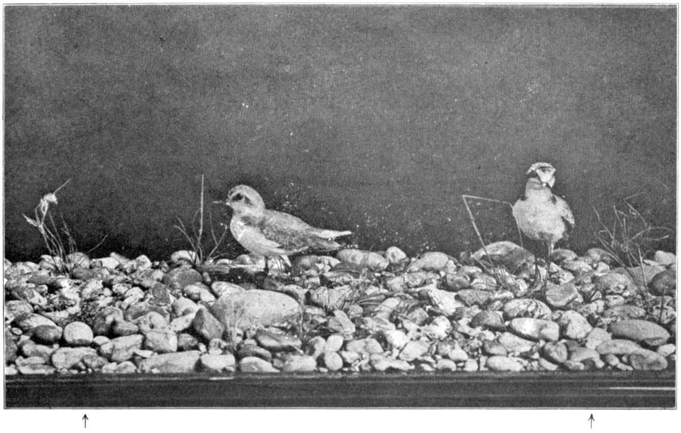
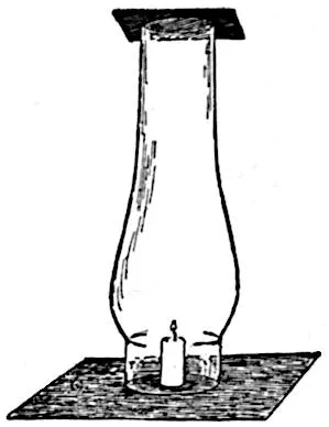
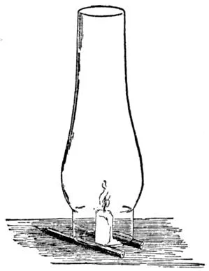
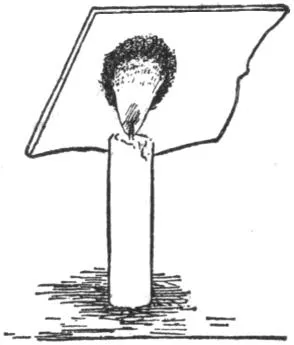
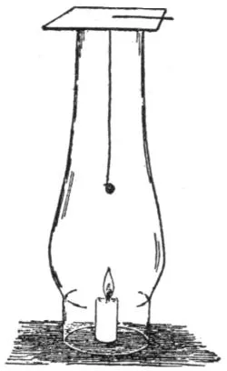
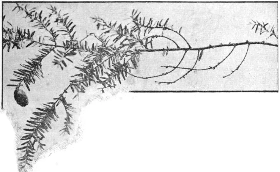
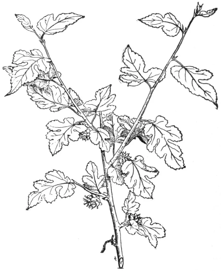
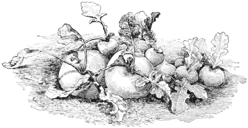
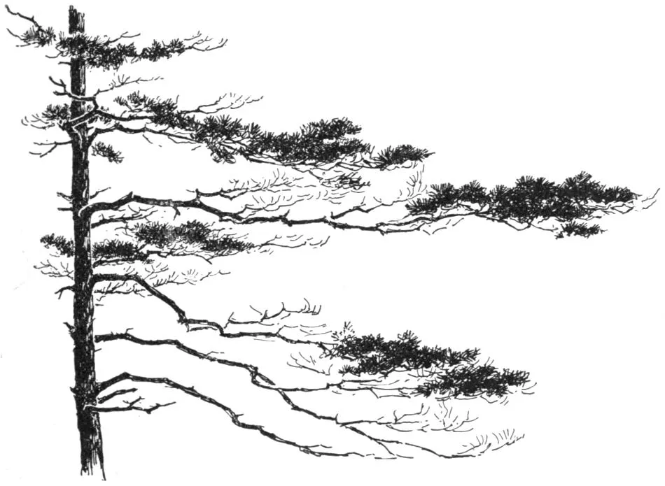
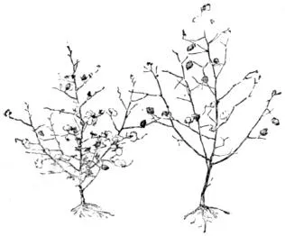
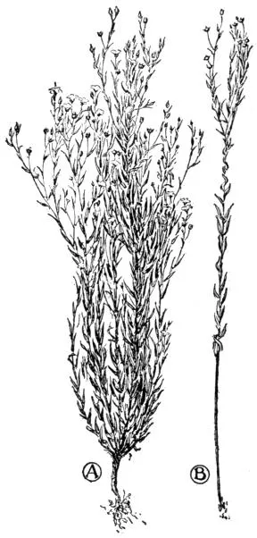
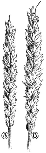
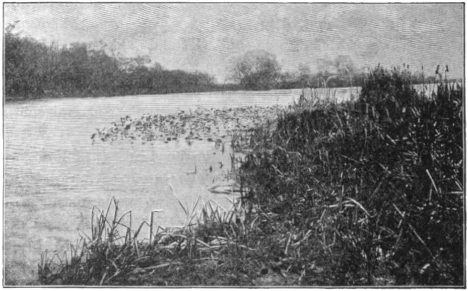
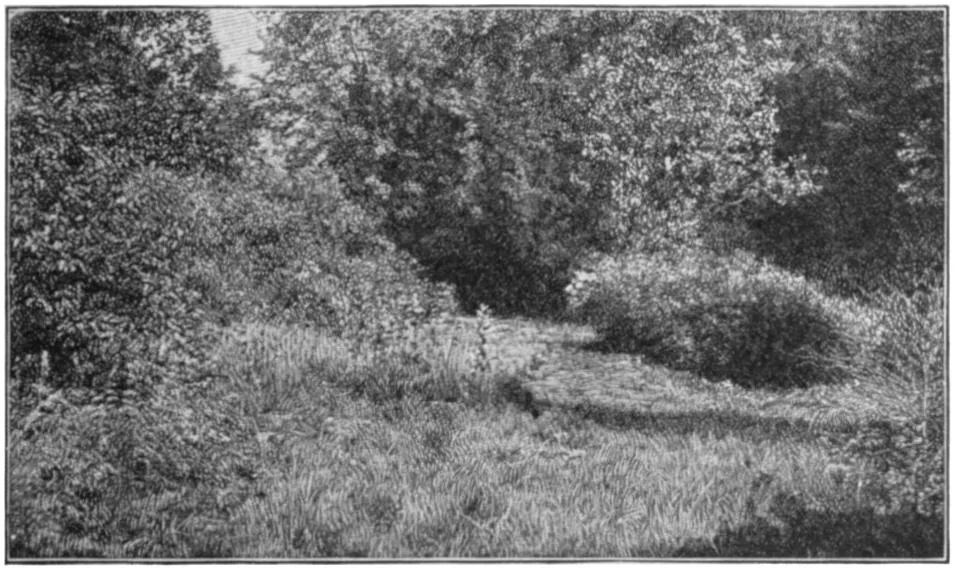
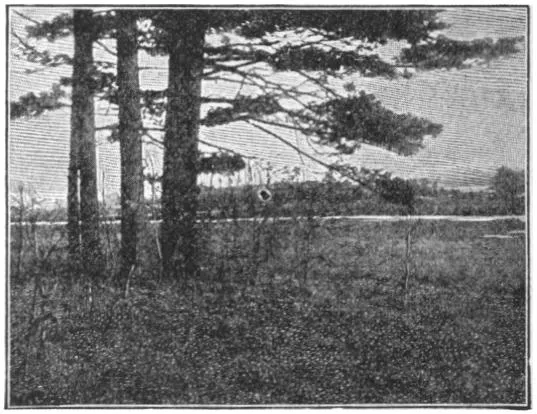
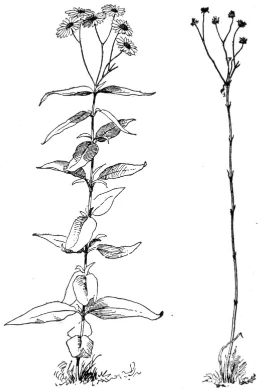
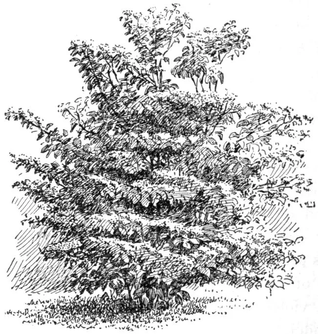
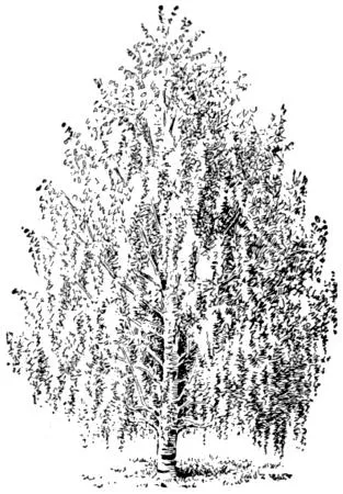
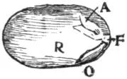
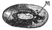
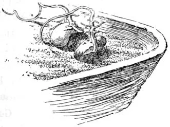
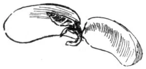
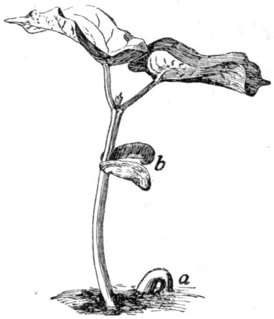
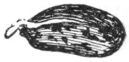
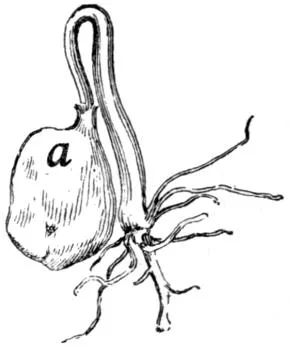
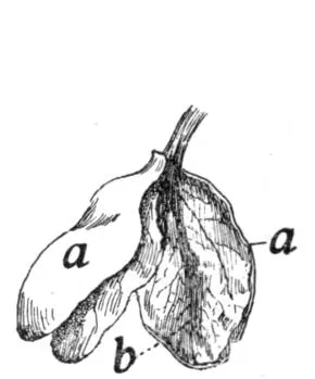
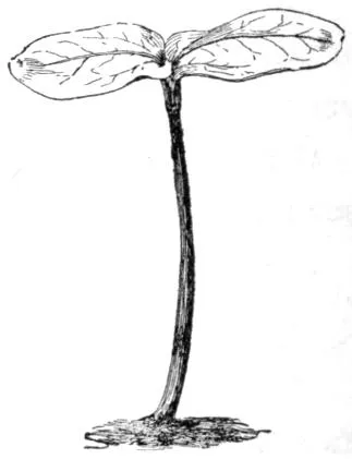
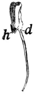
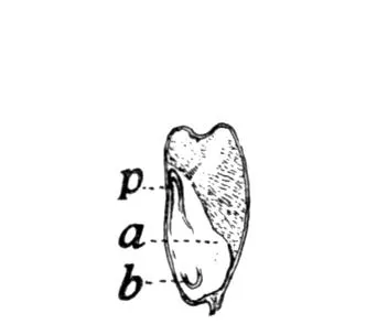
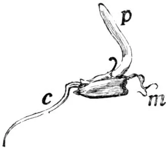
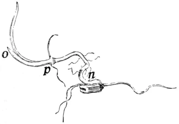
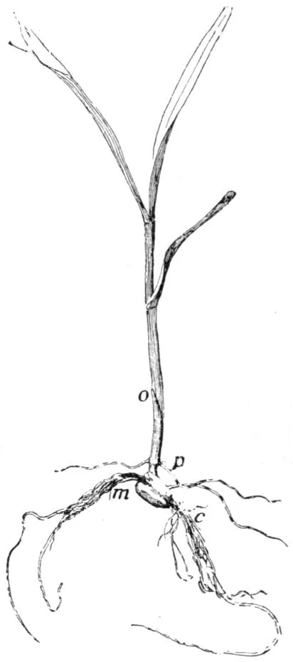
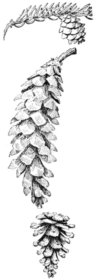
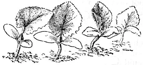
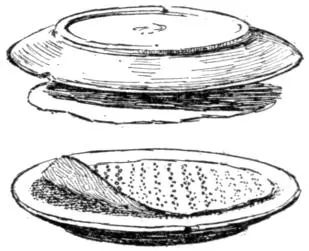
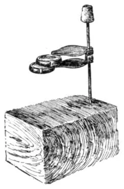

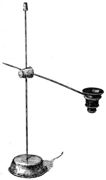
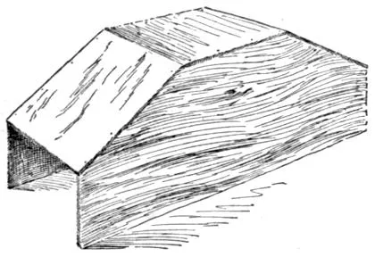
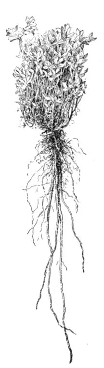
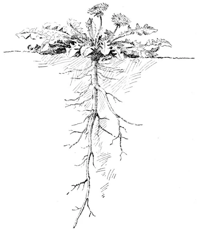
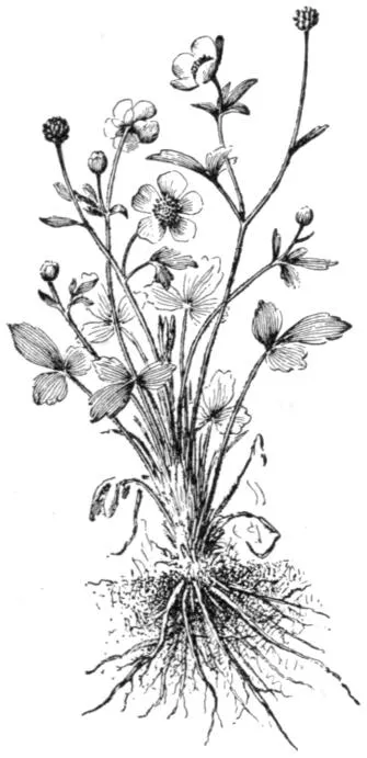
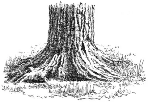
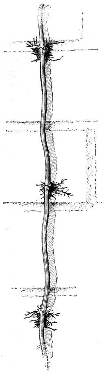
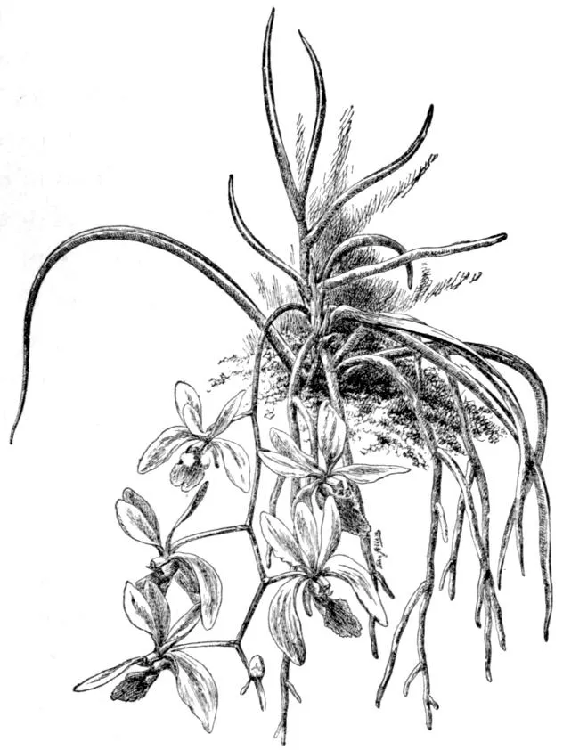
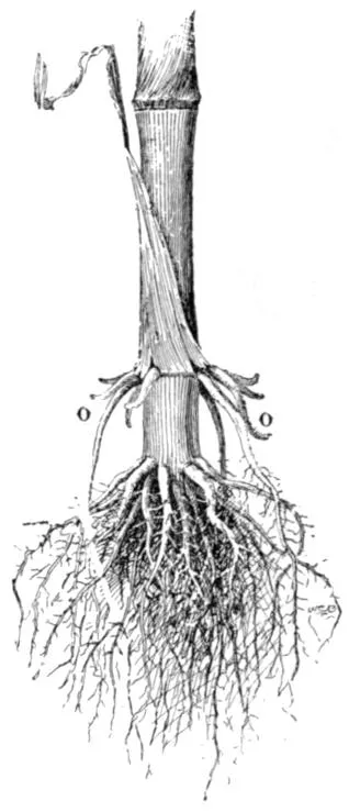
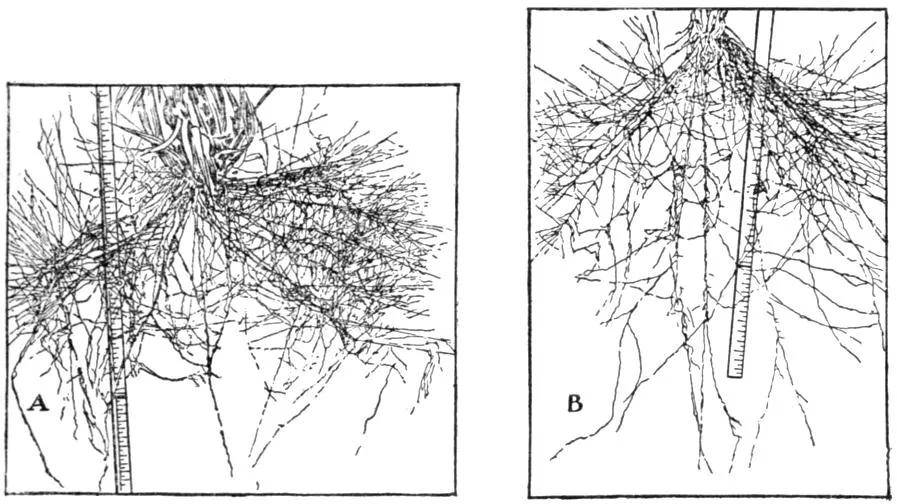
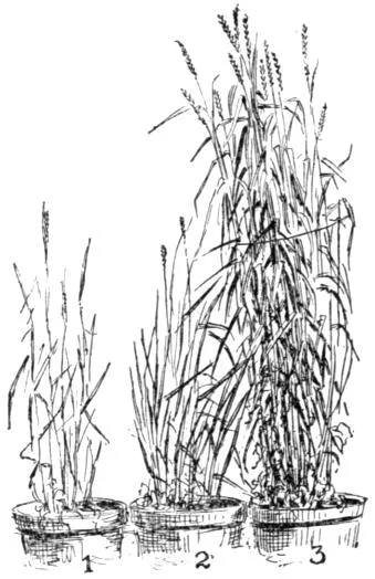
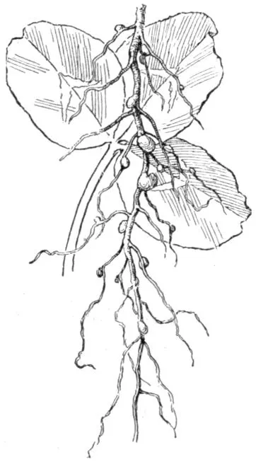
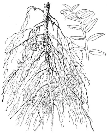
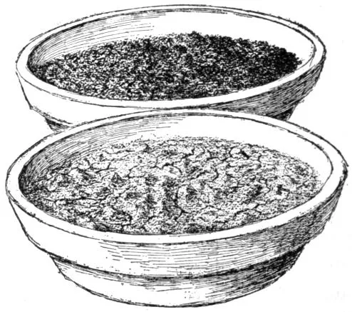
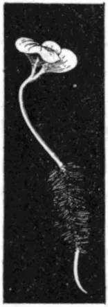
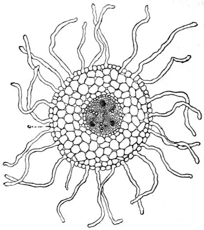
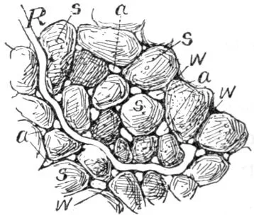
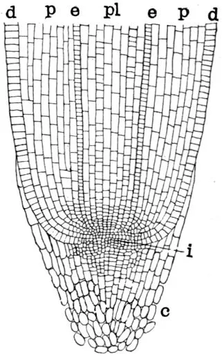
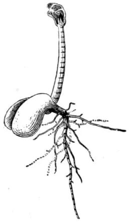
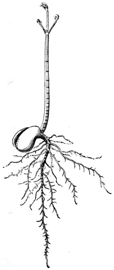
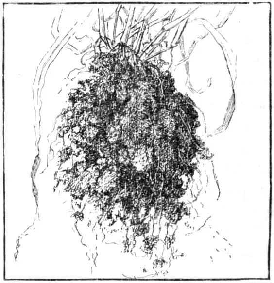
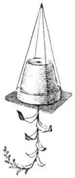
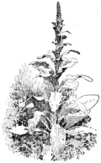
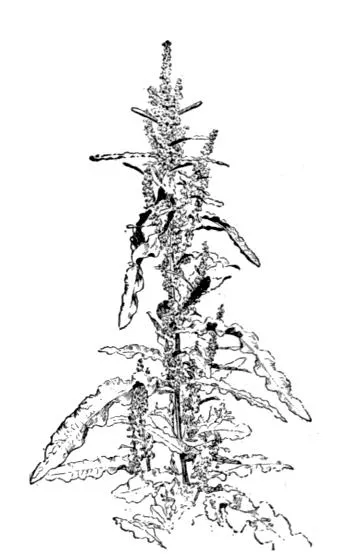
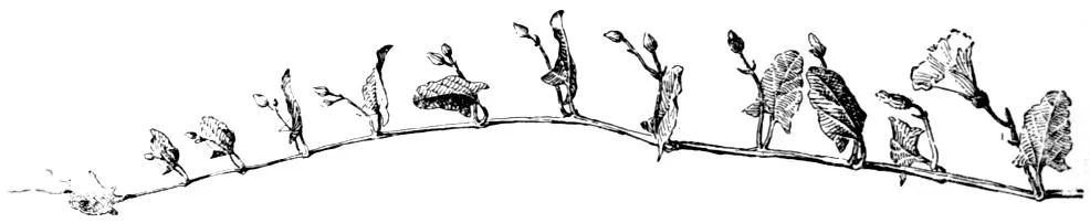
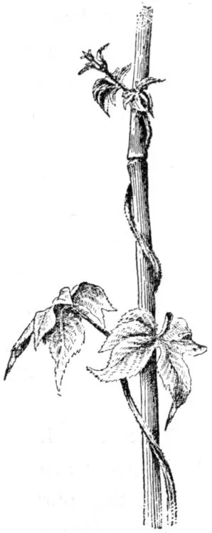
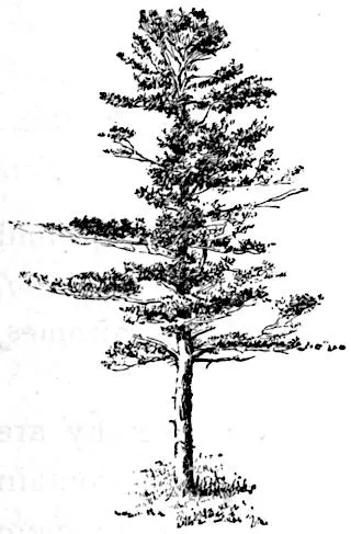
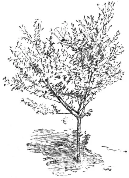
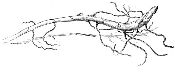
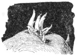
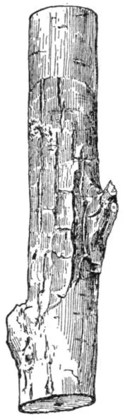
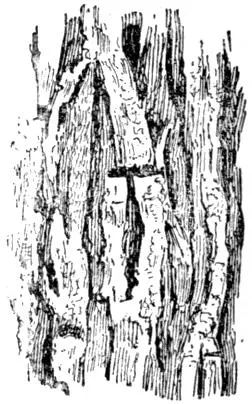
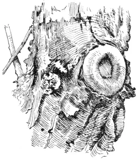
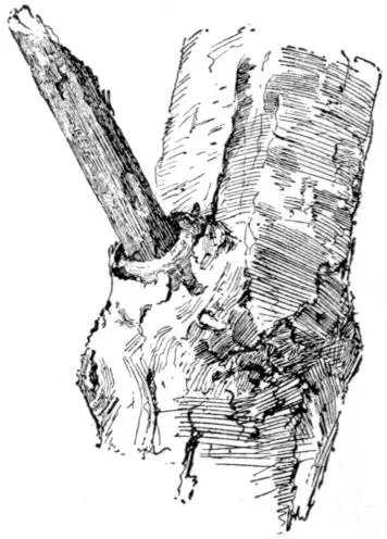
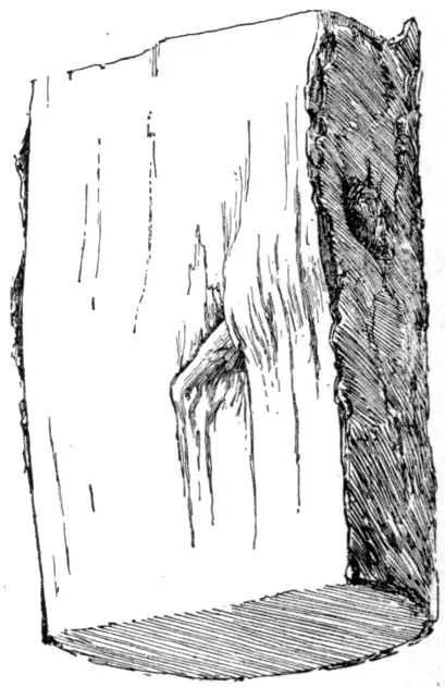
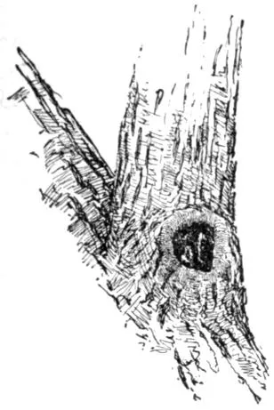
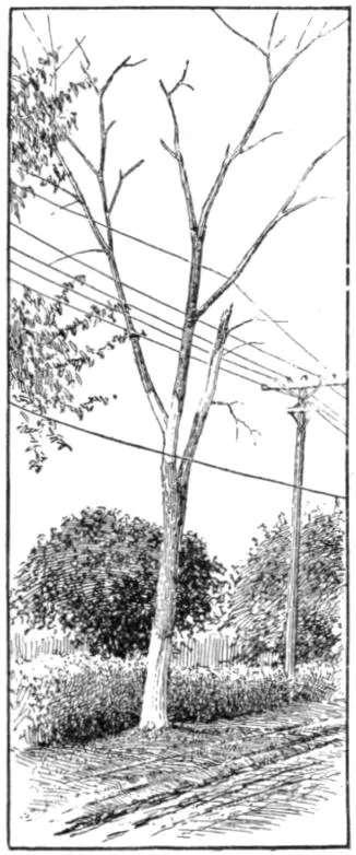
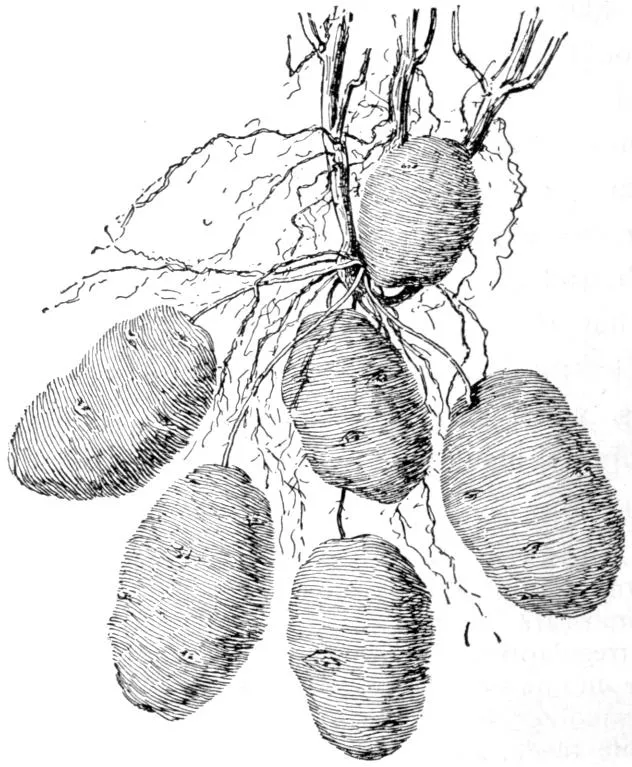
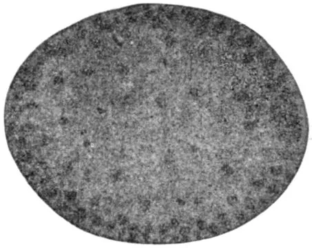
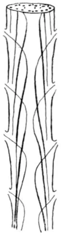
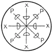
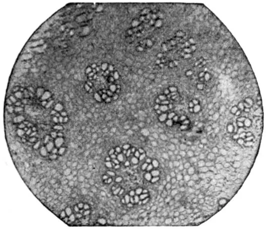
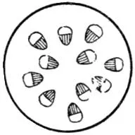
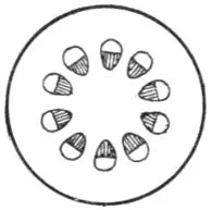
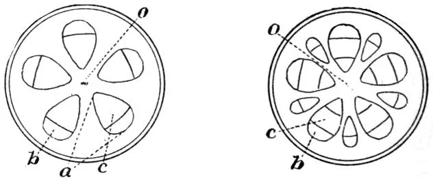
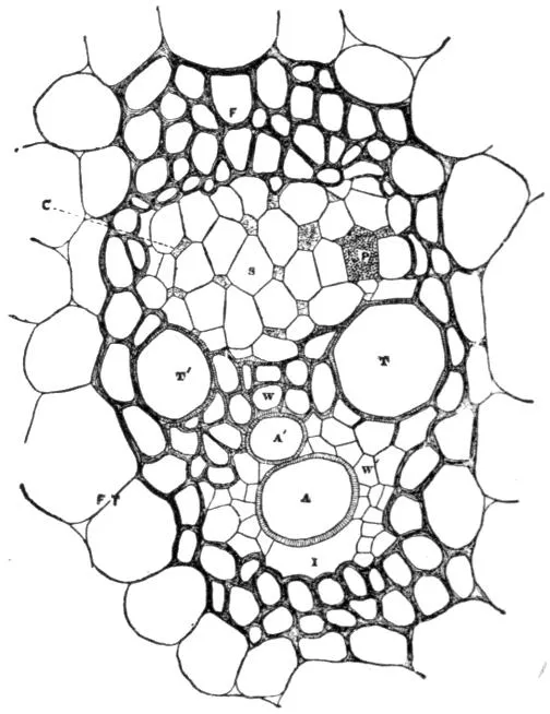
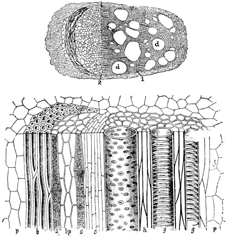
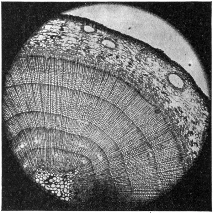
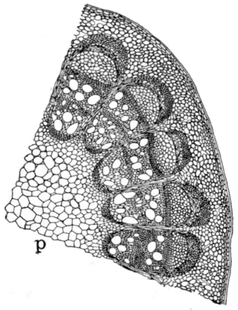
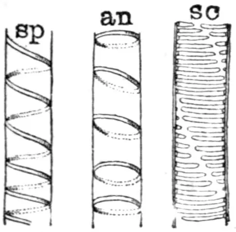
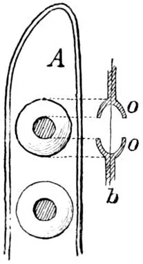
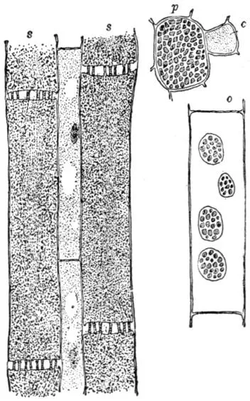
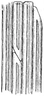
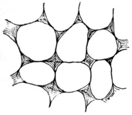
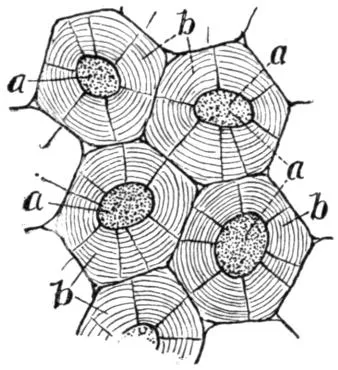
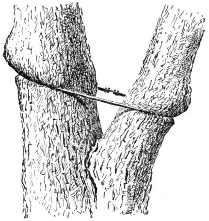
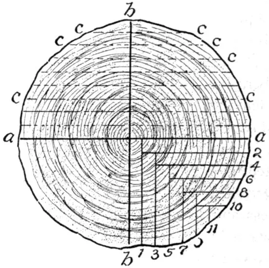
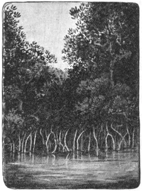
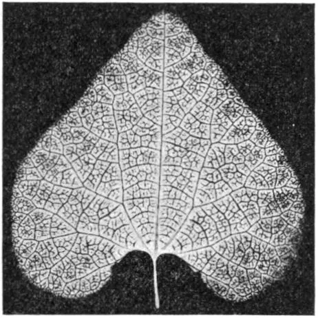
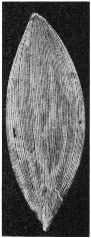
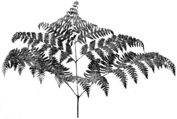
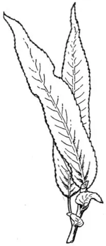
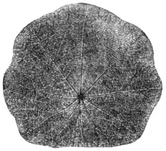
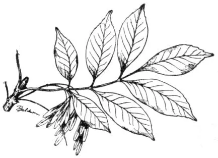
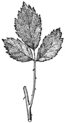
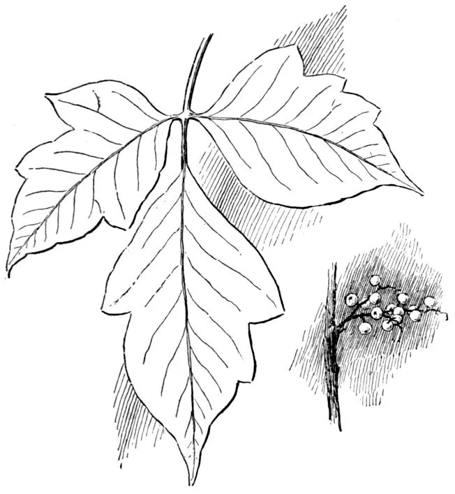
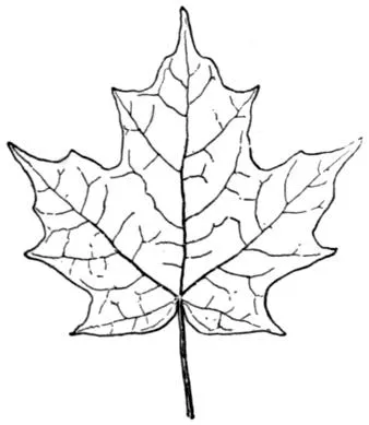
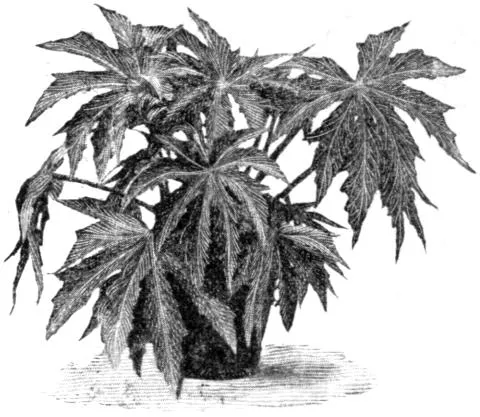
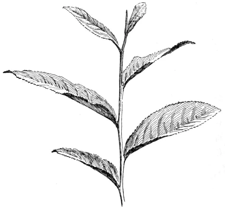
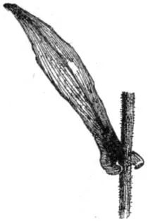
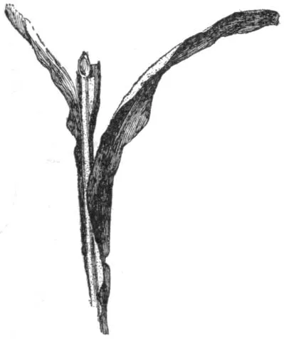
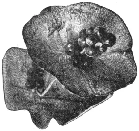

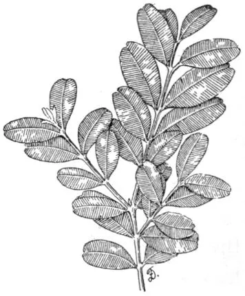
 Linear, several times longer than broad, with the sides
nearly or quite parallel. Spruces and most grasses
are examples (
 Oblong, twice or thrice as long as broad, with the sides
parallel for most of their length.
 Elliptic differs from the oblong in having the sides gradually
tapering to either end from the middle. The
European beech (
 Lanceolate, four to six times longer than
broad, widest below the middle, and
tapering to either end. Some of the
narrow-leaved willows are examples.
Most of the willows and the peach
have oblong-lanceolate leaves.
 Spatulate, a narrow leaf that is broadest
toward the apex. The top is usually
rounded.
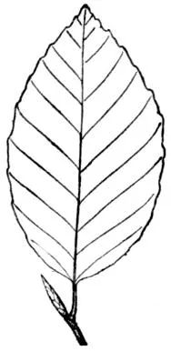
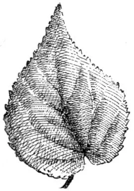
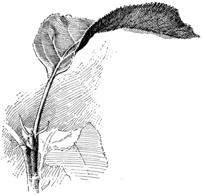
 Ovate, shaped somewhat like the longitudinal section of an
egg: about twice as long as broad, tapering from near
the base to the apex. This is one of the commonest
leaf forms (
 Obovate, ovate inverted,—the wide part towards the apex.
Leaves of mullein and leaflets of horse-chestnut and
false indigo are obovate. This form is commonest
in leaflets of digitate leaves: why?
 Reniform, kidney-shaped. This form is sometimes seen in
wild plants, particularly in root-leaves. Leaves of
wild ginger are nearly reniform.
 Orbicular, circular in general outline. Very few leaves are
perfectly circular, but there are many that are
nearer circular than any other shape (