CHAPTER IV THE PATHOLOGY AND BACTERIOLOGY OF PNEUMONIA FOLLOWING INFLUENZA — Bronchitis — Lobar Pneumonia — Bronchopneumonia — Peribronchial Hemorrhage and Pneumonia — Suppurative Pneumonia With Necrosis and Abscess Formation — Interstitial Suppurative Pneumonia — Suppurative Pneumonia with Multiple Clustered Abscesses Caused by Staphylococci — Empyema, Pericarditis and Peritonitis — Bronchiectasis — Unresolved Bronchopneumonia
107
E. L. Opie, M.D.; F. G. Blake, M.D.; and T.M. Rivers, M.D.
Many observers have described isolated phases of the
recent epidemic and of past epidemics of influenza. Few
have had an opportunity to follow the pathology of influenza
from the onset of an epidemic through a period of several
months and to observe the succession of acute and
chronic changes which occur in the lungs. Our commission
arrived on September 5, 1918, at Camp Pike two weeks before
the outbreak of influenza. The commission had previously
made a careful study of the clinical course, the bacteriology
and to a limited extent the pathology of pneumonia
occurring at Camp Funston where there was little
if any influenza. Study of the records preserved at the
base hospital at Camp Funston had convinced us that this
camp had passed through an epidemic of influenza during
the spring of 1918, this epidemic being followed by
a very severe outbreak of pneumonia. Our investigation
at Camp Funston had brought to our attention those
phases of pneumonia which with the facilities of a base
hospital laboratory could be most profitably studied with a
view to determining the causation, the epidemiology and
the prevention of the pneumonias prevalent in the American
army.
Study of pneumonia after death offers the only opportunity
of determining the relation of pulmonary lesions
to the considerable variety of microorganism associated
with them. Clinical diagnosis furnishes no certain criterion
for distinguishing lobar and bronchopneumonia; suppurative
108pneumonia is rarely recognizable during life.
The relation of pneumococci, streptococci, staphylococci or
B. influenzæ to one or other type of pneumonia can be determined
with accuracy only after death; for the demonstration
of one or more of these microorganisms in material
obtained from the upper respiratory passages in life,
though of value, furnishes us no definite evidence that the
organism which has been identified has entered the lung
and passed from the bronchi to produce pneumonia.
Study of autopsies following examination of the sputum
during life has shown that an individual primarily attacked
by influenza may suffer with a succession of pneumonias,
one microorganism having prepared the way for another.
The complexity of the subject is much increased by the
truth that pyogenic microorganisms, like the tubercle
bacilli, are capable of producing a considerable variety of
pulmonary lesions.
Examination of the lungs of a large number of individuals
who have died as the result of pneumonia following influenza
has disclosed a succession of acute and chronic diseases.
Immediately succeeding the height of the epidemic
of influenza, death occurred with acute lobar pneumonia
or with diffusely distributed hemorrhagic bronchopneumonia
caused in the majority of instances by Pneumococcus
IV in association with B. influenzæ. Superimposed infection
with hemolytic streptococci increased in frequency and
in individuals who had occupied certain wards was almost
invariable. At a later period, from one to two months following
the maximum incidence of influenza chronic lesions,
namely, bronchiectasis, unresolved pneumonia, and chronic
empyema were common and often occurred as the result
of influenza which had had its onset at the height of the epidemic.
When influenza attacked the encampment, about 50,000
troops were quartered in it, and for a considerable period
no more troops were brought into the camp and none left
109it. All cases of pneumonia occurring among these troops
were brought to the base hospital so that the autopsies
which were studied were representative of all the pneumonias
following influenza in this limited group of men.
It is noteworthy that autopsy disclosed no instance of fatal
influenza unaccompanied by pneumonia.
Pneumonia of Influenza.—Knowledge concerning the bacteriology
of the pneumonia of influenza dates from the
study of the epidemic of 1889–90. The frequency with
which Diplococcus lanceolatus occurred in association with
influenzal pneumonia was well recognized, although several
observers, notably Finkler[57] and Ribbert,[58] found Streptococcus
pyogenes so often that they attributed the pneumonia
of influenza to this microorganism.
During a subsidiary outbreak of influenza occurring in
1891–92 Pfeiffer[59] discovered the microorganism which he
believed was the cause of the disease. Pneumonia, he believed,
was caused by the invasion of this microorganism
into the lung, and the pneumonia of influenza, if death occurred
at the height of the disease, was characterized not
only by the presence of the bacillus of influenza, but was
recognizable by its anatomic peculiarities. He described
lobular patches of consolidation which were separated from
one another by air containing tissue or were confluent, so
that, although the lobular character was still recognizable,
whole lobes might be affected. The consolidated tissue
was dark red and within each lobular area were small, yellowish
gray spots varying in size from that of a pinhead to
a pea. In the mucus of the larynx and trachea were numerous
microorganisms, including diplococci and streptococci,
among which influenza bacilli were predominant; in
the larger bronchi, bacteria other than influenza bacilli
were less abundant, whereas in the finer bronchi filled with
110purulent fluid and in the lung tissue influenza bacilli had
undivided sway. Pfeiffer stated that the changes described
were found when death occurred at the height of
the disease, whereas other pulmonary lesions might be
sequelæ of this typical influenzal pneumonia.
Observations upon the pathology of influenzal pneumonia
made during the epidemic of 1889–92 have been collected
in the monograph of Leichtenstern[60] published in 1896. He
combats the opinion held by some observers that pneumonia
with influenza is always catarrhal and cites many writers
to prove that lobar pneumonia not infrequently accompanies
the disease. Indeed, some have found “croupous”
pneumonia more often than “catarrhal.” Krannhals[61]
at Riga found typical fibrinous pneumonia in 53 instances,
doubtful forms in 22 and bronchopneumonia in 37. Cruickshank[62]
in England found croupous forms predominant.
Among 43 autopsies performed upon individuals dead with
influenza Birch-Hirschfeld[63] found 11 instances of croupous
lobar pneumonia, 8 instances of croupous lobular pneumonia
and 24 instances of catarrhal pneumonia. Leichtenstern
thinks that the atypical symptomatology of lobar
pneumonia with influenza—for example, the purulent character
of the sputum—has led many physicians to believe
that lobar pneumonia rarely occurs. It is equally true that
many instances of confluent lobular pneumonia are mistaken
for lobar.
There appears to be no comprehensive description of the
pneumonias of influenza based upon the epidemics of 1889–92.
Descriptions dating from this period are much obscured
by attempts to separate croupous or fibrinous from
catarrhal pneumonias. Croupous lobular pneumonia has
been recognized, for example, by Birch-Hirschfeld. Leichtenstern
describes a form of pneumonia which he regards as
111neither lobar nor lobular although it implicates whole
lobes; the consolidated tissue is homogeneous and varies
in color from fleshy red to bluish red; it is tough and elastic
in consistency. The author thinks that it is an error to
regard this lesion as a confluent lobular pneumonia.
Kuskow[64], who has discussed the pathology of influenza
in considerable detail, has seldom seen lobar pneumonia but
has almost invariably found, even when there is lobar distribution
of the lesion, lobular patches of consolidation involving
groups of lobules, single lobules or only parts of
lobules; the lung tissue has been hyperemic and in places
edematous.
Opinions concerning the pathology and bacteriology of
the pneumonias of influenza, published since the recent
epidemic, have varied almost as much as those just cited.
Few observers have had the opportunity of making a considerable
number of observations under conditions which
determine the relation of the pulmonary lesions to the primary
disease.
Keegan[65] has found with influenza a massive and confluent
bronchopneumonia, frequently resembling lobar pneumonia
but distinguishable particularly in its early stages
when the cut section of the consolidated lung has a finely
granular character and each bronchiole stands out as a
grayish area with intervening dark red alveolar tissue.
Symmers[66] states that the pneumonia of influenza is a
confluent lobular exudative and hemorrhagic pneumonia
which bears a close resemblance to the pneumonic variety
of bubonic plague.
Our commission[67] published a preliminary report on
pneumonia following influenza observed at Camp Pike.
The occurrence of purulent bronchitis, distention of lungs,
112peribronchial hemorrhage and bronchiectasis were described.
B. influenzæ was isolated from the bronchi in approximately
85 per cent of instances of influenzal pneumonia
but from the consolidated lung in less than half this
number of autopsies. Lobar pneumonia was present in a
large proportion of autopsies but was less frequent than
bronchopneumonia. Bronchopneumonic consolidation is in
part red, lobular and confluent, in part nodular; pneumococci
have a predominant part in the production of these
lesions. Three types of suppurative pneumonia are described:
(a) Abscess caused by hemolytic streptococci
usually in contact with the pleura and accompanied by empyema;
(b) Suppuration of interstitial tissue of the lung
caused by hemolytic streptococci and accompanied by empyema;
(c) multiple foci of suppuration clustered about a
bronchus of medium size and caused by staphylococci. We
have expressed the opinion that B. influenzæ descends into
the bronchi; pneumococci, usually Type IV, may enter the
lung and produce either lobar or bronchopneumonia.
Hemolytic streptococci may descend and infect the pneumonic
lung causing death often before suppuration has occurred.
Epidemics of such superimposed streptococcus
pneumonia in wards of the hospital were described.
LeCount[68] says: “The pneumonia of influenza is commonly
referred to as bronchopneumonia. It may be so designated,
but it differs from other bronchopneumonias in its
predilection for the periphery of the lungs and in the extent
to which the inflammation is hemorrhagic.”
MacCallum[69] states that the following types of pneumonia
following influenza may be recognized. 1. Pneumococcus
pneumonia. The lobular character of the consolidation is
in these cases well marked, although it tends to lose its
definiteness through the confluence of adjacent areas. The
cut surface of the lung shows in the more acute cases a
113peculiar lobular or confluent consolidation which corresponds
well with what is commonly written of the stage
of engorgement in the description of lobar pneumonia.
Later stages in the pneumonia show within these areas
patches of rough gray consolidated tissue from which definite
plugs of exudate project. 2. Staphylococcal pneumonia.
3. Streptococcal pneumonia. There is lobular pneumonia,
the interlobular septa are edematous and, microscopically,
bronchi and alveoli are loaded with streptococci.
Whole areas of lung, though retaining their form, are entirely
necrotic. Lymphatics are distended with exudate
containing streptococci in great numbers, but in none of
these cases is interstitial bronchopneumonia found. 4.
Pneumonia produced by B. influenzæ of Pfeiffer. The lung
is studded with palpable shot-like grayish yellow nodules;
the bronchi exude thick yellow pus, which contains influenza
bacilli. Microscopically, the walls of the bronchi
are found to be thickened by mononuclear infiltration and
new formation of connective tissue. The walls of the alveoli
are thickened and indurated and the alveoli often contain
fibrin in process of organization. Absence of conspicuous
changes in lymphatics, absence of intense pleural infection
and relatively scant numbers of polynuclear leucocytes
in the exudate within the alveoli and bronchial walls
are, MacCallum states, all that distinguish this pulmonary
change from the interstitial bronchopneumonia caused by
the hemolytic streptococcus and described by him in
previous papers.
Lyon[70] designates the pulmonary lesion following influenza,
hemorrhagic pneumonitis, the lung tissue containing
serous fluid loaded with red blood corpuscles; in
many instances there was such scant consolidation that the
process could not be regarded as a true pneumonia. In 35
instances the lesion was lobular in distribution and in 16
114instances was sufficiently extensive to be designated lobar,
but it was not typical lobar pneumonia, and, often associated
with lobular patches of consolidation, appeared to be
a confluent lobular pneumonia.
Goodpasture and Burnett[71] say: “The difficulties of analyzing
the pulmonary lesions in any group of influenza
pneumonias as they have appeared in this epidemic, are
very apparent to anyone who has had an opportunity to observe
the bacteriology and pathology of this accompaniment
of the disease.” There is an acute outflow of the
fluid elements of the blood and of hemorrhage into the lung
tissue filling the alveoli in lobular areas and not infrequently
in an entire lobe. By a special method of staining
these authors have studied the distribution of Gram-negative
bacilli with the morphology of B. influenzæ. The fact
that in certain very early cases demonstrable bacteria of
any kind are scarce or not found at all, has lead them to
believe “notwithstanding the demonstration of influenza
bacilli in pure culture in the lung in all but one instance,
that at this stage organisms are comparatively few within
the alveoli, and the primary injury is due to a very potent
toxic agent elaborated in and disseminated through the
larger air passages. In the later stages or from the beginning,
if the injury be slight, the infection focalizes about
the bronchi or their terminations, so that the bronchial and
lobular distribution becomes very conspicuous.” Typical
lobar pneumonia with “croupous” exudate within the alveoli
occurs in cases complicated by secondary pneumococcus
infection.
Wolbach[72] has found that two types of pneumonia are
characteristic of influenza. In cases in which death has
occurred within a few days after onset of pulmonary signs,
the lung tissue is dark red and “meaty in consistency” and
contains abundant blood-tinged serous fluid which drips
115from the cut surface. The other type of lesion is found in
patients who have lived for ten days or more after onset
of the disease; there is extensive bronchitis, bronchopneumonia,
discrete or confluent, and peribronchitis. The lungs
are voluminous and the smaller bronchi are distended.
Microscopically, there is peribronchitis with extensive infiltration
of the interlobular septa and organization in alveoli
and bronchioles. This lesion is that designated by
MacCallum as “interstitial bronchopneumonia.” Wolbach
thinks that the two types of lesion represent different
stages of the same process. He regards as distinctive of
the pneumonias of influenza the presence of hyalin fibrin
lining distended air spaces. With the two types of lesion
which have been described, B. influenzæ was the only organism
which could be cultivated, and the author associates
these distinctive conditions with that microorganism.
Streptococcus Pneumonia.—Finkler emphasized the importance
of streptococcus pneumonia as a complication of
influenza. In 1888 he described instances of acute primary
streptococcus pneumonia observed in 1887 and 1889. This
form of pneumonia, Finkler thought, occurred in Bonn in
epidemic form before the influenza epidemic of 1889–90
and, he states, exhibited an astonishing similarity to the
pneumonia of influenza. He thought that later there was
in Bonn a mixed infection of influenza and primary streptococcus
pneumonia. In one type of streptococcus pneumonia,
described by Finkler, there was lobular consolidation
which in multiple patches produced “pseudolobar” consolidation;
the consolidated lung was smooth and red, and
similar to spleen, rather than hepatized. In another group
of instances the lesion merited the name “erysipelas of
the lung.” The lesion was an acute interstitial pneumonia
in some places, a cellular or occasionally fibrinous pneumonia
with involvement of the interstitial tissue in other
places. There was edematous swelling and accumulation of
leucocytes in the interstices between the alveoli and about
116the blood vessels and bronchi. Finkler stated that the similarity
to erysipelas might suggest that the lymphatics contain
streptococci, but this relation did not exist although
large lymphatic channels were occasionally filled with coagulum.
He asserted that the disease was contagious and
cited cases which he believed had their origin in hospital
wards.
The widespread occurrence of streptococcus pneumonia
in the army camps in this country attracted attention during
the first months of 1918. Cummings, Spruit and
Lynch[73] at Fort Sam Houston, Texas, recognized the prevalence
of streptococcus pneumonia, both as a complication
of measles and in association with lobar pneumonia, and
showed that the microorganism concerned was a hemolytic
streptococcus. In 7 autopsies upon individuals with lobar
pneumonia, they found pneumococcus alone in 2 instances
and pneumococcus and hemolytic streptococcus or hemolytic
streptococcus alone in 5 instances. Hemolytic streptococcus
was found in all of 24 instances of bronchopneumonia,
three-fourths of which followed measles. They recommend
the bacteriologic examination of the throat of
patients with measles and the segregation of those who
harbor hemolytic streptococci.
Cole and MacCallum[74] have published almost simultaneously,
with that just cited, a report upon the pneumonia
which has occurred at Fort Sam Houston and have shown
the importance of hemolytic streptococci in its causation.
They have found two varieties of pneumonia, namely, acute
lobar pneumonia which does not differ essentially from
that which occurs elsewhere and bronchopneumonia which
in most cases has followed measles and is caused by S. hemolyticus.
They think this infection is usually acquired in
the hospital. They believe that the pulmonary lesions are
characteristic. In this publication and elsewhere MacCallum
117has designated the lesion “interstitial bronchopneumonia.”
The epidemic of streptococcus pneumonia and empyema
occurring at Camp Dodge, Iowa, from March 20 to May 10
is described by Miller and Lusk[75]. During this period there
were 400 cases of pneumonia, whereas from September 20,
1917, to March 20 there had been only 276 instances of lobar
pneumonia. The type of pneumonia changed, there was
more severe intoxication and empyema became very frequent;
in 85 of 95 exudates streptococci were found. The
outbreak of pneumonia bore no relation to measles. The
authors state that a mild tracheitis was prevalent in the
cantonment during March, and whenever a large group of
soldiers congregated coughing was noticeable.
MacCallum[76] studied the pneumonia at Camp Dodge during
May and found 17 instances of the lesion which he had
designated interstitial bronchopneumonia; of these, 9 followed
measles, although in the earlier part of the epidemic
there appear to have been, he states, few such cases. Cultures
made at autopsy, except in a few fatal cases of uncomplicated
lobar pneumonia caused by the pneumococcus,
showed the hemolytic streptococcus in every situation
throughout the respiratory tract and pleura.
The pneumonia which occurred at Camp Funston is of
special interest to our commission because we were for a
time stationed at this camp and had the opportunity of
following in the excellent records of the hospital the history
of the occurrence of pneumonia during the year following
the establishment of the camp in September, 1917. Stone,
Phillips and Bliss[77] have described the outbreak of pneumonia
which occurred in March, 1918. At this time there
was, the authors state, severe pneumonia with frequent
empyemas due to hemolytic streptococci. This condition
118which did not follow measles was responsible for the
greatly increased death rate in March; 9 deaths occurred
in February, 45 in March, 25 in April and 14 in May. They
found during March 26 instances of multiple pulmonary
abscess. In 29 autopsies they found a pleural lesion which
they designate “subcostosternal pus pockets”; it occurs
only in association with empyema caused by hemolytic
streptococci.
Our commission[78] has shown that an epidemic of influenza,
well characterized by its epidemiology and symptoms,
preceded and accompanied the outbreak of pneumonia
just described. Between March 4 and 29 1,127 men
from Camp Funston, which then contained 29,000 men, were
sent to the base hospital with influenza and many more
were treated in the infirmaries of the camp; on March 11
107 patients with influenza were admitted to the hospital.
The greatest incidence of pneumonia in the history of the
encampment up to this time occurred between March 9 and
29, immediately following the outbreak of influenza, the
maximum incidence of pneumonia occurring five days after
the maximum for influenza.
The foregoing observations are cited to prove that streptococcus
pneumonia, which occurred during the spring of
1918 at Camp Funston and doubtless at other camps, had
its origin in influenza and did not differ in character from
that which occurred on a much larger scale in the fall of
1918.
Table of Autopsies.—In order to present as briefly as
possible the data upon which the present study has been
based, autopsies have been assembled in tabular form in
the order of their performance (Table XXVII). During the
early period of the epidemic autopsies were performed on
all who died with pneumonia, but later, with increase in the
number of deaths, this became impossible and autopsies
were performed on all those who died in certain wards.
119Comparison of charts representing incidence of influenza
and of deaths from pneumonia furnishes evidence that fatal
pneumonia during the period of investigation was with
few exceptions referable to influenza. During two weeks,
namely, from September 1 to 14, before the presence of the
epidemic was evident, there were only 2 fatal cases of
pneumonia. In most instances the relation of pneumonia
to influenza is established by a definite history of influenza
having its onset during the epidemic. Bronchopneumonia
usually develops gradually as a sequence of influenza in
which purulent bronchitis has occurred. Lobar pneumonia
may develop in cases of influenza complicated by purulent
bronchitis. In some instances there is apparent recovery
from influenza indicated by return of temperature to normal;
after from one to three days of normal temperature
there is typical lobar pneumonia with rusty sputum. In
many instances of pneumonia having their onset at the
height of the epidemic of influenza, the history indicates
that pneumonia was present immediately after the onset
of symptoms, so that the onset resembled that of pneumonia
rather than of influenza.
Cases of pneumonia following measles have been excluded
from the table in order that they may be studied
separately and compared with the pneumonias of influenza.
It is noteworthy that the lesions of pneumonia following
measles have shown a very close resemblance to the pneumonias
of influenza, with regard both to pathologic characters
and to bacteriology.
Five instances of pneumonia following typhoid fever
(Autopsies 245 and 329), scarlet fever (Autopsy 311) or
mumps (Autopsies 403 and 417) have been excluded from
the table. These secondary pneumonias are grouped as
an appendix to the section on pneumonia following measles.
It is not improbable that individuals with the diseases
named are just as susceptible as others to influenza.
Included in the table is an instance (Autopsy 487) in which
a definite attack of influenza preceded scarlet fever.
120
| Table XXVII |
|---|
| |
| NO. OF AUTOPSY |
RACE |
LENGTH OF MILITARY SERVICE |
DURATION OF ILLNESS |
DURATION OF PNEUMONIA |
CLINICAL DIAGNOSIS |
PURULENT BRONCHITIS |
LOBAR PNEUMONIA |
PERIBRONCHIOLAR CONSOLIDATION |
HEMORRHAGIC PERIBRONCHIOLAR CONSOLIDATION |
LOBULAR CONSOLIDATION |
PERIBRONCHIAL CONSOLIDATION |
ABSCESS |
INTERSTITIAL SUPPURATIVE PNEUMONIA |
MULTIPLE ABSCESSES IN CLUSTERS |
EMPYEMA |
BRONCHIECTASIS |
UNRESOLVED BRONCHOPNEUMONIA |
ORGANIZING BRONCHITIS |
BACTERIA IN SPUTUM |
BACTERIA IN BRONCHUS |
BACTERIA IN LUNG |
BACTERIA IN BLOOD OF HEART |
| 229 |
W |
1m |
12+ |
12+ |
L |
P |
+ |
|
|
|
|
|
|
|
|
|
|
|
Pn. I. B. inf. |
|
|
0 |
| 231 |
W |
2m |
13 |
4 |
B |
P |
|
|
|
+ |
|
|
|
|
|
|
|
|
|
|
|
|
| 232 |
W |
14d |
9 |
6? |
B |
|
+ |
|
|
|
|
|
|
|
|
|
|
|
Pn. IIa. B. inf. |
|
Pn. IIa. |
Pn. IIa. |
| 233 |
C |
11m |
5 |
2 |
L |
|
|
|
|
+ |
|
|
|
|
|
|
|
|
Pn. IIa. |
|
|
Pn. |
| 236 |
W |
3w |
8 |
7+ |
L |
|
+ |
|
|
|
|
|
|
|
|
|
|
|
Pn. IV. B. inf. |
|
B. inf. Staph. |
0 |
| 237 |
W |
10d |
8 |
2 |
L |
|
|
|
+ |
+ |
|
N |
|
|
E |
|
|
|
St. h. |
|
|
St. h. |
| 238 |
W |
2d |
8 |
5 |
L |
|
+ |
|
|
|
|
|
|
|
|
|
|
|
Pn. IV. B. inf. |
|
|
Pn. IV |
| 239 |
W |
11d |
9 |
5 |
L |
|
+ |
|
|
|
|
|
|
|
|
|
|
|
Pn. II. |
|
Pn. II. B. inf. |
Pn. II. |
| 240 |
W |
13d |
8 |
4 |
L |
|
+ |
M |
|
+ |
|
|
|
|
|
|
|
|
Pn. IV. |
B. inf. |
|
Pn. IV. |
| 241 |
W |
14d |
5 |
5 |
L |
|
+ |
|
|
|
|
|
|
|
|
|
|
|
Pn. IV. B. inf. |
|
|
Pn. IV. |
| 242 |
C |
14d |
7 |
4 |
L |
P |
|
|
+ |
+ |
|
|
|
|
|
|
|
|
Pn. IIa. B. inf. |
|
|
Pn. IIa. |
| 243 |
W |
15d |
5 |
5 |
L |
|
+ |
M |
|
|
|
|
|
|
|
|
|
|
|
|
|
Pn. IV. |
| 244 |
C |
1m |
6 |
3 |
B |
P |
|
M |
+ |
+ |
|
|
|
|
|
+ |
|
|
|
Pn. IV. B. inf. |
Pn. IV. B. inf. |
0 |
| 121246 |
C |
2m |
6 |
3 |
L |
P |
+ |
M |
|
|
|
|
|
|
|
|
|
|
Pn. IIa. B. inf. |
|
|
0 |
| 247 |
W |
10d |
10 |
5 |
L |
P |
|
M |
+ |
+ |
|
|
|
|
|
|
|
|
Pn. IV. B. inf. |
|
|
Pn. IV. |
| 248 |
W |
1m |
4 |
2+ |
I |
|
|
|
|
|
|
+ |
|
|
E |
|
|
|
|
St. h. |
St. h. |
St. h. |
| 249 |
W |
15d |
12 |
6+ |
L |
|
|
M |
|
+ |
|
|
|
|
|
|
|
|
|
|
|
Pn. III. |
| 250 |
W |
14d |
11 |
7 |
L |
|
|
M |
|
+ |
|
|
|
|
E |
|
|
|
Pn. IIa. B. inf. |
|
|
Pn. IIa. |
| 251 |
W |
25d |
7 |
1 |
B |
P |
|
|
+ |
+ |
|
+ |
+ |
|
|
|
|
|
|
|
St. h. |
St. h. |
| 252 |
W |
21d |
14 |
12 |
B |
P |
|
M |
|
+ |
|
|
|
|
|
|
|
|
|
Pn. II. B. inf. St. v. |
Pn. II. B. inf. |
0 |
| 253 |
W |
12d |
19 |
12+ |
B |
|
|
M |
|
+ |
+ |
|
|
|
E |
|
|
|
Pn. Ia. |
Pn. II. B. inf. Staph. |
Pn. II. B. inf. |
Pn. II. |
| 254 |
W |
21d |
7 |
6+ |
B |
P |
+ |
M |
|
+ |
|
|
|
|
|
|
|
|
|
Pn. I. B. inf. |
Pn. I. Staph |
0 |
| 255 |
W |
12d |
5 |
4? |
B |
P |
|
M |
+ |
+ |
|
+ |
|
|
E |
|
|
|
|
St. h. Pn. IIa. Staph. |
St. h. |
St. h. |
| 256 |
W |
17d |
8 |
4 |
B |
P |
+ |
M |
|
|
|
|
|
|
|
|
|
|
|
Pn. Staph. |
Pn. IIa. |
Pn. IIa. |
| 257 |
C |
21d |
10 |
7+ |
L |
P |
|
|
|
+ |
|
|
|
|
|
|
|
|
|
B. inf. Staph |
|
Pn. I. |
| 258 |
W |
1m |
6 |
2+ |
B |
|
|
M |
|
+ |
|
+ |
|
|
E |
|
|
|
|
|
St. h. Pn. IV. B. inf. |
St. h. |
| 259 |
W |
3m |
4 |
1 |
B |
|
|
|
|
+ |
|
+ |
+ |
|
|
|
|
|
|
|
St. h. Pn. III. Staph. |
St. h. |
| 260 |
W |
1m |
2 |
1 |
L |
|
+ |
|
|
|
|
|
|
|
|
|
|
|
|
|
|
0 |
| 261 |
W |
2m |
7 |
5+ |
B |
P |
+ |
|
|
|
|
|
|
|
|
|
|
|
|
B. inf. Pn. IV. |
|
0 |
| 262 |
W |
12d |
5 |
4? |
B |
|
|
|
|
+ |
|
+ |
|
|
E |
|
|
|
|
St. h. B. inf. |
St. h. |
St. h. |
| 263 |
W |
21d |
7 |
5 |
L |
P |
|
|
|
+ |
|
+ |
|
|
E |
|
|
|
|
|
St. h. B. inf. Staph. |
St. h. Staph. |
| 264 |
W |
21d |
10 |
7 |
L |
|
+ |
|
|
|
|
|
|
|
|
|
|
|
Pn. IV. Staph. |
St. h. Staph. |
St. h. |
St. h. |
| 265 |
W |
14d |
7 |
6+ |
L |
|
+ |
|
|
|
|
|
|
|
|
|
|
|
|
St. h. Staph. inf. |
Pn. IV. B. inf. |
St. h. |
| 122266 |
W |
1m |
7 |
2 |
L |
|
|
|
+ |
+ |
|
|
|
|
|
|
|
|
St. h. |
St. h. B. inf. |
St. h. |
St. h. |
| 267 |
W |
2m |
22 |
10 |
B |
|
+ |
|
|
|
|
|
|
|
|
|
|
|
Pn. IV. |
Pn. II. B. inf. |
Pn. II. |
Pn. II. |
| 268 |
W |
2m |
6 |
4? |
L |
|
+ |
|
|
|
|
|
|
|
|
|
|
|
Pn. III. B. |
Pn. III. B. inf. St. h. |
Pn. III. B. inf. St. h. |
0 |
| 269 |
W |
25d |
8 |
2 |
L |
|
+ |
|
|
|
|
|
|
|
|
|
|
|
Pn. IV. B. |
Pn. IV. Staph. |
Pn. IV. |
0 |
| 270 |
W |
17d |
18 |
3 |
L |
|
|
|
|
+ |
h |
|
|
|
|
|
|
|
|
St. h. B. inf. Staph. |
St. h. Staph. |
St. h. |
| 271 |
W |
2d |
12 |
5 |
L |
P |
|
|
|
+ |
|
|
|
|
|
|
|
|
|
St. h. Pn. IV. B. inf. |
St. h. |
St. h. |
| 272 |
W |
3m |
7 |
2 |
L |
|
+ |
|
|
|
|
|
|
|
|
|
|
|
|
|
|
St. h. |
| 273 |
W |
1m |
8 |
4+ |
L |
|
+ |
+ |
|
|
|
N |
|
|
E |
|
|
|
|
St. h. B. inf. Staph. |
St. h. Pn. IV. Staph. |
St. h. |
| 274 |
W |
2w |
9 |
5 |
B |
|
|
|
|
+ |
|
N |
|
|
|
|
|
|
Pn. IV. |
St. h. Staph. |
St. h. |
St. h. |
| 275 |
W |
4m |
9 |
4 |
L |
|
|
M |
+ |
+ |
|
N |
|
|
|
|
|
|
|
St. h. B. inf. Staph. |
St. h. B. inf. Staph. |
St. h. Pn. IV. |
| 123276 |
W |
1m |
6 |
4+ |
L |
|
+ |
|
|
|
|
|
|
|
|
|
|
|
|
St. h. Staph B. inf. |
St. h. Pn. IV. B. inf. |
St. h. |
| 277 |
W |
21d |
10 |
3 |
L |
|
|
|
|
|
|
+ |
|
|
|
|
|
|
|
St. h. Staph |
St. h. Staph |
St. h. |
| 278 |
W |
2m |
16 |
6+ |
L |
|
|
|
|
|
|
+ |
|
|
E |
|
|
|
|
St. h. B. inf. |
St. h. |
St. h. |
| 279 |
W |
|
|
|
L |
|
|
|
+ |
|
|
|
|
|
|
|
|
|
|
|
Pn. IV |
Pn. IV. |
| 280 |
W |
21d |
8 |
8 |
L |
|
|
|
+ |
+ |
|
|
|
+ |
|
|
|
|
|
St. h. B. inf. Staph. |
St. h. |
St. h. |
| 281 |
W |
21d |
9 |
5 |
L |
|
|
|
+ |
+ |
h |
|
|
|
|
|
|
|
|
|
St. h. B. inf. |
St. h. |
| 282 |
W |
1m |
10 |
? |
B |
|
|
|
|
+ |
|
+ |
|
|
E |
|
|
|
Pn. IV. B. inf. |
St. h. B. inf. Pn. II Staph. |
St. h. Pn. II. |
St. h. Pn. II. |
| 283 |
W |
7d |
19 |
8 |
B |
P |
|
+ |
+ |
+ |
M |
|
|
|
|
+ |
|
|
|
B. inf. Pn. IV. Staph. |
Staph. B. inf. |
Pn. IV. |
| 284 |
W |
21d |
11 |
? |
B |
P |
|
|
+ |
|
|
+ |
|
|
E |
|
|
|
|
St. h. B. inf. Staph. |
St. h. |
St. h. |
| 285 |
W |
|
11 |
9? |
B |
P |
|
M |
|
|
|
|
+ |
|
E |
|
|
|
Pn. IIa. B. inf. |
St. h. B. inf. |
St. h. |
St. h. |
| 286 |
W |
20d |
9 |
4+ |
L |
P |
|
M |
|
|
|
|
|
+ |
|
|
|
|
|
Pn. IV. B. inf. Staph. |
0 |
Pn. IV. |
| 287 |
W |
3m |
12 |
4 |
L |
P |
+ |
+ |
|
|
|
|
|
|
|
+ |
|
|
|
Pn. IV. B. inf. Staph. |
Pn. IV. B inf. Staph. |
Pn. IV. |
| 288 |
W |
1m |
10 |
5 |
B |
P |
|
M |
|
+ |
h |
+ |
|
|
E |
+ |
|
|
St. h. B. inf. |
St. h. B. inf. |
St. h. |
St. h. |
| 289 |
W |
19d |
12 |
7 |
B |
P |
|
M |
|
+ |
M |
|
|
|
|
+ |
|
|
|
Pn. IV. B. inf. Staph. |
Pn. IV. |
Pn. IV. |
| 290 |
W |
1m |
3+ |
3+ |
B |
|
|
|
|
|
|
+ |
|
|
|
|
|
|
|
St. h. B. inf. |
St. h. |
St. h. |
| 291 |
W |
2w |
18 |
11 |
B |
P |
|
+ |
|
+ |
M |
|
|
|
|
+ |
|
|
Pn. IV. B. inf. |
B. inf. Staph. |
0 |
0 |
| 292 |
W |
3m |
5 |
5 |
L |
|
|
|
|
|
|
+ |
|
|
E |
|
|
|
|
|
St. h. B. inf. |
St. h. |
| 293 |
W |
2m |
3+ |
3+ |
L |
P |
+ |
|
|
|
|
|
|
|
|
|
|
|
|
Pn. III. B. inf. |
Pn. III. B. inf. |
Pn. III. |
| 295 |
W |
1m |
12 |
5 |
L |
P |
|
|
|
|
|
+ |
+ |
|
E |
+ |
|
|
Pn. IV. St. h. |
St. h. B. inf. |
|
St. h. |
| 124296 |
W |
16d |
18 |
3 |
L |
P |
|
+ |
|
|
|
+ |
|
|
E |
+ |
|
|
|
St. h. B. inf. |
St. h. B. inf. |
St. h. |
| 297 |
W |
1m |
5 |
? |
I |
P |
|
|
|
+ |
|
|
|
|
|
|
|
|
|
B. inf. Pn. IV. St. h. |
Pn. IV. B. inf. |
0 |
| 298 |
C |
21d |
13 |
10 |
B |
P |
+ |
|
|
|
|
|
|
|
|
|
|
|
|
Pn. IIa. B. inf. |
Pn. IIa. |
|
| 299 |
W |
28d |
9 |
3 |
L |
P |
+ |
|
|
|
|
|
|
|
|
|
|
|
|
B. inf. Pn. IV. Staph. St. h. |
|
|
| 300 |
C |
22d |
16 |
12+ |
L |
P |
+ |
M |
|
|
h |
|
|
|
|
|
|
|
Pn. IIa. B. inf. |
Pn. IIa. B. inf. |
Pn. IIa. |
|
| 301 |
W |
1m |
7 |
5 |
B |
|
+ |
|
|
|
|
|
|
|
|
|
|
|
|
Pn. IV. B. inf. Staph. St. h. |
Pn. IV. |
|
| 302 |
C |
5d |
6 |
3+ |
B |
|
+ |
|
|
|
|
|
|
|
|
|
|
|
|
Pn. IV. B. inf. |
Pn. IV. |
|
| 303 |
W |
1m |
7 |
3+ |
L |
|
|
|
|
+ |
|
|
|
|
|
|
|
|
|
Staph. Pn. IV. B. inf. |
Pn. IV. B. inf. |
|
| 304 |
W |
4m |
10 |
2? |
L |
|
+ |
|
|
|
|
|
|
|
|
|
|
|
|
St. h. B. inf. |
St. h. B. inf. |
|
| 125305 |
W |
5m |
3+ |
3+ |
L |
|
+ |
|
|
|
|
|
|
|
|
|
|
|
|
B. inf. St. h. Pn. IV. Staph. |
B. inf. St. h Pn. IV. Staph. |
|
| 306 |
W |
1y |
6+ |
3 |
B |
|
|
|
|
+ |
|
|
|
|
|
|
|
|
|
B. inf. Pn. IV. Staph. |
0 |
0 |
| 308 |
W |
1m |
6 |
6 |
L |
|
+ |
|
|
+ |
|
|
|
|
|
|
|
|
|
Pn. IV. B. inf. St. h. Staph. |
St. h. |
Pn. IV. |
| 309 |
W |
1m |
4 |
2? |
L |
|
|
|
|
|
|
+ |
|
|
E |
|
|
|
|
|
St. h. |
St. h. |
| 310 |
W |
21d |
? |
3? |
L |
|
+ |
|
|
|
|
|
|
|
|
|
|
|
|
Pn. III. B. inf. Staph. |
Pn. III. Pn. IV. B. inf. |
|
| 312 |
W |
1m |
17 |
7 |
L |
P |
|
M |
|
+ |
h |
N |
|
|
|
+ |
|
|
Pn. IV. B. inf. St. h. |
St. h. B. inf. Staph. |
St. h. B. inf. |
St. |
| 313 |
W |
1y |
5 |
2 |
B |
|
+ |
|
|
|
|
|
|
|
|
|
|
|
|
St. h. B. inf. Staph. |
St. h. |
St. h. |
| 314 |
W |
1m |
3+ |
3+ |
L |
P |
|
|
|
+ |
|
|
|
|
|
+ |
|
|
|
Pn. IV. B. inf. Staph. |
Pn. IV. |
Pn. IV. |
| 315 |
W |
1m |
9 |
2 |
B |
|
+ |
|
|
|
|
|
|
|
|
|
|
|
|
|
Pn. IV. B. inf. Staph. |
Pn. IV. |
| 316 |
W |
1m |
11 |
4 |
L |
|
+ |
|
|
|
|
|
|
|
|
|
|
|
|
B. inf. |
Pn. III. B. inf. |
Pn. III. |
| 317 |
W |
13d |
9 |
2 |
L |
|
+ |
|
|
|
|
|
|
|
|
|
|
|
|
Pn. IV. B. inf. |
Pn. IV. |
|
| 318 |
W |
2m |
8 |
3 |
L |
|
+ |
|
|
|
|
|
|
|
|
|
|
|
|
Pn. IV. B. inf. |
Pn. IV. B. inf. |
|
| 319 |
W |
1m |
4+ |
4+ |
B |
P |
|
|
|
+ |
|
|
|
|
|
|
|
|
|
St. h. Staph. |
St. h. B. inf. Staph. |
St. h. |
| 320 |
C |
5m |
1+ |
1+ |
L |
|
+ |
|
|
|
|
|
|
|
|
|
|
|
|
Pn. IIa. |
Pn. IIa. |
Pn. IIa. |
| 321 |
W |
28d |
4+ |
4+ |
L |
|
+ |
|
|
|
|
|
|
|
E |
|
|
|
|
B. inf. |
Pn. IV. B. inf. |
Pn. IV. |
| 322 |
W |
10d |
8 |
6 |
L |
P |
|
|
|
+ |
|
|
|
+ |
|
+ |
|
|
Pn. IV. |
|
Pn. III. Staph. aur. |
0 |
| 323 |
W |
2m |
12 |
4 |
B |
|
+ |
|
|
|
|
|
|
|
|
|
|
|
|
Pn. IV. B. inf. Staph. |
|
0 |
| 126324 |
W |
22d |
9 |
6 |
B |
|
+ |
|
|
|
|
|
|
|
|
|
|
|
|
Pn. I. B. inf. |
0 |
Pn. I. |
| 325 |
W |
1m |
8 |
8 |
B |
P |
|
M |
|
|
|
N |
+ |
|
|
+ |
|
|
|
St. h. B. inf. Staph. |
St. h. |
St. h. |
| 326 |
W |
1m |
5+ |
2 |
B |
P |
|
|
|
+ |
h |
|
|
|
|
|
|
|
|
St. h. B. inf. Staph. |
St. h. B. inf. |
St. h. |
| 327 |
W |
1m |
4 |
? |
B |
|
+ |
|
|
|
|
|
|
|
|
|
|
|
|
|
|
|
| 328 |
W |
5m |
13 |
3+ |
L |
P |
+ |
|
|
|
|
|
|
|
|
|
|
|
|
Pn. III. Pn. IV. B. inf. Staph. |
Pn. III. Pn. IV. B. inf. |
Pn. III. |
| 330 |
W |
1m |
10 |
3 |
L |
|
|
M |
|
|
|
N |
+ |
|
E |
|
|
|
|
|
Pn. IV. |
Pn. IV. |
| 331 |
W |
1m |
12 |
11+ |
B |
P |
+ |
|
|
|
|
|
|
|
|
|
|
|
|
|
|
Pn. IV. |
| 332 |
W |
1m |
17 |
3 |
L |
P |
+ |
M |
|
|
h |
|
|
|
|
|
|
|
|
|
|
0 |
| 333 |
W |
19d |
15 |
7 |
L |
P |
|
M |
|
+ |
|
|
|
+ |
|
+ |
|
|
|
Staph. aur. B. inf. St. h. |
Staph. aur. Pn. IIa. St. h. |
Pn. IIa. |
| 334 |
W |
14d |
16 |
5 |
B |
|
|
M |
|
|
|
N |
+ |
|
E |
|
|
|
|
|
St. h. Staph |
St. h. |
| 127335 |
W |
1m |
7 |
? |
? |
|
|
|
|
+ |
|
|
|
|
|
|
|
|
|
|
St. v. B. inf. Staph. Pn. IV. |
Pn. IV. |
| 336 |
W |
2m |
12 |
6 |
B |
P |
|
M |
|
+ |
|
|
|
|
|
+ |
|
|
|
|
|
0 |
| 337 |
W |
1m |
9 |
2? |
L |
|
+ |
|
|
|
|
|
|
|
|
|
|
|
|
|
|
Pn. IV. |
| 338 |
W |
1m |
7 |
5+ |
B |
|
+ |
|
|
|
|
|
|
|
|
|
|
|
|
|
|
Pn. IV. |
| 339 |
W |
2m |
9 |
6? |
L |
|
|
|
|
+ |
|
|
|
|
|
|
|
|
|
|
|
Pn. IV. |
| 340 |
W |
35d |
8 |
3 |
L |
P |
|
|
+ |
+ |
|
|
|
|
|
|
|
|
|
|
|
0 |
| 341 |
W |
3m |
6 |
4+ |
|
|
+ |
|
|
|
|
|
|
|
|
|
|
|
|
|
|
0 |
| 342 |
W |
2m |
9 |
3? |
L |
P |
|
|
+ |
+ |
|
|
|
|
|
|
|
|
|
|
|
|
| 343 |
W |
1m |
11 |
1? |
L |
|
+ |
|
|
|
|
|
|
|
E |
|
|
|
|
|
Pn. IV. |
Pn. IV. |
| 344 |
W |
4m |
13 |
6 |
L |
|
|
|
|
+ |
|
+ |
|
|
E |
|
|
|
|
|
|
St. h. |
| 345 |
W |
1d |
? |
7 |
L |
P |
|
+ |
+ |
|
|
+ |
|
|
E |
|
|
|
|
|
Pn. III. St. h Staph. |
|
| 346 |
W |
26d |
16 |
10 |
B |
P |
+ |
M |
+ |
|
|
N |
|
|
|
|
|
|
B. inf. Pn. IV. |
St. h. B. inf. |
St. h. B. inf. |
St. h. |
| 347 |
C |
3d |
10 |
3 |
L |
P |
|
|
|
+ |
h |
|
|
|
|
|
|
|
|
|
|
St. h. |
| 348 |
C |
4d |
8 |
8 |
B |
P |
+ |
|
|
|
|
|
|
|
|
|
|
|
|
|
|
Pn. IIa. |
| 349 |
W |
2d |
12 |
6 |
L |
P |
+ |
|
|
|
|
|
|
|
|
|
|
|
|
|
Pn. III St. h |
Pn. I. |
| 350 |
W |
2m |
6 |
2? |
B |
|
+ |
|
|
|
|
|
|
|
|
|
|
|
|
|
|
St. h. |
| 351 |
C |
4m |
4 |
3+ |
L |
P |
|
|
|
+ |
|
|
|
|
|
|
|
|
|
|
|
|
| 352 |
C |
2m |
8 |
4 |
L |
P |
+ |
|
+ |
+ |
|
|
|
|
|
+ |
|
|
|
|
|
Pn. IIa. |
| 353 |
C |
6m |
18 |
18 |
L |
P |
+ |
|
|
|
|
|
|
|
E |
|
|
|
Pn. I. B. inf. |
B. inf. Pn. |
Pn. IIa. |
Pn. IIa. |
| 354 |
W |
15d |
2+ |
2 |
L |
P |
|
M |
|
|
h |
+ |
|
|
E |
|
|
|
|
St. h. B. inf. Staph. |
St. h. |
St. h. |
| 355 |
W |
21d |
7 |
7 |
B |
P |
|
|
+ |
|
|
+ |
|
|
E |
|
|
|
|
St. h. B. inf. Pn. IV. Staph. |
St. h. B. inf. Staph. |
0 |
| 356 |
C |
5d |
8 |
4 |
L |
|
|
|
|
+ |
|
|
|
|
|
|
|
|
|
|
|
Pn. IV. |
| 357 |
W |
1m |
10 |
6+ |
L |
|
+ |
|
+ |
+ |
|
|
|
|
|
|
|
|
|
|
Pn. IV. B. inf. |
Pn. IV. |
| 358 |
W |
1m |
15 |
6? |
L |
|
+ |
|
|
|
|
|
|
|
E |
|
|
|
|
|
|
Pn. IIa. |
| 128359 |
W |
1m |
7+ |
? |
L |
|
|
M |
|
|
h |
|
|
|
|
+ |
|
|
|
|
|
Pn. IV. |
| 360 |
W |
36d |
10 |
3 |
L |
P |
+ |
+ |
|
+ |
|
|
|
|
|
|
|
|
|
|
|
0 |
| 361 |
W |
3m |
8 |
2 |
L |
|
+ |
|
|
|
|
|
|
|
|
|
|
|
|
|
|
Pn. IV. |
| 362 |
W |
4m |
15 |
13+ |
L |
|
+ |
|
|
|
|
|
|
|
|
|
|
|
|
|
|
St. h. |
| 363 |
W |
3m |
8 |
1+ |
L |
P |
+ |
|
|
|
|
|
|
|
|
|
|
|
|
|
|
Pn. IV. |
| 364 |
W |
6d |
9 |
5+ |
B |
|
|
|
|
+ |
|
|
|
|
|
|
|
|
|
|
|
St. h. |
| 365 |
W |
2m |
11 |
2 |
B |
|
+ |
|
+ |
+ |
|
|
|
|
|
|
|
|
|
|
Pn. IV. |
Pn. IV. |
| 366 |
W |
6d |
8 |
1+ |
B |
|
+ |
M |
|
|
|
|
|
|
|
|
|
|
|
|
|
Pn. IV. |
| 367 |
W |
22d |
15 |
8+ |
L |
|
+ |
|
|
|
|
|
|
|
|
|
|
|
|
|
|
Pn. IV. |
| 368 |
C |
4d |
15 |
11+ |
L |
P |
+ |
M |
|
|
|
|
|
|
|
|
|
|
|
|
|
Pn. I. |
| 369 |
W |
68d |
7+ |
4 |
B |
P |
|
|
+ |
+ |
|
N |
+ |
|
E |
|
|
|
St. h. |
|
St. h. |
St. h. |
| 370 |
W |
17d |
17 |
14 |
L |
P |
|
+ |
|
+ |
|
|
|
+ |
|
|
+ |
+ |
[No. St. h.] |
Staph. aur. Pn. IV. B. inf. |
Staph. aur |
0 |
| 372 |
W |
1m |
17 |
5 |
L |
P |
|
|
+ |
+ |
Mh |
|
|
|
|
+ |
|
|
|
|
|
|
| 373 |
W |
4d |
11 |
1? |
B |
|
|
|
|
+ |
|
|
|
|
|
|
|
|
|
|
|
St. h. |
| 374 |
C |
4d |
10 |
5 |
L |
P |
+ |
|
|
|
+ |
|
|
|
|
+ |
|
|
|
|
|
Pn. IV. |
| 375 |
C |
4d |
12 |
6 |
L |
P |
|
+ |
|
+ |
|
|
|
|
|
+ |
|
|
|
|
|
0 |
| 129376 |
W |
1m |
10 |
7+ |
B |
P |
|
|
+ |
+ |
M |
+ |
|
|
E |
+ |
|
|
[No. St h.] |
St. h. B. inf. Staph. aur. |
St. h. |
St. h. |
| 377 |
W |
1m |
7 |
4? |
B |
P |
+ |
|
|
+ |
|
|
|
|
|
|
|
|
|
|
|
Pn. IV. |
| 378 |
W |
1m |
28 |
7 |
B |
P |
|
+ |
|
|
|
+ |
|
|
E |
+ |
|
|
|
St. h. B. inf. Pn. IIa. |
St. h. B. inf. Pn. IIa. |
Pn. IIa. |
| 379 |
W |
11m |
7 |
1 |
B |
|
+ |
|
|
|
|
|
+ |
|
E |
|
|
|
|
B. inf. + |
? |
Pn. IIa. |
| 380 |
W |
3m |
11 |
? |
B |
P |
|
+ |
|
+ |
|
+ |
|
|
|
|
|
|
|
|
Pn. III. |
Pn. III. |
| 381 |
W |
21d |
13 |
9? |
L |
P |
|
|
|
+ |
|
+ |
|
|
E |
+ |
|
|
|
|
St. h. Pn. II. Pn. IV. Staph. aur. |
St. h. |
| 382 |
W |
1m |
9 |
6+ |
L |
P |
|
+ |
|
|
|
|
|
|
|
|
|
|
|
|
|
0 |
| 383 |
W |
2m |
9 |
2 |
L |
|
|
|
|
+ |
|
+ |
|
|
|
|
|
|
|
|
B. inf. Pn. III. St. h. |
Pn. III. |
| 384 |
W |
1m |
13 |
5 |
L |
|
+ |
|
|
|
|
|
|
|
|
|
|
|
|
|
|
Pn. IV. |
| 385a |
W |
3w |
12 |
6? |
B |
P |
|
|
|
+ |
|
+ |
|
|
E |
|
|
|
|
|
St. h. |
St. h. |
| 385b |
W |
2m |
11 |
4 |
L |
|
+ |
|
|
|
|
|
|
|
|
|
|
|
|
|
|
Pn. IV. |
| 385c |
W |
24d |
17 |
10 |
B |
P |
|
M |
+ |
+ |
|
|
+ |
|
|
+ |
|
|
|
|
St. h. B. inf. |
St. h. |
| 386 |
W |
1m |
? |
5 |
L |
P |
|
|
+ |
+ |
|
|
|
|
|
|
|
|
|
|
Pn. III. B. inf. Staph. aur. |
Pn. III. |
| 387 |
W |
3w |
19 |
9 |
B |
P |
|
|
+ |
+ |
+ |
+ |
|
|
E |
+ |
|
|
|
St. h. B. inf. Staph. aur. Pn. |
Staph. alb Pn. II. B. inf. |
St. h. |
| 388 |
W |
3m |
11 |
7 |
L |
P |
+ |
|
|
|
|
|
|
|
|
|
|
|
|
|
|
Pn. IV. |
| 389 |
W |
1m |
15 |
15 |
L |
P |
|
|
+ |
|
|
|
+ |
|
E |
+ |
|
|
|
|
St. h. |
St. h. |
| 391 |
W |
25d |
13 |
13 |
L |
P |
+ |
+ |
|
+ |
h |
|
|
|
|
+ |
|
|
|
|
Pn. IV. |
Pn. IV. |
| 392 |
W |
1m |
12 |
8+ |
L |
P |
+ |
M |
|
|
+ |
|
|
|
|
|
|
|
|
|
|
Pn. II. |
| 393 |
W |
1m |
20 |
4 |
L |
|
+ |
|
|
|
|
|
|
|
|
|
|
|
|
|
|
Pn. IV. |
| 394 |
W |
21d |
? |
? |
L |
P |
|
|
+ |
+ |
h |
|
|
|
|
+ |
|
|
|
|
|
St. h. |
| 395a |
W |
1m |
19 |
11? |
L |
P |
|
M |
|
+ |
|
+ |
|
|
E |
|
+ |
|
|
|
St. h. B. inf. |
St. h. |
| 395b |
W |
3m |
12 |
3? |
B |
P |
|
|
|
+ |
h |
|
|
|
E |
|
|
|
|
|
Pn. IIa. |
Pn. IIa. |
| 396 |
W |
2m |
7 |
1+ |
B |
|
+ |
|
|
|
|
|
|
|
|
|
|
|
|
|
|
0 |
| 130397 |
W |
21d |
22 |
14 |
B |
P |
|
M |
|
+ |
|
|
+ |
|
E |
|
|
|
|
|
St. h. B. inf. |
St. h. |
| 398 |
W |
1m |
16 |
6? |
B |
P |
|
+ |
|
|
M |
|
|
|
|
+ |
+ |
|
|
|
|
0 |
| 399 |
W |
1m |
18 |
4 |
L |
|
+ |
|
|
|
|
|
|
|
|
|
|
|
|
|
|
Pn. IIa. |
| 400 |
W |
1m |
15 |
11+ |
L |
|
+ |
|
|
|
|
|
|
|
|
|
|
|
|
|
|
Pn. IV. |
| 401 |
W |
1m |
13+ |
9 |
B |
P |
+ |
M |
|
|
h |
|
|
|
|
+ |
|
|
|
|
|
Pn. IV. |
| 402 |
W |
1m |
14 |
8 |
B |
P |
|
M |
|
+ |
+ |
|
|
|
E |
+ |
+ |
+ |
|
|
|
Pn. IV. St. h. |
| 404 |
|
|
|
|
B |
P |
|
+ |
+ |
|
|
|
|
|
|
|
|
|
|
|
|
|
| 405 |
W |
21d |
13 |
11+ |
B |
|
+ |
M |
|
|
|
|
|
|
|
|
|
|
|
|
|
Pn. |
| 406 |
C |
2d |
18 |
15? |
L |
P |
+ |
|
|
|
|
+ |
|
|
E |
|
|
|
|
|
|
Pn. IV. |
| 408 |
C |
1m |
13 |
? |
L |
P |
|
|
+ |
+ |
|
|
|
|
|
|
|
|
|
|
|
0 |
| 409 |
C |
6d |
12 |
9+ |
L |
P |
|
|
+ |
+ |
M |
|
|
|
|
|
|
|
|
|
|
0 |
| 410 |
W |
35d |
13+ |
13+ |
L |
P |
|
+ |
|
+ |
M |
+ |
|
|
E |
+ |
|
|
|
|
St. h. B. inf. Staph. aur. |
|
| 411 |
W |
3m |
16 |
2 |
B |
P |
+ |
|
+ |
|
|
|
|
|
|
|
|
|
|
|
Pn. IIa. B. inf. |
Pn. IIa. |
| 412 |
W |
1m |
15 |
13? |
L |
|
+ |
|
|
|
M |
|
|
|
|
+ |
|
|
|
Pn. II. B. inf. |
|
Pn. II. |
| 131413 |
C |
2m |
13 |
8+ |
L |
P |
|
|
+ |
+ |
|
|
|
|
|
|
|
|
|
|
Pn. III. B. inf. |
Pn. III. |
| 414 |
C |
7d |
18 |
4 |
L |
P |
+ |
|
|
|
|
|
|
|
|
|
|
|
|
|
Pn. IIa. B. inf. |
Pn. IIa. |
| 415 |
C |
16d |
8 |
6+ |
L |
P |
+ |
|
|
+ |
|
|
+ |
|
|
|
|
|
|
|
St. h. |
0 |
| 416 |
C |
7d |
14 |
6? |
L |
P |
|
|
|
|
|
+ |
|
|
|
|
|
|
|
Pn. IV. |
Pn. IV. |
Pn. IV. |
| 418 |
W |
2m |
19 |
4 |
B |
|
|
|
|
+ |
|
|
|
|
|
|
|
|
|
|
Pn. IIa. St. v. B. inf. |
Pn. IIa. |
| 419 |
W |
4m |
20 |
? |
L |
P |
|
+ |
|
+ |
M |
|
|
|
|
+ |
|
|
|
Pn. II. B. inf. |
Pn. II. B. inf. |
0 |
| 420 |
W |
1m |
11 |
3 |
B |
P |
|
|
+ |
|
M |
|
+ |
|
E |
|
+ |
+ |
|
|
St. h. B. inf. Staph. aur. |
St. h. |
| 421 |
W |
21d |
19 |
15? |
B |
P |
|
|
|
+ |
M |
|
|
|
|
+ |
+ |
+ |
|
|
Pn. IV. St. h. |
St. h. |
| 422 |
W |
3m |
11+ |
11+ |
B |
P |
+ |
|
+ |
|
M |
|
|
|
|
+ |
+ |
|
|
|
Pn. IIa. B. inf. |
0 |
| 423 |
W |
1m |
16 |
12? |
B |
P |
|
+ |
|
|
M |
|
|
|
E |
+ |
+ |
|
|
|
St. h. B. inf. |
St. h. |
| 424 |
W |
5y |
14 |
6 |
L |
P |
|
|
|
+ |
+ |
|
|
|
|
|
|
|
|
|
Pn. IV. |
|
| 425 |
W |
1m |
29 |
14 |
B |
P |
|
+ |
|
+ |
M |
|
|
+ |
|
+ |
+ |
|
|
|
St. h. B. inf. Staph. alb. |
St. h. |
| 426 |
W |
4m |
20 |
13 |
? |
|
+ |
|
|
|
|
|
|
|
|
|
|
|
|
|
Pn. IIa. B. inf. |
Pn. IIa. |
| 427 |
W |
1m |
16 |
? |
L |
P |
|
|
+ |
+ |
|
|
|
|
|
|
|
|
|
|
St. h. |
St. h. |
| 428 |
W |
3w |
25 |
21 |
L |
P |
|
M |
|
+ |
|
|
+ |
|
E |
|
+ |
|
|
St. h. B. inf. |
St. h. B. inf. |
St. h. |
| 429 |
C |
2m |
7+ |
5 |
L |
P |
|
+ |
|
+ |
|
|
|
|
|
+ |
|
|
|
|
B. inf. Staph. alb |
0 |
| 430 |
W |
2m |
16+ |
7 |
L |
P |
+ |
|
+ |
|
|
N |
|
|
|
|
|
|
|
|
St. h. B. inf. Staph. aur. |
St. h. |
| 431 |
W |
21d |
23 |
18 |
L |
P |
|
+ |
|
+ |
|
|
|
|
|
+ |
+ |
|
|
|
0 |
0 |
| 432 |
W |
42d |
19 |
12+? |
L |
P |
+ |
|
|
|
|
|
|
|
E |
|
|
|
|
|
B. inf. Pn. IIa. |
Pn. IIa. |
| 433 |
W |
1m |
19 |
17 |
B |
P |
|
M |
+ |
|
M |
|
+ |
|
E |
|
+ |
|
|
|
St. h. B. inf. Staph. aur. |
0 |
| 132434 |
W |
4m |
14+ |
10 |
L |
|
|
|
|
+ |
|
|
+ |
|
E |
|
|
|
|
St. h. B. inf. Staph. aur. |
St. h. Staph. aur. |
St. h. |
| 435 |
W |
1m |
16+ |
2? |
B |
|
|
|
+ |
|
|
|
|
|
|
|
|
|
|
St. h. B. inf. |
B. inf. + |
St. h. |
| 436 |
C |
11m |
5 |
3+? |
B |
|
+ |
|
|
|
|
|
|
|
|
|
|
|
|
Pn. IIa. B. inf. Staph. aur. |
Pn. IIa. |
Pn. IIa. |
| 437 |
C |
5m |
11 |
7? |
L |
P |
+ |
|
|
|
|
|
|
|
|
+ |
|
|
|
|
|
0 |
| 440 |
W |
1m |
19 |
12? |
L |
P |
|
+ |
+ |
+ |
M |
|
|
|
|
+ |
+ |
|
|
B. inf. Staph. aur. |
B. inf. Staph. aur. |
0 |
| 445 |
W |
1m |
27 |
16? |
|
P |
|
M |
|
|
h |
|
|
|
|
+ |
|
+ |
|
Staph. aur. |
Staph. aur. Pn. IV. |
St. h. |
| 446 |
W |
8d |
13 |
? |
|
P |
|
|
+ |
+ |
|
|
|
|
|
|
|
|
|
|
0 |
|
| 447 |
W |
8d |
10 |
2 |
B |
|
|
|
|
+ |
|
|
|
|
|
|
|
|
|
Staph. aur. |
B. coli. Staph. aur. |
0 |
| 448 |
W |
70d |
17 |
14+ |
L |
P |
|
+ |
|
+ |
Mh |
|
|
|
|
+ |
|
|
|
0 |
0 |
0 |
| 449 |
W |
2m |
27 |
13+ |
B |
P |
|
+ |
+ |
|
|
+ |
|
|
E |
+ |
+ |
|
|
St. h. B. coli. |
St. h. B. coli. |
St. h. |
| 452 |
W |
4m |
14 |
9 |
B |
P |
|
M |
|
|
|
|
+ |
|
E |
|
|
|
|
St. h. B. inf. |
St. h. B. inf. |
St. h. |
| 133451 |
C |
2m |
7 |
3+ |
B |
|
+ |
|
|
|
|
|
|
|
E |
|
|
|
|
B. coli. Staph. St. v. |
Pn. IIa. B. inf. |
Pn. IIa. |
| 455 |
C |
|
26 |
22+ |
B |
|
|
|
+ |
|
|
+ |
|
|
E |
|
|
|
|
St. h. B. inf. |
St. h. |
St. h. |
| 456 |
W |
1m |
23+ |
20+ |
L |
P |
|
|
+ |
|
|
|
|
|
E |
|
|
|
|
B. coli. |
|
St. h. |
| 457 |
W |
17m |
17+ |
17+ |
B |
|
|
+ |
+ |
+ |
|
|
|
|
|
|
+ |
+ |
|
Pn. IV. B. inf. |
|
|
| 458 |
W |
11m |
10 |
8+? |
L |
|
|
|
+ |
+ |
|
|
|
|
|
|
|
|
|
Pn. IV. B. inf. St. v. |
|
0 |
| 459 |
C |
10d |
6 |
3 |
B |
P |
|
|
+ |
+ |
|
|
|
|
|
|
|
|
|
Staph. aur. Pn. IV. |
|
0 |
| 460 |
W |
1m |
17 |
17 |
L |
P |
|
+ |
|
+ |
M |
+ |
|
|
E |
+ |
|
|
|
St. h. B. inf. Staph. |
St. h. B. inf. |
St. h. |
| 461 |
C |
5d |
14 |
8+ |
L |
P |
+ |
M |
+ |
|
|
|
|
|
|
|
|
|
|
Staph. aur. Pn. I. St. h. |
|
Pn. I. |
| 462 |
C |
5d |
15+ |
12 |
B |
|
|
|
|
+ |
|
|
|
|
|
|
|
|
|
|
|
0 |
| 463 |
W |
3m |
20 |
12 |
B |
P |
|
+ |
|
+ |
Mh |
|
|
|
|
+ |
+ |
|
|
B. inf. Staph. |
B. inf. Staph. |
0 |
| 464 |
C |
21d |
24 |
17? |
L |
P |
|
|
|
+ |
|
|
|
|
|
|
|
|
|
B. inf. Pn. I. Staph. |
B. inf. Pn. I. |
0 |
| 465 |
W |
1m |
24+ |
11 |
LB |
P |
|
M |
|
|
|
+ |
|
|
E |
+ |
|
|
|
St. h. B. inf. Staph. St. v. |
St. h. |
St. h. |
| 466 |
W |
2d |
13 |
3 |
B |
|
+ |
|
|
+ |
|
|
|
|
|
|
|
|
|
Pn. IIa. |
Pn. IIa. |
Pn. IIa. |
| 467 |
W |
3m |
30 |
25? |
B |
P |
|
|
+ |
+ |
|
+ |
|
|
E |
+ |
|
|
|
St. h. B. inf. |
0 |
St. h. |
| 468 |
W |
1m |
22+ |
6 |
L |
P |
+ |
|
|
+ |
|
|
|
|
|
+ |
|
|
|
Staph. aur. B. inf. St. v. |
B. inf. St. v. |
0 |
| 469 |
W |
1m |
25 |
12 |
B |
P |
|
+ |
|
+ |
|
|
|
|
|
|
+ |
|
|
|
|
|
| 471 |
C |
|
6+? |
4 |
L |
P |
+ |
|
|
|
|
|
|
|
|
|
|
|
|
Pn. IV. B. inf. Staph. aur. |
Pn. I. Pn. IV. B. inf. |
0 |
| 472 |
W |
1m |
37 |
21 |
B |
P |
|
+ |
+ |
+ |
|
+ |
|
|
E |
+ |
+ |
|
|
B. coli. |
St. h. B. coli. |
St. h. |
| 134473 |
W |
2w |
28+ |
24 |
B |
P |
|
+ |
|
+ |
|
|
|
|
E |
+ |
+ |
+ |
|
B. inf. St. v. Staph. M. cat. |
St. v. |
Pn. III. |
| 474 |
W |
1m |
36 |
31+ |
L |
|
|
|
|
|
|
+ |
+ |
|
E |
|
|
|
|
St. v. B. inf. M. cat. |
St. h. |
St. h. B. inf. |
| 476 |
W |
7d |
6 |
2 |
B |
P |
|
M |
|
+ |
|
|
|
|
|
|
|
|
|
|
|
0 |
| 477 |
W |
5d |
9 |
5 |
B |
|
|
|
+ |
+ |
|
N |
|
|
|
|
|
|
|
St. h. B. coli. |
St. h. B. coli. |
St. h. |
| 478 |
W |
2m |
9 |
3 |
L |
|
|
|
|
+ |
|
|
|
|
|
|
|
|
|
|
St. h. |
St. h. |
| 479 |
C |
8d |
29 |
15 |
L |
P |
+ |
M |
|
|
|
N |
|
|
|
|
|
|
|
B. inf. St. h. Staph. M. cat. |
St. h. Staph. aur. |
St. h. |
| 480 |
W |
4m |
31+ |
29 |
B |
|
|
|
|
+ |
|
+ |
|
|
E |
|
|
|
|
Staph. aur. St. v. |
Staph. aur. B. inf. St. v. |
B. coli. St. |
| 482 |
W |
2m |
11 |
5+ |
B |
P |
|
+ |
+ |
+ |
M |
|
|
|
|
+ |
|
|
|
B. inf. Pn. IV. St. h. |
B. inf. Pn. IV. |
0 |
| 485 |
W |
3d |
9+ |
3+ |
B |
P |
|
M |
|
|
|
|
+ |
|
E |
|
|
|
|
St. h. B. inf. Staph. aur. B. coli. |
St. h. |
St. h. |
| 135487 |
W |
21d |
55 |
40 |
B |
P |
|
+ |
|
|
|
+ |
|
|
E |
+ |
+ |
|
|
B. inf. St. h. |
B. inf. |
St. h. |
| 488 |
W |
4d |
16 |
8 |
L |
P |
|
M |
|
|
|
+ |
|
|
E |
+ |
|
|
|
St. h. Pn. IIa. |
St. h. |
St. h. |
| 489 |
C |
8d |
11 |
4 |
B |
P |
+ |
|
+ |
|
M |
|
|
|
|
+ |
|
|
|
B. inf. Pn. IV. |
Pn. IV. B. inf. |
0 |
| 494 |
W |
2m |
11 |
3 |
L |
|
|
|
|
+ |
h |
+ |
|
|
E |
|
|
|
|
St. h. B. inf. |
St. h. B. inf. |
St. h. |
| 498 |
W |
1y |
6 |
1? |
L |
|
|
|
|
+ |
|
N |
|
|
|
|
|
|
|
St. v. |
St. v. Staph. aur. Staph. alb. |
St. v. |
| 499 |
W |
5m |
36 |
5 |
B |
P |
|
M |
|
|
|
|
|
|
E |
|
|
+ |
|
St. h. B. inf. |
0 |
St. h. |
| 504 |
W |
3m |
6 |
3 |
L |
|
|
M |
|
|
|
|
+ |
|
E |
|
|
|
|
St. h. B. inf. Staph. |
St. h. |
St. h. |
| 506 |
W |
8m |
7+ |
2? |
B |
|
|
|
|
+ |
|
|
|
|
|
|
|
|
|
Pn. IV. B. inf. Staph. aur. M. cat. |
Pn. IV. Staph. aur. |
Pn. IV. |
136In successive columns the table gives the autopsy number,
race, and length of military service. These factors
have had an important influence upon the incidence of influenza
and pneumonia and have been discussed in a preliminary
report.[79] The duration of illness (4th column of
table), counted from the date of onset of symptoms of
influenza or in some instances, when the earliest symptoms
were those of pneumonia, from onset of pneumonia, can
usually be determined accurately. The duration of pneumonia
(5th column of table) is much more uncertain, because
its determination dates from the first recognition of
the physical signs of pneumonia.
Clinical Diagnosis.—The clinical diagnosis recorded
upon the clinical history of the patient is given in column 6.
Many clinicians have been impressed with the difficulty
of determining during life the type of pneumonia associated
with influenza. The occurrence of purulent bronchitis,
the frequent coexistence of lobar and bronchopneumonia
and an atypical onset often make the recognition of lobar
pneumonia more difficult than usual. The diffuse consolidation
of confluent lobular pneumonia increases the difficulty
of recognizing bronchopneumonia. In the table (column
6) lobar pneumonia is indicated by L., bronchopneumonia
by B. Among 227 autopsies the clinical diagnosis
agreed with the condition found at autopsy in 109 instances
(48 per cent); in 35 instances (15.4 per cent) both lobar and
bronchopneumonia were found at autopsy and a diagnosis
of one or other was made during life. In 83 instances
(36.6 per cent) the diagnosis made during life was incorrect.
Cases admitted to the base hospital at Camp Pike
were as carefully studied as the conditions in a base hospital
during an epidemic permitted and diagnosis of pneumonia
was doubtless as accurate as in other base hospitals.
137Statistics from military and other hospitals based upon
clinical diagnosis of the pneumonias of influenza are probably
subject to an error of at least 1 in 3 cases, and conclusions
based upon them are almost valueless.
The inaccuracy of clinical diagnosis of the pneumonia
of influenza is further illustrated by a consideration of
lobar pneumonia. This diagnosis on the one hand was
made 136 times and was correct 67 times and incorrect 69
times; on the other hand, lobar pneumonia was found at
autopsy 98 times and had been diagnosed in only 67 of
these cases (68.4 per cent).
Classification of the Pulmonary Lesions of Influenza.—
Influenzal pneumonia exhibits the following noteworthy
characters:
1. Acute bronchitis with injury or destruction of lining
epithelium and accumulation of inflammatory exudate
within the lumen.
2. Hemorrhagic pneumonia with accumulation of blood
within the alveoli and within and about the bronchi.
3. Susceptibility of bronchi and pulmonary tissue to secondary
pyogenic infection with necrosis and suppuration.
4. Bronchiectasis.
5. Tendency to the occurrence of chronic pneumonia following
failure of pneumonia to undergo resolution.
All these changes are doubtless referable to the severity
of the primary injury to the lower air passages.
In the presence of destructive changes in the bronchi
many bacterial species, including B. influenzæ, pneumococci
of various types, streptococci (notably hemolytic streptococci)
and staphylococci may invade the lungs and produce
acute inflammation. The anatomic characters of the pneumonic
lesions following influenza are equally varied.
In order to obtain insight into the pathogenesis of these
lesions, it is desirable to imitate the historical development
of knowledge concerning the characters and causes of disease,
namely, first to define accurately the lesions concerned
138and later to determine with what microorganisms these
lesions are associated. The difficulties of this undertaking
are increased by the multiplicity of the microorganisms
concerned and by the well-known truth that the same microorganism,
e. g., the tubercle bacillus, may produce
widely different anatomic lesions.
In the table of autopsies the following lesions are listed:
Column 7. Purulent bronchitis.—“P” indicates that the small bronchi
contain mucopurulent fluid.
Column 8. Lobar pneumonia.—The occurrence of the lesion is indicated
by the plus sign (+).
Column 9. Peribronchiolar consolidation.—The presence of nodular
patches of consolidation about respiratory bronchioles is indicated by the
plus sign (+) when the lesion has been recognized at the time of autopsy.
When the lesion has been first recognized by microscopic examination the
letter “M” is used.
Column 10. Hemorrhagic peribronchiolar consolidation.—The occurrence
of this lesion which represents the preceding on a background of
hemorrhage is indicated by the plus sign (+).
Column 11. Lobular consolidation.—The presence of the lesion is indicated
by the plus sign (+).
Column 12. Peribronchial consolidation.—Peribronchial pneumonia
recognized at the time of autopsy is indicated by the plus sign (+). Peribronchial
pneumonia recognized microscopically is indicated by “M.” The
presence of peribronchial hemorrhage without consolidation is indicated by
“h.”
Column 13. Abscess formation with pneumonia.—Suppuration with abscess
formation almost invariably just below the pleura is indicated by the
plus sign (+). Necrosis of lung tissue recognized microscopically and unaccompanied
by suppuration is indicated by “N.”
Column 14. Suppurative interstitial pneumonia.—This lesion invariably
associated with suppurative lymphangitis is indicated by the plus sign
(+).
Column 15. Multiple abscess in clusters.—Abscesses in clusters about a
bronchus of medium size are indicated by the plus sign (+).
Column 16. Empyema.—The presence of the lesion is indicated by “E.”
Column 17. Bronchiectasis.—“B” indicates the lesion.
Column 18. Unresolved bronchopneumonia.—Presence of the lesion is
indicated by the plus sign (+).
Column 19. Organizing bronchitis and bronchiolitis.—“O” indicates
the lesion.
The lesions of columns 7 to 12 are acute inflammatory
processes, columns 9 to 12 represent different types
139of bronchopneumonia. Columns 13 to 15 represent suppurative
lesions. Columns 17 to 19 represent chronic
lesions. A survey of the table shows the predominance
of acute lesions in the early period of the study and the
gradual increase of chronic lesions.
The last four columns of the table of autopsies give the
bacteriology of the sputum during life and the bacteria
found in the bronchi, in the lungs, and in the blood of the
heart after death.
Mortality of Pneumonia Following Influenza.—From
September 6 to December 15, 250 autopsies were performed
on patients who had died with pneumonia at the base hospital
at Camp Pike, and with few exceptions bacteriologic
cultures were made from them. Although it was not possible
to perform autopsies on all who died, those which were
performed afford a fair index of all deaths, for throughout
the epidemic of influenza and its outbreak of pneumonia
approximately one half of all who died were examined after
death. The relation of autopsies to deaths is shown by a
comparison by weeks of the number of deaths and number
of autopsies during the months of September and October.
| WEEK |
DEATHS |
AUTOPSIES |
| Sept. 1–7 |
1 |
1 |
| Sept. 8–14 |
1 |
1 |
| Sept. 15–21 |
4 |
3 |
| Sept. 22–28 |
15 |
14 |
| Sept. 29–Oct. 5 |
121 |
67 |
| Oct. 6–12 |
191 |
78 |
| Oct. 13–19 |
78 |
43 |
| Oct. 20–27 |
22 |
15 |
| Oct. 28–31 |
8 |
6 |
| |
|
|
| |
441 |
228 |
For most of these autopsies there is a record of the
date of onset of the illness, namely, influenza, which finally
resulted in pneumonia and death. Comparison of the number
of cases of influenza which developed on any day with
the number of fatal cases which had their onset on the same
140day will determine the mortality of influenza at different
periods of the epidemic. Chart 1 shows the number of
cases of influenza which had their onset on each day from
September 1 to October 31 and the number of fatal cases
with autopsy which had their origin on corresponding days.
The comparison by weeks between autopsies and total
number of deaths shows that the autopsies represent with
considerable accuracy the deaths. If there is any error it
occurs at the height of the outbreak of pneumonia from
September 29th to October 5th and not at its beginning,
or end. The chart shows that the highest mortality occurred
among cases of influenza which had their origin at the
beginning of the epidemic from September 21 to October 1,
whereas after October 1, though the maximum number of
cases of influenza occurred on October 3, very few developed
fatal pneumonia.
Mortality from Pneumococcus and Streptococcus Pneumonias.—By
referring fatal cases of streptococcus pneumonia
back to their date of origin it is possible to determine
what proportion of the cases of influenza, which developed
on any day, died with infection by hemolytic streptococcus.
The accompanying chart (Chart 1) shows that infection
with hemolytic streptococci has been very frequent at the
beginning of the epidemic of influenza (shown by area with
double hatch in chart) that is, from September 20 to 30
and subsequently has gradually diminished so that few
cases have had their onset in the second half of the epidemic
from September 30 to October 15.
Pneumococcus pneumonia uncomplicated by streptococcus
infection (shown by area with single hatch in chart)
pursued a course which more closely conformed to the curve
representing influenza. The cases of influenza which resulted
fatally bore a fairly constant ratio to the total number
of cases of influenza from the onset of the epidemic
until October 1, but subsequently few cases of influenza developed
fatal pneumococcus pneumonia.
141These charts arranged with reference to the onset of fatal
pneumonias dissociate very clearly the outbreak of streptococcus
pneumonia, which reached its height at the beginning
of the influenza epidemic, from the uncomplicated
pneumococcus pneumonia which reached its maximum at
the midpart of the influenza epidemic and then abruptly
abated.
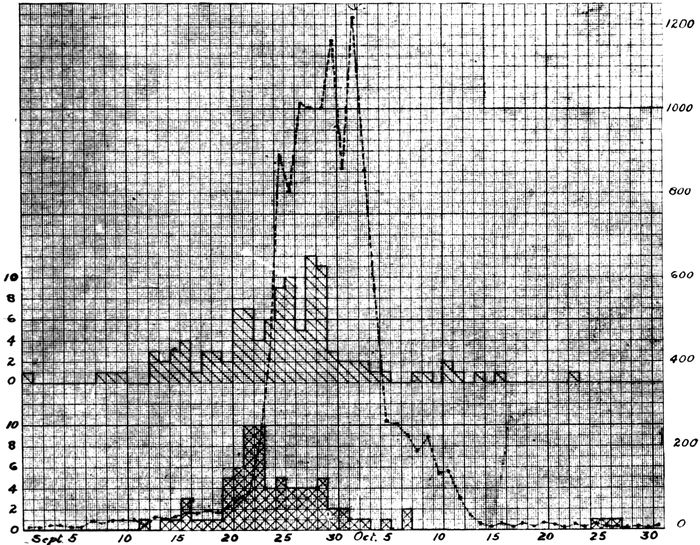
Chart 1—Showing the relation of (a) onset of cases of pneumonia shown by autopsy to be uncomplicated by secondary infection with hemolytic streptococcus, indicated by upper continuous line with single hatch, and of (b) onset of fatal cases of streptococcus pneumonia, indicated by the lower continuous line with double hatch, to (c) the occurrence of influenza, indicated by the broken line. The onset of each case of fatal pneumonia is represented by a single square.
Our study of ward infection in pneumonia furnishes an
explanation of the outbreak of fatal streptococcus pneumonia
coincident with the initial stage of the influenza epidemic.
This outbreak is a true epidemic of streptococcus
infection superimposed, in many instances at least, upon
preexisting pneumococcus pneumonia, but in some instances,
142doubtless, a primary streptococcus pneumonia,
following the bronchitis of influenza. In the absence of
secondary streptococcus infection a very large proportion
of these individuals would have recovered. This epidemic
of streptococcus pneumonia, it has been shown, was the result
of unfavorable conditions produced by great overcrowding
of the hospital and in the early part of the epidemic
by inadequate separation of those with streptococcus
infection from those with none. With control of these conditions,
streptococcus pneumonia rapidly diminished.
Greater susceptibility to pneumococcus pneumonia in the
early than in the late period of the epidemic is perhaps
explained by differences in the severity of influenza; the
more susceptible individuals were attacked by influenza
in the early period, whereas the less susceptible did not
acquire the disease until they had been exposed to an immensely
increased number of infected individuals. A better
explanation is furnished by the greater opportunity at
the beginning of the epidemic for the transmission of microorganisms
causing pneumonia, for at this time patients
with influenza were crowded together and methods to prevent
the transmission of infection were little used.
Clinical study has shown that purulent bronchitis (see
Fig. 2) occurs in about one-third of the cases of influenza.
In a large proportion of cases of bronchitis there is no clinical
evidence of pneumonia. The bronchial lesions found in
association with the pneumonia of influenza are an index of
the ability of the agent, which causes influenza, to injure the
bronchi.
In those who have died with pneumonia following influenza
the large bronchi (with cartilage) are intensely injected,
so that the mucosa has a deep red color which
on cross section contrasts very sharply with the pearly
white of the cartilage. Superficial injury to the bronchi is
143not infrequently evident in the larger bronchial branches;
superficial loss of epithelium is indicated by erosion of the
surface, whereas somewhat deeper destructive changes are
occasionally evident. Microscopic examination accurately
determines the degree of destructive change.
Purulent bronchitis was noted in 134 autopsies (55.6 per
cent of autopsies). From the small bronchi, in many instances,
purulent fluid welled up upon the cut surface of
the lung, whereas in other instances tenacious mucopurulent
fluid could be squeezed from small, cut bronchi by pressure
upon lung tissue. The consistency of the material
within the bronchi varied greatly, ranging from a viscid
and tenacious mucus of creamy, yellow color to a thin, turbid,
gray fluid. The coexistence of local inflammatory or
of general edema of the lungs modifies the character of the
material found in the bronchi at autopsy; with edema the
purulent exudate is in some instances diluted so that a thin
cloudy fluid flows from the small bronchi. In the presence
of advanced edema of the lungs the bronchi rarely if ever
contain purulent exudate. The underlying changes in the
bronchi are more significant than the character of the exudate
found at autopsy. Nevertheless, the group of cases
in which the diagnosis of purulent bronchitis has been
made, because small and medium sized bronchi have contained
purulent or mucopurulent exudate, represents instances
of readily recognizable bronchitis of considerable
severity.
With few exceptions, purulent bronchitis was diffusely
distributed in the lungs; occasionally it was observed in
one lung alone, and in several instances was limited to the
bronchi at the base of a lung, usually of the left lung.
In a considerable proportion of instances of purulent
bronchitis abnormal distention of the lungs was noted. On
removal from the chest the lungs fail to collapse and retain
the size and shape of the thorax. Even after section is
made through the organ, parts of the lung fail to collapse
144and have a resistant cushion-like consistency. This condition
is present where the lung tissue is air containing and
dry, and occurs when very small bronchi contain tenacious
mucous exudate which becomes apparent upon the cut surface
after the sectioned lung is squeezed. Microscopic examination
shows that the alveolar ducts and infundibula
are distended with air, though the respiratory bronchioles
contain inflammatory exudate. Complete obstruction of
the bronchi is followed by absorption of air from the tributary
pulmonary tissue with atelectasis. It is not improbable
that partial obstruction, permitting the penetration of
air with inspiration, produces distention of air containing
tissue.
It is furthermore probable that cyanosis, which is a conspicuous
feature of many instances of pneumonia following
influenza, is referable, in part at least, to obstruction of the
bronchi by mucopurulent exudate.
The term pneumonia will refer to those inflammatory
changes in the lung which are found within the alveoli; it
will include inflammatory changes in the alveoli surrounding
the respiratory bronchioles, in the alveolar ducts and
infundibula and in their tributary alveoli. Bronchitis will
be described by defining the changes which occur (a) in the
small bronchi with no cartilage or mucous glands, and (b)
in the large bronchi including the primary branches of the
trachea.
For convenience of description those bronchi may be
designated small, which have no cartilaginous plates in
their wall. Larger bronchi have cartilage and mucous
glands, the latter situated in considerable part outside the
cartilaginous plates. These bronchi, of which the largest
are the right and left bronchi formed by bifurcation of the
trachea, diminish with repeated branching to a caliber of
about 1 mm. Small bronchi are lined by columnar ciliated
epithelium; their wall consists of very vascular connective
145tissue containing a layer of smooth muscle and their caliber
varies approximately from 1 to 0.5 mm. It is convenient
to designate as respiratory bronchioles[80] the terminal
ramifications of the bronchi; they are lined by a
single layer of columnar ciliated cells passing over into
cuboidal nonciliated epithelium and are beset with small
air sacs lined by flat cells or epithelial plates similar to those
of the alveoli elsewhere. Not infrequently these alveoli occur
along only one side of the bronchiole, the remainder of
the circumference being covered by a continuous layer of
cubical epithelium. The respiratory bronchiole by branching
along its course or at its end is continued into several
alveolar ducts which unlike the respiratory bronchioles
have no cubical or columnar epithelium but are closely beset
by alveoli lined by flat epithelial plates. The alveolar
duct is recognized by the absence of cubical epithelium and
the presence of bundles of smooth muscle which occur in
the wall. The infundibula or alveolar sacs arise as
branches from the alveolar ducts and like them are beset
with alveoli, but smooth muscle does not occur in their
walls. The base of the infundibulum is wider than its orifice,
which Miller states is surrounded by a sphincter-like
bundle of smooth muscle.
Changes in the main bronchi and their primary branches
are usually less severe than those in bronchi of smaller
size. The epithelium is often intact, the superficial cells
being columnar and ciliated, but not infrequently desquamation
of superficial cells has occurred and the lower
layers alone remain. Occasionally (Autopsy 471) there is
necrosis of epithelium with which, although the architecture
of cells is preserved, nuclei have disappeared. Accumulation
of blood or serum may separate epithelium from
the underlying basement membrane (Fig. 1). Complete
loss of epithelium occurs, usually in small patches.
146Polynuclear leucocytes are numerous upon the surface
of the epithelium and are sometimes fixed in process of
migration through epithelium and basement membrane.
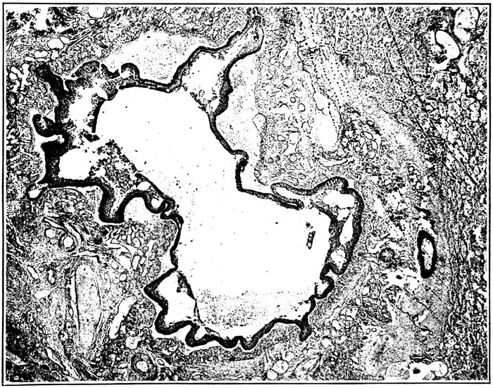
Fig. 1.—Acute bronchitis showing engorgement of blood vessels of mucosa and elevation of epithelium by serum and blood. Autopsy 352.
The blood vessels of the mucosa are engorged. There
is sometimes edema or hemorrhage, and in the superficial
part of the mucosa polynuclear leucocytes are often fairly
abundant. When superficial epithelium has been lost, polynuclears
are numerous immediately below the surface of
the exposed tissue. Fibrin is often present upon the denuded
surface and extends for a short distance into the
tissue below. In the deeper part of the mucosa, about the
muscularis and especially about and between the acini of
the mucous glands, the tissue is infiltrated with lymphoid
and plasma cells.
Changes in the mucous glands are invariably present.
These changes are distention of ducts and acini with mucous,
147degenerative changes occasionally ending in necrosis
of cells, disappearance of acini, dense infiltration of interstitial
tissue with lymphoid and plasma cells and finally
proliferation of this interstitial tissue. The duct of a mucous
gland, dilated and filled with mucus, may be surrounded
by lymphoid and plasma cells in great number.
Acini, similarly dilated, contain mucus and are composed of
cubical cells which have discharged their mucous content.
In some instances (e. g., Autopsy 257) the cells of the acini
have undergone necrosis; the cytoplasm stains homogeneously
and the nuclei have disappeared. Where necrosis
has occurred, polynuclear leucocytes may penetrate into
the dead cells. In association with degenerative changes
in the acini there is abundant infiltration of the interstitial
tissue within and about the glands with lymphoid and
plasma cells. When the acini have disappeared there is
proliferation of fibroblasts and new formation of fibrous
tissue, and mucous glands are found in which a few
atrophied acini are separated by newly formed fibrous
tissue.
With the bronchitis of influenza the small bronchi (with
no cartilage or mucous glands) show every stage of transition
from early acute inflammation characterized by accumulation
of polynuclear leucocytes within the lumen, engorgement
of blood vessels, and infiltration of the wall with
polynuclear leucocytes, through various stages of destructive
changes to complete disappearance of the bronchial
wall and formation of an abscess cavity at the site of the
bronchus. In the early stages of acute bronchitis, hemorrhage
is frequently associated with the lesion. Blood may
be abundant within the lumen of the bronchus, and in the
mucosa red blood corpuscles often infiltrate the tissue
around greatly distended blood vessels, or accumulating
below the epithelium, separate it from its basement membrane.
Hemorrhage is not limited to the wall of the bronchus,
but frequently occurs into the alveoli in a zone encircling
the bronchus.
148With acute bronchitis there may be desquamation of
epithelial cells with partial or complete loss of epithelial
lining. In the smallest bronchi the single layer of columnar
cells may be separated in places from the underlying
tissue, so that intact rows of cells are found within the
lumen. In somewhat larger bronchi, lined by epithelium
in multiple layers, superficial columnar ciliated cells may
be lost. In some instances superficial epithelial cells appear
to have lost their cohesion and are separated by narrow
spaces; in these instances, polynuclear leucocytes are
often numerous between epithelial cells. Epithelium is occasionally
separated from its basement membrane by small
accumulations of serum or blood. Occasionally necrosis
of epithelial cells with disappearance of nuclei is seen and
is doubtless caused by the action of bacteria; the affected
cells may be raised from the underlying tissue by accumulated
serum (Autopsy 253). The changes which have
been described bring about partial or complete loss of
the ciliated lining of the bronchial tube.
The severity of changes in the bronchial wall is in direct
relation to the extent of destruction of the lining epithelium:
when the epithelium remains intact polynuclear
leucocytes may be found in considerable number immediately
below it, but as the lesion progresses, cells in great
part mononuclear, namely, lymphoid and plasma cells, accumulate
in large number throughout the wall of the bronchus.
There is often abundant cellular infiltration within
and about the bundles of the muscular coat. The changes
assume the character of chronic inflammation.
When the lining epithelium of the bronchus is lost, fibrin
tends to accumulate over the surface of the defect, to which
it is firmly attached. It remains separated by a conspicuous
space from adjacent intact epithelium over which it
may project. This superficial network of fibrin merges
with a similar network, extending to a variable depth within
the tissue. What may well be described as coagulative
149necrosis has often occurred, and structures, such as white
fibrous bundles or wall of blood vessels, are marked out
by hyaline material which merges with fibrin. When the
walls of the blood vessels which are invariably engorged
are involved, the lumen is plugged by a fibrinous thrombus.
Little patches of fibrin adherent to the inner surface of
the bronchus may occur in spots where epithelium has been
lost; with uniform loss of epithelium the entire circumference
may be lined with fibrin forming a circular zone
occasionally quite uniform in thickness.
Accumulations of polynuclear leucocytes doubtless bring
about conditions which cause solution of fibrin or prevent
its formation (when disintegration of leucocytes sets free
leucoprotease in abundance). The activity of the infecting
microorganisms, usually hemolytic streptococci or staphylococci,
may cause complete necrosis of a part or all of the
bronchial wall. The cavity which is formed may penetrate
into lung tissue that has previously undergone pneumonic
consolidation.
Further changes caused by the bronchitis of influenza
will be considered under peribronchial hemorrhage and
edema, peribronchial pneumonia and bronchiogenic abscess.
Purulent bronchitis is almost invariably associated
with dilatation of the bronchi, the affected bronchi being
distended with pus. With increasing dilatation bronchiectasis
becomes evident upon gross examination of the tissue,
and is much more advanced in the small bronchi than in
the larger cartilaginous passages. This subject will be
further considered under bronchiectasis.
In association with the acute bronchitis of influenza the
epithelium of bronchi not infrequently looses its superficial
columnar ciliated cells and assumes some of the characters
of a squamous epithelium being covered by polygonal or
flat cells (Figs. 17 and 18). The condition is often described
a “squamous metaplasia,” although it doubtless
represents a stage of regeneration following injury rather
150than a true metaplasia. The basal cells of the epithelium
have a cubical or columnar form; above them the cells become
polygonal and as the surface is approached, cells are
flat and even scale-like. The nuclei of these superficial
cells are often lost. There is no close resemblance to the
squamous epithelium of the skin, for intercellular bridges
are not seen.
This change may occur within six days after onset of
influenza, though in most instances the duration of illness
has been two weeks or more. It may affect either large
or small bronchi, but it is more frequently found in the
latter. Whenever ciliated columnar cells are lost, superficial
cells tend to become flat. Epithelium on one side of a
bronchus may have a squamous character, whereas that
elsewhere is columnar and ciliated. The flat epithelium may
undergo thickening so that it is 0.1 mm. or more in thickness.
It is noteworthy that regenerating epithelium growing
over a denuded surface has the squamous character
which has been described (Plate XIV, Fig. 22).
Bacteriology of the Bronchitis of Influenza.—With the
pneumonia of influenza, bronchitis is invariably present.
Cultures have been made from the right or left main bronchus
or from the very small bronchi which contained purulent
exudate. A routine method of making the culture has
been adopted. The right main bronchus, exposed by drawing
the right lung out of the chest and toward the midline,
was widely seared with a hot knife; the bronchus was partially
cut across through the seared surface with a heated
knife and a platinum needle inserted into the lumen. The
bacteria obtained named in the approximate order of their
relative frequency have been: B. influenzæ, pneumococci,
hemolytic streptococci, staphylococci (aureus and albus),
B. coli, S. viridans, M. catarrhalis, and diphthoid bacilli
which have not been identified. Mixed infections occurred
in most instances. The following list arranged by grouping
151bacteria in the order cited above, shows how varied have
been the combinations which occur:
| B. influenzæ |
3 |
| Pneumococci |
5 |
| S. hemolyticus |
3 |
| Staphylococci |
3 |
| B. coli |
3 |
| S. viridans |
1 |
| B. influenzæ, pneumococci |
17 |
| B. influenzæ, S. hemolyticus |
18 |
| B. influenzæ, staphylococci |
4 |
| Pneumococci, S. hemolyticus |
1 |
| Pneumococci, staphylococci |
3 |
| S. hemolyticus, staphylococci |
4 |
| S. hemolyticus, B. coli |
2 |
| Staphylococci, S. viridans |
1 |
| B. influenzæ, pneumococci, S. hemolyticus |
6 |
| B. influenzæ, pneumococci, staphylococci |
15 |
| B. influenzæ, pneumococci, S. viridans |
2 |
| B. influenzæ, S. hemolyticus, staphylococci |
16 |
| B. influenzæ, S. hemolyticus, M. catarrhalis |
1 |
| B. influenzæ, staphylococci, S. viridans |
1 |
| Pneumococci, S. hemolyticus, staphylococci |
3 |
| Staphylococci, B. coli, S. viridans |
1 |
| B. influenzæ, pneumococci, S. hemolyticus, staphylococci |
7 |
| B. influenzæ, pneumococci, staphylococci, M. catarrhalis |
1 |
| B. influenzæ, S. hemolyticus, staphylococci, B. coli |
1 |
| B. influenzæ, S. hemolyticus, staphylococci, S. viridans |
1 |
| B. influenzæ, S. hemolyticus, staphylococci, M. catarrhalis |
1 |
| B. influenzæ, staphylococci, S. viridans, M. catarrhalis |
1 |
B. influenzæ has been present in the bronchi in 79.3 per
cent of instances of pneumonia referable to influenza. Combinations
which have been found most frequently are B.
influenzæ and pneumococci (17 instances), B. influenzæ and
hemolytic streptococci (18 instances), or the same combinations
with staphylococci, namely, B. influenzæ, pneumococci
and staphylococci (15 instances), and B. influenzæ, hemolytic
streptococci and staphylococci (16 instances). There
is little doubt that B. influenzæ was not identified in some
instances in which it was present; when other microorganisms
are very numerous its inconspicuous colonies may be
overgrown even though the presence of pneumococci, streptococci
152or staphylococci tends to increase the size of its colonies.
Moreover, it is not improbable that the microorganism
may disappear from the bronchi. Comparison with
observations made upon influenza suggests that multiple
methods of examination might have demonstrated a much
higher incidence of B. influenzæ. Throat cultures alone
made during life demonstrated the presence of B. influenzæ
in only 65.7 per cent of patients with acute influenza,
whereas when cultures were made from the nose, throat
and sputum, and a mouse was inoculated with sputum from
each patient, B. influenzæ was found in every instance.
After the acute stage of the disease had passed, the number
of microorganisms diminished, and in many instances
B. influenzæ disappeared from the upper air passages. In
some of our autopsies B. influenzæ doubtless present during
life has similarly disappeared before death due to
pneumonia caused by pneumococci or streptococci. In view
of these considerations it is not improbable that B. influenzæ
demonstrated by a single culture in 80 per cent of
instances has been constantly present.
Table XXVIII represents the incidence of pneumococci,
hemolytic streptococci, staphylococci, and B. influenzæ in
the bronchi, lungs and blood of those individuals with pneumonia
in whom bacteriologic examination has been made at
autopsy. The number of cultures made from the bronchi,
lungs or blood of the heart is given in the second column
of the table and in other columns are given the incidence in
number and percentage of the microorganisms which have
been mentioned.
| Table XXVIII |
|---|
| |
|
NO. OF CULTURES |
PNEUMOCOCCI |
HEMOLYTIC STREPTOCOCCI |
STAPHYLOCOCCI |
B. INFLUENZÆ |
| NO. POSITIVE |
PER CENT POSITIVE |
NO. POSITIVE |
PER CENT POSITIVE |
NO. POSITIVE |
PER CENT POSITIVE |
NO. POSITIVE |
PER CENT POSITIVE |
| Bronchus |
121 |
56 |
46.3 |
58 |
47.9 |
61 |
50.4 |
96 |
79.3 |
| Lung |
153 |
68 |
44.4 |
77 |
50.3 |
37 |
24.2 |
70 |
45.7 |
| Blood |
218 |
87 |
39.9 |
85 |
39.0 |
1 |
0.5 |
1 |
0.5 |
153Cultures from the bronchus represent the bacteriology
of the bronchitis of influenza. Infection of the lung following
influenza doubtless occurs by way of the bronchi,
so that the bacteria which cause pneumonia are present in
the bronchi before they enter the lung tissue. The figures
in Table XXVIII, similar to those previously cited, show
the high incidence of B. influenzæ, and the occurrence of
pneumococci, hemolytic streptococci and staphylococci each
present in approximately half of all autopsies.
The figures in Table XXVIII are an index of the capacity
of the microorganisms which enter the bronchi to invade
the lungs and finally the blood. Pneumococci were present
in the bronchi in 46.3 per cent of instances, in the lungs
in only slightly less, and in approximately 40 per cent of
autopsies they had penetrated into the blood. Hemolytic
streptococci enter the bronchi with the same frequency and
exhibit an equal ability to penetrate into the lungs and
blood. Staphylococci enter the bronchi in half of these individuals,
but penetrate into the lungs in only a fourth of
the instances. They have entered the blood only once (Autopsy
263) in this instance in association with hemolytic
streptococci. B. influenzæ has been present in the bronchi
in approximately 80 per cent of autopsies. It is noteworthy
that it has been found in the lung in little more than
half this percentage of instances and has entered the blood
only once (Autopsy 474), in this instance in association with
hemolytic streptococci.
In a limited number of autopsies there was purulent
bronchitis recognized by the presence of mucopurulent exudate
in small bronchi. It has been stated that this group
of cases is not sharply separable from other instances of
bronchitis, because in some cases death has occurred before
a purulent exudate has accumulated or in other instances
a purulent exudate has been displaced by edema. Table
XXIX shows the bacteriology of instances of purulent
bronchitis:
154
| Table XXIX |
|---|
| |
|
NO. OF CULTURES |
PNEUMOCOCCI |
HEMOLYTIC STREPTOCOCCI |
STAPHYLOCOCCI |
B. INFLUENZÆ |
| NO. POSITIVE |
PER CENT POSITIVE |
NO. POSITIVE |
PER CENT POSITIVE |
NO. POSITIVE |
PER CENT POSITIVE |
NO. POSITIVE |
PER CENT POSITIVE |
| Bronchus |
66 |
33 |
50.0 |
32 |
48.5 |
36 |
54.5 |
53 |
80.3 |
The percentages of various bacteria with purulent bronchitis
do not differ essentially from those obtained from
all autopsies with pneumonia. B. influenzæ is found in approximately
80 per cent of autopsies. In 16 instances cultures
were made from the purulent fluid contained in a
small bronchus and the incidence of B. influenzæ (namely,
81.4 per cent) has not differed from that in the main bronchus.
In 7 of 8 instances in which cultures were made, both
from the right main bronchus and from the purulent fluid
in a small bronchus, B. influenzæ was found in one or other
in all but one autopsy (87.5 per cent); in this instance (Autopsy
472) respiratory disease began thirty-seven days before
death and cultures from large and small bronchi at
autopsy were overgrown by B. coli. Since observations
upon influenza made during life have shown that B. influenzæ
is constantly demonstrable when multiple methods are
employed for its detection, the figures just cited give support
to the suggestion that B. influenzæ is constantly present
in the bronchi with the bronchitis of influenza.
The frequency with which the confluent lobular consolidation
of bronchopneumonia involving whole lobes or
parts of lobes follows influenza has emphasized the desirability
of distinguishing carefully between lobar and confluent
lobular pneumonia. The pulmonary lesion has been
designated lobar pneumonia when it exhibited the well-known
characters of this lesion, namely, firm consolidation
of large parts of lobes, coarse granulation of the cut surface,
155fibrinous plugs in the bronchi and, on microscopic examination,
homogeneous consolidation and fibrinous plugs
within the alveoli. With confluent lobular consolidation of
bronchopneumonia the consolidated area is in most cases
obviously limited by lobule boundaries, and well-defined
lobules of consolidation occur elsewhere in the lungs.
Lobar pneumonia occurred in 98 among 241 instances of
pneumonia following influenza, namely, in 40.7 per cent of
autopsies.
The difficulty of separating lobar and bronchopneumonia
following influenza has been increased by the frequent combination
of the two lesions in the same individual. There
were 34 instances in which lobar and bronchopneumonia
occurred together. The anatomic diagnosis of lobar pneumonia
was made only when lobes or parts of lobes were
firmly consolidated and exhibited the characters of the lesion
enumerated above; in several instances, in which there
was some doubt concerning the nature of the lesion, microscopic
examination was decisive. The associated bronchopneumonic
lesions represented all the types which have
been associated with influenza. In the group of 34 cases of
coexisting lobar and bronchopneumonia, lobular consolidation
occurred 10 times, peribronchiolar consolidation 14
times (recognized in all but 4 instances by microscopic examination),
hemorrhagic peribronchiolar consolidation 9
times, peribronchial pneumonia 4 times. The intimate relation
of these lesions to changes in the bronchi is well
shown by the frequent presence of purulent bronchitis.
The associated lesions of the bronchi in these cases were as
follows: purulent bronchitis in 23 instances; peribronchial
hemorrhage in 6; bronchiectasis in 11. The frequency of
purulent bronchitis and other bronchial lesions in association
with coexisting lobar and bronchopneumonia is in
sharp contrast with the occurrence of these lesions in association
with lobar pneumonia alone; with 69 instances of
156lobar pneumonia alone purulent bronchitis occurred 17
times and bronchiectasis once.
Lobar pneumonia following influenza passes through the
usual stages of red and gray hepatization. Red hepatization
was found 16 times, combined red and gray hepatization
28 times, and gray hepatization 20 times. The average
duration of pneumonia with red hepatization was 3.7 days,
with combined red and gray hepatization 5.1 days and with
gray hepatization 7.5 days. These figures, it will be shown
later, have some importance in relation to the stage at
which hemolytic streptococcus infects lungs the site of
lobar pneumonia.
Bacteriology of Lobar Pneumonia.—Table XXX is compiled
with the purpose of determining the bacteriology
of the bronchi, lungs and heart’s blood in autopsies
performed on individuals with lobar pneumonia. In some
instances bacteriologic examination of one or other of these
organs was omitted; the percentage incidence is an index
of the presence of pneumococci, hemolytic streptococci,
staphylococci or B. influenzæ in the bronchi, lungs or
heart’s blood and measures the invasive power of these microorganisms
during the course of lobar pneumonia following
influenza.
| Table XXX |
|---|
| |
|
NO. OF CULTURES |
PNEUMOCOCCI |
HEMOLYTIC STREPTOCOCCI |
STAPHYLOCOCCI |
B. INFLUENZÆ |
| NO. POSITIVE |
PER CENT POSITIVE |
NO. POSITIVE |
PER CENT POSITIVE |
NO. POSITIVE |
PER CENT POSITIVE |
NO. POSITIVE |
PER CENT POSITIVE |
| Bronchus |
44 |
56.9 |
14 |
31.8 |
22 |
37 |
84.1 |
96 |
79.3 |
| Lung |
53 |
77.3 |
13 |
24.5 |
8 |
26 |
49.1 |
70 |
45.7 |
| Blood |
87 |
65.5 |
11 |
12.6 |
|
|
|
1 |
0.5 |
Pneumococci, the recognized cause of lobar pneumonia,
were found in the lungs in 73.3 per cent of autopsies; failure
to find the microorganism in all instances is doubtless
the result of its disappearance from the lung, which, it is
well known, occurs not infrequently particularly during
157the later stages of the disease. In 65.5 per cent of instances
of fatal lobar pneumonia pneumococci have entered the
heart’s blood.
Hemolytic streptococci unlike pneumococci were found
more frequently in the bronchi than in the lungs; this microorganism
which exhibits little tendency to disappear,
once it has established itself within the body, found entrance
into the bronchi in 31.8 per cent of instances of lobar
pneumonia and in 24.5 per cent entered the lungs. Its invasive
power is further illustrated by its penetration into the
heart’s blood approximately in half this proportion of autopsies.
Staphylococci enter the bronchi in many instances (50
per cent), but relatively seldom (15.1 per cent) invade the
lung and rarely if ever penetrate into the blood.
The high incidence, namely, 84.1 per cent, of B. influenzæ
in the bronchi is particularly noteworthy; it exceeds that
of pneumococci, the well-recognized cause of lobar pneumonia,
within the lung. It is found much less frequently
within consolidated lung tissue and shows no tendency to
invade the heart’s blood. B. influenzæ finds the most favorable
conditions for its multiplication within the bronchi.
In view of the frequent occurrence of coexisting lobar
and bronchopneumonia it has appeared desirable to determine
how far the existence of obvious bronchopneumonia
modifies the bacteriology of lobar pneumonia. In Table
XXXI the incidence of pneumococci, hemolytic streptococci,
staphylococci and B. influenzæ after death with lobar pneumonia
on the one hand is compared with their incidence
after combined lobar and bronchopneumonia on the other.
Pneumococci are found in the lung more frequently with
lobar than with combined lobar and bronchopneumonia.
The incidence of hemolytic streptococci and of staphylococci
in the lung is on the contrary higher when bronchopneumonia
is associated with lobar pneumonia. It is not
improbable that these microorganisms have a part in the
production of associated bronchopneumonia. The frequency
with which microorganisms invade the blood is almost
identical in the two groups.
158
| Table XXXI |
|---|
| |
| With Lobar Pneumonia Alone |
|---|
| |
|
NO. OF CULTURES |
PNEUMOCOCCI |
HEMOLYTIC STREPTOCOCCI |
STAPHYLOCOCCI |
B. INFLUENZÆ |
| NO. POSITIVE |
PER CENT POSITIVE |
NO. POSITIVE |
PER CENT POSITIVE |
NO. POSITIVE |
PER CENT POSITIVE |
NO. POSITIVE |
PER CENT POSITIVE |
| Bronchus |
30 |
20 |
66.6 |
9 |
30 |
15 |
50 |
26 |
86.7 |
| Lung |
34 |
29 |
85.2 |
7 |
20.6 |
3 |
8.8 |
18 |
52.9 |
| Blood |
54 |
36 |
66.7 |
7 |
13 |
|
|
|
|
| |
| |
| With Combined Lobar and Bronchopneumonia |
|---|
| |
|
NO. OF CULTURES |
PNEUMOCOCCI |
HEMOLYTIC STREPTOCOCCI |
STAPHYLOCOCCI |
B. INFLUENZÆ |
| NO. POSITIVE |
PER CENT POSITIVE |
NO. POSITIVE |
PER CENT POSITIVE |
NO. POSITIVE |
PER CENT POSITIVE |
NO. POSITIVE |
PER CENT POSITIVE |
| Bronchus |
14 |
9 |
64.3 |
5 |
34.3 |
7 |
50 |
11 |
78.6 |
| Lung |
19 |
12 |
63.2 |
6 |
31.6 |
5 |
26.3 |
8 |
42.1 |
| Blood |
33 |
21 |
63.1 |
4 |
12.1 |
|
|
|
|
The relative frequency with which different types of
pneumococci produce lobar pneumonia under the conditions
existing when Camp Pike was attacked by an epidemic of
influenza is indicated by Table XXXII in which instances
of lobar pneumonia alone and of combined lobar
and bronchopneumonia are listed separately.
Pneumococcus I and II, which are found approximately
in two-thirds of instances of lobar pneumonia occurring in
cities, have an insignificant part in the production of these
lesions. Pneumococcus IV and atypical Pneumococcus II,
which are commonly found in the mouth, are the predominant
cause of these lesions, and with Pneumococcus III,
also an inhabitant of the mouths of normal individuals,
have been the cause of two-thirds of all instances of lobar
pneumonia observed in this camp.
159
| Table XXXII |
|---|
| |
| With Lobar Pneumonia |
|---|
|
NO. OF CULTURES |
PNEUMOCOCCUS I |
PNEUMOCOCCUS II |
PNEUMOCOCCUS II (Atyp.) |
PNEUMOCOCCUS III |
PNEUMOCOCCUS IV |
| NO. POSITIVE |
PER CENT POSITIVE |
NO. POSITIVE |
PER CENT POSITIVE |
NO. POSITIVE |
PER CENT POSITIVE |
NO. POSITIVE |
PER CENT POSITIVE |
NO. POSITIVE |
PER CENT POSITIVE |
| Bronchus |
30 |
1 |
3.3 |
1 |
3.3 |
4 |
13.3 |
4 |
13.3 |
10 |
33.3 |
| Lung |
34 |
1 |
2.9 |
2 |
5.9 |
9 |
26.5 |
6 |
17.6 |
11 |
32.4 |
| Blood |
54 |
2 |
3.7 |
2 |
3.7 |
12 |
22.2 |
3 |
5.6 |
17 |
31.5 |
| |
| With Combined Lobar and Bronchopneumonia |
|---|
|
NO. OF CULTURES |
PNEUMOCOCCUS I |
PNEUMOCOCCUS II |
PNEUMOCOCCUS II (Atyp.) |
PNEUMOCOCCUS III |
PNEUMOCOCCUS IV |
| NO. POSITIVE |
PER CENT POSITIVE |
NO. POSITIVE |
PER CENT POSITIVE |
NO. POSITIVE |
PER CENT POSITIVE |
NO. POSITIVE |
PER CENT POSITIVE |
NO. POSITIVE |
PER CENT POSITIVE |
| Bronchus |
14 |
2 |
14.3 |
1 |
7.1 |
3 |
21.4 |
|
|
3 |
21.4 |
| Lung |
19 |
1 |
5.3 |
|
|
5 |
26.3 |
|
|
6 |
31.6 |
| Blood |
33 |
2 |
6.1 |
3 |
9.1 |
4 |
12.1 |
|
|
12 |
36.4 |
There is no noteworthy difference in the occurrence of
these types of pneumococci among instances of lobar pneumonia,
on the one hand, and of combined lobar and bronchopneumonia,
on the other. Different types exhibit no noteworthy
differences in their ability to penetrate into lungs
and blood.
Hemolytic Streptococcus with Lobar Pneumonia.—There
can be no doubt that the concurrent infection with
microorganisms other than pneumococcus modifies the
progress of lobar pneumonia. With lobar pneumonia alone
hemolytic streptococci have entered the bronchi in 30 per
cent of instances and have penetrated into the lungs in 20.6
per cent; with associated lobar and bronchopneumonia the
same microorganism has entered the bronchi in 34.3 per
cent of instances and invaded the lung in 31.6 per cent.
Hemolytic streptococci are the only microorganisms other
than pneumococci which, in association with lobar pneumonia,
160have found their way from the lungs to the blood
stream; more than one-third of all instances of lobar pneumonia
in which hemolytic streptococci find entrance into
the bronchi die with streptococcus septicemia.
Separation of instances of lobar pneumonia into groups
on the basis of the occurrence of red or gray hepatization
shows that infection with hemolytic streptococcus is more
likely to occur during the early stages of the disease. The
average duration of lobar pneumonia with red hepatization
has been 3.7 days, with red and gray hepatization, 5.1 days,
and with gray hepatization, 7.5 days. Infection with hemolytic
streptococcus has occurred in association with red or
gray hepatization as shown in Table XXXIII.
| Table XXXIII |
|---|
| |
|
NO. OF AUTOPSIES |
NO. WITH HEMOLYTIC STREPTOCOCCUS |
PER CENT WITH HEMOLYTIC STREPTOCOCCUS |
| Lobar pneumonia with red hepatization |
16 |
6 |
37.5 |
| Lobar pneumonia with red and gray hepatization |
28 |
6 |
21.4 |
| Lobar pneumonia with gray hepatization |
20 |
1 |
5.0 |
Notwithstanding the longer duration of the disease and
consequent prolongation of exposure to infection, lobar
pneumonia, which has reached the stage of gray hepatization,
has shown the smallest incidence of infection with
hemolytic streptococci. In the stage of gray hepatization
there is diminished susceptibility to secondary infection
with this microorganism.
Characteristic histologic changes have been found in the
lungs of those who have died with lobar pneumonia followed
by invasion of lungs and blood by hemolytic streptococci
(e. g., Autopsies 273, 430), but with no evidence of
suppuration found at autopsy. Within the pneumonic
lung occur patches of necrosis implicating both exuded
cells and alveolar walls; in some places nuclei have
disappeared; elsewhere nuclear fragments are abundant.
161In these patches of necrosis Gram-positive streptococci in
short chains occur in immense number. In some instances
(e. g., Autopsies 273, 346, 479) interlobular septa are very
edematous and often contain a network of fibrin; lymphatics
are dilated and contain polynuclear leucocytes in abundance.
Streptococci are found within these lymphatics.
The histologic changes which have been described represent
the earliest stages of abscess formation and interstitial
suppuration, lesions almost invariably caused by hemolytic
streptococci.
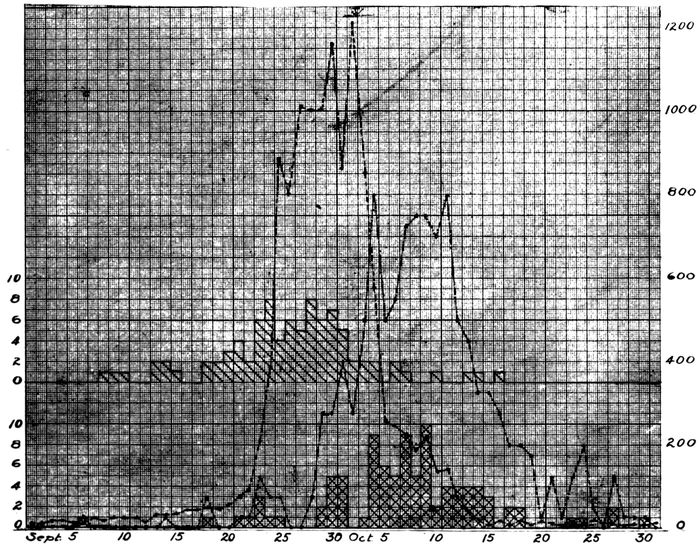
Chart 2.—Showing the relation of (a) date of onset of cases in which autopsy demonstrated lobar pneumonia, indicated by upper continuous line with single hatch, and of (b) date of death of these cases, indicated by lower continuous line with double hatch to (c) the occurrence of influenza, indicated by the broken line, and to (d) the total number of fatal cases of pneumonia, indicated by the broken dotted line. Each case of fatal pneumonia is indicated by one division of the scale as numbered on the left of the chart; cases of influenza are indicated by the numbers on the right of the chart.
Relation of Lobar Pneumonia to Influenza.—Some writers
have suggested that lobar pneumonia, heretofore observed
during the course of epidemics of influenza, is an
162independent disease with no relation to influenza, both diseases
being referable perhaps to similar meteorologic or
other conditions. Chart 2, which shows by weeks from
September 1 to October 31 the relation of deaths from lobar
pneumonia (indicated by double hatch) to deaths from all
forms of pneumonia, disproves this suggestion. The two
curves follow parallel courses; that representing lobar
pneumonia reaches a maximum approximately one week
after the outbreak of influenza had reached its height. Lobar
pneumonia, like other forms of pneumonia, was secondary
to influenza. When a chart is plotted to represent the
dates of onset of fatal cases of lobar pneumonia (indicated
by single hatch in Chart 2), it becomes evident that the
greatest number of these cases of pneumonia had their onset
at the beginning of the influenza epidemic, approximately
one week before it reached its height. Fatal lobar
pneumonia developed less frequently in the latter part of
the epidemic; to obtain an explanation of this relation it is
necessary to chart separately cases of lobar pneumonia with
secondary streptococcus infection, for we have already
learned that streptococcus infection was the predominant
cause of death in the early period of the influenza epidemic.
Exclusion of these instances of secondary streptococcus infection
makes no noteworthy change in the character of the
chart. Fatal lobar pneumonia, like all forms of fatal
pneumonia (p. 140), was more frequent in the first half
than in the second half of the epidemic. This difference
is referable either to greater virulence of the virus of
influenza or to the greater susceptibility of those first selected
by the disease or, as more probable, to conditions
such as crowding together of patients with influenza, favoring
the transmission of microorganisms which cause pneumonia.
For the purpose of the present study it is convenient to
group together instances of bronchopneumonia which have
163been unaccompanied, on the one hand, by lobar pneumonia
(p. 155) or, on the other hand, by suppuration, which with
few exceptions is caused by hemolytic streptococci or by
staphylococci. A group of cases in which lobar and bronchopneumonia
have occurred in the same individual have
already been considered. In many instances, bronchopneumonia
is accompanied by abscess formation or by some
other form of suppuration; these lesions will be discussed
elsewhere.
Bronchopneumonia unaccompanied by lobar pneumonia
or by suppuration occurred in 80 autopsies.
Pneumonic consolidation distributed with relation to the
bronchi exhibits considerable variety, and an attempt to
define a type of bronchopneumonia characteristic of influenza
would be futile. Nevertheless, the bronchopneumonia
of influenza has in many instances distinctive characters.
Lesions of bronchopneumonia which are frequently
found in the autopsies under consideration may be conveniently
designated by descriptive terms, indicative of
their location in the lung tissue. These lesions, of which
two or more often occur in the same lung, are:
1. Peribronchiolar consolidation with which the inflammatory
exudate is limited to the alveoli in the immediate
neighborhood of the bronchioles.
2. Hemorrhagic peribronchiolar consolidation in which
gray patches of peribronchiolar pneumonia occur upon a
deep red background produced by hemorrhage into alveoli.
Pfeiffer believed that this lesion was characteristic of influenza.
3. Lobular consolidation with which consolidation is limited
to lobules or groups of lobules.
4. Peribronchial pneumonia with which small bronchi are
encircled by pneumonic consolidation.
Each one of these lesions will be discussed separately.
Following is a list of the bacteria which have been isolated
from the consolidated lung of individuals with bronchopneumonia
164unaccompanied by lobar pneumonia or by
suppuration:
| B. influenzæ |
1 |
| Pneumococci |
5 |
| S. hemolyticus |
5 |
| S. viridans |
1 |
| B. influenzæ, pneumococci |
9 |
| B. influenzæ, S. hemolyticus |
4 |
| B. influenzæ, staphylococci |
4 |
| Pneumococci, S. hemolyticus |
1 |
| Pneumococci, staphylococci |
2 |
| S. hemolyticus, staphylococci |
1 |
| S. hemolyticus, B. coli |
1 |
| Staphylococci, S. viridans |
1 |
| Staphylococci, B. coli |
1 |
| B. influenzæ, pneumococci, staphylococci |
1 |
| B. influenzæ, pneumococci, S. viridans |
1 |
| B. influenzæ, S. hemolyticus, staphylococci |
2 |
| B. influenzæ, pneumococci, staphylococci, S. viridans |
1 |
| No microorganisms found |
6 |
| |
|
| |
47 |
The similarity of this list to that representing the bacteriology
of bronchitis is evident; there is the same multiplicity
of microorganisms and the frequent occurrence of
mixed infections. B. influenzæ is much less frequently
found in the lung. The relative pathogenicity of the large
group of microorganisms enumerated above is better indicated
by the following list which shows what microorganisms
have penetrated into the blood in autopsies performed
on individuals with bronchopneumonia:
| Pneumococci |
20 |
| S. hemolyticus |
23 |
| S. viridans |
1 |
| Pneumococci, S. hemolyticus |
2 |
| No bacteria found |
25 |
| |
|
| Total |
71 |
Table XXXIV shows the percentage incidence of pneumococcus,
hemolytic streptococcus, staphylococcus and
165B. influenzæ in the bronchi, lungs and blood and is inserted
for comparison with the similar table (Table XXX)
showing the incidence of these bacteria in lobar pneumonia.
| Table XXXIV |
|---|
| |
|
NO. OF CULTURES |
PNEUMOCOCCI |
HEMOLYTIC STREPTOCOCCI |
STAPHYLOCOCCI |
B. INFLUENZÆ |
| NO. POSITIVE |
PER CENT POSITIVE |
NO. POSITIVE |
PER CENT POSITIVE |
NO. POSITIVE |
PER CENT POSITIVE |
NO. POSITIVE |
PER CENT POSITIVE |
| Bronchus |
37 |
19 |
48.6 |
13 |
35.1 |
22 |
59.5 |
28 |
75.7 |
| Lung |
47 |
20 |
42.6 |
14 |
29.8 |
13 |
27.7 |
23 |
48.9 |
| Blood |
70 |
22 |
31.4 |
24 |
34.3 |
|
|
|
|
Table XXXIV shows that pneumococci have a less important
part in the production of broncho than of lobar pneumonia;
with lobar pneumonia this microorganism was found
in the lungs in 77.3 per cent of instances and in the blood, in
65.5 per cent, whereas with bronchopneumonia it was found
in the lungs in 42.6 per cent and in the blood in 31.4 per cent.
Hemolytic streptococci (in lungs and blood) and staphylococci
(in lungs), on the contrary, were more common with
bronchopneumonia, and doubtless have a part in the production
of the lesion. Streptococcus viridans, B. coli and
M. catarrhalis, which are not infrequently found in the
bronchi (p. 151), occasionally enter the lungs with bronchopneumonia
but are rarely found with lobar pneumonia.
B. influenzæ has been found in less than 80 per cent of
instances in the bronchi and in about half of the lungs,
maintaining an incidence approximately the same as that
with lobar pneumonia.
Table XXXV shows the types of pneumococci found in
association with bronchopneumonia and is inserted for
comparison with the similar table (Table XXXII) showing
types of pneumococci with lobar pneumonia.
With broncho as with lobar pneumonia pneumococci commonly
found in the mouth, namely, atypical II, and Types
III and IV, have a more important part in production of the
lesion than the so-called fixed types, I and II. Atypical
Pneumococcus II has been less frequently encountered with
broncho than with lobar pneumonia.
166
| Table XXXV |
|---|
| |
|
NO. OF CULTURES |
PNEUMOCOCCUS I |
PNEUMOCOCCUS II |
PNEUMOCOCCUS II (Atyp.) |
PNEUMOCOCCUS III |
PNEUMOCOCCUS IV |
| NO. POSITIVE |
PER CENT POSITIVE |
NO. POSITIVE |
PER CENT POSITIVE |
NO. POSITIVE |
PER CENT POSITIVE |
NO. POSITIVE |
PER CENT POSITIVE |
NO. POSITIVE |
PER CENT POSITIVE |
| Bronchus |
37 |
1 |
2.7 |
3 |
8.1 |
|
|
|
|
14 |
37.8 |
| Lung |
47 |
2 |
4.3 |
2 |
4.3 |
2 |
4.3 |
2 |
4.3 |
12 |
25.2 |
| Blood |
70 |
1 |
1.4 |
1 |
1.4 |
5 |
7.1 |
4 |
5.7 |
11 |
15.9 |
Peribronchiolar Consolidation.—In many instances of
bronchopneumonia, usually in association with lobular or
confluent consolidation, small firm nodules of consolidation
are clustered about the bronchioles (Fig. 2). These nodular
foci of consolidation are usually 1.5 to 2 mm. in diameter,
being sometimes slightly smaller or slightly larger.
They are usually gray and occasionally surrounded by a
red halo; sometimes they are yellowish gray. They are
clustered about the smallest bronchial tubes to form groups
which are from 0.5 to 1 cm. across. A group of nodular
foci of consolidation occupies the central part of a lobule of
lung tissue. When pneumonia has been of short duration
these foci are fairly soft and not sharply defined, and in
many instances this form of bronchopneumonia is first recognized
by microscopic examination. When the disease has
lasted from ten days to two weeks, the consolidated nodules
are very firm and sharply circumscribed, closely resembling
tubercles. When they have assumed this character,
microscopic examination shows that chronic changes indicated
by new formation of interstitial tissue have occurred.
The lesion may be designated peribronchiolar consolidation.
It has occurred usually in association with other types
of pneumonic lesion in 61 instances, being recognized at
autopsy in 18 and by microscopic examination in 43.
167
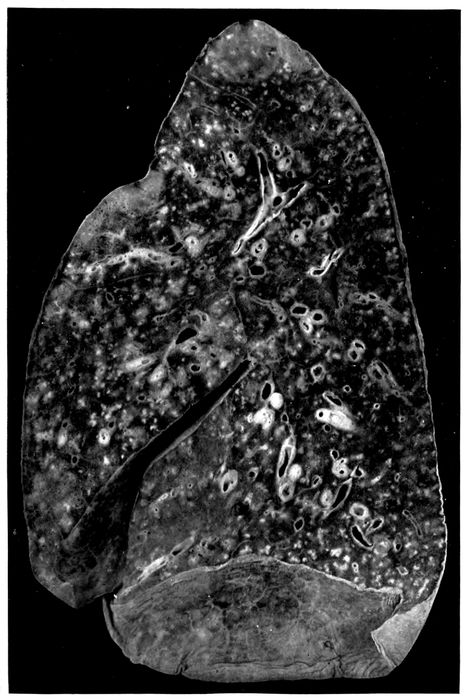
Fig. 2.—Acute bronchopneumonia with nodules of peribronchiolar consolidation and purulent bronchitis. Autopsy 429.
168In association with this lesion there are almost invariably
severe lesions of the bronchi. Purulent bronchitis was
noted in 47 of the 61 instances, in which this nodular bronchopneumonia
was found at autopsy. An index of the severity
of the bronchial injury is the frequency with which
bronchiectasis has occurred; dilatation of small bronchi
was observed in 24 instances. In 10 instances the bronchi
were encircled by conspicuous zones of hemorrhage.
In association with this peribronchiolar lesion the lung is
often voluminous and fails to collapse on removal from the
chest. Pressure upon the lung squeezes from the smallest
bronchi, both in the neighborhood of the nodular consolidation
and elsewhere, a droplet of viscid, semifluid mucopurulent
material. The presence of this tenacious material
throughout the small bronchi doubtless explains the failure
of the lung tissue to collapse. Interstitial emphysema has
been present in some of these lungs.
A red zone of hemorrhage has occasionally been observed
about the foci of peribronchiolar pneumonia. A further
stage in the same process is represented by hemorrhage
into all of the alveoli separating these patches of consolidation.
This hemorrhagic lesion, which will be described in
more detail later, has been found repeatedly in the same
lung with peribronchiolar pneumonia, being present in 8
among the 61 autopsies cited. Lobular bronchopneumonia
accompanied the peribronchiolar lesion 27 times and lobar
pneumonia accompanied it 20 times.
When an abscess caused by hemolytic streptococcus is
associated with peribronchiolar pneumonia, empyema is
present, but otherwise pleurisy is absent or limited to a
scant fibrinous exudate.
169
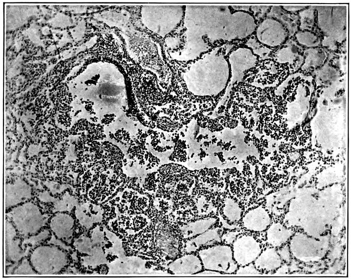
Fig. 3.—Acute bronchopneumonia with peribronchiolar consolidation; a respiratory bronchiole partially lined by columnar epithelium passes into alveolar duct and the adjacent alveoli are filled by polynuclear leucocytes. Autopsy 333.
Histologic examination demonstrates very clearly the relation
of this lesion to the bronchioles (Fig. 3). These
passages are filled and distended with an inflammatory exudate
consisting almost entirely of polynuclear leucocytes.
The respiratory bronchioles are beset with alveoli often
limited to one side of the tubule and these alveoli are filled
with leucocytes. The alveolar ducts, distinguishable from
the bronchioles by the absence of columnar or cubical epithelium
and by possession of smooth muscle, are similarly
filled with leucocytes; the numerous alveoli which form the
walls of the alveolar ducts are distended by an inflammatory
exudate. In sections which pass through an alveolar
duct and one or more of its infundibula, the further extension
of the lesion may be determined (Fig. 4). The infundibulum
in proximity with the alveolar duct contains polynuclear
leucocytes and the same cells are seen in the alveoli
which here form its wall, but the intensity of the inflammatory
reaction diminishes toward the periphery, so that the
distal part of the infundibulum, which is much distended
170and in consequence more readily definable than usual, is
free from inflammatory exudate.
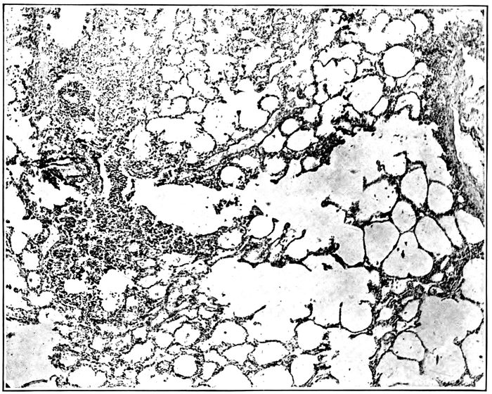
Fig. 4.—Acute bronchopneumonia with peribronchiolar consolidation; a respiratory bronchiole is in continuity with an alveolar duct and two distended infundibula; alveoli about bronchiole, alveolar duct and proximal part of infundibula contain polynuclear leucocytes, the distal part of the infundibula showing no evidence of inflammation. Autopsy 333.
Occasionally there is irregularly distributed hemorrhage
and perhaps some edema in the alveoli immediately adjacent
to those which form the peribronchiolar focus of inflammation.
In such instances small bronchi, that is, air
passages, lined by columnar epithelium and devoid of tributary
alveoli, may be surrounded by a zone of hemorrhage;
immediately surrounding the bronchus, the wall of which
shows intense inflammation, alveoli, in a zone of which the
radius represents several alveoli, are filled with blood. This
hemorrhagic zone is continued from the bronchus over the
focus of inflammation which surrounds the bronchiole.
171Another variation in the character of the lesion is doubtless
referable to variation in the severity of primary bronchial
injury. Alveoli immediately surrounding small bronchi
are filled with dense plugs of fibrin. The alveoli which
besot the walls of the bronchioles contain fibrin, but the
alveolar duct and its tributary alveoli are filled with polynuclear
leucocytes.
The bacteria which have been cultivated from the lung in
autopsies with peribronchiolar pneumonia are as follows:
| Pneumococcus |
5 |
| S. hemolyticus |
8 |
| B. influenzæ, pneumococcus |
5 |
| B influenzæ, S. hemolyticus |
7 |
| B. influenzæ, staphylococcus |
1 |
| Pneumococcus, staphylococcus |
2 |
| S. hemolyticus, staphylococcus |
2 |
| B. influenzæ, pneumococcus, S. hemolyticus |
2 |
| B. influenzæ, pneumococcus, staphylococcus |
1 |
| B. influenzæ, S. hemolyticus, staphylococcus |
2 |
| Pneumococcus, S. hemolyticus, staphylococcus |
3 |
| No organism |
3 |
| |
|
| Total |
41 |
The following list which shows the bacteria found in the
blood is an index to the pathogenicity of pneumococci and
hemolytic streptococci:
| Pneumococcus |
22 |
| S. hemolyticus |
20 |
| Pneumococcus, S. hemolyticus |
1 |
| No organism |
14 |
| |
|
| Total |
57 |
The percentage incidence of pneumococcus, hemolytic
streptococcus, staphylococcus and B. influenzæ in bronchus,
lung and blood, given in Table XXXVI, is inserted to
indicate with what readiness each one of these microorganisms
passes from the bronchus through the lung into the
circulating blood.
| 172 |
| Table XXXVI |
|---|
| |
|
PNEUMOCOCCUS |
HEMOLYTIC STREPTOCOCCUS |
STAPHYLOCOCCUS |
B. INFLUENZA |
| Bronchus |
39.4% |
57.7% |
60.6% |
84.8% |
| Lung |
43.9% |
61.0% |
21.9% |
43.9% |
| Blood |
40.3% |
36.8% |
0. % |
0. % |
B. influenzæ is present in the bronchi in a very large proportion
(84.8 per cent) of those in whom this type of bronchopneumonia
has been found at autopsy; it is much less
frequently recovered from the lungs. Staphylococci, in
part S. albus and in part S. aureus, are less frequently
found in the bronchi and are recovered from the lungs in a
relatively small proportion of autopsies. The percentage
incidence of pneumococci and streptococci in lungs and
blood demonstrates the pathogenicity of these microorganisms,
for whereas pneumococci and hemolytic streptococci
are found in the consolidated lungs in 43.9 and 61.0 per
cent of instances of the lesion respectively, they make their
way into the blood in 40.3 and 36.8 per cent of instances.
Coexisting infection with pneumococci and hemolytic
streptococci has been not uncommon e. g., Autopsy 275 in
which both were in the blood; in 2 instances (Autopsies 333
and 378) in which pneumococci were obtained from the
blood, hemolytic streptococci were found in the lungs and
bronchi; in 3 instances (Autopsies 258, 273 and 445) in
which hemolytic streptococci were present in the blood,
pneumococci were obtained from the lungs.
In the group of autopsies under consideration, examination
of the sputum was made during life and after onset of
pneumonia in 11 instances. The microorganisms found in
the sputum and at autopsy were as follows:
|
|
SPUTUM |
IN BLOOD, LUNGS OR BRONCHUS AT AUTOPSY |
| Autopsy |
240 |
Pneum. IV |
Pneum. IV |
| |
246 |
Pneum. atyp. II, B. inf. |
|
| |
247 |
Pneum. IV, B. inf. |
Pneum. IV |
| |
250 |
Pneum. atyp. II, B. inf. |
Pneum. atyp. II |
| |
253 |
Pneum. atyp. II |
Pneum. II |
| |
285 |
Pneum. atyp. II, B. Inf. |
S. hem., B. inf. |
| |
288 |
S. hem., B. inf. |
S. hem., B. inf. |
| 173 |
291 |
Pneum. IV, B. inf. |
Staph., B. inf. |
| |
300 |
Pneum. atyp. II, B. inf. |
Pneum. atyp. II, B. inf. |
| |
312 |
Pneum. IV, S. hem., B. inf. |
S. hem., B. inf. |
| |
346 |
Pneum. IV, B. inf. |
S. hem., B. inf. |
In 2 instances (Autopsies 285 and 346) among this small
group of cases, pneumococci but no hemolytic streptococci
were found in the sputum several days before death,
whereas death occurred as the result of secondary invasion
with hemolytic streptococci and no pneumococci were found
at autopsy. It is probable that this sequence of events is
not uncommon. B. influenzæ finds its way into the bronchi
and pneumococci follow it; pneumonia limited to peribronchiolar
alveoli may occur in consequence of this invasion.
Later hemolytic streptococci may follow the same path and
cause death with bacteremia.
Hemorrhagic Peribronchiolar Consolidation.—Peribronchiolar
pneumonia accompanied by diffuse accumulation
of blood within the alveoli is one of the most frequent complications
of influenza. The lung tissue is laxly consolidated,
and on section there is a homogeneous dull deep red
background upon which are seen small gray spots (1.5 to 2
mm. in diameter) grouped in clusters about the smallest
bronchi (Fig. 5). Wide areas of lung tissue are implicated
and the lesion is more common in the dependent parts of
the lung than elsewhere. In common with other forms of
bronchopneumonia the lesion is in most instances associated
with changes in the bronchi; in 55 instances of hemorrhagic
bronchiolar pneumonia purulent bronchitis was
found in 43 instances; it is noteworthy that purulent bronchitis
often is not evident in the presence of pulmonary
edema and edema is not infrequent with this pneumonic
lesion.
Microscopic examination demonstrates the presence of
acute bronchitis; the lumina of the small bronchi contain
polynuclear leucocytes and red blood corpuscles. Accumulation
of blood may separate the epithelium from the basement
174membrane. The mucosa immediately below the epithelium
contains polynuclear leucocytes in fair abundance
and the blood vessels of the bronchial wall are much engorged.
Respiratory bronchioles are distended with polynuclear
leucocytes and red blood corpuscles. In a zone
about each bronchiole, in areas corresponding to the small
gray spots seen upon the cut surface of the lung, the alveoli
are filled with polynuclear leucocytes. In the lung tissue
intervening between these spots of leucocytic pneumonia
the alveoli are distended with red blood corpuscles.
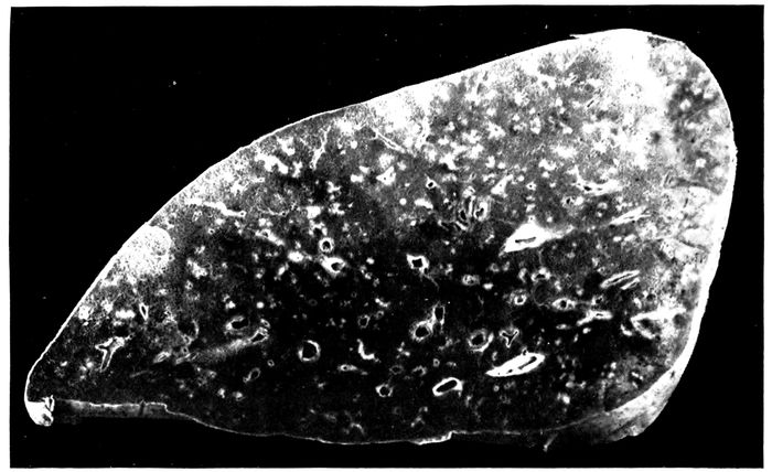
Fig. 5.—Bronchopneumonia with hemorrhagic peribronchiolar consolidation.
In favorable sections it is occasionally possible to follow
the bronchiole and alveolar duct, both filled with leucocytes,
into an infundibulum. The proximal part of the infundibulum
contains polynuclear leucocytes, whereas the
distal part and its tributary alveoli are filled with serum
and red blood corpuscles.
When the lesion has persisted for a short time there is
evidence of beginning migration of polynuclear leucocytes
from the blood vessels into the alveoli which are filled with
175blood. The alveolar walls contain numerous polynuclear
leucocytes and leucocytes which have entered the intraalveolar
blood are numerous in contact with the wall but occur
in scant number in the center of the alveolar lumen.
Alveolar epithelium in contact with the blood in the lumen
is usually swollen and often uniformly nucleated.
The inflammatory process is evidently transmitted from
the bronchioles and to a less degree from the small bronchi
to the adjacent alveoli. Polynuclear leucocytes fill the lumen
of the bronchiole and the alveoli immediately adjacent;
at the periphery of the focus of pneumonia, the alveoli
may contain fibrin. In such instances small bronchi
(lined by a continuous layer of columnar epithelial cells)
may be surrounded by alveoli containing fibrin.
In sections from one part of the lung, the alveoli between
the peribronchiolar foci of pneumonia may be uniformly
filled with red blood corpuscles, whereas in sections from
another part pneumonic foci may be surrounded by a zone
of intraalveolar hemorrhage or of hemorrhage and edema
outside of which some air-containing tissue occurs. There
are transitions between this halo of intraalveolar hemorrhage
and edema surrounding each bronchiolar focus and
complete hemorrhagic infiltration of all intervening alveoli.
Large mononuclear cells are occasionally fairly numerous
within the alveoli containing blood. These cells act as
phagocytes ingesting red corpuscles, so that at times they
are filled with corpuscles. Disintegration of red corpuscles
occurs and brown pigment remains within the cell. It is
not uncommon to find numerous mononuclear pigment containing
cells which resemble those found with chronic passive
congestion of the lungs.
Lungs, the site of hemorrhagic peribronchiolar pneumonia,
may undergo chronic changes which will be described
elsewhere.
The lesion which has been designated hemorrhagic peribronchiolar
pneumonia is that which Pfeiffer regarded as
176the characteristic type of influenzal pneumonia. In the
small bronchi containing pus and in lung tissue, Pfeiffer
states, influenza bacilli are predominant and present in astonishing
number in smear preparations. The demonstration
of B. influenzæ by cultures from pneumonic lung is
mentioned by him but its association with other microorganisms
in such cultures is not discussed.
Microorganisms which we have isolated from the lungs
of individuals with hemorrhagic peribronchiolar pneumonia
are as follows:
| B. influenzæ |
1 |
| Pneumococcus |
2 |
| S. hemolyticus |
10 |
| B. influenzæ, pneumococcus |
7 |
| B. influenzæ, S. hemolyticus |
3 |
| B. influenzæ, staphylococcus |
2 |
| S. hemolyticus, B. coli |
3 |
| B. influenzæ, pneumococcus, staphylococcus |
2 |
| B. influenzæ, S. hemolyticus, staphylococcus |
5 |
| Pneumococcus, S. hemolyticus, staphylococcus |
1 |
| No organisms |
2 |
| |
|
| Total |
38 |
With this type of pneumonia B. influenzæ has not been
isolated in pure culture; B. influenzæ alone is recorded only
once (Autopsy 435), but in this instance the culture has
been so obscured by contamination that the occurrence of
pneumococci or streptococci cannot be excluded; S. hemolyticus
has doubtless been present in this lung, for it has
been found in the heart’s blood, in the bronchus, and in the
peritoneal exudate of the same individual.
The incidence of pneumococci and hemolytic streptococci
in this list does not differ materially from that with peribronchiolar
pneumonia unaccompanied by extensive intraalveolar
hemorrhage, though hemolytic streptococci are
somewhat more frequent with the hemorrhagic lesion. The
following table shows the frequency with which pneumococci
and hemolytic streptococci have penetrated into the
blood:
| 177 |
| Pneumococcus |
11 |
| S. hemolyticus |
24 |
| Pneumococcus, S. hemolyticus |
1 |
| No organism |
12 |
| |
|
| Total |
48 |
Table XXXVII showing the percentage incidence of
pneumococci, hemolytic streptococci, staphylococci and B.
influenzæ further emphasizes the similarity between the
bacteriology of peribronchiolar pneumonia (Table XXXVI)
and the closely related hemorrhagic lesion:
| Table XXXVII |
|---|
| |
|
PNEUMOCOCCUS |
HEMOLYTIC STREPTOCOCCUS |
STAPHYLOCOCCUS |
B. INFLUENZÆ |
| Bronchus |
44.0% |
64.0% |
44.0% |
72.0% |
| Lung |
31.6% |
57.9% |
26.8% |
52.6% |
| Blood of heart |
25.0% |
52.1% |
0% |
0% |
Pneumococci have been found in the lungs (31.6 per
cent) and blood (25 per cent), somewhat less frequently
than with peribronchiolar pneumonia (43.9 and 40.3 per
cent respectively), and hemolytic streptococci have been
found in the blood more frequently (52.1 per cent) than
with the latter (36.8 per cent) but otherwise the bacteriology
of the two lesions corresponds closely. The low incidence
of B. influenzæ in the bronchi (72 per cent) with hemorrhagic
peribronchiolar pneumonia is perhaps incorrect
as the result of the relatively small number of bacteriologic
examinations (namely, 25), but the incidence of the same
microorganism in the lung has been higher (52.6 per cent)
than with nonhemorrhagic peribronchiolar lesion (43.9 per
cent).
In some instances infection with hemolytic streptococci
has occurred after the onset of pneumonia. The following
list compares the results of bacteriologic examination of
the sputum made after the onset of pneumonia with that of
blood, lungs or bronchus after death:
| 178 |
|
|
SPUTUM |
IN BLOOD, LUNGS OR BRONCHUS AT AUTOPSY |
| Autopsy |
237 |
S. hem. |
S. hem. |
| |
242 |
Pneum. atyp. II, B. inf. |
Pneum. atyp. II |
| |
247 |
Pneum. IV, B. inf. |
Pneum. IV |
| |
266 |
S. hem. |
S. hem., B. inf. |
| |
346 |
Pneum. IV, B. inf. |
S. hem., B. inf. |
| |
376 |
(No. S. hem.) |
S. hem., staph., B. inf. |
Instances of secondary infection with hemolytic streptococcus
occur in the list, namely, Autopsies 346 and 376.
From the foregoing studies of the bacteriology of peribronchiolar
and hemorrhagic peribronchiolar pneumonia
the following conclusions may be drawn: (a) B. influenzæ
is found in most instances of these lesions in the bronchi
and in about half of all instances in the lungs, but does not
occur unaccompanied by other microorganisms. (b) In a
considerable number of autopsies pneumococcus is the only
microorganism that accompanies B. influenzæ; from the
lungs it penetrates into the blood from which it is obtained
in pure culture. (c) In a considerable number of instances
S. hemolyticus accompanies B. influenzæ, and in some of
these instances (representing a large proportion of the relatively
small number of cases examined during life), examination
of the sputum has demonstrated that infection has
been secondary to a pneumonia with which no hemolytic
streptococci have been found in the sputum.
Lobular Consolidation.—Consolidation of scattered lobules
or groups of lobules has occurred in nearly all instances,
namely, 71 of 80 autopsies with bronchopneumonia
unaccompanied by lobar pneumonia or by suppuration.
When death follows shortly after the onset of pneumonia,
patches of consolidation have a dull deep red color; blood-tinged
fluid escapes from the cut surface which is almost
homogeneous or finely granular. The consolidated tissue
seen through the pleura, which is raised above the general
level, has a bluish red color. Isolated lobules or groups of
lobules which have undergone consolidation may be scattered
throughout the lungs, but not infrequently there is
confluent consolidation of the greater part of lobes, of
179whole lobes or of almost an entire lung. Such lungs are
very heavy and may weigh 1,400 or 1,500 grams; bloody
serous fluid exudes from the cut surface. The lesion resembles
the red hepatization of lobar pneumonia, but confluent
patches of pneumonia are usually well defined by lobule
boundaries. The tissue is soft and the granulation of lobar
pneumonia is absent. In many instances the lobular or confluent
areas of consolidation are reddish gray; in some instances
consolidated tissue is in places red and elsewhere
gray, and in a smaller group of autopsies there is gray
consolidation only (Fig. 6). Red lobular consolidation is
often seen in those who have died within the first four days
following the onset of pneumonia, but is almost equally frequent
after from five to ten days; the average duration of
pneumonia in these cases was 5.5 days. Combined red and
gray consolidation was more frequently found when pneumonia
had lasted more than five days, the average duration
of pneumonia being 7.3 days. The greater number of instances
of gray consolidation were found after seven days
of pneumonia, the average duration of the disease being
10.0 days. These figures are cited to show that lobular,
like lobar, consolidation passes gradually from a stage of
red to gray hepatization, but the change occurs more slowly
and is often long delayed.
Lobular pneumonia, which occurred 71 times among 80
cases classified as bronchopneumonia, may be regarded as
an almost constant lesion of the disease. It is found not
only in association with other lesions of bronchopneumonia,
but with lobar pneumonia of influenza as well.
The bacteriology of this lesion shows no deviation from
that of the slightly larger group of bronchopneumonia
(p. 163). All types of pneumococcus have been found in
association with the lesion, Pneumococcus I in 2 instances,
Pneumococcus II in 1 instance; atypical Pneumococcus II
and Pneumococcus IV have been found much more frequently.
Pneumococci have been found in more than a
third of these autopsies (42.9 per cent in the lungs, 33.3 per
cent in the blood); hemolytic streptococci in less than one-third
(28.5 per cent in the lungs, 30.2 per cent in the blood).
180
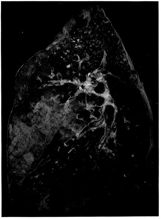
Fig. 6.—Acute bronchopneumonia with confluent gray lobular consolidation in lower part of upper lobe and hemorrhagic peribronchiolar pneumonia in lower lobe; purulent bronchitis.
181The following list shows the bacteriology of a small
group of autopsies in which the sputum was examined after
onset of pneumonia:
|
|
SPUTUM |
BLOOD, LUNGS OR BRONCHUS AT AUTOPSY |
| Autopsy |
233 |
Pneum. atyp. II |
Pneum. |
| |
237 |
S. hem. |
S. hem. |
| |
242 |
Pneum. atyp. II, B. inf. |
Pneum. atyp. II |
| |
250 |
Pneum. atyp. II, B. inf. |
Pneum. atyp. II |
| |
253 |
Pneum. atyp. II |
Pneum. atyp. II, staph., B. inf. |
| |
266 |
S. hem. |
S. hem., B. inf. |
| |
274 |
Pneum. IV |
S. hem. |
| |
291 |
Pneum. IV, B. inf. |
Staph., B. inf. |
| |
312 |
Pneum. IV, S. hem., B. inf. |
S. hem., staph., B. inf. |
In one instance of streptococcus pneumonia (Autopsy
274) infection with streptococci occurred subsequent to the
examination of the sputum made five days before death;
pneumococcus was found in the washed sputum.
With lobar pneumonia there was evidence that superimposed
infection occurred more frequently during the stage
of red than of gray hepatization. With the lobular consolidation
of bronchopneumonia this relation has not been
found. Among 27 instances of red lobular consolidation,
hemolytic streptococcus has occurred 6 times, namely in
22.2 per cent; among 26 instances of red and gray consolidation,
8 times, namely, in 30.7 per cent; among 13 instances
of gray consolidation, 5 times, namely, in 38.5 per
cent. Infection with hemolytic streptococci is more frequent
when the lesion has persisted to the stage of gray
hepatization. This difference between lobar and bronchopneumonia
is probably dependent in part at least upon the
more severe and persistent lesions of the bronchi with
bronchopneumonia.
The histology of consolidation which is definitely limited
to secondary lobules or groups of lobules varies considerably.
When death occurs in the early stage of the lesion,
182consolidated patches are deep red and somewhat edematous,
so that bloody serous fluid escapes from the cut surface
of the lung and red blood corpuscles are present in the
alveoli in great abundance together with polynuclear leucocytes,
fibrin and serum in varying quantity. It is not
uncommon to find evidence that the lesion has had its origin
in the bronchioles and extended from them to other
parts of the lobule. Polynuclear leucocytes may be relatively
abundant within and immediately about the bronchioles
and alveolar ducts, whereas the intervening alveoli and
infundibula are filled with red blood corpuscles among
which are polynuclear leucocytes and perhaps some fibrin.
It may be evident that bronchiolar pneumonia with hemorrhage
into intervening alveoli is in process of transformation
into a more diffuse leucocytic pneumonia, for polynuclear
leucocytes are making their way from the alveolar
wall into the blood-filled lumen and, as the result of the
presence of blood, remain for a time close to the lining of
the alveolus.
When the consolidated lobules have assumed a gray or
reddish gray color, polynuclear leucocytes are more abundant
and often almost homogeneously pack every alveolus
within the boundaries of the lobule. In some instances
there is fibrin partially obscured by the presence of leucocytes
in great number.
Although fibrin is less abundant with bronchopneumonia
than with lobar pneumonia, nevertheless in a considerable
proportion of instances it is a very conspicuous element
of the inflammatory exudate within the bronchioles, alveolar
ducts and alveoli. It is unusual to find the alveolar
ducts and alveoli uniformly plugged with fibrin containing
leucocytes; there is a variegated distribution of exudate
which has little resemblance to that of lobar pneumonia.
Occasionally (Autopsies 242 and 247) polynuclear leucocytes
fill the bronchioles, alveolar ducts and infundibula,
whereas the surrounding tributary alveoli contain fibrin
183and polynuclear leucocytes in moderate number; red blood
corpuscles may be present in sufficient number to give a
homogeneously red color to the lobular consolidation.
In association with lobular pneumonia, fibrin within the
lung tissue undergoes certain changes which outline very
sharply the alveolar ducts and the other structures usually
ill defined in preparations of the lung. A remarkable appearance
is produced by the deposit of hyalin fibrin upon
the surface of the alveolar ducts and infundibula. This
lesion has been described by LeCount.
Within the alveolar tissue of the lung, spaces are seen
lined by a layer of fibrin which stains homogeneously and
very brightly with eosin. They are recognized as alveolar
ducts by the presence of scattered bundles of smooth
muscle in their wall. The layer of hyaline fibrin overlying
the surface of the alveolar duct usually forms a continuous
lining and covers over the orifices of the alveoli which surround
the alveolar duct. These ducts are rendered still
more conspicuous by the character of their contents which
exhibits a sharp contrast with that of the surrounding
alveoli. The alveoli duct occasionally contains a bubble of
air, but more frequently it is filled with serum in which red
blood corpuscles are sometimes numerous. There is within
the lumen scant fibrin and very few cells, among which polynuclear
leucocytes are predominant. In the surrounding
alveoli on the contrary leucocytes and fibrin are abundant.
A similar change is found in the infundibula very clearly
defined by their conical form, which is especially well outlined
below the pleura or in contact with interlobular septa.
The infundibulum is outlined by hyaline fibrin which passes
over the orifices of the tributary alveoli and separates the
serous contents of the infundibulum from the cellular fibrinous
contents of the alveoli about.
The lesion which has been described is often associated
with acute bronchitis and bronchiolitis, and the alveoli immediately
about the respiratory bronchioles may be filled
184with polynuclear leucocytes. It is very common to find
large bubbles of air sharply defined within the purulent
contents of the bronchiole. In some lobules the alveolar
ducts, infundibula and alveoli intervening between
these foci of leucocytic pneumonia are almost uniformly
filled with fibrin and polynuclear leucocytes, but in other
places the formation of complete layers of hyaline fibrin
is in process. Bubbles of air are often seen within the alveolar
ducts, and about them is an irregular layer of fibrin
formed by the penetration of air into a channel previously
filled with a loose network of fibrin containing serum in its
meshes. The fibrin compressed against the walls of alveolar
duct and infundibulum remains as a compact layer separating
these structures from the alveoli which project
from their walls. The bubble of air is doubtless later absorbed
and replaced by serum, so that many alveolar ducts
are filled with serum almost wholly free from cells, whereas
alveoli outside the fibrinous membrane contain a network
of fibrin with leucocytes in greater or less abundance.
In association with this fibrinous pneumonia, which has
been described, hyaline thrombosis of the capillaries is not
uncommon. This hyalin material within the capillaries
gives reactions of fibrin, and in sections stained by the
Gram-Weigert method for demonstration of fibrin, these
thrombosed vessels have the appearance of capillaries irregularly
injected with a blue material.
The interstitial tissue surrounding consolidated lobules
is often edematous; the lymphatics are distended with
serum and contain a moderate number of lymphocytes and
polynuclear leucocytes.
Among the lungs which have been studied histologically,
pneumococcus has been almost invariably associated with
the lobular lesions which have just been described, whether
hemorrhagic, leucocytic or fibrinous; the histologic changes
accompanying infection of the lung with streptococcus will
be described later. Pneumococcus has been cultivated from
the consolidated lung and is found in section of the lung.
B. influenzæ is found in cultures made from the bronchi.
Table XXXVIII includes those instances in which the
histology of the consolidated lung accords with the description
given above.
| 185 |
| Table XXXVIII |
|---|
| |
| NO. OF AUTOPSY |
CHARACTER OF LOBULAR CONSOLIDATION |
PREDOMINANT TYPE OF INFLAMMATORY EXUDATE |
CULTURE FROM HEART’S BLOOD |
CULTURE FROM LUNG |
CULTURE FROM BRONCHUS |
| 242 |
Red |
Fibrinous |
Pneum. atyp. II |
|
|
| 244 |
Red |
Leucocytic and hemorrhagic |
|
Pneum. IV B. inf. |
Pneum. IV, B. inf. |
| 247 |
Red and gray |
Fibrinous |
Pneum. IV |
|
|
| 249 |
Red and gray |
Fibrinous |
Pneum. III |
|
|
| 252 |
Red and gray |
Fibrinous |
|
Pneum. II B. inf. |
Pneum. II, B. inf., S. vir. |
| 257 |
Red and gray |
Leucocytic |
Pneum. I |
|
B. inf., staph. |
| 303 |
Red |
Fibrinous |
|
Pneum. IV B. inf. |
Pneum. IV, B. inf., staph. |
| 314 |
? |
Fibrinous |
Pneum. IV |
Pneum. IV |
Pneum. IV, B. inf., staph. |
| 336 |
Red |
Fibrinous |
|
|
|
| 395 |
Red and gray |
Leucocytic |
Pneum. atyp. II |
Pneum. atyp. II |
|
| 464 |
Red |
Leucocytic and hemorrhagic |
|
Pneum. I B. inf. |
Pneum. I, B. inf., staph. |
| 476 |
Red |
Leucocytic and hemorrhagic |
|
|
|
| 498 |
Red and gray |
Fibrinous |
|
S. aur. |
|
| 506 |
Red |
Fibrinous |
Pneum. IV |
Pneum. IV S. aur. |
Pneum. IV, B. inf., S. aur., M. catarrh |
186Pneumococcus was found in all but 2 instances, and in
one of these (Autopsy 336) the only culture was from the
heart’s blood and in the other (Autopsy 498) cultures were
unsatisfactory because proper media were not obtainable.
Pneumococci of Types I, II, II atypical, III and IV are represented
in the list. B. influenzæ has been found in a considerable
number of instances in which cultures have been
made from the lung and in every instance in which cultures
have been made from the bronchi. Staphylococci are often
found in the bronchi, but in most instances they do not
penetrate into the lung.
Another group of cases of lobular pneumonia are important
because in association with necrosis of lung tissue
recognized by the microscope hemolytic streptococci have
been found in the lungs. In such instances serum is abundant
and polynuclear leucocytes are relatively scant
though their distribution varies considerably; in some
places leucocytes are fairly abundant though elsewhere almost
absent, but this distribution bears no obvious relation
to the bronchioles. In some instances (Autopsies 274 and
487) red blood corpuscles are numerous but in others (Autopsies
275 and 312) they are inconspicuous. The characteristic
feature of the lesion is the occurrence of patches
of necrosis within which the nuclei both of exudate and of
alveolar walls have partially or completely disappeared.
In these areas of necrosis short chains of streptococci are
found in immense number whereas in living tissue they are
present in moderate number. There has been a relatively
inactive inflammatory reaction, great proliferation of
streptococci and necrosis of invaded tissue. The bacteriology
of instances of lobular pneumonia with necrosis is
shown in Table XXXIX.
| 187 |
| Table XXXIX |
|---|
| |
| NO. OF AUTOPSY |
CHARACTER OF LOBULAR CONSOLIDATION |
PREDOMINANT TYPE OF INFLAMMATORY EXUDATE |
CULTURE FROM HEART’S BLOOD |
CULTURE FROM LUNG |
CULTURE FROM BRONCHUS |
| 274 |
Red |
Leucocytic and hemorrhagic |
S. hem. |
S. hem. |
S. hem., staph. |
| 275 |
Red and gray |
Leucocytic |
Pneum. IV S. hem. |
S. hem., B. inf., staph. |
S. hem., B. inf., staph. |
| 312 |
Red and gray |
Leucocytic |
S. hem. |
S. hem., B. inf. |
S. hem., B. inf., staph. |
| 478 |
Red |
Leucocytic and hemorrhagic |
S. hem. |
S. hem. |
|
188Lobular pneumonia, in some of these instances at least,
has been caused primarily by pneumococci; necrosis has
been the result of secondary invasion by streptococci. In
Autopsy 275 Pneumococcus IV has been obtained from the
blood, but in the presence of streptococci has presumably
disappeared from the lung and bronchus. In the case represented
by Autopsy 274, Pneumococcus IV has been found
in the sputum five days before death at the onset of pneumonia,
but at this time no hemolytic streptococci have been
found. In the case represented by Autopsy 312, Pneumococcus
IV, B. influenzæ and a few colonies of hemolytic
streptococci have been obtained from the sputum two days
after recognition of pneumonia and five days before death.
The hemorrhagic and edematous consolidation of the
early pulmonary lesions of influenzal pneumonia is their
most distinctive feature. Red confluent lobular pneumonia
is frequently found in those who have died within the first
week following the onset of influenza. The lungs are voluminous
and heavy and may weigh as much as 1,500 grams;
the pleura which overlies the consolidated area is blue or
plum colored and usually shows scant if any evidence of
pleurisy. Scattered patches of consolidation are accurately
limited to lobules, but in addition there are large
areas often involving the greater part of the lobes and not
infrequently situated in the lowermost part of the lower
lobes. This confluent consolidation may be obviously limited
by lobule boundaries. The consolidated tissue is deep
red and laxly consolidated; red serous fluid escapes from
the cut surface. The lesion not infrequently occurs in association
with hemorrhagic peribronchiolar pneumonia.
The histology of this confluent lesion has been studied
in Autopsies 242, 244, 303, 336, 464, 474 and 506. The histology
varies, because, in some instances, leucocytes, in
other instances, fibrin, is abundant, but the presence of red
blood corpuscles in large number within the alveoli gives
a red color to the consolidated tissue. In these cases pneumococci,
189associated in the lungs or in the bronchi with B.
influenzæ, have been the cause of pneumonia. In two autopsies
studied histologically (Autopsies 274 and 478) there
was red lobular and confluent pneumonia and the blood and
lungs contain hemolytic streptococci demonstrated by cultures;
microscopic examination showed the presence of a
widespread necrosis of the lung tissue.
In the group of autopsies in Table XL there was red confluent
lobular pneumonia. These autopsies are separated
from those just cited because there was no histologic examination
of the tissue.
| Table XL |
|---|
| |
| NO. OF AUTOPSY |
BACTERIOLOGY OF HEART’S BLOOD |
BACTERIOLOGY OF LUNGS |
BACTERIOLOGY OF BRONCHUS |
| 289 |
Pneum. IV |
Pneum. IV |
Pneum. IV, B. inf., staph. |
| 297 |
|
Pneum. IV, B. inf. |
Pneum. IV, B. inf., S. hem. (a few) |
| 306 |
|
|
|
| 339 |
Pneum. IV |
|
|
| 364 |
S. hem. |
|
|
| 418 |
Pneum. atyp. II |
Pneum. atyp. II, B. inf., S. vir. |
|
| 424 |
|
Pneum. IV. |
|
This group of autopsies confirms the view that the red
confluent lobular pneumonia is caused by pneumococci in
association with B. influenzæ. Hemolytic streptococci may
invade secondarily. In Autopsy 297 a few hemolytic streptococci
were found in the bronchus but apparently had not
entered the lungs. In the absence of histologic examination
it is not possible to determine if the invasion of hemolytic
streptococcus (in Autopsy 364) has caused necrosis of
the pneumonic tissue.
190
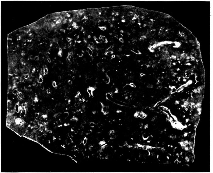
Fig. 7.—Bronchopneumonia with purulent bronchitis and peribronchial hemorrhage.
In a considerable number of instances, namely, in 19 autopsies,
hemorrhage about the small bronchi has been recognizable
upon gross examination of the lung. A conspicuous
zone of hemorrhage 2 or 3 mm. in thickness surrounds
small (with no cartilage) often dilated bronchi and on longitudinal
section may be tracted for a considerable distance
along the bronchus (Fig. 7). In many additional instances
peribronchial hemorrhage has been found by microscopic
examination. In some instances the peribronchial zone of
hemorrhage is firmer than the tissue elsewhere and it is
occasionally difficult to determine whether the lesion is
hemorrhage or pneumonia. In 7 instances frank red consolidation
of peribronchial tissue was recognized at autopsy;
this lesion will be considered later under peribronchial
pneumonia. Hemorrhage about bronchi, like other
evidences of severe injury to bronchi following influenza,
is more frequently found in the lowermost parts of the
lungs than elsewhere. It is invariably associated with
severe bronchitis; the bronchi have contained purulent
191fluid in 15 of 19 instances of peribronchial hemorrhage and
in 10 instances the lesion has been associated with dilatation
of the bronchi.
Microscopic examination furnishes further evidence of
the severity of the bronchial changes which have brought
about hemorrhage into the surrounding alveoli. The lumen
of the bronchus contains blood and leucocytes; the epithelium
is sometimes raised in places from the underlying
basement membrane by blood; blood vessels of the bronchial
wall are engorged, and there is hemorrhage into the
tissue of the bronchus. More frequently the bronchial epithelium
is completely lost and the denuded surface is often
covered by a layer of fibrin intimately adherent to the inflamed
mucosa. Transitions between simple hemorrhage
and pneumonia are found, polynuclear leucocytes being
mingled with red blood corpuscles. In several instances
the alveoli in immediate contact with the bronchial wall
have contained fibrin, whereas those in the surrounding
zone have contained blood.
Bacteria found in the bronchi in 10 instances of peribronchial
hemorrhage have been as follows:
| Staphylococci |
1 |
| B. influenzæ, pneumococci |
1 |
| B. influenzæ, S. hemolyticus |
2 |
| B. influenzæ, pneumococci, staphylococci |
1 |
| B. influenzæ, S. hemolyticus, staphylococci |
4 |
| No organism found |
1 |
The high incidence of B. influenzæ and the frequent association
of B. influenzæ and hemolytic streptococci are
noteworthy. The instance in which no organisms were
found is probably due to a defect in media and should perhaps
be excluded from the list.
The percentage incidence of pneumococci, hemolytic
streptococci, staphylococci and B. influenzæ in the bronchus,
lungs and blood of the heart is an index of the facility with
which these microorganisms penetrate internal organs
when the bronchi are the site of this hemorrhagic lesion.
192
| Table XLI |
|---|
| |
|
NO. OF CULTURES |
PNEUMOCOCCI |
HEMOLYTIC STREPTOCOCCI |
STAPHYLOCOCCI |
B. INFLUENZÆ |
| NO. POSITIVE |
PER CENT POSITIVE |
NO. POSITIVE |
PER CENT POSITIVE |
NO. POSITIVE |
PER CENT POSITIVE |
NO. POSITIVE |
PER CENT POSITIVE |
| Bronchus |
10 |
2 |
20.0 |
6 |
60.0 |
6 |
60.0 |
8 |
80.0 |
| Lung |
13 |
4 |
30.8 |
7 |
53.8 |
3 |
23.1 |
5 |
38.5 |
| Blood |
17 |
4 |
23.5 |
9 |
52.9 |
|
|
|
|
When these figures are compared with those for all forms
of bronchitis no very noteworthy differences are found;
the incidence of pneumococci here is less and that of hemolytic
streptococci greater. In association with the severe
changes present in the bronchi, hemolytic streptococci
which enter the lungs almost invariably find their way into
the blood.
In 6 instances there has been frank pneumonic consolidation
limited to a zone encircling small and medium-sized
bronchi which have often been obviously dilated. On cross
section these patches of pneumonia are circular, from 1 to
2 cm. in diameter and each contains a bronchus at its center.
When the bronchus is cut longitudinally it is evident that
pneumonic consolidation forms a cylindrical sheath about
the tube. The consolidation varies in color from red to
grayish red. In one instance (Autopsy 253) the consolidated
tissue has formed a gray zone in contact with the
bronchus and is red in a peripheral zone; microscopic examination
shows that the alveoli about the bronchus contain
fibrin, whereas those at a greater distance contain red
blood corpuscles. In this instance, the associated pneumonia
in another part of the lung has been somewhat
anomalous and has had characters both of lobar and bronchopneumonia,
for scattered in the left lung there have been
patches of firm consolidation not more than 2 cm. across.
The smaller of these patches are deep red, but the larger
are coarsely granular and gray in the center. The patchy
193character of the lesion has suggested bronchopneumonia,
but the coarse granulation on section and the presence of
fibrinous plugs within the small bronchi have presented a
close resemblance to lobar pneumonia. This autopsy is
one of the few instances in which Pneumococcus II has been
found, Pneumococcus II being present in blood and lungs,
B. influenzæ, in lungs and bronchi. In 2 additional instances
(Autopsies 374 and 392) peribronchial pneumonia,
recognizable at autopsy, has been associated with consolidation
having the characters of lobar pneumonia. In one
instance, Autopsy 374, the right lung has contained two
patches of firm, mottled red and pinkish red coarsely granular
consolidation each about 6 cm. across, one situated in
the upper lobe and the other in the lower lobe. Elsewhere
in the lung, in definite relation to dilated bronchi, occur
patches of firm, red, coarsely granular consolidation from
1 to 1.5 cm. in diameter when cut transversely. The bronchus
in the center has contained purulent fluid. In the opposite
lung similar consolidation has been limited to zones
about dilated bronchi which contain purulent fluid. Pneumococcus
IV has been obtained from the blood of the heart.
The peribronchial pneumonia which has been described
occurs in association with evidence of profound injury to
the bronchial wall. In 5 of 6 instances purulent bronchitis
has been found at autopsy; in half of these instances bronchiectasis
has been noted. The epithelium of the bronchus
has been found separated from the underlying tissue by serous
exudate, blood and leucocytes; epithelial cells undergo
necrosis and disappear, the denuded surface being covered
by fibrin. Necrosis extends a varying depth into the wall
of the bronchus; blood vessels are engorged, and there is
in some instances hemorrhage throughout the wall of the
bronchus.
The character of the exudate in the alveoli surrounding
the bronchus differs considerably in different instances.
In some instances (Autopsies 374 and 392) red blood corpuscles
194are predominant in the alveoli in contact with the
bronchial wall, whereas in a peripheral zone polynuclear
leucocytes are more abundant. In other instances (Autopsies
253 and 402) alveoli next the bronchial wall contain
abundant fibrin and these are surrounded by a zone in
which the alveoli are filled with blood.
Peribronchial pneumonia is the result of the direct extension
of the inflammatory process through the wall of the
bronchus; it occurs when the epithelium of the bronchus is
destroyed and the underlying tissues are injured, but
may be present in a wide encircling zone even when
the lesion has not penetrated the bronchial wall. The distribution
of the pneumonia demonstrates very clearly that
the inflammatory process does not reach the affected peribronchial
alveoli by way of the bronchioles tributary to the
bronchus.
The bacteriology of these instances of peribronchial
pneumonia is noteworthy. (Table XLII.)
| Table XLII |
|---|
| |
| AUTOPSY |
BLOOD |
LUNG |
BRONCHUS |
| 253 |
Pneum. II |
Pneum. II, B. inf. |
Staph., B. inf. |
| 374 |
Pneum. IV |
|
|
| 387 |
Pneum. II, S. hem. |
Pneum. II, staph., B. inf. |
Pneum. II, S. hem., staph., B. inf. |
| 392 |
Pneum. II |
|
|
| 402 |
Pneum. IV, S. hem. |
|
|
| 424 |
? |
Pneum. IV |
|
Pneumococcus has been found in every instance either in
the lungs or blood. Pneumococcus II, which has been uncommon
with the pneumonia following influenza at Camp
Pike and has occurred only ten times in more than 200
autopsies, has been present in one-half of these cases. The
constant association of the lesion with pneumococcus is
particularly significant when a comparison is made between
the incidence of pneumococcus with peribronchial hemorrhage,
on the one hand, and peribronchial pneumonia on the
other; pneumococcus has been present in less than a third
195of the instances of hemorrhage but in all instances of pneumonia.
In addition to the instances in which gross peribronchial
consolidation has been noted at autopsy, microscopic examination
has demonstrated the presence of fibrinous pneumonia
surrounding bronchi in a considerable number of
autopsies. In a zone encircling small bronchi (with no
cartilage) alveoli are filled by plugs of dense fibrin (Fig.
20) containing in variable number polynuclear leucocytes
and mononuclear cells. The width of the zone is often
equal or greater than the diameter of the bronchus. Alveoli
outside the zone of fibrinous inflammation may contain
red blood corpuscles or serum, and desquamated epithelial
cells are often abundant.
Of 21 instances of peribronchial fibrinous pneumonia 20
were associated with purulent bronchitis. Further evidence
of the relation of the lesion to profound injury to
the bronchi is its association with bronchiectasis in 17 instances.
Peribronchial fibrinous pneumonia, like other lesions encircling
the small bronchi, bears a direct relation to the
severity of microscopic changes in the bronchus. The epithelium
of the bronchus is either partially or completely
lost. Occasionally epithelium is raised by hemorrhage or
leucocytes from the underlying tissue but more frequently
it is wholly lost and the surface is covered by a layer of
fibrin. In the early stages of the lesion, polynuclear leucocytes
may be numerous throughout the bronchial wall, indicating
that the inflammatory irritant within the lumen is
affecting the entire wall and extending its influence to the
surrounding pulmonary tissue. Later lymphoid and plasma
cells are more abundant than polynuclear leucocytes.
Coagulative necrosis and disintegration of the bronchial
wall, proceeding from the inner surface outward, may extend
more or less deeply, and fibrinous inflammation of
adjacent alveoli is often more extensive about that segment
196of the bronchus which shows the greatest change. In some
instances segments of the bronchial wall or even the entire
wall has disappeared, so that alveoli containing fibrin form
part of the wall of the cavity thus formed. When bronchiectasis
has occurred, there are often fissures from the
lumen through the entire wall extending into the surrounding
lung tissue: here fibrinous pneumonia is particularly
conspicuous, occurring in a zone about the edges of the
defect. This deposition of fibrin within the alveoli adjacent
to the injury doubtless has a part in limiting the distribution
of bacterial infection. Nevertheless breaks in the
continuity of the bronchial wall are not essential to the
production of the lesion and the irritant, which is responsible
for the lesion, may penetrate through the bronchial
wall to surrounding alveoli and from alveoli to other alveoli
immediately adjacent.
With this peribronchial pneumonia the smallest bronchi
are distended with pus and their walls are infiltrated with
polynuclear leucocytes, lymphoid and plasma cells. In a
broad zone encircling the bronchus the alveoli are filled
with plugs of fibrin. Bronchioles are similarly distended
with polynuclear leucocytes; the alveoli which occur upon
the wall of the bronchiole are often limited to one side of
the wall and are filled with fibrin. This fibrin occasionally
projects into the lumen of the bronchiole and forms a continuous
layer in contact with the wall on the same side.
The alveolar duct and infundibulum are distended with
polynuclear leucocytes. The alveoli upon the wall of the
alveolar duct and upon the proximal part of the infundibulum
are filled with fibrin. The bronchus, bronchiole,
alveolar duct and part of the infundibulum are thus surrounded
by a continuous zone of alveoli containing fibrin.
The alveoli about the distal part of the infundibulum may
be filled with polynuclear leucocytes. Lung tissue between
adjacent zones of fibrinous pneumonia may contain serum
and desquamated epithelial cells.
197Organization of peribronchial fibrin was found in 10
of the 22 autopsies in which peribronchial fibrinous pneumonia
had been found. Fibroblasts have invaded the fibrin
and newly formed capillaries have penetrated into it. In
some instances the interalveolar septa are thickened and
infiltrated with lymphoid and plasma cells, and in 7 instances
there was chronic pneumonia with thickening and
mononuclear infiltration of the interstitial tissue about the
bronchi and blood vessels, and elsewhere. The duration
of the fatal illness in 12 instances with no organization was
usually from ten days to two weeks, though in 3 instances
there was no organization although the respiratory disease
had lasted from seventeen to nineteen days (average duration
with no organization, 13.5 days). The duration of
illness in 10 instances with organization of fibrin was
slightly less than three weeks (average 18.9 days). These
figures do not accurately represent the duration of pneumonia
which usually develops after a period of several days
following onset of influenza.
This group of instances of peribronchial fibrinous pneumonia
has offered an opportunity to study the bacteriology
of pneumonia with organization and to determine if it presents
any unusual characters. The bacteriology of autopsies
with peribronchial fibrinous pneumonia with no organization
is shown in Table XLIII:
| Table XLIII |
|---|
| |
| AUTOPSY |
BLOOD |
LUNG |
BRONCHUS |
| 289 |
Pneum. IV |
Pneum. IV |
Pneum. IV, B. inf., staph. |
| 372 |
|
|
|
| 376 |
S. hem. |
S. hem. |
S. hem., B. inf., S. aur. |
| 409 |
0 |
|
|
| 410 |
|
S. hem., B. inf. S. aur. |
|
| 412 |
Pneum. II |
|
Pneum. II, B. inf. |
| 420 |
S. hem. |
S. hem., B. inf. S. aur. |
|
| 423 |
S. hem. |
S. hem., B. inf. |
|
| 440 |
0 |
B. inf., S. aur. |
B. inf., S. aur. |
| 448 |
0 |
0 |
0 |
| 482 |
0 |
B. inf., Pneum. IV |
B. inf., Pneum. IV, S. hem. |
| 489 |
0 |
Pneum. IV, B. inf. |
Pneum. IV, B. inf. |
198The bacteriology of instances of peribronchial fibrinous
pneumonia with organization of the intraalveolar fibrin is
shown in Table XLIV:
| Table XLIV |
|---|
| |
| AUTOPSY |
BLOOD |
LUNG |
BRONCHUS |
| 283 |
Pneum. IV |
Staph., B. inf. |
B. inf., Pneum. IV, staph. |
| 291 |
0 |
0 |
B. inf., staph. |
| 398 |
0 |
|
|
| 419 |
0 |
Pneum. II, B. inf. |
Pneum. II, B. inf. |
| 421 |
S. hem. |
Pneum. IV, S. hem. |
|
| 422 |
0 |
Pneum. II atyp., B. inf. |
|
| 425 |
S. hem. |
S. hem., B. inf., S. alb. |
|
| 433 |
0 |
S. hem., B. inf., S. aur. |
|
| 460 |
S. hem. |
S. hem., B. inf. |
S. hem., B. inf., staph. |
| 463 |
0 |
B. inf., staph. |
B. inf., staph., Pneum. IV |
B. influenzæ has been present in the bronchi in every instance
save one in which cultures have been made, and it
is probable that in this exceptional instance cultures have
remained sterile because the media employed have been defective.
The incidence of B. influenzæ in the lung has been
unusually high both with and without organization (66.7
per cent with no organization; 77.8 per cent with organization).
Streptococci and staphylococci have been found
in a considerable proportion of all instances of peribronchial
fibrinous pneumonia, but there has been no notable
preponderance of these microorganisms when organization
has occurred. Organization has been present in instances
in which pneumonia is referable to pneumococcus
associated with B. influenzæ and unaccompanied by either
streptococci or staphylococci (Autopsies 419 and 422).
Wadsworth[81] found no organization after inoculation of the
lungs of dogs with pneumococcus or with staphylococcus
alone, but produced organization when he inoculated animals
with both microorganisms.
Injury to bronchi produced in part at least by B. influenzæ
exposes the bronchi and lung tissue to repeated
infection with a variety of microorganisms; absorption of
199fibrin and regeneration of alveolar epithelium are prevented,
resolution fails to occur and organization of fibrin
follows.
Three varieties of suppurative pneumonia have occurred
in association with influenza.
A. Necrosis and suppuration with formation of one or
several abscesses usually below the pleura and almost invariably
caused by hemolytic streptococci.
B. Interstitial suppurative pneumonia caused by hemolytic
streptococcus.
C. Multiple abscesses in clusters caused by staphylococci.
Suppurative pneumonia with necrosis and abscess formation
will be discussed in this section. Pulmonary abscesses
which occurred in 43 autopsies may be included in
this group; in 4 of these autopsies abscess and interstitial
suppurative pneumonia occurred in the same individual.
These abscesses were much more frequently situated in the
lower than in the upper lobes and more often in the right
than in the left lung. In most instances there was one or
several abscesses situated below the pleura of one lobe;
occasionally abscesses occurred in two lobes of the same
lung or in both lungs. The distribution was as follows:
Abscess in only one lung occurred in right upper lobe in
6 autopsies; middle lobe, 3; lower lobe, 15; left upper lobe,
2; lower lobe, 16. Abscesses occurred in both right and
left lower lobes, twice. The usual situation was at the
lower and posterior part of the lower lobe at or near the
basal edge, less frequently below the posterior border or
upon the basal surface of the lobe. These abscesses in
almost every instance were found immediately below the
pleural surface, so that they appeared upon the pleura
as opaque yellow spots usually surrounded by narrow
zones of hemorrhage. In one instance (Autopsy 376) the
200abscess cavity was separated from the pleural cavity by
remains of the pleura which was as thin as tissue paper and
in other instances perforation had occurred (Fig. 9). In
Autopsy 480 the abscess cavity which had perforated the
pleura was in free communication with a bronchus of medium
size.
In most instances of suppurative pneumonia there have
been associated lesions of bronchopneumonia which have
been peribronchiolar, hemorrhagic or lobular and have exhibited
no unusual characters. The abscess or abscesses
are situated within an area of pneumonic consolidation
which is not limited by lobule boundaries and has not the
characters of bronchopneumonic consolidation. In some
instances this consolidation is limited to a zone immediately
about the abscess, but often it involves the greater part of
a lobe. The tissue is laxly consolidated and flabby; on section
it has a dull, conspicuously cloudy appearance and is
grayish red, pinkish gray or gray; it is homogeneous or
very finely granular. Turbid gray fluid, which sometimes
resembles thin pus, oozes from the cut surface.
Widespread necrosis of tissue is not infrequently a conspicuous
feature of this pyogenic pneumonia (Fig. 8).
Upon a cloudy gray background of consolidation are numerous
opaque yellowish gray or yellow patches, occasionally
2 or 3 cm. across, giving a mottled character to the
cut surface. Upon the pleura these necrotic patches appear
as dull opaque yellow spots. They may be surrounded
by a zone of hemorrhage. The opaque material
is at first firm but may undergo softening, becoming semisolid
and finally purulent. Necrotic patches may be scattered
throughout a lobe, but fully formed abscesses are with
few exceptions immediately below the pleura (Fig. 9).
201
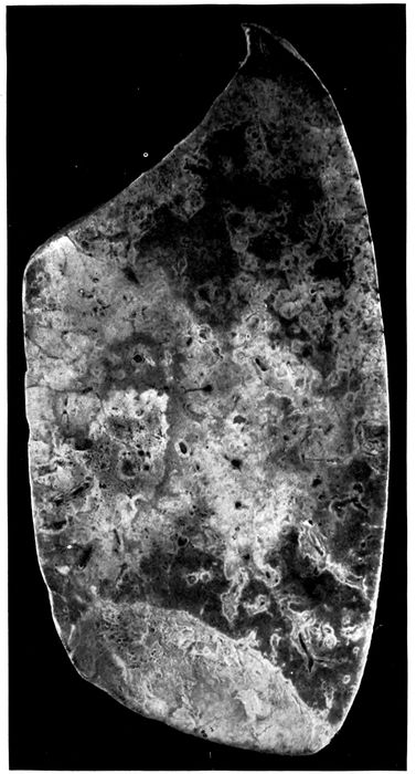
Fig. 8.—Streptococcus pneumonia with massive necrosis. Autopsy 354.
202
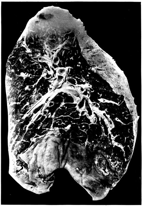
Fig. 9.—Abscess below pleura with perforation caused by hemolytic streptococci. Healing suppurative interstitial pneumonia indicated by yellowish gray lines marking interlobular septa at base of lower lobe. Autopsy 474; right lung. (See left lung, Fig. 10.)
203The duration of illness in cases of pneumonia with abscess
varied from a week or less (11 instances) to more
than four weeks. The duration of the greater number of
cases (17 instances) was between one and two weeks. In
one instance onset occurred with symptoms of influenza,
pneumonia was recognized two days later, and death occurred
only four days after the onset of illness. When the
duration of the illness was less than a week the symptoms
of onset were in some instances those of pneumonia.
Table XLV shows the incidence of pneumococcus, S. hemolyticus,
staphylococcus and B. influenzæ in instances of
suppurative pneumonia with abscess formation, 4 instances
of abscess with interstitial suppurative pneumonia being
excluded:
| Table XLV |
|---|
| |
|
NO. OF CULTURES |
PNEUMOCOCCI |
HEMOLYTIC STREPTOCOCCI |
STAPHYLOCOCCI |
B. INFLUENZÆ |
| NO. POSITIVE |
PER CENT POSITIVE |
NO. POSITIVE |
PER CENT POSITIVE |
NO. POSITIVE |
PER CENT POSITIVE |
NO. POSITIVE |
PER CENT POSITIVE |
| Bronchus |
24 |
5 |
20.8 |
22 |
91.6 |
12 |
50.0 |
18 |
75.0 |
| Lung |
36 |
9 |
25.0 |
30 |
83.3 |
14 |
35.6 |
8 |
22.2 |
| Blood |
37 |
6 |
16.2 |
31 |
83.8 |
|
|
|
|
In over 80 per cent of instances of pulmonary abscess
hemolytic streptococcus has been found in blood, lungs and
bronchus and, when cultures have been made, in the inflamed
pleural cavity as well. Streptococci have been
found in immense number in sections from the necrotic
lung tissue and the abscesses which have been formed. It
is evident that hemolytic streptococci have caused suppurative
pneumonia and death, being found in the blood of the
heart just as frequently as in the lungs (83 per cent). The
relative unimportance of pneumococci is indicated by their
low incidence in the blood (16.2 per cent) when compared
with that of lobar pneumonia (65.5 per cent) or of bronchopneumonia
(31.4 per cent). B. influenzæ has been found
in three-fourths of these autopsies in the bronchus, but its
incidence in the lungs has been much smaller.
In 3 instances of suppurative pneumonia with abscess
formation no hemolytic streptococci were found; they are
as follows:
204Autopsy 380.—Bronchopneumonia with gray and red lobular consolidation
in right upper and lower lobes; peribronchiolar nodules of consolidation
in left lower lobe; abscess, 1.5 cm. across, below the pleura of the
posterior border of the left lower lobe near its base; fibrinopurulent pleurisy
(300 c.c.) on right side; serous pleurisy (200 c.c.) on left. Pneumococcus
III was found in cultures from the blood of the heart from the right
lung and with B. influenzæ from the right pleural cavity. No culture was
made from the left lung which contained the abscess. In sections of the
abscess gram-positive streptococci in chains of 4 to 8 cocci were numerous.
Autopsy 406.—Acute lobar pneumonia with red hepatization of greater
part of right lung; patch of consolidation in lower lobe of left lung containing
an abscess cavity 2.5 x 1.5 cm.; localized seropurulent pleurisy
(375 c.c.) on left side. Pneumococcus IV was obtained from the blood of
the heart; a culture from the lung was contaminated. Tissue from the
abscess was not saved for histologic examination.
Autopsy 416.—Suppurative pneumonia with necrosis and abscess formation
in right lower lobe; fibrinous pleurisy on right side. Pneumococcus
IV was obtained from the blood, right lung and right main bronchus. No
streptococci were found in sections from the abscess in the right lung.
The foregoing observations demonstrate that suppurative
pneumonia with abscess formation following influenza
is with few exceptions caused by S. hemolyticus.
The autopsies (Table XLVI) in which pneumococci have
been found in association with hemolytic streptococci in the
blood or lungs indicate that pneumococci have had a part
in the production of fatal pneumonia.
| Table XLVI |
|---|
| |
| AUTOPSY |
CULTURE FROM BLOOD |
CULTURE FROM LUNGS |
CULTURE FROM BRONCHUS |
| 258 |
S. hem. |
S. hem., Pneum. IV B. inf. |
|
| 282 |
S. hem., Pneum. II |
S. hem., Pneum. II |
S. hem., B. inf. Pneum. II, staph. |
| 345 |
|
S. hem., Pneum. II, staph. |
|
| 378 |
Pneum. atyp. II |
S. hem., Pneum. atyp. II |
S. hem., B. inf., Pneum. atyp. II |
| 381 |
S. hem. |
S. hem., Pneum. II Pneum. IV, staph. |
|
| 383 |
Pneum. III |
S. hem., Pneum. III B. inf. |
|
| 387 |
S. hem. |
Pneum. II, staph., B. inf. |
S. hem., pneum., staph., B. inf. |
These autopsies, notably those in which pneumococci
have been found in the blood, suggest that infection with
205pneumococci has preceded suppurative pneumonia caused
by hemolytic streptococci. In a small number of instances
the sputum was examined in life after onset of pneumonia.
| Table XLVII |
|---|
| |
| AUTOPSY |
SPUTUM |
CULTURES FROM BLOOD, LUNGS AND BRONCHUS |
| 282 |
Pneum. IV. B. inf. |
S. hem., Pneum. II, staph., B. inf. |
| 288 |
S. hem., B. inf. |
S. hem., B. inf. |
| 376 |
(No S. hem., Oct. 8) |
S. hem., staph., B. inf. (Oct. 11) |
In 2 of these 3 cases infection with hemolytic streptococcus
occurred subsequent to the onset of pneumonia.
Several observations help to explain the occurrence of
abscess in association with the pneumonia of influenza.
The fissures which will be described in association with
bronchiectasis represent traumatic ruptures of the bronchial
wall consequent upon weakening by necrosis and over
distention. They expose the injured bronchial wall and
the alveolar tissue adjacent to it to infection by the microorganisms
contained within the lumen of the inflamed
bronchus. Occasionally a favorable microscopic section
demonstrates the relation of pulmonary necrosis and consequent
suppuration to injuries of the bronchial wall.
Peribronchial fibrinous pneumonia occurs about the bronchi
of which the epithelial lining has been destroyed, and
when a fissure penetrates the bronchial wall fibrinous pneumonia
is almost invariably found in a zone about the tear;
it doubtless tends to limit the extension of the process.
Occasionally, wide areas of necrosis occur within consolidated
tissue near the site of the fissure (Autopsy 312 with
S. hemolyticus and B. influenzæ, p. 254). Accumulation of
polynuclear leucocytes between living and dead tissue may
form a line of demarcation (Autopsy 387); finally, fairly
large, irregularly formed, abscess cavities are found.
Necrosis and beginning suppuration in contact with the
lumen of the bronchus will be described in association with
bronchiectasis (Autopsies 312, Fig. 24, and 423, p. 256). In
206the following autopsies upon individuals who have died
with pulmonary abscesses, favorable microscopic sections
have demonstrated abscess formation in contact with lesions
which have penetrated the walls of small bronchi. They
help to explain the pathogenesis of abscess in association
with influenza.
Autopsy 376.—H. M., white, aged twenty-four, a fireman, resident of
Oklahoma, had been in military service one month. Onset of illness occurred
October 1, ten days before his death; he was admitted to the base
hospital on the fourth day of his illness with the diagnosis of bronchopneumonia.
Anatomic Diagnosis.—Acute bronchopneumonia with patches of lobular
and confluent lobular consolidation in both lungs and hemorrhagic peribronchiolar
consolidation in right upper lobe; abscess in right upper lobe
below pleura; fibrinopurulent pleurisy on right side; purulent bronchitis;
bronchiectasis at base of left lobe.
An irregular abscess, 2 x 1 cm., filled with creamy purulent fluid is
separated from the interlobular surface of the right upper lobe by a thin
membrane representing the pleura. The right pleural cavity contains 200
c.c. of turbid yellow fluid in which is soft fibrin. The bronchi contain
purulent fluid in great abundance. The bronchi at the base of the left
lower lobe are widely dilated, so that many small bronchi with no cartilage
in their wall measure from 3 to 5 mm. in diameter.
Cultures show the presence of hemolytic streptococci in the blood of
the heart and in three plates from the lung; B. influenzæ and S. aureus
were found in the left bronchus.
The bronchi have wholly or partially lost their epithelium and there
is deep erosion of the walls. Cavities containing polynuclear leucocytes
occur within the alveolar tissue; in some instances pus containing cavities
are surrounded by alveolar tissue, but in other places it is evident,
that they have had their origin in bronchi. In a short segment of the
circumference the wall of the preexisting bronchus is preserved and
consists of squamous epithelium, vascular connective tissue and smooth
muscle. The remainder of the bronchus has disappeared and a cavity is
produced. The very irregular wall of the cavity is formed by partially
destroyed alveoli filled with fibrin and leucocytes.
Autopsy 387.—C. M., white, aged twenty-one, laborer, resident of Mississippi,
had been in military service twenty-one days. Illness began on
September 22, nineteen days before death, and the patient was admitted
to the hospital on the same day with a diagnosis of bronchitis; a diagnosis
of bronchopneumonia was made on October 2, nine days before death.
The leucocytes on October 3 numbered 8000 (small mononuclear, 36 per
cent; large mononuclear, 5 per cent; polynuclear, 59 per cent).
207Anatomic Diagnosis.—Acute bronchopneumonia with consolidation in
right upper lobe and hemorrhagic peribronchiolar consolidation in left lower
lobe; abscess below pleura in left lower lobe; purulent pleurisy on both
sides; edema of mediastinum; purulent bronchitis; bronchiectasis.
There is advanced bronchiectasis, and bronchi with no visible cartilage
are dilated to from 4 to 8 mm. in diameter; they contain purulent fluid
which wells up from the cut surface. About dilated bronchi there is in
places dull red or grayish red consolidation forming an encircling zone.
Situated below the pleural surface within an area of consolidation at the
posterior border of the left lower lobe there is a spot 3 cm. across where
the tissue is yellow and has in places undergone purulent softening. Several
smaller abscesses occur nearby.
Cultures from the blood of the heart and from the edematous mediastinum
contain hemolytic streptococci. From the abscess are grown S.
albus, Pneumococcus II and B. influenzæ. The purulent contents of a small
bronchus contains S. hemolyticus, B. influenzæ, S. aureus and a few pneumococci.
Microscopic examination shows that the epithelium of dilated bronchi
has disappeared and the denuded surface is covered by fibrin and polynuclear
leucocytes; fissures extend from the lumen through the bronchial wall into
the surrounding alveolar tissue. A zone of fibrinous pneumonia surrounds
these bronchi and fissures in the bronchial wall penetrate into this zone.
One dilated bronchus 2.4 mm. in diameter with no cartilage in its wall
has vascular connective tissue covered by epithelium on one side, whereas
the remainder of the circumference is formed by exposed alveoli filled with
fibrin, the bronchial wall having disappeared. A section through a part
of the abscess which has been mentioned shows a very irregularly formed
cavity approximately 1 x 0.7 cm. Remains of bronchial wall, consisting
of very vascular tissue covered by flat epithelium in several layers, indicate
the origin of the cavity. Between these remnants of bronchi deep pockets
extend into the pulmonary tissue which in the margin of the cavity is the
site of fibrinous pneumonia. In one place, in contact with the cavity, a
wide area of consolidated tissue has undergone necrosis and both alveolar
walls and their contents have lost their nuclei. Leucocytes which are
accumulating at the margin of the necrotic patch form a line of demarcation
between living and dead tissue.
Abscess may be the result of the profound changes which
occur in the bronchi as the result of influenza. Necrosis
caused by bacteria within the bronchi weakens and in
places destroys the wall. Bacteria penetrate into the surrounding
tissue and hemolytic streptococci (or staphylococci)
may produce localized abscesses. These abscesses
are usually situated near the pleural surface of the lung, because
destructive changes causing rupture of the bronchial
208wall occur more frequently in the smaller peripheral bronchi
than in the larger bronchi containing cartilage. Abscesses
occur more frequently at the bases of the lungs,
because the most severe changes in the bronchi occur in
the dependent part. (See “Bronchiectasis,” p. 240.)
Healing of Abscess.—The following autopsy is of interest
in relation to the treatment of pulmonary abscess and associated
empyema.
Autopsy 467.—P. C., white, aged twenty-five, a farmer from Missouri,
had been in military service three months. Illness began September 27,
thirty days before death, and the patient was admitted the day following
onset with headache, backache and cough. Pneumonia with consolidation
in the right lower lobe was recognized on the sixth day of illness. On the
ninth day 500 c.c. of fluid were withdrawn from the right pleural cavity;
there were cyanosis and dyspnea. On the eleventh day 700 c.c. of fluid
were withdrawn. On the twelfth day thoracotomy was performed and 100
c.c. of greenish fluid were removed. The patient’s condition improved for
a time, but on the twenty-sixth day 1,000 c.c. of straw colored fluid were
aspirated from the left pleural cavity and on the twenty-eighth day the
same amount of seropurulent fluid was withdrawn.
Anatomic Diagnosis.—Healing abscess of right lower lobe communicating
with the pleural cavity; acute purulent pleurisy with closed thoracotomy
wound on the right side; purulent pleurisy on the left side; acute bronchopneumonia
with lobular consolidation in the left lung; purulent bronchitis;
bronchiectasis with formation of spherical bronchiectatic cavities; acute
splenic tumor.
At the base of the right chest is a closed thoracotomy wound 2 cm.
in length; the right pleural cavity contains 200 c.c. of thick creamy pus
and the cavity is lined by a thick tough membrane. The left pleural cavity
contains 800 c.c. of white purulent fluid thinner than that on the right
side. The right lung is compressed into the posterior and inner part of
the chest. The upper lobe is pink and air containing; the posterior and
lower part of the lower lobe is red and atelectatic, and fibrous septa are
more conspicuous than elsewhere. The pleura of the external surface near
the basal edge, in an area 2 cm. across, is depressed and yellowish gray in
color. In the center of this area is a small opening communicating with
a pocket 0.5 cm. across within the substance of the lung.
In the lower lobe beneath the interlobular surface are two spherical
bronchiectatic cavities, each about 1.5 cm. across, with smooth lining in
continuity with two branches of the same bronchus of medium size.
Bacteriologic examination showed the presence of S. hemolyticus in the
blood of the heart. No growth was obtained from the left lung; the left
pleural cavity contained hemolytic streptococci and S. aureus, the latter in
209small number. S. hemolyticus and B. influenzæ were grown from the left
main bronchus.
A microscopic section through the abscess and its communication with
the pleura shows that its cavity contains polynuclear leucocytes and the
wall is formed by granulation tissue covered by fibrin. Some alveoli outside
the abscess contain compact balls of fibrin containing a few fibroblasts;
this fibrin stains deeply with hematoxylin as if it contained calcium. The
surface of the lung is covered by fibrin in process of organization.
In the foregoing instance a pulmonary abscess on the
right side has ruptured into the pleura and, completely
separated from the adjacent lung by a wall of newly formed
tissue, is in process of healing. It shows that these pulmonary
abscesses below the pleura may heal provided
drainage is established by rupture into the pleural cavity
and subsequent evacuation of pleural exudate. It is noteworthy
that in this instance empyema extended from the
right to the left pleural cavity, both S. hemolyticus and S.
aureus were found at autopsy. The thoracotomy wound
on the right side was closed at autopsy.
A second type of suppurative pneumonia is characterized
by acute inflammation of interstitial tissue between the secondary
lobules of the lung and by acute lymphangitis; suppuration
involves the interstitial septa and the walls of the
lymphatics. The lesion is designated by Kaufmann,[82]
Beitzke[83] and others acute interstitial pneumonia. Pneumonia
dissecans in which solution of interstitial tissue isolates
sections of lung tissue is said to be a consequence of
the lesion. Many text books of pathology, overlooking
the occurrence of this lesion, limit the consideration of interstitial
pneumonia to chronic processes in which the interlobular
and interalveolar fibrous tissue is increased.
Acute inflammation and edema of the interlobular septa
of the lung with no suppuration is often found with both
lobar and bronchopneumonia and is occasionally so far advanced
210that it can be recognized on gross examination of
the lungs. In a small area interlobular septa are conspicuous
as yellowish lines of edematous appearance which may
be 1 to 1.5 mm. in thickness and sometimes form a network
with rectangular or polygonal meshes. The gelatinous appearance
of the edematous fibrous tissue does not suggest
suppuration. Microscopic examination shows that the
tissue is distended by edema and contains fibrin and polynuclear
leucocytes; the lymphatics are distended and contain
a network of fibrin within which leucocytes are numerous.
Inflammatory edema of the interstitial tissue has
been recognized at autopsy four times in association with
bronchopneumonia (Autopsy 253 with Pneumococcus II;
Autopsy 335, with Pneumococcus IV and S. viridans; Autopsy
477 with S. hemolyticus and Autopsy 498 with S.
viridans); twice with lobar pneumonia (Autopsy 343 with
Pneumococcus IV and Autopsy 353 with atypical Pneumococcus
II); twice with combined lobar and broncopneumonia
(Autopsy 273 with S. hemolyticus and Pneumococcus
IV and Autopsy 357 with Pneumococcus IV). Edema of
interstitial septa was recognized at autopsy in the immediate
neighborhood of an abscess three times (Autopsies
277 and 278 with hemolytic streptococci and Autopsy 282
with hemolytic streptococci and Pneumococcus II). In
these instances of inflammation and edema the lymphatics
are found distended by fibrinous thrombi, and it is probable
that occlusion of lymphatics determines the occurrence of
inflammatory edema within the surrounding tissue. Inflammation
has not proceeded to suppuration.
With interstitial suppurative pneumonia, interlobular
connective tissue is marked by conspicuous yellow lines,
1 to 3 or even 5 mm. in thickness, forming a network with
polygonal meshes which represent secondary lobules (Figs.
10 and 11). The distended septa not infrequently have
bead-like enlargements at intervals and from the cut surface
it is often possible to scrape away creamy yellow pus.
These lines of suppuration invariably extend up to the
pleura and are often broadest immediately below it. Adjacent
septa which have not undergone suppuration are much
thickened and have the yellowish gray appearance produced
by edema.
211
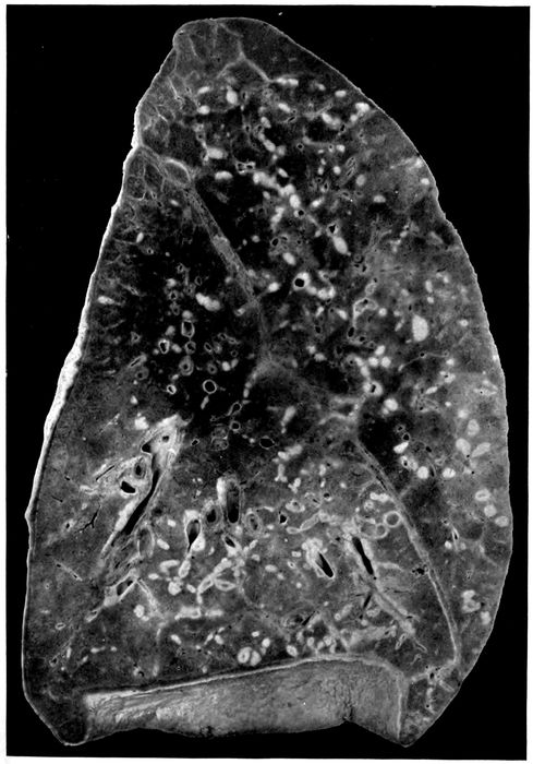
Fig. 10.—Interstitial suppurative pneumonia; interstitial septa are the site of suppuration and lymphatics are distended with purulent fluid; empyema. Autopsy 474, left lung. (See right lung) Fig. 9.
212
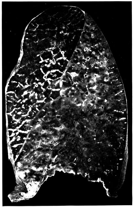
Fig 11.—Suppurative interstitial pneumonia; the left lower lobe is the site of almost uniform consolidation and here interstitial septa and their lymphatics are distended with pus. There is more extensive interstitial suppuration in the upper lobe where consolidation is absent. The cloudy appearance of the consolidated lung is well shown. Autopsy 452.
213Suppurative interstitial pneumonia frequently occurs in
association with bronchopneumonic consolidation which
may be peribronchiolar, hemorrhagic or lobular, but there
is in addition consolidation of the pulmonary tissue between
the inflamed septa which may affect part of a lobe, an entire
lobe, or parts of several lobes; it does not exhibit the
characters of confluent lobular pneumonia.
In approximately half of the cases consolidation, associated
with interstitial suppuration, has been lobar in distribution
(Fig. 11). The tissue is laxly consolidated, finely
granular, and has a cloudy red or gray appearance. The
coarsely granular surface of lobar pneumonia is absent.
The affected lung may weigh 1,500 or 1,650 grams. Occasionally,
interstitial septa of air containing lung tissue
is the site of suppurative inflammation or edema. In
Autopsy 452 the lower lobe, save a small part at the base,
is laxly consolidated; interstitial septa in the consolidated
area are yellow, 1.5 to 2 mm. in thickness, beaded and exude
purulent fluid on pressure. In the adjacent part of
the upper lobe there is a patch of consolidation, and a network
of yellow thickened septa extends from it far into the
surrounding air containing tissue. The weight of the right
lung is 635 grams; of the left, 1,650 grams.
The distribution of interstitial suppuration in 21 instances,
including 4 in which the lesion has occurred in the
same lungs with abscess formation, has been as follows:
right upper lobe, 9 instances; middle lobe, 4; lower lobe, 5;
left upper lobe, 7; left lower lobe, 6. In 6 of these autopsies
more than one lobe of the same lung has been affected
by the lesion; in 2 autopsies parts of both lungs have been
affected. Localized abscess of the lung is more common in
214the lower than in the upper lobes, but suppuration of the
interstitial tissue is more often found in the upper lobes.
The duration of illness with interstitial suppurative
pneumonia has varied from six days to five weeks. In over
half of the cases death has occurred during the second week
of illness.
The bacteriology of these cases is shown in Table
XLVIII.
| Table XLVIII |
|---|
| |
|
NO. OF CULTURES |
PNEUMOCOCCI |
HEMOLYTIC STREPTOCOCCI |
STAPHYLOCOCCI |
B. INFLUENZÆ |
| NO. POSITIVE |
PER CENT POSITIVE |
NO. POSITIVE |
PER CENT POSITIVE |
NO. POSITIVE |
PER CENT POSITIVE |
NO. POSITIVE |
PER CENT POSITIVE |
| Bronchus |
10 |
|
|
9 |
90.0 |
5 |
50.0 |
10 |
100.0 |
| Lung |
20 |
1 |
5.0 |
17 |
85.0 |
5 |
25.0 |
7 |
35.0 |
| Blood |
21 |
2 |
9.5 |
17 |
81.0 |
|
|
|
|
S. hemolyticus has been almost invariably present in
lungs, heart’s blood and bronchi. In 16 of 21 autopsies
hemolytic streptococci have been obtained from the blood
in pure cultures, in one instance associated with pneumococcus.
With associated empyema, pericarditis or peritonitis,
the same microorganism has been found in the
pleural cavities, pericardium or peritoneum. Furthermore,
microscopic examination has demonstrated the presence
of chains of streptococci in the affected interlobular
tissue and in much greater abundance in the distended
lymphatics.
Nevertheless in 2 instances no streptococci have been
found. These cases are as follows:
Autopsy 330.—Illness began with symptoms of influenza ten days before
death; signs of pneumonia were recognized three days before death. There
is firm, gray red consolidation of the entire left upper lobe; the interlobular
septa are here indicated by yellow lines of obvious suppuration and thick puslike
fluid exudes from the cut surface of the consolidated tissue. The upper
half of the left lower lobe has undergone gray hepatization, but here there
is no distention of the interlobular septa. There is fibrinopurulent pleurisy
on the left side with accumulation of 400 c.c. of fluid. Pneumococcus IV is
215obtained from the blood of the heart and from the lung. In the suppurating
tissue diplococci which stain by Gram’s method are present in large number;
there are a few short chains.
Autopsy 379.—Illness began seven days before death with influenza;
signs of pneumonia were first recognized the day before death. The middle
lobe of the right lung is firmly consolidated; on section there is mottling of
deep red and pinkish red and the cut surface is coarsely granular. The interstitial
septa are distended by fluid and are grayish yellow. There is fibrinopurulent
pleurisy on the right side with accumulation of 600 c.c. of fluid.
Pneumococcus atypical II is obtained from the blood of the heart. A large
bacillus unstained by Gram’s method is obtained from the right lung and from
the right main bronchus. In the bronchus are a few influenza bacilli. In the
suppurating and necrotic tissue of the interstitial septa are found diplococci
and chains of 4 to 6 cocci in great number; a few large Gram-negative bacilli
are found.
In both these autopsies consolidation had the characters
of lobar pneumonia, and pneumococci were obtained from
the blood of the heart. It is possible that streptococci failed
to grow or while present elsewhere were absent at the spot
where cultures were made.
It is noteworthy that B. influenzæ was found in the bronchi
in every instance (10) in which cultures were made,
but was obtained much less frequently from the lung. In
one instance (Autopsy 474) this microorganism was found
in the blood in association with hemolytic streptococci.
There was suppurative interstitial pneumonia in the left
lung and abscess in the right lower lobe with rupture into
the cavity and empyema. Hemolytic streptococci and B.
influenzæ were found in the bronchus, right pleural cavity
and blood of the heart.
In 4 instances (Autopsies 251, 259, 295 and 474) interstitial
suppurative pneumonia has been associated with
abscess formation. In one instance (Autopsy 251) the
right middle lobe has been the site of interstitial suppuration
and abscess formation; in another (Autopsy 295) the
left lower lobe has been the site of both lesions, but in the
other 2 instances suppurative interstitial pneumonia and
abscess formation have occurred in opposite lungs. In all
2164 autopsies hemolytic streptococci have been found in the
blood of the heart and in lungs or bronchi.
Empyema has been present in all but 3 of 21 instances of
interstitial suppurative pneumonia.
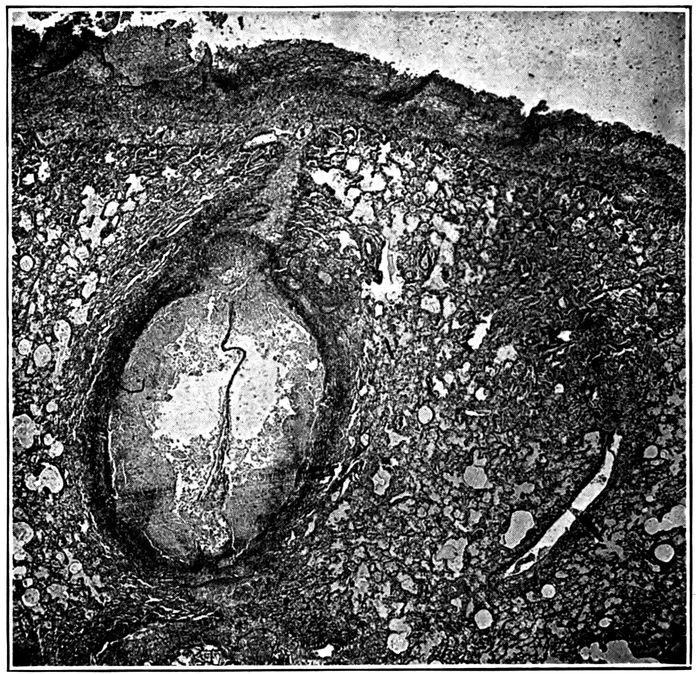
Fig. 12.—Suppurative interstitial pneumonia, showing an immensely dilated lymphatic containing purulent exudate, a short distance below the pleura. Autopsy 474.
Histologic examination of lungs with interstitial suppuration
shows that the interlobular septa are distended by
serum and contain a conspicuous network of fibrin. Polynuclear
leucocytes are present in varying number, and at
times densely infiltrate the distended tissue; it is not uncommon
to find a zone of densely crowded polynuclear leucocytes
along each edge of the septum, whereas the central
part contains comparatively few. Occasionally, there is
hemorrhage into the distended connective tissue.
217Within the distended septa occur greatly dilated lymphatics
filled with polynuclear leucocytes (Figs. 12 and 13).
Thrombosis of the distended lymphatics has usually occurred,
and a conspicuous network of fibrin in which are
polynuclear leucocytes plugs the lumen. Streptococci in
chains of variable length are found in the inflamed interstitial
tissue, but are present in far greater number within
the distended lymphatics.
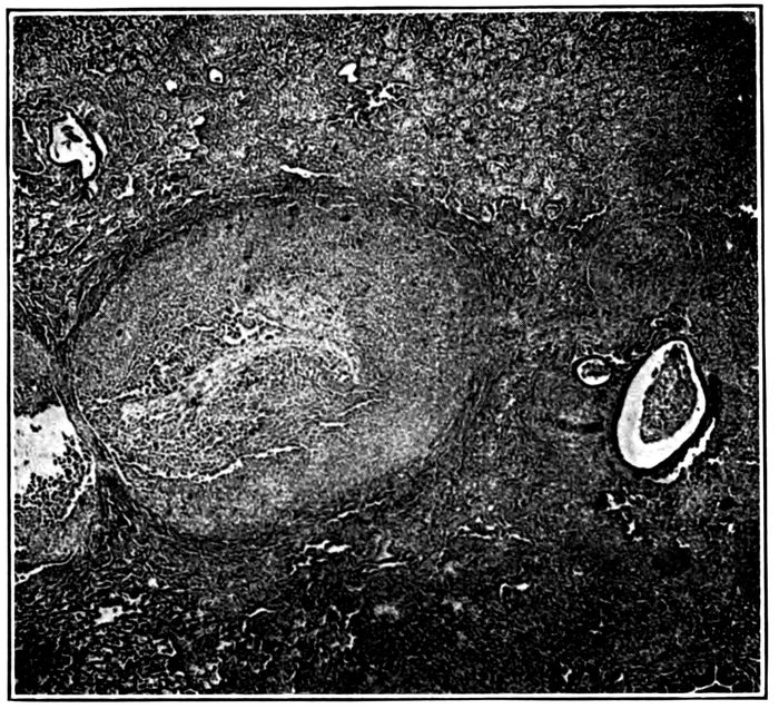
Fig. 13.—Suppurative interstitial pneumonia showing a dilated lymphatic. Autopsy 428.
Necrosis of the cells which fill the lymphatics occurs in
spots, usually in the center of the thrombus, and occasionally
affects the entire contents of the lymphatic; polynuclear
leucocytes have lost their nuclei or in some the nucleus
has undergone fragmentation. In these spots the network
of fibrin has disappeared. Not infrequently the wall
of the lymphatic in a small sector or throughout the circumference
218has undergone necrosis, and spots of necrosis
may occur in the interlobular septa distended by inflammatory
exudate. Wherever necrosis has occurred, chains
of streptococci are present in immense number.
Accumulation of polynuclear leucocytes, necrosis of these
cells, solution of fibrin at first in the centers of the lymphatic
thrombus and later throughout, occasionally with
necrosis of the wall of the vessel, result in the formation
of an abscess at the site of the distended lymphatic. These
lymphatics, dilated by purulent fluid, may have a diameter
from 2 to 3 mm. and may cause considerable compression
and collapse of immediately adjacent alveoli. Lymphangitis,
distention of lymphatics, thrombosis and finally suppuration
may occur in the lymphatic vessels encircling the
blood vessels and in those situated in the adventitia of the
bronchi of medium size.
The alveoli adjacent to the distended septa are filled by
inflammatory products; edema is almost invariably present
and the alveoli may contain serum and desquamated
epithelial cells; fibrin is often present, but more frequently
polynuclear leucocytes are predominant. Not infrequently,
abscess formation, recognized microscopically, has occurred
in contact with septa most often immediately below
the pleura. Polynuclear leucocytes are present in immense
number and alveolar septa have disappeared; occasionally,
with abscess formation there is more or less widespread
necrosis of tissue, cells both of the exudate and of the alveolar
walls having lost their nuclei.
Lymphatics in many places are distended and plugged
by fibrinous thrombi, whereas elsewhere softening of the
thrombus has been brought about by suppuration. Suppuration,
both within the lymphatic and in adjacent alveoli,
appears to be secondary to lymphatic obstruction. In
some instances the lymphatic appears to have undergone
distention after the thrombus has formed, for between the
219thrombus and the wall of the lymphatic a channel is occasionally
found containing uncoagulated lymph.
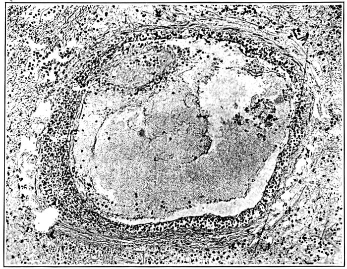
Fig. 14.—Endophlebitis occurring in association with suppurative pneumonia; the intima contains lymphoid cells in great number; at one spot there is a small thrombus adherent to the intima. Autopsy 325.
Acute endophlebitis has been repeatedly observed in association
with interstitial suppurative pneumonia (Fig.
14). The lesion usually occurs in veins situated within the
septa which are the site of intensely acute inflammation
associated with necrosis. The wall of the vein appears to
be so injured by the surrounding changes that polynuclear
leucocytes and small mononuclear cells accumulate below
the endothelium. Throughout the circumference of the
veins, often 0.5 to 1 mm. in diameter, the endothelium is
separated from the underlying media by polynuclear leucocytes
which form a conspicuous zone encircling the lumen.
Some cells of lymphoid type are usually present
among the polynuclear leucocytes. Polynuclear leucocytes
220are often adherent to the endothelial lining of the vessel
and are not infrequently fixed in the process of passing
through the endothelium. The lesion may be more severe
(Autopsy 325), so that the endothelium has disappeared,
and upon the exposed surface fibrin is deposited; within
this fibrin polynuclear leucocytes are numerous and nuclear
fragmentation has occurred. The middle coat of the
vessel usually contains few cells; some polynuclear leucocytes
within it may be stretched out as if in process of wandering
through the wall.
In other instances the accumulation of cells below the
endothelium is almost wholly mononuclear. Cells of the
type of lymphocytes occur, but more abundant are slightly
larger cells with more abundant cytoplasm. These cells
may form a thick zone below the intima throughout the entire
circumference of the lesion. It seems probable that
these cells, like the polynuclear leucocytes, are derived from
circulating blood within the lumen of the vessel, for small
cells of the type of lymphocytes are not infrequently found
adherent to the lumen and occasionally one is fixed in
process of passing through the endothelium.
This endophlebitis appears to be the result of changes
outside the vessel; there is usually necrosis of the adjacent
tissue and the production of the lesion is favored by lymph
stasis; as the result of injury to the vessel wall, polynuclear
leucocytes in response to chemotaxis, or with milder irritation,
mononuclear cells, wander through the endothelium
and accumulate below it perhaps on account of the greater
impermeability of the middle coat to the passage of cells.
The lesion described does not occur exclusively with interstitial
suppurative pneumonia caused by hemolytic
streptococci, but has been found in association with abscess
formation (Autopsies 354 and 383) caused by hemolytic
streptococci or (Autopsy 322) caused by staphylococci.
In 1 instance it has been found with lobar pneumonia (Autopsy
320) caused by atypical Pneumococcus II and in 2
221instances with combined lobar and bronchopneumonia
(Autopsy 357 with Pneumococcus IV; Autopsy 392 with
Pneumococcus II). In these 3 instances there has been interstitial
inflammation, edema and lymphangitis without
suppuration.
Interstitial suppurative pneumonia of long standing may
occasionally be accompanied by chronic changes which
bring about thickening of the interlobular tissue. In the
following autopsy acute suppurative inflammation in the
left lung has been associated with conspicuous thickening
of interlobular septa in the right lung.
Autopsy 474.—I. H., white, aged twenty-one, was a native of Oklahoma
and had been in military service one month. His illness began with influenza
thirty-six days before death; he was admitted to the base hospital thirty-one
days before his death with signs of pneumonic consolidation of the right
lower lobe. Evidence of fluid in the right pleural cavity was obtained two
weeks before death, and from 100 to 700 c.c. of thick purulent fluid were aspirated
on five occasions. Hemolytic streptococci were found in the aspirated
fluid.
Anatomic Diagnosis.—Interstitial suppurative pneumonia in left lung;
abscess of right lower lobe with rupture into pleural cavity; thickening of
interlobular septa of right lower lobe; double purulent pleurisy with thoracotomy
on right side; serofibrinous pericarditis.
The right pleural cavity contains 85 c.c. of thick purulent
fluid; the right lung (Fig. 9) is collapsed and pushed to the
median line, being bound by firm adhesions to the pericardium.
Over the external and basal surfaces is a localized
cavity walled off by adhesions. An abscess cavity in the
lower part of the lower lobe communicates through a perforation
in the basal surface of the lung with the pleural
cavity and is in free communication with a small bronchus.
About the abscess the lung is red and laxly consolidated,
but elsewhere air containing; throughout the lower half of
the lower lobe, the interlobular septa are marked by conspicuous
yellowish gray lines about 1 mm. in thickness.
Between these thickened septa the lung tissue contains air.
The lung weighs 600 grams. The left lung (Fig. 10) is voluminous
and heavy, weighing 1,320 grams. The surface is
222everywhere covered by thickened pleura and fibrin, the
pleural cavity containing 150 c.c. of thick purulent fluid.
The lung is consolidated varying in color from a fleshy red
to yellowish gray. The surface is very conspicuously
marked by yellow lines 2 or 3 mm. thick, corresponding to
the interlobular septa which have undergone suppuration.
The septa have bead-like swellings along their course, and
when pus escapes from the cut surface small cavities remain
at the site of these swellings.
Bacteriologic examination has shown hemolytic streptococci
in the blood, left lung, right and left pleural cavities,
and right bronchus. B. influenzæ has been found in the
bronchus, in the right pleura and in the heart’s blood. A
few colonies of S. aureus have been found on the plate
from the right pleural cavity (site of thoracotomy).
Microscopic examination of the right lower lobe shows
that the interstitial septa are much thickened by young
fibrous tissue infiltrated with lymphoid and a few plasma
cells. Large mononuclear cells with granular cytoplasm are
very numerous. A lymphatic is much distended and contains
a few polynuclear leucocytes and many lymphoid and
large mononuclear cells. There is no suppuration. Sections
from the right lung show suppurative lymphangitis
with suppurative inflammation of interstitial tissue.
The right lung is the site of a healing lesion of the interstitial
tissue which has developed simultaneously with
acute interstitial suppurative pneumonia in the left lung.
Both lesions are doubtless caused by S. hemolyticus. This
healing lesion exhibits little similarity to the interstitial
bronchopneumonia described by several observers with
both measles and influenza.
The following autopsy furnishes further evidence that
interstitial suppurative pneumonia exhibits a tendency to
heal. Proliferation of endothelial cells lining the inflamed
lymphatics gives rise to phagocytic cells which aid in removing
the accumulated leucocytes.
223Autopsy 397.—N. P., white, aged twenty-one, farmer, a native of Oklahoma,
had been in military service twenty-one days. Illness began twenty-two
days before death, the patient being admitted on the day following onset
with influenza, pharyngitis and bronchitis. A diagnosis of lobar pneumonia
was made fourteen days before death. The left pleural cavity was aspirated
twelve days later and 800 c.c. of thick yellow pus were withdrawn. Hemolytic
streptococci were found in the sputum five days before death.
Anatomic Diagnosis.—Interstitial suppurative pneumonia in left upper
lobe; acute bronchopneumonia with lobular consolidation in right upper lobe;
localized purulent pleurisy on left side with compression and atelectasis of
left lung; compensatory emphysema of right lung; purulent bronchitis; beginning
serofibrinous pericarditis; chronic passive congestion of liver, spleen
and kidneys.
The right lung is very voluminous, free from coal pigment and bright
pink save over lobular patches of consolidation which have a bluish red color;
the bronchi contain mucopurulent material. The anterior surface of the left
lung is bound to the chest wall by firm adhesions, but over the external and
posterior surfaces of the lung there is a localized cavity containing 1,100 c.c.
of turbid fluid. The left lung is collapsed and airless with deep fleshy red
color. In the upper lobe there are scattered patches of consolidation 1.5 to
2.5 cm. across where the tissue is grayish red and coarsely granular. In the
adjacent tissue interstitial septa are thickened to 1 or 2 mm. and are conspicuous
as gray bands. Along their course occur bead-like swellings from
which purulent fluid can be scraped. These septa at one point reach the
anterior surface of the lung where the pleural cavity is in large part obliterated
by adhesions; here there is an encapsulated pocket 4 x 1.5 cm. containing
thick creamy pus.
Bacteriologic examination of the blood shows the presence of hemolytic
streptococci; cultures from the lungs contain hemolytic streptococci and B.
influenzæ.
Microscopic examination shows that interlobular septa are thickened and
infiltrated with plasma cells in large number. Leucocytes in the center of
much dilated lymphatics have undergone necrosis and have lost their nuclear
stain. About the periphery of the lumen and evidently derived from the
swollen endothelial cells which surround it, are numerous large mononuclear
cells. They act as phagocytes and ingest polynuclear leucocytes. Multinucleated
giant cells, derived from these cells, occur. In several places thrombosed
lymphatics in process of organization occur; the lumen is filled with
compact fibrin which is invaded by fibroblasts and newly formed capillaries.
The process just described is analogous to that which occurs
whenever an unopened abscess heals; mononuclear
cells accumulate and act as phagocytes ingesting polynuclear
leucocytes.
224The following instance of streptococcus empyema is noteworthy
because no suppurative pneumonia has been found
in association with it. Nevertheless the character of the
changes present in the lung indicate that the organ has
been the site of an interlobular inflammation which has
healed.
Autopsy 499.—J. H. M., white, aged twenty-four, a farmer from Arkansas,
had been in military service five months. Onset of illness began two
weeks before his admission to the hospital on November 15 with cough, fever,
headache and malaise; on admission there was acute bronchitis. Thirteen
days after admission the patient developed parotitis (mumps?); five days
later and five days before death pleurisy was recognized on the right side
and pneumonia was suspected. Death occurred thirty-six days after onset.
The temperature on admission was 103.2° F. and remained elevated during
one week falling by lysis; from this time until the pleurisy was recognized
it was normal and later it remained approximately 103° F.
Anatomic Diagnosis.—Fibrinopurulent pleurisy on right side; fibrinous
pleurisy on left side; fibrinopurulent pericarditis; chronic interstitial (interlobular)
pneumonia in process of healing; purulent bronchitis; acute splenic
tumor; parenchymatous degeneration of kidneys.
The right pleural cavity contains 1,650 c.c. of grayish yellow fluid containing
an abundant sediment of softened fibrin. Part of this fluid, more
opaque than the remainder is confined in a localized pocket between the inner
surface of the lung and the pericardium. The apex and anterior surface
of the right upper lobe, over an area about 7 cm. across, is held by fibrinous
adhesions to the chest wall; when this adhesion is broken a pocket is exposed
6.5 x 2.5 cm. containing fibrin and fluid. The pericardial cavity is distended
by 350 c.c. of turbid yellow seropurulent fluid. The pericardial surfaces are
covered by shaggy, tough gray fibrin.
The right lung is collapsed; the lower and posterior part of the upper
lobe is deep red and atelectatic. Throughout the upper lobe the interlobular
septa are thickened, often 1 mm. across and very conspicuous; in the lower
and anterior tip of the lobe is an area where tissue is firm grayish red and
heavier than water. The lower and posterior half of the right lower lobe
is firm and airless, and the tissue is reddish gray or gray and in places finely
granular on section; interlobular septa are conspicuous. Although the lung
is cut into thin sections, no abscesses are found. Bronchi throughout the lung
contain mucopurulent fluid.
The left lung over its lower half is covered by a thin layer of fibrin. The
tissue is crepitant throughout and moderately edematous. Bronchi contain
mucopurulent fluid.
Hemolytic streptococci in pure culture are obtained from the blood of
the heart, right pleural cavity and pericardium. No growth is obtained
on a plate inoculated with material from the right lower lobe. The right
bronchus contains hemolytic streptococci and B. influenzæ.
225The pleural surface of the right lung is covered by a thick layer of fibrin
which has undergone advanced organization. Fibrous septa within the lung
are much thickened by the presence of newly formed fibrous tissue; the interstices
of the tissue are distended and contain fibrin into which fibroblasts
and new blood vessels have penetrated. Some lymphatics are plugged with
fibrin and contain polynuclear leucocytes, lymphoid and large mononuclear
cells. In several places organization of these thrombi is beginning. About
the blood vessels are thrombosed lymphatics in which polynuclear leucocytes
and mononuclear cells are equally abundant. Alveoli immediately adjacent
to blood vessels and to fibrous septa often contain fibrin, and alveoli elsewhere
contain desquamated cells in abundance.
In association with hemolytic streptococci in the blood,
pleura and pericardium, there has been inflammation of the
interlobular septa of the lungs with acute lymphangitis;
there has been no suppuration and the lesion is in process
of healing with new formation of fibrous tissue. It is evident
that this lesion, as well as pleurisy with advanced organization,
preceded the exacerbation of the patient’s illness
which occurred five days before death. The advanced
chronic changes found at autopsy indicate that the pulmonary
and pleural lesions had their origin during the
illness which was present at the time of admission to the
hospital. Interstitial pneumonia caused by hemolytic
streptococci was of mild character and did not produce
suppuration within the lung; nevertheless, hemolytic streptococci
which reached the pleura caused empyema.
In the preliminary report of this commission published
in The Journal of the American Medical Association, loc.
cit., pg. 111, we described suppurative pneumonia with multiple
abscesses caused by staphylococci and cited 4 instances
of the lesion which followed influenza. Chickering and
Park[84] published in a subsequent number of the same journal
226an account of staphylococcus pneumonia, a lesion which has
heretofore attracted very little attention.
In a small group of cases abscesses in the lungs have had
characters which serve to distinguish them from the abscesses
previously described. Small, sharply circumscribed
yellow nodules, which in their centers have undergone suppurative
softening, form a cluster upon a red, airless background
(Figs. 15 and 16). One or more of these groups
several centimeters across, occur in the lungs. It is usually
evident that the abscesses are clustered about a medium-sized
bronchus, but occasionally with increase in the size
of the small cavities the lung tissue assumes a honey-combed
appearance.
These clustered abscesses occur in association with bronchopneumonia
and have been in all instances associated
with purulent bronchitis. The mucosa of the small bronchi
may be destroyed so that the surface is eroded. These
small clustered abscesses are seen as conspicuous yellow
spots immediately below the pleura, but there has been no
associated empyema. In 2 instances these abscesses were
accompanied by fibrinous pleurisy, but in the remaining autopsies
the pleura has been normal. The infrequency of
empyema is in contrast with its almost invariable presence
when a streptococcus abscess is found below the pleura.
Autopsy 280.—Onset of illness with malaise, headache, cough and fever
was on September 24, eight days before death. At autopsy there were hemorrhagic
peribronchiolar and lobular bronchopneumonia, clustered foci of suppuration
in right lung, purulent bronchitis and fibrinous pleurisy. Hemolytic
streptococci were obtained from the consolidated lung and from a
bronchus. A culture from the right lung was contaminated. In the bronchus
were found B. influenzæ and a few staphylococci. Microscopic examination
of the abscesses shows that they contain Gram-staining cocci grouped into
staphylococcus-like colonies.
Autopsy 286.—Duration of illness, which began September 25 with
symptoms of influenza, was nine days. At autopsy there were lobular and
confluent patches of bronchopneumonia, clustered abscesses in the right lung
below the pleura, purulent bronchitis, and serofibrinous pleurisy localized
in the neighborhood of the abscesses. Pneumococcus IV was obtained from
the blood of the heart, and Pneumococcus IV, staphylococci and B. influenzæ
from the right main bronchus; growth failed to occur on plates from right
and left lungs. Microscopic examination shows the presence of clumps of
cocci with staphylococcus grouping in the centers of the small abscesses.
Section through one abscess shows its continuity with the wall of a bronchus;
along one side of the abscess is epithelium composed of flattened epithelial
cells in multiple layers continuous with that of the bronchus; the remainder
of the abscess wall is formed by disintegrated lung tissue.
227
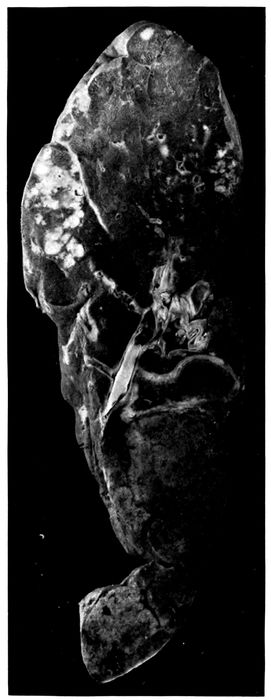
Fig. 15.—Abscesses in two clusters caused by S. aureus in upper part of right upper lobe; confluent lobular consolidation in lower part of lobe. Autopsy 333.
228
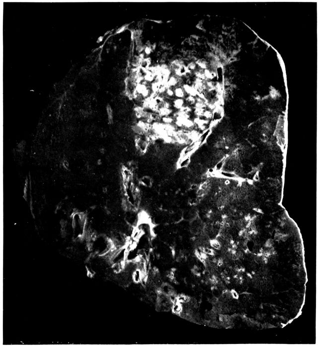
Fig. 16.—Abscesses in cluster caused by S. aureus at apex of right upper lobe. Autopsy 322.
Autopsy 322.—The patient was admitted with influenza eight days before
death; signs of pneumonia appeared two days later, and on the following
day Pneumococcus IV was obtained from the sputum. At autopsy
there were bronchopneumonia with lobar consolidation, abscesses clustered
about a bronchus in the right upper lobe and purulent bronchitis. The
blood was sterile; S. aureus was obtained from the consolidated part of
the left lung; S. aureus and Pneumococcus III from the abscesses of the right
lung. Microscopic examination of sections of abscesses showed the presence
of Gram-staining cocci in staphylococcus-like colonies, surrounded by
necrotic material and polynuclear leucocytes; Gram-negative bacilli resembling
B. influenzæ were seen. (See Fig. 16.)
229Autopsy 333.—The onset of influenza was fifteen days before death; a
diagnosis of pneumonia was made seven days before death. At autopsy there
were confluent bronchopneumonia, clustered abscesses in the right lung and
purulent bronchitis (no pleurisy). The blood contained Pneumococcus II
atypical. S. aureus and Pneumococcus II atypical were obtained from the
abscesses; S. hemolyticus, from the consolidated left lung; S. aureus, B. influenzæ
and a few hemolytic streptococci, from the bronchus. (See Fig. 15.)
Autopsy 370.—The patient was admitted seventeen days before death
and signs of pneumonia were noted three days after admission. At autopsy
there were lobular and confluent bronchopneumonia and small abscesses clustered
about bronchi and situated within the gray consolidated lung; purulent
bronchitis and patches of atelectasis, with distention of the lungs, so that they
failed to collapse on removal. No growth was obtained from the heart’s
blood; S. aureus in pure culture was obtained from the abscesses of the right
lung; S. aureus, Pneumococcus IV and B. influenzæ were obtained from a
small bronchus on the left side.
Autopsy 425.—Illness began with influenza twenty-nine days before
death; a diagnosis of pneumonia was made fourteen days before death. At
autopsy there were chronic bronchopneumonia with tubercle-like nodules of
consolidation with some large patches of consolidation, multiple small abscesses
giving a honey-combed appearance to part of the right middle lobe,
purulent bronchitis and bronchiectasis. S. hemolyticus was grown from the
heart’s blood; S. hemolyticus, B. influenzæ and S. albus from the lung. Sections
of an abscess contain clumps of cocci. An abscess cavity has along one
side remains of a bronchial wall covered by squamous epithelium; a dilated
bronchus, cut longitudinally, terminates in this irregular abscess cavity.
Table XLIX shows the incidence of pneumococci,
hemolytic streptococci, staphylococci and B. influenzæ in
the foregoing autopsies with abscesses clustered about
bronchi:
| Table XLIX |
|---|
| |
|
NO. OF CULTURES |
PNEUMOCOCCI |
HEMOLYTIC STREPTOCOCCI |
STAPHYLOCOCCI |
B. INFLUENZÆ |
| NO. POSITIVE |
PER CENT POSITIVE |
NO. POSITIVE |
PER CENT POSITIVE |
NO. POSITIVE |
PER CENT POSITIVE |
NO. POSITIVE |
PER CENT POSITIVE |
| Bronchus |
4 |
2 |
50.0 |
2 |
50.0 |
4 |
100.0 |
4 |
100. |
| Lung |
6 |
2 |
33.3 |
3 |
50.0 |
4 |
66.7 |
2 |
33.3 |
| Blood |
6 |
2 |
33.3 |
2 |
33.3 |
|
|
|
|
Staphylococcus shows in the lung the same tendency to
produce localized abscesses which it exhibits in other tissues
of the body; it invades the lung by way of the bronchi,
230but shows no ability to invade lymphatics, and in the cases
we have examined rarely enters the pleura or the blood.
In all of these cases B. influenzæ has been found in the bronchi
and perhaps precedes the staphylococcus as an invader
of the lower respiratory passages. Pneumococci atypical
II, Types III and IV have been found in over half of these
cases. The significance of this organism is emphasized by
the 2 cases in which it has been found in the heart’s blood
at autopsy. It appears not improbable that S. aureus has
invaded the lung already the site of bronchopneumonia
caused by pneumococci.
Notwithstanding the small number of autopsies, the figures
in Table XLIX, showing the incidence of pneumococci,
streptococci, staphylococci and B. influenzæ, are cited so
that they may be compared with the corresponding figures
for the usual type of streptococcus abscess (p. 203). The
incidence of hemolytic streptococci is relatively low,
whereas that of staphylococci approximates 100 per cent.
S. aureus was present in great number in the lung of Autopsies
322 and 333 and in pure culture in the abscess of
Autopsy 370. Microscopic examination of sections from
the abscesses which have been described, demonstrated the
presence of Gram-staining cocci in characteristic staphylococcus-like
clumps within the exudate of the abscesses; scattered
chains of streptococci were not found. In those instances
(Autopsies 280 and 286) in which cultures failed to
demonstrate staphylococci, microscopic examination demonstrated
staphylococcus-like clumps of bacteria within the
abscess cavity. Cultures were usually made from the consolidated
lung near the abscess where the pleural surface
could be seared, rather than from the pus, so that in some
instances the microorganism has doubtless escaped detection
although present.
In association with the multiple abscesses which have
been described, injury to the bronchi and bronchopneumonia
have been invariably present. Purulent bronchitis
231has been present in all instances of this lesion; in 2 instances
there has been dilatation of the bronchi, and in 1
instance in which the onset of influenza was twenty-nine
days before death, there has been advanced bronchiectasis.
Microscopic examination shows that the epithelium of the
bronchi is partially or completely destroyed and that destruction
of the underlying tissue, with acute suppurative
inflammation, penetrates to a greater or less depth into the
wall. When the epithelium of the bronchus is wholly
destroyed and the lumen is filled and distended with polynuclear
leucocytes, a cross section of the tube has the appearance
of a small abscess; but more careful examination
often shows that the engorged mucosa is still intact.
Occasionally, a network of fibrin forms a layer covering the
denuded mucosa. Disintegration of the superficial tissue
may extend to the muscularis or through it, and may penetrate
the wall of the bronchus. The tissue in contact with
the exposed surface contains many polynuclear leucocytes
and blood vessels plugged with fibrinous thrombi, but
deeper in the tissue lymphoid and plasma cells are more
numerous. In 2 instances (Autopsies 286 and 425) favorable
sections have demonstrated that the wall of an abscess
on one side consists of the remains of a bronchus, covered
by epithelium composed of squamous cells, Whereas the
remainder of the wall, here very irregular, is formed by
partially destroyed alveoli plugged with fibrin. The suppurative
process has penetrated the wall of the bronchus
on one side and extended into the surrounding alveolar
tissue. In other instances, abscess cavities occur within
the alveolar tissue of the lung and their relationship to
bronchi is not evident. In the mass of polynuclear leucocytes
which fill the abscess cavity, are clumps of staphylococci
in great abundance, usually forming characteristic
colonies which are conspicuous with the low power of the
microscope.
232
No sharp line can be drawn between nonpurulent and
purulent pleurisy. A diagnosis of empyema has been
made when the fluid in the chest has become opaque and
fibrin has undergone softening or solution. The lesion
has been designated seropurulent when there has been
abundant thin, opaque, gray fluid. Pleurisy has been
designated fibrinopurulent when the cavity has contained
opaque fluid and ragged soft white or yellowish fibrin adherent
to the chest wall; this fibrin is evidently in process
of disintegration and there may be numerous shreds and
flakes of fibrin which subside to the bottom of the fluid.
The amount of fluid in the cavity may occasionally exceed
1,700 c.c.; that in both pleural cavities may exceed 2,500 c.c.
The lesion has been designated purulent when fibrin has
almost wholly disappeared and the cavity contains thick
yellowish white fluid. In 4 of 5 instances in which thoracotomy
had been performed, empyema has assumed this
otherwise uncommon type.
Some inflammation of the pleura is almost constantly
found in association with all forms of pneumonia, but in
many instances is so slight that it has no noteworthy significance.
Table L shows the incidence of various types of
pleurisy.
| Table L |
|---|
| |
|
LOBAR PNEUMONIA |
BRONCHOPNEUMONIA |
SUPPURATIVE PNEUMONIA WITH ABSCESS |
INTERSTITIAL SUPPURATIVE PNEUMONIA |
| No. |
% |
No. |
% |
No. |
% |
No. |
% |
| No pleurisy noted |
30 |
46.9 |
44 |
55 |
1 |
2.6 |
1 |
5.9 |
| Serous pleurisy |
5 |
7.8 |
9 |
11.2 |
|
|
|
|
| Fibrinous pleurisy |
10 |
15.6 |
5 |
6.2 |
1 |
2.6 |
|
|
| Serofibrinous pleurisy |
12 |
18.2 |
14 |
17.5 |
3 |
7.7 |
|
|
| Seropurulent pleurisy |
|
|
|
|
9 |
23.1 |
1 |
5.9 |
| Fibrinopurulent pleurisy |
7 |
10.9 |
5 |
6.2 |
17 |
43.6 |
12 |
70.6 |
| Purulent pleurisy |
|
|
3 |
3.7 |
8 |
20.5 |
3 |
17.6 |
| Total |
64 |
|
80 |
|
39 |
|
17 |
|
233Empyema has occurred, on the one hand, in 12.4 per cent
of instances of lobar pneumonia and in 9.9 per cent of instances
of bronchopneumonia alone. It has occurred, on
the other hand, in 87.2 per cent of instances of suppurative
pneumonia with abscess formation and in 94.1 per cent
instances of interstitial suppurative pneumonia. These
suppurative lesions are caused by hemolytic streptococci,
and when cultures are made from the pleural exudate this
microorganism is isolated.
Of 16 instances in which empyema has occurred in association
with lobar pneumonia or bronchopneumonia unaccompanied
by suppuration in 6 there has been infection
with hemolytic streptococci. Empyema has occurred in
the absence of hemolytic streptococci only 10 times.
Empyema Caused by Hemolytic Streptococci.—When
necrosis preceding abscess formation has occurred in the
lung, streptococci are found in immense numbers in the
dead tissue. The pleura overlying the abscess undergoes
necrosis and occasionally streptococci are particularly numerous
upon the pleural surface of the necrotic tissue. In
Autopsy 376 a membrane thin as tissue paper, representing
the pleura, separated an abscess containing thick pus from
the pleural cavity which was the site of empyema. The
abscess may rupture into the pleural cavity and at the same
time may be in free communication with a bronchus (Autopsy
480). In one (Autopsy 467) instance an abscess
which had ruptured into the pleural cavity had completely
discharged its contents and was in process of healing,
newly formed fibrous tissue being abundant in its wall.
With few exceptions empyema has accompanied subpleural
abscess caused by hemolytic streptococci, being
found on the side corresponding to the abscess. Among
39 instances of pulmonary abscess, empyema has been
limited to the side of the abscess in 23; it has been present
on the opposite side as well in 10 instances. In 2 instances
there have been abscesses in both lungs; in one (Autopsy
234385 A) there has been double empyema, and in the other
(Autopsy 487) empyema only on the left side. In one instance
abscess has been recognized by microscopic examination
and its location is not recorded. In 5 instances of
abscess formation there has been no empyema. In Autopsy
383 there has been no pleurisy noted; in Autopsy 416 there
has been fibrinous pleurisy and in Autopsies 277, 290 and
380, serofibrinous pleurisy.
Empyema has been almost invariably found in association
with interstitial suppurative pneumonia. This lesion
extends by way of the lymphatics up to the pleural surface
and is often more conspicuous just below the pleura than
elsewhere. Empyema has been absent in only 3 of 21 examples
of the lesion and in one of these there has been
serous effusion. In 12 instances interstitial suppuration
has occurred only on one side and empyema has been limited
to this side; in 5 instances with interstitial suppuration
on one side there has been empyema on both sides;
in 2 instances with interstitial suppuration in both lungs
there has been double empyema.
The amount of fluid in the pleural cavity has varied from
less than 100 to 1,500 c.c. The fluid has occasionally been
seropurulent or yellow, thick and purulent, but in most
instances the exudate is best described as fibrinopurulent.
There is yellow or yellowish gray purulent fluid containing
flakes of soft ragged fibrin.
The foregoing study has shown, on the one hand, that
empyema is a frequent complication of streptococcus pneumonia
and, on the other hand, that empyema following influenza
with relatively few exceptions is caused by hemolytic
streptococci. Empyema caused by this microorganism
exhibits in some instances characters not seen with
other varieties of pleural inflammation. The tissue between
sternum and pericardium is often edematous and the
adjacent fat has a firm brawny consistence. In some instances
the exudate contains blood, and hemolysis has occurred
235so that the fluid has a diffuse red color. The occurrence
of multiple pocketed collections of purulent fluid
within the pleural cavity is peculiar to streptococcus empyema.
These pockets have been found 6 times in association
with abscess and 5 times with interstitial suppurative pneumonia.
In the presence of an exudate within the pleural
cavity, some part of the lung, usually the anterior surface
behind the sternum and costal cartilages, is glued by fibrinous
adhesions to the parietal pleura. Here occur pockets
containing thin purulent fluid and softened fibrin or thicker
creamy pus walled off by fibrin about the edges of the
pocket. At the site of the lesion the lung, after it is separated
from the chest wall, is marked by a shallow depression
surrounded by the fibrin which has walled in the
pocket. The little cavity thus formed, varying much in
size, is usually oval, the long diameter being from 1 to 3 cm.
These pleural pockets may occur over the external surface
of the lung (Autopsies 452, 455, and 472) or between
the internal surface and pericardium (Autopsy 452). Occasionally
with partial fibrinous adhesion between the pleural
surfaces there are both scattered pockets containing
purulent fluid and a larger encapsulated collection of fluid;
in Autopsy 455 the pleural surfaces were adherent and
there was 100 c.c. of purulent fluid encapsulated in a space
over the external surface of the lung, 12 × 8 cm. In Autopsy
452 the lower part of the pleural cavity was encapsulated
and contained 650 c.c. of fluid. This tendency of empyema
caused by S. hemolyticus to form encapsulated pockets is
doubtless of considerable importance in the treatment of
the condition.
Stone, Bliss and Phillips[85] have described these encapsulated
pockets as “subcostosternal pus pockets” and have
maintained that they are formed about the sternal lymphatic
nodes. We have found them so widely scattered
that this relation seems improbable.
236Pneumococcus Empyema.—Empyema occurred in association
with pneumonia referable to pneumococci 10 times,
once with Pneumococcus II; 6 times with Pneumococcus
atypical II; once with Pneumococcus III and twice with
Pneumococcus IV. The lesion was seropurulent once;
fibrinopurulent 8 times and purulent once. Fibrin in several
instances was somewhat voluminous. In the following
instance voluminous masses of fibrin had an important influence
upon the attempted treatment.
Autopsy 473.—A. D. P., white, aged twenty-one, a student from Missouri,
had been in military service two weeks. He was admitted to the
hospital with influenza twenty-eight days before his death, and four days
after admission there were signs of pneumonia. Paracentesis was performed
on the right side on the eleventh day after admission; 4 c.c. of
cloudy fluid which contained Pneumococcus III were obtained at this time
and later in the day 800 c.c. were withdrawn. On the thirteenth day attempted
withdrawal of fluid from both pleural cavities failed. On the
eighteenth day aspiration of the right pleural cavity yielded only 30 c.c.
of fluid. On the nineteenth day 400 c.c. of purulent fluid were withdrawn
from the right pleural cavity. On the twenty-fifth day there was cyanosis
and delirium. Shortly before death aspiration of the right pleural cavity
was attempted, but only 4 c.c. of fluid were obtained.
Anatomic Diagnosis.—Chronic bronchopneumonia with lobular and peribronchiolar
consolidation in left lung; fibrinopurulent pleurisy on both
sides; purulent bronchitis and bronchiectasis.
On removal of the sternum, encysted purulent pleurisy is found between
the inner surface of the right lung and the pericardium; there is
here 450 c.c. of very thick creamy, greenish yellow pus entirely separated
from the remainder of pleural cavity. The external part of the cavity
contains 1,450 c.c. of fluid and voluminous masses of firm fibrin which
placed in a measuring cylinder occupy 450 c.c. The left pleural cavity contains
400 c.c. of seropurulent fluid in which there is abundant sediment
of fibrinous particles.
The right lung is compressed; the bronchi exude purulent fluid. The
left lung is voluminous; in the upper and lower lobes there are small yellowish
gray nodules of consolidation, grouped in clusters, and gray patches
of lobular consolidation occur. Bronchi are dilated and filled with purulent
fluid.
Bacteriologic examination shows the presence of Pneumococcus III obtained
in pure culture from the blood of the heart and from the right
pleural cavity. S. viridans is grown from the left lung; a plate from the
right bronchus contained B. influenzæ, S. viridans and a few colonies of
staphylococcus and M. catarrhalis.
237The foregoing case is particularly noteworthy because
aspiration failed repeatedly to yield more than a few cubic
centimeters of fluid, doubtless because the voluminous
masses of fibrin present in the cavity prevented escape of
fluid. Aspiration was attempted shortly before death, but
only 4 c. c. of fluid were obtained; nevertheless, at autopsy
the right pleural cavity contained 2,350 c.c. of exudate. Another
factor of much importance in relation to treatment
is the encapsulation of 450 c.c. of purulent fluid between the
inner surface of the right lung and the pericardium. It is
possible that free drainage might have emptied the main
cavity and perhaps even freed the encapsulated fluid.
Pericarditis.—Among 241 autopsies on individuals with
pneumonia following influenza, pericarditis occurred 23
times; these lesions were classified as follows: Serous pericarditis,
1; serofibrinous pericarditis, 9; seropurulent pericarditis,
1; fibrinopurulent pericarditis, 10; purulent pericarditis,
2.
It is noteworthy that in 12 of 23 instances of pericarditis
the lesion was associated with S. hemolyticus infection of
the lung and whenever in these instances cultures were
made (Autopsies 434, 485, 499 and 504) hemolytic streptococci
were obtained from the pericardial exudate in pure
culture.
The tendency of interstitial suppurative pneumonia to
produce pericarditis is especially evident. Among 21 instances
of interstitial suppurative pneumonia pericarditis
occurred 6 times (28.6 per cent); among 39 instances of suppurative
pneumonia with abscess formation, pericarditis
occurred twice (5.1 per cent); whereas among all other
autopsies, namely, 181, the lesion occurred 15 times (8.3 per
cent).
Pericarditis occurred in association with pneumonia referable
to Pneumococcus I, once, (Pneumococcus I isolated
from the pericardium); to Pneumococcus II, once; to atypical
Pneumococcus II, 5 times (twice isolated from the pericardium);
238and to Pneumococcus IV, twice (once isolated
from the pericardium).
Peritonitis.—Purulent peritonitis occurred only twice,
in both instances in association with pneumonia caused by
hemolytic streptococci. Purulent peritonitis was part of
a general serositis involving both pleural cavities, pericardium
and peritoneum in 2 noteworthy instances:
Autopsy 465.—J. K., white, aged twenty-two, farmer from Oklahoma,
had been in military service one month. He was admitted to the hospital
with influenza, sore throat and bronchitis twenty-four days before his
death. Signs of pneumonia were recognized thirteen days later and at the
same time there was otitis media on the right side. Empyema and pericarditis
were found three days before death and two days later 1000 c.c.
of cloudy fluid were withdrawn from the chest.
Anatomic Diagnosis.—Suppurative pneumonia with consolidation and
abscess in right lower lobe below pleura; purulent pleurisy on right, seropurulent
pleurisy on left side; beginning serofibrinous pericarditis; fibrinopurulent
peritonitis; purulent bronchitis.
The body is emaciated. The right pleural cavity contains 350 c.c. of
thick, creamy yellow pus in which are flakes of fibrin; the right lung
is collapsed and lies at the back and inner side of the cavity. The left
pleural cavity contains 500 c.c. of turbid, yellow, seropurulent fluid in which
is soft fibrin. The lower lobe of the right lung is consolidated throughout,
flabby, gray red and finely granular on section. Below the pleura of the
posterior border is a wedge-shaped cavity with its base 1.5 cm. across, in
contact with the pleural surface. About the cavity consolidated tissue has
an opaque, yellow color. Bronchi in both lungs contain mucopurulent fluid.
The pericardial cavity contains 20 c.c. of turbid fluid; the left auricular
appendage is bound by a thin layer of fibrin to the parietal pericardium.
The peritoneal cavity contains 100 c.c. of thick, creamy, yellow, purulent
fluid. Between the diaphragm and liver is a layer of fibrin, in places 1.5
cm. in thickness; fibrin is present upon the peritoneum overlying the kidneys
and base of mesentery.
Bacteriologic examination shows the presence of hemolytic streptococci,
obtained in pure culture from the blood of the heart, right pleural cavity
and peritoneum. From the right bronchus are grown S. hemolyticus, B.
influenzæ and a few colonies of S. viridans and staphylococcus.
Autopsy 504.—G. R. C., white, aged twenty-eight, farmer from Alabama,
had been in military service three months. Onset of illness occurred six
days before death, and two days later he entered the hospital with fever
(103.4° F.), pains in the abdomen and vomiting. Consolidation at the bases
of the lung was recognized on the day following admission and on the day
before death 900 c.c. of greenish brown fluid were aspirated from the left
pleural cavity.
239Anatomic Diagnosis.—Interstitial suppurative pneumonia with consolidation
in left lower lobe; purulent pleurisy on both sides; purulent pericarditis;
purulent peritonitis; parenchymatous degeneration of kidneys;
acute splenic tumor.
The body is that of a large well-nourished man. The left pleural cavity
contains 975 c.c. of creamy, yellow fluid; right pleural cavity contains 425
c.c. of purulent fluid thinner than that on the left side. The left lung
is collapsed; the posterior and lower half of the lower lobe is consolidated,
flabby, deep red and fleshy in appearance. The interstitial septa are yellow,
thickened with bead-like enlargements and contains creamy purulent
fluid which flows away and leaves small cavities. This interstitial suppuration
is more advanced below the outer surface of the lobe than elsewhere.
The pericardial cavity contains 25 c.c. of creamy, yellow, purulent,
fluid; the epicardium is dull, covered in a few places by a small amount
of fibrin and below it are ecchymoses.
The peritoneal cavity contains 100 c.c. of thick, yellow pus; the peritoneal
surfaces are injected and between the liver and diaphragm is
fibrin.
Bacteriologic examination shows the presence of S. hemolyticus in pure
culture from the blood of the heart, the lower lobe of the left lung, pericardium
and peritoneum. The right main bronchus contains the same
microorganism, B. influenzæ and a few staphylococci.
General serositis has been caused by hemolytic streptococci
which in one instance have entered the pleura from
a subpleural abscess, and in the other from the suppurating
interstitial tissue of the lung. In one of these cases the
patient entered the hospital with symptoms suggestive of
acute peritonitis.
Acute dilatation of the bronchi is a common result of
the bronchitis of influenza, and its frequent occurrence is
an index of the severity of the changes in the bronchial
wall. In some instances the smaller bronchi in well-localized
areas are uniformly dilated; in other instances, large
cavities, several centimeters in diameter, are formed and
all transitions between the two extremes occur.
The occurrence of bronchiectasis following influenza is
mentioned by Leichtenstern[86]. He states that evidence of
240bronchiectasis can persist for weeks or months and nevertheless
end with complete restitution of the lungs to normal.
Lord[87] has described instances of bronchiectasis occurring
in association with infection by B. influenzæ and Boggs[88]
has recorded similar observations.
We have had abundant opportunity to observe early
stages in the production of bronchiectasis and to study the
much discussed pathogenesis of the condition.
The following figures show the predilection of bronchiectasis
for the left lung and for the lower lobes: Bronchiectasis
occurred 30 times in the left lung alone, 9 times in
the right lung alone and 13 times in both lungs, the total
being 52. Among 30 instances in which the lesion occurred
only in the left lung, in 24 it was limited to the lower lobe,
and in 15 of these 24 instances to the base of the lower lobe.
Among 9 instances in which dilatation of bronchi occurred
only in the right lung, it was limited to the lower lobe in
4 instances and to the base of the lower lobe in 2 of these
4 instances.
When the lesion is limited to the base of the lower lobes
small bronchi with no recognizable cartilage in their wall
are dilated to a diameter of from 3 to 6 cm. and are distended
with thick mucopurulent fluid. The tenacious character
of the bronchial contents and the action of gravity
doubtless have a part in the production of the dilatation.
In several instances dilatation of the bronchi was limited
to the basal parts of both upper and lower lobes.
When bronchiectasis occurs throughout a whole lung,
usually the left, or in both lungs, the lesion is more advanced
and conspicuous (Fig. 26). There is diffuse dilatation
of small and medium-sized bronchi. Dilated bronchi
with deeply injected mucosa and filled with yellow mucopurulent
fluid, are seen throughout the sectioned lung. A
bronchus cut longitudinally may have a nearly uniform
241diameter of from 5 to 9 mm. for a distance of 5 or 6 cm.,
maintaining this diameter to within 1 cm. of the pleural
surface, where normally only small bronchi occur.
More advanced bronchiectasis is represented by the occurrence
of spherical bronchiectatic cavities, having a
diameter from 1 to 2.5 cm. In some instances there have
been two or three of these cavities but occasionally there
may be many. Cylindrical dilatation of the bronchi usually
occurs widely distributed in the lungs. In Autopsy
440 a small bronchus, cut longitudinally, was dilated to a
diameter of 5 mm. for a distance of 5 cm. and terminated
in a spherical cavity 2 cm. in diameter; there was another
smaller spherical cavity nearby and dilated bronchi occurred
elsewhere. In Autopsy 467, in the upper part of
the lower lobe, two spherical cavities 1 and 1.5 cm. in diameter
communicated with a bronchus of medium size.
Autopsies with bronchiectasis are listed in the order of
the duration of illness to show the parallel increase in the
severity of the lesion (Table LI). In 2 instances (Autopsies
244 and 314) bronchiectatic cavities surrounded by firm
fibrous tissue have evidently existed before the onset of the
fatal illness, which has lasted in one instance approximately
four and in the other six days; these autopsies have been
omitted from the table.
The table shows that bronchiectasis observed within
twelve days after onset of illness with symptoms of influenza
is moderately advanced and almost invariably limited
to the left lower lobe and usually to the base of the lobe.
Advanced dilatation, indicated by the formation of spherical
or cylindrical cavities, occurs with increasing frequency
as the duration of the respiratory disease increases.
Bronchiectasis has been almost invariably associated
with purulent bronchitis. The dilated bronchi contain mucopurulent
material and throughout the lungs the same condition
is usually widespread. Among 137 instances of purulent
bronchitis bronchiectasis consequent upon influenza
has been present in 50.
| 242 |
| Table LI |
|---|
| |
| NO. OF AUTOPSY |
DURATION OF ILLNESS IN DAYS |
TYPE OF PNEUMONIA |
LOCATION OF BRONCHIECTASIS |
CHARACTER OF BRONCHIECTASIS |
BACTERIA IN BRONCHUS |
| 394 |
5 ? |
Broncho |
Rt. base |
Dilatation |
|
| 359 |
7 + |
Lobar and broncho |
Lt. lower lobe |
Dilatation |
|
| 322 |
8 |
Abscess (staph.) |
Lt. base |
Dilatation |
|
| 325 |
8 |
Interst. suppuration |
Lt. base |
Dilatation |
S. hem., B. inf., staph. |
| 352 |
8 |
Lobar and broncho |
Lt. lower lobe |
Advanced dilatation |
|
| 429 |
8 ? |
Broncho |
Rt. base |
Dilatation |
|
| 288 |
10 |
Abscess |
Lt. base |
Dilatation |
S. hem., B. inf. |
| 374 |
10 |
Lobar and broncho |
Rt. and lt. lungs |
Advanced dilatation |
|
| 376 |
10 |
Abscess |
Lt. base |
Dilatation |
S. hem. |
| 437 |
11 |
Lobar |
Rt. lower lobe |
Advanced dilatation |
|
| 482 |
11 |
Broncho |
Lt. base |
Dilatation |
B. inf., Pneum. IV, S. hem. |
| 489 |
11 |
Lobar and broncho |
Lt. lung |
Dilatation |
B. inf., Pneum. IV. |
| 287 |
12 |
Lobar and broncho |
Lt. lower lobe |
Advanced dilatation |
Pneum. IV., B. inf., staph. |
| 289 |
12 |
Broncho |
Lt. lower lobe |
Advanced |
Pneum. IV., B. inf. staph. |
| 295 |
12 |
Interst. sup. and abscess |
Rt. lung |
Advanced dilatation |
S. hem., B. inf. |
| 336 |
12 |
Broncho |
Lt. base |
Dilatation |
|
| 375 |
12 |
Broncho |
Rt. and lt. bases |
Dilatation |
|
| 422 |
12 ? |
Lobar and broncho |
Lt. base |
Dilatation |
|
| 381 |
13 |
Abscess |
Lt. base |
Spherical |
|
| 391 |
13 |
Lobar and broncho |
Lt. lung |
Dilatation |
|
| 401 |
14 ? |
Lobar and broncho |
Rt. and lt. lungs |
Spherical |
|
| 402 |
14 |
Chronic broncho |
Rt. lower lobe |
Dilatation |
|
| 410 |
14 ? |
Abscess |
Rt. upper lobe |
Dilatation |
|
| 333 |
15 |
Abscess (staph.) |
Lt. upper lobe |
Dilatation |
S aur., B. inf. S. hem. |
| 389 |
15 |
Interst. suppuration |
Lt. lung |
Advanced dilation |
|
| 412 |
15 |
Lobar and broncho |
Lt. lower lobe |
Cylindrical |
|
| 398 |
16 |
Broncho |
Rt. and lt. lungs |
Advanced dilatation |
|
| 423 |
16 |
Broncho |
Lt. base |
Dilation |
|
| 243488 |
16 |
Abscess |
Lt. lower lobe |
Dilatation |
S. hem., Pneum. atyp. II. |
| 312 |
17 |
Broncho |
Rt. and lt. lungs |
Dilatation |
S. hem., B. inf. staph. |
| 372 |
17 |
Broncho |
Rt. lung |
Dilatation |
|
| 385 C |
17 |
Interst. suppuration |
Lt. base |
Dilatation |
|
| 448 |
17 |
Broncho |
Lt. lung |
Dilatation |
|
| 460 |
17 |
Abscess |
Lt. lower lobe |
Spherical |
S. hem., B. inf., staph. |
| 291 |
18 |
Broncho |
Lt. base |
Advanced dilatation |
B. inf., staph. |
| 296 |
18 |
Abscess |
Lt. base |
Dilatation |
S. hem., B. inf., |
| 387 |
19 |
Abscess |
Rt. and lt. lungs |
Advanced dilatation |
S. hem., B. inf., S. aur. Pneum. II. |
| 421 |
19 |
Chronic broncho |
Rt. lung |
Advanced dilatation |
|
| 440 |
19 |
Chronic broncho |
Rt. and lt. lungs |
Spherical |
B. inf., S. aur. |
| 419 |
20 |
Broncho |
Rt. lung |
Dilatation |
Pneum. II, B. inf. |
| 463 |
20 |
Chronic broncho |
Rt. and lt. lungs |
Spherical |
B. inf., staph., Pneum. IV |
| 431 |
23 |
Chronic broncho |
Lt. base |
Dilatation |
|
| 468 |
23 ? |
Lobar and broncho |
Lt. lung |
Dilatation |
S. aur., B. inf., S. vir. |
| 465 |
25 ? |
Broncho |
Lt. base |
Dilatation |
S. hem., B. inf., staph., S. vir. |
| 445 |
27 |
Broncho |
Lt. lower lobe |
Spherical |
S. aur. |
| 449 |
27 |
Abscess |
Rt. and lt. lungs |
Spherical |
S. hem., B. coli. |
| 378 |
28 |
Abscess |
Lt. base |
Cylindrical |
S. hem., B. inf., Pneum. atyp. II. |
| 473 |
28 |
Chronic broncho |
Lt. lung |
Advanced dilatation |
B. inf., S. vir., staph., M. catarr. |
| 425 |
29 |
Abscess (staph.) |
Rt. and lt. lungs |
Cylindrical |
|
| 467 |
30 |
Abscess |
Rt. lower lobe |
Spherical |
S. hem., B. inf. |
| 472 |
37 |
Chronic broncho |
Rt. and lt. lungs |
Advanced dilatation |
B. coli |
| 487 |
55 |
Abscess |
Rt. and lt. lungs |
Cylindrical |
B. inf. S. hem. |
244The bacteriology of autopsies with bronchiectasis is
shown in Table LII.
| Table LII |
|---|
| |
|
NO. EXAMINED |
PNEUMOCOCCUS |
S. HEMOLYTICUS |
STAPHYLOCOCCUS |
B. INFLUENZÆ |
| NO. POSITIVE |
PER CENT POSITIVE |
NO. POSITIVE |
PER CENT POSITIVE |
NO. POSITIVE |
PER CENT POSITIVE |
NO. POSITIVE |
PER CENT POSITIVE |
| Bronchus |
29 |
9 |
31.0 |
15 |
51.7 |
16 |
55.2 |
23 |
79.3 |
| Lung |
37 |
16 |
43.2 |
18 |
48.6 |
10 |
27.0 |
19 |
51.4 |
| Blood |
50 |
12 |
24.0 |
22 |
44.0 |
|
|
|
|
Comparison of the percentage incidence of the organisms
which have to be found associated with bronchiectasis and
with purulent bronchitis unaccompanied by bronchiectasis
shows that there is no noteworthy difference in the occurrence
of pneumococci, hemolytic streptococci or B. influenzæ
within the bronchi. When allowance is made for the
difficulty of demonstrating B. influenzæ in the presence of
a large number of other microorganisms, it is not improbable
that this organism has been constantly present in the
purulent contents of the bronchi with purulent bronchitis,
with and without bronchiectasis. Pneumococci, streptococci
and staphylococci are each present in the bronchi in
about one-half of the instances of bronchiectasis and mixed
infections are very common, S. viridans, B. coli and M.
catarrhalis being occasionally found in the bronchi. The
table shows that pneumococci, streptococci and staphylococci
show no greater tendency to enter the lungs and
blood when bronchiectasis and purulent bronchitis coexist
than with purulent bronchitis alone.
Moderate dilatation of the small bronchi at the base of
the left lung was found in several instances eight days
after onset of symptoms referable to the respiratory passages.
Advanced, diffuse dilatation of the bronchi was
seldom seen before the lapse of two weeks, and bronchiectasis
with formation of spherical or cylindrical cavities
245was found with few exceptions three weeks after onset of
the fatal illness. Long continued, purulent bronchitis does
not necessarily produce dilatation of the bronchi. It is
noteworthy that the average duration of the fatal illness
in 137 instances of pneumonia and purulent bronchitis with
no bronchiectasis was 12.5 days, whereas the average duration
of 49 instances of pneumonia with purulent bronchitis
and bronchiectasis was only 16.5 days.
Bronchiectasis is almost invariably associated with purulent
bronchitis in which tenacious mucopurulent fluid accumulates
in the bronchi. It begins at the bases of the
lower lobes and is usually more advanced here than elsewhere.
Mechanical distention of the small bronchi by
viscid fluid, expelled with difficulty, brings about their dilatation
and gravity appears to have a part in accentuating
the process. Histologic examination of the changes
accompanying bronchitis show that lesions which penetrate
into the muscular layer and presumably weaken the bronchial
wall are not uncommon and partial or complete destruction
of the wall may result. To what extent infiltration
of the muscular wall by polynuclear leucocytes or by
lymphoid and plasma cells is accompanied by changes which
weaken the wall may be questioned. When the epithelial
lining of the bronchus is destroyed coagulative necrosis of
the underlying tissue occurs and may extend a variable
distance into the bronchial wall, not infrequently penetrating
into or entirely through the muscular layer. These
changes furnish an explanation of the occurrence of bronchiectasis
following influenza.
246
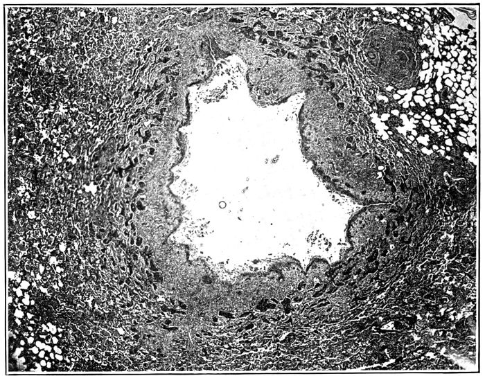
Fig. 17.—Acute bronchiectasis showing fissures penetrating into bronchial wall and at one place entering surrounding alveolar tissue; the surrounding alveoli are filled with fibrin. Autopsy 425.
Acute bronchiectasis may be found following influenza
after the illness has lasted eight or ten days. There is no
increase of fibrous tissue. Small bronchi with no cartilage,
which in normal lungs have a diameter approximating 1
mm., are dilated to 3 mm. or more. The surface epithelium
is wholly or partially lost. Necrosis occurs in places and
extends deep into the tissue, destroying muscle and often
penetrating the entire thickness of the wall which in these
small bronchi consists in large part of fibrous tissue containing
greatly engorged blood vessels. In this necrotic
material nuclei are absent and the tissue containing fibrin
stains deeply with eosin. In it occur fissures or tears which
extend from the lumen a variable distance, very frequently
penetrating the entire thickness of the wall and entering
adjacent alveoli (Figs. 17 and 19). Alveoli thus exposed
almost invariably contain plugs of dense fibrin. Where
these rents have occurred, adjacent edges of the bronchial
wall, held together by underlying lung tissue, have separated
from one another, so that the circumference of the
bronchus has been increased (Fig. 18). These breaks in
the continuity of the wall may occur in several places, so
that a fourth or a third of the circumference may be formed
247by exposed alveolar tissue which has become the site of
fibrinous pneumonia (Fig. 20). During life, though the inflamed
bronchus is filled by mucopurulent exudate, distention
of loose alveolar tissue, uniting the interrupted bronchial
wall, is doubtless greater than it appears in the lung
fixed by hardening fluids.
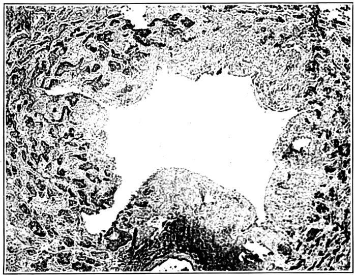
Fig. 18.—Acute bronchiectasis showing fissures in the bronchial wall extending into neighboring alveoli which in zone about are filled with fibrin; one fissure has separated widely; peribronchial fibrinous pneumonia (fibrin is black). Autopsy 425.
Recently dilated bronchi have an irregularly stellate lumen
as the result of clefts penetrating at intervals into or
through the bronchial wall (Fig. 26). Longitudinal fissures
mark the lining of these dilated bronchial tubes.
When the fatal illness has lasted more than two weeks,
abundant new formation of fibrous tissue occurs in a zone
surrounding the dilated bronchus. Adjacent alveolar walls
are thickened by young fibrous tissue. Alveoli, much diminished
in size, are filled by hyaline fibrin into which fibroblasts
248and newly formed blood vessels have penetrated.
These changes are limited to a wide zone in immediate contact
with the dilated bronchus, whereas at a greater distance
alveolar walls have undergone no thickening and
alveoli contain no fibrin.
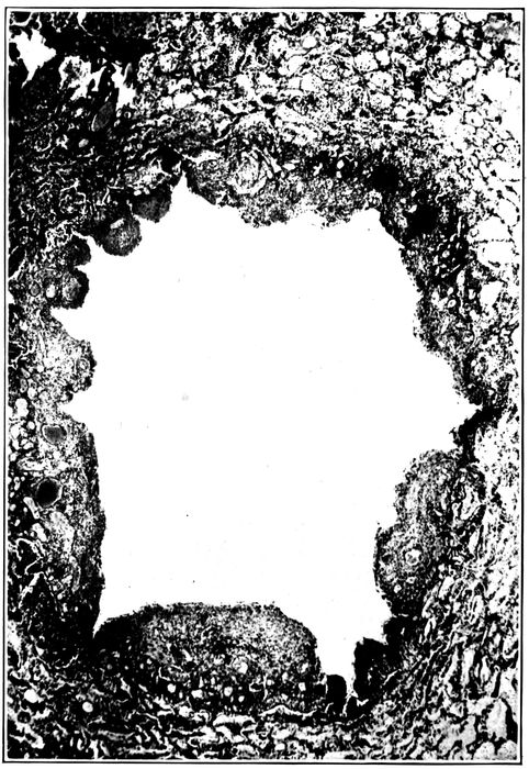
Fig. 19.—Acute bronchiectasis; the bronchial wall indicated by engorged mucosa shows a varying degree of destruction, fissures extending into and through the bronchial wall. Autopsy 352.
249
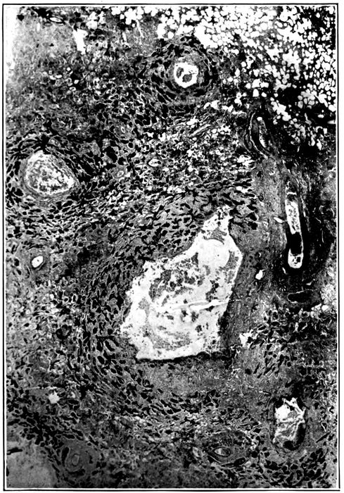
Fig. 20.—Acute bronchiectasis; with destruction of bronchial wall exposing alveoli filled with fibrin; peribronchial fibrinous pneumonia is seen about several bronchi present in the section; Gram Weigert fibrin stain. Autopsy 425.
This stage is well represented by Autopsy 421 after an
illness of nineteen days. Bronchiectatic cavities, from 3
to 6 mm. in diameter, are numerous in sections of the lung;
their lumina are irregular in outline and often irregularly
stellate. Microscopic examination shows the presence of
250clefts which interrupt the bronchial wall at intervals
throughout its entire circumference. The original wall is
well indicated by the very richly vascularized connective
tissue containing scattered muscle bundles and is infiltrated
with lymphoid and plasma cells in great number.
Where fissures have occurred the adjacent edges of the interrupted
wall have separated from one another, leaving
a wide interval where underlying alveolar tissue is exposed.
Two changes tend eventually to render the fissures inconspicuous,
namely, regeneration of epithelium and new formation
of fibrous tissue. Exposed alveoli filled with fibrin
are in process of organization and epithelium which has assumed
a squamous type has grown down over the exposed
surfaces of the interrupted bronchial wall. It has begun to
cover or in some instances has completely covered the surface
of rents entering alveoli plugged with fibrin (Fig. 21).
In the periphery of the bronchus alveolar walls are thickened
and infiltrated with lymphoid and plasma cells. The
same changes affect bronchi containing cartilage which is
undergoing atrophy.
The reinforcement of the fissured bronchial wall by new
formation of fibrous tissue, by thickening of the interalveolar
walls and by organization of fibrin within the alveoli is
well shown after four weeks (Autopsy 425; Fig. 28).
There are spherical bronchiectatic cavities more than a centimeter
in diameter surrounded by a dense fibrous wall in
which are atrophied alveoli lined by epithelium of cubical
form. Occasionally, the fibrous wall is interrupted and alveoli,
plugged with organizing fibrin, are in immediate contact
with the lumen. When these plugs of fibrin which are
slowly absorbed disappear, evidence of preexisting rents in
the bronchial wall are lost, and there are in this lung bronchiectatic
cavities of which the wall is a continuous circle
of dense fibrous tissue.
251
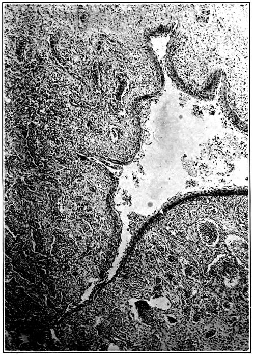
Fig. 21.—Bronchiectasis with fissures extending through the bronchial wall into alveolar tissue which is the site of fibrinous pneumonia; epithelium has grown down into these fissures and has covered the exposed surfaces. Autopsy 463.
252
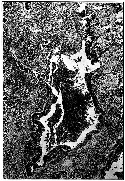
Fig. 22.—Regeneration of epithelium over fissures which have been formed in the wall of a bronchus; the epithelium in the neighborhood of and within the fissure is squamous.
Epithelium lining the dilated bronchi is at times completely
destroyed (Fig. 28), but more frequently it persists
in part. That which remains has almost constantly the
character of squamous epithelium (Figs. 22 and 23). The
lowermost cells are cubical; those above them are polygonal,
tending to become flatter as the surface is approached;
upon the surface are cells often much flattened
and occasionally they have lost their nuclei and stain deeply
with eosin as the result of superficial necrosis. The change
should not be regarded as metaplasia, for the epithelium
assumes this squamous type when the superficial columnar
cells have been lost. Actual necrosis of superficial ciliated
columnar cells is occasionally seen (Autopsy 352); injured
cells have separated from one another and desquamated
253into the lumen of the bronchus. The epithelium which remains
after the superficial cells are lost consists of cells
which become flatter from base to surface, but the intercellular
bridges characteristic of the epithelium of the skin
are not found. When epithelium is in process of regeneration,
a layer gradually diminishing in thickness extends
over the denuded surface, the advancing edge being formed
by very flat cells in a single layer. The epithelium growing
into fissures which have penetrated the bronchial wall
may completely cover the exposed alveolar tissue. The
newly formed epithelium may follow a fissure into an alveolus
which has been opened and come into contact with the
fibrin which fills the alveolus.
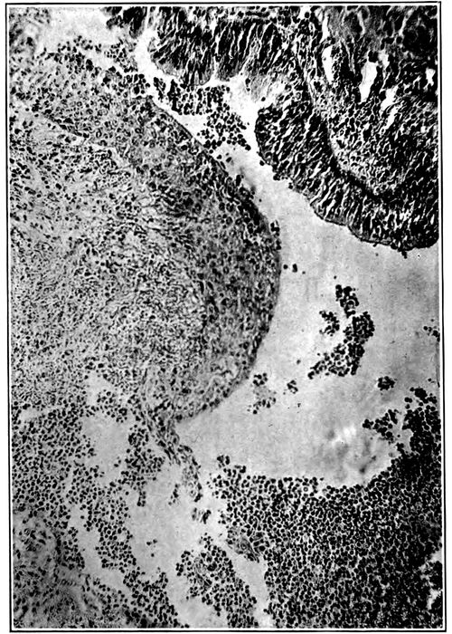
Fig. 23.—Squamous epithelium growing over the defect in the bronchial wall shown in Fig. 22 more highly magnified; squamous epithelium is present above and columnar epithelium below.
254Bronchiectasis usually affects the small bronchi with no
cartilage. It is not uncommon to find greatly dilated bronchi
with no cartilage in close proximity to cartilage containing
bronchi of smaller caliber. In one instance (Autopsy
421) a bronchus of medium size with cartilage measured
3 mm. in diameter, whereas two bronchi with no cartilage
were dilated to 4 and 6 mm., respectively. Nevertheless,
larger bronchi are occasionally the site of superficial
loss of epithelium, necrosis extending into the bronchial
wall, formation of fissures and stretching of the wall at the
spot which is weakened. In association with these changes
atrophy of the cartilage may occur (Autopsies 421, 425,
440, 463). Plates of cartilage in process of atrophy are
readily recognized by their irregularly indented outline
and often by their small size. The fibrous tissue surrounding
the cartilage is the site of chronic inflammation and is
densely infiltrated with lymphoid and plasma cells among
which polynuclear leucocytes are scant. Nevertheless,
polynuclear leucocytes are abundant in immediate contact
with the cartilage and appear to have an important part in
the solution of its matrix, for about them occur indentations
of the edge. Leucocytes penetrate into the cartilage.
The necrosis and tears which occur in the wall of the
bronchus are not always limited to the bronchus, but may
extend deeply into the surrounding tissue. In Autopsies
312 (Fig. 21) and 423 wide areas of necrosis have penetrated
deeply into the tissue about the bronchi.
Autopsy 312.—Illness began with influenza on September 26, seventeen
days before death; a diagnosis of lobar pneumonia with consolidation of
255the right lower lobe was made ten days after onset and Pneumococcus IV,
B. influenzæ and S. hemolyticus were found in the sputum. At autopsy
there was bronchopneumonia with red and gray lobular and confluent
lobular patches of consolidation and right and left serofibrinous pleurisy;
there was purulent bronchitis; no abscesses were seen. Small bronchi
throughout both lungs were dilated and often surrounded by a zone of hemorrhage.
Hemolytic streptococci were found in the heart’s blood, in the pleural
exudate, consolidated lung and bronchus; B. influenzæ was found in the
lung and in a small bronchus, and staphylococci in the contents of a small
bronchus.
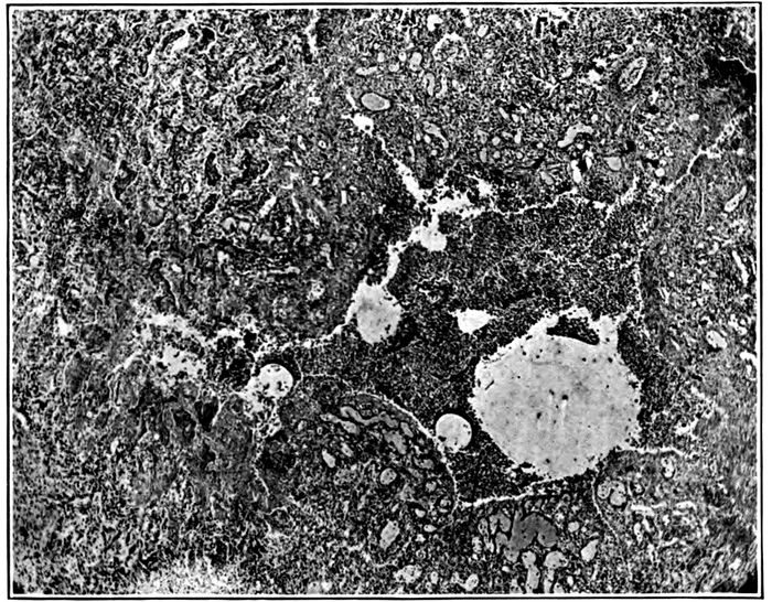
Fig. 24.—Acute bronchiectasis with fissures extending through bronchial wall which is marked by great engorgement of blood vessels; at one point a fissure has penetrated deep into the alveolar tissue and formed a small cavity containing purulent exudate and surrounded by fibrinous pneumonia. Autopsy 312.
Bronchi which are the site of acute inflammation have lost their epithelium
wholly or in part, and deep fissures penetrate the entire thickness
of the bronchial wall, extending into the surrounding lung tissue which is
the site of fibrinous pneumonia. In some instances plugs of fibrin within
the alveoli are bisected by these tears. There is some superficial necrosis
along the edge of each fissure, in several places extending outward from
defects in the walls of small bronchi dilated to approximately 1.5 mm.
There are wide patches of necrosis affecting both alveolar walls and contents
256of alveoli and extending 2 mm. into the lung tissue. When a fissure
has penetrated from the lumen of the bronchus into necrotic tissue
(Fig. 21), polynuclear leucocytes have accumulated within the necrotic
tissue, disintegration of tissue occurs, and a small cavity communicating
with the bronchus is formed.
Autopsy 423.—C. H., white, aged twenty-five, resident of Oklahoma,
had been in military service one month. Death occurred sixteen days after
onset of influenza.
Anatomical Diagnosis.—Chronic bronchopneumonia with peribronchiolar
consolidation throughout right lung and in left lower lobe; right purulent
pleurisy; purulent bronchitis; bronchiectasis at base of left lung.
The right lung weighs 1,260 grams; in the upper lobe are yellowish
gray nodules having the appearance of tubercles clustered about small
bronchi; in places similar nodules occur upon a background of pinkish
gray consolidation occupying the greater part of the lower lobe. Bronchi
contain purulent fluid. The left lung weighs 760 grams; it is edematous
and small, yellowish gray nodules of consolidation in the lower lobe
are clustered about terminal bronchi. Bronchi at the base of the lower
lobe are dilated.
Bacteriologic examination shows the presence of hemolytic streptococci
in the blood of the heart; hemolytic streptococci and B. influenzæ in the
lung.
Microscopic examination shows that the walls of the bronchi are infiltrated
with lymphoid and plasma cells; these cells are very numerous
in peribronchiolar patches of consolidation. A small bronchus 1 mm. in
diameter has squamous epithelium along one side; on the opposite side, the
wall is completely absent and there is superficial necrosis of exposed alveoli
filled with fibrin. A deep fissure passes from the bronchus into the
consolidated tissue; its edges are necrotic and it is filled with polynuclear
leucocytes. A small cavity in contact with the bronchus has been formed.
In another part of the lung a distended bronchus has lost its epithelium
on one side, and here alveoli filled with fibrin form the wall of the bronchus
which is filled with leucocytes. Extending outward from the eroded wall
is a focus of necrosis where both alveolar walls and contained exudate
have lost their nuclei.
The necrosis which has had its origin in the bronchi is
soon followed by accumulation of polynuclear leucocytes,
softening and disintegration of tissue. Discharge of the
disintegrated tissue through the bronchi results in the formation
of a small cavity continuous with the bronchus.
These changes are well illustrated by the bronchiogenic
abscesses which have been described elsewhere (Autopsies
376, p. 206, and 387, p. 206). When disintegrated tissue
257is discharged by way of the bronchi no accumulation of pus
occurs, but cavities will be formed, in part by dilation of
bronchi, in part by erosion of the adjacent lung tissue. Histologic
examination shows that these changes have produced
the advanced bronchiectasis found in Autopsy 445
(Fig. 25).
Autopsy 445.—W. F., white, aged twenty-three, from Mississippi, had been
in military service one month. His illness began September 22, twenty-seven
days before death, with severe coryza, weakness, nausea and vomiting;
great pain in bones, cough and sore throat. He was admitted to the
base hospital one week later with diagnosis of influenza and bronchitis.
On October 3, sixteen days before death, signs of consolidation were found
on the left side over the back and a diagnosis of lobar pneumonia was
made. On October 18 there was severe headache, pupils were dilated, and
there was rigidity of neck; lumbar puncture was made and pneumococci
were found in the fluid obtained. Death occurred on the following day.
Anatomic Diagnosis.—Bronchiectasis with unresolved pneumonia limited
to the left lower lobe; acute bronchopneumonia with peribronchiolar consolidation
in right lung; purulent bronchitis, peribronchial hemorrhage
and organizing bronchiolitis in right lung; adherent pleura on left side;
purulent meningitis.
The left upper lobe is crepitant throughout. The outer and posterior
two-thirds of the left lower lobe is riddled with cavities often rounded
and varying in diameter from 0.5 to 3 cm. but not infrequently irregular
in shape and in communication with adjacent cavities (Fig. 25). In places
cavities pass in a tortuous course from pleura to the midpart of lung.
The lining of these cavities is usually smooth, but in places is covered
by gray necrotic material. Communication between the cavities and medium-sized
bronchi is occasionally found. The lung tissue between the
cavities is in part grayish red and consolidated, in part pink and air containing.
The right lung is edematous throughout; the bronchi in the lower
part of the right lung contain purulent fluid and are in places surrounded
by zones of hemorrhage.
The spleen is very large (14 × 11 × 5 cm.) and firm.
The spinal fluid is cloudy and blood vessels over the lumbar enlargement
and lower thoracic region are congested; in the upper thoracic region the
cord is covered by purulent exudate.
Bacteriologic examination demonstrates the presence of hemolytic streptococci
in the blood of the heart; plates from the left lung contain a few
colonies of S. aureus and Pneumococcus IV; plates from the right main
bronchus contain S. aureus and a large bacillus which does not stain by
Gram’s method. Three plates from the spinal meninges contain Pneumococcus
IV.
258
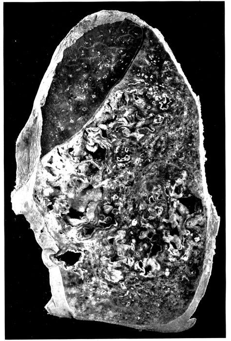
Fig. 25.—Advanced bronchiectasis throughout lower left lobe. Autopsy 445.
259Microscopic examination shows that the cavities which have been described
are lined by very vascular connective tissue containing many cells;
there is no epithelial lining and the surface is in places covered by fibrin.
On the surface polynuclear leucocytes are numerous, but immediately below,
large mononuclear cells occur and frequently contain one or several
ingested polynuclear leucocytes. None of the structures peculiar to the
bronchi can be identified in the wall of these cavities, and in many places
it is evident that lung tissue has undergone destruction, for in places the
lining of vascular connective tissue is interrupted and an extension of the
cavity penetrating into the lung substance is surrounded by alveoli filled with
fibrin; in contact with the cavity there is some necrosis.
The cavities communicate with the bronchi and are
lined in part by vascular connective tissue which may in
part represent preexisting bronchial walls, but no epithelium
is present and the relation to the bronchi cannot be established
with certainty. These cavities have extended by
necrosis which has broken the vascular connective tissue of
their wall and penetrated into adjacent lung tissue. Death
has been the result of purulent meningitis caused by pneumococcus,
and the histologic changes in the walls of the
cavities suggest that the activity of the inflammatory reaction
here is subsiding, for large mononuclear cells are numerous
and are ingesting polynuclear leucocytes. The
changes described would, if continued, result in the formation
of cavities lined by fibrous tissue and resembling many
of those formed as the result of dilatation of the bronchi.
A study of the progress of the changes which result in
the formation of bronchiectatic cavities has shown how the
inflammatory irritant within the bronchus destroys the epithelium
of the bronchus, penetrates into the deeper tissues
and produces fissures which extend through the entire
thickness of the bronchial wall at one or usually several
places. These longitudinal fissures, which at first often
give a stellate outline in cross section to the cavity of the
affected bronchus, permit the separation of the edges of the
fissure, so that an increase in the circumference occurs.
The base of the fissure is formed by surrounding alveolar
tissue and its edges are the site of necrosis. Tears may
extend into the surrounding alveolar tissue, thus permitting
further stretching of the bronchial wall. The consequences
260of rupture of the small bronchi into the adjacent
alveoli are to some extent overcome by the inflammatory
reaction which plugs the adjacent alveoli with fibrin.
Compression of the lungs by forced expiration, even
though the glottis were closed as in coughing, would not
dilate the bronchi, because pressure outside and within the
bronchi would be equally elevated (Thornton and Pratt[89]).
The pressure within the bronchi does not differ greatly
from atmospheric pressure, whereas the negative pressure
within the pleural cavity may vary from approximately
6 mm. of mercury during quiet inspiration to 30 mm. with
forced inspiration. Excess of pressure upon the inner surface
of the bronchial walls will vary with coughing and
other respiratory efforts, between these limits depending
upon the readiness with which pressure is equalized within
and without the bronchi by penetration of air into the alveoli.
The presence of viscid mucopurulent fluid within bronchioles
will obstruct these tubules and retard the entrance
of air into alveoli.
Weakening of the bronchial wall by the changes which
have been described will cause lasting dilatation of the
bronchi. Whatever increases pressure within the bronchi
will increase the tendency to dilatation; the bronchi being
filled with mucopurulent exudate dilatation usually appears
first at the bases of the lung, since gravity increases
intrabronchial pressure here. New formation of fibrous
tissue within the wall of the bronchus, thickening of adjacent
alveolar walls, and organization of fibrin reinforce the
weakened bronchial wall and limit the dilatation which follows
injury to the wall. Regeneration of epithelium covering
the dilated tube will further obscure the early
changes which have made dilatation possible. The changes
which weaken the bronchial wall permit dilatation at a
time when there is no new formation of fibrous tissue.
When the bronchial lesion has persisted several weeks,
261chronic pneumonia is associated with it. It has been suggested
that the contraction of newly formed fibrous tissue
within the substance of the lung might cause bronchi to be
enlarged by traction upon their walls. Newly formed connective
tissue is most abundant in the wall of the bronchiectatic
cavity, and here contraction would tend to diminish
the size of the cavity.
Chronic bronchopneumonia is characterized by changes
similar to those associated with chronic inflammation in
other parts of the body, namely, by thickening of the interstitial
tissue of the lung, by accumulation of mononuclear
cells, by proliferation of fibrous tissue and by organization
of exuded fibrin. In a few instances these changes have begun
at the end of two weeks after onset of influenza, but
they have been little advanced until three weeks has
elapsed; advanced chronic inflammation has occurred after
from four to eight weeks. Chronic inflammation primarily
affects those structures which are most severely injured by
the acute lesion and is most conspicuous in immediate proximity
to the small bronchi and bronchioles; the perivascular
and interlobular connective tissue are secondarily
involved. Corresponding to each of the lesions of the alveolar
tissue which have been found with bronchopneumonia,
namely, peribronchiolar, hemorrhagic peribronchiolar, lobular
and peribronchial consolidation, there is a chronic lesion
which develops when pneumonia has failed to resolve.
The term interstitial bronchopneumonia has been used
by MacCallum to designate a lesion which he has found in
association with measles at Fort Sam Houston. This name
he states does not describe accurately the early stage of the
lesion, for its interstitial character is not evident at first. In
his monograph on “Epidemic Pneumonia in the Army
Camp,” published in 1919, MacCallum describes and pictures
262instances of the lesion which we have designated interstitial
suppurative pneumonia and classifies them as interstitial
bronchopneumonia. We have shown that this lesion,
which is the result of infection of the lymphatics with
S. hemolyticus, bears no necessary relation to the lesion
which is characterized in its early stage by peribronchiolar
pneumonia and in its later stages by chronic inflammation
with mononuclear infiltration and proliferation of the peribronchial,
perivascular and interalveolar tissue. At Fort
Sam Houston, nearly every patient with measles was infected
with hemolytic streptococci; we observed, following
influenza, similar prevalence of hemolytic streptococci in
certain wards in the base hospital at Camp Pike. Among the
cases at Fort Sam Houston there were doubtless instances
both of interstitial suppurative pneumonia caused by hemolytic
streptococcus and of chronic bronchopneumonia not
referable to this microorganism.
Studying pneumonia following influenza at Camp Lee,
Va., and later at Camp Dix, N. J., during the fall of 1918,
MacCallum reached the conclusion that “interstitial bronchopneumonia”
following influenza was caused by B. influenzæ
of Pfeiffer. This lesion attributed to B. influenzæ
differed from that previously referred to hemolytic streptococcus
in the following characters: the lymphatic channels
in the bronchial walls and widened interlobular septa
are inconspicuous and none are found distended with exudate;
there is no intense infection of the pleura, and polynuclear
leucocytes are inconspicuous in the alveolar exudate
and in the walls of the bronchi. It seems probable
these differences are explained by the absence of hemolytic
streptococci which tend to invade lymphatics and produce
severe inflammatory changes in the pleura.
Chronic Bronchitis.—The earliest changes in the bronchial
wall with bronchitis of influenza are hyperemia, leucocytic
infiltration and hemorrhage, and they may occur
even though the lining epithelium remains intact. Epithelium
263frequently undergoes partial or complete destruction,
and with this severe injury the influence of the inflammatory
irritant may extend directly through the wall of the
bronchus, for in some instances there is hemorrhage into
all the alveoli in a zone encircling the bronchus. Since these
alveoli have only indirect communication with the affected
bronchus through alveolar tissue not involved in the inflammatory
process, it is evident that the surrounding
hemorrhage is secondary to the lesion of the bronchus.
Fibrinous inflammation in other instances, similarly localized
in a zone of alveoli encircling a bronchus, is doubtless
the result of direct extension of the inflammatory process
through the bronchial wall. After the disease has existed
during two or three weeks inflammation is still active immediately
below the inner surface of the bronchus; here
polynuclear leucocytes are numerous whereas in the deeper
parts of the mucosa and about the muscularis leucocytes are
scant but lymphoid and plasma cells are very numerous.
The severity of the inflammatory reaction may be judged
by the abundance and extent of this cellular reaction and is
in close relation to the intensity of the changes affecting
the mucous membrane of the bronchus. Infiltration of the
entire bronchial wall with lymphoid and plasma cells is
almost invariable when the primary injury to the bronchus
has destroyed the epithelial lining, and this infiltration is
not limited to the bronchial wall but extends outward into
the contiguous alveolar septa which are thickened by it.
The sheath of the pulmonary artery which accompanies the
bronchus exhibits a similar change, and the alveolar septa,
as a fringe about it, are thickened and infiltrated with mononuclear
cells. Interlobular septa continuous with the bronchus
often show some infiltration.
A later phase in this series of changes is represented by
new formation of fibrous tissue. The bronchial walls and
interalveolar septa are thickened by proliferating fibrous
tissue, young fibroblasts and newly formed collagen fibrils
264being abundant (Fig. 28; also Fig. 30). This increase of
fibrous tissue is especially noteworthy immediately surrounding
the walls of the small bronchi, which are often
considerably dilated, and about the smaller of those bronchi
which have cartilage; with thickening of alveolar walls
immediately adjacent to the bronchus every stage in the
obliteration of the alveoli may be found. Their walls are
thickened and their lumina are diminished in size and often
flattened in a direction concentric with the bronchus. Such
atrophied alveoli lined by cubical epithelial cells occurring
within the thickened peribronchial fibrous tissue give evidence
that this tissue has replaced alveoli. Alveoli surrounding
and within the new fibrous tissue are frequently
filled with fibrin, and organization indicated by
penetration of fibroblasts and capillaries into the fibrin
may be far advanced. There is some increase of perivascular
and interlobular tissue. The bronchiectasis which is
almost invariably found with unresolved bronchopneumonia
has been described. Squamous transformation of epithelium
(page 251) is frequently found in association with
the chronic bronchitis of unresolved pneumonia.
Organizing Bronchitis and Bronchiolitis.—When the
bronchial epithelium is destroyed, fibrin is deposited upon
the denuded surface and may partly or completely fill the
lumen of the bronchial tube. The plug of fibrin is adherent
to the underlying tissue wherever epithelium is lost but is
separated from the bronchial wall by a well-defined space
where epithelial lining is still intact. Fibroblasts promptly
migrate from the wall of the bronchiole into this fibrin, and
fibroblasts, fixed during ameboid movement, are irregularly
elongated in a direction toward the fibrin.
Organization of fibrin occurs within the smallest bronchi
(diameter 0.3 to 0.5 mm.) or within respiratory bronchioles.
It has been found in 8 autopsies. In one instance it has
been present eleven days after the onset of influenza, but
usually it is seen three or four weeks after onset of symptoms
265of respiratory disease. In the early stages of the
lesion a plug of fibrin within the lumen of the bronchus or
bronchiole is invaded by fibroblasts, plasma cells and newly
formed capillaries. These capillaries have their origin in
the wall of the tube and enter the fibrin at points where
in consequence of loss of epithelium fibrin is continuous
with the connective tissue. When the bronchiole is cut
longitudinally, partially or completely organized fibrin may
be found adherent at several places with intact epithelium,
sometimes beautifully ciliated, between the sites of attachment.
The fibrin is finally replaced completely and the
lumen of the bronchiole contains a mass of organized
fibrous tissue in which young fibroblasts and plasma cells
are numerous.
The lesion has been associated with chronic bronchopneumonia
in 6 of 8 instances. In Autopsy 445, p. 257, organizing
bronchitis and bronchiolitis occurred in the right lung
unassociated with other chronic lesion, although there was
advanced bronchiectasis with fibrous induration in the
left lung. In Autopsy 499 (p. 224) organizing bronchiolitis
occurred in association with chronic changes which appear
to have followed interstitial suppurative pneumonia caused
by S. hemolyticus. Other severe lesions of the bronchi
have accompanied organizing bronchitis and bronchiolitis.
Purulent bronchitis has been present in 7 of 8 instances;
bronchiectasis in 5 of 8 instances.
The bacteriology of autopsies with organizing bronchitis
and bronchiolitis is shown in Table LIII.
The bacteriology of these cases presents no constant
feature. Invasion of the blood by S. hemolyticus has been
present in a large proportion of cultures, namely, in 5 of 7
(71.4 per cent). In one of the 2 instances in which hemolytic
streptococci have been found, neither in the blood nor
lungs, Pneumococcus III has been found in the blood and S.
viridans in the lungs and bronchus; in the other, S. aureus
has been found in the lung and bronchus. Staphylococci
have been found frequently in the bronchi (60 per cent)
and in the lungs (50 per cent). B. influenzæ has been present
in the bronchi in the usual proportion of instances
(80 per cent). The lesion has occurred in the presence of
B. influenzæ combined with streptococci or staphylococci.
| 266 |
| Table LIII |
|---|
| |
| AUTOPSY |
DURATION OF ILLNESS |
BLOOD |
LUNGS |
BRONCHUS |
| 420 |
11 days |
S. hem. |
S. hem., B. inf., S. aur. |
|
| 402 |
14 days |
Pneum. IV, S.hem. |
|
|
| 370 |
17 days |
|
S. aur. |
S. aur., Pneum. IV, B. inf. |
| 457 |
17+ days |
|
|
Pneum. IV, B. inf. |
| 421 |
19 days |
S. hem. |
Pneum. IV, S. hem. |
|
| 445 |
27 days |
S. hem. |
Pneum. IV, S. aur. |
S. aur. |
| 473 |
28+ days |
Pneum. III |
S. vir. |
B. inf., S. vir., staph., M. catarr. |
| 499 |
36 days |
S. hem. |
|
S. hem. B. inf. |
Thrombosis of lymphatics in the wall of bronchi adjacent
to blood vessels and in interlobular septa occurs, and occasionally
organization of the fibrinous plug within the lymphatic
is in progress (Autopsies 283, 425 and 463). Fibroblasts
and capillaries penetrate from the wall of the lymphatic
into a mass of hyaline fibrin which fills the lumen.
Unresolved Bronchopneumonia.—The most common type
of pneumonic lesion following influenza is characterized by
acute inflammation of the alveoli immediately adjacent to
the bronchioles and the lesion is associated in many instances
with hemorrhage or edema. If this lesion persists
unresolved during several weeks, evidences of chronic inflammation
are found. Peribronchial, perivascular and interlobular
connective tissue is thickened and richly infiltrated
with lymphoid and plasma cells, large mononuclear
cells and many young fibroblasts. Interalveolar septa adjacent
to the walls of bronchi and between alveoli surrounding
inflamed bronchioles are implicated in the process.
267Interstitial changes characterize the lesion only in its late
stage. It appears undesirable to give the name “interstitial
pneumonia” to the early stage of a lesion which begins and
in most instances terminates as an acute relatively superficial
inflammation of the bronchi, bronchioles and peribronchiolar
alveoli.
Chronic bronchopneumonia is often overlooked at autopsy
because newly formed connective tissue is not present
in sufficient quantity to attract attention (Fig. 26). When
the lesion is advanced conspicuous gray white patches of
fibrous tissue may be seen about the bronchi (Autopsy 487;
Fig. 27) and interlobular septa may be obviously thickened
(Autopsy 472). The most distinctive feature of the lungs
is the presence of small, firm, gray or yellowish gray nodules
of consolidation which resemble miliary tubercles.
They represent the peribronchiolar patches of bronchopneumonia
present during the acute stage and have
assumed the well-defined outline and firm consistence of
tubercles because polynuclear leucocytes and red blood corpuscles
have in large part disappeared, interstitial tissue
is increased, and exudate is in process of organization.
These nodules are grouped in clusters about the small
bronchi.
With unresolved bronchopneumonia the lungs are very
voluminous and fail to collapse after they are removed from
the chest and in some instances even after incision. The
air containing tissue is usually dry. In our autopsies the
lungs have been pink in color and often free from coal pigment,
because those suffering with pneumonia have been in
considerable part men from rural districts. Thick mucopurulent
material exudes from the small bronchi which
have been cut across; purulent bronchitis has been present
in 20 of 21 instances of chronic bronchopneumonia. Bronchiectasis
has been present in 13 instances; dilatation is
often advanced, so that throughout the lungs are found
bronchi with no cartilage distended to a diameter of 0.5 cm.
In addition to the firm peribronchiolar tubercle-like nodules
of consolidation there are scattered patches of gray
lobular or confluent lobular consolidation. Yellowish nodules,
grouped about bronchi and resembling those found
elsewhere in air containing tissue, are occasionally seen
scattered upon the cut surface of a patch of gray, confluent
lobular consolidation (Autopsies 421, 423, 431).
268
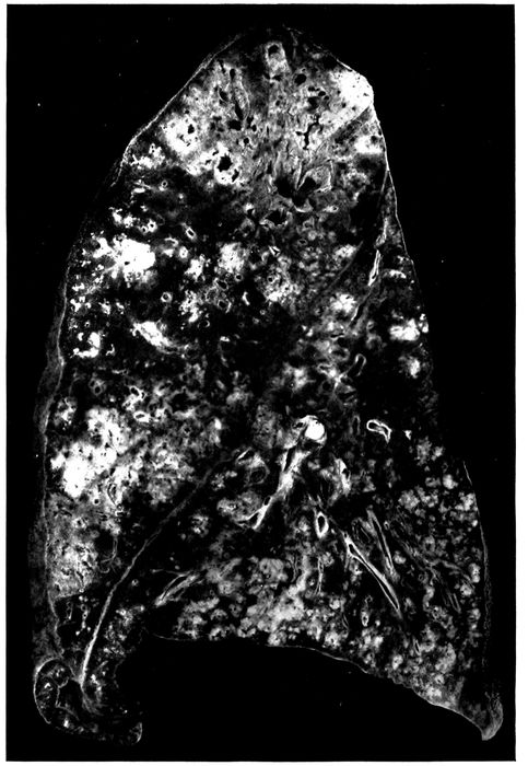
Fig. 26.—Unresolved bronchopneumonia with tubercle-like nodules of peribronchiolar consolidation best seen in lower lobe; bronchiectasis. Autopsy 425.
269Microscopic examination demonstrates the presence of
those changes which have been described in association
with chronic bronchitis and bronchiectasis. There is abundant
new formation of fibrous tissue about the bronchi of
small and medium size, thickening of adjacent interalveolar
walls and incorporation of alveoli into the thickened bronchial
wall (Figs. 27, 28, 30, and 31). In half of the instances
of chronic bronchopneumonia there has been peribronchial
fibrinous pneumonia, and organization of fibrin
within the alveoli is usually well advanced. In one instance
(Autopsy 487; Figs. 27 and 28) after an illness of fifty-five
days this process has resulted in the formation of conspicuous
patches of firm, grayish white fibrous tissue surrounding
dilated bronchi. Organization of fibrinous exudate
within the lung has not been limited to the alveoli but has
occurred in the bronchioles as well. Organizing bronchiolitis
has been present in 5 instances (Autopsies 370, 402,
457 and 473).
Increase of fibrous tissue occurs about the blood vessels
and in the septa between the lobules, which are infiltrated
with mononuclear wandering cells and fibroblasts. Dilatation
and thrombosis of the lymphatic vessels have occurred
in both situations, and in 3 instances (Autopsies
283, 425 and 463) organization of these fibrinous thrombi
has occurred.
270
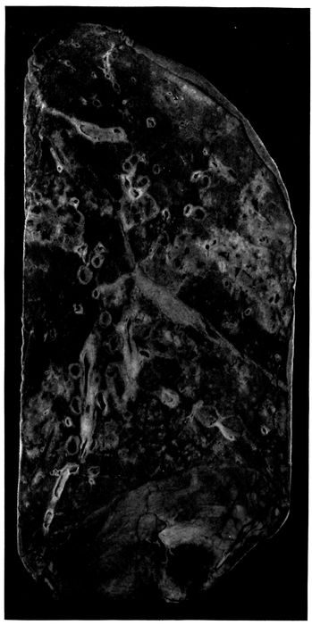
Fig. 27.—Unresolved pneumonia with peribronchial formation of fibrous tissue; bronchiectasis. Autopsy 487.
271
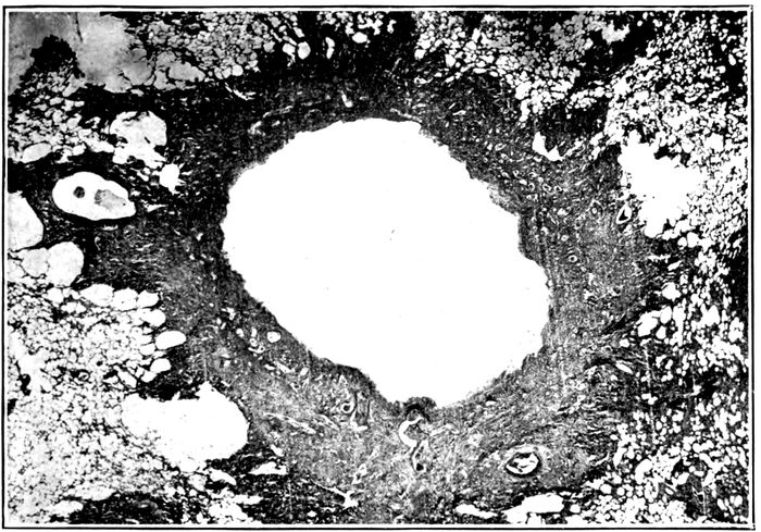
Fig. 28.—Unresolved pneumonia with bronchiectasis showing new formation of fibrous tissue about a greatly dilated bronchus of which the epithelial lining has been lost. Autopsy 487.
Thickening, cellular infiltration and fibrosis of the bronchial
walls with interstitial inflammation and fibrosis of immediately
adjacent alveolar septa are found about the
ramifications of the bronchial tree and may be followed
to the smallest bronchi. When the respiratory bronchioles
are reached it will be found that the alveoli which stud their
walls are implicated in the change. The fibrin which they
contain is infiltrated with lymphoid and plasma cells, and
with progress of the lesion is invaded by fibroblasts and
capillaries. Infiltration and fibroid thickening extends
from the bronchiolar wall to the alveolar septa continuous
with it (Fig. 31 with measles). Similar changes occur about
the alveolar ducts, and about the orifices of the tributary
infundibula (Fig. 32), peribronchiolar foci of acute inflammation
having assumed the characters of a chronic inflammatory
process. Fibrin within the alveoli contains round
cells and fibroblasts. With thickening of alveolar walls the
alveolar lumina may be much diminished in size and often
persist as spaces lined by cubical cells. Polynuclear leucocytes
are usually numerous within the alveolar duct and
272in a few alveoli immediately adjacent to it, but elsewhere
throughout the focus of inflammation round cells are predominant.
The changes which have been described correspond
with the transformation of ill-defined, gray or reddish
gray spots of consolidation grouped about the terminal
bronchi into firm sharply defined grayish white nodules
having the consistence and appearance of miliary tubercles.
One of the most constant characters of pneumonia following
influenza is its hemorrhagic character. In the
earlier stages of pneumonia phagocytosis of red blood corpuscles
by large mononuclear cells is frequently seen. In
association with the chronic changes which have been described,
large mononuclear cells filled with brown pigment,
doubtless formed from red corpuscles, are often found
within the alveoli. These pigment containing cells are similar
to those commonly associated with chronic passive congestion
of the lungs.
In one instance (Autopsy 457) hemorrhagic peribronchiolar
pneumonia has been found in process of organization.
The bronchioles and alveoli adjacent to them contain
polynuclear leucocytes, but intervening alveoli almost uniformly
contain blood and are the site of new formation of
connective tissue. Interalveolar septa are thickened and
alveoli which are lined by cubical epithelium are often diminished
in size. In many places fibroblasts have penetrated
in considerable number into the blood within the alveoli
and occasionally newly formed capillaries are found
within them.
Lobular patches of pneumonia are often found in process
of organization (Autopsies 370, 421, 423, 433, 463, 472 and
473). Microscopic examination shows that whole lobules
well defined by thickened septa are the site of chronic interalveolar
inflammation and intraalveolar organization of
exudate, whereas adjacent lobules are air containing and
relatively normal. In the earlier stages of the process
fibrin present within the alveoli is invaded by fibroblasts,
273mononuclear wandering cells and blood vessels but in the
later stages fibrin has disappeared; the lumina of the alveoli
are occupied by cellular fibrous tissue and in places the
thickened alveolar walls and intraalveolar fibrous tissue
have been fused to form wide patches of new tissue.
With chronic bronchopneumonia confluent lobular consolidation
occasionally has a gray ground upon which are
scattered small yellow spots clustered about the small
bronchi (Autopsies 421, 423 and 431). Microscopic examination
has shown that the yellowish spots correspond to
dilated bronchioles filled with purulent exudate and surrounded
with alveoli containing many polynuclear leucocytes.
In the interstitial tissue about the bronchiole and
between adjacent alveoli plasma cells are often present in
great number. Between these spots of subacute bronchiolar
inflammation lung tissue is the site of interalveolar proliferation
of fibrous tissue and intraalveolar organization of
exudate.
In all instances of chronic bronchopneumonia there has
been peribronchial pneumonia in a zone encircling small
bronchi with no cartilage and the smallest of the bronchi
which have cartilage in their wall; thickening of interalveolar
septa, organization of peribronchial fibrinous pneumonia
and partial disappearance of alveoli have been described.
In the following autopsy peribronchial fibroid
pneumonia has been so advanced that conspicuous patches
of gray white tissue surrounding bronchi have replaced in
some parts of the lung a considerable part of the lung substance.
Autopsy 487.—W. C., white, aged twenty-seven years, a farmer from
Mississippi had been in military service twenty-one days. Illness began
on September 17, fifty-five days before death, with chill, fever, cough, backache,
pain in the chest and coryza. The patient was admitted two weeks
after onset with the diagnosis of influenza. Eight days later his sputum
was blood tinged and there were signs of bronchopneumonia. One month
after admission the patient developed a rash and a diagnosis of scarlet
fever was made.
274Anatomic Diagnosis.—Chronic bronchopneumonia with peribronchial
fibroid induration; bronchiectasis; purulent bronchitis; abscesses at the
bases of both lungs; seropurulent pleurisy on the left side.
The body is much emaciated. The left pleural cavity contains 650
c.c. of opaque, dull yellow, thin, purulent fluid. The surface of the left
lung is covered in spots by white partially organized fibrin.
On section of the right lung (Fig. 27) the tissue is found in great part
air containing but there are numerous firm, gray patches, irregular in shape
and from 1 to 2 cm. across. In these spots the tissue is tough and resembles
fibrous tissue; within them are much dilated bronchi. In the central part
of the upper lobe is a group of cavities with smooth wall, the largest of
these cavities being 12 mm. in diameter; immediately adjacent are dilated
bronchi. Between and surrounding these cavities is gray tissue, like
that described above. Below the outer surface of the upper lobe is an
extensive area 7 cm. from above downward, thickly studded with bronchiectatic
cavities, in the walls of which there is tough fibrous tissue.
In the middle lobe are several dilated bronchi, the largest of which is 7 mm.
in diameter, and elsewhere occur dilated bronchi with thickened walls. At
the base of the lung below the pleura are two abscesses, which are yellow in
the center and surrounded by hemorrhagic tissue. At the posterior part of
the lower lobe there are numerous firm, nodular, yellowish spots grouped in
clusters upon a background of red, air containing tissue. The bronchi
throughout the lung contain mucopurulent fluid.
In the left lung patches of fibrous tissue are more numerous than on the
right side and are irregular in shape, from 1 to 2 cm. across and most abundant
in the center of the upper lobe. This fibrous tissue is in great part gray
but in places it has a yellowish tinge. The bronchi everywhere are moderately
dilated. At the base of the lung below the pleura is an abscess.
The other organs show no noteworthy change.
Bacteriologic Examination.—The fluid in the left pleura and right main
bronchus contain S. hemolyticus. B. influenzæ is found in the right lung and
right main bronchus.
Microscopic examination shows that the patches of dense fibrous tissue
seen at autopsy almost invariably surround dilated bronchi with no cartilage
in their walls (Fig. 28) and with a diameter of from 1 to 2 or more millimeters.
These bronchi have lost their epithelial lining; they contain polynuclear
leucocytes, and their wall in contact with the lumen is infiltrated to
a varying distance with the same cells. Their inner surface is very irregular,
and superficial necrosis occurs. The limits of the preexisting bronchial wall
is no longer recognizable in the dense surrounding fibrous tissue richly infiltrated
with lymphoid and plasma cells. In contact with the bronchus, often
in a wide zone, all traces of alveoli have been destroyed, but further outward
alveoli are represented by spaces lined by cubical epithelium. At the periphery
of the zone of fibroid induration alveolar walls are much thickened
and richly infiltrated with mononuclear wandering cells; the lumina of the
alveoli contain plugs of organized fibrous tissue often covered by flat or cubical
epithelium. In the surrounding tissue a few small bronchi are lined by columnar
275epithelium; there is scant new formation of fibrous tissue but the
alveolar walls are thickened and infiltrated with cells. Epithelium of the
larger bronchi with cartilage in their walls is usually intact and there is about
them little peribronchial inflammation.
Advanced induration about the bronchioles represents a
late stage of chronic peribronchiolar pneumonia. A bronchiole
cut transversely is found in the center of a focus of
induration situated within relatively normal air containing
lung tissue. Next the bronchiole which in some instances
has wholly or partly lost its epithelium there is very cellular
fibrous tissue; further from the bronchiole alveoli are
much diminished in size, lined by flat or cubical epithelium
and separated by thick cellular walls. Plugs of cellular
fibrous tissue sometimes fill the alveolar duct. In favorable
sections, cut in a plane which shows the alveolar duct opening
out into infundibula, it is found that newly formed fibrous
tissue surrounds the alveolar duct and extends into
the walls of its tributary alveoli; alveoli may be obliterated
by this fibrous tissue. Induration of alveolar walls is evident
along the proximal part of the infundibula which are
readily demonstrable because they are much dilated. (See
Fig. 32.) The distal parts of the infundibula are surrounded
by alveoli with delicate walls.
One bronchus retains along one side part of its epithelium
which has assumed a squamous form. In other places
the wall has undergone necrosis which at one spot extends
deeply into the surrounding tissue. Necrotic tissue in another
part of the circumference is infiltrated with polynuclear
leucocytes and separated from the surrounding tissue
by a space filled with leucocytes. An abscess communicating
with the bronchus is thus formed.
The foregoing instance is an example of the chronic fibroid
pneumonias with bronchiectasis which occur as sequelæ
of the epidemic of influenza. It is not improbable
that a considerable number of those who suffer with chronic
bronchitis and bronchiectasis following influenza have less
extensive lesions similar to those which have been described.
276Bacteriology of Unresolved Bronchopneumonia.—Bacteria
found in the bronchi in 10 instances of chronic bronchopneumonia
have been as follows:
| Bacteria in Bronchi with Chronic Bronchopneumonia |
|---|
| |
| B. coli |
1 |
| B. influenzæ and pneumococcus |
1 |
| B. influenzæ and S. hemolyticus |
2 |
| B. influenzæ and staphylococcus |
1 |
| S. hemolyticus and B. coli |
1 |
| B. influenzæ, pneumococcus and staphylococcus |
3 |
| B. influenzæ, S. viridans and M. catarrhalis |
1 |
Bacteria found in the lungs in 17 instances of chronic
bronchopneumonia were as follows:
| Bacteria in Lungs with Chronic Bronchopneumonia |
|---|
| |
| B. influenzæ |
1 |
| Staphylococcus |
1 |
| S. viridans |
1 |
| B. influenzæ and pneumococcus |
1 |
| B. influenzæ and S. hemolyticus |
3 |
| B. influenzæ and staphylococcus |
3 |
| Pneumococcus and S. hemolyticus |
1 |
| S. hemolyticus and B. coli |
2 |
| B. influenzæ, S. hemolyticus and staphylococcus |
3 |
| No organism found |
1 |
A noteworthy feature of these lists is the multiplicity of
microorganism found, namely, B. influenzæ, S. hemolyticus,
pneumococcus, staphylococcus, S. viridans, B. coli, and M.
catarrhalis. More than one microorganism is usually
found in both bronchus and lung. In the one instance
(Autopsy 472) in which B. coli alone has been found in the
bronchus, B. coli and S. hemolyticus have been found in the
lung and hemolytic streptococcus in the blood; it is evident
that B. coli alone has not been responsible for the lesion.
In one instance (Autopsy 487) B. influenzæ alone has been
found in the lung but hemolytic streptococci have been
found in the bronchus, pleura and blood of heart; with
S. aureus alone in the lung (Autopsy 370), S. aureus, Pneumococcus
IV and B. influenzæ have been found in the bronchus.
With S. viridans alone in the lung (Autopsy 473),
277Pneumococcus III has been found in the pleura and in the
blood of the heart and has doubtless had an important part
in the production of pneumonia; S. viridans, M. catarrhalis
and B. influenzæ have been found in the bronchus in this instance.
No single microorganism is associated with the lesions
but combinations of B. influenzæ with hemolytic streptococci
or staphylococci are common (over 50 per cent). In
Autopsy 422 B. influenzæ and Pneumococcus atypical II
have been present in the lungs. Among 10 instances in
which cultures have been obtained from the bronchus B.
influenzæ is found 8 times, and in the 2 instances in which
it has not been identified B. coli has been present. B. influenzæ
has seldom been found (Table XXVII) in the presence
of B. coli, and it is not improbable that B. coli outgrows
and obscures the presence of B. influenzæ.
Table LIV shows the per cent incidence of pneumococci,
hemolytic streptococci, staphylococci and B. influenzæ
in the bronchus, lung and heart’s blood with chronic
bronchopneumonia and serves as an index of the readiness
with which each of these microorganisms passes from bronchus
to lung and from lung to the blood in this disease.
| Table LIV |
|---|
| |
|
PNEUMOCOCCUS
PER CENT POSITIVE |
HEMOLYTIC STREPTOCOCCUS
PER CENT POSITIVE |
STAPHYLOCOCCUS
PER CENT POSITIVE |
B. INFLUENZÆ
PER CENT POSITIVE |
| Bronchus |
40.0 |
30.0 |
50.0 |
80.0 |
| Lung |
12.5 |
56.2 |
37.5 |
68.7 |
| Blood |
16.6 |
55.6 |
0 |
0 |
Comparison of Table LIV with the analogous figures
for acute bronchopneumonia shows little noteworthy difference.
Pneumococci are less frequently found in the
lung (12.5 per cent) and in the blood (16.6 per cent) with
chronic bronchopneumonia than with acute bronchopneumonia
(lung 43.9 per cent; blood, 40.3 per cent). Hemolytic
streptococci and staphylococci are not more frequently
278found with unresolved than with acute bronchopneumonia
and failure to resolve cannot be referred to either or to both
microorganisms, for bronchopneumonia not infrequently remains
unresolved in their absence. B. influenzæ is present
in the bronchi in at least 80 per cent of instances and perhaps
in all; it is usually combined both in the lungs and in
the bronchi with one of the pyogenic cocci.
The severity of the injury to the walls of bronchi resulting
in continued infection with a variety of bacteria,
appears to be the factor determining failure of resolution
and the persistence of bronchopneumonia.
The Relation of Unresolved Bronchopneumonia to Interstitial
Suppurative Pneumonia Caused by Hemolytic Streptococci.—Hemolytic
streptococci have been present in a
considerable proportion of those who have had unresolved
bronchopneumonia and its occurrence in the bronchi, lung
and blood of the heart indicates that it has had an important
part in causing death. Unresolved bronchopneumonia,
following measles, designated by MacCallum “interstitial
bronchopneumonia” in a series of autopsies at Fort
Sam Houston in the spring of 1918, was constantly associated
with hemolytic streptococci. Among the lesions described
as interstitial bronchopneumonia was at least one
which was evidently what we have designated interstitial
suppurative pneumonia. Lymphangitis was not infrequently
found with “interstitial bronchopneumonia” following
measles. At Camp Lee and Camp Dix, following
the epidemic of influenza, MacCallum found “interstitial
bronchopneumonia” with no hemolytic streptococci and
noted that lymphatics in the interstitial septa were inconspicuous
and that none was found distended with exudate;
empyema was not present.
We have shown that interstitial suppurative pneumonia
is an acute lesion caused by hemolytic streptococci. Unresolved
bronchopneumonia is accompanied by chronic pneumonia
and has no necessary relation to this microorganism.
279In a foregoing section we have described instances of interstitial
suppurative pneumonia unaccompanied by chronic
changes, and in the present section we have described instances
of unresolved bronchopneumonia with no infection
by hemolytic streptococci. We have pointed out that the
incidence of streptococcus infection with unresolved bronchopneumonia
does not materially differ from that with
acute bronchopneumonia even though the greater duration
of the disease gives more opportunity for infection. In
some of the autopsies made by MacCallum at Fort Sam
Houston, lesions of streptococcus infection doubtless coexisted
with unresolved bronchopneumonia.
In the 3 autopsies described below, interstitial suppurative
pneumonia with empyema caused by hemolytic streptococcus
occurs in association with unresolved bronchopneumonia.
Autopsy 420.—J. E. S., white, aged thirty-two years, born in England
and resident of Los Angeles, Cal., had been in military service one month.
Onset of illness began on October 3, eleven days before his death. He was
admitted to the hospital on the following day with the diagnosis of influenza
and acute bronchitis. Pneumonia believed to be lobar was recognized eight
days after admission.
Anatomic Diagnosis.—Unresolved bronchopneumonia with hemorrhagic
peribronchiolar consolidation in right lung; interstitial suppurative pneumonia
with consolidation in left upper lobe; fibrinopurulent pleurisy; purulent bronchitis.
The left pleural cavity contains 200 c.c. of turbid yellow fluid in which
are flakes of fibrin. In the inner and upper part of the left upper lobe there
is an area of consolidation where the tissue has a cloudy, pinkish gray color
and is finely granular on section. Here the interstitial septa are distended by
edema, so that they are in places 0.5 c.c. across; in some spots they have a
bright yellow color. In the posterior parts of the middle and lower lobes
there is flabby consolidation where the tissue has a cloudy, red color with
scattered ill-defined yellow spots.
Bacteriologic examination shows the presence of hemolytic streptococci
in the blood of the heart; hemolytic streptococci with B. influenzæ and S.
aureus in the left lung and S. hemolyticus with S. aureus in the right lung.
Microscopic examination shows that bronchi, bronchioles, alveolar ducts
and the greater part of the infundibula are filled with polynuclear leucocytes,
whereas the alveoli surrounding these structures contain fibrin. The walls
of the small bronchi are thickened and contain mononuclear cells; the adjacent
280alveolar walls are similarly infiltrated and thickened and the fibrin within
them is undergoing organization, being invaded by plasma cells, fibroblasts
and newly formed blood vessels. In some sections interstitial septa are distended
by edema and contain fibrin in abundance; in places the tissue contains
polynuclear leucocytes closely packed together. There are lymphatics greatly
distended by polynuclear leucocytes with some fibrin, lymphocytes and red
blood corpuscles.
Autopsy 428.—D. B., white, aged twenty-five, a farmer from Oklahoma,
had been in military service three weeks. Onset of illness was on September
21, twenty-five days before death, with fever, cough and mucopurulent expectoration.
The patient was admitted with the diagnosis of acute bilateral
bronchitis. Four days later bronchopneumonia was recognized, and subsequently
there was otitis media and empyema; 600 c.c. of thin, purulent fluid
were aspirated from the right chest three days before death.
Anatomic Diagnosis.—Unresolved bronchopneumonia; suppuration of interstitial
tissue of upper right and lower left lobes; purulent bronchitis;
fibrinopurulent pleurisy; thoracotomy wound at the base of the right chest;
collapse of both lungs; serofibrinous pericarditis.
The left pleural cavity contains 550 c.c. of turbid seropurulent fluid in
which are numerous flakes of soft fibrin. The right pleural cavity contains
150 c.c. of similar fluid. The mediastinum is edematous. The pericardial
cavity contains 50 c.c. of yellow fluid.
The right lung is moderately collapsed. In the upper and lower lobes are
small patches of red, lobular consolidation. The upper third of the upper
lobe is laxly consolidated and near its inner surface the interstitial septa are
thickened to from 1 to 1.5 mm. in width, and at intervals occur bead-like
swellings from which creamy purulent fluid exudes upon the cut surface. In
the left lung small patches of gray consolidation occur throughout the lower
lobe and here the interstitial septa are thickened, beaded and contain purulent
fluid.
Bacteriologic examination shows that the blood contains S. hemolyticus;
from the right lung and from the right main bronchus hemolytic streptococci
and B. influenzæ are grown.
Microscopic examination shows that the epithelium of the bronchi has
undergone hypertrophy; the wall is infiltrated with lymphoid and plasma
cells and thickened by new formation of fibrous tissue; there is similar thickening
of adjacent alveolar septa and alveoli, often lined by cubical cells, are
diminished in size. Connective tissue about the blood vessels and the interstitial
septa are thickened and infiltrated with mononuclear cells. In parts
of the lung the interstitial septa are edematous and contain polynuclear leucocytes,
in some places in great number. Lymphatics are greatly dilated and
filled with polynuclear leucocytes which in the center of some lymphatics have
undergone necrosis. In one place a small abscess is in contact with a distended
lymphatic. Lymphatics contain Gram-staining cocci in pairs and short
chains, present in immense number where necrosis has occurred.
Autopsy 433.—B. J., white, aged twenty-seven, from Arkansas, has been
in military service one month. Onset of illness was on September 28, nineteen
281days before death, with cough and expectoration. Pneumonic consolidation
was recognized two days later and 20 c.c. of cloudy fluid were aspirated
from the left chest on the same day. Hemolytic streptococci were found
in a culture from the throat nine days before death.
Anatomic Diagnosis.—Unresolved bronchopneumonia with peribronchiolar
and confluent lobular consolidation; interstitial suppuration of the right lower
lobe; purulent bronchitis; fibrinopurulent pleurisy.
The right pleural cavity contains 700 c.c. of yellowish gray purulent fluid
containing flakes of fibrin. The left pleural cavity contains seropurulent
fluid localized over the external part of the lung.
The right lung is voluminous and free from consolidation save at the lower
and posterior part of the lower lobe where the tissue is deep red and studded
with firmer spots of yellow color clustered about the bronchi. In places
the interstitial septa are thickened and yellow. Surrounding some of the
bronchi near the apex of the left lung are red patches of consolidation.
Culture from heart’s blood remained sterile. S. hemolyticus was grown
from right pleural cavity, and S. hemolyticus and B. influenzæ were grown
from the right lung. Culture from the left lung contained S. aureus and
contaminating microorganisms.
Microscopic examination shows the presence of peribronchiolar patches of
pneumonia in which there are few polynuclear leucocytes and many lymphoid
and plasma cells; the alveolar walls are thickened and infiltrated with mononuclear
cells. In some sections the tissue is wholly consolidated and the site
of advanced organizing pneumonia. Interlobular septa and connective tissue
about blood vessels are thickened and cellular. Small bronchi have lost their
epithelial lining, their walls are thickened and there is peribronchial organizing
pneumonia. In some sections the lymphatics are immensely dilated and
distended with polynuclear leucocytes. There is necrosis of the walls of the
lymphatics and of the polynuclear leucocytes within the lumen.
In the discussion of acute bronchopneumonia it has been
shown that S. hemolyticus is not infrequently a secondary
invader of a pneumonic lesion perhaps caused by pneumococci.
With progress of the disease hemolytic streptococci
persist. In the autopsies with unresolved pneumonia
just described, hemolytic streptococci have found their
way into the lymphatics and produced suppurative lymphangitis
with inflammation of the interstitial septa of the
lung.
CHAPTER V SECONDARY INFECTION IN THE WARD TREATMENT OF MEASLES — Hemolytic Streptococci with Measles at Camp Pike — Complications of Measles — The Dissemination of Hemolytic Streptococci in Wards — Carriers of Hemolytic Streptococci
282
A study of 979 cases of measles was made in the base
hospitals of Camps Funston and Pike from July to December,
1918, with the purpose of establishing any existing relation
between the prevalence of the hemolytic streptococci
and the incidence of the graver complications of measles,
especially the pneumonia following measles. The greater
number of these cases occurred at Camp Pike coincidently
with the influenza epidemic, so that the picture is modified
during this period by a summation of the after effects of
the two diseases.
The work undertaken includes:
(a) Routine throat cultures on admission of all patients
with measles.
(b) Separation and treatment in separate wards of the
patients harboring hemolytic streptococci and those free
from such streptococci.
(c) Investigation of the bacteriology of all cases under
treatment, by weekly throat cultures during the period in
the hospital.
(d) Bacteriologic study of the complications of measles
during life and at autopsy.
(e) Study of the throat bacteriology of men on duty in
the camp, to establish the prevalence of hemolytic streptococci
and of B. influenzæ in normal individuals.
The work is further divided into that done at Camp
Funston during the latter part of July and throughout
August, and that done at Camp Pike during September,
October, November and December, 1918.
283Studies at Camp Funston.—The work done at Camp
Funston is limited strictly to the identification of hemolytic
streptococci in the throats of all patients with measles
coming into the base hospital at Ft. Riley and to the same
study of a group of normal men on duty. During the
period of study hemolytic streptococci were identified by
throat culture in about 1 in 5 of all the normal men examined.
Two instances of otitis media represent the only
complications developing in the 112 cases of measles. Cultures
from both patients showed staphylococci. The entire
absence of streptococcus complications appears the more
surprising in view of the fact that the prevalence of hemolytic
streptococci among patients under treatment in the
ward was for a time as great as that among the normal
men. No special hospital management was instituted on
the basis of the findings in throat culture. S. hemolyticus
carriers remained in the wards and were treated alongside
the “clean” cases. The sheet cubicle system was used for
bed patients. Face masks were not worn. Convalescent
patients were not segregated, and they assisted in the care
of the bed patients and in the ward kitchen. After the
initial throat culture on admission, the throats were
gargled with argyrol and afterwards sprayed with the same
solution three times a day. This solution was also employed
to relieve the discomfort caused by the conjunctivitis
during the acute stage of the disease.
Throat Culture and Identification of Hemolytic Streptococci.—In
general the methods for the isolation and
identification of hemolytic streptococci as adopted by the
Medical Department of the Army were used. All organisms
were isolated in pure culture, grown in broth, examined
microscopically and subjected to tests for hemolysis,
(a 5 per cent suspension of sheep corpuscles being employed),
and for bile solubility.
Beef infusion broth and beef infusion agar constituted
the two basic media used. They were prepared so that the
284finished product titrated about 0.3 per cent acid to phenolphthalein.
Broth tubes were carried to the bedside. In swabbing,
the attempt was made to produce gagging. This causes
the tonsils to protrude from behind the anterior pharyngeal
pillars and places a slight tension on the capsule which
tends to squeeze material from the crypts. The surfaces
of the tonsils thus protruding toward the midline were
brushed quickly with a small cotton swab which was lastly
touched to the posterior pharyngeal wall and withdrawn so
as to avoid touching any other parts. The swab was immediately
introduced into a tube of broth, twirled freely
under the surface of the liquid and discarded. The material
thus washed into the broth was carried to the laboratory
and kept in the ice box until plating, which was accomplished
with as little delay as possible.
Tubes of melted agar containing 12 c.c. cooled below
45° C., after receiving 0.6 c.c. of sterile defibrinated horse
blood, were inoculated with a loopful of this broth. Thorough
mixing and pouring into Petri dishes (10 cm. diameter)
followed. After cooling, a second loopful was
streaked over the surface of one half of the plate. Deep
and superficial planting were thus effected on the same
plate.
This method was found to be very useful. It can be used
with advantage provided one is not called upon to make
a great number of cultures when its time consuming factor
is a great inconvenience. Another disadvantage is the difficulty
of picking single colonies for subculture. In spite
of the most careful selection and fishing of a deep colony,
subcultures are less likely to be pure than when surface
colonies are chosen. By careful regulation of the amount
of agar in the tubes, the addition of a measured amount
of blood to each enabled one to pour standard blood agar
plates. Uniform thorough mixing of the blood is essential
so that the plate may present the desired “silky” rather
285than a “curdled” appearance when viewed by transmitted
light.
The plates were incubated eighteen to twenty-four hours
when subcultures in broth were made from the hemolytic
colonies. After growing these for a similar period the
additional tests were carried out as indicated above.
Hemolytic Streptococci with Measles.—The incidence of
hemolytic streptococci in the throats of patients with
measles admitted to the base hospital at Ft. Riley was
found to be remarkably small.
| Table LV |
|---|
| |
| Hemolytic Streptococci with Measles in all Patients Admitted to the Wards at Camp Funston |
|---|
| |
|
DAYS IN HOSPITAL |
APPROXIMATE DAY OF DISEASE |
NO. OF PATIENTS CULTURED |
NO. WITH HEMOLYTIC STREPTOCOCCI |
PER CENT WITH HEMOLYTIC STREPTOCOCCI |
| First Culture |
0 to 1 |
1 to 8 |
112 |
3 |
2.67 |
| Second Culture |
3 to 10 |
4 to 16 |
86 |
11 |
12.79 |
| Third Culture |
8 to 23 |
12 to 26 |
58 |
14 |
24.14 |
| |
| The first culture represents the findings on admission, in a series of 112 cases; 86 patients being cultured twice; 58 patients three times. |
Of the 112 cases examined on admission only 3, or 2.67
per cent were found to carry hemolytic streptococci. Those
patients who were recultured after from three to ten days
in the hospital showed an incidence of 12.8 per cent. A
third culture including patients from eight to twenty-three
days in the hospital, showed an incidence of 24.1 per cent.
Hemolytic Streptococci in the Throats of Normal Men.—A
total of 274 throat cultures from normal men on duty at
Camp Funston (Table LVI) shows that 21.9 per cent carried
hemolytic streptococci at a time when there were few
upper respiratory infections in the camp. A small group
of men resident in the hospital shows a slightly higher
prevalence of hemolytic streptococci (29.3 per cent).
The figures in Table LVI are in sharp contrast with
those for measles patients on admission to the hospital.
| 286 |
| Table LVI |
|---|
| |
| Incidence of Hemolytic Streptococci, Camp Funston. |
|---|
| |
|
NUMBER EXAMINED |
HEMOLYTIC STREPTOCOCCI PRESENT |
PER CENT WITH HEMOLYTIC STREPTOCOCCI |
| (a) White Men: |
|
|
|
| 70th Infantry |
24 |
4 |
16.7 |
| 210th Engineers, Co. C |
26 |
6 |
23.1 |
| 164th Depot Brigade, Co. 15 |
50 |
10 |
20.0 |
| 164th Depot Brigade, Co. 18 |
51 |
13 |
25.5 |
| 164th Depot Brigade, Co. 28 |
50 |
13 |
26.0 |
| Total |
201 |
46 |
22.9 |
| |
|
|
|
| (b) Colored Men, Detention Camp No. 2: |
|
|
|
| 164th Depot Brigade, Prov. Co. 22 |
25 |
6 |
24.0 |
| 3d Development Battalion, Co. A |
24 |
3 |
12.5 |
| 3d Development Battalion, Co. D |
24 |
5 |
20.8 |
| Total |
73 |
14 |
19.2 |
| |
|
|
|
| (c) Men resident in the hospital: |
|
|
|
| Laboratory workers |
10 |
3 |
30.0 |
| Patients in surgical ward |
14 |
4 |
28.6 |
| Total |
24 |
7 |
29.3 |
Two organizations from which normal men were chosen
for examination furnished a considerable number of cases
of measles and offer data (Table LVII, A and B) for further
comparison.
| Table LVII |
|---|
| |
| A. Hemolytic Streptococci with Measles in 164th Depot Brigade, Company 28. |
|---|
| |
|
DAYS IN HOSPITAL |
NO. OF PATIENTS CULTURED |
NO. WITH HEMOLYTIC STREPTOCOCCI |
PER CENT WITH HEMOLYTIC STREPTOCOCCI |
| First Culture |
0 to 1 |
23 |
0 |
0 |
| Second Culture |
3 to 9 |
23 |
4[90] |
17.4 |
| Third Culture |
10 to 21 |
21 |
4 |
19.05 |
| Normal men of Co. 28 |
|
50 |
13 |
26.00 |
| |
| B. Hemolytic Streptococci with Measles in Seventieth Infantry |
|---|
| |
| First Culture |
0 to 1 |
38 |
0 |
0 |
| Second Culture |
5 to 9 |
25 |
1 |
4.0 |
| Third Culture |
8 to 17 |
12 |
2 |
16.7 |
| Normal men on duty with 70th Infantry |
|
24 |
4 |
16.7 |
287No one of the 61 cases of measles from the two organizations
was found to be positive on admission to the hospital.
Yet among normal men in one of these organizations
the incidence of hemolytic streptococci was 26 per cent
and in the other, 16.7 per cent. In both organizations the
incidence among normal individuals compares closely with
that of the patients after a period in the measles wards
of the hospital.
Discussion.—Three features of the data collected at
Camp Funston are noteworthy. First, the small percentage
of S. hemolyticus carriers among the men admitted to
the hospital with measles as compared with the percentage
found in normal men in the camp. Second, the increase
in the number of S. hemolyticus carriers among patients
during their stay in the hospital, the increase continuing
until it approaches that of the normal men on the outside.
Third, the prevalence of hemolytic streptococci in normal
throats.
In comparing men arriving at the hospital acutely ill
with measles with normal men in the organization from
which they came, only one variable can be found on which
to base the differences observed in the two groups. This
is the advent of the acute disease. The figures seem to suggest
a temporary disappearance of hemolytic streptococci
from the throats of patients acutely ill with measles, at
least, to such an extent that the same cultural methods fail
to identify the organisms.
The increase in the S. hemolyticus carriers among patients
with measles after a period in the hospital might
depend upon two factors: First, the exposure to contact
infections in the hospital ward, depending on the length
of time in the ward as well as on the character of the ward
management; second, the passing of the acute stage of
measles with a return of the bacterial flora of the throat to
the condition existing before the onset of the acute disease.
The first appears the more probable. The second has only
288the support of the observation that the streptococci were
absent from the throat during the acute stage of measles
or were much less frequently found in patients with measles
than in normal men and later their incidence approached
that in normal individuals. The rather high incidence of
hemolytic streptococci in normal men at Camp Funston may
have been due to the very recent assembling of the 10th
Division which now occupied the camp. It is probable that
the housing of large numbers of men in barracks is attended
by the same contact dissemination of mouth organisms
that occurs in hospital wards.
Measles at Camp Pike.—All cases of measles coming into
the base hospital at Camp Pike between September 15 and
December 15, 1918, a total of 867 cases, are included in the
report. Upon the arrival of the commission at Camp Pike
early in September, a plan for the separation of cases carrying
hemolytic streptococci and those free from these organisms
was put into operation. The preliminary arrangements
included the allotment of suitable wards for treatment
of the different classes of cases; a throat culture survey
of all patients with measles under treatment at the
time; their separation in accordance with the results of
bacteriologic examination, and the transfer of each group
of patients to its designated ward. By September 15 these
preliminary arrangements had been completed. Cases of
measles admitted on this date and afterwards were held in
an observation ward pending the report upon a throat culture
before they were transferred to the treatment wards.
Beginning September 15 the following system of handling
measles cases was maintained in the wards of the base
hospital.
All patients were received in an observation ward where
they remained until the results of a throat culture for hemolytic
streptococci could be reported back to the ward.
Cases reported positive or negative were immediately transferred
to their respective treatment wards. All patients in
289the treatment wards were cultured at intervals of one week
and cases found positive were transferred from the “clean”
treatment wards to a treatment ward for cases carrying
hemolytic streptococci. The ward personnel attending patients
in the “clean” treatment wards was examined by
throat cultures from time to time with the purpose of eliminating
S. hemolyticus carriers. Patients segregated in the
streptococcus wards remained there, if uncomplicated,
throughout their hospital treatment even though subsequent
repeated throat cultures showed that the carrier condition
had disappeared. Two wards were provided to care
for the pneumonia following measles. One received only
patients whose throat cultures were negative for hemolytic
streptococci; the other, those positive. It is essential that
the throat culture on which this differentiation is made be
taken as soon as the complication is reported and that
transfer be made promptly on receipt of the report of the
culture. To facilitate this transfer, cases of pneumonia
complicating measles were reported to the laboratory as
soon as diagnosed and cultures were taken at once. The
case remained in the measles ward during twenty-four
hours, isolated as well as possible, awaiting report of culture
before transfer. Within the positive ward for measles
pneumonias, distinction was made between streptococcus
pneumonias and nonstreptococcus pneumonias harboring
hemolytic streptococci in their throats. The two classes of
cases were treated in separate sections of the ward.
Ear complications were seen and treated by medical officers
from the otological service. These patients remained
in the measles wards while in the acute stage of measles,
but later were transferred to the service of otology whenever
further surgical treatment became necessary.
Within the individual wards for treatment of measles
and measles pneumonias, precautions for minimizing the
dangers of contact infections were carried out as well as
possible. Throughout the study we had the hearty cooperation
290of the base hospital authorities and earnest, well-directed
effort to perfect ward management on the part of
the ward surgeons and their staffs. Difficulties encountered
during the emergency created by the sudden explosion
of the influenza epidemic, in spite of the best efforts of all,
did much to disrupt the plan which had been instituted for
the control and study of the complications of measles.
Scarcely had wards been designated and all measles patients
on hand differentially allotted to them, when the influenza
epidemic appeared and quickly filled the hospital
beyond its capacity. Measles wards were taken over for
the care of influenza patients. Measles patients, of which
there were not a great number at the time, were necessarily
crowded together, so that compartments of wards instead
of separate wards had to be used in maintaining our separation
of the two groups of patients. While the base hospital
was yet filled with patients with influenza and influenza
pneumonia, admission of patients with measles increased,
so that one ward after another was reclaimed for
the care of this disease. During this period the measles
wards were at times overcrowded and the strictest ward
technic could not be practiced. Again new wards were,
on occasions, partly filled by admission and transfer before
they were properly equipped to receive patients. This disorganization
was directly due to the necessity of treating
a rapidly increasing number of measles patients before the
hospital was cleared of patients with influenza and pneumonia.
After this emergency, the system of ward management
was rapidly readjusted, and admissions were limited to the
normal capacities of the wards.
The cubicle system was used in all wards. Bed patients
were not required to wear masks, but the mask was strictly
enforced upon all patients leaving the cubicle. All attendants
were required to wear gowns, caps and masks while
in the wards. An attempt was made to prevent the congregating
of convalescents. Guards were posted at the latrine
291doors to limit admission to the capacity of the latrine. Borrowing
and lending of any materials between patients were
strictly forbidden. Paper sputum cups were provided,
kept clean and covered. In the measles pneumonia wards
hand disinfectant solutions were provided for use by attendants
when they passed from one patient to another.
The ward floors were scrubbed at intervals with lysol in
water. Dry sweeping of the wards in the morning is regrettable.
Bacteriologic Methods Used in the Study.—The methods
used for the identification of hemolytic streptococci here
were essentially the same as those used at Camp Funston
and described above, the one exception being the use of
surface cultures on blood agar instead of the combined surface
and deep culture. Blood agar plates containing 5 per
cent defibrinated horse blood were poured and used while
fresh. The throat swabs were carried to the laboratory in
sterile test tubes. The plates were inoculated by touching
the swab lightly to the surface of the agar plate at two
places, one near either extremity of a given diameter of
the plate. On touching the swab to the agar, the swab
stick was rolled between the fingers so as to turn it through
one revolution and thereby bring all points of the circumference
of the cotton swab in contact with the agar surface.
The material thus inoculated on the plates was spread by
means of a platinum wire slightly turned over at the end in
“hockey stick” fashion. The wire was passed back and
forth several times over the point of inoculation and then
multiple streaks and cross streaks were made over the agar
surface. The initial contact of the wire with the point of inoculation
was not repeated. The cross streaking serves to
spread and distribute this material evenly over the surface.
Well seeded plates by this multiple streak method are the
rule and the uniform distribution of well separated colonies
over the surface makes it very easy to pick pure cultures,
and renders plate reading easy.
292Very early in the course of our study of throat cultures
at Camp Pike, the great frequency of abundant growths of
B. influenzæ was observed. Consequently, the throat cultures
of all measles patients examined from September 15
to October 20 were studied for the identification of B.
influenzæ. In all cases identification was based on the cultural,
staining and morphologic characteristics. Tests for
growth on hemoglobin free media were not made as a
routine.
Relation of Measles and Pneumonia Following Measles
to the Influenza Epidemic.—The influenza epidemic at
Camp Pike was recognized on September 23 because of an
alarming increase of hospital admissions. It ran its brief
course, and ten days later, October 3, the decline began.
The first four days of October rank highest in admissions
of patients with pneumonia following influenza. The onset
of 20 scattered cases of measles occurred before September
25, and later the number slowly increased reaching its
height about the middle of October; after this time a gradual
decline began, and continued during about three weeks
before the preepidemic level was reached. During this
period of six weeks following September 25, 709 cases of
measles occurred.
| Table LVIII |
|---|
| |
| Onset of Measles and of Pneumonia Following Measles by Weeks from September 11 to December 11, 1918 |
|---|
| |
| DATES |
MEASLES |
PNEUMONIA FOLLOWING MEASLES |
| Sept. 11 to 17 |
18 |
0 |
| Sept. 18 to 24 |
20 |
0 |
| Sept. 25 to Oct. 1 |
74 |
0 |
| Oct. 2 to 8 |
143 |
13 |
| Oct. 9 to 15 |
178 |
9 |
| Oct. 16 to 22 |
158 |
16 |
| Oct. 28 to 29 |
100 |
6 |
| Oct. 30 to Nov. 5 |
56 |
3 |
| Nov. 6 to 12 |
38 |
4 |
| Nov. 13 to 19 |
23 |
1 |
| Nov. 20 to 26 |
29 |
1 |
| Nov. 27 to Dec. 3 |
22 |
1 |
| Dec. 4 to 10 |
8 |
1 |
| Dec. 11 |
0 |
1 |
293Pneumonia following measles began to appear on October
5, and within the week following 16 cases occurred. An
equal number of cases appeared each week during about
three weeks and fewer scattered cases occurred throughout
November and December. Table LVIII shows date of onset
of measles and measles pneumonia cases.
Chart 3 presents the occurrence of measles and of the
pneumonia following measles by weeks of onset compared
with that of epidemic influenza.
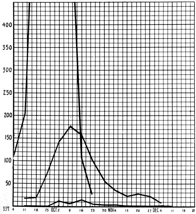
Chart 3.—Shows the relation of the epidemic of measles to that of influenza at Camp Pike, and the relations of the pneumonia following measles to both measles and influenza. The large incomplete curve represents influenza; the intermediate curve, measles; the small curve, pneumonia following measles.
294It will be noted from the overlapping of the two curves
in Chart 3 that a considerable portion of the measles cases
appeared before the influenza had subsided in Camp Pike.
This occurrence of the two epidemics at the same time
makes it impossible to separate the parts played by each
disease in producing the pneumonias and other complications
following measles. Analysis of the chart, however,
shows that the pneumonia with measles occurred in large
part during the first half of the measles epidemic. This
is of particular significance since it was during this period
that the effects of the influenza wave were felt most severely.
In Table LIX the cases of measles are grouped into
fifteen day periods according to their dates of onset and
the pneumonias arising from each group are tabulated.
This tabulation shows very clearly that the pneumonia
complications developed in large part in patients with
measles entering the hospital during the influenza period,
that is, late in September and during the first half of
October.
| Table LIX |
|---|
| |
| Patients with Measles and with Subsequent Pneumonia |
|---|
| |
| DATES |
TOTAL CASES OF MEASLES DURING INTERVALS OF 15 DAYS |
TOTAL CASES PNEUMONIA FROM SAME |
OF PER CENT INCIDENCE OF PNEUMONIAS |
| Sept. 11 to 30 |
86 |
433 |
14 |
42 |
16.28 |
9.7% |
| Oct. 1 to 15 |
347 |
28 |
8.07 |
| Oct. 16 to 31 |
270 |
434 |
8 |
14 |
2.96 |
3.2% |
| Nov. 1 to 15 |
91 |
2 |
2.2 |
| Nov. 16 to 30 |
56 |
4 |
7.15 |
| Dec. 1 to 15 |
17 |
0 |
|
The high incidence of pneumonia among measles patients
coming into the hospital prior to, with, or immediately following
the height of the influenza epidemic is very striking.
It so happens that half of the total number of measles cases
295considered, date their onsets prior to October 15. From the
433 cases included in this first half, 42 cases of pneumonia
arose, while from the 434 cases arising during the two
months following October 15, only 14 or one-third as many
cases of pneumonia developed. These figures very strongly
suggest that influenza played a large part in the production
of the pneumonia with measles in this group of cases.
Again the 9.7 per cent incidence of pneumonia in the first
half of cases considered, approaches the 12 per cent incidence
of pneumonia following influenza observed in the epidemic
at Camp Pike, while the incidence of 3.2 per cent in
the second half of the cases conforms more nearly to figures
for pneumonia following measles in the army prior to the
pandemic of influenza.
It has been shown that the prevalence of B. influenzæ at
Camp Pike increased with the passing of the wave of influenza
(p. 40) and that this increase applied to the
measles admissions. For a time the separation of measles
patients carrying B. influenzæ as identified by throat culture
on admission, from those free from it, was practiced.
All cases were then followed up by weekly throat cultures,
and cases in negative wards on being identified as positives
were transferred.
This practice was discontinued as impractical when it
became apparent that about 80 per cent of patients with
measles would be found positive for B. influenzæ when repeated
throat cultures were made during their hospital
treatment. The dissemination of B. influenzæ through the
wards from which we were attempting to exclude it took
place much faster than we could follow its spread by cultural
methods. When this became evident, the practice
of separating the two groups of patients with reference to
B. influenzæ was discontinued and the great inconvenience
of repeated transfer of patients was largely eliminated.
Table LX gives the findings in 426 cases of measles
cultured for B. influenzæ during the period when the practice of separating measles patients carrying B. influenzæ
from those not carrying the organisms was followed.
296
| Table LX |
|---|
| |
| Results of Repeated Throat Cultures for B. Influenzæ on 426 Cases of Measles, Camp Pike, Sept. 15 to Oct. 20, 1918. |
|---|
| |
| GROUPS |
TOTAL NUMBER CULTURED IN GROUP |
GROUP NO. NEGATIVE FOR B. INFLUENZÆ ON ADMISSION |
RESULTS OF CULTURES TO DATE |
GROUP NO. POSITIVE FOR B. INF. TO DATE |
GROUP OF POSITIVES DEVELOPING TO DATE IN CASES NEGATIVE FOR B. INF. ON ADMISSION |
PER CENT OF GROUP POSITIVE FOR B. INF. TO DATE |
GROUP PER CENT OF POSITIVES TO DATE AMONG CASES NEGATIVE FOR B. INF. ON ADMISSION |
| 1ST CULTURE |
2ND CULTURE |
3RD CULTURE |
4TH CULTURE |
NO. IN EACH CLASS |
| I |
|
|
|
|
|
|
|
|
|
|
|
| 1st culture on admission |
426 |
|
− |
|
|
|
274 |
152 |
|
35.6 |
|
| + |
|
|
|
152 |
| II |
|
|
|
|
|
|
|
|
|
|
|
| 1st and 2nd culture, after one week in hospital |
201 |
143 |
− |
− |
|
|
75 |
126 |
68 |
62.7 |
47.5 |
| − |
+ |
|
|
68 |
| + |
+ |
|
|
29 |
| + |
− |
|
|
29 |
| III |
|
|
|
|
|
|
|
|
|
|
|
| 1st, 2nd and 3rd cultures after two weeks in hospital |
94 |
69 |
− |
− |
− |
|
22 |
72 |
47 |
77.7 |
68.1 |
| − |
− |
+ |
|
18 |
| − |
+ |
− |
|
13 |
| − |
+ |
+ |
|
16 |
| + |
+ |
+ |
|
8 |
| + |
− |
+ |
|
6 |
| + |
+ |
− |
|
4 |
| + |
− |
− |
|
7 |
| IV |
|
|
|
|
|
|
|
|
|
|
|
| 1st, 2nd, 3rd and 4th cultures after three weeks in hospital |
25 |
19 |
− |
− |
− |
− |
4 |
21 |
15 |
84 |
79. |
| − |
− |
− |
+ |
3 |
| − |
− |
+ |
+ |
3 |
| − |
− |
+ |
− |
2 |
| − |
+ |
+ |
+ |
2 |
| − |
+ |
− |
+ |
2 |
| − |
+ |
+ |
− |
2 |
| − |
+ |
− |
− |
1 |
| + |
+ |
+ |
+ |
2 |
| + |
− |
− |
+ |
1 |
| + |
− |
+ |
+ |
1 |
| + |
+ |
+ |
− |
1 |
| + |
− |
− |
− |
1 |
297On admission 35.6 per cent of the patients were found
positive for B. influenzæ. Repeated throat cultures were
not confined to those appearing negative on this initial culture,
but were made on all patients without regard to their
being previously positive or negative. By a summation
of the results of the weekly cultures of all patients, the percentage
of patients carrying B. influenzæ rises from 35.6
per cent on admission, to 62.7 per cent after one week;
to 77.7 per cent after two weeks; to 84 per cent after three
weeks in the hospital.
To gain some idea of the rate of spread of B. influenzæ
in wards receiving only patients whose throat cultures were
negative for B. influenzæ on admission, a similar summation
of the results of repeated throat cultures on patients
in negative wards shows weekly increases from 47.5 per
cent after one week, to 68.1 per cent after two weeks; to
79 per cent at the end of three weeks.
These results demonstrate quite clearly that the measles
wards were saturated with B. influenzæ during the period
of the influenza epidemic. Conditions within the measles
wards with regard to B. influenzæ were not at all different
from those in the camp community during this period.
While no clinical methods could be relied upon to
diagnose influenza in the presence of an acute attack of
measles, there is every reason to believe that the occurrence
of clinical influenza with measles was no less frequent than
was its incidence in the camp at large, that is, about 20 to
25 per cent. That influenza played a large part in determining
predisposition to the complications of measles in
this series seems evident.
Between September 15 and December 15, 1918, 867 cases
of measles, admitted to the wards of the base hospital, were
298studied and handled according to the system outlined
above. About one half of these cases appeared during the
first month of the study. During this month hemolytic
streptococci played a very insignificant rôle. This microorganism
did not appear with alarming prevalence until
after the wards had been thoroughly overcrowded. After
the emergency, when better ward conditions were provided,
S. hemolyticus carriers continued to develop in the wards
and were removed when identified. The first S. hemolyticus
carriers to develop in the wards were identified on October
8. The first case of streptococcus pneumonia developed
on October 17, while streptococcus otitis as a complication
of measles did not begin until a little later. During
the latter two months of the study, S. hemolyticus became
rampant in the wards. The streptococcus complications
date their onset at some time during these two
months.
Table LXI shows the number of admissions to the measles
wards by weeks and the patients among them found to be
carrying hemolytic streptococci. It also shows the number
of S. hemolyticus carriers developing each week among
patients under treatment in the “clean” wards, as identified
by throat cultures repeated at weekly intervals. For
purposes of orientation, the number of cases developing
streptococcus pneumonia and otitis media with its subsequent
mastoiditis are given for each week during the period
of observation.
An admission to the measles ward can generally be regarded
as an acute case of measles. There are a few exceptions
to this statement and these are cases of measles
treated in barracks and afterwards transferred to the base
hospital. A relatively small number of such cases furnished
16 of the cases positive for hemolytic streptococci
on admission to the measles ward.
299
| Table LXI |
|---|
| |
| S. Hemolyticus Carriers Identified by Throat Culture Among Admissions; Those Developing Among Patients Under Treatment in the Streptococcus “Clean” Measles Wards; S. Hemolyticus Complications According to Their Dates of Onset |
|---|
| |
| GROUPING OF CASES BY WEEKS |
ADMISSION CASES |
HEMOLYTIC STREPTOCOCCI HOSPITAL CASES DEVELOPING |
PRINCIPAL COMPLICATIONS DUE TO HEM. STREP. |
| NO. CASES CULTURED |
NO. POS. HEM. STREP. |
PER CENT POS. HEM. STREP. |
NO. CASES CULTURED |
NO. POS. HEM. STREP. |
PER CENT POS. HEM. STREP. |
PNEUM. |
OTITIS |
MASTOIDITIS |
| Sept. 15 to Sept. 21 |
23 |
252 |
1 |
3 |
1.2 |
0 |
0 |
0 |
0 |
0 |
0 |
| Sept. 22 to Sept. 29 |
25 |
1 |
23 |
0 |
0 |
0 |
0 |
0 |
| Sept. 30 to Oct. 6 |
95 |
0 |
24 |
0 |
0 |
[91]1 |
0 |
0 |
| Oct. 7 to Oct. 13 |
109 |
1 |
121 |
4 |
3.3 |
0 |
0 |
0 |
| Oct. 14 to Oct. 20 |
223 |
494 |
7 |
19 |
3.8 |
175 |
8 |
4.6 |
1 |
0 |
0 |
| Oct. 21 to Oct. 27 |
156 |
5 |
451 |
35 |
7.7 |
2 |
3 |
0 |
| Oct. 28 to Nov. 3 |
71 |
6 |
333 |
29 |
8.7 |
1 |
12 |
1 |
| Nov. 4 to Nov. 10 |
44 |
1 |
263 |
45 |
17.1 |
3 |
8 |
11 |
| Nov. 11 to Nov. 17 |
31 |
117 |
4 |
13 |
11.1 |
149 |
46 |
30.8 |
0 |
5 |
5 |
| Nov. 18 to Nov. 24 |
41 |
4 |
93 |
7 |
7.5 |
0 |
2 |
2 |
| Nov. 25 to Dec. 1 |
19 |
0 |
48 |
7 |
14.6 |
0 |
3 |
2 |
| Dec. 2 to Dec. 8 |
26 |
5 |
52 |
12 |
23.1 |
0 |
3 |
0 |
| Dec. 9 to Dec. 15 |
4 |
|
2 |
|
|
47 |
12 |
25.5 |
1 |
0 |
0 |
300An admission to the measles ward does not indicate admission
to the hospital, because a considerable number of
cases of measles developed from time to time among patients
under treatment in the hospital for other conditions.
Since these patients remained in other wards not subject to
the same ward management and with no distinction between
those positive and those negative for hemolytic
streptococci, they cannot be included in figures to show the
incidence of hemolytic streptococci in patients with measles
at the time of admission to hospital from the camp. Two
classifications of the 37 cases, positive when first observed,
are necessary.
1. Division of cases according to days in the hospital
before first culture was taken:
| Days in Hospital |
No. of cases |
| 0–1 (admission) |
15 (2 not acute) |
| 2–7 |
10 |
| More than 7 |
12 |
2. Classification according to stage of the disease:
| During acute stage |
21 cases |
| After acute stage |
16 cases |
The first classification shows only 13 cases positive
when cultured on admission to the hospital and also during
the acute stage of the disease; the incidence of S. hemolyticus
in patients on admission is very low (1.76 per cent).
The second classification shows a slightly higher incidence
for cases during the acute stage of the disease, regardless
of whether they were admitted to the measles service
from camp or from another service of the hospital
(2.4 per cent). These findings conform with those at Fort
Riley in a smaller series of cases and support the opinion
that the hemolytic streptococci temporarily disappear from
the throat during the acute onset of measles. Unfortunately
controls among normal men in Camp Pike were not
taken at intervals throughout the period of three months
represented by this study of measles, but all controls taken
show a higher incidence than that found among measles
301patients on admissions over a period of time comparable to
that of the control series.
The gradual increase in the percentage of patients developing
hemolytic streptococci in their throats in wards
receiving only streptococcus free cases demonstrates that
the admission culture and the subsequent weekly cultures,
with the separation of all patients identified as carriers,
did not suffice to control the spread of streptococcus in this
group of cases. It is interesting to note that the greatest
incidence of streptococcus carriers among these patients
occurred three weeks after the height of the measles epidemic,
when it became about four times that observed at
the height of the measles epidemic.
When we consider the time relations of the streptococcus
complications, it is noteworthy that they begin to
appear somewhat after the appearance of streptococcus
carriers and then increase parallel with the increase in the
numbers of carriers. The relative number of complications
developing among the first carriers which were identified is
less than that among the carriers appearing later. This
suggests an increase in virulence of hemolytic streptococci
attending their wider dissemination.
Tables LXII and LXIII are introduced for the purpose of
showing to what extent duration of stay in the hospital
increases the individual’s chances of acquiring hemolytic
streptococci. Table LXII includes all cases admitted to and
treated in the measles wards. On repeated cultures, previous
positives and negatives were cultured alike and the
total positives reported for each week.
Table LXIII includes only those cases treated in the
“clean” wards and known to be negative on previous culture.
| 302 |
| Table LXII |
|---|
| |
| Incidence of Hemolytic Streptococci in Throats of Measles Cases with Reference to Period in Hospital |
|---|
| |
| (All cases treated in the wards) |
|---|
| |
| PERIOD IN MEASLES WARD |
NO. CASES CULTURED |
NO. POSITIVE FOR HEMOLYTIC STREPTOCOCCI |
PER CENT POSITIVE FOR HEMOLYTIC STREPTOCOCCI |
| (Admission) |
867 |
37 |
4.2 |
| 1 week |
768 |
84 |
10.9 |
| 2 weeks |
479 |
109 |
22.8 |
| 3 weeks |
240 |
63 |
26.2 |
| 4 weeks |
133 |
44 |
33.1 |
| 5 weeks |
82 |
26 |
31.7 |
| 6 weeks |
53 |
14 |
26.4 |
| 7 weeks |
25 |
8 |
32.0 |
| 8 weeks |
13 |
1 |
7.7 |
| 9 weeks |
9 |
1 |
11.1 |
| 10 weeks |
6 |
0 |
0 |
| 11 weeks |
5 |
0 |
0 |
| |
| Table LXIII |
|---|
| |
| Weekly Development of Hemolytic Streptococci in Throats of Patients Treated in “Clean” Wards |
|---|
| |
| PERIOD IN MEASLES WARD |
NO. CASES CULTURED |
NO. POSITIVE FOR HEMOLYTIC STREPTOCOCCI |
PER CENT POSITIVE FOR HEMOLYTIC STREPTOCOCCI |
| 1 week |
738 |
67 |
9.1 |
| 2 weeks |
424 |
74 |
17.4 |
| 3 weeks |
195 |
34 |
17.4 |
| 4 weeks |
92 |
16 |
17.4 |
| 5 weeks |
46 |
7 |
15.2 |
| 6 weeks |
26 |
4 |
15.4 |
| 7 weeks |
14 |
3 |
21.4 |
| 8 weeks |
8 |
0 |
|
| 9 weeks |
5 |
0 |
|
| 10 weeks |
4 |
0 |
|
| 11 weeks |
3 |
0 |
|
A comparison of Tables LXII and LXIII gives some indication
of what might have been expected if the carriers had
not been removed from the treatment wards at weekly intervals.
With the carriers removed from the “clean”
cases and segregated in a separate ward so as to be removed
effectively as sources of spread of the S. hemolyticus
infection to clean cases, the percentage incidence with
all cases considered rose to a point nearly twice as high
as that ever reached in the wards where clean cases alone
were allowed to remain. Had these carriers not been separated,
and remained in contact with cases free from hemolytic
streptococci, they would have served as just so many
more sources of infection, and an incidence of at least twice
303that recorded for all cases combined, or four times that
of the treatment wards, might have been expected. These
results indicate that the weekly separation of carriers from
clean cases did, to a considerable extent, lower the individual’s
danger of acquiring S. hemolyticus infection
while in the hospital.
In Table LXIV the complications developing in the measles
patients under observation at Camp Pike are tabulated.
In the division of the complications developing in “carriers”
and “noncarriers” of the hemolytic streptococci, reference
is made only to the records of the throat cultures.
The division is therefore not dependent upon the bacteriology
of the complications. For example, only 9 of the 12 cases
of pneumonia developing in “carriers” were streptococcus
pneumonias. On the other hand, the cases of mastoiditis
following otitis media were almost invariably due to hemolytic
streptococci. Of the 10 otitis cases occurring in “noncarriers,”
4 developed mastoiditis and 3 of these showed
hemolytic streptococci on culture from the mastoid cells
at operation. Missed cases of identification of S. hemolyticus
by throat culture in cases which develop S. hemolyticus
complications may arise from a number of causes.
It is desired here only to direct attention to these discrepancies.
Pneumonia Following Measles.—Fifty-six cases of pneumonia
following measles occurred during the period of observation
in this group of 867 cases of measles. Of these,
9 were streptococcus pneumonias. This gives an incidence
for streptococcus pneumonias of 1.04 per cent, while that
for all the pneumonia is 6.4 per cent. There were 8 cases
of lobar and 48 cases of bronchopneumonia. Seventeen
fatal cases occurred giving a mortality rate of 30.4 per cent
for the group. Five of these fatal cases occurred among
the 9 streptococcus pneumonias. The mortality rate for
the streptococcus pneumonia thus was 55.5 per cent; that
for the nonstreptococcus group was 25.5 per cent. All 9
cases of streptococcus pneumonia developed empyema. In
7 cases it was diagnosed clinically; in 2 at autopsy only.
No cases of empyema developed in the group of nonstreptococcus
pneumonias.
304
| Table LXIV |
|---|
| |
| Complications Developing in 867 Cases of Measles at Camp Pike. Distribution of Complications Between 242 “Carriers” and 625 “Noncarriers” of Hemolytic Streptococci from September 15 to December 15, 1918 |
|---|
| |
| NAME OF COMPLICATION |
NUMBER OCCURRING IN |
TOTAL NUMBER |
PER CENT IN |
| “CARRIERS” OF HEM. STREP. |
“NONCARRIERS” OF HEM. STREP. |
CASES WITH INCOMPLETE RECORD OF THROAT CULTURES |
ALL CASES |
HEM. STREP. “CARRIERS” |
“NONCARRIERS” OF HEM. STREP. |
| Pneumonia |
12 |
44 |
0 |
56 |
6.4 |
5.0 |
7.0 |
| Otitis media |
31 |
11 |
6 |
48 |
5.5 |
12.8 |
1.8 |
| Mastoiditis (following otitis media) |
15 |
4 |
4 |
23 |
2.6 |
6.2 |
0.6 |
| Local meningitis (extension from mastoid) |
2 |
0 |
0 |
2 |
|
|
|
| Frontal sinusitis |
1 |
0 |
0 |
1 |
|
|
|
| Ethmoidal sinusitis |
0 |
1 |
0 |
1 |
|
|
|
| Suppurative arthritis |
1 |
0 |
0 |
1 |
|
|
|
| Cervical adenitis |
1 |
0 |
0 |
1 |
|
|
|
| Acute bronchitis |
4 |
2 |
0 |
6 |
|
|
|
| Acute tonsillitis |
4 |
1 |
0 |
5 |
|
|
|
| Acute laryngitis and aphonia |
1 |
0 |
0 |
1 |
|
|
|
| Acute pleurisy |
2 |
1 |
0 |
3 |
|
|
|
| Erysipelas of face |
0 |
1 |
0 |
1 |
|
|
|
| Epidemic meningitis |
0 |
1 |
0 |
1 |
|
|
|
| |
| Note.—The percentages of incidence of pneumonia and otitis media in the “carrier” and “noncarrier” groups are at direct variance. It would appear from these findings that streptococci very readily invade the middle ear from the throat and set up grave disorders. The invasion of the lung from the throat occurs with less frequency. Hemolytic streptococci perhaps never initiate the pneumonic processes and can be regarded as more or less accidental secondary invaders. |
305The relation of these pneumonias following measles, to
the influenza epidemic has been discussed. The time relations
between the onsets of measles and that of the subsequent
pneumonia vary widely. There appears to be
nothing constant in the length of time between the onset
of measles and that of the pneumonia. In 30 of the cases
this period is less than ten days; in the remaining 26 cases,
it ranges from ten to thirty-two days (Chart 4).
In the ward treatment of these cases of pneumonia, they
were divided into three groups according to their clinical
characters and according to the results of throat and
sputum cultures.
| (a) |
Streptococcus pneumonias |
9 cases |
| (b) |
Pneumonia with hemolytic streptococci in the throat without symptoms referable to the streptococcus |
13 cases |
| (c) |
Pneumococcus pneumonias not carrying hemolytic streptococci |
34 cases |
The streptococcus-free cases were treated in a separate
ward. Cases were admitted to this ward directly from the
“clean” measles wards, but only after a throat culture
taken prior to their transfer had been negative for the
hemolytic streptococcus.
The other two groups were treated together in another
ward, but in strictly separate compartments of it. This
precaution was carried out on the assumption that patients
with an acute streptococcus pneumonia were real sources
of danger in the ward because of a heightened virulence of
the organism causing the grave symptoms. The pneumonias
subsequently developing hemolytic streptococci in
their throats, without their presence modifying the course
of the pneumonia, came to be regarded as being in the same
class with uncomplicated cases of measles carrying hemolytic
streptococci, in so far as their being potential sources
of danger in a ward is concerned.
306
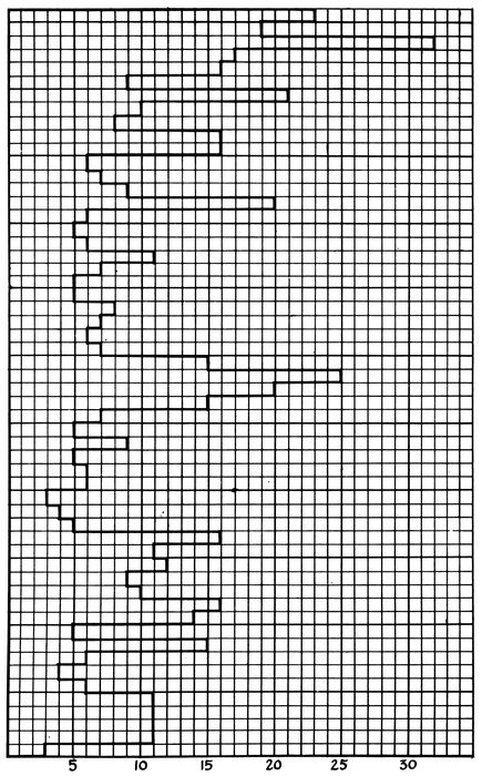
Chart 4.—Shows the time interval between the onset of measles and the onset of the subsequent pneumonia in the 56 cases of pneumonia following measles at Camp Pike. Each case is represented by one of the small blocks measured along the ordinate. The onset of measles in all cases is represented by the line at the extreme left of the chart. The onset of pneumonia in each case is indicated by the limit of the block marked off in days to the right of this line.
307(a) Streptococcus Pneumonias.—Nine cases of streptococcus
pneumonia developed. Of the 867 cases of measles
studied, 242 showed throat cultures positive for the hemolytic
streptococci at some period of their stay in the hospital.
It appears then that 3.7 per cent of the patients
carrying hemolytic streptococci in their throats developed
streptococcus pneumonia. Thirty-seven cases had positive
throat cultures when first observed on admission to the
measles wards. It is significant to note that not a single
case of pneumonia of any kind developed among these
cases.
MEASLES PNEUMONIA; STREPTOCOCCUS GROUP
Case 98, O. McN. Onset of measles, Sep. 19; admitted to hospital Sep. 21;
onset of bronchopneumonia, Oct. 21; of empyema, Oct. 23. Recovered from
pneumonia; convalescent in empyema ward.
Bacteriology.—1. Throat culture for: (a) S. hem.: Sep. 21, −; 28, −; Oct.
9, −; 20, −; 23, +; Nov. 2, −; 9, −; 15, −; (b) B. influenzæ: Sep. 21, +; 28, −;
Oct. 9, +. 2. Pleural fluid (culture) S. hem. Oct. 23, +.
Case 141, J. G. G. Autopsy No. 438. Onset of measles, Sep. 28; admitted
to hospital, Oct. 1; onset of bronchopneumonia, Oct. 6; of otitis media (bilateral),
Oct. 12; died, Oct. 18.
Bacteriology.—1. Throat culture for: (a) S. hem., Oct. 2, −; 6, −; 8, −;
(b) B. influenzæ, Oct. 2, −; 6, +; 8, +. 2. Autopsy cultures: Heart blood, negative;
left lung, Pneumococcus II atypical, B. influenzæ and S. viridans; right
lung, S. hem. and B. influenzæ; right bronchus, S. hem. and B. influenzæ.
Case 147, S. W. Autopsy No. 442. Onset of measles, Oct. 1; admitted to
hospital, Oct. 2; onset of bronchopneumonia, Oct. 17, with chill and rapid
development; died, Oct. 18.
Bacteriology.—1. Throat culture for: (a) S. hem., Oct 2, −; 9 −; 15, −;
18, +; (b) B. influenzæ, Oct. 2, −; 9, −; 15, −; 18, −. 2. Autopsy cultures:
Heart blood, S. hem.; right main bronchus, S. hem. and B. influenzæ.
Case 281, T. M. Onset of measles, Oct. 6; admitted to hospital Oct. 9; onset
of bronchopneumonia, Oct. 21; of empyema, Oct. 23; recovered from pneumonia;
convalescent in empyema ward.
Bacteriology.—1. Throat culture for: (a) S. hem., Oct. 10, −; 20, −; 24, +;
Nov. 2, +; 9, +; 15, +; (b) B. influenzæ, Oct. 10, −; 20, +. 2. Culture from
pleural fluid, Oct. 23, S. hem.
Case 285, J. H. Onset of measles, Oct. 4; admitted to hospital, Oct 9;
onset of lobar pneumonia, Oct. 29; of empyema, Nov. 9; convalescent in empyema
ward.
Bacteriology.—1. Throat cultures for: (a) S. hem., Oct. 11, −; 20, −; 24,
+; 29, −; Nov. 2, −; 9, −; (b) B. influenzæ, Oct. 11, −. 2. Cultures from pleural
fluid, Nov. 9 and 13, S. hem.
308Case 714, W. H. Onset of measles, Oct. 26; admitted to hospital, Oct. 28;
otitis media, Nov. 8; onset of bronchopneumonia, Nov. 9; of empyema, Nov.
17; convalescent in pneumonia ward.
Bacteriology.—1. Throat cultures for: S. hem., Oct. 28, −; Nov. 4, −; 12, +;
23, +; 30, +; Dec. 7, +; 12, −. 2. Sputum: Nov. 10, Pneumococcus II atypical,
S. hem. and B. influenzæ.
Case 730, W. S. Autopsy No. 491. Onset of measles, Oct. 26; admitted to
hospital, Oct. 29; onset of bronchopneumonia, Nov. 10; of empyema, Nov. 11;
of cervical adenitis, Nov. 5; died, Nov. 15.
Bacteriology.—1. Throat culture for: S. hem., Oct. 30, −; Nov. 4, +. 2.
Sputum: Nov. 10, S. hem. 3. Pleural fluid: Nov. 11, S. hem. Autopsy bacteriology:
Heart blood, S. hem.; right main bronchus, B. influenzæ, B. coli;
right lung, S. hem. and B. influenzæ; right pleura, S. hem.; peritoneum, S. hem.
Case 751, P. B. Autopsy No. 492. Entered hospital, Oct. 19; onset of
measles, Oct. 30; of bronchopneumonia, Nov. 5; of right empyema, Nov. 12;
died, Nov. 16.
Bacteriology.—1. Throat cultures for: S. hem., Nov. 1, −; 4, +; 15, +.
2. Sputum: Nov. 13, B. influenzæ and S. hem. 3. Autopsy cultures: Heart
blood, S. hem.; right lung, S. hem., Pneumococcus IV, B. influenzæ, B. coli;
pericardium, negative; right pleura, S. hem.; peritoneum, S. hem.
Case 880, B. McN. Autopsy No. 507. Onset of measles, Nov. 30; entered
hospital, Dec. 3; onset of bronchopneumonia, Dec. 11; of empyema, Dec. 14;
died, Dec. 14.
Bacteriology.—1. Throat cultures for: S. hem., Dec. 3, −; 5, −; 12, +.
2. Cultures from pleural fluid, Dec. 14, S. hem. 3. Autopsy cultures: Heart
blood, S. hem.; right main bronchus, S. hem., B. influenzæ, staphylococcus (a
few); left lung, S. hem.; left pleura, S. hem.
The average period in the hospital before the development
of the streptococcus pneumonia is about two weeks.
Cases 98 and 285 were in the hospital thirty and twenty
days respectively before the onset of pneumonia. There
is a record of from one to four negative throat cultures
on each case before streptococcus was found in the
throat. This enables us to fix the onset of the pneumonia
with reference to the appearance of the streptococcus in
the throat.
Case 141 stands alone as representing a class in which
S. hemolyticus was implanted upon a pneumococcus pneumonia
during its course. In this instance two throat cultures
on alternate days after the onset of the pneumonia
were negative for hemolytic streptococci. Unfortunately
309the last record of a throat culture is for one taken ten days
before the fatal termination of the case, and it can only
be stated that the S. hemolyticus infection was implanted
within the last ten days of the course of the pneumonia,
perhaps on or about October 12 when bilateral otitis media
developed.
In Cases 285 and 730 hemolytic streptococci were found
in the throats five and six days respectively before the onset
of pneumonia. They represent the 2 cases of pneumonia
which developed in patients isolated in the streptococcus
“carrier” ward. Case 285 is of particular interest for
several reasons. It is the only case of lobar pneumonia
in the group and happens also to be the only case from
which B. influenzæ was not obtained. S. hemolyticus was
found only once on throat culture, i.e., five days before the
onset of the pneumonia. Three throat cultures after the
onset of the pneumonia were negative. The case ran the
course of a lobar pneumonia. Eleven days after the onset
(November 9) a small amount of pleural fluid was diagnosed.
Aspirated fluid on this date and again four days
later showed many streptococci in smears and pure cultures
of S. hemolyticus.
The remaining 6 cases belong to a group in which hemolytic
streptococci were first identified in the throats after the
cases had been reported to the laboratory as pneumonia suspects
to be examined by culture before transfer from the
measles ward. In all these cases the culture taken at this
time was positive while all cultures taken before were negative.
In some cases, e. g., Cases 98, 147, and 281, throat
cultures taken only one or two days before the onset of the
pneumonia were negative. In these cases the onset of the
pneumonia and the appearance of the streptococcus in the
throats appear to be simultaneous.
It should be noted that the period between the appearance
of the hemolytic streptococci in the throat and the development
of the pneumonia is very short in all cases. In this
310small group of cases S. hemolyticus infection which has
complicated pneumonia has been acquired at or near the
time of onset of the pneumonia.
(b) Pneumonia with Hemolytic Streptococci in the
Throat without Symptoms Referable to the Streptococcus.—Thirteen
cases of pneumonia associated with measles developed
into S. hemolyticus “carriers” without having the
course of the disease affected by the presence of the organism
in the throat. Cases 705, 872, and 188 are of interest
in that hemolytic streptococci were identified in the throats
from one to six days prior to the onset of the pneumonia.
In spite of their presence, the symptoms, course and outcome
of the pneumonia were apparently unaffected. One
of these cases (Case 872) died. Autopsy showed lobar
pneumonia with no signs of invasion of the lung by hemolytic
streptococci. Cultures at autopsy showed that pneumonia
was due to a pneumococcus, Type II atypical. A
few hemolytic streptococci were found in culture from the
right main bronchus.
Of the remaining 10 cases 1 developed S. hemolyticus
in a throat culture at the end of the first week of the pneumonia;
3 during the second week; 1 during the third week,
and 5 further along in the convalescent period. In 8 cases
hemolytic streptococci appeared in the throat, at a time
when invasion of the lower respiratory tract by the streptococcus
might be expected, and yet none of them developed
evidence of streptococcus pneumonia. The 9 cases with
hemolytic streptococci appearing late in convalescence are
not of particular interest, since the dangers of lower respiratory
invasion are much reduced after the acute stage
of the pneumonia has passed. Three of these cases (Cases
678, 725 and 398) did however develop ear complications
directly referable to the streptococcus invasion of the
throat. Two of them terminated in mastoiditis with operation.
These cases emphasize the greater tendency of S.
hemolyticus to invade the middle ear rather than the lung.
311In 3 fatal cases of pneumococcus pneumonia in which during
life no hemolytic streptococci were found by throat culture,
a few hemolytic streptococci were found at autopsy
in culture from the main bronchi, along with predominating
growths of pneumococci and B. influenzæ. In 2 instances
there was frank lobar pneumonia and in the third bronchopneumonia;
there was no evidence to show that hemolytic
streptococci had any relation to the pneumonia which was
found.
MEASLES PNEUMONIAS; GROUP CARRYING HEMOLYTIC STREPTOCOCCI
Case 705. Onset of measles, Oct. 25; admitted to hospital, Oct. 27; onset
of bronchopneumonia, Nov. 10; acute pleurisy, Nov. 16; convalescent in pneumonia
ward.
Bacteriology.—1. Throat cultures for: S. hem., Oct. 27, −; Nov. 4, −; 11,
+; 15, +; 23, −; 30, −; Dec. 7, −; 12, −. 2. Sputum: Nov. 10, Pneumococcus II
atypical, S. hem. and B. influenzæ.
Case 872. Autopsy No. 508. Onset of measles, Nov. 29; admitted to hospital,
Nov. 30; onset of lobar pneumonia, Dec. 10; died, Dec. 14.
Bacteriology.—1. Throat cultures for: S. hem., Nov. 30, −; Dec. 5, +;
10, +; 12, +; 14, +. 2. Autopsy culture: Heart blood, Pneumococcus II atypical;
right main bronchus, Pneumococcus II atypical, B. influenzæ, S. hem. (a
few); left lung, Pneumococcus II atypical; left pleura, Pneumococcus II
atypical.
Case 188. Onset of measles, Oct. 3; admitted to hospital, Oct. 4; onset of
bronchopneumonia, Oct. 14; recovered and discharged from hospital, Nov. 24.
Bacteriology.—1. Throat cultures for: (a) S. hem., Oct. 5, −; 8, +; 12, +;
19, +; 20, +; 27, −; Nov. 2, −; 9, +; 15, −; (b) B. influenzæ, Oct. 5, −; 8, −;
12, +; 19, +.
Case 678. Onset of measles, Oct. 23; admitted to hospital, Oct. 25; onset
of bronchopneumonia, Nov. 2; of otitis media, Nov. 9; of mastoiditis, Nov.
13; mastoid operation, Nov. 20; still under treatment.
Bacteriology.—1. Throat cultures for: S. hem., Oct. 25, −; Nov. 4, −; 5, −;
12, +. 2. Sputum: Nov. 3, Pneumococcus Type IV, and B. influenzæ. 3. Culture
from mastoid bone at operation, Nov. 20, S. hem.
Case 389. Admitted to hospital, Oct. 2, with diagnosis of influenza; onset
of bronchopneumonia, Oct. 7; onset of measles, Oct. 13; phlebitis (right leg),
Oct. 22; otitis media, Oct. 31; recovered.
Bacteriology.—1. Throat cultures for: (a) S. hem., Oct. 16 −; 20, −; 27, +;
Nov. 2, +; 9, +; 15, −; 23, −; 30, −; Dec. 7, −; 12, −; (b) B. influenzæ, Oct.
16, −.
Case 725. Onset of measles, Oct. 18; one week in measles barracks; admitted
to hospital, Oct. 27; onset of lobar pneumonia, Oct. 23; otitis media,
Nov. 7; mastoid operation, Nov. 20; still under treatment.
312Bacteriology.—1. Throat cultures for: (a) S. hem., Oct. 29, −; Nov. 1, −;
5, −; 12, +; (b) B. influenzæ, Oct. 29, +. 2. Sputum: Nov. 2, Pneumococcus
II atypical. B. influenzæ. 3. Culture from mastoid at operation, Nov. 20,
S. hem.
(c) Pneumococcus Pneumonias not Carrying Hemolytic
Streptococci.—Thirty-four cases of pneumonia following
measles went through their entire course in the hospital
with no throat culture positive for hemolytic streptococci.
In some of these cases there are records of twelve negative
throat cultures. Eleven fatal cases occurred in this group.
Autopsy findings and bacteriology showed in each instance
that S. hemolyticus was not the cause of the pneumonia.
Measles During the Course of Pneumonia.—Eleven cases
of pneumonia which developed measles during the course
of the pneumonia came under observation. Hemolytic
streptococci appeared in the throats of 3 of these patients
during convalescence, but there was no evidence that it invaded
the lung. In one fatal case autopsy showed that
there was no streptococcus pneumonia; pneumonia followed
influenza and the onset of measles occurred three days after
the onset of bronchopneumonia.
Bacteriology of Pneumonia Following Measles.—When
observations made during life are combined with the results
of postmortem cultures, the bacteriology of 35 of the 56
cases is available and is as follows: Pneumococcus Type II
atypical in 36 per cent, Type IV in 22.9 per cent, Type I in
2.8 per cent, Type III in 2.8 per cent, hemolytic streptococci
in 22.4 per cent, and B. influenzæ in 88.6 per cent of these
cases.
Otitis Media and Mastoiditis Complicating Measles.—The
occurrence of otitis media and mastoiditis complicating
measles in patients harboring hemolytic streptococci in
their throats has already been presented (Table LXIV).
The bacteriology of these complications was not studied
by this commission. The records of the base hospital laboratory
at Camp Pike contain reports of twenty-nine cultures
made at operation from pus in the middle ear and
313the mastoid bone. Hemolytic streptococci were found in
22 of these cases. Throat cultures were in accord with
these positive findings in all except a few instances. The
throat culture serves as a fairly reliable index of the bacterial
nature of these complications. By combining our
records of throat cultures with the results of the cultures
from the lesions, hemolytic streptococci were obtained from
37 of the 48 cases of otitis media. In 23 cases of mastoiditis
following the otitis media, hemolytic streptococci were demonstrated
in all except 2. It is evident that the great majority
of these complications were due to hemolytic streptococci.
The relation between the appearance of hemolytic streptococci
in the throat and the onset of the otitis is recorded
in all except 4 of the 31 instances of otitis media occurring
in patients with throat cultures positive for hemolytic
streptococci. These four patients had positive throat cultures
when first observed and represent the only patients
who carried hemolytic streptococci when admitted to
measles wards and developed complications.
The first of these patients had been under treatment in
an otologic ward during a month before measles developed.
Measles caused a recurrence of disease of the ear with
double mastoiditis requiring bilateral operation. Two
other patients had been in the hospital ten and eleven days
respectively before they were admitted to the measles
ward; on admission to the ward otitis media was present
in one patient and in the other it developed six days later.
The fourth patient was admitted to the measles wards directly
from the camp, and culture from the throat on the
day of admission showed the presence of S. hemolyticus.
Two weeks later at the time of onset of otitis media, culture
from the throat contained no hemolytic streptococci. Repeated
cultures during the next three weeks were negative.
No complications of otitis media developed and no direct
cultures from the ear are recorded.
314
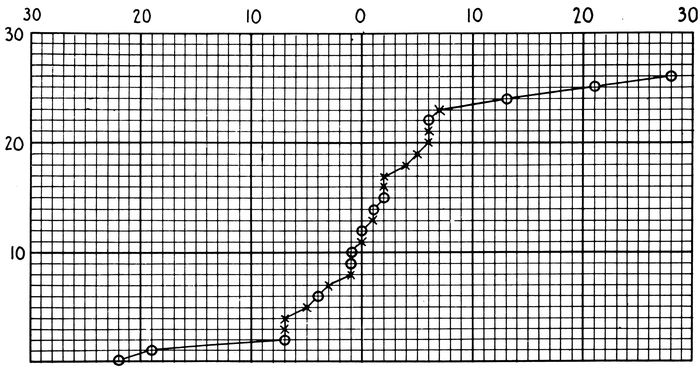
Chart 5.—Shows the time relation between the identification of hemolytic streptococci in the throats and the development of otitis media in 27 cases shown to be due to hemolytic streptococci. The onset of otitis media is represented by the ordinate marked ○. The number of days before or after the onset of the otitis, within which the throat culture which proved positive for hemolytic streptococci was taken, is marked off along abscissæ to the left and right of ordinate ○ respectively. On the curve plotted these symbols are used: A circle represents a throat culture positive for hemolytic streptococci in a case of otitis media without extension to mastoid. The plus sign represents a throat culture positive for hemolytic streptococci in a case of otitis media with mastoiditis and osteitis.
315In this series of cases (Chart 5) the appearance of S.
hemolyticus in the throat and the onset of otitis media are
very closely associated in those patients in whom further
extensions of the streptococcus infection occurred. In instances
in which appearance of streptococci and of otitis
media are separated by an interval of more than seven days,
no further extension occurred. In 8 cases in which this interval
is seven days or less there has been no further extension
of the infection.
Beginning October 24 cultures for the identification of
carriers of hemolytic streptococci were made from all patients
in a ward and repeated at intervals of one week.
Prior to this time individual patients had been examined at
intervals of one week, so that an entire ward was never
studied on any particular day. This system did not identify
and remove all “carriers” in a ward at a given time
and was abandoned because it failed to show the conditions
present. Investigation of wards as units proved much
more satisfactory.
The studies made in four of the double wards used for
the care of patients with measles are presented in Table
LXV. During the time of this study hemolytic streptococci
were more prevalent than at an earlier period.
Cultures from the throats of all patients entering these
wards were negative for S. hemolyticus on admission. The
table showing the incidence of “carriers” of hemolytic
streptococci each week in these wards demonstrates:
1. The separation of “carriers” and “noncarriers” by
throat culture made on admission does not prevent the increase
of streptococcus “carriers” in wards.
2. Removal of all “carriers” found by cultures on admission
and at weekly intervals is inadequate.
| 316 |
| Table LXV |
|---|
| |
| Ward Conditions with Reference to Hemolytic Streptococcus Infection |
|---|
| |
| DATE OF CULTURE |
NO. PATIENTS CULTURED |
NO. POSITIVE HEM. STREP. |
PER CENT POSITIVE HEM. STREP. |
COMPLICATIONS ASSOCIATED WITH HEM. STREP. WITH DATES OF ONSET |
REMARKS |
| Ward 57 |
|
|
|
|
|
| 11–3 |
35 |
1 |
2.8 |
|
|
| 11–10 |
13 |
2 |
15.5 |
None |
|
| 11–17 |
16 |
6 |
37.5 |
|
|
| Ward 58 |
|
|
|
|
Wards 57 and 58 served by same ward staff. |
| 11–3 |
38 |
7 |
18.4 |
Otitis media: |
| 11–10 |
11 |
4 |
36.4 |
11–8 1 case |
Members of staff cultured on 11–5, 11–12 and 11–19. No positives |
| 11–17 |
6 |
2 |
33.0 |
11–7 1 case |
| Ward 49 |
|
|
|
|
|
| |
|
|
|
Otitis media: |
|
| 10–25 |
37 |
7 |
18.9 |
10–25 2 cases |
|
| 11–1 |
31 |
3 |
9.7 |
10–26 1 case |
|
| 11–8 |
35 |
9 |
25.7 |
10–28 1 case |
|
| 11–15 |
32 |
18 |
56.3 |
11–15 1 case |
|
| 11–22 |
16 |
7 |
43.8 |
11–18 1 case |
|
| |
|
|
|
11–27 1 case |
|
| Ward 50 |
|
|
|
|
Wards 49 and 50 served by same ward staff. |
| 10–25 |
29 |
2 |
3.4 |
Otitis media: |
| 11–1 |
43 |
2 |
4.6 |
11–8 1 case |
Ward staff cultured:
11–5 1 positive
11–12 1 positive
11–26 2 positives |
| 11–8 |
32 |
3 |
9.4 |
11–13 1 case |
| 11–15 |
20 |
11 |
55.0 |
11–22 1 case |
| 11–22 |
11 |
0 |
0.0 |
|
| Ward 41 |
|
|
|
|
Case of pneumonia developing on 11–9 was transferred to the “clean” pneumonia ward without a throat culture to warrant its transfer; last culture 11–4 negative; culture 11–12 in pneumonia ward positive |
| 10–28 |
45 |
4 |
8.9 |
Streptococcus pneumonia: |
| 11–4 |
34 |
9 |
26.5 |
(11–9 1 case) |
| 11–11 |
12 |
8 |
66.6 |
11–10 1 case |
| Ward closed—No patients. |
Otitis media: |
| 11–21 |
13 |
0 |
0.0 |
10–29 1 case |
| 11–28 |
8 |
4 |
50.0 |
11–4 1 case |
| 12–5 |
12 |
4 |
33.3 |
11–5 1 case |
| 12–12 |
4 |
3 |
75.0 |
11–11 1 case |
| |
|
|
|
11–27 1 case |
| |
|
|
|
12–3 1 case |
| Ward 42 |
|
|
|
|
Wards 41 and 42 served by same ward staff. |
| |
|
|
|
Streptococcus pneumonia: |
| 10–28 |
32 |
0 |
0 |
11–10 1 case |
| 11–4 |
43 |
7 |
16.3 |
12–11 1 case |
| Ward closed—No patients. |
Otitis media: |
Ward staff cultured:
11–5 2 positive
11–12 2 positive
11–26 2 positive
12–2 1 positive |
| 10–21 |
16 |
4 |
25.0 |
10–29 1 case |
| 11–28 |
12 |
1 |
12.5 |
12–3 1 case |
| 12–5 |
20 |
10 |
50.0 |
12–6 1 case |
| 12–12 |
14 |
7 |
50.0 |
|
| 317Ward 59 |
|
|
|
|
The 3 cases of streptococcus pneumonia acquired S. hemolyticus infection while patients in the 16 bed south section of this ward |
| |
|
|
|
Streptococcus pneumonia: |
| 10–24 |
37 |
6 |
16.2 |
10–17 1 case |
| 10–31 |
27 |
5 |
18.5 |
10–21 1 case |
| 11–7 |
9 |
3 |
33.3 |
10–29 1 case |
Case developing 10–29 was removed from section a few days before onset of pneumonia |
| 11–12 |
7 |
1 |
14.3 |
Otitis media: |
| |
|
|
|
11–1 1 case |
| Ward 60 |
|
|
|
|
Wards 59 and 60 served by same ward staff. |
| |
|
|
|
Streptococcus pneumonia: |
| 10–24 |
22 |
1 |
4.5 |
10–21 1 case |
Ward staff cultured:
11–5 0 positive
11–12 1 positive
11–19 0 positive |
| 10–31 |
17 |
2 |
11.7 |
Otitis media: |
| 11–7 |
8 |
1 |
12.5 |
10–31 1 case |
| 11–12 |
6 |
1 |
16.6 |
|
When the streptococcus complications are traced back to
the wards in which the streptococcus infection of the throat
was acquired, it is found that with the exception of Case
141 (already cited) all the streptococcus pneumonias arose
from two double wards. Wards 41 and 42 furnished 4
cases at times when streptococcus was rampant in them and
3 of these cases arose within a period of a few days.
Wards 59 and 60 furnished 4 cases, very closely associated.
In 3 cases the streptococcus infection was acquired in a
section of Ward 59 containing 16 beds. These patients
were in beds, of which the positions are represented by numbers
2, 5, and 7, along one side of the ward. The fourth
instance of pneumonia appeared at the same time in Ward
60, which was attended by the same ward personnel, but
no other connection can be established between this case
and the other three.
The otitis media appeared in patients scattered throughout
those wards for measles in which the weekly incidence
of “carriers” was rising rapidly. This relation is illustrated
by Wards 58, 50, and 41. The same observation applies
to streptococcus pneumonia arising in Wards 41 and
42. In Ward 41 the weekly percentage of carriers are
October 28, 8.9, November 4, 26.5 and November 11, 66.6.
318On November 9 and 10 the first 2 cases of streptococcus
pneumonia arising from this ward developed. At the same
time, November 10, a third case appeared in another part
of this same ward unit (Ward 42) where the spread of
hemolytic streptococci had been very active. These observations
suggest that hemolytic streptococci may build
up its virulence as the result of rapid dissemination to
such a degree that it is capable of causing grave complications.
The relation of complications to “carriers” in Wards
59 and 60 is different from that in the wards just cited.
Wards 59 and 60 were opened on October 9 and before October
17; when the first case of fulminating streptococcus
pneumonia occurred, only three “carriers” had been found
in them. From October 17 to 24 when the record in Table
LXV begins eight “carriers” were removed. The appearance
of a case of severe streptococcus pneumonia in an
unusually clean ward was followed by the rapid development
of “carriers,” and the appearance within twelve days
of 3 other cases of streptococcus pneumonia, 2 of which
were in beds close to the first case. This sequence suggests
focal dissemination of a streptococcus from a case in which
it had suddenly assumed high virulence.
An outbreak of infection with S. hemolyticus was recognized
on November 12 in a measles-pneumonia ward which
had been opened for several weeks and had continued free
from streptococcus. In three patients hemolytic streptococci
were found by throat cultures. Inquiry revealed that
a nurse in this ward, recognized as a streptococcus “carrier”
the week before, had been retained on duty. Two
patients well advanced in the course of their pneumonias,
had acquired S. hemolyticus demonstrated by throat examination.
Both patients developed otitis media with mastoid
extension requiring operations. Cultures from both
at operation showed hemolytic streptococci.
319The third patient, with acute pneumonia, had been sent
into the ward on November 11 from Ward 42, which at the
time was a highly infected ward; no culture of the throat
was made before transfer. This patient developed streptococcus
pneumonia with empyema requiring subsequent operation.
Discussion.—At Camp Funston, where the prevalence of
S. hemolyticus in the measles wards did not rise above that
among normal men in the camp at large, 112 consecutive
cases of measles were treated without a single complication
due to hemolytic streptococci.
At Camp Pike, the investigation began at the onset of
a small epidemic of measles at a time when hemolytic streptococci
were an almost negligible factor. The epidemic of
measles was followed throughout its course; and, with the
passing of the epidemic, there was an increase in the prevalence
of hemolytic streptococci which assumed alarming
importance in the production of complications.
The epidemic of measles was in part superimposed upon
the epidemic of influenza, so that deductions concerning
complications strictly due to measles became impossible.
It is evident that influenza played a considerable part in
producing the complications of measles at Camp Pike.
The dissemination of hemolytic streptococci through
measles wards was controlled only in part by the methods
used. This partial control may have served to limit the
incidence of streptococcus pneumonia, nine instances occurring
among 867 cases of measles.
In the ward treatment of measles effort should be directed
to prevent the exposure of patients free from hemolytic
streptococci to S. hemolyticus “carriers.” By this
means the rate of development of S. hemolyticus “carriers”
may be reduced.
Measures which should be adopted are as follows:
1. Adequate wards should be prepared in advance for
the treatment of measles. The rather gradual onset of
epidemics of measles makes this provision possible.
3202. The separation of S. hemolyticus “carriers” from
other patients should be enforced. Observation wards,
where strict technic to prevent transfer of infection is
practiced and where throat cultures are made on admission,
are essential. Those wards should be promptly evacuated
to wards for the care of S. hemolytic “carriers” on the one
hand and for “noncarriers” on the other. As far as possible
patients should be admitted to a ward until it is filled
and then another ward should receive consecutive cases in
the same manner. It is desirable to have all cases in each
treatment ward in the same stage of the disease. With
this system of ward rotation convalescent wards are necessary,
so that cases requiring a period of hospitalization
longer than the average may be segregated, thus rendering
treatment wards available for another levy of acute cases.
3. Strict ward technic elaborated to prevent transfer of
bacterial infection from one patient to another must be
employed.
4. Throat culture for identification of “carriers” is laborious
but essential. An accurate method for identifying
and reporting “carriers” as speedily as possible must be
employed. A competent bacteriologist is essential. A
twenty-four hour interval between culture and its report is
desirable. The following scheme is recommended:
(a) A culture from the throat made on admission to the
observation ward (first day in hospital).
(b) A culture made on the first day in the treatment
ward (third day in hospital).
(c) A culture made one week later (tenth day in hospital).
If the ward incidence of hemolytic streptococci reaches
10 per cent, especially in a filled ward, the cultures should
be repeated on the thirteenth day in the hospital. If the
incidence of “carriers” of hemolytic streptococci increase
rapidly, cultures on alternate days should be made so that
“carriers” may be removed from the ward. Wherever
321possible, culturing of the treatment wards as units should
be practiced.
5. Patients developing acute symptoms in any way suggestive
of infection with S. hemolyticus should be immediately
isolated; culture from the throat should be made at
once and final disposal of the patient should depend upon
its result.
During the winter of 1917–18, with the establishment of
the army camps, it very soon became evident that in many
of the serious and fatal complications of measles and other
respiratory diseases, hemolytic streptococci were playing
a very important rôle. The epidemic prevalence of hemolytic
streptococci among hospital cases, and later among
men on duty in the camps, was established by bacteriologic
studies. Prior to this time in civil life, hemolytic streptococci
under epidemic conditions had been studied in milk-borne
epidemics of septic sore throat, such as are reported
from Chicago in 1911–13[92]; from Boston in 1911[93]; and from
Baltimore in 1911–12[94]. Contact air-borne infection has not
been emphasized in considering the dissemination of hemolytic
streptococci. Smillie[95] reports a few cases of hemolytic
streptococcus throat infections which he attributes
to contact infection. Conditions within the army camps
were such as to suggest the dissemination of hemolytic
streptococci by contact air-borne infection. Some knowledge
of the percentage of individuals showing positive
throat cultures became desirable at the very beginning of
studies of contact dissemination of hemolytic streptococci.
Smillie found that only one of 100 normal throats harbored
the Beta hemolytic streptococci of Smith and Brown.
Levy and Alexander[96] report the presence of hemolytic
322streptococci in 83.2 per cent of healthy men at Camp Taylor,
and hemolytic organisms (not definitely identified as
streptococci) in 14.8 per cent of recruits arriving at Camp
Taylor. Irons and Marine[97] found hemolytic streptococci
among 70 per cent of healthy men at Camp Custer.
Among measles patients on admission to the hospital at
Fort Sam Houston, Cole and MacCallum[98] report 11.4 per
cent and Cummings, Spruit and Lynch,[99] 35 per cent of
throat cultures positive for hemolytic streptococci. At
Camp Taylor, Levy and Alexander report 77.1 per cent
positive among 388 cases of measles on admission to the
hospital.
The spread of hemolytic streptococci in measles wards
was shown by Cole and MacCallum when on admission 11.4
per cent of cases had positive throat cultures, 38.6 per cent
after from three to five days, and 56.8 per cent after from
eight to sixteen days in the ward. In our study of hemolytic
streptococci with measles at Camp Funston, 2.6 per
cent of the cases had positive throat cultures on admission,
12.8 per cent after three to ten days, and 24.1 per cent after
eight to twenty-three days in the hospital. In a similar
study at Camp Pike we found 1.7 per cent positive on admission;
10.9 per cent after one week; 22.8 per cent after
two weeks; 26.2 per cent after three weeks; and, 33.1 per
cent after four weeks in the hospital.
Hemolytic Streptococci in the Throats of Normal Men.—The
percentage of normal individuals harboring hemolytic
streptococci in their throats was investigated in three distinct
classes of men, classified according to the degree of
exposure to contact infection.
The first group includes men largely from country districts,
cultured within an hour after being assembled by
their local draft board. The laboratory car “Lister” was
sent to Hot Springs, Ark. to meet the November draft of
323men to be sent to Camp Pike. These men were returned
to their homes when the armistice was signed, so that there
was no opportunity to study them after they had lived under
camp conditions.
The second group includes men on duty in Camps Funston
and Pike. These men, while largely from country districts,
had been living crowded together in the camp for a
period varying from a few weeks to several months.
The third group includes normal men resident in the
base hospitals at Ft. Riley and Camp Pike. This group
includes at Camp Pike the medical personnel of the measles
and measles pneumonia wards and represents individuals
most exposed to contact infection with hemolytic streptococci.
On the other hand, the group includes doctors,
nurses and seasoned medical detachment men who are perhaps
less susceptible to respiratory infections than are raw
recruits.
The results of studies of these groups are presented in
Tables LXVI and LXVII.
| Table LXVI |
|---|
| |
| Hemolytic Streptococci in Throats of Normal Men Not Resident in the Base Hospital |
|---|
| |
PLACE OF STUDY
DATE |
NO. OF CASES |
NO. POSITIVE FOR HEM. STREP. |
PER CENT POSITIVE FOR HEM. STREP. |
REMARKS |
Camp Funston, Kan.,
Aug., 1918. |
274 |
60 |
21.9 |
Men on duty in camp including 201 white and 73 colored; in great part newly drafted men |
Camp Pike, Ark.,
Nov. 5 to Dec. 10, 1918 |
337 |
25 |
7.4 |
Largely white men on duty in camp |
Hot Springs, Ark.,
Nov. 12, 1918 |
[100]64 |
0 |
0.0 |
Men from country districts, assembled by the local draft board |
| |
| Table LXVII |
|---|
| |
| Hemolytic Streptococci in Throats of Normal Men Resident in the Base Hospital |
|---|
| |
PLACE OF STUDY
DATE |
NO. OF CASES |
NO. POSITIVE FOR HEM. STREP. |
PER CENT POSITIVE FOR HEM. STREP. |
REMARKS |
Ft. Riley, Kan.,
Aug., 1918 |
24 |
7 |
29.2 |
14 convalescent patients in a surgical ward; 10 laboratory workers |
Camp Pike, Ark.,
Sept. 10 to Nov. 30, 1918. |
153 |
22 |
[101]7.5 |
Personnel of measles wards |
324The group of men studied at Hot Springs represents
individuals among whom there was little chance for contact
dissemination of hemolytic streptococci. It is a control
series of men from outlying districts examined before their
throat bacteriology has been complicated by the interchange
of mouth organisms which occurs when a group of
men are crowded into close quarters. The entire absence
of hemolytic streptococci by the throat culture method is
noteworthy. By multiplying the chances of identifying
hemolytic streptococci by making parallel cultures from
the saliva, and from the peritoneal exudates of mice inoculated
with saliva, hemolytic streptococci were found,
in small numbers, in 3 instances. The findings in this
group were only three throats lightly infected with hemolytic
streptococci. They are in direct contrast with the
findings among individuals living in camps under crowded
conditions and are in accord with the findings among recruits
arriving in camp as recorded by Levy and Alexander.
In the second group, men living for a time in camp, the
findings at Camp Funston and at Camp Pike show rather
325striking differences. The lower percentage incidence at
Camp Pike is the more remarkable since the studies were
made soon after the influenza epidemic had swept the camp
and made necessary the hospitalization of about 20 to 25
per cent of the camp population.
In the third group, namely, individuals resident in the
hospital, percentage rates at Camp Funston are slightly
higher than for men resident in camp. This difference
disappears for the entire group at Camp Pike if we consider
a single throat culture, as we must for the sake of
comparison. The majority of these individuals at Camp
Pike served in measles wards from which patients carrying
hemolytic streptococci were removed at weekly intervals.
Seven and one-half per cent of the ward personnel
were positives when first cultured. An additional 7.5 per
cent acquired the streptococcus while under observation.
Duration of the “Carrier” State.—Unfortunately there
are very few observations with regard to the duration of
the “carrier” state which can be determined only by repeated
cultures at short intervals. We have made no observations
of the duration of the “carrier” state in healthy
men. Two hundred and forty-two individuals carrying
hemolytic streptococci were identified in the ward treatment
of measles. All except 37 of these cases were “noncarriers”
when first observed. The remaining 205 include
166 contact “carriers” and 39 patients with acute symptoms
of infection by hemolytic streptococci.
The complete record of throat cultures on these cases is
presented in Table LXVIII.
Group I includes 37 cases positive for hemolytic streptococci
on admission.
(a) Twenty-two of these remained positive throughout
the period of observation. Four patients became negative
after one or two weeks and later showed positive findings,
leaving the hospital as positives. These are classified as
“irregular.” The results of culture were as follows: Cultured once only, 7; positive after one week, 7; positive after
two weeks, 6; positive after three weeks, 2; irregular, 4.
326
| Table LXVIII |
|---|
| |
| Results of Throat Cultures in 242 Hospital Patients Identified as “Carriers” of Hemolytic Streptococci; Cultures Taken at Weekly Intervals |
|---|
| |
| GROUP |
1st Culture |
2nd Culture |
3rd Culture |
4th Culture |
5th Culture |
6th Culture |
7th Culture |
8th Culture |
9th Culture |
10th Culture |
11th Culture |
12th Culture |
No. of Contact “Carriers.” |
No. with Acute Hem. Strep. Complications |
| I |
37 |
Cases |
|
|
|
|
|
|
|
|
|
|
|
| |
+ |
|
|
|
|
|
|
|
|
|
|
|
7 |
|
| |
+ |
+ |
|
|
|
|
|
|
|
|
|
|
7 |
|
| |
+ |
+ |
+ |
|
|
|
|
|
|
|
|
|
6 |
|
| |
+ |
+ |
+ |
+ |
|
|
|
|
|
|
|
|
2 |
|
| |
+ |
− |
|
|
|
|
|
|
|
|
|
|
8 |
|
| |
+ |
− |
− |
|
|
|
|
|
|
|
|
|
1 |
|
| |
+ |
− |
− |
− |
− |
|
|
|
|
|
|
|
1 |
|
| |
+ |
+ |
− |
|
|
|
|
|
|
|
|
|
1 |
|
| |
+ |
− |
+ |
|
|
|
|
|
|
|
|
|
2 |
|
| |
+ |
− |
− |
− |
− |
+ |
+ |
+ |
|
|
|
|
1 |
|
| |
+ |
+ |
− |
− |
+ |
+ |
+ |
|
|
|
|
|
1 |
|
| II |
67 |
Cases |
|
|
|
|
|
|
|
|
|
|
|
| |
− |
+ |
|
|
|
|
|
|
|
|
|
|
26 |
3 |
| |
− |
+ |
+ |
|
|
|
|
|
|
|
|
|
12 |
5 |
| |
− |
+ |
+ |
+ |
|
|
|
|
|
|
|
|
2 |
2 |
| |
− |
+ |
− |
|
|
|
|
|
|
|
|
|
9 |
1 |
| |
− |
+ |
− |
− |
|
|
|
|
|
|
|
|
0 |
1 |
| |
− |
+ |
+ |
− |
|
|
|
|
|
|
|
|
2 |
1 |
| |
− |
+ |
+ |
+ |
+ |
− |
− |
+ |
− |
|
|
|
1 |
|
| |
− |
+ |
− |
+ |
|
|
|
|
|
|
|
|
2 |
|
| III |
74 |
Cases |
|
|
|
|
|
|
|
|
|
|
|
| |
− |
− |
+ |
|
|
|
|
|
|
|
|
|
38 |
5 |
| |
− |
− |
+ |
+ |
|
|
|
|
|
|
|
|
5 |
3 |
| |
− |
− |
+ |
+ |
+ |
|
|
|
|
|
|
|
4 |
1 |
| |
− |
− |
+ |
+ |
+ |
+ |
|
|
|
|
|
|
0 |
2 |
| |
− |
− |
+ |
− |
− |
− |
|
|
|
|
|
|
0 |
1 |
| |
− |
− |
+ |
− |
|
|
|
|
|
|
|
|
4 |
|
| |
− |
− |
+ |
+ |
− |
− |
− |
− |
|
|
|
|
1 |
|
| |
− |
− |
+ |
+ |
− |
|
|
|
|
|
|
|
0 |
2 |
| |
− |
− |
+ |
+ |
− |
− |
− |
|
|
|
|
|
1 |
|
| |
− |
− |
+ |
+ |
+ |
+ |
− |
|
|
|
|
|
0 |
1 |
| |
− |
− |
+ |
− |
+ |
|
|
|
|
|
|
|
1 |
|
| |
− |
− |
+ |
− |
+ |
+ |
|
|
|
|
|
|
2 |
|
| |
− |
− |
+ |
− |
+ |
+ |
+ |
|
|
|
|
|
1 |
|
| |
− |
− |
+ |
− |
− |
+ |
+ |
|
|
|
|
|
1 |
|
| |
− |
− |
+ |
+ |
+ |
− |
− |
+ |
|
|
|
|
1 |
|
| 327IV |
34 |
Cases |
|
|
|
|
|
|
|
|
|
|
|
| |
− |
− |
− |
+ |
|
|
|
|
|
|
|
|
12 |
4 |
| |
− |
− |
− |
+ |
+ |
|
|
|
|
|
|
|
5 |
|
| |
− |
− |
− |
+ |
+ |
+ |
|
|
|
|
|
|
4 |
|
| |
− |
− |
− |
+ |
− |
|
|
|
|
|
|
|
3 |
1 |
| |
− |
− |
− |
+ |
− |
− |
|
|
|
|
|
|
1 |
|
| |
− |
− |
− |
+ |
+ |
− |
|
|
|
|
|
|
1 |
|
| |
− |
− |
− |
+ |
+ |
+ |
− |
|
|
|
|
|
1 |
|
| |
− |
− |
− |
+ |
− |
− |
+ |
+ |
|
|
|
|
1 |
|
| |
− |
− |
− |
+ |
+ |
− |
− |
− |
− |
− |
|
|
0 |
1 |
| V |
16 |
Cases |
|
|
|
|
|
|
|
|
|
|
|
| |
− |
− |
− |
− |
+ |
|
|
|
|
|
|
|
1 |
1 |
| |
− |
− |
− |
− |
+ |
+ |
|
|
|
|
|
|
1 |
1 |
| |
− |
− |
− |
− |
+ |
− |
|
|
|
|
|
|
3 |
1 |
| |
− |
− |
− |
− |
+ |
− |
− |
|
− |
|
|
|
0 |
2 |
| |
− |
− |
− |
− |
+ |
− |
− |
− |
− |
− |
|
|
1 |
|
| |
− |
− |
− |
− |
+ |
+ |
− |
|
|
|
|
|
1 |
|
| |
− |
− |
− |
− |
+ |
− |
+ |
|
|
|
|
|
2 |
|
| |
− |
− |
− |
− |
+ |
+ |
− |
|
+ |
|
|
|
1 |
|
| |
− |
− |
− |
− |
+ |
+ |
− |
− |
− |
+ |
|
|
1 |
|
| VI |
7 |
Cases |
|
|
|
|
|
|
|
|
|
|
|
| |
− |
− |
− |
− |
− |
+ |
|
|
|
|
|
|
1 |
|
| |
− |
− |
− |
− |
− |
+ |
+ |
|
|
|
|
|
2 |
|
| |
− |
− |
− |
− |
− |
+ |
− |
|
|
|
|
|
3 |
|
| |
− |
− |
− |
− |
− |
+ |
− |
− |
|
|
|
|
1 |
|
| VII |
4 |
Cases |
|
|
|
|
|
|
|
|
|
|
|
| |
− |
− |
− |
− |
− |
− |
+ |
|
|
|
|
|
2 |
|
| |
− |
− |
− |
− |
− |
− |
+ |
+ |
|
|
|
|
1 |
|
| |
− |
− |
− |
− |
− |
− |
+ |
− |
− |
− |
− |
− |
1 |
|
| VIII |
3 |
Cases |
|
|
|
|
|
|
|
|
|
|
|
| |
− |
− |
− |
− |
− |
− |
− |
+ |
|
|
|
|
2 |
|
| |
− |
− |
− |
− |
− |
− |
− |
+ |
+ |
+ |
|
|
1 |
|
328(b) Eleven of the patients entering as positives became
negative, 10 after one week and 1 after two weeks.
This group of cases furnishes no data concerning the
duration of the “carrier” state, since all cases were positive
when first observed. In 30 per cent of instances, hemolytic
streptococci disappeared within the first two weeks
of observation.
Groups II to VIII include 205 patients who became positive
at some time during their stay in the hospital. The
arrangement in groups depends upon the length of time
the patients remained in the hospital before acquiring S.
hemolyticus. Ninety-five of these patients had no further
cultures after the initial positive culture. Fourteen appear
as “irregular,” as defined above. These two classes of
cases are omitted in the following summary of these groups.
The initial positive culture is arbitrarily considered the
day of infection and subsequent cultures mark off weekly
intervals.
(a) Thirty-nine patients had acute infections due to
hemolytic streptococci. Thirteen of these patients passed
from observation after their initial positive culture. The
cases with repeated cultures after initial positive may be
summarized as in Table LXIX.
| Table LXIX |
|---|
| |
|
NO. PATIENTS CULTURED |
NO. BECOMING NEGATIVE |
PER CENT BECOMING NEGATIVE |
| Recultured after one week |
26 |
7 |
26.9 |
| Recultured after two weeks |
14 |
8 |
57.1 |
| Recultured after three weeks |
7 |
4 |
57.1 |
| Recultured after four weeks |
2 |
2 |
100.0 |
The records within this small group of cases indicate
that hemolytic streptococci tend to disappear with the passing
of the acute infection.
329(b) One hundred and sixty-six contact “carriers” are
included in Groups II to VIII. Eighty-two of these passed
from observation after their initial positive culture and
14 appear as “irregular.” The cases with repeated throat
cultures after the initial positive are summarized in Table
LXX.
| Table LXX |
|---|
| |
|
NO. PATIENTS CULTURED |
NO. BECOMING NEGATIVE |
PER CENT BECOMING NEGATIVE |
| Recultured after one week |
70 |
26 |
37.1 |
| Recultured after two weeks |
22 |
9 |
40.9 |
| Recultured after three weeks |
5 |
5 |
100.0 |
| Recultured after four weeks |
4 |
4 |
100.0 |
These records indicate that contact carriers in great part
harbor hemolytic streptococci during short intervals. A
longer period of observation after the disappearance of
hemolytic streptococci would have been desirable in many
instances. Some patients were followed with consistently
negative cultures during three, four and five weeks after
hemolytic streptococci had disappeared.
It is difficult to explain those instances in which negative
cultures are interposed between positives. Where one
negative interrupts positive cultures, it is possible that the
throat culture failed to demonstrate hemolytic streptococci
which were present. Such cases in this series fall within
the limits of the percentage error of throat culture identification.
Where two or three, or even four negative cultures
intervene, reinfection is not impossible.
Relation of S. Hemolyticus “Carriers” to the Complications
of Acute Respiratory Diseases.—In the present
study of measles it has been shown that pneumonia following
measles has been no more common in “carriers”
than in “noncarriers.” Nevertheless, pneumonia occurring
in badly infected wards has been modified by streptococcus
complications.
More cases of otitis media have appeared in “carriers”
than in “noncarriers.” The possibility that mild otitis
330media, which would ordinarily pass unnoticed, might become
evident as the result of streptococcus invasion must
be considered. Levy and Alexander have made an important
contribution to our knowledge of the rôle of hemolytic
streptococci in measles. They find that “carriers” of
hemolytic streptococci among measles patients are especially
predisposed to complications following measles.
Their cases were drawn from a camp population highly
saturated with S. hemolyticus “carriers.” In the organization
from which 89 per cent of their patients with
measles came, there were 83 per cent hemolyticus “carriers”
among men on duty. Among patients with measles,
throat cultures were positive for hemolytic streptococci on
admission in 77 per cent. It is evident that all patients
with measles have been exposed to hemolytic streptococci
during the first day or two after admission. Failure to
carry streptococcus would appear to be dependent upon
ability to resist it rather than upon lack of opportunity for
acquiring it. Of 388 cases observed by Levy and Alexander
only 79 were “noncarriers” of hemolytic streptococci on
admission, and of these, 27 became positive while under
observation; only 52 remain as “noncarriers” of hemolytic
streptococci. This small group must be regarded as a
highly selected one, composed of individuals more than ordinarily
resistant to hemolytic streptococci and perhaps to
all complications of measles. The chances are that these
52 cases placed under any circumstances might very well
have been among the large number of measles cases in
which no complications develop.
Furthermore, it is not unlikely that any complication of
measles may be modified by a streptococcus secondarily
when about 85 per cent of the cases show S. hemolyticus in
the throat. The complications in the cases of Alexander
and Levy appear to have been caused in large part by
streptococcus, but a complete bacteriologic study of them is
not recorded. Complications among streptococcus “carriers”
331are not identical with complications due to the
streptococcus, and it is desirable to know what percentage
of complications actually due to hemolytic streptococci occurred
among the 85 per cent of patients with measles who
carried hemolytic streptococci.
Summary.—No hemolytic streptococcus complications
occurred in 112 cases of measles observed at Ft. Riley,
among which streptococcus “carriers” rose from 2.6 per
cent on admission to 24.1 per cent before discharge from
the hospital. The percentage of “carriers” of hemolytic
streptococci among normal men in the camp supplying these
cases was about 25.5 per cent.
The influenza epidemic and a small epidemic of measles
occurred in part simultaneously at Camp Pike during September
and October, 1918. The complications following
measles at Camp Pike were to a considerable extent dependent
upon the combined effects of influenza and measles.
Thirty-five per cent of the measles patients showed throat
cultures positive for B. influenzæ on admission to the
hospital. On repeated cultures, this rose to 84 per cent before
discharge.
Ward separation of cases of measles carrying hemolytic
streptococci in their throats and cases not carrying these
organisms were practiced in handling this epidemic. Of
867 cases of measles treated in this manner, 37 were positive
for hemolytic streptococci on admission, and 205 developed
positive throat cultures for these organisms during
their period of observation in the hospital.
At Camp Pike, the percentage incidence of S. hemolyticus
“carriers,” on admission to the measles wards, was
4.2 per cent. In cases recultured after one week, it was
10.9 per cent; after two weeks 22.8 per cent; after three
weeks 26.2 per cent; and after four weeks 33.1 per cent.
The weekly development of “carriers” in the “clean”
treatment wards was during the first week 9.1 per cent;
during the second week 17.4 per cent; during the third
332week 17.4 per cent; and during the fourth week 17.4 per
cent.
The principal complications of these 867 cases of measles
at Camp Pike were: pneumonia, 56 cases; otitis media, 48
cases, with subsequent mastoiditis in 23 cases, 2 of which
had extensions to the meninges and brain. The greater
part of the pneumonia occurred early in the period of observation,
while most of the otitis media occurred later.
Incidence of hemolytic streptococci was low during the
pneumonia period and high during the prevalence of otitis
media.
Hemolytic streptococci complicated 9 of these pneumonias;
caused a large percentage of otitis (bacteriology incomplete),
and 21 of the 23 cases of mastoiditis.
The bacteriology of 35 of the 56 pneumonias showed:
Pneumococcus Type II atypical, in 36 per cent, Type IV in
22.9 per cent, Type I in 2.8 per cent and Type III in 2.8 per
cent; hemolytic streptococci in 22.4 per cent; and B. influenzæ
in 88.6 per cent.
The culturing of wards as units revealed widespread contact
dissemination of hemolytic streptococci, at times 25
to 50 per cent of the patients in a ward becoming “carriers”
within the period of a week. Streptococcus pneumonias,
otitis media and its complications were furnished
in large part by wards in which active dissemination
occurred.
Streptococcus complications did not occur among 37 patients
who were “carriers” of hemolytic streptococci when
admitted to the hospital.
The epidemic dissemination of hemolytic streptococci occurs
in measles wards, and is a serious danger. Many, patients
whose throats become infected, develop no symptoms.
In some instances streptococcus invades, and renders much
more serious lesions caused by other microorganisms.
Methods to prevent transfer of infection within the ward
and separation of “carriers” from “noncarriers” in different
333wards are efficient in keeping epidemic dissemination
of hemolytic streptococci under control. Frequent
throat cultures and prompt report of the results of cultures
are essential.
The dissemination of B. influenzæ in patients with measles
was not controlled by segregation of “carriers” and
“noncarriers” of this organism as identified by throat cultures
in separate wards.