New original cover art included with this eBook is granted to the public domain.
By
G. MOUSSU
PROFESSOR AT THE VETERINARY COLLEGE OF ALFORT; DOCTOR OF MEDICINE; DOCTOR OF SCIENCE, ETC.
AND
JNO. A. W. DOLLAR, M. R. C. V. S., F. R. S. E., M. R. I.
PRESIDENT OF THE ROYAL COLLEGE OF VETERINARY SURGEONS; VICE-PRESIDENT OF THE ROYAL INSTITUTE OF PUBLIC HEALTH; CORRESPONDING MEMBER OF THE CENTRAL SOCIETY OF VETERINARY MEDICINE OF PARIS; ASSOCIATE OF THE SOCIETY OF VETERINARY MEDICINE OF BRABANT (BELGIUM); LIFE MEMBER OF ROYAL ITALIAN SOCIETY OF HYGIENE, ETC.
AMERICAN VETERINARY PUBLISHING COMPANY
CHICAGO
PUBLISHER
1920
(All Rights Reserved)
PREFACE.
v
No apology seems called for in presenting to English-speaking
veterinary surgeons and students a treatise on the diseases of cattle.
To those entrusted with the onerous task of preventing or curing disease
in cattle, sheep, and swine the scantiness of permanent literature dealing
with the subject must always have proved a matter of some embarrassment,
while to teachers and students alike the want of a concise and
modern textbook has long been a difficulty of the first order. It is
hoped that the present volume may go some way towards remedying
this state of affairs.
As on previous occasions, the writer has freely availed himself of
foreign sources of information. Two years ago he purchased the
literary rights in Professor Moussu’s “Maladies du Bétail,” which
had even then attained an European reputation, and which forms the
backbone of the present volume. To obtain further information, the
more important German treatises have been laid under contribution,
while all accessible English, American, and Colonial literature of recent
date has been referred to. (The references practically extend up to
the moment of writing—the latest being June, 1905.) In this way
the work may in some degree claim to have assumed an international
character. The extent of the additions is indicated by an increase in
the number of illustrations of 140, and of the text of nearly 50 per cent.
Professor McQueen has performed the greatly-valued service of
reading proof sheets and advising the writer as the book passed
through the press.
To Dr. Salmon, of the United States Department of Agriculture,
special thanks are due for his generous permission to quote from the
annual reports of that body.
Other acknowledgments will be found in the text.
viOnce again the writer, who on this occasion chances also to be the
President of the Royal College of Veterinary Surgeons, appeals for
lenient judgment on work performed under no common stress of duties,
professional and political.
JNO. A. W. DOLLAR.
56, New Bond Street,
London, England,
June, 1905.
CONTENTS. — SECTION I. DISEASES OF THE ORGANS OF LOCOMOTION. — METHODS OF EXAMINATION. — CHAPTER I. DISEASES OF BONES. — GENERAL DISEASES. — LOCAL AFFECTIONS. — CHAPTER II. DISEASES OF THE FOOT. — CONGESTION OF THE CLAWS. — CHAPTER III. DISEASES OF THE SYNOVIAL MEMBRANES AND OF THE ARTICULATIONS. — I.—SYNOVIAL MEMBRANES AND ARTICULATIONS. — II.—STRAINS OF JOINTS. — III.—LUXATION OF JOINTS. — IV.—HYGROMAS. — CHAPTER IV. DISEASES OF MUSCLES AND TENDONS. — RUPTURE OF THE EXTERNAL ISCHIO-TIBIAL MUSCLE (BICEPS FEMORIS). — PARASITIC DISEASES OF MUSCLES. — CHAPTER V. RHEUMATISM. — ARTICULAR RHEUMATISM. — INFECTIOUS FORMS OF RHEUMATISM OR PSEUDO-RHEUMATISM.
Accidental and local diseases of the apparatus of locomotion are
matters of less urgency in the case of cattle than in that of the horse.
On the other hand, general affections, such as rheumatism and osseous
cachexia, demand a larger share of attention, and are of the utmost
importance.
As the accurate diagnosis of any disease demands careful and
systematic examination, the practitioner usually observes a certain
order in his investigations, as indicated below:—
(1.) Inspection, from the side, from the front and from behind, reveals
the existence of deformities of bones, limbs, muscles and joints,
articular displacements, and irregularities of conformation or of gait.
By inspection of an animal as it walks various forms of lameness, and
their particular characteristics, are rendered visible.
(2.) Palpation and pressure will detect changes in local sensibility,
the softness or hardness of tissues, the existence of superficial or deep
fluctuation, œdematous swelling, and abnormal growths like ring-bones
and exostoses, as well as the exact character of articular enlargements.
(3.) Percussion is of little value in examining the apparatus of locomotion.
Nevertheless, percussion of the claws, and of certain bones of
the limbs, or of flat bones, may afford valuable information in cases of
laminitis, ostitis, and periostitis. Percussion along the longitudinal
axes of the limb bones is also useful in diagnosing intra-articular fractures,
subacute arthritis, osteomyelitis, etc.
(4.) The gait. Lame animals should be made to move, in order to
assist both in discovering the cause, and in estimating the gravity of the
condition. Sometimes it is advisable to turn the animal loose, but most
frequently it is moved in hand, either in straight lines or in circles.
2Information so obtained should always be supplemented by local
manipulation and by passive movement, such as flexion, extension,
abduction, adduction and rotation of the joints.
A knowledge of the characteristics of normal movement in any given
joint, renders it comparatively easy to detect abnormality, such as
increased sensibility, articular crepitation or friction, and to diagnose
fractures with or without displacement, ruptures of tendons or
ligaments, etc.
3
The diseases affecting bony tissues may broadly be divided into local
and general. Local diseases like ostitis, periostitis, necrosis, fracture,
etc., are somewhat rare, and are less important in cattle than such
general diseases as rachitis and osseous cachexia.
Rachitis is a disease of young animals, and occurs during the growing
period. Osseous cachexia is a disease of adults. Nevertheless, there is
a relationship between these two morbid conditions, for they frequently
co-exist in one family. Moreover, brood mares and cows suffering from
osseous cachexia give birth to foals and calves, which, if left with their
mothers, almost inevitably become rachitic.
The general characteristic common to both rachitis and osseous
cachexia consisting in diminution in the normal proportion of mineral
salts entering into the constitution of the bone, numerous theories have
been advanced to explain this irregularity in nutrition.
The theory of insufficiency is one of the oldest. It presupposes that
the young animals’ food contains insufficient mineral salts necessary for
building up the skeleton, hence rachitis; or again, that the daily food
of the adults does not afford sufficient mineral salts to compensate for
the normal transformation which is continually going on within the
organism, and for the direct losses which occur through the medium of
the urine, milk, etc.
This extremely simple theory appears perfectly logical, but unfortunately
does not fit in with all the observed facts. In reality, rachitis
attacks children whose supply of milk, from a chemical point of view,
leaves nothing to be desired. The same is true of animals, particularly
of young pigs. The so-called “acid theory” has therefore been advanced
to explain the points left obscure by its predecessor.
The acid theory. According to this theory, the food may contain
more than sufficient mineral material without, however, preventing the
development of rachitis or of osseous cachexia.
In animals suffering from digestive disturbance the alimentary tract
may become the seat of excessive fermentation or of changes in secretion.
There is thus produced an excess of lactic acid which passes into the
4circulation and accumulates in the tissues, checking the processes which
end in ossification or, in the case of adults, even leading to decalcification.
It seems fairly well established that experimental administration of
lactic acid to animals causes diminution in the quantity of calcium salts
contained in the bones (Siedamgrotsky, Hofmeister). On the other hand
however Arloing and Tripier failed to produce rachitis experimentally.
Bouchard revived this theory in a somewhat modified form. He considers
that calcium salts are absorbed as carbonates and chlorides and
phosphoric acid as phospho-glyceric acid. The reaction which these
compounds undergo within the organism ends in the formation of the
phosphate of calcium necessary to ossification, but this “phosphate of
ossification” cannot be deposited if the organism contains an excess of
lactic acid.
Theory of inflammation. A third theory which until now has received
very little support is that called the theory of inflammation. The general
lesions which characterise rachitis are regarded as resulting from primary
attacks of ostitis and osteo-periostitis. The cause of these forms of
inflammation is not suggested.
To the above views may be added that more recently emitted by
Dr. Chaumier, according to which rachitis is of an infectious nature.
Unfortunately no proof of this has yet been adduced.
RACHITIS.
Rachitis is a disease of youth, and is common both to the human
species and to all domestic animals. It is characterised by irregularities in
development and by imperfect consolidation of the bones. The boundary
between rachitis and osseous cachexia is difficult to define and in fact
at the present moment the two diseases can scarcely be defined with
exactitude. Rachitis again is often complicated with softening of the
bones, disease of the limbs, arrested development, etc., but it must not be
forgotten that although the irregularities in ossification and development
of the skeleton are the symptoms most striking to the eye, they do not stand
alone, and that from the point of view of development all the tissues,
including the muscles, are more or less affected and that most of the
physiological functions such as digestion and the secretion of urine are
deranged.
Etiology. One of the principal causes suggested is that of heredity,
and so far as human beings are concerned, one seldom fails to discover
the rachitic taint. Certainly the offspring of individuals marked by any
debilitating disease like alcoholism, tuberculosis, syphilis, etc., are poorly
5equipped for their future development. Their tissues lack the necessary
qualities and, cæteris paribus, their physiological functions are performed
less perfectly than are those of normal individuals.
It is difficult to apply such information to domestic animals, because
badly developed subjects are not used for reproduction and the importance
assigned to heredity can therefore scarcely be sustained. The
conditions of life, on the contrary, have an unquestionable influence,
and if rachitis is so frequent in young animals living near towns, for
example, it is undoubtedly due to that want of air, light and liberty,
which first affects the mother’s health and later that of her offspring.
The same may be said of insufficient and improper food; for in this
connection quality is of even greater importance than quantity. Even
free feeding is insufficient if the fodder does not contain the material
necessary for sustaining and building up the developing frame, a point
which readily explains the occurrence of rachitis when young animals
receive a diet deficient in certain chemical constituents.
This occurs in young lambs and pigs where the mothers are given too
little variety or too small a quantity of food.
In calves and foals rachitis is rare but occurs when the mothers are
exhausted or cachectic or are debilitated by chronic wasting diseases like
tuberculosis or osseous cachexia. The milk is then no longer of normal
chemical constitution.
One fact appears to dominate the whole subject of the causation of
rachitis, viz., the failure to assimilate sufficient of the mineral salts
required in building up the skeleton. This failure to assimilate may be
caused by too meagre feeding, but even when the food is sufficiently rich,
some digestive disturbance may reduce the amount absorbed below
normal. This appears the only plausible explanation unless we admit Dr.
Chaumier’s theory that the disease is of an infectious character.
Symptoms. The onset is absolutely insidious and the diagnosis of
rachitis is never made until nutrition has long been abnormal.
This disturbance of nutrition is revealed by irregularity and abnormality
in appetite, by difficulty in rising and moving about, and by the
animals lying down for long periods. The subjects are feeble, sluggish
and badly developed.
Next supervenes the second phase characterised by deformity of bones.
This is of two kinds—deformity in the neighbourhood of joints (deformity
or enlargement of the epiphyses) and deformity of the diaphyses. The
former results from irregularity in ossification of the articular cartilages.
The latter is followed by loss of rigidity in the bones of the limbs which,
under the influence of the body weight and of muscular contraction, bend
in different directions.
The bones appear of increased thickness principally towards the
6articulations. The latter are deformed, and on palpation are found to
be surrounded by uneven and irregular growths.
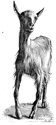
The front limbs are distorted. In young pigs, lambs, and less
frequently in foals, calves and dogs, the jaws become deformed, and
mastication is rendered difficult.
The vertebral column may also be affected, and lordosis (bending
downwards of the back) or skoliosis (lateral bending of the back) is somewhat
frequent.
Cyphosis, or upward bending of the back, seldom occurs, and when
seen, sometimes results from disease other than rachitis.
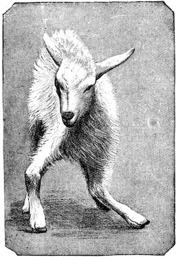
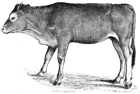
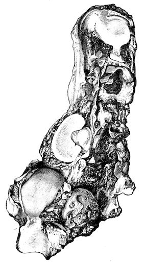
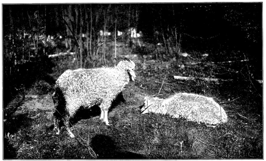
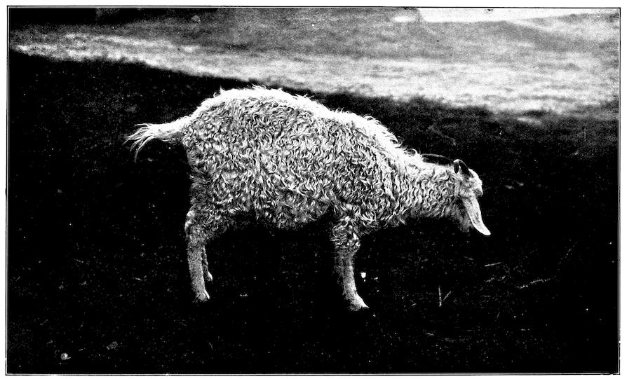
Fig. 1.—Rachitis in a young goat.
General development is always interfered
with and the young creatures
are generally dwarfed.
The digestive apparatus is disordered,
the appetite is irregular and
sometimes depraved, while indigestion,
gastritis, and enteritis are not
exceptional. Physiological and pathological
research has shown that the
quantity of phosphoric acid eliminated
in twenty-four hours in a
rachitic child is double the quantity
passed by a healthy infant.
The amount of urea in the urine
(which is a criterion of nutrition, and
usually varies in proportion to the
amount of food ingested) is, on
the contrary, diminished even when
highly nitrogenous food is given,
thus suggesting diminution in nutrition.
Lesions. The lesions are represented by abnormal and irregular
thickening around the interarticular cartilages. The cartilage is
thickened, compressible, very spongy and without regular ossification.
Diffused periostitis exists principally towards the extremities of the bone.
Beneath the periosteum the surface of the bone appears rough and
softened. On section the medullary canals are seen to be enlarged and
filled with marrow of a gelatinous character. The Haversian canals are
dilated, and the entire tissue appears very vascular. Chemical analysis
proves that the mineral constituents of the bone, particularly the phosphates,
have diminished by one-half; the organic constituents on the
other hand are increased in a similar ratio, but the ossein is abnormal.
Ossification has, in a word, been incomplete.
7Diagnosis. Diagnosis presents no difficulty except in the early stages
before deformity has occurred.
Rachitis can scarcely be mistaken for any other condition except
perhaps infectious rheumatism, but the rapid course of the disease in the
latter case, the persistence of fever and the swelling of the joint cavities
sufficiently differentiate the conditions provided care is exercised.
Prognosis. From an economic point of view the prognosis is very
grave for if the lesions are extensive there is nothing to be gained by
keeping the animal.
Treatment. Treatment differs very little, whether the animals are
still being suckled or have been weaned. In the former case it is
necessary to improve the quality and chemical constitution of the
mother’s milk by giving food, richer both in mineral salts and in
nitrogenous material.
Cooked grains, milk, and forage of good quality should be given freely.
When the mothers are exhausted and anæmic it is better to feed the
little animals artificially or to change them to a foster-mother. Those
already weaned should be given good rich milk, eggs, boiled gruel, and
drugs, such as the phospho-chlorate of lime, 1 to 1½ drachms per day (for
a calf); lacto-phosphate of lime, 1 to 1½ drachms; bi-phosphate of lime,
1 drachm, or simply ordinary phosphate of lime. Oil containing 1 per
cent. of dissolved phosphorus may be given in doses of 1 to 2½ drachms,
according to the size of the calves, but its use calls for much care, and it
should only be given for alternate periods of a fortnight. The glycerophosphates
are not very active. Beef meal in doses of 6 drachms to
1½ ounces and chloride of ammonium in doses of 30 to 60 grains have
also been used advantageously. The above drugs, but particularly the
bi-phosphate of lime and chloride of ammonium, stimulate nutrition and
diminish the quantity of phosphoric acid eliminated.
OSSEOUS CACHEXIA.
“Osseous cachexia” is a general disease which develops slowly and
progressively, producing its most marked effects on the bony tissues.
It has received a great many different names, such as osteoporosis,
osteoclastia, osteomalacia, fragilitas ossium, enzootic ostitis, bone
softening, etc., but none of these appears so appropriate as the term
osseous cachexia, suggested by Cantiget.
All the above-mentioned names are applicable to some phase of the
disease, but none to the disease in its complete development. Thus the
name “osteoporosis,” accepted by German authors, is quite applicable
to the phase of rarefying ostitis seen at the commencement, but this
condition occurs in other diseases. The expressions “osteoclastia” and
8“fragilitas ossium” suggest the fragility of the bones and the commonness
of fracture. The term “osteomalacia” is warranted during the
period of bone softening. The term “gout,” though in practice confusing,
has been held to be justified by the frequent appearance of synovitis and
arthritis; while that of “enzootic ostitis” indicates the appearance of
the disease in all the stables in one district, without however pointing
to its nature. It is possible that under certain circumstances the train
of symptoms might be incomplete, and then the terms above indicated
would be quite inappropriate. “Osseous cachexia,” on the other hand,
is very comprehensive, and appears to cover the entire development of
the disease, for which reason it here receives preference.
Law defines the disease as “a softening and fragility of the bones of
adult animals, in connection with solution and removal of the earthy
salts.” He describes it as an enzootic disease of mature animals—mainly
cows—in which the decalcifying process proceeds most actively in the
walls of the Haversian canals and cancelli of the affected bones. In
consequence of the removal of the earthy salts the bones become soft
and more or less fragile.
The disease has been observed in England, Scotland, United States,
France, Belgium, and Jutland, and generally in districts with low-lying
damp pastures. It attacks cows which are heavy milkers.
Susceptibility appears to increase with advancing age.
History. Having been described by Vegetius, the disease was again
observed about 1650 in Norway where it was treated by the administration
of crushed bones. It is fairly frequent in some parts of Germany
and Belgium. In France it was studied in 1825 by Roux, and in 1846
by Dupont, but Zundel in 1870 was the first who gave a good description
of it, founded partly on the authority of German authors and partly on
observations made by himself in the Valley of the Lower Rhine. Since
that time it has successively been reported in the Yonne by Thierry, in
the Nièvre by Vernant, in the Aube by Collard and Henriot (1893), in
the Indre by Cantiget, as well as in La Vendée by Tapon in 1893. In
that and the succeeding year Moussu also saw numerous cases in the
districts of Indre-et-Loire, Loire-et-Cher, Berry, Sologne, and in some
parts of Beauce.
Symptoms. The first symptoms are difficult to detect and interpret,
especially at the commencement of an outbreak and in parts where the
disease is rare they may lead to confusion and errors in diagnosis. On
the other hand, in regions where the disease is common the practitioner
will be able to form his diagnosis from the appearance of the first signs.
To render clear the mode in which the symptoms develop we may
divide the progress of the disease into four phases, though this grouping
is somewhat arbitrary.
91. The initial phase is not well marked, and is announced by digestive
disturbance and by wasting. The former of these symptoms may be
referred to some other cause, but consists in irregularity, diminution
and sometimes perversion of the appetite. These earlier signs are soon
followed by loss of spirits, and some interference with movement, but
the symptoms only become of importance or attain their full development
when the animals remain lying for a long period in the stable.
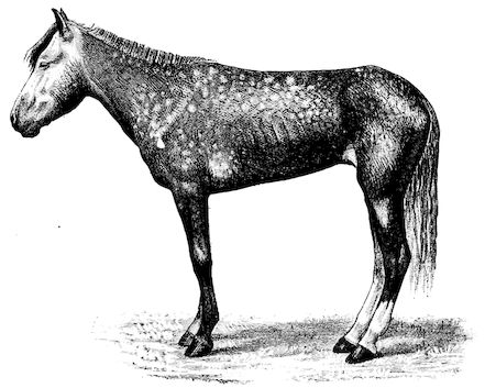
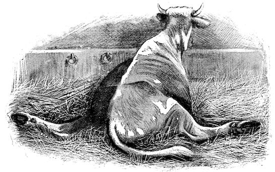
Fig. 2.—Horse suffering from osseous cachexia.
2. The second phase is characterised by more precise signs, which
become almost pathognomonic. Difficulty in rising is added to the
already existing tendency to remain lying, and to the interference with
movement.
When lying down the patient no longer responds to the trifling
stimulus, which a healthy animal needs to cause it to rise. It remains
languid and apparently lazy, though in reality it experiences pain
and difficulty on attempting to get up. The least muscular effort when
lying down often causes it to moan, as do efforts to change its position
or to walk. Even when standing still, it may appear to be in pain,
and patients often assume a position similar to that of a horse suffering
from laminitis.
At the end of this second phase, swellings appear, due to synovitis or
arthritis of the extremities, synovitis of the sesamoid or navicular
sheaths or to inter-phalangeal arthritis or arthritis of the fetlock joint.
Weakness becomes marked, and the appetite is very irregular.
10Secretion of milk diminishes or ceases and abortion is not uncommon.
3. The third phase is characterised by fractures, and it is this
peculiarity of the disease which has procured for it the names of fragilitas
ossium, and osteoclastia. These fractures may affect any portion
of the skeleton. Animals so suffering sometimes break a leg whilst
trotting or the pelvis in simply jumping over a ditch; a collision with a
fixed object like the jamb of the stable door, or a fall on the ground,
may result in the fracture of one or several ribs.
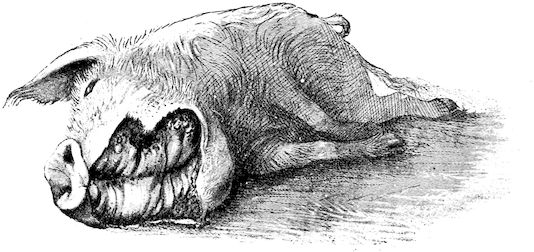
Fig. 3.—Pig suffering from osseous cachexia (fourth stage).
Such shocks would be of no importance to a healthy animal, but to
one suffering from osseous cachexia, any violence, or even the slightest
muscular effort may be followed by fracture of the gravest character,
involving even the vertebral column. In cows the pelvis, femur, and
tibia are most frequently injured.
In horses, particularly in riding horses, fractures are commonest in
the region of the forearm, cannon bone, and anterior phalanges. So
extremely fragile are the bones at this stage that the horse represented
herewith broke twelve ribs at one time by simply falling on its side. It
is interesting to note that such fractures are never accompanied by any
extensive bleeding. They have little tendency to repair, no real callus
formation occurs, and on post-mortem examination one often finds the
ends unconnected by temporary callus, worn, and rounded by reciprocal
friction.
At this stage but under other circumstances, the animals show great
reluctance to rise, remaining down for twelve to twenty-four hours
without shifting their position. If forced to get up, they stand as
though fixed in one position, the respiration and circulation become rapid,
and they soon grow tired and fall.
114. The fourth phase, or period of osteomalacia, i.e. softening of the
bones, is also the last. It is rarely seen in large animals like horses and
oxen, because accidents so often accompany the preceding stages and
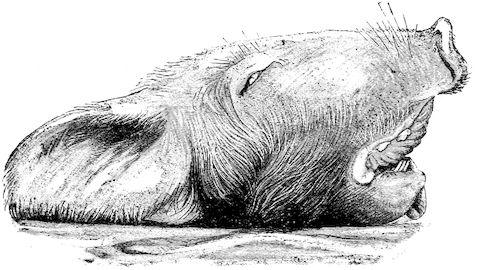
necessitate slaughter; but it is common in goats and pigs.
In this phase the bones become elastic, soft and depressible, yielding
to the pressure of the operator’s fingers.
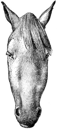
Fig. 4.—Deformity of the face in the horse shown in Fig. 2.
The flat bones are particularly liable to this change, which is common
to domesticated animals. The bones of the
head are the first to suffer; later those of the
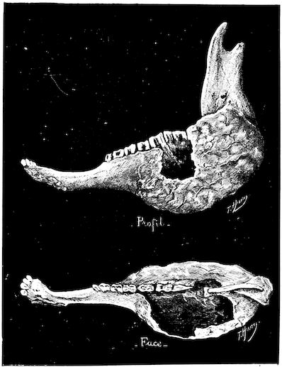
pelvis. The lower jaw becomes swollen, particularly
about the centre of the branches
which may attain three, four, or five times,
their normal thickness.
The depression in the submaxillary space
disappears. The upper jaw undergoes similar
changes, becoming deformed and thickened
until the cavities of the sinuses and the
hollow appearance of the palate are lost, while
the face is so changed that it cannot be
recognised as that of a horse, goat, etc.
The molar teeth are almost buried, their
tables alone being visible at the bottom of a
depression, the edges of which rise above the
neighbouring parts (pig).
Mastication is clearly impossible, the jaws
appear paralysed, the muscles powerless, and
only swallowing is possible, a fact which
explains why life is only prolonged to this stage
in animals which can be fed with a spoon or
bottle (pigs and goats). The bones of the
cranium, although greatly changed in texture, are always less deformed
than those of the face.
The changes are such that it is often easy with a mere post-mortem
knife to cut the head completely in two. Osseous tissue, properly
so-called, has disappeared.
All the constituent tissues, with the exception of the skin and muscles,
i.e., the bone, periosteum and aponeuroses, have the appearance and
consistence on section of the fibro-lardaceous tissue seen in chronic
inflammation.
The following is a condensed description of the disease as given by
Law:—
Symptoms. Poor condition or even emaciation, with very visible
projection of the bones. The coat is rough, skin tense, inelastic and
12hidebound, appetite variable, sometimes impaired, and nearly always
perverted (or depraved) so that the patient will lick the manger continually
or pick up and chew all sorts of objects: bones, leather,
clothing, wood or iron, stones, etc. The amount of food consumed
may, however, be up to the normal. The most marked feature is the
difficulty and stiffness of locomotion.... Temperature and yield
of milk may remain normal.
“Later, appetite and milk secretion fail, temperature rises a degree
or two, the animal refuses to rise, remaining down twelve to twenty-four
hours at a time, and ... when rising ... remaining on the
knees for a time, moaning and indisposed to exert itself further. At
this stage many cases begin to improve and may get well in five or six
weeks. Some will remain down for several weeks and finally get up
and recover. With constant decubitus, however, the animal falls off
greatly, becoming emaciated and weak, the appetite may fail altogether,
and the patient is worn out by the persistent fever, nervous exhaustion
and poisoning from the numerous bed-sores ... which are common
over the bony prominences. It is in these last conditions, above all,
that fractures and distortions of the pelvic bones, and less frequently of
the bones of the legs occur.”
Fig. 5.—Head of a pig suffering from osseous cachexia.
“The disease may advance for two or three months, and in case of
pelvic fractures and distortions, there may be permanent lameness, and
dangerous obstruction to parturition, even though the bones should
acquire their normal hardness through the deposition of lime salts.”
In horses, the different phases of the disease develop precisely as in
bovines. The apparent differences between affected horses and cattle
result in reality from differences in their capacity for continuing work.
In the first phase, horses are incapable of work, their movements being
13badly co-ordinated. They are inclined to stumble, and appear as though
suffering from strain of the lumbar muscles.
In the second phase pain referable to the bones sets in. Lameness
develops without visible lesions and is rapidly followed by synovitis and
arthritis in the lower portions of the limbs, and by wasting and anæmia.
The animals seem unable to move rapidly, or if forced to do so may
sustain fractures even at a trot: the limb bones sometimes break or
ligamentous insertions in the neighbourhood of joints are torn away,
resulting in sudden falls on the ground and fracture of ribs or even of
the vertebral column. This corresponds to the
third phase, osteoclastia, in oxen.
Fig. 6.—Osseous cachexia. This condition developed in two months, the last month of gestation and the first of lactation.
From then onwards, horses become useless
and, if not destroyed, may, after a few weeks
or months, develop the condition known as
osteomalacia, in which the flat bones become
softened, the head, the branches of the lower
jaw and the face become deformed, while
mastication and other functions are impeded.
Germain gives the above symptoms as
characteristic of the mode of development of
the disease in French and Algerian horses
imported into Tonquin, and his description,
written several years ago, is fully confirmed by
more recent observations. Since Tonquin was
taken over by the French, however, improved
methods of culture have resulted in the production
of better cereals and forage; the fodder
plants have been vastly improved, to the great
benefit of imported animals.
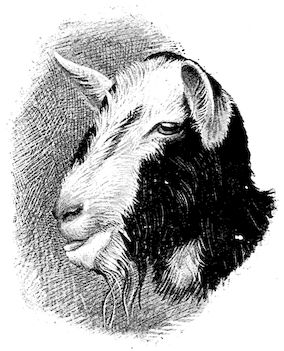
In the goat, the disease shows some slight
peculiarities. Thus, in the second phase, during
which goats and sheep suffer so markedly from
lameness and pain in the bones, goats often
walk on the knees. The disease, however, is uncommon in these animals.
The phase of osteoclastia is also less marked and fractures are rare,
because the animals weigh less and also because they are less exposed
to falls and violent shocks. The bones, nevertheless, are extremely
fragile and fractures may be produced at will.
Osteomalacia, on the other hand, is always well marked.
Regarding the development of the disease in pigs, we may repeat what
has just been said respecting the goat. Walking on the knees is often
one of the first signs, fractures are somewhat rare, and the period
of softening and deformity is always very noticeable.
14Course. The development of the disease is slow, lasting from one to
three months as a rule, and is little influenced by hygienic conditions.
Good milking cows, however, seem to be most frequently attacked,
probably because of the great losses of nutritive material which occur
through the milk. The calves borne by such animals are often rachitic.
Oxen are less commonly attacked. Horses rarely suffer from the disease
in France, but frequently in Tonquin. Pigs reared on very poor soil
seldom escape attack.
Fig. 7.—Osseous cachexia: softening of the maxillæ.
If treated from the beginning, or even before the second phase has
become well developed, the disease may be cured, but after this
period little improvement need be
expected.
Causation. The problem of why
osseous cachexia occurs has naturally
given rise to numerous
explanations, some plainly inadmissible,
others, however, of greater
or less plausibility.
The fact which, from the earliest
times, appears to have attracted
most attention is the relation
defective nourishment bears to
development of the disease. In
Norway, as early as the year 1650,
the plant known as sterregraes
(which renders animals dull and
heavy) was thought to be the cause
of the disease; two centuries later,
in 1846, the Anthericum ossifragum
was similarly regarded. Zundel,
in 1870, claimed that the Germans
first referred the development of
the disease to chemically incomplete forms of nourishment. This opinion
seems fully confirmed by the remarkable observations of Germain on
European horses imported into Cochin-China, and it is finally placed
beyond question by the work of Cantiget. Basing his researches on
analysis of the soil, he proved that osseous cachexia only occurs in cattle
depastured on land which is too poor in phosphoric acid and calcium
phosphate, and that it can be banished by enriching the soil with suitable
manures up to a point when the proportion of phosphoric acid becomes
normal. In good land, suitable for raising cattle, the proportion of phosphoric
acid, according to the best exponents of agricultural chemistry,
should not fall below 4,000 kilograms to the hectare. Cantiget and
15Brissonet have shown that where the soil contains less than 1,500 kilograms
to the hectare, osseous cachexia is almost permanently present.
As soon, however, as this proportion is raised above 2,000 kilograms by
suitable culture, the losses diminish, and the cachexia finally disappears.
This view was greatly strengthened by fodder analyses, which showed
that in all cases where the soil is poor in calcium phosphate, the forage
is poor in phosphoric acid, and vice versâ. The food is too poor in
mineral salts, firstly for normal development; and secondly for the
proper nutrition of the skeleton.
Germain is of a similar opinion with regard to the occurrence of
osseous cachexia in horses in Cochin-China, where the soil is very poor
in lime. The fodder and cereals are poor in mineral salts, and even
when given in large quantities do not furnish proper (chemical) nutrition.
Clear proof of the correctness of this view is afforded by the fact
that feeding with forage and cereals obtained from France or Algeria
prevents the disease appearing, or diminishes and finally removes the
previously existing symptoms. Furthermore, Germain shows that
Europeans, living solely on the products of the country, to some extent
suffer like the horses.
This theory though based on sufficiently solid foundations to carry
conviction, has been questioned, and it may be desirable to record briefly
the criticisms advanced against it.
One of the most important is as follows:—
As osseous cachexia of oxen occurs in certain well-defined districts in
France, and seems due to the feeding, why does it not attack horses in
the same regions in an enzootic form? The answer appears to be that
horses receive a greater amount of rich food, particularly of cereals,
which contain much larger amounts of mineral salts, including phosphates,
than does ordinary forage.
The most serious objection was made by Tapon, who states that in
1893 he saw osseous cachexia in oxen on farms in La Vendée where
superphosphate had been used for years, whilst the disease did not exist
on other farms where such chemical manures were not employed.
Before attaching much weight to this objection, however, it would be
necessary to know the richness in phosphoric acid of the soil on the
respective farms, for it is possible that, in consequence of natural conditions
and in spite of the use of certain mineral manures, the richness of
the soil on the first-mentioned farms, though manured with superphosphates,
was still below that of the others which had received no artificial,
manure.
The system of culture is also of importance, for at the present day,
even with the use of artificial manures, cropping would rapidly impoverish
soils which were not suitably and sufficiently enriched.
16Abundance or apparent richness of food signifies nothing if quality is
lacking.
It may also be asked: if the question of nourishment is of such prime
importance why are animals of European origin in Cochin-China
affected, whilst the indigenous races prove immune? The answer
would appear to be that, in addition to the defective quality of food,
other factors, such as adaptation to environment and relative digestive
power, play a considerable part in the production of the disease.
Favouring causes. Whilst conceding that the disease is due to one
determining cause, viz. the food, it is unquestionable that other causes
may favour its appearance. Abundant milking is one, so that the
disease most frequently appears six to eight weeks after calving. Gestation
may also determine an attack. The disease is rarer in oxen than in
milch cows. Starvation and bad hygienic conditions also have a certain
influence; it is well known that during dry years, particularly when
fodder is scarce, osseous cachexia makes the greatest ravages. Law
states that the disease has been attributed to excess of organic matter in
the soil, to succulent watery foods, as rank watery grasses, potatoes,
turnips and other roots deficient in nutritious solids. Some agent—microbe
or toxin—swallowed with the food has been suspected but not
yet isolated.
Other explanations have been advanced but up to the present time
they scarcely deserve to be regarded even as hypotheses. Thus Anacker
in 1865 declared that the disease commenced as muscular rheumatism,
was succeeded by destructive or atrophic ostitis, and ended as osteoporosis.
So far as the order of the osseous lesions is concerned, this
view is quite correct, but the ossific changes are consequences and not
causes.
The idea that the disease was due to an infectious agent has been
advocated by Leclainche, without, however, having been proved.
Pétrone is the only person who has hitherto suggested that osteomalacia
in man is due to infection with a nitric ferment (Micrococcus
nitrificans). According to him, pure cultures of this organism injected
into dogs, produce osteomalacia. These statements, however, require
confirmation.
Lesions. The chief lesions are to be found in the bones. They
consist in rarefaction of the compact tissue, increase in size of the
medullary cavity and Haversian canals, and enlargement of the areolæ
of the spongy tissue. The bone marrow loses its fatty constituents,
appears red and gelatinous, and contains a greatly exaggerated number
of blood-vessels. When heated, the bones do not yield oil as in healthy
subjects, and when dry, they seem abnormally porous. In the osteoclastic
phase, the bones become very friable and even the shafts assume
17a spongy appearance. They diminish in density. These changes
correspond to the stages of eccentric rarefying ostitis and osteoporosis
of German authors.
The flat bones often show well-marked periostitis, but the great
thickening sometimes seen in certain of the bones of the head appears
to be the result of a special osteo-periostitis. It is quite certain that
the disease is due to something more than a mere want of mineral
constituents in the bone, and poverty in this respect certainly does
not explain the hypertrophic changes. The nutrition of the bones as
a whole is disturbed, resulting in alterations both in the ossein and
in the mineral salts, the whole process being accompanied by symptoms
of osteo-periostitis.
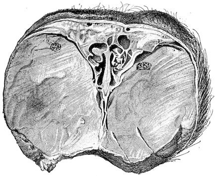
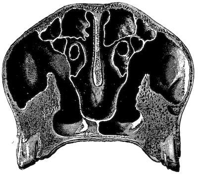
Fig. 8.—Transverse section through the middle region of the face in a pig suffering from osseous cachexia.
The fractures which occur so frequently during the osteoclastic phase
have well-marked peculiarities. The extravasation of blood is trifling,
and no callus forms, even when the ends of the bones are immobilised
by external aid; if the ends are left free, they soon become worn and
polished by rubbing against one another.
In the neighbourhood of the articulations and ligamentous insertions
the periosteum soon undergoes change, and it is not uncommon to find
sub-periosteal and intra-osseous extravasations of blood.
Germain has also noted in horses the disappearance of the intervertebral
and articular cartilages, and the frequent occurrence of anchylosis,
true or false.
18In the final stages, the bones may be cut with a knife, and a time
arrives when bony tissue seems completely to have disappeared; thus, as
shown in Fig. 8 herewith, it was possible to cut the entire head of a pig
into thin slices without the slightest difficulty. All parts of the head had
been affected by the softening change.
From the chemical point of view, the diminution in mineral salts and
in phosphate of calcium has long been recognised, but the degree of this
change varies according to the phase. In human beings the proportions
have been estimated as follows: Normal bone, 50 to 80 per cent. of
phosphate of calcium; bone in persons suffering from osteomalacia, 5
to 20 per cent. of phosphate of calcium. The changes in the ossein
have not been carefully studied. We only know that histologically the
ossein becomes fibrillar, and that chemically it no longer retains its
normal composition.
The diagnosis is difficult, particularly on the first occasion of seeing
the disease, and especially if this is of an enzootic character. The
practitioner may also have some hesitation in diagnosing isolated cases
in regions where the disease seldom occurs.
Otherwise, diagnosis is usually easy, as soon as lameness or synovitis,
or arthritis of the lower regions of the limbs appears. Only in isolated
cases are the lesions likely to be mistaken for accidental injuries, and it
is also fairly easy to differentiate them from the localised lesions of
rheumatism. The latter disease seems more frequently to attack the
upper joints of the limbs, and is often accompanied by intense fever and
cardiac disturbance.
Prognosis. In a general sense the disease is very grave, because it
appears as an enzootic, and, in dry years and those during which there
is a scarcity of forage, inflicts enormous losses on the breeders of certain
countries. When advice is sought towards the end of the second phase
of the disease the prognosis is therefore very grave. Under such circumstances
it is often better to slaughter rather than to treat, provided
that the affected animals, like cows, pigs, or goats are still of some value.
The prognosis is much more hopeful if treatment is attempted at an
early stage, when improved diet and the use of suitable drugs sometimes
lead to recovery.
Treatment. We know that in the Middle Ages this disease was often
treated by the administration of crushed bones, and even at the present
day ground bones are frequently recommended. Treatment must be
subordinated to proper feeding, no system of medication being of any
value whatever unless the food is suitable.
Germain states that imported horses in Cochin-China recover if
simply returned to their former diet, i.e. to cereals and forage obtained
from France or Algeria. Cantiget shows that such improvements in
19cultivation as the free distribution of superphosphate manures on
impoverished soils modify the chemical composition of the forage,
and render it capable of building up and sustaining the organism and
bony tissues; treatment should therefore be essentially prophylactic
in character.
Animals suffering from osseous cachexia should be fed on cereals and
forage obtained from rich districts where the disease has never occurred;
but, as in times of scarcity questions of expense almost always receive
first consideration, it may be necessary to substitute bran for such
products, or give oats, maize, beans, rice, and oil or cotton cake, etc.,
all of which can be obtained commercially, and are of sufficient
nutritive richness. It is often advantageous to give such food cooked
and slightly salted.
Commercial ground bones and calcium phosphate (bi- or tri-basic), in
doses of 1 ounce per day for oxen and 1¼ to 2 drachms for pigs or goats,
have given excellent results in the hands of most practitioners. Some
recommend the addition of iron salts or bitter tonics like gentian or
nux vomica in doses of 2½ drachms per day for a full-grown ox.
Law declares that the treatment should be varied “with the predominance
of the causes, essential or accessory.... Green clover,
alfalfa, and other leguminous products, ground oats, beans, peas,
linseed or rape cake ... and vetches may be especially recommended....
The free access to common salt and a liberal supply
of bone meal are helpful.... Apomorphia is especially valuable in
correcting the perverted appetite and stimulating digestion. A change
of pasture is always advisable. In all cases where possible the water
should be changed as well as the food. Attention to the housing,
grooming, and general care of the animals should not be neglected.
Finally, every drain upon the system should be lessened or stopped.
The milk may be dried up, and the animal should not be bred.”
Meat meal also renders good service, but the use of cod liver oil,
suggested by Zundel, is too expensive, and phosphorised oil is too
dangerous to be adopted in ordinary treatment.
Local treatment for synovitis and arthritis has been recommended.
It is ineffective unless accompanied by good feeding and internal
medication. On the other hand, the lesions often diminish rapidly or
totally disappear under the influence of general medication alone.
20
FRACTURES.
Although oxen, sheep, goats, and pigs are much less subject to
fractures than the horse and dog, nevertheless, they do suffer from
such accidents. Repair is perfectly possible, but the cases are often not
worth treating, unless the subjects are young or of considerable value.
On the other hand, in fat and heavy subjects, it is difficult to fix the
parts in position. Slinging produces bad results, and generally should
not be encouraged.
Apart from fractures accompanying general chronic diseases, like
rachitis and osseous cachexia, the vertebræ, the pelvis, the ribs, or any
of the limb bones, may be fractured in consequence of accident.
Such fractures may be either complete or incomplete (fissures),
simple or compound.
The general signs which indicate fracture are always the same, viz.,
loss of function, local pain, abnormal mobility, crepitation, due to rubbing
together of the ends of the bones, and deformity of the part. Diagnosis
is generally easy; prognosis on the other hand is very variable.
The vertebral column may be accidentally fractured in the region of
the neck in consequence of the animal falling on its head; in the dorsolumbar
region, from falling into ditches or ravines, or, in the case of
bulls fighting, from violent muscular efforts. Fractures of the first
kind are immediately fatal; those of the second result in paraplegia
of the hind limbs, and necessitate immediate slaughter.
Fractures of the pelvis comprise:—
1. Fractures of the angle of the haunch, resulting from external
violence and characterised by sinking of the external angle of the
ilium, deformity of the hip, and lameness without specially marked
characters. This fracture is rarely complicated. The symptoms of
lameness diminish with rest, but deformity continues.
2. Fractures of the floor of the pelvis, usually extending from the
anterior margin of the pubis to the foramen ovale and from the
posterior margin of the foramen ovale to the end of the symphysis.
They result from obstetrical manipulation, as in forcibly removing a
fœtus which is too large, or a monstrosity. As a rule, the animals
cannot rise, or if they succeed in doing so, are incapable of moving.
Diagnosis is made by exploration through the rectum. Such fractures
always necessitate slaughter.
Fractures of the neck of the ilium and of the base of the cotyloid
cavity, even in cases of dislocation, are rare despite what has been
said to the contrary.
21In the fore limb, fractures of the scapula and humerus are usually
of traumatic origin, are seldom accompanied by marked displacement,
and are capable of uniting if a long rest at grass is allowed. Pitch
bandages should be applied to the surface, covering all the surrounding
regions, viz. the withers, upper portion of the forearm, girth and chest,
to assist in immobilising the region of fracture, and to promote union.
Fractures of the forearm are more difficult to treat, because the
bandage applied must extend as far as the hoof. In this case displacement
often occurs. It is therefore necessary, firstly, to reduce the
fracture, and bring the ends in perfect contact, for which purpose it
may be requisite to cast the animal, and give an anæsthetic; and,
secondly, to apply a pitch plaster in the form of a shallow gutter, leaving
the inner surface of the limb uncovered along a line about two inches
wide following the course of the veins of the forearm.
Fractures of the metacarpus and metatarsus usually heal well in all
animals of moderate weight, such as heifers, steers, goats or sheep,
provided a simple plaster bandage, covering the entire limb or preferably
with an opening in the position above indicated, is applied and
continued downwards as far as the claws.
In sheep and goats it is sometimes even sufficient to use a splint
formed of straw-boards, and in the case of oxen, of wood, applied over a
cotton-wool padding and retained in position by straps, or in the case
of the heavier animals by dextrine or pitch bandages.
In the hind limb, fractures of the femur are more serious, because the
apparatus that can be used to secure immobility is seldom or never
effective; excepting in young animals, it is therefore usually better to
slaughter.
Fractures of the tibia are treated like those of the forearm when it
appears desirable to keep the animals alive.
Plaster bandages can very easily be prepared by saturating tarlatan
in a mixture of equal parts of thoroughly dry plaster and water. Six to
ten thicknesses of tarlatan, arranged alternately longitudinally and
transversely, are sufficient. When adjusted they can be kept in position
until the plaster has hardened by means of dry bandages applied from
below upwards, which can be removed after a lapse of half an hour to
an hour.
FRACTURES OF THE HORNS.
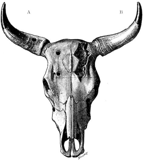
Anatomy of the horns. The horns form organs of defence, and
project on either side of the frontal bone at the poll. Each consists
firstly of a bony basis generally known as the horn core; secondly, of a
horn-secreting membrane; thirdly, of a horny sheath, the horn properly
so called.
22(1.) The horn core projecting from the frontal bone does not
develop until after birth. About the third month a little prominence
appears under the skin, which, as it develops, assumes a conical shape,
and may be seen to be covered with a horny substance. In proportion
as the horn core grows, there develops within it a cavity which may
either be of a simple character or divided by a longitudinal partition.
This communicates with the frontal sinus, a fact which explains the
collection of pus in the sinuses as a result of injuries to the horns.
The sinus of the horn core does not exist in young animals, and is not
completely developed before the third or fourth year of life.
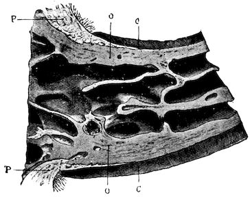
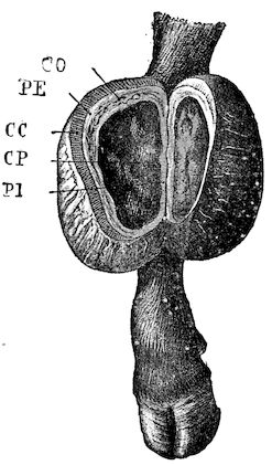
Fig. 9.—C, horn; P, modified skin forming the keratogenous membrane; O, horn core, exhibiting a double sinus.
(2.) The horn-secreting membrane is formed by the skin, which undergoes
special development around the base of the horn and comes to
resemble that of the coronary band, from which the hoof or claw is
secreted. The band is about one-fifth of an inch in breadth. The
papillæ of the dermis are specially developed at this point, and the
epithelium which they secrete eventually forms the horn.
The internal surface of the growing horn is adherent to the horn core
through the medium of another tissue formed by a specially differentiated
periosteum which is continuous with the periosteum covering the frontal
bone. It is not a true periosteum, but a vascular tissue formed of papillary
layers analogous to those of the podophyllous tissue of the ox’s claw or
horse’s hoof.
This keratogenous membrane receives a rich vascular supply from
the arterial circle formed at the base of the horn core by a division
of the external carotid, the blood conveyed by which is freely distributed
to the enlarged papillæ. The great vascularity of these parts
23explains why lesions of the horns are often followed by such profuse
bleeding.
(3.) The horn secreted by the papillæ of the horn band (which is
analogous to that of the coronary band of the horse) forms a cone
varying in its curve in various breeds. Its base is hollow, and
contains little depressions holding the papillæ from which the horn is
secreted. From its base up to the end of the horn core the walls progressively
increase in thickness. From this point it is solid; in a
fully-grown horn the bone does not extend more than one-half or
two-thirds of the entire length.
In the adult, the development of the horns varies with different
breeds and is affected by sex. In the bull the horns are short, but in
the cow and ox long. Short and fine in animals of improved breed like
the Durham, they are long and thick in breeds of working oxen.
Injuries affecting the horns are of three classes, determined by the
part affected.
1. Detachment of the horn or sheath.
2. Laceration:—
(a)
Of the horny sheath alone.
(b)
Of the horny sheath and of the horn core.
(c)
Of the horn core alone, the horny sheath remaining intact.
3. Fractures:—
(a)
Of the terminal half of the horn.
(b)
Of the lower half.
(c)
Of the base.
DETACHMENT OF THE HORNS.
When the yoke is badly fitted or padded, it is liable to cause a
continual strain or a succession of shocks producing chronic inflammation
of the keratogenous membrane. Should the end of the horn
then be struck heavily, it is quite possible that the horn will either
partially or wholly be detached. In this case it falls away without there
necessarily being any important lesion of the horn core.
Such accidents are not infrequently caused by the driver striking the
ox on the horn with the yoke in order to keep it quiet while it is being
harnessed.
The prognosis of this condition is not grave, except for the fact
that working animals cannot be used until the horn is completely
regrown.
The treatment simply consists in thoroughly cleansing and disinfecting
the horn core and then applying a protective dressing. The
bony basis is surrounded with a mass of tow saturated with an antiseptic
24solution, like 2 per cent. creolin or carbolic acid solution, which is kept
in position by a spiral bandage passed around the horn, and secured in
a figure of 8 on the opposite horn. Instead of applying such a dressing,
some practitioners content themselves with using an antiseptic ointment
or even a simple dressing of tar.
FISSURING OF THE HORNS.
Causation. In a general sense fissures may result from any violence
affecting the centre portion of the horns, such as blows with the yoke
or accidental bruises inflicted by the animals themselves in fighting
with their neighbours.
Symptoms. Whether the fissure is confined to the horny covering
itself or whether it extends to both the portions constituting the horn,
that is, the horny covering and the horn core, two very noticeable
symptoms are always present: 1. A straight fissure resembling a sand
crack, and appearing usually on the convexity of the horn, and, 2. A
very trifling hæmorrhage, which does not appear until some hours or
even a day after the accident.
Diagnosis. If the lesion only affects the horn core, diagnosis is
always difficult, for one can hardly perceive any sensitiveness of the horn
near the fissure.
Prognosis. Provided that the horn core is not injured, the prognosis
is favourable; but in the contrary case, it should be reserved; for
hæmorrhage extending to the interior of the frontal sinus not infrequently
causes suppuration in that cavity.
Treatment. Attempts should first be made to check hæmorrhage by
applying masses of tow saturated with cold water and frequently wetted
with slightly antiseptic solutions, such as 2 per cent. creolin or
carbolic acid. If hæmorrhage persists in spite of this simple treatment,
astringents may be employed, which, by causing the formation of a clot,
mechanically arrest further extravasation of blood. These astringents
vary considerably in value, and we should particularly warn practitioners
against perchloride of iron, which causes necrosis of the
tissues, and later, formation of pus. A 5 per cent. solution of gelatine
is hæmostatic and excellent for the purpose named, as also is hydroxyl
solution. When once hæmorrhage is arrested, the keratogenous membrane
rapidly heals in consequence of its vascularity, and soon secretes
fresh horn.
25
FRACTURES OF THE HORNS.
Etiology. Fractures of the horns, like fissures, are produced by
violence, but of a more marked character. They are termed complete
or incomplete, according as the entire thickness of the horn or only a
portion of that thickness is involved.
The fracture may affect either the terminal half or the basilar half;
or, again, it may have its seat in the frontal bone below the origin of the
horn core, in which case a flake of bone will be detached. Such fractures
assume varying forms, and may either be deeply excavated, oblique,
smooth, regular or dentated.
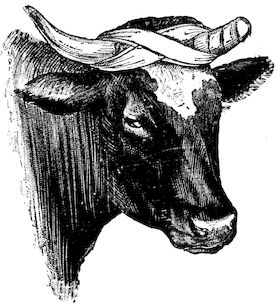
Fig. 10.—Dressing for fracture of the horn.
Symptoms. The symptoms are extremely simple. They consist
mainly in the mobility of the fractured
end, and such phenomena
as sensitiveness, hæmorrhage, etc.
When the fracture extends to the
frontal bone, crepitation may also
be noted.
Prognosis. The prognosis is not
grave unless the fracture extends
to the basilar half of the horn or
affects the frontal bone.
Treatment. (1.) If the fracture
is confined to the horn core, it is
only necessary to bring the fragments
into regular apposition, after
having removed the broken end of
the horn itself.
(2.) In treating a fracture affecting
the middle portion of the horn or in treating animals destined for the
butcher, the best method is to make a simple wound by dividing the
parts with a saw below the fracture. This is a painful operation, necessitating
anæsthesia, and requiring the animal to be cast or firmly fixed to
a post or placed in a trevis. To diminish the painful stage of the operation,
it was formerly recommended to make a circular incision extending
through the entire thickness of the horn proper, and then to remove
with a fine, very sharp saw the portion of the horn core. This, however,
is scarcely practicable, and it is much better to make a direct section.
Hæmorrhage is checked with compresses, moistened with cold water,
after which a dressing known as the “Maltese cross dressing” (Fig. 10)
is applied according to general principles.
The surface of the section, after washing with an antiseptic solution,
is powdered with iodoform or a mixture of iodoform and boric acid,
26covered with a mass of tow or cotton wool, saturated with liquid antiseptic,
and then surrounded with a flat pad of wadding, which extends
completely around the horn as far as its base. A second pad of larger
size, intended to protect the wound against external violence, is arranged
around the free extremity of the horn. This is kept in position by two
small cross bandages. Another bandage, the loop of which is fixed to
the base of the opposite horn, is then applied in spiral turns, completely
enveloping the former pads and extending from the base to the point.
Arriving at the free end, the operator reverses the bandage, draws it
tight, and continues down to the base of the horn, fixing it by figure of 8
turns passed around the base of both horns.
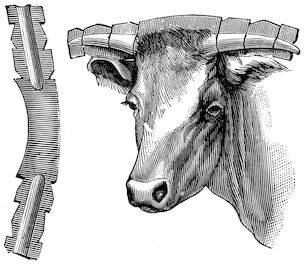
Fig. 11.—Splint for fractured horn.
(3.) In dealing with fractures of the lower third of the horn in working
oxen, it is necessary to seek consolidation of the horn by callus formation,
so that the animals may again
become useful in the yoke.
Very great difficulty accompanies
attempts to immobilise
the horn in such cases, since
the least shock to the extremity
of the horn destroys the union,
on account of the length of the
lever represented by the horn
itself.
The first method of treatment
consists, after the wound has
been carefully disinfected, in
fixing the ends in place by
tightly applied pads, surrounded
by splints, curved to fit to the horn. These are supported by several
turns of a spiral bandage.
Were one certain of the cleanliness of the wound and of its perfectly
aseptic condition, it would be better at once to have recourse to a fixed
bandage, strengthened by plaster or silicate of soda.
These bandages are applied longitudinally and should extend a considerable
distance on either side of the fractures. If the horn is long and
thick, it is best to use a fixed dressing of this kind secured by bandages
in the form of a Maltese cross.
All these methods, however, are more or less inconvenient, and the
most practical procedure often consists in removing the horn. Treatment
should only be attempted when the owner specially requests it in order to
render the animal useful for working or show purposes.
Moreover, however strong the dressing, accidents are frequent, for
accidental shocks to the fractured horn interfere with the co-aptation of
27parts, and diminish the chance of perfect union. To avoid these drawbacks,
the apparatus shown in Fig. 11 has been invented.
This apparatus consists of a splint, the middle of which fits the back of
the animal’s poll, the sides being gouged out to receive the lower half of
the horn and notched, to enable the fractured horn to be well supported
by bandages.
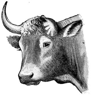
Fig. 12.—Dressing for fracture of the base of the horn.
(4.) Fractures of the base of the horn are more serious, because a
fragment of the frontal bone is usually torn away with the horn core.
As a rule, the fracture is
subcutaneous or without external
wound. The horn is
displaced, and swings loosely.
On examination, a characteristic
crepitation sound is
easily detected. In such fractures
hæmorrhage is subcutaneous
and often extends to
the frontal sinus, in consequence
of which it is not
uncommon for pus to form
in the sinus as a complication.
Treatment includes reduction
of the fracture, and the
application of a fixed plaster
or silicate bandage covering
the fronto-occipital and
superior auricular regions.
When a skin wound exists, it is better to remove the horn and bony
fragment, and to apply an antiseptic dressing in order to prevent infection
of the frontal sinus.
EXOSTOSES.
SPAVIN IN THE OX.
Exostoses are somewhat uncommon in the bovine species, and when
they occur are rarely of great clinical interest. Nevertheless, in cows
and old working oxen one sometimes sees metatarsal spavin. Its
gravity, however, appears to be very much less than in the horse, on
account of its position. Very commonly there is only trifling lameness.
Treatment by application of biniodide of mercury ointment or the
actual cautery gives good results. The principal precaution required is
to prevent the animals licking the parts.
28
RING-BONE.
Ring-bones only occur in working oxen, and particularly in aged
animals used in hilly regions. They result almost exclusively from
wounds, ligamentous and tendinous strains, and articular injuries.
Fig. 13.—Tibia. Lesions due to open fracture and displacement. Irregular callus formation and segnestrum.
They are preceded (as can usually be proved by dissection of limbs) by
fibrous or fibro-cartilaginous induration in or about the coronet or one
of the phalanges. These thickenings increase the diameter of the
pastern in all directions. Ring-bones are seldom very large; but as they
29partially or entirely surround the insertions of the lateral ligaments,
inter-phalangeal articulations or insertions of the digital extensors, they
are painful, and produce lameness of varying intensity.
Diagnosis is easy, partly because the tension of the skin and the
fibrous thickening render palpation painful.
Prognosis is grave, because the effect of ring-bone is sometimes to
render working animals useless.
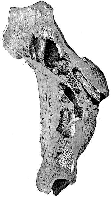
Fig. 14.—Sarcoma of the periosteum beneath the scapula.
Fig. 15.—Sarcoma of the periosteum covering the upper end of the tibia.
Treatment. To relieve the diseased claw of pressure due to its bearing
on the ground, the shoe should be removed and the claw freely pared.
If necessary, the healthy claw of the same foot may be raised by placing
a piece of thick leather between the sole and the shoe. It is advisable at
once to apply an energetic plaster, or, better still, to resort to firing in
points.
SUPPURATING OSTITIS.
In addition to the changes in bone resulting from rachitis, osseous
cachexia, tuberculosis, and actinomycosis, one sometimes sees cases of
periostitis or ostitis pure and simple. As a result of external injury or
30direct wounds, the bone may be contused and injured, becoming the seat
of diffused periostitis, necrosis, suppurating ostitis or osteomyelitis.
Open fractures may produce the same results.
Treatment comprises disinfection of wounds, antiseptic injection of
fistulæ, the application of antiseptic pencils, curettage, the removal of
sequestra, and vesicant or resolvent complications. When such conditions
extend to neighbouring joints and produce suppurative arthritis,
the animals ought to be killed.
BONE TUMOURS.
The only bone tumours of real importance from a practical point of
view are malignant growths represented by rapidly spreading epitheliomata
or sarcomata, originating in the periosteum. Fortunately such
tumours are rare.
They are not difficult to diagnose, as they develop rapidly, are accompanied
by pain and lameness ending in diminution or loss of the power
of movement, and frequently attack neighbouring lymphatic glands.
Even when in good condition, animals lose flesh and appetite, and finally
die of general wasting. The diagnosis is sufficiently guided by the
deformity of the parts, the bosselated appearance of the tumours, the
absence of fluctuation, the hæmorrhage which follows exploratory puncture,
the character of the little fragments of tissue removed through
these punctures, and finally the leukocytosis, which accompanies the
development of malignant tumours.
The prognosis is grave, for it is usually difficult or out of the question
to have recourse to removal, resection or amputation, when the tumours
have acquired any considerable size. Success is impossible unless
intervention is early, and the growth is in a readily accessible part. In
other circumstances early slaughter is indicated.
31
Congestion of the claws is not infrequently confused with contusion of
the sole. It is, however, essentially different, and presents closer
analogies with laminitis. The condition is characterised by congestion of
the entire vascular system of the claw and principally of the velvety
tissue. Like laminitis, it affects all four limbs; in rare cases the two
front or two hind.
Congestion of the claw results almost exclusively from enforced movement
on hard, dry and hot ground. It is commoner in animals
unaccustomed to walking, and in heavy beasts which have been travelled
considerable distances to attend fairs or markets. It is commonest in
the bovine and porcine species, and less common in sheep.
The symptoms appear after animals return from a long journey by
road. They are characterised by unwillingness to bear weight on the
feet and difficulty in movement. Standing is painful, and the animals
resist being moved; as soon as released they lie down.
Diagnosis presents no difficulty, though the condition is sometimes
mistaken for slight laminitis.
Prognosis is favourable.
Absolute and prolonged rest is always followed by recovery, no
internal medication being necessary, though this result is promoted by
enveloping the claws in wet compresses or by using cold foot baths, etc.
CONTUSIONS OF THE SOLE.
Contusions of the sole are only seen in animals which work without
shoes or in such as are badly shod.
Work on rocky ground, movement over newly metalled roads, and
wounds produced by sharp stones, are the principal causes of contusion
of the sole. Badly applied shoes, flat or slightly convex on their upper
surfaces, may also produce bruising in the region of the sole. The
anterior angle of the claw is rarely affected.
32Lameness is the first symptom to attract attention. It is slightly
marked, unless the bruising has been overlooked until suppuration has
set in. It affects only one or two limbs, and is rarely accompanied by
general disturbance, such as loss of appetite, fever, exhaustion during
work, etc.
Locally the claw or claws affected are abnormally sensitive to percussion
of the wall, and particularly to compression of the sole.
The parts are hot to the hand, and thinning the sole with a knife shows
little perforations, irregular points and crevices in the horn. One
may also find softening, infiltration and hæmorrhage within the horn
similar to those of corn in the horse, undermining of the sole over
limited areas, and sometimes suppuration, if the animals have been
forced to work when lame.
Complications like necrosis of the velvety tissue or of the bone, though
comparatively common in the horse, are rare in oxen.
Diagnosis is not difficult provided the history of the case is known.
Confusion with laminitis is scarcely possible, for the gait of this lameness
and the local symptoms are all different. Examination of the sole will
usually dispel any remaining doubt.
Prognosis. The prognosis is favourable. When the horn is simply
softened and a blackish liquid transudes, the lesion is trifling; if the
discharge is reddish grey the lesion is graver, and implicates all the
velvety tissue; finally, separation of the horn from the secreting
membrane and the discharge of true pus point to death of the
keratogenous tissue or of the bone.
Treatment should be commenced by carefully thinning the sole
around the wound and applying moist antiseptic dressings or cold
affusions. Removal of loose portions of horn hastens repair by allowing
discharge, which has accumulated between the living tissues and the horn
itself, to escape freely. The extirpation of necrotic tissue and the application
of surgical dressings are only called for in specially grave cases.
This treatment usually gives good results. The acute complications
which are so common and so dangerous in the horse seldom occur in
the ox.
Most of these operations can be performed without casting, provided
the animal is placed in a trevis or is sufficiently secured.
LAMINITIS.
Laminitis is characterised by congestion, followed by inflammation
of the horn-secreting tissues of the foot. It is now rare in
oxen and very seldom assumes an acute form. The slow pace at which
animals of the bovine species move may sufficiently explain this rarity;
33nevertheless, prolonged travel on stony roads with heavy vehicles, rapid
and repeated marches to towns or important fairs, are sufficient to produce
attacks. Before the days of railways, and for some time after
their introduction, in Britain cattle were travelled by road, and laminitis
was common.
Long journeys in crowded railway trucks may also produce the disease,
although the animal has not been forced to walk. Persons engaged in
exhibiting cattle at shows are well aware of this. Prolonged maintenance
of the standing position will produce the trouble, to which
the joltings of the railway journey may also contribute their share.
Prolonged standing on board ship may induce laminitis.
“Show condition” and the consumption of highly nitrogenous, and
particularly of farinaceous, foods favour the occurrence of laminitis.
Breed is also considered to have some influence, and laminitis is said
to occur more frequently in animals raised in flat districts, because in their
case the space between the digits is larger than in mountain-bred cattle.
In this connection the body weight may perhaps play a certain part.
The symptoms vary somewhat, depending on whether laminitis is
general and affects all four feet, or restricted to the two front or the two
hind feet.
The internal claws always seem more severely affected and more sensitive
than the external. In very rare cases the animal remains standing,
but usually it lies down, and will only rise under strong compulsion.
When standing, the symptoms are similar to those noted in the horse;
the animal appears as though absolutely incapable of moving. If all
four feet are affected the animal assumes a position as though just about
to rise; if the front feet alone are affected the animal kneels in front
whilst it stands on its hind legs, a very unusual position for the ox to
assume; finally, if the hind feet alone are affected, the animal seems to
prefer a position with the feet under the body both in front and behind.
(See Veterinarian, 1894, case by Bayley, and note by Nunn.)
It is always difficult to make the animal move. Walking seems painful,
and most weight is thrown on the heels. The body swings from
side to side as the limbs are advanced, and each limb is moved with a
kind of general bodily effort.
The claws are hot, sensitive to the slightest touch, and painful on
percussion.
Throughout the development of laminitis the general symptoms are
very marked. The appetite falls off early, fever soon appears, and in
grave cases the temperature rises to 105° or to 106° Fahr. Thirst is
marked, and the animal seems to prefer cold drinks. The muzzle is dry,
the face anxious and expressive of pain. Wasting is rapid.
The ordinary termination is in resolution, which occurs between the
34eighth and fifteenth day, provided the patient has been suitably treated.
The disease rarely becomes chronic. On the other hand, the claw occasionally
separates, as a consequence of hæmorrhage or suppuration,
between the horn proper and its secreting membrane. Should this
complication threaten, the pastern becomes greatly swollen, the extremities
become intensely congested, and separation commences at
the coronet. Loss of the claws, however, like suppuration, is rare.
Diagnosis. Congestion of the sole, the early stage of infectious
rheumatism and osseous cachexia may, at certain periods of their
development, be confused with laminitis; but the history and the
method of development of the above-mentioned diseases always allow
of easy differentiation.
It should, however, be added that, in certain exceptional conditions
(suppurating echinococosis, producing chronic intoxication, tumours
of the liver, and tumours of the pericardium and mediastinum),
symptoms may be shown that suggest the existence of laminitis,
although it is not really present. In these cases pain may possibly be
felt in the bones of the extremities.
The prognosis is usually favourable, but necessarily depends on the
intensity of the disease. Fat stock always suffer severely.
The treatment varies in no important particular from that prescribed
for the horse, and is usually followed by rapid improvement. The chief
indications are free bleeding from the jugular, the application of a
mustard plaster over the chest, and the administration of a smart purgative
(1 to 2 lbs. of sulphate of soda, according to the size of the animal)
at first, followed by laxatives. This treatment may be completed by
giving salicylate of soda per os in doses of 5 to 8 drams, or arecoline in
subcutaneous injection, 1 to 1½ grains. Local treatment consists in cold
affusions or poultices to the feet.
Failing cold baths, clay plasters applied to the feet are useful. To
ensure success all these methods should be utilised simultaneously. In
cases of separation of the claw, antiseptic dressings, with a thick pad of
tow placed under the sole, become necessary.
Chronic laminitis may perhaps occur in the ox as in the horse, but, as
a rule, oxen are slaughtered before the disease can assume this form.
In dealing with fat, or even with fairly well-nourished, oxen it would
clearly be more economical to slaughter early, and so prevent wasting
and the resulting loss from disease.
SAND CRACK.
Sand crack—that is to say, the occurrence of vertical fissures in the
wall of the claw—is not absolutely rare in bovines. It is commonest in
35working oxen drawing heavy loads, though in very exceptional cases it
affects animals which have never worked. (Moussu describes one case
in a young ox where four sand cracks existed simultaneously.) It may
also result from injuries to the coronet. In contrast to the case of the
horse, and owing to the different conditions under which the ox performs
its work, the disease is commoner in front than in hind feet. In
drawing, the ox’s front limbs play the principal part, and the animal
pivots, so to speak, on the claws of the front limbs.
The position of the crack may vary. It is commonest on the inner
surface of the claw, rare at the toe, and still rarer at the quarter.
It is often superficial and complete, extending throughout the entire
height of the claw, but not throughout its thickness; sometimes it is
complete and profound, the fissure then extending to the podophyllous
tissue.
The symptoms are purely local in the case of superficial lesions.
When the injury is deep seated, or when it originates in a wound of the
coronet, lameness is present. Intense lameness, swelling of the coronet,
and blood-stained or purulent discharge point to grave injury and
probable complications.
Diagnosis is easy. The prognosis naturally varies with the symptoms.
It is favourable when the fissure is merely superficial, but becomes grave
when it is deep seated and the animal is exclusively used for heavy
draught.
Treatment. When the lesion is superficial and unaccompanied by
lameness, no surgical interference is necessary. Rest or very light work
is alone required. As soon as lameness appears, rest is obligatory. The
application of antiseptic poultices, containing 2½ to 3 per cent. of carbolic
acid, creolin, etc., usually alleviates pain in a short time, and facilitates
healing in the depth of the fissure.
In exceptional cases, where complications have occurred in consequence
of suppuration beneath the fissure, suppuration in the coronary
region, or necrosis of the podophyllous tissues, an operation becomes
necessary, and is of exactly similar character to that performed under
like circumstances in the horse.
Over a space of 1 to 1½ inches on either side of the fissure the horn
is thinned “to the blood,” and the subjacent dead tissue removed. The
claw is then thoroughly cleansed with some antiseptic solution, the
wound freely dusted with equal parts of iodoform, tannin and boric
acid, and covered with pads of tow or cotton wool, fixed in position by
appropriate bandages. After such operations a long rest is essential
for complete recovery, during which, however, the animal may be
fattened.
The object of operation is to prevent complications, like chronic
36suppuration and necrosis, which would endanger the animal’s life, rather
than to effect perfect restoration of usefulness for the work previously
done.
PRICKS AND STABS IN SHOEING.
The wall of the ox’s claw is so thin that shoeing is always somewhat
difficult, more especially as nails can only be inserted in the external
wall. Moreover, as very fine nails must be used, they are apt to bend,
penetrate the podophyllous tissue, and cause injuries of varying importance.
The ox is often very restless when being shod, and, even though
firmly fixed, usually contrives to move the foot every time the nail is
struck. The farrier, therefore, may easily overlook the injury which he
has just caused, and by proceeding and ignoring it may transform a
simple stab into a much more dangerous wound.
Symptoms. In most cases lameness appears immediately the animal
leaves the trevis, but, although this is more difficult to explain, lameness
is sometimes deferred until the day after, or even two days after, shoeing.
Though little marked at first, lameness may become so severe that the
animal cannot bear the pain caused by the foot touching the ground.
When this stage is reached general disturbance becomes marked, fever
sets in, rumination stops, and appetite is lost.
These symptoms point to the occurrence of suppuration. The pus,
confined within the horny covering of the foot, causes very acute suffering
and sometimes grave general disturbance; later it burrows in various
directions, separating the podophyllous tissue from the horn, and ends by
breaking through “between hair and hoof” in the region of the coronet.
In exceptional cases, complications such as necrosis of the podophyllous
tissue extending to the bone, and suppuration of its spongy tissue, may
be observed.
Diagnosis. When the farrier suspects he has pricked an animal
the immediate withdrawal of the nail will remove any doubt, because
bleeding usually follows. If the condition is only detected at a later
stage, the early lameness having been misinterpreted, examination of
the claw and tapping the clenches of the nails will cause the animal
to show pain at a given point, thus indicating the penetration of the
nail. Removal of the offending nail is painful, and is often followed by
discharge of pus or blood-stained fluid, which clearly points to the
character of the injury. In obscure cases the shoe should not be
reapplied.
When the horn wall is separated from the sensitive structures, there is
marked general disturbance, and pus is discharging at the coronet, it is
practically impossible to err in diagnosis.
Prognosis. In cases of simple nail puncture the prognosis is hopeful,
provided that the condition is at once diagnosed. The longer it remains
37unrecognised, particularly if complication like necrosis has occurred, the
graver becomes the outlook.
Treatment. In cases of simple puncture the nail should immediately
be withdrawn and the animal placed on a perfectly clean bed to prevent
the wound becoming soiled or infected. If lameness appear and become
aggravated, the shoe should be removed and antiseptic poultices applied.
In the majority of cases the lameness will then diminish, and in a few
days completely disappear.
In cases of discovery within the first few days the same treatment is
applicable, and is often sufficient. If, on the contrary, pus is discharging
at the coronet, if lameness is intense and the general symptoms marked,
it may be needful to operate.
The stages of operation comprise: thorough thinning of the horn in the
shape of an inverted V over the affected portion of the wall, removal of
the loose necrosed parts, disinfection of the wound, and the application
of a surgical dressing covering the entire claw.
PICKED-UP NAILS, Etc. (“GATHERED NAIL.”)
Penetrating wounds of the plantar region are, as in the horse, usually
included under the heading of “Picked-up Nails.” They are only seen
in oxen or cows which are not shod. Pointed objects, like nails, harrow
teeth, sharp fragments of wood or glass, etc., may produce injuries of
the character of that now in question.
In considering the position of such wounds we may for convenience
divide the plantar region into two zones, one extending from the toe of
the claw to the point of insertion of the perforans tendon, the other
comprising the region between this insertion and the bulb of the heel.
Symptoms. Lameness occurs immediately, and varies with the intensity
of the existing pain. If the offending body has not remained
fixed in the wound, this lameness may in a few moments disappear,
either for good or merely for a time. The recurrence of lameness on
the following day or a couple of days later marks the commencement
of inflammatory changes in the deeper seated tissues. This lameness
in many instances is accompanied by a movement suggestive of stringhalt,
the foot being kept on the ground only for a very short time, or
sometimes not being brought into contact with the ground at all.
The depth to which the offending object has penetrated, and the
direction it has taken, may sometimes be discovered by a mere casual
examination of the sole. In other cases only the orifice by which it has
penetrated can be found. If the injury has existed for several days, the
discharge from the puncture will be thin and blackish, purulent, or
blood-stained, according to the case. Fever and general systemic disturbance
suggest an injury of a grave character.
38Diagnosis. The diagnosis is easy, inasmuch as the lameness almost
directs examination to the foot.
Prognosis is rarely grave. The direction, the situation and mode of
insertion of the flexor tendon, which forms the plantar aponeurosis,
ensure this aponeurosis being rarely injured by objects penetrating from
without. The points of the offending bodies usually pass either forwards
to the phalanx or backwards in the direction of the plantar cushion.
Treatment. The first stage in treatment consists in removing the
foreign body and thoroughly thinning the neighbouring horn. An
antiseptic poultice consisting of linseed meal saturated with 3 per cent.
carbolic acid or creolin solution is then applied. Considerable and progressive
improvement usually takes place in a few hours. If lameness
persists, surgical interference becomes necessary; in the anterior zone
it is confined to removing any dead portions of the velvety tissue and to
extirpating the fragment of bone which has undergone necrosis. In the
posterior zone the sinus must be probed and laid open, so that all the
diseased parts can be treated as an open wound.
If, as happens in exceptional cases, the plantar aponeurosis is found
to be severely injured, the complete operation for picked-up nail, as
practised in the horse, may be performed, or the claw may be amputated.
In the former operation the horn covering the sole is first
thinned “to the blood.”
The stages of operation are as follows:—
(1.) Ablation of the anterior portion of the plantar cushion. Transverse
vertical incision at a distance of 1¼ inches in front of the heel;
excision of the anterior flap.
(2.) Transverse incision and ablation of the plantar aponeurosis by
the same method.
(3.) Curettage of the point of implantation of the aponeurosis into
the bone.
(4.) Antiseptic dressing of the claw.
Finally, if the primary lesion, wherever it may have started, has
become complicated by arthritis of the inter-phalangeal joint, it will
be necessary to remove the claw, or, better still, to remove the two
last phalanges, the latter operation being easier than the former, and
providing flaps of more regular shape and better adapted for the
production of a satisfactory stump.
INFLAMMATION OF THE INTERDIGITAL SPACE.
(CONDYLOMATA.)
Condylomata result from chronic inflammation of the skin covering
the interdigital ligament. Any injury to this region causing even
superficial damage may result in chronic inflammation of the skin
39and hypertrophy of the papillæ, the first stage in the production of
condylomata.
Injuries produced by cords slipped into the interdigital space for
the purpose of lifting the feet when shoeing working oxen are also
fruitful causes.
Inflammation of the interdigital space is also a common complication
of aphthous eruptions around the claws and in the space between them.
Continual contact with litter, dung and urine favour infection of superficial
or deep wounds, and by causing exuberant granulation lead to
hypertrophy of the papillary layer of the skin. When the animal
stands on the foot the claws separate under the pressure of the body
weight and the condylomata are relieved of pressure. When, however,
the limbs are rested, the claws mutually approach, compress the
abnormal vegetations, flatten, excoriate, and irritate them, thus favouring
their further development.
Fig. 16.—Condylomata of the interdigital space and sidebones.
The symptoms are easy to detect. The animals appear in perfect
health, but have difficulty in walking, and show pain. They walk as
though on sharp, rough ground, and lameness is sometimes severe.
Locally, the anterior surface of the claws and the interdigital space
are markedly congested and sensitive, or painful on pressure. The
growths are of varying size, isolated or confluent, bleeding, excoriated,
or covered with horn, and are visible between the claws when the animal
stands on the limb. In many cases they form a perfect cast of the
vertical interspace. When the superficial layers have undergone
conversion into a horn-like material, lameness diminishes or disappears.
Diagnosis presents no difficulty.
Prognosis is only grave in so far as the condition interferes with
animals working, but it may render working oxen entirely useless.
Treatment in the early stages is of a preventive character, and
consists in placing animals which have been accidentally injured or
attacked with foot-and-mouth disease on a perfectly clean bed.
40Surgical treatment is the only reliable method in cases where hypertrophy
of the papillary layer is well marked, and is extremely simple.
The animal should be fixed in the trevis, the foot to be operated on
separately secured, and the growths completely removed with sharp
scissors or with a bistoury and forceps. When bleeding has subsided
the wound is covered with a mixture of equal parts of iodoform, tannin,
and powdered boric acid, and an interdigital dressing is applied. The
dressing is removed after five to ten days, according to circumstances.
If the cicatrix shows signs of exuberant growth it is dusted with
powdered burnt alum, and the parts are treated as an open wound.
When the growths are covered with horn and no longer painful it is
not desirable to interfere with them.
CANKER.
Canker—i.e., chronic suppurative inflammation of the podophyllous or
velvety tissue—is accompanied by hypertrophy of the papillæ and progressive
separation of the horn of the sole. It is much rarer in the ox
than in the horse, although it occasionally occurs.
Prolonged retention in dirty stables, where the bedding is mixed with
manure and continually moistened with urine, is the principal cause of
the disease. Individual predisposition and the action of some specific
organism may also have some influence.
Canker in oxen, like the same disease in horses, is recognised by
softening and separation of the horn of the sole, and by progressive
extension of the process towards neighbouring parts. The usual course
consists in invasion of the podophyllous tissue, separation of the wall and
of the heels, and pathological hypertrophy of the horn-forming tissues,
producing condylomata.
The new growths do not attain the same dimensions as in the horse,
but, on the other hand, the disease very frequently takes a progressive
course, involving the whole of the claw. A trifling accidental injury may
be followed by infection of the subungual tissues, and thus become the
point of origin for canker.
Canker may attack only one claw; on the other hand, it may extend
to both claws of one foot, or to the claws of more than one foot in the
same animal.
Diagnosis. Diagnosis is easy. The separation of the horn, the
presence of a caseous, greyish-yellow and offensive discharge between
the separated parts and the horn-secreting tissues, the appearance of
the exposed living tissues, etc., leave no room for doubt.
Prognosis. The prognosis is grave; for, as in the horse, the disease
is obstinate.
41Treatment consists in scrupulously removing all separated horn, so
as fully to expose the tissues attacked by the disease. The parts should
then be thoroughly disinfected with a liquid antiseptic, and a protective
pressure dressing applied.
As a rule, cauterisation with nitric acid, followed by applications of
tar or of mixtures of tannin and iodoform, iodoform and powdered burnt
alum, etc., effect healing, without such free use of the knife as has been
recommended in the horse during the last few years.
GREASE.
Grease in the ox seems only to have been described by Morot and
Cadéac, and even in these cases the descriptions appear rather to apply
to elephantiasis or fibrous thickening of the skin than to grease proper.
The descriptions are not sufficiently clear, and the symptoms described
differ too much from the classical type seen in the horse to convince us
without further confirmation of the occurrence of the disease.
PANARITIUM—FELON—WHITLOW.
Any injury in the interdigital space or flexure of the pastern may,
under unfavourable circumstances, be complicated by death of the skin,
necrosis of the interdigital ligament, of the fibro-fatty cushion in the
flexure of the pastern, and of the terminal portions of the tendons.
These lesions are sometimes regarded as panaritium. In reality,
they correspond exactly to what, in the horse, are known as “cracked
heels” and “quittor.” The primary injury becomes infected with
organisms which rapidly cause death of the skin or the formation of a
deep-seated abscess and necrosis of the invaded tissues.
Causation. Neglect of sanitary precautions and filthy stables constitute
favouring conditions, the feet being continually soiled and irritated
by the manure and urine. Animals reared on plains, and having broad,
flat, widely-separated claws, are more predisposed than animals from
mountainous regions, in which the interdigital ligament is stronger and
the separation of the claws less marked. Any injury, abrasion, or cut
may serve as a point of origin for such complications.
Panaritium may even occur as an enzootic with all the characters
noted in isolated cases. In Germany it has received the name of “contagious
foot disease.” These enzootic outbreaks of panaritium follow
epizootics of foot-and-mouth disease, with lesions about the claws.
Through the superficial aphthous lesions the parts become inoculated
with bacteria, and the severity of the resulting injury is in some
measure an indication of the virulence of the infecting organism.
42Symptoms. The first important symptom consists in intense local
pain, rapidly followed by marked lameness. The affected region soon
becomes swollen; the coronary band appears congested; the skin of the
interdigital space projects both in front and behind; the claws are
separated, and all the lower portion of the limb appears congested
and œdematous. The engorgement usually extends as high as the
fetlock, and the parts are hard and extremely sensitive. The patient
is feverish, loses appetite, and commences to waste. After five to ten
days sloughing occurs at some point—if the ligament is affected, in the
interdigital space; if the tendons, or the fibro-fatty cushions, the slough
appears in the flexure of the pastern. The dead tissue may separate and fall
away, or remain in position macerated in pus. Separation is generally slow,
requiring from twelve to fifteen days, and, unless precautions are taken,
complications occur. If only the interdigital ligament or fibro-fatty
cushion be necrotic, recovery may be hoped for; but, on the other hand,
if the tendons, tendon sheaths, ligaments, or bones are affected, complications
like suppurating synovitis, suppurating ostitis, arthritis, etc., supervene,
with fatal results. Death may occur from purulent infection, unless
the animal is slaughtered early.
The diagnosis is easy. The intensity of the lameness, separation of
the claws, swelling of the pastern region, sensitiveness of the swollen
parts, and absence of lesions in the ungual region sufficiently indicate
the nature of the condition.
The prognosis is grave, for complications may result, in spite of
proper treatment.
Treatment. Treatment consists, first of all, in thoroughly cleansing
the affected limb and placing the animal on a very clean bed. The parts
are next subjected to antiseptic baths containing carbolic acid, creolin,
sulphate of zinc, or sulphate of copper. It is often more convenient, and
quite as efficacious, to apply antiseptic poultices to the foot and pastern,
and to allow them to remain for some days, being moistened several
times daily with one of the solutions indicated. The effects are: rapid
diminution of the pain, delimitation of the necrotic tissues is hastened,
and the abscess is more readily opened.
Many practitioners recommend early intervention in the form of
deep scarification in the interdigital space or pastern region. The
local bleeding, and the drainage which takes place through the wounds
so made, is said to hasten recovery or to prevent complications.
When the abscess has opened, and the dead tissue separated, the
abscess cavity or wound should be regularly washed out with a disinfecting
solution, to prevent complications, in case fragments of necrotic
tissue have been retained. If, however, complications have occurred, no
hesitation should be felt in freely incising the parts, and, if necessary, in
43removing one or both phalanges. When both joints of one foot are
affected, and arthritis threatens to or has set in, there is no object in
treating the animal, and early slaughter is to be recommended.
In cases where the disease follows foot-and-mouth disease, and
threatens to become enzootic, it can generally be prevented spreading
by keeping the foot-and-mouth subjects on very clean beds, and
frequently washing the feet with antiseptic solutions. Disinfection of
the sheds is also very desirable.
FOOT ROT.
Foot rot is a disease of sheep, and, like canker, is confined to the
claws.
Thanks to the progress of hygiene, it tends to become rarer, but is
still seen in the enzootic form in some portions of England and Scotland,
in the mountains of Vivarais, the Cévennes, and the Pyrenees.
It affects large numbers of animals at once, animals belonging to one
flock or to neighbouring flocks in one locality, and when it invades a
sheep farm, all the animals may successively be attacked at intervals,
according to the local conditions.
Symptoms. The disease develops rather insidiously, and the patients
always retain an excellent appetite. It begins with lameness, which is
at first slight, later becomes accentuated, and in the last periods is very
intense. On examination, the coronet and lower part of the limb as
high as the fetlock are found to be swollen. Palpation reveals exaggerated
sensibility, and on direct examination, a fœtid discharge is discovered in
the interdigital space. This discharge, which is peculiar to the onset of
the disease, only continues for a week or two, and is succeeded by a
caseous exudate which is always offensive, which moistens and macerates
the horn, the skin, the tissues in the interdigital space, and the region of
the heels. From the 20th to the 30th day after onset the claw separates
above in the interdigital space. The separation extends towards the
heel, then to the toe, exposing ulceration of the subjacent podophyllous
tissue.
From this time the patients experience very severe pain, and, as in
other diseases of the feet, remain lying for long periods. Movement
becomes extremely painful, and the animals frequently walk on the knees.
The subungual lesions become aggravated, separation of the claw
extends, necrosis of the podophyllous tissue and of subjacent tissue
becomes more extensive, and the inter-phalangeal ligaments and the
extensor or flexor tendons become involved. Finally, the claws are lost,
and synovitis and arthritis are added to the complications already existing.
In an infected locality the development is always the same. The
44animals lose flesh, become anæmic, and, unless vigorously treated, soon
die. The ordinary duration of the disease is from five to eight months,
sometimes more. If, however, patients are isolated and well treated
they recover.
Causation. The specific cause of foot rot still remains to be discovered,
although everything points to the conclusion that it consists
in an organism capable of cultivation in manure, litter, etc., for foot rot
is transmissible by cohabitation, by mediate contagion through infected
pasture, by direct contact and by inoculation.
The chief favouring influences are bad drainage, filthy condition of
the folds, and herding in marshy localities.
Diagnosis. The condition can scarcely be mistaken, for the sheep
suffers from no other disease resembling it, excepting, perhaps, foot-and-mouth
disease.
Prognosis. The prognosis is grave, for the disease usually assumes
a chronic course, affects entire flocks, and the patients require individual
attention.
Treatment. The primary essential to success in treatment consists
in separating and isolating the diseased animals in a scrupulously clean
place and providing a very dry bed.
In the early stages the disease may be checked by astringent and
antiseptic foot baths. It is then sufficient to construct a foot-bath at the
entrance to the fold, containing either milk of lime, 4 per cent. sulphate
of iron, copper sulphate, creolin, etc. Through this the sheep are passed
two or three times a week. These precautions rarely suffice when the
feet are already extensively diseased; and when the horn is separated to
any considerable extent, surgical treatment is indispensable. All loose
portions of horn should be removed and antiseptic applications made to
the parts.
When a large number of sheep are affected the treatment is very
prolonged, but it is absolutely indispensable, and the numerous dressings
required necessarily complicate the treatment. It would be valuable to
experiment with small leggings, which would retain the dressings in
position, and, at the same time, shelter the claws from the action of the
litter, while favouring the prolonged action of the antiseptic.
When the lesions are not extensive, a daily dressing is sufficient.
Among the materials most strongly recommended are antiseptic and
astringent ointments containing carbolic acid, iodoform, or camphor.
Vaseline with 5 per cent. of iodine is very serviceable, and much to be
preferred to applications like copper sulphate, iron sulphate, etc. Its
greatest drawback is its expense.
45
SYNOVITIS.
Inflammation of the synovial membranes, or synovitis, may affect the
synovial sacs either of the joints or of the tendon sheaths. It may be
acute or chronic and occur either idiopathically or follow the infliction of
an injury. Its two chief forms are simple, or “closed,” synovitis and
suppurative, or “open,” synovitis, the essential distinction between which
is that in the latter microorganisms are present, whilst in simple
synovitis they are absent. In all cases the disease is characterised by
distension of the sac affected.
Synovitis produced by a wound communicating with the outer air
may be complicated by suppuration, and if the synovial membrane of a
joint be involved the primary synovitis is almost always followed by
traumatic arthritis.
The commonest forms of chronic simple synovitis are:—
INFLAMMATION OF THE PATELLAR SYNOVIAL CAPSULE.
Inflammation of the synovial membrane of the femoro-patellar joint
is most commonly seen in working oxen as a consequence of strains
during draught. It is also found in young animals which have
injured the synovial capsule through falls, slips, or over-extension of
the limb.
Symptoms. Development is slow and progressive, and injury may
not be discovered until the lameness which follows has become fairly
marked. This lesion is characterised by swelling in the region of the
stifle. On palpation, fluctuation may readily be noted both on the outer
and inner surfaces of the joint. The exudate is sometimes so abundant
and distension so great that the straight ligaments, the neighbouring
bony prominences, and the ends of the tendons are buried in the liquid
swelling.
Lameness, which is at first marked, often diminishes with exercise.
The length of the step is lessened.
46Diagnosis. The diagnosis presents no difficulty, but the lesions
must be distinguished from those due to tuberculosis in this region,
rheumatic arthritis, and the specific arthritis seen in milch cows.
The prognosis is grave, for the disease renders animals useless
for work.
Treatment. Rest, cold moist applications, and massage constitute
the best treatment in the early stages. Should swelling persist, one
may afterwards apply a smart blister or even tap the joint aseptically,
drawing off the fluid and then applying the actual cautery. Irritant
injections must be avoided.
DISTENSION OF THE SYNOVIAL CAPSULE OF THE HOCK JOINT.
Bog Spavin in the Ox.
Bog spavin is frequent in working oxen and in oxen from three to
five years old. It is due to strain in draught or to strain produced in
rearing up at the moment of covering. Old bulls, heavy of body, and
stiff in their limbs are predisposed to it.
Symptoms. The symptoms usually develop gradually and without
lameness, but sometimes declare themselves more rapidly with lameness,
accompanied by marked sensitiveness on palpation. At first the hock
shows a generalised doughy swelling, soon followed by dilatation of the
articular synovial sac. Somewhat later four different swellings appear—two
in front, separated by the tendons of the common extensor and
flexor metatarsi, and two at the back, extending inside and outside to the
flexure of the hock.
Diagnosis. The only precaution required in diagnosis is to avoid
confusion with articular rheumatism.
Prognosis. The prognosis is rather grave in the case of working
oxen, and even of bulls; often slaughter is preferable to treatment.
Treatment differs in no respect from that of distension of the stifle joint.
In young bulls aseptic puncture and drainage of the joint,
followed by the application of the actual cautery, probably give the
best results.
DISTENSION OF TENDON SHEATHS IN THE HOCK REGION.
Like the preceding, this condition is rarely seen except in bulls and
working oxen. It is characterised by dilatation of the upper portion of
the tarsal sheath, one swelling appearing on the outer side, the other on
the inner.
The differential diagnosis is based on the position of these synovial
sacs, which are quite close to the insertion of the tendo-Achillis, and on
the absence of any swelling in front of the joint.
47Treatment is identical with that indicated in the last condition.
Massage and cold water applications should be employed at first, to
be followed by aseptic puncture and withdrawal of fluid, supplemented if
necessary by firing in points.
DISTENSION OF THE SYNOVIAL CAPSULE OF THE KNEE JOINT.
Fig. 17.—Front view of the ox’s hock, showing the relations of the tendons and synovial sacs.
Fig. 18.—Side view of the ox’s hock. The synovial sac of the true hock joint has been injected to show the relations of the sacs.
This is one of the rarest conditions now under consideration, because
the synovial membranes of the knee joint are everywhere strongly
supported by very powerful ligaments. The synovial capsules of the
carpo-metacarpal and inter-carpal joints are incapable of forming sacs
of any size. On the other hand, the radio-carpal may become moderately
prominent in front, especially towards the outside above the superior
carpal ligament. When weight is placed on the limb, the excess of
synovia is expelled from the joint cavity towards this little sac, which
48then becomes greatly distended. If, on the other hand, the knee is bent,
the sac shrinks or disappears.
Treatment. Treatment is restricted to the application of a blister or
to firing in points.
DISTENSION OF THE SYNOVIAL CAPSULE OF THE FETLOCK JOINT.
The synovial capsule of the fetlock joint in the ox is strongly supported
in front and at the sides, but may protrude under the anterior
ligament, producing a swelling behind the metacarpus under the five
branches of division of the suspensory ligament and slightly below the
sesamoid bones. These distensions, like bursal swellings, are commoner
in hind limbs and in old working oxen. Their development is always
followed in time by a certain degree of knuckling over. At first the
metacarpus and phalanges come to form a straight line, but later the
fetlock joint itself is thrust forward.
The diagnosis necessitates careful manual examination of the region
of the fetlock joint.
The prognosis is somewhat grave, for the disease sooner or later
necessitates the destruction of certain animals.
Treatment is practically identical with that used in all such conditions:
friction with camphorated alcohol, cold affusions and massage
in the earlier stages, followed if needful by blisters or firing in points.
DISTENSION OF TENDON SHEATHS.
Distension of the synovial capsule which surrounds the superior
suspensory ligament, like distension of the articular capsule of the
fetlock, occurs in working animals, and most commonly affects the front
limbs. It is indicated by two swellings, one situated on either side of
and behind the branches of division of the suspensory ligament and in
front of the flexor tendons. These two swellings extend higher than the
articular swellings, which, however, they sometimes accompany. The
surface of the fetlock is then swollen, doughy on pressure, and somewhat
painful.
These enlargements may produce more or less marked lameness and
cause knuckling.
The diagnosis is clear from local examination.
The prognosis is unfavourable, as the animals after a time become
useless for work.
Treatment. The beginning of the disease may often be cured by
baths of running water, combined with massage. At a later stage, local
stimulants, blisters, or firing are necessary. The best treatment
49probably consists in puncturing the parts with antiseptic precautions,
washing out the synovial cavity with an antiseptic, and immediately
afterwards lightly firing the surface of the region in points.
DISTENSION OF TENDON SHEATHS IN THE REGION OF THE KNEE.
Any of the numerous tendon sheaths which facilitate the gliding of
tendons in the neighbourhood of the knee may become inflamed
and give rise to a chronic synovial swelling. The commonest of
such swellings is due to distension of the sheath of the extensor
metacarpi magnus, which appears as a vertical line in front of the
knee, extending from the lower third of the forearm and slightly to
the outer side of the central line. This synovial enlargement arises in
oxen working on broken roads, in clay or marshy soils, where the
animals are liable to stick fast, and are often obliged to struggle
vigorously in order to extricate themselves.
The diagnosis is based on the position and direction of the dilated
synovial sheath.
Treatment is identical with that of other cases of chronic synovitis.
DISTENSION OF THE BURSAL SHEATH OF THE FLEXOR TENDONS.
This condition is rare. It is announced, as in the horse, by a
dilatation of semi-conical form, the apex of which is situated opposite the
lower margin of the carpal sheath, the base extending as high as the
infero-posterior third of the radius.
The dilatation is more marked on the inner than on the outer side
of the limb.
Distension of the synovial sheath of the common extensor of the
digits in the fore limb and of the extensor of the external digit is still
rarer than the preceding conditions.
TRAUMATIC SYNOVITIS—“OPEN SYNOVITIS.”
When an injury in the neighbourhood of a joint penetrates deeply, it
may implicate either the synovial sheath of a tendon or the synovial
membrane of a joint. If the body inflicting the wound is aseptic, a
condition which in accidental wounds is rare, the wound may have no
grave consequences. Usually, however, the body producing the injury
is infected, and the infection rapidly extends throughout the tendon
sheath or synovial sac. In the first case, traumatic suppurating synovitis
of a tendon sheath is the result; in the second, a suppurating articular
synovitis arises, which soon becomes complicated with injury of the
articular cartilages, ligaments, etc. (traumatic arthritis).
50The primary lesion may only affect the periarticular region, not
directly extending to the synovial membranes, and only after an interval
of some days may symptoms of suppurating synovitis or suppurating
arthritis appear, in consequence of progressive invasion of the parts by
specially virulent microbes.
TRAUMATIC TENDINOUS SYNOVITIS.
Suppurative inflammation of the synovial bursæ of tendons in consequence
of wounds most commonly affects the sesamoid sheaths of the
front or hind limbs; more rarely, the tendon sheaths of the hock or
knee; and, exceptionally, the small synovial sheaths of the extensors of
the metacarpus and phalanges, etc.
Such inflammation follows injuries with forks, harrow teeth, or any
sharp foreign body. It is characterised by the existence of a fistula or
wound, indicating the course taken by the body inflicting the injury,
from which at first normal synovia escapes. Later, however, the
discharge becomes turbid, and after the second day gives place to a
clotted, serous, or purulent fluid.
A diffuse, œdematous, warm, painful swelling very rapidly develops
around the injury. The animal is more or less feverish and lame. The
swelling soon extends throughout the entire length of the infected
synovial sheath. The patient loses appetite, and unless treatment is
promptly undertaken, complications supervene which often necessitate
slaughter. The prognosis is always grave.
Treatment. Continuous irrigation has long been recommended. It
is worthy of trial, but in the majority of cases occurring in current
practice it cannot be carried out.
Moussu prefers a form of treatment which he claims has always
succeeded in horses and oxen—viz., irrigation of the parts, followed by
injection of sublimate glycerine solution.
He first washes out the infected synovial cavity with boiled water
cooled to 100° Fahr. A counter-opening may become necessary, and
the washing should be continued until the escaping water appears
perfectly clear. Immediately after each such irrigation he injects from
7 to 14 drams of glycerine containing 1 part in 1,000 of corrosive
sublimate. He repeats this treatment daily.
By reason of its affinity for water and for the liquids in the tissues or
suppurating cavities into which it is injected, the glycerine penetrates
in all directions, reaching the finest ramifications of the synovial sacs, a
fact which explains its superiority over aqueous antiseptic solutions.
Suppuration is rapidly checked and repair becomes regular. The
pain and lameness progressively diminish, and recovery may be complete.
51It is advisable to assist this internal antiseptic treatment by external
stimulants and by the use of a blister. Solutions of greater strength
than 1 part of sublimate to 500 of glycerine are only required during the
first few days of treatment and until suppuration diminishes. Later,
they prove irritant, and interfere with healing.
It has been described above how primary inflammation of the
articular synovial membrane
produced by a wound may
rapidly develop into suppurating
arthritis.
Symptoms. The pain is
very marked at the moment
when the accident occurs,
but this pain, due to the
mechanical injury inflicted,
diminishes or completely disappears
after some hours.
Soon, however, synovial discharge
sets in, announcing
the onset of traumatic synovitis.
At first limpid, it
soon becomes turbid, then
curdled, and finally grumous,
purulent and greyish in
colour.
Pain then returns, rapidly
becomes intense, continuous
and lancinating. It produces
lameness, sometimes so severe
that no weight whatever can
be borne on the limb. A
diffuse, œdematous, warm and
extremely sensitive swelling
then rapidly develops around
the whole of the injured joint.
General disturbance, with fever and loss of appetite, appears, indicating
a very alarming condition.
It is sometimes a little difficult to differentiate between this condition
and that due to injury of a tendon sheath, but as a rule diagnosis is easy.
Prognosis is very grave. Life is threatened, and wasting occurs very
52rapidly. Infection of the synovial membrane is soon followed by
necrosis and erosion of the articular cartilages, as well as by softening
and inflammatory change in the ligaments, etc.
Fig. 20.—Suppurative arthritis following traumatic osteo-periostitis.
Treatment. Should the patient be in reasonably good condition, but
otherwise of no particular value, it should at once be slaughtered. If, on
the other hand, it possess a special value, and the owner wish to preserve
its life, the same antiseptic
treatment should be adopted
as was indicated in dealing
with suppurating synovitis of
tendon sheaths. The loss of
the articular cartilage of the
joint is followed by free granulation
and union of the apposed
surfaces of bone resulting in
anchylosis of the joint and
qualified recovery. Such results,
however, are only likely
to follow in relatively light
animals which are capable of
standing for considerable
periods.
When, in consequence of
muscular contraction or external
violence, the bony surfaces
constituting a joint are
displaced so as to stretch,
lacerate, or partially tear the
ligaments, synovial capsule or
tendons supporting the joint,
so-called sprain, or strain, or
wrench results. The articular surfaces are not sufficiently displaced to
cause luxation, but a series of periarticular injuries result, and are
followed by the symptoms which we recognise as those of strain. Joints
in which movement is restricted within narrow limits, such as the hock
or fetlock, are most liable to such injuries, which also occur in very freely
movable articulations like the shoulder and stifle.
STRAIN OF THE SHOULDER.
This accident is produced by the animal falling on its side, by slipping
at the moment of landing, the limb being extended and in contact with
53the ground, by the foot slipping at the moment when it is leaving the
ground and the limb is at its extreme limit of backward extension, by
side slips, etc. The periarticular lesions will then be found on the
front, back, or internal surface of the articulation, depending on the
manner in which the accident has occurred. According to some authors,
violent muscular efforts, as well as work in soft clay soil, bogs and rice
swamps, are also capable of causing strain of the shoulder.
Symptoms. At first moving appears difficult, the patient lies down
a great deal, then lameness becomes characteristic. Movement of the
scapulo-humeral joint being painful, the patient endeavours as far as
possible to avoid it; the limb is advanced stiffly in an abducted position
and with a mowing movement. This semi-circular movement avoids
the necessity of flexing the scapulo-humeral angle, but diminishes the
length of the stride.
Locally, the region of the scapulo-humeral angle is swollen, doughy,
and extremely sensitive on manual examination. If the practitioner
endeavours to flex it by lifting the limb the animal flinches.
Diagnosis. The diagnosis is not very difficult, although the peculiar
mowing movement is also seen in other cases, such as cracked heels and
mallenders.
Prognosis. The prognosis is favourable, for, provided there has been
only a moderate strain of the tendons or isolated injury to the muscles,
recovery is almost certain.
Treatment. Treatment consists in resting the animal and in
assisting repair.
The parts can be immobilised by the application of a vesicant or
mild blister. A week or ten days later dry friction, muscular and periarticular
massage are indicated, and gentle exercise should be commenced.
The same treatment may be employed in strain of the elbow joint and
in strains of muscles or tendons in the neighbourhood of joints. Such
lesions are, however, very rare in the ox.
STRAIN OF THE KNEE.
Strain of the knee is commoner in the ox than in the horse, because of
the special conformation of the knee in the ox and the mode of working
under a yoke. This mode of working gives less individual liberty to the
fore quarters, and sometimes interferes with the animal’s efforts when
moving a load. Strains of ligaments and periarticular injuries occur as
a general rule on the inner side of the limb.
The symptoms consist in lameness, exaggerated sensibility on pressure,
pain on forced flexion of the knee, and swelling of the entire periarticular
region.
The prognosis is somewhat serious in working oxen.
54Treatment, when applicable, should consist of stimulant dressings,
massage, and douches.
STRAIN OF THE FETLOCK.
This is one of the commonest accidents of draught oxen.
Causation. Working on rough, irregular, rocky ground, or on roads
with deep ruts, is the commonest cause of this condition. When the
animal moves on irregular surfaces the two claws do not bear an equal
share of weight; sometimes the whole weight is for a moment thrown
on one claw.
The phalanges, therefore, are displaced inwards or outwards, or are
twisted around their vertical axis, causing the fetlock joint and its supporting
ligaments to be more or less severely strained. The internal or
external ligaments of the joint or the suspensory ligament or flexor
tendons may even be lacerated.
The fetlock may also be strained by the animal making violent efforts
to free the claws or pastern which have become fixed in a hole in the
ground, in bogging, in hobbling, or in leaping a fence.
Symptoms. Lameness is noticeable from the beginning, but is
unaccompanied by any visible lesion. On examination of the limb, the
entire region from the fetlock downwards is found to be sensitive to
pressure, and painful when forcibly extended or flexed from side to
side. This sensitiveness is particularly marked when the phalanges are
rotated on the shank. A few days later the entire fetlock becomes the
seat of diffused swelling.
Diagnosis is facilitated by the fact that the fetlock has an entirely
different appearance from that seen when tendon sheaths or the synovial
capsules of joints are distended.
Prognosis. The prognosis varies considerably, according to the extent
to which deep-seated structures are involved, and the gravity of the
lesions is usually proportioned to the intensity of the symptoms.
Treatment. Frequent cold applications, cold foot baths for an hour
or two night and morning, and even cold poultices are useful. When
the pain has somewhat diminished, which usually occurs in from three
to four days, vesicants may be employed, and, at a later stage, massage.
Failing improvement by these methods, the injured region may be fired
in points.
Strain of the hind fetlock occurs under precisely similar conditions to
those above described in the case of front limbs.
STRAIN OF THE STIFLE JOINT.
Strain of the stifle joint results from over-extension of ligaments
without displacement of the patella, and also (and probably more
55frequently) from injuries to the aponeurosis and tendons of insertion of
the abductor muscles of the femur and tibia.
Causation. Strain of the stifle may follow violent collisions, such as
occur in entering or leaving the stable, from falls on rough ground, from
direct blows, from slips, etc., or even from the sudden and violent
contraction of the muscles of the antero-external surface of the quarter.
Symptoms. Lameness follows immediately or soon after the accident,
and is of a peculiar character; to avoid using the injured joint, the
animal advances the hind limb with a mowing movement.
The injured region exhibits diffuse inflammatory swelling, which
impedes palpation, and makes it difficult to determine the exact nature
of the local lesion. The thigh at the stifle is painful.
Diagnosis and prognosis. Diagnosis presents no considerable difficulty.
The prognosis must be based on the intensity of the symptoms,
and becomes grave if the tendons or aponeurotic insertions be injured,
or extensive damage have been done to ligaments.
Treatment. At first, continued cold applications, douches, and
massage are most useful; blisters are more effective in the grave cases,
and comprise not only the ordinary blisters, but the powerful mixed
blister containing tartar emetic, powdered hellebore, bichromate of
potash, etc. If for any special reason it is of importance to preserve
the animal’s life, the parts may be fired; but for economic reasons it
is usually better to fatten it for the butcher.
STRAIN OF THE HOCK JOINT.
Causation. Strain of the hock joint is commonest in young oxen
which are being trained to work. Their hind quarters are necessarily
more or less free, and the animals are apt to make side movements to
avoid the goad, thus exposing the hock to irregular strain.
The internal ligaments are more frequently strained than the external,
a fact due to the conformation of the hocks.
Symptoms. Strain is accompanied by lameness, most marked when
the animals endeavour to turn, by exaggerated sensibility of the entire
hock region, and, in grave cases, by subcutaneous œdema.
Diagnosis and prognosis. Diagnosis is simple. The prognosis is
sometimes grave, because a spavin or a permanent chronic enlargement
of the capsule of the true hock joint may form.
Treatment. It is often desirable at once to apply an extensive
blister over the whole hock and to supplement this at a later stage by
cold applications, or by continuous cold irrigation for half an hour or
more both night and morning.
In exceptional cases the use of the actual cautery becomes necessary.
56
Luxation consists of permanent displacement of the bony surfaces
forming a joint, and may follow violent mechanical injury or any
other cause.
Luxations have been divided into congenital, i.e., such as exist from
the time of birth; spontaneous, i.e., those which result from some defect
of conformation or constitution; and acquired or accidental, which occur
as results of falls, wounds, accidents, etc.
From the point of view of their duration, luxations are termed
temporary when they do not necessitate reduction, progressive when
the tendency is towards greater and greater displacement of the surfaces,
or permanent when reduction is impossible.
Those commonest in bovines are luxations of the femur, luxation of
the patella, femoro-tibial luxation, and luxation of the scapulo-humeral
joint.
LUXATION OF THE FEMUR.
Luxation of the head of the femur with displacement beyond the
cotyloid cavity is very frequently congenital. The condition also occurs
with some frequency in adults or aged animals in consequence of
relaxation of the articular ligaments and the absence of the subpubic
ligament (pubio-femoral ligament).
Causation. Luxation may be congenital, the head of the femur
being displaced backwards and carried above the cotyloid cavity. This
form is of no practical interest, because the animals are not usually
reared. More frequently in young or adult animals it assumes the
spontaneous progressive form, in consequence of degenerative changes in
and relaxation of the coxo-femoral interosseous ligament. The head of
the femur presses on the upper margin of the cotyloid cavity, which it
injures, and eventually becomes lodged in the neighbourhood of the neck
of the ilium, in the great sciatic notch.
This luxation is also found as a purely accidental occurrence in
animals which have suffered from falls and from slipping of the hind
limbs backwards or sidewards, as occasionally follows awkward leaping
movements. The slipping outwards of the limbs, which causes this form
of luxation, is comparatively easy, because of the absence of the pubio-femoral
ligament. The accident may also be followed merely by
subluxation, that is to say, tearing of the inner portion of the capsular
ligament and rupture of some portion of the adductor muscles of the
thigh without rupture of the interosseous fibres, the head of the femur
not quitting the cotyloid cavity. This accident occurs in stables with
smooth, slippery floors, and in railway trucks. It may affect one side
or both. The latter condition is exceptional.
57Finally, luxation may be either complete (in which case the capsular
and interosseous ligaments are both ruptured) or incomplete. In the
former case, the head of the femur becomes displaced upwards and
forwards towards the great sciatic notch, more rarely backwards in the
direction of the ischium, and in exceptional cases downwards and
inwards below the pubis into the foramen ovale.
Symptoms. The symptoms vary, depending on whether the luxation
is of the spontaneous, progressive order or, on the contrary, is accidental.
In progressive luxation, the animals are able to rise and walk with
difficulty. The affected limb swings when the animal is advancing, not
as though it were paralysed, but simply as though displaced at its upper
part. Pain is exhibited when weight is placed on the limb, and there is
difficulty in movement. The limb appears shorter than its neighbour
when the animal stands on it, and the prominence representing the
trochanter is more marked. When a false joint has formed, the limb is
rigid, is moved stiffly and abducted, and the stride is shortened.
Fig. 21.—Accidental luxation of the hip joint.
In accidental luxations, either of one or both limbs, the attitude
assumed by the animal is often characteristic. One of the limbs is extended
at right angles to the longitudinal axis of the body, and sometimes
both limbs assume this position, an attitude which would be
absolutely impossible under normal conditions. The animal cannot
rise. It lifts the front part of the body by rising on its knees, but the
hind quarters do not follow. The ruptured adductor muscles are unable
to bring and hold the limb parallel to the longitudinal axis of the body;
the abductors act unopposed, and at the moment when the animal makes
58its greatest effort to rise the limb (or limbs) is carried outwards and the
body comes to the ground on the udder or groin.
Inspection discovers a depression where the great trochanter should
be. On applying the open hand over the hip joint, whilst an assistant
who grasps the pastern moves the limb in different directions, one can
feel and hear, in spite of the mass of muscle covering the part, a modified
crepitation due to sero-sanguinolent effusion in and around the joint.
This crepitation can also be detected by pushing the hand as high up as
possible on the internal surface of the thigh, and thus exploring the
neighbourhood of the luxation.
Diagnosis. The position which the animal assumes when one
endeavours to make it rise is characteristic. Diagnosis of progressive
luxation, however, is more difficult.
Prognosis. The prognosis is extremely grave; for, even though
reduction can be effected, one is practically unable to ensure that the
parts shall remain in position.
Treatment. Treatment comprises reduction of the luxation and
fixation of the parts. Reduction can be effected without very much
difficulty by casting the animal on the affected side, placing a block
of wood, or a pail, between the thighs, and manœuvring the limb
in the desired direction. As, however, subluxation is the condition
most commonly seen, there is frequently no reduction to effect. Moreover,
fixation of the joint after reduction is almost impossible in such
heavy subjects, and luxation is very liable to recur. From an economic
standpoint, slaughter is advisable or imperative.
LUXATION OF THE PATELLA.
Luxation of the patella is not uncommon in working oxen, and is
occasionally seen in cows as the result of a slip when entering or leaving
the byre, the reason being the peculiar anatomical arrangement of the
femoro-patellar articulation.
The internal lip of the trochlea of the femur is very high, the external,
on the other hand, being only slightly developed. The patella itself is
smaller than that of the horse, and possesses a small cartilaginous
prolongation, principally on its inner face, in marked contrast with the
large cartilage found in the horse. As in the latter animal, the outward
displacement of the patella is only prevented by the internal patellar
ligament and the femoro-patellar aponeurosis. Under the influence of
varying causes to which the joint is exposed, these supporting structures
often prove insufficient to prevent the patella being displaced outwardly.
Causation. From an anatomical standpoint, luxation inwards seems
impossible. It certainly must be very rare and be preceded by rupture
of the external ligament. On the other hand, as in the horse, it seems
59possible that the patella may be caught on the summit of the internal lip
of the trochlea, especially if the trochlea happens to present a flattening
at that point.
Outward luxation may be spontaneous or accidental. It is termed
spontaneous, when produced by relaxation of the ligaments of attachment
or by irregular muscular action; accidental, when resulting directly from
any external mechanical cause. Violent contraction of the triceps cruralis,
by lifting the patella beyond its normal limit of travel, helps, or at least
permits, the patella to be displaced outwardly at the moment when the
muscle relaxes.
Pathological relaxation of the ligaments and muscles, by allowing the
patella to descend too far on the trochlea, also renders displacement
possible, hence spontaneous luxation sometimes occurs even while the
animal is at rest in the stable. This luxation is certainly only of a
temporary character, or perhaps only of the nature of subluxation, and
is often reduced by mere muscular contraction when the animals are
forced to move.
Should the hind limb slip in a backward direction the angle of the
joint becomes more obtuse and the lips of the trochlea are turned downwards,
thus greatly favouring lateral displacement of the patella, which
under these circumstances is no longer immobilised on the trochlear
pulley; displacement outwards then occurs, constituting the condition
termed luxation.
Various forms of mechanical violence, like blows, collisions of the stifle
with the jambs of doors, falls, etc., may also bring about this luxation.
The symptoms of fully-developed accidental luxation are characteristic.
Immediately the accident occurs, the limb is immobilised in a
state of complete extension; neither the stifle nor the hock joint can be
flexed, and only the fetlock joint retains any degree of mobility.
Movement is very difficult. The hind limb appears rigid, as though
formed of one bone. The pastern is directed backwards and dragged
along the ground, and when weight is placed on the limb the anterior
surface of the pastern may almost be in touch with the ground. The
limb is advanced, but the foot cannot be placed properly on the ground.
Locally the patella is found to be outside the external lip of the
trochlea, and its internal ligaments are extremely tense.
If the luxation is spontaneous and of muscular origin, or a consequence
of relaxation of the tendons, it is usually noticeable immediately the
animal leaves the stall. The animal cannot move without great difficulty.
It grows steadily worse with the lapse of time, because the synovial membrane
becomes irritated and chronic arthritis is set up.
The symptoms are identical with those of traumatic luxation, but are
only temporary.
60Diagnosis. The position of the limb and the displacement of the
patella are sufficiently distinctive to render diagnosis fairly easy, and to
allow of this accident being differentiated from luxation of the femoro-tibial
joint or hooking of the patella in the ischio-tibial muscle.
Prognosis. The prognosis varies greatly. If the luxation is of
traumatic origin and the accompanying symptoms are not grave, reduction
may be followed by permanent recovery. On the other hand, in
spontaneous luxation recurrence is almost inevitable.
Treatment. The indications for treatment may be comprised in a
single phrase: reduction, with immobilisation of the parts for a sufficient
time. To effect reduction, a strip of webbing is fixed around the
pastern of the affected limb, passed over the withers, in front of the
shoulder of the opposite side, and brought round in front of the neck or
between the front limbs. By means of this an assistant exercises strong
traction on the limb until the fetlock is raised as high as the elbow of the
same side. The operator then applies strong inward pressure to the
patella, which usually slips back on to the gliding surface of the trochlea
at the first or second effort.
Fig. 22.—Bandage for luxation of the patella.
After-treatment comprises the application of a smart blister, producing
œdematous infiltration of all the tissues around the joint, and thus
impeding movement and recurrence of luxation. For this purpose various
preparations are used—e.g., cantharides, biniodide of mercury, croton
oil, etc. It is also advisable to fix the animal so that for a time it cannot
lie down, and to secure the pastern to the neck by means of a side-line.
As an experiment, cases of simple fixation of the patella on the summit
of the internal lip of the trochlea might be treated by Bassi’s method—i.e.,
subcutaneous division of the internal lateral ligament of the patella
which holds the bone in its abnormal position.
Finally, in spontaneous luxation, occurring in young animals in which
blisters have been ineffectually tried after reduction, Bénard’s bandage
may be used, though it is not generally regarded as very practical. It
consists of a piece of cloth of elongated lozenge form about four feet
in length, six inches in breadth at its centre and two inches at its ends.
Its centre is pierced by a transverse opening intended to surround
the patella, and carries a loop for the purpose of supporting the turns of
bandage. A second longitudinal opening is situated about eight inches
from the centre.
61Reduction being effected, the whole region of the stifle joint is covered
with Burgundy or ordinary pitch and the bandage then applied. The
patella projects through the central opening. The end A is passed
backwards around the thigh, and through the aperture B; the two cross
ends are then brought forward, crossed again at the end, the loop over
the patella carried a second time backwards, again crossed, and finally
fixed in front under the patella. The bandage should be firmly applied,
without, however, being so tight as to interfere with circulation, and must
be left in place from eight to ten days.
Fig. 23.—Backward luxation of the femoro-tibial joint. (From a photograph by Professor Besnoit.)
Van Denmoegdenberg recommends placing the patient on an inclined
plane, with the hind quarters a foot higher than the front, so as to cause
permanent contraction of the anterior muscles of the quarter, and thus
immobilise the patella. Simple cold baths, frequently repeated, friction
with camphorated alcohol or essence of turpentine, complete this original
but somewhat questionable treatment, and are said to result in recovery
in a fortnight.
LUXATION OF THE FEMORO-TIBIAL ARTICULATION.
This form of luxation is rare, a fact explained by the strength of the
lateral ligaments of the joint, and of the cruciform interosseus ligaments.
It may assume different forms, according as the head of the tibia is displaced
in front of, behind, to the inside, or to the outside of the lower
extremity of the femur. In all, therefore, it may appear in four different
forms. The commonest is backward luxation.
62Causation. With the sole exception of luxations or subluxations
due to tuberculosis (lateral luxations, either inwardly or outwardly,
occurring during tubercular arthritis, with more or less marked destruction
of the condyles), these luxations are always accidental or the result of
mechanical violence.
They result from leaping into hollows, falling into deep ditches or
ravines, or galloping through broken or steep places. Any violent shock
affecting the femur, either in front or from the outside, is capable of
causing luxation backwards or inwards.
Fig. 24.—Luxation backwards and inwards of the femoro-tibial joint. (From a photograph by Professor Besnoit.)
Symptoms. The most frequent condition is backward luxation of the
upper extremity of the tibia. Movement becomes difficult, the limb
is held rigidly, and all the lower part of the
leg is extended. None of the joints can be
flexed. The leg is dragged forward, without
the animal being able to place the foot flat on
the ground, and the claws are trailed over the
litter or the toe grazes the soil.
On local examination the stifle is seen to be
deformed. The lower extremity of the femur
and the patella appear prominent. The upper
part of the tibia is thrust backwards, and seems
to have disappeared, leaving a depression below
the femoro-patellar region. The muscles forming
the back of the thigh at this level are
thrust out of position, and appear to project
abnormally.
Viewed from behind, the inner line of the
thigh appears more or less convex when the
upper extremity of the tibia is luxated inwardly.
On local examination the displacement of the
bones can be readily detected. In luxation
forwards the prominence of the stifle is caused by the summit of the
tibial crest and by the patella, whilst the lower extremity of the femur
cannot be felt. In outward luxation the upper extremity of the tibia
forms an abnormal prominence, above which a horizontal digital
depression appears.
Diagnosis. Provided that the examination is made soon after the
occurrence of the accident, little difficulty will be found in coming to a
conclusion, but the diagnosis necessitates more care when examination is
deferred for two or three days, because extensive effusion then exists.
Luxations or subluxations of tuberculous origin are generally consecutive
to old-standing destructive tuberculous arthritis.
The prognosis is grave—firstly, because reduction is difficult; and,
63secondly, because it is often impossible to maintain the reduction and to
preserve complete immobilisation of the injured joint.
Treatment should not be undertaken except in young animals which
have not yet attained full development.
In attempting to reduce outward or inward luxation of the tibia the
animal should be cast on the side opposite to the lesion. Counterextension
is practised by passing a length of webbing around the limb in
the region of the groin; extension in the direction of the length of the
femur by means of a loop of webbing fixed to the cannon bone: the
operator uses both hands in endeavouring to replace the head of
the tibia.
In forward luxation of the tibia counter-extension is effected by
means of a loop of webbing passed above the hock and drawn forward.
Extension is made backwards in an oblique direction, the operator again
being left free to effect reduction with both hands.
Reduction of backward luxation of the tibia is still more difficult, in
consequence of the contraction of the mass of muscle at the back of
the thigh.
Plaster bandages are the most convenient means of immobilising the
parts after reduction.
LUXATION OF THE SCAPULO-HUMERAL JOINT.
This luxation, like that of the femoro-tibial articulation, is exceptional.
It may assume one of two forms, depending on whether the head of the
humerus is displaced inwardly, or towards the back of the glenoid cavity;
but as a rule luxation occurs inwardly. Forward luxation of the head
of the humerus is almost impossible, in consequence of the resistance
offered by the tendons of the flexor brachii and antea spinatus muscles.
Similarly, luxation outwards is very difficult, the tendon of the postea
spinatus being very powerful and offering enormous resistance.
Inwardly, on the other hand, the insertion of the subscapularis is
much less powerful, and there is no real opposition to movement of the
head of the humerus.
Causation. Violent mechanical shocks transverse to the upper third
of the arm may, by sheer force, displace the head of the humerus in an
inward direction, causing rupture of the internal wall of the capsular
ligament and of the subscapularis muscle. Jumping from high to low
ground and falling on the front limbs tend to displace the glenoid cavity
in front of the head of the humerus, and often result in luxation of the
bone in a backward direction, a luxation, however, which almost always
assumes a postero-internal direction. The commonest causes of these
luxations are the sideward falls of animals which have attempted to
64cover others. Whether the subject be a bull or a cow, if the stationary
animal suddenly moves to one side, or if the moving animal is frightened
by the appearance of a dog, one of the front limbs may be violently
dragged away from the body; the resistance of the capsular ligament
and internal muscles of the shoulder may be overcome and luxation
produced.
Symptoms. Symptoms are immediately apparent: no weight can be
placed on the injured limb, and the animal moves on three legs. All
muscular action is avoided, the limb is slightly shortened as a consequence
of the head of the humerus slipping behind the shoulder, which
is held rigidly during movement; the points of the claws are dragged
along the ground.
On local examination the point of the shoulder appears to be deformed
and outwardly displaced, in consequence of the pressure exercised by the
displaced head of the humerus. Below the glenoid cavity and coracoid
process lies a depression, at the base of which the displaced humerus
can be felt. This depression, however, is soon filled up by the sero-sanguinolent
effusion consequent on luxation.
Diagnosis. Bearing in mind the conditions to which the accident is
due, the diagnosis presents no great difficulty.
Prognosis. The prognosis is grave, for although it is relatively easy
to reduce the displacement, it is very difficult to keep the joint fixed
in position.
Treatment. To effect reduction, the animal should be cast on the
sound side, and a loop of webbing passed under the arm, in order to
provide for counter-extension. The limb is extended by direct traction
on the cannon bone or fetlock, whilst the operator endeavours to effect
reduction with one hand placed in front and one behind the joint.
In young animals of trifling weight immobilisation may be attempted,
the best method being the application of pitch plasters. If, on the other
hand, the subject is heavy, there is so great a chance of displacement
when lying down and rising, that such cases are usually sent to the
butcher, or abandoned to chance. Relative recovery, sufficient to permit
of growth or fattening, may take place without professional assistance.
Hygromas result from chronic inflammation of serous bursæ, naturally
existing, or of serous bursæ which form at prominent points where the
skin is exposed to repeated friction, blows, shocks, or over-extension.
They usually develop slowly, without producing marked pain or
alarming symptoms, and therefore the practitioner is seldom consulted
until the swelling has attained a considerable size.
65The hygroma is usually characterised by its non-painful character
and by regular fluctuation throughout. The walls of the serous bursæ
are merely thickened, so that palpation is easy.
Should the hygroma become infected and inflamed, it assumes the
same characters as an abscess: it becomes highly sensitive, is surrounded
by œdematous infiltration, shows more marked fluctuation at some
specialised point, and eventually breaks, discharging pus.
Long-standing hygromas often have thickened fibro-cartilaginous
and extremely hard walls, which render
examination more difficult.
Fig. 25.—Old-standing hygroma of the knee. PE, external coat; CC, cornified coat; CO, osseous coat; CP, pus cavity; PI, internal coat.
Where the hygroma is much exposed
to friction the skin covering it undergoes
complete transformation, the layers of
epidermis becoming converted into a
substance resembling horn. The entire
substance of the wall of the hygroma then
undergoes change, and is often infiltrated
with lime salts or encrusted with plates of
bone of varying thickness.
HYGROMA OF THE KNEE.
This condition is very common in
bovines, a fact explained by the manner
in which these animals rise. Whilst the
hind limbs are being lifted, the entire
burden of the body weight is transmitted
to the knees and the tissues covering them;
so that, if the ground is rough, the skin may
be sufficiently displaced to produce laceration
of the subcutaneous connective tissue,
serous effusion in the layers of connective
tissue, and the immediate production of
an hygroma beneath the skin and in front of the synovial sheaths of
the extensor tendons.
Hygroma is principally caused by falls on the knees, roughness of the
stable floor, prolonged decubitus during the course of a serious disease,
or after an attack of foot-and-mouth disease.
Hygromas may be no larger than a turkey’s egg or a man’s clenched
fist, but sometimes assume the dimensions of a child’s head. Calcification
and ossification of the walls and cornification of the skin are
commonest in old hygromas of the knee.
The sensibility and uniform fluctuation make mistakes in diagnosis
66difficult. The condition can only be confused with distension of the
synovial sheath of the extensor metacarpi magnus; but this (synovial)
swelling extends in the same direction as the tendon, i.e., vertically,
attains the lower third of the radius, and is broadest above. Hygromas
must also be distinguished from tumours. Moussu only mentions a
single case of this kind, the tumour being very slightly bosselated and,
naturally, revealing no fluctuation.
The prognosis is not grave, though the condition may prove troublesome,
because the original injuries may be continued even during treatment
and prevent recovery.
Fig. 26.—Hygroma of the knee. The skin has undergone conversion into a substance resembling horn.
Treatment. Success rarely follows
cold applications or blistering, which
are only of value at the commencement.
It is better to puncture the
cavity aseptically, remove the fluid
contents, and fire the growth in points.
Free opening of the lowest portion of
the swelling is followed by discharge
of liquid, but almost inevitably by infection
at a later stage, and by suppuration.
Recovery certainly may occur,
a slight thickening of the anterior
surface of the knee remaining; but
the process is often very prolonged.
Some authors prefer to pass a seton or
drain vertically through the swelling.
The results are identical with those
following free opening and drainage,
suppuration being unavoidable.
If the animal be sufficiently valuable
to warrant surgical intervention,
the entire hygroma, together with its
indurated wall, may be excised. An
elliptical fragment of skin is removed from the front of the swelling, and
the whole mass separated by dissecting away or tearing through the connective
tissue. Considerable care is necessary to avoid injuring the synovial
sheaths of the extensor tendons. This treatment, which is only applicable
in valuable animals, is completed by firmly suturing the lips of the wound,
and applying an antiseptic surgical dressing or a plaster bandage similar
to that used in operating on broken knees in horses.[1] The animal must
be prevented from lying down until the wound has firmly united.
1. See Dollar’s “A Surgical Operating Table for the Horse.” (London: Gay
and Bird.)
67
HYGROMA OF THE HAUNCH.
With the exception of hygroma of the knee, hygromas are commoner
on hind than on front limbs. That of the haunch is limited to the
external angle of the ilium. It follows violent falls or collisions with
door posts, and results from laceration of the layers of subcutaneous
connective tissue and separation of the skin from subjacent parts.
The effusion is often of a sero-sanguinolent character. It is more
frequent in animals occupying narrow or irregularly shaped stalls, the
hygroma being developed through repeated collision of the angle of the
haunch with the wall. Finally, it may follow prolonged decubitus.
Diagnosis is easy, but the prognosis has a certain element of gravity,
because, should suppuration occur, it may be succeeded by necrosis of
the aponeurosis inserted into the external angle of the ilium.
Treatment should first be directed to removing the cause. Of the
various modes of intervention, the best probably consists in disinfecting
the parts, puncturing the swelling, and injecting some irritant of an
antiseptic character, or simply washing out the cavity. Iodine and
carbolic solutions are most commonly employed. Firing is contra-indicated.
HYGROMA OF THE TROCHANTER OF THE FEMUR.
This condition is rare, except in thin milch cows kept under bad
hygienic conditions and insufficiently supplied with bedding. Continual
bruising of the prominences of the quarters whilst the animal is lying
is the usual cause.
This hygroma forms a hemispherical swelling covering the trochanteric
prominence. Movement is interfered with, and the stride is shortened.
The condition can only be confused with the diffuse swellings due to
periarthritis in the coxo-femoral region, which frequently occur in cows
suffering from infectious pseudo-rheumatism.
The prognosis is somewhat grave, for in case of suppuration the
insertions of tendons and fascia into the summit of the trochanter may
become necrotic.
Treatment. The first point is to supply the animal with ample clean
bedding. The swelling may be repeatedly blistered. If considered necessary,
a puncture may be made under antiseptic precautions, the fluid drawn
off, and the cavity washed out; but it is better to avoid opening the parts
with a bistoury, on account of the danger of suppuration and of necrosis
of the tendons and aponeurotic tissues in the neighbourhood.
HYGROMA OF THE STIFLE.
Hygroma of the stifle or of the patella appears under the skin, outside
the external ligament of the femoro-tibial articulation. It usually follows
68repeated abrasion when lying down, especially if the paving of the stalls
is rough or irregular.
The swelling varies in size from a hen’s egg up to that of a child’s
head, and exhibits fluctuation throughout.
Fig. 27.—Capped hock.
The prognosis is somewhat serious, for
here again complications may result from
necrosis of neighbouring aponeuroses.
Some authors recommend passing a seton
dressed with some irritant material through the
swelling. Aseptic puncture, however, seems
preferable, followed by washing out of the
cavity and the application of a blister.
HYGROMA OF THE POINT OF THE HOCK.
This hygroma usually results from blows
with the ox-goad, which cause inflammation
of the subcutaneous connective tissue and
œdematous infiltration extending down the leg. Afterwards the slightest
injury, or even the friction due to the animal lying down, causes liquid
to collect and an hygroma to form.
Fig. 28.—Hygroma of the point of the sternum.
This hygroma is readily infected and often suppurates; it then
becomes very sensitive, producing intense lameness. More frequently,
however, under the influence of prompt treatment, the liquid is
69absorbed, the layers of subcutaneous connective tissue become hardened,
and undergo more or less extensive induration.
HYGROMA OF THE POINT OF THE STERNUM.
Hygroma of the point of the sternum is a rarity. It only occurs
in thin animals in which the point of the sternum is prominent, and
which are confined to stables with rough floors and provided with
insufficient bedding.
The swelling should not be opened, for the tissues in front of the
sternum readily become the seat of suppuration.
If treatment is desirable, the fluid may be drained off by means of an
aseptic puncture.
70
The biceps femoris muscle extends from the superior spinous process
of the sacrum to the region of the patella and the external surface of
the thigh. Above, it is inserted into the sacrum and the posterior
margin of the ischium, below into the supero-external surface of the tibia.
It completely covers the coxo-femoral articulation, and its passage over
the summit of the trochanter is lubricated by a serous bursa. The whole
of its anterior margin is connected with the fascia lata by an aponeurotic
expansion. From varying causes this aponeurotic layer may become
fissured; during the backward and forward movements of the limb the
summit of the trochanter may enter the fissure and become fixed there
by the tension and resistance of neighbouring tissues. This accident
has been described as “displacement or rupture of the biceps femoris
muscle.”
According to Cruzel, fixation of the biceps femoris may occur, in very
thin animals, without rupture of the musculo-aponeurotic layer, the
process being then simply confined to stretching of the aponeurotic layer
over the summit of the trochanter. Under such circumstances the
musculo-aponeurotic layer presents a cup-like depression, into which the
summit of the trochanter fits, and thus effectually prevents movement of
the muscle.
Whether the accident is due to an actual fissure, or only to stretching
of the aponeurosis, the symptoms are the same.
Causes. The principal predisposing causes are thin condition and
malformation of the limbs (turning outward of the hind toes).
The accident may occasionally be caused by a slip backwards, or by
extreme efforts in draught when ascending hills.
Symptoms. Immediately the accident occurs the limb becomes fixed
in a position of maximal extension. The trochanter being caught, the
femur can no longer be flexed, and the lower joints are also fixed in
such a way that the limb can only be moved as a whole. The claws
71are dragged along the ground, and the affected limb, which can never be
completely advanced, is brought forward with a mowing movement.
On local examination, the trochanter appears to be very prominent,
and situated directly above a rigid cord which extends parallel with
the anterior margin of the affected muscle.
If the accident consists merely in the formation of a depression in
the muscle, in which the summit of the trochanter is fixed, and if there
is no fissuring, the muscle and the femur are certainly immobilised, but
the limb can be moved to a certain extent, the “mowing” movement is
less marked, and there is no well-defined rigid cord along the anterior
margin of the muscle.
Fig. 29.—Arrangement of the external ischio-tibialis muscle. 1, Anterior margin of the muscle; 2, fascia lata.
Diagnosis. The condition can only be confused with luxation of the
patella; but manual examination of the affected parts revealing the
presence of a rigid cord below the trochanter at once removes any
doubt.
Prognosis. The prognosis is only grave in working animals. Moreover,
the accident is now much rarer than formerly, if only because
animals are better looked after and better fed.
Treatment. If the accident results simply from the formation of a
depression in the musculo-aponeurotic layer which replaces the muscle
72at the point where it passes over the trochanter, there is nothing to be
done. Reduction will occur spontaneously, and entire liberty of action
will be regained. Cruzel states that it is sometimes sufficient to force
the animal to move down a slope, in order to withdraw the trochanter
from the depression in which it has been lodged, and to restore its
normal mobility.
Rest and good feeding favour the deposition of fat, and soon alter
the conditions responsible for the accident; the muscles of the quarter
become surrounded with fat, the external ischio-tibial muscle (biceps
femoris) is thrust outwards on account of its superficial position, and
then cannot be ruptured by the summit of the trochanter. If, on the
other hand, the musculo-aponeurotic layer is fissured and the summit of
the trochanter firmly fixed in the opening, operation becomes necessary.
This consists in incising the anterior margin of the muscle over the
afore-mentioned rigid cord. The margins of the wound retract, the
tension of the cord is diminished, the trochanter released, and the
normal play of the limb restored.
Numerous methods of operation have been described and a number
of special instruments invented. The earlier methods consisted in simple
subcutaneous section of the rigid cord formed by the musculo-aponeurotic
layer and the muscle. Subcutaneous section is carried out exactly like
tenotomy, using straight and curved tenotomes. The seat of operation
is about three inches below the summit of the trochanter. In the
absence of tenotomes, section may be performed with a bistoury introduced
from below the muscle by means of a grooved director, which has
been inserted through a cutaneous puncture made at the point indicated
over the anterior margin of the prominent cord.
In better nourished subjects, in which this cord is less prominent,
the operator may, to ensure greater accuracy, make a vertical incision
an inch or two in length at the point selected over the anterior margin
of the muscle, isolate this muscle by means of the director, and afterwards
perform the section. Considerable hæmorrhage occasionally
follows division of some small muscular vessel, but is of no consequence
unless the wound has been infected.
RUPTURE OF THE FLEXOR METATARSI.
The rupture of this tendon-muscle is exceptional, and, according to the
description given by Furlanetto, is attended by the same symptoms as in
the horse—i.e., flexion of the stifle joint is not accompanied by flexion of
the hock or of the metatarsus on the tibia. The cannon bone hangs
vertically when the limb is moved.
Recovery follows prolonged rest.
73Wounds and sections of tendons in the region of the cannon bone,
sections of the tendo-Achillis, etc., have been seen and described. All
such injuries may heal under antiseptic treatment and after aseptic
suture of the divided ends, provided the sutures and dressings are applied
immediately. If, on the other hand, suppuration, infection, necrosis
of tendons, synovitis or arthritis occur as complications, such injuries
become extremely serious, and from an economic standpoint render it
better to sacrifice the animal rather than attempt treatment.
Strains of tendons and tendinitis occur in the front limbs of oxen,
particularly of those used in carts. The chief indications are swelling in
the region of the cannon bone and fetlock, uneven contour of the flexor
tendons, sensibility on pressure, and lameness of varying intensity.
Treatment consists in continuous cold irrigation, massage, the
application of a blister or even of the actual cautery. As a rule, however,
it is better to rest and fatten the animal.
A frequent complication of such injuries of tendons consists in
knuckling over at the fetlock.
INFECTION WITH CYSTICERCI.
Infection of the connective and muscular tissues with cysticerci
results from the entry into the body of embryos of Tænia solium and
Tænia saginata of man. It occurs in man and almost all animals, but
is only of grave clinical importance in the pig and ox.
The following table shows the chief cystic (cestode) parasites of animals,
though the cysts are not always confined to muscular structures:—
Adult.
Larva.
Name.
Host.
Name.
Host.
Tænia saginata
Man
Cysticercus bovis
Cattle.
Tænia solium
Man
Cysticercus cellulosæ
Swine and man.
Tænia marginata
Dogs
Cysticercus tenuicollis
Cattle, sheep, and swine.
Tænia cœnurus
Dogs
Cœnurus cerebralis
Cattle and sheep.
Tænia echinococcus
Dogs
Echinococcus polymorphus
Cattle, sheep, swine, man, etc.
CYSTICERCUS DISEASE OF THE PIG.
This disease of the pig is due to Cysticercus cellulosæ, the cystic form
of the Tænia solium or Tænia armata of man. As a disease of the pig
it has been recognised from the most ancient times, and is stated to be
74the cause of Moses and Mohammed having prohibited the consumption
of pork by their disciples. In the Middle Ages it formed the subject of
legislation. It was, however, only when the investigations of Van Beneden
and Kuchenmeister had completed those of the zoologists of the seventeenth
and eighteenth centuries that the evolution of tæniæ became well
known and the importance of the cystic phase clearly established.
Fig. 30.—A piece of pork heavily infected with pork measles (Cysticercus cellulosæ), natural size. (Stiles, Report U.S.A. Bureau of Agriculture, 1901.)
Fig. 31.—An isolated pork-measle bladder-worm (Cysticercus cellulosæ), with extended head, greatly enlarged. (Stiles, Report U.S.A. Bureau of Agriculture, 1901.)
Causation. The cause of cysticercus disease in the pig may be
summed up in one phrase—viz., ingestion of eggs or embryos of Tænia
solium.
Young animals alone seem to contract the disease. After the age of
eight to ten months they appear almost entirely proof against it.
It is very rare in animals reared in confinement, but is relatively
75common in those roaming at liberty; because they are much more likely
to discover human excrement and
the embryos of tænia. The eggs
having been swallowed, the six-hooked
embryos are set at liberty
in the intestine, perforate the
tissues, enter the vessels, and are
carried by the blood into all parts
of the body. Those alone develop
well which reach the interstitial and
intermuscular connective tissue.
The others in the viscera usually
disappear. Their presence in the
depths of the muscles produces
slight general disturbance and
signs of local irritation, due to
the development of the cyst itself.
At the end of a month the little
vesicle is large enough to be visible
to the naked eye; in forty to forty-five
days it is as large as a mustard
seed, and in two months as a grain
of barley. Its commonest seats are
the abdominal muscles, muscular
portions of the diaphragm, the
psoas, tongue, heart, the muscles
of mastication, intercostal and cervical
muscles, the adductors of the
hind legs, and the pectorals.
Fig. 32.—Several portions of an adult pork-measle tapeworm (Tænia solium), natural size. (Stiles, Report U.S.A. Bureau of Agriculture, 1901.)
Symptoms. The symptoms of
invasion are so little marked as
usually to pass undetected. Occasionally,
when large quantities
have been ingested, signs of enteritis
may occur, but these are
generally ascribed to some entirely
different cause. In some cases there
is difficulty in moving, and the grunt
may be altered.
Certain authors declare that
the thorax is depressed between
the front limbs, but this symptom
is of no particular value, and is
76also common to osseous cachexia and rachitis. Paralysis of the tongue
and of the lower jaw is of greater importance. In exceptional cases, where
the cysticerci are very numerous and penetrate the brain, signs of
encephalitis, vertigo, and turning sickness (gid, sturdy) may be produced.
These signs, however, disappear, and the cysticerci undergo atrophy.
Interference with movement may give rise to suspicion when the toes of
the fore and hind limbs are
dragged along the ground, and
thus become worn. This peculiarity
is due to the presence
of cysts in the muscles of the
limbs, but it occurs in an almost
identical form in osseous
cachexia.
Fig. 33.—Large (a) and small (b) hooks of pork-measle tapeworm (Tænia solium). × 280. (After Leuckart.)
One symptom alone is
pathognomonic, and it appears
only at a very late stage—viz., the presence of cysts under the thin
mucous membranes which are accessible to examination, such as those
of the tongue and eye.
Fig. 34.—Mature sexual segments of pork-measle tapeworm (Tænia solium), showing the divided ovary on the pore side. cp, Cirrus pouch; gp, genital pore; n, nerve; ov, ovary; t, testicles; tc, transverse canal; ut, uterus; v, vagina; vc, ventral canal; vd, vas deferens; vg, vitellogene gland. × 10. (After Leuckart.)
Visual examination then reveals
beneath these mucous membranes
the presence of little greyish-white,
semi-transparent grains the size of
a grain of barley, or even larger.
Unfortunately, in an animal so difficult
to handle as the pig, this visual
examination is decidedly troublesome,
and is usually replaced by palpation.
In many instances the disease
does not attract attention during the
patient’s life, and is only discovered
on slaughter in consequence of the
lesions by which it is characterised.
Diagnosis. As the characteristic
lesions of cysticercus disease are
to be found in the depths of the
muscular and connective tissues, and
as the external symptoms may be regarded as of doubtful significance,
the diagnosis can only be confirmed during life by manual examination
of the tongue. This examination of the tongue has been practised since the
earliest times. Aristophanes even speaks of it, and in the Middle Ages
it was performed under sworn guarantees. The regulations concerning
the inspection of meat have finally led to the suppression of this calling.
77In this method of examining the tongue, the operator commences by
throwing the animal on its side, usually on the right side, and holding
it in this position by placing his left knee on its neck. He then passes
a thick stick between the jaws and behind the tusks, opens the mouth
obliquely, raising the upper jaw by manipulating the stick. Finally he
fixes one end of this last by placing his foot upon it, and holds the
other extremity by slipping it under his left arm. In this position he
is able to grasp the free end of the tongue and by digital palpation to
examine the tongue itself, the gums, the free portions of the frænum
linguæ, etc.
Fig. 35.—Gravid segment of pork-measle tapeworm (Tænia solium), showing the lateral branches of the uterus enlarged. (Stiles, Report U.S.A. Bureau of Agriculture, 1901.)
If he discovers cysts, the diagnosis is confirmed, but failure to do so
by no means disposes of the possibility of infection. Railliet declares
that about one animal in four or five shows no cysts beneath the tongue,
and, moreover, fraud is possible in this connection, it being quite possible
to prick the little cysts with a needle so that the liquid contents escape,
and examination gives no positive result. For these reasons intra-vitam
examination alone is now discounted, and the chief reliance is placed on
post-mortem search.
Prognosis. The prognosis is very grave, not on account of danger to
the lives of the infected, but because infected meat may be offered for
human consumption. Should such meat, in an insufficiently cooked
condition, be eaten by man, its ingestion is followed by the development
of Tænia solium. If cooking were always perfect it would destroy the
78cysticerci, but the uncertainty in this respect should prevent such meat
being consumed. The cysticerci are killed at
a temperature of 125° to 130° Fahr.
Fig. 36.—Eggs of pork-measle tapeworm (Tænia solium): a, with primitive vitelline membrane; b, without primitive vitelline membrane, but with striated embryophore. × 450. (After Leuckart.)
Lesions. The lesions are represented by
cysts alone—i.e., by semi-transparent bladders,
each of which contains a scolex or head armed
with four suckers and a double crown of hooks.
The little bladders are most commonly found
in the muscles, lodged in the interfascicular
tissue, which they slightly irritate.
Fig. 37.—Half of hog, showing the portions most likely to become infested with pork measles. (After Ostertag.)
The number present varies extremely,
depending on the intensity of infestation and
the number of eggs swallowed. Whilst in
some cases difficult to discover, in others
they are so numerous that the tissues
appear strewn with them.
They are commonest in the muscles
of the tongue, neck, and shoulders, in
the intercostal and psoas muscles, and in
those of the quarter.
The viscera—viz., the liver, kidneys,
heart, lungs, etc.—are less commonly infested,
and in these organs the cysts
degenerate very rapidly. In animals
which have been infested for a long time,
the cysts may even have undergone caseo-calcareous
degeneration, the liquid being
absorbed and the lesions presenting the
appearance of little oblong firm nodules.
On cutting through masses of muscle
the vesicles protrude from between the
bundles.
In young animals, infestation with
cysticerci causes wasting and ill-health;
subsequently the patients improve in
appearance, later on fatten, and gain
marketable condition.
Of the carcases examined in Prussian
slaughter-houses between 1876–82, one
in every 305 was found infested; between
1885–93, one in every 537.
Treatment. There is no curative treatment.
Only preventive measures are of
79value. These are confined to rendering it impossible for animals to ingest
eggs of the Tænia solium.
Cysticercus disease is rare in the north, centre, and east of France,
and in districts where animals are reared in confinement. It is commoner
where pigs are at liberty, such as Limousin, Auvergne, and Perigord. It
is frequent in North Germany, where the custom of eating half-cooked
meat contributes to the propagation of Tænia solium. It is also frequent
it Italy.
Fig. 38.—Cysticercus cellulosæ in pork. c, Cysts; v, fibrous tissue capsule which forms around the cyst.
BEEF MEASLES.
Causation. The disease of beef measles is due to the penetration
into the connective and muscular tissues of embryos of the Tænia saginata,
or unarmed tænia of man.
This disease, unlike that of the pig, has only been recognised
within comparatively recent times, and only after Weisse’s experiments
(St. Petersburg, 1841) on feeding with raw flesh was attention drawn to
it, although as early as 1782 the Tænia saginata had been described by
Goëze.
Measles in the ox is rarely seen in France, but is common in North
and East Africa. Alix has found it in Tunis, Dupuys and Monod in
Senegal, and it is common in the south of Algeria. The disease is due
80simply to oxen swallowing eggs or embryos of the unarmed tænia, a
fact which explains the frequency of the disease in places where the
inhabitants are of nomad habits, and consequently disregard the most
elementary rules of public and general hygiene.
Fig. 39.—Anatomy of the Cysticercus cellulosæ (after Robin). A, Cyst; B, scolex with hooks; C, hooks; D, magnified fragment of cyst.
Fig. 40.—Section of a beef tongue heavily infested with beef measles, natural size (Stiles, Annual Report U.S.A. Bureau of Agriculture, 1901).
Furthermore, cattle in the Sahara, in Senegal and in the Indies,
have a very marked habit of eating
ordure, and as no attempts are
made to prevent it, the risk to these
animals is greatly increased.
As in the pig, the embryos
which reach the stomach and intestine
penetrate into the circulatory
system, and are thereby distributed
throughout the entire organism.
The development of the cysticercus
is complete in forty days,
and if swallowed by man in infected
meat after this period it again gives
rise to the Tænia saginata.
The age of the animals seems of
less importance than in the case
of the pig, for Ostertag and Morot
have seen cases of beef measles in
animals of ten years old.
81Symptoms. The
symptoms are still less
marked than in the pig,
and in ordinary cases of
infection always escape
observation. Stiles, however,
gives the following
account of a case experimentally
infected:—
Fig. 41.—Several portions of an adult beef-measle tapeworm (Tænia saginata) from man, showing the head on the anterior end and the gradual increase in size of the segments, natural size. (Stiles, Annual Report U.S.A. Bureau of Agriculture, 1901.)
“Symptoms. Four
days after feeding segments
of T. saginata to
a healthy three-months-old
calf, the patient
showed a higher temperature
(the normal
temperature was 39·2°
C.). The calf ate but
little on that day, showed
an accelerated pulse,
swollen belly, staring
coat, and upon pressure
on the sides showed signs
of pain. The next day
the animal was more
lively, ate a little, and
for nine days later did
not show any special
symptoms except pain
on pressure of the abdominal
walls, and a slight
fever. Nine days after
the infection the temperature
was 40·7° C.,
pulse 86, respiration 22;
the calf laid down most
of the time, lost its appetite
almost entirely,
and groaned considerably.
When driven it
showed a stiff gait and
evident pain in the side.
The fever increased
82gradually, and with it the feebleness and low-spiritedness of the calf,
which now retained a recumbent position most of the time, being
scarcely able to rise without aid, and eating only mash with ground
corn. Diarrhœa commenced, the temperature fell gradually, and on the
twenty-third day the animal died. The temperature had fallen to
38·2° C. During the last few days the calf was unable to rise; in fact,
it could scarcely raise its head to lick the mash placed before it. Pulse
was reduced by ten beats. On the last day the heart-beats were very
much slower, yet firm, and could be plainly felt. Several days before
death the breathing was laboured, and
on the last day there was extreme
dyspnœa.”
Fig. 42.—Apex, dorsal, and lateral views of the head of beef-measle tapeworm (Tænia saginata), showing a depression in the centre of the apex. × 17. (Stiles, Report U.S.A. Bureau of Agriculture, 1901.)
Diagnosis. In forming a diagnosis
we meet with the same difficulty as in
the case of the pig. It is always easy
to examine the tongue; but when
visible lesions are absent diagnosis in
the case of the ox remains doubtful
and problematical even more than in
the pig.
In the carcase, diagnosis is much
easier. The cysts are sought for,
as in the pig, by making sections of
muscle, those usually selected being
the pterygoid, cervical, cardiac, and
psoas muscles, and those of the
quarters.
Prognosis. The prognosis is grave,
not indeed for the infected animals,
which seem little injured by the parasite,
but for human beings, who run
the risk of contracting Tænia inermis
by eating insufficiently-cooked meat.
A temperature of 115° to 120° Fahr. destroys the cysticerci, but in
roast meats the central temperature of the mass always remains below
this figure.
Salting for fifteen to twenty days destroys the vitality of the parasite.
Lesions. The lesions are confined to the presence of the cyst and
of two little zones of chronic inflammation immediately surrounding it.
Unless heavily infested the subjects fatten just as well as others.
The vesicles are semi-transparent, ³⁄₁₆ inch to ¼ inch in length, slightly
ovoid in form, and contain a tænia head with four suckers, but without
hooks.
83In seven to eight months the cysts undergo degeneration, the liquid
is absorbed, and calcium salts are deposited throughout the mass. The
lesions which remain have, in the ox, the appearance of interstitial
disseminated tuberculosis.
There is no curative treatment. The infested animal recovers spontaneously
with the lapse of time, for the cysticerci undergo degenerative
processes, but the flesh of such animals is of little commercial value.
From a preventive standpoint we can only hope to improve matters by
a gradual and progressive change in social and public hygienic conditions.
Fig. 43.—Sexually mature segment of beef-measle tapeworm (Tænia saginata). c.p., Cirrus pouch with cirrhus; d.c., dorsal canal; g.p., genital pore; n., lateral longitudinal nerves; ov., ovary; sg., shell gland; t., testicles; ut., median uterine stem, enlarged (in part after Leuckart); v., vagina; v.c., ventral canal, connected by transverse canal; tc., vd., vas deferens; vg., vitellogene gland.
When the life of the nomad shall have been entirely replaced by
that of the highly-civilised European and private hygienic precautions
have rendered it impossible for animals to obtain access to segments or
eggs of the Tænia saginata, beef measles will disappear.
At present, in the countries where the disease is common, one
experiences a feeling of astonishment that it is not far more frequent;
for experiment has shown that a person infected with one unarmed
tapeworm expels with the fæces an average of four hundred proglottides
per month, each proglottis or segment of the worm containing
about 30,000 eggs, each of which is capable of developing into a
tapeworm.
84Beef measles is rather common in Germany, but rare in France,
Switzerland, and Italy.
TRICHINIASIS—TRICHINOSIS.
Trichinosis is a disease caused by the entrance into the body of the
Trichina spiralis. This parasite is swallowed in the larval form, and
undergoes sexual changes in the intestine, at first producing intestinal
trichinosis, which represents the first phase in the development of the
disease.
The trichinæ breed rapidly. The embryos penetrate into or are
directly deposited in the blood-vessels, which convey them to all parts of
the body, thus setting up the second phase of the disease, known as
muscular trichinosis.
Fig. 44.—Gravid segment of beef-measle tapeworm (Tænia saginata), showing lateral branches of the uterus, enlarged. (Stiles, Annual Report U.S.A. Bureau of Agriculture, 1901.)
Fig. 45.—Egg of beef-measle tapeworm (Tænia saginata), with thick egg-shell (embryophore), containing the six-hooked embryo (oncosphere), enlarged. (After Leuckart.)
Trichinosis as a disease has long been recognised. Peacock in 1828
and J. Hilton in 1832 mentioned the existence of the cysts of trichinæ;
Owen in 1835 gave the name of Trichina spiralis to the parasites contained
in the cysts. Trichinosis being common in Germany at that time,
Virchow and Leuckart undertook its investigation, but mistook other
nematodes of the intestine for the Trichina spiralis. In 1847 Leydy
recognised that trichinosis occurred in American pigs.
In 1860 Zenker found muscular and intestinal trichinosis on post-mortem
examination of a girl who had been suspected of suffering from
typhoid fever, and a carefully conducted inquiry revealed the fact that
85this girl had some time previously eaten a quantity of raw ham. Virchow
and Leuckart returned to their investigations, and the life history of the
parasite soon became definitely known.
Causation. Trichinosis is capable of attacking all mammifers without
exception, from a man to a mouse; and most animals which can be
made the subjects of experiment contract the disease in varying degrees.
The intestinal form is seen in birds, but the muscles do not become
infested by the embryos.
Cold-blooded animals are proof against the disease.
Fig. 46.—Male trichina from the intestine. (Colin.)
After the ingestion of meat containing cysts of the parasite, the
processes of gastric and intestinal digestion set the larvæ at liberty.
These larvæ become sexual at the end of four
to five days, and the females, which are usually
twice as numerous as the males, begin laying
eggs from the sixth day, continuing for a
month to six weeks. Each female lays
approximately from 10,000 to 15,000 eggs.
The embryos perforate the intestinal walls,
pass into the circulation, and are hurried into
all parts of the system. This period of infestation
constitutes the first phase of the disease.
Askanazy, in 1896, suggested that it was
not the embryos which perforated the intestinal
walls and thus reached the blood-vessels,
but the fertilised female trichinæ themselves,
which entered the terminal chyle vessels and
laid their eggs directly within them.
This observation is of great interest, for
it contradicts the view held by Leuckart and
proves that treatment is useless even in the
first phase.
The males are about ¹⁄₁₆ inch in length, the females ⅛ inch to ⁵⁄₃₂ inch,
and are ovoviviparous.
Symptoms. The symptoms lack precise character, even when the
disease is known to be developing, and moreover they have only been
carefully observed in experimental cases. As soon as the laying period
begins, signs of intestinal disturbance may be observed, possibly due to
embryos perforating the intestinal walls (if we accept Leuckart’s view), or,
according to Askanazy, to adult females penetrating the chyle vessels and
disturbing intestinal absorption.
These symptoms are only appreciable in cases of “massive” infestation.
If slight, the disturbance passes unperceived. In severe cases
the symptoms consist of diarrhœa, loss of appetite, grinding of the teeth,
86abdominal pain in the form of dull colic, and sometimes irritation of the
peritoneum. The embryos carried by the circulation then escape into
the tissues and, like the cysticerci, become encysted, preferably in the
muscles, in the interfascicular connective tissue towards the ends of the
bundles. Each (asexual) parasite plays the part of a foreign body,
causing infiltration of serum and exudation of leucocytes in its neighbourhood,
and soon becoming encysted in the interior of a little ovoid space
surrounded by a fibro-fatty wall. Fat granules accumulate at each end
of the cyst.
Fig. 47.—Free larval trichina. (Colin.)
Fig. 48.—Trichinæ encysted in the muscular tissue. (Colin.)
The parasite, which at first appeared straight, soon assumes a bent
form, then that of a figure “6,” then of a figure “3,” and preserves a
latent vitality throughout the entire period of encystment. These cysts
are of very small dimensions, invisible to the naked eye, and their
discovery necessitates the use of the microscope. They are about ¹⁄₆₄th
inch in length and ¹⁄₁₂₀th inch in width. Very frequently two or three
cysts may be found arranged in line, presenting the appearance of
beads on a string: more rarely two parasites may be found in one cyst;
exceptionally, as many as six or seven. The appearance of “beads on a
string” is due to the fact that the parasites follow the interfascicular
capillaries.
In animals which are kept for a long time and fattened the cyst walls
87undergo fatty infiltration. The change is commonest in pigs. In the
same way calcareous infiltration sometimes occurs, but only when the
parasites have lost their vitality. This calcareous degeneration consists
in the deposit of carbonate and phosphate of lime in the walls of the cyst;
it never begins before the seventh or eighth month after infestation,
and is sometimes much longer delayed.
No man or animal ever becomes infested except by the ingestion of
meat or drink containing larval trichinæ. The pig and small rodents
are most frequently attacked. Man contracts trichinosis by eating insufficiently
cooked infected pork. The fact that small rodents, particularly
rats, eat the bodies of their kind explains the persistence of
trichinosis in certain regions. Pigs roaming at large, and thus liable
to find and eat the dead bodies of such rodents, may contract trichinosis
in this way or from eating ordure.
For some weeks after the larvæ have penetrated the muscular tissues
the animals show stiffness of the limbs, difficulty in moving, and in
mastication, etc., but these troubles disappear in a short time.
The above facts explain why trichinosis in the pig is almost unknown
in France, Italy, and Spain. It is commoner in Germany and in certain
States of Europe, such as Holland and Russia, although investigations
had previously shown that in Paris about 7 per cent. of the sewer rats
were sufferers from trichinosis and that in Germany the percentage rose
as high as 15 to 20. In Chicago and Cincinnati, U.S.A., the proportion
of rats suffering from trichinosis has been as high as 50 to 70 per cent.,
and as in some of the Northern States pigs were bred in complete
freedom, it follows that at one time very large numbers of American pigs
must have suffered from trichinosis.
In consequence of sanitary precautions this proportion has since
greatly diminished.
Diagnosis. During the animal’s life diagnosis is a difficult matter,
though, on the other hand, simple microscopic examination of suspected
meat is sufficient at once to settle the question. In dealing with the
living animal, however, it is necessary, as in examining suspected meat,
to obtain a fragment of muscle in order to submit it to microscopic
examination. This fragment can be obtained by the method known as
“harpoonage,”—a trocar provided with a cutting hook, or a trocar the
canula of which has a sharp-edged opening near its end, being thrust
into the muscle. On removing the trocar the elasticity of the tissues
causes a fragment to project into the opening in the canula, and on withdrawing
the latter a fragment sufficient for examination is obtained.
One may proceed in the same way by harpoonage when examining large
masses of suspected meat the surface of which reveals no lesion.
The specimen having been obtained, a few fragments of the
88muscular fasciculi are crushed between two glasses and examined
with a low power.
The trichinæ will be found towards the ends of the muscle near
the region of the tendons; few or none exist in the fat. These
parasites are most readily discovered in the diaphragm, in the
muscles of the shoulders and quarters, and in the psoas muscles.
Prognosis. The prognosis is relatively favourable so long as infestation
is only moderate. But it is very grave from the point of view
of public hygiene, on account of the possibility of persons becoming
infected by eating the diseased meat.
Treatment. There is no curative treatment. Formerly it was
believed that, provided the condition were early diagnosed, the intestinal
form might possibly be cured by administering purgatives and
vermifuges so as to prevent the embryos penetrating the system.
After Askanazy’s discoveries this view had to be abandoned, and
the practitioner is necessarily powerless in dealing with the muscular
form. Time alone effects improvement and a relative cure by causing
caseo-calcareous degeneration of the cysts. With a prophylactic object,
every precaution should be taken to prevent the possibility of pigs being
contaminated. This question particularly interests America, because of
the extreme prevalency of pig trichinosis there.
From the point of view of public hygiene all infected meat should
be seized and destroyed, despite the fact that perfect cooking destroys
the vitality of the parasites, which perish at 120° Fahr.
Ordinary salting but slightly affects their vitality, which explains
why from time to time the importation of meat has to be prohibited
and why meat should always be scrupulously inspected.
89
In bovine pathology the term “Rheumatism” is applied to a number
of different morbid conditions, the sole connection between which is that
they seriously affect the organs of locomotion. This reason may perhaps
be accepted as sufficient for including the study of rheumatism amongst
diseases affecting locomotion.
The disease is of considerable importance, and for this reason the
study of rheumatism itself necessarily precedes the description of
pseudo-rheumatism, secondary rheumatism, or infectious rheumatism
in young and adult animals.
Acute rheumatism has a clearly marked predilection for the articulations.
Sometimes the great serous membranes are simultaneously
affected (pleura, pericardium, endocardium), but only in very exceptional
circumstances are they primarily attacked. That form of rheumatism
known as visceral is as a general rule secondary in comparison with
articular rheumatism. Several joints and tendon sheaths may be
attacked at the same time. Under such circumstances rheumatism
may be defined as a febrile disease, probably of an infectious nature,
revealing itself by simple or multiple inflammation of joints and the
tissues surrounding them, and capable of becoming complicated with
inflammation of the pleura, pericardium, endocardium, meninges of
the brain, etc.
Causation. All authors agree in recognising the influence of
heredity, of wet and cold, of sudden changes in temperature, draughts
in the stable, prolonged exposure to low temperatures, or the chilling
of animals saturated with perspiration. These are and cannot be otherwise
than occasional causes; but the determining cause remains at
present unknown.
In human pathology it has been proved beyond dispute that a certain
relationship exists between arthritism, or the “uric acid diathesis,” and
rheumatism. This fact is so well recognised that doctors have said that
rheumatism was to arthritism what scrofula is to tuberculosis. That,
90however, does not advance our knowledge of the question in the smallest
degree, and it may simply be that arthritism represents one of the
principal favouring conditions in the development of rheumatism.
In domestic animals the uric acid diathesis is little known, renal
lithiasis is no more a rarity than gravel; but at the present time no
one appears clearly to have established the relationship between these
diseases and the development of rheumatism. What, however, we must
all admit is that rheumatism exhibits all the phases of development of
a rapidly progressive infectious disease.
Numerous attempts have been made by doctors during the last few
years to discover the presence of a microscopic agent and to demonstrate
its pathological characteristics. Several microbes have been described,
but one is forced to confess that the results have until now been very
contradictory and uncertain; and yet there is little room to doubt that
the disease is of an infectious character.
Symptoms. The symptoms are generally well defined and well
developed. The onset is sudden; an animal which one day before
appeared perfectly well is attacked in one or several joints. Usually
the upper joints of the limb are involved—the shoulder, elbow, knee,
haunch, stifle, hock.
Nevertheless, invasion is probably not as sudden as it appears to
be, and, as in the human species, the subject begins by feeling erratic
pains, which, however, pass unnoticed. The animal moves with difficulty,
as though it were suffering from laminitis, and has pain when
placing weight on the limb, while the joint attacked soon shows a
swelling which extends to the tendon sheaths and the neighbouring
serous bursæ. The local temperature is higher than that of surrounding
parts, sensibility becomes very marked, and pain attends
the slightest pressure on, or even movement of, the affected joint.
Intense lameness follows, which may even at first give rise to the
suspicion of fracture. The animals remain lying for long periods,
groan from time to time, and suffer great pain when rising.
In some cases the local manifestations appear to be transferred
from one joint to another.
These local symptoms are accompanied by high fever. The
temperature rises to 105° or 106° Fahr., the pulse to 80 or 90, and
the breathing is enormously accelerated if the patients are forced
to move.
Loss of appetite is very marked. Rumination may be suspended,
and these grave symptoms are accompanied by constipation, rapid
wasting, cessation or marked diminution of the milk supply, decrease
in the quantity of urine passed, etc.
A few days after the onset, visceral complications may occur, though
91fortunately such complications are far from being constant. Auscultation
and percussion sometimes reveal the lesions of pleurisy, endocarditis,
pericarditis, etc.
The development of articular rheumatism varies greatly, and may
occasionally continue for weeks or months, the condition of one joint
improving only to be followed by inflammation of another.
The visceral lesions rarely disappear completely, and it is not uncommon
to note symptoms of chronic valvular endocarditis. Relapses
are somewhat frequent, and the disease may continue in a chronic form
after the acute symptoms have disappeared.
Lesions. The joint itself is not alone affected. All the tissues surrounding
it are congested, swollen and painful, particularly the sheaths
and insertions of the tendons. Within the inflamed synovial capsules
of the joints an increased quantity of turbid synovia accumulates, distending
the joint and producing a condition of hydrarthrosis.
In animals slaughtered during the course of the disease one finds
congestive infiltration of the limbs.
The temperature of the parts near the affected joints is higher than
that of neighbouring regions. Sensibility is much more acute, and the
slightest external pressure gives rise to pain.
In favourable cases the joint may appear scarcely injured. The
principal symptoms are those of pain. In old-standing cases certain
permanent changes may occur, such as thickening and hardening of the
wall of the synovial capsule, fibrous infiltration of the tissues around the
joint, or even diffuse and irregular calcareous infiltration.
Cases of false or true anchylosis are rare, the animals usually being
slaughtered before such conditions can develop.
Complications. The commonest complications are endocarditis and
pericarditis. Valvular endocarditis localised in the auriculo-ventricular
valves is revealed by a systolic sound, and by tumultuous or irregular
beating of the heart when the animals are forced to move. Pericarditis,
which seems rare in bovine animals, is much commoner in sheep. This
pericarditis, however, produces none of the external signs of pericarditis
due to a foreign body. Like tuberculous pericarditis, it is only accompanied
by a trifling amount of exudate, and is recognised by increased
cardiac dulness and diminution in the cardiac sounds on auscultation.
Simple pleurisy associated with pericarditis is frequent in sheep, but
unknown or little known in the ox.
If in animals other visceral complications occur, affecting the peritoneum,
meninges of the brain or intestines, they are at present little
recognised.
Diagnosis. Articular rheumatism can only be confused with osseous
cachexia or laminitis. Osseous cachexia, however, possesses symptoms
92peculiar to itself, and generally extends to an entire district, whilst
rheumatism appears in an isolated form. Again, the arthritis peculiar
to osseous cachexia most commonly affects the joints of the extremities
(fetlock and phalanges). The disease may be differentiated from laminitis
by simply manipulating the joints, which are painful in rheumatism
but not in laminitis, and by percussing the claws, which are painful in
laminitis but not in rheumatism, and by noting the character of the gait.
Prognosis. The prognosis is grave, as in all acute diseases which
are capable of assuming a protracted chronic form. It is also necessary
to take into account the loss of condition, the possibility of relapse, and
the complications due to prolonged decubitus.
Treatment. The first indication is to place the patients in a nearly
constant temperature, to supply bedding generously, and to arrange for
the animal being undisturbed. Among drugs salicylate of soda gives the
best results if administered in sufficient doses—6 to 7 drams per day for
oxen or cows of medium size, 45 to 75 grains for sheep.
Diuretics, like bicarbonate of soda, nitrate of potash and hay tea or
infusion of couch grass, pellitory, etc., also give good results.
The joints attacked may be blistered, but it is often preferable to use
mild ointments, containing camphor or belladonna, because, as soon as
pain diminishes, moderate massage of the affected parts, which favours
rapid absorption of the effusions, can then at once be resorted to. The
diet should consist of easily digested food and of lukewarm hay tea, etc.
When the animals are suffering from kidney disease in any form
salicylate of soda is contra-indicated.
Antipyrin may also be of service in doses of 45 to 75 grains for bovines
and 15 grains for sheep. Preparations of methyl salicylate can only be
used for animals of value.
MUSCULAR RHEUMATISM.
Muscular rheumatism is due to causes similar to those of articular
rheumatism. The symptoms, moreover, often occur simultaneously, or
may alternate with the articular manifestations, with which they are
seldom entirely unconnected.
Moist cold seems to be the predetermining cause, whether it acts
indirectly on the nerve trunks or affects the capillary circulation in the
muscles, through the medium of the vaso-motor supply. The results are
revealed by the development of neuralgia, neuritis or interstitial myositis;
and these diseases, by producing more or less intense pain, cause
difficulty in movement or distinct lameness. Attempts have been made
to explain the development of these lesions by ascribing a certain action
to the uric acid (which is said to be in excess in the body), and to the
93lactic acid, which accumulates in the muscular tissue after fatigue or
over-exertion, and may occasionally produce temporary myositis. Up to
the present time, no satisfactory proof has been furnished enabling us
to identify the myositis of rheumatism with the myositis of over-exertion,
which, moreover, appears to differ from it in essential particulars.
Symptoms. Muscular rheumatism is often ill-recognised in veterinary
medicine, and closer observation would appear to suggest that it is
much less frequent than has been stated. Generalised muscular rheumatism
is rare; patients stand stiffly as though incapable of moving;
the limbs and the back appear rigid, and the animal seems only capable
of changing its position by a single movement of its whole body. One
might readily believe at first sight that the case was one of generalised
laminitis or slight generalised tetanus. The animal has difficulty in
rising; when moving the limbs are dragged, and the patient is cautious
in lying down.
Most frequently the disease is localised in one region, such as the
shoulder, the loins, or the quarters. The affected part is stiff, tense,
painful, hard, and as though in a state of cramp. Palpation and pressure
reveal the presence of very exaggerated sensibility, which varies within
wide limits, according to circumstances, changes in atmospheric conditions,
etc. These local signs are accompanied by a general reaction of
varying severity, somewhat resembling that seen in articular rheumatism.
Appetite is diminished or suppressed, as is rumination; the muzzle is dry
and hot; the temperature may rise as high as 103° or 104° Fahr.
Lesions. The lesions are imperfectly recognised, because those who
might most easily observe and study them have often neither the means
nor the leisure for the purpose. Possibly one would at times discover
lesions of neuritis; but in any case it is not so very rare to discover
lesions of interstitial sclerosing myositis in the depths of the muscles of
the quarter, loins, shoulders, etc., a condition rarely found in any other
disease. Naturally these are only the ultimate lesions of muscular
rheumatism; for slight attacks leave no traces visible to external
examination.
Diagnosis. The commonest error is that of mistaking the condition
for laminitis of all four limbs. The history often suffices to eliminate
this disease from consideration, while palpation and percussion of the
claws remove any remaining doubt.
The prognosis is not usually grave, and recovery sometimes follows
the adoption of good hygienic conditions. On the other hand, certain
patients lose flesh rapidly.
Treatment. Salicylate of soda and antipyrin still form the two most
efficacious drugs, particularly the first, which may almost be regarded as
a veritable specific in rheumatism. The doses vary with the size of the
94patients, from 2½ to 7 drams per day. These doses are continued for six
to eight days consecutively. Some authors prefer salicylic acid, which,
however, is more irritant, in doses of 15 to 75 grains. Tartar emetic, in
doses of 2½ to 4 drams per day until purgation is established, is also said
to have proved of great value in the hands of the older practitioners.
Local treatment comprises stimulating frictions with camphorated alcohol,
ammonia, and oil of turpentine. Such applications are usually of great
service, particularly when associated with methodical massage of the
affected parts. These modes of treatment should be supplemented with
a proper dietary and the administration of tepid, diuretic fluids as
required.
Under the head of infectious rheumatism or pseudo-rheumatism may
be grouped joint diseases of a rheumatic type accompanying various
general or local diseases in young animals and adults: diseases of the
umbilicus, rachitis, peripneumonia, retention of the fœtus, dysentery,
etc. These diseases are characterised by articular symptoms, which
sometimes appear early, sometimes only when the disease itself is
declining, and develop suddenly or gradually, the joint cavities themselves
either being directly invaded by the agents of the primary disease
or remaining exempt. These pseudo-rheumatic attacks are due to the
localised action of microbic toxins on the articular synovial membranes.
Sometimes the serous membranes of the large body cavities are also
implicated.
This theory explains the development of acute arthritis without the
presence of gonococci during the course of an attack of blenorrhagia in
man, and may be applied in respect of certain forms of arthritis or
synovitis without the presence of microbes in domestic animals.
INFECTIOUS RHEUMATISM IN YOUNG ANIMALS.
Causation. To explain the occurrence of infectious rheumatism in
young animals a variety of causes has been invoked, such as bad feeding,
the absence of that purgation which usually follows the action of the
mother’s first milk (i.e., colostrum), and clears the bowel of meconium,
the effect of heredity, of chills, of insufficient food, and of unduly abundant
or very rich food, which has been said to produce indigestion and
its various complications.
All these causes may play a certain part in favouring the development
of infectious rheumatism, but none constitutes the direct cause.
Lecoq and Loiset in their investigations regarding this disease in
95colts mentioned the almost invariable existence of lesions in the umbilical
region. Bollinger in 1869 recognised the possibility of infection by way
of the umbilical vein. Röll and Guillebeau are of the same opinion, and
Morot’s excellent study shows that here must be sought the most frequent
point of origin of the articular symptoms. In animals born in dirty
stables the umbilical cord becomes infected at the time of birth, or its
cicatrix a few days later. The result is the development of rapidly fatal
septicæmia, suppuration in the wound, omphalitis, omphalo-phlebitis, or
umbilical arteritis; and to this infection are due the various complications,
which may appear almost immediately, as in the case of septicæmia of
calves, or may be deferred for a shorter or longer interval, for so long indeed
that the umbilicus may appear to have healed externally (infectious pneumonia
and endocarditis, infectious arthritis, etc.). The umbilical cord
and the tissues surrounding the cicatrix form excellent culture grounds
for those microorganisms which always exist in such abundance in litter
and manure; and there is, therefore, no difficulty in understanding why in
dirty stables infection so readily occurs. The infective agents may be of
very varying kinds, a fact which explains the difference in the symptoms
which follow umbilical infection; although ovoid bacteria, streptococcus
pyogenes, and the bacilli of necrosis seem most common.
Omphalitis and omphalo-phlebitis are not the only diseases capable
of producing infectious rheumatism in young animals. Certain infections
resembling dysentery and diarrhœic enteritis are also its frequent
forerunners. In young animals even rachitis, which is accompanied
by various digestive disorders, may serve as the point of origin for
infectious rheumatism and all its complications.
In older animals—i.e., in animals from five to six months, or even twelve
to fifteen months—infectious rheumatism may occur without a clearly
defined cause. It then develops with the symptoms and lesions of that
condition known as “osteomyelitis of adolescence” in human pathology.
These forms of osteomyelitis are due to infection with streptococci
and staphylococci. In veterinary medicine the pathogeny has not yet
been accurately ascertained.
Symptoms. Infectious rheumatism in young animals assumes one
of two clinical appearances, possibly due to different causes, viz.—plastic
or suppurative arthritis following umbilical infection, and simple
exudative arthritis. In the former variety symptoms appear soon after
birth, rarely after the age of two months, and as an exception in animals
of six to eight months affected with rachitis. The onset is sometimes
sudden; the patient, though healthy on the previous evening, is unable
next morning to rise or move. Hence in France this disease has
received the titles, amongst others, of laminitis and paralysis of the
newly born.
96Certain joints, often a pair, appear swollen, hot and painful. The
synovial sacs are distended, and in the upper joints of the limb appear
much more prominent than in the lower. When the patients are still
able to move they walk on three legs, but usually they remain lying
permanently, and if aroused show great difficulty in rising and very
acute pain in moving.
Fig. 50.—Young bull suffering from infectious rheumatism.
Fig. 51.—Attitude when walking of a calf suffering from infectious rheumatism.
General disturbance is also very marked; the temperature varies
between 103° and 105° Fahr. The animals are dull, have no appetite, and
exhibit intense thirst. The
pulse rate and respirations
are increased, and not uncommonly
symptoms of grave
visceral complications, such
as endocarditis, pleurisy,
pneumonia, etc., can be detected.
Diarrhœic enteritis
sometimes appears as a
secondary development.
The animals remain permanently
lying down. They
can only be induced to rise
with difficulty, and, when
moved, exhibit very acute
pain.
In many cases the disease then takes a rapid course and ends
fatally. Death is the usual termination whenever any of the above-mentioned
visceral complications exist. Recovery is exceptional. In
97certain lucky cases, sometimes without any treatment whatever, the
symptoms become less acute, the appetite persists or improves, fever
diminishes, the condition of the joints remains stationary, and after the
lapse of several weeks there is ground for anticipating recovery. In all
cases, however, the convalescents remain thin and sickly, exhibit pain
and capricious appetite, and in very few cases indeed is there any economic
reason for keeping them alive.
More frequently infectious rheumatism terminates by abscess formation
in the joints. The articular cavity becomes filled with pus, the
tissues covering one of the synovial sacs soften, and the abscess opens,
discharging fibrinous clots, thin watery pus mixed with synovia,
and débris of articular cartilages or ligaments. Pyæmia is the final
complication when the patients are not slaughtered.
In the cases comprised under the second heading the symptoms
appear more slowly and develop insidiously, the chief, viz., a tendency
to remain lying and difficulty in movement, long preceding the appearance
of exudative arthritis. The course depends chiefly on the nature
of the infective agent. In calves suffering from peripneumonia, calves
from stables in which epizootic abortion rages, rachitic calves or calves
suffering from severe diarrhœic enteritis, the joints do not become
the seat of suppuration.
In such cases the arthritis is of a simple exudative character, but
without microbic infection of the joint cavity. This form is less
dangerous, and is often curable provided the original cause be kept
in mind.
Lesions. The lesions are always very marked, and are quite
different from those of simple rheumatism. The synovial membranes
and the periarticular tissues are always thickened, injected, inflamed,
and infiltrated.
In more benign cases the synovial exudate from the joint cavities
is simply cloudy, contains no infectious germs, and proves sterile on
attempts at cultivation. In such cases there is no abscess formation.
But most frequently this stage of serous exudation is only temporary,
and the articular fluid, which at first seems sterile, may, when tested
some days later by means of cultures, reveal the presence of organisms.
The synovia accumulated within the joint sometimes contains fibrinous
flocculi, which are at first small, but later form veritable coagula, filling
up and thickly coating all the prominences of the joint and moulding
themselves on the extremities of the bones constituting the articulation.
Sometimes the cartilages undergo very rapid ulceration, the subjacent
layers of bone become inflamed, and the osteoarthritis which develops
is so severe and painful that the patients are forced to remain absolutely
still, and are quite incapable of rising. The lesions may
98remain stationary at this point. In other cases suppuration occurs in
the articulation itself, the wall of the synovial cavity, the periarticular
tissues, and the skin soften; then the abscess breaks, giving rise to open
suppurating arthritis. Animals are rarely kept long enough to become
so gravely affected; many die before this stage, and the others are
usually slaughtered. Moreover, they rapidly perish from exhaustion
and from visceral complications of a pyæmic character.
Fig. 52.—Ulceration of the articular cartilage in infectious rheumatism.
In many cases post-mortem examination reveals nothing whatever in
the region of the umbilical cord (through which infection has occurred),
but the germs of infection may be found in the blood or general
circulation; or again, careful investigation may show ulceration of the
umbilicus, lesions of omphalitis, of ascending umbilical arteritis in
consequence of infection of the thrombus, of umbilical phlebitis, or of
infective peritonitis, etc. The infectious agent reaches the liver through
the blood-vessels, then attains the posterior vena cava, after which the
infection assumes the gravest possible character, producing complications
like arthritis and purulent infection, with the formation of multiple
abscesses in the depths of the viscera.
The Staphylococcus aureus and various streptococci are the most
frequent but not the only causes of these infections.
The diagnosis is not difficult, provided the disease be not mistaken
for true rheumatism. As true rheumatism is very rare in young animals,
and as, on the other hand, attention is aroused by the presence of
lesions of the umbilicus and by the existence of diarrhœic enteritis,
rachitis, etc., there is seldom room for doubt.
Prognosis. The prognosis is extremely grave whenever the case
99results from infection of the umbilicus. French statistics place the
mortality at 90 per cent. and German at 75 per cent.
Curative treatment can only be attempted with any chance of success
in the simple exudative form. Even then it is necessary to simultaneously
treat the primary disease, such as rachitis or diarrhœic enteritis.
The recommendations of former practitioners as to the use of saline
purgatives, cream of tartar, etc., were probably due to their having recognised
that diarrhœa is sometimes the primary cause.
Moussu has seen simple exudative arthritis in rachitic subjects
disappear, together with the rachitis, under proper treatment.
The indications therefore are, firstly, to take measures against the
primary disease, treating the local lesions separately with blisters, douches,
or simply cold applications and massage. Provided the general condition
can be improved, recovery may follow.
Unfortunately, this treatment is useless against infectious rheumatism
with suppurative arthritis resulting from infection of the umbilicus. In
such cases treatment, if undertaken, should be directed towards perfectly
disinfecting the umbilical wound or any existing sinuses.
Injections of strong carbolic solution, the application of antiseptic
ointments or of antiseptic pencils containing iodoform, salol, etc., into the
sinuses, followed by a surgical dressing covering the umbilicus, form the
basis of this primary treatment, which, it need scarcely be said, has little
chance of checking the course of already existing arthritis. The use of
internal antiseptics and of antipyretics like camphor, salicylate of soda,
etc., is worthy of trial. On the other hand, prophylactic treatment in an
infected area has every chance of succeeding. The use of dry, clean
litter under the mother and the new-born calf, thorough cleansing of
the umbilical cord or umbilical cicatrix, and the application to the
umbilicus of a small surgical dressing or even a smear of tar, almost
always suffice to prevent the occurrence of these forms of arthritis.
INFECTIOUS PSEUDO-RHEUMATISM IN ADULTS.
The infectious pseudo-rheumatism of adults differs from infectious
rheumatism in young animals in that it never becomes complicated
with suppurative arthritis, and rarely affects more than one joint at
a time. The hind limbs are the parts usually attacked, and the joints
seem predisposed to disease in the following order of frequency: the
femoro-tibial, coxo-femoral, and hock joints.
On account of its greater frequency in cows, it has been termed
“arthritis of milch cows” and “infectious arthritis of milch cows,” etc.
In reality it may also attack bulls and oxen, but such cases are
exceptional.
100Causation. First mentioned by Coulbeaux in 1824, and by Pauleau
in 1832, this disease has been well studied by Ph. Heu. Old works
mention it as attacking good milkers in the best dairies around Paris,
and Heu declares it to be the most deadly disease after peripneumonia
and tuberculosis.
This form of arthritis usually appears in an insidious form after
abortion, retention of the fœtus, or post-partum metritis. In cases of
epizootic abortion infectious rheumatism sometimes assumes an epizootic
form, and completes the devastation begun by abortion. Under other and
much rarer circumstances it may follow enteritis of adults or attacks
of mammitis, etc. The pathogeny of these forms of arthritis is not
difficult to understand, for in the greater number of instances they form
delayed consequences of local uterine infection.
Fig. 53.—Infectious rheumatism. Arthritis of the left stifle joint.
The soluble products secreted by infectious organisms multiplying
within the uterus are absorbed by the uterine mucous membrane, causing
slow intoxication; and in consequence of the special elective affinity
which the toxins show for the articular serous membranes, and in many
cases also for the visceral serous membranes, the special characteristics
are developed. Under certain circumstances the joint cavity may even
become the seat of true microbic infection.
Symptoms. The appearance of the first symptoms is difficult to
identify, for many cows abort, fail to “cleanse,” or become affected with
metritis without infectious rheumatism supervening. It is a long-delayed
condition, which may be postponed for weeks or even for several months
after an abnormal parturition, and to a time when the symptoms of
metritis have almost entirely disappeared. The onset is characterised
101by difficulty in rising, and soon afterwards by lameness, or by the animal
failing to place weight on one of the hind limbs. The affected joint,
usually one of the articulations of the stifle, appears markedly enlarged,
is not appreciably hot to the touch, but reveals a certain amount of
painful sensibility on pressure. The periarticular tissues are infiltrated
and the synovial sacs slightly distended.
Fig. 54.—The femoro-tibial joint in a case of infectious rheumatism. All the articular surfaces of one side have undergone change (the condyle of the femur, interarticular cartilage, and the upper extremity of the femur).
Fig. 55.—Normal femoro-tibial joint.
After a few days, a week or two at most, the periarticular swelling
diminishes, and the condition appears to remain stationary.
Appetite is normal or only diminished. The pain continues, and
causes progressive wasting and diminution in the yield of milk.
If at this time the practitioner makes a careful digital examination of
the diseased region, he may find one of two conditions. In the first, the
102synovial sacs appear distended, fluctuating and in exactly the same
condition as in ordinary bog spavin. This is what has been termed the
exudative form of infectious arthritis. In the second, the enlarged joint
remains very sensitive, the walls of the synovial sac are thickened,
fluctuation is either absent or only slightly marked, but induration is
very manifest. This constitutes the plastic form.
Fig. 56.—Plastic form of infectious rheumatism (pseudo-anchylosis).
Exudative arthritis is the form usually seen at first. It may preserve
its primary characteristics, but only too often proves to be the forerunner
of the plastic form, which develops with the lapse of time. If
nothing is done wasting becomes more marked, and is accompanied by
cachexia. The animals are unable to rise, the complications inseparable
from decubitus occur, and wasting or secondary purulent infection
sets in.
Lesions. In the exudative form the changes are confined to inflammation
103and thickening of the synovial membranes, intra-articular
exudation, and sometimes grooving of the cartilages without ulceration
of the articular surfaces and without disorganisation of the joint.
The plastic form, on the other hand, leads to destruction of the
cartilages, ligaments, and sub-cartilaginous bony layers, induration and
calcification of the walls of the synovial cavity, and even periostitis of the
ends of the bones, with the formation of false joints. The internal
surface of the inflamed synovial membranes begins to granulate, the
fibrous clots distending the articular dilatations are perforated by
these extending granulations, and fibrous tracts soon develop even
in the articulation itself, undergo calcification, and produce complete
anchylosis.
Diagnosis. The diagnosis is easy. The animal’s history and
symptoms, and the stationary character of the pain in the earlier
stages are sufficient to prevent any error.
Prognosis. The prognosis is grave, but not fatal. In the exudative
form recovery may follow early treatment. In the plastic form, however,
the chances of success are extremely meagre.
Treatment. It is easier to prevent than cure, particularly in these
forms of arthritis. The means are simple, and consist in always
treating the post-partum infection as soon as it is recognised. The
animals can be effectively safeguarded against later articular complications
by the free use of intra-uterine antiseptic injections, until the
uterine injuries have wholly healed, and by the administration of saline
purgatives and diuretics.
When infectious arthritis is diagnosed, it is still necessary to resort
to the same methods if the uterine symptoms persist, and to complete
the treatment by local applications.
The best method of local treatment seems to consist in puncturing
the articulation aseptically, removing almost all of the liquid exudate,
and immediately thereafter firing the skin covering the joint in points
or lines.
If treatment has been invoked too late, if plastic arthritis with
the formation of fibrous bands within the joint and destruction of the
cartilages and calcification of ligaments, etc., already exists, there is no
economic object in undertaking treatment. Fattening may be attempted,
or the animals may be handed over to the butcher, if wasting is not too
far advanced.
The use of cold douches, plaster bandages, blisters containing nitrate
of mercury, painting with sulphuric acid, etc., are too inconvenient and
too inefficient to be recommended in actual practice. Similarly, the
salicylate of soda, which is so useful in simple rheumatism, has no real
superiority over diuretics in this condition.
104
SCURVY-SCORBUTUS.
Definition. “Scurvy is a subacute or chronic trophic disorder
characterised by debility, inanition, anæmia, swelling and bleeding of
the gums, gingival ulceration, dropping of the teeth, and petechial or
more extensive hæmorrhages and exudations in the skin, serosa, and
solid tissues.”
Causes. “Among the lower animals, pigs especially suffer, when
kept in close, foul quarters and fed on a monotonous and insufficient
ration. Formerly scurvy ... was attributed to an exclusive diet of
salt food; to excess of sodium and deficiency of potassium salts; to the
absence of fresh vegetables; to tainted food, etc. In pigs the food and environment
are usually chiefly at fault, the subjects have been kept ...
in foul buildings, in a hot, moist atmosphere, and with an uniform diet
of maize or other unvarying and insufficient ration. Röll attaches
great importance to putrid food. Benion has found the affection
mainly in obese swine, the forced feeding and intestinal fermentations
manifestly operating as factors. Hess and others attribute the disease
in pigs to the germ of erysipelas. Stengel has produced purpuric disease
in animals by inoculation of the extravasated blood from human scurvy
patients. Muller and Babès found a slender bacillus and streptococci in
the tissues of scorbutic gums.... There is considerable presumption
of the existence of a microbian cause, the efficiency of which is dependent
on the unhygienic conditions above stated, while these unwholesome
conditions are equally non-pathogenic in the absence of the microbe.”
Lesions. “The blood is black and incoagulable or clots loosely, rigor
mortis is slight, changes may be found in the number and character of
the white and red blood globules, but are not constant; there is usually
an excess of sodium salts and deficiency of potassium ones, and there is
marked petechiation of the skin, mucosæ and serosæ. The bone marrow
may be abnormally red and the bones fractured at the epiphyses, or
carious.... The gums are softened, swollen, red and uneven, with
hæmorrhagic discoloration, erosions, necrotic areas and ulcers.”
Symptoms. “Anorexia or fastidious appetite, prostration, debility
and sluggish, indifferent movements, are followed by the local lesions on
the skin and gums. On the skin appear petechiæ and extravasations,
which often implicate the bristles, so that they may be shed or pulled
out with ease, the bulbs appearing dark and blood-stained (bristle rot).
These may be followed by necrotic sloughs, and deep ulcers that are slow
to heal. The gums are red and swollen, with hæmorrhagic spots, and
bleed on the slightest touch. Erosions, sores, and ulcers are not
uncommon; the tongue is dry and furred, and the mouth exhales a fœtid
odour. The teeth may become loose in their sockets. Swelling of the
105joints ... may be noticed, and lameness or stiffness from muscular
or intermuscular extravasation. Blood effusions into ... the eye
have been noticed, and paralytic or comatose symptoms from similar
effusions on the brain. In the absence of improvement the patient
becomes more and more debilitated and exhausted, and death may be
preceded by profuse exhausting diarrhœa.”
“Prognosis is unfavourable in advanced cases, and when the faulty
regimen cannot be corrected.”
Treatment. “The first consideration is to correct the unwholesome
conditions of life, purify the building and its surroundings, and allow a
free range on pasture. Subject each patient to a thorough soapy wash,
and if possible allow clean running water in which a bath may be taken
at will. Access to green food and invertebrates (slugs, larvæ, etc.) is
important, or a varied diet of grain, middlings, bran, roots, fruits, tubers,
cabbage, silage, etc., must be furnished. Iron and bitters (gentian, nux
vomica) are useful, and sometimes small doses of arsenite of soda solution
or cream of tartar are useful. Acorns or horse-chestnuts are
recommended. For the mouth a wash of potassium chlorate, soda
biborate, or potassium permanganate may be resorted to.... In
the case of fat pigs it is more profitable to butcher at once, as soon
as early symptoms appear.” From Law’s “Veterinary Medicine,”
p. 558, Vol. III.
SECTION II. DISEASES OF THE DIGESTIVE APPARATUS. — SEMIOLOGY OF THE DIGESTIVE APPARATUS. — CHAPTER I. DISEASES OF THE MOUTH. — STOMATITIS. — GLOSSITIS. — CHAPTER II. DISEASES OF THE SALIVARY GLANDS, TONSILS AND PHARYNX. — PAROTIDITIS (PAROTITIS). — INFLAMMATION OF THE SUBMAXILLARY SALIVARY GLAND. — TONSILITIS IN PIGS. — PHARYNGITIS. — PHARYNGEAL POLYPI. — CHAPTER III. DISEASES OF THE ŒSOPHAGUS. — ŒSOPHAGITIS. — STRICTURE OF THE ŒSOPHAGUS. — DILATATION OF THE ŒSOPHAGUS. — ŒSOPHAGEAL OBSTRUCTIONS. — RUPTURES AND PERFORATIONS OF THE ŒSOPHAGUS. — CHAPTER IV. DEPRAVED APPETITE—PICA—THE LICKING HABIT. — DEPRAVED APPETITE IN THE OX. — DEPRAVED APPETITE IN CALVES AND LAMBS. — COLIC IN THE OX. — DISEASES OF THE STOMACH. — INDIGESTION. — CHAPTER V. ACUTE INFLAMMATION OF THE GASTRIC COMPARTMENTS. — RUMENITIS—RETICULITIS—GASTRITIS. — CHAPTER VI. ENTERITIS. — ACUTE ENTERITIS. — HÆMORRHAGIC ENTERITIS. — CHRONIC ENTERITIS (CHRONIC DIARRHŒA). — DYSENTERY IN CALVES. — DIARRHŒIC ENTERITIS IN CALVES. — CHAPTER VII. POISONING. — POISONING DUE TO FOOD. — POISONING BY CAUSTIC ALKALIES. — POISONING BY CAUSTIC ACIDS. — POISONING BY COMMON SALT. — POISONING BY THE NITRATES OF POTASH AND SODA. — POISONING BY TARTAR EMETIC. — POISONING BY ARSENIC. — PHOSPHORUS POISONING. — MERCURIAL POISONING. — LEAD POISONING: SATURNISM. — COPPER POISONING. — CARBOLIC ACID POISONING. — POISONING BY ALOES. — IODOFORM POISONING. — IODINE POISONING: IODISM. — STRYCHNINE POISONING. — LIST OF PLANTS POISONOUS TO STOCK. [2] — PERISPORACEÆ (ROT-MOULD FAMILY). — HYPOCREACEÆ (ERGOT FAMILY). — POISONING BY ERGOT OF RYE: ERGOTISM. — USTILAGINACEÆ (SMUT FAMILY). — POISONING BY SMUT OF BARLEY. — AGARICACEÆ (MUSHROOM FAMILY). — PHALLACEÆ (STINK-HORN FAMILY). — POLYPODIACEÆ (FERN FAMILY). — EQUISETACEÆ (HORSETAIL FAMILY). — TAXACEÆ (YEW FAMILY). — YEW POISONING (TAXUS BACCATA). — POACEÆ (GRASS FAMILY). — POISONING BY THE MALE TUFTS OF MAIZE. — POISONING BY SWEET SORGHUM GRASS. — MELANTHACEÆ (BUNCH-FLOWER FAMILY). — CONVALLARIACEÆ (LILY OF THE VALLEY FAMILY). — HÆMODORACEÆ (BLOODWORT FAMILY). — FAGACEÆ (BEECH FAMILY). — POISONING BY ACORNS. — URTICACEÆ (NETTLE FAMILY). — CHENOPODIACEÆ (GOOSEFOOT FAMILY). — PHYTOLACCACEÆ (POKEWEED FAMILY). — ALSINACEÆ (PINK FAMILY). — MAGNOLIACEÆ (MAGNOLIA FAMILY). — RANUNCULACEÆ (CROWFOOT FAMILY). — LARKSPUR POISONING IN SHEEP. [3] — POISONING BY HELLEBORE. — POISONING BY RANUNCULACEÆ. — BERBERIDACEÆ (BARBERRY FAMILY). — BUTNERIACEÆ (STRAWBERRY-SHRUB FAMILY). — PAPAVERACEÆ (poppy family). — POISONING BY POPPIES. — PRUNACEÆ (PLUM FAMILY). — VICIACEÆ (PEA FAMILY). — POISONING BY WHITE LOCO WEED (ARAGALLUS SPICATUS). — POISONING BY LUPINES (LUPINUS LEUCOPHYLLUS; L. SERICEUS; L. CYANEUS). — POISONING BY VETCHES (LATHYRUS SATIVUS): LATHYRISM. — LINACEÆ (FLAX FAMILY). — MELIACEÆ (UMBRELLA-TREE FAMILY). — EUPHORBIACEÆ (SPURGE FAMILY). — BUXACEÆ (BOX FAMILY). — ÆSCULACÆ (HORSE-CHESTNUT FAMILY). — HYPERICACEÆ (ST. JOHN’S WORT FAMILY). — POISONING BY ST. JOHN’S WORT. — APIACEÆ (CARROT FAMILY). — POISONING BY HEMLOCK AND WILD CHERVIL (ANTHRISCUS SYLVESTRIS). — POISONING BY FENNEL. — ERICAEÆ (HEATH FAMILY). — PRIMULACEÆ (PRIMROSE FAMILY). — OLEACEÆ (OLIVE FAMILY). — APOCYNACEÆ (DOGBANE FAMILY). — ASCLEPIADACEÆ (MILKWEED FAMILY). — SOLANACEÆ (POTATO FAMILY). — TOBACCO POISONING. — SCROPHULARIACEÆ (FIGWORT FAMILY). — CAMPANULACEÆ (BELL-FLOWER FAMILY). — AMBROSIACEÆ (RAGWEED FAMILY). — CARDUACEÆ (THISTLE FAMILY). — COLCHICUM POISONING. — POISONING BY ANNUAL MERCURY. — POISONING BY BRYONY. — POISONING BY CASTOR OIL CAKE. — POISONING BY COTTON CAKE. — POISONING BY MOLASSES REFUSE. — DISEASES PRODUCED BY DISTILLERY AND SUGAR FACTORY PULP. — CHAPTER VIII. PARASITES OF THE DIGESTIVE APPARATUS. — GASTRO-INTESTINAL STRONGYLOSIS IN SHEEP. — PARASITIC GASTRO-ENTERITIS. OVINE PASTEURELLOSIS. — LUMBRICOSIS OF CALVES. — STRONGYLOSIS OF THE ABOMASUM IN THE OX. — PARASITIC GASTRO-ENTERITIS, DIARRHŒA, AND ANÆMIA IN CATTLE, SHEEP AND LAMBS. — INTESTINAL COCCIDIOSIS OF CALVES AND LAMBS (PSOROSPERMOSIS, HÆMORRHAGIC ENTERITIS, BLOODY FLUX, DYSENTERY, ETC.). — INTESTINAL HELMINTHIASIS IN RUMINANTS (Ox, Sheep, Goat). — CHAPTER IX. DISEASES OF THE LIVER. — CONGESTION OF THE LIVER. — NODULAR NECROSING HEPATITIS. — CANCER OF THE LIVER AND BILE DUCTS. — ECHINOCOCCOSIS OF THE LIVER. — SUPPURATIVE ECHINOCOCCOSIS. — CYSTICERCOSIS. — DISTOMATOSIS—LIVER FLUKE DISEASE—LIVER ROT.
106
The group of diseases which affect the digestive apparatus is one of
the most important in bovine pathology, because almost all animals of
the bovine species are bred with the object of utilising to the full their
powers of digestion and assimilation.
Whether we consider adult fat animals, calves intended for slaughter
or milch cows, the object sought is always the same—i.e., to secure the
greatest possible economic return through the medium of the digestive
functions.
Even although in working oxen there is no tendency to overfeeding,
the animals remain none the less predisposed to diseases of the digestive
apparatus; the meal times are often too short, and rumination has to be
performed under the yoke or during work—in a word, under unfavourable
physiological conditions.
Semiology. To ensure correct diagnosis it is necessary here, perhaps
more than in any other department of pathology, to be capable of grasping
the symptoms or syndromes and signs afforded by the different parts
of the digestive apparatus; to know how to co-ordinate and group them
so as logically to deduce the final synthesis, the diagnosis. The diagnosis
proving correct, the prognosis becomes easy, and this is the chief object
from the economic standpoint. The practitioner who undertakes treatment
knows how to deal with the case, and the owner likewise knows what he
undertakes to do.
Although this classification may appear arbitrary, we shall consider
successively diseases of the mouth, of the pharynx, œsophagus, stomach,
intestines, etc., firstly describing the symptoms characterising these
diseases. At the same time we should state that many symptoms are
common to a large number of diseases and in themselves have absolutely
nothing characteristic. They are simply sign-posts capable of showing
the way.
Mouth. External examination reveals the condition of the muzzle,
the lips and their commissures, and the surroundings of the buccal
107opening, and detects the existence of any desquamation, rents, eruptions,
ulcerations, etc., which may be present.
In quiet animals the practitioner can examine the cavity of the mouth
single-handed, but in troublesome animals it becomes necessary to have
an assistant, who seizes the muzzle with one hand and the tongue with
the other, or who simply fixes the animal’s head. In exceptional cases
it will be necessary to secure the patient to a post, tree, or wall. The
mere attempt at examination will show whether there is trismus or
absolute freedom of movement in the jaws.
Fig. 57.—Examination of the mouth.
By introducing the fingers between the commissures and applying
them to the bars or to the free
portion of the tongue, the practitioner
will be able approximately
to estimate the local and
general temperature. The sensations
experienced will also
inform him of the degree of
moisture or dryness of the
mouth and of its sensibility.
On separating the jaws, he
will note the odour exhaled and
its possible abnormalities—its
acid, sourish, fœtid, or putrid
character. He will directly observe
any anæmia or hyperæmia
of the mucous membrane,
from the inner surface of the
lips and cheeks up to the soft
palate, although owing to the
thickness of the buccal epithelium
it is not always easy to
estimate anæmia or hyperæmia in the ox. The surface of the tongue
should also be examined, and a note made whether it appear dry, pasty,
dusty, sooty, etc., though these appearances are occasionally apt to lead
one astray. The observer should also inquire regarding want of appetite,
depraved or exaggerated appetite, etc.
Even the manner in which the animal picks up its food will serve
to direct his attention to the development, or possible existence, of some
disease of the mouth, although want of appetite is not always characteristic
of a lesion in the pharynx or œsophagus, but sometimes of a
lesion in its neighbourhood, like hypertrophy of the retro-pharyngeal or
bronchial lymphatic glands.
This examination will also detect the existence on the lips of wounds,
108cuts, injuries or specific eruptions (aphtha, tuberculous ulcerations, the
ulcerations of gangrenous coryza, etc.) on the gums indications of
gingivitis, periostitis, mercurial poisoning, actinomycosis of the maxilla,
and ulcerations of all kinds; on the tongue, of wounds, of simple or
specific inflammatory eruptions (aphtha, the ulcerations of actinomycosis,
tuberculosis, gangrenous coryza, etc.), as well as the swellings due to
superficial or deep-seated glossitis. By the same method of examination,
though with somewhat more difficulty, one can detect abnormal mobility,
irregularity of development, caries, etc., of the teeth, the condition of
the excretory ducts of the salivary glands, the state of the hard and
soft palate, and the existence of
fissures, vegetations, polypi and
tumours.
Fig. 58.—Examination of the mouth.
Salivary glands. The salivary
glands, particularly the parotid and
submaxillary, should be examined
by direct inspection and palpation.
Direct inspection reveals the
existence of swellings, deformity
of parts, increase in salivation, or
ptyalism, which sometimes occur
in conjunction with foot-and-mouth
disease, actinomycosis,
acute stomatitis and mercurial
poisoning, as well as increase in
size of the salivary ducts.
Palpation reveals the degree
of sensibility of the parts, the
existence of œdema, induration,
cysts, and, more frequently, distension of the salivary ducts as well as
the presence of calculi, tumours, the direction of fistulæ, etc.
Difficulties may occur, particularly when the submaxillary and parotid
glands are affected; but methodical and complete examination will usually
enable one to differentiate the conditions.
Pharynx. The pharynx may be examined externally by inspection
and palpation, and internally by direct digital palpation. Inspection
reveals possible deformities of the region of the gullet, palpation the
condition of the tissues as well as abnormal sensibility and infiltration.
Internally, digital examination must be cautiously conducted, and after
a strong gag has been securely inserted in the mouth. Under such conditions
it is without danger. The hand being inserted exactly in the
median line will detect obstructions which may already have been partly
identified by external palpation, as well as the existence of inflammation
109with or without the formation of false membranes, and of ulcerations,
polypi, etc.
Œsophagus. In consequence of its anatomical formation, situation
and course, the œsophagus may be divided into two distinct parts—viz.,
the cervical, which can be examined from the outside, and the thoracic,
which cannot so be examined.
The cervical part may be examined by inspection, by palpation from
one side, or by palpation with both hands and from both sides.
Fig. 59.—Examination of the pharynx.
Inspection leads to the detection of changes in the shape of the
œsophagus and of the jugular furrow. In fat subjects, however, it is of
little value. As the position and the course of the œsophagus are known,
unilateral palpation, or, better still, bilateral palpation, employing both
hands, is of very much greater service. These methods reveal the
presence of swellings, infiltrations, changes in shape and sensibility, the
presence of foreign bodies, the existence of dilatations or contractions
of the tube, etc.
Auscultation and percussion, though recommended by some practitioners,
are not of much service.
Inability to swallow, due to change in the œsophagus, is also detected
by inspection. Its existence suggests a number of possible conditions,
such as fissure or ulceration of the œsophagus, compression in the
mediastinal region as a result of tuberculous or other disease, contraction
or dilatation of the œsophagus, etc. Furthermore, inspection will betray
110the existence of dilatation of the tube, to which vomiting and regurgitation
of food are sometimes due.
Internal exploration is the only method of detecting changes in the
thoracic portion, and may also be utilised to locate lesions in the cervical
region. It is practised by passing a sound of small calibre or any
flexible cylindrical object, such as a cart rope, etc. The patient must be
fixed with the head extended on the neck and a proper gag or speculum
introduced into the mouth. Exploration assists us in recognising the
existence of inflammation of the œsophagus, true or false contraction,
dilatation and the presence of obstructions.
In animals of the bovine species all these lesions—viz., inflammation
of the œsophagus, fissuring and ulceration, obstructions, compressions,
dilatations and contractions of the tube—although not very frequent,
are nevertheless from time to time encountered.
Stomach. Exploration of the stomach or of the different gastric
compartments presupposes an exact knowledge of the respective positions
of the different reservoirs. Topographical anatomy shows that the rumen
is situated in the left flank, and that it occupies the whole of the left
abdominal region from the diaphragm to the pelvic cavity. As a consequence,
it may be explored from the region of the twelfth rib; it is
inclined slightly from above downwards, and from left to right, its extreme
right border extending as far as, or a little beyond, the white line.
The reticulum, the smallest of the four reservoirs, is situated in the
sub-ensiform region at right angles to the median plane of the body. On
the left it touches the rumen and the diaphragm; on the right side it
is in contact in front with the diaphragm, above with the omasum, and
to the right and towards the rear with the abomasum. The omasum is
situated above the reticulum and conical right portion of the rumen;
in front it touches the liver, and towards the back and left the rumen.
The abomasum is situated obliquely in the right hypochondriac region,
its anterior surface resting on the lower wall of the abdomen towards the
middle and right side of the body, its pyloric portion extending upwards,
behind the right hypochondriac region.
Rumen. The rumen can be examined by inspection, palpation, percussion,
and auscultation. The use of the œsophageal sound and of the
trocar and canula is also of value in diagnosis.
Inspection affords information of a varying character, according to
the moment when it is practised, even in a condition of health. It only
extends to the condition of the flank before or after a meal, etc., emptiness
of the rumen being accompanied by hollowness of the flank, and
distension, following an abundant meal, by fulness in this region.
When digestion is not proceeding normally, the flank may be distended
unduly by gaseous accumulations or by the presence of solid food. In
111abdominal and mediastinal tuberculosis and in gastro-enteritis there may
be simple tension or slight dilatation. When indigestion or enteritis is
entering on a favourable stage, the flank may appear hollow, and in cases
of chronic diarrhœa it may appear retracted.
Fig. 60.—Position of the thoracic and abdominal viscera of the left side. A, posterior aorta; P, paunch or rumen; Id, line of insertion of the diaphragm; C, heart and pericardium; Pd, anterior lobe of right lung; Pg, left lung; D, diaphragm; Rg, left kidney.
112Digital examination or palpation may be practised over the entire
region of the flank. It shows whether the rumen is full or empty,
reveals the consistence of the contained food in cases of chronic indigestion,
the sensibility of the walls, and the rate and order of the muscular
contractions. Direct or indirect percussion may be carried out on a horizontal
line from the twelfth rib as far back as the flank, and vertically
from the lumbar vertebræ to the white line. In health one discovers in
young animals an upper zone of normal resonance due to gas, a zone of
semi-dulness and an inferior zone of absolute dulness, due to the liquids
in the rumen. The spleen, which is attached to the supero-anterior surface
of the left side of the rumen, does not seriously restrict the area
open to percussion.
In pathological conditions percussion from above downwards may produce
a tympanitic sound, due to gaseous indigestion or a clear sound
throughout the greater portion of the vertical diameter suggestive of
acute gastro-enteritis with the formation of gas in the rumen, or of
adhesive peritonitis preventing the rumen from collapsing. Indigestion
due to excess of solid food, on the contrary, is characterised by a dull
sound throughout the entire region from above downwards. Percussion
along a horizontal line permits of the delimitation of certain zones which
vary a great deal in area, according to the case.
Auscultation is more instructive than percussion. Like percussion, it
may be practised throughout the entire depth of the abdomen, from the
transverse processes of the lumbar vertebræ as far as the white line, and
in a horizontal direction from the eleventh rib to the region of the flank.
Auscultation of the upper zone enables one to detect sounds of
deglutition, gurgling sounds (glou-glou), and a sound resembling falling
water, due to the movement of solids or liquids in the rumen and
reticulum. The sounds heard vary in different cases, and depend on
the state of repletion or of emptiness of the rumen.
Auscultation of the middle zone reveals:
Firstly, a very special crepitation sound, which may be compared to
the deflagration of a handful of salt thrown on burning coal. It is
believed to result from the bursting of bubbles in the contents of the
rumen under the action of normal digestion.
Secondly, a churning sound produced by the rhythmic peristaltic
contractions of the rumen, by which the substances ingested are very
intimately mixed. By applying the ear over the flank region or by
palpation the rhythmic contractions of the rumen, two per minute in
most cases, can readily be perceived.
In practice examination of the rumen is confined to these four methods.
Puncture. From an exclusively scientific point of view, exploration of
the rumen also comprises analysis of the gas collected through puncture
113and analysis of the liquids removed by aspiration (first stages of gastric
digestion). Normally, these gases, in the order of their abundance in the
mixture, consist of the following: Carbonic acid, carburetted hydrogen
and nitrogen.
In disease, and in most cases of abnormal fermentation, the carburetted
hydrogen is greatly in excess of the carbonic acid. In chronic
gastro-enteritis, ammonium sulphide and other offensive gases are found
in addition.
Chemical analysis. In the rumen the ingested food is macerated in
an alkaline liquid at a temperature of 100° to 101° Fahr. (the alkalinity
is due to the saliva). This process markedly modifies the composition of
the ingested matter. Nevertheless, the upper portion in contact with the
gas sometimes presents a slightly acid reaction, probably due to carbonic
acid. The sugary and fatty materials contained in the food respectively
undergo lactic and butyric fermentation. Only a small quantity of the
starch, however, is transferred into sugar. In the calf, and in very young
animals, the reaction of the rumen is acid throughout the entire period
of sucking. In disease, when rumination has long been suspended and
chronic loss of appetite or gastro-enteritis exists, the reaction is generally
acid. The sugars, gums, and soluble salts of forage, roots, etc., are dissolved
in the rumen, but fatty materials undergo no modification.
The reticulum, which is the smallest of the gastric compartments, is
situated in the sub-ensiform and retrodiaphragmatic regions, extending
right and left of the middle line to a nearly equal distance. Above and
to the left it communicates freely with the rumen, to the right with the
omasum.
In practice it can only be examined in two ways: inspection and
palpation.
By inspection changes in the configuration of the ensiform region
may sometimes be detected. Such changes are rare, and must be distinguished
from congenital deformity. They sometimes accompany
inflammation of the reticulum produced by a foreign body, when the
lower abdominal wall is directly perforated by such body.
In cases of inflammation of the reticulum due to foreign bodies,
abscess formation, perforation, etc., it is possible to detect œdematous
infiltration, abnormal sensibility, fluctuation and increased heat, etc., by
manipulating the parts with the fingers or the clenched fist.
If the evidence pointing to the presence of a foreign body in the
reticulum is considered sufficient, gastrotomy may be performed and the
interior of the viscus examined with the hand, but although the operation
is possible it is very rarely practised.
Omasum. The omasum occupies, so to speak, a position inverse to that
114of the reticulum, lying deep down on the right side, behind the diaphragm,
under the hypochondrium, and above the abomasum and reticulum.
It is the only gastric compartment which cannot be examined, although
when impacted it may be felt on the right side.
Abomasum. The abomasum is lodged in the lower part of the right
flank under the circle of the hypochondrium. It extends obliquely from
below, upwards from the sub-ensiform to the sublumbar region. The
smaller curvature is turned towards the rumen on the left side; the larger
curvature is in contact with the abdominal wall. In spite of what has so
often been stated by those who have never seen it, the abomasum can be
examined and is accessible along the circle of the hypochondrium.
In adults useful information can rarely be obtained by inspection;
but in sucking calves the abomasum, if distended by indigestion, gastro-enteritis,
etc., sometimes appears prominently in the right abdominal
region. Palpation with the fingers or with the fist will detect exaggerated
sensibility, irritation, inflammation, or distension.
Percussion and auscultation furnish no very precise information. The
information obtained by the above-described examination of the stomach
is in practice amplified by a search for certain symptoms which are usually
easy to detect. They comprise:
(a) Suppression or irregularity of rumination. This very important
symptom suggests the degree of gravity of the digestive disturbance, and
to some extent the gravity of the general condition. Suppressed rumination
is a common symptom in many diseases, some of which are purely
digestive, though all are not. It is, however, a grave sign in most cases.
(b) Eructation, which is usually frequent, may be regarded as normal
provided the exhaled gas preserves the fresh odour of grass or of the food
swallowed, like brewers’ grains, turnips, etc. Sometimes the gas is sour,
acid, fœtid, or putrid, all of which conditions indicate disease.
(c) Yawning is not common. It becomes frequent and attracts
attention in certain abnormal conditions; in others, again, it may be
completely suppressed.
(d) Nausea and vomiting are rare. Vomiting is commoner in calves,
and results from inability to digest the milk, or simply to over-distension
of the abomasum. The matter vomited by adults usually consists of
partly masticated food, and is derived from the rumen; while the contents
of the abomasum are occasionally rejected, in which case the
material is of pulpy consistence and has an acid smell.
(e) Digestive disturbance is sometimes accompanied by various modifications
in the breathing, such as immobilisation of the hypochondriac
region and of the diaphragm; abnormal sensibility and reflex coughing
on palpation, and, in inflammation of the reticulum due to foreign
bodies, costal respiration.
115
Fig. 61.—Position of the abdominal viscera, seen from below. Gi, large intestine; V.c.g., left pouch of the rumen; E, E, epiploon (line of insertion); P, paunch or rumen; Ax, xiphoid appendix of the sternum; R, reticulum; C, abomasum; V.c.d., right pouch of the rumen; Ig, small intestine.
116It is by methodically observing, grouping, and classifying the
symptoms presented that one is enabled to detect the links connecting
them.
Intestine. The intestinal mass is contained in the right half of the
abdomen above the compartment of the stomach. The large intestine
occupies the upper zone, corresponding externally with the hollow of
the flank from the thirteenth rib to the haunch. The small intestine
occupies the middle zone from the thirteenth rib to the entrance to the
pelvis and the stifle region; the inferior zone is occupied by the rumen
and abomasum, and in pregnant females by the gravid uterus.
Notwithstanding these indications it is somewhat difficult to examine
the mass of the large intestine, separated as it is from the abdominal wall
by the U-shaped inflection of the duodenal loop, of which the deep retrograde
branch is in contact with the terminal portion of the floating colon.
Inspection of the right flank furnishes no information of value
in diseases of the intestine, nor is auscultation of much service beyond
enabling one to detect the frequency, diminution, or absence of borborygmus.
Palpation alone is really of service. Practised gently and
superficially with the tips of the fingers it detects abnormal sensibility
in acute cases of enteritis; when with more energy, palpation reveals
whether the bowel be full or empty, provided that the muscular resistance
be not too marked.
Colic. In colic the clinical signs, their varieties, and the lesions which
give rise to them are of much more importance. When it results from
intestinal congestion à frigore (due, for example, to the ingestion of cold
water), colic is usually violent, sudden, and of relatively short duration.
In other cases it is violent and prolonged for several hours, a whole day,
or even two days, and may be followed by coma and suppressed peristalsis;
it then indicates invagination, volvulus, or strangulation. Sometimes, on
the contrary, it remains dull and is slow and continued (acute gastro-enteritis,
hæmorrhagic gastro-enteritis, etc.).
Finally, colic of the latter character may, in addition, be accompanied
by icterus, in cases of retention of bile, biliary calculi, hepatitis, etc.
Anus. Examination of the anus is easy. Simple inspection reveals
its presence or absence, and consequently the existence of congenital
rectal atresia, which is somewhat common in calves and colts. Digital
exploration is, however, sometimes useful, for in occasional cases an anus
may exist, which externally appears normal, but terminates in a sac, the
rectum being closed by a membranous partition.
Nothing is easier to recognise than tenesmus; it occurs in cases of
profuse diarrhœa, diarrhœa of calves, and dysentery in newly-born
animals.
117
Fig. 62.—Position of the thoracic and abdominal viscera as seen from the right side. ID, insertion of the diaphragm; V, gall bladder; C, abomasum; I, small intestine; D, duodenum; GC1—GC2, large intestine (colon); Co, cæcum; Pc, point of the cæcum; CF, floating colon (first part); P, pancreas; R, right kidney; F, liver; Di, diaphragm (line of projection towards the front); Pd, right lung.
Rectal exploration. Exploration of the rectum is a last and most
valuable means of confirming the diagnosis in all visceral diseases of the
pelvis and abdomen. To utilise this method to the full, the rectal pouch
118should first be thoroughly emptied by the free use of enemata, the
subsequent examination being made with great care. The animal’s hind
legs being secured, the operator smears his hand and forearm with some
fatty substance, and, forming the fingers into a cone, introduces them
with gentle pressure through the anus, the palm of the hand being turned
downwards. Passing the hand gently along the rectum, the operator
will be able to distinguish the conical posterior pouches of the rumen,
the loop of the duodenum, the mass of convolutions of the small intestines
and of the colon, etc. Next, he will examine the vagina, uterus,
bladder, ureters, kidneys, aorta, and the pelvic and sublumbar lymphatic
glands. He may be able to recognise distension of the rumen with food,
twists of the intestine, herniæ, mesenteric or diaphragmatic invagination
or volvulus of the bowel, etc.
In other cases he may be able to discover lesions of the kidney, of the
uterus, of the broad uterine ligaments, of the ovaries, or of vessels.
In all cases it is desirable to make a methodical and complete
examination, whatever the primary object may have been. Such
an examination may be carried out as follows: The operator having
introduced his hand into the rectum, begins by examining the state
of the pelvic organs, the rectum, base of the vagina, the body and
horns of the uterus, the bladder and the lymphatic glands and ligaments
of the pelvis.
By laying the hand flat in the rectum and pressing gently downwards
the anterior border of the pubis may be felt, somewhat more deeply
placed. The rectum is then thrust slightly to the right, and the
ascending branch of the right ilium, as high as the sacro-iliac articulation,
and the lower surface of the sacrum, are directly examined; lastly,
the hand is directed towards the left, gliding down the left ilium, and
returning to the point of departure. In this way the state of the pelvic
floor, of the arteries, veins, and lymphatic glands, etc., the degree of
mobility, tension, or fulness of the uterus, as well as the condition of the
broad ligaments, are all ascertained.
Still more deeply placed, and at the extreme limit to which the arm
can be introduced, will be found some or all of the above-mentioned
organs—viz., the small intestine, large intestine, kidney, etc.
Defæcation: Examination of the fæcal material. The character of
the fæces is very important in certain diseases; e.g., diarrhœa assumes a
varying importance, according as the discharges are of an alimentary,
serous, mucous, or sanguinolent type, and are slight, temporary, intense,
profuse, or continued. In other cases defæcation is slow, becomes difficult,
and various degrees of constipation exist. Defæcation may be
completely suppressed, as in invagination or strangulation of the intestines;
on the other hand, one may observe diarrhœa, dysentery (microbic
119or sporozoic diarrhœa), and intestinal hæmorrhage. The last named may
be of varying degrees of acuteness, from the passage of simple drops or
streaks of blood, distributed over almost normal excreta, to the passage
of unchanged blood in liquid jets or in clots.
Macroscopic examination. Macroscopic examination takes cognisance,
firstly, of the quantity (40 to 80 lbs.), consistence (firmness or softness),
colour (olive green, blackish green, greyish black, sooty, or tarry)
and odour (normal, fœtid, putrid, etc.) of the fæces.
Sometimes the excreta are moulded and covered with glairy mucus,
or contain such abnormal products as undigested food (a sign of
chronic diarrhœa), false membranes, false membranes due to pseudo-membranous
enteritis, fibrinous clots, or parasites like liver flukes, tæniæ
and strongyles.
Microscopic examination. Microscopic and bacteriological examination
is sometimes useful; and even when macroscopic examination has
revealed nothing, it is possible to detect the presence of the eggs of
parasites like flukes, strongyles, hooked worms, etc., the presence of
sporozoa (as in intestinal coccidiosis) and of specific microbes, as in the
diarrhœa of calves, etc.
It is only by the synthesis of methodically collected signs that
one finally succeeds in exactly diagnosing the numerous diseases which
may affect the intestine: intestinal congestion, invagination, volvulus,
intestinal strangulation (mesenteric or diaphragmatic herniæ, etc.),
atresia of the anus, acute or hæmorrhagic enteritis, or intestinal
helminthiasis.
Liver. The liver is situated in the right sublumbar region. It is
fixed behind the diaphragm and under the hypochondriac region, and
extends from the ninth to the thirteenth rib. It can be examined by
palpation through the last intercostal spaces and behind the thirteenth
rib. In health it is difficult to pass the fingers sufficiently under the
hypochondriac circle to reach the liver; but in case of morbid hypertrophy
it extends more or less beyond the last rib, and palpation between
the last ribs sometimes reveals abnormal sensibility.
Percussion better than palpation enables one to delimit the space
occupied by the liver, particularly towards the back, where there is no
interposed layer of lung. Percussion is especially useful in detecting
hypertrophy due to cancer, tuberculosis, echinococcosis of the liver, etc.,
or hepatic atrophy. In isolated cases icterus may exist, confirming the
conclusions otherwise arrived at.
Pancreas. The pancreas is situated rather deeply in the right sublumbar
region, below the kidney, behind the liver, above the floating
colon, and within the duodenal loop. It is therefore very difficult to
examine; moreover, the diseases which affect it are still little understood.
120The point most prominently brought forward by this demonstration
of the topographical anatomy and semiology of the digestive apparatus
is the difficulty of accurately diagnosing digestive diseases in the ox when
one confines oneself to a superficial examination. To have any chance
of arriving at an exact diagnosis, methodical and thorough examination
is indispensable. Given this condition, accurate diagnosis becomes
possible, despite all difficulties.
121
Definition. By stomatitis we mean inflammation of the buccal
mucous membrane. Stomatitis may be simple—i.e., due to accidental
causes, to varying local forms of irritation, or to wounds; or, on the
contrary, it may be specific, of infectious origin, like the stomatitis
of foot-and-mouth disease, gangrenous coryza, cattle plague, etc.;
or, finally, it may be of toxic origin, like the stomatitis of mercurial
poisoning.
Here we shall only study the simple forms of stomatitis, the
gangrenous stomatitis of calves, and mercurial stomatitis. The others
will be noticed in speaking of the diseases of which they form one of
the symptoms.
SIMPLE STOMATITIS.
Causation. Simple stomatitis of bovine animals is often due to
feeding with rough forage, or forage containing prickly or spiny plants,
like thistles, sea holly, eryngo, etc. Sometimes it follows prolonged
irritation by rough teeth, premolars or stumps, or accompanies the shedding
of the temporary molars. Ingestion of irritant plants like nettles,
certain specimens of the orders Labiatæ and Umbelliferæ, leaves
covered with vesicant insects, cabbage and turnip leaves infested with
aphides, oil beetles, etc., or the swallowing of hot liquids, may also,
though more rarely, produce it. Finally, in grave diseases of the digestive
apparatus, the buccal mucous membrane may secondarily become
affected.
Symptoms. The primary symptoms are usually represented by
ptyalism and a certain difficulty in grasping food. In other cases the
mucous membrane appears slightly dry for some time before salivation
sets in. On introducing the fingers into the animal’s mouth some
elevation of temperature may be noted, and on direct inspection the
non-pigmented regions are seen to be abnormally vascular, a fact which
122has earned for this form of stomatitis the names of “erythematous”
and “erysipelatous stomatitis.”
If the stomatitis is due to local multiple irritation, such as results
from spines and prickles in the food, the abnormal vascular appearance
is confined to the neighbourhood of the abrasions or punctures, and the
affected regions are of very varying size. In some cases, principally as a
consequence of trifling local irritation and of burns of the first and
second degree, blisters of varying size may develop and break, leaving
behind ulcerations, which, however, always heal rapidly. The aphthous
non-contagious stomatitis mentioned by certain authors seems most
probably to pertain to this class.
Diagnosis. The diagnosis is usually easy, and a little attention to
the accompanying symptoms is sufficient to avoid confusion with the
various forms of symptomatic stomatitis.
The prognosis is favourable.
Treatment is based on removal of the determining cause, if this can
be recognised, withdrawal of rough forage, removal of sharp points on
the teeth, extraction of stumps, etc. As a rule, recovery follows rapidly.
It can be hastened by washing out the mouth directly, or by means of a
syringe, with water containing honey, vinegar, decoctions of brier twigs,
oak bark, barley or rice. This treatment is completed by supplying
nourishing gruel and food demanding little mastication.
CATARRHAL STOMATITIS IN SHEEP.
“The more delicate buccal mucosa in these animals would render
them more subject to inflammations, but this is more than counterbalanced
by the mode of prehension of aliments, not by the tongue, but
by the delicately-sensitive lips, and further by the daintiness and care
with which these animals select their food. The treatment would not
differ materially from that prescribed for the ox.” (Law’s “Veterinary
Medicine,” Vol. II. p. 15.)
[The treatment referred to consists of simple astringent and antiseptic
washes, borax given in the drinking water, or mixed with honey
or treacle and smeared occasionally on the tongue. Washes of sodii
hyposulphis or sulphis, or even weak solution of carbolic acid, may be
used after the irritant cause has been removed. Vinegar or diluted
mineral acids may be used alternately with decoctions of blackberry bark
or other vegetable astringents. When there are symptoms of gastric
disorder a laxative, followed by vegetable bitters and other tonics, may
be prescribed. Foreign bodies—thorns, wire, etc.—fixed in the tongue,
cheek, or palate should be searched for and removed at the first
examination of the patient.]
123
NECROSING STOMATITIS IN CALVES.
Definition. This name is applied to a special stomatitis, which in very
young animals produces superficial necrosis of more or less extensive
areas of the buccal mucous membrane, and sometimes of subjacent parts.
The disease, although somewhat rare in France, has been mentioned
by Lafosse and well studied by Damman and Lenglen.
Causation. Its cause is still imperfectly understood. Some regard it as
a consequence of insufficient nourishment, of disturbance resulting from
dentition, of general exhaustion, and of bad hygienic conditions. These
explanations are scarcely sufficient, however, and at the present time
there is a tendency to regard it as a complication of primary grave,
debilitating diseases, like the diarrhœa of calves, omphalitis, omphalo-phlebitis,
etc. Moussu has never seen it apart from omphalitis, and
he considers the lesions due to the action of the bacillus of necrosis.
Infection occurs through the umbilicus becoming soiled by contact with
the litter.
The early symptoms consist in loss of appetite, congestion of the
mucous membranes, and salivation. Early examination of the buccal
cavity reveals the presence everywhere, except on the palate, of whitish-grey
or yellowish patches, whose aspect is markedly in contrast with that
of neighbouring parts. These are fragments of the mucous membrane
undergoing necrosis. They are numerous, are surrounded by a narrow
inflamed zone, and may be from ½ inch to 1 inch in diameter.
Necrosis advances rapidly, and extends throughout the thickness of
the mucous membrane; the slough soon becomes delimited and separates.
The odour of the mouth is then absolutely fœtid, and the saliva appears
streaked with pus and blood.
The ulcerations exhibit a livid base, and show no tendency to heal.
Necrosis extends in depth, and affects the muscles, tendons, and even the
periosteum and the bones. The teeth are frequently loosened.
Grave complications, such as pharyngitis, broncho-pneumonia, infectious
enteritis, and septicæmia soon appear, and the animals are carried
off by septic infection and intoxication in a few days—at the longest
in a week.
Diagnosis. The diagnosis is only difficult at first; and, at a later
stage, the only mistake possible is that of confounding the condition with
a very grave attack of aphthous stomatitis. Local sanitary conditions
are sufficient to secure the avoidance of this error.
Prognosis. The prognosis is extremely grave; the disease usually
results in death; recovery is exceptional. Very luckily the disease seems
to become rare in direct proportion as the hygienic conditions of cattle
breeding and keeping are improved.
124Treatment. Curative treatment always appears to be ineffectual
when a grave primary disease has already enfeebled the animal before
necrosing stomatitis appears. In those cases where the stomatitis constitutes
the primary condition, attempts should be made as far as possible
to cleanse the wounds. The buccal cavity should be thoroughly washed
out, and the wounds then cauterised with solution of carbolic acid of
6 per cent. strength, nitric acid of 10 per cent. strength, hydrochloric
acid of 7 to 8 per cent. strength, or chromic acid of 20 per cent.
strength.
This local cauterisation is performed with a tampon of cotton wool
fixed to a handle; for necessarily it is impossible to apply any dressing
whatever of a permanent nature. The cauterisations may be repeated
twice a day. The stump of the umbilical cord should never be forgotten,
for it sometimes contains a mass of necrotic tissue the size of a man’s
thumb. It should be thoroughly cleansed and the wound plugged with
a mixture of iodoform and boric acid.
This treatment will not avail unless the functions are stimulated by
rich food that can be easily masticated and digested, and by giving milk
of good quality, or boiled milk, eggs, meat-powder, cooked beans, etc.
Aromatic infusions and hay tea containing coffee and small quantities
of alcohol or tincture of quinine are also of service.
MYCOTIC STOMATITIS IN CALVES.
Thrush. Muguet.
“This is a form of stomatitis manifested by a raised white patch on the
mucous membrane and determined by the presence of the Oidium albicans
(Saccharomyces albicans), a cryptogam discovered by Berg in 1842 in
thrush in children. It is closely allied to the mucor, and attacks only the
young and feeble. The white crust consists of epithelial cells intermingled
with an abundance of the white mycelium and oval spores of the fungus.
Andry in his artificial cultures found that it was pearly white when grown
on gelatine, dirty white on potato, and snow white on carrot.”
Symptoms. “Buccal mucosa, red, congested and tender, shows here
and there white curdy looking elevations, or red erosions caused by the
detachment of such masses. These bear a strong resemblance to the
crusts seen on this mucosa in rinderpest, but are easily distinguished
by the absence of the attendant fever, and by the discovery, under the
microscope, of the specific microphyte. The eruption may extend to the
pharynx and œsophagus and interfere fatally with deglutition, but usually
it merely renders sucking painful and is not serious.”
Treatment. Cleanse and disinfect the sheds, “and invigorate the young
animals by sunshine, free air and exercise. Locally ... borax, which
125arrests the growth of the parasite, whether in artificial cultures or in the
mouth. The powder may be rubbed into the sores, or it may be mixed
with ... molasses and used as an electuary. As substitutes boric
acid, salol, thymol, potash chlorate, or Condy’s fluid may be used.”
(Law’s “Veterinary Medicine,” Vol. II. p. 36.)
ULCERATIVE STOMATITIS IN SHEEP.
The name of ulcerative stomatitis of sheep has been given to a disease
which is characterised by the appearance of a pultaceous deposit on the
surface of the buccal mucous membrane and later by the development of
ulcerations and of vegetative growths.
Causation. The cause is imperfectly understood. In certain years
the disease appears to attack lambs at the time of weaning, but it may
also affect flocks of animals as old as fifteen or eighteen months, two
years, or even more. It is contagious, and may extend to one-half or two-thirds
of the entire number in the flock. Full-grown and old animals
appear to be immune.
It was formerly thought, à priori, and in consequence of the character
of the buccal deposit, that the disease was identical with thrush, and that
the lesions were produced by Oidium albicans. Neumann in 1885 declared,
however, that he could not find the fungus in question in scrapings from
the mucous membrane.
Moussu had similar negative results in the experiments he made
during 1894, when he examined both young and old animals belonging
to flocks in the departments of Berry and of La Brie.
In addition it has been suggested that the disease affects badly cared-for
and badly fed lambs, and subjects suffering from “watery cachexia.”
This seems correct in many cases, and Moussu has seen ulcerative
stomatitis decimate flocks which had previously been attacked with
intestinal helminthiasis and verminous broncho-pneumonia; but, on the
other hand, in the environs of Melun he saw it in animals which had
previously been quite free of disease and were kept under perfect hygienic
conditions.
The conditions in which animals are reared, the use of common drinking
ponds, and the method of supplying flocks with food, are the chief
causes of the distribution of the disease.
Symptoms. The symptoms consist in loss of appetite, or rather in
difficulty in grasping and masticating food, wasting, a certain degree of
dulness, and salivation. Somewhat later one often sees appear on the
free margin of the upper and lower lips an eruption of small vesico-pustules,
which quickly become covered with yellowish-brown crusts and
bleed on the slightest touch. The lips swell, become sensitive and painful,
so that examination of the cavity of the mouth should be made with
126care. The mucous membrane is then seen to present a very varying
number of greyish-white or greyish-brown points, each of which is due to
the destruction of the epithelium and to the production of a pultaceous
deposit, which can very readily be removed, leaving uncovered superficial
ulcerations, which bleed if very lightly touched. These ulcerations are
of irregular shape, and are distributed most freely over the gums, the
internal surface of the lips and cheeks. The palate and tongue are only
invaded in the last stages. Still later one notes, instead of these ulcerations
and as a consequence of abnormalities in the process of repair,
reddish-violet turgid vegetations varying in size from that of a millet
seed to that of a pepper-corn or even of a small mulberry.
The total duration of the disease varies with its degree of intensity.
In favourable cases it may not exceed eight to ten days; in grave cases
it continues for fifteen to twenty days. Recovery is usual when the
subjects are vigorous and well-nourished lambs, or well-kept sheep,
previously free of disease. On the other hand, Moussu has noted a mortality
of 15 to 20 per cent. in thin animals of bad bodily condition and
already depressed by verminous broncho-pneumonia and intestinal helminthiasis.
When the attack is about to prove fatal, complications such
as gastro-enteritis, resulting from septic infection or intoxication, and
accompanied by fœtid diarrhœa, often make their appearance.
Diagnosis. The diagnosis presents no difficulty, the characteristics of
this stomatitis being entirely different from those of the aphthous form.
The claws are never affected, and there is no lameness.
Prognosis. In this connection the practitioner will do wisely to avoid
committing himself, and to closely study the conditions under which the
disease has developed. In all cases the appearance of fœtid diarrhœa
must be regarded as of very unfavourable augury.
Treatment. The first precaution to take consists in separating all
the healthy animals in the flock, and placing them in a non-contaminated
spot.
The diseased are then treated one by one; each day the buccal cavity
is washed out with boiled water or with a solution of borax or boric acid.
After each washing the ulcerations should be touched with tincture of
iodine or with some other rather active antiseptic solution. Finally it
might be useful, and would be of value, to add to the gruel or water a
small quantity of common salt and of salicylate of soda at the rate of
15 grains to the pint.
GENERAL CATARRHAL STOMATITIS IN SWINE.
Causes. “Swine suffer from simple stomatitis when exposed to
thermal, mechanical, or chemical irritants. Such irritants comprise food
that is too hot, or is hard and fibrous, or which contains spikes and awns,
127capable of entering and irritating gland ducts or sores, or food which is
fermented or putrid, food or medicine of an irritant character. The habit
of catching and holding swine with a running noose over the upper jaw,
and the forcing of the jaws apart with a piece of wood in search of the
Cysticercus cellulosa are further causes. In several specific infectious
diseases inflammation of the mucous membrane, with eruption or erosion,
is not uncommon. This aphthous fever is marked by vesicular eruption,
muguet by epithelial proliferation and desquamation, hog cholera and
swine plague by circumscribed spots of necrosis and erosion. Patches of
false membrane are not unknown, and local anthrax, tubercle, and actinomycosis
are to be met with. Inflammation may start from decaying
teeth.”
“Symptoms resemble those in other animals, refusal of food, or a disposition
to eat sparingly, to select soft or liquid aliments, to swallow hard
materials half chewed, or to drop them, to champ the jaws, and to seek
cold water. Accumulation of froth around the lips is often seen, and
the mouth is red, angry, dry, and hot, and exhales a bad odour.”
“Treatment does not differ materially from that adopted in other
animals: Cooling, astringent, antiseptic lotions, honey and vinegar, and
in case of spongy or eroded mucosa, tincture of myrrh daily or oftener.
Soft feeding, gruels, pulped roots, ... and clean water should be
constantly within reach. In case of ... indigestion a laxative,
followed by vegetable tonics, will be in order.” (Law’s “Veterinary
Medicine,” Vol. II. p. 17.)
ULCERATIVE STOMATITIS IN SWINE.
“This is the scorbutus of Friedberger and Fröhner, the glossanthrax
of Benion.”
Causes. “It has been attributed to insufficient or irritant food, to
damp, close pens, and to chronic debilitating diseases, and all these
act as predisposing causes. In gastritis and in infectious fevers like
hog cholera, swine plague, and rouget (hog erysipelas) the spots of congestion
and petechiæ on the buccal mucous membrane may become the
starting points for ulcerative inflammations. These conditions appear,
however, to be supplemented by infection from bacteria present in the
mouth or introduced in food and water, and, as in the case of other
domestic animals, the most successful treatment partakes largely of disinfectant
applications.”
Symptoms. “Loss of appetite, grinding of the teeth, champing of
the jaws, the formation of froth round the lips, fœtor of the breath, redness
of the gums and tongue, and the formation of vesicles, or white
patches, which fall off, leaving red, angry sores. These may extend,
forming deep unhealthy ulcers, with increasing salivation and fœtor.
128As the disease advances the initial dulness and prostration become more
profound, and debility and emaciation advance rapidly. Unless there is
early improvement an infective pharyngitis or enteritis sets in, manifestly
determined by the swallowing of virulent matters from the
mouth, and swelling, redness, and tenderness of the throat, or colics
and offensive black diarrhœa, hasten a fatal issue. Rachitis may be
a prominent complication, as it seems in some instances to be a predisposing
cause.”
Treatment. “Isolate the healthy from the diseased, and apply disinfection
to all exposed articles and places. Employ local antiseptics as
in other animals. Sulphuric or hydrochloric acids, in fifty times their
volume of water, or tincture of iron, chlorate of potash, or chloride of
ammonia, or borax have been used successfully. Bitters and aromatics
have also been strongly recommended.” (Law’s “Veterinary Medicine,”
Vol. II. p. 29.)
MERCURIAL STOMATITIS.
This form of stomatitis possesses certain distinguishing characters,
and develops after severe or trifling mercurial poisoning.
Causation. Sheep sometimes suffer from mercurial poisoning as a
result of the use of baths containing corrosive sublimate or mercurial
ointment for acariasis or other cutaneous parasitism. Animals of the
bovine species seem predisposed to the disease as a consequence of their
special sensitiveness to the action of mercury, which is not shared by
other species.
Mercurial poisoning may occur accidentally, but is usually the result
of some attempt at treatment. Any preparation containing mercury or
mercurial salts may produce it. In domesticated animals it most frequently
results from the action of the ordinary mercurial blister or
mercurial ointment of the pharmacopœia, or again of calomel. Sometimes
it follows the use of mercurial salts in uterine douches, or in lotions used
to wash out large abscess cavities or wounds.
The application of blisters or of anti-parasitic dressings, or infriction
with grey ointment over extensive surfaces, favours this intoxication. It
may result from direct local intra-cutaneous absorption, from vapour given
off by mercurial applications obtaining entrance into the body through
the broncho-pulmonary and digestive tracts, from vapour given off by
metallic mercury (as in ships’ holds), or from ingestion of mercurial
compounds licked off the skin, as certainly occurs. Hitherto in all discussions,
even the most recent, on the mechanism of poisoning, partisans
of different views do not appear to have given sufficient attention to these
now clearly proved facts. The conclusion to be drawn is that in animals of
the bovine species mercurial preparations ought to be used with caution,
129and that even under such conditions stomatitis may appear. Finally, it
should be remembered that all lesions of the kidney indicated by albuminuria
and other signs, and all lesions of the liver, favour poisoning by
checking or preventing the elimination of mercury by the kidney, or by
interfering with its transformation in the hepatic cells.
Nature. Regarding the essential nature of stomatitis, it would appear
(according to the work carried out in 1890 by Gallipe on mercurial stomatitis
in man), that we should regard it as a septic stomatitis, and not as a
primary toxic stomatitis. The mercury absorbed by the body not only
produces salivation, but a very important change in the chemical composition
of the saliva. The vitality and toxicity of the saprophytic microbes
normally present in the buccal cavity appear greatly to increase, and
although only the most trifling erosions may exist in the mucous membrane,
true intra-mucous inoculation takes place, and forms the point of
origin for septic stomatitis.
It has been found that it is not even necessary to have lesions in the
buccal mucous membrane; in fact, this is the weak point in the theory
emitted. Nor is a modification in the chemical composition of the saliva
sufficient; for when a mixture of iodine and the iodides, for example, is
being given, the saliva is chemically modified, and yet stomatitis, properly
so-called, does not occur.
What seems most probable is that mercurial stomatitis is a toxi-infectious
stomatitis, in the development of which mercury acts primarily
by its toxic effect on the salivary glands, whose secretion it modifies, and
on the buccal epithelium, the renewal of which it checks. Infection of
the mucous membrane is thereby favoured, even in the absence of any
previous lesion, and stomatitis develops.
Symptoms. The symptoms consist in abundant salivation with
discharge from the mouth, suggesting the existence of foot-and-mouth
disease. In grave cases the saliva appears streaked with blood, even
from the beginning. The buccal cavity exhales an intense fœtid odour
which, during the following days, becomes more marked; the mucous
membrane is pale in colour, and coated with a greyish exudate. The
mouth is hot and sensitive, the gums are swollen, reddish-violet in
colour, and painful. Alveolar periostitis soon sets in, the teeth become
loose, and mastication is rendered impossible, especially as the inflammation
causes the tongue to swell and lose its mobility. These
symptoms are unaccompanied by fever.
In the last stage ulcerations and local necroses appear on the gums,
on the inner surface of the lips and cheeks, and around the commissures
of the lips. The patients are almost unable to feed, rapidly lose flesh,
become anæmic, and die from septic infection. The temperature is often
below normal.
130A toxi-infectious gastro-enteritis, with fœtid, blood-stained diarrhœa,
is grafted on the primary stomatitis. Complications in connection
with the respiratory, circulatory, and urinary apparatus appear, and
the patients die in a condition of absolute exhaustion.
Naturally this termination is not inevitable; trifling cases of poisoning,
and even grave forms, when properly treated, may, and should, recover.
Diagnosis. The diagnosis is mainly based on the history, except in
cases of accidental and unsuspected poisoning.
Prognosis. The prognosis is grave, for even when the disease does
not prove fatal the animals remain anæmic and exhausted for long
periods.
Treatment should principally be directed towards combatting the
local complications. When poisoning is due to external applications
of mercury or its salts the skin should be washed with soap and water,
and afterwards dressed with a soluble sulphide, which renders the mercury
insoluble. The mouth should frequently be rinsed with boiled water,
with decoctions of barley or marsh-mallow, with a 30 per cent. solution of
boric acid or alum, or with a 1 to 2 per cent. solution of salicylic acid.
Fully grown cattle receive daily doses of 1¼ to 2 drams of chlorate
of potash internally. This drug appears to owe its favourable action
to the fact that it is partly eliminated by the salivary glands. Lastly,
with the idea (which may, perhaps, be illusory) of minimising and
checking the bad effects of the mercury introduced into the body, certain
practitioners have recommended the administration of eggs, flowers of
sulphur (2½ to 5 drams), sulphate of iron (1¼ to 2½ drams), and of iodide
of potassium (1¼ to 2½ drams), drugs which are alleged to form insoluble
compounds with mercury.
The term “glossitis” is applied to all inflammations of the tongue,
whether superficial or deep seated. These inflammations may result
from trifling causes, in which case they are termed “simple, acute,
or chronic glossites”; or, on the other hand, from well-recognised
causes, like tuberculosis or actinomycosis, in which case they receive
the name of “specific glossites.” Here only ordinary glossites are
investigated, the others being more particularly described in chapters
specially reserved for the description of the primary diseases of which
these form symptoms.
SUPERFICIAL GLOSSITIS.
This condition is characterised by lesions in the mucous membrane
or in the immediately subjacent tissues, deeper seated structures not
being involved.
131The causes are similar to those of simple stomatitis, and as in animals
of the bovine species the tongue is the principal and almost the only
organ of prehension, it is particularly exposed to the chance of injury.
As mechanical injury done by rough fodder first makes its effects
felt on the tongue, superficial glossitis is often due to the action of sharp
or prickly plants like furze, wrestharrow, thistles, sea holly, etc. The
glumes, awns, and spikelets on certain plants play a similar part.
Caustic medicinal draughts, hot drinks, and sharp points on the
molar teeth may also produce the disease without the rest of the buccal
mucous membrane being involved.
Symptoms. These are extremely simple. There is, first of all, some
difficulty in grasping the food and some diminution of appetite, which,
however, is more apparent than real, the digestive organs acting well.
The second symptom consists in moderate salivation without special
characters.
The local symptoms alone are characteristic. The mucous membrane
covering the tongue appears red, swollen, locally inflamed, and painful.
The inflamed areas are usually located on the free part near the frænum
or opposite the molar teeth.
Thorns, foreign bodies, awns or spikelets of rough grasses can often
be seen implanted in the tongue, and if the disease has existed for a
short time, little ulcerations may not improbably be discovered.
Diagnosis. The characteristics of this superficial glossitis are sufficiently
marked to allow of easy diagnosis, and to prevent it being
confused with the lesions of actinomycosis or tuberculosis or with the
extensive desquamation which accompanies foot-and-mouth disease.
Prognosis. The prognosis is never grave. Recovery occurs in six
to eight days, provided the primary cause be removed.
Treatment consists in avoiding the use of rough food, in removing
foreign bodies implanted in the mucous membrane or in rasping rough
and irregular teeth. For the rest, as in stomatitis, mere attention to
cleanliness suffices. The mouth is washed out with boiled water, boric
acid solution, mixtures of vinegar and water, or with water containing a
small percentage of alcohol.
ACUTE DEEP-SEATED GLOSSITIS.
This disease has also been termed parenchymatous and interstitial
glossitis, because all the deep-seated tissues, including the muscles and
connective tissue layers are involved in the inflammation.
Causes. The disease may be due to a neglected attack of superficial
glossitis, to some grave microbic infection, or to excoriation and
ulceration of the mucous membrane. Very often it is of traumatic
132origin, and is due to violent traction on the tongue by herdsmen or
others when administering draughts of their own composition. This
traction causes rupture of the muscle and slight interstitial hæmorrhage.
The symptoms develop somewhat rapidly. Whilst at first the animal
shows difficulty in grasping food, it is soon totally incapable of doing so.
The tongue loses its mobility, is no longer protruded from the mouth, and
swallowing becomes so painful that salivation sets in. On direct examination
the tongue is found swollen, thickened, immobile, painful, and
occupying the whole of the oral cavity, sometimes projecting beyond the
region of the incisors, and preventing the mouth being closed. Inflammation
may become so intense that the point of the tongue hangs out of
the mouth. It becomes blackish, bleeding, swollen, and excoriated by
contact with foreign bodies or simply with the row of incisor teeth.
The saliva becomes fœtid, blood-stained and purulent, and contains large
quantities of broken down epithelial cells. It is not uncommon to note
more or less extensive necrosis.
Diagnosis. The diagnosis of this form of glossitis presents no
difficulty, for if under certain conditions it may resemble, for example,
the glossitis of actinomycosis, it differs essentially from that disease by
its rapidity of development, by its complications, and also by the absence
of the specific ray fungus.
Prognosis. The prognosis is grave, not only on account of the
possible complications, but also because the animals are unable to feed
and therefore lose flesh with very great rapidity.
Treatment. The first steps in treatment are in the nature of local
disinfection, in order to prevent general infection. The mouth therefore
should be washed out five or six times per day with boiled water, followed
by antiseptic injections until improvement commences. Solutions of
boric acid or borax (3 per cent.), chlorate of potassium (2 to 3 per cent.),
or salicylic acid (3 to 4 per cent.), are useful, but the free employment of
1, 2, or 3 per cent. solutions of chloral is even preferable.
With the idea of protecting the organ from external injury, Lafosse
formerly suggested the use of a suspensory bandage for the tongue, fixed
to the base of the horns by small bandages. Guittard recommends
scarification of the free part, and the application of a support perforated
at the bottom to allow the blood, saliva, pus, etc., to escape. In spite of
careful attention the disease may last from two to three weeks.
CHRONIC GLOSSITIS.
This form of disease has also been termed “sclerosing glossitis” and
“non-actinomycotic wooden tongue,” because it is anatomically characterised
by induration of the tissues, and because apparently it resembles
133true “wooden tongue,” with which it was confused until within the last
few years. Imminger in 1888 and Pflug in 1891 described two forms.
The first, termed “superficial sclerosing glossitis,” is said to be the
more frequent, and most commonly affects young animals suffering from
irregularities in dentition. It seems due to a superficial acute glossitis
assuming a chronic form. The submucous connective tissue in time
undergoes hyperplastic changes, so that the tongue at first becomes
swollen and afterwards absolutely rigid. One never finds ulcerations or
actinomyces.
Diagnosis. A close examination will always enable the case to be
diagnosed at the first visit, or under any circumstances after a short
time.
The prognosis is very grave, because the animal has the greatest
difficulty in grasping and masticating food, or may even be unable to do
so. Patients lose flesh very rapidly, and end by dying of hunger.
Treatment is of little service. The administration of iodine, the
only drug which appears indicated, has proved of very questionable
value. Economically, it is better to slaughter the animal.
The second variety is rare, and has been termed “deep-seated
sclerosing glossitis.” It is simply a chronic form of ordinary deep-seated
glossitis. The layers of connective tissue separating the muscles
are represented by very hard, inelastic vertical partitions. As a consequence,
the whole tongue becomes indurated and more or less completely
immobile. In this condition, again, there is neither ulceration
nor actinomyces.
Diagnosis. The diagnosis necessitates a careful examination. In
the living animal it is very difficult to establish a distinction between
this and the preceding form, though after death the task becomes much
easier.
The prognosis is grave, complete restoration of the parts being
impossible.
The treatment differs in no respect from that given in the preceding
instance. It is of little value.
Imminger and Pflug have also described a deep-seated nodular sclerosing
glossitis, characterised anatomically by the existence in the depth of
the tongue of fibrous nodules, varying in size between a small nut and
a fowl’s egg. The tongue is only slightly increased in size.
This disease may perhaps be due to actinomycosis.
134
The term “parotiditis” indicates an inflammatory condition of the
parotid gland. Of this disease several forms exist. The disease is termed
simple when due to accidental causes or infections, specific when resulting
from some special disease germ like the ray fungus. Anatomically,
these diseases consist in inflammation of the glandular parenchyma and
connective tissue stroma which surrounds the acini.
ACUTE PAROTIDITIS.
Causation. The causes of acute parotiditis are varied. Mechanical
violence or contusions (due to collisions with fixed bodies, horn thrusts,
or blows from the ox-goad) may produce it, the glandular parenchyma
and connective tissue separating the acini or the peri-glandular tissue
being torn, crushed, lacerated, and often also directly infected in consequence
of the injury. Ascending infection through the medium of the
salivary ducts represents a second possible cause of the disease.
Finally, parotiditis may, in some cases, constitute only a localisation
of a general disease. It seems a fact that in rare circumstances parotiditis
may assume an epizootic character, and attack a large number of
animals in a particular stable or in neighbouring stables; and if, under
these circumstances, we are unable to blame the food (which has not been
done), we are forced to admit the influence of infection and contagion.
Symptoms. Whatever the cause, the symptoms are generally well
marked. In many cases the first indication of the disease is apparent
or real want of appetite, always complicated with difficulty in swallowing,
and often accompanied by trifling fever.
Salivation, resulting from irritation of the gland and inability to
swallow, becomes abundant, and at once draws attention to the buccal
cavity and adjacent parts. Inspection of the patient then reveals the
existence in the parotid region of a diffuse swelling, which on palpation
is found to be hot and painful, and to occupy the whole of
135the parotid region between the lower jaw and the upper extremity of
the neck. The lesion is usually unilateral, but occasionally bilateral.
Parotiditis may terminate in resolution, suppuration, or necrosis.
The suppuration may either be simply subcutaneous and extra-glandular,
or may involve a portion of the salivary gland and of the parotid
lymphatic gland in addition.
Necrosis is exceptional, though Moussu saw double and total gangrene
of both parotids, complicated with septicæmia, in the animal, of which a
sketch is given herewith.
Fig. 63.—Acute parotiditis.
If the disease is due to violent injury by a foreign body, traces of a
wound may be found, but it
is often useless to search for
these, even when the parts
have been pricked with a
sharp goad. When the inflammation
has resulted from
ascending infection of the
salivary ducts, exaggerated
sensibility may sometimes
be detected throughout the
whole length of Stenon’s
duct, particularly at the point
where the duct crosses the
jaw. There is always marked
difficulty in moving the head,
particularly towards the side,
and sometimes in a vertical
plane. The head is extended
on the neck, and is held stiffly
in such a way as to suggest
the possibility of tetanus. Some observers have described as an important
symptom marked swelling of the orifice of Stenon’s duct. It is certainly
difficult to detect, and furthermore is of no great significance.
Diagnosis. Although diagnosis is easy, it is a difficult matter to
detect the point of origin of the disease. The salivation and difficulty
in swallowing might seem to suggest pharyngitis, a condition which
sometimes exists simultaneously. The distinction between this disease
and the forms of chronic parotiditis, or tumour formation in the parotid
(due to actinomycosis, lymphadenoma, melanoma), is also easy, on account
of the slow development of the last-named conditions. The only
condition liable to be confused with that under consideration is abscess
of the subparotid gland.
Prognosis. The gravity of the disease varies greatly. When
136inflammation is slight, resolution usually occurs in eight to fifteen days.
The onset of suppuration is announced by renewal of the fever, by more
marked swelling, which becomes localised at a given point, and by the
existence of deep or superficial fluctuation. Care, however, is required
to detect the last named.
Necrosis may occur suddenly when the infecting organism is specially
virulent, and may affect one-third, one-half, or the whole of the gland.
The prognosis then becomes extremely grave, and if diagnosis has not
been prompt and treatment energetic, death may follow in a short time
from septic infection.
Treatment. Unless some well-marked sign foreshadows a complication,
treatment should be directed to ensuring resolution. Bleeding
has been recommended; its good effects, however, are open to doubt,
though one cannot entirely forbid it. All practitioners agree in recognising
the value of vesicant applications. The affected parts may be
freely dressed with an ointment containing 2 per cent. each of pulverised
tartar emetic and bichromate of potash, with the ordinary cantharides
blister, or even with a weak mercurial blister, provided that the animals
can be prevented from licking the wound.
Some practitioners prefer vesicants prepared with cantharides and
croton oil. Whatever be the vesicant chosen, it is best after three or
four days to apply emollients of slightly antiseptic character, such as
ointments containing camphor, boric acid, salol, etc. When abscess
formation is recognised the abscess should be opened as early as possible.
Some precautions are necessary to avoid injuring important nerves and
vessels; in dealing with a deep-seated abscess it is necessary to use the
knife for dividing the skin alone, to seek the abscess by blunt dissection
with the finger or with round-pointed scissors, and to open it with a
similar instrument. The cavity should then be freely washed out with a
warm antiseptic solution—3 per cent. carbolic solution, or 1 per cent.
iodine solution, etc. If necessary a drain composed of iodoform gauze
can be inserted, or a counter-opening made.
In the case of partial necrosis, all the necrotic tissue must be carefully
removed, injury to vessels, which would favour septicæmic infection, being
avoided. Afterwards free antiseptic irrigation should be employed several
times per day.
In necrosis of the entire parotid extirpation may seem indicated; but
the greatest prudence is demanded, for the operation is extremely serious
and delicate.
CHRONIC PAROTIDITIS—PAROTID FISTULA.
When a case of acute parotiditis is not treated, and does not end
in suppuration, it is usually succeeded by chronic inflammation and fibrous
137induration of the gland. Any obstruction of Stenon’s duct, whatever the
originating cause (foreign bodies like wheat awns, oat grains, calculi, etc.)
stops the flow of saliva throughout the excretory apparatus, and produces
over the entire parotid region a doughy swelling, which might seem to
indicate the existence of indolent parotiditis. The collections of liquid
thus produced have improperly been termed “salivary abscesses.” If
ascending infection fails to occur, or if infection is unimportant and does
not lead to suppuration, a relatively painless chronic parotiditis develops,
and in this case movements of the head and mastication and deglutition
alone are impeded.
The salivary ducts, however, may become so distended that the main
superficial collecting duct undergoes softening, and the skin covering it
becomes ulcerated, just as would occur had a true abscess formed.
Under such circumstances the skin soon yields and a salivary fistula is
established.
The symptoms consist in swelling or induration of the gland, interference
with movement of the head and with mastication; the whole
developing slowly without pain or fever.
The distinction of this condition from actinomycosis of the parotid
may sometimes present some difficulty until a fistula develops.
The prognosis is grave, because there is no hope of normal conditions
being restored.
Treatment. Treatment often proves unsuccessful. Should the
condition have resulted from an obstruction, it is first necessary to
attempt the removal of such obstruction, whether it be a foreign body
or calculus, and so to re-establish the normal channel for the saliva.
Local stimulation may be tried, though in cases of fistula without much
hope of success. Smart blistering of the parts surrounding the opening
and firing in points have been recommended; but rather than persist in
prolonged treatment of doubtful value, it is often better to prepare the
animals for slaughter.
Inflammation of the submaxillary gland is rare in the ox. As in the
horse, it is usually caused by the penetration of foreign bodies, such as
glumes or awns of grain, fragments of straw, thorns, etc., into Wharton’s
duct.
The difficulty in grasping food and the restricted movement of the
tongue are the first symptoms to attract attention. On examination,
the region of the “barbs,” usually on one side, appears injected, swollen,
inflamed, and sensitive.
The submaxillary space is effaced by the swelling of Wharton’s duct;
138the corresponding submaxillary gland is doughy and painful on pressure.
The symptoms rarely become more threatening than this.
The diagnosis presents no difficulty.
The prognosis is favourable.
Treatment consists primarily in removing the obstruction from the
duct. Steady pressure from behind, forwards along the course of the
duct, may sometimes cause the foreign body to be ejected into the mouth,
along with a jet of offensive saliva. The distended and inflamed duct
soon becomes emptied, and all the symptoms rapidly diminish. In other
cases, when the obstructing body is firmly fixed in position, it is necessary
to open the duct within the mouth by a stroke of the bistoury.
“Acute and chronic forms are seen. The former has the general
causes and symptoms of pharyngitis—fever, dulness, a disposition to lie
with head extended and buried in litter, drooping ears, watery eyes,
indifferent appetite, painful deglutition, and sometimes vomiting. Mouth
red and hot, breath fœtid, tonsils swollen, their alveoli filled with muco-purulent
matter or with fœtid cheese-like masses. Cough at first dry
and hard, later loose and gurgling.
In chronic form there is general swelling of the tonsils with
distension of follicles by above-mentioned putty-like masses, which are
often calcareous. These are due to the proliferation of microbes, which
find in these alveoli a most favourable field for their propagation. The
affection usually ends in recovery, but may go on to grave local ulceration
and general infection.
Treatment. Astringent antiseptics to buccal mucous membrane.
Electuaries of honey or treacle and borax, sal ammoniac, chlorate or
permanganate of potash and externally stimulating embrocations to the
skin of the throat. In some cases solutions of iron chloride or tincture
of iodine may be used with advantage and as a wash for the mouth
and fauces. Attend to general health. If constipated give Glauber’s
salt or jalap, ... and elimination through the kidneys must be
sought through the use of nitrate of potash or other diuretic.” (Law’s
“Veterinary Medicine,” Vol. II. p. 46.)
Inflammation of the mucous membrane of the pharynx is less frequent
in the ox than in the horse, a fact probably due to its less sensitive
character in the ox and to differences in the methods of working oxen.
Causation. The causes are numerous and varied; and although
local microbic infection undoubtedly plays the most effective part in the
development of the disease, it is none the less certain that external
139influences are of considerable importance. For this reason chills,
sudden variations in temperature, sudden arrest of perspiration, and
currents of cold air have always been invoked as causes. Cruzel thinks
that the ingestion of ice-cold water in winter is sufficient to produce
acute pharyngitis. The action of rough forage may of itself explain the
development of pharyngitis in animals constantly kept indoors under
excellent hygienic conditions. In such cases the disease may be regarded
as of traumatic origin. Furthermore, it is necessary to mention direct
injuries of the mucous membrane caused by clumsy examination,
awkwardness in passing the probang, and attempts on the animal’s part
to swallow sharp foreign bodies, which scratch, tear, lacerate, or
penetrate the mucous membrane and become fixed in it.
Finally, another series of causes, and not the least important,
remains to be considered—viz., the forced administration of irritant
substances like ammonia, tincture of iodine, oil of turpentine, very hot
liquids, etc.
To sum up, the four great series of causes consist in direct irritation,
intra-pharyngeal wounds, variations in temperature, or primary or
secondary microbic infections.
The symptoms are characteristic. They consist in loss of appetite,
difficulty in swallowing, consequent on the condition of the pharynx, and
fever, which is often marked from the first. Pharyngeal dysphagia can
easily be distinguished from that due to injury of the œsophagus,
inasmuch as it occurs on the first attempt to swallow.
Urged on by hunger, the animal grasps food, which it chews and
attempts to swallow, but immediately allows it to fall back into the
manger, or ejects it by a painful coughing effort. In the case of liquids,
even of lukewarm drinks, the same accident occurs, the food or liquid being
returned by the nostrils. Slight salivation results from this difficulty in
swallowing. The animal’s attitude is similar to that in parotiditis. The
head is held stiffly, extended on the neck, in order to avoid compressing
the region of the pharynx, and can only be moved slowly and with much
pain. There is no apparent swelling of the parotid region, but on
manipulating or pressing on the gullet the animal sometimes manifests
the very acute tenderness of the parts by coughing and endeavouring to
thrust away or to kick the examiner. Finally, examination of the mouth
sometimes shows reddening and excessive sensibility of the soft palate
and of the pillars of the fauces.
These symptoms often assume a more alarming character, or are complicated
by others; in fact, the condition very rarely stops at pharyngitis,
but is usually accompanied by inflammation of the larynx, of the soft
palate, and of the naso-pharynx. The whole throat is then inflamed;
the nose and sometimes the eyes discharge, and there is difficulty in
140swallowing, interference with respiration or noisy respiration, and intense
fever.
Diagnosis. The diagnosis presents no difficulty, the symptoms mentioned
being easy to identify, whether the condition be simply inflammation
of the pharynx or be of a more extensive character. Nevertheless,
cases occur where the symptoms are alarming, but in which
one might hesitate between the diagnosis of ordinary acute sore throat
and the sore throat which ushers in gangrenous coryza. A definite
expression of opinion must then be deferred to a later date; for one
cannot be absolutely certain whether or not the other signs of gangrenous
coryza will appear.
When there is only difficulty in swallowing one might à priori suspect
traumatic injury of the mucous membrane, with or without the
presence of a foreign body. It is also necessary to bear in mind the
possibility of difficulty in swallowing being occasioned by reflex irritation
without local lesions, originating in enlargement of the retro-pharyngeal
lymphatic glands as a consequence of tuberculosis or other disease.
The prognosis is favourable; even without treatment acute pharyngitis
usually tends to recovery in eight to twelve days, and rarely becomes
complicated. Nevertheless, some reserve ought to be exhibited in cases of
pharyngitis due to the action of rough forage, the removal of the cause being
here indispensable to any improvement. Similarly in cases of pharyngitis
due to foreign bodies having penetrated the mucous membrane, which
are chiefly characterised by inability to swallow, the disease may continue
for a very much longer time than above indicated, unless the foreign
body is discovered and removed. Inflammation is limited to a zone surrounding
the point of implantation. It extends more deeply with movements
of the foreign body, and may end in the formation of an abscess.
Of this variety is Hopsomer’s remarkable case, in which a darning-needle
finally obtained exit through the submaxillary space, in which it
had caused the formation of an abscess.
Treatment. The treatment is the same whether we are dealing with
a simple acute pharyngitis or with a more widely distributed inflammation.
It consists in smartly stimulating the region of the throat with
mustard, cantharides oil, or ointment, or with an ointment containing
2 per cent. of tartar emetic and of bichromate of potash, and then covering
the parts with a flannel hood. This mode of treatment seems
preferable to that recommended by German authors—viz., the application
of cold compresses to the throat, the administration of cold gargles,
etc. Moderate bleeding, to the extent of two or three quarts, has the
great advantage, as in all similar cases, of lowering the temperature.
This treatment may, if necessary, be completed by the internal
administration of 3 to 5 drams of Kerme’s mineral (oxysulphuret of
141antimony) in electuary, according to the animal’s size. Medicated
inhalations diminish local irritation, render swelling less painful, and
facilitate the separation of false membranes and the discharge of
adherent mucous secretions.
The ordinary food should be replaced by cooked roots, lukewarm
drinks and gruels, all rough fodder being prohibited.
If difficulty in swallowing alone continues, the operator should
examine the mucous membrane of the pharyngeal cavity with the hand,
in order to discover and remove any foreign body which may have
become implanted there.
PSEUDO-MEMBRANOUS PHARYNGITIS IN CATTLE.
In addition to the above acute forms of pharyngitis, a pseudo-membranous,
croupal, or pseudo-diphtheritic pharyngitis has been described in
the ox. It is due to polymicrobic infection, and is characterised by the
formation of false membranes on the pharyngeal mucous membrane.
The condition seems to be a pharyngitis of exceptional intensity, varying
markedly from the classic type and being most nearly related to
severe sore throat, laryngitis, gangrenous coryza, etc.
It rarely attacks aged cattle, but is readily transmissible to calves and
young stock. Cadéac failed to inoculate it on guinea-pigs and rabbits.
Damman succeeded with sheep and with rabbits, the latter dying in
twenty-four hours after inoculation with hæmorrhage at the seat of
puncture. Löffler hypodermically infected mice and produced extensive
infiltration of the abdominal walls, and often of the peritoneum, surface
of the liver, kidneys and intestine, on which formed a thick yellowish
exudate containing the organism.
Causes. Löffler found filaments of a long delicate bacillus about half
the thickness of the bacillus of malignant œdema. The bacillus did not
grow in nutrient gelatine or in sheep’s blood serum, but readily grew in
blood serum of the calf. Cadéac gives as predisposing causes: sudden
chills, rapid alterations of temperature, inhalation of irritant vapours, suppression
of cutaneous secretion, swallowing irritant liquids, and injuries.
Symptoms. The nasal membrane is reddened, thickened and covered
in patches with false membrane, causing snuffling and wheezing breathing.
The throat is tender and swollen, cough is frequent, gurgling, and followed
by expulsion of false membrane, muco-pus and some blood. Shreds of
false membrane adhere to the nose and lips. Other symptoms are: fever,
accelerated pulse, dark mucous membranes, haggard countenance, mouth
open, hanging tongue, stringy salivation, and constipation or diarrhœa.
The disease runs a rapid course, and death may occur in twenty-four
hours. Recovery may be equally rapid, but often convalescence is
protracted.
142Lesions. Intense congestion of mucosæ of nose, mouth, pharynx,
larynx, and bronchi, with here and there patches of false membrane,
which may be soft or tough, according to the duration of the attack.
The deep surface of the false membranes is blood-stained; and, according
to Preitsch, false membranes sometimes occur in the œsophagus, rumen,
and omasum, which in consequence may show patches of ulceration.
Treatment (as for the horse). This includes poultices, counter-irritants,
laxatives, febrifuges, alkalies and antiseptics. Inhalations of medicated
vapour, warm water to which has been added some antiseptic—carbolic,
lysol, creolin, camphor, sulphurous acid; or for calves, iodoform,
oil of turpentine, calcium sulphide, silver nitrate, coal tar. To detach
the false membranes ipecacuanha and potash chlorate, or soda sulphate,
or magnesia sulphate may be tried. Papayin and pepsin have been
suggested as appropriate remedies. Anyodnes—digitalis, belladonna,
morphia and aconite—may be useful. Tracheotomy is indicated as a
last resort.
PSEUDO-MEMBRANOUS PHARYNGITIS IN SHEEP.
Roche-Lubin states that this is common in flocks as a result of moving
the sheep in dusty enclosures. The dust is supposed to excite the intense
croupous inflammation of the mucous membrane. The disease has been
noticed in spring in young lambs shortly after weaning. Damman
states that he transmitted the disease to the sheep from the exudate of
the calf.
Symptoms. Frothy salivation with constant movements of the jaws,
viscid nasal discharge, difficult deglutition, panting, snuffling breathing,
throat swollen and very tender, frequent cough, discharge of exudate. The
head and neck are extended, the eyes dull, appetite is lost, the mucous
membranes are red or cyanotic, and the animal appears weak and listless.
As respiration becomes more difficult the mouth is held open, the tongue
is protruded, and with each cough shreds of false membrane are expelled.
Death from suffocation is not uncommon.
The lesions are not different from those seen in the calf.
The treatment is similar to that for the calf. Tepid drinks containing
hydrochloric acid, or sulphate of soda (1 lb. to 50 sheep) in the
drinking water, has been recommended. Fumigation with sulphurous
acid or chlorine may be tried. Small numbers may be treated by swabbing
the throat with solution of sodii hyposulphis or weak caustics and
antiseptics.
In young and in adult pigs pseudo-membranous pharyngitis is often
only a manifestation of pneumo-enteritis. It therefore calls for no special
143description at this point. No exact investigation of the organisms which
produce these forms of pharyngitis with false membrane formation has
been made in veterinary surgery. We only know that these diseases are
not true diphtheria due to “Klebs’ bacillus.” Treatment should be very
energetic from the commencement, but otherwise it differs in no respect
from that ordinarily adopted.
Tonics and stimulants, like alcohol, wine, coffee, etc., are indicated.
[The following account of the disease is summarised from Law’s
“Veterinary Medicine,” Vol. II.]
“Pseudo-membranous pharyngitis has long been recognised as a
contagious disease of swine, attacking especially swine kept in herds
or in close, insanitary pens. Young pigs are more liable to attack than
older animals, perhaps, owing to the older animals having suffered the
disease in early life.
Modern observation shows that pharyngitis with false membranes
is common in swine plague, and the present tendency is to refer all
such cases to that category. It is, however, altogether probable that
the occurrence of local irritation, with the addition of an irritant or septic
microbe altogether distinct from that of swine plague or hog cholera,
gives rise at times to this exudative angina. Certain it is that septic
poisoning with the food is not at all uncommon in the hog, in the absence
of these infectious diseases.
Symptoms are those of sore throat, with much prostration, a croaking
cough, yellow discharge from nose and mouth, and marked muscular
weakness. The tongue, tonsils and soft palate are red, swollen, and
studded with patches of false membrane. The identification of swine
plague may be made by the history of the outbreak, the number of
animals affected, the tendency to pulmonary inflammation, the enlarged
lymph glands, the presence of the non-motile bacillus, which does not
generate gas in saccharine media, and which readily kills rabbits and
pigs with pure cultures of the germ.
Treatment. Isolation, cleansing and disinfection. Locally antiseptics
and generally a febrifuge regimen will be advisable.”
The term “pharyngeal polypi” includes tumours of varying
character, which affect the polypus form, and occur with considerable
frequency in the bovine species. Many of these polypi are simply actinomycotic
growths springing from the pillars of the fauces, from the
upper parts of the palate or from its posterior surface. Less frequently
they arise from the lateral walls or the free surface of the hard palate.
Symptoms. The symptoms are so characteristic that the diagnosis
rarely presents much difficulty. They may shortly be described as
144indicative of repeated obstruction in the pharyngeal, œsophageal or
laryngeal region. At the moment of deglutition, the polypus is thrust
towards and obstructs the œsophageal orifice.
Reflex stimuli are thus excited, which prevent deglutition; an attack of
coughing occurs, and food mixed with saliva is ejected from the mouth
and nostrils. The attack of coughing displaces the polypus either
in a forward or lateral direction, and swallowing then again becomes
possible, until by changing its position the growth produces fresh signs
of obstruction.
In other cases the polypus may only be of such small size as to
impede the food passing through the pharynx on its way into the
œsophagus or to cause difficulty in respiration by partially blocking
the pharyngeal portion of the nasal cavities. In such cases deglutition
is only checked and rendered slower.
Or again, the pedicle of the polypus may be sufficiently long to allow
the growth at certain moments to fall in front of the laryngeal opening.
Respiration is then painful, difficult and noisy. Unless the growth is
displaced during the subsequent attack of coughing, asphyxia may appear
imminent, or may even occur unless assistance is afforded.
Guided by these symptoms, the operator will explore the pharynx
manually, and thus discover the position and size of the tumour.
Tumours of the naso-pharynx produce very similar symptoms.
The prognosis is based on the information obtained by manually
exploring the pharynx. It is relatively favourable if the polypus has a
well-marked neck, but is very grave if the tumour is largely sessile and
cannot be removed.
Treatment. Medical treatment appears useless except in cases of
polypi due to the presence of actinomyces. The administration of iodine
and iodide of potassium, in large doses, may then lead to resorption; but
extirpation is often preferable.
In other cases extirpation is the only rational treatment. The
operation necessitates the performance of provisional tracheotomy in
order to avoid risk of asphyxia. The growth may be directly removed
through the buccal cavity without incision, provided that it prove possible
to pass the chain of an écraseur around the pedicle; or through the
buccal cavity, with incision, after vertically or obliquely dividing the soft
palate; or, lastly, through the larynx, after performing median laryngotomy,
thus obtaining access to the pharynx.
Only the first method of intervention is to be recommended; the last
two are more delicate. They necessitate after-treatment, and when the
patients are in a condition for slaughter it is frequently preferable to
send them to the butcher. The essential point is not to act without a
full knowledge of the causes.
145
Fig. 64.—Schema illustrating anatomy of the œsophagus: a strong external layer of muscle, intermediate cellulo-elastic layer, inner layer of mucous membrane lying in folds and capable of great dilatation.
The œsophageal tube is of very simple anatomical construction, and
performs an equally simple physiological function; nevertheless, in the
ox it is liable to a large number of diseased conditions. These conditions
may affect only a circumscribed area of the
mucous membrane or the entire extent of the tube.
Again, both the muscular and mucous tissues may
be affected, as in inflammation of the œsophagus
accompanied or followed by contraction, and in the
formation of œsophageal abscesses and tumours;
or the muscular tissue alone may be affected, as
in cases of dilatation. Even where no lesion is
apparent the normal rhythm of deglutition may
be interfered with, either by the presence of a
foreign body (obstruction) or by spasm of the muscular
layers (œsophagismus) or by compression due
to tissues surrounding the œsophagus (false contractions).
We shall successively study the different forms
of œsophagitis, contraction, and dilatation, together
with their complications; then obstructions, ruptures of the œsophagus,
œsophagismus, and false contractions.
Inflammation of the œsophagus may be due to many different causes,
and may occur in one of three different degrees of severity. It may
be either superficial, i.e., limited to the epithelial layer of the mucous
membrane; or deep, affecting the entire thickness of the mucous membrane
(epithelium, corium, and œsophageal glands); or, finally, it may
attack both the mucous and muscular layers. German authors recognise
various divisions, such as erythematous, catarrhal, follicular, and
phlegmonous œsophagitis. In reality these are not always different forms,
but simply successive stages in the evolution of a single morbid condition.
146Here we shall only study the ordinary forms of œsophagitis, leaving
on one side those which occur symptomatically during foot-and-mouth
disease, cattle plague, gangrenous coryza, actinomycosis, etc.
Causation. The causes of œsophagitis may be divided into three
groups of different character:—(a) Rough fodder (clover containing wrestharrow,
thistles, thorns, furze, or splinters of wood, etc.) must be placed
in the first rank, for its repeated action abrades and irritates the mucous
membrane to such an extent as finally to produce inflammation. This
inflammation usually remains superficial and of moderate intensity; its
occurrence can be anticipated during years of scarcity, when the animals
feed on rough and irritating material like fern, broom, heather, furze, etc.
(b) Hot drinks, whether in the nature of beverages or medicinal
draughts, are a frequent cause of œsophagitis if administered by careless
or inexperienced persons. The mucous membrane is scalded over a
varying area and with different degrees of severity, or is destroyed
by the chemical action of such drugs as ammonia, dilute acids, iodine
solution, etc.
(c) Rough or clumsy manipulation in withdrawing or displacing
foreign bodies, or merely passing the probang, produces that variety of
œsophagitis termed traumatic. In clumsy hands œsophageal sounds or
catheters may abrade or even tear the mucous membrane and subjacent
tissues.
Symptoms. These vary to some extent, according to the intensity
of the inflammatory phenomena. If the lesions are superficial and only
implicate the epithelium, as in catarrhal œsophagitis, the symptoms often
pass unnoticed, and only produce difficulty in swallowing. When inflammation
has involved the entire thickness of the mucous membrane the
immediate consequence is loss of appetite due to pain during swallowing.
After the bolus of food has been masticated, and has passed into the
pharynx, the animal stretches out its head and neck and seems to be
making efforts to force it down the œsophageal canal. The progress of
the bolus is slow and clearly difficult.
In œsophagitis due to scalding the blisters are soon broken by the
passage of food, the corium is exposed, and the animal has equal difficulty
in swallowing either solids or liquids. The reflex action provoked by the
passage of the food over these lesions may be so violent that the ingesta
never arrive at the stomach, but are violently rejected by a sudden and
unexpected antiperistaltic contraction. Even saliva is returned. Moreover,
in these cases the history is generally clear, and the animal is
feverish or greatly depressed. These objective symptoms are very
significant, and when, in addition, an abnormal and exceptional degree
of sensibility is detected at some point by palpation, they unmistakably
indicate the existence of œsophagitis.
147The irregularity in deglutition, and therefore also in rumination,
sometimes excites moderate tympanites without any very apparent cause.
Should the condition still appear doubtful the œsophageal sound may be
passed, but with great care. It generally aggravates the pain and produces
intense antiperistaltic movements, which the practitioner should
not attempt to overcome.
Complications. If œsophagitis is moderate, recovery is the rule.
The symptoms of pain gradually diminish.
When, on the contrary, inflammation is very intense, as in certain
cases of traumatic œsophagitis, the injured spot may become infected
and suppuration follow. The existing fever then persists or becomes
more marked; the animal is extremely depressed; respiration may be
difficult and accelerated, and appetite is entirely lost.
If the œsophageal abscess remains submucous the diagnosis is
difficult, but it is often problematical, even when the abscess develops
in the cervical region. The jugular furrow (usually on the left side)
becomes the seat of a severe diffuse inflammatory swelling, the course of
which clearly indicates the development of the symptoms. In exceptional
cases fluctuation may be detected.
If from the first the abscess develops around the œsophagus or in the
course of suppuration comes to occupy this position, swelling in the
jugular furrows is more apparent and easier to detect, and in this case
fluctuation may be localised. When the lesions are within the thorax
no tangible symptoms can be detected. Death may occur in a few days,
when an abscess in the lower cervical region breaks into the anterior
mediastinum, or when an abscess in the thoracic region opens into the
pleural cavity. In œsophagitis produced by scalding and from swallowing
hot or caustic liquids the mucous membrane, and sometimes the muscular
tissue, is destroyed, and ulcerations and cicatrices result, or the œsophagus
may even be perforated, with rapidly fatal results; even when recovery
occurs, cicatrices form and cause very grave contraction.
Diagnosis. The diagnosis is generally easy, provided that the
symptoms noted are methodically analysed and the history of the
case is taken into consideration.
Prognosis is favourable in ordinary cases. On the other hand, it
may be very grave when general symptoms become marked, when the
vital functions are disturbed and a deep-seated abscess appears to be
forming.
Lesions. In the first degree the lesions are confined to inflammation
and desquamation of the epithelium; in the second, to inflammation of
the corium and of the mucous membrane; in the third, to infiltration of
the submucous layers and of the muscular and periœsophageal tissues.
Sloughing and perforation follow the administration of caustic liquids.
148Treatment. As the direct application of medicines to the inflamed
mucous membrane can only be of a momentary character, treatment is
confined to administering emollient, anodyne, and slightly astringent
drinks, the action of which is assisted by feeding with milk, farinaceous or
mucilaginous foods. Under these circumstances recovery occurs in ten
to fifteen days. The application of stimulant or blistering ointments
along the jugular furrow may have a good effect.
When the general condition of the patient becomes aggravated, and
the formation of an abscess appears certain, it is best to recommend
slaughter. In the case of a submucous abscess the passage of the probang
may, however, predispose or cause the abscess to open into the
œsophagus, and thus lead rapidly to recovery, but this is exceptional.
The “pointing” of the abscess and its opening towards the jugular
furrow may be followed by temporary improvement, but at a later stage
is followed by fistula formation, or by contraction of the œsophagus
itself. From an economic standpoint it is better to slaughter.
Under normal conditions the cavity or lumen of the œsophageal tube
is, so to speak, imaginary: the walls of the tube lie flatly together, and
the mucous membrane is in folds. During the act of swallowing the
tube becomes dilated to a degree varying with the size of the bolus of
food, and again retracts as soon as deglutition is effected. Whenever the
dilatability of the tube is markedly diminished by changes in its walls,
and, in a much higher degree, when this dilatability has disappeared,
true stricture exists. In the former case small boluses of food and
liquids alone succeed in passing the stricture; in the latter, liquids alone
can pass.
Causation. Strictures are never primary. They result from intense
attacks of œsophagitis, ending in sclerosis of the mucous coat, extensive
ulceration consequent on scalding, or interstitial inflammation affecting
the muscular coats, which then become thickened or sclerosed.
Internal injuries due to attempts to withdraw or propel foreign
bodies along the œsophagus may also cause strictures.
Lesions. In simple strictures the lesions are confined to the development
in the depths of the mucous membrane and in the muscular layers
of inflammatory tissue, which becomes denser with lapse of time. This
alters the character of the walls and the structure of the tissues, and
causes them to lose their elasticity. After extensive ulceration the tissue
of the cicatrix contracts and hardens to a very varying degree.
Symptoms. The apparent symptoms are very clearly marked; the
appetite is good, and the animal masticates as usual, but in the act
of deglutition is seen to extend the head on the neck, and to make
149efforts to swallow, which prove unavailing when the contraction is
too marked. A reflex antiperistaltic movement often causes the substances
ingested to be at once rejected. These violent efforts, however,
in time provoke dilatation above the stricture. A quantity of food
accumulates in this dilatation, and the symptom so characteristic of
œsophageal stricture then appears—viz., regular regurgitation. The
second constant symptom associated with compression or obstruction
of the œsophagus is tympanites after feeding, however trifling may
be the amount swallowed. Rumination is
suspended, and even eructation of gas is
difficult. Finally, the characteristic sign of
stricture is noted on passing the probang,
which reveals the existence of the condition,
indicates its position, and suggests its degree
of development.
Fig. 65.—Schema of recent and old-standing contraction of the œsophagus. R, simple contraction; D, secondary dilatation.
Diagnosis. Strictures only develop progressively
and slowly, a fact which enables
them to be differentiated from œsophagitis.
It is more difficult to differentiate them
from dilatations, because the stricture always
ends by becoming complicated with dilatation;
but this distinction is of little practical
importance, the consequences being
identical.
Prognosis. The prognosis is very grave,
and there is no economic reason for attempting
treatment except in special cases; the
indications are in the direction of slaughter.
From the economic standpoint there is
no treatment. Basing their actions on
human practice, the Germans have recommended
progressive dilatation of the lumen of the œsophagus by passing
a series of catheters of gradually increasing size. What, however, is
justifiable in human medicine, where the only object is to keep the
patient alive at any cost, may be highly objectionable in veterinary
practice; and in the present instance this is the case. Except in very
rare instances, which the practitioner alone can appreciate, dilatation is
contra-indicated, and the owner’s interest lies in slaughtering the animal
before it has lost much condition.
Dilatations are more frequent than strictures. Their mode of origin
is easily understood. When the muscular tissue has lost its tonicity and
150contractile power at a given point, or when, as a consequence of any form
of inflammation, it has begun to undergo atrophy, the mucous membrane
becomes herniated, because its circumference is not supported regularly
during deglutition. The ectasia, which at first is of small size, becomes
more marked in consequence of the tendency that exists for the food to
accumulate in the dilated region. Dilatation is thus set up.
Fig. 66.—Schema of œsophageal stricture (the muscular layer above the stricture has undergone atrophy; the mucous membrane is dilated).
Localised attacks of œsophagitis, accidental injuries and fissuring of
the œsophageal muscular tissue, produced by clumsy efforts to displace
foreign bodies with the probang, are the principal
causes of dilatation. When the probang is imprudently
or clumsily manipulated, it may press excessively
at any point where the œsophagus makes a
slight bend, and thus split the contracted muscular
coat without injuring the lax mucous membrane.
Œsophageal contractions, as we have seen, may
form the point of origin of dilatations, but in this
case the dilatations are more regular in form, and
affect the entire circumference of the tube. The
muscular tissue is still everywhere normal, and
becomes dilated in consequence of equally-applied
eccentric pressure.
Symptoms. When the dilatation develops
slowly and progressively, as a consequence of muscular
atrophy, the symptoms remain unnoticed for
a long time, and the owner only begins to be
anxious when the animal loses condition, or when
the driver or cowman detects masses of half-chewed
food mixed with the saliva in the manger.
Certain signs are pathognomonic; others may
be regarded as of secondary importance. By carefully
watching an animal which is feeding the
following symptoms may be noted: As a general
rule hunger is very marked, and the animal chews
its food and swallows the first few mouthfuls in a perfectly normal way.
Three, five, eight, or even ten mouthfuls may be swallowed; then the
animal suddenly stops, appears a little anxious, extends its head and
neck, an antiperistaltic contraction occurs, and one or two masses of
food are rejected and fall into the manger. The discomfort being thus
momentarily relieved, the animal, which is dying of hunger, although
faced with food which it is unable to swallow, returns to its meal, swallows
one, two, or three boluses of food, regurgitation again occurs, and the
whole process is repeated.
What is going on under these circumstances is easy to explain.
151At the commencement of the meal the dilatation is usually empty, or
nearly empty. A mouthful of food is swallowed. It descends the œsophagus
until it arrives at the diverticulum, into which it partially or
wholly passes, the peristaltic wave of contraction ceasing at this point.
The second mouthful follows with the same result, then a third, a fourth,
etc. The diverticulum soon becomes filled to repletion, and no more food
can enter it. The food therefore accumulates in the upper portion of the
œsophageal tube until the latter becomes nearly filled; but as this tube,
provided its innervation is intact, is intolerant of the presence of any
foreign body, and as efforts to swallow prove fruitless, a sudden antiperistaltic
wave of contraction occurs, with the result that all the material
contained in the tube above the dilatation is ejected into the mouth,
whence it falls into the manger. The same result follows any further
attempts to swallow during a particular feeding time. From this it will
be seen that the animal can ingest at a given time only as much as the
dilatation will contain.
In the intervals between meal times and under the action of the saliva
and warmth, the food collected in the dilatation becomes softened, breaks
down, and slowly moves onward towards the rumen. When the next
feeding time arrives the dilatation is almost empty, and the same set of
symptoms recurs.
If, instead of forage, the animal begins by taking gruel or very fluid
material, deglutition appears normal, or at least fairly easy; but if drinking
is deferred until after taking hard food, it becomes almost impossible,
because the passage is obstructed. These symptoms are, so to speak,
pathognomonic. Under any circumstances they are so significant that
error in diagnosis is unlikely.
By careful examination œsophageal regurgitation can very easily be
distinguished from true vomiting; the character of the rejected material
shows that it has not come from the stomach, while the boluses of food
preserve their cylindrical form, and are still saturated with saliva.
Some secondary signs also deserve to be mentioned, such as the
animal’s anxiety and restlessness whilst its neighbours are feeding, the
existence of trifling and intermittent tympanites due to suppressed
eructation, suppression or irregularity of rumination, constipation, etc.
At a later stage there is rapid wasting and disordered appetite, and
finally the patients die slowly of hunger, whatever efforts are made to
feed them.
When the seat of dilatation is in the cervical portion of the œsophagus,
there are other symptoms which leave no doubt as to the condition.
When empty the pouch cannot be detected; but during a meal the accumulation
of food causes it to assume the appearance of a doughy, diffuse,
indolent swelling, which alters the outline of the jugular furrow, yields
152to pressure, and sometimes produces respiratory disturbance by pressing
on the trachea, the pneumo-gastric and inferior laryngeal nerves, etc.
When the dilatation is intra-thoracic and the above-described symptoms
have been observed, the dilated spot can only be detected and localised by
using the probang. The greatest possible prudence, however, is required
in manipulating the instrument, in order to avoid rupturing the thin walls
of the dilated portion.
The diagnosis is not always easy; when food is regurgitated, and
one finds by auscultation that the sound usually produced by the passage
of solids or liquids into the rumen is absent, there need be no hesitation
in diagnosing either a dilatation or a stricture. The clinical consequences
being the same, the possible error would be of little importance.
Prognosis. The animal’s life is rarely in immediate danger, but from
the economic point of view the prognosis is extremely grave, and it is in
the owner’s interest to slaughter the animal as soon as possible in order
to avoid loss. Even in cases of dilatation in the cervical region, surgical
interference is not advisable.
Treatment. As foreshadowed by what has been said, there is no
rational economic treatment. When the dilatation is in the cervical region,
one might in exceptional cases attempt to restore the regular calibre of
the œsophagus by removing an elliptical portion of mucous membrane,
and bringing the muscular tissue together with sutures; that is to say,
when the rupture or fissuring of the muscular coat has been accidental.
But as one is usually unable to remove the primary cause, to which the
change in the muscular tissue is essentially due, the dilatation would
recur without the operation having conferred any benefit.
When an exact diagnosis has been made, the only useful indication
is to confine the animal to very fluid food, which will not obstruct the
œsophagus. Ordinary forage should be withheld.
In this chapter we shall only consider such obstructions as occur in
consequence of the animal having attempted to swallow without sufficiently
chewing objects which become arrested in the œsophagus.
Obstruction is termed “total” or “partial,” according as the obstructing
body fills the entire calibre of the œsophagus at the point of obstruction,
or only occupies a part of the space. Partial obstructions produced by
beet and turnip tops, etc., are usually but momentary; liquids and saliva
are still able to pass between the obstruction and the walls of the tube,
and as soon as the arrested food becomes a little softened it is displaced
and the œsophagus again becomes patent.
Causation. The circumstances under which this accident occurs are
extremely easy to understand. Obstructions are produced by apples,
153potatoes, turnips, carrots, cabbage-stalks, beetroots, etc., which, whether
sliced or not, are swallowed gluttonously. Not having been sufficiently
comminuted, and being of larger size than the œsophagus can readily
accommodate, they become arrested at some point between the commencement
of the œsophagus or a few inches behind the pharynx, or
just in front of the point of entry of the gullet into the stomach. The
latter is the commonest position, though not infrequently the obstruction
occurs in the intra-thoracic portion.
It may occur in the stable, but is commoner in animals which, having
broken loose, have entered orchards, gardens or potato or turnip fields
and attempted to swallow apples, cabbages, potatoes, etc., found there.
In sheep, obstruction of the œsophagus is due to similar causes, but
in their case the above-mentioned objects are replaced by small wild
apples, turnip shells, Jerusalem artichokes, horse-chestnuts, carrots, etc.
The symptoms may be divided into general and local.
General symptoms. As soon as the foreign body becomes fixed in
position, the animal begins to make exceptional efforts to swallow. The
head is extended on the neck, and the œsophagus and the muscles surrounding
the trachea are violently contracted. These efforts proving
fruitless, feeding is necessarily stopped, and the animal at once appears
slightly anxious.
Very soon afterwards salivation sets in, saliva being continuously
secreted. If the obstruction is total, the saliva cannot be swallowed,
and is either returned in quantities by antiperistaltic movements or
escapes in frothy filaments from the mouth.
Tympanites is not long in appearing. It is progressive, and results
both from arrest of eructation and from continued fermentation in the
rumen. It may eventually come to a standstill, or may continue and
threaten to produce asphyxia.
Local symptoms. The local symptoms are difficult to appreciate,
except in cases of cervical obstruction. Sometimes the foreign body
produces a local swelling, which changes the outline of the jugular
furrow, most frequently on the left side. In many cases it can only
be detected by manipulating the parts between the trachea and the
lower surface of the cervical vertebræ. When the obstruction is within
the thorax, the probang alone can detect its position.
Diagnosis. The diagnosis is usually easy. The history and the
observed symptoms are often very clear, and the suddenness with which
the obstruction has made its appearance prevent the condition from
being confused with dilatation or stricture.
The prognosis is very variable. It is often easy to remove the
obstacle; in other cases intervention is difficult, and death may occur
rapidly.
154Treatment is confined to one essential point—removal of the
obstruction. The chief difficulty lies in choosing the mode of intervention.
Moreover, success depends on several factors, which, in the
order of their importance, are as follows: the size of the obstructing
body; the time which has elapsed since the accident occurred; the
bodily condition of the animal—i.e., whether it be fat or thin—and the
extent to which tympanites has developed.
The first thing to do (and in favourable cases all that is required) is
to puncture the rumen and leave the canula for some time in position.
The onward progress of the foreign body, especially when the obstruction
is in the intra-thoracic portion of the tube, is often impeded by the
tympanites, which tends to thrust the object towards the pharynx, or at
least to fix it in position. In consequence of a sudden change in the
conditions of pressure the foreign body may move and pass into the
rumen; all danger is then at an end.
Even though the obstruction does not immediately cease, puncture of
the rumen, by removing the danger of asphyxia, allows one at least to
wait for several hours, sometimes until next day, during which time the
object may pass into the rumen without further extraneous assistance.
The other methods may be grouped into four series:
I. External taxis. This is directed towards loosening the foreign
body and thrusting it towards the pharynx and buccal cavity. It can
only be used against obstructions in the cervical region. Two methods,
although very ancient, are still practised.
(a) The first is carried out in the following way: the animal is fixed
to a post or tree so that it cannot struggle, its head being drawn up as
high as possible. The operator stands on the left side, with his back
turned towards the patient’s head, his left hand is pressed into the right
jugular furrow, his right hand is placed on the left jugular furrow immediately
below the foreign body. By using the fingers the foreign body
is moved, and is progressively thrust towards the pharynx, in spite of the
animal’s efforts to swallow. In carrying out this manipulation it is
absolutely indispensable not to let slip the obstructing body for a single
instant, otherwise the peristaltic action will immediately return it to its
former place. When it has been raised as far as the pharynx, an
assistant passes his hand into the back of the mouth, as indicated in a
former chapter, seizes the object and withdraws it; or, instead, the
assistant takes over the operator’s duties, while the latter himself
extracts the foreign body.
(b) In the second method the animal is fixed in a different position,
the head being held about 10 to 12 inches from the ground, with the
neck lowered and inclined towards the earth. As in this position the
œsophagus is longitudinally relaxed, and can be dilated to its fullest
155extent transversely, the difficulty of displacing the obstacle should be
very much less. In this case the operator always stands on the left side
of the neck, but with his back towards the animal’s body. The right
arm is passed around the neck and the right hand pressed into the
right jugular furrow, the left hand being similarly engaged in the left
jugular furrow. The method of employing the fingers
is identical, or instead of the fingers the thumbs may
be used.
Fig. 67.—Œsophageal sounds. Probangs.
When the obstructing object has been lifted as far
as the pharynx it has a tendency to fall out of the
mouth, and if it fail to do so it can be fixed in position
and removed as in the preceding case.
II. Extraction. These methods are applicable to
cases where the foreign body has become fixed in the
cervical region, but more especially to obstructions in
the intra-thoracic part of the œsophagus. In the
majority of cases they are dangerous, and may lead
to pinching, rupture, or perforation of the œsophageal
mucous membrane. They should therefore be regarded
as exceptional measures. Theoretically, the
instruments described are perfect, but practically they
do not secure the results anticipated, because one can
never prevent displacement, wrinkling, and involution
of the œsophageal mucous membrane.
The forceps probang has the drawback of seldom
grasping smooth foreign bodies with sufficient firmness
to permit of their extraction.
The corkscrew sound exposes one to the great
danger of completely piercing the œsophagus, because
it has to be managed blindly, and because one never
knows at what depth the corkscrew portion should be
protruded in order to obtain a proper hold of a foreign
body.
III. Passage of the probang. When taxis fails or
is inapplicable, we are forced to attempt thrusting the foreign body
onwards. The method is much safer than the preceding, but, nevertheless,
demands great tact, prudence, and gentleness. Suitable œsophageal
sounds are made with cupped extremities, though in cases of
emergency an instrument can often be successfully improvised from a
cane, whip handle, or flexible stick, about 4½ to 5 feet in length, securely
wrapped at one end with cloth or tow and freely coated with some greasy
material such as lard, vaseline, or oil.
The end of the sound having arrived in contact with the obstacle,
156the operator exercises moderate but permanent pressure. The obstacle
may not move immediately, because of spasm of the œsophagus, which
grasps it. It is therefore necessary to wait and to take advantage of a
moment when the resistance is less, and even then the obstacle may
not move.
Rough manipulation with improvised sounds may tear, fissure, or
perforate the muscular and mucous coats, producing the gravest
consequences.
IV. Crushing. The crushing of an obstruction in the cervical
region was long ago suggested, and is still greatly commended by
empirics and farriers. It is performed by means of a little mallet and a
piece of board. The method is barbarous, and exposes the animal to
such grave complications as crushing of the œsophageal walls, followed by
necrosis, laceration of the connective tissue, and interstitial hæmorrhage,
injuries of the superficial or deep-seated jugulars, of the carotid artery,
pneumo-gastric nerve, etc. It should never be practised, even although
attempts have been made to improve it by replacing the mallet and board
by specially formed forceps intended for crushing potatoes or roots.
Only in the rare cases where one is certain that the foreign body consists
of a very ripe fruit could crushing be justified, and in this case there is no
need to have recourse to special instruments, for the hands alone suffice.
Injection of alkaloids. The practitioner occasionally finds himself
in the embarrassing position of having vainly tried all the above
methods. Before adopting the last resource, viz., œsophagotomy, it is
then worth while to test the action of certain alkaloids, injected subcutaneously,
after having punctured the rumen.
We know that pilocarpine and eserine stimulate secretion and the
action of the bowels. Injected under the skin they cause frequent
swallowing efforts, and intense peristalsis extending throughout the
length of the digestive tract. Doses of 1½ to 2 grains of pilocarpine and
1 to 1½ grains of eserine, according to the size of the animal, sometimes
produce excellent results, and rapidly remove obstructions.
Apomorphine, the effects of which are, so to speak, inverse, because
they tend to produce anti-peristalsis and vomiting, may be tried in doses
of 2 or 3 grains.
Œsophagotomy. The last resource is œsophagotomy, which, however,
is only applicable in cases of obstruction of the cervical portion of
the œsophagus. It should be performed as described in the section hereafter
on operative manipulation. (See also Möller and Dollar’s “Regional
Surgery,” p. 166.)
The point selected is necessarily governed by the position of the
obstacle. There is no need to enter into full details. We may remark
that it is not always necessary to perform the complete operation, and
157the third and fourth stages can sometimes be avoided by substituting for
them attempts to break down the foreign body by submucous manipulation.
The œsophagus, having been exposed and isolated, is punctured
with a straight tenotome immediately below the obstacle. A curved
tenotome is then introduced, and the root, potato, or fruit divided. As a
rule, a little pressure from the outside then causes one or other of the
fragments to move onwards and deglutition becomes normal.
Attempts have also been made to divide the obstructing body directly
without previous incision and without isolating the œsophagus. It is
much more difficult, for the least movement of the patient changes the
relationships of the superposed layers and introduces obstacles to the
manipulation of the blunt-pointed tenotome which is employed. More
success often attends attempts to puncture the object with a fine trocar.
Causation. Wounds of the œsophagus caused by external violence
are rare, or at least secondary; lacerations produced from within, on
the contrary, as a result of clumsy manipulation are relatively frequent.
They may extend throughout the length of the tube, but in a far greater
number of cases are found near the entrance to the stomach at the point
where the œsophagus turns towards the left.
The passage of the œsophageal sound or probang is apt to exaggerate
this curvature, and if pushed violently the instrument may produce first
a flexure, then a partial rupture or even a perforation of the tube.
In other cases a rough, irregular, infected foreign body may when
swallowed penetrate the wall and cause inflammation, necrosis and
perforation of the œsophagus.
The symptoms are always very grave, and of rapid development.
They consist in local œdematous swelling, sero-sanguineous infiltration
at the entrance to the chest, in the pretracheal region and along the
jugular furrows.
The pneumo-gastric and inferior laryngeal nerves being compressed,
dyspnœa results. If the œsophagus is perforated in the thoracic cavity
septic pleurisy at once sets in.
Diagnosis. The diagnosis is easy, provided the history point to
perforation of the œsophagus.
The prognosis is fatal whenever the perforation is within the thorax.
It is sometimes possible to intervene in cases of perforation in the
cervical region, but from the economic standpoint such intervention is
of little value.
158
Depraved appetite, causing animals to swallow bodies which cannot
properly be described as food, is frequent. The condition is commonest
in adult animals of the bovine species, in calves and in lambs. The
consequences are sometimes very serious, so that although depraved
appetite does not represent a well-defined morbid entity, it is important
to be in a position to remedy it.
Depraved appetite does not appear under the same conditions in
young and in old animals. In adults it often results from faulty
feeding, or from some wasting disease which develops insidiously, or
remains unrecognised; in young animals it is the result of insufficient
nourishment.
Roloff & Röll hold that pica is the first symptom of osteomalacia
(which see).
Causation. In the bovine species depraved appetite occurs in adult,
debilitated animals, which are often, though not always, suffering from
some well-marked digestive disturbance.
The frequency of this symptom, and the peculiarities in its occurrence,
have caused it to be referred to a large number of different causes,
among which may be mentioned bad hygiene, chronic gastro-enteritis,
tuberculosis, osseous cachexia, pasteurellosis, gestation, etc.
It is very certain that the peculiarity in the appetite is, above all, the
result of incomplete and irrational alimentation. The animal has certain
special requirements, to meet which the food must be of suitable composition.
If these alimentary and digestive conditions are not fulfilled,
depraved appetite may occur, even in animals which appear well
nourished. Certain authors refer the appearance of this condition to
want of certain soda salts in the daily ration, and, in support of this
opinion, they point to the frequency of the disease in mountainous
regions where the geological formation is chiefly granite, as in the Black
Forest. Alluvial soils are supposed not to produce it. It certainly seems
more common on soils lacking in certain constituents or exhausted by
159repeatedly growing certain crops. Nevertheless, in France it might be
urged that pica occurs equally on all kinds of soil, and a German author,
Lemke, ascribes this perversion of nutrition to the want of phosphorus.
Haubner and Siedamgrotsky attribute it to a nervous disorder. All causes
which exhaust the organism, especially all chronic diseases of digestive
origin, may induce aberration of appetite.
Permanent stabling, confinement, absence of sunlight, want of exercise
and pure air contribute to the general debility which predisposes to
attack. Dry seasons, by reducing the supply of food, have a similar effect.
In tuberculosis and in pasteurellosis, it is the general organic decline
which produces these puzzling changes in appetite. Similarly
the influence of gestation depends on the superadded demands on
the organism caused by the development of the fœtus.
Symptoms. The symptoms may be divided into two phases.
In the first phase, the animals still preserve their appetite, but
whenever they have an opportunity they eat earth, sand, manure, litter
saturated with urine, plaster, etc. They lick the walls, the boarding, the
mangers and the trees, and they chew and swallow linen spread out to
dry.
This phase may continue for a very long time, three to four months
or more, provided no acute complication results from the eating of such
foreign material. There is no fever, but the appetite, although well
preserved, is often capricious, and the ordinary food is eaten slowly.
In the second phase, which frequently marks the development of
complications produced by the passage, contact, or prolonged sojourn of
various materials in the digestive tract, fever appears, little marked as a
rule, but continuous in character.
The appetite is diminished. The animal wastes; the secretion of
milk diminishes, and signs of chronic gastro-enteritis may be noted. The
perversion of appetite still continues; rags, decomposing or filthy
materials, pieces of old shoes, etc., are eaten, and it is not surprising
that such substances should have an unfavourable effect on the mucous
membrane of the digestive tract.
The wasting process slowly leads to marked emaciation, and after an
interval of from six months to a year, or even two years, the patients
die in a state of complete exhaustion. The lesions found on post-mortem
examination are those of various diseases capable of producing depraved
appetite or simply lesions of chronic gastro-enteritis.
Diagnosis. The diagnosis presents no difficulty. The important
point is to discover whether or not there exists some previously unrecognised
primary disease.
Prognosis. The prognosis of this condition is grave, because depraved
appetite is frequently only a symptom of some incurable disorder,
160or because the changes in the digestive mucous membrane are already
too far advanced to permit of much improvement.
The lesions comprise: general emaciation, presence of a yellow
serum in the fatty tissue, muscles pale and flabby, catarrh of the
mucous membrane of the stomach and bowel. The blood seems less in
quantity and coagulates feebly or not at all.
Treatment. The treatment should be directed against the primary
cause, if such exists (osseous cachexia, pasteurellosis, gestation, etc.).
In other cases a change in management and in feeding, and the
administration of food rich in mineral salts like chlorides, carbonates, and
phosphates of lime, soda or potash, produces the best possible results.
The leguminous foods, sainfoin, clover and lucern, are to be recommended.
The animal, if formerly stabled, should be turned out and its living conditions
entirely altered.
It is often useful to place a block of rock-salt in the manger; when
hyperacidity of the stomach is suspected lime water, chalk, or magnesia
should be given. Where digestion is weak or slow HCl, pepsin and
vegetable bitters are indicated. Nevertheless, one sees cases which refuse
to yield to any of the ordinary methods. In treating these, Lemke has
recommended the subcutaneous injection of chloride of apomorphine, a
drug which may be regarded as a true specific. The doses vary between
1½ and 3 grains, and an injection is given once a week for three weeks in
succession. After this the tendency to pica is said to disappear and the
general condition to improve. The treatment must be repeated every
three months in countries where depraved appetite appears general and
permanent.
It is difficult to understand by what mechanism this drug produces
the effects attributed to it, but those who have employed it speak very
highly of its action.
We may add that in addition to the different modes of treatment, it is
not infrequently necessary to hastily perform gastrotomy in order to
avoid fatal consequences, which would otherwise follow indulgence in
this habit. When an animal has swallowed a considerable quantity of
linen, for example (and Moussu has seen cases in which many pounds
weight had been devoured), immediate intervention is required to avoid
intestinal obstruction. Furthermore, when the history is quite clear
gastrotomy allows the entire mass of foreign bodies, ingested at different
times, to be removed.
Causation. Depraved appetite is commonest in calves and lambs
when the animals are insufficiently nourished, or when the mothers are
suffering from chronic debilitating diseases and are therefore yielding
161milk poor in fat and in mineral constituents. In a few rare cases it is
impossible to discover what causes the young animals to devour these
foreign materials. Even fully-grown sheep, when shut up together in
winter, acquire the habit of chewing each other’s wool, sometimes to
the extent of virtually depilating their fellows and accumulating wool
balls in their stomachs.
Symptoms. Calves have a tendency to lick themselves or their
neighbours, and thus little by little collect a varying quantity of hair
which they swallow. When this habit of licking is little marked the
quantity of hair ingested may not be dangerous; but in the contrary
case the hair (which cannot be digested) accumulates and is permanently
retained in the abomasum. It soon becomes converted into masses,
cemented together with mucus, and forms round balls, to which the name
of œgagrophiles has been given. If these œgagrophiles, or hair balls, are
of small size, they prove of trifling importance; but too frequently they
attain considerable dimensions and obstruct the pylorus or the intestine.
The young calves then refuse all nourishment, and die in twenty-four to
forty-eight hours in a state of complete exhaustion or after a series of
epileptiform attacks.
In lambs the complications due to depraved appetite develop in a
similar way, but the wool swallowed is obtained from the mothers. The
lambs first suck the locks of wool, then tear them off and swallow them.
So long as these peculiarities of appetite are little marked no bad results
follow; but if the shepherd is careless, and fails to note the condition of
his young flock sufficiently early, accidents occur.
The wool is not so easily converted into balls as is hair, but it soon
accumulates in the pyloric region or in the intestine, and forms obstructing
masses. The little patients lose appetite and lie down in corners,
where they are found dead after twenty-four to forty-eight hours. The
masses of wool or of hair are rarely passed with the excrement; more
frequently they are vomited, but this again is exceptional; usually they
become arrested at the entrance to the pylorus. The lambs show colic,
tympanites of the abomasum, and attempts at vomiting, though unfortunately
these are often overlooked. The quantity of wool found in the
abomasum and intestine on post-mortem examination may be considerable,
in relation to the size of the digestive compartments. Death results
from intestinal obstruction, exactly as in the case of calves.
These aberrations of appetite in lambs have been considered as due
to the want of sufficient mineral salts in the mother’s milk; and it has
been stated that the lambs practise this habit because of the laxative
result of the fat contained in the wool swallowed. The explanation seems
very logical, though it is by no means perfectly proved. It is certain that
this habit becomes particularly common after years in which forage has
162been scarce and among flocks in bad bodily condition. The force of
example also plays a certain part, and animals probably imitate one
another, and so acquire the disease. This explains the importance of
early segregation.
Diagnosis. The diagnosis of depraved appetite, pica, or the licking
habit presents no difficulty; but it can only be arrived at by the cowman
or shepherd, for the symptoms can only be detected by continued
watching.
The diagnosis of pyloric or intestinal obstruction is very difficult in
the absence of information. It becomes easy after the first post-mortem
examination has been made.
Prognosis. The prognosis is grave. In calves, obstruction of the
bowel by hair balls inevitably causes death, and in sucking lambs the
mortality may be high: as much as 15 per cent. to 20 per cent. according
to the observations of several observers. The mortality occurs about
the age of six weeks to two months, whilst the licking habit may begin
towards the end of the second week.
Treatment. Prophylaxis demands that the mothers (whether cows
or ewes) be well fed. An excellent precaution consists in adding to the
food a sufficient quantity of salt and of phosphate of lime (2 drams
to 2½ drams of each). This treatment of the mothers is necessary
as soon as the tendency to licking becomes manifest.
In calves the best method of avoiding fatal results is to prevent the
young animals licking one another; and the method now usually practised
on well-managed farms consists in applying a simple muzzle of
wicker work immediately after each meal.
In lambs treatment is more difficult. As soon as the shepherd
sees any tendency to depraved appetite the lambs should only be left
with their mothers whilst being suckled. The flock should be exercised
in the open, and ordinary salt should be placed at a number of points on
the ground occupied by the animals.
COLIC DUE TO INGESTION OF COLD WATER. CONGESTIVE COLIC.
Causation. Congestive colic occurs in the stable, in animals which
have been doing heavy work, and, returning in a heated condition, drink
large quantities of cold water. It is commoner when animals have not
eaten for a considerable time, and when, therefore, the stomach is nearly
empty. Under these circumstances chill of the digestive viscera is direct
and immediate.
Symptoms. This form of colic occurs suddenly, soon after the water
has been swallowed, and is characterised by violent pain. At first the
163animals show uneasiness, stamp, and continually move about striking
themselves in the flank with the feet or horns, swishing the tail, etc.
They refuse food, lie down and rise frequently, and paw the ground.
As a general rule this form of colic lasts from half an hour to one
hour, and terminates in recovery. In some rare cases where death
occurred Cruzel found on post-mortem examination congestion of the
abomasum, and, in a few, congestion of the small intestine, with or
without rupture.
The diagnosis is easy, on account of the suddenness of onset, rapid
development and history of the disease, discovered on questioning the
owner or herdsman.
The prognosis is not grave. This form of colic generally cures itself.
Nevertheless precautions are required against possible complications,
such as intestinal hæmorrhage and invagination.
The necessary preventive measures are self-evident. Animals returning
from work should not be allowed to drink freely of cold water, but
should first receive a little food and afterwards water at the temperature
of the atmosphere.
When colic has set in, the patient can be walked about. If pain
persists, the region of the abdomen may be dressed with oil of turpentine,
mustard, or similar counter-irritants. The application of warm
clothing is also useful. Finally, in grave cases, a moderate quantity
(three, four, or five quarts) of blood may be withdrawn from the jugular.
The administration of stimulants like wine, alcohol, etc., is also
indicated.
COLIC DUE TO INVAGINATION.
Invagination consists in the passage of one portion of the intestine
into the next-following portion. When once the condition has been set
up it tends to become aggravated, the invaginated part being drawn
further and further forwards. Invaginations therefore may vary in
length between a few inches and sixteen to twenty inches.
Law states (Vol. II. p. 347) that in cattle and swine invagination of
the large colon is almost impossible owing to the relation of the bowel
with the layers of the mesentery. The anatomical arrangement is
opposed to the formation of invagination, yet this accident is not
uncommon in cattle and swine. The small intestine can be invaginated
into the cæcum or into itself. The cæcum may become invaginated, or
it may pass into the colon or rectum.
Cartwright, Veterinarian (1829), reports a case of invagination in
a bull calf, and Youatt gives particulars of a similar case which was
followed by sloughing and discharge per anum of the intussuscepted
portion of bowel. (See also Möller and Dollar’s “Regional Surgery,”
p. 328.)
164Causation. This variety of colic is due to a number of somewhat
obscure causes. In a general sense we may say that anything which
increases intestinal peristalsis increases the risk of invagination. The
accident may follow intestinal congestion, but is most frequent in
animals suffering from intestinal worms, or in animals used for heavy
work. Under the influence of violent tractive efforts the peristaltic
movements are stimulated, and the intestine being in an oblique
position on a plane inclined backwards, the contracted portion may slip
into the dilated section behind it.
Fig. 68.—Invagination of the intestine in an ox (the constricting portion has been incised longitudinally).
Invagination may also occur without any apparent cause, even in
animals standing in the stable.
Symptoms. The attack always occurs suddenly, develops rapidly,
and is of an extremely grave character.
Colic comes on while the animal is working, moving about, or resting,
according to circumstances, and at first resembles that due to congestion.
It afterwards becomes very violent; the animals paw, stamp, show great
uneasiness, throw themselves violently down, and rise suddenly, only to
again lie down as before. The face expresses anxiety, suffering and
depression; the tail is often kept lifted, and efforts are continually made
to defæcate, mucus being passed. By passing the hand into the rectum
the invagination may occasionally be discovered.
165Colic persists with great intensity for ten to twelve hours, interrupted
only by rare periods of calm. At the end of this time, however, it may
suddenly disappear, and the animal may fall into a semi-comatose state.
This indicates the onset of necrosis in the invaginated section, the painful
reflexes no longer being transmitted to the sympathetic system. The
disappearance of colic is sometimes regarded as a sign of improvement,
but this improvement is illusory. From this time onwards the animals
stand stolidly, obstinately refusing both food and drink. If they lie down,
it is with great care. Palpation of the right side of the abdomen is
painful, and the animal actively resents it. One of the most important
and constant signs at this stage is the absence of defæcation, due to obstruction
of the intestine, which is occluded. The animals may survive
for ten, twelve, or even fifteen days (see also Möller and Dollar’s
“Regional Surgery,” loc. cit.). The invaginated, necrosed portion may
even be passed with the fæces, and recovery may occur, the continuity of
the intestinal tube being secured by the adhesion of the serous surfaces;
but such spontaneous recoveries are exceptional. Usually after a few
days death results from peritonitis.
Occasionally, trifling invaginations may become reduced spontaneously,
Diarrhœa, with the passage of blood-stained material is then seen for a
time, a sign which alone at this stage would justify the diagnosis of
invagination.
Diagnosis. The intensity of the colic and the absence of defæcation
for several days afterwards, justifies the diagnosis of invagination.
Purgatives then remain without effect. In addition, rectal exploration
offers a valuable means of diagnosis. The last portions of the intestine
are found absolutely empty, and the arm when withdrawn is found to
be covered with viscous blood-stained mucus, resulting from the sero-sanguinolent
exudate, due to compression of the blood-vessels.
In cases of this kind accompanied by the above-mentioned symptoms
abdominal exploration by the rectum should always be practised, but it
rarely gives exact information. The hand, when passed towards the
right flank, may sometimes reach the invaginated part, which conveys the
impression of a cylindrical swelling. The invagination, however, can
rarely be reached. If the operator is successful, he will find that as he
displaces this cylindrical mass or attempts to grasp it, the animal shows
signs of exaggerated sensitiveness.
The prognosis is of exceptional gravity. Apart from the rare cases
where the invaginated portion becomes necrotic and is eliminated, death
is inevitable. Unless an operation is performed, septic peritonitis may
develop about the fifth or sixth day.
Treatment. The only treatment consists in surgical intervention.
Some practitioners have recommended giving large doses of purgatives
166with the idea of causing changes in the neighbourhood of the invaginated
part; but such treatment presents little chance of success. The same is
true of the administration of large doses of olive oil, either in the form of
draught or of enema.
Siebert attempted reduction by generating CO2 from soda bicarbonate
dissolved in water and diluted HCl, injected successively per
rectum. In time fæces and CO2 escaped, and the patient recovered.
Siebert claims to have cured by this method a cow with invagination of
five days’ duration; but the effect of his treatment may be doubted, as
afterwards a portion of bowel was found in the cow’s dung.
When diagnosis is certain, the only treatment that can be recommended
consists in performing laparotomy followed by enterotomy.
One cannot, however, operate in all cases, nor do all cases offer the same
chances of success. If the invagination is situated in the first portion
of the small intestine, and is hidden beneath the circle of the hypochondrium,
intervention is out of the question, but if it has been
detected by rectal exploration in the last portion of the intestine,
operation may prove successful. Only in cases of the latter description
should it be attempted.
Laparotomy is performed in the right flank according to the usual
method (see Möller and Dollar’s “Regional Surgery,” p. 313). After
opening the peritoneal cavity, the invaginated loop of intestine must be
sought. It is not always easy to discover amongst the mass of intestines
present, but can be recognised by its hardness and by the congestion
of neighbouring parts. After withdrawing it through the abdominal
opening, the operator may then proceed by one of several methods.
(1.) Some authors recommend grasping the two ends, drawing them
apart, and thus reducing the invagination. The actual manœuvre is
not difficult, but even when unattended by accident or tearing of the intestine
it is by no means always followed by recovery. Although the
intestine may not appear gangrenous externally, necrosis often occurs
eventually.
This method should only be practised during the first twenty-four
hours after the appearance of colic, and even then one must always bear
in mind the possible consequences just mentioned, and the chances of
rapidly fatal septic peritonitis.
(2.) The second method consists in removing the invaginated portion
of intestine. It is best to apply bichromatised catgut or silk ligatures
to all the arteries which pass from the mesentery into the loop to be removed;
after which the loop itself may be simply divided an inch or two
above and below the invagination, in order to be quite certain that one is
operating on healthy tissue, the divided ends being held meanwhile by
an assistant. The intestine is afterwards sutured with a fine needle and
167bichromatised catgut or boiled silk. The form of suture will be found
described in Dollar’s “Operative Technique.” It may be valuable to test
the efficacy of decalcified bone tubes for uniting the ends of the intestine.
The operation is long, delicate and difficult, and it is imperative not
to infect the abdominal cavity during its performance. To prevent this
the liquid and solid materials present in the bowel may be thrust upwards
and downwards away from the diseased part before the section is made;
and in this way the wound and the operator’s hands are preserved from
infection. The intestine should be kept closed during the application of
sutures by means of flat clamps cautiously applied. In their absence the
ends may be held by an assistant, whose hands should previously have
been carefully disinfected.
(3.) In cases where the serous coats of the two portions of bowel constituting
the invagination are to some extent adherent, another operation
of a less perilous character may be performed. This consists in
liberating the invaginated part by means of longitudinal incision, without
previously disengaging the parts, and without resection. The invaginated
(external) portion of intestine is divided longitudinally; the gangrenous
part immediately becomes visible, and may be removed. The
operator has then only to suture the longitudinal wound, an operation
which is much easier and demands much less time than any circular
intestinal suture whatever. These operations must not be attempted
except in response to the express wish of the owner, who should be fully
informed of the dangers to which they expose the animal; for after
the second day of invagination local peritonitis has often developed
and one is then operating on injured or infected tissues, in itself a
very unfavourable modifying condition. The current formula that “the
operation was very successful” is not accepted in veterinary practice
when the patient dies three or four days afterwards. From the economic
standpoint it is better to slaughter animals of any value, for
unless secondary peritonitis has occurred, and the animal is not feverish,
the meat is fit for consumption. “Volvulus,” or twist of the intestine,
is said to be almost unknown in cattle, though Reichert records a case of
volvulus of the ileum.
COLIC AS A RESULT OF STRANGULATION.
The symptoms of this colic differ very little from those of the preceding
with which they are often confused. But in regard to its causation the
condition is essentially different.
Causation. Strangulation of the intestine in the ox may be produced
in several different ways: by the passage of a loop of intestine through
a tear in the epiploon, through the diaphragm, mesentery, broad ligament
of the uterus, the serous layer surrounding the spermatic cord, etc.,
168or by strangulation of an intestinal loop by fibrous bands resulting
from chronic peritonitis, etc. Of these various causes, the three principal
may here be described:—
(1.) Tearing of the mesentery. As a result of mechanical violence
the epiploon or mesentery becomes fissured, and the peristaltic movements
cause a loop of intestine to pass through and become fixed in
the fissure. If the opening is narrow, as is usually the case, the base
of the intestinal loop, riding on the lower lip of the slit, becomes constricted
by the margins of the opening through which it has passed.
Fig. 69.—Schema of hernial strangulation.
(2.) In pelvic hernia a loop of intestine passes between the spermatic
cord and the walls of the pelvis. The fissure in this case is in the
serous fold which supports the large testicular arteries and the vas
deferens. The fold is often ruptured during castration, especially during
the practice of “bistournage,” in consequence
of traction exercised on the cord.
(3.) Pseudo-ligaments and fibrous
bands due to chronic peritonitis.—In
local, subacute or chronic peritonitis false
membranes may become organised, forming
fibrous cords or folds connecting the
parieto-visceral or inter-visceral surfaces.
If by accident a loop of intestine insinuates
itself beneath one of these fibrous
bands, the passage of digestive material
is first impeded and then stopped. The
intestine becomes engorged, and symptoms
of strangulation soon follow.
The symptoms appear suddenly, and are similar to those of invagination.
They consist of very acute colic, which disappears after ten
to twelve hours.
The peristaltic movements drive the semi-digested food, whether liquid
or gaseous, towards the lower (strangulated) end, from which it cannot
escape. It therefore distends the herniated loop and sets up intestinal
engorgement. This constitutes the first stage of strangulation, and is
accompanied by severe disturbance in the local circulation. The mucous
membrane of the intestine becomes swollen and infiltrated, so that it
alone soon fills the entire neck of the hernia. Necrosis of the loop of
intestine is then only a matter of time.
The diagnosis of colic by strangulation is difficult. The condition
cannot often be recognised at an early stage, and may easily and
excusably be confused with invagination. Only in rare cases will rectal
and abdominal examination enable one to detect a pelvic or mesenteric
hernia.
169The prognosis is even graver than in cases of invagination. Intestinal
hernia progresses very rapidly, necrosis soon sets in, and is
followed by fatal consequences if the condition be not relieved.
The treatment is exclusively surgical. As a general rule, whenever
colic is recognised as resulting from strangulated hernia, it is immediately
necessary to perform laparotomy in the right flank, and after
having discovered the cause of strangulation, to divide the mesentery,
epiploon, serous fold supporting the testicular cord, or accidental fibrous
bands, so as to free the herniated loop and avoid necrosis. If necrosis
already exist, the intestine may be resected, exactly as in invagination.
In ruminants diseases of the gastric compartments are numerous,
and, although they have been recognised since the earliest times, much
remains to be discovered concerning at least some of them. This fact
results from the imperfect state of our knowledge concerning the essential
phenomena of gastric digestion in ruminants. Digestion really consists
of a number of different acts—some mechanical or neuro-motor, some
chemical; in addition to which must be reckoned the phenomena of
sensation, concerning which patients cannot give any information.
The mechanical phenomena, consisting in the constant movement
of ingested material through the different compartments, rumination,
eructation, evacuation towards the intestine, etc., are well known to us;
and a careful examination of diseased animals enables us to estimate the
importance of changes in them.
On the other hand, the chemical phenomena are little understood.
It has hitherto been considered that the rumen, reticulum, and omasum
are only simple diverticula, with mechanical functions, and that the
abomasum is the reservoir in which the chemical changes take place.
Another view, which is perhaps not altogether justified, presupposes
that the chemical transformation of the food in the abomasum takes
place as in other animals, and in particular as in man, in whom the
chemistry of gastric digestion has been the object of extremely careful
research by certain French and other pathologists. We do not believe
(for reasons too long to be explained here) that the gastric digestion of
ruminants, or even of herbivora in general, can be identified with that
of omnivora.
The nature of the food being totally different, the chemical reactions
in the stomach and intestines are also different; in proof of which we
need only cite the single fact that ptyalin is absent from the saliva.
Straw and oats are not digested in the same way as a mutton cutlet.
But even supposing that the broad outlines of physiological action
170are the same, nothing has hitherto been discovered in veterinary surgery
respecting possible variations in the chemical processes taking place in
the stomach during different gastric diseases; and it appears not improbable
that in this direction causes might be discovered which veterinary
practitioners have hitherto sought elsewhere. Excess or insufficiency of
hydrochloric acid, and variations in the quantity of the organic acids, play
so important a part in the theory of gastric pathology in man, that it is
scarcely surprising to find similar ideas recurring in the pathology of
domestic animals. The correctness of these views remains to be proved;
and without wishing actually to classify dyspeptic conditions as in man,
we may assert that diseases described under other names stand in direct
relation to variations in the gastric secretion or to disturbance of gastric
movements—e.g., simple chronic tympanites, which, without a doubt, is
often a neuro-motor dyspepsia.
The classification we shall adopt in studying the diseases of the
gastric compartments is, therefore, extremely simple. In the first
series we shall consider sudden, accidental, and temporary forms of
indigestion, and in the second series, acute or chronic forms of gastric
inflammation.
GASEOUS INDIGESTION.
Gaseous indigestion, also described as indigestion of the rumen, is
characterised by the rapid accumulation of gases (chiefly carbon dioxide,
carbon monoxide, and marsh gas), due to fermentation in the upper
part of the rumen. It is common in oxen and sheep, and has received
the names of mephitic indigestion, acute tympanites, meteorism, etc.
It occurs during or immediately after feeding.
Causation. Numerous causes have been invoked to explain the
sudden occurrence of gaseous indigestion.
The most important is the particular condition of the animal at the
moment when it has been attacked. For if external influences alone
were responsible, there is no reason why all the animals of a given herd
or flock, or of a particular stable, which are under similar conditions
as regards feeding, etc., should not be affected in the same way.
That the external causes cited (cold, excessive heat, stormy weather,
etc.) may affect different animals differently and unfavourably is beyond
doubt. But the temporary morbid condition of the animal itself is the
essential condition to the development of indigestion.
In all probability the animal has in every case been more or less
unwell, except in those attacks of indigestion resulting from progressive
poisoning during the course of a meal, such as occur when toxic plants
171like belladonna, veratrine, colchicum, poppies, tobacco, hemlock, etc.,
have been eaten. In such temporary abnormal states movement of the
rumen is partly abolished, or at least is markedly retarded, and, as a
consequence of vaso-motor disturbance, the mucous membrane is probably
not so abundantly covered with mucus nor so freely irrigated with
secretion, as usual. Under these conditions, if the animal, which may
appear perfectly well, is allowed to partake of soft, wet, fermentescible
food, gaseous indigestion is very likely to develop.
Cultivated grasses, like lucern, sainfoin, clover, and especially grasses
grown on artificially manured fields, are regarded as particularly liable
to cause gaseous indigestion. This conclusion seems justified by experience,
particularly by the fact that young shoots or young, tender
after-growths are very liable to fermentation.
This exaggerated tendency to fermentation of tender grasses has even
been held exclusively responsible for indigestion, and the cessation of
peristalsis in the rumen has been considered a secondary phenomenon,
due to distension.
Whether atony of the rumen be the primary condition and abnormal
fermentation secondary or inversely, whether fermentation be primary
and atony secondary, is not of importance; for either view may be
adopted without altering the results, and without the theory being
invalidated by the objection that other animals subjected to similar
influences had not contracted the condition.
We have already drawn attention to the importance of the condition
of the animal’s health for the time being. Digestive peristalsis being
diminished, eructation, admixture of food in the rumen, and its onward
movement being impeded, fermentation proceeds rapidly. As a consequence
the rumen becomes distended, and, cause and effect changing
places, the distension in its turn arrests peristalsis, which had previously
only been checked.
Local chills, produced by ingestion of food covered with rime, hoarfrost,
or simply with dew, may favour gaseous indigestion; such conditions
retard or suspend the peristaltic movements by direct local action,
and probably by producing vaso-motor disturbance of the mucous membrane.
In very rare cases chill has an undeniable influence, either by
provoking general vaso-motor disturbance, which reacts on the secretions,
or neuro-motor trouble. Gaseous indigestion is not uncommon in animals
living on dry winter food, which have been moved from their ordinary
quarters and sent on railway journeys or to fairs, etc. As a general
rule this form of indigestion is commonest in spring, when the transition
from dry winter food to grass, etc., has not been carefully
effected. It is also frequent during stormy weather in full summer.
Marked barometric changes seem to have an influence on the general
172health, and particularly on the nervous system, thus favouring organic
fermentations.
Symptoms. The earlier symptoms of indigestion escape observation,
but they soon begin to develop rapidly, and are then very easy to follow.
They always exhibit the same characters, developing, however, with
more or less rapidity in different cases. Soon after they commence
feeding animals appear to experience special discomfort, which causes
those at grass to stop grazing; even when stabled they stop feeding.
From this time they show eructation, repeated yawning, restlessness,
and some anxiety.
In a quarter of an hour, or less, the left flank begins to project, both
laterally and vertically, so that eventually the walls of that part of the
abdomen may project above the transverse processes of the lumbar
vertebræ. The right flank also becomes swollen, as a consequence of
the intestine being thrust out of position. The animal very rapidly
shows general disturbance; the nostrils are dilated, the mucous membranes
congested, respiration becomes rapid, and asphyxia threatens.
The respiration soon becomes panting, for the distended rumen paralyses
the diaphragm and compresses the lungs. To ease respiration
the animals open the mouth, extend the neck, and stand with the
front limbs spread apart; but this fails to prevent dyspnœa becoming
more intense and asphyxia imminent.
The heart beats more rapidly, the superficial veins appear swollen,
and the mucous membranes cyanotic. The rhythmic contractions of the
rumen can no longer be detected by manual examination of the left
flank; and on auscultation one neither hears the liquid nor the rolling
sound, but only exaggerated crepitation. Finally, there is marked
tympanitic resonance on percussion.
In cases of very grave tympanites the gaseous pressure in the
interior of the rumen appears to stop the crepitation sound. The
animals soon become unable to walk or even to move, suddenly fall
to the ground, and die rapidly from asphyxia.
The rapidity with which gaseous indigestion develops varies greatly.
Sheep and oxen may die from tympanites, within an hour or even half
an hour of their arrival in the field; but more frequently the symptoms
develop slowly, only becoming alarming after some hours and continuing
for twelve or even twenty-four hours without causing death.
As a rule, the gas is voided by a series of eructations which empty the
rumen, and recovery follows; but when distension is extreme eructation
cannot occur, and gaseous indigestion then ends in asphyxia and death.
Lesions. It might be imagined that this form of indigestion would
only appear when the rumen contains a large quantity of food; but,
in point of fact, the rumen often contains very little.
173On post-mortem examination the rumen is found to contain an
enormous quantity of gas, which, when collected and submitted to
analysis, reveals approximately the following composition:—Carbonic
acid, 74 per cent.; carburetted hydrogen, 24 per cent.; sulphuretted
hydrogen, 2 per cent.; nitrogen, traces. The composition of this
mixture varies within certain limits, according to its origin; but
carbonic acid always predominates.
Lungwitz, after elaborate experiments with different foods kept in
closed vessels at the body temperature and with similar agents fed for
days as an exclusive aliment to oxen provided with a fistula of the rumen
for purposes of collection, found carbonic dioxide the predominating gas
in all cases, though the proportion varied with the nature of the food.
Marsh gas varied from 16 to 39 per cent., being especially abundant
in cases of abstinence. Hydrogen sulphide was found only in traces.
Oxygen and nitrogen were present in small amount, and were attributed
to air swallowed with the food. In fermentation the oxygen may be
completely consumed.
The abdominal organs, particularly the intestine, are congested, as a
result of impediment to the venous circulation. The thoracic organs
exhibit the lesions of asphyxia.
Pathogeny. Death is due to carbonic acid poisoning, brought about
in two different ways—viz., progressive asphyxia, caused by inability to
inflate the lungs, and absorption of carbonic acid gas from the rumen;
by virtue of the laws of diffusion, part of the gas contained in the rumen
passes into the blood.
The diagnosis is always very easy, and even farm servants may
recognise the condition.
The prognosis varies, according to the rapidity with which the
disease develops. In rapid cases, where the condition is fully established
in thirty minutes to one hour, asphyxia may be threatened
from the beginning; but in others, e.g., when the attack follows consumption
of dry food, tympanites may develop slowly, only attaining
its maximum intensity after a considerable lapse of time. In general
one may say that tympanites is grave in proportion to the rapidity
with which the gas is generated.
Treatment. From the prophylactic point of view, it is necessary to
avoid suddenly changing animals from dry to green food; the transition
should be effected by giving mixtures of dry and green food.
Curative treatment comprises a large number of methods.
The latest, and one of the most practical, consists in massage of
the left flank. The open hand is applied to the left flank and sharply
pressed directly downwards, care being taken not to injure the parts.
This manipulation excites reflex action, awakens the dormant contractility
174of the rumen, and leads to restoration of peristaltic movement. The
gases pass into the omasum and abomasum, or in many cases make their
way into the œsophagus. The sudden impulses sometimes cause food to
be returned into the mouth, eructation recommences, and the gas accumulated
in the rumen is partially and progressively evacuated. This
manipulation is often practised in breeding districts, particularly in the
case of sheep, in which the disease occurs with the same characters.
The shepherd fixes the animal between his legs, and, thrusting the
extended fingers of either hand into the flanks, makes sudden, sharp
movements, which again set up eructation and get rid of the excess of gas.
In Germany cold douches are often applied to the flanks. These
excite vaso-motor action and reflex peristaltic movements, which result
in eructations and in the evacuation of the rumen. But this is not a
very practical method, and necessitates arrangements which seldom
exist on sheep farms.
The action of massage may be completed by administering stimulants
like wine, alcohol, or infusions of such aromatic plants as cummin, fennel,
peppermint, camomile, etc. These act first of all mechanically, by clearing
the terminal portion of the œsophagus. Furthermore, they stimulate
the mucous membrane of the rumen, causing reflex peristaltic contractions,
and, as a consequence, circulation of the partly digested food;
finally, the majority of them arrest fermentation.
With the latter object, ether and assafœtida are also given. The use
of these drugs, however, entails disadvantages, and if the animal has
finally to be slaughtered renders the flesh unfit for consumption.
The giving of absorbents is probably most widely practised. The
ammonia which many of them contain absorbs carbonic acid, thereby
diminishing the pressure of gas contained in the rumen, and therefore
the distension of the first gastric reservoirs. Unfortunately this action is
only temporary, and if the drug is given in too concentrated a form, the
mucous membrane of the mouth, of the œsophagus, and sometimes even
of the rumen and reticulum, may be irritated and inflamed, producing
lesions of stomatitis, pharyngitis, œsophagitis, contraction of the œsophagus,
etc., which after recovery from the acute condition may gravely
affect the animal’s general health. A further drawback is that the flesh
rapidly acquires an ammoniacal odour.
Perhaps the best internal treatment consists in administering purgatives
such as hyposulphite or sulphate of soda or sulphate of magnesia,
in doses of 10 to 20 ounces, according to the animal’s size, or, in the
case of pregnant animals, in small frequently repeated doses. These
check fermentation, and so arrest the evolution of gas, whilst by their
purgative properties they excite contraction of the gastric reservoirs and
cause eructation.
175None of these methods of treatment, therefore, should be used
exclusively, but all may be utilised as auxiliaries to mechanical or
surgical measures, and all should be preceded by the use of the probang
and puncture of the rumen.
The first of these operations, the technique of which scarcely requires
description, is often of little value; for the solids and liquid contents of
the rumen being permeated with gases, rise as a fermenting mass into
the upper portions of the rumen, and continually obstruct the open end
of the catheter, so that very little gas escapes.
Puncture of the rumen is much more effective and easier to perform.
The owner himself often operates with an ordinary pocket knife, sometimes
introducing a couple of fingers or a short length of elder-wood
tube into the wound thus produced.
The incision should be made at one stroke, for any hesitation may
cause the wall of the abdomen to recede from the rumen, which lies
immediately below. Should gas escape under the skin, emphysema,
which often extends to the loins and along the quarters, may be produced,
and may be followed by diffuse subcutaneous suppuration, resulting
from pyogenic germs entering the subcutaneous tissue.
Large quantities of gas escape from the puncture, sometimes with
such force as to drive out the canula. The flow of gas then ceases. In
other cases the tube becomes blocked; because, as the pressure within
the rumen diminishes, the gases dissolved or mixed with the partially
digested food are freed, and the whole contents of the rumen become
converted into an aerated, bubbling mass. Liquid or semi-liquid materials
may be ejected to some distance, or may pass between the skin and the
muscles, or between the walls of the rumen and the abdomen, producing
various complications, like necrosis, abscess formation, etc. Such accidents
can be avoided by exercising firm pressure with the fingers on the
tissues surrounding the canula.
Even when the rumen has resumed its normal size recovery is not
certain, and may not occur for several hours, or even several days, afterwards.
The patients should therefore be kept under observation for
some time, and it is usually best to leave the canula in place for one or
two days, and to put the animal on low diet.
Necrosis of aponeurotic tissues, fistula formation, and local peritonitis
only occur if the instrument is dirty or is introduced in a
wrong direction.
IMPACTION OF THE RUMEN. INDIGESTION AS A RESULT OF OVER-EATING.
In this condition the rumen is over-distended with food. The
symptoms are principally due to abnormal fermentation, the peristaltic
176action of the rumen being in abeyance, and the food failing to pass towards
the omasum and abomasum. Rumination is generally suppressed.
The disease usually follows change of diet. When the diet has long
been restricted, as occurs during years of bad harvests, and animals are
afterwards set at liberty in rich pastures, they eat greedily, distend
the rumen with large quantities of green fodder, and set up all the
necessary conditions for this form of indigestion. Similar results follow
when gluttonous animals are freely supplied with rich food. Working
oxen also suffer if withdrawn from work and fed with roots, beetroot
refuse, brewers’ grains, or other manufacturing residue for the purpose
of fattening. These materials can only be absorbed in moderate
quantity, and the large amount of water, etc., they contain is apt to
disturb the animal’s digestive powers, while owing to its fine state of
division such food cannot be returned to the mouth for secondary mastication,
and rumination therefore remains incomplete: the food accumulates
in the rumen, distending and eventually paralysing it. This is a
common result of feeding on semi-liquid pulp, which in order to be
ruminated should be mixed with rough forage.
Insufficiency of drinking water is another and more frequent cause,
especially during the winter, because the ox-herd or cowman is often too
lazy to give a regular and sufficient supply unless water is laid on in the
stable itself. The dry food becomes compacted into a mass, which cannot
be returned to the mouth for rumination. Moreover, less saliva is then
secreted, and Colin has shown that rumination is impossible when the
parotid ducts are ligatured.
Symptoms. As may readily be imagined, the symptoms vary,
according to the quantity and digestibility of the food swallowed. In
the first place the appetite falls off: animals suffering from commencing
indigestion only take part of their food; later on appetite ceases, and
with it rumination. Trifling colic sets in, resembling that due to congestion,
and is indicated by unrest, switching of the tail, lifting of the
hind legs, slight groaning, moving from side to side, and lying down
and rising at short intervals. The animals seem oblivious of their surroundings,
anxious, and at times semi-comatose.
When the case has been neglected for several days the animal may
masticate without having any food in the mouth, and may attempt to
eructate and to regurgitate food; but such attempts always fail. It then
absolutely refuses food, and animals which have eaten large quantities of
green forage may show tympanites. If called in at this period of the
disease the veterinary surgeon finds nothing positive except signs referable
to the digestive apparatus. By methodically examining the digestive
tract, and in particular the stomachs, one discovers during palpation of
the left flank that the rumen is distended. This is characteristic. By
177deep palpation it is even possible to detect marked resistance and a
certain characteristic firmness resulting from accumulation of food. The
percussion sounds over this region are dull, and pressure causes pain, as
though the rumen and peritoneum were inflamed. When the open hand
is laid flat on the rumen and thrust downwards, no peristaltic movement
can be discovered. Finally, on auscultation the normal sounds,
including crepitation, fermentation, and rolling sounds are all absent.
There are no well-marked general symptoms. Respiration and
circulation are hardly accelerated, nor is the artery particularly tense.
Course and Termination. The course of the disease varies, and the
condition may be divided into two forms, acute and chronic. The first
develops in a single day, and may cause death by the same mechanism
as acute tympanites—i.e., asphyxia or carbonic acid poisoning; the
other continues for five, ten, or even twenty or thirty days, according to
the promptitude with which treatment is undertaken.
In protracted cases, however, the indigestion itself ceases to be as important
as the complications. Sometimes spontaneous recovery occurs,
the food passing away towards the intestine, or even being vomited,
though the latter conclusion is rare. Recovery may also follow from
treatment. If the disease is neglected it may become complicated with
gastro-enteritis.
The diagnosis is not very difficult. Indigestion resulting from impaction
is distinguished from acute tympanites by its less rapid course
and by the less marked distension of the rumen (in this case due to solid
food), and from acute gastro-enteritis by the varying degree of fever
which accompanies the latter condition.
Prognosis. The prognosis is always grave, even in cases of acute
indigestion resulting from eating green food.
In this case gaseous indigestion occurs as a complication, and necessitates
immediate intervention. The other forms may rapidly yield to
proper treatment, or, in spite of every care, may give rise to prolonged
complications.
Lesions. On post-mortem examination of animals which have died
of complicated forms of the disease, we find certain lesions peculiar to
gaseous indigestion associated with impaction of the rumen.
If death has followed the consumption of root pulps, we see signs of
poisoning. As a consequence of prolonged stagnation of food in the
rumen, there follows an exaggerated organic fermentation, whose products
are absorbed through the stomach or intestine and pass into the
circulation.
These various fermentations, which may be of the lactic, butyric,
and even putrid order, produce changes in the mucous membrane of the
rumen; wide tracts of the epithelium may be shed, exposing the corium,
178and producing enormous ulcerations, which in certain cases implicate the
entire inner surface of the rumen.
The treatment must be varied, according to the cause, symptoms,
and immediate complications. When the disease is of an acute type,
such as that produced by over-gorging with lucern and green food, it is
best to proceed as in gaseous indigestion, i.e., to puncture the rumen and
give frequent large doses of purgatives until the stomach and bowels have
been freely unloaded. The animals should then be kept for some days
on small quantities of easily digested food, and should be allowed lukewarm,
mucilaginous drinks.
When tympanites and impaction occur simultaneously, immediate
surgical intervention becomes necessary, and gastrotomy may then be
performed by a very simple method.
Two loops of cord are passed around the abdomen, one behind the
hypochondriac circle, the other in front of the angle of the haunch.
Assistants placed on the right side draw these loops tight, so as to immobilise
the left flank. A bistoury is then thrust directly through the
walls of the abdomen and rumen. As a consequence of the pressure
exercised by the ropes, if not of the pressure of gas itself, the food material
contained in the rumen is often expelled in a powerful stream. As the
superposed tissues cannot very readily change their mutual relations, the
author of this suggestion claims that there is little danger either of infectious
materials passing into the subcutaneous connective tissue, or of
peritonitis; but this rude treatment can only be resorted to in cases of
extreme urgency, and it appears by no means without danger.
Injections of 10 to 15 centigrammes of pilocarpine and 5 to 10 centigrammes
of eserine are also useful.
When impaction of the rumen assumes a less acute form, moderate
doses of purgatives may be given and repeated daily, or twice a day, until
the peristaltic action of the rumen is restored and resumes its normal
rhythm. In certain cases, however, recovery is only apparent. The
food in contact with the walls of the rumen breaks down, and passes
away into the abomasum and intestine, while appetite returns. The
animals then resume feeding, and some days afterwards show all their
former symptoms. Low diet should therefore always be continued for
some time.
In spite of treatment, or in consequence of treatment being too long
delayed, no improvement may follow. The ingested food is not expelled.
Putrid fermentation results, auto-intoxication sets in, and the temperature
rises to 40° or 41° C. Unless gastrotomy is performed death is then
certain.
This operation should be undertaken whenever the fever rises to 40° C.,
and two-thirds of the contents of the rumen removed. The rumen should
179not be completely emptied, as there is danger of collapse of its walls.
Complications in the region of the wound can be avoided by drainage.
If the operation succeeds, the patients must be placed on very low
diet or on milk for some days, and should be given lukewarm farinaceous
drinks, and a little hay of good quality to excite rumination. In old
milch cows this operation is seldom followed by a satisfactory recovery.
Apart from the loss of milk, the animal loses condition, refuses to feed,
and gradually succumbs to exhaustion.
IMPACTION OF THE OMASUM (THIRD STOMACH).
Definition. “A form of indigestion, of which the prominent feature
is the drying and impaction of the ingesta between the folds of the third
stomach. It may seem to be a primary disease, but in very many cases
it occurs as a result of some acute febrile or inflammatory affection.”
(Law’s “Veterinary Medicine,” Vol. II. p. 123.)
Causes. Torpidity of the omasum, suppression of salivary secretion,
with absence of “waves of liquid floating the finely divided food from the
mouth or rumen to third stomach, are prime conditions of desiccation of
the contents.” The third stomach, like the first and second, has no provision
for liquid secretion, and depends for its supply on constant flushing
by swallowed fluids. Therefore, if feeding and rumination are
arrested and salivary secretion is suppressed, and if movements of the
rumen and resulting overflow into the third stomach are checked, the
ingesta of the third stomach, compressed between its folds, becomes
drained of liquid and converted into a powder or dry mass. All febrile
and inflammatory affections tend to this end, and more or less drying,
with impaction of the contents of the omasum, is a constant feature in
such cases. But in the majority of cases this condition is to be looked
on as a secondary or subsidiary affection, and the real disease must be
sought elsewhere.
The explanation of the susceptibility of the third stomach in constitutional
troubles has been sought in the source of its innervation. Electric
stimulation of the vagus rouses the movements of the first and second
stomachs, but not those of the third. Action of the third stomach is
excited by stimulation of the spinal cord, and of the sympathetic nervous
branches going to the ganglionic cells in the walls of the omasum (Colin
and Ellenberger). Its nerve supply coming from a different source, derangement
of its function may occur independently of antecedent disorder
of the first or second, and its motor supply coming from a source so closely
related to the vaso-motor centres, perhaps affords some explanation of the
180connection of disorders of the omasum with febrile and inflammatory
diseases.
Food is an important cause. Impaction of the omasum is a winter
disease—the time of dry feeding. Dry, fibrous, innutritious fodder, and
scarcity of water contribute to its production. It attacks cattle in spring
or autumn on pastures in which fresh grass grows among the dead,
dried, or withered stems of a previous growth. It occurs when stock
are fed on corn or corn stalks (maize stalks) affected with smut or ergot,
or on cereals or grasses similarly damaged, and in both cases especially
when the water supply is deficient or restricted.
Sheep and goats, which habitually drink little, suffer less than do
cattle, which drink freely.
Other causes. Fermented foods, microbian ferments and their products,
which tend to induce torpidity of the omasum, fever, and lessened
secretion of saliva, with diminished supply of liquid from mouth or rumen.
Pericarditis, by causing vascular stasis in the omasum, may induce
torpor and impaction.
Lead poisoning paralyses action and favours impaction. Finely
divided food stuffs—meal and bran—eaten greedily, may pass in quantity
directly into the omasum and induce impaction. “The most acute and
fatal forms occur in connection with a sudden change from dry to rich,
luscious, green food in spring, the unwonted stimulus giving rise to
general irritation of the whole gastric mucosa, with disordered and impaired
function of all four stomachs, but especially of the third. Such
cases are usually congestive and inflammatory, and the suspension of the
gastric movements is a grand cause of impaction. In such cases, too,
the brain or spinal cord, or both, are seriously involved, and the early
death is preceded by torpor, paralysis, violent delirium or convulsions,
following largely the type of acute lead poisoning.” (Law, loc. cit.)
The symptoms depend on the degree of impaction, and vary from
simple, irregular, or suspended rumination to severe gastric and nervous
disorder. The less acute cases are marked by failure to re-establish
regular rumination or partial convalescence from fever or inflammation.
The fever subsides, but the appetite remains capricious, the muzzle dry,
eyes dull, spirits low, and breathing accelerated; the condition is sometimes
accompanied by moaning. Slight tympanites may appear, and
the contents of the rumen may feel solid, the mouth hot, clammy, and
fœtid. The bowels are constipated, the fæces small in quantity, hard,
covered with mucus or blood-streaked, and containing particles of undigested
food; in other cases diarrhœa may set in, to be followed later by
constipation. Alternations of constipation and diarrhœa may be repeated
again and again. Exploration by pressure of the closed hand over the
omasum will give an impression of solid resistance. There may be slight
181shivering, the ears and limbs are cold, the hair is erect in patches, dry
and lustreless.
In cases occurring independently of previous disease, diarrhœa may
be the first symptom observed, the malady being preceded by local irritation
and congestion; but this soon gives place to constipation or diarrhœa
and the symptoms above mentioned. The animal is found lying apart
on its left side, with its nose in its right flank, the pulse and breathing
quickened, the eyes congested; expiration is accompanied by a grunt.
The patient walks with its back arched and dragging its limbs. The
appetite may continue, but only in an impaired and irregular form, and
as rumination ceases grinding of the teeth becomes common. The
secretion of milk is diminished or arrested, emaciation advances day by
day. Fœtid eructation may be a marked symptom. This form may
last from ten to fourteen days, and merge finally into paralysis of the
hind limbs, drowsiness and stupor, or delirium and convulsions.
“In more acute cases (from sudden access of green food, change of
water, or ingestion of irritant plants), the affection partakes more or
less of the nature of congestion or inflammation of the viscus (omasitis),
and may run a rapidly fatal course” (Law, loc. cit.). The animal is seen
apart from the herd in a characteristic recumbent position, the eyes are
red and glassy, the eyelids semi-closed, the patient shows much drowsiness
and stupor, but when raised may still feed in a sleepy, listless
manner. The bowels are loose or confined, the pulse and breathing
accelerated, the right hypochondrium is firm and tender, and the sound
of fermentation absent or subdued over the omasum. Soon nervous disorder
appears, the eyes glare wildly, the animal seeks relief in motion—sometimes
in a straight line, sometimes to one side—and being blind and
unconscious of obstacles, may fall into pits or ditches, knock against
trees, fences, gates, or buildings, and continue pushing against resisting
objects, breaking its horns or teeth; and otherwise sustaining injury
through violent muscular contractions.
Course. Chronic cases may continue indefinitely, with symptoms of
poor health, impaired digestion, and gradual loss of condition. After
death the omasum may contain dried food which the animal consumed
several months before the attack.
In cases ending in early recovery there occurs abundant diarrhœa,
“the fæces are mixed with flattened, dark, solid, and polished masses, the
impacted ingesta from the omasum. Tympany subsides; movement
in rumen and omasum and rumbling in bowels can be heard. Appetite
returns.” (Law.)
Diagnosis. The condition of the pulse and respiration, and the
grunting with expiration may lead to confusion with pneumonia.
At first there is no fever, tenderness is confined to the right flank;
182there is an absence of pulmonary crepitation, of pleural effusion,
and of movement in the rumen and omasum. Signs of gastric and
intestinal disorder can be detected.
Lesions. The omasum is gorged—it may be twice its normal size—solid,
resistant, almost stony. The spaces between the leaves are
packed with dried food, which, when removed, carries a layer of epithelium
from the mucous membrane. (This (layer on contents) is not
inconsistent with health.)
The rumen contains ingesta packed in masses, more or less offensive
from putrefaction.
The abomasum is empty of food, but contains much mucus. Its
mucous membrane is congested.
The small intestine is red in places, empty and collapsed.
The larger intestine contains a quantity of dry, glistening pellets, and
much mucus.
Treatment follows the lines of impaction of rumen, though the
response is usually less certain, and always slower. Flax-seed tea,
several bucketfuls per day, will often succeed.
Epsom and common salts, with sol. ammoniæ, excite thirst; liquids
should be supplied freely.
In obstinate cases, and in absence of gastric or cerebral congestion,
20 croton beans, or 20 drops of croton oil, may be added to the purgative.
Nux vomica stimulates the nervous supply. Enemata may be given
freely.
Other remedies, stimulating contractility and secretion, are: Eserine,
1½ grains; veratrine, 1 grain; barium chloride, 10 to 15 grains; or pilocarpin,
3 grains, hypodermically.
The patient may be days or even a week without alvine discharge and
recover.
If fever and symptoms of gastric congestion appear, a blister may be
applied to the right side over the omasum.
Nervous symptoms, such as dilated pupils, blindness, congested
mucous membrane, hot horns and ears, drowsiness or excitement, are
combated by applying cold water or ice to poll, etc.
When free action of the bowels is restored, laxative diet, roots
(pulped), green food, plenty of common salt, and free access to drinking
water should be prescribed.
During convalescence a course of tonics, including nux vomica, is
advisable to help in restoring normal gastric functions.
ABOMASAL INDIGESTION.
Primary indigestion in the abomasum appears to be rare in adults,
for until the present time no one has given a sufficiently characteristic
183description of this disease to enable it readily to be recognised. On the
other hand, it is to be presumed, although final proof has certainly not
been furnished, that in cases of gaseous indigestion, or of impaction of
the rumen, the abomasum, whose physiological action is predominant,
must simultaneously suffer.
Primary abomasal indigestion, on the contrary, is common in young
animals before weaning, so that the condition has been given the name
of “milk indigestion.” It could not very well be otherwise, for the
abomasum is the only one of the gastric divisions which in ruminants
is active during the first few weeks of life. At this period it is larger
than the other gastric reservoirs; and the rumen, the reticulum, and the
omasum do not undergo great development till weaning begins.
Causation. Milk indigestion attacks young animals, under varying
conditions.
In animals suckled by the mother the disease rarely occurs, but yet
when the mothers are good milkers, like the Flemish, Norman, Jersey,
and Holland breeds, and when there is too long an interval between the
feeds, calves, which are naturally greedy, and in addition are hungry, are
apt to take too large a quantity of milk—in fact, they often gorge to the
fullest possible extent. Owing to its over-distended state the abomasum
either fails to secrete sufficient of the rennet ferment necessary for
coagulating the milk or secretes an insufficiently active ferment. The
first stage of digestion remains incomplete, giving rise to so-called
“milk indigestion.”
When the cows are employed in ploughing, etc., or in drawing carts,
not only are the calves fed at long intervals, but the milk is not always
of proper chemical composition even in the udder. As a result of
work, fatigue, over-exertion and irregular feeding, the cow’s yield of milk
for the time is less digestible than the normal supply, or may even prove
irritant to the calf’s stomach. Milk indigestion is thus set up.
When the cows are fed on factory waste, like beetroot-pulp or brewers’
grains, toxic or irritant products may even find their way into the milk,
which then irritates the little creature’s abomasum and produces gastric
indigestion. Just as in the production of congenital alcoholism in man,
the young animal is then ingesting, unknown to those responsible for
its well-being, chemical substances which produce various pathological
changes.
But milk indigestion is commonest of all in calves fed by hand. The
food usually given is a mixture of milk from the previous night, and
skim milk or even butter milk. It contains lactic ferments and various
microbes, some capable of producing toxic principles.
When swallowed and brought directly in contact with the mucous
membrane these cause abomasal indigestion.
184Symptoms. Soon after feeding, the little animal appears dull and
somnolent, and shows moderate abdominal pain, suggesting trifling
colic.
This stage is soon followed by nausea; the breathing and the heart’s
action become rapid, vomiting efforts are made, and finally milk, in
the form of firm or partially softened curds, depending on the time which
has elapsed since the last feed was taken, are vomited. The quantity
ejected varies. Pressure over the right side of the abdomen produces
pain, and tympanites of the abomasum may sometimes be detected on
percussion.
The sensitiveness and gaseous inflation are confined to the middle
and lower zone of the hypochondrium. Soon after vomiting the animal
begins to improve. The patient seems brighter, relief is very marked,
and in some cases proves permanent; but more frequently a certain
degree of depression persists, the mouth emits a sourish odour, and for
a time the appetite remains poor. This temporary irritation of the
abomasum has a tendency to become permanent; or even to extend to
the intestine, in which the conditions appear more favourable to the
development of microorganisms than do those in the stomach. Indigestion
then becomes complicated with diarrhœic enteritis.
The diagnosis presents no difficulty.
The prognosis is not serious, provided that the young animals are
carefully attended to; but such complications as diarrhœic enteritis may
become very grave if neglected.
The treatment. To prevent recurrences:
(1) The periods of feeding should be regulated;
(2) The cows should not be worked, or should be worked as little as
possible;
(3) Mixed milk, or milk which has already undergone lactic or other
fermentation, should be avoided.
If the calves must be reared by hand, the mixed milk should at least
be boiled or relatively pasteurised by heating to 70° or 80° C., and the
buckets used for feeding should be kept scrupulously clean. These
precautions become absolutely necessary when diarrhœa exists amongst
the calves. Curative treatment consists in placing the animals on low
diet for two or three days after the attack of indigestion, or in giving
them boiled milk diluted with from one-half to two-thirds of boiled
water.
The addition of a mild saline purgative like sulphate of soda, in doses
of one-half to three-quarters of an ounce, usually ensures a cure.
Infusions of lime-tree flowers, peppermint, camomile, etc., may advantageously
be used to replace boiled water in diluting the first
foods.
185
ACUTE GASTRIC INDIGESTION IN SWINE.
The causes comprise putrid food, swill, spoilt turnips, potatoes,
apples, succulent vegetables, frozen food, and the admixture of caustic
alkaline powders (used in washing table dishes) with the swill. Indigestible
matters—hoof, horn, hair, bristles, tree bark, etc.—when not rejected
by vomiting, cause gastritis and indigestion. Lastly, medicinal
substances and poisons, paint and lead, sometimes produce the disease.
Among the symptoms may be mentioned dulness, arching of the
back, standing with the feet brought together, erection of the bristles,
hiding under the litter, grunting, uneasiness, shifting from place to
place, tenseness of the abdominal wall, borborygmus; these may be
followed by diarrhœa and recovery. Speedier relief is afforded by
copious vomiting of irritant matters.
The treatment should commence with the free administration of
emetics. To combat alkaline poisoning vinegar may be given, followed
by a laxative. Prophylaxis calls for greater care in feeding.
186
Causation. Acute primary inflammation of the first gastric reservoirs,
viz., the rumen and reticulum, is not common. It sometimes
accompanies such infectious disorders as foot-and-mouth disease, gangrenous
coryza, etc., but then constitutes an added phenomenon which
should be studied along with the original disease itself. Rumenitis or
reticulitis may however follow the ingestion of irritant foods or plants,
of very hot liquids, and more frequently still of unskilfully compounded
medicines. In such cases the mucous membrane is directly attacked,
and pathological congestion, infiltration, and desquamation may follow,
or even vesicles and ulcerations may rapidly be formed.
Symptoms. Inflammation of the rumen or reticulum is announced
by loss of appetite, suspension of rumination or of regular peristalsis,
slight tympanites, and particularly by excessive sensitiveness to palpation.
This sensitiveness is general, but is more specially marked in
the left lower third of the abdominal cavity, and in the retro-ensiform
region which corresponds to the position of the reticulum. Moderate
fever is present.
These symptoms, which indicate the gravity and intensity of the
inflammation, may persist, become aggravated, provoke vomiting from
the rumen, and leave as a legacy motor dyspepsia, or even more serious
consequences. On the other hand, they may progressively diminish
and disappear for good.
Lesions. The lesions comprise hyperæmia of the walls of the rumen
and of the mucous membrane, extensive local exfoliation of epithelium,
and sometimes true ulceration of the mucous membrane.
Diagnosis. The diagnosis is based on the exceptional sensitiveness of
the gastric compartments on palpation, and also on the history, provided
reliable information can be obtained.
Prognosis. The prognosis should be reserved, because it is never
possible to foretell whether acute lesions may not give place to chronic
187disease, which, though apparently unimportant, may terminate in grave
consequences.
Treatment. Owing to their local action demulcent drinks and teas
are indicated. Cooked food is useful, because it makes little demand on
the digestive powers; steamed hay and farinaceous substances are
given, both on account of their nutritious qualities and of the slight
local irritation they cause when swallowed.
Lukewarm drinks and saline laxatives, such as the sulphate of soda, and
carbonate of soda or Carlsbad salt in small doses of 1½ to 2 ounces, seem
most useful in combatting the reflex atony of the digestive compartments.
Inflammation of the omasum, like that of the rumen and reticulum,
occurs as a secondary phenomenon in conditions like rinderpest, Texas
fever, foot-and-mouth disease, anthrax, and gangrenous coryza (malignant
catarrh); but primary inflammation is much rarer even than that
of the rumen and of the reticulum.
This is accounted for by the deep position of the omasum, which is
thus sheltered from external violence, early contact with irritant foods and
from the effect of chills, etc. It can only become inflamed by the prolonged
action of irritant food and drink, which have already produced lesions in
the rumen and reticulum; or as a consequence of the prolonged stagnation
of dry food in cases where animals have been deprived of water.
Under these conditions inflammation of the omasum develops slowly,
and from the clinical point of view is identical with what was formerly
known as obstruction of the omasum. Obstruction or impaction is
probably much rarer than has been stated, in so far at least as it constitutes
a primary condition, for in the great majority of cases it is
consecutive to impaction, inflammation of the rumen, or inflammation
of the abomasum. Obstruction of the omasum, which was formerly
invoked in all doubtful and ill-defined cases of digestive disturbance,
seldom occurs as an isolated disease.
It has been suggested that the omasum, being supplied with nerves
solely by the sympathetic system, and provided with a relatively weak
muscular coat, was more susceptible than the other reservoirs to the
reaction of abdominal reflexes, and therefore more subject to inflammation,
indigestion and obstruction. We do not hold that view, because,
as a result of its general situation and the position of its orifices of
communication, this compartment is easily able to expel its contents
so long as they are liquid. Its function appears chiefly to be to complete
the trituration of food after rumination.
We do not consider that inflammation of the omasum never occurs,
for we are well aware of the contrary, and that the inflammation assumes
a subacute course and is accompanied by stasis of the semi-digested food
between the mucous leaves which partly fill the cavity. We simply wish
188to emphasise the view that the condition is not a primary and isolated
inflammation.
Symptoms. The symptoms are always vague and very difficult
clearly to define.
Inflammation of the omasum is indicated by relative loss of appetite,
marked thirst, general atony, and diffuse and vague sensitiveness in the
inferior half of the right hypochondrium (zone of the asternal ribs).
There are no pathognomonic symptoms.
Obstruction has also been described as accompanied by loss of
appetite, constipation, the passage of black, coated, fœtid and sometimes
blood-streaked fæces, symptoms of chronic tympanites with fœtid eructations
and sometimes vomiting. These sometimes accompany hydrochloric
acid dyspepsia, a form of chronic gastritis, and, from our standpoint, the
stagnation of food in the omasum is only secondary. We therefore
interpret the facts in quite a different manner, and believe that only by
a rational and physiological interpretation of the symptoms observed can
one diagnose the condition.
Diagnosis. The diagnosis of inflammation of the omasum can only
be made by a process of exclusion; and although we are admittedly
dealing with a condition secondary to disturbance of the rumen and
reticulum, or on the other hand consecutive to inflammatory states or
to modifications in the secretion of the abomasum (dyspepsia), the
diagnosis does not present insuperable difficulties.
The prognosis is only grave when the primary acute or chronic
conditions of the other gastric compartments are serious.
The lesions comprise abnormal vascularity of the mucous membrane
and desquamation, and even gangrene, of the leaves. The partially
digested food is not passed on, becomes dry and hard, and in time
aggravates the local condition.
The treatment does not essentially differ from that of other gastric
inflammations. The object to be attained is to evacuate as completely as
possible, not only the omasum, but all the gastric reservoirs, for which
purpose one may freely administer demulcent drinks—linseed gruel,
bran mashes and lukewarm liquids containing laxatives. At first such
alkaloids as arecolin and pilocarpine may be subcutaneously injected
to ensure energetic and speedy evacuation.
Later on slightly stimulant aromatic infusions, like infusions of sage,
peppermint, hyssop, thyme, etc., stimulate the functions of the stomach
and hasten the return of normal conditions.
ACUTE GASTRITIS.
The term “acute gastritis,” sometimes “gastro-enteritis,” is used
in bovine pathology to indicate inflammation of the abomasum. If this
189inflammation is confined to the superficial epithelial layers it is defined
as superficial catarrh of the abomasum; if, on the contrary, it extends to
the deep epithelium of the gastric glands and to the mucous corium, it
is termed deep-seated gastritis.
Clinically it is impossible to make these distinctions. We simply
recognise degrees of gravity, and only in this way can one diagnose
acute gastritis, phlegmonous gastritis, ulcerative gastritis, etc.
Causation. The abomasum frequently becomes inflamed as a consequence
of irritant foods, apart altogether from lesions of the rumen or
reticulum, the mucous membrane lining the abomasum being so much
more delicate than that of either of the two first compartments.
Irritant plants, parasites, acid drinks, very cold water, certain acid
or toxic industrial residues like mouldy brewers’ grains, fermented
vegetable pulp, decomposed beet, etc., and mouldy or spoilt forage of
any kind may all produce acute gastritis.
Intense feeding—i.e., feeding with farinaceous materials, with large
quantities of beans, roots, peas, given regularly—may also cause gastritis
by overtaxing the functions of the organ. Frozen or fermented roots
and sudden changes in feeding produce similar results. Chills have also
been blamed, but it is probable that they only act as favouring causes.
Symptoms. It is necessary to consider these very carefully in order
to arrive at a correct diagnosis.
Inflammation of the abomasum is attended with moderate fever,
diminution in appetite, irregularity in rumination and some tension of
the rumen, without, however, true tympanites.
At first the bowels are constipated, but in time fœtid diarrhœa sets
in. Examination of the digestive apparatus on the left side and in the
right posterior abdominal region reveals nothing abnormal, but pressure
over the lower portion of the abdomen and along the cartilages of the
right hypochondriac region produces, on the contrary, well-marked pain.
This region corresponds to the position of the abomasum.
The conjunctiva appears reddish yellow, as in most visceral inflammations.
Some authors have described attacks of extreme excitement, but
these are no more pathognomonic than is grinding of the teeth, which
is a constant symptom, or the metallic sound noted on auscultation
of the rumen. This sound occurs in all cases of inertia of the rumen,
and indicates distension and emptiness of the viscus (acute peritonitis,
chronic adhesive peritonitis, inflammation of the reticulum as a consequence
of the presence of foreign bodies).
Dull colic and groaning are not uncommon. Finally, Thierry and
others have all mentioned an alliaceous smell of the eructations as
pathognomonic.
190Acute gastritis develops regularly in ten to fifteen days, after which
the symptoms diminish and disappear, giving place to normal health. In
grave cases, despite proper treatment, acute gastritis more frequently
ends in a chronic condition, finally leading to gastric atrophy, and the
insufficient secretion of hydrochloric acid, with all the consequences of
these conditions. The glands of the stomach degenerate; the secretion
becomes abnormal and dyspepsia is set up.
Diagnosis. The diagnosis is rather difficult, for the condition is very
apt to be confused with primary dyspepsia, or with inflammation of the
gastric compartments. It might also be mistaken for acute enteritis of
the first part of the small intestine; but as gastritis is very often complicated
with duodenitis, such a mistake is without serious consequences.
Prognosis. The prognosis is grave, not because death is a frequent
termination, but because the disease very often leads to chronic incurable
lesions.
The lesions consist of congestion of the vascular network of the
mucous and sub-epithelial coats, serous infiltration of the corium and
submucous connective layers, desquamation, and later in excessive
proliferation of the epithelium.
When the inflammation is deep seated the epithelium of the gastric
glands becomes swollen and cloudy, and undergoes a kind of atrophic degeneration.
In very grave cases, petechiæ, superficial capillary hæmorrhages,
and slight ulceration may be noted. The mucous folds are
always thickened and infiltrated.
Treatment. In cases of gastritis or acute gastro-duodenitis moderate
bleeding (three to four quarts) and local stimulation were formerly
recommended. This practice certainly has its advantages, provided it is
not pushed to excess. Sinapisms give good results, but as they must be
left in position for a considerable time, it is often better to apply vesicants
over the lower right hypochondriac region. At first purgatives are useful,
because they unload the digestive tract, arrest the organic fermentation
which results from stagnation in the movement of food along the alimentary
tract, and diminish the tendency to intoxications or infections.
At a later stage small doses of laxatives and bicarbonate of soda should
be given daily, the diet being of an emollient character, and consisting of
milk, starchy or farinaceous foods, and small quantities of good hay.
Linseed, bran, cooked grain, decoctions of pellitory, barley and
various cereals may also be administered with advantage.
CATARRHAL GASTRITIS IN SWINE.
Definition. Inflammation of the gastric mucosa, with muco-purulent
discharge.
191The causes comprise irritants, fermented or putrid swill, decomposed
food, excess of brine, alkalies (washings from table dishes, hotels, etc.),
gastric parasites.
The condition occurs also in hog cholera, swine plague, rouget, etc.
The chief symptoms are: Inappetence, vomiting, uneasiness, colic,
constipation or diarrhœa, fever, stiffness, tense and tender abdomen,
arched back, frequent grunting, limpness of the tail. Sudden vomiting
may bring about a rapid recovery.
Treatment calls for a complete change of food, which may include
freshly cooked roots, linseed or meal, butter milk, boiled milk, etc.
As an emetic 30 grains of ipecacuanha may be given. Constipation
can be relieved by a dose of calomel or jalap. If diarrhœa is persistent
small doses of grey powder should be given, and to combat the irritation of
the gastric mucous membrane bismuth subnitrate is also useful.
The piggery should be cleansed and disinfected, and the litter frequently
changed.
ULCERATIVE GASTRITIS.
Ulcerative gastritis (ulcer of the abomasum) is recognised after death,
but hitherto it has been impossible to so clearly identify the symptoms as
to permit of diagnosis during the animal’s life. It has been found
after death in adults and in calves (Ostertag).
Causation and Pathogeny. The cause of gastric ulceration is
decidedly obscure, though we know that certain forms occur during
infectious diseases like cattle plague, foot-and-mouth disease, gangrenous
coryza, and as a consequence of certain direct local infections; other
forms result from the administration of drugs; and finally some are of
secretory origin.
In human medicine at the present day there is a tendency to refer
the development of round ulcer and ulcerative gastritis to the secretion of
an excess of hydrochloric acid. Probably the same cause may be at work
in domestic animals, but the proof has not yet been given.
With regard to the pathogeny, the theories of embolism or of thrombosis
of capillary vessels find favour with few authorities at the present
day. Yet these explanations are logical enough, for if we prevent
physiological irrigation of any given part, it is possible to conceive that
ulcer formation may follow from auto-digestion, i.e., from the simple
action of the gastric juice on a surface which is no longer protected.
The theory of microbic origin has been advanced; but although it may
be accepted in relation to the intestine, where the most varied organisms
abound, it is scarcely so applicable to the stomach, in which acidity is
always very marked and must exercise a very energetic antiseptic action.
In sucking calves, however, this theory appears the most plausible.
192It is quite certain that ulceration may result from the unwise use
of drugs, like tartar emetic or arsenious acid, especially if these be
administered for long periods; but such ulceration always occurs at the
same points, viz., at the deepest portion of the rumen, reticulum, or
abomasum.
On the other hand, ulceration due to secretory disturbance occurs at
different points, and the figure opposite shows that the mucous folds
themselves may be injured and perforated.
Lesions. Ulceration of the abomasum varies in severity. The case
referred to showed excavated ulcers from the site of which a portion
or the whole of the epithelium and glandular layer had been shed; true
round ulcers, which had destroyed the entire depth of the mucous membrane
and had produced chronic inflammation and sclerosis of the muscular
layer; and finally perforations resembling cleanly punched-out holes.
The Symptoms are those of a mild form of ordinary acute gastritis,
without marked fever, and without special injection of the conjunctiva.
The appetite is diminished and irregular, but more as a consequence of
excessive reflex sensibility of the injured organ than from absence of
hunger. This excessive reflex sensibility of the abomasum causes relative
or absolute gastric intolerance, so that only a small amount of the food
ingested passes towards the intestine.
Absolute intolerance on the part of the abomasum may even occur,
as in a case described by Moussu in 1895, which produced a very special
form of impaction of the rumen, absolutely different from primary
impaction.
Intolerance of the abomasum for food already ingested and ruminated
may extend to the omasum. Peristalsis of the rumen then ceases,
and slight tympanites occurs. The most characteristic condition is the
existence of obstinate constipation. If ulceration takes place without
producing any important vascular lesion, which, however, is rare, the
fæces are hard and coated, but without other peculiarity; if, however
(and this appears to be the rule), local hæmorrhage occurs, the extravasated
blood is modified by the gastric and intestinal juices, and the fæces
appear of a black, tarry colour. This coloration is very significant,
and differs from that produced by the bile. It occurs only in gastric
hæmorrhage, and at intervals.
Diagnosis. The diagnosis of gastric ulcer is difficult, and can only be
arrived at with confidence when the above-mentioned coloration of the
fæces can be detected.
Prognosis. From an economic standpoint the prognosis is grave. The
patient may recover; the ulcers may heal, but cicatrisation is always prolonged,
and as, on the other hand, the glandular apparatus of the abomasum
is generally more or less injured, complete recovery is impossible.
193
Fig. 70.—Ulcerative gastritis. 1, Typical round ulcer; 2 and 3, ulcers perforating folds of the abomasum; 4, 5, and 6, old-standing ulcers with sclerosis of the walls of the abomasum and adhesive perigastritis.
194Treatment should be directed towards checking hæmorrhage, diminishing
intolerance to food, and assisting the healing of the lesions.
These objects are facilitated by prolonged complete rest in the stable, by
injecting ergotine, or, ·7 per cent. saline solution, at the time when the
hæmorrhage occurs, and, if possible, by placing the animals on milk diet
or on emollient food, like gruels, emollient infusions, barley, milk, cooked
roots, etc.
Stimulation over the region of the abomasum is also of value. At a
later stage, when the acute symptoms have diminished, Carlsbad salts,
in doses of 1 to 2 ounces per day, can be given. Bicarbonate of soda is
also of considerable value.
CHRONIC TYMPANITES.
Chronic Indigestion—Obstruction of the Abomasum—Chronic Gastritis—Dyspepsia.
Among pathological conditions of the stomach in ruminants a
certain number are clinically marked by one constant symptom, viz.,
chronic tympanites, a fact which was formerly recognised as indicative
of chronic indigestion.
It is evident that such titles have only the significance accorded
them, and the term chronic indigestion used only to mean that gastric
digestion was badly performed, and that the condition was more or less
permanent. It being granted, on the other hand, that the term indigestion
is used to characterise temporary conditions during which
digestion is suspended, and produces immediate disturbance, it would
appear that the term gastric dyspepsia is more exact and more in
conformity with the present state of our knowledge of general physiology.
In studying this question it is clear a number of facts still
require explanation, for, as has been previously indicated, we know
almost nothing concerning the variations in the chemical phenomena
of gastric digestion under different morbid conditions; nevertheless, the
dominant fact, the imperfect or irregular digestion, is easily appreciable.
Future discoveries will no doubt enable us more exactly to differentiate
several dyspeptic conditions due to chemical or mechanical causes and
with or without anatomical lesions. At present it is sufficient to indicate
the limits of inquiry.
Causation. The symptom of chronic tympanites accompanies a
great number of very different conditions—some due to disease of the
digestive tract itself, others to general diseases or lesions of neighbouring
parts. In the latter cases the tympanites is only indicative
of secondary dyspepsia; in the former, on the other hand, the dyspepsia
is primary.
Secondary dyspepsia occurs very commonly during tuberculosis,
195diseases of the liver, subacute or chronic peritonitis, gestation, lesions
of the mediastinum, etc., etc.
(a) Secretory or Chemical Dyspepsia. In primary dyspepsia it is
impossible to discover any lesion sufficient to explain the disturbance.
Chronic tympanites, for instance, follows prolonged consumption of
rough or bad food (in years when forage has been scarce or winter
food has been lacking), and too short a supply of water for weeks in
succession. It also occurs as a consequence of acute inflammation of
one or other of the gastric compartments—rumenitis, reticulitis, gastritis.
Sometimes it assumes an insidious, slow, progressive form,
without any apparent cause whatever.
In these various conditions, the mucous membrane of the gastric
compartments suffers from the deferred results of the bad feeding
or want of water. Its secretory powers and anatomical structure becoming
modified, it is no longer able regularly to elaborate the juices
necessary for digestion, and chronic indigestion, imperfect digestion, or
dyspepsia result. Similar results follow acute inflammation of the
omasum, reticulum, or rumen. Integral repair becomes impossible.
Anatomical injury is done, disturbance of secretion follows, and dyspepsia
is a necessary consequence.
(b) Motor Dyspepsia. Finally, it would appear that general bad
health, abstinence and exhausting work, may produce a form of dyspepsia,
unconnected with secretory disturbance, but resulting from
mechanical disturbance due to general enfeeblement and to atony of
the muscular walls of the gastric compartments. The rumen ceases
to perform its work of mixing the food, the reticulum also acts badly,
and the abomasum receives imperfectly prepared material. The result
is what might be described as motor dyspepsia, in opposition to those
forms which are of chemical origin.
The condition may vary in degree, peristalsis being diminished (one
contraction every two or three minutes instead of two per minute), or
being simply intermittent and occurring only for a few hours during
the day, or finally being altogether suppressed. Suppression is never
absolute, but atony may be so marked that the mixing of the food is
very imperfectly performed.
Symptoms. The most constant symptom present in all dyspeptic
conditions is chronic tympanites, indicated by a certain degree of
tension or by permanent dilatation of the rumen.
Rumination is impeded and irregular, the distended rumen loses its
power of contraction, and no longer causes eructation nor passes gas
into the intestine. It becomes progressively inert, whether the inertia
be primary and occur suddenly, as a result of some particular condition
of the sympathetic system (motor dyspepsia), or secondary and of slow
196development in consequence of disturbance in the gastric secretions and
of abnormal organic fermentation (secretory dyspepsia).
This symptom of tympanites is always accompanied by irregularity,
diminution, and frequently also by depravity of appetite.
Wasting occurs, but to a very varying degree, according to the
nature of the primary condition and the method of feeding.
These general symptoms are accompanied either by constipation or
by diarrhœa; and as stagnation of food leads to fermentation, which
always forms products differing from those of normal digestion—toxic
materials in fact—a chronic auto-intoxication results, which in its
turn, if not remedied, becomes a cause of irritation, and aggravates the
bad general condition.
In many cases fever is absent, except during the final complications,
in animals in the last stages of wasting; but some signs always
exist on which the diagnosis may be founded.
The most frequent clinical type of these dyspeptic conditions is
motor dyspepsia, consisting in relative atony of the rumen without
disturbance in the secretion of the gastric mucous membranes.
Luckily, this is the most easily curable form, and is only marked by
distension, dulness and constipation.
Dyspeptic diseases of secretory origin are little understood. Their
essential causes have been badly described, and their clinical symptoms
are ill-recognised.
We cannot prove whether the forms said to be due in man to excess
of hydrochloric acid and insufficiency of hydrochloric acid really occur
or are well defined in domestic animals: nor are we better informed as
to the exact part played by the organic acid of fermentation (lactic,
butyric, acetic acid, etc.); but the most complete investigations which
have yet been made justify our supposing there is some parallel.
Moussu described primary ulcerative gastritis in 1895; and as this
form is almost certainly associated with excess of pepsin, the occurrence
of an excess of hydrochloric acid also seems possible, the
more so as the symptoms noted resemble the general symptoms of
that condition—preservation of appetite and of the motor power of
the rumen, accumulation of food in the rumen as a consequence of
reflex intolerance of the abomasum, constipation, and vomiting.
In addition to these two morbid conditions, a third occurs with
some frequency. It is characterised by chronic tympanites, alimentary
diarrhœa (the food being badly digested), and progressive wasting.
This condition seems due to an insufficiency of hydrochloric acid,
brought about by chronic gastritis, the epithelial cells of the mucous
membrane appearing incapable of producing sufficient hydrochloric
acid for digestion.
197Diagnosis. In the present state of our knowledge regarding digestion
in ruminants the precise diagnosis of these pathological conditions
must always remain difficult; but it is indisputable that with the above
grouping of symptoms we are more likely to succeed than by confining
ourselves to the diagnosis formerly common, viz., that of chronic tympanites
or chronic indigestion.
The difference between primary and secondary dyspepsia should always
be borne in mind, and careful examination will often reveal the special
condition which has served as the point of departure for gastric disturbance.
Thus generalised tuberculosis, or tuberculosis of the liver or
mediastinum, should always be sought for, and the possible existence
of such conditions of the liver as echinococcosis, cancer of the bile
ducts, tumours, etc., and diseases of the kidneys should be borne in
mind. The influence of gestation, which so frequently causes gastric
disturbance complicated with albuminuria, should never be overlooked,
and in these secondary forms of dyspepsia the determining
cause, and not the objective symptoms should receive chief attention.
Prognosis. The prognosis of secondary forms of dyspepsia varies
with the gravity of the primary disease. The prognosis of primary
dyspeptic conditions varies greatly, and that state in which hydrochloric
acid is too sparingly secreted is certainly the gravest.
Lesions. The lesions have not been carefully studied, but it is
probable that in many cases they might afford a key to the symptoms
noted. Like all mucous lesions, they are difficult to demonstrate
histologically. Infiltration and thickening of the mucous corium and submucous
layers have been described. Such lesions indicate nothing; but
in some cases new growths have been found in the gastric compartments
or contractions about the pyloric orifice, the essential importance of
which cannot be doubted.
Treatment. If our knowledge is still insufficient to enable us precisely
to diagnose what we have termed “gastric dyspepsia,” or what is
still currently described as “chronic dyspepsia,” the difficulty is even
greater when attempting to lay down lines of treatment, because of the
lack of known facts and the want of a base for reasoning. Thus we find,
without being able to explain why, that some prescribe tartar emetic,
others rapid and energetic purgatives, others, again, laxatives; whilst
German authors, apparently without any justification, recommend oil
of turpentine. It seems to us, however, that one might do better than
this and attempt to lay down some rational indications for treatment.
(a) In cases characterised only by chronic tympanites, without diarrhœa,
without manifest constipation (motor dyspepsia), and without any
other apparent organic disturbance, drugs calculated to stimulate peristalsis
of the rumen seem indicated. The most promising comprise
198ipecacuanha in doses of 1 to 2 drams per day, tincture of nux vomica in
similar doses, powdered nux vomica in doses of 45 to 75 grains, and
laxatives like Carlsbad salts in doses of 8 to 10 drams.
Little by little peristalsis returns, becomes regular, and the chronic
tympanites disappears for good. This is frequently the case during
gestation, when the condition simply consists in motor dyspepsia without
anæmia.
(b) If, on the contrary, chronic tympanites is accompanied by
constipation, and the fæces are hard or, as is usually the case, covered
with mucus, suggesting the probability of excess of hydrochloric acid,
salines are indicated—not to the point of producing purgation, which
would not lead to any lasting improvement, but still as laxatives, continued
daily for ten, fifteen, or twenty days, or, if necessary, even longer.
Carlsbad salts in doses of 8 to 10 drams, or one-ounce doses of sulphate
of sodium associated with 2½ drams of bicarbonate of potash after each
meal, are to be recommended in preference to bicarbonate of soda alone,
because they act on the secretions, on the muscular system, and also on
the liver.
(c) Finally, when chronic tympanites is accompanied by diarrhœa,
a condition which usually indicates insufficient secretion of hydrochloric
acid, the administration of that acid tends to arrest or check organic
fermentation and to facilitate digestion in the abomasum by supplementing
the diminished physiological secretion. The dosage is an
important point. At first small quantities should be given, 2½ drams
per day, divided into two doses and freely diluted in the drinking water;
but this amount may, if necessary, afterwards be doubled or trebled. The
drinking water is not rendered irritant by these doses, for it is admitted,
and Moussu has confirmed the fact by analysis, that the quantity of
HCl in the gastric juice may rise as high as ·2 to ·3 per cent. Chloride
of sodium, the excito-secretory action of which on the gastric mucous
membrane is well known, may be given for long periods in doses of
1 to 1½ ounces per day.
In these various chemico-pathological states the food should receive
the closest attention. The forage, which should be good, may be supplemented
by the addition of cooked roots, demulcent drinks, and, if
possible, milk.
GASTRIC DISTURBANCE DUE TO FOREIGN BODIES.
These morbid conditions are extremely complex, but the facts that
they are due only to one cause, and that they possess certain symptoms
in common, permit of a certain grouping. It would obviously be
illogical to speak of traumatic indigestion of the rumen, reticulum, and
abomasum, as has been done in more than one book on this subject; for
199the gastric disturbances described below should be considered as
complications, and not as diseases.
Causation. In young animals foreign bodies may be composed of
hairs, wool, bristles, cotton, and clover hairs.
Hair balls are common in the rumen, and are sometimes met with in
the fourth stomach. They cause irritation, indigestion, sometimes pyloric
obstruction, dilatation, and eventually death.
Wool balls in lambs, bristle balls in young pigs, cause much gastric
irritation. Cotton balls occur in lambs fed on cotton-seed cake; the fibre
constitutes a foreign body. The hairs of clover leaves may form a ball
in the abomasum of lambs.
Under the influence of depraved appetite animals of the bovine
species consume, apart from their regular food, the most varying substances,
such as linen, fragments of wood, nails, stones, gravel, sand, etc.
Moreover, forage, even when of good quality, often contains foreign
bodies like nails and pins (when the fields are near factories), sewing or
knitting needles (when the animals are looked after by women), fragments
of iron wire derived from bales of compressed forage, etc., etc.
The ingestion of such objects is followed by various consequences, which
may be studied in three divisions, in the first of which the foreign object
is soft in character, in the second is blunt at one extremity and pointed
at the other, and in the third is pointed at both ends.
(1.) Soft objects. The movements of the rumen, the warmth and
the action of the digestive fluids, may cause soft objects to be broken
up; the disturbance they produce is then insignificant.
Of such substances, however, some are quite incapable of digestion
(clothing, sacks, linen, etc.), and may produce obstructions; others are
both indigestible and heavy (gravel and sand), and may fall into the
depressions of the compartments, where they remain, or, if passed into the
reticulum, may become arrested in the deepest lying part. They then
produce atony of the muscular coats, slowing of peristaltic movements,
diminution in the frequency of eructation, and, as an additional consequence,
chronic tympanites, sometimes visible at the flank.
The symptoms are vague and common to a number of the digestive
diseases already described. The animals masticate without having anything
in the mouth; rumination becomes irregular or is altogether suppressed,
but this is not characteristic, being a symptom common to
many visceral diseases.
Later, as a result of auto-infection, diarrhœa sets in; under the
influence of abnormal fermentation in the gastric compartments the
eructations become fœtid; the animals fall into a condition of marasmus.
Death usually results after a varying time—when large quantities of
foreign substances have been ingested, in twenty to thirty days.
200The diagnosis chiefly rests on the history, and can only be of a
confident character when one knows what quantity and what kind of
foreign body has been swallowed.
The prognosis is grave, because the animal usually dies of progressive
exhaustion.
Treatment. There is only one rational form of treatment—viz.,
gastrotomy, followed by examination of the rumen and reticulum and
removal of the foreign body. Before undertaking operation the surgeon
should be fully informed as to the cause and the probable results to be
expected.
(2.) Foreign bodies with one pointed extremity. These usually consist
of large-headed nails, or fragments of iron wire rolled up at one end,
which have been swallowed during primary mastication along with
forage.
When ingested, they may become implanted at any point in the
gastric apparatus without necessarily penetrating deeply. When fixed
across the division of the reticulum, they cause slowing of its physiological
action. Should they penetrate the wall either of the reticulum
or of the rumen, they may attack on the right the liver, or on the left
the diaphragm or spleen, producing suppurating hepatitis, splenitis, or
respiratory disturbance. The hypochondriac region then appears sensitive.
The muscular portion of the diaphragm is partly paralysed, and
costal respiration set up, while frequent coughing of reflex origin is provoked
by irritation of the pneumo-gastric and diaphragmatic nerves, and
may give rise to suspicion of some thoracic disease, from which, however,
it is distinguished by the absence of discharge, expectoration, and pulmonary
symptoms.
Finally, if implanted in the lower wall or sides of the rumen or
reticulum, foreign bodies may carry with them infectious agents and
set up localised or generalised peritonitis.
Early diagnosis is a matter of great difficulty, as it can only rest on
the diaphragmatic disturbance or on the symptoms of peritonitis.
Lesions. Small-sized sharp bodies cause lesions of trifling extent,
which in most cases are only indicated by retardation of movement of
the gastric compartments, between which and the diaphragm, and between
the diaphragm and the posterior portions of the lung, various
adhesions are set up. In such cases the peripheral inflammation ends
by producing a fibrous sleeve, which prevents the pleural cavity becoming
infected. Other cases show patches of adhesive peritonitis or signs of
generalised peritonitis, the real cause of which often evades discovery
during life.
Treatment. Gastrotomy is the sole means of effecting a cure, but we
are forced to admit that it only gives good results when the operator
201knows what he is trying to find. Without this information he acts in the
dark, is obliged to abandon himself to chance, and although luck sometimes
favours him, it more often leaves him in the lurch.
(3.) Foreign bodies pointed at both ends. Bodies like needles, pins,
straight fragments of iron wire, knitting needles and broken hairpins,
become implanted in the gastric walls and travel in the most diverse
directions, in obedience to the varied movements of the organ injured.
They produce results similar to those just described. Most frequently
they fall into the lower part of the gastric compartments, pass near the
ensiform cartilage, between the pleura and the triangularis sterni into
the thickness of this muscle, or into the mediastinum, and there produce
either an abscess in the region of the ensiform cartilage, an abscess of
the thoracic wall, or a collection of pus in the subpericardial or subpleural
region (pseudo-pericarditis). They may even reach the pericardium,
causing pericarditis, and sometimes, when adhesions are set up
between the heart and pericardial sac, carditis.
By deviating to the right or left, the foreign body may produce
pleurisy or even pneumonia. If it moves towards the right, it involves
the liver and produces suppurative hepatitis; if to the left, suppurative
splenitis. Travelling in a downward direction, it encounters the abdominal
wall, and after producing an abscess may be eliminated; passing
backwards, it falls into the peritoneum, and may lead to peritonitis. In
those exceptional cases in which foreign bodies reach the abomasum
they generally become implanted towards the greater curvature, producing
in the abdominal wall an abscess which breaks externally, and
through which the foreign body is discharged; gastric fistula is then
a common sequel.
The symptoms vary, according to the complications. The earliest
comprise digestive disturbance, which coincides with the passage of the
sharp object through the rumen or reticulum, and depends on whether
such passage produces local peritonitis and pain, rendering movement of
these compartments impossible. Later, when the diaphragm has been
penetrated, respiratory disturbance occurs, and is succeeded by apparent
improvement, which in its turn may be followed by the occurrence of
pericarditis, pleurisy, or abscess formation.
In other cases where suppurative hepatitis or splenitis, or even
peritonitis may be present, the symptoms are extremely vague and very
difficult to refer to their real cause.
Diagnosis is difficult, unless the owner is able to supply exact information
that at some previous time the animal had swallowed such and
such an object.
The prognosis is grave, though cases occur where a foreign body is
tolerated, and may for a long time be retained without producing accidents.
202Treatment. Gastrotomy should not be performed unless the operator
is possessed of very precise information. In such case the rumen and
reticulum should be emptied and the foreign body sought for and removed.
When the symptoms strongly point to the presence of a foreign body
exploratory gastrotomy may be performed, but the operator will do well
to employ the operation only as a last resort.
Many complications, like septic peritonitis, hepatitis, and splenitis,
are practically hopeless; but others show a tendency to recovery. This
is the case when abscesses form in the thoracic or abdominal wall, or
beneath the pleura or pericardium. The entire difficulty consists in
diagnosis, for when once this is clearly defined intervention is fully
justified. As, however, the surgical measures vary in every case, the
exact course to be adopted must be left to the initiative of the surgeon.
TUMOURS OF THE GASTRIC COMPARTMENTS.
Papillomata result from hypertrophy of normal papillæ; they
resemble those of the pharynx and œsophagus. The growth may attain
the size of a fist. It often resembles a cauliflower in appearance. When
very large, such growths may cause obstruction. A very striking illustration
of a papilloma of the mucous membrane is given on p. 180 of
Möller and Dollar’s “Regional Surgery.”
Sarcoma has been noted by Paule, Kitt, and Schütz as forming in
the subserous tissue of the omasum, and later bulging out as a wounded
swelling of irregular size.
Actinomycosis of the abomasum has been reported by Professor
Axe.
203
Enteritis consists in inflammation of the intestine, or, more precisely,
in inflammation of the intestinal mucous membrane. All the
constituent portions of the intestinal tube may be affected (duodenum,
jejeunum, ileum, colon, cæcum); but clinical distinctions and localisation
of inflammation in the various parts are very difficult in the domesticated
animals, and at present it is impossible to describe with any accuracy
the differences between duodenitis, enteritis of the jejeunum and ileum,
colitis or typhlitis. Without doubt certain symptoms suggest that some
regions are more affected than others; but clinically we are only able
to distinguish between acute and chronic enteritis. Acute enteritis may
assume different forms, according to its intensity, rapidity of development,
and lesions, so that it is possible to distinguish between such conditions
as simple acute enteritis and hæmorrhagic enteritis.
Chronic enteritis, an abstraction founded on our knowledge of such
specific forms of enteritis as are due to tuberculosis, distomatosis,
helminthiasis, etc., usually assumes the diarrhœic form.
As acute enteritis, whether localised in one portion of the intestine or
involving the whole intestinal tube, is produced by varying causes, and
assumes very varying degrees of intensity, its clinical symptoms are
equally diverse.
Causation. The various forms of enteritis result from two great
series of causes: infections and intoxications. Normally the intestine
contains an extremely large number of different microbes, which may
prove of service so long as circulation, secretion, and peristalsis continue
normal; but as soon as any perturbation occurs, either in the blood
supply or in the movement of the bowel, normal secretion is impeded;
abnormal organic fermentation commences, producing irritant principles
or toxins which at once set up local irritation, or, being absorbed, produce
that complex of symptoms which we recognise as enteritis, intoxication
of intestinal origin, or even infection.
204Bearing in mind these facts, we are better able to understand the
part played by cold, by damaged fodder, by intense, stimulating feeding,
or sudden changes in the food, as well as by the action of drastic
purgatives, which modify beyond physiological limits the condition of
the glands, or even cause local desquamation of epithelium.
Toxic substances or plants act similarly by modifying either the
circulatory, secretory, or motor systems.
Symptoms. The first appreciable external symptoms appear to result
from fever—loss of appetite, suspension of rumination, dryness of the
muzzle and of the mouth, earthy-red colour of the conjunctival mucous
membrane, etc.
On manipulating the left flank one notes neither tympanites nor
sensitiveness—in a word, there is no indication of functional disturbance
of the rumen.
On the right side, on the contrary, palpation causes the animal to
resist and to show signs of pain. According as this sensitiveness is more
marked in the middle or upper region or towards the hypochondriac
circle, we infer that the inflammation is most acute in the large or
small intestine, either in the middle portions or, again, in the most
anterior portion. The temperature always rises at the commencement,
attaining 103° to 104° Fahr. (39·5° or 40° C.), but rarely a higher
point, a fact which negatives the idea of a rapidly progressive infectious
disease. Slight colic appears, and is accompanied by constipation; the
fæces are covered with mucus, or false membranes, or are completely
enclosed in fibrinous tubes. After four or five days the fæces change
in character. Constipation gives place to a liquid, blackish, very fœtid
diarrhœa. Finally the mouth exhales a stercoraceous odour, resulting
from fœtid eructation.
The passage of flat or tubular false membranes continues for a certain
time.
When false membranes constitute the chief symptom, the condition
is termed “pseudo-membranous, croupal, or diphtheritic enteritis.”
This form is only a variety of acute enteritis, but is grave, because
complications due to hæmorrhage or infection are very liable to occur.
In certain cases movement causes groaning. In the slighter forms
the patient may recover spontaneously. If fed with easily digested
materials, the symptoms diminish in intensity. Towards the eighth
day the fæces become normal, appetite and rumination reappear, and
the secretion of milk which had fallen off rises to its normal amount.
Resolution has occurred.
More frequently, either because the animals have less power of
resistance or because decomposition is taking place more actively in
the intestine, or, again, because intoxication is setting in, the disease
205becomes aggravated. Constipation is more marked, and the fæces passed
are in small masses, covered with layers of epithelium, or sometimes
streaked with blood. In other cases the diarrhœa becomes exaggerated
and assumes a mucous or sero-mucous character; the temperature rises,
and death results from exhaustion and infection, microorganisms passing
from the lumen of the intestine through its walls and invading the
general circulation.
Lesions. To study the lesions to advantage the autopsy must be
performed as soon as possible after death.
In slight cases they consist of trifling generalised congestion of the
intestinal mucous membrane. The points most affected show infiltration
of the submucous coat, and sometimes of the muscular coat; the
wall of the intestine is double its normal thickness, but the thickening
never equals that seen in cases of rapidly fatal intestinal congestion.
At a more advanced stage the mucous membrane clearly is inflamed,
its surface is covered with a fibrous exudate, and the glandular and
epithelial cells proliferate, assume the embryonic form, and produce the
new tissue which one recognises under the form of false membranes.
The false membranes are generally but slightly adherent, and are
readily expelled.
In other cases, however, they adhere firmly, and when detached by
the friction caused by movement of semi-digested food through the bowel,
produce rupture of capillary vessels; this explains the presence of blood
streaks in the fæces.
The diagnosis is relatively easy, especially at the onset, on account
of the special character of the fæces and the sensitiveness of the right
flank.
The prognosis is seldom grave. If the enteritis is taken at the
beginning, recovery is usual; but if the animal has been ill for eight
or ten days and is exhausted, and if fever and diarrhœa are intense, the
prognosis should be guarded.
The treatment is that of all acute inflammatory diseases. Mustard
plasters may be applied to the chest and abdomen and left in position
for some hours, or may be repeated. Some practitioners prefer hot
blankets or dry friction, or, again, rubbing with essence of turpentine,
but this substance should be spread over large surfaces, in order to
prevent injury to the skin. Bleeding is only justifiable in plethoric
animals, and should never be exhaustive.
To relieve the digestive symptoms purgatives are at first given, even
though diarrhœa is marked from the onset; for purgatives still constitute
the best intestinal antiseptics, because they get rid of the intestinal
contents and microbes. The diet should be carefully selected, and may
consist of gruel, mucilaginous materials, linseed tea, cooked roots, etc.
206Of drugs, sulphate of sodium in doses of 10 to 15 ounces is probably
the best. It can gradually be replaced by 2 to 3 drachm doses of
bicarbonate of soda or of ordinary salt per day. Laudanum, camphor,
and bismuth relieve persistent colic and diarrhœa. Pilocarpine, veratrine
and eserine, though recommended by some authors, present no advantage
in our opinion. The first two of these substances certainly cause purgation,
but the action is quite temporary. The last induces violent contraction
of the striped muscular tissue, and may produce grave lesions or
invagination when the bowel is diseased, thickened or infiltrated.
This form of enteritis derives its name from the dominant symptom,
which consists in the passage of unaltered or clotted blood in the fæces.
In the former case the blood is bright in colour, as if it came directly
from an open vessel. In the second it is coagulated, and assumes the
form of fibrinous clots, which seem to result from the superposition,
in the intestinal tract of their constituent elements, viz., serum, blood
corpuscles, and fibrin.
Causation. Hæmorrhagic enteritis is rarely seen except during the
hottest days of summer, and in young animals which have previously
shown nothing abnormal. The high temperature seems to favour its
appearance, but is always supplemented by another cause, viz., the
ingestion of irritant food, particularly of weeds and toxic plants or
herbage of bad quality; amongst such may be mentioned dog’s mercury,
and plants of the order Papaveraceæ, Euphorbiaceæ, etc. Otherwise
the often rapid manner in which the disease develops indicates toxic
enteritis.
In other cases, more benign in appearance, but quite as grave in
reality, blood is passed continually, and the disease assumes a chronic
form. It is then of parasitic origin, and is due to intestinal psorospermosis.
Symptoms. The primary symptoms are similar to those of acute
enteritis, and consist of fever, dryness of the muzzle and of the mouth,
colic and constipation. This is soon followed by loose motions containing
blood or blood clots, according as the hæmorrhage occurs at a
greater or less distance from the rectum. The fæces are then ejected
violently to a considerable distance, on account of the exaggerated
intestinal peristalsis.
The disease may produce death in twenty-four hours, though usually
the end is deferred for several days, or, in cases due to sporozoa, for a
considerably longer time. In these cases there is some chance of
recovery, provided that treatment be prompt.
The diagnosis is very easy.
207The prognosis is in all cases grave.
The lesions are ill-recognised. The animals die rapidly, and if not
immediately examined show no characteristic lesions. The changes to be
looked for consist in intense congestion or ulceration of the intestine, or
even in ulceration of arterioles and of arteries of considerable size.
In parasitic enteritis one finds localised inflammation and, in the
epithelial interstices, sporozoa, which cause the rupture of blood-vessels.
Treatment. It is necessary to act energetically from the outset,
and to arrest hæmorrhage by acting on the intestine, on the vascular
system, and on the skin. Mustard plasters or other external stimulant
applications are therefore indicated. Internally, small doses of
astringents like tannin, preparations of opium, 25 per cent. solution
of sulphuric acid in alcohol, etc., are given to produce constriction of
the vessels, though they seldom arrest the discharge of blood for long.
In most cases it is necessary to have recourse to subcutaneous injections
of ergotine, in doses of 5 to 10 grains in young animals, and 15 to 45
grains in adults. The dose may be given in two parts to prevent an
unduly severe action. The smooth fibres of the small vessels are thus
directly excited, and hæmorrhage ceases in consequence of clotting in
the contracted vessels.
One may give for the same purpose injections of arseniate of
strychnine, which has the advantage of sustaining the tone of the
heart and preventing syncope. The dose should not exceed 1·5 grains
in large animals.
Finally, a stimulating diet containing milk, alcohol, soup, cooked
vegetables, and small quantities of vegetable pulp is useful.
With early treatment animals sometimes recover in a few hours.
Intravenous or subcutaneous injections of physiological salt solution
(2 drachms of sodium chloride in one quart of water) may prove of value
when hæmorrhage has been abundant and vascular pressure is low.
Chronic diarrhœa is common in byres. It appears suddenly, often
fails to attract attention, and assumes the form of simple diarrhœa, a
fact which accounts for it frequently being described as chronic diarrhœa,
dysentery, etc.
Causation and pathogeny. This form of diarrhœa occurs sporadically
throughout France, and under exceptional circumstances may
permanently attack a number of animals in a given place.
The immediate cause is not known, but without doubt the disease
is of microbic origin. Like Lignières, Moussu at one time believed
that this disease was very probably identical with that known in
208Argentina under the names of diarrhœa, entéqué, or bovine pasteurellosis.
The hypothesis has not been verified, and Lignières’ treatment, said by
him to have succeeded in Argentina, always failed in Moussu’s hand.
Fig. 71.—Appearance of a patient suffering from advanced chronic diarrhœa.
The only point which seems admissible is that this disease, which
Moussu considered to have analogies with chronic sporadic dysentery
in man, is due to one or several organisms, which develop in the
intestine and produce toxins, causing
diarrhœa, without, however, marked inflammation
of the intestinal mucous
membrane.
Symptoms. The onset is often overlooked.
The diarrhœa gradually increases
without appearing to be very
serious; but it persists in varying degrees
of intensity. The patients do not
appear to suffer, and do not lose their
appetite or spirits, but in time the
diarrhœa becomes exhausting; they
waste, and after some months become
excessively thin and poor.
Intestinal peristalsis becomes exaggerated
without the existence of colic or
tympanites. The evacuations are frequent,
and little by little the abdomen
retracts, until, in horseman’s parlance,
“the belly is up to the back,” even in
cows of four, seven, and eight years’
bearing.
The diarrhœa is serous, always fœtid,
and without tenesmus.
The fæces may either be very soft
or be passed in veritable jets. They
are always a little discoloured, and
frequently contain grain or undigested
forage. They always contain numerous bubbles of gas.
The wasting during later periods of the disease is absolutely
characteristic, and different from that of other wasting diseases, such
as chronic broncho-pneumonia, tuberculosis, etc. The patients finally
become walking skeletons. The red corpuscles of the blood progressively
decrease, until the number may fall as low as 800,000 or even 500,000
red corpuscles instead of six millions, the normal figure. The œdema
common to wasting conditions appears, and the animals die without
suffering, in a condition of absolute exhaustion.
209
Fig. 72.—Appearance of a portion of the small intestine in chronic diarrhœa.
Complications are rare, though occasionally intestinal hæmorrhage or
broncho-pneumonia occurs. The temperature, which remains normal or
shows very slight changes throughout the course of the disease, may then
oscillate between 101°
and 103° Fahr. (38° and
39·5° C.).
Fig. 73.—Atheromatous lesions of the aorta in chronic diarrhœa.
Lesions. At the first
glance no lesions can be
detected on post-mortem
examination except those
of generalised wasting,
but when the autopsy is
carried out immediately
after death all the interior
of the intestine appears affected. The mucous membrane of the
abomasum and the mucous folds appear infiltrated, thickened, and with
moderate sub-epithelial congestion. The intestine seems friable, and
tears with the slightest traction. The sub-epithelial portions of the
mucous folds are
infiltrated and congested,
while the
more prominent
parts of the folds,
which are exposed
to the friction of
semi-digested food,
become eroded.
Throughout the
length of the jejeunum
and ileum the
mucous membrane
exhibits multitudes
of small ulcerations.
Histologically,
the epithelium and
the glands seem atrophied, without any inflammatory change having
occurred in the mucous or submucous coats.
The colon and the cæcum show similar lesions, and in addition
brown deposits of pigment under the mucous membrane and along the
course of the small blood-vessels. This lesion resembles that found
in chronic dysentery in man, and suggested to Moussu a possible
connection between the two diseases.
210The liver is less than the normal size. In the heart Moussu has
seen one case of sclerosis of the auricles and calcareous infiltration of
the sub-endothelial lining of the aorta.
In several instances he has noted calcareous infiltration of the
mesenteric lymphatic gland.
Diagnosis. It is easy to diagnose this condition, and quite possible
to distinguish it from the diarrhœa of tuberculous enteritis, infectious
hepatitis, and other conditions.
Prognosis. The prognosis is very grave, and the disease almost
always proves fatal.
Treatment. At the present moment no curative treatment is known.
Lignières’ treatment—viz., the injection of physiological salt solution
and serum from healthy oxen, and saline solution or defibrinated blood—has
never given permanently successful results.
All the drugs usually employed against diarrhœa, the antiseptics,
astringents, etc., fail, or confer merely momentary benefit. Economically,
nothing is to be gained by keeping the patients alive. With
great care existence may be prolonged for months, or even for several
years, but the animals never regain condition, and are never of any use.
The most rapid and lasting good effects follow the administration of
2½ to 8 fluid drachms of hydrochloric acid per day, given in two portions
and very freely diluted.
This disease sometimes appears on the first day after birth, frequently
on the second or third. It may be mistaken for septicæmia of umbilical
origin.
Symptoms. The young animal may be born vigorous and in good
condition, though this is exceptional. More frequently it is puny and
below normal weight. The first evacuation (of meconium) may exhibit
the diarrhœic character; in other cases this peculiarity only appears on
the second or third day, when half-digested milk is passed. The fluid
is greyish, extremely fœtid, and rapidly becomes brownish and blood-stained.
Evacuation is frequent and accompanied by tenesmus.
The patients at once become very dull, refuse to suck, and resist
efforts to feed them by hand. The temperature rises, and the diarrhœa,
which at first was of an alimentary character, becomes mucous, serous
and blood-stained. The little patients appear “tucked up,” the flanks
sink in, the strength diminishes, and in twenty-four hours, or two or
three days at most, they die of exhaustion.
Recovery is exceptional, and when the disease assumes this character
it usually attacks a considerable proportion of the other animals in
the byre.
211Causation. The exact cause of this form of dysentery in new-born
calves has not yet been identified, but the disease is undoubtedly a microbic
enteritis, and may even be a primary septicæmia of puerperal origin.
For a very long time this dysentery was mistaken for simple
diarrhœa, though it exhibits neither the characteristics, course, nor
termination of the latter disease.
The co-existence of epizootic abortion and dysentery in certain byres
has led some authors to believe that a connection exists between the
two diseases, and that the young are infected with dysentery at birth.
Evidently, intra-uterine infection is not exceptional, and it seems quite
natural that the new-born calf may equally suffer when the fœtal
envelopes and fluids are infected before parturition. Epizootic abortion
is probably not the only disease which may produce this condition.
The diagnosis is very simple. The course of the disease and its
rapid development prevent it from being mistaken for ordinary diarrhœa.
It is more difficult to distinguish from septicæmia of umbilical origin,
although this disease also has well-marked characteristics.
The prognosis is not hopeful. Statistics show that almost all the
affected animals die, and that those exceptional cases which survive
remain puny and sickly. There is no economic advantage in attempting
to save them.
The mothers of affected calves seldom show any sign of illness,
though the after-birth is often retained.
Lesions. The macroscopic lesions are of very trifling importance
compared with the gravity of the disease. The digestive tract appears
congested throughout. The intestinal mucous membrane is moderately
swollen, but without gross lesions. The intestinal contents exhale a
sickly, fœtid odour. The smaller vessels and capillaries forming the
peripheral vascular network appear distended, as in septicæmia. The
carcases putrefy with extreme rapidity.
Curative treatment is very uncertain. Varying results have been
obtained with doses of 4 to 5 drams of slight purgatives like borotartrate
of potassium, sodium sulphate, and magnesium sulphate;
small doses of intestinal antiseptics like salicylic acid, 15–grain doses of
salicylate of soda, 45 to 75 grains of carbolic acid, 2 per cent. carbolic
solution in doses of 7 to 12 drams; 1 per cent. Lysol solution in doses
of 2 to 4 ounces; benzo-naphthol, lactic acid 45 to 75 grains, tar water,
lime water, etc. Although all these preparations, when carefully used,
generally give good results in the early stages of simple diarrhœa,
they appear to fail in dysentery of new-born calves.
Prophylactic measures are more reliable. They consist:
(1.) In scrupulous disinfection of the byres when the first case of
abortion occurs;
212(2.) In successively isolating cows which are about to calve in a
small specially detached stable;
(3.) In carefully disinfecting the genital organs of cows which have
aborted, firstly with boiled water at a temperature of 100° Fahr., and
then with 1 per cent. iodine solution.
Calves which are infected when born cannot be saved, but abortion
can be prevented and dysentery so stamped out.
This disease is usually called “simple sporadic diarrhœa.” It may
appear at any time before weaning, and can usually be cured if treated
early before the patients show bodily wasting.
Causation. Indigestion from failure of the abomasum to deal with
the milk usually precedes diarrhœic enteritis; it may terminate without
complications, but very often is followed by diarrhœa. Anything which
produces milk indigestion, therefore, favours the occurrence of enteritis.
Such predisposing influences include over-distension of the abomasum,
milk of bad chemical composition, milk tainted by keeping or by storage
in dirty and infected pails, etc. The addition to the milk of nutritive
substances which the abomasum and intestine are not yet capable of
digesting, such as wheat, rye, barley, or maize meals, very often produces
diarrhœa even when the meal is well cooked.
Chills, privations, irregular feeding, and badly-managed weaning
may facilitate its development, but none of these causes, however important
they may be, seem to play any other part than that of favouring the
multiplication of the numerous varieties of microbes to be found in the
intestinal tract. Vascular disturbance occurs, either as a result of direct
irritation of the intestinal mucous membrane or of the action of toxic
products contained in milk which has served as a culture medium for
these microbes; this is followed by secretory disturbance, and the
intestinal contents being modified in character, the microbes normally
present undergo changes in number and quality. Inoffensive organisms
assume pathogenic qualities and secrete toxic principles, normal digestion
is disturbed, the intestinal defence becomes less perfect, toxic principles
which the liver is incapable of destroying are absorbed, and diarrhœic
enteritis is set up.
Symptoms. Diarrhœic enteritis appears during the second week of
life, towards the end of the first month, or even later. It is characterised
by the passage of fæces consisting of mucus and containing little clots
of milk.
This is the first stage of alimentary diarrhœa, also termed “white
diarrhœa” or “white scour.” It may prove unimportant; it may last a
213day or two and then cease. Most commonly diarrhœa increases and
assumes a mucous and then a serous character, whilst the dejecta exhale
a very characteristic repulsive smell. The excrement becomes greenish
brown, and after several days or a week or more may appear blood-stained.
The number of evacuations varies enormously, depending on
the gravity of the condition. The ejected material is irritant, and the
parts soiled by it, like the perineum, hocks, and back of the cannon
bones, become slightly inflamed as though blistered; later, the hair
falls away.
The general health then begins to suffer. Fever remains moderate,
but the mouth is pasty, the tongue coated, and the breath fœtid.
The patients become thin, and lose their appetite and spirits. Palpation
of the abdomen, especially of the right flank, is slightly painful, and
the pulse is accelerated.
The diarrhœa may spontaneously diminish if the animal’s constitution
prove sufficiently strong; but if it follows its course, the little patient
becomes weaker, eats less, the evacuations increase and are accompanied
by tenesmus. In seven to eight days, in rare cases in four to five, the
animal dies from toxi-intoxication of intestinal origin, or from infection
resulting from intestinal germs, particularly the bacterium Coli communis,
obtaining entrance into the circulation. On the other hand, the diarrhœa
may continue for weeks.
The diagnosis is easy, and there should be no difficulty in distinguishing
this disease from dysentery and from umbilical phlebitis,
which are also accompanied by diarrhœa.
The prognosis is grave, unless treatment is early undertaken. In
the latter case there is a good chance of recovery.
Lesions. The macroscopic lesions are not of much interest, being
confined to congestion of the intestinal mucous membrane, superficial
desquamation of the epithelium, small vascular erosions, and general
wasting.
The mesenteric lymphatic glands are swollen and œdematous.
After death from general infection, it is not uncommon to find pleural,
peritoneal, and pericardial exudation or even endocarditis.
Even in cases where no post-mortem change has had time to occur
cultures from the blood yield varieties of the bacterium Coli communis.
Treatment. The meals should be given at fixed hours, and regulated
both as regards quantity and quality. These precautions alone suffice to
prevent the appearance of diarrhœic enteritis.
Curative treatment has every chance of succeeding when undertaken
at the outset. Delafond and Trasbot recommend mild purgatives,
which act more rapidly than intestinal antiseptics. These comprise borotartrate
of potash in doses of 4 to 5 drachms, sodium sulphate in doses
214of 2½ to 4 drachms, sulphate of magnesia, etc. By evacuating the bowel
and removing a large number of the germs which have multiplied there,
they arrest intoxication and prevent infection. Nevertheless, they should
not be administered for long, and after one or two doses should be followed
by antiseptics like benzo-naphthol, in doses of 15 to 30 grains, salicylic
acid in doses of 5 to 10 grains, or salicylate of soda 45 to 60 grains.
Mucilaginous and sweetened drinks containing lactic acid in doses of 45
to 75 grains per day may be given between the meals, or at intervals if
the calf is sucking.
Laudanum in doses of 6 to 10 drops per day administered in rice
water, extract of opium, weak solutions of tannin, etc., are also of value.
Filliàtre has successfully used tar water in the first stages. The solution
consists of vegetable tar 6 drachms, boiling water 1 quart. This solution
is used tepid in the proportion of 1 part to 3 parts of warm milk.
Decoctions of spiked purple loosestrife, willow bark, etc., are also of
great value in certain districts.
The drug which appears least dangerous, however, is that so often
successfully used in young children—viz., subnitrate of bismuth. It
can be given in doses of 30 to 45 grains per day, with lactic acid in doses
of 75 to 150 grains, according to the size of the patients. If the animals
are greatly exhausted and have been ill for some time there is less chance
of recovery, and under these circumstances Dr. Lesage’s anti-colic serum
might be used.
It gives excellent results in infants, and it has proved successful in
simple diarrhœa of calves.
215
Accidental poisoning is frequent in domesticated animals. It may
present no visible lesions, and it is therefore very important to recognise
the symptoms which indicate the secret lesion.
Under this title are included all forms of poisoning resulting from the
ingestion of bad fodder. Such expressions as “intestinal typhus” and
“typhic gastro-enteritis” only indicate a special stage in the condition,
which is never twice the same.
Causation. The most important changes in the food ingested do not
consist in a mere modification in its chemical composition, but in the
presence of various parasites which develop in grain and forage, after
moistening, or after abnormal fermentation in the interior of the grains.
These parasites are chiefly represented by fungi belonging to the genus
Mucor: Aspergillus or Penicillium; blight—Puccinia graminis, Uredo
linearis; smut—Tilletia caries, Ustilago segetum, Ustilago maydis;
yeasts of different kinds resulting from the fermentation of brewers’
grains; and, finally, unrecognised microbes which act by means of the
poisons they secrete.
The symptoms are always very vague. At first the only marked
symptom is loss of appetite, accompanied by dryness of the mouth and
muzzle, depression and constipation. The animals never clearly show
signs of gastro-enteritis; nevertheless, the changes in general health
point very clearly to a digestive origin.
In cases of acute poisoning the symptoms develop rapidly. Torpor
becomes more marked, the movements of the heart tumultuous, and the
temperature rises to 105° Fahr. (40·5° C.), diminishing later until death
occurs.
In chronic forms of poisoning constipation is present at first, but is
soon followed by profuse, fœtid, blackish diarrhœa, sometimes containing
streaks of blood and accompanied by abdominal pain.
In exceptional cases these digestive symptoms are amplified by the
presence of broncho-pneumonia, pleuro-pneumonia, nephritis, and cystitis,
216as in poisoning by tannin and essential oils. These complications are
of infectious origin.
In young animals, like lambs and young pigs, still with their mothers
alimentary intoxication may also occur though the mothers show no
signs of illness. The passage of poisonous principles into the milk
cannot be disputed. Moussu has seen numerous cases of alimentary
intoxication in lambs whose dams were fed with decomposed beet pulp,
and in sucking pigs whose mothers had received bad maize, turnips, etc.
Diagnosis. Careful examination of the substances with which the
animals are fed, and consideration of the history, prevent confusion with
ordinary poisoning. Anthrax as a cause can easily be eliminated.
The prognosis is grave, unless the practitioner is summoned early.
The lesions are those of acute gastro-enteritis—congestion of the
mucous membrane, abomasum, and intestine, submucous infiltration,
shedding of the epithelium, which sometimes attains the stage of ulceration,
suffusion and intestinal or superficial hæmorrhage, dilatation of the
capillaries, etc.
The symptoms of poisoning are produced by the absorption of toxic
products, which pass from the intestine into the blood current.
Poisoning is frequently complicated by infection produced in a similar
manner.
Treatment. The first point is to change the food. This alone is
often sufficient to dispose of the digestive disturbance in a week or two.
In addition, mustard plasters may be applied, and purgatives, stimulants
and mucilaginous drinks may be given. Finally, diuretics are useful in
eliminating the toxic products accumulated in the blood. They comprise
general stimulants like wine, alcohol, tea, coffee, etc. Subcutaneous or
intravenous injection of physiological salt solution is indicated.
The cause consists in the administration of insufficiently diluted
solutions of ammonia in cases of tympanites, or the ingestion of quick-lime,
used for disinfecting stables, by animals suffering from depraved
appetite.
The symptoms indicate injury to the anterior part of the digestive
tract. They consist in salivation, loss of appetite, colic, indigestion,
diarrhœa, and progressive loss of strength.
Diagnosis is only possible when the history is clear.
The prognosis is grave if the doses swallowed have been so large
as to cause severe burning of the mouth, œsophagus, rumen, etc. The
local lesions are grey and soft.
Treatment consists in immediately giving acidulated draughts containing
vinegar or 1, 2 or 3 per cent. of hydrochloric acid, and emollient,
217mucilaginous drinks containing opium for the purpose of calming the
irritation.
Cases of this kind are rare. Gerlach described a case of poisoning by
straw which had been removed from sulphuric acid carboys. Abadie
saw a number of cases which were traced to the unskilful treatment of
two empirics.
The symptoms point to the existence of stomatitis, œsophagitis, and
gastro-enteritis. Death occurs rapidly, with a running-down pulse. On
post-mortem examination one discovers more or less deep burning of the
mucous membrane of the digestive tract.
The diagnosis is difficult in the absence of information.
The prognosis is grave.
Treatment comprises administration of alkaline draughts, solutions
of bicarbonate of soda, calcined magnesia, etc., and of mucilaginous drinks
containing opium, chalk, etc. This may produce temporary relief.
Water mixed with whipped whites of eggs is also extremely valuable,
but it is often better to slaughter the animal as soon as the condition is
diagnosed.
This form of poisoning is rare in oxen on account of the large
quantity of salt which can be ingested without producing bad effects.
It is commonest in animals to which old brine has been given or which
have received rough salt containing toxic substances (sheep and pigs).
Beef, pork, or fish brine, four or five months old, is especially dangerous
because of the toxins it contains. Half a pint is a fatal dose for a pig
(Reynal). The symptoms include marked thirst, vomiting and diarrhœa;
at a later stage motor and nervous disturbance appears, resulting from
poisoning of the cerebro-spinal system. Paralysis, epileptiform convulsions,
trismus, coma and death characterise extremely acute cases.
To the naked eye, the lesions are those of acute gastro-enteritis;
and, in many cases, of marked congestion of the brain and medulla and
of the mucous membrane of the bladder.
The treatment is prophylactic and hygienic. Old brine and salt of
doubtful purity should be avoided. The symptoms should be treated by
administering diuretics, preferably soda bicarbonate, which does not
irritate the kidney, and by giving mucilaginous drinks with anodynes.
This form of poisoning has frequently been described as following the
ingestion of water used for washing sacks which have contained chemical
218manures. Occasionally it results from the administration of medicines
containing excessive doses of nitrate of potash. The symptoms vary in
severity with the purity of the salt, with its nature, and with the degree
of concentration of the solution: nitrate of potash is more dangerous
than nitrate of soda.
The chief symptoms may be grouped as follows:—
Salivation, indigestion and tympanites, nausea, vomiting, diarrhœa,
and (especially) intense polyuria: the kidney may be irritated to such a
degree as to produce albuminuria and hæmaturia. Intense dulness
and general weakness precede death, which sometimes occurs in four to
twelve hours after the poison has been taken.
The lesions are to be found in the digestive and urinary apparatus.
The kidneys are congested and hypertrophied, or present changes indicating
epithelial nephritis. The ureters and the bladder may show
similar lesions.
Treatment comprises removal of the cause, and the administration
of emollients, narcotics, and diffusible stimulants.
Tartar emetic is sometimes given for the purpose of favouring secretion
and restoring rumination; occasionally the proper dose is exceeded and
poisoning occurs. Given repeatedly, tartar emetic is apt to accumulate
in the deeper portions of the gastric compartments and to produce
general symptoms of super-purgation, and such local symptoms as
ulceration and even perforation of the walls of the stomach, which in
turn is followed by abscess formation in the abdominal wall.
Diagnosis is easy. The prognosis is grave.
Treatment is confined to the administration of mucilaginous and
diuretic fluids. Tannin has been recommended.
Overdoses of Fowler’s solution produce rapidly fatal results, in
twenty-four to forty-eight hours. Lesions are little marked.
Arsenious acid acts like tartar emetic by accumulating and producing
local gastritis; it may also cause acute poisoning, being in that case
characterised by severe colic with tympanites, salivation, and fœtid, sometimes
blood-stained, diarrhœa. The urine becomes albuminous, and remains
scanty. Incomplete paralysis is sometimes produced, and various
forms of hæmorrhage result from changes in the constituents of the blood.
If the gastric compartments are perforated, an abscess may develop
in the abdominal wall. The lesions are those of acute gastro-enteritis.
The contents of the stomach exhales an odour resembling garlic. The
219parenchymatous organs—the liver, kidney, and heart—show fatty degeneration
in chronic cases.
In prescribing arsenic, one should begin with small doses, which can
gradually be increased as the animal becomes accustomed to the drug.
Curative treatment consists in administering antidotes, such as hydrated
oxide of iron, sulphate of iron, and calcined magnesia.
Haubner described a form of chronic arsenical poisoning produced
in the neighbourhood of blast furnaces near Freiberg. Similar forms
of poisoning may perhaps occur in the neighbourhood of chemical and
smelting works in England.
Though the chronic form is common in men, phosphorus poisoning
only occurs accidentally in animals through eating phosphorus paste,
used as a poison for rats, or as a consequence of excessive doses given
medicinally. Some cases have been described by Maury.
The symptoms comprise salivation, loss of appetite, alliaceous
odour of the buccal cavity, arrest of intestinal peristalsis, indigestion,
colic, diarrhœa, exhaustion and death in a state of coma. Albuminuria
and icterus also occur.
The lesions are the same as those of arsenical poisoning—viz.,
stomatitis, pharyngitis, and gastro-enteritis. The specific changes consist
in fatty degeneration of the liver and kidneys, and the peculiar
odour resembling garlic exhaled by the flesh.
Death is a consequence of deoxygenation of the blood, which appears
black, and when spectroscopically examined reveals only the lines of
reduced hæmoglobin.
Treatment. Oil and milk dissolve phosphorus and render it more
easily assimilable; they should never be administered. Large doses (up
to six fluid ounces) of essence of turpentine have been recommended.
This prevents the phosphorus absorbing oxygen at the expense of the
blood. Saline purgatives assist in eliminating the poison.
Mercurial poisoning may result either from the administration of
medicines or from accident. Cases of the former kind follow the internal
use of sublimate or calomel. Doses of 2 to 2½ drams of calomel, if
repeated for a certain time, may prove toxic in the ox. Accidental
poisoning results from the use of mercurial ointment as a parasiticide
over large surfaces. Poisoning, however, rarely occurs unless the animal
is able to lick the parts. Some authors have tested this point by freely
applying mercurial ointment over parts likely to absorb it readily, without
having seen any bad effect.
220Symptoms. Free salivation, the saliva gradually becoming fœtid
and blood-stained. The slight irritation of the buccal membrane first
seen is soon replaced by congestion of the gums, then by gingivitis and
periostitis, with local ulceration and hæmorrhage. The portion of the
gums surrounding the teeth becomes violet, and suppurative alveolitis
may follow.
As a consequence of disturbance in the digestive secretions digestion
ceases, defæcation becomes irregular, the fæces are often hard and coated,
and can only be passed with difficulty, though sometimes there is profuse
fœtid diarrhœa.
Respiration is difficult, jerky, or even dyspnœic, and is accompanied
by discharge from the nose and by expectoration. The gait is irregular,
and paralysis may follow. Finally the skin shows an eruption resembling
that of impetiginous eczema; vesico-pustules, covered with yellowish
crusts, appear over the entire surface of the body.
Lesions. The lesions of hæmorrhagic gastro-enteritis are sometimes
accompanied by catarrhal tracheo-bronchitis, and even intra-pulmonary
hæmorrhage. The muscles are discoloured, appear as though boiled, and
are covered with ecchymoses. Blood effusions occur beneath the eczematous
crusts, but the rest of the skin is anæmic.
Treatment includes administration of raw eggs, or, better still, of
white of egg beaten up in water; the albumen in coagulating imprisons
the mercury. Failing these, other bodies, like flowers of sulphur and
iodide of potassium, which with mercurial salts form soluble and
harmless compounds, may be given. Chlorate of potash has also been
recommended.
Complications like stomatitis and gastro-enteritis are treated by the
usual methods.
Lead poisoning is very rare, and seldom occurs except near camps or
factories. It then results from swallowing lead “spray” mixed with the
grass or from inhalation of lead vapour.
The symptoms comprise salivation, nausea, colic, obstinate constipation,
tympanites, and arrest of milk secretion. A peculiar form of
trembling affects the head; epileptiform convulsions, amaurosis, and
paralysis may also occur. General sensibility diminishes, and death
follows.
The disease may assume a chronic form, characterised by a blue line
around the gums, changes in the joints, albuminuria, and bodily wasting.
The lesions are those of ulcerative stomatitis, anæmia of the mucous
membranes, and fatty degeneration of the epithelium. In the chronic
form the kidneys are atrophied.
221Treatment consists in giving substances which form insoluble compounds
with lead. It comprises the administration of dilute sulphuric
acid, solutions of sodium sulphate or magnesia, milk, eggs, and iodide of
potassium.
This is a rare form of poisoning. It may follow ingestion of food
stored in copper vessels, licking of ointments containing copper salts, or
ingestion of vine leaves, or leaves of other plants which have been sprayed
with sulphate or acetate of copper to prevent “mildew.”
The symptoms comprise vomiting of green-coloured material, colic,
diarrhœa, muscular weakness, and convulsions. The urine contains dissolved
albumen and hæmoglobin.
The lesions are those of acute enteritis and dilatation of the
stomach. The essential lesion consists in decomposition of the blood
with the formation of methæmoglobin. Nephritis and granular degeneration
of the muscles form secondary symptoms.
Treatment consists in administering raw eggs, albumen, milk, mucilage,
flowers of sulphur, or calcined magnesia.
Carbolic acid poisoning sometimes follows the use of carbolic acid
solutions in the form of injections, enemas, or baths.
Symptoms. Administered in over-doses or for too long a time, carbolic
acid produces stomatitis, œsophagitis and vomiting.
True poisoning is characterised by changes in the kidneys and bladder;
the urine becomes brown and turbid, and possesses a well-marked carbolic
odour, the animals suffer from severe trembling and appear stupid, and
coma and paralysis precede death.
The specific lesions consist in parenchymatous nephritis, sometimes
accompanied by renal hæmorrhage, cystitis and hyperæmia of the lung
and brain. The flesh has an odour of carbolic acid, which renders it unfit
for human consumption.
Treatment consists in giving stimulants and slight diuretics, such
as ether, alcohol, wine, coffee, saline sulphates, or Glauber’s salt. The
last named forms phenyl-sulphuric acid, which is not toxic. Olive,
rape, or linseed oil has been recommended. All these drugs are useless
if the kidney lesions have become too pronounced.
This form of poisoning is caused by administering over-doses of aloes.
Apart from the accidents possible in pregnant female animals, large
doses of aloes produce symptoms of super-purgation—profuse diarrhœa,
running down pulse, and nervous prostration.
222The lesions are those of gastro-enteritis, the intestine being empty,
and its mucous membrane of red colour.
Treatment is confined to administering drugs like camphor, rice,
bismuth, opium, chloral, and emollients, which diminish peristalsis and
lessen secretion.
The causes are limited to the licking of wounds which have been
dressed with iodoform.
The symptoms include gastric disturbance, somnolence, coma, and
the signs of iodism.
The only lesions are those of fatty degeneration of the kidneys and
liver.
Treatment comprises the exhibition of vomitants, stimulants, and
diuretics.
Death by iodine poisoning is absolutely exceptional: the complications
described under the name of iodism are rather to be referred to saturation
of the organism than to true poisoning.
The cause of iodism is unduly prolonged administration of iodide of
potassium, or of iodine in solution.
The symptoms include discharge from the eyes, coryza, hypersecretion
from all the mucous membranes, and gastric disturbance. The
cutaneous eruption exhibits special peculiarities, including desquamation
of the epidermis in the form of branlike scales, and pruritus.
The first indications in treatment are to discontinue the drug and to
administer stimulants and nutrients like milk, cod liver oil, etc.
Strychnine poisoning is most commonly due to over-doses of the drug
accidentally given during treatment.
The symptoms comprise tetanic convulsions, hyperæsthesia and
dyspnœa. As a result of muscular rigidity, the ribs cannot be moved,
and death by asphyxia follows.
Treatment comprises the use of anæsthetics, and the administration
of chloral hydrate, as long as the contractions continue. Bromide of
potassium, tobacco, tannin, etc., are also useful.
The chief part of the botanical descriptions in the following list have
been extracted, by permission, from the Annual Report for 1898 of the
United States Bureau of Animal Industry, p. 392. To Dr. D. E. Salmon,
223who gave this permission, and who also kindly supplied the blocks, the
writer (Mr. Dollar) desires to express his very sincere thanks. Considerable
modifications have been made, and in the case of the more important
poisonous plants, details of symptoms and treatment have been added.
2. The names of plants proved beyond doubt to be poisonous to stock are marked
with an asterisk. The evidence in regard to the toxic properties of others is not in
every case entirely convincing, though there is good ground for strong suspicion.
Aspergillus glaucus.—This is the common flocculent woolly mould
which sometimes develops to a dangerous extent on corn, oats, and other
food grains which have either been harvested before full maturity or been
stored in a damp place. The mouldy growth is pure white at first, but
changes with the ripening of the spores to gray and then green. The
spores are apparently the cause of the so-called enzootic cerebritis, or
“staggers,” of horses, which, during the winter of 1898–99, was reported
as having caused very heavy losses in the southern portion of the United
States. European investigators have shown that the spores of this mould
will grow within the living body if they are introduced into the blood.
Death is probably caused by some poison which is simultaneously produced
with the mould in the body of the animal.
* Claviceps purpurea.—This, the most common species of ergot,
infests various species of native and cultivated grasses. It sometimes
causes great losses of stock.
Causation. This form of poisoning is due to the presence of ergot in
grain or forage.
Symptoms. The symptoms are seldom very marked. In mild cases
pregnant females may abort; grave cases are indicated by local gangrene
of the mucous membrane and gangrene of the extremities, particularly
in poultry, in consequence of the constrictive action of the drug on the
peripheral blood-vessels, on the vaso-motor centres, and on unstriated
muscular fibres.
Treatment. The use of chloral and morphine has been suggested in
this condition; but as the lesions when discovered are established and
permanent, this treatment seems illusory. More is to be expected from
preventive measures and the avoidance of ergotised food.
224
Ustilago maydis.—The black powdery fungus known as corn smut is
common throughout the corn-producing districts of the central United
States, and is occasionally reported as being fatal to stock. Experiments
made in Wisconsin and elsewhere show that it is not a very active poison
when eaten in moderate quantity. When given in gradually increasing
amounts up to 2 lbs., no effect was noted, but 4 lbs. on each of two
successive days caused the sudden death of one cow. Since corn smut
has been shown to be less fatal when wet, it seems probable that its
physical and not its chemical character may be responsible for the death
of cattle which have eaten it in considerable quantity.
Fig. 74.—Fly amanita (Amanita muscaria). a, Mature plant; b, top view of cap showing corky, patches—both one-half natural size.
Causes. The presence of smut in straw, in millers’ grains, or in
damaged kiln-dried grains which have fraudulently been added to grain
225sold for feeding, represents the principal source of this form of
poisoning.
The symptoms are very vague. There is indigestion, with a tendency
to nausea, and vomiting. Diarrhœa may be present: At a later
stage the heart and nervous system are affected. The gait becomes hesitating,
the animals show general dulness and spasm of the pharynx;
death, when occurring, is by cardiac or respiratory syncope.
Treatment consists in giving an entire change of food, and administering
stimulants and purgatives.
Amanita muscaria.—The well-known fly amanita (fly fungus; deadly
amanita) may be found from spring to early winter in pine forests
throughout the United States. Cows are supposed to be killed by eating
it, and almost every year the daily papers chronicle the death of several
human beings who were led to eat the fungus through mistake for some
edible species. The fresh cap is frequently rubbed up with milk and
used to poison flies.
* Clathrus columnatus.—In an article published in the Botanical
Gazette (Vol. XV. p. 45), Dr. Farlow, of Harvard University, gives an
account of an investigation of a case of poisoning in hogs which was
caused by eating this peculiar fungus. It grows in patches in oak woods
and openings, and is quite common throughout the Southern States.
Pteris aquilina.—In July, 1895, nineteen cattle died in Maryland,
which were supposed to have been poisoned by eating the common
bracken fern. Very few similar cases are on record, but one European
authority cites one in which five horses were killed by eating hay contaminated
with this fern, and another states that cases are quite frequent
among cattle in England.
Equisetum arvense.—The field horsetail was reported from Connecticut
in 1871 as being poisonous to horses. Cases are very rare, and
it is probable that this plant is deleterious only when eaten in considerable
quantity, and then perhaps only on account of its physical character.
Experiments made in Europe show that a similar species (E. palustre) is
fatal to horses when fed in considerable quantity with hay.
226
Taxus minor.—The common yew, or ground hemlock, is called poison
hemlock in some places. The leaves of this shrub are probably poisonous
to stock, as are those of the European yew. This species is more accessible
to stock than the western yew (Taxus brevifolia), which grows only
in deep cañons.
The leaves of yew, when eaten in any considerable quantity, are very
poisonous. Two ounces to eight ounces, according to the size of the
animal, slow the heart’s action, produce weakness, staggering, and
apoplectic congestion, which may have a fatal result in less than an
hour. The bark is less injurious, and the fruit is scarcely poisonous
at all.
The early symptoms, which are only seen in chronic cases, consist
in excitement, followed by somnolence and muscular weakness, with
slowing of the respiration and circulation.
Lolium temulentum.—The seed of the darnel, or poison rye grass, an
introduced annual especially abundant on the Pacific Slope, is considered
poisonous to both man and animals.
Stipa robusta.—This is a perennial plant which is known in Arizona
and New Mexico as sleepy grass. It produces a narcotic effect on horses
and cattle that feed upon it, but stock bred in that region rarely touch it.
Zea mays.—The numerous deaths that are frequently attributed to
Indian corn are mostly due, not to any poison inherent in the plant, but
rather to parasitic or saprophytic fungus growths, as noted under Aspergillus
and Ustilago. The green fodder is very apt to cause severe and even
fatal tympanites if the animal’s diet is not properly regulated. Death
has also been attributed to the presence of nitre (potassium nitrate) in
the growing stalks. It is supposed that in very rich soil this substance
will sometimes accumulate in the stalks in considerable quantity during
prolonged drought.
The green tufts alone are toxic; the toxicity disappears after drying.
Prolonged administration produces nephritic colic and renal lithiasis.
This form of poisoning is characterised by attacks of trembling,
tympanites, and frequent attempts to urinate. The heart beats feebly,
227and the pulse grows progressively weaker, until at last it becomes
imperceptible.
* Chrosperma muscætoxicum.—The bulbous portion of the fly poison,
or crow poison, an eastern plant, is sometimes eaten by cattle with fatal
results. The bulbs, when crushed with molasses, are used to stupefy
flies.
Fig. 76.—Lily of the valley (Convallaria majalis), one-third natural size.
* Veratrum viride.—The leaves of the common swamp hellebore
(American white hellebore; false hellebore; Indian poke) of the eastern
and northern portions of the United States have proved fatal to man and
to horses. Sheep eat the young leaves and shoots with apparent relish.
The seed is poisonous to chickens.
Zygadenus venenosus.—The name death camas has been applied
to this plant in the North-West of America to distinguish it from the
true camas (Quamasia quamash), which is highly esteemed for food by
228the Indians. In Oregon it is erroneously called “lobelia” by most
stockmen and farmers. Horses, cattle, and sheep, as well as man,
are poisoned by eating the bulb.
Zygadenus elegans.—The bulbs, and perhaps the leaves, of the
Glaucous zygadenus, or alkali grass, are poisonous to cattle.
Convallaria majalis.—All parts of the lily of the valley are powerfully
poisonous, and are liable to injure cattle and horses.
Gyrotheca capitata.—This is the red root, or paint root, of the
Atlantic coast and Cuba, so called on account of the red colour of its
sap. White hogs are supposed, throughout the Southern United States,
to be particularly subject to the poison contained in the plant.
Quercus sp.—In Europe the acorns of various species of oak cause
sickness and death in hogs and cattle. This effect may possibly be due
to tympanites, but may also be due in some way to the tannin or the
bitter principle which they contain.
The acorn, or fruit of the oak (Quercus robur and Quercus sessiliflora),
is much valued as a food for swine. “A peck of acorns a day
with a little bran will make a hog, ’tis said, increase a pound weight
per diem for two months together.” Though largely consumed by swine
with no apparent ill effects, acorns constitute a dangerous food for young
cattle, especially when eaten before they are ripe and when herbage or
other feeding is scanty or restricted.
The symptoms comprise dulness, loss of appetite, constipation,
followed by diarrhœa, with straining and colicky pains, head carried
low, eyes retracted, with mucus about the eyelids and blood-stained
discharge from the nose. Frequently the abdomen is distended.
Temperature normal.
The lesions are, abrasions of the buccal membrane on the palate,
cheeks, etc.; impaction and intense congestion of the omasum.
Treatment. Change of pasture. Alkalies—potash or soda bicarbonate,
magnesia; tonics and stimulants.
229
* Urtica gracilis.—The slender nettle covers thousands of acres of
reclaimed swamp land in Michigan and Wisconsin, which is made nearly
worthless by its dense growth, horses refusing to pass through it to cultivate
the soil.
Fig. 77.—Slender nettle (Urtica gracilis).
Sarcobatus vermiculatus.—Black greasewood, or chico, is a scraggy
shrub which grows in strongly
alkaline soil in the south-western
and western portions of the United
States. A correspondent in New
Mexico states that on one occasion
he counted as many as 1,000 sheep
that had been killed by eating the
leaves of this plant. It is claimed
that cows are not affected by eating
it at any time, and that sheep can
eat it quite freely in winter. Death
is perhaps due more to tympanites
rather than to any poisonous substance
which the plant contains.
* Phytolacca decandra.—The
leaves of the common pokeweed
(poke; garget; American nightshade)
of the eastern half of the
United States is occasionally eaten
by cattle with fatal results.
* Agrostemma githago.—The
common corn cockle (cockle; mullein
pink) is a weed common to both the United States and Europe.
Poultry and household animals are occasionally poisoned by eating the
seeds or the bread made from wheat contaminated with the seeds.
Illicium floridanum.—The leaves of this species of anisetree are
supposed to be poisonous to stock.
230
* Aconitum napellus.—Aconite (monkshood; wolfsbane) is very commonly
cultivated in gardens, and is therefore capable of doing great
damage to stock. Horses and cattle have frequently been poisoned by
eating the leaves and flowering tops.
* Aconitum columbianum.—The Western aconite, or monkshood, is
native in the north-western portion of America, where it sometimes
poisons sheep.
Anemone quinquefolia.—The common wind flower, which grows
throughout most of the United States, is extremely acrid and poisonous.
Cattle seldom touch it. The plant loses most of its poison in drying.
* Delphinium tricorne.—The dwarf larkspur, or stagger weed, of the
north-eastern quarter of the United States has been especially reported
from Ohio as fatal to cattle in April, when the fresh leaves appear.
* Delphinium consolida.—The seeds of the commonly introduced
field larkspur are well known to be poisonous; the leaves are known
in Europe to be fatal to cattle.
* Delphinium menziesii.—The purple larkspur of the north-western
quarter of the United States is very common throughout Montana. In
one case of poisoning reported by Dr. E. V. Wilcox, of the Montana
Experiment Station, over 600 sheep were affected, 250 of which were
claimed to have been killed by the weed. An experiment made by
231Dr. S. B. Nelson, Professor of Veterinary Sciences in the Washington
State Agricultural College, shows that it is possible to feed as much as
24¾ lbs. of the fresh leaves to a sheep within a period of five days
without any apparent ill effect taking place. An experiment made by
Dr. Wilcox shows that the extract from less than an ounce of the dried
leaves killed a yearling lamb in two hours, the dose having been given by
way of the mouth.
Fig. 79.—Corn cockle (Agrostemma githago). a, Sprays showing flowers and seed capsule, one-third natural size; b, seed, natural size; b′, seed, four times natural size.
3. The following account is summarised from a bulletin of the Montana Experiment
Station by Dr. Wilcox.
Severe losses have from time to time been recorded, especially in
America, from larkspur poisoning, the number of animals lost amounting
232to thousands. The first signs of poisoning are slight general stiffness
and straddling gait, especially of the hind legs. The stiffness becomes
more and more pronounced, until walking is difficult and evidently painful.
Soon there are manifested various involuntary twitchings of the muscles
of the legs and sides of the body, and loss of control or co-ordination of
the muscles. Ordinarily there is no increase in the quantity of the saliva,
no dribbling of saliva from the mouth, no champing of the jaws or attempts
at swallowing. The sheep manifest none of the mental disturbances
frequently seen in cases of poisoning from other sources, as for
example loco weed and lupine. There is no impairment of the special
senses. The sheep seem to hear and see as well and as correctly as under
normal conditions of health.
No indications of any disturbances of the digestive functions are to be
seen. The appetite remains good, and the sheep eat up to the very last.
They were observed eating industriously during the intervals between the
attacks of spasms which they have during the last stages.
At first the frequency of the pulse and of the respiratory movements
is lessened and the temperature is lowered. The pulse remains very
weak, but in the later stages becomes very rapid, in some cases 130 per
minute. Toward the last also the respiration is very shallow and rapid.
During the final convulsions the respiration is sometimes 120 per minute,
but so shallow that the air is simply pumped up and down the windpipe.
The air in the lungs is therefore not renewed, and the animal dies by
asphyxia or suffocation.
So long as the sheep can stand on its feet, or walk, it keeps up with
the flock as nearly as possible. The exercise, however, excites it, makes
its respiration more rapid, and it has frequently to lie down for a moment
and then get up and hobble along after the flock. The worst cases can
thus easily be detected, since they straggle behind the rest of the flock.
The later stages follow rather rapidly. The involuntary movements
become more frequent and more severe. All four legs tremble and shake
violently. In fact, all the muscles of the body contract spasmodically until
the animal totters over on its side and dies in the most violent spasms.
Larkspur has the effect of arresting the heart’s action and respiration
and of paralysing the spinal cord.
Treatment. Place the animal by itself in a cool, quiet, shaded place
and avoid all excitement. Of the drugs tested, atropine sulphate dissolved
in camphor water has given the best results. Wilcox (Bull. 15,
Montana Ex. Station) recommends for sheep from ¹⁄₂₀ to ¹⁄₁₅ grain in the
earlier, and ⅙ to ¼ grain in the later convulsive stages. Cattle require
from four to five times these doses. Inhalations of ammonia vapour,
and small doses of alcohol and ether, are also useful.
Fig. 81.—Delphinium menziesii.
(To illustrate “Larkspur Poisoning.” From the Annual Report, U.S.A. Department of Agriculture, 1898.)
Fig. 82.—Delphinium menziesii.
(To illustrate “Larkspur Poisoning.” From the Annual Report, U.S.A. Department of Agriculture, 1898.)
Fig. 83.—Delphinium scopulorum.
(To illustrate “Larkspur Poisoning.” From the Annual Report, U.S.A. Department of Agriculture, 1898.)
233In other cases very good results have been obtained from giving
permanganate of potash in the form of a drench: 5 to 10 grains for an
adult sheep or pig, 15 to 20 grains for a horse, and 30 to 50 grains for an
ox, dissolved in a pint or two pints of water.
* Delphinium geyeri.—The Wyoming larkspur is well known throughout
Wyoming, Colorado, and Nebraska under the name of poison weed.
It is reported to be the most troublesome plant to stock in Wyoming, the
dark-green tufts of foliage being especially tempting in spring when the
prairies are otherwise dry and barren.
Delphinium recurvatum.—This species of larkspur grows in wet subsaline
soil in the southern half of California. It has been reported from
San Luis Obispo county as fatal to animals.
Delphinium scopulorum.—The tall mountain larkspur of the Rocky
Mountains has been reported to the Canadian Department of Agriculture
as poisonous to cattle in the high western prairies of Canada.
Delphinium trolliifolium.—This plant is common throughout the
coast region of northern California, Oregon, and Washington. In
234Humboldt County, Cal., it is known as cow poison, on account of its
fatal effect on cattle. Its toxic character has been questioned. Perhaps
it is not equally poisonous throughout all stages of its growth.
* Helleborus viridis.—The green hellebore is a European plant, sometimes
self-sown from gardens. All parts of the plant are poisonous.
Cattle have been killed by eating the leaves.
Fig. 86.—Mandrake (Podophyllum peltatum).
This form of poisoning is of slow progress, the plant producing
irritation of the digestive mucous
membrane. The symptoms consist
in loss of appetite, blackish,
glairy diarrhœa, and intermittence
of the pulse.
* Ranunculus sceleratus.—The
cursed crowfoot, or celery-leafed
crowfoot, is found throughout
the eastern half of the United
States and also in Europe. Cattle
generally avoid all of the buttercups,
but fatal cases of poisoning
from this plant are recorded in
European literature. When dried
in hay, the plant appears to be
non-poisonous. The bulbous crowfoot
(R. bulbosus) and the tall crowfoot
(R. acris) are well-known to
be very acrid in taste, and it is
probable that all of the species
which grow in water or in very
marshy land are poisonous.
Poisoning only occurs when
the green plants are eaten. Drying
causes certain essences contained in them to disappear, and thus
destroys their toxicity.
This form of poisoning is indicated by yawning, colic, blackish, fœtid
diarrhœa, and rapid loss of strength.
The animals suffer from stertorous breathing, weakness of the pulse,
and aberration of vision. They die in convulsions.
235
Podophyllum peltatum.—The leaves of the common mandrake, or
May apple, of the eastern half of the United States, are sparingly eaten
by some cattle. Cases of poisoning are very rare, but the experience
of one correspondent shows that the milk from a cow that had been
feeding on the plant off and on for about three weeks was so extremely
laxative as to be positively poisonous. The accident occurred to a baby,
fed exclusively on cow’s milk. The physiological effect of the milk was
precisely like that of mandrake. It was shown that the cow ate the plant,
which was abundant in one pasture, and when the animal was removed
to a pasture free from the plant the child’s illness stopped at once.
Butneria fertilis.—The large oily seeds of the calycanthus, or sweet-scented
shrub, contain a poisonous alkaloid, and are strongly reputed to
be poisonous to cattle in Tennessee.
Argemone mexicana.—The Mexican poppy is reputed to be poisonous
to stock both in the United States and in New South Wales. The seeds
are narcotic, like opium.
* Chelidonium majus.—The yellow milky sap of the celandine, an
introduced weed common in the eastern United States, contains both an
acrid and a narcotic poison. Both are powerfully active, but cases of
poisoning are rare, as stock refuse to touch the plant. Reeks, of Spalding,
however, describes (J. Comp. Path. and Therap., Dec. 1903, p. 367) an
outbreak of poisoning by common celandine in which twenty-one valuable
cows were affected and three died. The symptoms comprised excessive
salivation and thirst, convulsions, unconsciousness and epileptiform
movements.
* Papaver somniferum, opium poppy, or garden poppy: P. rhœas,
field poppy, red poppy, or corn poppy.—These plants are sometimes
self-sown from gardens. Both contain acrid and narcotic poisons, and
European literature records the death of various animals from eating
their leaves and seed pods.
The consumption of poppies causes arrest of peristalsis, secretion of
foamy saliva, colic, depression, coma, and in severe cases death by
stoppage of respiration.
236
* Prunus caroliniana.—The laurel cherry, or mock orange, is native
in the south-eastern quarter of the United States, and is there often
cultivated for hedges. The half-withered leaves and the seeds yield
prussic acid, and are poisonous when eaten by animals.
* Prunus serotina.—The wild black cherry is a valuable forest tree
which ranges throughout the eastern half of the United States. Cattle
are killed by eating the partially
withered leaves from branches
thrown carelessly within their
reach or ignorantly offered as
food. The leaves of various other
wild and cultivated cherries are
probably poisonous to cattle in
the same way.
Aragallus lambertii.—The
Lambert, or stemless loco weed,
is, next to the following species,
the best known representative of
a large group of closely related
plants which are native to the
western half of the United States,
and are known as loco weeds on
account of the peculiar excited
condition which they induce
in animals that eat of their
leaves. Horses and cattle are both
affected, but the chief damage is
done to horses. After being permitted
to graze on any of these
plants the animal acquires an unnatural
appetite for them, and soon refuses all other kinds of food. It
rapidly becomes unmanageable, shows brain symptoms, and finally dies
from lack of proper nourishment.
Astragalus mollissimus.—This, the woolly loco weed, is perhaps the
best known of all the loco weeds. It is the species most abundant in
Colorado, where from 1881 to 1885 nearly $200,000 was paid out in
bounties in an attempt to exterminate it. The plant is still abundant
in that State, and reports of the damage done by it continue frequent.
Specimens of the three following species of Astragalus have been forwarded
to the Division of Botany with the information that they were
causing great financial loss in the districts noted. It is quite probable
that other species are dangerous also.
Fig. 88.—White loco weed (Aragallus spicatus) in flower.
(From the Annual Report, U.S.A. Department of Agriculture, 1900.)
Fig. 89.—White loco weed (Aragallus spicatus), showing seed pods.
(From the Annual Report, U.S.A. Department of Agriculture, 1900.)
Fig. 90.—Loco weed (Astragalus splendens).
(From the Annual Report, U.S.A. Department of Agriculture, 1900.)
237
Fig. 91.—Stemless loco weed (Aragallus lambertii). a, Flowering plant; b, seed pods; c, cross-section of seed pod—all one-third natural size.
Fig. 92.—Woolly loco weed (Astragalus mollissimus). a, Whole plant; b, section of pod—both one-third natural size.
This is an erect tufted perennial, 4 to 18 inches high, with pinnately
divided leaves and spikes of white or cream-coloured flowers, shaped like
those of the pea. The pod is one-celled, and when shaken produces a
rattling sound, which gives the plant the name of “rattle weed” in some
localities. The white loco weed is exceedingly common throughout Montana.
It occurs most abundantly on the northern slopes of foothills up
to an altitude of about 8,000 feet. Its preferred habitat is for the most
238part in rather dry situations. The habit of the plant varies in different
parts of Montana. In some localities the flowers are pure white, while
in others they are decidedly yellow.
In Colorado the plant which is most ordinarily known as loco weed
is Astragalus mollissimus, while in Montana the species already named is
perhaps most important; but there are others which have a rather wide
distribution and are known to produce the same effects. Among these
may be mentioned A. splendens, A. lagopus, and A. besseyi.
The losses caused from the loco disease are very heavy in nearly all the
Rocky Mountain States. The locoed condition is so commonly observed
among sheep and horses that cases are not reported, and it is practically
impossible to learn the exact extent of the disease. In the Judith Basin
one prominent stockman was nearly ruined financially by the prevalence
for a number of years of the loco habit among his sheep. In another
instance the raising of horses was abandoned over a large tract of
country on account of the loco weeds.
The loco disease occurs under two forms—an acute and a chronic.
An acute case of loco disease was observed by Dr. Wilcox in a two-year-old
ewe with a lamb at its side. The ewe was observed eating large
quantities of white loco weed on May 22nd, 1900. During the afternoon
of the same day it became unmanageable, and the lamb was badly
affected. An examination of the ewe at this time showed that it was
completely blind and was affected with dizziness. It walked around in
long circles to the right, and after a short period remained standing for
a few moments in a sort of stupor. At the beginning of each attack
the head was elevated and drawn to the right; eyelids, lips, and jaws
were moved rapidly. Each attack lasted from one to two minutes
and the intervals between the attacks lasted about five minutes. The
second day the attacks became more severe and of longer duration, the
head being turned more decidedly to the right and the animal sometimes
falling upon the ground. Similar symptoms, accompanied by digestive
disturbances, were manifested by the lamb during the second day, and it
died during the afternoon. On the morning of the third day it was
found that the ewe was pushing against the fold, and had apparently
been in that position during the greater portion of the night. The
animal then began to whirl round to the right. Later it became unable
to stand, and the spasmodic movements were largely confined to the legs.
On the morning of the fourth day it died. The pupil of the eye was at
no time dilated, and the expression was nearly normal. The pulse was at
first very irregular, but on the second day became again regular and of
normal frequency. The only remedy which was tried was frequent
injections of one-quarter grain doses of morphine, but this was without
effect. Two other ewes ate smaller quantities of loco weed at the same
239time and were similarly affected, but less severely. In these cases
morphine was tried with better success. The lambs, however, died from
the poisonous properties contained in the milk of the mother.
The general symptoms of loco disease are quite familiar to all stock
raisers. Perhaps the most characteristic are those of cerebral origin, and
are shown in peculiarities of gait and action, which may be compared to
a drunken condition. The brain disturbances may consist in impairment
of the special senses or in irregular motor impulses, which produce incoherent
muscular action. In some cases the animal becomes blind. More
frequently the animal makes errors in judgment of the size and distance
of objects. These visual disturbances are often quite ludicrous. The
animal often takes fright, apparently at imaginary objects, or at objects
which under ordinary circumstances would cause no alarm. Locoed
horses are somewhat dangerous for driving purposes on account of their
tendency to run away. Such horses are frequently attacked with kicking
fits without any apparent cause. The sense of hearing is often affected,
and the response to sounds is irregular and out of proportion to the
volume and character of the sound. Irregularities in muscular movements
of sheep may assume a variety of forms. The animal may simply
carry its head in an extended or otherwise unnatural condition. In some
cases the back is arched. Trembling is a characteristic symptom. In
locoed horses a great difficulty is sometimes experienced in persuading
them to go backward. Locoed sheep are exceedingly difficult to manage.
The different members of the flock may suddenly take a notion to run
away in different directions, with the result that it is almost impossible
for the shepherd to prevent their becoming separated. In cattle the
disease appears to be rare, although symptoms, so far as observed,
are essentially the same as those in sheep and horses. Occasionally
locoed cattle manifest dangerous symptoms, and attack men and other
animals.
In chronic cases of loco the animal gradually becomes more emaciated
and crazy. In sheep the fleece may be shed in patches or as a whole.
The animal becomes unable to care for itself, and is apt to fall into the
water while attempting to drink. Fits of trembling are of frequent
occurrence, and the animal finally dies of inadequate nutrition and total
exhaustion. In chronic cases of loco disease in horses the animal is
usually left to its own resources on the range. During the later stages
it may remain for weeks at a time upon a small area of ground without
taking water. Dr. Wilcox saw a number of such cases in horses that
were almost unable to walk. Under such circumstances the animals
seldom or never lie down. One horse which was seen remained for a
period of two weeks, in 1897, upon a piece of ground about 150 feet
square. During this time the horse had no water.
240Numerous autopsies on locoed sheep and horses revealed slight congestion
of the brain membranes in all cases. The lungs and heart were
in normal condition. Fatty tissue was considerably reduced in quantity,
and the muscles were paler in colour than under normal conditions.
The most serious mistake in connection with loco disease is made in
allowing locoed sheep to remain with the rest of the flock. The loco
habit is apparently learned by imitation of locoed animals, and so long
as locoed sheep are allowed to remain with other sheep the loco habit
rapidly spreads. An experienced sheep raiser, after being nearly ruined
financially through the loco disease, adopted the method of immediate
isolation and the feeding of locoed sheep for mutton. His stock was
replaced with sheep that were free from the loco habit, and the trouble
has been entirely eradicated from his range.
No specific remedy for the loco disease has been discovered, and in
the nature of the case no such remedy is likely to be found. In the
present state of knowledge concerning the subject the only rational
treatment to be recommended is that of confinement and feeding with a
nutritious diet. By separating the locoed sheep at once from other sheep
the spreading of the habit will be prevented, and the locoed animals may
be fattened and thus prevented from becoming a total loss. Although
locoed animals may readily be fattened and sold for mutton, their
recovery from the loco habit is apparent only, and is due to their
inability to obtain the loco weed. Such animals when allowed to run
upon the range again almost invariably return to their old habit of
eating loco weed. Animals which have once been locoed are, therefore,
unsuitable for stocking the range.
In combatting the loco disease the most rational methods include
providing salt for the sheep, the immediate removal of locoed sheep
from the band, confining them in a fold, and feeding them upon a
nutritious diet. They may thus be fed for market, and their pernicious
habit will not spread to other sheep. In the case of locoed horses, an
apparent recovery takes place if they are confined in a stable and fed on
ordinary cultivated forage or allowed to run in pastures where no loco
weeds are found. Such horses are always somewhat dangerous, and
more apt to run away or become unmanageable than horses which have
not become affected with this disease.
* Crotalaria sagittalis.—The rattlebox (rattle weed; wild pea) is an
annual weed which grows on sandy soil throughout most of the eastern
half of the United States. In some years it is especially abundant in the
bottom lands of the Missouri Valley. Horses and sometimes cattle are
killed in this region by eating grass or meadow hay which is contaminated
with the plant.
241
Fig. 93.—Rattle box (Crotalaria sagittalis). a, Whole plant; b, cross-section of seed pod—both one-third natural size.
Lupinus leucophyllus.—This herbaceous shrub is a representative of
a very large genus of plants, many of which are widely and abundantly
distributed throughout the western United States, and are generally
known as lupines. The above species is very abundant in Montana,
where it is said to have caused the death of a very large number of
sheep. There is some question whether the animals are killed by a
poisonous constituent of the plant
or merely by tympanites. The seeds
of all the lupines are probably deleterious
in the raw state. In Europe,
however, the seeds of Lupinus albus,
after the bitter taste has been removed
by steeping and boiling, are
eaten by human beings as well as
by cattle.
These plants are commonly
known by the names blue pea,
blue bean, and wild bean. They
are coarse, silky-haired perennial
herbs, with blue flowers arranged
in conspicuous terminal racemes,
which blossom in June and July,
with long-stemmed leaves, which
are divided into from seven to
eleven leaflets radiating from a
common point. The fruit is a hairy,
several-seeded pod, and the seeds
are small and somewhat flattened.
As a rule these plants do not
occur in the flat river bottoms.
They occur most abundantly on the foothills and mountain ranges at
moderate elevations.
During the season of 1900 the lupines in Montana began to bloom
about May 20th, and the first full pods were collected on June 5th.
Lupines are not very extensively eaten by sheep during the spring and
summer, except when they are unusually hungry or are being driven
from one range to another. Lupines are more often eaten by sheep in
summer on the mountain sides, and in the fall and early winter after
242early frosts have opened the pods and the seeds have fallen out. Lupine
hay is greedily eaten by all kinds of stock during the winter, and large
quantities of this hay have been fed for the past fifteen or twenty years.
Lupine hay is cut in different years at dates ranging from the 1st of
July to the middle of September. When cut during the first half of July
the newly ripe pods, full of seeds, are secured in the hay. When, however,
the harvesting of lupine hay is postponed until September, the
pods become ripe and split open, and the majority of seeds fall out. A
striking variation in the quantity of pods containing seeds is noted
during different years. During seasons in which May and June are wet
the quantity of pods is usually large. When, however, these months are
dry only a few pods are found on each plant, and a vast majority of the
flowers fail to be fertilised.
Dr. Wilcox has observed that sheep are especially fond of the pods of
various leguminous plants before they become mature and while they are
still in a succulent condition.
Dr. Wilcox saw a flock of sheep which while being driven from one
range to another, in a hungry condition, was allowed to feed upon an
area of lupines in a nearly ripe condition. Within two hours the sheep
manifested violent symptoms of poisoning, and ultimately 100 out of the
lot of 200 died. He afterwards saw many hundreds of fatal cases in
sheep and a number in horses, both from eating green lupines and
lupine hay.
As an experiment two sheep were given each 150 medium-sized
lupine pods (L. leucophyllus) which were entirely full of ripe seeds. The
sheep ate the pods readily. Both sheep became frenzied within about
forty-five minutes after feeding upon the lupine pods, and died about one
hour later. The symptoms in these cases were the same as those
observed in poisoning under natural conditions.
The symptoms of lupine poisoning are so well known in Europe
that chronic lupine poisoning has been given the name lupinosis. It is
characterised by loss of appetite, fever, dyspnœa, constipation, and yellowness
of the visible mucous membranes. Diarrhœa, sometimes of a sanguinolent
type, appears later. The urine becomes albuminous, tinted with
bile products or stained red by hæmoglobin, and the head shows œdema.
Death occurs in a few days. In America the chronic form has not been
observed. In cases of lupine poisoning in Montana there was noted
acute cerebral congestion, accompanied with mental excitement. The
sheep rushed about in different directions, butting one another and other
objects. The first stage of frenzy was soon followed by a second stage,
characterised by pronounced irregularity of movement, spasms, and
falling fits. In the majority of cases death occurred in from one-half to
one and one-half hours. In extensive cases of lupine poisoning it was
uniformly observed that a number of the sheep lingered on from two to
four days before they died. The muscular convulsions resembled those
caused by strychnine. The excretion of the kidneys was much increased
and frequently was bloody. Post-mortem examinations of sheep poisoned
by lupines revealed conditions similar to those in acute forms of loco
disease, with the addition of a congested condition of the kidneys.
Fig. 94.—Lupine (Lupinus leucophyllus).
(From the Annual Report, U.S.A. Department of Agriculture, 1900.)
Fig. 95.—Lupine (Lupinus leucophyllus) in hay.
(From the Annual Report, U.S.A. Department of Agriculture, 1900.)
243No remedies have been tried in cases of poisoning from American
species of lupine, but it seems reasonable to suppose that potassium
permanganate would probably destroy the lupine alkaloids in the stomach
if administered promptly after the first signs of poisoning. Experience
and observation indicate that lupine hay is always dangerous for sheep if
cut at a time when the seeds are retained in the hay. The evidence thus
far collected regarding this matter indicates that the seeds are the most
poisonous part of the plant.
In the horse this disease is due to feeding on grain containing
the seeds of vetches, but in the ox to eating the green portions of the
plants. Feeding has to be continued for at least a month to produce
accidents.
The earliest symptoms consist in suppression of milk secretion, and
somnolence. Nervous symptoms—from which alone the horse suffers—soon
make their appearance. The neuro-muscular system is attacked.
Interference with the nervous system is followed by inco-ordination of
movement, and later by paraplegia of the hind quarters. Roaring is not
noticeable, probably because the patients rarely move rapidly.
The lesions have been little studied, but appear to consist in congestion
and infiltration of the meninges, cord, and roots of the lumbo-sacral
plexus.
Treatment. If the animals are paralysed, treatment is rarely of
value; otherwise it is sufficient to remove the cause and to administer
purgatives and diuretics, with the object of eliminating toxic products.
Recovery follows in three to four weeks.
Robinia pseudacacia.—The common locust tree is native in the
central and eastern parts of the United States, and is extensively
cultivated for ornamental purposes throughout the Union. The bark
and leaves contain a powerful poison, and persons have been killed by
eating these parts.
* Sophora secundiflora.—The beautiful bright-red beans of the frijo-lillo,
or coral bean of southern and western Texas contain a powerfully
poisonous alkaloid. The plant is said to have poisoned stock in Texas
and in northern Mexico.
244
Linum rigidum.—The large-flowered yellow flax is reported from
Pecos Valley, Texas, as poisonous to sheep. An investigation made by the
Bureau of Animal Industry, U.S.A., showed that the plant is poisonous.
Fig. 96.—Caper spurge (Euphorbia lathyris). a, Upper half of plant, one-third natural size; b, seed capsule, natural size.
Fig. 97.—Snow on the mountain (Euphorbia marginata). a, Whole plant, one-third natural size; b, seed capsule, natural size.
* Melia azedarach.—The Chinese umbrella-tree is much cultivated
for ornament, and sometimes grows wild in the South. A correspondent
from Arizona stated that three of his hogs were poisoned by eating the
seeds, which were ignorantly offered to them for food.
Euphorbia.—There are many species of spurge native to the United
States, nearly all of which contain an acrid milky juice. Stock generally
245avoid them, but cattle have been poisoned by drinking water into which
the plants have been thrown. The juice of E. marginata and E. bicolour
is used to some extent in Texas to brand cattle, it being held to be
superior to a red-hot iron for that purpose, because screw worms will not
infect the fresh scar and the spot heals more readily.
* Jatropha stimulosa.—The seeds of the spurge nettle of the Southern
States are extremely poisonous. Stock avoid the plant on account of its
stinging hairs.
* Ricinus communis.—The castor oil plant is quite commonly
cultivated in the warmer portions of the United States, and grows
wild in the South. The seeds have been accidentally eaten by
horses with fatal effect, and they have been strewn on pasture lands
in the North-West for the purpose of killing sheep that were trespassing
thereon. A Frenchman has discovered a method of making
cattle immune to the effects of the toxalbumin contained in the
seeds, so that they may be fed to stock without causing any apparent
ill effect. A note on poisoning by castor oil cake will be
found hereafter.
246
* Buxus sempervirens.—The leaves of the common box, cultivated
for hedges, are poisonous to all kinds of stock.
Fig. 100.—Water hemlock (Cicuta maculata), showing section of spindle-shaped roots and lower stem, the leaves, flowers, and fruit, one-half natural size; also fruit and cross-section of seed, enlarged five times.
Æsculus californica, California buckeye: Æ. glabra, Ohio buckeye;
fœtid buckeye: Æ. hippocastanum,
horse-chestnut: Æ.
pavia, red buckeye.—The leaves
and fruit of these species are
generally regarded as poisonous
to stock. The fruit may be easily
converted into food by washing
and boiling. It is believed that
a small quantity of the unprepared
fruit of the California buckeye
will cause cows to slip their
young.
* Hypericum perforatum.—The
common St. John’s wort is
commonly believed to cause disagreeable
eruptions on cows’
udders and on the feet of white
haired animals. This species
and the spotted St. John’s wort
(H. maculatum) were brought into
the United States Bureau of Agriculture
by Dr. G. W. Bready, from
Norwood, Maryland, who stated
that five horses were poisoned in
May, 1898, by eating meadow hay
which contained nearly 50 per cent. of these plants. One horse died
from the effects of the poison, and two were killed to prevent their
further suffering.
The ingestion of St. John’s wort produces excitement followed by dulness,
interference with vision and hearing, and by visual hallucinations
247with a tendency to lean backwards, the front limbs remaining fixed in
position. The patient often sits down on the hind quarters like a dog.
* Cicuta maculata. This is the water hemlock (spotted hemlock;
beaver poison; cowbane), which grows most abundantly throughout the
United States. It is one of the best known poisonous plants. Stock are
not infrequently killed by eating the fleshy roots or hay with which the
plants are mixed.
Fig. 101.—Oregon water hemlock (Cicuta vagans). a, Plant with leaves, one-sixth natural size; b and b′, rootstock and horizontal roots, showing section, half-size; c, terminal leaflets, one-sixth natural size; d, flowering spray, full size.
Fig. 102.—Poison hemlock (Conium maculatum), showing upper portion of plant with flowers and seed, one-third natural size.
* Cicuta vagans.—Cattle are frequently killed in Oregon and
Washington by eating the large fleshy rootstocks which have been
248washed, frozen, or dug out of the soil, or by drinking water in marshes
where the roots have been trampled upon. The roots of the other
species of Cicuta are undoubtedly poisonous, but cases have been
reported against one other species only, namely, C. bolanderi. It grows
in marshy land in California.
* Conium maculatum.—The well-known poison hemlock, or spotted
hemlock of Europe, is an introduced weed not uncommon in the north-eastern
section of the United States and in California. The plant is
generally avoided by stock on account of its bad odour, but animals
have been killed by eating it in the fresh state. Since the poisonous
constituent is volatile, the dry plants are not so dangerous.
Fig. 104.—Broad-leaf laurel (Kalmia latifolia). a, Flowering spray, one-third natural size; b, vertical section of flower showing peculiar attachment of stamens, natural size; c, fruiting capsules, natural size.
Poisoning only results from ingestion of the green plants. It is
characterised by salivation, nausea, dyspnœa, generalised trembling and
vertigo, paraplegia, and symptoms of gastro-enteritis.
This disease, seen in Algeria, and recently studied by Bremond and
Bojoly, need only be mentioned. The information at present available
is indefinite, and the symptoms so closely resemble those of Texas fever
that there seems a possibility of
confusion having arisen.
The lesions are those of hæmorrhagic
gastro-enteritis.
Treatment consists in giving
tannin, opium, and emollients.
Oxypolis rigidus.—The cowbane
is natural in swamps throughout
the eastern half of the United
States. The leaves and roots are
reputed to be poisonous to cattle.
Sium cicutæfolium.—The leaves
of the hemlock water parsnip, which
is more or less common throughout
the United States, are said to be
poisonous to stock.
Andromeda polifolia.—The
wild rosemary, or moorwort, is a
plant native to the northern regions
of Europe, Asia, and America, entering
the United States only in
the extreme north-east. The leaves,
which have been eaten by sheep
with fatal effect, contain a narcotic poison known as andromedotoxin.
The plant is not very dangerous in its native habitat, because it grows
in bogs which are inaccessible to stock.
* Azalea occidentalis.—The California azalea is very much dreaded by
sheep men who drive their flocks into the southern Sierras for pasture.
Investigation has shown that the leaves contain a poisonous substance.
* Kalmia angustifolia.—The narrow-leaf laurel is abundant in the
north-eastern section of the United States, where it is also well known
as sheep laurel and lamb-kill. The leaves contain andromedotoxin,
and sheep and calves are frequently poisoned by eating them.
* Kalmia latifolia.—The broad-leaf laurel is native throughout the
greater part of the eastern half of the United States, and is known by
250a great variety of common names, the most important of which are
laurel and ivy. The latter name is most commonly used south of Maryland.
Scores of cattle and sheep are poisoned annually by eating the
plant. It is probably the most dangerous of all the shrubs belonging
to the heath family.
* Leucothoë catesbæi.—This is the branch ivy, hemlock, or calf-kill,
of the Allegheny Mountains. It is well known in that region to be fatal
to all kinds of stock.
* Leucothoë racemosa.—The swamp Leucothoë of the Atlantic and
Gulf States has been reported from New Jersey as especially fatal to
calves.
* Pieris mariana.—The stagger bush of the Atlantic Coast region,
Tennessee, and Arkansas is commonly known to be poisonous to
calves and to sheep. The name stagger bush was applied to the
shrub on account of the peculiar intoxicating effect of the leaves.
* Rhododendron californicum.—The California rhododendron is
251native on the Pacific Slope from San Francisco to British Columbia.
The plant is reported from Oregon as poisonous to sheep. It is quite
probable that the leaves contain andromedotoxin, but they have not
been tested.
* Rhododendron maximum.—The great laurel (rosebay; mountain
laurel; rhododendron) is a large evergreen bush or small tree which
is quite commonly cultivated for ornament, and
is found native in the Allegheny Mountains. The
leaves contain andromedotoxin, and they are occasionally
eaten by stock with fatal effect.
Anagallis arvensis.—The pimpernel is a
European plant which has obtained a specially
strong foothold in California, where it grows
luxuriantly and is sometimes known as poison
weed. It is suspected of having caused the death
of a horse at Santa Ana. Chemists have isolated
a powerfully poisonous oil and a strongly active
ferment from the plant.
Ligustrum vulgare.—The privet, or prim, is
a garden shrub, introduced from Europe and
Asia, which is much used for hedges, and has
escaped from cultivation in western New York
and southward to North Carolina. Accidents have
been occasioned in children both by the fruit and
the leaves. The plant is to be suspected in cases
of poisoning in animals.
Apocynum androsæmifolium, spreading dogbane:
A. cannabinum, Indian hemp.—These
plants are generally distributed throughout the
United States. Stock generally avoid them in pasture fields on account
of their acrid milky juice. When dry they are not so poisonous as when
in the fresh state.
Nerium oleander.—The oleander is a common house plant throughout
a large portion of the United States. It grows freely out of doors in
the Southern and Western States, and has probably escaped from cultivation
in some places. It grows wild in northern Mexico. The leaves
252are well known to be most powerfully poisonous, and stock are occasionally
killed by eating them.
* Asclepias eriocarpa.—This is the plant with broad mullein-like
leaves which is known as milkweed in California. Several authentic
accounts of the poisoning of
sheep have been secured
against the plant in Mendocino
County. It is especially
feared on very warm
days by sheep men when
they are compelled to drive
their flocks through dry,
barren valleys. It sometimes
grows on cultivated land, and
is cut with hay.
* Asclepias syriaca.—This
is the common milkweed, or
silkweed, of the north-eastern
quarter of the United States.
Experiments show that the
milky juice so abundant in
all parts of the plant is very
acrid and poisonous. It is
listed among the poisonous
plants of Europe.
* Datura stramonium:
D. tatula.—These two species
very closely resemble each
other, and are most commonly
known in the United
States by the name of jimson
weed. They are European plants which have become weeds in waste
grounds and about dwellings throughout the greater portion of the
country. One or two instances are recorded in which cattle have been
poisoned by eating hay containing the young leaves.
* Hyoscyamus niger.—The black henbane is an ill-smelling plant, a
native of Europe, now naturalised in Michigan, and from New York northward.
One or two cases are recorded in European literature in which stock
253have been poisoned by eating the plant of their own accord, but there
is very little danger from it, on account of its ill odour and harsh
texture.
* Nicotiana tabacum.—This is the tobacco most commonly cultivated
in the United States. It is native to South America and has escaped
from cultivation to some extent in the Southern States. According to
some authorities stock are not always disposed to shun this plant on
account of its characteristic ill odour and taste, but, on the contrary,
will eat a small amount of the leaves with apparent relish, especially
when they are somewhat fresh. Stock have, however, been poisoned by
eating leaves which were placed within their reach to dry, and also by
eating food contaminated with the juice of the leaves. Considerable
precaution should be used in applying tobacco juice to fresh cuts or
bruises in stock, as the poison is easily absorbed into the system and
may prove fatal. There are several native species of tobacco in the
254western half of the United States, all of which are undoubtedly poisonous
if eaten even in moderate quantity.
Tobacco poisoning may be produced by baths or lotions containing
tobacco juice, which is often used as a parasiticide. The ingestion of
tobacco leaves in forage may also
produce poisoning. Doses of 1 ounce
in the goat and 10 ounces in the ox
are toxic.
The symptoms consist in salivation,
vomiting, nausea, diarrhœa,
cardiac palpitation and dyspnœa.
The lesions are those of gastro-enteritis
with cerebral congestion.
Treatment consists in giving
tannin, black coffee, etc.
Solanum dulcamara.—The
bittersweet, or climbing nightshade,
is a European weed, now
introduced in the north-eastern
quarter of the United States. The
leaves are suspected of being poisonous
to stock.
* Solanum nigrum.—The black
nightshade (common nightshade;
garden nightshade) is a common
weed in cultivated fields throughout
the greater portion of the United
States. Cattle seldom eat the plant,
but a few cases of poisoning are
recorded for calves, sheep, goats,
and swine.
* Solanum triflorum.—The spreading nightshade is a native of the
Great Plains (United States), and also a common garden weed from
Arizona and Texas to British America. Complaints of the poisoning
of cattle by this plant have been sent to the Department of Agriculture
from Nebraska. Experiments show that the berries are poisonous.
Solanum tuberosum.—The small, immature tubers of the common
cultivated potato and those that have turned green from exposure to
the sun are slightly poisonous. The green fruit and the white sprouts
255from mature potatoes are likewise poisonous. In all of these cases the
deleterious substance may be removed or destroyed by thorough boiling.
Digitalis purpurea.—The purple foxglove is a common garden plant
which has sparingly escaped from
cultivation and is naturalised to
some extent on Cape Breton Island.
Horses are occasionally poisoned
in Europe by nipping the plants
from gardens or by eating hay
contaminated with it.
Gerardia tenuifolia.—The
slender gerardia is native to the
eastern half of the United States,
and has been specially reported as
poisonous to sheep and to calves
in the Southern States.
Gratiola officinalis.—The hedge
hyssop of the Southern States
contains an acrid poison. The
same plant grows in Europe, and
is there regarded as poisonous to
stock.
Pedicularis.—The plants of this
genus are commonly called lousewort.
In Europe several species
are suspected of being slightly
poisonous to stock. One of these,
P. palustris, occurs in Labrador,
and there are over thirty species
native to the United States, largely
Western. They should all be suspected of being poisonous.
Lobelia inflata, Indian tobacco: L. kalmii, brook lobelia: L. spicata,
pale-spiked lobelia: L. syphilitica, great lobelia.—All of the species in
this genus contain an acrid and usually milky juice, and are poisonous.
None has been specially reported as poisonous to stock, but the above-named
species are to be suspected, because they frequently occur in
grass and are sometimes found in meadow hay.
256
Xanthium canadense.—The young seedlings of the American cocklebur
are reported from Texas as being rapidly fatal to hogs.
Xanthium spinosum.—The spiny clotbur is suspected of being poisonous,
but few cases have been definitely recorded against it. The
seeds apparently contain a toxic compound.
Xanthium strumarium.—The young seedlings of the broad cocklebur
are reported from Georgia as being fatal to hogs. Experiments
seem to show that the seed is poisonous.
* Helenium autumnale.—Sneezeweed (sneezewort; autumn sneezeweed;
stagger weed; false sunflower) is found throughout the greater
portion of the United States, being most abundant in the Southern and
Eastern States. Sheep, cattle and horses that are unfamiliar with the
plant are often poisoned by it when driven to localities where it is abundant.
Stock avoid it, as a rule, but it is claimed that they sometimes
develop a taste for the plant, and are killed quickly by eating it in large
quantity.
Senecio jacobæa.—The tansy ragwort, or stagger wort, is a European
plant which grows as a weed in ballast about New York and Philadelphia.
Farther north, in Nova Scotia, it has become extensively naturalised, and
it is there regarded by stock men as poisonous. It is interesting to
note that S. guadalensis of Mexico is also considered fatal to stock.
Fodder of bad quality often contains leaves, flowers, and particularly
seeds of colchicum, which produce nausea, vomiting, colic and diarrhœa.
The colchicine acts particularly on the kidney and heart, producing
specific disturbance, indicated by hæmaturia, polyuria, and cardiac palpitation,
with lowering of the body temperature.
Even when poisoning is not fatal, it is very apt to produce abortion.
Annual mercury given in green fodder is stated to produce indigestion,
diarrhœa, vesical and intestinal hæmorrhage, and early death.
Some authors, however, deny that it has such toxic properties.
In large doses all parts of the bryony plant are toxic—the root, stalk,
and leaves.
257Bryony is sometimes used as a purgative. Poisoning is characterised
by nausea, sweating, diuresis, frequent action of the bowels, and, in grave
cases, by tetaniform convulsions followed by death.
Causation. Excessive use of this form of cake is the usual cause
of such poisoning, though bad quality is also an important factor. The
castor oil beans are often insufficiently crushed and compressed, so that
a considerable amount of oil is contained in the cakes as sold; but the
most dangerous constituent is undoubtedly the material known as ricin,
which, in some specimens of cake, may exist in highly dangerous
quantity.
The oil contained in the cake, like every other fatty substance,
favours intestinal peristalsis and the onward movement of the digested
food. The laxative principle excites secretion, and if the cake be given
for considerable periods, the most serious consequences may ensue.
Cakes prepared from mixed rape seed and castor beans act in a
similar way, though in a longer or shorter time, according to their richness
in ricin.
The earliest symptom consists in purgation, which gradually develops
into super-purgation, and is followed by direct irritation of the
mucous membrane, indicated by serous, fœtid, and sometimes sanguinolent,
diarrhœa. The symptoms may appear in twenty-four hours.
They are usually accompanied by a rise in temperature of 2° to 3° Fahr.
Secretion of milk ceases, and animals heavy with young sometimes abort.
In exceptional cases death follows.
The lesions are those of hæmorrhagic enteritis.
Treatment is principally of a preventive character. The cakes should
be examined, and if they contain insufficiently crushed seeds or beans
should be discontinued or given in smaller quantities. The proportion
of ricin in mixed rape and castor cakes should be determined.
Curative treatment consists in removing the cause and treating the
enteritis. The latter is best controlled by giving emollients, diuretics,
and mucilaginous drinks prepared from linseed, marsh-mallow, barley, etc.
Cotton cake forms a rich food, which fattens animals very rapidly,
but given in excess may produce true poisoning, and if prepared from
undecorticated seed may produce mechanical irritation ending in obstruction
of the bowel.
The latter accident occurs only in the sheep. It consists in obstruction
of the omasum (œsophageal gutter), and particularly of the
258abomasum, by the woody seed covering, the fibres of which become
agglutinated and close the pyloric opening, just as do the fragments of
wool or the hairs in animals affected with the licking habit (pica, depraved
appetite). The mass thus formed passes into the intestine, and
is apt to become fixed at some point and to cause death.
In the ox, as in the sheep, true poisoning may result from the action
of an injurious principle which Cornevin discovered in the seed and
particularly in the meal. The relative rarity of such accidents is
explained by the composition of the cakes, which are rich in husks but
poor in meal.
In the first series of accidents the symptoms resemble those produced
by the intestinal obstructions peculiar to the licking disease; in
the second they appear about the eighth to the fifteenth day, and are
indicated by sensitiveness of the abdomen and by efforts to pass urine.
The urine is albuminous; at a later stage it becomes darker in colour,
reddish, and stained with hæmoglobin. The mucous membranes exhibit
a sub-icteric tint.
Lesions. The liver shows interstitial hepatitis, consequent on changes
in the hepatic cells due to the poisonous principle. The kidney first
shows lesions of interstitial, but afterwards of epithelial, nephritis; the
endothelium of the tubes appears to be undergoing proliferation.
Treatment should only be undertaken when the organic lesions
seem trifling, and suggest the possibility of cure without excessive
outlay. Under such circumstances it is sufficient to remove the cause
and to supply proper diet.
Molasses refuse is much used about Paris and in the department of
Le Nord for fattening or simply for feeding animals. Added to rough
fodder, even of poor quality, the refuse renders it palatable, and thus
forms an economic food; it also improves the condition of animals with
broken wind. Its poverty in nitrogenous materials (the refuse consists
of 60 per cent. of hydrocarbons; 10 to 12 per cent. of potash and soda
salts) renders it necessary to enrich it in this respect. Moreover, only
a limited quantity should be given. If given in larger amounts than
2 to 2½ parts per 500 parts of body weight it may produce bad effects.
In this case the earlier symptoms point to interference with the urinary
apparatus, the digestive apparatus being affected later; both accidents
are due to the potash and soda salts present in the refuse, and may
become so well marked as to constitute true poisoning.
The symptoms consist in abundant diuresis, resulting from the
excess of potash and soda salts, and are followed by albuminuria. Superpurgation
is usually present.
259Lesions. On post-mortem examination one finds lesions of irritant
gastro-enteritis, and of chronic nephritis.
Treatment consists in withdrawing the molasses refuse, and giving
milk, mucilaginous fluids, barley-water, and cereals, which soothe the
kidney.
This disease, which is very common in France and Germany, results
from feeding on distillery and sugar factory residues, consisting for the
most part of beet pulp.
In 1860 Guionnet described it under the name of disease of the
abomasum, and more recent work by Butel, Rossignol, and Arloing has
thrown a great deal of light on its exact nature.
Causation. Guionnet attributed the injurious action of beet pulp,
etc., to excess of acidity, due to the addition of sulphuric acid during
manipulation in the factory; but it has since been shown that this acidity,
if existing, is specially due to various fermentation products, the results
of lactic, butyric, and acetic fermentation, etc.
Rossignol regarded the symptoms as wholly due to the excessive
proportion of water, viz., 90 per cent.; but this does not explain the
general symptoms of poisoning.
The real cause is to be sought in the manner of preserving the pulp
in simple earth silos or in cemented silos, where it undergoes fermentation
and putrefaction. The contained liquid is then extremely toxic.
Filtered through porcelain and injected under the skin, it produces
vaso-motor and vaso-paralytic disorder, identical with that seen in acute
forms of the disease; in other cases it excites abnormal secretion, and
leads to permanent diarrhœa and chronic gastro-enteritis.
This liquid, if injected intravenously, may prove toxic in doses
of 2 to 3 cubic centimètres per kilogram of bodily weight. Its injurious
effect is due to toxins secreted by special bacilli, which were
isolated and studied by Arloing. The toxicity diminishes as the pulp
becomes older, and can be avoided by adding antiseptics like common
salt, which prevent fermentation. These experiments of the Lyons
professor are certainly very interesting, and, although perhaps not
identical with what occurs in practice, sufficiently indicate the way in
which poisoning occurs.
Pathological disturbance only follows the use of decomposed pulp.
Animals reared on farms where distillery and sugar factory pulp is
regularly given become accustomed to it, and are rarely affected. The
chief sufferers are those recently imported, or recently placed on such
food; in them the disease may assume either the acute, nervous, subacute,
or chronic form.
260Acute form: Symptoms. This form is exceptional in the ox, but
is more frequent in the sheep. In oxen the earlier symptoms point to
digestive disturbance, and consist of dulness, loss of appetite, colic,
sensibility of the abdomen, cessation of rumination (without tympanites),
and constipation. The excreta are hard, coated, and blackish in colour,
but not blood-stained.
Diarrhœa follows, is accompanied by aggravation of the general symptoms,
the temperature rises to 104° or 106° Fahr. (40° or 41° C.), and
exhaustion is pronounced. Other, less characteristic, symptoms, such as
grinding of the teeth and mastication without food being present in
the mouth may accompany the above and arouse fears of peritonitis.
In sheep the dulness and prostration shown at first suggest the existence
of anthrax—a view strengthened by the fact that the respiration becomes
very frequent and the fever intense, whilst death may be rapid, and may
sometimes occur with startling suddenness.
Lesions. Bacteriological examination, or even a naked-eye examination,
made immediately after death enables one easily to differentiate
between the two conditions. When the animals have died very rapidly—in
one night—lesions of enteritis alone are present. More marked cases
exhibit thickening and intense congestion of the mucous membrane of
the abomasum, which may be of a deep mahogany colour.
The intestine itself is affected, and even though the glandular
epithelium is little changed, the intercellular spaces show ecchymoses
and multiple hæmorrhages, which give the contents of the digestive
tract the appearance of wine lees.
The abdominal viscera scarcely present any characteristic lesions.
The liver has the appearance of having been boiled, as in many forms
of poisoning. The kidney is congested and blackish; the spleen only
appears hypertrophied when post-mortem examination has been delayed
and microorganisms from the intestine have invaded the circulatory system.
After death the kidneys and spleen very rapidly undergo softening.
Nervous form: Symptoms. Whilst in the first form the symptoms
appear especially due to diastatic ferments present in the pulp liquid,
in the nervous form they appear rather to result from the convulsing
and paralysing action of ptomaines.
The ox seems more particularly susceptible to the action of the latter.
It shows symptoms resembling those of horses suffering from a severe
abdominal form of influenza: profound depression, mahogany-coloured
conjunctiva, lachrymation, infiltration of the cornea, high temperature
(104° to 106° Fahr.—41° to 41·5° C.), strong action of the heart, but
small pulse. The cerebral symptoms are especially marked. The animal
suffers from vertigo, and when excited, or when attempts are made to
administer medicine, it thrusts its head against the wall, as though
261suffering from a cerebral tumour. It also shows hyperæsthesia, slight
colic, and sensitiveness of the abdomen.
In sheep the symptoms consist in alternate extreme depression and
extreme excitability.
In both species the termination is always rapid: death occurs in a
few days.
The abdominal lesions are identical with those of the acute form.
They consist in gastro-enteritis, or, rather, intense congestion of the
abomasum and intestine, with extravasation of blood around the acini
of the glands and beneath the mucous membrane, etc., and in more or
less marked desquamation of epithelium.
The annexed organs sometimes present secondary changes: in the
nervous centres the lesions are more marked; the meninges are congested,
in some cases inflamed, and cerebro-spinal fluid is present in
increased quantity.
Subacute or Chronic form: Symptoms. This form is equally
frequent in the ox and sheep; it develops insidiously, and for a time
may escape detection.
The symptoms are those of slight gastro-enteritis without tympanites;
but this condition is succeeded by serous, fœtid, uncontrollable
diarrhœa, which weakens the animal and causes death from excessive
wasting and hydræmia.
The sensitiveness of the entire right side of the abdomen, the special
diarrhœa, the cardiac disturbance, and the widely distributed œdema,
usually suffice to prevent confusion with the ordinary forms of gastro-enteritis.
In sheep the diarrhœa is blackish, sometimes blood-stained,
and is accompanied by a sub-icteric or icteric tint of the mucous membranes,
of the skin, and of all the tissues. The intensity of coloration
affords a guide to the rapidity of development of the disease. The urine
is also bile-stained, and there appears to be an exchange of functions
between the two great depurative organs, the liver and kidney. The
urine may become sanguinolent, because it contains either unchanged
blood or simply dissolved hæmoglobin.
The lesions are similar to those previously described, but with
modifications of intensity. When diarrhœa has been marked and persistent,
the digestive mucous membrane is hardened, indurated, and
appears as though tanned. This is due to chronic inflammation,
probably to the gastro-enteritis with which the condition begins. The
liver appears as if cooked; the fat, the majority of the tissues, and
especially the conjunctiva, exhibit a light yellow tint, pointing to hepatic
disturbance.
The diagnosis is generally easy in all three forms, provided that
the food be examined.
262The prognosis varies; the acute and nervous forms are usually
fatal. But when the disease develops slowly, recovery may occur.
Pathogeny. Practical observation and laboratory researches show
that the above conditions result from poisoning. Histological examination
of livers from sheep which have died rapidly shows complete
degeneration of the hepatic cells, which become incapable of performing
their function. The biliary acids, no longer being withdrawn
from the circulating blood, produce general intoxication, destruction of
the blood corpuscles, and the appearance of hæmaphæic icterus and
hæmoglobinuria.
Treatment. Being convinced that acidity alone caused this disease,
the older practitioners suggested the administration of salines. In
reality it is necessary to check fermentation. Drainage of the silos in
which beet pulp is stored is often sufficient for this purpose; but if badly
arranged, drainage rather assists the growth of moulds and various
organisms in the mass, which affords a medium favourable to their
multiplication. Complete desiccation would undoubtedly give much
better results, but cannot economically be effected. The best practical
measure consists in storing the pulp in special silos, divided into
compartments by lattice work partitions. The escape of liquid is
facilitated by forming the floors of the silo with a sufficient slope.
Preservation, however, is not perfect, and some compartments are
always found damaged. Excessive fermentation can be checked by
adding ordinary salt to the pulp in the proportion of ·2 per cent.
Curative treatment comprises restricted diet for several days, and the
administration of milk, carbonate of soda, and stimulants, which favour
excretion of the toxins. Some patients may be saved by subcutaneous
injections of large quantities of normal salt solution. When poisoning
is pronounced, and the viscera are clearly injured, it is more economical
to slaughter the animals, provided that the icterus does not render the
flesh useless for sale.
263
Parasites of the digestive apparatus are extremely common in
ruminants, some, like the infusoria of the rumen, being of no importance,
others, on the contrary, appearing to play a predominating part in the
development of certain forms of anæmia and serious wasting diseases,
such as the various forms of gastro-intestinal strongylosis, coccidial
enteritis, etc.
Verminous diseases of the abomasum are common in the sheep and
goat, but (in France at least) appear rare in the ox.
Causation. Gastro-intestinal strongylosis is produced by various
parasites of the genus Strongylus, such as Strongylus contortus,
Strongylus convolutus, Strongylus instabilis, Strongylus circumcinctus,
Strongylus filicolis, which occur not only in the abomasum, but also
in the small portions of the first intestine, sometimes in very large
numbers.
The first is a threadlike worm, pointed at both extremities, and from
½ to 1 inch in length. It exhibits a red tint, which zoologists refer to the
presence of blood in its digestive apparatus, derived from the host’s intestinal
mucous membrane. Some doubt has been thrown on the nature
of this coloration, however; and certain Italian authors, in common
with Lignières, have declared it due to a certain red pigment developed
in the tissues of the parasite, the spectroscopic reaction of which differs
from that of hæmoglobin.
The strongyles are said not to penetrate the mucous membrane, but
simply live on the intestinal contents. This may be true of certain
varieties, but it is none the less certain that others penetrate the mucous
membrane deeply, even to the extent of becoming half-embedded in it.
Particularly is this the case with Strongylus circumcinctus, found in the
epizooty which occurred at Grignon, and also found by Moussu in grave
epizooties which he has from time to time investigated.
According to Lignières this gastro-intestinal strongylosis only plays a
264trifling part in the development of the above-mentioned disease, which
he declares to be due to infection with a cocco-bacillus of the Pasteurella
group, the development of which alone, he states, explains
all the symptoms.
In France the disease now under consideration has never extended
beyond limited regions, but in Argentina, where Lignières carried out his
investigations, it is said to be very fatal. Lignières bases his opinion on
the following facts:—
Firstly, that experimental verminous infection of young animals does
not produce either anæmia or cachexia.
Secondly, that animals in excellent condition may prove to be infested
with large numbers of parasites.
Thirdly, that animals which have succumbed to this disease, hitherto
regarded as verminous, sometimes prove to harbour few or no parasites.
Fourthly, that this grave and fatal disease, and the symptoms by
which it is clinically recognised, can be produced with cultures of the
cocco-bacillus, which he has isolated.
These statements are very precise, but Moussu declares that he has
never yet been convinced of the reliability of the last statement referring
to experimental transmission, any more than of the data regarding
alleged protective vaccination.
The symptoms of gastro-intestinal strongylosis or ovine pasteurellosis
point to a progressive pernicious anæmia of chronic or subacute
form. Rapidly progressive cases are exceptional.
In France the form seen is almost invariably chronic. The animals
appear dull, sluggish, and feeble; they lose appetite, waste, become
anæmic, then cachectic, and, after several months, die of exhaustion
and wasting, after having shown diarrhœa during the later stages.
The fæces are pasty, and exhale a very marked putrefactive odour.
There is little room to doubt that auto-intoxication from resorption of
intestinal products is continually going on.
The animal’s general appearance is bad, the ears are pendant, the
wool is dry and dull, and can be removed in handfuls by the slightest
pull. There are no other external symptoms, and the diagnosis can only
be arrived at by discovering the eggs of the parasite in the fæces.
Lesions. Post-mortem examination reveals all the general lesions
of advanced cachexia and of gastro-enteritis of varying intensity. The
abomasum and first portions of the small intestine usually contain a
considerable number of strongyles; tæniae are often present in the
intestine, and Moussu declares that he has always found a certain
number of hooked worms and œsophagostomes.
The peritoneal, pleuritic, and pericardial exudates common in most
wasting conditions are always present, but the quantity of exudate in
265each cavity varies within wide limits. The liquid may even resemble
that due to inflammation or infection; sometimes it is light pink or
red in colour.
Pulmonary lesions usually exist. Moussu has almost always found
gastric strongylosis associated with pulmonary or tracheo-bronchic
strongylosis, but Lignières asserts that the Argentine cases showed
nodules of hepatisation which had nothing to do with the pulmonary
strongylosis, and which appeared to result from areas of pneumonia
produced by the specific cocco-bacillus and other organisms. He has
even found abscesses and cavernous spaces in the lung.
Fig. 114.—Wasting due to gastro-intestinal strongylosis.
Pathogeny. According to Lignières the specific agent of pernicious
anæmia is a cocco-bacillus which stains well with fuchsin, violet, blue,
safranin, etc., but does not
take Gram, and which in
cultures assumes either
the strepto-bacillary form
or occurs in barrel-shaped
masses. It grows in simple
bouillon at 38° C., but
better still in peptonised
bouillon, which turns turbid
for five or six days,
afterwards becoming limpid
in consequence of the
organisms falling to the
bottom of the vessel. It
does not coagulate milk.
On agar the culture is
thin, bluish, shows an
iridescent reflection, and
when old appears whitish.
Grown on gelatine, the appearances are similar—the gelatine is not
liquefied; on serum the pellicle is scarcely visible.
The organism is said to be pathogenic for guinea-pigs, rabbits, dogs,
and, of course, for sheep. Moussu, however, does not consider that the
reported cases of transmission through the blood stream or by subcutaneous
injection are really convincing or characteristic. He does not
question the fact that Lignières discovered a special pathogenic agent in
all cases and in all his patients; but what appears to him debatable
is the exclusive part which Lignières attributes to that agent.
The fact that in Argentina, just as in France, strongyles have always
been discovered in epizootics of this nature of itself constitutes something;
and causes the second fact, that it has never yet been proved
266that any heavy mortality occurred in the absence of parasitic infestation,
to assume considerable importance. Moussu regards these two facts as
the greatest obstacles to Lignières’ theory. He states that in his view
the verminous affection is the essential, primordial and primitive
affection, and that microbic infection is only secondary, and an almost
inevitable result of grave verminous infestation.
It is quite certainly wrong to think that verminous affections may
continue with scarcely any injury to the animal, especially when such a
belief is based on observation of a few parasites which are not of a predatory
character. In most cases of the kind now under consideration,
the various parasites found (Strongylus circumcinctus and filicolis, Anchylostomes,
etc.) cause more or less grave lesions.
These intestinal wounds facilitate the infection to which the fatal
course of the disease is due. The disease, then, is not a special unvarying
infection, but consists of multiple superposed infections.
It is wrong to believe and to teach that the parasitic disease is of
no importance, and Moussu declares his belief that the proposed vaccinations
will prove unavailing. He is of opinion, on the other hand,
that when the parasitic invasion can be overcome the mortality will
be checked, and only those animals which are already suffering from
severe infections will succumb. Is not this precisely what has been
observed in human pathology regarding miners’ anæmia or miners’
worm disease (anchylostomiasis)?
The diagnosis presents no difficulty for those who have had a little
practice with the microscope, since the presence of eggs of the parasite
can always be detected in the fæces in cases where external signs have
given rise to suspicion of gastro-entestinal strongylosis.
The macroscopic diagnosis on post-mortem examination is not so easy
as might be supposed, and when very small varieties are in question it is
sometimes necessary to examine the mucous membrane of the abomasum
or intestine very carefully in order to discover the parasite.
The prognosis is grave, because before attention has been drawn to
many patients, the entire herd may be more or less infested. It is also
grave because the pastures are infested with eggs or embryos, and the
power of increase of these parasites is enormous.
Treatment. The earliest and most energetic means should be
adopted in all cases. Treatment comprises:—
Drainage of swampy pastures.
Dressing of the pastures with chemical manures, preferably with
iron sulphate, at the rate of 40 to 80 lbs. per acre.
Disinfection of manure to destroy the contained eggs or embryos.
The use of chalk, iron sulphate, various acids, etc.
As regards curative treatment, the diseased animals should be grouped
267and isolated as far as possible, and should receive doses of the following
vermifuge:—
Powdered areca nut
2 ounces.
Arsenic
30 grains.
The above is sufficient for ten animals, and a dose should be given
daily for a period of six days in a small quantity of bran. Treatment
is completed by abundant nourishment, and by distributing about the
pastures pieces of rock-salt suitably protected.
Many other vermicides or vermifuges have been suggested, but are
less easy to use. They comprise essence of turpentine, mixtures of
oil with essence of turpentine and benzine, picrate of potash in doses
of 7 to 20 grains per day, ethereal extract of male fern, etc.
Following the example of human medicine, we apply the term
“lumbricosis” to a disease caused by ascarides in calves, although
Neumann separates the ascarides of calves from the lumbricoid ascarides
with which they are usually confused in current practice.
Causation. The disease is exclusively due to infestation with embryos
of the parasite, which in young calves afterwards develop in the first
portions of the intestine and in the abomasum, interfering with secretion,
and producing mechanical disturbance, colic, and digestive irregularity,
eventually followed by marked loss of condition. Death may even follow,
either from rupture of the pylorus or duodenum, or from secondary
septicæmia of intestinal origin, due to the parasites burrowing into the
mucous membrane and facilitating infection. In adults of all species
lumbricosis is rare. It occurs principally in young animals from the
time of weaning up to the age of eighteen months or two years.
The diagnosis cannot usually be formed until the parasites are
found in the fæces, but microscopic examination sometimes reveals the
presence of the eggs, and thus excites suspicion.
Provided the condition is diagnosed early, the prognosis is not
grave; but when patients have become exhausted and anæmic they require
a long time to recover, even when freed from parasites.
Treatment. Guittard recommends empyreumatic oil as very efficacious,
and gives it in doses of 2½ to 3 drachms diluted with ordinary oil,
or emulsified with any kind of mucilage.
Calomel gives good results, and maybe administered in doses of 15 to
60 grains, according to the animal’s age and size.
Powdered areca nut would probably be easier to administer with the
food. Oil of turpentine is given mixed with ordinary oil, but its action
is less certain.
268
Although well studied by Stadelmann and Ostertag in Germany, and
by Stiles in America, this disease has not yet been regarded in France as
giving rise to accidents.
It is produced by the Strongylus convolutus rel Ostertagi, which
becomes embedded under the epithelium of the mucous membrane and
causes the formation of small nodules, the size of a pin’s head or lentil,
which can be detected on palpation. The cavity thus formed beneath
the epithelium communicates with the gastric cavity by a little orifice,
through which the cephalic end of the parasite passes.
A disease characterised by anæmia with wasting and diarrhœa is
sometimes produced in cattle by the presence in the fourth stomach
of small strongyles varying in size between 3 and 9 millimètres in length,
according to the variety encountered. One variety of the smaller size
has been named by McFadyean Strongylus gracilis. Penberthy, who
described the disease in the Jour. of Comp. Path. and Therap. for 1894,
p. 249, states that in certain cases he also found the Strongylus ventricosus,
the Tricocephalus affiinis, and minute straight worms about ²⁄₂₅ of
an inch long, which he regarded as anguillulæ. Neumann declares
that pernicious anæmia with catarrh of the abomasum in young animals
is due to Strongylus convolutus.
The symptoms comprise anæmia, wasting, and diarrhœa of varying
severity. In acute cases, which are common between the ages of six
months and two years, husk is sometimes (accidentally) present. The
disease is rarest in summer. Certain animals lose flesh rapidly, though
appetite is retained. Acute fœtid watery diarrhœa follows. The animal
shows tenesmus, appears dejected, and has a temperature of 103° to
105° Fahr. The mucous membranes become pale, the pulse small and
weak, the appetite capricious, the eyes sunken, belly tucked up, coat harsh
and dry and hide tight. Wasting is rapid. The animals are listless, and
often lie down for long periods. Death occurs from exhaustion.
The parasite. Scrapings from the abomasum and intestine when
diluted with water and viewed under a lens show minute bodies resembling
short, fine hairs. When isolated these are easily visible with
the naked eye. In situ they appear to be lying on the mucous membrane,
sometimes with the head fixed in the latter. They vary in length
from ³⁄₂₅ to ⁶⁄₂₅ of an inch, and are brownish in colour. For a detailed
description see Penberthy loc. cit. McFadyean (Jour. of Comp. Path. and
269Therap. for 1896, p. 314) also gives a very full description, illustrated by
plates, of the two species of strongyles which he regards as the cause
of gastritis in cattle in England.
Fig. 115.—Caudal extremity of male Strongylus cervicornis. (Block kindly lent by Prof. McFadyean.)
The lesions are those of wasting diseases. The first three gastric
compartments may be thin, but usually show no abnormality. The
mucous membrane of the abomasum shows evidences of catarrh, is
sometimes covered with a loose croupous material, or is denuded of its
epithelium and even extensively destroyed. In acute cases it is more
or less deeply reddened over spots or extensive patches. Occasionally
it appears jelly-like, owing to effusion into its substance. Except for
the catarrhal condition
the small intestines may
be healthy, but the large,
especially the colon and
cæcum, show lesions similar
to, but more pronounced
than, those of
the abomasum.
The treatment comprises
administration of
the usual vermifuges, of
which Penberthy prefers
turpentine. The diet
should be nutritious and
easily digestible. Among
drugs, non-irritant iron
salts, cod liver oil, bitter
vegetable tonics and common
salt are recommended.
The animals should be housed and kept warm; the litter,
containing parasites, should be destroyed. The pastures may be dressed
with salt and lime; those worst infected should be ploughed. An uninfected
water supply is essential.
Parasitic Gastro-enteritis in Sheep and Lambs. A disease in seven
to eight months old lambs, closely resembling the above, was described
by McFadyean in the Jour. of Comp. Path. and Therap. for 1897, p. 48.
Sheep over one year old were not affected.
The symptoms comprised diarrhœa, rapid wasting, impaired appetite,
thirst, a tendency to lick and swallow sand or earth, dulness, and continued
fever (105° to 108° Fahr.). There was no evidence of acute pain
or of marked cough.
Duration. The disease sometimes proved fatal in one or two days,
but sometimes extended over several days or weeks. The mortality
270varied from 10 to 20 per cent., but in many cases almost all the
members of a flock exhibited diarrhœa and loss of condition.
The cause appeared to be the presence of small nematode worms in
the fourth stomach, which generally exhibited gastritis with inflammatory
congestion of the mucous membrane, though in a considerable number
of cases the lining membrane of the stomach was markedly anæmic. In
a large number of cases the irritation of the mucous membrane was
continued into the duodenum, but as a rule the remainder of the small
intestine was not inflamed.
The worms named by McFadyean Strongylus cervicornis are from 10
to 12 mm. in length, so that although not of microscopic dimensions
they cannot be seen when suspended in the stomach contents. They
are readily detected in microscopical preparations under a low power.
Fig. 116.—Caudal extremity of male Strongylus contortus. (Block kindly lent by Prof. McFadyean.)
The treatment is similar to that of gastro-enteritis in cattle, but chief
attention should be directed to
prophylaxis.
A verminous disease, closely
simulating the above, and
affecting cattle, sheep, and
goats in Texas, is described
by Ch. Wardell Stiles in the
Annual Report of the United
States Department of Agriculture
for 1900, p. 356. The
disease was of mixed character,
and consisted in various degrees
of verminous gastritis,
verminous enteritis, and verminous
bronchitis. In the
stomach were found the common twisted wireworm (Strongylus contortus)
and Ostertag’s encysted wireworm (Strongylus Ostertagi). It appeared to
be present in every calf, steer and cow examined (post-mortem), and was
undoubtedly the chief agent in causing death. The sheep and goats
were very similarly affected. In the bowel of cattle were found the
hookworm (Uncinaria radiata), nodular disease worm (Œsophagostoma
columbianum): in that of sheep the hookworm (Uncinaria cernua) and
nodular disease worm (Œsophagostoma columbianum), and the fringed
tapeworm (Thysanosoma actinioides). In the lungs of the cattle Strongylus
micrurus (the small-tailed lungworm), and of sheep the threadworm
strongyle (Strongylus filaria) were detected.
Treatment. Sulphate of copper, gasoline and coal-tar creosote
were tried, but the best results were obtained from doses of 30 grains
271(for a lamb) up to 100 grains (for a two-year-old sheep) of thymol in
1 per cent. coal-tar creosote solution.
History. This disease is very common in the Avalon and surrounding
districts, sometimes assumes the characteristics of a true epizootic, and
affects young bovine animals between the ages of six months and two
years, but is commonest and most contagious in animals of from ten to
eighteen months old.
It attacks animals in good or bad condition, without distinction of
breed or species. It begins towards the end of July, attains its maximum
development towards the end of August and September, and disappears
in October, though occasionally it continues until November.
In exceptional cases Degoix has seen it during January and February in
animals which had returned from the fields to the byres about the end
of November. It develops earlier than verminous bronchitis, in conjunction
with which, however, it often occurs. It is commonest in warm,
moist, rainy years, and amongst animals pastured on swampy ground
containing numerous springs and streams. Year after year it attacks
animals occupying particular pastures in summer. The soil of these
pastures is undoubtedly infested with the germs of the disease, just as
in places the soil is infested with anthrax bacilli. The appearance of
symptoms is preceded by an incubation period of one or two months.
The length of this incubation period is fixed by the observations which
Degoix has made during the past twenty years, and depends on the time
which elapses between the animals being turned into infected pastures
and the appearance of the preliminary symptoms.
Symptoms. The disease commences with liquid, serous, fœtid,
greenish-black diarrhœa, the material being voided without special straining
and the animals losing neither their spirits nor appetite. Fever can
scarcely be detected, the temperature ranging between 38° and 39° C.
On the second or third day the diarrhœa changes in character. Though
it always remains fœtid, it now becomes mucous, reddish-black, or
sanguinolent, and contains more or less frequent blood clots of varying
size.
The passage of this material causes violent straining, which becomes
more and more common, and is accompanied by very pronounced rectal
tenesmus. The animals stand with their backs arched for one or two
minutes, sometimes longer, and the liquid escapes in large quantities,
soiling the quarters and hocks. The animals are dull, show a certain
272degree of colic, and frequently grind the teeth. Appetite is lost, thirst is
severe, and rumination ceases. Wasting makes rapid strides, the coat
stares, the animals have difficulty in standing on account of their weakness,
fever sets in, and the temperature rises to 40° C. This condition
may last from five to ten days and terminate either in recovery or death.
Recovery is frequently rapid in animals which have continued to eat, and
in which the acute period has been of short duration—five to seven days
at most. On the other hand, it is slow if the appetite has disappeared
and the acute period has been prolonged beyond ten days.
The diarrhœa, which has lost its sanguinolent character towards the
sixth or eighth day, may continue for somewhat longer. The attacks of
straining become rare, and cease between the tenth and fifteenth days.
The appetite remains capricious for a long time.
Convalescence is marked by alternate improvement and retrogression.
The animals are weak, and only recover quickly under energetic treatment
and forced feeding with concentrated digestible foods like milk, soup,
cooked grain, etc., administered for three weeks or more.
Death may occur towards the tenth or fifteenth day from exhaustion.
The patients become very anæmic and thin, the eyes are withdrawn into
the orbits, and the animals appear indifferent to what goes on about
them. They still groan feebly, occasionally grind the teeth, and lie continually
on the chest with the head extended. The body temperature
falls and death follows.
In well-bred animals in good condition the disease sometimes assumes
a much graver and more rapidly progressive form, with peracute symptoms,
and makes as many, if not more, victims than that previously
described.
The process is as follows: After suffering for a day from serous
diarrhœa, to which the owners pay little attention, the animals show
sanguinolent diarrhœa and pass blood clots. This is almost immediately
followed by very violent convulsive attacks—true eclampsia. The
animals are then unable to stand, lie on the side with the head
outstretched and resting on the ground, the eyes withdrawn into the
sockets and often showing pirouetting movements (nystagmus), the neck
drawn upwards and backwards (opisthotonos), and the limbs rigidly
extended. From time to time the whole body is shaken by extremely
violent convulsive movements.
This condition, which is sometimes preceded by weakness of the hind
quarters and symptoms of locomotor ataxia and inco-ordination, may continue
from six to thirty-six hours; in nine cases out of ten it terminates
in death.
Causation. On microscopic examination of the serous dejections
one finds distributed throughout the liquid mass very small numbers of
273ovoid corpuscles having a double outline, and contents of varied
appearance; these are the coccidia.
When the diarrhœa has become sanguinolent and muco-fibrinous, the
fluid contains these coccidia in considerable quantities, and large numbers
of them may be found in the mucus, where they are mixed with epithelial
débris, blood corpuscles, and lymphatic cells, etc. They are rarer in the
clots. Coccidia cannot be found in the fæces of healthy animals, even in
those occupying the same pastures with the diseased. Should the clinical
symptoms be thought insufficient of themselves clearly to identify the
disease, a simple microscopic examination of the fæces will remove any
doubt.
Lesions. Post-mortem examination immediately after death enables
one exactly to identify the habitat of the parasite and the lesions it produces.
These lesions are to be found throughout the large intestine,
from the cæcum to the anus.
The large intestine is almost or entirely empty, the mucous membrane
is reddish-brown in colour, lies in folds, is œdematous, and everywhere
covered with a coating of mucus. This coating varies in character at
different points; in places it forms more or less thickened patches of
greyish or yellowish colour, and of a resistant character, as though mixed
with coagulated fibrin. These patches are fairly well defined, they are
irregular in form, and vary in width from some millimètres up to several
centimètres. They are more or less adherent to the mucous membrane,
from which they can easily be stripped away. The mucous membrane
thus exposed is slightly depressed, and of a whitish colour, thus markedly
contrasting with the surrounding red coloration. This depression represents
a slight ulceration, which, though superficial, is clearly visible to
the naked eye.
Microscopic examination of the mucus patches reveals the existence,
both superficially and in the depths, of epithelial cells derived from the
mucous membrane, of vesicular cells derived from the Lieberkuhnian
follicles, of numerous blood and lymphatic corpuscles, and, distributed
irregularly throughout this mass of cells, of coccidia, resembling those
found in the dejections.
In thin sections of the intestine, made through the ulcerated mucus-covered
patches, and in a direction perpendicular to the mucous
membrane, one finds that the epithelial covering of the intestine has
disappeared.
The Lieberkuhn’s follicles are shortened, their orifices are irregular
and partly blocked with epithelial débris. In a large number of these
follicles the blind extremity is dilated, and more or less filled with coccidia,
varying in appearance according to their stage of development. The
epithelial cells normally lining these blind ends seem to have disappeared,
274and to have been replaced by the parasites. This, however, is not really
the case. It is easy to prove by suitable dissection (after maceration of
the sections in 30 per cent. alcohol) that the coccidia are lodged in epithelial
cells which have become modified in shape and undergone hypertrophy
as the parasite has grown, whilst the nucleus has been pushed to one end
and undergone atrophy.
Alongside the deceased glands may be found others which are quite
healthy. In the parts which are most markedly affected the interglandular
connective tissue is infiltrated and slightly thickened. Degoix has
never discovered coccidia in the cells of the intestinal mucous membrane
itself. The lesions may be met with throughout the large intestine, but
are more numerous and grave as the rectum is approached.
This disease is characterised by extensive inflammation, affecting the
entire mucous membrane of the large intestine, by more or less extensive
ulceration of this bowel, the ulcers being covered with muco-fibrinous
patches rich in coccidia, by localisation of the parasites in the epithelial
cells of the glandular cul-de-sac, by the appearance of the disease at a
special time of year and in special districts, where it assumes the form
of an enzooty, and by the fact that it always appears in the same pastures.
The mortality varies between 12 and 25 per cent. from year to year.
Microscopic appearances. The protozoa which produce this disease
assume the form of ovoid cysts, varying in length from 18 to 25 µ, and
measuring at the widest part about 13 µ. They possess a hyaline envelope
whose existence is proved by its double contour line, and yellowish,
granular, highly refractile contents. This granular material does not
always occupy the whole of the cavity. At a certain stage of development
it collects towards the centre, forming a nucleus and leaving clear
spaces at the poles of the cell. At a later stage this nucleus divides into
four portions which afterwards separate.
Treatment. The protozoa, in their cystic condition, are very resistant
to destructive influences. The most common natural cause of their destruction
is drying in the open air. Unfortunately, in the localities where
the patients usually live, that is, filthy byres, wet pasturages, etc., this
destruction only takes place to a very limited extent.
The disease can be attacked by prophylactic and therapeutic measures.
Prophylaxis consists—firstly, in destroying the parasites contained in
the dejections by the free use of 3 per cent. sulphuric acid solution; and,
secondly, in removing the young animals from infected fields during the
months between June and September.
Therapeutic treatment comprises—firstly, stimulant applications
to the abdomen; secondly, disinfection of the intestine by the administration
of salol, benzo-naphthol, very diluted solutions of creolin, etc.;
thirdly, intestinal irrigation by the administration of mucilaginous drinks
275containing bicarbonate of soda, supplemented by general tonic treatment
and the supply of concentrated, very nourishing, and easily digested food
(Degoix, Revue générale de Médecine Vétérinaire, No. 28, February 15th,
1904, p. 177).
McFadyean describes a similar disease to the above in two to three
months old lambs (Jour. of Comp. Path. and Therap., March, 1896, p. 31).
The mortality reached 10 per cent. The lambs at the time the disease
broke out in the flock were closely folded together with the ewes on
growing roots, which, needless to say, were much soiled with earth and
fæces before they were completely consumed.
Fig. 117.—Head of Tænia alba of the ox and sheep. (After Neumann.)
Verminous disease of the intestine is often accompanied by similar
disease of the stomach (gastro-intestinal strongylosis of the sheep, lumbricosis
of the calf), but it also occurs apart from the
presence of gastric parasites. Parasites are more
frequent in the intestine than in the abomasum,
because the alkaline intestinal juices prove a much
more favourable medium for their development than
do the acid juices of the stomach.
The actual parasites may include ascarides, strongyles,
hooked worms, œsophagostomes, tricocephales,
sclerostomes, and various tæniæ (Tænia expansa et
alba). Many of these have already been, or will hereafter
be, referred to.
Helminthiasis due to round worms like strongyles,
and the various forms of hooked worm, is
graver than that due to flat worms, but most extraordinarily
varied collections are sometimes met with. Speaking generally,
however, helminthiasis more particularly affects young animals
like calves, lambs, and yearling sheep, is rarer in adults, and in all cases
the complications it produces are of trifling importance in adults as compared
with those caused in the young.
The persistence of verminous diseases in certain infested countries,
districts, farms, or pastures is explained by the enormous number of eggs
or embryos passed with the fæces and disseminated with manure, as well
as by the high degree of resistance of the eggs and embryos to destructive
influences.
Causation. The various forms of intestinal helminthiasis are all due
to embryos or eggs of worms obtaining entrance to the stomach or bowels
276of susceptible animals. Nevertheless, external conditions largely favour
infestation. The existence of marshes, ponds, and stagnant
water on certain pastures, heavy annual rainfall,
the occurrence of wet seasons, etc., favour the existence
and growth of embryos, and, as a consequence, the infestation
of animals.
Fig. 118.—Adult specimen of the fringed tapeworm (Thysanosoma actinioides). (After Stiles, 1893.)
The symptoms of intestinal helminthiasis are always
very vague, whether the victims be oxen, sheep, or goats.
They point to the slow and progressive development of
a pernicious anæmia, characterised primarily by interference
with appetite and digestion, then by anæmia,
and finally by cachexia. The patients are dull, lose
condition, suffer from depraved appetite, and, during
the cachectic period, from diarrhœa, and die in a condition
of exhaustion. These forms of helminthiasis are
much more frequent in sheep than in oxen.
The diagnosis is only arrived at by discovering
parasites in the fæces, or, in those doubtful cases where
one has been led to make a microscopic examination
of the excrement, by the detection of large numbers
of eggs.
From an economic standpoint the prognosis is grave,
because severely infected animals recover their condition
very slowly, whatever treatment be adopted.
Treatment includes the measures previously indicated
regarding the destruction of germs, eggs, or embryos
distributed over the pastures, and the drainage of
wet grounds and stagnant pools. Where such diseases
are common, the pastures should be dressed with iron
sulphate, and the stable manure disinfected or burned.
Curative treatment comprises the administration of
such anthelmintics as can be given with the food. They
should not require to be administered by force, as this
proceeding would not only necessitate a great loss of
time, but might result in accidents.
Arsenious acid, in doses of 15 grains per day, and
tartar emetic, in doses of 75 to 150 grains, according to
the animal’s size, are among the best drugs for oxen, and
may be given for four or five days running. Benzine,
oil of turpentine, and empyreumatic oil are more difficult
to administer.
In the case of sheep, preference should be given to areca nut,
in doses of 75 to 120 grains, according to the animal’s size, and to
277tansy, which are convenient to give, and can be added to bran, oats, or
beetroot.
Adult tapeworms of oxen are of relatively minor importance, but one
tapeworm of sheep, viz., the fringed tapeworm (Thysanosoma actinioides),
also known as Tænia fimbriata; Moniezia fimbriata deserves notice,
as at times it forms a veritable scourge to the sheep industry of
North America and South America.
Fig. 119.—Ventral and apex views of the head of the fringed tapeworm (Thysanosoma actinioides). × 17. (After Stiles, 1893.)
Fig. 120.—Segments of the fringed tapeworm (Thysanosoma actinioides), showing canals and nerves, and (f) fringed border, (t) testicles, and (ut) uterus. Enlarged. (After Stiles, 1893.)
Disease. The disease in sheep caused by the fringed tapeworm has
been studied in detail by Curtice, who considers that next to scab it is
the most important sheep disease of the western plains of North America.
The financial loss it causes is extensive, and results from the failure of
the lambs to fatten, the small crop of wool, and the weakening of the
animals, so that they cannot withstand cold winter weather. The parasites
develop slowly, and are present in considerable numbers before their
presence is suspected. Toward September the lambs fail to grow as they
should; in November the symptoms are marked. First, the worms produce
local irritation of the intestine, which finally develops into a chronic
catarrhal inflammation; their presence in the gall-ducts produces similar
results, and obstructs the flow of bile; infected lambs are large-headed,
under-sized, and hidebound; their gait is rheumatic, and they appear
more erratic than the other sheep, standing oftener to stamp at the
sheep dogs or herds, and lagging behind the flock when driven; the
278general symptoms are those of malnutrition, and Curtice considers them
nearly identical with the symptoms of the “loco” disease; in fact, he
states that it is extremely difficult to distinguish between the two diseases,
and believes that the fact that the worms “may tend to produce depraved
appetites and a morbid craze for a particular food is also reason for suspecting
that the loco disease may depend on the tapeworm disease.”
General systemic disturbances result from malnutrition; the usual fat
is absent; serous effusions are noticed in the body cavities, serous infiltration
in the connective tissue.
Treatment is similar to that of parasitic gastro-enteritis of sheep and
lambs (which see).
279
From the physiological standpoint the liver is an organ of such importance
that its pathology should be studied as completely as possible.
Furthermore, it is often the seat of a number of varying lesions, either of
parasitic, toxic, infectious, or cancerous origin.
In animals of the bovine species the liver is placed in the right retrodiaphragmatic
region, so that it is somewhat difficult to examine by any
of the ordinary methods, like palpation and percussion. Under normal
conditions it is entirely concealed beneath the hypochondrium, except
towards the upper margin of the thirteenth rib, where it can be examined
by palpation. When, as in various morbid conditions, it is considerably
increased in size, it extends as far as the margin of the hypochondriac
circle, thus becoming directly accessible to palpation and percussion.
Sometimes it even enters the hypochondriac region, passing outside the
omasum and abomasum, which it then thrusts towards the middle of the
abdominal cavity.
The margin of the liver exhibits a depression lodging the gall bladder
nearly opposite the centre of its vertical depth.
As the liver is so deeply situated, percussion is found to be the method
of examination which gives the best results. Beyond the limits of the
zone of auscultation, percussion gives above a semi-dull sound, then,
proceeding downwards, a dull sound due to the liver, the omasum, and
the collection of liquid in the bowel. When this dulness is well defined,
clear, broad from above downwards, and extends to or beyond the hypochondrium,
it indicates hypertrophy of the liver. By deep palpation of
the posterior margin of the hypochondrium the liver can then be sounded,
and its excessive size detected.
The symptomatology of the liver is still very imperfectly understood,
for in practice the urine is rarely tested for bile pigments, nor attempts
made to ascertain whether the glycogenic function is normal by the test
for alimentary glycosuria.
In this connection nothing has yet been done to assist in diagnosing
certain hepatic conditions. Fortunately, those diseases of the liver
which we have to study are more often of a parasitic nature than true
diseases of the hepatic tissue.
280The reported cases of venous or biliary cirrhosis, moreover, are too ill-defined
and too incomplete to be taken as a type for description. We
leave them on one side. In a similar way, apart from parasitic cholangeitis
and cholecystitis, inflammations of the biliary ducts are little
known, and are rare.
In bovine pathology only passive congestion of the liver, often a result
of various primary affections with cardiac lesions, is well recognised.
Active congestions probably occur during infections or intoxications
of various kinds, but have not been made the object of special
research.
Among diseases likely to produce passive congestion must be included
all those which interfere with the return circulation through the posterior
vena cava. All cardiac affections with lesions of the valves or orifices of
the right heart, all forms of pericarditis, tumours or lesions of the
mediastinum compressing the posterior vena cava, produce stasis,
passive congestion, and progressive development of what is called
“cardiac liver.”
Symptoms. The liver is considerably hypertrophied, as a consequence
of the stasis of blood and progressive dilatation of the portal system. Its
zone of dulness increases in size, whilst on palpation its borders may
sometimes be detected. This condition is always accompanied by
digestive disturbance.
The function of the liver is more or less interfered with; the urine is
scanty in amount and charged with deposit. Ascites of varying intensity
frequently occurs; cardiac disturbance accompanies or usually precedes
the above symptoms.
The lesions of passive congestion are represented by progressive
dilatation of the entire portal venous system (nutmeg liver). In time
this dilatation may produce biliary cirrhosis, as a result of chronic
irritation of the blood-vessels and perivenous inflammation. This
condition is known as “cardiac cirrhosis of the liver.”
The diagnosis of this pathological condition is generally easy, provided
that the primary disease which causes it be recognised.
The prognosis is always grave, and the practitioner is limited to
treating the primary affection, such as endocarditis, pericarditis, etc.
This form of inflammation of the liver is somewhat rare in animals of
the bovine species. The disease is difficult to diagnose, and is often only
recognised on post-mortem examination.
281Isolated tracts of the liver become inflamed, between which the rest of
the tissue preserves its normal character; the parts affected appear to
undergo complete degeneration, the cause of which is difficult to
explain. On examining affected animals after death, the liver is found
to be greatly enlarged, and apparently invaded by multiple tumours. On
section, the parenchyma generally is of normal colour, but the diseased
parts are represented by dirty greyish-yellow tissue of a lardaceous
character, somewhat resistant to the knife.
The affected spots vary in size, between that of a lentil or hazel nut
and an egg, and are formed of necrotic tissue.
The periphery is the seat of true chronic fibro-plastic inflammation.
Fig. 121.—Appearance of the liver in necrosing hepatitis
Causation. According to Stubbe, these lesions are produced by
microbes, originating in the intestines, and carried to the liver by the
mesenteric veins. The lesions and blood of the liver yield cultures of a
microbe resembling that of necrosis; nevertheless, such lesions have not
been experimentally reproduced.
According to Berndt, infection from the uterus is possible, and indeed
probable. Moussu has only seen three cases of this particular condition
of the liver in living animals. Two of these were in a working ox and a
bull respectively, so that Berndt’s view would not seem to be exclusively
applicable. Moussu is convinced that infection is of intestinal origin,
and that it takes place through the mesenteric veins; he claims to
have found the proof of this in the existence of multiple pylephlebitis
and complete obliteration of the subhepatic veins in some cases.
Symptoms. The symptoms are so vague as to render diagnosis difficult.
Berndt, on the other hand, regards it as fairly easy. He states
282that the disease occurs in old cows, which after parturition show loss of
appetite, polydipsia, fever, dyspnœa, and short, feeble cough, suggesting
pneumonia. After a few days the animals appear extremely weak,
remain lying for long periods, and exhibit icterus. Percussion of the
liver detects abnormal sensibility and hypertrophy.
The three cases seen by Moussu showed only slight yellowness of the
membranes, general weakness and difficulty in walking, which at first
glance appeared to suggest laminitis, marked hypertrophy of the liver,
sensitiveness over the hepatic region, and, as complications, uncontrollable
diarrhœa and peritonitis. But these symptoms are also noted in suppurating
echinococcosis, and even in cancer of the biliary ducts, so that
diagnosis does not appear easy. Nevertheless, there is always marked
fever, and on post-mortem examination it is not unusual to find, in
addition to the hepatic lesions, a certain amount of perihepatitis, partial
peritonitis, and even pleurisy in the region of the diaphragm. The
question is of little practical importance, for the gravity of the disease
just described is such that economically no treatment is possible. The
great point lies in correctly diagnosing disease of the liver, and that is
relatively easy.
Cancer of the liver, that is, broadly speaking, the development in the
liver of malignant tumours, capable of becoming generalised throughout
the organism, is comparatively rare when compared with parasitic
diseases of the same organ. It may be primary or secondary in
character, but is much more frequently secondary. In bovines primary
cancer assumes the forms of adenomata, trabecular epitheliomata, or
adeno-carcinomata. Moussu describes a case in which the growths
assumed the form of papillomata or adeno-papillomata extending
throughout the biliary ducts, and partly obstructing the common bile
duct, which was greatly dilated.
The real cause of these, as of all other primary tumours, remains
shrouded in mystery.
Secondary cancer is more frequent; it occurs usually in the form
of little isolated tumours (nodular cancer) of varying size and greyish
colour.
Symptoms. Clinically the description, or rather the identification,
of cancers of the liver is difficult, and the diagnosis particularly troublesome
in cases of primary cancer.
In secondary cancer (following tumour of the testicle in oxen castrated
by the method of bistournage, for example) the general condition, on the
other hand, is usually so affected that attention is pointedly drawn to the
283seat of the secondary growths. The patients lose appetite, the fæces
become fœtid, and diarrhœa sets in without clear signs of enteritis.
Examination of the liver always reveals hypertrophy, and sometimes
sensitiveness. The patients rapidly lose flesh, become cachectic, and the
proportionate number of red blood corpuscles diminishes. From the
normal of six or seven millions the number may fall to one million or
less, while that of the white corpuscles considerably increases. This
leucocytosis, which accompanies all forms of visceral cancer, enables
one to distinguish between cancer and chronic forms of diarrhœic enteritis;
it must not be confused with leucæmia. Moderately developed ascites is
common, in consequence of obstruction in the porto-hepatic circulation.
Fig. 122.—Portion of pig’s liver infested with echinoccocus hydatid, natural size. (Stiles, Annual Report, U.S.A. Department of Agriculture, 1900.)
The diagnosis of cancer of the liver or biliary ducts is surrounded
with difficulty, and the prognosis is extremely grave, because no treatment
is possible.
This term is applied to the development in the depths of the hepatic
parenchyma of hydatids of Tænia echinococcus.
The echinococcus hydatid is found in man, cattle, sheep, swine, etc.
It represents an intermediate stage of development of the echinococcus
tapeworm of dogs. Since this parasite develops its larval stage in
man also, and further, since it is the most dangerous animal parasite
found in man, it is important to thoroughly understand its life history
in order to guard against infection.
Adult stage (Tænia echinococcus).
284Hosts. Dog, dingo, jackal, wolf.
Fig. 123.—Portion of the intestine of a dog infested with the adult hydatid tapeworm (Tænia echinococcus) natural size. (After Ostertag, 1895.)
Fig. 125.—Hooks of adult hydatid tapeworm. a, From a hydatid; b, three weeks after feeding to a dog; c, from an adult; d, combined figures of a-c, showing the gradual changes in form. × 600. (After Leuckart, 1880.)
Life history. Starting with the adult tapeworm (Fig. 124) in the
small intestine of the dog or wolf, the eggs are scattered over the ground
and are swallowed by the intermediate host with the fodder or water.
Upon arriving in the stomach the egg-shell is destroyed, and the six-hooked
embryo, which is thus freed, bores its way through the intestinal
wall, and wanders, actively or passively (that is, carried along by the
blood), to various organs of the body—liver, lungs, ovaries, bones, skull,
etc.—where it develops first into an acephalocyst, which may develop
further, as shown by the accompanying illustrations. The heads which
are formed, upon being devoured by a dog or wolf, then develop into
adult tapeworms.
285Young animals are most exposed to this disease; in adults or aged
animals the migration and development of the embryo are more difficult.
Fig. 126.—Diagram of an echinococcus hydatid. cu, Thick external cuticle; pa, parenchym (germinal) layer; c, d, e, development of the heads, according to Leuckart; f, g, h, i, k, development of the heads according to Moniez; l, fully-developed brood capsule with heads; m, the brood capsule has ruptured, and the heads hang into the lumen of hydatid; n, liberated head floating in the hydatid; o, p, q, r, s, mode of formation of secondary exogenous daughter cyst; t, daughter cyst with one endogenous and one exogenous granddaughter cyst; u, v, x, formation of endogenous cyst, after Kuhn and Davaine; y, z, formation of endogenous daughter cysts, after Naunyn and Leuckart; y, at the expense of a head, z, from a brood capsule; evag, constricted portion of the mother cyst. (After R. Blanchard, 1886.)
These embryos perforate the tissue of the liver, become fixed in it,
and derive from it the nourishment necessary for their conversion into
cystic bladders of varying size, either sterile or fertile.
The number of vesicles is rarely large, and when only one or two are
286present they seldom produce sufficient disturbance to attract attention.
On the other hand, when numerous they deform the liver, produce
glandular atrophy, increase the total size of
the organ, and lead to the appearance of
clearly marked symptoms.
The cystic vesicles contain a clear, limpid,
transparent fluid, in which float secondary,
daughter, or granddaughter vesicles.
Fig. 128.—Section through a multilocular echinococcus, × 30. (After Leuckart, 1880.)
Symptoms. Echinococcosis of the liver
has no well-marked symptoms, and is therefore
difficult to diagnose in animals whose
liver is deeply seated, and therefore
beyond palpation. The signs which may
characterise the period of penetration of the
embryos through the intestine and into the
depths of the liver, and which are probably
represented by slight colic, vague pain and
diarrhœa, usually pass unnoticed. But later,
when the liver is extensively invaded appetite
becomes irregular without apparent
cause, animals show intractable diarrhœa,
general feebleness, dulness, and wasting.
Fig. 129.—A multilocular echinococcus from the liver of a steer, natural size.—(After Ostertag, 1895.)
Fig. 130.—A multilocular echinococcus from the pleura of a hog, natural size. (After Ostertag, 1895.)
These symptoms do not point with sufficient clearness to a special
287visceral lesion, but as they call for a complete examination, the practitioner
is almost forced to a certain conclusion by the fact that the
examination remains negative except in regard
to the liver. The liver seems large and
sensitive, and may sometimes be considerably
hypertrophied, for cases have been seen in
the ox where the normal weight of 10 to 12
lbs. has been increased to 60 or even 100 lbs.,
while in the pig, whose liver normally weighs
4 lbs., the weight has been as high as 20 or
40 lbs. In such cases percussion and palpation
show that the liver extends beyond the
right hypochondriac region and invades a
large portion of the corresponding flank. But
such great enlargement is exceptional, and
when only a dozen vesicles are present, although
the functions of the liver may be
seriously disturbed, the information obtained by physical examination
is seldom sufficient to justify an exact diagnosis. The
liver is found to be enlarged and thickened; otherwise
the examination gives negative results.
Fig. 131.—Echinococcus bladder-worm or hydatid.
Fig. 132.—Tænia echinococcus.
The diarrhœa may result from failure of the liver to
secrete sufficient bile to destroy intestinal toxins, or to
carry on its glycogenic function; but it may possibly be
the direct result of chronic intoxication by the contents
of the vesicles.
Experience has shown, in fact, that in man, when a
superficial vesicle becomes ruptured, the peritoneal cavity
is flooded with the contents of the cyst; the daughter
cysts adhere to the peritoneum, and that almost invariably
vascular disturbance occurs, accompanied by itching
of the skin and an eruption resembling that of urticaria.
The liquid of the vesicles contains an active toxalbumin.
Diagnosis. In certain cases, diagnosis is possible, and
even easy, but in others it is extremely difficult and almost
impossible.
Prognosis. The prognosis is always grave, for if the
lesions in the liver do not produce death, as usually
happens, they so profoundly affect the animals’ general
state, that it is no longer worth while to keep them alive.
No practical treatment exists. In exceptional cases it certainly might
be possible, although in the large herbivora always difficult, to expose
288the liver and to puncture and evacuate the contents of some of the
cysts; but the result would be illusory, because some vesicles would
always be inaccessible, and economically intervention would be incomplete
and useless.
Fig. 133.—Pig’s liver with echinococcus cysts. (After Railliet.)
Although there is no useful method of treatment, prophylaxis is
possible and valuable. It consists in preventing the development of
tæniæ in farm and sporting dogs. For this purpose it is sufficient to
prevent their obtaining raw offal containing vesicles of echinococci from
sheep, oxen, or pigs, and also to free them from any helminths which
they may harbour. In this way they no longer spread eggs of tæniæ
with their fæces in the neighbourhood of ponds or drinking places, and
the cattle do not ingest the embryos.
Causation. Simple echinococcosis may remain undetected for a long
time, and young animals affected with it may grow up without exhibiting
marked general disturbance. The old echinococci end by degenerating,
the wall of the cyst becomes modified, the liquid it contains, turbid,
lactescent, then caseous; the vesicle becomes wrinkled, and finally
nothing resembling the primary vesicle remains. The liquid is soon
absorbed, and the primary cyst is only represented by a caseous magma,
which undergoes calcareous infiltration and progressive atrophy.
Under other circumstances the development of the echinococcus
289vesicles is less regular; they may become accidentally infected and
transformed into encysted abscesses, constituting suppurative echinococcosis
of the liver. The membrane of the vesicles usually resists the
passage of microbes, but the fibrous tissue surrounding the cyst is very
vascular; and if, in consequence of vascular disturbance in the liver
(which may result simply from feeding, trifling infection or other visceral
disease), the blood should for a short time be infected, microbes penetrate
through solutions of continuity in the wall of the vesicle, which
becomes a centre of suppuration. The liquid becomes turbid, the primary
cyst is transformed into an abscess, and suppurative echinococcosis is
set up.
Symptoms. The general condition resulting from the development
of suppuration in echinococcus cysts is very different from that of true
echinococcosis. If the abscess develops rapidly, acute generalised peritonitis
or localised peritonitis of the right anterior abdominal region
may almost immediately occur, producing all the characteristic symptoms
of ordinary peritonitis. In all cases, even in the absence of well-marked
peritonitis, perihepatitis occurs, and the liver becomes adherent to the
posterior surface of the diaphragm, to the hypochondriac region, to the
abdominal wall, or to one of the gastric compartments.
This perihepatitis is indicated by exceptional sensitiveness in the right
hypochondriac region, and by respiratory disturbance due to fixation of
the diaphragm.
In certain cases these abscesses seem to develop like “cold” abscesses—i.e.,
without fever, and this without producing very marked digestive
disturbance; but the patients waste rapidly, become weak, show slight
sub-icteric coloration of the membranes, and appear to lose their
strength. Movement is slow and hesitating, as though the animals
were suffering from laminitis, the anæmia becomes more marked from
day to day, and examination of the blood reveals abundant leucocytosis,
the existence of which often assists in the diagnosis of internal suppuration.
In a few months, at least in the cases we have seen, the animals
become cachectic.
In other and still more obscure cases suppuration of the liver is
accompanied by total hypertrophy, excessive sensitiveness in the right
hypochondriac region, progressive loss of appetite, excessive thirst, and
uncontrollable diarrhœa and fever, although in the case mentioned above
there was little fever and no diarrhœa. The course of these cases, which
probably result from intestinal infection, is much more rapid. In a
fortnight or three weeks, sometimes less, the patients are carried off by
intoxication, generalised purulent infection, or septicæmia.
Diagnosis. The diagnosis of suppurative echinococcosis and of
primary abscess of the liver is difficult to establish. It is attained
290chiefly by a process of exclusion, though the signs
furnished by percussion of the right flank, and by
examination of the blood,
are of some assistance.
Fig. 134.—Thin-necked bladder-worm (Cysticercus tenuicollis), with head extruded from body, from cavity of a steer, natural size. (Stiles, Annual Report, U.S.A. Bureau of Agriculture, 1901.)
Fig. 135.—The marginate tapeworm (Tænia marginata), natural size. (Stiles, Annual Report, U.S.A. Bureau of Agriculture, 1901.)
Prognosis. The prognosis
is extremely grave.
Treatment is of little
value. Even supposing
that the diagnosis has been
exact, surgical intervention
is out of the question, and
only this would appear
theoretically to offer a
chance of success. The
abscesses are multiple,
deeply placed, separated
from one another, and
sometimes surrounded by
enormous tracts of inflamed
tissue. In fact, the condition is of such a
character as entirely to prohibit active measures.
Fig. 136.—Head of the marginate tapeworm (Tænia marginata). × 17. (Stiles, Annual Report, U.S.A. Bureau of Agriculture, 1901.)
This disease is produced by the thin- or long-necked
bladder-worm (Cysticercus tenuicollis) found
in cattle, sheep, and swine.
The cysticercus represents
an intermediate stage of development
of the marginate
tapeworm (Tænia marginata)
of dogs and wolves.
It is by no means uncommon
in Europe and
America, and occurs in the
body cavity of cattle, sheep,
swine, and other animals,
attached to the diaphragm,
omentum, liver, or other
organ.
When eaten by dogs or
wolves, it develops into the
marginate tapeworm, which was formerly confused
with T. solium of man, and gave rise to the
291erroneous idea that the pork-measle tapeworm occurs in dogs as well
as in man.
Fig. 137.—Small and large hooks of (A) Tænia marginata, (B) T. serrata, and (C) T. cœnurus. a, Small hooks; b, large hooks. × 480. (After Deffke.)
Fig. 139.—Egg of the marginate tapeworm (Tænia marginata), with six-hooked embryo, greatly magnified. (Stiles, Annual Report, U.S.A. Bureau of Agriculture, 1901.)
Fig. 140.—Portion of the liver of a lamb which died nine days after feeding with eggs of the marginate tapeworm (Tænia marginata), with numerous “scars,” due to young parasites. (After Curtice.)
Life history. In tracing the life history it is best to begin with the
egg, produced by the adult tapeworm in the intestine of dogs. These
eggs, containing a six-hooked embryo, escape from the dog with the
excrements, and are scattered on the ground, either singly or confined
in the escaping segments of the tapeworm. Once upon the ground,
they are easily washed along by rain into the
drinking water, ponds, or brooks, or scattered on
the grass. Upon being swallowed with fodder or
water, they arrive in the stomach of the intermediate
host (cattle, sheep, etc.), where the eggshells
are destroyed and the embryos set free.
The embryos then traverse the intestinal wall,
and, according to most authors, arrive either actively,
by crawling, or passively, by being carried
along by the blood, in the liver or lungs, where
they undergo certain transformations in structure.
While still in the finer branches of the blood-vessels
of the liver, which they transform into
small, irregularly shaped tubes about 12 to 15 mm.
long and 1 to 1·5 mm. broad, the embryos lose their six hooks, and
develop into small, round kernels, which are generally situated at one
end of the tubes. The embryo can first be seen about four days after
infection. The “scars” (Figs. 140 and 141) described in the liver of
293animals infested with Cysticercus tenuicollis are nothing more nor less
than these tubes, or altered blood-vessels, caused by the growth and
wandering of the parasites.
Curtice takes a somewhat different view—that is, he considers the
liver as a place of destruction for the young parasites, rather than a
normal place for their development; he also claims that the embryos,
which may even travel the entire length of the intestine of the intermediate
host, traverse the intestine and arrive directly in the position
where they complete their larval development without first passing
through the liver.
Fig. 141.—Cross-section of the liver of a lamb which died nine days after feeding with eggs of the marginate tapeworm (Tænia marginata). (After Curtis.)
After developing into the full-grown bladder-worm, the parasites
remain unchanged until they are devoured by a dog or wolf, or until,
after an undetermined length of time, they become disintegrated and
more or less calcified.
If the hydatid is devoured by a dog or wolf, either when the latter
prey upon the secondary host or when the dog obtains the cyst at a
slaughter-house, the bladder portion is destroyed, the scolex alone
remaining intact in the digestive fluids. The head holds fast to the
intestinal wall with its suckers and hooks; by strobilation (transverse
division) it gives rise to the segments, which as we have already seen,
together with the head, go to make up the adult tapeworm. Reproductive
organs of both sexes develop in the separate segments, and
eggs are produced, within which are developed the six-hooked embryos,
the point from which we started.
In France the name of distomatosis has been given to a disease caused
by the presence of distomata in the bile ducts. It is the “liver rot” of
England, the Eberfäule of Germany, and is produced by the growth in
294the biliary ducts of oxen, sheep, and goats of two species of distomata,
viz., the Distoma hepaticum or Fasciola hepatica, and the Distoma lanceolatum.
Fig. 142.—Fasciola hepatica. A, young; B, adult parasite. (After Railliet.)
In 1875 Zundel established the causative relation between the presence
of distomata in the liver and the development of progressive fatal
cachexia in most of the animals affected. This
opinion was emphasised by the works of Leuckart
and Thomas on the development of distomata,
and at the present day the parasitic theory is
accepted as beyond question.
Fasciola hepatica (Distoma hepaticum).—The
common liver fluke of cattle, sheep, swine, etc.
Life history. The adult parasite, instead of
producing young similar to itself and capable
of developing directly into adults in cattle, produces
eggs which develop into organisms totally
different from the adult form, living a parasitic
life in other animals. In scientific language, the
parasite is subject to an alternation of generations,
together with a change of hosts. The following
summary of the life history will make this point clear:—
(a) The adult hermaphroditic worm (Figs. 144 and 145) fertilises itself
(although a cross fertilisation of two individuals is not impossible) in the
biliary passages of the liver, and produces a large number of eggs.
Fig. 143.—Eggs of Fasciola hepatica. A, from the bile duct; B, embryonic; C, after opening. (After Railliet.)
Fig. 144.—Common liver fluke (Fasciola hepatica), natural size. (Stiles, Annual Report, U.S.A. Bureau of Agriculture, 1901.)
295
Fig. 145.—The common liver fluke (Fasciola hepatica), enlarged to show the anatomical characters. a, Acetabulum; c, cirrus pouch; i, intestine; m, mouth with oral sucker; o, ovary; p, pharyngeal bulb; s, shell gland; t, profusely branched testicles; u, uterus; va, vagina; vg, profusely branched vitellogene gland. (After Stiles, 1894, p. 300.)
296
Fig. 146.—Egg of the common liver fluke (Fasciola hepatica), examined shortly after it was taken from the liver of a sheep. At one end is seen the lid or operculum, o; near it is the segmenting ovum, e; the rest of the space is occupied by yoke cells which serve as food; all are granular, but only three are thus drawn. × 680. (After Thomas, 1883, p. 281, Fig. 1.)
Fig. 147.—Egg of the common liver fluke containing a ciliated embryo (miracidium) ready to hatch out: d, remains of food; e, cushion of jelly-like substance; f, boring papilla; h, eye-spots; k, germinal cells. × 680. (After Thomas, 1883, p. 283, Fig. 2.)
(b) Eggs (Figs. 143, 146 and 147).—Each egg is composed of the
following parts: (1) A true germ cell, which originates in the ovary and is
destined to give rise to the future embryo; (2) a number of vitelline or
yolk cells, which are formed in a specialised and independent portion
(vitellogene gland) of the female glands—instead of developing into embryos
the yolk cells form a follicle-like covering for the true germ cell, and play
an important rôle in the nutrition of the latter as it undergoes further
development; (3) a shell surrounding the germ cell and vitelline cells,
and provided at one end with a cap or operculum. The eggs escape from
the uterus of the adult through the vulva, are carried to the intestine of
the host with the bile, then pass through the intestines with the contents
of the latter, and are expelled from the host with the fæcal matter. Many
of them become dried, and then undergo no further development; but
others are naturally dropped in the water in marshes, or, being dropped
on dry ground, they are washed into water by the rain, or are carried to
a more favourable position by the feet of animals pasturing or passing
through the fields. After a longer or shorter period of incubation, which
297varies with the temperature, a ciliated embryo (miracidium) is developed.
At a temperature of 20° to 26° C. the miracidium may be formed in ten
days to three weeks; at a temperature of 16° C. the development takes
two to three months; at 38° C. it ceases entirely. Experiments have
shown that as long as these eggs remain in the dark the miracidium will
not escape from the egg-shell; accordingly it will not escape during the
night. When exposed to the light, however, or when suddenly brought
into contact with cold water, the organism bursts the cap from the
egg-shell, crawls through the opening, and becomes a—
Fig. 148.—Embryo of the common liver fluke (Fasciola hepatica), boring into a snail. × 370. (After Thomas, 1883, p. 285, Fig. 4.)
Fig. 149.—Sporocyst of the common liver fluke which has developed from the embryo, and contains germinal cells. × 200. (After Leuckart, 1889, p. 109, Fig. 67 B.)
Fig. 150.—Sporocyst of the common liver fluke, somewhat older than that of Fig. 149, in which the germinal cells are giving rise to rediæ. × 200. (After Leuckart, 1889, p. 109, Fig. 67 C.)
(c) Free-swimming ciliated miracidium (Fig. 148).—As already stated,
this organism is entirely different from its mother. It measures about
0·15 mm. long; is somewhat broader in its anterior portion than in its
posterior portion; on its anterior extremity we find a small eminence,
known as a boring papilla; the exterior surface of the young worm is
covered with numerous cilia, which by their motion propel the animal
through the water; inside the body we find in the anterior portion a
simple vestigial intestine and a double ganglionic mass provided with a
peculiar pigmented double cup-shaped eye-spot; in the posterior portion
298of the body cavity are found a number of germ cells, which develop into
individuals of the next generation.
Fig. 151.—Redia of the common liver fluke (Fasciola hepatica), containing germinal cells which are developing into cercariæ. × 150. (After Leuckart, 1889, p. 269, Fig. 129 A.)
Fig. 152.—Redia of the common liver fluke, with developed cercariæ. × 150. (After Leuckart, 1889, p. 270, Fig. 130.)
Fig. 153.—Free cercaria of the common liver fluke, showing two suckers, intestine, large glands, and tail. (After Leuckart, 1889, p. 279, Fig. 137.)
Swimming about in the water, the miracidium seeks out certain snails
(Limnæa truncatula, L. oahuensis, L. rubella), which it immediately attacks
(Fig. 148). The miracidium elongates its papilla and fastens itself to the
feelers, head, foot, or other exterior soft portion of the body of the snail;
some of the parasites enter the pallial (lung) cavity and attach themselves
there. After becoming securely fastened to the snail the miracidium
discards its ciliated covering, and shortens to about half its former length
(0·07 mm. to 0·08 mm.). The parasites now bore their way into the body
of the snail, and come to rest in the liver or near the roof of the pallial
cavity, etc.; the movements gradually cease, and we have before us the
stage known as the—
(d) Sporocyst (Figs. 149 and 150).—The eye-spots, ganglionic swellings,
and vestigial intestine become more and more indistinct, and are finally
299lost. The sporocyst grows slowly at first, then more rapidly, and at the
end of fourteen days or so measures 0·5 mm. The germ cells mentioned
as existing in the posterior portion of the miracidium now develop into
individuals of a third generation, known as—
Fig. 154.—Portion of a grass stalk with three encapsuled cercariæ of the common liver fluke (Fasciola hepatica). × 10. (After Thomas, 1883, p. 291, Fig. 13.)
Fig. 155.—Isolated encysted cercaria of the common liver fluke. × 150. (After Leuckart, 1889, p. 286, Fig. 142.)
(e) Rediæ (Figs. 151 and 152).—The rediæ escape from the sporocyst
when the latter are from two weeks (in summer) to four weeks (in late
fall) old. Upon leaving the body of the sporocyst they wander to the
liver of the snail, where they grow to about 2 mm. long by 0·25 mm.
broad. Each redia consists of a cephalic portion, which is extremely
motile, and which is separated from the rest of the young worm by a
ridge; under the latter is situated an opening, through which the next
generation (cercariæ) escape. The posterior portion of the worm is provided,
at about the border of the third and the last fourths of the body,
with two projections. There is a mouth with pharynx situated at the
anterior extremity, the pharynx leading to a simple blind intestinal sac.
The redia, as well as the sporocyst, may be looked upon as a female
organism, and in its body cavity are found a number of germ cells, which
develop into individuals of the next generation, known as—
(f) Cercariæ (Figs. 153—155).—These organisms are similar to the
adult parasites into which they later develop. The body is flat, more or
less oval, and provided with a tail inserted at the posterior extremity.
The oral sucker and acetabulum are present as in the adult, but the
intestinal tract is very simple; on the sides of the body are seen two
large glands, but the complicated genital organs of the adult are not
visible. The cercaria leaves the redia through the birth opening, remains
300in the snail for a longer or shorter time, or passes out of the body of the
snail and swims about in the water. After a time it attaches itself to a
blade of grass (Fig. 154) or some other object, and forms a cyst around
itself with material from the large glands, at the same time losing its
tail. It now remains quiet until swallowed by some animal. Then, upon
arriving in the stomach—of a steer, for instance—the cyst is destroyed,
and the young parasite wanders through the gall-ducts or, as some believe,
through the portal veins to the liver, where it develops into the adult
hermaphrodite.
Fig. 156.—Drawing from a microscopic preparation, showing a hæmorrhage in the parenchyma of the liver caused by the common liver fluke (Fasciola hepatica). a, Atrophic liver tissue; b, round-cell infiltration; c, a portion of the parasite; d, hæmorrhage. (After Schaper, 1890, Pl. I., Fig. 1.)
From the above we see that this parasite runs through three generations,
namely:
(1.) Ovum, miracidium, and sporocyst ... first generation.
(2.) Redia ... second generation.
(3.) Cercaria and adult ... third generation.
During this curious development, which lasts about ten to twelve
weeks, there is a constant potential increase in the number of individuals,
for each sporocyst may give rise to several (five to eight) rediæ, each
redia to a larger number (twelve to twenty) cercariæ, and each adult to an
301enormous number (37,000 to 45,000) of eggs. This unusual fertility of the
parasite is necessary because of the complicated life history and the comparatively
small chance any one egg has of completing the entire cycle.
Hosts. An interesting and, from an agricultural standpoint, an
important matter connected with this fluke is that it is found in a large
number (about twenty-five) of domesticated and wild animals, and this
fact probably explains to some degree the wide geographical distribution
of the parasite.
Fig. 157.—Drawing from a microscopic preparation, showing the glandular hyperplasia of the mucosa of a gall-duct caused by the common liver fluke (Fasciola hepatica). a, Hypertrophied submucosa; b, interstitial connective tissue; c, compressed lobule; d, lumen of the gall-duct—thickened fibrous wall of the gall-duct. (After Schaper, 1890, Pl. I., Fig. 2.)
Symptoms. The symptomatology of this disease may clinically be
divided into three well-marked periods:
I. Primary period. The primary phase commences with the penetration
of the embryos of the parasite into the body, firstly into the
intestine, and then into the liver by ascending the bile ducts. This phase
occurs during the last months of the year, October, November, and
December, and is rarely accompanied by alarming symptoms. At this
time the sheep appear in good health, the summer being over, and the
302animals, being well nourished and fat, are able to resist the first attacks
of the parasite, so that even an observant shepherd only notices a little
dulness, want of condition, and muscular weakness. It requires a carefully
trained eye to note these very general symptoms, for the bodily
condition only changes very slowly and progressively, the appetite remaining
good. Experienced butchers, however, in the districts where distomatosis
is common, readily detect this condition. The animals make
little resistance when handled.
Fig. 158.—Drawing from a microscopic preparation showing a fluke in the tissue of the liver. a, Necrotic liver tissue; b, atrophic liver cells; c, spines on the fluke, showing the outline of the body. (After Schaper, 1890, Pl. III., Fig. 5.)
Nevertheless, even in this primary phase, the conditions are not always
as above sketched, and a certain number of deaths may occur. Gerlach
has mentioned the possibility of death by cerebral apoplexy, in consequence
of the young distomata penetrating to the brain. Moussu has
certainly never seen such a complication, but has seen death from
hepatitis, perihepatitis, and secondary pericarditis in animals gravely
infested. The young embryos, whether they penetrate only by the bile
ducts, as has been stated, or are carried to the liver by the blood stream,
often excavate canals in the substance of the gland before establishing
themselves in the bile ducts. They make their way as far as Glisson’s
capsule, and may even penetrate it; and as they carry with them innumerable
intestinal germs, when they arrive viâ the bile ducts, they set up
303hepatitis, perihepatitis, with the formation of numerous false membranes,
or even infectious fibrinous peritonitis. Should the patients die
during this phase one finds young distomata at the surface of the liver,
or even in the thickness of the false membranes.
Fig. 159.—Tabular diagram of the occurrence of the common liver fluke (Fasciola hepatica) during different months of the year. a, Cattle; b, sheep; c, swine. (After Leuckart, 1889, p. 301, Fig. 147.)
When infestation is discrete the appearances are quite different.
Careful breeders have even stated that at this period the young sheep
appear to show a greater tendency to fatten.
II. Second period. In the primary phase deaths are exceptional; they
304only become common towards the end of the winter. During the second
or middle period (December and January) the patients lose flesh, appear
less active, show less regular appetite and greater thirst. The conjunctiva
becomes pale and swollen, the sclerotic has a bluish tint, and the eyelids
are somewhat infiltrated. The wool appears drier and less curly; locks
of wool part readily from the skin, and the individual fibres become dry
and fragile.
Fig. 160.—The large American fluke (Fasciola magna), natural size. (Stiles, Annual Report, U.S.A. Bureau of Agriculture, 1901.)
This phase is accompanied by very marked anæmia, rapid exhaustion
during movement, and inability to run for any length
of time.
The different methods of examination reveal
nothing specially striking, except that the valvular
sounds of the heart are sharper, and that trifling
œdema occurs under the thorax and abdomen.
Microscopic examination of the fæces reveals the
presence of eggs of distomata. The sheep rapidly
become thin from about the end of January, even
although the appetite persists and nourishing food
is given.
III. Third, or wasting, period. The decline,
which sets in about February, appears extremely obstinate,
and resists all treatment.
The patients become feeble, eat less, and digest
badly. Submaxillary œdema, common to advanced
wasting diseases, then appears. If the sheep are removed
from the fold to pasture, the swelling of the
submaxillary space is very noticeable. It consists in
an indolent œdematous tumefaction, which disappears
when the animals are travelled, but reappears when
grazing on account of the low position in which the
head is then held.
The condition then becomes complicated with
diarrhœa, and soon grows alarming. On examination, extensive dropsy
may often be found in the thorax, pericardium, and abdomen.
Death results from exhaustion; the animals do not appear to suffer,
but become extraordinarily anæmic, and perish without a struggle. The
blood is simply rosy in colour, like gooseberry syrup; the clot is soft
and gelatinous; the number of red blood corpuscles has fallen from
about seven millions to a few hundred thousand.
Icterus is rare, though certain cases have been described where it has
appeared during the last and even during the middle stage.
When animals begin to die in a district which has long been infested,
the losses are enormous, the condition sometimes constitutes a perfect
305scourge. It should be remarked, however, that all those affected do not
die; animals kept under good conditions may even survive for several
months, although greatly wasted.
Towards March and April the parasites leave their position, and are
conveyed by the current of bile towards the intestine, to be rejected
with the fæces. This is the period of convalescence and recovery; but
recovery is only relative, for the parasites are never entirely evacuated.
The distomata then recommence their
life cycle outside the animal body.
Fig. 161.—Macerated specimen of large American fluke, showing the digestive system and acetabulum. X 2. (After Stiles, 1894, p. 226, Fig. 2.)
Unfortunately the mortality caused by
distomata is accidentally aggravated by
other diseases, and the scourge then becomes
an absolute disaster for the districts
where such complications occur.
Thus Besnoit and Cuillé, of Toulouse
have shown that distomatosis may become
complicated with a form of very
rapidly fatal hæmorrhagic septicæmia,
produced by an ovoid bacterium.
Distomatosis, already sufficiently
grave, then becomes infinitely more
serious, if only from the fact that it
may prove the point of origin of an
absolutely fatal complication.
In bovine animals the symptoms develop
exactly as in sheep, though the
cachectic period is uncommon and the
injury done is often less important than
in sheep. The patients exhibit irregular
appetite, wasting without appreciable
cause, anæmia, and even diarrhœa. In
spite of excellent winter feeding they
do not regain condition, and relative recovery
only sets in with the approach of spring. Death from simple
distomatosis is exceptional, but in animals so predisposed enteritis develops
more easily, as do all forms of infection of intestinal origin.
The disease is, however, also grave for bovines because successive
reinfection occurs, and the disease may be prolonged for years.
Causation. Distomatosis is due to one cause, viz., the entrance of
embryo flukes into the digestive apparatus of herbivora.
The adult distomata in the biliary ducts continually discharge large
quantities of eggs, though the process is most active between February
and June or July. The eggs are carried away with the bile and fæces
306and pass on to the pastures, where they continue their life cycle, thanks
to moisture and the presence of stagnant water. The embryos, having
escaped from the egg, enter the bodies of the snails found in or near
stagnant water (Limnæa truncatula), become converted into sporocysts,
and afterwards into rediæ and cercariæ. The cercariæ become encysted
on the lower surface of blades of grass in damp pastures, whence they
are transferred to the animals’ stomachs along with the grass itself.
Fig. 162.—Limnæa truncatula. Natural size and magnified. (After Railliet.)
As the Limnæa truncatula lives not only in marshy regions, but also
in all damp situations, the embryos of distomata are distributed over
enormous areas, and the disease itself is
equally widespread. The embryo, after ingestion,
is set at liberty, and passes from the
intestine into the innermost recesses of the
liver, being guided up the bile ducts by
the current of bile. At this point it attaches
itself to the wall of the bile duct, passes
through its various stages of evolution, and
attains the adult form. It then begins laying
eggs, and thus starts a new evolutionary
cycle.
The life cycle of Distoma lanceolatum is
not yet known, and this variety, moreover,
is less widely distributed than the Distoma
hepaticum.
The bile ducts are more easily penetrated
by the distoma in young animals, a
fact which explains why calves and lambs
are particularly affected. Adults present a
less favourable nidus, a fact which renders
them less easily infected, but does not entirely
prevent the parasites from attacking
them. Old animals, although unable to resist entirely, seldom harbour
many of the parasites.
Wet years appear to favour the extension and propagation of distomatosis
in an extraordinary fashion, a fact which is easily understood,
if we regard the phases of evolution of the parasite. The autumn appears
particularly favourable to the infection of herds. This is explained by
the fact that, during the summer, the dryness of the fields entirely prevents
the development of such eggs as may be distributed over them;
whilst wet periods during the autumn favour this development.
On the other hand, the grass becomes eaten down in autumn, so that
the animals gather it almost level with the ground. As the cercariæ
attach themselves to the lowest leaves they are then ingested in much
307larger quantities. The bad effects of wet seasons are not immediately
apparent, but appear during the following spring.
Distomatosis is common throughout almost the whole of Europe,
Africa, and America. In France it is most serious in the moister
regions of Sologne, in Berry, the mountainous and wet districts of the
great central plateau, and particularly in the Pyrenees. It particularly
attacks oxen in the valley of the Meuse, the marshes of Picardy, the
lower regions of Normandy, and in all the mountainous pastures of the
central plateau.
Lesions. The lesions of distomatosis vary with the stage of development
of the parasites. During the primary phase of invasion of the
bile ducts by young distomata one finds interstitial diffuse hepatitis, due
to perforation of the gland by young parasites, adhesive perihepatitis,
with the formation of false membranes, and not uncommonly slight
peritonitis.
Zoologists state that the young distomata penetrate the liver by
passing upwards against the current of bile. It does not appear impossible,
however, that they may penetrate by another path, particularly as
so-called “erratic” forms of distomatosis like distomatosis of the lung,
heart, lymphatic glands, and various other tissues are not uncommon.
It has been suggested that the young distomata, arriving in the bile
ducts, perforate the gland, giving rise to these lesions of perihepatitis,
peritonitis or erratic distomatosis; but this view is scarcely in harmony
with the fact that the parasites are usually found in the bile ducts.
During the second phase, corresponding to the development of almost
adult distomata, the perihepatitis and peritonitis set up either produce
fatal results by secondary infection or diminish and disappear. The
parasites develop in the bile ducts, in which they attain the adult condition.
They steadily ascend towards the origins of the ducts, dilating
them in their passage in an extraordinary way. The number of parasites
varies greatly: sometimes there are but few, and they are only
discovered on post-mortem examination; in other cases the bile ducts are
crammed with them, as many as six or seven hundred or even a thousand
being present. The distended bile ducts always show chronic peripheral
inflammation, which steadily becomes aggravated, producing pericanalicular
atrophying sclerosis. This condition is followed by change in and
disappearance of a certain quantity of hepatic tissue, and by various
forms of vascular and secretory disease.
This is the period of greatest disturbance, not only in consequence of
the actual presence, but also of the mode of living, of the parasites.
Moussu declares that the parasites live principally on blood, at least
during the first and second stage of their sojourn in the liver, adducing
as proof that if one completely injects the vascular system of the liver
308(arteries and veins), some of the injected matter will be found a day
afterwards in the digestive apparatus of the parasites.
The disturbances which they produce are therefore due to their
actual presence and its consequences, to their mode of life, and to the
intercurrent infections of which they are sometimes the initial cause.
It is idle to object that the part played by these parasites is less
important than has been suggested, and that the mortality results from
intercurrent infection, and not from the parasites themselves. It is
equally idle to point out that carcases of animals suffering from severe
infection with distomata, particularly the carcases of sheep, are frequently
found in slaughter-houses, in perfectly fat condition, and with the appearance
of not having suffered in any way. These observations are perfectly
correct and well founded. But it matters little that death results from
an infection superadded to the distomatosis, if the presence of distomata is
the determining factor in causing the superadded infections, and if such
infection is, as Moussu believes, almost inevitable in animals already
exhausted by the action of the parasites.
The fact that animals suffering from distomatosis and slaughtered for
food are well nourished is not a valid objection; for it has long been
known that wasting and anæmia are not immediate consequences, and
that before they are clearly apparent the distomata must have been
present in the liver for several months. Bakewell and the Marquis
of Behague have shown that in moderately infected animals there is
a tendency to lay on flesh during the first and a portion of the second
stage of development of the disease.
If the animals are slaughtered before the period of progressive
decline sets in, it is quite possible to form entirely wrong views regarding
the importance of these parasites.
The wasting process commences towards the end of the second phase
of the disease, and then makes rapid progress. The parasites, which
have then been continuously drawing on the blood for their nourishment
for a long time, produce anæmia, and some infection of the bile ducts,
and usually a certain degree of icterus.
The third phase is accompanied by general signs of cachexia, which
need not again be described. They are similar to those of all progressive
cachexias. In animals which survive this phase and are
ultimately slaughtered the liver always shows very marked sclerosis,
commencing around the biliary ducts. Even after the parasites have
been evacuated, these ducts appear indurated, thickened, fibrous, and
sometimes encrusted with biliary deposits or obstructed with true calculi.
These calculi may or may not contain parasites; sometimes they simply
contain eggs: they are open, tubular, and perforated, but always irregular
on the surface.
309When in addition complications have appeared, one usually finds
general lesions of septicæmia and blood infection.
In erratic distomatosis, which is of no importance clinically, distomata
may become encysted in the lung or other viscus, and in time
die. The cysts, which only contain one and rarely two parasites,
present a fibrous shell, enclosing a blackish, pultaceous, grumous
magma, which sometimes has undergone a certain amount of calcareous
infiltration. The parasite may be entirely destroyed.
Diagnosis. Early diagnosis is difficult, and can only be established
by microscopic examination of the excreta and the discovery of eggs.
On an average one may find one egg in each preparation when the
liver contains 80 to 100 flukes. When wasting is very marked, and
particularly when there has already been a number of deaths, diagnosis
becomes extremely easy. It is sufficient to find flukes in any form
(Distoma hepaticum rel lanceolatum) to be assured as to the cause of
disease.
Prognosis. In severely infested cases the prognosis is extremely
grave, because no efficient method of treatment exists. Embedded
in the liver, the parasites resist the action of all drugs, and we know
of no anthelmintic eliminated by the bile which in any way affects
their vitality. When the disease is recognised early, the most economical
method is to fatten the animals as rapidly as possible and
prepare them for slaughter.
Treatment. There is no reliable curative treatment. The drugs
which one might employ would kill the animal before poisoning the
parasites embedded in the liver. Various mixtures containing sulphate
of iron, juniper leaves, etc., have been recommended; but rich food
constitutes the best of all treatment, both from a curative and a prophylactic
standpoint.
With the view of preventing the disease, however, and protecting
flocks from attack in places where the disease is common, certain
precautions should be adopted. They comprise—(a) providing a free
supply of rock-salt, either in masses placed in the mangers or distributed
with the food; the salt increases gastric secretion, and has
a slight action on the parasites: (b) adding to the food during the
first months of winter branches of birch, juniper, willow, and broom;
the leaves of these plants contain aromatic or resinous principles
which act on the liver, are eliminated by the bile, and may have
valuable results.
But of all preventive measures the most effective consist in draining,
cleansing, and drying low, moist, or marshy lands, because the molluscs
which are essential to the life cycle of the parasites are unable to
develop where the soil is dry. The manure containing the eggs of
310the parasites may be disinfected by adding to it lime, sulphate of iron,
or common salt. Common salt and lime spread over the pastures
has a double beneficial influence, acting both as a manure and as a
parasiticide. From May to August is the best time for spreading
this dressing.
The fluke embryos are destroyed by 1 to 2 per cent. solutions of
common salt, and by ¾ per cent. solutions of lime.
In over-stocked fields the animals are obliged to graze very close
to the ground, and are thus more exposed to infection. Over-stocking
should therefore be avoided. Animals should not be left too long on
the same ground. If infected they should at once be driven to higher
pastures. Raised water tanks can be placed in the pastures—they are
less likely to become infected. Animals from known infected flocks
or herds should not be purchased. Livers from infected animals
should be cooked or destroyed. If eaten raw by dogs the eggs pass
uninjured through the dog’s intestine and infect fresh pastures.
Sulphate of iron distributed in quantities of 250 to 400 lbs. per
acre is valuable as a manure, and would probably have a greater
effect in destroying the embryos.
SECTION III. RESPIRATORY APPARATUS. — CHAPTER I. EXAMINATION OF THE RESPIRATORY APPARATUS. — CHAPTER II. NASAL CAVITIES. — SIMPLE CORYZA. — GANGRENOUS CORYZA. — TUMOURS OF THE NASAL CAVITIES. — PURULENT COLLECTIONS IN THE NASAL SINUSES. NASAL GLEET. — PURULENT COLLECTIONS IN THE FRONTAL SINUS. — PURULENT COLLECTIONS IN THE MAXILLARY SINUS. — ŒSTRUS LARVÆ IN THE FACIAL SINUSES OF SHEEP. — CHAPTER III. LARYNX, TRACHEA AND BRONCHI. — LARYNGITIS. — TUMOURS OF THE LARYNX. — BRONCHITIS. — CHAPTER IV. LUNGS AND PLEURÆ. — PULMONARY CONGESTION. — SIMPLE PNEUMONIA. — PNEUMONIA DUE TO FOREIGN BODIES—MECHANICAL PNEUMONIA. — PNEUMO-MYCOSIS DUE TO ASPERGILLI. — GANGRENOUS BRONCHO-PNEUMONIA DUE TO FOREIGN BODIES. — INFECTIOUS BRONCHO-PNEUMONIA. — BRONCHO-PNEUMONIA OF SUCKING CALVES. — SCLERO-CASEOUS BRONCHO-PNEUMONIA OF SHEEP. — PULMONARY EMPHYSEMA. — DISEASES OF THE PLEURA. — PNEUMO-THORAX. — HYDRO-PNEUMO-THORAX AND PYO-PNEUMO-THORAX. — CHAPTER V. DISEASES OF STRUCTURES ENCLOSED WITHIN THE MEDIASTINUM. — TUMOURS OF THE MEDIASTINUM.
311
From the clinical standpoint a study of the respiratory apparatus
implies the examination of the nostrils, nasal cavities, frontal and
maxillary sinuses, larynx, trachea, and, for the purpose of ascertaining
the condition of the lungs and pleura, of the chest.
Nasal cavities. Examination of the external portion of the nasal
cavities is extremely simple. The observer notes the degree to which
the orifices are dilated, the frequency of the respiratory movements
and the condition of the muzzle. He may find various eruptions,
crusts due to discharge, etc., and will remark any peculiarities presented
by these. The depths of the nasal cavities can only be examined
through the nostrils to a very limited extent; but the condition of the
pituitary mucous membrane, its degree of vascularity, and the existence
of ulcerations or of vegetations can be observed.
Digital examination is sometimes useful in discovering the condition
of the lower portions of the turbinated bones. The electric light does
not greatly assist the observer, on account of the narrowness of the
air passages. On the other hand, examination of the face, and palpation
and percussion over the region of the frontal sinuses is of
considerable value. By inspection, one discovers deformity caused by
tumours, by ossific inflammation, or other lesions; and detection is
rendered easier by the fact that deformities are usually asymmetrical,
only occurring on one side.
Palpation reveals the degree of resistance and flexibility of the
external bony wall as well as the condition of the subcutaneous
tissues.
Percussion sometimes discloses absolute dulness, due to tumours of
the mucous membrane, of the turbinated bones, or of the bones of
the face.
The pharyngeal portion of the nasal cavities is difficult to reach,
312but can be examined by passing the hand, palm upwards, into the
pharyngeal cavity, when the fingers may be slipped behind the soft
palate and thus introduced into the posterior nasal chamber. The
animals should first be very carefully secured and a strong gag introduced
into the mouth.
Sinuses.Frontal sinus.—The frontal sinus occupies the greater
part of the anterior cranial region and the summit of the head, extending
from the highest point of the poll as far forward as a line
drawn between the two orbits. Above, it directly communicates with
the sinus of the horn core. In this upper region, where it abuts on
the sinus of the opposite side along the median line, it is of greatest
size. Below, on the other hand, it is very narrow, much broken up
and incompletely divided into a series of cells, by thin, bony plates
running in all directions.
Fig. 163.—Median section through an ox’s head. Sf, Frontal sinus; N.Ph. nasopharnyx; Vp., hard palate; C., turbinated bones; V., vomer; L., tongue; V. pa., soft palate; E., epiglottis; La., larynx; Œ., œsophagus.
It is in direct communication with the nasal cavity.
The frontal sinus, then, occupies all the supero-lateral portion of
the cranial box, which is thus provided with a double wall. At one
point, however, over a lozenge-shaped area, the cranial cavity is only
protected by a single wall. It is in this region that the slaughterman
strikes the animal when killing it.
The frontal sinus may be examined by inspection and palpation.
313Inspection sometimes reveals deformity of the external wall of the
sinus. Such deformity, however, is rare, and Moussu has only
observed it in two cases of tuberculosis of the bones forming the
cranial wall.
By percussion one detects abnormal sensibility, and partial or complete
dulness over certain areas.
Fig. 164.—General arrangement of the sinuses. The circular openings indicate points at which trepanation is performed.
Maxillary sinus.—The maxillary sinus occupies the whole lateral
region of the face, from the inferior extremity of the maxillary spine
as far as the sub-orbito-palatine region. In front its external wall
is very solid, but behind, below the orbit, is extremely thin. Externally
the sinus is largely protected by the anterior insertion of the
external masseter muscle, a fact which explains how rarely it is
injured.
Larynx. In consequence of its deep situation, the larynx can
314scarcely be examined except by inspection, external palpation, internal
digital palpation, and auscultation.
External examination presents no difficulty, and enables one to detect
lesions around or near the larynx.
Palpation reveals the existence of œdematous swellings, enlargements
of the retro-pharyngeal lymphatic glands, inflammatory engorgements,
etc.
Internal digital examination, like examination of the pharynx, can
only be performed after very firmly fixing the animal in position, and
inserting a gag in the mouth.
Fig. 165.—Manual examination of the larynx in the ox.
The hand is introduced, palm downwards, and passed as far as the
pharyngeal cavity. The index finger can then easily be slipped into
the glottis. Exploration must be effected rapidly and without violence.
It reveals the condition of the glottis, the presence, position and form
of growths like myxomata and of new tissues, due to the presence of
actinomyces, as well as tuberculous vegetations or ulcerations.
By auscultation we discover the presence of normal or abnormal
laryngeal sounds—roaring, whistling, or bubbling sounds, etc.
Trachea. The trachea may be examined by palpation and
auscultation.
Palpation reveals the degree of sensitiveness, abnormalities in position
or form, the presence of peripheral inflammations, fractures of rings, etc.
Auscultation indicates whether the tracheal sound be normal or
otherwise, or accompanied by abnormal sounds, like mucous râles, or
by pathological sounds conveyed from the chest.
315Thorax. The thorax can be examined by inspection, palpation,
percussion, and auscultation.
By inspection we discover whether the formation of the thorax
as a whole is normal, or whether there exist congenital or acquired
deformities, asymmetry like that produced by pneumo-thorax or deviations
of ribs from the normal line, etc. One also notes the breathing
movements, the manner in which the sides expand and contract, the
respiratory rhythm, and any special peculiarities of inspiration or
expiration.
Palpation reveals the degree of sensitiveness of the thoracic wall and
of the intercostal spaces, the existence of more or less extensive local
œdematous infiltration, and the presence or disappearance of thoracic
conditions like hydro-thorax.
Percussion indicates the degree of sonority of the chest in different
parts. It can either be practised directly with the hand or through the
medium of a pleximeter. The latter method is preferable when
dealing with fat animals. Percussion, however, gives slightly different
results, according to the degree of fatness of the subjects. It should be
practised both in a vertical as well as in a horizontal direction.
At all points where the muscles are thick or well developed the results
produced are negative, in the sense that only a dull sound is obtained.
This is the result obtained in auscultating the areas marked 1, where
one meets with the ileo-spinal and common intercostal muscle, and in
that marked 4, where the olecranian muscles are encountered (Fig. 166).
Over the middle and inferior zones, however, the results are much more
instructive.
On the right side percussion of the middle zone gives, under normal
conditions, a clear sound and perfect resonance from above downwards,
and from in front backwards, between the fourth intercostal space and
the ninth rib. Beyond this point lies the liver, which gives a partial
dull sound, and absolute dulness from the ninth to the twelfth rib, in
consequence of its position, and of the projection of the diaphragm
towards the thoracic cavity.
Percussion of the inferior zone produces less marked resonance, which
diminishes more and more towards the base, in consequence of the thinness
of the pulmonary lobes at this point. This resonance does not
extend as far as the hypochondriac region, because the lower part of the
abomasum insinuates itself beneath the costal cartilages and causes a
region of dulness.
On the left side percussion gives precisely similar results, except
in the upper portion of the middle zone. Beyond the ninth intercostal
space the sound changes, and has a tympanic character; because
here the anterior and upper portions of the rumen are encountered,
316as they are lodged beneath the hypochondrium. Below, the sound is
dull, on account of the gastric compartments generally being full of
food.
Certain trifling modifications of this normal condition may be noted,
depending on the degree of fatness or thinness of the subjects. The
pathological changes which may occur are as follows:—
A tympanic sound, with or without metallic character, may be found
at a point where normally one would expect a clear sound (pneumo-thorax,
diaphragmatic hernia). A dull sound may be met with in the
same regions, all resonance being lost (pneumonia, broncho-pneumonia,
pleural exudate, etc.). Partial dulness and partial loss of resonance may
occur in regions which ought to give a resonant sound (deep pneumonia,
tuberculous lesions, the presence of echinococcus cysts, etc.).
Auscultation—i.e., examination by means of the ear—is the most
valuable method of discovering and localising pulmonary, pleural, or
cardiac lesions.
Various sensations are conveyed to the ear, depending on the method
in which the normal or pathological sounds are produced.
The deductions to be drawn as regards the nature of existing disease
are based on the intensity, character, duration, and special attributes of
the sounds noted.
Direct auscultation is the most certain method, but the ear cannot be
applied with equal facility at all points. Under such circumstances
a simple or binaural stethoscope, or the phonendoscope, may be used
with advantage.
To properly appreciate the sounds heard it is essential to be exactly
acquainted with the relation between the lung and thorax. On the left
side (Fig. 60) the anterior pulmonary lobe occupies the space between
the first and fourth ribs, in front and above the base of the heart. The
middle, or cardiac, lobe covers the left upper and postero-lateral part
of the heart from the fourth to the sixth rib. The posterior lobe
occupies all the region beyond the sixth rib as far as the twelfth.
On the right side the arrangement is similar, but the anterior lobe
and the cardiac lobe are more developed (Fig. 62).
Under ordinary circumstances the extensive movement of the lung
which occurs during inspiration produces a special sound known as the
respiratory or vesicular murmur. Contrary to what has been written,
and said, this sound in animals possessing absolutely sound lungs ends
with inspiration. Expiration is silent, though it is easy to estimate
its duration.
In auscultating the lung, we may distinguish four zones, a superior
zone, a middle zone, an inferior zone, and a scapular zone.
The superior zone is bounded by the vertebro-costal gutter, descends
317approximately as far as the inferior line of insertion of the common
intercostal muscle, and extends from the summit of the scapula in front
to the hypochondrium behind.
Auscultation of this region through the ileo-spinal and common
intercostal muscle will always reveal, except in very fat animals, the
vesicular murmur to a point as far back as the eleventh intercostal
space. Nevertheless, this vesicular murmur is relatively feeble, and
becomes imperceptible beyond the eleventh rib.
The middle zone comprises the most convex portion of the ribs, and
at this point the wall of the thorax is thinnest, while the lung below
is thickest.
Fig. 166.—The areas over which auscultation of the chest may be performed, showing their extent, position and relations to the thoracic wall. 1, Upper zone; 2, middle zone; 3, inferior zone; 4, subscapular zone.
For these reasons the vesicular murmur is heard at its maximum
intensity at this point. Towards the upper and lower limits of this zone
are found the great bronchial divisions, so that auscultation should
always be practised with the greatest care at this point. The middle
zone occupies approximately one-third of the total depth of the thorax.
The vesicular murmur becomes weaker as one passes backwards, and
finally disappears at a considerable distance from the angle of the
hypochondrium, following a curved line the convexity of which is
directed forwards, and which is continuous with that limiting the
upper zone.
These peculiarities are due to the anatomical arrangement and
mode of insertion of the diaphragm on the internal surface of the
hypochondrium.
318The inferior zone is very limited, and corresponds externally to the
inferior third of the thorax, and topographically to the cardiac lobe or
middle lobe of the lung and to the inferior portion of the posterior lobe.
As these pulmonary lobes are of comparatively slight thickness, the
vesicular murmur is feeble. It can be heard over a trapezoidal space,
forming a prolongation of the middle zone, but not below in the region
of the sternum or pectoral muscles.
The fourth zone extends over the mass of the olecranian muscles.
It is of triangular form, in consequence of the inclination of the scapula
and humerus. Except in very fat animals the vesicular murmur is
readily audible through the muscular mass on the right side better than
on the left, on account of the development of the right anterior pulmonary
lobe.
On the left side the beating of the heart is heard above the
pulmonary sounds.
Clinically one may hear an exaggeration of the ordinary respiratory
murmur whenever the lung is actively exercised, as, e.g., immediately
after trotting. This exaggeration, however, is often pathological. It is
known as “juvenile or supplementary respiration,” when due to the
fact that some other portion of the lung is not acting.
The respiratory murmur may be lessened in certain morbid conditions,
such as emphysema and congestion of the lung, and may completely
disappear in pneumonia or broncho-pneumonia, a fact which
is even of greater significance.
In various pathological conditions the respiratory murmur may also
be modified. On the other hand, the movement of air in the bronchi
also produces various sounds of importance.
A number of different bronchial sounds may be distinguished; these
include both inspiratory and expiratory sounds, for sometimes an expiratory
sound may become audible and clearly appreciable, or may
acquire characters of the greatest importance.
The inspiratory sound may be strong, rough, rasping, painful, moist
or rattling. The pathological expiratory sounds may vary between
audible, strong, rough, prolonged, or rattling. The varieties of souffle,
or rattle, are the tubal souffle of inspiration or expiration (met with
in pneumonia or broncho-pneumonia); the soft, deep-seated pleuritic
souffle (peripneumonia); the continuous cavernous souffle (met with in
tuberculosis); the broad amphoric souffle, in which the vibrations are
extensive and of metallic character (met with in pneumo-thorax).
As to the varieties of râles which usually accompany these souffles,
they may all be met with in tuberculous animals, and comprise
crepitant and sub-crepitant, mucous, cavernous, snoring and sibilant
râles.
319
Simple acute coryza, or inflammation of the mucous membrane
of the nasal cavities, is of comparatively trifling importance in bovine
animals, and, were it not for the possibility of gangrenous coryza being
confused with it, there would scarcely be any necessity for a special
description.
The onset of coryza is indicated by repeated sneezing and coughing,
by congestion of the pituitary mucous membrane, which soon begins to
secrete abnormally, and by difficulty in respiration, which becomes
snoring or whistling.
The discharge, transparent at first, then mucous and muco-purulent,
is abundant in quantity; the inflammation is arrested at this point or
extends towards the facial sinuses, the pharynx and larynx; the eyes are
swollen and watering, and almost all the characteristic symptoms appear
which mark the onset of gangrenous coryza. Two signs, however, are
wanting. The appetite is fairly well maintained, and the temperature
little above normal. Simple coryza occurs at all times of the year in
consequence of sudden chills, but is commonest in spring and autumn.
At first the distinction between this condition and gangrenous coryza
can only be ascertained after taking the temperature.
The prognosis is absolutely favourable, and often in forty-eight
hours every symptom disappears.
Treatment is confined to keeping the animals in stables at a suitable
temperature, sheltered from draughts. Emollient fumigations and
inhalations of turpentine, creosote, or eucalyptus oil rapidly check the
more alarming symptoms. Warm drinks and foods and cooked roots
are recommended.
In sheep, acute coryza as a consequence of chills, etc., occurs in
autumn, but is more commonly due to a parasitic cause, viz., invasion
of the nasal cavities by larvæ of certain œstridæ. During the succeeding
winter it continues under the form of chronic coryza, as a result of the
larvæ retaining their position in the sinuses.
320Treatment of this parasitic coryza consists in trephining the sinuses
and destroying the larvæ.
Gangrenous coryza is a grave disease of diphtheritic character, which
at first seems to be localised in the mucous membrane of the upper
respiratory passages, but which exhibits a tendency to affect all the
mucous membranes of the system.
The term “gangrenous coryza,” adopted in France, has been replaced
in other countries, especially in Germany, by such descriptions as
“contagious disease of the head,” and “malignant catarrhal fever of the
ox.” Old writers describe gangrenous coryza as a disease common in the
Jura, the eastern parts of France generally, and in the valley of the
Saône. In reality this disease occurs everywhere, both in the centre,
west and north of France, as well as in the eastern regions. Serious
outbreaks frequently occur in Germany and Italy.
Symptoms. Gangrenous coryza assumes three different forms, which,
however, only represent successive degrees of intensity of the attack.
In the peracute form death occurs in three to five days, even when the
characteristic signs are not all as yet apparent. In the acute, and by
far the most frequent form, the disease lasts from fifteen to twenty days,
and also ends in death in by far the greater number of cases. Finally,
in the form usually termed chronic, the disease lasts from four to eight
weeks, and most frequently ends in recovery.
Acute and peracute forms.—The onset is marked by very striking
symptoms, which precede the local symptoms by some hours, or by a
day or more.
The temperature rises rapidly from the normal to 103° or 105°
Fahr. (39·5° to 41° C.), or even higher. Appetite and rumination are
entirely suspended; the respiration becomes rapid and difficult, while
the heart beats strongly and tumultuously; the muzzle is dry, the
mouth hot, and salivation so abundant as to suggest an attack of foot-and-mouth
disease. Fæces and urine are only passed at long intervals,
and dysuria is present.
At first everything seems to indicate the development of an acute
infectious disease; but soon afterwards appear local indications affecting
the respiratory, ocular, digestive, urinary, nervous and cutaneous
systems.
The respiratory symptoms are most important, and almost characteristic.
Respiration becomes difficult, rough as in acute coryza, but
soon assumes a snoring character, and is accompanied by a discharge,
containing false membranes, from both nostrils.
The serous and muco-purulent discharge becomes rusty or reddish-brown,
321soon acquires a very fœtid smell, and is found to contain epithelial
débris and yellowish-green false membranes. After the least effort
to cough or the slightest touch on the membranes themselves—sometimes
without any visible cause at all—epistaxis sets in, the blood being
mixed with the discharge or simply escaping in the form of reddish
strings, like that occasionally seen in glanders.
The mucous membrane of the nasal cavities is red, turgid, apt to
bleed, and painful to the touch.
Percussion of the nasal cavities, sinuses, and even of the horns reveals
everywhere exceptional sensibility.
Sometimes, but only in certain subjects, the lower portions of the head,
including the muzzle, nostrils, lips and forehead, become infiltrated, as
though the case were one of purpura. Thoracic complications are rarely
absent, unless the disease is treated. Towards the end of the first week
the respiration, still painful and snoring, becomes more rapid; and
auscultation reveals at various points in the lungs areas of bronchitis
and of broncho-pneumonia, indicated by bronchial râles, rattling breathing,
and tubal souffles, etc. These complications are accompanied by attacks
of coughing, which increase the discharge, and may threaten to end in
suffocation. This happens when large masses of false membranes from
the bronchi are thrown into the larynx and cannot readily be ejected
through the glottis, which has been reduced in size by œdematous
infiltration and inflammation.
Percussion is generally useless. The appearance of the eyes is also very
significant. These symptoms develop simultaneously with the respiratory
disturbance, and are marked by infiltration of the eyelids, œdematous
conjunctivitis, and ophthalmia. The cornea becomes whitish, infiltrated,
opaque, and sometimes shows ulcerative keratitis; or, on the other hand,
it remains simply semi-transparent, and through it the media of the
eye may be seen to have become opalescent. Ulcerative keratitis may
develop rapidly and end in perforation of the cornea.
In certain rare instances examination with the ophthalmoscope has
revealed the existence of exudative iritis; this condition may be complicated
with syncchia, intra-ocular hæmorrhage, and result in permanent
loss of vision.
These ocular symptoms are accompanied by continuous, abundant
and prolonged discharge of tears, intense photophobia, and exceptional
sensitiveness to manual examination, etc.
Digestive disturbance appears less important, and may be regarded as
consequent on the febrile reaction, the general disturbance, or the condition
of the respiratory apparatus. But complete examination will show
that from the onset of the disease a special form of stomatitis occurs.
From the first the mouth is hot and dry: soon afterwards abundant
322reflex salivation occurs, and the discharge, like that from the nose, becomes
excessively fœtid. This stomatitis differs entirely from ordinary forms
of stomatitis and from the stomatitis peculiar to foot-and-mouth disease,
and is characterised by the necrosis of fragments of epithelium forming
false membranes. These on being shed leave exposed numerous ulcers
distributed over the tongue, cheeks, and lips. Neither vesicles nor pustules
are produced, but merely false membranes of small dimensions.
The false membranes and ulcerations occur on the soft palate and in
the pharynx.
When the patients survive for a certain time, croupal enteritis and
ulcerative enteritis, sometimes accompanied by hæmorrhage, develop.
The administration of enemata is followed by the passage of fæces containing
considerable fragments of epithelium or of streaks of blood.
From the outset these digestive complications are indicated by failure to
ruminate, by cessation of peristalsis and by constipation, which is usually
succeeded by abundant fœtid diarrhœa.
Functional disturbance of the genito-urinary apparatus is rarer, or
at least more difficult to detect. The animals refuse drink; micturition
seems to be suspended or very difficult. The urine may be albuminous
or rose-coloured, in consequence of the presence of hæmatin; more rarely
it is purulent or sanguinolent. There may also be urethritis, cystitis,
pyelitis, and nephritis, with the passage of hyalin cylinders in the urine,
although this is not always the case.
In females the mucous membrane of the vagina and lips of the vulva
usually seem congested and œdematous; but it is rare to find diphtheritic
false membranes, as on the buccal and nasal mucous membranes,
etc. On the other hand, vaginitis and exudative metritis are common.
Cutaneous outbreaks also constitute important symptoms by which
this disease is recognised. At points where the skin is fine, on the
inner surface of the thighs, around the girth, on the inner surface of
the forearm, and on the mammæ, etc., an exanthematous eruption
occurs, followed later by the development of pustules, which at first
sight might suggest cow-pox.
These pustules are prominently apparent, and can readily be detected
on palpation. They are more or less confluent, hard, and without a
peripheral œdematous zone.
In the case of the mammæ these pustules occur most commonly on
the teats, are round or slightly oval in form, bright red in colour, and
sometimes violet-red. They never become converted into vesico-pustules,
as in cow-pox, or into vesicles; and in no way resemble the skin eruption
peculiar to foot-and-mouth disease.
Certain nervous symptoms have also been described, comprising
trembling, epileptiform convulsions, and paraplegia of the hind quarters.
323Moussu has never seen nervous disturbance assume the form of
epileptiform convulsions, and it is possible that the paraplegia referred
to simply marks the last stage of the disease.
Causation. The essential cause of gangrenous coryza has not yet
been definitely ascertained. Within recent years teachers of the highest
authority have represented the disease as a general affection belonging to
the hæmorrhagic forms of septicæmia (Nocard and Leclainche). Nocard
has found ovoid bacteria in the false membranes of the larynx, and
Leclainche a paracoli-bacillus in the mesenteric ganglia and the intestines,
but the disease has never been reproduced in a characteristic and
complete form similar to the clinical type.
Other microbes have also been described as occurring in the blood or
discharges; but attempts to transmit the disease by using cultures or the
different morbid products which observers have collected have invariably
failed, and it has therefore been concluded that the disease is not
contagious, but merely infectious.
Moussu does not regard this disease as a hæmorrhagic septicæmia,
because the blood proves sterile unless grave pulmonary, intestinal or
renal complications occur, and because the disease appears capable of
being cured in a short time by simple methods. In the present state of
knowledge he prefers to regard it as an infectious disease of diphtheritic
form, at first localised in the upper respiratory and digestive tracts,
always tending towards a grave toxæmia, and towards complications due
to various other infections.
Even though direct contagion has not been proved, it is impossible
to doubt that stables may become infected. This is sufficiently proved
by the continued appearance of the disease when disinfection is neglected
after the occurrence of the first case.
It is possible that the causes formerly assigned—viz., chills, the
influence of draughts, and a morbid predisposition—may increase susceptibility
in animals otherwise well cared for, but it is quite certain
that infection of the stable is an important factor.
Lesions. The lesions vary with the complications, but those shown
in the beginning are always identical. The mucous membrane of the
nasal cavities is congested, inflamed, sphacelated, and ulcerated at different
points. The turbinated bones and the ethmoid cells may become
necrotic; in the larynx the region of the glottis is always most markedly
affected; the mucous membrane becomes ulcerated in the neighbourhood
of the vocal cords, and the tissues may become more deeply attacked.
In the trachea and bronchi the mucous membrane undergoes desquamation,
and may become ulcerated at the points where false membranes
have formed. The mucous membrane of the sinuses is always affected,
but is rarely ulcerated.
324Such complications as capillary bronchitis, broncho-pneumonia, and
gangrene of the lung may be noted.
The mucous membrane of the mouth presents a violet-red or
blackish-red colour; the tongue and gums are swollen, and ulcers
as large as a lentil or a halfpenny may occur either singly or in a
confluent form.
The genito-urinary apparatus reveals signs of croupal cystitis,
submucous effusions of blood, vaginitis with the formation of false
membranes, acute pyelitis, etc.
Diagnosis. When the symptoms are fully developed the diagnosis of
gangrenous coryza is extremely simple, but the absence of some of these
may justify hesitation in forming an opinion. If the examination is
carefully carried out, it is always possible to distinguish this disease
from simple coryza, which is only accompanied by trifling fever, and in
which appetite is preserved; from foot-and-mouth disease, with its very
characteristic buccal eruption and absence of lesions from the upper
portions of the respiratory tract; and from contagious ophthalmia.
Prognosis. Up to the present time the prognosis has always been
regarded as extremely grave, the mortality being sometimes as high as
from 90 to 100 per cent., and moreover the cases which recover are
invariably those of what is considered the chronic form. Sudden
sinking of temperature during the disease is an unfavourable sign.
From 1894 to 1900 Moussu never cured a single case, whatever his
method of treatment; nevertheless, it now seems possible to regard the
condition a little more hopefully, provided that no incurable complication
occurs before the beginning of treatment.
Treatment. Of all the modes of treatment suggested—viz., anti-febrile
agents, general stimulants, purgatives, diuretics, external
stimulant applications, etc.—none have succeeded. Antiseptics injected
into the nasal cavities, antiseptics given internally, milk diet,
and all the methods hitherto suggested are equally useless.
The only treatment which appears to have achieved any degree of
success is that of injecting physiological salt solution in large doses (up
to six quarts per day, divided into three parts). All the animals treated
by this method recovered, provided they presented no broncho-pulmonary
complications.
The sole inconvenience of this treatment is the difficulty in carrying
it out when the animals are at a distance from the practitioner. It is
indispensable that they should be close at hand, in order that he himself
may make the injections at the proper time. There is some reason for
hoping, however, that serum from animals which have recovered will
prove to be more active than saline injections.
This method of treatment should be followed up by the most rigid
325hygienic precautions. The mouth, nasal cavities and eyes should frequently
be washed with antiseptic solutions. The stables should be freely
ventilated, and the floors and bed kept in the cleanest possible condition,
etc.
Whenever a case of gangrenous coryza is observed it should be
isolated, and the stables should most carefully be disinfected.
Apart from actinomycosis of the upper jaw, tumours of the nasal
cavities or of the sinuses are not frequent in bovine animals. They are,
however, occasionally met with, and present symptoms which must be
carefully studied in order to avoid errors of diagnosis. Usually they
are of the nature of myxomata, more rarely of fibro-myxomata.
Fig. 167.—Transverse section through the nasal cavities near their centre (normal).
Symptoms. The dominant symptom is difficulty in breathing, both
when moving and eating, a difficulty which is sometimes so considerable
as to cause snoring respiration or roaring. Nevertheless, examination of
the trachea and of the lung, visual examination of the lower portions of
the nasal cavities, and manual examination of the pharynx, larynx and
glottis give only negative results. It may even happen, as occurred in
the case from which the illustration herewith was taken, that percussion
of the maxillary sinus reveals normal resonance.
In the case of tumours of small size the forehead is not deformed.
When, on the other hand, the tumour partly obstructs the nasal cavities it
326may thrust on one side the septum nasi and externally cause well-marked
asymmetry of the face. Sero-mucous or muco-purulent discharge then
occurs.
The diagnosis is somewhat difficult, for continuous or temporary
roaring (or at any rate difficulty of respiration), being the dominant
symptom, must be distinguished from roaring due to a laryngeal lesion
like paralysis or tumour formation, from perilaryngeal compression due
to enlarged retro-pharyngeal glands, and from tracheal or pulmonary
lesions; and its origin must be located in the nasal cavities.
Fig. 168.—Transverse section through the nasal cavities: myxoma of the right side and of the maxillary sinus. Deformity of the forehead and face.
The prognosis is grave, in consequence of the difficulty of exploring
the depth of these cavities and of the possible nature of the tumour.
Nevertheless, in the case of simple myxomata permanent recovery
usually follows extirpation.
Treatment is confined to extirpation, which is quite possible in the
case of pedunculated tumours; in the case of sessile tumours free
trepanation of the roof of the nasal cavities becomes necessary. The
operation is quite safe.
From the clinical point of view two varieties of this condition may be
distinguished—inflammation of the mucous membrane of the maxillary
327sinus and inflammation of the mucous membrane of the frontal sinus
and of the horn core. These forms of inflammation frequently lead to
suppuration. The pus collects in the depressions and divisions of the
frontal or maxillary sinus.
Causation. In the majority of cases inflammation of the mucous
membrane of the frontal sinus is produced by external causes: fractures
of the horns and horn core accompanied by hæmorrhage into the horn
core; fractures of the horn with exposure of the sinus of the horn core;
wounds and violent blows on the occipital region or the frontal bone;
cracks or depressions of the external wall of the sinus, etc.
In all these cases, whether blood is effused or the mucous membrane
of the sinus is merely infiltrated with serum, infection may be produced
by germs being deposited from the air passing through the nasal cavities
and causing suppuration.
Purulent collections in the frontal sinus may result from continued
irritation, like that due to a badly fitting yoke. They also occur as an
accidental complication of general diseases like gangrenous coryza.
Symptoms. The catarrh or purulent collection in the sinus may be
unilateral or bilateral, and the symptoms vary, according to the form
which it assumes.
Unilateral collections.—Nasal hæmorrhage is often the first symptom,
but this is often regarded as of little importance, because the formation of
pus does not occur until very much later. The animal shows ill-defined
pain, loses appetite, remains dull and somnolent, and carries its head on
one side. The horn on the injured side is hot and sensitive, and at a
later stage the eye becomes affected by contiguity of tissue. It is then
swollen, closed, and watery; the conjunctiva is infiltrated, and somewhat
inflamed. Sensibility and partial or complete dulness of the affected
side may be detected by palpation and percussion. On the animal
being made to cough, a yellowish or greyish-white discharge of very
fœtid and sometimes putrid character escapes.
Bilateral collections.—Catarrh is rarely bilateral at first; but if the
unilateral lesion is not treated, it affects the median septum dividing the
two cavities, and the inflammation extends to the second sinus. The
animal then shows dull pain, and exhibits marked depression; sometimes
it appears quite prostrate. The head is carried low and inclined towards
the ground, while the above-mentioned ocular symptoms and the indications
furnished by palpation and percussion extend to both sides.
Coughing produces momentarily a double discharge, which the animal
disposes of after the fashion of horned cattle.
328Diagnosis. The diagnosis only presents difficulty in the early stages.
Later the warmth and sensitiveness of the horns, the partial dulness,
offensive character of the discharge, etc., render diagnosis easy.
The disease is not likely to be mistaken for gangrenous coryza,
despite the condition of the eyes, because it develops slowly, progressively,
and without marked fever.
Prognosis. If treated early, unilateral or bilateral collections of pus
in the sinuses are capable of cure, but later when bodily health is impaired
and the local lesions of the mucous membrane very pronounced,
there is less chance of success.
Lesions. The initial lesions consist in cracks, fissures, or fractures
of the bones of the face or exostoses of traumatic origin. In other cases
the mucous membrane alone is affected. As a result of chronic irritation
it becomes thickened, inflamed, and ulcerated, and granulates
freely. The depressions in the sinuses contain grumous, fœtid pus,
which irritates the surrounding tissues and produces pain and general
symptoms of cerebral irritation, which are sometimes very disquieting.
Treatment. Numerous methods of treatment were formerly recommended,
such as absolute rest, bleeding, cold affusions, perforation of a
horn, section of a horn, etc. None of these is of any value.
At first, provided only a certain degree of sensitiveness and simple
catarrh without suppuration exist, antiseptic fumigations with tar, carbolic
acid, thymol, etc., are useful; but later, when pus has formed,
they are useless. At this period the only rational and efficacious treatment
consists in trepanation. In unilateral collections three openings
are necessary.
The first is an opening into the sinus of the horn core. It is made
⅜ to ¾ of an inch above the horn-secreting band of the horn. It must
not be forgotten, however, in planning such an opening that the sinus of
the horn core only exists in a rudimentary condition in young animals,
and that it is scarcely possible to trephine the horns before the patient
is three years of age.
The second opening is made towards the upper part of the frontal
sinus about ¾ inch below the horn-secreting ring at the base of the
horn and in a line with the axis of the horn core itself. Whatever the
animal’s age and however little the sinuses may be developed, this
opening is certain to expose the cavity of the frontal sinus.
In old animals where the frontal sinus is enormously developed, and
where very large depressions exist in the orbital region, a third opening
should be made just above a transverse line uniting the upper margins
of the two orbits and inside the suborbital suture.
These openings having been made, treatment consists—firstly, in
completely washing out the cavity with boiled water, cooled to 95° or
329100° Fahr.; and, secondly, in injecting antiseptic and astringent solutions
so as to check the formation of pus. Among such may be mentioned
3 per cent. carbolic solution, 5 per cent. carbolic glycerine, 2 per cent.
solution of iodine in iodide of potassium, etc.
Whatever the drugs employed, the cavities should be washed out
every day, first with plain sterilised water, and then with antiseptic
solutions at the body temperature, since cold solutions often cause
inflammation of the mucous membrane of the opposite sinus.
This disease is much rarer than that of the frontal sinus, and only
within the last few years (Ries, 1899) has a really good description been
given of it.
The causation is imperfectly understood. Injuries to the suborbital
region and maxillary ridge, caries of the molar teeth, and inflammation
occurring during the development of general diseases represent the
principal causes.
The dominating and characteristic symptom of the presence of pus
in the maxillary sinus consists in incessant snorting, accompanied by
violent movements of the head and the discharge of purulent or muco-purulent
material.
At the beginning of these attacks of snorting, which are produced
by the reflux of pus from the sinus towards the nasal cavities, the
respiration becomes snoring and rapid, and the animal makes sniffing
movements as though the nasal cavities were partially obstructed. After
these crises, the respiration again becomes silent.
Contrary to what has been observed in purulent collections in the
frontal sinus, the discharge is unilateral, reddish yellow in colour,
viscous in consistence, and is accompanied by clots of a gelatinous
material or even of blood.
During the attacks of snorting, the discharge resembles that of
croupal or pseudo-membranous bronchitis, but the material discharged
is not moulded on the internal shape of the bronchi. The masses of
discharge are irregular in form, and appear as though made up of fibrous
tissue matted together. Attention having been attracted by the discharge,
exploration of the trachea and chest reveals nothing; on an
examination of the sinuses, however, palpation and percussion betray
a certain amount of sensitiveness, together with partial or complete
dulness, and the lesion is discovered.
Diagnosis. Confusion between pus formation in the maxillary and
frontal sinuses can be avoided by careful examination.
Prognosis. The prognosis is not very grave; the animals maintain
330their appetite, but become thinner, and the condition shows no tendency
to spontaneous cure.
Treatment. The only rational treatment consists in trephining, an
operation practised immediately above the maxillary tuberosity and over
the lowest part of the sinus (Fig. 164). This opening allows the cavity
to be washed out and the sinus drained.
Antiseptic treatment exactly resembles that of purulent collections
in the frontal sinus. Injections of astringents, dilute carbolic acid and
iodine solutions, etc., are recommended.
(FALSE STURDY.)
Causation. This disease of sheep, which sometimes produces vertigo
resembling that shown in gid or sturdy, is produced by the growth of
larvæ of Œstrus ovis in the frontal sinuses. The œstrus of the sheep
assumes perfect insect form during the fine days of summer from July to
September. The females swarm around the flocks and attempt to alight
on the animal’s head close to the nostrils, where they deposit their eggs
or larvæ. The larvæ crawl into the nostrils, thence into the nasal cavities,
the meatus, and finally the sinuses, where they become fixed. In these
sinuses they undergo complete development, increasing from a length of
about ⅒ inch to from ¾ to 1 inch before their transformation into the
nymph and perfect insect. They remain in the sinus for eight to ten
months. When numerous and well developed they may fill the whole
of the cavity.
Symptoms. It is easy for a careful observer to note the time at
which the larvæ penetrate the sinus. During the hottest hours of the
day the adult insects are continually hovering over the flocks, and on
watching carefully one sees sheep suddenly become excited, tap with their
feet, rub their faces against any hard, resisting object in the neighbourhood,
plunge their nostrils into the dust, and snort violently.
When the larvæ have penetrated the nasal cavities they produce
frequent attacks of sneezing by irritating the mucous membrane, and
cause an intense sero-mucous and afterwards a moderate muco-purulent
coryza. As long as the larvæ remain of small size, the apparent results
they produce are insignificant, as during the first months of winter; but
when they are numerous, and have become of considerable size, they
cause symptoms which might suggest an attack of gid or sturdy.
Thus the bodily movements become spasmodic, the gait irregular,
and the animals show attacks of vertigo. They stagger and fall, making
convulsing movements, grinding their teeth and rolling their eyes, while
frothy saliva escapes from the mouth, etc.
331Death may occur during such attacks, which, however, are happily
very rare. Most commonly the animals are simply dull and somnolent.
They feed badly, carry their heads low, and sometimes hide themselves
under the mangers or in corners.
In exceptional circumstances they bury their heads in the wool or
carry them high in the air in walking, while they lift their front legs
high, with a stepping movement.
Fig. 169.—Parasitic invasion of the sinuses and cœnurosis (showing the seat of operation in either condition).
Diagnosis. The diagnosis of parasitic invasion of the sinus in the
sheep is rather difficult, because certain of the symptoms suggest gid.
One never finds the signs of true gid, however, and, on the other hand, a
certain amount of discharge and attacks of snorting always exist. Finally,
gid (cœnurosis) only attacks young animals, whilst the larvæ of œstridæ
are commonest in adults.
To confound the disease with verminous bronchitis is still less likely,
for although a discharge exists in both cases, this is accompanied by
cough in bronchitis, and only by sneezing attacks in infection of the
sinuses.
332Finally, in bronchitis, histological examination leads to the discovery of
eggs or embryos of the strongyles. The diagnosis becomes very easy on
post-mortem examination, the identification of the larvæ of œstridæ being
extremely simple.
Prognosis. The prognosis is only grave when infestation is very
pronounced. Most commonly the parasites complete their development
without producing disquieting symptoms; as summer approaches they
are expelled and recovery occurs.
Lesions. The only lesions consist in very active inflammation of the
mucous membrane of the sinus, which appears excessively hypertrophied,
and in the existence of larvæ. These develop in the midst of a magma of
purulent, fœtid discharge. The number of parasites usually ranges from
two to twenty, although Zürn declares that he has found as many as
eighty in one subject.
Treatment. Many precautions have been suggested for preventing
infestation. Many are impossible or difficult to carry out in current
practice, even the soaking of the nostrils in empyreumatic oil, and the
majority are useless. The only suggestion to which we attribute any
importance consists in trying to prevent the perfect insects from obtaining
a lodgment in cavities in the walls or roofs of sheep-sheds.
The really efficacious modes of treatment are also few in number, for
the larvæ are so firmly lodged in the mucous membrane of the sinuses that
they can only be detached after these have been trephined. The use of
nasal injections, embrocations and powders, with the object of making
the animal sneeze, are ineffectual.
Trepanation should not be performed until it is quite clear that the
flock is badly infested, and when serious symptoms appear to threaten
the lives of some of the animals.
The operation, although very simple, requires care on account of the
thinness of the bones.
The anatomical directions are the middle line of the head, and a
transverse line uniting the upper margin of the two orbits. The orifices
are made in the two lower angles produced by the intersection of these
lines (Fig. 169).
It is then easy to remove with the fingers or forceps the larvæ situated
immediately below the point of trepanation, and afterwards to wash out
thoroughly the cavities of the sinuses. If some lie at points which cannot
be directly reached, they can be killed by injecting a little benzine and
water. This proceeding is quite safe.
333
Laryngeal diseases are common, but are usually only the local expression
of some grave general infection, such as foot-and-mouth disease,
gangrenous coryza, or tuberculosis. Pathological conditions such as these
can be disregarded for the moment, as they will receive attention under
special heads.
The two current forms of laryngeal disease are acute laryngitis and
stridulous laryngitis.
ACUTE LARYNGITIS.
Acute laryngitis, like simple coryza, of which it is often only an
accompaniment, is caused by chill, by irritant vapours, by smoke, etc.,
or by external traumatic causes.
The cough is dry and painful at first; afterwards it is accompanied
by a discharge or by the swallowing of mucus or muco-purulent products.
The respiration sometimes appears accelerated and difficult, but roaring
or whistling and marked fever are rare. The slightest pressure over the
laryngeal region causes pain and attacks of coughing. The respiration
is normal while the animal is at rest, provided that the laryngitis remains
localised; frequently, however, it is complicated with bronchitis.
The appetite is somewhat diminished, but all these symptoms very
rapidly improve.
The diagnosis is based on the frequency of the cough and the sensitiveness
of the throat region.
The prognosis is favourable in cases of simple laryngitis.
Treatment consists in the administration of steam inhalations,
warm drinks, the application of mustard plasters or blisters around
the laryngeal region, and the administration of expectorants to facilitate
mucous discharge.
PSEUDO-MEMBRANOUS LARYNGITIS.
Pseudo-membranous laryngitis, also termed by German authors
croupal or diphtheritic laryngitis, because it bears a certain resemblance
334to human croup, is characterised by the formation of false membranes
on the vocal cords, arytenoid cartilages, and subglottal region, etc.
In France it has been described under the name of stridulous
laryngitis, because its dominant symptom consists in attacks of intense
dyspnœa, during which the respiration is accompanied by whistling.
Without making any attempt to prejudge the nature of the disease, which
resembles the pseudo-diphtheritic forms of laryngitis in man, we prefer
the term pseudo-membranous laryngitis. Moreover, this pseudo-membranous
laryngitis very frequently accompanies pseudo-membranous sore
throat, tracheitis and bronchitis, with formation of false membranes.
Causation. The causes suggested are similar to those of simple sore
throat: they include chills, the ingestion of ice-cold water, or the inhalation
of irritant gases, during outbreaks of fire, etc., etc.; but it is quite
certain that here, as in many similar cases, a primarily simple laryngitis
is complicated by a well-marked infection.
Symptoms. The general symptoms seen during the early stages
consist in loss of appetite, general depression, rigors, and a rise in
temperature of 1° to 2° Fahr.
Then, after twenty-four or forty-eight hours, the respiration becomes
more rapid, difficult, whistling and dyspnœic, with intermittent attacks
of suffocation.
Examination of the chest gives negative results, but, on the throat
being manipulated, the slightest pressure exercised over the larynx produces
attacks of coughing. During the first few days these attacks are
loud, spasmodic, and difficult, but on succeeding days they lead to the
discharge, from the nostrils or mouth, of masses of false membrane
accompanied by whitish and sometimes blood-stained fluid. Secondarily
the mucous membrane of the nasal fossæ appears inflamed to a varying
degree. The conjunctiva is also affected, the eyes are watering. An
important symptom consists in the fact that this watering is accompanied
by internal ophthalmia, as in gangrenous coryza.
In consequence of the respiratory difficulty, the animal takes up a
characteristic attitude, holding its neck stiffly in a horizontal position
and its head completely extended; the nostrils are widely dilated.
When the disease is fully-developed rumination is suppressed, the
bowels are constipated and the fæces are coated as in grave cases of
enteritis, the yield of milk diminishes, the heart beats feebly, the pulse
remains small; death may occur from asphyxia, and probably also from
intoxication.
The disease usually lasts from eight to ten days, but death may occur
earlier. In the majority of cases, however, the animal can be saved.
All the symptoms diminish, the temperature falls, rumination again
appears and with it appetite, and the whole condition becomes normal.
335Many animals, however, remain thin and recover slowly; these
are principally cases which have suffered from pseudo-membranous
bronchitis.
Lesions. The lesions may be confined to the larynx, but may also
invade neighbouring cavities. They consist in the formation of mucoalbuminous
and fibrinous exudates, covering the mucous membrane in
superposed layers, penetrating the epithelial stratum, and adhering so
strongly to the corium that attempts to loosen them cause the deeper
seated structures to bleed.
Diagnosis. The symptoms are sufficiently well marked to prevent
any confusion arising except with gangrenous coryza, but in this case
there is no ophthalmia lesion of the nasal cavities, cutaneous eruption,
or eruption over the claws.
The prognosis is grave when the disease attacks debilitated or
exhausted animals.
The treatment is confined entirely to treating symptoms. As in all
acute inflammatory affections, moderate bleeding, sinapisms over the
region of the larynx, repeated as often as necessary, or applications of
antimonial ointment have been recommended. Applications of moxas
or setons in the neighbourhood of the dewlap might possibly prove of
value.
Internally tartar emetic, in doses proportioned to the size of the
animal (2½ to 3 drachms in adults), has been recommended; also iodide
of potassium.
General stimulants, like alcohol, coffee, tea, acetate of ammonia, and
suitable hygienic conditions suggest themselves. Tepid drinks can be
given freely. Nourishing and easily digested food, and a ration of milk
complete the treatment.
Emollient and antiseptic fumigations are also of value, as in ordinary
sore throat or laryngitis, for they favour the separation and discharge
of the false membranes.
Acute forms of pseudo-membranous laryngitis, or even tuberculous
laryngitis, are not the only diseases which affect the larynx. It is by no
means exceptional to discover intra-laryngeal tumours, mucous polypi,
tuberculomes, and growths due to actinomyces, etc.
The presence of these tumours is indicated by difficulty in respiration,
fits of coughing, and threatened suffocation, accompanied by discharges
of varying character.
When the tumour is largely sessile, the respiration may simply be
snoring or whistling, without any suggestion of suffocation; but if, on
the other hand, it is pedunculated, displacement of the polypus produced
336by the currents of air during inspiration and expiration causes spasm of
the glottis, fits of coughing and threatened suffocation.
The diagnosis is not always easy, though examination of the nasal
cavities, the sinuses, trachea and chest gives negative results. Auscultation
of the larynx may suggest the existence of the lesion, but an exact
diagnosis can only be attained by digital examination of the larynx
through the pharynx.
Fig. 170.—Sessile form of intra-laryngeal polypus.
The prognosis of these tumours is grave, because death from
asphyxia may occur during an
attack of coughing or as a consequence
of the fits of suffocation.
Treatment. As it is usually
extremely difficult, if not impossible,
to discover the exact nature
of the tumour, medical treatment,
except for instance in the
case of actinomycosis, is of very
uncertain value.
Surgical treatment alone suggests
itself. Before attempting
an operation tracheotomy should
be performed and a metal canula
inserted. The animal having
been cast, and the upper laryngeal
region anæsthetised by
means of cocaine, a vertical incision
is made in the median
line below the larynx, passing
through the skin, the vertical
junction between the neck muscles
and the three first circles
of the trachea, and access is
thus obtained to the larynx and subglottal region. The operation
should only be performed in exceptional cases, such as that of a valuable
stud animal.
Provided that the new growth has a well-developed pedicle it can be
removed through the mouth by the use of an écraseur or simply by
tearing out.
Diseases of the bronchi in bovine animals reveal very different
characters, according to the nature of the primary cause, for which
337reason cases occur of simple acute bronchitis, verminous bronchitis,
simple chronic bronchitis, pseudo-membranous bronchitis, tuberculous
bronchitis, etc.
SIMPLE ACUTE BRONCHITIS.
Simple acute bronchitis co-exists with, or is often only the logical
and inevitable complication of, coryza and acute laryngitis. It is commonly
associated with inflammation of the mucous membrane of the
trachea.
It accompanies cold and wet seasons, and usually appears with the
autumn frosts, in animals still at grass. In animals under shelter it
occurs after undue exertion followed by chills, and after sudden rain
storms. It also attacks working oxen, which are much exposed to the
weather.
The symptoms follow very rapidly on the determining cause. They
are ushered in by rigors, trembling attacks, diminution or loss of appetite,
arrest of rumination, acceleration of breathing, and the appearance
of a rough and spasmodic cough.
In ordinary cases these symptoms rapidly diminish, even without
treatment. The appetite again becomes moderate, rumination returns,
but the cough remains more or less rough and spasmodic, ending in the
discharge, or more often in the swallowing, of abundant bronchial
mucus.
This is the condition at the period of crisis. Percussion of the
thorax reveals normal resonance. On auscultation of the sides during
the period of onset, rough râles are heard, which at the period of crisis
are replaced by mucous râles. The cough diminishes in frequency, and
after a fortnight everything again becomes normal.
The diagnosis is very easy, the important point being not to confuse
common bronchitis with tuberculous bronchitis, which very often
assumes a chronic form.
Prognosis. The prognosis is not grave, even though the disease may
assume a chronic condition.
Treatment does not differ from that of acute bronchitis in the horse.
It consists in antiseptic and steam fumigations, tepid drinks, the
administration of doses of 2 to 3 drachms Kerme’s mineral in
adult animals, and of 1 to 1½ drachms of iodide of potassium, given
in a mash or in honey electuary. During convalescence tar water
should be administered.
CHRONIC BRONCHITIS.
Chronic inflammation of the mucous membrane of the large
bronchi and trachea may follow acute bronchitis, but it is also a frequent
338termination of verminous bronchitis. It is found in fully developed
animals, adult or old, and particularly in those inhabiting wet, cold
valleys.
It is characterised by frequent paroxysms of coughing, which appear
on the slightest provocation, such as the action of cold air on leaving the
stable or of the air of a confined space on animals returning from the
open; concussion of the chest by the pleximeter, squeezing of the loins,
rapid movement, etc.
This coughing is accompanied by the discharge of mucus, which
rarely arrives at the nostrils, but is swallowed in passing through the
pharynx. Such mucus is always thick, greenish yellow in colour, and
without smell.
The respiration, although regular whilst the animal is at rest, becomes
accelerated on moving, and after attacks of coughing. It is sometimes
rapid and whistling.
Percussion discloses neither partial nor complete dulness, but everywhere
irregularly distributed mucous rattling and sibilant râles are
revealed by auscultation.
There is no fever, the appetite is maintained, and, what is an even
more important point, animals in good condition preserve their flesh.
Interlobular pulmonary emphysema and emphysema resulting from
dilatation are inseparable accompaniments of chronic bronchitis, for
which reason the flank respiratory movement is frequently very
marked.
The diagnosis is of only moderate difficulty, because although in
certain conditions the disease may be mistaken for tuberculosis or
emphysema, it can be distinguished by bacteriological examination of
the discharge, by an injection of tuberculin, by careful auscultation,
and by consideration of the general condition.
Lesions. The walls of the bronchi are thickened, the submucous
connective tissue is sclerosed, the muscular fibres are modified in
structure, and have become fibrous, while the epithelial layer is desquamated
and suppurating. The peribronchial tissue also undergoes
sclerosis, and in certain cases the smaller bronchi present marked
dilatations resembling small caverns (bronchi-ecstasis).
Treatment can never be more than palliative; the aim should be to
prevent the lesions becoming aggravated, and to check the pathological
secretion from the bronchi, but the lesions already existent can never
be removed. Tar water should be perseveringly administered. Essence
of turpentine in doses of 2 to 2½ drachms per day in electuary
(adults), creosote in doses of 1¼ to 1½ drachms, and terpine in
doses of ¾ to 1 drachm give the best results, and produce a marked
improvement.
339
PSEUDO-MEMBRANOUS BRONCHITIS.
The pseudo-membranous forms of bronchitis, formerly termed
“croupal or diphtheritic bronchitis,” are rare. They develop suddenly
or follow pseudo-membranous laryngitis. Like the latter, they are due
to a specific infection, possibly aided by accidental causes.
Their causation is imperfectly understood, and they cannot be compared,
still less homologated, with diphtheritic disorders in man. They
are characterised by the formation of false membranes, which develop on
the mucous surface, mould themselves over the internal surface of the
large bronchi, and ramify throughout the bronchial channels like
branches of trees. They are of greyish-yellow colour, and appear to be
formed of fibrin, coagulated albumen, and epithelial débris cemented
together with mucus.
Symptoms. At the outset these pseudo-membranous forms of
bronchitis have the same characters as acute bronchitis, which at the
crisis would be marked by the expulsion of fragments of false membrane
by coughing. Most frequently it seems that the bronchitis follows its
regular course, and in such case it is only during convalescence or a
considerable time afterwards that the membranes begin to be discharged
during paroxysms of coughing.
The patients are subject to intense dyspnœa, appear about to suffocate,
and during the efforts then made the false membranes are discharged
in the form of half-organised layers, or, on the other hand, in
branched masses, resembling twigs.
The dyspnœa at once ceases. Despite the development of these
false membranes in the bronchi, no alarming symptoms are produced,
which is explained by the fact of the false membranes being adherent
only to the inner surface of the principal conduits, without closing or
even markedly obstructing them or the smaller passages leading to
the pulmonary alveoli. When, however, they are displaced, violent
reflex spasms are produced as soon as the fragments approach the
larynx.
Diagnosis. The diagnosis rests entirely on examination of the
expectorated material.
So far as the prognosis is concerned, it is less grave than might be
supposed from the symptoms. The gravity arises from the fact that this
disease has a certain tendency to become chronic.
Treatment scarcely differs from that of ordinary bronchitis. Tar,
creosote in doses of 2½ to 5 drachms given in oil; terpine in doses of
½ to ¾ drachms per day can be recommended. Iodide of potassium also
has certain advantages.
340
VERMINOUS BRONCHITIS IN SHEEP AND CATTLE (HUSK, HOOSE, ETC.).
Lambs, young sheep, and calves sometimes suffer severely from infestation
with lung worms, which set up great irritation in the bronchial
passages, leading to chronic bronchitis. The animals show frequent
attacks of paroxysmal coughing, during which some of the parasites may
be expelled. The irritation produced causes serious loss of condition,
and if not alleviated may lead to death. The parasite of the sheep is
known as Strongylus filaria (sheep lungworm), that of the calf Strongylus
micrurus. The worms are from 2 to 4 inches long, whitish in colour,
and of the diameter of a hat-pin.
Treatment. According to generally accepted views among veterinarians
and zoologists, it is a comparatively simple matter to kill
worms in the bronchial tubes, and a number of cases of the disease
are reported in literature which are alleged to have been cured. These
views, however, are open to very serious doubt.
Neumann (1892b, pp. 590, 591, 593, 594) summarises the subject of
treatment as follows:—
Two different procedures in treatment are pursued. In one, substances
are passed into the digestive canal, which, being diffused in the
blood, are believed to be capable of attacking the worms in the bronchial
tubes. With this view, the picrate of potash (0·20 to 0·40 gram per head)
is given, dissolved in thin gruel or mucilage; creosote; oil of turpentine;
a mixture of equal parts of oil of turpentine and tincture of camphor—a
teaspoonful every day to each lamb in a mucilaginous fluid; a mixture of
creosote 120 grams, spirits of wine 500 grams, and water 700 grams—an
ordinary spoonful every day to each animal; or creosote 60 grams,
benzine 300 grams, water 2 litres—an ordinary spoonful given every day
for eight days to each sheep. Hall states he has successfully employed
prussic acid in ten-drop doses, morning and evening.
But experience has shown that, while such treatment is troublesome
to carry out, its efficacy cannot be relied upon.
Success is more certain with fumigations, as they penetrate directly
to the worms, stupefy them, and induce fits of coughing that cause expulsion.
They are practised in buildings from which all forage is previously
removed, and which are well closed. Into these the diseased [animals]
are introduced, and on a red-hot shovel are placed rags, horns, feathers,
hair, old pieces of leather, empyreumatic oil, tar, juniper berries,
asafetida, etc. The intensity, duration, and number of these fumigations
are graduated as the sheep become accustomed to them. At first
once a day may suffice, and then the intensity should be moderate and
the duration about ten minutes; afterwards two, and finally three, may
be given during the day, each lasting for twenty minutes. Kowalewsky
341says he has obtained very good results from similar fumigations. Fumigations
with chlorine, sulphur, and sulphuret of mercury or cinnabar
have been recommended, but they are dangerous.
(Stephen recommends as follows: Put about forty lambs at a time
into an air-tight house, and place tar, sulphur, and turpentine in a pot of
burning coals, suspended by a chain from the ceiling and brought as
near to the heads of the animals as possible; the fumes are to be allowed
to fill the house, and more ingredients are added as required, the lambs
being kept in the place for twenty-five minutes each time, and the process
to be repeated on three occasions.)
Tracheal injections in the verminous bronchitis of calves are of great
utility; but for a flock of sheep they would be troublesome and difficult
to administer. However, Nieman, has successfully employed them on
384 sheep belonging to several small owners. He used a solution of 2
parts iodine and 10 parts iodide of potassium in 100 parts of distilled
water. This fluid was mixed, in equal parts, with oil of turpentine, and
made into an emulsion with olive oil; each sheep received 5 to 8
grams of the mixture, and the number of the injections varied according
to the gravity of the disease—from two to three at two days’ interval.
The worms were killed and expelled during the paroxysms of coughing,
and the bronchitis was modified.
The medical treatment should be assisted by very nourishing food,
and by bitter, stimulating, and ferruginous tonics, which arouse the
digestive functions and allow those animals which are least exhausted to
reach the period of elimination of the parasites.
At the commencement of any kind of treatment it is well to have an
examination of the flock, with the object of sending the worst cases to
the butcher.
The same medicaments have been employed in treating this malady
in calves as in that of sheep, and no better results have been obtained.
Neumann and Janné have, however, been successful with asafetida (30
grams), Chabert’s empyreumatic oil (60 grams), and a mucilaginous
decoction (500 grams)—a spoonful of this mixture being given in a half
litre of milk, and the treatment continued for about a month.
The results are less uncertain if the worms lodged in the bronchial
tubes are directly acted upon, either by means of injections of the same
kind as those employed for sheep, or fluid medicaments introduced
directly into the bronchi.
Read says he has cured calves worn down almost to skeletons by
verminous bronchitis by the following procedure: The head of the calf
is slightly elevated, and about 2 drachms of ether, chloroform, oil of
turpentine, or rectified oil of amber—single or combined—are poured
into each nostril and allowed to vaporize there; it will then, by the
342respiration, be carried into the air passages, and thus destroy the filariæ.
In some cases it must be repeated two or three times, but once has
frequently the desired effect.
The method of treatment by intra-tracheal injection, introduced by
Levi, of Pisa, has yielded very satisfactory results. Levi has been completely
successful with a sheep. Éloire has employed it in sixteen calves
affected with the disease, and all were cured. He used the following
mixture: Black poppy oil, 100 parts; oil of turpentine, 100 parts; carbolic
acid, 2 parts; purified cade oil, 2 parts. Each calf received 10
grams of this mixture daily for three days.
The injection, which should be given slowly, is followed by a fit of
coughing, and the expired air has the odour of turpentine. This treatment
has also been successful at Milan. Similar favourable results have
followed Hutton’s treatment of eight calves, some of which were in the
last stage of verminous bronchitis. He employed a mixture of oil of
turpentine, tincture of opium, pure carbolic acid, and water—the oil
of turpentine forming one-half of the mixture. The dose was ½ an
ounce, and in the serious cases this was given every day for three days,
and in other cases every second or third day. Kriwonogow has likewise
cured twenty-two calves by giving each of them two tracheal injections of
8 grams of the following mixture: Essence of cloves and oil of turpentine,
360 parts of each; carbolic acid and olive oil, 30 parts of each.
(Williams speaks highly of the administration of prussic acid.
Penhale gives—by intra-tracheal injection, and slowly—oil of turpentine
2 drams, carbolic acid 20 minims, and chloroform ½ a dram.)
343
Besides passive congestions of the lung, which it is unnecessary to
describe here, and which result from cardiac or pericardiac affections or
the compression of important vessels, there sometimes occur, particularly
among young animals, cases of active congestion of the lung. Such cases
are produced by over-exertion on the part of animals which have escaped
from control or have been chased by dogs.
They are most common in animals usually kept in stables, but which
have accidentally escaped, or in very fat animals.
Dyspnœa and cough are the chief symptoms. The animals stop as
though exhausted, extend their neck and head, dilate their nostrils and
thrust their limbs out on either side of the body, while at the same time
they appear in a condition of terrible distress.
The respiration is rapid and short, the patient can scarcely breathe,
and asphyxia seems imminent. On auscultation it seems that the
respiratory murmur has disappeared over almost the entire extent of
the lung.
Death may occur very quickly.
The diagnosis is extremely easy, provided that the history is known.
The prognosis is grave.
One of the most successful methods of treatment consists in free
bleeding. In a great majority of cases this causes the symptoms to
abate as though by enchantment. Cutaneous stimulation by mustard
and similar irritants, as well as ablutions of cold water, are useful. The
animal should be placed in a very airy spot.
History. Veterinary surgeons have long been divided in opinion on
the question whether simple pneumonia occurs in animals of the bovine
species. Whilst some affirm it, others think that all lesions of the lung
in the ox, apart from pneumonia due to foreign bodies, should be
regarded as of the nature of peripneumonia.
344Some ten years ago two veterinary surgeons of the department of the
Aisne, Coulon and Ollivier, practising in a district where peripneumonia
rages, made some extremely interesting observations on pneumonia in
the ox. Their object was to distinguish between contagious peripneumonia
and simple pneumonia during life, simple pneumonia having
formerly been regarded as a non-contagious peripneumonia. Despite
the rather unfavourable conditions in which ordinary practitioners are
frequently placed, these gentlemen performed a work of great value.
The facts which point to the occurrence of simple pneumonia are as
follows:—
The disease is not contagious. One may allow affected animals to
mix with normal subjects without the disease being communicated.
Pulmonary exudate from cases of simple pneumonia can be injected
into the dewlap and hind quarters of young and adult animals, without
pathological results.
The lesions and course of simple pneumonia entirely differ from
those of peripneumonia.
Causation. Simple pneumonia is not common, and only occurs
quite exceptionally in fat stock, or in milch cows kept in stables at a
regular temperature, as in the north of France and near Paris.
It occurs most commonly in working animals, which are exposed to
variations in temperature and to chills. By causing vascular disturbance,
chill favours microbic infection and visceral inflammation. Trasbot
has described the case of an ox which, after having worked hard, and
whilst freely sweating, was left exposed to the wind under a shed for
about three hours. This animal contracted unilateral pneumonia the
following day.
Coulon and Ollivier have seen the disease in animals living in damp,
low-lying valleys, or valleys exposed to the north wind, which are exposed
in consequence to great variations in temperature.
The symptoms follow almost the same course as in the horse, and
one may distinguish three periods:—
I. Period of onset. The symptoms which mark the onset of the
disease are moderate fever, which progressively increases, and acceleration
of respiration and of circulation. The number of the respiratory
movements rises to twenty or twenty-five per minute, those of the
pulse to fifty, sixty, or eighty. The conjunctiva becomes injected, and
then of a yellow tint. At this period the appetite never disappears
completely, rumination is regular, and there is neither tympanites nor
colic.
These general symptoms, which are not of special significance, are
supplemented by more precise local symptoms—an abortive, difficult and
painful, cough which is easily induced, and a whitish discharge. The
345rusty expectoration which is characteristic of simple pneumonia in the
horse and in man has never been observed.
Percussion discloses partial dulness, usually on one side, in the lower
region of the chest: the respiratory murmur in this region is ascertained
by auscultation to have diminished, whilst in the upper part and also on
the opposite side the respiratory murmur is increased.
II. Period of exacerbation. This period is characterised by accentuation
of all the symptoms: the temperature rises, and may attain
104° Fahr.; the submaxillary artery is tense; the dulness becomes
more marked, whilst crepitant and mucous râles are heard. In the
portions still unattacked the function of the lung is exaggerated in
order to make up for the defect of the diseased parts, and the respiration
becomes juvenile.
The appetite, which previously had been maintained, diminishes considerably,
without, however, entirely disappearing, and intense thirst sets
in, as a consequence of the fever.
III. Period of crisis. The general symptoms remain stationary for
four or five days; the respiration, which is always affected, sometimes
becomes as rapid as thirty to forty per minute; the tubal souffle which
invariably occurs in pneumonia of the horse is not always clearly
audible.
Terminations. (1.) Resolution.—This is indicated by the attenuation
of all the symptoms and the disappearance of fever, which gradually
sinks from 105° to 101° Fahr. The respiratory movements become fuller
and fewer in number, the pulse slower, and the artery softer and more
compressible. The cough changes its character, is stronger, more
sonorous and prolonged, and is accompanied by the free discharge of
muco-pus. The dulness descends, and the tubal souffle, if previously
existing, is replaced by the returning crepitant râle. In general the
disease runs its course in eight to ten days in young and in fourteen
to fifteen days in aged subjects.
(2.) Death by asphyxia is almost the only fatal termination of
pneumonia in the ox. It occurs in one-third to one-fourth of the
subjects attacked. Its approach is announced by a deep mahogany-red
coloration of the conjunctiva. The pulse becomes very rapid, 100
to 110 per minute, thready, small, and almost imperceptible, whilst the
beating of the heart is strong and tumultuous. Respiration is rapid and
very laboured (50 to 70 per minute). The animal’s attitude is typical;
it stands with its limbs thrust out, its head extended, its nostrils
dilated, and its mouth half open, discharging foamy and viscous saliva.
Throughout the greater portion of the lung gurgling sounds and
crepitant mucous râles can then be detected.
(3.) Cases ending in gangrene and suppuration are excessively
346rare, and others resulting in chronic pneumonia have not been
authoritatively described.
Pneumonia is distinguished from broncho-pneumonia by the existence
of dulness at the period of crisis, whilst in the case of broncho-pneumonia
this period is only marked by partial dulness, which is even
then sometimes slight. Moreover, broncho-pneumonia usually develops
much more slowly.
The disease, then, is distinguished from peripneumonia by the
following points:—
(a) By the character of the temperature curve, which is regular in
pneumonia, only attaining its highest point at the period of crisis, whilst
in peripneumonia it ascends suddenly, and presents sudden oscillations.
(b) The appetite remains, although diminished.
(c) Sensitiveness in the region of the ribs is but feebly marked, or is
entirely absent, simple pneumonia not being accompanied by pleurisy.
(d) The dewlap never shows œdema, a symptom which usually
accompanies the period of crisis in peripneumonia, when the jugular
veins and the anterior vena cava are compressed.
(e) These signs alone are almost sufficient on which to base the
diagnosis, but they are often supplemented by two others, of some
what less importance (for in exceptional cases they may also be
observed in simple pneumonia), viz.—the absence in most instances
of a membranous sound, and of a well-marked souffle.
Prognosis. Two-thirds of the cases recover. This proportion might
be increased if the veterinary surgeon were called in at the beginning.
Lesions. Post-mortem examination reveals neither pleural exudate
nor pleural lesions. The lung is large and of increased weight, hepatised
along its lower borders, and congested in its upper part.
The sero-hæmorrhagic infiltration of the interlobular spaces varies,
according to the region examined: the upper regions are engorged and
black, owing to capillary hæmorrhages and blood clots, which completely
surround the pulmonary lobule, the latter being violet or brownish-red
in colour. In the hepatised portions the lobules are of a washed-out
reddish tint, and the interspaces of a whitish colour.
The bronchi are filled with frothy, whitish mucus; the small bronchi
sometimes contain fibrous concretions and the mucous membrane is
injected, and may be destroyed in places. The bronchial lymphatic
glands are enlarged, congested, and contain small hæmorrhages.
It is important in making a post-mortem examination to be able to
distinguish pneumonia from peripneumonia. This is comparatively
easy if one bears in mind that in the latter pleurisy always exists,
that the interlobular connective tissue spaces are always greatly distended
with a citrine-coloured serosity, that on section the peripneumonic
347lung resembles a mosaic; and that, finally, the course of
hepatisation is centripetal, the inflammation commencing at the periphery
of the lobule, and progressively extending towards the centre. In
pneumonia, on the contrary, pleurisy is always absent; the interlobular
connective spaces are only distended slightly, if at all, and always contain
a brownish-red serosity: the course of hepatisation is centrifugal;
it commences in the pulmonary alveoli, and extends towards the periphery
and the interlobular divisions. The following table gives a
résumé of the other differences between the two diseases:—
Peripneumonia.
Pneumonia.
Œdema of the dewlap.
No œdema of the dewlap.
Pleural exudate.
No pleural exudate.
Centripetal lobular hepatisation.
Ascending centrifugal lobar hepatisation.
Extreme infiltration of the interlobular connective tissue spaces (primary yellow infiltration).
Moderate infiltration of the interlobular connective tissue spaces (secondary reddish-brown infiltration).
Treatment. Good hygiene, regular ventilation, moderate warmth,
and the administration of tepid drinks facilitate recovery.
Certain German authors recommend cold compresses to the thorax,
douches, and cold enemata in pneumonia. We do not think that such
treatment has proved very successful, although it has been well tried.
The classic treatment commences with moderate bleeding, the free
application of mustard to the sides, the application of moxas, frictions
with antimonial or blister ointment, and the administration of draughts
containing 2 to 2½ drachms of tartar emetic per day, or considerable
doses of alcohol; and this treatment seems to have given the best results.
Antithermic agents, like acetanilide, phenacetin and quinine sulphate,
are too costly to be greatly used in bovine medicine. Salicylate of
soda is preferable.
In order to assist circulation, support the tone of the heart and avoid
engorgement of the lung and asphyxia; digitalis should be given in doses
of ¾ to 1 drachm per day, or digitalin in subcutaneous injections of 5 to 6
milligrammes, continued for five or six days. Finally, iodide of potassium
may be given in doses of 1 to 1½ drachms, to reduce inflammation and as
an expectorant.
It may happen that in examining a patient pneumonia is diagnosed
under circumstances which seem to forbid its being regarded as simple
or primary. This may be explained by the fact that ruminants are very
348apt to suffer from pneumonia produced by foreign bodies. The lung may
be penetrated either by some sharp object making its way forwards from
the rumen or reticulum or by liquid or solid material passing into the
trachea. These are two common methods by which this form of pneumonia
is produced.
PNEUMONIA DUE TO THE MIGRATION OF FOREIGN BODIES FROM THE RETICULUM.
Causation. The conditions under which food is swallowed by ruminants
after preliminary mastication permit indigestible objects, such as
stones, fragments of wood, nails, needles, bits of iron wire, etc., to enter
the rumen, whence they reach the reticulum in consequence of peristaltic
movements. Sharp, perforating objects, like needles or fragments of iron
wire, penetrate the walls of the gastric compartments, and, impelled by
the movements of these organs, pass through the intervening tissues,
usually in the direction of the heart. Under conditions which cannot
precisely be defined, these foreign bodies make their way towards the
pleural cavity (usually the right, in consequence of the situation of the
reticulum), traverse the diaphragm, and directly penetrate the base of
the lung.
As the migrating object is usually infected, its passage through the
diaphragm always produces a localised patch of diaphragmatic pleurisy.
Although possible, it is only rarely that the pleural sac becomes generally
infected, or that rapidly fatal septic pleurisy is set up. Usually the
localised pleurisy causes the base of the lung to become adherent to
the anterior surface of the diaphragm. The foreign body continuing
its movements, passes into the lung, and there sets up pneumonia.
Symptoms. When the practitioner is first consulted he often finds
only indications of the crisis period of a localised pneumonia at the base
of the affected lung. The symptoms include fever, accelerated breathing,
moaning, loss of appetite, cough without discharge, dulness over the base
of the lung on percussion, disappearance of the respiratory murmur in
the dull area, souffle opposite the inferior bronchi, and normal or juvenile
respiration towards the front, i.e., in the anterior lobe, and sometimes
in the cardiac lobe.
The temptation under such circumstances is to deliver a diagnosis
of simple pneumonia with prognosis of probable recovery. It should be
remembered, however, that in all cases of basilar pneumonia without
affection of the anterior lobes there is a considerable chance of the condition
being due to the presence of a foreign body. On more careful
examination it is found that the intercostal spaces opposite the affected
region are very sensitive, and that the circle of the hypochondrium is
349correspondingly sensitive. The owner, moreover, almost always informs
the practitioner that for several weeks his animal has coughed, shown
tympanites, diminished appetite, etc.
Compression of the roots of the corresponding diaphragmatic nerve
at the base of the neck always produces coughing.
These symptoms rarely accompany the development of simple pneumonia.
Furthermore, the course of this accidental pneumonia is entirely
different. Instead of developing regularly according to the above-described
cycle, pneumonia due to foreign bodies develops slowly, and
only becomes well defined after several weeks, whilst its tendency is to
grow more and more aggravated. The zone of dulness extends both in
a forward and upward direction. The souffle extends forwards. Auscultation
and palpation sometimes reveal the formation of an abscess or
local gangrene; while there is slight œdema of the wall of the chest, as
well as a gurgling sound at the moment when the lung is displaced,
high fever, intensely coloured urine, and very marked leucocytosis, etc.
Death is inevitable, and when gangrene exists it sometimes occurs
suddenly.
Diagnosis. The diagnosis is based on the information furnished with
regard to the course of the disease, the localisation of the hepatised zone,
and the progressive character of the affection.
The diagnosis, nevertheless, is always a little doubtful, but may be
so far assured as to attain the position of a quasi-certainty.
Prognosis. The prognosis is unequivocal.
Treatment. No practical treatment, either to extract the foreign
body or to combat the special pneumonia which it has produced, can
be attempted. All the interlobular connective layers and the lobules
themselves are invaded by various microorganisms carried by the foreign
body. Numerous fragments of tissue serve as centres of suppuration and
gangrene, and the only chance would lie in attempting resection of the
lung. Such intervention has no practical interest in veterinary surgery.
It is true that when the existence of an abscess is suspected, an aseptic
exploratory puncture may be made, and, in the event of the diagnosis
being so far confirmed, the abscess might be opened through an intercostal
space. Under such circumstances, however deep the point of
penetration of the foreign body, the development of the resulting abscess
causes local pleurisy and adherence between the pleura and lung, so
that there is no immediate danger of producing purulent pleurisy and
pneumo-thorax. If small the foreign body might possibly be discharged
through the passage thus afforded.
In practice the best plan is to recommend slaughter as soon as
the diagnosis becomes certain, provided that the meat can still be
utilised.
350
The term pneumo-mycosis, or pulmonary aspergillosis, is used to denote
a condition due to the growth in the respiratory apparatus of a fungus
of the order aspergillus (family, Perisporiæ; sub-order, Perisporiaceæ;
order, Ascomycetes).
In ruminants, as in all other animals, pulmonary aspergillosis
occurs accidentally, and may often pass unperceived, in spite of the
indications given by Lucet and Bournay regarding its development and
symptoms.
It seems most frequently to be caused by Aspergillus niger and Aspergillus
fumigatus, particularly by the latter, which, according to Rénon’s
work, also appears to be the most pathogenic. It only develops in animals
whose respiratory apparatus is injured and is the seat of such lesions as
those of chronic bronchitis, bronchi-ectasis, and of parasitic lesions
or those containing cavernous spaces resulting from abscess formation,
etc., etc.
The fungi, or more properly the spores, which have accidentally
penetrated into the respiratory channels germinate and develop in the
pathological dilatations, causing disseminated areas of pneumonia and
some mechanical disturbance, but not producing intoxication by liberating
toxins.
Causation. Infection occurs through the air passages, in consequence
of the inspiratory current carrying spores of the fungi into
the ramifications of the bronchi, where they develop if the soil is
favourable. Development is favoured if the animals exposed to contamination
are in bad condition, or if, as sometimes happens, the walls
of the stables are not kept clean, and are covered with various forms
of fungi.
Prolonged feeding on musty fodder may also favour respiratory infection;
but it seems highly improbable that under ordinary conditions
infection can occur through the digestive tract. Infection of the lung
must also be regarded as exceptional, if one bears in mind the frequency
with which oxen are fed on musty or mildewed fodder and the small
number of accidents recorded.
Symptoms. The symptoms are obscure, and pulmonary aspergillosis
is often only discovered on post-mortem examination. A cough is the
chief symptom. It is dry at first. Afterwards it becomes spasmodic
and frequent.
Respiration is difficult, dyspnœic, effected only with effort, and sometimes
even discordant. Expiration is sighing.
Percussion reveals zones of partial or complete dulness when the
lesions are near the surface of the lung, which, however, is rare.
351Bournay has noted the occurrence of a musical sound resembling that
obtained by tapping a small crystal or glass bell.
Auscultation is said to reveal rough or sibilant râles, but as the
lesions occur only in animals whose respiratory apparatus has already
been impaired, it is difficult to offer an opinion on the subject.
These signs are invariably accompanied by a certain amount of general
ill-health, wasting, and irregularity of appetite and of rumination.
On post-mortem examination the lung of the affected animal appears
strewn with nodules, varying in size between a hazel-nut and a walnut.
On section, Bournay claims to have found a wall or fibrous shell
covering a greenish cryptogamic growth, in the centre of which was a
yellowish, sharply defined kernel formed of masses of fungi (Mycelium,
sterigmata and spores). In cases of rapid development, the pulmonary
tissue around the parasitic lesion is completely hepatised.
Diagnosis. Diagnosis is impossible without recourse to microscopic
examination of the discharge. This examination, which, however, is
somewhat difficult, may, after staining, result in the detection of débris
of the mycelial filaments and of spores, with or without bacilli of tuberculosis.
The diagnosis as regards fungi can only be assured by preparing
cultures.
Prognosis. The prognosis is grave, because the disease is always
superadded to lesions, which of themselves would justify a sombre view.
Treatment. In consequence of the small number of observations published
and the difficulty in diagnosis, no rational treatment has hitherto
been laid down. From a purely theoretical standpoint, fumigations with
tar and essence of turpentine and the inhalation of carbolic spray have
been recommended. Life in the open air would, without doubt, be
equally or more efficacious. Preventive treatment consists in withholding
musty fodder and keeping the stables clean.
Foreign bodies which find their way into the trachea instead of
the œsophagus provoke in most cases broncho-pneumonia, which very
rapidly terminates in gangrene and death.
Causation. Forced feeding of sick animals which have lost their
appetite is one of the principal causes of this grave condition. In
order to administer food such as mashes, gruel, hay tea, etc., the oxherds
have a bad habit of lifting the entire head and drawing forward
the tongue whilst they pour the concoctions into the animal’s mouth.
The liquid cannot then be divided into portions, deglutition in the
pharynx is badly effected, and the substances administered find their
352way partly into the larynx and partly into the œsophagus. In the case
of astringent, bitter, or highly stimulating drugs, a similar accident may
be caused by spasm of the pharynx or œsophagus, where the tongue has
not been left free and excessive quantities have been given.
Again, during the course of diseases complicated with paralysis of the
pharynx (cow-pox, parturient apoplexy), obstruction of the pharynx and
œsophagus (tympanitic indigestion), intense pharyngeal dysphagia (foot-and-mouth
disease), etc., the risk of broncho-pneumonia due to foreign
bodies is much greater still. It may even occur spontaneously in animals
in the enjoyment of complete freedom (foot-and-mouth disease).
Lastly, cases of broncho-pneumonia have been described as a consequence
of inhaling foreign bodies, when the animals are fed, for example,
with meal made from undecorticated cotton-seed. Under such circumstances
the lesions produced are similar to those of pneumoconiosis in man
(the chronic forms of pneumonia of miners, charcoal-burners, quarrymen,
stonemasons, etc.).
Symptoms. The symptoms of gangrenous broncho-pneumonia
become apparent immediately after the foreign body has entered the
trachea. They commence with a violent, spasmodic cough, produced
by reflex action, which in its turn is due to the laryngeal mucous membrane
having been touched. But this cough is now too late to be of use,
for the food, drug or liquid has passed into the depths of the trachea,
and cannot be ejected. The cough soon ceases, and the animals may
even return to their food. These appearances, however, are deceptive, for
twelve, twenty-four or forty-eight hours later the cough reappears, whilst
appetite diminishes. The attacks of coughing are succeeded by the discharge
of a greyish or reddish-grey offensively smelling material; respiration
becomes more rapid, the heart’s action violent, and the temperature
rises to 103° or even 105° Fahr. (39·5 to 40·5° C.).
The patients soon refuse all solid food, and if the chest is then
examined by percussion one finds partial dulness, rarely simple dulness,
over the cardiac lobes opposite the point where the girth passes. The
partial dulness may rise to a varying height on both sides; sometimes
it is confined to one side.
On auscultation the respiratory murmur in the upper two-thirds of
the lung appears exaggerated on both sides, and is found to have
greatly diminished, or disappeared altogether in the inferior zone.
Auscultation through the scapula almost always shows that the
anterior lobes are affected; but, at all events, in the examinations we
have made, checked by post-mortem examination, the cardiac lobes have
always proved to be most affected, a fact attributable to the direction of
the principal bronchi. The lower portion of the posterior lobes may
also be affected, but this is rarer. All the lower zone is irregularly
353hepatised, and on auscultation one hears large moist râles, whilst
respiration sometimes appears of a blowing character, and divided by
a pause, but there is no tubal souffle. If the patient survive for a
certain time, the sounds heard on auscultation undergo change; gurgling
noises and sometimes true cavernous souffles are heard, as a result of
suppuration in the bronchi and gangrene of one or more areas in the
lung. Diffuse gangrene is rare, and the inferior zone is usually the only
portion affected.
During this phase the expired air has an absolutely characteristic
gangrenous odour.
Death occurs by asphyxia and intoxication, but some animals hold
out for a fortnight and more.
Lesions. Post-mortem examination reveals a suppurative but
secondary inflammation of the mucous membrane of the nasal
cavities, pharynx, larynx, and trachea.
In the bronchi, sometimes very deeply placed, remains of foreign
bodies are found in cases where some solid material has been inhaled.
The mucous membrane of the bronchi is violet in colour, in places
appears to be sloughing, and is covered by gangrenous patches immersed
in a reddish-grey putrid fluid of offensive odour. In places the pulmonary
tissue has undergone gangrene; and incision of the diseased
centres discovers irregular cavities, filled with a pultaceous, greyish
material, which often makes its way into the bronchi. These are the
irregular cavities which give rise to the gurgling sounds. The walls of
these cavities are formed of disintegrating pulmonary tissue, which
again is surrounded by a zone of grey hepatisation. The gangrenous
areas may unite, forming vast caverns. If near the surface they
cause adhesive or septic pleurisy.
Diagnosis. The diagnosis is not very difficult, provided that an exact
account can be obtained of the circumstances which preceded the appearance
of the disease. The signs furnished by the discharge, the expired
air, percussion and auscultation are sufficiently significant to remove any
doubt.
Prognosis. The prognosis is extremely grave, and in the great
majority of cases fatal.
Treatment. There is very little chance of recovery, no matter what
treatment may be employed. The most favourable termination consists
in the gangrene remaining limited to the bronchi and to a small fragment
of the lung, so that the damaged tissues, being gradually delimited
and sloughed off, may finally be discharged by coughing.
This is an exceptional termination, but attempts may be made to
assist its evolution by giving alcohol in doses of 8 to 10 ounces per day,
and salicylate of soda in doses of 4 to 5 drachms. When the condition
354can be early diagnosed before intense and continued fever has set in,
and when the animal’s condition is good, it is often preferable to
slaughter the patient.
The ox’s lung is liable to so many and such extremely varied
diseases that it seems desirable to add to the above descriptions
some remarks on infectious broncho-pneumonia of external or internal
origin.
Anatomically these forms of broncho-pneumonia are characterised
by the occurrence of “islands” of pulmonary hepatisation, more rarely
by extensive (massive) hepatisation; in all cases the hepatisation is
irregular, and in no way resembles that of simple pneumonia.
Causation. The internal causes are numerous and varied. They
are due to primary infection of an organ whence arises a general infection,
sometimes even true septicæmia. Some form of broncho-pneumonia,
such as simple broncho-pneumonia, purulent broncho-pneumonia,
gangrenous broncho-pneumonia, etc., then follows as a complication.
These broncho-pneumonias are therefore only manifestations
of purulent infection or septicæmia. They frequently follow post-partum
infections, vaginitis, metritis, and suppurative mammitis.
Symptoms. The general symptoms first attract attention, and are
extremely acute. They comprise high fever, loss of appetite, cessation
of rumination and of milk secretion, breathlessness, blowing, etc.—all
signs of grave and rapidly progressive infection.
Sometimes at this period nothing more than the primary lesion, such
as metritis or mammitis, can be detected. It may even happen that the
uterus seems little affected, and, despite the accelerated respiration,
neither partial nor complete dulness of the lung is discovered.
Hepatisation only occurs some days afterwards, and with it irregular
partial dulness localised in the lower zones, disappearance of the respiratory
murmur in the corresponding regions, exaggeration in the infected
regions, an expiratory sound which is barely perceptible or may be of a
blowing character, or, again, after several days may be transformed into
a tubal souffle.
The cough then becomes frequent, generally difficult, paroxysmal,
feeble, and easily provoked. The appetite suffers, the patients seem
to prefer fluid nourishment and lose flesh very rapidly.
If the broncho-pneumonia is about to terminate in suppuration
or gangrene, the respiration becomes sighing, the breath fœtid, and
the cough is accompanied by a greyish muco-purulent or gangrenous
discharge.
355When the abscesses are deep-seated, the alarming symptoms retain
their primary degree of intensity for weeks, until the animals are
completely exhausted. Abscesses, originating in deep-seated parts,
may even extend towards the surface of the lung, and produce either
adhesive pleurisy that can be detected by palpation, or exudative
pleurisy, easily recognised on percussion.
When the infective microbes are not pyogenic the general condition
appears less grave, the animals exhibit only moderate fever,
appetite is diminished but not lost, wasting is slower, and may continue
for months, but the affected portions of lung become converted
into fibrous masses or a material resembling spleen pulp.
The duration of infectious broncho-pneumonia, therefore, varies
with the nature of the infecting organism. In cases which terminate
in gangrene, the animals may survive for three or four weeks; in
those where suppuration occurs, for several months. In short, recovery
is the rule in simple broncho-pneumonia; but from an economic standpoint
there is little reason for keeping the animals alive.
Diagnosis. The diagnosis is not generally very difficult; for if at first
the case may be mistaken for one of simple pneumonia, the persistence
or prolonged aggravation of the symptoms and the irregularity in position
of the lesions revealed by percussion and auscultation enable the
condition to be distinguished at an early period from simple pneumonia.
Confusion with acute or chronic pleuro-pneumonia may easily be
avoided by noting the absence of pleural effusion, and of the soft
pleuritic souffle of peripneumonia, etc.
Where auscultation is chiefly relied upon it is more difficult to
differentiate between this disease and acute tuberculosis, and between
it and broncho-pneumonia produced by foreign bodies, although the
latter disease develops differently.
Prognosis. The prognosis is always extremely grave and, in cases
where there is gangrene or abscess formation, fatal. From the economic
standpoint the chronic form is also very grave.
Treatment. As broncho-pneumonia is frequently of a secondary
character, treatment should at first be particularly directed against the
primary condition, whether in the mammary gland, uterus or elsewhere.
Early treatment of suppurative mammitis, metritis, etc., is
therefore necessary.
Broncho-pneumonia is treated by free vesication of the walls of
the chest, the administration of tonics and antiseptics, alcohol in small
doses, acetate of ammonia in doses of 1 to 2 drachms, salicylate of
soda in doses of 5 to 8 drachms per day, salicylic acid in doses of 1
drachm, and creosote in doses of 1½ to 5 drachms, given in electuary, etc.
Diuretics, farinaceous gruels, etc., may be used freely, and are of value.
356If the symptoms persist or become aggravated, and suggest the
development of an abscess or gangrene, it is better to slaughter the
animal.
Young animals still with the mother, particularly calves during the
first few weeks of life, are liable to broncho-pneumonia of a specialised
character, as regards not only its causes, but its development and duration.
Causation. The causes may be grouped under two principal heads:—
(a) In slow or difficult cases of parturition, the fœtus may be injured
whilst being delivered, as a consequence of direct compression of the
great blood-vessels, etc. (particularly of compression of the umbilical
cord, compression of the thorax in the cardiac region, or partial premature
separation of the envelopes), and may thus by reflex action
make automatic inspiratory movements.
Respiration being impossible, inasmuch as the thorax has not yet
passed the posterior passages, such inspiratory efforts made during
the passage through the pelvis may cause amniotic liquid to pass into
the bronchi. This accident is particularly liable to occur during
deliveries with breech presentation. If, as happens frequently, the
amniotic liquid has become infected either prior to or as a consequence
of obstetrical manipulation, the result is fatal; for the passage
of infected amniotic fluid into the bronchi develops a broncho-pneumonia
of a degree of gravity depending on the character of
infection.
(b) By an entirely different mechanism broncho-pneumonia may
occur in sucking calves during the first few weeks of life, even in the
case of animals born in a vigorous condition, and kept in warm and
well-arranged stables. This form follows diarrhœa, and constitutes a
final complication which is always of very marked gravity, and in
most cases fatal.
Such secondary broncho-pneumonia only occurs when the diarrhœa
has resisted treatment, and it is important to note that the pectoral
lesions appear at a time when the intestinal mischief seems to have
diminished, the diarrhœa having lessened or disappeared. This variety
of broncho-pneumonia of young animals is by far the most frequent.
It has been termed broncho-pneumonia of intestinal origin, and exactly
resembles, so far as its development and gravity are concerned, the
broncho-pneumonia in young infants described by Sevestre and Lesage.
The term broncho-pneumonia, moreover, is not strictly correct, or
at least is not exclusive; for the rapid forms often exhibit lesions
other than those of broncho-pneumonia. Post-mortem examination
reveals pleurisy and pericarditis.
357Pathogeny. At the outset of these attacks of broncho-pulmonary
disease, a careful bacteriological examination of the organisms to be
found in the discharge of bronchial mucus leads to the discovery of
bacilli which do not stain with Gram, and which resemble varieties
of the colon bacillus; in other cases of streptococci. At a later stage,
when the animal has become weak, microorganisms are present in
much greater variety. Nocard found in lung abscesses the bacillus
of epizootic lymphangitis. It seems that the development of various
lesions in the thoracic cavity may be due to auto-infection, i.e., to
the penetration from the intestine of germs which, after passing
through the circulation, establish themselves at some point in the
lung. The pleura is attacked at a later period as a consequence of
continuity and contiguity of tissue.
In a similar way pericarditis and even valvular endocarditis may be
produced.
Symptoms. The symptoms are similar to those of all forms of
broncho-pneumonia. Where diarrhœa has been neglected, the conditions
may apparently improve without evident cause, whilst the respiration
becomes more frequent. The patient soon suffers from cough,
and in a few hours the existence of broncho-pneumonia is clearly
apparent. Acceleration of breathing is the dominant symptom. The
respirations may rise to fifty to sixty per minute, at which they continue,
while fever sets in. On percussion the thorax may appear of
normal resonance throughout; but when pleural lesions and exudates
exist, resonance gives place to partial or complete dulness. Should
pericarditis or small cardio-pericardial adhesions exist, they may escape
observation, but if the exudate is abundant or the adhesions multiple or
of large size the usual symptoms of pericarditis develop progressively.
On auscultation the respiratory murmur is always found to be greatly
exaggerated in the healthy parts, usually the upper portions of the lung.
On the contrary, it is attenuated or suppressed in the affected regions.
The other signs vary greatly, according to the extent, intensity, and more
or less advanced condition of the lesions. Crepitant and bronchial râles,
blowing respiration and tubal souffles, etc., are among the symptoms.
The duration of the disease varies; some patients may be carried off
in five or six days, while others survive for one or two months, or even
longer. A few recover, but they remain thin, puny, and atrophied, and
are not worth keeping alive.
Lesions. The lesions extend to the bronchi, the pulmonary tissue,
and sometimes the pleura and pericardium. They consist in lesions of
diffuse broncho-pneumonia, pleurisy with false membranes and parietopulmonary
adherences, and pericarditis with partial cardio-pericardial
adhesions.
358In rare cases abscesses caused by pyogenic streptococci may be
found.
The anterior lobes, cardiac lobes, and lower part of the posterior lobes
are those singled out for attack.
Diagnosis. The diagnosis is not difficult, provided that the circumstances
preceding the appearance of the pulmonary lesions are known.
Prognosis. The prognosis is very grave.
Treatment. Treatment very often proves useless, because the patients
have little resisting power and are exhausted, and also because they are
suffering from a slowly progressive septicæmia. It may, however, be
worth while in the early stages to apply blisters to the chest and administer
general stimulants: alcohol in doses of 8 to 12 drachms per day,
divided into two parts and mixed with milk; acetate of ammonia in doses
of ½ to 1 ounce; and tinctura digitalis 5 to 6 drops.
The primary disease of the intestine is masked by the pulmonary
symptoms, but should not be overlooked. Rice water, subnitrate or
salicylate of bismuth may be added to the milk or albuminous solutions
constituting the diet. When an epizootic of broncho-pneumonia
complicates the diarrhœa it is necessary to take all the preventive
measures which have been suggested in connection with white scour
and umbilical diseases in calves. These comprise disinfection of the
premises and local disinfection of the animals affected.
The sheep suffers from a special form of broncho-pneumonia, which
is seldom seen except in isolated cases, but which, under exceptional circumstances,
may nevertheless attack a certain number of animals in a
particular flock. It was first noticed and described by Liénaux in 1896,
and has more recently been studied by Sivori (1899). Moussu has only
seen it in flocks in the north of France.
Causation. The causes of this disease are still imperfectly understood.
Sivori’s researches show that the disease may be referred to a microorganism,
but we do not yet know exactly by what path infection occurs.
The agent of sclero-caseous broncho-pneumonia in the sheep appears
similar to that described by Preisz and Guinard in 1891, and identical
with the microbe of ulcerative lymphangitis of the horse (Nocard, 1897).
It is probable that infection occurs through the respiratory apparatus.
Symptoms. The clinical development of the disease is difficult to
describe, because its course is slow and unaccompanied by well-marked
external signs.
The animals lose flesh, pant for breath when moved, drop to the
rear of the flock, cough frequently, feed badly and end by becoming
359cachectic. Many suffer from the disease and yet remain in fair bodily
condition.
On post-mortem examination the lungs are found not to collapse,
having lost their elasticity, and are of a yellowish-white colour, which is
only seen in this disease. On section the pulmonary tissue appears
dense, hard, and of a fibrous and lardaceous character. At various
points nodules with fibrous envelopes and caseous, yellowish or greenish
contents are found.
When the caseous nodules are near the surface the pleura may be
chronically inflamed and thickened. The liver and kidney frequently
contain caseous lesions.
Diagnosis. The diagnosis becomes easy after the first post-mortem
examination, for the lesions discovered cannot be mistaken for those
of parasitic broncho-pneumonia, degenerated pulmonary echinococcosis
or tuberculosis. In the living animal, on the contrary, the diagnosis
is extremely difficult.
Prognosis. The prognosis is grave. No special method of treatment
is known.
Pulmonary emphysema, i.e., exaggerated dilatation of the pulmonary
tissue by air, is not uncommon in the bovine species, and occurs under
the two classical forms—(1) alveolar or intra-lobular emphysema limited
to dilatation of the alveoli; and (2) interlobular emphysema, produced
by the entrance and diffusion of air in the interlobular spaces in consequence
of rupture of the lobules.
These two forms are very frequently associated:—
(1.) Emphysema by dilatation usually begins in the right pretracheal
lobe; also in the cardiac and even in the posterior lobes.
(2.) Interlobular emphysema begins in the same regions, but it
spreads readily in a backward direction, remaining interstitial; or, on
the other hand, becoming subpleural at the periphery of the lung.
In both cases the pulmonary tissue is pale, the blood-vessels are
partially obliterated by compression; circulation and aeration of the
blood are impeded—hence the appearance of the disturbance noted.
Causation. Emphysema is seen in adult working oxen; also, and to
an even greater degree, in aged cows. It is produced by excessive strains
in draught, or more often by the paroxysms of coughing so common
during simple or parasitic bronchitis, broncho-pneumonia, pneumonia,
chronic broncho-pneumonia, etc. Successive gestations also produce it.
All these pathological conditions also interfere with the nutrition of
the bronchial mucous membrane, particularly of its deep-seated muscular
layer, which is then incapable of regulating the distribution of air in the
360bronchial channels. The distribution being no longer regulated by reflex
action, air accumulates at certain points as a result of the expiratory
efforts made during coughing, and dilatation of the vesicles or lobules
occurs.
Diseases of the digestive apparatus, acute or chronic tympanites in
particular, may play a certain part by compressing the diaphragm,
causing expiratory efforts and fits of coughing.
Furthermore, swelling of the lymphatic glands at the entrance to
the chest, by compressing the pneumo-gastrics, provokes reflex cough
and finally emphysema.
Symptoms. Pulmonary emphysema is marked by accelerated respiration
due to diminution in the respiratory capacity, which is often
very seriously affected; to insufficient absorption of oxygen in consequence
of diminution in the space available for exchange of gases in
the lung, and to insufficiency of expiration. This acceleration in breathing,
though little marked during repose, becomes very pronounced after
exercise, or during hot weather; and under these circumstances is
accompanied by a paroxysmal, feeble but shrill cough, without discharge.
This cough without discharge is frequently followed by swallowing.
Percussion reveals an important point, viz., increase in the normal
resonance of the thorax.
On auscultation the vesicular murmur is found to be diminished, the
respiration assumes a rough and rasping character, inspiration is difficult,
expiration painful, and often divided into two periods, as indicated
by a slight double movement of the flank. Expiration is clearly audible.
Its duration is generally less than that of inspiration, although in some
cases it is equal or even longer. It is accompanied by sibilant and
snoring râles, sometimes even mucous râles, of an intermittent character.
In rare cases there may be difficulty of respiration, as in broken-winded
horses.
Diagnosis. The diagnosis may suggest a doubt as to whether
emphysema or tuberculosis is present, but in the latter there is fever,
the general condition is poor; on percussion the thorax reveals areas
of partial dulness; and expiration is rough and prolonged, sometimes of
a blowing character, a peculiarity which is exceptional in emphysema.
Prognosis. The prognosis is not very grave, except where emphysema
is only an accompanying symptom of another disease, such as
chronic bronchitis, tuberculosis, etc.
Treatment. Little can be done to check the development of the
above described pulmonary lesions; but the cough may be relieved, and
the pulmonary circulation improved by assisting the heart.
The most prompt and efficacious assistance is given by digitalis in
doses of ½ to 1 drachm per day for adults, iodide of potassium in doses of
3611 to 1½ drachms, and bromide of potassium in doses of 1 drachm to
guard against reflex excitability of the pneumo-gastric. This treatment,
however, should not be followed for more than five or six days, and
should then be replaced by the administration of arsenious acid in doses
of 15 grains per day, ground horse-chestnuts in doses of 3 ounces per
day, etc., etc.
Primary inflammation of the pleura is very rare in animals of the
bovine species, but secondary diseases of this membrane, on the other
hand, are frequent.
ACUTE PLEURISY.
Cruzel, Fabry, and a number of practitioners have described the
occurrence in working animals of acute pleurisy à frigore or serofibrinous
pleurisy in consequence of severe, sudden variations in temperature,
or prolonged chills. At the present day it seems fairly well
established that pneumonia, and not pleurisy, is commonest under such
conditions, and Moussu disclaims ever having seen primary pleurisy.
On the other hand, pleuritic effusions are very common in contagious
pleuro-pneumonia, secondary pleurisy due to pericarditis produced by
foreign bodies, septic broncho-pneumonia or broncho-pneumonia due to
foreign bodies, and the pleurisy which accompanies septicæmia consequent
on parturition, etc. These forms of disease, however, are not
simple serofibrinous pleurisy, but septic or suppurative pleurisy, still
little understood in veterinary surgery.
Tuberculosis of the pleura, although very frequent, is rarely accompanied
by marked exudation. Like secondary disseminated pleural
carcinoma, it usually assumes the vegetative and adhesive form, with
adhesions of greater or less extent between the lung and wall of the chest.
Symptoms. In all these morbid conditions the symptoms vary
greatly, and it would be difficult to give an accurate general description
of them.
In acute pleurisy à frigore shivering attacks, moderate fever, dulness,
loss of appetite, interference with rumination, dryness of the skin, rapid
wasting and intercostal pain, first indicated by dull colic, constitute the
usual symptoms.
The respiration is short and irregular, interrupted when the exudation
is abundant. Pressure over the intercostal spaces produces pain, as
does strong percussion. Percussion reveals an area of dulness bounded
above by a horizontal line.
Auscultation shows the respiratory murmur to have disappeared
throughout the zone of dulness, and reveals the presence of a soft
362pleuritic souffle (a soft tubal souffle quite different from that of contagious
pleuro-pneumonia) when pleural exudation is abundant. In
septic or suppurative pleurisy fever is higher, loss of appetite more
marked, wasting more rapid, and depression extreme, with, however,
identical local symptoms.
Diagnosis. The diagnosis of pleural exudation presents little difficulty,
because of the peculiar characters of the dulness and the pathognomonic
indications obtained by auscultation. The exudation is usually
unilateral, the mediastinum being very resistant and not perforated
in the ox.
By passing the needle of a Pravaz’s syringe with antiseptic precautions
through the intercostal space a little fluid may be drawn off
and the diagnosis formed, the form and nature of the pleurisy being
simultaneously established. The liquid extracted can be examined bacteriologically,
and can be grown on nutritive media, or inoculated into
experimental animals.
The prognosis is grave, because in the ox pleurisy is very often of
a secondary character. The outlook varies, however, with the form of
the pleurisy and the nature and virulence of the infecting organism.
Treatment. Treatment consists firstly in applying an energetic
vesicant like antimonial ointment or liquid cantharides blister; internally
diuretics such as soda bicarbonate, nitrate of potash, resin,
and decoctions of pellitory, dogs’ grass, etc., may be given. If thought
desirable the chest may be tapped and the pleural cavity washed out
with an antiseptic solution.
CHRONIC PLEURISY.
Chronic pleurisy is frequent in aged animals, but usually assumes the
form of local adhesive pleurisy. The adhesions between the lung and
pleura are more or less extensive; they result from verminous broncho-pneumonia,
echinococcosis, external injuries, etc. Clinically this form
is of no importance, and is almost impossible to diagnose. During
the development of pleural tuberculosis, on the contrary, adhesive dry
pleurisy is frequent, and sometimes becomes so well marked that almost
the whole of the opposing pleural surfaces may become united.
The name pneumo-thorax is given to the condition produced by the
entrance of air or gas into one of the pleural cavities.
The accident is usually produced by rupture of the parenchyma of
the lung and of the pleura, a rupture which produces a communication
between the alveoli or a bronchus and the corresponding pleural cavity.
363As soon as the rupture occurs air passes from the lung into the pleural
cavity, and the lung collapses in consequence of the resilience of its
elastic constituents.
Under other, much rarer, circumstances pneumo-thorax occurs in consequence
of gas generated in the digestive tract passing into the pleural
spaces. The condition then makes rapid progress, and death occurs in a
few days.
Fig. 171.—Schema illustrating open pneumo-thorax. Right lung collapsed; pericardium and heart displaced towards the right.
Symptoms. The symptoms are well defined. As soon as the accident
occurs the animal exhibits extremely marked and sudden dyspnœa,
accompanied by heaving at the flank or
general agitation of all the muscles of
the body. One of the lungs, in fact, has
suddenly been called on to perform the
functions of both, and at first it naturally
has great difficulty in meeting this
demand.
The heaving at the flank and the
general agitation of the body muscles is
due to the fact that the regularity and
rhythm of contraction of the diaphragm
are disturbed, and the mechanical conditions
have become different on the two
sides. From the first, respiration is
moaning and expiration becomes rapid,
stertorous and deep, while the face is
anxious-looking, and the nostrils are dilated
as though the animal was on the
point of suffocation. On examining the
animal from in front or behind, the thorax
is easily seen to be wanting in symmetry,
the side on which the pneumo-thorax
has occurred being immobile as compared with the sound side. The
latter, moreover, is dilated in order to compensate for the loss of function
of the collapsed lung.
Percussion reveals greatly increased resonance on the side of the
pneumo-thorax. On the other hand, the opposite side yields a normal
sound.
Auscultation reveals an increase of the respiratory murmur on the
side which is still acting and, on the contrary, complete and total suppression
of the respiratory murmur on the affected side. On applying
the ear to the chest wall, a large soft, amphoric souffle of well-marked
metallic character is heard. This is particularly clear on respiration,
giving the impression of the existence of a large cavity beneath the ear.
364The sighing sound heard on auscultation of the chest wall is louder than
that heard externally or over the region of the nostrils or larynx; and it
seems to be reinforced, as though by the resonance of a large cavity with
thin metallic walls. Once or twice per minute, moreover, a sound may
be heard like that of dropping water. It is of a very special character,
resembling that produced by drops falling to the bottom of a hollow
metallic vase, and setting up prolonged vibration.
As secondary symptoms the heart’s action is accelerated, the number
of beats rising to 80 or even 120 or 130 beats per minute; appetite is
lost; slight tympanites develops as a result of rumination and eructation
being suspended; the peristaltic movements of the rumen are interrupted,
and constipation develops.
Diagnosis. The diagnosis of pneumo-thorax is easy, and the condition
can scarcely be mistaken for any other except diaphragmatic
hernia; but the indications derived from percussion and auscultation
are so different in the two cases that they need not be further
emphasised.
The task becomes more difficult, however, when an attempt is made
to identify the exact form of pneumo-thorax, for three principal varieties
are recognised.
In open pneumo-thorax, the first and most frequent form, air passes
from the lung into the pleura at each inspiration, and flows back from
the pleural cavity towards the bronchus at each expiration. The intra-pleural
pressure is then approximately equal to the intra-bronchial
pressure, and undergoes similar oscillations. (It should be noted that
the aperture in the lung is seldom sufficiently large to establish an
absolute equality of pressure between the bronchus and the pleural
cavity. Respiration, therefore, though very seriously impeded, generally
continues in a modified form.)
In a second variety, termed “valvular pneumo-thorax,” air passes
freely from the lung into the pleural cavity, but is unable to return
from that cavity towards the lung, because a flap of tissue acts as a
valve and closes the orifice at the commencement of expiration. As
soon as intra-pleural pressure rises above that of the inspiratory effort,
the valve remains permanently closed.
In the third variety, called “closed pneumo-thorax,” the orifice of
communication is obstructed by some mechanism, and the pleural sac
only contains a film of air.
In practice, valvular pneumo-thorax is recognised by the movement
of the thoracic wall (which in open and closed pneumo-thorax remains
depressed), as well as by extreme intensity of the dyspnœa and attacks of
threatened suffocation. Closed pneumo-thorax, which is only a termination
and a stage in the cure of open pneumo-thorax and of valvular
365pneumo-thorax, is suggested by progressive improvement in the symptoms.
Scientifically it is very easy to make this diagnosis by putting a
manometric apparatus in communication with the pleural cavity by means
of a simple hollow needle provided with a thick-walled rubber tube.
In open pneumo-thorax the liquid column in the manometer undergoes
rhythmic oscillations corresponding to the respiratory movements;
in valvular pneumo-thorax the intra-pleural pressure increases progressively
until it becomes higher than the external pressure; and finally,
in closed pneumo-thorax, the column of the manometer assumes a certain
level at which it rests.
Prognosis. The prognosis is very variable, according to the primary
cause of the accident. Animals might recover, but economically there
is little advantage in preserving them when the diagnosis is assured,
except in cases of animals of great value, and when the primary disease
admits of it.
Causation. Pneumo-thorax may be produced by various causes.
The most frequent cause in large animals is pulmonary echinococcosis,
during the course of which a peripulmonary vesicle, after having injured
several lobules, one of the air passages or even a bronchiole, may break
through the pleura, thereby setting up direct communication between
the bronchi and the pleural cavity.
To pulmonary tuberculosis, with peripheral softened tubercles,
perforating simultaneously into an alveolus or a small bronchus and
into the pleura, must be assigned the second place.
Vesicular and interstitial subpleural pulmonary emphysema is also
a frequent cause of pneumo-thorax, the pleura being ruptured over the
emphysematous points.
Finally, and exceptionally, an abscess of the lung may open into the
pleura and form sinuses, which may establish a communication between
the digestive reservoirs and pleural sacs; but such accidents produce
pyo-pneumo-thorax and septic pleurisy of a rapidly fatal character.
The diagnosis of pneumo-thorax, and even of its varieties, does not,
however, enable one to form a prognosis; the important point is to
ascertain the original cause.
Treatment. It may be said of pneumo-thorax that no treatment
exists, and that the position is one of expectancy. In fact, we possess
no means of directly dealing with such diseases as echinococcosis, tuberculosis,
or emphysema. For this reason it is best as a rule to advise
slaughter. Nevertheless, when the condition is due simply to pulmonary
echinococcosis of a discrete character, there is some chance that after
several months the animal may recover spontaneously. The communicating
orifice becomes closed by reparative processes (cicatricial contraction,
the formation of a false membrane, limited adhesion between the
366two walls of pleura, etc.); the layer of air imprisoned within the pleural
cavity is progressively absorbed, provided that it has not been accidentally
infected; the collapsed and partially splenised lung progressively
regains its function under the inspiratory efforts, and after some
months complete recovery may occur. This termination cannot always
be confidently predicted, because complications may arise at any moment;
under no circumstances can complete recovery be anticipated when the
primary disease is tuberculous.
Fig. 172.—Hydro-pneumo-thorax. I, Point of adhesion of the pleura; P, healthy lung; Ps, splenised lung; E, liquid or purulent exudate; Ca, air cavity constituting pneumo-thorax; C, heart.
In cases of valvular pneumo-thorax with extreme oppression, attacks
of suffocation threatening death as a consequence of excessive intra-pleural
pressure, displacement of
the mediastinum towards the opposite
side, compression of the heart,
and functional disturbance of the
sound lung, it may be worth considering
whether the attacks of
suffocation and threatened asphyxia
can be modified or removed by preventing
the excess of intra-pleural
pressure. By simply passing a stout
hollow needle through one of the
intercostal spaces, the intra-pleural
pressure may be reduced to that of
the external atmosphere, and the
effects of compression removed.
This, however, is a last resort, and
has no permanent effect.
When pneumo-thorax is set up,
it rarely remains simple. In the
great majority of cases the pleura
becomes infected, either directly, by the lesion which has determined
the pneumo-thorax (tubercle, superficial abscess, actinomycotic lesion,
etc.), or secondarily, by the penetration of germs from the air or from
the bronchus (echinococcosis, emphysema). Simple pneumo-thorax then
becomes converted into hydro-pneumo-thorax or pyo-pneumo-thorax,
according to circumstances—that is to say, whether the exudation into
the pleural cavity is of a simple character or is of the nature of pus.
Symptoms. Hydro-pneumo-thorax is characterised by the signs
common to true pneumo-thorax, which constitutes the primary lesion,
viz., sudden difficulty in breathing, exaggerated unilateral resonance,
367amphoric souffle accompanied by a sound like that of drops of water
falling into a metallic vessel, and by the signs of secondary exudative
pleurisy, viz., moderate fever, dulness over the lower zones of the chest,
limited above by a horizontal line, slight splashing sound, and a soft
distant pleuritic souffle.
All the secondary symptoms—loss of appetite, suppressed rumination,
sighing, accelerated pulse, etc.—are found in a more or less accentuated
form.
In pyo-pneumo-thorax fever is more marked, while the signs noted
on auscultation and percussion are identical, and are accompanied by
digestive disturbance and marked œdema of the wall of the chest,
which can be seen or detected by palpation.
Diagnosis. The diagnosis is relatively easy when the lesion is
secondary; but the difficulty (as in simple pneumo-thorax) is to identify
the exact character of the primary affection.
On the other hand, pyo-pneumo-thorax and hydro-pneumo-thorax
are not always complete; adhesions of very varying character may exist
between the lung and the chest wall; hence it is impossible to group
together all the possible symptoms.
Diagnosis is facilitated by aseptically puncturing the chest with a
Pravaz’s syringe.
Prognosis. The prognosis is extremely grave even in cases of hydro-pneumo-thorax.
Treatment is useless, for even allowing that the primary
disease might be cured, this process of cure, after reabsorption of the
transudate, would be extremely tedious, and the animals would long
remain in poor condition.
Treatment. In hydro-pneumo-thorax no treatment is advisable.
Nothing is gained by thoracentesis, at least at an early stage, or before the
lesion causing the pneumo-thorax has closed.
In pyo-pneumo-thorax, on the contrary, the theoretical course is to
evacuate the pus and completely wash out the pleural sac with lukewarm
non-irritant solutions of antiseptics.
368
The mediastinum is a space enclosed in the median plane of the
thorax by the approach of the two opposite layers of pleura. Needless
to say, at those points where the layers are in apposition, the space is
theoretical only. It extends from the suprasternal region to the dorsal
subvertebral region, and encloses all the vessels which pass from or to
the base of the heart, the trachea, the œsophagus, the pneumo-gastric,
diaphragmatic and cardiac nerves, etc., as well as the pericardial sac and
the heart. The organs most frequently affected are the lymphatic glands
lodged in the thickness of the mediastinum, the glands placed at the
entrance to the chest, the bronchial glands, and the glands situated in the
posterior mediastinum.
Inflammation of the mediastinum may coincide with inflammation of
the mediastinal layers of the pleura; but this can only be detected on
post-mortem examination. The lesions which can be recognised during
life are simple inflammation of glands, resulting from pulmonary or
pleural diseases, tuberculous inflammation of glands, and the presence of
cancerous tumours of the mediastinum and hypertrophy of glands due
to lymphadenitis.
Simple inflammation of the lymphatic glands is secondary and
consecutive to broncho-pneumonia, verminous bronchitis, infectious
bronchitis, etc.
It produces reflex irritation by compressing the pneumo-gastric and
laryngeal nerves, and is indicated by loud, spasmodic coughing.
Treatment consists in administering iodide and bromide of potassium,
terpine, in doses of 1 drachm per day for adults, essence of turpentine
and tar water.
Tuberculous inflammation of glands, inseparable from pulmonary
tuberculosis, has very special characteristics peculiar to tuberculosis.
Inflammation due to lymphadenitis is also very easy to diagnose
as a rule, in consequence of the symmetrical enlargement of lymphatic
glands elsewhere.
369
Sarcomata, carcinomata, lymphomata, and lympho-sarcomata all
occur in the mediastinum. They attack young healthy animals, and
sometimes develop with such rapidity that in a few weeks they become
generalised and invade the heart, lungs, and principal viscera. Their
cause is as yet unknown.
Symptoms. At first sight the symptoms are much like those of
pericarditis caused by foreign bodies. They consist in deformity of
the presternal region, swelling of the jugulars, submaxillary œdema,
irregular pretracheal tumefaction, etc.
The tumour, whatever its nature, commences in the mediastinum,
develops towards the entrance to the chest, where it projects, and before
long produces in the pretracheal region clearly marked œdematous
swelling.
Between the two first ribs the tumour compresses the carotids, the
jugulars, the nerve trunks, and also the trachea and œsophagus, producing
difficulty in the return circulation, especially in the jugulars,
swelling in the submaxillary space, loss of appetite and dyspnœa.
Palpation affords indication of a tumour of soft consistence, bosselated,
more or less adherent to the skin, usually painless on pressure,
and of irregular development. Compression of the œsophagus interferes
with the deglutition of rough forage, impedes rumination, prevents
eructation, and thus produces trifling but permanent tympanites.
The heart is affected reflexly or directly as a result of generalisation
of the tumour, and the pulse may rise to 70 or even 120 per minute.
During the first stages neither auscultation nor percussion points to
any pulmonary lesion. At a later stage the lung itself may be affected.
The other important functions are normal.
Animals suffering from sarcoma, carcinoma, or lympho-sarcoma of
the mediastinum waste very rapidly, lose appetite, become feverish, and
soon develop cachexia.
Diagnosis. The diagnosis of tumour of the mediastinum is easy,
because of the well-marked character of the apparent symptoms.
Prognosis. The prognosis must be regarded as extremely grave,
and in most cases fatal, for there is no active method of intervention,
extirpation being impossible.
There is no treatment. The animal should at once be slaughtered.
SECTION IV. THE ORGANS OF CIRCULATION. — SEMIOLOGY OF THE ORGANS OF CIRCULATION. — CHAPTER I. CARDIAC ANOMALIES. — ECTOPIA OF THE HEART. — CHAPTER II. PERICARDITIS. — EXUDATIVE PERICARDITIS DUE TO FOREIGN BODIES. — CHRONIC PERICARDITIS. — PSEUDO-PERICARDITIS. — CHAPTER III. ENDOCARDITIS. — CHAPTER IV. DISEASES OF BLOOD-VESSELS. — PHLEBITIS. — ACCIDENTAL PHLEBITIS. — INTERNAL INFECTIOUS PHLEBITIS (UTERO-OVARIAN PHLEBITIS). — UMBILICAL PHLEBITIS OF NEW-BORN ANIMALS. — UMBILICAL PHLEBITIS OR OMPHALO-PHLEBITIS. — CHAPTER V. DISEASES OF THE BLOOD. — SEPTICÆMIA OF NEW-BORN ANIMALS. — TAKOSIS: A CONTAGIOUS DISEASE OF GOATS. [4] — BLOOD POISONING (MALIGNANT ŒDEMA) IN SHEEP AND LAMBS IN NEW ZEALAND. — PIROPLASMOSIS. — DISEASES PRODUCED BY TRYPANOSOMATA. [5] — LOUPING-ILL. — BRAXY. — BILHARZIOSIS IN CATTLE AND SHEEP. — HEAT STROKE—OVER-EXERTION. — CHAPTER VI. DISEASES OF THE LYMPHATIC SYSTEM. — THE LYMPHOGENIC DIATHESIS. — CASEOUS LYMPHADENITIS OF THE SHEEP. — GOITRE IN CALVES AND LAMBS.
370
The semiology of the circulatory apparatus comprises the clinical
examination of the heart, arteries and veins, and the examination of the
pulse and blood.
Heart. In animals of the bovine species, the heart is situated in the
thoracic cavity opposite the third, fourth, fifth and sixth ribs, nearly in
the median plane of the thorax, and inclined from front to back at an
angle of 70 degrees.
The pericardial sac touches the extremity of the sternum where it is
in immediate contact with the lower insertion of the diaphragm. This
peculiar arrangement favours the development of pericarditis due to
foreign bodies.
On the left side the pericardial sac may come in direct contact with
the internal surface of the thoracic cavity opposite the lower extremity of
the third, fourth, and sometimes fifth ribs. At all other points the
pulmonary lobes, as expanded during inspiration, separate it from the
thoracic wall.
Although the pericardium and heart are situated in the median plane,
percussion and auscultation should be performed on the left side, since
the anterior and cardiac lobes of the left lung are less developed than
those of the right; but the heart can be auscultated on the right side, as
is advisable at times.
In the healthy ox there exists an area of the left thoracic wall which
may be called the cardiac zone, on a level with which are heard the
normal heart sounds. In diseased conditions this zone or area may
vary in size, and the sounds may be modified.
The heart can be examined by inspection, palpation, percussion,
and auscultation.
Under ordinary conditions inspection reveals nothing in well-nourished
animals; but in very thin subjects and in those suffering from recent
cardiac lesions or pseudo-pericarditis, a rhythmic movement of the chest
wall is sometimes detected.
371Palpation is performed by placing the open hand on the cardiac zone.
In this way the cardiac shock can be felt, its degree of intensity judged,
and, in an imperfect manner, its rhythm.
Percussion by means of the fingers or a pleximeter discloses the
extent of the physiological area of partial dulness, due to the presence of
the heart, as well as its variations in pathological conditions, particularly
in pericarditis with marked exudation.
Fig. 173.—Schema of a section through the chest opposite the heart. Pg, Left lung; Pd, right lung; cp, right and left pleural cavities; P, pericardium; l, l1, cardiac lobes of the lung interposed between the pericardium and thoracic wall.
In such cases there may even be complete dulness when the distended
pericardium thrusts upwards the corresponding pulmonary lobe, and comes
in contact with the internal
surface of the thoracic wall, or,
in cases of pericardial pneumatosis,
exaggerated resonance,
and a tympanitic sound.
Auscultation is carried out
either directly or by the stethoscope
or the phonendoscope.
The normal or pathological
sounds of the heart are thus
ascertained, as well as the intensity
of the cardiac beats and
sounds, the frequency of the
rhythm, etc.
When injuries have occurred
which cause murmurs,
it is best to use the stethoscope
and to apply it at the points
where murmurs are heard at
their maximum intensity, i.e.,
exactly at the spot where the cardiac
shock is noted, and towards
the base, in the region where
the great arterial trunks begin.
In the latter case it is often useful to draw the left fore leg forward.
Arteries. The arteries are rarely the seat of lesions that can be
detected by examination, and therefore such examination is usually
limited to noting the state of the pulse.
Arteritis and thrombosis of arteries are rare, and although lesions
of atheroma have been discovered in certain chronic diseases, such as
tuberculosis, chronic diarrhœa, etc., they are difficult to detect, even
on examination per rectum of the bifurcation of the aorta.
Pulse. The examination of the pulse, on the contrary, is of great
importance. In animals of the bovine species the pulse may be taken at
372a number of different points, such as the submaxillary artery, on the
side of the lower jaw: in the case of very thin animals at the radial
within and in front of the elbow joint; at the internal saphenous
artery, at the height of the mamma or scrotum; or at the coccygeal
artery, at the base of the tail.
This examination reveals the frequency (50 to 60 per minute), the
quality, whether strong, feeble, imperceptible, etc., the regularity,
etc., etc.
Veins. The veins are more easily examined than the arteries, on
account of their superficial position in most cases.
Inspection and palpation are the only means of examination.
Inspection shows the degree of fulness or collapse, and also the
existence or absence of what has been termed venous pulse.
Venous pulse occurs only at the lower extremity of the jugulars.
It is very frequent in animals of the bovine species, and in thin animals
is not necessarily a pathological symptom. It is due to reflux of blood
in the anterior vena cava, under the influence of the expiratory effort;
sometimes to emphysema, tuberculosis, etc.; in other cases to the
return of blood towards the vena cava and jugulars at the moment of
auricular systole, as a result of lesions of the tricuspid or auriculo-ventricular
orifices.
By palpation of the veins their permeability can be estimated, also
the degree of distension or obstruction, and the condition of their
contents.
Capillary system. Among methods of arriving at the state of the
circulatory system must be included an examination of the vascular condition
of the accessible mucous membranes, such as those of the eye,
mouth, nostril, vulva, etc. This examination is easy to carry out,
and is of value in diagnosing congestive states, pneumonia, and local
inflammation.
Blood. Examination of the blood is sometimes necessary for the
exact diagnosis of certain diseases, and therefore should be carried out
whenever occasion requires. The physical state, coloration, and rapidity
of coagulation afford valuable data in certain diseased conditions, and
indicate the approximate richness in hæmoglobin, the normal or abnormal
composition of the plasma, and the richness of the blood in
white corpuscles.
Microscopic examination is still more valuable, whether carried out
by the moist method, in which a drop of blood is compressed under a
cover glass, or the dry method with or without staining. In the latter
case the specimen is fixed with a mixture of equal parts of alcohol and
ether or by immersing it in a 1 per cent. solution of osmic acid.
By this means it is possible to detect the condition of the red and
373white blood corpuscles and hæmatoblasts; the existence or non-existence
of leucocytosis and its degree, as well as the existence, for instance, of
leucocythæmia.
The blood corpuscles may also be counted.
Histological examination, supplemented by suitable staining, reveals
the presence of normal or abnormal blood corpuscles, parasites such as
piroplasma, or microbes such as bacteria.
Such examination necessarily presupposes a knowledge of what
should be looked for in the normal state.
In normal blood the red blood corpuscles predominate. They are all
similar in form and, with few exceptions, of the same size. They stain
strongly with acid solutions such as eosine. In pathological conditions,
large or giant corpuscles may be found (macrocytes), as well as those of
medium size (normal) and small size (microcytes). Some are vigorous
and stain deeply; others, on the contrary, are degenerating or dead, and
have no greater affinity for one constituent than for another of the
double or triple stains commonly employed.
In pathological conditions the hæmatoblasts occur in very varying
numbers.
The white blood corpuscles found in health may be classified as
follows:—
Large and small lymphocytes, each of which has a round voluminous
nucleus and a narrow border, and contains a non-granular
protoplasm; their proportion varies between 22 per cent. and 25 per
cent.:
Polynuclear leucocytes or polymorphous leucocytes with a single
nucleus, which originate in bone marrow, stain best with neutral colours,
and are present in the proportion of 70 per cent. to 72 per cent.:
Mononuclear leucocytes with an ovoid eccentric nucleus stain best
with basic colours, and form about 1 per cent.:
Polynuclear leucocytes stain best with eosine or acid colours, and
form about 1 per cent. to 2 per cent.
When these white blood corpuscles are in larger number the condition
is known as leucocytosis, and when one or other variety is in
very great excess the condition is known as leucæmia.
374
Ectopia of the heart, i.e., congenital malformation in which the heart
is displaced from its normal position and thrust sometimes completely
beyond the thoracic cavity, is not very rare. The heart may be well
developed, but it is not enclosed by the thoracic walls when the thoracic
cavity closes during the first stages of embryonic life. The sternum,
which is cartilaginous and becomes ossified only at a later period,
remains fissured along the median line, and the fissure, usually of
oval form and with rounded margins, surrounds the auricles and the
vessels at the base of the heart. The ventricles form a hernia projecting
beyond the thorax, which then only contains the two pleural
sacs and a complete mediastinal partition. The pericardium remains
undeveloped.
Despite this malformation, the embryo develops. The fœtus may
in due season be brought forth living, but as a rule death occurs in
a few hours.
The diagnosis is easy, but this malformation cannot be treated.
All that can be done is to protect the ectopiated organ against external
violence in cases where the young creature is born alive.
375
Pericarditis consists in inflammation of the pericardial sac. It is
attributable to different causes, varying in importance and in causation.
Specific pericarditis may be produced by the tubercle bacillus, or it
may develop during an attack of contagious peripneumonia. Tuberculous
or peripneumonic forms of pericarditis as a rule form only complications
of chronic pulmonary tuberculosis or peripneumonia. They
are very rarely primary in character, and, like the allied forms of
pleurisy, assume a vegetative and adhesive form in tuberculous cases.
Moussu has never seen the true exudative form either in acute or
chronic tuberculosis, but only vegetative and caseous forms.
Simple acute pericarditis. Cases of simple acute exudative pericarditis
have been described, and have been referred to chills, wounds,
or injuries in the region of the heart, and in a few cases to the
rheumatic diathesis.
Such forms of pericarditis may occur, but probably are very rare,
for Moussu has seen but two cases. As the symptoms correspond
exactly to those of exudative pericarditis produced by a foreign body,
it is unnecessary to describe them specially.
The only important detail to bear in mind with this disease is the
possibility of cure by suitable treatment, such as the application of
stimulants or vesicants to the cardiac zone, the administration of salicylate
of soda or diuretics, and complete rest.
The diagnosis, moreover, should be confirmed by making an aseptic
exploratory puncture with the capillary trocar. The nature of the
liquid withdrawn will indicate whether the case is one of simple acute
pericarditis or pericarditis due to a foreign body.
Cancerous pericarditis is generally secondary, and is caused by
development of tumours on the pericardial serous membrane, and in
the myocardium. Moussu, however, has seen one case of primary
cancerous pericarditis, the tumours being found only on the periphery
of the myocardium. The growth assumes a vegetative form with
moderate exudation. The symptoms, however, so closely approach to
those of exudative pericarditis due to foreign bodies that only the
376latter variety, which is by far the most frequent in animals of the
bovine species, need be described.
Fig. 174.—Tumours of the surface of the heart. Primary cancerous pericarditis and myocarditis.
This condition has been erroneously described as traumatic pericarditis,
but the latter term would suggest that the disease was due
to an injury acting from without. It may be defined as a disease produced
by the discharge into the pericardial
cavity of some foreign body
from the gastric compartments.
Boizy in 1858 described several
cases of this kind of pericarditis.
Hamon in 1866 gave an excellent
table of symptoms. Roy in 1875
supplemented this with numerous observations
showing clearly the possibility
of recognising the disease by
clinical examination. Pericarditis due
to foreign bodies is to-day one of the
best characterised diseases of the ox,
and it is easy to diagnose.
Before approaching the etiological
side of the question, it is necessary to
recall in a few words the anatomical
arrangement of the pericardium and
its relations to neighbouring organs.
In the ox the diaphragm presents
a marked concavity directed towards
the abdomen. The pericardium,
situated exactly in the median plane, is fixed by its point to the sternum.
A fold of adipose tissue directly connects it with the anterior surface of
the diaphragm. On the abdominal side the conical right compartment
of the rumen is in free communication with the reticulum, which is
closely applied to the posterior surface of the diaphragm on the median
line opposite the spot occupied by the pericardium on the anterior surface
(Fig. 176). As a result of this arrangement any object passing
through the reticulum and diaphragm in the median plane would enter
the pericardial cavity. These particulars indicate clearly how this form
of pericarditis is produced.
Causation. One of the chief causes of pericarditis by a foreign body
is connected with the way in which oxen feed. They rapidly swallow their
food and any foreign bodies that may be concealed in it, submitting
it later on to a second mastication in the course of rumination. This
377method of feeding results in bolting the food almost without mastication,
hence the possibility of swallowing foreign bodies.
The proximity of the reticulum to the pericardium is also an important
factor, because the foreign bodies fall into the reticulum as soon
as the bolus of food begins to break up. It is important to notice,
moreover, that pericarditis is commonest on farms where the oxen are
attended by women, or in regions where sharp objects are to be found on
roads or pastures frequented by the animals, such as the vicinity of
needle, nail, and rivet factories.
The sole cause is the penetration of a foreign body into the pericardial
sac.
Pathogeny. All kinds of foreign bodies are swallowed by oxen, as
is abundantly shown by post-mortem examinations. These indigestible
bodies pass with the food into the rumen, and accumulate in the deepest
portions of that receptacle. Owing to physiological contractions the
lower wall of the rumen rises to the level of the orifice of communication
with the reticulum, and so passes much of the material accumulated
within it into this organ.
Soft foreign bodies fall towards the lower parts of the reticulum, but
sharp objects may lodge in its walls. Very often the bodies penetrate in
this way without causing reticulitis or grave inflammation. The functions
of the reticulum are not impeded. The commonest of such objects
are needles, pins, nails, or fragments of iron wire. On account of their
form, needles are the most dangerous. The sharpness of one extremity
ensures its passing readily through the tissues, and as the point is
the part that offers least resistance, the needle continues gradually to
penetrate.
If the foreign body becomes implanted vertically in the lower wall of
the rumen or reticulum it may be expelled directly through the medium
of an abscess. This is a favourable termination, though it usually
results in permanent gastric fistula.
More often the objects penetrate the anterior wall of the reticulum
and gradually work their way towards the diaphragm, impelled by the
movements of the reticulum and the other digestive compartments.
They perforate the muscle and pass into the thoracic cavity, either in
the direction of the pericardium or of the pleural sacs.
First as to the penetration of the pericardium. The foreign body,
whatever it may be, produces by its presence alone very marked irritation,
and as in addition it is always infected in consequence of its having
passed through the digestive compartments, inflammation is set up to a
degree proportionate to the pathogenic qualities of the infective agent.
Symptoms. The early symptoms are those of indigestion, and not of
pericardial disease, a fact which is easy to understand, because at first the
378whole mischief is in the abdominal cavity. The patients are dull, restless,
and seem to be suffering from an obscure ailment. They remain
standing more than usual, show more than ordinary deliberation in
lying down, lose appetite, cease to ruminate regularly, and exhibit
intermittent tympanites.
The cause of these symptoms is as follows: At first the reticulum is
partly immobilised by the local inflammation, and at a later stage movement
of the diaphragm is checked by reflex action when the sharp body
has progressed far enough to touch it. The rhythmic movements of the
reticulum and the diaphragm are interfered with, rumination is disturbed,
eructation ceases, and tympanites appears.
The patient often utters slight groans, particularly when forced to
move; but as this is a sign common to all grave diseases it can only
give rise to a suspicion as to what has occurred. In ten to fifteen days
this primary phase may have terminated; but it is impossible to say
how long it lasts, for it varies with each animal as with each variety
of foreign body, and it may be prolonged for months.
From the moment it reaches the thoracic cavity the foreign object
makes its way towards the channel formed on either side by the ribs and
below by the sternum, and therefore towards the point of the heart.
This is the second phase of development.
The passage of the foreign body through the diaphragm occupies a
more or less considerable time, depending on its length; the beginning
of this second phase is characterised by relative immobility of the circle
of the hypochondrium during respiration. The abnormal sensibility and
pain impede contraction of the diaphragm.
Palpation of the region of the xiphoid cartilage then reveals abnormal
sensibility, and sometimes causes the animal to resent being handled.
From this time the pericardial symptoms proper commence, the
foreign body having come in contact with the pericardium. This phase,
unlike those which precede it, presents well-defined symptoms. The
irritation of the heart and its ganglionic system by a foreign body in the
pericardium is shown by considerable acceleration of the heart beats
even before there is any exudation into the pericardial sac. Instead of
60 to 70 beats, the normal number, the pulse may rise to 80, 90, 100, or
even 110 beats per minute. The heart sounds are tumultuous, dull and
ill-defined, while the pulse appears bounding and strong.
But this period of cardiac excitement while persisting is soon complicated
by other symptoms. As soon as the foreign body penetrates the
pericardial sac, there is infection, which produces an active form of
inflammation and abundant exudation. From this time the pulse becomes
weaker and weaker, until, under the steadily increasing pressure
on the heart, it is almost imperceptible.
379There is only moderate fever. As soon as the exudation becomes
considerable, the symptoms of pericarditis grow very marked: they may
be grouped in the following order, according to their importance.
A. Cardiac symptoms. On palpation of the cardiac zone on the left
the impulse of the heart is no longer felt. Percussion, which under
normal circumstances reveals only partial dulness, now seems to give
pain, and indicates abnormal dulness distributed in a vertical plane.
The pulmonary lobes between the pericardium and thoracic walls are
thrust upwards. The distended pericardial sac approaches the parietal
layer of the pleura and may adhere to it, hence the dulness. This dulness
extends as far back as the xiphoid appendix of the sternum, and can
be detected on both sides, marginated above by a convex line.
In rare cases the dulness is absent, being partially replaced by tympanitic
resonance, due to the presence of gases in the distended pericardial
cavity, which gases originate in the digestive reservoirs or result
from putrid fermentation of the pericardial exudate.
Simple or double pleurisy, or even pneumonia of the cardiac lobes
resulting from infection by contiguity, may complicate cases of rapid
pericarditis. The dulness then appears modified, as do the signs observed
on auscultation.
Auscultation furnishes valuable indications. From the outset it reveals
acceleration of the heart. At a later stage, but only for a short
time, it permits of the detection of the pericardial rubbing sound which
precedes serous exudation, and which may persist for several days when
large quantities of false membrane are produced.
If exudate is present in considerable quantities a liquid sound is
heard at each heart beat. The heart appears to be beating in water, but
the liquid note varies considerably. It has been termed the “claclaque”
sound (Lecouturier, 1846), in allusion to the sound produced by the
meeting of water ripples; “clapotement” sound (Boizy, 1858), with
reference to the sound produced under the influence of a light breeze
on the borders of a stream; “glou-glou” sound (Roy, 1875), suggested
by the noise of liquid escaping from an inverted bottle into a resonant
vessel, etc. It is important, however, to remember that cases occur
(principally when the pericardium is greatly distended and entirely filled
with liquid) where, with the animal at rest, these sounds are difficult to
detect. To render them noticeable the patient must be walked for a few
yards.
Vernant, again, has described a sound as of dripping water, of quite
special character; he compared it to that resulting from the fall of drops
of liquid on to a marble table or into a half-filled vessel. So far as can
be ascertained this sound of dripping water greatly resembles that heard
in pneumo-thorax, but it is less resonant and less prolonged.
380It appears to be characteristic of the presence of air in the pericardial
cavity, and its special quality varies with the quantity accumulated
in the pericardium. Masked by these pericardial sounds the
beating of the heart seems dull, badly defined, distant and stifled.
Fig. 175.—Appearance of a patient suffering from fully-developed pericarditis.
B. Jugular symptoms. The “jugular” symptoms are secondary,
and result from the accumulation of liquid in the pericardial cavity.
No intra-pericardial exudate can exist without exerting pressure on the
heart, and as the auricles have very thin walls and are more compressible
than the ventricles, this pressure immediately causes difficulty in
the return circulation, whence venous stasis, varying in intensity, but
clearly visible and appreciable
on account of the
distension of the jugulars.
The venous stasis is
general, for the pulmonary
veins are as much compressed
as the posterior
and anterior venæ cavæ,
but it is only apparent in
the large superficial veins.
This stasis is accompanied
by venous pulse, and particularly
by peripheral or
internal œdema, œdema of
the lung, intestine, mesentery,
etc., of the submaxillary
space and of the dewlap
and entrance to the chest.
Œdema of the submaxillary
space is specially characteristic,
for it appears almost
first amongst external signs. That of the dewlap follows at a later stage,
and extends backwards as far as the umbilicus, rising above this point as
high even as the entrance to the chest and the axillary region.
C. Pulmonary symptoms. The pulmonary symptoms result from
difficulty in the return circulation and from the venous stasis. They are
due to passive congestion and œdema of the lung or to hydro-thorax. At
rest the respiration may appear fairly regular, but at the least movement
it is accelerated, and may rise to 40 or even 60 per minute.
Percussion reveals lessened resonance of the parts, and in the case of
hydro-thorax dulness marginated by a horizontal line, as in pleurisy.
On auscultation the vesicular murmur may sometimes have diminished
or even disappeared, while the respiration may be blowing, as in
381active congestion, and in exceptional cases a tubal souffle may be
observed. In most cases the animal has a paroxysmal, somewhat frequent
cough, due to reflex irritability of the pneumo-gastric.
Cruzel in addition mentions a double respiratory movement like that
produced in the horse by broken wind. This is really the result of
hydro-thorax, and is not a constant symptom.
D. General symptoms. When the disease has lasted a certain
time the patients show certain well-marked general symptoms: they
remain standing in one position for long periods, with the head and
neck extended, the front legs thrust outwards from the trunk and the
body rigid, as though the least movement caused them pain. The
general attitude expresses anxiety, the animals lie down with great care
and seldom remain long in this position, which interferes with the
functions of the heart and lung. In the last stages the animals
remain constantly standing, appetite is almost entirely lost, and they
waste rapidly.
The course of pericarditis due to foreign bodies is very variable.
Sometimes death occurs in eight or ten days. In other cases the animal
may survive for weeks, provided it is well tended. Everything depends
on the rapidity with which the foreign body moves and on the character
of the infectious organisms which it introduces into the pericardium.
Death is the inevitable termination, and occurs as a consequence of
cardiac and respiratory syncope. It may follow suddenly as the result
of a simple forced movement, even when the animal still seems to retain
some amount of strength. When the organisms introduced into the pericardium
are of marked virulence, complications such as septic pleurisy
and pneumonia may be observed, and death soon takes place.
It has been suggested that recovery might follow a return of the
foreign body towards the reticulum. This view can only have been
advanced as a consequence of errors in diagnosis, either as to the existence
of pericarditis or as to its nature. Pericarditis due to cold or rheumatism
sometimes becomes cured spontaneously.
Death, again, may suddenly occur by syncope when the foreign body
penetrates the myocardium, passes through it, and enters the ventricular
cavities.
The return of the foreign body is not conceivable, at all events after
it arrives in the pericardial cavity. Up to that time the only disturbance
is of a digestive character; no pericarditis exists. But when
for example the disturbance is due to long fragments of iron wire which
may extend from the reticulum as far as the pericardium, it is clear that
the pericarditis is of a kind which cannot be cured without leaving
traces. In our opinion, natural recovery is impossible.
Diagnosis. The diagnosis of pericarditis cannot be made until such
382pericarditis actually exists, i.e., until the disease has arrived at the third
stage of development mentioned above.
As long as the symptoms point only to the first or second stage, the
logical diagnosis is reticulitis produced by a foreign body. At this time
the development of pericarditis, although possible, is not inevitable.
When, on the other hand, one knows how the digestive disturbance
has originated and developed and thereafter notes signs of cardiac irritation,
disappearance of the cardiac impulse, dulness of the heart sounds,
venous stasis, etc., the diagnosis is easy even thus early.
Mistakes are not very likely. Only in some cases are they liable to
occur, as in acute peripneumonia of the anterior pulmonary lobes,
causing compression of the pericardium of the anterior vena cava and
producing secondarily venous stasis and œdema of the dewlap. Cases
of specific pericarditis due to peripneumonia also occur, and under such
circumstances a mistake would be even more excusable. Nevertheless,
the temperature curve in itself is a sure indication, for whilst in peripneumonia
the fever is always very marked, it is scarcely noticeable in
pericarditis due to a foreign body.
When the diagnosis of pericarditis has been arrived at it is desirable
to determine the exact nature of the disease, for whilst cases of pericarditis
due to foreign bodies are incurable and in the interest of the
owner the animals should be slaughtered, pericarditis due to cold or
rheumatism may be successfully treated. Rheumatism generally affects
the synovial membranes even before it produces pericarditis, and this
indication, supplemented by the history of the case usually ensures one
against mistakes regarding the initial cause.
It is much more difficult to distinguish pericarditis due to a
foreign body from pericarditis due to carcinoma and from the forms
of pseudo-pericarditis produced by lesions in the neighbourhood of the
heart. When considering the latter we shall deal with this particular
point.
Prognosis. The prognosis is always fatal.
Lesions. When the foreign body is very thin and sharp, the reticulum
may not become attached to the diaphragm. In such cases its
passage has been rapid and the tissues have healed.
Usually the reticulum, diaphragm and pericardium are united by a
mass of fibrous tissue as thick as a man’s arm. It resembles a fibrous
sleeve surrounded by an œdematous zone, usually of slight extent. This
mass of new fibrous tissue is traversed by a sinuous tract resulting from
the irritant action of the foreign body on the surrounding tissues. All
writers describe this fibrous sleeve, which, however, only occurs in cases
where a very long foreign body has occupied a considerable time in passing
from the reticulum to the cavity of the chest.
383In very exceptional cases the sinuous tract is ramified, possibly as a
result of displacements of the foreign body.
The orifices of the tract are to be found, one in the reticulum, the
other in the pericardium. On the side of the reticulum there is never
more than one opening, and in many instances the tract is already closed
on that side, either by exuberant granulations or by a cicatrix.
On the contrary, the fistula is more frequently open in the pericardial
cavity. Its walls are of very varying appearance, depending on their age:
they may be red, greyish, soft or hard, and when the lesion is of old
standing they may have been converted into a sclerotic tissue.
Fig. 176.—Appearance of the lesions in a case of fatal pericarditis. P, inflamed pericardium, distended with exudate and adherent to the neighbouring pulmonary lobes; 1, posterior lobe; 2, cardiac lobe; 3, anterior lobe; Fp, pleural false membranes.
The pericardium appears distended with a considerable quantity of
liquid of a special character—sometimes sero-sanguinolent, sometimes
almost or entirely purulent; sometimes yellowish, or greenish-grey;
sometimes frothy, inodorous, or very fœtid.
These characters depend on the nature and number of the germs
which have invaded the pericardial cavity. They also vary with the
gravity and number of the hæmorrhages produced by the action of the
foreign body on the myocardium.
384The quantity of liquid also varies within very wide limits. There
may be scarcely any exudation. In that case the pericarditis is of a
partially adhesive character, with abundant false membranes. As a
rule the quantity of fluid exudation varies between seven and eight
quarts, but sometimes the quantity is much greater. Trasbot described
an instance in which the united weights of the heart and pericardium
exceeded 36 lbs. Hamon mentioned a case of pericarditis in
which the liquid exudate exceeded twenty quarts.
“When inflammation is first set up the liquid is serous, yellowish,
or reddish yellow. It contains fibrinous flocculi in suspension. Little
by little this exudate becomes purulent, whilst the internal layers of the
pericardial serous membrane undergo desquamation. These are next
covered with false membranes of varying appearance; the fibro-albuminous
exudation is wrinkled, villous and tufted. The two layers of
serous membrane are connected at certain points by this exudation, the
adhesions being sometimes very extensive. The pericardial sac properly
so called becomes the seat of marked lardaceous thickening, due
to inflammation. The heart appears entirely covered with a layer of
greyish or earthy-coloured granulation tissue, which appears as though
baked, and was compared by Hamon to the back of a toad. It is atrophied
as a consequence of prolonged compression.
Under the influence of the eccentric pressure of the liquid the pericardial
sac is distended and comes in contact with the walls of the chest,
to which it may adhere. The foreign body, especially if small, is not
always easy to find.
The myocardium often displays interesting lesions. At first there is
thickening, or more commonly sclerous degeneration, of the superficial
layers covering the ventricles, and then appears a crop of little miliary
abscesses. Abscesses of considerable size have several times been
detected in the walls of the ventricles and in the interventricular
septum.
The foreign body, moreover, may not only injure the myocardium,
but may even perforate it completely and produce ulcerative endocarditis
(Cadéac). In this case infectious germs very rapidly invade the circulation
and all the tissues, and the animal dies of pyæmia.
These essential lesions are accompanied by others of varying importance.
Thus the lung is congested throughout, and by contiguity
of tissue inflammation may extend from the pericardium to the lower
part of the pulmonary lobes and to the pleura.
Interference with the return circulation induces lesions due to venous
stasis: dropsy of the chief serous membranes, œdema of the connective
tissue, pleural and peritoneal exudations, etc. If the hind limbs never
become swollen it is because the skin covering them is very resistant
385and does not readily yield. The liver becomes hypertrophied, congested
and engorged with blood, and when the animals live for some weeks,
shows the appearances known as cardiac or nutmeg liver.
Treatment. The treatment of pericarditis due to the presence of
foreign bodies is at present merely palliative. Often the only thing
to be done is to slaughter the animal.
We need not go back to the methods formerly recommended. All
are illusory or mischievous, such as the use of purgatives to arrest or
reverse the progress of the foreign body, removal of the foreign body
after opening the rumen, puncture of the pericardium, etc.
In 1878 Bastin successfully opened the pericardium and extracted
the foreign body through a window produced in the thoracic wall.
This operator recommends that after drawing the left limb forward
and incising the skin and muscles, the operator, with his hand bound
round with a cloth, should perforate the pleura, and then having found
the foreign body, proceed to extract it. By this method it seems difficult
to cause perforation of the pericardium, which would certainly lead to
the production of pneumo-thorax complicated with fatal septic pleurisy.
It must be borne in mind that the two pleural sacs, right and left,
descend as far as the sternum (Fig. 173), and that it is not possible to
touch the pericardium directly without perforating the pleura.
Moussu has drained the pericardium through the pleura in the hope
of relieving the pressure on the heart and facilitating the reabsorption of
the œdema, in order to permit of the subsequent slaughter of the animal,
but has had unsatisfactory results. Lastly, he has practised median
trepanation of the sternum in the infra-pericardiac region. Here again
the operation is difficult, because of the œdematous infiltration of all the
substernal region, while it is so dangerous to the patient, which must be
cast and may suddenly succumb, that it is of no use in ordinary practice.
There is probably only one condition in which it would be possible to
attempt intervention with a fair chance of success, that is, when there
exists a fibrous connection between the pericardium, lung, and wall of
the chest on the right or left side.
In such cases aspiratory puncture or incision of the pericardium in
an intercostal space might prove of service, because it would not expose
the animal to the danger of pneumo-thorax.
The only difficulty lies in ascertaining beyond all question the existence
of such an adhesion before attempting operation, and this is really very
great, even having regard to the form of the dulness and the absence of
all respiratory sound in the lower third of the thoracic cavity and cardiac
zone. The pulmonary lobe between the heart and chest wall may be
thrust upwards and be partially adherent to the pericardium and to the
parietal pleura, and at the same time it may be impossible to avoid
386producing operative pneumo-thorax when the cartilages are resected to
admit of incising the pericardium.
The only logical method seems to be puncture of the pericardium
through the xiphoid cartilage, as described below.
The topographical anatomy of the thoracic viscera shows that the
point of the pericardium extends along the sternum to a point close to
the lower insertion of the diaphragm, and that the pericardial sac is
only separated from the xiphoid region, or rather from the region of the
neck of the xiphoid appendix of the sternum, by the fatty cushion at the
point of the heart.
Fig. 177.—Lesions of exudative pericarditis produced by a foreign body. Relation of the pericardium to the sternum and ensiform cartilage. Pericardium opened. D, diaphragm; Œ, œdema of the dewlap, Ax, ensiform cartilage; F, liver; Vb, gall bladder; 1, posterior lobe of the lung, drawn backwards; 2, cardiac lobe; 3, anterior lobe; E, spot where the foreign body penetrated, towards the point of the pericardium, between the neck of the ensiform cartilage and the circle of the hypochondrium.
A glance at the annexed diagram (Fig. 177) will show this.
The diagram, carefully reproduced from an anatomical preparation of
an animal which succumbed to pericarditis, shows that the distended
pericardium extends close to the neck of the xiphoid cartilage.
First stage. Identify the three following anatomical guiding points:—
(1.) Xiphoid appendix and white line. (2.) Point at which the circle of
387the hypochondrium becomes attached to the sternum. (3.) Point at which
the external mammary vein penetrates the abdominal wall (Fig. 178).
Lines uniting these three points enclose a right-angled triangle, which
the operator must imagine to be bisected by a third line.
The incision, which should be about 8 inches in length, follows this
bisecting line at an equal distance between the white line and the circle
of the hypochondrium, to a point within about 8 inches of the anterior
margin of the mamma. All these points are readily observable before
the animal is cast.
Fig. 178.—Seat of operation for puncturing the pericardium by way of the ensiform cartilage. L B, White line; H, line of the hypochondrium; V. M.a., anterior mammary vein; P, point where the pericardium is punctured through the incision.
The cutaneous incision affords exit to large quantities of fluid, and the
pectoral muscles attached to the neck of the ensiform cartilage can then
be divided with the bistoury. The area of operation is thus uncovered.
Second stage. The second phase comprises incision of the tissues
opposite the neck of the ensiform cartilage, about 8 inches in front of
the base of the triangle and at equal distances from the points Nos. 1
and 2; incision through the skin for a distance of 8 inches, and dissection
of the muscles of the ensiform region exposed at the neck of the
cartilage.
By means of the index finger or the index and middle fingers of the
right hand the mediastinal space is explored, and the fatty masses round
the base of the heart broken through. If the pericardium is greatly
distended, the point of the sac can be felt with the tip of the finger, or
its position can be recognised, even from a little distance, on account of
the fluid contents transmitting the impulse of the heart. The sensation
conveyed to the finger is very clear.
388The right index finger is then replaced by the left, and, a trocar about
10 inches long and ¼ inch in diameter being introduced along the index
finger used as a director, the pericardial sac is reached. The exudative
fluid transmits the impulse due to the beating of the heart, and the
pulsations can be clearly distinguished when grasping the handle of
the trocar.
Third stage. Digital exploration of the course of the puncture and
fatty cushion at the base of the heart, with the object of discovering the
position of the pericardium.
Fourth stage. Puncture with a trocar about 10 to 12 inches in
length, puncture of the pericardium, irrigation and dressing.
Fig. 179.—Photograph of a patient immediately after operation. Extensive œdema of the dewlap and neck.
The trocar is inclined in a slightly oblique direction from without
inwards and forwards towards the median plane, in order that the point
may not deviate towards the left pleural sac; the left index finger is then
withdrawn, and by a sharp thrust of the right hand the trocar is pushed
forward about 1 to 1½ inches and the pericardial sac is entered.
The position of the canula should not be altered whilst liquid is
escaping, for if it is thrust in too far a considerable quantity of fluid
may remain in the deepest portion of the sac.
The cavity having been drained, a long strip of iodoform gauze is introduced
into the track and a protective surgical dressing applied over the
incision in order to prevent infection by the litter.
In consequence of the introduction of the fingers into the track caused
by puncture and the escape of pericardial liquid along the canula or after
removal of the canula, the operative wound is necessarily infected; but this
389is of little importance, because the opening is inclined downwards,
and also because it is impossible to hope for aseptic healing.
The dressing is renewed after forty-eight hours, and every three or
four days afterwards.
The œdematous infiltration about the front portion of the body
disappears rapidly in two to three days, and should the animal be
slaughtered the meat is quite sound in appearance.
This operation does not aim at effecting a cure, but is simply for the
purpose of allowing animals which would otherwise be valueless to be
slaughtered and sold.
Fig. 180.—Photograph of the same subject as Fig. 179, taken six days later.
Pericarditis when due to tuberculosis may assume the chronic form.
Tuberculous pericarditis, at least in a large number of cases, is only accompanied
by slight exudation, which might remain unnoticed unless the
animals were carefully examined; but it causes the internal surface of the
pericardial sac and the surface of the myocardium to become covered
with exuberant vascular growths, which by setting up adhesions lead to
partial or generalised union of the heart and pericardial sac. Between
these adhesions, which form partitions, are found little cavities filled
with sero-sanguinolent, grumous, or caseous liquid. In time the adhesions
increase in number, pericarditis obliterates the free space, and
the heart becomes wholly adherent to the pericardium.
As in acute pericarditis, the fibrous layer undergoes thickening and
hardening processes. The superficial layers of the myocardium undergo
sclerous transformation, and the tissues forming the adhesions themselves
may assume the characters of fibrous tissue.
390In one solitary case Moussu saw another form of chronic pericarditis
with complete adhesion of the heart and pericardial sac, without any
exudation and almost without any false membranes. He was unable to
determine the exact cause, but was strongly inclined to regard the disease
as having followed pericarditis à frigore or pericarditis of a rheumatic
character.
Fig. 181.—Schema of a subpleural abscess on the right side which produced symptoms of pericarditis (pseudo-pericarditis). Po, Right lung, partly splenised and thrust upwards; Pl, parietal pleura separated from the wall of the lower half of the chest; A, cavity of the subpleural abscess.
Adhesions between the heart and pericardial sac are also said to be
the inevitable though delayed result of all punctures of the pericardium
through the ensiform cartilage
in cases of pericarditis due to
a foreign body.
Symptoms. If the chronic pericarditis
is limited to a few partial
adhesions, it remains unnoticed;
but when it is more marked it offers
certain signs of acute pericarditis,
such as partial dulness of the cardiac
area, which is more extensive
than usual, disappearance of the
cardiac shock, weakening of the
sounds, feebleness of the pulse,
very marked venous pulse, moderate
stasis, extremely rapid and aggravated
dyspnœa when the animal is
forced to walk, threatened asphyxia
if exercise is prolonged, and complete
asystole.
All these symptoms are due to
the existence of adhesions between
the heart and pericardium, which,
by destroying the interpericardial
space, interfere with diastole while
preventing regular systole.
Sudden death is a frequent consequence.
The diagnosis of chronic pericarditis is very difficult. The prognosis
is extremely grave, and we do not possess any means of dealing with the
condition.
Under this title we purpose grouping a certain number of pathological
accidents due to different causes, but manifesting themselves by identical
symptoms, which symptoms offer so marked a resemblance to those of
pericarditis produced by foreign bodies as to suggest the presence of that
391disease. This refers to accidents by which the foreign body closely
approaches, without actually touching, the pericardium, the lung or the
pleural sacs, but in which it causes purulent collections which displace
the pericardium, indirectly compress the heart, and finally cause symptoms
of an apparently pericardial character.
Causation. During the development of pericarditis the foreign body
perforates the reticulum and diaphragm, passing along the middle line
of the body, without which it would not come in contact with the pericardium.
If the perforation, however, occurs to the right or left of the
median plane, the foreign body moves forward just as easily, but it
misses the pericardium and passes either into the lung, where it causes
fatal pneumonia; or the pleura, where either it sets up septic pleurisy
in the subpleural connective tissue or produces an abscess.
The abscess is generally lateral, situated in the right subpleural
region, or it may develop below the pericardium. These are the two
varieties of pseudo-pericarditis seen by Moussu.
There is, however, a third variety, which might be called “parasitic
pseudo-pericarditis.” It is extremely rare, and Moussu has only seen
one case. It was due to the presence of an enormous hydatid cyst of the
right lung as large as a man’s head, which was situated towards the
mediastinal plane of the lung and pressed on the supero-posterior surface
of the heart and pericardium. In consequence of the permanent
downward pressure which it exercised it interfered seriously with the
heart’s action and caused symptoms of pseudo-pericarditis.
Symptoms. The general and external symptoms are those of pericarditis—viz.,
dulness, diminution in appetite, irregular rumination,
wasting, œdema of the dewlap, distension of the jugulars, marked
venous pulse, great anxiety and dyspnœa when the patients are forced
to move, etc.
But the cardiac symptoms differ notably, and moreover vary, according
to the nature of the lesions. Speaking generally percussion reveals
complete dulness on one or both sides, and auscultation always indicates
the absence of sounds due to extravasated fluid in the pericardial sac.
When the abscess is situated below the pericardium, a condition
difficult to diagnose, the dulness seldom extends very high on either
side of the chest, and the sounds heard over the cardiac area, while
much weaker than usual, are audible above the normal points.
An abscess developing beneath the pleura on one side displaces the
heart in the opposite direction. The cardiac beat is weakened by the
compression, but, nevertheless, transmits an impulse to the purulent
fluid, which in its turn conveys it outwards through the intercostal
spaces in the form of movements corresponding in rhythm with the
beating of the heart, so that at first glance one might imagine an
392aneurism existed at the base of the large arterial trunks. The lower pulmonary
lobe is thrust upwards, and over the area of dulness pulmonary
sounds completely disappear.
When the heart is compressed by a large hydatid cyst or other lesion,
the general and external symptoms are similar to those above described.
Finally, one last symptom, which appears of some importance, may be
mentioned. When animals suffering from pericarditis due to a foreign
body are forced to move, the heating of the heart becomes so tumultuous
that it can no longer be counted, and even in a state of rest it may rise to
140 or 150 beats per minute. In cases of pseudo-pericarditis it rarely
rises above 90 or 110.
Fig. 182.—Appearance of an animal suffering from pseudo-pericarditis (infra-pericardial abscess). The dotted outline indicates the space occupied by the heart, which is thrust upwards.
Diagnosis. The attempt to diagnose this condition accurately must
not be regarded merely as a result of scientific curiosity. Under certain
circumstances the diagnosis may be of very great importance. While
the patient affected with pericarditis due to a foreign body is beyond
all hope of recovery, certain cases of pseudo-pericarditis appear amenable
to treatment.
The diagnosis, therefore, is of great importance, and the practitioner
should spare no effort to confirm it, bearing in mind the symptoms
enumerated, and remembering that the normal sounds of the heart
never completely disappear.
An aseptic exploratory puncture with a long, fine needle will sometimes
prove of great assistance.
393Prognosis. Although grave, the prognosis is less so than in true
pericarditis.
Treatment. If clearly recognised, both subpleural and subpericardial
abscesses seem curable. By freely puncturing the pus-filled
cavity through an intercostal space, the liquid may be evacuated and
recovery may occur. Healing is favoured by carefully washing out the
cavity with a non-irritant disinfectant.
The only precaution required in making such punctures is to avoid
the internal thoracic artery and vein, the intercostal artery, and the
lower cul-de-sac of the pleura.
394
If the symptoms of pericardial diseases are well defined, we cannot
say the same of diseases of the heart, properly so-called. Such affections
often pass unnoticed, being detected only on post-mortem examination.
Moreover, cardiac diseases are rare. Very frequently they are only of a
secondary nature, accompanying or following better recognised conditions,
such as infectious diseases, post-partum infections, etc.
Causation. Endocarditis, i.e., inflammation of the endocardium and
valves, is rarely primary, simple and benign. It was formerly thought to
be the result of chills or of the rheumatic diathesis. These simple forms
of endocarditis usually escape observation, though careful examination in
the first instance reveals them.
Much more frequently, however, endocarditis is secondary, malignant,
infectious and infecting. This variety occurs as a complication of post-partum
infection or of very serious general conditions, such as peripneumonia,
gangrenous coryza, aphthous fever, tuberculosis, etc. To
detect it, not only must the original disease be accurately diagnosed, but
all the changes the disease is producing in important organs must be
followed.
While it is generally admitted that all forms of endocarditis, even of
the most benign character, are originally due to infection, it is certain that
in those of the second group the organisms which have entered the bloodstream
through a lesion of the uterus, lung or other tissue, are endowed
with very great virulence. They attack some point on the endocardium,
and produce either ulcerations which become covered with fibrous clots,
or exuberant new growths of a pathological nature, which generally are
papilliform, fragile, and prone to become detached by rupture of their
pedicle and thus to be launched into the general circulation and to form
emboli. The surface of these infected vegetations, like that of the ulcerations,
becomes covered with fibrinous clots, which are readily loosened,
form emboli in their turn, and infect distant organs.
Symptoms. The general symptoms of infectious endocarditis are by
far the most important. They consist of prostration, loss of appetite,
severe thirst, and high temperature. The local symptoms consist
395principally of murmurs: soft murmurs due to insufficiency of the
auriculo-ventricular valves, heard during systole, particularly opposite
the point of the heart where the cardiac shock is most clearly felt. This
fact differentiates them from the murmurs of chronic endocarditis,
which are usually due to aortic contraction, and are accompanied by a
systolic sound heard at the base of the heart, more in advance and at
a higher point than those now under consideration.
These murmurs or souffles furthermore vary in intensity and in
character, according to whether the endocarditis results from post-partum
infection, pyæmic disease, or some other cause.
Diagnosis. The diagnosis of endocarditis has not yet been the
object of really careful study in bovine pathology, but there is no doubt
that it can often be detected by patient examination.
Prognosis. The prognosis is very grave, and patients may die in
a few days.
Treatment comprises vigorous local stimulation over the cardiac
area, the administration of antithermic and antiseptic drugs, such as
salicylate of soda, or of digitalis, sparteine or other cardiac tonics.
Pathologists have also described, chiefly as post-mortem curiosities
of interest to pathological anatomists, various diseases and lesions due
to insufficiency or contraction of the auriculo-ventricular, aortic, and
pulmonary openings, lesions due to infectious myocarditis, to the presence
of parasites and to other causes.
The symptoms of these various diseases or lesions in bovine
animals are still too imperfectly understood to permit of more than
a very limited description.
In the present state of our knowledge, diagnosis would always be
of an uncertain character, and for this reason we do not propose to
deal with them at present.
396
Diseases of vessels, arteries or veins, in animals of the bovine and
ovine species are frequently nothing more than localisations of grave
general disorders, and rarely admit of treatment. This is specially the
case in regard to arteries, but a study of the diseases of veins has some
practical importance.
Phlebitis, i.e., inflammation of a vein, is of interest only in the case
of bovine animals. In them certain conditions may occur which the
practitioner should understand, with a view either to prevention or treatment.
Inflammation of the veins may be due to external causes, such as
surgical or accidental wounds (phlebotomy wounds, accidental wounds,
local inflammations, etc.), or to internal causes of infectious origin
(general infection, puerperal infection, etc.).
The jugular vein may become inflamed as a result of accidental
wounds or of phlebotomy, but the mammary vein in cows is much more
frequently affected. In both cases the disease is due to infection of the
clot which seals the vessel; it may assume the form of either adhesive
phlebitis or suppurative phlebitis. Whether produced directly by the use
of infected instruments or whether it is of a secondary character, traceable
to the clot being infected by germs entering from without being
conveyed to the wounds by the head-stall chains, by litter, manure, etc.,
the result is the same. The inflammation, at first confined to the
endothelium, extends to the wall of the vein and causes fibrin to be
precipitated over the inner wall of the inflamed vein for a distance
varying with each case.
If the microorganisms do not produce suppuration, the vein appears
simply thrombosed and inflamed, the phlebitis remains of an adhesive
character, and may disappear spontaneously, provided the animal be kept
quiet. If, on the other hand, suppuration is set up, the clot gradually
breaks down, the internal surface of the vein develops granulations and
397undergoes suppuration, and the phlebitis is then said to become suppurative.
The clot may even become entirely detached, transforming the
suppurative phlebitis into a very grave form of hæmorrhagic phlebitis.
The jugular is the commonest seat of adhesive phlebitis, the mammary
vein of suppurative phlebitis.
Symptoms. The symptoms are easy to recognise. The accidental or
instrumental wound is the seat of a painful œdematous swelling. It discharges
a reddish offensive serosity, or exhibits blackish-violet bleeding
granulations surrounding a little central sinus.
The affected vein, whether the jugular or mammary, soon becomes
swollen, is sensitive to the touch and very rapidly becomes indurated
in the direction of its origin for a greater or less distance.
Phlebitis has then set in, and according as one or other complication
predominates, it is described as suppurative or hæmorrhagic.
Diagnosis and prognosis. The diagnosis presents no difficulty. In
phlebitis of the jugular the neck is held stiffly, and the jugular furrow is
partly obliterated.
The prognosis is somewhat serious, particularly in phlebitis of the
mammary vein, for obliteration of the vein interferes with the function
of the venous plexus from which it springs, and, although there may
be a limited vicarious circulation, the secretion of milk is indirectly and
secondarily checked owing to difficulty of irrigation.
The extension of phlebitis of the jugular towards the head and the
venous sinuses of the cranial cavity, is quite exceptional.
When the mammary vein is inflamed it appears collapsed in the
direction of the heart and swollen, indurated, and painful in that of
its origin in the mammary gland.
Treatment. The first point requiring attention is so to fix the
animal as to prevent the clot from being pressed upon or crushed,
though, unfortunately, this cannot always be properly done. The difficulty
is obviated by applying vesicants, which cause swelling and pain,
and so reduce natural movement of the parts to a minimum.
At first, when the parts surrounding the operative wound are simply
swollen and phlebitis is threatened, repeated application of tincture
of iodine or a liquid vesicant is useful, and may prevent the disease
developing.
In existing cases a blister applied over and around the whole of
the hardened tract may prevent the mischief from proceeding beyond
the adhesive stage. In such case the clot becomes organised, the vein
remains obliterated, and recovery follows.
Similar treatment may also be employed in suppurative phlebitis,
but as the clot gradually breaks down in consequence of the action of
bacteria it is useful and almost indispensable to disinfect the vessel. For
398this purpose the opening of the sinus must be enlarged, and, by means of
a sterilised or very clean syringe with a curved nozzle, the parts washed
out daily with warm boiled water, followed by an antiseptic injection
containing 2 per thousand of iodine, 3 per cent. of carbolic acid, or,
better still, glycerine containing 1 per thousand of sublimate.
If in spite of this treatment the phlebitis extends towards the origin
of the jugular or mammary vein, a counter-opening may be made at the
point where the clot still remains adherent, and a strip of iodoform gauze
saturated with tincture of iodine or with blistering ointment diluted to
one-eighth with oil may be passed. Needle firing is also of value.
Finally, as a last resource, a ligature may be applied to the vein above
or beyond the clot.
This operation, which in the horse is confined to hæmorrhagic
phlebitis, is especially applicable to phlebitis of the mammary vein in
the cow. As the vein is subcutaneous, the operation may easily be
performed in the standing position; the successive stages are as
follows:—
The patient is firmly secured and its hind limbs hobbled by passing
a rope around the hocks in a figure of eight. It is steadied on one
side by an assistant who presses on the quarter.
One cubic centimètre of a 10 per cent. solution of cocaine is subcutaneously
injected on each side of the vein at the point chosen. Ten
minutes later a button-hole incision is made through the skin and a loop
of thick catgut passed around the vein by means of a curved needle.
The ligature is tied firmly with a surgical knot and the little wound
afterwards covered with a mass of cotton wool secured by collodion.
The internal forms of phlebitis of parasitic or infectious origin are as
yet little understood, but mention may be made of phlebitis of the utero-ovarian
veins which frequently follows parturition and post-partum infection.
This is probably in many instances the real cause of the post-partum
paraplegia without gross or apparent material lesions.
This form of infectious phlebitis may extend to the large internal and
external iliac veins and produce embolism and septicæmia, as is shown
by recorded cases.
The mechanism of the disease is easily understood. The infective
agents penetrate the veins of the uterine mucous membrane and pass
from the lumen into the wall of the vein. Here they cause inflammation
of the vascular endothelium, followed by the deposit of a fibrous clot of
cylindrical form, which sets up partial thrombosis of the vein. This
thrombosis becomes complete by the formation of a central clot due
to venous stasis.
399It is not necessary for the germs to penetrate at a number of points.
The thrombosis progresses until it gains a large trunk beyond the
original point of infection.
Symptoms. Phlebitis of the veins of the pelvis is frequently misunderstood
or overlooked, because the practitioner is apt to confine his
attention to external signs, the paresis and paraplegia of the hind
quarters.
The symptoms usually appear from five to eight days after normal
parturition or parturition in which there is retention of the after-birth
followed by metritis. The animals show fever and lose appetite, signs
which may be due to metritis, but soon after they experience difficulty
in rising, and some days later remain permanently recumbent.
The circulation is weak, and the entire intra-pelvic region painful;
the large nervous trunks are affected, exertion becomes difficult, and the
animals refuse to rise. At this stage they should not be forced to do so.
In two to three weeks improvement may occur and lead to recovery
but in many instances various complications in the nature of purulent
infection or septicæmia set in, or the animals are previously slaughtered.
Diagnosis. The diagnosis can only be determined after the symptoms
develop. Confirmation might in some cases be obtained by
rectal exploration made methodically and gently.
Prognosis. The prognosis is grave.
Treatment. Treatment should be based on disinfection of the uterus
by injections of boiled water or warm iodised solutions and drainage by
means of strips of iodoform gauze. The animals should be placed on a
thick and scrupulously clean bed, and as far as possible be spared any
considerable exertion for a fortnight. By changing their position once
or twice a day complications may be avoided.
One of the most serious conditions met with in practice is that
known as umbilical phlebitis of new-born animals. Whilst in fact it is
easy to deal with phlebitis of the jugular or mammary vein, surgical or
medical assistance becomes extremely difficult in this case, because the
inflamed vein is deeply situated in the abdomen and passes through one
of the most important internal organs, viz., the liver. When it is added
that umbilical phlebitis is in 95 per cent. of cases of a suppurative
character, the reader may form some idea of its gravity.
Unless the condition is early diagnosed and measures are at once
taken, such complications as infectious hepatitis, purulent infection,
and septicæmia cannot be avoided. Death is then inevitable.
In order clearly to understand this phlebitis, however, it is necessary
400to recall the anatomical formation of the umbilical region in the new-born
animal.
At birth the umbilical cord is represented by a cylindrical mass,
surrounded by the terminal portion of the amnion. It enters the
abdomen through a circular perforation in the abdominal wall known
as the umbilical ring. This ring may be divided into two parts, one
deeply seated, the fibro-aponeurotic ring, consisting of an aperture in
the white line; the other the superficial or cutaneous ring, formed
by the skin, which is wrinkled all round it, and constitutes a kind of
sleeve about an inch in length. This cutaneous sleeve is continuous
with the amniotic tissues. The entire umbilical cord is therefore
enveloped in an amniotic-cutaneous sheath.
Fig. 183.—Position of the abdominal viscera in a new-born animal: Ru, rumen; E, epiploon; Rg, left kidney; Ig, small intestine; C, abomasum; U, ureter; O, urachus; R, rectum. Umbilical cord: Vo, Umbilical vein; Aa, allantoid arteries; Va, allantoid veins; O, the urachus.
It is composed of four principal structures—the umbilical arteries,
the umbilical vein, the urachus, and the interstitial mucous tissue.
The umbilical arteries and vein consist of two parts—the extra-fœtal
part, which co-operates in forming the cord, and the intra-fœtal part.
401The first is formed of two arteries and two veins, in contra-distinction
to the condition in solipeds, where the cord only contains one
vein. In the second, the arrangement is as follows: The two umbilical
allantoid arteries on entering the abdomen curve backwards towards the
entry of the pelvis, passing over the sides of the bladder enveloped in
the lateral ligaments, and extend upwards towards the bifurcation of the
aorta, finally pouring their contents into the internal iliac arteries. In
the adult they may still be traced as annexes of these latter vessels.
The two umbilical veins on passing through the ring unite to form one
within the abdomen. This vessel passes forwards, rising along the
lower abdominal wall, then becomes lodged in the thickness of the
inferior middle ligament of the liver, and finally penetrates that organ
where it unites with the portal vein. It is also connected with a vessel
known as “the vein of Arantius,” which places it in communication with
the posterior vena cava, a vein not found in solipeds.
The fœtal blood is purified by exchanges between it and that circulating
in the maternal placenta, and when re-arterialised it returns by
the umbilical vein.
The urachus, found in the embryo and fœtus, eventually gives rise
to the bladder. In new-born animals this viscus is therefore open at
its base, and communicates with the allantoid cavity through the
urachus. The urachus starts from the base of the bladder, and, extending
along the median plane of the lower abdominal wall between the two
umbilical arteries as far as the umbilical opening, takes its place in the
cord alongside the vessels. Through it the secretions of the fœtal
kidneys drain into the allantoid cavity. The interstitial mucous tissue,
also called “Wharton’s jelly,” is a gelatinous material which unites
these different vessels and helps to support and protect them in the
umbilical cord. It is particularly abundant opposite the umbilicus.
Immediately after birth the umbilical cord ruptures of itself as a
result of the fall which the young animal experiences or of movements
made by the mother, as for instance when she attempts to rise. In
certain other cases it is divided by the mother biting it, or it may be
ligatured by some person present. However the rupture may be
brought about, it always occurs at a distance of 2 to 4 inches from the
umbilicus. The immediate result is to produce thrombosis of the
umbilical vessels and obstruction of the urachus. The two umbilical
arteries rarely bleed, for hæmostasis is brought about by stretching,
and these arteries, being very elastic, almost immediately retract and
close. The umbilical veins simultaneously become blocked, and the
single intra-abdominal vein having no further raison d’être, gradually
becomes obliterated. The urachus should normally be obliterated at
the moment of delivery (Colin and Saint-Cyr), or at any rate soon
402afterwards, as a consequence of rupture of the cord (Chauveau and
Zundel).
Immediately after delivery another change sets in. The extra-fœtal
portion of the cord, which remains attached to the umbilicus, dries on
contact with the air, the Wharton’s jelly retracts, the whole undergoes
a kind of necrosis, assumes the appearance of a dry scab, and in eight or
ten days falls away, leaving in its place the umbilicus, which should be
half cicatrised on the fall of the cord. Thus the umbilical cord presents
an extra-fœtal degenerated portion and a persistent portion about ½ to
1 inch only in length, buried in the cutaneous ring of the umbilical
region.
If all the changes indicated occur normally and physiologically, the
little wound in the region of the umbilicus cicatrises in a perfectly
regular way. But unfortunately this is not always the case. At times
the cicatrix becomes contaminated by manure, urine or dust, suppurates,
and may then become the seat of various complications, such as umbilical
phlebitis, omphalitis or persistence of the canal of the urachus.
History. Umbilical phlebitis, and in a more general sense all pathological
conditions of the umbilicus, in new-born animals have been the
object of numerous investigations by Lecoq, Bénard, Loiset (1843),
Bollinger (1874); and more recently by Morot (1884), Uffredizzi (1884),
Chassaing (1886), etc.
Omphalo-phlebitis may occur as a primary condition or may appear
as a complication of omphalitis and of persistence of the urachus. It
consists essentially in suppurating inflammation of the umbilical vein,
but is not infrequently accompanied by omphalitis, arteritis, peritonitis,
and cystitis.
Causation. The disease results from infection of the (normal) clot
and of the wound resulting from severance of the cord. The infection
may only cause simple phlebitis of the umbilical vein, but if the
organisms are virulent the phlebitis almost inevitably degenerates into
suppurative phlebitis.
Formerly omphalo-phlebitis was thought to be caused by the
mother licking the foal, by irregular tearing of the cord, by crushing
and separation of the obliterating clot, etc. The truth is that all these
causes favour infection of the umbilical wound, which is the primary
cause, suppurative phlebitis being secondary only.
When the cord is ruptured both the veins and arteries become
plugged, and bleeding ceases. This plugging should end in organisation
of the clot and obliteration of the vessels. If, however, the wound is
403infected, microorganisms make their way between the clot and walls,
and extend along the inner surface of the vein, infecting first the clot
and then the vein, and thus setting up suppurative phlebitis.
If suppuration does not continue, recovery may occur spontaneously.
Infection may be confined to the clot, producing simple phlebitis, but it
often extends along the umbilical vein to the liver, causes infectious
hepatitis and purulent infection or septicæmia. Similar results may be
produced by infection of the arteries, the organisms making their way
as far as the bifurcation of the aorta, and thus gaining the general
circulation. Moussu believes that this is the commonest method by
which septicæmia is produced in calves.
Symptoms. In these cases it is usually the general symptoms which
first attract attention, the local lesion passing unnoticed for a greater
or less time.
The animal shows intense fever, due to either suppurative phlebitis,
infectious hepatitis, or, as often happens, to generalised infection.
Appetite is lost, diarrhœa is abundant, the respiration and circulation
are accelerated, and the temperature rises to 104° Fahr., or even
105° Fahr.
The local symptoms are those usually associated with omphalitis or
phlebitis. An examination of the umbilical ring reveals an œdematous,
hot, sensitive swelling, the lower part of which exhibits a chronic, suppurating,
fungoid, blackish wound of unhealthy appearance.
This wound is the seat of one or more sinuses which penetrate the
vein, arteries, or urachus. If only one sinus exists, it always passes
upward and forward into the umbilical vein. The utmost precaution
should be employed in examining the parts. Should it be thought
desirable to probe the sinus in order to discover its direction, the probe
must be very cautiously introduced, and only for a short distance,
because rough handling would tear the tissues and carry infective
material to deeper seated points.
Complications. These are numerous and very grave. Long ago
Lecoq described a disease suggestive of laminitis, which beyond doubt
was only a form of purulent infection. At a later date Loiset studied
a disease following omphalitis, in which interstitial abscesses developed
in the cord. This also was simply purulent infection.
More recently complications such as pleurisy, pneumonia, infectious
endocarditis, diarrhœic enteritis, and especially suppurative polyarthritis
of young animals have been referred to omphalo-phlebitis. All these
complications result from infection. The microorganisms themselves
or the toxins they secrete appear to have a particularly injurious action
on the serous membranes, a fact which throws light on the frequency of
such complications as pleurisy, peritonitis, endocarditis and arthritis.
404Intoxication also plays a certain part, and microbic toxins are
responsible, at least at first, for the uncontrollable diarrhœa, arthritis
with sterile exudations, etc.
Diagnosis. This presents no difficulty. The alarming general symptoms
seen at the outset immediately suggest in the case of young animals
the possibility of disease in the umbilical region.
Prognosis. The prognosis is grave, it may be said very grave,
because treatment is difficult to apply, and dangerous complications,
which almost always prove fatal, may already have been set up.
One must always distinguish, however, and take into account in
forming the prognosis, the special characteristics of the phlebitis, and
weigh carefully the signs of complication. The fistula should be
cautiously explored, and its depth, etc., noted, while the temperature,
circulation, respiration, etc., should be carefully studied.
Treatment. A very important item of treatment consists in regularly
and scrupulously cleansing the region of the umbilicus after the
cord has separated and until the wound has completely cicatrised.
The parts are washed with boiled water and dusted with boric acid,
iodoform, etc.
A still better plan, and one that almost certainly guards against this
disease, is to apply an antiseptic dry dressing as soon as the new-born
animal has become dry. This need only consist of a small sheet of
antiseptic cotton wool fixed to the umbilicus by four pitch bandages
or by two pieces of webbing passed over the back. In this way contamination
of the cord and the risk of infection are avoided.
In cases of fully-developed phlebitis the old generation of practitioners
used to recommend local dressings with adhesive plasters, astringent
and vesicant applications, etc. All such methods are useless, because
they only act on a part of the diseased structures and cannot reach the
blind ends of the sinuses. The classic treatment of suppurative phlebitis
also is out of the question.
All that can be done, therefore, is slightly to open up the sinuses
and wash them out frequently with antiseptic solutions, such as boiled
water, sublimate glycerine, carbolic glycerine, etc., afterwards applying
antiseptic dressings. These methods, however, are scarcely likely
to put an end to infectious complications such as suppurative polyarthritis.
There is no danger in using strong carbolic solution, 3 per cent.
creolin, 4 per cent. chloride of zinc, sulphate of copper, etc. Should there
be several sinuses and should one of them extend in a backward direction,
it is necessary to make certain that no communication exists between
the urachus and the bladder. For this purpose some boiled water may
be injected into the sinus. If a communication exist, this water will
405fill the bladder and distend the urachus. The treatment necessary in
this case is similar to that of persistence of the urachus.
It is well in all cases to be guided by the following principle:
never to resort to treatment unless suppuration has occurred and the
sinus is blind. To check suppuration a blister may be applied around
the umbilical region while means are taken to prevent the animal
licking the parts.
Chassaing in 1886 suggested a rather original method of operation
which deserves description. It is founded on the permanent treatment
of sinuses, and consists in introducing a flexible osier stick, a kind of
bougie, enveloped in tow and moistened with the following mixture:
Collodion
3 parts.
Sublimate
1 part.
This is introduced for a distance of 3 to 4 inches into the fistula, and
is fixed to the skin with gutta-percha or pitch. The dressing is renewed
every five or six days, and healing takes place, it is said, in one, two,
or at most three weeks.
It is very likely that if the sinuses were previously cleared and
simply plugged with antiseptics or treated by introducing pencils of
salol, nitrate of silver, sulphate of copper, iodoform, etc., at least as
good results might be obtained.
406
The above title is given to that exceedingly fatal disease commonly
known as “white scour,” etc., the mortality in which often rises to
95 per cent.
The disease was studied by Poels in Holland in 1889, Dèle in
Belgium in 1891, Perroncito in Italy, Galtier in the centre of France
in 1891–92, and quite recently by Nocard in Ireland in 1901.
It occurs throughout all the breeding districts of France, and in some
parts causes enormous losses, the mortality comprising two-thirds or
even three-fourths of all new-born calves. In certain breeding establishments
in Normandy all the new-born animals without exception
die unless special precautions are adopted.
In foals, septicæmia of the new-born is very rare, because horse-breeding
establishments are much better cared for, and breeding mares
are segregated. In byres, on the other hand, the greatest promiscuity
exists. The disease is equally uncommon in lambs, although it makes
numerous victims in folds which have once been attacked. It is, however,
quite common in young pigs.
Symptoms. The development and course of the disease are in
certain respects characteristic.
The disease usually appears within two or three days after birth, and
only in rare cases after the second week. Calves which at birth appeared
vigorous and in good health are found dull on the second day; after the
second or third meal they suffer from diarrhœa, and from that time
refuse all nourishment, lie down as though exhausted, and sometimes
die very rapidly.
Some even perish in ten to twelve hours without showing diarrhœa;
although apparently well at night, they are found dead or dying the next
morning. This is the peracute form.
Most frequently the young creatures suffer for two or three days,
sometimes a week. Appetite is partly preserved; at first the diarrhœa
resembles that due to inability to digest milk, but the fæces soon
407become greyish or blackish and very fœtid. The hair of the tail,
quarters and hocks is soiled and matted, the skin irritable and reddish;
the patients lose strength, appear unsteady on their limbs, and develop
rapid respiration and tumultuous action of the heart.
They take little food, become weaker by degrees, and die in a condition
of exhaustion.
Fever, well marked at first, frequently diminishes, and the temperature
may remain normal for several days, falling to 97° Fahr., or even
95° Fahr., twenty-four hours before death.
This is the commonest form of the disease. It lasts three to five
days, and is always grave.
Cattle-men recognise the disease chiefly by the diarrhœa and loss of
appetite.
Lastly, a third and rarer form occurs during which appetite is maintained
in spite of the diarrhœa. The animals remain thin, develop
poorly, but survive for a month, six weeks or two months. The diarrhœa
diminishes or disappears, but its disappearance is followed by complications
such as broncho-pneumonia, pleuro-pneumonia, endocarditis, acute
arthritis, etc., a fact which led Prof. Galtier to give the disease the name
of “septic pleuro-pneumonia in calves.” These complications, again, are
extremely grave, and generally prove fatal after a period of varying
length. They are due to local development of microorganisms of the
kind which produce septicæmia, and similar to those described under the
name of broncho-pneumonia of intestinal origin in sucking calves.
They differ, however, as regards their cause, from the primary affection,
and may be due to very varied organisms, the commonest being
those of suppuration. These organisms, in fact, are alien to the primary
disease, and obtain entrance from without, very probably by the tracheobronchial
tract.
In young pigs septicæmia assumes the same forms as in the calf.
In lambs the chronic form seems more frequent than the peracute
and the ordinary forms.
Causation. The septicæmia of calves, and possibly of all new-born
animals, of whatever species, is produced by a microbe which flourishes
in the manure and litter of stables, and which Nocard included in the
group of Pasteurella. It can be found in the blood from the moment the
first external symptoms appear until the time of death. During the last
hours, however, the bacterium Coli communis also invades the circulation
in many instances, and if cultures are not made until some hours after
death, the colon bacillus and bacteria of putrefaction are more particularly
discovered.
The microbe of calf septicæmia can be readily cultivated in jelly or in
ordinary liquid media. Injected into the veins of experimental animals,
408it reproduces the clinical symptoms, and causes death more or less
rapidly, according to the dose injected.
The virulence of cultures grown in defibrinated calf’s blood seems
more intense, and Moussu has been able to reproduce the clinical form
of the disease by applying to the umbilical cord of a new-born animal a
pledget of cotton wool saturated with such a culture, and covering it with
a dressing. The germs of the disease are spread throughout the byres
through the medium of fæces. When the umbilical cord has become dry,
that is, after the third day, the application of virulent cultures to the
stump no longer causes infection.
Pathogeny. The pathogeny of this septicæmia of calves and of new-born
animals is easy to explain.
At birth the young animals fall on the litter, and the umbilical cord
becomes contaminated. The infective agent, finding an excellent culture
medium in the tissues of the cord, at once begins to develop, increases in
enormous numbers, steadily ascends along the cord, and sets up septicæmia.
It grows in the gelatinous Wharton’s jelly and in the fibrinous
plug closing the arteries and umbilical vein, and soon enters the true
circulation. Septicæmia is then fully established, general disturbance
sets in, and with it the diarrhœa by which it is externally indicated.
It is important to remember, however, that infection occurs most
readily through the medium of the cord, and during the first few days
after birth: it may occasionally be brought about towards the eighth
or tenth day, when the shrivelled portion of the cord falls; in this case
its entrance is effected through the little umbilical wound.
Lesions. The lesions are sometimes so obscure that the practitioner
may hesitate to deliver an opinion.
In acute cases, where death occurs in two or three days, or even in
ten to twelve hours, post-mortem examination reveals only increased
vascularity of the serous membranes—the peritoneum, pleura, pericardium,
etc.; and it may be almost impossible to discover anything
abnormal in the cord, for although the clots closing the arteries and
veins are infected, they are neither separated from the walls of the
vessels nor broken up.
On the surface of the urachus, at the base of the bladder, and in
the depths of the peritoneal folds supporting the allantoid arteries
(sometimes also the hepatic vein), unequivocal signs of local ascending
infection may, however, almost always be found, together with intense
injection of the capillaries, little hæmorrhagic spots, and commencing
formation of false membranes, etc.
The infection extends also by the lymphatic vessels contained in
these peritoneal folds, and finally attains the sublumbar region.
When the disease develops less rapidly the peritoneal cavity
409contains a certain quantity of blood-stained serosity, as do the pleuræ
and pericardium, whilst vascular engorgement of the serous membranes
is extremely marked. The intestine shows traces of congestion
and inflammation throughout its length, and its contents contain the
specific organism in very large numbers.
Finally, in the chronic forms, the serous membranes and the intestine
seem only slightly attacked, possibly because the lesions have undergone
retrogressive changes. The striking features are the secondary lesions,
such as those of pneumonia, broncho-pneumonia, pericarditis, and abscess
formation in the lung.
Nocard gives the following description of the lesions found during his
investigation of “white scour” of calves in Ireland (Veterinarian, April,
1902, p. 171; see also Prof. Mettam’s paper, Veterinarian, June, 1902,
p. 307):—“The lesions found on autopsy vary according to whether the
evolution of the disease has been rapid or slow. One lesion, however, is
never absent—that of the navel and the navel vessels. In all the calves
attacked we found a large umbilicus with hardened coats enclosing a clot
easily broken down, sometimes soft and purulent. In every case, also,
we observed blood suffusions, often very extensive, along the course of
the umbilical vessels and of the urachus, invading often the posterior
third of the bladder. In cases where the evolution had been rapid we
found the lesions of true hæmorrhagic septicæmia. All the viscera were
congested to excess; their surface was studded with petechiæ, ecchymoses,
or subserous blood suffusions. The capillary network of the
peritoneum, pleura, and pericardium appeared strongly injected. This
lesion was especially marked on the epiploon. The intestine was the
seat of intense congestion, especially at the level of the ‘floating
colon.’
“The mucous membrane was thickened, gorged with blood, and
friable; the solitary glands, thick and protruding, were sometimes
transformed into a kind of bloody magma, or they were ulcerated, as in
anthrax; the contents of the bowel were mixed with a large quantity of
blood. The mucous membrane of the fourth stomach was altered nearly
to the same degree; it was studded with interstitial hæmorrhages, especially
above the level of the open edge of its folds. The mesenteric
glands—especially those of the colon—were enormous, gorged with blood,
reddish, and often hæmorrhagic. The mucous membrane of the bladder
was often covered with petechiæ, the urine which it contained was clear
and limpid, but always rich in albumen. [In one sample which was
analysed, the urine contained more than 4 grammes of albumen to the
litre.] The lungs were gorged with blood, like the intestines; sometimes
they were manifestly œdematous, but generally their tissue was still
supple, elastic, permeable, and without apparent lesion.
410“In the subacute forms the lesions are much less marked. The mucous
membrane of the intestine is less congested; sometimes œdema of the
submucous tissue exists. The mucous membrane of the fourth stomach
is often punctuated with brownish-red patches, traces of the capillary
hæmorrhages which were produced at the onset of the disease. The
mesenteric glands are swollen, gorged with serum, but not hæmorrhagic;
the liver is large and of a yellowish tint; the spleen is little
altered; the urine always contains albumen; the lungs are seldom
quite sound; they usually contain here and there small diffuse centres
of catarrhal pneumonia, of nodular bronchial pneumonia, or simply of
atelectasis.
“These lesions are more constant and more dense if the animals
have resisted the disease for some time; they then constitute the
transition stage between the simple collapse at the beginning of the
disease and the suppurating lesion of lung disease. The joint lesions
when they exist are very interesting. At the beginning all the periarticular
tissues are infiltrated with a yellowish gelatinous serosity.
The synovial membrane is covered with vascular aborisations of an
extreme richness, which extend on to the articular cartilages. The
synovial capsules are distended by a considerable quantity of thick
synovia of a deep yellow or brownish tint, holding in suspension flakes
of fibrous exudate more or less dense and abundant. When the lesion
is older the synovia is replaced by a thick fibrous exudate, which fills
sacculations, and extends between the articular surfaces. In this case
the lesion appears identical with that of the arthritis seen in pleuro-pneumonia
of sucking calves.”
Diagnosis. The diagnosis presents no difficulty, for the development
and acute course of the disease (the majority of patients die within a
week of birth) leave little room for doubt.
This disease is easily distinguished from dysentery in new-born
animals, which appears at birth, as also from simple diarrhœic enteritis;
in the latter disease the symptoms are delayed, sometimes occurring
only when the animals are weaned; moreover, the disease is never
so grave as that now under consideration.
Should, however, the post-mortem appearances seem indecisive, the
diagnosis can be based simply on the high mortality.
Prognosis. The prognosis is extremely grave. About 95 per cent.
of the animals attacked die, and among those which survive many show
thoracic complications, that render them useless.
Treatment. Treatment of animals already affected is useless, and,
moreover, too costly. Drugs administered through the digestive apparatus
to a large extent miss their mark, because the digestive symptoms
are secondary, primary infection having occurred through the
411circulation. The administration of purgatives and internal antiseptics
can, therefore, only prove illusory.
On the other hand, prophylactic treatment is of the greatest value;
all that is necessary is to prevent the umbilical cord from becoming
infected.
The great mortality, which causes such severe loss to breeders, is
simply due to want of proper care of new-born animals. Even in
carefully kept byres the mortality may be high, for the specific agent
develops in litter contaminated with fæcal matter, by lying on which
young animals become fatally infected.
Fig. 184.—Dressing for umbilicus of new-born calf.
To check or prevent this septicæmia in breeding establishments, it is
merely necessary to take the same precaution as is taken in dealing with
young children, i.e., to apply an aseptic or antiseptic dressing to the stump
of the cord after ligation. As soon as the young animal has been dried
by the mother or by artificial
means, a carefully boiled
ligature is applied to the
cord at a distance of about
1 inch from the umbilical
ring. The portion of the
cord below the ligature is
snipped off, the remaining
part is carefully washed with
boiled water or boric solution,
and is surrounded with
a mass of iodoform wool, kept
in place by a bandage passed
over the back.
The cord will shrivel a
little less rapidly than it
would if exposed to the air, but will be protected from all infection.
The young animal should be separated from the mother to prevent
her from displacing the dressing by licking the parts.
In a few days all danger is at an end. This method is very
simple, and can be carried out even by the breeder and in an infected
byre. Nocard recommends the use of umbilical dressings containing
collodion, and the practitioner can choose whichever method
he pleases.
In grave outbreaks involving large establishments, the byres should
be rigorously disinfected, and it is sometimes well to segregate cows
about to calve in a special byre, from which the calves are not allowed
to pass until the umbilicus is cicatrised.
412
4. Annual Report, U.S.A. Bureau of Animal Industry, 1902, p. 354 (Mohler and
Washburn).
This disease has been seen in Angora goats brought from Texas into
Pennsylvania, U.S.A.
Symptoms. The disease presents many of the symptoms usually
accompanying a parasitic invasion, and is characterised by great emaciation
and weakness, with symptoms of diarrhœa and pneumonia. In the
early stages of the affection there is usually little to indicate that anything
is seriously amiss with the animal. The first observable symptom
manifested is the listless and languid appearance of the animal, evidenced
by its lagging behind the flock, and is usually accompanied by a drooping
of the ears and a drowsy appearance of the eyes. The pulse is slow and
feeble, and the temperature is elevated slightly at first, but becomes subnormal
a few days before death. The highest temperature observed in
the natural disease was 104·1°, and the lowest, in a prostrated animal a
few hours before death, registered 99·7° Fahr. Snuffling of the nose, as
in a case of coryza, with occasional coughing is sometimes in evidence.
As the disease advances the animal moves about in a desultory
manner, with back arched, neck drawn down toward the sternum, and
with a staggering gait. Rumination is seldom impaired. The appetite,
while not so vigorous, is still present, though capricious, and the affected
animal shows plainly that the ravages of the disease are rapidly overcoming
the restorative elements derived from the food. The fleece is
usually of good growth, and presents a surprisingly thrifty appearance
when the condition of the animal is taken into consideration. All the
exposed mucous membranes appear pale, and the respirations are accelerated
and laboured. The goats finally become so weak that they are
readily knocked down and trampled upon by their fellows. If picked up
they may move off slowly and eat a little, but within a few hours are
down again, and in this way linger for several days, shrinking to about
half their natural weight, and occasionally bleating or groaning, with
head bent around on the side or drawn down to the sternum. A fluid
discharge from the bowels of a very offensive odour is usually observed
in the last few days of life, but this symptom is not constant.
Course and Susceptibility. This disease may assume a subacute or
chronic type, usually the latter. The animal dies of inanition in from
eight days to six or eight weeks. Several owners have reported deaths
after only two or three days of illness, but the goats doubtless had been
affected for a longer period, although not noticed on account of their
mingling in the flock. Many of the animals live for weeks, but gradually
413become weaker and more debilitated, finally dying in a comatose condition.
In no instance has the natural recovery of an animal after once
the symptoms of takosis were noticed been observed or heard of.
The younger goats seem to be the most susceptible to the disease,
although the old animals are by no means immune.
Pathological Anatomy. As already indicated, the general appearance
of the carcase simulates that produced by a wasting disease. The visible
mucous membranes are pale and anæmic, while the fleece, which appears
somewhat dry and lustreless, furnishes a shroud for the extremely
emaciated condition, that becomes plainly perceptible on skinning. The
same anæmic condition of the subcutaneous and muscular tissues is
observed on eviscerating the carcases. The lungs in most cases are the
seat of a peculiar diversified inflammation, never of a remarkable extent.
The external appearance of these organs is at times mottled, caused by
a few congested areas, several patches of an iron-grey colour similar to
areas of pneumonia during the process of absorption, and normal tissue.
On section through the reddened patches, a frothy mucus may exude
from the bronchioles, and in one case numerous punctiform hæmorrhages
were observed on the sides of the incision. This tissue, while not so
buoyant as a normal portion would be, nevertheless floats when placed
in water.
The heart in all cases is pale and dull, its tissue soft and flabby,
while inflamed areas, more or less penetrating, are present at times on
the epicardium about the auricular appendages, and at other times on
the endocardium, especially that lining the ventricles. These hæmorrhagic
patches consist of either pure extravasated blood or blood mixed
with serum, which gives them a more diffuse appearance and a gelatinous
consistence. The pericardium is slightly thickened, and usually
contains a small increase of fluid tinged with blood. The liver usually
appears normal, although the gall bladder is frequently distended with
pale-yellow watery bile. The kidneys are anæmic and softened. The
cortex appears slightly thicker and paler than normal, and contrasts
strongly with the darker pyramids. The capsule strips off easily from
the parenchyma of the organ. In one instance several pale areas simulating
anæmic infarcts were observed under the capsule extending into
the cortex, which probably resulted from the compression of the capillaries
by the swollen parenchymatous cells. The presence of albumin in the
urine was detected by the nitric-acid test. The spleen appears atrophied
and indurated, and on section the fibrous tissue far exceeds the splenic
pulp. Attachments by fibrous adhesions may fix the spleen to the
diaphragm or the neighbouring organs. The intestines may contain
normal fæcal matter or semi-fluid fæces of a disagreeable odour. The
surface of the mucous membrane is at times covered with a slimy
414mucus or plastic exudate, and the appearance is that of a chronic catarrh
associated with necrosis of the mucosa.
Bacteriology. Examination of cultures and slides showed the presence
of a micrococcus, usually arranged in the form of a diplococcus, which
was found in pure cultures from the heart’s blood, spleen, kidneys, and
pericardial fluid, and essentially so in the tubes inoculated from the
other organs.
The specific organism of takosis appears in fresh bouillon cultures as
a spherical or oval micrococcus with a diameter of 0·8 to 1 µ. In these
cultures it is single or in chains of two, three, or four elements, but most
frequently in pairs, as diplococci, with a diameter transverse to the axis
of the chain greater than the longitudinal diameter.
Treatment: Prophylaxis. Sudden climatic changes should be
avoided as far as possible, and when shipments of goats for breeding
purposes are to be made which necessitate their transportation over
considerable distances the changes should be made during the months
of summer or late spring, and not in the fall or winter, when the contrast
of temperature will be so much greater.
Angora goats should be provided with stables that are thoroughly
dry, erected upon ground that has perfect natural drainage.
As a third measure of prevention may be mentioned careful feeding.
The segregation or isolation of all affected animals as soon as they
evince any symptoms of the disease will be found a most valuable means
of protection for those that remain unaffected, and a strict quarantine
over all of the diseased members of the flock should be maintained so
long as the disease remains upon the premises.
Medicinal treatment has proved unsatisfactory in many of the cases
of takosis to which it has been applied. The best results have been
derived from the administration of calomel in 0.1 gram doses twice daily
for two days, followed by arsenic, iron, and quinine, as follows:
Arsenious acid
1·40
grams.
Iron, reduced
12·00
„
Quinine sulphate
6·00
„
Mix and make into twenty powders, giving one to each adult goat
morning and evening at the conclusion of the administration of calomel.
After an interval of two days this treatment is repeated. In case the
diarrhœa persists, the sulphate of iron has been substituted for the
reduced iron, with beneficial effects.
Conclusions. After preliminary investigation, the following conclusions
have been reached:—
(1.) The disease described as takosis has appeared in many parts of
America, but particularly in the Northern States, where it has caused
great loss to many breeders of Angora goats.
415(2.) It is a progressive, debilitative, contagious disease, characterised
by great emaciation and weakness, with symptoms of diarrhœa and
pneumonia, and causes a mortality of 100 per cent. of those affected
and from 30 to 85 per cent. of the whole flock.
(3.) From the carcases of numerous animals that have succumbed a
new organism, Micrococcus caprinus, has been recovered in purity, and
is presumably the etiological factor.
(4.) This micrococcus possesses pathogenic properties for goats,
chickens, rabbits, guinea-pigs, and white mice, but not for sheep, dogs,
or rats.
(5.) Medicinal treatment was attempted with varying success, while
the immunising experiments thus far conducted (although too few to
permit of any conclusive statement or accurate estimate as to their
protective value) have shown highly encouraging results. When accompanied
with measures of isolation and disinfection, the treatment may
prove of great assistance in the suppression and eradication of the
disease in an infected flock.
This disease, which occurs during the operations of shearing sheep
and of castrating and docking lambs, is the cause of considerable loss
annually to sheep breeders in several districts of New Zealand. In 1893
J. A. Gilruth, Chief Veterinarian for New Zealand, issued a leaflet
dealing with the disease and the preventive measures to be adopted.
Generally the first thing that draws the owner’s attention seriously to
the condition of his flock is the discovery, in from thirty-six to forty-eight
hours after docking or shearing, of a few dead sheep lying in
various parts of the paddocks. Next morning he finds a few more dead,
and so on for three or four days, when, as a rule, the mortality ceases.
Symptoms. In the early stages of this disease the animal seems
listless, disinclined to move about, and, if the sun is shining strongly,
prefers to lie in the shade. If forced to move, the hind legs are drawn
forward with a peculiar stiff, dragging motion, as if there were no joints.
There are slight muscular tremors all over the body, which become
spasmodic as the disease progresses. If the flock be driven about much,
the diseased animal soon shows signs of great fatigue, ultimately dropping
to the ground thoroughly exhausted. The breathing is fast and painful,
being maintained more by a series of spasmodic jerks than by any
regular act. The pulse is quick and weak; the temperature is very high,
registering 106° to 108° Fahr., showing acute fever; the eyes close, and
the whole face is expressive of pain. Gradually the spasms cease and
coma sets in, resulting in death. The scrotum and surrounding skin
416right along the floor of the abdomen and between the hind legs become
swollen and black. This gangrenous tissue, when present before death,
can be peeled off without pain to the animal. On post-mortem examination
various conditions are met with. The animals are generally found
to be among the best of the flock and in fairly good condition. The
scrotal and perineal regions in lambs (between hind legs and below
tail) are always, or almost always, gangrenous, this condition extending
along to the floor of the chest, and sometimes implicating the tail.
Many of the muscles, generally those of the shoulders, haunches, and
loins, are dark in colour and infiltrated with a black, watery fluid. The
intestines are generally healthy, though sometimes the peritoneum is
inflamed. The spleen and liver are in the usual condition after death,
due to febrile disturbance. In the chest, either the pleuræ (coverings
of the lungs) or pericardium (covering of the heart) are often inflamed,
with occasionally a fibrinous exudation, causing surfaces to adhere.
Cause. In Gilruth’s report for 1900 he demonstrated the cause of
blood poisoning to be a microbe known as the malignant œdema bacillus
(Vibrion septique of Pasteur). This organism, which is found in many
dirty yards, swampy soils, etc., on gaining entrance to the system of
almost any animal by means of a wound, rapidly increases in numbers,
producing gangrene, or death, of the part affected first, and ultimately
the death of the animal.
Curative treatment is practically useless.
Preventive measures. Destruction of the carcases of animals which
have succumbed to this disease by efficient burial or by fire. Disinfection
of surface soil of yards, etc., by quick-lime. Cleansing of floors and walls
of sheds with strong hot lime wash containing crude carbolic acid in the
proportion of 1 to 50. Disinfection of flesh cuts made by the shears or
the docking or castrating knife. Boiling of docking and tailing knives
before use. Observance of antiseptic applications even when temporary
yards are employed. Sheep and lambs after operation to be kept in a
paddock free from swampy patches.
Under the title piroplasmosis is included a group of diseases caused
by hæmosporidia, and found in animals of the bovine and ovine species.
These affections are far from having the same importance in temperate
as they have in tropical countries; nevertheless, it is very important to
be able to recognise them.
BOVINE PIROPLASMOSIS.
Bovine piroplasmosis has been described under different names, such
as hæmoglobinæmia, hæmoglobinuria, Texas fever (U.S.A.), tick fever
(Australia), tristeza (Argentine Republic), African coast fever, East
Coast fever, redwater, Rhodesian fever (Cape), and bovine malaria.
Fig. 185.—Angora goats affected with takosis (photographed twenty-four hours before death).
(Ann. Rep. U.S.A. Bur. An. Ind. 1902.)
Fig. 186.—Angora goat six days before death from takosis.
(Ann. Rep. U.S.A. Bur. An. Ind. 1902.)
Fig. 187.—Angora goat (photographed three days before death).
(Ann. Rep. U.S.A. Bur. An. Ind. 1902.)
Fig. 188.—The same goat as shown in Fig. 187. Position assumed after exhaustive efforts to regain its feet. (Ann. Rep. U.S.A. Bur. An. Ind. 1902).
Fig. 189.—Portion of a steer’s hide, showing the Texas fever tick (Boophilus annulatus) of the United States. Natural size. (Styles. Ann. Rep. Bur. An. Ind. 1900.)
Fig. 190.—Two female ticks (Hyalomma ægyptium) ovipositing. Natural size. (Photograph by J. E. Emery. Annual Report, U.S.A. Bureau of Animal Industry, 1900.)
417It was first described by Babès in 1888, in animals inhabiting the
Danube Valley, and was termed by him bacterial hæmoglobinuria of the
ox. Afterwards it was well described by Smith and Kilborne (in 1889)
under the title of Texas fever. It was rediscovered in Finland by
Krogius and Van Hellens in 1894; in Sardinia by San Felice and
Loi; in Australia in 1895 by Pound; and it has been the object of remarkable
investigations by Koch in South Africa (1898–1904).
Fig. 191.—Dorsal view of larva of Boophilus annulatus of North America. Greatly enlarged. (Stiles, Ann. Rep., U.S.A. Bur. An. Ind., 1900, p. 388.)
Nicolle and Adil-Bey (1899) state that it exists in a latent form in
European Turkey and Asiatic Turkey; and Lignières (1900), after a
series of researches in Argentina, suggests the final solution of the
questions which it raises.
Symptoms. Babès describes it as an acute febrile disease, clinically
distinguished by the passage of blood-stained urine. The urine is
coloured by dissolved hæmoglobin; red blood corpuscles are not found.
Smith and Kilborne, and afterwards Stiles, described two forms: an
acute, rapidly fatal form, in which the Piroplasma bigeminum is found
in the blood of the general circulation, in the spleen and kidneys; and
a chronic form, in which, notwithstanding the absence of clinical signs,
the parasites may be discovered in the blood under the form of
diplococci.
Lignières describes a grave and a benignant form. The grave form
418is indicated by dulness, loss of appetite, and considerable fever. In
twenty-four hours the temperature rises to 104° or 105° Fahr., and
the pulse to 100 or 120 per minute, while the respiration is greatly
accelerated.
The urine is of a light-red or brownish-red colour, resembling
coffee-grounds, but it contains no blood corpuscles. The animals die
in from three to eight days with symptoms of asphyxia; but this
termination is not inevitable, and recovery may occur spontaneously.
Improvement is indicated by a fall in temperature, disappearance of
the blood-stained urine, and a return of appetite, together with marked
thirst.
Fig. 192.—Ventral view of larva of Boophilus annulatus of North America. Greatly enlarged. (Stiles, Ann. Rep., U.S.A. Bur. An. Ind., 1900, p. 388.)
Drs. Smith and Kilborne describe the symptoms as follows:—“The
beast when first observed to be amiss appears to be dull and sluggish,
with a disinclination to move, and hence it is generally found apart
from the rest of the herd. The hair stands erect like that of an animal
on a cold day (a staring coat), the ears hang, and the eyes have a dull
and lustreless appearance. In some cases the animals cease to feed,
or ruminate, in others they continue to nibble at the herbage until
nearly the last, but in a languid, indifferent manner, indicating that
they have little relish for their food, and they fall off very rapidly in
condition. There is generally a dribbling of saliva from the mouth,
the muzzle may appear quite moist during the early stages of the
disease, but it invariably becomes dry and crusty as the disease
419advances. Later on the animal manifests a strong reluctance to move,
and when compelled to do so, it walks with a dragging, straddling
gait, as if weak across the loins. In severe cases, when the sick
beast is left undisturbed, it will remain almost constantly in one
place, standing with its head depressed and ears hanging in a drowsy
semi-comatose condition, looking the very picture of complete nervous
prostration. Other animals will lie down the greater part of the time
and scarcely move, and when
dead the limbs will be found
in their natural position, and
the head doubled round on
the shoulder as if asleep. On
making a post-mortem examination
of some of these
cases the carcase was found
to be pale and bloodless, as
if the animal had been bled
to death.
Fig. 193.—Scutum and scutellum of female Boophilus annulatus, showing mouth parts, porose areas (p. a.), and eyes (e). Greatly magnified. (Stiles, Ann. Rep., U.S.A. Bur. An. Ind., 1900, p. 392.)
“In other acute cases a
twitching and quivering of
the muscles will be observed,
especially of those situated
in the flank and behind
the shoulder. The pulse
and breathing are much
quickened, and the animal
will stand and grind its teeth
and curl up its upper lip,
indicating great uneasiness
and pain. The fæces during
the early stages of the disease
are very often soft, with a
tendency to diarrhœa, more
especially in transport oxen
on the road, but they invariably become hard as the disease advances;
but whether hard or soft, they have generally a brownish tinge, and
often mixed more or less with blood and mucus. In some severe cases
which recover, the favourable crisis is often ushered in by a salutary
diarrhœa.”
In the benign form the animal for the space of about a week
shows indifference to its surroundings, loses its appetite, wastes, and,
less frequently, has slight feverish symptoms, without discoloration
of the urine. The only reason for the belief that this trifling
420disturbance is due to piroplasmosis is furnished by examination of the
blood, in which the parasites may be found in very small numbers in
certain blood corpuscles.
Calves seldom take the disease except in the benign form.
Fig. 194.—A, red blood corpuscles containing pear-shaped Piroplasma bigeminum (the typical form); B, red blood corpuscles containing round Piroplasma bigeminum. (After Lignières.)
Lesions. At first glance the lesions appear to resemble those of
anthrax, but may be differentiated from them in many details.
The skin is covered with ticks or shows traces of their punctures.
Fig. 195.
The myocardium appears as if boiled, the spleen is invariably
hypertrophied and two or three times as large as in the normal state.
The kidneys are violet in colour and congested, and the adipose
layer surrounding the kidney is infiltrated with a yellowish serosity.
421The urine may present a variety of tints, derived from hæmoglobin.
The liver is often engorged with blood and the gall bladder always
distended.
When convalescence sets in, icterus appears; but it is a special
kind of icterus, depending on changes in the hæmoglobin—in fact, a
hæmaphæic icterus. Histological examination of the blood furnishes
the explanation of the disease by revealing the presence of the parasite.
Fig. 196.—Slow formation of free germs. 1, Pear-shaped organisms with large chromatic element and a flagellum; 2, newly-developed round cell without chromatic element; 3, 4 and 5, formation of the chromatic elements; 6, the germs about to escape; 7, the germs freed. (After Lignières.)
Pathogeny. The parasite is the Piroplasma bigeminum, which is
easily demonstrated by drying the blood, fixing it and staining with
very weak methylene blue. The blood is light in colour and pale,
and the serum is tinted by the dissolved hæmoglobin. The blood
corpuscles diminish in number with extreme rapidity while the parasites
are developing, and in twenty-four or forty-eight hours may fall
from some six millions and a half, the normal number, to one million
or even to two or three hundred thousand red blood corpuscles per
cubic millimètre.
Fig. 197.—Rapid formation of free germs. 1, Newly-developed round parasite without distinct germs; 2, round parasite with two germs; 3, the germs approaching the surface and undergoing development; 4, the germs beginning to grow outwards preparatory to forming new round parasites; 5, the process previously noted is becoming more marked; the protoplasm of the cell is undergoing degeneration, and scarcely stains at all; 6, round parasites. This completes the cycle. These parasites (6) are similar to (1). (After Lignières.)
This destruction is due to the action of the piroplasmata, as may
be shown by staining with a ·5 per cent. methylene blue or carbolised
thionine. These parasites usually assume a simple pyriform shape,
and two or three may be found in one blood corpuscle. The number
422of parasites and infected blood corpuscles is generally in direct ratio
to the intensity of the infection. These parasites are found throughout
the blood, but principally in that of the spleen, kidneys, and
mesenteric veins. They are only abundant whilst the temperature is
rising or at the moment when it reaches its highest point, and they
often disappear before death or convalescence.
The pyriform shape is only temporary, and corresponds to the acute
phase of the disease, but the parasite assumes the round form as soon
as convalescence sets in. This round form gives birth to one, two or
three spherical spores, which are set free in the plasma after the
destruction of the maternal protoplasm, and are able after transference
to a fresh red blood corpuscle to again assume the pyriform
shape peculiar to the grave forms of disease.
The parasite can only be cultivated in defibrinated blood from a
hæmoglobinuric subject, and the cultures do not always yield more
than reproductions of the round form, the pear-shaped form only
being produced with red blood corpuscles in the living animal body.
Regarding the method of growth of the piroplasma in the body and
in cultures, Lignières believes that the parasite may produce two forms
of spores differing in their nature. One, the active spore, has little
resisting power. It soon degenerates outside the animal body, forms
rapidly at the expense of one of the pear-shaped parasites, and may
immediately reinfect another red blood corpuscle. The other, called
the passive spore, is very resistant, and retains its vitality for a long
time outside the body, being produced at the expense of spherical
parasites already withdrawn and incapable of producing the disease.
The form of piroplasmosis at present under consideration is peculiar
to the ox, and none of the other domestic animals or experimental
subjects can be inoculated with it.
Subcutaneous or intravenous inoculation of the ox with 5 to 10
cubic centimètres always gives positive results when made with blood
or active products, such as the pear-shaped parasites or active spores,
but is ineffectual when the parasites have already begun to retract
in order to form passive spores.
Calves seldom contract more than the benign form of the disease,
and do not die.
In the grave form following experimental infection the temperature
begins to rise between the third and sixth day, and corresponds with
a marked increase in the number of parasites to be found within the
red blood corpuscles. The urine at first becomes albuminous, then
hæmoglobinuric, whilst the red blood corpuscles diminish in number
to a very marked extent, falling from about six or seven millions to
one million, or even a few hundred thousands, in the course of a few
423days. The temperature, which may previously have risen to above
105° Fahr., suddenly falls, indicating the approach of death.
If an immediate autopsy is made, the spleen is always found to
be enlarged, the intestinal mucous membrane reddish in tint or blood-stained,
and the serous membranes, particularly the endocardium,
covered with petechiæ.
Few or no parasites can be discovered except in the blood from
the cardiac muscle and the kidneys.
The grave form may end in recovery. This end is indicated by
the temperature remaining normal after defervescence, the appearance
of hæmaphæic icterus of an obstinate character, and the progressive
return of appetite.
The disease is usually transmitted by adult and larval ticks carrying
the parasite from infected animals. Lignières has proved that this
transmission occurs through the medium of passive spores, which,
though themselves incapable of producing the disease, become active
and infective in consequence of the local irritation produced by the
poisonous saliva of the ticks.
The pathogeny of Texas fever may be shortly summed up as
follows:—Animals suffering from the disease carry in their blood a
protozoan organism called the Piroplasma bigeminum, analogous to the
parasite of human malaria; once introduced into the blood, this
organism remains there in an active condition throughout the animal’s
life; it is transferred to susceptible cattle either within or without the
infected district by the Southern (U.S.) cattle tick Boophilus annulatus;
Southern cattle, although carrying the protozoa, are harmless unless
infested by this particular tick: the mature ticks and their eggs contain
the protozoa, and the mystery of certain grounds over which
infected animals have passed being first dangerous, then harmless,
and again dangerous depends on—(a) the infestation of the ground
with mature infected ticks; (b) the destruction or death of the mature
ticks; and (c) the hatching out of new (infected) ticks from the eggs
laid on the ground by the mature female ticks.
Dr. Salmon states that in Texas a successful method of protection
is in practice based on the observations that young cattle do not
suffer so severely as adults, and that the disease always assumes a
milder form in winter. Young animals introduced during the winter
are inoculated with virulent blood. They contract a mild form of
disease, and afterwards resist. In this way the losses, which previously
amounted to 90 per cent. of all freshly introduced stock, have been
reduced to about 10 per cent.
A remarkable and very interesting observation (if absolutely reliable)
deserves to be mentioned, viz., that the ticks develop regularly in the
424natural prairie, but do not develop in parts artificially sown with grass
such as lucern, and that when contaminated or diseased animals are
transferred to artificial meadows they do not convey the disease to
other animals already there; the latter are proof against it.
Diagnosis. The disease is so typical that it cannot be mistaken for
anthrax. In anthrax the urine is never hæmoglobinuric and very rarely
hæmaturic, and the fæces are sometimes blood-stained, a symptom never
present in piroplasmosis. Anthrax can be transmitted to experimental
animals, but piroplasmosis cannot.
Prognosis. The prognosis is generally grave.
Treatment. Van Hellens recommends the use of quinine in large
doses. He give 5 drachms in one dose, and repeats it for the next two,
three, or four days.
Lignières says that he has never obtained the slightest success
with quinine, though it is true he has never given higher doses than
2½ drachms.
Attempts have been made to confer immunity by injecting animals
with serum from others which have recovered. Vaccination with the
blood of patients arrived at the period of convalescence has also been
tried. The results, however, have not been very satisfactory.
Lignières has formulated an efficient method of vaccination, of which
he has not yet published the full details, but which appeared by reason
of its simplicity likely to render great service. Nevertheless, his most
recent reports seem to show that vaccination is not always efficacious,
and that in the Argentine Republic alone several varieties of the disease
exist, two being caused by allied but different parasites. The vaccine
used against one variety is powerless against the other. The problem of
vaccination would therefore appear to be much more complex than in
the case where one form only occurs in any particular country.
The immunity arising from attacks of piroplasmosis is in direct ratio
to the gravity of the disease, and according to Lignières’ views this
acquired immunity is due to the secretion by the piroplasma of a substance
which is toxic for the red blood corpuscles. This toxic substance
provokes, as in other diseases, an organic antitoxic reaction.
BOVINE PIROPLASMOSIS IN FRANCE.
Until recent years it did not seem that piroplasmosis occurred in
France. It had been detected in Algeria, although its existence had not
been conclusively proved. Mathis claims to have met with it in the
department of the Loire in 1896 and in the Ain in consequence of
the importation of Algerian cattle, but its ravages were comparatively
trifling.
425Having good reason to suspect that certain morbid conditions, known
as mal de Brou, might be due to piroplasmosis, Lignières endeavoured
to verify his theory, and discovered that sometimes, but not often, this
disease was mistaken in France for anthrax and mal de Brou. Piroplasmosis
in France appears less grave than in America, and is rarely
fatal.
As regards its symptoms, it usually develops suddenly with fever,
loss of appetite, acceleration of the pulse and respiratory movements,
suppression of the milk secretion, and the passage of red hæmoglobinuric
urine. In exceptional cases death may occur in from three to
five days.
On post-mortem examination a varying number of ticks (Ixodes
hexagonus) are found on the skin, the spleen is always increased in
size, and the kidneys are black and hæmorrhagic.
The disease transmitted by ticks, as in Texas fever, seems due to
the presence of a round parasite, different from the well-known Piroplasma
bigeminum.
The elucidation of this disease, which occurs towards the northern
frontier of France in the neighbourhood of Maubeuge, calls for further
investigation. It never appears to be very fatal, and it attacks more
especially animals imported into the infected region. A method of
vaccination identical with that used by Lignières against one of the
forms of the American disease may perhaps in the future prove available
against the disease in France. Until then the best treatment
would appear to consist in free subcutaneous injection of saline solution
and the administration of evacuants, sulphate of quinine, and
laxatives.
OVINE PIROPLASMOSIS.
Causation. The existence in France of this disease has not yet been
clearly established, for in the only communication on the subject (by
Leblanc in 1899) the writer seems to have confused the toxic hæmoglobinuria
produced by feeding on decomposed beet pulp with the
parasitic hæmoglobinuria due to piroplasmosis.
In Italy ovine piroplasmosis was described by Bonomo in 1896 under
the title of parasitic icteric hæmaturia of sheep. It is said to be due to
a parasite of the red blood corpuscles (Amœba sporidium polyphagum),
the said parasite being of oval form, very refractile, always occupying
an outer position near the free margin of the corpuscle, and sometimes
floating freely in the plasma.
According to Babès, who described it under the name of gurceac du
mouton, the same disease appears to occur in the islands and low parts
of the Danube valley.
426Symptoms. The development of this parasite produces in the
patient loss of appetite and high fever, accompanied by the passage
of dark coloured hæmoglobinuric urine. Icterus is frequently present.
The animals rapidly become exhausted, collapse and die.
On post-mortem examination carried out immediately after death
the spleen is found to be large, the pulp being like wine lees. The
liver is soft and yellowish; the kidneys are soft and black.
The disease is said not to be transmissible by direct transfusion (?).
It would appear that this disease has also been seen in Turkey by
Nicolle and Laveran, near Constantinople, in 1899. The parasites (Piroplasma
oris) are round or slightly elongated and occur near the periphery
of the red blood corpuscles. It is to be hoped in the interest of
breeders in localities where this disease rages that Lignières’ method of
vaccination against bovine piroplasmosis may prove reliable and equally
applicable in the case of sheep.
5. An interesting article and a series of figures on the “Evolution of the
Trypanosoma Evansi” were published in the Jour. of Comp. Path. and Therap.
for September, 1904, p. 210. The same number also contained articles on several
piroplasmic diseases.
In 1904 Professor Koch delivered an address, from which the following
is a summary, to the Berlin Medical Society regarding his experiences
and observations on diseases produced by trypanosomata in Africa:—
A wide field of study has recently been opened by the discovery of
various pathological protozoa. Three discoveries especially have directed
attention to these special disease organisms—
(1.) Laveran’s discoveries regarding malaria. Ross has shown that
the malaria parasites are carried by mosquitoes (Anopheles claviger).
(2.) The discovery of the protozoa of Texas fever by Smith. In this
case ticks (Rhipicephalus (boophilus) annulatus) convey the disease.
(3.) The discovery of the trypanosoma of the tsetse disease, which is
conveyed by a stinging fly (Glossina morsitans).
These discoveries were followed by numerous others indicating protozoa
as causes of disease.
The trypanosomata are morphologically distinguished by the existence
of a flagellum. When fresh blood is examined it is scarcely possible to
overlook the protozoa in the preparation, for attention is at once attracted
by the energetic way in which the red blood corpuscles are continually
being displaced. The peculiar form of the protozoa, however, can only
be detected in stained preparations. Romanowsky’s staining method is
427probably the best. Trypanosomata stained by this method show a fish-shaped
body, the front end of which carries a flagellum. The body of
the trypanosoma is coloured blue. At the anterior end may be seen a
red-stained nucleus; at the opposite end a much smaller red spot, which
has been termed the nucleolus, but is more properly described as the
centrosome. From the centrosome a red thread extends along the outer
margin of the body as far as the front extremity, where it becomes
continuous with the flagellum.
The trypanosomata increase by longitudinal fission. The centrosome
and then the nucleus divide, and finally a second flagellum is formed.
Sometimes the young trypanosomata remain connected, producing the
so-called “rosettes.”
The disturbance produced by trypanosomata seldom becomes acute,
but often continues for years. The only sign of disease consists in ill-defined
fever with long intermissions. The destruction of the red blood
corpuscles causes anæmia, the animals or men become weak and waste
away, œdema and sometimes erythema occur at varying points in the
body, and occasionally the lymphatic glands become swollen.
The classical land of the tsetse disease is in the neighbourhood of the
Zambesi. There it was seen and very well described by Livingstone;
but, unfortunately, further investigations have shown that tsetse disease
extends over the whole of Africa.
Whilst the trypanosomata of rats can only be conveyed to the one
species, those of tsetse disease thrive in all mammals, particularly in the
horse, mule, ox, dog, rat, and mouse.
The tsetse organism has been shown to kill both horses and mules,
but to be less dangerous for oxen. A certain relative immunity exists in
some races. As regards the ass, observers are not agreed; Koch failed
to infect it. Sheep and goats are also but slightly susceptible. The
conveyance of trypanosomata from the blood to uninfected animals
occurs through the medium of a stinging fly (the Glossina morsitans).
Surra is endemic in the Philippines, Java, and the island of Mauritius.
Koch regards the trypanosomata of surra as strictly analogous with the
parasites of tsetse disease. Horses (and, in India, elephants) especially
suffer from surra. Although the Glossina morsitans does not occur in
India, other stinging flies replace it and convey the disease.
Another variety of trypanosomiasis is mal de caderas, seen in
South America, particularly in Argentina and Brazil. It affects horses.
According to Koch, the parasites of mal de caderas exactly resemble
the tsetse and surra parasites. Other observers, however, declare that
the mal de caderas parasites are distinguished from those before mentioned
by their particularly small centrosome. Mal de caderas affects
not only horses, but all the other animals which suffer from tsetse.
428Another variety of trypanosoma, the trypanosoma Theileri, is especially
striking on account of its size. It is only found in oxen, and
exhibits a very slight degree of virulence.
Koch divides trypanosomata into two great groups.
The grouping is based on three important peculiarities: firstly, the
morphology of the parasite; secondly, its virulence; and, thirdly, its
relation to the host.
The first group only exists in one species of animal. They have
become so completely accustomed to this method of life that they cannot
exist under other circumstances. Their virulence is slight but constant.
This group comprises the trypanosoma of rats and the trypanosoma
Theileri.
The second group (to which all other trypanosomata belong) shows
great variation in virulence and in form. These trypanosomata are not
peculiar to any one species, but may affect dogs, rats, horses, etc. Their
morphological peculiarities also vary according to the animals in which
they are found. Thus, the tsetse parasites when cultivated in the bodies
of dogs and rats become much smaller than usual, and the centrosome
appears near the end; when cultivated in horses the end appears
pointed, and the centrosome lies near the centre; in the pig the parasites
lose their peculiar short flagellum. Their virulence also varies
within wide limits.
It has been found possible, as in the case of bacteria, to modify the
virulence of trypanosomata by successive passages through different
animals. By inoculating dogs with comparatively innocuous trypanosomata
and conveying the disease from dog to dog the virulence is
markedly increased. On the other hand, parasites which prove very
virulent for oxen become much less active for these animals after passages
through rats and dogs. This apparently trifling discovery laid the
foundation for protective inoculation experiments. Parasites of the
second group can also exist in the bodies of almost all mammals.
Koch is of opinion that the parasites of surra in India and of tsetse
disease in Africa are absolutely identical. Laveran, on the other hand,
states that he has protected animals against tsetse, and that they have
nevertheless suffered from surra.
That this in no way disproves the identity of the two parasites is
shown by other experiments.
Koch, whilst in Dar-es-Salaam, made some interesting experiments
for the purpose of discovering a method of protective inoculation. He
had found that the virulence of the ox parasites could be modified. He
therefore inoculated oxen first with these weakened parasites and afterwards
with others of high virulence. All the control animals died while
those treated as above remained alive.
429Veterinary Surgeon Schmidt kept these animals under observation,
and reinoculated them from time to time with highly virulent material,
notwithstanding which they were still perfectly well six years after the
first inoculation.
In practising this method, however, the trypanosomata used for the
first inoculation must not be unduly weakened. The method would have
appeared fully successful were it not for the fact that the protected and
apparently quite vigorous animals still suffered from the presence of
parasites in the blood. To extend its use, therefore, meant that one
would not suppress, but would spread the disease. The effect would
be to produce herds harbouring the parasite, which herds, though
exhibiting no signs of illness, would nevertheless in a sense be propagating
the active cause. Further observation has also shown that the
protection so conferred is only relative. Dogs can always be infected
with the blood of such animals. It has long been known in Africa that
antelopes and buffaloes harbour trypanosomata in their blood without
showing external signs of disease.
Another method of protection must therefore be sought, such as
destroying the various stinging flies; but this offers little hope of
success. Koch admits that he sees no method of dealing with them.
The other method is directed against the parasite, and here he seems
more hopeful. The disease can be rooted out by killing all diseased
animals suspected of disease. The line of procedure is indicated by the
experience gained in Mauritius and Java. When surra broke out in
Mauritius almost all the oxen died in two years. In Java the nature of
the disease was early recognised, and all suspected animals were at once
slaughtered or isolated until slaughtered; in this way the disease was
soon stamped out.
The close analogy between the convulsive form of the disease
described as “trembling” (which disease is well known in France) and
the condition known in Britain as louping-ill lead us to give here a short
account of the latter condition. For a great part of what follows we are
indebted to articles by Meek and Greig Smith, published in the Veterinarian,
Vol. LXIX, Nos. 820 and 840.
Nature and Symptoms of the disease. The disease known usually
as louping-ill or trembling has long been of annual and sometimes of
biennial recurrence in certain parts of Great Britain. In these places
sheep farmers look for the appearance about the middle of April, to its
continuation during May, and to its gradual disappearance early in June.
Lambs are most liable, but sheep are also quite susceptible to the disease,
and in both the symptoms are the same. The disease under consideration
430is rendered quite distinct by certain well-known symptoms. Though
these have been described in various ways, the disease can be recognised
by the more or less complete paralysis of the body and limbs. Symptoms
may succeed one another very rapidly, or may be spread over some length
of time. The animal at first loses control over the muscles, which are
seen to twitch convulsively. It may fall down and struggle on the
ground, sometimes jumping up again, often to some height. Between
the fits it is often seen to stand trembling. These symptoms are frequently
accompanied by frothing at the mouth. Some such appearances
are the usual onset to the disease, and are followed by a paralysis which
usually affects the hind limbs, but may also include more or less of
the body and the head and neck. The fore limbs are often similarly
paralysed. The affected limb or limbs become cold to the touch. The
paralysis necessarily brings the animal to the ground, though it may be
able to crawl about by the aid of the unaffected legs. When the head
and neck are affected the former is usually drawn to one side, and the
eyes often become oblique. Excitement is greatly increased when the
animal is disturbed. The symptoms, then, in a few words are more or
less complete paralysis, preceded as a rule by fits and trembling.
The small number which recover present “a wry neck, stiff joint,
high back, or other deformity.” During recovery swellings occur at the
joints; these may be pierced with good results, giving a large discharge
of pus. According to Fair, in the Veterinarian, Vol. VIII., “these abscesses
usually appear in the neighbourhood of the joints, but sometimes above
the arms, the brisket, or any neighbouring part of the body.”
While the disease is characteristically a sheep ailment, other animals
are also liable. Swine fed with the carcases or blood of sheep which
have succumbed to louping-ill die with every characteristic of the disease
in a short time. If the carcase has been boiled this does not occur.
Swine will also frequently take the malady if allowed access to the grass
of affected fields. Cattle are said to take the disease, and in the North
Tyne district it is said that if a cow takes louping-ill, the milk will give
the illness to a calf or lamb. One or two cases of horses being attacked
are also reported.
Regarding the infectious character of louping-ill, the following is very
well known. Sheep bred on diseased places are not nearly so liable to
the disease as sheep which have been introduced from unaffected places.
Louping-ill may be introduced into a new place, but in such cases,
unless the importation from affected farms be continued, the malady
may disappear.
Distribution of the disease. In Great Britain it is confined to the
North Tyne district of Northumberland and to the contiguous border
counties of Scotland, extending into Kirkcudbrightshire and certain
431valleys of Dumfriesshire. It is rare in Berwickshire, common in
the north and west of Roxburghshire and the similar hilly districts
of Selkirkshire and Peeblesshire. It occurs in Ayrshire, to a slight
extent in Lanarkshire, and is found in the western parts and islands of
Argyleshire and Inverness-shire.
Not only is the disease very circumscribed in its distribution as a
whole, but locally in the places mentioned infected and non-infected
spots are pointed out. These may be quite contiguous. The flocks in
the North Tyne district feed up and down the hills in limited “cuts,”
and it is one of the features of the illness that certain “cuts” are very
liable to it, while others, even on the same farm, are just as free. In
many cases the nature of the pasture is such as to suggest to an experienced
man the probability of its being subject to the disease. A dry
and foggy pasture seems best suited for harbouring the cause of the
malady. These infected places have remained wonderfully constant, but
a peculiar feature about them is that some may be very bad for louping-ill
one year, and others bad another year. Of two adjoining farms, one
may be badly attacked and the other mildly, while in the following year
the conditions may be reversed. Districts may present the same peculiarities.
Thus, though the disease is essentially endemic, it is not absolutely
constant in its recurrence. There seem to be certain circumstances
capable of favouring or retarding it.
Fig. 198.—Larva of the grass tick.
Length, ¹⁄₄₀th of 1 inch.
Fig. 199.—Pupa of the grass tick.
Length, ¹⁄₁₈th of 1 inch.
Lesions. The chief lesions are localised in the membranes of the
brain and spinal cord, which are congested or inflamed, and contain an
increased amount of cerebro-spinal fluid or a jelly-like, sometimes blood-stained
exudation. Softening and hardening of the spinal cord have both
been observed. Inflammation of the pleura and pericardium, with fluid
or jelly-like exudation, are common; lobar congestion of the lungs, endocarditis,
gastritis, and enteritis have all been described; some observers
have mentioned congestion of the kidneys and liver and swelling of
the spleen. Lesions of the nerve-centres are the most constant and
reliable.
432Etiology. Depressing and weakening influences of all kinds have
been blamed for producing the disease, but the general consensus of
opinion points in the direction of infection with microorganisms carried
and introduced into the sheep’s system by the common sheep tick or
“grass tick” (Ixodes redurius). The following remarks on, and illustrations
of, this parasite are from an article by Mr. Wheeler, of Alnwick
(Veterinarian, Vol. LXXIII., No. 867, p. 141).
Life History of the Grass Tick. Sheep ticks (which must not be
confused with the sheep-ked, or keb, a wingless six-legged fly, universal
on sheep everywhere) are allied to the spiders. They pass through four
stages of existence: the egg—the six-legged larva—the eight-legged
pupa—and, finally, the eight-legged adult male or female.
Fig. 200.—Adult male of the grass tick. Length, ⅑th of 1 inch.
Fig. 201.—Adult female. Length, ⅐th of 1 inch.
In each of the three stages of larva, pupa, and adult female, all
species of ticks attack some “host” or animal, either beast, bird, or
reptile, to which they attach themselves by the “rostrum” or beak, and
become greatly distended by suction of the host’s blood. When replete
they fall to the ground—if a larva or pupa, in order to undergo its
metamorphosis to the next stage of its existence, and afterwards seek a
fresh host; if an adult female, to lay its eggs amongst herbage. The
adult male is not capable of distension by suction, though it equally
attaches itself to a host.
After undergoing metamorphoses, grass ticks, with the exception of
males, are light in colour, soft and lethargic, and remain concealed for
some time while recovering strength before seeking a fresh host.
Professor Neumann alludes to the fact that a fresh host is sought by
ticks three several times during their existence.
433The Larva. When first hatched out from the eggs, which are supposed
to be laid at the roots of coarse herbage, the young ticks are
white and soft, but soon gain strength. Provided the weather is favourable,
they climb up the stems, and, holding by their two posterior pairs
of legs, await the passing of a host, employing their two front legs as
insects use their antennæ.
In this, as in other “free living” stages of their existence, the young
larvæ show great activity, attaching themselves and clinging tenaciously
to any moving object. They appear to be more numerous on the rank
rushes growing in damp, undrained places.
Fig. 202.—Partially distended female. The dotted white line represents the size of the tick before distension.
On finding a host, larvæ attach themselves by the rostrum, and remain
there for about two days, by which time they are distended, black and
globular. At this time they are easily detached
from the host, and have lost their activity and
clinging habits.
The Pupa. The possession of eight legs distinguishes
the pupa easily from the larva. The
extra pair are placed behind the others. After
the metamorphosis, the pupa takes up its position
on the stalks of herbage, just as the larva
had done, for another chance of attachment to
a host. But whereas adult grass ticks seem to
confine themselves mostly to sheep, cattle, and
deer, the larvæ and pupæ attach themselves
very readily to various hosts, such as horses,
dogs, and even human beings. After about
four days the pupa is again replete with blood,
black and opaque, and again drops to the
ground to undergo its second and final change.
Adults. On reaching the adult age, both males and females again
wait on herbage for a passing host. At this time, as well as after distension
of the female on the host, an action which appears to be sexual
intercourse freely takes place, even in confinement. On the host the
females gradually distend (Fig. 202), and in the course of so doing vary
much in colour and appearance. When fully replete, the female Ixodes
reduvius becomes globular and black. One taken in this condition on
April 15th commenced to lay on May 12th, and a few others taken at
the same time commenced shortly afterwards.
Grass ticks never remain on the host to undergo metamorphosis or to
lay eggs. They must therefore during their cycle of existence contrive
to find a fresh host no fewer than three times.
In an article published in the Transactions of the Highland and
Ag. Soc. for 1902 Mr. Wheeler draws attention to the close points of
434resemblance between louping-ill, Texas fever, tsetse fly disease, surra,
heart-water, yellow fever, and malaria.
In the article previously referred to he summarises his conclusions
as follows:—
One species only of tick, Ixodes reduvius, commonly known as the
grass tick, has been found to carry the louping-ill bacillus to the sheep.
It is easily recognised by the red body of the young females, the legs,
shield, etc., being dark brown.
It lays its eggs, and undergoes its metamorphoses, in coarse
herbage, and after each change seeks a fresh “host” on which to
distend itself to a large size by suction of blood.
Fig. 203.—Female, under size.
Fig. 204.—Headless female.
In all stages grass ticks abstain from all food except when on a host,
and they are endowed with extraordinary powers of fasting until a host
is found.
Ticks soon die of drought where there is no good harbourage among
rank vegetation.
Judging from analogy, it is probable—
That the bacillus can only be obtained from a diseased sheep, and
inserted by the tick into another sheep.
That ticks convey the bacillus through their eggs to their offspring,
as well as retain it through their metamorphoses.
That there is no danger in removing sheep from foul ground to
cultivated lowlands, but that the disease is easily imported from one
hill farm to another.
Strong and fat animals are nearly as susceptible to attack as weakly
ones.
If the land is once free of disease, it can only be re-imported by
diseased sheep, or ticks taken from them.
435
SUGGESTED MEASURES FOR PREVENTION.
Burning and cutting of long grasses, bracken, rushes, etc.
Salt and sulphur given to the sheep.
Inoculation.
Removal of all diseased sheep to a separate inclosure, where hand-picking
and dipping are carefully attended to, the pasture is kept short,
and damp places are drained. The sheep to be confined to this inclosure
so long as the tick season lasts.
Immediate slaughter and burial of all affected sheep.
[The following is a very condensed account of a paper published by
C. O. Jensen on the above disease. It first appeared in English in the
Veterinarian, Vol. LXIX., No. 825, p. 621, along with the original
illustrations.]
The name Braxy is applied to a disease in some respects resembling
anthrax, which appears as an epizootic, and is best known in
Iceland, the Faroe Islands, and parts of Norway, though it also occurs in
Scotland and Cornwall. Krabbe describes the disease as infectious, very
acute in its course, and as proving fatal within a few hours of the
appearance of certain characteristic swellings about the posterior parts
of the body. Post-mortem reveals extensive dark purplish staining of
the abomasum and distension of the digestive canal with gas, while decomposition
of the cadaver occurs with excessive rapidity, the liver and
kidneys undergoing softening, the skin assuming a bluish tint, the wool
becoming loose, and the entire carcase giving off a most offensive stench.
Krabbe states that the disease was regarded as a form of anthrax—a
view, however, in which he does not coincide. Somewhat later Messrs.
J. Sigurosson, S. Jönsson, and Einarsson, all natives of Iceland, and
the Norwegian State Veterinary Surgeon, Ivar Nielsen, carefully described
the disease, throwing considerable light both on the conditions
in which it appears and on its etiology.
According to them, braxy is an acute, or even exceedingly acute,
infectious disorder, which begins as a hæmorrhagic inflammation of the
mucous membrane of the abomasum, is accompanied by excessive development
of gas in the digestive canal, especially in the stomachs, and
proves fatal in some cases by a kind of general infection, in others by a
specific intoxication, or by dyspnœa due to tympanites.
Braxy commits its chief ravages during the winter months: appearing
first in autumn, the cases increase as winter approaches, to diminish again
in spring; in summer they are exceedingly rare. This fact explains why
436the disease was so long regarded as due to climatic influences. Even at
the present day, when it is known to be due to a specific organism, the
action of temperature, etc., must still be regarded as probably playing an
important part in infection. The disease is said not to occur in mild
weather; but whether or not this be true, every one is agreed that it is
principally seen during frost, especially when frost is unaccompanied by
snow.
From experience gained both in Iceland and Norway, the disease
appears to be often localised in certain districts and fields—a fact largely
accounted for when we learn that up to the present little or no attempt
has been made to prevent the spread of infection from the dead
bodies.
Braxy chiefly attacks young animals, and is rare in those over three
years of age. Hjaltelin estimates the number of deaths in a single
district during the years 1849–1854 at approximately 6,000, made up
as follows:—
Yearling lambs
2,440
Two-year sheep
2,460
Three-year sheep
1,020
Animals older than three years
80
The younger animals suffer most, and in Norway Nielsen directs
attention to the heavy fatalities amongst lambs.
Symptoms. The sheep suddenly appears ill, is dull, lies about, and
cannot be induced to rise; all movement seems to give pain, and from
time to time the animal groans; the posterior parts of the body become
swollen, and a little froth often escapes from the mouth. The pulse
varies between thirty and thirty-five per minute, and is often imperceptible
in the extremities; the temperature may rise to 105° or even 108° Fahr.
This condition may last some hours, and always ends with the animal’s
death; sheep, which overnight had shown no signs of illness, are often
found dead in the morning. The incubation period is from forty-eight to
sixty hours, but ordinary cases seldom live longer than from five to eight
hours after the symptoms declare themselves.
The striking post-mortem appearances, especially the hæmorrhagic
inflammation of the abomasum, were early the subject of remark. This
appearance is very characteristic.
If the animals are slaughtered, the most important change is found to
be a purplish, dark, somewhat swollen patch in the abomasum; during
the course of the disease this increases in size, and if the animal should
be allowed to die of braxy the entire abomasum shows hæmorrhagic or
sero-hæmorrhagic infiltration; the abomasum and the first part of the
small intestine usually contain no food, but may often show a certain
amount of bloody fluid. This hæmorrhagic inflammation may extend in
437a forward direction, implicating the other stomachs, or backward, invading
the small or both small and large intestines. The other parts of
the intestinal canal are congested. The pleural and peritoneal cavities
contain a little serous fluid. The blood is dark in colour, but may be
clotted; the spleen is at times somewhat swollen, at others normal. The
liver is usually light-coloured, soft, and degenerated; occasionally this
degenerative process is extremely marked, but due allowance should
always be made for post-mortem change. The kidneys may appear
degenerated; in many cases they are enlarged and soft, or almost fluid
in consistence. The carcase decomposes very rapidly; within a short
time of death the belly is distended with gas, the rectum protrudes at
the anus; the skin assumes a bluish colour in places, and the wool falls
out; sometimes the skin bursts, revealing the presence in the subcutaneous
tissue of a sero-hæmorrhagic fluid.
Fig. 205.—The shaded areas of the above map indicate the distribution of braxy.
Braxy is, then, a primary violent hæmorrhagic inflammation of the
abomasum, with or without secondary general infection.
From careful study it seems quite certain that the Scottish “braxy”
is identical with the Norwegian and Icelandic “bradsot”; it appears at
438the same season, and is intimately connected with climatic influences; it
runs its course so rapidly that animals left healthy at night are found
dead in the morning; and the pathological anatomy of braxy is the same
as that of “bradsot.”
To Ivar Nielsen, of Bergen, must be ascribed the honour of elucidating
the etiology of braxy. During the course of investigations, published
in 1888, he found, partly in the local lesions of the intestinal track, partly
in the capillaries of the internal organs, a special bacillus, easy to distinguish
from that of anthrax, of which he gives the following description:
“The bacilli (B. gastromycocis-ovis) are oval, of a length varying
from 2 to 6 micromillimètres, and a thickness of one micromillimètre.
They are often in pairs, arranged in a straight line or meeting at an
angle; in the former case, and especially if deeply stained, the pair may
present the appearance of a single bacillus. Occasionally they form long
chains. Near the centre of the bacillus, but not always centrally placed,
may often be found a zone measuring more than half the total length of
the bacillus, and exhibiting little or no colouration. It appears as though
the stained portions gradually contracted, finally forming two deeply
coloured masses at the poles of the lemon-shaped bacillus, which then
somewhat resembles the bacillus of rabbit septicæmia, except that the
unstained part of the braxy bacillus is larger and more rounded, appearing
to be bulged out laterally. In dry preparations the bacillus is easily
recognised on account of the highly refractile character of the colourless
portion; but in sections careful search is often required, especially if the
section be somewhat thick. Whether the colourless portion represents a
spore cannot at present be said, though such appears probable. The
bacillus is always found in the mucous membrane of the abomasum, and
especially in the submucous and subserous connective tissue. In the
other organs the bacillus may be present in considerable numbers, or, on
the other hand, may be impossible to detect.”
The same bacillus has been found in the tissues of affected sheep both
in Norway and in Iceland; the bacillus, when subcutaneously injected,
produces a violent hæmorrhagic inflammation of the same character as
one finds in the abomasum in cases of spontaneous braxy, and the local
changes at the point of inoculation may, just as in spontaneous braxy,
be accompanied by a general infection with degeneration of different
organs, and with softening of the kidney substance.
The bacillus of braxy is anaërobic. In cultures it develops considerable
quantities of gas, just as it does when inoculated into the tissues.
It is closely related to the bacillus of symptomatic anthrax, which it somewhat
resembles in general appearance, and of which it reminds one by
its ability to produce hæmorrhagic inflammation in the muscular tissues.
It is distinguished from the last named, however, by being pathogenic to
439swine, mice, pigeons, and poultry, which are not killed by the bacillus of
symptomatic anthrax.
The bacilli of braxy, malignant œdema, symptomatic anthrax, together
with Ivar Nielsen’s shortly described bacillus of whale’s septicæmia, and
Thoma’s bacillus of malignant emphysema (found in extensive subcutaneous
inflammation and emphysema in man), and certain others less
well known, form a group of closely allied bacilli resembling one another
in form, in being anaërobic, and in producing a sero-hæmorrhagic inflammation
and emphysema, but differing in the manner of producing their
effects.
Experience and analogy both seem to indicate that young animals
occasionally suffer from mild attacks of braxy from which they recover.
Such animals afterwards exhibit a well-marked immunity against the
disease.
Ivar Nielsen attempted to vaccinate against braxy by a method
resembling that used in black-quarter. He dried the diseased kidney
tissue, and injected subcutaneously small quantities of the material thus
obtained suspended in water. A slight local inflammation followed, which
appeared to protect against later “spontaneous” infection. He has used
this method in his own district, and states that it is also practised to some
extent in Iceland. As far as one can judge—and of course a just opinion
is very difficult to form—these inoculations appear of value.
The result of experiment, considered in conjunction with the good
results of inoculation for black-quarter, would seem to indicate that
Nielsen’s method of vaccination against braxy may yet prove of the
greatest possible value, although the method will doubtless require
modification in its details.
These modifications Jensen enumerates at some length.
(Mr. Dollar has been informed that Professor Hamilton and Dr. McCall
have been engaged in an investigation regarding the possibility of conferring
immunity against braxy, and that a Government report will be
issued on the subject. Up to the present time however—April, 1905—he
has not been able to obtain this report or any advance proof sheets of it.)
This disease is caused by the bovine blood fluke (Schistosoma bovis)
of cattle and sheep. Synonyms: Bilharzia bovis; Bilharzia crassa;
Gynæcophorus crassus; Gynæcophorus bovis; Bilharzia hæmatobia crassa;
Schistosomum bovis.
Geographical Distribution. Egypt, Italy, Sicily, India (?).
This parasite was discovered by Sonsino (1876) in Egypt in the
portal veins of the ox, and later he found it in sheep, while Grassi
440and Rovelli afterwards found it in about 75 per cent. of the sheep
slaughtered at Catania, Sicily.
Source of Infection. Clinical observation and analogy point to
unfiltered drinking water as the source of infection.
Position of the Parasite. The worms are found in the veins
of the abdomen, the vena porta, vena linealis, vena renalis, and the
venous plexus of the bladder and of the rectum.
Symptoms. The young parasites appear to do no injury; in fact,
even the adult worms seem to be inoffensive in themselves. The eggs,
on the other hand, armed with a sharp point, are the exciting cause
of the disease. The position of the parasite in the venous system, and
the consequent location of the agglomeration of eggs, determine the
particular symptoms. Either the genito-urinary system is attacked,
in which case hæmaturia is one of the first symptoms, or the large intestine
is attacked and blood is noticed in the fæces.
Fig. 206.—The bovine blood fluke (Schistosoma bovis), male and female. × 9. (After Leuckart, 1894, p. 467, Fig. 204 A.)
Fig. 207.—Cross-section of bovine blood fluke (Schistosoma bovis), showing the position of the female in the gynæcophoric canal. × 200. (After Leuckart, 1894, p. 472, Fig. 209.)
If the parasites are lodged in the venous plexus of the genito-urinary
system, the chief symptoms are: hæmaturia, pains in the lumbar region,
the left iliac fossa, the thigh, or in the vulva, which may be spontaneous
or may accompany micturition; cystitis, vesical calculus, urinary fistulæ,
vaginal verminous tumours, nephritis.
The eggs accumulate in the capillaries, which they rupture; they
traverse the mucosa and fall into the bladder, thus causing more or
less hæmorrhage; in this way the hæmaturia is established, which is
441often the initial symptom. At first the urine is quite bloody, but it
gradually becomes clearer, and it is only at the end of micturition that
muco-purulent flakes are expelled, in which numerous eggs and even
embryos are found; the urine contains also epithelial cells, more or
less pus, eggs, and occasionally embryos. On micturition sharp pains
are felt at the base of the penis or at the gland, possibly due to the
passage of eggs. The passage of eggs through the walls of the bladder
gives rise to cystitis; blood becomes
more abundant in the
urine after fatigue or coitus;
clots may form and cause retention
of urine; chronic urethritis
may develop, evidently due to the
presence of the eggs. In Egypt
80 per cent. of the cases of vesical
calculus in man coincide with
bilharziosis; the formation of the
calculi evidently results from the
presence of the eggs, for the central
nodule always contains one
or more of these structures.
Urinary fistulæ, opening on the
perineum, more rarely into the
rectum, occasionally form. The
mucosa of the vagina, also of the
uterus and bladder, becomes impregnated
with calcareous salts.
Nephritis develops in grave cases.
Fig. 208.—Eggs of bovine blood fluke (Schistosoma bovis), showing the peculiar process on the end. a, b, Layers of the oviduct; c, eggs in the oviduct × 180; x, eggs deformed by pressure; y, spinous process on end of egg × 700. (After Sonsino.)
If the parasites lodge in the
veins of the rectum the lesions
caused are analogous to those
described for the genito-urinary
tract.
The heart, lungs, and liver generally remain normal.
Pathology. The bladder is reduced in size, while its wall is greatly
thickened, due chiefly to hypertrophy of the muscularis; the mucosa
is also thickened, and at certain points it is indurated by uric or calcareous
deposits, but the principal lesion consists in ulcerations covered
with sanious pus. Lesions analogous to those of the bladder are also
observed in the lower third of the ureters, and may extend as high as
the kidney; the ureter is enlarged and tortuous; the mucosa irregular;
its lumen may remain nearly normal in size, but its wall becomes very
thick: the flow of urine may be obstructed; in short, a veritable
442hydro-nephrosis obtains, which results in atrophic lesions of the kidney,
and may finally end fatally.
The mesenteric lymphatic glands may hypertrophy, their substance
becoming tumefied, presenting small hæmorrhagic centres, and containing
eggs. The liver may contain eggs and become somewhat cirrhotic;
the eggs accumulate in the branches of the portal veins, or after
piercing the walls they lie in the hepatic parenchyma. The lungs
may also contain eggs.
Diagnosis. The diagnosis may easily be made by a microscopic
examination of the urine to determine the presence of the egg.
Prognosis, etc. The severity of the disease varies directly with the
number of parasites (and hence the number of eggs) in the body.
Fortunately, in the majority of cases, the number of parasites is small,
though it may increase from repeated infections to 500 or more. In
cases of comparatively light infection the disease is reduced to a slight
chronic cystitis, with now and then exacerbations, in course of which
a slight amount of blood and pus is passed in the urine. The disease
may last for years without apparent increase. In the most severe cases
death may occur from various causes, rupture of the bladder, ascending
pyelo-nephritis, uræmia, albuminuria; the patient may die in marasmus,
being exhausted by the dysentery or the anæmia.
Bilharziosis is accordingly not such a fatal disease as has sometimes
been supposed.
In oxen and sheep heat stroke is rare as a primary accident, but it is
frequently produced by over-exertion resulting from the combined action
of the sun’s rays, heat, and fatigue due to work or travelling.
It is commonest during the hottest months of the year in oxen doing
hard work or in flocks which have been travelled considerable distances.
It may also be seen during cooler seasons as the result of exceptional
fatigue.
The disease results from a general intoxication which reacts most
markedly on the cerebro-spinal centres. It is in fact a complex
intoxication resulting from failure of the natural excretory organs to
perform their function completely, and from excessive central heat
acting on the nervous centres.
Fat animals out of condition are more readily attacked than working
animals or sheep reared in the open air.
The symptoms are very characteristic. Oxen when attacked first
of all show extremely rapid respiration and dyspnœa, announcing progressive
asphyxia. They move with the nostrils dilated, the eyes
prominent and injected, the mouth open and the tongue lolling out.
443Then all of a sudden they come to a stop beside a wall, or, if at liberty,
in a ditch, and refuse to move. They may die rapidly with symptoms
of asphyxia if they are forced to move until completely exhausted.
In others, after a rest of several hours, the breathing becomes slower,
the anxiety less and normal conditions return.
In sheep the same general signs may be seen: panting respiration,
cyanosed mucous membranes and extreme anxiety, while death follows
rapidly in the same way, with symptoms of asphyxia.
The diagnosis is extremely easy. The prognosis is grave.
Treatment consists in prompt and free bleeding to prevent pulmonary
congestion. The animals should be rested in a shady, sheltered spot.
They should have cool drinks and be sprinkled over the head, neck, or
entire surface of the body with cold water.
To prevent such attacks, fat animals should not be moved for long
distances, or during the hottest hours of the day, while difficult and
prolonged exertion should be avoided.
444
Diseases of the lymphatics are numerous, highly important, and
still imperfectly understood. They follow various accidents, local
inflammations, certain specific diseases, such as tuberculosis, and may
occur in an isolated form without involving any other part of the body.
Fig. 209.—Superficial lymphatic glands of the head and neck. P, parotid gland; Gl.SM, submaxillary gland; GaSG, subglossal gland; GaPPA, preparotid gland; GaSA, subatloid gland; GaPS, prescapular gland; GaPPE, prepectoral gland; J, jugular; 1re C, first rib.
Inflammation, usually of infectious origin, may attack lymphatic
vessels (lymphangitis) or lymphatic glands (adenitis), giving rise either
to simple lymphangitis, suppurative lymphangitis, or again to simple
or suppurative adenitis.
It is unnecessary to emphasise this point in general surgical pathology,
for it is identical with that which is observed in other domestic animals,
but in order properly to detect the glandular symptoms in certain
445diseases peculiar to the lymphatic apparatus, and in certain specific
diseases, such as tuberculosis, farcy of the ox, etc., it is necessary to
understand thoroughly the topography of the lymphatic system.
Topography of the lymphatic glandular apparatus: Examination.
The lymphatic glands are in some cases superficial, in others deep seated,
and are arranged symmetrically on either side of the body.
Fig. 210.—Deep-seated lymphatic glands of the head and neck. The posterior portion of the lower jaw removed. P, pharynx; GRp, retro-pharyngeal gland; GC, deep cervical glands (cervical chain); NS, spinal nerve; NP, pneumo-gastric nerve; GCs, superior cervical nerve ganglion; NMi, inferior maxillary nerve.
Beginning with the head and fore quarters, the lymphatic apparatus
comprises a subglossal, a preparotid, a subatloid, a prescapular and
several prepectoral glands (Fig. 209).
None of these glands are very deeply placed, and all are easily
accessible to palpation, provided their exact position is known and
the animals are not too fat.
The subatloid is a little more difficult to detect, but in thin animals
the tips of the fingers can easily be passed under the wing of the atlas
so that the condition of the gland can be examined.
In a normal condition, any gland on being examined conveys a
sensation of softness and elasticity of a special character which never
varies. Palpation is painless.
When, however, the gland is diseased, palpation causes pain in the
case of all acute affections. It may, indeed, be impossible to reach
446the glands, as they are buried sometimes in œdematous swellings of
varying size. On the other hand, they may be painless on being
touched, but swollen, hypertrophied, indurated, hardened or caseous.
The deep-seated glands of the fore portion of the body comprise
the retro-pharyngeal and the cervical chain running along the posterior
border of the trachea. Normally these glands cannot be examined
(Fig. 210); but when the seat of certain morbid processes, they may
be so enlarged as even to be readily visible. The larynx and pharynx
are then displaced downwards, the depression between the head and
upper extremity of the neck disappears, together with the depression
known as the jugular furrow. Such deformities may be either perfectly
symmetrical, as in lymphadenia, or (as is more commonly the
case) asymmetrical, as in tuberculosis; and if inspection leaves any
doubt, the glands may be further examined by palpation.
Fig. 211.—Lymphatic glands of the hind quarter. GG, Precrural lymphatic gland; GF, lumbar lymphatic; GP, popliteal lymphatic (deep-seated); GI, ischiatic lymphatic (deep-seated); GA, anal lymphatic (deep-seated).
To obtain the fullest information both hands should be used, one
arm being passed over the neck and the fingers engaged behind the
trachea. The operator may also stand in front of the animal, whose
head should be lifted so that the points of the fingers can be thrust
deeply inwards on either side of the trachea in the direction of the
vertebral column.
447In the posterior portion of the body the number of glands that
can be examined is much smaller. The gland of the stifle, also called
“gland of the flank,” is, so to speak, the only one which can readily
be detected by examination or palpation. Nevertheless, in cases of
lymphadenitis, tuberculosis of the glands, etc., it becomes easy to
detect lymphatic glands in the loose fold of skin known as the flank.
These glands are very small, and three in number. They are arranged
in a triangle, one being much more prominent than the two others.
In exceptional cases, little nodular glands, indistinguishable at ordinary
times, may become hypertrophied. This is particularly true of
the small glands in the neighbourhood of the last rib.
Fig. 212.—Lymphatic glands of the thoracic and abdominal cavities. T, Trachea; C, heart and pericardium; Bd, right bronchi; O, œsophagus; A, posterior aorta; VCp, posterior vena cava; R, rectum; VIe, external iliac vein; AIe, external iliac artery; GA, aortic lymphatic gland (anterior mediastinum); GM, mediastinal lymphatic glands (posterior mediastinum); GSL, sublumbar lymphatic glands (part of lumbar chain); GSS, subsacral lymphatic glands; GI, iliac lymphatic glands.
The retro-mammary glands need only be mentioned, but it is
448important to know that a deep-seated popliteal gland also exists
above the semi-tendinosus and semi-membranosus muscle in the thickness
of the muscles of the thigh; as also an ischiatic gland opposite
the ischiatic notch, which can only be examined by internal palpation
from the pelvis, and an anal gland situated deeply on the sides
and in front of the sphincter ani.
With the exception of those of the pelvis and of the sublumbar
region, the glands of the thoracic and abdominal cavity cannot be
examined, but change in them is indicated under certain circumstances
by clearly defined clinical symptoms, and moreover it is
necessary to be able to detect changes in these glands on post-mortem
examination.
In the thoracic cavity the lymphatic apparatus comprises the
mass of the prepectoral glands, which extends into the anterior mediastinum
between the first ribs (glands of the entry to the chest), the
aortic lymphatic gland situated beneath the dorsal portion of the
spine opposite the bifurcation of the aorta, and the lymphatic glands
of the posterior mediastinum, one of which is relatively small and is
lodged in the concavity of the posterior aorta, the other large, elongated
and situated immediately above the œsophagus in front of its
passage through the diaphragm.
In the abdominal cavity a sublumbar chain is found situated on
the sides of the lumbo-sacral portion of the vertebral column, the
mass of the subsacral lymphatic glands, and, at the entrance to the
pelvis, extending on either side along the course of the external iliac
arteries and veins and resting on the shafts of the iliac bones, the
iliac glands.
All these glands are partly accessible to examination by the rectum.
Last of all, we may mention the gland situated on the hilum of
the liver, the mesenteric glands, and the little lymphatic glands above
the sternum.
In the front limb the only glands of importance from a clinical
standpoint are those of the internal surface of the shoulder, close to
the divisions of the brachial plexus.
When enlarged or invaded by any specific organism, they may,
by compressing the nerve trunks, cause lameness.
(LYMPHADENITIS, LYMPHO-CYTHÆMIA, MYELO-CYTHÆMIA.)
It has been questioned whether the term “lymphogenic diathesis,”
which was employed in human medicine by Jaccoud to describe certain
morbid conditions also found in animals of the bovine species, should
449continue in use. At the present moment it is difficult to determine
the question. Under any circumstances it has the advantage of
including diseases of the lymphatic system, indicated by hypertrophy
of the lymphatic glands (adenitis) or by an exaggerated production of
white blood corpuscles (leucæmia), and the passage into the general
circulation of products derived from the lymphatic apparatus. For
these reasons it may be employed here.
Clinical investigation long ago demonstrated that in man certain
pathological conditions were characterised by a peculiar colour of the
blood, due to the presence of white blood corpuscles in excessive
quantities, whence the names “leucæmia” (Virchow) and “leucocythæmia”
(Bennett). In the same way it has been shown that the
change in the blood characterised by a superabundance of white blood
corpuscles generally coincides with engorgement or more or less
marked hypertrophy of the lymphatic system and of the adenoid
tissue of the body (lymphatic glands, spleen, bone marrow, and, in
exceptional cases, liver, kidneys, etc.)—leuco-cythæmic lymphadenitis;
but that many cases also occur in which this hypertrophy of the
adenoid tissue or of the lymphatic gland tissue may exist, without
any excessive number of white blood corpuscles in the blood, whence
the name “aleucæmic lymphadenitis or pseudo-leucæmia.” Cases of
true leucæmia without adenitis are much rarer, the lesions therein
being confined to the adenoid tissue of the bone marrow.
These three morbid conditions—leucæmic lymphadenitis, or leucocythæmia;
aleucæmic or pseudo-leucæmic lymphadenitis, or more
simply adenitis; and true or simple leucæmia—are frequently found
in the bovine species. Whilst stating that these diseases are frequent,
we must, however, be understood to except the numerous cases of
tuberculous lesions formerly included under the same head.
Jaccoud has shown that in reality the causes of these three morbid
conditions are very similar, and that a case which at first appears to
be of the nature of aleucæmic lymphadenitis may later become transformed
into leucæmic lymphadenitis; or, inversely, that a case which
at first appeared to be a simple leucæmia might often become complicated
with lymphadenitis: hence the grouping of these different
morbid conditions under the heading of lymphogenic diathesis.
Investigations have now thrown more light on the subject because of
the more perfect recognition of the varieties of white blood corpuscles,
and the above-mentioned morbid conditions may be defined as follows:—
(1.) The first variety consists of a more or less marked adenitis or
lymphadenitis without leucæmia (aleucæmic lymphadenitis).
(2.) The second variety, consisting of leucæmic lymphadenitis, or
leucocythæmia, is a lymphatic lucæmia or lympho-cythæmia, the
450anatomical characteristic of which is enlargement of lymphatic glands,
and the histological characteristic increase in number of the large
and small lymphocytes.
(3.) A third variety, formerly regarded as simple leucæmia without
lymphadenitis, is myelogenic leucæmia or myelo-cythæmia, the anatomico-pathological
characteristic of which is to be found in myeloid
hypertrophy of the bone marrow, giving to the bone marrow on post-mortem
examination a puriform appearance, and in the myeloid
condition of the spleen.
Histologically this variety is characterised by an absolute increase
in numbers of the large mono- and poly-nuclear eosinophile leucocytes.
Symptoms. Simple lymphadenitis begins in an insidious manner,
and is characterised by weakness, anæmia, paleness of the mucous
membranes, and wasting without apparent reason, although the appetite
is preserved. It is only at a later stage that the glandular enlargements
are discovered (adenitis), and often this discovery is not made
until the veterinary surgeon is called in.
The existence of the disease is indicated by enlargement of the
superficial glands, and this enlargement, which may commence at any
point, extends along the course of the lymphatic vessels to the neighbouring
glands, until in a shorter or longer time it involves all the
lymphatic glands in the body.
The enlargement of the glands is usually symmetrical, and on
clinical examination it is sometimes easy to detect at the outset
an increase in size of the retro-pharyngeal glands, the glands of the
neck, the prescapular glands, the glands of the flank, etc.
Rectal exploration reveals hypertrophy of the glands of the pelvis
and of the sublumbar region, etc. The animals waste very rapidly,
and sometimes in a few months become incapable of standing. They
develop cachexia, and die in a state of exhaustion, with no other lesions
than those of lymphatic hypertrophy. Neither do they exhibit any
marked increase in the number of white corpuscles in the blood.
In lympho-cythæmia the beginning of the disease is often identical
with that of simple lymphadenitis, the increase in the number of white
blood corpuscles not occurring until later. In other cases, on the contrary,
leucæmia appears first, and the enlargement of the lymphatic
gland follows; but what characterises this form and allows of it being
distinguished from myelo-cythæmia is the great increase in the number
of large or small lymphocytes. The development is identical with, and
sometimes much more rapid than, that of the preceding form. The
animals waste away and become anæmic and cachectic, dying at last
in a state of absolute exhaustion.
Post-mortem examination reveals, as in the previous condition,
451symmetrical hypertrophy of all the lymphatic glands; the spleen is
very often enormous, and the liver is sometimes affected, as are also,
in exceptional cases, the kidneys.
It may happen that the spleen alone appears affected, or at least
that it has been first attacked, a fact which explains the existence of
leucæmia before any enlargement of the lymphatic glands.
Causation. The causes of lymphadenitis and of lympho-cythæmia
are unknown in veterinary as in human medicine. Apparently these
diseases are more common in adults than in young animals. Some
regard them as infectious in character, but this can hardly be the
case, as all experimental attempts to transmit the diseases have failed.
It is more plausible to compare the development of these morbid
conditions with that of malignant tumours, and although some doubt
still exists, simple lymphadenitis may be described as an aleucæmic
lymphoma or lympho-cytoma, which has gradually become generalised,
spreading by way of the lymphatic channels from the gland first affected
through the surrounding glandular system. Lympho-cythæmia, on the
other hand, may be said to be a leucæmic lympho-cytoma, which spreads
both by the blood circulation and by the lymphatic paths (spleen,
hæmatopoietic glands and organs).
This view of the development of the lesions enables us to class
lympho-sarcomata with lymphomata and lympho-cytomata. The
malignant character and extremely rapid development of lympho-sarcomata
appear due to its extending by contiguity of tissue, and
simultaneously by the lymphatic paths.
This new grouping would consequently place on one side myelogenic
leucæmia, also called myelo-cythæmia, which is perhaps a different morbid
species. This would destroy the unity implied in Jaccoud’s theory
of the lymphogenic diathesis; but for all that this method of grouping
might be justified by reference to specific cellular characteristics. In
myelo-cythæmia the disease appears to commence as a lympho-cythæmia,
i.e., it is unaccompanied by enlargement of lymphatic
glands or hypertrophy of the spleen or liver, though the blood appears
leucæmic. The condition is not a leucæmia due to lymphocytes,
but rather a leucæmia produced by mono- and poly-nuclear
eosinophile leucocytes, i.e., leucocytes derived from the bone marrow.
The patients are carried off rapidly after persistent wasting, decline
and cachexia, whilst on post-mortem examination the puriform aspect
of the bone marrow is an extremely striking characteristic.
Diagnosis. There is rarely much difficulty as regards the diagnosis.
The enlargement of the lymphatic glands, for instance, can readily be
detected, and the only disease with which this can possibly be confounded
is tubercular enlargement.
452With the means at present available for diagnosing tuberculosis,
such as microscopic examination of the discharge, inoculation with
discharge, examination of material from the glands, injection of
tuberculin, etc., the nature of the disease can always be placed
beyond doubt.
In lympho-cythæmia and in myelo-cythæmia, the whitish-violet
lactescent appearance of the blood is of unmistakable significance,
particularly when the manifest progressive wasting of the whole
system is taken into account.
Histological examination of the blood after fixation and staining
will in the former cases reveal the presence of very large numbers of
lymphocytes, and in the latter an absolute increase in the number of
the mono- and poly-nuclear lymphocytes. It should be easy, therefore,
to distinguish the two diseases, especially as other symptoms vary.
In the early stages leucæmia may be mistaken for the leucocytosis
seen in infectious diseases. These forms of leucocytosis are very common
in animals of the bovine species. They occur in certain forms of
tuberculosis, in uterine infections, in cases of internal suppuration, in
tumour of the heart, the rumen, etc., and vary in so far as one style or
another of white blood corpuscle predominates. The diagnosis, therefore,
necessitates that the white blood corpuscles should be counted, and
whenever it is found that their variations in number are no more than
between 5,000 and 15,000 per cubic millimètre, the case may be regarded
as one of temporary leucocytosis.
If, on the other hand, those corpuscles number more than from
15,000 to 20,000, or, as may sometimes happen, they attain to from
200,000 to 300,000 per cubic millimètre (one white to two or three
red blood corpuscles), the case is one of leucæmia, and, according to
the predominance of the particular type of cell, it is a lympho-cythæmia
or a myelo-cythæmia.
In leucæmic conditions the red blood corpuscles are also present
in fewer than the normal numbers. They are more irregular, assume
giant and dwarf forms (macrocytes and microcytes), sometimes exhibit
lacunæ, and are always polychromatophile, i.e., without special affinity
for any particular constituent of double or triple stains.
Prognosis. The prognosis of diseases included in the lymphogenic
diathesis is extremely grave, and in the present state of our knowledge
it may be assumed that sooner or later death is inevitable.
Treatment. Treatment can scarcely be considered to exist, for at
the best it can only delay the development of the disease. Nevertheless,
and with this reservation, it is certain that preparations of iron,
iodine and arsenic have a certain effect, probably by acting on the
hæmatopoiesis.
453
In the sheep the lymphatic glands are sometimes the seat of peculiar
changes, which do not appear to have any marked effect on the general
condition. Thus a post-mortem examination or an examination of
animals in the slaughter-house sometimes shows a certain number of
isolated or symmetrical glands, such as the mediastinal, tracheal, inguinal,
pelvic or sublumbar glands, to be greatly enlarged and completely
degenerated. The precrural, prescapular, and popliteal glands are said
to be most frequently affected. Their contents are caseous and yellowish,
enveloped in a fibrous sheath, and show no signs of peripheral inflammation.
The other organs and viscera may either be healthy or exhibit
caseous lesions identical with those found in caseous broncho-pneumonia.
The causes of this disease are imperfectly understood, although
Cherry and Bull (1899, the Veterinarian, Vol. LXXII., No. 860, p. 523)
have isolated from the lesions an organism identical with Preisz’s
bacillus and with the microbe of ulcerative lymphangitis in the horse.
Norgaard and Mohler (Annual Report, United States Bureau of Agriculture,
1899, p. 638) have studied the disease. In June, 1897, Turski,
at Danzig, found about 150 breeding ewes, from eight to twelve years
old, suffering from nodules or abscesses the size of a child’s fist in the
inguinal and prescapular regions. They had been sold for slaughter,
and many were in very poor condition. The disease occurs in Europe,
Western America, South America, and Australia. Several thousand
cases are annually seen in the slaughter-houses of the United States.
The symptoms generally escape notice, and it is only by accident
that one sometimes detects marked enlargement of the lymphatic
glands of the neck or of the superficial inguinal glands. The patients,
moreover, may remain in very good bodily condition, so that the
lesions are only discovered on the meat being inspected.
Having regard to our imperfect knowledge of this disease, it is
impossible to express an opinion as to its importance or treatment.
Although not strictly relevant to the foregoing matter, a few remarks
may here be made on the subject of goitre.
True goitre consists in hyperplasia of the follicles of the thyroid
gland, with colloid change of their contents, which are chiefly albuminous.
The swelling is mainly due to enlargement of the follicles,
and is termed struma follicularis. It may attack the entire organ
or only one-half; less frequently it is confined to certain sections.
Other varieties of goitre are recognised, such as fibrous, varicose and
cystic goitre. (For fuller details see Möller and Dollar’s “Regional
Surgery,” p. 149.)
454
Fig. 213.—Calf showing swelling due to goitre.
455Treatment by injection of thyroid juice or by feeding on thyroid
extract has given better results than drugs.
The following account of an outbreak in New Zealand is summarised
from the Annual Report of the Chief Veterinarian of New
Zealand, 1901:—
The calves affected were born with enlarged thyroids. The farm is
of rich alluvial deposit, and rather below the level of the river, which it
borders. The land has been in occupation, however, for many years,
and no similar condition had been previously noted. At first, as calves
only were affected, it was thought possibly to be due to the bull, a two-year-old
animal, but when a foal was born suffering from a similar malformation
this theory naturally fell to the ground.
The land had been ploughed with a special plough 20 inches
deep, but this is no uncommon practice in the island.
About the same time, a similar disease was discovered affecting
lambs at a farm near Outram. From 450 ewes, 150 lambs had been
lost, the glands being enlarged to the size of a cricket ball. A few
had been born dead, many only lived a few hours, others lived several
days, and a considerable number recovered. There was no connection,
directly or indirectly, between the two farms, they being at least fifty
miles apart. A few of the calves died or were killed, the remainder
recovered, and the foal grew rapidly better. The land on both farms
is very similar in composition.
Mr. Wilkie states, from observation of previous cases in lambs, that
“it appears to be always associated with malnutrition and a condition of
anæmia in the parent, induced in most cases by feeding with watery,
innutritious foods.”
Specimens were forwarded, from a calf and from a lamb, of enlarged
glands. The gland of the calf was enormously enlarged, being at least
twice the size of an orange, dark in colour, flabby in consistency, and
on section a mucous material exuded copiously from the cut surface.
Micro-examination showed the acini to be larger than normal, filled with
the usual mucous material, and lined with cubical epithelium. The
connective tissue surrounding the alveoli was, however, crowded with
round-cells, so much so that the whole parenchyma seemed to be
practically composed of these cells.
A specimen of an enlarged thyroid from the lamb was about the size
of a sheep’s kidney, and very much the same shape and colour. Sections
microscopically examined showed a different condition to that of the
calf’s thyroid. Here the acini were filled with epithelial cells loosely
arranged as if the lining epithelium had been proliferating rapidly,
while the connective tissue surrounding the acini was fairly normal.
The section had a somewhat adenomatous appearance.
SECTION V. NERVOUS SYSTEM. — CEREBRAL CONGESTION. — MENINGITIS. — ENCEPHALITIS. — CEREBRAL TUMOURS. — INSOLATION. — POST-PARTUM PARALYSIS—MILK FEVER—MAMMARY TOXÆMIA—PARTURIENT APOPLEXY—DROPPING AFTER CALVING. — CŒNUROSIS (GID, STURDY, TURN-SICK). — “TREMBLING,” OR LUMBAR PRURIGO, IN SHEEP.
456
According to Cruzel, cerebral congestion is somewhat frequent in
working oxen subject to continued concussion from the yoke, especially
among animals working on a rocky soil. The condition may also be
produced by prolonged exposure to the sun, as well as by sudden and
intense cold.
Passive cerebral congestion by stasis may be produced by any cause
markedly interfering with the return circulation (pericarditis due to
foreign bodies). Clinically it is of no importance.
The animals, previously in good health, suddenly appear comatose.
They are insensible to stimulation of any kind, the head is rested on any
convenient object or is held stationary, the animal looks drowsy, the
gait is hesitating or vacillating, the respiration slow or irregular. Left
at liberty, the animal does not seem to know where it is going; indeed,
sometimes it is absolutely blind and strikes against any obstacle in its
path, or falls and suffers from epileptiform convulsions. The cranial
region is abnormally warm. The course of the attack is rapid, and
the animal either dies in a state of coma or convulsions or else recovers
rapidly.
Diagnosis. The diagnosis is decidedly difficult; and the prognosis
should be reserved.
Treatment commences with free bleeding, the amount of blood
drawn being proportioned to the animal’s size. The sides of the body
may then be stimulated and a purgative administered.
The generic term “meningitis” includes all inflammations of the
arachnoid, pia mater and internal surface of the dura mater.
These forms of inflammation occur in diseases such as tuberculosis
and in parasitic diseases of the brain. Under other circumstances, they
are rare, and may be produced by very varying causes.
An epizootic cerebro-spinal meningitis of the bovine species has
457also been described, principally in Germany. It seems almost unknown
in France, and French literature contains no well-authenticated
case.
Furthermore, an epizootic cerebro-spinal meningitis of sheep, or
rather of lambs, has been described in Germany, in Italy, and in
France. These descriptions are all open to many objections. It
seems that under the term “cerebro-spinal meningitis” have been
grouped cases of enzootic tetanus, doubtful cases of poisoning, and
particularly cases of cœnurosis in the first stage of development. We
therefore discard these descriptions, which differ too much among
themselves to be of any value.
Causation. Meningitis occurs in the ox and sheep as a complication
of wounds in the cranial region, accompanied by fissuring of the
bone, periostitis, abscess formation, etc.
It is also seen as a complication of fractures of the horns, and old-standing
catarrh of the facial sinuses. In the sheep it follows parasitic
catarrh due to the larvæ of œstridæ.
The meningitis appears, according to circumstances, in the forms of
local meningitis, anterior frontal meningitis, basilar meningitis, etc.
Finally, it may develop as a complication of different diseases, such as
gangrenous coryza, purulent infection, subparotid abscess, suppurative
phlebitis, suppuration of the eye or of the orbit, etc.
Symptoms. It is difficult to detect and interpret the first symptoms
shown, because these chiefly consist in dulness, want of appetite and
constipation, without any particular fever. At a later stage, excessive
excitability is produced by noises, by changes of light, or by handling.
Careful examination of the patients shows a change in their expression,
rapidly followed by contraction and inequality of the pupils or deviation
of the visual axis (strabismus, squinting). The pulse becomes irregular,
as also the respiration. The appetite is entirely lost, and it is not
uncommon to note a contraction of the muscles of the neck and jaws,
as well as inability to move about and symptoms similar to those of
dropsy of the cerebral ventricles.
The chronic form is rare.
Lesions. The lesions comprise local or general hyperæmia and
exudative inflammation of the pia mater and arachnoid, together with
the formation of false membranes or of pus in the subdural space.
The meninges are partially adherent, and the superficial layers of the
brain are also inflamed by contiguity of tissue.
Diagnosis. The diagnosis must be based on the disturbance of
vision, movement, and appetite, and on the course of the symptoms,
as well as on the external signs in the case of such diseases as are
prone to become complicated with meningitis.
458Prognosis. Sooner or later the case is likely to end fatally, and
there is no practical use in treating the patient.
Treatment. If in exceptional cases slaughter is objected to, setons
and blisters may be applied to the poll or the parotid region, or the
parts may be enveloped in ice bags or compresses of iced water
frequently renewed.
Encephalitis, i.e., inflammation of the cerebral substance, is very
closely allied to meningitis; in a great number of cases meningitis
and encephalitis co-exist. In other cases encephalitis may be found
apart from meningitis, and vice versâ. Moreover, many of the symptoms
of meningitis are to be found in cases of encephalitis.
Encephalitis may develop as a complication of meningitis. Encephalitis
may also follow abundant parasitic infestation, as in cœnurosis
(which will be particularly studied as it affects sheep), or microbic
infection, the commonest form of which in the ox is tuberculosis.
The encephalitis may be diffuse or circumscribed, according to the
cause, while the symptoms are varied and numerous. Very frequently,
particularly in cases of tuberculosis, encephalitis assumes a chronic
form.
Symptoms. The earliest symptoms are extremely difficult to detect,
because they are scarcely characteristic and because it is impossible
to ascertain the sensations of the animal.
It is only when the disturbances in walking, in the eyesight, in
swallowing, etc., are noted that suspicion is aroused.
The symptoms may appear suddenly. Nevertheless it is beyond
doubt that there are certain slightly marked prodromata, indicated by
diminution of appetite, wasting, and changes in vision. Soon afterwards
occur other forms of disturbance, which may be classified under the
heading of “motor, visual, nervous, and impulsive.” The patients
appear stunned, their movements are slow and hesitating, they partially
lose control over their limbs and display lameness, with spasmodic
movements of one or two limbs. Examination of the joints shows no
injury. The lameness may simultaneously affect two diagonal limbs
or two fore and two hind limbs, or even three limbs. This lameness
is of central origin.
The ocular disturbance is marked by diminution or loss of vision,
by strabismus, or by frequent unconscious movements of the eyes and
eyelids, and also more particularly by inequality, contraction or dilatation
of the pupils.
Nervous, impulsive disturbance is most readily noted when the
animals are at liberty. Even when the sight remains, they seem
459quite incapable of avoiding obstacles or as though absolutely forced
to move to the right or left, etc.
Attacks of giddiness, moreover, are not unusual under the influence
of the slightest excitement. During such attacks the animals thrust
the head against a wall, or they involuntarily recoil or make lateral movements.
In many cases these vertiginous attacks end by the animal
falling and showing epileptiform convulsions, during which it may die.
The symptoms are never the same in two different animals, but
they may easily be classed according to the above indications. The
indications furnished by the condition of the eyes and by the peculiar
impulsive movements are particularly significant.
On the other hand, there are modifications in breathing without
apparent local cause, and difficulty or even impossibility of swallowing,
etc., although there exists no material obstacle.
Diagnosis. The condition is often confused with meningitis, and
the mistake is not serious, because meningitis and encephalitis frequently
accompany one another.
Prognosis. The prognosis must be regarded as fatal. The patients
very seldom recover, and there is no reason for keeping them alive.
Treatment. Here, again, blisters may be applied to the upper
extremity of the neck, or setons may be passed. Cooling applications
to the cranial region have also been suggested. None of these
methods produces more than a temporary palliative effect.
The brain may be injured and compressed by various tumours of
other than parasitic origin. Such tumours may originate in the
bones, the meninges or the choroid plexus, or they may simply be
due to generalisation of a previously existing tumour. Whilst of
very varied origin and nature, all tumours of the cranial cavity have
one common effect, viz., to compress the brain. This continuous
compression causes progressive atrophy of the brain, but its existence
is not always suspected, because the lesions may not give rise
to any marked symptoms.
The hind portions of the hemispheres and the white substance
are generally very tolerant. The front portions, on the other hand—the
frontal lobes and the grey substance—resent compression, which
provokes various symptoms in consequence.
The symptoms of compression and atrophy of the brain differ
greatly, a fact which is easily understood, inasmuch as the seat of
the change may vary, and therefore it is possible only to trace the
chief manifestations, which suggest the existence of a cerebral tumour.
460The general changes are indicated by signs precisely similar to
those so common in horses with dropsy of the ventricles (general
depression, inability to back, long intervals between the prehension
of successive mouthfuls of food, sudden cessation of mastication, etc.),
by an impulsive or automatic gait, and by the assumption of strange
attitudes (kneeling down in front, etc.). When at rest the animals
appear to be in a state of continual torpor.
Special symptoms sometimes occur, which enable the seat of the
injury to be localised in more or less exact fashion. These symptoms
affect the vision (amblyopia, amaurosis, strabismus, nystagmus), general
sensibility (hyperæsthesia, anæsthesia, etc.), and the power of movement
(total, partial or crossed hemiplegia, want of co-ordination of
movements, etc.).
Trifling stimuli almost always lead to marked and even epileptiform
attacks.
The diagnosis of cerebral tumours is very difficult, particularly
when attempts are made to indicate their exact seat, but that of
other cerebral lesions is somewhat easier.
The prognosis is very grave, and in the case of domestic animals
nothing can be done. In the ox intra-cranial operations are difficult,
by reason of the presence of the sinuses which obstruct the
approach to the brain cavity; economically surgical treatment is
seldom advisable.
Insolation is an exceptional accident in animals of the bovine,
ovine, or porcine species. If at liberty these animals move about,
and always seek shelter when the sun is fierce. If, on the contrary,
they are harnessed and kept standing for long, exposed to the
full midday sun during June, July or August, they may suffer from
insolation.
During the International Cattle Show attached to the Exhibition
of 1900 in Paris, a considerable number of cases of insolation occurred
in animals of one class, exposed to the full midday sun, in an ill-ventilated
spot. The other classes only received sunlight from the
sides, and in them not a single case occurred.
Death may follow in a few hours; it is difficult to say precisely
how it is brought about, but it is always accompanied by congestion
of the cerebro-spinal centres and general blood stasis.
The symptoms of the development of insolation occur very rapidly.
In animals of the bovine species there is accelerated respiration, which
soon amounts to dyspnœa. The mucous membranes then become
cyanotic. The animals attacked seem anxious, although not agitated,
461and soon afterwards the eyes water, the mucous membrane and the
lips of the vulva display œdematus infiltration and congestion, and
areas of cutaneous congestion, closely resembling mud fever in the
horse, appear over the mammæ. At this stage the animals move with
difficulty, and show all the symptoms seen at the outset of gangrenous
coryza.
All these symptoms develop in one, two, or three hours, and death
may follow if nothing is done. They disappear, however, as rapidly
as they appear. In an hour or less we have seen in some cases a
complete return to the normal condition. Given the facts, the
diagnosis is extremely easy.
Treatment. Treatment should be commenced by immediately removing
the animal to a cool, airy, shady place. It may then be
bled, and the head and neck should be freely drenched with cold water.
The symptoms generally disappear as though by magic.
For a great part of the following short account we are indebted to an
excellent report by J. J. Repp, V.M.D., in the Journal of Comp. Medicine
and Veterinary Archives, September, 1901:—
The word “fever” in connection with the terminology of this disease
is not very appropriate, because in the majority of cases fever is not
present, but the animal has a subnormal temperature. The term milk
fever is very misleading and indefinite, as it is also used by the laity to
designate other diseases, such as parturient septicæmia and the various
forms of mammitis. Parturient paralysis must be clearly differentiated
from parturient septicæmia, which is a disease of an entirely different
character and which may occur in any of the domestic species, whereas
parturient paralysis occurs only in the cow.
Distribution. Parturient paralysis occurs wherever milch cows are
kept. It is more prevalent in dairy districts, because it is the heavy
milking strains of cows that are most subject to the disease.
Cause. No definite cause can be assigned for this disease. Schmidt’s
theory is that parturient paralysis is caused by the evolution in the mammary
gland of a poisonous substance through the over-activity of the
epithelial cells of this gland excited by the determination to the udder
after birth of large quantities of blood which was supplied to the uterus
and the fœtus before birth, but which now goes to the udder because
of the natural demand for milk secretion. This poisonous substance
being carried in the circulation to various parts of the body, brings on
the symptoms which characterise the disease. It is well recognised that
462living cells may, under certain circumstances, produce poisonous substances.
Schmidt’s theory, therefore, is in accord with an established
principle.
Pathogenesis, or generation, of the disease. Parturient paralysis,
as a rule, occurs in cows which give a heavy flow of milk and which
are in a high state of nutrition. It may develop at any age, but is
extremely rare in cows before they have reached adult age and have
given birth to several calves. It is also rare in old cows. It occurs,
then, in cows which are of middle age and in the full height of their
activity as milk producers. The disease attacks the cow after she has
given birth to a calf, usually within twenty-four hours thereafter, but in
some cases not until a week or even a month after parturition. In a few
cases the disease has its inception a short time before parturition. Cows
which are stabled and deprived of exercise are said to be more prone to
the disease than those which are permitted to exercise at will. There
are many exceptions to this statement, although it is the usual teaching.
Further observation may show that it is not correct. In Iowa more
cows take this disease while at pasture than in any other circumstance.
This doubtless arises from the fact that in Iowa cows are given more
freedom than is customary in older dairy States. The disease may arise
at any time in the year, but, on account of the fact that spring-time is
pre-eminently the calving season, most cases originate at this season.
Morbid anatomy. The morbid alterations are limited and variable,
and offer nothing characteristic. The blood is irregularly distributed,
a condition which probably indicates marked vaso-motor disturbance
resulting from the profound interference with the nervous functions
which accompanies the disease. The abdominal organs are usually
filled with blood. The brain may be anæmic, œdematous, easily torn,
and yellowish in colour. In other cases it shows hyperæmia of the
meninges and of the brain substance.
Symptoms. The disease usually appears within twenty-four to forty-eight
hours after parturition. In extreme cases it may not occur until
two months or even six months after parturition. It may rarely occur
before birth. It usually follows an easy birth. At the onset of the
disease the cow manifests some uneasiness; it moves about in a restless
manner, stamps, strikes the abdomen with its hind legs, perhaps
bellows, grinds the teeth, and may have spasms of groups of muscles
or even a general convulsion. After this period, which may be unnoticed,
the symptoms of paralysis come on. The cow shows weakness,
staggers, and at last falls. As the paralysis advances it stretches on
the ground, lying on its side usually with the neck bent to one side so
as to bring the nose into the flank or the costal region. This is the
characteristic position in parturient paralysis. If the head is brought
463into the normal position, it at once returns to the unnatural position in
which it was found. The animal is in a state of partial or complete
unconsciousness, does not respond to blows or calls, and takes no note
of its surroundings. The eye is dull and not sensitive to the finger
touch, sunken, pupil dilated, and the upper lid is drooping; the tongue
is paralysed, saliva runs from the mouth, the pharynx and œsophagus
have lost the power of motion, so that the animal is unable to swallow;
the peristalsis of the stomachs and intestines is in abeyance, and as a
result digestion is arrested, fermentation sets in, and the animal becomes
tympanitic; the contents of the rectum and colon are hard and dry, and
may be covered with mucus or blood, urination is suspended; the os
uteri is almost invariably dilated if the disease occurs within a day of
parturition; pulse small, often imperceptible, 60 to 120 per minute;
temperature, usually normal or below normal, may be as low as 95°
Fahr., in some cases may be as high as 105° Fahr. Such a high
temperature probably does not occur in a case of pure parturient paralysis,
but only when there is a complication of parturient septicæmia.
The extremities are cold. The after-birth is sometimes retained. There
may be accompanying prolapse of the uterus.
Course. Without treatment, and, indeed, with most kinds of treatment
which have been applied in the past, the disease usually runs
rapidly to a fatal issue. It lasts two to three days, and in some cases
longer, the condition gradually becoming more and more aggravated.
Death results from sudden failure of the heart or brain, and is often
preceded by profuse diarrhœa. In milder cases the cow may linger as
long as two to four weeks and then die of pneumonia, which results from
the inhalation, or introduction through attempts at medication, of foreign
substances into the lungs during the period of paralysis of the pharynx
and œsophagus. If recovery occurs, the animal is entirely well in two
to five days. In rare cases paralysis of the hind parts may persist for a
long while.
Diagnosis. This is made by a study of the history and symptoms.
It is comparatively easy.
Differential diagnosis. It must be distinguished from ante-partum
paralysis, broken-back, parturient septicæmia; but one familiar with
the character of these diseases will find no difficulty in making this
differentiation.
Treatment. This may be considered under two distinct subdivisions,
viz., preventive treatment and curative treatment.
(a) Preventive treatment.—By considering what has been said under
the head of “generation of the disease,” one can easily infer what
measures should be adopted to prevent the disease. Cows in the later
stage of gestation should be fed moderately, grain especially being
464given sparingly or entirely withheld; the animals should be given an
opportunity to take plenty of exercise; the bowels should be kept
in good condition by the administration of such salines as magnesium
sulphate, sodium chloride, and sodium bicarbonate. The after-birth
should be removed soon after parturition and several uterine douches
administered.
(b) Curative treatment.—The older methods of treatment comprised:
warmth and friction to the mammary gland; the administration of
sedatives, such as opium, chloral and bromide of potassium; stimulants,
including ammonia, ether, turpentine and alcohol; washing out the
uterus with water or disinfectant solutions; the relief of tympany by
the use of the trocar and canula (by which instrument medicines may
also be injected directly into the rumen); the removal of fæces from the
rectum; warm clothing of the body and general attention to the animal’s
comfort, and to the teachings of hygiene. For all these widely diversified
methods good results have been claimed, and, we may add, bad ones
at times recorded. F. T. Harvey (Cornwall) estimates the average mortality
at from 40 to 66 per cent., though he claims for his more recent
practice a lessened mortality of only 20 per cent.
Schmidt does not claim that his method of treatment disposes bodily
of the morbid condition, but that it does measurably assist Nature in her
efforts to restore the animal to the normal physiological state. It is
well known that after the beginning of the attack the animal, if left
to itself, rapidly grows worse until the crisis of the disease is reached,
at which time death occurs or convalescence begins, usually the former.
It has been observed, however, that if the treatment is applied within a
few hours after the inception of the disease its progress is modified in
such a way that convalescence at once begins, as a rule, and the animal
hastily recovers its health, usually within twelve hours, although in
extreme cases it may be as late as forty-eight hours. The following is
an outline of the plan of treatment of parturient paralysis suggested by
Schmidt. The operator should disinfect his hands and the udder and
teats of the cow by washing with a 5 per cent. solution of carbolic
acid or creolin, or a 1½ per cent. solution of lysol or trikresol. The
apparatus needed for the treatment consists of a small glass funnel,
a rubber hose three feet long and one-eighth inch in calibre into which
the funnel fits, and an ordinary milking tube over which the rubber
hose fits. This apparatus should be sterilised immediately before it
is used by boiling or soaking in such a solution as recommended for
washing the udder. Dissolve from 2 to 2½ drachms of potassium
iodide—the size of the dose depending upon the size of the cow
and the character of the attack—in about one quart of clean water
previously boiled to sterilise it, and allow the solution to cool to a
465little above body temperature, or 40° C. or 104° Fahr. The temperature
may be determined with the clinical thermometer. Withdraw all the
colostrum or milk from the udder. Then insert the milking tube, with
hose and funnel attached, into one of the teats, elevate the funnel about
two feet above the teat and slowly pour in one-fourth of the solution,
allowing the funnel and hose to become empty several times during the
process in order to permit the entrance of a liberal quantity of air.
Repeat this infusion with the other three quarters of the udder. After
all is introduced, knead the udder carefully so as to cause the solution
to permeate the ducts and acini as much as possible.
As the condition of the cow is usually such as to call for additional
treatment, the veterinarian should not be content with injecting
the potassium iodide solution, but should resort to any and all other
measures which promise assistance.
As the cow is usually unable to urinate, the bladder will be found
filled with urine. This should be removed with the catheter, and its
removal accomplished at intervals until the recovery of the cow renders
this procedure no longer needful.
It may be advisable that catharsis be brought about. As the cow
is usually unable to swallow, it is dangerous to attempt to give medicines
by the mouth. This may be done if assurance that the cow
can swallow is obtained. Some have given medicines successfully
through a probang inserted into the stomach. The plan is feasible.
Schmidt says that he usually resorted to an aloe powder. If this
is done 1 ounce to 1½ ounces of aloes may be given. It would seem
preferable to give the aloes in a bolus, capsule, or drench. Some
have given linseed oil or Epsom salts. If the animal cannot swallow
and a probang is not at hand, one may administer 1½ to 2
grains of physostigmine salicylate subcutaneously, repeating the dose
in about three hours if purgation is not produced. Rectal injections
should be given at short intervals in order to get rid of the
accumulation of hard, dry fæces in the rectum. These injections may
be of linseed oil, cotton-seed oil, or warm soap solution. Schmidt
recommends, also, enemata of sodium chloride solution. Meanwhile
the cow should be kept propped up on the sternum by means of bags
of straw or pieces of wood. If the temperature is below normal, as it
usually is, the cow should be thickly clothed with blankets and straw
heaped up about it. Schmidt used powdered digitalis given by the
mouth when the heart was rapid and weak. It would seem much better
in every way to give the tincture of digitalis subcutaneously. He has
also resorted to subcutaneous injections of camphor and caffeine. This
is good treatment. If the cow does not show marked improvement
within eight hours the potassium iodide infusion may be repeated.
466Schmidt has found that as much as 6 drachms may be injected into
the udder without harm to the cow. Schmidt, in his first report, made
in 1898, recorded 50 cases treated for parturient paralysis by this
method with but two deaths from the disease. There were, however,
only 46 recoveries, as two cows were slaughtered for beef during the
first day of convalescence. A short time later a report was made by
Jensen showing that in Denmark up to that time sixty-five veterinarians
of that country had treated 412 cases by the Schmidt method, 90 per
cent. of which recovered. Such results seem to indicate this as the treatment
par excellence for parturient paralysis. It still remained to secure
the introduction of this treatment into the United States and to determine
what results could be obtained. In all 166 cases were reported;
of these 166, 119 resulted in recovery, while 47 were fatal. Of the fatal
cases, in eight of the cows death may be traced to some complication, such
as prolapse of the uterus, foreign-body pneumonia, etc. In these cases
the Schmidt treatment cannot be said to have failed, for it is not in any
way intended that it shall be able to overcome such accidental conditions.
If the cow has recovered from its condition of paralysis as a result of
the Schmidt treatment far enough to be out of danger from that source
and to promise recovery, but later falls a victim to some complication
that is in no measure a part of parturient paralysis, but only a result
of that disease, it may with justice be said that the Schmidt treatment
was a success so far as the malady against which it was directed is concerned.
Looking at the reports from this generous point of view, in 127
cases out of 166, or 76·5 per cent., the Schmidt treatment was successful
so far as the parturient paralysis was concerned.
In a paper published in the Berliner Thierärztliche Wochenschrift
in August, 1902, Schmidt reviews the results of his treatment as
evinced by 914 patients treated by thirty-one different practitioners:
884, or 96·7 per cent., recovered, twelve died and six were slaughtered
during the course of the disease. Twelve others were slaughtered at a
later period in consequence of complications. Jensen reported the
results of 1,744 cases.
Schmidt also found that the simple injection of air was in many
cases sufficient to produce recovery, and subsequent observation tends
to show that the fluid injected is of less importance than was first
anticipated. A large number of unirritating solutions may be employed.
Schmidt, however, still counsels the use of a quart of 1 per cent.
solution of iodide of potassium, in which can be dissolved 5 grammes
of caffein sodio-salicylate if the heart’s action is weak. About 10
ounces of this solution are injected into each quarter, and are followed
by a liberal injection of air. The parts should afterwards be freely
massaged.
467
Cœnurosis is a disease due to invasion of the animal body by embryos
of larvæ of the Tænia cœnurus of dogs and wolves. These embryos
only develop freely in the brain substance (Cœnurus cerebralis) and
medulla oblongata. The hosts of the larvæ include the calf, sheep, goat,
roedeer, reindeer and horse.
The disease was formerly erroneously called “turn-sick,” for the
turning is only a manifestation, and even a tardy manifestation, of
the disease, while in addition it is not invariably present.
Cœnurosis principally attacks lambs of from three to six months,
although it occurs up to eighteen months, and sometimes even two
years. It is exceptional, however, in adults. Similarly in the bovine
species it usually affects young animals up to the fourth or fifth year.
Cœnurosis with diffuse parasitic encephalitis often remains unrecognised,
the animals being regarded as affected with epizootic meningitis
of unknown cause or septic intoxication, and when they die the owners
are ignorant as to the cause of death. The stage corresponding to turn-sick,
which is an advanced phase of the disease, is only seen in animals
which have been infested to a slight extent, and in which three or four
parasites only, sometimes only one, have attained the brain and developed
there. Such cases exhibit all the classic symptoms of turn-sick,
viz., turning movements, heaviness, vertigo, etc.
Causation. Cœnurosis is due solely to one cause, viz., the ingestion
of eggs or embryos in feeding or drinking.
The Tænia cœnurus lives in the dog, and fertilised segments are
passed with the fæces in yards, pastures and fields, and on the margins
of roads, ditches and ponds. Amongst damp grass or in water the eggs,
which contain more or less well-developed embryos, may retain their
vitality for several weeks, and when swallowed the embryos are set
at liberty in the intestine.
The six-hooked embryos perforate the walls of the intestine, pass
into the blood stream or chyle ducts, and from these points are carried
in all directions. Those which gain the nervous centres, the brain or
spinal cord, continue to develop; the others, dispersed through different
tissues, degenerate and disappear.
Experimental infection with these parasites shows that the brain is
invaded after about a week’s time. From the twentieth day the presence
of embryos can easily be detected in the superficial layers of the convolutions.
They make their way through the grey substance, leaving
behind them greenish-yellow sinuous tracts with caseous contents.
The cyst or finn undergoing development can be found at the end of
one of these tracts in the form of a little transparent bladder, of a size
468varying between that of a pin’s head and that of a lentil or a small
hazel-nut.
Later the tracts, with their caseous contents, disappear, and the
development of many of the vesicles proves abortive. At the end of
a month the vesicles, continuing to develop regularly, attain to about
the size of a pea. Between the fiftieth and sixtieth days heads or
scolices appear in the interior of the vesicle, which then reaches the
dimensions of a hazel-nut. From this time the vesicles continue to
increase in size until the death of the patient. Usually they become as
large as a walnut, or even larger, and the interior contains hundreds
of scolices, each showing a head.
Fig. 214.—Brain of sheep. Cœnurosis of the left hemisphere. Œstrus larvæ exposed by trepanation.
The cystic phase only develops completely in animals whose brains
contain a limited number of cysts, and in such the signs of turn-sick
are well developed. In others, where the numbers are large (ten to
fifteen embryos or more), death occurs during the primary stage, usually
towards the end of the first month, in consequence of acute encephalitis
and without any of the symptoms of turn-sick.
469
Fig. 215.—Skull of a sheep showing the brain infested with a gid bladder-worm (Cœnurus cerebralis). Two-thirds natural size.
The number of animals attacked is sometimes enormous. Moussu
has recorded cases where fifty, one hundred, and even four hundred
lambs of one flock were affected. The enormous mortality in such cases
is very apt to cause errors in diagnosis. Cœnurosis occurs most frequently
during rainy seasons, moisture favouring the preservation of
the eggs. Young animals become infected, particularly during the
spring and autumn, more rarely in the summer, as prolonged desiccation,
say for a period of twelve to fifteen days, destroys the vitality
of the eggs, but animals may become infested at any time through
drinking contaminated water. Moussu has seen cœnurosis (acute
encephalitis) from the last-named cause in the middle of January.
470Symptoms. First phase.—Disseminated encephalitis.—The symptoms
vary with the phases of evolution of the parasite and of the disease
which it causes. After the six-hooked embryos have penetrated to the
brain, the animals affected lose appetite and show a certain degree of
dulness, which is all the more marked inasmuch as the animals usually
affected are young, and therefore should appear bright and alert. Then
follow wasting and depression; the animals remain stationary for whole
hours together, the head being carried low or inclined to one side. At
this stage disturbance in vision and irregularities in movement may
appear.
The eyesight is almost always affected, but the symptoms may vary
widely. In some cases the patients seem to be absolutely blind, and
strike against any obstacle in their way; in others the power of vision
seems to be lost only on one side. All that can be discerned objectively
is an inequality in the pupils, together with retraction or dilatation, convergent
or divergent strabismus, nystagmus, etc. The humours of the
471eye appear infected, but examination with the ophthalmoscope reveals
lesions of more or less extensive neuro-retinitis.
The visual disturbance is of central origin. The powers of movement
may be affected in numerous ways, which at times are extremely
difficult to estimate with accuracy. Sometimes the gait is uncertain,
inco-ordinated, and hesitant; at others the animal shows lameness or
loss of control over a front or hind limb, or over two limbs simultaneously
(either the two front or hind limbs or the diagonal limbs),
or it may be absolutely unable to stand.
It walks obliquely, or the front or hind limbs collapse; or again,
it may persistently lie down, a fact which makes the shepherd think it
is suffering from paralysis. On examination, however, no true indications
of paralysis can be found; sensation and motor power are both preserved
in a modified form.
Fig. 218.—Brain of a lamb infested with young gid bladderworms (Cœnurus cerebralis). Natural size. (After Leuckart.)
Death is very frequent at this stage of the disease; the animals
eat little or nothing, refuse
drink, and die of exhaustion.
All this general disturbance
is of central origin,
and is due to disseminated
parasitic encephalitis, but
up to this point the seat
of the disease is not yet
clearly apparent.
Second phase.—Turn-sick.—The
central symptoms are slow of development, and are due
to the progressive growth of one or two, more rarely three or four, fertile
vesicles. These are the true symptoms of turn-sick, and it is only
after this phase of the disease has developed that the term becomes
appropriate.
Left at liberty, the patient usually walks in a circle towards the right
or left in an impulsive and irresponsible fashion. Sometimes it describes
a circle, always of the same size. In other cases, on the contrary, it
travels along a spiral track, getting further from or nearer to the centre
as the case may be. The turning movement may become so accentuated
that the animal appears to revolve as on a pivot, and if it is confined in
a field or straw-yard its legs become caught in the litter and it falls to
the ground.
Attempts have been made under these circumstances to discover the
exact point of compression, i.e., the point at which the cyst exists, by
noting the direction of the turning movement. The diagnosis, however
arrived at in this way is frequently illusory, because it is not uncommon
472to find two or three vesicles, and in any case the most important information
in regard to diagnosis is to be derived from the ocular symptoms.
When only one vesicle exists, the turning movement usually occurs
towards the side on which it is situated, and the eye of the opposite side
is affected with amaurosis.
Fig. 219.—Sheep’s skull, the hind portion thin and perforated, due to the presence of gid bladderworms (Cœnurus cerebralis). (After Dewitz.)
If the cyst is situated near the olfactory lobes, the animal marches
with a high-stepping movement and the head drawn back towards the
body. If the cyst is in the cerebellum the animal is incapable of
moving, because it can no longer co-ordinate its movements. Finally,
if the cyst develops in the occipital region, animals turn towards the
wind, with the neck raised and the head extended.
At the moment when they fall to the ground they sometimes have
epileptiform convulsions, grind their teeth, and salivate profusely. In
a severe attack even death may supervene at this point.
Cœnurosis of the Medulla. The embryos may develop in the
medulla oblongata as well as on the brain itself. Compression and
atrophy of the medulla then give rise to true paralysis.
The animals exhibit paralysis of the hind quarters, unilateral paralysis
473only, or still less marked signs. Everything depends on the degree of
development of the cysts.
Bovine Animals. Cœnurosis in oxen is less important than in sheep.
Moreover, it very rarely affects a large number of young animals belonging
to one farm. Loss of appetite, dulness and depression are the earliest
indications, as in sheep. The gaze seems fixed, the neck is held stiffly
and almost rigidly, the animal shows a tendency to vertigo, pushes its
head against a wall, or leans the head or neck on the manger or trough.
Inequality in the size of the pupils, amaurosis, hesitating and inco-ordinated
movements may also be seen developed in different degrees.
The animals have the appearance of horses suffering from “immobilité”—that
is, the very peculiar general condition produced by dropsy of the brain
ventricles, or from encephalitis. They forget to eat or do not attempt
to chew unless handfuls of food are thrust
between the molars; they plunge the muzzle
into a bucketful of water and do not drink,
etc. They take little notice of what passes
around them, although they may become
greatly excited if an attempt is made to move
them, to give them medicine from a bottle, or
to set them at liberty, etc. Such attacks of
excitement often end in vertigo and in the
animals falling to the ground and showing
epileptiform movements. All these symptoms
may occur with extraordinary variations,
due in reality to the peculiar position which
the cœnurus occupies.
Second phase. If set at liberty during the first phase of the disease,
the animal’s gait appears only slightly disordered, but when a single
vesicle has become well developed in one of the hemispheres (and this is
usually the case with oxen), the symptoms of turn-sick appear as in
sheep, and are equally varied. The patients seem impelled to move in
a given direction, whatever obstacles may be in their way. It is not
at all uncommon to see them thrusting their heads against walls or
trees, falling into ponds or ditches, or attempting to force their way
through blind alleys between hay or straw stacks.
After the cyst develops in the cerebellum, the animals are soon
unable to move. They may be able to stand in one position, but on
any attempt to move they fall.
Lesions. The lesions develop successively from the moment the embryos
arrive in the mass of the brain. At first the six-hooked embryos
only excite a slight disseminated encephalitis. Their course through the
brain is marked by short, greyish-green caseous tracts, the thickness of
474a needle, which are readily discoverable in the superficial layers. Later
these caseous deposits become absorbed, the lesions of disseminated encephalitis
diminish and disappear, while a certain number of vesicles after
partial development undergo atrophy and disappear. Before long nothing
remains but local atrophic encephalitis caused by the development of the
vesicles, and from this time the central symptoms begin to appear.
Diagnosis. When the turning movement has developed the diagnosis
is generally easy, but it is more difficult during the first period, when
encephalitis alone exists; or at least, it is very difficult at this period to
discover whether the symptoms are attributable to encephalitis, meningoencephalitis,
cœnurosis, tuberculosis, or to some injury.
Fig. 221.—Diagrammatic section of a gid bladder-worm (Cœnurus cerebralis). a, Normal disposition of scolex; b, c, d, e, diagrammatic drawing to show the homology between cysticercus and cœnurus. (After Railliet.)
Prognosis. The prognosis is grave, and very few animals recover.
Zürn estimated the cases of recovery in flocks at 2 per cent. In these
cases the cysts degenerate and disappear.
Treatment. At the present moment there is no really practical
curative treatment.
The best plan appears to be to trephine the skull and remove the
cyst, provided its exact position can previously be determined. That,
however, is the great difficulty. Its solution presupposes a perfect
knowledge of the central nervous system and of the real purport of
any apparent symptoms.
On the other hand, in sheep at least, the position of the cyst must
always remain somewhat uncertain, because there are generally several,
and the symptoms are of a mixed character.
475The only treatment, therefore, which would be likely to succeed is
difficult to carry out, and more difficult in the ox than in the sheep because
of the great development of the frontal sinuses.
It has been mentioned that under exceptional circumstances the
cœnurus, if very superficially placed, may cause atrophy of the cranial
wall in the sheep by the outward pressure it exerts, and that the points
of least resistance may be detected by the touch. In such cases intervention
is necessary and is greatly facilitated; but they must be extremely
rare, and Moussu declares he has never seen one.
Hartenstein has suggested continuous cooling of the cranium by
irrigation or by applications of ice, the local lowering of temperature
serving to impede the development of the cœnurus. This method
should be tried in animals of high value, and, if the symptoms have not
yet become too alarming, recovery is possible. On the other hand, when
the symptoms indicate the presence of an old-standing cyst, there is
little chance of success. Moreover, the treatment could not be applied
where a large number of animals are affected.
It is much better to send the animals to the butcher if they are
in good condition. In Scotland, however—particularly in Caithness—operation
is said to be frequently practised with success.
Prophylaxis. In well-managed establishments it is easy to avoid
the appearance of cœnurosis.
The development of this condition being due to the Tænia cœnurus
of the dog, which passes ripe segments containing eggs with its fæces in
the pastures, the first indication is to prevent the development of this
tænia in sheep dogs, sporting dogs and house dogs, and the sole precaution
required is to avoid giving them as food sheeps’ heads containing
the bladder-worms. But, as despite these precautions they may
accidentally contract infection with Tænia cœnurus by eating the offal of
slaughtered animals, it is a wise precaution to administer to all farm
dogs, twice a year at least, a dose of some tænicide.
They should be starved and kept in for twenty-four hours, and should
then receive a full dose of some energetic vermifuge, such as areca nut,
kamala, kousso, powdered pomegranate root, extract of male fern, etc.,
followed by a purgative. The material passed and the fæces should
be burned or mixed with quick-lime. By these simple precautions the
losses which formerly proved so heavy may entirely be prevented.
The above term is applied to a disease peculiar to sheep, and
characterised by neuro-muscular disturbance, which always ends fatally
after a longer or shorter period.
476Symptoms. Clinically the disease occurs in two forms, one termed
the convulsive form, the other the pruriginous form. In the convulsive
form the patients rapidly lose appetite, soon appear unable to stand, fall
on their sides, and exhibit spasmodic contractions of certain groups of
muscles. After a time the clonic contractions may give place to persistent
rigidity. The patients are carried off rapidly in a week or two
without other important symptoms. This disease appears to exhibit
certain analogies with louping-ill, but these do not seem to be recognised
in France, where it chiefly occurs.
In the pruriginous form the beginning of the attack is obscure, and
only the shepherd is able to note anything unusual. The animals move
in a jerky way, the hind limbs being lifted at times after the manner of
a horse suffering from stringhalt. They are excitable, exhibit trembling
movements when touched, with convulsive movements of the head, and
present an anxious and vacant appearance. The development of these
symptoms, which constitute what may be termed the first phase of the
disease, is sometimes prolonged, and in winter may last from one to two
months. In summer it rarely lasts longer than a week or two.
At this stage a special pathognomonic symptom develops, viz., intense
and permanent pruritus of the hind quarters, which causes the animals
to rub the croup against any projecting objects, walls, mangers, etc.
They thrust themselves backward against the object, and rub with
such violence that the wool is torn away and the skin itself often
excoriated, although the irritability appears to be in no wise diminished
thereby. In the fold the animals sometimes rub against one
another, making their sides raw, and bite themselves on the croup,
the quarters, and the tail.
When the affected parts are touched with the hand, peculiar movements
of the head and the lips are immediately excited, similar in
character to those caused by mange. Up to this time the animals
continue to feed, but they progressively lose condition, become weak
in the hind quarters, and fail to keep up with the other animals in
the flock. Their gait becomes hesitating; they move with a trotting
step and appear semi-ataxic.
All these symptoms develop without fever, but become aggravated
from day to day, until they end in paraplegia and death. This second
phase lasts for a period of from two to four months in winter.
Lesions. No lesions can be detected on a simple examination of the
dead bodies. Gilbert has mentioned a change in the blood; Trasbot,
chronic inflammation of the pia mater and of the lumbo-sacral portion of
the spinal cord. German writers have described sclerotic changes in the
posterior portions of the cord. Moussu has carried out a large number
of examinations, but has never found these lesions, either on anatomical
477or on histological investigation, and he considers that they are not by
any means present in all cases.
Besnoit and Morel, who carried out a very remarkable anatomical
and pathological study of the disease, used Nissl’s staining method, but
only found very discrete changes in the cord (vacuoles in the motor cells
of the anterior horns). They found, however, significant lesions of
neuritis in the peripheral nerves.
Causation. The cause is as yet little understood, and it is difficult
to prove how the forms of neuritis described by Besnoit are brought
about.
The disease was not known in France before the introduction of
Merinos, and former investigators referred it to heredity, consanguinity,
precocity, and even to sexual excitement. German writers declare that
it seems more particularly to attack rams and the better-bred varieties.
In reality, the disease occurs in all flocks, and in all kinds of sheep
indiscriminately; it attacks ewe lambs and young castrated lambs
as well as rams. Moussu is absolutely of this opinion, and for want
of more precise information agrees with Trasbot, and, he believes, with
Besnoit, that the symptoms shown may possibly be referred to a
chronic intoxication.
Diagnosis. The diagnosis is difficult during the early stages, but
when the pruritus becomes manifest there can no longer be room for
doubt.
Cases of paraplegia might perhaps be mistaken for paraplegia due
to cœnurosis, but in the latter case there is no pruritus.
Prognosis. The prognosis is extremely grave, observation having
shown that all the patients die after a longer or shorter time.
Treatment. Until now treatment has proved absolutely useless, and
it would seem the best course to slaughter the animals before wasting
becomes marked.
SECTION VI. DISEASES OF THE PERITONEUM AND ABDOMINAL CAVITY. — CHAPTER I. PERITONITIS. — ACUTE PERITONITIS. — CHRONIC PERITONITIS. — ASCITES. — PERITONEAL CYSTICERCOSIS. — CHAPTER II. HERNIÆ. [6] — CONGENITAL HERNIÆ. — ACQUIRED HERNIÆ. — HERNIA OF THE RUMEN. — HERNIA OF THE ABOMASUM. — HERNIA OF THE INTESTINE. — TREATMENT OF HERNIÆ. — DIAPHRAGMATIC HERNIA. — EVENTRATION. — FISTULÆ OF THE DIGESTIVE APPARATUS.
478
Peritonitis, i.e., inflammation of the peritoneum, may attack any of
the domesticated animals. It must, however, be regarded as an almost
accidental and relatively infrequent disease. It is due to infection of
very varying character, and from the clinical standpoint may assume
one of two forms—acute peritonitis or chronic peritonitis.
The microorganisms which produce peritonitis have not been the
subject of special investigation in the domesticated animals, though
the colon bacillus and streptococci, so frequently found in the female
genital tract after parturition, seem to be the most frequent causative
agents. Certain putrefactive organisms may also bring about the
disease.
The peritoneum may become infected, and acute peritonitis ensue
under various circumstances.
All operations in which the peritoneal cavity is opened, such as
castration of the cow and of the sow, laparotomy, gastrotomy, enterotomy,
etc., may be followed by acute peritonitis if performed without
sufficient regard to aseptic precautions. Peritonitis then usually
assumes an acute septic form.
Even simple puncture of the rumen, though usually quite harmless
if carefully performed, may by followed by local or general peritonitis
should food material escape from the rumen and find its way into the
peritoneal cavity.
One of the most frequent causes is infection from the genital tract
soon after parturition. Here the agents of infection are not introduced
directly into the cavity, but find their way there in consequence of a
479diseased condition of the mucous membrane and the uterine walls.
Ascending infections of this character and infections by contiguity of
tissue may only give rise to local peritonitis, though in too many
instances they become generalised.
Acute peritonitis may follow infection from the stomach or bowel,
should a foreign body perforate the rumen or reticulum and pass
backwards towards the peritoneal cavity, or a serious intestinal inflammation
(enteritis, invagination, etc.) facilitate the passage of microbes
through the thickness of the intestinal wall.
Abscess of the liver, suppurative echinococcosis, renal infection,
pyelo-nephritis, acute cystitis, rupture of the bladder, etc., may in a
similar way become complicated with acute peritonitis.
Finally, abdominal wounds may cause interstitial ruptures and
lesions in the serous membrane, accompanied by local exudation
(kicks, horn-thrusts, blows from cart-poles, etc.), and if microbic
agents are brought within the region of the lesion by the general
circulation or otherwise, peritonitis may follow.
Symptoms. At first the symptoms are vague and imperfectly
defined, and diagnosis is always very difficult during the first few
days, except in cases where there exists a lesion or a condition previously
recognised as likely to become complicated with peritonitis.
The early symptoms comprise fever, loss of appetite, arrest of
rumination, rigors, constipation, etc., but these symptoms only attain
full significance when accompanied by what has been termed
“peritonism.”
The patient appears to be suffering from tympanites, as may really
be the case, but the tympanites of the rumen and gaseous distension
of the loops of bowel are not primary, and only result from the arrest
of peristalsis. The primary condition is peritonism, i.e., distension of
the peritoneal cavity, this being indicated by a symmetrical fulness
of the right and left flanks.
The patients suffer from dull colic, and from this time always
assume an attitude indicating pain. They remain in one position,
with the back arched, the limbs gathered together, and the lower
abdominal wall shortened. The face expresses suffering, the respiration
is short and rapid and of the costal type, movement is painful
and causes groaning, and the animals do not shrink when the lumbar
region is pressed upon.
Palpation of the abdomen causes pain, and if practised at certain
points may be followed by groaning. This method of examination,
however, gives no further information, because the abdominal wall
is rigid, tense, and as though tonically contracted.
Percussion is followed by tympanic resonance in the right and left
480upper zones, due to accumulation of gases of fermentation, and to
distension of the peritoneal cavity itself. Towards the lower parts,
however, percussion produces a dull sound. The presence of liquid
can here be detected by the manner in which impulses are transmitted,
particularly at the period of crisis and when much exudation
exists.
Abdominal auscultation shows that the digestive movements are
arrested. Peristaltic movement ceases, and the movements peculiar
to the rumen and to the progress of food through the intestine are
absent. Fermentation sounds, however, can be detected.
The heart beats are strong, rapid and violent, and yet the pulse
remains feeble, though the artery is tense.
At a later stage, when the disease becomes aggravated, pain is
less acute, depression is extreme, the animals no longer even drink,
the abdominal wall becomes relaxed, and diarrhœa is succeeded by
constipation. Palpation of the abdomen is less painful and does not
cause groaning, but the pulse becomes feebler, much more frequent,
imperceptible, and at last the animal dies from intoxication and
exhaustion, caused by the fever and pain.
When peritonitis is due to rupture of the intestine or escape of
alimentary material from the rumen into the peritoneal cavity, as
may occur after puncture of the rumen or gastrotomy, etc., fever
is not always very marked. The temperature may even fall below
the normal point. Some cases vary greatly from the type described
as regards their development, but the important features are always
present, and the difference is chiefly found in the course of the disease.
Diagnosis. The diagnosis is rather difficult, but when there is
colic, together with persistent peritonism, exaggerated sensitiveness
to palpation and arrest of the functions of the digestive apparatus,
there is little room for doubt.
Prognosis. The prognosis of acute peritonitis is very grave.
Lesions. The lesions vary with the primary cause (traumatism,
metritis, suppurative echinococcosis, foreign bodies escaping from the
digestive tract into the peritoneal cavity, etc.).
The parietal and visceral layers of the serous membrane are always
inflamed, vascular, roughened, dull, and in places covered by vegetations.
Between the loops of intestine and in the peritoneal pockets
there are discovered more or less numerous and more or less thickened
false membranes, presenting the characteristics of the false membranes
seen in acute pleurisy.
The liquid varies in quantity and in colour, being sometimes lemon-yellow,
sometimes purulent, sanguinolent, or even blackish, and of
putrid odour.
481The lesions may appear more marked at a particular point, such as
the uterus, rumen, hypochondrium, etc., and the intestinal loops may
become partly fixed in position by false membranes. In time these false
membranes may solidify and undergo transformation into fibrous tissue.
Treatment. Treatment is generally useless in cases where peritonitis
results from rupture of the bladder or intestine or from eventration.
Complete and perfect cleansing of the infected abdominal cavity
is impossible in large animals.
In other cases the animals should be left completely at rest, and
purgatives should be avoided. Movement or the administration of purgatives
provokes peristalsis, and, as a consequence, almost inevitably
leads to generalisation of a lesion which might otherwise have some
chance of remaining localised, as in pelvi-peritonitis and peritonitis due
to foreign bodies issuing from the rumen or reticulum. If the movements
of the intestinal loops disperse the septic liquids beyond the
points originally injured, the whole cavity becomes inoculated and
generalised peritonitis is set up.
Emollient and diuretic drinks containing opium, and oatmeal or
linseed gruels, have the advantage of soothing the colic and preventing
stagnation in the bowel. These should be given from the first and solid
food entirely avoided.
The sides of the abdomen should be mildly stimulated, provided the
operation does not give rise to undue pain and cause the animals to
struggle. Vesicants are preferable to mustard, though mixtures of
mustard and linseed meal may be used, and, if found advantageous,
can be repeated.
Mercurial salts, though much used in earlier times, are now entirely
given up. Diuretics, such as bicarbonate of potash, nitrate of potash,
alcohol, and acetate of ammonia, should be used, according to circumstances.
Aseptic washing out of the peritoneal cavity would be advantageous,
but in large animals cannot easily be effected.
Causation. Chronic peritonitis may occur as a termination of the
acute form, but it may also develop gradually as a result of disease of
the kidney (pyelo-nephritis), of the uterus or ovaries (chronic metritis,
tumour of the ovary), of the liver (suppurative echinococcosis), or of
any other lesion in neighbouring parts which is capable of setting up
continued irritation.
It also accompanies tuberculosis of the peritoneum, cancer of the
peritoneum, chronic disease of the bladder, etc. Further, it appears,
482but more rarely, in certain chronic diseases, such as chronic dysentery
and lymphadenitis.
Lesions. The lesions consist of local thickenings of the peritoneal
layers, and numerous papilliform vegetations scattered very irregularly
over the parietal peritoneum, mesentery, epiploon, etc.
If the disease has existed for a long time, fibrous bands or solid
adhesions may be discovered, connecting various parts of the digestive
apparatus with one another, or with the abdominal walls.
Sometimes the intestinal contents seem almost entirely adherent to
the abdominal walls.
The primary lesions of the liver, spleen, kidneys, or genital organs,
from which the disease originated, are also found.
The quantity of exuded liquid varies greatly; sometimes there is a
great quantity of a transparent or lemon-coloured liquid, resembling that
of ascites. In other cases the liquid is scanty, and may be confined
between layers of bowel, which are connected by an inflamed layer of
epiploon.
These old-standing lesions cause atrophy of the abdominal organs,
contraction of the intestine, and sometimes true obstruction.
In chronic tuberculous peritonitis the adhesions between the intestine
and the abdominal walls may be enormous. The peritoneum is
generally covered with great masses of tuberculous new growth, while
the mesenteric and sublumbar lymphatic glands are attacked.
Symptoms. The disease develops without marked fever or grave
interference with the chief functions, and the first approach of the
disease may, therefore, easily be overlooked. Chronic peritonitis,
moreover, may remain strictly localised.
When the disease assumes the ascitic form the dominant sign is
readily detected. Where new membranes form the principal lesions the
symptoms are much less definite, and the existence of disease is chiefly
indicated by digestive disturbance, such as diminished peristalsis, the
occurrence of colic, diarrhœa, etc.
It is well to remember, however, that these troubles often follow an
ascitic stage, which may gradually disappear owing to the fluid becoming
absorbed. Even in the fibrous form, where the intestines appear completely
glued together by adhesions, the volume of the abdomen is
increased and the belly is deformed, as in ascites.
In time patients suffering from primary lesions of an important
internal organ are affected in their digestion, lose flesh and become
anæmic, and finally cachectic.
Diagnosis. The diagnosis is by no means easy, particularly in the
fibrous forms, owing to the great difficulty of discovering the primary
lesion.
483Prognosis. The prognosis is grave, though it must not be regarded
as necessarily fatal. In cases resulting from genital diseases, and in
localised chronic peritonitis resulting from persistent, but not excessive,
mechanical violence, complete and perfect recovery may occur.
On the other hand, in cases of chronic lesions of the liver, kidneys,
heart, etc., and in tuberculosis, carcinoma, etc., recovery cannot be
expected.
Treatment. Treatment should be directed towards combatting the
chronic inflammation. With this object resort may be had, when necessary,
to persistent stimulation of the sides of the abdomen, mild blisters
and mustard plasters, or friction with turpentine.
The food should be easy of digestion, and of first-rate quality. The
most useful drugs comprise mild, unirritating diuretics, general stimulants,
and tonics.
Animals affected with incurable lesions should not be treated.
True ascites consists in dropsy of the peritoneum, unaccompanied by
inflammation of that membrane, or by the presence of infectious microorganisms
in the transuded liquid. Properly speaking, it is not a morbid
entity, but only a symptom common to several very complex diseases.
Causation. The diseases which produce it may be set forth under
five principal heads:—
(1.) Cardiac affections in general, particularly chronic lesions of the
heart, interfering with venous circulation, and causing prolonged stasis
of blood in an organ or tissue.
(2.) Pericarditis due to foreign bodies, and the various forms of
pseudo-pericarditis, i.e., lesions in the neighbourhood of the heart,
causing compression of that organ and of its vessels.
(3.) Generally speaking, all lesions which interfere with the return
circulation, particularly lesions of the liver (distomatosis, echinococcosis,
and interstitial hepatitis). These produce compression of the portal
vein or other obstacle to circulation, and the transudation is exclusively
localised in the abdominal cavity. The connective tissue does not
become infiltrated.
(4.) Diseases of the kidneys (nephritis, pyelo-nephritis), which
secondarily produce cardiac disturbance.
(5.) Gestation, which causes compression of certain digestive viscera,
and of certain veins of the pelvic cavity.
Ascites was formerly regarded as always forming a complication
either of anæmia or of hydræmia. We now know that the primary
cause of these three collections of symptoms (ascites, anæmia, and
484hydræmia) is the development of certain chronic wasting diseases or
chronic lesions of the heart, liver, and kidney, which act and react upon
each other.
Symptoms. True ascites is unaccompanied by fever. The condition
develops slowly, insidiously, and therefore escapes notice at first. Only
when the exuded liquid is present in considerable quantities is the
condition apparent. The symptoms are similar to those of ascites
following chronic peritonitis.
The transuded liquid progressively accumulates in the peritoneal
cavity, the lower portion of which it distends. When the animal is
viewed from behind the enlargement appears symmetrical, despite the
position of the rumen. The intestinal contents float on the liquid and
are thrust upwards towards the lumbar region. On palpation, the abdominal
cavity seems unusually full, the tension differing in proportion
to the quantity of liquid. The accumulation of liquid may become considerable
and interfere with respiration, circulation, and movement.
Very marked anæmia always exists, the mucous membranes are extremely
pale, the respiration is rapid, the pulse feeble, all these
symptoms being consequent on the primary disease of the heart or
liver. Percussion of the lower part of the belly produces a dull sound.
On the left side this dulness often extends from the linea alba as high
as a horizontal line, uniting the external angle of the ilium and the
hypochondriac circle. On the right it is bounded by a horizontal line.
Percussion or, better still, palpation provokes on one side of the abdomen
a wave or impulse of the liquid, perceptible to the touch or even to
the view at the opposite side.
Diagnosis. In general diagnosis is easy, thanks to the slowness
with which the disease develops.
Prognosis. The prognosis varies in each case, more especially
according to the more or less marked debility of the animal. Ascites due
to gestation is usually of a very simple character, but if it is the result
of pericarditis produced by a foreign body, or of nephritis, the outlook is
very gloomy; lesions of the kidney in particular showing little tendency
to recovery. Finally, the prognosis varies when the ascites follows
disease of the liver, for certain exceptional cases have been noted in
which an attack of hepatitis has led to the disappearance of the
transudate.
Lesions. The lesions peculiar to this disease are very trifling.
Transudation takes place without inflammation of the peritoneum,
although the veins of the abdominal cavity are abnormally dilated.
The abdominal wall is thin and distended, and the tissues are colourless
as though soaked in water. The cavity is distended with a clear
lemon-coloured albuminous liquid free from blood corpuscles.
485Treatment. The treatment must vary according to circumstances,
i.e., having regard to the primary cause. Ascites due to gestation,
which is always slight, calls only for simple hygienic treatment; but
when the disease is attributable to lesions of the heart, pericarditis, or
chronic affections of the kidney or liver, it is generally incurable in
common with the original lesions themselves.
If, finally, no clearly defined cause can be detected, or if the ascites
is due to chronic peritonitis, treatment should be attempted. The first
step may consist in evacuation of the liquid for the purpose of reducing
the excessive pressure on the diaphragm and facilitating respiration.
For this purpose an aseptic puncture is made with a fine trocar on the
right side of the abdomen in the flank region, about equidistant from
the umbilicus and the loose flap of skin in front of the stifle. The absorption
of liquid may afterwards be assisted by administering diuretics,
such as digitalis, bicarbonate of potash or nitrate of potash, and by
giving lukewarm drinks, tonics, etc. In Germany injections of pilocarpine
have been suggested, but it is doubtful whether they have
proved satisfactory.
The above name has been given to a parasitic disease caused by the
infestation of young animals, such as calves, lambs and young pigs, with
embryos of the Tænia marginata of the dog.
Symptoms. Peritoneal cysticercosis is often of so mild a character,
and the number of embryos which penetrate the body so small, that in
the majority of cases there are no visible symptoms. It is not until the
meat comes to be dressed by the butcher that little cysts (Cysticercus
tenuicollis) are discovered in the abdominal cavity.
Unfortunately, in exceptional cases it may also happen that the
number of embryos in the abdominal cavity is so great as to produce
lesions of acute hepatitis, acute peritonitis, and sometimes pleurisy.
These grave forms are more common in young pigs and lambs.
The animals appear dull, feeble, exhausted and without appetite,
but exhibit marked thirst, lose flesh and become anæmic in a few
days. Soon afterwards they show symptoms of acute peritonitis,
with exudation of fluid, and death may follow in a week or two.
In cases where infestation is less marked, the animals may exhibit
only progressive anæmia, without well-developed symptoms of peritonitis,
until death occurs.
Lesions. On post-mortem examination a sero-sanguinolent exudation
is seen, together with more or less numerous false membranes, and
a varying number of young cysticerci floating freely in the liquid or enclosed
in the folds of the mesentery. The cystic vesicles are spherical,
486ovoid, or elongated, and translucid or opalescent. They are some
millimètres in diameter, and in some cases are very numerous, ranging
from a few hundreds up to several thousands, but in others comparatively
few.
The liver shows signs of intense hepatitis, caused by embryos
burrowing into its tissue.
Causation. The causes are limited to a single fact, viz., ingestion
of the eggs of Tænia marginata, which are spread over the fields in
the excrement of dogs suffering from that parasite.
Diagnosis. The diagnosis can only be arrived at by a post-mortem
examination, when cysticerci in various stages of development are
discovered.
Prognosis. The prognosis is difficult to indicate, because everything
depends on the intensity of the infestation.
Treatment. No curative treatment is possible, direct action on
the developing parasites being impracticable. Nevertheless, some
patients survive, and after having shown grave general disturbance
may gradually improve.
The only efficacious treatment is of a prophylactic nature, as in
cœnurosis and echinococcosis. Dogs suffering from tæniæ should
periodically be treated and freed from their parasites.
487
6. For a fuller description of herniæ and their treatment, see Möller and Dollar’s
“Regional Surgery,” pp. 263–309.
PERINEAL HERNIA OF YOUNG PIGS.
This variety is very common in young pigs, on account of their
anatomical peculiarities and of the persistence and enlargement of the
inguinal canal. Loops of intestine, impelled by their own weight,
accumulate at the lowest point and readily pass into the canal.
It is usually when the little pig begins to eat, i.e., a fortnight or
three weeks after birth, that the symptoms become plainly apparent.
The hernia is indicated by a swelling which commences in front of
the pubis and extends backwards behind the hind limbs. When the
herniated loop of intestine is examined by palpation, the presence
of liquid in it can be detected, particularly after a meal, while a
characteristic gurgling sound is heard.
Diagnosis. To confirm the diagnosis, the animal is placed on its
back, whereupon reduction as a rule is easily effected. As soon as the
animal rises again the hernia returns.
The prognosis is not grave.
The treatment is exclusively surgical, and the hernia can be reduced
and castration performed at one and the same time. The animal being
placed on its back and firmly held, an incision of about 2 to 3 inches
in length is made in the inguinal region, dividing the skin and subcutaneous
connective tissue only. The vaginal sheath is then completely
isolated, the hernia reduced, and a ligature applied to the
sheath and the spermatic cord close to the abdominal wall. The
testicle is then removed.
If adhesions have been set up, which is quite exceptional, the vaginal
sheath is incised and the loop of adherent intestine liberated, when it
can readily be returned. The vaginal sheath and spermatic cord are
then twisted as high as the level of the inguinal ring and tied with
catgut. To prevent this ligature becoming displaced, it should be fixed
488by passing a sterilised thread through it and through the mass of
tissue; the hernial sac should be divided immediately below. In order
to ensure greater security, it may even be desirable to pass a suture
through the margins of the inguinal ring.
UMBILICAL HERNIA.
Umbilical hernia is less common in young animals of the bovine,
ovine and porcine species than in the foal, and when existing almost
always disappears at the period of weaning. The rumen then assumes
its full development, the loops of intestine are displaced and thrust
towards the sublumbar region, and the hernia disappears. The same
is true of the young pig, the development of the stomach producing
the same favourable result.
In the rare cases where this hernia is not reduced spontaneously, it
may be necessary to utilise the methods so frequently employed in the
foal, and, despite the number of these, there are only two which can
thoroughly be relied on to give good results.
In the first, irritants are employed.
Subcutaneous injections of concentrated solution of common salt,
filtered and sterilised, or 10 per cent. solution of chloride of zinc produce
enormous engorgement of the connective tissue, which thrusts
back the herniated loop of intestine and later causes the development
of very resistant fibrous tissue, which prevents the hernia returning.
To ensure this result, however, it is indispensable that perfect
asepsis should be observed in the injections, for if germs are introduced
severe suppuration occurs at the point of injection. The
injections are made at four opposite spots in the subcutaneous tissue
surrounding the hernia, 1 to 2 drachms of saline solution being
injected at each spot; of the chloride of zinc solution half a cubic
centimètre is used. This method is only of value in small herniæ,
which may sometimes be cured by the application of sinapisms alone.
The second method is applicable to larger herniæ, and aims at
destroying the hernial sac.
The application of clams is simplest, and can be recommended.
The patient is placed on its back, reduction is effected, the hernial sac
is drawn upwards vertically, and the clams placed as near the
abdominal wall as possible, after care has been taken that no portion
of the intestine is included in the sac. The clams are kept in place
by a suture passed through the neighbouring tissues.
In other cases where a radical cure is necessary, because of adhesions
within the hernial sac, the patients are similarly placed on their backs,
the hernial sac is opened aseptically, the adherent parts liberated, the
herniated portions of intestine reduced, and the hernial ring sutured
489with sterilised strong silk, the skin being afterwards brought together
with silk sutures after removal of the sac itself. A surgical dressing
can then be applied to the umbilicus. The patients should be carefully
dieted.
When the hernial ring is large and its lips widely dilated, the silk
sutures, even when supported by secondary sutures, sometimes cut
through the tissues and do not
achieve the desired result.
Fig. 222.—Schema illustrating Degive’s operation for umbilical and ventral herniæ. A, Serous; B, musculo-aponeurotic, and C, cutaneous coats of the hernia; D, the special needle in place; EE, clams; FF, nails. The three figures show the successive stages of the operation.
Degive’s method (see “Möller
and Dollar’s Regional Surgery,”
p. 304) can then be employed. The
hernial sac is opened under antiseptic
precautions, in order to break
down any existing adhesions, and
the skin and edges of the hernial
ring are transfixed with packing
needles about 8 inches long. Above
these is adjusted a clam, which is
closed, by means of a screw and
firmly secured. The packing needles
are then replaced with horse-shoe
nails, the points of which are bent
round. In about a week the necrotic
tissue falls away, and recovery
occurs even in severe cases in which
previous treatment had failed.
Acquired or accidental herniæ
are not serious, and only deserve
to be studied in so far as they
affect organs contained within the
abdominal cavity. They may result from violence, or may occur
without the intervention of any external cause.
Traumatic herniæ may occur at any point in the abdominal wall.
Under the influence of a violent blow from a waggon pole, a horn
thrust, a kick, a fall, etc., the muscular tunic of the abdominal wall
is injured and becomes fissured in the direction of its fibres. The
peritoneum is rarely affected. Being pushed outwards by the digestive
viscera, however, the peritoneum projects into the muscular layer,
distends it, separates the layers of subcutaneous tissue, and finally
forms a distinct hernia.
490The consequent disturbances are more or less marked and the
lesions more or less variable, according to the part affected. In the
lower region fissure of the abdominal wall affects the rectus abdominis,
obliquus abdominis and transversus abdominis, and on the right
side gives rise to hernia of the abomasum or small intestine, on the
left of the rumen. In the lateral regions muscular fissures can be
produced only in the transversus and obliquus abdominis muscles.
Hernia of the rumen is rare on the left side. On the right side hernia
of the intestine is more readily produced.
In all cases where hernia is suspected, the hernial orifice should be
examined. Its situation will at once show which organ is affected.
Spontaneous herniæ are very rare in the domesticated animals.
They occur only in aged animals, and various reasons have been suggested
to explain their appearance. Certain herniæ of this character are only
found in old female animals which have borne a considerable number of
young. Repeated gestation produces elongation and relaxation of the
muscular fibres from the weight of the fœtus and its envelopes. In time,
the abdominal walls become thinner and thinner under the weight of
the viscera, and thus facilitate the slow formation of a hernia. Certain
practitioners consider that some of these spontaneous herniæ are due
solely to the pressure produced by the distended viscera, as for example
in greedy-feeding animals. In such cases the weight of the viscera
would cause, as in the previous case, a certain degree of anæmia and
emaciation of the abdominal muscles.
These spontaneous ventral herniæ are due in reality to changes in
the nutrition of the abdominal wall, the exact cause of which it is difficult
to ascertain. The elastic tunic becomes atrophied, and ceases to act
as an automatic girth; the muscular wall gradually becomes sclerosed
from the white line towards the sides, and having lost its elasticity
becomes distended and thinned.
These changes are not exclusively caused by old age, for they may
be found even in young animals.
Nothing can be done in cases of spontaneous herniæ. The qualities
originally pertaining to the abdominal wall cannot be restored, and
treatment is confined to applying suspensory bandages, and, where possible,
preparing the animals for slaughter.
Causation. Hernia of the rumen is, as a rule, of traumatic origin,
and always occurs in the left flank, either in the lower or middle regions.
Cases of spontaneous hernia of the rumen have been observed in very
old and anæmic animals, as well as in females which have borne many
491young and which have suffered from spontaneous progressive hernia of
the uterus.
Symptoms. The symptoms are the same in all herniæ. Immediately
after the injury the abdominal organs show a tendency to escape in
the direction of least resistance. A fold of the rumen passes through the
muscular fissure, and a swelling soon becomes visible externally, which
alters the contour of the abdomen. Most frequently at this time traces
of the injury can be detected on the surface of the skin, either the linear
trace due to a horn thrust, the ill-defined lesion due to a kick, or what not.
There follows rapid swelling, which results from the inflammatory reaction
due to rupture of small vessels within the muscle. A certain amount
of sanguineous exudation and of œdematous swelling occurs, and may at
first suggest the existence of an abscess of the abdominal wall. At
the same time there is more or less fever, which may continue for
a few days, but the swelling seldom lasts very long; in two or three
days even, it becomes reabsorbed, commencing at the upper part and
diminishing progressively downwards.
Henceforth the hernia alone remains.
It is soft, compressible, and sometimes susceptible of reduction. On
palpation, the operator feels a rupture extending through the tunic and
the abdominal wall, sometimes even through the muscular tissue of the
rumen, in cases where the skin is neither perforated nor torn through.
The mucous membrane of the rumen is rarely ruptured.
Whether or not the peritoneum is injured, the rumen presses between
the lips of the wound, thrusts back the skin, and separates the connective
tissue, thus setting up local irritation and œdematous swelling.
The rumen may contract more or less close adhesions with the abdominal
wall, and even with the subcutaneous tissues.
Afterwards, when the exudate has been reabsorbed, palpation reveals
a different condition of things. The mass is uniformly fluctuating
or semi-fluctuating, and is surrounded at the base by an indurated ring
of very varying dimensions. The final indication—which, however, is
not invariably seen—deserves attention, viz., the change in volume of
the hernia at different moments, particularly during meals. This
change in size only occurs if the hernial orifice is large.
In cases of spontaneous hernia of the rumen, the condition is not
fully established at first. It is always progressive, and the lesion is
situated in the lower abdominal region. It increases in size from day
to day, from week to week, whilst the animals lose appetite and flesh.
Spontaneous herniæ are never accompanied either by exudation, engorgement,
fever, or traces of mechanical injury.
When only slightly developed, herniæ do not threaten life, a fact
which often prevents the owners troubling about them. Progressive
492herniæ may become of considerable size, and two cases are recorded in
one of which the opening of the hernial sac was 13½ inches in length
and 18 inches in width, and in the other 28 inches in length and 24
inches in width. The latter is the largest ever recorded.
Complications. Complications are not always grave. If the hernia
is little marked the function of the rumen is not greatly affected and its
rhythmic contraction continues. When the original injury has caused
rupture of the muscular tissue of the rumen, and the mucous membrane
has passed into the opening, it may become strangulated and
gangrenous.
Finally, if the mucous membrane has been torn at the same time
as the muscular tissue (which is very uncommon), alimentary material
may escape into the subcutaneous connective tissue, setting up either
cellulitis and death by infection, or suppuration; abscess formation and
rupture towards the exterior, followed by a persistent sinus; or again
septic peritonitis, and death.
The same results may occur when the hernia is in a very low portion
of the abdomen; food accumulates in it, becomes stagnant there, sets up
local irritation and inflammation, and sometimes abscess formation with
external discharge, followed by fistula of the rumen.
Gastric fistula without secondary complications is compatible with
life, and even with fattening for slaughter, provided the peritoneum
covering the rumen becomes attached to the opposing surface of peritoneum
around the perforation. The fistula is then surrounded by a
circular mass of fibrous tissue, forming a kind of sleeve.
Lesions. The lesions are the same in all herniæ. They consist
primarily in rupture of the abdominal wall, and, later, of sero-sanguinolent
infiltration of the margins of the wound, similar to that accompanying
the formation of an abscess. Hernial swellings are of
very varying size. Apart from cases similar to those above described,
the swelling may be simply an inch or two in diameter, or it may
attain the dimensions of a hen’s egg or even of a man’s fist.
When the abdominal tunic only has been ruptured, as is most frequently
the case, the peritoneum is thrust outwards and forms a cavity,
the hernial sac. This sac is absent when the peritoneum is ruptured.
Little by little the surrounding connective tissue forms a pseudo-serous
hernial sac. But, nevertheless, in some cases there may be found, immediately
under the skin, the mucous membrane of the rumen in a
state of congestion and ready to become gangrenous.
Diagnosis. Easy in all cases.
Prognosis. Very variable. In the case of small herniæ situated in
the lateral regions of the abdomen the prognosis is not very grave. If,
however, the rupture is wide, and situated in the lower portion of the
493abdominal walls in a dependant position, the hernial swelling steadily
grows in size in consequence of the weight of the food which is constantly
thrust in this direction by the contraction of the rest of the rumen, and
recovery is impossible. The only resource is to fatten the animals as
quickly as possible for slaughter.
Causation. This condition is due to causes similar to those above
mentioned, including mechanical violence. It is rare in adults, but
much commoner in young animals, especially in sucking calves, where
the abomasum is the most highly developed digestive compartment.
Hernia of the abomasum is produced essentially and almost exclusively
by horn thrusts inflicted when calves attempt to suck cows
other than their own mothers.
Symptoms. Hernia of the abomasum always occupies a certain
position in the lower part of the right flank, or, rather, in the space
comprised between the white line and the lower part of the circle of
the hypochondrium.
The immediate symptoms are similar to those of hernia of the
rumen. They include: progressive swelling, formation of a peripheral
œdematous ring, interstitial sero-sanguineous exudation, which becomes
absorbed after a few days; finally, the development of a hernia, formed
as a rule by the larger curvature of the viscus, which is in direct contact
with the abdominal wall.
The lesions are those common to all hernia, and usually include
a partially healed wound.
Diagnosis. The diagnosis is easy, particularly in calves, and the
possible existence of the condition should always be borne in mind when
dealing with injuries of the right pre-umbilical zone. An abscess of
the lower abdominal wall may occur at or near the umbilicus as a result
of omphalitis or umbilical phlebitis, but it is readily distinguished
from a hernia.
The prognosis is graver than in the case of hernia of the rumen, for
the displacement of the abomasum interferes with its regular function.
The prognosis varies, however, in accordance with the size of the hernia.
If the rupture is small, there is some chance that the abomasum, on
account of its longitudinal position, many penetrate but slightly into
the fissure.
If, on the other hand, the rupture is large, the prognosis becomes
very serious. It is sometimes best to slaughter the animal, if in good
condition; otherwise an operation is necessary.
494
Causation. This is due to the same cause as hernia of the rumen—a
blow which, while injuring the skin only to a trifling extent, damages
the abdominal walls, and even the intestine itself.
Symptoms. The hernia is situated in the lower or lateral zone of
the right flank.
The symptoms present some peculiarities. The loop of intestine
which has passed through the aperture in the abdominal walls becomes
distended by the accumulation in it of semi-liquid alimentary
material, and, acting by its own weight, produces a hernial sac, which
steadily grows in size. The skin being very mobile, and the subcutaneous
connective tissue very loose, they readily yield and become
separated. The inflammatory symptoms disappear, and are followed
by a swelling under the skin, which is compressible all over, and can
readily be reduced, whereupon it gives forth a gurgling noise, or a
sound as of borborygmus. Reduction is easier when the animal is
lying on its left side, or on its back.
Complications. Strangulation of the small intestine is the only
serious complication in this form of hernia, but it is very dangerous.
It occurs frequently when the rupture is somewhat highly-placed on the
lateral portion of the abdominal wall, because the loops of intestine
have a tendency to descend, thrusting away the skin owing to the
weight of material which they contain.
The partially digested food is apt to accumulate in the herniated
loop, and hernial engorgement, the first phase of strangulated hernia,
rapidly occurs.
Fermentation is set up in the half-digested food, and putrid gases
are generated. Thus the hernial sac becomes distended, the vessels
are compressed, circulation is arrested, and gangrene supervenes.
At this time gurgling sounds and a certain degree of tympanitic
resonance may be noted. These are followed by all the symptoms
of intestinal strangulation—namely, intense colic, which suddenly disappears
when the intestine becomes mortified, absolute loss of appetite,
stoppage of rumination, constipation, suppression of defæcation, tympanites,
and peritonitis.
The diagnosis is comparatively easy at an early stage, owing to the
peculiar character of the soft swelling, which is easily compressible.
At first there may be difficulty in distinguishing it from a collection
of serous fluid, but the facility with which the swelling can be reduced
removes any doubt.
The prognosis is always serious, on account of possible complications,
due to strangulation of the herniated loop. When the hernia is
495chronic, reduction is much more difficult, there being, as a rule, adhesions
between the intestine and the hernial sac.
Numerous attempts have been made to treat abdominal hernia in
bovine animals.
Irritant and vesicant applications to the skin have been recommended,
with the object of producing a large swelling, and thus
thrusting back the herniated mass into its proper position.
One of the most popular of these applications is nitric acid of a
strength of 36° Baumé, applied to the skin twice at an interval of
ten days. Skilfully used, it gives good results in umbilical herniæ,
but its effects in ventral herniæ are less certain. It causes slow
mortification of the skin, abundant subcutaneous swelling, and produces
an eschar, which separates in about a fortnight.
An ointment of yellow chromate of potash (1 to 8) has been recommended,
and can be applied two or three times at intervals of eight or
ten days.
Bandaging and various forms of local dressing have also been employed
from time to time. Serres employed simple bandages similar
to those used in cases of inguinal or crural hernia in human beings.
These bandages have a pad, which is applied over the hernial opening,
but their action is strictly palliative. They simply allow of the animal
being kept a certain length of time for fattening.
When the hernia has been reduced recourse may be had to bandages
saturated in melted pitch, care being taken to extend the dressing a considerable
distance beyond the limits of the hernial opening. Successive
layers of bandage are superposed across and across, and, to make the
dressing more solid, the pads may be reinforced with a sheet of solid
cardboard. This method only succeeds when the swelling is slight and
is situated elsewhere than in the lowest portions of the abdomen.
Some practitioners prefer a cloth bandage after reduction. The
bandage is ten to fifteen yards in length, and should be considerably
wider than the greatest measurement of the hernial opening. Such
bandages can easily be applied to calves, whose bodies are of regular
shape, but in adults, in which the body is of ovoid formation, they
prove faulty, and tend to slide backwards or forwards.
All these measures are merely more or less palliative and of temporary
effect.
The only rational and radical treatment is surgical. This is clearly
indicated when the hernia is recent and of small size. At a later stage,
when fibrous adhesions have formed between the various organs, and
reduction has become difficult, caution must be observed. Surgical
496treatment is always a serious matter, and should only be attempted
in the case of valuable breeding animals, or those which cannot be sold
for slaughter.
Young animals are kept without food for twenty-four hours and
are cast on the side opposite the hernia; they can be placed on the
right or left side, or on the back, as seems most convenient. The
site of operation is disinfected, and the operation carried out with
aseptic precautions. The skin covering the swelling is incised and,
the margins of the hernial orifice having been examined, the sac is
isolated. Next, an incision is carefully made, any adhesions which may
exist are broken down and the herniated parts are reduced. It only
remains to suture the wound with silk or catgut, bringing the lips of
the fissure together. Finally the skin wound is firmly united, and a
large suspensory bandage tightly applied.
If the hernia is of long standing, and is irreducible on account of
numerous adhesions, operation may still be attempted. In that case
the incision must be an inch or two longer, all adhesions should be
destroyed, and the margins of the orifice need to be freshened so as
to insure their uniting.
During the days following operation, the animals should have light
food, principally gruel, mashes and cooked roots. But it must be borne
in mind that this operation is serious, and may possibly be followed by
eventration.
The term diaphragmatic or mediastinal hernia denotes a condition
in which certain of the abdominal viscera penetrate into the thoracic
cavity. This displacement may be congenital, acquired, or accidental.
The accidental herniæ are of traumatic origin, and are often caused
by fractured ribs, which injure the diaphragm. The hernia is then
purely diaphragmatic.
Congenital or acquired herniæ are more frequently mediastinal; they
occur exactly in the median plane as a consequence of fissure of the
diaphragm above the ensiform cartilage, and cause a separation between
the two layers of serous membrane enclosing the posterior mediastinum.
The region immediately behind the diaphragm in the ox being
occupied by the large viscera—namely, the anterior conical portions
of the rumen, the reticulum, the omasum, and the liver—diaphragmatic
or mediastinal hernia is far from being common, though occasionally
it may be discovered or at least suspected.
Causation. The causes of diaphragmatic and mediastinal hernia
are closely connected with injuries in the region of the hypochondrium;
with arrest in the development of the diaphragm; or with accidental
vertical fissuring consequent on gestation or acute tympanites.
497The fissure seems most commonly to occur between the point where
the œsophagus passes through the diaphragm and the ensiform cartilage
of the sternum, in which case mediastinal herniæ most commonly
supervene. As, on the other hand, the rumen, owing to its size, form
and position, cannot readily be displaced, the reticulum and omasum
are the viscera which most commonly pass into the thorax.
Symptoms. In true accidental diaphragmatic hernia visceral displacement
only occurs on the right side, and symptoms of this are
immediately apparent. The passage of the liver, reticulum, or omasum
into the right pleural sac compresses the lung, causes attacks of dyspnœa
and acceleration of the heart’s action.
Fig. 223.—Intra-mediastinal diaphragmatic hernia (viewed in position from the left side). P, Lung; C, heart (displaced); D, diaphragm; H, hernial mass.
Percussion may not reveal any important change, but on auscultation
digestive sounds can plainly be heard within the chest.
The symptoms are far from being well defined. They may be more
or less intense, and colic may or may not be present. Mediastinal
hernia (Fig. 223) appears to develop slowly, and it is only by degrees
that the viscera become displaced.
There is then no sudden change, no clearly marked disturbance,
but simply a certain amount of digestive irregularity, together with
loss of appetite, cessation of rumination, slight indigestion, and moderate
tympanites. The disturbance is really due to obstruction in the
alimentary canal and displacement of the reticulum and omasum, so
that rumination and deglutition are affected.
Very often this condition may last for weeks, in either a stationary
498or more or less aggravated form, so that there is an appearance of
chronic gastro-enteritis, motor dyspepsia, or chronic indigestion.
Though a diagnosis in this sense would be correct, the atony of
the rumen is not primary, but of mechanical origin.
One indication is constantly present, which might suggest indigestion
due to overloading of the rumen, and which is also seen in
ulcerative gastritis, viz., progressive stasis of food in the cavity of the
rumen. When the patients remain for some time under observation,
this stasis becomes every day more marked, and, being recognised, the
diagnosis becomes easier. Animals suffering from mediastinal hernia lose
condition, waste away, and in the end may die in a state of cachexia.
Fig. 224.—Schema of the position of the organs in the hernia represented by Fig. 223. D, Diaphragm; FF1, liver; Pl, pleura; Fe, omasum; Re, reticulum; CC, abomasum.
Lesions. The lesions vary greatly. In accidental diaphragmatic
hernia they are confined to rupture of the diaphragm, sometimes of
the liver, and to changes in the reticulum or omasum.
In intra-mediastinal hernia the layers of the mediastinum form a
true hernial sac, and if the lesion is of old standing the displaced viscera
may become attached to it, compressed, and partially strangulated.
Diagnosis. The diagnosis is very difficult, at all events in mediastinal
hernia, and can only be arrived at by a process of exclusion.
499The most significant symptom is progressive stasis of food within the
rumen, suggestive of some obstacle in the alimentary canal.
Prognosis. The prognosis is extremely grave, because it is impossible
to reduce the hernia.
Treatment. No treatment is possible. The essential point is to
confirm the diagnosis as soon as possible and to slaughter the animal
while it is yet in good condition.
Eventration belongs to the same group of lesions as herniæ, of
which it is merely a more serious form. It differs from them only in
the fact that the entire abdominal wall is injured. The skin, muscle,
and peritoneum are torn, and the digestive organs pass into direct
communication with the external air.
The name eventration has also been given to enormous subcutaneous
abdominal herniæ, in which the sero-muscular wall is injured
over a large area and the viscera become displaced and separate the
subcutaneous tissue layers while at the same time they alter the
whole shape of the abdomen.
Causation. The cause is always the same—some grave mechanical
injury to the abdominal wall, producing an extensive perforation. The
injury may be due to a horn thrust or to the animal falling on some
sharp-edged body.
The symptoms are very marked. Through the wound, the rumen,
the abomasum, or the intestine protrudes more or less. Generally
it is the small intestine which becomes displaced, because it is the
most mobile of the abdominal viscera. These organs soon become
dried by contact with the air, and may become infected, soiled, congested,
thickened, torn, or gangrenous. The successive development
of these changes causes serious and violent colic, accompanied by expulsive
efforts; the animals throw themselves on the ground, and
may tear the mesentery, the intestines, etc. At an advanced stage
the animal may stand motionless, looking at its viscera. Death may
also be caused in a very short time by the intense pain.
Diagnosis and Prognosis. The diagnosis is evident. The prognosis
is always very grave, although, of course, it depends on the
condition of the displaced viscera.
Treatment. It is often useless to attempt anything, and if the
animal is in suitable condition it is best, as a rule, to slaughter it.
If the accident is quite recent, and the viscera only slightly injured,
surgical treatment may be attempted. With this object, the displaced
organs are carefully and thoroughly washed with lukewarm boiled water,
500or with some unirritating disinfectant, to guard against peritonitis,
and are then reduced.
The abdominal wound must afterwards be carefully sutured. This
is performed in two stages. The musculo-serous layer is first brought
together with catgut, or better still with silk, and the skin joined by
means of deep and closely placed stitches. To prevent these sutures
being torn out, and to support them, the abdomen is swathed in a
broad cloth bandage, tightly applied.
Fistulæ of the digestive apparatus are of accidental origin and of
relatively small practical interest. In most cases they necessitate
surgical and other treatment of too delicate a kind and too prolonged
a character to justify the necessary expense. Their nature and origin
sufficiently suggest the course to be adopted.
Fig. 225.—Fistula of the rumen.
These fistulæ are divisible into two varieties, gastric fistulæ and
intestinal fistulæ. Gastric fistulæ comprise fistulæ of the rumen, reticulum,
and abomasum. They may be of external origin, but in the
majority of cases they are produced by foreign objects accidentally
swallowed and eliminated through the medium of an abscess of the
abdominal walls. Their position and direction indicate their point of
origin. (Fistulæ of the rumen appear on the left side of the reticulum,
near the ensiform cartilage and middle line; those of the abomasum
on the right side, near the middle line.) In doubtful cases, chemical
501analysis of the liquid which escapes will afford valuable information.
Acidity alone is a sufficient indication in fistula of the abomasum.
Fistulæ of the rumen and reticulum are difficult to close on account
of their low position in the abdominal wall, but, if great care is exercised,
they may be successfully treated. Those of the abomasum, on
the contrary, only tend to increase in size, and any surgical interference
still further favours the destructive action of the gastric juice.
As a rule, therefore, they cannot be treated.
Fistulæ of the second variety comprise all intestinal fistulæ. They
may be either accidental or artificial, and they are less grave than
gastric fistulæ, because they are rarely situated in the lower portions
of the abdomen. With time they may become closed either spontaneously
or by means of simple treatment tending to regulate the
passage of food through the bowel.
SECTION VII. GENITO-URINARY REGIONS. — DISEASES OF THE URINARY APPARATUS. — CHAPTER I. POLYPI OF THE GLANS PENIS AND SHEATH. — INFLAMMATION OF THE SHEATH. — PERSISTENCE OF THE URACHUS. — CHAPTER II. DISEASES OF THE BLADDER. — ACUTE CYSTITIS. — CHRONIC CYSTITIS. — URINARY LITHIASIS. CALCULUS FORMATION. — PARALYSIS OF THE BLADDER. — EVERSION OF THE BLADDER. — HÆMATURIA. — CHAPTER III. DISEASES OF THE KIDNEYS. — CONGESTION OF THE KIDNEYS. — ACUTE NEPHRITIS. — CHRONIC NEPHRITIS — HYDRO-NEPHROSIS. — INFECTIOUS PYELO-NEPHRITIS. — SUPPURATIVE NEPHRITIS AND PERINEPHRITIS. — THE KIDNEY WORM (SCLEROSTOMA PINGUICOLA) OF SWINE. [7] — CHAPTER IV GENITAL APPARATUS. — VAGINITIS. — METRITIS. — EPIZOOTIC ABORTION IN COWS. — SALPINGITIS SALPINGO-OVARITIS. — TORSION OF THE UTERUS. — TUMOURS OF THE UTERUS. — TUMOURS OF THE OVARY. — GENITAL MALFORMATIONS. — NYMPHO-MANIA. — CHAPTER V. DISEASES OF THE MAMMARY GLANDS. — PHYSIOLOGICAL ANOMALIES. — WOUNDS OR TRAUMATIC LESIONS. — MILK FISTULÆ. — INFLAMMATORY DISEASES. — MAMMITIS. — CYSTS OF THE UDDER. — TUMOURS OF THE UDDER. — VERRUCOUS PAPILLOMATA OF THE UDDER. — CHAPTER VI. DISTURBANCE IN THE MILK SECRETION AND CHANGES IN THE MILK. — MICROBIC CHANGES IN MILK. — LACTIC FERMENTS. — CHAPTER VII. MALE GENITAL ORGANS. — TUMOURS OF THE TESTICLE. — ACCESSORY GLANDS OF THE GENITAL APPARATUS.
502
Symptomatology. The urinary apparatus comprises the organs of
secretion (the kidneys) and those of excretion (the ureters, the bladder,
the urethra).
A thorough examination of the urinary apparatus should include,
firstly, that of the external organs (the sheath, glans penis and urethra)
in the male, and, in the female, the vulva, meatus and urethra;
secondly, that of the internal organs (the bladder, ureters and kidneys)
in both sexes.
To carry out the external examination, the head must first be fixed
and the hind limbs hobbled. If necessary, the animal can be attached
to the side of a wall.
External examination comprises inspection and palpation, which is
only possible in males. Inspection will reveal at once the existence of
malformation, deformity, traumatic lesions, or tumours of the organs.
By palpation the sheath and glans penis can be examined, and cellulitis,
abscess formation, calculi in the urethra, obstruction of the extremity
of the canal by very fine gravel, as in the case of sheep, etc.,
can be detected.
The inner margin of the right kidney may also be examined by
external palpation, though only in very thin animals. The examination
is made from the flank region, behind the last rib, in an upward direction
and towards the right (Fig. 227). The kidney, attached under the
lumbar region, sometimes extends back beyond the last rib, under the
transverse processes of the lumbar vertebræ. On the left the presence
of the rumen prevents any examination.
In sheep this examination requires special care.
Examination of the internal urinary organs must be made through
the rectum. It should be undertaken slowly and gently. In the male
the hand detects the condition of the organs contained within the pelvic
cavity, the fulness or emptiness of the bladder, or the existence of calculi
within it. More deeply seated can be felt the entire length of the right
503ureter; its state of dilatation can be felt, and the existence of diverticula
of inflammation, if any, can be ascertained. The left ureter cannot
readily be examined, on account of the position of the rectum, except
as regards the posterior part, close to the bladder and the rumen.
Fig. 226.—Genito-urinary organs in the male (pelvic cavity exposed). Cp, Peritoneal cavity (posterior cul-de-sac); Vs, vesicula seminalis; R, rectum; U, ureter; Ip, line of insertion of the peritoneum; Cd, vas deferens; V, bladder; Agt, testicular artery; Ci, inguinal canal; P, penis; Cv, neck of the bladder; Ic, accelerator urinæ muscle; Gc, prostate gland.
In the abdominal cavity the rectum, or rather the floating colon,
turns to the right, in such a way that, in spite of the shortness of the
meso-rectum and meso-colon, the hand can be passed as far as the right
kidney. It is, then, easy to discover whether this organ is sensitive to
pressure, hypertrophied, atrophied, cystic, etc. The paunch interferes
with examination on the left side.
504
Fig. 227.—Relations of the abdominal organs in the female. RR, rectum; D, duodenum; P, pancreas; Rd, right kidney; FF′F″, liver; Di, diaphragm (showing the extent to which the diaphragm projects upwards); Fe, omasum; Id, line of insertion of the diaphragm; C, abomasum; Vb, gall bladder; U, gravid uterus; I, small intestine.
In females the ureters and kidneys can be examined in the same
way, but the bladder and the canal of the urethra must be reached
through the vagina. About 2 or 2¼ inches from the vulva on the floor
of the vagina is the entrance to the urethra, covered with a special valve.
The meatus becomes visible by separating the lips of the vulva and the
505vaginal walls. For this purpose a speculum ad hoc can advantageously
be used. It is clear that if it is thought desirable to examine the
bladder through the rectum, which is not absolutely impossible, the
vagina will be interposed between the arm and the urinary organs, and
the sensations experienced will, therefore, be much less clearly defined
(Fig. 226).
In passing a catheter for the purpose of emptying the bladder the
end of the sound should be introduced under the valve of the meatus.
By slightly lowering the back end of the sound, the front end is lifted
above the cul-de-sac, and the operator at the same time pushes gently
forwards. It is then only necessary to lift the hand, and the sound
passes readily into the urethra and the bladder.
Examination of the urine and even chemical analysis are of great
importance in diagnosing diseases of the urinary tract.
From the clinical point of view very complete analyses are not necessary,
but the tests for albumen, sugar, bile pigments and indican are
absolutely indispensable; and the same is true of microscopic examination
for ascertaining the presence or absence of epithelial cylinders,
blood corpuscles, pus corpuscles, etc. It is important also to note the
colour and odour and the quantity passed.
These examinations are by no means difficult. Albumen is detected
by adding a few drops of acetic acid to the urine, and heating, or by
adding nitric acid and Esbach’s liquid. Esbach’s albumenimeter is sufficiently
exact to discover the approximate amount of albumen present.
Sugar is detected by the use of Fehling’s solution, bile pigments by
nitric acid, and indican by adding a few drops of 10 per cent. chloride of
calcium solution and hydrochloric acid. Should indican be present a
more or less deeply tinted indigo blue circle appears, resulting from its
oxidation.
Hippuric acid is precipitated by pure hydrochloric acid.
506
In young animals the extremity of the glans penis and the margins
of the sheath are not infrequently studded with polypi, soft swellings of
papillomatous or verrucous appearance, sometimes of considerable size,
which interfere with micturition, and deform the glans. The existence
of these growths is shown by very well marked signs—viz., difficulty in
micturition, deformity of the sheath, deviation of the jet of urine, obstruction
in the passage of the penis, and more or less marked deformity
of the penis itself.
These polypi are of the same nature as those so common in dogs,
viz., papillomata.
The diagnosis is very easy. On digital examination the growths
are almost always found at the base of the sheath.
At an early stage the prognosis is benign, provided intervention is
promptly undertaken. If the disease is of old standing, or is not treated,
the animals may become somewhat thin. They suffer pain, caused by
retention of urine and inflammation of the sheath; urethritis may even
be set up.
The treatment is fairly easy, and is exclusively surgical; but as it
necessitates casting the animals the bladder should first be examined
per rectum, and operation should never be attempted until the bladder
has been emptied so as to avoid possible rupture. The animal being
fixed on its back, the penis is withdrawn from the sheath, and the
polypi can then be snipped off with scissors, the wounds being slightly
cauterised to arrest hæmorrhage: in the case of the sheath, rather extensive
wounds must sometimes be produced; these may be sutured.
To prevent the wounds afterwards becoming infected and suppurating,
the sheath should be regularly washed out with an antiseptic solution
and the animal kept on a very clean bed.
This condition is much more common in the ox than in the horse on
account of the different anatomical structure of the parts, and the
methods of keeping and using oxen.
507Causation. Several predisposing causes undoubtedly exist. The
sheath is prolonged beneath the abdomen to a considerable distance
beyond the glans. It is narrow, deeply seated, and, during micturition,
not even the point of the penis passes beyond it. The urine, therefore,
soils the interior, or a certain quantity may be retained, according to
whether the orifice is more or less obstructed by urinary sediment,
sebaceous material, manure, or other material. Moreover, experience
shows that of all the large ruminants, those used for outdoor work are
the most affected.
Of the occasional causes, if we except sebaceous and urinary products,
the most important is mechanical violence, such as the lacerations
or wounds produced by the bed-piece of the trevis when the animal
is being shod. The working ox throws its whole weight on this bed-piece,
on which it lies on one side or the other, according to the foot
which is being lifted. The sheath is compressed, and if the animal is
heavy and struggles, the parts may be abraded and torn, or the sheath
and even the glans may be crushed. In less dangerous cases the connective
tissue may be lacerated by the edge of the bed-piece. Any
injury so inflicted is aggravated by dirt in the neighbourhood.
Symptoms. The first signs which attract attention are of a general
character, and seldom very strongly marked. The animal shows slight
fever, dryness of the muzzle, is restless and continually moves about, as
though to get rid of the pain it feels. The hind limbs are frequently
lifted. In this attitude it makes efforts to urinate, but urine is passed
very slowly, and the act seems painful; then, later on, the appetite
diminishes, rumination is suspended, and, as in all intense forms of
cellulitis, complications may occur.
The local symptoms are more suggestive. The slowness and difficulty
with which urine is passed at once attracts attention to the
diseased parts. Examination immediately reveals extreme sensitiveness
of the sheath, although as yet there is only slight engorgement. At a
later stage a large swelling develops and extends along the abdominal
wall on either side, sometimes upwards into the groin. In certain cases
the sheath may be totally obstructed by sebaceous and inflammatory
material, and in the absence of surgical assistance the bladder may
become ruptured.
Inflammation most frequently ends in the tardy formation of an
abscess, which shows little tendency to open spontaneously. It is
usually accompanied by gangrene and by mortification of a mass of
skin and subcutaneous tissue, sometimes of portions of the abdominal
tunic. Such grave complications may even lead to the opening of
an artery, and to fatal hæmorrhage.
All these symptoms develop comparatively slowly. Abscesses scarcely
508ever appear before the twelfth or fifteenth day, but when the disease
is not treated it may continue as long as five or six months. On
the other hand, resolution is the ordinary termination under suitable
treatment.
Diagnosis. The diagnosis is easy when the exact facts can be
ascertained. The difficulty in micturition is the chief indication. At
a later stage, local swelling and acute sensitiveness are characteristic.
Prognosis. The prognosis is grave, having regard to possible complications
and the chronic character which the inflammation tends to
assume.
Treatment. When the sheath is filled with sebaceous material or
foreign substances it may be necessary to cleanse it daily with mild
antiseptic injections. Some practitioners recommend laying open the
external orifice, an operation which may be performed in the standing
position. This, however, is not without danger, and infection
may easily occur.
When the subcutaneous connective tissue surrounding the sheath
and the glans penis is infiltrated and inflamed, the inflammatory
swelling must be examined every day, so as to detect the abscesses as
early as possible, and open them without delay. Under some circumstances
deep firing in points produces very favourable results.
When an abscess has caused partial necrosis of the sheath, it is
well to pass a drain and wash out the parts frequently with antiseptic
solutions. For this purpose a counter-opening is made through
the skin opposite the fluctuating point. The mucous membrane of
the sheath is then cautiously punctured, and a seton or strip of
gauze is passed by means of a seton needle, so as to allow of constant
drainage.
This condition is an infirmity or congenital anomaly, rather than
a morbid condition. Persistence of the urachus after birth and after
separation of the cord is due to the fact that the canal which communicates
with the bladder fails to close up. The urine, instead
of escaping through the urethra, passes along the urachus, and the
animal urinates through its umbilicus.
Causation. The causes are simple. Certain practitioners have suggested
that sex has some influence, this being an anomaly occurring
more frequently in males than in females. From investigations which
have been carried out, it would appear that, in some cases, persistence
of the urachus is due to an imperforate condition of the urethra; in
other cases, to its obstruction by accumulations of mucus of a caseous
appearance, which enter the urethra and completely block the passage.
509Finally, in other cases the urachus simply persists while no lesion
exists on the side of the urethra.
Whatever the original cause, escape of urine by the umbilicus produces
irritation, which may end in complications, infection of the
unhealed umbilical wound, infection of the canal of the urachus itself
and the bladder, or even infection of the peritoneum.
Symptoms. At first, persistence of the urachus is shown by permanent
or intermittent discharge of urine through the umbilicus.
Usually this discharge is only seen from five to eight days after
birth, when the necrosed cord is detached; in most cases it is continuous,
for the opening is abnormal and has no sphincter.
Contact with the air and the wound causes the urine to undergo
a kind of ammoniacal fermentation and to irritate neighbouring tissues,
such as the stumps of the umbilical vessels, the interstitial connective
tissue, and even the skin. The wound constitutes an excellent culture
medium for microbes. The umbilicus becomes swollen and œdematous,
and soon exhibits a saccular swelling, 2 to 4 inches in diameter, which,
on palpation, proves to be hot and painful; its centre is occupied
by the opening of the urinary fistula. A probe passed into this
fistula travels upwards and backwards (see Fig. 183).
At a later stage other complications, including omphalitis and
omphalo-phlebitis, may set in. The most frequent of the delayed complications
is purulent cystitis, with the formation of purulent concretions
in the bladder, which may occur even after the fistula of the
urachus has healed. In other cases the canal of the urachus contracts
and becomes obliterated towards the bladder. A blind fistula
then persists, with an opening at the umbilicus, or, the parts having
healed externally, all that remains is a phlegmonous swelling with
the characteristic symptoms.
Diagnosis. A thorough examination will invariably allow of urinary
fistulæ being distinguished from other diseases of the umbilicus, the
escape of urine at this point being so suggestive.
Prognosis. The prognosis is rather grave, on account of the complications,
which are possible, and, in fact, usual, unless the disease
is promptly diagnosed.
Treatment. Formerly as a preventive measure a ligature was applied
to the whole mass of the cord at birth, but this ligature in no
way prevents the canal of the urachus persisting after the atrophied
portion separates. Cauterisation of the urinary fistula with Rabel’s
fluid or tincture of iodine can have no good result, unless the
urethra is permeable.
If the urethra is obstructed it is evidently from that side that treatment
should be attempted, either by displacing the mucoalbuminous
510plugs with a catheter or in some other way. Such intervention is
very delicate and difficult in the case of young animals. Sometimes it
is better to allow the discharge of urine from the umbilicus to continue,
and to recommend that the animal should be rapidly fattened for the
butcher.
In practice, pervious urachus may be treated by suturing, by firing
across the fistula, and sometimes by blistering. The first step in all
cases is to see that the urethral passage is free. English veterinary
surgeons claim to have had a fair measure of success from these
methods.
If a radical cure were thought desirable, the abdomen would have
to be opened, and the operator would proceed to isolate the urachus,
resect it, and suture the base of the bladder by bringing together its
external surfaces. The operation, though not perhaps impossible, is
of no practical value.
511
Cystitis, or inflammation of the bladder, may be divided into two
varieties: simple (acute or chronic) cystitis, and chronic cystitis due to
the presence of calculi.
Simple acute cystitis occurs most commonly in the cow, less frequently
in the ox, and rarely in the pig or sheep. Female animals
are more subject to it than males.
It consists of more or less acute inflammation of the vesical mucous
membrane. The inflammation sometimes extends to the muscular coat
and the peri-vesical tissues, setting up local or general peritonitis.
Causation. The internal causes to which Cruzel attributes the
disease are rather open to question. Retention of urine in particular
is common in working oxen, which only pass urine when resting, and
this would be more likely to produce distention, paralysis, or rupture
of the bladder than true inflammation; it is doubtful whether inflammation
would even follow the chronic irritation resulting from frequently
repeated over-distention.
The ingestion of irritant plants certainly has a more marked action.
Irritant principles eliminated by the kidneys might not injure them,
although they would bring about changes in the vesical mucous membrane,
with which they would remain in contact for some time.
More frequently cystitis is the result of inflammation due to continuity
of tissue, and forms a complication of urethritis, vaginitis,
and the conditions set up by retention of the after-birth. It may
also result from ascending infection causing pyelo-nephritis, or ascending
infection of any kind which eventually attacks the bladder. It
is a very common consequence of the use of unclean catheters, but
only in equines and females, as the catheter is not passed in the ox
or bull until after urethrotomy.
In by far the majority of cases it is of infectious, and not of toxic,
origin.
Symptoms. At first the symptoms are obscure. They commence
with slight colic, and are afterwards characterised by frequent, difficult
512and painful micturition and by the small quantity of urine passed on
each occasion.
The urine, moreover, is modified in appearance. At first it contains
a small quantity of blood, and is reddish, or at least of a darker
tint than is usual. Later it becomes thick and whitish, and contains
greyish films of epithelium, and the débris of fibrinous coagula. The
microscope reveals in it the presence of pus corpuscles, flat polygonal
cells with large nuclei, and sometimes red blood corpuscles.
Locally almost all that can be detected in male animals is a little
tenderness of the neck of the bladder on rectal examination, and in
females on examination of the meatus urinarius by means of a
speculum.
The vaginal mucous membrane then appears inflamed around the
meatus, which itself is exceedingly sensitive.
In cases of very marked inflammation, accompanied by partial
necrosis of the mucous membrane or the production of false membranes,
the temperature rises as high as 104° Fahr. (40° C.), appetite
disappears, colic is extremely acute, and violent efforts to pass urine
are continually made until the animal is completely exhausted. The
patient then refuses to walk about, but frequently lies down, arches
its back, and constantly makes efforts to urinate, which are abortive
or end only in the passage of little jets of fluid.
The urine passed contains little greyish necrotic fragments or débris
of false membranes, suggestive of diphtheria. In female animals the
canal of the urethra may also become obstructed, and rupture of the
bladder, though rare, occasionally occurs.
In the ordinary forms inflammation persists for two to three weeks,
then diminishes in intensity, and either ends in recovery or becomes
chronic.
In the grave forms, where inflammation and infection extend to the
peri-vesical tissues and the peritoneum, death by peritonitis is the rule.
On post-mortem examination, the mucous membrane is seen to be
of a greyish colour, and sloughing or gangrenous over surfaces of varying
size, whilst the surroundings are infiltrated, blackish and greatly
thickened. The whole of the connective tissue layer which supports
the peritoneum near the base of the bladder, and also the adipose
tissue around the bladder, are markedly inflamed. At this stage pelvi-peritonitis
or generalised peritonitis may occur as complications.
In the simple forms the mucous membrane is desquamating, infiltrated,
and covered with granulations of apparent healthy appearance.
Diagnosis. The diagnosis is comparatively easy, the external
symptoms being so clearly defined. There is a difficulty, however,
in distinguishing this disease from cystitis caused by a calculus. In
513male animals this latter form of cystitis is characterised by frequent
spasmodic contractions of the accelerator urinæ. In acute cystitis, on
the contrary, the contractions are temporary only and of no importance.
Lastly, in female animals cystitis due to calculus formation
is quite exceptional, owing to the large diameter and shortness of the
urethra.
When nephritis and cystitis co-exist certain signs indicate the fact.
Prognosis. The prognosis varies, according to the acuteness of the
disease and the character of the urine and epithelial débris, which afford
valuable information.
Treatment. The treatment should be directed towards relieving the
vesical and pelvic pain and modifying the local conditions.
Hot fomentations to the loins and flanks relieve pain. The administration
of bicarbonate of soda and of cold drinks, such as barley-water,
decoctions of couch grass and pellitory, mucilage, etc., are of service.
These materials are readily taken by the patients, and have a soothing
effect. Camphor also produces good results, but benzoate of soda is
most useful on account of the disinfectant action produced within the
bladder, as a consequence of the benzoic acid being separately eliminated
by the kidney. Repeated washing out of the bladder with antiseptic
solutions has been recommended, but is open to criticism. Such treatment
is difficult in male animals, owing to the special formation of
the glans penis and urethra, and in female animals it is by no means
easy. In all cases of acute cystitis, in fact, the passage of the catheter
is painful, and as a metal, gutta-percha or hard rubber sound is used,
the mere contact of the tip of the instrument injures the diseased
mucous membrane, makes it bleed, and gives rise to danger of autoinoculation,
with the possibility of serious results.
Those who recommend this method of treatment can never have
followed closely the development of a grave case of acute cystitis, and if
the practice is at all permissible, only a soft catheter should be used.
In chronic cystitis, on the other hand, washing out the bladder might
be useful.
Chronic inflammation of the bladder is still rarer than acute cystitis.
It usually attacks females as a consequence of acute inflammation, though
the condition may be chronic from the first, in which case the early
stages are commonly overlooked.
Symptoms. The chief functions of the body do not appear to be disturbed,
although the urine seems abnormal. Micturition is difficult, slow,
and somewhat painful, and is followed by long-continued tenesmus.
The urine appears whitish, purulent, slimy, or of a deeper tint,
514rapidly becoming blackish. It is of ammoniacal or fœtid odour, and
decomposes quickly.
The period of development may be prolonged, and recovery rarely
occurs spontaneously. The condition often leads to ascending infection,
inflammation of the ureters, pyelitis, and nephritis.
On post-mortem examination of animals slaughtered before complete
wasting has occurred the vesical mucous membrane is found to
be thickened, granulating, or suppurating. The muscular tissue shows
infiltration and localised sclerosis, and is very irregularly thickened,
inelastic, and wanting in contractile power. The peri-vesical tissues
may be chronically inflamed.
Diagnosis. The diagnosis is easy, provided the peculiar characteristics
of the urine are noted, and an examination is made through
the vagina of the condition of the walls of the bladder, of the ureters
and of the kidneys.
Prognosis. The prognosis is grave, because treatment would occupy
too much of the practitioner’s time to allow it to be rigorously carried
out, and therefore animals are usually slaughtered.
Treatment. Treatment comprises the use of many of the drugs
used in acute cystitis, particularly benzoate of soda, benzoic acid, and
bicarbonate of soda. The medicines comprised in the balsamic group
are also valuable, viz., turpentine, tar, and terpine.
In this chronic form the bladder should be irrigated, but this must
be done with strict aseptic precautions, the fluids used being cooled
boiled water, boric acid or borax solution of 3 per cent, strength, or
solution of fluoride of soda of a strength of 15 grains to the quart.
We need not point out the difficulties of pursuing this treatment in
ordinary practice. As a rule, treatment is confined to internal medication
whilst the animals are fattened.
Normally the urine contains in solution certain salts, such as urates,
hippurates and phosphates of lime, magnesia or ammonia. Under certain
circumstances, in animals predisposed to the condition, these salts
are precipitated in the kidneys, ureters or bladder, and form powdery
or sandy deposits known as sediments; or, on the other hand, calculi,
produced by the adhesion of the powdery masses. This constitutes
urinary lithiasis.
The sediments are of a greyish-yellow colour.
The calculi are generally rose-coloured, white or somewhat grey.
They contain oxalates and carbonates of lime and magnesia, earthy
phosphates, etc. In appearance and shape they vary greatly. They
515may resemble coral or may form growths of a rounded, polyhedric or
raspberry appearance. Some are hard and resistant; others friable.
They vary in size between that of a grain of sand and a hen’s egg or
more. A large calculus is usually solitary; the smaller sizes are often
multiple.
Calculi occur in oxen and sheep, but more particularly in the latter
species. They develop slowly without producing any marked external
signs, and often it is only when the urethra becomes obstructed and
urine is retained that the diagnosis is established. Calculi are rare
in females in consequence of the dilatability of the urethra.
CALCULI IN BOVINE ANIMALS.
Causation. The older writers believed that calculi developed through
winter feeding and a stinted supply of water. Nowadays this would not
apply to well-managed establishments, water being provided regularly,
and winter feeding comprising roots, etc., rich in water. Experience
and observation have shown that the chief cause is excessive feeding,
calculus formation occurring most frequently in animals which are
most richly fed.
Nevertheless, it would be a mistake to overlook the effects of
temperament and constitution. In human medicine the importance
of hereditary predisposition and of the special diathesis is undeniable
(uric or gouty diathesis). In veterinary medicine the same thing
applies, for, apart from rich feeding, it is not uncommon to meet with
cases of gravel in animals living under the most diverse conditions,
although the issue of the same parents.
Certain infections of the urinary passages, though trifling at first,
or at least of slow development, are also capable of causing mineral
deposits in the urinary passages, a fact which Moussu considers to be
proved by his success in experimentally reproducing given forms of
pyelo-nephritis.
Symptoms. The symptoms are often overlooked, as long as the
calculus deposits affect only the kidneys, their increase in that part
not giving rise to alarming symptoms. It is certain that the eventual
passage of the calculus through the ureter towards the bladder causes
nephritic colic, but this form of colic is little understood, and has
never been well described. Writers have simply mentioned cases of
colic accompanied by great tenderness in the lumbar region, temporary
suspension of the secretion of urine, and more or less marked dysuria.
Nothing resembling the extremely grave symptoms of nephritic colic
in mankind has been described, though probably there is little difference
in the complaint as it affects the bovine species.
516When the sediment or the calculus reaches the bladder there is a
tendency for it to be passed during micturition. The signs then become
more strongly marked, because they point to obstruction of the urethra.
If the deposit is simply of the nature of sediment, there is merely a
little difficulty in urination, accompanied by some moderate amount of
pain, and sediment is afterwards found within the sheath or adhering
to the groups of hair at its extremity.
If, however, the deposit is in the form of small calculi, these are
pushed towards the neck of the bladder and the urethra, which then
appears to be obstructed.
The obstruction may occur at the origin, at the ischial curve, or
at the S-shaped curve of the penis (Fig. 226). Henceforth strongly
marked and unmistakable symptoms rapidly develop. Vesical colic
appears, owing to retention of urine, and rapidly acquires extreme
intensity, though it instantly ceases with rupture of the bladder in
cases where no treatment is attempted. This form of vesical colic is
accompanied by continual but unsuccessful efforts to urinate and by
spasmodic contractions of the accelerator urinæ.
Appetite and rumination cease, and the animal shows extreme anxiety.
Palpation along the course of the penis reveals unusual tenderness, and
the calculus can sometimes be felt near the S curve, though more frequently
in the ischial arch. The litter is not soiled with urine.
Cautious rectal examination proves the bladder to be extremely
distended, or, in the case of rupture having occurred, entirely collapsed.
In the latter case the spasmodic contraction of the accelerator
urinæ completely ceases soon after rupture, and the animal appears to
be recovering. This deceptive calm is due to the disappearance of the
vesical colic, but the animal’s condition is still graver in consequence,
and it must of necessity die. Rupture of the bladder is followed by
inundation of the peritoneal cavity with urine, which is partly reabsorbed
by the peritoneum, producing a kind of urinary intoxication,
so that despite the elimination of certain volatile principles through
the lungs (the breath has an odour suggestive of urine), the animal
very soon dies.
In many cases, also, the urine is not aseptic, and after rupture of
the bladder acute peritonitis supervenes and carries off the patient in
from six to ten days.
Even when the urine appears to be aseptic, chronic exudative peritonitis
is produced by the irritant action of the urine on the peritoneal
endothelium. The exuded liquid mixes with the urine, and the animal
soon shows marked ascites. Despite this condition, some animals have
been known to survive as long as from three to six weeks without
showing very marked disturbance.
517Death is the inevitable sequel after a longer or shorter time. When
large calculi have been arrested, or rather developed, in the bladder
the same symptoms occur should the calculus be thrust towards the
neck of the bladder so as completely to obliterate the passage. This,
however, is a very rare accident. As a rule the obstruction is merely
temporary, and the resulting vesical colic and retention last but a
short time. The displaced calculus falls back again into the lower
part of the bladder, where it is retained, and the urinary passages
again become free.
Diagnosis. The diagnosis is sometimes extremely easy, but it may
present serious difficulty.
When the urethra is obstructed, the symptoms are so striking that
there can scarcely be any doubt; but the diagnosis of renal calculus,
nephritic colic, calculus in the bladder, and rupture of the bladder
demands more attention. Examination of the urinary organs through
the rectum then proves of great service.
Prognosis. The prognosis is grave in all cases, because of the possibility
of the urinary passages being obstructed, so that surgical interference
is necessary.
Lesions. The lesions caused by urinary calculi may vary greatly.
Though insignificant and scarcely apparent in certain cases, they are
often very marked, and comprise simple or suppurative pyelitis, inflammation
of the ureters, hydro-nephrosis, cystitis of varying intensity,
urethritis, and inflammation of the sheath.
Treatment. All farmers who fatten their animals know that the
use of alkaline drugs, such as bicarbonate of soda, together with
diuretics, linseed, barley and pellitory diminish the danger of urinary
calculus formation. Bicarbonate of soda is often given with this object,
and is excellent in cases where lithiasis does not extend beyond the
production of sandy or muddy deposits. By rendering the urine more
alkaline it prevents the growth of sabulous deposits, and may even
cause slow but progressive solution of concretions already formed.
When, on the other hand, the urethra is obstructed, and urine is
retained, early surgical treatment (urethrotomy) alone offers any chance
of preventing rupture of the bladder.
Certainly it is possible, as recommended by the older practitioners,
to try massage of the glans penis and urethra opposite the obstruction,
and, after withdrawing the penis, to attempt to loosen and eject the
obstructing matter. But such attempts very frequently fail, because
the material is too firmly fixed, and no time must be lost.
Urethrotomy is usually practised at one of two points, according to
circumstances—firstly, opposite the ischial arch; and, secondly, opposite
the S-shaped curve.
518Ischial urethrotomy is the promptest method of affording relief,
and should always be preferred whenever there is danger of rupture
of the bladder.
It is performed in the standing position, but is only possible when
the animal is not too fat.
In other cases urethrotomy is performed opposite the point where
the calculus is fixed, and aims at removing the obstruction. It can
only be practised after casting the animal; but, in this case also, it is
indispensable that the animal should not be excessively fat, as in such
cases a secondary urinary abscess is almost certain to form.
Should the animal be so fat as to render treatment difficult, it is
best to slaughter it at once.
URINARY CALCULI IN SHEEP.
Urinary calculi are commoner in sheep than in oxen, and seem to
depend more on the breed and on conditions of feeding. They are
almost exclusively confined to animals which are richly fed, to show
animals, and to males. In exceptional cases they are seen, under
ordinary conditions of feeding, in aged subjects.
Calculus formation can moreover be induced experimentally, and in
a relatively short time, by giving certain rations—e.g., 7 lbs. per day
of maize, lentils and beans for adults, and 3 lbs. for lambs. The other
favouring circumstances, viz., hereditary gouty diathesis and infection,
are less well established than they are in the case of the ox.
In sheep the symptoms are still less characteristic than in oxen,
for which reason gravel in sheep merits special description.
It shows itself in the passage of turbid urine, forming a deposit at
the extremity of the sheath, which becomes somewhat inflamed. The
colic resulting from retention of urine is shown by depression, want
of appetite, dysuria, and generalised convulsive shivering fits.
The patients lie down in the sterno-abdominal or sterno-lateral position.
They constantly suffer from attacks of general violent shivering,
and die after twelve, twenty-four, or forty-eight hours.
On post-mortem examination the bladder is found to be ruptured,
or the urethra obstructed.
Diagnosis. The diagnosis involves no difficulty, provided the method
of feeding is understood. In many cases the shepherds themselves perfectly
recognise the cause of the symptoms.
Prognosis. The prognosis is very grave, it being impossible to pass
the catheter on account of the perineal valve in the urethra, while it is
difficult to operate, the urethra being very small and deeply embedded
in a thick layer of fat.
519Treatment. The only resource is massage along the urethra, which
may sometimes break up the mass of sediment or move the obstructing
calculus. One remark may, however, be made, viz., that in the
majority of cases the urethra is obstructed at its extremity by local
accumulations of sediment behind the spiral filiform prolongation of
the penis.
It is then sufficient, and experienced shepherds have no hesitation
in performing the operation, to remove the spiral filament, thus facilitating
the expulsion of the sediment and affording relief. If both
methods, viz., massage and section of the filament, fail, the animal
should be slaughtered, so as to avoid rupture of the bladder, which
would render the flesh useless as food.
From a preventive standpoint, all sheep which are richly fed should
receive an allowance of some slightly alkaline drink.
Paralysis of the bladder is somewhat frequent in female, but very
rare in male, animals. In the majority of cases it is the consequence
of difficult parturition, or is a post-partum complication.
It is characterised by incontinence of urine or retention with overflow.
The continuous discharge soils the hind quarters, hocks, shanks,
pasterns, etc., and the urine decomposes and causes irritation; it soon
sets up urinary eczema in all the parts with which it comes in contact,
a condition which can only be successfully treated by removing the
cause.
The prognosis is grave, for methods of treatment are few, and of
doubtful efficacy.
Treatment. If the condition results from post-partum infection,
this must naturally first receive attention. Should the infection have
disappeared whilst incontinence of urine still continues, the administration
of tonics, e.g., tincture of nux vomica in daily doses of ¾ to
1 drachm for ten days or so in the case of a bovine animal, and a
stimulating application to the lumbo-sacral region, may bring about
recovery.
But if, in spite of such treatment, the incontinence persists, it is
better, from an economic standpoint, to treat the urinary eczema with
astringents, etc., and quickly to fatten the animal.
Eversion of the bladder only occurs in female animals after difficult
parturition. The viscus is turned completely inside out, as occurs in
eversion of the uterus, the base of the bladder becoming invaginated in
520the cavity of the bladder itself, and afterwards passing into the urethra
and vagina. The bladder thus becomes totally displaced, and appears
between the lips of the vulva, resting on the inferior commissure, and
forming a mass the size of an orange.
Eversion cannot occur unless the ligaments of the bladder have
become relaxed, stretched or ruptured. Expulsive efforts and the
pressure of the intestinal mass complete the process, the peritoneum
and peri-vasicular layers of connective tissue being torn.
Diagnosis. The diagnosis of eversion of the bladder presents no
difficulty. The everted mass appears to have a narrow neck opposite
the meatus, and is seen to form a reddish, unctuous mass. The mucous
membrane now forms the external coat and appears covered with mucus
so long as inflammation does not occur.
The urine continually escapes from the ureters (which open on the
surface of the mucous membrane) as it is formed, and flows away by
the lower commissure of the vulva. The vulva is half open, and the
prominence formed by the bladder projects beyond it.
Prognosis. The prognosis is grave, because reduction is difficult,
and may be accompanied by rupture of the organ; also because even
in favourable cases it is invariably followed by acute cystitis.
Treatment. Treatment is confined to reduction. Before attempting
this, measures must be adopted to prevent straining, either by passing
a rope round the animal’s body, thus causing it to flex the vertebral
column, or by puncturing the rumen or performing tracheotomy. The
open hand is then applied to the surface of the swelling, which is gently
compressed and thrust in turn through the meatus and urethra. The
portions nearest to the urethra should first be returned. It is sometimes
necessary to use both hands, and even to employ a catheter with a
large round head, to reduce the eversion effectually. After reduction a
truss or vulval clamp should be applied. Subsequent treatment consists
in the administration of sedatives—e.g., laudanum, mucilaginous
drinks, barley-water, pellitory, etc.
Hæmaturia, i.e., the passage of blood-stained urine, is in itself only
a symptom, which may accompany very varying conditions, such as
the congestion peculiar to the early stages of nephritis, traumatic
lesions of the kidneys, ulceration of the uriniferous tubules, or of the
pelvis of the kidney, lesions of the ureters, bladder, etc., etc. The term,
therefore, does not indicate a disease, but nevertheless in bovine practice
the term hæmaturia has acquired a special significance.
This hæmaturia of bovine animals is clinically indicated by the
presence of blood in the urine; anatomically by lesions of the bladder,
521sometimes also of the ureters. It is probable that some forms at least
of the condition will ultimately be proved to be due to the piroplasmata,
but in the present state of our knowledge the disease can only be
described from the clinical standpoint. The reader is recommended to
refer to the article on “Bovine Piroplasmosis,” ante.
Pichon in 1863 and Sinoir in 1864 introduced the name “hæmaturia”
in the course of their remarkable investigations concerning the
disease. Vigney in 1845 and Gillet in 1862 had previously described
it, and it has since formed the subject of constant researches.
Detroye in 1891 termed it “essential hæmaturia,” and Galtier in
1892 gave it the name of “hæmorrhagic cystitis.” Boudeaud in 1894
also used the term “hæmaturia of bovine animals.” In Germany
the disease is known as “stallroth” (stable-red).
Geographical distribution. Hæmaturia is a perfect scourge in certain
countries. It seems to have made its appearance in the departments
of the West of France, the Mayenne and the Sarthe, afterwards
spreading into the Maine-et-Loire and the Indre. At the present day,
it inflicts great ravages in the Creuze, the Corrèze, Haut-Vienne,
Cantal and Haute-Loire districts. It has been described in Germany,
Belgium, and Italy. These forms are probably due to Piroplasma
bigeminum.
Causation. The most varying opinions have been advanced regarding
its cause. Pichon believed its appearance was due to changes in
cultivation, which between 1830 and 1860 completely altered the general
appearance of the country and the conditions of breeding in the old
province of Maine. Land reclamations and the use of lime dressings
have been mentioned, as well as the introduction of the Durham breed
of cattle. Sinoir practically adopts the latter view, for he considers that
the crossing with the Durham breed, while increasing the precocity, has
diminished the powers of resistance of the indigenous cattle.
But in course of time these ideas have become modified, and investigation
has taken a new direction. Detroye regarded the disease as
a microbic and easily transmissible disorder, while Galtier in the following
year described it as merely a chronic hæmorrhagic cystitis, produced
by the consumption of irritant plants in animals previously suffering
from distomatosis. In Germany, Arnold attributed “stallroth” to
coccidia developing in the epithelium of the vesical mucous membrane.
Cruzel considered the disease to be due entirely to poor feeding.
Boudeaud thought the same. He says that hæmaturia affected one-tenth
of the whole of the oxen in the south of the Indre and the north
of the Creuze, in parts where the arable soil is thin and poor in phosphoric
acid. Furthermore, he suggests that dressings with lime and
phosphates would result in the disappearance of hæmaturia.
522We cannot admit that poor forage and feeding alone are sufficient
to produce hæmaturia, for one frequently sees poorly nourished animals
pass through all the stages of wasting and most profound cachexia without
ever showing signs of this particular ailment. Besides, hæmaturia
may attack animals in good condition.
Detroye’s early opinion as to the infectious or microbic nature of
the disease seems scarcely more acceptable, for it now appears certain
that the organism originally described is incapable of producing the
disease.
Galtier’s theory is still less admissible. According to the Lyons
professor, hæmaturia occurs only in animals suffering from distomatosis.
The liver, he says, being affected by the growth of liver flukes, no longer
performs its proper work of destroying toxins, and if under these conditions
the animals eat improper food containing ranunculaceæ, sedges,
rushes, etc., the toxic principles of these plants are absorbed. Then,
he adds, these principles being no longer destroyed, are eliminated by
the kidneys, their stay in the bladder causes irritation, and hæmorrhagic
cystitis is set up, this being afterwards maintained by microbic agents
in the bladder.
This very specious theory, all the points in which may readily be
refuted, in our opinion falls to the ground before the simple fact that
hæmaturia occurs in animals which present no trace of distomatosis
on post-mortem examination, and that, furthermore, it is not seen in
the lower regions of the departments of the Nord, the Pas-de-Calais
and the Somme, where ranunculaceæ and other irritant plants are
common and distomatosis rages.
Moussu states that he has proved that hæmaturia is very rare in
young animals and is exceptional before the age of two and a half
years or three years; that it attacks oxen as often as cows; that it
is particularly common in low regions; and that it is scarcely ever
seen above a height of 800 yards. Careful investigation, moreover,
shows that the passage of blood occurs just as frequently in winter,
when the animals are housed, as in spring, when at pasture.
Lesions. The lesions of hæmaturia are to be found in the bladder,
though in exceptional cases they may also affect the ureters and kidneys.
They have been described by Pichon and Sinoir, but as these
observers regarded the condition as a disease of the blood due to poor
feeding, etc., they did not attach much importance to them. Detroye
has described the different appearances very well, though Moussu
states that he has never met with the “blisters” which he mentions.
The first period is accompanied simply by abnormal vascularity of
the bladder, which appears in the form of true varicosities of the
submucous vessels and intra-mucous capillaries. But if this lesion
523is primary, it does not correspond to the period during which blood-stained
urine is passed, and is not sufficient to explain it. It always
appears in the form of a more or less abundant hæmorrhagic intra-mucous,
sub-epithelial spotting.
Over the hæmorrhagic area, which may be of very varied dimensions,
ranging from those of a small pin’s head to those of a lentil, the epithelium
is swollen and loosened, and so separated from the surrounding
parts as to have lost its vitality. This patch of separated epithelium
soon falls away, leaving an epithelial ulceration of the mucous membrane.
The subjacent clot rapidly breaks up in contact with the
liquid in the bladder, and is replaced by a small ulceration which
becomes the seat of continual capillary hæmorrhage. Nevertheless, the
neighbouring tissues react, and the process of repair may end either
in true cicatrisation, which appears to be rare, or more frequently in
the formation of exuberant granulations, which are also of the nature
of a soft, bleeding vegetation. This vegetation is either sessile or
pedunculated, and is of very varying size.
The wall of the bladder also reacts, becoming sclerosed and thickened
beneath the granulations, so that, in animals which have long suffered
from hæmaturia, it may entirely have lost its dilatability.
When the disease has existed for a certain time, sub-epithelial
hæmorrhages, ulcerations, vegetations and points of sclerosis may all
co-exist, a fact which shows that the disease does not develop all
at once, but that, on the contrary, every little lesion develops separately
and continuously. This fact also explains the length of time
for which blood may be passed, despite the presence of old or healed
lesions.
Finally, in very old standing cases dating from several years back
(Moussu saw an animal aged twenty-eight years which had suffered
from this disease for more than twenty years, but in a very intermittent
fashion), it is not exceptional to find numerous papilliform
vegetations 1 or 2 inches in length, either with a fine pedicle or
largely sessile, invading one-half or two-thirds of the internal surface
of the bladder.
These vegetations sometimes, though rarely, invade the ureters.
When they occur towards the point where these conduits enter the
bladder, they obstruct the passage of urine, and lead to the development
of hydro-nephrosis or pyelo-nephritis.
Symptoms. The early symptoms often escape notice, because
general disturbance is rare. The first appreciable signs are cystitis
and frequent urination.
The urine passed is turbid, particularly towards the end of the act
of urination; then it is of a pink or red colour, and all intermediate
524shades between a pale pink and a bright arterial red colour may be
observed.
The patients sometimes seem to pass unaltered blood in the
urine, but on microscopic examination this blood is found to be
extremely diluted. Provided the bladder is not gravely infected by
the (secondary) penetration of germs into its cavity the blood corpuscles
remain normal, or are scarcely changed. As soon as the
bladder, however, becomes secondarily infected an almost immediate
change takes place; the red blood corpuscles become crenated,
broken up and dissociated; the hæmoglobin is also partly dissolved
and modified, and at this stage the urine is red-brown or coffee-coloured,
according to the length of time it has been retained in the
bladder.
In other cases, chiefly when hæmaturia has existed for some time,
the extravasated blood coagulates in the bladder, and the urine passed
contains filamentous clots the size of a man’s thumb, a pigeon’s egg,
or more. If the clots formed are too large to be passed, which is
often the case in the ox, they may obstruct the urethra, causing
retention of urine and all the accidents which accompany this condition,
even including rupture of the bladder. This, in the ox, is a
frequent termination. In the cow the dilatability and shortness of
the urethra render retention of urine much rarer. It is certainly
possible, however, and it is not exceptional, to find from 4 to 6 lbs.
of clotted matter in the distended bladder. All these conditions can
be detected by rectal exploration, and by attention to the symptoms
of obstruction of the urethra.
Whenever there is retention of clots dysuria is extremely marked
and, so to speak, permanent, the animals having continual tenesmus.
Hæmaturia observes a slow, progressive course, which, in time, ends
in death by exhaustion, though this is not invariably the case. Hæmaturia
is frequently intermittent, and, after having been very marked for
weeks or months, may suddenly or gradually cease, and only reappear
a long time afterwards. This fact is explained by a study of the development
of the lesions. When ulceration occurs the sub-epithelial
vessels of the mucous membrane, which have contributed to the formation
of the hæmorrhagic spot, are widely open, and a capillary hæmorrhage
results; but as soon as a small clot forms in this position, or local
capillary thrombosis occurs, the hæmorrhage ceases, with the result that
the hæmaturia disappears. Unfortunately, however, the obliterating
clots are not permanent, any more than the local thrombosis—or, in
the event of their proving permanent, another small lesion develops at a
different point, and this lesion may at any time cause the reappearance
of the hæmaturia; the process goes on until the animal succumbs.
525Should the lesions heal successively, spontaneous recovery may take
place, but such recovery is exceptional.
The animals may not appear to suffer from the passage of blood for
weeks or even months, but after a time they become less capable of
replacing the loss. They become anæmic, the number of corpuscles
falls from the normal figure of from six to seven millions of red corpuscles
per cubic millimètre to three millions, two millions, one
million, and even to five hundred or eight hundred thousand.
The richness in hæmoglobin simultaneously diminishes; wasting
progresses to the point of cachexia, and the appetite diminishes while
diarrhœa appears; swellings are noticeable about certain parts of
the body; and the animals, continuing to pass blood, die in a state of
absolute exhaustion, without apparent suffering.
This termination is the most common, unless slaughter is determined
on, and is very different from the premature end which follows the formation
of clots and obstruction of the urethra.
Externally the patients only show feebleness, pallor of the visible
mucous membranes, and difficulty in urination. The bunch of hair at
the lower commissure of the vulva is always soiled with blood-stained
urine or little clots.
Hæmaturia may cause death by exhaustion in from six weeks to two
months, but not infrequently it lasts for months or even years.
Diagnosis. The diagnosis presents no difficulty when the urine
can be examined; but in the periods of intermittence no opinion can
be advanced. These intermittences are so frequent that in parts of
the country ravaged by this disease it is a usual custom, when selling,
to grant or refuse guarantees for a longer or shorter term.
The condition can be distinguished from parasitic hæmoglobinuria
(piroplasmosis) or from Brou’s disease (a febrile disease of rapid development)
by simply examining the urine or blood.
Prognosis. The prognosis is extremely grave, for, up to the present,
no really efficacious treatment has been discovered, and although
some animals may live for years without their lives being in any way
endangered, this cannot possibly be foreseen, and there is no economic
advantage in keeping them.
Treatment. No curative treatment is known.
It is true that iron salts, tonics, Rabel’s liquid, decoctions of certain
plants, such as plantain, have been recommended, but apart
from the fact that they are of doubtful efficacy, they cannot be used
over long periods. All these preparations also tend to increase the
coagulability of the blood; but considering that the disease is beyond
question of a parasitic character, good results cannot always be expected
of them.
526Preventive treatment appears more hopeful, although even in this
connection, the best informed appear to have considerable doubts.
All those who have studied the question agree in recommending
drainage of the pasturages, and their improvement by the use of
various manures, particularly superphosphates and lime. These
improvements alter the character of the pasture, render the soil
healthier, and may perhaps prove sufficient to diminish or prevent
the local growth of the germs. Under such conditions, Boudeaud
declares that he has seen hæmaturia disappear from farms where it
had previously been in permanent possession. It has also been recommended
that the affected cattle should be sent elsewhere to
places where the disease does not exist, and experience shows that
spontaneous recovery is more frequent under such conditions.
It is probable that, during attacks of hæmaturia in a contaminated
country, successive parasitic infestations occur, which would explain
the persistence with which blood is passed, a symptom which does
not occur in a healthy country. This view, however, is still only
an hypothesis.
527
Congestion of the kidneys is not a morbid condition in the strict
sense of the term, for it is merely the forerunner of nephritis caused by
infectious diseases or intoxications (primary active congestions) or the
final consequence of other diseases, such as diseases of the heart or
liver, mechanical compression of the vena cava or renal veins (secondary
passive congestion, cardiac kidney).
Nevertheless, under certain circumstances the development of nephritis
may be arrested at the primary congestive stage, and it is only
then that an opportunity occurs of studying it as a definite complaint.
Causation. All infections accompanied by lesions of the kidneys,
and these are numerous (gangrenous coryza, anthrax, parasitic hæmoglobinuria),
produce congestion of the kidneys.
Cold also acts directly under certain conditions, as do large doses of
diuretics, irritant foods the principles of which are eliminated through
the urine (fermenting or putrid sugar-pulp, for example), and foods rich
in resins, essential oils, various glucosides, tannin, etc. (young shoots of
trees during the spring-time).
Symptoms. The symptoms are difficult to define accurately, and the
diagnosis can only be arrived at with the aid of the history.
Renal congestion produces pain, indicated by dull colic and repeated
and ineffectual attempts to urinate, suggesting acute cystitis. The
patients lose appetite, and present all the general symptoms of marked
visceral inflammation, viz., fever, acceleration of breathing, somewhat
tumultuous action of the heart, etc.
External or internal examination of the kidneys reveals abnormal
sensitiveness. The urine is of a dark or bright-red tint, owing to the
presence of red blood corpuscles. These blood corpuscles are precipitated
on placing the fluid in a tall glass, and can be detected, together
with renal epithelium, by microscopic examination.
The diagnosis is somewhat difficult, and it requires very careful
attention to distinguish between congestion of the kidney and true
nephritis.
528The prognosis should always be reserved until it is certain that
acute nephritis will not ultimately develop.
The treatment consists in removing the cause of the congestion;
rich foods, or foods containing irritant principles, should, therefore, be
avoided, as also the administration of diuretics, etc.
Otherwise, the treatment is similar to that employed in all visceral
inflammations: bleeding to the extent of two to four quarts, according
to the size of the animals, warm poultices to the loins and flanks, dry
friction, mucilaginous drinks and emollient decoctions of barley or pellitory.
The animals should be kept in a warm place.
In cases of passive and secondary congestion, treatment must be
directed towards improving the condition of the organ primarily affected,
whether it be the heart, liver, or lymphatic glands.
The term nephritis applies to inflammation of the renal tissues.
Clinically, two forms only can be distinguished, the acute and the
chronic.
As regards its pathological anatomy, the inflammation may principally
affect either the interstitial tissue or the epithelial parenchyma, a
fact which has suggested the division of the condition into epithelial
nephritis, interstitial nephritis, and mixed nephritis. Clinically, such
distinctions are impossible; and in reality all forms of nephritis are to a
varying degree mixed, the lesions predominating in one or other of the
constituent tissues. These lesions depend on the extent, intensity, and
duration of the inflammatory attack, whatever the primary causes. All
the constituent tissues of the kidney may be affected, simultaneously
or individually: the Malpighian corpuscles, the convoluted tubules, the
collecting tubules, or the interstitial connective tissue.
Causation. Cold seems to be an important factor. All acute or
chronic intoxications in which the toxic principles are eliminated by the
kidneys, such as poisoning by cantharides, fermented beet pulp, young
shoots of trees or toxic plants, may cause acute nephritis.
Infectious diseases, such as gangrenous coryza, hæmoglobinuria,
tuberculosis and post-partum infections, also play an important part,
whether the nephritis be direct, that is to say, the result of the infecting
agent itself, or indirect, i.e., produced by toxins generated in the body.
In female animals gestation is an often unsuspected cause. Moussu
believes that albuminuria is frequent during gestation, and although in
most cases it is only of moderate degree, he thinks it is often associated
with subacute nephritis, which might be aggravated by an accidental
cause.
529Many forms of nephritis are overlooked in consequence of their slight
character.
Symptoms. The early symptoms are similar to those of congestion
of the kidney, viz., dull colic, excessive sensitiveness over the region of
the loins, passage of pink urine, loss of appetite, and fever. At a later
stage, in cases of acute nephritis due to cold, the animal stands with the
limbs close together and remains stationary, arching the loins and back,
which are held stiffly. The animal obstinately refuses to move in consequence
of the pain produced by so doing.
The general condition becomes grave, respiration is rapid, the pulse
frequent, the artery tense, the muzzle dry, the accessible mucous membranes
are injected, and appetite is almost entirely lost.
Urine is frequently passed, but the act causes pain, and the quantity
is small. Absolute anuria is rare, and does not last long.
The urine is generally sanguinolent, at least at first, but to a very
varying extent. It is always albuminous, the quantity of albumen
varying enormously, and on microscopic examination, is usually found
to contain red and white blood corpuscles, epithelium from the kidney
hyaline or epithelial cylinders, and, towards the end, pus corpuscles.
Œdema or anasarca, though common in mankind, does not occur in a
very marked form, except in intense acute nephritis. Epistaxis is also rare.
Diagnosis. The diagnosis requires some care, because unless the
urine be examined the symptoms might lead to error. Nevertheless, it
is always possible to distinguish between this condition and hæmaturia
or accidental renal hæmorrhage.
Prognosis. The prognosis is grave, because absolute recovery is
rare, and because the condition is very apt to become chronic.
The degree of anuria and the respiratory difficulty are of great
service in confirming the prognosis. As soon as urine is freely passed
the prognosis becomes more favourable.
Treatment. Among the most effective methods of treatment must
be included bleeding, which always produces some improvement. Dry
friction over the kidneys and flanks, hot moist applications, and the
application of a sheep-skin to the loins are also of service. Internally,
mucilaginous drinks, diuretic decoctions and milk give the best results.
The proportion of albumen rapidly diminishes, dysuria becomes less
marked, urine is passed in greater quantities, and in from eight to ten
days all the alarming symptoms disappear. Bicarbonate of soda may
then be given for a fortnight.
In very grave cases camphor, bromide of camphor, injections of
camphorated oil (1 to 2½ drachms internally, or 1 to 1¼ drachms in
subcutaneous injections) give excellent results in modifying the pain and
moderating the inflammation.
530From ½ to 1 drachm of digitalis in powder, or better still an injection
of from 5 milligrammes to 1 centigramme of digitalin may also be given
when dyspnœa is very great and is accompanied by anasarca. Medicines
such as oil of turpentine and considerable doses of nitrate of potash,
however, are contra-indicated.
True chronic nephritis, i.e., a condition strictly limited to the renal
tissue, and unaccompanied by pyelitis, is still little known among our
domestic animals. The symptoms characterising it have not always
been carefully noted, and the diagnosis is very often uncertain. Nevertheless,
one of the most common forms has been carefully studied by
Seuffert, viz., chronic hypertrophic nephritis.
Causation. Chronic nephritis is the common sequel to the acute
forms, whatever their origin, but it may also occur primarily from
repeated chills produced by such conditions as exposure to heavy continued
rain when at grass, chills contracted during cold nights and
the great variations in temperature in spring and autumn. The conditions,
however, thus produced are rather of the nature of subacute
nephritis than of chronic nephritis, properly so called.
These forms of chronic nephritis may also occur primarily in consequence
of chronic hepatic lesions with pressure on the posterior vena
cava, producing blood stasis in the kidneys. Finally, they may represent
the delayed effects of slight lesions which have escaped notice and
have developed during grave diseases or as a consequence of repeated
gestation.
From the anatomico-pathological standpoint, the only conditions
hitherto recognised are the chronic hypertrophic forms of nephritis
(large, white sclerotic kidney with lardaceous degeneration and sometimes
marbling). This is probably because the animals are slaughtered
as soon as they suffer in condition, but if they were kept long enough
they would undoubtedly suffer also from the atrophic chronic forms of
nephritis found in man and in the dog. In the case of man observation
has shown that these two forms only represent different stages in the
development of one disease, the large, hypertrophied kidney of the early
stages afterwards undergoing marked progressive atrophy.
The symptoms are at first so vague that diagnosis would be
impossible on a single examination. Seuffert states that the condition
develops as follows:—
The first sign, loss of appetite, is soon followed by constipation and
dull colic, due to congestion of the kidney; the pain is often so great
as to cause intermittent groaning.
531The urine passed is always turbid, and sometimes blood-stained, but
this staining rarely lasts longer than a week. The urine then gradually
resumes its normal appearance, is passed in small quantities, and contains
more or less albumen. The yield of milk markedly and progressively
diminishes.
If treatment is resorted to at this stage laxatives and diuretics
appear to effect a real improvement. Unfortunately, however, the
apparent improvement is but temporary; the kidneys become hypertrophied,
and the right soon occupies the whole of the sublumbar
space, its margin extending as far as the extremity of the transverse
processes near the anterior angle of the hollow of the flank.
This hypertrophy and the extreme sensitiveness can be detected
by external palpation. Internal examination confirms the facts so
observed as regards both the kidneys.
The patients eat little and become thin, whatever treatment be
adopted. They progressively waste, and die after some months in a
state of marasmus, exhausted and intoxicated.
It is very probable that the digestive disturbances are complicated
by respiratory and cardiac trouble, as in man and the dog; but neither
cardiac nor uræmic disease of the kidney has been recorded.
Diagnosis. When the urine is analysed the diagnosis becomes comparatively
easy. Persistent albuminuria and hypertrophy of the kidneys
during the early stages are significant indications. There can be little
hesitation except in so far as pyelo-nephritis and hydro-nephrosis are
concerned, but the conditions are distinguished by the character of the
urine in the two latter cases, together with the condition of the pelvis
of the kidney, and of the ureters.
Prognosis. The prognosis is grave, and Seuffert believes that recovery
never occurs. This is also true, generally speaking, as regards
all forms of chronic nephritis.
Treatment. As the disease must be regarded as incurable there is
really no justification for treatment. Nevertheless, if for special reasons
the owner wishes to keep the animals for a certain time, as in the
case of a cow near its time of calving, recourse may be had to the
internal treatment suggested in acute nephritis, viz., mucilaginous
drinks, diuretic infusions, milk, bicarbonate of soda, stimulating
applications to the loins, etc.
Hydro-nephrosis, i.e., retention of urine in the pelvis of the kidney
and in the collecting and secreting tubules, is a somewhat common
malady of the bovine species. It is usually confined to one kidney.
532
Fig. 228.—Hydro-nephrosis of the kidney.
Causation. Anything which obstructs the discharge of urine through
the ureters may cause hydro-nephrosis. Thus, vesical tumours pressing
on the orifices of the ureters, calculi which have become fixed in them,
torsion or “kinking” of the ureters, may bring about hydro-nephrosis.
The urine secreted by the kidney being unable to escape, accumulates
in the pelvis of the kidney, in the ureter, and uriniferous tubules,
producing dull colic, which escapes observation, or the exact cause of
which is not discovered, because the second kidney vicariously acts for
the one affected, and urination continues regularly. Secretion continuing
in spite of the obstruction,
that portion of
the ureter above the obstructed
point, together
with the pelvis and
the uriniferous tubules,
gradually becomes dilated,
until the whole
mass of the kidney is
hypertrophied.
The ureter sometimes
becomes enlarged
to the size of a man’s
arm, the kidney double,
treble, or quadruple its
normal side: the interlobular
divisions are
lost, and each circumscribed
lobule soon
forms a cystic cavity
varying in size. The
pressure due to the accumulated urine causes the renal tissue, first the
medullary substance and afterwards the peripheral zone, to undergo
atrophy.
The kidney is represented by a vast cystic cavity, and the lobules
by culs-de-sac; the cortical layer may become atrophied to such a
degree as to form merely a fibrous sheath, the primary constituent
elements of which are difficult to discover. From 20 to 40 pints of
liquid may sometimes be found in the cystic kidney.
Diagnosis. The condition is rarely diagnosed, because, as one of the
kidneys continues to act, no acute disturbance follows. Only in cases
where the cystic kidney projects into the flank are suspicions aroused.
Examination per rectum will then permit of the diagnosis being made.
Prognosis. Hydro-nephrosis being, as a rule, unilateral, the
533prognosis is not very grave as regards immediate danger. As the
condition is hopeless, however, the lesions being irreparable, the
animal should be prepared for slaughter.
Treatment. Practically there is no treatment. Puncture of the
cystic cavity or even the removal of the hydro-nephrotic kidney certainly
suggests itself, but, as such operations are usually opposed to
the interests of the owner, they are rarely or never practised.
Fig. 229.—Section of a kidney affected with hydro-nephrosis. The gland substance is almost entirely atrophied, and each lobule shows marked dilatation.
The term “infectious pyelo-nephritis” describes an inflammation
which may involve any portion of the mucous membrane of the
urinary tract, and which is produced by a special bacillus. As a
rule, this inflammation commences in the mucous membrane of the
calices and pelvis (pyelitis). It afterwards extends into the depths
of the uriniferous canaliculi (nephritis), but in grave and old-standing
cases the mucous membrane of the ureters and the bladder may
also be affected. The disease had long been known in France
(Rossignol, 1848). It was afterwards described in Germany (Siedamgrotsky,
1875; Pflug, 1876), in Switzerland (Hess, 1888), and also in
France (Lucet, 1892; Masselin and Porcher, 1895).
Causation. Female animals are more frequently affected than
males, because the lesions are produced by an ascending infection,
originating very frequently in genital infection after delivery. Nevertheless,
534calculus formation is also an important factor in producing
the disease.
Many different agents are capable of producing pyelo-nephritis.
Hofflich in 1891 described a bacillus about 2 to 8 micromillimètres
in length, which stained readily with aniline colours and with Gram
solution. Lucet in 1892 found a short bacillus which did not stain
with Gram, and later another thin bacillus which did. Kitt has
described cocci, but no other organisms. Masselin and Porcher discovered
a cocco-bacillus which stained with Gram and reproduced
the disease in an animal lent by Moussu, after a single intra-vesical
injection of the culture. Cadéac has met with staphylococci, and
Moussu has discovered various bacilli, some resembling the colon
bacillus, and pyogenic streptococci.
There is no doubt that many different organisms may produce
pyelo-nephritis by ascending infection. The most common seem to
be forms of paracoli, such as the Bacillus ureæ. Moussu nevertheless
believes that Hofflich’s bacillus, which was rediscovered by Porcher, is
that which produces typical pyelo-nephritis. It grows in the bladder
without producing cystitis, and is succeeded by an ascending infection
of the ureters without causing primary ureteritis, the local inflammation
occurring chiefly, it would seem, in the pelvis and the kidney.
All the other organisms which Moussu has tested have caused lesions
of cystitis and of ureteritis, together with those of pyelo-nephritis.
In these latter cases the pyelo-nephritis assumes the acute form,
and is accompanied not infrequently by cellulitis and abscess formation
in the tissue around the kidney.
Symptoms. Pyelo-nephritis develops in one of two principal forms,
the slow chronic form, which is the most frequent, or an acute or
subacute form, much more rapid in its development.
The chronic form for a time escapes notice. There is no doubt
that at first some general disturbance occurs, such as diminution of
appetite, disturbed nutrition, unhealthy general appearance, staring
of the coat, tightness of the hide, wasting, etc., but such symptoms
are in no wise characteristic, being found in all grave diseases.
The signs only become really significant from the clinical standpoint
when the urine appears modified in character, and such modification
does not occur until the pelvis of the kidney and the kidneys themselves
are already gravely diseased.
The urine is then turbid, of a brownish colour, and charged with
sediment, filaments of mucin, pus corpuscles, and earthy phosphates.
On analysis it is found to contain more or less albumen.
At a late stage it may even become glairy, blood-stained, or of
the colour of blood, and when the pelvis or the calices of the kidney
are ulcerated may, on standing, deposit considerable quantities of red
blood corpuscles.
535
Fig. 230.—Pyelo-nephritis with hæmorrhagic pyo-nephrosis of one side. One ureter is dilated and blocked with a blood clot.
536Exposed to the air, the urine rapidly assumes a brown tint and
smells strongly of ammonia.
Percussion of the loins in the region of the kidneys causes pain, as
does external palpation by the flank. On rectal examination at this
period the ureters are found to be distended and hard, and they give
the impression of rigid or bosselated fibrous cords, sometimes as large
as a child’s arm. The corresponding kidney, often both kidneys, are
enlarged, sometimes to double or treble their normal volume, and are
painful on pressure and fluctuating, at least in the region of the pelvis.
On vaginal examination the meatus urinarius is usually found to be inflamed,
rough and turgid.
In this condition the animals rapidly lose flesh, the appetite becomes
irregular, the general condition gradually gets worse, and they die as a
result of continued uro-septic fever or uræmic troubles.
The acute form takes a much more rapid course, with fever, more
marked general disturbance, acceleration of pulse and breathing, the passage
of turbid and sometimes purulent urine with a strong ammoniacal
smell. Pyo-nephrosis is the most frequent and characteristic end. Ordinary
chronic pyelo-nephritis may also occur in these cases, and the acute
course may be determined simply by accidental ascending infections.
Diagnosis. During the early stages diagnosis is extremely difficult,
unless a careful examination of the urine be made. Afterwards it becomes
easy, the appearance of the urine and the indications furnished by
rectal exploration being perfectly characteristic. In very exceptional
cases there may be some doubt, as where the urine remains normal, in
spite of hydro-nephrosis, or where there is old-standing hæmaturia or
renal tuberculosis. In simple hæmaturia the lesions are confined to the
bladder and ureters, the kidneys not being affected, and in renal tuberculosis
the diagnosis can always be confirmed by the use of tuberculin.
Prognosis. The prognosis is extremely grave, for the lesions produced
are irreparable, and, moreover, local intervention is impossible.
Treatment. There is no curative treatment. All that is possible is
palliative treatment with the object of facilitating the function of the
kidney and of disinfecting the urinary passages by administering antiseptic
substances which are excreted by the kidney. It is not possible,
however, to administer active drugs of this kind (e.g., combinations of
carbolic acid). As the kidney acts badly it soon ceases to eliminate such
substances, and the condition would not be improved, but aggravated.
Benzoate of soda in doses of 2 to 2½ drachms per day dissolved in
diuretic liquids is the most useful drug, and sometimes holds the disease
in check for a sufficient time to allow of the animals being fattened.
537Treatment also comprises certain prophylactic precautions. As the
infection which produces pyelo-nephritis originates in the genital tract,
it is desirable to protect all animals in a receptive condition (those about
to calve or having recently calved) from infection; hence, when the
disease is detected in a cow-shed, the patients should be isolated, and
the shed thoroughly disinfected.
Suppuration of the kidney may occur under two conditions. In the
majority of cases such suppuration occurs as a complication of pyelo-nephritis;
less frequently it is the consequence of infection from within
or infection of adjacent parts, leading to the formation of an abscess.
When it results from an ascending infection the kidney becomes
swollen, congested and inflamed, and soon displays localised minute
hæmorrhages. Pus then forms within the calices, in the large straight
tubes, and diffuse suppuration invades all the uriniferous tubules. The
enlarged kidney is yellowish, firm under the knife, and when sections are
compressed pus exudes from the openings of the tubular canaliculi.
When suppurative nephritis has resulted from accidental infection
of internal origin, an abscess is found to have produced more or less
extensive atrophy of a portion of the kidney while not affecting the rest
of the organ.
It is only in those favourable cases where the renal abscess opens
into the pelvis that suppuration may invade the whole of the kidney,
producing diffuse suppurative nephritis by secondary infection of the
uriniferous tubules. Such complications are rare. Usually the abscess
empties through the pelvis, and recovery may occur.
More frequently suppurative pyelo-nephritis develops, together with
ureteritis, cystitis, dilatation of the ureters, dilatation of the pelvis of
the kidney, and dilatation of the collecting tubules of the pyramids, the
final stage resembling the lesions of pyo-nephrosis.
Perinephritis and perinephritic cellulitis, i.e., inflammation with or
without abscess formation in the connective tissue and adipose layer
surrounding the kidney, always occur in cases of suppurative nephritis
or pyelo-nephritis. Such inflammations may also, in exceptional cases,
follow direct mechanical injury, but they almost invariably represent
complications, the organisms infecting the kidney passing through the
tissues and the layer of fibrous tissue, or extending by the lymphatic
paths, finally attaining the fatty tissue surrounding the kidney and there
undergoing multiplication. The fatty tissue is infiltrated with reddish
serosity, is inflamed, and may become the seat of large abscesses communicating
with or separate from the abscesses of the kidney itself.
538Symptoms. Suppurative nephritis is characterised by fever, loss
of appetite, arrest of rumination, and frequent attempts to urinate.
These attempts are painful, are accompanied by groaning, and end in
the passage of an insignificant quantity of blood-stained and purulent
urine.
Palpation, more especially palpation of the right flank, percussion
over the region of the loins, and examination of the kidneys through the
rectum are painful. Wasting is rapid.
If the suppurative nephritis develops rapidly, and particularly if it be
accompanied by perinephritis, the patients refuse to rise and appear to
be suffering from paraplegia, although not really so, both sensation and
motor power persisting in a greater or less degree. Probably the condition
is accompanied by reflex pain and irritation of the nerve trunks of
the lumbo-pubic plexus.
On the other hand, when suppurative nephritis tends to develop
slowly and assume a chronic form, lesions of pyo-nephrosis gradually
develop, and are identical in appearance with those of hydro-nephrosis,
except that the ureters, the pelvis and the dilatations corresponding to
the lobules, are filled with pus.
Fig. 231.—Leaf lard around kidney of pig.
Diagnosis. The diagnosis is not very difficult. The urinary trouble
and the composition of the urine itself always arouse suspicion. The
diagnosis is confirmed by careful and methodical examination per rectum;
the inflammation of the fatty tissue surrounding the kidney can usually
be detected.
Prognosis. The prognosis is extremely grave, and almost always
fatal, particularly in cases of diffuse nephritis.
Treatment. No curative treatment can be absolutely relied on.
Treatment, if attempted, is limited to the methods suggested for pyelo-nephritis.
Mucilaginous, emollient, and diuretic drinks, and daily doses
of 2 to 3 drachms of benzoate of soda given in the drinking water, cause
some improvement.
539
Fig. 232.—Sclerostoma pinguicola. External view of female. a, Male, natural size; b, female, natural size; c, mouth; d, buccal cavity; e, œsophagus; f, intestine; g, anus; h, genital opening; i, genital tract; k, cephalic gland. (Louise Taylor, Annual Report, Bureau of Animal Industry, 1899, p. 614.)
Stimulation of the region of the
loins also undoubtedly has a favourable
effect, and should always be
practised, particularly where perinephritis
is developing. It may check
the course of the disease and prevent
the formation of abscesses. On
slaughtering animals suffering as
above described the layer of tissue
surrounding the kidney is found to
be lardaceous and fibro-fatty.
Any treatment through the bladder
is contra-indicated, for even the passage
of a catheter may cause severe
injury of the urethra or the vesical
mucous membrane and produce a
fatal aggravation.
If these conditions are diagnosed
early, while the function of the kidney
is more or less preserved, and if the
animal is still in good condition, it
should be slaughtered.
7. From Report of the U.S.A. Bureau
of Animal Industry, 1899, p. 612. (Louise
Taylor.)
In the United States of America a
worm is frequently found in the fat
surrounding the kidneys of pigs, and
is supposed by farmers to be the cause
of paralysis of the hind limbs.
This so-called kidney worm of
hogs (Sclerostoma pinguicola) should
not be confounded with the kidney
540worm (Dioctophyme viscerale) of dogs and man. Both of these parasites
belong to the same zoological family (Strongylidæ), but to different subfamilies
and genera. The kidney worm of dogs grows to a length of
1 to 3 feet. The kidney worm of hogs is much smaller, attaining at
most something less than 2 inches in length.
Fig. 233.—Embryos of Sclerostoma pinguicola. (Louise Taylor, Annual Report, U.S.A. Bureau of Animal Industry, 1899, p. 634.)
The body of the worm is plump, mottled in color—red, yellow, white,
black—according to the organs visible beneath. The average female is
about 37 mm. and the average male 32 mm. in length. The worms
seem to occur in pairs, usually in cysts or canals; thus, upon the examination
of two kidneys with their surrounding fat, fifteen specimens were
found, seven males and eight females. The connective tissue layers
between the fat were found to be the most general seat of infection, and
the cysts were numerous and closely packed together. Although a cyst
usually contained two worms, a male and a female, sometimes three were
found together, two females and one male, or just as often one female
and two males. The cysts contained pus, which bathed the parasites,
and in which were thousands of eggs in the segmentation stage. Still,
541other cysts, upon being cut into, were found without parasites and in a
necrotic condition.
It will be noticed that Sclerostoma pinguicola is colloquially known as
the kidney worm. In no case, however, has Miss Taylor found it in the
kidney substance, but only in the tissue surrounding this organ; the
lard appears to be its normal habitat, at least.
Just how the eggs leave the kidney fat or enter the bodies of fresh
hogs has not been demonstrated, but it does not seem unreasonable to
suppose that they eventually find their way out with the urine. Indeed,
Dean reports eggs found in the urine. From analogy one is led to
believe that no intermediate host is required, but that in all probability
the embryos develop for a short time in water, casting several skins,
and they eventually gain access to the hogs either through contaminated
drinking water or food.
Because of the hog’s habits, it is difficult to see any practical measures
which can be adopted to prevent infection. Feeding from troughs
and supplying plenty of pure drinking water will decrease but not
exclude the disease. Leuckart’s advice to the Germans, “Swine should
be kept in a less swine-like manner,” holds good in all countries and in
connection with all diseases. It is equally impossible to suggest practical
methods of treatment. This is all the more true because it seems
probable that a number of distinct complaints are popularly grouped
together by the farmer as kidney-worm disease.
542
Semiology. The examination of the genital apparatus properly so
called is easy in animals of large size, whether male or female, but is
more delicate and difficult, and is sometimes partially impossible, in
small creatures.
In male animals it comprises the examination of the testicles by
inspection and palpation, of the vas deferens, and of the intra-pelvic
genital organs (vesiculæ seminales, prostate, etc.).
Inspection and palpation of the scrotum reveals hypertrophy,
atrophy, œdematous or sanguineous infiltrations, inflammation of the
tunica vaginalis, and tumours of the testicle. Intra-pelvic examination
partly covers the same ground as examination of the pelvic
portion of the urethra, and, provided the anatomical relationships of
the different organs encountered are known, there is no difficulty in
detecting the position of possible lesions (Fig. 226).
In small male animals, such as he-goats and rams, rectal exploration
is confined to the use of one or two fingers.
In female animals examination comprises inspection, intra-vaginal
examination, and rectal examination.
Inspection reveals lesions of the vulva and clitoris.
Vaginal examination with the hand establishes the condition of the
walls of the vagina, the neck of the uterus, and the vaginal culs-de-sac.
If a lesion is detected, its character can easily be ascertained by
means of a speculum, which exposes the base of the vagina, the prominence
formed by the uterus, or any particular part of the vagina itself.
Examination with the speculum is the only useful method in young
female animals, heifers in particular, on account of the narrowness
of the genital tract.
In small female animals, such as she-goats, ewes and sows, the
fingers alone can be employed.
As regards examination of the uterus, the direct method gives little
exact information, and examination by the rectum is to be preferred.
By passing the arm into the rectum and gently pressing downwards
towards the base of the pelvis, the hand can be brought in contact
543with the body of the uterus, which can be moved and displaced from
right to left; the horns of the uterus can be felt and followed from
the body of the uterus as far as the Fallopian tubes and the ovaries.
By this means the state of the uterus, its degree of sensitiveness and
mobility, as well as the state of the Fallopian tubes and of the ovaries,
can all be ascertained. The examination also reveals the existence or
non-existence of gestation, during which the uterus becomes hypertrophied
and is displaced in a forward direction towards the right
flank, at the same time descending in front from the base of the
pelvis over the abdominal wall and under the mass of the intestinal
convolutions.
Fig. 234.—Genital organs in a cow, showing the anatomical relations. R, Rectum; Gr, meso-rectal lymphatic glands; U, ureter; LL, broad ligament; Va, vagina; V, bladder; Cu, uterine cornu; O, ovary; F, Fallopian tube.
Inflammation of the vaginal conduit may be primary or secondary.
It usually follows difficult parturition, but may occur under various
544circumstances. From the clinical standpoint three varieties are distinguished:
simple or contagious acute vaginitis; croupal vaginitis;
and chronic vaginitis.
ACUTE VAGINITIS.
Causation. Deep-seated genital injuries leading to metritis, excessive
and prolonged strains due to painful labours, accidental injuries
caused by obstetrical operations, etc., are followed by more or less
acute vaginitis.
Suppurative inflammation of Gartner’s canals, irritant and caustic
injections, and foreign bodies likewise cause local irritation, which may
become complicated by infection and eventually produce vaginitis. The
infective organisms may be numerous and varied.
Symptoms. The vagina being closed to external inspection, the
symptoms are not very apparent. At first, acute vaginitis is suggested
by swelling of the vulva, pruritus, and dysuria. The lips
of the vulva are œdematous, injected, sensitive and of a brownish-red
or violet-red colour on the internal surface. Sometimes they
are excoriated and torn.
The period of full development is accompanied by the escape from
the vulva of a serous, mucous, muco-purulent or purulent discharge
of varying odour. Urination is painful and defæcation difficult. Examination
of the vagina by means of a speculum shows the mucous
membrane to be excoriated, ultra-sensitive, ulcerating or suppurating at
certain points. The parts are hot.
The general symptoms are little marked, and without importance.
The usual termination consists either in recovery, which may be spontaneous,
or in passage to the chronic form.
The diagnosis is easy, and the prognosis favourable, provided the
vaginitis has not been caused by severe mechanical injuries, capable
of setting up cellulitis or the formation of deep abscesses of the pelvis.
Treatment. One of the principal reasons why vaginitis persists is
the retention of morbid products in the vaginal culs-de-sac. Treatment
ought therefore to aim chiefly at removing these by soothing,
astringent, and antiseptic injections. Soothing injections should first
be tried. They consist of lukewarm water at body temperature,
decoctions of black-cherry bark, poppy-heads, linseed, etc. After a
few days, when the excessive sensibility has disappeared, antiseptic
and astringent solutions may be used, such as crystallised alum, 150
grains to the pint; sulphate of zinc, 75 grains to the pint; carbolic
acid, lysol, cresyl, etc., 150 grains to the pint.
Injections of permanganate of potash of the strength of 150 grains
to the pint and of solutions of iodine at a strength of 1 in 2,000 are
545more active, but require more careful handling. Hydroxyl diluted
with from 3 to 5 parts of water is also of great efficacy. Strong
solutions should never be used, because they cause irritation and expulsive
efforts.
All these injections may be made without difficulty by passing a
simple perforated drainage tube to the end of the vagina, and connecting
it with a syringe, or, better, with a small cistern hung from
the wall, which allows the required pressure to be obtained.
When there are deep and severe wounds, the parts should be washed
out once or twice daily and the vagina should be packed with surgical
wool and iodoform gauze. The septic liquids are absorbed by the
dressing, which acts continuously. This dressing is renewed until recovery
takes place.
CONTAGIOUS VAGINITIS.
During the past few years certain observers have described a disease
which has been termed “contagious vaginitis,” in consequence of the
facility with which it is transmitted.
This vaginitis may be transmitted by copulation, the bulls then
serving as propagators of the disease. The bulls themselves are usually
affected with balanitis.
The causative agent of the disease is unknown.
This contagious vaginitis is characterised by all the symptoms of
acute vaginitis, and it is only from the fact of its appearing in all the
animals served by one bull that its contagious character is established.
A short time after service the vulva appears swollen and extremely
sensitive; at the same time general disturbance appears, viz., diminution
of appetite and of milk secretion, slackening of rumination, etc.
Vaginal exploration, which is somewhat difficult, reveals a papulovesicular
eruption, accompanied by a muco-purulent discharge.
This vaginitis is easy to diagnose. It may disappear spontaneously,
and the treatment differs in no respect from that of ordinary acute
vaginitis.
CROUPAL VAGINITIS.
Croupal vaginitis is a form of acute vaginitis, from which it is distinguished
by the formation of false membranes resembling those of
diphtheria over the whole of the vaginal mucous membrane.
It was described by Baumeister. Moussu has only seen one case,
and that at a period which rendered recovery out of the question.
Symptoms. The external symptoms are those of acute vaginitis,
with greyish, fœtid, purulent or sanguinolent discharge. On examination,
the mucous membrane is found to be covered with yellowish,
greyish false membranes, and with vegetations of a greyish, dirty,
546verrucous appearance. The entire extent of the vaginal mucous
membrane may be attacked, together with the neck of the uterus. In
Moussu’s case the uterus itself was entirely invaded.
These false membranes and vegetations are very adherent, and
bleed freely at the slightest touch. They are apt to extend by degrees.
The cause of this infection has not yet been determined. It appears
to obtain access to the parts during parturition, and develops insidiously
for a week or two, when widespread lesions have already formed.
The diagnosis is extremely easy.
The prognosis is grave, for the lesions have a tendency to extend
towards neighbouring organs. Moreover, the general health is severely
affected; there is rapid wasting, loss of appetite, and continued fever
and death occurs from exhaustion, intoxication, and possibly infection.
Treatment. The treatment suggested for the ordinary acute forms
appears to be useless in this condition. The new membranes show
too many folds, depressions, and accidental culs-de-sac for simple injections
to have any real effect. Better results might be expected
from packing with antiseptic gauze or from the use of antiseptic
ointments applied after washing out the cavity with permanganate
of potash solution or hydroxyl.
Curettage, followed by the use of gauze dressings, might also
be tried; considerable difficulty must necessarily be anticipated in
operating in a cavity which has become inextensible and partly filled
with vegetations and false membranes.
CHRONIC VAGINITIS.
Chronic vaginitis usually represents the last stage of some form of
acute vaginitis, though it occasionally develops in a slow and progressive
fashion as a primary condition in consequence of some deep-seated
genital lesion. There is usually a constant discharge of irritant material.
The symptoms are not very striking, and are purely local.
Externally all that can be detected is a continuous or, much more
frequently, an intermittent muco-purulent discharge from the vulva,
which occurs only on urination, defæcation, coughing, etc.
Locally, examination with a speculum discloses the fact that the
mucous membrane is of a greyish colour, thickened, less yielding
than usual, and in places sclerosed. The entire thickness of the
mucous membrane is affected, as also at times the muscular tissue,
chronic irritation having caused sclerosis.
The diagnosis is very simple, and the prognosis of no particular
gravity, because the animals can always be fattened. The condition
is only grave as regards animals intended for breeding.
547Treatment is often very successful, but, as in all chronic diseases,
it extends over a considerable time. Practically it is not often
attempted. It does not differ greatly from that of ordinary acute vaginitis,
but the best results seem to follow the use of astringents.
Infectious or traumatic diseases of the uterus are of the greatest
importance in bovine pathology, both on account of their frequency
and gravity. They comprise septic metritis, acute metritis, and chronic
metritis.
SEPTIC METRITIS.
Septic metritis is also termed “metro-peritonitis” and “parturient
septicæmia.” It may be compared with puerperal fever in woman.
These terms are sufficiently explicit to indicate that if at first the
metritis is typical it frequently becomes complicated with peritonitis,
and too often also with true septicæmia.
Causation. The disease only appears after parturition or abortion,
and during the few days immediately succeeding delivery. Parturition
may occur spontaneously in a perfectly regular and easy manner, and
nevertheless be followed by fatal metritis as a consequence of infection.
Usually the labour has been difficult, and the after-birth, or portions
of the fœtal membranes, have been retained. Septic metritis then
develops in consequence of their putrefaction.
Infection with microorganisms is therefore the essential cause, and
the only one of importance. None of the conditions formerly invoked
can do more than favour or check the course of this infection.
Moreover, the subsequent complications are entirely due to the
special character of the infective agent.
These infective agents may be of various descriptions. They have
been the subject of numerous investigations, on account of the gravity
of puerperal fever in woman. Pasteur, Colin, Chauveau and Doléris
were the first to take up this question. In veterinary medicine several
inquiries have been instituted, but a great deal remains to be done.
The most frequent agents are varieties of streptococci, of the colon
bacillus, and of putrefactive bacteria.
Septic metritis may occasionally be purely accidental and only
affect one animal, but infection of stables by a primary case is an
obvious cause of propagation. Moussu has seen six animals successively
die of septic metritis in one year, and in a stable which had
not been disinfected after each death.
Symptoms. The first symptoms occur between the first and fourth
days after parturition, when the uterine mucous membrane is still
548tender, discharging and bleeding, and the lochial discharge is abundant.
The disease rarely appears after the first week. The earliest
symptoms are dulness, depression, loss of appetite, and general
weakness. The animals appear exhausted, the secretion of milk is
diminished or altogether suspended, and all the chief functions of the
body are interfered with.
The temperature varies in a peculiar and significant way. In some
forms, due to infection with streptococci or to mixed infection, it rises
to 104° or 105° Fahr.; in others it remains stationary or falls below
normal. It might be thought that in these latter cases, which are
usually due to infection with the colon bacillus, the general condition
was not grave. This, however, would be a very serious error, for in
such cases death follows as rapidly as in the others.
The patients, or some of them at least, have slight colic and
peritonism when the infection extends to the peritoneal pockets at
the entrance to the pelvis. At a later stage they appear prostrate,
remain lying, and seem to be suffering from paralysis of the hind
quarters.
None of these general symptoms are in themselves significant, and
to appreciate them at their true value the local signs must be taken
into account.
The external genital organs are moderately swollen, the vagina is
infiltrated and sensitive, and is soiled by exudate of varied character.
The neck of the uterus is sometimes prematurely contracted after
the first or second day, constituting a troublesome complication both
in examining the parts and in treatment. When, however, it is dilated
and the hand can be passed, it is found that the uterus itself is not
contracted or is only half-contracted, and that it is filled with a
reddish-grey liquid of putrid appearance, sometimes without smell, at
others fœtid or even putrid.
The uterine mucous membrane is infiltrated, thickened, and extremely
fragile, partially destroyed, and breaks at the slightest touch.
The cotyledons may become loosened by necrosis, and accumulate in
the depression formed by the gravid horn of the uterus; otherwise
they may be detached without difficulty.
When the neck of the uterus is prematurely contracted direct exploration
gives no result, but rectal examination reveals a much enlarged
uterus, filled with liquid or distended with putrid gas.
If, however, the after-birth has not come away, fragments of fœtal
membranes may be removed from the deeper portions of the uterus
or the surface of the cotyledons.
Death is inevitable unless treatment is early initiated. The animals
succumb to infection produced by germs entering the vascular apparatus.
549When the infection extends by contiguity of tissue to the
peritoneal cavity the immediate causes of death are infection and intoxication.
In cases where the infection remains localised within the
uterus the animal is poisoned by the absorption of toxins through the
uterine mucous membrane.
The condition may prove fatal in from four to six days in the
cow, but in a shorter period in the goat, ewe, and bitch.
Lesions. The uterus is excessively fragile, and can be torn at will.
The mucous membrane in which the microorganisms more particularly
develop appears necrosed in places. Large areas are sloughing
or ulcerated.
The vessels are thrombosed, and extensive portions of the organ
may be affected by true capillary phlebitis.
The lymphatics are dilated and distended with pus in animals
which have resisted for some days.
If there is peritonitis, the entire floor of the abdominal cavity is
affected, and sometimes the peritonitis is generalised.
The lesions in the other tissues and viscera are similar to those
found in septicæmia and in general intoxication, such as injection
of the capillary system, and interstitial extravasations of blood in the
pleura, pericardium and other tissues.
Diagnosis. The diagnosis of septic metritis is not very difficult,
though something more is required than simple observation of external
signs. From the clinical standpoint it is of no great importance
to distinguish between the various forms or to determine the
responsibility of microorganisms for the infection. In every case the
practitioner must utilise all the means at his disposal.
Prognosis. The prognosis is extremely grave, and death almost
invariably occurs when treatment is not early undertaken.
Treatment. Treatment should be prompt and energetic. Infection
of the genital organs being the cause of the symptoms, it is against
this that remedies should be directed. The parts should first be
thoroughly washed out with boiled water at the body temperature.
A stiff drainage tube about 6 feet in length is passed to the base of
the uterus and connected at its free end with a reservoir of liquid,
which can be raised so as to obtain sufficient pressure. During this
operation the animals should be placed with the front limbs higher
than the hind.
When the liquid injected returns perfectly clear, antiseptics may
be employed. Strong solutions containing mercury, carbolic acid,
lysol or creolin should be avoided, partly because of their toxic
action, but principally because they cause irritation and violent
expulsive efforts.
550A 25 per cent. hydroxyl solution gives surprisingly good results.
A 1 in 2,000 iodine solution (iodine 15 grains, potassium iodide
1 drachm, warm water 4 pints) is also of very great service.
As the first irrigations are difficult to carry out thoroughly, it is
often advisable to cleanse the parts directly by means of a large disinfected
sponge, which should be passed over the whole surface of
the mucous membrane and into the depressions of the uterus, thus
directly removing septic liquids. Thereafter irrigation will be easier
and more efficacious.
This method, however, of cleansing the parts is dangerous for the
operator unless he takes the antiseptic precautions necessary in
every case of delivery.
When the neck of the uterus is prematurely contracted, the difficulties
become much greater, and are sometimes insurmountable on account
of the impossibility of dilating it. It then becomes necessary to
use metallic canulæ or uterine sounds made of aluminium in order to
pass through the neck of the uterus. Liquids can be evacuated by compressing
the uterus through the wall of the rectum, but the method
is very troublesome.
This local treatment should be practised twice a day at least until all
danger is over, and may be completed by the administration of diffusible
stimulants, such as alcohol in doses of 6 to 10 ounces, acetate of
ammonia in doses of 2 drachms, wine, coffee, and diuretic decoctions.
These may be given with food or drink to whatever amount is considered
necessary if the animals still retain their appetite. The food
should be light and easily digested.
From 8 to 12 pints of physiological salt solution may be injected
intravenously every day, the temperature at which this fluid is injected
being varied according to the degree of fever. (Physiological
salt solution consists of chloride of sodium 9 parts, sterilised water
1,000 parts.)
Prophylaxis. Should a case of septic metritis occur in a byre containing
other cows about to calve the building should be disinfected.
ACUTE METRITIS.
The term “acute metritis” is used to indicate a variety of inflammation
of the uterus of a sufficiently grave character, which, however,
does not prove fatal in twenty-four or forty-eight hours.
Causation. In domestic animals acute metritis develops exclusively
after difficult parturition and as a consequence of the tearing of tissues
or accidental post-partum infection.
At one time it was the rule to recognise a traumatic form consequent
551on wounds by embryotomy hooks, crutches, cords, etc., etc. There is
no reason for maintaining this distinction, because the essential condition
for the development of metritis is the infection of the injuries.
Acute metritis follows non-delivery, incomplete delivery, or accidental
infection.
Symptoms. The external signs are very few, and must be carefully
studied, in order that wrong conclusions may be avoided.
Certain of these external signs suggest general disturbance such as
one finds in all acute visceral inflammations, viz., loss of appetite, progressive
wasting, irregular slight fever, diminution or cessation of the
secretion of milk, dulness, etc.
The others are purely local. The discharge from the vagina is
mucoid, muco-purulent, sanguinolent or fœtid, according to circumstances.
It is small in quantity, and occurs only when the animal
lies down or makes expulsive efforts. Examination with the speculum
reveals the existence of slight secondary vaginitis and more
intense inflammation of the neck of the uterus, which remains half
open. Rectal examination shows that the uterus is abnormally large
and more difficult than usual to displace. If acute metritis has
existed for some weeks, the uterus is painful to the touch, and sometimes
fixed in position in consequence of the development of parametritis
and of slight pelvi-peritonitis, the occurrence of which is
always indicated by temporary tympanites.
Cases of acute metritis may recover spontaneously, but they rarely do
so. The condition usually tends to become chronic or to be complicated
with peri-uterine diseases which may prove fatal.
Diagnosis. The diagnosis can be established without difficulty by
rectal examination and direct examination with a speculum.
Prognosis. The prognosis is grave, because the patients are temporarily
or permanently incapable of becoming pregnant, and because
acute metritis may be complicated with pelvi-peritonitis, phlebitis of
the intra-pelvic veins, etc.
Treatment. The uterus, and particularly the uterine mucous membrane,
being affected, all our efforts should be concentrated on that organ.
A careful study of the lesions shows that the glandular follicles are
infected, and with them the entire thickness of the mucous membrane.
The object to be attained, therefore, is the perfect disinfection of this
tissue. The parts should repeatedly be washed out with warm water at
blood-heat, followed by antiseptic injections containing 4 drachms of
chloral per pint; a 1 in 2,000 iodine solution or 20 per cent. to 25 per
cent. hydroxyl solution, etc. Despite such injections, the inflammation
disappears slowly and with difficulty, and when the neck is sufficiently
open it might perhaps be possible, as in human medicine,
552slightly to curette the mucous membrane of the uterus and plug the
cavity with iodoform gauze.
Where, however, the neck of the uterus is so far contracted as no
longer to admit a sound or canula for irrigation, the difficulties are very
great. Nothing effectual can be done until the neck of the uterus is
dilated, an exceedingly troublesome operation.
In the forms termed “post-partum traumatic metritis” antiseptic
injections must not be made with any considerable pressure, because
of the danger of rupture; plugging the cavity with antiseptic gauze is
preferable.
CHRONIC METRITIS.
Chronic metritis is often the termination of acute metritis, though inflammation
of the uterine mucous membrane may assume the chronic
form from the first. All post-partum infections with pathogenic microbes
may give rise to chronic metritis, as may the various forms of cystitis,
vaginitis, rupture of the vulva, etc. Tuberculosis also leads to chronic
metritis, which is easily diagnosed by a simple bacteriogical examination
of the discharge.
Symptoms. Chronic metritis is accompanied by bad general health
and persistent local disturbance. The animals show a permanent muco-purulent
discharge varying in amount, or simply an intermittent discharge,
which is then more abundant and only lasts some hours or
some days, but reappears after irregular intervals. On examination the
neck of the uterus is found to be half open, slightly hypertrophied,
sometimes sensitive, and covered with vegetations.
Examination through the rectum may show the organ to be considerably
hypertrophied, sensitive, and comparatively immobile. Cases
are numerous, however, in which the examination reveals nothing
very striking.
In other cases, vaginal examination by means of the speculum reveals
nothing, except that the neck of the uterus is completely closed, and yet
on rectal examination the uterus is found to be of large size, tense, uniformly
fluctuating, and in exactly the position to be expected were the
animal pregnant. This clinical form was formerly termed “hydrometritis,”
but it would be better named “pyo-metritis,” inasmuch as it
depends on chronic metritis. The neck of the uterus remains contracted,
and the morbid products accumulate in the body and uterine
horns, which are gradually dilated. Then suddenly the uterus is
seized with reflex contractions overcoming the resistance of the neck
and expelling the contents in one jet. The discharge may continue
for some days, after which the neck again closes and the disease
enters on a new phase.
Lesions. The lesions affect the mucous membrane, more particularly
553the glandular tissue and the interstitial tissue. From the anatomical
and pathological point of view different forms are recognised, some with
glandular and mucous atrophy, others with marked hypertrophy, the
mucous membrane being covered in some cases with vegetations and
fungus-like growths.
Diagnosis. From a clinical standpoint, it is only necessary to distinguish
the ordinary forms from tuberculous metritis, which latter is
of no clinical importance on account of the impossibility of treatment.
Prognosis. The prognosis is grave, as in all chronic diseases.
Furthermore the animals are, for the time being, sterile and difficult
to fatten.
Treatment. One of the fundamental conditions of treatment is to
attack the disease locally, and it is necessary, therefore, that the
uterine neck should be dilated.
If the neck of the uterus is pervious, the parts must be washed
out daily with antiseptic solutions, after having lightly curetted the
mucous membrane with a blunt curette. Boiled water is first used,
and is followed by solutions of chloral, iodine, hydroxyl, or permanganate
of potash.
When the neck of the uterus is contracted, it must first of all be
dilated. In practice such treatment is sometimes considered too costly,
so that the animals are slaughtered or recovery is left to chance.
Moussu has seen several animals suffering from metritis, and even
from salpingitis, recover spontaneously after six to eight months at grass.
This disease, which was carefully investigated, first by Professor
Nocard of Alfort, and afterwards by Professor Bang of Copenhagen, may
be regarded as a specific uterine catarrh, determined by a definite
species of bacterium.
It often affects large numbers of animals in one district or on
one farm, and causes very serious loss. It is conveyed from cow to
cow either by the bull or by litter or utensils used in the byre which
have been soiled by the uterine discharges of an infected cow. As
in many other infectious disorders, one attack of the disease seems
to confer a certain immunity, and although some cows become sterile
after an attack and others continue to abort, a certain proportion
after aborting two or three times acquire relative immunity, so that
they conceive and carry their calves the full time. This is probably
why epizootic abortion usually ceases after some years in herds which
are kept isolated and do not receive fresh recruits.
The microbe of epizootic abortion is a very small bacterium which
stains well with Löffler’s methylene blue. When massed together
554these bacteria resemble cocci, but isolated specimens are seen to be
true bacteria containing one, two, or occasionally three roundish,
elongated deeply-stained granules. They do not stain with Gram,
and are non-motile.
These bacteria exhibit remarkable vitality. Bang relates cases
which seem to prove that they may exist within the uterus for at
least fourteen months, and in the uterine exudate outside the body
for at least seven months, even at comparatively low temperatures.
On post-mortem examination one finds between the mucous membrane
of the uterus and the fœtal envelopes an abundant odourless
exudate, dirty-yellow in colour, somewhat thin, pultaceous, slimy, or
lumpy in character. Under the chorion is found a thin, clear, gelatinous
substance contained within the fine connective tissue lying
between the chorion and allantois. The umbilical cord is often œdematous.
All these exudates contain the specific minute bacterium.
The above exudate forms the peculiar dirty, reddish yellow, slimy,
flocculent, pus-like odourless fluid which escapes from the vagina
during or immediately after the act of abortion.
The results of infection of the uterus with Bang’s bacterium
may be delayed for a considerable time. In two cases where he
injected pure cultures into the vaginæ of pregnant cows no apparent
local results had been produced at the end of thirty-three and thirty-five
days respectively when the cows were slaughtered; but in the
case of two other pregnant cows, inoculated three months after conception,
signs of abortion became apparent, and one cow in fact
aborted in about ten weeks; post-mortem examination revealed the
characteristic local changes, and microscopical and cultural preparations
clearly established the presence of the specific organism.
Although the sexual organs form the usual channel of infection,
it seems possible that the organism may in some cases enter the
body through the respiratory or digestive tract.
The treatment in this condition is chiefly of a prophylactic
character. Bulls which have served cows belonging to herds known
to be infected should not be allowed to cover healthy cows. They
should undergo careful local disinfection, and for a time be withdrawn
from the stud.
Cows which show signs of impending abortion should at once be
removed to a separate shed. The fœtus and its envelopes should
be buried or burnt, and the person who attends the diseased cow
should be prohibited from entering the common cow-shed.
Where space does not admit of this the affected cows should be
removed as far as possible from those still healthy and placed in a
separate row. When they abort the after-birth should be removed
555by hand, and the uterus daily washed out with some non-irritant
but effectual disinfectant. Even after apparent recovery a period of
probation should elapse before the cow is again put to the bull.
The genital organs and vaginæ of the still healthy animals may
also be irrigated with a disinfectant solution, in order, if possible, to
ward off infection. For disinfecting the channels and floor of the
stable quick-lime will be found clean, non-odorous, cheap and effective.
In dealing with this disease one must always bear in mind the
great vitality of the bacterium, the relatively long time it may persist
either in the animal’s body or in the infected sheds, and the
considerable period which may elapse before its effects become evident.
The same or a similar organism seems capable of producing
abortion in sheep and mares.
This section will be brief, because the condition is very far from
having been thoroughly elucidated. Moussu himself has only studied
a single case of simple suppurative salpingo-ovaritis.
Salpingitis and salpingo-ovaritis, i.e., inflammation of the Fallopian
tubes and of the ovaries, can only develop as a consequence of
ascending infection, as a complication of acute or chronic metritis,
by auto-infection during the course of tuberculosis, or as an accident
during what is known as tubal gestation.
Tuberculous salpingitis is frequent, and exists in a very large
majority of cases of genital tuberculosis. Accidental salpingitis as a
consequence of tubal gestation is extremely rare, and is usually overlooked
or mistaken for some other condition.
From the clinical standpoint, therefore, we recognise two varieties
of salpingitis—the one suppurative, the other tuberculous.
Symptoms. The external symptoms are similar to those of
metritis, because salpingitis develops as a complication of metritis
after parturition, abortion, or retention of the after-birth. The only
external symptom is a discharge of varying quantity from the vulva.
This may be intermittent or permanent, and it is accompanied by
frequent expulsive efforts in no respect characteristic.
The nature of the lesions is ascertained by rectal examination, and
as lesions of the uterus, of the Fallopian tube, and sometimes of
the ovary often co-exist, the examination must be carried out methodically
and gently in order to distinguish between the parts
touched. The normal relationships may be modified by uterine
lesions, inflammatory adhesions, local peritonitis, etc.
Diagnosis. The diagnosis requires care.
556Prognosis. The prognosis is grave. The lesions are too deep-seated
to be directly attacked, and, moreover, salpingitis may terminate
in pyo-salpynx, i.e., in encysted abscess of the Fallopian tube.
Treatment. The treatment is similar to that of metritis. The
natural opening of the Fallopian tube into the uterus allows pus
and morbid products to escape, and when the metritis disappears
the salpingitis may diminish and recovery may take place.
Treatment therefore is quite indirect, for in veterinary surgery it
is useless to attempt to repeat on large domestic animals the brilliant
operations of human surgery. The relations between uterine diseases
and those of the Fallopian tubes are so close that this method of
treatment gives excellent results. Moussu has seen a case of chronic
metritis complicated with salpingitis recover after simple uterine treatment.
Although torsion of the uterus is a condition more particularly
pertaining to the domain of obstetrics, a few remarks on the subject
may not be altogether out of place at this point.
The accident is commonest in the cow, but it has also been described
in the mare, ewe, bitch and cat, and it probably occurs, though less
frequently, in the other domestic animals. In the cow it is commonest
during the last month of pregnancy.
Very little is known as to its cause, though the consensus of opinion—if
any consensus can be said to exist in face of the existing divergent
views—appears rather to indicate that it follows falls in awkward
positions, sudden efforts, severe prolonged exertion, or tympanites.
In pregnant cows the uterus assumes the appearance of a pendulous
organ the body and horns of which constitute the bob of the
pendulum, whilst the ligaments represent the cords by which it is
suspended. The fixed points are formed by the insertions of the two
ligaments in the neighbourhood of the two external iliac angles.
The uterus, however, is also steadied in position by the vagina
and by the cellular tissue surrounding it; in fact, in non-pregnant
animals it can scarcely be regarded as pendulous, but rather as freely
floating and readily yielding to the movements of the surrounding
organs.
As soon as the uterus is occupied by a fœtus, however, the conditions
become changed. In consequence of the increased weight of
its contents the uterus exerts a pull on the broad ligaments and
sinks lower in the abdominal cavity. The vagina and the surrounding
connective tissue are rendered tense to a degree depending on the
increasing weight of the calf. The uterus then more closely resembles
557a pendulum, the bob being represented by the fœtus and its envelopes.
The suspensory apparatus can be divided into three parts, viz., the
two broad ligaments and the tissue connecting the uterus to the
vagina.
The pull on the vagina increases greatly as soon as the gravid
uterus is twisted either to the right or left, for, torsion being attended
with more or less extensive displacement towards the lower abdominal
wall, the tension on the vagina must become more marked.
Considering now how the spiral folds and the constrictions which
are of such importance in diagnosis are formed, we find that both
structures, viz., the wall of the uterus and the ligaments, are implicated,
though to different degrees. Whilst the spiral folds are more
particularly formed by the wall of the uterus, the broad ligaments are
chiefly responsible for the constrictions, though to some extent the
spiral folds also contribute to their production. The spiral folds of
the body of the uterus are formed solely by twisting of its own walls.
This can easily be shown by taking any tubular organ whose walls
are not too rigid, and twisting it round its horizontal axis.
The broad ligaments contribute less to the formation of the spiral
folds, though they play a more important part in producing constrictions
and thus in compressing the wall of the uterus.
The symptoms are ill-defined. Sometimes there is difficulty in
micturition, but as a rule little evidence exists of any abnormal condition
until the advent of labour pains. The first pains, which are
usually feeble and separated by rather long intervals, are succeeded
by colic. The succeeding efforts steadily become more violent and
frequent, but the “water-bag” fails to appear, and in a period varying
between twelve and forty-eight hours the pains subside. Rumination
is at first suspended, the pulse and respiration are accelerated, and
the surface temperature is irregular.
If treatment is not undertaken similar symptoms, but of exaggerated
intensity, may again appear in from one to six days. Failing relief
death always follows after a varying interval.
The diagnosis is not difficult, provided the maternal passages be
examined. On passing a carefully lubricated hand into the vagina the
operator discovers, at a varying distance from the os uteri, signs of
collapse and twisting of the canal. In cases of quarter twist it is
often possible, by rotating the hand so as to follow the spiral folding
of the vagina, to introduce the fingers as far as the os uteri; but in
half or complete rotation only one or two fingers can be passed so
far, or it may be altogether impossible to reach the os.
In the Berliner Archiv for 1902 Lempen gave a summary of the
extensive literature dealing with this disease and of the varying views
558held regarding its origin and treatment, particularly as to the direction
in which the animal should be rolled in order to reduce the torsion.
In common with the majority of authors, Lempen rightly concluded
that the rolling should be in the same direction as the torsion.
He also proposed to describe the torsion as being to right or left,
according to the direction of the spiral folds to be found on the upper
wall of the dilated cervix uteri when the examiner stands behind the
animal. This mode of describing the changes seems least likely to
cause misunderstanding.
In describing the degree of torsion Haase takes as his index the upper
wall of the uterus. Where this has moved through an angle of 90 degrees
he speaks of quarter torsion; when through an angle of 180 degrees (in
which case the upper wall will have become the lower) of half or semi-complete
torsion; when through an angle of 270 degrees as three-quarter,
and when through 360 degrees (the upper wall having then described
an entire circle and returned to its former position) as complete torsion.
In forming a diagnosis, the extent to which the maternal passages
seem fixed in position, the amount of resistance they offer to the hand,
and the degree of tension in the spiral folds to some extent indicate
how far torsion has proceeded. Where the spiral folds are very tense
and the passages completely immovable, so that the operator cannot
reach the fœtus, torsion is usually complete; in cases of less complete
torsion (one-quarter to three-quarters) the cervix uteri is closed and
displaced to a proportionate extent, and the resistance to the passage
of the hand is in keeping.
The prognosis is very grave.
Treatment is difficult, and of the numerous methods suggested
(laparotomy and direct abdominal taxis, vaginal hysterotomy followed
by abdominal taxis, vaginal taxis, etc.) most have fallen into desuetude
or are looked on as of so desperate a character as only to be
justified in extreme cases. That which most merits attention and
has been attended by most uniformly favourable results consists in
the rotation of the animal’s whole body. The best results are said to
be obtained by casting the animal, or causing it to lie down, on a
sloping surface with the hind quarters higher than the fore and then
to roll it down hill, in the same direction as the spiral twists discovered
in the vagina. It is possible to follow the course of the manipulation
by retaining the hand in the vagina, but failing this the vagina should
be examined after each attempt. Even, though the first attempts fail
hope should not be abandoned, for Haase has succeeded in effecting
delivery after rolling the animal twenty times. The operation should
be performed smartly and the animal’s body be rolled as a whole,
the fore limbs turning along with the hind.
559
The study of tumours of the uterus is still so incomplete that it
would be impossible to give a precise description of them. This is
to a great extent due to the fact that as treatment is difficult the
animals are usually slaughtered as soon as an assured diagnosis can
be given.
The general symptoms of tumours of the neck, body, or horns of
the uterus resemble those of chronic metritis, viz., permanent or intermittent
discharge from the vulva, wasting, expulsive efforts, dysuria
and sterility. The position of the tumour, its form, point of insertion,
size, consistency, mode of attachment, etc., can be detected by vaginal
or rectal examination.
The diagnosis having been made, the only question is as to treatment.
Should the tumour prove mobile, clearly delimited, and with
a well-marked pedicle, it can be removed either by tearing away or
by breaking into fragments, or again simply by dividing the pedicle
and applying antiseptic pads to check bleeding. But if the tumour
proves largely sessile and ill-defined and it extends into neighbouring
tissues the animal should be slaughtered, as such patients can neither
be fattened nor used for reproductive purposes.
We might repeat in regard to tumours of the ovary what has
just been said as regards those of the uterus, though the former
are much commoner than the latter.
Clinically, ovarian tumours may be grouped under two heads, solid
tumours and cystic tumours—the first represented by the fibromata,
fibro-sarcomata and epitheliomata, the second by uni- or multi-locular
cysts.
All these tumours are dangerous; they may develop rapidly, and
they rarely fail to produce disturbance, the animals presenting various
genital troubles, among which may be mentioned sterility and nympho-mania.
The cystic tumours, which develop at the expense of epithelial
invaginations of the peritoneal covering or at the expense of Pflüger’s
tubes, and not, as was formerly believed, by the morbid development
of the Graafian vesicles, constitute dangerous growths, true cysto-epitheliomata
or cystic epitheliomata, capable of producing fatal
complications (vascular disturbance, local or general peritonitis, compression
of the ureters, etc.).
The diagnosis must be arrived at by vaginal and rectal examination.
560It is usually possible to distinguish the condition from disease of the
kidney, bladder, or pelvic lymphatic glands.
Treatment. The only possible treatment is removal of the diseased
ovary and of the ovarian tumour, but much depends on circumstances.
If a large tumour has formed extensive adhesions, ablation
may be impracticable or so dangerous that under the circumstances
in which veterinary practitioners are forced to operate it cannot be
undertaken. If, on the contrary, the ovarian growth is free and
pedunculated, even though of large size, extirpation is possible.
The method is exactly similar to that of castration of the cow,
and follows the same rules, but the vaginal incision has to be much
longer, so as to allow the entire hand to be passed as far as the
tumour. The pedicle is divided by means of the écraseur, which
should be worked very slowly. In removing very large tumours,
however, it is possible to operate from the flank.
IMPERFORATE VAGINA.
Many forms of genital malformation occur, but only those which
produce sterility are pathologically important.
One alone causes very marked disturbance, viz., imperforate vagina.
This condition may be accidental or acquired, and may follow either
difficult parturition, with circular lesions of the vagina, or burns or
cauterisation of the vagina, followed by adhesion of its walls.
It is generally of congenital origin, and the obstruction as a
rule is in the region of the hymen, as a consequence of some
anomaly in development, and not of abnormal development of the
hymen itself.
This imperforate condition of the vagina is not attended by grave
consequences during early life; but later, when the generative functions
become active, all the products of secretion of the uterine and
vaginal mucous membranes accumulate in the closed cavity, giving
rise first to muco-metritis, then to muco-kolpitis, similar in its development
to the hæmato-kolpitis of young girls. The uterus gradually
becomes distended with liquid, the neck is dilated, and a portion of
the vagina may attain enormous dimensions, so much so as to suggest
pregnancy.
Symptoms. The symptoms become appreciable only after a time—about
one year or fifteen months in heifers—and they seem to be associated
with the appearance of œstrum. The animals make continued
expulsive efforts, which when the genital canal is much distended may
become extremely violent. There is also dysuria as a consequence of
561compression, together with uterine and vesical colic, loss of appetite
and wasting.
Diagnosis. The diagnosis requires care, and can only be arrived
at after examination of the vagina and examination per rectum. In
young females this examination is extremely difficult, because of the
narrowness of the genital tract and of the rectum. For vaginal examination
we prefer to use a small speculum, which exposes the depths of
the vagina or the transverse septum without necessitating other
manipulation. On rectal examination the uterus and vagina are
sometimes found to be enormously enlarged, and to contain a quantity
of fluid, but no fœtus.
Fig. 235.—Imperforate vagina: position and appearance of the genital organs. Cu Distended uterine horns (muco-metritis); Va, dilated extremity of the vagina; Ve, bladder, distended with urine, owing to compression of the urethra. The hymen was situated about 1¼ to 1½ inches in front of the meatus urinarius.
Prognosis. The prognosis is grave. Unless treatment is undertaken
the animals die in consequence of exhaustion or secondary
peritonitis.
The treatment is simple, and consists in aseptic puncture of the
septum and evacuation of the contents. The operation is carried out
with a long, large-sized trocar, which is passed through the centre of
the most prominent portion of the transverse septum where it projects
towards the vulva. Five, ten, or fifteen quarts of mucous fluid escape,
and the constitutional disturbance disappears almost instantly.
562Antiseptic precautions are necessary in order to avoid the development
of secondary pyo-metritis. The artificial orifice can afterwards be
gradually dilated to allow free exit to the discharges, but in practice, as
the animals cannot be used for breeding purposes, they are usually
fattened for slaughter.
The term “nympho-mania” is employed to describe a special condition
in female animals which is manifested by continual sexual
excitement. The animals are almost always sterile. The disease is
most frequent in cows.
Causation. This general condition may depend on one of many
causes, but is rarely due to a true neurosis, as was once believed.
Some morbid influence of genital origin is always responsible for the
appearance of the symptoms.
Nympho-mania, therefore, often co-exists with lesions of the ovaries
(simple ovaritis, cystic ovaritis, tumours of the ovary), with lesions of
the Fallopian tubes and of the uterus (salpingitis, chronic metritis
and tumours of the uterus), with chronic vaginitis and lesions of the
clitoris (hypertrophy and tumour formation), and even with peri-vaginal
or peri-uterine lesions (cysts or tumours).
In exceptional cases it may be found occurring as a simple nervous
disturbance without genital lesion, and it would then appear to be due
to some temporary genital affection having produced nervous irritation.
In short, nympho-mania may be considered as almost invariably the
result of a genital lesion.
Symptoms. The symptoms are very clearly marked. They consist
in persistence of the sexual appetite, which is quite abnormal in female
domestic animals. The patients lose flesh, feed badly and irregularly,
annoy their fellows, cause accidents, and sometimes become
dangerous.
Diagnosis. The diagnosis of nympho-mania is so simple that the
condition is generally recognised by the owners or cow-herds. The
only difficult point lies in discovering the exact cause. Complete
examination of the genital organs per rectum and per vaginam is
absolutely necessary to settle this question.
Prognosis. From an economic standpoint the prognosis is generally
grave.
Treatment. The treatment varies considerably, according to the
nature of the lesion.
In mild cases where nympho-mania is due to some lesion of the
clitoris (balanitis, hypertrophy, or tumour formation), the radical
treatment consists in clitoridectomy. The operation is comparatively
563slight, the organ being removed with forceps and scissors, or with a
bistoury after the animal has been hobbled or placed in a trevis.
The hæmorrhage which follows removal of the clitoris is of little importance,
and after-treatment simply consists in keeping the parts
clean.
Animals so treated can sometimes be preserved for breeding.
Fig. 236.—Specimen of lesions found in nympho-mania. V, Vagina laid open; Cu, neck of the uterus; O, O, ovaries; Cd, right horn of the uterus; Cg, left horn of the uterus; K, K, K, peri-uterine cysts.
When nympho-mania
co-exists
with, and is a delayed
consequence
of, either chronic
vaginitis or metritis
localised in the
neck of the uterus,
or, again, chronic
metritis of the
cavity of the uterus,
etc., the treatment
must be directed
against these
diseases, and the
nervous condition
may be sufficiently
modified to render
the animals useful
for breeding, or at
least for slaughter,
while fattening is
easy.
Similarly, when
the nervous condition
results from a
lesion of the ovary,
improvement will
only follow removal
of the
diseased part. The
operation is similar to that of castration of the female. It presents,
however, certain added difficulties, in consequence of the size of the
organs and of the abnormal adhesions which often occur. Nevertheless,
these difficulties are seldom insurmountable.
In the case of peri-uterine disease operation is difficult, and it is
better to slaughter the animal.
564Finally, as may occasionally happen, should there be no congenital
lesion capable of explaining the appearance of nympho-mania, the
disease may be regarded as a neurosis, and may then be treated by
such sedatives as the bromides of potassium, sodium and strontium,
in doses of 2 to 3 drachms per day, divided into two or three
portions. Bromide of camphor also gives excellent results by acting
simultaneously on the nervous system and calming excessive genital
irritation.
The above method of treatment is much preferable to performing
clitoridectomy, or ovariotomy as a kind of panacea, although certain
writers have suggested these operations without taking into account
the special local conditions.
565
In animals used for the purpose of providing milk, viz., cows,
goats, and milch ewes, diseases of the mammary glands are of daily
occurrence, but are rare in those in which the mammary function is
limited to the nourishment of the young, such as the mare, female ass,
sow, etc.
In order clearly to understand the development of these diseases,
it is necessary to bear in mind the anatomical construction of the
organs, for which purpose we may take as a type the mammary gland of
the cow, which is the most complicated.
The udder of the cow is of hemispherical shape. It is situated in
the inguinal region, and is composed of two parts, the right and left,
which are absolutely independent and can easily be isolated from each
other along the median plane throughout their extent. The mass of
parenchyma is enveloped in a fibrous envelope, which is covered with a
very loose layer of subcutaneous connective tissue. Each half is subdivided
into two quarters, an anterior and a posterior quarter. Each
quarter again represents a distinct gland, although anatomical separation
between the anterior and the corresponding posterior quarter would
be almost impossible, the separating fibro-connective partition being
common to both glands.
In very good milkers it sometimes happens that two small supplementary
glands may be found behind the posterior quarters, bringing
up the total number to six.
Parenchyma.—Each of these glands is provided with a teat containing
a large sinus. Anatomically the mamma consists of glandular tissue
arranged like a bunch of grapes, in which the active tissues of the acini
deliver their secretion into little excretory canals, which unite, forming a
large collecting plexus. The collecting canals, or galactophorous canals,
open into the galactophorous sinus, which occupies the entire depth of
the teat and communicates with the exterior by a small pore provided
with a sphincter. The interacinous connective tissue of the udder and
the subcutaneous tissue of the teat, which envelops the galactophorous
566sinus, is extremely rich in elastic fibres, enabling the organ to undergo
great changes in volume without injury.
Vessels.—The mammæ are supplied by two great arteries, the
mammary arteries, which are given off from the prepubic arteries, pass
into the inguinal canal, and penetrate the gland by its upper, deep face.
Each principal lateral artery divides into two trunks, one for the
anterior, the other for the posterior quarter.
Fig. 237.—Schema showing the structure and organisation of the udder. Antero-posterior section showing the arrangement of the anterior and posterior quarters and the teats, skin, transverse partition, etc. GRM, Retro-mammary lymph gland; Lp. lymphatics of the posterior quarter; La, lymphatics of the anterior quarter; LE, efferent lymphatics; AM, mammary artery; VM, mammary vein; VMa, anterior mammary vein (subcutaneous abdominal vein); C, transverse intermammary septum.
The veins which collect the blood from the mammæ form two
systems, the first accompanying the mammary arteries, the second,
more superficial,
giving rise to the
anterior subabdominal
mammary
veins. The
arterio-venous
plexus of the
udder, which represents
the vascular
pedicle of the
organ, penetrates
the gland, near a
line dividing the
posterior and
middle thirds of
the upper surface,
an inch or so in
front of the mammary
lymphatic
gland.
Lymphatics.—On
either side of
the middle line lies
an extremely rich lymphatic plexus, the origin of which is to be found
near the ends of the teats and in the peri-acinous spaces.
The superficial collecting vessels are dispersed under the skin,
perforate the fibrous sheath towards the base of the teat, and anastomose
with one another on the surface of the gland, the anastomosis being
most intimate between those of the same quarter, finally emptying
separately by two large trunks into the retro-mammary lymphatic gland
of the same side.
The vessels of the anterior quarter enter the lymphatic gland at
its most anterior point; those of the posterior quarter join it a little
below.
567The retro-mammary lymphatic glands are two in number, and are
situated very high and towards the back, above the posterior quarters
and close to the perineum, outside the fibrous envelope of the gland.
They are sheltered in a recess excavated within the depths of the gland
itself. The main collecting lymphatics from the anterior and posterior
quarters enter it separately.
The lateral efferent vessels are divided into two groups, one
of which ascends vertically in the perineal region, towards the
lymphatic glands round the anus; the other passes through the inguinal
canal towards the sublumbar region, together with the blood-vessels.
The mammary nerves are two in number. The anterior has a
downward course outside the fibrous envelope and supplies the teat;
the posterior nerve is similarly distributed. In other domestic female
animals which have only two mammæ the general arrangement is
precisely the same.
Imperforate condition of the Teat.—It sometimes happens that
although the udder is otherwise well formed, the teats, or more
frequently a single teat, proves to be imperforate. Between the galactophorous
sinus and the exterior, opposite the sphincter, a little
membrane may be found which closes the teat and entirely prevents
the contents of the udder from escaping. Its existence is only discovered
when the animal first calves and lactation commences. Not a
drop of milk can be withdrawn, although the udder is swollen. Local
examination readily reveals the defect.
Treatment is very simple and effective, the membrane being perforated
with the end of a milk catheter.
Contraction of the Sphincter (Atresia of the Extremity of the Teat).—Under
other circumstances the teat may present a distinct perforation,
and yet milking may be impossible, or at all events may be
extremely difficult. This is sometimes due to contraction of the sphincter,
or possibly to atresia of the orifice.
The diagnosis of this condition is easy, but the outlook is not
promising.
Treatment is rather difficult. Some operators recommend dividing
the terminal sphincter with a small, specially formed bistouri caché,
provided with two cutting points. The operation has very satisfactory
immediate results, but after the little wounds so produced have healed,
cicatricial contraction takes place around the orifice.
Forcible dilatation is far preferable. It is carried out in the same
way as in human medicine, where the sphincter ani or the orifice of
568the uterus has to be dilated. No superficial lesion and no incision is
produced; the result is therefore more permanent (see “Operative
Technique”).
CHAPS AND CRACKS.
These injuries consist in little transverse or oblique wounds of the teat.
Causation. In free milkers the udder appears completely relaxed
after milking. In the intervals between the different milkings, however,
the quarters become swollen, and are sometimes so distended as to overcome
the resistance of the sphincter at the base of the teat. The teats
are then greatly elongated, and, despite the richness of the tissues in
elastic fibres, this distension leads to little superficial epidermic fissures.
These small lesions are unimportant, but if they become infected
by contact with the litter they granulate and suppurate, so that grave
complications may eventually follow.
The wounds caused by the calves’ teeth when sucking, or simply by
the rough way in which the little animal seizes the teat, may produce
similar accidents.
Symptoms. The teat shows one or more little transverse fissures,
a few millimètres to a centimètre or more in length. The base of the
fissure appears of a reddish or brownish-red colour, and has thickened,
indurated, painful, discharging or suppurating margins. Local sensitiveness
may be either slight or very pronounced. In the latter case,
the patients resist being milked, and even refuse to let the calf suck.
Diagnosis. The diagnosis is extremely simple.
In a general sense the prognosis is favourable, but nevertheless the
local infection may extend and become generalised, thus giving rise to
interstitial mammitis, sometimes of a very grave character. On the
other hand, the sensitiveness may of itself render milking difficult or
impossible, and thus cause serious distension of the gland with milk.
Treatment. As both sucking and milking aggravate the lesions, they
should be prevented by the insertion of a milk catheter.
The surface of the udder and the wounds should be cleansed with
an antiseptic solution and be dressed with a 20 per cent. camphorated
vaseline or with carbolic or iodoform ointment, to favour healing.
If the cracks produce excessive sensitiveness a small quantity of
orthoform may be added to the camphor ointment. Before the milk
catheter is inserted, the teat should be very carefully cleansed with
boiled water and the catheter sterilised by boiling. Neglect of these
precautions may result in infection of the galactophorous sinus and
mammitis.
569
Causation. Any accidental injury to the udder which establishes
connection between the galactophorous canals or the galactophorous
sinus and the exterior may give rise to milk fistulæ, if the injury
occur during lactation.
Apart from lactation these wounds may be grave, though if carefully
treated they heal without complication. During lactation, on
the contrary, the milk escapes permanently from the injured spot,
cicatrisation cannot occur, and a fistula forms.
Fig. 238.—Milk fistulæ. 1, Deep suture—schema showing the course of the suture; FL, base of the fistula; S, suture; 2, superficial interrupted suture.
Symptoms. The principal symptom is the permanent discharge of
milk. The fistula may be large or small, according to circumstances.
In rare instances it is
situated on the udder
itself, but it is commonest
on the teat.
Milk may escape in
mere drops or, on the
other hand, in considerable
quantities.
Diagnosis. The
diagnosis presents no
difficulty.
Prognosis. The
prognosis is grave so
far as the loss of milk
is concerned, although
the lesion has no
effect on the general health. It is particularly serious, however, because
it may cause the interior to become infected, and an acute parenchymatous
mammitis may thus be set up. It must also be borne in mind
that old fistulæ are much more difficult to obliterate than recent ones.
Treatment is much more troublesome than might at first be thought,
the great obstacle to repair being the continual secretion and discharge
of milk.
At first, attempts should be made to re-establish and render permanent
the natural method of discharge. This can be effected by inserting an
aseptic milk catheter and fixing it in position with a little pitch bandage.
The course of the fistula is then cleansed, curetted, and rendered
aseptic in some way, as for example by washing with boiled salt solution
and dilute hydroxyl.
As there is little hope of obliterating the fistula by merely suturing
the skin, its course should first be closed by passing one or two deep
570sutures without touching the external orifice and without passing over
it (Fig. 238). The discharge of fluid being then entirely stopped, the
external portion of the fistula is thoroughly cleansed, powdered with
iodoform, and finally closed with external, closely-applied sutures. These
sutures are protected with a little cotton wool or collodion dressing, and
healing then almost invariably occurs.
The animal should be given a very clean bed, and closely watched
to prevent it tearing out the milk catheter. On the fourth or fifth day
the catheter is removed, and is afterwards only used at intervals.
As all the sutures can be of aseptic catgut or silk, there is no
necessity to trouble about their removal. The dressing can be left
until it falls away of itself.
CONGESTION OF THE UDDER.
Congestion—i.e., distension of the vascular plexus as a consequence of
momentary stasis, vaso-motor disturbance, or paralysis of the little vessels
in the udder—can only be regarded as pathological in cases when it precedes
mammitis or when it results from prolonged neglect to milk the
animal, external irritation, etc. It was studied long ago by H. d’Arboval,
Gellé, Delafond, Trasbot, Lucet, etc.
It also occurs, but in a form which might almost be termed physiological,
after the first parturition in the large milch breeds, where the
rush of blood which precedes secretion is very great.
Symptoms. The udder is swollen, tense, doughy, shining, and
œdematous, not very painful on pressure, but sufficiently so to interfere
with movement. The general condition is little altered, but the temperature
of the udder is abnormally high.
Manipulation reveals the existence of more or less œdema, the parts
preserving the imprint of the finger. Sometimes this œdema extends
along the abdominal wall in front of the udder. The milk may be
grumous or even sanguinolent. The congestive state continues for
some days, eight to ten at the most, and may gradually disappear by
resolution. Not infrequently it terminates in acute mammitis after
forty-eight hours.
Lesions. In simple mammary congestion the lesions are confined
to excessive dilatation of the peri-acinous capillary vessels, and extravasation
into the connective tissue. On section the tissue has a dark-red
appearance.
Diagnosis. The diagnosis is simple.
Prognosis. The prognosis is less alarming than might at first sight
be supposed.
571The treatment consists simply in hygienic precautions, frequent
milking, emollient, sedative applications to the udder, and frequent
washing. As far as possible the use of milk catheters should be avoided.
Boric vaseline and belladonna ointment may be recommended. In
very serious cases blood can be withdrawn from the jugular. This is
better than bleeding from the mammary vein, which always entails the
risk of thrombus formation.
Under the heading “mammitis” are included different forms of inflammation
of the mammary tissue, whether such inflammation attack
the parenchyma of the gland or the interstitial tissue. Generally
the whole gland is invaded at the end of a few days, whatever
the point of origin, and the inflammation is therefore of a mixed
character.
Mammitis has been recognised from very early times. In his
researches on “contagious mammitis” Nocard in 1884 showed that
infection was the principal factor in its evolution.
Numerous classifications, based on the causes or on the pathological
anatomy of the condition, have been suggested; but most appear too
rigid, and therefore, without discussing them, we confine ourselves to
giving the following résumé:—
Rainard (1845)
Lacteal engorgement.
Cellulitis of the udder.
Mammitis
Acute.
Chronic.
Lafosse (1856) Trasbot (1883)
Mammitis
Acute.
Chronic.
Saint Cyr (1874) Violet (1888)
Mammitis
Catarrhal.
Phlegmonous or interstitial.
Parenchymatous.
Lucet (1891)
Primary mammitis (properly so-called)
Acute
Galactogenous.
Lymphogenous.
Chronic
Galactogenous.
Lymphogenous.
Symptomatic mammitis
Acute
Hæmatogenous.
Lymphogenous.
Chronic
Hæmatogenous.
Lymphogenous.
All these classifications are justified by the guiding ideas of the
writers, yet, as in every case of attempted systematisation, they have
the disadvantage of not being in entire agreement with clinical
experience.
For instance, the differences between catarrhal and parenchymatous
572forms of mammitis are only of degree, and it is difficult, therefore, to
see why they should be divided into two distinct varieties. The difference
is in regard to the prognosis.
Similarly in practice it is difficult and sometimes impossible to distinguish
between an interstitial and a parenchymatous mammitis,
because all the tissues of the gland may be involved at a given
moment. The only factor which allows of differentiation is the discovery
of the point from which infection took place. Finally, it is
sometimes so difficult to distinguish between galactogenous and lymphogenous
mammitis that the attempt has had to be abandoned. In
gangrenous mammitis of milch ewes, for example, the infective organism
is found not only in the sinus and the galactophorous canals, but
also in the serosity of the interstitial tissue and of the perimammary
œdema.
Without doubt the causative agent of mammitis may enter the
gland by three principal channels—the galactophorous sinus, the lymphatic
plexus (after some injury), and the blood circulation. But from
the clinical standpoint it is not at all necessary to identify all the causes
in order to establish the classification.
The symptoms allow of a division only into acute and chronic
mammitis. Careful examination of the general condition of the patients
will afterwards allow cases of primary mammitis to be distinguished
from secondary or symptomatic mammitis such as occurs in tuberculosis.
Finally, consideration of the conditions under which a particular case
of mammitis has appeared, and study of the symptoms in detail
(peculiarities of the milk, local temperature, hardness of the tissues,
œdematous infiltration, etc.) will in most cases indicate whether the
mammitis be parenchymatous or interstitial.
This system really differs little from that adopted by Lucet in his
work on Mammitis.
The classification adopted in the following pages is as follows:—
Mammitis
Acute
Primary
Parenchymatous or galactogenous.
Interstitial or lymphogenous.
Secondary or symptomatic.
Hæmatogenous.
Galactogenous.
Chronic
Primary
Simple.
Parenchymatous.
Interstitial.
Secondary
Parenchymatous.
Interstitial.
We shall leave on one side everything concerning secondary symptomatic
mammitis, the study of which merges into that of the general
diseases from which it arises.
573
ACUTE MAMMITIS.
Causation. The general cause of acute mammitis, like that of
chronic mammitis, is infection by pathogenic organisms, whether such
organisms enter by the usual natural path, viz., the galactophorous
sinus and excretory apparatus, by the lymphatic path, owing to some
accidental injury, or, again, by the blood circulation.
Infection of the lymphatics undoubtedly plays a part in superficial
and interstitial inflammations, and it is proved that certain microorganisms
may pass into the milk, as it has been proved that they
pass through the kidney.
But if infection is the determining cause, certain secondary favouring
influences must not be overlooked.
Thus lactation is an almost indispensable condition. It is true
that some cases of mammary inflammation apart from lactation have
been described, but they have been the result of violence, accidental
or mechanical.
Accumulation of milk in the udder (overstocking) has unquestionably
a certain influence in the large milk-yielding animals, not because
it directly produces inflammation, but because milk then escapes spontaneously;
and as the udder cannot be entirely evacuated without
external assistance, the entrance is kept permanently open for the
passage of germs, which are freely transferred to the teat from the
litter and surrounding objects.
Cold, or rather chills, also act in a complex manner, particularly
by disturbing vaso-motor control. Different forms of mechanical
violence, such as blows, crushing strains, wounds, etc., may immediately
and directly set up local or general inflammation.
Bacteriological investigation has proved that numerous and varied
microorganisms can be found in the milk or interstitial exudates in
cases of mammitis, but only a few special forms have been proved to
be specific: streptococcus of contagious mammitis of milch cows, and
micrococcus of contagious gangrenous mammitis of ewes (Nocard).
Pathology. The pathogenic results produced by infective organisms
depend on their number and power of reproduction, and on the activity
of their life products.
The most immediate and regular result of acute mammary infection
is coagulation of the milk within the udder by decomposition of
the lactose, and the formation of lactic or even of butyric acid. The
acini and excretory canals are dilated by coagula, and can no longer
expel their products of secretion, so that the colonies of microorganisms
develop there in full security. The active epithelial cells undergo
granular degeneration and disappear, whilst the walls of the glands
574become infiltrated and large numbers of leucocytes are poured forth
around the glandular culs-de-sac.
The tissues being thus affected, the virulent organisms penetrate
from the acini into the interstitial tissue, and from this time onwards
the lesions become mixed.
Inversely, should infection originate in the lymphatic spaces, a
time arrives when the organisms make their way from the interstitial
tissue into the acini, with a similar result in the end.
The development of the lesions may be arrested or may pass on to
suppuration, or even gangrene, of the parenchymatous lobules. Cases
happen in which infection is so rapid and severe that the successive
stages cannot even be identified, and gangrene appears without any preliminary
stages at all. Luckily the commonest forms are less serious.
Symptoms. Acute mammitis is characterised by its sudden appearance,
more or less acute general symptoms (dulness, fever, and loss
of appetite), and variable local symptoms. When the practitioner is
able to follow the development of the disease throughout, he may
sometimes distinguish well-marked signs, which permit the two clinical
varieties to be distinguished.
A. Interstitial Mammitis.—This form, which might perhaps also
be termed peri-mammitis when it primarily affects the subcutaneous
lymphatic spaces, has also received the names of phlegmonous and
lymphogenous mammitis.
It is characterised by alarming general symptoms, and particularly
by a rise in temperature of 2°, 4°, or even 5° Fahr., with all its consequences,
such as loss of appetite, stoppage of rumination, acceleration
of breathing and circulation, slight tympanites, constipation, and by
the thrusting of the hind limb on the affected side away from the
centre line. The animals groan when forced to move.
These symptoms sometimes precede by a considerable interval the
appearance of the local changes, which consist in painful swelling of
one or two quarters, rarely of more.
The perimammary subcutaneous tissue is infiltrated, œdematous,
painful on palpation and preserves the imprint of the finger. The teat
is tense, swollen, very tender, and of reddish colour. In the grave
forms the swelling extends forwards under the abdomen in the direction
of the umbilicus, and backwards towards the perineum. The local
temperature is abnormally high, the secretion of milk in the diseased
gland is modified or checked, and sometimes this phenomenon extends
by reflex action to the neighbouring quarters, although the latter may
not themselves be affected. The inflammation rarely extends from one
quarter to another, because the lymphatic plexuses do not anastomose
(Fig. 237).
575The animals lose appetite and fall away rapidly.
Resolution may occur after from five to eight days. By degrees
all the symptoms then become less marked. The appetite returns,
pain diminishes, the fever drops, and the lesions gradually disappear,
but the yield of milk rarely regains its former amount.
Suppuration may occur; sometimes a superficial subcutaneous
abscess forms, more rarely, a deep-seated, interstitial abscess, originating
in the connective tissue or lymphatic spaces. With a superficial
abscess, the local symptoms again revive to a slight extent; these are
present in a more marked degree where the abscess is deep-seated.
An extremely sensitive œdematous swelling forms, the skin covering
which is at some point of a deep-red tint, whilst fluctuation gradually
appears.
In cases of deep-seated abscess formation the general condition
becomes alarming; the affected quarter is tense throughout, hard and
very sensitive.
Deep-seated suppuration is difficult to detect, and exploratory
punctures with a fine needle may be necessary before the diagnosis
can be made.
Local or diffuse gangrene forms a rare complication. It is due to
the vessels of one or several glandular lobules becoming obliterated
or thrombosed.
Such a termination is indicated by extreme aggravation of the general
symptoms, feebleness of the heart and great weakness of the patients, who
fall into a condition of coma. Locally the udder remains œdematous,
the skin becomes of a blackish-violet colour, whilst the local temperature
falls and the animals die from exhaustion and intoxication.
Parenchymatous Mammitis.—Parenchymatous mammitis when
mild is also termed catarrhal mammitis. It is in reality true
primary mammitis; interstitial being primarily and practically perimammary
lymphangitis.
In this case infection occurs through the teat, and may be localised
in the sinus or excretory apparatus, giving rise to galactophoritis, but
it usually extends to the acini. Inflammation of the mammary tissue
is therefore direct and primary. It rapidly extends, however, through
the glandular wall into the interstitial tissue, thus setting up (from
the anatomo-pathological standpoint) a mixed mammitis. Clinical
distinction between this form and interstitial mammitis is at first
easy.
The symptoms usually occur in the following order:—Swelling of
the affected quarter or quarters; appreciable increase in size and
sensitiveness; the presence at first of curdled milk in the galactophorous
sinus, then of clots mixed with slightly red tinged serosity;
576complete cessation of the yield of milk, and suppuration in the
secreting portions of the gland.
The general symptoms appear only after the objective signs, and
vary greatly in intensity, according to the case. As in the interstitial
form, there may be marked fever, loss of appetite, cessation of rumination,
groaning, and difficulty in walking.
In some grave forms, where development is peracute, infection
spreads rapidly from the glandular to the interstitial tissue, and
subcutaneous, subabdominal, or perineal interstitial œdema occurs
secondarily.
The udder is turgid, tense, shining, and of reddish-violet colour in
places, as if a deep-seated abscess were developing.
Pressure on the galactophorous sinus causes the flow of reddish-grey
milk, sometimes fœtid or of gangrenous odour. The animals seem
exhausted, show signs of profound intoxication, are unable to rise, and
appear paralysed.
But besides these grave forms are others, in which the patients seem
scarcely to suffer: appetite is preserved and all the vital functions are
in full activity. Only the local signs are of importance.
This variability in the clinical symptoms of acute forms of mammitis
is entirely due to differences in the pathogenic infecting organisms.
Parenchymatous mammitis may end in resolution in three to four
days, with progressive but slow return to the physiological condition.
This termination is announced by the gradual disappearance of all
the symptoms and the return of milk secretion. It is, however, quite
exceptional for the former condition to be fully restored, and in many
cases the affected quarter must be regarded as lost from the physiological
standpoint.
It gradually becomes hardened, sclerotic and atrophied.
Suppuration is very common. It attacks the galactophorous sinus,
the excretory canals, and even the acini. If obstructions occur in the
course of the collecting vessels, or if evacuation is not artificially
stimulated by milking, the pus collects in the depths of the gland,
and enormous diffuse abscesses may form at the expense of the
mammary tissue.
Circumscribed or diffuse gangrene, as a primary condition, is rarer.
Infective organisms rapidly invade even the depths of the gland, the
interstitial and subcutaneous tissue, and thrombosis due to infection
or intoxication occurs, followed by gangrene. Death results from
infection or intoxication.
Complications such as necrosis of the abdominal tunic, of the
fibrous tissue enveloping the mamma, and of the muscular layers on
the inner surface of the thighs, may occur in the suppurative forms.
577Diagnosis. The diagnosis of acute mammitis is easy, and the interstitial
forms (mammary lymphangitis) can be distinguished from the
parenchymatous forms very early in the attack.
Careful examination suffices to differentiate between this condition
and mammary congestion or primary chronic mammitis. The examination,
however, must be much more thorough and searching when a
specific disease (such as tuberculous mammitis) is in question.
Prognosis. The prognosis of acute mammitis is always grave,
whatever form the disease may assume, for, if the animal’s life is not
invariably endangered, its economic value is always affected. Moreover,
should superficial or deep-seated abscesses form, prolonged
suppuration may follow, resulting in loss of condition and enormous
depreciation.
Lesions. The lesions of interstitial mammitis are similar to those
of ordinary lymphangitis, the condition originating near the teat and
gradually extending to the layers of connective tissue between the
acini, mammæ, etc.
In the parenchymatous form the inflammation may remain partial,
and be localised in particular tracts of glandular tissue. The secreting
epithelium, when infected, exhibits cloudy swelling, becomes loosened,
and disappears; the margin of the gland and the interstitial divisions
become infiltrated with enormous numbers of white blood corpuscles,
and are the seat of suppurative processes which end in the production
of small acinous abscesses. By the union of neighbouring abscesses
large branching collections of pus are produced, and lead to partial or
total destruction of tracts of the parenchyma, of the connective tissue
divisions, vessels and aponeuroses.
The abscess tends to break through the skin, which becomes inflamed
and ulcerated, or, when the microorganisms are of slight virulence, the
tissues may react, so that the abscess becomes surrounded with a thick
indurated wall, and finally encysted.
Treatment. Very numerous methods of treatment have been proposed,
an admission which, in itself, suggests that no perfect one has
been discovered. No infallible system, in fact, exists of arresting the
disease and restoring the parts to their normal condition.
From a prophylactic standpoint, mammitis can be avoided by placing
the animals under proper hygienic conditions, paying special attention
to cleanliness, avoiding overstocking, and treating excoriations or injuries
to the teat or udder as soon as they appear.
Once acute mammitis has developed, general and local treatment
must both be attempted.
The older practitioners were in the habit of bleeding from the
mammary or jugular vein. Since their time, however, objections have
578been made to bleeding because acute mammitis has been proved to be
of an infectious character, and, therefore, it is undesirable to lower
the patients’ resisting power.
This reasoning, however, appears to be erroneous. Little by little
the advantages of bleeding, both in intoxications and infections, have
been recognised, and one thing at least is beyond dispute, namely, its
action on fever. Undoubtedly, it must not be resorted to without
judgment, nor should it be freely employed in debilitated animals;
but in well-nourished patients its effect on fever and on the accompanying
respiratory and circulatory disturbance is immediate.
We, therefore, recommend moderate bleeding from the jugular.
Bleeding from the mammary vein entails too great a risk of infection
to be commendable.
Purgatives and diuretics diminish or prevent accidents such as
intoxication and the complications resulting from temporary suspension
of the digestive function.
Local treatment is more or less efficacious in mammary infection.
To relieve pain and check infection it should be of an emollient and
antiseptic character. Ointments containing 10 per cent. of carbolic
acid, boric acid or iodine, or 12½ per cent. of camphor, opium or
belladonna, are of real service during the first stages, particularly of
mammary lymphangitis and interstitial mammitis.
Repeated applications of 10 per cent. carbolic glycerine have similar
advantages.
In the less acute forms originating in the parenchymatous tissue,
mild ointments of plumbic iodide, Goulard’s extract, or mercury
may also be used if precautions are taken to prevent the animals
from licking, and so poisoning themselves.
When the tendency to suppuration is marked, vesicants hasten
the development of the abscess and facilitate puncture. The most
commonly used are the 33 per cent. tartar emetic ointment or the
10 per cent. biniodide of mercury ointment.
If, on the other hand, the mammitis is of the interstitial type,
with severe subcutaneous œdema, extending over the belly and
towards the perineum, good results often follow deep firing in points
over the swollen region. The points should be widely spaced, venous
branches being avoided. In this way numerous ducts are formed
by which the toxic and septic liquid which causes the œdema is enabled
to escape.
This method of treatment can be supplemented by the simultaneous
use of antiseptic ointments.
Finally, in mammitis of the parenchymatous type, where there
is no marked tendency to invade the interstitial tissue, the most
579important point is to wash out the interior of the gland, and even
the acini as far as possible, with antiseptic fluids. Practically this is
difficult to effect, because such irrigation must be performed aseptically,
and cannot properly be left to the cowmen.
In current practice, therefore, one often has to be content with
stripping the udder every hour. Milk clots which accumulate in the
sinuses and galactophorous canals are broken down by soft pressure,
and withdrawn with more or less difficulty. By repeated milking they
are prevented from accumulating in the galactophorous sinus and canals,
a very important point. Neglect of this precaution enables the colonies
of microorganisms to develop uninterruptedly in the culs-de-sac, whereupon
the coagula formed of casein obstruct the excretory channels and
complications develop despite all external treatment.
By repeated friction of the udder as in milking the advantages
that would be produced by washing out the gland from the direction
of the acini are secured, and thus the ascending infection is checked.
The diseased udder must always be emptied before making antiseptic
injections, which would otherwise be useless.
Should the practitioner decide to face the practical difficulties of injections,
he must take care that his instruments are aseptic; that the
solutions employed are always at or about bodily temperature; that
these solutions are incapable of irritating even tissues so tender as the
epithelium of the acini or of the galactophorous canals; and, finally, that
the drugs employed will not coagulate the milk within the gland.
Bearing in mind these points, the practitioner will do well to restrict
himself to the use either of boiled water, physiological salt
solution (·9 per cent.); alkaline 3 per cent. solution of borate of soda;
or ·05 per cent. of fluoride of sodium. Every precaution having been
taken, from 12 to 20 ounces of liquid can be injected into each
quarter, according to its size. The solutions should be made to penetrate
as far as possible into all portions of the gland by gently
manipulating the parts, and should again be withdrawn in about a
quarter of an hour.
It must always be remembered that failure to observe the above
precautions may make matters worse instead of better, and therefore
that intra-mammary injections can only be of value when carried out
by a skilled person.
In otherwise hopeless cases there remains as a last resort total or
partial ablation of the mamma. This operation is advisable in cases
of diffuse gangrene, or of intense massive suppuration, where there is
imminent danger of death from infection.
Directions for its performance will be found in Möller and Dollar’s
“Regional Surgery” (uniform with the present volume), p. 454.
580
CONTAGIOUS MAMMITIS IN MILCH COWS.
Although its cause was unknown before the investigations of
Nocard and Mollereau in 1884, this disease is of very common
occurrence in England, in large dairies in the environs of Paris, and
is also seen, though more rarely, in Normandy, in Brie and the Soissonnais,
causing serious losses on account of its transmissibility.
It had been described in Germany by Gerlach as early as 1854,
and Kitt in 1885 recognised it as common, and proposed for it the
title of “contagious catarrhal agalaxia.” It also occurs in Denmark,
Italy, and England.
Contagious streptococcic mammitis of cows always assumes a chronic
form, during the course of which indurated centres appear, varying in
size between that of a hazel-nut and a walnut.
Causation. The cause is always to be found in contagion, the
disease being due to a streptococcus, which has been carefully studied
by Nocard. Its transmission from a diseased to a healthy udder is explained
by the fact that the milkers are careless as to cleanliness, and
thus directly convey the germs to healthy teats and facilitate infection.
Symptoms. As in chronic mammitis, general symptoms are not
well marked, though some cases are ushered in with cough, slight
nasal discharge, and offensive diarrhœa. The first appreciable indication
is a change in the milk, which diminishes in quantity, and,
although normal to all appearance, coagulates rapidly if left undisturbed.
Infection has already taken place, although nothing can be
detected on examining the udder.
Next appears a nodule of hardened tissue above the teat. This
nodule is of rounded or ovoid shape, ill-defined towards its periphery,
and it becomes progressively enlarged without any sign of acute
inflammation. The milk becomes watery and of a bluish colour.
Microscopical examination reveals numerous streptococci. The hardening
process proceeds slowly. At the end of several months the
sclerosis may only have extended to one-third of the height of the
infected quarter. With the appearance of these lesions the character
of the milk again changes. It becomes yellowish in colour and fœtid,
and contains a fibrous reticulum, whilst its reaction is distinctly
acid. The lesions, which at first had been confined to one quarter,
successively extend to the others unless precautions are taken.
Lesions. The lesions consist in mammary sclerosis, with nodules
which progressively increase in size. This sclerosis originates in the
galactophorous canals, and extends first to the periphery, then to the
interstitial connective tissue; the latter undergoes hyperplasia and
confines within its tracts the true glandular elements, destroying
their secretory power. Locally, catarrhal mammitis exists.
581The lesions can easily be recognised on microscopic examination,
and, when the section is suitably stained, numerous streptococci can
be detected in the acini, which are blocked with proliferating epithelial
cells. These lesions are due to the invasion of colonies of
microorganisms, which spread from below upwards.
The streptococcus causing this mammitis colours readily with
thionin and methylene blue. It can be cultivated in liquid and
solid media containing sugar or glycerine, but growth is impeded by
the presence of peptone or common salt. Under ordinary circumstances
the culture dies after some weeks, but if the acidity of the
medium is neutralised by the addition of powdered carbonate of
lime, it preserves its vitality for six or eight months. The culture
when injected into the udder of a healthy cow or she-goat reproduces
the disease. The microorganism is not pathogenic for any of the
smaller animals used for purposes of experiment.
Diagnosis and prognosis. The diagnosis is easy, the presence of
the sclerotic nodules being characteristic, whilst in doubtful cases a
microscopical examination can always be made.
The treatment should be prophylactic and curative.
Prophylactic treatment comprises disinfection of the milkers’ hands,
which are the ordinary vehicles of contagion, and disinfection of the
cow’s udder. Diseased animals in a byre should be milked last of all,
and the milk should be destroyed.
Curative treatment is confined to local antiseptic injections. Warm
concentrated boric solution gives good results. The injections may be
repeated three or four times a day after milking, the liquid being left
for a certain time within the udder. In this way animals suffering
only from a slight attack may be cured, but when the disease is
already somewhat advanced injections lose their effect.
Injections of fluoride of sodium (·1 to ·5 per cent. strength)
appear to be much more effectual. Moussu claims to have cured by
this means several old-standing cases where all four quarters of the
gland were diseased.
These injections necessitate the same precautions as those above
indicated for securing perfect asepsis. In grave cases it is very difficult
to prevent the disease from extending in infected stables, because
the necessary precautions cannot be observed.
CHRONIC MAMMITIS.
Chronic inflammation of the udder may form a termination of
ordinary acute mammitis, or it may result from infection with organisms
of a less virulent type.
582In cases of acute mammitis, where severe injury of the interstitial
tissue or mammary parenchyma has occurred, its physiological function
is partially destroyed. The circulation is disturbed, the vessels
undergo change, the layers of connective tissue are indurated and
sclerotic, the epithelial tissue is not fully restored, and chronic
inflammation of the udder persists.
Clinically, it is impossible to distinguish all the variations that
may occur, because the constituent tissues are all injured, and the
disease is of a mixed type, with very numerous variations.
Symptoms. When chronic mammitis follows the acute form its
onset is only indicated by gradual diminution in the signs of acute
inflammation. The appetite is regained, and all the important vital
functions are properly performed.
Nevertheless, it is possible to distinguish two different clinical
forms. In one, the secretion of milk is almost entirely suspended,
the udder becomes atrophied, shrivelled and sclerotic; the hardening
gradually continues, and the patients, being useless as milch cows,
can only be fattened for slaughter. The induration may be partial,
nodular, or diffused.
In the other form the udder is large, but the milk is replaced by
a purulent secretion which permeates the acini and galactophorous
canals. Although they eat well, the patients remain thin, and are
with difficulty fattened for slaughter.
The suppuration may be diffuse or local. In rarer cases it is
localised in the form of “cold abscess.”
Treatment. No treatment can perfectly restore the udder after
chronic mammitis with sclerosis and atrophy.
In the suppurating forms of chronic mammitis the morbid secretion
may be checked by internal irrigation of the udder and antiseptic
injections, but, as this treatment necessitates careful and continued
attention, it is usually out of the question.
If only one quarter is diseased the animal may be kept for milking
purposes, but where two or three quarters are attacked it should be
prepared for the butcher.
In cows suffering from chronic purulent mammitis of all four
quarters, Kroon suggests removing the teats in order to facilitate
fattening. The teats are removed with the bistouri or scissors, by
which means a large opening into the galactophorous sinus is produced,
through which the purulent secretion escapes freely and
continuously; the retention of pus and intoxication in consequence of
resorption are prevented and the animal can be fattened, which would
be difficult without the operation.
583
GANGRENOUS MAMMITIS OF MILCH EWES.
This disease occurs in different parts of France, and has also been
seen in Germany.
Causation. Lafosse in 1856 attributed it to the dark and dirty
condition of the sheep-folds, a cause which certainly contributes to
its propagation, though it is not the determining cause of the disease
itself. The latter is a specific micrococcus discovered in 1875 by
Rivolta, and thoroughly studied by Nocard in 1886 and 1887.
Symptoms. The course of the symptoms offers a certain analogy
to that of septic engorgements and interstitial mammitis or mammary
lymphangitis. As a rule only one gland is infected, but generally
symptoms at once appear, indicating an extremely dangerous condition,
viz., peracute mammitis. The patient suddenly becomes dull and
entirely loses appetite, rumination ceases and respiration is short
and jerky, although the bodily temperature does not always rise to any
marked extent.
Local symptoms soon develop. The udder assumes a violet-red
tint and becomes the seat of an erysipelatous swelling; the local temperature
rises, but as the disease progresses it gradually falls again.
Milk secretion ceases.
All these appearances rapidly become aggravated. The patient lies
down; the œdematous swelling extends to the belly and even to the
chest and thighs; the local temperature falls, indicating the imminence
of gangrene; the teat becomes contracted, and the pulse is very
frequent and almost imperceptible. From time to time the animal
grinds its teeth.
The bodily temperature next falls to 98° or even 96° Fahr. (37° or
36° C.), and the animal shows extreme prostration. The subcutaneous
swelling extends as far as the sternum in one direction and the
quarters and perineum in the other. The udder crackles under the
finger. Death occurs without a struggle.
All these symptoms follow as a rule in barely more than twenty-four
hours. Nevertheless, in certain cases, the disease lasts for three,
four or five days. Cases of spontaneous recovery are exceptional.
The gangrenous part may become delimited and slough away, leaving
an enormous suppurating wound, which slowly cicatrises. Even
though the animals survive, they never recover condition, but remain
weakly, so that, from a monetary standpoint, death would have been
preferable. Moreover, the lambs are starved and require a foster-mother.
Lesions. Post-mortem examination reveals œdematous infiltration
of the udder and surrounding connective tissue, and often extensive,
584diffuse gangrene. The serosity is of a reddish colour, and sections
of the diseased udder of a violet tint. The tissues of the udder and
the serous liquid contain the specific micrococcus.
It is very small, and stains readily by the Gram-Nicolle method.
It grows rapidly in liquid and solid media, liquefies gelatine, and
quickly renders neutral media alkaline.
The injection of a few drops of culture into the udder of a milch
ewe reproduces the typical disease. In any other species it is without
effect. Infection occurs through the open extremity of the teat,
or the medium of a wound, and the microorganism is so virulent
that it rapidly invades all the tissues.
Treatment. No antiseptic treatment appears capable of checking
the course of the disease.
Surgical treatment alone is of any value, and consists in ablation
of the udder, followed by antiseptic dressing. Only a portion of the
gland is removed: an elliptical incision is made, including the diseased
teat, the skin is then dissected free so as to form a flap; the diseased
tissue is isolated; last of all, the vessels are ligatured. The consequences
of operation are less grave than might be expected, considering
the size of the wound, which heals with fair rapidity.
Moussu has frequently practised this radical method of treatment
without losing a single case. The remaining portion of the udder
becomes hypertrophied, and is often quite capable of secreting sufficient
milk for the nourishment of one lamb.
GANGRENOUS MAMMITIS IN GOATS.
Goats suffer from a form of mammitis which presents symptoms
precisely similar as regards development and termination to that
of ewes. The disease occurs in a sporadic form in animals giving
milk, whatever their breed or the conditions under which they are
kept. It has been seen at Alfort, as well as at Lyons.
Moussu has seen it in an enzootic form in herds of milch goats
near Bizerta (Tunis), and considers that contagion results from kids
sucking healthy animals after having been suckled by diseased ones.
Fifty milch goats out of a total of three hundred were affected at
the time of Moussu’s visit, and two had already succumbed.
The diagnosis presents no difficulty.
The prognosis is very grave.
The treatment is precisely similar to that of gangrenous mammitis
of milch ewes, viz., ablation of the diseased udder.
Of the above-mentioned herd, twenty animals were operated on,
and all recovered without accident. Among the others treated by less
585radical methods, such as scarifications, incisions, antiseptic injections,
etc., eight died. The only justifiable treatment, therefore, is ablation.
Cysts of the udder may assume one of two forms, milk or galactocelous
cysts and serous cysts; possibly the latter are only hydatid cysts.
Both varieties are of the nature of retention cysts, and result from
accidental obliteration of a milk conduit, which has been obstructed
by coagulated casein, or from atresia due to contraction of inflamed
connective tissue. The acini are isolated and become dilated, the milk
undergoes changes in composition, the secretory epithelium degenerates,
and the cyst is produced.
The serous or milk cysts may vary considerably in size, and always
show a rapid tendency to suppuration.
The diagnosis is based on the detection of uniform fluctuation
without excessive sensibility, and can be confirmed by an exploratory
capillary puncture.
The prognosis is grave, for recovery can only occur after the lining
membrane of the cyst has been destroyed.
Treatment. Puncture followed by irrigation is insufficient, whatever
the nature of the cyst.
Free incision, or simple puncture with a bistoury, produces suppuration
within the cystic cavity, which continues until the lining
membrane is completely destroyed; but this takes a very long time.
Removal of the entire cyst, either with the bistoury or by tearing
through the surrounding connective tissue, is the best method of treatment.
The wound thus produced heals regularly and rapidly under an
antiseptic surgical dressing.
Tumours of the udder have been little studied in the larger
domestic animals, the reason being that immediately animals cease to
yield milk they are fattened and despatched to the butcher. Clinically,
therefore, these tumours are not of great practical importance.
Without entering into general considerations of a pathological
anatomical character, by which different varieties of these tumours are
differentiated, we may say that they assume one of three different
forms. The first is sharply circumscribed, of clearly defined shape, and
easily separated from neighbouring tissues, to which it adheres but
slightly. Such tumours are benign, and have no tendency to return
after removal.
The second is ill-defined, very adherent, and appears to infiltrate
586the surrounding tissue. It is malignant in character, is often impossible
to completely remove, returns after extirpation, and infects neighbouring
lymphatic glands.
Between these two varieties may be placed a third, occupying an
intermediate position as regards both its characteristics and gravity.
Practically a knowledge of the above facts is a sufficient guide in
dealing with lesions of this character.
Canalicular Papillomata.—There exists another variety of tumours
which is of much more frequent occurrence, and which is apt to cause
mistakes. This variety consists of inter-canalicular papillomata.
These show no sign of their existence on external examination of
the diseased udder, and can be diagnosed only by reasoning based on
the signs observed.
They are of very small size, and grow from the internal wall of
the galactophorous sinus or excretory canal. They increase in length,
finally extending for some distance along the sinus, where they remain
unrecognised until some external manifestation arouses suspicion as to
their existence.
Under the mechanical contraction due to milking they easily become
excoriated, and their existence is then suggested almost solely by the
fact that the milk is tinted with blood, for palpation of the udder very
seldom gives more than negative results.
The diagnosis is always doubtful. The prognosis is grave, because
it is impossible to remove the tumour, which is often deeply placed
within the udder. There is no curative treatment.
In cows suffering from cutaneous papillomata the udder is often
covered with a varying number of little sessile warts, which are spread
over a considerable surface, are very sensitive, and bleed at the lightest
touch. Milking is thus rendered extremely painful and difficult; the
animals struggle, kick, and become dangerous.
The condition is extremely troublesome, and in spite of every precaution
the milk is always soiled.
The best method of treatment consists in removing the warts
individually with fine, sharp-bladed curved scissors.
The animals must be firmly secured, if necessary cast, and the
operator should avoid removing more of the skin covering the udder
than is absolutely necessary. The bleeding which follows is trifling,
and stops of itself. After washing the parts with an antiseptic solution,
the little wounds may be powered with a mixture in equal
proportions of tannin and boric acid.
587
Changes in the milk are so common and play so important a part
in the milk industry that it is absolutely necessary to mention the
more important, the country veterinary surgeon being frequently consulted
on this point.
The udder acts as a natural emunctory, just like the kidney, by
which are eliminated, in consequence of special selective properties,
certain natural principles (the active principles contained in the forage
and other food, vegetable alkaloids, etc.), drugs (alcohol), and poisons
(nicotine). This physiological peculiarity explains the influence of
changes of diet on the composition of the milk in mothers and on
the condition of their offspring. In the human species it also explains
congenital alcoholism in children, and a number of diseases whose
cause was formerly unknown.
Agalaxia.—This term implies a temporary or definite arrest of
the milk secretion. In many instances it represents nothing more
than a disturbance in the physiological function of the gland, but in
others it forms a true diseased condition.
Thus it is beyond dispute that psychical influences may produce
disturbance in this respect. Temporary suspension in the secretion as
a consequence of removal of the offspring from its mother, especially
when the young animal has been sucking for several days or weeks,
is a fact very well known to all breeders.
Under ordinary circumstances, however, the reasons are quite
different, and when the secretion of milk is diminished or suppressed
it is due to the ingestion of plants, substances, or drugs which check
the secretion of milk. All the solanaceæ (belladonna, hyoscyamus,
stramonium, woody nightshade, etc.), certain umbelliferæ (hemlock),
colchicum, etc., have this effect.
As to agalaxia of a true pathological character, it occurs in debilitating
and grave diseases, and sometimes follows certain contagious
forms of mammitis complicated with sclerosis of the udder (infectious
agalaxia of goats).
588Diagnosis. The diagnosis of agalaxia presents no difficulty.
Prognosis. The prognosis varies, according to the determining
cause. In cases of accident and in temporary agalaxia, it is sufficient
to change the food in order to restore the secretion. Cooked food
and warm drinks, with an allowance of roots such as turnips or mangolds,
have excellent results. Where restoration of the secretion is
delayed the use of what are called galactogogues has been recommended,
comprising fennel, carraway, cummin, aniseed, juniper, sulphur, etc.,
mixed in equal parts and given in doses of 6 to 8 drachms per day for
a cow.
They act principally through the stimulating effects of their aromatic
principles.
Microbic changes in milk are much commoner than changes of a
chemical nature. Milch animals differ very markedly one from another,
and, according to circumstances, give milk of ordinary composition,
milk of a very rich character, or watery milk; but the most important
changes in composition are those due to microbic agents.
During the milking, and according to whether this is performed in a
low, dirty byre, in a clean, roomy byre, or in the open air, various
numbers of germs obtain entrance to the milking vessels, and develop
there with extraordinary rapidity. The milk may even become infected
by non-pathogenic germs while still within the udder, in the sinus and
galactophorous canals. The cleanliness of the milking vessels also has
a considerable influence on the number and variety of the microbes
which may eventually germinate in the liquid.
Among the microorganisms usually found in milk there are some,
however, which always preponderate and play the part of organised
ferments, viz., the lactic ferments and the organisms which cause coagulation
of the casein; these may be regarded as normal constituents.
The others are more or less foreign, and may cause important changes
in the milk or cream.
The lactic ferments are numerous, comprising the lactic bacilli of
Hueppe and Grotenfeld, the micrococci of Hueppe and Marpmann, and
the bacilli and micrococci of Freudenreich. These different agents act
on the lactose of the milk, decomposing it into carbonic and lactic
acids, which coagulate the milk.
Another group of microorganisms which were well studied by
Duclaux comprises those acting on the casein, among others Tyrothrix
tenuis, filiformis, turgidus, scaber, virgula, etc.
These organisms secrete principles having similar effects to those of
589rennet, and are capable of coagulating enormous quantities of milk.
After a certain time, they also secrete a second diastase, viz., casease,
which acts in the ripening of cheese.
Clotted Milk.—This term is used in dairies to indicate milk which
coagulates in lumps immediately after being withdrawn from the udder,
or which coagulates spontaneously a few hours later.
The change may be of a chemical nature, depending on conditions
of keep or feeding. More frequently, however, it is related to a latent
non-pathogenic infection of the udder, or to immediate infection of the
milk after removal by lactic ferments contained in the milk vessels or
the atmosphere.
It is necessary, according to circumstances, either to modify the diet
or disinfect the milk vessels, and immediately pasteurise the milk.
Milk without Butter.—Less commonly the diseased condition is
indicated by marked diminution in the quantity of cream.
Churning only produces a poor kind of butter, particles of which do
not readily cohere. This peculiarity is due to the presence of microorganisms,
which have not yet been fully identified. It can be prevented
by disinfection of the milking vessels, as well as of the dairy itself, and
by the use of centrifugal separators.
Putrid Milk.—This milk is characterised by its odour. It cannot be
used for making butter. In fact, as soon as the cream separates, little
bubbles of gas form at various points and break, leaving small cavities.
These little separate cavities reunite very rapidly, and the cream becomes
reabsorbed as fast as it is formed. Afterwards oily drops formed of
butyric, capric, and caprylic acids appear in the depressions and give
the milk a repulsive odour (rancidity).
This change is seen during mammitis, but most commonly results
from uncleanliness in byres and dairies. In the latter case putrefaction
occurs about twenty-four hours after milking, and is due to the growth
in the milk of Bacterium termo, lineola, etc. These organisms are present
in the dust which falls into the milking pails in the byre; when
milk so contaminated is stored in the dairy the changes occur.
Putrid odour may also be due to the presence of ammoniacal gas
in the byre, or to special toxins liberated by microbes which have found
their way into the milk. It is most marked during the warm seasons of
the year.
The occurrence of putrid milk can be prevented by disinfecting the
dairy and the milking pails daily for a certain time.
Mucous, viscous, or thready Milk.—These terms are applied to a
condition which usually appears twenty-four or thirty-six hours after the
milk has been withdrawn. The milk seems thick and viscous, and can
be drawn out into threads like mucus. It sticks to neighbouring objects,
590and adheres to milk vessels like molasses. It coagulates imperfectly
on standing, gives little cream, and even this cream only furnishes a
mawkish, ill-flavoured butter.
In certain parts of Switzerland the production of mucous milk is
favoured, because it is employed in making cheeses.
The change is due to the presence of various microorganisms.
Those which have been best studied are Schmidt-Mülheim’s micrococci,
the Actinobacter polymorphus of Duclaux, the Bacillus lactis pituitosi of
Löffler, the Bacillus lactis of Adametz, the Streptococcus hollandicus, and,
finally, three others which are much commoner, Guillebeau’s bacillus,
the Micrococcus Freudenreichii, and the Bacterium Hessii. These microorganisms
act on the lactose, decomposing it and causing the formation
of a kind of filamentous mucilage, which can be isolated by the addition
of alcohol.
The mucilaginous change in milk can be prevented by ordinary
methods of disinfection.
Red Milk.—Milk which becomes red some hours after withdrawal, or
within forty-eight hours after milking, should be distinguished from
milk which on withdrawal from the udder is tinted red in consequence
of hæmorrhage within the udder itself. When the milk is of a hæmorrhagic
tint the blood corpuscles are soon deposited on the bottom of the
vessel if the milk is allowed to remain undisturbed.
The tint which the milk assumes is due to the growth of chromogenic
organisms, the best known of which are as follows:—1. B. prodigiosus,
which produces large red patches on the surface. It grows readily on
potato and gelatine, which it liquefies. 2. The Sarcina rosea, which
develops first of all in the cream and afterwards invades the milk. It
grows in sterilised milk, on alkaline potato, and on gelatine. 3. The
Bacterium lactis erythrogenes, which liquefies gelatine and produces a
reddish coloration. Casein can be precipitated and peptonised by means
of its cultures. It develops in the milk below the cream, the serum
alone becoming red, and only when shaded from the light.
Blue Milk.—In this case the milk appears normal when withdrawn,
but some days afterwards shows blue patches, which gradually increase
in size, and by uniting produce a distinct blue tint at the surface.
This change is connected with the presence of the B. cyanogenus.
The organism grows in sterilised milk, but in this case merely produces
greyish patches, the blue tint only occurring when a certain quantity of
lactic acid is added or when the ordinary lactic ferments are present.
Yellow Milk.—A yellow tint occurs in ordinary milk and cream,
particularly in certain breeding districts—in Normandy, for example,
where the butter produced is greatly valued on account of this appearance.
Pathological yellow milk is the result of the growth of
591B. synxanthus Schröter, which secretes a substance resembling rennet,
curdles the milk, and finally dissolves the clot, at the same time
producing the yellow colour.
Bitter Milk.—Milk which is of a normal character on being withdrawn
from the udder may acquire a bitter taste some hours later. At
rest, this milk produces a small quantity of yellowish, frothy cream.
The organisms which produce the change have been studied in Germany,
Switzerland, and Auvergne. We may mention Weizmann’s
bacillus of bitter milk, Conn’s micrococcus of bitter milk, and Duclaux’s
Tyrothrix geniculatus.
Medicated Milk.—Medicated milk may be divided into two kinds:
Firstly, medicated milk proper, which differs from normal milk inasmuch
as it contains a certain proportion of drugs, which, when swallowed
by milch cows are partly eliminated through the udder. When taken
by a young animal or child such milk has a distinct therapeutic effect,
depending on the principles employed.
It does not appear, however, that up to the present any very great
success has followed this system. It is possible to increase the richness
of the milk in phosphates, but as regards mercurial or iodine preparations
the failure has been complete.
Secondly, fermented milks, which in addition to their nutritive action
are made more digestible.
Fermented milk is easily digested, and is better borne by the weakest
stomachs.
In human practice the fermented forms of milk are two, viz., kephyr
and koumiss.
Kephyr is prepared in Afghanistan and Persia from camel’s milk,
but for some years past it has been made in England with cow’s milk.
A certain quantity of cow’s milk is placed in a bottle and the ferment,
consisting of kephyr grains, is added. The lactose is converted into
carbonic acid and alcohol in consequence of the action of certain lactic
microbes.
This milk after ingestion does not require to be coagulated and then
digested before absorption, a fact which considerably diminishes peptic
digestion.
Koumiss is a milk preparation resembling kephyr; it is made by
the Kirghizes with mare’s milk according to the same principles, but
the ferment employed gives more alcohol.
Preservation of Milk.—On account of the importance of preserving
milk for use in large towns, in hospitals, and in the army during war,
the question of its preservation has long been studied.
Chemical Processes.—The principle of preserving milk by chemical
action consists in preventing, or at least retarding, the changes which
592inevitably follow exposure to the air. For this purpose, chemical substitutes
are added which in themselves have no injurious action. Those
most commonly employed are:—
Carbonate of soda
45 grs.
per quart.
Bicarbonate of soda
45 grs.
„
Boric acid
15 to 30 grs.
„
Salicylic acid
12 grs.
„
Borax
60 grs.
„
Lime
20 grs.
„
The results obtained are of comparatively little value; the milk only
keeps for a few hours, or at the most for three or four days.
Cold.—Refrigeration, which is so valuable in preserving all kinds of
animal products for long periods, has also been used for preserving
milk. Unfortunately, although cold impedes the development of bacteria,
it also has the grave inconvenience of causing the cream to separate
from the milk, and it being impossible to mix them again satisfactorily,
milk preserved in this way is more or less unfit for consumption.
Heat.—The principle of preserving milk by heat is based on the
destruction of the microorganisms at a high temperature. In this
respect again, one meets with obstacles, for, if the heat be applied
direct, some of the principles of the milk are converted into caramel,
and if the temperature rises beyond 157° Fahr. (70° C.) the composition
of the milk is changed.
Preservation by Oxygen.—Within the last few years the use of oxygen
at a pressure of about two atmospheres has been recommended.
When the milk is to be used it is only necessary slightly to relieve the
pressure and allow the oxygen to escape, the liquor which remains
having all the characters and qualities of fresh milk. The method
appears excellent, but is too costly for every-day use.
Pasteurisation.—The pasteurisation of milk aims at destroying the
greater proportion of the ferments above mentioned. The milk is
heated at atmospheric pressure, and is kept for a time at a temperature
of between 150° and 157° Fahr. (65° and 70° C.). It preserves
its properties and composition, but sterilisation is not complete, and
the milk cannot be kept indefinitely.
Concentrated Milk.—Concentrated milk is obtained by prolonged
heating to 157° Fahr. (70° C.) in a vacuum, when it becomes syrupy
by evaporation and its composition is not greatly modified. It is
then drawn off into bottles, which are hermetically sealed and subjected
to a higher temperature to complete the destruction of all the
germs. Condensed milk keeps for a very long time. To prepare it
for use it is mixed with a certain quantity of water, and then yields
a liquid similar to normal milk.
593Sterilisation.—Sterilisation necessitates the use of special apparatus
in which the milk is heated in a water or steam bath sheltered from
the action of the air, the temperature rising to 212° to 240° Fahr.
(100° to 115° C.); all the ferments are destroyed, and the milk will
keep indefinitely, but its composition is slightly modified.
Diseases Transmissible to Man through the Medium of Milk.—Tuberculosis.—The
history of tuberculosis contains numerous facts
proving the possibility of contagion by milk from cows suffering from
tuberculous mammitis, though it seems necessary that the milk
should be taken for a certain time to produce these effects.
Foot-and-Mouth Disease.—Observations recorded by veterinary
surgeons prove that this disease affects the teats. It may be transmitted
to man. The milker may be directly inoculated, but the milk
is the ordinary vehicle of contagion. Chauveau saw an epidemic in
a school at Lyons where milk was obtained from cows suffering from
foot-and-mouth disease. In a similar way 205 persons were inoculated
at Dover in 1884, and suffered from vesicles about the mouth.
Although foot-and-mouth disease is extremely benign in men, it
is well to take every precaution against it.
Gastro-Intestinal Infections.—Cases have been recorded of gastro-intestinal
infection in young animals and children in consequence of
consuming milk which had undergone abnormal changes. Milk containing
various kinds of microorganisms may at first produce lactic
indigestion and afterwards diarrhœic enteritis.
594
The scrotum, the vaginal sheath, the testicles, the vas deferens,
the vesiculæ seminales, the prostate, and Cowper’s glands may all
become the seat of disease.
The scrotum and testicles seldom reveal more than mechanical
injuries of external origin, producing wounds and cuts and, in the
case of severe contusions, hæmatoma of the scrotum, of the vaginal
sheath, and of the testicles. Inflammation of the testicle, that is to
say, traumatic orchitis, is rare; on the other hand, Moussu has several
times seen tuberculous orchitis, for which he has operated. This,
however, was in the boar. These different lesions, the last named
excepted, usually heal with rest and the application of antiseptic
dressings, anodyne and resolvent lotions.
Breeders seldom retain more entire animals than are strictly necessary
for reproduction. The others are castrated, and this alone
explains why the treatment of genital diseases in male animals of
any species is rare.
Of the genital diseases which possess real clinical interest the
most important are tumours of the testicle. These occur not only in
male animals, but also in those which have undergone the operation
of bistournage.
It might à priori be believed that in an animal of the latter class
the testicle had been completely destroyed, not only from the physical,
but from the pathological point of view. This, however, is by no means
the case, and Cruzel has described, under the erroneous designation
of sarcocele, tumours of the testicle which develop in oxen of various
ages.
Moussu has had a similar experience with animals of from four
to six years of age which had been carefully operated on.
These tumours, the origin of which is unknown, develop at the
expense of the rudiments of the atrophied testicle. They vary in
595character, and Moussu has only observed tumours of a type different
from that of the testicle itself, containing tracts of carcinomatous,
sarcomatous, and fibrous tissue. They also appear to vary greatly in
gravity, for, although the cases seen by Moussu had become generalised
in a few months, Cruzel states that these tumours may remain stationary
for several years.
Fig. 239.—Upper surface of the neck of the bladder and origin of the urethra in the ox. 1, Bladder; 2, vesiculæ seminales; 3, vas deferens; 4, principal portion of the prostate gland extending beneath the sphincter; 5, sphincter; 6, aponeurosis of the sphincter, clothing the upper portion of the urethra and the flattened portion of the prostate. (After Barrier.)
The symptoms consist in progressive enlargement of the scrotal
region and the appearance of
a tumour surrounded by œdematous
or lardaceous tissue.
The tumour, which is confined
to one side, increases in
size, is bosselated and adherent
at points to the surface of the
skin, while it is insensitive or
only slightly painful to the
touch.
In a few months it may increase
to the size of a child’s
head. The animals have difficulty
in moving or lying down,
the hind limb on the affected
side is abducted, and the
animal usually lies on its
chest and abdomen. The
patient rapidly loses flesh,
although the appetite remains
good.
When the growth is removed,
it is easy to prove
that the tumour has developed
in the atrophied mass of testicular
tissue, and that it remains
suspended from the end
of the cord.
The diagnosis of tumour
of the testicle is extremely easy, for the condition cannot be mistaken
for a hæmatoma or for a scrotal abscess.
The prognosis necessarily depends on the nature of the tumour,
but as it is impossible to determine this point before extirpation, the
prognosis should always be regarded as very grave.
Treatment. Whatever the nature of the tumour, ablation is advisable.
If it is of a benign character, recovery may be complete and
596permanent, but if it is malignant, generalisation will rapidly ensue.
Before interfering surgically, however, it is essential to examine the
inguinal, pelvic, and sublumbar lymphatic glands, to make sure that
they are not affected. If they prove
to be already invaded, an operation
should be avoided.
Fig. 240.—Roof of the neck of the bladder and first part of the urethra (internal surface). 1, Orifice of the ureter; 2, urethra at a point beyond the urethral valve; 3, submucous erectile tissue; 4, urethral sphincter; 5, vesiculæ seminales; 6, veru montanum; 7, orifices of the vesiculæ seminales; 8, orifice of the vas deferens; 9, orifices of the prostate; 10, urethral valve, showing on its free margin the point-like openings of the ducts of Cowper’s gland. (After Barrier.)
Even despite such precautions and
the apparent absence of any glandular
infection, operation may be followed
by generalisation in a few months,
although for a short time the condition
may seem to have greatly
improved. No special indications
need be given beyond those suggested
as necessary in dealing with
all kinds of tumours, whatever their
nature.
The skin should be freely divided,
that the tumour may be thoroughly
dissected out and no fragments be
overlooked. The mass of the tumour
having been dissected free, the écraseur
is applied to the pedicle or the
cord. Enucleation of the tumour
may be very difficult on account of
the presence of numerous ramifications
or attachments, but the closest
attention must be given to removing
every fragment if a fresh local growth
is to be avoided. In the event of
fragments being left, a fresh growth
is certain to occur.
Generalisation, which may extend
to all the viscera (lymphatic glands,
liver, spleen, lungs, pleura, peritoneum,
heart, etc.), is indicated by
rapid wasting, fever, acceleration of
breathing, digestive disturbance, and
sometimes coughing, etc.
Local new growths assume the form of ulcerating swellings. In
practice, if the general condition can be improved for a short time, it
is advisable to slaughter the animal.
597
(PROSTATE, VESICULAR SEMINALES, COWPER’S GLANDS.)
Inflammatory or other diseases of the above glands are most frequent
in entire animals. As a group they are uncommon, and are
still ill-recognised and ill-described, being seldom identified except on
post-mortem examination.
Fig. 241.—View of Cowper’s gland and of its compressing muscles. (After Cocu.) 1, Cowper’s gland; 2, compressing muscle; 3, accelerator urinæ muscle turned back to expose the gland; 4, sphincter of the urethra.
The cause of inflammation of the prostate, of the vesiculæ seminales,
and of Cowper’s glands is probably an ascending infection of
the urethra and neighbouring
channels, a fact which
explains the comparative
rarity of such conditions in
castrated animals.
The pathogenic germs
penetrate from the urethra
into the excretory ducts of
the glands, obtain a lodgment
in the culs-de-sac of
the glands themselves, and
thus produce simple or suppurative
inflammation.
The symptoms may
easily be mistaken, to some
extent at least, for those of
acute cystitis or cystitis due
to the presence of a calculus,
and it is only by
rectal examination that the
diagnosis can be made.
The first symptom is defective micturition, which occurs in jets,
is intermittent and painful, and sometimes causes slight groaning.
It is accompanied by vesical colic, spasmodic lifting of the hind legs,
and more or less unsuccessful expulsive efforts.
These symptoms are of reflex or mechanical origin, and are due
either to direct compression of the urethra by an hypertrophied and
inflamed gland, or to spasm of the vesical sphincter. The urine
passed, unlike that in cases of acute cystitis or in cystitis due to
calculus, is of a normal character. Rectal examination usually reveals
distention of the bladder, and hypertrophy and exceptional sensitiveness
at certain points.
If the painful and hypertrophied area is over the neck of the
598bladder, this indicates that the lobes of the prostate are affected.
If, however, the affected parts are situated on the sides of the neck
and along the posterior pointed end of the bladder (Fig. 239), the
vesiculæ seminales are affected; finally, if the painful points are
directly above the ischial arch and below the sphincter ani, Cowper’s
glands are the seat of disease (Fig. 240).
To diagnose these conditions requires very close and careful examination.
They may easily be mistaken for acute cystitis, though the
character of the urine should suggest doubts and lead to rectal examination.
Although they do not endanger the animal’s life, these conditions
give rise to such serious inconvenience as often to render entire
animals useless for stud purposes.
Local treatment is impossible, and the practitioner is restricted to
the use of anodynes, balsamic preparations and diuretics. The treatment,
in fact, differs little from that of cystitis. In cases of suppuration
of Cowper’s glands, the abscess usually forms just below the
anus and close to the median line. Thus the diagnosis may be confirmed
by capillary puncture, the part being laid open if necessary.
SECTION VIII. DISEASES OF THE SKIN AND SUBCUTANEOUS CONNECTIVE TISSUE. — CHAPTER I. ECZEMA. — ACUTE ECZEMA. — CHRONIC ECZEMA. — SEBACEOUS OR SEBORRHŒIC ECZEMA. — ECZEMA DUE TO FEEDING WITH POTATO PULP. — IMPETIGO IN THE PIG. — ACNE IN SHEEP. — FAGOPYRISM (BUCKWHEAT POISONING). — CHAPTER II. PHTHIRIASIS. — SCABIES—SCAB—MANGE. — SCABIES IN SHEEP. — MANGE IN THE OX. — MANGE IN THE GOAT. — MANGE IN THE PIG. — DEMODECIC MANGE. — NON-PSOROPTIC FORMS OF ACARIASIS. — HYPODERMOSIS IN THE OX (WARBLES). — CHAPTER III. RINGWORM. — RINGWORM IN THE SHEEP, GOAT, AND PIG. — CHAPTER IV. WARTS IN OXEN. — URTICARIA IN THE PIG. — SCLERODERMA. — CHAPTER V. SUBCUTANEOUS EMPHYSEMA.
599
Under the name of eczema may be grouped a series of cutaneous
diseases characterised by pruritus, by a discharge from the skin, or
simply by epidermic proliferation, without any apparent parasitic or
accidental cause.
These diseases have been referred to a special constitutional condition
termed a diathesis, although during the last few years we have
come to recognise certain microbic and toxic influences in their production.
In the ox eczema assumes different appearances. Accordingly,
the disease has been divided into the acute, chronic, sebaceous,
and toxic forms.
Causation. In the ox, as in all other species, the determining
cause is to be found in bad hygienic conditions, improper feeding,
and in a special individual condition of the animal affected, that is
to say, a diathesis.
Symptoms. During a preliminary period, which might be termed
a prodromic period, only general symptoms can be detected, such as
fever, loss of appetite, digestive disturbance, constipation, etc. Local
signs are still absent, or at least are not noticeable, and are only
represented by congestion of the skin.
The second phase is characterised by a papulous eruption which
is difficult to detect, inasmuch as it occurs in the depths of the
coat. Nevertheless, the skin is distinctly sensitive, and at numerous
small points the hairs appear to stand upright.
The third phase consists in the development of more or less confluent
vesicles, with exudation and discharge. The disease is not
really visible externally until after the hair has become agglutinated
600by the discharge. This discharge is seldom as abundant as in eczema
in the dog or horse. It is produced slowly and dries rapidly.
As the crusts fall, carrying with them a portion of the hair, the
general symptoms disappear, but the sites of these vesicular patches
now show cracks extending as deep as the dermis and often complicated
by secondary infection involving suppuration, adenitis, abscess
formation, or diffuse subcutaneous suppuration.
Acute eczema is generally confined to the limbs. It may develop
fully in from twenty-four to forty-eight hours. The acute stage is
attained in a few days, and the condition disappears in two to three
weeks provided it does not assume the chronic form as the result of
complications.
Diagnosis. The absence of parasites enables the condition to be
distinguished from phthiriasis and acariasis, whilst the history prevents
its being confounded with toxic eczema.
Prognosis. The condition is troublesome rather than grave. Suppuration
is often persistent, and despite careful attention the discharge
may only diminish slowly, while the disease is always liable to return.
The treatment is local and general. Local treatment consists in
emollient and antiseptic washes and the application of drying powders.
The former comprise glycerole of starch, bran water, boric ointments,
camphorated vaseline, iodine and glycerine. At a later stage the skin
can be washed with decoction of oak bark or a weak iodine solution,
followed by the application of talc or starch powder. As far as possible
this external treatment should be supplemented by the frequent
use of mild purgatives and various diuretics, which seem to have
a special action on the arthritic diathesis.
Comparatively few cases of chronic eczema have hitherto been described
in oxen, and the details given are extremely meagre. Chronic
eczema may assume that form from the first or may succeed acute
eczema. The causes are probably the same in both conditions.
The symptoms appear to be those of the acute form, but are much
less severe. They consist in papulation, a miliary vesicular eruption,
pruritus, and the formation of crusts and epidermic scales.
In a case seen by Mégnin the crusts separated and fell away,
leaving bare spots. The disease re-appeared for several years in succession.
The spots finally remained bare, but showed no thickening
of the skin and no microscopic changes.
Diagnosis. Microscopical examination is necessary to distinguish
this disease from scabies or ringworm.
601The prognosis is grave, because large areas of the skin may be
invaded in succession.
Treatment. As in acute eczema, the condition is improved by
the prolonged administration of small doses of salines and diuretics.
Arsenical preparations are also valuable, but should only be employed
for two or three weeks together, with intervals of equal duration.
The dangerous complications which sometimes result from accumulations
of this drug in the system are thus avoided.
Whilst the two preceding forms of eczema result from vascular
disturbance of the skin or dermis that now under consideration seems
due to vascular and secretory troubles in the accessory structures of
the skin, and more particularly in the sebaceous glands.
Fig. 242.—Sebaceous eczema, first stage. Depilation of the extremities.
Symptoms. The disease develops slowly. At first circular or
elliptical patches of skin, distributed regularly over the body, more
especially round the natural orifices, become deprived of hair. The
surface of these patches is covered either with thick crusts of a peculiar
greyish-brown tint which display numerous superficial cracks, or
parts normally free from hair exhibit shining epidermic crusts arranged
in layers, which are shed on the slightest touch.
This depilation has a strong tendency to spread, and so at first
it often resembles ringworm. It results from changes in the hair
602follicles, with atrophy of the papillæ and complete loss of hair.
The changes in the skin, however, are little marked. The skin
itself is but slightly thickened and preserves its usual suppleness,
while the subcutaneous tissue is not œdematous.
Fig. 243.—Sebaceous eczema, second stage. Partial alopecia.
Fig. 244.—Sebaceous eczema, third stage. Total alopecia.
The hairs, however, when examined microscopically are found to
be thickened at their roots, though otherwise throughout their whole
603length they remain normal. The condition is a consequence of disturbance
in the circulation and nutrition of the papilla and the root
of the hair.
Finally, the congestive process extends to the sebaceous glands,
the secretion of which it modifies, and sets up seborrhœa, which is
responsible for the formation of the crusts and epidermic scales
above-mentioned.
The pathogeny of the disease is difficult to explain, but the stages
in the clinical development are as follows: the skin undergoes repeated
attacks of congestion, followed by seborrhœic folliculitis, epidermitis
and loss of hair.
The diagnosis is easy, the absence of ringworm being proved by
the absence of the spores of trichophyton and by the non-contagious
character of the discharge.
The prognosis is not grave from the point of view of general
health, the chief functions being perfectly regular, but it is otherwise
as regards possible terminations, viz., partial or total loss of the coat,
which may be irremediable.
Treatment. It is difficult to lay down a really satisfactory method
of treatment, because the exact cause of seborrhœa is still unknown.
As it is probably to be sought in some individual constitutional
peculiarity, the feeding and method of life should be changed and
the skin stimulated by washing with lukewarm water and soap and
by dry friction. Locally, if the alopecia threatens to be permanent
stimulating applications may be tried. These comprise lotions containing
alcohol, solutions of chloral, salicylate of soda, etc., and they
should be applied with smart friction.
The sufferers should not be used for breeding purposes.
Among the forms of eczema due to toxins, several varieties may
be recognised. Some result from the administration of drugs such
as iodine, others from special forms of food. The only one of clinical
importance is that following the consumption of excessive quantities
of potato pulp.
Causation. The disease is seen in all districts in which potatoes are
employed for the manufacture of alcohol and starch, more especially
in Germany. The disease seems to be a direct consequence of feeding
with residual products of distilleries and starch manufactories.
Spinola states that 160 lbs. of potato refuse per day to 1,000 lbs.
of body weight would certainly produce eczema: 60 to 80 lbs. only
produce it in rare cases and 20 to 40 lbs. are harmless.
604There is a further point to consider as regards the variation of
potato pulp in toxicity. Potatoes yield a more or less active residue,
according to the year in which they are grown, their state of germination,
and the variety to which they belong. Raw or cooked potatoes
may produce eczema if given in large quantities or for long periods.
Numerous theories have been advanced as to the nature of the
morbid disturbance producing eczema. Some authorities declare the
appearance of the disease to be due to the presence of a toxic principle
contained in potatoes, viz., solanin. It might be objected that this
chemical principle only exists during germination while potatoes that
have not undergone germination produce the disease. Further, the
symptoms of poisoning by solanin differ from those of this form of
eczema, among which loss of appetite, for instance, or stupefaction, or
narcosis is never observable.
According to other writers eczema is due to the action of the
lower alcohols contained in the refuse pulp, but again these properties
exist in brewers’ and distillers’ grains, the consumption of
which produces no bad results. Similar objections might be made
regarding the suggested action of the acids of fermentation (lactic,
butyric, and acetic acids, etc.).
Johne blames the salts of potassium, which, however, can only act
as digestive irritants, and Zürn suggests mycosic inflammation.
Whatever the toxic principle, its effects are most marked in
animals undergoing fattening, and are rarely found in working oxen,
still more rarely in milch cows. In the last named the injurious
principle appears to be eliminated in the milk, and this theory is
supported by the fact that the liquid has purgative properties; the
calves which consume it suffer from diarrhœa, which ceases when the
feeding is altered.
Finally, it has been proved that different animals show different
degrees of susceptibility to the action of potato pulp.
The symptoms do not appear until after two or three weeks’ feeding
on the potato pulp. Then the animals walk stiffly, rise with
difficulty, and display redness, swelling and sensitiveness of the limbs.
When the œdematous infiltration and reddening have become distinctly
marked there appear, not only between the claws, as in foot-and-mouth
disease, but over the entire limb and principally near the
folds of skin about the joints, numerous closely-packed small papules,
which in one or two days become transformed into vesicles through
exudation below the epidermis.
This marks the eczematous phase properly so called. The vesicles
then become ruptured, the exuded matter glues the hairs together,
dries, and forms crusts, which have a peculiar and distinctive odour.
605The disease may extend towards the hocks, the knees, the stifle,
the armpit, etc. In the folds of skin surrounding the joints deep
cracks form, and sometimes become secondarily infected, thus leading
to the development of lymphangitis.
General symptoms, such as fever, loss of appetite and constipation,
always follow; they are afterwards succeeded by diarrhœa and
progressive weakness, ending in death.
The disease is easily curable if seen in its earlier stages, but after
all signs of the first attack have disappeared, the condition may return
five or six times in a year if potato pulp is again given. Recovery
is always very difficult in aged or enfeebled patients.
The mortality varies greatly; formerly it was as high as 20 per
cent., but at the present time it is much lower.
Diagnosis. Provided the history of the case is borne in mind, the
diagnosis is always easy.
The prognosis is not grave if the disease is treated early.
Treatment. This consists first of all in altering the diet and
reducing the quantity of potato pulp, or, better still, in discontinuing
it entirely. The food should consist of good hay, bran, oatmeal gruel,
pollard, etc. Internally, diuretics are given to assist in the elimination
of the toxic products.
This treatment arrests the course of the disease. Suitable local
treatment will remove the existing lesions. It is precisely similar in
character to that of acute eczema, but it must be borne in mind that
it can only prove effective if the primary cause be removed.
The term impetigo is used to describe a disease characterised by
an eruption of papules, the discharge from which forms yellowish
crusts, which when dry are of a grey or brown colour. The point of
origin of the eruption is unknown, but the crusts rapidly become
infected on contact with the air, and the bodily lesions may end in
suppuration. The disease is not frequent nowadays except in sucking
pigs and in large or badly-kept piggeries.
Symptoms. The eruption usually appears between the ages of
two and three months, and is accompanied from the first by moderate
pruritus. The papules rupture after two or three days and discharge
a lemon-coloured liquid, which is distributed over the surface, dries
rapidly, and causes the bristles to stick together at the roots. The
crusts formed in this way remain adherent to the skin, though their
surface becomes cracked. They increase in thickness, cover the head
and part of the body, particularly the belly and the inner surface of
606the thighs, and if removed, an operation of some difficulty, leave exposed
a bleeding, sanious, or purulent wound. The animals lose
condition and appetite, cease to grow, seem as though attacked with
rachitis, and may die if the general conditions of their maintenance
are not improved.
The diagnosis is not difficult, but the prognosis depends on how
long the disease has existed and the bodily condition of the patients.
The treatment consists entirely in improving the hygienic conditions
and the feeding. The patients must be repeatedly washed or
bathed and carefully disinfected, and they must have better food.
The crusts should be softened before the animals are washed, so
that bleeding may be avoided and the affected areas not be transformed
into suppurating wounds. By applying oil or some fatty
matter to the crusts it is possible to cleanse the parts with bran
water. If considered necessary, this cleansing can be followed by
dressing with boric or weak creolin solution. Open-air life and good
food soon relieve the principal symptoms.
Acne, that is to say, localised inflammation of the sebaceous glands
and hair follicles, sometimes occurs in sheep apart from any parasitic
invasion. The eruption is particularly seen after shearing, and it is
probable that, as in the horse, irritation produced by the machine,
and possibly by accidental infection, constitute the principal determining
causes.
Symptoms. The disease is indicated by the appearance of cutaneous
pustules, which are only slightly painful on pressure and which
involve the entire thickness of the skin. The dermis is hardly
congested, and no constitutional disturbance occurs.
Acne lesions may be more or less confluent, and may attain the
size of a small hazel-nut.
The diagnosis presents no difficulty. Puncture or incision reveals
the fact that the abscesses are filled with white sebaceous material and
are quite free from parasites.
The prognosis is not grave. Recovery occurs spontaneously in a
few weeks.
Treatment. Liquid emollient applications and the opening of the
small follicular abscesses appear to represent the only means of
hastening recovery.
Fagopyrism is a disease of toxic origin in sheep, and is due to
eating buckwheat (Polygonum fagopyrum).
607The disease has also been entitled erysipelatous or gangrenous
dermatitis, according to its form and gravity.
Causation. The cause is extremely simple, viz., the consumption
of buckwheat and other food pertaining to a like species, such as
Polygonum persicaria. The green plant and the straw give the same
results, but the action of light and air are also necessary for the
production of the disease, a fact which is somewhat difficult to explain.
According to German writers the disease occurs more frequently in
white sheep and lambs than in those in which the skin is of a very
dark colour.
Symptoms. When the sheep are fed in folds, with the green plant in
summer or with the straw in winter, nothing unusual is seen, the herds
being, to all appearance, in perfect health. On their being set at liberty,
however, the first symptoms appear, perhaps in less than an hour.
Some animals become restless, make peculiar movements of the head,
and soon display intense congestion of the parts free of wool, together
with redness and swelling of the ears, eyelids, face, throat, etc. The
condition develops with extreme rapidity, the animals being immediately
afflicted with pruritus over the affected regions. If they are not removed
to the quiet and warmth of the fold the symptoms increase
and papules appear, which may be transformed into vesicles and bullæ.
In the fold, on the other hand, all the symptoms rapidly disappear.
The disease rarely assumes an erysipelatous form, but respiratory and
cerebral symptoms, together with fever and vertigo, are not exceptional.
Treatment. The feeding on buckwheat should at once be discontinued,
and the patients should be kept in the fold until the toxic
principles have been eliminated, that is, for a month or more.
Bicarbonate of soda may be added to the drinking water. The
local lesions about the head must be kept clean and dressed with antiseptic
astringent lotions.
608
The term phthiriasis is applied to infestation of the skin with lice.
Causation. These diseases are due to the presence of various parasites
which live by destroying the epidermic scales, or by piercing the
superficial layers of the skin. They are of a greyish-yellow colour, and
belong to the genera Hæmatopinus and Trichodectes.
The Hæmatopinus forms have pointed heads, and are equipped for
penetrating the skin by suction. The Trichodectes have a large flat
head constructed for masticating.
Fig. 245.—Hæmatopinus eurysternus of the ox, magnified 20 diameters. (After Neumann.)
Fig. 246.—Hæmatopinus of the calf, magnified 20 diameters. (After Neumann.)
The ox harbours two forms of Hæmatopinus and one of Trichodectes,
the Hæmatopinus eurysternus and tenuirostrus, and the Trichodectes
scalaris.
The sheep suffers from Trichodectes sphærocephalus and a Melophagus,
609the goat from the Hæmatopinus stenops and the Trichodectes climax, and
the pig from the Hæmatopinus urius.
The symptoms are, with trifling variations, the same in all domestic
animals, the principal being rubbing and itching. The animals scratch,
bite and attempt to rub against hard objects, even abrading the skin
when this is thin and the irritation is severe.
The parasites may, however, remain localised, and it rarely happens
that they are present in any considerable number in all parts of the
body.
Fig. 247.—Sheep louse (Trichocephalus sphærocephalus). a, Female; b, antenna; c, d, dorsal and side view of leg. Enlarged. (After Osborn, 1896; Bul. No. 5, Div. Entomology, Dept. Agr.)
In the ox they are principally found in the depression at the back
of the base of the horns, and in the upper margin of the neck and the
back. In the absence of treatment phthiriasis may become generalised
over the entire surface of the body.
The trichodectes and the melophagus of the sheep choose similar
points, but when the wool is long they may be found nearly all over
the body.
In the pig the hæmatopinus is found on the neck, in the region of
the poll, about the armpits, and round the eyes and ears.
610These parasites, whose powers of increase are astonishing, keep the
patients in a continual state of irritation, causing them to lose condition
and, in the absence of treatment, to die of exhaustion.
The diagnosis is very easy, the parasites being visible to the naked eye.
The prognosis is not grave unless the condition affects a large
number of animals in herds. In young animals the prognosis is much
graver, for the little creatures rapidly become anæmic and die in a
state of exhaustion.
Treatment. When the byre, fold, or piggery is infested the first point
is to remove the animals and thoroughly disinfect and cleanse all parts.
Fig. 248.—Sheep foot louse (Hæmatopinus pedalis). a, Adult female; b, ventral view of terminal segment of same, showing brushes; c, terminal segments of male; d, egg. Enlarged. (After Osborn, 1896; Bul. No. 5, Div. Entomology, Dept. Agr.)
After the manure has been cleared out, the walls, mangers, racks,
etc., are washed with boiling water, or, better still, potash solution, and
disinfected first with vaporised sulphurous acid, then, if necessary, with
a washing of caustic lime.
The patients are afterwards clipped, washed with soft soap and
dressed with anti-parasitic solutions, such as 1 per cent. tobacco juice,
or a mixture of equal parts of benzine and oil or benzine and petroleum,
etc., which give excellent results.
A 3 per cent. creolin solution is also a very active anti-parasitic and
very easy to use.
611All these solutions, however, are more or less poisonous and need
to be used with caution, weak solutions only being used at first, particularly
in the case of animals, such as oxen, which are given to licking
themselves.
Fig. 249.—Adult sheep tick (Melophagus ovinus). (a) Tick; (b) puparium. Magnified. (Salmon and Stiles, Annual Report, U.S.A. Dept. of Agriculture, 1897, p. 103.)
Fig. 250.—Trichodectes scalaris of the ox. Magnified 20 diameters. (After Railliet.)
The term scabies is given to a group of diseases affecting man and
all domestic animals. These diseases are produced by two classes of
parasites, viz., sarcoptinæ, which live within the epidermis or on the
surface of the skin, and demodectes (sing. demodex), which penetrate
into the sebaceous glands and hair follicles.
Scabies, though known from the earliest times, has long been confounded
with constitutional diseases characterised by cutaneous eruptions.
The symptoms shown were formerly regarded as due to the
elimination of “humours” which the organism was casting off, for
which reason scabies was even treated with internal medicines.
At the present time the cause of the disease is perfectly well understood,
as well as the mode of development of the different parasites.
The rate at which these parasites develop is almost incredible, a fact
which explains the highly contagious character of the disease.
Each species of animal may present several varieties of scabies,
caused by different parasites, such as sarcoptes, psoroptes, chorioptes,
demodectes, etc.
Scabies in sheep usually assumes one of three forms—sarcoptic,
psoroptic, or chorioptic scabies. Follicular, or demodectic, mange affects
612the eyelids, and is very rare. It is produced by the Demodex folliculorum
var. ovis.
SARCOPTIC SCABIES
This scabies has long been recognised as affecting more especially
the head, muzzle, etc. It was mentioned as long ago as the fourteenth
century by Jehan de Brie, but, until Delafond’s time, no one recognised
that it was caused by an acarus. In 1858 Delafond discovered the
parasite in Piedmont sheep.
Causation. Formerly, writers on the subject and shepherds attributed
this disease to the wounds and excoriations which sheep receive
in passing through brambles, holly, etc., or in rubbing against their
racks. The true cause of the disease
is the presence under the skin of the
Sarcoptes scabiei var. ovis, which passes
from sheep to sheep by direct contact.
The animals attempt to rub
against everything about them, even
against their neighbours. These parasites
can be transferred from the goat
to the sheep, and vice versâ.
Walraff, Roloff, Delafond, Gerlach
and Railliet have described cases of infection
in man, but the disease is rarely
more than of a temporary character.
Symptoms. This form of scabies
affects the head and the parts free
from wool.
At first the parasites invade the
upper lip and the tissues about the
nostrils, sometimes, but more rarely,
the eyes and ears. They cause the
formation of vesicular papules, accompanied
by violent itching. The
animal, in rubbing itself, excoriates these papules, which discharge a
fluid and soon become covered with yellowish-brown crusts.
The disease afterwards invades the face, forehead, jaws, and entire
head. The skin becomes wrinkled and the brownish crusts thicker
and more abundant. These are fissured and bleeding, and they give
the face the appearance of one vast sore.
The parasite rarely attacks the region of the elbow, the belly, or
the inside of the thigh. The disease never advances in parts covered
by wool, although in breeds of sheep with thick wool, such as are found
613in Algeria and Tunis, the sarcoptic form of scabies may become
generalised and attack the entire body. The extremities of the limbs,
however, are usually attacked after all the head has become involved.
Throughout the course of the disease the patients scratch and rub
themselves, thus tearing off the crusts and causing bleeding and the
formation of new crusts of a blackish hue. This form of scabies
about the head may become complicated with conjunctivitis, the
inflammation extending from the extremity of the eyelids to the conjunctiva.
This arises from the fact that the membrane is frequently
injured by the animal rubbing the parts. Conjunctivitis may be so
intense as to lead to purulent ophthalmia and the loss of the eye.
Fig. 252.—Sarcoptic mange of the sheep, showing the appearance of the head.
Diagnosis. Sarcoptic
mange in
sheep cannot be
mistaken for any
other disease on
account of its localisation.
Prognosis. The
disease is not very
dangerous, for it
is easy to treat,
though if left to
itself it might in
time become fatal.
Treatment.
Preventive treatment
consists in
isolating diseased
animals, cleansing and disinfecting the folds, and preventing the introduction
of diseased animals into healthy flocks.
Curative treatment. When the disease is detected at an early
stage anti-psoroptic remedies may be directly employed.
If, however, it is of old standing, the crusts must first be softened
and removed by the use of fatty substances, such as vaseline or oil,
before any curative treatment can be undertaken.
The crusts can be removed in from twenty-four to forty-eight
hours by vigorous washing with soft soap and the application of anti-parasitic
solutions. The omission to wash the parts causes drugs to
lose much of their efficacy.
Helmerich’s ointment, oil of cade, mixtures of oil, benzine and
petroleum, and 3 per cent. to 4 per cent. of tobacco juice, are the
commonest and most efficacious applications.
614A non-poisonous ointment may be made by taking 4 ounces of oil of
turpentine, 6 ounces of flowers of sulphur, and 1 lb. of lard. Mix the
ingredients at a gentle heat, and rub in well with the hands or with a
brush, at the same time breaking the crusts. The simple sulphur ointment
may be made of one part of sulphur and four parts of lard; one
fourth part of mercurial ointment may be added. Few remedies are so
useful as sulphur iodide, and it may well be given a trial on head scab.
In most countries this disease has been made the subject of special
legislation.
PSOROPTIC MANGE—SHEEP SCAB.
This is probably the gravest form of mange. It was described by
Cato the Censor in 160 B.C., by Virgil, Juvenal, Celsius, Columella,
Pliny, Vegetius, etc. In 1787 Abildgaard first showed that psoroptic
mange in sheep could be cured by simple external remedies, without
internal medications. In 1809 Walz described the causes, nature,
seat, and treatment of the disease.
Since then, the parasitic, contagious nature of mange or scabies
has been more and more clearly recognised.
Causation. Experience and observation have long shown that the
only cause is the presence of the Psoroptes communis (ovis). This
disease is much more contagious than that just described. Psoroptic
mange or scabies exclusively affects those portions of the body covered
by the wool, and may for a long time remain unrecognised.
The parasite is visible to the naked eye, though most inspectors
employ the microscope. The adult female is about ¹⁄₄₀ of an inch long
and ¹⁄₆₀ of an inch broad; the male is ¹⁄₅₀ of an inch long and ¹⁄₈₀ of an
inch broad. The mites are discovered more easily on a dark background,
and if a portion of the wool and crusts is placed on black
paper and exposed to the sun for a few minutes the parasites will
generally be seen crawling about on the paper.
The disease is transmitted directly or indirectly by contact from
diseased to healthy animals in the folds, fields, or sheep runs. One diseased
sheep may contaminate an entire flock. The disease is extremely
contagious, and may appear even within a week after exposure.
The parasites have exceptional vitality. It is generally stated that,
kept at a moderate temperature on portions of scab, the adults may
live from four to twenty days, but they will occasionally live much
longer; cases are on record where they have lived three, four, or
even six weeks when separated from sheep; if the atmosphere is dry
they will generally die in about fifteen days; but death is often only
apparent, for the mites may sometimes be revived by warmth and
moisture even after six or eight weeks; the fecundated females are
especially tenacious of life.
615
Fig. 253.—Left-hand figure shows adult male parasite of common sheep scab, dorsal view; right-hand figure, ventral view of same; top figure shows head of female; bottom figure, leg of female. All greatly enlarged. (Salmon and Styles, Annual Report, U.S.A. Bureau of Animal Industry, 1897.)
Experience has shown that in some cases apparently healthy sheep
have become infected in places where no sheep have been kept for four,
eight, twelve, or even twenty-four months. The conditions underlying
this infection are not thoroughly understood. Possibly some of the
eggs have retained their vitality a long time and then hatched out;
possibly the vitality of the fecundated female has also played a rôle;
616while it is not at all improbable that an entirely new infection has
accidentally been introduced by birds or other animals. Certain
authors of high standing scout the idea that birds can introduce an
infection of scab, but there is no reason why birds should not do this,
and there are some reasons for believing that they do. It has been
noticed on the Experiment Station of the United States Bureau of Agriculture,
for instance, that crows delight in perching on the backs of
scabby sheep and picking at the scab; while so doing it is only natural
that small tags of wool would adhere to their feet, and thus scatter scab.
Delafond’s experiments show that psoroptic mange is most troublesome
amongst thin, ill-nourished, weakly animals, whilst robust sheep
in good condition may be cured simply by attention to cleanliness and
abundant feeding.
In America this disease causes extremely heavy pecuniary loss,
second only in importance to that produced by hog cholera. It has
also interfered very seriously with the export of American sheep.
The course of the disease is affected by the time of year and surrounding
conditions. In autumn and winter, when sheep are in continual
contact in a moist, warm atmosphere within the folds, the
disease makes rapid progress. Young, weak, closely-inbred animals,
and those with long, coarse wool, most quickly succumb. Unhealthy
surroundings, damp, and bad ventilation favour the disease. Pure or
mixed bred merino sheep suffer severely. In summer the animals are
generally shorn and live in the open, and the disease then usually
diminishes or may even be arrested.
A study of the life history of the scab parasite is necessary in
order to determine several important points of practical value, such
as the proper time for the second dipping, etc.
The female mite lays about fifteen to twenty-four eggs on the
skin, or fastened to the wool near the skin; a six-legged larva is
hatched; these larvæ cast their skin and become mature; the mites
pair and the females lay their eggs, after which they die. The exact
number of days required for each stage varies somewhat, according to
the writings of different authors, a fact which is probably to be explained
by individual variation, and by the conditions under which
the observations and experiments were made. Thus Gerlach, in his
well-known work (1857) estimates about fourteen to fifteen days as
the period required for a generation of mites from the time of pairing
to the maturity of the next generation. He divides this time as
follows: Under ordinary conditions the eggs hatch in three to four
days, although two authors allow ten to eleven days for the egg
stage; three or four days after birth the six-legged larvæ moult and
the fourth pair of legs appears; this fourth pair is always present
617when the mites are two-thirds the size of the adults; when seven to
eight days old the mites are mature and ready to pair; several (three
or four) days are allowed for pairing; another generation of eggs
may be laid fourteen to fifteen days after the laying of the first generation
of eggs. Without going into all of the other observations on
these points, it may be remarked that the eggs may not hatch for
six or seven days; the six-legged larvæ may moult when three to
four days old, and become mature; after pairing, a second moult
takes place, lasting four to five days; a third moult follows immediately,
then eggs are laid and the adults die; in some cases there
is a fourth moult, but apparently without any further production of
eggs. Accepting Gerlach’s estimate of fifteen days as an average for
each generation of ten females and five males, in three months time
the sixth generation would appear and consist of about 1,000,000
females and 500,000 males.
Fig. 254.—Adult female parasite of common sheep scab. (Both greatly enlarged.) (Salmon and Stiles, loc. cit.)
618Several practical lessons are to be drawn from these figures: Firstly,
it is seen that the parasites increase very rapidly, so that if scab is
discovered in a flock, the diseased sheep should immediately be isolated;
secondly, if new sheep are placed in a flock, they should either
first be dipped, as a precautionary measure, or they should at least
be kept separate for several weeks to see whether scab develops;
thirdly, since the chances for infection are very great, the entire flock
should be treated, even in case scab is found only in one or two
animals; fourthly, as dipping is not certain to kill the eggs, the sheep
should be dipped a second time, the time being selected between the
moment of the hatching of eggs and the moment the next generation
of eggs is laid. As eggs may hatch between three and seven, possibly
ten or eleven days, and as fourteen to fifteen days are required for the
entire cycle, the second dipping should take place after the seventh
day, but before the fourteenth day; allowing for individual variation
and variation of conditions, the tenth, eleventh, or twelfth day will be
the best time to repeat the dipping.
Symptoms. Psoroptic mange attacks the parts covered with wool,
so that attention is only drawn to the disease by some slight injury
to the fleece, which becomes rough, matted, brittle, and liable to fall.
Scabies commences with intense pruritus. The animals scratch and
bite themselves, and tear away the fleece. These symptoms become
aggravated when the animals are hot, as, for instance, when travelling.
If the sheep have travelled through mud, the fleece becomes matted
on the neck, behind the shoulders, and at all points where they are
able to scratch themselves with the hind limbs.
When a scabby sheep is touched at a diseased point, the animal
shows pleasure by nibbling and moving the head up and down.
At an early stage, if the fleece is divided and the skin examined,
little papules may be discovered somewhat less in size than a lentil.
They are of a yellowish colour, and are distinctly visible against the
reddish colour of the skin. These papules are due to the attacks of
the psoroptes.
619
Fig. 255.—Left top figure, egg of mite which causes common sheep scab; middle top figure, six-legged stage of sheep scab mite; right top figure, young female before moulting for the last time, dorsal view; middle figure, adult male parasite of sarcoptic scabies of man (the corresponding parasite of sheep is very similar), ventral view, × 250 (after Blanchard); left bottom figure, adult female parasite of sarcoptic scabies, dorsal view, × 250 (after Blanchard); right bottom figure, same, ventral view (after Blanchard). All greatly enlarged.
620They soon become more numerous and even confluent, break and
discharge, become converted into pustules, and cause the formation
of crusts. In a few days the diseased points are covered with a
squamous, yellowish, sticky covering, under which the psoroptes lie
hidden and which affords them nourishment.
The crusts steadily grow thicker and lift the individual fibres of
wool, tearing them from their follicles, so that patches of skin become
bare. The patches thus formed increase in diameter, for the acari
leave the centre, where crust-formation is replaced by abundant desquamation
of the epidermis. The skin is thickened, assumes the
character of parchment, and in old-standing cases becomes wrinkled.
The disease always commences along the back, withers, loins, and
the upper part of the quarters. Thence it spreads to the flanks and
sides of the chest. The psoroptes are almost exclusively confined to
recently affected points on the edges of the scabby patches. They
are visible to the naked eye, and appear as little whitish-brown
points.
Scab is specially liable to attack a flock containing lambs and
yearling sheep, whose skin is thin, fine and supple, and therefore
more susceptible to their attacks. If a portion of a scabby flock be
shorn, the shorn animals will probably recover on account of the
psoroptes transferring themselves to the animals with long fleeces.
The diagnosis is easy. Psoroptic mange cannot be mistaken for
sarcoptic mange, on account of the different points affected.
Psoroptic mange only attacks regions covered by wool, and sarcoptic
mange those free of wool. A microscopical examination of acari removed
from the diseased animals will, however, immediately remove
all doubt.
Nor can the disease be confounded with phthiriasis, the trichodectes
being immediately distinguished from the psoroptes by their
greater size and the shape of their head. Moreover, they are usually
to be found on the front portions of the shoulders.
It is more likely to be mistaken for another disease, termed by
some writers seborrhœa and studied and described by Delafond under
the name of sebaceous folliculitis. This disease appears mostly in
autumn, and attacks animals much exposed to the weather and on
moist, cold soils. It begins with very violent pruritus, followed by
biting and loss of portions of the fleece. The skin is red, inflamed
and painful, and the wounds are covered with large quantities of
yellowish acid discharge of a sticky and offensive nature. The treatment
of this disease consists in placing the sheep in clean, dry, well-ventilated
sheds. Recovery is assisted by clipping and the application
of some emollient dressing to the diseased parts.
621The ease with which the disease can be cured and the absence of parasites
enable one to readily distinguish it from psoroptic mange (scab).
Prognosis. The disease is not specially grave, so long as only a few
animals are affected, for it is not difficult to cure by isolation, good
feeding, and proper external treatment; but if scab appears in a flock,
the freedom with which the animals intermingle is such that all are
rapidly attacked, and the irritation produced at once checks their growth
and causes loss in condition. Many ewes give birth to small, feeble
lambs, which are almost certain to die, and in any case the wool is
considerably diminished in value.
Delafond estimated that psoroptic mange formerly attacked one
thirty-fifth of all the sheep in France every year, causing damage to
the extent of five francs per head. At the present time, and since
proper sanitary laws have been instituted, it has become much less
common.
During the bad season of the year the mortality is greater, and
may reach as much as from 40 to 50 per cent. In cases where
scab is accompanied by some other disease, such as distomatosis, it
may even rise to 80 per cent.
The treatment is preventive and curative. Preventive treatment
consists in separating the healthy from the diseased animals and in
disinfecting the folds, sheds, etc.
Curative treatment. The first point in this treatment consists in
improving the diseased animal’s food both as to quality and quantity.
It is to be observed that the parasite has more difficulty in living
on robust and well-nourished animals. Moreover, observation shows
that transference from poor land to rich pastures is sometimes in
itself sufficient to bring about a spontaneous cure. Such, at least,
is the belief of the Spanish sheep farmers in Estramadura and of the
French shepherds.
The shepherd can do a great deal to arrest the course of the disease.
If he is careful, zealous, intelligent and observant he will quickly note
the first indications of the disease and, by isolating the animals, check
its spread.
The second point consists in shearing the diseased animals, and
this must be carried out at any season of the year. The money loss
is sometimes important, but must be met, for otherwise treatment is
impossible. In cases of localised scab, empyreumatic oil, oil of cade,
solutions of sulphuret of potassium, decoctions of black hellebore (water
1 quart, fresh rhizome 4 ounces or dry rhizome 2 ounces), decoction
of tobacco and diluted tobacco juice (6 ounces in 1 quart
of water) have been recommended. Such local treatment, however,
is often useless, because incomplete.
622When scab is generalised and it is impossible to define the parts
attacked, general treatment is indispensable and the diseased sheep
should be dipped.
As a preliminary, however, and in order to make sure that the
application will produce its effect, the animals after shearing should
be passed, twenty-four hours before the medicinal bath, through a warm
bath containing soap in order to soften and remove the scabs. Applications
of oil or some fatty substance will also soften the scabs, which
may afterwards be removed with a scraper without producing bleeding.
One pound of soft soap may be dissolved in fifty quarts of water and
each sheep plunged into this and scrubbed with a brush for a few
minutes. Washing alone removes a large number of the parasites.
Whatever bath be used it should not be given until four or five
hours after the last feeding. The dips most popular in France are
as follows:—
Tessier’s Bath (1810).
For 100 sheep
Arsenious acid
1½
parts or lbs.
Sulphate of iron
10
„
Water
100
„
The above materials should be boiled for ten minutes, and, as a
consequence of the chemical fusion which occurs in the process, the
proportion of arsenious acid dissolved amounts to about 2 drachms
per quart instead of 3¾ drachms.
In this bath the arsenious acid acts as a parasiticide and the
sulphate of iron as an astringent, the latter checking the absorption
of toxic principles by the skin and sores, and preventing the sheep
from licking themselves.
Absorption by the skin is not so dangerous as has been believed.
Rossignol has shown that poisoning need not be feared in chemical
baths unless the proportion of dissolved arsenic is above 150 grains
per quart, especially if the period of immersion does not exceed five
minutes. Even pure solutions of arsenic, free from any astringent,
may be used, provided the quantity in the bath does not exceed
120 grains per quart.
The bath should, if possible, be kept warm—85° to 95° Fahr.
(30° to 35° C.).
Four men are generally employed for the operation. One drives
in the sheep, two others hold and brush them in the bath, and the
fourth holds the head of the animal above the liquid. Tessier recommended
gloves for the use of the operators, but experience has
shown that such a precaution is unnecessary. The udder, and particularly
the teats, of ewes with young may, if necessary, be smeared
623with some fatty substance, such as vaseline or oil, in order to guard
against the astringent action of the liquid.
Each sheep is plunged in the bath for one or two minutes, or five
minutes at the most. All the diseased spots must be brushed, rubbed
and cleansed; but care must be taken not to make them bleed.
Tessier suggested leaving the animals for twenty-four hours in
some disinfected place, without straw or food, to prevent these
materials from being wetted by the liquid which runs from the
fleece, and which, if afterwards eaten, might have a poisonous effect.
Here, again, the danger has been exaggerated. Delafond has shown
that sheep may be given a fluid ounce of Tessier’s bath for eight days
running without producing the slightest unfavourable symptom.
Tessier’s bath is excellent from the therapeutic standpoint, but it
imparts a yellow tint to the fleece, which is thus rendered less valuable.
The mixture has therefore been modified in various ways.
Clément’s Bath (1846).
For 100 sheep
Arsenious acid
1·5
parts or lbs.
Sulphate of zinc
5
„
Water
100
„
In this bath every quart contains about 2½ drachms of arsenious
acid. Its toxic power, therefore, is considerably greater than that of
Tessier’s bath.
Clément’s formula has one drawback. The sulphate of zinc may
be mistaken for a non-astringent alkaline sulphate (sulphate of soda),
and as a consequence poisoning may occur, as experience has shown.
Mathieu of Sèvres replaced the sulphate of iron by an equal
quantity of alum. In this case, each quart of the bath contains
2½ drachms of arsenious acid.
Clément’s and Mathieu’s formulæ have given just as good results
as Tessier’s.
A last formula is that of Professor Trasbot. The aloes is of very
little use, however, because it is almost insoluble.
For 100 sheep
Arsenious acid
2
lbs.
Sulphate of zinc
10
„
Aloes
1
„
Water
25
gallons.
Condition of the animals after the bath.—On leaving the bath
the abraded parts are slightly cauterised. During the five or six
following days the skin is stiff, and covered with adherent crusts
over the points attacked by the parasites. The animals no longer
scratch or bite themselves.
624Towards the eighth day the crusts fall, the skin appears supple
and of a pink colour, and the wounds cicatrise. In animals which
have suffered for a long time recovery is much slower, and may
extend over from thirty to fifty days. The wool again grows soft
and bright, while the sheep rapidly regain their spirits and condition.
The cicatrisation of the wounds is often accompanied by intense
itching, which must not be taken as a sign of the persistence of
the disease. It is well, however, to keep the animals under observation
at this period.
Under any circumstances, six weeks or two months should always
be allowed to elapse before giving a second bath. Should a few spots
appear to be attacked secondarily, they may be moistened with a little
of one of the bath liquids.
In Germany the creolin bath is generally employed:
For 100 sheep
{ Water
250
gallons or parts.
{ Creolin
6·5
„
Each sheep is bathed a second time after an interval of one week, the
animal being immersed for three minutes in the bath and thoroughly
scrubbed with a brush. The efficacy of this method, however, is
less certain.
In America, where the flocks are large and scabies is frequent,
sulphur baths are employed, the baths themselves being of great size.
The animals are forced to pass through them and remain there for
some minutes.
The size and value of American, Australian, and New Zealand
flocks demand that the modes of treatment practised and the experience
gained in these countries should receive something more than passing
notice. We therefore purpose giving a short résumé of some parts
of the very valuable monograph on sheep scab issued in 1897 by Dr.
Salmon and Mr. Stiles, for the American Bureau of Animal Industry.
In selecting a dip the question of expense will naturally arise;
next, the question as to whether or not scab actually exists in the
flock to be dipped. The facilities at hand, the set-back to the
sheep, and the length of the wool are also matters for consideration,
as well as the pastures into which the dipped sheep are to be
placed.
Expense.—In estimating the expense one should consider not only
the actual outlay for the ingredients of the ooze, but the cost of
fuel and labour, the injury, if any, to the sheep, and the liability
of not curing the disease. It is much more economical to use an
expensive dip and cure scab, than it is to use a cheap dip and fail
to cure it.
625Does scab exist in the flock?—If scab does not actually exist and
the wool is long, the dipping in this case simply being a matter of
precaution, it is best not to select a dip containing lime.
The facilities at hand for preparing dip.—If fuel is very scarce,
so that it is impracticable to boil the mixture for at least two hours,
the lime-and-sulphur dips should not be selected.
Fig. 256.—A comparatively early case of common scab, showing a bare spot and tagging of the wool.
The pastures.—In case it is necessary to place the dipped sheep
on the same pastures they occupied before being dipped, it is always
best to use a dip containing sulphur. If a proprietary dip is selected
under those circumstances, it is suggested that sulphur be added,
about 1 lb. of flowers of sulphur to every 6 gallons of dip. The
object in using sulphur is to place in the wool a material which
will not evaporate quickly, but will remain there for a longer period
of time than the scab parasites ordinarily remain alive away from
their hosts. By doing this the sheep are protected against reinfection.
Sulphur is one of the oldest known remedies for scab; its use
is best known in the tobacco-and-sulphur dip and in the lime-and-sulphur
dip. These home-made mixtures are the two dips which
626have played the most important rôles in the eradication of scab from
certain English colonies, and their use is extensive in America.
The Tobacco-and-Sulphur Dip.
Fig. 257.—A slightly advanced case of common scab.
The formula as given here, and as adopted by the New South
Wales sanitary authorities, appears to have first been proposed in
1854 by Mr. John Rutherford. “On the Hopkins Hill Station Mr.
Rutherford, with two dressings of these ingredients, then cured over
52,000 sheep which had been infected for eighteen months. Since
then millions of scabby sheep have been permanently cured in Victoria
in the same way, and in South Australia and New South Wales
hundreds of thousands of scabby sheep have also been cleansed with
tobacco and sulphur. Judging, therefore, from the experience of the
three colonies, there is no medicament or specific yet known that
can be compared with tobacco and sulphur as a thorough and lasting
627cure for scab in sheep.” (Dr. Bruce, Chief Inspector of Sheep for
New South Wales.)
The proportions adopted by Rutherford, and afterwards made
official by the scab sanitary authorities, are—
Tobacco leaves
1
lb.
Flowers of sulphur
1
„
Water
5
gallons.
The advantage of this dip lies in the fact that two of the best scab
remedies, namely, tobacco (nicotine) and sulphur, are used together,
each of which kills the parasites, while the sulphur remains in the wool
and protects for some time against reinfection. As no caustic is used
to soften the scab, heat must be relied on to penetrate the crusts.
Directions for preparing the dip.—Infusing the tobacco:—Place
1 lb. of gold-leaf or manufactured tobacco for every 6 gallons of
dip desired in a covered boiler of cold or lukewarm water, and allow
to stand for about twenty-four hours; on the evening before dipping
bring the water to near the boiling point (212° Fahr.) for an instant,
then remove the fire and allow the infusion to stand overnight.
Thoroughly mix the sulphur (1 lb. to every 6 gallons of dip
desired) with the hand in a bucket of water to the consistency of
gruel.
When ready to dip, thoroughly strain the tobacco infusion from
the leaves by pressure, mix the liquid with the sulphur gruel, add
enough water to make the required amount of dip, and thoroughly
stir the entire mixture.
Lime-and-Sulphur Dips.
Under the term “lime-and-sulphur dips” is included a large
number, of different formulæ requiring lime and sulphur in different
proportions.
To give an idea of the variety of the lime-and-sulphur dips, the
following list is quoted, the ingredients being reduced in all cases to
avoirdupois pounds and United States gallons:
(1.) The original “Victorian lime-and-sulphur dip,” proposed by
Dr. Rowe, adopted as official in Australia:
Flowers of sulphur
20⅚
lbs.
Fresh slaked lime
10⁵⁄₁₂
„
Water
100
gallons.
(2.) South African (Cape Town) official lime-and-sulphur dip,
February 4th, 1897:
Flowers of sulphur
20⅚
lbs.
Unslaked lime
16⅔
„
Water
100
gallons.
628(3.) Fort Collins lime-and-sulphur dip:
Flowers of sulphur
33
lbs.
Unslaked lime
11
„
Water
100
gallons.
(4.) A mixture which, used to some extent by the Bureau of
Animal Industry, contains the same proportions of lime and sulphur
(namely, 1 to 3) as the Fort Collins dip, but the quantities are
reduced to—
Flowers of sulphur
24
lbs.
Unslaked lime
8
„
Water
100
gallons.
In case of fresh scab Formula No. 4 will act as efficaciously as
the dips with a greater amount of lime, but in cases of very hard
scab a stronger dip, as the Fort Collins dip, should be preferred;
or, in unusually severe cases, an ooze with more lime in proportion
to the amount of sulphur, such as the Victorian (No. 1) or the
South African (No. 2) dip might be used.
Prejudice against Lime-and-Sulphur Dips.
There is at present great prejudice (a certain amount of it justified,
no doubt) against the use of lime and sulphur, emanating
chiefly from the agents of patent or proprietary dips and from the
wool manufacturers.
In the first place, it is frequently asserted that lime and sulphur
does not cure scab. Experience in Australia and South Africa, as
well as in America, has shown beyond any doubt that a lime-and-sulphur
dip, when properly proportioned, properly prepared, and
properly used, is one of the best scab eradicators known.
It is claimed by some that it produces “blood poisoning.” But
the cases of death following the use of lime-and-sulphur dips have
been infinitesimally few when compared with the number of sheep
dipped in these solutions, and when compared with the deaths which
have been known to follow the use of certain proprietary dips. The
details of such accidents, so far as they have been reported, have
not shown that death was due to any property prepared and properly
used lime-and-sulphur dip. It is highly probable that the cases
of so-called “blood poisoning” of shear-cut sheep are generally due to
an infection with bacteria in stale dip containing putrefying material.
The greatest objection raised against the use of lime-and-sulphur
dip is that it injures the wool. This objection is raised by many
wool manufacturers and echoed with ever-increasing emphasis by
the manufacturers of prepared dips; while, after years of extensive
629experience with properly prepared dip, its injury to the wool is
strongly and steadfastly denied by the Agricultural Department of
Cape Colony.
It is believed that a certain amount of justice is attached to this
objection to lime and sulphur as generally used; unless, therefore,
lime and sulphur can be used in a way which will not injure the
wool to an appreciable extent, we should advise against its use in
certain cases; in certain other cases the good accomplished far outweighs
the injury it does. Let us, therefore, examine into this
damage and its causes.
Fig. 258.—A more advanced case of common scab.
The usual time for dipping sheep is shortly after shearing, when the
wool is very short; whatever the damage at this time, then, it can be
only slight, and the small amount of lime left in the wool will surely do
but little harm.
In full fleece lime and sulphur will cause more injury. In Australia
the deterioration was computed by wool buyers at 17 per cent., although
in Cape Colony the Department of Agriculture maintains that if properly
prepared, and if only the clear liquid is used, the sediment being thrown
away, the official lime-and-sulphur formula will not injure the long wool.
The United States Bureau of Agriculture have found some samples of
wool injured by dipping, while on other samples no appreciable effect
was noticeable.
If a lime-and-sulphur dip is used, care must be taken to give the
630solution ample time to settle; then only the clear liquid should be used,
while the sediment should be discarded. In some of the above tests on
samples of wool it was found that the dip with sediment had produced
very serious effects, even when no appreciable effects were noticed on
samples dipped in the corresponding clear liquid.
Experience has amply demonstrated that a properly made and
properly used lime-and-sulphur dip is one of the cheapest and most
efficient scab eradicators known, but its use should be confined to flocks
in which scab is known to exist, and to shorn sheep, with the exception
of very severe cases of scab in unshorn sheep. It should only be used
when it can be properly boiled and settled. The use of lime-and-sulphur
dips in flocks not known to have scab, especially if the sheep are full
fleeced, cannot be recommended; in such cases tobacco, or sulphur and
tobacco, is safer and equally good.
All things considered, where it is a choice between sacrificing the
weight of sheep and to some extent the colour of the wool by using
tobacco and sulphur, and sacrificing the staple of the wool by using lime
and sulphur, the owner should not hesitate an instant in selecting
tobacco in preference to lime. The loss in weight by using tobacco and
sulphur is not much greater than the loss in using lime and sulphur,
while the loss in staple is of more importance than a slight discoloration.
Preparation of the mixture.—Take 8 to 11 lbs. of unslaked lime,
place it in a mortar-box or a kettle or pail of some kind, and add
enough water to slake the lime and form a “lime paste” or “lime
putty.”[8]
8. Many persons prefer to slake the lime to a powder, which is to be sifted and
mixed with sifted sulphur. One pint of water will slake 3 lbs. of lime if the
slaking is performed slowly and carefully. As a rule, however, it is necessary to use
more water. This method takes more time and requires more work than the one
given above, and does not give any better results. If the boiled solution is allowed
to settle the ooze will be equally safe.
Sift into this lime paste three times as many pounds of flowers of
sulphur as of lime, and stir the mixture well.
Be sure to weigh both the lime and the sulphur. Do not trust to
measuring them in a bucket or to guessing at the weight.
Place the sulphur-lime paste in a kettle or boiler with about twenty-five
to thirty gallons of boiling water, and boil the mixture for two hours
at least, stirring the liquid and sediment. The boiling should be continued
until the sulphur disappears, or almost disappears, from the surface;
the solution is then of a chocolate or liver colour. The longer the
solution boils the more the sulphur is dissolved and the less caustic the
ooze becomes.
Pour the mixture and sediment into a tub or barrel placed near the
631dipping vat and provided with a bung-hole about 4 inches from the
bottom, and allow ample time (two to three hours, or more if necessary)
to settle.
When fully settled draw off the clear liquid into the dipping vat, and
add enough water to make a hundred gallons. Under no circumstances
should the sediment be used for dipping purposes.
Fig. 259.—A shorn sheep with large bare area due to scab.
To summarise the position of the United States Department of
Agriculture on the lime-and-sulphur dips:—When properly made and
properly used these dips are second to none and equalled by few as scab
eradicators. There is always some injury to the wool resulting from the
use of these dips, but when properly made and properly used upon shorn
sheep, it is believed that this injury is so slight that it need not be considered;
on long wool the injury is greater and seems to vary with
different wools, being greater on a fine than on a coarse wool. This
injury consists chiefly in a change in the microscopic structure of the
fibre, caused by the caustic action of the ooze. When improperly made
and improperly used the lime and sulphur dips are both injurious and
dangerous, and in these cases the cheapness of the ingredients does not
632justify their use. In case scab exists in a flock and the farmer wishes to
eradicate it, he cannot choose a dip which will bring about a more
thorough cure than will lime and sulphur (properly made and properly
used), although it will be perfectly possible for the farmer to find several
other dips which will, when properly used, be nearly or equally as effectual
as any lime-and-sulphur dip. There is no dip to which objections cannot
be raised.
Arsenical Dips.
There are both home-made arsenical dips and secret proprietary
arsenical dips. It is well to use special precautions with both, because
of the danger connected with them. One of the prominent manufacturers
of dips, a firm which places on the market both a powder arsenical
dip and a liquid non-poisonous dip, recently summarised the evils of
arsenical dips in the following remarkable manner:
“The drawbacks to the use of arsenic may be summed up somewhat
as follows: (a) Its danger as a deadly poison. (b) Its drying effect on
the wool. (c) Its weakening of the fibre of the wool in one particular
part near the skin, where it comes in contact with the tender wool roots
at the time of dipping. (d) Its not feeding the wool or stimulating the
growth, or increasing the weight of the fleece, as good oleaginous dips do.
(e) The danger arising from the sheep pasturing, after coming out of the
bath, where the wash may possibly have dripped from the fleece, or
where showers of rain, after the dipping, have washed the dip out of the
fleece upon the pasture. (f) Its occasionally throwing sheep off their
feed for a few days after dipping, and so prejudicing the condition of the
sheep. (g) Its frequent effect upon the skin of the sheep, causing excoriation,
blistering, and hardness, which stiffen and injure the animal,
sometimes resulting in death.”
Although this manufacturer has gone further in his attack upon arsenic
than the United States Bureau of Agriculture would have been inclined
to do, it must be remarked that when a manufacturer of such a dip
cannot speak more highly of the chief ingredient of his compound than
this one has done in the above quotations, his remarks tend to discredit
dips based upon that ingredient. Bruce, the Chief Inspector of Live Stock
for New South Wales, speaking of arsenical dips, says: “Arsenic and
arsenic and tobacco (with fresh runs) cured 9,284 and failed with 9,271.”
It may be said, on the other hand, that arsenic really has excellent
scab-curing qualities; it enters into the composition of a number of the
secret dipping powders, and forms the chief ingredient in one of the
oldest secret dips used. This particular dip has been given second place
(with some qualifications) among the officially recognised dips in South
Africa.
633Formulæ for arsenical dips.—Finlay Dun recommends the following:—Take
3 lbs. each of arsenic, soda ash (impure sodium carbonate)
or pearl ash (impure potassium carbonate), soft soap, and sulphur. A
pint or two of naphtha may be added if desired. The ingredients are
best dissolved in 10 to 20 gallons of boiling water, and cold water is
added to make up 100 gallons. The head of the sheep must, of course,
be kept out of the bath.
A mixture highly endorsed by certain parties consists of the following
ingredients:
Commercially pure arsenite of soda
14
lbs.
Ground roll sulphur
34½
„
Water
432
gallons (U.S.)
The arsenite of soda is thoroughly mixed with the sulphur before
being added to the water.
Precautions in use of arsenical mixtures.—Any person using an
arsenical dip should bear in mind that he is dealing with a deadly
poison. The following precautions should be observed:
(1) Yards into which newly-dipped sheep are to be turned should
first be cleared of all green food, hay, and even fresh litter; if perfectly
empty they are still safer. (2) When the dipping is finished, the yard
should be cleaned, washed, and swept, and any unused ooze should at
once be poured down a drain which will not contaminate food or premises
used by any animals. (3) Dipped sheep should remain in an open,
exposed place, as on dry ground. (4) Overcrowding should be avoided,
and every facility given for rapid drying, which is greatly facilitated by
selecting fine, clear, dry weather for dipping. (5) On no account should
sheep be returned to their grazings until they are dry and all risk of
dripping is passed.
The feeling of the United States Bureau of Agriculture towards arsenical
dips is shown by the following:
Suggestion as to danger.—The formulæ given above are copied from
the writings of men who have had wide experience in dipping, but this
Bureau assumes no responsibility for the efficacy of the dips given, or
for their correct proportions. Furthermore, as long as efficacious non-poisonous
dips are to be had, we see no necessity for running the risks
attendant upon the use of poisonous dips.
Carbolic Dips.
This class of dips kills the scab mites very quickly, but unfortunately
the wash soon leaves the sheep, which is consequently not protected from
reinfection in the pastures. If, therefore, a carbolic dip is selected, it is
634well to add flowers of sulphur (1 lb. to every 6 gallons) as a protection
against reinfection.
The advantages of carbolic dips are that they act more rapidly than
the tobacco or sulphur dips, and that the prepared carbolic dips are very
easily mixed in the bath. They also seem, according to Gillette, to have
a greater effect on the eggs of the parasites than either the sulphur or
the tobacco dips. The great disadvantages of this class of dips are—first,
in some of the proprietary dips, that the farmer is uncertain regarding
the strength of material he is using; second, the sheep receive a greater
set-back than they do with either lime and sulphur or tobacco.
Fig. 260.—An advanced case of common scab.
The United States Bureau of Agriculture is inclined to be extremely
conservative in regard to them, and to advise their manufacturers to
prepare them in a guaranteed strength with more explicit directions for
use than are to be found in the present circulars.
One of the prominent proprietary carbolic dips was formerly recognised
as one of the three official dips in New South Wales, but it has
now been erased from the list. In Cape Town carbolic dips are not
much used, and in the official reports little is said concerning them.
The United States Bureau of Animal Industry gives the following
advice as regards dipping:
(1.) Select a dip containing sulphur. If a prepared “dip” is used
which does not contain sulphur, it is always safer to add about
63516½ lbs. of sifted flowers of sulphur to every 100 gallons of water,
especially if, after dipping, the sheep have to be returned to the
old pastures.
Fig. 261.—An American sheep-dipping plant in operation.
636(2.) Shear all the sheep at one time, and immediately after shearing
confine them to one-half the farm for two to four weeks. Many
persons prefer to dip immediately after shearing.
(3.) At the end of this time dip every sheep (and every goat also,
if there are any on the farm).
(4.) Ten days later dip the entire flock a second time.
(5.) After the second dipping, place the flock on the portion of
the farm from which they have been excluded during the previous
four or five weeks.
(6.) Use the dip at a temperature of 100° to 110° Fahr.
(7.) Keep each sheep in the dip for two minutes by the watch—do
not guess at the time—and duck its head at least once.
(8.) Be careful in dipping rams, as they are more likely to be
overcome in the dip than are the ewes.
(9.) Injury may, however, result to pregnant ewes, which must
on this account be carefully handled. Some farmers arrange a stage,
with sides, to hold the pregnant ewes, which is lowered carefully into
the vat, and raised after the proper time.
(10.) In case a patent or proprietary dip, especially an arsenical
dip, is used, the directions given on the package should be carried out
to the letter.
CHORIOPTIC MANGE, SYMBIOTIC MANGE, FOOT SCAB.
This disease was studied in Germany by Zürn in 1874, and by
Schleg in 1877. It has not yet been seen in France.
Causation. The sole cause is the presence of Chorioptes scabiei
(v. ovis). Contagion is favoured by the animals being in poor condition.
The disease extends very slowly. Only 2 to 3 per cent. of the
animals are affected, and the sufferers are usually those with fine
skins.
German shepherds consider this disease to be due to an excessive
allowance of salt, because it is most common during the winter, when
the sheep are housed. Needless to say, this theory is incorrect.
Symptoms. Sometimes this form of mange attacks the limbs and
develops very slowly. It commences about the pasterns, and gradually
extends upwards towards the knee or hock. It really advances
beyond these points. The parasites are much smaller than those of
common scab, and are often overlooked. The sheep stamp their feet
and scratch and bite the infected parts, sometimes transferring the
disease to the lips and face, where it may persist for a time.
637
Fig. 262.—The left-hand figure shows the adult male parasite of chorioptic scabies of the horse (the corresponding parasite of sheep is almost identical), ventral view. The central figure shows the young female parasite of chorioptic scabies, ventral view. The right-hand figure shows the adult female parasite of chorioptic scabies with egg, ventral view. All × 100. (After Neumann.)
638In very old standing cases which have been entirely neglected,
it may be met with in the region of the armpit and thigh, the
limbs becoming swollen so as to suggest lymphangitis. In the folds
of the hock and pastern the thickening of the skin may lead to the
formation of yellowish or brownish crusts, according to whether
the exudation is merely of a serous character or accompanied by
bleeding.
The diagnosis is easy. The parasites are found in the thickness
of the crusts, and are readily recognised under the microscope.
The prognosis is not grave, because of the comparative rarity of
the disease and its mildly contagious character. This disease, moreover,
never attacks the head or body, but remains localised in the
lower portions of the limbs.
Treatment. The crusts should be removed by washing or by standing
the animals for a time in warm water. Simple cleanliness often
suffices, but, to save time, some anti-parasitic may be used. The condition
is easily cured even without disinfecting the folds.
Bovine animals may be attacked by three varieties of mange, all
of which have long been known.
SARCOPTIC MANGE.
This is not of any great clinical importance, as it is purely
accidental, and only results from the conveyance of sarcoptes from
other animals, such as the horse, sheep, dog, goat or cat, to an animal
of the bovine species. That the disease does occur, however, is shown
by the following excerpt from a report by Professor McFadyean:
The animal showed no symptom of skin disease at the time of purchase,
but soon after it was brought home it was noticed to be rubbing,
and the skin began to assume an unhealthy appearance. When
seen in January (four months after purchase) it was rather poor,
although it had been in very good condition when sold. Almost the
entire skin had become affected, including that of the legs down to
the top of the hoofs. There had been extensive loss of hair, and the
skin was thick, grey, wrinkled, and dry. At some places it had become
thrown into thick folds. Only a few scabs or crusts had formed where
the animal had recently rubbed itself. In some scurf scraped from what
seemed to be the parts most recently invaded, numbers of acari were
without much difficulty found with the microscope.
A good many cases of mange of undetermined character have been
reported as occurring among cattle in various parts of England during
the last year or two, and in a number of instances the disease was
believed to have been contracted during exhibition at a show. It is
639not improbable that some or all of these were cases of this sarcoptic
mange, and in view of its very contagious character it is desirable
that care should be taken to exclude from shows animals exhibiting
any symptom of the disease.
PSOROPTIC MANGE.
This condition has also been termed dermatodectic mange. This
is very rare, and, like the former, of trifling clinical importance.
Causation. It is due to infection with Psoroptes communis (v. bovis).
Poor condition, want of grooming, bad hygienic surroundings and
general neglect, facilitate its spread.
Symptoms. It commences at the base of the neck, in rarer cases
at the sides of the neck, along the withers, and at the root of the
tail; thence it gradually attacks the croup, loins, back, shoulders,
sides of the chest, and finally all the body with the exception of the
limbs.
It produces violent itching, the animal continually scratching itself,
even causing raw sores. At first the epidermis is elevated in little
miliary points, which may be isolated or confluent, and are filled
with serosity. This fluid discharges, gluing together the hairs; it
then hardens and produces adherent crusts, which increase in number
and size. The skin exhibits large numbers of bare, mangy points;
these have irregular margins, and are covered with thick, grey, scaly
crusts. The psoroptes are found under these crusts.
The skin becomes hard, dry, fissured and cracked, and sometimes
forms large folds on the sides of the neck, shoulders and chest.
The influence of the seasons on the development of this disease
has been well shown by Gerlach and Muller. The disease commences
about the end of autumn, when the animals are stabled. It continues
to extend until February, but diminishes as soon as the animals return
to the fields in spring. The crusts fall, the hair again grows,
and the animal appears to be cured, but the disease again revives
during the autumn. The psoroptes lie hidden during the summer
round the poll and the horns. The disease seldom attacks animals
at grass and in good condition, or those over three years old. Calves,
yearlings, and two-year-olds in poor condition suffer most.
When the disease is very extensive, the animals lose condition
and may even die.
Diagnosis. Psoroptic mange in the ox may be mistaken for several
cutaneous diseases, to which it has a certain resemblance, as for instance
phthiriasis and the first stage of ringworm.
These different diseases, however, show their own distinctive symptoms
on a careful examination.
640The prognosis only becomes grave when the disease has been
neglected and the animals are greatly reduced in condition. When
recent, this form of mange can readily be cured by the application
of parasiticides.
Treatment. As in dealing with all transmissible diseases, the
animals must be isolated and washed with soap, and the diseased
parts must be dressed with some parasiticide, such as sulphur ointment.
The best preparations are: Benzine and petroleum in equal
quantities; concentrated solution of sulphuret of potassium (8
ounces to the quart); Helmerich’s ointment; diluted creolin solution;
decoctions of tobacco; ointment of pentasulphuret of potassium,
and oil of cevadilla.
One or two applications are generally sufficient to effect a cure, and
relapses are not likely to take place if the stable is disinfected. In
America the disease is common in the West and North-West, where
it is treated by the lime-and-sulphur bath recommended for sheep scab
(which see). Large vats are constructed, and the oxen are lowered into
these by means of a wooden cage controlled by machinery, which is operated
either by a small engine or more frequently by a horse.
The food should be of good quality, for good general health plays
a great part in resisting parasitic invasions.
CHORIOPTIC MANGE.
This disease has also been termed dermatophagic and symbiotic
mange.
It was first described in 1835 by Kégélaar, and has been the subject
of investigation by Hering, Gerlach, Delafond, and Mégnin.
Causation. It is produced by the Chorioptes bovis, and is transferred
with difficulty, even by cohabitation.
Symptoms. This form of mange in the ox does not affect the same
parts as in other animals. In the horse, etc., it attacks the limbs, whilst
in the bovine species it is usually found at the base of the tail. It causes
slight itching. The diseased region becomes covered with numerous
little pellicles, the hairs gradually fall, crusts form, and the skin shows
deep cracks. When the disease is completely neglected, it may extend
to the loins, back, sides of the body and shoulders. It may also affect
the perineal region, the inner surface of the thighs, and in fact the whole
of the body if the animals are young and in poor condition.
Diagnosis. At the outset it is difficult to distinguish between chorioptic
and psoroptic mange, and the use of the microscope is necessary.
On a superficial examination chorioptic mange may be mistaken for
phthiriasis when the latter attacks the posterior portions of the body,
641particularly the base of the tail, and when it is accompanied by eruptions,
loss of hair, and intense itching.
The distinction between the two conditions, however, is extremely
easy after an examination of the two parasites.
Prognosis. The disease is of slight gravity, and does not threaten
the animal’s health unless neglected for so long a time that the parasites
invade all parts of the body. In that case the disease may
cause anæmia and loss of condition.
The treatment of this form of mange comprises nothing special, it
being sufficient to proceed as directed in the previous article.
The stables ought always to be thoroughly disinfected.
The goat suffers from the attacks of sarcoptes, psoroptes, and
chorioptes, but up to the present time only two forms of mange
have been described, sarcoptic and chorioptic mange.
Psoroptes have only been found about the eye, where the disturbance
they produce is comparatively trifling.
SARCOPTIC MANGE.
This disease was noticed in 1818 in goats imported into France
from Thibet.
Henderson published the history of a Persian goat which conveyed
sarcoptic mange to men and horses. In 1851 Walraff noticed an epizootic
mange which attacked the goats in the Prattigau valley of
Switzerland, which was transmitted to men and sheep and which
exhibited the clinical characters of sarcoptic mange.
Causation. This disease is due to the presence of Sarcoptes scabiei
(v. capræ).
It sometimes occurs in an epizootic form, as Walraff’s observations
show, but it seems specially to attack goats in Asia and Africa.
This mange may be transmitted by the goat to sheep, in which
animals it attacks the head and muzzle; it is particularly contagious
in sheep having coarse, dry fleeces.
Similarly sarcoptic mange of sheep may be conveyed to the goat, in
which animal it extends all over the body.
Symptoms. This mange causes intense itching. It first attacks the
head and ears, then the trunk, belly, udder, and limbs. If the disease is
neglected it becomes generalised very rapidly, and the animals waste
away and die in a very short time.
At the commencement little crusts, which discharge a viscous liquid,
are found about the head. The goats rub themselves raw, and, as in
642facial mange of sheep, there appear dry, scaly, branlike patches. After
a time the diseased area extends, the wool falls, and the skin becomes
dry, thick and wrinkled. The appearance is exactly like that of sarcoptic
mange in sheep, the lower part of the head being seldom invaded. The
animals lose condition, waste and die of exhaustion.
Diagnosis. The parasite is readily recognised, and the practitioner,
moreover, is often put on his guard by the epizootic character assumed
by the disease.
The prognosis is grave. Walraff declared the mortality in Grisons
(Switzerland) to be as high as 20 per cent.
The treatment is identical with that of psoroptic mange in sheep.
After the animals have been sheared and washed with soap, they should
be completely immersed in a bath of the character mentioned in connection
with sheep scab. If only one animal is infected, it may be
sufficient to dress it repeatedly with an ointment containing some
parasiticide.
CHORIOPTIC MANGE.
This form of mange was noted by Delafond in 1854 at the Jardin des
Plantes (Paris) in some angora goats, and by Mollereau in 1889. The
disease studied by Delafond had invaded both sides of the neck, the eyes,
withers, back, loins, and base of the tail. It was characterised by partial
loss of hair, the finer hair falling and the coarser remaining in position.
In Mollereau’s case the disease was located in one of the hind pasterns,
and assumed the form of a thickened band, which produced an œdematous
swelling. The chorioptes were discovered in a thick crust formed
by the drying on the hair of the discharge due to their punctures.
Diagnosis. The parasites can easily be found under the crusts, and,
once recognised, distinguish the disease from any other infection.
Treatment. Ointments containing some parasiticide and solutions
of sulphuret of potassium generally suffice, the disease having little
tendency to become generalised.
The pig suffers from one variety only of mange. It was described
by Viborg, Gürlt and Spinola, who found a sarcopt in the mange of
wild boars in 1847. Hertwig and Gerlach made a similar observation
some years later. Delafond in 1857 discovered the sarcopt of mange
in the pig.
Causation. Sarcoptic mange in the pig is due to the presence of
Sarcoptes scabiei (v. suis), although the pig may contract (temporarily)
the sarcoptic mange of goats.
Contagion is favoured by poor condition, over-crowding, dirt and bad
hygienic surroundings.
643The primitive races of pigs resist the disease better than the
improved races. This mange can be conveyed to man and to other
animals.
Symptoms. It usually commences about the head, ears, and eyes,
and extends to the quarters, internal surface of the thighs, etc. In the
early phases it is impossible to discover the little galleries under the
epidermis, but closely placed reddish papules may be seen. The active
proliferation of the epidermis, together with discharge, causes the formation
of dry crusts of a greyish-white, silvery tint, adherent while
still thin, easy to detach at a later stage, and sometimes ⅜ of an inch
in thickness. The skin becomes wrinkled, the bristles are shed or
loosened in their follicles, and are glued together in little bunches before
falling. As these patches extend over the whole surface of the body, the
animal appears to be bespattered with dry guano (Muller).
Under the crusts the skin is rough, excoriated, and, about the
thorax and abdomen, is indurated, and sometimes measures 1 to 1½
inches in thickness. In other parts, particularly at the base of the
ears, the papillæ are hypertrophied; they become as large as a pea,
or even a bean, and, lifting the crusts which cover them, assume the
appearance of the warts sometimes found on the cheeks of dogs or the
teats of cows. Sarcoptes may be found under these epidermic growths,
though in order to obtain them the skin must be scraped until it almost
bleeds.
The dimensions of these parasites render them visible to the naked
eye. They are the largest variety of the sarcoptinæ, the egg-bearing
female being half a millimètre in length. Guzzoni has found in the ears
specimens of smaller size.
Mange in pigs develops slowly. When it affects the whole body, it
prevents fattening and causes loss of condition.
Diagnosis. This is the only parasitic disease which affects the entire
surface of the body and presents these peculiar powdery crusts.
Treatment. All the styes should first be carefully disinfected.
Treatment is commenced by vigorously scrubbing the animal with a
brush dipped in soap and water, and thus getting rid of the crusts as
far as possible.
The animals are afterwards dressed with decoctions of tobacco, with
Helmerich’s ointment, or the other mixtures above mentioned.
This mange is produced by parasites of the family Demodecidæ
(Demodex folliculorum), which live in the hair follicles and sebaceous
glands of several species of mammals.
644
DEMODECIC MANGE IN THE OX.
This was described in 1845 by Gros, and in 1878 was found by Faxon
in Illinois in the skins of cows prepared for tanning. It has not been
met with in France. The skins examined by Faxon showed numerous
rounded enlargements, resulting from dilatation of the hair follicles in
the regions of the neck and shoulders.
By pressing on these enlargements a whitish, greasy, sebaceous
material was ejected, very rich in demodectes.
DEMODECIC MANGE IN THE GOAT.
Fig. 263.—Demodex of the pig, magnified 250 diameters. (After Railliet.)
This was first noticed by Niederhaüsern, at the Bern Veterinary
College, in a goat which showed little nodosities over different parts of
the trunk, varying in size between
that of a pea and that of a hazel-nut.
By forcibly compressing these
enlargements a yellowish-grey semi-solid
material, containing a considerable
number of demodectes, was caused
to exude.
In 1885 Nocard and Railliet found
the same parasite in a young he-goat;
the pustules were spread over
the sides and flank. Treatment consists
in opening the pustules and
dressing them a few times with an
anti-parasitic lotion.
DEMODECIC MANGE IN THE PIG.
This was well described for the
first time by Csokor. It was afterwards
seen by Neumann and Lindqvist.
The isolated pustules are of the
size of a grain of sand, but when confluent may reach the size of a
hazel-nut. They are sometimes dark in colour, often deep-seated, are
surrounded by a zone of inflammation, and appear in places where the
skin is fine (the groin, neck, belly, etc.). The demodex becomes lodged
and multiplies, not in the hair follicles, but in the sebaceous glands.
Csokor regarded this disease as contagious; in a herd of one hundred
he found twenty-two pigs affected with it. Lindqvist, however, found
but one case in a herd of two hundred.
645
These are produced in farm animals by arachnide belonging to the
families of Trombidiidæ and Ixodidæ.
(1.) The Leptus autumnalis is considered to be the larva of the
Trombidium Holoscriceum, or silky trombidion. It lives in late summer
and autumn, in the grass.
Symptoms. The animals show intense itching, and cannot sleep
owing to burning sensations. They continually rub themselves, and
thus, secondarily, produce excoriated papules and patches resembling
those of eczema. When the papules are very numerous, particularly
if the animals are thin-skinned, more or less extensive erythema may
be produced.
At the points attacked the skin swells, becomes red, and sometimes
even violet, and exhibits irregular, isolated or confluent swellings, ¼
to ⅜ of an inch in diameter.
The parasite most commonly becomes fixed round the lips, the forehead,
the cheeks, the sides of the neck, and the extremities.
The diagnosis is easy, the discovery of the parasite removing all
doubt.
The condition is of slight importance. The parasites do not live for
more than a few days on the animal’s skin, so that they only produce
temporary disturbance.
Treatment consists in bathing the parts with some lotion, such as 2
to 3 per cent. creolin or 2 per cent. chloral, or in applying mixtures
of oil and petroleum, etc.
(2.) Ixodes hexagonus, I. ricinus, and other species of the tick family
(Ixodidæ) attack sheep, goats, and oxen in France.
Symptoms. In sheep the ixodidæ usually affix themselves at points
where the skin is tender and unprotected by wool, as for instance the
thighs, armpit, and upper part of the neck. Their bites produce irritation,
followed by an intense burning sensation, and the formation of a
red blush round the point bitten.
In the ox the ticks fasten on the neck, behind and within the ears,
and also wherever the skin is tender. Until the last few years little
importance was attached to their development, but since it has been
proved that Rhipicephalus annulatus is the active factor in desseminating
Texas fever, ticks have attracted much attention.
It seems, moreover, to be proved by the researches of Lignières that
a form of piroplasmosis exists in France, and it seems possible that the
Ixodes ricinus may be a means of propagation.
The diagnosis of acariasis produced by ticks is easy, for the parasites
attain large dimensions.
646Prognosis. It is difficult at present to say what importance should
be attached to this form of acariasis, but its existence and possible
consequences should be noted.
Treatment. Some authors have recommended killing the ticks by
touching them with benzine, petroleum, essence of turpentine, etc., but
these methods do not always succeed. Applications of concentrated
solution of chloral are more effective. When the parasites are so large
as to render this possible it is better to remove them by hand, taking
care at the same time to remove the rostrum, which, if left in place,
might cause more or less suppuration. In countries where ticks are
numerous and large numbers of cattle are infested, the parasites are
destroyed by smearing and dipping.
(The cattle ticks of America are of especial importance in relation
to the disease known as Texas fever. Those who wish to study the
entomology of this subject are referred to the masterly account and
fine coloured illustrations of Salmon and Stiles, “Cattle Ticks of the
United States,” Ann. Rep. U.S.A. Bureau of Agriculture, 1900, p. 380.)
Causation. This is a parasitic disease characterised by subcutaneous
swellings due to the presence of larvæ of the Hypoderma bovis. The
larva is met with throughout Europe. It attains the perfect stage
during the summer, from the middle of June to the commencement
of September.
647The female deposits her eggs on animals with fine skins. These
eggs are elliptical, and provided with a kind of tail of a brownish
colour. They soon become converted into larvæ, provided with rows
of little spines.
The manner in which the eggs are laid is not exactly understood,
nor are we better informed regarding the hatching of the young larvæ.
Until recently it was believed that the larva perforated the skin as soon
as it quitted the egg, and then penetrated as far as the subcutaneous
connective tissue. Recent observations, however, have upset this view.
It is probable that this larva, like other gastrophili, is swallowed by
animals of the bovine species, and passes through the intestine into
the surrounding tissues by a path which is yet unknown, possibly by
the blood-vessels, whence it makes its way after a longer or shorter
interval into the subcutaneous connective tissue.
Fig. 265.—A, Hypoderma bovis, natural size. B, larva of the hypoderma escaping from a “warble.” (After Railliet.)
Certain recent observations seem to support the latter view, which
is also corroborated by known facts regarding hypodermic myiosis
in man. The eggs are laid in summer, and the swellings indicating
the presence of the larvæ only appear during the winter. Henrichsen
found young larvæ in the fatty tissue situated between the periosteum
and spinal dura mater, between the period from December to March.
Symptoms. Whatever the mode of development of the larvæ,
cutaneous swellings appear between the months of February and
March on the back, lumbar region, quarters, shoulders and ribs, and,
less frequently, over the chest, belly and thighs.
They vary in number. Commonly there are from ten to twenty,
and it is only in rare cases that less than four or five are found. As
soon as they attain the subcutaneous connective tissue they act as
foreign bodies, causing a circumscribed inflammation, and finally suppuration.
In this way the so-called “warbles” are produced.
648Each larva is surrounded by a thick wall, forming a cavity, which
communicates with the outer air by a minute aperture.
When the swelling is sufficiently advanced the larva may be extricated
by pressing with the fingers around the base of the warble. A
few days before it leaves its shelter the larva enlarges the little opening
by thrusting its last rings into it. Soon after the larva has escaped
the discharge of pus ceases, and the skin wound heals.
Diagnosis. The times at which the swellings appear and the larvæ
are present render the diagnosis easy.
Prognosis. This is seldom grave, for the larvæ rarely cause death.
In cases where they are present in very large numbers, however, they
may set up purulent infection.
Treatment. No really effective treatment against warbles is known.
Curative treatment consists in squeezing out or killing the larvæ
when in the subcutaneous tissue, but this is practically useless, as
the dead larvæ then set up prolonged suppuration.
649
The old term “ringworm” is still used to indicate a well-marked
skin disease due to parasitic fungi which grow at the expense of the
epidermis. Other names, such as dermatophytis and epidermophytis,
have been suggested, to indicate the mode in which the parasite
grows. The term dermatomycosis suggests a cutaneous vegetable
parasite.
The dermatomycoses of the domestic animals are caused by fungi
belonging to six distinct genera:—
Ringworm is common in animals of the bovine species, but very
rare in other domesticated animals, except, perhaps, the horse. It
is caused by the growth of a parasite, Trichophyton mentagrophytes
(Robin), of the genus Trichophyton, family Gymnoascea, order Ascomycetes.
The ascosporaceous form of reproduction is still unknown, but the
mode of reproduction by conidia is characteristic. In cultures the
mycelium is represented by growing filaments branching off at right
angles, and by separate superficial aerial reproductive filaments of
the conidian form. There is some reason for believing that these
fungi may lead a saprophytic as well as a parasitic existence, i.e.,
that they can exist and multiply apart from the animal body.
Their vitality is marked. Various experimenters have transmitted
the disease with crusts kept for eighteen months. Thin declares
that in two and a half years the spores had lost all power of germination.
They resisted immersion in water for two days, but were
dead after eight days. Soft soap and 1 per cent. acetic acid kill
them in an hour.
Symptoms. The disease most frequently attacks young animals
and milch cows—very rarely adults or old animals. This peculiarity
is very difficult to explain.
In calves, ringworm seems specially to attack the head, the
650neighbourhood of the lips, the nostrils and submaxillary region, as
well as the throat and neck. It assumes the form of circular
patches, over which the hair stands erect.
Gruby in 1842 discovered the parasite of tinea tonsurans, or
herpes, and thus proved that the cutaneous lesions were not due to
any constitutional condition, as was long thought, although dirt, bad
hygienic conditions, and crowded stables favoured the spread of ringworm.
Fig. 266.—Calf suffering from ringworm.
Direct contact between healthy and diseased animals and the
transport of spores, by combs, brushes, etc., favour contagion. The
disease may not only be conveyed from one animal to another of the
same species, but from the ox to
man, and, with somewhat greater
difficulty, from the ox to the horse.
Cases of transmission from the ox
to the sheep, pig, and dog have
also been recorded.
Mégnin in 1890 attempted to
prove that all the trichophytons producing
ringworm in animals do not
belong to the same species, and gave
the name of Trichophyton epilans to
that usually found in the ox, because
it causes absolute loss of the
hair by growing in the follicle, whilst
he named the parasite found in the
horse Trichophyton tonsurans, because
it only grows on the surface
of the skin and in the thickness
of the hair, without causing inflammation of the hair follicle and
without invading it.
The epidermis soon undergoes proliferation, and becomes covered
with crusts, which adhere to the hairs, gluing them together, and
finally causing them to be shed, leaving bare patches the size of a
shilling or a florin. The lesion extends in an ever-widening circle,
until it attains, perhaps, the dimensions of a five-shilling piece or
more.
The affected hairs break off level with the free surface of the
skin, rendering the patches more apparent. White hairs are less
affected, and some always remain projecting above the crusts, causing
the patches, when on a white skin, to retain a certain amount of
covering.
At first the crust is closely adherent to the skin, and, if forcibly
651detached, exposes the dermis, which is swollen and bleeding. Gradually
the centre becomes detached, whilst the periphery, representing
a more recent lesion, continues to adhere. The crusts then rest on
a thin layer of pus, and the dermis, whilst still inflamed, is punctuated
with numerous minute apertures, representing the roots of the
detached hairs. The pus lifts the crust; gradually it dries up and
forms superposed layers, which may or may not prove adherent to
the parasitic products, and which form a new crust. The latter is
purely inflammatory in character, and is left after the fall of the
first. It no longer contains any parasites, at least within its
deeper layers.
This second crust dries up in its turn, falls away or breaks up,
leaving a smooth spot, over which the hairs again appear, either at
once, or at least after a short period of desquamation.
The disease is accompanied by well-marked pruritus, more marked
at the commencement and towards the end than during the intermediate
period, but, nevertheless, much less acute than in scabies.
Ringworm may undergo spontaneous cure in from six weeks to
three months. It is more obstinate in calves than in adults, and
the want of grooming tends to increase its duration. If it extends
over a large part of the body the disease may seriously affect the
animal’s health, and the cases described by Macorps prove that where
pruritus is violent it seriously affects the animal’s general condition.
The patches may finally become confluent and the disease extend
over the whole of the neck, shoulder and back, or it may attack the
entire body, leaving it practically hairless.
When the hair has been shed, the crusts and discharge seen at
the outset disappear, and the bare spots are covered with a scaly
coating, due to excessive production of epidermic cells.
According to Gerlach, such crusts are thicker where the skin is
black, and often exhibit a greyish-white, fibrous, starchy appearance.
On unpigmented portions of the skin, which are usually thinner, the
crust is less dense, and is slightly yellowish. Gerlach failed to reinoculate
the bare patches of skin left after a primary eruption of
ringworm. Where the hair had again grown an eruption could again
be produced, though it was usually of a feeble character.
In a second form of the disease, the spots may be of very small
dimensions. The hair falls away, but there is no exudative inflammation,
and no formation of crusts. In this second form the animals
simply show characteristic circular bare spots about the head, neck,
or shoulders.
Causation. The disease is due to the growth of germs on the
skin of animals which are in a receptive condition. The parasite
652thereafter develops in the hairs, the hair follicles and the epidermis,
causing lesions which vary according to the species.
At the present time three groups of ringworm are recognised as
occurring on animals, Trichophytic, Microsporous, and Favus.
Clinically the trichophytons are divided into the T. ectothrix, which
lives outside the hair, and both outside and inside the hair follicles;
T. endothrix, which penetrates the thickness of the hair itself, rendering
it brittle and easily destroyed; and T. endo-ectothrix, which both
surrounds and invades the hair.
The study of artificial growths of these fungi will probably afford
valuable information on the above points. In the ox the particular
parasite is invariably the Trichophyton mentagrophytes, whatever may
be the characteristics of the clinical lesion.
In France ringworm is particularly common in Auvergne and
Normandy, where hygienic precautions are neglected, but cases may
also be found throughout the country.
After affecting cows throughout the winter, the disease often disappears
in the spring. Throughout the winter cows are kept in dark
and often filthy sheds, where the parasite propagates rapidly, whilst
in spring they are sent to grass, where the conditions are inimical to
contagion.
Diagnosis. The diagnosis of ringworm seldom presents any difficulty.
The appearance of the lesions (Fig. 266), their particular tendency
to spread and contagious character, facilitate the diagnosis.
They entirely differ from those of eczema or mange, and should any
doubt exist, the slightest microscopic examination is sufficient to dissipate
it. In ringworm in the ox the base of the hairs is covered
with enormous numbers of spore chains, which do not extend into the
depths.
To detect the parasites it is best to shave off a thin fragment of
skin from the periphery of the patch, place it on a slide with a drop
or two of 30 per cent. caustic potash solution, and heat it for a few
seconds almost to boiling point. By applying a cover glass with
firm pressure the epidermal cells are spread out and the parasites can
be seen, especially around the roots of the hairs. Sometimes they
form little dirty-yellow masses, consisting almost exclusively of spores.
When ringworm has attacked the entire surface of the body, it is
much more difficult to distinguish from sebaceous eczema, and a
microscopic examination or experimental inoculation may become
necessary. Inoculation with ringworm material always succeeds
with calves.
Prognosis. Ringworm is not dangerous in itself. In time it may
disappear spontaneously, but when it extends over the whole body it
653may be dangerous. Owing to their bare condition the animals easily
catch cold, while the epidermic proliferation is very great, and makes
great demands on the animal’s bodily powers. Under such circumstances
it is better to slaughter early.
Spontaneous recovery from small lesions may occur in two or
three months.
Treatment. The patients should be isolated, and any brushes,
combs, etc., with which they have been in contact must be disinfected.
The diseased areas should then be dressed with some fatty substance,
to soften the crusts and enable them to be removed without
injury.
Dressings are useless unless these crusts have been removed, for
the spores are always in the deepest recesses and in the follicles, so
that the drugs employed never come in contact with them.
Once the skin is cleansed, numerous chemical substances may be
utilised, the solutions being applied for several days in succession.
Amongst them may be mentioned tincture of iodine, oil of cade,
10 per cent. solution of sulphate of iron, and solution of perchloride
of iron. Such drugs, however, must not be used for a long time, as
they all attack the skin to some extent.
When the patches are small and well defined the following mixture
proves very efficacious:—
Crystallised carbolic acid
}
Equal parts.
Tincture of iodine
}
Chloral hydrate
}
Two or three applications generally prove successful.
Despite the natural activity of the spores, which are capable of
germination after three months’ desiccation, some medicines are useful,
although it is necessary, in order to judge of their effects, to
wait for the renewal of the skin, hair follicles, and hair.
Where the disease is generalised this form of treatment is scarcely
practicable, or at least it becomes more difficult.
Nevertheless, by dressing with soft soap the disease may be cured
in a few months. The soap should remain on the skin for some
hours and then be washed off, after which dressings of creolin, lysol,
or chloral solution, etc., should be employed.
Little information regarding the dermatomycoses of the sheep, goat,
and pig is available. Allowing for modifications due to the nature
654of the coat, the symptoms of recorded cases seem to indicate a close
relationship with ringworm in the horse and ox.
In sheep suffering from ringworm, the wool is at first matted into
small irregular tufts, which grow larger and more numerous. The
coat appears felted together at various points. The neck, chest,
shoulders and back exhibit crustaceous patches covered with branlike
epidermal scales, and the animals suffer from marked pruritus,
which causes them to rub and injure the coat.
Ringworm is very obstinate in the goat.
Two pigs described by Siedamgrotsky showed irregular, rounded
patches, due to trichophyton, from 1 inch to 2 inches in diameter,
reddish in tint, without exudation, but covered with abundant scales.
In the pig ringworm attacks the croup, sides of the chest, flanks
and sides of the abdomen, but is commonest on the back and outside
of the quarters. It forms red isolated patches, from 1 to 2
inches in diameter, covered with miliary vesicles, which in turn are
replaced by brown crusts. The bristles remain unchanged, and are
not shed or broken. There is no pruritus. Contagion from pig to
pig occurs readily. The disease may be conveyed from oxen to pigs
through the medium of litter removed from the cowsheds to the
piggery.
Gerlach was unable to inoculate sheep or pigs with ringworm from
the ox. Perroncito mentions a case of contagion from the ox to a
lamb. Schindelka has seen sheep contract ringworm as a result of
confinement to sheds previously occupied by oxen suffering from the
disease.
Siedamgrotsky successfully inoculated two pigs and two sheep with
ringworm from the horse and a goat with the bovine form of the
disease. The two pigs inoculated two others by contact. Contagion
from the goat to the ox was noted in the canton of Zürich in 1852.
Fuller particulars on these heads will be found in a series of articles
by Neumann in the Revue Vétérinaire, January to June, 1905.
In 1876 Laillier communicated to a French medical society a
letter written by Lespiau describing an endemic of trichophyton disease
in the cantons of Céret and Arles-sur-Tech. Thirty-four persons,
including twenty-eight children, were affected. A dog was first
attacked and seems to have inoculated a pig, which in these districts
often lives with the human family. The pig inoculated the human
beings. A moist season appears to have favoured the development
of the disease. The parts principally attacked were the head, eyebrows,
cheeks, and neighbourhood of the genital organs. The subjects
showed considerable pruritus.
655
Warts are cutaneous tumours, real papillomata, which most commonly
attack young animals such as heifers. As a rule they are
pedunculated, smooth, wrinkled or deeply cracked on the surface, but
in some cases they are sessile.
Causation. The cause is difficult to ascertain. It has been referred
to the growth of bacteria (Bacterium porri) in the superficial layers
of the skin. It is at least certain that warts can be transmitted by
inoculation or through the medium of cutaneous injuries.
Symptoms. On their first appearance warts consist in hypertrophy
of the cutaneous papillæ, which become covered with layers of actively
growing epidermis and end by projecting above the general surface.
The lesions may remain isolated, or they may become confluent or
unite at their base. This form is fairly common, the warts attaining
the size of a man’s fist or more.
The disease attacks the most tender portions of the skin, such as
that covering the udder, internal surface of the thighs, lower abdominal
wall, region of the elbow, posterior surface of the ears, etc.
In rarer cases warts may be seen on the limbs.
When they extend over a considerable surface they become infected,
suppurate and give rise to various complications, the most
serious being pyæmia. The patients lose condition and value.
Diagnosis. The diagnosis is easy. It has been proved that warts
are contagious, not only as between animal and animal, but as
between animal and man.
Prognosis. Warts are not dangerous to life, but they diminish
the value of the animals, particularly that of milch cows when the
teats are affected.
Treatment. Leaving out of account internal medication with calcined
magnesia, many medicines that are still recommended are of
comparatively little value.
Peuch and Cruzel recommend friction with oil of cade. Repeated
cauterisation with nitric acid is declared to give good results by destroying
the new tissue.
656These modes of treatment, however, are impracticable in dealing
with large multiple lesions, nor does the elastic ligature give much
better results.
Total removal with the scissors or bistoury, or simply tearing out
by hand, is preferable to any other course. Troublesome bleeding
may follow, but is rarely of great importance. It usually stops in
a few minutes, even where small arteries of the size of several millimètres
in diameter have been divided. As a measure of precaution,
however, the little wounds may be touched with the red-hot blade of
the thermo-cautery.
The écraseur is rarely required. The smaller warts are generally
sessile, and can be removed with a bistoury or a sharp curette. The
removal of those about the udder requires considerable precaution to
avoid injuring the teats.
Fig. 267.
All the growths are usually removed at one operation; Moussu
has thus taken away 30 lbs. weight without the slightest ill effect.
After the wounds have been washed with an antiseptic, the raw surfaces
are powdered with a mixture of equal parts of boric acid, tannin,
and calcined alum; cicatrisation occurs in a few days.
This disease usually attacks pigs during the spring or summer,
producing characteristic cutaneous lesions, which, however, are of a
657benign character. It seems to arise from some form of alimentary
intoxication.
Symptoms. At first the dominant symptoms point to disturbance
of digestion. The appetite is lost, and the bowels may be confined or
there may be diarrhœa. This is sometimes accompanied by vomiting,
and by fever.
The symptoms may develop fully in from six to twelve hours;
sometimes the skin is covered with slightly prominent reddish
patches, varying in size from ⅓ of an inch to 1¼ inches in
diameter.
The patches may also become confluent and form large, irregular
red or violet flattened swellings, sensitive to the touch and spread
over the upper and lateral portions of the body. Only in exceptional
cases is there any oozing of blood.
In favourable cases recovery takes place in forty-eight hours, and
even in grave cases in from five to six days.
Diagnosis. It is sometimes very difficult to distinguish this condition
from swine erysipelas, particularly in the first few cases, although
the congested or hæmorrhagic patches occupy different positions.
The prognosis is usually favourable.
Treatment. The disease being unquestionably of digestive origin,
the animals should be kept without food and receive repeated doses
of mild purgatives according to their age and condition, sulphate of
soda, 4 to 12 drachms, or calomel, 1½ to ·8 grains. Recovery is
rapid.
This term is applied to a disease characterised by thickening and
hardening of the skin. Up to now it has been described only in the
pig, and principally in male animals or old animals of either sex.
The symptoms are difficult to detect, and in many cases are only
discovered after slaughter. Without any change in external appearance,
the skin becomes thick, hard and sclerosed over limited or
extensive areas, and is thus transformed into hard, rigid, inextensible
and inelastic plates, sometimes as much as 1 to 2 inches in thickness.
The change usually commences about the dorsal region, and extends
irregularly towards the chest and sometimes towards the limbs.
The patient thus becomes imprisoned in a kind of cuirass, which
interferes with its movements and causes unaccountable stiffness.
Palpation of the skin gives the impression of a piece of wood, for
it is hard and resistant over the affected regions, whilst over the
belly, inner surface of the thighs, and region of the elbow, it retains
its usual pliability.
658The patient exhibits no other symptoms, the principal functions
of the body appearing to be properly performed. There is no fever,
but in time the animals lose condition and waste away.
Causation. This disease is also well known in man, but no
general agreement exists regarding its nature. Some refer it to disturbance
of the thyroideal function, though scleroderma is quite
different to myxœdema. Others attribute it to changes in the cutaneous
blood-vessels, others, again, to peripheral neuritis accompanied
by atrophic disturbance. Nothing, however, is proved.
The apparent lesions are limited to hypertrophic sclerosis of the
dermis, with progressive atrophy of the layers of subcutaneous adipose
tissue.
The diagnosis is comparatively easy.
The prognosis is grave, because it is never known how rapidly
the disease may develop.
No method of treatment being known, the animals should at once
be slaughtered.
659
By subcutaneous emphysema is meant the condition produced by
the entrance of air or gas into the subcutaneous and interstitial connective
tissue. Emphysema may remain localised or it may become
generalised, according to the nature and extent of the lesion which
causes it, and the points where emphysema is developed. Subcutaneous
emphysema is common in the sheep and ox.
Symptoms. Sometimes the symptoms of subcutaneous emphysema
are extremely well defined. They consist in the presence of diffuse or
limited crepitant swellings which may appear at various points—in the
flank or the entrance to the chest; more rarely in the region of the
elbow, etc.
The limits of crepitation may be ascertained by palpation, while
percussion produces a peculiar abnormal sound. The subcutaneous
tissue and very often the interstitial tissue appear as though
blown out.
Emphysema may be generalised. Such an accident is rare, but
may occur in the ox as well as in the sheep and goat.
Provided the emphysema remains confined to the subcutaneous
tissue, the animals are not necessarily in danger. Where, however,
it also extends to the interstitial tissue, and particularly if the cause
to which it is due continues, death may result in a very short time.
This occurs, for example, when the emphysema extends into the
mediastinum, and thus gains the pleura and lung.
The symptoms of emphysema are then complicated with respiratory
and circulatory disturbance and with signs of asphyxia.
Causation. Subcutaneous emphysema may be produced in many
different ways.
If, for example, in puncturing the rumen the canula be carelessly
withdrawn so that the skin is slightly separated from the subjacent
tissues, gas may pass from the rumen into the channel produced by
the instrument. It then becomes distributed throughout the subcutaneous
tissue, and if the cutaneous opening is displaced its escape
is confined to the connective and interstitial tissues in the region of
660the flank. Diffuse suppuration may then be set up in these parts,
and may extend far beyond them.
In the ox emphysema rarely becomes generalised, but in the sheep
and goat extension is more common; the patients perish of intoxication,
caused by reabsorption of septic gases.
Under other circumstances emphysema may be due to an injury
in some region where the connective tissue is loose and pliable, as for
instance the region of the elbow, the internal surface of the shoulder,
or the fold of the flank. Every time the animals move the tissues
are displaced, and air being drawn in, it is imprisoned by the valve-like
action of the injured part and gradually finds its way into the
subcutaneous tissue.
Accidental injuries to the trachea, particularly injuries produced by
dogs biting sheep or goats, are always accompanied by local emphysema,
unless the wounds in the skin and trachea correspond, which
rarely happens. At every respiration a portion of the air expelled
passes into the peritracheal tissue, from which it gradually invades
neighbouring parts, and may attain the mediastinum, etc. The
injured animal thus inflates its own tissues and dies from asphyxia.
The open lesions due to pulmonary echinococcosis, and the accidents
associated with pneumo-thorax, tuberculous caverns and abscesses,
or pulmonary emphysema may become points of departure for local,
general, interstitial or subcutaneous emphysema.
The diagnosis of accidental emphysema presents no difficulty, for
the local swellings can only be mistaken for those of black-quarter.
In the latter disease, however, fever is a constant accompaniment,
whilst in simple emphysema it is absent.
Nevertheless, it is well to remember the possibility of complications
due to compression, asphyxia, and even intoxication.
The prognosis may be very hopeful or very grave. Everything
depends on the primary lesion, and it is therefore important that the
practitioner should know how to interpret the course of affairs.
Treatment. In slight cases the best method is to immobilise the
parts and await developments, but in grave cases, for instance where
the trachea is much injured, the animal should at once be slaughtered.
Scarification, cutaneous incisions, and massage were formerly
recommended as a means of aiding the escape of gas accumulated
in the tissues. Such methods, however, are useless, and have the
disadvantage of causing numerous suppurating wounds.
Provided the initial wounds are not seriously infected and the
animals are kept quiet, in a well-ventilated place, the gas gradually
becomes reabsorbed, and healing may take place in a fortnight or
three weeks.
SECTION IX. DISEASES OF THE EYES. — FOREIGN BODIES. — CONJUNCTIVITIS AND KERATITIS. — VERMINOUS CONJUNCTIVITIS. — VERMINOUS OPHTHALMIA OF THE OX.
661
In domesticated animals, apart from parasitic diseases, the diseases
of the eye which particularly deserve description and offer a special
clinical interest are very few. These are the diseases that affect the
globe of the eye or the organs annexed to it.
Foreign bodies become lodged on the internal surface of the eyelids,
in the folds of the conjunctiva, in the thickness of the cornea, and sometimes,
though rarely, in the anterior chamber, the lens, or the vitreous
humour. They include particles of grit or dust, the awns and glumes
of grain, etc.
The eyes are half closed and the conjunctiva is swollen, whilst the
eye weeps and the animals dread the light.
Diagnosis. This is somewhat difficult, for the parts rapidly become
very sensitive, and the animals violently resist examination. When the
foreign body penetrates the anterior chamber or the lens, it produces
suppuration or traumatic cataract.
Before anything can be done it is often necessary to render the
parts anæsthetic by instilling a few drops of cocaine solution into
the eye.
The foreign body may then be discovered by close observation.
If the pain is very intense, and great resistance is offered to opening
the eye, the practitioner may confine himself to passing a soft camel-hair
brush saturated with cocaine solution over the surface of the
eye and into the conjunctival sacs. The brush loosens, and often
removes, the offending body. In the absence of a camel-hair brush,
the little finger, covered with a piece of fine linen, may be used.
Where the parts cannot be touched owing to the resistance of the
animal, lukewarm solutions of antiseptics such as boric acid may be
occasionally injected into the eye by means of a syringe, but care
must be taken to prevent the animal injuring itself against the
syringe by sudden movements.
662
Inflammation of the conjunctiva and inflammation of the cornea
almost always occur together, and reciprocally induce one another
when of a certain degree of intensity. They may be simple, that is
to say, produced by simple causes, or they may be specific, and
of a contagious character.
Simple inflammation is caused by the action of cold, draughts,
dust, or mechanical injuries. Specific inflammations, the nature of
which is still little understood, occur in the ox and goat. They are
very contagious, and may successively attack all the animals of a
herd.
The symptoms of acute and specific inflammation differ very little.
They comprise congestion, lachrymation, chemosis, a certain amount
of suppuration, and sometimes superficial ulceration of the cornea.
The patients suffer very acute pain, avoid the light, present all the
symptoms of photophobia, and are affected with spasm of the
orbicularis muscle.
In simple cases these symptoms frequently disappear, provided
the byres are kept clean and astringent eye-washes are applied.
In contagious keratitis, however, the cornea may suppurate and
even become perforated after a few weeks.
Treatment. The chief object of treatment under any circumstances
must be to insure the most perfect cleanliness both of the globe of the
eye and the conjunctival sacs.
The eye must, therefore, be irrigated with lukewarm water, the
stream being injected beneath the lids. Each irrigation is followed
by the use of an anodyne and astringent eye-wash containing borate
of soda or sulphate of zinc, combined if necessary with cocaine.
Distilled water
100
parts.
Borate of soda
4
„
Hydrochlorate of cocaine
1
„
But saturated solution of boric acid is simple, and no less effective.
In contagious keratitis the eye lotion may contain 2 to 3 per
cent. of nitrate of silver, the excess of silver being neutralised by
washing out with a weak solution of common salt. After three or
four applications this should be changed for a saturated solution of
boric acid.
This form of conjunctivitis, described by Rodes in 1819, is due to
the presence of the Filaria lachrymalis, which varies in length between
⅜ of an inch and 1 inch.
663Like ophthalmia, the disease attacks cattle reared in wet localities.
Symptoms. The symptoms are those of acute conjunctivitis, and
consist first of lachrymation, then of injection of the blood-vessels of
the conjunctiva, together with swelling of the eyelids and photophobia.
The animals keep the eye closed, and display extreme sensitiveness.
Here again cocaine proves of value.
Examination is rather difficult, the worms being sometimes displaced
towards the cornea or membrana nictitans, although more
commonly they remain hidden in the folds of the mucous membrane
towards the point where the membrana nictitans is inserted. It is,
therefore, necessary to thoroughly expose the folds of the mucous
membrane in order to discover them.
In time conjunctivitis becomes complicated with diffuse ulceration,
keratitis, and sometimes with ophthalmia and suppuration of the eye.
Diagnosis. The diagnosis is rather troublesome, and cocaine is of
great assistance.
Prognosis. This is rather grave.
Treatment. The object of treatment is the complete removal of
the parasites. This can sometimes be attained by means of the finger,
a pair of forceps, or a very clean feather.
Treatment is completed by injecting an antiseptic and anti-parasitic
eye lotion for several days, lest some of the worms should
remain hidden in the folds of the mucous membrane. One per
cent. creolin or 1 in 2,000 sublimate solution may be used.
If in some exceptional case it is difficult to remove the parasites,
they may be got rid of by injections or free irrigation. The stream
of liquid, pointed in different directions, distends the mucous membrane
and washes away the foreign bodies on its surface.
This ophthalmia is due to the presence of a small worm, ⅔ of an
inch to 1¼ inches in length, which has been regarded as the larval
form of the Filaria cervina of the serous cavities.
It is very common in animals which are kept permanently in low-lying
meadows particularly in some parts of France, as for instance
in Normandy, in the departments of the Sarthe and the Mayenne.
Not infrequently it occurs as an epizootic, and is then regarded as
a contagious ophthalmia. Verminous ophthalmia occurs chiefly
during the spring and autumn.
Symptoms. The disease is accompanied by lachrymation, signs
of conjunctivitis, and fear of light. Very soon the media of the eye
become turbid, the sclerotic and cornea are injected, and finally
exhibit marked opalescence.
664On examination the eye appears extremely sensitive; in fact, it
can scarcely be touched unless cocaine solution is previously applied.
The parasites, two or three as a rule, but in exceptional cases
from five to seven in number, are seen rolled up within the anterior
chamber of the eye. A week after the beginning of the attack, however,
they begin to move about, and are then found close behind the
cornea, upon the lens, or suspended in the aqueous humour.
The irritation produced sets up inflammation of Descemet’s membrane
and the cornea, together with iritis, and, secondly, keratitis
and changes in the lens.
Unless treatment is adopted verminous ophthalmia inevitably
ends in cataract.
Diagnosis. Diagnosis is always uncertain on account of the difficulty
of examination. When the cornea is very opaque examination
necessarily gives a negative result.
The prognosis is grave.
Treatment. Eye lotions containing tincture of aloes, creolin,
corrosive sublimate, etc., have been suggested, but are practically
useless, because they can have no action on a parasite enclosed
within the globe of the eye. The most logical treatment consists in
aseptic puncture of the anterior chamber of the eye towards its lower
border with a cataract needle.
The escaping liquid carries with it the parasites, and recovery is
then only a matter of time, provided the wound does not become
inflamed. The great danger consists in inflammation and suppuration
of the eye. This, however, can be avoided by antisepsis and by
applying a surgical wool dressing, which can be left in place for a
few days.
SECTION X. INFECTIOUS DISEASES. — COW-POX—VACCINIA. — COW-POX AND HUMAN VARIOLA—PREPARATION OF VACCINE. — TETANUS. — ACTINOMYCOSIS. — TUBERCULOSIS. — TUBERCULOSIS OF THE RESPIRATORY APPARATUS. — TUBERCULOSIS OF SEROUS MEMBRANES. — TUBERCULOSIS OF LYMPHATIC GLANDS. — TUBERCULOSIS OF THE DIGESTIVE TRACT. — TUBERCULOSIS OF THE GENITAL ORGANS. — TUBERCULOSIS OF BONES AND ARTICULATIONS. — TUBERCULOSIS OF THE BRAIN. — TUBERCULOSIS OF THE SKIN. — ACUTE TUBERCULOSIS—TUBERCULOUS SEPTICÆMIA. — SWINE FEVER—VERRUCOUS ENDOCARDITIS AND PNEUMONIA OF THE PIG. — SWINE FEVER. [9] — VERRUCOUS ENDOCARDITIS OF THE PIG. — PNEUMONIA OF THE PIG. — CONCLUSIONS. — HÆMORRHAGIC SEPTICÆMIA IN CATTLE. — CONCLUSIONS.
665
The name cow-pox, or vaccinia, is employed to describe a special
disease which in animals of the bovine species is characterised by
the development of pustules at points where the skin is fine, and
more particularly the mammary region.
It can be conveyed both to man and the domestic animals.
This disease has been known from time immemorial, and it would
appear that first of all in the East and later in England it was a
general belief that its attacks rendered human beings proof against
small-pox. Medical men, it must be admitted, long regarded this
belief as a popular delusion, as is proved by their continuing to
practise inoculation with true small-pox material.
Jenner in 1770 was the first to declare the truth of this popular
opinion, and by his wise foresight to confer on humanity one of the
most beneficent discoveries ever made, although the weight of modern
opinion is in favour of the identity of cow-pox and human variola.
Having observed that milkmaids who happened to have small cuts
or sores about the hands sometimes contracted the disease in a mild
form, and that they did not afterwards suffer from small-pox, he was
struck with the advantages consequent on such a discovery, and having
proved the possibility of inoculating human beings artificially, he
immediately formulated the principles of vaccination. A child eight
years of age was vaccinated with cow-pox, and afterwards inoculated
with pus from a small-pox patient. It contracted vaccinia in consequence
of the first inoculation, but entirely resisted the attempt to
inoculate it with small-pox. Vaccination had been discovered.
Jenner furthermore proved that cow-pox was transmissible from
cow to cow and from man to man, but it seemed to him that the
original disease was to be sought elsewhere, and that the pustular
affection originated primarily with the horse. The horse is sometimes
the subject of a pustular disease called horse-pox; this disease
when inoculated in man confers immunity against small-pox, just as
does cow-pox, and Jenner believed that the disease did not attack
666cows unless they had been accidentally inoculated through the medium
of the people about the farm. Unfortunately, he named the pustular
disease of the horse which he had studied “sore heels,” and for a
long time all those who busied themselves with the question of vaccine
confounded “sore heels” with a number of different diseases, although
as early as 1802 Loy had experimentally proved that so-called “grease”
(in reality horse-pox) was transmissible by inoculation to the cow, in
which it produced cow-pox.
Loy’s “grease” and Jenner’s “sore heels” only represent forms
of horse-pox, but for more than fifty years the origin of vaccine was
sought in grease, lymphangitis, and other diseases which attack the
extremities of horses’ limbs. Pételard (1845–1868) rediscovered and
redescribed horse-pox and proved its transmissibility to man; Lafosse
and U. Leblanc discovered it in an epizooty which broke out at
Rieumes; and Bouley in 1862 furnished a synthetical description of it
under the designation of horse-pox. He shows that horse-pox is always
a pustular disease, but that it may sometimes appear in the form of
a discrete eruption around the lips and nostrils, sometimes of an eruption
limited to the pasterns or extremities of the limbs when inoculation
has been effected in this region, sometimes of lymphangitis, and sometimes
of a more or less confluent and generalised eruption.
Symptoms. The disease as discovered and described by Jenner
was soon rediscovered and redescribed on all sides—by Sacco in Italy,
Hering in Germany, etc.
The pustular eruption usually appears on the udder in the case
of cows, and on the muzzle, nose, and lips in that of calves. In
exceptional cases the eruption may become generalised.
The pustules are round or slightly elliptical, and are preceded by
the appearance of red congested patches, followed by infiltration and
thickening of the skin.
The pustule is moderately prominent, and after some days there
is exudation at its centre, transforming it into a vesico-pustule. The
exuded liquid collects under the thickened layer of epidermis, which
it raises, and on examination it appears as a white or transparent
little central patch, with a thin grey periphery surrounded by a reddish
inflammatory zone. This liquid becomes thicker and the pustule is
flattened at its centre, then, towards the eighth or ninth day, the
pustule is ruptured, owing to tearing of the epidermic patch. The
vaccine thus escapes.
In what is termed spontaneous vaccinia the udder is covered with
a varying number of pustules, usually in different stages of development.
Some are very small, whilst others have attained the size of sixpence
and are already in course of cicatrisation.
667When cow-pox is accidental or the result of inoculation, the eruption
occurs exactly at the point of inoculation, whether the latter has been
through an abrasion, a puncture, an incision, or any other skin injury,
and the eruption may assume the most varied appearances, according
to the nature of the primary lesion, although the mode in which the
pustules themselves form never varies. Pustules experimentally produced
by puncturing the parts may be taken as a type of inoculation.
On the day following the operation nothing abnormal appears. On
the third day there is a slight swelling around the point punctured,
and this increases until the fifth day, when there is exudation, which
converts the primary lesion into a vesico-pustule. On the sixth day
the vesico-pustule becomes umbilicated at its centre, the exudation is
abundant, and already vaccine might be collected.
This may be termed the period of crisis; the appearances are most
characteristic. During the following days the vesicle is ruptured; the
discharge continues from the ninth to the twelfth day, when the pustule
diminishes in size and dries up. After the fifteenth day crusts of a
brownish colour form; these separate between the twentieth and
twenty-fifth days, leaving hard whitish-looking cicatrices, which permanently
remain.
Moderate itching accompanies the development of the eruption, the
principal functions are not disturbed, and fever only appears in the
event of the eruption becoming confluent or extending over a large
area. When the eruption is generalised the pustules or vesico-pustules
are found mostly in the region of the elbow, the lower border
of the neck, the flank and the inner surface of the limbs. They
present exactly the same appearance as the pustules on the surface
of the udder, but, being covered with hair, are less open to inspection.
In certain rare cases the eruption extends to the perineum and
lips of the vulva. Signs of inflammation then develop on one or both
sides, the tissues display œdematous infiltration and disseminated or
confluent pustules. The lymphatic glands and vessels in the neighbourhood
of the pustules are always swollen.
Causation. Cow-pox, or vaccinia, is a virulent disease transmissible
by accidental or intentional inoculation. The discharge from the vesico-pustules
and the crusts which afterwards cover them are virulent,
and inoculation can be performed by simply scratching the skin. A
first attack confers prolonged and sometimes perfect immunity, the
operation being successful if only one pustule develops.
The disease is transmitted to healthy animals by milkers, by calves
in sucking, or by the conveyance in whatsoever form of virulent material
to sores or cuts.
The nature of the parasitic or microbic agent which produces the
668disease is still unknown. Some investigators have described intracellular
parasites, others extra-cellular parasites, others, again, blood
parasites, etc., but the exact cause has always eluded research.
It is, however, known that filtration of vaccine, pure or diluted,
through porcelain removes the active material, which remains in the
residue arrested by the filter.
Prolonged exposure to a temperature above 104° Fahr. (40° C.)
greatly diminishes the activity of the vaccine. Simple desiccation has
no action. Warming to 140° Fahr. (60° C.) for fifteen minutes also
destroys its action completely.
These facts explain why it has always been so difficult to preserve
and cultivate vaccine in tropical regions.
Mixed with equal parts of neutral glycerine, the virulent material
preserves its activity unimpaired for from six to eight months.
The disease develops equally in man, the horse, the ox, the buffalo,
the goat, and the camel. Its development is less typical in the pig,
sheep, dog, and rabbit. Young animals are best adapted for its
cultivation.
The blood and serum of animals suffering from cow-pox possess
immunising properties, but only when administered in very large
doses, say from 6 to 12 lbs. of blood, or ½ to 1 lb. of serum.
The curative action of this serum against small-pox is comparatively
trifling.
The diagnosis of cow-pox is not very difficult.
At first the disease might be mistaken for false cow-pox, the eruptions
of foot-and-mouth disease, or gangrenous coryza.
In false cow-pox, the nature of which is also little understood,
although it is known to be contagious and is regarded by some as
true vaccinia, the pustules are smaller and thinner, while the vesicle
is more developed, and the disease runs a more rapid course.
In foot-and-mouth disease the eruptions are of the nature of
vesicles or bullæ, not pustules. The eruption occurs in twenty-four
hours or less, and can only be mistaken for vaccinia during the
period of desiccation and the formation of crusts.
Finally, as regards gangrenous coryza, the hardened pustules do
not produce vesicles.
The prognosis is generally favourable. The disease runs its course
within relatively fixed periods, according to the development of the
pustules, and recovery occurs without complications.
Treatment. No curative treatment can be laid down, the development
of the disease being perfectly regular and tending to recovery.
Simple hygienic precautions and cleanliness are sufficient to avoid
complications due to suppuration.
669
Time and experience having proved that inoculation with cow-pox
or vaccinia protected human beings against small-pox, the question
arose as to the connection between the two diseases, whether or not
they were identical and whether vaccinia in the bovine animal might
not merely represent an alternative form of small-pox. The importance
of the question will at once be understood by bearing in mind the
danger to which human beings would be exposed by vaccination with
small-pox virus unmodified by passage through the calf.
Nevertheless, at the present time the opinion of the early writers
appears to prevail, and the theory of identity is accepted by the great
majority of scientific men. The reason why experimenters in the
second group came to believe in duality is that the method of inoculation
chosen (by puncture) was not entirely reliable. The inoculations
proved too slight, and it is only after inoculation by scarification or
incision that typical eruptions can be reproduced in series.
Preparation of vaccine. Whether cow-pox and small-pox are or
are not identical, the benefits resulting from vaccination are none
the less real, and it is to be hoped that vaccination and revaccination
will soon be made obligatory in all countries. We should then no
longer have to deplore those epidemics of small-pox which periodically
cause consternation in large cities and colonies.
The preparation of vaccine has been the subject of such minute
care in every country that neglect of vaccination is astonishing. The
material is obtained from calves or cows. In France the vaccine is
prepared from animals of five to eight months old, free from disease.
The old system of inoculation by puncture has been completely
abandoned, the yield being insufficient, scarifications or incisions being
now employed.
The animal is secured or, better still, laid down on a suitable table,
and is shaven over a sufficient surface. The inoculations are made on
the sides of the chest, over the thorax or elsewhere, but preferably over
the flank and thorax, as being most readily accessible. The region of
operation is rendered aseptic as far as possible, and scarified in lines
about 1 to 2 inches in length, the lines of one horizontal row alternating
with those in the next. It is imperative that the slight bleeding
which may result should entirely cease before inoculation is attempted.
The scratches are inoculated with the purest vaccine obtainable,
preferably with glycerinated pulp which has been kept for six weeks
or two months. From the third day the lines of inoculation become
prominent, and an indurated longitudinal swelling, with all the characteristics
of a pustule, soon projects above the neighbouring portions
670of skin. On the fifth day exudation commences, and from the sixth to
the seventh day a large quantity of vaccine lymph may be collected.
The line of inoculation appears slightly umbilicated and surrounded
by a greyish-white zone and a hard peripheral swelling.
Vaccine may be collected from the fifth day in summer to the
eighth day in winter.
The inoculated area having been cleansed with boiled water and
carefully dried, the little crusts covering the inoculation wounds are
loosened and the wounds themselves gently scraped with a special
curette of small size. The exuded liquid is very active.
The base of each swelling is then grasped in a little special clamp,
which acts like a pressure forceps and causes the discharge of a further
large quantity of active vaccine lymph. All the material thus obtained
is mixed; an equal quantity of neutral glycerine is added, the whole
is finely triturated, passed through a cloth, and stored in little sterilised
glass tubes, which are hermetically sealed.
The vaccine thus prepared retains its activity for from five to eight
months, if kept from the action of heat and light. Accidental germs
which may have developed in the wounds and thus gained entrance
to the vaccine gradually lose their activity. After from forty to sixty
days the vaccine may be regarded as absolutely pure and incapable
of producing accidental suppuration, as sometimes occurs when fresh
vaccine is employed.
The old electuaries, dried vaccines, vaccine pastes, etc., have been
almost entirely given up, the above method always yielding a pure
and active vaccine. Vaccination with calf lymph should always be
preferred to vaccination from arm to arm, in view of possible transmission
of grave disease, such as syphilis.
Tetanus is a disease characterised by tonic contraction of the
muscles of one or more limbs or of all the muscles of the body.
Causation. It is due to the growth of Nicolaïer’s bacillus in some
part of the body (in accidental wounds, in the uterine cavity after
parturition, etc.), and the contraction of muscles is due to toxins
(elaborated by the microbe), which have a selective affinity for the
nervous centres.
These toxins, secreted by bacilli localised in wounds, are absorbed
and carried away by the lymphatic and vascular channels and distributed
throughout the body. They seem chiefly to affect the cells of
the central nervous system. Infection is due to microbes capable of
living as saprophytes outside the animal body.
671Nicolaïer’s bacillus assumes the form of a straight rod, one end of
which is swollen by the presence of a spore. It is anerobic, grows in
a number of different media, most rapidly at a temperature of 100° to
102° Fahr. (38° to 39° C.), and stains well by Gram’s method.
Though quite common in the horse, tetanus is rare in other
domestic animals.
In the ox it may result either from mechanical injuries, suppurating
sores, or surgical operations. In the cow, goat, and sheep it
sometimes assumes the form of a true enzooty after parturition if the
byres, etc., are not disinfected. In male animals it principally follows
castration by one of the cutting methods, and in lambs is seen after
amputation of the tail. A large number of animals belonging to one
flock may be affected, and Moussu has known two-thirds of a given
number of castrated lambs to die of tetanus.
Despite the sensitiveness of domesticated animals to tetanic infection
they may all be protected, either by injections of cultures, or by
gradually increasing injections of specific toxin. The latter, however,
are more efficacious when modified by the addition of terchloride of
iodine or of iodine water. The blood of immunised subjects rapidly
acquires antitoxic powers, which may be greatly increased for the purpose
of obtaining anti-tetanic serum.
The symptoms of tetanus are the same in all species.
In the first stage the animal appears stiff, walks in a jerky way
and holds the head high, with the ears pricked. The eyes are
slightly withdrawn into the orbits, and the animal shows marked
general excitability.
In the second stage there are muscular contractions, together with
trismus, stiffness of the neck, limbs and vertebral column, spasm of
the spinal muscles and muscles of the limbs (tonic contraction), and
the animal has a peculiar, staring look.
In the third stage mastication becomes difficult or impossible,
respiration is impeded, and the animal suffers from spontaneous
attacks of muscular contraction or from attacks due to external
stimulation (noises, sudden movements, changes from darkness to
light, etc.).
In the fourth stage the animal is liable to fall, asphyxia threatens,
and death occurs from respiratory syncope.
Recovery is quite exceptional in the sheep, goat, and ox; death
usually occurs between the second and sixth days.
Diagnosis. Tetanus being less common in the ox, sheep, and goat
than in the horse, the diagnosis is not so easy in these animals, but
as it develops under different conditions, and as it usually attacks
several animals in one byre or fold, the diagnosis is rarely very
672difficult. At the worst some hesitation may be felt at first, the
condition being mistaken for disease of the brain.
The prognosis is extremely grave.
The treatment is, above all, of a preventive nature, investigation
having proved that injections of anti-tetanic serum, before the first
appearance of tetanus, are invariably effectual.
If, therefore, one case of tetanus appears in a byre at calving time
or in a flock at the season when the lambs are castrated, no hesitation
should be felt in preventively inoculating all the castrated animals and
the cows which have calved. The quantities required are, for a cow
10 cc. (about 3 fluid drachms) and for a sheep 5 cc. of anti-tetanic
serum.
This treatment, however, should be supplemented by general
hygienic precautions and internal treatment, such as irrigation and
disinfection of the parts affected.
Curative treatment has little chance of success. Experiments have
also proved that when the first symptoms of tetanus appear, anti-tetanic
serum is powerless to prevent the development of the disease. Nevertheless,
as its gravity is in direct ratio to the quantity of toxin absorbed,
and as the degree of this absorption depends on the length of time that
the place remains infected, the first thing to be done is to disinfect
and, in certain cases, curette the wounds which are believed to be the
source of mischief. Although antiseptics have little action on Nicolaïer’s
bacillus, they may be used. Solutions of iodine appear most
active, both as regards ordinary wounds and infection of the uterus.
General tonics, diuretics, and lukewarm gruels can be given. Unfortunately
the patients are often unable to swallow them. In such
cases both liquids and medicines may be directly introduced into the
rumen by puncturing the parts with a trocar and canula, the latter
being left in position.
Intravenous injections of large quantities of normal salt solution
are also of considerable value, 4 to 6 quarts per day for an ox
and 20 to 40 fluid ounces per day for a sheep.
Actinomycosis is a disease produced by a fungus belonging to the
group of oomycetes (Actinomyces bovis) which develops in the depths
of living tissues in man and the ox, producing grave and sometimes
incurable lesions, most commonly in and about the jaws.
Actinomycosis is very common in America, and is also met with
in all parts of Europe.
Symptoms. The disease assumes many different clinical forms, but
673it more frequently attacks some parts of the body than others, and
by far the greater number of cases occur in the ox.
ACTINOMYCOSIS OF THE MAXILLA.
Actinomycosis of the maxilla attacks young animals, and its usual
seat is in the molar region, although occasionally it affects the incisors.
Fig. 268.—Actinomycosis of the jaw.
The earliest symptoms consist in swelling of the bone, which may
be overlooked if within the mouth, but the outline of the jaw soon
becomes deformed, generally in the middle region of the row of molars.
Somewhat tender and firm to the touch at first, the tumour gradually
increases in size, invades the
deeper regions of the skin,
and displays fluctuation at
one or two points, followed
by abscess formation. The
pus discharged may be white,
creamy, and inoffensive, but
the cavity of the abscess
shows no tendency to cicatrise,
and the opening through
which the pus has escaped is
transformed into a fistula.
From this moment the pus discharge
is of a greyish, sanious
nature, and contains a greater
or less number of little yellowish grains. It soon acquires an offensive
odour, and the fistulous opening is surrounded by exuberant granulations,
forming a fungoid mass.
The neighbouring tissues become hardened and lose their sensitiveness,
the jaw becomes completely deformed, and a condition is set up
which the old writers considered as true cancer of the jaw or maxillary
osteosarcoma (Fig. 268).
A probe passed into the fistula penetrates deeply, usually into the
thickness of the jaw itself, and however carefully manipulated injures
the diseased tissues and causes free bleeding.
If neglected, these lesions become steadily worse, mastication is more
difficult, being possible only on the healthy side, and the animals lose
condition and eventually die of exhaustion. The external lesion, represented
by the fungoid mass, increases in size, assumes a blackish colour,
and discharges an offensive liquid. Portions of it undergo mortification
and give off a characteristic and extremely fœtid odour. The
molars become loose and in some cases fall out, but development is
674usually slow, and some weeks or months elapse before this stage is
reached.
When the disease attacks the region of the incisors the symptoms
are much sooner apparent, and treatment is much easier. The parasitic
invasion results from an injury to the jaw caused by shedding of the
milk teeth. Swelling of the body of the jaw thrusts the lower lip
downwards, interferes with the prehension of food, and calls for
prompt treatment. The disease is rarely allowed to attain the degree
of development shown in Figs. 269 and 270.
As in the preceding instance, the animals die of exhaustion unless
relieved.
Fig. 269.—Actinomycosis in the region of the incisors.
Fig. 270.—Actinomycosis in the region of the incisors.
For reasons difficult to explain, but probably because inoculation
is less easy, actinomycosis is much rarer in the upper than in the
lower jaw. The disease develops exactly as above described, but
shows much less tendency to external ulceration. It invades the
maxillary sinus and the region of the palate, and fistulæ are found
opening into the buccal cavity, while at the same time the region of
the forehead is often deformed.
ACTINOMYCOSIS OF THE TONGUE.
Actinomycosis attacks the tongue apart from any lesion of the
jaws, and produces what is commonly called “wooden tongue.”
The disease develops in the tongue itself, generally in the submucous
zone, and causes chronic interstitial inflammation, infiltration
675of the connective tissue, and, in time, changes in the muscular structures
themselves.
The tongue shows progressive hypertrophy, and becomes hard, sensitive,
rigid, and incapable of free movement. As a result the patients
first have difficulty in grasping food, then in swallowing their saliva,
which dribbles from the mouth, and finally are quite unable to feed
themselves.
Fig. 271.—Actinomycosis of the tongue.
The tongue is enlarged and indurated, and fills the entire cavity
of the mouth. Sometimes it projects beyond the incisors, excoriated
and bleeding. On passing
the hand into the mouth it
is found that the surface is
covered with little yellowish
or red ulcerated nodules,
varying in size from that of
a large pin’s head to that of
a lentil.
In eating, the animals
seize food between the lips
and lift the head high, so
as to allow the food to fall
between the rows of molars.
The motion is very similar
to that of a fowl drinking.
ACTINOMYCOSIS OF THE PHARYNX, PAROTID GLANDS AND NECK.
Actinomycosis may sometimes
leave the mouth and
tongue unaffected and attack
the pharynx, from which it
extends in the direction of
the parotid glands and external surface of the neck. In these cases,
however, the inoculations are more localised than when the surface
of the tongue is attacked, and the lesions consist of vegetations, polypi,
or actinomycomata.
The growths develop on the posterior pillars of the fauces, on the
sides of the pharynx, or near the entrance to the œsophagus. They
interfere with swallowing, and produce symptoms which are easy to
detect and interpret.
The lesions may also affect deeper seated tissues and produce
growths in the parotid or subparotid region, or lead to the development
of fistulæ in the region of the neck. Most fistulæ, however, in
676this region are due to specific inoculation of external injuries. Fistulæ
originating in the parotid region and in the upper part of the neck
usually resemble in appearance the maxillary fistulæ. The external
fungoid growth, however, is less exuberant, suppuration is less abundant,
and the surrounding induration less extensive.
Various localisations. Although the disease generally attacks the
mouth, tongue or pharynx, it may invade the œsophagus, rumen,
reticulum, liver and intestine, larynx, trachea, lung, peritoneum,
epiploon, and even the udder.
Fig. 272.—Actinomycosis of mammary gland (cow).
Localisations in the udder and peritoneum are commonest in pigs,
and it is believed that inoculation occurs either through the galactophorous
sinuses or through the abdominal wounds made for purposes
of castration.
Causation. The cause of actinomycosis is to be sought in the
development of Actinomyces bovis within living tissues. It seems problematical
whether the germs to be found in the pus or saliva of
affected animals ever directly infect new hosts, and it is difficult to
carry out infection in this manner even in very sensitive experimental
677animals. Nevertheless, the persistence of the disease in certain byres
would seem to support the view of direct infection.
On the other hand, it is proved that the actinomyces is a parasite
affecting vegetables, principally the graminaceæ, and that domestic
animals are most commonly infected through injuries caused by vegetable
substances. This is suggested by the discovery of the débris
of grain at the point where the lesions have originated.
Fig. 273.—Actinomycosis of mammary gland (cow), showing growth invading lobules. (Figs. 272 and 273 are from blocks kindly supplied by Mr. Gilruth, F.R.C.V.S.).
Inoculation is commonest in the mouth and on the surface of the
tongue, parts which are, so to speak, permanently excoriated. The
shedding of the temporary molars favours such accidents, and this is
why actinomycosis of the jaw is, relatively, so common. The incisor
region may also be inoculated during the shedding of the milk teeth,
but as the infected food comes more closely and for much longer
periods in contact with the molars, it is easy to understand why
actinomycosis is rarer in the incisor region.
The conditions are less favourable for inoculation of the pharynx,
because food does not remain in position there for more than a
678second or two, but when the epithelium has been shed as a consequence
of laryngitis or pharyngitis, infection may occur.
As regards cutaneous inoculation, the parasite only seems dangerous
when the skin is excoriated or injured either accidentally or
as the result of surgical interference.
Fig. 274.—Old-standing bone lesions in a case of actinomycosis of the jaw.
Actinomycosis of the lung is probably caused by the germs being
inhaled along with
the inspired air.
Lesions. The
lesions are very peculiar
in character,
and end in completely
destroying
the tissues invaded.
Once lodged
within an organ,
the disease shows
a tendency to extend
in all directions,
and, despite
the defensive reaction
of the tissues,
it soon forms
numerous parasitic
centres.
In bones, for
example, actinomycosis
invades the
spongy tissue with
the greatest ease.
It causes subacute
ostitis, which leads
to diffuse suppuration
and local
hypertrophy of the bone, destruction of the compact layers, and the
development of an abscess with fungoid, exuberant, granulating walls
which show no reparative tendency whatever.
The pus of the abscess and the liquid from the fistula contain
varying quantities of yellowish grains, representing clusters of actinomyces.
The surrounding tissues, muscles, tendons, skin, etc., are all
involved before long in the inflammatory process, and the granulating
masses themselves are invaded by the yellowish parasitic tufts. All
the fistulæ are surrounded by enormous zones of infiltration, which on
679incision exhibit a lardaceous appearance. On section it may appear
that the lesion is confined entirely to the bone, though this is exceptional
(Fig. 274). Ordinarily the neighbouring tissues are also destroyed,
and not infrequently there is communication with the external
air. Sections then display a fungoid tissue, interspersed with
perforated lamellæ of bone and lardaceous tissue containing cavities
crammed with actinomyces.
Fig. 275.—Highly-magnified clump of actinomyces.
The lesions in the parotid regions, the neck or other parts attacked
always present the same appearance, viz., wide, tortuous, bifurcated
fistulæ, with exuberant granulations both in the direction of the cavities
and of the exterior, together with lardaceous induration of the
tissues and abundant fœtid liquid
pus.
When it affects the tongue the
parasite is to be found in the submucous
region, where it causes
little swellings, which, when superficial,
rapidly undergo ulceration.
The subjacent regions, the interstitial
connective tissue, and the
muscular tissue become infiltrated,
hardened and progressively sclerosed.
The tongue is gradually
hypertrophied, and soon it becomes
as hard as wood, whence the term
“wooden tongue.”
Actinomycosis of the lung
may easily be mistaken for tuberculosis,
for the centres, although
usually confined to one lobe, may also be disseminated. The lesions,
however, are surrounded by an abundant fibro-sclerous inflammatory
tissue.
In the abdominal cavity, particularly in sows, actinomycotic lesions
occur as little masses varying in size between that of a pea and that
of a haricot bean, attached to the epiploon and peritoneum and filled
with pus containing mycosic grains.
Diagnosis. Actinomycosis is usually easy to recognise, both on
account of the special character of the lesions and the presence of
the little grains formed by the parasite. The practitioner will rarely
fail to recognise at once the signs of actinomycosis of the jaw, but
actinomycosis of the tongue is more apt to be mistaken for deep-seated
sclerosing glossitis, although a careful examination will always enable
the different symptoms to be distinguished.
680It is otherwise with regard to growths in the pharynx and œsophagus,
for, until after removal, simple polypi cannot be distinguished from
actinomycotic growths. In such cases the administration of iodide of
potassium affords valuable indications.
The prognosis is grave, whatever the clinical form of the disease.
Important advances, it is true, have lately been made, and the iodide
of potassium treatment is of great value, but too much must not be
expected of it, and its benefits have certainly been exaggerated. Clinical
experience suffices to prove that only actinomycosis of soft tissues can
be cured by drugs, bony lesions being amenable only to medical and
surgical treatment combined. Even combined treatment is often unsuccessful.
Treatment. Thomassen in 1885 first explained the favourable action
of iodide of potassium on actinomycotic growths, and Nocard in 1892
again directed public attention to the advantages attending the use
of this drug both in man and the lower animals. As too frequently
happens, however, the benefits of this treatment have been exaggerated,
and iodide of potassium has been held out as a specific even
against lesions in bone. With very few exceptions this is incorrect, and,
as Moussu has shown, when the disease affects bone tissue it only
yields to mixed treatment.
The treatment of actinomycosis may therefore be considered under
two heads.
Firstly, the treatment of actinomycosis of soft tissues; and, secondly,
that of bone.
Actinomycosis of soft tissues, muscle, skin, lymphatics, serous membranes,
etc., comprises the most common forms of actinomycosis, viz.,
those of the tongue, pharynx, parotid glands, neck, etc.
The second form comprises actinomycosis of the lower jaw (molar
region), the upper jaw, region of the incisors, etc.
Iodide of potassium in daily doses of from 2 to 3 drachms is almost
a specific in dealing with the first form of disease.
In lingual actinomycosis, for example, the effects may be seen a
few days after treatment is begun. The tongue becomes softer and
more mobile, can be protruded beyond the mouth and retracted into it,
and day by day tends progressively to resume its normal appearance.
The patients, which were previously slowly dying of inanition because
they were unable to feed themselves, again take to their food
and begin to put on flesh. To ensure the treatment being efficacious
it should, as a rule, be continued for three or four weeks.
During the course of this treatment the system becomes saturated
with the drug, but no bad effects follow. The patients suffer from
lachrymation, coryza, bronchorrhæa, and especially iodic eczema, but
681all these symptoms diminish and disappear soon after the administration
of the drug is discontinued.
Recovery, however, is not always permanent, and even when the
tongue has resumed its normal appearance a relapse may occur. We
have seen several such cases after treatment extending over more than
six weeks, and it is therefore often advisable to fatten the animals as
rapidly as possible and prepare them for slaughter.
If no relapse occurs, and recovery is regarded as permanent,
another complication may make its appearance, viz., sclerous atrophy
of the tongue. This is almost as dangerous as the primary lesion,
because it prevents the animals from feeding, and constitutes an additional
reason for following the course above suggested.
Other lesions of soft tissues, such as disease of the parotid or cervical
glands, etc., yield to the same treatment, but it is advisable first
of all to clean out the fistulæ, scrape off exuberant granulations, cleanse
the irregular culs-de-sac, and thoroughly curette all accessible parts.
Treatment is much longer than in the case of actinomycosis of
the tongue, but it is not always necessary to push the remedy to
extreme limits. As soon as symptoms of iodism appear only a drachm
or two of the drug need be given daily.
Actinomycosis of Bone.—As a general rule, actinomycosis of bone
resists the administration of iodide of potassium, a fact probably explained
by the much less abundant blood supply in bone as compared
with very vascular tissues, such as the tongue.
To have any chance of success the iodide treatment must be supplemented
by surgical interference. As regards the surgical aspect
of the case, the affected bone should be removed as far as possible,
together with all broken down tissue. Should this be neglected, the
disease returns in a little while.
In actinomycosis of the region of the incisors the method is radical
when adopted in time. The body of the maxilla can be partially
removed with a fine saw, two cuts being made disposed thus: < (the
letter V sideways). The upper and lower layers of compact tissue
should be spared as much as possible, so that the body of the bone
may not afterwards break. Recovery is only a matter of time.
A very small local iodoform dressing is applied, and, when healthy
granulations appear, cicatrisation can be left to natural means.
Cases of actinomycosis of the jaw are much more troublesome. If,
as usually happens, the lesion is ulcerated before the practitioner is
called in, the external fungoid growth should be removed by means
of an elliptical incision through the skin, the axis of the ellipse being
parallel with the branch of the maxilla. The bony fistula is then
exposed.
682In following up this fistula care must be taken not to injure the
facial artery, the facial vein, or Stenon’s duct. Once the bone is
exposed the disease can be attacked in the depths. The diseased
interior is cut away by means of a special curette, all affected portions
being removed, and an iodine or iodoform dressing is then
applied.
The operation is extremely troublesome, owing to the enormous
bleeding, and sometimes it is impossible to carry out successfully,
as in the case of old-standing and extensive lesions. To ensure
recovery under such circumstances, it is necessary to remove a portion
of the branch of the jaw, and this, though quite possible from
the scientific standpoint, would not be worth while in an animal, the
value of which is usually small.
Curettage of the bone is only of value in dealing with recent
lesions, and even then should not be practised except in the case of
animals which the owners particularly desire to keep.
In cases of actinomycosis of the upper jaw surgical treatment is
just as difficult as in the lower jaw, and calls for similar precautions.
The diseased portions of bone having been removed, the cavity is
plugged with iodoform or cotton wool, or a dressing saturated with
boric acid and iodoform.
In all surgical operations it is important not to injure the dental
arteries or nerves, or the alveolo-dental periosteum.
Tuberculosis is a contagious disease produced by the action of
Koch’s bacillus. It is common to man and all domesticated animals,
but it specially affects animals of the bovine species. Its existence
has long been recognised, although in oxen it was formerly confounded
with the lesions of peripneumonia and echinococcosis.
It was not until the beginning of the nineteenth century that
Laënnec (1811) described the tuberculous lesion from the anatomical
and pathological standpoint. Gürlt pointed out for the first
time in 1831 the similarity, the identity in fact, of tuberculous lesions
in man and the ox.
In 1865 Villemin showed that tuberculosis could be conveyed
from animal to animal, always producing similar lesions, and in
1868 Chauveau proved that, in the calf, infection might arise simply
from the eating of tuberculous material.
At a somewhat later date doubts were entertained regarding the
identity of human and bovine tuberculosis. Virchow denied the
identity of the two diseases on the basis of a comparative study of
the lesions. His opinion, however, has not prevailed, and the doctrine
683of the identity of tuberculosis in mammals still appears probable, in
spite of the recent declarations of Koch (1901).
Causation. Tuberculosis is due solely to the activity of the
tubercle bacillus. In 1884 Koch isolated and cultivated this bacillus
in living animals, and always reproduced typical tuberculous lesions
by injecting cultures. In 1887 Nocard and Roux described a rapid
method of cultivating the bacillus, and in 1890 Koch announced the
discovery of tuberculin.
The tubercle bacillus assumes the form of a little rod, five or six
micromillimètres in length, and ·03 to ·05µ in thickness. It has a
special staining reaction when treated with Ehrlich’s or Ziehl’s
solution. It grows between 98° and 104° Fahr. (37° and 40° C.) in
various artificial media containing glycerine.
Healthy subjects become infected by the accidental entrance of
germs into their bodies, either by the respiratory and digestive tracts,
or through solutions of continuity in the skin.
The material from tuberculous centres is virulent, whether consisting
of sputum or discharge, saliva, fæces, urine, milk, etc., or
tuberculous tissues derived from the different viscera.
The blood and muscular tissues are not always virulent, even in
cases of generalised tuberculosis.
The virulent organisms usually enter the body through the lymphatic
system; invasion proceeds from the point inoculated towards the
nearest lymphatic glands and thence along the chain of lymphatic
vessels, and the lesions extend, attacking the internal organs more
or less rapidly. The body does not necessarily become fatally infected
as a consequence of accidental or even experimental infection,
for the bacillus may itself be destroyed by the phagocytes, or the
lesion may remain purely local.
Although tuberculosis is the gravest and most widespread disease
on the surface of the globe, its contagious character is relatively little
marked, a fact which has unfortunately led to its receiving little
attention in ordinary life.
Contagion is usually the result of cohabitation, although contact
between diseased and healthy subjects for a period of some days or
even weeks does not seem sufficient to produce the disease. Nocard
has fixed a mean period of five to six months as necessary for the
contraction of the disease by bovine animals, and Moussu has arrived
at almost identical results by placing tuberculous and healthy cows
together in a byre reserved for such researches. In this connection,
however, very great differences of individual susceptibility exist, and
these are difficult to appreciate in the present state of our knowledge.
It thus happens that an animal of vigorous appearance and in good
684condition may easily contract tuberculosis, whilst a thinner and less
vigorous one will resist it for a comparatively long time.
Speaking generally, it may be said that young animals contract
tuberculosis by cohabitation in infected places more easily than adult
or aged ones, and the fact that old animals contribute the larger
number of cases is to some extent due to their having in the course
of their lives been more exposed to continued or successive infection.
Contagion does not occur in byres unless as the result of the
presence of animals with open tuberculous lesions, such as caverns
in the lungs, tuberculous bronchitis with ulceration of the mucous
membrane, tuberculous metritis, enteritis, etc. The virulent germs
are expelled in the saliva, nasal discharge, excrement, etc., and are
distributed over the forage, manure, litter, and in the drinking water;
after desiccation they may be spread by currents of air.
The mangers, racks, drinking pails, and various stable utensils
become permanently contaminated, the air of the cowsheds contains
virulent dust, and the animals there confined are continually exposed
to infection either through the respiratory or digestive passages.
Contamination through the respiratory tract is by far the most
frequent cause of the evil, and recent experiments at Pouilly-le-Fort
(1900) have shown how easy it is to convey the disease experimentally
by inhalation.
Patients suffering from closed tuberculous lesions of the pleura,
pericardium, spleen, peritoneum, etc., do not spread the bacilli.
Healthy animals may remain in contact with them without danger,
but it is well to remember that such cases are quite exceptional.
As a rule the lesions are of a mixed character, and the general
principle may be laid down that cohabitation of any duration with
tuberculous subjects is dangerous.
Contagion spreads more easily, in proportion to the number of
tuberculous subjects in a given byre, to the total number of
animals in a herd, and to the neglect of cleanliness, good feeding,
ventilation, etc.
Life in the open air and at grass greatly diminishes the chances
of contagion. The virulent products are then disseminated in all
directions and are soon destroyed by the general atmospheric conditions.
Close confinement in ill-ventilated stables, on the contrary,
strongly tends to the propagation and development of tuberculosis.
In calves infection may occur through the alimentary tract by
means of tuberculous milk, whether such milk is obtained directly
from the udder or out of a pail. The same may be true of young
pigs fed with skimmed milk.
Goats contract tuberculosis somewhat readily by confinement in
685byres with tuberculous cows, and Moussu declares that contagion
afterwards spreads just as rapidly among goats as among cows. The
vaunted great resistance of goats to tuberculosis, formerly so often
spoken of, and by some wrongly considered as a condition of immunity,
is deceptive, and if tuberculosis is less frequently seen in goats, this is
solely because goats enjoy the greatest liberty at all seasons.
On the other hand, the disease is very rarely conveyed to sheep,
even when they are kept for long periods with tuberculous cows.
Moussu found that two years of close cohabitation were necessary
for its development under these conditions.
Heredity is a factor of the highest importance in determining the
causation of tuberculosis. At the present time a tendency exists to
deny this, but such a view is erroneous.
Observation has clearly shown that tuberculosis is rarely conveyed
from the mother to the fœtus, and that practically none of
the calves borne by tuberculous mothers react to tuberculin (95 per
cent.: Nocard and Bang); but even if this is absolutely correct, it
only shows that great benefits might be derived if proper sanitary
organisation and intelligent hygienic conditions in byres were found
everywhere in the country. Unfortunately in practice this is far
from being the case. These non-tuberculous calves are left in common
contaminated byres, where they rapidly become infected and
perpetuate the disease.
Physiologically these facts are easily explained. The placenta
resists the passage of microbes, or at least only allows them to pass
under quite exceptional conditions, and practically only when the
blood-vessels are affected. As, on the other hand, tuberculosis of
the ovaries, Fallopian tubes or uterus generally prevents pregnancy
and causes sterility, there is nothing extraordinary in the fact that
tuberculosis is not hereditary in the strict sense of the term. The
influence of the sire has been invoked, but it has been proved that
direct paternal infection is only possible where ulcerating tuberculous
lesions of the testicle, prostate, or vesiculæ seminales exist. Such
conditions seldom or never occur in the sires of domestic animals.
As a general rule, therefore, it may be said that tuberculosis is
not hereditary. New-born animals become infected during the months
following birth, either directly through the alimentary tract when the
mothers are suffering from mammary tuberculosis, or, perhaps more
frequently, through the respiratory and digestive tracts.
But although microbic infection is not hereditary, it by no means
follows that the offspring of tuberculous subjects are as well prepared
for the struggle of life as the descendants of healthy subjects. What
is transmitted is a greater tendency to contract the disease.
686This aptitude or predisposition is of such importance that in
Moussu’s opinion it should be regarded as one of the essential factors
in the development of tuberculosis. The cause of tuberculosis is
Koch’s bacillus. It does not always produce its full effects in animals
born of healthy parents; but in one that suffers from a tuberculous
hereditary taint tuberculosis appears.
Physiological and pathological researches cast considerable light
on this question. In tuberculous mothers the organism not only
suffers from the infection, but from a permanent intoxication which
interferes with normal metabolism in the vital organs and the exchanges
between mother and fœtus. If the microbes remain confined
to the system of the mother, their poisons are conveyed by the blood
and pass through the placental barrier. In a greater or less degree
they saturate the tissues of the little creature in process of development,
and communicate to it a peculiar hereditary taint. The effects of
this taint are often noticeable from the moment of birth, for comparative
physiological and pathological investigations have shown that
the tissues of tuberculous animals assimilate given foods less perfectly
and are the seat of greater losses of all kinds than those of healthy
subjects.
Although the disease itself, therefore, is not hereditary, it is otherwise
with the organic taint which plays so important a part in its development.
This organic taint consists in a special condition of the
tissues or cells of the parents, which show a diminished power of
resistance to the action of the germs of tuberculosis; it is therefore
easy to understand how important a part these influences may play
under certain conditions.
Without doubt, in the case of bovine animals, the predisposition
could be neutralised in carefully managed studs by the immediate
isolation of the new-born under conditions which shield them from
tuberculous infection, and experiment has shown the benefits derived
from such precautions; but it must not be forgotten that intelligently
managed studs are the exception, and that for a long time to come
we must in practice take cognisance of the actual conditions under
which the disease develops.
The lesions of tuberculosis vary greatly in appearance, according
to the organs affected, though the method of development is always
identical.
The primary lesion corresponds to what has been termed tuberculous
granulation, or anatomical tubercle properly so called; this,
the macroscopical, pathological entity, assumes the form of a small
prominent centre, semi-transparent, greyish, opaque or yellowish, according
to its age.
687These tubercles, produced by the presence of colonies of bacilli,
are due to the defensive reaction of the invaded tissues, which gradually
undergo change and are destroyed in a direction radiating from
the centre towards the periphery. The tubercle in itself has no very
specific character—only the bacillus.
The elementary lesion may remain isolated, but very frequently
it is closely surrounded by other similar tubercles, and becomes
enveloped in a common inflammatory area. A large portion of an
organ may appear as if riddled with tubercles of different age and
size, while the interstitial connective tissue reacts and forms fibrous
separating partitions. The general appearance is that described under
the term “diffuse tuberculous infiltration.”
At a still more advanced stage in the development of the disease
conglomerations are produced, consisting of tuberculous masses the
size of a hazel-nut, a walnut, an egg, a man’s fist, or even larger.
These lesions, irrespective of size, undergo caseous degeneration from
the centre towards the periphery.
In exceptional cases the tubercles remain fibrous. More frequently,
particularly in animals of the bovine species, they become infiltrated
with lime salts. Caseous degeneration not only invades the centre of
the tubercles but also the peripheral layers, and sometimes the whole
of a conglomerated mass.
Steadily pursuing their course of pathological development, the
tuberculous masses become softened and are transformed into tuberculous
abscesses, which open towards any free passage, leaving behind
sometimes ulcerations, sometimes caverns of varying sizes, or blind
simple or bifurcated fistulæ.
Recent experiments by Nocard and Rossignol (1900) prove conclusively
that a certain time (always more than a fortnight) elapses
between the moment of entry of the contagion into the organism
and that at which its effects become manifest by furnishing a
reaction to tuberculin. Calcification or softening of the lesions,
moreover, never occurs in less than fifty days.
According to the organs studied, these tuberculous lesions assume
certain appearances, which in each locality seem almost always to be
identical.
Thus, as regards the larynx, trachea, and bronchi, the tubercles
develop in the depths of the mucous membrane, rapidly undergoing
caseous transformation, softening and purulent degeneration, and producing
numerous isolated or confluent ulcerations in the air passages.
According to the case and the kind of animal affected, the lung
presents either disseminated tuberculous formation, tuberculous infiltration,
tuberculous conglomeration, or cavern formation.
688The lung may be affected to such a degree that it appears incredible
that the blood can have been sufficiently aerated to support life.
The lungs may be transformed into yellowish, caseous, calcareous, or
softened masses enveloped in thick, fibrous, resistant walls. The intervening
pulmonary tissue may be healthy in appearance, or reddened,
congested, and sometimes hepatised.
The pleural, pericardial, and peritoneal membranes may be covered
with exuberant tuberculous lesions, like ripe mulberries, in consequence
of fusion and massing of the tuberculous growths. The primary
tubercles are surrounded with fibrous walls, which granulate when
on the surface of a serous membrane, and impart to the membrane a
vegetative, sometimes villous appearance, and a colour varying from
pink to light or dark red.
The collective lesions lining the cavities are described by butchers
under the significant term of “grapes.” In the interior of these exuberant
masses, which sometimes form layers an inch or more in thickness,
the tuberculous lesions undergo the usual developmental changes,
that is to say, they become caseated or infiltrated with lime salts, but
they do not so readily undergo softening as those of the lung. The
parietal and visceral serous membranes readily become adherent at
numerous points, setting up union between the lung and the walls of
the chest, or the intestine and the walls of the abdomen, etc.
In the pericardium the vegetations are frequently of a fungoid
character.
Tuberculosis of lymphatic glands sometimes assumes a disseminated,
discrete form or that of a diffuse infiltration, or, again, in old-standing
cases it constitutes a massive tuberculous conglomeration. In point
of fact, the lymphatic glands as such no longer exist, their tissue
having undergone total degeneration; they are represented only by
an enlarged, thick, fibrous shell, forming the envelope which encloses
caseated and calcareous masses of a more or less soft nature.
Tuberculous infiltration of the submaxillary and sub-parotideal lymphatic
glands interferes with swallowing and breathing, compresses the
pharynx, œsophagus and larynx, and deforms the head.
Compression of the arteries, veins, nerves, etc., at the entrance to
the chest may cause various symptoms which are not difficult to interpret.
The glands at the entrance to the chest and the whole of the
anterior mediastinum may form a single mass. Lesions in the posterior
mediastinum, however, are of even greater importance and
explain certain symptoms, such as difficulty in swallowing, spasm
of the œsophagus, mechanical contraction of the œsophagus, permanent
tympanites, etc., for which the state of the lungs alone would
not account.
689Even when the lungs are unaffected it may happen that the lymphatic
glands of the mediastinum (superior or inferior œsophageal
lymphatic glands) and the bronchial lymphatic glands may be so
diseased that the œsophagus is completely surrounded and compressed
by them, and its function thus seriously impaired (Fig. 276).
In the abdomen the mesenteric glands are most exposed to disease,
and when infected through the intestinal tract they assume the form
of large flattened masses arranged along the mesentery.
In the digestive tract, as in the trachea and bronchi, tuberculosis
has a marked tendency to assume the ulcerative form. Disseminated
or aggregated tubercles develop in the thickness of the mucous membrane,
and, after rapidly softening, become ulcerated. The nature of
these lesions can only be determined by noting their character and
examining the discharge.
Fig. 276.—Tuberculosis of lymphatics. PG, Left lung; PD, right lung; TT, tuberculous œsophageal lymph glands; A, aorta; Œ, œsophagus (the lung is divided transversely near its centre).
The ulcerations are localised in the mouth and pharynx, in the second
half of the small intestine towards the ileum, and in Peyer’s patches.
Tuberculous lesions develop in the vaginal sheath of the male genital
organs exactly in the same way as in an ordinary closed serous cavity;
tubercles may also develop on the surface or in the substance of the
testicle. They become aggregated, undergo softening, spread towards
the interior, and may break down, thus forming abscesses. In the
female genital passages the disease invades the thickness of the walls,
but shows a marked tendency to ulceration, as in the intestine or
trachea.
In the udder tuberculosis is generally diffuse, shows a tendency
to hypertrophy and the free formation of fibrous or sclerous tissue;
only tubercles in the glandular layer of the acini become ulcerated.
In time the whole of the secreting structure undergoes diffuse tuberculous
suppuration, fibro-caseous masses form in the depths of the tissue
and may soften, producing deep-seated tuberculous “cold abscesses.”
690The mammary lymphatic glands are affected in the same way as other
lymphatic glands.
In the joints tubercles appear either on the synovial membrane or
in the thickness of the bony epiphyses, very often at both points simultaneously.
The synovial membrane is covered with vegetations and
villous growths, the ends of the bones are attacked by a destructive
ostitis, tubercles or tuberculous centres form in the thickness of the
spongy tissue, the articular cartilages are destroyed, the ends of the
bones become deformed, and in the last stages fungoid arthritis in
various forms may be produced.
In bones the tubercles originate in the depths of the spongy tissue.
They produce destructive hypertrophic ostitis, in which the bony tissue
is replaced by tuberculous centres or masses divided by fibrous partitions.
On section, these lesions exhibit the same yellowish caseated
or calcified appearance as the lesions of other affected organs. The
compact layer may sometimes be perforated at several points before
being destroyed.
In tuberculosis of the brain the primary lesions develop at the
expense of the serous layers of the arachnoid and on the pia mater,
towards the base of the brain and the fissure of Sylvius, or at the
expense of the small vessels which penetrate the depths of the nerve
substance itself. Some tubercles remain isolated, become confluent or
are collected in masses of different sizes, and provoke symptoms which
vary with the locality attacked.
Symptoms. Tuberculosis is the most protean of all diseases, and
at first sight it often seems impossible to assign to one group, clinical
conditions presenting such essentially different appearances. All the
tissues may be attacked, from the bones to the most delicate of the
viscera, a fact which explains why all aspects of tuberculosis cannot
be described. Certain forms, however, occur very frequently, and
may be regarded as classic; these will be considered in the order of
their frequency.
Without doubt this form of tuberculosis is by far the most frequent.
It assumes the form either of bronchitis, laryngo-bronchitis,
or pulmonary tuberculosis.
Tuberculous Bronchitis.—The symptoms of tuberculous bronchitis
do not essentially differ from those of ordinary bronchitis, though the
disease develops more insidiously and slowly, and is seldom accompanied
by fever. At first the cough is dry and suppressed; later it
becomes paroxysmal, and at a still more advanced period liquid and
691rough. The least irritation brings on these attacks of coughing;
changes from the warmth of the stable to the coldness of the outer
air or vice versâ, the presence of dust or the action of liquids when
drinking, etc., etc. During the first stage coughing is not followed
by expectoration, but later yellowish-grey, glairy mucus may be discharged:
more frequently it is coughed into the pharynx and swallowed.
These symptoms continue for weeks or months without showing
any tendency to abate. If the larynx is attacked inspiration becomes
rattling and difficult, while the neck and head are held extended, and
the least pressure over the larynx produces coughing.
Tuberculosis of the larynx, trachea, and bronchi is usually accompanied
by disease of the lung, but may occur by itself.
When there is a discharge it consists of thick, viscous, sticky
mucus of a peculiar greyish-yellow colour. Microscopical examination
shows it to contain tuberculous bacilli.
Pulmonary Tuberculosis usually assumes the chronic form, and is
almost always preceded by specific bronchitis. The patients retain
their appearance and condition for a longer or shorter time, and,
without the experience resulting from continued observation, it would
be difficult to believe them to be suffering from the slow development
of a serious disease.
Frequent coughing without any apparent reason is the only symptom
likely to arouse suspicion.
At a later stage these animals lose condition, feed less eagerly or
exhibit capricious appetite, and sometimes well-marked and repeated
digestive disturbance, such as slight tympanites with constipation or
diarrhœa, moderate impaction of the rumen, relative atony and
slackening of peristaltic movements. The wasting gradually becomes
more marked or, in the case of pregnant or milch cows, makes
intermittent progress, until the animals become anæmic and finally
cachectic. The cough is more frequent and more severe, and is
followed by discharge from the nose or by swallowing movements.
From this time phthisis, properly so called, exists.
The course of the disease is not invariable. Certain animals may
appear ill for years without clinically showing the least apparent
aggravation; others on the contrary, though living under similar
conditions, are rapidly attacked, and in six to twelve months exhibit
all the signs of advanced phthisis. Pregnancy, suckling, and prolonged
lactation favour the development of the disease by taxing
the physical resources of the animal.
Animals suffering from phthisis exhibit a peculiar appearance.
They are extremely thin, all their soft tissues are wasted, the limbs
are dragged in moving, respiration is rapid and sometimes jerky,
692the mucous membranes are pale and discoloured, and the skin is
tight and adherent to the subjacent tissues.
These general signs, however, would not warrant a diagnosis, for,
apart from the cough, certain other diseases present all the external
appearances of the last period of tuberculosis (chronic diarrhœa,
chronic forms of poisoning—bacterial or otherwise—dyspepsia, etc.).
In cases of doubt it is essential to discover by percussion and
auscultation that the external signs are really the result of lesions
of the lung, and that the lung disease has developed gradually in
accordance with the signs shown by simple external inspection.
The symptoms presented during the development of the pulmonary
lesions may be divided into three phases.
In the first phase percussion gives no information, though auscultation
reveals rough respiration, inspiration and expiration being
also unequal. Expiration, which, in the healthy subject, is silent,
becomes clearly perceptible, not over the whole lung, but usually over
the anterior lobes, particularly the cardiac lobes. This sign is the
result of tuberculous infiltration and of the neighbouring pulmonary
tissue having lost its elasticity.
Inspiration is rough and rasping, and sometimes occurs in several
stages, the act being interrupted or jerky; expiration lasts longer
than inspiration, is rough and prolonged, but never blowing in
character. These peculiarities are only found in one other condition
of the lung, viz., emphysema.
The patients appear little affected in this, the first, stage of tuberculosis.
But for the cough they may seem perfectly healthy.
In the second phase the tuberculous infiltration extends and ends
in the massing, by fusion or centrifugal growth, of the tuberculous
masses.
Percussion may now indicate localised dulness, but this is not
invariable, because the diseased anterior and middle lobes of the
lung are concealed beneath the muscles of the shoulder. When
dulness is noted, it is usually over the lower part of the posterior
lobes, very rarely at any higher point on the side of the chest.
Frequently the dulness is only partial.
On auscultation the signs met with during the first stage become
much more marked. Inspiration is always rough, rasping, painful
and difficult at certain points, particularly in the anterior zones.
In this region expiration is rough, prolonged and sometimes of a
clearly marked blowing character. This is particularly the case in
the subscapular zone and the auscultation zones 2 and 3 (Fig. 166).
In the dorsal region and in zone No. 1, respiration may appear
normal. Nevertheless, the sounds are propagated to a distance, the
693infiltrated lung steadily loses its elastic qualities, the vesicular murmur
entirely disappears from the affected regions, and the sounds noted are
of bronchial origin.
Like the first, the second phase may vary in intensity, extent,
and in the diffusion or localisation of the tuberculous lesions.
Blowing respiration may be noted over different areas, accompanied
by sibilant, snoring and migratory mucous râles. The vesicular
murmur is exaggerated in the healthy parts, coughing, accompanied
by expectoration or followed by swallowing movements, is frequent,
the appetite becomes capricious, and the general condition suffers.
In this second phase almost the whole of one lung may be diseased
and exhibit the signs described.
The third phase corresponds to the softening of the tuberculous
masses, and the formation of ulcers and caverns. The zones of
dulness or partial dulness may be more extensive, though cavern
formation is usually confined to the anterior or middle lobes. Percussion
still affords no precise information.
As the tuberculous masses undergo softening and ulceration,
their contents are gradually passed into the bronchi, and auscultation
reveals signs indicative of the existence of caverns, which signs
vary with the dimensions of the caverns themselves. On auscultation
the respiration is always found to have at certain points a
blowing character, and it may even develop into a true tubal souffle.
In other areas, where the caverns are merely in course of formation,
gurgling sounds are all that are heard, but where true caverns
exist there is an incessant cavernous souffle.
The lesions peculiar to the third phase are seldom seen in practice;
because the animals become anæmic, exhausted and cachectic,
they are usually slaughtered early. Nevertheless, the third stage occasionally
develops in an astonishingly short time, six to eight months
at most.
Very frequently the patients, although cachectic and even phthisical,
do not yield on auscultation the sounds described as peculiar to the
third stage, because the tendency to softening is not very marked in
bovine animals. The lungs exhibit massive infiltration, and, whilst
pulmonary consumption is not uncommon, the development of caverns
is comparatively rare.
The expectoration or discharge in this third form is puriform,
glairy, viscous, and of a dirty-yellow or even greenish-yellow colour.
Bacteriological examination reveals the presence of tubercle bacilli
and adventitious organisms.
These conditions are always associated with various complications,
and the second and third stages of chronic tuberculosis are frequently
694accompanied by lesions of the pleura, of the mediastinal lymphatic
glands, of the liver, etc.
Digestive disturbances often occur; the appetite is capricious or
in abeyance, there is atony of the rumen and chronic dyspeptic
tympanites. These disturbances are easily understood where there
are lesions of the liver, intestine, and mesenteric lymphatic glands,
but not when the lung alone appears the seat of the disease. In
this condition the patients probably suffer from permanent complex
intoxication, due to toxins elaborated by the tubercle bacillus and
other microbes which multiply on or in the lesions, and this chronic
intoxication reacts on the vital functions (innervation, secretion,
digestion and nutrition). Nor are the effects limited to these appearances;
the heart’s action is also accelerated, and the temperature
rises. During the first and part of the second phase there is comparatively
little fever, but afterwards this is continuous or of a
peculiar intermittent character. In the morning the patient’s
temperature may be normal; in the evening it has risen from
1·5 to as much as 9° Fahr. (1·1 to 5·2° C.) above normal, and
this recurs day by day. These attacks coincide with softening of the
lesions, and when suppurating caverns exist they are more marked
and more nearly continuous, assuming the characters of the hectic
fever shown in consumption.
Often during the febrile periods the urine is albuminous.
In chronic tuberculosis of bovine animals bleeding from the lung
is rare even when caverns exist, and Moussu, in spite of extensive
experience, has seen only two cases. This is in striking contrast with
the condition in human sufferers from pulmonary tuberculosis, two-thirds
of whom bleed at the lungs.
After pulmonary tuberculosis, tuberculosis of the pleural and peritoneal
serous membranes is the most frequent clinical form of this
disease. Sometimes both forms exist, and although the pleural and
peritoneal lesions predominate or alone attract attention, there are
also lesions in the lung or mediastinal lymphatic glands.
It is difficult to explain how the pleural and peritoneal serous
membranes can be seriously invaded without the lung becoming
affected, though in point of fact such a state of things frequently
exists.
Tuberculosis of the pleura without pulmonary lesions is suggested
by very obscure symptoms. The general signs consist in diminution
of appetite, loss of condition, tachycardia, elevation of temperature,
695and progressive organic wasting. These are always present, though
in themselves they have no specific significance.
The local symptoms are still more vague. Percussion causes pain,
and the practitioner might at first suspect peripneumonia. The
patient edges away, and tries to avoid the application of the pleximeter
hammer. Firm pressure over the intercostal spaces sometimes
causes struggling, and produces indications of abnormal sensitiveness.
There is generally extensive partial dulness, sometimes complete dulness
towards the lower regions of the chest.
On auscultation the lung may reveal the different indications of
chronic pulmonary tuberculosis, or simply diminution of the respiratory
murmur at points, accompanied by crepitant, sibilant râles, and
moist, crackling sounds. As the anterior portions of the pleural sacs
are most commonly invaded, the anterior vena cava is compressed,
causing some difficulty in the return circulation, and producing venous
pulse, which may extend as high as the parotid gland; there is, however,
no swelling of the dewlap.
Respiration is frequent and difficult in consequence of adhesions
between the pleura and lungs, which are connected by bands of
fibrous tissue of varying extent. Coughing is rarely absent, and if the
lung is diseased may be followed by discharge containing numerous
bacilli. Otherwise the cough exhibits the pleuritic character, that
is, it remains slight, dry, paroxysmal, and painful. The pericardium
may be affected as well as the pleura; if the conditions occur
simultaneously the venous pulse in the jugulars will be particularly
apparent.
The symptoms of tuberculous pericarditis are similar to those of
ordinary pericarditis, except that the exudation is less abundant; in
a word, the symptoms are those of rather trifling exudative pericarditis.
Tuberculosis of the peritoneum is frequently accompanied by that
of the pleura or the abdominal viscera. The lesions are localised on
the parietal peritoneum and epiploon, producing in time adhesions
between the viscera and walls of the peritoneal cavity, which affect
the action of the digestive organs, gradually causing interference with
the peristaltic movement both of the rumen and the intestines. The
stagnation of alimentary matter favours fermentation, so that the
rumen becomes permanently distended. The right flank also is
swollen, and the abdomen exhibits a change in shape similar to that
in peritonism, which is a constant symptom of tuberculous peritonitis.
As in the thorax, the tuberculous lesions seldom produce extensive
liquid exudation, so that ascites does not occur, but on palpation
the abdominal walls appear to have entirely lost their pliability and
696to be unyielding and greatly thickened, a point which is the more
remarkable as the animals are thinner.
The wall of the abdomen is stiff, incapable of being depressed as
in ordinary subjects, and gives to the fingers the sensation of a thick
hard covering, through which the subjacent organs and their contents,
that is, the rumen, intestine and alimentary material, can no longer
be felt. This rigidity is always most marked in the lower abdominal
region. The digestive peristaltic movement can no longer be detected,
and on auscultation the normal sounds are manifestly much slower
than usual.
It might perhaps have seemed more logical to place tuberculosis of
the lymphatic glands at the commencement of these clinical divisions
of tuberculosis, as when tuberculous lesions, of whatever kind, occur
in the lung, pleura, abdomen, etc., the lymphatic glands in the neighbourhood
are invariably invaded. In such cases, however, the lesions
in question are not the dominant features.
Under this heading must be classed tuberculous lesions which, on
the contrary, affect the lymphatic glands in so marked a manner that
lesions in other organs may be regarded as secondary. This occurs
somewhat frequently, because at the present day there is a tendency
to believe that inoculation takes place mainly through the mucous
membrane of the pharynx, and thence extends towards the neighbouring
lymphatic glands. At any rate, it is unquestionable that tuberculosis
of the lymphatic glands may exist quite apart from any other lesion
visible to the naked eye.
Two forms are very common, tuberculosis of the retro-pharyngeal
region and of the neck, and tuberculosis of the mediastinal lymphatic
glands.
Tuberculosis of the Retro-pharyngeal Glands.—In addition to
the retro-pharyngeal glands the cervical chain of lymphatic glands,
the subglossal, subatloid, preparotid, and even the prescapular
lymphatic glands and those at the entrance to the chest, may also
be invaded more or less.
This form of tuberculosis may remain latent for a long time, attention
being attracted to it only when deglutition is impeded and local
deformity becomes apparent.
Swelling of lymphatic glands resulting from tuberculous infection
is slow and progressive, differing entirely from that which accompanies
suppurative adenitis. The neighbouring connective tissue is
certainly somewhat thickened or infiltrated, but the glands themselves
can always be detected. The region of the gullet is enlarged, the
697depression marginating the lower jaw is filled up, the subatloid space
disappears, the subglossal glands occupy the space beneath the tongue,
and in cases where the lesions are very pronounced the œsophagus
and larynx may even be pushed downwards.
Swallowing is difficult, in consequence of compression of the upper
part of the œsophagus, and, as the
laryngeal nerves may be included in
the swelling, dyspnœa or roaring not
uncommonly results.
By palpation with one or both
hands it is easy to identify the glands
and detect enlargement, hardness and
sensitiveness. In exceptional instances
the caseous masses they contain
undergo softening and conversion into
purulent material.
When the cervical lymphatic glands
are attacked the jugular furrows disappear,
and the whole of the pretracheal
and lateral regions of the
neck exhibit doughy swellings.
These swellings are rarely symmetrical,
a fact which admits of this
condition being distinguished from
lesions due to lymphadenitis, without
examining the blood.
The prescapular glands are rarely
attacked, but those at the entrance
to the chest, which may be found on
either side of the trachea by passing
the fingers between the two first ribs,
are frequently enlarged to the size of
a fowl’s egg.
Tuberculosis of the Mediastinum.—Whenever
the lungs are much involved,
the bronchial glands are also
invaded, though the glands of the anterior and posterior mediastina
may escape. On the other hand, the mediastinal glands are sometimes
much involved, whilst the lung remains intact.
The lymphatic glands, particularly those of the mediastinum, may
be enormously enlarged, and the various accidents which result are
due as much to mechanical interference with the functions of adjacent
organs as to the lesions themselves.
698When the glands of the anterior mediastinum are affected, they
cause compression of the anterior vena cava, with stasis of blood in
the jugular vein and venous pulse, then compression of the œsophagus
and trachea, and of the nerves at the entrance to the chest, producing
difficulty in swallowing, respiration and circulation.
Fig. 278.—Well-developed tuberculosis of the mediastinal lymphatic glands. PG, Left lung; PD, right lung; Œ, œsophagus; A, posterior aorta; T, tuberculous lymphatic glands.
If, as often happens, the glands of the posterior mediastinum are
affected and greatly enlarged, they may involve the œsophagus and
the œsophageal nerves, interfere with deglutition and rumination, and
thus produce marked disturbance. The animals only swallow with
difficulty, and later rumination becomes impossible, the antiperistaltic
movement not being powerful enough to overcome the resistance.
Soon after eating, the patients exhibit tympanites, certainly only to
a moderate extent, but the swelling is long in disappearing. This
699tympanites is due to the difficulty in eructation and to the impossibility
of rumination. The contents of the rumen pass slowly towards
the intestine until the onset of a fresh attack.
On account of digestive difficulties, the animals rapidly lose flesh,
just as though they were suffering from extensive visceral lesions.
Tuberculosis of the digestive tract is rarer than tuberculosis of
the lungs or lymphatic glands, and occurs in two well-differentiated
clinical forms, that is to say, tuberculosis of the buccal and pharyngeal
membrane and tuberculosis of the intestines. Tuberculosis of the liver,
which is less easily recognised, is indicated by signs of dyspepsia.
Bucco-Pharyngeal Tuberculosis.—This may be primary or secondary,
and occurs in the form of local or general glossitis or superficial
ulcerative stomatitis.
In the former case the glossitis may be regarded as due to actinomycosis;
in the latter it can only be mistaken for simple ulcerative
stomatitis.
It is accompanied by difficulty in mastication, and still more, where
the pharynx is invaded, in deglutition; abundant frothy salivation in
feeding; sometimes by true spasm of the pharynx and rejection of
masticated balls of food.
Locally the buccal mucous membrane (cheeks, tongue, pillars of
the fauces, etc.) exhibit ulcerations, with festooned borders, in size
something between a florin and a five-shilling piece, covered with
a greyish-yellow, earthy-coloured exudation adhering firmly to the subjacent
parts. The margin of the ulceration is but slightly indurated,
and the tongue preserves its mobility, except in cases of general
deep-seated glossitis.
The condition may continue for weeks and months without improvement.
Intestinal Tuberculosis.—Tuberculous enteritis is always accompanied
by tuberculosis of the mesenteric glands and of the sublumbar
lymphatic chain. It is indicated at first by chronic tympanites and
peritonism, unaccompanied, however, by atony of the rumen; at a
later period by intermittent diarrhœa, which attains a maximum and
is followed by constipation. Finally, when there exist numerous intestinal
ulcers, the diarrhœa is profuse and intractable, the animals
rapidly grow exhausted, and the disease spreads to other organs with
startling rapidity.
The food is ill-digested and the fæces have a repulsive odour, as
has also the gas which escapes from the rumen when it is punctured.
700
In males, tuberculosis may attack the testicle and neighbouring
organs and tissues; in females, the ovary, uterus, vagina, or udder.
Tuberculosis of the testicle is rare. Moussu appears only to have
seen one case, namely, in the boar. The disease produces specific
vaginitis and specific orchitis, the serous surfaces of the vaginal tunic
becoming adherent, and tubercles forming in the depths of the testicle,
whilst in time the testicle becomes the seat of fungoid growths.
Tuberculosis of neighbouring glands has only been observed in
the case of the prostate. Clinically it is distinguished by symptoms
which suggest difficulty in urination, and comprise frequent straining,
efforts to micturate, dysuria, etc. Examination by the rectum reveals
changes in the prostate, but affords no exact information as to their
nature.
Tuberculosis of the genital tract in the female is usually a delayed
complication of a preceding visceral tuberculosis, though it may occur
as a primary disease, in which case the lesions are localised about
the vulva or the vagina. Tuberculosis of the ovaries, Fallopian
tubes, and uterus is much more common than that of the two
organs just mentioned. It seems certain that the disease may be
transmitted directly from the male to the female by copulation, in
cases where the male animal has a lesion on the penis.
Tuberculosis of the vulva is rarer, and is indicated by swelling,
sclerotic changes, and the presence of tuberculous nodules, varying in
size between a lentil and a hazel-nut; after ulceration of these growths,
a thick yellowish pus containing the specific bacilli is discharged.
Tuberculosis of the vagina is also indicated by hardening of the
walls, sclerous infiltration, and the presence of deep-seated tuberculous
nodules, which may or may not become ulcerated. It may
follow uterine tuberculosis, the discharge from the uterus continually
soiling and at length infecting the floor of the vagina. In
such cases the lower wall of the vagina is thickened and infiltrated
to a greater extent than the roof, and is sometimes intersected by
transverse ulcerated suppurating folds. Bacteriological examination
reveals the presence of bacilli.
Tuberculous invasion of the ovaries, Fallopian tubes, and uterus
is externally indicated by signs of chronic metritis accompanied by
a purulent discharge, which may or may not be fœtid, but always
possesses special characteristics. The neck of the uterus is half
open, and the discharge is continuous. The pus is of a greyish-yellow
colour, ill-formed, grumous, or more frequently granular, and
it sometimes accumulates in large quantities in the depressions of
701the vagina. Examination by means of the speculum is of great value
in diagnosing such lesions. On examining the parts through the
rectum, the walls of the uterus are found to be greatly thickened,
sometimes indurated, bosselated or totally deformed.
The Fallopian tubes and ovaries may have attained enormous
dimensions, and the normal anatomy of the parts is greatly altered
both as regards dimensions and relations.
Fig. 279.—Hypertrophic tuberculous mammitis.
The almost inevitable consequences of tuberculosis of the genital
organs are hypertrophy, induration or caseation of the subsacral
and sublumbar lymphatic
glands.
Udder.—Tuberculosis
of the udder
may be primary or
secondary. When
the infection is slight
the results may escape
notice for weeks
or even months, the
patients appearing
to suffer only from
subacute or chronic
mammitis, while at
the same time the
milk preserves its
ordinary appearance.
In time, however,
the mammitis becomes
aggravated, the infected regions are enlarged, and the secretion
becomes grumous, serous, curdled, and of a yellowish colour, afterwards
ceasing altogether. In some cases one quarter only is attacked,
though total mammitis is more common.
These forms of tuberculous mammitis tend towards hypertrophy,
local hardening, and the formation of deeply-seated cavities containing
pus, the gland itself sometimes acquiring enormous dimensions.
The retro-mammary lymphatic glands are invaded even before the
gland itself is seriously attacked. For a longer or shorter time the
udder may externally appear healthy, although on manual examination
these lymphatic glands are found to be indurated and bosselated.
Tuberculosis of the bones is seen only in young animals, and
chiefly affects the vertebral column and the bones of the head. The
702limb bones are attacked as a rule only in the vicinity of diseased
articulations.
The vertebral lesions corresponding to those in Pott’s disease in
human beings are very difficult to discover before they produce complications,
such as depression of the spine, compression of the spinal
cord, paralysis, etc.
Lesions of the bones of the head or of the limbs are characterised
by local deformity, destruction of osseous tissue, invasion of
surrounding tissues, and by local symptoms peculiar to tumours
originating in the periosteum.
Fig. 280.—Perforating tuberculosis of the right frontal region.
Tuberculosis of joints produces special symptoms resembling those
seen in the “white swellings” of man, that is, diffuse, œdematous,
warm and moderately
painful swelling of
adjacent parts, accompanied
by lameness
of varying intensity.
According to
Guillebeau and Hess,
many conditions described
as strain or
rheumatic arthritis
are really tuberculous
in character. They
may remain stationary
for a long time,
or even recede under
treatment. As a rule,
however, these forms
of tuberculous arthritis assume the fungoid type and prove incurable.
They are clinically distinguished from ordinary arthritis by the
enormous swelling, which involves the extremities and a portion of
the shafts of the bones. The adjacent muscles are chronically contracted,
and the diseased joint is held semi-flexed. In course of time,
if the patients are kept alive, abscess formation may occur, but this
is seldom seen in practice, because the animals are slaughtered.
Tuberculosis of the nervous centres, localised either in the meninges
or the brain proper, may attack both young and old animals, not as a
primary condition, but as a sequel to visceral disease, which, however,
may have produced no outward indications, a fact that renders the
diagnosis extremely difficult.
703When localised in the meninges, the disease produces the symptoms
of ordinary meningitis, general weakness, vacillating, staggering
or irregular gait, disturbed vision, variation in the size of the
pupils, difficulty in swallowing, muscular twitching, cramp of the
muscles along the upper margin of the neck, etc.
Tuberculosis of the brain proper seems more commonly to affect
the anterior convolutions and the depths of the frontal and temporal
lobes. It produces some of the symptoms of meningitis or symptoms
suggestive of the existence of cœnurosis, as, for instance, walking
in circles, lameness of central origin, without appreciable lesions of
the limbs, prolonged kneeling, disturbed vision, generalised attacks
of epilepsy or of Jackson’s epilepsy, spasm of the pharynx, general
signs of compression of the brain or cerebral dropsy, dulness,
coma, etc.
Fig. 281.—Tuberculosis of the brain. 1, 2 and 3, Tuberculous centres.
In the absence of pulmonary lesions it is extremely difficult to
arrive at an exact diagnosis except after injection of tuberculin, for
the above symptoms very closely resemble those of cœnurosis, brain
tumours, and even tumours in the frontal sinuses.
Cutaneous tuberculosis is one of the rarest forms of the disease.
It is distinguished by the formation beneath the skin of little hardened
swellings varying in size between a hazel-nut and a walnut,
and containing caseous or calcareous material. These swellings have
no connection with the superficial lymphatic glands. They may be
found grouped together within certain areas, or distributed irregularly
over the whole body, particularly towards the base of the tail.
The condition may be mistaken for generalised sarcomatosis, from
704which, however, it is readily distinguished by microscopic examination
of the contents of the swellings.
However rapidly the above-described forms of tuberculosis may
develop, the disease as a whole is always of long duration, and continues
for months, or even for years. The development of these
chronic forms may, however, be interrupted by various influences
which cause it to assume an acute character, either for a time or
continuously. Each intermittent attack aggravates the condition of
the patient, but gradually subsides, with or without treatment. Continued
attacks, however, rapidly lead to death; they may be seen in
animals previously unsuspected of any grave disorder.
Fig. 282.—General appearance of a case of cutaneous tuberculosis.
The dominant symptom is continuous fever, accompanied by signs
of disturbance of any or all of the chief bodily functions.
The temperature rises to 102° Fahr. (39° C.), or even to 104° or
105° Fahr. (40° or 41° C.), with morning and evening remissions of
some hours. The respiration is accelerated. On auscultation it is
often difficult to discover signs of chronic tuberculosis. The lung is
the site of repeated congestive changes, resembling those of broncho-pneumonia
or contagious pleuro-pneumonia. The pleura and walls of
the chest become extremely sensitive, as in the last-named disease,
and the abdomen may exhibit signs of peritonism, as at the beginning
of acute peritonitis. The pulse rises to 80, 90, 100, or even
705120 beats per minute, and the urine contains albumen in notable
quantities. This condition continues for weeks without apparent diminution,
the patients refuse food, lose flesh with startling rapidity,
and finally die of exhaustion.
It would be impossible from these peculiarities alone to identify
the nature of the disease which causes such progressive organic
wasting, as the continued presence of fever prevents the use of
tuberculin, but fortunately the preliminary changes in the lungs,
lymphatic glands, genital tract, etc., are sufficient in most cases for
the purposes of diagnosis.
Sheep, Goats, and Pigs.—In the other domestic animals tuberculosis
is only of secondary importance to the practitioner.
It has been seen in the sheep and goat, but almost exclusively as
the result of experiment. It must be understood, however, that prolonged
cohabitation with diseased oxen or lengthened sojourn in contaminated
places may easily produce tuberculosis in the goat, though
the sheep continues to resist for a somewhat longer period.
Clinically such tuberculosis presents little interest on account of
its rarity.
The same remark applies to pigs; nevertheless, an entire herd
may become infected, and it may be necessary, after making a preliminary
post-mortem, to examine the other patients. All forms of
the disease occur in pigs, the lung being most frequently affected, but
tuberculosis also attacks the intestine, udder, lymphatic glands, joints,
etc. The pig, in fact, is extremely susceptible to this disease, whilst
the sheep is only subject to it in a comparatively trifling degree.
Diagnosis. The clinical manifestations of tuberculosis are so
numerous and so various that it is often an extremely hard task to
form a diagnosis. Without doubt detection is relatively easy in well-marked
forms, such as tuberculosis of the lungs, lymphatic glands
and genital apparatus, but even in such cases the symptoms must
be reasonably well-marked.
At first, unless the lesions produce externally visible signs, diagnosis
is impossible, and in the case of hidden forms, such as tuberculosis
of the serous membranes, mediastinum, intestine, testicle, etc.,
all that can be done is to take into account the probabilities.
Clinical diagnosis is therefore possible, but only in exceptional
cases can it be absolutely relied upon. Fortunately, methods of investigation
increase and become more exact every day, so that the
points which clinical examination is incapable of deciding are often
cleared up in the laboratory. Bacteriological examination of morbid
products, such as the nasal discharge, the products of suppuration,
the milk or the diseased tissues, is a valuable means in many cases
706of determining the presence of the organism which causes the disturbance.
In all cases, in fact, the tubercle bacillus should be sought
for in order to confirm the diagnosis.
Fig. 283.—Tuberculosis of the posterior mediastinal lymphatic glands in a sheep which had been kept for two years in company with some tuberculous cows. PG, Left lung; PD, right lung; T, trachea; La, anterior lobes; L, middle cardiac lobes; Lp, posterior lobes; Gm, tuberculous and enlarged posterior mediastinal lymphatic glands.
If this method cannot be employed, as for example in tuberculosis
of the liver, brain, etc., and the diagnosis is uncertain, the use
of tuberculin constitutes
the surest and easiest
method of coming to a
conclusion. Nocard’s researches
have shown the
precautions to be observed.
A minimum febrile
reaction of 2° Fahr.
(1·5° C.) is, however,
necessary before the
existence of the disease
can be affirmed.
Lastly, there remains
a less rapid method,
which aims at transmitting
the disease to
specially susceptible
animals by inoculating
with suspected materials,
such as the nasal discharge,
pus, milk or
pulp of internal organs.
This method is most
valuable when bacteriological
examination has
failed and tuberculin has
produced only doubtful
results. The guinea-pig
is the subject usually
chosen, but some weeks,
or even months, may elapse before definite results are obtained.
In those forms where ordinary methods of investigation prove
sufficient it is well to bear in mind the symptoms which differentiate
this disease from others closely resembling it.
Pulmonary tuberculosis, for example, should always be suspected
whenever there exists frequent coughing, nasal discharge and poor
bodily condition; if in addition to this respiration is found on auscultation
to be rough, inspiration interrupted or rasping, expiration
707prolonged or blowing and the vesicular murmur non-existent, suspicion
becomes almost a certainty. The diagnosis is even more assured
if the respiration is of a blowing character at certain points, accompanied
by snoring and sibilant râles and gurgling or cavernous
souffles.
The distinction between pulmonary tuberculosis and chronic bronchitis
or simple pulmonary emphysema is based on the increased
resonance revealed by percussion in the latter case; the different
character of expiration; the existence of a double breathing movement;
the external appearance of the animal; the absence of bacilli
from the nasal discharge, and the failure to react to tuberculin.
The condition cannot be mistaken for verminous broncho-pneumonia
if the information furnished by auscultation and the results of
microscopical examination of the discharge are taken into account,
the eggs or embryos of strongyles being extremely easy to detect.
Tuberculosis of the pleura may be mistaken for peripneumonia if
the observer trusts to percussion alone, but the auscultation sounds
are then different, and an injection of tuberculin will remove any
doubt.
Tuberculosis of the peritoneum is often suggested by the indications
afforded by careful palpation (thickening of the walls of the
abdomen, rigidity and sensitiveness), and is distinguished from ordinary
acute peritonitis by the difference in the appearance of the
animals and the absence of much fluid. Chronic exudative peritonitis
and ascites also exhibit sufficiently well-marked characteristics
to enable them to be differentiated from tuberculosis of the peritoneum,
but this is certainly not true of adhesive peritonitis, and in
cases of the latter kind tuberculin is the only means of confirming
the diagnosis.
External tuberculosis of the retro-pharyngeal and cervical lymphatic
glands resembles, at a first glance, simple inflammation of
lymphatic glands, but in the last-mentioned disease the lesions are
symmetrical and the glands still retain a certain amount of elasticity,
whilst in tuberculosis they are bosselated, hard, and sometimes
fluctuating.
Tuberculosis of the mediastinum is suggested by difficulty in swallowing,
especially if tympanites follows soon after eating, eructation
is absent, and rumination is arrested.
A careful study of the development of the ulcerations will also in
most instances make clear the difference between tuberculous stomatitis
and simple stomatitis or stomatitis due to actinomycosis. Intestinal
tuberculosis and tuberculous enteritis are sufficiently characterised
by persistent intractable diarrhœa accompanied by tympanites,
708by the fœtid character of the fæces, and by moderate but continued
fever. Chronic diarrhœa, which alone presents some analogy with
this condition, is never accompanied by permanent tympanites.
Tuberculosis of the genital organs in male animals is always
liable to be mistaken for simple orchitis and the development of
tumours in the testicle; an injection of tuberculin will, however, indicate
the nature of the lesion.
When the symptoms of genital disease or chronic mammitis in
female animals suggest that the disease is of a specific character,
the diagnosis can frequently be confirmed by a microscopical examination
of the pus or milk.
Finally, should the practitioner hesitate as to the nature of the
lesions which are the cause of arthritis, deformity of bones, cerebral
symptoms, etc., tuberculin again will in most cases settle the
question.
The prognosis in cases of tuberculosis is extremely unfavourable,
whatever the form of the disease or its manifestations. Clinically
the disease should be regarded as incurable in the strict sense of
the word, however limited may be the lesions. The affected animals
are not all doomed to immediate death: some may be kept alive, and
may even serve an economic purpose without necessarily endangering
others; it is sometimes possible to fatten them, though the risks
probably far outweigh the advantages, but one can never rely on
recovery in any particular case.
The gravity of this disease is the greater inasmuch as it assumes
so many forms, any one of which may result in the infection of
other animals.
All those forms of the disease, such as tuberculosis of the respiratory,
digestive and genital tracts, in which virulent material containing
bacilli is discharged realise these conditions. The patient
becomes a source of infection to others of its kind, a fact which
more than anything else renders the disease so dangerous to the
farmer and breeder.
Only in cases where the lesions are closed (as in tuberculosis of
the lymphatic glands, serous membranes, joints, etc.) can the sufferers
be regarded as innocuous, and—as these lesions are exceptional or
at least, as animals suffering from them are very frequently afflicted
with open lesions from which bacilli are continually being discharged—every
tuberculous animal must be regarded from a clinical
standpoint as a constant danger to its neighbours.
This, however, must not be understood to mean that there are
not different degrees of danger. It is quite certain that a patient
with pulmonary caverns which are constantly throwing off enormous
709quantities of material full of bacilli is much more dangerous than
another suffering only from slight bronchial or tracheal lesions,
though the danger in the latter case is none the less always present.
Treatment. There is no really curative treatment of tuberculosis.
It must not be thought, however, that we are completely helpless
and that the present condition of affairs must be allowed to continue
indefinitely. Nocard and Leclainche have minutely laid down
the lines to be followed as regards prophylaxis, though unfortunately
the measures recommended cannot always be carried out.
Tuberculin having been proved an exact means of detecting
tuberculous lesions in animals even where none were suspected, it
is desirable, firstly, to test all the animals in a given establishment
with tuberculin; and, secondly, to separate into classes (1) all
animals which have reacted, and (2) those which have resisted.
The stables, etc., should then be completely disinfected by
sweeping, washing first with hot water, then with strong antiseptic
solutions, brushing over the walls with quick-lime solution and
fumigating with sulphurous acid or formic aldehyde. The healthy
animals should then be placed in one shed and the diseased animals
in another. In order to render this system of isolation really
efficacious the isolated animals and the healthy animals should have
nothing in common, and the persons tending the two classes of
animals, the buckets and other utensils, the watering places, etc.,
should be kept rigorously apart.
The animals known to be tuberculous should as rapidly as
possible be prepared for slaughter, and if pregnant cows are included
in the number the calves should be removed to the healthy stable
immediately after birth and brought up either on boiled milk or by
a healthy mother, experience having shown that congenital tuberculosis
is of rare occurrence.
After the tuberculous animals have left the shed, this should
again be thoroughly disinfected, in order to make it fit for the
reception of healthy subjects.
Unfortunately such precautions can only be observed in model
establishments. They necessitate expenses and immediate sacrifices
of a very serious character, and breeders too often view only the
sacrifice without regard to the after benefits. For this reason the
above system has only been practised in certain of the best known
and best managed farms.
To ensure the full benefit of these precautions, and to prevent
a fresh introduction of tuberculosis into the herd, every new animal
introduced should be subjected to the tuberculin test. Unless this
710precaution is taken, there is always a risk of introducing a tuberculous
subject, thus nullifying all the precautions previously taken.
The problem is therefore still very complex, and the system can
only give good results when rigorously observed and followed out.
As, however, in spite of all precautions, animals regarded as
healthy are always, under normal conditions of existence, more or
less exposed to accidental infection, it is desirable to subject the
entire herd to the tuberculin test annually. This would cause the
immediate detection of any animals with latent infection, so that
they could be removed from the herd.
These wise precautions might, if understood and observed,
eliminate the disease from the country, but they depend on individual
initiative, and have not as yet been grasped by the mass of
small breeders, farmers, etc. This class only see the difficulties in
the way of realising the idea, without appreciating the constant
benefit which they would derive from it.
Although it is not contemplated in this work to deal with those
disorders which, on account of their highly contagious or infectious
character, can only be dealt with by legislative action and by processes
of “stamping-out,” it may be permissible to make certain
exceptions. While we have made no reference to contagious pleuro-pneumonia
of cattle, foot-and-mouth disease, rinderpest, anthrax and
black-quarter we have devoted some space to Texas fever and tuberculosis,
and give herewith a summary of the present state of knowledge
regarding swine fever or hog cholera and a hæmorrhagic septicæmia
of cattle known under various names in different countries
and of very wide distribution.
9. Report of the Departmental Committee re Swine Fever. (Annual Report of
Board of Agriculture, 1896.)
Swine fever may assume two distinct forms, viz., the acute and
fatal and the non-acute or slowly progressive.
Symptoms. In the acute form all those symptoms which are indicative
of a severe febrile affection are present. The animals are
disinclined to feed; they present evidence of great prostration and lie
about their dwellings in a listless manner sheltering themselves from
cold; their skins are hot, their eyes partially closed, and they are
obviously suffering from some severe constitutional disturbance. Within
711a very few hours after these premonitory symptoms have set in the
pigs become rapidly worse; they may or may not have a deep-red
blush on the skin, which is more particularly noticeable on those
parts of the body where there is an absence of hair, such as the
inside of the thighs, the point of the axilla, and over the abdomen.
Choleraic evacuations, having a most offensive odour, succeeding
upon constipation, follow later on, and the animals die perhaps as
early as the third or fourth day after the symptoms have first been
observed.
In some instances the disease proceeds with great rapidity
through a herd, the symptoms being of a most aggravated and pronounced
character, and the outbreak attended with great fatality.
Generally speaking, the above description depicts the symptoms
of swine fever in the acute form, more especially when it breaks
out in a herd of young pigs.
In the non-acute form the disease progresses slowly, the clinical
evidence is extremely obscure, the reddening of the skin, formerly
regarded as being invariably present in swine fever, is absent, and
beyond the fact that the animal is unthrifty, develops slowly, and
perhaps has a constantly relaxed condition of the bowels, it may be
asserted that there are no symptoms which could be regarded as
absolutely indicative of swine fever, and nothing short of a post-mortem
examination will enable even an expert to satisfy himself
that the animal was affected with the disease.
As a general rule swine fever assumes this non-acute and slowly
progressive form in pigs which have arrived at an age when their
powers of resistance to disease are materially increased, i.e., in
animals of eight or more months old; on post-mortem examination
they are found to have been extensively diseased, more particularly
in the large intestine, a portion of the digestive apparatus which
does not appear to perform any very important function in connection
with the nutrition of the animal, and so long as the stomach
and small intestines remain healthy, pigs with a considerable amount
of disease in the large intestine may still keep up their condition
for a considerable time.
Etiology. As regards the etiology of the form of swine fever
prevalent in England no question now exists. It has been proved to
demonstration by the bacteriological inquiry conducted by Professor
McFadyean that it is due to a special pathogenic organism, a bacillus,
which, after cultivation in artificial media, will produce in the healthy
pig fed with the pure cultures the typical ulcerations which are
found in the intestines of pigs affected with swine fever contracted
in the ordinary way.
712The observations made by the veterinary officers of the Board
of Agriculture caused them to doubt whether there was any disease
of the lungs of pigs which, in the absence of lesions in the intestinal
tract, could be accepted as evidence of swine fever.
The bacillus which produced swine fever when introduced in the
healthy pig did not induce any special disease of the lungs.
Pathology and morbid anatomy. Swine fever, like typhoid fever
in man, is essentially a disease of the digestive system, its chief
characteristic being certain morbid changes of a well-marked nature
which are found upon the surface of the mucous membrane in some
part of the alimentary canal.
The changes referred to consist of what have been commonly described
as the formation of a series of ulcers, single or confluent, distributed
upon some part of the intestinal tract, varying in size and
shape, of a yellowish-grey to black colour, and assuming as a rule
a circular form. In some instances the lesions consist of diphtheritic
exudations with necrosis of the lining membrane of the bowels.
These ulcers or necrotic patches may be found upon the tongue,
tonsils, epiglottis, stomach, and small intestines, but they are more
constant in the large intestines, especially the cæcum and colon. The
lesions may involve the whole thickness of the mucous membrane,
but seldom penetrate the other coats of the intestine; in fact, perforation
of the peritoneal covering of the bowel is very rare in even
prolonged cases of swine fever.
In cases where swine fever assumes the more acute form and
death supervenes rapidly, it is usual to find that the small intestines
are largely involved.
In the non-acute or slowly progressing form the lesions are more
abundant in the large intestines, and in some instances the walls of
the intestines become so thick as a result of infiltration into their
structure and the excessively thick deposits upon the lining membrane,
that it becomes a matter of surprise that the passage of the
ingesta has been possible and that the animal has lived so long.
Next to the intestinal lesions the congested condition of the lymphatic
glands, especially those of the mesentery, may be considered
as most prominent among the pathological changes which occur in
swine fever. Occasionally centres of necrosis are observed in the
liver, and some writers refer to changes in or upon the spleen and
kidneys.
The only lesions which can be characterised as absolutely typical
of swine fever are those present in the bowels, the absence of which
will justify any observer in declining to accept the case as one
of swine fever without some further evidence or inquiry. It must,
713however, be distinctly understood that in the case of very young pigs
which have died shortly after infection, there is often an entire
absence of the lesions described, the only changes present being
inflammation of the stomach or some part of the intestines.
Further, there are instances where older pigs have been slaughtered
in the early stage of the disease in which no definite lesions have been
found, and in such cases inquiry into the condition of the rest of the
herd becomes necessary.
One most important feature in connection with the morbid anatomy
of swine fever is the disposition which many animals have to recover
from the disease; evidence of the reparatory process having often been
detected in the intestines after they had been carefully washed.
Dr. Klein also maintained that many pigs took the disease in the
mild form, and recovered without presenting any of the marked
symptoms of swine fever.
It was found that, whether infected in the ordinary way or by
direct inoculation, in some pigs killed only a few days after being infected
the ulcers were occasionally seen gradually detaching from the
surface of the intestines, and cicatrisation had already commenced.
In the report of the Board of Agriculture for 1894 reference was
made to the numerous instances in which the heart of the pig had
been found affected with verrucous endocarditis.
This form of disease of the heart was known to veterinarians in
Great Britain as far back as the year 1847. For reasons given in
that report it became obvious that this diseased condition of the
valves of the heart was not produced by swine fever. The question
arose whether in addition to swine fever another disease existed,
known on the Continent under the name of swine erysipelas. The
importance of this question will be appreciated when it is explained
that on the Continent swine erysipelas is classed among the contagious
diseases of the pig.
The clinical evidence of the disease called swine erysipelas on the
Continent appears to be more or less discoloration of the skin, similar
to that which is frequently observed in swine fever, together with the
occasional presence within the warty growths upon the valves of the
heart of a bacillus which is regarded by Continental authorities as
the cause of the disease.
Early in the inquiry it was ascertained that a bacillus identical
with that found in swine erysipelas was also present in the diseased
portion of the valves of the heart of the pigs in this country.
714But the inquiries made did not corroborate or favour the suggestion
that the disease which produced these morbid growths was in any way
infectious or contagious. Such inquiries as could be made led to the
opposite conclusion, since in every instance where the cases could be
followed up it was ascertained that the deaths had been quite sudden,
were limited to a single animal, and that those in contact remained
in perfect health.
At this stage the all-important point to determine was whether
the disease which existed in this country, “verrucous endocarditis,”
was communicable from pig to pig, and with this object numerous
experiments have been conducted to discover whether the bacilli found
within the hearts of diseased pigs were pathogenic to healthy swine.
A large number of healthy pigs have been fed or inoculated with
the blood, the diseased portions taken from the valves of the heart,
and with artificial cultures of the bacilli obtained from the heart,
but in no instance has the attempt to produce this disease been
successful.
The occasional association of pneumonia with or without pleurisy
in cases of swine fever has led many veterinarians in England to
regard lung complications as one of the lesions produced by that
disease.
In the Board of Agriculture’s report for the year 1894 a description
was given of the various diseases in the lungs of swine which had
come under notice, and it was therein stated that the Board had
been unable to discover any special lesion of the lung which would
warrant them in stating that it was indicative of swine fever or due
to contagion.
It is an indisputable fact that pigs are extremely liable to pneumonia
and pleurisy. But as the clinical appearances present in the lungs
examined in no wise differed from those which take place in the lungs
of other animals which have been exposed to cold or septicæmia and
other causes, the Board’s officers have never accepted these lesions as
being specific.
It is well known that both in Germany and the United States outbreaks
of pneumonia of a contagious nature attributed to the presence
of a bacillus pathogenic to the pigs of those countries are reported to
occur. Indeed, contagious pneumonia of swine under the names of
schweineseuche in Germany and swine plague in America are regarded
as one and the same disease.
In view of the fact that in a large number of cases pneumonia,
more or less extensive, sometimes associated with pleurisy, was found
715among the specimens forwarded to London, it was considered desirable
that the departmental committee should institute a series of experiments
to decide whether we had in this country a form of pneumonia
in the pig which was communicable from one pig to another.
Accordingly a series of bacteriological experiments were conducted
by Professor McFadyean with a view to isolate, if possible, a microorganism
which would be capable of inducing pneumonia in healthy
pigs. A number of diseased lungs, some of which were taken from
pigs affected with swine fever, were examined microscopically by him,
and, as was to be expected, several organisms were isolated, but they
proved to be morphologically and culturally different from the bacillus
of swine fever. Inoculations were carried out with these organisms
not only subcutaneously but directly into the lung through the walls
of the chest, and feeding experiments were also conducted. The
results of these experiments were entirely negative; a certain amount
of local injury was caused to the lungs at the seat where they had
been punctured, but in no case was either pneumonia or swine fever
induced.
The experiments have therefore demonstrated that the pneumonia
found in the lungs of pigs affected with swine fever is not due to the
swine fever bacillus.
The departmental committee arrived at the conclusion that the
pneumonia which is occasionally encountered as an independent
disease of the pig or in association with swine fever is not ascribable
to contagion, but to the presence of organisms which are generally
saprophytic in their mode of life, and which only in particular circumstances
(such as lowered vitality and diminished resistance on the
part of the pig) are able to multiply in the air passages and lung
tissue and thus induce pneumonia; and it appeared to the departmental
committee that in this country pneumonia of the pig is sporadic and
not contagious or epizootic.
There is now no reason whatever to believe that there exists at
the present time in Great Britain any disease of a contagious nature
affecting pigs other than swine fever. The disease of the heart,
“verrucous endocarditis,” and the pneumonia which are so frequently
met with in pigs cannot be regarded as lesions indicative of an attack
of swine fever.
As regards verrucous endocarditis and pneumonia, it may safely
be said that they do not exist in England in a contagious form.
Considering all the evidence, it may reasonably be concluded that
the departmental committee were correct in their views when they
716stated that “the evidence obtained during the whole inquiry justifies
the conclusion at which they have arrived, viz., that there is no
epizootic of swine except swine fever in any part of the United
Kingdom which requires to be dealt with under the provisions of
the Act of 1894.”
Finally, it may be said that the great factors in perpetuating swine
fever will always be pigs which are affected with that disease in the
less fatal and unrecognisable form. These animals are constantly
distributing the germs of swine fever through their highly infective
evacuations wherever they may be taken during the whole period of
their illness, and the final extinction of the malady must depend upon
the possibility of enforcing measures which will have the effect of
preventing the movement of pigs affected with swine fever in this
particular form.
In 1902 Drs. Wilson and Brimhall, of the State Board of Health of
Minnesota, U.S.A., described under the title of “hæmorrhagic septicæmia
of cattle” a widespread infectious disease of bovines which has
the following general characteristics:—The disease is distributed the
world over, but is apparently most common in low-lying regions,
and most general in wet seasons. The animals attacked are of all
ages. The onset of the disease is sudden, its course rapid, and its
termination usually (90 to 98 per cent.) fatal. Thirty to 90 per cent.
of all animals in an infected herd die. The clinical symptoms are
refusal of food, cessation of rumination and lactation, initially increased
temperature (107° to 109° F.: 42° to 43° C.), rapid, laboured breathing,
sometimes bloody discharge from nostrils, bladder, and bowels, and
non-crepitant swellings in the throat region, back of shoulders, or
about the fetlocks. The most striking pathological lesions are
hæmorrhages from 1 millimètre to 20 centimètres in diameter, throughout
the subcutaneous, submucous, subserous and intermuscular connective
tissue, infiltrating the lymphatic glands, and involving several
or all of the internal organs. The spleen is neither enlarged nor
darkened. The causative bacteria, which may be isolated from the
larger hæmorrhagic areas, lymph glands, heart’s blood, lung, spleen,
etc., have the following distinguishing characteristics:
Ovoidal bacilli, with rounded ends of 0·5 to 0·8 microns in transverse
diameter, and 1·0 to 1·5 microns in length; sometimes paired and
sometimes in chains of three to six individuals. The bacilli in the
tissues exhibit polar staining with an unstained “belt” or “middle
piece.” They are non-capsulated, non-spore-forming, non-Gramstaining,
and non-motile. They grow best aërobically at 98·5° F. (37° C.),
717though capable of developing anaërobically and at room temperature;
prefer the depths rather than the surfaces of media; grow feebly, if at
all, on potato; fail to liquefy gelatine; produce acid, but no gas in
glucose media, neither acid nor gas in lactose media; and develop
varying amounts of indol and phenol in peptone solution. The
organisms have been named Bacillus bovisepticus. The lesions of the
disease are reproduced in cattle and other animals by inoculation of
pure cultures of the organism.
It should be insisted upon that the identification of the disease in a
locality in which it has not been previously described, or by veterinarians
not having had previous experience therewith, shall take into
consideration—(a) the essential clinical symptoms; (b) the pathological
lesions as observed before the onset of decomposition; and (c) the morphological
and biological identification of the specific bacilli.
The following is a tabulated list of the principal epidemics so
studied and reported to January 1st, 1901:
Table showing the Principal Epidemics of Hæmorrhagic Septicæmia in Bovines due to Bacillus bovisepticus.
Name of observer.
Year.
Locality.
Local or reporter’s name of disease.
Bollinger
1878
Germany
Wild und Rinderseuche.
Kitt
1885
„
Rinderseuche.
Kitt
1887
„
Septikæmia hæmorrhagica.
Poels
1886
Holland
Septic pleuro-pneumonia of calves.
Oreste et Armanni
1886
Italy
Barbone.
Jensen
1889
Jutland
Rinderseuche.
Piot
1889
Egypt
Kounnaq.
Van Eecke
1890 1895
Java
Septichæmia hæmorrhagica.
Hubenet
1895
„
„ „
Galtier
1891
France, imported from Algiers
Infectious pneumo-enteritis.
Reischig
1891
Hungary
Maladie des buffles ou Angine Charbonneuse.
Bongartz
1892
Germany
Wild und Rinderseuche.
Jakobi
1892
„
Wildseuche.
Buch
1892
„
Hæmorrhagische septikæmia.
Güllebeau and Hess
1894
„
Septikæmia hæmorrhagica. Charbon Blanc.
Fischer
1894
Dutch Indies
Septichæmia hæmorrhagica.
Leclainche
1895
France
Pneumo-enteritis.
Von Ratz
1896
Hungary
Barbonekrankheit.
Sanfelice, Loi, and Malato
1897
Sardinia
„
Bosso
1898
Italy
Septicæmia hæmorrhagica.
Pease
1898
British India
Ghotwa or Ghotu.
Lignières
1898
Argentina
Pasteurellosis bovina. “Diarrhœa” and “Enteque.”
Fenimore
1898
Tennessee
Wild and cattle disease.
718It may not be out of place to give at this point a short list of
the principal infective diseases of definitely known ætiology, with
which hæmorrhagic septicæmia in cattle has been, and may readily
be, confused:
Name of disease.
Diagnostic points.
Anthrax
Altered condition of blood, enlarged spleen, presence of B. anthracis, etc.
Black-quarter
Usually localised lesion, crepitant tumour, presence of B. anthracis symptomatici.
Septicpneumo-enteritis of calves (Galtier)
Due to a spore-bearing bacillus—“Pneumobacillus septicus”—which grows rapidly on potato.
Septicæmia of calves
Due to typhoid-like bacilli.
(1.) Eight outbreaks of hæmorrhagic septicæmia in cattle due to
B. bovisepticus occurred in Minnesota from August to December, 1900.
(2.) So far as can be determined, the only other outbreak of this
disease hitherto published as occurring in America was one near
Knoxville, Tenn., in 1898. The foci of the disease have also apparently
been present in Texas and the district of Colombia. No relation can
be traced between the disease elsewhere and the present outbreaks,
nor between any two of the present ones.
(3.) Of 160 animals in the eight herds, sixty-four showed symptoms
of the disease, and all such died—a mortality of 40 per cent. of all
the animals in the herds, and of 100 per cent. of those showing
symptoms.
(4.) The chief symptoms were loss of appetite, fever, stiffness,
swelling of the legs and throat, and a black, tarry, or bloody discharge
from the bowels. Bloody urine and bloody nasal discharge
were present in some cases. Death occurred usually in from six to
twenty-four hours after the first appearance of symptoms.
(5.) The chief lesions discovered at autopsy were ecchymoses, and
small and large hæmorrhagic areas in the subcutaneous connective
tissues, muscles, lymph glands, and throughout the internal organs.
The cervical lymph glands, heart muscle, and alimentary canal were
most affected. The spleen was not enlarged nor darkened (except
after onset of decomposition).
(6.) From the twenty-two animals on which autopsies were made
the same bacillus was obtained from all the tissues examined. Where
the examination was made immediately after death—nine cases—it was
unmixed with any other organism.
719(7.) The bacillus was identified as belonging to the hæmorrhagic
septicæmia group of Hüppe, best specifically designated as B. bovisepticus;
and besides causing hæmorrhagic septicæmia in cattle (synonyms—rinderseuche,
buffleseuche, barbone, khounnaq, charbon blanc,
ghotwa, pasteurellosis bovina, etc.), closely resembles, if, indeed, it
is not identical with, the bacilli causing wildseuche, swine plague,
schweineseuche, rabbit septicæmia, chicken cholera, grouse disease,
duck cholera, etc. The organism was studied in direct coverglass preparations,
parallel cultures in and on various media, and by inoculation
of animals in which the characteristic lesions were reproduced, and
from the tissues of which the inoculated bacilli were recovered in pure
culture.
(8.) An attempt was made to immunise cattle by the injection of
filtered, and later of the killed, cultures of the bacillus. The chief
difficulties met with were in maintaining the virulence of the bacillus
on artificial media, and in determining the proper dosage. The
experiments were too few, and the results not sufficiently tested to
warrant conclusive statements as to the protective value of the inoculations,
but it would appear that a fairly high degree of immunity
was produced.
(9.) The prompt removal of the dead animals and isolation of sick
ones, accompanied by thorough disinfection by fire, carbolic acid,
corrosive sublimate, and freshly-slaked lime, apparently served to
check each outbreak within a short time after the measures were
instituted.
SECTION XI. OPERATIONS. — CHAPTER I: CONTROL OF ANIMALS. — CONTROL OF OXEN. — CONTROL OF SHEEP AND GOATS. — CONTROL OF PIGS. — ANÆSTHESIA. — CHAPTER II. CIRCULATORY APPARATUS. — BLEEDING. — SETONS, ROWELS, PLUGS, OR ISSUES. — CHAPTER III. APPARATUS OF LOCOMOTION. — SURGICAL DRESSING FOR A CLAW. — AMPUTATION OF THE CLAW OR OF THE TWO LAST PHALANGES. — CHAPTER IV. DIGESTIVE APPARATUS. — RINGING PIGS. — ŒSOPHAGUS. — RUMEN. — GASTROTOMY. — LAPAROTOMY. — HERNIÆ. — CHAPTER V. RESPIRATORY APPARATUS. — TREPHINING THE FACIAL SINUSES. — TRACHEOTOMY. — CHAPTER VI. GENITO-URINARY ORGANS. — URETHROTOMY IN THE OX. — PASSAGE OF THE CATHETER AND URETHROTOMY IN THE RAM. — PASSAGE OF THE CATHETER IN THE COW. — CASTRATION. — SUTURE OF THE VULVA — TRUSSES. — SECTION OF THE SPHINCTER OF THE TEAT. — DILATATION OF THE ORIFICE OF THE TEAT. — ABLATION OF THE MAMMÆ.
720
The safe and efficient performance of surgical operations renders
it necessary that the animal should first of all be placed under
complete control. This precaution, therefore, is the first to merit
attention. Animals are either secured completely or to a more or
less limited extent, according to circumstances. The ox, for example,
may be secured by the head, one or more limbs may be fastened, or,
by being placed in a trevis, the whole of the animal may be secured.
PARTIAL CONTROL.
(1.) The simplest method of securing the ox is to grasp the nostrils
or lower extremity of the septum nasi between the thumb and index
finger of the right or left hand (Fig. 57).
(2.) This method may be rendered more complete by the assistant
passing his arm from behind forward over the animal’s head between
its horns, and then grasping the nostrils as above described, whilst
one of the horns is firmly held with the other hand (Fig. 58).
(3.) A third method consists in fixing the head to a post, tree,
fence, or other solid body, by passing a rope round the base of the
horns and tying it to the object selected.
CONTROL OF THE LIMBS.
To prevent the animal kicking or moving about during an operation
it is sometimes necessary to fix the limbs. A front leg may be
lifted as for shoeing, or may be kept lifted by means of a rope passed
around the fetlock, over the withers, downward between the front
legs, outside the forearm, in front of the chest and inside the pastern,
after which one or more turns may be made around the pastern to
prevent the rope slipping.
721A hind limb may be secured in a simple way by passing the end
of the tail round it in front of the hock (Fig. 284); any violent and
extensive movement of the limb is then necessarily painful to the
animal owing to the tension of the tail.
Fig. 284.—Fixing the head and a hind limb.
Greater security, however, is given by passing a rope backward
and forward around the hind legs, above the hocks, in the form of
a figure of eight (Fig. 285). This does not prevent all movements of
the hind limbs, but it limits them and secures both animal and
operator from danger.
Fig. 285.—Fixing the hind limbs.
Should it become necessary to examine the hind foot or interdigital
space, it is useful at times to pass a loop of cord round the
leg in the region of the tendo-Achillis, and
to twist and tighten this loop by a short,
stout stick passed through it; this is the leg
twitch described in Dollar’s “Operative Technique,”
p. 7.
For such examinations, however, the
animal is usually placed in the trevis, or is
secured to the side of a long waggon (Fig. 286).
To secure the limbs beneath the abdomen
a strip of webbing or a rope is fastened round
the pastern, passed between the fore limbs,
then in front of the shoulder of the opposite side, over the withers and
beneath the elbow of the same side, being secured with a slip-knot.
For castration in the standing position a hind and a fore limb
may be fastened together, as shown in Fig. 287.
In examining the sole of the hind claws, the animal’s head may
722be secured to a tree, and the hind limb lifted by a strip of webbing
or rope fixed to the body of a waggon (Fig. 288).
GENERAL CONTROL.
General control in the standing position can only be made really
effective by using the trevis, in which both the head and the fore
and hind limbs are secured.
Fig. 286.—Fixing a front limb. Examining a hind foot.
Fig. 287.—Fixing two limbs of one side for the operation of bistournage.
The practitioner, however, is often content with much less perfect
methods, using either the so-called “bulldogs” or the nose ring, which
may usually be so manipulated as to control all but the most dangerous
animals. The nose ring is seldom employed except for bulls, which
723are usually ringed at an early age; the “bulldogs” can be employed
and removed at will.
In inserting a nose ring the head is securely fixed, the centre
line of the muzzle is grasped with the left hand, and the septum
nasi is pierced with a straight bistoury, the cutting edge being
turned in a backward direction; the aperture being thus prepared,
the ring is introduced and fixed. Some operators prefer to use a
trocar, slightly exceeding in diameter the ring to be worn; in that
case the manual technique is the same. The puncture is made, the
trocar alone is withdrawn, and the canula remains in situ; one
extremity of the ring is then inserted into the canula, and both
are drawn through the septum together. There is no difficulty in
thus inserting the ring, though puncture with the trocar is perhaps
rather less convenient than with the bistoury.
Fig. 288.—Fixing an ox for the examination of a hind limb.
Relative control without casting may also be effected by attaching
the end of the tail to the base of the horns or the head to the cannon
bone of a front or hind limb. These methods, however, are of little
value except to prevent animals at grass from escaping entirely during
operation.
CONTROL BY CASTING.
Oxen should always be cast on a thick straw bed to avoid fracturing
the horns.
The simplest method consists in using hobbles and ropes, as for
the horse.
724
Fig. 289.—A, “bulldogs” operated by a screw; B, “bulldogs” with a sliding ring.
Another, though less practical, method consists in using two long
ropes, each fixed to the base of the horns and passed first between
the front, then the hind limbs, round the hind pasterns from within
outwards and then brought forward
(Fig. 292). When the ropes
are drawn tight by assistants
standing in front of the animal,
the latter usually falls on its
hocks and rolls over to the right
or left, according to the direction
given to it.
A third method is particularly
useful in young or feeble animals.
It consists in fixing around the
horns a rope about eight to ten
yards in length, the free end of
which is carried along the upper
margin of the neck and back. A
loop is formed embracing the base
of the neck, a second behind the shoulders in the region occupied
by the girth, and a third around the flanks (Fig. 293). By pulling in
a backward direction the rope is drawn tight, and the animal first
backs and afterwards falls on its hocks, subsiding either to the right
or left, as in the case previously mentioned.
Fig. 290.—Nose ring open for insertion.
Fig. 291.—Nose ring closed.
In using either of these two methods it is very important, as
soon as the animal falls, to secure the limbs in a certain way, according
to the nature of the operation to be performed. This can
be effected with hobbles and ropes passed round the hocks, above the
knee, etc.
725
The above animals may be secured, whilst standing, by grasping
the head and neck or, when cast by crossing the front and hind
limbs in the form of an X and tying a soft rope or piece of webbing
round the crossing of the limbs.
Fig. 292.
Fig. 293.
No difficulty is usually experienced in controlling young pigs, either
when standing or cast, only one or two assistants being required, but
726aged animals are more difficult and more dangerous to deal with,
and by their tusks sometimes inflict severe wounds.
In the standing position they can be partially fixed by passing a
running loop behind the canine teeth of the upper jaw, but should
the examination to be carried out prove to be of a difficult character
it is best to cast the animal.
A strong assistant grasps one of the hind limbs by means of a
running loop, fixed, for example, above the right hock. He rapidly
slides his left knee towards the front of the left side of the chest,
passes his left hand over the withers, and by the combined use of
his knees and arms throws the animal on its left side, controlling
as far as possible the struggles of the right front and hind limbs,
which he grasps with his hands.
The animal is then further secured by rapidly passing a thin rope
in figures of eight around the front and hind limbs. If necessary
all four legs may be brought together and fastened by a rope passed
round the region of the pastern; a muzzle can afterwards be applied
to prevent biting.
Oxen rarely receive general anæsthetics, though in certain obstetrical
cases they may be necessary. Ether and chloroform are given
by inhalation, and chloral of 10 to 20 per cent. strength by intravenous
injection. In utilising the latter method the injection should
be made slowly, the pulse and heart being closely scanned to prevent
cardiac syncope. The dose of chloroform varies with the size of the
animal, 2 ounces often sufficing for a full-grown ox. The same methods
may be used for sheep, goats, and pigs, the doses being suitably
altered. (For fuller particulars see Dollar’s “Operative Technique,”
pp. 44 to 70.)
Most frequently, however, the surgeon contents himself with producing
local anæsthesia by the injection of a 4 to 10 per cent. solution
of cocaine.
727
Bovine animals are usually bled from the superficial jugular, or
the mammary vein.
Bleeding from the Jugular.—The animal having been suitably
fixed, the jugular is raised by means of a cord drawn tightly round
the base of the neck, and the vessel is opened with a fleam about
the middle of the neck.
The skin of the ox being thick, a long-bladed instrument is
necessary. When the bleeding ceases, the cord is removed: some
practitioners take no precautions as regards the wound; it is better
to insert a pin suture.
Bleeding from the jugular may also be performed with the trocar,
particularly in animals with fine, thin skin.
Bleeding from the Mammary Vein.—The mammary vein may be
opened with the fleam, the straight bistoury, or the lancet. The head
is firmly fixed and the hind limbs controlled by a rope passed in a
figure of eight above the hocks.
In bleeding on the left the operator places himself at an angle to
the animal’s side, opposite the hypochondriac region, with his back
towards the animal’s head, and holds the fleam in his right hand.
To operate on the right-hand side the fleam is held in the left hand.
This method of bleeding always causes thrombus formation, on
account of the low position of the opening in the vein. The animal’s
bed should be kept very clean, in order to prevent any local infection
which might cause hæmorrhagic or suppurative phlebitis. The
lancet or bistoury can only be used in animals with very fine skin.
In bovine animals small quantities of blood are sometimes taken
from the facial vein or the veins of the ear or tail.
BLEEDING IN SHEEP.
On account of the quantity of fatty tissue and wool covering the
jugular furrow in the sheep, bleeding is scarcely practicable at that
728point. The operation is usually performed on the angular vein of
the eye, the external saphenous vein, or the subcutaneous vein of
the forearm.
In operating on the facial vein the animal’s head is firmly held,
the operator compresses with the fingers of his left hand the facial
vein at the point where it passes into the maxillary fissure, and with
a lancet opens the angular vein of the eye or one of the other
branches of origin which project prominently beneath the skin.
Bleeding ceases as soon as the pressure is relaxed.
In the case of the external saphenous vein, the vein is raised by
compressing the middle region of the limb and the vessel is opened
with a lancet, a little above and towards the outside of the hock.
Fig. 294.—Angular vein of the eye and facial vein.
The subcutaneous vein of
the forearm can be raised
by compressing the fore limb
below the elbow. The vein
is visible throughout the
length of the inner surface
of the radius, and can easily
be opened with a lancet.
It is also possible to withdraw
small quantities of blood
by opening the marginal veins
of the ear.
BLEEDING IN THE PIG.
Breeders sometimes bleed
by slitting one of the animal’s
ears or cutting the tail. It
is preferable to bleed with a lancet from the marginal veins of the
ear, the external saphenous vein a little above the hock, or the subcutaneous
vein of the forearm.
Although the application of setons is still practised in horses, that
of “issues” has largely been given up in bovine animals, although
some practitioners still regard issues as of considerable value and
as producing effects similar to, or better than, those of sinapisms.
They are usually inserted in the region of the dewlap; the materials
employed comprise black and white hellebore, veratrine and stems of
clematis.
729Two methods are practised.
In the first, a transverse fold is raised in the skin of the
dewlap, which is divided with a stroke of the bistoury, leaving a
little aperture in the skin. By introducing the rounded ends of a
pair of curved scissors the subcutaneous connective tissue is broken
down, leaving a little space beneath the skin, into which the plug
is introduced. Swelling takes places very rapidly—in twenty-four
to forty-eight hours it is very considerable—and if the substance
employed is violent in its action, like hellebore, it must be withdrawn,
as otherwise considerable sloughing takes place. To facilitate
this object a thread or piece of string is usually attached to the
plug before it is inserted.
In the second method, the irritant material is attached to,
or smeared on, a strip of broad linen tape which is passed in precisely
the same manner as in the horse (see Dollar’s “Operative
Technique,” pp. 107–111).
730
Fig. 295.—Dressing for claw after operation.
The customary operations on the apparatus of locomotion are
almost entirely confined to the feet. They
consist in operations for sand crack, picked-up
nail, stabs by nails and bruising of the
sole, elsewhere mentioned. As they call
for no special precautions they need not
be further mentioned here.
The surgical dressing necessitated by the
operation for sand crack, picked-up nail, or
injury to the heels is often very difficult to
fix in the ox, and necessitates a support
round the pastern. It can, however, be
secured in the following way:
The seat of operation is covered with
small antiseptic pads, which are also applied
round the pastern and in the interdigital
space. A bandage is then passed
twice round the pastern and over the posterior
two-thirds of the claw, as in fixing
the dressing used after removal of the lateral
cartilage in the horse. The bandage is then
passed repeatedly round the pastern in an
upward direction and tied above the interdigital space.
It sometimes happens that certain grave diseases in the foot or
pastern (stabs or picked-up nails, panaritium of the interdigital space,
necrosis of the ends of the flexor tendons, etc.) are accompanied by
731necrosis of the bones, suppurative synovitis, and even suppurative
arthritis of the second and first inter-phalangeal joints.
If carefully treated these forms of arthritis may disappear, leaving
the joints anchylosed, but unfortunately the application of the
necessary antiseptic injections (free injection with warm boiled water,
injection of 10 per cent. iodised glycerine, 3 per cent. carbolic glycerine
or ·1 per cent. sublimate) is difficult and costly.
Fig. 296.—Anatomical relations of the inter-phalangeal joints.
Fig. 297.—Disarticulation of the claw and third phalanx.
It is better, in such cases, to remove the claw or the two last
phalanges. With antiseptic precautions the stump heals, and recovery
takes place without the interminable suppuration and pain which
otherwise cause such grave loss of condition.
(1.) Disarticulation of the Claw and Third Phalanx.—The
patient is cast and suitably fixed. The horn-secreting coronary band
of the claw must be preserved.
First stage. The horny wall immediately beneath the coronary band
is thoroughly thinned and the tissues are divided as far as the bone.
732Second stage. Disarticulation: The tendon of the extensor pedis
is divided and the joint opened. The claw is pressed backwards, and
first the external and internal ligaments, then the flexor tendons of
the phalanges, are divided.
Fig. 298.—Amputation of the two last phalanges. First and second phases.
Fig. 299.—Amputation of the two last phalanges. Third phase.
This operation is of no great use, because, on account of the
position of the joint and the arrangement of the articular surfaces,
the end of the second phalanx extends beyond the line of section. To
avoid complications, therefore, it is better to remove the lower extremity
of the second phalanx, which, moreover, is always injured to a greater
or less extent in cases of pedal arthritis. To effect this it is only
necessary to draw back the flap of skin a little and rapidly divide the
second phalange at its upper third with a fine saw. The points of
section of the tendons and ligaments must be carefully examined, and
if they exhibit necrosis should be further shortened.
The stump is enveloped in a surgical dressing fixed to the pastern.
Amputation of the two First Phalanges.—When necrosis is
very serious and has extended a long way upwards, it is often better
immediately to resort to amputation of the two last phalanges.
733The region is first shaved and thoroughly cleansed. The coronary
band of the claw is also preserved in this case.
First stage. The horn below the coronary band is thoroughly
thinned and the tissues are divided as far as the bone.
Second stage. The skin covering the front of the limb is vertically
incised from the lower third of the first phalanx (Fig. 298) to the
coronary band; the skin is separated and external and internal flaps
are formed.
Third stage. The extensor pedis tendon is divided, the first
inter-phalangeal joint opened, the internal and external lateral ligaments
are divided, the claw is pressed backwards, and the flexor
tendons are also divided.
To facilitate disarticulation, and particularly to facilitate section of
the lateral ligaments, the claw is rotated successively outwards and
inwards.
According to circumstances, the lower extremity of the first
phalange is either scraped or divided and the stumps of the tendons
are carefully trimmed to a regular shape.
A surgical antiseptic dressing is applied over the whole of the
seat of operation.
Several other methods of performing this operation will be found
in Möller and Dollar’s “Regional Surgery,” pp. 831–835.
734
Fig. 300.—“Ringing” the pig.
This operation is customary in countries where pigs are allowed to
roam more or less at liberty, and it is necessary to adopt some precaution
to prevent them from uprooting the soil and thus causing
damage, but the practice tends nowadays to disappear. It simply
consists in passing through the
nose some object which on
being rubbed against anything
causes pain and thus checks
the animal’s natural proclivity.
Numerous methods have
been suggested. One of the
simplest is as follows: The
animal having been cast, suitably
secured and muzzled, two
thick iron wires sharpened at
the ends are passed through
the snout, and the two ends
are then twisted together in
the form of two rings. These
can, if necessary, be united.
Another method, perhaps even more efficacious, consists in bending
a thick wire into the shape of the letter U, and preparing a small
metal plate with two holes corresponding in position to the distance
between the two nostrils. The ends of the wire, being sharpened, are
passed through the nostrils and securely united to the metal plate by
being bent into a spiral or simply at right angles.
The operations practised on the œsophagus comprise passage of
the œsophageal sound or probang, taxis, crushing of foreign bodies
within the œsophagus, and œsophagotomy.
735
PASSING THE PROBANG.
Passage of the probang is called for in cases of marked tympanites,
suspected dilatation or contraction of the œsophagus, and accidental
obstruction. Special or improvised instruments may be used, according
to circumstances.
The animal is secured in a standing position with the head
extended on the neck and in a straight line with the body. A gag
is placed in the mouth and the tongue is grasped and withdrawn by
an assistant, whilst the operator, having carefully oiled the probang,
passes it through the gag towards the back of the pharynx.
Violence should be avoided, the probang being gently slid along
the centre of the vault of the palate. When the animal makes
swallowing movements, the apparatus is slowly pushed onwards.
This manipulation, though simple, requires some dexterity, because
at the moment when the instrument enters the pharynx the animal
often thrusts it to one side or the other with the base of its tongue,
bringing it between the molar teeth, and so crushing, or at least
injuring it.
The passage of hollow probangs gives comparatively little relief
in cases of tympanites, because the probang is almost always obstructed
by semi-digested material from the rumen, or plunges into
the semi-solid masses of food contained therein.
When the œsophagus is dilated at a point within the thorax, the
progress of the probang is checked by the accumulated food material,
and it becomes possible to determine approximately the place where
the dilatation occurs. In the same way, should a slender probang
be arrested at a given point in the œsophagus, this indicates that
there is contraction of the tube at that point.
In cases of obstruction the cupped probang is always arrested by
the foreign body. Efforts to thrust the latter onwards should always
be made with great caution, otherwise the œsophagus may be greatly
distended or its walls even ruptured.
CRUSHING THE FOREIGN BODY.
No attempt should be made to crush a foreign body within the
cervical portion of the œsophagus unless it is quite certain that that
body is of comparatively soft character. Crushing may be performed
by lateral pressure with the fingers within the region between the two
jugular furrows, or mechanical means may be adopted.
In the latter case a small piece of board is applied to one side of
the neck behind the foreign body, whilst gentle blows are given from
736the opposite side with a little wooden mallet. Whatever precautions
may be taken, however, this method cannot be recommended.
The same remark applies to the use of forceps, the jaws of which
are so fashioned as to escape pressing on the trachea whilst they grasp
directly the foreign body through the walls of the œsophagus.
ŒSOPHAGOTOMY.
Œsophagotomy, or incision of the œsophagus, is an operation which,
though sometimes necessary, should only be regarded as a last resort
after all other methods have failed. Unfortunately it can be performed
only in the region of the neck, and even then the most
favourable point (viz., the lower third of the jugular furrow) cannot
always be selected, the operation having to be performed directly
over the foreign body.
The animal may be either standing or lying down. The seat of
operation should be thoroughly cleansed and disinfected.
First stage. Incision through the skin and subcutaneous connective
tissue above the level of the jugular vein and opposite the foreign body.
Second stage. Isolation of the œsophagus by dissection and tearing
through of the connective and fibro-aponeurotic tissue at the base
of the jugular furrow.
Third stage. Incision through the œsophagus for a distance just
sufficient to enable the foreign body to be extracted.
Fourth stage. Suturing of the mucous membrane, suturing of
the muscular walls of the œsophagus, suturing of the skin, precautions
being taken to allow of drainage at the lower part of the operative
wound.
SUB-MUCOUS DISSECTION OF THE FOREIGN BODY.
As œsophagotomy, despite every precaution, often leads to fistula
formation, Nocard has recommended submucous dissection of the
obstructive body, such body being usually semi-solid. This method
has considerable advantages.
The first and second stages of the operation are exactly the same
as those above mentioned.
The third stage consists in puncturing the walls of the œsophagus
with a straight tenotome immediately behind the foreign body, as in
tenotomy. A curved, button-pointed tenotome having next been
introduced and passed with the blade flat between the foreign body
and the mucous membrane of the œsophagus, it is turned on its axis,
and attempts are made to divide the obstruction. A few moments
are often sufficient to effect this, after which the substance may be
further broken up by the fingers.
737These various methods may lead to delayed complications, such as
dilatation or contraction of the mucous membrane of the œsophagus,
muscular atrophy of the œsophageal walls, œsophageal
fistula, and, sometimes, abscess formation.
Two operations are currently performed on the rumen,
puncture and gastrotomy.
Fig. 301.—Trocar for puncture of the rumen.
PUNCTURE OF THE RUMEN.
Puncture of the rumen is essentially an urgent operation
for the relief of acute and rapidly progressive tympanites.
It is performed in
the left flank, at an equal
distance between the last
rib and the angle of the
haunch, and an inch or two
beyond the transverse processes
of the lumbar region.
First stage. Incision of
the skin to the extent of
about one inch (not absolutely
necessary).
Second stage. Puncture
with a sharp trocar directed
forwards, downwards, and
inwards. In making this
puncture the point of the
trocar is passed through
the incision, and a sharp
push is given. The sensation
of resistance overcome
indicates that the trocar has
penetrated the cavity of the
rumen. Gas then escapes.
When the operation is completed,
and the canula is
being withdrawn, care should
be taken to press down the
skin on either side with the fingers of the left hand, to prevent accidental
lifting and laceration of the connective tissue. Even so slight
an accident as this might cause serious complications at a later stage.
In the absence of a trocar, and in cases of extreme urgency, the
738rumen may be directly punctured with a straight bistoury, and
after the punctured wound is slightly enlarged, but before the blade
of the bistoury is withdrawn, an improvised canula, consisting of a
hollow elder twig, may be introduced. Were the blade of the bistoury
withdrawn before the introduction of the canula, the rumen would
be displaced, and the points punctured would no longer correspond.
Complications, such as respiratory or circulatory syncope, attacks
of vertigo, etc., have been noted, but these in reality are very rare.
Subcutaneous Emphysema.—When the canula is carelessly removed,
and the subcutaneous connective tissue is torn, local emphysema may
occur if the pressure of gas in the rumen is very great. This gas
enters the puncture, proceeds along the connective tissue, particularly
the subcutaneous connective tissue, and causes crepitant subcutaneous
emphysema, very easy to recognise. This emphysema may remain
localised in the neighbourhood of the puncture and gradually become
absorbed. It may, however, extend to the whole of the flank or even
beyond, and in exceptional cases bring about generalised subcutaneous
emphysema. Such very extensive emphysema as this rarely becomes
reabsorbed without complications.
The suppuration which follows puncture of the rumen may assume
one of two forms:—
(a) That of a little local abscess at the point of puncture, when
foreign matter or the microbes of suppuration have been left in the
path made by the withdrawal of the canula. Such abscesses are of
little importance. They rapidly heal if opened and treated with antiseptic
injections.
(b) That of diffuse subcutaneous or interstitial suppuration following
accidental emphysema.
The pressure of gas forces fragments of food material between the
layers of tissue, and suppuration is set up, the pus escaping by a fistula
at the point of puncture. Such suppuration is decidedly dangerous,
because it may result in necrosis of the aponeurotic layers of the
small oblique muscle, in which case recovery is tedious and uncertain.
Treatment consists in laying open the orifice and fistula, and
making a counter-opening at the lowest point of the swelling. Free
drainage and abundant irrigation with boiled water at the body temperature,
followed by antiseptic injections, complete the treatment.
Peritonitis is not altogether exceptional as a sequel to puncture
of the rumen, if ordinary precautions are neglected or if infective
material or fragments of food pass into the peritoneal cavity.
At first the condition is usually local, but it may extend and assume
the form of general peritonitis two or three weeks later. The symptoms
are those of acute peritonitis.
739Speaking generally, however, puncture of the rumen in cattle and
sheep is seldom followed by any complication.
Gastrotomy is performed for the relief of impaction of the rumen
and to remove foreign bodies, such as linen, nails, bits of leather, etc.,
which have been swallowed.
Fig. 302.—Gastrotomy. Pa, Skin; 1m, 2m, muscular layers; Pe, peritoneum; R, rumen, showing line of incision.
A vertical or slightly oblique incision is made in the left flank,
extending from the fourth transverse process of the lumbar vertebræ
towards the last rib. The operation comprises the following stages:—
Fig. 303.
First stage. Incision through the skin for a distance
of from 6 to 10 inches, according to the size of
the animal.
Second stage. Incision through the muscles and
peritoneum and torsion of any small muscular arterioles,
which may be divided.
Third stage. Fixation and immobilisation of the
rumen with from four to six sutures (Fig. 303).
Fourth stage. Vertical incision into the rumen;
manual examination of the cavity and its contents.
Formerly the operation was confined to these
stages. In such cases localised adhesive peritonitis
follows, causing the rumen to adhere to the internal
surface of the abdominal wall, and the fistula continues
in existence for months before complete cicatrisation. It is
better, therefore, to insert sutures in the rumen, in order to secure
more rapid and complete closure.
Fifth stage. Suture of the rumen with carbolised silk. The lips
740of the wound should be brought together face to face, or they can
be slightly inverted, but the sutures should only pass through the
peritoneum and muscular coats, avoiding the
mucous coat. If the silk threads pass through
the mucous membrane and come in contact
with the gas in the upper zone of the rumen
they are rapidly macerated, and the sutures
tear out before the wounds can heal. The
rumen should always be kept fixed to the abdominal
wall towards the upper and lower extremities
of the operative wound, in order to
avoid displacement and occurrence of peritonitis.
For a similar reason the passing of the
silk sutures should be preceded by careful disinfection
of the operative wound, and free washing
of the parts with boiled water.
Fig. 304.
The operation is concluded by bringing the skin together with a
few silk sutures and inserting a strip of iodoform gauze into the
lower portion of the wound, to serve as a drain.
Laparotomy is comparatively seldom performed on animals of the
bovine species, though it may become necessary in dealing with cases
of hernia, uterine torsion (where direct taxis is called for), Cæsarean
section, invagination or strangulation of the intestine, and under a
few other exceptional circumstances.
If simple exploration is aimed at, the operation is most conveniently
performed from the right flank with the animal in a standing
position, but should a prolonged operation be contemplated the
animal should be cast. The incision varies in length, according to
circumstances, from 8 to 16 inches, and, like that in gastrotomy,
should correspond in direction with the fibres of the small oblique
abdominal muscle; the seat of operation should previously be washed,
shaved, and disinfected.
The operation comprises the following stages:—
First stage. Incision of the skin.
Second stage. Incision through the muscles and peritoneum.
Third stage. Exploration, inspection, palpation, extraction or
ablation, etc.
Fourth stage. Suture of the peritoneal opening, the lips being
brought together face to face.
Fifth stage. Suture of the muscles and the skin. It is sometimes
advisable to insert a drain of iodoform gauze under the skin.
741In small animals, such as the sheep, goat, and pig, laparotomy
is more easily practicable, and can be performed either in the right
flank or towards the white line. The stages of operation are exactly
the same, but after operating near the white line it is extremely
important to use numerous and strong sutures, and afterwards to
apply a suspensory bandage around the abdomen, securing it above
the loins.
The situation and nature of the hernia determine whether or not
a radical cure should be attempted.
Fig. 305.—Inguinal hernia in a young pig.
When a decision has been arrived at the seat of operation must first
of all be thoroughly cleansed
and disinfected. The animal
is cast in a convenient position,
and a general anæsthetic
is given or a subcutaneous injection
of 1 per cent. cocaine
solution administered.
The operation comprises:—
First stage. Incision through
the skin covering the hernial
sac, opposite the orifice of the
hernia.
Second stage. Isolation of
the hernial sac.
Third stage. Reduction of
the hernia and breaking down
of any adhesions that may
exist.
Fourth stage. Resection of
the sac and obliteration of the
peritoneal orifice by suture and
ligature.
Fifth stage. Suturing of
the muscles and skin, and application of a surgical dressing.
In practice, the deep sutures should be of bichromatised catgut
or silk, and the skin sutures of catgut ligature or aseptic silk.
INGUINAL HERNIA IN YOUNG PIGS.
One of the most frequent forms of hernia which the practitioner is
called on to treat is inguinal hernia in young pigs. Although this
allows little tendency towards strangulation it is always desirable to
742operate, as otherwise the patients develop badly. There is no difficulty
in this, though the animals must be cast and placed on their
backs, the hind quarters being raised (Fig. 305).
First stage. A longer or shorter cutaneous incision over the neck
of the hernia and along its greater curvature.
Second stage. Isolation of the hernial sac, consisting of the dilated
internal sheath.
Third stage. Direct reduction of the hernia without opening the
sac, provided no adhesions occur, or, in the event of adhesions, after
incision of the sac.
Fourth stage. Torsion of the hernial sac and of the testicular
cord up to the inguinal ring. Application of a catgut or silk ligature
around the sac and cord at the level of the inguinal ring.
Fifth stage. Fixation of the ligature to the lips of the ring.
Suture of the skin wound, and drainage of the wound with a strip
of iodoform gauze.
IMPERFORATE ANUS.
This anomaly of development, which is not uncommon, presents
two different degrees of development.
In the first degree the rectum is well formed, and extends as far
as the skin below the base of the tail.
In the second the rectum is incomplete or non-existent, the
floating colon terminating in a blind end at the entrance to the
pelvis.
In calves, lambs, and young pigs very often imperforate anus is
not diagnosed until the second or third day after birth. Defæcation
cannot occur, and death is inevitable unless an artificial anus be
established.
First Degree.—The patient loses appetite, the abdomen remains
distended, and on examination of the anal region a doughy swelling
is felt, which projects backwards when the animal strains. The
operation is quite elementary, and always proves successful.
First stage. The skin beneath the tail is incised vertically; the
rectal cul-de-sac projects towards the incision.
Second stage. The rectal cul-de-sac is punctured, the contents
are removed, and the rectum and skin united by a few sutures. An
anus is thus established, though there is no sphincter.
Second Degree.—The general symptoms are similar, though very
often the little patient shows symptoms of atrophy or arrest in
development. The operation is somewhat complicated.
First stage. Vertical incision through the skin at the base of
the tail.
743
Fig. 306.—1. Prolapse of the rectum and vagina; 2, schema showing the relations of the layers of the rectum in prolapse; 3, first phase, showing manner of fixing the superposed layers of tissue by inserting four sutures—the left index finger is inserted into the rectum in order to manipulate the parts; 4, interrupted sutures inserted around the bowel after amputation.
Second stage. Digital exploration of the cavity of the pelvis
after breaking down of the layers of connective tissue, and search
for the blind end of the floating colon. When discovered, the colon
is grasped between the jaws of a clamp or large forceps with smooth
jaws, and gently drawn
towards the opening.
Third stage. Puncture
of the blind end of
the colon, and suture of
the latter to the cutaneous
wound, as in the former
case.
A third condition may
exist, where the extremity
of the colon remains
within the abdomen.
Operation by way
of the pelvis then proves
unsuccessful. If considered
advisable, an
opening may be made
through the right flank,
so that the floating colon
may be brought to the
surface and an artificial
anus produced in this
region.
An incision 1 or 2
inches in length is made
below the haunch, to
allow of the introduction
of the index finger,
with which the loop is
sought. The colon is withdrawn, and the operation thenceforth is as
above described.
PROLAPSUS AND INVERSION OF THE RECTUM.
This condition occurs in young pigs in various degrees. The
necessity for reduction depends on the extent to which tearing or
gangrene of the mucous membrane has progressed. The inverted
portion is carefully washed, freely dressed with some non-irritant
fatty substance such as vaseline, and progressively pushed back with
the thumbs and index fingers of both hands applied flat on either
744side of the anus. To facilitate reduction it is best to check the
animal’s expulsive efforts by placing a gag in the mouth.
In more aggravated cases, when prolapsus of the rectum has
returned several times and the mucous membrane is gangrenous in
places so that such a complication as peritonitis of the pelvic cavity
is to be feared, it is better to amputate the prolapsed portion.
The animal is secured either standing or lying down, and a large
enema is administered to remove the contents of the rectum. The
herniated portion of bowel is carefully examined, for it sometimes
happens that loops of intestine have become lodged in the dilated
peritoneal sac, produced by displacement of the rectum. In such
cases reduction should be effected before anything more is done, and
for this purpose the patient’s hind quarters should be lifted or even
suspended.
The operation for removal comprises two stages:
(1.) Fixation of the two layers of bowel by the passage of either
two or four sutures about ½ an inch behind the anus.
(2.) Circular amputation of the sutured tissues; insertion of interrupted
silk sutures through the lips of the wound; reduction. The
patient is restricted to milk diet for a week. Laxative gruels, etc.,
may then be given.
The complication to be feared is peritonitis of the pelvic cavity
owing to the sutures tearing out and allowing infective material to
pass from the bowel into the cavity.
Slight cases of prolapsus might possibly be treated by the injection
in lines of melted paraffin wax beneath the mucous membrane of the
last part of the bowel. The injection is made by means of a large
syringe provided with a long needle, the needle being gradually withdrawn
as the melted wax is expressed. Four “pillars” of wax are
usually injected at equidistant points. As they solidify they support
the bowel and prevent the recurrence of the prolapse. The operation,
however, is delicate, and scarcely to be recommended in pigs. Moreover,
in man, in whom it has chiefly been practised, the deferred results
have not always proved satisfactory.
745
This operation is necessary when pus, tumours, or parasites exist
within the sinuses, and in some cases where tumours form within
the nasal cavities, etc.
TREPHINING THE HORN CORE.
This cavity is opened in front, at the base of the horn, about
¾ of an inch above the keratogenous band.
FRONTAL SINUS.
The frontal sinus may be trephined at one of two points, that is,
either towards its highest or lowest extremity.
In the former case the point selected is in the direction of the
axis of the horn core, about ¾ of an inch nearer the middle line than
the base of the horn itself.
The animal should be cast.
First stage. A V-shaped incision ¾ of an inch long on each side
is made through the skin and subjacent tissues, exposing the bone.
Second stage. The skin and periosteum are dissected away and
reflected upwards.
Third stage. Trepanation.
The lower portion of the cavity is trephined within the angle
formed by a transverse line uniting the upper margin of the orbits
and the inner margin of the super-orbital foramen.
The stages are precisely the same as those above described.
MAXILLARY SINUS.
In adult animals the maxillary sinus is opened immediately above
the maxillary tuberosity. In the young the point selected is ¾ of an
inch higher.
746
In bovine animals tracheotomy is only performed in urgent cases,
in order to ward off asphyxia or to facilitate some other operation
on the upper air passages. It is performed exactly as in the horse,
the animal either standing or lying down. In the former case, the
animal may be placed in the trevis, but two strong assistants holding
the animal’s head and nose by means of “bulldogs” are often
sufficient.
To prevent the animal from striking out with the front legs, a
rope is passed above and around the knees in the form of the figure
8; the animal is backed into a corner, and operation is then quite
safe.
Large animals must be cast or placed in the trevis.
The seat of operation should be washed, shaved, and disinfected.
The operation may be divided into four stages.
First stage. Vertical median incision about 2 inches long
through the skin at the height of the upper third of the trachea.
Second stage. Separation with a blunt director of the muscles
covering the trachea. Incision through the pretracheal connective
tissue.
Third stage. Circular or elliptical opening through the trachea
of a size corresponding to that of the tracheotomy tube.
Fourth stage. Insertion of the tracheotomy tube.
747
In the domestic ruminants the penis exhibits a peculiar S-shaped
curve, situated in the subpubic region (Fig. 226), so that when
operation on the urethra, or even on the extremity of the penis,
becomes necessary the organ must first of all be withdrawn.
The manipulation is as follows:—
The animal having been fixed by the head and front legs in a
standing position, and if possible thrust against a wall, the operator
stands on its left side. With his right hand he seizes the penis
and the skin immediately in front of the scrotum and pushes them
forward in the direction of the opening of the sheath.
The extremity is nipped between the first fingers of the left hand,
and to prevent the glans slipping or escaping when the right hand
is removed (for the purpose of taking a fresh hold of the body of
the penis further back) the operator may reverse the free extremity
of the penis so that it forms a loop, and thus secure a firmer hold.
With the right hand the skin is thrust backward, a new portion
of the sheath fixed, and the organ again pushed forward. In this
way the penis is gradually extended. When the animal is cast, this
manipulation is much easier.
Urethrotomy consists in incising the urethra, usually for the purpose
of extracting a foreign body or calculus which impedes micturition.
In the ox, calculi may become fixed either in the intra-pelvic
portion of the urethra, though this is very rare; in the ischial
curvature, or more commonly at some point in the S-shaped curve
of the penis; or sometimes even within the sheath itself.
Urethrotomy is performed in the ischial or scrotal region, according
to the point where the obstruction exists.
ISCHIAL URETHROTOMY.
Urethrotomy is performed in the ischial region either to displace
or indirectly to abstract a foreign body fixed in the membranous
748portion of the urethra, or directly to remove one from the spongy
portion opposite the ischial curve.
Calculi fixed in the intra-pelvic region are detected by rectal
exploration.
The exact position of the foreign body is determined by inspection
and palpation, whilst distension of the urethra by urine may
be noted even before more striking symptoms appear.
The urethra can be incised by one of three methods.
The animal should be secured, if possible, in the standing
position.
The first method, which dates back to very early times, consists
in puncturing the urethra at one stroke with the fleam or lancet,
and opening it more freely, after introducing a grooved director.
This method is very useful where rupture of the bladder is
imminent.
The extraction of a calculus fixed in the ischial region, or the
manipulation of an obstruction at any other point, can afterwards
be undertaken.
Second method. A second method consists in incising the subcutaneous
tissues, layer by layer, until the urethra is reached at
the ischial arch.
The operation is terminated by puncturing the urethra and
enlarging the incision in an upward direction after passing a grooved
director. This method minimises hæmorrhage and urinary infiltration.
By previously injecting cocaine, the operation may be made
practically painless.
Third method. Puncture of the urethra by a single stroke with
a straight bistoury at the ischial arch.
The opening is enlarged in an upward direction with the same
instrument.
SCROTAL URETHROTOMY.
Scrotal urethrotomy is necessary when the calculus is situated
in one of the S-shaped curves of the penis or nearer the glans.
The operation is facilitated by casting the animal and withdrawing
the penis from the sheath, but as there is considerable
danger of rupturing the bladder when casting an animal with
marked distension of that organ, the more serious operation should be
preceded by puncturing the urethra with a fleam at the ischial arch.
By repeated moderate traction on the extremity of the glans, the
S-shaped curve can be obliterated and the anterior portion of the
penis withdrawn beyond the sheath.
One of two conditions may exist.
749First case. Where the calculus is in the anterior, extra-prepubic
portion, it is removed through an incision made directly over it.
After extraction and disinfection, one or two sutures are inserted.
Second case. Should the calculus be situated in that portion of
the penis which remains within the sheath after the fullest withdrawal
of the organ, it is necessary to proceed as follows:—
(1.) The skin covering the sheath, the subcutaneous tissue, and
the mucous membrane are first incised for a length of from 1¼ to
1¾ inches.
(2.) The penis is drawn through this opening; an incision is made
directly over the calculus, dividing the fibrous layer, erectile tissue
and mucous membrane of the urethra; the parts are disinfected and
the wounds closed with sutures.
With ordinary antiseptic precautions little danger is to be feared.
Even should infiltration of urine occur, the operator need not be
unduly anxious, for, provided the parts are punctured or scarified
early, recovery usually follows.
Obstruction of the urethra in rams is more commonly caused by
deposits of gravel than by single large calculi. It is generally found
in highly-fed animals, in which gravel accumulates and becomes
massed together at some point in the canal, often near the free
extremity, where it forms a plug, causing complete retention of urine.
In other cases this retention is due to a mass of sediment formed
by vesical mucus and fine gravel which collects about the neck of
the bladder.
Three operations have been advised for the removal of this condition:—
(1.) Section of the Appendix of the Penis.—When the disease is
just appearing the sedimentary material may be collected at the
anterior extremity of the penis behind the appendix. The shepherds
in such cases remove the extremity of the penis. The resistance
disappears, the plug formed of gravel yields to the pressure of urine,
and micturition occurs as usual. Excision of the appendix, however,
incapacitates the ram for service.
(2.) Passage of the Catheter.—Passage of the catheter has been
recommended for the removal of deposits of gravel in the urethra,
but it seems a very questionable method.
Should it be determined on, the animal must be placed on its
back. The penis is then withdrawn and the double S-shaped curve
is obliterated. An incision is made over the canal behind the
750appendix and a soft gutta-percha sound is passed. The sabulous
accumulation is thus dispersed.
(3.) Urethrotomy.—Scrotal urethrotomy may be performed as in
the ox.
Fig. 307.—Passing the catheter in the cow.
Ischial urethrotomy is impracticable in very fat animals, but when
the obstruction is about the neck of the bladder, and the animal’s
condition admits of it, this operation may be performed.
The patient is fixed on its back, and a metallic or gutta-percha
sound is passed into the urethra. The tissues are incised layer by
layer in the direction of the sound. Once the urethra has been
opened the soft magma may be washed out of the bladder by a free
injection of boiled water or similar aseptic liquid.
Fig. 308.—Catheter for cows.
It sometimes becomes necessary to examine the bladder of the cow.
There is an obstacle, however, to the introduction of the sound
into the urethral canal. The meatus urinarius is covered by a little
valve which springs from the lower wall and forms behind the real
opening of the urethra a cul-de-sac, into which the point of the
catheter is apt to pass. The instrument usually employed is of
gutta-percha, glass, or, better still, of metal, as more easily sterilised
(Fig. 308). It is held like a pen, and is directed along the floor of
751the vagina as far as the opening of the meatus, being guided by the
index finger of the left hand, which has previously been introduced.
The point being very slightly depressed, it enters the cul-de-sac. It
is then only necessary to reverse the movement, that is to say, raise
the point, whilst gently pressing forward; a slight resistance is felt
and the sound enters the bladder. If necessary the little valve may
be held down by gently pressing on it with the point of the left
index finger.
It is sometimes an advantage to expose the seat of operation. In
such cases the lips of the vulva and the walls of the vagina may
be separated by retractors or by the use of a speculum.
Castration is performed for the purpose of removing the reproductive
power, either by obliterating the testicle or ovary or by
suppressing their functions.
In ruminants, the testicles are elongated and placed in a vertical
position, the upper portion of the scrotum presenting a constriction
and the whole scrotal mass resembling in shape a cone with its base
downwards.
CASTRATION OF THE BULL AND RAM.
These two animals, when destined for slaughter, are usually
castrated either at birth or at latest two or three months afterwards.
In Normandy, in Franche-Comté, and in England breeders castrate
young bulls by torsion of the cord.
Two incisions about 1¼ to 1¾ inches in length are made on the
lower extremity of the scrotum. The testicles are enucleated and
the testicular cords seized with two pressure clamps, with which
torsion is effected. In the South of France, in Auvergne, and in the
Limousin, bulls intended for working are not castrated until after the
lapse of some months, on account of the influence which the testicles
have on the development of the bones and muscles. Such animals
are only operated on at the age of from six months to a year, and
as a rule the method employed is that of bistournage.
BISTOURNAGE.
This method of castration has been practised from time immemorial.
It consists essentially of torsion of the testicular cord,
and aims at obliterating the vessels which it contains, and thus
bringing about atrophy of the organs served by them.
The Bull.—The animal is operated on in the standing position.
The head is fixed to a post or ring somewhat high up, in order to
752check movement of the hind legs. The hind legs are also partially
secured by means of
ropes or two pieces
of webbing passed in
a running noose about
the hocks and fixed
above the knee. No
preliminary disinfection
is practised, because
no wound is
made.
Fig. 309.—Bistournage. First phase. Manipulating the scrotum.
Manual Technique.
The operation comprises
four stages:
Fig. 310.—Bistournage. First phase. Thrusting the testicles upwards; manipulating the scrotum.
First stage. The
operator, standing behind
the animal’s hocks, grasps the testicular cords with the hands,
immediately above the testicles, and by exercising strong pressure,
thrusts the latter to the extreme
base of the scrotum. The movement
is next reversed; seizing the
base of the scrotum with the right
hand, he draws it smartly downwards,
whilst he places the left
hand above the right, and thrusts
the testicles towards the abdomen. If
the testicles do not rise sufficiently
high, the right hand is slipped
between these and the left hand,
and the testicles are thus thrust upwards
towards the lower inguinal
rings, slightly dilating the latter.
After this manipulation has been
repeated two or three times, the
scrotum, etc., become more pliable
and the testicles more easily displaced.
The second stage of the
operation is thus facilitated.
Second stage. The second stage
of operation may be effected by one
of two methods.
753
Fig. 311.—Bistournage. Second phase.
Old method: The oldest method
consists in allowing one of the testicles to rise towards the inguinal
ring and to turn the other in a vertical plane. If, for instance, it
is desired to turn the right testicle, the cord is grasped between the
thumb and index finger of the left hand (Fig. 311), the lower part
of the scrotum is seized with the right hand, and the object then
is to slide the point of the testicle along the dorsal surface of the
754fingers (Fig. 311). Simultaneously the operator presses on the base
of the testicle with the thumb of the left hand, thus causing a rotary
movement in a vertical plane; the tail of the epididymis becomes
uppermost. A certain empty space separates the testicle from the
base of the scrotum.
Third stage. Torsion of the cord. The testicle having been
rotated, the cord must be twisted so that the vessels may be obliterated.
The left hand continues grasping the cord, which is then
brought in front of the testicle, whilst with the right hand the testicle
is pushed backwards and is made to describe a semi-circle. The cord
was previously on the left side; it is now on the right, and simultaneously
the testicle passes from right to left.
Fig. 312.—Bistournage. Third phase.
In completing the turn the
hands must not be changed, and,
above all, must not let go their
hold; and the cord is pushed forwards
and towards the right with
the right hand, whilst the testicle
is pushed backwards and to the
left with the left hand. The cord
and the testicle resume their original
position; one complete turn
has been effected. These manipulations
are repeated several times,
and the cord soon assumes the
appearance of a large, hard, tense
string. To ensure obliteration seven
or eight turns should be made in
the case of the bull and four or
five in that of the ram.
Torsion of the right testicle
being complete, the gland is thrust towards the upper part of the
scrotum and the left testicle is submitted to the same manipulation,
the position of the hands, however, being reversed.
Fourth stage. Fixation of the testicles in the inguinal region.
Both testicles having been thrust upwards as far as possible into
the inguinal region, the scrotum is ligatured below them. Tape or
thick cord should be used, to guard against gangrene of the lower
portions of the scrotum. A considerable œdematous swelling soon
occurs, and when at the end of twenty-four or forty-eight hours
infiltration is well developed, the ligature should be removed.
Dubourdieu has described a different method, in which the testicle
is rotated in a horizontal plane. The position of the hands is
755then different. The left testicle, for instance, being at the base
of the scrotum, the cord is
grasped with the right hand
opposite the base of the testicle,
and the tail of the epididymis
and the testicle are
held with the whole hand
whilst being rotated. If care
is taken to fix the cord with
the right hand, rotation is
more rapid and easier than
in the preceding method.
Fig. 313.—Bistournage. Second phase. Dubourdieu’s method.
Fig. 314.—Bistournage. Second phase. Dubourdieu’s method.
Difficulties in Operation.—Bistournage
is highly commended
in France on account
of its avoiding all the complications
resulting from
sanguinary operations. Nevertheless
it presents great difficulties,
particularly in bulls of
from two to three years of age, in which the testicles are hard to
manipulate on account of their size, the thickness of the connective
tissue, and sometimes because
of abnormal adhesions. In
such cases the preliminary
manipulation alone sometimes
extends over half an
hour.
Bistournage is of doubtful
efficacy when the testicles
are small and round, because
after the ligature has been
applied the testicular cord
tends to untwist, and the
shape of the testicles readily
lends itself to such movements.
If untwisting occurs,
the operation fails.
Consequences of the Operation.—The
operation is often
followed by more or less violent
attacks of colic; the
animal may suffer for five or six hours, after which it recovers.
756If torsion has been clumsily performed, or if the ligature becomes
displaced, the testicle may descend and the cords untwist;
the latter then appear to have lost the firm, tense consistence
which they presented after operation. To prevent slipping of the
ligature and untwisting of the cord, Guittard suggests the use
of an iron needle, with which the scrotum is pierced through
the median line, just beneath the testicles when at their highest
position; above this is placed the ligature, which then cannot
possibly slip.
The Basque operators, in order to avoid untwisting, exercise
vigorous traction from above downwards after rotating the testicle.
In this way ruptures occur which diminish the elasticity of the
cord and the epididymis, and tend to check the untwisting of the
former.
When the operation has succeeded the testicles gradually atrophy.
They do not disappear completely, and may sometimes be found several
years later of the size of a hazel-nut or a chestnut and of fibrous consistence.
It need scarcely be said that in the event of bistournage
failing, cutting operations can always be resorted to.
MARTELAGE.
The process of martelage consists in mutilating with a mallet the
testicular cord whilst still covered by all its envelopes. This mutilation
injures the walls of the arteries, causing the formation of a clot,
which cuts off the supply of blood to the testicle and causes the
gland to atrophy.
The practice is very ancient.
The animal is fixed by the horns as if for bistournage, and
the limbs are secured by two strips of webbing or two ropes, as
in the former case, though some practitioners neglect the latter
precaution.
Two cylindrical rods the size of broomsticks and a wooden mallet
or farrier’s hammer are the instruments employed.
The method, however, is barbarous, cruel, and of doubtful value.
It would never be countenanced in England.
CASTRATION BY CLAMS.
Castration of bulls by means of clams has been practised in many
different forms.
757
Fig. 315.—Crushing the testicular cord. (This method is to be strongly condemned.)
Castration by the Exposed Method.—The operation is the same
as in the horse, the scrotum being incised on either side, and the
dartos, connective tissue, tunica vaginalis scroti, and tunica
vaginalis testis being divided. Short clams are applied to the
cord, and the lumen of the arteries is completely obliterated
in five to six
days, when the
clams can be
removed.
Instead of an incision
being made
for the removal of
each testicle, the
scrotum and dartos
may be divided in
the middle line, after
which incisions may
be made to the right
and left respectively,
exposing the fibrous
tissue and enabling
the testicles to be
enucleated. A clam
may then be applied
to each cord, or the
two cords may be included
in one pair
of clams.
This method has the advantage of inflicting less injury on the
scrotum, a point which is of some importance in animals destined for
slaughter.
Castration by the Covered Method.—This operation is identical
with the preceding, except that the incision of the scrotum does
not involve the cremaster and fibrous tunic, or the tunica vaginalis
scroti.
CASTRATION BY TORSION.
The cord may be twisted throughout its entire length or torsion
may be limited to a part of the cord, hence the two methods hereafter
described.
(a) Limited Torsion.—The testicles are exposed as in castration
by the open method. The cord is then drawn forward and fixed by
758means of forceps applied just outside the scrotum; ¾ to 1 inch below
this point the torsion forceps are applied. The cord is slowly
twisted, and usually ruptures about the centre of the fragment included
between the two pairs of forceps.
Fig. 316.—Castrating the ram by ligature. (This method is to be strongly condemned.)
(b) Direct or Unlimited Torsion.—First stage. The testicle is exposed,
as in castration
by the covered method.
Second stage. The
cord and vaginal tunic
are twisted by grasping
the testicle, which
is protected with a
piece of clean linen.
CASTRATION WITH THE ACTUAL CAUTERY.
The testicles are
exposed, the cords are
seized with clams and
divided separately or
together with a sharp-bladed
cautery at a
white heat. The eschar
so produced is
sufficiently dense to
obliterate the vessels.
CASTRATION BY THE ELASTIC LIGATURE.
This method has
been largely employed
during the last ten
or fifteen years. It
consists in applying to the upper part of the scrotum several turns
of a tensely stretched round or square rubber thread. The two
ends of the rubber thread are crossed and tied with string. About
the seventh or eighth day the testicles may be removed with a
knife close to the ligature, provided the process of delimitation is
well advanced.
The chief objection to this method lies in the fact that the
scrotum is destroyed, which lowers the value of the animal from
a commercial point of view.
759
CASTRATION OF THE RAM.
Most of the preceding methods may be used in castrating rams,
but certain special methods are more generally employed. These we
shall shortly mention.
Castration by Bistournage.—The method is exactly similar to that
in the bull. The animals are placed in the position shown in the
figure, except that the hind limbs are extended and held in that
position by the operator’s knees or feet. The process is only applicable
to animals of four or five months old.
Castration by Tearing.—This method is only practised by shepherds,
and on animals a few days or at most a month old. The
base of the scrotum is snipped off with scissors, the testicles are
enucleated, and each in turn is seized and torn away with a sudden
snatch. Even though a considerable length of cord is removed with
the testicle, bad results seldom follow, provided the subject is not
of greater age than that mentioned.
Fig. 317.—Castration of young pigs.
Castration by Ligature.—This method consists in passing a stout
ligature in the form of a running knot over the neck of the scrotum
and, by means of two short pieces of wood, drawing it tight. The
method, however, is not to be recommended, as it not infrequently
leads to tetanus.
CASTRATION OF BOARS AND YOUNG PIGS.
For castration boars should be cast on the left side, and three
legs at least firmly secured together (Fig. 317). The best method is
that of limited torsion. Care should be taken to avoid dragging the
760cord downwards during the operation, for the testicular artery ruptures
readily, and fatal abdominal hæmorrhage may follow.
As the subjects are apt to wallow in the litter after the operation
a strip of iodoform gauze should be applied and secured by
one or two sutures. This is removed on the third or fourth day.
On young pigs the operation is simpler. The animal is cast on
the left side and firmly held, the left hind limb being extended and
the right drawn towards the right shoulder.
The testicles are grasped each in turn with the left hand, whilst
with the right they are exposed by a single sweep of the bistoury.
The testicles are removed by torsion with artery forceps. Many laymen
simply use the hands, the cord being grasped between the left
thumb and index finger, whilst torsion is effected by the right index
finger thrust between the vas deferens and the body of the testicle.
CASTRATION OF CRYPTORCHIDS.
Cryptorchids are very rare amongst cattle and sheep, as the
testicles enter the scrotum during intra-uterine life. The internal
inguinal ring in the pig being of very small size, the condition
is more common in that animal.
The same process is employed in castrating cryptorchid bulls,
rams, or boars. The animal is thrown on one side and securely
fixed. A vertical incision is made in the region of the flank, varying
in length from 4 to 5 inches in the bull, 2 to 2½ inches in
the ram, and 4 to 5 inches in the pig. The abdominal cavity is
examined, the testicle found, and a ligature applied to the cord, after
which the testicle is removed.
Another method consists in employing the écraseur for division
of the cord.
To avoid subsequent complications antiseptic precautions should be
taken.
Complications after Castration.—Whatever the method employed,
swelling of a more or less abundant character always develops during
the few days immediately succeeding the operation. It is, however,
of little importance.
Should antiseptic precautions be neglected, suppuration, septicæmia,
tetanus, and sometimes scirrhous cord may follow.
FEMALE GENITAL ORGANS.
Examination with the Speculum.—In the cow certain diseases
of the vagina, bladder, neck of the uterus, and even of the uterus
itself, may necessitate visual examination in addition to the manual
761examination commonly employed. Under such circumstances a special
speculum is introduced in a closed condition, being afterwards opened
and dilated to the required extent.
Before inserting the speculum, however, the genital passages should
be cleaned, and the speculum itself smeared with vaseline. It penetrates
readily with moderate pressure.
Heifers and similar animals require a special (small) instrument
(Fig. 318).
Fig. 318.—Vaginal speculum for heifers.
In certain circumstances, moreover, it is preferable to use retractors,
with which local examination is easier. These can be applied at either
side of the vagina and drawn apart, thus exposing
the depths of the genital tract.
CASTRATION OF THE COW.
The operation of castrating the cow is
very old, and was mentioned by both Aristotle
and Pliny. Many other descriptions of it
have since been given. But more recently
the manual technique has been considerably
simplified and very fully described.
Utility.—The operation is practised for
the cure of nympho-mania; also to prolong
the period of milk-yielding and to facilitate
fattening.
As regards nympho-mania, it is only of
value where the excessive excitement is due
to disease of the ovaries.
Under ordinary conditions the secretion of
milk diminishes more or less, and becomes
very slight after eight or nine months from
calving. If, however, the cow is castrated
under favourable circumstances, lactation continues
for several months, sometimes for several years, beyond this
period. It is said that castrated cows yield milk of a more constant
composition and richer in butter fat, casein, and mineral salts than
those which are not castrated, although the point cannot be said to have
been fully established.
To obtain the best results the subjects should have attained their
maximum yield of milk and be from five to seven years old. The best
period is six weeks to two and a half months after calving. Neglect of
these considerations is liable to be followed by inappreciable, doubtful,
or bad results.
762The influence of castration on fattening is explained by the suppression
of œstrum.
Manual Technique.—An ovariotome with a hidden blade and an
écraseur with an extra long stem are the only instruments required.
The animals should be prepared for some days by diminishing their
food supply and administering gentle laxatives.
Acute or chronic lesions of the genital tract should be held to
contra-indicate operation, and it should be noted that tuberculous
animals are particularly liable to awkward complications.
On the day of operation an abundant enema is given, to empty the
rectum, after which the vagina is washed out freely with a lukewarm
solution of some non-irritant antiseptic. The hind quarters, and particularly
the neighbourhood of the anus, vulva, base of the tail, etc.,
should be carefully washed and disinfected with a solution of lysol,
cresyl or carbolic acid.
Fig. 319.—Ovariotome.
The patients are secured in a standing position, a rope being
passed in the form of the figure 8 around the hind limbs above the
hocks, and the animal is then firmly thrust against a wall by several
strong assistants.
The operator’s hands and instruments must be rigorously disinfected.
The operation comprises three stages:
First stage. Puncture of the vagina.
Second stage. Finding and securing the ovaries.
Third stage. Ablation.
The hand is smeared with sterilised oil, and, grasping the ovariotome,
is passed into the vagina, which contracts on it and on the forearm.
Within a short time, however, which may vary between two or
three minutes and a quarter of an hour, the vagina becomes distended
and its walls rigid, so that the operator is able to continue his manipulations
more easily. This is the moment for effecting puncture, after
an examination of the pelvic organs through the vaginal walls.
The blade of the ovariotome is advanced until fully exposed, and the
point is brought directly above the neck of the uterus, about ¾ to 1¼
inches from it. By a sharp movement the instrument is then thrust
directly forward, dividing the wall of the vagina in the median line.
763The blade is next retracted into its sheath and the instrument
dropped on to the floor of the vagina. The right index finger is at once
passed through the orifice so made into the peritoneal cavity, in order
to make certain that all the membranes have been divided. By pressing
on and slightly tearing the tissues the middle finger is then introduced
alongside the index. Only these two fingers should be passed into the
peritoneal cavity.
In order to secure the ovaries it then suffices (Fig. 321) to thrust
forward the base of
the vagina, allowing
the two fingers to
glide over the body
of the uterus and
thence downward
over its side to the
point where the
horns of the uterus
originate. Here the
fingers meet the
ovary, which is readily
recognisable on account
of its size and
shape (those of a
walnut). The gland
is nipped between the
index and middle
fingers, and is drawn
into the vagina
through the operative
opening.
Fig. 320.—Castration of the cow. First phase.
In order to remove
the ovary the
operator seizes the
écraseur with the
left hand, a loop of chain about an inch long projecting, and slides
the instrument along his right forearm. The arm must be retained
in the vagina, the fingers grasping the ovary. The organ is slipped
into the loop of the écraseur, which is then manipulated with the left
hand until the pedicle is divided. To prevent hæmorrhage the screw
of the écraseur should only be turned at the rate of about twice a
minute. The ovary when separated is left on the floor of the vagina,
the fingers being again introduced into the abdominal cavity to secure
the second one. This is removed in precisely the same way.
764The pedicles of the ovaries are released, and at once return to the
peritoneal cavity, while the operator, in withdrawing his hand, brings
with it the knife and the ovaries themselves. The lips of the operative
incision come together again spontaneously as the vagina contracts.
This operation is followed by slight colic, which, however, need not
cause alarm.
Fig. 321.—Castration of the cow. Second phase.
Complications: Hæmorrhage.—If the incision is unskilfully performed,
it may in exceptional cases result in injury of the terminal
portion of the aorta or
the iliac arteries. The
blood then streams
past the operator’s
hand, and the animal
dies of internal
hæmorrhage in a few
minutes; nothing can
be done.
If there is undue
haste in manipulating
the écraseur, the
pedicle of the ovary
is cut rather than
crushed, the vessels
close imperfectly, and
grave hæmorrhage
may occur.
In cases where
hæmorrhage is slight
the peritoneal clot is
readily reabsorbed;
but should the animal
happen to be tuberculous
or its vessels
in any other way diseased, free hæmorrhage may occur and may
eventually be followed by fatal peritonitis.
Hernia of the intestine through the vagina is a rare complication
nowadays, on account of the small dimensions of the perforations.
In former times, when larger incisions were made, it occurred rather
frequently.
Abscess Formation.—Suppuration of the wound and peritonitis or
vaginitis are caused solely by the neglect of antiseptic precautions.
Pelvic peritonitis following an operation is indicated, like ordinary
acute peritonitis, by loss of appetite, peritonism, colic, etc.
765Even where peritonitis is avoided a local abscess often forms in
the vaginal wall owing to infection of the operation wound. The
symptoms are delayed for several days, sometimes for a fortnight
after operation, and consist in straining efforts, moderate peritonism,
diminution in appetite, etc. Vaginal or rectal examination reveals
the character and extent of the disease. The abscess should be
punctured through the vagina.
Finally, it may happen that castration does not prevent the
recurrence of œstrum. The ovarian pedicle may have been divided
too close to the glandular tissue, a fragment of which has remained
adherent to the pedicle.
Certain other operative complications are also possible in dealing
with cows suffering from nympho-mania which have developed cysts,
tumours, or abscesses of the ovaries. It is then necessary to enlarge
the incision in the vagina and take particular precautions not to
rupture the cysts or abscesses in the peritoneal cavity. The operator
must proceed cautiously and modify his technique according to circumstances.
CASTRATION OF THE SOW.
Castration of the sow has been practised since very ancient
times. The operation is performed on animals intended for fattening,
and at all ages between six weeks and maturity.
Anatomical Arrangement of the Genital Organs.—Before performing
ovariotomy in the sow it is indispensable to understand the special
arrangement of the genital organs. The uterine horns are very long
and folded on themselves, forming convolutions which give them
somewhat the appearance of small loops of intestine. Nevertheless
they can readily be distinguished by the touch, for they are much
smaller in size than the latter.
In young sows, two to three months old, they are of about the
thickness of a small pencil. Differentiation is more difficult in
sows that have borne litters, but as the ovaries alone are withdrawn,
leaving the horns of the uterus uninjured, this distinction is unimportant.
The horns of the uterus are suspended in the peritoneal cavity
by means of very extensive, well-developed, and very lax ligaments,
and, as the horns of the uterus lie at a very acute angle one to the
other, the ovaries are very close to the median line of the abdomen.
The length and yielding character of the parts and the close
apposition of the ovaries explain why the latter can be found and extracted
through a single incision in the flank, either on the right or
left side.
766An ordinary convex bistoury or a special knife and two artery
forceps are the instruments employed.
Manual Technique.—The animal is cast on the right or left side,
preferably on the right, so that the right index finger can be employed.
Full-grown sows should always be muzzled. In the case of young
animals, the limbs should be grasped by assistants, the hind limbs
being crossed one over the other and drawn backwards.
Antiseptic applications are highly desirable, although they are
usually neglected when a layman directs the operation.
The operator places himself close to the animal’s back. The incision
may be made in one of three different places.
Fig. 322.—Genital organs of the sow. 1, Ovary; 2, horn of the uterus; 3, uterus; 4, rectum; 5, vagina; 6, bladder; 7, kidney; 9, intestine.
Certain practitioners recommend a vertical incision about 2 to
2¼ inches in length, commencing at the external angle of the ilium,
or ⅓ of an inch in front of it, and prolonged downwards; others
make a horizontal incision parallel with the vertebral axis; and,
lastly, some believe that an oblique incision following the direction
of the “cord of the flank” is just as advantageous. A vertical or
oblique incision is probably the best.
The operation comprises four stages:
First stage. Incision of the skin and subjacent muscles.
Second stage. Perforation of the peritoneum and discovery of the
ovaries.
767Third stage. Ablation of the ovaries or of the ovaries and horns
of the uterus.
Fourth stage. Suture of the wound.
The tissues are divided layer by layer. The skin is formed into
a longitudinal fold and divided in a vertical direction, and the subjacent
muscular layers are then divided with the bistoury. The
tissues are next broken through layer by layer with the index finger
until the parietal peritoneum is reached. This membrane is then
fissured, or at least scraped with the nail, and perforated with a
sudden sharp thrust of the index finger.
Fig. 323.—Castrating knives.
This practice, however, has the disadvantage of sometimes causing
the parietal peritoneum to strip away from the wall of the abdomen,
which greatly increases the difficulties of operation.
It is better, therefore, to grasp the peritoneum
with a small pair of forceps, draw it outwards,
and secure it so as to puncture it with
more certainty. When experience has been acquired
this precaution will be unnecessary.
The incision being made and the finger introduced
into the abdomen, the operator, who
kneels against the animal’s back, searches for
the ovaries with his index finger. The upper
ovary of the side in which the incision has been
made will be found immediately in contact with
the parietal peritoneum, and the operator must
take care not to displace it by untimely or careless
manipulation, which may thrust it away
among the loops of intestine. The finger being
doubled up in the form of a hook, the ovary is
seized and drawn out. Sometimes it may be
easier to withdraw the uterine horn, leaving the search for the ovary
until a little later.
The first ovary having been discovered, its pedicle is seized
between the left thumb and index finger or the jaws of pressure
forceps, and then the search is continued for that of the opposite
side. In young sows the horn of the uterus may be followed up
from its extremity (ovarian extremity) towards its origin (bifurcation
of the body of the uterus) and the search continued along the horn
of the uterus of the opposite side, which is followed in the reverse
direction from its base towards its extremity until the second ovary
is found.
The most difficult stage is that at which the change is made from
one horn to the other, for this is the moment when the contraction
768of the parts is most violent, the animal’s struggles most energetic
and the pain most acute, so that great care must be taken not to
let go the horn which has already been secured.
When the second ovary appears at the external orifice, it is secured
like the preceding, and both are removed by torsion. The horns of
the uterus are then freed and returned to the abdominal cavity, the
wound is thoroughly disinfected and united with from one to three
interrupted sutures passed through the skin. The animal is then
allowed to rise. Complications are rare.
In small females the uterine horns are often removed by torsion
along with the ovaries. In adults, only the ovaries are
removed.
Subsequent Precautions: Operative Accidents.—The patients are
kept on low diet for some days after operation. Accidental stripping
away of the peritoneum at the seat of operation may sometimes
result in the formation of a little abscess when the wound has been
infected. This is diagnosed by direct examination or palpation. The
lips of the wound are then opened in order to permit the pus to
escape and avoid peritonitis. Should the horns of the uterus or the
broad ligaments be roughly manipulated, they may be torn to some
extent, but this rarely causes grave complications.
Bleeding from the incision in the abdominal wall is of little
importance.
Hernia rarely occurs, for the opening in the peritoneum is of
very small size.
In rare cases, and when care is not used, a portion of the intestine
may be sutured to the margin of the wound. The intestine then
becomes adherent to the abdominal wall, but grave results seldom
follow.
In cases of recurrence after reduction of an inversion of the
uterus or the vagina it may become necessary to suture the vulva
in order to control the effects of straining.
Several forms of suture are employed; the best are probably
those of Rainard and Strebel.
Simple Suture.—Simple suture may be formed of very flexible
copper wires. Three are usually inserted, one at the base, one about
the middle, and one near the upper third of the vulval opening.
The ends of each suture are knotted and drawn moderately tight over
the opening, then one of the ends of the highest knot is united vertically
to an end of the middle knot, and the latter in its turn is similarly
secured to the lowest knot.
769To be reliable, sutures should embrace the entire thickness of the
lips of the vulva.
Rainard’s Suture.—Rainard’s suture consists only of two oblique
stitches, crossed in the form of the letter “X,” starting from the upper
third of one of the lips of the vulva and terminating in the lower
third of the opposite lip. The ends are tied opposite the centre of
the vulval opening.
Strebel’s Suture.—Strebel’s suture consists of three stitches inserted
transversely. The material employed is galvanised wire, sharpened
at one end and rolled into a flat spiral at the other. Each wire,
which plays the part of a needle, is inserted separately, and is afterwards
twisted into a spiral by means of special forceps, the excess of
length being thus taken up until the lips of the vulva are brought
closely in contact.
Fig. 324.
In practice, in order to avoid the cutting and irritant effect of
such sutures and to increase their efficiency, two rectangular pieces
of leather are applied on either side through holes in which the
actual metallic sutures are passed. West’s vulval clamp is better
than sutures. It is very popular in England.
Trusses applied for the purpose of preventing prolapsus of the
genital organs are now almost entirely given up, as they very imperfectly
achieve their object.
770Lund’s truss, shown in the illustration (Fig. 325), is perhaps the
most efficient of those which survive. The essential portion of this
is of metal, and is approximately of the shape of the letter “V.”
The two ends may be separated and drawn together at will by means
of a cord. This apparatus is held in contact with the vulva by
means of strings which pass through eyes in the metallic triangle
and are secured to a collar placed on the neck or to a girth secured
round the chest.
Fig. 325.—Lund’s truss.
This operation is performed to render easily pervious the ends of
teats which have undergone contraction as a consequence of changes
in the sphincter or from some other cause.
The animals are secured, and the teat to be operated on having
been grasped between the index finger and thumb of the left hand,
the point of the little cutting instrument shown in Fig. 326 is introduced
into the opening of the teat, and it is then thrust in as far
as the shoulder on the cutting edges.
The results of the above operation not always proving permanent,
and cicatricial contraction often following after a few weeks’ time,
771forced dilatation by means of the conical tubes shown (Fig. 327) has
been generally substituted for it. Three instruments of varying thickness
are usually sufficient. Forced dilatation may be effected at a
single operation, and has not the drawback of causing cicatrices.
Fig. 326.
Fig. 327.
Ablation of the mammæ is rather frequently necessary, principally
in cases of gangrenous mammitis, of prolonged, continuous suppuration,
or of grave mammitis, where death would otherwise follow.
Provided the anatomical structure of the parts is known (Fig. 327)
ablation does not present any insuperable difficulty.
In the cow, either two lateral quarters or the entire udder must
be removed. The lines of incision through the skin should first of
all be traced.
One half of the udder may be removed by the following method:
First stage. Elliptical incision through the skin, including the
two teats of one side.
The lines of incision should extend backwards sufficiently high to
enable the vessels at the base of the gland to be easily ligatured.
Second stage. Dissection and breaking down of the subcutaneous
and intermammary connective tissue. The anterior mammary vein
must be ligatured.
Third stage. Isolation of the mamma from the front backwards,
and ligature of the vessels of supply. Extirpation.
772Fourth stage. Suture and drainage of the operative wound with
iodoform gauze.
Fig. 328.—Ablation of the udder in the cow. Li, Line of incision; Lp, prolongation backwards.
Fig. 329.—Ablation of the udder in the goat. Li, Line of incision; Lp, prolongation backwards.
This operation appears to cause formidable injuries, the abdominal
tunic and the muscles of the flat portion of the thigh being largely exposed,
but in reality the wound is less grave than might be supposed,
773and healing occurs in a comparatively short time, provided none of
the diseased tissue is left.
The dressing is renewed at intervals of two or three days, and
when cicatrisation proceeds regularly it may be omitted and replaced
by antiseptic irrigation.
In the ewe and she-goat the operative technique is identical:
First stage. Elliptical incision, including the teat.
Second stage. Breaking through of the intermammary connective
tissue partition and the subcutaneous tissue. Ligature of the anterior
mammary vein.
Third stage. Isolation of the mamma from the front backwards.
Ligature of the vessels of supply. Extirpation.
BRADBURY, AGNEW & CO. LD., PRINTERS, LONDON AND TONBRIDGE
BY THE SAME AUTHOR.
Royal 8vo, 438 pages, 406 illustrations.
A Handbook of Horse-shoeing.
With Introductory Chapters on the Anatomy and Physiology
of the Horse’s Foot. The section on the “Practice of Shoeing”
contains 54 full-page plates and descriptive notes
supplied by Albert Wheatley, F.R.C.V.S.
60 large pages (14½ × 10 inches), 315 illustrations.
An Atlas of Veterinary Surgical Operations.
Demy 8vo, 42 pages, 16 full-page plates.
A Surgical Operating Table for the Horse.
Royal 8vo, 618 pages, 95 illustrations.
Studies in Clinical Veterinary Medicine and Surgery.
By P. J. Cadiot and Jno. A. W. Dollar.
Royal 8vo, 269 pages, 272 illustrations.
The Practice of Veterinary Surgery.
Vol. I.—Operative Technique.
Royal 8vo, 853 pages, 315 illustrations.
The Practice of Veterinary Surgery.
By H. Möller and Jno. A. W. Dollar.
Vol. III.—Regional Surgery.
TRANSCRIBER’S NOTES
Silently corrected obvious typographical errors and variations in spelling.
Retained archaic, non-standard, and uncertain spellings as printed.